Lead Toxicity: Challenges and Solution (Environmental Science and Engineering) 303137326X, 9783031373268
This book delivers an outline to graduate, undergraduate students, and researchers, as well as academicians who are work
158 19 6MB
English Pages 318 [309] Year 2023
Preface
Acknowledgments
Contents
Part I Source and Distribution of Lead in the Environment
1 Source and Distribution of Lead in Soil and Plant—A Review
1.1 Introduction
1.2 Different Forms of Lead
1.3 Sources and Distribution of Lead
1.4 Fate and Mechanism of Accumulation of Lead in Soil–Plant System
1.5 Lead Toxicity
1.6 Environmental Impacts of Lead Toxicity
1.6.1 Harmful Effects of Lead on Soils
1.7 Harmful Effects of Lead on Plants
1.8 Harmful Effects of Lead on Human Beings
1.9 Harmful Effects of Lead on Animals
1.10 Confronting Lead Toxicity
1.11 Conclusion
References
2 The Dynamics of Lead in Plant-Soil Interactions
2.1 Introduction
2.2 Lead Dynamics in Soil
2.3 Lead Behavior in Plants
2.4 Sources of Lead Contamination in Soil
2.5 Methods to Reduce Lead Toxicity
2.6 Conclusion
References
Part II Lead Toxicity and Health
3 Neurotoxic Effect of Lead: A Review
3.1 Introduction
3.1.1 Lead and Sources of Exposure
3.1.2 Brief Overview of Lead Toxicity
3.1.3 Lead Exposure in Children and During Pregnancy
3.1.4 Metabolism of Lead in the Body
3.2 Neurotoxic Effects of Lead Exposure
3.3 Mechanisms of Lead-Induced Neurotoxicity
3.3.1 Blood Brain Barrier Disruption and Lead Accumulation in the Brain
3.3.2 Impacts on Neurotransmitter Signaling and Synaptic Plasticity
3.3.3 Disruption of Ion Channels and Membrane Transporters
3.4 Mechanism by Which Lead Causes Nerve Damage
3.5 Lead Toxicity and of Neurotransmitters
3.6 Lead Toxicity and Acetylcholinestrase
3.7 Lead Toxicity and Calcium Channels
3.8 Lead Toxicity and Structural Nerve Damage
3.9 Lead Toxicity and Axonal Degeneration
3.10 Lead Toxicity and Apoptosis
3.11 Clinical Evaluation and Diagnostic Tests
3.12 Management of Acute and Chronic Lead Toxicity
3.13 Approaches and safety measures to prevent lead exposure
3.13.1 Prevention and Control of Lead Exposure
3.13.2 Public Health Interventions and Policies
3.13.3 Occupational Health and Safety Measures
3.13.4 Community-Based Approaches and Education
3.14 Conclusion
References
4 Lead: Exposure Risk, Bio Assimilation and Amelioration Strategies in Livestock Animals
4.1 Introduction
4.2 Source
4.3 Absorption
4.4 Mechanism of Lead Toxicity
4.5 Effects of Lead on Haematological Indices
4.6 Effects of Lead on Neurological Indices
4.7 Effects of Lead on Reproductive Indices
4.8 Effects of Lead on Kidney Indices
4.9 Effects of Lead on Cardiovascular System Indices
4.10 Amelioration of Lead Toxicity in Livestock and Poultry
4.10.1 Herbal Additives
4.10.2 Vitamins
4.10.3 Minerals
4.10.4 Probiotics
4.10.5 Prebiotics
4.10.6 Certain Chemicals
4.10.7 Alpha-Lipoic Acid (ALA)
4.11 Conclusion
References
Part III Lead Remediation Strategies
5 Phytoremediation of Lead: From Fundamentals to Application
5.1 Introduction
5.2 Why Plants Are Tolerant to Pb?
5.3 Why Plants Take Up Pb?
5.4 Accumulation of Pb
5.4.1 Natural Conditions
5.4.2 Hydroponic Systems
5.4.3 Tissue Culture
5.4.4 Soil-Based Systems
5.4.5 Halophytes
5.4.6 Hyperaccumulation of Pb
5.5 Chemically-Assisted Phytoremediation of Pb
5.6 Microbially-Assisted Phytoremediation of Pb
5.7 Future Perspectives and Conclusions
References
6 Bioremediation Potential of Lead Tolerant Microorganism from Contaminated Soil: A Review
6.1 Introduction
6.2 Exposure of Lead
6.3 Sources of Lead Pollution in Soil
6.4 Sign and Symptoms of Lead Toxicity
6.5 Bioremediation of Lead
6.6 Microorganism Used for Bioremediation of Lead
6.7 Mechanism for Lead Detoxification
6.7.1 Cell Membrane Biosorption
6.7.2 Bioaccumulation of Metalloproteins
6.7.3 Encapsulation
6.7.4 Binding Efficiency of Siderophores
6.7.5 Biofilms Formation and Removal of Heavy Metals
6.8 Conclusion
References
7 Antioxidant Defense: A Key Mechanism of Lead Tolerance
7.1 Introduction
7.2 Antioxidant Defense System
7.2.1 Free Radicals and Oxidants
7.3 Antioxidants
7.3.1 Types of Antioxidants
7.3.2 Mechanism of Antioxidants
7.4 Mechanism of Superoxide Dismutase (SOD), Catalase (CAT), and Glutathione Peroxidase (GPx)
7.4.1 Mechanism of Non Enzymatic Antioxidants
7.5 Heavy Metals and Tolerance
7.6 Antioxidant Defence and Tolerance
7.7 Lead and Oxidative Stress
7.8 Antioxidants and Lead Stress in Animals
7.8.1 Antioxidant Defense Mechanisms and Lead Tolerance in Animals
7.8.2 Effects of Lead on Antioxidant Ability
7.8.3 Decreased Activity of Antioxidant Enzymes
7.8.4 Lead Exposure and Depletion of Non-enzymatic Antioxidants
7.9 Lead Tolerance and Antioxidative Enzymes in Plants
7.9.1 Metallothioneins (MTs) and Lead
7.10 Conclusion
References
8 Phytoremediation of Lead: A Review
8.1 Introduction
8.2 Lead
8.3 Phytoremediation of Lead
8.3.1 Interaction of Lead and Soil Matrix
8.3.2 Phytoextraction
8.4 Detoxification Mechanism
8.4.1 Avoidance
8.4.2 Tolerance
8.4.3 Uptake of Lead by Food Plants
8.5 Phytoaccumulation
8.6 Phytostabilization
8.7 Phytovolatilization
8.8 Phytofiltration
8.8.1 Chelant Assisted Phytoextraction
8.8.2 Using Organic Chelants
8.8.3 Using Inorganic Chelants
8.9 Improving Plant Performance
8.9.1 Genetic Engineering
8.9.2 Using Microbes to Improve Plant Performance
8.9.3 Increasing Bioavailability of Heavy Metals
8.10 Conclusion
References
9 Microbial Remediation of Lead: An Overview
9.1 Introduction
9.2 Sources of Lead Poisoning
9.3 Microbial Remediation of Lead
9.3.1 Bio-sorption of Lead
9.3.2 Bioaccumulation of Lead
9.3.3 Biotransformation of Lead
9.3.4 Bioleaching of Lead
9.3.5 Siderophores and Lead Bioremediation
9.3.6 Bacterial Encapsulation
9.3.7 Genetically Modified Microorganisms (GEMs) for Lead Bioremediation
9.4 Microorganisms and Lead Detoxification
9.4.1 Lead Bioremediation by Bacteria
9.4.2 Lead Bioremediation by Fungi
9.4.3 Lead Bioremediation by Algae
9.4.4 Lead Bioremediation by Cyanobacteria
9.4.5 Lead Bioremediation by Actinomyces
9.5 Conclusions and Future Research
References
10 Treatment Methods for Lead Removal from Wastewater
10.1 Introduction
10.1.1 Status Pb Pollution
10.1.2 Lead Pollution in Pakistan
10.2 Effects of Pb Pollution on Human Health and the Environment
10.3 Treatment of Pb Contaminated Wastewater
10.3.1 Physical Treatment
10.3.2 Chemical Treatments
10.3.3 Biological Treatment Methods
10.4 Conclusion
References
11 Lead Removal from Aqueous Solutions Using Different Biosorbents
11.1 Introduction
11.2 Material and Methods
11.2.1 Resistant Testing by Dry Wet
11.2.2 Obtaining the Fungal Biomasses
11.2.3 Biosorption Tests for Lead (II) by Using Dry Fungus
11.2.4 Desorption of the Metal
11.2.5 Polluted Soil Remediation Tests
11.3 Results and Discussion
11.3.1 Isolation and Identification of Fungal Strains Resistant to Lead (II)
11.3.2 Growth and Dry Weight of the Different Species Isolated of Penicillium sp.
11.3.3 Removal of Lead (II) by Dry Cells of Different Species of Penicillium
11.3.4 Removal of Lead (II) in Gas Soil Contaminated with Fungal Biomasses
11.3.5 Removal of Lead (II) by the Different Fungal Biomasses
11.4 Conclusions
References
12 Molecular Mechanism of Lead Toxicity and Tolerance in Plants
12.1 Introduction
12.1.1 Heavy Metals
12.1.2 Sources of Lead
12.1.3 Lead Bioavailability in Soil
12.1.4 Lead Accumulation in Plants
12.1.5 Classes of Plants Based on Their Strategies
12.1.6 Lead Toxicity in Plants
12.1.7 Physiological and Biochemical Effects of Lead
12.1.8 Role of NADPH-Oxidase in Lead-Induced Toxicity
12.1.9 Mechanisms of Lead Tolerance
12.1.10 Conclusions
References
13 Microbial Transformations of Lead: Perspectives for Biological Removal of Lead from Soil
13.1 Introduction
13.2 Microremediation
13.3 Microorganisms Used to Remove Lead from Soil
13.3.1 Bacteria
13.3.2 Fungi
13.3.3 Microalgae
13.4 Microbial Transformation Processes
13.4.1 Bioaccumulation
13.4.2 Biosorption
13.4.3 Biomineralization
13.5 Biocomplexation
13.5.1 Complexation by Siderophores
13.5.2 Complexation by Metallothionein
13.6 Major Factors Affecting Microbial Transformation Processes
13.6.1 Soil pH
13.6.2 Temperature
13.6.3 Soil Aeration and Moisture
13.6.4 Application of Soil Organic Amendment
13.6.5 Nutrient Availability and Soil Amendments
13.6.6 Pollutants
13.6.7 Soil Enzymatic Activity and Soil Microbes
13.7 Advantage of Bioremediation
13.8 Disadvantages of Bioremediation
13.9 Conclusion
References
Recommend Papers


- Author / Uploaded
- Nitish Kumar (editor)
- Amrit Kumar Jha (editor)
File loading please wait...
Citation preview
Environmental Science and Engineering
Nitish Kumar Amrit Kumar Jha Editors
Lead Toxicity: Challenges and Solution
Environmental Science and Engineering Series Editors Ulrich Förstner, Buchholz, Germany Wim H. Rulkens, Department of Environmental Technology, Wageningen The Netherlands
The ultimate goal of this series is to contribute to the protection of our environment, which calls for both profound research and the ongoing development of solutions and measurements by experts in the field. Accordingly, the series promotes not only a deeper understanding of environmental processes and the evaluation of management strategies, but also design and technology aimed at improving environmental quality. Books focusing on the former are published in the subseries Environmental Science, those focusing on the latter in the subseries Environmental Engineering.
Nitish Kumar · Amrit Kumar Jha Editors
Lead Toxicity: Challenges and Solution
Editors Nitish Kumar Department of Biotechnology Central University of South Bihar Gaya, Bihar, India
Amrit Kumar Jha Krishi Vigyan Kendra, Sahibganj Birsa Agricultural University Ranchi, Jharkhand, India
ISSN 1863-5520 ISSN 1863-5539 (electronic) Environmental Science and Engineering ISBN 978-3-031-37326-8 ISBN 978-3-031-37327-5 (eBook) https://doi.org/10.1007/978-3-031-37327-5 © The Editor(s) (if applicable) and The Author(s), under exclusive license to Springer Nature Switzerland AG 2023 This work is subject to copyright. All rights are solely and exclusively licensed by the Publisher, whether the whole or part of the material is concerned, specifically the rights of translation, reprinting, reuse of illustrations, recitation, broadcasting, reproduction on microfilms or in any other physical way, and transmission or information storage and retrieval, electronic adaptation, computer software, or by similar or dissimilar methodology now known or hereafter developed. The use of general descriptive names, registered names, trademarks, service marks, etc. in this publication does not imply, even in the absence of a specific statement, that such names are exempt from the relevant protective laws and regulations and therefore free for general use. The publisher, the authors, and the editors are safe to assume that the advice and information in this book are believed to be true and accurate at the date of publication. Neither the publisher nor the authors or the editors give a warranty, expressed or implied, with respect to the material contained herein or for any errors or omissions that may have been made. The publisher remains neutral with regard to jurisdictional claims in published maps and institutional affiliations. This Springer imprint is published by the registered company Springer Nature Switzerland AG The registered company address is: Gewerbestrasse 11, 6330 Cham, Switzerland
Preface
Lead (Pb) is one of the most toxic heavy metals, which has no role in biological systems. Its tracer amount in the environment, soil, water, and biological systems can pose major issues for all living things, and its bioaccumulation in the food chain is particularly risky for the well-being of people and animals. Lead is a naturally occurring, bluish-gray metal that is found in small quantities in the earth’s crust. The existing literature demonstrates that non-biodegradable character and continuous use results in the accumulation of lead concentration in the environment and causes various ill effects such as neurotoxicity and change in psychological and behavioral development of different organisms. In soil, speciation of lead greatly affects its bioavailability and thus its toxicity on plants and microbes. Many plants and bacteria have evolved to develop detoxification mechanisms to counter the toxic effect of lead. The book sheds light on this global environmental issue and proposes solutions to contamination through multidisciplinary approaches. This book contains three sections. The first section describes the different sources and distribution of lead in soil and plant ecosystems. The second section explains the health risks linked to lead toxicity. The third section addresses sustainable lead toxicity mitigation strategies and the potential applications of recent biological technology in providing solutions. We provide an overview of the bioremediation treatments promoted by plants (phytoremediation), fungi, or bacteria that could be applied to areas polluted by lead. These restoration processes have the advantage of being environmentally friendly and cost-effective solutions that exploit plants to immobilize and extract contaminants from soil and water, and fungi and bacteria to degrade them. Phytoremediation is an extensively studied and mature practice, with many in-the-field applications where numerous plant species have been employed. This book is a valuable resource to students, academics, researchers, and environmental professionals doing fieldwork on lead contamination throughout the world. Gaya, Bihar, India Sahibganj, Jharkhand, India
Nitish Kumar Amrit Kumar Jha
v
Acknowledgments
Thanks to all the authors of the various chapters for their contributions. It had been a bit of a long process from the initial outlines to developing the full chapters and then revising them in light of the reviewer’s comments. We sincerely acknowledge the author’s willingness to go through this process. We also acknowledge the work and knowledge of the members of our review panels, many of which had to be done at short notice. Thanks to all the people at Springer Nature, especially Dr. Christian Witschel, Mr. Birke Dalia, and Mr. Yogesh Padmanaban with whom we corresponded for their advice and facilitation in the production of this book. Gaya, Bihar, India Sahibganj, Jharkhand, India
Nitish Kumar Amrit Kumar Jha
vii
Contents
Part I
Source and Distribution of Lead in the Environment
1
Source and Distribution of Lead in Soil and Plant—A Review . . . . . Ankush, Shubham Lamba, Ritambhara, Aniket Diwedi, Shital Kumar, and Vikram Singh
3
2
The Dynamics of Lead in Plant-Soil Interactions . . . . . . . . . . . . . . . . . Usha Kumari, Pankaj, Saloni Yadav, Pooja Jangra, Dev Raj, and K. K. Bhardwaj
17
Part II
Lead Toxicity and Health
3
Neurotoxic Effect of Lead: A Review . . . . . . . . . . . . . . . . . . . . . . . . . . . . Chanchal Singh, Apoorva Shekhar, and Raghubir Singh
4
Lead: Exposure Risk, Bio Assimilation and Amelioration Strategies in Livestock Animals . . . . . . . . . . . . . . . . . . . . . . . . . . . . . . . . . A. K. Singh, M. S. Mahesh, Lamella Ojha, Mahipal Choubey, Punita Kumari, and S. K. Chaudhary
33
51
Part III Lead Remediation Strategies 5
Phytoremediation of Lead: From Fundamentals to Application . . . . Gederts Ievinsh
91
6
Bioremediation Potential of Lead Tolerant Microorganism from Contaminated Soil: A Review . . . . . . . . . . . . . . . . . . . . . . . . . . . . . 117 Sanjana Bhagat
7
Antioxidant Defense: A Key Mechanism of Lead Tolerance . . . . . . . 127 Chanchal Singh, Raghubir Singh, and Apoorva Shekhar
8
Phytoremediation of Lead: A Review . . . . . . . . . . . . . . . . . . . . . . . . . . . . 145 Abhijit Kumar, Saurabh Gupta, Gunjan Mukherjee, and Bhairav Prasad ix
x
Contents
9
Microbial Remediation of Lead: An Overview . . . . . . . . . . . . . . . . . . . 175 Bhairav Prasad, Saurabh Gupta, and Abhijit Kumar
10 Treatment Methods for Lead Removal from Wastewater . . . . . . . . . . 197 Iftikhar Ahmad, Umair Asad, Laraib Maryam, Marriam Masood, Muhammad Farhan Saeed, Aftab Jamal, and Muhammad Mubeen 11 Lead Removal from Aqueous Solutions Using Different Biosorbents . . . . . . . . . . . . . . . . . . . . . . . . . . . . . . . . . . . . . . . . . . . . . . . . . . . 227 Ismael Acosta, Adriana Rodríguez, Juan Fernando Cárdenas, Víctor Manuel Martínez, and Dalila Contreras 12 Molecular Mechanism of Lead Toxicity and Tolerance in Plants . . . 247 Dipti Srivastava and Neerja Srivastava 13 Microbial Transformations of Lead: Perspectives for Biological Removal of Lead from Soil . . . . . . . . . . . . . . . . . . . . . . . . 287 Usha Kumari, Pankaj, and Saloni Yadav
Part I
Source and Distribution of Lead in the Environment
Chapter 1
Source and Distribution of Lead in Soil and Plant—A Review Ankush, Shubham Lamba, Ritambhara, Aniket Diwedi, Shital Kumar, and Vikram Singh
Abstract The contamination of the environment with heavy metals particularly lead (Pb) is highly contagious and an alarming situation in metropolitan regions with high anthropogenic pressure like vehicular and industrial active areas. Its tracer amount in the environment, soil, water, and biological systems can pose major issues for all living things, and its bioaccumulation in the food chain is particularly risky for the well-being of people and animals. It has been widely observed that human populations are exposed to lead contamination and are accumulating it as a result of a contaminated environment entering into the food chain. Though it is not an essential mineral element its plants can nevertheless absorb it from the contaminated area since it remains soluble in soil. Soil properties can have a significant impact on the behaviour and mobility of lead in the soil and the presence of lead in the soil can have several negative impacts on the soil–plant system. It is important to work with experts, such as soil scientists and environmental engineers, to determine the most effective strategy for tackling lead toxicity in the soil. In this chapter, our main focus is on the sources and mechanism of Pb in soil and plants because every environmental component contains a sizable proportion of lead, thus its management and remediation should be urgently required for better environmental health. Keywords Agriculture · Environment · Lead · Heavy metals · Plants · Soil
Ankush (B) · S. Lamba · Ritambhara Department of Soil Science, CCS HAU, Hisar, Haryana 125004, India e-mail: [email protected] A. Diwedi · S. Kumar Department of Agronomy, CCSHAU, Hisar, Haryana 125004, India V. Singh Division of Agronomy, ICAR-IARI, New Delhi 110012, India © The Author(s), under exclusive license to Springer Nature Switzerland AG 2023 N. Kumar and A. K. Jha (eds.), Lead Toxicity: Challenges and Solution, Environmental Science and Engineering, https://doi.org/10.1007/978-3-031-37327-5_1
3
4
Ankush et al.
1.1 Introduction There are several sources of heavy metal and metalloid contamination in the soil like wastewater used for irrigation, land application of animal manures & sewage sludge, excessive use of pesticides, coal combustion residues, disposal of leaded paints & untreated industrial waste/byproduct, atmospheric deposition, and emissions from rapidly growing industrial areas. Lead (Pb), chromium (Cr), arsenic (As), zinc (Zn), cadmium (Cd), copper (Cu), mercury (Hg), and nickel (Ni) etc. are the heavy metals that are most frequently discovered in contaminated sites. Inorganic chemical hazards known as heavy metals are an undefined category of naturally occurring elements with high atomic weights and densities at least five times greater than those of water. In contrast to organic contaminants, which are oxidised to carbon (IV) oxide by microbial action, most metals do not undergo microbial or chemical degradation, and their total concentration in soils continues to exist for a long time after being introduced. Therefore, soil serves as a substantial sink for the heavy metals released into the environment by the aforesaid anthropogenic activities. Soil properties can have a significant impact on the behaviour and mobility of lead in the soil. Some of the soil properties that can affect lead in the soil include: (i) soil pH: it is one of the most important factors affecting lead behaviour in soil. Lead can become more soluble and mobile in acidic soils, while higher pH soils can reduce the solubility and mobility of lead. As a result, soil pH can influence the bioavailability of lead to plants and organisms; (ii) soil organic matter: Organic matter in soil can bind with lead, reducing its bioavailability to plants and organisms. Soils with higher organic matter content may therefore have lower levels of plant-available lead; (iii) soil texture: it can affect the availability of lead to plants and organisms. Lead can adsorb onto soil particles, and finer-textured soils (e.g., clay soils) may have a higher adsorption capacity for lead than coarser-textured soils (e.g., sandy soils); (iv) Soil redox potential: it can influence the solubility and mobility of lead in soil. In anaerobic soils (i.e., soils with low oxygen content), lead may become more soluble and mobile, while in aerobic soils (i.e., soils with high oxygen content), lead may be less mobile; (v) Soil minerals: Some soil minerals can adsorb lead, reducing its bioavailability to plants and organisms. For example, iron and manganese oxides have a high affinity for lead and can significantly reduce its mobility in soil. The presence of heavy metal contamination may pose greater risks and hazards to humans as well as the ecosystem through direct contact with the contaminated soil, the recurring food chain (soil–plant-human or soil–plant-animal-human) and drinking of contaminated groundwater. There are several remediation methods namely soil washing, organic amendments, bioremediation and phytoremediation being used for the remediation of heavy metal-contaminated sites. Additionally, soil characterization would shed light on the speciation and bioavailability of heavy metals, and any attempt to remediate heavy metal-contaminated soils would require knowledge of the contamination’s origin, fundamental chemistry, and the risks that these heavy metals pose to the environment and human health. Risk assessment is a powerful scientific technique that helps decision-makers to manage highly contaminated areas efficiently
1 Source and Distribution of Lead in Soil and Plant—A Review
5
and affordably while conserving ecosystem and human health. In developing nations with high population densities and limited funding for environmental restoration, low-cost and ecologically sustainable remedial options are needed in order to restore contaminated lands and reduce associated risks, make the land resource available for agricultural production, and improve food security. In this chapter, the main emphasis is given to the presence of toxic lead in the environment & its fundamental chemistry; its sources and distribution in soils & plants, and the associated environmental and health risks which can provide insight into heavy metal speciation, bioavailability, and hence selection of appropriate remedial options.
1.2 Different Forms of Lead Lead is a member of group IV and period 6 in the periodic table having atomic number 82, the atomic mass 207.2, the density 11.4 g/cm3 , the melting point 327.4 °C, and the boiling temperature 1725 °C. It is a naturally occurring, bluish-grey metal that ranges from 10 to 30 mg/kg in the earth’s crust and is typically found as a mineral mixed with other elements like sulphur (PbS, PbSO4 ) or oxygen (PbCO3 ). The typical mean Pb content for surface soils around the world ranges from 10 to 67 mg/kg. After Fe, Cu, Al, and Zn, lead comes in fifth place in the industrial production of metals. It is important to understand the different forms of lead present in soil to effectively manage and reduce lead contamination. Different forms of lead may require different management strategies, and the most appropriate strategy will depend on the specific form and concentration of lead present in the soil. Lead can exist in different forms in soil, and the form of lead can influence its mobility and toxicity. Some of the common forms of lead present in soil include: 1. Elemental lead: This is metallic lead, which is typically not very mobile in soil but can be a source of contamination if present. 2. Inorganic lead: Inorganic lead is bound to other elements such as oxygen, sulfur, or chlorine. Inorganic lead can be more mobile in soil than elemental lead and can be more readily taken up by plants. 3. Organic lead: Organic lead is bound to carbon atoms and is typically less toxic than inorganic lead. However, organic lead can still accumulate in soil and can be taken up by plants. 4. Particulate lead: Particulate lead consists of small particles of lead, which can be easily transported by wind or water. Particulate lead can be a significant source of contamination in urban areas near roads and industrial sites. 5. Complexed lead: Complexed lead is bound to other organic or inorganic molecules, which can affect its mobility and toxicity. Some forms of complexed lead may be less toxic than others.
6
Ankush et al.
1.3 Sources and Distribution of Lead Lead toxicity in soils and plants can have several sources. Some of the primary sources of lead toxicity are: 1. Industrial activities: Lead is often used in the manufacturing of batteries, paint, and other industrial products. These activities can release lead into the air, soil, and water, leading to contamination of the surrounding environment. 2. Urbanization: Lead-based paint was commonly used in homes and buildings until the 1970s. As these buildings age and are renovated or demolished, lead-based paint can become a source of lead contamination in the soil. 3. Traffic emissions: Lead used to be added to gasoline to boost its octane rating. As a result, lead was a common component of vehicle emissions. Although leaded gasoline has been phased out in most countries, soils and plants near busy roads can still be contaminated with lead from historical emissions. 4. Mining and smelting: Mining and smelting of lead and other metals can release lead into the air, water, and soil. This can be a significant source of lead contamination in areas near mines and smelters. 5. Natural sources: Some soils naturally contain high levels of lead due to their geology. These soils can lead to the accumulation of lead in plants grown on them. 6. Agricultural activities: Fertilizers and pesticides containing lead can contribute to lead contamination in the soil. Additionally, lead shots used for hunting can lead to lead accumulation in agricultural fields. There are several agricultural activities that lead to build-up of the lead concentration in the ecosystem given below: • Use of lead-containing pesticides: Some older pesticides contain lead or lead compounds, which can contaminate the soil when applied. While the use of lead-containing pesticides has been banned in many countries, they may still be used in some areas. • Use of lead-based fertilizers: Lead may be present in some fertilizers, particularly those derived from rock phosphate. Over time, repeated applications of these fertilizers can lead to the accumulation of lead in the soil. • Use of lead shot: Lead shot used for hunting can accumulate in the soil, particularly in areas where hunting is frequent. This can lead to lead contamination of agricultural fields. • Animal manure: Animals that have been exposed to lead in their environment can excrete lead in their manure. Repeated application of lead-contaminated manure to agricultural fields can lead to the accumulation of lead in the soil. • Sewage sludge: Sewage sludge is the solid or semi-solid by-product obtained from wastewater treatment plant. Though it maintains large amount of nutrients and organic carbon it is being used for agricultural purpose. It may contain lead out of permissible ranges depending upon the source from where the raw materials for treatment plant is collected. As the sewage sludge obtained from
1 Source and Distribution of Lead in Soil and Plant—A Review
7
domestic wastewater treatment plant will be containing low amount of metal concentration compared to sewage sludge obtained from industrial wastewater treatment plant. • Wastewater irrigation: In some areas, irrigation water may contain high levels of lead, which can accumulate in the soil over time. The untreated wastewater comes out from industries used for irrigation halt the concentration of lead in the soil. • Use of saline irrigation: Using saline water for irrigation in metal contaminated areas can also lead to the enhance mobility and availability of heavy metals in the soil solution. This could be explained by facts: (i) formation of soluble complex between lead and chloride ions in the soil solution; (ii) competition between lead and sodium ions for the exchangeable sites thus enhanced it concentration in the soil (Ankush et al. 2021).
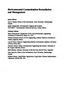
1.4 Fate and Mechanism of Accumulation of Lead in Soil–Plant System Soil plays a significant role in the retention and accumulation of lead in the environment. Lead enters the soil through anthropogenic sources such as industrial emissions, fossil fuel combustion, mining, and smelting. Natural sources, including weathering of rocks and erosion of soil, contribute to lead accumulation in soil. The behaviour of lead in soil is complex, and it can exist in various forms, including elemental, organic, inorganic, and complexed forms. While residing in the soil, lead may remain relatively immobile due to the low solubilities of the compounds involved. Alternatively, the movement of lead in the soil profile and its ultimate fate may be determined by one or more of several processes. If lead is in a soluble form, the soil profile may be exposed to lead leaching. Lead is quickly adsorbed initially in soil by fast reactions, then slow adsorption reactions occur and lead may get redistributed into various chemical forms with varied bioavailability, mobility, and toxicity (Shiowatana 2001). This distribution is regulated by reactions of lead in soils such as (i) mineral precipitation and dissolution, (ii) ion exchange, adsorption, and desorption, (iii) aqueous complexation, (iv) biological immobilization and mobilization, and (v) plant uptake (Wuana and Okieimen 2011) as depicted in Fig. 1.1. These reactions are strongly influenced by various factors such as soil type, pH, redox potential, organic matter content, microbes, and other ions (Silveira et al. 2003). For instance, lead tends to form insoluble compounds in alkaline soils, reducing its mobility. However, in acidic soils, the solubility increases, and leaching into groundwater or onto the crops can occur (Kushwaha et al. 2018). Lead is more soluble under reducing conditions and less soluble under oxidizing conditions. Biogeochemical factors such as soil organic matter and microbial activity play a critical role in the fate of lead in the soil. They can either increase or decrease the bioavailability of lead in the soil. For instance, soil organic matter can adsorb lead and limit or enhance its mobility in the soil forming complexes that can be more or
8
Ankush et al.
less stable (Kushwaha et al. 2018) while microbial activities can enhance or limit its solubility through oxidation and reduction. Plants may also take it up, thereby entering the food chain. Data are available, implying that the latter possibility may be of real significance. Lead accumulation in plants is a serious concern as it can be transferred to humans through the food chain. The uptake of lead by plants is influenced by various factors such as soil type, plant species, ion exchange capacity (particularly CEC), pH, redox potential, microbial community, and organic matter (Cobb et al. 2000; Kumar et al. 2020). Roots play a vital role in the uptake and translocation of lead (water). The accumulation of lead in plant organs varies and is plant species-dependent. Pb mostly accumulates (≥95%) in the roots of plant species and only a small fraction is translocated to aerial parts of the plant. The mechanism of lead accumulation in plants is not well understood. Adsorbed Pb often enters roots passively and is translocated through the xylem. Pb enters the roots and travels via the water stream via the apoplast until it reaches the endodermis. As the casparian strip blocks the water stream and Pb enters the symplastic movement, the endoderm serves as a physical barrier to Pb translocation (Fig. 1.1). Immobilization by negatively charged pectins within the root cell wall has been reported as the cause of the reduced Pb transportation from root to aerial parts of the plant like leaves and fruits. Insoluble Pb salts precipitate in root cell intercellular spaces (Zhang et al. 2017). Similarly, it has been found that Pb accumulates in the plasma membranes of root cells or is confined in the vacuoles of rhizodermal and cortical cells of roots (Zhang et al. 2017). Consequently, through plants lead enters the food chain and may end up in the human body as well.
1.5 Lead Toxicity Lead toxicity is a global problem that affects many countries and regions worldwide. The sources of lead contamination can vary, but some of the most common sources include lead-based paints, leaded gasoline, industrial emissions, mining activities, and lead-acid battery recycling. Lead contamination in soil and water can have serious environmental and public health consequences. Lead can persist in the environment for long periods of time and can accumulate in soil, water, and plants, posing a risk to human health and the ecosystem. However, lead contamination is also a problem in developed countries, where it may arise from legacy sources, such as contaminated industrial sites, lead pipes, and lead-based paints in older homes and buildings. Overall, lead toxicity is a serious issue that requires global attention and action to reduce exposure and protect human health and the environment. According to the World Health Organization (WHO), lead exposure is responsible for an estimated 1.06 million deaths each year. The majority of these deaths are due to cardiovascular disease (WHO 2019). Lead exposure is more common in low- and middle-income countries, where regulations and enforcement of environmental and occupational health standards may be weaker. According to the United Nations Environment
9
Fig. 1.1 Lead in soil–plant system
1 Source and Distribution of Lead in Soil and Plant—A Review
10
Ankush et al.
Programme (UNEP), lead pollution is a significant problem in many parts of the world, including areas with high levels of industrial activity, lead mining, and lead battery recycling. The use of leaded gasoline has been phased out in many countries, but it is still used in some parts of the world, particularly in low-income countries. Lead-based paints are also still used in some countries; despite being banned in many others. Overall, lead toxicity is a significant public health issue worldwide, with high levels of exposure reported in many parts of the world. Efforts are needed to address lead exposure include the implementation of regulations to limit the use and release of lead in products and industries, as well as efforts to remediate contaminated sites and reduce exposure in high-risk populations.
1.6 Environmental Impacts of Lead Toxicity Lead (Pb) is a heavy metal that is widely used in industrial and commercial applications such as batteries, construction materials, and gasoline. It is a highly toxic heavy metal that can be found in the environment due to various human activities such as mining, manufacturing, and the use of lead-containing products like paint, gasoline, and batteries. Its widespread use has led to environmental contamination, posing a threat to the health of plants, animals, and humans. In humans, lead exposure can have harmful effects on the nervous system, cardiovascular system, kidneys, and reproductive system. The toxicity of lead not only affects humans but also plants and animals. 1. Environmental contamination: Lead can accumulate in the soil and water, and can persist for many years. This can harm the ecosystem, reduce biodiversity, and make it difficult for plants and animals to survive. 2. Health effects: Exposure to lead can have harmful effects on human health, particularly in children and pregnant women. Lead can damage the brain and nervous system, leading to learning and behavioural problems. It can also cause developmental delays, anaemia, and kidney damage. 3. Food safety: Plants grown in lead-contaminated soil can accumulate lead in their tissues, making them potentially unsafe to eat. This can pose a risk to human health, particularly for people who rely on home-grown produce for their food. 4. Economic impact: Lead contamination can also have a negative impact on the economy. It can reduce property values, increase healthcare costs, and require expensive clean-up efforts. It is important for farmers to be aware of these potential sources of lead contamination in the soil and take steps to prevent and reduce lead accumulation. This may include testing the soil for lead, avoiding the use of lead-containing pesticides and fertilizers, and using alternative forms of hunting ammunition. Additionally, people can reduce their risk of lead exposure by washing their hands after working in the garden or handling soil, and by consuming a healthy diet with a variety of foods to reduce the risk of exposure to any single contaminant.
1 Source and Distribution of Lead in Soil and Plant—A Review
11
1.6.1 Harmful Effects of Lead on Soils 1. Reduced nutrient availability: Lead can compete with other nutrients, such as calcium, magnesium, and iron, for plant uptake. This can lead to nutrient imbalances and deficiencies, which can further reduce plant growth and productivity. 2. Altered soil microbial activity: Lead can also affect soil microbial activity, reducing the activity of important soil microorganisms such as nitrogen-fixing bacteria and mycorrhiza fungi. This can affect nutrient cycling and availability in the soil. 3. Reduced soil quality: High concentrations of lead in soil can also reduce soil quality and fertility, making it more difficult to grow crops over time. To mitigate these negative impacts, it is important to reduce lead contamination in soil. This may involve implementing best management practices, such as avoiding the use of lead-containing products and reducing exposure to lead in industrial and urban areas. Additionally, strategies such as phytoremediation, which involves using plants to remove contaminants from the soil, can be used to reduce lead concentrations in contaminated soils.
1.7 Harmful Effects of Lead on Plants Lead exposure in plants can occur through the soil, water, and air. Plants that are exposed to lead can exhibit a range of symptoms, including chlorosis, necrosis, stunted growth, and reduced crop yield. Here are some of the ways that lead can impact plant health: 1. Stunted growth: Lead exposure can cause stunted growth in plants, as well as reduced leaf area and decreased biomass production (Kumar et al. 2019). 2. Reduced photosynthesis: Lead can also reduce the efficiency of photosynthesis in plants, leading to reduced energy production and decreased plant growth (Mahmood et al. 2018). 3. Nutrient uptake problems: Lead can interfere with the uptake of essential nutrients in plants, such as iron and calcium, leading to nutrient deficiencies and other health problems (Kumar et al. 2019). 4. Cellular damage: Lead exposure can cause damage to plant cells, leading to cell death and tissue damage (Mahmood et al. 2018). 5. Accumulation in plant tissues: Plants can take up lead from the soil and accumulate it in their tissues, potentially making them toxic to humans and animals if consumed. 6. Reduced crop yield: Lead exposure can also reduce crop yield in plants, leading to economic losses for farmers (Kumar et al. 2019).
12
Ankush et al.
Lead contamination in soil is a significant concern for plants, particularly in areas with industrial activity or heavy traffic. Plants can take up lead from the soil and store it in their tissues, which can then be passed on to animals and humans that consume them (Arao and Ae 2003).
1.8 Harmful Effects of Lead on Human Beings Lead exposure in humans can occur through various routes, such as ingestion of lead-contaminated food and water, inhalation of lead-containing dust and fumes, and skin contact with lead-containing products. Lead is a highly toxic heavy metal that can have harmful effects on human health. Here are some of the ways that lead can impact human health: 1. Neurological damage: Lead exposure can cause neurological damage in both children and adults, leading to cognitive impairment, learning difficulties, and behavioral problems (Needleman 2004). 2. Cardiovascular problems: Lead exposure has also been linked to cardiovascular problems, such as hypertension, heart disease, and stroke (Navas-Acien et al. 2007). 3. Reproductive problems: Lead exposure can cause reproductive problems in both men and women, including decreased fertility and increased risk of miscarriage (Ronis et al. 2018). 4. Gastrointestinal problems: Lead exposure can cause gastrointestinal problems, such as abdominal pain, vomiting, and constipation (Agency for Toxic Substances and Disease Registry 2007). 5. Kidney damage: Lead can accumulate in the kidneys and cause damage, potentially leading to kidney failure (Satarug et al. 2010). Lead exposure is a particular concern for children, as they are more vulnerable to its harmful effects. Even low levels of lead exposure can cause long-term damage to children’s developing brains and nervous systems (Lidsky and Schneider 2003). Lead exposure can come from a variety of sources, including lead-based paint, contaminated soil and water, and certain occupations (such as construction or battery manufacturing). It is important to be aware of potential sources of lead exposure and to take steps to protect yourself and your family.
1 Source and Distribution of Lead in Soil and Plant—A Review
13
1.9 Harmful Effects of Lead on Animals Lead is a toxic heavy metal that can also have harmful effects on animals. Here are some of the ways that lead can impact animal health: 1. Neurological damage: Like in humans, lead exposure can cause neurological damage in animals, leading to cognitive impairment, seizures, and other neurological problems (Matsuda et al. 2012). 2. Reproductive problems: Lead exposure can also cause reproductive problems in animals, such as decreased fertility and reproductive success (Reigart and Roberts 2013). 3. Gastrointestinal problems: Animals exposed to lead can experience gastrointestinal problems, such as vomiting and diarrhoea, as well as decreased appetite and weight loss (Gwiazda et al. 2012). 4. Kidney damage: Lead can accumulate in the kidneys of animals, causing damage and potentially leading to kidney failure (Reigart and Roberts 2013). 5. Liver damage: Lead exposure can also cause damage to the liver in animals, leading to liver failure and other health problems (Matsuda et al. 2012). Lead exposure is a particular concern for wildlife that live in areas with high levels of environmental contamination. For example, eagles and other birds of prey have been found to have high levels of lead in their bodies due to ingesting lead ammunition fragments in their prey (Bedrosian et al. 2012).
1.10 Confronting Lead Toxicity Tackling lead toxicity in the soil can be a challenging task, but there are several strategies that can be used to reduce or remove lead from contaminated soils. Some of these strategies include: 1. Soil testing: Soil testing is the first step in identifying lead-contaminated soil. It is important to test soil samples to determine the concentration and form of lead present in the soil. 2. Phytoremediation: Phytoremediation involves using plants to remove contaminants from the soil. Some plant species, such as Indian mustard, sunflowers, and willows, are known to be effective at removing lead from the soil. These plants can be grown in contaminated areas and then harvested and disposed of as hazardous waste e.g., Indian mustard (Brassica juncea), Sunflowers (Helianthus annuus), Willow trees (Salix spp.), Poplar trees (Populus spp.) and Alfalfa (Medicago sativa). Phytoremediation is energy efficient, aesthetically pleasing method of remediating sites with lowto-moderate levels of contamination, and it can be used in conjunction with other more traditional remedial methods as a finishing step to the remedial process.
14
Ankush et al.
3. Bioremediation: It is possible to reduce lead toxicity in soil using microorganisms. Certain microorganisms have the ability to immobilize or detoxify lead through various mechanisms, such as adsorption, precipitation, or transformation into less toxic forms. One example of microorganisms that can be used for lead remediation is bacteria from the genus Pseudomonas. These bacteria have been found to have the ability to adsorb and accumulate lead in their cells, reducing the bioavailability of lead to plants and organisms. Other bacteria, such as Bacillus and Arthrobacter, have also been shown to have lead immobilization capabilities. Fungi, such as Aspergillus and Penicillium, have also been found to have the ability to immobilize lead in soil. These fungi can produce organic acids that can solubilize lead, allowing it to bind to fungal hyphae or to form insoluble complexes with soil minerals. In addition, certain plants have been found to form symbiotic relationships with microorganisms, such as mycorrhizal fungi, that can help to reduce the bioavailability of lead in soil. Mycorrhizal fungi can enhance plant growth and reduce lead uptake by plants by increasing soil pH, immobilizing lead in soil, or binding with lead in fungal hyphae. Overall, the use of microorganisms for lead remediation is a promising approach that can complement traditional remediation methods such as soil removal and containment. However, more research is needed to optimize the use of microorganisms for lead remediation and to develop practical applications for their use in the field. 4. Soil amendments: Adding soil amendments, such as lime, gypsum, or phosphate, can help reduce the bioavailability of lead in the soil, making it less toxic to plants and reducing the potential for lead uptake. 5. Soil removal and replacement: In severe cases of lead contamination, soil removal and replacement may be necessary. This involves removing contaminated soil and replacing it with clean soil. 6. Containment: If soil removal is not feasible, containment may be an option. This involves capping the contaminated soil with an impermeable layer, such as plastic or concrete, to prevent the spread of contaminants. 7. Best management practices: Implementing best management practices, such as avoiding the use of lead-containing products and reducing exposure to lead in industrial and urban areas, can help prevent lead contamination in the first place.
1.11 Conclusion Plants, animals, and people are all severely harmed by lead exposure. Background understanding of the sources, chemistry, and possible risks of toxic heavy metals in contaminated soils is necessary for the selection of viable remedial procedures. Remediation of soil contaminated by heavy metals is necessary to reduce the risks involved, make the land resource available for agricultural use, increase food security. Preventing lead toxicity can be greatly aided by taking steps to limit environmental lead exposure, such as lowering industrial source emissions. Additionally, people
1 Source and Distribution of Lead in Soil and Plant—A Review
15
can decrease their exposure to lead by utilizing lead-free items and avoiding places where lead contamination is known to exist.
References Agency for Toxic Substances and Disease Registry (2007) Toxicological profile for lead. US Department of Health and Human Services Ankush, Prakash R, Singh V et al (2021) Sewage sludge impacts on yields, nutrients and heavy metals contents in pearl millet–wheat system grown under saline environment. Int J Plant Prod 15:93–105. https://doi.org/10.1007/s42106-020-00122-4 Arao T, Ae N (2003) Genotypic variations in cadmium levels of rice grain. Soil Sci Plant Nut 49(1):47–52 Bedrosian B, Craighead D, Crandall R, Hansen L (2012) Blood lead levels of bald eagles: reference values and residues associated with clinical signs of poisoning. J Wildl Manag 76(8):1586–1594 Cobb GP, Sands K, Waters M, Wixson BG, Dorward-King E (2000) Accumulation of heavy metals by vegetables grown in mine wastes. Environ Toxicol Chem Int J 19(3):600–607 Gwiazda R (2012) Lead poisoning of wild birds from ingestion of spent lead shot: a review and analysis. Ornithol Monogr 74(1):1–26 Kumar A, Kumar A, MMS CP, Chaturvedi AK, Shabnam AA, Subrahmanyam G, ... Yadav KK (2020) Lead toxicity: health hazards, influence on food chain, and sustainable remediation approaches. Int J Environ Res Public Health 17(7):2179 Kumar P, Bhatt I, Pandey S, Pandey-Rai S (2019) Lead stress alters physiological and biochemical responses in maize seedlings. J Plant Growth Regul 38(3):1113–1125 Kushwaha A, Hans N, Kumar S, Rani R (2018) A critical review on speciation, mobilization and toxicity of lead in soil-microbe-plant system and bioremediation strategies. Ecotoxicol Environ Saf 147:1035–1045 Lidsky TI, Schneider JS (2003) Lead neurotoxicity in children: basic mechanisms and clinical correlates. Brain 126(1):5–19 Mahmood K, Rahman KU, Hayat Y, Ali S, Abbasi GH (2018) Impact of heavy metals on plant growth, uptake, and metabolism: a review. Environ Rev 26(1):1–39 Matsuda Y (2012) Effects of lead exposure on hippocampal neurogenesis in the rat. J Toxicol Environ Health A 75(1):517–525 Navas-Acien A, Guallar E, Silbergeld EK, Rothenberg SJ (2007) Lead exposure and cardiovascular disease—a systematic review. Environ Health Perspect 115(3):472–482 Needleman HL (2004) Lead poisoning. Annu Rev Med 55:209–222 Reigart JR, Roberts JR (2013) Recognition and management of pesticide poisonings, 6th edn. EPA, Washington, DC Ronis MJ, Hennings L, Stewart B et al (2018) Lead exposure during development results in increased neurofilament phosphorylation, neuritic beading, and temporal processing deficits within the murine auditory brainstem. Environ Health Perspect 126(8):087004 Satarug S, Baker JR, Urbenjapol S et al (2010) A global perspective on cadmium pollution and toxicity in non-occupationally exposed population. Toxicol Lett 192(2):245–254 Shiowatana J, McLaren RG, Chanmekha N, Samphao A (2001) Fractionation of arsenic in soil by a continuous-flow sequential extraction method. J Environ Qual 30(6):1940–1949 Silveira MLA, Alleoni LRF, Guilherme LRG (2003) Biosolids and heavy metals in soils. Scientia Agricola 60:793–806 World Health Organization (2019) Lead poisoning and health. Retrieved from https://www.who. int/news-room/fact-sheets/detail/lead-poisoning-and-health Wuana RA, Okieimen FE (2011) Heavy metals in contaminated soils: a review of sources, chemistry, risks and best available strategies for remediation. International Scholarly Research Notices
16
Ankush et al.
Zhang C, Wang X, Ashraf U, Qiu B, Ali S (2017) Transfer of lead (Pb) in the soil-plant-mealybugladybird beetle food chain, a comparison between two host plants. Ecotoxicol Environ Saf 143:289–295
Chapter 2
The Dynamics of Lead in Plant-Soil Interactions Usha Kumari, Pankaj, Saloni Yadav, Pooja Jangra, Dev Raj, and K. K. Bhardwaj
Abstract Lead (Pb) is among major pollutant for environmental ecosystem and is the toxic heavy metal following to arsenic (As). It is toxicity affects human beings, and young children are most vulnerable, and also toxic to animals. There is more concern regarding Pb as a contaminant due to its omnipresence in all parts of environment in different forms of compound. Pb causes oxidative damages to plants due to exposure at higher rates and even disturb the water and nutritional relationships of the crop. There are numerous sources of Pb in soil apart from steadily addition via natural weathering processes. Anthropogenic sources involve traffic emissions, mining, smelting, house hold emissions, weathering of old houses and buildings (coated with Pb-based paint) and footpaths surfaces, sewage irrigation, agricultural practices like fertilizers and pesticide application and waste dumping and disposal. The bioavailability of Pb in the soil poses a threat or risk to the environment which in turn depends on solubility of Pb solid phases and other Pb complexes in the soil. There are various factors which influences the availability of lead such as organic matter content, soil mechanical composition, soil pH etc. Soil pH is dominant factor and on increasing pH the availability of lead decreases & vice-versa. The soil chemistry with Pb showed the sedentary fixation of soil lead can be one of the alternative to reduce its uptake by the plants. This can be achieved by various methods such as phytoextraction, Phytostabilization, rhizofiltration, movement by microbes. Organic manures along with some other agricultural practices have the potential of better crop yield without compromising the quality from Pb contaminated soils. Keywords Lead toxicity · Lead-Soil Plant interaction · Phytoextraction · Phytostabilization · Rhizofiltration U. Kumari (B) · P. Jangra · D. Raj · K. K. Bhardwaj Faculty of Soil Science, Department of Soil Science, College of Agriculture, Chaudhary Charan Singh Haryana Agricultural University, Hisar, India e-mail: [email protected] Pankaj · S. Yadav Department of Soil Science, College of Agriculture, Chaudhary Charan Singh Haryana Agricultural University, Hisar, India © The Author(s), under exclusive license to Springer Nature Switzerland AG 2023 N. Kumar and A. K. Jha (eds.), Lead Toxicity: Challenges and Solution, Environmental Science and Engineering, https://doi.org/10.1007/978-3-031-37327-5_2
17
18
U. Kumari et al.
2.1 Introduction Soil, along with serving some important ecological functions like a foundation for vegetation, supporting food production, and providing raw materials, soil has an impact on human health as well. Soils become unable to perform these functions or support healthy ecosystems when they have been altered from their bare state or get polluted. Due to growing concerns about the safety of agricultural products, soil heavy metal pollution becomes a global environmental issue that has attracted an enormous public attention (Hu et al. 2017). The accelerating urbanization and industrialization over the last few decades has turned soil heavy metals contamination issue extrusive and widespread. As soon as the heavy metals get entry into the food chain, humans can get easily exposed to them (Intawongse and Dean 2006). Heavy metals are the native and metallic constituents of lithosphere, which have atomic number more than 20 and density of over 6 g cm−3 (Hodson 2004). Their natural presence in soil environment is attributed to pedogenic processes of weathering (Wuana and Okieimen 2011a, b, c) and at this concentration, they are rarely lethal to human health (Kabata-Pendias and Pendias 2001). Heavy metals such as arsenic (As), chromium (Cr), nickle (Ni) and lead (Pb) are among inorganic pollutants, which are most toxic owing to non-degradable nature (Nagajyoti et al. 2010). Lead (Pb) is one of the most hazardous heavy metals due to the damage it causes to the environment and to human health. (Lee and Tallis 1973), although it is of high industrial value due to its unique characteristics, such as lower melting point, high density, ease of moulding, and acid resistance. Pb contamination of soil is currently a major concern because it is negatively affecting all living things. (Ma et al. 2016) This metal is also accountable for a variety of environmental pollution because of its indulgence in numerous anthropogenic uses. Heavy metals in soil may originate from natural and anthropogenic sources. Lithogenesis, weathering, desertification, erosion, and other processes related to geology are examples of natural sources. While anthropogenic sources are primarily linked to human activities such as mining, ore processing, smelting, vehicle exhaust, atmospheric deposition, waste disposal, sewage treatment, and fertilizer application (Lu et al. 2012; Yang et al. 2009). Aluminosilicates and Feoxides are the two main reservoirs for naturally occurring Pb in soil (Teutsch et al. 2001). Galena (PbS), the most prevalent Pb ore mineral, has a weight percentage of 87% Pb. The most stable form of lead in solid form in reduced systems with sulfur is lead sulfate, or PbS. Galena readily changes into other common forms of Pb when placed in an oxidized environment (such as one where it is exposed to the atmosphere or oxygen-rich waters). such as anglesite (PbSO4), cerrussite (PbCO3), and the pyromorphites (Pb5(PO4)3) upon oxidation of sulfide to sulfate. Under a variety of environmental conditions, Pb phosphates, and particularly the pyromorphites, are the most stable forms of Pb among the many Pb minerals (Nriagu 1972 and Hem 1973). Unintentional addition of flourine, mercury, and lead to the soil occurs when certain fertilizers with phosphorus are used (Duruibe et al. 2007).
2 The Dynamics of Lead in Plant-Soil Interactions
19
In the past, a number of pesticides that were widely used in agriculture and horticulture practices contained high levels of heavy metals. Hu et al. (2018) pointed out that a significant source of soil Pb is coal combustion in the chemical industry. Lead enters the food chain and the agroecosystem through polluted soil, which is contaminated by manufacturing of acid batteries, use of Pb containing fuel, Pb containing insecticides, mining, printing, etc. Its buildup in the plants causes a very serious health risk to people. Polluted soils are the primary source of heavy metals for plants in terrestrial ecosystems, and once they enter the plant system, they have significant negative effects on production of crops and grain quality. The weekly Pb intake cap for humans is approximately 25 µg kg-1 of human body weight (Fang et al. 2014). It is present in meagre quantity in almost all food crops and growing of these crops in Pb contaminated soil, substantially enhances its concentration in plants tissue. According to Doyle et al. (2018), lead is one of the elements that exhibits the translocation restriction phenomenon, which means that the majority of Pb that is available to plants is retained and accumulated within the roots. However, some Pb ions can also be transported to above-ground plant parts (mostly by xylem vessels) (Zhou et al. 2016). It doesn’t necessarily influence the same physiological or metabolic process in plants because it affects different plants in different ways. Even at relatively low concentrations, chronic exposure to Pb may result in negative effects on the bloodstream and central nervous system, particularly in infants and kids, according to epidemiologic studies. Pb plays no beneficial roles in biological networks and instead solely poses a threat to health to humans, animals, and other living things (Maestri et al. 2010). According to estimates Pb pollution contributed to 853,000 fatalities and 0.6% of the global disease burden (WHO 2016). Pb has been released into the environment in 800,000 tonnes over the past 50 years, much of it accumulating in soil and causing severe pollution levels (Chen et al. 2016a, b). There is a crucial need to understand deeply and thoroughly about the dynamics of lead in soil to eliminate to root cause of raising its concentration and affecting environment and human health.
2.2 Lead Dynamics in Soil The soil is a crucial element of the ecosystem that affects food and agricultural crops. The soil quality of can be defined by many factors; among them one is its potential to regulate the availability of harmful constituents in soil and to prevent them to enter food chain and deteriorating human health. Heavy metals especially arsenic (Ar), Lead (Pb) and cadmium (Cd) are among the potential hazardous elements which can affect the environment quality, these could be introduced to the top soil in a variety of ways, including wastewater, fertilizers, herbicides, and others. Soil consists many organic, inorganic and organo-mineral compounds, such as clay minerals, iron, aluminium and manganese oxides, humic substances, soluble substances and some other solid constituents as well as which govern the availability and exposure of toxic substances. Heavy metal speciation and mobility are influenced by their
20
U. Kumari et al.
binding mechanisms in soils, which changes depending on the soil composition, soil response, and redox conditions. Depending on how they attach to specific soil components, reactive surfaces, or external or interior binding sites with variable bonding energies, metals can produce distinct species. Lead is present in different forms in soil, constituting bioavailable and non-bioavailable fractions in soil. This includes soluble fractions, exchangeable fraction, organic and inorganic bound fractions and mineralogical associated lead (Vega et al. 2010). Among the different fractions only soluble and exchangeable lead fractions are bioavailable (Kopittke et al. 2008) while other fractions are not available to plants and other microbes due to its specific as well as strong bonding. The most stable forms of lead in soil are Pb(II) and Pb-hydroxy oxides, though it also exists in ionic and covalent forms (Wuana and Okieimen 2011a, b, c). Lead is first quickly absorbed by soil, then slowly absorbed by soil, and finally redistributed into various chemical forms with varying bioavailability, mobility, and toxicity (Shiowatana 2001).This distribution is controlled by reactions of lead in soils such as. i. ii. iii. iv. v.
Precipitation and dissolution of minerals Adsorption, desorption, and ion exchange Biological immobilization and mineralization Aqueous complexation and Plant Absorption
Lead can be found in soils in three different states: as a free metal ion, in complex with inorganic elements like HCO3-, CO3-, SO42-, and Cl-, or with organic ligands like amino acids, fulvic acids, and humic acids. Lead can also adsorb onto the surfaces of particles (such as clay particles, Fe-oxides, organic matter, and biological material). Lead dynamics in soil are influenced by a number of factors, including soil pH, soil type, particle size, organic matter, the presence of organic colloids and iron oxides, cation exchange capacity (CEC), lead content, and the presence of different amendments (Silveira et al. 2003). The availability and translocation of lead in the soil are influenced by the soil’s pH, texture, organic matter, and presence of specific amendments. These elements are further described in detail. Soil PH Soil pH has a big impact on how much lead is retained by soils, which influences soil lead bioavailability and solubility significantly. At alkaline pH levels, lead is primarily found as insoluble lead carbonates and phosphates, whereas at acidic pH levels, lead is present as free ionic species. Lead is soluble in ionic form (Pb2+ ) and in pairs of ions (e.g., PbSO4 ) at low pH values. As pH rises from 3 to 6.5, lead solubility decreases due to an increase in the concentration of phosphates, hydroxides, and carbonates, insoluble forms of lead. From 6.5 to 8, there is an increase in the formation of organolead complexes. Finally, at neutral pH, 80–99% of lead predominantly occurs in the form of organic complexes (Sauve et al. 1998).This indicates that lead is more mobile and bioavailable at acidic pH, which increases lead uptake by plants and corresponding harmful effects on living beings. In actuality, the specific adsorption of Pb is strongly influenced by the pH of the soil (Yang et al. 2011a). At low soil
2 The Dynamics of Lead in Plant-Soil Interactions
21
pH, adsorption is more important than precipitation of the solid phase in reducing the concentration of Pb ions in solution, and the opposite is true at high pH (Esbaugh et al. 2012). Organic matter The main factor in the immobilisation of lead in soils is lead’s high solubilization with soil organic matter. Chelation with humic or fulvic acids immobilizes lead (Seki et al. 1990). The most significant reaction by which lead is adsorbed by humus is coordinated binding at groups with free electron pairs, such as carbonyl groups, by completing the coordination sphere of lead. According to Xiong et al. (2013), lead binds to humic acid more tightly as pH rises and ionic strength falls. Lead is found to be retained in the top 2 to 5 cm of undisturbed soils with a 5% organic matter content and a pH of 5, whereas in soils with a high organic content and a pH of 6 to 8, lead forms insoluble organic lead complexes (Ahmed et al. 2015). Clay minerals Lead is easily adsorbed onto the exchange complex of clay minerals and is merely difficult to replace. Given that both lead ions have equal ionic sizes, divalent lead should be absorbed more strongly than monovalent lead. Certain soils’ B horizon lead enrichment has been linked in part to the presence of too much clay (Wuana and Okieimen 2011a, b, c). According to Arenas-Lago et al. (2014), lead also interacts with amorphous iron sesquioxide and, to a lesser extent, aluminum sesquioxide. In their study of soil texture and extractable Pb concentration using 0.1 M DTPA and HCl, Qian et al. (1996) discovered that clay fractions contained higher concentrations of Pb than sand fractions. Lead adsorption also varies with different types of clay mineral, for instance, selectivity montmorillonite for Pb is 32 times lesser than for the illite. (Suzuki et al. 2014). Presence of amendments The presence of various amendments, such as fertilizers, phosphate, and limestone, among others, has a significant impact on bioavailability. The use of the herbicide glyphosate [N-(phosphonomethyl)-glycine] in agricultural practices is common and it takes the form of a complex with metal ions that affects the availability of lead to plants. Lead solubility significantly decreased in the presence of phosphorus amendments. According to Chen et al. (2003), phosphate in soil effectively immobilized lead.
2.3 Lead Behavior in Plants Plants, as an important component of the ecosystem, are primarily impacted by a wide variety of pollutants, which vary in concentration, speciation, and toxicity. These pollutants enter the plant through either the soil or the atmosphere. Lead is a major pollutant that has a significant impact on plants (Shahid 2011). As described
22
U. Kumari et al.
below, its activities in soil varied in terms of adoption, translocation in roots, and thus to other plant parts. 1. Adsorption/uptake of lead by plants: The lead adsorbed by plants is through roots a portion of the lead present in soil is adsorbed onto the upper layers of the radicular cortex (rhizoderm and collenchyma/parenchyma) initially lead binds to the carboxyl groups of mucilage uronic acid or it may bind to the polysaccharides of the rhizoderm cell surface. Uptake of lead is a non-selective phenomenon and on the other hand it greatly depends on the functioning of an H + /ATPase pump to maintain a strong negative potential in rhizoderm cells (Wang et al. 2007).The well-known mechanism of lead uptake is by calcium channels and has been documented by several authors. The non-selective cations channels that are the main routes of lead entry in root cells include depolarization-activated calcium channels (DACC), hyperpolarization activated calcium channels (HACC) and voltage-insensitive cation channels (VICC) are thought to be one of the principal routes of Pb entry into root cells. 2. Translocation of lead to aerial parts of the plant: Lead accumulates in the roots after entering the root system and may also go to the plant’s aerial portions. The majority of the absorbed lead (about 98% or more) has been found to collect in the roots, with only a minor amount moving to the other sections of the plant. The plant species has a significant impact on how heavy metals are transported to aerial sections of plants. As soon as lead enters the root, it travels through the apoplastic and symplastic pathways to reach the plant’s aerial components. 3. Apoplastic pathway: Once lead has been transported to the apoplast, it moves with the flow of water until it reaches the endodermis (Lane and Martin 1977). 4. Symplastic pathway: Lead barely passes through symplastic pathway. In situations where the dose of lead is not fatal, it enters the protoderm or the symplast, where cells are actively dividing. Because immature cells lack secondary walls, lead can enter the cell membrane more easily. Lead that is present in symplast may be contained in certain cell compartments like vacuoles, dictyosomal vesicles, endoplasmic reticulum vesicles, or plasmatubules. But when a deadly dose is present, lead reaches the radicular tissues. At this concentration, lead reaches the cytoplasm, mitochondria, nucleus, and other organelles, causing membrane disruption (Malecka et al. 2009).
2.4 Sources of Lead Contamination in Soil The risk of heavy metal environmental contamination has significantly increased as a result of the rapid industrial development occurring around the world. Toxic substances build up in soil, water, and air as a result of rapid industrialization, chaotic urbanization, and extensive long-term pesticide and fertilizer use (Kumar et al. 2015; Rodriguesa et al. 2017). The earth’s crust naturally contains lead, which is gradually added to the soil through natural processes like weathering, crustal material erosion, or deposition of lead released into the atmosphere by volcanic activity, which totally
2 The Dynamics of Lead in Plant-Soil Interactions
23
makes up 80% of all natural sources (Callender 2003; Hou, O’Connor et al. 2017). The extent of addition is at subside rate and even in longer duration, there won’t be considerable heaping of it, which can deteriorate the soil quality. Other major natural sources are forest fires and biogenic sources which contribute to soil Pb (Zhang et al. 2019a, b). Gelena (PbS) is the most commonly found naturally derived lead in soil, this is followed by smaller addition of anglesite (PbSO4 ), cerussite (PbCO3 ), crocoite (PbCrO4 ), pyromorphite (Pb5 (PO4 )3 Cl), litharge (PbO) and massicot (PbO) (Laperche et al. 1996; Mulligan et al. 2001). Metallic form Pb in nature is rarest and it usually coexists with zinc, silver and copper elements (Cheng and Hu 2010). Addition of soil Pb is profuse from anthropogenic activities, such as traffic emissions (Eichler et al. 2015), domestic emissions, paint industry and agriculture activities like use of sewage water for irrigation, use of agrochemicals (Yang et al. 2013) waste dumping and disposal (Ren et al. 2018) mining, smelting and other industrial activities (Li et al. 2014). From 1930 to 2010, there was an increase in the global lead production from 1 million tonnes per year to about 10 million tonnes per year. Pb emissions increased from 1 million tonnes per year to 3.6 million tonnes per year, totalling 174 Mt, during the same time period (Zhang et al. 2019a, b). A huge amount of fertilizers is added to soils during extensive and intensive farming to supply adequate amount of nitrogen, phosphorus, and potassium for crop growth (Huan et al. 2017; Dhaliwal et al. 2019) and most of these fertilizers have substantial amount of heavy metals (Table 1). Recently 10% of the fungicides and insecticides were legalized which contain lead, mercury, manganese, copper and zinc in the United Kingdom. The type of surrounding industries like paper making, electronics, textile printing, chemical, metallurgy, dyeing, electroplating, and food processing use different raw materials, products, technological processes, and operation modes, resulting in differing types, degrees, and spatial distribution of lead pollution in soil. According to earlier research (Schwarz et al. 2013), Pb originates from traffic pollution and is linked to tire wear and the combustion of leaded gasoline (Table 1). Tetraethyl lead is added to gasoline as an anti-knock agent. Unleaded gasoline usage decreased lead emissions and the buildup of Pb in the soil, according to Konstantinova et al. (2019). Although leaded gasoline has been outlawed in China since 2000, lead Pb accumulation in soil is still a problem due to vehicle exhaust emissions (Lv et al. 2015). Furthermore, the chemical industry is also a source of Pb. Chemical parks have dense roads and frequent vehicle traffic, leading to frequent accumulation of Pb in the soil.
2.5 Methods to Reduce Lead Toxicity There are numerous in-situ and ex-situ methods for removing lead and other dangerous trace metals from soil. Ex-situ technologies for cleaning up polluted land are prohibitively expensive in many cases. Yet, an in-situ, plant-based technology called phytoremediation has attracted a lot of scientific attention since it is an economical and environmentally beneficial method for cleaning up places that have
24
U. Kumari et al.
Table 1 Sources of lead contamination in soil 1. Natural
• Ore and minerals • Volcanic eruption
2. Agriculture (Duruibe et al. 2007)
• Fertilizers • Pesticides
3. Antropogenic (Seaward and Richardson 1990; Gisbert et al. • Pb Mining 2003) • Burning of leaded gasoline • Smelting of metalliferous ores • Municipal sewage • Paints with lead pigments • Preservatives • Industrial wastes enriched in Pb
been contaminated with lead and other trace metals (Mahdavian et al. 2017). The rhizosphere’s microbial activity is stimulated by this technology. For soil remediation following contamination, the intricate physical, biological, and chemical interactions that occur in soil near roots are essential. Exudates from the roots of plants contain organic substances that improve the microbial population and make lead absorption easier. With phytoremediation, lead contamination is disseminated less widely through the water and air and disturbances to the soil ecosystem are minimised. There are many different phytoremediation techniques, but only a few of them are applicable to lead phytoremediation, such as phytostabilization, rhizofiltration and phytoextraction. (a) Phytostabilization With or without non-toxic soil additives that bind to metals, metals are immobilized through revegetation. By using species that can withstand metals, the risk to the environment and human health is reduced because heavy metals are immobilized by roots’ absorption and accumulation. It reduces airborne movement, leaching, and bulk erosion (Dary et al. 2010). Metals’ ability to remain in the roots or rhizosphere inhibits their ability to enter the food chain. A metal-soil complex’s physicochemical characteristics can be changed by adding anions, which improves metal adsorption through anion-induced negative charge and metal precipitation. By raising the pH of the soil and adding humified organic matter and lime, which facilitates the revegetation of polluted soil, lead can be immobilised. Lime aids in reducing soil acidity, hence lowering lead bioavailability. This process reduces the amount of lead that leaches into groundwater and accumulates in soil. (b) Phytoextraction This process involves plant roots absorbing lead from soil or water, moving it to above-ground biomass, and accumulating it there (Rafati et al. 2011). These plants are later collected and burned. Plants used in this method typically have traits like rapid growth, a large biomass, a spread-out root system, and the ability to tolerate
2 The Dynamics of Lead in Plant-Soil Interactions
25
high levels of heavy metals. Based on plant characteristics, there are two different ways to extract phytochemicals. In the first, plants like Minuartia verna and Agrostis tenuis are used; these plants naturally uptake, store, and tolerate large amounts of metals in shoots without exhibiting symptoms. In the second, high biomass plants are used; these plants’ tendency to accumulate lead is improved by the addition of chelates. (c) Rhizofiltration This process is defined as the use of both terrestrial and aquatic plants to concentrate toxins at the roots of those plants by drawing those toxins from contaminated water sources. This technique involves hydroponically growing plants before relocating them into water that contains lead, where they take it up through their roots and shoots. Lead precipitates on the root surface as a result of rhizospheric pH changes and root exudates. Whole plants or parts are collected to dispose off as they become saturated with lead pollutants. The lead translocation from roots to shoots may reduce the rhizofiltration effectiveness (Dushenkov et al. 1995). A wide range of lead concentrations, ranging from 4-500 mg/l, are removed using plants with significant amounts of root area, such as Indian mustard (Raskin and Ensley 2000). (d) Lead immobilization by bacteria Lead interacts directly or indirectly with soil microbes, and lead is immobilized by a variety of processes, including chelation, restricting entry, enzymatic detoxification, intra- and extracellular sequestration, precipitation and biotransformation (Chen and Cutright 2003). (e) Biosorption/Complexation of Lead On the surface of many bacteria, lead has been found to be immobilized and adsorb. Trifolium pratense L. grows more if soil is treated with Brevibacillus species under Pb-contamination which leads to increased nodules formation, enhanced nitrogen and phosphorus uptake, and restricted root uptake of Pb. According to Puyen et al. (2012), Micrococcus luteus DE2008 has an adsorption capability of 1965 mg/g Pb (II).
2.6 Conclusion The amount of heavy metal pollution is rising day by day and due to restricted land availability for agriculture, it is indispensable to overlook the problem of soil contamination. Lead is one of the potential soil pollutant but widely used by human being in numerous industries for variety of purposes since antiquity, because of having some useful properties. Lead affects plants either directly or indirectly, interfering with their physiological and enzymatic processes. In soil lead activities are affected by various soil properties and pH have dominant affect and inversely influence the content of available lead in soil. Other factors include soil organic matter, soil texture
26
U. Kumari et al.
and amendments in soil. There are various sources of lead in soil apart from natural deposition through volcanic eruptions, which are anthropogenic as paint industries and agriculture etc. Toxicity of Pb can be reduced by various processes like rhizofilration, phytoextraction, phytoremediation and involvement of microbes in these toxic leveled sites. These processes have potential to provide economically viable and environmentally sound remedies for cleaning Pb contaminated soils. There is need of regular monitoring of industrial effluents which are added to water bodies or soil as per the WHO guidelines so that the soil could be saved from overburdened level of Pb. Hence, research priorities should be directed towards Pb mobility in soil–plant–human continuum.
References Ahmed IM, Helal AA, El Aziz NA, Gamal R, Shaker NO, Helal AA (2019) Influence of some organic ligands on the adsorption of lead by agricultural soil. Arab J Chem 12(8):2540–2547 Arenas-Lago D, Vega FA, Silva LF, Lago-Vila M, Andrade ML (2014) Lead distribution between soil geochemical phases and its fractionation in Pb-treated soils. Fresen Environ Bull 23(4):1025– 1035 Callender E (2003) Heavy metals in the environment-historical trends. Treat Geochem 9:67–105 Chen H, Cutright TJ (2003) Preliminary evaluation of microbially mediated precipitation of cadmium, chromium, and nickel by rhizosphere consortium. J Environ Eng 129(1):4–9 Chen YY, Tang MY, Wang ST, Wang Q, Zhan WX, Huang G (2016a) Evaluation of heavy metal pollution in farmland soil of China based on bibliometrics. Chin J Soil Sci 47(1):219–225 Chen Z, Pan X, Chen H, Guan X, Lin Z (2016b) Biomineralization of Pb (II) into Pb-hydroxyapatite induced by Bacillus cereus 12–2 isolated from Lead-Zinc mine tailings. J Hazard Mater 301:531– 537 Cheng H, Hu Y (2010) Lead (Pb) isotopic fingerprinting and its applications in lead pollution studies in China: a review. Environ Pollut 158:1134–1146. https://doi.org/10.1016/j.envpol.2009.12.028 Dary M, Chamber-Pérez MA, Palomares AJ, Pajuelo E (2010) “In situ” phytostabilisation of heavy metal polluted soils using Lupinus luteus inoculated with metal resistant plant-growth promoting rhizobacteria. J Hazard Mater 177(1–3):323–330 Dhaliwal SS, Naresh RK, Agniva-Mandal WMK, Gupta Raj K, Singh R, Dhaliwal MK (2019) Effect of manures and fertilizers on soil physical properties, build-up of macro and micronutrients and uptake in soil under different cropping systems: a review. J Plant Nutr H Dogan M, Karatas M, Aasim M (2018) Cadmium and lead bioaccumulation potentials of an aquatic macrophyte Ceratophyllum demersum L.: a laboratory study. Ecotoxicol Environ Saf 148:431– 440 Duruibe JO, Ogwuegbu MOC, Egwurugwu JN (2007) Heavy metal pollution and human biotoxic effects. Intern J Phy Sci 2(5):112–118 Dushenkov V, Kumar PN, Motto H, Raskin I (1995) Rhizofiltration: the use of plants to remove heavy metals from aqueous streams. Environ Sci Technol 29(5):1239–1245 Eichler A, Gramlich G, Kellerhals T, Tobler L, Schwikowski M (2015) Pb pollution from leaded gasoline in South America in the context of a 2000-year metallurgical history. Sci Adv 1:e1400196. https://doi.org/10.1126/sciadv.1400196 Esbaugh AJ, Brix KV, Mager EM, De Schamphelaere K, Grosell M (2012) Multilinear regression analysis preliminary biotic ligand modeling, and cross species comparison of the effects of water chemistry on chronic lead toxicity in invertebrates. Comp Biochem Physiol C Toxicol Pharmacol 155:423–431
2 The Dynamics of Lead in Plant-Soil Interactions
27
Fang Y, Sun X, Yang W, Ma N, Xin Z, Fu J, Liu X, Liu M, Mariga AM, Zhu X, Hu Q (2014) Concentrations and health risks of lead, cadmium, arsenic, and mercury in rice and edible mushrooms in China. Food Chem 147:147–151 Gisbert C, Ros R, de Haro A, Walker DJ, Pilar Bernal M, Serrano R, Avino JN (2003) A plant genetically modified that accumulates Pb is especially promising for phytoremediation. Biochem Biophys Res Commun 303(2):440–445. https://doi.org/10.1016/S0006-291X(03)00349-8 Hem JD, Durum WH (1973) Solubility and occurrence of lead in surface waters. J Am Water Work Assoc 65:562–568 Hodson ME (2004) Heavy metals: geochemical bogey men? Environ Poll 129:341–343 Hou D, O’Connor D, Nathanail P, Tian L, Ma Y (2017) Integrated GIS and multivariate statistical analysis for regional scale assessment of heavy metal soil contamination: a critical review. Environ Pollut 231:1188–1200. https://doi.org/10.1016/j.envpol.2017.07.021 Hu BF, Chen SC, Hu J, Xia F, Xu JF, Li Y, Shi Z (2017) Application of portable XRF and VNIR sensors for rapid assessment of soil heavy metal pollution. PLoS ONE 12:e0172438 Hu WY, Wang HF, Dong LR, Huang B, Borggaard OK, Hansen H, He Y, Holm PE (2018) Source identification of heavy metals in peri-urban agricultural soils of southeast China: an integrated approach. Environ Pollut 237:650–661 Huan L, Haixia Z, Longhua W, Anna L, Fang-Jie Z, Wenzhong X (2017) Heavy metal ATPase 3 (HMA3) confers cadmium hypertolerance on the cadmium/zinc hyperaccumulator Sedum plumbizincicola. New Phytol 15:687–698 Intawongse M, Dean JR (2006) Uptake of heavy metals by vegetable plants grown on contaminated soil and their bioavailability in the human gastrointestinal tract. Food Addit Contam 23(1):36–48 Kabata-Pendias A, Pendias H (2001) Trace elements in soils and plants, 3rd edn. CRC Press, Boca Raton, FL, USA Konstantinova E, Minkina T, Sushkova T, Konstantinov A, Rajput VD, Sherstnev A (2019) Urban soil geochemistry of an intensively developing Siberian city: a case study of Tyumen. Russia J Environ Manage 239:366–375 Kopittke PM, Asher CJ, Kopittke RA, Menzies NW (2008) Prediction of Pb speciation in concentrated and dilute nutrient solutions. Environ Pollut 153:548–554 Kumar N, Kumar S, Bauddh K, Dwivedi N, Shukla P, Singh DP, Barman SC (2015) Toxicity assessment and accumulation of metals in radish irrigated with battery manufacturing industry effluent. Intern J Veg Sci 00:1–13 Lane SD, Martin ES (1977) A histochemical investigation of lead uptake in Raphanus sativus. New Phytol 79(2):281–286 Laperche V, Traina SJ, Gaddam P, Logan TJ (1996) Chemical and mineralogical characterizations of Pb in a contaminated soil: reactions with synthetic apatite. Environ Sci Technol 30:3321–3326. https://doi.org/10.1021/es960141u Lee JA, Tallis JH (1973) Regional and historical aspects of lead pollution in Britain. Nature 245(5422):216–218 Li Y, Gou X, Wang G, Zhang Q, Su Q, Xiao G (2008) Heavy metal contamination and source in arid agricultural soil in central Gansu Province, China. J Environ Sci 20:607–612 Li Z, Ma Z, van der Kuijp TJ, Yuan Z, Huang L (2014) A review of soil heavy metal pollution from mines in China: pollution and health risk assessment. Sci Total Environ 468–469:843–853. https://doi.org/10.1016/j.scitotenv.2013.08.090 Lu A, Wang J, Qin X, Wang K, Han P, Zhang S (2012) Multivariate and geostatistical analyses of the spatial distribution and origin of heavy metals in the agricultural soils in Shunyi, Beijing, China. Sci Total Environ 425:66–74 Lv J, Liu Y, Zhang Z, Dai J, Dai B, Zhu Y (2015) Identifying the origins and spatial distributions of heavy metals in soils of Ju country (Eastern China) using multivariate and geostatistical approach. J Soil Sediment 15:163–178 Ma Y, Egodawatta P, McGree J, Liu A, Goonetilleke A (2016) Human health risk assessment of heavy metals in urban storm water. Sci Total Environ 557:764–772
28
U. Kumari et al.
Małecka A, Derba-Maceluch M, Kaczorowska K, Piechalak A, Tomaszewska B (2009) Reactive oxygen species production and antioxidative defense system in pea root tissues treated with lead ions: mitochondrial and peroxisomal level. Acta Physiol Plant 31:1065–1075 Mulligan CN, Yong RN, Gibbs BF (2001) Remediation technologies for metal contaminated soils and groundwater: an evaluation. Eng Geol 60(1–4):193–207. https://doi.org/10.1016/S00137952(00)00101-0 Nagajyoti PC, Lee KD, Sreekanth TVM (2010) Heavy metals, occurrence and toxicity for plants: a review. Environ Chem Lett 8:199–216 Nriagu JO (1972) Lead orthophosphates. I. Solubility and hydrolysis of secondary lead orthophosphate. Inorg Chem 11, 2499–2503, 39 Yang PG, Mao RZ, Shao HB, Gao YF (2009) The spatial variability of heavy metal distribution in the suburban farmland of Taihang Piedmont Plain, China. C R Biol 332:558–566 Puyen ZM, Villagrasa E, Maldonado J, Diestra E, Esteve I, Solé A (2012) Biosorption of lead and copper by heavy-metal tolerant Micrococcus luteus DE2008. Biores Technol 126:233–237 Qian J, Wang Z, Shan X, Tu Q, Wen B, Chen B (1996) Evaluation of plant availability of soil trace metals by chemical fractionation and multiple regression analysis. Environ Pollut 91:309–315 Rafati M, Khorasani N, Moattar F, Shirvany A, Moraghebi F, Hosseinzadeh S (2011) Phytoremediation potential of Populus alba and Morus alba for cadmium, chromuim and nickel absorption from polluted soil. Intern J Environ Res 5(4):961–970 Raskin I, Ensley BD (2000) Phytoremediation of toxic metals. John Wiley and Sons Ren J, Chen J, Han L, Wang M, Yang B, Du P, Li F (2018) Spatial distribution of heavy metals, salinity and alkalinity in soils around bauxite residue disposal area. Sci Total Environ 628:1200–1208. https://doi.org/10.1016/j.scitotenv.2018.02.149 Rodriguesa AAZ, De Queiroz MELR, Oliveira AF, Heleno AAFF, Zambolim L, Freitasa JF, Morais EHC (2017) Pesticideresidueremovalinclassicdomestic processing of tomato and its effects on product quality. J Environ Sci Health Part B 52:1–8 Sauve S, McBride M, Hendershot W (1998) Soil solution speciation of lead (II): effects of organic matter and pH. Soil Sci Soc Am J 62(3):618–621 Schwarz K, Weathers KC, Pickett ST, Lathrop RG Jr, Pouyat RV, Cadenasso ML (2013) A comparison of three empirically based, spatially explicit predictive models of residential soil Pb concentrations in Baltimore, Maryland, USA: understanding the variability within cities. Environ Geochem Health 35:495–510 Seaward MRD, Richardson DHS (1990) Atmospheric sources of metal pollution and effects on vegetation. In: Shaw AJ (ed) Heavy metal tolerance in plants: evolutionary aspects. CRC Press, Florida, pp 75–92 Seki H, Suzuki A, Kashiki I (1990) Adsorption of lead ions on immobilized humic acid. J Colloid Interface Sci 134(1):59–65 Shahid M, Pinelli E, Pourrut B, Silvestre J, Dumat C (2011) Lead-induced genotoxicity to Vicia faba L. roots in relation with metal cell uptake and initial speciation. Ecotoxicol Environ Safety 74(1):78–84 Shiowatana J, McLaren RG, Chanmekha N, Samphao A (2001) Fractionation of arsenic in soil by a continuous-flow sequential extraction method. J Environ Qual 30(6):1940–1949 Silveira MLA, Alleoni LRF, Guilherme LRG (2003) Biosolids and heavy metals in soils. Scientia Agricola 60:793–806 Suzuki T, Niinae M, Koga T, Akita T, Ohta M, Choso T (2014) EDDS-enhanced electrokinetic remediation of heavy metal-contaminated clay soils under neutral pH conditions. Colloid Surf Physicochem Eng Asp 440:145–150 Teutsch N, Erel Y, Halicz L, Banin A (2001) Distribution of natural and anthropogenic lead in Mediterranean soils. Geochim Cosmochim Acta 65(17):2853–2864 Vega F, Andrade M, Covelo E (2010) Influence of soil properties on the sorption and retention of cadmium, copper and lead, separately and together, by 20 soil horizons: comparison of linear regression and tree regression analyses. J Hazard Mater 174:522–533
2 The Dynamics of Lead in Plant-Soil Interactions
29
Wang HH, Shan XQ, Wen B, Owens G, Fang J, Zhang SZ (2007) Effect of indole-3-acetic acid on lead accumulation in maize (Zea mays L.) seedlings and the relevant antioxidant response. Environ Exp Botany 61(3):246–253 WHO (2019) Lead poisoning and health (September 2016. Retrieved 14 October) Wuana RA, Okieimen FE (2011a) Heavy metals in contaminated soils: a review of sources, chemistry, risks and best available strategies for remediation. International Scholarly Research Notices Wuana RA, Okieimen FE (2011b) Heavy metals in contaminated soils a review of sources, chemistry, risks and best available strategies for remediation. Commun Soil Sci Plant Analy 42:111–122 Xiong J, Koopal LK, Tan W, Fang L, Wang M, Zhao W, Liu F, Zhang J, Weng L (2013) Lead binding to soil fulvic and humic acids: NICA-Donnan modeling and XAFS spectroscopy. Environ Sci Technol 47(20):11634–11642 Yang S, Zhou D, Yu H, Wei R, Pan B (2013) Distribution and speciation of metals (Cu, Zn, Cd, and Pb) in agricultural and non-agricultural soils near a stream upriver from the Pearl River, China. Environ Pollut 177:64–70. https://doi.org/10.1016/j.envpol.2013.01.044 Yang X, Yang S, Yang S, Hu J, Tan X, Wang X (2011) Effect of pH, ionic strength and temperature on sorption of Pb(II) on NKF-6 zeolite studied by batch technique. Chem Eng J 168:86–93 Zhang Y, Hou D, O’Connor D, Shen Z, Shi P, Ok YS, Tsang DCW, Wen Y, Luo M (2019a) Lead contamination in Chinese surface soils: Source identification, spatial-temporal distribution and associated health risks. Crit Rev Environ Sci Technol 49(15):1386–1423. https://doi.org/10. 1080/10643389.2019.1571354 Zhou C, Huang M, Li Y, Luo J, Cai L (2016) Changes in subcellular distribution and antioxidant compounds involved in Pb accumulation and detoxification in Neyraudia reynaudiana. Environ Sci Pollut Res 23:21794–21804
Part II
Lead Toxicity and Health
Chapter 3
Neurotoxic Effect of Lead: A Review Chanchal Singh, Apoorva Shekhar, and Raghubir Singh
Abstract The neurotoxic effect of lead is a well-documented and concerning issue that poses a significant threat to public health. Lead is a heavy metal that can accumulate in the body, especially in bone tissue, over time. Its toxic effects can manifest in many ways, but the nervous system is particularly vulnerable to its effects. This chapter explores the mechanisms by which lead causes neurotoxicity, including its ability to disrupt neurotransmitter production, interfere with neuron development, and cause oxidative stress. The chapter also discusses the health effects of lead exposure on the nervous system, such as developmental delays, cognitive impairment, and peripheral neuropathy. Additionally, the chapter describes the sources of lead exposure, including occupational exposure, environmental pollution, and lead-based paints. Finally, the chapter emphasizes the importance of reducing lead exposure through public health measures such as lead abatement programs and reducing lead in consumer products. Understanding the neurotoxic effects of lead and taking action to mitigate its impact is critical to protect public health, particularly for vulnerable populations such as children and pregnant women. Keywords Lead · Neurotoxicity · Nervous system · Neurotransmitters · Neurons
3.1 Introduction Lead is a toxic heavy metal that can cause a range of health problems in humans, particularly when it comes to neurological health. Exposure to lead has been shown to cause neurotoxicity, which is the damage or destruction of nerve cells in the brain and C. Singh (B) · A. Shekhar Department of Veterinary Physiology and Biochemistry, College of Veterinary Science, Guru Angad Dev Veterinary and Animal Sciences University, Ludhiana, India e-mail: [email protected] R. Singh Department of Veterinary Public Health, College of Veterinary Science and Animal Husbandry, CAU Imphal, Jalukie, Nagaland, India © The Author(s), under exclusive license to Springer Nature Switzerland AG 2023 N. Kumar and A. K. Jha (eds.), Lead Toxicity: Challenges and Solution, Environmental Science and Engineering, https://doi.org/10.1007/978-3-031-37327-5_3
33
34
C. Singh et al.
nervous system (Bressler et al. 1999). The neurotoxic effects of lead occur because lead is able to disrupt the normal functioning of nerve cells in the brain and nervous system. Specifically, lead can interfere with the transmission of signals between nerve cells, which can disrupt brain function and lead to a range of symptoms. The mechanisms of lead neurotoxicity include oxidative stress, interference with neurotransmitter function, and disruption of calcium signaling (Baranowska et al. 2012). The symptoms of lead neurotoxicity can vary depending on the severity and duration of exposure, as well as individual factors such as age, genetics, and overall health. In children, the symptoms can include developmental delays, learning difficulties, hyperactivity, irritability, and even seizures. In adults, the symptoms can include headaches, memory loss, confusion, fatigue, and depression (Waterman et al. 2015). Long-term consequences of lead neurotoxicity can be severe and include permanent damage to the nervous system. For example, children who are exposed to high levels of lead during critical periods of brain development may experience permanent cognitive impairment, which can affect their academic and social functioning. Adults who are exposed to lead over long periods of time may experience a higher risk of dementia and other neurodegenerative diseases later in life. Preventing lead neurotoxicity requires a multi-faceted approach, including measures such as reducing exposure to lead in the environment, improving workplace safety standards, and increasing public awareness of the risks associated with lead exposure. In addition, individuals who work in industries where lead exposure is common, such as construction and mining, should take precautions to protect themselves, such as wearing protective clothing and using appropriate safety equipment.
3.1.1 Lead and Sources of Exposure Lead, a commonly recognized toxic pollutant, is widely used in the petrol, lead vehicle batteries, paints, ceramics, ammunition, water pipes, solders, cosmetics, hair colour, farm equipment, aeroplanes and x-ray machine shielding and also in the production of corrosion- and acid-resistant polymers for use in the construction sector (Nriagu 1983). Contaminated soil, household dust, drinking water, lead crystal, and lead-glazed pottery are the other sources from which lead toxicity can ensue. Children are at risk for the development of lead toxicity by the ingestion of paint flaking off from the walls and by drinking water being carried off in the lead leaching from corroding pipes and fixtures.
3.1.2 Brief Overview of Lead Toxicity Numerous harmful systemic consequences, including hypertension, frank anaemia, cognitive deficiencies, infertility, immunological imbalances, delayed skeletal and deciduous dental development, vitamin D deficiency, and gastrointestinal problems,
3 Neurotoxic Effect of Lead: A Review
35
may be brought on by both acute and chronic lead exposure (Neal et al. 2013) Malnutrition may make lead’s harmful biological effects on developing bodies worse in youngsters. For example, it has been demonstrated that consuming more iron (Fe) and calcium (Ca) will assist lower lead adsorption in the body and, thus, lower the blood lead levels (blls). Chronic lead exposure can cause a number of symptoms, mostly in the neural, neurological, and gastrointestinal systems. When there is a significant exposure for a brief amount of time, neurological problems result. In contrast, problems of the digestive system typically result from prolonged exposure. Many symptoms, including anaemia, weariness, decreased limb sensation, stomachaches, nausea, vomiting, depression, and poor focus and memory, may appear in cases of long-term chronic exposure. The Burton line, a blue line that runs down the gums, is another symptom of chronic lead poisoning (Yamaguchi and Yamagami 2021).
3.1.3 Lead Exposure in Children and During Pregnancy Premature birth and low birth weight are increased risks when a baby or embryo is exposed to lead in utero.The standard increased blood lead level for adults is 10 g/ dL and for children is 5 g/dL of whole blood, according to the Centres for Disease Control and Prevention (CDC 2012). Prior to now, children were exposed to lead at a conventional threshold of 10 g/dL. Children are particularly susceptible to lead poisoning because their bodies are smaller than those of adults and because lead is absorbed more quickly in smaller bodies. Moreover, babies are more likely to be exposed to lead since they crawl on the floor and suck on numerous items (Bornschein et al. 1986). Loss of appetite, stomachaches, vomiting, learning difficulties, behavioural issues, and anaemia are common symptoms in youngsters, while leukonychia striata signs are seen in cases of abnormally high lead exposure. In addition to these symptoms, optic neuritis-related scotomas and vision impairments may become more common.
3.1.4 Metabolism of Lead in the Body Lead is primarily transferred by erythrocytes to the brain, renal cortex, lungs, liver, and bone after being absorbed into the bloodstream. Lead’s half-life in blood is thought to be 35 days as compared to its half-life in the cortical bone for about 25 years (Gerhardsson et al. 1993). Lead that has been retained in bone is thought to provide a continuous source of lead for years or decades, especially in the elderly as bone demineralizes. Thus, blood lead levels (BLLs) represent both endogenous exposure from bone and recent foreign exposure. BLLs may be low or zero if assessed over the course of 30–40 days after exposure or absorption because of dispersion to multiple organs, even though bone lead levels may be higher.
36
C. Singh et al.
Considering the remarkable adverse effects and long term irreversible neurodevelopmental derangements, lead poisoning can have on children and adults, it is pertinent to address this issue in its full scope. The easy accessibility of sources containing lead in day to day life, has contributed to the increased incidence of lead toxicity worldwide. It, thus, is imperative to discuss why lead continues to remain a potent cause of concern even though it was recognized as a neurotoxin many decades ago. The chapter here endeavours to elucidate the sources behind lead toxicity, its infamous neurotoxic effects arising out of its diverse mechanisms of action in the nervous system, its diagnosis and management of its toxicity. Being a hazardous toxicant with longstanding effects, chelation therapy is generally found to be ineffective, hence prevention is targeted as the mainstay to ensure reduced incidence of toxicity and safety amongst children and adults. Thus, the chapter has also delved into the preventive measures routinely endorsed by the policy stakeholders to keep the threat of toxicity under check. The chapter also discusses the policies and action plans followed by the occupational health workers exposed to lead to thwart unwanted adverse effects and incidences of exposure. The reasons behind the failure of countries to effectively tackle the issues of lead toxicity have also been discussed.
3.2 Neurotoxic Effects of Lead Exposure Lead toxicity can have a range of neurotoxic effects on the human body, particularly on the developing brains of infants and children. Exposure to lead can cause damage to the developing brain, leading to reduced cognitive function and lower IQ levels in children (Liu et al. 2013). It can cause changes in behavior and mood, including irritability, aggression, hyperactivity, and depression. The Children exposed to lead can experience learning difficulties, including problems with memory, attention, and concentration. It can cause problems with fine motor skills, such as difficulty with writing or drawing. In severe cases, lead toxicity can cause seizures and convulsions. The Children exposed to lead may experience delays in their physical and mental development. The Long-term exposure to lead can cause damage to peripheral nerves, leading to symptoms such as numbness, tingling, and weakness in the limbs (Mameli et al. 2001; Sankhla et al. 2017).
3 Neurotoxic Effect of Lead: A Review
37
3.3 Mechanisms of Lead-Induced Neurotoxicity 3.3.1 Blood Brain Barrier Disruption and Lead Accumulation in the Brain The nervous system is far more vulnerable to the toxic effects of lead in the developing child brain.Pb exposure increases the permeability of BBB which results in its neurological effects. Occludin, a 65 kda protein, which is found to be indispensable for the tightness of BBB, is decreased during Pb2+ exposure. Thus, the integrity of BBB is hampered and neurological insult ensues (Wang et al. 2007).
3.3.2 Impacts on Neurotransmitter Signaling and Synaptic Plasticity Lead decreases basal release of acetylcholine, dopamine, and amino acid neurotransmitters but increases activity-associated Ca2 + -dependent release (Devoto et al. 2001). Lead affects presynaptic Ca2 + channels involved in neurotransmitter release (Bouton et al. 2001) and increases the pool of releasable vesicles by activating PKC (Protein Kinase C). The exact mechanism(s) causing these effects is unknown. Additionally, lead has a variety of effects on synaptic architecture and processes. Lead affects the cerebral cortex in growing brains, which can lead to problems with neurotransmitter development (Wani et al. 2015). In addition to reducing the number of developing neurons, high lead concentrations also inhibit neuron growth (Wani et al. 2015).
3.3.3 Disruption of Ion Channels and Membrane Transporters Lead toxicity can interfere with calcium signaling in nerve cells, leading to an influx of calcium ions into the cells. This can disrupt the normal functioning of the cells and contribute to nerve damage. Here are some of the ways in which lead exposure can affect calcium channels.
3.3.3.1
Interference with Voltage-Gated Calcium Channels
Lead exposure can interfere with voltage-gated calcium channels, which are responsible for the influx of calcium ions into the cells in response to changes in membrane
38
C. Singh et al.
potential. This can disrupt the normal transmission of nerve signals and contribute to nerve damage.
3.3.3.2
Activation of Non-Voltage-Gated Calcium Channels
Lead exposure can also activate non-voltage-gated calcium channels, which allow calcium ions to enter the cells in response to other signals. This can contribute to an accumulation of calcium ions in the cells and disrupt the normal functioning of the nervous system.
3.3.3.3
Disruption of Calcium-Dependent Processes
Calcium ions play a key role in many cellular processes, including neurotransmitter release, gene expression, and cell signaling. Lead exposure can disrupt these calciumdependent processes, leading to disruptions in the normal functioning of the nervous system. Lead improperly activates calmodulin-dependent activities and substitutes calcium and, to a lesser extent, zinc (Goldstein 1993).Lead interferes with the action of enzymes by attaching to their sulfhydryl groups, which also affects oxidative equilibrium. Lead also disrupts the operation of GABAergic, dopaminergic, and cholinergic systems as well as blocking NMDA-ion channels during the neonatal period (Guilarte et al. 1993), interfering with neurotransmitter release. It interferes with energy metabolism by triggering protein kinase C in capillary cells and inhibiting Na + /K + -ATPase in the cell membrane, according to in vitro studies (Markovac and Goldstein 1988). Lead appears to prevent the release of calcium from the mitochondria within the cell (Simons 1986), which causes the production of reactive oxygen species, speeds up the creation of the permeability transition pore, and primes the activation of programmed cell death mechanisms. Excessive PKC activation caused due to lead toxicity can impair the prefrontal cortex’s ability to control behaviour and thought, which may contribute to symptoms of prefrontal cortex dysfunction like impulsivity, distractibility, poor judgement, and thought disorder. By delaying the differentiation of glial progenitors, lead has been shown to have deleterious effects on oligodendroglia and astroglia (Sharma et al. 2015). It also causes hypomyelination and demyelination. It has been established that lead’s capacity to interfere with heme synthesis, which causes an increase in the precursor δ-aminolevulinic acid (ALA), is the mechanism by which it causes indirect neuronal damage. Gamma-aminobutyric acid (GABA)-mediated neurotransmission is known to be inhibited by ALA, which also competes with GABA at receptors (Verity 1990). Accordingly, anaemia, which also causes neuropsychological abnormalities, has been linked to lead exposure.
3 Neurotoxic Effect of Lead: A Review
3.3.3.4
39
Oxidative Stress and Mitochondrial Dysfunction
The organelles that control cellular energy metabolism, the mitochondria, are harmed by lead accumulation. Lead also causes the mitochondrial release of calcium, which kick starts apoptosis Pb2 + and Ca2 + bind to the internal metal-binding site of the mitochondrial transition pore, open the transition pore, and then start the cytochrome C-caspase activation cascade that leads to apoptosis (Jin et al. 2017). Lead-induced retinal degeneration also appeared to be related to rod-specific effects of Pb2 + and Ca2 + on rod mitochondria. Lead affects haem production, a normal mitochondrial process that disrupts synaptic transmission in the brain. However, diminished mitochondrial function can also cause excitotoxicity, which kills neurons, to result from normally innocuous synaptic transmission mediated by glutamate. Lead also kills brain cells via oxidative stress and by either directly or indirectly-produced lipid peroxidation, in addition to doing so via excitotoxicity and apoptosis. In the developing brains of rats, lead changes lipid metabolism, inhibits superoxide dismutase, and increases lipid peroxidation (Villeda-Hernandez et al. 2001). Lead deactivates GSH by attaching to its sulfhydryl group, which renders GSH replenishment ineffective and induces oxidative stress (Shilpa et al. 2021). By inhibiting the action of enzymes including δ- aminolevulinic acid dehydratase (ALAD), glutathione reductase (GR), glutathione peroxidase, and glutathione Stransferase, lead also lowers glutathione levels. Hemolytic anaemia can result from lead’s lipid peroxidation-induced cellular membrane destabilisation. Lead’s replacement of the divalent cations required for cellular activity, which are toxic, also results in toxicity.
3.3.3.5
Epigenetic Modifications and Gene Expression Alterations
Lead increased the expression of the genes for the extracellular matrix (ECM) receptor, focal adhesion, vascular endothelial growth factor, and the mitogenactivated protein kinase (MAPK) signaling pathway (VEGF). Hippocampal NMDAr subunit mRNA expression variations are brought on by lead which can modify receptor levels or subtypes and affect the formation of certain neural connections that need NMDA-r activation. The Brn-3a POU transcription factor, which is connected to the survival and differentiation of sensory neuronal cells throughout development, is one more transcription factor that has been linked to lead-induced neurotoxicity. Decreased Brn-3a transcription factor causes the dysfunctional differentiation of sensory neurons. Inhibition of the growth-associated protein GAP-43 mRNA and protein expression in the hippocampus of offspring rats exposed to low levels of lead during pregnancy has been demonstrated. Tumour necrosis factor (TNF) expression was increased by lead, whereas IL-1, IL-6, GABA, transaminase, and glutamine synthetase expression levels were lowered (Sanders 2009; Wirbisky et al. 2014). Other polyvalent cations, such as Zn2+ , can be replaced by Pb2+ in the molecular machinery of living things in addition to Ca2+ . For instance, Pb2+ alters the control
40
C. Singh et al.
of genetic transcription by displaces Zn from sequence-specific DNA-binding Zn finger proteins or Zn binding sites in receptor channels. Lead exposure also affects Egr-1, the product of an early growth response gene that is functionally implicated in brain cell proliferation and differentiation.
3.3.3.6
Lead Toxicity and Peripheral Neuropathy
Peripheral neuropathy is another potential consequence of lead toxicity, particularly in adults who are exposed to high levels of lead over a prolonged period of time. Peripheral neuropathy is a condition that affects the nerves outside of the brain and spinal cord, leading to symptoms such as numbness, tingling, and weakness in the limbs. Exposure to lead can damage the myelin sheath that surrounds nerve fibers, disrupting the normal functioning of the nerves and leading to symptoms such as numbness and tingling. It can interfere with the production and function of neurotransmitters, which are chemical messengers that transmit signals between nerve cells. This can disrupt the normal functioning of the nerves and contribute to peripheral neuropathy. Reduced blood flow: Lead toxicity can interfere with the normal functioning of blood vessels, reducing blood flow to the nerves and leading to damage and dysfunction. One possible mechanism is through the production of reactive oxygen species (ROS). Lead exposure can cause an increase in the production of ROS, which can damage blood vessel walls and reduce their ability to dilate and constrict in response to changing blood flow needs. This can lead to a decrease in blood flow to various organs, including the brain and kidneys. Another possible mechanism is through the inhibition of enzymes that are involved in the production of nitric oxide (NO). Nitric oxide is a key signaling molecule that helps regulate blood flow by causing blood vessels to dilate. Lead exposure can inhibit the activity of enzymes involved in the production of NO, leading to reduced blood flow. Lead exposure can also cause an increase in the production of inflammatory cytokines, which can damage blood vessels and reduce their ability to function properly. In addition, lead can interfere with the binding of calcium to proteins involved in regulating blood vessel function, which can also lead to reduced blood flow.
3.4 Mechanism by Which Lead Causes Nerve Damage The mechanism by which lead causes nerve damage is complex and not fully understood, but it is believed to involve several different processes. Lead interferes with the normal functioning of neurotransmitters, which are chemical messengers that transmit signals between nerve cells. Specifically, lead can interfere with the release and uptake of neurotransmitters, leading to disruptions in the normal functioning of the nervous system. It can also disrupt ion channels, which are proteins that allow ions to flow in and out of nerve cells. This can affect the normal transmission of
3 Neurotoxic Effect of Lead: A Review
41
nerve signals. The increase levels of reactive oxygen species (ROS) in the body, also damage nerve cells and contribute to inflammation and oxidative stress. Lead can interfere with calcium signaling in nerve cells, leading to an influx of calcium ions into the cells and contribute to nerve damage. It can also cause structural damage to nerve cells and the myelin sheath that surrounds them, disrupting the normal functioning of the nervous system (Toscano et al. 2005).
3.5 Lead Toxicity and of Neurotransmitters Lead toxicity can interfere with the normal functioning of neurotransmitters, associated with alterations in cholinergic and dopaminergic neurotransmission in the central nervous system. which are chemical messengers that transmit signals between nerve cells. Specifically, lead can interfere with the release and uptake of neurotransmitters, leading to disruptions in the normal functioning of the nervous system (Nehru and Sidhu 2001). Lead exposure can decrease the release of neurotransmitters such as dopamine, norepinephrine, and acetylcholine, which are important for cognitive function, mood regulation, and muscle control (Ortega et al. 2021). Lead exposure can also disrupt the uptake of neurotransmitters, leading to an accumulation of these chemicals in the synaptic cleft (the small gap between nerve cells). This can interfere with the normal transmission of nerve signals and contribute to nerve damage. It can inhibit the activity of enzymes that are involved in the synthesis and breakdown of neurotransmitters. This can affect the levels of these chemicals in the brain and contribute to disruptions in the normal functioning of the nervous system (Antonio et al. 2003).
3.6 Lead Toxicity and Acetylcholinestrase Acetylcholinesterase is an enzyme that breaks down the neurotransmitter acetylcholine, which is involved in muscle movement, memory, and cognitive function. Lead toxicity can interfere with the normal functioning of acetylcholinesterase, leading to an accumulation of acetylcholine in the synaptic cleft (the small gap between nerve cells) which can disrupt the normal transmission of nerve signals. Lead exposure can inhibit the activity of acetylcholinesterase, leading to an accumulation of acetylcholine in the synaptic cleft. This can interfere with the normal transmission of nerve signals and contribute to nerve damage. Lead exposure can also alter the structure of acetylcholinesterase, making it less effective at breaking down acetylcholine. This can contribute to the accumulation of acetylcholine in the synaptic cleft and interfere with nerve function. Lead exposure can disrupt the expression of genes involved in the synthesis and regulation of acetylcholinesterase. This can affect the levels of this enzyme in the brain and contribute to disruptions in the normal functioning of the nervous system (Ademuyiwa et al. 2017). The exposure
42
C. Singh et al.
of Korean Rockfish to dietary Pb caused significant alterations in the antioxidant enzymes, SOD and GST, and in GSH. The dietary Pb exposure also inhibited AChE activity in the brain and muscle tissues (Kim et al. 2017; Gupta et al. 2015).
3.7 Lead Toxicity and Calcium Channels Lead toxicity can interfere with calcium signaling in nerve cells, leading to an influx of calcium ions into the cells. This can disrupt the normal functioning of the cells and contribute to nerve damage. Lead exposure can interfere with voltage-gated calcium channels, which are responsible for the influx of calcium ions into the cells in response to changes in membrane potential. It can also activate non-voltage-gated calcium channels, which allow calcium ions to enter the cells in response to other signals. This can contribute to an accumulation of calcium ions in the cells and disrupt the normal functioning of the nervous system: Calcium ions play a key role in many cellular processes, including neurotransmitter release, gene expression, and cell signaling. Lead exposure can disrupt these calcium-dependent processes, leading to disruptions in the normal functioning of the nervous system.
3.8 Lead Toxicity and Structural Nerve Damage Lead exposure can cause axonal degeneration, leading to a loss of nerve function. Lead exposure can cause demyelination, leading to a loss of nerve function and disruptions in the normal transmission of nerve signals. Lead exposure can trigger apoptosis in nerve cells, leading to a loss of nerve function and disruptions in the normal functioning of the nervous system. It can disrupt the normal functioning of synapses, leading to a loss of nerve function and disruptions in the normal transmission of nerve signals.
3.9 Lead Toxicity and Axonal Degeneration Axonal degeneration is one of the ways in which lead toxicity can impact the nervous system. Axons are the long, slender projections of nerve cells that transmit signals to other cells. Lead exposure can cause axonal degeneration, which can result in a loss of nerve function. Lead exposure can disrupt the normal functioning of microtubules, leading to axonal degeneration. Oxidative stress can also lead to axonal degeneration and a loss of nerve function. Lead exposure can interfere with axonal transport, leading to a buildup of toxic molecules within the axon and contributing to axonal degeneration. Demyelination is another way in which lead toxicity can affect the nervous system. Myelin is a fatty substance that surrounds and insulates nerve fibers,
3 Neurotoxic Effect of Lead: A Review
43
allowing for rapid and efficient transmission of nerve signals. Lead exposure can cause demyelination, which can result in a loss of nerve function (Latronico et al. 2022). Oligodendrocytes are specialized cells that produce and maintain myelin. Lead exposure can interfere with the normal functioning of oligodendrocytes, leading to a loss of myelin and demyelination. The dysfunctioning of ion channels, also cause loss of myelin and demyelination.
3.10 Lead Toxicity and Apoptosis Neuronal apoptosis is a process of programmed cell death that occurs naturally in the body. However, lead exposure can trigger apoptosis in nerve cells, leading to a loss of nerve function and disruptions in the normal functioning of the nervous system. Oxidative stress can trigger apoptosis in nerve cells, leading to a loss of nerve function. Lead exposure can inhibit the normal functioning of antioxidant systems, leading to an accumulation of oxidative stress and triggering apoptosis in nerve cells (Ahmed et al. 2013). Synapses are the specialized connections between nerve cells that allow for the transmission of signals within the nervous system. Lead toxicity can disrupt the functioning of synapses, leading to a loss of nerve function and disruptions in the normal functioning of the nervous system. Lead exposure can interfere with the normal functioning of neurotransmitter receptors and release of neurotransmitters, leading to a loss of nerve function and disruptions in the normal functioning of synapses. Lead exposure can disrupt the normal structure of dendritic spines, leading to a loss of nerve function and disruptions in the normal functioning of synapses.
3.11 Clinical Evaluation and Diagnostic Tests The principal biomarker for the assessment of lead exposure, both for screening and diagnostic reasons as well as for biomonitoring body burden and absorbed (internal) dosages of the metal, is blood lead (BPb), primarily erythrocyte lead. This is a representation of soft tissue lead. It is widely known that Pb has an impact on a number of the enzymatic activities necessary for heme production. The cytoplasmic enzyme -aminolevulinic acid dehydratase (ALAD) is directly inhibited by lead, causing a negative exponential connection between ALAD and BPb (Fox et al. 2012). Up to 50% of lead that is inhaled by adult people is absorbed into the bloodstream, and of the 10% of lead that is taken through food, more than 98% is found in blood cells. Measurements of blood lead reflect both recent and historical exposures; the latter is due to mobilisation of lead from bone back into blood (/14/). Even in people who have not had a lot of lead exposure, bones can contribute between 45 and 55% of BPb. Other than blood lead concentrations, faecal lead levels can be used for the determination of current gastrointestinal exposure while urinary lead levels
44
C. Singh et al.
are advisable for the measurement of organic lead exposure. For measurement of lead levels due to long term exposure, hair, nails, teeth and bone tissues have been preferred over plasma blood levels. In particular for occupational exposures, the collection of urine lead (UPb) is preferred for long-term biomonitoring. From PPb that is filtered at the glomerular level and eliminated by the kidneys, urine Pb is produced. It has also been claimed that UPb levels that have been corrected for glomerular filtration rate can be used as a stand-in for PPb. Cortical bone contributes a mean of 0.43 g Pb per day discharged in urine, but trabecular bone contributes as much as 1.6 g Pb per day, according to Tsaih et al. (1999). K Shell X-ray fluorescence (KXRF) can be used to measure the amounts of lead in bones, which are the best indicators of cumulative lead burden. KXRF can validate exposure from decades in the past, but it cannot pinpoint the exact moment or length of the exposure. Many chemical kits are designed for the detection of lead based paints and the lead laden dust in the buildings.
3.12 Management of Acute and Chronic Lead Toxicity Chelating therapy is generally used for the treatment of acute lead toxicity for bll > 500 g/L. Chelating therapy is not recommended for treatment of chronic toxicity of leadbecause of the significant adverse effects of medications like nephrotoxicity and hepatotoxicity. Dimercaptosuccinic acid (DMSA), dimercaptopropane sulfonate (DMPS), dimercaprol (British Anti-Lewisite, BAL), penicillamine, and CaNa2 EDTA are medications used as lead chelating agents. Chelating with lead ions prevents and reduces lead toxicity by limiting ROS production. The first chelating drug used to treat lead poisoning was penicillamine. After oral administration, it operates at its peak capability one to four hours later. Penicillamine absorption can be lowered by food, antioxidants, and iron supplements. DMSA is a dithiol molecule and a dimercaprol analogue that has two sulfhydryl (SH) groups. DMSA is the least poisonous dithiol molecule and has a broad therapeutic window. Vital metals like zinc, iron, calcium, and magnesium have been shown not to be affected by DMSA, but chelation therapy may induce a reduction in vital metals. Since DMSA is the most effective and secure chelating agent for lead exposure, it has recently been utilised the most. Chelation therapy is often only advised for children whose blood lead levels are 45 g/dL or above. When blood lead levels are between 45 and 69 ng/dL, DMSA chelation treatment can be used. Chelating drugs can remove lead from the body, but it is unknown if they can treat lead’s neurological damage. For this reason, they are not used to treat children who have low blood lead concentrations. Not advised in situations where adult blood lead levels are less than 45 g/dL. Blood lead levels should also be assessed before and after the therapy because rebounding is frequently seen following chelation therapy. Chelation treatments are given intravenously or orally (Hauptman et al. 2015, 2017; Aposian 1983). Despite their high effectiveness, they cannot undo the harmful consequences of lead (Gracia and Snodgrass 2007).
3 Neurotoxic Effect of Lead: A Review
45
Combination therapies such as DMSA and EDTA or antioxidant therapy such as EDTA and taurine have also been recommended. The health effects of lead and other harmful heavy metals can be lessened by eliminating the source of exposure, but naturally occurring essential minerals (such as calcium, magnesium, selenium, etc.) and other related nutrient replacement therapies can help to reduce the absorption of dangerous heavy metals. Inhibitors are a practical and efficient way to lower the amount of lead released into the water inside the pipe. In the United States, phosphate-based corrosion inhibitors including orthophosphate, zinc orthophosphate, and polyphosphates are frequently utilised (McNeill and Edwards 2002). Lead poisoning-related health issues can be effectively treated with antioxidants like vitamins B6 (pyridoxine) and B1 (thiamine). Vitamin B6 encourages the production of GSH, which has antioxidant benefits. Lead-induced hepatotoxicity can be reduced by silymarin and vitamin C supplementation. Vitamin E was helpful in alleviating lead impaired memory production. When combined with DMSA, lipoic acid demonstrated great efficiency in reducing lead-induced oxidative stress.
3.13 Approaches and safety measures to prevent lead exposure 3.13.1 Prevention and Control of Lead Exposure Lead exposure causes pervasive neurotoxic and systemic effects. The treatment with chelation therapies does little to reverse the neurotoxic effects and the adverse effects linger on for life. Hence, prevention is key to tackle the issues of lead toxicity. Prevention can be achieved in the following ways: 1. By controlling the sources of exposure: Controlling possible lead sources necessitates deliberate, frequently multiple strategies. Effective lead control measures should be developed, put into place, and enforced in order to lower lead emissions, clean up polluted areas, encourage safer behaviours, and limit the use of lead for non-essential purposes. There is frequently efficient technology available to lower lead emissions. For instance, technical options exist to cut down on lead emissions from smelting plants. Lead levels can be reduced by soil remediation. 2. Monitoring environmental exposure and hazards: Monitoring the blood lead levels of people has been a major prevention strategy for lead exposure. Blood test results can be used to pinpoint interventions by defining risk factors for higher populations and identifying people at high risk. A cost-effective method to determine and track population exposures is through targeted and periodic lead testing of populations that may be at risk for lead poisoning. To ensure the protection of public health, lead levels in the air should be monitored. Batteries and electronic products should be recycled to
46
C. Singh et al.
ensure that emissions are adequately controlled, protecting families from lead exposure and avoiding environmental damage (Meyer et al. 2008).
3.13.2 Public Health Interventions and Policies Various acts namely, the Toxic Substances Control Act (TSCA), the Residential Lead-Based Paint Hazard Reduction Act of 1992 (Title X), the Clean Air Act (CAA), the Clean Water Act (CWA), the Safe Drinking Water Act (SDWA), the Resource Conservation and Recovery Act (RCRA), and the Comprehensive Environmental Response, Compensation, and Liability Act (CERCLA), among other laws, all of which are administered by the Environmental Protection Agency (EPA), all regulate lead as a pollutant. Limitations of the policies: 1. Lead policies are divided into categories based on the source of the lead, the environment in which people were exposed to the lead, and the jurisdiction of the policy. Because lead policies are fragmented, a crisis results in which policies are unable to address widespread exposures in communities as well as the cumulative and compounding effects of lead exposure from various sources over the course of a lifetime. 2. The continuing and dynamic gentrification processes that may increase health inequities are not taken into consideration by lead policies. Gentrification processes frequently involve home remodeling in addition to the eviction of lowincome inhabitants and the instability of the housing market (Gonzalez 2017). Remodeling may disturb lead that is already present on the property, which could (re-)suspend lead in nearby areas (Rabinowitz et al. 1985). 3. Third, even though there is no level of lead exposure that is safe, child blood lead levels continue to be the standard for initiating any lead mitigation or remediation activities. As a result, these rules need the requisite proof of impairment to children’s health, which can have long-term health effects. 4. This policy analysis reveals that current lead policies are implemented in a largely passive and weak manner, which contributes to the persistence of lead exposures and lead-related disparities. For instance, assessments of lead in the environment (such as homes, schools, and playgrounds) do not begin until a child is found to have elevated blood lead levels.
3.13.3 Occupational Health and Safety Measures The recommendation applies to professions and places of employment where workers may be exposed to lead. The following are a few examples of places of employment where this risk may exist: primary and secondary lead smelters and foundries battery manufacturing and battery reclaiming industry radiator and silencer repair shops
3 Neurotoxic Effect of Lead: A Review
47
gunsmiths and shooting ranges auto and ship paint industries glass and ceramics industries certain paints used in renovation or demolition projects. To ensure that workplaces in Manitoba are safe and healthy, Workplace Safety and Health implements the Workplace Safety and Health Act and its three related regulations. Employers must undertake a risk assessment to determine whether lead in the workplace poses a concern to the health of the workers in accordance with Manitoba Regulation (MR) 217/2006, Part 364 (Manitoba Regulation 2006). The risk assessment will include an analysis of the work processes, including but not limited to the collection of air samples at worker breathing zones (personal monitoring) and in the general work areas (area sampling), an assessment of the efficacy of engineered control measures and personal protective equipment (PPE) being used for the work process, and the safe work procedures. The evaluation must be performed by a qualified individual.
3.13.4 Community-Based Approaches and Education The implementation of the Orange County Childhood Lead Poisoning Prevention Programme (CLPP) by the regional Department of Public Health, California (Orange County Health Care Agency) oversees the dissemination of the lead toxicity prevention strategies (Orange County Healthcare Agency 2015). The Orange County CLPP’s publicly stated objectives include delivering lead poisoning prevention education to caregivers, medical professionals, and local agencies as well as engaging in outreach to boost lead testing for kids who are more at risk of lead exposure. Notably, lead exposures from paint and soil were acknowledged in online and offline public communications through the local CLPP programme, while lead exposures related to candy and Mexican pottery were highlighted This communication strategy failed to emphasize the diverse distribution of exposure to lead in the environment for different groups of people since it was based on a racist individual behaviour change (and blame) model. Remediation of lead in paint, soil and water is a key component of the governmental policies to combat the levels of lead in the most common sources of exposure. The CDC’s Childhood Lead Poisoning Prevention Programme strives to lower childhood lead poisoning as a public health issue by improving blood lead testing, reporting, and surveillance, connecting exposed children to recommended services and using targeted population-based interventions (Ettinger et al. 2019).
48
C. Singh et al.
3.14 Conclusion In conclusion, lead neurotoxicity is a serious public health concern that can cause a range of neurological symptoms and long-term consequences. The mechanisms of lead neurotoxicity include oxidative stress, neurotransmitter dysfunction, and disruption of calcium signaling. Prevention of lead neurotoxicity requires a comprehensive approach that includes reducing exposure to lead in the environment and increasing awareness of the risks associated with lead exposure. By taking these steps, we can work to reduce the incidence of lead neurotoxicity and promote healthier outcomes for individuals and communities.
References Ademuyiwa OURN, Ugbaja RN, Rotimi SO, Abam E, Okediran BS, Dosumu OA, Onunkwor BO (2007) Erythrocyte acetylcholinesterase activity as a surrogate indicator of lead-induced neurotoxicity in occupational lead exposure in Abeokuta, Nigeria. Environ Toxicol Pharmacol 24(2):183–188 Ahmed MB, Ahmed MI, Meki AR, AbdRaboh N (2013) Neurotoxic effect of lead on rats: relationship to apoptosis. Int J Health Sci 7(2):192 Antonio MT, Corredor L, Leret ML (2003) Study of the activity of several brain enzymes like markers of the neurotoxicity induced by perinatal exposure to lead and/or cadmium. Toxicol Lett 143(3):331–340 Baranowska-Bosiacka I, Gutowska I, Marchlewicz M, Marchetti C, Kurzawski M, Dziedziejko V, ... Chlubek D (2012) Disrupted pro-and antioxidative balance as a mechanism of neurotoxicity induced by perinatal exposure to lead. Brain Res 1435:56-71 Bornschein RL, Succop PA, Krafft KM, Clark CS, Peace B, Hammond PB (1986) Exterior surface dust lead, interior house dust lead and childhood lead exposure in an urban environment. In: Hemphill D (ed) Trace substances in environmental health. University of Missouri, Columbia, MO, pp 322–332 Bouton CM, Frelin LP, Forde CE, Godwin HA, Pevsner J (2001) Synaptotagmin I is a molecular target for lead. J Neurochem 76(6):1724–1735 Bressler J, Kim KA, Chakraborti T, Goldstein G (1999) Molecular mechanisms of lead neurotoxicity. Neurochem Res 24:595–600 Canfield RL, Henderson CR Jr, Cory-Slechta DA, Cox C, Jusko TA, Lanphear BP (2003) Intellectual impairment in children with blood lead concentrations below 10 μg per deciliter. N Engl J Med 348(16):1517–1526 Devoto P, Flore G, Ibba A, Fratta W, Pani L (2001) Lead intoxication during intrauterine life and lactation but not during adulthood reduces nucleus accumbens dopamine release as studied by brain microdialysis. Toxicol Lett 121(3):199–206 Ettinger AS, Leonard ML, Mason J (2019) CDC’s lead poisoning prevention program: a longstanding responsibility and commitment to protect children from lead exposure. J Public Health Manag Pract JPHMP 25(Suppl 1 LEAD POISONING PREVENTION):S5 Fox DA, Grandjean P, de Groot D, Paule MG (2012) Developmental origins of adult diseases and neurotoxicity: epidemiological and experimental studies. Neurotoxicology 33(4):810–816 Goldstein GW (1993) Evidence that lead acts as a calcium substitute in second messenger metabolism. Neurotoxicology 14(2–3):97–102 González ER (2017) Latino city: urban planning, politics, and the grassroots. Routledge, New York, NY, USA
3 Neurotoxic Effect of Lead: A Review
49
Gerhardsson L, Attewell R, Chettle DR et al (1993) In vivo measurements of lead in bone in long-term exposed lead smelter workers. Arch Environ Health 48(3):147–156 Guilarte TR, Miceli RC, Altmann L, Weinsberg F, Winneke G, Wiegand H (1993) Chronic prenatal and postnatal Pb2+ exposure increases [3H]MK801 binding sites in adult rat forebrain. Eur J Pharmacol 248(3):273–275 Gupta VK, Pal R, Siddiqi NJ, Sharma B (2015) Acetylcholinesterase from human erythrocytes as a surrogate biomarker of lead induced neurotoxicity. Enzyme Res Jin X, Xu Z, Zhao X, Chen M, Xu S (2017) The antagonistic effect of selenium on lead-induced apoptosis via mitochondrial dynamics pathway in the chicken kidney. Chemosphere 180:259– 266 Kim JH, Oh CW, Kang JC (2017) Antioxidant responses, neurotoxicity, and metallothionein gene expression in juvenile Korean rockfish Sebastesschlegelii under dietary lead exposure. J Aquat Anim Health 29(2):112–119 Latronico T, Fasano A, Fanelli M, Ceci E, Di Nunno M, Branà MT ... Liuzzi GM (2022) Lead exposure of rats during and after pregnancy induces anti-myelin proteolytic activity: a potential mechanism for lead-induced neurotoxicity. Toxicology 472:153179 Liu KS, Hao JH, Zeng Y, Dai FC, Gu PQ (2013) Neurotoxicity and biomarkers of lead exposure: a review. Chin Med Sci J 28(3):178–188 Mameli O, Caria MA, Melis F, Solinas A, Tavera C, Ibba A, ... Randaccio FS (2001) Neurotoxic effect of lead at low concentrations. Brain Res Bull 55(2):269-275 Manitoba Regulation (MR) 217/2006, Part 36. Available at: http://web2.gov.mb.ca/laws/regs/cur rent/ Markovac J, Goldstein GW (1988) Lead activates protein kinase C in immature rat brain microvessels. Toxicol Appl Pharmacol 96(1):14–23 Meng H, Wang L, He J, Wang Z (2016) The protective effect of gangliosides on lead (Pb)-induced neurotoxicity is mediated by autophagic pathways. Int J Environ Res Public Health 13(4):365 Meyer PA, Brown MJ, Falk H (2008) Global approach to reducing lead exposure and poisoning. Mutation Res Rev Mutation Res 659(1–2):166–175 Neal AP, Guilarte TR (2013) Mechanisms of lead and manganese neurotoxicity. Toxicol Res 2(2):99–114 Nehru B, Sidhu P (2001) Behavior and neurotoxic consequences of lead on rat brain followed by recovery. Biol Trace Element Res 84:113–121 Nriagu JO (1983) Lead exposure and lead poisoning. Lead and Lead Poisoning in Antiquity: John Wiley & Sons, pp 309–424 Ortega DR, Esquivel DFG, Ayala TB, Pineda B, Manzo SG, Quino JM, ... de la Cruz VP (2021) Cognitive impairment induced by lead exposure during lifespan: mechanisms of lead neurotoxicity. Toxics 9(2) Rabinowitz M, Leviton A, Bellinger D (1985) Home refinishing, lead paint, and infant blood lead levels. Am J Public Health 75:403–404. [CrossRef] Sanders T, Liu Y, Buchner V, Tchounwou PB (2009) Neurotoxic effects and biomarkers of lead exposure: a review. Rev Environ Health 24(1):15–46 Sankhla MS, Sharma K, Kumar R (2017) Heavy metal causing neurotoxicity in human health. Intern J Innov Res Sci Eng Technol 6(5) Sharma P, Chambial S, Shukla KK (2015) Lead and neurotoxicity. Indian J Clin Biochem 30(1):1–2 Shukla PK, Khanna VK, Khan MY, Srimal RC (2003) Protective effect of curcumin against lead neurotoxicity in rat. Human Exp Toxicol 22(12):653–658 Simons TJB (1986) Cellular interactions between lead and calcium. Br Med Bull 42(4):431–434 Toscano CD, Guilarte TR (2005) Lead neurotoxicity: from exposure to molecular effects. Brain Res Rev 49(3):529–554 Verity MA (1990) Comparative observations on inorganic and organic lead neurotoxicity. Environ Health Perspect 89:43–48
50
C. Singh et al.
Villeda-Hernandez J, Barroso-Moguel R, Mendez-Armenta M, Nava-Ruız C, Huerta-Romero R, Rıos C (2001) Enhanced brain regional lipid peroxidation in developing rats exposed to low level lead acetate. Brain Res Bull 55(2):247–251 Wang Q, Luo W, Zheng W, Liu Y, Xu H, Zheng G, Da Z, Zhang W, Chen Y, Chen J (2007) Iron supplement prevents lead-induced disruption of the blood-brain barrier during rat development. Toxicol Appl Pharmacol 219:33–41 Wani AL, Ara A, Usmani JA (2015) Lead toxicity: a review. Interdisciptoxicol 8:55–64 Wirbisky SE, Weber GJ, Lee JW, Cannon JR, Freeman JL (2014) Novel dose-dependent alterations in excitatory GABA during embryonic development associated with lead (Pb) neurotoxicity. Toxicol Lett 229(1):1–8 Zhu ZW, Yang RL, Dong GJ, Zhao ZY (2005) Study on the neurotoxic effects of low-level lead exposure in rats. J Zhejiang Univ Sci B 6(7):686
Chapter 4
Lead: Exposure Risk, Bio Assimilation and Amelioration Strategies in Livestock Animals A. K. Singh, M. S. Mahesh, Lamella Ojha, Mahipal Choubey, Punita Kumari, and S. K. Chaudhary
Abstract Lead (Pb) toxicity is a serious issue affecting animals of all kinds, including wildlife, livestock, and companion animals. Lead poisoning has been linked to animals that have been let out to pasture and unintentional lead ingestion from consuming contaminated feed, soil and oil, licking grease off of equipment, chewing on plumbing or batteries, or drinking water contaminated by leaching materials. Compared to monogastric animals, ruminant is better able to withstand the harmful effects of lead and the risk of susceptibility is higher in young animals and human. The primary mechanisms of lead-related toxicity involve the production of oxidative stress by free radicals, which directly unbalances the body’s prooxidants and antioxidants system. The main targets of lead toxicity in animals are vital biomolecules like protein, lipid, and nucleic acids (DNA), the liver, the nervous system, the cardiovascular system, the kidneys, and the reproductive organs. Chelation therapy was considered one of the most effective approaches for alleviating lead toxicity. Many new approaches have been investigated and few of them viz essential metals, vitamins, edible plants, phytochemicals, probiotics and other dietary supplements found to be effective in protecting against Pb toxicity. Plant bioactive compounds possess and offer wider and safe alternatives for alleviation of lead toxicity. Overall, there are many strategies available for mitigating lead toxicity in animals, and each approach has its own advantages and disadvantages. Further, research is needed to determine the most effective strategies for different types of animals and lead exposure scenarios. A. K. Singh (B) · M. Choubey · S. K. Chaudhary Department of Animal Nutrition, FVAS, RGSC, BHU, Barkachha, UP, India e-mail: [email protected] M. S. Mahesh LFC (Animal Nutrition), FVAS, RGSC, BHU, Barkachha, UP, India L. Ojha Animal Resource Development Department, Government of West Bengal, West Bengal, India P. Kumari Department of Animal Nutrition, BAU, Kanke, Ranchi, Jharkhand, India © The Author(s), under exclusive license to Springer Nature Switzerland AG 2023 N. Kumar and A. K. Jha (eds.), Lead Toxicity: Challenges and Solution, Environmental Science and Engineering, https://doi.org/10.1007/978-3-031-37327-5_4
51
52
A. K. Singh et al.
Keywords Lead · Contamination · Oxidative damage · Mitigation · Chelation · Minerals · Vitamins · Herbal additives
4.1 Introduction Lead is regarded as a dangerous heavy metal and pervasive environmental contaminant. Numerous environmental factors, including industrial pollution, farming practises, contaminated feed and soil, can cause lead poisoning (plumbism), which is especially common in animals. Lowered production performances, health and sometimes death may be reported as a consequence of chronic and significant quantity of lead consumption (Blaxter and Allcroft 1950). Prolonged exposure to small quantities of lead may accumulate and built up a toxic level in the tissue of exposed animals as their elimination rate is very slow (Ansari et al. 2016). In general, organic lead compounds are more toxic than inorganic lead salts or metallic forms of lead owing to its easy assimilation in the body (Bampidis et al. 2013). In general Ruminants, followed by horses, poultry, and swine are impacted by lead toxicity (Roegner et al. 2013). Once inside the animal’s body, lead can accumulate in various tissues, including the liver, kidneys, and bones, where it can cause a range of health problems such as anaemia, neurological disorders, reproductive issues, and developmental abnormalities (Charkiewicz et al. 2020). The lead may cause oxidative damage, enzyme deactivation, hormones disruptions, DNA damage and suppression of immunity (Sun et al. 2022). Overall, the mechanism of toxicity in animal models depends on the toxic agent and the target organ or tissue. Understanding the underlying mechanisms of toxicity is important for identifying potential hazards and developing strategies to prevent or mitigate toxic effects. To ameliorate the effects of lead poisoning in animals, several strategies like use of minerals, vitamins, herbal feed additive, probiotics, and prebiotics supplements can be employed. These include chelation therapy, which involves administering drugs that bind to lead and facilitate its removal from the body (Mukherjee et al. 2022). This chapter assess the effectiveness of them which may be used as supplement to be added as preventive measures for Pb exposure and developing a promising strategy to reduce the animal’s exposure to lead. Education and awareness campaigns can also be effective in mitigating lead toxicity in animals. However, with continued efforts to develop and implement effective mitigation strategies, we can protect animals from the harmful effects of lead toxicity.
4.2 Source Animals can come into contact with lead from a variety of factors in their environment. This hazardous metal enters the food chain of livestock through contaminated feed, soil, and water from anthropogenic sources such as burning of fossil fuels,
4 Lead: Exposure Risk, Bio Assimilation and Amelioration Strategies …
53
mining, smelting, and alloy processing, paint industries, coal combustion, textile, leaded batteries, and other lead-based sectors in addition to natural exposure from soils or earth crust with high lead content (Swarup et al. 2005). Even though surface deposition of specific substances contaminates feeds the most, some soil-to-plant transfer does occur (Alengebawy et al. 2021). A significant source of lead in soil is found in sewage sludge (Khatik et al. 2006). Although lead poisoning in livestock can occur at any time of the year, it is more prevalent in the spring and summer (Cowan and Blakley 2016) when animals are relocated to pasture (Payne and Livesey 2010). (Fig. 4.1) also describes the different sources and its bio assimilation and their target organs in the animal system. It was reported that burned-out vehicles and fire ash from burned batteries can cause lead poisoning in cattle because it palatable to animals (Sharpe and Livesey 2004). The contamination of vegetation and pastures near secondary lead–zinc smelters and lead smelters (Dey et al. 1996) led to acute lead toxicity in cattle and buffaloes as well as subclinical toxicity in goats that affected critical trace mineral profiles (Swarup et al. 2005). In some cases, cattle have died after 60 days of exposure to as little as 6 mg lead/kg of body weight (BW)/ day (Hammond and Aronson 1964). Ingestion of 200–400 mg lead/kg in one day, whether as the acetate, basic carbonate, or oxide, can cause mortality under 4 months old calves (Blaxter and Allcroft 1950). Livestock have been known to suffer lead poisoning from exposure to paint (Sharpe and Livesey 2006; Bischoff et al. 2010). Lead poisoning in cattle is also brought on by grease-containing lead from used vessels (Roegner et al. 2013). Other possible sources include lead water pipes, fishing weights, sump oil and lead flashing (Burren et al. 2010). There have been a few extremely severe incidents with significant effects have been recorded, and they almost always consist of contaminated feedstuff. Lead can be present in water sources due to corroded pipes or industrial pollution. Livestock may consume contaminated water directly or indirectly through their feed and forage.
Fig. 4.1 Various sources of lead poisoning in livestock
54
A. K. Singh et al.
4.3 Absorption The amount of lead absorbed by its soluble as well as insoluble compounds, and metal varies only slightly. Compared to monogastric animals, ruminant are better able to withstand the harmful effects of lead. Besides, the risk of lead poisoning is higher in young animals and humans. It’s possible that a diet rich in milk accelerates lead absorption, which accounts for the reduced resistance of young animals of all species. Lead administered orally to neonatal milk-fed calves may be absorbed by 10% (Zmudski et al. 1986), whereas mature sheep are only able to absorb 1.3 to 0.85% over a range of intakes and from different compounds (Blaxter 1950). Humans only consume 5% of the dietary lead, but with soluble lead salts, the absorption is closer to 10% (Boeckx 1986). Zinc and Calcium reduces the absorption of lead, whereas vitamin D increases it (Sobel et al. 1938). It was shown that a high dietary intake of zinc from food sources can lessen the harmful effect of lead in horses, pigs and rats (Willoughby et al. 1972). A percentage of the relatively soluble and absorbable lead compounds that are consumed (such as oxides and carbonate) is converted by the reducing environment in the rumen into lead sulphide, which is poorly absorbed and insoluble. Lead acetate, lead carbonate, lead phosphate, lead paint, and lead oxide are generally absorbed more quickly (Allcroft 1950). After being absorbed, lead is quickly dispersed in the blood to soft tissues (liver and kidneys), where it is then redistributed to create an exchangeable compartment (soft tissues and blood) and a storage compartment (essentially bone). The half-life of lead in blood has been found to vary from 24 to 2507 days in cattle that have survived lead poisoning (Miranda et al. 2006; Bischoff et al. 2014), whereas in soft tissue is thought to be 40 days. Lead is transported in conjunction with erythrocyte membranes in a significant portion of cases (63–70% in cattle and 85–90% in sheep). The bulk of the lead in blood is bound to serum albumin, with less than 1% of it being unbound state (Blaxter 1950). The method of administration and chemical form of lead affect how much of it is distributed within tissues (Blaxter 1950; Hammond and Aronson 1964) mainly liver, kidney, and bone (Allcroft 1950; Bischoff et al. 2016). In most animal species, skeletal tissue is the primary edible tissue and the main site of lead accumulation after exposure to persistent, chronic, low doses. The amount of free lead in the blood determines whether lead will transfer into milk. Only after an acute exposure will lead show up in milk, and clinical toxicity symptoms are likely to accompany it. Milk amounts can range from 9 to 43% of blood lead concentrations (Bischoff et al. 2014).
4.4 Mechanism of Lead Toxicity Lead has no beneficial biologic effects on an animal’s body. Concern over the “normal” body burden of lead and the health impacts of low-level lead exposure has grown over the past few years. The ability of lead to bind biologically significant molecules
4 Lead: Exposure Risk, Bio Assimilation and Amelioration Strategies …
55
and interfere with their function through a variety of mechanisms is the biochemical foundation for lead toxicity (Fig. 4.2). Lead poisoning results in oxidative stress due to the loss of the antioxidant defence system and increased production of free radicals like singlet oxygen, hydroperoxides (HO2 .), and hydrogen peroxide (H2 O2 ) or reactive oxygen species (ROS), which cause cellular impairment (Flora, 2011). A significant indirect method of oxidative stress caused by redox-inactive metals appears to be the reduction in the main sulfhydryl reserves in cells (Stohs and Bagchi 1995; Ercal et al. 2001). The primary targets of lead poisoning include the antioxidant enzymes sodium dismutase (SOD), glutathione peroxidase (GPx), and catalase (CAT) (Kim and Kang 2015). In order to accomplish the enzymatic recovery of ROS, lead can also replace the monovalent cations, such as sodium (Na + ), and the bivalent cations, such as magnesium (Mg2 + ), iron (Fe2 + ), and calcium (Ca2 + ) (Lidsky and Schneider 2006). Glutathione (GSH), an enzyme that works to prevent or lessen the production of free radicals, is the most important antioxidant present in mammalian tissues. Lead prevents the action of several enzymes, including GPx, glutathione reductase (GR), glutathione S-transferase, -aminolevulinic acid dehydratase, SOD, and CAT, which causes a rise in oxidative stress. Lead does this by binding to the sulfhydryl group (-SH) of glutathione (Kim and Kang 2015). The elimination of superoxide radical (O2 .) scavenging ability is hampered by decreased CAT and SOD concentration (Flora et al. 2007). The aforementioned lead mechanism has had a significant impact on a number of critical cellular functions, including ion transport, intracellular and intercellular signalling, enzyme control, cell adhesion, protein folding and maturation, cell death, and neurochemical release (Patocka and Kuca 2016). Lead’s ability to have multiple unpaired electrons, which makes it extremely responsive to new molecules, causes an increase in the production of ROS, a major molecular mechanism that damages cellular components and interferes with normal metabolism. Reactive oxygen species can change the permeability, integrity, and oxidation of RNA and DNA in macromolecules, which can lead to cancer. ROS can also cause protein oxidation and plasma membrane disintegration through lipid peroxidation. (Birben et al. 2012). According to González Rendón et al. (2018), excessive free radical production as a consequence of lead exposure begins in the mitochondria and spreads to all tissue and cellular components before causing oxidative stress and cell apoptosis.
4.5 Effects of Lead on Haematological Indices Lead mainly affects the hematopoietic system in animal bodies by decreasing the activity of many steps (enzymatic) which helps in haemoglobin (Hb) synthesis. Lead poisoning results from the inhibition of the expression of some crucial hemeproducing enzymes, including ferrochelatase, delta-aminolevulenic acid dehydrase (ALAD), and delta-ALA synthetase (ALAS) (Konuk et al. 2010). Ferrochelatase is found in mitochondria and enables the formation of heme by catalysing the addition of Fe to prototoporphyrin, where delta-ALAD, is cytosolic enzyme is responsible
56
A. K. Singh et al.
Fig. 4.2 Figure showing the causes of lead toxicity
for catalysing the formation of porphobilinogen from delta-ALA. Additionally, the mitochondrial enzyme ALAS, catalyses the synthesis of ALA (Feeser and Loria 2011). The key enzymes of this pathway, are more strongly affected by lead and are only used in clinical use to assess the severity of lead exposure and its influence on ALAD is more. Inhibition of ferrochelatase caused protoporphyrin to build up in erythrocytes and increased coproporphyrin released in urine. In addition to this, lead shortens the red blood cells (RBCs) lifespan by making cell walls more fragile. So, due to the destruction of RBCs, which causes anaemia. According to the dose and length of lead exposure, there are two types of anaemia: (1) hemolytic anaemia is linked to acute, high levels of lead exposure, and (2) frank anaemia is brought on by long-term lead exposure (Vij 2009). The main test used to identify lead poisoning is the blood lead level. Lead poisoning also lowers total Hb, the number of RBCs, and the plasma levels of T3 and T4 without significantly affecting the number of white blood cells (WBCs) (Ibrahim et al. 2011). The above findings was also supported by Jassim and Hassan (2011), where lead acetate given orally to female rats at the rate of 10 mg/kg BW resulted in a significant decrease in RBC count, Hb level, mean corpuscular Hb concentration, and packed cell volume, while a significant increase in mean corpuscular volume, WBC count and total proteins was observed.
4 Lead: Exposure Risk, Bio Assimilation and Amelioration Strategies …
57
4.6 Effects of Lead on Neurological Indices Lead neurotoxicity happens when lead exposure damages as well as alters the normal function of the central nervous system (CNS). The immediate neurotoxic effects of lead include memory loss, vision loss, cognitive and behavioural issues, brain damage/mental retardation, and other symptoms can manifest right away or later after contact. Lead disrupts the function of GABAergic, dopaminergic, and cholinergic systems as well as blocking N-methyl-D-aspartate (NMDA) ion channels during the neonatal period (Guilarte and Mcglothan 1998), interfering with neurotransmitter release. Inside the cell, lead seems to prevent the release of Ca from the mitochondria (Simons 1986), which causes the production of ROS, speeds up the destruction of mitochondria through the formation of the permeability transition pore, and preactivating processes that lead to programmed cell death or apoptosis (Brookes 2004). The main cause of neurological deficits is an ionic mechanism. Lead easily substitutes Ca ions across the blood–brain barrier (BBB), preventing the regulation of Ca in brain cells to stop intracellular processes (Brochin et al. 2008). Astroglial cells that have crossed the BBB acquire lead due to lead-binding proteins. Lead toxicity is more pronounced in developing nervous systems because they lack mature astrocytes. Additionally, the lead is carried by the divalent metal transporter-1 (DMT-1), a 12 transmembrane domain protein found in endothelial cells (Roy et al. 2013). Because toxic metals are similar in appearance to other essential minerals, they are also transported by DMT-1. Lead ions, which are present in sub-nanomolar concentrations, have the ability to activate the protein kinase C (PKC) enzyme by substituting for Ca ions, which are important for a number of cellular processes like cell division and CNS development.
4.7 Effects of Lead on Reproductive Indices Lead has been linked to negative effects on the male reproductive system, including a rise in sperm pathologies, altered spermatogenesis, and testicular degeneration. According to reports, severe lead poisoning is linked to sterility, premature birth, miscarriage, abortion, and neonatal mortality (Gerhard et al. 1998). Lead exposure may affect male reproduction in a number of different ways, including decreased enzyme activities, including sodium–potassium ATPase (Na+ K+ ATPase) and alkaline phosphatase in lead-exposed rodents (Batra et al. 2001). Lead exposure may cause DNA damage and lipid peroxidation in cellular membranes, ROS also prevents the synthesis of sulfhydryl antioxidants, slows down enzyme processes, damages nucleic acids, and prevents DNA repair. Lead causes oxidative stress and encourages the production of H2 O2 (Vaziri and Khan 2007; Ni et al. 2004). It has been hypothesised that lead exposure-related disorders are largely caused by the wide-ranging negative effects caused by a rise in ROS levels in tissues (Patrick
58
A. K. Singh et al.
et al. 2006). In an experiment on the male reproductive system, a favourable correlation was found between the levels of lead in seminal plasma and the ROS level in spermatozoa (Kiziler et al. 2007). On the other hand, prolonged exposure to lead has been associated with increased SOD activity, suggesting an adaptive response to the lead-induced rise in ROS production (Kasperczyk et al. 2015). This could cause oxidative cell damage in reproductive tissues. The effects of lead-induced oxidative stress on numerous target sites, including sperm, are known to vary from low to high doses. Research on lead-exposed rats has shown that lead affects sperm function, lowers serum testosterone levels, and causes an early start of capacitation by activating pathways of ROS generation (Hsu PC and Guo 2002). In additional studies, rats exposed to lead over time showed increased levels of lipid peroxide in their reproductive systems (Marchlewicz et al. 2007). Accordingly, research findings imply that lead-induced ROS, whether during spermatogenesis or the hormonal stages, is a significant molecular cause of male reproductive disorders. In experimental animals, chronic lead exposure may inhibit follicular growth and ovulation (Vermande-Van Eck and Meigs 1960) delay of vaginal opening in pubertal rats (Kimmel et al. 1980), and reduce the number of ova implanted in mice (Odenbro and Kihlström 1977). Frank et al. (1989) reported inhibition of ovarian and luteal functions after toxicity of lead which is evident by lowering levels of progesterone, luteinizing hormone (LH), and follicle-stimulating hormone (FSH). In the ovaries of mice, lead has been shown to alter the number of primordial follicles and cause an increase in atretic follicles (Sharma et al. 2012). The need for Ca increases during pregnancy, and lead stored in bone can substitute the Ca and circulate in the blood, becoming an internal cause of exposure (R˘adulescu and Lundgren 2019; Gulson et al. 2016). Bloodborne lead can penetrate the placenta and harm the developing foetus (Gundacker et al. 2012). As a result of lead interfering with Ca metabolism when it crosses the placenta from the bloodstream, it is known that the development of the foetus may be compromised (Zhu et al. 2010; Zhang et al. 2015). An inverse correlation between levels of lead in maternal urine and preterm low birth weight was found during the assessment of prenatal exposure to lead (Zhu et al. 2010).
4.8 Effects of Lead on Kidney Indices Prolonged occupational or environmental lead exposure, may affect the kidney adversely. Acute lead nephropathy is characterized morphologically by the tubular epithelium becoming progressively more deteriorated and the existence of lead protein complex-containing nuclear inclusion bodies. The widespread prevalence of renal dysfunction, which is characterized by tubule interstitial and glomerular changes that may leads to chronic renal failure, hyperuricemia, and hypertension, has also been related to long-term occupational exposure to lead. An irreversible renal condition called chronic lead nephropathy can result from months or years of excessive exposure to lead (Odigie et al. 2004; Lin et al. 2001).
4 Lead: Exposure Risk, Bio Assimilation and Amelioration Strategies …
59
There have been several proposed pathways for lead nephrotoxicity (Fig. 4.3). Lead probably enters renal proximal tubule cells through endocytosis after attaching to low-molecular-weight proteins. It appears to suppress renal mitochondrial respiratory activity within the cells, which results in the production of ROS, oxidative stress, intracellular GSH reduction and programmed cell death. Experiment conducted on primary cell cultures of proximal tubule cells in rats revealed that lead acts on the inositol 1,4,5-trisphosphate receptor (IP3Rs) to increase cytosolic as well as mitochondrial Ca2+ concentration, and decrease endoplasmic reticulum’s (ER) Ca2+ ion. A major factor in the Ca2 + -induced cell death of lead-exposed cells was also IP3R-mediated ER Ca2 + release (Chen et al. 2019). In addition, it inhibits zinc finger protein (Cys2His2) and displaces important metal ions such as Ca2+ and Zn2+ from proteins (Gonick 2011). Other hypothesised processes such as the stimulation of nuclear factor-B (NF-B), the intrarenal renin-angiotensin system (RAS), and the draw of macrophages, which results in inflammation in the renal interstitium. Lead has also been linked to a higher chance of hypertension, which is a significant risk factor for kidney damage (Weaver et al. 2009). Singh et al. (2018) recently observed that exposure to lead acetate at 10 and 150 mg/kg BW in Balb-c mice up to 24 h, caused increased levels of thiobarbituric acid reactive substances (TBARS), a marker of lipid peroxidation, along with CAT and SOD activities in the kidney. Furthermore, a number of authors have verified that the kidneys of lead-exposed animals had higher levels of lipid peroxidation. Studies have shown that intraperitoneal (IP) injections of lead-acetate at 20 mg/kg BW for 5 days and 5 mg/kg BW for 30 days may increase kidney lipid peroxidation (Moneim et al. 2014; Lakshmi et al. 2013). These results are consistent with Sharma et al. (2010a, b), who suggested a significant rise in kidney TBARS levels in rats treated orally administer lead-nitrate for 40 days at the rate of 50 mg/kg BW, and with Wang et al. (2016), who showed an increase in lipid peroxidation as indicated by levels of malondialdehyde (MDA) in the kidney of rats exposed to 500 mg lead/L through drinking water upto 8 weeks. Recent studies using rodents (Singh et al. 2018; Aziz et al. 2012) also demonstrated that long-term lead exposure caused the kidney to produce free radicals and lipid peroxidation, which caused the components of tubular cells to become inactive and the membrane structure to be lost.
4.9 Effects of Lead on Cardiovascular System Indices The cardiovascular system is negatively impacted by lead pollution, which results in cardiac dysfunction. Lead exposure has negative effects on cardiac function, such as an increased chance of heart failure, arrhythmia, pressure overload, cardiotoxicity, myocardial ischemia, and more (Carmignani et al. 1999). Due to its detrimental effects on endothelial repair, promotion of oxidative stress and inflammation, impaired angiogenesis, inactivation of nitric oxide (NO), and other factors, exposure to lead is a significant risk factor for the emergence of cardiovascular diseases and many other illnesses, including the onset of atherosclerosis, hypertension pain
60
A. K. Singh et al.
Fig. 4.3 Proposed pathways for lead nephrotoxicity
syndromes (Ni et al. 2004; Vaziri and Ding 2001). Due to the similarity between Pb2+ and Ca2+ , it may impact numerous signalling and divalent cation channel mechanisms (Chen et al. 2021), in addition to having the impact of toxicity on cardiac tissues that can impair cardiac function (Ferreira de Mattos et al. 2017). Lead increases oxidative stress and endothelin reaction, which leads to dysfunction of endothelial cells and vascular smooth muscle. Blood vessel smooth muscle and endothelium cells exposed to lead produce more H2 O2 , superoxide, and ROS than control cells (Ni et al. 2004). Increased NO inactivation and decreased NO bioavailability can both be caused by elevated amounts of ROS (Vaziri and Ding 2001). The expression of cyclooxygenase-2 (COX-2) is increased in intact aortic segments treated with a medium containing lead (Molero et al. 2006). According to Silveira et al. (2010), lead may cause vasoconstrictor effects by way of a prostanoid that is generated from COX. Anomalies in blood pressure can result from renin– angiotensin–aldosterone system dysfunction, and lead exposure’s pharmacological impacts on this system have been linked to lead-induced hypertension (Simoes et al. 2011). In studies on animals, greater blood lead levels are linked to higher blood pressure (Simoes et al. 2011; Wildemann et al. 2015). Furthermore, epidemiological studies show a link between lead exposure and a higher risk of developing hypertension in both general and occupational populations (Gambelunghe et al. 2016). Gump et al. (2011) demonstrated a correlation between elevated blood lead levels and increased total peripheral resistance in toddlers.
4 Lead: Exposure Risk, Bio Assimilation and Amelioration Strategies …
61
4.10 Amelioration of Lead Toxicity in Livestock and Poultry Lead toxicity is a serious health concern that can lead to neurological, developmental, and other health problems. Although, efforts have been made to reduce lead exposure, such as the use of lead-free gasoline, paint, and plumbing, there are still ways to further mitigate lead toxicity. Apart from that lead toxicity affects many enzymatic systems along with metabolic and physiological processes leading to disruption of many cellular activity as discussed above. Lead exposure may damage DNA and disrupts oxidant-antioxidant balance of the body and exposes the animals for other harmful effects. Induction of oxidative stress by formation and production of high free radicals in the course of lead metabolism inside the body is pertinent event and homeostatic mechanism to control lead toxicity (Patra et al. 2011; Lopes et al. 2016). Nevertheless, supplementation of antioxidants from various feed supplements and sources could be recommended and used to combat the harmful effect of lead exposure to the animals. (Kim and Kang 2015). Remedial strategies for lead toxicity involves use of both natural and synthetic products. These compounds may chelate with lead and increases its excretion from the body thereby decreasing the concentration of accumulated lead in the body. Moreover, some of them may have some antioxidant properties or compound which negates the harmful effect of lead. In future it could be elicitated that natural antioxidant containing feed additives capable of being used as mitigating agent of lead adversity. While there is no specific herbal feed that can completely eliminate lead toxicity, some herbs have shown potential in reducing the adverse effects of lead poisoning. Here are some herbs that can be incorporated into the diet to help mitigate lead toxicity.
4.10.1 Herbal Additives It’s important to note that while these herbs may help reduce the effects of lead toxicity, they should not be used as a substitute for medical treatment. Most of the herbal additive contains some biological active compound in the form of phenolic, flavonoids, essential oils etc. which imparts antioxidant, anti-inflammatory response along with immune stimulation which helps to combat lead poisoning by exerting specific functions as required (Described in Fig. 4.4).
4.10.1.1
Garlic (Allium Sativum)
Garlic has been used in traditional medicine for thousands of years due to its potent biological active compounds. Some of the major biological active compounds found in garlic include Allicin, Alliin, Ajoene, and Diallyl sulfide. These compounds are known to possess a wide range of pharmacological properties, including antioxidant,
62
A. K. Singh et al.
Fig. 4.4 Bioactive principle of various herbal additive and their mode of action to limit the lead toxicity and bring about antioxidant activities to alleviate the harmful effect. SOD-Superoxide dismutase, DNA- Deoxy-ribonucleic acid, ROS- Reactive oxygen species; GSH-Glutathione; Nrf2The nuclear factor erythroid 2–related factor 2; ARE-Antioxidant response element
anti-inflammatory, antimicrobial, immunomodulatory, and cardioprotective activities (Mikaili et al. 2013). Studies have shown that garlic has the ability to chelate, or bind to, lead and facilitate their removal from the body. In animal studies, garlic has been shown to reduce the accumulation of lead in the liver, kidneys, and brain. A study by Flora et al. (2012) investigated the effects of garlic extract on lead toxicity in rats. The study found that treatment with garlic extract significantly decreases lead levels in the blood, liver, and kidneys of the rats. The researchers suggested that the protective effect of garlic against lead toxicity may be due to its antioxidant and chelating properties. Another study found that garlic oil supplementation reduced lead-induced oxidative stress in rats. The study also found that garlic oil reduced the levels of lead in the blood and tissues of the rats. (Sharma et al. 2010a, b; Lawal and Ellis 2011). Additionally, garlic extract (1, 2, and 4%) has been reported to mitigate the toxic biomarkers of Pb toxicity in broiler chickens and rabbit (Hossain et al. 2014). Further, Pourjafar et al. (2007) established that garlic has the ability to reduce the burden of lead from various organs like liver, kidney, bones and blood. Kianoush et al. (2015) reported that administration of garlic (1200 μg allicin) is as effective as 250 mg d-Penicillamine in limiting the adverse effect of lead mild to moderate toxicity. These studies suggest that garlic may have a role in reducing lead toxicity, more research is needed to determine the optimal dose and duration of garlic supplementation, as well as its efficacy.
4 Lead: Exposure Risk, Bio Assimilation and Amelioration Strategies …
4.10.1.2
63
Moringa Oleifera
Moringa is considered as the magic plant as they are reported to contain many bioactive principle like vitamin C, flavonoids, polyphenols, Quercetin, carotenoids etc. which impart many biological functions like antioxidant activities, antibacterial, anti-inflammatory and related medicinal activities (Abdel-Daim et al. 2020). Owing to these properties they are widely being used to alleviate lead induced toxicities as they have minimal side effects. Usman et al. (2022) reported hepatoprotective and nephroprotective roles of Moringa supplementation in mice exposed to lead. Further, Melebary et al. (2023) observed a reduction in cholesterol and improvement in liver function of mice induced with Pb toxicity. Reversal in ALAD activities and oxidative damage were demonstrated by Nakata et al. (2022) in rats administered with Moringa leaves (600 mg/kg body weight). Similar results were obtained by Laksana et al. (2022) after administration of moringa leaves in mice treated with lead. More studies are needed to explore other possibilities of moringa in this aspect.
4.10.1.3
Turmeric
Turmeric is a spice that contains a compound called curcumin, which has been shown to have antioxidant and anti-inflammatory properties. It can also help improve liver function and support detoxification. Turmeric can be added to food or taken as a supplement. In a study it was demonstrated that curcumin (1.43 μg/ml) able to mitigate the lead induced genotoxicity (Nariya et al. 2018). They described that curcumin guard the chromatin material of the DNA thereby lowering the lead induced oxidative damage of the DNA protein cross linking and maintains the integrity of single and double strands of DNA. Abubakar et al. (2020) found that supplementation of curcumin reduces the toxicity of lead by increasing the activity of antioxidant enzymes like SOD, GSH and lipid peroxidases. Abubakar yet al. (2020) proposed that curcumin may exert its function in many different ways which may include free radical scavenging of reactive oxygen and nitrogen species, at the same time by increasing the different antioxidant enzymes like SOD, Catalase, GSH. Sometimes they also scavenge peroxyl radicals and break the chain of oxidation occurring inside the cell. Aditionally, it also inhibits the activity of enzymes such as Xanthine oxidase, Lipooxygenase Cyclooxygenase of ROS generating system and down regulates the lead persuaded oxidative stress (Mailafiya et al. 2022; Priyadarsini et al. 2021; Fig. 4.4).
4.10.1.4
Tulsi (Ocimum Tenuiflorum)
Tulsi, also known as Holy Basil, is a plant species that is native to Southeast Asia and is widely cultivated for its medicinal properties. Some of the major biological active compounds found in Tulsi include eugenol, rosmarinic acid, apigenin, luteolin, ursolic acid, and carvacrol. These compounds are known to possess a wide range of
64
A. K. Singh et al.
pharmacological properties, including antioxidant, anti-inflammatory, antimicrobial, immunomodulatory, and anti-cancer activities. Use of tulsi extract (100 ppm) leads to inversion of biochemical indices (ALT, AST, ALP, Bilirubin, Creatinine) of lead exposed toxicity in cockerels (Prakash et al. 2009). Sreelakshmi (2017) depicted that tulsi leaves can reduces the lead concentration from water in a dose dependent manner. It has been inferred that the leaf extract of Osimum Sanctum can be used as hepatoprotective agent against the lead exposed toxicity in mice. Consequently the concentration of MDA, SGOT, SGPT, BUN, and creatinine lowered down in mice intoxicated with lead (Yuniarti et al. 2021). Vetriananta (2020) depicted a decrease in SGOT activity in mice exposed to lead poisoning after addition of Osimum (280 mg/ Kg).
4.10.1.5
Coriander (Coriandrum Sativum)
Coriandrin and isocoumarines are the bioactive component present in the coriander which possess strong antioxidant property along with many other beneficial functions like antibacterial, immunomodulatory and anti-inflammatory properties (Taniguchi et al. 1996). Sharma et al. (2010a, b) inferred that Coriander improved the antioxidative status in male rats induced with lead experimentally. Donia (2019) conferred that supplementation of aqueous and alcoholic extract of coriander (300, 600 ppm and 250,500 ppm) tends to reduce the oxidative biomarkers and shows some hepatoprotective function in lead intoxicated rabbits. Supplementation of coriander (methanolic extract) bring about lowering of Pb level and magnitude of liver damage in rats (Téllez-López et al. 2017; Arituluk 2022). Further, Kansal et al. (2011) concluded that oral administration of Coriandrum sativum attenuates oxidative damage of kidney and liver through lead in mice.
4.10.1.6
Yucca (Yucca Schidigera)
Yucca schidigera has been used in traditional medicine for its anti-inflammatory, antioxidant, and hepatoprotective properties. There is some evidence to suggest that Yucca schidigera may have a role in mitigating lead toxicity. Farag et al. (2018) inferred that Yucca schidigera extract significantly reduced lead levels in the blood, liver, and kidneys of the Quails. In other studies Yucca schidigera has been reported to exhibit modulatory effects for toxic effect of Pb-and improves productive and reproductive performances in Japanese quails and rabbits (Alagawany et al. 2018; Taha et al. 2019). Mukherjee et al. (2022) discussed about the different role of yucca in amelioration of lead induced toxicity in different species. Another study published El-Sheshtawy et al. (2021) investigated the effects of Yucca schidigera extract on lead-induced oxidative stress in male rabbits. The study found that treatment with Yucca schidigera extract significantly reduced the levels of lead in the liver and kidneys as yucca has ability to increase the activity of antioxidant enzymes.
4 Lead: Exposure Risk, Bio Assimilation and Amelioration Strategies …
4.10.1.7
65
Chlorella
Chlorella is a type of green algae that has been studied for its potential to reduce lead toxicity in the body. The chlorophyll compound present in them has been reported to exert antioxidant activities. Queiroz et al. (2003) found that supplementation of chlorella vulgaris extract at the dose rate of 50 ppm tends to reduce the lead concentration by 60% in mice. Further, Queiroz et al. (2011) also demonstrated that the algae tends to raise the production of IFN-γ, IL-1α, TNF-α and NK cells activity mice. Yun et al. (2011) conducted a study with rats that were exposed to lead. They found that chlorella supplementation significantly reduced the levels of lead in the rats’ blood and tissues, as well as decreased oxidative stress and inflammation markers indicating them to be used as potential therapeutic agent for lead toxicity. The researchers determined that chlorella may be a useful adjunct therapy for lead-exposed individuals. Yadav et al. (2022) investigated the role of chlorella supplementation in lead exposed rats and found significant reduction of Pb level in their blood and tissues along with reduction in oxidative stress and inflammation markers. These studies suggest that chlorella may be a useful natural agent for reducing lead toxicity in the body.
4.10.1.8
Ginger (Zingiber Officinale)
Ginger contains several bioactive compounds, including gingerols, shogaols, paradols, and zingerone that have been shown to possess anti-inflammatory, antioxidant, and anticancer properties. Some studies have also suggested that ginger may be effective in reducing the toxicity of lead. Enogieru et al. (2022) and Abdelfattah et al. (2023) summarized wide range of biological activities of ginger and described its antioxidant activities. The bioactive compounds in ginger, such as gingerols and shogaols, possess free radical scavenging properties, which means they can neutralize free radicals and prevent them from damaging cells and tissues in the body (Thuwaini and Had 2019). Ginger can also increase the activity of antioxidant enzymes, such as superoxide dismutase (SOD) and catalase, which help to protect cells from oxidative stress (Amin et al. 2021). In addition, ginger can upregulate the expression of genes involved in antioxidant defense, such as nuclear factor erythroid 2-related factor 2 (Nrf2). Moreover, ginger can prevent lipid peroxidation, which is a process that damages cell membranes and leads to inflammation and other health problems. The antioxidant activity of ginger may also contribute to its anti-inflammatory effects, as oxidative stress is a major driver of inflammation in the body. Reddy et al. (2014) shown that Ginger extract could confer protective effects on lead induced renal toxicity as they likely to increase the levels of antioxidants enzymes like glutathione, glutathione peroxidase, glutathione-s-transferase and catalase. Moreover, it was shown that 300 ppm of ginger extract may improves the quality of sperm and its haematology in rats induced with lead acetate (Odo et al. 2020). Okediran et al. (2019) revealed that zinger at the dose of 50 ppm may alleviate the lead induced toxicity in rats. Riaz et al. (2011) reported increase in the concentration of testosterone in male rats which are exposed to lead toxicity. They
66
A. K. Singh et al.
proposed that this might be due to various bioactive component present in zinger which scavenges superoxide anions and peroxyl radicals. The authors suggested that ginger may work by chelating, or binding to, lead in the body and helping to eliminate it.
4.10.2 Vitamins Vitamins are organic compounds which play a crucial role in body. Several vitamins act as antioxidants, including vitamins A, C, and E. Vitamin A is important for vision, immune function, and cell growth and differentiation. It also acts as an antioxidant by neutralizing free radicals in the body. Vitamin C is also an important antioxidant and is involved in collagen synthesis, wound healing, and immune function. Vitamin E is important for maintaining cell membrane integrity and acts as an antioxidant by preventing the oxidation of lipids. The liver is an important organ that helps to detoxify the body by processing and eliminating toxins. Vitamins, such as Bcomplex vitamins, have been shown to provide hepatoprotective effects by reducing liver inflammation and improving liver function (Fig. 4.5).
4.10.2.1
Ascorbic Acid (Vitamin C)
It is a well-established fact that vitamin C has potent antioxidant properties consequently they are very effective in combating oxidative stress including that are being caused by heavy metals (Mumtaz et al. 2020). Recently Shawahna et al. (2020) noted a reduction in blood and egg lead concentration after treated with ascorbic acid in
Fig. 4.5 Different vitamins exert their biological function mainly chelating effect and activation of antioxidant enzymatic system thereby inhibiting the lead accumulation and binding to specific sites to alleviate its harmful effect
4 Lead: Exposure Risk, Bio Assimilation and Amelioration Strategies …
67
broiler chicks which were induced with Pb acetate. Plenty of literature is available for beneficial effect of vitamin C in ameliorating the oxidative stress caused after exposure of lead. Due to the metal chelating and ROS quenching properties of vitamin C, it became a very effective mitigating compound for lead toxicity (Das et al. 2010; Pervin et al. 2020; Alasia et al. 2020). Shan et al. (2009) observed protective roles of thiamine and vitamin C for lead-associated harmful effects on the reproductive system of male mice. In another study it was found that vitamin C supplementation reduced the levels of lead in the blood and organs of rats exposed to lead. The study concluded that vitamin C could be a potentially effective strategy for reducing lead toxicity in humans as well (Obi et al. 2017). Shaban et al. (2020) determined that by using beta vulgaris juice lead toxicity cam be minimized in rats. These studies established that vitamin C can play important role in mitigating the threat of lead toxicity.
4.10.2.2
Vitamin E
Vitamin E, a fat-soluble vitamin rich in antioxidant properties. Vitamin E has a potency to block the free chain radical reaction in the cell membrane thus preventing their lipid peroxidation and conferring antioxidant action. Sajitha et al. (2010) determined that the free radical scavenging properties of vitamin E in rats limits the oxidative stress caused due to lead exposure. An inhibition of lead induced ALAD changes in the erythrocytes was observed by the use of vitamin E in mice (Rendón-Ramirez 2007). Further, restoration of thyroid function and hepatic cell membrane structure caused due to lead poisoning was reported after application of vitamin E. One study conducted on rats found that vitamin E supplementation reduced lead-induced oxidative stress and improved histopathological alterations in the liver and kidney (El-Desoky et al. 2011). Similarly, vitamin E supplementation may help to reduce the harmful effects of lead exposure on bone health in rats (Islam 2020). It is also evident that vitamin E can exert its effect more profoundly in combination with other antioxidants than alone. Faster recovery of rats from lead-induced pathologic conditions is achieved by the application of vitamin E along with monoisoamyl derivative (Flora et al. 2003). Restoration of normal blood biochemical profile in broiler chicken fed containing Pb was reported by Jaiswal et al. (2017) after supplementing ascorbic acid (200 mg/kg basal diet) and vitamin E (100 mg/ kg basal diet).
4.10.2.3
Vitamin B
It is revealed that thiamine (vitamin B1) and pyridoxine (vitamin B6) have important properties of curing the harmful effects of lead poisoning. Vitamin B1 is found to have protective effects on the impact of short-term lead exposure (Flora et al. 2012). Vitamin B6 excites the production of GSH conferring antioxidant effects at the same time have the ability to chelate lead (Ahamed and Siddiqui 2007). The nitrogen atom present in the ring structure of pyridoxine provides the functionality
68
A. K. Singh et al.
of chelating the lead. Tandon et al. (1987) investigated the role of vitamin B complex supplementation and observed decreased oxidative damage and improved antioxidant status in the brains of rats exposed to lead. Büyük¸sekerci et al. (2015) examined and noted significant decrease in lead acetate level in organs (blood, liver, kidney, brain and femur) after administration of vitamin B-complex in mice. Abdel Moneim et al. (2015) showed that thiamine supplementation reduced oxidative stress and improved liver function in rats exposed to lead, suggesting that thiamine may have a protective effect against lead toxicity. Moreover, El-Boshy et al. (2017) explored the effect of vitamin B12 on lead toxicity in mice and found that vitamin B12 supplementation reduced oxidative stress and improved liver and kidney function in mice exposed to lead. A study reported the results that folic acid supplementation reduced the negative effects of lead on the rats’ immune system and liver function (McGowan 1989). While these studies suggest that certain forms of vitamin B may have a protective effect against lead toxicity, further research is needed to fully understand the mechanisms underlying this effect and to determine the optimal dose and duration of supplementation in animals exposed to lead. These studies concluded that vitamin B complex supplementation may be a useful strategy for reducing the harmful effects of lead exposure.
4.10.2.4
Vitamin D
Recent research trends suggests that vitamin D can play an important role in reducing the negative effects of lead harmfulness in farm animals. It has been found that vitamin D supplementation reduced the toxicity of lead in the brains and livers of rats. The study also found that vitamin D supplementation improved the antioxidant status of the rats, which could contribute to its protective effect (Huang et al. 2012). Another study explored the protective effect of vitamin D against lead toxicity in chickens and found that vitamin D supplementation reduced the accumulation of lead in the liver and kidneys as well as improvement in the antioxidant status (Li et al. 2018). Researchers investigated the effect of vitamin D on lead toxicity in laying hens and observed significant improvement on egg production and eggshell quality, as well as on the birds’ immune system and bone health (Xiao et al. 2015). Likewise, vitamin D supplementation improved bone health and reduced lead accumulation in the liver and kidney of broiler chickens exposed to lead. The researchers concluded that vitamin D may be useful in mitigating the negative effects of lead toxicity in poultry (Zhang et al. 2019). Habib et al. (2020) conducted experiments to show the effect of vitamin D on lead toxicity in rabbits and concluded that vitamin D supplementation reduces oxidative stress and improves liver function in rabbits. The researchers suggested that vitamin D supplementation may be a useful strategy for reducing lead toxicity. Further research is needed to confirm these findings and determine the optimal dosages of vitamin D supplementation for animals exposed to lead.
4 Lead: Exposure Risk, Bio Assimilation and Amelioration Strategies …
69
4.10.3 Minerals Minerals are known to improve and modulated immune function, antioxidant status and many other physiologically important biological functions. One mineral tends to interacts with many other minerals and either show synergistic or antagonistic activities resulting in either increase or decrease in their absorption. Combined action of interaction and biological beneficial activities are mainly responsible for consideration of many minerals as therapeutic agent in case of lead exposure in different class of animals. Several essential trace minerals have ameliorating effect on heavy metal toxicity including Pb in animals (Kar and Patra 2021).
4.10.3.1
Calcium
Lead toxicity is a serious health concern in many animal species, including ruminants, dogs, and poultry. Calcium supplementation has been shown to reduce the toxic effects of lead in these animals. In ruminants, lead toxicity can cause decreased feed intake, weight loss, and anemia. Calcium supplementation has been shown to reduce the absorption of lead and protect against lead-induced bone loss in ruminants (R˘adulescu and Lundgren 2019). In a study of lead-exposed goats, researchers found that calcium supplementation reduced the levels of lead in the blood and improved liver function (Almaimani et al. 2019). In dogs, lead toxicity can cause neurological symptoms, such as seizures and blindness. Calcium supplementation has been shown to reduce the absorption of lead and protect against lead-induced bone loss in dogs (Peterson et al. 1996). In a study of lead-exposed dogs, researchers found that calcium supplementation reduced the levels of lead in the blood and improved cognitive function (Gulson et al. 1997). In poultry, lead toxicity can cause decreased egg production and quality, as well as reduced growth rates. Calcium supplementation has been shown to reduce the absorption of lead and protect against lead-induced bone loss in poultry (Berg et al. 1980). In a study of lead-exposed chickens, researchers found that calcium supplementation reduced the levels of lead in the blood and improved bone mineralization (Casacuberta 2010). The mechanism by which calcium reduces lead toxicity in these animals is thought to involve the binding of calcium to lead ions in the body, which reduces the absorption of lead and promotes its excretion. Calcium may also reduce the toxic effects of lead by protecting against oxidative stress, which is a key mechanism by which lead damages cells (Kumar et al. 2020). In summary, calcium supplementation can be a useful strategy for reducing the toxic effects of lead in ruminants, dogs, and poultry. However, calcium supplementation should be administered under the guidance of a veterinarian and in accordance with the recommended dosage and duration of treatment (Fig. 4.6).
70
A. K. Singh et al.
Fig. 4.6 Different minerals show competitive inhibition mechanism to block the binding of lead in biological membrane and acts on different pathways to mitigate the toxic effect of lead
4.10.3.2
Iron
Iron is an essential mineral that plays a key role in various physiological processes, including haemoglobin synthesis and oxygen transport. Recent research has suggested that iron supplementation may also be useful for reducing the toxic effects of lead in animals, including ruminants, dogs, and poultry. Lead toxicity can cause various health problems in animals, including anaemia, bone loss, and neurological symptoms. Iron application has been shown to reduce the absorption of lead and improve haemoglobin synthesis, which can help protect against leadinduced anaemia in ruminants, dogs, and poultry (Vigeh et al. 2006). In ruminants, lead toxicity can also cause reduced feed intake, weight loss, and liver damage. Iron supplementation has been shown to reduce the levels of lead in the blood and liver of lead-exposed goats, as well as improve liver function (Carocci et al. 2016). In dogs, lead toxicity can cause neurological symptoms, such as seizures and blindness. Iron usage has been revealed to reduce the levels of lead in the blood and improve cognitive function in lead-exposed dogs (Gulson et al. 2006). In poultry, lead toxicity can cause reduced egg production and quality, as well as reduced growth rates. Iron addition has been reported to reduce the levels of lead in the blood and improve egg production and quality in lead-exposed hens (Bampidi et al. 2019). The mechanism by which iron reduces lead toxicity is thought to involve competition between iron and lead for binding sites in the body. Iron may also help protect against the toxic effects of lead by reducing oxidative stress and promoting the excretion of lead from the body (El-Tantawy 2016). In summary, iron supplementation may be a useful strategy for combating the toxic effects of lead in ruminants, dogs, and poultry. However, iron supplementation should be administered under the guidance of a veterinarian and in accordance with the recommended dosage and duration of treatment.
4 Lead: Exposure Risk, Bio Assimilation and Amelioration Strategies …
4.10.3.3
71
Zinc
Zinc is an essential trace mineral that is involved in various biological processes in the body. One of its important functions is its ability to inhibit the toxic effects of heavy metals like lead. Zinc plays an essential role in mitigating the negative effects of lead toxicity in farm animals. When animals are exposed to lead, it can interfere with the absorption and utilization of zinc in the body, which can lead to a deficiency of zinc. This deficiency can exacerbate the negative effects of lead toxicity, such as decreased feed intake, weight loss, and impaired immune function. In animal models, zinc has been found to reduce lead toxicity through various mechanisms. Mechanism of action: The exact mechanism by which zinc inhibits lead toxicity is not fully understood. However, several studies have proposed possible mechanisms. Zinc may show competition with lead for binding sites of different organs (Flora et al. 2012). Lead can bind to various molecules in the body, including enzymes and proteins, disrupting their function and leading to toxicity. Zinc can bind to these same sites, reducing the bioavailability of lead and preventing it from binding to other molecules in the body. Zoorob et al. (1998), has proposed another possible mechanism of activation of metallothioneins protein that bind to heavy metals like lead and help to reduce their toxicity. Further, Liao et al., 2017 explored that Zinc has antioxidant properties and can scavenge free radicals, reducing oxidative stress and protecting cells from damage. Lead exposure can lead to the production of reactive oxygen species (ROS) in the body, which can cause oxidative damage to cells and tissues. Zinc supplementation can help to reduce oxidative stress and protect against the toxic effects of lead (Fig. 4.6). Zinc is an element which is an active component of several enzymes and participates in different metabolic reactions. Pb-exposure in Wistar rats have been found to cause spermatogenic impairment and several pathological conditions of testis. Supplementation of Zn (71 mg/L) in drinking water shows improvement in spermatogenesis, steroidogenesis, oxidative status and histological damage in the testis (Anjum et al. 2017). Prasanthi et al. (2010) found that zinc supplementation reduced oxidative stress in the bone and other tissues of rats exposed to lead, suggesting a potential mechanism for the protective role of zinc against lead-induced changes in bone. Further, Sabir et al. (2015) suggested a protective role for zinc against lead-induced changes in bone. Rani et al. (2022) inferred that zinc supplementation during gestation and lactation reduced the loss of bone mineral density and oxidative damage to bone marrow in rats exposed to lead, suggesting a protective role for zinc against the toxic effects of lead on bone. Additionally, studies conducted on broiler chickens found that zinc supplementation reduced lead accumulation in the liver and kidney and improved liver and kidney function (Mehmood et al. 2015). Another study conducted on dairy cows and buffaloes found that zinc supplementation reduced lead accumulation in the liver and improved liver function (Wang et al. 2019; Shailaja et al. 2014). Zinc supplementation reduced the histopathological changes in the liver and kidneys of rats exposed to cadmium and lead, suggesting a protective role for zinc against the toxic effects of these heavy metals (Saleh et al. 2013). Jadhav et al. (2007) found that providing zinc improved haematological and biochemical parameters in rats exposed to lead,
72
A. K. Singh et al.
indicating a potential protective role for zinc against lead toxicity. Likewise, Młyniec et al. (2013) observed improved haematological and biochemical parameters in rats exposed to lead, indicating a potential protective role for zinc against lead toxicity. These studies may lead to conclusion that zinc supplementation can be an effective strategy for reducing the toxic effects of lead in farm animals.
4.10.3.4
Selenium
Selenium is an essential trace mineral required in small amounts for the proper functioning of various enzymes in the body. It is known to have antioxidant properties and helps to reduce the toxic effects of heavy metals like lead in the body. In ruminants, dogs, and poultry, selenium supplementation has been found to reduce lead toxicity and improve overall health. In ruminants, selenium deficiency is common, especially in regions with low soil selenium levels. Selenium deficiency can lead to various health problems, including lead toxicity. Selenium supplementation has been found to reduce lead toxicity in ruminants. A study conducted by Jaffari et al. (2012) found that selenium supplementation reduced lead-induced oxidative stress in the liver and kidneys of sheep. In dogs, lead toxicity is a common problem, especially in areas with environmental pollution. Selenium supplementation has been found to reduce lead toxicity and improve the overall health of dogs. A study conducted by ElDemerdash et al. (2016) found that selenium supplementation reduced lead-induced liver damage in dogs. In poultry, lead toxicity can lead to various health problems, including decreased egg production and poor growth rate. Selenium supplementation has been found to reduce lead toxicity and improve the overall health of poultry. A study conducted by Hassan and Abdel-Moneim (2017) found that selenium supplementation reduced lead-induced oxidative stress in the liver and kidneys of broiler chickens. Jin et al. (2017) demonstrated ameliorative effect of selenium against lead bound toxicity in chicken. They observed reversal in the up and down regulation of pro apoptotic genes in kidney (Bax, p53, caspase9, 3; cytochrome c) after supplementation of selenium in them. Further, Se treatment tends to reduce the expression of interleukins and interferons in the chicken bursa fabricius (Jiao et al. 2017). Recently it has been proposed that selenium can help to restore the antioxidant function by blocking the MAPK/NF-kB pathway thereby minimising the toxic effect of lead on spleen of chicken (Jiayong et al. 2020; Fig. 4.6). Selenium supplementation has been found to reduce lead toxicity in ruminants, dogs, and poultry. These studies provide evidence of the beneficial effects of selenium in reducing the toxic effects of heavy metals like lead in the body.
4.10.4 Probiotics Probiotics are live microorganisms that, when administered in adequate amounts, confer health benefits to the host. In recent years, there has been growing interest
4 Lead: Exposure Risk, Bio Assimilation and Amelioration Strategies …
73
in the use of probiotics for reducing the negative effects of lead toxicity in animals. Among the different mode of action in negating the toxic effect of lead probiotics were reported to reduce the absorption of lead in the gut by binding to the lead and preventing its absorption into the bloodstream (Xiao et al. 2020). Additionally, Yang et al. (2020) proposed that Probiotics can help to restore the balance of gut microbiota and reduce inflammation and gut permeability caused due to lead toxicity (Yang et al. 2020; Fig. 4.7). Moreover, Probiotics can produce antioxidant compounds such as glutathione which known to reduce the levels of reactive oxygen species (ROS) in the body, thereby reducing oxidative stress (Yi et al. 2017). Several studies have demonstrated the beneficial effects of probiotics in reducing the negative effects of lead toxicity in animals. For example, a study performed on broiler chickens found that probiotics supplementation reduced lead accumulation in the liver and improved liver function (Jahromi et al. 2017). Another study conducted on rats established that probiotics supplementation reduced lead accumulation in the liver and improved antioxidant status (Yao et al. 2020). Ghenioa et al. (2015) conferred that Mixture of Lactobacillus acidophilus, Lacto-bacillus plantarum, Bacillus subtilis, Bacillus licheniformis, Pediococcus pentosaceus, and Saccharomyces cerevisiae when added to the drinking water (1 ml/L) may reduce the lead accumulation in tissues and improves the antioxidant properties in broiler chicken. Zhang et al. (2023) and Tian et al. (2012) observed normalization of ALAD and other antioxidant enzymatic activities along with reduction in lead load in various organs after administration of Lactobacillus fermentum HNU312 and Lactobacillus plantarum CCFM8661 in mice respectively.
4.10.5 Prebiotics Prebiotics are non-digestible food ingredients that stimulate the growth and activity of beneficial bacteria in the gut. They have been shown to have potential for mitigating lead toxicity in farm animals, dogs, and poultry. An experiment conducted on goats found that prebiotic supplementation (inulin and oligofructose) reduced the negative effects of lead toxicity on their blood parameters and liver function (Khan et al. 2021). Another study on rabbits reported that prebiotic supplementation with chicory inulin and apple pectin improved their antioxidant status and reduced lead accumulation in their tissues (Azizi et al. 2019). Further, trail conducted on dogs determined that inclusion of fructo-oligosaccharides (FOS) reduces the adverse effects of lead toxicity on their liver function and oxidative stress markers (Asadi-Samani et al. 2017). Hossain et al. (2020) observed that by using inulin and oligofructose on broiler chickens mitigates the adverse effects of lead toxicity as reflected in their blood parameters and antioxidant status. On the same line, application of galacto-oligosaccharides (GOS) in laying hens reported to reduced lead accumulation in their eggs thus improving their egg quality (El-Desoky et al. 2017). Moreover, study conducted on laying hens found that supplementation with a prebiotic blend of inulin and oligofructose reduced
74
A. K. Singh et al.
Fig. 4.7 Prebiotics helps to grow the population of probiotics bacteria. Probiotics bacteria maintains the integrity of intestine tight junctions and microbes and confer beneficial effect via different mode like scavenging free radicals, chelation and excretion of lead through urine
the accumulation of lead in the liver, improved egg production, and reduced oxidative stress (Kumar et al. 2021).
4.10.6 Certain Chemicals Lead is a toxic metal that can cause serious health problems in farm animals such as cattle, sheep, goats, and poultry. It is essential to develop effective strategies to prevent or mitigate lead-induced toxicity in these animals. One such strategy is the use of chelators, which are compounds that can bind with lead and prevent it from causing damage in the body. Some of the potent chelators for lead-induced toxicity in farm animals are.
4.10.6.1
Ethylenediaminetetraacetic Acid (EDTA)
EDTA is a strong chelating agent that can bind with lead ions and form a stable complex that can be excreted from the body. It has been shown to be effective in reducing lead toxicity in cattle, sheep, and goats (Kumar et al. 2012). Owain et al. (2018) recorded amelioration in lead induced damage in dogs after EDTA treatment.
4 Lead: Exposure Risk, Bio Assimilation and Amelioration Strategies …
4.10.6.2
75
Dimercaprol (BAL)
BAL is a thiol compound that can also bind with lead and form a complex that can be eliminated from the body. It has been used in the treatment of lead poisoning in humans and animals, including poultry (Ashraf et al. 2015).
4.10.6.3
D-penicillamine
D-penicillamine is a chelating agent that can bind with lead and other heavy metals. It has been used in the treatment of lead poisoning in poultry (Paliwal et al. 2015).
4.10.6.4
Dimercaptosuccinic Acid 4 (DMSA)
It is a chemical metal chelator that has been used to reduce lead toxicity. DMSA binds to lead ions in the body and forms a stable complex that can be excreted in the urine or feces, thereby reducing the amount of lead in the body. Hauli et al. (2020) found that DMSA was effective in reducing lead levels in the blood of rats exposed to lead. Saed et al. (2020) reported reversal of clinical symptoms and lead concentration of different organs of puppies exposed to lead toxicity after treatment with 10 mg/kg DMSA. Moreover, Shaban et al. (2021) observed lowering of renal tissue damage after administration of DMSA in rats. The mechanism of action of these chelators involves the formation of stable complexes with lead ions, which prevents lead from binding with essential enzymes and proteins in the body. This reduces the toxic effects of lead and allows the body to excrete it more efficiently.
4.10.7 Alpha-Lipoic Acid (ALA) It is a naturally occurring antioxidant that has been shown to have protective effects against lead toxicity in both human and animal studies. In ruminant animals, such as cows and sheep, lead toxicity is a significant concern due to their potential exposure to lead-contaminated feed or water. Several studies have investigated the potential of ALA to mitigate the effects of lead toxicity in ruminants. Deore et al. (2021) investigated the effects of ALA on lead-induced reproductive toxicity in male rats, it provides insight into the potential mechanisms by which ALA may reduce lead toxicity. The study establish that ALA supplementation reduced lead-induced oxidative stress, inflammation, and apoptosis in the testes of male rats, ultimately improving their reproductive function. The mechanism by which ALA reduces lead toxicity is thought to be related to its antioxidant properties. ALA is a potent antioxidant that can scavenge free radicals and reduce oxidative stress. Lead toxicity is known to induce oxidative stress, which can lead to tissue damage and inflammation (Allam et al. 2021). Al-Okaily and Murad (2021) demonstrated improvement in the sperm
76
A. K. Singh et al.
parameters being affected by lead toxicity in rats due to its ability to provide antioxidant properties and synthesis of shield to protect the sperm surface. Further, Ismail et al. (2017) suggested that the supplementation of alpha lipoic acid (1 g/kg diet) helps to counteract the deleterious effect of lead in quails by decreasing the oxidative stress and improvement of liver tissue activities. The combination of ALA (100 mg/ kg) along with Ca (50 mg/kg) and Zn (10 mg/kg) tends to restore the tissue lead concentration to normal and prevents the Pb induced DNA damage in rats (Shukla et al. 2016). ALA may also have chelating properties, which can help remove lead from the body and reduce its toxicity (Kumar et al. 2013). ALA’s antioxidant properties, which can scavenge free radicals and reduce oxidative stress, and chelating properties, which can help remove lead from the body, may be responsible for its protective effects against lead toxicity. Studies are promising, nevertheless further research is needed to fully understand the mechanisms underlying ALA’s protective effects and to determine the optimal dosage and administration route of ALA in different animal species (Modgil et al. 2019). Additionally, it is important to note that ALA should not be used as a sole treatment for lead toxicity and should only be used in conjunction with other appropriate therapies.
4.11 Conclusion This chapter serves to highlight the importance of exposure risk and bio assimilation in the context of farm animal management, and provides valuable insights for farmers and industry professionals seeking to optimize animal health and productivity. The management of exposure risk and bio assimilation are crucial concerns for the health and productivity of farm animals. The timely identification and mitigation of potential stressors can help to reduce exposure risk, while proper feeding practices and nutritional supplementation can improve bio assimilation and promote optimal animal well-being. The understanding of its distribution and metabolism helps to understand the harm caused by it and devising the proper and effective strategies for controlling the infirmities and their management. Additionally, the implementation of amelioration strategies, such as providing a comfortable environment and appropriate medical care, can further support animal welfare and product quality. As such, continued research and innovation in these areas are essential for advancing the field of livestock management and promoting sustainable and responsible agriculture practices. Additionally, proactive measures to address potential sources of stressors, such as poor air quality or high stocking densities, can further support animal welfare and productivity.
4 Lead: Exposure Risk, Bio Assimilation and Amelioration Strategies …
77
References Abdel-Daim MM, Alkahtani S, Almeer R, Albasher G (2020) Alleviation of lead acetateinduced nephrotoxicity by Moringa oleifera extract in rats: highlighting the antioxidant, anti-inflammatory, and anti-apoptotic activities. Environ Sci Pollut Res 27:33723–33731 Abdelfattah MG, Hussein MT, Ragab SM, Khalil NSA, Attaai AH (2023) The effects of Ginger (Zingiber officinale) roots on the reproductive aspects in male Japanese Quails (Coturnix coturnix japonica). BMC Veter Res 19(1):1–12 Abdel-Moneim AM, El-Toweissy MY, Ali AM, Awad Allah AAM, Darwish HS, Sadek IA (2015) Curcumin ameliorates lead (Pb 2+)-induced hemato-biochemical alterations and renal oxidative damage in a rat model. Biol Trace Elem Res 168:206–220 Abdel-Moneim AMH, Meki A, Gabr AM, Mobasher A, Lutfi MF (2014) The protective effect of green tea extract against lead toxicity in rats kidneys. Asian J Biomed Pharm Sci 4:30–34 Abubakar K, Mailafiya MM, Chiroma SM, Danmaigoro A, Zyoud TYT, Abdul Rahim E, Abu Bakar Zakaria MZ (2020) Ameliorative effect of curcumin on lead-induced hematological and hepatorenal toxicity in a rat model. J Biochem Mol Toxicol 34(6):e22483 Ahamed M, Siddiqui MKJ (2007) Environmental lead toxicity and nutritional factors. Cln Nutr 26(4):400–408 Alagawany M, Abd El-Hack ME, Farag MR, Elnesr SS, El-Kholy MS, Saadeldin IM, Swelum AA (2018) Dietary supplementation of Yucca schidigera extract enhances productive and reproductive performances, blood profile, immune function, and antioxidant status in laying Japanese quails exposed to lead in the diet. Poult Sci 97(9):3126–3137 Alasia DD, Emem-Chioma PC, Ojeka SO (2020) An evaluation of the mitigating effects of αtocopherol (vitamin E) and ascorbic acid (vitamin C) on the renal function and histology of adult male albino Wistar rats with sub-acute lead acetate exposure. Occup Environ Med 8(2):35–49 Alengebawy A, Abdelkhalek ST, Qureshi SR, Wang MQ (2021) Heavy metals and pesticides toxicity in agricultural soil and plants: ecological risks and human health implications. Toxics 9(3):42 Allam W, Kasem S, Mahmoud S, Abdallah A, Hilal M (2021) Experimental study of the possible protective effect of alpha-lipoic acid on paracetamol induced oxidative stress and hepatic toxicity in albino rats. Ain Shams J Forensic Med Clin Toxicol 36(1):75–89 Allcroft R (1950) Lead as a nutritional hazard to farm livestock. IV. Distribution of lead in the tissues of bovines after ingestion of various lead compounds. J Comp Pathol 60:190-208 Almaimani RA, Almasmoum H, Ghaith MM, El-Boshy M, Idris S, Ahmad J, Abdelghany AH, BaSalamah MA, Mahbub A, Refaat B (2019) Enhanced remedial effects for vitamin D3 and calcium co-supplementation against pre-existing lead nephrotoxicity in mice: the roles of renal calcium homeostatic molecules. Biochim et Biophy Acta (BBA)-Molec Basis Dis 1865(2):512– 524 Al-Okaily BN, Murad HF (2021) Role of alpha lipoic acid in protecting testes of adult rats from lead toxicity. Iraqi J Vet Sci 35(2):305–312 Amin I, Hussain I, Rehman MU, Mir BA, Ganaie SA, Ahmad SB, Mir MUR, Shanaz S, Muzamil S, Arafah A, Ahmad P (2021) Zingerone prevents lead-induced toxicity in liver and kidney tissues by regulating the oxidative damage in Wistar rats. J Food Biochem 45(3):e13241 Anjum MR, Madhu P, Reddy KP, Reddy PS (2017) The protective effects of zinc in lead-induced testicular and epididymal toxicity in Wistar rats. Toxicol Ind Health 33(3):265–276 Ansari AB, Esfahanian V, Torabi F (2016) Discharge, rest and charge simulation of lead-acid batteries using an efficient reduced order model based on proper orthogonal decomposition. Appl Energy 173:152–167 Arituluk ZC (2022) Coriandrum sativum L. In Novel Drug Targets With Traditional Herbal Medicines: Scientific and Clinical Evidence, pp 151–172. Cham, Springer International Publishing Asadi-Samani M, Bagherinejad MR, Kafash-Farkhad N, Azimi N (2017) Protective effect of fructooligosaccharides on lead-induced oxidative stress in dogs. Biol Trace Elem Res 180(1):25–33
78
A. K. Singh et al.
Ashraf U, Kanu AS, Mo Z, Hussain S, Anjum SA, Khan I, Abbas RN, Tang X (2015) Lead toxicity in rice: effects, mechanisms, and mitigation strategies—a mini review. Environ Sci Pollut Res 22:18318–18332 Aziz FM, Maulood I, Chawsheen MAH (2012) Effects of melatonin, vitamin Cand E alone or in combination on lead-induced injury in liver and kidney organs of rats. IOSR J Pharm 2(5):13–18 Azizi S, Tafreshi AR, Ostadrahimi A, Pourgonabadi S (2019) Chicory inulin and apple pectin improve antioxidant status and mitigate lead accumulation in rabbits. J Anim Physiol Anim Nutr 103(2):516–524 Bampidis VA, Nistor E, Nitas D (2013) Arsenic, cadmium, lead and mercury as undesirable substances in animal feeds. Ani Sci Biotch 46(1) Batra N, Nehru B, Bansal MP (2001) Influence of lead and zinc on rat male reproduction at ‘biochemical and histopathological levels.’ J Appl Toxicol Intern J 21(6):507–512 Berg LR, Nordstrom JO, Ousterhout LE (1980) The prevention of chick growth depression due to dietary lead by increased dietary calcium and phosphorus levels. Poult Sci 59(8):1860–1863 Birben E, Sahiner UM, Sackesen C, Erzurum S, Kalayci O (2012) Oxidative stress and antioxidant defense. World Allergy Organ J 5:9–19 Bischoff K, Higgins W, Thompson B, Ebel JG (2014) Lead excretion in milk of accidentally exposed dairy cattle. Food Add Contam Part A 31(5):839–844 Bischoff K, Hillebrandt J, Erb HN, Thompson B, Johns S (2016) Comparison of blood and tissue lead concentrations from cattle with known lead exposure. Food Addit Contam Part A 33(10):1563– 1569 Bischoff K, Priest H, Mount-Long A (2010) Animals as sentinels for human lead exposure: a case report. J Med Toxicol 6(2):185–189 Blaxter KL (1950) Lead as a nutritional hazard to farm livestock: III. Factors influencing the distribution of lead in the tissues. J Comp Pathol Therap 60:177–189 Blaxter KL, Allcroft R (1950) Lead as a nutritional hazard to farm animals: I. The determination of lead in biological material. J Comp Pathol Therap 60:133–139 Boeckx RL (1986) Lead poisoning in children. Anal Chem 58(2):274A-288A Brochin R, Leone S, Phillips D, Shepard N, Zisa D, Angerio A (2008) The cellular eff ect of lead poisoning and its clinical picture. GUJHS 5(2):1–8 Brookes PS, Yoon Y, Robotham JL, Anders MW, Sheu SS (2004) Calcium, ATP, and ROS: a mitochondrial love-hate triangle. Am J Physiol-Cell Physiol Burren BG, Reichmann KG, McKenzie RA (2010) Reduced risk of acute poisoning in Australian cattle from used motor oils after introduction of lead-free petrol. Aust Vet J 88(6):240-241 ˘ S, Büyük¸sekerci M, Ceylan BAL, Alagüney M, Erol AGI ¸ Gündüzöz M, Hocao˘glu A, Tutkun L, Yilmaz Ö (2015) Evaluation of folate and vitamin B12 levels in lead exposed workers. Dicle Tıp Dergisi 42(3):294–298 Carmignani M, Boscolo P, Poma A, Volpe AR (1999) Kininergic system and arterial hypertension following chronic exposure to inorganic lead. Immunopharmacology 44(1–2):105–110 Carocci A, Catalano A, Lauria G, Sinicropi MS, Genchi G (2016) Lead toxicity, antioxidant defense and environment. Rev Environ Contamin Toxicol 45–67 Casacuberta N, Traversa FL, Masqué P, Garcia-Orellana J, Anguita M, Gasa J, Garcia-Tenorio R (2010) Distribution and biokinetic analysis of 210Pb and 210Po in poultry due to ingestion of dicalcium phosphate. Sci Total Environ 408(20):4695–4701 Charkiewicz AE, Backstrand JR (2020) Lead toxicity and pollution in Poland. Int J Environ Res Public Health 17(12):4385 Chen X, Zhu G, Wang Z, Zhou H, He P, Liu Y, Jin T (2019) The association between lead and cadmium co-exposure and renal dysfunction. Ecotoxicol Environ Saf 173:429–435 Chen Z, Huo X, Chen G, Luo X, Xu X (2021) Lead (Pb) exposure and heart failure risk. Environ Sci Pollut Res 28:28833–28847 Cowan V, Blakley B (2016) Acute lead poisoning in western Canadian cattle—A 16-year retrospective study of diagnostic case records. Can Vet J 57(4):421
4 Lead: Exposure Risk, Bio Assimilation and Amelioration Strategies …
79
Das KK, Saha S (2010) L-ascorbic acid and alpha-tocopherol supplementation and antioxidant status in nickel-or lead-exposed rat brain tissue. J Basic Clin Physiol Pharmacol 21(4):325–346 Deore MSSK, Naqvi S, Kumar A, Flora SJS (2021) Alpha-lipoic acid protects co-exposure to lead and zinc oxide nanoparticles induced neuro, immuno and male reproductive toxicity in rats. Front Pharmacol 8(12):626238 Dey S, Swarup D (1996) Reports from developing countries: lead concentration in bovine milk in India. Arch Environ Health Intern J 51(6):478–479 Donia GR (2019) Protective effect of Coriander (Corindrum sativum) against lead toxicity in rabbits. Eur J Biomed 6:520–532 El-Boshy M, BaSalamah MA, Ahmad J, Idris S, Mahbub A, Abdelghany AH, Almaimani RA, Almasmoum H, Ghaith MM, Elzubier M, Refaat B (2017) Vitamin B protects against oxidative stress, inflammation and hepatorenal damage induced by acute paracetamol toxicity in rat. Free Radical Biol Med 141:310–321 El-Demerdash FM, Attia HF, El-Nahas AF, Soliman MS (2016) Effects of selenium supplementation on lead-induced hepatic damage in dogs. Environ Sci Pollut Res 23(6):5331–5337 El-Desoky GE, Abd El-Hack ME, Alagawany M, Taha AE (2017) Dietary supplementation of galacto-oligosaccharides and lead toxicity in laying hens. Environ Sci Pollut Res 24(35):27468– 27477 El-Desoky GE, Bashandy SA, Alhazza IM, Al-Othman ZA, Aboul-Soud MA, Yusuf K (2011) Improvement of mercuric chloride-induced testis injuries and sperm quality deteriorations by Spirulina platensis in rats. PLoS ONE 6(9):e24016 El-Sheshtawy S, Samak D, Nada M, El-Hafeez A, El-Samahy A (2021) Protective Effect of Yucca Schidigera extract against lead induced-toxicity in NewZealand male rabbits. Damanhour J Vet Sci 6(2):16–24 El-Tantawy WH (2016) Antioxidant effects of Spirulina supplement against lead acetate-induced hepatic injury in rats. J Tradit Complement Med 6(4):327–331 Enogieru AB, Momodu O (2022) Neuroprotective effects of Zingiber officinale against lead-induced toxicity in Wistar rats. Nutrire 48(1):2 Ercal N, Gurer-Orhan H, Aykin-Burns N (2001) Toxic metals and oxidative stress part I: mechanisms involved in metal-induced oxidative damage. Curr Top Med Chem 1(6):529–539 Farag MR, Alagawany M, Abd El-Hack ME, El-Sayed SA, Ahmed SY, Samak DH (2018) Yucca schidigera extract modulates the lead-induced oxidative damage, nephropathy and altered inflammatory response and glucose homeostasis in Japanese quails. Ecotoxicol Environ Saf 156:311–321 Feeser VR, Loria RM (2011) Modulation of traumatic brain injury using progesterone and the role of glial cells on its neuroprotective actions. J Neuroimmunol 237(1–2):4–12 Ferreira de Mattos G, Costa C, Savio F, Alonso M, Nicolson GL (2017) Lead poisoning: acute exposure of the heart to lead ions promotes changes in cardiac function and Cav1. 2 ion channels. Biophys Rev 9:807–825 Flora SJ, Pande M, Mehta A (2003) Benefi cial eff ect of combined adminis-tration of some naturally occurring antioxidants (vitamins) and thiol chela-tors in the treatment of chronic lead intoxication. Chem Biol Interact 145:267–280 Flora G, Gupta D, Tiwari A (2012) Toxicity of lead: a review with recent updates. Interdiscip Toxicol 5(2):47–58 Flora SJS (2009) Structural, chemical and biological aspects of antioxidants for strategies against metal and metalloid exposure. Oxid Med Cell Longev 2(4):191–206 Flora SJ (2011) Lead-induced oxidative stress and its reversibility. Free Radical Biol Med 51(2):257– 281 Flora SJ, Saxena G, Mehta A (2007) Reversal of lead-induced neuronal apoptosis by chelation treatment in rats: role of reactive oxygen species and intracellular Ca2+. J Pharmacol Exp Ther 322(1):108–116
80
A. K. Singh et al.
Gambelunghe A, Sallsten G, Borné Y, Forsgard N, Hedblad B, Nilsson P, Fagerberg B, Engström G, Barregard L (2016) Low-level exposure to lead, blood pressure, and hypertension in a populationbased cohort. Environ Res 149:157–163 Gerhard I, Waibel S, Daniel V, Runnebaum B (1998) Impact of heavy metals on hormonal and immunological factors in women with repeated miscarriages. Hum Reprod Update 4(3):301–309 Ghenioa AM, Ashry KM, Nazem AM (2015) Protective effect of probiotic bactosac® against induced sub chronic lead toxicity in broiler chicks. Alexand J Veterin Sci 47(1) Gonick HC (2011) Lead-binding proteins: a review. J Toxicol González Rendón ES, Cano GG, Alcaraz-Zubeldia M, Garibay-Huarte T, Fortoul TI (2018) Lead inhalation and hepatic damage: morphological and functional evaluation in mice. Toxicol Ind Health 34(2):128–138 Guilarte TR, McGlothan JL (1998) Hippocampal NMDA receptor mRNA undergoes subunit specific changes during developmental lead exposure. Brain Res 790(1–2):98–107 Gulson B, Mizon K, Korsch M, Taylor A (2016) Revisiting mobilisation of skeletal lead during pregnancy based on monthly sampling and cord/maternal blood lead relationships confirm placental transfer of lead. Arch Toxicol 90:805–816 Gulson BL, Jameson CW, Mahaffey KR, Mizon KJ, Korsch MJ, Vimpani G (1997) Pregnancy increases mobilization of lead from maternal skeleton. J Lab Clin Med 130(1):51–62 Gulson BL, Mizon KJ, Korsch MJ, Taylor AJ (2006) Low blood lead levels do not appear to be further reduced by dietary supplements. Environ Health Perspect 114(8):1186–1192 Gump BB, MacKenzie JA, Bendinskas K, Morgan R, Dumas AK, Palmer CD, Parsons PJ (2011) Low-level Pb and cardiovascular responses to acute stress in children: the role of cardiac autonomic regulation. Neurotoxicol Teratol 33(2):212–219 Gundacker C, Scheinast M, Damjanovic L, Fuchs C, Rosner M, Hengstschläger M (2012) Proliferation potential of human amniotic fluid stem cells differently responds to mercury and lead exposure. Amino Acids 43:937–949 Habib AA, Elsayed MA, El-Sayed AA, El-Gohary HM, Abdeen E (2020) Ameliorative effects of vitamin D on lead-induced hepatic oxidative stress and inflammation in rabbits. J Trace Elem Med Biol 62:126642 Hammond PB, Aronson AL (1964) Lead poisoning in cattle and horses in the vicinity of a smelter. Ann NY Acad Sci (United States), 111 Hassan AM, Abdel-Moneim AE (2017) Protective effect of selenium against lead toxicity in broiler chickens. Environ Sci Pollut Res 24(28):22559–22566 Hossain MM, Islam MM, Hoque MM, Chowdhury SD (2020) Mitigating effects of inulin and oligofructose prebiotics on lead toxicity in broiler chickens. Biol Trace Elem Res 196(2):522– 529 Hossain MA, Akanda MR, Mostofa M, Awal MA (2014) Therapeutic competence of dried garlic powder (Allium sativum) on biochemical parameters in lead (Pb) exposed broiler chickens. J Adv Veter Animal Res 1(4):189–195 Hsu PC, Guo YL (2002) Antioxidant nutrients and lead toxicity. Toxicology 180(1):33–44 Huali T, Honglei P, Feng W, Shaokang W, Ligang Y, Jianghong L, Guiju S (2020) Effects of combined administration of calcium, iron, zinc, chrysanthemum flavonoids, and DMSA on the treatment of lead intoxication in mice. J Biochem Mol Toxicol 34(2):e22425 Huang Y, Chen C, Chen S et al (2012) Effects of vitamin D3 supplementation on antioxidant status and oxidative stress in lead-exposed rats. Toxicol Lett 210(3):308–317 Ibrahim NM, Eweis EA, el-Beltagi HS, Abdel-Mobdy YE (2011) The effect of lead acetate toxicity on experimental male albino rat. Biol Trace Elem Res 144(1-3):1120–1132 Islam MR (2020) 27. Ameliorating effects of vitamin E and coriander (Coriandrum sativum) seed extract on lead-induced renal damage in swiss albino mice. J Agricul Food Environ (JAFE)| ISSN (Online Version): 2708–5694, 1(4):181–185 Ismail MA, Taha N, Mandour AEW, Lebda M, Hashem A (2017) Anti-oxidative role of lipoic acid against lead-induced hepatic toxicity in Japanese quails. Alexand J Veter Sci 54(2)
4 Lead: Exposure Risk, Bio Assimilation and Amelioration Strategies …
81
Jadhav SH, Sarkar SN, Patil RD, Tripathi HC (2007) Effects of subchronic exposure via drinking water to a mixture of eight water-contaminating metals: a biochemical and histopathological study in male rats. Arch Environ Contam Toxicol 53:667–677 Jaffari AK, Kiani AR, Saremi A, Nazifi S (2012) Protective effects of selenium and vitamin E against lead toxicity in sheep. Biol Trace Elem Res 146(2):206–211 Jahromi MF, Liang JB, Ebrahimi R, Soleimani AF, Rezaeizadeh A, Abdullah N, Shokryazdan P (2017) Protective potential of Lactobacillus species in lead toxicity model in broiler chickens. Animal 11(5):755–761 Jaiswal R, Ali SL, Roy S, Dinani OP, Jaiswal SK (2017) Effect of dietary lead exposure on hematological parameters and their alleviation by antioxidants in broilers. Intern J Bio-Res Stress Manag 8(1):110–115 Jassim HM, Hassan AA (2011) Changes in some blood parameters in lactating female rats and their pups exposed to lead: effects of vitamins C and E. Iraqi J Vet Sci 25(1) Jiao X, Yang K, An Y, Teng X, Teng X (2017) Alleviation of lead-induced oxidative stress and immune damage by selenium in chicken bursa of Fabricius. Environ Sci Pollut Res 24:7555– 7564 Jiayong Z, Shengchen W, Xiaofang H, Gang S, Shiwen X (2020) The antagonistic effect of selenium on lead-induced necroptosis via MAPK/NF-κB pathway and HSPs activation in the chicken spleen. Ecotoxicol Environ Saf 204:111049 Jin X, Xu Z, Zhao X, Chen M, Xu S (2017) The antagonistic effect of selenium on lead-induced apoptosis via mitochondrial dynamics pathway in the chicken kidney. Chemosphere 180:259– 266 Kansal L, Sharma V, Sharma A, Lodi S, Sharma SH (2011) Protective role of coriandrum sativum (coriander) extracts against lead nitrate induced oxidative stress and tissue damage in the liver and kidney in male mice Kar I, Patra AK (2021) Tissue bioaccumulation and toxicopathological effects of cadmium and its dietary amelioration in poultry—a review. Biol Trace Elem Res 199(10):3846–3868 Kasperczyk A, Dobrakowski M, Czuba ZP, Horak S, Kasperczyk S (2015) Environmental exposure to lead induces oxidative stress and modulates the function of the antioxidant defense system and the immune system in the semen of males with normal semen profile. Toxicol Appl Pharmacol 284(3):339–344 Khan S, Sardar R, Abbas RZ, Khan MA, Riaz M (2021) Mitigating effects of inulin and oligofructose prebiotics on lead toxicity in goats. Biol Trace Elem Res 199(6):2116–2125 Khatik SK, Thakur RISIKESH, Sharma GD (2006) Lead: the heavy metal in soil water and plant environment. J Ind Pollut Control 21(2):233–244 Kianoush S, Sadeghi M, Balali-Mood M (2015) Recent advances in the clinical management of lead poisoning. Acta Med Iranica, 327–336 Kim JH, Kang JC (2015) The lead accumulation and hematological findings in juvenile rock fish Sebastes schlegelii exposed to the dietary lead (II) concentrations. Ecotoxicol Environ Saf 115:33–39 Kimmel CA, Grant LD, Sloan CS, Gladen BC (1980) Chronic low-level lead toxicity in the rat: I. Maternal toxicity and perinatal effects. Toxicol Appl Pharmacol 56(1):28–41 Kiziler AR, Aydemir B, Onaran I, Alici B, Ozkara H, Gulyasar T, Akyolcu MC (2007) High levels of cadmium and lead in seminal fluid and blood of smoking men are associated with high oxidative stress and damage in infertile subjects. Biol Trace Elem Res 120:82–91 Konuk M, Ci˘gerci ˙IH, Korcan SE (2010) ALAD (δ-aminolevulinic acid dehydratase) as biosensor for Pb contamination. Intelligent and biosensors. Intech, Rijeka, pp 363–376 Kumar A, Kumar A, MMS CP, Chaturvedi AK, Shabnam AA, Subrahmanyam G, Mondal R, Gupta DK, Malyan SK, Kumar SSA, Khan S (2020) Lead toxicity: health hazards, influence on food chain, and sustainable remediation approaches. Intern J Environ Res Public Health 17(7):2179 Kumar A, Patra AK, Mandal GP et al (2021) Ameliorative effects of inulin and oligofructose prebiotics on lead toxicity in laying hens. J Anim Physiol Anim Nutr 105(4):768–779. https:// doi.org/10.1111/jpn.13505
82
A. K. Singh et al.
Kumar A, Puniya AK, Shukla P (2012) Detoxification of heavy metals in livestock: a review. J Environ Biol 33(2):399–410 Kumar SKJ, Liao JW, Su CC, Chen YC, Vijaya Padma V, Hsu JL (2013) Alpha-lipoic acid protects against lead-induced neurotoxicity by regulating chelatable zinc and brain-derived neurotrophic factor expression. Toxicol Appl Pharmacol 273(2):363-374 Laksana ASD, Kusumasita L, Faniyah F (2022) Ameliorative effect of 50% ethanol extract of moringa leaves (Moringa oleifera Lam.) on lead-induced oxidative stress in the liver of male wistar rat model. Bali Med J 11(3):1887–1891 Lakshmi BVS, Sudhakar M, Aparna M (2013) Protective potential of Black grapes against lead induced oxidative stress in rats. Environ Toxicol Pharmacol 35(3):361–368 Lawal AO, Ellis EM (2011) The chemopreventive effects of aged garlic extract against cadmiuminduced toxicity. Environ Toxicol Pharmacol 32(2):266–274 Li M, Li X, Zhang X et al (2018) Protective effects of vitamin D against lead toxicity in chickens. Environ Toxicol Pharmacol 57:15–20 Liao X, Yu Y, Xie J (2017) Zinc protects against cadmium-induced toxicity by regulating oxidative stress, ions homeostasis and protein synthesis. Chemosphere 185:59–69 Lidsky TI, Schneider JS (2006) Adverse effects of childhood lead poisoning: the clinical neuropsychological perspective. Environ Res 100(2):284–293 Lin JL, Tan DT, Hsu KH, Yu CC (2001) Environmental lead exposure and progressive renal insufficiency. Arch Intern Med 161(2):264–271 Lopes ACBA, Peixe TS, Mesas AE, Paoliello MM (2016) Lead exposure and oxidative stress: a systematic review. Rev Environ Contam Toxicol 236:193–238 Mailafiya MM, Abubakar K, Chiroma SM, Danmaigoro A, Zyoud TY, Rahim EBA, Moklas MAM, Zakaria ZAB (2022) Curcumin-loaded cockle shell-derived calcium carbonate nanoparticles ameliorates lead-induced neurotoxicity in rats via attenuation of oxidative stress. Food Sci Nutr Marchlewicz M, Wiszniewska B, Gonet B, Baranowska-Bosiacka I, Safranow K, Kolasa A, Gł˛abowski W, Kurzawa R, Jakubowska K, Ra´c ME (2007) Increased lipid peroxidation and ascorbic acid utilization in testis and epididymis of rats chronically exposed to lead. Biometals 20:13–19 McGowan C (1989) Influence of vitamin B6 status on aspects of lead poisoning in rats. Toxicol Lett 47(1):87–93 Mehmood S, Khan RU, Naz S, Nikousefat Z, Tufarelli V, Laudadio V (2015) Zinc supplementation can alleviate the negative effects of lead toxicity on blood parameters and performance in broiler chickens. Biol Trace Elem Res 163(1–2):143–153 Melebary SJ, Elnaggar MH (2023) Impact of Moringa oleifera leaf extract in reducing the effect of lead acetate toxicity in mice. Saudi J Biol Sci 30(1):103507 Mikaili P, Maadirad S, Moloudizargari M, Aghajanshakeri S, Sarahroodi S (2013) Therapeutic uses and pharmacological properties of garlic, shallot, and their biologically active compounds. Iran J Basic Med Sci 16(10):1031 Miranda M, Lopez-Alonso M, Garcia-Partida P, Velasco J, Benedito JL (2006) Long-term follow-up of blood lead levels and haematological and biochemical parameters in heifers that survived an accidental lead poisoning episode. J Vet Med Ser A 53(6):305–310 Młyniec K, Gaweł M, Doboszewska U, Staro´n M, Pytka K, Nowak G (2013) Essential elements in depression and anxiety. Part II. The role of zinc, copper, iron and magnesium. Pharmacol Rep 65(2):542–548 Modgil S, Bammidi S, Banik A, Sharma VL, Anand A (2019) Alpha lipoic acid attenuates the long-term effects of lead exposure in retinal ischemic injury mouse model. Environ Sci Pollut Res 26:15548–15558 Molero L, Carrasco C, Marques M, Mateos-Caceres PJ, Casado S, Macaya C, Barrientos A, LopezFarre AJ (2006) Involvement of endothelium and endothelin-1 in lead-induced smooth muscle cell dysfunction in rats. Kidney Int 69(4):685–690
4 Lead: Exposure Risk, Bio Assimilation and Amelioration Strategies …
83
Mukherjee A, Kar I, Patra AK (2022) Lead toxicity in livestock and poultry: illuminating the molecular pathomechanism, bioaccumulation and effective mitigation strategies. Indian J Anim Health 61(2):103–118 Mumtaz S, Ali S, Khan R, Shakir HA, Tahir HM, Mumtaz S, Andleeb S (2020) Therapeutic role of garlic and vitamins C and E against toxicity induced by lead on various organs. Environ Sci Pollut Res 27:8953–8964 Nakata H, Nakayama SM, Kataba A, Toyomaki H, Doya R, Yohannes YB, Ikenaka Y, Ishizuka M (2022) Does Moringa Oleifera affect element accumulation patterns and lead toxicity in Sprague-Dawley rats. J Funct Foods 97:105242 Nariya A, Pathan A, Shah N, Chettiar S, Patel A, Dattani J, Chandel D, Rao M, Jhala D (2018) Ameliorative effects of curcumin against lead induced toxicity in human peripheral blood lymphocytes culture. Drug Chem Toxicol 41(1):1–8 Ni Z, Hou S, Barton CH, Vaziri ND (2004) Lead exposure raises superoxide and hydrogen peroxide in human endothelial and vascular smooth muscle cells. Kidney Int 66(6):2329–2336 Obi E, Akunyili DN, Ekpo B, Orisakwe OE (2017) Lead toxicity: a review of the literature. Sci Total Environ 580:233–245 Odenbro A, Kihlström JE (1977) Frequency of pregnancy and ova implantation in triethyl leadtreated mice. Toxicol Appl Pharmacol 39(3):359–363 Odigie IP, Ladipo CO, Ettarh RR, Izegbu MC (2004) Effect of chronic exposure to low levels of lead on renal function and renal ultrastructure in SD rats. Niger J Physiol Sci 19(1):27–32 Odo RI, Mbegbu EC, Anyanwu LN (2020) Effect of aqueous ginger (Zingiber officinale) extract on sperm quality and haematology in lead acetate-treated male albino rats. Trop J Pharm Res 19(7):1481–1485 Okediran BS, Suleiman KY, Adah AS, Sanusi F (2019) Mitigation of Lead Acetate Induced Toxicity by Ginger (Zingiber officinale). Ann Clin Toxicol 2(2):1020 Owain MS, Yousif AA (2018) Effect of calcium disodium EDTA and thiamine on subchronic lead toxicity and oxidative DNA damage in dogs. Online J Veter Res 22(5):362–368 Paliwal VK, Garg SK, Sharma SK, Kataria M (2015) Effect of D-penicillamine on lead toxicity in broiler chickens. Veterinary World 8(10):1208–1212 Patocka J, Kuca K (2016) Lead exposure and environmental health. Mil Med Sci Lett 85(4):147–163 Patra RC, Rautray AK, Swarup D (2011) Oxidative stress in lead and cadmium toxicity and its amelioration. Veterinary Med Intern Patrick L (2006) Lead toxicity, a review of the literature. Part I: Exposure, Evaluation, and treatment. Altern Med Rev 11(1) Payne J, Livesey C (2010) Lead poisoning in cattle and sheep. In Pract 32(2):64–69 Pervin R, Hossain MA, Debnath D, Bhuiyan MA (2020) Lead toxicity: an overview of its pathophysiology and intervention strategies. Metal Toxicol Handbook, 353–372 Peterson ME, Kintzer PP, Kass PH (1996) Pretreatment clinical and laboratory findings in dogs with hypoadrenocorticism: 225 cases (1979–1993). J Am Veter Med Assoc 208:85–91 Pourjafar M, Aghbolaghi PA, Shakhse-Niaie M (2007) Effect of garlic along with lead acetate administration on lead burden of some tissues in mice. Pak J Biol Sci 10:2772–2827 Prakash A, Singh SP, Varma R (2009) A Eliorative efficacy of Tulsi lead toxicity in cockerels. Indian Veterinary J Prasanthi RJ, Devi CB, Basha DC, Reddy NS, Reddy GR (2010) Calcium and zinc supplementation protects lead (Pb)-induced perturbations in antioxidant enzymes and lipid peroxidation in developing mouse brain. Int J Dev Neurosci 28(2):161–167 Priyadarshini E, Priyadarshini SS, Cousins BG, Pradhan N (2021) Metal-Fungus interaction: review on cellular processes underlying heavy metal detoxification and synthesis of metal nanoparticles. Chemosphere 274:129976 Queiroz ML, da Rocha MC, Torello CO, de Souza Queiroz J, Bincoletto C, Morgano MA, Romano MR, Paredes-Gamero EJ, Barbosa CM, Calgarotto AK (2011) Chlorella vulgaris restores bone marrow cellularity and cytokine production in lead-exposed mice. Food Chem Toxicol 49(11):2934–2941
84
A. K. Singh et al.
Queiroz ML, Rodrigues AP, Bincoletto C, Figueirêdo CA, Malacrida S (2003) Protective effects of Chlorella vulgaris in lead-exposed mice infected with Listeria monocytogenes. Int Immunopharmacol 3(6):889–900 R˘adulescu A, Lundgren S (2019) A pharmacokinetic model of lead absorption and calcium competitive dynamics. Sci Rep 9(1):1–27 Rani R, Sinha P (2022) Toxic effect of lead nitrate on testicular functions in male Swiss albino mice. J Appl Nat Sci 14(4):1183–1189 Reddy YA, Chalamaiah M, Ramesh B, Balaji G, Indira P (2014) Ameliorating activity of ginger (Zingiber officinale) extract against lead induced renal toxicity in male rats. J Food Sci Technol 51:908–914 Rendón-Ramirez A, Cerbón-Solórzano J, Maldonado-Vega M, Quintanar-Escorza MA, CalderónSalinas JV (2007) Vitamin-E reduces the oxidative damage on δ-aminolevulinic dehydratase induced by lead intoxication in rat erythrocytes. Toxicol in Vitro 21(6):1121–1126 Riaz F, Khan UA, Ayub M, Shaukat S (2011) Protective role of ginger on lead induced derangement in plasma testosterone and LH levels of male sprague dawley rats. J Ayub Med Coll Abbottabad 23(4):24–27 Roegner A, Giannitti F, Woods LW, Mete A, Puschner B (2013) Public health implications of lead poisoning in backyard chickens and cattle: four cases. Veter Med Res Rep, 11–20 Roy A, Ettinger AS, Hu H, Bellinger D, Schwartz J, Modali R, Wright RO, Palaniappan K, Balakrishnan K (2013) Effect modification by transferrin C2 polymorphism on lead exposure, hemoglobin levels, and IQ. Neurotoxicology 38:17–22 Sabir M, Zia-ur-Rehman M, Hakeem KR, Saifullah U (2015) Phytoremediation of metalcontaminated soils using organic amendments: prospects and challenges. Soil Remed Plants, 503–523 Saed OAS, Al-Kubaisi SMA, Suleiman JM, Hasan MS (2020) Treatment of induced lead toxicity by using DMSA in puppies. Indian J Forensic Med Toxicol 14(1):532–535 Sajitha GR, Jose R, Andrews A, Ajantha KG, Augustine P, Augusti KT (2010) Garlic oil and vitamin E prevent the adverse effects of lead acetate and ethanol separately as well as in combination in the drinking water of rats. Indian J Clin Biochem 25:280–288 Saleh RAM, Alkarim S, Al-Saleh I (2013) Effect of zinc supplementation on the histopathological changes in liver and kidneys of rats exposed to cadmium and lead. Saudi J Biol Sci 20(3):223–230 Shaban NZ, Abd El-Kader SE, Mogahed FA, El-Kersh MA, Habashy NH (2021) Synergistic protective effect of Beta vulgaris with meso-2, 3-dimercaptosuccinic acid against lead-induced neurotoxicity in male rats. Sci Rep 11(1):252 Shaban NZ, Abdelrahman SA, El-Kersh MA, Mogahed FA, Talaat IM, Habashy NH (2020) The synergistic hepatoprotective potential of Beta vulgaris juice and 2, 3-dimercaptosuccinic acid in lead-intoxicated rats via improving the hepatic oxidative and inflammatory stress. BMC Compl Med Therap 20(1):1–15 Shailaja M, Reddy YS, Kalakumar BDP, Brinda SA, Manohar G, Kumar BD (2014) Lead and trace element levels in milk and blood of buffaloes (Bubalus bubalis) from Hyderabad, India. Bull Environ Contam Toxicol 92:698–702 Shan G, Tang T, Zhang X (2009) The protective effect of ascorbic acid and thiamine supplementation against damage caused by lead in the testes of mice. J Huazhong Univ Sci Technol [Med Sci] 29:68–72 Sharma R, Qureshi N, Mogra S, Panwar K (2012) Lead induced infertility in swiss mice and role of antioxidants. Uni J Environ Res Technol 2(2) Sharma V, Kansal L, Sharma A (2010a) Prophylactic efficacy of Coriandrum sativum (Coriander) on testis of lead-exposed mice. Biol Trace Elem Res 136:337–354 Sharma V, Sharma A, Kansal L (2010b) The effect of oral administration of Allium sativum extracts on lead nitrate induced toxicity in male mice. Food Chem Toxicol 48(3):928–936 Sharpe RT, Livesey CT (2006) Lead poisoning in cattle and its implications for food safety. Veter Rec 159(3):71–74
4 Lead: Exposure Risk, Bio Assimilation and Amelioration Strategies …
85
Shawahna R, Zyoud A, Yahia EH, Sulieman R, Haddad A, Makhlof M, Abu-Hilal B, Murtaza G, Hilal H (2020) Sub-chronic treatment with high doses of ascorbic acid reduces lead levels in hen eggs intentionally exposed to a concentrated source of lead: a pilot study. BMC Pharmacol Toxicol 21(1):1–8 Shukla S, Sharma Y, Shrivastava S (2016) Reversal of lead-induced acute toxicity by lipoic acid with nutritional supplements in male Wistar Rats. J Environ Pathol Toxicol Oncol 35(2) Silveira EA, Lizardo JHF, Souza LP, Stefanon I, Vassallo DV (2010) Acute lead-induced vasoconstriction in the vascular beds of isolated perfused rat tails is endothelium-dependent. Braz J Med Biol Res 43:492–499 Simões MR, Ribeiro Junior RF, Vescovi MVA, de Jesus HC, Padilha AS, Stefanon I, Vassallo DV, Salaices M, Fioresi M (2011) Acute lead exposure increases arterial pressure: role of the renin-angiotensin system. PLoS ONE 6(4):e18730 Simons TJB (1986) Cellular interactions between lead and calcium. Br Med Bull 42(4):431–434 Singh N, Kumar A, Gupta VK, Sharma B (2018) Biochemical and molecular bases of lead-induced toxicity in mammalian systems and possible mitigations. Chem Res Toxicol 31(10):1009–1021 Sobel AE, Gawron O, Kramer B (1938) Influence of vitamin D in experimental lead poisoning. Proc Soc Exp Biol Med 38(3):433–435 Sreelakshmi V, Raj N, Abraham A (2017) Evaluation of the drug-like properties of kaempferol, Chrysophanol and Emodin and their interactions with EGFR tyrosine Kinase-an in silico approach. Nat Product Commun 12(6):1934578X1701200621 Stohs SJ, Bagchi D (1995) Oxidative mechanisms in the toxicity of metal ions. Free Radical Biol Med 18(2):321–336 Sun Q, Li Y, Shi L, Hussain R, Mehmood K, Tang Z, Zhang H (2022) Heavy metals induced mitochondrial dysfunction in animals: molecular mechanism of toxicity. Toxicology 153136 Swarup D, Patra RC, Naresh R, Kumar P, Shekhar P (2005) Blood lead levels in lactating cows reared around polluted localities; transfer of lead into milk. Sci Total Environ 347(1–3):106–110 Taha HS, Abdelnour SA, Alagawany M (2019) Growth performance, biochemical, cytological and molecular aspects of rabbits exposed to lead toxicity. J Anim Physiol Anim Nutr 103(3):747–755 Tandon SK, Flora SJ, Singh S (1987) Influence of pyridoxine (vitamin B6) on lead intoxication in rats. Ind Health 25(2):93–96 Taniguchi M, Yanai M, Xiao YQ, Kido T, Baba K (1996) Three isocoumarins from Coriandrum sativum. Phytochemistry 42(3):843–846 Téllez-López MÁ, Mora-Tovar G, García-Lujan C, Puente-Valenzuela CO, Serrano-Gallardo LB, Garza RG, Morán-Martínez J (2017) Evaluation of the chelating effect of methanolic extract of Coriandrum sativum and its fractions on wistar rats poisoned with lead acetate. Afr J Tradit Complement Altern Med 14(2):92–102 Thuwaini MM, Had LE (2019) Effect of Ginger (Zingiber Officinale) on Serum Blood Enzyme And Lipid Profile of Female (Rattus Norvegicus) Rats Treated with Lead Acetate. J Educ Pure Sci 9(2) Tian F, Zhai Q, Zhao J, Liu X, Wang G, Zhang H et al (2012) Lactobacillus plantarum CCFM8661 alleviates lead toxicity in mice. Biol Trace Elem Res 150:264–271 Usman A, Kawu MU, Shittu M, Saleh A, Jolayemi KO, Ibrahim NB, Oyetunde JS, Okoronkwo MO (2022) Comparative effects of methanol leaf extract of Moringa oleifera and ascorbic acid on haematological and histopathological changes induced by subchronic lead toxicity in male wistar rats. Pharmacol Res Modern Chinese Med 2:100031 Vaziri ND, Khan M (2007) Interplay of reactive oxygen species and nitric oxide in the pathogenesis of experimental lead-induced hypertension. Clin Exp Pharmacol Physiol 34(9):920–925 Vaziri ND, Ding Y, Ni Z (2001) Compensatory up-regulation of nitric-oxide synthase isoforms in lead-induced hypertension; reversal by a superoxide dismutase-mimetic drug. J Pharmacol Exp Ther 298(2):679–685 Vermande-Van Eck GJ, Meigs JW (1960) Changes in the ovary of the rhesus monkey after chronic lead intoxication. Fertil Steril 11(2):223–234
86
A. K. Singh et al.
Vigeh M, Yokoyama K, Ramezanzadeh F, Dahaghin M, Sakai T, Morita Y, Kitamura F, Sato H, Kobayashi Y (2006) Lead and other trace metals in preeclampsia: a case–control study in Tehran, Iran. Environ Res 100(2):268–275 Vij AG, Dhundasi SA (2009) Hemopoietic, hemostatic and mutagenic effects of lead and possible prevention by zinc and vitamin C. Al Ameen J Med Sci 2(2):27–36 Wang H, Li D, Hu Z, Zhao S, Zheng Z, Li W (2016) Protective effects of green tea polyphenol against renal injury through ROS-mediated JNK-MAPK pathway in lead exposed rats. Mol Cells 39(6):508 Wang J, Gao Y, Xie Q, Zhang X, Wang J (2019) Effects of dietary zinc on lead accumulation, oxidative stress, and immune function in lactating dairy cows exposed to lead. Biol Trace Elem Res 189(2):425–435 Weaver VM, Griswold M, Todd AC, Jaar BG, Ahn KD, Thompson CB, Lee BK (2009) Longitudinal associations between lead dose and renal function in lead workers. Environ Res 109(1):101–107 Wildemann TM, Weber LP, Siciliano SD (2015) Combined exposure to lead, inorganic mercury and methylmercury shows deviation from additivity for cardiovascular toxicity in rats. J Appl Toxicol 35(8):918–926 Willoughby RA, MacDonald E, McSherry BJ, Brown G (1972) Lead and zinc poisoning and the interaction between Pb and Zn poisoning in the foal. Can J Comp Med 36(4):348 Xiao J, Wang T, Xu Y, Gu X, Li D, Niu K, Wang T, Zhao J, Zhou R, Wang HL (2020) Longterm probiotic intervention mitigates memory dysfunction through a novel H3K27me3-based mechanism in lead-exposed rats. Transl Psychiatry 10(1):25 Xiao X, Li Z, Han F, Wang Y, Li J, Wang X (2015) Protective effects of vitamin D3 supplementation on the reproductive performance, immune function, and carcase quality of laying hens exposed to lead. Journal of environmental science and health. Part. B, Pest Food Contam Agricul Wastes 50(6):413–420 Yadav M, Sharma P, Kushwah H, Sandal N, Chauhan MK (2022) Assessment of the toxicological profile of Chlorella (C. vulgaris) powder by performing acute and sub-acute oral toxicity studies in mice. J Appl Phycol 1–11 Yang H, Tian Y, Liu L, Zhang Y, Gao X (2020) Effects of probiotics on intestinal microflora and immune function in rats with lead poisoning. Food Funct 11(2):1315–1326 Yao M, Xie J, Du H, McClements DJ, Xiao H, Li L (2020) Progress in microencapsulation of probiotics: a review. Compreh Rev Food Sci Food Saf 19(2):857–874 Yi YJ, Lim JM, Gu S, Lee WK, Oh E, Lee SM, Oh BT (2017) Potential use of lactic acid bacteria Leuconostoc mesenteroides as a probiotic for the removal of Pb (II) toxicity. J Microbiol 55:296– 303 Yun H, Kim I, Kwon SH, Kang JS, Om AS (2011) Protective effect of Chlorella vulgaris against lead-induced oxidative stress in rat brains. J Health Sci 57(3):245–254 Yuniarti WM, Krismaharani N, Ciptaningsih P, Celia K, Veteriananta KD, Ma’ruf A, Lukiswanto BS (2021) The protective effect of Ocimum sanctum leaf extract against lead acetate-induced nephrotoxicity and hepatotoxicity in mice (Mus musculus). Vet World 14(1):250 Zhai Q, Qu D, Feng S, Yu Y, Yu L, Tian F, Zhao J, Zhang H, Chen W (2020) Oral supplementation of lead-intolerant intestinal microbes protects against lead (Pb) toxicity in mice. Front Microbiol 10:3161 Zhang B, Xia W, Li Y, Bassig BA, Zhou A, Wang Y, Li Z, Yao Y, Hu J, Du X, Zhou Y (2015) Prenatal exposure to lead in relation to risk of preterm low birth weight: a matched case–control study in China. Reprod Toxicol 57:190–195 Zhang H, Wu S, Ma L, Wang J, Wang Z, Liu X, Guo Y (2019) Vitamin D3 attenuates leadinduced bone loss via enhancing calcium absorption and suppressing osteoclastogenesis in broiler chickens. J Anim Sci 97(10):4168–4178 Zhu M, Fitzgerald EF, Gelberg KH, Lin S, Druschel CM (2010) Maternal low-level lead exposure and fetal growth. Environ Health Perspect 118(10):1471–1475 Zmudski J, Bration GA, Womac CW, Romer LD Jr, Wagner B (1986) Bull Environ Contam Toxicol 36:356
4 Lead: Exposure Risk, Bio Assimilation and Amelioration Strategies …
87
Zoorob GK, McKiernan JW, Caruso JA (1998) ICP-MS for elemental speciation studies. Microchim Acta 128:145–168 Zhang Z, Li J, Jiang S, Xu M, Ma T, Sun Z, Zhang J (2023) Lactobacillus fermentum HNU312 alleviated oxidative damage and behavioural abnormalities during brain development in early life induced by chronic lead exposure. Ecotoxicol Environ Saf 251:114543
Part III
Lead Remediation Strategies
Chapter 5
Phytoremediation of Lead: From Fundamentals to Application Gederts Ievinsh
Abstract Lead (Pb) is one of most widely studied heavy metals in respect to plant responses and accumulation potential in tissues, but scientific opinion on the use of plants in phytoremediation of Pb-contaminated soil and wastewater is sometimes controversial. Therefore, the aim of the present review was to analyze recent information on phytoremediation of lead, emphasizing possible problems related to use of various experimental systems. After a brief review of Pb tolerance and uptake by plants, an analysis of Pb accumulation in various experimental systems was performed. It is evident that the use of plant material from natural metal-contaminated habitats cannot give reliable results due to possible aerial contamination. Similarly, while hydroponic cultivation system has been frequently used for Pb accumulation experiments, it is that that extrapolation of results obtained in hydroponic experiments can be misleading and cannot be used for estimation of Pb accumulation capacity. Sometimes, experiments in tissue culture are employed for assessment of Pb accumulation, but the degree of generalization of the obtained results is limited by the possible interaction of Pb with medium components, as well as the dependence of the results on the type of explants. Soil-based experimental systems seems to be the most reliable for evaluation of Pb accumulation potential in plants. In contrast to chemically-assisted Pb phytoremediation systems, which have several problems of practical nature, microbially-assisted systems combined with co-cropping seem to be the most perspective for practical use. Keywords Accumulation · Lead · Phytoremediation
G. Ievinsh (B) Faculty of Biology, Department of Plant Physiology, University of Latvia, 1 Jelgavas Str, R¯ıga 1004, LV, Latvia e-mail: [email protected] © The Author(s), under exclusive license to Springer Nature Switzerland AG 2023 N. Kumar and A. K. Jha (eds.), Lead Toxicity: Challenges and Solution, Environmental Science and Engineering, https://doi.org/10.1007/978-3-031-37327-5_5
91
92
G. Ievinsh
5.1 Introduction Lead (Pb) is a natural chemical component of Earth’s crust and enters the soil as a result of weathering of rocks. Due to general low availability of natural Pb compounds to plants, Pb is rarely toxic in natural conditions. Concentration of Pb in atmosphere and soils has been increasing about 1000-fold due to different anthropogenic activities (mining, practical use of Pb-containing products, dumping of waste). Pb is suggested to be the second most harmful heavy metal after As (Zulfiqar et al. 2019). However, Pb is one of the least bioavailable metals in the soil (Kushwaha et al. 2018; Yan et al. 2020). The fate of Pb in soil has been analyzed by numerous reviews (Pourrut et al. 2011; Egendorf et al. 2020; Aslam et al. 2021), and it is clear that immobilization of the metal by inorganic and organic soil constituents results in only tiny fraction to be available to plants (Pourrut et al. 2011). Phytoremediation of contaminated soils and waters has gained enormous attention within recent decades from point of view both of scientists and practitioners (Yang et al. 2005; Gupta et al. 2013; Yan et al. 2020; Alsafran et al. 2022). It has become clear that finding practical solutions to clean up the environment with the help of plants is not possible without a deeper understanding of the fundamental biological mechanisms that determine the resistance of plants to heavy metals and their ability to accumulate them. Several phytoremediation techniques have been designated according to the functional principle used and the result to be obtained (Kanwar et al. 2020). Some of them involve interaction between plants and rhizosphere microorganisms. In the context of (heavy)metal-contaminated sites, phytoextraction and phytostabilization are the two most intensively studied approaches of phytoremediation, differing in whether the plant accumulates metals in the above-ground parts and these are therefore recoverable, or binds them in the rhizosphere (including accumulation in root tissues) in the form of stable compounds, respectively. Sometimes, rhizofiltration is considered as a separate approach, involving metal accumulation in roots of plants immersed in contaminated water. In all these cases, the tolerance of the plant material used is the primary determining factor of its suitability for the achievement of the remediating effect. The aim of the present review is to analyze recent information on phytoremediation of lead, paying special attention to functional properties of plants required for successful phytoremediation as well as to limitations imposed by different experimental systems. Due to the vast amount of information available, this review cannot be considered a comprehensive analysis and enumeration of facts, but rather an outline of the main problems with relevant examples.
5 Phytoremediation of Lead: From Fundamentals to Application
93
5.2 Why Plants Are Tolerant to Pb? Relatively high tolerance to a particular metal is a prerequisite for use of particular plant species in phytoremediation. Most importantly, while usually metalaccumulating species are tolerant to this particular metal, the two features are genetically independent (Goolsby, Mason 2015). Concept of plants metallophytes seems to be useful for explaining a wide range of variation in plant tolerance to particular metals (Alford et al. 2010; Baker et al. 2010). There are several hypotheses trying to explain why highly tolerant metal-accumulating plant species have evolved, mainly associating high metal concentration in tissues as a mean to increase their tolerance to adverse abiotic and biotic environmental factors (Rascio, Navari-Izzo 2011). In the case of Pb, it is often difficult to correctly assess the level of tolerance, because the majority of Pb in soils are in a form of chemical complexes not directly available for plants. However, in contrast to the rather widespread opinion that Pb is relatively very toxic to plants, it seems that the majority of species have evolved some basic tolerance level and can withstand approximately 100–500 mg kg–1 of experimentally added Pb (Brown et al. 2015). Toxicity symptoms of Pb are rather unspecific, and involve decrease in leaf chlorophyll concentration followed by inhibition of root growth. Only after a significant increase in the plant-available Pb level in soil does a decrease in the growth of the aboveground parts appear.
5.3 Why Plants Take Up Pb? When nutrient elements are concerned, plants take up them mostly selectively using designated membrane transporter systems at root hair cell plasmalemma (Miller 2014). Some non-biogenous heavy metals are taken up due to their chemical similarity to some essential metals, like in the case of Cd and Zn, by means of the common transporter systems (Kuˇcera et al. 2008). However, none of the above applies to Pb. Therefore, one should ask why the uptake and transfer of lead to above-ground parts is still occurring at least in limited number of plant species? It seems that Pb is becoming adsorbed to root cell surface through binding to mucilage uronic acid or other polysaccharides present there, and further is passively taken up through water supply system (Kushwaha et al. 2018). Alternatively or complementary, Pb can enter cells non-selectively through transporters or ionic channels. Thus, Ca channels have been documented as one of the main routes for assisted uptake of Pb (White 2012). Presence of other transporter systems for Pb uptake has been suggested, including ion channels (Leng et al. 1999) and cation transporters (Wojas et al. 2007). It is suggested in general that translocation of Pb from roots to shoot is limited by root endodermis, where layers of highly lignified cells restrict apoplastic movement of solutes. Only after uptake in cells further movement is possible by symplastic route followed by xylem loading. Therefore, apical parts of roots with actively
94
G. Ievinsh
dividing cells possessing thin primary walls are possible entry points of Pb (Pourrut et al. 2013). However, universal participation of this mechanism has been questioned recently, showing that Pb translocation from root to stem is not related to concentration of Pb in roots for Bidens pilosa (Salazar et al. 2021). In addition, Pb-induced callose formation has been described as a mechanism restricting Pb penetration in roots (Pirselova et al. 2012). Moreover, Pb translocation to shoot seems to be limited by its immobilization in root by cell wall constituents, in intercellular spaces or plasma membrane, or active sequestration in root cell vacuoles usually in a form of ligand-Pb complexes. However, complex formation with acetate, sulfate and sulfide seems to be an important for Pb accumulation in leaves (Sharma et al. 2004). At the tissue level, in Cd/Zn hyperaccumulator and Pb accumulator species Sedum alfredii, Pb was mainly accumulated in vascular bundle tissues in stems, and leaf veins and epidermis in leaves (Tian et al. 2011).
5.4 Accumulation of Pb From a theory side, an ability to accumulate metals in plants above some functionally necessary level has been analyzed in detail earlier (Baker et al. 2000). It has been suggested that for some metal-accumulating metallophyte plant species, extreme accumulation of metals in aboveground parts is a necessity not a problem. However, the question remains open as to how find plants that accumulate high concentration of Pb in their tissues? The toxic concentration of Pb in plant tissues has been indicated in a range 10–20 mg kg–1 (White, Brown 2010). Consequently, any species that contain higher concentration, especially, in photosynthetic tissues, can be considered as Pb accumulator. Different test systems have been used for assessing Pb tolerance and accumulation potential in plants. Most importantly, scientific studies on Pb accumulation potential in plants need to be divided according to whether “natural” versus “assisted” or “induced” accumulation were used. Thus, in nonassisted experimental systems, soil or soil-like substrate is used for plant cultivation, without addition of any chemical substances (besides essential mineral nutrients) or microbiological agents. In principle, hydroponic plant cultivation in diluted fertilizer also belongs to the group of non-assisted cultivation approaches, unless chelating agents are added. Examples of non-assisted maximal accumulation of Pb in different plant model systems are given in Table 5.1, and these for chemically-assisted Pb accumulation in Table 5.2. In many studies, measurements of metals are performed in batch tissues designated as “shoot” or “leaves” versus “roots”, and this results in losing understanding of essential functional mechanisms. As a result, the fact that Pb (and other metals) is often accumulated in older leaves, and is excluded from actively photosynthesizing and growing leaves is ignored. However, this mechanism has been described for some model plant species, as Armeria maritima (Dahmani-Muller et al. 2000).
Root Shoot Explants Roots Leaves Flower stalks Flowers Roots Stems Brown leaves Green leaves Explants Root Stem Leaves Root Stem Leaves Roots
386 µM 275 mg L–1 500 mg L–1
16 900 mg kg–1
574 mg kg–1 1336 mg kg–1
1336 mg kg–1
217 µM
Hydroponics, 4 days, Pb(NO3 )2
Tissue culture, 4 weeks, Pb(CH3 COO)2
Soil, 9 weeks, Pb(CH3 COO)2
Contaminated soil
Tissue culture, 4 moths, Pb(CH3 COO)2
Contaminated soil
Arabis paniculata
Armeria maritima (2 accessions)
Armeria maritima (3 accessions)
Armeria maritima subsp. halleri
Asplenium scolopendrium
Bidens pilosa from uncontaminated soil
Bidens pilosa from contaminated soil Contaminated soil
Brassica juncea
Hydroponics, 3 days exposure, after 14 days, Pb(NO3 )2
Root Shoot
5140 – 54 400 mg kg–1
Native populations
Arabis paniculata
Plant part
Pb
Cultivation system
Species
Table 5.1 Examples of non-assisted maximal accumulation of Pb in different plant parts
138 000
96 12 7
91 7 5
1131
1760 176 162 20
126 – 1274 186 – 1474 3 – 31 2 – 10
11 – 23
45 000 13 000
318 – 7970 168 – 11 500
Tissue Pb (mg kg–1 )
(continued)
Meyers et al. (2008)
Salazar et al. (2021)
Salazar et al. (2021)
Soare et al. (2015)
Dahmani-Muller et al. (2000)
Purmale et al. (2022)
Purmale et al. (2022)
Tang et al. (2009)
Tang et al. (2009)
References
5 Phytoremediation of Lead: From Fundamentals to Application 95
Hydroponics, 30 days, Pb(NO3 )2
Soil, PbCl2
Hydroponics, 4 weeks, Pb(NO3 )2
Hydroponics, 10 days, Pb(NO3 )2
Native population
Hydroponics, 60 days, Pb(NO3 )2
Soil, 60 days, Pb(NO3 )2
Contaminated soil, 120 days
Chenopodium quinoa
Chenopodium quinoa
Chrysopogon (Vetiveria) zizanioides
Chrysopogon (Vetiveria) zizanioides
Cleome rutidosperma
Cleome rutidosperma
Corchorus capsularis (2 cultivars)
Hydroponics, 21 days, Pb(CH3 COO)2 41 mg L–1
Canna indica
Carthamus tinctorius
Root Hypocotyl Shoot
10–3 M
Hydroponics, 4 days, Pb(NO3 )2
Brassica juncea
Root Shoot Root Shoot Seed Root Shoot Root Shoot Roots Shoots Roots Shoots Roots Shoots Root Shoot
150 mg kg–1 150 mg kg–1
500 µM 800 mg L–1 585 mg kg–1 10 mg kg–1 100 mg kg–1 150 mg kg–1 158 mg kg–1
Root Rhizome Stem Leaf
Plant part
Pb
Cultivation system
Species
Table 5.1 (continued)
195 – 300 200 – 380
8726 3733
491 28
15 433 934
9830 427
20 15
42 25 18
1700 32
2480 328 14 12
6100 4800 73
Tissue Pb (mg kg–1 )
(continued)
Uddin et al. (2016)
Bhattacharya, Biswas (2022)
Bhattacharya, Biswas (2022)
Schneider et al. (2016)
Pidatala et al. (2018)
Iftikhar et al. (2022)
Amjad et al. (2022)
Çelebi et al. (2017)
Cule et al. (2016)
Jiang et al. (2000)
References
96 G. Ievinsh
Roots Shoots Root Petiole Leaf Roots Stem Leaves Root Shoot Root Shoot Root Shoot Roots Shoots Root Stem Leaf
30 µM 1000 mg L–1
6643 mg kg–1
150 mg kg–1 158 mg kg–1 158 mg kg–1 1000 mg kg–1 35 mg L–1
0.5 mM
Hydroponics, 14 days, Pb(NO3 )2
Hydroponics, 10 days, Pb(NO3 )2
Contaminated soil, 8 weeks
Hydroponics, 30 days, Pb(NO3 )2
Contaminated soil, 120 days
Contaminated soil, 120 days
Soil, 45 days, Pb(CH3 COO)2
Hydroponics, Pb(NO3 )2
Hydroponics, 96 h, Pb(NO3 )2
Dianthus carthusianorum (non-metallicolous ecotype)
Eichhornia crassipes
Fagopyrum esculentum
Helianthus annuus
Hibiscus cannabinus (2 cultivars)
Hibiscus sabdariffa
Houttuynia cordata
Ipomoea aquatica
Lathyrus sativus
Roots
Roots Shoots
30 µM
Hydroponics, 14 days, Pb(NO3 )2
Dianthus carthusianorum (metallicolous ecotype)
Plant part
Pb
Cultivation system
Species
Table 5.1 (continued)
153 000
9551 2792 480
2500 400
150 570
300 – 350 640 – 850
28 000 90
3300 2000 8000
54 500 27 000 6600
3901 209
7504 370
Tissue Pb (mg kg–1 )
(continued)
Brunet et al. (2008)
Chanu, Gupta (2016)
Liu et al. (2018)
Uddin et al. (2016)
Uddin et al. (2016)
Çelebi et al. (2017)
Tamura et al. (2005)
Malar et al. (2014)
Wójcik, Tukiendorf (2014)
Wójcik, Tukiendorf (2014)
References
5 Phytoremediation of Lead: From Fundamentals to Application 97
Root Stem Leaf Roots Stems Leaves Ears Grains Roots Leaves Root Shoot Shoots Shoots Roots Leaves Roots Leaves Root Stem Leaf
3312 – 4297 mg kg–1 1200 mg kg–1
8 mM 150 mg kg–1 1830 mg kg–1 39,250 mg kg–1 40 mg L–1 20 mg kg–1 400 µM
Contaminated soil, 1 year
Oryza sativa (5 fragrant rice cultivars) Soil, Pb(NO3 )2
Sand hydroponics, Pb(NO3 )2
Hydroponics, 30 days, Pb(NO3 )2
Contaminated soil, 150 days
Hydroponics, 25 days, Pb(NO3 )2
Soil, 20 days, Pb(NO3 )2
Hydroponics, 30 days, Pb(NO3 )2
Nerium indicum
Panicum aquanticum
Panicum virgatum
Pelargonium spp. (12 cultivars)
Plantago major
Plantago major
Phyllostachys pubescens
Plant part
Pb
Cultivation system
Species
Table 5.1 (continued)
Su et al. (2022)
References
Çelebi et al. (2017)
Pires-Lira et al. (2020)
4284 482 149
77 30
9285 25
(continued)
Liu et al. (2015)
Romeh et al. (2016)
Romeh et al. (2016)
468 – 1467 Arshad et al. (2008) 3800 – 7000
1350 46
2279 119
3700 – 5000 Ashraf et al. (2020) 500 – 1400 250 – 550 30 – 60 6 – 15
113 – 328 15 – 112 6 – 26
Tissue Pb (mg kg–1 )
98 G. Ievinsh
Plant part Roots Small leaves Rosette leaves Stem Stem leaves Flowers Roots Small leaves Rosette leaves Stem Stem leaves Flowers Roots Leaves Root Stem Leaf Root Shoot Root Stem Leaf Roots Stems Leaves
Pb 1000 mg L–1
1000 mg L–1
1000 mg kg–1 750 mg L–1
539 mg kg–1 400 µM
800 mg kg–1
Cultivation system
Soil, 7 weeks, Pb(NO3 )2
Soil, 7 weeks, Pb(CH3 COO)2
Soil, 45 days, Pb(NO3 )2
Soil, 30 days, Pb(CH3 COO)2
Soil, 10 weeks, PbO
Hydroponics, 10 days, Pb(NO3 )2
Soil, Pb(NO3 )2
Species
Ranunculus sceleratus
Ranunculus sceleratus
Raphanus sativus
Raphanus sativus
Rapistrum rugosum
Ricinus communis
Ricinus communis
Table 5.1 (continued)
Kiran, Prasad (2017)
Saghi et al. (2016)
Hamadouche et al. (2012)
Kapourchal et al. (2009)
Ievinsh et al. (2022)
Ievinsh et al. (2022)
References
(continued)
8340 – 9600 Kiran, Prasad (2019) 1830 – 2060 430 – 490
19 530 386 55
967 221
332 282 229
208 27
3051 2368 950 61 169 10
3658 669 397 46 83 20
Tissue Pb (mg kg–1 )
5 Phytoremediation of Lead: From Fundamentals to Application 99
Plant part Roots Stems Leaves Root Shoot Root Shoot Root Stem Leaf Root Shoot Roots Shoots Roots Shoots Root Shoot Root Shoot Root Shoot Root Shoot
Pb 1000 mg kg–1
200 µM 200 µM 200 µM
1000 mg L–1 1000 µM 800 mg kg–1 539 mg kg–1 20 mg L–1 300 mg kg–1 800 mg L–1
Cultivation system
Soil, Pb(CH3 COO)2
Hydroponics, 5 days, Pb(NO3 )2
Hydroponics, 5 days, Pb(NO3 )2
Hydroponics, Pb(NO3 )2
Hydroponics, 2 weeks, Pb(NO3 )2
Hydroponics, 21 days, Pb(NO3 )2
Soil, 60 days, Pb(NO3 )2
Soil, 10 weeks, PbO
Hydroponics, 15 days, Pb(NO3 )2
Soil, 120 days, Pb(NO3 )2
Hydroponics, 10 days, Pb(NO3 )2
Species
Schizolobium parahyba
Sedum alfredii (accumulating genotype)
Sedum alfredii (non-accumulating genotype)
Sedum alfredii (accumulating genotype)
Sesbania drummondii
Sesuvium portulacastrum
Sesuvium portulacastrum
Sinapis arvensis
Sonchus arvensis
Suaeda salsa
Zea mays
Table 5.1 (continued)
192 9
153 107
8114 346
1640 218
1250 1500
55 000 3400
1900 90
8675 2107 947
66 847 66
53 775 2506
2400 125 240
Tissue Pb (mg kg–1 )
Pidatala et al. (2018)
Wang, Song (2019)
Surat et al. (2008)
Saghi et al. (2016)
Zaier et al. (2014)
Zaier et al. (2010)
Tian et al. (2011)
Gupta et al. (2010)
Gupta et al. (2010)
Ribeiro de Souza et al. (2012)
References
100 G. Ievinsh
5 Phytoremediation of Lead: From Fundamentals to Application
101
Table 5.2 Examples of chemically-assisted maximal accumulation of Pb in different plant parts Species
Cultivation system
Plant part
Arundinaria Soil, 500 mg kg–1 Root Whip argenteostriata Pb, 90 days Stem Leaf
Non-assisted Chemical, tissue Pb concentration (mg kg–1 )
Assisted References tissue Pb (mg kg–1 )
120 70 71 184
EDTA, 1000 mg kg–1
247 69 189 507
Jiang et al. (2019)
EDTA, 5 mmol kg–1
400 677
Lim et al. (2004)
Brassica juncea
Contaminated soil, 310 – 350 mg kg–1 Pb, 1 week
Brassica juncea
Soil, 100 mg kg–1 NA Pb, 4 weeks
10 000
Biochar, 5%
23 500
Rathika et al. (2021)
Brassica juncea
Soil, 100 mg kg–1 NA Pb, 4 weeks
10 000
EDTA, 100 mM
22 000
Rathika et al. (2021)
Brassica juncea
Soil, 100 mg kg–1 NA Pb, 4 weeks
10 000
Biochar, 5% EDTA, 100 mM
60 200
Rathika et al. (2021)
Chrysopogon zizanoides
Soil, Root 375 1000 mg kg–1 Pb, Shoot 166 Leaves 45 7 days
EDTA, 10 mmol kg–1
1054 511 86
Gupta et al. (2008)
Pelargonium hortorum
Soil, Roots 130 1500 mg kg–1 Pb, Shoots 650 6 moths
EDTA, 5 mmol kg–1
630 2200
Gul et al. (2020)
Pelargonium hortorum
Soil, Roots 130 1500 mg kg–1 Pb, Shoots 650 6 moths
Citric acid, 10 mmol kg–1
560 2100
Gul et al. (2020)
Pelargonium hortorum
Soil, Roots 130 1500 mg kg–1 Pb, Shoots 650 6 moths
Ammonium nitrate, 10 mmol kg–1
520 1800
Gul et al. (2020)
Pisum sativum
Soil, 2500 mg L–1 Pb, 1 week
Shoots 70
EDTA. 0.5 g kg–1
11 000
Huang et al. (1997)
Raphanus sativus
Hydroponics, 10 mg L–1 Pb(NO3 )2 , 2 weeks
Root Leaf
110 000 190
Citric acid, 0.5 mmol L–1
45 700 150
Chen et al. (2003)
Sonchus arvensis
Contaminated soil, 12 826 – 15 324 mg kg–1 Pb, 3 months
Root Shoot
499 226
EDTA, 5 mmol kg–1
6316 1309
Surat et al. (2008)
Zea mays
Soil, 2500 mg L–1 Pb, 1 week
Shoots 110
EDTA, 0.5 g kg–1
3400
Huang et al. (1997)
Zea mays
Soil, 500 mg L–1 Pb, 8 weeks
Roots 680 Leaves 10
EDTA, 1000 2.5 mmol kg–1 200
Roots 130 Shoots 5
Hovsepyan, Greipsson (2005)
102
G. Ievinsh
5.4.1 Natural Conditions Very often tests for metal accumulation are performed in natural conditions using plant species growing on soils with high concentration of Pb and other heavy metals, usually, resulting from mining activities. However, in native populations, when plants grow on heavily contaminated soil, it is suggested that the major part of Pb in shoots is a result from aerial deposition of contaminated dust (van der Ent et al. 2013). This indeed seems to be the case as atypically high concentrations of Pb are often found in plant leaves compared to roots. Thus, in a study testing Pb concentration in 12 plant species growing natively around a lead–zinc mine area on soils containing 3847–12 140 mg kg–1 Pb, several species had higher amount of Pb in leaves: Euphorbia macroclada (8095 mg kg–1 in shoots and 2809 mg kg–1 in roots), Centaurea virgata (1927 and 1180 mg kg–1 ), Reseda lutea (1774 and 98 mg kg–1 ), Echinophora platyloba (10 121 and 1421 mg kg–1 ), Tamarix ramosissima (2010 and 130 mg kg–1 ), Scrophularia scoparia (6270 and 577 mg kg–1 ), Cardaria draba (3171 and 676 mg kg–1 ), Scariola orientalis (9017 and 1204 mg kg–1 ) (Nouri et al. 2011).
5.4.2 Hydroponic Systems The problem with hydroponics experiments is that the results obtained there usually are not compared with data from soil-grown plants of the same genotype. One of the early studies compared these two systems and it clearly showed that the potential for Pb accumulation in soil-grown plants is usually several times lower, especially, in roots (Fig. 5.1; Huang, Cunningham 1996). In general, even non-tolerant plants are able to accumulate extremely high concentration of Pb in roots and relatively high concentration in shoots during short-term exposure to high level of soluble Pb salts, but these plants cannot survive the treatment (Baker et al. 2000). Evidently, root damage by Pb followed by loss of integrity allows for unlimited access of Pb to root tissues. At least, in some cases, dose-accumulation relationship indeed shows typical non-linear response with Pb exclusion at low to moderate external Pb concentration, followed by extremely fast increase at high concentration, as characteristic for typical excluder species sensu Baker (1981). However, some studies show dose-accumulation relationship as characteristic for indicator or moderate accumulator species (Fig. 5.2, Yongpisanphop et al. 2017). In another study, rooted shoot explants of selected clones of Populus tremula × Populus tremuloides (hybrid aspen) and Sorbus aucuparia were used to assess heavy metal tolerance in conditions of hydroponics (Malá et al. 2007). The resulting accumulation Pb was linear in respect to external Pb concentration and time of incubation, and reached 57 and 13 051 mg kg–1 in shoots and roots of hybrid aspen, and 5 and 5728 mg kg–1 in shoots and roots of S. auccuparia, respectively, within 7 days. Many plant species show a surprising potential for Pb accumulation under hydroponic conditions, as Arabis paniculata (45 000 and 13 000 mg kg–1 in roots and
5 Phytoremediation of Lead: From Fundamentals to Application
103
Fig. 5.1 Pb concentration in shoots (a) and roots (b) of selected plant species grown in hydroponics with 20 µM Pb and in contaminated soil with 2500 mg kg–1 Pb. AA, Ambrosia artemisifolia; BJ, Brassica juncea; TC, Thlaspi caerulescens; TR, Thlaspi rotundifolium; TA, Triticum aestivum; ZM, Zea mays. Graphs are redrawn from the data of Hunag and Cunningham (1996)
Fig. 5.2 Changes of Pb concentration in dependence of added Pb concentration in shoots (a) and roots (b) of four species of fast-growing tree seedlings in hydroponics for 15 days. AI, Azadirachta indica; AM, Acacia mangium; EC, Eucalyptus camaldulensis; SS, Senna siamea. Graphs are redrawn from the data of Yongpisanphop et al. (2017)
shoots) (Tang et al. 2009), Brassica juncea (138 000 mg kg–1 in roots) (Meyers et al. 2008), Canna indica (2480 and 328 mg kg–1 in roots and rhizome) (Cule et al. 2016), Chrysopogon (Vetiveria) zizanioides (15 433 and 934 mg kg–1 in roots and shoots) (Pidatala et al. 2018), Eichhornia crassipes (54 500, 27 000, and 6600 mg kg–1 in root, petiole and leaf) (Malar et al. 2014), Helianthus annuus (28 000 and 90 mg kg–1 in root and shoot) (Celebi et al. 2017), Ipomoea aquatica (9551, 2792 and 480 mg kg–1 in root, stem, and leaf) (Chanu, Gupta 2016), Lathyrus sativus (153 000 mg kg–1 in
104
G. Ievinsh
roots) (Brunet et al. 2008), Phyllostachys pubescens (4284, 482, and 149 mg kg–1 in root, stem, and leaf) ( Liu et al. 2015), Ricinus communis (19 530, 386 and 55 mg kg–1 in roots, stems, and leaves) ( Kiran, Prasad 2017). Although not all species show high Pb accumulation potential in hydroponic conditions. Thus, Chenopodium quinoa accumulated only 20 and 15 mg kg–1 in root and shoot, respectively (Iftikhar et al. 2018), and Zea mays accumulated only 192 and 9 mg kg–1 Pb in root and shoot, respectively (Pidatala et al. 2018). Interesting results can be obtained when comparing different ecotypes of the same wild species occurring both in metal-contaminated as well as non-contaminated sites. Thus, metallicolous ecotype of Dianthus carthusianorum had significantly higher Pb tolerance in conditions of hydroponics in comparison to nonmetallicolous ecotype, and also higher Pb accumulation potential (Wójcik, Tukiendorf 2014). Sedum alfredii is an extremely interesting model species for studies of Pb accumulation mechanisms due to the existence of both Pb accumulating and non-accumulating ecotypes. While both ecotypes accumulated high concentration of Pb in roots in hydroponics conditions, only accumulating ecotype was able to accumulate Pb in shoots, reaching 2506 mg kg–1 (Gupta et al. 2010). This was associated with higher activity of enzymatic antioxidants in plants of accumulating ecotype in comparison to non-accumulating plants. Several studies have clearly shown that extrapolation of results obtained in hydroponic experiments can be misleading and cannot be used for estimation of Pb accumulation capacity (Manousaki, Kalogerakis 2009). For example, plants of Plantago major were able to accumulate 9285 mg kg–1 Pb in roots in hydroponic conditions, and only 77 mg kg–1 Pb in soil culture, but accumulation potential in leaves was identically low in both systems (25–30 mg kg–1 ) (Romeh et al. 2016). On the other hand, Cleome rutidosperma plants showed higher Pb accumulation potential in soil culture in comparison to that revealed in hydroponics (Bhattacharya, Biswas 2022).
5.4.3 Tissue Culture Tissue culture has been used as a means for relatively fast screening of Pb tolerance. Thus, five genotypes of Populus alba have been compared using root-forming shoot explant culture, and the maximum Pb concentration found in shoot tissues was in a range 108–186 mg kg–1 (Kovaˇcevi´c et al. 2013). Sometimes relatively high Pb accumulation capacity has been shown in cultivated plant tissues. Four pteridophyte species showed high tolerance and capacity for Pb accumulation during prolonged cultivation in tissue culture (Soare et al. 2015). Accumulation of Pb was directly proportional to its concentration in the cultivation medium. Maximum Pb concentration in gametophyte explants reached 821–1131 mg kg–1 at 574 mg kg–1 Pb. In shoot culture of tank bromeliad species Aechmea blanchetiana, tissue Pb concentration increased linearly with increasing medium Pb concentration, and reached 580 mg kg–1 at 2000 µM Pb (Martins et al. 2021).
5 Phytoremediation of Lead: From Fundamentals to Application
105
There is a possibility that the ability of plant tissues to accumulate Pb in tissue culture conditions does not reflect the capacity of whole plants to accumulate this metal. Growth of root-forming shoot explants of calamine ecotype of metallophyte Dianthus carthusianorum was negatively affected by increasing medium Pb concentration, and shoot Pb concentration increased up to 54 mg kg–1 when cultivated in presence of 1 mM Pb (Muszy´nska et al. 2018). However, plants of the same ecotype of D. carthusianorum accumulated up to 370 mg kg–1 Pb in shoots when cultivated in hydroponics (Wójcik, Tukiendorf 2014). Similarly, shoot non-forming explants of Armeria maritima from nonmetallicolous habitats accumulated only 11–23 mg kg–1 Pb in the presence of 275 mg L–1 Pb, but plants derived from tissue culture and grown in soil spiked with 500 mg L–1 Pb accumulated 180–400 mg kg–1 Pb in leaves (Purmale et al. 2022). Therefore, it was suggested that Pb interacted with chemical components of the agarized medium, decreasing its availability.
5.4.4 Soil-Based Systems Plant cultivation in soil or soil-like substrate seems to be the most useful approach if Pb accumulation capacity of the particular genotype needs to be assessed. Sometimes soil from sites contaminated due to mining activities or waste dumping are used, but more often, soil is spiked with different concentrations of soluble Pb salts. It was shown that results from experiments using soils freshly spiked with metal salts overestimate their toxicity due to high salinity (Smolders et al. 2015). Even if contaminated soil has usually been “aged” to allow for chemical interactions between Pb and other components, the results obtained in such experimental systems will not always be able to be replicated under field conditions. Organ-specific pattern of Pb accumulation in plants is very often encountered. Early research showed that only 23 to 29% of Pb taken up by roots is transported to shoots, as in Lolium perenne (Jones et al. 1973). With the buildup of information, it has been becoming clear that typically only 5% of Pb is transferred to aboveground parts (Aslam et al. 2021). Therefore, it is widely recognized that only a small number of plant species are able to accumulate the amounts of Pb in leaves comparable to these in roots (Pourrut et al. 2013). Within this literature review, concentrations of Pb in the surface parts comparable or even higher to those observed in the roots were found for Arabis paniculata (Tang et al. 2009), Armeria maritima (Purmale et al. 2022), Cleome rutidosperma (Bhattacharya, Biswas 2022), Corchorus capsularis (Uddin et al. 2016), Fagopyrum esculentum (Tamura et al. 2005), Hibiscus sabdariffa (Uddin et al. 2016), Pelargonium spp. (Arshad et al. 2008), Ranunculus sceleratus (Ievinsh et al. 2022), and Raphanus sativus (Hamadouche et al. 2012). In all these experiments, no means of assisted accumulation were used, but in some cases plants treated with Pb in a form of acetate showed higher Pb accumulation potential. However, aspects of physiological regulation of Pb accumulation in different plant parts have not been much studied, and the mechanisms controlling ligand formation, transport and sequestration of Pb remain largely unknown.
106
G. Ievinsh
High genotype-dependence of Pb accumulation is evident, as indicated by differences between various species and even various cultivars within particular species. For example, extremely variable Pb accumulation values have been reported for Zea mays, as reviewed recently (Abedi et al. 2022). For field-grown plants, root Pb was in a range 2.62–27 870 mg kg–1 , but that for shoots 1.31–4180 mg kg–1 . Among other common crop species, Fagopyrum esculentum also has shown exceptional potential in Pb accumulation, with an unusually large concentration directly in the aboveground parts. Thus, while root concentration reached 3300 mg kg–1 Pb, it was 2000 and 8000 mg kg–1 in stem and leaves, respectively (Tamura et al. 2005). In addition, several species of ornamental Pelargonium plants showed preferential accumulation of Pb in leaves, reaching 3800–7000 mg kg–1 (Arshad et al. 2008). While Pb was preferentially accumulated in roots of Ricinus communis plants (8340–9600 mg kg–1 ), accumulation capacity in leaves was relatively high (430–490 mg kg–1 ; Kiran, Prasad 2019). Variation in Pb accumulation capacity in part can be related to different ability of plant species to solubilize metals from soil by secretion of siderophores or acidification (Yan et al. 2020). Indeed, it was shown that plant-available concentration of Pb is higher in rhizosphere of rice roots in comparison to that in bulk soil (Lin et al. 2004).
5.4.5 Halophytes It is often suggested that salt tolerant plant species, halophytes, have high potential in phytoremediation of heavy metals (Lutts, Lefèvre 2015; Van Oosten, Maggio 2015; Amari et al. 2017; Caparrós et al. 2022; Singh et al. 2023). Especially, more species having both metal hyperaccumulator and halophyte characteristics have been identified than expected, especially, within Asteraceae, Amaranthaceae, Fabaceae, and Poaceae (Moray et al. 2016). However, general causal relationship between salt resistance and heavy metal tolerance and accumulation has not been demonstrated, as not all tested halophytes have shown high rates of metal accumulation. Thus, facultatively halophytic crop species Chenopodium quinoa accumulated low amounts of Pb when cultivated either in hydroponics (15 mg kg–1 in shoots; Iftikhar et al. 2022) or in soil (25 mg kg–1 in shoots; Amjad et al. 2022). Similarly, xerohalophyte species Atriplex halimus accumulated 50 mg kg–1 Pb in roots and 8 mg kg–1 in shoots, when cultivated in the presence of 800 mg kg–1 Pb (Manousaki, Kalogerakis 2009). Similarly, Suaeda salsa plants cultivated in presence of 300 mg kg–1 Pb for 120 days accumulated 153 and 107 mg kg–1 Pb in roots and shoots, respectively (Wang, Song 2019). Certain halophyte species really show a high degree of Pb tolerance and its accumulation capacity. Thus, an accession of Armeria maritima plants from nonmetalliferous coastal habitat were able to accumulate 1500 mg kg–1 Pb in leaves and 1300 mg kg–1 in roots when cultivated in a soil spiked with 500 mg kg–1 Pb in a form of an acetate salt (Purmale et al. 2022). Sesuvium portulacastrum plants
5 Phytoremediation of Lead: From Fundamentals to Application
107
accumulated 1250 and 1500 mg kg–1 Pb in roots and shoots, respectively (Zaier et al. 2010). Similarly, Ranunculus sceleratus plants from a salt-affected wet coastal beach habitat accumulated 1000–2400 mg kg–1 Pb in rosette leaves and 3100 mg kg–1 in roots when cultivated in the presence of 1000 mg kg–1 Pb in a form of an acetate salt (Ievinsh et al. 2022). One important aspect of metal accumulation in halophytes is related to the ability of recretohalophytes to excrete salts to epidermal structures—trichomes and salt glands—or simply to leaf surface (Sruthi et al. 2017). A characteristic example is the already mentioned species Armeria maritima, where salt glands are shown to be extremely effective at removing Pb, with about 40% of Pb taken up by leaves excreted to leaf surface through the glands (Wierzbicka et al. 2023). Interestingly, only one subspecies of A. maritima, subsp. halleri, has been designated as absolute metallophyte (Dahmani-Muller et al. 2000).
5.4.6 Hyperaccumulation of Pb The threshold for Pb hyperaccumulation in plants has been set to be 1000 mg kg–1 dry mass (Reeves et al. 2018). However, only a few species so far has been described as “true” hyperaccumulators of Pb (Egendorf et al. 2020). On the other hand, the phenomenon of hyperaccumulation is somehow an artificial construction associated with a desire to emphasize “natural” metal accumulation as opposed to some imagined “artificial” metal accumulation obtained in various “assisted” cultivation systems. However, from a practical point of view, namely these “assisted” systems provide an opportunity to achieve a high degree of accumulation, which is sufficient for practical use. Supporters of the “true” hyperaccumulation theory are quite skeptical about the existence of Pb hyperaccumulators in nature and the possibilities of finding them (Egendorf et al. 2020). In itself, this would only be a scientific opinion, but the proponents of this direction assume that phytoremediation of Pb is in principle impossible if the plants do not meet all the criteria for hyperaccumulation. Since no plant species fully meets these criteria, it is concluded that phytoextraction of Pb is not possible and phytostabilization is the only possible option in this case (Egendorf et al. 2020). However, it must be agreed that in general the results described in this chapter have only moderate or even little relevance to potential practical use in phytoremediation, and are seen as the effects of a combination of often uncontrolled and even unidentified conditions and variation due to genotipic differences on Pb uptake, sequestration, translocation and storage.
108
G. Ievinsh
5.5 Chemically-Assisted Phytoremediation of Pb General aspects of use of chemical amendments in phytoremediation of heavy metals have been reviewed recently (Hasan et al. 2019). Also, a detailed review on effect of different organic ligands on speciation of Pb and resulting availability and toxicity to plants is available (Shahid et al. 2012). One of the most widely used chemical agents aiming to increase Pb accumulation in plant tissues, is aminopolycarbocylic acid etyhylenediaminetetraacetic acid (EDTA). The main property of this waterinsoluble solid substance is unspecific binding of different metal ions in aqueous solution, forming highly water-soluble complexes (Hart 2011). Most importantly, chelated EDTA-Pb complexes are taken up by plants through apoplastic pathway instead of the symplastic one, further facilitating transport to aboveground parts (Nowack et al. 2006). The very first studies in this area showed that within a day after application of EDTA, Pb translocation from roots to shoots in Zea mays plants increased 120-fold, with xylem sap concentration of Pb increasing 140-fold (Huang et al. 1997). In a study with Pelargonium horttoum, which accumulated more Pb in shoots in comparison to that in roots, relatively similar stimulative effect on increased accumulation of Pb was achieved by soil amendment with EDTA, citric acid and ammonium nitrate (Table 5.2; Gul et al. 2020). In this study, availability of Pb in metal-spiked soil was very low due to the high pH level of 7.39, and all amendments increased Pb mobility 3.1–6.2 times, possibly due to soil acidification. However, when applied in slightly acidic soil (pH 4.63), citric acid amendment decreased Pb accumulation both in roots and leaves of Raphanus sativus (Chen et al. 2003). It was concluded that citric acid treatment resulted in stimulation of Pb adsorption on root surface, lowering its uptake. Extreme cases of assisted Pb accumulation in aboveground parts were evident for Pisum sativum (Huang et al. 1997), Zea mays (Huang et al. 1997), and Fagopyron esculentum (Tamura et al. 2005). In the last study, application of EDTA and methylglycinediacetic acid resulted in shoot concentration of 15 000 and 21 500 mg kg–1 Pb, respectively (Tamura et al. 2005). Interestingly, synergistic effect of biochar and EDTA application on accumulation of Pb in Brassica juncea plants was shown (Rathika et al. 2021). Extremely high expenses of EDTA-assisted phytoextraction of Pb makes this option impractical (Egendorf et al. 2020). In addition, EDTA has shown toxic effects in a form of significantly reduced plant growth or even plant death (Hovsepyan, Greipsson 2005; Gul et al. 2020). Moreover, enhanced solubilization of Pb results in higher possibility of the metal leaching in groundwater (Nowack et al. 2006). Several studies have assessed the effect of plant growth regulators (auxin, cytokinin, gibberellic acid, salicylic acid) to improve phytoremediation capacity, as reviewed recently (Rostami, Azhdarpoor 2019). However, from a functional point of view, effect of plant hormone-like substances need to be associated with beneficial effects of plant growth-promoting microorganisms, as analyzed in the next section.
5 Phytoremediation of Lead: From Fundamentals to Application
109
5.6 Microbially-Assisted Phytoremediation of Pb Isolation of microorganisms associated with plant rhizosphere in metal-contaminated soils could provide an opportunity to clarify the importance of these microorganisms in the adaptation of plants to such unfavorable environments. Information has accumulated that both symbiotic and free-living microorganisms could be important in plant metal resistance and also affect their accumulation in plants. In general, microorganisms have a relatively smaller effect on Pb accumulation in plant tissues, but a greater effect on metal resistance. Legume species have not been frequently used in Pb phytoremediation studies, but it could be expected that the formation of functionally active symbiosis with N2 fixing rhizobia could facilitate metal uptake. A detailed study was carried out with Lathyrus sativus and it was shown that nodule-forming associations together with different combinations of plant growth-promoting rhizobacteria (PGPR) positively affected plant tolerance to Pb and increased its accumulation both in roots and shoots (Abdelkrim et al. 2018). The highest increase in roots was from 71 to 105 mg kg–1 and in shoots from 41 to 64 mg kg–1 . Possible improvement of phytoremediation efficiency of heavy metals by application of arbuscular mycorrhizal fungi has been reviewed recently (Khalid et al. 2021; Boorboori, Zhang 2022). In respect to Pb, it was concluded that in general development of mycorrhizal symbiosis results in lower accumulation capacity of the metal in shoots, either through enhanced accumulation in mycorrhizal structures or plant roots. Such results have been obtained in several studies. In a study with Eucalyptus rostrata plants, mycorrhizal symbiosis tended to decrease shoot Pb concentration (Bafeel 2008). Symbiosis with Glomus intraradices resulted in decreased Pb accumulation in roots and shoots of Zea mays plants, but the effect depended on Pb concentration (Malcová et al. 2003). However, the opposite effect has also been observed. Thus, mycorrhizal symbiosis increased Pb accumulation both in roots (from 47 to 71 mg kg–1 ) and shoots (from 36 to 42 mg kg–1 ) of Centaurea cyanus plants (Karimi et al. 2018). There is no doubt that plant-associated microbial endophytes are important factors in respect to plant heavy metal tolerance, affecting also metal bioavailability and uptake in plants (Tiwari, Bae 2023). Role of fungal endophytes in heavy metal phytoremediation has been also discussed (Khalid et al. 2021). In addition to enhanced tolerance to Pb, usually endophyte inoculation results in reduced metal concentration in plant tissues, especially, shoots, but increase in Pb accumulation has been also reported. Other types of plant-microbial interaction could have practical interest in development of fungal-assisted phytoremediation systems, as the one using plant growthpromoting Trichoderma isolates (Govarthanan et al. 2018). Isolate of Trichoderma from decayed wood had high tolerance to heavy metals and can efficiently remove heavy metals from aqueous solution. When inoculated together with Helianthus annuus plants grown in contaminated soil, Trichoderma promoted plant growth and stimulated Pb accumulation both in roots and shoots (Govarthanan et al. 2018).
110
G. Ievinsh
However, in another study, Trichoderma asperellum promoted growth of Suaeda salsa plants in saline soil contaminated with Pb, and this treatment decreased Pb accumulation (Li et al. 2019). Bacterial-assisted phytoremediation of heavy metals has gained recent interest due to positive effects increasing plant resilience in contaminated soils as well as promotion of metal availability and uptake (Sharma 2021). Several bacteria from Bacillaceae have been shown as efficient means for promoting Pb accumulation in plants. Thus, inoculation with Lysinibacillus sphaericus promoted growth and increased Pb accumulation in roots of Canavalia ensiformis plants from 100 to 260 mg kg–1 (Martínez, Dussán 2018). Inoculation of Houttuynia cordata plants with Bacillus subtilis in soil culture improved Pb transport from root to shoot, increasing shoot Pb concentration from 400 to 800 mg kg–1 (Liu et al. 2018). Inoculation of Centaurea cyanus plants with PGPR from genus Pseudomonas increased Pb concentration in shoots (from 36 to 59 mg kg–1 ; Karimi et al. 2018).
5.7 Future Perspectives and Conclusions In contrast to other heavy metals, where the use of phytoremediation strategies may make a practical contribution to the cleanup of contaminated soils, phytoremediation of Pb is relatively problematic, mainly due to the ability to form stable compounds of Pb. The process of Pb accumulation in plants has been studied so far mostly as a scientifically intriguing phenomenon, trying to find plants with different and special accumulation abilities. It is clear that new approaches and comparative, mechanism-oriented studies in this direction are further needed. Problems related to use of different study systems to assess Pb toxicity need to be addressed experimentally to justify capabilities and shortcomings of using them. From a clearly practical point of view, for phytoremediation of Pb and other metals, plants need to be able to produce large biomass and to concentrate the metal in aboveground parts. In addition, the use of native plants is highly encouraged (Gupta et al. 2013). What could be prospects for practical implementation of phytoremediation technologies to treat lead-contaminated soil or wastewater? There is no doubt that free-floating aquatic macrophytes can be efficiently used for phytoremediation of moderately contaminated water basins as well as in wastewater treatment systems. Similarly, results from hydroponic systems analyzed here indicate that several plant species having high root biomass can be used in a short-term systems for removal of Pb and other metals in different types of artificial wetlands. However, soil-based phytoremediation systems need to use more sophisticated and complex approaches. Thus, several studies have shown relative efficient Pb removal from soils using cultivation of Zea mays either as the only technique (Cheng et al. 2015) or in combination with electrochemical methods (Chang et al. 2019). While Pb removal efficiency by a single cultivation round of Z. mays was only 5–10%, combining phytoremediation with other techniques could facilitate higher rates of removal. Within a single
5 Phytoremediation of Lead: From Fundamentals to Application
111
season, combining Z. mays cultivation with repeated circulation-enhanced electrokinetics, it was possible to reduce Pb contamination by 63% (Chang et al. 2019). Another successful approach could include intercropping of highly metal accumulating plants together with high biomass-producing legume crops, on the background of plant growth promoting microorganisms. The effectiveness of such system was assessed in a field study using Vicia faba and Sedum alfredii plants together with endophyte consortium, and it resulted in 3% Pb removal rate (Tang et al. 2020). Most importantly, despite the relatively low removal efficiency, such systems give an additional benefit in a form of high energy biomass.
References Abdelkrim S, Jebara SH, Saadani O, Jebara M (2018) Potential of efficient and resistant plant growthpromoting rhizobacteria in lead uptake and plant defence stimulation in Lathyrus sativus under lead stress. Plant Biol 20:857–869 Abedi T, Gavanji S, Mojiri A (2022) Lead and zinc uptake and toxicity in maize and their management. Plants 11:1922 Alford É, Pilon-Smiths EAH, Paschke MW (2010) Metallophytes—a view from the rhizosphere. Plant Soil 337:33–50 Alsafran M, Usman K, Ahmed B, Rizwan M, Saleem MH, Jabri HA (2022) Understanding the phytoremediation mechanisms of potentially toxic elements: a proteomic overview of recent advances. Front Plant Sci 13:881242 Amari T, Ghnaya T, Abdelly C (2017) Nickel, cadmium and lead phytotoxicity and potential of halophytic plants in heavy metal extraction. South Afr J Bot 111:99–110 Amjad M, Iqbal MM, Abbas G, Farooq ABU, Naeem MA, Imran M, Murtaza B, Nadeem M, Jacobsen S-E (2022) Assessment of cadmium and lead tolerance potential of quinoa (Chenopodium quinoa Willd) and its implications for phytoremediation and human health. Environ Geochem Health 44:1487–1500 Arshad M, Silvestre J, Pinelli E, Kallerhoff J, Kaemmerer M, Tarigo A, Shahid M, Guiresse M, Pradere P, Dumat C (2008) A field study of lead phytoextraction by various scented Pelargonium cultivars. Chemosphere 71:2187–2192 Ashraf U, Mahmood MHR, Hussain S, Abbas F, Anjum SA, Tang X (2020) Lead (Pb) distribution and accumulation in different plant parts and its association with grain Pb contents in fragrant rice. Chemosphere 248:126003 Aslam M, Aslam A, Sheraz M, Ali B, Ulhassan Z, Najeeb U, Zhou W, Gill RA (2021) Lead toxicity in creals: mechanistic insight into toxicity, mode of action, and management. Front Plant Sci 11:587785 Bafeel SO (2008) Contribution of mycorrhizae in phytoremediation of lead contaminated soils by Eucalyptus rostrata plants. World Appl Sci J 5:490–498 Baker AJM (1981) Accumulators and excluders—strategies in the response of plants to heavy metals. J Plant Nutr 3:643–654 Baker AJM, Ernst WHO, van der Ent A, Malaisse F, Ginocchio R (2010) Metallophytes: the unique biological resource, its ecology and conservational status in Europe, cetral Africa and Latin America. In: Barray LC, Hallberg KB (eds) Ecology of industrial pollution. Cambridge University Press, Cambridge, pp 7–40 Baker AJM, McGrath SP, Reeves RD, Smith JAC (2000) Metal hyperaccumulator plants: a review of the ecology and physiology of a biological resource for phytoremediation of metal-polluted soils. In: Terry N, Banuelos GS (eds) Phytoremediation of contaminated soil and water. CRC Press, Boca Raton, pp 85–107
112
G. Ievinsh
Bhattacharya E, Biswas SM (2022) First report of the hyperaccumulating potential of cadmium and lead by Cleome rutidosperma DC with a brief insight into the chemical vocabulary of its roots. Front Environ Sci 10:830087 Boorboori MR, Zhang H-Y (2022) Arbuscular mycorrhizal fungi are an influential factor in improving the phytoremediation of arsenic, cadmium, lead and chromium. J Fungi 8:176 Brown SL, Chaney RL, Hettiarachi GM (2015) Lead in urban soils: a real or perceived concern for urban agriculture? J Environ Qual 7:0376 Brunet J, Repellin A, Varrault G, Terryn N, Zuily-Fodil Y (2008) Lead accumulation in the roots of grass pea (Lathyrus sativus L.): a novel plant for phytoremediation systems? C.R. Biol 331:859– 864 Caparrós PG, Ozturk M, Gul A, Batool TS, Pirasteh-Anosheh H, Unal BT, Altay V, Toderich KN (2022) Halophytes have potential as heavy metal phytoremediators: a comprehensive review. Environ Exp Bot 193:104666 Çelebi S, Z, Ekin Z, Eryi˘git T (2017) Lead phytoremediation potential of hydroponically cultivated crop plants. Int J Agric Biol 19:1141–1148 Chang J-H, Dong C-D, Shen S-Y (2019) The lead contaminated land treated by the circulationenhanced electrokinetics and phytoremediation in field scale. J Hazard Mater 368:894–898 Chanu LB, Gupta A (2016) Phytoremediation of lead using Ipomoea aquatica Forsk. in hydroponic solution. Chemosphere 156:407–411 Chemg S-F, Huang C-Y, Lin Y-C, Lin S-C, Chen K-L (2015) Phytoremediation of lead using corn in contaminated agricultural land–An in situ study and benefit assessment. Ecotoxicol Environ Safety 111:72–77 Cule N, Vilotic D, Nesic M, Veselinovic M, Drazic D, Mitrovic S (2016) Phytoremediation potential of Canna indica L. in water contaminated with lead. Fresenius Environ Bull 25:3728–3733 Dahmani-Muller H, van Oort F, Gélie B, Balabane M (2000) Strategies of heavy metal uptake by three plant species growing near a metal smelter. Environ Pollut 109:231–238 Egendorf SP, Groffman P, Moore G, Cheng Z (2020) The limits of lead (Pb) phytoextraction and possibilities of phytostabilization in contaminated soil: a critical review. Int J Phytoremed 22:916–930 Goolsby EW, Mason CM (2015) Toward a more physiologically and evolutionarily relevant definition of metal hyperaccumulation in plants. Front Plant Sci 6:33 Govarthanan M, Mythili R, Selvankumar T, Kamala-Kannan S, Kim H (2018) Mycophytoremediation of arsenic- and lead-contaminated soils by Helianthus annuus and wood rot fungi, Trichoderma sp. isolated from decayed wood. Ecotoxicol Environ Safety 151:279–284 Gul I, Manzoor M, Kallehoff J, Arshad M (2020) Enhanced phytoremediation of lead by soil applied organic and inorganic amendments: Pb phytoavailability, accumulation and metal recovery. Chemosphere 258:127405 Gupta DK, Huang HG, Corpas FJ (2013) Lead tolerance in plants: strategies for phytoremediation. Environ Sci Pollut Res 20:2150–2161 Gupta DK, Srivastava A, Singh VP (2008) EDTA enhances lead uptake and facilitates phytoremediation by vetiver grass. J Environ Biol 29:903–906 Hamadouche NA, Aoumeur H, Djediai S, Slimani M, Aoues A (2012) Phytoremediation potential of Raphanus sativus L. for lead contaminated soil. Acta Biol Szeged 56:43–49 Hart JR, Ethylenediaminetetraacetic acid and related chelating agents. In: Ullmann’s Encyclopedia of Industrial Chemistry. Wiley, Veinheim Hasan MM, Uddin MN, Ara-Sharmeen I, Alharby HF, Alzahrani Y, Hakeem KR, Zhang L (2019) Assisting phytoremediation of heavy metals using chemical amendments. Plants 8:295 Hovsepyan A, Greipsson S (2005) EDTA-enhanced phytoremediation of lead-contaminated soil by corn. J Plant Nutr 28:2037–2048 Huang JW, Chen J, Berti WR, Cunningham SD (1997) Phytoremediation of lead-contaminated soils: role of synthetic chelates in lead phytoextraction. Environ Sci Technol 31:800–805 Huang JW, Cunningham SD (1996) Leaf phytoextraction: species variation in lead uptake and translocation. New Phytol 134:75–84
5 Phytoremediation of Lead: From Fundamentals to Application
113
Ievinsh G, Landorfa-Svalbe Z, Andersone-Ozola U, Karlsons A, Osvalde A (2022) Salinity and heavy metal tolerance, and phytoextraction potential of Ranunculus sceleratus plants from a sandy coastal beach. Life 12:1959 Jiang M, Liu S, Li Y, Luo Z, Song H, Chen Q (2019) EDTA-facilitated toxic tolerance, absorption and translocation and phytoremediation of lead by dwarf bamboos. Ecotoxicol Environ Safety 170:502–512 Jiang W, Liu D, Hou W, Hyperaccumulation of lead by roots, hypocotyls, and shoots of Brassica juncea. Biol Plant 43:603–606 Jones LHP, Clement CR, Hopper MJ (1973) Lead uptake from solution by perennial ryegrass and its transport from roots to shoots. Plant Soil 38:403–414 Kanwar VS, Sharma A, Srivastav AL, Rani L (2020) Phytoremediation of toxic metals present in soil and water environment: a critical review. Environ Sci Pollut Res 27:44835–44860 Kapourchal SA, Kapourchal SA, Homaee M (2009) Assessing radish (Raphanus sativus L.) potential for phytoremediation of lead-polluted soils resulting from air pollution. Plant Soil Environ 55:202–206 Karimi A, Khodaverdiloo H, Rasouli-Sadaghiani MH (2018) Microbial-enhanced phytoremediation of lead contaminated calcareous soil by Centaurea cyanus L. Clean: Soil, Air, Water 46:1700665 Khalid M, Ur-Rahman S, Hassani D, Hayat K, Zhou P, Hui N (2021) Advances in fungal-assisted phytoremediation of heavy metals: a review. Pedosphere 31:475–495 Kiran BR, Prasad MNV (2017) Responses of Ricinus communis L. (castor bean, phytoremediation crop) seedlings to lead (Pb) toxicity in hydroponics. Seluck J Agr Food Sci 31:73–80 Kiran BR, Prasad MNV (2019) Biochar and rice husk ash assisted phytoremediation potentials of Ricinus communis L. for lead-spiked soils. Ecotoxicol Environ Safety 183:109574 Kovaˇcevi´c B, Orlovi´c S, Kebert M, Miladinovi´c D, Katani´c M, Kovinˇci´c J (2013) Lead tolerance and accumulation in white poplar cultivated in vitro. South-East Eur Forestry 4:3–12 Kuˇcera T, Horáková H, Šonská A (2008) Toxic metal ions in photoautotrophic organisms. Photosynthetica 46:481–489 Kushwaha A, Hans N, Kumar S, Rani R (2018) A critical review on speciation, mobilization and toxicity of lead in soil-microbe-plant system and bioremediation strategies. Excotoxicol Environ Safety 147:1035–1045 Leng Q, Mercier RW, Yao W, Berkowitz GA (1999) Cloning and first functional characterization of a plant cyclic nucleotide-gated cation channel. Plant Physiol 121:753–761 Li X, Zhang X, Wang X, Yang X, Cui Z (2019) Bioaugumentation-assisted phytoremediation of lead and salinity co-contaminated soil by Suaeda salsa and Trichoderma asperellum. Chemosphere 224:716–725 Lim J-M, Salido AL, Butcher DJ (2004) Phytoremediation of lead using Indian mustard (Brassica juncea) with EDTA and electrodics. Microchem J 76:3–9 Lin Q, Chen YX, He YF, Tian GM (2004) Root-induced changes of lead availability in the rhizosphere of Oryza sativa L. Agric Ecosyst Environ 104:605–613 Liu Z, Cai J, Wang T, Wu L, Chen C, Jiang L, Li X, Zhang W (2018) Houttuynia cordata hyperaccumulates lead (Pb) and its combination with Bacillus subtilis wb600 improves shoot transportation. Int J Agric Biol 20:621–627 Liu D, Li S, Islam E, Chen J-R, Wu J-S, Ye Z-Q, Peng D-L, Yan W-B, Lu K-P (2015) Lead accumulation and tolerance of Moso bamboo (Phyllostachys pubescens) seedlings: applications of phytoremediation. J Zheojang Univ Sci B 16:123–130 Lutts S, Lefèvre I (2015) How can we take advantage of halophyte properties to cope with heavy metal toxicity in salt-affected areas? Ann Bot 115:509–528 Malá J, Máchová P, Cvrˇcková H, Vanˇek T (2007) Heavy metals uptake by the hybrid aspen and rowan-tree clones. J Forest Sci 63:491–497 Malar S, Vikram SS, Favas PJC, Perumal V (2014) Lead heavy metal toxicity induced changes on growth and antioxidative enzymes level in water hyacinths [Eichhornia crassipes (Mart.)]. Bot Stud 55:54
114
G. Ievinsh
Malcová R, Vosátka M, Gryndler M (2003) Effects of inoculation with Glomus intraradices on lead uptake by Zea mays L. and Agrostis capillaris L. Appl Soil Ecol 23:55–67 Manousaki E, Kalogerakis N (2009) Phytoextraction of Pb and Cd by the Mediterranean saltbush (Atriplex halimus L.): metal uptake in relation to salinity. Environ Sci Pollut Res 16:844–854 Martínez SA, Dussán J (2018) Lysinibacillus sphaericus plant growth promoter bacteria and lead phytoremediation enhancer with Canavalia ensiformis. Environ Progress Sust Energy 37:12668 Martins JPR, Conde LT, Falqueto AR, Gontijo ABPL (2021) Selenium biofortified Aechmea blanchetiana (Bromeliaceae) can resist lead-induced toxicity during in vitro culture. Acta Physiol Plant 43:149 Meyers DER, Auchterlonie GJ, Webb RI, Wood B (2008) Uptake and localisation of lead in the root system of Brassica juncea. Environ Pollut 153:323–332 Miller AJ (2014) Plant Mineral Nutrition. In eLS. John Wiley & Sons, Chichester. https://doi.org/ 10.1002/9780470015902.a0023717 Moray C, Goolsby EW, Bromham L (2016) The phylogenetic association between salt tolerance and heavy metal hyperaccumulation in angiosperms. Evol Biol 43:119–130 Muszy´nska E, Hanus-Fajerska E, Ciarkowska K (2018) Studies on lead and cadmium toxicity in Dianthus carthusianorum calamine ecotype cultivated in vitro. Plant Biol 20:474–482 Nouri J, Lorestani B, Yousefi N, Khorasani N, Hasani AH, Seif F, Cheraghi M (2011) Phytoremediation potential of native plants grown in the vicinity of Ahangaran lead-zinc mine (Hamedan, Iran). Environ Earth Sci 62:639–644 Nowack B, Schulin R, Robinson BH (2006) Critical assessment of chelat-enhanced metal phytoextraction. Environ Sci Technol 40:5225–5232 Pidatala VR, Li K, Sarkar D, Wusirika R, Datta R, Comparative metabolic profiling of vetiver (Chrysopogon zizanioides) and maize (Zea mays) under lead stress. Chemosphere 193:903–911 Pires-Lira MF, de Castro EM, Lira JMS, de Oliveira C, Pereira FJ, Pereira MP (2020) Potential of Panicum aquanticum Poir. (Poaceae) for the phytoremediation of aquatic environments contaminated by lead. Ecotoxicol Envir Safety 193:110336 Pirselova B, Mistrikova V, Libantova J, Moravcikova J, Matusikova I (2012) Study on metaltriggered callose deposition in roots of maize and soybean. Biologia 67:698–705 Pourrut B, Shahid M, Douay F, Dumat C, Pinelli E (2013) Molecular mechanisms involved in lead uptake, toxicity and detoxification in higher plants. In: Gupta DK, Corpas FJ, Palma JM (eds) Heavy metal stress in plants. Springer-Verlag, Berlin, Heidelberg, pp 121–147 Pourrut B, Shahid M, Dumat C, Winterton P, Pinelli E (2011) Lead uptake, toxicity, and detoxification in plants. In: Whitacre DM (ed) Reviews of environmental contamination and toxicology, vol 213. Springer, New York, pp 113–136 Purmale L, J¯ekabsone A, Andersone-Ozola U, Karlsons A, Osvalde A, Ievinsh G (2022) Comparison of in vitro and in planta heavy metal tolerance and accumulation potential of different Armeria maritima accessions from a dry coastal meadow. Plants 11:2104 Rascio N, Navari-Izzo F (2011) Heavy metal hyperaccumulating plants: how and why do they do it? And what makes them so interesting? Plant Sci 180:169–181 Rathika R, Srinivasan P, Alkahtani J, Al-Humaid LA, Alwahibi MS, Mythili R, Selvankumar T (2021) Influence of biochar and EDTA on enhanced phytoremediation of lead contaminated soil by Brassica juncea. Chemosphere 271:129513 Reeves RD, Baker AJM, Jaffré T, Erskine P, Echevarria G, van der Ent A (2018) A global database for plants that hyperaccumulate metal and metalloid trace elements. New Phytol 218:407–411 Ribeiro de Souza SC, López de Andrade SA, Anjos de Souza L, Schiavinato MA (2012) Lead tolerance and phytoremediation potential of Brazilian leguminous tree species at the seedling stage. J Environ Manage 110:299–307 Romeh AA, Khamis MA, Metwally SM (2016) Potential of Plantago major L. for phytoremediation of lead-contaminated soil and water. Water Air Soil Pollut 227:9 Rostami S, Azhdarpoor A (2019) The application of plant growth regulators to improve phytoremediation of contaminated soils: a review. Chemosphere 220:818–827
5 Phytoremediation of Lead: From Fundamentals to Application
115
Saghi A, Mohassel MHRM, Parsa M, Hammami H (2016) Phytoremediation of lead-contaminated soil by Sinapis arvensis and Rapistrum rugosum. Int J Phytoremed 18:387–392 Salazar MJ, Wannaz ED, Blanco A, Pazcel EMM, Pignata ML (2021) Pb tolerance and accumulation capabilities of Bidens pilosa L. growing in polluted soils depend on the history of exposure. Chemosphere 269:128732 Schneider J, Bundschuh J, Araújo do Nascimento CW (2016) Arbuscular mycorrhizal fungi-assisted phytoremediation of a lead-contaminated site. Sci Total Environ 572:86–97 Shahid M, Pinelli E, Dumat C (2012) Review of Pb availability and toxicity to plants in relation with metal speciation; role of synthetic and natural organic ligands. J Hazard Mater 2019–220:1–12 Sharma NC, Gardea-Torresdey JL, Parsons J, Sahi SV (2004) Chemical speciation and cellular deposition of lead in Sesbania drummondi. Environ Toxicol Chem 23:2068–2073 Sharma P (2021) Efificency of bacteria and bacterial assisted phytoremediation of heavy metals: an update. Bioresour Technol 328:124835 Singh VK, Singh R, Rajput VD, Singh VK (2023) Halophytes for the sustainable remediation of heavy metal-contaminated sotes: recent developments and future perspectives. Chemosphere 313:137524 Smolders E, Oorts K, Peeters S, Lanno R, Cheyn K (2015) Toxicity in lead spiked soils to plants, invertebrates and microbial processes: unraveling effects of acidification, salt stress and ageing reactions. Sci Total Environ 536:223–231 Soare LC, Visoiu E, Bejan C, Dobrescu CM, Fierascu I, Iosub I, Paunescu A (2015) Research on the in vitro bioaccumulation capacity of lead in some pteridophyte species of the Romanian flora. Rev Chim 66:2017–2020 Sruthi P, Shackira AM, Puthur JT (2017) Heavy metal detoxification mechanisms in halophytes: an overview. Wetlands Ecol Manage 25:129–148 Su R, Ou Q, Wang H, Luo Y, Dai X, Wang Y, Chen Y, Shi L (2022) Comparison of phytoremediation potential of Nerium indicum with inorganic modifier calcium carbonate and organic modifier mushroom residue to lead–zinc tailings. Int J Environ Res Public Health 19:10353 Surat W, Kruatrachue M, Pokethitiyook P, Tanhan P, Samranwanich T (2008) Potential of Sonchus arvensis for the phytoremediation of lead-contaminated soil. Int J Phytoremed 10:325–342 Tamura H, Honda M, Sato T, Kamachi H (2005) Pb hyperaccumulation and tolerance in common buckwheat (Fagopyrum esculentum Moench). J Plant Res 118:355–359 Tang L, Hamid Y, Zehra A, Sahito ZA, He Z, Beri WT, Khan MB, Yang X (2020) Fava bean intercropping with Sedum alfredii inoculated with endophytes enhances phytoremediation of cadmium and lead co-contaminated field. Environ Pollut 265:114861 Tang Y-T, Qiu R-L, Zeng X-W, Ying R-R, Yu F-M, Zhou X-Y (2009) Lead, zinc, cadmium hyperaccumulation and growth stimulation in Arabis paniculata Franch. Environ Exp Bot 66:126–134 Tiwari P, Bae H (2023) Trends in harnessing plant endophytic microbiome for heavy metal mitigation in plants: a perspective. Plants 12:1515 Uddin MN, Wahid-Uz-Zaman M, Rahman MM, Islam MS, Islam MS (2016) Phytoremediation potentiallity of lead contaminated soils by fibrous crop varieties. Amer J Appl Sci Res 2:22–28 van der Ent A, Baker AJM, Reeves RD, Pollard AJ, Schat H (2013) Hyperaccumulators of metal and metaloid trace elements: Facts and fiction. Plant Soil 362:319–334 Van Oosten MJ, Maggio A (2015) Functional biology of halophytes in the phytoremediation of heavy metal contaminated soils. Environ Exp Bot 111:135–146 Wang F, Song N (2019) Salinity-induced alterations in plant growth, antioxidant enzyme activities, and lead transportation and accumulation in Suaeda salsa: implications for phytoremediation. Ecotoxicology 28:520–527 White PJ (2012) Heavy metal toxicity in plants. In: Shabala S (ed) Plant Stress Physiology. CABI, Wallingford, pp 210–237 White PJ, Brown PH (2010) Plant nutrition for sustainable development and global health. Ann Bot 105:1073–1080
116
G. Ievinsh
Wierzbicka M, Abratowska A, Bemowska-Kałabun O, Panufnik-M˛edrzycka D, W˛asowicz P, Wróbel M, Trzybi´nski D, Wo´zniak K (2023) Micro-evolutionary process in Armeria maritima at metalliferous sites. Int J Mol Sci 24:4650 Wojas S, Ruszczynska A, Bulska E, Wojciechowski M, Antosiewicz DM (2007) Ca2+ -dependent plant response to Pb2+ is regulated by LCT1. Environ Pollut 147:584–592 Wójcik M, Tukiendorf A (2014) Accumulation and tolerance of lead in two contrasting ecotypes of Dianthus carthusianorum. Phytochemistry 100:60–65 Yan A, Wang Y, Tan SN, Yusof MLM, Ghosh S, Chen Z (2020) Phytoremediation: a promising approach for revegetation of heavy metal-polluted land. Front Plant Sci 11:359 Yang X, Feng Y, He Z, Stoffella PJ (2005) Molecular nechanisms of heavy metal hyperaccumulation and phytoremediation. J. Trace Elem Medic Biol 18:339–353 Yongpisanphop J, Babel S, Kruatrachue M, Pokethitiyook P (2017) Hydroponic screening of fastgrowing tree species for lead phytoremediation potential. Bull Environ Contam Toxicol 99:518– 523 Zaier H, Ghnaya T, Ghabriche R, Chmingui W, Lakhdar A, Lutts S, Abdelly C (2014) EDTAenhanced phytoremediation of lead-contaminated soil by the halophyte Sesuvium portulacastrum. Environ Sci Pollut Res 21:7607–7615 Zaier H, Ghnaya T, Lakhdar A, Baioui R, Ghabriche R, Mnasri M, Sghair S, Lutts S, Abdelly C (2010) Comparative study of Pb-phytoextraction potential in Sesuvium portulacastrum and Brassica juncea: tolerance and accumulation. J Hazard Mater 183:609–615 Zulfiqar U, Farooq M, Hussain S, Maqsood M, Hussain M, Ishfaq M, Ahmad M, Anjum MZ (2019) Lead toxicity in plants: impacts and remediation. J Environ Manage 250:109557
Chapter 6
Bioremediation Potential of Lead Tolerant Microorganism from Contaminated Soil: A Review Sanjana Bhagat
Abstract Lead is a toxic heavy metal pollutant and associated with environmental toxicity, adverse health effect on human and significantly reduced the microbial diversity a contaminated soil. Microbial bioremediation has emerged a promising tool for removal of lead from environment. This review highlights the potential lead-resistant bacteria is used for bioremediation of contaminated soil. Keywords Lead toxicity · Lead · Soil bioremediation · Heavy metals
6.1 Introduction Lead (Pb) exposure and excessive accumulation in soil is most common problem for human health and environment toxicity. Lead has a wide range of applications in various industries, petroleum, electronics, battery, paints, leaded gasoline, ceramics, stained glass, biocide tones (Kalita and Joshi 2017; Varenyam et al. 2012). Lead, mercury and cadmium are biologically non-essential and toxic heavy metals which affect the terrestrial and aquatic biota along with human beings due to their release from industrial effluents directly into terrestrial and estuarine ecosystems. However, the various uses of lead in different applications like- batteries, bearing metals, cable covering, gasoline additives, explosives and ammunition as well as in manufacture of pesticides, antifouling paints and caused widespread environmental contamination. Firstly, heavy metal released into soils by the above-mentioned anthropogenic activities. The toxicity of lead is a consequence of the ability of Pb2 + to interfere with several enzymes. Because lead causes a large variety of toxic effects, including gastrointestinal, muscular, reproductive, neurological and behavioral and genetic malfunctions in human the fate of lead in the environment is of great concern
S. Bhagat (B) Govt. Nagarjuna PG College of Science, Raipur, Chhattisgarh, India e-mail: [email protected] © The Author(s), under exclusive license to Springer Nature Switzerland AG 2023 N. Kumar and A. K. Jha (eds.), Lead Toxicity: Challenges and Solution, Environmental Science and Engineering, https://doi.org/10.1007/978-3-031-37327-5_6
117
118
S. Bhagat
(Pal and Paul 2008; Markus and McBratney 2001). Lead is a persistent environmental pollutant with half life of approximately 700 years in soil without remediation and biomagnifies through the trophic levels. It is important to note that lead causes neurodegenerative diseases, reproductive impairments and renal failure in humans. Long-term exposure of human to lead causes anemia, cancer, interferes with vitamin D metabolism and causes coma and death if blood level exceeds (Kafilzadeh et al. 2012; Girma 2015). Contamination of lead is mostly limited to surface soil in boreal forests, which are rich in humic substance and have a podsolic stratification. In addition, lead has a very long retention time in the forest floor and least soluble in metals. However, lead complexes with dissolved organic matter may migrate from the surface soil layer to mineral soil, thus raising the concern of lead contaminating the groundwater. Concentrations of lead in humic surface soils have declined after some time thus indicating that lead had passed down to the mineral soil. In addition, that the retention of lead by the humus layer of boreal forest soil was only 26–54% of the total input of lead to the forest floor. Therefore, bioremediation of lead-contaminated soils or detoxifying lead with the fewest (Sayqal and Ahmed 2021; Tchounwou et al. 2012). Lead pollution in soil and water is a widespread problem in the world. The heavy metals is also present in industrial effluents and before discharge into soil and water the industrial effluent are pretreated and remove these heavy metal. Microbial bioremediation has been now considered as more efficient technique for removal of heavy metals. In addition some species of bacteria remove heavy metals from their surrounding has been utilized to purify industrial effluents. The objective of this review article is to highlight the importance, impact and significant applications of the bacteria in the bioremediation process, for the effective remediation of lead in contaminated soil.
6.2 Exposure of Lead Workers works in battery recycling industry are at risk for lead exposure. People can be exposing when working in facilities that produce a verity of lead containing product. This includes nuclear radiation protection shield, cables and ammunition certain surgical equipment, paints, ceramics and developing dental X-ray films prior to digitals X-ray (Verma and Kuila 2019). Lead also occurrence auto mechanic, glass manufacturers, constriction worker, firing range instructors and plastic manufacturer are at risk for lead exposure and other occupation that present lead exposure risks include welding, manufacture of rubber, zinc and copper smelting, processing of ore combustion of solid waste, production of paint and pigment. The excess uses of lead in paints, batteries, alloy, gunshot and lead based gasoline due to this lead enter into food chain of human and environment. In adult can absorb 5–15% of interest lead and approximately 5% of lead absorbed. Lead can cause acute and chronic disease condition in both children and adults. Lower levels of lead in blood is associated with low performance, memory and concentration problem in children’s other than
6 Bioremediation Potential of Lead Tolerant Microorganism …
119
higher levels is associated with risk for cardiovascular diseases in adults (Barbosa et al. 2005; Bellinger 2008). In addition some lead compounds are colorful so that it is widely used in paints manufacturing for paints industries and lead paint are directly exposure to children. Furthermore deteriorating lead paint can produce dangerous lead levels in household dust and soil and its main cause of chronic lead poisoning.
6.3 Sources of Lead Pollution in Soil In natural condition lead released in environment by the activity of dust particle, volcanic eruption, forest fire smoke and sea salt. However, in human activity includes paint industry, battery industry, and lead mining etc. the most common contaminant in soil in lead. Increased level of lead found in soil because excess use of gasoline and lead paint. Primary source lead pollution in soil by dust deposition in the soil. The high level of lead pollution in soil is mediated by anthropogenic activities. According to the US Environmental Protection Agency (EPA) has set lead concentration in soil at 400 mg/kg (4,000 ppb). Bioremediation is a method that exploits the potential of microbial degradation for providing a cost-effective and reliable approach to remove lead from contaminated sites. However, the indiscriminate use of lead has inflicted serious harm and problems to humans as well as to the biodiversity (Mustapha and Halimoon 2015; Rigoletto et al. 2020).
6.4 Sign and Symptoms of Lead Toxicity A range of signs and symptoms of lead poisoning were observed in an individual that depends on lead concentration and duration of lead exposure. Symptoms may be different in adult and children, the main symptoms in adult are headache, abdominal pain, memory loss, kidney failure, male reproductive problems, and weakness, pain, or tingling in the extremities, depression, abdominal and neuromuscular symptoms, hypertension, and cardiovascular diseases. In children chronically exposure of lead associated with apathy and aggressive behavior (Wuana and Okieimen 2011; Norini et al. 2019). Contaminated soil is often an important source of lead exposure in children as compare to adult. As recommends by the Center for Disease Control and Prevention (The Center for Disease Control and Prevention 2012), taking various management actions for children with BLL greater or equal to 5 µg/dL [0.24 µmol/ L].
120
S. Bhagat
6.5 Bioremediation of Lead Lead has been a potential hazardous pollutant. Due to various activity industrial wastes, coal, fertilizers industry releases the toxic lead compound in the environment. In addition the lead generally exists in soil with its ionic form Pb (II), oxides, hydroxides and metallic form. Lead adsorption and its accumulation in soil depend upon bioavailability, mobility, and toxicity and found its various forms free metal ions, complex metal complex. Lead generally toxic to plant, animal and human health because small amount of lead dissolves in the soil (Shahid et al. 2021). The remediation of lead-contaminated soil aims to remove toxic lead and wide range of remediation technique available. These approaches, includes physical remediation, chemical remediation, biological remediation to clean contaminated soil. In the present environmental condition the physical and chemical methods for remediation of lead are generally costly and not efficient because formation of other toxic product. Bioremediation has been now considered the most efficient method to remove lead form contaminated soil. Microorganism exhibits absorption and accumulation process for removal of toxic metals. Moreover, microorganism generally used these efficient strategies biotransformation, biosorption, precipitation, and encapsulation are efficient strategies for microbial bioremediation (Jeyakumar et al. 2023; Jarosławiecka and Piotrowska-Seget 2014). Alternative techniques like bioremediation, which use biological systems to catalyze degradation or transformation of these recalcitrant molecules to less toxic or non-toxic compounds, are attracting worldwide attention to decontaminate lead polluted site. Being eco-friendly, this mode is sustainable too. The general approaches to bioremediation are (i) intrinsic bioremediation, (ii) biostimulation, and (iii) bioaugmentation. In each approach, microbes endowed with inherent abilities to live, metabolize, thrive, colonize and decontaminate lead contaminated soil streams come to the rescue (Henao and Ghneim-Herrera 2021). Divalent metal ion Pb (II) enters into bacterial cell through uptake pathways. Lead is very toxic at very low concentration and that depends on its bioavailability. Lead toxicity occurs due to change in the enzyme activity, changes in the structure of nucleic acids and proteins. It is showing affinity towards thiol and oxygen groups. Some earlier studies show that interaction between lead ion and cell membrane explained that these metals ions present on cell membrane. Many bacteria synthesize extra cellular protein or polymer that binds to metal ions for metal sensitive components. Moreover, some bacteria have metallothioneins proteins for protection of bacterial cell against toxic metals.
6.6 Microorganism Used for Bioremediation of Lead Although several bacterial strains have been isolated on wide range heavy metals, the search for better and better strains is still on. Better, refers to strains that degrade a compound and its analogues completely. However, main limitations in the microbial
6 Bioremediation Potential of Lead Tolerant Microorganism …
121
degradation are incomplete degradation some of the xenobiotic compounds, inhibition of the bacterial growth or catabolic enzymes by intermediates of the pathway, and non-selective induction of some of pathway enzymes leading to the production of dead end metabolites. For a better understanding of the molecular processes involved in degradation of these lead in the microbes, the microbial ecology of the contaminated sites are some of the areas that need intensive investigation (Schoof et al. 2016). Isolation of novel strains and construction of novel bacterial genotypes for degradation of pollutants are essential steps for the efficient decontamination of polluted sites. Microbes represent the largest untrapped reservoir of biodiversity and, few studies have suggested that polluted ecosystems contain significant diversity of microorganisms that contribute to biodegradation as well as bioprospecting. The microbial diversity of any ecologically important niche can be studied using culturedependent or culture independent taxonomic approach. The culture-dependent technique deserves greater attention with regard to isolation and characterization of novel taxa of different taxonomic ranks and thereby, enriching the existing pool of known taxa and possible exploitation of the metabolic potential of isolated strain in remediation or biotechnology. Nevertheless, the impact of lead on the diversity of microflora in chemically perturbed soil can be analyzed based on the identification of major component species. The increasing concentration of lead in soil is removed or utilized by many microorganisms including Oceano bacillus profundus, Klebsiella sp. 3S1, Pseudomonas pseudoalcaligenes, Bacillus cereus, B. megaterium, B. megaterium, Penicillium chrysogenum CS1, Bacillus strain, Proteus sp. strains, Gloeocapsa gelatinosa, Bacillus strains, Bacillus cereus, Arthrobacter sp. and Corynebacterium, Pseudomonas marginalis, Pseudomonas vesicularis and Enterobacter Synechococcus sp., Pseudomonas azotoformans JAW1, Streptomyces sp. Brevibacillus brevis etc. (Gabr et al. 2008; Mwandira et al. 2020; Munoz et al. 2015; Cabuk et al. 2015; Murthy et al. 2011; Bhagat and Thawait 2018). Although several microorganisms have been isolated and identified for metabolic activity on chemical compound and heavy metal pollutants; yet there are relatively very few reports for characterization of microbial tolerance and adaptation towards elevated concentrations of toxic chemical compounds/heavy metals. Currently, microbial approaches of bioremediation are controlled due to a number of factors, viz.: (i) presently increasing load of pollutants in environment, (ii) presences of higher concentration of other inhibitors as compare to the target compound, (iii) presence of heavy metals in industrial effluents, and (iv) slow rate of degradation process requiring more time to achieve a predetermined clean-up end point. In addition, these factors render use of biological systems rather inefficient. To circumvent these difficulties, molecular biology techniques have provided a useful handle to modify the genomic structure of the microorganisms to improve their qualitative and quantitative performance (Burakov et al. 2018; Mosa et al. 2016). Bioremediation methods are based on microbial degradation and are generally achieved via biostimulation bioaugmentation, or both, depending on soil conditions and the microbial community structure. Biological methods offer a sustainable alternative to other treatment methods due to their low environmental
122
S. Bhagat
impact, low costs and high capability to degrade a wide variety of organic contaminants. However, the leading process used for successful removal and elimination of lead from the environment is microbial transformation and degradation (Dobrescu et al. 2022). Microorganisms are metabolically resourceful to carry out difficult chemical reactions with the application of degradative enzymes especially evolved for enabling them to thrive on unusual carbon sources. A number of microorganisms have been isolated, identified and characterized for metabolic activity on several lead compounds and a few strains with activity on lead compounds. Further, studies have targeted the biochemical characterization of the microbial metabolic pathway for degradation of these compounds.
6.7 Mechanism for Lead Detoxification Accumulation of lead significantly reduced microbial diversity in soil. Following complex mechanism is found between microorganism and heavy metal.
6.7.1 Cell Membrane Biosorption Bacterial cell wall and plasma membrane prevent entry of ion into the cell. There are important places or site for binding of of ionizable compounds like amino, carboxyl, phosphate, and hydroxyl on cell wall. Studies show that carboxyl, phosphate, hydroxyl, and amino groups on the cell wall of Pseudomonas aeruginosa ASU6a arerequired for attachment of lead into cell wall. Furthermore phosphate groupis responsible for lead ion attachment in Saccharomyces cerevisiae (Liu et al. 2017; Khanafari et al. 2008a, b).
6.7.2 Bioaccumulation of Metalloproteins In some bacterial species small molecular weight metalloproteins accumulate in cell wall and prevent entry of ions. The carboxyl, phosphate, hydroxyl, and amino groups present in the cell wall are responsible for lead ion entry in cell wall. Recent studies shown that bioaccumulation process reported in many bacterial species like Bacillus megaterium, Providencia vermicola strain SJ2A, Salmonella choleraesuis, and Streptomyces sp. Proteus penneri GM-10 (Biswas et al. 2020; Diep et al. 2018).
6 Bioremediation Potential of Lead Tolerant Microorganism …
123
6.7.3 Encapsulation Encapsulation is most efficient method for heavy metal bioremediation by bacteria. It includes emulsifying-cross linking, spray drying and coacervation methods at high temperature or organic agents (Priyadarshanee et al. 2021).
6.7.4 Binding Efficiency of Siderophores Various microorganisms were secreting small, high-affinity iron chelating compounds called siderophores. Siderophores are responsible for the formation of complex between complex- ligand compounds outside from the cell can reduce movement of living organisms and the environment (Saha et al. 2016). The lead resistant bacterial strains Pseudomonas vesicularis, P. aeruginosa are associated with siderophores formation after application of the optimal concentration of lead nitrate.
6.7.5 Biofilms Formation and Removal of Heavy Metals Biofilm has a high tolerance and absorption capacity and collection of microbial population on the surface and resistance towards adverse condition like pH, temperature and nutritional requirements. Recent studies show that Bacillus circulans biofilm was capable to absorb 78 percent of Pb(II) and initial concentration lead compound is 0.5 g L-1 (Khanafari et al. 2008a, b). Biofilm formation is the adaptive responses that can be successfully implemented for in-situ bioremediation process. Most of the bacterial organisms release a slippery substance, which gets adhered to a matrix or substrate. This slippery coating is identified as ‘microbial biofilm’.
6.8 Conclusion Microbial communities are critical components of soil and may be the earliest predictors of soil quality changes due to human interventions. Microorganisms that are isolated from contaminated soil may harbor the ability to degrade the xenobiotic contaminant as well. Among various heavy metals, lead (Pb) compounds are the target molecules of this review due to their higher recalcitrance, acute toxicity and predominant presence in both environmental contaminants and industrial effluents as revealed by the world-wide survey. The continuous increasing concentration of lead in the environment has stimulated to find the new possible ways for lead removal
124
S. Bhagat
from soil. Due to traditional physicochemical methods of remediation changes soil structure and quality. The microbial bioremediation methods, is cost-effective and environmental friendly, bioremediation efficiency of living organisms by overcoming the lead toxicity and proves its suitability over conventional method. Moreover, assessment of functional diversity of the isolated bacterial strain by estimating their capability to degrade the different fractions of lead.
References Barbosa F Jr, Tanus-Santos JE, Gerlach RF, Parsons PJ (2005) A critical review of biomarkers used for monitoring human exposure to lead: advantages, limitations, and future needs. Environ Health Perspect 113:1669–1674 Bellinger DC (2008) Very low lead exposures and children’s neurodevelopment. Curr Opin Pediatr 20:172–177 Bhagat S, Thawait L (2018) Bioremediation of Lead by lead resistant bacterial isolates isolated from contaminated soils of Chhattisgarh. Research J Pharm and Tech. 11:4559–4562 Biswas JK, Banerjee A, Sarkar B et al (2020) Exploration of and extracellular polymeric substance from earthworm Gut Bacterium (Bacillus licheniformis) for bioflocculation and heavy metal removal potential. Appl Sci 10:349. https://doi.org/10.3389/fbioe.2018.00157 Burakov AE, Galunin EV, Burakova IV, Kucherova AE, Agarwal S, Tkachev AG et al (2018) Adsorption of heavy metals on conventional and nanostructured materials for wastewater treatment purposes: a review. Ecotoxicol Environ Saf 148:702–712 Cabuk A, Akar T, Tunali S, Gedikli S (2007) Biosorption of Pb (II) by industrial strain of Saccharomyces cerevisiae immobilized on the biomatrix of cone biomass of Pinus nigra: equilibrium and mechanism analysis. Chem Eng J 131:293–300 Diep P, Mahadevan R, Yakunin AF (2018) Heavy metal removal by bioaccumulation using genetically engineered microorganisms. Front Bioeng Biotechnol. 6:157. https://doi.org/10.3389/ fbioe.2018.00157 Dobrescu A, Ebenberger A, Harlfinger J, Griebler U, Klering I, Nußbaumer-Streit B et al (2022) Effectiveness of interventions for the remediation of lead-contaminated soil to prevent or reduce lead exposure—a systematic review. Sci Total Environ 806:150480 Gabr RM, Hassan SHA, Shoreit AAM (2008) Biosorption of lead and nickel by living and non-living cells of Pseudomonas aeruginosa ASU 6a. Int Biodeterior Biodegr 62:195–203 Girma G (2015) Microbial bioremediation of some heavy metals in soils: an updated review. Ind J Sci Res. 6:147–161 Henao SG, Ghneim-Herrera T (2021) Heavy metals in soils and the remediation potential of bacteria associated with the plant microbiome. Front Environ Sci 9:604216 Jarosławiecka A, Piotrowska-Seget Z (2014) Lead resistance in micro-organisms. Microbiology 160:12–25 Jeyakumar P, Debnath C, Vijayaraghavan R (2023) Trends in bioremediation of heavy metal contaminations. Environ Eng Res. 28:220631 Kafilzadeh F, Afrough R, Johari H, Tahery Y (2012) Range determination for resistance/tolerance and growth kinetic of indigenous bacteria isolated from lead contaminated soils near gas stations (Iran). Eur J Exp Biol 2:62–69 Kalita D, Joshi SR (2017) Study on bioremediation of Lead by exopolysaccharide producing metallophilic bacterium isolated from extreme habitat. Biotechnology Reports 16:48–57 Khanafari A, Eshghdoost S, Mashinchian A (2008a) Removal of lead and chromium from aqueous solution by Bacillus circulans biofilm. Iran J Environ Heal Sc Eng 5:195–200
6 Bioremediation Potential of Lead Tolerant Microorganism …
125
Khanafari A, Eshghdoost S, Mashinchian A (2008b) Removal of lead and chromium from aqueous solution by Bacillus circulans biofilm. Iran J Environ Heal Sci Eng 5:195–200 Liu SH, Zeng GM, Niu QY, Liu Y, Zhou L, Jiang LH et al (2017) Bioremediation mechanisms of combined pollution of PAHs and heavy metals by bacteria and fungi: a mini review. Bioresour Technol 224:25–33 Markus J, McBratney AB (2001) A review of the contamination of soil with lead II. Spatial distribution and risk assessment of soil lead. Environ Int 27:399–411 Mosa KA, Saadoun I, Kumar K, Helmy M, Dhankher OP (2016) Potential biotechnological strategies for the cleanup of heavy metals and metalloids. Front Plant Sci 7:303 Munoz AJ, Espinola F, Moya M, Ruiz E (2015) Biosorption of Pb (II) ions by Klebsiella sp. 3S1 isolated from a wastewater treatment plant: kinetics and mechanisms studies. BioMed Res Int 4:1–12 Murthy S, Bali G, Sarangi SK (2011) Effect of lead on metallothionein concentration in lead resistant bacteria Bacillus cereus isolated from industrial effluent. Afr J Biotechnol 10:15966–15967 Mustapha MU, Halimoon N (2015) Microorganisms and biosorption of heavy metals in the environment: a review paper. J Microb Biochem Technol 7:253–256. https://doi.org/10.4172/19485948.1000219 Mwandira W, Nakashima K, Kawasaki S, Arabelo A, Banda K, Nyambe I et al (2020) Biosorption of Pb (II) and Zn (II) from aqueous solution by Oceanobacillus profundus isolated from an abandoned mine. Sci Rep 10:1–9 Norini MP, Thouin H, Miard F, Battaglia-Brunet F, Gautret P, Guégan R et al (2019) Mobility of Pb, Zn, Ba, As and Cd toward soil pore water and plants (willow and ryegrass) from a mine soil amended with biochar. J Environ Manage 232:117–130 Pal A, Paul AK (2008) Microbial extracellular polymeric substances: central elements in heavy metal bioremediation. Indian J Microbiol 48:49–64 Priyadarshanee M, Das S (2021) Biosorption and removal of toxic heavy metals by metal tolerating bacteria for bioremediation of metal contamination: a comprehensive review. J Environ Chem Eng 9:104686. https://doi.org/10.1016/j.jece.2020 Rigoletto M, Calza P, Gaggero E, Malandrino M, Fabbri D (2020) Bioremediation methods for the recovery of lead-contaminated soils: a review. Appl Sci 10:3528 Saha M, Sarkar S, Sarkar B, Sharma BK, Bhattacharjee S, Tribedi P (2016) Microbial siderophores and their potential applications: a review. Environ Sci Pollut Res 23:3984–3999 Sayqal A, Ahmed OB (2021) Advances in heavy metal bioremediation: an overview. Appl Bionics Biomec 1-8. https://doi.org/10.1155/2021/1609149 Schoof RA, Johnson DL, Handziuk ER, Landingham CV, Feldpausch AM, Gallagher AE, Dell LD, Kephart A (2016) Assessment of blood lead level declines in an area of historical mining with a holistic remediation and abatement program. Environ Res 150:582–591 Shahid A, Pandey C, Ahmad F, Kamal A (2021) Bacterial bioremediation: strategies adopted by microbial-community to remediate lead from the environment. J Appl Biol Biotechn 9:18–24 Tchounwou PB, Yedjou CG, Patlolla AK, Sutton DJ (2012) Heavy metal toxicity and the environment. In Lunch A (ed) Molecular, clinical and environmental toxicology. Basel, Springer, p 133–164. https://doi.org/10.1007/978-3-7643-8340-4-6 Varenyam A, Pan X, Zhang D, Fu Q (2012) Bioremediation of Pb-contaminated soil based on microbially induced calcite precipitation. J Microbiol Biotechnol 22:244–247 Verma S, Kuila A (2019) Bioremediation of heavy metals by microbial process. Environ Technol Innov 14:100369. https://doi.org/10.1016/j.eti.2019.100369 Wuana RA, Okieimen FE (2011) Heavy metals in contaminated soils: a review of sources, chemistry, risks and best available strategies for remediation. Int Sch Res Notices 20. http://doi.org/https:/ /doi.org/10.5402/2011/402647
Chapter 7
Antioxidant Defense: A Key Mechanism of Lead Tolerance Chanchal Singh, Raghubir Singh, and Apoorva Shekhar
Abstract Heavy metal lead (Pb) is toxic to both plants and animals. It is known to elicit its toxic effects by enhanced production of ROS which adversely impact all the major cellular biomolecules: lipids, proteins and DNA. To protect themselves from lead toxicity, plants and animals have evolved antioxidant defense mechanisms. Antioxidants have been known to exert their effects by either enzymatic or nonenzymatic methods. Antioxidants reduce oxidative stress by scavenging ROS which in turn reduces their toxic effects on the cell. In addition to antioxidant defense, plants and animals also have the ability to develop tolerance to lead toxicity through various mechanisms such as chelation, compartmentalization, and detoxification. This chapter focused on the role of antioxidants in tolerating lead exposure and the mechanisms underlying lead tolerance in plants and animals. Keywords Lead · Oxidative stress · ROS · Antioxidant defense · Tolerance mechanisms
7.1 Introduction Plants and animals have developed various mechanisms to tolerate heavy metals, including avoiding exposure, sequestering the metals, and detoxifying or repairing damage caused by the metals. These mechanisms are crucial for the survival and persistence of plants and animals residing in areas with extensive lead exposure and dangerously high soil lead levels. Plants can avoid exposure to heavy metals by restricting uptake through their roots or by limiting translocation from roots to shoots. C. Singh (B) · A. Shekhar Department of Veterinary Physiology and Biochemistry, College of Veterinary Science, Guru Angad Dev Veterinary and Animal Sciences University, Ludhiana, India e-mail: [email protected] R. Singh Department of Veterinary Public Health, College of Veterinary Science and Animal Husbandry, Jalukie, Nagaland, Central Agricultural University, Imphal, Manipur, India © The Author(s), under exclusive license to Springer Nature Switzerland AG 2023 N. Kumar and A. K. Jha (eds.), Lead Toxicity: Challenges and Solution, Environmental Science and Engineering, https://doi.org/10.1007/978-3-031-37327-5_7
127
128
C. Singh et al.
Plants can also sequester heavy metals in their vacuoles or cell walls, preventing them from reaching sensitive cellular components. Some plants can hyperaccumulate heavy metals, which means they can accumulate them to levels much higher than normal without being harmed. Hyperaccumulating plants are being explored as a potential tool for phytoremediation, which can considerably clean up contaminated soils. Plants can also detoxify heavy metals through a variety of mechanisms. Small peptides like phytochelatins are synthesized by plants which bind to heavy metals and facilitate their transport to the vacuole for sequestration. Similarly, some plants produce proteins known as metallothioneins, those chelate these heavy metal ions and deter them from interacting with other cellular components. Similarly, animals can tolerate heavy metals through a variety of mechanisms. Some animals can avoid exposure to heavy metals by migrating to areas with lower metal concentrations. Other animals can sequester heavy metals in their tissues or excrete them through specialized organs or processes. Animals can also detoxify heavy metals through a variety of mechanisms. Some animals produce metal-binding proteins, such as metallothioneins or ferritins, which sequester heavy metals and salvage cellular biomolecules from the detrimental impacts of high doses of heavy metals. Other animals and human produce enzymes, such as glutathione peroxidase or catalase, which detoxify ROS produced during heavy metal exposure (Jan et al. 2015). One of the mechanism through which plants and animals defend themselves from heavy metal exposure is the antioxidant defense. Complex antioxidant defense mechanisms have evolved in both plants and animals to counteract the heavy metals. Plants possess both antioxidant systems: enzymatic antioxidants such as superoxide dismutase, catalase, and peroxidases, as well as non-enzymatic antioxidants such as ascorbic acid, glutathione, and flavonoids. Heavy metals challenge the antioxidant defense system in plants by generating oxidative stress (Navabpour et al. 2020; Ghori et al. 2019). For example, exposure to cadmium, a common heavy metal pollutant, has been shown to reduce the activity of antioxidant enzymes in plants and increase ROS production. Animals too have a complex antioxidant defense systems similar to as seen in plants. Heavy metal exposure in animals can too lead to oxidative stress and disrupt the antioxidant defense system (Wieloch et al. 2012; Das et al. 2017; Raza et al. 2016). The skewed balance between increased ROS production and reduced scavenging due to antioxidants is a significant mechanism which underlies lead toxicity. Long term exposure to heavy metals may augment the free radical formation by causing oxidative stress and lipid peroxidation. Heavy metals bind to the active site of enzymes alongside other functional proteins which results in their inactivation (Tsuji et al. 2002). Antioxidants ameliorate the effects of ROS generated due to heavy metals and therefore, antioxidant supplementation has proved beneficial in heavy metal toxicity. Selenium is an essential trace element for animals and humans. It is used in the expression of selenoprotein genes, which protects the cells against oxidative damage (Said et al. 2014). Selenium assists in the detoxification of heavy metals (Diplock et al. 1986). It offers protection to cellular membranes and lipoprotein surfaces from lipid peroxidation (Al-Othman et al. 2011). It has been reported to exert protective effects against heavy metal toxicity in experimental
7 Antioxidant Defense: A Key Mechanism of Lead Tolerance
129
animals (Agarwal et al. 2010). Selenium and Vitamin E are known to exert a synergistic effect in reducing toxicity of ROS (Schwenke and Behr 1998). Heavy metal exposure causes alteration in the expression of antioxidant enzymes and cellular proteins. Metal binding proteins like metallothioneins (MTs) are vital in the regulation of heavy metals. Metallothionein (MT) was first isolated in 1957 from horse kidney. Lead an infamous heavy metal toxin, is recognized for causing neurological and developmental disorders. Lead exposure is a global public health problem, and millions of people are at risk of exposure to lead. The toxicity of lead is primarily associated with its ability to generate reactive oxygen species (ROS) and to deplete the antioxidant defense systems which is a key mechanism for imparting tolerance to lead.
7.2 Antioxidant Defense System 7.2.1 Free Radicals and Oxidants Free radicals are molecules with an unpaired electron in their outermost shell. Presence of an unpaired electron makes them highly reactive which in turn leads them to create a chain reaction of oxidative damage. Free radicals are generated by multiple sources, including but not limited to exposure to radiation, environmental pollutants and normal metabolic processes in the body. Oxidants or Reactive Oxygen Species (ROS) are the unstable molecular species containing oxygen which are formed as a byproduct of normal cellular metabolism. However, when the level of oxidants in the body exceeds the ability of antioxidants to hamper, they can cause damage to DNA, proteins, and other cellular structures, leading to inflammation, cellular dysfunction, and disease.
7.2.1.1
Sources of Oxidants
Exposure to environmental pollutants, air pollution, radiation and chronic stress, can lead to the generation of free radicals (Fig. 7.1). Consumption of certain types of foods, such as fried foods, processed meats, and sugar also is known to cause free radical generation. Intense exercise and inflammatory processes in the body can also generate free radicals. Normal metabolic processes in the body can produce free radicals as a byproduct of energy production. Beyond a certain range, excessive amounts can cause damage to cells and lead to chronic diseases. Hence, an optimal balance between free radicals and antioxidants is warranted for a healthy state.
130
C. Singh et al.
Fig. 7.1 Sources of reactive oxygen species (ROS) in the animal body
7.2.1.2
Heavy Metals and Oxidants
Heavy metals such as lead, cadmium, mercury, and arsenic are known to be sources of free radicals in the body. These metals can enter the body through a variety of sources, including contaminated food, water and industrial exposure. Once inside the body, heavy metals can react with other molecules and generate free radicals through a process called redox cycling. This can lead to oxidative stress, which can damage cells, proteins, and DNA, and contribute to the development of a variety of diseases, including cancer, cardiovascular disease, and neurological disorders. Heavy metals can also disrupt the normal function of antioxidant enzymes, which further exacerbates the effects of oxidative stress (Ali et al. 2019; Ghori et al. 1999). Antioxidants hold a lot of significance for both animals and plants by combating oxidative stress. In animals, antioxidants are produced endogenously by the body and obtained exogenously through diet. Some common endogenous antioxidants includes enzymes like glutathione, superoxide dismutase, and catalase, which work together to neutralize free radicals and other reactive species. Exogenous antioxidants can be found in many foods, such as fruits, vegetables, nuts, and whole grains.
7.2.1.3
Heavy Metals and Lipid Peroxidation
Lipid Peroxidation is a process by which reactive oxygen species (ROS) attack polyunsaturated fatty acids in cell membranes, resulting in the formation of lipid peroxides. This process can lead to oxidative damage to cell membranes, imparting their function and contributing to various diseases. Heavy metals induce lipid peroxidation and contribute to oxidative stress in cells and tissues. Heavy metals can enter cells and generate ROS, leading to oxidative stress and lipid peroxidation
7 Antioxidant Defense: A Key Mechanism of Lead Tolerance
131
(Antonowicz et al. 1998). Heavy metals inhibit the antioxidant enzymatic activity which further exacerbates lipid peroxidation (Verma and Dubey 2003). Additionally, heavy metals can deplete cellular non-enzymatic antioxidants, such as glutathione (GSH), which can impair the cell’s ability to scavenge ROS and prevent lipid peroxidation. The accumulation of lipid peroxides in cell membranes can lead to membrane damage and dysfunction, altering membrane fluidity, and ion transport. This can ultimately lead to cell death, tissue damage, and contribute to various diseases such as neurodegenerative diseases, cardiovascular diseases, and cancer. Antioxidants play a crucial role in preventing lipid peroxidation induced by heavy metals. Antioxidants, including vitamins C and E, glutathione, and flavonoids, can scavenge ROS and prevent the formation of lipid peroxides. Antioxidant enzymes such as SOD, CAT, and GPx can also help to neutralize ROS and prevent lipid peroxidation. Supplementation with antioxidants has been shown to reduce lipid peroxidation and oxidative stress induced by heavy metals in animal and human studies.
Lead Exposure and Lipid Peroxidation Lead exposure can increase lipid peroxidation in cellular membranes. This can lead to the disruption of membrane integrity and function, as well as the release of toxic lipid degradation products. Lead exposure in plants can lead to the generation of reactive oxygen species (ROS), which can cause oxidative damage to cellular components, including lipids. Lipid peroxidation is a process that occurs when ROS attack unsaturated fatty acids in the cell membrane, leading to the formation of lipid peroxides. These peroxides can further react with other molecules to form complex products, which can cause damage to the cell membrane and disrupt membrane functions. The disruption of membrane functions due to lipid peroxidation can lead to changes in the permeability and fluidity of the membrane, which can affect the transport of nutrients and other molecules across the membrane. It can also lead to the loss of integrity of the membrane, which can result in cell death. Lead exposure has also resulted in increased levels of lipid peroxidation in plants. For example, a study conducted on soybean plants showed that lead exposure resulted in an increase in lipid peroxidation and a decrease in the concentration of antioxidants such as ascorbic acid and glutathione. Another study conducted on rice plants reported that lead exposure causes enhancement in the oxidative stress by increasing the activity of enzymes involved in lipid peroxidation (Verma and Dubey 2003; Ruley et al. 2004). To counteract the effects of lipid peroxidation, plants have developed various defense mechanisms, including the synthesis of antioxidants and the activation of antioxidative enzymes. As mentioned earlier, antioxidative enzymes can scavenge ROS and convert them into less harmful forms, thereby preventing oxidative damage to cellular components. Plants have developed defense mechanisms such as the synthesis of antioxidants and the activation of antioxidative enzymes to counteract the deleterious effects of lipid peroxidation caused by lead exposure (Arif et al. 2016).
132
C. Singh et al.
7.3 Antioxidants Antioxidants are compounds that can neutralize free radicals and protect cells from damage. Antioxidants act by donating an electron to the free radical, which results in its stabilization and hence other molecules are salvaged from the effects of ROS (Vertuani et al. 2004). Antioxidants are found in many foods, such as fruits, vegetables, and nuts, and can also be taken as supplements. The balance between antioxidants and ROS is considered critical for the health of the organism. In plants, antioxidants are produced as a defense mechanism against environmental stressors, such as UV radiation and herbivory. Some common plant antioxidants include carotenoids, flavonoids, and phenolic acids, which can be found in fruits, vegetables, and herbs. Plants also produce specialized antioxidant enzymes, such as ascorbate peroxidase, glutathione peroxidase, and superoxide dismutase, which play a key role in protecting plant cells from oxidative damage (Omidifar et al. 2021).
7.3.1 Types of Antioxidants Antioxidants can be classified based on their chemical properties and mechanisms of action (Fig. 7.2). Some common types of antioxidants include: Enzymatic antioxidants: These are produced by the body and include enzymes such as superoxide dismutase, catalase, and glutathione peroxidase.
Fig. 7.2 Salient endogenous and exogenous antioxidants which help in combating oxidative stress in animals
7 Antioxidant Defense: A Key Mechanism of Lead Tolerance
133
Non-enzymatic antioxidants: These include vitamins, minerals, and other compounds that are obtained through the diet. Examples include vitamins C and E, beta-carotene, and selenium. Phenolic antioxidants: These are a type of non-enzymatic antioxidant that are found in many plant-based foods, such as fruits, vegetables, and herbs. Examples include flavonoids, phenolic acids, and lignans. Carotenoids: These are a type of non-enzymatic antioxidant that are found in many colorful fruits and vegetables, such as carrots, tomatoes, and sweet potatoes. Thiol antioxidants: These are a type of non-enzymatic antioxidant that contain a sulfur atom in their chemical structure. Examples include glutathione, alpha-lipoic acid, and cysteine. Melatonin: This is a hormone that also acts as an antioxidant in the body (El-Sokkary et al. 2005).
7.3.2 Mechanism of Antioxidants Antioxidants can also be classified based on their mechanisms of action, such as free radical scavenging, metal chelation, and induction of antioxidant enzymes. The mechanism by which antioxidants neutralize oxidants involves a variety of different pathways and depends on the specific antioxidant and oxidant involved. However, some general mechanisms include: Free radical scavenging, Metal chelation, Chainbreaking and Induction of antioxidant enzymes etc.
7.3.2.1
Free Radical Scavenging
Antioxidants can react with free radicals and other reactive species, such as singlet oxygen and peroxides, to form stable, non-reactive compounds that are less damaging to cells. Free radical scavenging is one of the primary mechanisms by which antioxidants neutralize oxidants in the body. This involves the reaction of antioxidants with free radicals, such as the hydroxyl radical, superoxide anion, and peroxyl radicals, to form stable, non-reactive compounds that are less damaging to cells. Antioxidants can act as electron donors to free radicals, thereby stabilizing them and preventing them from reacting with other molecules in the body. For example, vitamin C and vitamin E are two common antioxidants that can donate electrons to free radicals, neutralizing their damaging effects (Al-Attar 2011). Some antioxidants, such as flavonoids and phenolic acids, can also scavenge free radicals through hydrogen atom transfer, by transferring a hydrogen atom from the antioxidant to the free radical, thereby, stabilizing the molecule. In addition to direct free radical scavenging, antioxidants can also regenerate other antioxidants in the body. For example, vitamin E can be regenerated by vitamin C, allowing it to continue to scavenge free radicals.
134
7.3.2.2
C. Singh et al.
Chain-Breaking
Antioxidants react with lipid peroxyl molecules and cause disruption in the chain reaction of lipid peroxidation. This disruption or chain breaking is another important mechanism by which antioxidants neutralize free radicals in the body. This mechanism is particularly important for the prevention of lipid peroxidation, which can lead to cellular damage and the development of chronic diseases. Lipid peroxidation is a chain reaction process where a single free radical can initiate a cascade of reactions by reacting with unsaturated fatty acids throughout the cellular biomembrane and resulting in the formation of peroxyl radicals. These radicals can then react with other lipids to form more lipid peroxides, resulting in a chain reaction that can lead to membrane damage. Antioxidants disrupt the chain reaction of lipid peroxidation by reacting with lipid peroxyl radicals, forming stable non-reactive compounds that prevent further propagation of the reaction. For example, vitamin E is a powerful antioxidant that can donate a hydrogen atom to a lipid peroxyl radical, forming a stable tocopheroxyl radical that is less reactive. Other antioxidants, such as betacarotene and lycopene, can also sever the chain reaction of lipid peroxidation by reacting with lipid peroxyl radicals and other reactive species.
7.3.2.3
Metal Chelation
Some antioxidants can chelate or bind to metal ions, such as iron and copper, that can catalyze the formation of free radicals through the Fenton reaction. In addition to free radical scavenging and chain breaking, some antioxidants can also act as metal chelators, binding to metal ions and preventing them from catalyzing the formation of free radicals (Flora et al. 2007). Metal ions such as iron and copper can catalyze the formation of free radicals through the Fenton and Haber–Weiss reactions, which involve the generation of hydroxyl radicals from hydrogen peroxide and superoxide anions. Antioxidants that are capable of binding to these metal ions can prevent the formation of hydroxyl radicals and other reactive species, thereby reducing oxidative stress and the associated damage to cells and tissues. One example of an antioxidant that can act as a metal chelator is desferrioxamine, a compound used to treat iron overload conditions such as thalassemia and hemochromatosis. Desferrioxamine binds to excess iron in the body, preventing it from catalyzing the formation of free radicals and reducing the risk of oxidative damage. Other antioxidants that can act as metal chelators include vitamin C, which can chelate copper and iron ions, and flavonoids, which can chelate iron and other metal ions.
7.3.2.4
Induction of Antioxidant Enzymes
In order to further neutralize oxidants, endogenous antioxidant enzymes like superoxide dismutase and catalase are induced in the presence of other antioxidants.
7 Antioxidant Defense: A Key Mechanism of Lead Tolerance
135
Some antioxidants have the ability to stimulate the synthesis of endogenous antioxidant enzymes including glutathione peroxidase, superoxide dismutase (SOD), and catalase in addition to directly scavenging free radicals and metal chelation. These enzymes are essential for the body’s defence against free radicals and other reactive species. For example, SOD converts superoxide anions into hydrogen peroxide, which is then converted into water and oxygen by catalase and glutathione peroxidase. Antioxidants such as vitamin E, vitamin C, and beta-carotene have been shown to increase the activity of these enzymes in various tissues, providing an additional level of protection against oxidative stress. Additionally, some antioxidants can activate transcription factors like Nrf2 to increase the expression of genes that code for antioxidant enzymes. SOD, catalase, and glutathione peroxidase are just a few of the genes whose expression can be activated by Nrf2, a crucial regulator of the antioxidant response (Kumar et al. 2013). Heavy metals have been demonstrated to trigger the expression of these enzymes, however other antioxidants can also stimulate the synthesis of endogenous antioxidant enzymes to defend against oxidative stress. The kind of metal, the concentration and length of exposure, as well as the tissue or cell type being studied, can all have an impact on how strongly antioxidant enzymes are induced by heavy metals. Some studies have shown that exposure to certain heavy metals, such as zinc and copper, can increase the activity of SOD and other antioxidant enzymes in various tissues. The fact that certain metals are necessary for the enzymes to operate properly may be the cause of this effect. On the other hand, it has been demonstrated that exposure to other heavy metals, such as cadmium and lead, causes oxidative stress and tissue damage in cells. Although the presence of these metals may also increase the expression of antioxidant enzymes as a defence mechanism, the oxidative stress they generate may ultimately exceed any protective effects.
7.4 Mechanism of Superoxide Dismutase (SOD), Catalase (CAT), and Glutathione Peroxidase (GPx) Three significant antioxidant enzymes—superoxide dismutase (SOD), catalase (CAT), and glutathione peroxidase (GPx)—are crucial for shielding cells from oxidative stress by scavenging reactive oxygen species (ROS). Here are brief descriptions of the mechanisms of these enzymes: 1. Superoxide dismutase (SOD): This enzyme converts the superoxide anion (O2 .− ), a highly reactive and toxic ROS, into hydrogen peroxide (H2 O2 ), which is less toxic. SOD enzymes can be divided into three categories based on the metal cofactors they contain: Cu/Zn-SOD, Fe-SOD, and Mn-SOD. Fe-SOD is present in the chloroplasts, Cu/Zn-SOD is found in the cytosol and extracellular space, and Mn-SOD is found in the mitochondria.
136
C. Singh et al.
2. Catalase (CAT): This enzyme converts hydrogen peroxide (H2 O2 ), a toxic ROS, into water (H2 O) and oxygen (O2 ). CAT is mainly located in the peroxisomes, although it can also be found in other cellular structures including the cytosol and mitochondria. 3. Glutathione peroxidase (GPx): This enzyme uses reduced glutathione (GSH) as a cofactor to catalyze the reduction of hydrogen peroxide (H2 O2 ) and lipid hydroperoxides to water (H2 O) and corresponding alcohols, respectively. GPx is located in different cellular compartments, such as the cytosol, mitochondria, and peroxisomes. The antioxidant defence system employs all three enzymes in tandem to scavenge ROS and restrict oxidative cell damage. Superoxide anion (O2.− ) is transformed by SOD into hydrogen peroxide (H2 O2 ), which CAT and GPx can then use to produce water (H2 O) and oxygen (O2 ). Additionally, GPx aids in the regeneration of reduced glutathione (GSH) from oxidised glutathione (GSSG), which can subsequently take part in other antioxidant processes.
7.4.1 Mechanism of Non Enzymatic Antioxidants Non-enzymatic antioxidants are molecules that shield cells from oxidative stress by directly or indirectly removing reactive oxygen species (ROS). The main nonenzymatic antioxidants and their modes of action are listed below. 1. Vitamin E: This fat-soluble vitamin is located in cell membranes and acts as a lipid-soluble antioxidant. It forms a stable tocopherol radical after reacting with lipid radicals, which vitamin C can then convert back to vitamin E. Vitamin E also prevents the propagation of lipid peroxidation by reacting with lipid peroxyl radicals (Collin et al. 2008). 2. Vitamin C: This water-soluble vitamin is present in high concentrations in the cytosol and extracellular fluids. It acts as a potent reducing agent and scavenges ROS directly by donating electrons. Vitamin C can also regenerate vitamin E by reducing the tocopherol radical back to vitamin E. 3. Glutathione (GSH): This tripeptide serves as a significant intracellular antioxidant and is found in high amounts in the cytoplasm. Glutathione peroxidase (GPx) catalyses its reaction with ROS to produce oxidized glutathione (GSSG), which can then be reduced back to GSH by glutathione reductase. 4. Carotenoids: These pigments are found in the chloroplasts and chromoplasts and act as lipid-soluble antioxidants. They create stable radicals when they interact with singlet oxygen and other ROS, which can be neutralised by other antioxidants such vitamin C and E. 5. Flavonoids: These plant-derived antioxidants are found in fruits, vegetables, and tea, and act as potent scavengers of ROS. They can also bind metal ions, which can cause Fenton reactions to produce ROS.
7 Antioxidant Defense: A Key Mechanism of Lead Tolerance
137
7.5 Heavy Metals and Tolerance It is well recognized that heavy metals cause oxidative stress and tissue damage in cells and tissues, which can result in cellular malfunction, tissue damage, and disease. Tolerance of organisms to heavy metals in this context is greatly influenced by the antioxidant defence system. It has been demonstrated that exposure to heavy metals like cadmium, lead, and mercury causes elevated expression of antioxidant enzymes like SOD, catalase, and glutathione peroxidase in various organisms, including plants and mammals (Gurer and Ercal 2000). It has been found that induction of antioxidant enzymes is a key method by which organisms combat the oxidative stress elicited by heavy metals. Additionally, it has been reported that phytochemicals like flavonoids and polyphenols as well as non-enzymatic antioxidants like vitamins C and E protect against the oxidative stress induced by the heavy metals. These antioxidants act by scavenging ROS and other reactive species, preventing the formation of oxidative damage, and repairing or removing damaged molecules and cells. Additionally, certain organisms have developed specialised defences against heavy metal stress. For instance, plants can produce phytochelatins and metal-binding proteins that can bind to heavy metals and prevent their toxicity.
7.6 Antioxidant Defence and Tolerance Antioxidant defense is an important mechanism of tolerance in living organisms. Tolerance refers to the ability of an organism to withstand adverse conditions and maintain its function and survival. In this context, antioxidant defense mechanisms help organisms cope with oxidative stress, which is a common consequence of exposure to environmental stressors such as pollutants, UV radiation, and extreme temperatures. Reactive oxygen species (ROS) are produced in excess relative to the antioxidants causes oxidative stress. which can result in cellular malfunction, tissue damage, and illness. Organisms have evolved complex antioxidant defense systems to combat oxidative stress and maintain cellular homeostasis. These systems include enzymes such as SOD, catalase, and glutathione peroxidase, as well as non-enzymatic antioxidants such as vitamins C and E, carotenoids, and glutathione (Dai et al. 2018). Together, these antioxidants act to scavenge ROS and other reactive species, prevent the formation of oxidative damage, and repair or remove damaged molecules and cells. The ability of an organism to upregulate its antioxidant defense system in response to environmental stressors is an important aspect of tolerance (Fig. 7.3). For example, studies have shown that plants exposed to high levels of environmental stressors such as UV radiation and heavy metals can increase the expression and activity of antioxidant enzymes and non-enzymatic antioxidants to cope with the oxidative stress induced by these stressors. Similarly, studies in animals have shown that exposure to environmental stressors such as pollutants and extreme temperatures can induce the expression of antioxidant enzymes and increase the levels
138
C. Singh et al.
Fig. 7.3 Schematic representation of production of oxidative stress, disorders arising due to oxidative stress and effects of antioxidant supplementation
of non-enzymatic antioxidants in tissues, helping to maintain cellular function and survival.
7.7 Lead and Oxidative Stress Lead exposure leads to the generation of ROS, which cause oxidative damage to lipids, proteins, and DNA (Ercal et al. 2001). Antioxidants can become depleted as a result of lead-induced oxidative stress, which might result in further damage. Because of the vulnerability of the brain to oxidative stress, lead-related oxidative stress has been linked to cognitive decline and neurodegeneration. Through the neutralisation of ROS and the prevention of oxidative damage, the antioxidant defence system is essential for lead tolerance. According to studies, those who have been exposed to lead have decreased levels of both enzymatic and non-enzymatic antioxidants. Antioxidant supplements, however, can improve the body’s capacity to withstand lead exposure. Vitamin C and E supplements, for instance, have been demonstrated to lessen the oxidative stress brought on by lead and enhance cognitive function in lead-exposed individuals.
7 Antioxidant Defense: A Key Mechanism of Lead Tolerance
139
7.8 Antioxidants and Lead Stress in Animals The antioxidant defense system in animals comprises enzymatic and non-enzymatic antioxidants that work together to neutralize ROS and prevent oxidative damage. The enzymatic antioxidants include superoxide dismutase (SOD), catalase (CAT), and glutathione peroxidase (GPx). The non-enzymatic antioxidants include vitamins C and E, glutathione (GSH), and carotenoids. These antioxidants work by scavenging free radicals and preventing the oxidation of cellular components. Studies have shown that lead exposure can lead to oxidative stress in animals, which can result in the depletion of antioxidants. For example, lead exposure has been shown to decrease the activity of SOD, CAT, and GPx in animals. Additionally, lead exposure can reduce the levels of non-enzymatic antioxidants such as vitamins C and E and GSH in animals.
7.8.1 Antioxidant Defense Mechanisms and Lead Tolerance in Animals The ability of animals to tolerate lead stress is greatly influenced by their antioxidant defence system. According to studies, lead exposure in animals can boost the activity of antioxidant enzymes and the production of non-enzymatic antioxidants such vitamins C and E and GSH. This induction can increase the animal’s tolerance to lead stress and assist minimize the oxidative damage induced by lead exposure. Additionally, Saleh (2014) found that antioxidant supplementation increased animal tolerance to lead stress. Vitamin C and E supplements, for instance, have been demonstrated to prevent lead-induced oxidative stress and enhance cognitive function in lead-exposed mice. Similar to this, adding GSH to a diet has been proven to lessen lead’s ability to cause oxidative damage to the liver and kidney in rats.
7.8.2 Effects of Lead on Antioxidant Ability Chronic or high-level exposure to lead can still have negative health effects on animals, including impaired growth and development, reproductive toxicity, and neurotoxicity. Lead is a hazardous heavy element that can weaken antioxidant defence mechanisms in both plants and animals and cause oxidative stress. Reactive oxygen species (ROS) can be produced as a result of lead exposure, and ROS can oxidatively damage biological components like lipids, proteins, and DNA. Some of the ways that lead affects antioxidant capacity are as follows.
140
C. Singh et al.
7.8.3 Decreased Activity of Antioxidant Enzymes Lead exposure can reduce the activity of antioxidant enzymes such as SOD, catalase, and peroxidase in animals and plants. This reduction in enzyme activity can impair the ability of the organism to scavenge ROS and prevent oxidative damage. Antioxidant enzymes, which are crucial for scavenging and neutralising reactive oxygen species (ROS) produced under stressful conditions, can be inhibited by lead exposure in plants. These enzymes are essential for preventing oxidative cell damage and preserving biological activities in stressful situations. Plants exposed to lead may experience decreased antioxidant enzyme activity, which raises ROS and oxidative stress levels (Bhaduri and Fulekar 2012). Several studies have reported a decrease in the activity of antioxidant enzymes in plants exposed to lead, indicating that lead exposure can impair the synthesis and/or stability of these enzymes. The decrease in the activity of antioxidant enzymes can lead to the accumulation of ROS, which can damage cellular components such as lipids, proteins, and DNA. This can result in oxidative stress, leading to a range of physiological and biochemical changes in the plant, including cell death, reduced growth, and decreased yield. To counteract the decrease in the activity of antioxidant enzymes, plants have developed mechanisms such as the induction of gene expression and the activation of alternative pathways. For example, the induction of gene expression of antioxidant enzymes such as SOD and CAT has been observed in plants exposed to lead stress, indicating that plants can increase the synthesis of these enzymes to counteract the decrease caused by lead exposure. Plants can also activate alternative pathways, such as the ascorbate–glutathione cycle, to compensate for the decrease in the activity of antioxidant enzymes.
7.8.4 Lead Exposure and Depletion of Non-enzymatic Antioxidants Lead exposure can also deplete non-enzymatic antioxidants such as glutathione, vitamins C and E, and other phytochemicals in animals and plants. The depletion of these antioxidants can impair the ability of the organism to protect against oxidative stress and damage. Lead exposure in plants can cause the depletion of non-enzymatic antioxidants, which are important molecules that scavenge and neutralize reactive oxygen species (ROS) that are generated under stress conditions. Non-enzymatic antioxidants include molecules such as ascorbic acid (vitamin C), glutathione, and αtocopherol (vitamin E). These molecules play a crucial role in protecting the cell from oxidative damage and maintaining cellular functions under stress conditions (Collin et al. 2008). Several studies have reported a decrease in the concentrations of ascorbic acid and glutathione in plants exposed to lead, indicating that lead exposure can impair the synthesis and/or stability of these important molecules. The depletion of non-enzymatic antioxidants can lead to the accumulation of ROS, which can damage
7 Antioxidant Defense: A Key Mechanism of Lead Tolerance
141
cellular components such as lipids, proteins, and DNA resulting in oxidative stress which can cause a variety of physiological and biochemical alterations in the plant, including cell death, slowed growth, and lower yield. Plants have evolved defence mechanisms, such as the activation of enzymes involved in the manufacture of these compounds, to combat the depletion of non-enzymatic antioxidants. For instance, the enzyme L-galactose dehydrogenase controls the synthesis of ascorbic acid by catalysing the conversion of L-galactose to L-galactono-1,4-lactone, a precursor to ascorbic acid. According to numerous studies, this enzyme’s activity rises in response to lead stress, suggesting that plants can produce more ascorbic acid to make up for the depletion brought on by lead exposure.
7.9 Lead Tolerance and Antioxidative Enzymes in Plants Lead toxicity in plants can lead to the production of reactive oxygen species (ROS) that can damage cellular components such as lipids, proteins, and DNA. To counteract the deleterious effects of ROS, plants have developed a defense system involving antioxidative enzymes. These enzymes, including superoxide dismutase, catalase, peroxidases, and glutathione reductase, can scavenge ROS and convert them into less harmful forms, thereby preventing oxidative damage to cellular components. The activity of these enzymes increases in response to lead stress, indicating that they play an important role in lead tolerance in plants. Superoxide dismutase (SOD) is an important antioxidative enzyme that catalyzes the dismutation of superoxide radicals to hydrogen peroxide and molecular oxygen. Catalase and peroxidases are enzymes that detoxify hydrogen peroxide, converting it into water and oxygen. Glutathione reductase is involved in the regeneration of reduced glutathione, which is an important antioxidant in plants. High level of lead exposure have been shown to have increased activity of these antioxidative enzymes, indicating that they play a crucial role in protecting plants from oxidative damage caused by lead stress. Additionally, some studies have shown that overexpression of genes encoding antioxidative enzymes can improve lead tolerance in plants, further supporting the importance of these enzymes in lead detoxification.
7.9.1 Metallothioneins (MTs) and Lead MTs are intracellular low molecular weight cysteine-rich proteins characterized by their high affinity for d10 electron configuration metals, including essential (Zn and Cu) and non-essential (Cd and Hg) trace elements. MTs can bind to metal ions, including lead (Pb), and reduce their toxic effects by sequestering them and preventing their accumulation in sensitive tissues. MTs are widely distributed metal binding proteins that have been found in a wide range of animals, including humans and bacteria, and play a crucial part in metal
142
C. Singh et al.
metabolism. Four isoforms of MTs exist: MT-1, MT-2, MT-3, AND MT-4. MT-1. In contrast to MT-3 and -4, which are mostly present in the brain and skin, MT-2 are expressed in various tissues, including the kidney, liver, pancreas, and intestine (Wu et al. 2007; Kagi and Nordberg 1979; Kagi and Kojima 1987). These proteins bind tightly to heavy metals to decrease toxicity (Klaassen et al. 1999, 2009). For mammals, MTs bind zinc (Kagi 1991), but with excess copper or cadmium, zinc can be easily replaced by these metals (Shaw et al. 1991). Because of their rich thiol content, MTs bind a number of trace metals including cadmium, mercury, platinum and silver, and also protect cells and tissues against heavy metal toxicity. Studies have shown that MTs play a critical role in lead detoxification in animals. For instance, lead exposure in rats can cause the expression of MTs in the liver and kidney, causing lead ions to be sequestered and reduce the toxic effects. Additionally, it has been demonstrated that MT-deficient mice are more vulnerable to lead toxicity than wild-type mice, indicating that MTs have a protective effect against lead toxicity. Additionally, MTs have been linked to plants’ detoxification of lead. For instance, lead poisoning can cause the model plant species Arabidopsis thaliana to induce the MT gene(s), which then sequesters lead ions and lessens their harmful effects. Furthermore, it has been demonstrated that transgenic plants overexpressing MT genes are more resilient to lead stress than wild-type plants.
7.10 Conclusion The antioxidant defense system is a crucial mechanism for lead tolerance in plants and animals. Lead exposure leads to oxidative stress, which can cause severe damage, particularly the brain in animals. Enzymatic and non-enzymatic antioxidants cooperate as part of the antioxidant defence system to combat ROS and reduce oxidative damage. Antioxidant supplements can improve tolerance in plants and animals to lead exposure and reduce the danger of lead-induced toxicity. To further understand the role of the antioxidant defence system in lead tolerance and to develop effective interventions for lead-exposed plants and animals further research is required.
References Agarwal R, Goel SK, Chandra R, Behari JR (2010) Role of vitamin E in preventing acute mercury toxicity in rat. Environ Toxicol Pharmacol 29:70–78 Al-Attar AM (2011) Antioxidant effect of vitamin E treatment on some heavy metals-induced renal and testicular injuries in male mice. Saudi J Biol Sci 18(1):63–72 Ali MA, Fahad S, Haider I, Ahmed N, Ahmad S, Hussain S, Arshad M (2019) Oxidative stress and antioxidant defense in plants exposed to metal/metalloid toxicity. In: Reactive oxygen, nitrogen and sulfur species in plants: production, metabolism, signaling and defense mechanisms, pp 353–370
7 Antioxidant Defense: A Key Mechanism of Lead Tolerance
143
Al-Othman AM, Al-Numair KS, El-Desoky GE, Yusuf K, Al Othman ZA, Aboul-Soud MAM (2011) Protection of tocopherol and selenium against acute effects of malathion on liver and kidney of rats. Afr J Pharm Pharmacol 5:1054–1060 Antonowicz J, Andrzejak R, Lepetow T (1998) Influence of heavy metals, especially lead, on lipid metabolism, serum alpha-tocopherol level, total antioxidant status, and erythrocyte redox status of copper smelter workers. Fresenius J Anal Chem 361:365–367 Arif N, Yadav V, Singh S, Kushwaha BK, Singh S, Tripathi DK, Chauhan DK (2016) Assessment of antioxidant potential of plants in response to heavy metals. Plant Responses Xenobiot 97–125 Bhaduri AM, Fulekar MH (2012) Antioxidant enzyme responses of plants to heavy metal stress. Rev Environ Sci Bio/Technol 11:55–69 Collin VC, Eymery F, Genty B, Rey P, Havaux M (2008) Vitamin E is essential for the tolerance of Arabidopsis thaliana to metal-induced oxidative stress. Plant Cell Environ 31(2):244–257 Dai J, Zhang L, Du X, Zhang P, Li W, Guo X, Li Y (2018) Effect of lead on antioxidant ability and immune responses of crucian carp. Biol Trace Elem Res 186:546–553 Das D, Moniruzzaman M, Sarbajna A, Chakraborty SB (2017) Effect of heavy metals on tissuespecific antioxidant response in Indian major carps. Environ Sci Pollut Res 24:18010–18024 Diplock AT, Watkins WJ, Heurson M (1986) Selenium and heavy metals. Ann Clin Res 18:55–60 El-Sokkary GH, Abdel-Rahman GH, Kamel ES (2005) Melatonin protects against lead-induced hepatic and renal toxicity in male rats. Toxicology 213(1–2):25–33 Ercal N, Gurer-Orhan H, Aykin-Burns N (2001) Toxic metals and oxidative stress Part I: Mechanisms involved in metal-induced oxidative damage. Curr Top Med Chem 1(6):529–539 Flora SJS, Flora G, Saxena G, Mishra M (2007) Arsenic and lead induced free radical generation and their reversibility following chelation. Cell Mol Biol 53(1):26– Ghori NH, Ruci´nska R, Waplak S, Gwó´zd´z EA (1999) Free radical formation and activity of antioxidant enzymes in lupin roots exposed to lead. Plant Physiol Biochem 37(3):187–194 Ghori T, Hayat MQ, Imadi SR, Gul A, Altay V, Ozturk M (2019) Heavy metal stress and responses in plants. Int J Environ Sci Technol 16 Gurer H, Ercal N (2000) Can antioxidants be beneficial in the treatment of lead poisoning? Free Radical Biol Med 29(10):927–945 Jan AT, Azam M, Siddiqui K, Ali A, Choi I, Haq QMR (2015) Heavy metals and human health: mechanistic insight into toxicity and counter defense system of antioxidants. Int J Mol Sci 16(12):29592–29630 Kagi JHR (1991) Overview of metallothionein. Methods Enzymol 205:613–626 Kagi JHR, Kojima Y (eds) (1987) Metallothionein II: proceedings of the second international meeting on metallothionein and other low molecular Kagi JHR, Nordberg M (eds) (1979) Metallothionein. Experientia Supplementum, vol 34. Birkhauser Verlag, Basel Klaassen CD, Liu J, Chaudhari S (1999) Metallothionein: an intracellular protein to protect against cadmium toxicity. Ann Rev Pharmacol Toxicol 39:267–294 Klaassen CD, Liu J, Diwan BA (2009) Metallothionein protection of cadmium toxicity. Toxicol Appl Pharmacol 238:215–220 Kumar R, Mini SJ, Munuswamy N (2013) Effects of heavy metals on antioxidants and expression of HSP70 in different tissues of Milk fish (Chanoschanos) of Kaattuppalli Island, Chennai, India. Ecotoxicol Environ Saf 98:8–18 Navabpour S, Yamchi A, Bagherikia S, Kafi H (2020) Lead-induced oxidative stress and role of antioxidant defense in wheat (Triticumaestivum L.). Physiol Mol Biol Plants 26:793–802 Omidifar N, Nili-Ahmadabadi A, Nakhostin-Ansari A, Lankarani KB, Moghadami M, Mousavi SM, Ebrahimi Z (2021) The modulatory potential of herbal antioxidants against oxidative stress and heavy metal pollution: plants against environmental oxidative stress. Environ Sci Pollut Res 1–11 Raza B, Javed M, Ambreen F, Latif F (2016) Toxic effect of lead chloride on antioxidant enzyme in the liver and kidney of fish. J Bioresour Manag 3(4):1
144
C. Singh et al.
Ruley AT, Sharma NC, Sahi SV (2004) Antioxidant defense in a lead accumulating plant, Sesbania drummondii. Plant Physiol Biochem 42(11):899–906 Said RS, Badr AM, Nada AS, El-Demerdash E (2014) Sodium selenite treatment restores longlasting ovarian damage induced by irradiation in rats: impact on oxidative stress and apoptosis. Reprod Toxicol 43:85–93 Saleh AAS (2014) Potential effects of some antioxidants against lead toxicity. Int J Adv Res 2(5):824–834 Schwenke DC, Behr SR (1998) Vitamin E combined with selenium inhibits atherosclerosis in hypercholesterolemic rabbits independently of effects on plasma cholesterol concentrations. Circ Res 83:366–377 Shaw CF, Savas MM, Petering DH (1991) Ligand substitution and sulfhydryl reactivity of metallothionein. Methods Enzymol 205:401–414 Tsuji N, Hirayanagi N, Okada M, Miyasaka H, Hirata K, Zenk MH (2002) Enhancement of tolerance to heavy metals and oxidative stress in Dunaliella tertiolecta by Zn-induced phytochelatin synthesis. Biochem Biophys Res Commun 293:653–659 Verma S, Dubey RS (2003) Lead toxicity induces lipid peroxidation and alters the activities of antioxidant enzymes in growing rice plants. Plant Sci 164(4):645–655 Vertuani S, Angusti A, Manfredini S (2004) The antioxidants and pro-antioxidants network: an overview. Curr Pharm Des 10(14):1677–1694 Wieloch M, Kami´nski P, Ossowska A, Koim-Puchowska B, Stuczy´nski T, Kuligowska-Prusi´nska M, Odrow˛az˙ -Sypniewska G (2012) Do toxic heavy metals affect antioxidant defense mechanisms in humans? Ecotoxicol Environ Saf 78:195–205 Wu JP, Ma BY, Ren HW, Zhang LP, Xiang Y, Brown MA (2007) Characterization of metallothioneins (MT-I and MT-II) in the yak. J Anim Sci 85:1357–1362
Chapter 8
Phytoremediation of Lead: A Review Abhijit Kumar, Saurabh Gupta, Gunjan Mukherjee, and Bhairav Prasad
Abstract The buildup of heavy metals in soil has increased fast as a result of both natural and anthropogenic (industrial) causes. Because they do not break down via natural processes, heavy metals can build up in the environment, infiltrate the food chain via crop plants, and even biomagnify to dangerous levels inside the human body. Due to their toxicity, heavy metal contamination has become a major problem for human health and the environment. Hence, cleaning up polluted soil is critical. Eco-friendly phytoremediation has the potential to be an efficient mitigation method for reforesting heavy metal-polluted soil at a low cost. Increasing our knowledge of the processes that lead to plant heavy metal accumulation and tolerance is crucial for enhancing the efficacy of phytoremediation. The methods through which plants take in, translocate, and detoxify heavy metals are reported in this chapter. The use of genetic engineering, microbe-assisted, and chelate-assisted techniques, as well as others, to increase the effectiveness of phytostabilization and phytoextraction are the main topics of discussion. Keywords Soil heavy metals · Remediation · Soil health · Urban horticulture · Detoxification · Phytoextraction
A. Kumar (B) · G. Mukherjee University Institute of Biotechnology, Chandigarh University, Gharuan, Punjab, India e-mail: [email protected] S. Gupta Department of Microbiology, Mata Gujri College, Fatehgarh Sahib 140406, Punjab, India B. Prasad Department of Biotechnology, Chandigarh College of Technology, CGC, Landran, Mohali 140301, India © The Author(s), under exclusive license to Springer Nature Switzerland AG 2023 N. Kumar and A. K. Jha (eds.), Lead Toxicity: Challenges and Solution, Environmental Science and Engineering, https://doi.org/10.1007/978-3-031-37327-5_8
145
146
A. Kumar et al.
8.1 Introduction Heavy metals are a class of metallic elements that are dense, toxic, and have a high atomic weight. These metals, including lead, mercury, arsenic, cadmium, and chromium, are present in trace amounts in the earth’s crust and are naturally occurring. However, human activities, such as mining, industrial processes, and transportation, have significantly increased the concentration of heavy metals in the environment, causing significant ecological and health problems. One of the primary ways heavy metals enter the environment is through industrial processes, such as mining and smelting (Suman et al. 2018; Ashraf et al. 2019). These processes can release large amounts of heavy metals into the air, soil, and water. For example, lead is a common pollutant in urban areas due to its use in leaded gasoline and lead-based paint. Heavy metals can also enter the environment through agricultural practices, such as the use of pesticides and fertilizers containing heavy metals (Chen et al. 2016; Muradoglu et al. 2015). Runoff from these practices can contaminate nearby water sources, leading to harmful effects on aquatic life and humans who consume the contaminated water or fish. Exposure to heavy metals can have serious health effects, especially for vulnerable populations such as children, pregnant women, and people with compromised immune systems. Heavy metals can accumulate in the body over time, leading to chronic health conditions such as kidney damage, neurological disorders, and cancer. Several measures can be taken to reduce heavy metal pollution in the environment. These include implementing pollution control technologies in industrial processes, reducing the use of heavy metals in consumer products, and promoting sustainable agricultural practices. Additionally, individuals can reduce their exposure to heavy metals by consuming locally sourced, organic produce and filtering their drinking water. heavy metal pollution is a serious environmental and health problem that requires immediate attention. Preventative measures must be taken to reduce the release of heavy metals into the environment and to reduce exposure to vulnerable populations. Through concerted efforts, we can create a healthier and more sustainable future for ourselves and the planet (Gerhardt et al. 2017; Hasan et al. 2019). Phytoremediation is a plant-based method that uses plants to remove or reduce the bioavailability of elemental pollutants in soil (Berti and Cunningham 2000). Ionic compounds in the soil can be taken up by plants through their root systems, even in small amounts. Plants send their roots deep into the soil and set up a rhizosphere ecosystem to collect heavy metals and change how bioavailable they are. This cleans up polluted soil and keeps soil fertility stable (Ali et al. 2013; Jacob et al. 2018; DalCorso et al. 2019). Phytoremediation’s benefits include, but are not limited to: Phytoremediation’s many advantages include (i) its low cost and ease of management (since it is a solar-powered, self-sustaining system) and (ii) its potential to lessen the environmental impact of pollutants (since it can be applied to a wide area and then discarded) and (iv) its ability to mitigate erosion and metal leaching by stabilising hazardous materials (Aken et al. 2009; Wuana and Okieimen 2011; Jacob et al. 2018). Several efforts have been made over the past few decades to learn more about the
8 Phytoremediation of Lead: A Review
147
Fig. 8.1 Phytoremediation techniques
molecular mechanisms that underlie heavy metal tolerance and to discover ways to increase the efficiency of phytoremediation. This review explores the mechanisms by which plants take in and translocate heavy metals, as well as the detoxification strategies (avoidance and tolerance) they develop in order to deal with this environmental threat (Fig. 8.1).
8.2 Lead Naturally occurring lead (Pb) in soil is regularly mixed with other elements to generate a wide range of minerals (Kinder 1997). It has been shown that almost two-thirds of homes and yards in New Orleans contain excessive levels of Pb. The Environmental Protection Agency has established a threshold of 400 ppm for “above normal” soil in residential landscapes and playgrounds (Schleifstein 2011). Lead is a metal that has been used in a variety of manufactured goods, including paint, metal water pipes, and gasoline, despite the fact that it can be harmful to humans. Lead was typically ingested through exposure to lead-containing paint or lead-containing gasoline. As it was realised that prolonged exposure to lead was dangerous, the government restricted the substance’s usage in many common consumer goods, but decades of use had already deposited a significant amount of Pb residue in the environment (Mayo Foundation 2019). The process of removing Pb from soil can be time-consuming and costly. Phytoremediation is a non-invasive method for cleaning up polluted soil. For the removal of pollutants in soil, scientists turn to phytoremediation (Rock et al. 2000). The public has been made aware of the dangers of Pb exposure thanks to the efforts of researchers and the government. Children’s development may
148
A. Kumar et al.
be negatively affected by lead exposure. These difficulties in maturation can lead to behavioural disorders, which in turn can impede a child’s education and even lead them to a life of crime (Adelson 2016). Lead can harm the brain, kidneys, and blood cells if there’s too much of it in the blood. Consequences include lowered intelligence, irritability, short height, impaired hearing, and chronic headaches (Schleifstein 2011). Toxic levels of lead in the environment and the population are a problem in every country (Kinder 1997). In the United States of America, lead poisoning is a problem on a spectrum of severity (Mayo Foundation 2019). Children’s exposure to lead is often initiated by playing in lead-contaminated soil (Adelson 2016). Little children, whose actions put them at a higher risk for soil Pb toxicity, were found to have a substantial correlation between soil Pb bioaccessibility and blood Pb (Ren et al. 2006). Plants, microorganisms, and invertebrates can be hurt when there is more than 500 parts per million of lead in the water. When there is enough lead in the environment to kill off living things, it lets other species that can handle lead take their place. No matter how much Pb is added to an area where living things are, the Pb will change the living things. In particular, Pb can change the biochemical process that cleans and cleans again the calcium pool in animals that graze and organisms that are breaking down (Greene 1993). Plants are a big part of making the soil and things on or in the soil more stable. Together, the plants’ leaves and roots above and below the ground help keep the soil from washing away (Ford et al. 2016). Soil pollution happens when there are enough dangerous chemicals, heavy metals, and other contaminants in the soil that they pose a risk to plants, animals, people, and the soil itself. Soil pollution can hurt ecosystems and the health of people, plants, and animals in many ways. Soil pollution can hurt people when they touch contaminated soil or when they eat or drink something that has been in contact with polluted water or food (Science Communication Unit 2013). Humans don’t have to be exposed to a lot of pollution in the soil for it to hurt them (Science Communication Unit 2013). The Environmental Protection Agency (EPA) makes rules that can be followed to keep our environment safe and healthy. The Environmental Protection Agency’s job is to keep people and the environment safe (United States Environmental Protection Agency 2017b). The Environmental Protection Agency set up a programme called the Lead Residential Lead-Based Paint Disclosure Program. This programme says that “potential buyers and renters of housing built before 1978 must get information about lead and lead hazards before buying or renting, and buyers can get independent lead inspections.” The goal of this programme is to let the buyer know that Pb is present and fix the problems (The Environmental Protection Agency of the United States 2017a). In an urban setting, 500 ppm of Pb can be a level at which management should be thought about (United States Environmental Protection Agency 2017a). Researchers were surprised to find that the average amount of Pb in the soil has slowly gone down from 560 ppm in 1998 to 408.1 ppm in 2000, but it is still a problem. Most of the time, you can tell how much Pb is in a home by how old it is (Schleifstein 2011). The New Orleans area has a lot of problems with lead pollution. Before Pb paint was banned in 1978, many of the historic homes in New Orleans were built. The weather is hot and humid, so there is a lot of water in the air. Because of this moisture, the
8 Phytoremediation of Lead: A Review
149
Pb paint can flake off and get into the soil. After Hurricane Katrina, Pb from older buildings that were remodelled polluted the area around them (Mielke et al. 2019). Lead can get into and move through different ecosystems. When this happens, Pb from car exhaust, paint chips, used ammunition, fertilisers, pesticides, and Pb-acid batteries gets into the air and contaminates the area. Pb often builds up on the surface of the soil, where it can stay for up to 2,000 years (Greene 1993). Pb can get into a plant’s root zone in cultivated soil, allowing the plant to take it up. Because of how Pb works, the Environmental Protection Agency thinks that an uneven distribution of Pb in ecosystems can keep other metals from sticking to organic matter where they belong (Greene 1993). Lead is not an essential part of a plant, but it is easy for some plants to take in and store lead (Sharma and Dubey 2005). Sites with high but shallow concentrations of contaminants are good candidates for phytoremediation and phytoextraction because these techniques may remove these contaminants without using any harmful chemicals (Sharma and Dubey 2005). For the same amount of money, you can get significantly more soil Pb out via phytoremediation (Peuke and Rehnenberg 2005). Plants used for phytoremediation serve to stabilise the soil and prevent it from migrating to other areas, so reducing the release of secondary polluted airborne wastes (Lew 2021). Phytoextraction and phytodegradation, in which targeted plant species are used to remove and degrade contaminants from polluted soil, have garnered the lion’s share of attention from researchers and the general public (Peuke and Rehnenberg 2005). To put it simply, phytoextraction is the process by which plants remove heavy metal toxins from the environment by transforming the metals’ insoluble forms into water-soluble ones (Ghori et al. 2016). By increasing the number of plants in an area, “brownfields” can be transformed into “greenfields” thanks to phytoremediation, improving the site’s aesthetic value. Due to our shared evolutionary history, many individuals are receptive to the concept of phytoremediation because of the close bonds we have forged with plants (Lew 2021). Plant cover establishment, or phytostabilization, is another important strategy for decreasing vadose zone mobility and, by extension, off-site contamination (Bolan et al. 2011). Leaves, stolons, roots, and rhizomes can help stabilise soil by growing in a dense mat that limits the soil’s ability to shift. Children might be exposed to Pb through contaminated dust found in the home (United States Environmental Protection Agency 2021). The use of plants to clean up polluted soils has gained popularity in recent years (Oseni et al. 2020). Kids’ blood lead levels went down in tandem with those seen in the soil (Mielke et al. 2019). Together, phytoremediation and phytostabilization can be a powerful tool in the fight against urban pollution. The aesthetics, adaptability, and practicality of a city’s landscape set it apart from any other natural or constructed setting. Some retailers stock horticultural decorative plants for commercial sale. In recent decades, the United States has seen a rise in the availability of woody ornamental plants, resulting in the availability of hundreds of plant species to the general public. Plants like this give people who work in landscaping and gardening a resource that they may use to improve the appearance of their homes in urban areas. The value of a home can be increased by as much as 20% just by landscaping it, according to studies (Shahli et al. 2014). Plants like Indian
150
A. Kumar et al.
mustard (Goldowitz and Goldowitz 2006) and sunflower (Alaboudi et al. 2018) that are not good perennial landscaping plants are typically used for phytoremediation in deserted regions at remediation sites.
8.3 Phytoremediation of Lead 8.3.1 Interaction of Lead and Soil Matrix Lead is extremely challenging to remove once it has become embedded in the soil matrix. The transition metal is primarily located in the top 6–8 in. of soil, where it is tightly bound through adsorption, ion exchange, precipitation, and complexation with adsorbed organic matter (GWRTAC 1997; Raskin and Ensley 2000). The lead in soil can be broken down into six different types: ionic lead found in the soil water; exchangeable lead; lead carbonate; lead oxyhydroxide; lead organic; and lead precipitate. The sum of these subcategories is equivalent to the overall lead content in soil (Raskin and Ensley 2000). Lead is only absorbable by plants in its water-soluble and exchangeable forms. Lead is most tightly attached to the soil in the forms of oxyhydroxides, organic compounds, carbonate, and precipitates (Chaney 1998) (Table 8.1). The pH of the soil influences every interaction in the soil matrix. Lead and other metals in the soil are significantly more mobile at acidic soil pH. Soil pH is typically measured between 4.0 and 8.5. Metal cations are more mobile in acidic circumstances (pH 5.5), whereas anions tend to sorb to mineral surfaces (GWRTAC 1997). Nevertheless, increased aluminium (Al) solubility may be hazardous to plants, potentially stifling growth under these conditions (Raskin and Ensley 2000). This is because metals are more accessible to plant roots. When basic circumstances prevail inside the soil matrix, the opposite occurs. In this process, anions are released and cations are adsorbed to mineral surfaces or precipitate, reducing the metal’s bioavailability for plant uptake (GWRTAC 1997). Raising the soil’s pH, cation exchange capacity (CEC), organic carbon content, soil/water Eh (redox potential), and phosphate levels all boost its lead-absorption capability (USEPA 1992).
8.3.2 Phytoextraction Hyperaccumulation of lead has not been observed in nature. However, there are some plants that could absorb lead if exposed to it. The following flora groups contain several of these plants: Scrophulariaceae, Lamiaceae, Asteraceae, Euphorbiaceae, and the Brassica, Euphorbia, and Aster family. For the purposes of phytoextraction, the ability of Brassica juncea, or Indian Mustard, to transport lead from its roots to its shoots has been discovered to be quite robust (USEPA 2000a, b).
8 Phytoremediation of Lead: A Review
151
Table 8.1 Substances amenable to the phytoremediation process Organics
Inorganics
Chlorinated solvents TCE, PCE, MTBE, carbon, tetrachloride
Metals B, Cd, Co, Cr, Cu, Hg, Ni, Pb, Zn
Explosives TNT, DNT, RDX, and other nitroaromatics
Radionuclides Cs, 3 H, Sr, U
Pesticides Atrazine, bentazon, and other chlorinated and nitroaromatic chemicals
Others As, Na, NO3 , NH4 , PO4 , perchlorate (ClO4 )
Wood preserving chemicals PCP and other PAH’s
As previously discussed (USEPA 2000a, b), Indian Mustard (Brassica juncea) has a phytoextraction coefficient of 1.7 and is not phytotoxic to 500 mg/L of lead. The phytoextraction coefficient measures how much metal is extracted from the soil and how much is detected in the plant’s surface biomass. As a result, higher coefficients indicate greater contamination uptake (USEPA 2000a, b). Brassica juncea can reportedly absorb up to 1,550 kg of lead per acre, according to some estimates (Designer Trees May Mitigate Mercury Pollution 2000). Plantago rotundifolia (Thalspi) ssp. Soil contaminated with lead (0.82%) and zinc from a mine does not prevent the growth of Cepaeifolium, a non-crop Brassica also known as Pennycress. A number of crop plants have been demonstrated to be phytoextractable in laboratory settings. Successful crops included maize, alfalfa, and sorghum because of their rapid growth and high biomass yields (USEPA 2000a, b).
8.4 Detoxification Mechanism It is essential to first remove toxic metals before beginning phytoremediation (Thakur et al. 2016). Plants often use one of two defence mechanisms—tolerance or avoidance—to deal with the toxicity of heavy metals. Using these two strategies, plants are able to keep heavy metal concentrations in their cells well below the limits at which they would cause harm (Hall 2002). Detoxification is a biological process that refers to the elimination or neutralization of harmful substances from the body. The theory behind detoxification is based on the idea that our bodies are exposed to a wide range of toxins and harmful substances on a daily basis, including environmental pollutants, food additives, and metabolic waste products. These toxins can accumulate in the body and cause various health problems, such as inflammation, oxidative stress, and cellular damage. The body has several mechanisms for detoxification, including the liver, kidneys, lungs, skin, lymphatic system, and digestive system. The liver is the primary organ responsible for detoxification, and it works by breaking down toxins into smaller, less harmful molecules, which can then be eliminated from the body through urine or feces. The
152
A. Kumar et al.
kidneys also play an important role in detoxification by filtering toxins from the blood and eliminating them through urine. The lungs eliminate toxins through respiration, while the skin eliminates toxins through sweat. The detoxification process is complex and involves several metabolic pathways and enzymatic reactions. One of the most important processes in detoxification is the conjugation of toxins with molecules such as glutathione, sulfate, and glucuronide. This process makes the toxins more water-soluble and easier to eliminate from the body. Another important process in detoxification is the activation of enzymes that break down toxins, such as cytochrome P450 enzymes.
8.4.1 Avoidance The term “avoidance strategy” refers to a plant’s inherent capacity to reduce heavy metal uptake and prevent the metals’ entry into plant tissues via root cells (Dalvi and Bhalerao 2013). Root sorption, metal ion precipitation, and metal exclusion are a few of the processes it employs as an extracellular first line of defence (Dalvi and Bhalerao 2013). Root sorption and ion modification are two mechanisms plants use to immobilise heavy metals after being exposed to them. Root exudates such organic acids and amino acids serve as a ligand for heavy metals in the rhizosphere, where they combine with the metals to form stable complexes (Dalvi and Bhalerao 2013). In order to reduce the bioavailability and toxicity of heavy metals, the pH of the rhizosphere can be altered by certain root exudates (Dalvi and Bhalerao 2013). The uptake and root-to-shoot transfer of heavy metals are regulated to preserve aerial components through the metal exclusion mechanism, which creates exclusion walls between the root system and the shoot system. Furthermore, arbuscular mycorrhizas can act as an exclusion barrier for heavy metal uptake by limiting the absorption, adsorption, and chelation of heavy metals in the rhizosphere (Hall 2002). Another route for heavy metal avoidance is the incorporation of the metals into the cell walls of the plant (Memon and Schröder 2009). Carboxyl groups of polygalacturonic acids form pectins, which are found in cell walls and are negatively charged, making them effective heavy metal binders. In this way, the cell wall functions as a cation exchanger to prevent the uptake of free heavy metal ions (Ernst et al. 1992).
8.4.2 Tolerance The toxicity of accumulating metal ions, also known as heavy metals, can vary widely depending on the specific metal, the dose and duration of exposure, and the individual’s susceptibility. Some of the most common heavy metals that can accumulate in the body and cause toxicity include lead, mercury, cadmium, arsenic, and chromium. Exposure to these metals can occur through various routes such as inhalation of contaminated air or dust, ingestion of contaminated food or water, or
8 Phytoremediation of Lead: A Review
153
dermal contact with contaminated soil or objects. Once inside the body, these metals can accumulate in various organs and tissues, interfere with essential biological processes, and cause damage to cells and tissues (Roy and Bera 2002). The toxic effects of heavy metals can vary depending on the specific metal and the dose and duration of exposure. Some of the common symptoms of heavy metal toxicity include neurological symptoms such as headache, dizziness, and tremors, gastrointestinal symptoms such as nausea and vomiting, and respiratory symptoms such as coughing and difficulty breathing. Chronic exposure to heavy metals can also lead to long-term health problems such as kidney damage, liver damage, and cancer. The tolerance level for accumulating metal ions varies widely depending on the metal and the individual’s susceptibility. In general, it is recommended to minimize exposure to heavy metals as much as possible to avoid toxicity. This can be achieved by taking precautions such as avoiding contaminated food and water sources, wearing protective equipment when working with heavy metals, and following proper disposal procedures for metal-containing waste. Plants have a tolerance strategy to deal with the toxicity of accumulating metal ions once heavy metal ions enter the cytosol. It serves as a secondary line of defence inside the cell by neutralising, chelating, and segregating heavy metal ions (Dalvi and Bhalerao 2013). To reduce the toxicity of heavy metal ions, plants must detoxify them when they collect in the cytosol (Manara 2012). The complexation of metal ions with ligands, known as chelation, is the primary mechanism by which this is accomplished. By using chelation, free metal ion concentrations can be kept to a minimum. There are specific types of amino acid that tend to build up when a person is under stress from heavy metals. Histidine accumulation is induced by Ni hyperaccumulation, whereas cysteine synthesis is stimulated by Cd in Arabidopsis thaliana (Domínguez-Solís et al. 2004; Harper et al. 1999). Cd, Pb, Zn, and Cu stress all lead to an increase in proline accumulation). Inside cells and xylem sap, these amino acids can chelate heavy metal ions, effectively detoxifying the body of toxic metals (Rai 2002). Heavy metal ions can accumulate in various organelles in plant cells besides the vacuoles. Some of the common organelles where heavy metals can accumulate include the cell wall, cytosol, chloroplasts, mitochondria, and endoplasmic reticulum. The cell wall is the first line of defense against metal toxicity in plants, and it can bind heavy metals and limit their entry into the cytosol. However, heavy metals that penetrate the cell wall can accumulate in the cytosol and interact with various cellular components, including proteins, enzymes, and DNA. Heavy metals can also accumulate in chloroplasts, the site of photosynthesis in plant cells. Chloroplasts contain pigments and enzymes that are sensitive to heavy metals, and their accumulation can disrupt photosynthetic processes and lead to a decrease in plant growth and productivity. Heavy metal ions can be sequestered and compartmentalised into various plant organelles besides vacuoles (Robinson et al. 2003; Eapen and D’souza 2005), including the leaf petiole, leaf sheath, and trichomes, all of which are less damaging to the plant. Natural leaf drop is another mechanism by which plants rid themselves
154
A. Kumar et al.
of heavy metals (Thakur et al. 2016). Plantago lanceolata, for instance, shifts Zn to its leaves in the final week before they drop, and then loses the element entirely once the leaves have fallen (Ernst et al. 1992).
8.4.3 Uptake of Lead by Food Plants Exposure to lead from tainted soil is just one of several health issues that could be exacerbated by having a private garden in a home where lead contamination is present. Lead, six additional elements, and five polyaromatic hydrocarbons (PAHs) were among the substances that Samsøe-Petersen et al. (2002) studied in their analysis of food-borne PAH absorption. Uncontaminated, moderately contaminated, and strongly contaminated soils were used to cultivate the veggies (Table 8.2). As was to be expected, the lead concentration in the plants was highest in the heavily contaminated soil and lowest in the uncontaminated soil. It was found that root vegetables, such as carrots and radishes, had significantly more lead than leaf or seed foods, such as lettuce or beans. To no one’s surprise, peeling root vegetables also decreased their lead content. When comparing the amount of lead in the soil to the amount of lead in the plants, a biological concentration factor (BCF) was obtained. The BCF values for the crops ranged from 1.1 × 10⁴ for potatoes produced in highly polluted soil without a skin to 3.1 × 10² for carrots grown in uncontaminated soil with a skin. Although the BCF values were generally greater in the uncontaminated soil, for lead there were no clear relationships between the BCF values and the soil lead content. Lead levels in fruits were often lower than in vegetables, with most samples having lead levels