Fungi Bioactive Metabolites: Integration of Pharmaceutical Applications 9819956951, 9789819956951
This book presents the exploration and exploitation of various bioactive metabolites obtained from fungi isolated from d
126 1 22MB
English Pages 779 [759] Year 2024
Preface
Contents
Editors and Contributors
Part I: Bioactive Compounds from Endophytic Fungi
Chapter 1: Recent Advances in Pharmaceutically Important Compounds from Endophytic Fungi
1 Introduction
2 Biomes, Regions, and Plant Families
3 Handling Endophytic Fungi
4 Innovative Pharmacological Targets for Bioactive Fungal Metabolites
5 Promising Endophytic Fungi Metabolites Recently Described
6 Developments Related to Bioactive Metabolites from Endophyte Fungi
7 Perspectives
References
Chapter 2: Recent Advances in Anti-Infective Compounds Produced by Endophytic Fungi
1 Introduction
2 Role of Endophytic Research with Respect to Drug Discovery
3 Antibacterial Metabolites
3.1 Antimycobacterial Compounds from Endophytes
4 Antifungal Metabolites
5 Antiviral Metabolites
6 Antiprotozoal Metabolites
6.1 Antimalarial Metabolites
6.2 Anti-Leishmanial Metabolites
6.3 Anti-Trypanosomal Metabolites
7 Future Prospects and Challenges
References
Chapter 3: Fungal Endophytes: An Accessible Natural Repository for Discovery of Bioactive Compounds
1 Endophytic Natural Products (ENPs): An Introduction
1.1 Endophytes as a Treasure Hunt for ENPs
1.2 Polyketides, an Important Class of Bioactive ENPs
1.3 Small Peptides as Bioactive ENPs
2 ENPs with Anticancer Potential
3 ENPs with Antimicrobial Potential
4 Molecular Crosstalk Underlying ENPs Production
5 Conclusion
References
Part II: Endolichenic Fungi: a Source of New Chemical Entities
Chapter 4: Endolichenic Fungi as a Source of Pharmaceutically Active Compounds
1 Introduction
2 Pharmaceutically Active Compounds Isolated from Endolichenic Fungi
2.1 Cytotoxic Compounds Isolated from Endolichenic Fungi
2.2 Antifungal Compounds Isolated from Endolichenic Fungi
2.3 Antioxidant Compounds Isolated from Endolichenic Fungi
2.4 Antibacterial Compounds Isolated from Endolichenic Fungi
2.5 Antiviral Compounds Isolated from Endolichenic Fungi
2.6 Anti-Inflammatory Compounds Isolated from Endolichenic Fungi
3 Classification of Bioactive Compounds According to Selected Taxa of Their Endolichenic Fungi
4 Summary Statement
References
Chapter 5: Endolichenic Fungi, an Emerging Source of Bioactive Compounds: A Pharmaceutical Perspective
1 Introduction
2 Anticancer Compounds
3 Anti-Microbial Compounds
3.1 Antibacterial Compounds
3.2 Antifungal Compounds
4 Anti-Inflammatory Compounds
5 Anti-Alzheimer Compounds
6 Antioxidant Compounds
7 AChE Inhibitors
8 Phytotoxic Compounds
9 Antithrombotic Compounds
10 Conclusion
References
Part III: Marine Fungi as a Source of Medicinal Compounds
Chapter 6: Antibiofilm Metabolites from Sponge-Derived Aspergillus, Penicillium, and Fusarium for the Antibiotic Pipeline
1 Introduction
1.1 Antibiofilm Mechanism of Action Versus Antibiotic Resistance
1.2 Marine Sponge Symbionts as a Potential Source of Antimicrobials
2 Secondary Metabolites and New Potential Antibiotics
2.1 Antimicrobials
2.1.1 Aspergillus Antimicrobial Metabolites
2.1.2 Penicillium Antimicrobial Metabolites
2.1.3 Fusarium Antimicrobial Metabolites
2.2 Antibiofilms
2.2.1 Aspergillus Antibiofilm Metabolites
2.2.2 Penicillium Antibiofilm Metabolites
2.2.3 Fusarium Antibiofilm Metabolites
3 Global Distribution of Antibacterial Sponge-Derived Fungal Metabolites
3.1 Antibacterial
3.2 Antibacterial Sponge-Derived Penicillium Metabolites
3.3 Antibacterial Sponge-Derived Fusarium Metabolites
4 Bioprospecting Antibiofilm Metabolites
4.1 What Is Bioprospecting?
4.2 Methods Used for Bioprospecting
5 Summary and Conclusion
References
Untitled
Chapter 7: Marine Fungi as a Bioresource of Medicinal Entities
1 Introduction
2 Sources of Marine Fungi
2.1 Plant Sources
2.1.1 Marine Algae and Seaweeds
2.2 Animal Sources
2.2.1 Sponges
2.2.2 Corals
2.2.3 Sea Urchins
2.2.4 Marine Vertebrates: Fish
2.3 Mangrove Soil
2.4 Marine Sediment
3 How Can Marine Fungal Compounds Serve as Novel Drugs?
4 Challenges and Future Aspects
4.1 Untapping the Potential of Marine Fungi
4.2 Quantity of Secondary Metabolites Produced.
4.3 Understanding of Complete Secondary Metabolites Synthesis Pathway in Marine Fungi
4.4 Sustainable Drug Delivery
4.5 Industrial Transition and Commercialization
5 Conclusions
References
Chapter 8: Natural Bioactive Products from Marine Fungi Against Bacterial Infection
1 Introduction
2 Fungal Secondary Metabolites Biosynthetic Pathway
3 Bioactive Compounds Derived from Marine Fungi
4 Marine-Derived Fungi Effective Against Different Bacterial Pathogens
4.1 Marine-Derived Compounds Effective to Gram-Positive Bacterial Pathogens
4.2 Marine-Derived Fungal Compounds Effective Against Gram-Negative Bacteria
4.3 Broad-Spectrum Antimicrobial Effect of Marine-Derived Fungal Secondary Metabolites
4.4 Marine-Derived Fungal Secondary Metabolites Effective Against Multidrug Resistant Bacterial Strains
5 Conclusion
6 Future Prospects
References
Part IV: Fungi as a Bioresource of Pharmacologically Active Agents
Chapter 9: Penicillium: A Treasure Trove for Antimycobacterial and Antioxidant Metabolites
1 Introduction
2 Antimycobacterial Activity of Penicillium
3 Antioxidant Activity of Penicillium
4 Biotechnological Interventions in Penicillium for Enhanced Metabolite Production
4.1 Antimycobacterial Potential
4.2 Antioxidant Potential
5 Conclusion
References
Chapter 10: Metabolites from Fungi: A Promising Source of Lead Compounds Against Cancer
1 Historical Overview: Discovery of Anticancer Drugs from Fungi
2 Promising Fungal Metabolites and Derivatives with Anticancer Effects
2.1 Edible and Medicinal Mushrooms
2.2 Endophytic Fungi
2.3 Fungi from Other Biomes
3 Future Perspectives: What´s Next in the Pathway of Anticancer Drug Discovery from Fungal Compounds
4 Final Considerations
References
Chapter 11: Edible Mushrooms Substances as Natural Prevention in Autoimmunological Diseases
1 Introduction
2 Primary Metabolites: Significant Developments
2.1 Carbohydrates
2.2 Amino Acids and Proteins
2.3 Glycoproteins: Lectins
2.4 Lipids
3 Secondary Metabolites: Significant Developments
3.1 Phenolic Compounds
3.2 Indole Compounds
3.3 Vitamins
3.4 Isoprenoids
3.5 Bioelements
4 Conclusions
References
Chapter 12: New Antifungal Drugs: Discovery and Therapeutic Potential
1 Introduction
2 Target Fungal Cell Wall and Membrane Synthesis
2.1 Fosmanogepix/Manogepix (A Novel Gwt1 Enzyme Inhibitor, AMPLYX)
2.1.1 Mechanism of Action
2.1.2 Spectrum of Activity
2.1.3 Mouse Model Studies
2.1.4 Clinical Studies
2.2 Ibrexafungerp (An Enfumafungin Derivative, Glucan Synthase Inhibitor, Scynexis)
2.2.1 Mechanism of Action
2.2.2 Spectrum of Activity/in Vivo Susceptibility Data
2.2.3 Clinical Development
2.3 Rezafungin (Second-Generation Echinocandins, CIDARA)
2.3.1 Mechanism of Action
2.3.2 Spectrum of Activity/In Vivo Susceptibility Data
2.3.3 Clinical Development
2.4 Encochleated Amphotericin B (MAT2203, CAmB-MATINAS, Cell Membrane Inhibitor)
2.4.1 Mechanism of Action
2.4.2 Spectrum of Activity/In Vivo Susceptibility Data
2.4.3 Clinical Development
3 Tetrazole Inhibitors
3.1 Oteseconazole (VT-1161), VT-1598, VT-1129 (Mycovia, Novel Tetrazole-Specific Cyp51 Inhibitors)
3.1.1 Mechanism of Action and Spectrum of Activity/In Vivo Susceptibility Data
3.1.2 Clinical Development
3.2 Opelconazole (PC945, Pulmocide, Novel Tetrazole-Specific Cyp51 Inhibitors)
3.2.1 Mechanism of Action
3.2.2 Spectrum of Activity/In Vivo Susceptibility Data
3.2.3 Clinical Development
4 Target Nucleic Acid Metabolism
4.1 Olorofim (F2G, LTD, a Novel Dihydroorotate Dehydrogenase Enzyme Inhibitor)
4.1.1 Mechanism of Action
4.1.2 Spectrum of Activity/In Vivo Susceptibility Data
4.1.3 Clinical Data
5 Final Evaluation
References
Chapter 13: Fungal Enzyme Inhibitors: Potent Repository of Lead Compounds to Curb Cancer
1 Introduction
1.1 Cancer
1.2 Rationale of Selecting Enzyme Inhibitors for Anticancer Therapy
2 Selective Enzyme Targets for Discovering Endophytic Fungal Inhibitors
2.1 DNA Topoisomerases
2.2 Telomerase
2.3 Angiotensin-Converting Enzyme (ACE)
2.4 Histone Deacetylase (HDAC)
2.5 DNA Polymerase
2.6 Methionine Aminopeptidase 2 (Met AP-2)
2.7 Caspase-1
2.8 Cyclooxygenase (COX)
2.9 Phosphatidylinositol 3′ Kinases (PI3Ks)
3 Concluding Remarks
References
Chapter 14: Bioactive Metabolites from Fungi with Anti-Inflammatory and Antithrombotic Properties: Current Status and Future P...
1 Introduction
2 Fungi as Sustainable Sources for Bioactive Compounds of Natural Origin
3 Fungi Bioactive Metabolites with Anti-Inflammatory and Antithrombotic Properties as Candidates for the Development of Supple...
3.1 Fungi-Derived Vitamins and Bioactive Carotenoid-Based Colored Pigments
3.2 Fungi-Derived Classic and Complex Bioactive Phenolic Compounds/Pigments with Anti-Inflammatory and Antithrombotic Properti...
3.3 Fungi Lipid Bioactives with Anti-Inflammatory and Antithrombotic Properties
3.3.1 Fungi-Derived Anti-Inflammatory and Antithrombotic Fatty Acids
3.3.2 Polar Lipids
3.3.3 Other Fungi-Derived Lipid Bioactives with Anti-Inflammatory and Antithrombotic Properties
3.4 Alkaloids
3.5 Terpenoids
3.6 Other Fungi Metabolites with Well-Established Pharmaceutical Health Benefits That Also Exhibit Anti-Inflammatory Effects
3.6.1 Drugs Containing Fungal Polyketides and Their Derivatives with Anti-Inflammatory Potential
3.6.2 Fungi-Derived Antibiotics with Anti-Inflammatory Properties
3.7 Other Fungi Metabolites with Anti-Inflammatory Properties and Health Benefits
4 Plant-Derived Compounds from Fungal Endophytes with Potential Anti-Inflammatory and Antithrombotic Benefits
5 Conclusions
References
Chapter 15: Progress in the Development of Fungal Metabolites as New Drugs for Human Mycoses
1 Introduction
2 Development and Mechanisms of Action of Antifungal Drugs
3 Production of Antifungal Metabolites by Fungi
4 Some Current Major Applications for Antifungal Fungal Metabolites
4.1 Treatment of COVID-19-Associated Fungal Infections
4.2 Development of New Drugs to Rare and Neglected Fungal Diseases
5 Final Remarks and Perspectives
References
Part V: Fungi from Extremophilic Environment/Unique Ecosystems
Chapter 16: Uncovering the Desert Fungal Enigma: An Attractive Resource for Biopharmaceuticals
1 Introduction
2 Biopharmaceuticals from Desert Fungi
3 Future Prospect
4 Conclusion
References
Chapter 17: Bioactive Metabolites Produced by Fungi Present in Antarctic, Arctic, and Alpine Ecosystems
1 Introduction
2 Polar and Alpine Ecosystems: A Natural Habitat of Psychrophilic and Psychrotolerant Fungi
3 Bioactive Compounds from Extremophile Fungi from the Antarctic
4 Bioactive Compounds Produced by Extremophiles Fungi from Arctic Ecosystems
5 Bioactive Compounds from Extremophiles Fungi from Alpine Ecosystems
6 Summary
References
Part VI: Novel Strategies to Screen or Enhance Secondary Metabolite Production
Chapter 18: Enhancing Chemical Diversity of Fungal Secondary Metabolite by OSMAC Strategy
1 Introduction
2 Variation of Culture Medium
2.1 Medium Composition
2.2 Salinity
2.3 Metal Ion
3 Change of Cultivation Condition
3.1 Temperature
3.2 pH
3.3 Oxygen Concentration
3.4 Cultivation Status
4 Cocultivation with Other Strain(s)
4.1 Fungus and Other Fungal Strain
4.2 Fungus and Other Microorganism
5 Epigenetic Modifier(s)
5.1 DNMT Inhibitor
5.2 HDAC Inhibitor
5.3 Multiple Chemical Epigenetic Modifiers
6 Other Factors
6.1 Enzyme Inhibitor
6.2 Biosynthetic Precursor
7 Conclusions
References
Chapter 19: Epigenetic Regulation of Fungal Secondary Metabolites for the Enhancement of Therapeutically Active Compounds
1 Introduction
2 Epigenetic Modifications in Fungi
2.1 Histone Modifications
2.1.1 Histone Acetylation and Deacetylation
2.1.2 Histone Methylation and Demethylation
2.1.3 Histone Phosphorylation
2.1.4 Histone Succinylation
2.1.5 Histone Crotonylation
2.1.6 Histone Biotinylation
2.1.7 Histone Malonylation
2.1.8 Histone SUMOylation
2.1.9 Histone Ubiquitination
2.1.10 Other Histone Modifications
2.2 DNA Methylation
2.3 RNA-Mediated Regulation
3 Relevance of Epigenetic Modifications in Fungal SM Synthesis
4 Conclusion
References
Chapter 20: Strategies for Enhancing the Production of Echinocandin
1 Introduction
2 Temperature
3 pH
4 Effects of Medium
4.1 Effects of Carbon and Nitrogen Source
4.2 Effects of Microparticles
4.3 Effects of Surfactants
5 Genetic Regulation in Echinocandin Production
6 Conclusion
References
Part VII: Other By Products of Fungal Metabolism with Medicinal Applications
Chapter 21: Fungal Pigments: Applications and Their Medicinal Potential
1 Introduction
1.1 Synthetic Dyes
1.2 Natural Fungal Pigments
2 Fungal Pigments Sources
3 Fungal Pigments Applications
4 Pigment Production
4.1 Pigment Yield
4.2 Mycotoxin Production
4.3 Pigment Stability
5 Carotenoids
6 Polyketides
6.1 Anthraquinones and Hydroxyanthraquinones
6.2 Naphthoquinones
6.3 Azaphilones
7 Melanins
8 Final Considerations
References
Chapter 22: A Brief Insight into Peptide and Non-Peptide Drugs of Fungal Origin
1 Introduction
2 Peptide Drugs
2.1 Echinocandin Class of Drugs
2.1.1 Caspofungin (Brand Name: Cancidas)
2.1.2 Micafungin (Brand Name: Mycamine)
2.1.3 Anidulafungin (Brand Name: Ecalta, Eraxis)
2.1.4 Rezafungin (Drug Bank Accession Number DB16310)
2.2 Cyclosporine (Brand Names: Sandimmune, Cequa, Gengraf)
3 Non-peptide Drugs
3.1 Griseofulvin (Brand Name: Gris-Peg)
3.2 Fusidic Acid (Brand Name: Fucibet, Fucidin, Fucithalmic)
3.3 Mycophenolic Acid (Brand Name: Myfortic)
3.4 Fingolimod (Brand Name: Gilenya)
3.5 Irofulven
4 Perspective for Future
References
Chapter 23: Kojic Acid from Aspergillus wentii: A Journey from Isolation to Application
1 Introduction
2 The Approach
2.1 Collection of Green Alga from Goa Coast, India, and Isolation of Endophytic Fungi
2.2 Kojic Acid Production
2.2.1 Crude Extract Preparation and Purification of Kojic Acid
2.2.2 Spectroscopic Identification
3 The Outcome
3.1 Preferred Method of Extraction of Kojic Acid
3.2 Unique Technique for the Detection of Kojic Acid
3.3 Characterization of Kojic Acid
4 Conclusion
References
Part VIII: Bioinformatics and Fungal Biotransformations in Pharmaceutical Drug Development
Chapter 24: How Does Bioinformatics Play a Role in Fungal Drug Discovery?
1 Introduction
2 Genomic Analysis for Understanding Fungal Pathogenesis
3 Proteomic Analysis for Understanding Fungal Pathogenesis
4 Benefits and Limitations of Proteomic Versus TRANSCRIPTOMIC Analysis in Antifungal Drug Discovery
5 Fungal Secondary Metabolites and Human Health
6 Human: Antifungal Drug Targets
7 Structural Biology and Interactome-Based Approaches in Antifungal Drug Discovery
8 Bioinformatics Software and Databases for Antifungal Drug Discovery
9 Conclusion
References
Chapter 25: Biotransformation of Steroids: History, Current Status, and Future Prospects
1 Introduction
2 Chemical Structure of Steroids
3 Microbial Transformation of Steroids
3.1 Microorganisms Employed in Biotransformation
3.2 Types of Biotransformation Reactions
4 Steroid Biotransformation Techniques
4.1 Whole Viable Cells
4.2 Using Whole-Cell/Spores Immobilized Biocatalysts
4.3 Enzymes in Steroids Biotransformation
4.3.1 Fungal Enzymes Involved in Steroids Biotransformation
Hydroxylases
5α-Reductases
Oxidoreductases
β-Hydroxysteroid Dehydrogenase/Δ5-Δ4-Isomerases
17 β-Hydroxysteroid Dehydrogenases
Steroid C-1/C-2 Dehydrogenases
C-17-C-20 Lyase
5 Recent Techniques for Selective Transformations
6 Cunninghamella as Potent Biotechnological Tool in Steroid Biotransformation
7 Conclusion and Future Prospects
References
Recommend Papers
![Bioactive Natural Products for Pharmaceutical Applications [1st ed.]
9783030540265, 9783030540272](https://ebin.pub/img/200x200/bioactive-natural-products-for-pharmaceutical-applications-1st-ed-9783030540265-9783030540272.jpg)
![Bioactive Natural Products for Pharmaceutical Applications (Advanced Structured Materials, 140) [1st ed. 2021]
9783030540265, 9783030540272, 303054026X](https://ebin.pub/img/200x200/bioactive-natural-products-for-pharmaceutical-applications-advanced-structured-materials-140-1st-ed-2021-9783030540265-9783030540272-303054026x.jpg)
![Plant Metabolites: Methods, Applications and Prospects [1st ed.]
9789811551352, 9789811551369](https://ebin.pub/img/200x200/plant-metabolites-methods-applications-and-prospects-1st-ed-9789811551352-9789811551369.jpg)


![Fungi of Switzerland [1/6]](https://ebin.pub/img/200x200/fungi-of-switzerland-1-6.jpg)
![Fungi - Biology and Applications [1 ed.]
0470867019, 9780470867013, 9780470867037](https://ebin.pub/img/200x200/fungi-biology-and-applications-1nbsped-0470867019-9780470867013-9780470867037.jpg)


File loading please wait...
Citation preview
Sunil Kumar Deshmukh · Jacqueline Aparecida Takahashi · Sanjai Saxena Editors
Fungi Bioactive Metabolites Integration of Pharmaceutical Applications
Fungi Bioactive Metabolites
Sunil Kumar Deshmukh • Jacqueline Aparecida Takahashi • Sanjai Saxena Editors
Fungi Bioactive Metabolites Integration of Pharmaceutical Applications
Editors Sunil Kumar Deshmukh Research and Development Greenvention Biotech Pvt. Ltd. Pune, Maharashtra, India
Jacqueline Aparecida Takahashi Department of Chemistry Universidade Federal de Minas Gerais Belo Horizonte, Brazil
Sanjai Saxena Department of Biotechnology Thapar Institute of Engineering and Technology Patiala, Punjab, India
ISBN 978-981-99-5695-1 ISBN 978-981-99-5696-8 https://doi.org/10.1007/978-981-99-5696-8
(eBook)
© The Editor(s) (if applicable) and The Author(s), under exclusive license to Springer Nature Singapore Pte Ltd. 2024 This work is subject to copyright. All rights are solely and exclusively licensed by the Publisher, whether the whole or part of the material is concerned, specifically the rights of translation, reprinting, reuse of illustrations, recitation, broadcasting, reproduction on microfilms or in any other physical way, and transmission or information storage and retrieval, electronic adaptation, computer software, or by similar or dissimilar methodology now known or hereafter developed. The use of general descriptive names, registered names, trademarks, service marks, etc. in this publication does not imply, even in the absence of a specific statement, that such names are exempt from the relevant protective laws and regulations and therefore free for general use. The publisher, the authors, and the editors are safe to assume that the advice and information in this book are believed to be true and accurate at the date of publication. Neither the publisher nor the authors or the editors give a warranty, expressed or implied, with respect to the material contained herein or for any errors or omissions that may have been made. The publisher remains neutral with regard to jurisdictional claims in published maps and institutional affiliations. This Springer imprint is published by the registered company Springer Nature Singapore Pte Ltd. The registered company address is: 152 Beach Road, #21-01/04 Gateway East, Singapore 189721, Singapore Paper in this product is recyclable.
Preface
Natural products have been the mainstay for bioactive compounds, which have propelled the development of pharmaceuticals, cosmeceuticals, and agrochemicals. All biological matrices present on the Earth comprise naturally occurring substances that may prove to be beneficial as a medicine and personal care products or could provide solutions to crop protection and production. It may not be out of place to mention that fungi were recognized as one of the tactical organisms which could propel the process of drug discovery and developed subsequent to identification of the first beta-lactam antibiotic, penicillin, from Penicillium notatum (now Penicillium rubens). Prior to this, plants were the mainstay for discovering medicinal compounds based on the earlier studies done by shamans and healers. Microbes are ubiquitous in their existence and so are fungi. Fungi are eukaryotic microorganisms, which exhibit a heterotrophic mode of nutrition, evolutionarily closer to animals than plants. The study of fungi, their forms, life cycle, and physiological and biochemical patterns that influence their growth and reproduction is known as mycology. These are considered as one of the most diverse forms of life-forms with respect to their morphological, ecological, and nutritional modes. They constitute the largest organismic group after insects, which have been estimated to be existing between 2.8 and 3.3 million species within different ecosystems on the Earth, of which only 150,000 species have been described. Many secondary metabolites (bioactive compounds) produced by fungi have been successfully developed into medicines. The major fungal secondary metabolites which have been developed into medicinal agents (drugs) beyond penicillin comprise cephalosporins (antibiotic, Cephalosporium acremonium), griseofulvin (antifungal, Penicillium griseofulvum), echinocandins (antifungal, Aspergillus nidulans var. roseus ATCC 58397), mycophenolic acid (immunosuppressant, Penicillium sp.), cyclosporine (immunomodulatory agent, Tolypocladium inflatum), and lovastatin (cholesterol reducing, Aspergillus terreus). Thus, fungi hold immense possibilities as bioresources of new chemical entities, which could be developed into new medicines. The recent development in the field of genomics and analytical v
vi
Preface
instrumentation has paved way to shift the exploration of these new chemical entities more rationally and target specifically, as compared to the phenotypic screening methods. Hence, in this book, we have tried to bring in the latest concepts in screening fungi from varied ecological niches for fueling the drug discovery and development in industry as well as in academia. This book has been aptly divided into eight sections. The first part of the book addresses the role of bioactive compounds isolated from the endophytic fungi, wherein the emphasis has been laid on the molecules which are into pharmaceutical development, possessing anti-infective properties and possibilities to be developed into therapeutic interventions for neurodegenerative disorders. Another interesting facet of fungi is to associate itself with other microorganisms such as algae in lichens, and during this interaction, they produce some novel chemistries. So, the second part of the book is basically oriented on the discovery of bioactive compounds from endolichenic fungi with pharmaceutical potential by Sri Lankan and Indian researchers. The third part of the book emphasizes the role of marine fungi in discovery of new pharmacophores as well as in antibacterial drug discovery and development. The fourth part addresses the role of fungal secondary metabolites in the development of medicinals belonging to different classes of drugs. The emphasis is on antibacterial drug, immunosuppressants, anticancer agents, antifungals, and anti-inflammatory and antithrombotic compounds. As fungi also exist in unique environments and extreme conditions, they become a lucrative source of unique secondary metabolites with potential to be exploited for drug discovery and development. This is the theme of the fifth part of the book. Several techniques at molecular level have been developed to enhance the yield of secondary metabolites for commercial production. These comprise one strain many compounds (OSMAC), co-cultivation, and epigenetic modulation. These have been addressed in the sixth part. The seventh part addresses other products of fungal metabolism, the pigments and peptidyl/non-peptidyl compounds, which possess medicinal properties that could be exploited in the process of drug discovery and development. The last part of this book emphasizes on the role of bioinformatics and biotransformation carried out by fungi in the production of novel medicinal agents. To summarize, this book is a treatise on all aspects of fungi and their potential for pharmaceutical drug development and would generate much interest among the readers to undertake mycological research using advanced tools and techniques in propelling drug discovery research. Mumbai, India Belo Horizonte, Brazil Patiala, India
Sunil K. Deshmukh Jacqueline A. Takahashi Sanjai Saxena
Contents
Part I 1
2
3
Bioactive Compounds from Endophytic Fungi
Recent Advances in Pharmaceutically Important Compounds from Endophytic Fungi . . . . . . . . . . . . . . . . . . . . . . . . Jacqueline A. Takahashi, João Gabriel S. Marques, Mariana R. Ferreira, Thiago P. Santos, and Gustavo O. C. Rosário Recent Advances in Anti-Infective Compounds Produced by Endophytic Fungi . . . . . . . . . . . . . . . . . . . . . . . . . . . . . . . . . . . . . . Sanjay Kumar, Indu Pathania, Takaaki Kamishima, Yoshitaka Koseki, Hitoshi Kasai, and Inder Pal Singh Fungal Endophytes: An Accessible Natural Repository for Discovery of Bioactive Compounds . . . . . . . . . . . . . . . . . . . . . . Malik Muzafar Manzoor, Zahoor Ahmed Wani, and Syed Riyaz-Ul-Hassan
Part II
3
29
85
Endolichenic Fungi: a Source of New Chemical Entities
4
Endolichenic Fungi as a Source of Pharmaceutically Active Compounds . . . . . . . . . . . . . . . . . . . . . . . . . . . . . . . . . . . . . . . . . . . 111 Nisali N. Mendis, Isiwara A. Ratnayake, Dinusha N. Udukala, and Priyani A. Paranagama
5
Endolichenic Fungi, an Emerging Source of Bioactive Compounds: A Pharmaceutical Perspective . . . . . . . . . . . . . . . . . . 141 Kaveri Pawar, Shamana Gondalia, Chaitrali Shevkar, EDE Venkata Gopal, and Abhijeet S. Kate
vii
viii
Contents
Part III
Marine Fungi as a Source of Medicinal Compounds
6
Antibiofilm Metabolites from Sponge-Derived Aspergillus, Penicillium, and Fusarium for the Antibiotic Pipeline . . . . . . . . . . . 161 RuAngelie Edrada-Ebel, Amenay Michael, Fatemah Alsaleh, and Hannah Binti Zaharuddin
7
Marine Fungi as a Bioresource of Medicinal Entities . . . . . . . . . . . 207 Siya Kamat, Disha Sureesh, Suraj Modi, Madhuree Kumari, and C. Jayabaskaran
8
Natural Bioactive Products from Marine Fungi Against Bacterial Infection . . . . . . . . . . . . . . . . . . . . . . . . . . . . . . . . . . . . . . 241 Minakshi, Shaurya Prakash, Hemlata Kumari, and Antresh Kumar
Part IV
Fungi as a Bioresource of Pharmacologically Active Agents
9
Penicillium: A Treasure Trove for Antimycobacterial and Antioxidant Metabolites . . . . . . . . . . . . . . . . . . . . . . . . . . . . . . . . . 263 Mehak Kaur, Hishita Peshwani, and Mayurika Goel
10
Metabolites from Fungi: A Promising Source of Lead Compounds Against Cancer . . . . . . . . . . . . . . . . . . . . . . . . . . . . . . 283 Christiane Contigli, Warne Pedro de Andrade, Patrícia Gomes Cardoso, Yumi Oki, Geraldo Wilson Fernandes, and Lúcia Pinheiro Santos Pimenta
11
Edible Mushrooms Substances as Natural Prevention in Autoimmunological Diseases . . . . . . . . . . . . . . . . . . . . . . . . . . . . . . 339 Katarzyna Kała, Jan Lazur, Katarzyna Sułkowska-Ziaja, and Bożena Muszyńska
12
New Antifungal Drugs: Discovery and Therapeutic Potential . . . . . 371 Francisca Vicente, Fernando Reyes, and Olga Genilloud
13
Fungal Enzyme Inhibitors: Potent Repository of Lead Compounds to Curb Cancer . . . . . . . . . . . . . . . . . . . . . . . . . . . . . . 401 Lokesh Gambhir and Neha Kapoor
14
Bioactive Metabolites from Fungi with Anti-Inflammatory and Antithrombotic Properties: Current Status and Future Perspectives for Drug Development . . . . . . . . . . . . . . . . . . . . . . . . . 427 Alexandros Tsoupras and Kyeesha Glenn Davi
15
Progress in the Development of Fungal Metabolites as New Drugs for Human Mycoses . . . . . . . . . . . . . . . . . . . . . . . . . . . . . . . 495 Jacqueline Aparecida Takahashi
Contents
Part V
ix
Fungi from Extremophilic Environment/Unique Ecosystems
16
Uncovering the Desert Fungal Enigma: An Attractive Resource for Biopharmaceuticals . . . . . . . . . . . . . . . . . . . . . . . . . . 517 Pruthviraj Chavan and Shivankar Agrawal
17
Bioactive Metabolites Produced by Fungi Present in Antarctic, Arctic, and Alpine Ecosystems . . . . . . . . . . . . . . . . . . . . . . . . . . . . 537 Vívian N. Gonçalves, Camila R. Carvalho, Laura Beatriz M. Martins, Débora L. C. Barreto, Bianca Ferreira da Silva, Sonia C. N. Queiroz, Prabin Tamang, Joanna Bajsa-Hirschel, Charles L. Cantrell, Stephen O. Duke, and Luiz H. Rosa
Part VI
Novel Strategies to Screen or Enhance Secondary Metabolite Production
18
Enhancing Chemical Diversity of Fungal Secondary Metabolite by OSMAC Strategy . . . . . . . . . . . . . . . . . . . . . . . . . . . 567 Wangjie Zhu and Huawei Zhang
19
Epigenetic Regulation of Fungal Secondary Metabolites for the Enhancement of Therapeutically Active Compounds . . . . . . . . . . . 605 Shaurya Prakash, Hemlata Kumari, Minakshi, and Antresh Kumar
20
Strategies for Enhancing the Production of Echinocandin . . . . . . . . 633 Hemlata Kumari, Shaurya Prakash, Minakshi, and Antresh Kumar
Part VII
Other By Products of Fungal Metabolism with Medicinal Applications
21
Fungal Pigments: Applications and Their Medicinal Potential . . . . 651 Dhionne Correia Gomes
22
A Brief Insight into Peptide and Non-Peptide Drugs of Fungal Origin . . . . . . . . . . . . . . . . . . . . . . . . . . . . . . . . . . . . . . . . . . . . . . . 683 A. Bhama Mishra, P. Usha, and V. Sabareesh
23
Kojic Acid from Aspergillus wentii: A Journey from Isolation to Application . . . . . . . . . . . . . . . . . . . . . . . . . . . . . . . . . . . . . . . . . . . 709 Prabha Devi, Rajesh Parvatkar, Rani Rajamanikam, Solimabi Wahidullah, and Narsinh Thakur
x
Contents
Part VIII
Bioinformatics and Fungal Biotransformations in Pharmaceutical Drug Development
24
How Does Bioinformatics Play a Role in Fungal Drug Discovery? . . . . . . . . . . . . . . . . . . . . . . . . . . . . . . . . . . . . . . . . . . . . 725 Akanksha Jaiswar and Nivedita Rai
25
Biotransformation of Steroids: History, Current Status, and Future Prospects . . . . . . . . . . . . . . . . . . . . . . . . . . . . . . . . . . . 743 Hassaan A. El Menoufy, Waill A. Elkhateeb, and Ghoson M. Daba
Editors and Contributors
About the Editors Sunil Kumar Deshmukh is Scientific Advisor to Greenvention Biotech, Uruli Kanchan, Pune, India, and Agpharm Bioinnovations LLP, Patiala, Punjab, India. The veteran industrial mycologist spent a substantial part of his career in drug discovery at Hoechst Marion Roussel Limited [now Sanofi India Ltd.], Mumbai, and Piramal Enterprises Limited, Mumbai. He has also served TERI-Deakin Nanobiotechnology Centre, TERI, New Delhi, and as an Adjunct Associate Professor at Deakin University, Australia. He has to his credit 8 patents, 150 publications, and 20 books on various aspects of fungi and natural products of microbial origin. He is President of the Association of Fungal Biologists (AFB) and a past president of the Mycological Society of India (MSI). He is also a fellow of the Mycological Society of India, the Association of Biotechnology and Pharmacy, the Society for Applied Biotechnology, and Maharashtra Academy of Science. Dr. Deshmukh serves as a referee for more than 20 national and international journals. He has approximately four decades of research experience in getting bioactive compounds from fungi and keratinophilic fungi. Jacqueline Aparecida Takahashi is a Full Professor at the Universidade Federal de Minas Gerais (Brazil). For more than two decades, she has been studying routes to modulate fungal secondary metabolism, and she has been dedicated in recent years to issues related to access to Brazilian biodiversity, in reference to international treaties. She is a member of the Disciplinary Nucleus of Bioactive Natural Products of the Montevideo Group Universities Association (AUGM). She has to her credit 8 patents, 183 publications, and 12 book chapters on natural products from plants and fungi. She is also a member of the Brazilian Society of Chemistry (SBQ) and of the Brazilian Society for the Progress of Science (SBPS). Dr. Takahashi serves as a referee for more than 20 national and international journals.
xi
xii
Editors and Contributors
Sanjai Saxena is currently an academic and researcher with over two decades of experience in biotech research and development. He is currently a full professor in the Department of Biotechnology, Thapar Institute of Engineering and Technology (Patiala), India. He has embarked upon his journey as an innovator turned entrepreneur and founded a startup, Agpharm Bioinnovations LLP, with a pursuit to address the ever-arising unanswered pharmaceutical and agricultural sector needs. He continually focuses on exploring and exploiting novel biomolecules drawn from biological systems and their interactions in nature, particularly plant-microbe interactions. Prof. Saxena focuses his research primarily on microbial secondary metabolites, microbial diversity and bioprospecting, biochemistry, and drug discovery. His research work has resulted in significant extramural funding, 4 patents, over 78 peer-reviewed publications in referred research journals, 12 book chapters, and 1 book on applied microbiology to his credit. Dr. Saxena is a Fellow of Association of Advancement of Biodiversity Science, Karnataka. Prof. Saxena serves as a referee for approximately 25 peer-reviewed journals apart from being on the editorial board of a couple of them.
Contributors Shivankar Agrawal ICMR-National Institute of Traditional Medicine, Belagavi, Karnataka, India Fatemah Alsaleh Strathclyde Institute of Pharmacy and Biomedical Sciences, University of Strathclyde, Glasgow, UK Warne Pedro de Andrade Núcleo de Hematologia e Oncologia, Grupo Oncoclínicas, Belo Horizonte, MG, Brazil Joanna Bajsa-Hirschel Natural Products Utilization Research Unit, Agricultural Research Service, United States Department of Agriculture, Oxford, MS, USA Débora L. C. Barreto Departamento de Microbiologia, Universidade Federal de Minas Gerais, Belo Horizonte, MG, Brazil Charles L. Cantrell Natural Products Utilization Research Unit, Agricultural Research Service, United States Department of Agriculture, Oxford, MS, USA Patrícia Gomes Cardoso Departamento de Biologia, Universidade Federal de Lavras, Lavras, Minas Gerais, Brazil Camila R. Carvalho Departamento de Microbiologia, Universidade Federal de Minas Gerais, Belo Horizonte, MG, Brazil Pruthviraj Chavan ICMR-National Institute of Traditional Medicine, Belagavi, Karnataka, India
Editors and Contributors
xiii
Christiane Contigli Serviço de Biologia Celular, Diretoria de Pesquisa e Desenvolvimento, Fundação Ezequiel Dias, Belo Horizonte, Minas Gerais, Brazil Ghoson M. Daba Chemistry of Natural and Microbial Products Department, Pharmaceutical Industries Institute, National Research Centre, Dokki, Cairo, Egypt Bianca Ferreira da Silva Instituto de Química, Universidade Estadual de Campinas, Campinas, SP, Brazil EMBRAPA Meio Ambiente, Jaguariúna, SP, Brazil Kyeesha Glenn Davi Department of Biological Sciences, University of Limerick, Limerick, Ireland Prabha Devi BioOrganic Chemistry Laboratory, Chemical Oceanography Division, CSIR-National Institute of Oceanography, Dona Paula, Goa, India Stephen O. Duke National Center for Natural Products Research, School of Pharmacy, University of Mississippi, Oxford, MS, USA RuAngelie Edrada-Ebel Strathclyde Institute of Pharmacy and Biomedical Sciences, University of Strathclyde, Glasgow, UK Waill A. Elkhateeb Chemistry of Natural and Microbial Products Department, Pharmaceutical Industries Institute, National Research Centre, Dokki, Cairo, Egypt Hassaan A. El Menoufy Chemistry of Natural and Microbial Products Department, Pharmaceutical Industries Institute, National Research Centre, Dokki, Cairo, Egypt Geraldo Wilson Fernandes Belo Horizonte, MG, Brazil Mariana R. Ferreira Department of Chemistry, Universidade Federal de Minas Gerais, Belo Horizonte, Brazil Lokesh Gambhir School of Basic and Applied Sciences, Shri Guru Ram Rai University, Dehradun, Uttarakhand, India Olga Genilloud Fundación MEDINA, Parque Tecnológico de Ciencias de la Salud, Granada, Spain Dhionne Correia Gomes Department of Food Science, Universidade Federal de Minas Gerais, Belo Horizonte, Brazil Mayurika Goel Sustainable Agriculture Program, The Energy and Resources Institute (TERI), Gurugram, Haryana, India Vívian N. Gonçalves Departamento de Microbiologia, Universidade Federal de Minas Gerais, Belo Horizonte, MG, Brazil Shamana Gondalia Department of Natural Products, National Institute of Pharmaceutical Education and Research, Gandhinagar, Gujarat, India
xiv
Editors and Contributors
Akanksha Jaiswar ICAR-Indian Agricultural Statistics Research Institute, New Delhi, Delhi, India C. Jayabaskaran Department of Biochemistry, Indian Institute of Science, Bengaluru, Karnataka, India Katarzyna Kała Faculty of Pharmacy, Department of Pharmaceutical Botany, Jagiellonian University Medical College, Kraków, Poland Siya Kamat Department of Biochemistry, Indian Institute of Science, Bengaluru, Karnataka, India Takaaki Kamishima East Tokyo Laboratory, Genesis Research Institute, Chiba, Japan Neha Kapoor School of Applied Sciences, Suresh Gyan Vihar University, Jaipur, Rajasthan, India Hitoshi Kasai Institute of Multidisciplinary Research for Advanced Materials (IMRAM), Tohoku University, Sendai, Miyagi, Japan Abhijeet S. Kate Department of Natural Products, National Institute of Pharmaceutical Education and Research, Gandhinagar, Gujarat, India Mehak Kaur Sustainable Agriculture Program, The Energy and Resources Institute (TERI), Haryana, India Yoshitaka Koseki Institute of Multidisciplinary Research for Advanced Materials (IMRAM), Tohoku University, Sendai, Miyagi, Japan Antresh Kumar Department of Biochemistry, Central University of Haryana, Mahendragarh, Haryana, India Sanjay Kumar Institute of Multidisciplinary Research for Advanced Materials (IMRAM), Tohoku University, Sendai, Miyagi, Japan Hemlata Kumari Department of Biochemistry, Central University of Haryana, Mahendragarh, Haryana, India Madhuree Kumari Department of Biochemistry, Indian Institute of Science, Bengaluru, Karnataka, India Jan Lazur Faculty of Pharmacy, Department of Pharmaceutical Botany, Jagiellonian University Medical College, Kraków, Poland Malik Muzafar Manzoor Fermentation and Microbial Biotechnology Division, CSIR-Indian Institute of Integrative Medicine, Srinagar, Jammu and Kashmir, India João Gabriel S. Marques Department of Chemistry, Universidade Federal de Minas Gerais, Belo Horizonte, Brazil Laura Beatriz M. Martins Departamento de Microbiologia, Universidade Federal de Minas Gerais, Belo Horizonte, MG, Brazil
Editors and Contributors
xv
Nisali N. Mendis Department of Chemistry, College of Chemical Sciences, Institute of Chemistry Ceylon, Welikada, Rajagiriya, Sri Lanka Amenay Michael Strathclyde Institute of Pharmacy and Biomedical Sciences, University of Strathclyde, Glasgow, UK Minakshi Department of Biochemistry, Mahendragarh, Haryana, India
Central
University
of
Haryana,
A. Bhama Mishra Centre for Bio-Separation Technology (CBST), Vellore Institute of Technology (VIT), Tamil Nadu, India Suraj Modi Centre for Excellence for Nanomedicine and Nanosensor, Department of Biotechnology, Bennett University, Greater Noida, Uttar Pradesh, India Bożena Muszyńska Faculty of Pharmacy, Department of Pharmaceutical Botany, Jagiellonian University Medical College, Kraków, Poland Yumi Oki Laboratório de Ecologia Evolutiva & Biodiversidade, Departamento de Biologia Geral, Instituto de Ciências Biológicas, Universidade Federal de Minas Gerais, Belo Horizonte, MG, Brazil Priyani Paranagama Department of Chemistry, Faculty of Science, University of Kelaniya, Dalugama, Sri Lanka Rajesh Parvatkar Department of Chemistry, Government College of Arts, Science and Commerce, Sankhali, Goa, India Indu Pathania Department of Natural Products, SAS Nagar, Punjab, India Kaveri Pawar Department of Natural Products, National Institute of Pharmaceutical Education and Research, Gandhinagar, Gujarat, India Hishita Peshwani Sustainable Agriculture Program, The Energy and Resources Institute (TERI), Haryana, India Lúcia Pinheiro Santos Pimenta Laboratório CerQBio, Departamento de Química, Instituto de Ciências Exatas, Universidade Federal de Minas Gerais, Belo Horizonte, Minas Gerais, Brazil Shaurya Prakash Department of Biochemistry, Central University of Haryana, Mahendragarh, Haryana, India Sonia C. N. Queiroz EMBRAPA Meio Ambiente, Jaguariúna, SP, Brazil Nivedita Rai INSACOG Genomics and Molecular Epidemiology Unit, National Centre for Disease Control, New Delhi, Delhi, India Rani Rajamanikam BioOrganic Chemistry Laboratory, Chemical Oceanography Division, CSIR-National Institute of Oceanography, Dona Paula, Goa, India Isiwara A. Ratnayake Department of Chemistry, College of Chemical Sciences, Institute of Chemistry Ceylon, Welikada, Rajagiriya, Sri Lanka
xvi
Editors and Contributors
Fernando Reyes Fundación MEDINA, Parque Tecnológico de Ciencias de la Salud, Granada, Spain Syed Riyaz-Ul-Hassan Fermentation and Microbial Biotechnology Division, CSIR-Indian Institute of Integrative Medicine, Srinagar, Jammu and Kashmir, India Luiz H. Rosa Departamento de Microbiologia, Universidade Federal de Minas Gerais, Belo Horizonte, MG, Brazil Gustavo O. C. Rosário Department of Chemistry, Universidade Federal de Minas Gerais, Belo Horizonte, Brazil V. Sabareesh Centre for Bio-Separation Technology (CBST), Vellore Institute of Technology (VIT), Vellore, Tamil Nadu, India Chaitrali Shevkar Department of Natural Products, National Institute of Pharmaceutical Education and Research, Gandhinagar, Gujarat, India Inder Pal Singh Department of Natural Products, SAS Nagar, Punjab, India Katarzyna Sułkowska-Ziaja Faculty of Pharmacy, Department of Pharmaceutical Botany, Jagiellonian University Medical College, Kraków, Poland Disha Sureesh Department of Biotechnology, Ramaiah Institute of Technology, Bengaluru, Karnataka, India Jacqueline Aparecida Takahashi Department of Chemistry, Universidade Federal de Minas Gerais (UFMG), Belo Horizonte, MG, Brazil Prabin Tamang Natural Products Utilization Research Unit, Agricultural Research Service, United States Department of Agriculture, Oxford, MS, USA Narsinh Thakur BioOrganic Chemistry Laboratory, Chemical Oceanography Division, CSIR-National Institute of Oceanography, Dona Paula, Goa, India Thiago P. Santos Department of Chemistry, Universidade Federal de Minas Gerais, Belo Horizonte, Brazil Alexandros Tsoupras Department of Chemistry, International Hellenic University, Kavala, Greece Dinusha N. Udukala Department of Chemistry, College of Chemical Sciences, Institute of Chemistry Ceylon, Welikada, Rajagiriya, Sri Lanka P. Usha Centre for Bio-Separation Technology (CBST), Vellore Institute of Technology (VIT), Vellore, Tamil Nadu, India E. D. E. Venkata Gopal Department of Natural Products, National Institute of Pharmaceutical Education and Research, Gandhinagar, Gujarat, India Francisca Vicente Independent Consultant, Madrid, Spain Solimabi Wahidullah BioOrganic Chemistry Laboratory, Chemical Oceanography Division, CSIR-National Institute of Oceanography, Dona Paula, Goa, India
Editors and Contributors
xvii
Zahoor Ahmed Wani Department of Botany, Government Degree College Anantnag, Anantnag, Jammu and Kashmir, India Hannah Binti Zaharuddin Strathclyde Institute of Pharmacy and Biomedical Sciences, University of Strathclyde, Glasgow, UK Huawei Zhang School of Pharmaceutical Sciences, Zhejiang University of Technology, Hangzhou, China Wangjie Zhu School of Pharmaceutical Sciences, Zhejiang University of Technology, Hangzhou, China
Part I
Bioactive Compounds from Endophytic Fungi
Chapter 1
Recent Advances in Pharmaceutically Important Compounds from Endophytic Fungi Jacqueline A. Takahashi, João Gabriel S. Marques, Mariana R. Ferreira, Thiago P. Santos, and Gustavo O. C. Rosário
Abstract The study of endophytic fungi as a source of new bioactive molecules is an area that has grown very much within the chemistry of natural products, probably due to advances in the techniques of isolation, identification, and dereplication of fungal species and the exploration of new biomes and niches for the collection of host plants. Secondary metabolites produced by endophytes have been shown to be active against the most diverse types of biological targets, such as numerous tumor cell lines, parasites, and etiological agents causing neglected diseases (HIV, Chagas disease, Leishmaniasis, etc.). More recently, a significant number of metabolites of endophytic fungi have also been successfully studied against several important targets of the SARS-CoV-2 virus. This chapter presents recent data on fungal metabolites that have distinguished biological activity and potential for further drug development. Keywords Endophyte fungi · Biomes · Cancer · Neglected diseases · SARS-CoV2 · Drug development
1 Introduction Endophytic fungi are species that live inside plant tissues, throughout the entire or partial life cycle without affecting morphological characteristics or causing damage to the host plant. Instead, several studies have shown that these species offer physiological and ecological support to the host plants (Manganyi and Ateba 2020). Other studies have sought to understand the relationship between these fungi and the regulation of plant growth, plant protection, and resilience to stress
J. A. Takahashi (✉) · J. G. S. Marques · M. R. Ferreira · G. O. C. Rosário Department of Chemistry, Universidade Federal de Minas Gerais, Belo Horizonte, Brazil e-mail: [email protected] T. P. Santos Technical College (COLTEC), Universidade Federal de Minas Gerais, Belo Horizonte, Brazil © The Author(s), under exclusive license to Springer Nature Singapore Pte Ltd. 2024 S. K. Deshmukh et al. (eds.), Fungi Bioactive Metabolites, https://doi.org/10.1007/978-981-99-5696-8_1
3
4
J. A. Takahashi et al.
(Fadiji and Babalola 2020; Tiwari and Bae 2022; Toppo et al. 2022). Endophytic fungi are also the target of studies related to their influence on the biosynthesis of secondary metabolites by host plants (Caruso et al. 2020) and the effect of fungal diversity on the ecosystem (Adnan et al. 2022). This kind of fungi has a prolific and creative biosynthesis, capable of producing secondary metabolites of various classes such as terpenoids (Amirzakariya and Shakeri 2022; Galindo-Solís and Fernández 2022), polyketides (Liu et al. 2016), alkaloids (Daley and Cordell 2021), xanthones (Khattab and Farag 2022), and sulfur-containing compounds (Fan et al. 2022). The production of secondary metabolites with complex and unusual chemical structures such as 2,3-dihydro-1H-indene analogs (Cui et al. 2018) and molecules containing 6/6/6/5/5 tetracyclic skeleton has also been described (Zhang et al. 2022). Wen et al. recently reviewed 220 new compounds with rare or novel structures or skeleton structures possessing diverse biological activities from endophytic fungi from 82 journal articles between 2011 and 2021 (Wen et al. 2022). In addition to secondary metabolites, it is worth to briefly mention the importance of fungi in the production of extracellular enzymes, many of which have therapeutic applications (Raghav et al. 2022; Bhadra et al. 2022). Several industrially important enzymes are produced extracellularly by fungi: among them amylases, produced by endophytic species isolated from Euterpe precatory (Batista et al. 2022) and Albizia lebbeck (Mathur et al. 2022), and asparaginases, produced by endophytes of the plant Mandevilla catimbauensis (Araújo-Magalhães et al. 2021). L-asparaginase is used as a drug in acute lymphocytic leukemia chemotherapy, and it is produced by a number of fungi using submerged and solid-state fermentation. The use of L-asparaginase is preferred over chemical drugs due to the lack of toxicity (Bhadra et al. 2022). This enzyme can be produced by bacteria, but the final product has some toxicity issues, raising the interest of the pharmaceutical industry to find safe sources of this enzyme. Promising research in this area concerns in the isolation of L-asparaginase from T. pinophilus, an endophyte isolated from the rhizomes of Curcuma amada (Araújo-Magalhães et al. 2021). Several other enzymes, usually with hydrolytic action, are usually excreted by microorganisms during the fermentation process and have several industrial and pharmacological applications such as cellulases (Khalil et al. 2021), laccases, gelatinases (Marsola et al. 2022), lipases (Alves et al. 2018), and xylanases (Amobonye et al. 2021). Lipases have therapeutic potential as antifungal and leishmanicidal agents (Alves et al. 2018), while amylases are useful in the diagnosis of cystic fibrosis (Bhadra et al. 2022). Studies with endophyte fungi are progressing all over the world, and much information is available. Li et al. (2018) reviewed the antitumor activity of alkaloids, isocoumarins, polyketides, quinones, steroids, terpenes, and other classes of compounds from 109 strains of endophytic fungi (3 phyla, 7 classes, and 50 genera), reported between 2014 and 2017. A total of 118 anti-inflammatory compounds from diverse chemical classes (alkaloids, anthraquinones, azaphilones, butenolides, coumarins, cytochalasans, glycosides, lactones, sesquiterpenoids, quinones, xanthones) reported in the last two decades (until February 2019) as endophytic fungal metabolites were reviewed by Pal et al. (2020). The antibacterial profiling, chemical structures, and mode of action of 451 metabolites reported from January 2015 to
1
Recent Advances in Pharmaceutically Important Compounds from Endophytic Fungi
5
April 2021 of anthraquinones, sesquiterpenoids, chromones, xanthones, phenols, quinones, quinolones, piperazines, coumarins, and cyclic peptides classes were recently reported (Deshmukh et al. 2022a). The present chapter focuses on aspects less explored, such as fungal distribution, methodological approaches to study endophyte species, recent developments, current pharmacological targets, and metabolites recently described from endophyte fungal species, to add complimentary discussion in the area.
2 Biomes, Regions, and Plant Families Research with endophytic fungi has been carried out worldwide, thanks to the ubiquity of these microorganisms. Therefore, there are no major restrictions for the isolation of fungi in relation to the biome where the host plant is located. On the contrary, endophytic species that produce bioactive metabolites have been related from the most different biomes and these microorganisms are always associated to an ecological and sustainable production of compounds to feed the pharmaceutical industry in a nearby future (Zhao et al. 2020; Ancheeva et al. 2020). The marine environment is one of the main sources of new endophytic fungi nowadays. The marine endophytes have been isolated mainly from plants like seagrass, mangrove plants, algae, sponge, and corals. Marine sediments have also been frequently described as sources of fungi, although they cannot be considered endophytes. The success of marine plants-derived endophytic fungi can be justified by the different metabolism of these organisms, as a result of coevolution in an environment constantly subjected to osmotic stress. Sharma et al. (2020) reviewed the therapeutic applications of endophytes of several genera (Alternaria, Aspergillus, Botryosphaeria, Colletotrichum, Fusarium, Paecilomyces, Penicillium, and Pestalotiopsis, among others) harbored in Asian medicinal plants from Bangladesh, China, India, Indonesia, Japan, Malaysia, South Korea, Sri Lanka, and Thailand. The authors also discussed the interaction of fungal endophytes with the host plants. The arid ecosystem of the Thar Desert of Rajasthan is a rich repository of ethnomedicinal plants which, in turn, contain endophyte species able to produce pharmaceutically important metabolites (Yadav and Meena 2021). Indonesian fungal endophytes from medicinal plants with antibacterial, antifungal, and anti-enzymatic activity have been reported by Mamangkey et al. (2022). Endophytic fungi from the Himalayan region have been pointed out as important targets in the search for new antitumor and anti-HIV agents. The Himalayan region is also an important hotspot, with unique microorganisms, plants, and other underexplored genetic resources (Banyal et al. 2021). Santos et al. (2022) reviewed the metabolites produced by 36 Brazilian endophyte fungi (2015–2021), with a focus on terpenoids. Sixty-seven metabolites of this class were identified and they were associated with several biological activities such as antidepressant, anti-cancer, antimalarial, among others (Santos et al. 2022). On another study, Savi et al. (2019) described endophyte species isolated in Brazil
6
J. A. Takahashi et al.
between 2012 and 2017 covering rainforest, caatinga, cerrado, Atlantic Forest, pampa, and pantanal, the major Brazilian biomes. The authors highlighted the importance of studying species from global biodiversity hotspots since some endophytes are host-specific. Empetrum rubum, a medicinal plant from Austral South America, furnished 83 endophytic fungal isolates, including some fungal taxa uncommonly reported as endophytes (Allantophomopsis cytisporea, Humicolopsis cephalosporioides, Phacidium grevilleae, Pseudocercospora pseudophacidioides, Pseudocercospora brackenicola, and Pseudopithomyces sp.) (Ferreira et al. 2021). Several endophytic fungi of previously unknown taxa were identified from the Eastern Mountain Avens (Geum peckii), a globally rare and endangered perennial plant found only at two spots in Canada and endemic to the alpine sites in the White Mountains (New Hampshire, USA) (Adams et al. 2021). The fungi isolated are of future interest for restoration and conservation purposes from a mycological perspective, and they offer novel options for the recovery of novel secondary metabolites. Multiple species of fungal endophytes have been isolated from the most diverse plant families such as Rubiaceae (Cruz et al. 2020), Brassicaceae (Poveda et al. 2022), Amaryllidaceae (Caruso et al. 2020), as well as from Citrus plants (Nicoletti 2019), and ornamental species (Gioia et al. 2020). The specific interest for some botanical families as source of endophyte species has been increasing in the recent years, many times related to the identification of fungi producing specific metabolites or rare compounds. Other times, the interest is associated with the numerous biological activities associated with secondary metabolites biosynthesized by the host plant or by the associated endophyte, as occurs with the Rubiaceae family. The chemical diversity of metabolites isolated from Rubiaceae endophyte species is high, and these metabolites have demonstrated potential in the treatment of neurodegenerative diseases, and in the control of inflammation and hyperglycemia (Cruz et al. 2020; López et al. 2021). Some peculiarities make some botanical families the target of studies with specific objectives. A good example is the Brassicaceae family which has species with “remarkable peculiarities,” related to the production of defense compounds of the glucosinolate class (Poveda et al. 2022). These compounds are involved in plant defense making difficult the co-existence of fungi inside the plant. For the establishment of endophytic fungi in plants of the Brassicaceae family, it is necessary to depress plant defense mechanisms, increase the plant tolerance to the fungus, or suppress the defense mechanisms associated with the establishment of fungi. This dynamic makes the study of the interaction of endophytic fungi–Brassicaceae plants of great interest for the understanding of the mechanism of fungus–plant symbiosis formation. Research in this area has excellent future perspectives for agricultural applications such as plant disease prevention and biological control (MacíasRubalcava and Garrido-Santos 2022; Poveda et al. 2022). Amaryllidaceae species have important applications in culinary, especially as component of exotic foods, and this botanical family comprises medicinal plant species, although the family is neglected as sources of endophyte fungi. Caruso et al. reported that although the dominant fungal endophytes isolated from
1
Recent Advances in Pharmaceutically Important Compounds from Endophytic Fungi
7
Amaryllidaceae family are endemic from several taxonomically different hosts, there are some species and tissue-dependent endophytic species in this botanical family (Caruso et al. 2020). These authors highlighted the prominence of Amarylloidaceae subfamily as source of alkaloids that can be produced under stimuli and via in vitro synthesis, aiming at several applications. In Amaryllidaceae plants, fungal endophytes are better adapted to the morphological and physiological conditions found in the plants’ roots. Endophytes from plants of Citrus genus are mostly studied to verify pathogenicity issues and ecological features (Nicoletti 2019), while some botanical families have peculiar relationship with endophyte species. Plants from Orchidaceae family form a mutualistic association with some specific fungi during the germination phase, depending on the nutrition provided by the fungal species to initiate the seedling cycle (Bhatti and Thakur 2022). The diversity of fungal endophytes from Orchidaceae family is also of great interest since these fungi have several industrial applications (Chua and Ting 2021). Although endophyte fungi are not harmful for the host plants, these fungi may interfere in the environment once outside the host. In this way, some preoccupation is arising about the endophyte present in plants imported for ornamental purposes, and regulatory agencies have been studying possible threats to ecosystems and biosecurity that may arise during intercontinental trade (Gioia et al. 2020). Therefore, issues about the negative impacts of foreign fungal endophytes that may be accidentally introduced in foreign ecosystems are an interesting point for discussion among authorities and scientists.
3 Handling Endophytic Fungi Several authors strongly suggest that the success of endophytic fungi as a prolific source of new bioactive compounds, benefited from modern technologies for purification, characterization, and biological screening of active metabolites, as well as useful new approaches such as genetic engineering, amplification of gene clusters, mutagenesis, and refinement of fungal cultures (Rai et al. 2022). Starting from the beginning, the classical methods for the isolation of endophytic fungi consist in the addition of sterile fragments of the host plant in agar plates, following with the transference of macromorphologically different species until obtaining pure colonies. Enrichment of the culture media can help in increasing the variety of fungal endophytes isolated. An interesting work recently published by Reis, Lorenzi, and Vale in 2022 presents and discusses some useful methods used to collect and sample plant material, disinfection, culture media choices, morphotypes grouping, and molecular identification of endophyte fungi. The authors also discuss issues related to ITS rRNA region and molecular phylogeny, methods for studying chemical diversity, and cultivation-independent methods, being a good guide for starting working in this area (Reis et al. 2022).
8
J. A. Takahashi et al.
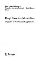
Fig. 1.1 Production of silent secondary metabolites after gene exposition by epigenetic modifiers
After isolating the endophyte species, the fermentation process for obtaining the secondary metabolites of endophytic fungi usually follows the general methodology used for fungi cultivation. The fungus is transferred from the solid storage medium (usually potato dextrose agar, PDA) and inoculated into a basic culture medium such as PDB (potato dextrose broth) to prepare a seed culture. The use of a seed culture prevents direct inoculation of the fungus, usually stored at low temperature, into the culture medium that will be used for fermentation. This procedure allows reactivation of fungal metabolism and increases the amount of inoculum, making the cultivation more productive. The use of rice as a substrate for the production of secondary metabolites has been used by a significant number of research groups (Chen et al. 2021; Ramos et al. 2022). Co-culture (Oliveira et al. 2022) modifications in the growing conditions (Lima et al. 2018) and mutation are important approaches for optimizing metabolites production by fungi (Deshmukh et al. 2022a; Takahashi et al. 2020). These tools can induce silent gene clusters related to the production of cryptic metabolites. The genetic tools available in recent decades have allowed a great advance in the knowledge of the vast metabolome of fungi (Nagarajan et al. 2021). The production of fungal secondary metabolites can be induced by small molecules, such as epigenetic modifiers (Toghueo et al. 2020) that act on DNA methyltransferases (e.g., 5-azacytidine, hydralazine, and procainamide) or histone deacetylases (e.g., suberoylanilide hydroxamic acid). These molecules provide changes in the chromatin structure allowing the expression of secondary metabolite-related genes hitherto silent in the chromatin (Mapook et al. 2022) (Fig. 1.1). The biosynthesis of cryptic metabolites and yield improvement have been achieved using the combination of OSMAC (one strain, many compounds) approach with other methodologies, such as genome mining. Simple modifications such as the introduction of halogens to the culture medium can lead to the incorporation of the halogen in the metabolite’s chemical structure. Using this approach, 12 new cytochalasins, including phomopchalasin E (bromine) (1) and phomopchalasins F (iodine), were isolated from Phomopsis sp. QYM-13 (Chen et al. 2022a). The
1
Recent Advances in Pharmaceutically Important Compounds from Endophytic Fungi
9
Fig. 1.2 Chemical structures of the compounds produced vunder OSMAC conditions (1–6)
metabolic alteration caused by OSMAC can also lead to the synthesis of new metabolites. Penicillium sp. T2–11 afforded penicnthene (2) and penicitrione H (3), new metabolites of xanthene and citrinin types. Penicnthene is the first example of a xanthene derivative with a 3-hydroxy-2-butyl chain. These metabolites showed acetylcholinesterase inhibition (28.03 ± 0.15 and 23.62 ± 0.37 μM, respectively), and the activity of penicitrione H (3) was supposed to be related to the methyl group located at C-6 (Li et al. 2022a). Asordaria conoidea was submitted to OSMAC modulation. Three extracts were obtained, one of them with allelochemical activity, showing that the different cultivation conditions led to different pools of metabolites (Arruda et al. 2022). Emericella sp. XL029, under OSMAC conditions, produced seven new compounds, including emerindole (4), emerone D (5), and emerxanthone B (6) (Xian et al. 2022). The chemical structures of the compounds are presented in Fig. 1.2. Macrophomina phaseolina, endophyte isolated from the host plant Brugmansia aurea, was studied with and without the effect of the epigenetic modifier sodium valproate, a histone deacetylase inhibitor, combined with OSMAC modifications. The results showed that the metabolite profile was modified. Among the changes, M. phaseolina produced four new and several volatile compounds (Singh et al. 2022).
10
J. A. Takahashi et al.
4 Innovative Pharmacological Targets for Bioactive Fungal Metabolites The demand for biologically active compounds for the production of new drugs is a very dynamic area. The development of new drugs can be oriented to increased absorption, prolongation of effect, higher specificity with decreased side effects, and lower negative drug interactions. Innovative drugs may be necessary for the treatment of new diseases, as occurred recently with the COVID-19 pandemic, a situation that involved the research of substances capable of acting on biological targets present in the SARS-CoV-2 virus, as well as antiviral drugs with new mechanisms of action. The development and application of vaccines are essential for coping with endemic viruses, such as HIV, dengue fever, Chikungunya, and Zica in various regions of the world (Costa and de Oliveira 2020; Banyal et al. 2021), but new drugs are also necessary since these diseases are spreading out (Takahashi et al. 2021; Lacerda et al. 2022). To exemplify, it is estimated that almost half of the individuals contaminated with HIV have died and around 37 million people are living with HIV worldwide (Bertagnolio et al. 2022). In addition, patients with pathologies such as AIDS are more propense to acquire other diseases, as recently reported during the COVID-19 pandemic. Figures also show that there is still a great demand for the development and availability of new medicines with lower toxicity and enhanced tolerability to virus mutations (Banyal et al. 2021). Equally important in this context is the development of antibiotics for the control of resistant bacteria, which causes the death of millions of people annually. In this area, research for new drugs should be constant since it is virtually impossible for bacterial strains to become resistant to available drugs. Several works have shown that metabolites biosynthesized by endophytic fungi are active against resistant bacterial strains (Manganyi and Ateba 2020). Isotetrahydroauroglaucin (7) and cristatumin B (8), metabolites produced by an Aspergillus niger species isolated from the host plant Opuntia ficus-indica, are examples of antibacterial metabolites as promising leads for the development of antibiotics effective against resistant strains (Fig. 1.3). Isotetrahydroauroglaucin (7) and cristatumin B (8) were active against Gram-positive and Gram-negative resistant bacteria, respectively (Elkady et al. 2022). Antibiotic activity was the most impactful event in the history of fungal metabolites as a source of new drugs. Between 2015 and 2021 were reported over 450 new fungal metabolites, potentially useful in the treatment of infections by resistant bacteria, although it is stressed the need for further evaluation by standardized studies using reference strains for a comparative analysis of the inhibiting potential of these metabolites (Deshmukh et al. 2022a). New drugs are also needed for the treatment of neglected diseases such as malaria, chagas disease, leishmaniasis, parasitic diseases, among others. These pathologies remain a serious public health problem and generally affect the most deprived populations living in tropical regions (Costa and de Oliveira 2020). A
1
Recent Advances in Pharmaceutically Important Compounds from Endophytic Fungi
11
Fig. 1.3 Chemical structures of the some of the antibacterial compounds (7, 8) and antileishmanial compound (9)
Paecilomyces species isolated as endophytic from Schnella splendens, a typical plant of the Amazon, produces phomoxanthone A (9), a metabolite with pronounced inhibitory effect against promastigote forms of Leishmania amazonensis and against epimastigote forms of Trypanosoma cruzi (Ramos et al. 2022). Trypanocidal natural products have also been described by Nascimento et al. (2020), while natural products active against Toxoplasma gondii were reported from Mazzone et al. (2022). Fungi can also cause human diseases of difficult treatment and new antifungal drugs for human use are extremely necessary. Several fungal infections are silent or seen only as esthetic problems related to skin, nails, and hair colonization. However, fungal diseases can acquire severity, reaching sepsis and death if untreated, with a mortality rate similar to tuberculosis (Bongomin et al. 2017). Some prominent fungal diseases are chronic pulmonary aspergillosis, cryptococcal meningitis complicating HIV/AIDs, Pneumocystis jirovecii pneumonia, invasive aspergillosis, histoplasmosis, fungal asthma, and fungal keratitis. The prevalence of invasive fungal infections has been increasing in recent decades among immunocompromised population, mainly HIV-positive individuals, organ transplant recipients, and cancer patients (Pfaller et al. 2019). Another fungal disease of great importance is candidemia, an invasive candidiasis caused by different species of Candida sp., which has approximately 700,000 individuals infected annually in the world (Bongomin et al. 2017). The literature has shown the emergence of resistance to antifungals of echinocandinR and fluconazole classes in C. glabrata and C. tropicalis isolates (Pfaller et al. 2019). Endophyte fungi are prolific sources of antifungal metabolites, which are produced to ensure the survival of a fungal species in the presence of another fungus (Oki et al. 2021). Antifungals naturally produced by endophytic fungi have been used for the treatment of mucormycosis, a very destructive invasive human disease caused by several mucorales, typically present in immunocompromised patients (Hashem et al. 2022). Deshmukh et al. (2018) listed 127 antifungal metabolites produced by Coelomycetes, 40 produced by Hyphomyces, and 3 produced by
12
J. A. Takahashi et al.
Fig. 1.4 Chemical structures of antifungal compounds (10–16)
Basidiomycetes of endophytic origin. Among the antifungal metabolites described in Deshmukh’s review, there are polyfunctionalized phenolic derivatives (colletonoic acid, polycyclic metabolites, macrocycles, phenylindol derivative, and chlorinated metabolites) (10–15). Endophyte species are also known to produce some metabolites with complex chemical structures, as can be exemplified by chaetoglobosin R (16) (Fig. 1.4). Antifungal metabolites containing the pyrone nucleus are commonly produced by endophyte species, as exemplified by ficipyrone A (17), rhizopycnin D (18), and phomopsolide A (19), which are α-pyrone, dibenzo-α-pyrone, and dihydropyrone derivatives, respectively. Anthraquinones like emodin (20) are also common fungal metabolites and molecules with steroidal and terpenoid biosynthetic origin can be exemplified by calvasterol A (21) and the oxygenated guaiane-type sesquiterpene (22), respectively (Deshmukh et al. 2022a) (Fig. 1.5). More restricted demands, equally important, comprise the search for innovative drugs aimed at the treatment of chronic and rare diseases, individuals allergic to conventional drugs, and immunosuppressed patients. Innovation in this area can lead to a decrease in the cost of production, distribution, and sale, increasing people’s access to drug treatments.
1
Recent Advances in Pharmaceutically Important Compounds from Endophytic Fungi
13
Fig. 1.5 Chemical structures of Antifungal metabolites containing the pyrone nucleus (17–22)
5 Promising Endophytic Fungi Metabolites Recently Described Secondary metabolites produced by endophytic fungi have been widely studied in the search for applications in agriculture (Fadiji et al. 2022; Fadiji and Babalola 2020) especially the production of volatile compounds (Santos et al. 2022), including those that can bring sustainability to agricultural activities (Kaddes et al. 2019). Development of anti-phytopatogenic agents is also suggested (Zhao et al. 2020). Other applications concern food (Manganyi and Ateba 2020) and cosmetics (Mahlangu and Tai 2022) industries. This is an area that is showing huge progress over the last two decades (Pal et al. 2020; Zheng et al. 2021). Fungal metabolites have been studied as leads for the development of new drugs to treat diseases, such as cancer (Kousar et al. 2022; Bai et al. 2022), inflammatory (Cui et al. 2018; Liao et al. 2018; Pal et al. 2020), infectious (Deshmukh et al. 2022a), and neurological problems (Mapook et al. 2022), as antioxidants (Bedi et al. 2021) and, recently, antiviral fungal metabolites against the SARS-CoV-2 virus due to the COVID-19 pandemic (Takahashi et al. 2021; Deshmukh et al. 2022b). In vitro screenings were also related (ElNaggar et al. 2022). Among them, cancer is undoubtedly one of the most important long-lasting targets for the development of new drugs. This disease is the second cause of death in the world and its incidence is increasing every year. Although hereditary factors, food habits, and occupational exposure are related to specific types of cancer, this disease often affects people regardless of age or social group. Treatment is currently based on chemotherapy, but the low specificity of the available treatments severely weakens the patient due to several major side effects,
14
J. A. Takahashi et al.
making treatment complex and costly. In addition, tumor cells may develop resistance to the drugs in use, and it is therefore necessary to find new selective drugs with fewer side effects for cancer treatment (Rai et al. 2022). An interesting review paper about endophyte fungi published in 2022 covered different chemical and biological aspects, such as the mechanisms of action, current challenges, and future perspectives of fungal metabolites active against Kaposi’s sarcoma, human papillary thyroid carcinoma, human pancreatic, ovarian, hepatic, lung, human lymphoma, human skin carcinoma, and breast cancer cell lines (Kousar et al. 2022). Many times, the antitumor activity is associated with uncommon natural products with unrevealed mechanisms of action. Some examples of metabolites with antitumor and other activities are given in Table 1.1, and the chemical structures of the representative metabolites are presented in Figs. 1.6 and 1.7. It is noteworthy that the fungal species present different cytotoxic profiles, represented by activity against different targets, as is the case of the action against a large number of human cancer cell lines. The metabolites cited in Table 1.1 also represent the action in other pathologies that can be associated with cancer, such as inflammatory processes and bacterial infection. The latter can be caused by opportunistic bacterial contamination of immune-depressed individuals during cancer chemotherapy. Some of the compounds presented in Table 1.1 were isolated for the first time from endophytes and represent novel compounds, as is the case of fusaroxazin (29), a novel 1,4- oxazine-xanthone derivative, produced by Fusarium oxysporum (endophyte isolated from Vicia faba) with cytotoxic effect toward A549, HCT-116, and MCF-7 tumor cell lines (Mohamed et al. 2022). In terms of biotechnological applications and product development, the potential of secondary metabolites in medicine is of major importance, but more investments are still needed in this area to accelerate biotechnological advance toward the development of new drugs and drug candidates (Mapook et al. 2022).
6 Developments Related to Bioactive Metabolites from Endophyte Fungi Chemical diversity is an important ally in the search for new pharmacologically active drugs. The biosynthesis of specific classes of metabolites can be related to the fungal species and also to the environment from where the host species is collected. Molecules with xanthone moiety, for example, are commonly isolated from marinederived endophytic fungi (Veríssimo et al. 2022; Khattab and Farag 2022), but novel chemical scaffolds have been isolated from most species of endophyte fungi (Liu et al. 2020). One of the most representative classes of secondary metabolites produced by endophytic fungi are alkaloids (Pal et al. 2020; Deshmukh et al. 2022a), as well exemplified by the well-known alkaloid’s paclitaxel, camptothecin, chincona, and
1 Recent Advances in Pharmaceutically Important Compounds from Endophytic Fungi
15
Table 1.1 Bioactive metabolites reported from endophytic fungal species and respective activity Endophyte fungal species [host] Aspergillus terreusa
Cladosporium sp.b
Diaporthe sp.c
Fusarium oxysporumd Montagnulaceae sp.e
Paraboeremia selaginellaef
Paraphaeosphaeria sporulosag
Penicillium brefeldianumh Penicillium sclerotiorum ZJHJJ-18i
Penicillium sp.j
Bioactive metabolite [IC50] Asperimide C (23) [0.78 ± 0.06 μM] Asperimide D (24) [1.26 ± 0.11 μM] Aspulvinone D (25) [10.3 ± 0.6 μM] Aspulvinone M (26) [9.4 ± 0.6 μM] Aspulvinone R (27) [7.7 ± 0.6 μM] Diaporindene A (28) [8.5 μM] Diaporindene B [5.9 μM] Diaporindene C [4.2 μM] Diaporindene D [4.2 μM] Fusaroxazin (29) [HCT-116 2.1 μM] [MCF-7 1.8 μM] [A549 3.2 μM] Rubiginosin B (30) [9.2 ± 0.9 μM] Montagnuphilone B (31) [25.5 ± 1.1 μM] Montagnuphilone E (32) [39.6 ± 1.8 μM] NK-A 17e233 (33) [5.75 μM] Cyperin (34) [27.22 μM] ES-242-1 (35) [7.38 μM] ES-242-3 (36) [17.99 μM] 5S,6S-phomalactone [5.13 μM] Paraphaeone E [25 μM] Paraphaeone F (37) [50 μM] Acremonone E [50 μM] Epicoccone [100 μM] Penipentenone A (38) [10.88 μM] Brefeldin C [2.71 ± 0.21 μM] Sclerazaphilone C (39) [7.87 ± 2.04 μM] Sclerazaphilone D (40) [6.30 ± 0.35 μM] Sclerazaphilone E (41) [9.45 ± 1.39 μM] Pinselin [5.9 to 7.5 μg/mL] Sydowinin A (42) [6.5 to 7.1 μg/ mL]
Activity Inhibition of LPS-activated NO production in RAW264.7 cells
References Liao et al. (2018)
Inhibition of NLRP3 inflammasome activation
Liang et al. (2022)
Inhibition of LPS-activated NO production in RAW 264.7 cells
Cui et al. (2018)
Cytotoxic effect toward tumor cell lines
Mohamed et al. (2022)
Inhibition of LPS-activated NO production in RAW 264.7 cells
Luo et al. (2017)
Anti-toxoplasma activity with low or no cytotoxicity in human cell lines
Mazzone et al. (2022)
Antibiotic activity against pseudomonas syringae pv. Actinidiae
Chen et al. (2022b)
Inhibition of SW620 and ASPC-1 cells
Bai et al. (2022)
Inhibition of LPS-activated NO production in RAW 264.7 cells
Jiang et al. (2022)
Suppress con A-induced T-cell and LPS-induced B-cell proliferation
Liu et al. (2016)
(continued)
16
J. A. Takahashi et al.
Table 1.1 (continued) Endophyte fungal species [host] Phoma bellidisk
Phomopsis asparagi DHS-48l Phomopsis asparagi LSLYZ-87m Phomopsis sp.n
Talaromyces amestolkiae YX1o
Bioactive metabolite [IC50] Phomaone A (43) [14.99 μM] Oblongolide J [22.26 μM] Phomoparagin B [21.6 ± 1.7 μM] Phomopchalasin A (44) [32.8 ± 2.4 μM] Phomasparapyrone B [50 μM] Phomopchalasin F [7.4 μM] Cytochalasin U (45) [8.2 ± 0.9 μM] Cytochalasin J [4.9 ± 0.6 μM] Cytochalasin H [0.2 ± 0.1 μM] Amestolkolide B (46) [1.6 ± 0.1 μM]
Activity Cytotoxicity against MCF-7, PC-3, and DU145 Inhibition of interleukin-2 (IL-2) transcription
References Zhang et al. (2022) Feng et al. (2022)
Inhibition of NO accumulation induced by LPS on BV-2 cells Cytotoxicity against MDA-MB-435
Liu et al. (2022)
Inhibition of NO production in LPS-activated AW264.7 cells
Chen et al. (2018)
Chen et al. (2022a)
A-549 (lung cancer), DU145 (prostate cancer), HCT-116 (colon cancer), MCF-7 (breast cancer), PC-3 (prostate cancer), and RAW 264.7 (macrophage cell line established from a tumor induced by the Abelson leukemia virus) LPS lipopolysaccharide, NO nitric oxide a Suriana maritima [South China] b Paris polyphylla var. yunnanensis [China] c Excoecaria agallocha [China] d Vicia faba [China] e Persicaria amphibia [North America] f Philodendron monstera [Central America] g Actinidia chinensis [China] h Houttuynia cordata [SW Asia] i Hibiscus tiliaceus [Pacific coastal zone] j Sonneratia apetala [China] k Tricyrtis maculate [Eastern Asia] l Rhizophora mangle [China] m Acanthus ilicifolius [China] n Kandelia candel [Southeast Asia] o Kandelia obovate [Eastern Asia]
huperzine A. Several classes of alkaloids isolated from fungi possess complex chemical structures, as shown in Fig. 1.8. Therefore, the total synthesis of many bioactive alkaloid compounds can be unfeasible or economically unviable. Therefore, natural sources of bioactive alkaloids are sought out worldwide for pre-clinical and clinical studies. It is widely reported that alkaloids and other secondary metabolites are biosynthesized by both endophytic fungi and the host plant, although this point is
1
Recent Advances in Pharmaceutically Important Compounds from Endophytic Fungi
17
Fig. 1.6 Chemical structures of representative metabolites described in Table 1.1 (23–32)
still a topic for which in-depth studies are suggested. Nevertheless, many examples of co-production are reported in the literature, such as vinblastine and vincristine, minor antitumor bis-monoterpene indole alkaloids produced in very low yield by the plant species Catharanthus roseus. As research with these alkaloids progressed, it was discovered that endophytic fungi isolated from C. roseus produce these indole alkaloids and structurally related bioactive derivatives at encouraging yields (Daley and Cordell 2021).
18
J. A. Takahashi et al.
Fig. 1.7 Chemical structures of representative metabolites described in Table 1.1 (33–47)
Indole alkaloids are a group of representative fungal metabolites comprising ergot alkaloids, produced by the species Claviceps purpurea and other species of the phylum Ascomycota. The ergot alkaloids interact strongly with serotonin, dopamine, and adrenergic receptors, causing several useful biological effects in the treatment of Parkinson’s disease, dementia, uterine hemorrhage, and migraines. Claviceps-infected rye contributes with half of the global production of ergot alkaloids (Yao et al. 2022a). Taking into consideration the high structural complexity of the natural bioactive ergot alkaloids, as represented by compound (48), total synthesis of this metabolite is challenging, making the research on endophyte fungi with improved production of ergot alkaloids an interesting research area. Suitable endophyte strains must have minimum tendency to degeneration, and few
1
Recent Advances in Pharmaceutically Important Compounds from Endophytic Fungi
19
FUNGAL ALKALOIDS
Fig. 1.8 Chemical diversity of fungal alkaloids produced by endophytic fungi
biosynthetic routes to ensure long utilization of the strain and ease separation of the product. Since a screening step must precede the production development, the use of high-speed automatized tools, to select promising strains, would allow the improvement of natural ergot alkaloids production (Wong et al. 2022). Jerveratum-type steroidal alkaloids were described as fungal metabolites with antifungal and anti-cancer activity. This alkaloid (49) acts synergistically with fluconazole and inhibits β-1,6-glucan biosynthesis in S. cerevisiae. Structure activity studies have shown that the NH group is essential for activity (Kubo et al. 2022). Diketopiperazine alkaloids have been described from several fungal species and with different biological activities. An alkaloid isolated from Penicillium sp. TW58–16 (50) was active against standard strains and drug-resistant clinical isolates of Helicobacter pylori in vitro (Tian et al. 2022), while a brevianamide analogue (51) exhibited potent cytotoxic activity against the HGC-27 cell line with IC50 stronger than that of cisplatin (Li et al. 2022b). A diketopiperazine-type alkaloid (52) isolated from the endophytic fungus Penicillium expansum showed anti-inflammatory activity against LPS-induced nitric oxide (NO) production in RAW264.7 mouse macrophages (IC50 25.65 μM) (Yao et al. 2022b). Chevalinulin A (53), an indole diketopiperazine alkaloid containing an unprecedented spiro[bicyclo[2.2.2]octanediketopiperazine skeleton with significant in vivo proangiogenic activity in transgenic zebrafish, was isolated from Aspergillus chevalieri CS-122 (Yan et al. 2022).
20
J. A. Takahashi et al.
Aspergillus species are proliferous sources of bioactive secondary metabolites (Hagag et al. 2022). Figure 1.8 presents the chemical structures of some alkaloids isolated by endophytic fungi, to show the huge chemical diversity of these fungal metabolites. Terpenoids are the largest group of secondary metabolites (Galindo-Solís and Fernández 2022) and have industrial great potential for cosmetic, food, and pharmaceutical industries. In the period of 2017–2019, only in China, 200 terpenoids biologically active were reported, mainly sesquiterpenoids, all of them produced by endophytic fungi (Zhao et al. 2020). In their review, Zhao et al. pointed that most of the endophyte fungal species were from Trichoderma, Aspergillus, Bipolaris, Penicillium, Phomopsis, and Alternaria genera, isolated from leaves, roots, branches, inner tissues, thallus, stems of plants, as well as from fruiting bodies. Paclitaxel, camptothecin, and fusidic acid are examples of bioactive terpenoids. In the period 2011–2020, 516 terpenoids were described from endophyte fungi, most of them of sesquiterpenoid type (Amirzakariya and Shakeri 2022), although meroterpenoids have also been highlighted. A pentacyclic triterpene, lupeol acetate, commonly found in plants was isolated from Colletotrichum gigasporum, an endophyte from Withania somnifera. Interestingly, this triterpene was described as an anti-obesity agent due to its capacity of inhibiting pancreatic lipase (IC50 16.62 ± 1.43 μg/mL) and its mechanism of action was studied (Patil et al. 2021). Zingiber griffithii was described as the host of the endophytic fungus Hypomontagnella monticulosa. The endophyte produced a sesterterpenoid active against Panc-1, NBT-T2, and HCT116 tumor cell lines (IC50 0.71, 0.30, and 0.67 ppm, respectively) (Lutfia et al. 2021). Meroterpenoids, ophiobolin-type sesterterpenoids, drimane-type sesquiterpenoids, and other terpenes were reported from Aspergillus sp. RR-YLM-12, an endophyte from the red algae Rhodomela confervoides. Some of these metabolites were reported as antimicroalgal (Fang et al. 2021). Phomopsis prunorum, an endophytic fungal species isolated from leaves of Hypericum ascyron, produces (+)- Phomoterpene A (54), a new phenolic bisabolane-type sesquiterpenoid with activity against the plant pathogenic bacteria Pseudomonas syringae pv. Lachrymans (MIC 15.6 μg/mL) (Qu et al. 2020). Penicillium chrysogenum MT-12, an endophytic fungus isolated from the medicinal plant Huperzia serrata, produced a new diterpenoid named Penicichrysogene A (55). This diterpenoid is capable of inhibit ATP release of thrombin-activated platelets (IC50 42.7 ± 3.5 μM) (Qi et al. 2022). Another diterpenoid (56) with a new type of indole scaffold with a rare 6/5/6/6/6/6/5 heterocyclic system bearing an aromatic ring C was reported from Penicillium sp. (strain ZO-R1–1) and named Shearilicine. This Indonesian strain was isolated from roots of Zingiber officinale, a medicinal plant used locally to treat cold, cough, and rheumatism among other illnesses. This cytotoxic diterpenoid was active toward L5178Y (IC50 3.6 μM) and A2780 (IC50 8.7 μM) cells (Ariantari et al. 2019). Talaromyces pinophilus produced a new fusicoccane diterpene, pinophicin A (57). The endophyte was obtained from the aerial parts of Salvia miltiorrhiza (Zhao et al. 2021). This novel meroterpenoid was isolated from the plant endophytic fungus of Penicillium sp. sh18. Isopenicin A
1
Recent Advances in Pharmaceutically Important Compounds from Endophytic Fungi
21
FUNGAL TERPENOIDS
Fig. 1.9 Chemical diversity of fungal terpenoids produced by endophytic fungi
(58) inhibits tumor by inducing G2/M cell cycle arrest and cell apoptosis. Moreover, this metabolite is a new chemotype for discovery and the development of microtubule inhibitors (Chen et al. 2020). Diaporpenoid A (59) is a new diterpenoid, with anti-inflammatory activity. This metabolite, produced by Diaporthe sp. QYM12, an endophyte isolated from the host plant Kandelia candel, inhibits the production of nitric oxide (NO) in lipopolysaccharide (LPS)-induced RAW264.7 cells (IC50 21.5 μM) (Chen et al. 2021). Figure 1.9 contains some examples of the chemical diversity of fungal terpenoids produced by endophytic fungi.
7 Perspectives Although fungi are an endless source of novel bioactive metabolites, the research of endophyte species is still in its childhood. The re-isolation of secondary metabolites already known is very common, but this is not a bad outcome per se. Re-isolation can provide higher yields of useful metabolites and, also, can bring about new biological activities. With the evolution of analytical and spectroscopical tools, minor novel metabolites are expected to be isolated, revealing novel chemical structures and new pharmacophoric groups, consequently, new mechanisms of action for several industrial applications (Singh et al. 2021).
22
J. A. Takahashi et al.
Plants are capable of adapting their metabolism to survive in challenging environments, causing a cascade of internal physiological modifications that interfere in the fungi that inhabit their interior. The metabolic changes that occur in the host plant also affect the variety of species and the metabolism of the endophytes. Comprehensive studies on the variability of endophytic species of the same plant collected from different biomes are still scarce and advanced research tools have been useful in this area (Adnan et al. 2022). Meta-genomics has been pointed out as a robust tool to quickly identify endophytes, with the objective of selecting promising species for the selection of a pool of beneficial fungi that can be explored focusing on biotechnological development (Toppo et al. 2022). Another relevant issue for the study in this area is the relationship between the biosynthesis of endophytic fungi in loco (within the host plant) and the in vitro biosynthesis (laboratory conditions) since the internal environment of the host plant is unique and cannot be imitated in vitro. Consequently, the biosynthesis of fungal secondary metabolites that depends on enzymes, metals, cofactors, and other conditions provided by the host plant is still to be better explored, although culture medium supplementation may provide some help in this regard. Development of novel approaches to produce metabolites from uncultivable endophytes and the search for new species are among the perspectives toward the progress of new drugs from endophyte fungi (Rai et al. 2022). Interdisciplinary approaches, application of omics, chromatin control, next-generation of sequencing, and genetic tools are also mentioned as a way to improve the release of toxin-free fungal products in the next two decades (Bielecka et al. 2022; Mapook et al. 2022). Additionally, it has been demonstrated that the ecological relationship of fungi in an ecosystem is dynamic, requiring metaproteomic and meta-transcriptomic assessments, and studies to define evolutionary and ecosystem functionality of the endophyte species (Adnan et al. 2022). Therefore, fungal endophytes are still a major reservoir of novel metabolites with the expectation of new discoveries in the near future. Acknowledgements The authors gratefully acknowledge financial support and grants from Fundação de Amparo à Pesquisa do Estado de Minas Gerais (PPM-00255-18), and Conselho Nacional de Desenvolvimento Científico e Tecnológico (311509/2022-3) (Brazil).
References Adams SJ, Robicheau BM, LaRue D, Browne RD, Walker AK (2021) Foliar endophytic fungi from the endangered eastern mountain avens (Geum peckii, Rosaceae) in Canada. Plan Theory 10(5): 1026 Adnan M, Islam W, Gang L, Chen HY (2022) Advanced research tools for fungal diversity and its impact on forest ecosystem. Environ Sci Pollut Res Int 29(30):45044–45062 Alves DR, Morais SM, Tomiotto-Pellissier F, Vasconcelos FR, Freire FD, Silva IN, Cataneo AH, Miranda-Sapla MM, Pinto GA, Conchon-Costa I, Noronha AD (2018) Leishmanicidal and fungicidal activity of lipases obtained from endophytic fungi extracts. PLoS One 13(6): e0196796
1
Recent Advances in Pharmaceutically Important Compounds from Endophytic Fungi
23
Amirzakariya BZ, Shakeri A (2022) Bioactive terpenoids derived from plant endophytic fungi: an updated review (2011–2020). Phytochemistry 197:113130 Amobonye A, Bhagwat P, Singh S, Pillai S (2021) Enhanced xylanase and endoglucanase production from Beauveria bassiana SAN01, an entomopathogenic fungal endophyte. Fungal Biol 125(1):39–48 Ancheeva E, Daletos G, Proksch P (2020) Bioactive secondary metabolites from endophytic fungi. Curr Med Chem 27(11):1836–1854 Araújo-Magalhães GR, Maciel MH, da Silva LF, Agamez-Montalvo GS, da Silva IR, Bezerra JD, Souza-Motta CM, Moreira KA (2021) Fungal endophytes from leaves of Mandevilla catimbauensis (Apocynaceae): diversity and potential for L-asparaginase production. Braz J Microbiol 52(3):1431–1441 Ariantari NP, Ancheeva E, Wang C, Mándi A, Knedel TO, Kurtán T, Chaidir C, Müller WEG, Kassack MU, Janiak C, Daletos G, Proksch P (2019) Indole diterpenoids from an endophytic Penicillium sp. J Nat Prod 82(6):1412–1423 Arruda GLD, Moraes GKAD, Junior AFC, Araujo AR, Chapla VM (2022) Aromatic compounds from the endophytic fungus Asordaria conoidea and their allelochemical property using OSMAC strategy. Nat Prod Res 36(15):3999–4002 Bai Y, Yi P, Li F, Hu J, Pan H (2022) Penipentenone a and brefeldin a derivatives potently inhibit KRAS mutant cancer cells from an endophytic fungus Penicillium brefeldianum F4A. Phytochemistry 200:113243 Banyal A, Thakur V, Thakur R, Kumar P (2021) Endophytic microbial diversity: a new hope for the production of novel anti-tumor and anti-HIV agents as future therapeutics. Curr Microbiol 78(5):1699–1717 Batista BN, Matias RR, Albuquerque PM (2022) Hydrolytic enzyme production from açai palm (Euterpe precatoria) endophytic fungi and characterization of the amylolytic and cellulolytic extracts. World J Microbiol Biotechnol 38(2):1–3 Bedi A, Gupta MK, Conla XA, Cahill DM, Deshmukh SK (2021) Endophytic and marine fungi are potential source of antioxidants. In: Sharma VK, Shah MP, Parmar S, Kumar A (eds) Fungi bio-prospects in sustainable agriculture, environment and nano-technology. Academic Press, London, pp 23–89 Bertagnolio S, Thwin SS, Silva R, Nagarajan S, Jassat W, Fowler R, Haniffa R, Reveiz L, Ford N, Doherty M, Diaz J (2022) Clinical features of, and risk factors for, severe or fatal COVID-19 among people living with HIV admitted to hospital: analysis of data from the WHO global clinical platform of COVID-19. Lancet HIV 9(7):e486–e495 Bhadra F, Gupta A, Vasundhara M, Reddy MS (2022) Endophytic fungi: a potential source of industrial enzyme producers. 3 Biotech 12(4):1–17 Bhatti SK, Thakur M (2022) An overview on orchids and their interaction with endophytes. Bot Rev 88:1–20 Bielecka M, Pencakowski B, Nicoletti R (2022) Using next-generation sequencing technology to explore genetic pathways in endophytic fungi in the syntheses of plant bioactive metabolites. Agriculture 12(2):187 Bongomin F, Gago S, Oladele RO, Denning DW (2017) Global and multi-national prevalence of fungal diseases—estimate precision. J Fungi 3(4):57 Caruso G, Abdelhamid MT, Kalisz A, Sekara A (2020) Linking endophytic fungi to medicinal plants therapeutic activity. A case study on Asteraceae. Agriculture 10(7):286 Chen S, Ding M, Liu W, Huang X, Liu Z, Lu Y, Liu H, She Z (2018) Anti-inflammatory meroterpenoids from the mangrove endophytic fungus Talaromyces amestolkiae YX1. Phytochemistry 146:8–15 Chen L, Fan DM, Tang JW, An T, Li X, Kong LM, Puno PT, Li Y (2020) Discovery of isopenicin a, a meroterpenoid as a novel inhibitor of tubulin polymerization. Biochem Biophys Res Commun 525(2):303–307 Chen Y, Zou G, Yang W, Zhao Y, Tan Q, Chen L, Wang J, Ma C, Kang W, She Z (2021) Metabolites with anti-inflammatory activity from the mangrove endophytic fungus Diaporthe sp. QYM12. Mar Drugs 19(2):56
24
J. A. Takahashi et al.
Chen Y, Yang W, Zou G, Wang G, Kang W, Yuan J, She Z (2022a) Cytotoxic bromine-and iodinecontaining cytochalasins produced by the mangrove endophytic fungus Phomopsis sPQYM-13 using the OSMAC approach. J Nat Prod 85(5):1229–1238 Chen Q, Yu JJ, He J, Feng T, Liu JK (2022b) Isobenzofuranones and isocoumarins from kiwi endophytic fungus Paraphaeosphaeria sporulosa and their antibacterial activity against Pseudomonas syringae p Vactinidiae. Phytochemistry 195:113050 Chua RW, Ting ASY (2021) Fungal endophytes from orchidaceae: diversity and applications. In: Yadav AN (ed) Recent trends in mycological research: environmental and industrial perspective, Fungal biology, vol 2. Springer, Cham, pp 391–426 Costa M, de Oliveira CM (2020) Endophytic fungi in the fight against neglected tropical diseases. Mini Rev Med Chem 20(16):1683–1693 Cruz JS, da Silva CA, Hamerski L (2020) Natural products from endophytic fungi associated with Rubiaceae species. J Fungi 6(3):128 Cui H, Liu Y, Li J, Huang X, Yan T, Cao W, Liu H, Long Y, She Z (2018) Diaporindenes A–D: four unusual 2,3-dihydro-1H-indene analogues with anti-inflammatory activities from the mangrove endophytic fungus Diaporthe sp. SYSU-HQ3. J Org Chem 83:11804–11813 Daley SK, Cordell GA (2021) Biologically significant and recently isolated alkaloids from endophytic fungi. J Nat Prod 84(3):871–897 Deshmukh SK, Gupta MK, Prakash V, Saxena S (2018) Endophytic fungi: a source of potential antifungal compounds. J Fungi 4(3):77 Deshmukh SK, Dufossé L, Chhipa H, Saxena S, Mahajan GB, Gupta MK (2022a) Fungal endophytes: a potential source of antibacterial compounds. J Fungi 8(2):164 Deshmukh SK, Agrawal S, Gupta MK, Patidar RK, Ranjan N (2022b) Recent advances in the discovery of antiviral metabolites from fungi. Curr Pharm Biotechnol 23(4):495–537 Elkady WM, Raafat MM, Abdel-Aziz MM, Al-Huqail AA, Ashour ML, Fathallah N (2022) Endophytic fungus from Opuntia ficus-indica: a source of potential bioactive antimicrobial compounds against multidrug-resistant bacteria. Plan Theory 11(8):1070 ElNaggar MH, Abdelwahab GM, Kutkat O, GabAllah M, Ali MA, El-Metwally MEA, Sayed AM, Abdelmohsen UR, Khalil AT (2022) Aurasperone a inhibits SARS CoV-2 in vitro: an integrated in vitro and in silico study. Mar Drugs 20(3):179 Fadiji AE, Babalola OO (2020) Elucidating mechanisms of endophytes used in plant protection and other bioactivities with multifunctional prospects. Front Bioeng Biotechnol 8:467 Fadiji AE, Mortimer PE, Xu J, Ebenso EE, Babalola OO (2022) Biosynthesis of nanoparticles using endophytes: a novel approach for enhancing plant growth and sustainable agriculture. Sustainability 14(17):10839 Fan Y, Ma Z, Zhang Y, Wang Y, Ding Y, Wang C, Cao S (2022) Sulfur-containing compounds from endophytic fungi: sources, structures and bioactivities. J Fungi 8(6):628 Fang ST, Liu XH, Yan BF, Miao FP, Yin XL, Li WZ, Ji NY (2021) Terpenoids from the marinederived fungus Aspergillus sPRR-YLW-12, associated with the red alga Rhodomela confervoides. J Nat Prod 84(6):1763–1771 Feng Z, Zhang X, Wu J, Wei C, Feng T, Zhou D, Wen Z, Xu J (2022) Immunosuppressive cytochalasins from the mangrove endophytic fungus Phomopsis asparagi DHS-48. Mar Drugs 20(8):526 Ferreira MC, Loureiro PHR, Assis JCSD, Carvalho-Silva M, Câmara PEAS, Henriques DK, Rosa LH (2021) Diversity of Endophytic fungi of Empetrum rubrum Vahl ex Willd (Ericaceae): a medicinal plant from austral South America. In: Neotropical endophytic fungi. Springer, Cham, pp 375–385. Galindo-Solís JM, Fernández FJ (2022) Endophytic fungal terpenoids: natural role and bioactivities. Microorganisms 10(2):339 Gioia L, d’Errico G, Sinno M, Ranesi M, Woo SL, Vinale F (2020) A survey of endophytic fungi associated with high-risk plants imported for ornamental purposes. Agriculture 10(12):643 Hagag A, Abdelwahab MF, Abd El-kader AM, Fouad MA (2022) The endophytic Aspergillus strains: a bountiful source of natural products. J Appl Microbiol 132(6):4150–4169
1
Recent Advances in Pharmaceutically Important Compounds from Endophytic Fungi
25
Hashem AH, Shehabeldine AM, Abdelaziz AM, Amin BH, Sharaf MH (2022) Antifungal activity of endophytic Aspergillus terreus extract against some fungi causing mucormycosis: ultrastructural study. Appl Biochem Biotechnol 194:3468–3482 Jiang H, Cai R, Zang Z, Yang W, Wang B, Zhu G, Yuan J, She Z (2022) Azaphilone derivatives with anti-inflammatory activity from the mangrove endophytic fungus Penicillium sclerotiorum ZJHJJ-18. Bioorg Chem 122:105721 Kaddes A, Fauconnier ML, Sassi K, Nasraoui B, Jijakli MH (2019) Endophytic fungal volatile compounds as solution for sustainable agriculture. Molecules 24(6):1065 Khalil AM, Hassan SE, Alsharif SM, Eid AM, Ewais EE, Azab E, Gobouri AA, Elkelish A, Fouda A (2021) Isolation and characterization of fungal endophytes isolated from medicinal plant Ephedra pachyclada as plant growth-promoting. Biomol Ther 11(2):140 Khattab AR, Farag MA (2022) Marine and terrestrial endophytic fungi: a mine of bioactive xanthone compounds, recent progress, limitations, and novel applications. Crit Rev Biotechnol 42(3):403–430 Kousar R, Naeem M, Jamaludin MI, Arshad A, Shamsuri AN, Ansari N, Akhtar S, Hazafa A, Uddin J, Khan A, Al-Harrasi A (2022) Exploring the anticancer activities of novel bioactive compounds derived from endophytic fungi: mechanisms of action, current challenges and future perspectives. Am J Cancer Res 12(7):2897 Kubo K, Itto-Nakama K, Ohnuki S, Yashiroda Y, Li SC, Kimura H, Kawamura Y, Shimamoto Y, Tominaga KI, Yamanaka D, Adachi Y, Takashima S, Noda Y, Boone C, Ohya Y (2022) Jerveratrum-type steroidal alkaloids inhibit β-1, 6-glucan biosynthesis in fungal cell walls. Microbiol Spect 10(1):e0087321 Lacerda ÍCDS, Polonio JC, Golias HC (2022) Endophytic fungi as a source of antiviral compounds–a review. Chem Biodivers 19(6):e202100971 Li SJ, Zhang X, Wang XH, Zhao CQ (2018) Novel natural compounds from endophytic fungi with anticancer activity. Eur J Med Chem 156:316–343 Li HT, Sun Y, Xie F, Wang M, Chen JY, Zhou H, Ding ZT (2022a) Xanthene and citrinin derivatives from the endophytic fungus Penicillium sp T2-11. Tetrahedron Lett 90:153626 Li L, Chang QH, Zhang SS, Yang K, Chen FL, Zhu HJ, Cao F, Liu YF (2022b) (±)-Brevianamides Z and Z1, new Diketopiperazine alkaloids from the marine-derived fungus Aspergillus versicolor. J Mol Struct 1261:132904 Liang XX, Zhang XJ, Zhao YX, Feng J, Zeng JC, Shi QQ, Kaunda JS, Li XL, Wang WG, Xiao WL (2022) Aspulvins A–H, Aspulvinone analogues with SARS-CoV-2 Mpro inhibitory and antiinflammatory activities from an Endophytic Cladosporium sp. J Nat Prod 85(4):878–887 Liao G, Wu P, Xue J, Liu L, Li H, Wei X (2018) Asperimides A–D, anti-inflammatory aromatic butenolides from a tropical endophytic fungus Aspergillus terreus. Fitoterapia 131:50–54 Lima G, Rocha AM, Santos GF, D’Silva AF, Marriel IE, Takahashi JA (2018) Metabolic response of Aspergillus sydowii to OSMAC modulation produces acetylcholinesterase inhibitors. Phytochem Lett 24:39–45 Liu H, Chen S, Liu W, Liu Y, Huang X, She Z (2016) Polyketides with immunosuppressive activities from mangrove endophytic fungus Penicillium sPZJ-SY2. Mar Drugs 14(12):217 Liu Z, Zhao JY, Sun SF, Li Y, Liu YB (2020) Fungi: outstanding source of novel chemical scaffolds. J Asian Nat Prod Res 22(2):99–120 Liu Y, Yang W, Liao Q, She Z (2022) Pyrone derivatives from a mangrove Endophytic fungus Phomopsis asparagi LSLYZ-87. Chem Biodivers 19(8):e202200491 López AFF, Martínez OMM, Hernández HFC (2021) Evaluation of Amaryllidaceae alkaloids as inhibitors of human acetylcholinesterase by QSAR analysis and molecular docking. J Mol Struct 1225:129142 Luo JG, Xu YM, Sandberg DC, Arnol AE, Gunatilaka AL (2017) Montagnuphilones A–G, azaphilones from Montagnulaceae sPDM0194, a fungal endophyte of submerged roots of Persicaria amphibia. J Nat Prod 8(1):76–81
26
J. A. Takahashi et al.
Lutfia A, Munir E, Yurnaliza Y, Basyuni M (2021) Chemical analysis and anticancer activity of sesterterpenoid from an endophytic fungus Hypomontagnella monticulosa zg15SU and its host Zingiber griffithii baker. Heliyon 7(2):e06292 Macías-Rubalcava ML, Garrido-Santos MY (2022) Phytotoxic compounds from endophytic fungi. Appl Microbiol Biotechnol 106:931–950 Mahlangu SG, Tai SL (2022) On bioactive compounds and the endophyte community in medicinal plants: bioprocessing nature’s abundance for skin disorder treatment. In: Herbal medicines. Academic Press, London, pp 361–382 Mamangkey J, Mendes LW, Harahap A, Briggs D, Kayacilar C (2022) Endophytic bacteria and fungi from Indonesian medicinal plants with antibacterial, pathogenic antifungal and extracellular enzymes activities: a review. Int J Sci Technol Manag 3(1):245–255 Manganyi MC, Ateba CN (2020) Untapped potentials of endophytic fungi: a review of novel bioactive compounds with biological applications. Microorganisms 8(12):1934 Mapook A, Hyde KD, Hassan K, Kemkuignou BM, Čmoková A, Surup F, Kuhnert E, Paomephan P, Cheng T, de Hoog S, Song Y, Jayawardena RS, Al-Hatmi AMS, Mahmoudi T, Ponts N, Studt-Reinhold L, Richard-Forget F, Chethana KWT, Harishchandra DL, Mortimer PE, Li H, Lumyong S, Aiduang W, Kumla J, Suwannarach N, Bhunjun CS, Yu FM, Zhao Q, Schaefer D, Stadler M (2022) Ten decadal advances in fungal biology leading towards human Well-being. Fungal Divers 116(1):547–614 Marsola SJ, Jorge LF, Meniqueti AB, Bertéli MBD, de Lima TEF, Bezerra JL, Lopes AD, Gazim ZC, do Valle JS, Colauto NB, Linde GA. (2022) Endophytic fungi of Brunfelsia uniflora: isolation, cryopreservation, and determination of enzymatic and antioxidant activity. World J Microbiol Biotechnol 38(6):94. https://doi.org/10.1007/s11274-022-03278-5 Mathur P, Ghosh U, Chetani R, Chaturvedi P, Sharma C, Bhatnagar P (2022) Enzymatic bioprospection of endophytic Aspergillus Niger isolated from Albizia lebbeck (L.) Benth. S Afr J Bot 1(148):580–587 Mazzone F, Simons VE, van Geelen L, Frank M, Mándi A, Kurtán T, Pfeffer K, Kalscheuer R (2022) In vitro biological activity of natural products from the endophytic fungus Paraboeremia selaginellae against toxoplasma gondii. Antibiotics 11(9):1176 Mohamed GA, Ibrahim SRM, Alhakamy NA, Aljohani OS (2022) Fusaroxazin, a novel cytotoxic and antimicrobial xanthone derivative from Fusarium oxysporum. Nat Prod Res 36(4):952–960 Nagarajan K, Ibrahim B, Ahmad Bawadikji A, Lim JW, Tong WY, Leong CR, Khaw KY, Tan WN (2021) Recent developments in metabolomics studies of endophytic fungi. J Fungi 8(1):28 Nascimento JS, Silva FM, Magallanes-Noguera CA, Kurina-Sanz M, dos Santos EG, Caldas IS, Luiz JH, Silva EO (2020) Natural trypanocidal product produced by endophytic fungi through co-culturing. Folia Microbiol 65:323–328 Nicoletti R (2019) Endophytic fungi of citrus plants. Agriculture 9(12):247 Oki Y, Nascimento IM, NBD C, Maia RA, Takahashi JA, Ferraz V, Corrêa A Jr, Fernandes GW (2021) Effectiveness of endophytic fungi from Baccharis dracunculifolia against sucking insect and fungal pathogens. In: Rosa LH (ed) Neotropical endophytic fungi. Springer, Cham, pp 337–349 Oliveira GP, Barreto DLC, Ramalho Silva M, Augusti R, Evódio Marriel I, Lana U, Takahashi JA (2022) Biotic stress caused by in vitro co-inoculation enhances the expression of acetylcholinesterase inhibitors by fungi. Nat Prod Res 36(16):4266–4270 Pal PP, Shaik AB, Begum AS (2020) Prospective leads from endophytic fungi for antiinflammatory drug discovery. Planta Med 86(13/14):941–959 Patil R, Patil S, Maheshwari V, Patil M (2021) Inhibitory kinetics and mechanism of pentacyclic triterpenoid from endophytic Colletotrichum gigasporum against pancreatic lipase. Int J Biol Macromol 175:70–280 Pfaller MA, Diekema DJ, Turnidge JD, Castanheira M, Jones RN (2019) Twenty years of the SENTRY antifungal surveillance program: results for Candida species from 1997–2016. In Open forum infectious diseases 6, suppl_1, p S79-S94). New York: Oxford University Press
1
Recent Advances in Pharmaceutically Important Compounds from Endophytic Fungi
27
Poveda J, Díaz-González S, Díaz-Urbano M, Velasco P, Sacristán S (2022) Fungal endophytes of Brassicaceae: molecular interactions and crop benefits. Front Plant Sci 13:932288 Qi B, Jia F, Luo Y, Ding N, Li S, Shi F, Hai Y, Wang L, Zhu ZX, Liu X, Tu P, Shi SP (2022) Two new diterpenoids from Penicillium chrysogenum MT-12, an endophytic fungus isolated from Huperzia serrata. Nat Prod Res 36(3):814–821 Qu HR, Yang WW, Zhang XQ, Lu ZH, Deng ZS, Guo ZY, Cao F, Zou K, Proksch P (2020) Antibacterial bisabolane sesquiterpenoids and isocoumarin derivatives from the endophytic fungus Phomopsis prunorum. Phytochem Lett 37:1–4 Raghav D, Jyoti A, Siddiqui AJ, Saxena J (2022) Plant-associated endophytic fungi as potential bio-factories for extracellular enzymes: Progress, challenges and strain improvement with precision approaches. J Appl Microbiol 133(2):287–310 Rai N, Gupta P, Keshri PK, Verma A, Mishra P, Kumar D, Kumar A, Singh SK, Gautam V (2022) Fungal endophytes: an accessible source of bioactive compounds with potential anticancer activity. Appl Biochem Biotechnol 194:3296–3319 Ramos GDC, Silva-Silva JV, Watanabe LA, Siqueira JE, Almeida-Souza F, Calabrese KS, Marinho AM, Marinho PS, Oliveira ASD (2022) Phomoxanthone a, compound of Endophytic fungi Paecilomyces sp and its potential antimicrobial and Antiparasitic. Antibiotics 11(10):1332 Reis JBA, Lorenzi AS, do Vale HMM (2022) Methods used for the study of endophytic fungi: a review on methodologies and challenges, and associated tips. Arch Microbiol 204(11):1–30 Santos LS, Guilhon GS, Neyva R, Araujo M, Filho AJ, Lopes ML, Junior HD, Brandão KS (2022) Terpenoids produced by plant endophytic fungi from Brazil and their biological activities: a review from January 2015 to June 2021. In: Filho APDSS, De Oliveira MS (eds) Terpenoids: recent advances in extraction, biochemistry and biotechnology. Bentham, Sharjah, pp 39–66 Savi DC, Aluizio R, Glienke C (2019) Brazilian plants: an unexplored source of endophytes as producers of active metabolites. Planta Med 85(08):619–636 Sharma A, Malhotra B, Kharkwal H, Kulkarni GT, Kaushik N (2020) Therapeutic agents from endophytes harbored in Asian medicinal plants. Phytochem Rev 19(3):691–720 Singh A, Singh DK, Kharwar RN, White JF, Gond SK (2021) Fungal endophytes as efficient sources of plant-derived bioactive compounds and their prospective applications in natural product drug discovery: insights, avenues, and challenges. Microorganisms 9(1):197 Singh G, Kumar A, Verma MK, Gupta P, Katoch M (2022) Secondary metabolites produced by Macrophomina phaseolina, a fungal root endophyte of Brugmansia aurea, using classical and epigenetic manipulation approach. Folia Microbiol 67:793–799 Takahashi JA, Barbosa BV, Martins BDA, Guirlanda C, Moura M (2020) Use of the versatility of fungal metabolism to meet modern demands for healthy aging, functional foods, and sustainability. J Fungi 6(4):223 Takahashi JA, Barbos BVR, Lima MTNS, Cardoso PG, Contigli C, Pimenta LPS (2021) Antiviral fungal metabolites and some insights into their contribution to the current COVID-19 pandemic. Bioorg Med Chem 46:116366 Tian D, Gou X, Jia J, Wei J, Zheng M, Ding W, Bi H, Wu B, Tang J (2022) New diketopiperazine alkaloid and polyketides from marine-derived fungus Penicillium sPTW58-16 with antibacterial activity against Helicobacter pylori. Fitoterapia 156:105095 Tiwari P, Bae H (2022) Endophytic fungi: key insights, emerging prospects, and challenges in natural product drug discovery. Microorganisms 10(2):360 Toghueo RMK, Sahal D, Boyom FF (2020) Recent advances in inducing endophytic fungal specialized metabolites using small molecule elicitors including epigenetic modifiers. Phytochemistry 174:112338 Toppo P, Subba R, Roy K, Mukherjee S, Mathur P (2022) Elucidating the strategies for isolation of endophytic fungi and their functional attributes for the regulation of plant growth and resilience to stress. J Plant Growth Regul:1–22. https://doi.org/10.1007/s00344-022-10638-w Veríssimo AC, Pinto DC, Silva AM (2022) Marine-derived xanthone from 2010 to 2021: isolation, bioactivities and total synthesis. Mar Drugs 20(6):347
28
J. A. Takahashi et al.
Wen J, Okyere SK, Wang S, Wang J, Xie L, Ran Y, Hu Y (2022) Endophytic fungi: an effective alternative source of plant-derived bioactive compounds for pharmacological studies. J Fungi 8(2):205 Wong G, Lim LR, Tan YQ, Go MK, Bell DJ, Freemont PS, Yew WS (2022) Reconstituting the complete biosynthesis of D-lysergic acid in yeast. Nat Commun 13(1):1–10 Xian PJ, Liu SZ, Wang WJ, Yang SX, Feng Z, Yang XL (2022) Undescribed specialized metabolites from the endophytic fungus Emericella sp. XL029 and their antimicrobial activities. Phytochemistry 202:113303 Yadav G, Meena M (2021) Bioprospecting of endophytes in medicinal plants of thar desert: an attractive resource for biopharmaceuticals. Biotechnol Rep 30:e00629 Yan LH, Li PH, Li XM, Yang SQ, Liu KC, Wang BG, Li X (2022) Chevalinulins a and B, Proangiogenic alkaloids with a Spiro [bicyclo [2.2. 2] octane-diketopiperazine] skeleton from Deep-Sea cold-seep-derived fungus Aspergillus chevalieri CS-122. Org Lett 24(14):2684–2688 Yao Y, Wang W, Shi W, Yan R, Zhang J, Wei G, Liu L, Che Y, An C, Gao SS (2022a) Overproduction of medicinal ergot alkaloids based on a fungal platform. Metab Eng 69:198– 208 Yao S, Zhang R, Wang J, Gu L, Hu Z, Zhang Y (2022b) A new diketopiperazine-type alkaloid from the endophytic fungus Penicillium expansum. Nat Prod Res:1–6. https://doi.org/10.1080/ 14786419.2022.2099391 Zhang X, Yang HX, Ye K, Wei PP, Lv X, Fan YZ, Yang YL, Ai HL, Liu JK (2022) Oblongolides from endophytic fungus Phoma bellidis Neerg. Harbored in Tricyrtis maculata (D. Don) JF Macbr. Phytochemistry 198:113126 Zhao Y, Cui J, Liu M, Zhao L (2020) Progress on terpenoids with biological activities produced by plant endophytic fungi in China between 2017 and 2019. Nat Prod Commun 15(7): 1934578X20937204 Zhao WT, Shi X, Xian PJ, Feng Z, Yang J, Yang XL (2021) A new fusicoccane diterpene and a new polyene from the plant endophytic fungus Talaromyces pinophilus and their antimicrobial activities. Nat Prod Res 35(1):124–130 Zheng R, Li S, Zhan X, Zhao C (2021) Biological activities of some new secondary metabolites isolated from endophytic fungi: a review study. Int J Mol Sci 22(2):959
Chapter 2
Recent Advances in Anti-Infective Compounds Produced by Endophytic Fungi Sanjay Kumar, Indu Pathania, Takaaki Kamishima, Yoshitaka Koseki, Hitoshi Kasai, and Inder Pal Singh
Abstract Endophyte is the combined form of two Greek words, which are endon (i.e. within) and phyto (i.e. plants). Endophytic microorganisms (mainly bacteria and fungi) live inside the plant for habitat, food and protection without any harmful effect on the host plant. Endophytic fungi are a highly diverse group of fungi colonized in the host plant tissue and produce diverse chemical classes of secondary metabolites such as alkaloids, steroids, terpenoids, flavonoids, glycosides, xanthones, isocoumarins, quinones, phenyl propanoids, lignans, lactones, benzopyranones, tetralones, cytochalasins and enniatins. These secondary metabolites have been reported to have various biological activities such as anticancer, immunomodulatory, insecticidal, antibacterial, antifungal, antitubercular, antiplasmodial, antiviral, anti-inflammatory and anti-oxidant. In this chapter, we mainly focused on recently reported and related antibacterial, antimycobacterial, antifungal, antiviral and antiplasmodial secondary metabolites isolated from endophytic fungi. Keywords Antibacterial · Antifungal · Antiviral · Fungi · Secondary metabolites · Antiparasitic
1 Introduction Endophyte biology is a relatively new field, with origins dating back to the nineteenth century. De Bary coined the word ‘endophyte’ of botanical origin in 1866 (De Bary 1866). Endophyte is derived from the two Greek roots: ‘endon’ which S. Kumar · Y. Koseki · H. Kasai (✉) Institute of Multidisciplinary Research for Advanced Materials (IMRAM), Tohoku University, Sendai, Miyagi, Japan e-mail: [email protected] I. Pathania · I. P. Singh (✉) Department of Natural Products, NIPER-Sector 67, SAS Nagar, Punjab, India e-mail: [email protected] T. Kamishima East Tokyo Laboratory, Genesis Research Institute, Inc., Ichikawa, Chiba, Japan © The Author(s), under exclusive license to Springer Nature Singapore Pte Ltd. 2024 S. K. Deshmukh et al. (eds.), Fungi Bioactive Metabolites, https://doi.org/10.1007/978-981-99-5696-8_2
29
30
S. Kumar et al.
means ‘within’ and ‘phyto’ which means ‘plants’ (Jalgaonwala et al. 2011). The word was originally used for any organism living within a plant, but with the development of science, broader definitions have been given nowadays. In general, endophytic microorganisms such as bacteria and fungi live in the tissues, leaves and roots of plants for reasons like habitat, food and protection. They may or not affect the host plant, and also have a symbiotic relationship. In addition, they inhabit or colonize plant tissues at specific or arbitrary stages of their life cycle, and do not immediately harm the host plant (Petrini 1991). Initially, researchers prospected that the relationship between host plants and endophytes was symbiotic. However, the initial hypothesis was disproved by the proposed one by Schulz and Boyle (2005). According to their hypothesis, endophytes acquire high virulence as pathogens when they colonize host tissues and secrete several exo-enzymes. The defence mechanism arising on the host plants as a response to colonization shows little difference between pathogens and endophytes. Pathogens often overcome the defensive chemicals produced by hosts causing a disease, tissue damage or even death. On the other hand, endophytes can colonize host tissues without damage to the hosts in the process of overcoming the defence reaction. Therefore, the difference between pathogens and endophytes depends on whether they injure the hosts during colonization. Furthermore, the collapse of the exquisite balance due to any stress caused by external or internal factors can lead to the death of hosts and/or endophytes. The individual species of more than 300,000 plants identified to date are hosts for one or hundreds of endophytes and are not hostspecific (Strobel and Daisy 2003). Therefore, a single endophytic species can exist in multiple different hosts or different tissues within the same host (Jalgaonwala et al. 2011). Endophytes belonging to various taxonomic groups have been identified, including bacteria such as mycoplasma and actinomycetes, and fungi, among which fungi have been the most extensively studied endophytic group (Bandara et al. 2006; Rana et al. 2019). Fungal endophytes are versatile and common, symbiotic with plants growing in almost all geo-climatic conditions on the earth (Patil et al. 2016). Some of the endophytic fungi can produce compounds with the same or equivalent properties as that of their host plant (Strobel and Daisy 2003; Zhao et al. 2010). Those ones are well known as a source of secondary metabolites with bioactivity, and some of them are certainly useful for the therapy of diseases affecting mankind. Various metabolites that have attractive functional properties such as antibiotics, anticancer agents, biological control agents and other bioactive compounds have been discovered and isolated from endophytic fungi up to the present. However, it has not yet been identified which diseases most of them are effective against. Hence, accelerating the study on endophyte biology may lead to the discovery of new products of unprecedented therapeutic value (Kusari and Spiteller 2011). Additionally, endophytic fungi give the researchers like chemists, microbiologists, ecologists and agronomists, challenging themes for discovering novel compounds that could help face problems like cancer, AIDS and infectious diseases. In this chapter, we describe some beneficial pharmaceutical and medicinal compounds with anti-infective potential derived from endophytic fungi. Since there are
2
Recent Advances in Anti-Infective Compounds Produced by Endophytic Fungi
31
abundant compounds to introduce, we have mainly focused on recently reported or representative examples.
2 Role of Endophytic Research with Respect to Drug Discovery The notable features of endophytes are the production of their secondary metabolites (Schulz et al. 2002). Enormous attractive secondary metabolites have been isolated to date, and many studies have been conducted in a wide range of fields, including pharmaceutical and medicinal chemistry. For instance, some of the secondary metabolites are known to have excellent bioactive effects like anticancer, antibacterial and anti-inflammatory, and some of them are actually used as pharmaceuticals in clinical conditions. The endophytes also produce several useful and novel enzymes that could be used in the biotechnology field for various purposes like degradation and biotransformation. One of the most famous secondary metabolites discovered from endophytes is penicillin isolated from Penicillium notatum in 1928 (Fleming 1929). Starting with this discovery, various antibiotics have been developed successively, and continue to save human lives to date. However, human beings have not yet overcome fungal and bacterial infections, and the threat of antimicrobial resistance (AMR) has recently arisen and must be addressed urgently (Fischbach and Walsh 2009). To confront AMR, there is an urgent need for novel antibiotics, which will be an alternative to the currently available broad-spectrum antibiotics. Even though the problem of morbidity and mortality associated with AMR is not new, AMR has recently been increasing at a significant rate due to bacteria that have acquired resistance to multiple groups of antimicrobial agents. Moreover, the decline in the development and marketing of new antimicrobial agents worsens the situation. O’Neill’s report predicts that with the current increasing trend of AMR (Fischbach and Walsh 2009), an estimated ten million lives per year may be lost to AMR-related diseases by 2050 (O’Neill 2016). To avoid this worst-case scenario, it is necessary to develop novel antimicrobial agents with chemical skeletons and/or antimicrobial mechanisms different from those of conventional drugs. Endophytic fungi can assemble mechanisms to resist pathogenic invasion by producing secondary metabolites. Some of antibiotics isolated from endophytic fungi would act against broad or specific pathogens at extremely low concentrations (Bo et al. 1998); Umino et al. 1973). Thus, many scientists pay attention to discovering novel compounds among the secondary metabolites of endophytes that have high potential for the treatment of infectious diseases. Another field that has been drawing attention in recent years is anticancer agents derived from endophytes. Today, cancer is raging as a worldwide disease. According to a statistical survey in 2018, more than 18.1 million people were affected by this incurable disease, of whom 9.6 million passed away (Umino et al. 1973). Current
32
S. Kumar et al.
cancer treatment systems are classified as surgery in the early stages and chemotherapy or radiotherapy in the later stages, which have significant side effects for the patient (Bray et al. 2018; Burish and Tope 1992; Vijayalakshmi et al. 2010). Due to the frequent disease recurrence and chemoresistance, treatment strategies to target cancer are required strongly. According to a report on the anticancer drugs, it was found that approximately 47% of the total 155 anticancer drugs approved by 2006 were derived from natural products or semi-synthesized from lead compounds isolated from natural compoundse (Newman and Cragg 2007). Additionally, it is estimated that more than 85% of plants have not yet been explored for their bioactive properties (Pieters and Vlietinck 2005). Thus, there is a great potential for undiscovered novel compounds with anticancer activities. The discovery of attractive bioactive metabolites from endophytic fungi has a potential to increase the options of lead compounds that may be developed into novel pharmaceuticals. On the other hand, it is extremely inefficient to develop effective drugs without any clues. Therefore, it will accelerate the development of novel drugs that can solve important problems such as high efficacy, low toxicity, low cost and low environmental impact. Compounds derived from discovered or undiscovered endophytes have great potential for the development of novel drugs that can save human lives. The following section introduces some representative compounds and their beneficial bioactive effects on human health. Endophytic fungi protect their host plant from a range of diseases (bacteria, fungi, viruses and protozoa) by creating bioactive compounds. Endophytic chemicals exhibit antibacterial, antifungal, antiviral and antiprotozoan properties. Due to the increase in the ability of bacteria for drug resistance after repeated use of a known antibiotic, novel antibiotic sources are constantly sought after. In an axenic culture, several endophytic fungi produce antibiotic substances that are active against bacteria harmful to people and plants. Compared to synthetic products, natural compounds made from endophytic fungi are more effective across a wider spectrum and are less hazardous. It has been observed that secondary metabolites from endophytic fungi have promising antibiotic actions against a variety of bacteria (Fig. 2.1, Table 2.1).
3 Antibacterial Metabolites The severity of antibiotic resistance was acknowledged by Aharwal and colleagues who also stressed the significance of creative approaches to research and development (Aharwal et al. 2016). Endophytes have been shown in several studies to produce secondary metabolites as part of their defence mechanisms against pathogenic invasion. Low molecular weight, organic, naturally occurring chemicals known as bioactive metabolites are created by microbes and have antimicrobial effects at low concentrations on other microorganisms (Manganyi and Ateba 2020). Recent studies have shown the investigation of Cupressaceae related natural sources for endophytic fungi. Highly bioactive endophytic Alternaria fungi were
2
Recent Advances in Anti-Infective Compounds Produced by Endophytic Fungi
Fig. 2.1 Structures of Antibacterial metabolites from endophytic fungi
33
Chaetomium sp. HQ-1, Fusarium sp. HQ-7 and Fusarium sp. HQ-9
Aspergillus sp. EJC08
Colletotrichum and Fusarium species Microsporum gypseum and Penicillium marneffei
Curvularia protuberata and Penicillium citrinum
Lophiostoma
Exserohilumrostratum
2.
3.
4.
6.
7.
8.
5.
Endophytic fungus strain Alternaria alternata, Alternaria pellucida, and Alternaria tangelonis
S. no. 1.
Scorpinone (6) 5-Deoxybostrycoidin (7) and 4-methyl-5,6-dihydro2 H-pyran-2-one (8) Ravenelin (9)
Ciprofloxacin cyclo (phenylalanylprolyl)
Differanisole A (1), 2,6-dichloro-4propylphenol (2), 4,5-dimethylresorcinol (3) Pseurotin A (4), Fumigaclavine C (5) EtOAc extracts of Fusarium sp. Crude extract of fungal strain
Compound isolated A. pellucida POE182, A. alternata CAE92 CSE177, CSE165, POE43, POE104, POE72.
Bacillus subtilis and Staphylococcus aureus
R. Solanacearum
Staphylococcus aureus, S. aureus, Escherichia coli, Pseudomonas aeruginosa S. aureus, S. enteric, and S. Typhi
Gram positive bacteria
B. subtilis
Listeria monocytogenes, Staphylococcus aureus
Target bacteria Bacillus sp., Erwinia amylovora, and Pseudomonas syringae
Table 2.1 List of anti-bacterial metabolites from endophytic fungi
Stemona sp.
Digitaria bicornis and Paspalidium flavidum Eucalyptus exserta
Cinnamomum mercadoi Cymodocea serrulata, Halophila ovalis, and Thalassia hemprichii
Bauhinia guianensis
Astragalus chinensis
Plant source Cypress trees (Cupressus arizonica, Cupressus sempervirens var. cereiformis, and Thuja orientalis)
Nischitha and Shivanna (2021) Mao et al. (2021)
Pina et al. (2021)
ZOI—10.0– 30.0 mm for different bacterial strains ZOI—6.43 ± 0.69 to 9.86 ± 0.75 MIC—125 μg/mL and 1.95 μg/mL
10 mm)
Malhadas et al. (2017)
MIC ≤ 0.095 mg/mL
Olea europaea
B. subtilis, S. aureus, B. cereus, E. coli Withania somnifera
Atiphasaworn et al. (2017)
MIC—81–250 μg/mL
Ocimum basilicum
Bacillus subtilis, Staphylococcus aureus, Escherichia coli, P. aeruginosa
S. aureus and E. coli
Yan et al. (2021)
MIC value—25 and 50 μg/mL
Acanthus ilicifolius
B. subtilis, E. coli, and S. aureus
2 35
Endophytic fungus strain Cochliobolus sp.
Aspergillus fumigatus
A. alternata
Chaetomium sp
Colletotrichum sp.
Diaporthe sp.
Eupenicillium sp. LG41
Fusarium solani JK10
Penicillium cataractum SYPF 7131
S. no. 16.
17.
18.
19.
20.
21.
22.
23.
24.
Table 2.1 (continued)
Penicimenolidying, A (27), B (28) Rasfonin (29)
Colletotrichones A–C (19–20), Chermesinone B (21) 19-nor-lanosta-5 (10),6,8,24-tetraene1,3,12,22S-tetraol (22) Eupenicinicols A (23) and B (24), Eupenicisirenins A (25) and B (26) 7-Desmethyl fusarin C
Ergosterol (14), Helvolic acid (15), and Monomethyl sulochrin-4sulphate (16) Isotalaroflavone (2), (+/)50 Dehydroxytalaroflavone (3a/3b), (+)-talaroflavone (4b) 6-Formamide-chetomin (17) and chetomin (18)
Compound isolated 1-(2-aminoethyl)-aziridine
Pseudomonas aeruginosa, Staphylococcus aureus Staphylococcus aureus, Escherichia coli E. coli, Acinetobacter sp S. aureus
Escherichia coli, S. aureus, enterococcus faecalis E. coli, Bacillus subtilis
Xanthomonas oryzae
Target bacteria MRSA, VRSA, B. cereus, B. subtilis, and S. aureus S. aureus
Wang et al. (2016) Li et al. (2015)
Li et al. (2014)
MIC—0.1 μg/mL MIC—1 to>10 μg/ml MIC—1 to >10 μg/ mL MIC-10 or more than 10 μg/mL MIC—65–10 μg/mL
Mahonia fortunei
Xanthium sibiricum
Chlorophora regia Ginkgo biloba
Kyekyeku et al. (2017) Wu et al. (2018a, b)
Yu et al. (2018)
MIC—0.78 μg/mL
Huperzia serrata
Buxus sinica
Zhao et al. (2021)
MIC—0.5–64 μg/mL
Cercis chinensis
References Santra et al. (2022) Hussein et al. (2022)
Anti-bacterial activity MIC—15.62–125 μg/ mL MIC—15.63, 1.95, and 3.90 μg/mL, respectively
Albizia lucidior
Plant source Andrographis paniculata
36 S. Kumar et al.
Lasiodiplodia theobromae ZJ-HQ1
Penicillium ochrochloronthe
Rhizopycnis vagumn
Xylomelasma sp.
25.
26.
27.
28.
(2,6-Dimethyl-5methoxyl-7hydroxylchromone (35) 6-hydroxymethyleugenin (36), 6-Methoxymethyleugenin (37), Isoeugenitol (38), Diaporthin (39), 8-Hydroxy- 6-methoxy-3methylisocoumarin (40)
Rhizopycnis acids A (33) and B (34)
Preussomerins, chloropreussomerins A (30) and B (31) Trichodermic acid (32) Bacillus substilis, Staphyloccocus aureus, Proteusbacillm vulgaris, Escherichia coli B. subtilis, P. lachrymans, R. Solanacearum B. subtilis, S. haemolyticus, A. tumefaciens, Erwinia carotovora, Ralstonia solanacearum, Xanthomonas vesicatoria
Staphylococcus aureus
Chen et al. (2016) Zhao et al. (2019)
Wang et al. (2018) Lai et al. (2021)
MIC—1.6 and 13 μg/ mL MIC—12.5 to 100 μg/ mL
IC50 = 16.1–81.3 μg/ mL MIC—25–100 μg/mL
Nicotiana tabacum
Salvia miltiorrhiza
Taxus roots
Acanthus ilicifolius
2 Recent Advances in Anti-Infective Compounds Produced by Endophytic Fungi 37
38
S. Kumar et al.
found on cypress trees such Cupressus arizonica, Cupressus sempervirens var. cereiformis and Thuja orientalis. Additionally, it was discovered that the extracts of Alternaria exhibit strong antibacterial properties. The target phytopathogenic bacteria Bacillus sp., Erwinia amylovora and Pseudomonas syringae and the model target fungus Pyricularia oryzae HS-1390 were used to test the activity of the Alternaria extracted compounds. The extracellular extracts had bacteriostatic and bactericidal effects in the range of 7.8–31.2 μg/mL. The intracellular metabolites were bactericidal at concentrations between 31.2–125 μg/mL and 15.6–62.5 μg/mL, respectively. A. pellucida POE182 was more efficient against Gram-positive bacteria, whereas A. alternata CAE92 and A. tangelonis POE103 were more effective against Gram-negative bacteria (Soltani and Hosseyni Moghaddam 2014). Three fungal endophytes with antibacterial activity were isolated from Astragalus chinensis and identified as Chaetomium sp. HQ-1, Fusarium sp. HQ-7 and Fusarium sp. HQ-9 showed potential antibiotic activity and was thus selected for large-scale fermentation. Further fermentation of strain HQ-1 led to the discovery of three compounds, i.e. differanisole A (1), 2,6-dichloro-4-propylphenol (2) and 4,5-dimethylresorcinol (3). All three compounds possess potent antibacterial activity against Listeria monocytogenes, Staphylococcus aureus and methicillin-resistant S. aureus, with Minimum Inhibitory Concentration (MIC) values ranging from 16 to 128 μg/mL (50 μg/mL ampicillin as a positive control). These findings suggest that the endophytic fungi from A. chinensis have the potential to be used as a bactericide (Liu et al. 2019a, b). Aspergillus sp. from Bauhinia guianensis was evaluated for antimicrobial activity. Two known alkaloids pseurotin A (4) and fumigaclavine C (5) were isolated. The purified alkaloids were about ten times more active than the ethyl acetate extract from which they were isolated. Fumigaclavine C was found to be the most active showing a MIC of 7.81 μg/mL against B. subtilis. The MIC of pseurotin A was 15.6 μg/mL (penicillin, vancomycin and tetracycline 25 μg/mL each were used as positive controls) (Pinheiro et al. 2013). Cinnamomum species have been widely studied for their endophytic fungi of pharmacological importance. Twelve endophytes were isolated from the bark of C. mercadoi. All endophytes exhibited antibacterial activity against Gram-positive bacteria. However, only two of the four endophytes were subjected to secondary screening and showed wide-spectrum activity and inhibited the growth of all test bacteria. EtOAc extracts displayed the highest antibacterial activity. Fungal isolates from the inner bark of C. mercadoi belong to nine different genera of filamentous fungi. The composition of fungi included the Ascomycetes Colletotrichum and Fusarium; the Zygomycetes Cunninghamella and Mucor and the Deuteromycetes Aspergillus, Penicillium, Pestalotiopsis, Phomopsis and Rhizoctonia. The ethyl acetate extract of Fusarium sp. isolated from the bark of Cinnamomum mercadoi showed MIC of 2.1, 4.2, 4.2, and 3.8 mg/mL against E. coli, E. aerogenes, S. aureus and B. cereus, respectively (Marcellano et al. 2017). Endophytic fungi are found in three common seagrasses in southern Thailand which were explored to produce antimicrobial metabolites. One hundred and sixty endophytic fungi derived from Cymodocea serrulata (family Cymodoceaceae),
2
Recent Advances in Anti-Infective Compounds Produced by Endophytic Fungi
39
Halophila ovalis and Thalassia hemprichii (family Hydrocharitaceae) were screened for the production of antimicrobial compounds against ten human pathogenic microorganisms including Staphylococcus aureus, a clinical isolate of methicillin-resistant S. aureus, Escherichia coli, Pseudomonas aeruginosa, Candida albicans, Cryptococcus neoformans and clinical isolates of Microsporum gypseum and Penicillium marneffei. Among the active fungi, seven isolates including Hypocreales sp. from C. serrulata, Trichoderma sp. H. ovalis, Penicillium sp., Fusarium sp., Stephanonectria sp. and an unidentified endophyte PSU-ES190 from T. hemprichii exhibited strong antimicrobial activity against human pathogens with MIC of less than 10 μg/mL. The inhibitory extracts at concentrations of four times their MIC destroyed the targeted cells as observed by scanning electron microscopy (Supaphon et al. 2013). The diverse endophytic fungal associations in the aerial regions of perennial grasses D. bicornis and P. flavidum have 70 species of 29 genera and 71 species of 30 genera, respectively, which were documented. Methanolic and ethyl acetate extracts of fungal species showed effective antibacterial and antagonistic properties. Curvularia protuberata and Penicillium citrinum, two fungal endophytes, showed strong antagonistic and antibacterial activity. The experiment was arranged in a randomized complete block design and carried out in triplicate using amoxicillin, chloramphenicol or ciprofloxacin as positive controls. Among the endophytic fungi showing antagonism in vitro, three isolates from P. flavidum showed high activity and another three isolates from D. bicornis showed moderate antibacterial activity against S. aureus, S. enteric and S. typhi (Nischitha and Shivanna 2021). Lophiostoma sp. Eef-7 produced 2-azaanthraquinone derivatives with little antibacterial action when exposed to Ralstonia solanacearum. The endophytic fungus Lophiostoma sp. linked to E. exserta produced three known compounds, scorpinone (6), 5-deoxybostrycoidin (7) and 4-methyl-5,6-dihydro-2 H-pyran-2-one (8). Antibacterial activities of isolated compounds were tested against R. solanacearum and the inhibition zones observed at 64 μg were 9.86 and 9.58 mm for the first two compounds. The inhibition zone diameter of streptomycin sulphate was 13.03 mm at 6.25 μg (Mao et al. 2021). In the biomass extracts of the fungus Exserohilum rostratum, the natural compound ravenelin (9) was identified and is a promising candidate for hit and lead development. Along with antiplasmodial and trypanocidal activities, it showed strong antibacterial activity. According to the findings, ravenelin inhibited both Bacillus subtilis and Staphylococcus aureus (MIC values of 7.5 μM and 1.95 μg/ mL, respectively; amoxicillin and terramycin were used as positive controls). Additionally, ravenelin has antiparasitic efficacy against P. falciparum (IC50 value of 3.4 ± 0.4 μM), Trypanosoma cruzi (IC50 value of 5.0 μM), and both the epimastigote and intracellular amastigote forms of Trypanosoma cruzi (IC50 value of 9.0 μM) (Pina et al. 2021). Marine natural products play a significant role in the drug discovery and development process, one such example is mangrove-derived endophytic fungus Epicoccum nigrum obtained from Acanthus ilicifolius. Six new polyketides, including one coumarin, two isocoumarins, dihydroradicinin and two benzofuranone
40
S. Kumar et al.
derivatives, together with seven known analogues were isolated from the culture of the mangrove endophytic fungus Epicoccum nigrum SCNU-F0002. The results showed that isolated compound 1-(4-hydroxy-2-methoxybenzofuran-5-yl) butan-1one (10) showed antibacterial activity with MIC values between 25 and 50 μg/mL against B. subtilis (ATCC 6538), E. coli (ATCC 8739) and S. aureus (ATCC 6538). Triadimefon and ciprofloxacin were used as positive controls for fungi and bacteria, respectively (Yan et al. 2019). About 14 fungal endophytes were isolated from the Ocimum basilicum var. thyrsiflora leaves. The antibacterial activity of crude extracts from all endophytic fungi was tested against nine human bacterial pathogens including Bacillus subtilis, Staphylococcus aureus, Staphylococcus epidermidis, Bacillus cereus, Escherichia coli, Klebsiella pneumoniae, Pseudomonas aeruginosa, Vibrio cholerae and Vibrio parahaemolyticus. The crude extract from Nigrospora MFLUCC16-0605 exhibited broad-spectrum antibiotic activity with MIC values ranging from 7.81 to 250 μg/mL (chloramphenicol was used as a positive control) and high antioxidant activity (IC50 value of 15.36 μg/mL) comparable to the trolox and gallic acid, respectively (Atiphasaworn et al. 2017). Another study reveals the antimicrobial potential of three fungal endophytes from leaves of Olea europaea L. The ethyl acetate extracts inhibited a wide spectrum of microorganisms at a MIC of ≤0.095 mg/mL. P. canescens and A. alternata isolated from O. europaea possess antimicrobial activity against tested Gram-positive and Gram-negative bacteria and yeasts (Malhadas et al. 2017). Fractionation of the crude extract of P. setosum was done using semi-preparative HPLC and the separated active fraction showed antibacterial efficacy with a MIC of 8 μg/mL against both S. aureus and E. coli. The yield of the active fraction was 7 mg and antibacterial activity against S. aureus and E. coli was obtained as a zone of inhibition of 19.5 ± 0.6 and 20 ± 0.4 mm, respectively (20 μL from 2 mg/mL). The compounds present in the active fraction were confirmed as patulin (11), leucodelphinidin (12) and other minor compounds like quercetin, taxifolin and kaempferol (George et al. 2019). A recent study determined the antimicrobial properties of secondary metabolites produced by endophytic fungi isolated from Sceletium tortuosum L. The secondary metabolites isolated from the Fusarium oxysporum were ethanol, octadecanoic acid, phenylmethyl ester, 4H-pyran-4-one, 2,3-dihydro-3,5-dihydroxy-6-methyl-4H-pyran-4-one and 5 hydroxymethylfurfural. 5-Hydroxymethylfurfural (13) was the most abundant secondary metabolite produced by F. oxysporum. The diameter of the inhibition zone of bacterial growth was found to be between 2 and 10 mm (Manganyi et al. 2019). Neurasthenia and newborn malnutrition are two conditions that are treated with the roots of S. suffruticosa. Therefore, S. suffruticosa endophytic fungi were investigated and tested for antimicrobial activity. The Minimum Bactericidal Concentration (MBC) and the MIC ranged from 0.5 to 2 mg/mL, respectively. The major active ingredients in S. suffruticosa are alkaloids of the securinega class. Strong antimicrobial activity was observed in the endophytic fungus C. globosum and Fusarium sp., which had alkaloid concentrations of 0.231% and 0.170%, respectively. The best bacteriostatic activity was demonstrated by C. globosum ethyl acetate extracts. S. aureus was effectively inhibited by the crude extracts of
2
Recent Advances in Anti-Infective Compounds Produced by Endophytic Fungi
41
C. globosum and Fusarium sp. 3 (1 mg/mL). The inhibition of E. coli, P. aeruginosa and E. faecalis required a crude extract concentration of at least 0.50 mg/mL (Du et al. 2020). Vernonia anthelmintica is another very effective source of antibacterial and cytotoxic activity. The isolates included species of Aspergillus calidoustus NR-10, Ovatospora senegalensis NR-03, Chaetomium globosum NR-04, Thielavia subthermophila NR-06, Aspergillus keveii XJF-23 and Aspergillus terreus XJF-3. O. senegalensis NR-03’s crude extract demonstrated potent antibacterial activity against the E. coli and S. aureus (zone of inhibition 26 and 19 mm) as well as mild antifungal inhibition (7.5 mm ZOI) against Candida albicans. O. senegalensis NR-03’s dichloromethane extract yielded 52 distinct components, the primary constituents were 9,12-octadecadienoic acid (Z, Z )-methyl ester (6.39%), (E)-9octadecenoic acid, methyl ester (6.15%), rosenonolactone (2.11%), 3-dimethyl1,2-cyclopentanedione (1.41%) and terrein (33.71%) (Niu et al. 2022). Cochliobolus sp. APS1, an endophyte of Andrographis paniculata, produces a plethora of natural bioactive compounds among which multipotent alkaloid 1-(2-aminoethyl) aziridine exhibited antibacterial, antibiofilm and antilarval potency. The MIC and MBC values of the ethyl-acetate culture extract ranged from 15.62 to 250 μg/mL against ten pathogenic microorganisms (including MRSA and VRSA) (Santra et al. 2022). Aspergillus fumigatus, isolated from Albizia lucidior leaves, was investigated for antimicrobial activity of isolated metabolites and their probable mode of action. Out of 42, three metabolites ergosterol (14), helvolic acid (15) and monomethyl sulochrin-4-sulphate (16) showed good activity with MIC values of 15.63, 1.95, and 3.90 μg/mL, respectively, compared to ciprofloxacin (Hussein et al. 2022). Cercis chinensis derived fungus A. alternata ZHJG5 yielded five polyketide derivatives, all of the studied microorganisms were effectively inhibited by these compounds at minimum inhibitory doses ranging from 0.5 to 64 μg/mL (Zhao et al. 2021). The endophytic fungus Chaetomium sp. M336 produced solid fermentation products that contained two novel compounds, 6-formamide-chetomin (17) and chaetomin (18). These compounds were identified after intensive spectroscopic analysis, including 1D and 2D NMR and HR-ESI-MS. Compound 17 had strong antibacterial activity against Escherichia coli, Staphylococcus aureus, Salmonella typhimurium ATCC 6539 and Enterococcus faecalis with a MIC value of 0.78 μg/ mL (Yu et al. 2018). An endophytic fungus, Colletotrichum sp. BS4 from a well-known boxwood plant Buxus sinica used in traditional Chinese medicine yielded new compounds, colletotrichones A (19), C (20) and one known compound chermesinone B (21). Compound 19 demonstrated substantial antibacterial activity against environmental microorganisms with a MIC of 0.1 μg/mL. E. coli was also susceptible to compound 21. Moreover, compound 20 was equally effective as streptomycin against the clinically significant pathogen S. aureus (Wang et al. 2016). A novel lanostanoid, 19-nor-lanosta-5(10),6,8,24-tetraene-1,3,12,22S-tetraol (22), containing an aromatic ring was discovered from an endophytic fungus called Diaporthe sp. LG23 that was isolated from the leaves of the Chinese medicinal plant Mahonia fortunei. Both Gram-positive and Gram-negative bacteria were
42
S. Kumar et al.
significantly inhibited by compound 22, particularly clinical isolates of Streptococcus pyogenes, Pseudomonas aeruginosa and a human pathogenic strain of Staphylococcus aureus with a MIC value of 1 to >10 μg/mL (Li et al. 2015). Eupenicillium sp. LG41, an endophytic fungus found in the roots of the Chinese medicinal plant Xanthium sibiricum, was the source of two new compounds with the decalin moiety, eupenicinicols A (23) and B (24), two new sirenin derivatives, eupenicisirenins A (25) and B (26), and four other known compounds. The antibacterial efficacies of these compounds were examined against the clinically significant Staphylococcus aureus and Escherichia coli, as well as Bacillus subtilis and Acinetobacter sp. BD4, which is generally found in soil with a MIC value of 1 to >10 μg/mL (Li et al. 2014). Seven 7-desmethyl fusarin C compounds were isolated from the endophytic fungus Fusarium solani JK10 found in the root of the Ghanaian medicinal plant Chlorophora regia. These compounds showed antibacterial activity against E. coli and Acinetobacter sp. with a MIC value of 10 or more than 10 μg/mL (Kyekyeku et al. 2017) The leaves, stems and roots of Ginkgo biloba L. yielded a total of 58 fungal isolates from 24 different genera. Among these, Penicillium cataractum SYPF 7131, an endophytic fungal strain, had the most potent antibacterial activity. Compounds penicimenolidyu A (27), penicimenolidyu B (28) and rasfonin (29) were isolated from the strain fermentation broth and had a MIC value of 65–10 μg/mL and had considerable inhibitory action against S. aureus (Wu et al. 2018a, b). Lasiodiplodia theobromae ZJ-HQ1, an endophytic fungus, yielded two novels chlorinated preussomerins, chloropreussomerins A (30) and B (31) as well as nine previously identified preussomerin analogues, from the healthy leaves of the marine mangrove Acanthus ilicifolius. The isolated compounds exhibited significant activities against Staphylococcus aureus with MIC values between 1.6 and 13 μg/mL (Chen et al. 2016). A solid-substrate fermentation culture of Penicillium ochrochloronthe linked taxus roots yielded three 3,4,6-trisubstituted pyrone derivatives and trichodermic acid (32). The MIC values for these substances ranged from 12.5 to 100 μg/mL, selectively exhibiting antibacterial action against Bacillus substilis, Micrococcus luteus, Staphyloccocus aureus, Bacillus megaterium, Salmonella enterica, Proteusbacillm vulgaris, Salmonella typhi, Pseudomonas aeruginosa, Escherichia coli and Enterobacter aerogenes (Zhao et al. 2019). Two novel anisic acid derivatives, rhizopycnis acids A (33) and B (34), were isolated from the fermentation cultures of the endophytic fungus Rhizopycnis vagumn from the healthy tissues of Nicotiana tabacum. Both substances were the first methylbutanoic/methylbutenoic acid group-containing anisic acid derivatives discovered in fungi. IC50 values for 33 and 34 ranged from 16.1 to 81.3 μg/mL against six tested bacteria (Wang et al. 2018). From the culture of an endophytic fungus, Xylomelasma sp. obtained from the curative herb Salvia miltiorrhiza Bunge, five chromone derivatives (2,6-dimethyl-5methoxyl-7-hydroxylchromone) (35), 6-hydroxymethyleugenin (36), 6-methoxymethyleugenin (37) and isoeugenitol (38); and isocoumarin congeners, namely diaporthin (39) and 8-hydroxy- 6-methoxy-3-methylisocoumarin (40) were
2
Recent Advances in Anti-Infective Compounds Produced by Endophytic Fungi
43
discovered. The isolated compounds demonstrated inhibitory activities against several bacteria with MIC ranges of 25–100 μg/mL (Lai et al. 2021).
3.1
Antimycobacterial Compounds from Endophytes
The microorganisms (Mycobacterium tuberculosis) that cause tuberculosis (TB) often damage the lungs. The most potent first-line TB medications, isoniazid and rifampicin are ineffective against multidrug-resistant tuberculosis (MDR-TB). Using second-line medications, MDR-TB can be treatable and curable. The alternatives for second-line therapy, however, are few and demand prolonged chemotherapy (lasting at least 9 months and may be 20 months). In 2021, the WHO South-East Asian Region saw the largest number of new cases of TB (46%), followed by the WHO African Region (23%) and the WHO Western Pacific (18%) (www.who.int). It has been observed that secondary metabolites from endophytic fungi have promising antimycobacterial action against a variety of bacteria (Table 2.2, Fig. 2.2). In the Huizhou Mangrove Nature Reserve in Guangdong Province, China, healthy leaves of K. obovate were host to the endophytic Talaromyces sp. (HZ-YX1), which produced the alkaloid talaramide A (41). Talaramide A demonstrated strong inhibitory efficacy against mycobacterial PknG with an IC50 value of 18.2 μg/mL (Chen et al. 2017). Together with a precursor, tenellone C (42), the new isoprenylisoindole alkaloid diaporisoindole A (43), was isolated from the endophytic Diaporthe sp. SYSU-HQ3 derived from the mangrove plant Acanthus ilicifolius collected from Shankou in Guangxi province, China. These compounds exhibited inhibitory efficacy against M. tuberculosis protein tyrosine phosphatase B (MptpB) with IC50 values of 1.77 and 2.2 μg/mL (Cui et al. 2017). A fungus BCC 25093 was isolated from an unidentified mangrove wood procured from Surat Thani province, Thailand. Palmarumycin P1 (44) and decaspirons A (45), members of the spirodioxynaph-thalenes, group exhibited antitubercular action against M. tuberculosis H37Ra, with MIC values of 1.56 and 3.13 μg/mL, respectively (Bunyapaiboonsri et al. 2015). A natural anthraquinone nigrosporin (46) isolated from a mangrove endophytic fungus Nigrospor showed an inhibitory effect against Mycobacterium tuberculosis. Another anthraquinone, 4-deoxybostrycin (47) was isolated from the fungus Alternaria eichhorniae obtained from Nigrospora oryzae. Their MIC values against a variety of mycobacterial strains were established and compared to those of commercially available control medications such as streptomycin (SM), isoniazid (INH), rifampicin (RFP) and ethambutol (EMB). Additionally, 4-deoxybostrycin showed good inhibition against the two MDR M. tuberculosis clinical isolates K2903531 and 0907961, which was even better than that of the first line of antiTB medications (Wang et al. 2013). Another research demonstrated the antimycobacterial action of penialidin C (48), which was isolated from the endophyte Penicillium sp. that was hosted by Garcinia nobilis. Citromycetin showed a MIC of 31.2 μg/mL, while penialidin C, which was
Endophytic fungus strain Talaromyces sp Diaporthe sp
BCC 25093
Alternaria eichhorniae
Penicillium sp
Aspergillus N830
Phoma sp.
Colletotrichum sp., Fusarium sp., Guignardia sp., Phomopsis sp., Phoma sp., and Microdochium sp.
S. no. 1. 2.
3.
4.
5.
6.
7.
8.
Kaurene diterpene, β-Sitosterol, brassicadiol, aspergillitine and β-sitosterol ferulate Cyclopenta[b]fluorene ring systems, phomapyrrolidones A (49)–C Javanicin (50)
Penialidin C (48)
Nigrosporin (46), 4-Deoxybostrycin (47)
Palmarumycin P1 (44), Decaspirone A (45)
Compounds isolated Talaramide A (41) Tenellone C (42), Diaporisoindole (43)
Table 2.2 List of anti-mycobacterial metabolites from endophytic fungi
Mycobacterium phlei, M. tuberculosis
M. Tuberculosis
Mycobacterium smegmatis Mycobacterium tuberculosis
M. Tuberculosis
Target bacteria Mycobacterial PknG M. Tuberculosis protein tyrosine phosphatase B (MptpB) M. Tuberculosis H37Ra
Rhoeo spathacea
Saurauia scaberrinae
Garcinia nobilis Gleditsia caspia
Nigrospora oryzae
Mangrove wood
Plant source K. Obovate Acanthus ilicifolius
MIC— 25–50 μg mL
MIC—5–50 μg/ mL
MIC—1.56 μg/ mL and 3.13 μg/ mL MIC— 172.25–12.50 μg/ ml MIC—15.6 μg/ mL MIC— 15.63–125 μg/ mL
Antimycobacterial activity IC50–18.2 μg/mL IC50–1.77 and 2.2 μg/mL
Alvin et al. (2016)
Wijeratne et al. (2013)
Jouda et al. (2016) Wang et al. (2013)
Wang et al. (2013)
Bunyapaiboonsri et al. (2015)
Reference Chen et al. (2017) Cui et al. (2017)
44 S. Kumar et al.
2
Recent Advances in Anti-Infective Compounds Produced by Endophytic Fungi
45
Fig. 2.2 Structures of anti-mycobacterial metabolites from endophytic fungi
the active molecule, had a MIC of 15.6 μg/mL (Jouda et al. 2016). The stems of Gleditsia caspia Desf. were used to create a fungal culture of Aspergillus N830, which yielded a novel kaurene derivative with a new 6/6/6/5/6 ring system structure. Using the Alamar Blue Assay (MABA), the compound demonstrated a promising antitubercular action with a MIC value of 124.5 μM. Six previously identified compounds were also isolated and showed great MIC values against Mycobacterium tuberculosis, ranging from 15.63 μg/mL (26.5 μM) to 125 μg/mL (500 μM), in comparison to the positive control isoniazid, whose MIC value was 0.24 μg/mL (1.75 μM), suggesting them as potential natural antitubercular agents (Moussa et al. 2021). Three novel alkaloids with cyclopenta[b]fluorene ring systems, phomapyrrolidones A (49)–C, were discovered from an endophytic fungal strain Phoma sp. mycelium extract inhabiting Saurauia scaberrinae. At subcytotoxic concentrations, these compounds showed minimal antitubercular action (Wijeratne et al. 2013). Rhoeo spathacea’s flowers, leaves and roots were found to have fungal endophytes, which revealed a community that included Colletotrichum sp., Fusarium sp., Guignardia sp., Phomopsis sp., Phoma sp. and Microdochium sp. Ethyl acetate extracts of the fungal isolates exhibited antiproliferative activity against at least one of the seven bacterial and mycobacterial test strains. The crude extract from a Fusarium sp. strain which exhibited strong antiproliferative activity against Mycobacterium tuberculosis resulted in the isolation of the polyketide javanicin (50)
46
S. Kumar et al.
effective against Mycobacterium phlei (MIC = 50 μg mL) and M. tuberculosis (MIC = 25 μg mL) (Alvin et al. 2016).
4 Antifungal Metabolites Fungi are a diverse group of creatures that are frequently found on surfaces, in water, soil and the air, as well as on our skin and within our bodies. Moulds can exacerbate asthma or allergy sufferers’ breathing issues, and different varieties of fungi can infect nails and result in skin rashes. There are now just four kinds of antifungal medications accessible, and there are not many candidates in the clinical development stage, making fungal diseases a serious danger to public health. The majority of fungal diseases lack quick and accurate diagnostics, and those that do exist are neither generally accessible nor inexpensively available worldwide. While resistance to the newly released medicine appears to have grown, particularly in patients receiving long-term therapy, the trends of new antifungals being launched onto the market have remained minimal. Given their enormous potential for the extraction and screening of new antibiotics for various pharmaceutical uses, natural microbial products have largely gone untapped as a source alternative. Endophytic fungi are a common source of new chemical compounds possessing antifungal properties. This section presented the endophytic fungi from medicinal plants that have been shown to produce antifungal metabolites and their potential as antifungal medicines in the last ten years (www.who.int). It has been observed that secondary metabolites from endophytic fungi have promising antifungal action against a variety of fungi (Table 2.3, Fig. 2.3). Aplosporella javeedii, an endophytic fungus associated with the host plant Orychophragmus violaceus, produced six novel polyketides called aplojaveediins A–F. Both the microbroth dilution experiment and the agar plate diffusion assay revealed that compound aplojaveediin A (51) has antifungal action against the hyphal form of the Candida albicans strain ATCC 24433. The microbroth dilution experiment revealed that the MIC of compound 51 against the hyphae form of the C. albicans strain ATCC 24433 was 100 μM. A time-kill assay was carried out as an extension of the antifungal assay. Candida albicans strain ATCC 24,433 hyphae cells were incubated with compound 51 at 400 μM (=fourfold MIC) using hygromycin B (474 μM = fourfold MIC) as a positive control drug. This discovery highlights the fungicidal ability of compound 51 (Gao et al. 2020a, b). Sixteen bioactive endophytic fungi including Alternaria alternata, Alternaria pellucida, Ascorhizoctonia sp., Aspergillus fumigatus, Aspergillus niger, Aurobasidium sp., Cladosporium porophorum, Fusarium oxysporum and Penicillium viridicatum were isolated from the plants of Cupressaceae family. The secondary metabolites of four endophytic fungi showed antifungal efficacy against A. fumigatus and A. niger. High antifungal activity against A. niger was demonstrated by secondary metabolites from Trichoderma koningii CSE32 and Pyrenochaeta CSE134. Secondary metabolites from Trichoderma koningii, namely
Botryosphaeria ramosa
Botryosphaeria dothidea
Mycosphaerella sp.
Emericella sp.
4.
5.
6.
Endophytic fungi strain Aplosporella javeedii Aspergillus fumigatus and Aspergillus niger
3.
2.
S. no 1.
Isocoumarin derivatives, botryospyrones A (54), natural tryptamines, (3aS, 8aS)1-acetyl-1, 2, 3, 3a, 8, 8a-hexahydropyrrolo [2,3b] indol-3a-ol (55) Stemphyperylenol (56) Chaetoglobosins, ceramide derivative, 3-hydroxy-N(1-hydroxy-3-methylpentan2-yl)-5-oxohexanamide 2-Amino-3,4-dihydroxy-225 (hydroxymethyl)-14-oxo6,12-eicosenoic acid (57) and myriocin (58) 5-(Undeca-3′,5′,7′-trien1′-yl) furan-2-ol (59) and 5-(undeca-3′,5′,7′-trien1′-yl) furan-2-carbonate (60)
6-Pentyl-α-pyranone (52) and 6-(4-oxopentyl)-2Hpyran-2-one (53)
Compound isolated Aplojaveediins A (51)-F
Panax notoginseng
Eugenia bimarginata
Cryptococcus neoformans and C. gattii
Rhizoctorzia solani, Verticillium dahlia, Helminthosporium maydis, Fusarium oxysporum, F. tricinctum,
Melia azedarach
Plant source Orychophragmus violaceus Juniperus virginiana, Platycladus orientalis, and Cupressus arizonica Myoporum bontioides
Alternaria solani
Penicillium italicum
A. fumigatus, A. niger
Target fungi Candida albicans
Table 2.3 List of antifungal metabolites from endophytic fungi
Xiao et al. (2014)
Pereira et al. (2016)
Wu et al. (2018a, b)
MIC of 1.57 μM
MIC-1.95 to 7.82 μM and from 0.49 to 7.82 μM MIC-50 to 6.3 μg/mL
Recent Advances in Anti-Infective Compounds Produced by Endophytic Fungi (continued)
Wu et al. (2019)
Reference Gao et al. (2020a, b) Li et al. (2019) and Erfandoust et al. (2020)
MIC— 450.4–57.3 μM
ZOI—0.9 ± 1.00 at 7.8 mg mL-1
Anti- fungal activity MIC—100 μM
2 47
Trichoderma longibrachiatum and Syncephalastrum racemosum Diaporthe eucalyptorum
8.
9.
Hypoxylon anthochroum
Endophytic fungi strain
7.
S. no
Table 2.3 (continued)
Eucalyptacid A (65) and eucacalactam B (66), eugenitol (67), cytosporone C (68), 4-hydroxyphenethyl alcohol (5), 1-(4-hydroxyphenyl) ethane1,2-diol (69), N(2-hydroxy-2-phenylethyl) acetamide (7), and phomopene (70)
7-Hydroxy-4,6-dimethyl3H-isobenzofuran-1-one (61), 7-methoxy-4,6dimethyl-3H-isobenzofuran1-one (62), 6-formyl-4methyl-7-methoxy-3Hisobenzofuran-1-one (63), 7-methoxy-4-methyl-3Hisobenzofuran-1-one (64) Ethyl acetate extracts and methanol fractions of T. longibrachiatum isolates (MF1 and MF5)
Compound isolated
Target fungi
Ibrahim et al. (2017)
Gao et al. (2020a, b)
MIC—1000 μg/mL
MIC—6.25 to 50 μM
Markhamia tomentosa
Melia azedarach
F.oxysporum, Sclerotinia sclerotiorium, Rhizoctonia solani, and Botrytis cinerea
Alternaria solani
Reference
FernándezPastor et al. (2021)
Anti- fungal activity
IC50—50 to 600 μg/ mL
Plant source
Gliricidia sepium
Botryosphaeria dothidea, and Alternaria fragriae Fusarium oxysporum, Alternaria alternata, Pythium aphanidermatum, and Phytophthora capsica
48 S. Kumar et al.
Xylaria sp. SNB-GTC2501 Arthrinium arundinis Aspergillus clavatus
12.
Berkleasmium
19.
18.
Chaetomium globosum Alternaria alternata
17.
16.
15.
14.
Saccharicola bicolor Biscogniauxia mediterranea
Phyllosticta sp.
11.
13.
Paraconionthyrium sp
10.
C. albicans M. oryzae
Palmarumycin C8 (85)
Sclerotinia sclerotiorum
Phomopsis obscurans, F. Oxysporum, C. fragariae
Dioscorea zingiberensis
Terminalia chebula
Ginkgo biloba
Bergenia purpurascens Opuntia humifusa
Myoporum bontioides
F. Oxysporum, C. musae, and P. italicm
Pythium dissimile
Nicotiana tabacum
P. cupana
Osmanthus fragrans
Azadirachta indica
Mucor hiemalis
C. Gloeosporioides
Rhizoctonia solani, Fusarium graminearum, and Botrytis cinerea
Fusarium graminearum, Fusarium solani, and Botrytis cinerea
Chaetoglobosin A (82) and D (83) Altenusin (84)
5-methylmellein (81)
24-hydroxylergosta-4,6,8 (14),22-tetraen-3-one, 4,40dimethoxy-5,50-dimethyl7,70-oxydicoumarin, orlandin Bicolorins B (79) and D (80)
Piliformic acid (77) and cytochalasin D (78) Diorcinol M,N,O
Globosuxanthone A (71), vertixanthone (72), hydroxyvertixanthone (73), 3,8-dihydroxy-1-methy19H- xanthen-9-one (4), and danthron (74), danthron (75) Guignardones (C) (76), meroterpenes, dioxolanone derivatives
IC50—9.1 μg/mL
MIC—6.2 and 8.5 μg/mL 63.5% growth suppression at 150 μM at 120 h IC50 values of 0.35 and 0.62 μg/mL FIC—0.078 to 0.188
MIC—8 and 4 μg/ mL MIC—244.73, 195.79, and 61.18 μM
Inhibition ratio— 48.43%, 40.98%, and 49.53% at 50 μg/mL, 200 μg/mL MIC—128 μg/mL
MIC—4-130 μg/mL
Recent Advances in Anti-Infective Compounds Produced by Endophytic Fungi (continued)
Zhang et al. (2013a, b) Phaopongthai et al. (2013) Shan et al. (2014)
Zhao et al. (2020) Silva-Hughes et al. (2015)
Elias et al. (2018) Zhang et al. (2018) Li et al. (2017)
Yan et al. (2021)
Miao et al. (2020)
2 49
Trichoderma koningiopsis
Trichoderma brevicompactum 0248
22.
Endophytic fungi strain Trichoderma sp.
21.
S. no 20.
Table 2.3 (continued)
Compound isolated Dichlorodiaportinolide (86) and dichlorodiaportin (87) Koninginin O (88), koninginin Q, and 7-Omethylkoninginin D Trichodermin (89)
Plant source Myoporum bontioides Panax notoginseng
Allium sativum
Target fungi Colletotrichum musae and Rhizoctonia solani F. Oxysporum and P. cucumerina Rhizoctonia solani and B. cinerea
Reference Li et al. (2016) Liu et al. (2016a, b) Shentu et al. (2014)
Anti- fungal activity MIC—6.25 to 150 μg/mL MIC—128 μg/mL EC50—0.25 μg/mL and 2.02 μg/mL
50 S. Kumar et al.
2
Recent Advances in Anti-Infective Compounds Produced by Endophytic Fungi
Fig. 2.3 Structures of Antifungal metabolites from endophytic fungi
51
52
S. Kumar et al.
6-pentyl-α-pyranone (52) and 6-(4-oxopentyl)-2H-pyran-2-one (53) showed high antifungal activity against A. fumigates (Li et al. 2019) (Erfandoust et al. 2020). Botryospyrones A (54) and a new natural tryptamine (3aS, 8aS)-1-acetyl-1, 2, 3, 3a, 8, 8a-hexahydropyrrolo [2,3b] indol-3a-ol (55) were isolated from a marine mangrove endophytic fungus called Botryosphaeria from the leaves of Myoporum bontioides. Inhibitory activities of isolated compounds on Fusarium oxysporum and Fusarium graminearum were all greater than those of triadimefon, a common agricultural fungicide. Botryospyrones A (54) had a MIC value of 112.6 μM against F. oxysporum. Compound 55 had roughly 12, 3- and 18-times greater activity against F. oxysporum, P. italicum and F. oxysporum, respectively (Wu et al. 2019). In another study by Zhibo et al. the solid culture of the endophytic fungus Botryosphaeria dothidea from the stems of white cedar (Melia azedarach) gave 15 known compounds, including chaetoglobosins C and F, as well as two new metabolites, α-pyridone derivative, a ceramide derivative. The antibacterial, antioxidant and cytotoxic properties of these metabolites were assessed in vitro (Xiao et al. 2014). Stemphyperylenol (56) demonstrated significant antifungal activity against the plant disease caused by Alternaria solani, comparable to the widely used fungicide carbendazim (Hu et al. 2020). Two eicosanoic acids from Mycosphaerella sp., 2-amino-3,4-dihydroxy-25(hydroxymethyl)-14-oxo-6,12-eicosenoic acid (57) and myriocin (58), were tested for their antifungal properties against Cryptococcus neoformans and C. gattii. The compounds demonstrated antifungal activity against several isolates of Candida neoformans and Candida gattii, with MIC values for compound 57 and compound 58 ranging from 0.48 to 1.95 μM and 0.49 to 7.82 μM, respectively (Pereira et al. 2016). 5-(undeca-3′,5′,7′-trien-1′-yl) furan-2-ol (59) and 5-(undeca-3′,5′,7′-trien1′-yl) furan-2-carbonate (60), two novel alkylated furan derivatives, were discovered from the crude extract of an endophytic fungus Emericella sp. XL029 associated with the leaves of Panax notoginseng. Compound 59 showed considerable action against all investigated fungi with MIC ranging from 25 to 3.1 μg/mL, while compound 60 demonstrated activity across the board with MIC ranges of 50–12.5 μg/mL. The MIC values of compounds 59 and 60 ranged from 50 to 6.3 μg/mL (Wu et al. 2018a, b). A chemical study of the culture medium and mycelium organic extracts of the endophytic fungus Hypoxylon anthochroum Gseg1 led to the isolation of three known isobenzofuranones, 7-hydroxy-4,6-dimethyl-3H-isobenzofuran-1-one (61), 7-methoxy-4,6-dimethyl-3H isobenzofuran-1-one (62), 6-formyl-4-methyl-7methoxy-3H-isobenzofuran-1-one (63) together with 7-methoxy-4-methyl-3Hisobenzofuran-1-one (64), which was reported for the first time as a natural product. All of the compounds significantly and concentration-dependently suppress the development of pathogens. IC50 for the isolated compounds ranged from 0.64 to 2.61 mM when compared with carbendazim as control (Sánchez-Fernández et al. 2020). Endophytic fungi namely Trichoderma longibrachiatum and Syncephalastrum racemosum were isolated from the leaves of M. tomentosa. In the poisoned food assay, the ethyl acetate extracts and methanol fractions of T. longibrachiatum
2
Recent Advances in Anti-Infective Compounds Produced by Endophytic Fungi
53
isolates (MF1 and MF5) and the methanol fraction of S. racemosum isolates (MF3) showed a MIC value of 1000 μg/mL (Ibrahim et al. 2017). The endophytic fungus Diaporthe eucalyptorum KY-9 that had been isolated from Melia azedarach produced two biosynthetically related new metabolites, eucalyptacid A (65) and eucacalactam B (66), as well as six known substances, eugenitol (67), cytosporone C (68), 1-(4-hydroxyphenyl) ethane-1,2-diol (69) and N- (2-hydroxy-2-phenylethyl) acetamide (70). The MICs of compounds 65, 68, 69 and 70 against Alternaria solani ranged from 6.25 to 50 μM. Strain KY-9 is an unexplored resource for the creation of biological control agents to stop the spread of the pathogenic fungus A. solani (Gao et al. 2020a, b). The strain YM 311593 (Paraconiothyrium sp.) was isolated from the fruits of Azadirachta indica. The fermentation broth extract of the strain yielded four xanthones and one anthraquinone. They were identified as globosuxanthone A (71), vertixanthone (72), hydroxyvertixanthone (73), 3,8-dihydroxy-1-methy1-9Hxanthen-9-one (74) and danthron (75). The broth microdilution technique was used to test the antifungal activity of compounds 71–75 against four phytopathogens. One of them, globosuxanthone A (71), had MIC values of 4, 8 and 16 μg/mL against Fusarium graminearum, Fusarium solani and Botrytis cinerea, respectively, demonstrating significant antifungal activity. Compound 72 had MIC of 16, 32 and 64 μg/mL, respectively, and moderately inhibited the growth of F. graminearum, F. solani and P. oryzae. With MIC of 8 and 64 μg/mL, respectively, compound 73 also demonstrated significant inhibitory action against F. graminearum and B. cinerea. Compounds 74 and 75 showed moderate antifungal activity (MICs of 128 μg/mL) against P. oryzae (Miao et al. 2020). The endophytic fungus Phyllosticta sp. WGHL2 provided four novel meroterpenes, guignardones U-X, 11 well-known meroterpenes and three wellknown dioxolanone derivatives. With an inhibition of 48.43%, 40.98% and 49.53% at 50 μg/mL, respectively, against Rhizoctonia solani, Fusarium graminearum and Botrytis cinerea in antifungal assays, dioxolanone guignardianone C (76) showed broad-spectrum antifungal activity. Additionally, compound 76 effectively inhibited B. cinerea spore germination at 200 μg/mL and showed a modest protective effect against B. cinerea in vivo (Yan et al. 2021). Piliformic acid (77) and cytochalasin D (78) were extracted using chromatographic methods from two species of Xylaria, with the help of tests using Colletotrichum gloeosporioides. This demonstrates the antifungal activity of piliformic acid and cytochalasin D against C. gloeosporioides, with MIC of 2.92 and 2.46 mol/mL, respectively (Elias et al. 2018). Fresh tobacco leaves were used to isolate and purify the endophytic fungus Arthrinium arundinis TE-3 from Nicotiana tabacum L. Three prenylated diphenyl ethers from this fungal strain showed MICs of 8 and 4 μg/mL and showed specific antifungal activity against Mucor hiemalis (Zhang et al. 2018). Two novel coumarin derivatives and two well-known bicoumarins were isolated from the root of Myoporum bontioides by the endophytic fungus Aspergillus clavatus. Isolated compounds showed strong broad-spectrum inhibitory activities with MIC values 244.73, 195.79 and 61.18 μM against all three pathogenic fungi,
54
S. Kumar et al.
F. oxysporum, C. musae and P. italicm, and triadimefon, which was used as the positive control, had MIC values 340.43, 272.39 and 170.24 μM, respectively (Li et al. 2017). Bicolorins A–I, a group of nine novel halogenated cyclopentenones, were isolated from endophytic fungus Saccharicola bicolour of Bergenia purpurascens together with three other known cyclopentenones. Isolated compounds showed antifungal activity against five phytopathogenic fungi (Uromyces viciae-fabae, Pythium dissimile, Gibberella zeae, Aspergillus niger and Sclerotinia sclerotiorum). Bicolorin B (79) and D (80), in particular, showed potent antifungal activity against P. dissimile with MIC values of 6.2 and 8.5 μg/mL, respectively, in comparison to the positive control cycloheximide (MIC of 8.6 μg/mL). Additionally, both in vitro and in vivo tests showed 80 to be a powerful antifungal against S. sclerotiorum (Zhao et al. 2020). 5-Methylmellein (81) was isolated from the endophytic fungus Biscogniauxia mediterranea Ohu 19B found on Opuntia humifusa (Cactaceae) and evaluated in vitro microdilution broth experiment against seven plant pathogens, including Colletotrichum acutatum, C. fragariae, C. gloeosporioides, Fusarium oxysporum, B. cinerea, Phomopsis obscurans and P. viticola. Phomopsis obscurans was most vulnerable (63.5% growth suppression) at 150 μM at 120 h. At 300 μM after 48 h, the best growth inhibition of F. oxysporum (20.1%) was achieved. Compound 81 stimulated B. cinera and C. fragariae at lower doses (75 and 150 μM), but C. acutatum and C. gloeosporioides were stimulated at all levels (Silva-Hughes et al. 2015). Another ascomycete genus, Chaetomium, is a prolific producer of active compounds. Chaetomium globosum CDW7, an endophyte from Ginkgo biloba, was used to isolate chaetoglobosin A (82) and chaetoglobosin D (83). Both the compounds demonstrated antifungal efficacy against Sclerotinia sclerotiorum with IC50 values of 0.35 and 0.62 μg/mL, respectively, in comparison to carbendazim (0.17 μg/ mL) (Zhang et al. 2013a, b). The endophytic fungus Alternaria alternata Tche-153 from Terminalia chebula yielded altenusin (84), a biphenyl derivative, which showed strong synergistic activity against C. albicans when combined with each of the three azole medications, ketoconazole, fluconazole or itraconazole, using the disc diffusion method and the microdilution checkerboard technique. The fractional inhibitory concentration (FIC) index ranged from 0.078 to 0.188 (Phaopongthai et al. 2013). Diepoxin, palmarumycin C11, palmarumycin C12, cladospirone B 1,4,7-trihydroxy-8-(spirodioxy-1′,8′-naphthyl)-7,8-dihydronaphthalene and palmarumycin were all extracted from Berkleasmium sp., an endophyte associated with Dioscorea zingiberensis from Hubei Province, China. The antifungal activity of several compounds was tested against M. oryzae spore germination. They prevented M. oryzae spore germination with IC50 values ranging from 9.1 to 124.5 μg/mL. Among the compounds examined, palmarumycin C8 (85) had the most inhibitory action (IC50 9.1 μg/mL), however it was less potent than the positive control carbendazim (IC50 6.3 μg/mL) (Shan et al. 2014). The endophytic fungus Trichoderma sp. isolated from the roots of Myoporum bontioides contained dichlorodiaportinolide (86) and dichlorodiaportin (87), which
2
Recent Advances in Anti-Infective Compounds Produced by Endophytic Fungi
55
showed MIC values ranging from 6.25 to 150 μg/mL against Colletotrichum musae and Rhizoctonia solani, however they were inactive against Penicillium italic and Fusarium graminearum (MIC values >200 μg/mL) (Li et al. 2016). Koninginin O (88) produced by the endophytic fungus Trichoderma koningiopsis YIM PH30002 growing on Panax notoginseng showed antifungal properties against Fusarium oxysporum, Fusarium solani, Fusarium flocciferum, Plectosphaerella cucumerina and Alternaria panax, which are root rot diseases causing pathogens in P. notoginseng. The MIC for koninginin O against F. oxysporum and P. cucumerina was 128 μg/mL (Liu et al. 2016a, b). Trichoderma brevicompactum 0248, an endophytic fungus strain identified from Allium sativum, was the source of the trichodermin (89). Trichodermin showed strong inhibitory efficacy against Rhizoctonia solani and B. cinerea, with an EC50 of 0.25 and 2.02 μg/mL, respectively, but was only moderately effective against Colletotrichum lindemuthianum (EC50 = 25.60 μg/mL) (Shentu et al. 2014).
5 Antiviral Metabolites Virus is nucleic acid (DNA or RNA) containing protein coated submicroscopic infectious agent that lives inside living cells and uses the machinery of the host cell for replication. It can infect all living organisms including plants, animals and microorganisms. Several diseases in humans caused by viral infections are respiratory viral diseases (e.g. flu, parainfluenza virus infection, severe acute respiratory syndrome (SARS), COVID, etc.), gastrointestinal viral diseases (e.g. norovirus infection, rotavirus infection, astrovirus infection, etc.), exanthematous viral disease (e.g. measles, rubella, chickenpox/shingles, roseola, smallpox, fifth disease, chikungunya virus infection, etc.), hepatic viral diseases (e.g. hepatitis A, hepatitis B, hepatitis C, etc.), cutaneous viral diseases (e.g. warts, oral herpes, genital herpes, etc.), haemorrhagic viral diseases (e.g. Ebola, Lassa fever, dengue fever, yellow fever, etc.), neurologic viral diseases (e.g. polio, viral meningitis, viral encephalitis, rabies, etc.) and human immunodeficiency virus (HIV). Tobacco mosaic virus is the first discovered virus. In 1892, Dimitri Ivanovski reported the extract of infectious tobacco leaves remain infectious after passing through a Chamberland filter candle. He concluded the infectious agents are smaller than bacteria. In 1892, Martinus Beijerinck named the infectious agent as tobacco mosaic virus (Lecoq 2001). Viruses complete their life cycle in three steps starting from entry, then genome replication and finally exit from host cell. In the entry step, the viral capsid or glycoprotein interacts with the receptor of the host cell, which then penetrates the virus particle into the cell followed by uncoating, in which the virus sheds its capsid. The naked viral genome is utilized for gene expression and viral genome replication. Finally, the viral proteins and viral genomes are accumulated and assembled to form new virions. The assembled virions are released from the host cell through cell lysis, budding and exocytosis (Ryu 2017). Secondary metabolites isolated from
56
S. Kumar et al.
Fig. 2.4 Structures of Antiviral metabolites from endophytic fungi
endophytic fungi have been reported for promising antiviral activity against diverse range of viruses (Fig. 2.4, Table 2.4). Brefeldin-A (BFA, 90) is a 13-carbon macrolide lactone also known as ascotoxin or cyanein or decumbin or lunatin or sinergisidin. It was reported for diverse biological activities such as antifungal (Crabbe and Betina 1994; Talukdar et al. 2021), antiproliferative (Ishii et al. 1989; Sausville et al. 1996), antimitotic, cytostatic (Wang et al. 2002) and antiviral (Betina 1992; Tamura et al. 1968). It was recently reported that BFA can inhibit TNFα-induced necroptosis (Shi et al. 2022).
Endophytic fungal species/strain Penicillium sp.
Phomopsis sp
Cytonaema sp.
Penicillium chrysogenum
Alternaria tenuissima
S. no. 1.
2.
3.
4.
5.
Xanthoviridicatin E (95) Xanthoviridicatin F (96) Altertoxin I (97) Altertoxin II (98) Altertoxin III (99) Altertoxin V (100)
Integracin A (92) Integracin B (93) Integracin C (94)
Betulinic acid (91)
Metabolites Brefeldin-A (90)
Table 2.4 List of antiviral metabolites from endophytic fungi
HIV-1 integrase HIV-1
Stem tissues of Quercus emoryi
HIV-1 integrase
DENV-NS5
Japanese encephalitis virus Zika virus
DENV-4
DENV-3
DENV-2
Target virus DENV-1
Leaves of unidentified plant
Diospyros carbonaria Twigs of Quercus ilex
Host plant Soil around Angelica keiskei root
54.8 ± 0.4 nM 4.1 ± 3.1 μM 3.2 μM 6.1 μM 3.5 μM 6 μM 5 μM 1.42 μM 0.21 μM 0.29 μM 0.09 μM
IC50 or EC50# 61.32 ± 13.5 nM 54.6 ± 0.9 nM 57.9 ± 0.1 nM 65.7 ± 6.3 nM 58.4 ± 0.3 nM
(continued)
Bashyal et al. (2014)
Singh et al. (2003)
Peyrat et al. (2017) Singh et al. (2002)
Reference Raekiansyah et al. (2017)
2 Recent Advances in Anti-Infective Compounds Produced by Endophytic Fungi 57
Fresh stems of Achyranthes bidentata Leaves of Fagara zanthoxyloides Branches of unknown tree Inner bark Aegiceras corniculatum
Phomochromanone A (112), Phomochromanone B (113), Phomopsones B (114), Phomopsones C (115)
Chloroisosulochrin (116)
Aspergillus sp.
Periconia sp.
Phomopsis sp
Pestalotiopsis theae
Pestalotiopsis theae
Emericella sp.
9.
10.
11.
12.
13.
Emerimidine A (118) Emerimidine B (119)
Pestalotheol C (117)
Pericoannosin A (108) Periconiasin F (110) Periconiasin J (111) Periconone B (109)
Epicocconigrone A (107)
Kadsuralongi pedunculata Kadsuralongi pedunculata Annona muricata
8.
Cytochalasin Z8 (106)
Aspergillus sp.
Host plant Kadsuralongi pedunculata
7.
Metabolites Asperphenalenone A (101) Asperphenalenone B (102) Asperphenalenone C (103) Asperphenalenone D (104) Asperphenalenone E (105)
Endophytic fungal species/strain Aspergillus sp.
S. no. 6.
Table 2.4 (continued)
H1N1
HIV-1 1A1
RSV
HIV-1
HIV-1
HIV-1
HIV-1
H1N1
Target virus HIV-1
42.07 μg/ mL 62.05 μg/ mL
69.6 μM 29.2 μM 25 μM 18 μM 99 μM 147 μM 6.1 μM 0.5 μM 4.22 ± 1.03 μM 16.1 μM#
6.6 μM
IC50 or EC50# 4.5 μM 2.4 μM 0.28 ± 0.01 μM 9.2 μM
Uzor et al. (2016) Liu et al. (2019a, b) Zhang et al. (2011)
Yang et al. (2020a, b)
Liu et al. (2016a, b)
Zhao et al. (2021) Pang et al. (2017) Pang et al. (2017) Zhang et al. (2016)
Reference Pang et al. (2017)
58 S. Kumar et al.
Phomopsis sp.
Nigrospora sp.
Nigrospora sp.
Phoma sp.
Pestalotiopsisvaccinii
15.
16.
17.
18.
19.
Vaccinal A (130) Vaccinol J (131)
Phomanolide (129)
Chermesinone B (128)
4-dehydroxyaltersolanol A (126) 6-O-demethyl-4dehydroxyaltersolanol A Altersolanol B (127)
Dankasterone A (125)
Pestalol A (120) Pestalol B (121) Pestalol C (122) Pestalol D (123) Pestalol E (124)
Respective value is EC50, all others are IC50
#
Pestalotiopsis sp.
14.
Kandelia candel
Root of aconitum vilmorinianum
Root of Aconitum carmichaeli
Stem of Aconitum carmichaeli Root of Aconitum carmichaeli
Stem of Aegiceras corniculatum
EV71
H1N1
H1N1
H5N1
H5N1
H1N1/H3N2
8.35 ± 1.41 μg/ mL 2.59 ± 1.22 μg/ mL 7.82 ± 1.86 μg/ mL 0.80 ± 0.29 μg/ mL 2.96 ± 0.64 μg/ mL 19.2 μM
18.9/ 22.6 μM 28.7/ 27.7 μM 40.0/ 42.6 μM 48.0/ 48.0 μM 49.9/ >50 μM 3.56 μM
Wang et al. (2014)
Liu et al. (2019a, b)
Zhang et al. (2016)
Ma et al. (2014) Zhang et al. (2016)
Sun et al. (2014)
2 Recent Advances in Anti-Infective Compounds Produced by Endophytic Fungi 59
60
S. Kumar et al.
BFA is produced by various endophytic fungi such as Paecilomyces sp./Aspergillus clavatus (isolated from the bark of the host plant Taxus mairei and Torreya grandis (Wang et al. 2002)). Penicillium melinii/Penicillium janthinellum (isolated from the roots of the host plant Panax ginseng) (Zheng et al. 2013), Penicillium brefeldianum (Bai et al. 2022), Eupenicellium brefeldianum (isolated from the host plant Arisaema erubescens) (Hai-yan et al. 2008), Scedosporium apiospermum (isolated from the host plant Bauhinia guianensis) (Cordeiro et al. 2019), Penicillium cyaneum and Cladosporium sp. (isolated from the host plant Quercus variabilis) (Wang et al. 2007) and Acremonium sp. (isolated from healthy twigs of the host plant Knema laurina). In 1958, Brefeldin-A was first isolated by Singleton et al. from the fungus Penicillium decumbens (Singleton et al. 1958) followed by isolation from other Penicillium sp. (Abraham and Arfmann 1992). In 1968, Tamura et al. reported that brefeldin-A (BFA) showed antiviral activity against herpes simplex viral strain HF (HSV) and Nawcastle disease viral strain Miyadare (NDV) by the agar-diffusion plaque-inhibition method (Tamura et al. 1968). Finter observed that the growth of myxovirus was inhibited by BFA at concentrations between 0.03 and 25 μg/mL (Finter 1970). Cheung et al. observed that BFA arrests the maturation and egress of HSV during early stages of infection by inhibiting the transportation of enveloped particles from the endoplasmic reticulum (ER) to Golgi complexes (Cheung et al. 1991). Raekiansyah et al. isolated the endophytic fungal strain Penicillium sp. FKI-7127 from soil around the root of the plant Angelica keiskei (family Umbelliferae). Brefeldin-A (BFA) was isolated from the ethyl alcohol extract of the cultured endophytic fungal broth of Penicillum sp. FKI-7127. It showed antiviral activity against dengue viruses (DENV-1, 2, 3, and 4), Zika virus (ZIKV) and Japanese encephalitis virus (JEV) with IC50 values at 48 h post infection 61.32 ± 13.5, 54.6 ± 0.9, 57.9 ± 0.1, 65.7 ± 6.3, 54.8 ± 0.4 and 58.4 ± 0.3 nM, respectively. The time-of-addition study showed that BFA inhibited the maturation of the DENV, ZIKV and JEV (Raekiansyah et al. 2017). It blocked the transportation of glycoprotein from the ER to the Golgi apparatus preventing the formation and release of the viruses from infected cells. An additional study by Raekiansyah et al. showed BFA could not block intracellular protein transport in the mosquito cell line (Raekiansyah et al. 2017). Peyrat et al. isolated betulinic acid (91) from the H2O/MeOH extract of an endophytic fungus Phomopsis sp. and its host plant Diospyros carbonaria (EtOAc extract of the bark). Betulinic acid showed antiviral activity, evaluated by the dengue polymerase assay on the RNA-dependant RNA polymerase (RdRp) domain of the non-structural protein 5 (NS5) with an IC50 value 4.3 ± 3.1 μM (Peyrat et al. 2017). Betulinic acid was reported to have a diverse range of biological activities such as anti-HIV, antibacterial, antimalarial, anti-inflammatory, antihelmintic, antinociceptive, anti-HSV-1 and anticancer activities (Moghaddam et al. 2012). Singh et al. isolated an endophytic fungus Cytonaema sp. from living twigs of Quercus ilex (family Fagaceae) in El Pardo, province of Madrid, Spain. Integracins A (92) B (93) and C (94) were isolated from the methyl ethyl ketone extract of the cultured fungal broth and evaluated for HIV-1 integrase inhibitory activities.
2
Recent Advances in Anti-Infective Compounds Produced by Endophytic Fungi
61
Integracins A, B and C inhibited the coupled reaction of HIV-1 integrase with IC50 values of 3.2, 6.1 and 3.5 μM, respectively, whereas they were found to be 10–30 times less active in the strand transfer assay with IC50 values of 32, 17 and 88 μM, respectively (Singh et al. 2002). Xanthoviridicatin E (95) and F (96) were isolated from the methyl ethyl ketone extract of the cultured broth of the endophytic fungus Penicillium chrysogenum. The fungal strain was isolated from the leaves of an unidentified plant, in Camino Cuzco Amazonico, Peru. Xanthoviridicatin E and F inhibited the HIV-1 integrase coupled reaction with IC50 values 6 and 5 μM but they did not inhibit the stand transfer reaction (IC50 > 100 μM) (Singh et al. 2003). Altertoxins I (97), II (98), III (99) and V (100) were isolated from the EtOAc extract of the endophytic fungal fermented broth Alternaria tenuissima. The fungal strain was isolated from the stem tissues of Quercus emoryi (family Fagaceae). Isolated compounds inhibited HIV-1LAV replication with IC50 values 1.42, 0.21, 0.29 and 0.09 μM, respectively (Bashyal et al. 2014). Pang et al. isolated asperphenalenone A–E (101–105), cytochalasin Z8 (106) and epicocconigrone A (107) from the methanolic extract of an endophytic fungus Aspergillus sp. The fungal strain was isolated from the plant Kadsura longipedunculata (family Schisandraceae). Asperphenalenone A, D, cytochalasin Z8 and epicocconigrone A showed anti-HIV activity against VSV-G pseudo-typed viral supernatant (HIV-1) with IC50 values 4.5, 2.4, 9.2 and 6.6 μM, respectively (IC50 values of positive control lamivudine and efavirenz were 0.1 and 1.4 × 10⁻³ μM, respectively). Asperphenalenone B (102) did not show good anti-HIV activity, whereas asperphenalenone C (103) was not tested (Pang et al. 2017). Asperphenalenone E was reported for antiviral activity against influenza A H1N1 with an EC50 value of 0.28 ± 0.01 μM and protected the mice from lethal influenza A H1N1 infection (Venkatesan et al. 2022). Pericoannosin A (108), periconone B (109) and periconiasin F (110) and J (111) were isolated from the EtOAc extract of an endophytic fungus Periconia sp. (host plant Annona muricata). They showed anti-HIV activity against VSV-G pseudo-typed viral supernatant (HIV-1) with IC50 values 69.6, 18, 29.2 and 25 μM, respectively (Liu et al. 2017, 2016a, b; Zhang et al. 2013a, b). Phomochromanone A (112), B (113), phomopsone B (114) and C (115) were isolated from the EtOAc extract of an endophytic fungus Phomopsis sp. The fungal strain was isolated from the fresh stems of Achyranthes bidentata (family Amaranthaceae). These compounds inhibited the VSV-G pseudotyped viral supernatant (HIV-1) with IC50 values 99, 147, 7.6 and 0.55 μM, respectively (Yang et al. 2020a, b). Li et al. isolated pestalotheols A–D from the endophytic fungus Pestalotiopsis theae, which was isolated from the branches of an unidentified tree near the Jianfeng Mountain, Hainan Province, China (Li et al. 2008). Later, P. theae was also isolated from the fresh leaves of the host Nigerian plant Fagara zanthoxyloides. Chloroisosulochrin (116) isolated from the cultured broth of the endophytic fungus showed antiviral activity against respiratory syncytial virus (RSV) with an IC50 of 4.22 ± 1.03 μM (IC50 value of positive control ribavirin was 4.91 ± 1.85 μM) (Uzor et al. 2016). In 2020, it was isolated from tea tree Camellia sinensis (Theaceae) in Hangzhou, China (Guo et al. 2020). Pestalotheols A–D were tested for anti-HIV
62
S. Kumar et al.
activity against HIV-1LAI in C8166 cells by the MTT assay. Pestalotheol C (117) showed anti-HIV replication with EC50 and CC50 values of 16.1 and 163 μM, respectively (EC50 value of positive control indinavir sulphate was 8.18 nM) (Li et al. 2008). Zhang et al. isolated an endophytic fungus Emericella sp. from the inner bark of the mangrove plant Aegiceras corniculatum (family Myrsinaceae). Isoindolones derivatives (emerimidine A and B), isoindolone-sesquiterpenoids and meroterpenoids were isolated from the EtOAc extract of both cultured broth mycelium of an endophytic fungus Emericella sp. and evaluated for antiviral activity against influenza A virus (H1N1) in MDCK cell monolayers by the CPE inhibition assay. Emerimidine A (118) and B (119) showed moderate inhibitory activity with IC50 values of 42.07 and 62.05 μg/mL (IC50 value of positive control ribavirin was 24.60 μg/mL) (Zhang et al. 2011). An endophytic fungal strain Pestalotiopsis sp. was also isolated from the mangrove plant A. corniculatum (inner stem). Pestalols A–E (120–124) were isolated from the EtOAc extract of the cultured mycelium containing broth of the endophytic fungus. Isolated compounds showed antiviral activity against virus H1N1 with IC50 values 18.9, 28.7, 40.0, 48.0 and 49.2 μM, respectively. Pestalols A–D also showed activity against virus H3N2 with IC50 values 22.6, 27.7, 42.6 and 48.0 μM, respectively (Sun et al. 2014). Dankasterone A (125) was isolated from the EtOAc extract of the cultured broth of an endophytic fungus Phomopsis sp. The fungal strain was isolated from the stem of Aconitum carmichaeli (family Ranunculaceae). Dankasterone A showed inhibitory activity against influenza A (H5N1) pseudovirus with an IC50 value of 3.56 μM (Ma et al. 2014). A fungal strain of Nigrospora sp. was also isolated from A. carmichaeli (root) in Lijiang County, Yunnan Province, China. 6-O-demethyl4-dehydroxyaltersolanol A (hydroanthraquinone derivative), 4-dehydroxyaltersolanol A (126), altersolanol B (127) and chermesinone B (128) were isolated from the EtOAc extract of the mycelia free cultured broth of an endophytic fungus Nigrospora sp. Isolated compounds were tested by the CPE inhibition assay against influenza virus A/Puerto Rico/8/34 (H1N1) in MDCK cell monolayers. Chermesinone B showed most potent antiviral activity with an IC50 value of 0.80 ± 0.29 μg/mL (CC50 = 184.75 ± 1.34 μg/mL). The hydroanthraquinone derivative was found to be three times less potent than 128 with an IC50 value of 2.59 ± 1.22 μg/mL, whereas 4-dehydroxyaltersolanol A (126) and altersolanol B (127) were found about ten time less potent than 128 with IC50 values 8.35 ± 1.41 and 7.82 ± 1.86 μg/mL, respectively (Zhang et al. 2016). The selective index of chermesinone B was >200, therefore it can be a key compound for further research and the development of anti-influenza drug. It was first time isolated from the EtOAc extract of the mycelia free cultured broth of the endophyte fungus Penicillium chermesinumm (fungal strain was isolated from the stem of the mangrove plant Kandelia candel) (Huang et al. 2011). Phomanolide (129) (14-nordrimane-type sesquiterpenoid) was isolated from the EtOAc extract of the cultured broth of an endophytic fungus Phoma sp. (isolated from roots of Aconitum vilmorinianum). It was also reported for antiviral activity against influenza virus
2
Recent Advances in Anti-Infective Compounds Produced by Endophytic Fungi
63
A/Puerto Rico/8/34 (H1N1) with IC50 values 2.96 ± 0.64 μg/mL (Liu et al. 2019a, b). Vaccinal A (130) and vaccinol J (131) were isolated from the EtOAc extract of the cultured broth of the endophytic fungus Pestalotiopsis vaccinii. The fungal strain was isolated from the branch of the mangrove plant K. candel. Isolated compounds (vaccinal A and vaccinol J) showed better antiviral activity than positive control ribavirin (IC50 177.0 μM) against enterovirus 71 (EV71) with IC50 values 19.2 and 30.7 μM, respectively (Wang et al. 2017, 2014). Selim et al. isolated 132 endophytic fungal strains from 18 different Egyptian medicinal plants. The isolated fungi were cultivated, extracted with EtOAc and evaluated for antiviral activity against herpes simplex virus type-2 (HSV-2, DNA virus) and vesicular stomatitis virus (VSV, RNA virus). The extract of the endophytic fungus Pleospora tarda (host pant Ephedra aphylla) showed promising antiviral activity against both HSV-2 and VSV with inhibitory activity 40.7% and 15.2%, respectively. Pullularins A–C (132–134) were isolated from the EtOAc extract of an endophytic fungus (Pullularia sp.) fermented broth. The fungal strain was isolated from a leaf of Culophyllum sp. (family Guttiferae). Pullularins A (132) and B (133) showed anti-HSV-2 activity with IC50 values 3.3 and 49 μg/mL, respectively, whereas pullularin C (134) was inactive against HSV-2. Pullularins A–C also showed antimalarial activity against Plasmodium falciparum K1 with IC50 values 3.6, 3.3 and 9.8 μg/mL, respectively (Selim et al. 2018).
6 Antiprotozoal Metabolites Protozoans (eukaryotic, unicellular, heterotrophic organisms) are either parasites or free-living organisms. Parasitic protozoans can infect humans, animals and plants and cause different diseases such as malaria, leishmaniasis, African sleeping sickness and American sleeping sickness (in human), coccidiosis, cvine theileriosis, and babeciosis (in animals) and phloem necrosis (in plants).
6.1
Antimalarial Metabolites
Malaria is a life-threatening disease caused by parasite Plasmodium species which is transmitted to people through the bites of infected female Anopheles mosquitoes. There are five Plasmodium species responsible for malaria such as P. falciparum, P. vivax, P. ovale, P. malariae and P. knowlesi. P. falciparum is the deadliest malaria parasite in the African continent, whereas P. vivax is the dominant malaria parasite in most countries outside of the African continent. In 2021, WHO estimated 247 million malarial cases and 6,19,000 malarial deaths worldwide. The African region contributes 95% of all malaria cases and 96% of deaths (www.who.int/news-room/factsheets/detail/malaria). Malarial parasites are continuously developing resistance
64
S. Kumar et al.
against currently used antimalarial drugs. It is very important to discover new antimalarial drugs to kill or prevent the antimalarial drug resistant Plasmodium species. Elfita et al. isolated two endophytic fungal strains (BB3 and BB4) from Tinaspora crispa L. Two alkaloids (2,5-dihydroxy-1-(hydroxymethyl)pyridin-4-on (135) and 7- hydroxy-3,4,5-trimethyl-6-on-2,3,4,6-tetrahydroisoquinoline-8-carboxylic acid (136)) were isolated from the EtOAc extract of cultured broth of fungal strains BB3 and BB4, respectively. These alkaloids 136 and 135 showed antimalarial activity against P. falciparum 3D7 with IC50 values 0.127 and 0.129 μM, respectively (Elfita et al. 2011). Hemtasin et al. isolated the endophytic fungal strain Phomopsis archeri from the cortex stem of Vanilla albidia. The EtOAc extract of the cultured fungal strain showed antimalarial activity against P. falciparum with an IC50 value 5.0 μg/mL. Three sesquiterpenes, phomoarcherins A–C, were isolated from the EtOAc extract of the cultured fungal strain. Phomoarcherin B (137) showed activity against P. falciparum (K1, multidrug-resistant strain) with an IC50 value 0.79 μM (Hemtasin et al. 2011). Moreno et al. isolated cercosporin (138) and cercosporin analogue (139) from the EtOAc extract of the mycelium of an endophytic fungus Mycosphaerella sp. The fungal strain was isolated from the plant Psychotria horizontalis (family Rubiaceae) in Panama. Isolated compounds showed antimalarial activity against P. falciparum (Indochina W2, chloroquine-resistant strain) with IC50 values 1.03 ± 0.03 and 2.99 ± 0.40 μM, respectively. Compounds 138 and 139 also showed activity against Leishmania donovani (IC50 values 0.46 ± 0.05 and 0.64 ± 0.05 μM, respectively) and Trypanosoma cruzi (IC50 values 1.08 ± 0.03 and 0.78 ± 0.07 μM, respectively) (Moreno et al. 2011). Ferreira et al. obtained the endophytic fungal strain Diaporthe miriciae from the leaves and adventitious roots of the endemic neotropical plant Vellozia gigantea (family Velloziaceae). Epoxycytochalasin H (140) was isolated from the dichloromethane extract of cultured endophytic fungus D. miriciae which showed antimalarial activity against two strains of P. falciparum (D6 and W2) with IC50 values of 52 and 39 ng/mL, respectively (Ferreira et al. 2017). Kumarihamy et al. isolated three known cytochalasins, 19,20-epoxycytochalasins C (141) and D (142) and 18-deoxy-19,20-epoxycytochalasin C (143), from the EtOAc extract of the cultured broth of an endophytic fungus Nemania sp. The fungal strain was isolated from the leaves of Torreya taxifolia (family Taxaceae). Compounds 141–143 showed antimalarial activity against P. falciparum D6 and W2 with IC50 values 0.07, 0.04, 0.56 and 0.05, 0.04, 0.29 μM, respectively (IC50 values of positive control chloroquine and artemisinin P. falciparum D6 and W2 were 0.03, 0.02 and 0.31, 0.01 μM, respectively) (Kumarihamy et al. 2019). Fusaripeptide A (144) (cyclodepsipeptide) was isolated from the EtOAC extract of the cultured endophytic fungal strain Fusarium sp. which was isolated from the root of Mentha longifolia (family Lamiaceae). It showed antimalarial activity against P. falciparum D6 with an IC50 value of 0.34 μM (IC50 value of positive control artemisinin was 0.57 μM) (Ibrahim et al. 2018). Another cyclopeptide, pipecolisporin (145), was isolated from the methyl ethyl ketone extract of the cultured endophytic fungus Nigrospora oryzae. The fungal strain was obtained
2
Recent Advances in Anti-Infective Compounds Produced by Endophytic Fungi
65
from the roots of Triticum sp. of family Poaceae. Pipecolisporin showed antimalarial activity against P. falciparum 3D7 (chloroquine sensitive strain) with an IC50 value of 3.21 μM. It also showed activity against T. cruzi with an IC50 value of 8.46 μM (Fernández-Pastor et al. 2021). Ravenelin (146), a xanthone, was isolated from the EtOAC extract of the endophytic fungus (isolated from Phanera splendens belonging to family Leguminosae). It showed antimalarial activity against P. falciparum 3D7 with an IC50 value of 3.4 ± 0.4 μM. It also showed activity against epimastigote and intracellular amastigote forms of T. cruzi with IC50 values 5 ± 1, 9 ± 2 μM, respectively (Pina et al. 2021). Aurasperone A (147), a dimeric naphthopyran, was isolated from the EtOAc extract of the cultured endophytic fungus Aspergillus niger (obtained from Terminalia catappa of family Combretaceae). It showed antimalarial activity against P. falciparum 3D7 and P. falciparum INDO (chloroquine-resistant strain) with IC50 values 4.17 and 3.08 μM, respectively (Toghueo et al. 2021). Recently, it was also reported for antiviral activity against SARS CoV-2 with an IC50 value of 12.25 μM (IC50 value of positive control remdesivir was 10.11 μM) (ElNaggar et al. 2022).
6.2
Anti-Leishmanial Metabolites
Leishmaniasis is parasitic disease caused by more than 20 Leishmania species. These parasites are transmitted through the bites of infected female phlebotomine sandflies. More than 90 species are known to transmit these parasites. There are three main forms of the disease such as visceral leishmaniasis (VL), cutaneous leishmaniasis (CL) and mucocutaneous leishmaniasis. The cases of VL, also known as kala azar, mainly occur in Brazil, East Africa and in India. About 90,000 new cases occur worldwide in every 1 year but only 25–40% are reported in WHO (www.who.int). CL is the most common form of leishmaniasis. It causes skin lesions, ulcers on exposed parts of the body, leaving life-long scars and serious disability or stigma. About one million new cases of CL occur worldwide annually. Mucocutaneous leishmaniasis may cause the destruction of mucous membranes of the nose, mouth and throat. Endophytic fungi produce different chemical classes of compounds that showed promising anti-leishmanial activity (Fig. 2.5, Table 2.5). Martinez-Luis et al. isolated pseurotin A (148), 14-norpseurotin A (149), FD-838 (150), pseurotin D (151) and fumoquinone B (152) from the EtOAc extract of an endophytic fungus (Aspergillus sp.). The fungal strain was isolated from the mature leaves of Guapira standleyana (family Nyctaginaceae) in Panama. Isolated compounds (148–152) showed anti-leishmanial activity against L. donovani (LD-1S/ MHOM/SD/00-strain1S) with IC50 values 5.8, 4.4, 0.2, 0.5 and 0.5 μM, respectively. Compounds 149–152 also showed weak antimalarial activity against P. falciparum IC50 values 16.9, 14.9, 12.5 and 5.4 μM, respectively (MartínezLuis et al. 2012). Ibrahim et al. isolated stigmasterol derivatives (22E,24R)-stigmasta-5,7,22-trien3-β-ol (153), stigmast-4-ene-3-one (154) and stigmasta-4,6,8(14), 22-tetraen-3-one
66
Fig. 2.5 Structures of Anti-protozoal metabolites from endophytic fungi
S. Kumar et al.
Endophytic fungal species/strain BB3
BB4
Phomopsis archeri
Mycosphaerella sp.
Diaporthemiriciae
Nemania sp.
Nemania sp.
S. no. 1.
2.
3.
4.
5.
6.
7.
Leaves of Torreya taxifolia
Leaves of Torreya taxifolia
19,20-epoxycytochalasins C (141) and D (142)
18-deoxy-19,20-epoxy-cytochalasin C (143)
Leaves and root Vellozia gigantea
Stem of vanilla albidia Psychotriahorizo ntalis
Host plant Tinospora crispa
Epoxycytochalasin H (140)
Acetate of cercosporin (139)
Cercosporin (138)
Metabolites 2,5-dihydroxy-1-(hydroxymethyl)pyridin-4on (135) 7- hydroxy-3,4,5-trimethyl-6-on-2,3,4,6tetrahydroisoquinoline-8-carboxylic acid (136) Phomoarcherin B (137)
Table 2.5 Anti-protozoal metabolites from endophytic fungi
P. Falciparum D6 P. Falciparum W2 P. Falciparum D6 P. Falciparum W2 P. Falciparum D6 P. Falciparum W2
T. cruzi
L. donovani
P. Falciparum W2
T. cruzi
L. donovani
P. Falciparum W2
P. Falciparum W2
Target virus P. falciparum3D7
1.03 ± 0.03 μM 0.46 ± 0.05 μM 1.08 ± 0.03 μM 2.99 ± 0.40 μM 0.64 ± 0.05 μM 0.78 ± 0.07 μM 52 ng/mL 39 ng/mL 0.04 μM 0.04 μM 0.56 μM 0.29 μM
0.79 μM
0.129 μM
IC50 0.127 μM
(continued)
Kumarihamy et al. (2019)
Kumarihamy et al. (2019)
Ferreira et al. (2017)
Hemtasin et al. (2011) Moreno et al. (2011)
Reference Elfita et al. (2011)
2 Recent Advances in Anti-Infective Compounds Produced by Endophytic Fungi 67
Aurasperone A (147)
Pseurotin A (148) 14-Norpseurotin A (149) FD-838 (150) Pseurotin D (151) Fumoquinone B (152) (22E,24R)-Stigmasta-5,7,22-trien-3-β-ol (153) Stigmast-4-ene-3-one (154)
Aspergillus Niger
Aspergillus sp.
11.
12.
Aspergillus terreus
Fusarium sp.
14.
15.
Integracide F (156) Integracide G (157) Integracide H (158) Integracide J (159)
Stigmasta-4,6,8(14), 22-tetraen-3-one (155)
Ravenelin (146)
Exserohilum rostratum
10.
13.
Pipecolisporin (145)
Nigrospora oryzae
9.
Metabolites Fusaripeptide A (144)
Endophytic fungal species/strain Fusarium sp.
S. no. 8.
Table 2.5 (continued)
Roots of Mentha longifolia
Roots of Carthamus lanatus
Leaves of Guapira standleyana
Terminalia catappa
Phanera splendens
Host plant Root of Mentha longifolia Root of Triticum sp. 3.4 ± 0.4 μM 5 ± 1 μM 9 ± 2 μM 4.17 μM 3.08 μM 5.8 μM 4.4 μM 0.2 μM 0.5 μM 0.5 μM 4.61 μg/mL
P. Falciparum 3D7
L. donovani promastigote
L. donovani promastigote
P. Falciparum 3D7 P. Falciparum INDO L. donovani
6.31 μg/ mL 22.03 μg/ mL 3.74 μg/mL 2.53 μg/ mL
3.21 μM 8.46 μM
P. Falciparum 3D7 T. cruzi
T. cruzi (epimastigote)
IC50 0.34 μM
Target virus P. Falciparum D6
Ibrahim et al. (2016a, b)
Ibrahim et al. (2015)
Martínez-Luis et al. (2012)
Toghueo et al. (2021)
Reference Ibrahim et al. (2017) FernándezPastor et al. (2021) Pina et al. (2021)
68 S. Kumar et al.
Nectriapseudotrichia
Nectriapseudotrichia
Purpureocillium lilacinum
16.
17.
18.
Hydroheptelidic acid (162) Purpureone (163)
6′-acetoxy-piliformic acid (161)
10-acetyl trichoderonic acid A (160)
Roots of Rauvolfia macrophylla
Caesalpinia echinata Caesalpinia echinata
Leishmania (Viannia) braziliensis amastigote Leishmania (Viannia) braziliensis Amastigote L. donovani P. Falciparum 24.8 μM 0.87 μM 12.34 μM
28.3 μM
4.75 μg/ mL 3.29 μg/ mL 21.4 μM
Lenta et al. (2016)
Cota et al. (2018) Cota et al. (2018)
2 Recent Advances in Anti-Infective Compounds Produced by Endophytic Fungi 69
70
S. Kumar et al.
(155) from the EtOAc extract of the cultured endophytic fungus Aspergillus terreus. The fungal strain was isolated from the roots of Carthamus lanatus (family Asteraceae). Compounds 153–155 inhibited L. donovani promastigotes with IC50 values 4.61, 6.31 and 22.03 μg/mL, respectively (Ibrahim et al. 2015). Other stigmasterol derivatives, integracides F (156), G (157), H (158) and J (159), were isolated from the EtOAc extract of an cultured endophytic fungal strain Fusarium sp. which was obtained from the roots of Mentha longifolia L. (family Labiatae). Integracides F, G, H and J showed anti-leishmanial activity against L. donovani promastigotes with IC50 values 3.74, 2.53, 4.75 and 3.29 μg/mL, respectively (IC50 value of positive control, pentamidine, was 2.1 μg/mL) (Ibrahim et al. 2016a, b). 10-Acetyl trichoderonic acid A (160), 6′-acetoxy-piliformic acid (161) and hydroheptelidic acid (162) were isolated from the EtOAc extract of Nectria pseudotrichia, obtained from the tree Caesalpinia echinata (family Caesalpinieae). Compounds inhibited intracellular amastigote forms of Leishmania (Viannia) braziliensis with IC50 values 21.4, 28.3 and 24.8 μM, respectively (Cota et al. 2018). Lenta et al. isolated endophytic fungus Purpureocillium lilacinum from the roots of Rauvolfia macrophylla (family Apocynaceae). The EtOAc extract of fungal mycelia showed anti-leishmanial activity against L. donovani (IC50 = 0.174 μg/ mL). Purpureone (163) (ergochromone derivative) was isolated from the same extract which showed activity against L. donovani with an IC50 value 0.87 μM. It also showed antimalarial activity against P. falciparum NF54 with an IC50 value 12.64 μM (Lenta et al. 2016).
6.3
Anti-Trypanosomal Metabolites
American trypanosomiasis (Chagas disease) and African trypanosomiasis (sleeping sickness) are parasitic disease, which are caused by parasitic protozoa T. cruzi and T. bruci, respectively. These parasites are transmitted to humans from either infected humans or animals by the bite of an infected vector such as triatomine bugs and tsetse fly (Glossina genus), respectively. These parasites can also be transmitted from infected mother to newborn baby because they can cross the placenta. T. bruci can also be transmitted orally (food-borne), or through blood/blood products, organ transplantation and laboratory accidents. It is estimated that about six to seven million people are infected with T. cruzi worldwide, mostly in Latin America (www.who.int). Secondary metabolites, isolated from endophytic fungi, were reported for their promising anti-trypanosomal activity (Fig. 2.6, Table 2.6). Aly et al. isolated 11α-methoxycurvularin (164), 11β-methoxycurvularin (165), 5-chloro-6,8,10-trihydroxy-1-methoxy-3-methyl-9(10H)-anthracenone (166) and trichodimerol (167) from the EtOAc extract of a cultured endophytic fungus Penicillium sp. The fungal strain was obtained from the fresh healthy stem tissues of Limonium tubiflorum (family Rutaceae) in Egypt. The isolated compound showed anti-trypanosomal activity against T. brucei with MIC values of 9.68, 4.96, 9.75 and 6.29 μM, respectively (Aly et al. 2011).
2
Recent Advances in Anti-Infective Compounds Produced by Endophytic Fungi
71
Fig. 2.6 Structures of anti-trypanosomal metabolites from endophytic fungi
Cladospirone B (168) and desmethyl-lasiodiplodin were isolated from the EtOAc extract of the cultured endophytic fungus Lasiodiplodia theobromae (obtained from the leaves of Vitex pinnata). Isolated compounds showed anti-trypanosomal activity against T. brucei with MIC values of 17.8 and 22.5 μM, respectively (Kamal et al. 2017). Ergosterol peroxide (169) isolated from the EtOAc extract of the cultured endophytic fungus Aspergillus flocculus showed anti-trypanosomal activity against T. brucei with a MIC value of 3.12 μM. The fungal strain was isolated from the fresh stems of Markhamia platycalyx (family Bignoniaceae) (Tawfike et al. 2019). Mazlan et al. isolated endophytic fungal strains Fusarium sp. and L. theobromae from root and stem of the mangrove plant Avicennia lanata, respectively. 1,4-Naphthoquinones, anhydrofusarubin (170), javanicin (171), dihydrojavanicin (172) and solaniol (173) were isolated from Fusarium sp., whereas dihydroisocoumarins (trans (174) and cis (175), 4,8-dihydroxy-3methylisochroman-1-one, 5-hydroxymellein (176) and mellein (177)) were isolated from the EtOAc extract of a cultured endophytic fungi L. theobromae. Solaniol was found most potent against T. brucei among isolated compounds from either Fusarium sp. or L. theobromae with a MIC value of 0.32 μM (MIC value of positive control, suramin, was 0.11 μM). Compound 170 (MIC = 1.20 μM), 2 (MIC = 0.60 μM) and 3 (MIC = 1.90 μM) was three, two and six times less potent, respectively, than compound 173 against T. brucei. Compounds 174–177 isolated from L. theobromae were found less potent than compounds isolated from Fusarium sp. with MIC values 3.2, 8.2, 2.4 and 5.2 μM, respectively, against T. brucei brucei (Mazlan et al. 2020).
Aspergillus flocculus
Fusarium sp.
Lasiodiplodia theobromae
Aspergillus pseudonomiae
Fusarium sp.
2.
3.
4.
5.
6.
Dihydrojavanicin
(+)-Phomolactone (178)
(+)-Phomolactone (178)
Anhydrofusarubin (170) Javanicin (171) Solaniol (173) Trans 4,8-Dihydroxy-3-methylisochroman-1-one (174) Cis 4,8-Dihydroxy-3-methylisochroman-1-one (175) 5-Hydroxymellein (176) Mellein (177) Cladospirone B Desmethyl-lasiodiplodin
Metabolites 11α-Methoxycurvularin (164) 11β-Methoxycurvularin (165) 5-Chloro-6,8,10-trihydroxy-1-methoxy-3-methyl9(10H )-anthracenone (166) Trichodimerol (167) Ergosterol peroxide (169)
* Respective value is IC50, all others are MIC values
Endophytic fungal species/strain Penicillium sp.
S. no. 1.
Table 2.6 Anti-trypanosomal metabolites from endophytic fungi
Root of Avicennia lanata
Arial part of euphorbia umbellate
Leaves of Vitex pinnata
Stem of Avicennia lanata
Stems of Markhamia platycalyx Root of Avicennia lanata
Host plant Stem tissues of Limonium tubiflorum
T. cruzi epimastigote T. cruzi trypomastigote T. brucei brucei
T. brucei brucei
T. brucei brucei T. brucei brucei
Target virus T. brucei brucei
1.9 μM
0.41 μM#
0.86 μM*
2.4 μM 5.2 μM 17.8 μM 22.5 μM
8.2 μM
1.2 μM 0.6 μM 0.3 μM 3.2 μM
6.29 μM 3.12 μM
MIC/ IC50# 9.68 μM 4.96 μM 9.75 μM
Mazlan et al. (2020)
Kamal et al. (2017) Gusmão et al. (2021)
Mazlan et al. (2020)
Tawfike et al. (2019) Mazlan et al. (2020)
Reference Aly et al. (2011)
72 S. Kumar et al.
2
Recent Advances in Anti-Infective Compounds Produced by Endophytic Fungi
73
Gusmao et al. isolated (+)-phomolactone (178) from the EtOAc extract of the cultured endophytic fungus Aspergillus pseudonomiae and evaluated for trypanocidal activity. The fungal strain was obtained from the arial part of Euphorbia umbellate (family Euphorbiaceae). (+)-178 inhibited both epimastigote and trypomastigote form of T. cruzi with IC50 values 0.86 and 0.41 μM, respectively (IC50 values of positive control benznidazole were reported to be 30.78 and 4.88 μM against T. cruzi epimastigote and trypomastigote, respectively) (Gusmão et al. 2021).
7 Future Prospects and Challenges The derived compounds from endophytic fungi possess an immense potential in curing various diseases such as cancer, infection, tuberculosis, heart, inflammatory diseases and so on. In fact, these are used as antimicrobial, antiviral, immunosuppressive agents, etc., saving a large number of human lives. In developing endophyte biology, one should pay attention to their biological properties. Host plants usually establish symbiotic relationships by having endophytes protect them from a variety of biotic and/or abiotic stresses. Moreover, their signalling processes still have unexplained fields. Therefore, it is possible to obtain desired secondary metabolites from endophytes by understanding this symbiosis more deeply. In addition, the work on endophytic fungi can help to prevent neurodegenerative diseases (NDs) and modulation of heat shock response. Misfolding of proteins in the brain and aggregation of various protein bodies occur irreversibly, resulting in the development of NDs represented by Huntington’s, Parkinson’s and Alzheimer’s diseases. Unfortunately, there are no breakthrough treatments for these diseases to date, and many people worldwide are still suffering, and waiting for the discovery of curative treatment and/or novel medicines. Normally, proper protein homeostasis in cells is appropriately regulated by the molecular chaperone system that functions to remove misfolded protein aggregates from the body. The chaperone system generates heat shock proteins (HSPs) (Schlesinger 1990). They play an important role in protein–protein interactions such as folding and assisting in the establishment of proper protein formation and prevention or the improvement of protein aggregation by misfolding (Borges and Ramos 2005). The regulation of these HSPs is governed by heat shock factor 1 (HSF1) (Salamanca et al. 2011). HSF1 is a transcription factor involved in protein maintenance and expression and, under non-stress conditions, exists as an inactive monomer, predominantly throughout the nucleus and cytoplasm. According to recent reports, the levels of monomeric HSF1 are identified to be reduced markedly in NDs or similar conditions (Huang et al. 2018; Ingenwerth et al. 2016). Therefore, increasing the amount of HSF1 to an appropriate value may ameliorate NDs caused by protein aggregation (Calderwood and Murshid 2017; Singh et al. 2018). More interestingly, HSPs and their associated factors also play a noteworthy role in cancer epidemiology and related fields. Recent reports have shown that high expression of HSPs may determine the presence of tumours (Albakova et al. 2021).
74
S. Kumar et al.
Cancer cells inadvertently cause protein misfolding because they divide more frequently than normal cells. To avoid the immune system and the apoptotic machinery of cells, they force to take over the role of normal HSF1 and maintain proper cellular homeostasis. Furthermore, HSF1 disrupts the HER2/NEU-mediated cellular senescence process and promotes malignant transformation (Meng et al. 2010). From these facts, HSPs are correlated with cancer information such as production, lifespan, up-regulation and/or down-regulation of tumour cells (Yildiz et al. 2022). In addition, the inhibition of HSF1 has been attracting attention as one of the targets for cancer therapy because it inhibits signal transduction pathways and apoptosis associated with carcinogenesis. Many pharmaceuticals have been discovered and developed from endophytes for the treatment of various diseases due to the great efforts of scientists. Antibiotics including beta-lactams, aminoglycosides and tetracyclines derived from natural products or led by semi-synthesis have been developed, however the antimicrobial activity of conventional drugs, especially those developed based on their chemical structures, is attenuated by AMR. Therefore, it is necessary to develop new kinds of antimicrobial agents with chemical skeletons and/or antimicrobial mechanisms different from those of conventional drugs. In recent years, novel skeletal compounds based on carbon five-membered ring with antimicrobial activity like pentenomycins and hygrophorones were discovered and synthetic studies and evaluation of their antimicrobial activity have been vigorously carried out (Arjona et al. 2007; Lübken et al. 2006; Umino et al. 1973). Anticancer agents like taxol, camptothecin and podophyllotoxin have been isolated from endophytic fungi (Amna et al. 2006; Hartwell and Schrecker 1951; Stierle et al. 1993). Moreover, studies on the formulation thereof such as DDS and prodrugs are being actively conducted for coping with drug resistance and reducing side effects (Kasai et al. 2012; Koseki et al. 2016; Rautio et al. 2008; Shibata et al. 2022; Taemaitree et al. 2021). Ergoflavin isolated from endophytic fungus has shown anti-inflammatory and anticancer activities (Deshmukh et al. 2009; Franck et al. 1965). Subglutinol A isolated from Fusarum subglutinans from Tripterygium wilfordii (Lee et al. 2012; Park et al. 2019), guaiazulene and its derivatives obtained from Lactarius indigo (Harmon et al. 1980; Maruoka et al. 2022) have been the subject of numerous biological and synthetic studies. As is evident from the above cases, proceeding with the discovery of novel secondary metabolites from endophyte fungi may give opportunities to discover more pharmaceutically important compounds to cure diseases. At the same time, new treatments may be developed by devising compounds that have already been discovered. Endophyte biology has infinite possibilities as a challenging field for humanity to overcome various diseases. Acknowledgement We are grateful thank to Genesis Research Institute Inc. for providing the references and supporting information to proceed with this work. Authors are also thankful to Director, NIPER-SAS Nagar for support.
2
Recent Advances in Anti-Infective Compounds Produced by Endophytic Fungi
75
References Abraham WR, Arfmann HA (1992) Penicillium camemberti a new source of brefeldin a. Planta Med 58:484–484 Aharwal R, Kumar S, Sandhu S (2016) Endophytic mycoflora as a source of biotherapeutic compounds for disease treatment. J Appl Pharm Sci 6:242–254 Albakova Z, Siam MKS, Sacitharan PK, Ziganshin RH, Ryazantsev DY, Sapozhnikov AM (2021) Extracellular heat shock proteins and cancer: new perspectives. Transl Oncol 14(2):100995 Alvin A, Kalaitzis J, Sasia B, Neilan B (2016) Combined genetic and bioactivity-based prioritization leads to the isolation of an endophyte-derived antimycobacterial compound. J Appl Microbiol 120(5):1229–1239 Aly AH, Debbab A, Clements C, Edrada-Ebel R, Orlikova B, Diederich M, Proksch P (2011) NF kappa B inhibitors and antitrypanosomal metabolites from endophytic fungus Penicillium sp. isolated from Limonium tubiflorum. Bioorg Med Chem 19:414–421 Amna T, Puri SC, Verma V, Sharma JP, Khajuria RK, Musarrat J, Qazi G (2006) Bioreactor studies on the endophytic fungus Entrophospora infrequens for the production of an anticancer alkaloid camptothecin. Can J Microbiol 52:189–196 Arjona O, Gomez AM, Lopez JC, Plumet J (2007) Synthesis and conformational and biological aspects of carbasugars. Chem Rev 107:1919–2036 Atiphasaworn P, Monggoot S, Gentekaki E, Brooks S, Pripdeevech P (2017) Antibacterial and antioxidant constituents of extracts of Endophytic fungi isolated from Ocimum basilicum var. thyrsiflora leaves. Curr Microbiol 74:1185–1193 Bai Y, Yi P, Li F, Hu J, Pan H (2022) Penipentenone a and brefeldin a derivatives potently inhibit KRAS mutant cancer cells from an endophytic fungus Penicillium brefeldianum F4a. Phytochemistry 200:113243 Bandara WMS, Seneviratne G, Kulasooriya SAJ (2006) Interactions among endophytic bacteria and fungi: effects and potentials. J Biosci 31:645–650 Bashyal BP, Wellensiek BP, Ramakrishnan R, Faeth SH, Ahmad N, Gunatilaka AL (2014) Altertoxins with potent anti-HIV activity from Alternaria tenuissima QUE1Se, a fungal endophyte of Quercus emoryi. Bioorg Med Chem 22:6112–6116 Betina V (1992) Biological effects of the antibiotic brefeldin a (decumbin, cyanein, ascotoxin, synergisidin): a retrospective. Folia Microbiol 37:3–11 Bo G, Haiyan L, Lingqi Z (1998) Isolation of an fungus producting vinbrastine. J Yunnan Univ 20: 214–215 Borges JC, Ramos CH (2005) Protein folding assisted by chaperones. Protein Pept Lett 12:257–261 Bray F, Ferlay J, Soerjomataram I, Siegel RL, Torre LA, Jemal A (2018) Global cancer statistics 2018: GLOBOCAN estimates of incidence and mortality worldwide for 36 cancers in 185 countries. CA Cancer J Clin 68:394–424 Bunyapaiboonsri T, Yoiprommarat S, Nopgason R, Intereya K, Suvannakad R, Sakayaroj J (2015) Palmarumycins from the mangrove fungus BCC 25093. Tetrahedron 71:5572–5578 Burish TG, Tope DM (1992) Psychological techniques for controlling the adverse side effects of cancer chemotherapy: findings from a decade of research. J Pain Symptom Manag 7:287–301 Calderwood SK, Murshid A (2017) Molecular chaperone accumulation in cancer and decrease in Alzheimer’s disease: the potential roles of HSF1. Front Neurosci 11:192 Chen S, Chen D, Cai R, Cui H, Long Y, Lu Y, She Z (2016) Cytotoxic and antibacterial preussomerins from the mangrove endophytic fungus Lasiodiplodia theobromae ZJ-HQ1. J Nat Prod 79:2397–2402 Chen S, He L, Chen D, Cai R, Long Y, Lu Y, She ZJ (2017) Talaramide a, an unusual alkaloid from the mangrove endophytic fungus Talaromyces sp.(HZ-YX1) as an inhibitor of mycobacterial PknG. New J Chem 41(11):4273–4276 Cheung P, Banfield BW, Tufaro F (1991) Brefeldin a arrests the maturation and egress of herpes simplex virus particles during infection. J Virol 65:1893–1904
76
S. Kumar et al.
Cordeiro JS, Carvalho JM, Feitosa AO, Pinheiro EAA, Marinho PSB, Marinho A (2019) Brefeldin a and other chemical constituents from endophytic fungus Scedosporium apiospermum. Rev Virtual Quim 11:210–217 Cota BB, Tunes LG, Maia DNB, Ramos JP, Oliveira DMD, Kohlhoff M, Zani CL (2018) Leishmanicidal compounds of Nectria pseudotrichia, an endophytic fungus isolated from the plant Caesalpinia echinata (Brazilwood). Mem Inst Oswaldo Cruz 113:102–110 Crabbe M, Betina V (1994) Structural correlates of antifungal and cytotoxic activity of brefeldin a: the importance of a rigid planar lactone ring. Folia Microbiol 39:445–448 Cui H, Lin Y, Luo M, Lu Y, Huang X, She Z (2017) Diaporisoindoles A–C: three isoprenylisoindole alkaloid derivatives from the mangrove endophytic fungus Diaporthe sp. SYSU-HQ3. Org Lett 19:5621–5624 De Bary A (1866) Morphologie und Physiologie der Pilze, Flechten und Myxomyceten, Holfmeister’s handbook of physiological botany. W. Engelmann, Leipzig Deshmukh SK, Mishra PD, Kulkarni-Almeida A, Verekar S, Sahoo MR, Periyasamy G, Vishwakarma R (2009) Anti-inflammatory and anticancer activity of ergoflavin isolated from an endophytic fungus. Chem Biodivers 6:784–789 Du W, Yao Z, Li J, Sun C, Xia J, Wang B, Ren L (2020) Diversity and antimicrobial activity of endophytic fungi isolated from Securinega suffruticosa in the yellow river delta. PLoS One 15: 0229589 Elfita E, Muharni M, Munawar M, Legasari L, Darwati D (2011) Antimalarial compounds from endophytic fungi of brotowali (Tinaspora crispa L). Indones J Chem 11:53–58 Elias LM, Fortkamp D, Sartori SB, Ferreira MC, Gomes LH, Azevedo JL, Lira SP (2018) The potential of compounds isolated from Xylaria spp. as antifungal agents against anthracnose. Braz J Microbiol 49:840–847 ElNaggar MH, Abdelwahab GM, Kutkat O, GabAllah M, Ali MA, El-Metwally ME, Khalil AT (2022) Aurasperone a inhibits SARS CoV-2 in vitro: an integrated in vitro and in silico study. Mar Drugs 20:179 Erfandoust R, Habibipour R, Soltani J (2020) Antifungal activity of endophytic fungi from Cupressaceae against human pathogenic Aspergillus fumigatus and Aspergillus Niger. J Mycol Med 30:100987 Fernández-Pastor I, González-Menéndez V, Annang F, Toro C, Mackenzie TA, Bosch-Navarrete C, Reyes F (2021) Pipecolisporin, a novel cyclic peptide with antimalarial and antitrypanosome activities from a wheat endophytic Nigrospora oryzae. Pharmaceuticals 14(3):268 Ferreira MC, Cantrell CL, Wedge DE, Gonçalves VN, Jacob MR, Khan S, Rosa LH (2017) Antimycobacterial and antimalarial activities of endophytic fungi associated with the ancient and narrowly endemic neotropical plant Vellozia gigantea from Brazil. Mem Inst Oswaldo Cruz 112:692–697 Finter NB (1970) Methods for screeningin vitro andin vivo for agents active against myxoviruses. Ann N Y Acad Sci 173:131–138 Fischbach MA, Walsh CT (2009) Antibiotics for emerging pathogens. Science 325:1089–1093 Fleming A (1929) On the antibacterial action of cultures of a penicillium, with special reference to their use in the isolation of B. Influenzæ. Br J Exp Pathol 10:226–236 Franck B, Baumann G, Ohnsorge U (1965) Ergochrome, eine ungewöhnlich vollständige Gruppe dimerer Farbstoffe aus Claviceps purpurea. Tetrahedron Lett 6:2031–2037 Gao YQ, Du ST, Xiao J, Wang DC, Han WB, Zhang Q, Gao JM (2020a) Isolation and characterization of antifungal metabolites from the melia azedarach-associated fungus Diaporthe eucalyptorum. J Agric Food Chem 68:2418–2425 Gao Y, Wang L, Kalscheuer R, Liu Z, Proksch P (2020b) Antifungal polyketide derivatives from the endophytic fungus Aplosporella javeedii. Bioorg Med Chem 28:115456 George TK, Joy A, Divya K, Jisha MS (2019) In vitro and in silico docking studies of antibacterial compounds derived from endophytic Penicillium setosum. Microb Pathog 131:87–97 Guo L, Lin J, Niu S, Liu S, Liu L (2020) Pestalotiones A–D: four new secondary metabolites from the plant endophytic fungus Pestalotiopsis theae. Molecules 25:470
2
Recent Advances in Anti-Infective Compounds Produced by Endophytic Fungi
77
Gusmão AS, Abreu LS, Tavares JF, De FHF, Silva DRPS, Dos SEG, Silva EO (2021) Computerguided trypanocidal activity of natural lactones produced by endophytic fungus of Euphorbia umbellata. Chem Biodivers 18:2100493 Hai-yan L, Rong H, Chen Q, Tao S, Ling-fei L, Zhi-wei Z (2008) Brefeldin a, a Cytotoxin from an Endophytic fungal strain of Eupenicillium brefeldianum isolated from Arisaema erubescens. Nat Prod Res 20:24–27 Harmon A, Weisgraber K, Weiss U (1980) Preformed azulene pigments of lactarius indigo (Schw.) fries (Russulaceae, Basidiomycetes). Experientia 36:54–56 Hartwell JL, Schrecker AW (1951) Components of podophyllin V. the constitution of podophyllotoxin1. J Am Chem Soc 73:2909–2916 Hemtasin C, Kanokmedhakul S, Kanokmedhakul K, Hahnvajanawong C, Soytong K, Prabpai S, Kongsaeree P (2011) Cytotoxic pentacyclic and tetracyclic aromatic sesquiterpenes from Phomopsis archeri. J Nat Prod 74:609–613 Hu Z, Wu Z, Su Q, Li M, Wu S, Meng R, Li C (2020) Metabolites with phytopathogenic fungi inhibitory activities from the mangrove endophytic fungus Botryosphaeria ramose. Bioorg Chem 104:104300 Huang H, Feng X, Xiao Z, Liu L, Li H, Ma L, Lin Y (2011) Azaphilones and p-terphenyls from the mangrove endophytic fungus Penicillium chermesinum (ZH4-E2) isolated from the South China Sea. J Nat Prod 74:997–1002 Huang C, Wu J, Xu L, Wang J, Chen Z, Yang R (2018) Regulation of HSF1 protein stabilization: an updated review. Eur J Pharmacol 822:69–77 Hussein ME, Mohamed OG, El-Fishawy AM, El-Askary HI, El-Senousy AS, El-Beih AA, Hamed AA (2022) Identification of antibacterial metabolites from endophytic fungus Aspergillus fumigatus, isolated from Albizia lucidior leaves (Fabaceae), utilizing metabolomic and molecular docking techniques. Molecules 27:1117 Ibrahim SR, Elkhayat ES, Mohamed GA, Khedr AI, Fouad MA, Kotb MH, Ross SA (2015) Aspernolides F and G, new butyrolactones from the endophytic fungus Aspergillus terreus. Phytochem Lett 14:84–90 Ibrahim SR, Abdallah HM, Mohamed GA, Ross SA (2016a) Integracides HJ: new tetracyclic triterpenoids from the endophytic fungus Fusarium sp. Fitoterapia 112:161–167 Ibrahim SR, Mohamed GA, Ross SA (2016b) Integracides F and G: new tetracyclic triterpenoids from the endophytic fungus Fusarium sp. Phytochem Lett 15:125–130 Ibrahim M, Kaushik N, Sowemimo A, Chhipa H, Koekemoer T, Van de Venter M, Odukoya OA (2017) Antifungal and antiproliferative activities of endophytic fungi isolated from the leaves of Markhamia tomentosa. Pharm Biol 5:590–595 Ibrahim SR, Abdallah HM, Elkhayat ES, Al Musayeib NM, Asfour HZ, Zayed MF, Mohamed GA (2018) Fusaripeptide a: new antifungal and anti-malarial cyclodepsipeptide from the endophytic fungus Fusarium sp. J Asian Nat Prod Res 20:75–85 Ingenwerth M, Estrada V, Stahr A, Müller HW, von Gall C (2016) HSF1-deficiency affects gait coordination and cerebellar calbindin levels. Behav Brain Res 310:103–108 Ishii S, Nagasawa M, Kariya Y, Haruo Y (1989) Selective cytotoxic activity of brefeldin a against human tumor cell lines. J Antibiot 42:1877–1878 Jalgaonwala RE, Mohite BV, Mahajan RT (2011) A review: natural products from plant associated endophytic fungi. J Microbiol Biotechnol Res 1:21–32 Jouda JB, Mawabo IK, Notedji A, Mbazoa CD, Nkenfou J, Wandji J, Nkenfou CN (2016) Antimycobacterial activity of polyketides from Penicillium sp. endophyte isolated from Garcinia nobilis against mycobacterium smegmatis. Int J Mycobacteriol 5:192–196 Kamal N, Viegelmann CV, Clements CJ, Edrada-Ebel R (2017) Metabolomics-guided isolation of anti-trypanosomal metabolites from the endophytic fungus Lasiodiplodia theobromae. Planta Med 234:565–573 Kasai H, Murakami T, Ikuta Y, Koseki Y, Baba K, Oikawa H, Ueda M (2012) Creation of pure nanodrugs and their anticancer properties. Angew Chem Int Ed 51:10315–10318
78
S. Kumar et al.
Koseki Y, Ikuta Y, Kamishima T, Onodera T, Oikawa H, Kasai H (2016) Drug release is determined by the chain length of fatty acid-conjugated anticancer agent as one component of nano-prodrug. Bull Chem Soc Jpn 89:540–545 Kumarihamy M, Ferreira D, Croom JEM, Sahu R, Tekwani BL, Duke SO, Nanayakkara ND (2019) Antiplasmodial and cytotoxic cytochalasins from an endophytic fungus, Nemania sp. UM10M, isolated from a diseased Torreya taxifolia leaf. Molecules 24:777 Kusari S, Spiteller M (2011) Are we ready for industrial production of bioactive plant secondary metabolites utilizing endophytes? Nat Prod Rep 28:1203–1207 Kyekyeku JO, Kusari S, Adosraku RK, Bullach A, Golz C, Strohmann C, Spiteller M (2017) Antibacterial secondary metabolites from an endophytic fungus, Fusarium solani JK10. Fitoterapia 119:108–114 Lai D, Li J, Zhao S, Gu G, Gong X, Proksch P, Zhou L (2021) Chromone and isocoumarin derivativesfrom the endophytic fungus Xylomelasma sp. Samif07, and their avntibacterial and antioxidant activities. Nat Prod Res 35:4616–4620 Lecoq H (2001) Discovery of the first virus, the tobacco mosaic virus: 1892 or 1898? C R Acad Sci 324(10):929–933 Lee WG, Kim WS, Park SG, Kim H, Hong J, Ko H, Kim YC (2012) Immunosuppressive effects of subglutinol derivatives. Chem Med Chem 7:218–222 Lenta BN, Ngatchou J, Frese M, Ladoh-Yemeda F, Voundi S, Nardella F, Tsamo E (2016) Purpureone, an antileishmanial ergochrome from the endophytic fungus Purpureocillium lilacinum. Z Naturforsch B 71:1159–1167 Li E, Tian R, Liu S, Chen X, Guo L, Che Y (2008) Pestalotheols A-D, bioactive metabolites from the plant endophytic fungus Pestalotiopsis theae. J Nat Prod 71:664–668 Li G, Kusari S, Lamshoft M, Schuffler A, Laatsch H, Spiteller M (2014) Antibacterial secondary metabolites from an endophytic fungus, Eupenicillium sp. LG41. J Nat Prod 77:2335–2341 Li G, Kusari S, Kusari P, Kayser O, Spiteller M (2015) Endophytic Diaporthe sp. LG23 produces a potent antibacterial tetracyclic triterpenoid. J Nat Prod 78:2128–2132 Li W, Xu J, Li F, Xu L, Li C (2016) A new antifungal isocoumarin from the endophytic fungus Trichoderma sp. 09 of Myoporum bontioides. Pharmacogn Mag 12:259 Li W, Xiong P, Zheng W, Zhu X, She Z, Ding W, Li C (2017) Identification and antifungal activity of compounds from the mangrove endophytic fungus Aspergillus clavatus R7. Mar Drugs 15: 259 Li MF, Li GH, Zhang KQ (2019) Non-volatile metabolites from Trichoderma spp. Meta 9:58 Liu K, Yang YB, Chen JL, Miao CP, Wang Q, Zhou H, Zhao LX (2016a) Koninginins N-Q, Polyketides from the Endophytic fungus Trichoderma koningiopsis harbored in Panax notoginseng. Nat Prod Bioprospect 6:49–55 Liu J, Zhang D, Zhang M, Liu X, Chen R, Zhao J, Dai J (2016b) Periconiasins I and J, two new cytochalasans from an endophytic fungus Periconia sp. Tetrahedron Lett 57:5794–5797 Liu JM, Zhang DW, Zhang M, Chen RD, Yan Z, Zhao JY, Dai JG (2017) Periconones B–E, new meroterpenoids from endophytic fungus Periconia sp. Chin Chem Lett 28:248–252 Liu SS, Jiang JX, Huang R, Wang YT, Jiang BG, Zheng KX, Wu SH (2019a) A new antiviral 14-nordrimane sesquiterpenoid from an endophytic fungus Phoma sp. Phytochem Lett 29:75– 78 Liu P, Zhang D, Shi R, Yang Z, Zhao F, Tian Y (2019b) Antimicrobial potential of endophytic fungi from Astragalus chinensis. 3 Biotech 9:405 Lübken T, Arnold N, Wessjohann L, Böttcher C, Schmidt J (2006) Analysis of fungal cyclopentenone derivatives from Hygrophorus spp. by liquid chromatography/electrospraytandem mass spectrometry. J Mass Spec 41:361–371 Ma KX, Shen XT, Huang R, Wang T, Xie XS, Liu SW, He J (2014) Bioactive metabolites produced by the endophytic fungus Phomopsis sp. YM355364. Nat Prod Commun 9(5):669–670 Malhadas C, Malheiro R, Pereira JA, De PPG, Baptista P (2017) Antimicrobial activity of endophytic fungi from olive tree leaves. World J Microbiol Biotechnol 33(3):46
2
Recent Advances in Anti-Infective Compounds Produced by Endophytic Fungi
79
Manganyi MC, Ateba CN (2020) Untapped potentials of endophytic fungi: a review of novel bioactive compounds with biological applications. Microorganisms 8(12):1934 Manganyi MC, Regnier T, Tchatchouang CDK, Bezuidenhout CC, Ateba CN (2019) Antibacterial activity of endophytic fungi isolated from Sceletium tortuosum L. (Kougoed). Ann Microbiol 69:659–663 Mao Z, Zhang W, Wu C, Feng H, Peng Y, Shahid H, Shan T (2021) Diversity and antibacterial activity of fungal endophytes from Eucalyptus exserta. BMC Microbiol 21:155 Marcellano JP, Collanto AS, Fuentes RG (2017) Antibacterial activity of endophytic fungi isolated from the bark of Cinnamomum mercadoi. Pharm J 9:405–409 Martínez-Luis S, Cherigo L, Arnold E, Spadafora C, Gerwick WH, Cubilla Rios L (2012) Antiparasitic and anticancer constituents of the endophytic fungus Aspergillus sp. strain F1544. Nat Prod Commun 7:165–168 Maruoka K, Kamishima T, Koseki Y, Suzuki R, Dao ATN, Murafuji T, Kasai H (2022) Versatile conversions of substituents in guaiazulene: synthesis of carboxylic acid derivatives with controlled regiospecific reactivities. Bull Chem Soc Jpn 95:1169–1177 Mazlan NW, Tate R, Yusoff YM, Clements C, Edrada-Ebel RJC (2020) Metabolomics-guided isolation of anti-trypanosomal compounds from endophytic fungi of the mangrove plant Avicennia lanata. Curr Med Chem 27:1815–1835 Meng L, Gabai V, Sherman M (2010) Heat-shock transcription factor HSF1 has a critical role in human epidermal growth factor receptor-2-induced cellular transformation and tumorigenesis. Oncogene 29:5204–5213 Miao C, Wang J, Huang R, Liu S, Zheng K, Liu C, Wu S (2020) Antifungal xanthones produced by the endophytic fungus Paraconionthyrium sp. YM 311593. Folia Microbiol 65:567–572 Moghaddam MG, Ahmad FBH, Samzadeh-Kermani A (2012) Biological activity of betulinic acid: a review. Pharm Pharmacol 3:119–123 Moreno E, Varughese T, Spadafora C, Arnold AE, Coley PD, Kursar TA, Cubilla-Rios LJN (2011) Chemical constituents of the new endophytic fungus Mycosphaerella sp. nov. and their antiparasitic activity. Nat Prod Commun 6:835–840 Moussa AY, Sobhy HA, Eldahshan OA, Singab ANB (2021) Caspicaiene: a new kaurene diterpene with anti-tubercular activity from an Aspergillus endophytic isolate in Gleditsia caspia desf. Nat Prod Res 35:5653–5664 Newman DJ, Cragg GM (2007) Natural products as sources of new drugs over the last 25 years. J Nat Prod 70:461–477 Nischitha R, Shivanna MB (2021) Antimicrobial activity and metabolite profiling of endophytic fungi in Digitaria bicornis (lam) Roem. and Schult. and Paspalidium flavidum (Retz.) a Camus. 3. Biotech 11:53 Niu L, Rustamova N, Ning H, Paerhati P, Lu C, Yili A (2022) Diversity and biological activities of endophytic fungi from the flowers of the medicinal plant Vernonia anthelmintica. Int J Mol Sci 23:11935 O’Neill J (2016) Tackling drug-resistant infections globally: final report and recommendations. HM Government and Wellcome Trust, London Pang X, Zhao JY, Fang XM, Zhang T, Zhang DW, Liu HY, Yu LY (2017) Metabolites from the plant endophytic fungus Aspergillus sp. CPCC 400735 and their anti-HIV activities. J Nat Prod 80:2595–2601 Park H, Christian LS, Kim MJ, Li QJ, Hong J (2019) Synthesis and biological evaluation of subglutinol analogs for immunomodulatory agents. J Med Chem 63:283–294 Patil RH, Patil MP, Maheshwari VL (2016) Bioactive secondary metabolites from endophytic fungi: a review of biotechnological production and their potential applications. Stud Nat Prod Chem 49:189–205 Pereira CB, de Sá NP, Borelli BM, Rosa CA, Barbeira PJS, Cota BB, Johann S (2016) Antifungal activity of eicosanoic acids isolated from the endophytic fungus Mycosphaerella sp. against Cryptococcus neoformans and C. gattii. Microb Pathog 100:205–212
80
S. Kumar et al.
Petrini O (1991) Fungal endophytes of tree leaves. In: Andrews JH, Hirano SS (eds) Microbial ecology of leaves. Springer, New York, pp 179–197 Peyrat LA, Eparvier V, Eydoux C, Guillemot JC, Litaudon M, Stien D (2017) Betulinic acid, the first Lupane-type triterpenoid isolated from both a Phomopsis sp. and its host plant Diospyros carbonaria Benoist. Chem Biodivers 14:e1600171 Phaopongthai J, Wiyakrutta S, Meksuriyen D, Sriubolmas N, Suwanborirux K (2013) Azolesynergistic anti-candidal activity of altenusin, a biphenyl metabolite of the endophytic fungus Alternaria alternata isolated from Terminalia chebula Retz. J Microbiol 51(6):821–828 Pieters L, Vlietinck AJ (2005) Bioguided isolation of pharmacologically active plant components, still a valuable strategy for the finding of new lead compounds? J Ethnopharmacol 100:57–60 Pina JRS, Silva-Silva JV, Carvalho JM, Bitencourt HR, Watanabe LA, Fernandes JMP, Marinho A (2021) Antiprotozoal and antibacterial activity of Ravenelin, a Xanthone isolated from the endophytic fungus Exserohilum rostratum. Molecules 26:1249 Pinheiro EA, Carvalho JM, dos Santos DC, Feitosa Ade O, Marinho PS, Guilhon GM, Marinho AM (2013) Antibacterial activity of alkaloids produced by endophytic fungus Aspergillus sp. EJC08 isolated from medical plant Bauhinia guianensis. Nat Prod Res 27(18):1633–1638 Raekiansyah M, Mori M, Nonaka K, Agoh M, Shiomi K, Matsumoto A, Morita K (2017) Identification of novel antiviral of fungus-derived brefeldin a against dengue viruses. Trop Med Health 45:1–7 Rana KL, Kour D, Sheikh I, Dhiman A, Yadav N, Yadav AN, Saxena AK (2019) Endophytic fungi: biodiversity, ecological significance, and potential industrial applications. In: Recent advancement in white biotechnology through fungi. Springer, Cham, pp 1–62 Rautio J, Kumpulainen H, Heimbach T, Oliyai R, Oh D, Järvinen T, Savolainen J (2008) Prodrugs: design and clinical applications. Nat Rev Drug Discov 7:255–270 Ryu WS (2017) Herpesviruses molecular virology of human pathogenic viruses. Academic Press, Boston Salamanca HH, Fuda N, Shi H, Lis JT (2011) An RNA aptamer perturbs heat shock transcription factor activity in Drosophila melanogaster. Nucleic Acids Res 39:6729–6740 Sánchez-Fernández RE, Sánchez-Fuentes R, Rangel-Sánchez H, Hernández-Ortega S, LópezCortés JG, Macías-Rubalcava ML (2020) Antifungal and antioomycete activities and modes of action of isobenzofuranones isolated from the endophytic fungus Hypoxylon anthochroum strain Gseg1. Pestic Biochem Physiol 169:104670 Santra HK, Maity S, Banerjee D (2022) Production of bioactive compounds with broad spectrum bactericidal action, bio-film inhibition and antilarval potential by the secondary metabolites of the endophytic fungus cochliobolus sp. APS1 isolated from the Indian medicinal herb Andrographis paniculata. Molecules 27:1459 Sausville EA, Duncan KL, Senderowicz A, Plowman J, Randazzo PA, Kahn R, Grever MR (1996) Antiproliferative effect in vitro and antitumor activity in vivo of brefeldin. A J Cancer 2:52–58 Schlesinger MJJJ (1990) Heat shock proteins. J Biol Chem 265:12111–12114 Schulz B, Boyle C (2005) The endophytic continuum. Mycol Res 109:661–686 Schulz B, Boyle C, Draeger S, Römmert AK, Krohn K (2002) Endophytic fungi: a source of novel biologically active secondary metabolites. Mycol Res 106:996–1004 Selim KA, Elkhateeb WA, Tawila AM, El-Beih AA, Abdel-Rahman TM, El-Diwany AI, Ahmed EF (2018) Antiviral and antioxidant potential of fungal endophytes of egyptian medicinal plants. Fermentation 4:49 Shan T, Tian J, Wang X, Mou Y, Mao Z, Lai D, Wang M (2014) Bioactive spirobisnaphthalenes from the endophytic fungus Berkleasmium sp. J Nat Prod 77:2151–2160 Shentu X, Zhan X, Ma Z, Yu X, Zhang CJB (2014) Antifungal activity of metabolites of the endophytic fungus Trichoderma brevicompactum from garlic. Braz J Microbiol 45:248–254 Shi JY, Wang CF, Xie MM, Hao YJ, Wang N, Ma HB, Yang XW (2022) Brefeldin a from the deepsea-derived fungus Fusarium sp. targets on RIPK3 to inhibit TNFα-induced necroptosis. Chem Biodivers 19:e202200696
2
Recent Advances in Anti-Infective Compounds Produced by Endophytic Fungi
81
Shibata A, Koseki Y, Tanita K, Suzuki R, Dao ATN, Kasai H (2022) Development of camptothecin nano-prodrugs based on trimethyl lock groups toward selective drug release in cancer cells. Tetrahedron Lett 103:153989 Silva-Hughes AF, Wedge DE, Cantrell CL, Carvalho CR, Pan Z, Moraes RM, Rosa LH (2015) Diversity and antifungal activity of the endophytic fungi associated with the native medicinal cactus Opuntia humifusa (Cactaceae) from the United States. Microbiol Res 175:67–77 Singh SB, Zink DL, Bills GF, Pelaez F, Teran A, Collado J, Hazuda DJ (2002) Discovery, structure and HIV-1 integrase inhibitory activities of integracins, novel dimeric alkyl aromatics from Cytonaema sp. Tetrahedron Lett 43:1617–1620 Singh SB, Guan Z, Collado J, Pelaez F, Felock PJ, Hazuda DJ (2003) Isolation, structure, and HIV-1 integrase inhibitory activity of Xanthoviridicatin E and F, two novel fungal metabolites produced by Penicillium chrysogenum. Helv Chim Acta 86:3380–3385 Singh BK, Vatsa N, Nelson VK, Kumar V, Kumar SS, Mandal SC, Jana NR (2018) Azadiradione restores protein quality control and ameliorates the disease pathogenesis in a mouse model of Huntington’s disease. Mol Neurobiol 55:6337–6346 Singleton V, Bohonos N, Ullstrup AJ (1958) Decumbin, a new compound from a species of Penicillium. Nature 181:1072–1073 Soltani J, Hosseyni Moghaddam MS (2014) Antiproliferative, antifungal, and antibacterial activities of endophytic Alternaria species from cupressaceae. Curr Microbiol 69:349–356 Stierle A, Strobel G, Stierle D (1993) Taxol and taxane production by Taxomyces andreanae, an endophytic fungus of Pacific yew. Science 260:214–216 Strobel G, Daisy B (2003) Bioprospecting for microbial endophytes and their natural products. Microbiol Mol Biol Rev 67:491–502 Sun JF, Lin X, Zhou XF, Wan J, Zhang T, Yang B, Liu Y (2014) Pestalols A–E, new alkenyl phenol and benzaldehyde derivatives from endophytic fungus Pestalotiopsis sp. AcBC2 isolated from the Chinese mangrove plant Aegiceras corniculatum. J Antibiot 67:451–457 Supaphon P, Phongpaichit S, Rukachaisirikul V, Sakayaroj J (2013) Antimicrobial potential of endophytic fungi derived from three seagrass species: Cymodocea serrulata, Halophila ovalis and Thalassia hemprichii. PLoS One 8:e72520 Taemaitree F, Koseki Y, Dao ATN, Kasai H (2021) Serum albumin-treated SN-38 Prodrug nanoparticles toward cancer treatment. Chem Lett 50:1555–1558 Talukdar R, Paul S, Tayung K (2021) Antifungal drugs from endophytic microbes: present and future prospects. In: Thatoi H, Das SK, Mohapatra S (eds) Bioresource utilization and management. Apple Academic Press, Palm Bay, pp 3–37 Tamura G, Ando K, Suzuki S, Takatsuki A, Arima K (1968) Antiviral activity of brefeldin a and verrugarin a. J Antibiot 21:160–161 Tawfike AF, Romli M, Clements C, Abbott G, Young L, Schumacher M, Edrada-Ebel R (2019) Isolation of anticancer and anti-trypanosome secondary metabolites from the endophytic fungus Aspergillus flocculus via bioactivity guided isolation and MS based metabolomics. J Chromatogr B 1106:71–83 Toghueo RMK, Kemgne EAM, Sahal D, Yadav M, Kagho DUK, Yang B, Boyom FF (2021) Specialized antiplasmodial secondary metabolites from Aspergillus Niger 58, an endophytic fungus from Terminalia catappa. J Ethnopharmacol 269:113672 Umino K, Furumai T, Matsuzawa N, Awataguchi Y, Ito Y, Okuda T (1973) Studies on pentenomycins production, isolation and properties of pentenomycins i and ii, new antibiotics from Streptomyces eurythermus mcrl 0738. J Antibiot 26:506–512 Uzor P, Odimegwu D, Ebrahim W, Osadebe P, Nwodo N, Okoye F, Proksch P (2016) Antirespiratory syncytial virus compounds from two endophytic fungi isolated from nigerian medicinal plants. Drug Res 66:527–531 Venkatesan J, Murugan D, Rangasamy L (2022) A perspective on newly emerging proteolysistargeting strategies in antimicrobial drug discovery. Antibiotics 11:1717 Vijayalakshmi N, Ray A, Malugin A, Ghandehari H (2010) Carboxyl-terminated PAMAM-SN38 conjugates: synthesis, characterization, and in vitro evaluation. Bioconjug Chem 21:1804–1810
82
S. Kumar et al.
Wang J, Huang Y, Fang M, Zhang Y, Zheng Z, Zhao Y, Su W (2002) Brefeldin a, a cytotoxin produced by Paecilomyces sp. and Aspergillus clavatus isolated from Taxus mairei and Torreya grandis. FEMS Microbiol Immunol 34:51–57 Wang F, Jiao R, Cheng A, Tan S, Song Y (2007) Antimicrobial potentials of endophytic fungi residing in Quercus variabilis and brefeldin a obtained from Cladosporium sp. World J Microbiol Biotechnol 23:79–83 Wang C, Wang J, Huang Y, Chen H, Li Y, Zhong L, Kang J (2013) Anti-mycobacterial activity of marine fungus-derived 4-deoxybostrycin and nigrosporin. Molecules 18:1728–1740 Wang J, Wei X, Lu X, Xu F, Wan J, Lin X, Tu Z (2014) Eight new polyketide metabolites from the fungus Pestalotiopsis vaccinii endogenous with the mangrove plant Kandelia candel (L.) Druce. Tetrahedron 70:9695–9701 Wang WX, Kusari S, Laatsch H, Golz C, Kusari P, Strohmann C, Spiteller M (2016) Antibacterial azaphilones from an endophytic fungus, Colletotrichum sp. BS4. J Nat Prod 79:704–710 Wang JF, Liang R, Liao SR, Yang B, Tu ZC, Lin XP, Liu Y (2017) Vaccinols J–S, ten new salicyloid derivatives from the marine mangrove-derived endophytic fungus Pestalotiopsis vaccinii. Fitoterapia 120:164–170 Wang A, Li P, Zhang X, Han P, Lai D, Zhou L (2018) Two new anisic acid derivatives from endophytic fungus Rhizopycnis vagum Nitaf22 and their antibacterial activity. Molecules 23: 591 Wijeratne EK, He H, Franzblau SG, Hoffman AM, Gunatilaka AL (2013) Phomapyrrolidones A–C, antitubercular alkaloids from the endophytic fungus Phoma sp. NRRL 46751. J Nat Prod 76: 1860–1865 Wu X, Pang XJ, Xu LL, Zhao T, Long X-Y, Zhang QY, Yang XL (2018a) Two new alkylated furan derivatives with antifungal and antibacterial activities from the plant endophytic fungus Emericella sp. XL029. Nat Prod Res 32:2625–2631 Wu YY, Zhang TY, Zhang MY, Cheng J, Zhang YX (2018b) An endophytic fungi of Ginkgo biloba L. produces antimicrobial metabolites as potential inhibitors of FtsZ of Staphylococcus aureus. Fitoterapia 128:265–271 Wu Z, Chen J, Zhang X, Chen Z, Li T, She Z, Li C (2019) Four new isocoumarins and a new natural tryptamine with antifungal activities from a mangrove endophytic fungus Botryosphaeria ramosa L29. Mar Drugs 17:88 Xiao J, Zhang Q, Gao YQ, Tang JJ, Zhang AL, Gao JM (2014) Secondary metabolites from the endophytic Botryosphaeria dothidea of Melia azedarach and their antifungal, antibacterial, antioxidant, and cytotoxic activities. J Agric Food Chem 62:3584–3590 Yan Z, Wen S, Ding M, Guo H, Huang C, Zhu X, Long Y (2019) The purification, characterization, and biological activity of new polyketides from mangrove-derived endophytic fungus Epicoccum nigrum SCNU-F0002. Mar Drugs 17:414 Yan W, Zhao S, Gu C, Tian K, Wang Z, Liu F, Ye Y (2021) Antifungal meroterpenes and dioxolanone derivatives from plant-associated endophytic fungus Phyllosticta sp. WGHL2. Fitoterapia 148:104778 Yang Z, Wu K, Xu Y, Xia X, Wang X, Ge M, Shao L (2020a) Three novel chromanones with biological activities from the endophytic fungus Phomopsis CGMCC no. 5416. J Antibiot 73: 194–199 Yang ZJ, Zhang YF, Wu K, Xu YX, Meng XG, Jiang ZT, Shao L (2020b) New azaphilones, phomopsones AC with biological activities from an endophytic fungus Phomopsis sp. CGMCC no. 5416. Fitoterapia 145:104573 Yildiz MT, Tutar L, Giritlioğlu NI, Bayram B, Tutar Y (2022) MicroRNAs and heat shock proteins in breast cancer biology in miRNomics. Methods Mol Biol 2257:293–310 Yu FX, Chen Y, Yang YH, Li GH, Zhao PJ (2018) A new epipolythiodioxopiperazine with antibacterial and cytotoxic activities from the endophytic fungus Chaetomium sp. M336. Nat Prod Res 32:689–694
2
Recent Advances in Anti-Infective Compounds Produced by Endophytic Fungi
83
Zhang G, Sun S, Zhu T, Lin Z, Gu J, Li D, Gu Q (2011) Antiviral isoindolone derivatives from an endophytic fungus Emericella sp. associated with Aegiceras corniculatum. Phytochemistry 72: 1436–1442 Zhang D, Ge H, Xie D, Chen R, Zou J, Tao X, Dai J (2013a) Periconiasins A–C, new cytotoxic cytochalasans with an unprecedented 9/6/5 tricyclic ring system from endophytic fungus Periconia sp. Org Lett 15:1674–1677 Zhang G, Zhang Y, Qin J, Qu X, Liu J, Li X, Pan H (2013b) Antifungal metabolites produced by Chaetomium globosum no.04, an endophytic fungus isolated from Ginkgo biloba. Indian J Microbiol 53:175–180 Zhang SP, Huang R, Li FF, Wei HX, Fang XW, Xie XS, He J (2016) Antiviral anthraquinones and azaphilones produced by an endophytic fungus Nigrospora sp. from Aconitum carmichaeli. Fitoterapia 112:85–89 Zhang P, Li X, Yuan XL, Du YM, Wang BG, Zhang ZF (2018) Antifungal prenylated diphenyl ethers from Arthrinium arundinis, an endophytic fungus isolated from the leaves of tobacco (Nicotiana tabacum L.). Molecules 23:3179 Zhao J, Mou Y, Shan T, Li Y, Zhou L, Wang M, Wang J (2010) Antimicrobial metabolites from the endophytic fungus Pichia guilliermondii isolated from Paris polyphylla var. yunnanensis. Molecules 15:7961–7970 Zhao T, Xu LL, Zhang Y, Lin ZH, Xia T, Yang DF, Yang XL (2019) Three new alpha-pyrone derivatives from the plant endophytic fungus Penicillium ochrochloronthe and their antibacterial, antifungal, and cytotoxic activities. J Asian Nat Prod Res 21:851–858 Zhao M, Guo DL, Liu GH, Fu X, Gu YC, Ding LS, Zhou Y (2020) Antifungal halogenated cyclopentenones from the endophytic fungus Saccharicola bicolor of Bergenia purpurascens by the one strain-many compounds strategy. J Agric Food Chem 68:185–192 Zhao S, Wang B, Tian K, Ji W, Zhang T, Ping C, Ye Y (2021) Novel metabolites from the Cercis chinensis derived endophytic fungus Alternaria alternata ZHJG5 and their antibacterial activities. Pest Manag Sci 77:2264–2271 Zheng CJ, Xu LL, Li YY, Han T, Zhang QY, Ming QL, Qin LP (2013) Cytotoxic metabolites from the cultures of endophytic fungi from Panax ginseng. Appl Microbiol Biotechnol 97:7617–7625
Chapter 3
Fungal Endophytes: An Accessible Natural Repository for Discovery of Bioactive Compounds Malik Muzafar Manzoor, Zahoor Ahmed Wani, and Syed Riyaz-Ul-Hassan
Abstract Microorganisms are proficient synthesizers of natural product molecules that are often bioactive and a source of various antibiotics and other pharmaceutical agents. The demand for new bioactive molecules is ever-increasing due to the emergence of new diseases, novel pathogens, and drug-resistant microbes. It is imperative to extend our search for molecules with novel bioactivities from unique and unexplored niches. Since the discovery of microbial Taxol (1), from a fungus inhabiting the tissues of Taxus brevifolia, endophytes, the microbes living within the plant tissues in mutualistic associations, have become an important source for the discovery of bioactive molecules. Since the endophytic associations are plantspecific, each plant forms a unique niche for the accumulation of a specific set of microbial species. The endophytes also pass through various stages of evolution within the plant system to evolve their natural product arsenal. Consequently, numerous plants are being analysed for the isolation and characterization of their endophytes for bioactive molecules and enzymes. Here, we report the progress of research and the current status of endophyte biology and biochemistry for the production of bioactive natural products. Keywords Fungi · Endophytes · Bioactive metabolites · Bioprospecting · Natural products · Secondary metabolites
M. M. Manzoor · S. Riyaz-Ul-Hassan (✉) Fermentation and Microbial Biotechnology Division, CSIR-Indian Institute of Integrative Medicine, Srinagar, Jammu and Kashmir, India e-mail: [email protected] Z. A. Wani (✉) Department of Botany, Government Degree College Anantnag, Anantnag, Jammu and Kashmir, India © The Author(s), under exclusive license to Springer Nature Singapore Pte Ltd. 2024 S. K. Deshmukh et al. (eds.), Fungi Bioactive Metabolites, https://doi.org/10.1007/978-981-99-5696-8_3
85
86
M. M. Manzoor et al.
1 Endophytic Natural Products (ENPs): An Introduction In any ecosystem, plants are subjected to innumerable environmental challenges and these environmental challenges can be from biotic stressors, like pathogens and parasites or abiotic stressors, like temperature, drought, salinity, and floods. Plants are known to produce a diverse array of secondary metabolites to overcome these environmental challenges. Howitz and Sinclair proposed the hypothesis of xenohormesis, which states that plants and associated microbes share homologous gene clusters capable of producing secondary metabolites under stress conditions (Howitz and Sinclair 2008). These homologous gene clusters are horizontally transferred between the host plant and microbes which are cross activated by plant hosts or endophytes under some emergencies. Plants in nature are engaged with a diverse array of microorganisms and this multispecies crosstalk results in complex and transient metabolic flux across the interacting partners that have a direct bearing on their phenotype and functional traits (Wani et al. 2015). Every natural product produced by the plant or microbe is potentially a functional trait developed by the interacting organisms sharing the same habitat and plays a central role in their interaction and survival (Kusari et al. 2012). Microorganisms are considered a treasure hunt for the isolation of biomolecules with potential therapeutic importance. From the discovery of antibiotics, like penicillin (2) from Penicillium notatum by Alexander Fleming in 1928, to other lifesaving drug molecules like Paclitaxel from Taxomyces andreanae (Stierle et al. 1993), microorganisms have contributed numerous molecules to natural product repositories having potential pharmaceutical importance. It is reported that approximately 15–20 natural products with different chemical types are produced by an individual microorganism, and another report suggests more than 70,000 ENPs of microbial origin out of which approximately 33,500 are bioactive (Berdy 2012; Wani et al. 2022). Studies based on the estimation of microbial populations have conservatively revealed that only about 1% of bacteria and 5% of fungal species have been identified and explored for human welfare while as the rest remain unexplored (Staley et al. 1997; Wani et al. 2022). The endophytic microbes are subjected to various selective pressures from biotic and abiotic factors which lead to the activation of cryptic/orphan biosynthetic pathways resulting in the production of an increased repertoire of ENPs which usually are not produced under standard culture conditions (Kusari et al. 2014). Challenging the microorganisms through various methods and modulating their secondary metabolism have resulted in several new molecules in recent times. Microorganisms are also challenged in nature by extreme environmental conditions that prompt them to evolve for new ecological settings, thus activating gene clusters that do not express under the optimum conditions, therefore they are metabolically more active than their free-living counterparts (Strobel 2006; Riyaz-Ul-Hassan et al. 2012).
3
Fungal Endophytes: An Accessible Natural Repository for Discovery. . .
1.1
87
Endophytes as a Treasure Hunt for ENPs
The term “secondary metabolite” refers to the low molecular mass compounds/ natural products derived from primary molecules or long enzymatic pathways of the organism. Secondary metabolites are not required for the growth, development, and reproduction of the organism under normal conditions. The production of secondary metabolites/natural products by endophytic microorganisms is often related to the stage of morphological differentiation of the microorganism (Keller et al. 2005; Singh et al. 2017; Narayanan and Glick 2022) and is induced by certain selection pressures in a given ecological niche. These secondary metabolites mostly induce specific phenotypic functions such as crosstalk with associated organisms, chemical warfare/defence, and stress adaptation in the endophyte (Kusari et al. 2014). The microorganisms are known to produce natural products in axenic culture conditions in the stationary phase when the nutrient status becomes a limiting factor. These natural products are of different types or belong to different classes of compounds and show a broad spectrum of bioactivity as an antibiotic, antiparasitic, immunosuppressant, anticancer, antimycotic, and anti-cholesterol (Mou et al. 2021; Deshmukh et al. 2022). In addition to their pharmaceutical value, many ENPs also play an important role within their host organisms as signalling molecules, pigments, defence molecules, or virulence factors. Endophytic microorganisms produce important classes of secondary metabolites including polyketides (e.g. rapamycin (3), lovastatin (4)), non-ribosomal peptides (e.g. sirodesmin G (5)), terpenes (T-2 toxin (6)), steroids (penicisteroid A (7)), alkaloids (cryptocin (8)), phenolic compounds (colletotric acid (9)), aliphatic compounds (brefeldin A (10)), and peptides (cryptocandin (11)) (Table 3.1). The production of ENPs under axenic cultures has many constraints depending upon the culture conditions, fermentation parameters, and microbial strains. The production of bioactive natural products from endophytes has so far been made by fermentation of axenic monocultures under standard culture conditions (Scherlach and Hertweck 2009). The sustained production of bioactive ENPs by an endophyte under standard culture conditions requires constant expression of the biosynthetic genes responsible for producing the natural product. However, it has been observed that there is a substantial reduction in the production of ENPs upon repeated sub-culturing under axenic monoculture conditions, which is one of the key challenges in achieving their commercial production. It is suggested that the failure in a constant expression of the biosynthetic genes required for the desired metabolite and non-expression of the cryptic biosynthetic gene clusters present in the endophytes under standard culture conditions is responsible for the reduction in secondary metabolite production in axenic cultures (Kusari et al. 2014). Thus, even if a microorganism has the potential to biosynthesize a particular secondary metabolite, it may not be produced under standard laboratory conditions. This deactivation of cryptic biosynthetic gene clusters under culture conditions leads to the production of a far lesser diversity of secondary metabolites than the actual repertoire of endophyte secondary metabolomes. This great challenge is obscuring the true potential of
88
M. M. Manzoor et al.
Table 3.1 Bioactive compounds produced by endophytic microorganisms isolated from different host plants Host plant Taxus brevifolia
Endophyte Taxomyces andreanae
Nothapodytes foetida
Entrophospora infrequens SW116 Cryptosporiopsis cf. quercina Pestallotiopsis microspora
Tripterygium wilfordii Torreya taxifolia
Bioactive compound/ activity Taxol (1) Taxol (1) and taxane Cryptocandin (11) Pestaloside, caryophyllene, sesquiterpenes, 2-hydroxydimeninol, pestalotiopsins A and B Vinblastine
Catharanthus roseus
Alternaria sp.
Garcini adulcis
Phomopsis sp.
Kennedia nigriscans
Erythrina crista-galli
Streptomyces coelicolor Streptomyces NRRl 30,566 Streptomyces sp. (MSU-2110) Phoma sp.
Monstera sp.
Streptomyces sp.
Coronamycin
Nothapodytes foetida
Entrophospora infrequens Trametes hirsuta
Camptothecin (36)
Hypericin
Juniperus recurva
Chaetomium globosum Fusarium oxysporum
Podophyllotoxin (33)
Azadirachta indica
Chloridium sp.
Javinicin (38)
Camptotheca acuminata
Fusarium solani
Camptothecin (36)
Phlegmariurus cryptomerianus Lycopodium serratum
Blastomyces sp., Botrytis sp. Penicillium sp.
Hupzine A Huperzine A
Mimusops elengi
Ascomycetes species
Ergoflavin
Grevillea pteridifolia Monstera sp.,
Podophyllum hexandrum Hypericum perforatum
Phomoxanthones A and B Munumbicins Kakadumycins Coronamycin Phomol
Podophyllotoxin (33)
References Stierle et al. (1993) Stierle et al. (1993) Strobel et al. (1999) Lee et al. (1995) and Pulici et al. (1996a, b, 1997) Guo et al. (1998) Isaka et al. (2001) Castillo et al. (2002) Castillo et al. (2003) Ezra et al. (2004) Weber et al. (2004) Ezra et al. (2004) Puri et al. (2005) Puri et al. (2006) Kusari et al. (2008) Kour et al. (2008) Kharwar et al. (2009) Kusari et al. (2009) Ju et al. (2009) Zhou et al. (2009) Deshmukh et al. (2009) (continued)
3
Fungal Endophytes: An Accessible Natural Repository for Discovery. . .
89
Table 3.1 (continued) Host plant Manihot esculenta (cassava) Taxus
Endophyte Paenibacillus sp. IIRAC-30 Metarhizium anisopliae
Aegiceras corniculatum
Emericella sp.
Ginkgo biloba
Chaetomium globosum
Marine red alga (Laurencia)
Penicillium chrysogenum QEN-24S Trichoderma atroviride D16 Fusarium oxysporum
Salvia miltiorrhiza Ginkgo biloba Forsythia suspensa
Bioactive compound/ activity Lipopeptides Taxol (1)
Emerimidine A and B, emeriphenolicins A and D Chaetoglobosin G (60), fumitremorgin C, Gliotoxin Penicisteroids A and B
Tanshinone IIA and tanshinone I Ginkgolide B Phillyrin
Clidemia hirta
Colletotrichum gloeosporioides Fusarium proliferatum Cryptosporiopsis sp
Cedrus deodara
Talaromyces sp.
(R) 5-hydroxy-2methylchroman-4-one (40) Ramulosin, epoformin
Capsicum annuum
Alternaria alternata
Capsaicin
Fritillaria unibracteata
Fusarium redolens
Dracaena draco L.
Botryodiplodia theobromae Pat.
Bostrychia tenella
Crocus sativus
Penicillium decaturense; P. waksmanii Phialophora mustea
Imperialine-3β-d-glucoside and peimisine Norharman, capsiamide, coumarin and isocoumarins Cytochalasin D (53)
Rosa damascena
Cercospora sp.
Glycyrrhiza glabra
Phoma sp.
Sonneratia apetala
Penicillium sp.
Dysoxylum binectariferum
Glycyrrhiza glabra
Rohitukine (37)
Phialomustins A-D (41–44) Fulvic acid and Anhydro fulvic acid Thiodiketopiperazine derivatives Lipopolysaccharides, concanavalin A Diapolic acid A-B
References Canova et al. (2010) Sonaimuthu and Johnpaul (2010) Zhang et al. (2011) Li et al. (2011) Gao et al. (2011) Ming et al. (2012) Cui et al. (2012) Zhang et al. (2012) Kumara et al. (2012) Zilla et al. (2013) Kumar et al. (2013) Devari et al. (2014) Pan et al. (2015) Zaher et al. (2015) de Felício et al. (2015) Nalli et al. (2015) Bashir et al. (2022) Arora et al. (2016) Liu et al. (2016) (continued)
M. M. Manzoor et al.
90 Table 3.1 (continued) Host plant
Nymphaea nouchali Hibiscus rosa-sinensis Elleanthus sp. Arbutus unedo
Pyrenacanthavolubilis Hook.
Glycyrrhiza glabra
Endophyte Diaporthe terebinthifolii Chaetomium globosum Pseudomonas oryzihabitans Muscodor yucatanensis Talaromyces pinophilus Bacillus subtilis (KY741853) and Bacillus amyloliquefaciens (KY741854) Fusarium solani
Zingiber cassumunar
Arthrinium sp. MFLUCC16–1053
Pelargonium sidoides
Alternaria sp.
Melaleuca leucadendra
Nigrospora aurantiaca
Bioactive compound/ activity
Chaetoglobosin A (56) and C (62) Asparaginase Brefeldin A (10) Ferrirubin, herquline B, 3-Omethylfunicone Camptothecin (36)
Fusarubin β-Cyclocitral, 3Ecembrene A, laurenan2-one, sclareol, 2Z,6Efarnesol, cembrene, β-isocomene and γ-curcumene Linoleic acid and cyclodecasiloxane Nigaurdiol, ergasterol, bostrycin
References Nalli et al. (2017) Dissanayake et al. (2016) Bhagat et al. (2016) Qadri et al. (2017) Vinale et al. (2017) Soujanya et al. (2017)
Shah et al. (2017) Pansanit and Pripdeevech (2018)
Manganyi et al. (2019) Safwan et al. (2021)
endophytes as microbial factories for the industrial production of desired bioactive metabolites.
1.2
Polyketides, an Important Class of Bioactive ENPs
Polyketides represent a class of compounds that are named after biosynthetic intermediates that carry multiple keto groups. Fungi are a prolific source of polyketides (PKs), which display remarkable structural diversity and biological activity (Fig. 3.1). The polyketide pathway is one of the main metabolic routes that result in the formation of fungal bioactive molecules. PKs utilize chemistry common to fatty acid biosynthesis. They are polymers synthesized from simple carboxylic acid derivatives of methyl malonyl-CoA, acetyl-CoA, and malonyl-CoA into linear chains by effective decarboxylative condensation. In some cases, the
3
Fungal Endophytes: An Accessible Natural Repository for Discovery. . .
91
Fig. 3.1 Chemical structure of various polyketides derived from endophytes
resulting β-keto groups are then subjected to reductive modification. The fungal polyketide statins, such as lovastatin (4) for example, are among the most successful cholesterol-lowering agents in the market (Crawford and Townsend 2010). Chemical investigation of filamentous fungus Penicillium citrinum isolated from the flowers of Ocimum tenuiflorum L. led to the discovery of 14 polyketides, some of which showed antibacterial activity against Staphylococcus aureus ATCC 29213 (Lai et al. 2013). Recently in 2018 a group of researchers from CAS Key Laboratory of Tropical Marine Bio-Resources and Ecology, Chinese Academy of Sciences, China, isolated six new polyketides including two octaketides, one each chromone and α-pyrone derivative, and two highly substituted phthalides from a mangroveassociated fungus Diaporthe sp. (SCSIO 41011). Antiviral evaluation results
92
M. M. Manzoor et al.
revealed that substituted phthalides displayed significant anti-viral activities against three influenza A virus subtypes (Luo et al. 2018). In some cases, pathogenic fungi produce certain polyketides with dimeric structures. One such example is that of dimeric anthraquinones, torrubiellins A and B (12, 13) produced by Torrubiella species, an orthopod pathogenic fungus known to parasitize spiders, scale insects, and hoppers. These compounds showed antibacterial activity against Bacillus cereus, antifungal activity against Candida albicans, and cytotoxic activity against three human cancer lines (KB, NCI-H187, and MCF-7) (Isaka et al. 2012). Lindgomycin (14) is an unusual polyketide antibiotic isolated from a variety of marine and terrestrial fungi belonging to the family Lindgomycetaceae, which is reported to exhibit antifungal activity against Candida albicans and plant pathogenic fungi Septoria tritici. Lindgomycin (14) also shows antibacterial activity with IC50 of 2–6 μM against a number of Gram-positive bacteria (Marfori et al. 2002). Corollosporine (15) is another polyketide derived from the marine fungi Corollospora maritime, showing concentration-dependent antibacterial activity against Staphylococcus aureus (Liberra et al. 1998). Xu et al. (2021) reported two new polyketide compounds asperulosins A and B (16, 17), as well as a new prenylated small molecule asperulosin C (18) from the fungus Aspergillus rugulosa. Asperulosins A (16) was found to exhibit potential antiinflammatory activities by suppressing immune response in lipopolysaccharide (LPS)-induced RAW264.7 cells by blocking the NF-κB pathway and decreasing the production of COX-2 and iNOS. Recently four new polyketide compounds isocoumarins penicillol A (19) and penicillol B (20) containing spiroketal rings and two novel citreoviridin derivatives citreoviridin H (21) and citreoviridin I (22) were reported from Penicillium sp. (BJR-P2) an endophytic fungus found in mangroves. All the isolated compounds were evaluated for their anti-inflammatory activity. Penicillol B (20) was found to be more effective than indomethacin used as a positive control, at inhibiting lipopolysaccharide (LPS)-induced NO (nitric oxide) generation in RAW 264.7 cells, with half-maximal inhibitory concentration (IC50) values of 12 M (Chen et al. 2022).
1.3
Small Peptides as Bioactive ENPs
The synthesis of peptidic natural products in fungi is carried out via two different mechanisms. The bulk of peptide metabolites is produced by enzymes present in the non-ribosomal peptide (NRP) pathway (Arnison et al. 2013). These multimodular, highly specialized enzymes known as non-ribosomal peptide synthetases (NRPSs), synthesize the peptidic backbones from both proteinogenic and non-proteinogenic amino acids. The second method involves ribosomally synthesized and posttranslationally modified peptides (RiPPs), which are used to create very large peptidic NPs with molecular weights generally about 1000 Da (Arnison et al. 2013). Two novel cyclic peptides, talaromins A (23) and B (24) (Fig. 3.2), were discovered through chemical analysis of the endophytic fungus Talaromyces wortmannii that was isolated from Aloe vera. Based on NMR spectroscopic and
3
Fungal Endophytes: An Accessible Natural Repository for Discovery. . .
93
Fig. 3.2 Chemical structures of bioactive peptides, isolated from the endophytes
mass spectrometric analysis, their structures were established. The absolute configurations of the amino acids were determined using Marfey’s Method. Further, it was found that both the cyclopeptides contain ring systems comprised of α-amino acid residues connected to β-amino acid (Bara et al. 2013). Similarly, several genes encoding non-ribosomal peptide synthetases were identified by Lane and coworkers in Epichloë festucae an endophyte of Lolium perenne (Bara et al. 2013). A group of researchers from China isolated two novel cyclic
94
M. M. Manzoor et al.
pentapeptides (L-Phe-L-Leu1-L-Leu2-L-Leu3-L-Ile and (3S,4R)-dihydroxy-(6S)undecyl-α-pyranone) from a fungus isolated from mangrove Avicennia marina. While assessing the isolated peptides for anti-cancer activity, it is reported that cyclo-(L-Phe-L-Leu1-L-Leu2-L-Leu3-L-Ile) exhibited inhibitory activity against the human cancer cell line Bel-7402 with 67% cell viability at a dose of 15 μg mL-1 (Li et al. 2004). Another study on endophytic fungus isolated from the leaf of Kandelia candel reported two novel cyclic depsipeptides along with the three known cyclodipeptides cyclo-(Leu-Leu), cyclo-(Leu-Tyr) and cyclo-(Phe-Gly) being active against MCF-7 human breast cancer cell lines (Huang et al. 2007). In 2004, Strobel and colleagues identified a combination of novel peptide antibiotics known as coronamycin. They found that it is delivered by verticillate endophytic Streptomyces sp. from an epiphytic vine, Monstera sp. found in the Amazon forests of Peru. Coronamycin was found active against pythiaceous fungi and Cryptococcus neoformans with an IC50 of 9.0 ng/mL (Ezra et al. 2004). Two novel cyclic lipopeptides, fusaristatins A (25) and B (26), were isolated from Fusarium sp. Fusaristatin A (25) and fusaristatin B (26) are reported to moderately inhibit Topoisomerases I and II at IC50 of 73 and 98 μM, respectively. Moreover, both the lipopeptides demonstrated inhibitory activity against lung cancer cells LU 65 with IC50 values of 23 and 7 μM, respectively (Shiono et al. 2007). Two new peptides pullularins E (27) and F (28) were isolated from the endophytic fungus (Bionectria ochroleuca) of the mangrove plant (Sonneratia caseolaris). These peptides demonstrated strong to moderate cytotoxic action against mouse lymphoma cells (L5178Y) (Ebrahim et al. 2012). Two cyclopeptides (pseurotin A (29) and cycloaspeptide A (30)) were isolated from Penicillium janczewskii, an endophytic fungus of Chilean gymnosperm Prumnopitys andina. Both the isolated compounds demonstrated low cytotoxicity towards human lung cells with an IC50 of greater than or equal to 1000 mM. Pseurotin A (29) also showed antibacterial activity against phytopathogenic bacteria Erwinia carotovora and Pseudomonas syringae (SchmedaHirschmann et al. 2008). Another cyclic peptide (epichlicin (31)) isolated from Epichloe typhina, an endophytic fungus of Phleum pretense L, was found to inhibit spore germination of Cladosporium phlei, a pathogenic fungus of the timothy plant (Seto et al. 2007). Hybrid peptide-polyketides described as curvularides A–E isolated from Curvularia geniculate, an endophyte of Catunaregam tomentosa, showed antifungal activity against Candida albicans and also showed synergistic activity with fluconazole drug (Chomcheon et al. 2010). Several peptides of non-ribosomal origin are important because they act as antibiotics or immunosuppressive drugs. It is reported that a cyclic peptide cyclosporine A (32) obtained from an endophytic fungus, Penicillium fellutanum , exhibits anticancer activity (Wu et al. 2022).
3
Fungal Endophytes: An Accessible Natural Repository for Discovery. . .
95
2 ENPs with Anticancer Potential Since the discovery of Taxol from an endophytic fungus Taxomyces andreanae inhabiting Taxus brevifolia, there has been a paradigm shift in endophytic research, and more focus was laid on obtaining bioactive metabolites with significant pharmaceutical values (Stierle et al. 1993). As taxol (1) is a natural-source cancer drug derived from the bark of the Yew tree (T. brevifolia), hence its production from an endophytic microbe is of great significance. Similarly, podophyllotoxin (33) was isolated from an endophytic fungus Trametes hirsuta, present in the host plant Podophyllum hexandrum (Puri et al. 2006), podophyllotoxin (33) was extracted from another endophytic fungus Fusarium oxysporum found in Juniperus recurva (Kour et al. 2008). Podophyllotoxin is used on the skin as a topical treatment of external genital warts caused by some types of the human pappilomavirus and other warts. Podophyllotoxin (33) is also used as starting compound for the synthesis of anticancer drug etopside (34) and teniposide (35). These drugs are used for lung cancer, testicular cancer, neuroblastoma, hepatoma, and other tumours. Another ENP with antitumour potential is camptothecin (36), isolated from an endophytic fungus Entrophospora infrequens, harbouring the host plant Nothapodytes foetida (Puri et al. 2005). Camptothecin is used as a chemotherapeutic agent in the treatment of leukaemia (Jones et al. 1997). Other plant metabolites isolated from endophytes include rohitukine (37) (Kumara et al. 2012), javinicin (38) (Kharwar et al. 2009), paclitaxel (taxol)(1) (Sreekanth et al. 2011), piperine (39) (Verma et al. 2011), and so on and these metabolites are reported to possess antitumour, anti-inflammatory, and immunomodulatory properties (Fig. 3.3). In most of the above-mentioned studies, the endophytes have been mostly acquired from individual plants sporadically. There is enough literature revealing the synthesis of plant-derived compounds by endophytes rather than hosts. However, the phylogenetic origin of genes related to the biosynthesis pathway of such plantderived compounds in host plants and their microbial endophytes is still unclear. The novel stereo bioactive secondary metabolite, (R)-5-hydroxy-2methylchroman-4-one (HMC) (40), isolated from the endophytic fungus Cryptosporiopsis sp. H2-1, associated with Clidemia hirta showed anti-proliferative activity against different cell lines with cytotoxicity window of 6–12 times lower in normal cells as compared to susceptible leukaemic HL-60, MOLT-4, and K-562 cells (Pathania et al. 2015). In the aforementioned leukaemic cell lines, it induces apoptosis via intrinsic and extrinsic pathways. Further in myeloid and lymphoid leukaemia cells, HMC was found to cause caspase-dependent apoptosis and significantly attenuate the p-STAT-3 transcription factor. The study showed how endophytes can aid in the identification and development of new anticancer therapies. Similarly, four novel bioactive compounds, phialomustin A–D (41–44), were isolated from an endophytic fungus Phialophora mustae associated with Crocus sativus (Nalli et al. 2015). Phialomustin A–D were screened for cytotoxic activity against four distinct human cancer cell lines, including pancreatic (MIAPaCa2), lung (A549), colon (HCT 116), and human breast cancer (T47D). Interestingly,
96
M. M. Manzoor et al.
Fig. 3.3 Chemical structure of ENPs synthesized by various endophytes with anticancer potential
phialomustin B (42), which is an unusual unsaturated fatty acid having two double bonds at C-3, C-5 and two methyls at C-4, C-6, exhibited potent cytotoxic potential in T47D cell lines (IC50 1 mM), while phialomustin A (41), C (43), and D (44)
3
Fungal Endophytes: An Accessible Natural Repository for Discovery. . .
97
showed promising cytotoxicity in T47D (IC50 7–10 mM). Three bioactive molecules (3S, 5S, 11S)-trihydroxydodecanoic acid (45), scytalone (46), and o-hydroxyphenyl acetone (47) were isolated from Cladosporium tenuissimum, an endophyte of Pinus wallichiana (Naseer et al. 2017). (3S, 5S, 11S)-trihydroxydodecanoic acid (45) showed significant cytotoxic activity against the human breast cancer cell line (MCF-7) with an IC50 of 15 μg/mL. A similar study was conducted in which three new natural products were derived from an endophytic fungus Cryptosporiopsis sp. (Kumar et al. 2017). These ENPs were evaluated for cytotoxic activity against six cancer cell lines, in which one ENP (4-epi-ethisolide (48) showed moderate activity with IC50 values 11 μM against human myeloid leukaemia cell line HL-60. In another study four known metabolites, macrophin (49), rosellisin (50), 2-(2-hydroxy-5-6-methoxy-3-methylene-1,4-benzodioxin-2(3H)-one (51), and methoxyphenoxyacrylic acid (52), were isolated from Phoma macrostoma, a fungal endophyte associated with Glycyrrhiza glabra (Nalli et al. 2019). Of these compounds, macrophin (49) showed prominent cytotoxic activity against the MDA-MB231, T47D, MCF-7, and MIAPaCa-2 cancer-cell lines with IC50 values of 14.8, 8.12, 13.0, and 0.9 μM, respectively. Further, the developed acylated analogs of macrophin (49) were found more potent than the parent molecule against MDMB231 and MIAPaCa-2 cancer cell lines. Figure 3.3 depicts the structures of various compounds, isolated from endophytes, with anticancer potential.
3 ENPs with Antimicrobial Potential Endophytic fungi are considered favourable natural bioactive compound reservoirs. The discovery of new bioactive secondary metabolites from endophytic microorganisms is vital to overcome the growing levels of drug resistance in many pathogenic fungi. Aggressive fungal infections are considered the main cause of illness and mortality in immunocompromised people (Gandji et al. 2018). The structure of some endophyte-based compounds with potential antimicrobial activity has been presented in Fig. 3.4. The cytochalasin alkaloids are a class of structurally related fungal metabolic products characterized by a highly substituted perhydroisoindolone moiety which is fused with a 9- to 15-membered macrocyclic ring (Wei et al. 2017). Till date, more than 300 cytochalasin analogs have been isolated from many genera of basidiomycetes and ascomycetes, including Chaetomium, Aspergillus, Penicillium, Spicaria, Xylaria, Phomopsis, and Phoma. Many cytochalasins display a variety of biological functions, including cytotoxicity, antibacterial activity, and phytotoxicity (Xue et al. 2012). There are reports of 14 cytochalasins with moderate to strong antifungal activity that were isolated from fungal endophytes. Endophytic fungi of Xylaria sp. isolated from the leaves of the Guarana plant are reported to produce cytochalasin D (53). Cytochalasin D (53) showed fungistatic efficacy with a MIC of 2.46 mM against the phytopathogen Colletotrichum gloeosporioides that causes anthracnose disease (Elias et al. 2018). Active cytochalasin alkaloids isolated from Xylaria sp., an
98
M. M. Manzoor et al.
Fig. 3.4 Chemical structure of ENPs synthesized by various endophytes with antimicrobial potential
endophytic fungus of Toona sinensis, showed potent fungicidal activity at MIC 12.5 μM against the phytopathogen Gibberella saubinetti (Zhang et al. 2014). Similarly, the antifungal metabolite rosellichalasin (54) was isolated from Aspergillus capensis, and was found to have good biocontrol potential in Brassica napus
3
Fungal Endophytes: An Accessible Natural Repository for Discovery. . .
99
(Qin et al. 2019). Penochalasin K (55) an unusual chaetoglobosin having a rare six-cyclic 6/5/6/5/6/13 fused ring system isolated from the solid culture of the mangrove endophytic fungus Penicillium chrysogenum demonstrated robust selective activity against C. gloeosporioides and Rhizoctonia solani (Zhu et al. 2017). Chaetoglobosins A (56), B (57), E (58), F (59), and penochalasin G (60) were obtained from Chaetomium globosum, an endophytic fungus isolated from the seeds of Panax notoginseng. Some of the isolated chaetoglobosins showed promising inhibitory activity against root rot causing pathogenic fungi. For instance, chaetoglobosin E (58) and penochalasin G (60) were found to possess potent activity against Epicoccum nigrum with the MICs 50.0 μg/mL) Isochromophilone XV (40) C. graminicola (IC50 29.9 μg/ mL) Sclerotioramine (41) C. graminicola (IC50 9.7 μg/ mL) (+)-Sclerotiorin (42) C. albicans (IZ 10.5 mm)
Reference Chatterjee et al. (2020) Zhang et al. (2018) Zhang et al. (2016) Yan et al. (2022)
Zhang et al. (2016) Shi et al. (2022) Ibrahim et al. (2018)
Ngo et al. (2021)
He et al. (2019)
Zhang et al. (2019)
(continued)
504
J. A. Takahashi
Table 15.1 (continued) Fungal species Aspergillus niger
Material tested Aspergyllone (43)
Ethyl acetate extract
Antifungal activity profile C. parapsilosis (IC50 52 μg/ mL) C. krusei (IC50 373 μg/mL) C. albicans (IC50 103 μg/mL) C. krusei (IC50 31 μg/mL) C. albicans (IZ 21 ± 1.0)
Ethyl acetate extract
C. albicans (IZ 22 ± 0.57)
3,11-dihydroxy-6,8dimethyldodecanoic acid (46) Trichopyrone B (47) Sorbicillin (48) Penicillone A (49) Curtachalasin C (50)
Inactive
Carbonarone A (44) Aurasperone A (45) Colletotrichum coccodes Phyllosticta capitalensis Penicillium decumbens
Xylaria cf. curta
Inactive C. albicans (MIC 50 μM) Inactive Resistance reversal activity against fluconazole-resistant C. albicans
Reference Padhi et al. (2020)
Lin et al. (2018)
Wang et al. (2019)
et al. 2007; Bao et al. 2010). Lin and colleagues successfully prepared several sclerotiorin analogs with simplified structures and improved activity (Lin et al. 2012). The authors proposed four major structure activity relationships based on their results. Shortly, (i) the key role of the hydroxyl group present in the quaternary center of azaphilone skeleton, (ii) the possibility of improving the activity according to the halogen introduction at C-5, (iii) the role of electron-withdrawing and (iv) electron-donating substituents on the phenyl ring (Lin et al. 2012). Sclerotioramine (41) is an antifungal metabolite of azaphilone class active against C. albicans, C. parapsilosis, C. tropicalis, and C. krusei (Lucas et al. 2007; Wang et al. 2010). The compounds isolated from P. mallochii were obtained on a fermentation process developed under the action of the epigenetic modifier suberoylanilide hydroxamic acid (SAHA) and were capable of inhibiting several fungal species, including Curvularia lunata and Curvularia clavata. The activity of this metabolite against Curvularia species is of great interest, since species of this genus are considered one of the most relevant fungi involved in invasive phaeohyphomycoses, a human disease highly prevalent in immunosuppressed patients (Tóth et al. 2020). C. lunata-associated phaeohyphomycoses, the most identified Curvularia species in this pathology, may cause severe complications, reaching the central nervous system with fatal outcomes (Carter and Boudreaux 2004). Zhang et al. (2019) also reported the activity of (+)-sclerotiorin active against C. albicans (Table 15.1). Echinocandins are fungal natural products of non-ribosomal lipopeptides class, capable of obstructing cell wall biosynthesis through the inhibition of 1,3-β-Dglucan synthase and a variety of glycoproteins (Carolus et al. 2020). The mechanism of action does not affect human cells, reducing the toxicity of this drug in relation to
15
Progress in the Development of Fungal Metabolites as New Drugs for. . .
505
Fig. 15.5 Chemical structures of compounds (18–29)
others without specificity. However, they have demonstrated several advantages over azoles, polyenes, and flucytosine drugs (Szymański et al. 2022). They were discovered in the 1970s as metabolites of fungi such as Aspergillus nidulans and Aspergillus rugulosus with effective antifungal activity (Szymański et al. 2022). Their chemical structures are very complex to be synthesized on a route viable for industrial production. Therefore, the most successful approach is the production of semisynthetic derivatives, well represented by the cyclic lipopeptides anidulafungin, caspofungin, and micafungin which have potent antimycotic activity (Hüttel 2021) (Fig. 15.8). Other semisynthetic analogs were also produced, but water solubility, oral availability, and toxicity issues, along with low antifungal effect led to the discontinuation of the studies, as was the case of cilofungin (Balkovec et al. 2014). Although the success of echinocandins in the treatment of infections caused by several fungal species, the treatment is restricted to hospitalized patients and cannot be administered to pregnant women due embryotoxicity (Szymański et al. 2022).
506
J. A. Takahashi
Fig. 15.6 Chemical structures of compounds (30–37)
4 Some Current Major Applications for Antifungal Fungal Metabolites 4.1
Treatment of COVID-19-Associated Fungal Infections
The severity of the SARS-CoV-2 pandemic, which caused a severe respiratory syndrome that overwhelmed the world in 2020 and 2021, also evoked the need for the development of new antimicrobial agents because COVID-19 was frequently characterized by secondary bacterial and fungal co-infections, which increased the severity and mortality rate of the disease (Duarte and Takahashi 2022). Since the onset of the pandemic, COVID-19-associated pulmonary aspergillosis, the first invasive fungal disease associated with COVID-19 infection, has been widely reported, sometimes related to azole-resistant species of Aspergillus sp. (Basile et al. 2022; Hoenigl et al. 2022). Aspergillosis can be caused by species such as Aspergillus fumigatus, A. flavus, A. niger, A. terreus, and A. parasiticus (Shafiei et al. 2020), and laboratory diagnosis, although effective to characterize the infection, is not efficiently applied to prevent nosocomial transmission of COVID-19 (Basile et al. 2022). Other fungal infections associated with COVID-19 patients deserve attention, such as mucormycosis, pneumocystosis, cryptococcosis, endemic mycoses, and candidiasis (Basile et al. 2022). Although mucormycosis is rare in
15
Progress in the Development of Fungal Metabolites as New Drugs for. . .
507
Fig. 15.7 Chemical structures of compounds (38–50)
immunocompetent individuals, this infection was related to thousands of COVID-19 patients with uncontrolled diabetes, especially in India, establishing a picture of great severity, since the diagnosis and, consequently, the treatment of mucormycosis may not occur, since the symptoms of this infection can be confused with the symptoms of COVID-19 (Arbune et al. 2023). Intravenous liposomal amphotericin is the preferable pharmacological treatment of mucormycosis that can be caused by fungi from Rhizopus, Mucor, and Rhizomucor genera, after spores’ inhalation, ingestion of contaminated food, or direct contamination of damaged skin or mucosa (Jafarzadeh et al. 2022; Skiada et al. 2022; Arbune et al. 2023). Candidiasis is a disease of global concern, considered a public health and economic problem. Invasive candidiasis is the second most reported fungal infection associated with COVID-19; therefore, it deserves attention (Basile et al. 2022). Most of the invasive yeast infections are caused by Candida species, mainly C. albicans, C. parapsilosis, C. krusei, C. tropicalis, and C. glabrata (Shafiei et al. 2020). The latter is acquiring resistance to fluconazole by overexpression of multidrug efflux
508
J. A. Takahashi
Fig. 15.8 Natural and semisynthetic echinocandins (Denning 2002; Hüttel 2021)
pumps, which leads to the possibility of cross-resistance. Together, C. glabrata and C. auris showed the highest mortality rates in patients with COVID-19 (Basile et al. 2022). Candida dubliniensis and C. auris are also reported as opportunistic species that can lead to severe infections (Rokas 2022). More recently, it has been found that yeasts of the genus Candida, present in the fungal mycobiome, may contribute to inflammatory bowel disease and are worrisome in patients undergoing abdominal surgery (Underhill and Braun 2022). Oral candidiasis is another opportunistic
15
Progress in the Development of Fungal Metabolites as New Drugs for. . .
509
infection caused by C. albicans and other Candida species that requires long treatment, especially in infants and HIV patients (Rai et al. 2022). Invasive Candida infections have also been reported in patients with COVID-19. The magnitude of this infection is considered underestimated, since the differential diagnosis of the main species involved in the process, Candida auris, has a high cost and is not routinely performed in several countries (Hoenigl et al. 2022).
4.2
Development of New Drugs to Rare and Neglected Fungal Diseases
Mycoses that have restricted geographical distribution affect specific population groups or are rare tend to be even more underestimated than superficial mycoses. However, several of these infections have a severe phase and potential to spread, becoming endemic. In other situations, life-threatening mycoses may manifest very early, in preterm infants, and treatment with antifungals of first choice, such as voriconazole (10) brings severe side effects, especially in the liver of babies (Mehler et al. 2022). Thus, new drugs to combat the etiological agents of these diseases are also of great interest. Lobomycosis is an infection caused by the fungus Lacazia loboi, whose distribution is concentrated in the Amazon rainforest region, although it has already been reported in other countries. The fungus targets both humans and dolphins, but due to the regional distribution, epidemiological data are still insufficient. A serious aggravating factor is the late manifestation of this disease, making it difficult to locate the origin of the fungus and the time of contamination. Several cases of lobomycosis have been related to ecotourists, a class of individuals that has been growing a lot in recent decades (Gonçalves et al. 2022).
5 Final Remarks and Perspectives There are still many other challenges in the fungal infections field, such as the need for a significant decrease in fungal human infections. Research should be able to conquer deeper knowledge about infections caused by nonculturable fungal species, as well as fungal infections restricted to some regions of the world (Gonçalves et al. 2022). Recovery of epidemiological data can help avoid the spread of fungi species that are currently restricted to specific territories. For mycoses already installed, the development of new drugs must take into consideration the need to avoid triggering resistance, maintaining the desirable characteristics for absorption, effectiveness, and low toxicity. Chemical stability should be observed so that new antifungals can be administered orally. In this scenario, new echinocandin derivatives have been developed to fulfill the current needs and some are already in clinical trials, and they
510
J. A. Takahashi
are promising new antifungal antibiotics, with the advantage of being related to reduced rates of fungal resistance. Some new targets of fungal inhibition with significant differences with human metabolism are arising, bringing hope to the development of novel drugs with reduced toxicity (Amich 2022). Immunotherapeutic approaches have also been emerging in this field (Armstrong-James et al. 2017). Hopefully, the new generation of antifungal drugs will be effective and fully accessible by all infected people, regardless of their economic and health condition. Acknowledgements The author gratefully acknowledge financial support and grants from Fundação de Amparo à Pesquisa do Estado de Minas Gerais (PPM-00255-18), and Conselho Nacional de Desenvolvimento Científico e Tecnológico (311509/2022-3) (Brazil).
References Agrawal S, Dufossé L, Deshmukh SK (2023) Antibacterial metabolites from an unexplored strain of marine fungi Emericellopsis minima and determination of the probable mode of action against Staphylococcus aureus and methicillin-resistant S. aureus. Biotechnol Appl Biochem 70(1):120–129. https://doi.org/10.1002/bab.2334 Alegbeleye O, Odeyemi OA, Strateva M, Stratev D (2022) Microbial spoilage of vegetables, fruits and cereals. Appl Food Res 2:100122 Alghuthaymi MA, Hassan AA, Kalia A, Sayed El Ahl RM, El Hamaky AA, Oleksak P, Kuca K, Abd-Elsalam KA (2021) Antifungal nano-therapy in veterinary medicine: current status and future prospects. J Fungi 7(7):494 Amich J (2022) Sulfur metabolism as a promising source of new antifungal targets. J Fungi 8(3): 295 Arbune M, Arbune AA, Nechifor A, Chiscop I, Sapira V (2023) Diagnostic and treatment challenges of emergent covid-associated-mucormycosis: a case report and review of the literature. Antibiotics 12(1):31 Armstrong-James D, Brown GD, Netea MG, Zelante T, Gresnigt MS, van de Veerdonk FL, Levitz SM (2017) Immunotherapeutic approaches to treatment of fungal diseases. Lancet Infect Dis 17(12):e393–e402 Balkovec JM, Hughes DL, Masurekar PS, Sable CA, Schwartz RE, Singh SB (2014) Discovery and development of first in class antifungal caspofungin (CANCIDAS®)—a case study. Nat Prod Rep 31(1):15–34 Bao L, Xu Z, Niu SB, Namikoshi M, Kobayashi H, Liu HW (2010) (-)-Sclerotiorin from an unidentified marine fungus as an anti-meiotic and antifungal agent. Nat Prod Commun 5(11): 1789–1792 Basile K, Halliday C, Kok J, Chen SC (2022) Fungal infections other than invasive Aspergillosis in COVID-19 patients. J Fungi 8(1):58 Brauer VS, Pessoni AM, Freitas MS, Cavalcanti-Neto MP, Ries LN, Almeida F (2023) Chitin biosynthesis in Aspergillus species. J Fungi 9(1):89 Brtko J (2022) Biological functions of kojic acid and its derivatives in medicine, cosmetics, and food industry: insights into health aspects. Arch Pharm 355(10):2200215 Butts A, Krysan DJ (2012) Antifungal drug discovery: something old and something new. PLoS Pathog 8(9):e1002870 Carolus H, Pierson S, Lagrou K, Van Dijck P (2020) Amphotericin b and other polyenes— discovery, clinical use, mode of action and drug resistance. J Fungi 6(4):321 Carter E, Boudreaux C (2004) Fatal cerebral phaeohyphomycosis due to curvularia lunata in an immunocompetent patient. J Clin Microbiol 42(11):5419–5423
15
Progress in the Development of Fungal Metabolites as New Drugs for. . .
511
Cavassin FB, Baú-Carneiro JL, Vilas-Boas RR, Queiroz-Telles F (2021) Sixty years of amphotericin B: an overview of the main antifungal agent used to treat invasive fungal infections. Infect Dis Ther 10(1):115–147 Chatterjee S, Ghosh R, Mandal NC (2020) Inhibition of biofilm-and hyphal-development, two virulent features of Candida albicans by secondary metabolites of an endophytic fungus Alternaria tenuissima having broad spectrum antifungal potential. Microbiol Res 232:126386 De Lucca AJ (2007) Harmful fungi in both agriculture and medicine. Rev Iberoam Micol 24(1): 3–13 Deng Q, Li G, Sun M, Yang X, Xu J (2020) A new antimicrobial sesquiterpene isolated from endophytic fungus Cytospora sp. from the Chinese mangrove plant Ceriops tagal. Nat Prod Res 34(10):1404–1408 Denning DW (2002) Echinocandins: a new class of antifungal. J Antimicrob Chemother 49(6): 889–891 Denning DW (2022) Antifungal drug resistance: an update. Eur J Hosp Pharm 29(2):109–112 Deshmukh SK, Agrawal S, Gupta MK, Patidar RK, Ranjan N (2022) Recent advances in the discovery of antiviral metabolites from fungi. Curr Pharm Biotechnol 23(4):495–537 Dos Reis CM, da Rosa BV, da Rosa GP, do Carmo G, Morandini LM, Ugalde GA, Kuhn KR, Morel AF, Jahn SL, Kuhn RC (2019) Antifungal and antibacterial activity of extracts produced from Diaporthe schini. J Biotechnol 294:30–37 Duarte NBA, Takahashi JA (2022) Plant spices as a source of antimicrobial synergic molecules to treat bacterial and viral co-infections. Molecules 27(23):8210 Dworecka-Kaszak B, Biegańska MJ, Dąbrowska I (2020) Occurrence of various pathogenic and opportunistic fungi in skin diseases of domestic animals: a retrospective study. BMC Vet Res 16(1):1–8 Garcia-Effron G (2020) Rezafungin—mechanisms of action, susceptibility and resistance: similarities and differences with the other echinocandins. J Fungi 6(4):262 Gnat S, Łagowski D, Nowakiewicz A, Dyląg M (2021) A global view on fungal infections in humans and animals: infections caused by dimorphic fungi and dermatophytoses. J Appl Microbiol 131(6):2688–2704 Gomes DC, Takahashi JA (2016) Sequential fungal fermentation-biotransformation process to produce a red pigment from sclerotiorin. Food Chem 210:355–361 Gonçalves FG, Rosa PS, Belone AD, Carneiro LB, de Barros VL, Bispo RF, Sbardelott YA, Neves SA, Vittor AY, Woods WJ, Laporta GZ (2022) Lobomycosis epidemiology and management: the quest for a cure for the Most neglected of neglected tropical diseases. J Fungi 8(5):494 He J, Yang MS, Wang WX, Li ZH, Elkhateeb WA, Wen TC, Ai HL, Feng T (2019) Antiphytopathogenic sesquiterpenoid-xanthone adducts from potato endophytic fungus Bipolaris eleusines. RSC Adv 9(1):128–131 Heard SC, Wu G, Winter JM (2021) Antifungal natural products. Curr Opin Biotechnol 69:232– 241 Hoenigl M, Seidel D, Sprute R, Cunha C, Oliverio M, Goldman GH, Ibrahim AS, Carvalho A (2022) COVID-19-associated fungal infections. Nat Microbiol 7(8):1127–1140 Huang DS, LeBlanc EV, Shekhar-Guturja T, Robbins N, Krysan DJ, Pizarro J, Whitesell L, Cowen LE, Brown LE (2019) Design and synthesis of fungal-selective resorcylate aminopyrazole Hsp90 inhibitors. J Med Chem 63(5):2139–2180 Hüttel W (2021) Echinocandins: structural diversity, biosynthesis, and development of antimycotics. Appl Microbiol Biotechnol 105(1):55–66 Ibrahim SR, Mohamed GA, Al Haidari RA, Zayed MF, El-Kholy AA, Elkhayat ES, Ross SA (2018) Fusarithioamide B, a new benzamide derivative from the endophytic fungus Fusarium chlamydosporium with potent cytotoxic and antimicrobial activities. Bioorg Med Chem 26(3): 786–790 Jafarzadeh S, Hadidi M, Forough M, Nafchi AM, Mousavi Khaneghah A (2022) The control of fungi and mycotoxins by food active packaging: a review. Crit Rev Food Sci Nutr:1–19. https:// doi.org/10.1080/10408398.2022.2031099
512
J. A. Takahashi
Langfeldt A, Gold JA, Chiller T (2022) Emerging fungal infections: from the fields to the clinic, resistant Aspergillus fumigatus and Dermatophyte species: a one health perspective on an urgent public health problem. Curr Clin Microbiol Rep 9(4):1–6 Laniado-Laborín R, Cabrales-Vargas MN (2009) Amphotericin B: side effects and toxicity. Rev Iberoam Micol 26(4):223–227 Lin L, Mulholland N, Wu QY, Beattie D, Huang SW, Irwin D, Clough J, Gu YC, Yang GF (2012) Synthesis and antifungal activity of novel sclerotiorin analogues. J Agric Food Chem 60(18): 4480–4491 Lin S, Wu Y-Z, Chen K-Y, Ye J, Yang X-W, Zhang W-D (2018) Polyketides from the fungus Penicillium decumbens. J Asian Nat Prod Res 20:445–450 Lockhart SR, Toda M, Benedict K, Caceres DH, Litvintseva AP (2021) Endemic and other dimorphic mycoses in the Americas. J Fungi 7(2):151 Lucas EM, Castro MC, Takahashi JA (2007) Antimicrobial properties of sclerotiorin, isochromophilone VI and pencolide, metabolites from a Brazilian cerrado isolate of Penicillium sclerotiorum Van Beyma. Braz J Microbiol 38:785–789 Ma Y, Wang X, Li R (2021) Cutaneous and subcutaneous fungal infections: recent developments on host–fungus interactions. Curr Opin Microbiol 62:93–102 Mehler K, Cornely O, Seifert H, Zweigner J, Janssen S, Oberthuer A (2022) Molds and more: rare fungal infections in preterm infants< 24 weeks of gestation. Pediatr Infect Dis J 41(4):352–357 Ng KR, Lyu X, Mark R, Chen WN (2019) Antimicrobial and antioxidant activities of phenolic metabolites from flavonoid-producing yeast: potential as natural food preservatives. Food Chem 270:123–129 Ngo MT, Nguyen MV, Han JW, Park MS, Kim H, Choi GJ (2021) In vitro and in vivo antifungal activity of sorbicillinoids produced by Trichoderma longibrachiatum. J Fungi 7:428 Oliveira GP, de Almeida MB, Lima MTNS, Takahashi JA (2019) Modulation of fungal metabolome by biotic stress. In: Satyanarayana T, Deshmukh SK, Deshpande MV (eds) Advancing frontiers in mycology and mycotechnology. Springer, Singapore, pp 599–626. https://doi.org/10.1007/978-981-13-9349-5_24 Padhi S, Masi M, Panda SK, Luyten W, Cimmino A, Tayung K, Evidente A (2020) Antimicrobial secondary metabolites of an endolichenic Aspergillus Niger isolated from lichen thallus of Parmotrema ravum. Nat Prod Res 34(18):2573–2580 Pawar K, Gadhave R, Waydande S, Pawar P (2019) Recent trends in antifungal agents: a reference to formulation, characterization and applications. Drug Deliv Lett 9(3):199–210 Petit P, Lucas EM, Abreu LM, Pfenning LH, Takahashi JA (2009) Novel antimicrobial secondary metabolites from a Penicillium sp. isolated from Brazilian cerrado soil. Electron J Biotechnol 12(4):8–9 Pimenta LP, Gomes DC, Cardoso PG, Takahashi JA (2021) Recent findings in azaphilone pigments. J Fungi 7(7):541 Rai A, Misra SR, Panda S, Sokolowski G, Mishra L, Das R, Lapinska B (2022) Nystatin effectiveness in oral candidiasis treatment: a systematic review & meta-analysis of clinical trials. Life (Basel) 12(11):1677 Rokas A (2022) Evolution of the human pathogenic lifestyle in fungi. Nat Microbiol 7(5):607–619 Rustamova N, Bozorov K, Efferth T, Yili A (2020) Novel secondary metabolites from endophytic fungi: synthesis and biological properties. Phytochem Rev 19:425–448 Shafiei M, Peyton L, Hashemzadeh M, Foroumadi A (2020) History of the development of antifungal azoles: a review on structures, SAR, and mechanism of action. Bioorg Chem 104: 104240 Shaw KJ, Ibrahim AS (2020) Fosmanogepix: a review of the first-in-class broad spectrum agent for the treatment of invasive fungal infections. J Fungi 6(4):239 Sheehan DJ, Hitchcock CA, Sibley CM (1999) Current and emerging azole antifungal agents. Clin Microbiol Rev 12(1):40–79
15 Progress in the Development of Fungal Metabolites as New Drugs for. . .
513
Shi T, Li XQ, Wang ZM, Zheng L, Yu YY, Dai JJ, Shi DY (2022) Bioactivity-guided screening of antimicrobial secondary metabolites from antarctic cultivable fungus Acrostalagmus luteoalbus CH-6 combined with molecular networking. Mar Drugs 20(5):334 Skiada A, Drogari-Apiranthitou M, Pavleas I, Daikou E, Petrikkos G (2022) Global cutaneous mucormycosis: a systematic review. J Fungi 8(2):194 Szymański M, Chmielewska S, Czyżewska U, Malinowska M, Tylicki A (2022) Echinocandins– structure, mechanism of action and use in antifungal therapy. J Enzyme Inhib Med Chem 37(1): 876–894 Takahashi JA, Lucas EMF (2008) Ocorrência e diversidade estrutural de metabólitos fúngicos com atividade antibiótica. Quím Nova 31:1807–1813 Takahashi JA, Teles APC, de Almeida Pinto Bracarense A, Gomes DC (2013) Classical and epigenetic approaches to metabolite diversification in filamentous fungi. Phytochem Rev 12(4):773–789 Takahashi JA, Barbosa BVR, Martins BDA, Guirlanda CP, Moura MAF (2020) Use of the versatility of fungal metabolism to meet modern demands for healthy aging, functional foods, and sustainability. J Fungi 6(4):223 Takahashi JA, Contigli C, Martins BA, Lima MTNS (2022) Fungal metabolites advances in contemporary industrial scenario. In: Deshmukh SK, Sridhar KR, Badalyan SM (eds) Fungal biotechnology prospects and avenues. CRC Press, Boca Raton, pp 87–129 Talukdar R, Wary S, Mili C, Roy S, Tayung K (2020) Antimicrobial secondary metabolites obtained from endophytic fungi inhabiting healthy leaf tissues of Houttuynia cordata Thunb., an ethnomedicinal plant of Northeast India. J Appl Pharm Sci 10(9):99–106 Tóth EJ, Varga M, Takó M, Homa M, Jáger O, Hermesz E, Orvos H, Nagy G, Vágvölgyi C, Papp T (2020) Response of human neutrophil granulocytes to the hyphae of the emerging fungal pathogen Curvularia lunata. Pathogens 9(3):235 Underhill DM, Braun J (2022) Fungal microbiome in inflammatory bowel disease: a critical assessment. J Clin Investig 132(5):e155786 Wang X, Sena Filho JG, Hoover AR, King JB, Ellis TK, Powell DR, Cichewicz RH (2010) Chemical epigenetics alters the secondary metabolite composition of guttate excreted by an Atlantic-forest-soil-derived Penicillium citreonigrum. J Nat Prod 73(5):942–948 Wang WX, Lei X, Ai HL, Bai X, Li J, He J, Li ZH, Zheng YS, Feng T, Liu JK (2019) Cytochalasans from the endophytic fungus Xylaria cf. curta with resistance reversal activity against fluconazole-resistant Candida albicans. Org Lett 21(4):1108–1111 Wijeratne EK, Carbonezi CA, Takahashi JA, Seliga CJ, Turbyville TJ, Pierson EE, Pierson LS III, VanEtten HD, Whitesell L, Bolzani VD, Gunatilaka AL (2004) Isolation, optimization of production and structure-activity relationship studies of monocillin I, the cytotoxic constituent of Paraphaeosphaeria quadriseptata. J Antibiot 57(8):541–546 Witzany C, Bonhoeffer S, Rolff J (2020) Is antimicrobial resistance evolution accelerating? PLoS Pathog 16(10):e1008905 Yan LH, Li XM, Chi LP, Li X, Wang BG (2022) Six new antimicrobial metabolites from the deepsea sediment-derived fungus Aspergillus fumigatus SD-406. Mar Drugs 20(1):4 Zhang H, Tang Y, Ruan C, Bai X (2016) Bioactive secondary metabolites from the Endophytic Aspergillus genus. Rec Nat Prod 10(1):1–16 Zhang H, Ruan C, Bai X, Chen J, Wang H (2018) Heterocyclic alkaloids as antimicrobial agents of Aspergillus fumigatus D endophytic on Edgeworthia chrysantha. Chem Nat Comp 54:411–414 Zhang S, Fang H, Yin C, Wei C, Hu J, Zhang Y (2019) Antimicrobial metabolites produced by Penicillium mallochii CCH01 isolated from the gut of Ectropis oblique, cultivated in the presence of a histone deacetylase inhibitor. Front Microbiol 10:2186
Part V
Fungi from Extremophilic Environment/Unique Ecosystems
Chapter 16
Uncovering the Desert Fungal Enigma: An Attractive Resource for Biopharmaceuticals Pruthviraj Chavan and Shivankar Agrawal
Abstract Fungi reside everywhere on the earth, from the Arctic to the tropics. Fungi are described as an exceptionally capable organism living in extremely inhospitable environments. Fungi also represent a promising biological reservoir for novel chemical entities and open new avenues in the frontiers of drug discovery. This chapter will highlight various bioactive molecules obtained from desert fungi and emphasis recent advanced technological developments that are enabling natural product-based drug discovery from fungi, highlight selected applications, and discuss key opportunities. Keywords Bioactive metabolites · Desert fungi · Diversity · Endophytes · Extreme environments
1 Introduction Life evolved on earth under very extreme conditions. Continued exploration of the biosphere has led to further discoveries of life in environments that were previously thought to be uninhabitable pushing the boundaries of where life can still exist and greatly expanding the notion of terrestrial habitability. It is expanding. Over 75% of our earth is covered by hostile environments, which create a favorable condition for the emergence of a unique type of biodiversity and specialized adaptations (Fitz et al. 2007). A striking number of microorganisms have been discovered during the past few decades in habitats with temperatures as high as +121 °C and as low as -25 °C, or capable of surviving at pH levels of 0 or 13, or exposed to harmful radiation or toxic substances, or in saturating salt concentrations (Merino et al. 2019; Rampelotto 2013). These discoveries have greatly increased our understanding of how extremophiles and extreme-tolerant microbes adapt, live, and function in the extremes, helping to shed light on the origins of life on earth by demonstrating unusually high levels of diversity and complexity (Rothschild and Mancinelli 2001).
P. Chavan · S. Agrawal (✉) ICMR-National Institute of Traditional Medicine, Belagavi, Karnataka, India © The Author(s), under exclusive license to Springer Nature Singapore Pte Ltd. 2024 S. K. Deshmukh et al. (eds.), Fungi Bioactive Metabolites, https://doi.org/10.1007/978-981-99-5696-8_16
517
518
P. Chavan and S. Agrawal
Initially, more effort has been mainly on bacteria or archaea to understand their survival in such environment. But it is now becoming evident that fungi are exceptionally capable of living in extremely inhospitable environments, showing evidence of resistance that is even higher than prokaryotes. Fungi are the most adaptable phylogenetic lineage in the tree of life (TOL) due to their high level of ecological versatility and morphological plasticity (McLaughlin et al. 2009), as they can live in almost any habitat (Blackwell 2011), thrive in a variety of conditions, and adopt a variety of lifestyles, such as saprotrophs, symbionts, or parasites. Some very extremophilic or extreme-tolerant lineages, which have been shown to originate from lichenized and rock-inhabiting lifestyles, have evolved as a result of interactions with other living forms. Fungi come under the furthermost stress-resistant eukaryotes (Sterflinger et al. 2012). The extreme hot and frigid desert environments have been shown to be rich in unexpected amount of fungal species diversity. They have exhibited a wide range of adaptive mechanisms (Santiago et al. 2018; Onofri et al. 2007). Although novel fungal species have been discovered from desert ecosystem, their utility in bioprospecting campaigns is limited. Considering this rationale, this chapter encompasses the recent progress being done in the exploration of biopharmaceuticals potential of desert fungi.
2 Biopharmaceuticals from Desert Fungi Desert plant-derived fungus Alternaria sp. HM 134 was the source of five new polyketides alternafurones A (1) and B (2), alternapyrones M-O (3–5), together with 14 known compounds (6–19). Out of these compounds, 1, 2, and 4 showed definite inhibitory activities against isocitrate dehydrogenase 1 gene (IDH1 R132h) with IC50 values of 29.38, 19.41, and 14.14 μg/mL, respectively, and remaining seven compounds (6, 7, 9–12, 14) showed potent protein tyrosine phosphatase 1B (PTP1B) inhibitory activity with IC50 values ranging from 0.97 to 89.80 μg/mL. Polyketides Alternafurones A and B represent an unprecedented 6/5/6 skeleton core (Li et al. 2022). A novel skin-whitening agent Comoclathrin (20) was produced by endophytic Comoclathris strains which was isolated from Andalusia desert plants stems of Sedum sediforme (S. Alhamilla, Almeria, Spain) and Nerium oleander (Tabernas, Almeria, Spain). Two new compounds (21–22), along with the known compounds graphostrin B (23) and brevianamide M (24), were also obtained. Comoclathrin showed the strongest anti-tyrosinase activity at IC50 value of 0.16 μM, which was 90 times higher than positive control kojic acid (IC50 14.07 μM). Moreover, Compound 20 also showed no significant cytotoxicity against a panel of cancer cell lines HepG2, A2058, A549, MCF-7 and MIA PaCa-2 and normal BJ fibroblasts (Georgousaki et al. 2022). Phaeosphspirone (25), an undescribed polyketide with a unique 6/5/5/6-fused tetracyclic system, along with two known analogs, herbarin (26) and O-methylherbarin (27), were isolated from the culture broth of a desert plant Bassia
16
Uncovering the Desert Fungal Enigma: An Attractive Resource. . .
519
dasyphylla endophytic fungus Phaeosphaeriaceae sp. Both herbarin and O-methylherbarin displayed moderate cytotoxicity against three cancer cell lines HepG2, HCT116, and B16 with IC50 = 4.62/3.64, 2.56/1.22, and 2.25/1.97 μM, respectively (Tan et al. 2022). Desert endophytic fungi Fusarium sp. HM166 was the source of seven undescribed carotane sesquiterpenoids, fusanoids A–G (28–34), along with one known analog (35) and two known sesterterpenes (36–37). Cytotoxic evaluation showed that compound 34 exhibited cytotoxic activity against human hepatoma carcinoma cell line (Huh-7) and human breast cell lines (MCF-7 and MDA-MB231), while compound (29) showed activity against MCF-7 with IC50 value of 43.38 μM, whereas compounds 31–36 were inactive against all the tested cell lines. Compounds (31) and (37) showed potent inhibitory activities against the IDH1R132h mutant IC50 value of 22.27 and 13.99 μM. (Zhang et al. 2022) (Fig. 16.1). Three rare new cytochalasan derivative chaetoglobosins B1-B3 (38–40) along with four known metabolite chaetoglobosins (41–44) were isolated from chemically engineered crude broth of desert soil-derived fungus Chaetomium madrasense 375. Potent anticancer activity against four human cancer cells (A549, HCC827, SW620, and MDA-MB-231) and two drug-resistant HCC827 cells (gefitinib-resistant, osimertinib-resistant) was observed in compound 43 (Guo et al. 2021). The desert plant-associated endophytic fungus Chaetomium globosum (Chaetomiaceae) gave an undescribed chromene-4,7(4aH)-dione-tetramic acid PKS-PKS-NRPS hybrid, globosumin (45), and globosumone (46), an undescribed azaphilone, together with 10 known metabolites spiciferone A (47), chaetoviridin E (48), chaetoviridin A (49), chaetoglobosin A (50), chaetoglobosin C (51), isochaetoglobosin D (52), chaetoglobosin E (53), chaetoglobosin F (54), chaetoglobosin G (55), and chaetoglobosin Fex (56). Chaetoglobosin A displayed biological effects against the seedling growth of Arabidopsis thaliana (Brassicaceae) in a dose-dependent manner, and this compound also exhibited biological activity against two cancer cell lines, A549 and HepG2, with IC50 values of 6.82 and 38.62 μM, respectively (Zhang et al. 2021). Fusapyrone A (57), a new c-pyrone derivative, along with six previously described compounds, 3-ethyl-2,5pyrazinedipropanoic acid (58), neovasinin (59), neovasifuranone A (60), isolumichrome (61), protocatechuic acid (62), and adenosine (63) was isolated from the rice fermentation of desert fungus Fusarium sp. CPCC 401218. Fusapyrone A was found to have weak antiproliferative activity for Hela cells, with an IC50 of 50.6 μM (Zhen et al. 2021). Desert plant endophytic fungus Chaetosphaeronema hispidulum was the source of four resorcylic acid lactones, hispidulactone F (64), hispidulactone B (65), 2 R, 4R-sonnerlactone (66), and 2 R, 4S-sonnerlactone (67), which were originated from the polyketide pathway. Compounds hispidulactone F and 2 R, 4S-sonnerlactone inhibit HepG2 cell proliferation at the IC50 values of 61.05 and 107.69 μM/L-1, respectively, with a dose-dependent manner (Zheng et al. 2020). Hispidulones A and B (68–69) are two new phenalenone analogs isolated from desert endophytic fungus Chaetosphaeronema hispidulum. NMR analysis reveals that compound (68)
520
P. Chavan and S. Agrawal
Fig. 16.1 Biologically active compounds isolated from desert fungi (1–19)
possesses a cyclohexa-2,5-dien-1-one moiety, whereas compound (69) contains a hemiacetal OCH3 group, which is very rare in the structures of phenalenone analogs. Hispidulone B demonstrated cytotoxic activities against three cancer cell lines A549, Huh7, and HeLa with IC50 values of 2.71, 22.93, and 23.94 μM (Zhang et al. 2020). Fungus, Monosporascus eutypoides, which was isolated from the root of the desert plant Agriophyllum squarrosum gave two new compounds, monosporasols A (70) and B (71), and two known compounds, pestaloficin C (72) and arthrinone (73). Unfortunately, none of these compounds showed cytotoxicity against HeLa, HCT-8, A549, and MCF-7 cells (Guo and Zou 2020).
16
Uncovering the Desert Fungal Enigma: An Attractive Resource. . .
521
Two new cytotoxic cytochalasans, Chaetomadrasins A (74) and B (75) along with six known analogs (76–81), were purified from the desert soil-derived Fungus Chaetomium madrasense 375. Both new compounds displayed moderate cytotoxicity against HepG2 human hepatocellular carcinoma cells with an IC50 value of 8.7 and 19.4 μM, respectively; however, the positive control (cis-platin) showed IC50 value 3.14 μM (Guo et al. 2019). The locoweed endophytic fungus Alternaria oxytropis was the source of 10 undescribed guaiane-type sesquiterpenoids oxytropiols A-J (82–91), and the mycotoxin swainsonine (92). Fungal toxin swainsonine showed cytotoxicity against two cancer cell lines, A549, and HeLa at IC50 values of 10.93 and 66.69 μM, respectively, whereas compound 82 displayed biological effects on the root growth of Arabidopsis thaliana (Tan et al. 2019). Trematosphones A (93) and B (94) were obtained from the desert plant endophytic fungus Trematosphaeria terricola. Further in vitro antidepressant activity using corticosterone-induced damage in PC12 cells model showed that Trematosphone A exhibited a protective effect against induced injury in PC 12 cells at 6.25 μM concentration (Song et al. 2019). A desert grass associated novel rhizospheric fungus PAAN135 was the source of a new antibacterial cyclo-L-prolylglycine diketopiperazine (95) (Nasim et al. 2018) (Figs. 16.2, 16.3, 16.4, and 16.5). Barceloneic acid A (96), barceloneic lactone (97), and 2′-O-methyl-barceloneate (98) were obtained from the desert plant endophytic fungus Embellisia chlamydospore. Compounds 97 and 98 significantly retarded the root growth of Arabidopsis thaliana (Li et al. 2018a). Hispidulactones A–E (99–103) along with a new natural product (104) and four known ones (105–108) with different ring systems were obtained from the fungus Chaetosphaeronema hispidulur. However, only natural product (104) showed cytotoxic activity against three HCT116, Hela, and MCF7 cancer cell lines with IC50 values at 54.86, 4.90, and 20.04 μM, respectively (Zhang et al. 2018). Endophytic fungus Phoma betae inhabiting in the desert plant Kalidium foliatum was the source of new spiciferone F (109) including two new analogs spiciferones G (110) and H (111) together with four known ones spiciferone A (112), spiciferol A (113), (114), and (115). Spiciferone F displayed inhibition against MCF7 cells with a half MIC value at 7.73 μM (Tan et al. 2018). Three new polyketides 4,6,8-trihydroxy-5-methyl-3,4-zihydronaphthalen-1 (2H)—one (116), 5,7-dihydrox y-3— (1-hydroxyethyl)—3,4dimethylisobenzofuran-1(3H)-one (117), and 1-(4-hydroxy-6-methoxy1,7-dimethyl-3-oxo-1,3-dihydroisobenzofuran-1-yl) ethyl acetate (118) together with seven known analogs (119–125) were obtained from desert fungus Paraphoma sp. Unfortunately, none of these compounds displayed cytotoxic activities (Li et al. 2018b). A thermophilic fungi Aspergillus japonicus isolated form the Egyptian desert soil was the source of nigragillin (126) and nigerazine B (127). Nigragillin showed antibacterial activities against B. subtilis, E. coli, N. lactamica, and S. aureus (Magdy et al. 2017). An endophytic fungus Alternaria sp. AST0039, obtained from the leaf of desert plant Astragalus lentiginosus, was the source of (-)-(10E,15S)-4,6-dichloro-10(11)dehydrocurvularin (128), (-)-(10E,15S)-6-chloro-10(11)-dehydrocurvularin (129), (-)-(10E,15S)-10(11)-dehydrocurvularin (130), and alterperylenepoxide A (131).
522
P. Chavan and S. Agrawal
Fig. 16.2 Biologically active compounds isolated from desert fungi (20–42)
Only 129 and 130 were found to be cytotoxic below a concentration of 5.0 μM with no apparent selectivity for any of the cell lines tested (Bashyal et al. 2017). A desert soil fungus, Aspergillus terreus, gave a novel compound Amhezole (132), which showed growth inhibition of Candida albicans at the concentration of 7.81 μg/mL (Awaad et al. 2017). Hormonemates A–D (133–136), hormonemate (137), and hormonemate E (138) and F (139) were isolated from fungus Dothiora
16
Uncovering the Desert Fungal Enigma: An Attractive Resource. . .
523
Fig. 16.3 Biologically active compounds isolated from desert fungi (43–56)
sp. Hormonemate B with IC50 11.1 μM and hormonemate A with IC50 15.6 μM showed cytotoxic activity against human breast cancer cells (MCF-7). Furthermore, all hormonemates were active against human hepatic cancer cells (HepG2) with an IC50 around 30 μM. Except hormonemate E (IC50 = 36.4 μM), none of them were
524
P. Chavan and S. Agrawal
Fig. 16.4 Biologically active compounds isolated from desert fungi (57–81)
active against the pancreatic cell line (MiaPaca_2) at the highest concentration tested (Perez-Bonilla et al. 2017). The mycelia and the culture broth of fungal endophyte Montagnulaceae sp. DM0194, gave seven azaphilones, montagnuphilones A–G (140–146), together
16
Uncovering the Desert Fungal Enigma: An Attractive Resource. . .
525
Fig. 16.5 Biologically active compounds isolated from desert fungi (82–108)
with previously known azaphilones (147–150). Out of then only compounds 147, 144, and 141 displayed NO inhibitory activity with IC50 values of 9.2, 25.5, and 39.6 μM, respectively (Luo et al. 2017). Five new isopimarane diterpenes, smardaesidins A–E (151–155) and two new 20-nor-isopimarane diterpenes, smardaesidins F (156) and G (157), together with sphaeropsidins A (158) and C–F
526
P. Chavan and S. Agrawal
(160–163), were obtained from fungal strain, Smardaea sp. AZ0432, which living inside the photosynthetic tissue of the moss Ceratodon purpureus. Chemical reduction of sphaeropsidin A afforded sphaeropsidin B (159), further catalytic hydrogenation of same compound yielded 7-O-15,16-tetrahydrosphaeropsidin A (164) and its new derivative, 7-hydroxy-6-oxoisopimara-7-en-20-oic acid (165). The acetylation and diazomethane reaction of sphaeropsidin A afforded two of its known derivatives, 6-O-acetylsphaeropsidin A (166) and 8,14-methylenesphaeropsidin A methyl ester (167), respectively. Methylation of compound 160 yielded sphaeropsidin C methyl ester (168). Biologically, only the compounds 156, 160, and 164 displayed significant cytotoxic activity. More importantly, sphaeropsidin A showed cell-type selectivity in the cytotoxicity assay and inhibited migration of metastatic breast adenocarcinoma (MDA-MB-231) cells at subcytotoxic concentrations (Wang et al. 2011) (Fig. 16.6). A desert endophytic strain of Fusarium tricinctum was the source of two new rare irregular sesquiterpenes, tricinonoic acid (169) and tricindiol (170), and the known furanopyrrolidones, NG-391 (171), and NG-393 (172) (Bashyal and Leslie Gunatilaka 2010). A desert endophytic fungal strain Phyllosticta spinarum gave five new metabolites, (+)-(5S,10S)-4′-hydroxymethylcyclozonarone (173), 3-ketotauranin (174), 3R-hydroxytauranin (175), 12-hydroxytauranin (176), and phyllospinarone (177), together with tauranin (178). All compounds were found inactive against a panel of five cancer cell lines, and only tauranin showed apoptosis in PC-3 M cell line with IC50 of 3.5 μM (Wijeratne et al. 2008). Sonoran desert plantassociated Aspergillus tubingensis gave a dimeric naphtho-c-pyrone asperpyrone D (179), nine known naphtho-c-pyrones (180–188), funalenone (189), and a cytotoxic cyclic penta-peptide, malformin A1 (190) (Zhan et al. 2007). Desert plant-associated fungal strains Paraphaeosphaeria quadriseptata and Chaetomium chiversii were the source of five new isocoumarins, paraphaeosphaerins A–C (191–193) and chaetochiversins A and B (194–195) biogenetically related to monocillin I (196) and radicicol (197), a new chroman-4-one, aposphaerin C (198), and three known chromones, eugenetin (199), 6-methoxymethyleugenin (200), and 6-hydroxymethyleugenin (201) (Wijeratne et al. 2006a). Ethyl acetate extract of Sonoran Desert fungal strain, Chaetomium globosum, afforded a new dihydroxanthenone, globosuxanthone A (202), a new tetrahydroxanthenone, globosuxanthone B (203), two new xanthones, globosuxanthone C (204) and D (205), 2 hydroxyvertixanthone (206), and two known anthraquinones (207–208). Only globosuxanthone A exhibits strong cytotoxicity by disrupting the cell cycle in either G2/M or S phase against seven human solid tumor cell lines (Wijeratne et al. 2006b). Three new orsellinic acid esters globosumones A-C (209–211) and three known compounds, orsellinic acid (212), orcinol (213), and trichodion (214), were obtained from desert endophytic Chaetomium globosum. Only globosumones A and B showed moderate inhibition of cell proliferation of four cancer cell lines, NCI-H460 (nonsmall cell lung cancer), MCF-7 (breast cancer), SF-268 (CNS glioma), and MIA Pa Ca-2 (pancreatic carcinoma), and normal human fibroblast cells (WI-38) (Bashyal et al. 2005). A Penicillium sp. obtained from the from the rhizosphere of Fallugia paradoxa of the Sonoran Desert was the source of a new anthraquinone (215)
16
Uncovering the Desert Fungal Enigma: An Attractive Resource. . .
527
Fig. 16.6 Biologically active compounds isolated from desert fungi (109–136)
along with known anthraquinone (216) and cytotoxic curvularins (217–219) (Zhan et al. 2004). Three new cytotoxic cytochalasans, Aspochalasins I, J, and K (220–222) along with four known cytochalasans, aspochalasins C (223), D (224), and E (225) and TMC-169 (226) were obtained from Aspergillus flavipes fungus of
528
P. Chavan and S. Agrawal
(158)
Fig. 16.7 Biologically active compounds isolated from desert fungi (137–168)
the Sonoran Desert. A weak to moderate cytotoxicity was observed in all compounds against NCIH460, MCF-7, and SF-268 cancer cell lines; however, none of them showed significant selectivity (Zhou et al. 2004) (Figs. 16.7, 16.8, and 16.9). Sonoran Desert-associated Aspergillus terreus gave a novel cyclopentanedione, asterredione (227), two new terrecyclic acid A derivatives, (+)-5(6)-dihydro-6-
16
Uncovering the Desert Fungal Enigma: An Attractive Resource. . .
Fig. 16.8 Biologically active compounds isolated from desert fungi (169–193)
529
530
P. Chavan and S. Agrawal
Fig. 16.9 Biologically active compounds isolated from desert fungi (194–223)
methoxyterrecyclic acid A (228) and (+)-5(6)-dihydro-6-hydroxyterrecyclic acid A (229). Except these five known molecules, (+)-terrecyclic acid A (230), (-)quadrone (231), betulinan A (232), asterriquinone D (233), and asterriquinone C-1 (234), (-)-isoquadrone (235), 5(6)-dihydro-terrecyclic acid A (236), and (+)terrecyclic acid A methyl ester (237) were also obtained. All compounds showed a
16
Uncovering the Desert Fungal Enigma: An Attractive Resource. . .
531
Fig. 16.10 Biologically active compounds isolated from desert fungi (224–235)
moderate cytotoxicity against panel of three sentinel cancer cell lines, NCI-H460 (nonsmall cell lung cancer), MCF-7 (breast cancer), and SF-268 (CNS glioma) (Wijeratne et al. 2003) (Fig. 16.10).
3 Future Prospect With increased focus on investigating the natural products of microorganisms from unusual or specialized niches may increase the chances of finding novel compounds. Fungi are increasingly being explored as production platforms for bioactive metabolites with diverse chemical structures (Agrawal et al. 2018, 2020, 2022; Deshmukh et al. 2022). Approximately 1% of total fungi have been studied so far, and still millions of fungal species need to be studied and characterized. Hot deserts are characterized by exceptionally limited availability of water and nutrients, extreme temperatures with wide day/night excursion, strong winds, and high ultraviolet (UV) radiation (Makhalanyane et al. 2015). Desert microbial communities have evolved to flourish in harsh environments and play a crucial part in ecosystem bioprocesses. Desert microbial communities differ from biomes in moderate or tropical climates in terms of composition, function, and phylogenetic diversity (Fierer et al. 2012). Fungi are the most stress-resistant eukaryotes, capable of
532
P. Chavan and S. Agrawal
growing under water shortage and tolerating desiccation by producing spores (Sterflinger et al. 2012). Fungi include a diverse range of saprobes, parasites, or symbionts, and they can also be pathogens or endophytes. Different groups of fungi are adapted to the desert environment, comprising terricolous fungi, fungi associated with plants, hyphomycetes, yeasts, and microcolonial fungi (Sterflinger et al. 2012). Although molecular studies of fungal diversity have started to emerge in recent years (Buee et al. 2009; Santiago et al. 2018; Moreno et al. 2014), the fungal communities of deserts are still poorly understood, particularly with regard to human pathogenic fungi (Kellogg and Griffin 2006). Despite the recent beginnings of molecular investigations of fungal diversity, in fact, desert mycological studies are mainly based on culture-dependent methods, which detect only a fraction of the fungal community (Sterflinger et al. 2012). Future research on the regulation of fungal secondary metabolism and the variety of endophytes may benefit from more sophisticated molecular biology techniques. Another fascinating area of research is examining how the endophyte–host connection is shaped by the viral or bacterial endosymbionts of endophytes. Optimizing the production of secondary metabolites by endophytic fungi under laboratory conditions will require a deeper comprehension of host–endophyte relationships at the molecular and genetic levels, of biogenetic gene cluster regulation, and of the effects of environmental changes and culture conditions on gene expression. This will eventually result in bioactive medicines and therapeutic candidates with great medical value. Research has revealed that metabolic engineering and random mutagenesis TALEN (transcription activator-like effector nucleases), CRISPR (clustered regulatory interspaced short palindromic repeats)-Cas9, and ZFN (zinc finger nucleases), MALDI-IMS (matrix-assisted laser desorption ionization-imaging mass spectrometry), and UPLC–TOF–MS (ultra-high-performance liquid chromatography–quadrupole time of flight mass spectroscopy) can be employed as effective techniques to improve the production of secondary metabolites and their identification (Tiwari and Bae 2022; Peng et al. 2021). Metabolic engineering involves a protoplast fusion of UL50-6 and UV40-19 fungal strains which was found to increase taxol production by 20–25% compared to parental strains (Zhao et al. 2011). Transgenic endophyte strain of Phomopsis sp. produced using genome shuffling yields deacetylmycoepoxydiene (DAM) (a potent antitumor drug) by 200-fold more than nontransgenic strains (Wang et al. 2016). Many fungal biosynthetic gene clusters are inactive in typical laboratories. The coculturing technique activates numerous dormant pathways for the synthesis of secondary metabolites and helps to replicate the biological environment. One practical way to increase the yield of secondary metabolites is coculturing (Peng et al. 2021). Coculturing Corynespora cassiicola SUK2 and Colletotrichum fructicola SUK1 showed 1.4-fold increase in yield of camptothecin (CPT) than that of monocultures (Bhalkar et al. 2016). Ahamed and Ahring (2011) demonstrated that Gliocladium roseum an endophytic fungus when cocultivated with E. coli, the fungus produce of hydrocarbons, was increased by 100-fold; thus, coculturing can act as an alternative for increasing metabolite yield. A study showed that bacteria when placed in physical contact with fungus they release chitinolytic enzymes which facilitate the entry of bacteria into the mycelium, endophytic fungi undergo histone
16
Uncovering the Desert Fungal Enigma: An Attractive Resource. . .
533
modification stimulated by acetyltransferase (HAT) Saga/Ada complex ultimately resulting in PKS (AN7909) and NRPS (AN7884) gene clusters of Aspergillus nidulans when cocultured with Streptomyces hygroscopicus (Nützmann et al. 2011). Fungi are not only employed to potentiate the production of secondary metabolites by other fungi and bacteria, but also to manufacture secondary metabolites themselves when cocultured with other microbes. Due to the complexity of the interactions between the bacteria, further research into the metabolic pathways that the coculture approach activates in the microbes is necessary. However, researchers need to focus on using the coculture approach to produce the desired bioactive molecule. To sum it all up, dessert fungi are a rich source of secondary metabolites with a variety of pharmacological value; thus, researchers must focus immediately on creating sustainable methods for the isolation and production of bioactive compounds at commercial levels.
4 Conclusion The difficulty in attaining the ideal growing conditions and high-cost participation are the main obstacles to the development, manufacturing, and extraction of extremophilic fungi on a laboratory scale. The generation of sufficient biomass for extensive industrial output continues to be quite challenging. The amount of information about extremophiles is growing, yet it is still insufficient given the enormous range of substances and species that exist. Additional study in this field may reveal additional bioactive compounds with clinically significant properties. Extremophilic fungus can be thought of as a kind of “chemical factory” in general. Therefore, it may be assumed that the desert fungus might be the new science’s armory as a source of bioactive chemicals.
References Agrawal S, Adholeya A, Barrow CJ, Deshmukh SK (2018) Marine fungi: an untapped bioresource for future cosmeceuticals. Phytochem Lett 23:15–20 Agrawal S, Deshmukh SK, Reddy MS, Prasad R, Goel M (2020) Endolichenic fungi: a hidden source of bioactive metabolites. S Afr J Bot 134:163–186 Agrawal S, Samanta S, Deshmukh SK (2022) The antidiabetic potential of endophytic fungi: future prospects as therapeutic agents. Biotechnol Appl Biochem 69:1159–1165 Ahamed A, Ahring BK (2011) Production of hydrocarbon compounds by endophytic fungi Gliocladium species grown on cellulose. Bioresour Technol 102:9718–9122 Awaad AS, AL-Mudhayyif HA, Al-Othman MR, Zain ME, El-Meligy RM (2017) Amhezole, a novel fungal secondary metabolite from Aspergillus terreus for treatment of microbial mouth infection. Phytother Res 31:395–402 Bashyal BP, Leslie Gunatilaka A (2010) Tricinonoic acid and tricindiol, two new irregular sesquiterpenes from an endophytic strain of Fusarium tricinctum. Nat Prod Res 24:349–256
534
P. Chavan and S. Agrawal
Bashyal BP, Wijeratne EK, Faeth SH, Gunatilaka AL (2005) Globosumones a-C, cytotoxic orsellinic acid esters from the Sonoran Desert endophytic fungus Chaetomium globosum. J Nat Prod 68:724–728 Bashyal BP, Wijeratne EK, Tillotson J, Arnold AE, Chapman E, Gunatilaka AL (2017) Chlorinated dehydrocurvularins and alterperylenepoxide a from Alternaria sp. AST0039, a fungal endophyte of Astragalus lentiginosus. J Nat Prod 80:427–433 Bhalkar BN, Patil SM, Govindwar SP (2016) Camptothecine production by mixed fermentation of two endophytic fungi from Nothapodytes nimmoniana. Fungal Biol 120:873–883 Blackwell M (2011) The fungi: 1, 2, 3. . . 5.1 million species? Am J Bot 98:426–438 Buee M, Reich M, Murat C, Morin E, Nilsson R, Uroz S, Martin F (2009) 454 pyrosequencing analyses of forest soils reveal an unexpectedly high fungal diversity. New Phytol 184:449–456 Deshmukh SK, Agrawal S, Gupta MK, Patidar RK, Ranjan N (2022) Recent advances in the discovery of antiviral metabolites from fungi. Curr Pharm Biotechnol 23:495–537 Fierer N, Leff JW, Adams BJ, Nielsen UN, Bates ST, Lauber CL, Owens S, Gilbert JA, Wall DH, Caporaso JG (2012) Cross-biome metagenomic analyses of soil microbial communities and their functional attributes. Proc Natl Acad Sci U S A 109:21390–21395 Fitz D, Reiner H, Rode BM (2007) Chemical evolution toward the origin of life. Pure Appl Chem 79:2101–2117 Georgousaki K, González-Menéndez V, Tormo JR, Tsafantakis N, Mackenzie TA, Martín J, Gumeni S, Trougakos IP, Reyes F, Fokialakis N (2022) Comoclathrin, a novel potent skinwhitening agent produced by endophytic Comoclathris strains associated with Andalusia desert plants. Sci Rep 12:1–12 Guo Z, Zou ZM (2020) Discovery of new secondary metabolites by epigenetic regulation and NMR comparison from the plant endophytic fungus Monosporascus eutypoides. Molecules 25:4192 Guo QF, Yin ZH, Zhang JJ, Kang WY, Wang XW, Ding G, Chen L (2019) Chaetomadrasins a and B, two new cytotoxic cytochalasans from desert soil-derived fungus Chaetomium madrasense 375. Molecules 24:3240 Guo Q, Chen J, Ren Y, Yin Z, Zhang J, Yang B, Wang X, Yin W, Zhang W, Ding G (2021) Hydrazine-containing heterocycle cytochalasan derivatives from hydrazinolysis of extracts of a desert soil-derived fungus Chaetomium madrasense 375. Front Chem 9:620589 Kellogg CA, Griffin DW (2006) Aerobiology and the global transport of desert dust. Trends Ecol Evol 21:638–644 Li LY, Zhang X, Sun B, Deng H, Zou Z, Ding G (2018a) Phenolic acid analogs from the endophytic fungus Embellisia chlamydospora isolated from desert medicinal plant Artemisia desertorum. Mycosystema 37:88–94 Li L, Sun B, Zhang G, Deng H, Wang M, Tan X, Zhang X, Jia H, Zhang H, Zhang T (2018b) Polyketides with different post-modifications from desert endophytic fungus Paraphoma sp. Nat Prod Res 32:939–943 Li Z, Meng L, Ma Q, Wang Z, Zhao Y, Luo D (2022) Polyketides with IDH1 R132h and PTP1B inhibitory activities from the desert-plant-derived fungus Alternaria sp. HM 134. Front Microbiol 13:975579 Luo JG, Xu YM, Sandberg DC, Arnold AE, Gunatilaka AL (2017) Montagnuphilones A–G, azaphilones from Montagnulaceae sp. DM0194, a fungal endophyte of submerged roots of Persicaria amphibia. J Nat Prod 80:76–81 Magdy W, Abdel-Motaal FF, El-Zayat SA, El-Sayed MA, Helaly SE (2017) Nigragillin, nigerazine B and five naphtho-γ-pyrones from Aspergillus japonicus isolated from hot desert soil. Nat Prod J 7:216–223 Makhalanyane TP, Valverde A, Gunnigle E, Frossard A, Ramond JB, Cowan DA (2015) Microbial ecology of hot desert edaphic systems. FEMS Microbiol Rev 39:203–221 McLaughlin DJ, Hibbett DS, Lutzoni F, Spatafora JW, Vilgalys R (2009) The search for the fungal tree of life. Trends Microbiol 17:488–497
16
Uncovering the Desert Fungal Enigma: An Attractive Resource. . .
535
Merino N, Aronson HS, Bojanova DP, Feyhl-Buska J, Wong ML, Zhang S, Giovannelli D (2019) Living at the extremes: extremophiles and the limits of life in a planetary context. Front Microbiol 10:780 Moreno G, Alvarado P, Manjón JL (2014) Hypogeous desert fungi. In: Kagan-Zur V, RothBejerano N, Sitrit Y, Morte A (eds) Soil biology, vol 38. Springer, Berlin, pp 3–20 Nasim FUH, Naureen A, Saleem M, Riaz N, Choudhary MS, Ashraf M (2018) PAAN135, a novel rhizospheric fungus associated with cholistan desert grass panicum antidotale, is a species of saccharomycetales and a new source of cyclo-L-prolylglycine diketopiperazine. Symbiosis 74: 121–130 Nützmann HW, Reyes-Dominguez Y, Scherlach K, Schroeckh V, Horn F, Gacek A, Schümann J, Hertweck C, Strauss J, Brakhage AA (2011) Bacteria-induced natural product formation in the fungus Aspergillus nidulans requires Saga/Ada-mediated histone acetylation. Proc Natl Acad Sci U S A 108:14282–14287 Onofri S, Selbmann L, De Hoog G, Grube M, Barreca D, Ruisi S, Zucconi L (2007) Evolution and adaptation of fungi at boundaries of life. Adv Space Res 40:1657–1664 Peng XY, Wu JT, Shao CL, Li ZY, Chen M, Wang CY (2021) Co-culture: stimulate the metabolic potential and explore the molecular diversity of natural products from microorganisms. Mar Life sci Technol 3:363–374 Perez-Bonilla M, Gonzalez-Menendez V, Perez-Victoria I, de Pedro N, Martin J, Molero-Mesa J, Casares-Porcel M, Gonzalez-Tejero MR, Vicente F, Genilloud O (2017) Hormonemate derivatives from Dothiora sp., an endophytic fungus. J Nat Prod 80:845–853 Rampelotto PH (2013) Extremophiles and extreme environments. Life 3(3):482–485 Rothschild LJ, Mancinelli RL (2001) Life in extreme environments. Nature 409:1092–1101 Santiago IF, Gonçalves VN, Gómez-Silva B, Galetovic A, Rosa LH (2018) Fungal diversity in the atacama desert. Antonie Leeuwenhoek 111:1345–1360 Song B, Li LY, Shang H, Liu Y, Yu M, Ding G, Zou ZM (2019) Trematosphones a and B, two unique dimeric structures from the desert plant endophytic fungus Trematosphaeria terricola. Org Lett 21:2139–2142 Sterflinger K, Tesei D, Zakharova K (2012) Fungi in hot and cold deserts with particular reference to microcolonial fungi. Fungal Ecol 5:453–462 Tan XM, Li LY, Sun LY, Sun BD, Niu SB, Wang MH, Zhang XY, Sun WS, Zhang GS, Deng H (2018) Spiciferone analogs from an endophytic fungus Phoma betae collected from desert plants in West China. J Antibiot 71:613–617 Tan X, Zhang X, Yu M, Yu Y, Guo Z, Gong T, Niu S, Qin J, Zou Z, Ding G (2019) Sesquiterpenoids and mycotoxin swainsonine from the locoweed endophytic fungus Alternaria oxytropis. Phytochemistry 164:154–161 Tan Y, Wang YD, Li Q, Xing XK, Niu SB, Sun BD, Chen L, Pan RL, Ding G (2022) Undescribed diphenyl ethers betaethrins AI from a desert plant endophytic strain of the fungus Phoma betae AB frank (Didymellaceae). Phytochemistry 201:113264 Tiwari P, Bae H (2022) Endophytic fungi: key insights, emerging prospects, and challenges in natural product drug discovery. Microorganisms 10:360 Wang XN, Bashyal BP, Wijeratne EK, U’Ren JM, Liu MX, Gunatilaka MK, Arnold AE, Gunatilaka AL (2011) Smardaesidins A–G, isopimarane and 20-nor-isopimarane diterpenoids from Smardaea sp., a fungal endophyte of the moss Ceratodon purpureus. J Nat Prod 74:2052– 2061 Wang M, Zhang W, Xu W, Shen Y, Du L (2016) Optimization of genome shuffling for high-yield production of the antitumor deacetylmycoepoxydiene in an endophytic fungus of mangrove plants. Appl Microbiol Biotechnol 100:7491–7498 Wijeratne EK, Turbyville TJ, Zhang Z, Bigelow D, Pierson LS, VanEtten HD, Whitesell L, Canfield LM, Gunatilaka AL (2003) Cytotoxic constituents of Aspergillus terreus from the rhizosphere of Opuntia versicolor of the Sonoran Desert. J Nat Prod 66:1567–1573
536
P. Chavan and S. Agrawal
Wijeratne EK, Paranagama PA, Gunatilaka AL (2006a) Five new isocoumarins from Sonoran Desert plant-associated fungal strains Paraphaeosphaeria quadriseptata and Chaetomium chiversii. Tetrahedron 62:8439–8446 Wijeratne EK, Turbyville TJ, Fritz A, Whitesell L, Gunatilaka AL (2006b) A new dihydroxanthenone from a plant-associated strain of the fungus Chaetomium globosum demonstrates anticancer activity. Bioorg Med Chem 14:7917–7923 Wijeratne EK, Paranagama PA, Marron MT, Gunatilaka MK, Arnold AE, Gunatilaka AL (2008) Sesquiterpene quinones and related metabolites from Phyllosticta spinarum, a fungal strain endophytic in Platycladus orientalis of the Sonoran Desert. J Nat Prod 71:218–222 Zhan J, Wijeratne EM, Seliga CJ, Zhang J, Pierson EE, Pierson LS 3rd, Vanetten HD, Gunatilaka AA (2004) A new anthraquinone and cytotoxic curvularins of a Penicillium sp. from the rhizosphere of Fallugia paradoxa of the Sonoran Desert. J Antibiot 57:341–344 Zhan J, Gunaherath GKB, Wijeratne EK, Gunatilaka AL (2007) Asperpyrone D and other metabolites of the plant-associated fungal strain Aspergillus tubingensis. Phytochemistry 68:368–372 Zhang XY, Liu ZL, Sun BD, Niu SB, Wang MH, Tan XM, Zou ZM, Ding G (2018) Bioactive resorcylic acid lactones with different ring systems from desert plant endophytic fungus Chaetosphaeronema hispidulur. J Agric Food Chem 66:8976–8982 Zhang X, Tan X, Li Y, Wang Y, Yu M, Qing J, Sun B, Niu S, Ding G (2020) Hispidulones a and B, two new phenalenone analogs from desert plant endophytic fungus Chaetosphaeronema hispidulum. J Antibiot 73:56–59 Zhang XY, Tan XM, Yu M, Yang J, Sun BD, Qin JC, Guo LP, Ding G (2021) Bioactive metabolites from the desert plant-associated endophytic fungus Chaetomium globosum (Chaetomiaceae). Phytochemistry 185:112701 Zhang BW, Jiang L, Li Z, Gao XH, Cao F, Lu XH, Shen WB, Zhang XX, Kong FD, Luo DQ (2022) Carotane sesquiterpenoids A–G from the desert endophytic fungus Fusarium sp. HM 166. RSC Adv 12:24590–24595 Zhao K, Sun L, Ma X, Li X, Wang X, Ping W, Zhou D (2011) Improved taxol production in Nodulisporium sylviforme derived from inactivated protoplast fusion. Afr J Biotechnol 10: 4175–4182 Zhen X, Mao MJ, Wang RZ, Chang SS, Xiao TM, Wu YX, Yu LY, Song YL, Chen MH, Si SY (2021) Fusapyrone a, a γ-pyrone derived from a desert Fusarium sp. J Asian Nat Prod Res 23: 504–511 Zheng M, Xu ZL, Yang RM, Hu SC, Ding G, Qin JC, Zhang YG (2020) Stereochemical determination of four 10-membered ring resorcylic acid lactones from the desert plant endophytic fungus Chaetosphaeronema hispidulum. J Antibiot 73:471–474 Zhou GX, Wijeratne EK, Bigelow D, Pierson LS, VanEtten HD, Gunatilaka AL (2004) Aspochalasins I, J, and K: three new cytotoxic cytochalasans of Aspergillus flavipes from the rhizosphere of Ericameria laricifolia of the Sonoran Desert. J Nat Prod 67:328–332
Chapter 17
Bioactive Metabolites Produced by Fungi Present in Antarctic, Arctic, and Alpine Ecosystems Vívian N. Gonçalves, Camila R. Carvalho, Laura Beatriz M. Martins, Débora L. C. Barreto, Bianca Ferreira da Silva, Sonia C. N. Queiroz, Prabin Tamang, Joanna Bajsa-Hirschel, Charles L. Cantrell, Stephen O. Duke, and Luiz H. Rosa
Abstract Hostile and harsh environments to most organisms represent more than 80% of the earth’s surface. These extreme environments shelter interesting microbial communities that can live in habitats that combine polyextremophile conditions, including cold, hot, dry, oligotrophic conditions, and high solar radiation over the year. Microorganisms inhabiting extreme ecosystems can have specialized metabolism to adapt to physicochemical conditions different from those that most organisms can withstand. These microbes are called extremophiles. For this reason, extreme regions represent a natural laboratory to study different aspects of its resident extremophile microorganisms, including fungi able to produce bioactive compounds. In this context, extremophile fungi present in environments such as Antarctica, the Artic, and alpine regions such as the Alps may have unusual biochemical metabolic pathways that produce secondary metabolites that can be used as possible prototypes for pharmaceuticals in medicine and pesticides for use in agriculture, as well as for industrial uses. Recent studies with these fungal V. N. Gonçalves · C. R. Carvalho · L. B. M. Martins · D. L. C. Barreto · L. H. Rosa (✉) Departamento de Microbiologia, Universidade Federal de Minas Gerais, Belo Horizonte, MG, Brazil e-mail: [email protected] B. F. da Silva Instituto de Química, Universidade Estadual de Campinas, Campinas, SP, Brazil EMBRAPA Meio Ambiente, Jaguariúna, SP, Brazil S. C. N. Queiroz EMBRAPA Meio Ambiente, Jaguariúna, SP, Brazil P. Tamang · J. Bajsa-Hirschel · C. L. Cantrell Natural Products Utilization Research Unit, Agricultural Research Service, Department of Agriculture, Oxford, MS, USA S. O. Duke National Center for Natural Products Research, School of Pharmacy, University of Mississippi, Oxford, MS, USA © The Author(s), under exclusive license to Springer Nature Singapore Pte Ltd. 2024 S. K. Deshmukh et al. (eds.), Fungi Bioactive Metabolites, https://doi.org/10.1007/978-981-99-5696-8_17
537
538
V. N. Gonçalves et al.
extremophiles have searched for new compounds with biological functions such as cytotoxicity, antimicrobial, lipid-lowering ability, antioxidant, nematocidal, antiinflammatory, antimalarial, antitumor, herbicidal, and antifouling activities. This review summarizes recent literature on extremophile fungi that are sources of metabolites for use in medicine, agriculture, and industry. Keywords Bioprospecting · Extremophile · Fungi · Polar regions, secondary metabolites
1 Introduction Considered the second most diverse group of organisms, with about five million estimated species (Blackwell 2011), only a small fraction (~100,000 species described) of fungal species has been described until now (Hibbett et al. 2011), and only a minor proportion of these fungi have been explored for their pharmacological potential (Ibrar et al. 2020). Microorganisms live in complex ecosystems where they compete and interact with other organisms and where they must evolve traits that guarantee their survival. One of these traits is the production of metabolites that provide an advantage in the physical and biological environment in which the fungus is found (Brakhage and Schroeckh 2011). Major secondary metabolites include antibiotics, pigments, toxins, ecological effectors of competition and symbiosis, pheromones, pesticides, antitumor agents, and growth promoters (Demain 1998). According to Rothschild and Mancinelli (2001), in addition to extremophile organisms providing fundamental data for the knowledge of molecular and evolutionary biology, these organisms have great economic potential, as they can be a source of metabolites of interest to the agrochemical and pharmaceutical industries (Cantrell et al. 2012; Newman and Cragg 2020; Sparks et al. 2023). To survive in cold ecosystems, such as polar and alpine regions, it is necessary to overcome abiotic conditions and ecological pressure such as food and space competition, predation, high UV radiation, and low temperatures, different adaptation strategies are employed by resident organisms. One of these is the production of secondary metabolites (Rosa et al. 2019; Ibrar et al. 2020; dos Santos et al. 2022). Fungi known as psychrophiles and psychrotolerants (extremophile microbes) can be considered valuable sources for the study of secondary metabolites with antimicrobial, lipid-lowering ability, antioxidant, nematocidal, anti-inflammatory, antimalarial, antitumor, herbicidal, and antifouling activities, which can be used as prototypes for future pharmaceuticals and pest management chemicals.
17
Bioactive Metabolites Produced by Fungi Present in Antarctic, Arctic,. . .
539
2 Polar and Alpine Ecosystems: A Natural Habitat of Psychrophilic and Psychrotolerant Fungi Approximately 80% of the biosphere experiences seasonally or permanently cold temperatures (Margesin et al. 2007). These environments, although often considered extreme for most organisms, are the habitat of different species of microorganisms, which grow and reproduce in conditions that restrict human life (Ibrar et al. 2020). Although the first extremophile microorganism was isolated in the 1860s, studies in the field and the acceptance that there were other organisms capable of surviving in extreme conditions only gained momentum after more than a century (Wilson and Brimble 2009). Recently, extremophile microbes have gained more attention due to the search for bioactive compounds, based on the hypothesis that competition for survival in extreme environments favors the production of unusual metabolites (Rosa et al. 2019; Ibrar et al. 2020). Psychrophilic fungi present a great unexplored potential regarding the production of bioactive compounds, reportedly producing cytotoxic compounds, antimicrobials, antioxidants, anti-inflammatory, antifouling, biosurfactants, herbicides, among others (Godinho et al. 2015; Gomes et al. 2018; Ibrar et al. 2020). The psychrophilic and psychrotolerant fungi are considered extremophile microorganisms, characterized by the ability to grow at low temperatures. According to Morita (1975), psychrophiles are microorganisms that have optimum growth temperature of around 15 °C or below, maximum growth temperature of around 20 °C and minimum growth temperature of 0 °C or below. In addition, psychrotolerant microorganisms are those that are able to grow at low temperatures, but have optimum and maximum growth temperatures above 15 and 20 °C, respectively (Morita 1975). Despite the predominantly cold nature of the biosphere and the first reports of psychrotolerant microorganisms occurring in the 1880s, research on the subject remained scarce for decades (Moyer and Morita 2007). Ecosystems characterized by the presence of more than one parameter considered extreme are common, and microorganisms capable of surviving in this kind of environment are called polyextremophiles (Rothschild and Mancinelli 2001). According to Robinson (2001), several physiological mechanisms of cold tolerance by fungi have been proposed, and it is possible that a combination of these strategies is employed and that there is not a single adaptation, as all the cell components of a psychrophile or psychrotolerant fungus must be functional. Thus, the actual role (s) of each physiological and ecological mechanism is/are extremely complex. Since approximately 15% of the earth’s surface lies in the polar regions (Fig. 17.1), the Arctic and Antarctic represent important sites for research on psychrophilic and psychrotolerant microorganisms (Jansen and Painter 1974). The Arctic is generally defined by biologists as the lands beyond the climatic limit of trees (Robinson 2001). In contrast, Antarctica is the coldest continent in the world and has several characteristics that make it an extreme environment (Rosa et al. 2019). On the Antarctic continent, oligotrophic conditions, high incidence of ultraviolet radiation, and drought and low temperatures are stressful conditions for
540
V. N. Gonçalves et al.
Fig. 17.1 (a) Antarctic and (b) Arctic regions: hotspot of extremophile microorganisms. Images obtained from https://earth.google.com/web
Fig. 17.2 Major alpine regions across the earth
microorganisms (Ruisi et al. 2007). Temperatures average below 0 °C and in certain regions can range from -20 to -50 °C, such as the McMurdo Dry Valleys (Onofri et al. 2007). Temperatures at Antarctic latitudes are generally lower than temperatures at equivalent latitudes in the Arctic, where there are no terrestrial ecosystems similar to those found in continental Antarctica (Robinson 2001). In addition, although the air temperatures at these locations are extremely low, the temperature of the substrates where the fungi grow can be higher, especially at some times of the year (Robinson 2001). Alpine ecosystems (Fig. 17.2) are usually defined as areas occurring above the limit of trees, though they can be found below this line in some regions, due to geology, geomorphology, and microclimate (Rundel and Millar 2016). Even though this definition might be similar to the definition of the Arctic, there are differences in the stress conditions that microbes are exposed to in these regions. The European
17
Bioactive Metabolites Produced by Fungi Present in Antarctic, Arctic,. . .
541
Alpine region, for example, has higher maximum temperatures, lower minimum temperatures, recurring temperature fluctuations, higher precipitation, lower atmospheric pressure, and higher intensity of solar radiation (Margesin 2012). According to Testolin et al. (2020), about 73% of the total area of alpine ecosystems are found in the Asian continent, followed by 15% located in South America, 9% in North America, 2% in Europe, and 1% in Oceania and Africa. Similar to those of polar ecosystems, alpine psychrophilic microorganisms play important ecological roles in their natural habitats, such as involvement in litter decomposition and CO2 release (Margesin 2012). Fungi known as psychrophiles or psychrotolerant are subject to different types of stress caused mainly by low temperatures, such as decreased membrane fluidity (induced by fatty acid tails of phospholipids becoming rigid due to less movement), decreasing permeability to molecules such as oxygen and glucose, and formation of ice crystals (that can damage membranes), resulting in death of the cell (Rafiq et al. 2019). Additionally, cooling to low temperatures reduces the rate at which chemical reactions occur, increases the viscosity of water, and denatures protein (Robinson 2001). This decrease in chemical reaction rates is explained by Arrhenius’ law, which postulates that the frequency of molecular collisions is directly related to temperature. Thus, adjustments in the control of molecule assembly and the secondary structure of nucleic acids are necessary to ensure proper rates of transcription and translation (Marx et al. 2007). Increased water viscosity is also a stressful factor for these microorganisms by affecting diffusion processes (Robinson 2001; Duarte et al. 2018).
3 Bioactive Compounds from Extremophile Fungi from the Antarctic Fungi are microorganisms with a great ability to synthesize secondary metabolites with different biological activities. According to Rosa et al. (2019), one of the interesting strategies for finding new metabolites of pharmaceutical importance is searching for fungi isolated from underexplored, geographically isolated, extreme environments, such as the Antarctic. Antarctica is a remote, inhospitable, and most unexplored environment on the earth that harbors the coldest and driest climate known (Rogers et al. 2007; Vaca and Chávez 2019). Temperature is the key factor, which exerts selective pressure on the growth of fungi and leads to structural and functional changes in the biomolecules along with the integrity of cellular components. These unique biomolecules are of strong interest to natural product chemists and biotechnologists (Ibrar et al. 2020). Therefore, the ability of Antarctic fungi to survive in extreme conditions suggests that they may possess unusual biochemical pathways that allow them to generate new compounds (Santiago et al. 2012; Rosa et al. 2019). Fungi are considered prolific producers of antimicrobial compounds throughout history (Ibrar et al. 2020), and extreme environments could be a unique and
542
V. N. Gonçalves et al.
unexplored habitat to detect species and strains able to produce new antibiotics (Zucconi et al. 2020). Some secondary metabolites and their respective activities, produced by different species of extremophile fungi and their habitats and collection sites, are listed in Table 17.1. Li et al. (2008) reported the antibacterial and antifungal activities of geomycins B (1) and C (2) (Fig. 17.3) obtained from the fungus Geomyces sp. (now Pseudogymnoascus) isolated from a soil sample collected at the Fildes Peninsula of King George Island. Geomycin B showed antifungal activity against Aspergillus fumigatus with IC50/MIC values of 0.86/29.5 μM (the positive control fluconazole had IC50/MIC values of 7.35/163.4 μM). Geomycin C had antibacterial activity against Staphylococcus aureus and Streptococcus pneumoniae, with IC50/MIC values of 17.3/75.8 and 36.2/151.5 μM, respectively (the positive control antimicrobial peptide AMP had IC50/MIC values of 1.40/4.50 and 7.20/ 18.0 μM, respectively), and against Escherichia coli, with IC50/MIC values of 12.9/ 30.3 μM (the positive control streptomycin showed IC50/MIC values of 1.05/ 11.2 μM) (Li et al. 2008). Brunati et al. (2009) collected 160 filamentous fungi and 171 yeasts from 11 lakes, and 47 (29%) of the filamentous fungi showed antimicrobial activity. Two bis-anthraquinone metabolites identified as rugulosin (3) and skyrin (4) (Fig. 17.3) were obtained from the most active isolate Penicillium chrysogenum isolated from a saline lake in the Vestfold Hills. The compounds rugulosin and skyrin exhibited antimicrobial activity against S. aureus with MIC values of 1.84 and 1.85 μM, respectively, and against Moraxella catarrhalis with MIC values of 460.82 nM and 1.85 μM, respectively (the positive control penicillin G had an MIC of 80.53 nM for both microorganisms). The extract of the endophytic fungus Mortierella alpina isolated from the moss Schistidium antarctici found in Admiralty Bay, King George Island, revealed the presence of potential antimicrobials: pyrrolo[1,2-a]pyrazine-1,4-dione, hexahydro3-(2-methylpropyl) (5) and pyrrolo[1,2-a]pyrazine-1,4-dione,hexahydro-3(phenylmethyl) (6) (Fig. 17.3). The pure compounds were not tested, but the crude extract of M. alpina exhibited antibacterial activity with a MIC value of 26.9 μg mL1 against E. coli and 107 μg mL-1 against Pseudomonas aeruginosa and Enterococcus faecalis. Besides these two compounds, some fatty acids were identified as mystic, palmitic, stearic, heptadecanoic, oleic, eicosenoic, gamma-linoleic, and arachidonic (Melo et al. 2014). Amphotericin B (7) (Fig. 17.3) was isolated from an organism other than the bacterium Streptomyces nodosus for the first time by Svahn et al. (2015). The metabolite was secreted from the isolate Penicillium nalgiovense recovered from a soil sample from a recently abandoned Adélie Penguin (Pygoscelis adeliae) nest located on Paulet Island. The compound showed an MIC value of 135.26 nM against Candida albicans (Svahn et al. 2015). The compounds meleagrin, neoxaline, and questiomycin A were isolated from the fungus Penicillium sp., obtained from a soil sample collected in King George Island, Antarctic Peninsula. Questiomycin A (8) (Fig. 17.3) had potent antituberculosis activity against Mycobacterium tuberculosis with an MIC value of 3.91 μM, an MIC value close to the control drug isoniazid (2.04 μM). Meleagrin,
Endophytic of moss
Penguin’s nest
Soil
Soil
Marine sediment
Soil
Mortierella alpina
Penicillium nalgiovense
Penicillium sp.
Aspergillus ochraceopetaliformis
Aspergillus sydowii
Acrostalagmus luteoalbus
Antibacterial and antifungal
Antibacterial
Antiviral
Antituberculosis
Antifungal
Not analyzed
Antibacterial
Antibacterial and antifungal
Soil
Lake
Activity
Habitat
Penicillium chrysogenum
Region/specie Antarctica Geomyces sp.
Ochraceopone A Isoasteltoxin Asteltoxin Cyclo-(L-Trp-L-Phe) (7S, 11S)-(+)-12hydroxysydonic acid Luteoalbusin A T988 C
Questiomycin A
Pyrrolo[1,2-a] pyrazine-1,4dione, hexahydro-3(2-methylpropyl) Pyrrolo[1,2-a] pyrazine-1,4dione, hexahydro-3(phenylmethyl) Amphotericin B
Rugulosin Skyrin
Geomycins B and C
Compounds
C23H20N4O3S2 (14) C23H20N4O4S2 (15)
Fildes Peninsula
Bioactive Metabolites Produced by Fungi Present in Antarctic, Arctic,. . . (continued)
Shi et al. (2022)
Li et al. (2018)
Great Wall Station
C23H30NaO9 (9) C23H30NaO7 (10) C23H30O7 (11) C20H19N3O2 (12) C15H22O5Na (13)
Great Wall Station
Great Wall Station
C12H8N2O2 (8)
C47H73NO17 (7)
C14H16N2O2 (6)
Svahn et al. (2015) Wang et al. (2015) Wang et al. (2016)
Admiralty Bay, King George Island
C11H18N2O2 (5)
Brunati et al. (2009) Melo et al. (2014)
Li et al. (2008)
Reference
Paulet Island
Fildes Peninsula, King George Island Vestfold Hills
C34H30O15Na (1) and C34H28O14Na (2) C30H22O10 (3) C30H18O10 (4)
Collection site
Formula
Table 17.1 Summary of secondary metabolites and their respective activities, produced by different species of extremophile fungi and their habitats and collection sites
17 543
Soil
Marine sludge Marine sludge
Oidiodendron truncatum
Penicillium sp.
Sponge
Anti-inflammatory
Antiallergic
Sediment
Penicillium sp.
Cytotoxic
Sediment
Penicillium crustosum Penicillium granulatum
Cytotoxic
Penguin excrements
Cytotoxic
Cytotoxic
Cytotoxic
Activity Cytotoxic and antibacterial
Microbispora aerate
Penicillium sp.
Habitat Soil
Region/specie Pseudogymnoascus sp.
Table 17.1 (continued)
Spirograterpene A Conidiogenone I Conidiogenone C Curvulone C Curvulone B Curvularin (11R,15S)-11hydroxycurvularin
Fusaperazine F
Microbiaeratin
Compounds Ganodermasides A and B Ganodermasides B and D Chetracin B Chetracin C Chetracin D Melinacidin IV T988 C T988 A Chloro-trinoreremophilane sesquiterpene Eremophilane-type sesquiterpene compounds Wu et al. (2013) Lin et al. (2014) Ivanova et al. (2007) Liu et al. (2018) Niu et al. (2017) Ha et al. (2017)
Prydz Bay
Livingston Island
Ross Sea
Prydz Bay Prydz Bay
C20H30O3 (29) C20H32O3 (30) C20H30O2 (31) C16H20O6 (32) C17H22O6 (33) C16H20O5 (34) C16H20O6 (35)
Prydz Bay
Li et al. (2012)
Reference Shi et al. (2021)
Great Wall Station
Collection site Fields Peninsula
C19H24N2O3S (28)
C15H24O2 (25) C18H24O6 (26) C18H20N4O2S (27)
C30H28N6O8S5 (19) C30H28N6O8S6 (20) C34H40N6O8S4 (21) C30H28N6O8S4 (22) C23H20N4O4S2 (15) C23H20N4O4S3 (23) C14H15ClO4 (24)
Formula C28H40O2 (16, 17 and 18)
544 V. N. Gonçalves et al.
Moss
Lichen
Soil
Pleosporales sp.
Aspergillus sp.
Arctic Penicillium algidum
Penicillium griseofulvum
Sediment
Penicillium glabrum
Antimicrobial
Cytotoxic and antimalarial
Antineuroinflammatory
Anti-inflammatory
Anti-inflammatory
Chanoclavine I Elymoclavine
Griseofulvin Fulvic acid Mycelianamide Roquefortine C and D
Psychrophilin D Cycloaspeptide A and D
(11S,15S)-11hydroxycurvularin (11R,15S)-11methoxycurvularin (11S,15S)-11methoxycurvularin (10E,15S)-10,11dehydrocurvularin (10Z,15S)-10,11dehydrocurvularin Neuchromenin Myxotrichin C Deoxyfunicone Alternate C Alternariol 5-methoxysterigmatocystin Sterigmatocystin C24H24N4O5 (48) C36H43N5O6 (49) and C35H41N5O6 (50) C17H17ClO6 (51) C14H12O8 (52) C22H28N2O5 (53) C22H23N5O2 (54) and C22H25N5O2 (55) C16H20N2O (56) C16H18N2O (57)
C13H12O5 (41) C14H10O6 (42) C19H18O7 (43) C16H16O6 (44) C14H10O5 (45) C19H15O7 (46) C18H13O6 (47)
C16H18O5 (40)
C16H18O5 (39)
C17H22O6 (38)
C17H22O6 (37)
C16H20O6 (36)
Frisvaad et al. (2004)
Greenland
(continued)
Dalsgaard et al. (2005)
Cao et al. (2022)
King George Island Zackenberg, Greenland
Dong et al. (2021)
Ha et al. (2020) King George Island
Ross Sea
17 Bioactive Metabolites Produced by Fungi Present in Antarctic, Arctic,. . . 545
Habitat Soil
Soil
Soil
Soil
Soil
Region/specie Penicillium jamesonlandense
Eutypella sp. D-1
Eutypella sp. D-1
Eutypella sp. D-1
Nectria sp.
Table 17.1 (continued)
Antibacterial and cytotoxic
Antibacterial, antifungal, and cytotoxic
Cytitoxic and antiinflammatory
Antibacterial and cytotoxic
Activity Antibacterial, antifungal, and antimalarial
Deacetylchloronectrin
Nectriatone C Ilicicolin E and D
Eutypenoid C Nectriatones A–B
Eutypellenoids A–C
Eutypellenones A and B Libertellenones H and J
Eutypellone A Kaempulchraol W Libertellenones O–S
Compounds Griseofulvin Penicillic acid Cycloaspeptide A Tryptoquivalin Chrysogine Pseurotin A Libertellenone M Libertellenone N Libertellenones A–C
Formula C17H17ClO6 (51) C8H10O4 (58) C36H43N5O6 (49) C27H26N4O7 (59) C10H10N2O2 (60) C22H25NO8 (61) C20H24O4 (62) C21H28O4 (63) C20H28O4 (64), C20H26O4 (65) and C20H28O5 (66) C20H30O4 (67) C20H32O3 (68) C24H30O7 (69), C25H32O7 (70), C24H32O6 (71), C26H36O7 (72 and 73) C26H34O7 (74), and C27H36O7 (75), C26H34O7 (76), and C21H28O6 (77) C26H34O8 (78, 79) andC26H34O7 (80) C26H34O8 (81) C20H26O (82) and C13H18O2 (83) C13H20O2 (84) C23H27ClO4 (85) and C23H29ClO4 (86) C23H31ClO5 (87) Ny-Ålesund District
Ny-Ålesund District
Ny-Ålesund District
Ny-Ålesund District
Collection site Greenland
Yu et al. (2018c)
Yu et al. (2018b)
Yu et al. (2018a)
Wang et al. (2018)
Reference Frisvad et al. (2006)
546 V. N. Gonçalves et al.
17
Bioactive Metabolites Produced by Fungi Present in Antarctic, Arctic,. . .
547
Fig. 17.3 Secondary metabolites obtained from extremophile fungi from the Antarctic (1–18)
548
V. N. Gonçalves et al.
neoxaline, and questiomycin A had significant in vitro cytotoxicities against the K562, MCF-7, A549, U937, Hela, DU145, HL60, and HT29 cell lines, with IC50 values ranging from 2.73 to 17.7 μM (Wang et al. 2015). At King George Island, Wang et al. (2016) recovered Aspergillus ochraceopetaliformis from soil samples that produced the compounds such as ochraceopone A (9), isoasteltoxin (10), and asteltoxin (11) (Fig. 17.3). These compounds exhibited antiviral activities against the H1N1 and H3N2 influenza viruses with IC50 values of >20.0/12.2 (ochraceopone A), 0.23/0.66 (isoasteltoxin), and 0.54/0.84 μM (asteltoxin). One new alkaloid, acremolin C, and four known compounds, cyclo-(L-Trp-LPhe) (12), 4-hydroxyphenylacetic acid, (7S)-(+)-hydroxysydonic acid, and (7S, 11S)-(+)-12-hydroxysydonic acid (13) (Fig. 17.3), were isolated from Aspergillus sydowii, fungi recovered from a marine sediment sample at King George Island. All the compounds were evaluated for their antibacterial activities; among them, compounds 12 and 13 showed moderate inhibition against methicillin-resistant Staphylococcus epidermidis (MRSE) with MIC values of 1.49 and 1.63 μM and methicillin-resistant Staphylococcus aureus (MRSA) with MIC values of 2.99 and 3.27 μM, respectively (the positive control tigecycline had MIC values of 204.9 and 426.9 nM, respectively) (Li et al. 2018). Shi et al. (2022) reported the isolation of two new α-pyrones, acrostalapyrones A and B, along with one known analog, multiforisin G, and three known indole diketopiperazines: luteoalbusin A (14), gliocladine C, and T988 C (15) (Fig. 17.3) by bioactivity tracking, combined with molecular networking. The secondary metabolites were obtained from the fungus Acrostalagmus luteoalbus (CH-6) isolated from soil samples collected from the Fildes Peninsula, King George Island. All the isolated compounds were evaluated for their antifungal and antibacterial activities, and luteoalbusin A and T988 C showed strong antimicrobial activities. Luteoalbusin A showed significant antimicrobial activities against Candida albicans and Aeromonas salmonicida with the same MIC value of 12.5 μM for both microbes. Compound T988 C exhibited broad-spectrum antimicrobial activities against C. albicans, A. salmonicida, Photobacterium halotolerans, Pseudomonas fulva, and S. aureus with the MIC values of 6.25, 3.125, 25, 25, and 25 μM, respectively. This compound displayed antibacterial activity two times higher than that of the antibiotic drug ciprofloxacin (6.25 μM) against A. salmonicida (Shi et al. 2022). An isolate of Pseudogymnoascus sp. genus, known to be a kind of psychrophilic pathogenic fungus that is ubiquitously distributed in Antarctica, was isolated by Shi et al. (2021) from a soil sample of the Fields Peninsula, King George Island. It yielded compounds such as ganodermasides A (16), B (17), and D (18) (Fig. 17.3). Ganodermasides B and D showed antibacterial activities against the marine-fouling bacteria A. salmonicida with the MIC values of 30 and 36 μM, respectively (positive control ciprofloxacin with MIC value of 7.8 μM). Concomitant compounds ganodermasides A and B exhibited cytotoxic activities against human breast cancer (MDAMB-231), colorectal cancer (HCT116), and hepatoma (HepG2) cell lines with the IC50 values ranging from 21 to 30 μM. Li et al. (2012) reported the isolation of two new epipolythiodioxopiperazines, named chetracins B and C (19 and 20), and five new diketopiperazines, named chetracin D (21) (Fig. 17.4) and oidioperazines
17
Bioactive Metabolites Produced by Fungi Present in Antarctic, Arctic,. . .
549
Fig. 17.4 Secondary metabolites obtained from extremophile fungi from the Antarctic (19–38)
550
V. N. Gonçalves et al.
A–D, along with the known natural products melinacidin IV (22), T988 B, T988 C (15), T988 A (23) (Fig. 17.4), chetoseminudin C, and cyclo-L-Trp-LSer from the fungus Oidiodendron truncatum isolated from soil under lichens at King George Island. All the compounds were evaluated against a panel of five human tumor cell lines: HCT-8 (colorectal), BEL-7402 (hepatocellular adenocarcinoma), BGC-823 (gastric), A-549 (lung adenocarcinoma), and A-2780 (ovarian). Compounds 19 and 22 exhibited potent activity in the nanomolar range with the IC50 3–28 nM against the human cell lines HCT-8, BEL-7402, BGC-823, A-549, and A-2780. In contrast, the compounds 20–21 and 15–23 showed moderate activity in the nano- to micromolar range, with IC50 values of 5 nM–1.8 μM. Wu et al. (2013) reported the isolation of a new chloro-eremophilane sesquiterpene named chloro-trinoreremophilane sesquiterpene (24) (Fig. 17.4) from Penicillium sp. isolated from marine sludge in Prydz Bay, Antarctica. The compound was evaluated for cytotoxic activity against HL-60 and A549 cell lines and showed IC50 values of 11.8 and 12.2 μM, respectively (Wu et al. 2013). From the same fungus, Lin et al. (2014) reported the isolation of two new eremophilane-type sesquiterpene compounds [C15H24O2 (25) and C18H24O6 (26) (Fig. 17.4)]. Cytotoxicity studies revealed moderate activity, with IC50 values of 45.8 and 28.3 μM against HL-60 cells and 82.8 and 5.2 μM against A-549 cell lines, respectively (Lin et al. 2014). Ivanova et al. (2007) described the isolation of Microbispora aerata from penguin excrement collected on Livingston Island, Antarctica. Two compounds, microbiaeratin (27) (Fig. 17.4) and microbiaeratinin, were obtained from the culture. Low antiproliferative and cytotoxic effects of microbiaeratin were determined with L-929 mouse fibroblast cells, K-562 human leukemia cells (GI50 > 50 μg/mL), and HeLa human cervix carcinoma (CC50 > 50 μg/mL). Liu et al. (2018) reported the cytotoxic activity of the compound fusaperazine F (28) (Fig. 17.4) isolated from Penicillium crustosum obtained from the Antarctic sediment from Prydz Bay. This compound exhibited cytotoxicity against K562 cell, with an IC50 value of 12.7 μM. A novel spiro-tetracyclic diterpene, spirograterpene A (29), and two biosynthetically related cyclopianes, conidiogenone I (30) and conidiogenone C (31) (Fig. 17.4), were isolated from the fungus Penicillium granulatum, recovered from a sediment sample at a depth of 2284 m from the Prydz Bay. The antiallergic activity of the compounds was measured, and spirograterpene A had an antiallergic effect on immunoglobulin E (IgE)-mediated rat mast RBL-2H3 cells with 18% inhibition compared to 35% inhibition for the positive control (loratadine, at the same concentration of 20 μg mL-1). Conidiogenone I and conidiogenone C exhibited weak effects with inhibition of 4% and 10%, respectively, at 20 μg mL-1 (Niu et al. 2017). The anti-inflammatory effects of Antarctic fungal compounds were studied by Ha et al. (2017). The authors described the isolation of a new curvularin derivative, named curvulone C (32), along with eight known curvularin-type polyketides: curvulone B (33), curvularin (34), (11R,15S)-11-hydroxycurvularin (35), (11S,15S)-11-hydroxycurvularin (36), (11R,15S)-11-methoxycurvularin (37), 17.4), (10E,15S)-10,11(11S,15S)-11-methoxycurvularin (38) (Fig. dehydrocurvularin (39) (Fig. 17.5), and (10Z,15S)-10,11-dehydrocurvularin (40) (Fig. 17.5) from the marine-derived fungal strain Penicillium sp. obtained from an
17
Bioactive Metabolites Produced by Fungi Present in Antarctic, Arctic,. . .
551
Fig. 17.5 Secondary metabolites obtained from extremophile fungi from the Antarctic (39–47)
unidentified sponge collected in the Ross Sea, Antarctica. All the compounds were evaluated in lipopolysaccharide (LPS)-stimulated RAW264.7 macrophages. Compounds 34–40 (Figs. 17.4 and 17.5) strongly inhibited LPS-induced overproduction of nitric oxide (NO) and prostaglandin E2 (PGE2) with IC50 values ranging from 1.9 to 18.1 μM and from 2.7 to 18.7 μM, respectively. In the evaluation of signal pathways involved in these effects, the most active compound, (10E,15S)-10,11dehydrocurvularin, attenuated the expression of inducible NO synthase (iNOS) and cyclooxygenase-2 (COX-2) in LPS-stimulated RAW264.7 macrophages. The compound (10E,15S)-10,11-dehydrocurvularin suppressed the upregulation of pro-inflammatory mediators and cytokines via the inhibition of the nuclear factorκB (NF-κB) signaling pathway, but not through the mitogen-activated protein kinase (MAPK) pathway (Ha et al. 2017). Penicillium glabrum, also obtained from sediments collected at the Ross Sea, yielded a new citromycetin (polyketide) derivative and four known secondary fungal metabolites: neuchromenin (41), asterric acid, myxotrichin C (42), and deoxyfunicone (43) (Fig. 17.5). The compounds were evaluated for their anti-inflammatory activity, and the compounds such as neuchromenin, myxotrichin, and deoxyfunicone showed inhibitory activity against the excessive production of nitric oxide (NO) in lipopolysaccharide (LPS)stimulated BV2 microglial cells, with IC50 values of 2.7, 28.1, and 10.6 μM, respectively. These compounds also inhibited the excessive production of NO, with IC50 values of 4.7, 41.5, and 40.1 μM, respectively, in LPS-stimulated
552
V. N. Gonçalves et al.
RAW264.7 macrophage cells, and inhibited LPS-induced overproduction of prostaglandin E2 in both cellular models (Ha et al. 2020). The anti-inflammatory effects of the compounds alternate C (44), altenusin, alternariol (45) (Fig. 17.5), and altenuene obtained from the fungal taxa Pleosporales sp., isolated from moss collected from the Marian Cove on King George Island, were evaluated in HaCaT human keratinocytes by Dong et al. (2021). The compounds alternate C and alternariol inhibited the secretion of interleukin-8 and interleukin-6 in tumor necrosis factor-α/interferon-γ-treated HaCaT cells, and these compounds also showed inhibitory effects on CCL5 and CCL22 (Dong et al. 2021). Cao et al. (2022) evaluated the anti-neuroinflammatory potential of compounds from Aspergillus sp. isolated from a lichen collected on King George Island in lipopolysaccharide (LPS)-stimulated BV2 cells. Anti-neuroinflammatory effects of the 5-methoxysterigmatocystin (46), sterigmatocystin (47) (Fig. 17.5), aversin, and 6, 8-O-dimethylversicolorin A were evaluated by measuring the production of NO, tumor necrosis factor (TNF)-α, and interleukin (IL)-6 in LPS-activated microglia at non-cytotoxic concentrations. The compounds 5-methoxysterigmatocystin and sterigmatocystin had significant effects on NO production and mild effects on TNF-α and IL-6 expression inhibition. The authors concluded that these sterigmatocystins present in the Aspergillus sp. are promising candidates for the treatment of neuroinflammatory diseases. Because of the rapid evolution and spread of herbicide resistance in weeds (Gaines et al. 2020; Heap 2022), the search for herbicides with new chemical structures and new molecular targets is badly needed for resistance management (Duke and Dayan 2022). Phytotoxins from fungi are a good source of compounds with both new structures and new molecular targets (Dayan and Duke 2014). Although some studies have looked for compounds with such structures and activities from extremophile fungi, many of them only describe the activity of crude extracts. For example, Godinho et al. (2015) described the herbicidal activity from the extracts of Penicillium brevicompactum, isolated from soil collected from ice-free areas of the Union Glacier region in the southern Heritage Range, Continental Antarctica against Lactuca sativa (lettuce) and Agrostis stolonifera (bentgrass). Gomes et al. (2018) reported the herbicidal activity of crude extracts of some isolates of Penicillium, Mortierella, and Pseudogymnoascus genus obtained soil samples in the Robert, Nelson, King George, and Penguin islands of the South Shetland Archipelago. Among them, Pseudogymnoascus destructans displayed strong herbicidal activity against Allium schoenoprasum and L. sativa. From glacial ice samples of Antarctica, de Menezes et al. (2020) determined that the fungi P. palitans, Penicillium sp. 2, Penicillium sp. 6, and Penicillium sp. 7 showed selective herbicidal activity. The extracts of these fungi were examined using 1H NMR analysis to search for the presence of aromatic compounds, which revealed the presence of highly functionalized secondary metabolites that were confirmed by the presence of proton signals consistent with aromatic/heteroaromatic (δH = 6–9.5) compounds, suggesting that these fungi can potentially produce secondary bioactive compounds.
17
Bioactive Metabolites Produced by Fungi Present in Antarctic, Arctic,. . .
553
From marine deep-sea sediments of Southern Ocean, Ogaki et al. (2020) detected herbicidal activity of compounds produced by the fungi Acremonium fusidioides, Penicillium allii-sativi, Penicillium chrysogenum, Penicillium palitans, and Penicillium solitum that could bring about total inhibition of germination of the seeds of A. schoenoprasum and L. sativa. Additionally, A. fusidioides and Pseudogymnoascus verrucosus produced compounds with selective herbicidal activities.
4 Bioactive Compounds Produced by Extremophiles Fungi from Arctic Ecosystems According to Bratchkova and Ivanova (2011), the Arctic region is located at the northernmost part of the Earth and includes the Arctic Ocean and all or parts of Canada, Russia, Greenland, the United States, Norway, Sweden, Finland, and Iceland. The Arctic’s landscapes consist of a vast, ice-covered ocean, surrounded by treeless permafrost (permanently frozen ground), and the climate is characterized by cold winters and cool summers (Bratchkova and Ivanova 2011). The large amounts of sea ice and the remoteness of the Arctic make access to study sites difficult and expensive, and this is probably the reason for the low number of studies on marine fungi from the Arctic (Hagestad et al. 2020). Food-borne penicillia are commonly found in refrigerated foods (Frisvad and Samson 2004) that are similar to those in Arctic glacier ice, and some studies have found no or very few differences between the secondary metabolite profiles of isolates from this microbial group found in refrigerated foods and cold ecosystems, such as Artic regions (Frisvad 2008). Sonjak et al. (2005) compared the secondary metabolite production of several isolates of Penicillium crustosum obtained from glacial ice from the Arctic Archipelago of Svalbard and further isolates from many different sources, and the authors detected that all isolates produced terrestric acid, roquefortine C, penitrem A, and viridicatins. In contrast, andrastin A was detected for the first time as a secondary metabolite produced by P. crustosum from melted snow, sea, and glacial ice collected in Arctic Archipelago of Svalbard. This is an example that it could be possible to find new secondary metabolites in already wellcharacterized microbial strains from other species from cold habitats (Frisvad 2008). However, it is important to emphasize that P. crustosum is a species with highly conserved secondary metabolite profile properties (Sonjak et al. 2005). Thus, new species may be more interesting candidates for finding new compounds (Frisvad 2008). The psychrotolerant fungus Penicillium algidum, isolated from soil of Zackenberg, Greenland, was cultivated, and a new cyclic nitropeptide, psychrophilin D (48), and two known cyclic peptides, cycloaspeptide A (49) and cycloaspeptide D (50) (Fig. 17.6), were isolated and tested in antimicrobial, antiviral, anticancer, and antiplasmodial assays (Dalsgaard et al. 2005). Psychrophilin D (48) exhibited moderate activity in a murine leukemia cell assay, with an ID50 of 22.52 μM, and cycloaspeptide A (49) and D (50) exhibited moderate activity against Plasmodium
554
V. N. Gonçalves et al.
Fig. 17.6 Secondary metabolites obtained from extremophile fungi from the Arctic (48–61)
17
Bioactive Metabolites Produced by Fungi Present in Antarctic, Arctic,. . .
555
falciparum, with IC50 values of 5.45 and 7.48 μM, respectively (Dalsgaard et al. 2005). Frisvaad et al. (2004) described a serie of secondary metabolites with antimicrobial activity produced by Penicillium griseofulvum isolates, collected in Greenland, and identified as griseofulvin (51), fulvic acid (52), mycelianamide (53), roquefortine C (54) and D (55), chanoclavine I (56), and elymoclavine (57) (Fig. 17.6). Frisvad et al. (2006) described that Penicillium jamesonlandense is a novel species from soil samples taken from Greenland and detected that all isolates of this fungus produced the glucose-derived kojic acid, the polyketides griseofulvin (51) and penicillic acid (58) (Fig. 17.6) (known for antifungal and antibacterial activity, respectively), the amino acid-derived compounds cycloaspeptide A (49) (previously studied with antimalarial activity), tryptoquivalins (59), and chrysogine (60), and the phenylalanine- and hexaketide-derived pseurotin (61) (Fig. 17.6). The secondary metabolite profile of a strain of Eutypella sp., obtained from highlatitude soil of the Arctic, has been extensively studied (Wang et al. 2018; Yu et al. 2018a, b). Wang et al. (2018) isolated from the culture extract of Eutypella sp. two new pimarane diterpenes identified as libertellenone M (62) and libertellenone N (63) and five known compounds [libertellenones A–C (64–66), eutypellone A (67), and kaempulchraol W (68) (Fig. 17.7)]. Compound 64 had weak antibacterial activity against E. coli, Bacillus subtilis, and Vibrio vulnificus, each with MIC values of 48.12 μM, while compounds 63 and 64 showed moderate cytotoxic activity against K562 and MCF-7 cell lines with IC50 values of 7.67 and 9.57 μM, respectively. Yu et al. (2018a) isolated seven new pimarane-type diterpene derivatives, libertellenones O–S (69–73) and eutypellenones A and B (74 and 75) (Fig. 17.7), together with two known compounds, libertellenones H and J (76 and 77) (Fig. 17.7). All compounds exhibited cytotoxicity against the cell lines (HeLa, MCF-7, HCT-116, PANC-1, and SW1990) cells, with IC50 values in the range of 0.3 to 29.4 μM, and libertellenone Q (71) presented similar IC50 values or even better than the positive control used in the assay. Additionally, eutypellenones A and B (74 and 75) showed anti-inflammatory activity, as they could dose-dependently inhibit the activity of NF-κB and exhibited significant inhibitory effects on NO production induced by lipopolysaccharide. Yu et al. (2018b) investigating the secondary metabolite profile of the same strain of Eutypella sp. isolated three new pimarane diterpenes, eutypellenoids A–C (78–80) (Fig. 17.8), together with a known compound, eutypenoid C (81) (Fig. 17.8). Antibacterial, antifungal, and cytotoxic activity were evaluated and eutypellenoid B (79) had antibacterial activity against S. aureus and E. coli with MIC values of 16.85 and 16.85 μM, respectively. Additionally, the same compound showed antifungal activity against Candida parapsilosis, C. albicans, C. glabrata, and C. tropicalis with MIC values of 16.85, 16.85, 33.71, and 67.43 μM, respectively, and moderate cytotoxic activity against HCT-116 cell line with an IC50 value of 3.7 μM. Yu et al. (2018c) studied the secondary metabolites produced by the fungus Nectria sp., isolated from a soil sample obtained from Ny-Ålesund on the Arctic Island of Spitzbergen, Svalbard. The authors identified two new cyclohexanone derivatives, nectriatones A–B (82, 83), and one new natural product, nectriatone C
556
V. N. Gonçalves et al.
Fig. 17.7 Secondary metabolites obtained from extremophile fungi from the Arctic (62–76)
(84), together with three known phenolic sesquiterpene derivatives, identified as ilicicolin E (85) and D (86) and deacetylchloronectrin (87). Compounds nectriatone A (82), ilicicolin E (85) and D (86), and deacetylchloronectrin (87) (Fig. 17.8) were cytotoxic to all the human cancer cell lines tested (SW1990, HCT-116, MCF-7 and K562), with IC50 values ranging 0.43 to 42.64 μM. Only ilicicolin E exhibited
17
Bioactive Metabolites Produced by Fungi Present in Antarctic, Arctic,. . .
557
Fig. 17.8 Secondary metabolites obtained from extremophile fungi from the Arctic (77–87)
antibacterial activity against E. coli, B. subtilis, and S. aureus (MIC 9.92, 4.96, and 9.92 μM, respectively). The endolichenic fungus Cochliobolus kusanoi, obtained from a lichen collected in Ny-Ålesund, Arctic, showed significant antimicrobial activity, against E. coli (ATCC 25922), S. aureus (ATCC 25923), and C. albicans (ATCC 10213), using the Oxford cup method (Yang et al. 2019). From the EtOAc extract of liquid cultures of Cochliobolus kusanoi, three substances were identified as phomalone (1-(2,4-dihydroxy-3-[2-hydroxyethyl]-6-methoxyphenyl) butan-1-one and two derivatives (1-(2,4-dihydroxy-3-[2-hydroxyethyl]-6-methoxyphenyl)-3hydroxybutan-1-one, and LL-D253α (7-hydroxy-8-[2-hydroxyethyl]-5-methoxy2methylchro-man-4-one. While the compound (1-(2,4-dihydroxy-3[2-hydroxyethyl]-6-methoxyphenyl)-3-hydroxybutan-1-one was a new product,
558
V. N. Gonçalves et al.
phomalone (1-(2,4-dihydroxy-3-[2-hydroxyethyl]-6-methoxyphenyl) butan-1-one and LL-D253α (7-hydroxy-8-[2-hydroxyethyl]-5-methoxy2-methylchro-man-4one produced by C. kusanoi were isolated for the first time.
5 Bioactive Compounds from Extremophiles Fungi from Alpine Ecosystems Alpine ecosystems (high-altitude habitats above the tree line) are the only cold biogeographic unit found on all continents and latitudes. They are characterized by a history of climate change and a wide range of microhabitats and are considered global biodiversity hotspots, harboring many endemic species (Körner 2003). The fungi found in these cold-dominated regions can exist as mutualists, increasing nutrient uptake in alpine plants; as decomposers, replenishing nutrients in poor alpine soils; and as pathogens, affecting populations of alpine plants (Körner 1999; Haselwandter 2007). Despite the fact that fungi have been considered a factory of bioactive secondary metabolites with great biotechnological application (Patil et al. 2016), no article was found reporting the isolation and characterization of bioactive compounds derived from fungi found in the Alps. However, some papers were found demonstrating activities derived from the crude extracts produced by these fungi. Clericuzio et al. (2021) reported the antimicrobial activity of 16 different species of the Basidiomycota, with the majority belonging to the genera Cortinarius, Mycena, and Ramaria, all of them collected in the Italian Alps. Although 12 extracts displayed good activity against P. aeruginosa, the highest activity was from Cortinarius nanceiensis. Rhodiola spp. are rare and endangered alpine plants widely used as medicines and food additives. Their main effective ingredients (salidrosides and p-tyrosol) have pharmacological effects and antioxidant properties (Cui et al. 2015). Cui et al. (2015) isolated 347 endophytic fungi of Rhodiola crenulata, R. angusta, and R. sachalinensis inhabiting the alpine ecosystem in China, and the antioxidant activity of these fungi was investigated for the first time. These fungi were identified and grouped into four phyla: Ascomycota, Basidiomycota, Mucoromycota, and Glomeromycota. The antioxidant assay showed that the DPPH (1,1-diphenyl-2picryl-hydrazyl) radical-scavenging rates of 114 isolates (63.33%) were >50%, and those of five isolates (Rct45, Rct63, Rct64, Rac76, and Rsc57) were >90%. The EC50 (effective concentration at which reactive oxygen species and other reactive radicals or ions were scavenged by 50%) values from five antioxidant assays suggested a significant potential of these fungi in scavenging DPPH radical, 1 O2 and OH radicals, as well as scavenging nitrite and acting as a Fe2+ chelator. Other research provided the first evidence that Rac12 (Lachnum sp.) could produce salidrosides and p-tyrosol. The results suggested that versatile endophytic fungi associated with Rhodiola sp. could be explored as potential sources of new antioxidant products.
17
Bioactive Metabolites Produced by Fungi Present in Antarctic, Arctic,. . .
559
Lv et al. (2010) investigated the antimicrobial potential of endophytic fungi obtained from Saussurea involucrata collected in the alpine ecosystem in China. A total of 49 fungi were isolated, and extracts from the fermentation broth of these fungi were tested for antimicrobial activity against pathogenic microorganisms using the agar diffusion method. Twelve isolates belonging to Fusarium sp., Phaeosphaeria sp., Cylindrocarpon sp., Leptosphaeria sp., Cladosporium sp., and Phoma sp. taxa. exhibited antimicrobial activity against at least one test microorganism, whereas the remainder yielded no activity. There were six strains active against Bacillus subtilis, one against S. aureus, nine against C. albicans, six against Cryptococcus neoformans, and eight against Aspergillus fumigatus. Moreover, five strains showed a broader spectrum of antimicrobial activity against test bacteria and fungi, respectively, and four displayed strong inhibition of pathogenic fungi. None of the fungal broths inhibited the test bacteria E. coli. The results indicate that endophytic fungi isolated from S. involucrata are diverse in species and a potential source of antimicrobial agents.
6 Summary Although there are several studies carried out from different fungi collected at the poles and alpine regions in recent years, most of these works aim to evaluate the biodiversity of the fungal community and to explore and understand the physiological adaptations present in these microorganisms that allow their survival in hostile environments. There is a relative lack of studies aimed to identify species and strains from these regions able to produce bioactive compounds that could be templates for new pharmaceuticals and/or pesticides. Such bioprospecting studies with fungi from other parts of the world are much more common. Unfortunately, a significant proportion of the studies on the biological activities of fungi from alpine and polar regions have only been conducted with crude extracts. Further studies are needed to isolate and identify the compounds responsible for the activities found, mainly those with potential to produce molecules to combat neglected tropical diseases or with herbicidal activities to use in agriculture. Another interesting point relating to natural product research is the lack of in vivo tests necessary to better understand the structural moieties of the active compounds responsible for bioactivities. Therefore, most of the bioprospecting and isolation studies of active compounds are carried out using in vitro tests. Among the regions discussed here, the Antarctic continent is the one with the most studies about isolation of active compounds from fungi, followed by the Arctic and lastly alpine regions. Regarding the fungal genera studied, different species of Penicillium are frequently isolated from samples of cold ecosystems and have been extensively studied for the isolation of secondary metabolites with biological activity. The discovery of new fungi species from Antarctic, Arctic, and alpine soils and vegetation and the isolation of new compounds from these fungal strains indicate that some secondary metabolites may be entirely unique to cold ecosystems. Thus, we can conclude from these studies that the fungal
560
V. N. Gonçalves et al.
communities present in the Antarctic, Arctic, and alpine regions are extremely interesting because they are sources of novel, bioactive compounds that are needed as prototypes to be used in the pharmaceutical and agrochemical industries. Acknowledgments This study received financial support from CNPq, PROANTAR, FAPEMIG, and CAPES. Partial funding was provided by “Discovery & Development of Natural Products for Pharmaceutical & Agrichemical Applications” funded by the United States Department of Agriculture, Agricultural Research Service, Specific Cooperative Agreement No. 58-6060-6-015 to the National Center for Natural Products Research, University of Mississippi.
References Blackwell M (2011) The fungi: 1, 2, 3. . . 5.1 million species? Am J Bot 98:426–438 Brakhage AA, Schroeckh V (2011) Fungal secondary metabolites—strategies to activate silent gene clusters. Fungal Genet Biol 48:15–22 Bratchkova A, Ivanova V (2011) Bioactive metabolites produced by microorganisms collected in Antarctica and the Arctic. Biotechnol Biotechnol Equip 25:1–7 Brunati M, Rojas JL, Sponga F, Ciciliato I, Losi D, Göttlich E, de Hoog S, Genilloud O, Marinelli F (2009) Diversity and pharmaceutical screening of fungi from benthic mats of Antarctic lakes. Mar Genomics 2:43–50 Cantrell CL, Dayan FE, Duke SO (2012) Natural products as sources of new pesticides. J Nat Prod 75:1231–1242 Cao TQ, Liu Z, Dong L, Lee H, Ko W, Vinh LB, Tuan NQ, Kim Y-C, Sohn JH, Yim JH, Lee D-S, Oh H (2022) Identification of potential anti-neuroinflammatory inhibitors from Antarctic fungal strain Aspergillus sp. SF-7402 via regulating the NF-κB signaling pathway in microglia. Molecules 27:2851 Clericuzio M, Bivona M, Gamalero E, Bona E, Novello G, Massa N, Dovana F, Marengo E, Robotti E (2021) A systematic study of the antibacterial activity of basidiomycota crude extracts. Antibiotics 10:1424 Cui JL, Guo TT, Ren ZX, Zhang NS, Wang ML (2015) Diversity and antioxidant activity of culturable endophytic fungi from alpine plants of Rhodiola crenulata, R. angusta, and R. sachalinensis. PLoS One 10:e0118204 Dalsgaard PW, Larsen TO, Christophersen C (2005) Bioactive cyclic peptides from the psychrotolerant fungus Penicillium algidum. J Antibiot 58:141–144 Dayan FE, Duke SO (2014) Natural compounds as next generation herbicides. Plant Physiol 166: 1090–1105 Demain AL (1998) Induction of microbial secondary metabolism. Int Microbiol 1:259–264 Dong L, Kim HJ, Cao TQ, Liu Z, Lee H, Ko W, Kim Y-C, Sohn JH, Kim TK, Yim JH, Lee D-S, Oh H (2021) Anti-inflammatory effects of metabolites from Antarctic fungal strain Pleosporales sp. SF-7343 in HaCaT human keratinocytes. Int J Mol Sci 22:9674 Duarte AWF, Dos Santos JA, Vianna MV, Vieira JMF, Mallagutti VH, Inforsato FJ, Wentzel LCP, Lario LD, Rodrigues A, Pagnocca FC, Junior AP, Durães Sette L (2018) Cold-adapted enzymes produced by fungi from terrestrial and marine Antarctic environments. Crit Rev Biotechnol 38: 600–619 Duke SO, Dayan FE (2022) New herbicide modes of action for new commercial herbicides— searching for the “holy grail”. Pest Manag Sci 78:1303–1313 Frisvad JC (2008) Cold-adapted fungi as a source for valuable metabolites. In: Margesin R, Schinner F, Marx JC, Gerday C (eds) Psychrophiles: from biodiversity to biotechnology. Springer, Berlin, pp 381–387
17
Bioactive Metabolites Produced by Fungi Present in Antarctic, Arctic,. . .
561
Frisvad JC, Samson RA (2004) Polyphasic taxonomy of Penicillium subgenus Penicillium. A guide to identification of food and air-borne terverticillate Penicillia and their mycotoxins. Stud Mycol 49:1–174 Frisvad JC, Smedsgaard J, Larsen TO, Samson RA (2004) Mycotoxins, drugs and other extrolites produced by species in Penicillium subgenus Penicillium. Stud Mycol 49:201–241 Frisvad JC, Larsen TO, Dalsgaard PW, Seifert KA, Louis-Seize G, Lyhne EK, Jarvis BB, Fettinger JC, Overy DP (2006) Four psychrotolerant species with high chemical diversity consistently producing cycloaspeptide a, Penicillium jamesonlandense sp. nov., Penicillium ribium sp. nov., Penicillium soppii and Penicillium lanosum. Int J Syst Evol Microbiol 56:1427–1437 Gaines TA, Duke SO, Morran S, Rigon C, Tranel PJ, Küpper A, Dayan FE (2020) Mechanisms of evolved herbicide resistance. J Biol Chem 295:10307–10330 Godinho VM, Gonçalves VN, Santiago IF, Figueredo HM, Vitoreli GA, Schaefer CEGR, Barbosa EC, Oliveira JG, Alves TMA, Zani CL, Junior PAS, Murta SMF, Romanha AJ, Kroon EG, Cantrell CL, Wedge DE, Duke SO, Ali A, Rosa CA, Rosa LH (2015) Diversity and bioprospection of fungal community present in oligotrophic soil of continental Antarctica. Extremophiles 19:585–596 Gomes ECQ, Godinho VM, Silva DAS, de Paula MTR, Vitoreli GA, Zani CL, Alves TMA, Junior PAS, Murta SMF, Barbosa EC, Oliveira JG, Oliveira FS, Carvalho CR, Ferreira MC, Rosa CA, Rosa LH (2018) Cultivable fungi present in Antarctic soils: taxonomy, phylogeny, diversity, and bioprospecting of antiparasitic and herbicidal metabolites. Extremophiles 22:381–393 Ha TM, Ko W, Lee S-J, Kim Y-C, Son J-Y, Sohn J-H, Yim J-H, Oh H (2017) Anti-inflammatory effects of curvularin-type metabolites from a marine-derived fungal strain Penicillium sp. SF-5859 in lipopolysaccharide-induced RAW264.7 macrophages. Mar Drugs 15:282 Ha TM, Kim D-C, Sohn J-H, Yim J-H, Oh H (2020) Anti-inflammatory and protein pyrosine phosphatase 1B inhibitory metabolites from the Antarctic marine derived fungal strain Penicillium glabrum SF-7123. Mar Drugs 18:247 Hagestad OC, Andersen JH, Altermark B, Hansen E, Rämä T (2020) Cultivable marine fungi from the Arctic archipelago of Svalbard and their antibacterial activity. Mycology 11:230–242 Haselwandter K (2007) Mycorrhiza in the alpine timberline ecotone: nutritional implications. In: Wieser G, Tausz M (eds) Trees at their upper limit. Springer, Dordrecht, pp 57–66 Heap I (2022) The international herbicide-resistant weed database. www.weedscence.org Hibbett DS, Ohman A, Glotzer D, Nuhn M, Kirk P, Nilsson RH (2011) Progress in molecular and morphological taxon discovery in Fungi and options for formal classification of environmental sequences. Fungal Biol Rev 25:38–47 Ibrar M, Ullah MW, Manan S, Farooq U, Rafiq M, Hasan F (2020) Fungi from the extremes of life: an untapped treasure for bioactive compounds. Appl Microbiol Biotechnol 104:2777–2801 Ivanova V, Kolarova M, Aleksieva K, Gräfe U, Dahse H-M, Laatsch H (2007) Microbiaeratin, a new natural indole alkaloid from a Microbispora aerata strain, isolated from Livingston Island, Antarctica. Prep Biochem Biotechnol 37:161–168 Jansen IML, Painter RB (1974) Predicting sediment yield from climate and topography. J Hydrol 21:371–380 Körner C (1999) Alpine plant life: functional plant ecology of high mountain ecosystems. SpringerVerlag, Berlin Körner C (2003) Alpine plant life: functional plant ecology of high mountain ecosystems, 2nd edn. Springer-Verlag, Berlin Li Y, Sun B, Liu S, Jiang L, Liu X, Zhang H, Che Y (2008) Bioactive asterric acid derivatives from the Antarctic ascomycete fungus Geomyces sp. J Nat Prod 71:1643–1646 Li L, Li D, Luan Y, Gu Q, Zhu T (2012) Cytotoxic metabolites from the Antarctic psychrophilic fungus Oidiodendron truncatum. J Nat Prod 75:920–927 Li WT, Luo D, Huang JN, Wang LL, Zhang FG, Xi T, Liao JM, Lu YY (2018) Antibacterial constituents from Antarctic fungus, Aspergillus sydowii SP-1. Nat Prod Res 32:662–667 Lin A, Wu G, Gu Q, Zhu T, Li D (2014) New eremophilane-type sesquiterpenes from an Antarctic deep-sea derived fungus, Penicillium sp. PR19 N-1. Arch Pharm Res 37:839–844
562
V. N. Gonçalves et al.
Liu CC, Zhang ZZ, Fenga YY, Gu QQ, Li DH, Zhu TJ (2018) Secondary metabolites from Antarctic marine-derived fungus Penicillium crustosum HDN153086. Nat Prod Res 33:414– 419 Lv YL, Zhang FS, Chen J, Cui JL, Xing YM, Li XD, Guo SX (2010) Diversity and antimicrobial activity of endophytic fungi associated with the alpine plant Saussurea involucrata. Biol Pharm Bull 33:1300–1306 Margesin R (2012) Psychrophilic microorganisms in alpine soils. In: Lütz C (ed) Plants in alpine regions. Springer, Vienna, pp 187–198 Margesin R, Neuner G, Storey KB (2007) Cold-loving microbes, plants, and animals fundamental and applied aspects. Naturwissenschaften 94:77–99 Marx JC, Collins T, D’Amico S, Feller G, Gerday C (2007) Cold-adapted enzymes from marine Antarctic microorganisms. Mar Biotechnol 9:293–304 Melo IS, Santos SN, Rosa LH, Parma MM, Silva LJ, Queiroz SC, Pellizari VH (2014) Isolation and biological activities of an endophytic Mortierella alpina strain from the Antarctic moss Schistidium antarctici. Extremophiles 18:15–23 de Menezes GCA, Porto BA, Amorim SS, Zani CL, Alves TMA, Junior PAS, Murta SMF, Simões JC, Cota BB, Rosa CA, Rosa LH (2020) Fungi in glacial ice of Antarctica: diversity, distribution and bioprospecting of bioactive compounds. Extremophiles 24:367–376 Morita RY (1975) Psychrophilic bacteria. Bacteriol Rev 39:144–167 Moyer CL, Morita RY (2007) Psychrophiles and psychrotrophs. In: Morita RY (ed) Encyclopedia of life sciences. John Wiley & Sons, Chichester, pp 1–6 Newman DJ, Cragg GM (2020) Natural products as sources of new drugs over the nearly four decades from 01/1981 to 09/2019. J Nat Prod 83:770–803 Niu S, Fan ZW, Xie CL, Liu Q, Luo ZH, Liu G, Yang XW (2017) Spirograterpene a, a tetracyclic Spiro-Diterpene with a fused 5/5/5/5 ring system from the Deep-Sea-derived fungus Penicillium granulatum MCCC 3A00475. J Nat Prod 80:2174–2177 Ogaki MB, Coelho LC, Vieira R, Neto AA, Zani CL, Alves TMA, Junior PAS, Murta SMF, Barbosa EC, Oliveira JG, Ceravolo IP, Pereira PO, Cota BB, Viana RO, Alves VS, Rosa LH (2020) Cultivable fungi present in deep-sea sediments of Antarctica: taxonomy, diversity, and bioprospecting of bioactive compounds. Extremophiles 24:227–238 Onofri S, Selbmann L, De Hoog GS, Grube M, Barreca D, Ruisi S, Zucconi L (2007) Evolution and adaptation of fungi at boundaries of life. Adv Space Res 40:1657–1664 Patil RH, Patil MP, Maheshwari VL (2016) Bioactive secondary metabolites from endophytic fungi: a review of biotechnological production and their potential applications. Stud Nat Prod 49:189–205 Rafiq M, Hassan N, Rehman M, Hasan F (2019) Adaptation mechanisms and applications of psychrophilic fungi. In: Tiquia-Arashiro SM, Grube M (eds) Fungi in extreme environments: ecological role and biotechnological significance. Springer, Cham, pp 157–174 Robinson CH (2001) Cold adaptation in Arctic and Antarctic fungi. New Phytol 151:341–353 Rogers AD, Murphy EJ, Johnston NM, Clarke M (2007) Introduction. Antarctic ecology: from genes to ecosystems. Part 2. Evolution, diversity and functional ecology. Philos Trans R Soc Lond Ser B Biol Sci 362:2187–2189 Rosa LH, Zani CL, Cantrell CL (2019) Fungi in Antarctica: diversity ecology effects of climate change and bioprospection for bioactive compounds. In: Rosa LH (ed) Fungi of Antarctica. Springer Nature, Cham, pp 1–17 Rothschild LJ, Mancinelli RL (2001) Life in extreme environments. Nature 409:1092–1101 Ruisi S, Barreca D, Selbmann L, Zucconi L, Onofri S (2007) Fungi in Antarctica. Rev Environ Sci Biotechnol 6:127–141 Rundel PW, Millar CI (2016) Alpine ecosystems. In: Zavaleta E, Mooney H (eds) Ecosystems of California. University of California, Berkeley, pp 613–634 Santiago IF, Alves TMA, Rabello A, Sales-Júnior PA, Romanha AJ, Zani CL, Rosa CA, Rosa LH (2012) Leishmanicidal and antitumoral activities of endophytic fungi associated with the
17
Bioactive Metabolites Produced by Fungi Present in Antarctic, Arctic,. . .
563
Antarctic angiosperms Deschampsia Antarctica Desv. and Colobanthus quitensis (Kunth) Bartl. Extremophiles 16:95–103 dos Santos GS, Teixeira TR, Colepicolo P, Debonsi HM (2022) Natural products from the poles: structural diversity and biological activities. Rev Bras de Farmacogn 31:531–560 Shi T, Li XQ, Zheng L, Zhang YH, Dai JJ, Shang EL, Yu YY, Zhang YT, Hu WP, Shi DY (2021) Sesquiterpenoids from the Antarctic fungus Pseudogymnoascus sp. HSX2#-11. Front Microbiol 12:688202 Shi T, Li XQ, Wang ZM, Zheng L, Yu YY, Dai JJ, Shi DY (2022) Bioactivity-guided screening of antimicrobial secondary metabolites from Antarctic cultivable fungus Acrostalagmus luteoalbus CH-6 combined with molecular networking. Mar Drugs 20:334 Sonjak S, Frisvad JC, Gunde-Cimerman N (2005) Comparison of secondary metabolite production by Penicillium crustosum strains, isolated from Arctic and other various ecological niches. FEMS Microbiol Ecol 53:51–60 Sparks TC, Sparks JM, Duke SO (2023) Natural product-based crop protection compounds— origins and future prospects. J Agric Food Chem 71(5):2259–2269 Svahn KS, Chryssanthou E, Olsen B, Bohlin L, Göransson U (2015) Penicillium nalgiovense Laxa isolated from Antarctica is a new source of the antifungal metabolite amphotericin B. Fung Biol Biotechnol 2:1 Testolin R, Attorre F, Jiménez-Alfaro B (2020) Global distribution and bioclimatic characterization of alpine biomes. Ecography 43:779–788 Vaca I, Chávez R (2019) Bioactive compounds produced by Antarctic filamentous fungi. In: Rosa LH (ed) Fungi of Antarctica. Springer, Cham, pp 265–283 Wang J, He W, Qin X, Wei X, Tian X, Liao L, Liao S, Yang B, Tu Z, Chen B, Wang F, ZhouX LY (2015) Three new indolyl diketopiperazine metabolites from the Antarctic soil-derived fungus Penicillium sp. SCSIO 05705. RSC Adv 5:68736–68742 Wang J, Wei X, Qin X, Tian X, Liao L, Li K, Zhou X, Yang X, Wang F, Zhang T, Tu Z, Chen B, Liu Y (2016) Antiviral Merosesquiterpenoids produced by the Antarctic fungus Aspergillus ochraceopetaliformis SCSIO 05702. J Nat Prod 79:59–65 Wang X, Sun K, Wang B (2018) Bioactive pimarane diterpenes from the Arctic fungus Eutypella Sp. D-1. Chem Biodivers 15:e1700501 Wilson ZE, Brimble MA (2009) Molecules derived from the extremes of life. Nat Prod Rep 26:44– 71 Wu G, Lin A, Gu Q, Zhu T, Li D (2013) Four new chloro-eremophilane sesquiterpenes from an Antarctic deep-sea derived fungus, Penicillium sp. PR19N-1. Mar Drugs 11:1399–1408 Yang C, Xiao S, Yan S, Zhang M, Cai Q, Fu S, Meng Q (2019) New phomalone derivatives from the endolichenic fungus Cochliobolus kusanoi in Ny-Alesund Arctic. J Chin Chem Soc 66:325– 329 Yu HB, Wang XL, Zhang YX, Xu WH, Zhang JP, Zhou XY, Lu XL, Liu XY, Jiao BH (2018a) Libertellenones O-S and eutypellenones a and B, pimarane diterpene derivatives from the Arctic fungus Eutypella sp. D-1. J Nat Prod 81:1553–1560 Yu HB, Wang XL, Xu WH, Zhang YX, Qian YS, Zhang JP, Lu XL, Liu XY (2018b) Eutypellenoids A-C, new pimarane diterpenes from the Arctic fungus Eutypella sp. D-1. Mar Drugs 16:284–293 Yu HB, Jiao H, Zhu YP, Zhang JP, Lu XL, Liu XY (2018c) Bioactive metabolites from the Arctic fungus Nectria sp. B-13. J Asian Nat Prod Res 21:961–969 Zucconi L, Canini F, Temporiti ME, Tosi S (2020) Extracellular enzymes and bioactive compounds from Antarctic terrestrial fungi for bioprospecting. Int J Environ Res Public Health 17:6459
Part VI
Novel Strategies to Screen or Enhance Secondary Metabolite Production
Chapter 18
Enhancing Chemical Diversity of Fungal Secondary Metabolite by OSMAC Strategy Wangjie Zhu and Huawei Zhang
Abstract Fungal secondary metabolites (SMs) with therapeutic potential have played and continue to play an important role in new drug discovery. These substances are biosynthetically formed through a series of sequential reactions catalyzed by SM biosynthetic gene cluster (BGC)-encoded enzymes. However, most of these BGCs are silent or expressed in low level under conventional cultivation and fermentation conditions. It frequently results in the rediscovery of the same metabolites from various strains, and the speed of discovery of novel fungal SM is slowing down. In order to alleviate this challenge, one strain many compounds (OSMAC) strategy has become the most effective and simple approach to awaken these cryptic BGCs. A great number of successful examples using this strategy to enhance chemical diversity of fungal SM are comprehensively summarized in this chapter. Keywords OSMAC strategy · Fungal secondary metabolite · Medium composition · Culture condition · Coculture · Epigenetic modification
1 Introduction Fungal microbes have extremely abundant biodiversity since they are ubiquitous in various natural environments including the aquatic, soils, marine sediments, polar regions, vascular plants, insects, and animals. Numerous chemical studies demonstrate that fungi are a prolific source of secondary metabolites (SMs) with diverse chemical structures and a wide variety of biological properties. In the past few years, compounds that have been isolated from fungi more than half displayed antibacterial, antifungal, or antitumor activity (Keller 2019). These leading SMs possessing therapeutic effects are critically important for new drug development (Deng et al. 2013; Gunatilaka 2006; Rateb et al. 2011). Traditional chemical investigation of fungal SMs mainly focuses on extraction and isolation of
W. Zhu · H. Zhang (✉) School of Pharmaceutical Sciences, Zhejiang University of Technology, Hangzhou, China e-mail: [email protected] © The Author(s), under exclusive license to Springer Nature Singapore Pte Ltd. 2024 S. K. Deshmukh et al. (eds.), Fungi Bioactive Metabolites, https://doi.org/10.1007/978-981-99-5696-8_18
567
568
W. Zhu and H. Zhang
structurally and highly bioactive compounds from fermentation broth and mycelium. However, these processes are becoming inefficient due to high rate of the rediscovery (Wasil et al. 2013). It is commonly believed that a large portion of SM biosynthetic gene clusters (BGCs) are silenced under standard fermentation conditions (Scherlach and Hertweck 2009; Schneider et al. 2008). Great advances in genome sequencing, bioinformatic algorithms, phylogenetic sleuthing, and increasing ease in genome manipulations have been made in the postgenomic era and definitely promoted the discovery of therapeutic agents from fungi (Kueck et al. 2014). Among these ways to stimulate silent BGCs in fungi, variation of culture conditions has been considered as the simplest and most effective strategy, which had been originally termed as “one strain many compounds (OSMAC)” by professor Zeeck and coworkers (Bode et al. 2002). During the past two decades, OSMAC strategy had been constantly improved and widely applied in the field of microbe natural product chemistry (Pan et al. 2019). Currently, this approach consists of several culture methods, including variation of medium, changing condition, and coculture as well as adding epigenetic modifier or biosynthetic precursor. By extensive literature search of Dictionary of Natural Products (DNP) and SciFinder databases, a total of 476 SMs (1–476) had been isolated and characterized from fungal strains using OSMAC strategy. Based on cultivation method, these natural products are respectively introduced herein.
2 Variation of Culture Medium Variations of medium include medium composition (C/N ratio), salinity, and metal ion, which regulate the degree and pattern of gene expression and result in production of various SMs in fungi. One hundred and thirty five new SMs (1–135) were discovered by changing the composition of culture medium.
2.1
Medium Composition
Generally, carbon and nitrogen sources are major components in the culture medium with C/N ratios and play an important role in the production of SMs (Basiacik Karakoc and Aksoz 2004; Brzonkalik et al. 2012; Dinarvand et al. 2013). The carbon source not only provides the basis for building biomass and represents the source of energy for all heterotrophs, but also delivers carbon units for SMs. The nitrogen source is required for the synthesis of essential proteins and nucleic acids, and likewise N-containing units for SMs. Studies have shown that the types of used carbon and nitrogen sources have a significant effect on fungal SMs (Ruiz et al. 2010; Singh et al. 2017). Notably, the consumption of carbon and nitrogen-based medium components can greatly affect the pH of the cultivation. Thus, fungi cultured in medium containing different components may exhibit differently adapted
18
Enhancing Chemical Diversity of Fungal Secondary Metabolite by OSMAC Strategy
569
metabolism and express specific sets of biosynthetic genes, which produced specialized metabolites. One marine-derived strain Asteromyces cruciatus 763 was shown to produce a new pentapeptide lajollamide A (1) when cultivated in the Czapek-Dox broth containing arginine solely as nitrogen source rather than NaNO3, which was missed in the normal Czapek-Dox medium (Gulder et al. 2012). A novel furan ester derivative (2), a compound that has toxicity activity against HCT-116 cancer cell line, was isolated from a sediment-derived strain Aspergillus niger BRF-074 which was cultivated in MPDB (malt peptone dextrose broth) medium (Uchoa et al. 2017). A fungus Aspergillus sp. from Waikiki Beach (Honolulu, HI) generated six isotopically labeled metabolites (3–8) when grown on the deuterium-enriched Czapek broth (Wang et al. 2015a), whereas this strain was found to produce a novel prenylated indole alkaloid, waikialoid A (9), when cultivated in PDB medium, which played potent inhibitory effect on biofilm formation of Candida albicans (Wang et al. 2012a). Five new polyketides (10–14) were produced by a marine-derived Cladosporium sphaerospermum 2005-01-E3 in rice-based medium (Wu et al. 2014). While this strain was fermented on the soybean flour, two new hybrid polyketides (15–16) were detected and characterized (Yu et al. 2015). Strain Dothideomycete sp. CRI7 growing in PDB medium led to the isolation of two azaphilone derivatives (17–18) and a novel tricyclic polyketide (19). However, three new polyketides (20–22) were produced when this strain was grown in PDB broth prepared from a commercial potato powder. Moreover, this fungus synthesized other SMs (20–21 and 23–25) in Czapek malt medium. Bioassay results indicated that compounds 19 and 24 displayed a broad spectrum of cytotoxic activity (Hewage et al. 2014; Senadeera et al. 2012). Fermentation of strain Fusarium tricinctum colonizing the rhizomes of Aristolochia paucinervis afforded three new fusarielins (26–28), in which compound 26 possessed cytotoxic effect on human ovarian cancer cell line A2780 with an IC50 value of 12.5 μM, while these metabolites were not detected when cultivated in normal rice medium supplemented with fruit and vegetable juice (Hemphill et al. 2017). A new diketopiperazine (29) isolated from Eurotium rubrum MPUC136 cultured in wheat medium displays more powerful bioactivity than that from the Czapek-Dox agar medium and has shown to have cytotoxicity against B16 melanoma cell line with an IC50 value of 60 μM (Kamauchi et al. 2016) (Fig. 18.1). One rhizosphere fungus Paraphaeosphaeria quadriseptata produced a known C18 polyketide monocillin I together with several analogs when incubated in PDA medium constituted with tap water (Wijeratne et al. 2004) and produced six new trihydroxybenzene lactones, cytosporones F-I (30–35), when the tap water was changed as distilled water (Paranagama et al. 2007). Similarly, one new naphthalopyran compound (36), which possesses an unusual oxygenated aromatic structure with a lactone bridge, could be metabolized by the fungus Penicillium hordei grown on plant tissue agar (macerated tulip, oatmeal, yellow onion, and red onion), while it was not detected in CYA (caffeic acid agar), MEA (malt extract agar), and YES (yeast extract with supplements) media (Overy et al. 2005). When cultivated in rice medium, a hard coral-derived fungus Scopulariopsis sp. was shown
570
W. Zhu and H. Zhang
NH O
O
H N
O
NH
O
N
O
HN
H N
O
O
O
(1) H N
O
NH
N O
O
N N
O H
O
N O O
H
H
O
O
(9) O NH2
O HN
NH2
(13) O
NH2 OR a major
O H
O HO
OH (19)
O
OH
b minor
(12) OH
O OH
N H
O
(14) O
NH2
OH OH
OH O
O
OH
O
12
17 12S; 18 12R
H2N H
O
OH
NH2
OH
O b minor
15 R=CH3; 16 R=H HO
O
OH
R
O
OH
O
HN O
O
b minor
a major O
O
b minor
NH2
HN
OH
(8) NH2
O
OH
a major OH
OH
HN
O
HN
O
O (7)
10 R=OCH3 11 R=OH
O
HN
N H
O
O
O
NH2 R
O
O O
HN
a major
O
NH N N
O
OH
O
O
HN
(5)
OH NH2
O
OH O
O
(6)
(4)
O
N HN
O
NH
O
O
O (3)
O
O
N HN
N
(2)
O
H N
O NH
NH
O O
R1 OH
OH
OH
OH
O O
OH 20 R1=R2=H 21 R1=OH, R2=H 22 R1=R2=OH
HO
OH OH (23)
Fig. 18.1 Second metabolites obtained from fungi using various medium composition (1–23)
to afford six SMs, including xanthone derivatives (37–38), phenolic bisabolane-type sesquiterpenes (39–40), one new alkaloid (41), and one new α-pyrone derivative (42) (Elnaggar et al. 2016). Interestingly, when cultivated in the protein-rich white bean medium, this strain could biosynthesize a new naphthoquinone derivative (43)
18
Enhancing Chemical Diversity of Fungal Secondary Metabolite by OSMAC Strategy
571
and two new triterpenoids (44–45). All isolated compounds were evaluated for their cytotoxic, antibacterial, and antitubercular activities (Elnaggar et al. 2017). Phytochemical study of a filamentous soil fungus Talaromyces wortmannii cultivated in maize culture medium led to the separation of three new polyketones (46–48), which were absent in rice or dextrose agar media and these metabolites displayed inhibitory activities against NFRD (fumarate reductase) with IC50 values of 8.8, 11, and 13 μM, respectively (Liu et al. 2016). Interestingly, four novel 22-membered macrolides (49–52), exhibiting in vitro moderate cytotoxic activities against human cancer cell lines (Dong et al. 2006), and four novel tetraene lactones (53–56), showing potent inhibitory effects on cathepsin B (Dong et al. 2009), were detected when this strain has grown in the still-cultured medium (2.5% soybean meal and 97.5% rice). A new sesquiterpene derivative (57) and two known compounds were obtained from the endophytic fungus Trichocladium sp. isolated from roots of Houttuynia cordata which was cultured on peas medium instead of rice medium (Tran-Cong et al. 2019). A sponge-derived fungus Aspergillus sp. LS34, using solid rice medium, resulted in the isolation and identification of two new compounds, asperspin A (58) and asperther A (59), while isolated seven known compounds in PDB medium (Li et al. 2019) (Fig. 18.2).
2.2
Salinity
Salinity is an important factor in determining many aspects of the chemistry of natural water and biochemical process within cultivation system. It is a thermodynamic state variable that regulates physical characteristics such as osmotic pressure involved in microbial growth and metabolism (Blunt et al. 2017). Suitable salinity is needed for normal fungal growth. Fungi exposed to different types of media supplemented with various halogens may trigger their synthesis pathway to restore osmotic imbalance, thus activating different hidden SMs biosynthetic gene clusters. Compared to that grown in seawater, one marine-derived fungus Aspergillus unguis CRI282-03 was shown to yield three new brominated depsidones (60–62), a new depsidone (63) in KI broth and two new orcinol derivatives (64–65) in KBr medium, while compounds 62 and 64 possess aromatase inhibitory effects (Sureram et al. 2012, 2013). Nine new polyketides (66–74), which were absent in the broth containing KI or deionized water, were obtained by the fungus Dothideomycete sp. CRI7 isolated from Tiliacora triandra when cultivated in the medium supplemented with KBr and seawater (Wijesekera et al. 2017). Chemical investigation of one symbiotic stain Aspergillus sp. D from Edgeworthia chrysantha yielded five known heterocyclic alkaloids from normal Czapek medium, while a new meroterpenoid (75) and four known analogs were obtained from Czapek medium with 3% salt (Zhang et al. 2018a, b). One mangrovederived endophyte Wallemia sebi PXP-89 cultivated in 10% NaCl broth led to the isolation of a new cyclopentanol pyridine alkaloid (76), which was not detected in normal medium (Peng et al. 2011). When cultivated in the medium containing 10%
572
W. Zhu and H. Zhang
Fig. 18.2 Second metabolites obtained from fungi using medium composition (24–59)
sea salt, strain Spicaria elegans KLA03 was shown to biosynthesize a new antimicrobial diacrylic acid (77) (Wang et al. 2011). Two rare epidithiodiketopiperazines, gliovirin and pretrichodermamide A, were detected in 1.5% NaCl broth of a marinederived strain Trichoderma sp. TPU199, while this strain produced a new iodine
18
Enhancing Chemical Diversity of Fungal Secondary Metabolite by OSMAC Strategy
573
Fig. 18.3 Second metabolites obtained from fungi exposed to different types of media supplemented with various halogens (60–78)
derivative (78) from freshwater medium with 3.0% NaI and 3.0% NaBr as well as 5-bromo-5-deoxy derivative (Yamazaki et al. 2015a) (Fig. 18.3). Twelve new cytochalasins, phomopchalasins D-O (79–90), including one brominated (80) and two iodinated cytochalasins (81 and 83), were isolated from the mangrove-derived fungus Phomopsis sp. QYM-13 treated with 3% NaBr or 3% KI in potato liquid medium and compound 80 exhibited significant cytotoxicity against human cancer cell line MDA-MB-435 with IC50 was 7.4 μM (Chen et al. 2022). A sponge-derived fungus Pestalotiopsis heterocornis XWS03F09 cultivated on 3% artificial sea salt in the rice media led to biosynthesize 12 compounds, including eight new polyketide derivatives, heterocornols Q-X (91–98), one new ceramide (99), and three known analogues (Lei et al. 2021). Fermentation of the endophytic fungus Aplosporella javeedii on solid rice medium with either 3.5% NaNO3 or 3.5% monosodium glutamate led to yield 11 new lactam derivatives, aplosporellins A-K (100–110), compared to fungal controls grown only on rice (Gao et al. 2020). The addition of a mixture of salts (MgSO4, NaNO3, and NaCl) to solid Czapek medium induced the endophytic fungus Bulgaria inquinans to produce nine additional new secondary metabolites (111–119), most of them not detectable in cultures lacking the salt mixture (Ariantari et al. 2019). A marine-derived fungus Aspergillus falconensis cultivated on solid rice medium containing 3.5% NaCl yielded two new chlorinated azaphilones, falconensins O and P (120–121), while replacing NaCl with 3.5% NaBr induced the accumulation of three additional new azaphilones, falconensins Q-S (122–124) (El-Kashef et al. 2020) (Fig. 18.4).
574
W. Zhu and H. Zhang
OH H R H
H H HN
HN O R1O
O O
79 R=OH 80 R=Br 81 R=I
HN
OH
9
N H
OH OH OH O
R1=R2=R3=R4=R5=R6=H R1=OH, R2=R3=R4=R5=R6=H R1=R2=R3=R5=R6=H, R4=OH R1=R4=OH, R2=R3=R5=R6=H R1=OAc, R2=R3=R4=R5=R6=H R1=R2=R3=R5=R6=H, R4=OAc R1=OH, R2=R3=O, R4=R5=R6=H R1=OCH3, R2=R3=O, R4=R5=R6=H R1=OH, R2=R3=O, R4=R5=H, R6=Ac R1=OAc, R2=R3=R4=R5=H, R6=Ac R1=R2=R3=R6=H, R4=R5=O
HO
O Cl
OR2
O
H3CO
2 O O
O
N H
O
O
N
(111)
OH
O
O
HO
O
O
OCH3
O
OH
O
O
O
O
OH
O O O
O R4
H
R3
O HO (118)
O O
R2O
O
112 R=OCH3 113 R=OH
114 R=CH3 115 R=COOCH3 116 R=CH2OAc 117 R=CH2OH
OR6
R O
R
O
R1
121 122 123 124
R1=R3=Cl, R2=H, R4=Ac R1=Br, R2=R3=R4=H R1=Br, R2=CH3, R3=R4=H R1=R2=R3=H, R4=Ac
O O
(119)
O H COOH
O
14
91 2R, 11S, 14S 92 2S, 11R, 14R OH
OH
R2 R3
O
11
R
O
97 R1=COCH3, R2=H 98 R1=H, R2=COCH3
7
(99)
O
O
O
OR1 O
OH
HO
O
O
O OH
OH
95 R1=COCH3, R2=H 96 R1=H, R2=COCH3
O
H
HN OO 89 R=OH 90 R=H
OR1 O
O O
(88) H
OH
O OH
OH H
H HN OO
OR2
O
93 9S, 22R 94 9R, 22S
Cl
O O
(84)
O
O 22
O
100 101 102 103 104 105 106 107 108 109 110
O
86 R=H 87 R=OH
OH
R1
HN
H
O O
R4 R5
H
H
(85)
9
R2
H
H
OH
H
H
HO
OH
H
R OH
O
O
82 R1=Ac, R2=H 83 R1=H, R2=I
O
HN
H
H
(120)
Fig. 18.4 Second metabolites obtained from fungi exposed to different types of media supplemented with various halogens (79–124)
18
Enhancing Chemical Diversity of Fungal Secondary Metabolite by OSMAC Strategy
2.3
575
Metal Ion
Metal ion affects physiological structure and function of fungi, which led to the production of various SMs. The interaction between metal ion and microbe is usually assumed in three pathways, including causing reactions in cells, conserving energy in the process of dissimilation, and assimilating reactions. One marine-derived strain Ascotricha sp. ZJ-M-5 was shown to produce a new 3,4-split ring lanolin alkyl triterpene (125) and a new cyclonerols derivative (126), when cultivated in eutrophic medium made up with sea salt (Xie et al. 2013), and three new caryophyllene derivatives (127–129) were detected in modified Czapek Dox medium, while compound 128 was absent in the fermentation broth without Mg2+ (Wang et al. 2014b). Strain Aspergillus sclerotiorum C10WU derived from hydrothermal vent sediment could biosynthesis three new alkaloids (130–132) under normal medium, but this strain metabolized one unelucidated compound when grown in the stressed culture medium with Cu2+ as a supplement. Likewise, two compounds, namely deoxytryptoquivaline and tryptoquivaline A (133–134), were purified from the normal extract of Aspergillus clavatus C2WU, while a new metabolite 135 was found in the normal medium adding Cu2+ and Cd3+ (Jiang et al. 2014) (Fig. 18.5).
HO
H
H HOOC HO
H
COOH (125)
O N H 130 R=H 131 R=OCH3
O R
(126)
O N HO
N
O O N H
HN O (132)
HO
O OH
OH H
O
O
H
OH
OR
127 R=H 128 R=CH3
N
O O
H HOOC
OH
(129)
N
O
O
O
HO O
N O
O N
R
OH (135)
N 133 R=H 134 R=OH
O
Fig. 18.5 Second metabolites obtained from different types of media supplemented with various metal ion (125–135)
576
W. Zhu and H. Zhang
3 Change of Cultivation Condition Suitable cultivation conditions, such as appropriate temperature, pH, oxygen concentration, and cultivation status, are essential for the growth and biochemical reactions of fungi and to activate silent gene clusters. In this part, 182 SMs (136–317) of fungi resulting from changes in culture conditions were summarized.
3.1
Temperature
Chemical diversity of fungal SMs is directly influenced by enzyme activity, which is susceptible to cultivation temperature. Generally, the higher the cultivation temperature is, the faster the enzyme deactivation rate will be (Feller et al. 1994). Five new polyketide, named raistrickiones A-E (136–140), were isolated from Penicillium raistrickii in response to the fermentation temperature setting at 15 °C instead of 28 ° C, with other conditions unchanged. These compounds exhibited moderate radical scavenging activities against 1,1-diphenyl-2-picrylhydrazyl radical 2,2-diphenyl-1(2,4,6-trinitrophenyl) hydrazyl (DPPH) (Liu et al. 2018).
3.2
pH
During fungus fermentation process, the decomposition and utilization of nutrients as well as the accumulation of SMs usually causes the variation of medium pH, which not only affects the activity of each enzyme, but also affects the surface charge of the membrane, thus affecting the growth of fungi and biosynthesis of SMs (Gibson et al. 1988; Tan et al. 1998). Acidic medium (pH = 5) dramatically increased the production of bioactive compounds of a mangrove-derived fungus Rhytidhysteron rufulum AS21B, including two new antitumor spirobisnaphthalenes (141–142) which were not detected in neutral medium (Siridechakorn et al. 2017).
3.3
Oxygen Concentration
Changes in oxygen supply can affect the biochemical reactions and activate different sets of functional gene clusters for different SMs production (Sato 1990). [13C]labeled acetates and a small amount of [18O2] were used to investigate the biosynthetic pathway of aspinonene (143) in the culture broth of Aspergillus ochraceus DSM-7428. It is interesting that a new compound aspyrone (144) was produced by increasing dissolved oxygen concentration during fermentation, accompanied by
18
Enhancing Chemical Diversity of Fungal Secondary Metabolite by OSMAC Strategy O
O O HO
O 8
HO
OH
O
9
OH
OH O (139) O
OH 136 8R, 9R 137 8S, 9S 138 8R, 9S
O
O
O
HO
O
H OH O
(141) OH
O
577
H OAc O
(142)
OH
OH (143)
OH
O OH
OH
O
(144)
(140)
Fig. 18.6 Second metabolites obtained from fungi by change of cultivation conditions (136–144)
reduced amounts of compound 143 under an oxygen-enriched atmosphere (Fuchser et al. 1995) (Fig. 18.6).
3.4
Cultivation Status
Cultivation status can directly affect fungus metabolic process, including solid or liquid, static or dynamic. Compared with solid and static cultivation, liquid and dynamic modes not only ensure the full contact of fungi and nutrients, but also affect their biochemical reactions by changing oxygen supply and activating functional gene clusters. One marine sponge-derived fungus Arthrinium arundinis ZSDS1-F was shown to metabolize a novel naphthalene glycoside (145) (Wang et al. 2014a), five cytochalasins (146–150) (Wang et al. 2015b), and three alkaloids (151–153) when cultivated in a rotary liquid medium (Wang et al. 2015c), when in static rice medium only phenethyl 5-hydroxy-4-oxohexanoate (154) was traced (Li et al. 2017c), and among this metabolites, compounds 151–154 possessed in vitro cytotoxicity against cancer cell lines A549, BGC823, Huh-7, K562, H1975, MCF-7, HL60, U937, Hela, and MOLT-4 with IC50 values in range of 0.24–45 μM. In addition, compounds 151 and 153 displayed acetylcholine esterase (AchE) inhibitory activity with IC50 values of 47 and 0.81 μM, respectively. Strain Aspergillus versicolor ZLN-60 could generate four new prenylated diphenyl (155–158) in static liquid condition (Gao et al. 2013), with four other novel cyclic peptides (159–163) detected in solid medium (Peng et al. 2014). Biological tests indicated that compound 157 displayed moderate cytotoxicity against Hela and K562 cancer cell lines with IC50 values of 31.5 and 48.9 μM, respectively. Chemical study of one marine-derived fungus Aspergillus terreus cultivated in 11 different culture conditions indicated that static agar was ideal for the accumulation of antifungal lovastatins (164–165) and 7-desmethylcitreoviridin (166), which were absent in the shaking fermentation (Adpressa and Loesgen 2016) (Fig. 18.7).
578
W. Zhu and H. Zhang
HO O O
146 R=H 147 R=OH
OH OH
OH
R
HN
OH
HN
O OO
OO
O
O
R3 O
N
O
N R1 O
O
(150)
R2
OH
NH
O
O
O N
R
O
N
O
O H
H
O
O
OH
HO
O
157 R=
H
158 R= O
(164)
O O
HO
O
156 R=
O
O (163)
OH
OH
155 R=
HO
OH OH
R
O O
O
O O H
(154)
HO
H
(153) HO
HN
HN
O
O
O
O
O
O
O
O
OH
N
HO
NH
NH
159 R1=R2=R3=H 160 R1=CH3, R2=R3=H 161 R1=CH3, R2=OH, R3=H 162 R1=CH3, R2=R3=OH
OH
151 R=H 152 R=OH O
N
O
O
N
O OH
HO
O O
O
(149)
H H
HN
OH
(148) H HO
(145)
HN
O
O
O
O (166)
(165)
Fig. 18.7 Second metabolites obtained from fungi by change of cultivation conditions (145–166)
One fungal strain Myxotrichum sp. isolated from lichen Cetraria islandica was shown to make one novel austdiol analog (167), three new fulvic acid derivatives (168–170), and a new citromycetin analog (171) in rotary PDB medium (Yuan et al. 2013), while four new polyketides (172–175) were acquired in rice medium under static fermentation status. And compound 174 was shown to restrain Arabidopsis seeds root markedly with the inhibition rate of 75.9% at 8 μg/mL (Yuan et al. 2016). Chemical investigation of one symbiotic strain Nodulisporium sp. (No. 65-12-7-1) from the lichen Everniastrum sp. resulted in the isolation of two rarely 4-methylprogesteroids (176–177) when grown in rice medium (Zheng et al. 2013), and this strain biosynthesized 10 novel nodulisporisteroids (178–187) in shaking PDB medium (Zhao et al. 2015). Two new prenyl phenols (188–189), one indole alkaloid echinuline (190) and one anthraquinone fiscione (191), were biosynthesized by Lentinus strigellus under static condition, while this strain could produce benzopyrans (192–195) together with panepoxydone (196) and isopanepoxydone (197) in shaking fermentation broth (Barros-Filho et al. 2012; Zheng et al. 2009).
Enhancing Chemical Diversity of Fungal Secondary Metabolite by OSMAC Strategy
18
O
HO
OH
HO
OH
O O
(167) OH O Cl O O O
O
R4
OH (173)
H
192 193 194 195
R2 9
O
O R1=CH3, R2=R3=H R1=R2=O, R3=H R1=H, R2=R3=OH, 9S R1=H, R2=R3=OH, 9R
O
HO
176 12S, 18S 180 12R, 18R
(187) H N
HO
O OH (189)
O
H N
O
OH
N H (190) O (191)
OH O
OH
OH
O
OR OH (197)
(196)
OH O
O
O O
200 R=CH3 201 R=H
O (202)
O O
O O
OH
O
O
CH2OH
198 R=CH3 199 R=H
O O
O
O
O
O
OH
OH
OH
O
O
O
O
OR
H
H OH O
O
OH
R3
HO
OH
179 R1=CH2OH, R2=CH3, R3=CH2CH3, R4= 181 R1=CH3, R2=H, R3=OH, R4=CH2OH 182 R1=CH3, R2=H, R3=OH, R4=COCH3 183 R1=CH2OH, R2=H, R3=OH, R4=COCH3 184 R1=CH3, R2=H, R3=OH, R4=COCH2OH 185 R1=CH3, R2=H, R3=OH, R4=Ac 186 R1=OH, R2=H, R3=OH, R4=COCH3 R1
H
H
18
O
O (188)
177 R1=CH2OH, R2=H, R3=CH2CH3, R4=
O
O
O
R2
178 R1=CH2OH, R2=R3=CH3, R4=
O
OH 174 R=OH 175 R=H
O
R3
(171)
OH
O
HO
OH R1
O
12
OH O
(172)
HO
R
OH O
O
O (170)
O
O
O
O
HOOC
O
HO
O
168 R=n-C4H9 169 R=CH3 O
O
HO
O
HO HOOC
O
O
OR
O
579
(203)
OH O
R1
OH
R2
O O 204 R1=OH, R2=H 205 R1=H, R2=OH
Fig. 18.8 Second metabolites obtained from fungi by change of cultivation conditions (167–205)
A fungal strain Paraphaeosphaeria photiniae, inhabiting Roystonea regia collected from Jianfeng Mountain (China), was shown to yield six new unique benzofuranone-derived γ-lactones (198–203) when cultivated in shaking liquid medium (Ding et al. 2009), while in rice medium, two different δ-lactone derivatives (204–205) were detected (Ding et al. 2012) (Fig. 18.8). When grown on solid PDA medium, one mangrove-derived fungus Penicillium brocae MA-231 could yield six new disulfide-bridged diketopiperazine derivatives (206–211), and bioassay results showed that compounds 206, 207, 210, and 211 had cytotoxic activities against Du145, Hela, HepG2, MCF-7, NCI-H460, SGC-7901,
580
W. Zhu and H. Zhang
SW1990, SW480, and U251 tumor cell lines with IC50 values ranging from 0.89 to 9.0 μM (Meng et al. 2014). Five new penicibrocazines (212–216), four new thiodiketopiperazine alkaloids (217–220), and two new N-containing phydroxyphenopyrrozin derivatives (221–222) were detected when this strain was cultivated in liquid media (PDB or Czapek medium), in which compounds 212–214 displayed antimicrobial activities against Staphylococcus aureus with MIC values of 32.0, 0.25, and 8.0 μg/mL and compounds 214–216 exhibited potent antimicrobial effect on Gaeumannomyces graminis with MIC values of 0.25, 8.0, and 0.25 μg/mL, respectively. Bioactivity test showed that compound 221 had powerful inhibitory effect on Fusarium oxysporum and Staphylococcus aureus (Meng et al. 2015b, 2017). Chemical investigation of one marine sponge-derived strain Penicillium adametzioides AS-53 yielded two new bisthiodiketopiperazine derivatives (223–224) from shaking PDB broth, whereas two new acorane sesquiterpenes (225–226) were isolated in its static rice medium, in which compound 223 showed strong lethality against brine shrimp with an LD50 value of 4.8 μM and a broad spectrum of antimicrobial effect (Liu et al. 2015). Six novel azaphilone derivatives (227–232) as major SMs were obtained from rotary PDB medium of one marinederived strain Penicillium commune QSD-17, whereas other new compounds isophomenone (233) and 3-deacetylcitreohybridonol (234) were detected in its static rice medium. Compounds 230–232 showed cytotoxic activity against human pancreatic tumor cell line SW1990 (Gao et al. 2011, 2012) (Fig. 18.9). Three novel penipanoids (235–237) were characterized from one marine-derived strain Penicillium paneum SD-44 grown in static rice medium (Li et al. 2011). The exploration of changing fermentation conditions of strain SD-44 to a seawater-based culture broth under dynamic fermentation condition gave five new anthranilic acid derivatives (238–242), in which compounds 238 and 242 exhibited inhibitory activity toward human colon cancer RKO cell lines with IC50 values of 8.4 and 9.7 μM, respectively (Li et al. 2013). One deep sea-derived fungus Penicillium sp. F23-2 biosynthesized terpenoids, diketopiperazines, and meleagrin alkaloids when incubated in sea-water-based culture medium under static condition (Du et al. 2009, 2010), whereas five new nitrogen-containing sorbicillinoids (243–247) were metabolized when incubated in PYG (peptone yeast glucose) medium under shaking status (Guo et al. 2013). Strain Penicillium alliisativi conducted on the oat static fermentation could biosynthesize 9 new andrastones (248–256) (Xie et al. 2021). When grown in rice medium, one endophytic strain of Pestalotiopsis fici from Camellia sinensis was found to be a prolific producer of bioactive SMs, including pupukeanane chloride (257) (Liu et al. 2008a), chloropestolide A (258) (Liu et al. 2009a), seven isoprenylated chromones (259–265) (Liu et al. 2010), three highly functionalized compounds (266–268) (Liu et al. 2009b), and three cytotoxic pupukeanane chlorides (269–271) (Liu et al. 2011). Among these metabolites, compound 258 possessed potent inhibitory effects on HeLa and HT29 cells with GI50 values of 0.7 and 4.2 μM, respectively, by in vitro cytotoxic assays. Besides, five new cyclopropane derivatives (272–276) were obtained when this strain
Enhancing Chemical Diversity of Fungal Secondary Metabolite by OSMAC Strategy
18
O H
R4
HO H
O
S N N S
H R2
H O OH
R1
O R3
R2
N H
H R2 R1 OH
N
HO O O
R 227 R=H 228 R=OH
S
N S O OH (220)
(219) O
HO H
O
N H OH
O
OH
O
O
R OH 225 R=CH3 226 R=CH2OH
O
R1
O
(229)
H
O HO
O
HO
HO
O
O OR2 O
O
HO
230 R1=R2=H 232 R1=OH, R2=CH3
OH
O
O
OH
O
O HO
O
S
HO
O
(231)
NH N
HO O
O
O
HO
H O
R1
O
O
S
223 R=Cl; 224 R=OH
221 R1=CH2COCH3, R2=OH 222 R1=R2=H
HO
O
OH (218) O OHH S O N S H N OH O R
(217) OH
R2
N
OH
O
O
11
212 R1=R2=R3=H,4R,8R, 11R 213 R1=SCH3, R2=R3=H, 4S, 8S, 11R 214 R1=SCH3, R2=R3=O, 4S, 8S, 11S
N
S
N 8
R3
HO
O
4
H OH
210 R1=H, R2=OH 211 R1=R2=O
S
S
O
NH
H N
HO
S N N S H O OH
206 R1=OCH3,R2=R3=O,R4=H 207 R1=R4=H, R2=R3=O 208 R1=OCH3, R2=R3=O, R4=OH 209 R1=R2=OH,R3=R4=H
O
R1 H
HO H
581
O
OH O
(233)
O
HO
O
O H
O
OH O
(234)
O
Fig. 18.9 Second metabolites obtained from fungi by change of cultivation conditions (206–234)
cultivated in shaking liquid medium (Liu et al. 2008b). An endophytic fungus Pestalotiopsis foedan, residing in Bruguiera sexangul, synthesized a new reduced spiro azaphilone derivative (277) together with two new isobenzofuranones (278–279) in solid GYM medium (Ding et al. 2008), and a pair of novel spiro-γlactone enantiomers (280–281) were identified in liquid-modified PDB medium (Yang and Li 2013) (Fig. 18.10). An endophytic fungus Phomopsis sp. sh917 isolated from fresh stems of Isodon eriocalyx var. laxiflora collected in Kunming Botanical Garden of China yielded six new polyketides (282–287) on solid rice medium, but metabolized a new polyketide (288) in shaking liquid FM4 medium (Tang et al. 2017). Nine new cytochalasins Z7-Z15 (289–297), one novel spicochalasin (298), five new aspochalasins (299–303), and three new aspochalasin derivatives (304–306) were synthesized by
582
W. Zhu and H. Zhang O
O
OH
N N
N OH
(235) O
N H (242)
O
N O
O
(243)
O
O
O OH O
HO
R1H AcO
H
8
OH
251 R1=R2=R3=OH, 8R 252 R1=CH2OH, R2=H, R3=OH, 8S 253 R1=CHO, R2=H, R3=OH, 8S 254 R1=CH2OH, R2=R3=H, 8R 255 R1=CHO, R2=R3=H, 8R 256 R1=CH3, R2=R3=H, 8R
O
O
O
OH
O
O O
OH O
O
O
O
HO
Cl
O O O
OH H
O H
O
HO
O HO
HO (277)
O
7 O
O OH 278 7S; 279 7R
O OH
O Cl
O
RO O
O
OH (272)
O O 5
Cl
O
H
O
O OH
O O
O
O
OH
O
OO
HO
O
(271)
HO
O O O
Cl
(270) O HO
O HO
OH R1 R2 OH 263 R =R =O 1 2 264 R1=H, R2=OCOCH3
(268)
HO OH O
HO
O O
O (262)
O
(259)
OH
OH
OH
266 R=CH3; 267 R=H
HO O
Cl
O O O OR
(265)
OH O
O
O
OH
O
O
O
OH
C
OH
O
O
O
HO (258)
HO
O
H
Cl
O HO
OH
O
OH O O
O O
O
O (261) OH
HO
O
HO
HO
O
(257)
O OH
O
(250)
O
O O
Cl
O (260)
OH
H
HO
HO
O
O OH
H
O
O
(249)
O
O
HO
O O
O
R2
R3
AcO
(248) OH
O
O
(247)
O
H
OH
(246) H
OH O
O
H
O OH
HO
OHC
H
OH
O
H2N O
O
O
H
O
OH
244 2S; 245 2R
H
O
O
O OH
R3 R2
238 R1=R2=H, R3=CH2OH 239 R1=R2=H, R3=CH2OCH3 240 R1=R2=O, R3=OH 241 R1=R2=O, R3=OCH3
H
N H HO
OH
O
O
2
H
OH
O
HO HO
O (237)
OH O
N
R1
OH
H
O
H2N
N
N
N
OH
OH OH
OH
NH
H
(236)
O
OH H N
O
O NH
N
273 274 275 276
(269) H 16 10 OH
OH O R=H, 10S, 16S R=CH3, 10S, 16S R=H, 10R, 16R R=CH3, 10R, 16R
O 280 5S; 281 5R
Fig. 18.10 Second metabolites obtained from fungi by change of cultivation conditions (235–281)
18
Enhancing Chemical Diversity of Fungal Secondary Metabolite by OSMAC Strategy
583
a marine-derived fungus Spicaria elegans KLA03 in the seawater-based medium under static fermentation status, while a new aromatic polyketide (307) was obtained by this strain under shaking seawater medium, and compounds 289 and 290 displayed strong cytotoxicity against P388 and A-549 cancer cell lines with IC50 values in range of 8.4–99 μM (Lin et al. 2009b, 2010; Liu et al. 2005, 2006, 2008c; Luan et al. 2014). Two antifungal polyketides (308–309) were characterized from rice medium of Ulocladium sp. that was isolated from the lichen Everniastrum sp. (Wang et al. 2012b), whereas three new tricycloalternarenes F-H (310–312) and five ophiobolane sesterterpenes (313–317) were detected in liquid Czapek or PDB medium. Biological assays indicated that compounds 313 and 317 exhibited moderate antibacterial activity against methicillin-resistant Staphylococcus aureus and Bacillus subtilis and displayed strong in vitro cytotoxicity against cancer cell lines KB and HepG2 (Wang et al. 2013a, b) (Fig. 18.11).
4 Cocultivation with Other Strain(s) In one culture medium, the relationship between one strain and other(s) may be competitive, antagonistic, or friendly. Cocultivation of two or more strains usually has positive effect of an enhanced production of known compounds or an accumulation of cryptic compounds that are not detected in common condition (Bohni et al. 2013; Marmann et al. 2014). This effect may result from the production of enzymes that activate metabolite precursors or that other strain(s) may induce epigenetic modifications of the producer strain. In fungi, there are two methods: cocultivation of fungus and other fungal strain and fungus and other microorganism. Forty nine new SMs (318–366) detected by fungus cocultivation with other strains were summarized in this part.
4.1
Fungus and Other Fungal Strain
When cocultivated with Mycogone rosea DSM 12973, an endophytic strain Acremonium sp. Tbp-5 from the European yew (Taxus baccata L.) could yield three new lipoaminopeptides (318–320) (Degenkolb et al. 2002). Chemical investigation of the mixed fermentation broth of two epiphytic strains Aspergillus sp. FSY-01 and FSW-02 from marine mangrove Avicennia marina led to the isolation of a novel alkaloid (321), which had antibacterial activity against Bacillus dysenteriae, Bacillus proteus, and E. coli (Zhu et al. 2011). The production of three 2-alkenyl-tetrahydropyran analogs (322–324) was provoked by Chaunopycnis sp. CMB-MF028 in the mixed culture with a partner strain Trichoderma hamatum CMB-MF030 (Shang et al. 2017). One marine-derived Penicillium citrinum could biosynthesize two new compounds (325–326) featuring in a unique tetracyclic framework when cocultivated with Beauveria felina, whereas neither strain could
W. Zhu and H. Zhang
584
HO OH
OH
O
O
OH
OH
O
O
(282) OH O
(288)
OH
O
R
HO
OO 296 R=
O
OH O
H
OH
HO
H OH O
(310)
R2
O
H
R2
R1
311 R1=CH2OH, R2=OH 312 R1=COCH3, R2=H
OH
HO
O H O
R1
O
O
H
314 R1=OH, R2=CH3 315 R1=H, R2=OCH3
OO
R6 R R3R4 5 299 R1=COCH3, R2=R5=R6=H,R3=R4=O 300 R1=COCH3, R2=R6=H, R3=R4=O, R5=OH 301 R1=R2=R5=R6=H, R3=R4=O 302 R1=R2=R6=H, R3=R4=O, R5=OH 303 R1=R2=R4=H, R3=OH, R5=R6=O 304 R1=R5=R6=H R2=OCH3, R3=R4=O 305 R1=R6=H, R2=R5=OH, R3=R4=O 306 R1=R5=R6=H, R2=OCH3, R3=R4=O
O
O
O
H
O
HN
O
H
O
(309)
R1 R2
H
HN (298)
OH
O
O
H H
OH O
297 R=
OH
O
(308)
OH OH
OH
O
R2
OO
OH
295 R=
O
O
(307) O
O
294 R=
OH
OH
R
OH
HN
OH OH
O O O
OH
OH
293 R=
O
OH
O
(287)
290 R1=H, R2=OH R1 291 R1=OH, R2=H
(289)
292 R=
HO
O
HN
OH
HO
O
O
OH
OH
O
O O
HN
OH
HN
(286)
O
H
O O
OH
HO
O
OH
O
R 284 R=OH, 8S 285 R=H, 8R OH
(283) O
O
8
O
OH
O
OH
HO
H O
O
H H
H
H
H
OH
HH
O
H H O (317)
(316)
Fig. 18.11 Second metabolites obtained from fungi by change of cultivation conditions (282–317)
produce these compounds in axenic medium and antimicrobial assay showed that compounds 325 and 326 had strong inhibitory effects on S. aureus and E. coli (Meng et al. 2015a). Four new polyketides (327–330) were detected in a dual culture of a deep-sea-derived fungus Talaromyces aculeatus and a mangrove-derived fungus Penicillium variabile, while these compounds were not identified in single culture and compounds 330 displayed strong cytotoxicity against human cancer cell lines such as A549, K562, HCT-116, HeLa, MCF-7, and HL-60 with IC50 values ranging from 1.2 to 9.8 μM (Zhang et al. 2017b) (Fig. 18.12).
18
Enhancing Chemical Diversity of Fungal Secondary Metabolite by OSMAC Strategy
585
HN H N
N
OH
O
NH O
O
O
N O
O
NH HN
O O
HN
NH
OH O
O
O NH O
HN
O
O
O 323 R=H 324 R=CH3
H
OH
(321)
O (322) O
R2
O
O R1
OR O
OH
O
OH N
O
NH R
1
318 R=N -methyl-propane-1,2-diamine 1 319 R=N , N1 -dimethyl-propane-1,2-diamine 320 R=(2S)-N1, N1-dimethyl-propane-1,2-diamine-N-oxide HO H
N
R 327 1=R2=H 328 R1=R2=OH 329 R1=OH, R2=H HO
H OH
R 325 R=H; 326 R=OCH3
O
O
(330)
Fig. 18.12 Second metabolites obtained from co-cultivation of fungi with other fungi (318–330)
4.2
Fungus and Other Microorganism
Cocultivation of one fungal strain Aspergillus terreus with bacteria Bacillus cereus and B. subtilis resulted in the yield of two novel butyrolactones (331–332), which were absent in single culture medium (Chen et al. 2015). An endophyte Chaetomium sp. from Sapium Ellipticum (Euphorbiaceae) metabolized two novel shikimic acid analogs (333–334) and four new butenolide derivatives (335–338) when cocultivated with Pseudomonas aeruginosa, while none of these chemicals was traced in axenic medium (Ancheeva et al. 2017). Strain Bacillus subtilis 168 trpC2 was shown to greatly activate the biosynthesis of three novel chemicals (339–341) of fungal endophyte Fusarium tricinctum during coculture process and these compounds were not duplicated in axenic fungal culture (Ola et al. 2013). A new pyridone alkaloid (342) was isolated from the mixed culture extract of Paecilomyces lilacinus and Salmonella typhimurium, which had 57.5 ± 5.50% of AChE inhibition (Teles and Takahashi 2013). Coculture of an endophyte Pestalotiopsis sp. from Drepanocarpus lunatus with B. subtilis was found to biosynthesize two novel sesquiterpenoids (343–344), while two new compounds (344–346) emerged in axenic culture (Liu et al. 2017). The mixed cultivation of Trichoderma sp. 307 colonizing in Clerodendrum inerme and one bacterium Acinetobacter johnsonii B2 detected two new sesquiterpenes (347–348) and three novel de-O-methyl lasiodiplodins (349–351) in which compounds 349 and 350 displayed potent α-glucosidase inhibitory effect with IC50 values of 25.8 and
586
W. Zhu and H. Zhang
54.6 μM, respectively (Zhang et al. 2017a). Mixed fermentation of Aspergillus austroafricanus with B. subtilis or Streptomyces lividans afforded several diphenyl ethers, including one new austramide (352) (Ebrahim et al. 2016). Two novel Nformyl alkaloids (353–354) were characterized from a mixed fermentation of Aspergillus fumigatus and Streptomyces peucetius, and compound 354 exhibited in vitro cytotoxic effect on cancer cell line NCI-60 with an IC50 value of 1.12 μM (Zuck et al. 2011). Cocultivation of Aspergillus fumigatus MR2012 with Streptomyces leeuwenhoekii C34 in ISP2 medium resulted in the yield of a new luteoride derivative (355) and a new pseurotin derivative (356) with none of these compounds could be detected in axenic culture (Elsard et al. 2015; Wakefield et al. 2017) (Fig. 18.13). Physical interaction of Aspergillus nidulans RMS011 with Streptomyces hygroscopicus was found to trigger biosynthesis of four new aromatic polyketides (357–360), which were absent in the axenic medium (Schroeckh et al. 2009). Biosynthesis of a new hexadienedioxic acid (361) and a new phenol derivative (362) was achieved by coculture of Streptomyces coelicolor A3(2) M145 and strain Aspergillus niger N402 (Wu et al. 2015). Cocultivation of one marine-derived fungus Emericella sp. CNL-878 with Salinispora arenicola CNH-665 resulted in the higher yields of two novel antimicrobial cyclic depsipeptides (363–364) than axenic culture (Oh et al. 2007). Two new compounds, ochraspergillic acids (365–366), which are both adducts of dihydropenicillic acid and o- or paminobenzoic acid, were characterized from the coculture of the fungus Aspergillus ochraceus with Bacillus subtilis (Frank et al. 2019).
5 Epigenetic Modifier(s) Epigenetic modifiers are those chemicals that are able to change microbial characteristics in correspondence to alteration of their epigenetic status, such as DNA methyltransferase (DNMT) inhibitor and histone deacetylase (HDAC) inhibitor. The addition of these modifiers usually suppresses the activity of related enzymes in the biosynthetic pathway and promotes the progress of other metabolic pathways (Seyedsayamdost 2014). 88 SMs (367–454) from fungi obtained by adding epigenetic modifier(s) were mentioned in this part.
5.1
DNMT Inhibitor
When located in a gene promoter, DNA methylation typically acts to repress gene transcription and causes chromatin structure changes in the corresponding regions, preventing the binding of specific transcription factors and suppressing gene expression (Araujo et al. 2001). 5-Azacytidine (5-Aza) is the most common DNMT inhibitor used to modify the function of microbe DNA followed by repressing
Enhancing Chemical Diversity of Fungal Secondary Metabolite by OSMAC Strategy
18
OH
HO O
OH
O OH
O
HO
OH
OH
O
N H
N
OH
O
R2
NH2
OH O
O
O (352)
O
359 R1=OH, R2=R4=H, R3=CH3 360 R1=R3=H, R2=CH3, R4=OH
(361)
HO
OH (358) O
OH O NH
N H
HN
O
(362) O O
OH O
OH O
HO
(356) O
HO
O
O
HO
OI
R O
OH
(357)
O
353 R=SO3OH 354 R=OCH3
O
NH O O OH
OH
H N
N H
O
OH
OH
O R1
NH2
HO
NH
O
O
R1
O
(355)
O
OH
R4 349 R1=R2=R4=H, R3=OH 350 R1=R2=H, R3=R4=O 351 R2=R2=O, R3=R4=H
O
HO
(346)
COOH
R2 R3
O
R4
O
HO
347 3S; 348 3R
R3
HOOC
(345) O
3
OH O
OH H H
OH O
OH
OH
H
HO
(344)
(343)
O
(342)
(341)
H
O R2
R1=OCH3, R2=CH3 R1=OH, R2=CH3 R1=OCH3, R2=H R1=H, R2=CH3 OH N O OH
O OH
OH
O R1
335 336 337 338
OH
O OH
(339)
O
OCH3
(334)
N COOH H (340) O
HO
O
COOH
O
O
HO HO
(333)
331 R=OCH3 OH 332 R=OH
O
O O
O
R
O
O
O
O
O
O
O
O
587
OH
O
3HCO
R2
NH
R2
O OH
HN
O
O R1
O
NH
363 R1=R2=H 364 R1=CH3, R2=CH2CH2CH3
R1 365 R1=Ac, R2=H; 366 R1=H, R2=Ac
Fig. 18.13 Second metabolites obtained from co-cultivation of fungi with other microorganisms (331–366)
gene transcription. Chemical investigation of a marine-derived fungus Aspergillus sydowii afforded three novel bisabolane-type sesquiterpenoids (367–369) when its culture medium was supplied with 5-Aza (Chung et al. 2013b). An
W. Zhu and H. Zhang
588 HO
R1
R2
OH
HO O
HO
O
O O O
O
O
O
O
O
O
O
O O O O O
O O
O
O O O
O O
O O O
O
OH HO
O
HO
O
O
(379)
O
O
O O
O
O OH O
O O
O
O
O
O O
O O O O
O
O O
O
O
O
(376)
O O
O
O
O
O
O
O
O HO
OH O
O O
O O
O O
O
(378)
O (377)
O
OH O
O
O O
O
O O
O
O O
O
O
OH O
O
O O
O
O O O
O
O
O
O
O
O
(373)
O O
(375)
(374) O O
OH O
O
O O
O
O
OH O
O
O
O
O
OH O
O
O
O O
(371) O O
O
O O
O
O O
(372)
O
O
O
(370)
OH
OH
O
HO O
O O
O O
O (369)
HO
O
O
O
O HO
HO
R3
HO
367 R1=OCH3, R2=H, R3=CH2OH 368 R1=OH, R2=OH, R3=COOH
OH
OH
OH
OH (380)
(381)
Fig. 18.14 Second metabolites obtained from fungi using DNMT inhibitors as Epigenetic Modifier (s) (367–381)
entomopathogenic fungus Cordyceps indigotica yielded a novel aromatic polyketide glycoside (370) when cultivated in PDB media, while the strain produced another unusual glycoside (371) when 5-Aza was added in the medium (Asai et al. 2012e). Several other examples that supplement 5-Aza as epigenetic modifier in culture medium could lead to the discovery of new metabolites as were also reported, such as seven novel diethylene glycol phthalate esters (372–378) from a marine-derived strain Cochliobolus lunatus TA26–46 (Chen et al. 2016), one new coumarin (379) from Pestalotiopsis crassiuscula NBRC 31055 associated with Fragaria chiloensis (Yang et al. 2014), and two novel meroterpenes (380–381) from Penicillium citreonigrum (Wang et al. 2010) (Fig. 18.14).
18
5.2
Enhancing Chemical Diversity of Fungal Secondary Metabolite by OSMAC Strategy
589
HDAC Inhibitor
The acetylation or deacetylation of histone affects its binding to DNA in microbe, with many chemical modifications in the tail of histone that regulate the gene expression. The introduction of hydrophobic acetyl group into the N-terminal lysine residues of histone could increase the electrostatic attraction and steric hindrance between histone and DNA, which is conducive to facilitate the depolymerization of DNA and the binding of transcription factors (Cole 2008; Fukuda et al. 2006). Suberoyl bishydroxamic acid (SBHA), suberoylanilide hydroxamic acid (SAHA), and nicotinamide are the most common HDAC inhibitors, with several other chemicals such as valproic acid and phenylbutyrate (Moore et al. 2012). Many reports suggested that the presence of SAHA in culture medium could result in the biosynthesis of new SMs, such as a novel metabolite nygerone A (382) from a soildwelling fungus Aspergillus niger ATCC 1015 (Henrikson et al. 2009), three novel cyclodepsipeptides (383–385) from Beauveria feline (Chung et al. 2013a), one novel chlorinated polyketide (386) from Daldinia sp. (Du et al. 2014), and a new cyclodepsipeptide of hybrid EGM-556 (387) from a marine sedimentderived fungus Microascus sp. (Vervoort et al. 2011). In SBHA-treated culture medium, Chaetomium indicum could characterized two novel spironolactone polyketides (388–389) and six novel prenylated aromatic polyketides (390–395) (Asai et al. 2013b, c). Similarly, when exposed to SBHA, four new 2,3-dihydrobenzofurans (396–399) and a new aromatic polyketide (400) were detected from an entomopathogenic fungus Cordyceps annullata (Asai et al. 2012c), six new aromatic polyketides (401–406) were synthesized by Cordyceps indigotica (Asai et al. 2012f), two new fusaric acid derivatives (407–408) were produced by Fusarium oxysporum associated with medicinal plant Datura stramonium L. (Chen et al. 2013), and three novel prenylated tryptophan analogs (409–411) were metabolized by an entomopathogenic fungus Torrubiella luteorostrata (Asai et al. 2011) (Fig. 18.15). Supplement of nicotinamide in culture medium of Chaetomium cancroideum could generate three novel polyketides (412–414) (Asai et al. 2016). Strains Eupenicillium sp. LG41 and Graphiopsis chlorocephala exposed in this inhibitor had the similar effect, the former supplied two new decalin-containing compounds (415–416) (Li et al. 2017a) and the later afforded a serious of new benzophenones (417–418) and diverse new C13-polyketides (419–427) (Asai et al. 2012d, 2013a). Adding sodium valproate into potato dextrose medium causing Macrophomina phaseolina metabolized 3-acetyl-3-methyldihydro-furan-2(3H)-one (428) (Singh et al. 2022) (Fig. 18.16).
590
W. Zhu and H. Zhang
O O N
O
H N
O
O
OH
R1 O
N
NH
O
HN H N
O
H N
O
N O
O HN
R3
HO
OH
OH O
(391)
HO
(395)
O
O
(404)
HO
OH
OH
(403) H N
HO
O
OH
O
OH O
(406)
17
OH (400)
16
OH O
O
O HO
O
OH
O
O
(407)
401 17R, 18R; 402 17S, 18S OH OH OH HO O O O O HO O (405) HO
OH
O
H N
(408)
O
OH
O OH
O
HO
OH
R
O O
O
HO
O OH
O
HO
O
O
O
OH
O
OH
OH HO
OH
OH
OH
OH
O
R4
O
O
OH
(394)
O
HO
O
R3
O
(399)
396 R1=R4=H, R2=R3=O 397 R1=R3=R4=H, R2=OH 398 R1=R2=OH, R3=R4=H OH OH O
O
HO
O R2
HO O
OH O
O
O
O
O
H
HO
R1
O
OH (390)
388 1S; 389 1R O
(393)
(392)
HO
O O
H O
HO
OH
OH
OH O
OH O
O
O O
1
OH
OH
OH
O
O
383 R1=R2=H, R3=CH3 384 R1=R3=CH3, R2=H 385 R1=R3=H, R2=CH3
O
O
O (386)
O
(387)
OH Cl
N R 2
O
N H
HO O
(382) O
HO
HO
O OH
HN 409 R=NH2 HO N 410 R=OCH3 HO
O O HO
HN (411)
HO N
O
Fig. 18.15 Second metabolites obtained from fungi using HDAC inhibitors as Epigenetic Modifier (s) (382–411)
18
Enhancing Chemical Diversity of Fungal Secondary Metabolite by OSMAC Strategy OH
HO
O
HO
H
H O
H HO O
HO
H N
H
O
COOH (421)
H O H
(425)
OH
OH
HO O
a
OH
H
O
O R2
O
OH
OH
OH O O
O O
O
COOH (420)
(424) HO
(422) HO
O
OH
OH (423)
COOH
426 (a:b=1:1)
O
COOH
OH
O
NH2
HN
(415)
O
417 R1=Cl, R2=CHO 418 R1=H, R2=CHO 419 R1=Cl, R2=Ac O
OH
O
H
(414)
O
H
(416) O
OH O O
R1
HO
OH
O
(413)
OH
H
O
H O
H
O
HO
HO
O
H O
(412)
O
O
O
591
O
O OH
O
(427)
OH b O O HO
(428)
Fig. 18.16 Second metabolites obtained from fungi using HDAC inhibitors as Epigenetic Modifier (s) (412–428)
5.3
Multiple Chemical Epigenetic Modifiers
Interactions between epigenetic features play an important role in regulation of gene expressing or silencing in microorganisms, and these chemicals could regulate the activity of genomic regions of varying sizes, from single genes to entire domains and chromosomes. Epigenetic markers could also interact with other nuclear proteins to work together to form chromatin structures and to create genomic functional discrete regions that induce the production of new SMs (Tammen et al. 2013). One symbiotic strain Alternaria sp. from medicinal plant Datura stramonium Linn. generated four new aromatic polyketides (429–432) and a new tenuazonic acid (433) when incubated in medium containing 5-Aza and/or SBHA, which were absent in normal culture medium, with the yield of these SMs being higher in the medium of adding HDAC and DNMT inhibitors than that of addition of any other inhibitors (Sun et al. 2012). Chemical investigation of one marine-derived fungus Aspergillus sp. SCSIOW2 exposed with an integration of SBHA and 5-Aza led to the isolation of three new eremophilane-type sesquiterpenes (434–436) together with a new diphenylether-O-glycoside (437) (Li et al. 2017b; Wang et al. 2016a). Exposure of one strain Cladosporium cladosporioides to 5-Aza resulted in
592
W. Zhu and H. Zhang OH
R1O
HO OH
O
O
429 R1=R2=H 430 R1=CH3, R2=H 431 R1=CH3, R2=OH HO HO
O
RO
O OH H O
OH
HO
O O
OH HO
O
O O OH
N H
HO (449)
(447) O
O O
HO
O O
O
OH
O
R 452 R=OH; 453 R=H
OH O
HO
O
OH OH (454)
O O
N H
OH
HO H
O O
OH
O
O OH OH (451)
(443)
HO N H
(448)
HO
OH
OH
O O
O OH
H
O
O
(444)
OH (436) OH
OH
3
O
O
O
N H 445 R=Ac, 3R; 446 R=H, 3S
OH
HO
O R2
HO
O
O
OH
O
OH
O
R1 441 R1=OH, R2=p-hydroxybenzoate 442 R1=OH, R2=p-hydroxycarbonate O HO
(437)
HO
OH
438 R=H; 439 R=CH3 440 R=CH2-CHOH-CH2OH
O
HO
OH
HO
OH OH
O
R
O
O
434 R1=OH, R2=H 435 R1=R2=O
O
O
HO
R2
(433)
O CH2 5
OH
R1
O
N H
OH (432)
O
O
OH
R2
O OH
O
OH
O
(450)
OH 12
H O
OH O
Fig. 18.17 Second metabolites obtained from fungi using multiple chemical Epigenetic Modifier (s) (429–454)
substantially increased biosynthesis of three oxylipins (438–440), whereas SBHA induced the yield of two new perylenequinones (441–442) (Williams et al. 2008). Concomitant supplement of SBHA and N-phthalyl-L-tryptophan to the fermentation medium of an entomopathogenic fungus Gibellula formosana induced the formation of two new highly oxidized ergosterols (443–444) and five new isariotin analogs (445–449) (Asai et al. 2012a). The same method was applied to Isaria tenuipes, which resulted in the yield of one new polyketide (450) (Asai et al. 2012b). Three novel aromatics (451–453) were detected in the culture medium of Pestalotiopsis acacia from Taxus brevifolia which was supplied with SBHA and 5-Aza (Yang and Li 2013). Application of this approach also stimulated strain Ustilago maydis to produce a new glycolipid ustilagic acid C (454) (Yang et al. 2013) (Fig. 18.17).
18
Enhancing Chemical Diversity of Fungal Secondary Metabolite by OSMAC Strategy
593
6 Other Factors 6.1
Enzyme Inhibitor
Beside DNMT and HDAC, other microbial enzymes also played important roles in regulating the biosynthesis of SMs, such as monooxygenase and hydroxylase. Some chemicals can selectively inhibit the activities of these enzymes in the biosynthetic pathway and promote the progress of other metabolic pathways, such as metyrapone, tricyclazole, jasplakinolide, and DMSO. Chemical study of Chaetomium subaffine in the presence of metyrapone (an inhibitor of cytochrome P450) led to purification of five new polyketides (455–459) and two new less oxidized analogs (460–461) (Oikawa et al. 1992). A soil-derived strain Phoma sp. SNF-1778 was shown to yield a new cytochalasin (462) when inoculated with metyrapone (Kakeya et al. 1997). When added with the F-actin inhibitor jasplakinolide in culture medium, one marine sponge-derived fungus Phomopsis asparagi could afford three unusual cytotoxic compounds, chaetoglobosin-510 (463), chaetoglobosin-540 (464), and chaetoglobosin-542 (465) (Christian et al. 2005). Two novel bisnaphthalene compounds (466–467) were characterized from Sphaeropsidales sp. F-24′707 cultured with tricyclazole, which was shown to inhibit the regular biosynthesis of 1,8-dihydroxynaphthalene (Bode and Zeeck 2000). Continuous study showed that metyrapone supplementation in the culture of Spicaria elegans led to the isolation of two novel 7-deoxy-cytochalasins (468–469) (Lin et al. 2009a). One marine-derived strain Trichoderma cf. brevicompactum elicited an unprecedented epidiketopiperazine (470), which has a trisulfide bond between the α-β positions of two amino acid residues, by adding DMSO to its natural seawater medium (Yamazaki et al. 2015b).
6.2
Biosynthetic Precursor
Biosynthetic precursor refers to one chemical that is apt to be directly incorporated into the final product. Adding various biosynthetic precursors in the fermentation medium may change biosynthesis pathways of SMs (Ramm et al. 2017). An endophytic fungus Penicillium crustosum treated with ferulic acid and quinic acid or cinnamic acid and 3,4-(methylenedioxy) cinnamic acid could generate a mycophenolic acid (471) and 5-hydroxy-7-methoxy-4-methylphthalide (472) (Valente et al. 2013). Three novel cytochalasins Z21–Z23 (473–475) were isolated from one marine-derived fungus Chaetomium indicum KLA03 when cultivated in medium supplied by L- and D-tryptophan (Wang et al. 2011). Trichocladium sp. cultivated on the rice medium adding 2% tryptophan yielded a new bismacrolactone (476) (Tran-Cong et al. 2019) (Fig. 18.18).
594
W. Zhu and H. Zhang
O
O
H
N H
HN
N H
O
HN O
O O
(455)
N H
N H
H
HN O
O
R1
R2 456 R1=R2=H; 457 R1=R2=O
O N H
H
O
H
HN O
(460) O
OH
(461)
O
O
(462)
R1
O OH
R2 R
3
O
H OH
O R2
OH OH
OH (471)
O O
O
O
S
S
N O
OH
H
O
(470)
466 R1=R2=O 467 R1=OH, R2=H
O
O
O
O
S
OH
OH R1 R2
463 R1=R2=R3=H 464 R1=R2=O, R3=OH 465 R1=R3=OH, R2=H
O
HN
NH
O
O
O
Cl O
O O
OH
H
N H
R2
458 R1=R2=O; 459 R1=R2=H
OH
N H O
O
R1
H HN O
H
O
R1
468 R1=OH, R2=H 469 R1=H, R2=OH
HN O HO
H O (472)
(473)
O
HOOC HN
O
O
O
HO O
OH
O
HN O
HN
O O
(474)
O OH
O O O (475)
HO O
O OH
O
(476)
Fig. 18.18 Second metabolites obtained from fungi using Enzyme Inhibitor (455–476)
7 Conclusions In summary, chemical diversity of fungal SMs is susceptible to the variation of culture conditions, such as medium composition, temperature, pH, salinity, culture status, axenic or mixed culture, epigenetic modifier, and biosynthetic precursor. Conventional culture method of fungi is limited to the expression of a large number of metabolic pathways that many SMs could not be biologically synthesized. OSMAC strategy can provide a simple, quick, and effective approach for enhancing chemical diversity of SMs to obtain new leading compounds through modification of
18
Enhancing Chemical Diversity of Fungal Secondary Metabolite by OSMAC Strategy
595
culture condition to awaken SM BGC(s). Therefore, OSMAC strategy is an important alternative and extensively used way to enhance chemical diversity of fungal SM. Combination of new approaches with OSMAC strategy to assist in isolating and identifying bioactive SMs with tiny amount is of vital importance for future research, including precision high-throughput screening, microbe genomes mining (Hug et al. 2018), and LC-MS/MS-based molecular networking analysis (Wang et al. 2016b).
References Adpressa DA, Loesgen S (2016) Bioprospecting chemical diversity and bioactivity in a marine derived Aspergillus terreus. Chem Biodivers 13(2):253–259 Ancheeva E, Kueppers L, Akone SH, Ebrahim W, Liu Z, Mandi A, Kurtan T, Lin W, Orfali R, Rehberg N, Kalscheuer R, Daletos G, Proksch P (2017) Expanding the metabolic profile of the fungus Chaetomium sp through co-culture with autoclaved Pseudomonas aeruginosa. Eur J Org Chem 2017(22):3256–3264 Araujo FD, Croteau S, Slack AD, Milutinovic S, Bigey P, Price GB, Zannis-Hajopoulos M, Szyf M (2001) The DNMT1 target recognition domain resides in the N terminus. J Biol Chem 276(10): 6930–6936 Ariantari NP, Daletos G, Mandi A, Kurtan T, Muller WEG, Lin W, Ancheeva E, Proksch P (2019) Expanding the chemical diversity of an endophytic fungus Bulgaria inquinans, an ascomycete associated with mistletoe, through an OSMAC approach. RSC Adv 9(43):25119–25132 Asai T, Yamamoto T, Oshima Y (2011) Histone deacetylase inhibitor induced the production of three novel prenylated tryptophan analogs in the entomopathogenic fungus, Torrubiella luteorostrata. Tetrahedron Lett 52(52):7042–7045 Asai T, Chung YM, Sakurai H, Ozeki T, Chang FR, Wu YC, Yamashita K, Oshima Y (2012a) Highly oxidized ergosterols and isariotin analogs from an entomopathogenic fungus, Gibellula formosana, cultivated in the presence of epigenetic modifying agents. Tetrahedron 68(29): 5817–5823 Asai T, Chung YM, Sakurai H, Ozeki T, Chang FR, Yamashita K, Oshima Y (2012b) Tenuipyrone, a novel skeletal polyketide from the entomopathogenic fungus, Isaria tenuipes, cultivated in the presence of epigenetic modifiers. Org Lett 14(2):513–515 Asai T, Luo D, Obara Y, Taniguchi T, Monde K, Yamashita K, Oshima Y (2012c) Dihydrobenzofurans as cannabinoid receptor ligands from Cordyceps annullata, an entomopathogenic fungus cultivated in the presence of an HDAC inhibitor. Tetrahedron Lett 53(17):2239–2243 Asai T, Morita S, Shirata N, Taniguchi T, Monde K, Sakurai H, Ozeki T, Oshima Y (2012d) Structural diversity of new C-13-polyketides produced by Chaetomium mollipilium cultivated in the presence of a NAD(+)-dependent histone deacetylase inhibitor. Org Lett 14(21):5456–5459 Asai T, Yamamoto T, Chung YM, Chang FR, Wu YC, Yamashita K, Oshima Y (2012e) Aromatic polyketide glycosides from an entomopathogenic fungus, Cordyceps indigotica. Tetrahedron Lett 53(3):277–280 Asai T, Yamamoto T, Oshima Y (2012f) Aromatic polyketide production in Cordyceps indigotica, an entomopathogenic fungus, induced by exposure to a histone deacetylase inhibitor. Org Lett 14(8):2006–2009 Asai T, Otsuki S, Sakurai H, Yamashita K, Ozeki T, Oshima Y (2013a) Benzophenones from an endophytic fungus, Graphiopsis chlorocephala, from Paeonia lactiflora cultivated in the presence of an NAD(+)-dependent HDAC inhibitor. Org Lett 15(8):2058–2061
596
W. Zhu and H. Zhang
Asai T, Taniguchi T, Yamamoto T, Monde K, Oshima Y (2013b) Structures of spiroindicumides a and B, unprecedented carbon skeletal spirolactones, and determination of the absolute configuration by vibrational circular dichroism exciton approach. Org Lett 15(17):4320–4323 Asai T, Yamamoto T, Shirata N, Taniguchi T, Monde K, Fujii I, Gomi K, Oshima Y (2013c) Structurally diverse chaetophenol productions induced by chemically mediated epigenetic manipulation of fungal gene expression. Org Lett 15(13):3346–3349 Asai T, Morita S, Taniguchi T, Monde K, Oshima Y (2016) Epigenetic stimulation of polyketide production in Chaetomium cancroideum by an NAD(+)-dependent HDAC inhibitor. Org Biomol Chem 14(2):646–651 Barros-Filho BA, Oliveira MCF, Mafezoli J, Barbosa FG, Rodrigues-Filho E (2012) Secondary metabolite production by the basidiomycete, Lentinus strigellus, under different culture conditions. Nat Prod Commun 7(6):771–773 Basiacik Karakoc S, Aksoz N (2004) Optimization of carbon-nitrogen ratio for production of gibberellic acid by pseudomonas sp. Pol J Microbiol 53(2):117–120 Blunt JW, Copp BR, Keyzers RA, Munro MHG, Prinsep MR (2017) Marine natural products. Nat Prod Rep 34(3):235–294 Bode HB, Zeeck A (2000) Sphaerolone and dihydrosphaerolone, two bisnaphthyl-pigments from the fungus Sphaeropsidales sp F-24’ 707. Phytochemistry 54(6):597–601 Bode HB, Bethe B, Hofs R, Zeeck A (2002) Big effects from small changes: pdxossible ways to explore nature’s chemical diversity. Chembiochem 3(7):619–627 Bohni N, Cordero-Maldonado ML, Maes J, Siverio-Mota D, Marcourt L, Munck S, Kamuhabwa AR, Moshi MJ, Esguerra CV, Witte PAM, Crawford AD, Wolfender JL (2013) Integration of microfractionation, qNMR and zebrafish screening for the in vivo bioassay-guided isolation and quantitative bioactivity analysis of natural roducts. PLoS One 8(5):e64006 Brzonkalik K, Huemmer D, Syldatk C, Neumann A (2012) Influence of pH and carbon to nitrogen ratio on mycotoxin production by Alternaria alternata in submerged cultivation. AMB Express 2:28 Chen HJ, Awakawa T, Sun JY, Wakimoto T, Abe I (2013) Epigenetic modifier-induced biosynthesis of novel fusaric acid derivatives in endophytic fungi from Datura stramonium L. Nat Prod Bioprospect 3(1):20–23 Chen H, Daletos G, Abdel-Aziz MS, Thomy D, Dai H, Broetz-Oesterhelt H, Lin W, Proksch P (2015) Inducing secondary metabolite production by the soil-dwelling fungus Aspergillus terreus through bacterial co-culture. Phytochem Lett 12:35–41 Chen M, Zhang W, Shao CL, Chi ZM, Wang CY (2016) DNA methyltransferase inhibitor induced fungal biosynthetic products: diethylene glycol phthalate ester oligomers from the marinederived fungus Cochliobolus lunatus. Mar Biotechnol 18(3):409–417 Chen Y, Yang W, Zou G, Wang G, Kang W, Yuan J, She Z (2022) Cytotoxic bromine- and iodinecontaining cytochalasins produced by the mangrove endophytic fungus Phomopsis sp. QYM-13 using the OSMAC approach. J Nat Prod 85(5):1229–1238 Christian OE, Compton J, Christian KR, Mooberry SL, Valeriote FA, Crews P (2005) Using jasplakinolide to turn on pathways that enable the isolation of new chaetoglobosins from Phomospis asparagi. J Nat Prod 68(11):1592–1597 Chung YM, El-Shazly M, Chuang DW, Hwang TL, Asai T, Oshima Y, Ashour ML, Wu YC, Chang FR (2013a) Suberoylanilide hydroxamic acid, a histone deacetylase inhibitor, induces the production of anti-inflammatory cyclodepsipeptides from Beauveria felina. J Nat Prod 76(7):1260–1266 Chung YM, Wei CK, Chuang DW, El-Shazly M, Hsieh CT, Asai T, Oshima Y, Hsieh TJ, Hwang TL, Wu YC, Chang FR (2013b) An epigenetic modifier enhances the production of anti-diabetic and anti-inflammatory sesquiterpenoids from Aspergillus sydowii. Bioorg Med Chem 21(13): 3866–3872 Cole PA (2008) Chemical probes for histone-modifying enzymes. Nat Chem Biol 4(10):590–597
18
Enhancing Chemical Diversity of Fungal Secondary Metabolite by OSMAC Strategy
597
Degenkolb T, Heinze S, Schlegel B, Strobel G, Grafe U (2002) Formation of new lipoaminopeptides, acremostatins a, B, and C, by co-cultivation of Acremonium sp Tbp-5 and Mycogone rosea DSM 12973. Biosci Biotechnol Biochem 66(4):883–886 Deng ZL, Du CX, Li X, Hu B, Kuang ZK, Wang R, Peng SY, Zhang HY, Kong DX (2013) Exploring the biologically relevant chemical space for drug discovery. J Chem Inf Model 53(11):2820–2828 Dinarvand M, Rezaee M, Masomian M, Jazayeri SD, Zareian M, Abbasi S, Ariff AB (2013) Effect of C/N ratio and media optimization through response surface methodology on simultaneous productions of intra- and extracellular inulinase and invertase from Aspergillus Niger ATCC 20611. Biomed Res Int 2013:508968 Ding G, Liu S, Guo L, Zhou Y, Che Y (2008) Antifungal metabolites from the plant endophytic fungus Pestalotiopsis foedan. J Nat Prod 71(4):615–618 Ding G, Zheng Z, Liu S, Zhang H, Guo L, Che Y (2009) Photinides A-F, cytotoxic benzofuranonederived gamma-lactones from the plant endophytic fungus Pestalotiopsis photiniae. J Nat Prod 72(5):942–945 Ding G, Qi Y, Liu S, Guo L, Chen X (2012) Photipyrones a and B, new pyrone derivatives from the plant endophytic fungus Pestalotiopsis photiniae. J Antibiot 65(5):271–273 Dong YS, Yang JS, Zhang H, Lin J, Ren X, Liu M, Lu XH, He JG (2006) Wortmannilactones A-D, 22-membered triene macrolides from Talaromyces wortmannii. J Nat Prod 69(1):128–130 Dong Y, Lin J, Lu X, Zheng Z, Ren X, Zhang H, He J, Yang J (2009) Cathepsin B inhibitory tetraene lactones from the fungus Talaromyces wortmannii. Helv Chim Acta 92(3):567–574 Du L, Li D, Zhu T, Cai S, Wang F, Xiao X, Gu Q (2009) New alkaloids and diterpenes from a deep ocean sediment derived fungus Penicillium sp. Tetrahedron 65(5):1033–1039 Du L, Feng T, Zhao B, Li D, Cai S, Zhu T, Wang F, Xiao X, Gu Q (2010) Alkaloids from a deep ocean sediment-derived fungus Penicillium sp and their antitumor activities. J Antibiot 63(4): 165–170 Du L, King JB, Cichewicz RH (2014) Chlorinated polyketide obtained from a Daldinia sp treated with the epigenetic modifier suberoylanilide hydroxamic acid. J Nat Prod 77(11):2454–2458 Ebrahim W, El-Neketi M, Lewald LI, Orfali RS, Lin W, Rehberg N, Kalscheuer R, Daletos G, Proksch P (2016) Metabolites from the fungal endophyte Aspergillus austroafricanus in axenic culture and in fungal-bacterial mixed cultures. J Nat Prod 79(4):914–922 El-Kashef DH, Youssef FS, Hartmann R, Knedel TO, Janiak C, Lin W, Reimche I, Teusch N, Liu Z, Proksch P (2020) Azaphilones from the red sea fungus Aspergillus falconensis. Mar Drugs 18(4):204 Elnaggar MS, Ebada SS, Ashour ML, Ebrahim W, Mueller WEG, Mandi A, Kurtan T, Singab A, Lin W, Liu Z, Proksch P (2016) Xanthones and sesquiterpene derivatives from a marine-derived fungus Scopulariopsis sp. Tetrahedron 72(19):2411–2419 Elnaggar MS, Ebada SS, Ashour ML, Ebrahim W, Singab A, Lin W, Liu Z, Proksch P (2017) Two new triterpenoids and a new naphthoquinone derivative isolated from a hard coral-derived fungus Scopulariopsis sp. Fitoterapia 116:126–130 Elsard SS, Trusch F, Deng H, Raab A, Prokes I, Busarakam K, Asenjo JA, Andrews BA, West P, Bull AT, Goodfellow M, Yi Y, Ebel R, Jaspars M, Rateb ME (2015) Chaxapeptin, a lasso peptide from extremotolerant Streptomyces leeuwenhoekii strain C58 from the hyperarid Atacama desert. J Org Chem 80(20):10252–10260 Feller G, Narinx E, Arpigny JL, Zekhnini Z, Swings J, Gerday C (1994) Temperature-dependence of growth, enzyme- secretion and activty of psychrophilic antarctic bacteria. Appl Microbiol Biotechnol 41(4):477–479 Frank M, Ozkaya FC, Mueller WEG, Hamacher A, Kassack MU, Lin W, Liu Z, Proksch P (2019) Cryptic secondary metabolites from the sponge-associated fungus Aspergillus ochraceus. Mar Drugs 17(2):99 Fuchser J, Thiericke R, Zeeck A (1995) Biosynthesis of aspinonene, a branched pentaketide produced by Aspergllus ochraceus, related to aspyrone. J Chem Soc Perkin Trans 1(13): 1663–1666
598
W. Zhu and H. Zhang
Fukuda H, Sano N, Muto S, Horikoshi M (2006) Simple histone acetylation plays a complex role in the regulation of gene expression. Brief Funct Genomic Proteomic 5(3):190–208 Gao SS, Li XM, Zhang Y, Li CS, Cui CM, Wang BG (2011) Comazaphilones A-F, azaphilone derivatives from the marine sediment-derived fungus Penicillium commune QSD-17. J Nat Prod 74(2):256–261 Gao SS, Shang Z, Li XM, Li CS, Cui CM, Wang BG (2012) Secondary metabolites produced by solid fermentation of the marine-derived fungus Penicillium commune QSD-17. Biosci Biotechnol Biochem 76(2):358–360 Gao H, Zhou L, Cai S, Zhang G, Zhu T, Gu Q, Li D (2013) Diorcinols B-E, new prenylated diphenyl ethers from the marine-derived fungus Aspergillus versicolor ZLN-60. J Antibiot 66(9):539–542 Gao Y, Stuhldreier F, Schmitt L, Wesselborg S, Guo Z, Zou K, Mandi A, Kurtan T, Liu Z, Proksch P (2020) Induction of new lactam derivatives from the endophytic fungus Aplosporella javeedii through an OSMAC approach. Front Microbiol 11:600983 Gibson AM, Bratchell N, Roberts TA (1988) Predicting microbial growth: growth responses of salmonellae in a laboratory medium as affected by pH, sodium chloride and storage temperature. Int J Food Microbiol 6(2):155–178 Gulder TAM, Hong H, Correa J, Egereva E, Wiese J, Imhoff JF, Gross H (2012) Isolation, structure elucidation and total synthesis of lajollamide a from the marine fungus Asteromyces cruciatus. Mar Drugs 10(12):2912–2935 Gunatilaka AAL (2006) Natural products from plant-associated microorganisms: distribution, structural diversity, bioactivity, and implications of their occurrence. J Nat Prod 69(3):509–526 Guo W, Peng J, Zhu T, Gu Q, Keyzers RA, Li D (2013) Sorbicillamines A-E, nitrogen-containing sorbicillinoids from the deep-sea-derived fungus Penicillium sp F23-2. J Nat Prod 76(11): 2106–2112 Hemphill CFP, Sureechatchaiyan P, Kassack MU, Orfali RS, Lin W, Daletos G, Proksch P (2017) OSMAC approach leads to new fusarielin metabolites from Fusarium tricinctum. J Antibiot 70(6):726–732 Henrikson JC, Hoover AR, Joyner PM, Cichewicz RH (2009) A chemical epigenetics approach for engineering the in situ biosynthesis of a cryptic natural product from Aspergillus Niger. Org Biomol Chem 7(3):435–438 Hewage RT, Aree T, Mahidol C, Ruchirawat S, Kittakoop P (2014) One strain-many compounds (OSMAC) method for production of polyketides, azaphilones, and an isochromanone using the endophytic fungus Dothideomycete sp. Phytochemistry 108:87–94 Hug JJ, Bader CD, Remskar M, Cirnski K, Mueller R (2018) Concepts and methods to access novel antibiotics from actinomycetes. Antibiotics 7(2):44 Jiang W, Zhong Y, Shen L, Wu X, Ye Y, Chen CTA, Wu B (2014) Stress-driven discovery of natural products from extreme marine environment-kueishantao hydrothermal vent, a case study of metal switch valve. Curr Org Chem 18(7):925–934 Kakeya H, Morishita M, Onozawa C, Usami R, Horikoshi K, Kimura K, Yoshihama M, Osada H (1997) RKS-1778, a new mammalian cell-cycle inhibitor and a key intermediate of the 11 cytochalasin group. J Nat Prod 60(7):669–672 Kamauchi H, Kinoshita K, Sugita T, Koyama K (2016) Conditional changes enhanced production of bioactive metabolites of marine derived fungus Eurotium rubrum. Bioorg Med Chem Lett 26(20):4911–4914 Keller NP (2019) Fungal secondary metabolism: regulation, function and drug discovery. Nat Rev Microbiol 17(3):167–180 Kueck U, Bloemendal S, Teichert I (2014) Putting fungi to work: harvesting a cornucopia of drugs, toxins, and antibiotics. PLoS Pathog 10(3):e1003950 Lei H, Bi X, Lin X, She J, Luo X, Niu H, Zhang D, Yang B (2021) Heterocornols from the spongederived fungus pestalotiopsis heterocornis with anti-inflammatory activity. Mar Drugs 19(11): 585
18
Enhancing Chemical Diversity of Fungal Secondary Metabolite by OSMAC Strategy
599
Li CS, An CY, Li XM, Gao SS, Cui CM, Sun HF, Wang BG (2011) Triazole and dihydroimidazole alkaloids from the marine sediment-derived fungus Penicillium paneum SD-44. J Nat Prod 74(5):1331–1334 Li CS, Li XM, Gao SS, Lu YH, Wang BG (2013) Cytotoxic anthranilic acid derivatives from deep sea sediment-derived fungus Penicillium paneum SD-44. Mar Drugs 11(8):3068–3076 Li G, Kusari S, Golz C, Laatsch H, Strohmann C, Spiteller M (2017a) Epigenetic modulation of endophytic Eupenicillium sp LG41 by a histone deacetylase inhibitor for production of decalincontaining compounds. J Nat Prod 80(4):983–988 Li X, Xia Z, Tang J, Wu J, Tong J, Li M, Ju J, Chen H, Wang L (2017b) Identification and biological evaluation of secondary metabolites from marine derived fungi Aspergillus sp. SCSIOW3, cultivated in the presence of epigenetic modifying agents. Molecules 22(8):1302 Li Y, Wang J, He W, Lin X, Zhou X, Liu Y (2017c) One strain-many compounds method for production of polyketide metabolites using the sponge-derived fungus Arthrinium arundinis ZSDS1-F3. Chem Nat Compd 53(2):373–374 Li W, Ding L, Wang N, Xu J, Zhang W, Zhang B, He S, Wu B, Jin H (2019) Isolation and characterization of two new metabolites from the sponge-derived fungus Aspergillus sp. LS34 by OSMAC approach. Mar Drugs 17(5):283 Lin ZJ, Zhu TJ, Zhang GJ, Wei HJ, Gu QQ (2009a) Deoxy-cytochalasins from a marine-derived fungus Spicaria elegans. Can J Chem 87(3):486–489 Lin Z, Zhu T, Wei H, Zhang G, Wang H, Gu Q (2009b) Spicochalasin a and new aspochalasins from the marine-derived fungus Spicaria elegans. Eur J Org Chem 2009(18):3045–3051 Lin ZJ, Zhu TJ, Chen L, Gu QQ (2010) Three new aspochalasin derivatives from the marinederived fungus Spicaria elegans. Chin Chem Lett 21(7):824–826 Liu R, Gu QQ, Zhu WM, Cui CB, Fan GT (2005) Trichodermamide a and aspergillazine a, two cytotoxic modified dipeptides from a marine-derived fungus Spicaria elegans. Arch Pharm Res 28(9):1042–1046 Liu R, Gu Q, Zhu W, Cui C, Fan G, Fang Y, Zhu T, Liu H (2006) 10-phenyl- 12 -cytochalasins Z (7), Z(8), and Z(9) from the marine-derived fungus Spicaria elegans. J Nat Prod 69(6):871–875 Liu L, Liu S, Jiang L, Chen X, Guo L, Che Y (2008a) Chloropupukeananin, the first chlorinated pupukeanane derivative, and its precursors from Pestalotiopsis fici. Org Lett 10(7):1397–1400 Liu L, Tian R, Liu S, Chen X, Guo L, Che Y (2008b) Pestaloficiols A-E, bioactive cyclopropane derivatives from the plant endophytic fungus Pestalotiopsis fici. Bioorg Med Chem 16(11): 6021–6026 Liu R, Lin Z, Zhu T, Fang Y, Gu Q, Zhu W (2008c) Novel open-chain cytochalasins from the marine-derived fungus Spicaria elegans. J Nat Prod 71(7):1127–1132 Liu L, Li Y, Liu S, Zheng Z, Chen X, Zhang H, Guo L, Che Y (2009a) Chloropestolide a, an antitumor metabolite with an unprecedented spiroketal skeleton from Pestalotiopsis fici. Org Lett 11(13):2836–2839 Liu L, Liu S, Niu S, Guo L, Chen X, Che Y (2009b) Isoprenylated chromone derivatives from the plant endophytic fungus Pestalotiopsis fici. J Nat Prod 72(8):1482–1486 Liu L, Niu S, Lu X, Chen X, Zhang H, Guo L, Che Y (2010) Unique metabolites of Pestalotiopsis fici suggest a biosynthetic hypothesis involving a Diels-Alder reaction and then mechanistic diversification. Chem Commun 46(3):460–462 Liu L, Bruhn T, Guo L, Goetz DCG, Brun R, Stich A, Che Y, Bringmann G (2011) Chloropupukeanolides C-E: cytotoxic pupukeanane chlorides with a spiroketal skeleton from Pestalotiopsis fici. Chem Eur J 17(9):2604–2613 Liu Y, Li XM, Meng LH, Jiang WL, Xu GM, Huang CG, Wang BG (2015) Bisthiodiketopiperazines and acorane sesquiterpenes produced by the marine-derived fungus Penicillium adametzioides AS-53 on different culture media. J Nat Prod 78(6):1294–1299 Liu WC, Yang F, Zhang R, Shi X, Lu XH, Luan YS, Xiu ZL, Dong YS (2016) Production of polyketides with anthelmintic activity by the fungus Talaromyces wortmannii using one strainmany compounds (OSMAC) method. Phytochem Lett 18:157–161
600
W. Zhu and H. Zhang
Liu S, Dai H, Heering C, Janiak C, Lin W, Liu Z, Proksch P (2017) Inducing new secondary metabolites through co-cultivation of the fungus Pestalotiopsis sp with the bacterium Bacillus subtilis. Tetrahedron Lett 58(3):257–261 Liu DS, Rong XG, Kang HH, Ma LY, Hamann MT, Liu WZ (2018) Raistrickiones A-E from a highly productive strain of Penicillium raistrickii generated through thermo change. Mar Drugs 16:213 Luan Y, Wei H, Zhang Z, Che Q, Liu Y, Zhu T, Mandi A, Kurtan T, Gu Q, Li D (2014) Eleganketal a, a highly oxygenated dibenzospiroketal from the marine-derived fungus Spicaria elegans KLA03. J Nat Prod 77(7):1718–1723 Marmann A, Aly AH, Lin W, Wang B, Proksch P (2014) Co-cultivation a powerful emerging tool for enhancing the chemical diversity of microorganisms. Mar Drugs 12(2):1043–1065 Meng LH, Li XM, Lv CT, Huang CG, Wang BG (2014) Brocazines A-F, cytotoxic bisthiodiketopiperazine derivatives from Penicillium brocae MA-231, an endophytic fungus derived from the marine mangrove plant Avicennia marina. J Nat Prod 77(8):1921–1927 Meng LH, Liu Y, Li XM, Xu GM, Ji NY, Wang BG (2015a) Citrifelins a and B, citrinin adducts with a tetracyclic framework from cocultures of marine-derived isolates of Penicillium citrinum and Beauveria felina. J Nat Prod 78(9):2301–2305 Meng LH, Zhang P, Li XM, Wang BG (2015b) Penicibrocazines A-E, five new sulfide diketopiperazines from the marine-derived endophytic fungus Penicillium brocae. Mar Drugs 13(1):276–287 Meng LH, Li XM, Liu Y, Xu GM, Wang BG (2017) Antimicrobial alkaloids produced by the mangrove endophyte Penicillium brocae MA-231 using the OSMAC approach. RSC Adv 7(87):55026–55033 Moore JM, Bradshaw E, Seipke RF, Hutchings MI, McArthur M (2012) Use and discovery of chemical elicitors that stimulate biosynthetic gene clusters in Streptomyces bacteria. Methods Enzymol 517:367–385 Oh D-C, Kauffman CA, Jensen PR, Fenical W (2007) Induced production of emericellamides a and B from the marine-derived fungus Emericella sp in competing co-culture. J Nat Prod 70(4): 515–520 Oikawa H, Murakami Y, Ichihara A (1992) Useful approach to find the plausible biosynthetic precursors of secondary metabolites using P-450 inhibitors-postulated intermediates of chaetoglobosin-A1. J Chem Soc Perkin Trans 1(21):2949–2953 Ola ARB, Thomy D, Lai D, Broetz-Oesterhelt H, Prolcsch P (2013) Inducing secondary metabolite production by the endophytic fungus Fusarium tricinctum through coculture with Bacillus subtilis. J Nat Prod 76(11):2094–2099 Overy DP, Zidorn C, Petersen BO, Duus JO, Dalsgaard PW, Larsen TO, Phipps RK (2005) Medium dependant production of corymbiferone a novel product from Penicillium hordei cultured on plant tissue agar. Tetrahedron Lett 46(18):3225–3228 Pan R, Bai X, Chen J, Zhang H, Wang H (2019) Exploring structural diversity of microbe secondary metabolites using OSMAC strategy: a literature review. Front Microbiol 10:294 Paranagama PA, Wijeratne EMK, Gunatilaka AAL (2007) Uncovering biosynthetic potential of plant-associated fungi: effect of culture conditions on metabolite production by Paraphaeosphaeria quadriseptata and Chaetomium chiversii. J Nat Prod 70(12):1939–1945 Peng XP, Wang Y, Liu PP, Hong K, Chen H, Yin X, Zhu WM (2011) Aromatic compounds from the halotolerant fungal strain of Wallemia sebi PXP-89 in a hypersaline medium. Arch Pharm Res 34(6):907–912 Peng J, Gao H, Zhang X, Wang S, Wu C, Gu Q, Guo P, Zhu T, Li D (2014) Psychrophilins E-H and versicotide C, cyclic peptides from the marine-derived fungus Aspergillus versicolor ZLN-60. J Nat Prod 77(10):2218–2223 Ramm S, Krawczyk B, Muehlenweg A, Poch A, Moesker E, Suessmuth RD (2017) A selfsacrificing N-methyltransferase is the precursor of the fungal natural product omphalotin. Angew Chem Int Ed 56(33):9994–9997
18
Enhancing Chemical Diversity of Fungal Secondary Metabolite by OSMAC Strategy
601
Rateb ME, Houssen WE, Harrison WTA, Deng H, Okoro CK, Asenjo JA, Andrews BA, Bull AT, Goodfellow M, Ebel R, Jaspars M (2011) Diverse metabolic profiles of a Streptomyces strain isolated from a hyper-arid environment. J Nat Prod 74(9):1965–1971 Ruiz B, Chavez A, Forero A, Garcia-Huante Y, Romero A, Sanchez M, Rocha D, Sanchez B, Rodriguez-Sanoja R, Sanchez S, Langley E (2010) Production of microbial secondary metabolites: regulation by the carbon source. Crit Rev Microbiol 36(2):146–167 Sato S (1990) Microbial production and control of cellular growth under high dissolved oxygen concentration. Hakkokogaku Kaishi 68(5):411–421 Scherlach K, Hertweck C (2009) Triggering cryptic natural product biosynthesis in microorganisms. Org Biomol Chem 7(9):1753–1760 Schneider P, Misiek M, Hoffmeister D (2008) In vivo and in vitro production options for fungal secondary metabolites. Mol Pharmaceut 5(2):234–242 Schroeckh V, Scherlach K, Nuetzmann HW, Shelest E, Schmidt-Heck W, Schuemann J, Martin K, Hertweck C, Brakhage AA (2009) Intimate bacterial-fungal interaction triggers biosynthesis of archetypal polyketides in Aspergillus nidulans. Proc Natl Acad Sci 106(34):14558–14563 Senadeera SPD, Wiyakrutta S, Mahidol C, Ruchirawat S, Kittakoop P (2012) A novel tricyclic polyketide and its biosynthetic precursor azaphilone derivatives from the endophytic fungus Dothideomycete sp. Org Biomol Chem 10(35):7220–7226 Seyedsayamdost MR (2014) High-throughput platform for the discovery of elicitors of silent bacterial gene clusters. Proc Natl Acad Sci 111(20):7266–7271 Shang Z, Salim AA, Capon RJ (2017) Chaunopyran a: co-cultivation of marine mollusk-derived fungi activates a rare class of 2-alkenyl-tetrahydropyran. J Nat Prod 80(4):1167–1172 Singh V, Haque S, Niwas R, Srivastava A, Pasupuleti M, Tripathi CKM (2017) Strategies for fermentation medium optimization: an indepth review. Front Microbiol 7:2087 Singh G, Kumar A, Verma MK, Gupta P, Katoch M (2022) Secondary metabolites produced by Macrophomina phaseolina, a fungal root endophyte of Brugmansia aurea, using classical and epigenetic manipulation approach. Folia Microbiol 67(5):793–799 Siridechakorn I, Yue Z, Mittraphab Y, Lei X, Pudhom K (2017) Identification of spirobisnaphthalene derivatives with anti-tumor activities from the endophytic fungus Rhytidhysteron rufulum AS21B. Bioorg Med Chem 25(11):2878–2882 Sun J, Awakawa T, Noguchi H, Abe I (2012) Induced production of mycotoxins in an endophytic fungus from the medicinal plant Datura stramonium L. Bioorg Med Chem Lett 22(20): 6397–6400 Sureram S, Wiyakrutta S, Ngamrojanavanich N, Mahidol C, Ruchirawat S, Kittakoop P (2012) Depsidones, aromatase inhibitors and radical scavenging agents from the marine-derived fungus Aspergillus unguis CRI282-03. Planta Med 78(6):582–588 Sureram S, Kesornpun C, Mahidol C, Ruchirawat S, Kittakoop P (2013) Directed biosynthesis through biohalogenation of secondary metabolites of the marine-derived fungus Aspergillus unguis. RSC Adv 3(6):1781–1788 Tammen SA, Friso S, Choi SW (2013) Epigenetics: the link between nature and nurture. Mol Asp Med 34(4):753–764 Tan YH, Wang ZX, Marshall KC (1998) Modeling pH effects on microbial growth: a statistical thermodynamic approach. Biotechnol Bioeng 59(6):724–731 Tang JW, Wang WG, Li A, Yan BC, Chen R, Li XN, Du X, Sun HD, Pu JX (2017) Polyketides from the endophytic fungus Phomopsis sp sh917 by using the one strain/many compounds strategy. Tetrahedron 73(26):3577–3584 Teles APC, Takahashi JA (2013) Paecilomide, a new acetylcholinesterase inhibitor from Paecilomyces lilacinus. Microbiol Res 168(4):204–210 Tran-Cong NM, Mandi A, Kurtan T, Muller WEG, Kalscheuer R, Lin W, Liu Z, Proksch P (2019) Induction of cryptic metabolites of the endophytic fungus Trichocladium sp. through OSMAC and co-cultivation. RSC Adv 9(47):27279–27288
602
W. Zhu and H. Zhang
Uchoa PKS, Pimenta ATA, Braz-Filho R, Oliveira MCF, Saraiva NN, Rodrigues BSF, Pfenning LH, Abreu LM, Wilke DV, Florenciog KGD, Lima MAS (2017) New cytotoxic furan from the marine sediment-derived fungi Aspergillus Niger. Nat Prod Res 31(22):2599–2603 Valente AMMP, Ferreira AG, Daolio C, Rodrigues FE, Boffo EF, Souza AQL, Sebastianes FLS, Melo IS (2013) Production of 5-hydroxy-7-methoxy-4-methylphthalide in a culture of Penicillium crustosum. An Acad Bras Cienc 85(2):487–496 Vervoort HC, Draskovic M, Crews P (2011) Histone deacetylase inhibitors as a tool to up-regulate new fungal biosynthetic products: isolation of EGM-556, a cyclodepsipeptide, from Microascus sp. Org Lett 13(3):410–413 Wakefield J, Hassan HM, Jaspars M, Ebel R, Rateb ME (2017) Dual induction of new microbial secondary metabolites by fungal bacterial co-cultivation. Front Microbiol 8:1284 Wang X, Sena JG, Hoover AR, King JB, Ellis TK, Powell DR, Cichewicz RH (2010) Chemical epigenetics alters the secondary metabolite composition of guttate excreted by an Atlanticforest-soil-derived Penicillium citreonigrum. J Nat Prod 73(5):942–948 Wang FZ, Wei HJ, Zhu TJ, Li DH, Lin ZJ, Gu QQ (2011) Three new cytochalasins from the marine-derived fungus Spicaria elegans KLA03 by supplementing the cultures with L- and D-tryptophan. Chem Biodivers 8(5):887–894 Wang QX, Bao L, Yang XL, Guo H, Yang RN, Ren B, Zhang LX, Dai HQ, Guo LD, Liu HW (2012a) Polyketides with antimicrobial activity from the solid culture of an endolichenic fungus Ulocladium sp. Fitoterapia 83(1):209–214 Wang X, You J, King JB, Powell DR, Cichewicz RH (2012b) Waikialoid a suppresses hyphal morphogenesis and inhibits biofilm development in pathogenic Candida albicans. J Nat Prod 75(4):707–715 Wang QX, Bao L, Yang XL, Guo H, Ren B, Guo LD, Song FH, Wang WZ, Liu HW, Zhang LX (2013a) Tricycloalternarenes F-H: three new mixed terpenoids produced by an endolichenic fungus Ulocladium sp using OSMAC method. Fitoterapia 85:8–13 Wang QX, Bao L, Yang XL, Liu DL, Guo H, Dai HQ, Song FH, Zhang LX, Guo LD, Li SJ, Liu HW (2013b) Ophiobolins P-T, five new cytotoxic and antibacterial sesterterpenes from the endolichenic fungus Ulocladium sp. Fitoterapia 90:220–227 Wang JF, Xu FQ, Wang Z, Lu X, Wan JT, Yang B, Zhou XF, Zhang TY, Tu ZC, Liu Y (2014a) A new naphthalene glycoside from the sponge-derived fungus Arthrinium sp ZSDS1-F3. Nat Prod Res 28(14):1070–1074 Wang WJ, Li DY, Li YC, Hua HM, Ma EL, Li ZL (2014b) Caryophyllene sesquiterpenes from the marine-derived fungus Ascotricha sp ZJ-M-5 by the one strain-many compounds strategy. J Nat Prod 77(6):1367–1371 Wang B, Park EM, King JB, Mattes AO, Nimmo SL, Clendinen C, Edison AS, Anklin C, Cichewicz RH (2015a) Transferring fungi to a deuterium-enriched medium results in assorted, conditional changes in secondary metabolite production. J Nat Prod 78(6):1415–1421 Wang J, Wang Z, Ju Z, Wan J, Liao S, Lin X, Zhang T, Zhou X, Chen H, Tu Z, Liu Y (2015b) Cytotoxic cytochalasins from marine-derived fungus Arthrinium arundinis. Planta Med 81(2): 160–166 Wang J, Wei X, Qin X, Lin X, Zhou X, Liao S, Yang B, Liu J, Tu Z, Liu Y (2015c) Arthpyrones A-C, Pyridone alkaloids from a sponge-derived fungus Arthrinium arundinis ZSDS1-F3. Org Lett 17(3):656–659 Wang L, Li M, Tang J, Li X (2016a) Eremophilane sesquiterpenes from a deep marine-derived fungus, Aspergillus sp SCSIOW2, cultivated in the presence of epigenetic modifying agents. Molecules 21(4):473 Wang M, Carver JJ, Phelan VV, Sanchez LM, Garg N, Peng Y, Don DN, Watrous J et al (2016b) Sharing and community curation of mass spectrometry data with global natural products social molecular networking. Nat Biotechnol 34(8):828–837 Wasil Z, Pahirulzaman KAK, Butts C, Simpson TJ, Lazarus CM, Cox RJ (2013) One pathway, many compounds: heterologous expression of a fungal biosynthetic pathway reveals its intrinsic potential for diversity. Chem Sci 4(10):3845–3856
18
Enhancing Chemical Diversity of Fungal Secondary Metabolite by OSMAC Strategy
603
Wijeratne EMK, Carbonezi CA, Takahashi JA, Seliga CJ, Turbyville TJ, Pierson EE, Pierson LS, VanEtten HD, Whitesell L, Bolzani VDS, Gunatilaka AAL (2004) Isolation, optimization of production and structure-activity relationship studies of monocillin I, the cytotoxic constituent of Paraphaeosphaeria quadriseptata. J Antibiot 57(8):541–546 Wijesekera K, Mahidol C, Ruchirawat S, Kittakoop P (2017) Metabolite diversification by cultivation of the endophytic fungus Dothideomycete sp. in halogen containing media: cultivation of terrestrial fungus in seawater. Bioorg Med Chem 25:2868–2877 Williams RB, Henrikson JC, Hoover AR, Lee AE, Cichewicz RH (2008) Epigenetic remodeling of the fungal secondary metabolome. Org Biomol Chem 6(11):1895–1897 Wu G, Sun X, Yu G, Wang W, Zhu T, Gu Q, Li D (2014) Cladosins A-E, hybrid polyketides from a deep-sea-derived fungus, Cladosporium sphaerospermum. J Nat Prod 77(2):270–275 Wu C, Zacchetti B, Ram AFJ, Wezel GP, Claessen D, Choi YH (2015) Expanding the chemical space for natural products by Aspergillus-Streptomyces co-cultivation and biotransformation. Sci Rep 5:10868 Xie LR, Li DY, Li ZL, Hua HM, Wang PL, Wu X (2013) A new cyclonerol derivative from a marine-derived fungus Ascotricha sp ZJ-M-5. Nat Prod Res 27(9):847–850 Xie CL, Liu Q, He ZH, Gai YB, Zou ZB, Shao ZZ, Liu GM, Chen HF, Yang XW (2021) Discovery of andrastones from the deep-sea-derived Penicillium allii-sativi MCCC 3A00580 by OSMAC strategy. Bioorg Chem 108:104671 Yamazaki H, Rotinsulu H, Narita R, Takahashi R, Namikoshi M (2015a) Induced production of halogenated epidithiodiketopiperazines by a marine-derived Trichoderma cf. brevicompactum with sodium halides. J Nat Prod 78(10):2319–2321 Yamazaki H, Takahashi O, Murakami K, Namikoshi M (2015b) Induced production of a new unprecedented epitrithiodiketopiperazine, chlorotrithiobrevamide, by a culture of the marinederived Trichoderma cf. brevicompactum with dimethyl sulfoxide. Tetrahedron Lett 56(45): 6262–6265 Yang XL, Li ZZ (2013) New spiral gamma-lactone enantiomers from the plant endophytic fungus Pestalotiopsis foedan. Molecules 18(2):2236–2242 Yang XL, Awakawa T, Wakimoto T, Abe I (2013) Induced production of novel prenyldepside and coumarins in endophytic fungi Pestalotiopsis acaciae. Tetrahedron Lett 54(43):5814–5817 Yang XL, Huang L, Ruan XL (2014) Epigenetic modifiers alter the secondary metabolite composition of a plant endophytic fungus, Pestalotiopsis crassiuscula obtained from the leaves of Fragaria chiloensis. J Asian Nat Prod Res 16(4):412–417 Yu GH, Wu GW, Zhu TJ, Gu QQ, Li DH (2015) Cladosins F and G, two new hybrid polyketides from the deep-sea-derived Cladosporium sphaerospermum 2005-01-E3. J Asian Nat Prod Res 17(2):120–124 Yuan C, Wang HY, Wu CS, Jiao Y, Li M, Wang YY, Wang SQ, Zhao ZT, Lou HX (2013) Austdiol, fulvic acid and citromycetin derivatives from an endolichenic fungus, Myxotrichum sp. Phytochem Lett 6(4):662–666 Yuan C, Guo YH, Wang HY, Ma XJ, Jiang T, Zhao JL, Zou ZM, Ding G (2016) Allelopathic polyketides from an endolichenic fungus Myxotrichum sp. by using OSMAC strategy. Sci Rep 6:19350 Zhang L, Niaz SI, Khan D, Wang Z, Zhu Y, Zhou H, Lin Y, Li J, Liu L (2017a) Induction of diverse bioactive secondary metabolites from the mangrove endophytic fungus Trichoderma sp (strain 307) by co-cultivation with Acinetobacter johnsonii (strain B2). Mar Drugs 15(2):35 Zhang Z, He X, Zhang G, Che Q, Zhu T, Gu Q, Li D (2017b) Inducing secondary metabolite production by combined culture of Talaromyces aculeatus and Penicillium variabile. J Nat Prod 80(12):3168–3172 Zhang H, Ruan C, Bai X, Chen J, Wang H (2018a) Heterocyclic alkaloids as antimicrobial agents of Aspergillus fumigatus D endophytic on Edgeworthia chrysantha. Chem Nat Compd 54(2): 411–414 Zhang H, Zhao Z, Chen J, Bai X, Wang H (2018b) Tricycloalternarene aanalogs from a symbiotic fungus Aspergillus sp D and their antimicrobial and cytotoxic effects. Molecules 23(4):855–861
604
W. Zhu and H. Zhang
Zhao Q, Wang GQ, Chen GD, Hu D, Li XX, Guo LD, Li Y, Yao XS, Gao H (2015) Nodulisporisteroids C-L, new 4-methyl-progesteroid derivatives from Nodulisporium sp. Steroids 102:101–109 Zheng Y, Zhao B, Lu C, Lin X, Zheng Z, Su W (2009) Isolation, structure elucidation and apoptosis-inducing activity of new compounds from the edible fungus Lentinus striguellus. Nat Prod Commun 4(4):501–506 Zheng QC, Chen GD, Kong MZ, Li GQ, Cui JY, Li XX, Wu ZY, Guo LD, Cen YZ, Zheng YZ, Gao H (2013) Nodulisporisteriods a and B, the first 3,4-seco-4-methyl-progesteroids from Nodulisporium sp. Steroids 78:896–901 Zhu F, Chen G, Chen X, Huang M, Wan X (2011) Aspergicin, a new antibacterial alkaloid produced by mixed fermentation of two marine-derived mangrove epophytic fungi. Chem Nat Compd 47(5):767–769 Zuck KM, Shipley S, Newman DJ (2011) Induced production of N-formyl alkaloids from Aspergillus fumigatus by co-culture with Streptomyces peucetius. J Nat Prod 74(7):1653–1657
Chapter 19
Epigenetic Regulation of Fungal Secondary Metabolites for the Enhancement of Therapeutically Active Compounds Shaurya Prakash, Hemlata Kumari, Minakshi, and Antresh Kumar
Abstract The microbial flora has always been a center of interest for the identification of novel bioactive secondary metabolites (SMs) used against a diversity of infections and ailments. These bioactive compounds are produced by the hosts in response to different stress conditions. Novobiocin naturally synthesized in Streptomyces spheroids, Echinocandin B from Emericella rugulosa, Penicillin from Penicillium chrysogenum, and Gentamycin from Micromonospora purpurea are such examples. In nature, the secondary metabolites genes, either cryptic or microbial bioactive compounds, synthesize in trace amounts under specific environmental conditions, physiological conditions which make their bioavailability scarcer. Different strategies such as induced stress stimuli, cloning the genes under the control of strong promoters, downregulating the expression of negative regulators, epigenetic manipulations, etc. have been tried to increase SM bioavailability. The posttranslational modifications of histones proteins render to regulate the secondary metabolites and have also been implied. Here, in this chapter, we emphasized the epigenetic regulation for enhancement of the production of therapeutically significant bioactive compounds in fungi. DNA wrapped around the histones proteins undergo different posttranslational modifications that facilitate chromatin DNA packaging around the histones proteins either loose (euchromatin) or tight (heterochromatin). Such alterations in the histones proteins epigenetically modulate the expression of genes that are responsible for regulating different metabolic events. Relevance of different epigenetic approaches such as the use of Histone deacetylase inhibitors (HDACs) or DNA methyl transferase inhibitors (DNMTs) concern different fungal bioactive compounds. These modifications could be intrinsically programmed or affected externally. Different studies have provided supportive arguments about the use of epigenetic modifiers for increasing the bioavailability of different fungal bioactive compounds. These epigenetic modifiers either act locally or globally for inducing or
S. Prakash · H. Kumari · Minakshi · A. Kumar (✉) Department of Biochemistry, Central University of Haryana, Mahendragarh, Haryana, India e-mail: [email protected] © The Author(s), under exclusive license to Springer Nature Singapore Pte Ltd. 2024 S. K. Deshmukh et al. (eds.), Fungi Bioactive Metabolites, https://doi.org/10.1007/978-981-99-5696-8_19
605
606
S. Prakash et al.
repressing the secondary metabolites synthesis genes. Different epigenetic modifiers have been earlier studied for the modulation of secondary metabolite synthesis in fungi. For instance, 5-azacytidine, hydralazine hydrochloride, trichostatin A, trapoxin B, etc. The chapter elaborates on a more developed understanding of histone modifiers for activation/induction of secondary metabolites yield of pharmacologically significant fungal-derived bioactive compounds. Keywords Epigenetics · Fungi · Therapeutically active secondary metabolites · Histones · Biosynthetic gene clusters
1 Introduction In recent times of rapid global development, drastic changes in the lifestyles of people are being observed. Other than the perks of developments, a major setback that the entire community is exposed to is a horizon of new diseases and health conditions that are more lethal and advanced than preexisting ones. For instance, mucormycosis, a recent secondary fungal epidemic infection, when traced back for its epidemiology, was found to be linked to diabetes and other predisposing conditions (Prakash and Kumar 2022). Similarly, the occurrence of lifestyle disease, diabetes, is more prominent among the population of developing and developed nations which is turning up into a life-threatening form. However, the existing medications and therapies for such ailments are expensive and not affordable to the common man. Antimicrobial resistance to existing medications is another alarming problem that needs to be addressed by means of sustainable medications/ therapeutics. Staphylococcus aureus is among the most prominent bacterial pathogens that are very readily acquiring antimicrobial resistance which makes it the need of the hour for the identification and characterization of novel antimicrobials against the same (Kumari et al. 2019). It is an urgent need to develop sustainable, affordable, and cost-effective medicines and therapies against these unseen enemies. Enhancement of yield of existing natural therapeutics is one of the lucrative approaches that could be a better option for providing a stable and sustainable solution. Nature has provided us with a never-ending treasure of resources and one such resource for yielding therapeutically active secondary metabolites compounds is the fungal flora. The trend of harnessing the fungal flora for the isolation of therapeutically relevant biomolecules began with the discovery of penicillin from Penicillium molds in 1928 which has proven a remarkable milestone in the discovery of the isolation of therapeutically significant bioactive compounds. Fungi are well known for their ability to produce a multitude of products ranging from antibiotics to toxins and a variety of enzymes useful to mankind and remain to be one of the most important resources for the discovery of unique bioactive molecules. Fungal secondary metabolites have proved to serve as important drugs such as antibacterial; penicillin (1) and alamethicin (2) isolated from P. chrysogenum, and Trichoderma viridie,
19
Epigenetic Regulation of Fungal Secondary Metabolites for the. . .
607
respectively, anti-hypercholesterolemic lovastatin (3), isolated from Aspergillus terreus, anti-cancerous taxol (4) from A. fumigatus, antifungal echinocandin B (5) from A. nidulans, antimalarial codinaeopsin (6) from fungal isolate CR127A, immunosuppressant bredinin (7) from Eupenicillium brefeldianum, and antifungal such as griseofulvin (8) (Fig. 19.1) isolated from Penicillium griseofulvum are some such examples. The complex genomic organization of the fungal plethora poses a major challenge to the research community. The attempts to resolve these complexities led to the exploration of the enormous reserves of bioactive compounds which may potentially be of therapeutic significance. The secondary metabolite synthesis genes were identified, characterized, and designated as biosynthetic gene clusters (BGCs). Most of the fungal-derived bioactive secondary metabolites have been wellidentified for the survival and growth of the organism. They also protect their territory from pathogenic invasions or help the hosts to display counter-reaction against adverse growth conditions or environmental stimulus (Kumar and Kumar 2019). The biosynthesis of fungal secondary metabolites is stage-specific and synthesized under certain environmental conditions. It has been documented that most of the secondary metabolite’s gene clusters remain silent under normal conditions. The activation of these genes remains an issue under laboratory conditions. Moreover, the biosynthesis of secondary metabolites in trace amounts is also another challenge. Different groups of studies have been employed to overcome these issues and make the bioavailability of secondary metabolites synthesis easier and more abundant. There has been a recent shift in research from synthetic medication to the characterization of BGCs for the identification of bioactive compounds of therapeutic significance. The research regarding BGCs is, however, not easy because these genes are mostly cryptic or silent. Another challenge includes the synthesis of secondary metabolites (SMs) in trace levels, inside the host. Hence, the isolation of SMs becomes an expensive and timeconsuming process. To make available SMs, more economical, genomics, proteomics, and metabolomics approaches have been used to activate and upregulate the expression of these BGCs of induction of SMs synthesis (Begani et al. 2018; Cichewicz 2010; Ochi and Hosaka 2013; Scherlach and Hertweck 2009, 2021). OSMAC (one strain many compounds), coculturing, genetic manipulation, induction of global regulators, pathway-specific regulation, reporter mutant selection, promoter engineering, and construction of heterologous expression systems are such examples that have been attempted, but they come up with an array of limitations such as time-consuming, laborious, altered metabolic profiling, lack of selection markers, etc. (Baral et al. 2018; Fan et al. 2017; González-Menéndez et al. 2019; Harvey et al. 2018; Jiang et al. 2021; Pfannenstiel and Keller 2019; Romano et al. 2018; Rutledge and Challis 2015; Toghueo et al. 2020). The epigenetic regulation approach is considered to be one of the most promising ways to activate and upregulate the biosynthesis of SMs. This
608
S. Prakash et al.
Fig. 19.1 Structure of fungal secondary metabolites used as a drug
approach has been studied extensively and displayed immense opportunities for activating the silent genes as well as increasing the expression level of pre-activated genes of interest.
19
Epigenetic Regulation of Fungal Secondary Metabolites for the. . .
609
2 Epigenetic Modifications in Fungi Epigenetics comes from the word epigenesist, a term coined by Waddington in 1942 for explaining the phenomena of phenotypic changes without change in the genotype of an organism (Aghcheh and Kubicek 2015). Epigenetics is the study of how a stimulus (chemical or environmental) affects the expression pattern of a gene without leading to any turnaround in the genome of the organism. It acts upon histone proteins, wrapped around chromatin DNA, and posttranslationally modifies them to cause chromatic alterations which facilitate regulation of gene expression of different genes including secondary metabolite biosynthetic genes. This close relationship between histone proteins and gene expression makes histones serve as a prime candidate for epigenetic regulation. However, epigenetic alterations caused at different levels of chromatin DNA led to cause several diseases, such as breast cancer, where DNA methylation of the BRCA1 gene reduces its expression levels and increases the risk of morbidity (Tang et al. 2016). Upon histone modification, Mycobacterium tuberculosis-engulfed macrophages switched off the expression of the IL-12B gene, resulting to cause weak immunity that potentiates M. tuberculosis growth (Chandran et al. 2015). Epigenetics regulation has also been extensively studied in the modulation of therapeutically bioactive compounds synthesized in different biological systems such as plants, bacteria, and fungi. The epigenetic modification for the activation of BGCs has shown significant success. The chromatin structure among fungi serves as a regulator of cellular metabolism where the chromatin-modifying enzymes play a crucial role in the regulation of secondary metabolome. Changes in the chromatin structure are found to be a prerequisite for the functioning of the cell and may lead to alterations in the transcriptional response of the cells. These alterations are regulated by intrinsic cellular or extrinsic environmental factors. Different histone modifiers tend to epigenetically modulate the level of gene expression involved in the secondary metabolites’ biosynthetic gene clusters. DNA methylation, Histone modification, RNA interference, etc. are some examples of epigenetic modifications that occur in fungi. The alterations in chromatin can be affected by local, global, or interspecies-based regulation. The in-cluster regulatory gene(s) present in BGCs are the main local regulator of the SM synthesis pathway. The epigenetic modifications on BGCs help to activate silent genes and also upregulate the expression of pre-activated genes. Histone deacetyltransferase (HDAC) and Histone acetyltransferase (HAT) are the intrinsic or local modifiers that modify gene expression levels to different extents (Albright et al. 2015). In contrast, environmental stimuli responsive global regulators allow for modulation of the transcriptional activity of different BGCs resulting in modulation of the production of SMs. Loss of function study of CclA, a Bre2 ortholog responsible for methylation of H3 and H4, has led to identifying novel SMs in fungi. Monodictyphenone (9), emodin (10), and their analogs are bioactive molecules
610
S. Prakash et al.
Fig. 19.2 Structures of secondary metabolites activate under interspecies interactions
synthesized in A. nidulans (Bok et al. 2009). Moreover, interspecies interaction also epigenetically affects SM production (Schroeckh et al. 2009). An earlier study on cell-cell interactions between A. nidulans and Streptomyces rapamycinicus displayed some remarkable shifts in the SM profile of the fungal counterpart. Some SMs that were previously silent were found to activate under interspecies interactions. For instance, orsellinic acid (11), lecanoric acid (12), F-9775A (13), and F-9775B (14) synthesized in A. nidulans were induced after cell-cell interaction with Streptomyces rapamycinicus. The activation of orsellinic acid (11), lecanoric acid (12), F-9775A (13), and F-9775B (14) (Fig. 19.2) was further corroborated by supplementing the anacardic acid and suberoylanilide hydroxamic acid (SAHA) which repressed and induced the orsA, orsellinic acid synthesis gene, respectively (Nützmann et al. 2011). Histones are lysine and arginine-rich proteins of basic nature which specifically present in the eukaryotic nucleus that has a significant role in DNA packaging and regulation of different metabolic processes such as DNA replication, recombination, repair, and transcription (Hauer and Gasser 2017). They are structural components of nucleosome assembly to act as a spool around which the DNA winds up. The winding up of DNA around nucleosome assembly protects the DNA from entangling and getting damaged. The histone superfamily comprises five subfamilies, of which
19
Epigenetic Regulation of Fungal Secondary Metabolites for the. . .
Heterochromatin
611
Euchromatin
Fig. 19.3 Structural arrangement of chromatin DNA on histone proteins: Condensed heterochromatin and relaxed euchromatin
H1 and H5 act as linker histones and H2A, H2B, H3, and H4 are core histones. The octamer structure of the nucleosome comprised two units of H2A, H2B H3, and H4 of histones proteins. In contrast, the linker histone binds with the nucleosome at the start and end of DNA to lock it in place (Kumar et al. 2020). The abundance of lysine and arginine contributes to the overall positive charge of histones which facilitates the binding of negatively charged DNA tightly around it. The histone DNA interaction can be of several types, such as Salt bridge and H-bond between amino acid side chain and phosphates on DNA, H-bond between DNA and amide group on the main chain of histone proteins, nonpolar bonding between DNA sugar and histone, etc. The final structure of histone features a long tail at the N-terminal end which serves as a site for posttranslational modification. Histones undergo posttranslational modification which affects their ability to interact with the DNA and thus affects the nucleosome integrity. Some modifications tighten the binding of histone and DNA (heterochromatin), thus making it less accessible for interaction with transcription factor and vice versa (euchromatin) (Fig. 19.3).
2.1
Histone Modifications
The understanding of posttranslational histone modifications has developed since the early 1960s. There occur different types of histone modifications, such as acetylation, phosphorylation, methylation, deimination, ADP (Adenosine diphosphate) ribosylation, ubiquitylatiion, sumoylation, tail clipping, etc. (Bannister and Kouzarides 2011). Histone acetylation and deacetylation are known to contribute to gene expression levels by regulating the DNA-histone interaction (Hyndman and Knepper 2017; Javaid and Choi 2017). The cascades of methylation and demethylation have also been attention seekers for their involvement in processes of gene regulation (Poças-Fonseca et al. 2020). The process of methylation could occur at sites such as lysine (mono-, di- or trimethylation) and arginine (mono- or dimethylation). The transcriptional repressive heterochromatic and active
612
S. Prakash et al.
Histone tails
Histone tails
DNA
Fig. 19.4 Nucleosome structure with protruding tails of histones
euchromatic modifications take place at H3 and H4. The lysine in the H3 and H4 of the euchromatin occur to be hyperacetylated while H3K4 is trimethylated, whereas in the heterochromatin, the H3K9 is trimethylated and others are hypoacetylated. The acetylation and phosphorylation of histones tend to increase the overall negativity of the histones which in turn hinder the histone DNA interactions, leading to a less compact chromatin structure. The role of histone SUMOylation is studied to be associated with transcriptional repression through the modification of H4, H2A, and H2B by SUMO (small ubiquitin modifier) family proteins. Much developed understanding has been established about the interactive network of SUMO with the members of the Velvet complex (involved in reproduction and secondary metabolites synthesis in yeast) and SAGA (Spt-Ada-Gcn5 acetyltransferase) complex (transcriptional facilitator in yeast) (Aghcheh and Kubicek 2015). Likewise, other histone modifications also play their respective roles in the regulation and activation of a different set of genes among different hosts. As discussed previously, it is the tail of histones that gets subjected to modifications (Fig. 19.4).
2.1.1
Histone Acetylation and Deacetylation
This mechanism of histone modifications involves the incorporation of the acetyl group to the lysine residue in the N-terminal tail that projects out from the histone core of the nucleosome assembly. Acetylation of histones hampers their positive charge resulting to decrease interactions between the DNA and histones. Acetylation causes relaxation in the condensed conformation of the chromatin which allows the expression of the gene to be transcribed. The process is catalyzed by agents falling under the category of histone acetyltransferases (HATs). Various types of HATs are involved in the metabolic process of cells. Some major HATs belong to the GNAT family (SAGA, SILK, STAGA, ADA and A2 complexes, Gcn5, Elp3, HPA2), MYST family (Tip60, MOF, MOZ, MORF), SRC1, ATF-2, etc. (Marmorstein and
19
Epigenetic Regulation of Fungal Secondary Metabolites for the. . .
613
Ac HAT Acetylation Deacetylation HDAC
Fig. 19.5 Histone acetylation and deacetylation
Roth 2001). HATs drive the transfer of the acetyl group from acetyl-coenzyme A to the lysine. Due to acetylation, the overall positive charge of the histone tail neutralizes which affects the nucleosome assembly by hindering the interaction between H4 and H2A domains. This configurational shift makes the DNA more exposed to a higher number of transcription factors, activating some genes which would be silent otherwise (Broekhuis et al. 2022) (Fig. 19.5). It is well-studied that histone acetylation modulates the expression of different genes, resulting to alter various metabolic processes including secondary metabolites synthesis. In contrast, the process of histone deacetylation carried out by histone deacetylases (HDAC) mediated catalysis which is exactly opposite to what occurs during histone acetylation. The deacetylation of histones proteins tends to facilitate the binding of histone and DNA tighter, resulting to cause silencing or downregulation of genes. Due to deacetylation, the coiling of DNA around histone is more condensed, and hence less part remains available for interaction with the transcription factors. Similar to acetylation, deacetylation also occurs at the lysine residues (Seto and Yoshida 2014). The application of HDACi for secondary metabolite regulation has proven to be fruitful. The use of SAHA, SBHA (Suberoyl bis-hydroamicspiepr acid), Trichostatin A, Valproic acid, nicotinamide, and sodium butyrate is involved in the regulation of several fungal SMs. desmethylisaridin E (15), desmethylisaridin C2 (16), and isaridin F (17) synthesized in Beauveria felina, meroterpenoids (18, 19) in Aspergillus terreus, fumiquinazoline C (20) in A. fumigatus, piperine (21) in Diaporthe sp., fellutamides A-D (22–25) in A. nidulans, ergometrine (26) in Claviceps purpurea, Terrein (27) in Aspergillus terreus, resveratrol (28) in Xylaria psidii, nygerone A (29) in Aspergillus niger, Chrysogine (30) in Penicillium chrysogenum, tryptophan analogs (31–33) in Torrubiella luteorostrata, 13-angeloyloxy-diplosporin (34) in Phomopsis sp., tenuipyrone (35) in Isaria tenuipes, mollipilins (36, 37) in Chaetomium moliipilium, syringic acid (38), sinapic acid (39), acetosyringone (40) in Penicillium brevicompactum, eupenicinicols C (41) and D (42) in Eupenicillium sp., and 5-bromozeaenol in Cochliobous lunatus, cytosporone B (43), and E (44) in Leucostoma persoonii are such examples.
614
2.1.2
S. Prakash et al.
Histone Methylation and Demethylation
The process of histone methylation takes place on the lysine and arginine residues of the histone side chain. The histone methylation and demethylation modification doesn’t affect the overall charge of histones and hence doesn’t alter the conformation of nucleosome assembly (Fig. 19.6). There may be one, two, or three substitutions of methyl group on the lysine, while arginine may have mono- or di-methylations. The methyl group borrowed from S-adenosyl methionine (SAM). The process of lysine methylation is catalyzed by histone lysine methyl transferase (HKMT). The SUV39H, DotI, DIM5, PRC2 (Polycomb repression complexesspiepr 2), and SET7/9 are some classes of histone methyl transferases (Bannister and Kouzarides 2011; Tamaru et al. 2003; Xiao et al. 2003). The event of arginine methylation is carried out by type I and type II categories of arginine methyl transferase (Bedford and Clarke 2009; Wolf 2009). The transfer of the methyl group takes place from SAM to the ω-guanidino group of the arginine residue. It was until 2004 that histone methylation was thought to be a stable modification, but the identification of lysinespecific demethylase I (LSD1) triggered the arguments about the demethylation of histones. LSD1 was found as a FAD utilizing enzyme which requires protonated nitrogen for the demethylation of mono- and di- methylated lysine residues (Shi et al. 2004).
2.1.3
Histone Phosphorylation
This type of histone modification is dependent upon the catalytic activity of kinases and phosphatases to transfer phosphate groups at the N-terminal histone tail (Oki et al. 2007). Phosphorylation is more vivid and takes place mostly on serine, threonine, and tyrosine residues of histone proteins. The kinases facilitate phosphate transfer from ATP to the hydroxyl group of target amino acid in the side chain. The addition of the phosphate group alters the overall charge configuration, leading to modify chromatin structure. Not much is clear about the histone phosphatase activity, but the occurrence of specific histone phosphorylation indicates the presence of high phosphatase activity within the nucleus (Goto et al. 2002; Sugiyama et al. 2002).
Fig. 19.6 Histone methylation and demethylation
Me
Methylation
Demethylation
19
Epigenetic Regulation of Fungal Secondary Metabolites for the. . .
2.1.4
615
Histone Succinylation
This type of posttranslational modification is specific to the lysine (Ksuc) residue of histone and nonhistone proteins among both prokaryotes and eukaryotes, either enzymatically or nonenzymatically. It is one of the most recently identified and studied modifications. It has been found that sites for succinylation are localized at the promoter region. Such modifications also enable the host to adapt for changes in environmental conditions such as carbon concentration and also enables host such as Mycobacterium tuberculosis to gain antibiotic resistance (Xie et al. 2015). The addition of succinyl group (-CO-CH2-CH2-CO2H) from succinyl-CoA to ε-amine of lysine leads to a charge shift from +1 to -1 state at physiological pH which invites other modifications such as acetylation and methylation to occur. Unlike other histone modifications, Ksuc occurs at both, the linker as well as core histones region. The Ksuc destabilizes the nucleosome stability and increases the DNA unwrapping at a significant rate to affect the transcription (Jing et al. 2018). Experiments have revealed that the succinylation at the promoter region is an indication of transcriptional initiation. In yeast, the global succinylation levels are regulated by the levels of galactose, since it serves as a modulator of succinyl-CoA concentration. A study conducted on Aspergillus flavus showed that lysine succinylation contributed to the aflatoxin production and its pathogenicity and also facilitated the metabolism of the cell (Ren et al. 2018).
2.1.5
Histone Crotonylation
Crotonylation is yet another posttranslational modification that is specific to the lysine residue of histone and nonhistone proteins. The donor for lysine crotonylation is a thioester-linked sulfhydryl group, named crotonyl-CoA, which covalently crotonylates the ε-amino group of lysine. No selective writers or drivers of crotonylation are reported yet, instead HATs and HDACs are reported to carry out such modifications. For example, GNAT and MYST families. Gcn5 and EsaI corotonylate at the K9, K14, K18, K23, K27 of H3 and K5, K8, K12 and, K16 of H4 respectively. Interestingly, these are the same K residues where HATs catalyze to acetylate as well (Kollenstart et al. 2019). Class I and III HDACs serve as crotonylation erasers and show histone decrotronylation (HDCR) activity. Earlier studies showed that crotonylation is mediated by the beta-oxidation of fatty acids (Gowans et al. 2019). Proteomics analysis of Trichophyton rubrum was performed to assess gross crotonylation. Data revealed that a total of 1574 proteins got crotonylated both in the conidial and mycelial stages. Out of which, 53 crotonylation sites were observed in histone proteins. All the identified crotonylated proteins were found to be involved in metabolic, virulence and pathogenicity, and cellular processes. For instance, a total of 30 efflux transporters were found to be crotonylated which contributes to drug resistance.
616
2.1.6
S. Prakash et al.
Histone Biotinylation
This is yet another type of lysine-specific histone modification which is catalyzed by biotinidase and holocarboxylase synthetase enzymes. The biotinidase catalyzes to remove the biotin group from biocytin which later reacts with holocarboxylase synthetase in ATP-dependent manner to histones. Earlier studies identified K9, K13, K125, K127, and K129 of H2A, K4, K9, and K128 of H3, K8, and K12 in H4, the sites for this such kind of modification from human and other eukaryotes (Hassan and Zempleni 2008). The biotinylated histones are reported to involve in gene regulation, cell proliferation, and cellular response to DNA damage. A study conducted on Candida albicans revealed that 16 out of 32 total lysine residues present in H2B and H4 were found to be biotinylated. However, biotinylation in other histones proteins suggested the functional disparity in humans and C. albicans. Chromatin modification via biotinylation served as a reservoir of biotin in the pathogenic C. albicans (Hasim et al. 2013).
2.1.7
Histone Malonylation
It is a reversible PTM where the negatively charged malonyl group is prominently coupled with positively charged lysine and fewer with arginine and glutamate. This modification amends the electrostatic force around the modified lysine residue. The malonyl group gets transferred from malonyl-CoA, an intermediate metabolite in the de-novo fatty acid synthesis pathway (Zhang et al. 2023). A study performed on Sanhuangporus sanghuang, a known medicinal mushroom for understanding the role of lysine malonylation, revealed a total of 714 sites for malonylation in 255 different proteins with a total of 26 different enzymes participating in malonylation which involved triterpene or polysaccharide biosynthesis pathways (Wang et al. 2021).
2.1.8
Histone SUMOylation
SUMOylation is a multistep PTM that plays a significant regulatory role in several physiological pathways, such as DNA repair, transcription, cell cycle progression, immune response, intracellular transport, and viral defense. SUMO stands for small ubiquitin-like modifiers whose C-terminal glycine is coupled with isopeptide linkage with the lysine residue of the acceptor protein. Studies conducted on Candida, Aspergillus, and Cryptococcus species revealed that conjugation of SUMO protein is an important driving factor in cell differentiation, stress tolerance, capsule and biofilm formation, colony forming abilities, conidiation, etc. (Gupta et al. 2020). A
19
Epigenetic Regulation of Fungal Secondary Metabolites for the. . .
617
study conducted on A. nidulans revealed that deletion of SumO (identified and characterized SUMOylation machinery) led to self-sterility and increased sensitivity towards DNA damaging agents and DNA synthesis inhibitors (Wong et al. 2008).
2.1.9
Histone Ubiquitination
Ubiquitin-mediated protein degradation is a regulatory mechanism of cellular functions. Ubiquitin is a 76 amino acid protein, highly conserved whose activity is triggered by ubiquitin-activating enzyme E1 by ATP hydrolysis, which conjugates with ubiquitin-conjugating enzyme E2. The activated ubiquitin-E2 complex binds to E3 ubiquitin ligase, resulting in the C-terminal glycine of ubiquitin covalently binding with the lysine residue of the substrate. This process repeatedly takes place forming a poly-ubiquitin chain which is recognized by proteasome for degradation. Ubiquitin is synthesized by cleavage of precursor proteins such as UBI1, UBI2, UBI3, or to itself UBI4. UBI4 is a poly-ubiquitin protein (five ubiquitin repeats) that is upregulated under stress conditions. The role of ubiquitination has been well-studied in fungi. Any defect in ubiquitination resulted to cause sporulation, pathogenicity, growth, and abnormal conidia morphology (Oh et al. 2012). Studies among different fungal species such as Candida albicans, Cryptococcus neoforms, Magnaporthe oryzae, Saccharomyces cerevisiae, etc. led to the similar findings (Cao and Xue 2021).
2.1.10
Other Histone Modifications
Several other less prominent histone modifications take place in different fungal species which were not well characterized for their specific functions. These modifications include propionylation, butyrylation, deamidation, ethylation, piperidation, and proline isomerization which are distributed at H3, H4, and H2B histones. The consequence of these modifications is needed to be studied and characterized as their effects on the fungal genome.
2.2
DNA Methylation
DNA methylation is a metabolic process of methyl group addition from SAM to cytosine (fifth carbon), yielding 5-methylcytosine. DNA methylation results to extend a major groove of the DNA, thereby causing repression of gene expression (Fig. 19.7). It is involved in gene imprinting, chromosome inactivation, and silencing (Jin et al. 2011). However, the reason for the natural occurrence of DNA
618
S. Prakash et al.
Fig. 19.7 DNA methylation and its effect on gene expression
methylation in fungi remains a subject of study. DNA methyltransferase (DNMTs) class of DNA-modifying enzymes catalyze DNA methylation and are mainly involved in epigenetic regulation. DNMTs are comprised of five different members termed DNMT1, -DNMT2, DNMT3A, DNMT3B, and DNMT3L. DNMT1 performs methylation of hemi-methylated DNA and is found near the replication foci during the S-phase (Jin et al. 2011). DNMT2 catalyzes methylation of cytosine-38 located at the anticodon loop of aspartic acid-coded tRNA (Goll et al. 2006). In addition, DNMT3A and 3B preferably methylate the unmethylated CpG dinucleotides and encourage de novo methylation. Inhibition of DNA methylation activity also led to induction and upregulation of several fungal secondary metabolites of therapeutic importance. 5′ azacytidine is a widely explored and applied epigenetic modifier which acts as a DNA methyl transferase inhibitor (DNMTi). 5′ azacytidine led to modulating various fungal secondary metabolites such as bisabolane-type sesquiterpenoids (45–50) in Aspergillus sp., sclerotiorin (51), sclerotioramine (52), dechloroisochromophilne III (53) in Penicillium citeronigrum, camptothecin (54) in Botryosphaeria rhodina, Penicillin (1) in A. nidulans, oxylipins (55–57) in Cladosporium cladosporioides, lunalides A and B (58) in Diatrype sp., carotenoids in Neurospora crassa, and isosulochrin (59) in Chaetomium sp. DNMT3L is structurally homolog to DNMT3A and 3B, but has no catalytic activity; it facilitates binding other DNMTs for methylation(Kareta et al. 2006). In contrast, DNMTs also help to trigger the expression of cryptic genes (Ramesha et al. 2021).
2.3
RNA-Mediated Regulation
The role of noncoding RNAs in chromatin remodeling emerged as an intense area of investigation in the recent past. Non-coding RNAs are a subgroup of RNAs recently known to play a significant role in epigenetic regulation (Fig. 19.8). Such regulatory RNAs are mainly divided into two groups termed short-chain noncoding RNA
19
Epigenetic Regulation of Fungal Secondary Metabolites for the. . .
619
Fig. 19.8 Scheme of RNA-mediated regulation. lncRNA regulating the activity of enzymes and miRNA regulating the stability of mRNA
(sncRNA) and long-chain noncoding RNAs (lncRNA). The size of short-chain noncoding RNA (sncRNA) and long-chain noncoding RNAs (lncRNA) is shorter than 30 nucleotides and longer than 200 nucleotides, respectively. These short or long noncoding RNAs modulate transcription (antisense) activity, thereby involved in chromatin remodeling. The micro RNAs (miRNAs), short interfering RNAs (siRNAs), PIWI-interacting RNAs (piRNAs), small nucleolar RNAs (snoRNAs), small nuclear RNAs (snRNAs), extracellular RNAs (exRNAs), and small cajal body-specific (scaRNAs) are some examples of short noncoding RNAs. In contrast, long noncoding RNAs include X-linked X-inactive-specific transcript (Xist), HOX antisense intergenic RNA (HOTAIR), intronic noncoding RNAs (incRNAs), and sense/antisense long noncoding RNAs (lncRNAs) (Fernandes et al. 2019; Hombach and Kretz 2016). The short-chain noncoding RNA interacts with the Piwi clade of Argonaute (AGO) proteins and guides the AGO proteins for the regulation of gene expression. Recent studies revealed that siRNA is predicted to facilitate DNA and histone methylation for gene silencing (Bayne and Allshire 2005; Kawasaki and Taira 2004; Morris et al. 2004). However, an earlier report showed that miRNAs did not directly silence the gene expression by disrupting the mRNA, but this was due to alteration of chromatin structure or chromatin remodeling enzymes (Denis et al. 2011; Yuan et al. 2011). Natural antisense transcripts (NATs) are a subset of lncRNAs involved in chromatin remodeling and transcriptional interference. The exact mechanism behind the activity of lncRNAs has not been well deciphered yet. However, it is proposed that lncRNAs either participate in chromosome inactivation or facilitate genome imprinting (Wu and Bernstein 2008; Yang and Kuroda 2007). Several studies have revealed that RNAi-mediated gene regulation affects the levels of gene (s) expression, suggesting to be involved in a variety of cellular functions. The level of patulin, a potential mycotoxin and an antimicrobial agent, was regulated by RNAi-mediated epigenetic modification (Sanzani et al. 2012).
620
S. Prakash et al.
3 Relevance of Epigenetic Modifications in Fungal SM Synthesis There are various epigenetic modifications taking place among the fungal species which affect several metabolic processes in the fungi. To survive under any adverse conditions, fungi tend to cope the stress by modulating their genomic and proteomic profiles to produce counter biomolecules that help them to survive. The biomolecules that help to cope with the surrounding stimulus are referred to as secondary metabolites (SMs). The genomic complexity of the fungal flora has immense potential for synthesizing an array of bioactive secondary metabolites (SMs) which especially synthesize under different environmental parameters such as nutrient availability, surrounding pH, competition, and oxidative or environmental stress. The bioactive secondary metabolites also have shown a significant therapeutic relevance for humans, plants, and animals. SMs synthesizing genes are mostly silent or minimally expressed under normal physiological conditions, making their bioavailability very scarce. Researchers tried to activate these genes for increasing the production of secondary metabolites (SMs) by employing various approaches. Epigenetic modification is the one that displayed promising outcomes. These modifications either lead to an increase in the transcription of some genes by loosening the DNA wrap around histones which increases the interaction between transcription factors (TFs) and genes or downregulate the expression of genes by masking the promoters or several other factors to convert it into heterochromatin. Here, we discuss the role of epigenetic modifications on secondary metabolite production in fungi. For the very first time, the role of chromatin remodeling in SM production in fungi was reported in A. nidulans. Lae A, a methyltransferase, has been identified as a global regulator in Aspergillus species that remodeled the chromatin by histone methylation. Other than Lae A, some examples of histone methylation regulators are COMPASS (complex proteins associated with Set1) and CclA (Wu and Yu 2015). The knockout mutant of LaeA disrupted the sterigmatocystin (ST) synthesis. In contrast, its overexpression was catalyzed to induce penicillin (1) and lovastatin synthesis genes in Aspergillus species (Wu and Yu 2015). The deletion of a deacetylase coding gene hdaA resulted to increase SM production in Clacarisporium arbuscula (Okada and Seyedsayamdost 2017). The epigenetic modifications led to activating cryptic genes to identify the novel SMs in fungi. Monodictyphenone, emodin, and its analogs were reported to activate cryptic genes in A. nidulans by epigenetic modifications (Bok et al. 2009). Studies on Fusarium species revealed several findings such as methylation of fourth lysine residue of H3 acts as a transcriptional activator for BGCs. Moreover, the knockout mutant of histone methyl transferase encoded gene, Set1, led to repress fumonisins and deoxynivalenol (DON) synthesis in Fusarium species (Gu et al. 2017; Liu et al. 2015; Studt et al. 2017). Interestingly, the loss of Set1 increased the bioavailability of fusarins and
19
Epigenetic Regulation of Fungal Secondary Metabolites for the. . .
621
Table 19.1 List of novel fungal compounds activated in response to epigenetic regulators Species Aspergillus sp.
Modifier 5’azacytidine
Compound Bisabolane-type sesquiterpenoids (45–50)
Eupenicillium sp. Aspergillus terreus Chaetomium moliipilium Beauveria feline
Nicotinamide
Eupenicinicols C (41) and D (42) Meroterpenoids (18–19) Mollipilins (36–37)
Trichostatin A Nicotinamide SAHA
Desmethylisaridin E (15), Desmethylisaridin C2 (16), and isaridin F (17)
Activity Antidiabetic, antiinflammatory, antibacterial Antibacterial and cytotoxic Alpha glucosidase inhibitor Anticancer Antiinflammatory
Reference Bai et al. (2018); Garnaud et al. (2016); Jasim et al. (2019); Jeon et al. (2015) Cichewicz (2010) Costa et al. (2001) Anjum and Xuewei (2022) Chung et al. (2013)
bikaervin in F. fujikuroi (Studt et al. 2017). On contrary, deletion of CclA, methyltransferase gene in Aspergillus species induces expression of different gene clusters. An example of SM gene downregulation by histone methylation is the effect of H3K9me3, which represses the ST gene cluster (Reyes-Dominguez et al. 2010). In contrast, loss of methylation from H3K9me3 in F. graminearum increased aurofusarin levels and decreased DON levels. Some examples of histone methyltransferases in fungi are SUV39 (methylate H3K9), SET1 (methylate H3K4), EZH1/2 (methylate H3K27), SET2 (methylate H3K36), DOT1 (methylate H3K79), RmtA (methylate H4R3), and RmtC (methylate H4 and H2A). The list of histone demethylase in fungi includes LSD1 (specificity not established but may demethylate H3K4), JHDM3A/JMJD2A (may demethylate H3K4 and H4K20), and RPH1 (demethylate H3K36me3 and K36me2) (Etier et al. 2022). Histone acetylation and deacetylation, other highly characterized epigenetic modifications, have been reported to modulate SM synthesis in fungi. GcnE (a homolog of GCN5) found in A. nidulans noticeably impaired the synthesis of sterigmatocystin, terrequinone, and penicillin up to some level. It was shown that H3K9ac and H3K14ac in A. nidulans by GcnE were localized in promoter region of ST biosynthesis gene aflR, regulating the level of aflatoxin. Loss of HdaA (histone decaetylase) also increased the ST and penicillin biosynthesis. Deacetylation of H3K4, H3K9, H3K14, H3K18, and H3K27 by FgSa3 downregulation in F. graminearum completely aborted the DON biosynthesis. However, similar modifications led to upregulate zearalenone and orsellinic acid (11) synthesis (Hueza et al. 2014; Kong et al. 2018). Various different therapeutically significant fungal SMs have been newly identified by these modifications listed in Table 19.1. The structure of such SMs is mentioned in Fig. 19.9. On the other hand, the synthesis of fungal bioactive was induced by epigenetic modifications as shown in Table 19.2 and Fig. 19.10. Some compounds that are obtained upon epigenetic modification but have not been characterized (Table 19.3) for their bioactivity are mentioned in Fig. 19.11.
622
S. Prakash et al.
Fig. 19.9 Structure of novel fungal bioactive compounds identified by epigenetic regulation: [18–19]: Meroterpenoids; [45–50] Bisabolane-type sesquiterpeniods
Penicillium brevicompactum Aspergillus nidulans
Penicillium citeronigrum Xylaria psidii
Species Aspergillus fumigatus Diaporthe sp. Botryosapaeriar hodina Claviceps purpurea Aspergillus nidulans Leucostoma persooniii Aspergillus terreus
Cytosporone B (43), and E (44)
Terrein (27)
Sodium butyrate SAHA
5’azacytidine
Nicotinamide
SAHA
Syringic acid (38), sinapic acid (39), acetosyringone (40) Penicillin (1)
Sclerotiorin (51), Sclerotioramine (52), Dechloroisochromophilne III (53) Resveratrol (28)
Anti-inflammatory, antibiosis, antitumor Antibacterial and antifungal
Fellutamide A-D (22–25)
SAHA
5’azacytidine
Antimalarial and antibacterial
Ergometrine (26)
SAHA
Antitumor, antioxidant, antiviral, phytoestrogenic Antioxidant and antiproliferative Antibacterial
Proteasome inhibitor
Piperine (21) Camptothecin (54)
SAHA 5’azacytidine
Activity Antibacterial, antifungal, antitumor Dietary supplement Insecticidal, apoptosis inducer, and anticancer Obstetrics
Bioactive compound Fumiquinazoline C (20)
Modifiers Valproic acid
Table 19.2 Induction of therapeutic active secondary metabolites enhanced by epigenetic regulators
Bok et al. (2009)
Dwibedi et al. (2019); Risuleo and La Mesa (2019) El-Hawary et al. (2018)
Akone et al. (2016); Chen et al. (2019) Wang et al. (2010)
Belofsky et al. (2000)
Asai et al. (2012c)
Jeon et al. (2015) Vasanthakumari et al. (2015); Asai et al. (2012a) Asai et al. (2012b)
Reference Magotra et al. (2017)
19 Epigenetic Regulation of Fungal Secondary Metabolites for the. . . 623
624
S. Prakash et al.
Fig. 19.10 Structure of bioactive compounds induced by epigenetic modification
4 Conclusion The high-paced and changing lifestyle is leading to cause numerous health conditions among individuals. With the increasing health anomalies, there is a need for the development and identification of sustainable medications. The diversity among fungal flora makes them a suitable candidate for the identification of the same. The recent biological advancements have allowed for attempts to upscale the bioavailability of therapeutically relevant SMs from the fungal hosts. Epigenetic modifications have been tried as a tool for the regulation of the synthesis level of these SMs. These modifications are mainly rendered at the histones proteins wrapped around the chromatin which leads to altered interactions with chromatin DNA, resulting in gene
19
Epigenetic Regulation of Fungal Secondary Metabolites for the. . .
625
Table 19.3 Epigenetically synthesized SMs with unknown therapeutic functions Species Cladosporium cladosporioides Diatrype sp. Neurospora crassa Aspergillus Niger Penicillium chrysogenum Torrubiella luteorostrata Phomopsis sp.
Modifiers 5’azacytidine
Compound Oxylipins (55–57)
5’azacytidine 5’azacytidine SAHA SAHA
Lunalides A and B (58) Carotenoids Nygerone A (29) Chrysogine (30)
SBHA
Cochliobous lunatus
Sodium butyrate SBHA SAHA/ 5’azacytidine
Tryptophan analogs (31–33) 13-angeloyloxydiplosporin (34) 5-bromozeaenol
Kritsky et al. (2001)
Tenuipyrone (55) Isosulochrin (59)
Montanini et al. (2014) Silva et al. (2004)
Isaria tenuipes Chaetomium sp
SBHA
Reference Cichewicz (2012); Jeon et al. (2015) Ying et al. (2021) Zwergel et al. (2016) Baker et al. (2008) Zhang et al. (2019) Costa et al. (2001) Brosch et al. (2001)
induction or gene repression due to the formation of either euchromatin or heterochromatin structure, respectively. These modifications worked by modulating synthetic genes to induce secondary metabolites synthesis or also led to induce cryptic genes for the synthesis of novel compounds. The reason behind inducing secondary metabolite synthesis by epigenetic regulation is to make chromatin DNA accessible for gene expression which leads to a shift in the overall transcript profile of the host without changing the genetic makeup of the host. This transcriptomic shift sometimes induces genes for SMs that were silent at normal conditions. Induction of chromatin modifications can be assessed both at the local, especially at the gene cluster of the SMs via modulating transcription activity of in clustered regulatory protein, or global level by counter-response exhibited by hosts in response to the external stress stimuli. These factors are the epigenetic inducers modulating several SMs without changing the genetic makeup of the host. The epigenetic approach can prove to be highly advantageous for the identification of novel bioactive compounds of therapeutic significance as well as for increasing the bioavailability of the known but scarce therapeutically significant fungal bioactive compounds or secondary metabolites.
626
S. Prakash et al.
Fig. 19.11 Structure of compounds synthesized with unknown function by epigenetic modification
19
Epigenetic Regulation of Fungal Secondary Metabolites for the. . .
627
References Aghcheh RK, Kubicek CP (2015) Epigenetics as an emerging tool for improvement of fungal strains used in biotechnology. Appl Microbiol Biotechnol 99(15):6167–6181 Akone SH, Mándi A, Kurtán T, Hartmann R, Lin W, Daletos G, Proksch P (2016) Inducing secondary metabolite production by the endophytic fungus Chaetomium sp. through fungal– bacterial co-culture and epigenetic modification. Tetrahedron 72(41):6340–6347 Albright JC, Henke MT, Soukup AA, McClure RA, Thomson RJ, Keller NP, Kelleher NL (2015) Large-scale metabolomics reveals a complex response of Aspergillus nidulans to epigenetic perturbation. ACS Chem Biol 10(6):1535–1541 Anjum K, Xuewei Y (2022) Epigenetic strategies to discover novel fungal secondary metabolites. J Biomed Res Environ Sci 3:246–263 Asai T, Luo D, Obara Y, Taniguchi T, Monde K, Yamashita K, Oshima Y (2012a) Dihydrobenzofurans as cannabinoid receptor ligands from Cordyceps annullata, an entomopathogenic fungus cultivated in the presence of an HDAC inhibitor. Tetrahedron Lett 53(17):2239–2243 Asai T, Morita S, Shirata N, Taniguchi T, Monde K, Sakurai H, Ozeki T, Oshima Y (2012b) Structural diversity of new C13-polyketides produced by Chaetomium mollipilium cultivated in the presence of a NAD+-dependent histone deacetylase inhibitor. Org Lett 14(21):5456–5459 Asai T, Yamamoto T, Chung YM, Chang FR, Wu YC, Yamashita K, Oshima Y (2012c) Aromatic polyketide glycosides from an entomopathogenic fungus, Cordyceps indigotica. Tetrahedron Lett 53(3):277–280 Bai J, Mu R, Dou M, Yan D, Liu B, Wei Q, Wan J, Tang Y, Hu Y (2018) Epigenetic modification in histone deacetylase deletion strain of Calcarisporium arbuscula leads to diverse diterpenoids. Acta Pharm Sin B 8(4):687–697 Baker TA, Bell SP, Gann A, Levine M, Losick R, Inglis C (2008) Molecular biology of the gene. Pearson/Benjamin Cummings, San Francisco Bannister AJ, Kouzarides T (2011) Regulation of chromatin by histone modifications. Cell Res 21(3):381–395 Baral B, Akhgari A, Metsä-Ketelä M (2018) Activation of microbial secondary metabolic pathways: avenues and challenges. Synth Syst Biotechnol 3(3):163–178 Bayne EH, Allshire RC (2005) RNA-directed transcriptional gene silencing in mammals. TRENDS Gene 21(7):370–373 Bedford MT, Clarke SG (2009) Protein arginine methylation in mammals: who, what, and why? Mol Cell 33(1):1–13 Begani J, Lakhani J, Harwani D (2018) Current strategies to induce secondary metabolites from microbial biosynthetic cryptic gene clusters. Ann Microbiol 68(7):419–432 Belofsky GN, Anguera M, Jensen PR, Fenical W, Köck M (2000) Oxepinamides A-C and Fumiquinazolines H-I: bioactive metabolites from a marine isolate of a fungus of the genus Acremonium. Chem Eur J 6(8):1355–1360 Bok JW, Chiang YM, Szewczyk E, Reyes-Dominguez Y, Davidson AD, Sanchez JF, Lo HC, Watanabe K, Strauss J, Oakley BR (2009) Chromatin-level regulation of biosynthetic gene clusters. Nat Chem Biol 5(7):462–464 Broekhuis JM, James BC, Cummings RD, Hasselgren PO (2022) Posttranslational modifications in thyroid cancer: implications for pathogenesis, diagnosis, classification, and treatment. Cancers 14(7):1610 Brosch G, Dangl M, Graessle S, Loidl A, Trojer P, Brandtner EM, Mair K, Walton JD, Baidyaroy D, Loidl P (2001) An inhibitor-resistant histone deacetylase in the plant pathogenic fungus Cochliobolus carbonum. Biochemistry 40(43):12855–12863 Cao C, Xue C (2021) More than just cleaning: ubiquitin-mediated proteolysis in fungal pathogenesis. Front Cell Infect Microbiol 11:774613
628
S. Prakash et al.
Chandran A, Antony C, Jose L, Mundayoor S, Natarajan K, Kumar RA (2015) Mycobacterium tuberculosis infection induces HDAC1-mediated suppression of IL-12B gene expression in macrophages. Front Cell Inf Microbiol 5:90 Chen JJ, Han MY, Gong T, Qiao YM, Yang JL, Zhu P (2019) Epigenetic modification enhances ergot alkaloid production of Claviceps purpurea. Biotechnol Lett 41(12):1439–1449 Chung YM, El-Shazly M, Chuang D, Hwang TL, Asai T, Oshima Y, Ashour LA, Wu YC, Chang FR (2013) Suberoylanilide hydroxamic acid, a histone deacetylase inhibitor, induces the production of anti-inflammatory cyclodepsipeptides from Beauveria feline. J Nat Prod 76(7): 1260–1266 Cichewicz RH (2010) Epigenome manipulation as a pathway to new natural product scaffolds and their congeners. Nat Prod Rep 27(1):11–22 Cichewicz R (2012) Epigenetic regulation of secondary metabolite biosynthetic genes in fungi. In: Witzany G (ed) Biocommunication of fungi. Springer, New York, pp 57–69 Costa MA, Silva NCG, Castro-Prado MAA (2001) Genetic and cytological characterization of a developmental mutant of Aspergillus nidulans induced by 5-azacytidine. Biol Res 34(2):91–98 Denis H, Ndlovu MN, Fuks F (2011) Regulation of mammalian DNA methyltransferases: a route to new mechanisms. EMBO Rep 12(7):647–656 Dwibedi V, Kalia S, Saxena S (2019) Isolation and enhancement of resveratrol production in Xylaria psidii by exploring the phenomenon of epigenetics: using DNA methyltransferases and histone deacetylase as epigenetic modifiers. Mol Biol Rep 46(4):4123–4137 El-Hawary SS, Sayed AM, Mohammed R, Hassan HM, Zaki MA, Rateb ME, Mohammed TA, Amin E, Abdelmohsen UR (2018) Epigenetic modifiers induce bioactive phenolic metabolites in the marine-derived fungus Penicillium brevicompactum. Mar Drugs 16(8):253 Etier A, Dumetz F, Chéreau S, Ponts N (2022) Post-translational modifications of histones are versatile regulators of fungal development and secondary metabolism. Toxins 14(5):317 Fan A, Mi W, Liu Z, Zeng G, Zhang P, Hu Y, Fang W, Yin WB (2017) Deletion of a histone acetyltransferase leads to the pleiotropic activation of natural products in Metarhizium robertsii. Org Lett 19(7):1686–1689 Fernandes JCR, Acuña SM, Aoki JI, Floeter-Winter LM, Muxel SM (2019) Long non-coding RNAs in the regulation of gene expression: physiology and disease. Noncoding RNA 5(1):17 Garnaud C, Champleboux M, Maubon D, Cornet M, Govin J (2016) Histone deacetylases and their inhibition in Candida species. Front Microbiol 7:1238 Goll MG, Kirpekar F, Maggert KA, Yoder JA, Hsieh CL, Zhang X, Golic KG, Jacobsen SE, Bestor TH (2006) Methylation of tRNAAsp by the DNA methyltransferase homolog Dnmt2. Science 311(5759):395–398 González-Menéndez V, Crespo G, Toro C, Martín J, de Pedro N, Tormo JR, Genilloud O (2019) Extending the metabolite diversity of the endophyte Dimorphosporicola tragani. Meta 9(10): 197 Goto H, Yasui Y, Nigg EA, Inagaki M (2002) Aurora-B phosphorylates histone H3 at serine28 with regard to the mitotic chromosome condensation. Gene Cell 7(1):11–17 Gowans GJ, Bridgers JB, Zhang J, Dronamraju R, Burnetti A, King DA, Thiengmany AV, Shinsky SA, Bhanu NV, Garcia BA (2019) Recognition of histone crotonylation by Taf14 links metabolic state to gene expression. Mol Cell 76(6):909–921 Gu Q, Tahir HAS, Zhang H, Huang H, Ji T, Sun X, Wu L, Wu H, Gao X (2017) Involvement of FvSet1 in fumonisin B1 biosynthesis, vegetative growth, fungal virulence, and environmental stress responses in Fusarium verticillioides. Toxins 9(2):43 Gupta D, Garapati HS, Kakumanu AVS, Shukla R, Mishra K (2020) SUMOylation in fungi: a potential target for intervention. Comp Struct Biotechnol J 18:3484–3493 Harvey CJB, Tang M, Schlecht U, Horecka J, Fischer CR, Lin HC, Li J, Naughton B, Cherry J, Miranda M (2018) HEx: a heterologous expression platform for the discovery of fungal natural products. Sci Adv 4(4):5459 Hasim S, Tati S, Madayiputhiya N, Nandakumar R, Nickerson KW (2013) Histone biotinylation in Candida albicans. FEMS Yeast Res 13(6):529–539
19
Epigenetic Regulation of Fungal Secondary Metabolites for the. . .
629
Hassan YI, Zempleni J (2008) A novel, enigmatic histone modification: biotinylation of histones by holocarboxylase synthetase. Nut Rev 66(12):721–725 Hauer MH, Gasser SM (2017) Chromatin and nucleosome dynamics in DNA damage and repair. Genes Dev 31(22):2204–2221 Hombach S, Kretz M (2016) Non-coding RNAs: classification, biology and functioning. Adv Exp Med Biol 937:3–17 Hueza IM, Raspantini PCF, Raspantini LER, Latorre AO, Górniak SL (2014) Zearalenone, an estrogenic mycotoxin, is an immunotoxic compound. Toxins 6(3):1080–1095 Hyndman KA, Knepper MA (2017) Dynamic regulation of lysine acetylation: the balance between acetyltransferase and deacetylase activities. Am J Physiol Ren Physiol 313(4):F842–F846 Jasim B, Sahadevan N, Chithra S, Mathew J, Radhakrishnan EK (2019) Epigenetic modifier-based enhancement of piperine production in endophytic Diaporthe sp. PF20. PNAS Biol Sci 89(2): 671–677 Javaid N, Choi S (2017) Acetylation-and methylation-related epigenetic proteins in the context of their targets. Genes 8(8):196 Jeon J, Choi J, Lee GW, Park SY, Huh A, Dean RA, Lee YH (2015) Genome-wide profiling of DNA methylation provides insights into epigenetic regulation of fungal development in a plant pathogenic fungus, Magnaporthe oryzae. Sci Rep 5(1):1–11 Jiang C, Lv G, Tu Y, Cheng X, Duan Y, Zeng B, He B (2021) Applications of CRISPR/Cas9 in the synthesis of secondary metabolites in filamentous fungi. Front Microbiol 12:638096 Jin B, Li Y, Robertson KD (2011) DNA methylation: superior or subordinate in the epigenetic hierarchy? Genes Cancer 2(6):607–617 Jing Y, Liu Z, Tian G, Bao X, Ishibashi T, Li XD (2018) Site-specific installation of succinyl lysine analog into histones reveals the effect of H2BK34 succinylation on nucleosome dynamics. Cell Chem Biol 25(2):166–174 Kareta MS, Botello ZM, Ennis JJ, Chou C, Chédin F (2006) Reconstitution and mechanism of the stimulation of de novo methylation by human DNMT3L. J Biol Chem 281(36):25893–25902 Kawasaki H, Taira K (2004) Induction of DNA methylation and gene silencing by short interfering RNAs in human cells. Nature 431(7005):211–217 Kollenstart L, de Groot AJL, Janssen GMC, Cheng X, Vreeken K, Martino F, Côté J, Van Veelen PA, Van Attikum H (2019) Gcn5 and Esa1 function as histone crotonyltransferases to regulate crotonylation-dependent transcription. J Biol Chem 294(52):20122–20134 Kong X, van Diepeningen AD, van der Lee TAJ, Waalwijk C, Xu J, Xu J, Zhang H, Chen W, Feng J (2018) The Fusarium graminearum histone acetyltransferases are important for morphogenesis, DON biosynthesis, and pathogenicity. Front Microbiol 9:654 Kritsky MS, Filippovich SY, Afanasieva TP, Bachurina GP, Russo VEA (2001) Effect of inhibitors of enzymatic DNA methylation on the formation of reproductive structures and carotenoid production in Neurospora crassa. Appl Biochem Microbiol 37(3):243–247 Kumar A, Kumar A (2019) Synthesis and regulation of fungal secondary metabolites. In: Arora PK (ed) Microbial technology for the welfare of society. Springer, Singapore, pp 25–52 Kumar K, Moirangthem R, Kaur R (2020) Genome protection: histone H4 and beyond. Curr Gene 66(5):945–950 Kumari N, Singh S, Kumari V, Kumar S, Kumar V, Kumar A (2019) Ouabain potentiates the antimicrobial activity of aminoglycosides against Staphylococcus aureus. BMC Complement Altern Med 19(1):1–12 Liu Y, Liu N, Yin Y, Chen Y, Jiang J, Ma Z (2015) Histone H3K4 methylation regulates hyphal growth, secondary metabolism and multiple stress responses in Fusarium graminearum. Env Microbiol 17(11):4615–4630 Magotra A, Kumar M, Kushwaha M, Awasthi P, Raina C, Gupta AP, Shah BA, Gandhi SG, Chaubey A (2017) Epigenetic modifier induced enhancement of fumiquinazoline C production in Aspergillus fumigatus (GA-L7): an endophytic fungus from Grewia asiatica L. AMB Express 7(1):1–10
630
S. Prakash et al.
Marmorstein R, Roth SY (2001) Histone acetyltransferases: function, structure, and catalysis. Curr Opin Genet Dev 11(2):155–161 Montanini B, Chen PY, Morselli M, Jaroszewicz A, Lopez D, Martin F, Ottonello S, Pellegrini M (2014) Non-exhaustive DNA methylation-mediated transposon silencing in the black truffle genome, a complex fungal genome with massive repeat element content. Genome Biol 15(8): 1–16 Morris KV, Chan SWL, Jacobsen SE, Looney DJ (2004) Small interfering RNA-induced transcriptional gene silencing in human cells. Science 305(5688):1289–1292 Nützmann HW, Reyes-Dominguez Y, Scherlach K, Schroeckh V, Horn F, Gacek A, Schümann J, Hertweck C, Strauss J, Brakhage AA (2011) Bacteria-induced natural product formation in the fungus Aspergillus nidulans requires Saga/Ada-mediated histone acetylation. PNAS 108(34): 14282–14287 Ochi K, Hosaka T (2013) New strategies for drug discovery: activation of silent or weakly expressed microbial gene clusters. Appl Microbiol Biotechnol 97(1):87–98 Oh Y, Franck WL, Han SO, Shows A, Gokce E, Muddiman DC, Dean RA (2012) Polyubiquitin is required for growth, development and pathogenicity in the rice blast fungus Magnaporthe oryzae. PLoS One 7(8):e42868 Okada BK, Seyedsayamdost MR (2017) Antibiotic dialogues: induction of silent biosynthetic gene clusters by exogenous small molecules. FEMS Microbiol Rev 41(1):19–33 Oki M, Aihara H, Ito T (2007) Role of histone phosphorylation in chromatin dynamics and its implications in diseases. Subcell Biochem 41:319–336 Pfannenstiel BT, Keller NP (2019) On top of biosynthetic gene clusters: how epigenetic machinery influences secondary metabolism in fungi. Biotechnol Adv 37(6):107345 Poças-Fonseca MJ, Cabral CG, Manfrão-Netto JHC (2020) Epigenetic manipulation of filamentous fungi for biotechnological applications: a systematic review. Biotechnol Lett 42(6):885–904 Prakash S, Kumar A (2022) Mucormycosis threats: a systemic review. J Basic Microbiol 63(2): 119–127 Ramesha KP, Chandra Mohana N, Chandra Nayaka S, Satish S (2021) Epigenetic modifiers revamp secondary metabolite production in Endophytic Nigrospora sphaerica. Front Microbiol 12: 3485 Ren S, Yang M, Yue Y, Ge F, Li Y, Guo X, Zhang J, Zhang F, Nie X, Wang S (2018) Lysine succinylation contributes to aflatoxin production and pathogenicity in Aspergillus flavus. Mol Cell Proteomics 17(3):457–471 Reyes-Dominguez Y, Bok JW, Berger H, Shwab EK, Basheer A, Gallmetzer A, Scazzocchio C, Keller N, Strauss J (2010) Heterochromatic marks are associated with the repression of secondary metabolism clusters in Aspergillus nidulans. Mol Microbiol 76(6):1376–1386 Risuleo G, La Mesa C (2019) Resveratrol: biological activities and potential use in health and disease. In: Gupta R, Srivastava A, Lall R (eds) Nutraceuticals in veterinary medicine. Springer, Cham, pp 215–226. https://doi.org/10.1007/978-3-030-04624-8_15 Romano S, Jackson SA, Patry S, Dobson ADW (2018) Extending the “one strain many compounds” (OSMAC) principle to marine microorganisms. Mar Drugs 16(7):244 Rutledge PJ, Challis GL (2015) Discovery of microbial natural products by activation of silent biosynthetic gene clusters. Nat Rev Microbiol 13(8):509–523 Sanzani SM, Reverberi M, Punelli M, Ippolito A, Fanelli C (2012) Study on the role of patulin on pathogenicity and virulence of Penicillium expansum. Int J Food Microbiol 153(3):323–331 Scherlach K, Hertweck C (2009) Triggering cryptic natural product biosynthesis in microorganisms. Org Biomol Chem 7(9):1753–1760 Scherlach K, Hertweck C (2021) Mining and unearthing hidden biosynthetic potential. Nat Commun 12(1):1–12 Schroeckh V, Scherlach K, Nützmann HW, Shelest E, Schmidt-Heck W, Schuemann J, Martin K, Hertweck C, Brakhage AA (2009) Intimate bacterial–fungal interaction triggers biosynthesis of archetypal polyketides in Aspergillus nidulans. PNAS 106(34):14558–14563
19
Epigenetic Regulation of Fungal Secondary Metabolites for the. . .
631
Seto E, Yoshida M (2014) Erasers of histone acetylation: the histone deacetylase enzymes. Cold Spring Harb Perspect Biol 6(4):a018713 Shi Y, Lan F, Matson C, Mulligan P, Whetstine JR, Cole PA, Casero RA, Shi Y (2004) Histone demethylation mediated by the nuclear amine oxidase homolog LSD1. Cell 119(7):941–953 Silva MG, Furtado NAJC, Pupo MT, Fonseca MJV, Said S, da Silva Filho AA, Bastos JK (2004) Antibacterial activity from Penicillium corylophilum Dierckx. Microbiol Res 159(4):317–322 Studt L, Janevska S, Arndt B, Boedi S, Sulyok M, Humpf HU, Tudzynski B, Strauss J (2017) Lack of the COMPASS component Ccl1 reduces H3K4 trimethylation levels and affects transcription of secondary metabolite genes in two plant–pathogenic Fusarium species. Front Microbiol 7: 2144 Sugiyama K, Sugiura K, Hara T, Sugimoto K, Shima H, Honda K, Furukawa K, Yamashita S, Urano T (2002) Aurora-B associated protein phosphatases as negative regulators of kinase activation. Oncogene 21(20):3103–3111 Tamaru H, Zhang X, McMillen D, Singh PB, Nakayama J, Grewal SI, Allis CD, Cheng X, Selker EU (2003) Trimethylated lysine 9 of histone H3 is a mark for DNA methylation in Neurospora crassa. Nat Gene 34(1):75–79 Tang Q, Cheng J, Cao X, Surowy H, Burwinkel B (2016) Blood-based DNA methylation as biomarker for breast cancer: a systematic review. Clin Epigenetics 8(1):115 Toghueo RMK, Sahal D, Boyom FF (2020) Recent advances in inducing endophytic fungal specialized metabolites using small molecule elicitors including epigenetic modifiers. Phytochemistry 174:112338 Vasanthakumari MM, Jadhav SS, Sachin N, Vinod G, Shweta S, Manjunatha BL, Kumara PM, Ravikanth G, Nataraja KN, Uma Shaanker R (2015) Restoration of camptothecine production in attenuated endophytic fungus on re-inoculation into host plant and treatment with DNA methyltransferase inhibitor. World J Microbiol Biotechnol 31(10):1629–1639 Wang X, Sena Filho JG, Hoover AR, King JB, Ellis TK, Powell DR, Cichewicz RH (2010) Chemical epigenetics alters the secondary metabolite composition of guttate excreted by an Atlantic-forest-soil-derived Penicillium citreonigrum. J Nat Prod 73(5):942–948 Wang T, Wang G, Zhang G, Hou R, Zhou L, Tian X (2021) Systematic analysis of the lysine malonylome in Sanghuangporus sanghuang. BMC Genomics 22(1):1–11 Wolf SS (2009) The protein arginine methyltransferase family: an update about function, new perspectives and the physiological role in humans. Cell Mol Life Sci 66(13):2109–2121 Wong KH, Todd RB, Oakley BR, Oakley CE, Hynes MJ, Davis MA (2008) Sumoylation in Aspergillus nidulans: sumO inactivation, overexpression and live-cell imaging. Fung Gen Biol 45(5):728–737 Wu HA, Bernstein E (2008) Partners in imprinting: noncoding RNA and polycomb group proteins. Dev Cell 15(5):637–638 Wu MY, Yu JH (2015) Epigenetics of fungal secondary metabolism related genes. In: Zeilinger S, Martin JF, Garcia-Estrada C (eds) Biosynthesis and molecular genetics of fungal secondary metabolites, vol 2, New York, Springer-Verlag, pp 29–42 Xiao B, Jing C, Wilson JR, Walker PA, Vasisht N, Kelly G, Howell S, Taylor IA, Blackburn GM, Gamblin SJ (2003) Structure and catalytic mechanism of the human histone methyltransferase SET7/9. Nature 421(6923):652–656 Xie L, Liu W, Li Q, Chen S, Xu M, Huang Q, Zeng J, Zhou M, Xie J (2015) First succinyl-proteome profiling of extensively drug-resistant mycobacterium tuberculosis revealed involvement of succinylation in cellular physiology. J Proteome Res 14(1):107–119 Yang PK, Kuroda MI (2007) Noncoding RNAs and intranuclear positioning in monoallelic gene expression. Cell 128(4):777–786 Ying YM, Li L, Yu HF, Xu YL, Huang L, Mao W, Tong CP, Zhang ZD, Zhan ZJ, Zhang Y (2021) Induced production of a new polyketide in Penicillium sp. HS-11 by chemical epigenetic manipulation. Nat Prod Res 35(20):3446–3451
632
S. Prakash et al.
Yuan J, Yang F, Chen B, Lu Z, Huo X, Zhou W, Wang F, Sun S (2011) The histone deacetylase 4/ SP1/microrna-200a regulatory network contributes to aberrant histone acetylation in hepatocellular carcinoma. Hepatology 54(6):2025–2035 Zhang S, Fang H, Yin C, Wei C, Hu J, Zhang Y (2019) Antimicrobial metabolites produced by Penicillium mallochii CCH01 isolated from the gut of Ectropis oblique, cultivated in the presence of a histone deacetylase inhibitor. Front Microbiol 10:2186 Zhang R, Bons J, Bielska O, Carrico C, Rose J, Heckenbach I, Scheibye-Knudsen M, Schilling B, Verdin E (2023) Histone malonylation is regulated by SIRT5 and KAT2A. iScience 26(3): 106193 Zwergel C, Valente S, Mai A (2016) DNA methyltransferases inhibitors from natural sources. Curr Top Med Chem 16(7):680–696
Chapter 20
Strategies for Enhancing the Production of Echinocandin Hemlata Kumari, Shaurya Prakash, Minakshi, and Antresh Kumar
Abstract Fungi are well known for their ability to naturally produce a multitude of secondary metabolites (SMs) which act as a weapon of defense to protect themselves against parasites and predators. The synthesis of SMs is a multistep process, stagespecific, and more amiable in the late-log or stationary phase. Echinocandins are potent first-line antifungals naturally synthesized from different fungal strains like Aspergillus rugulovalvus, Zalerion arboricola, and Papularia sphaerosperma. These important antifungals require a lower dosage, are less toxic to the host, and have quick fungicidal and fungistatic potential against a variety of fungal pathogens by targeting to inhibit 1,3-β-D-glucan synthase activity. The treatment of echinocandins is somewhat costlier and impracticable than other available antifungals. A group of investigators has made efforts to develop cost-effect echinocandin treatment. Different forms of echinocandins are now known that can be synthesized either naturally or semisynthetically from different strains. However, in apparent time, various semisynthetic derivatives of echinocandins such as anidulafungin, caspofungin, and micafungin have been developed. In this chapter, we will focus on different strategies employed for the induction of echinocandin production, in particular for reducing the cost of echinocandin treatment against fungal infections. Keywords Echinocandins · Antifungal · Media optimization · Surfactants · Microparticles · Genetic regulation
Hemlata Kumari and Shaurya Prakash contributed equally to this work. H. Kumari · S. Prakash · Minakshi · A. Kumar (✉) Department of Biochemistry, Central University of Haryana, Mahendragarh, Haryana, India © The Author(s), under exclusive license to Springer Nature Singapore Pte Ltd. 2024 S. K. Deshmukh et al. (eds.), Fungi Bioactive Metabolites, https://doi.org/10.1007/978-981-99-5696-8_20
633
634
H. Kumari et al.
1 Introduction Echinocandins are cyclic lipo-hexapeptide comprised of non-proteinogenic amino acids and are majorly synthesized by filamentous fungi in response to different stress conditions. The cyclic lipo-hexapeptide structure of echinocandins is primarily assembled by non-ribosomal peptide synthase (NRPS) assembly (Kumar et al. 2018). These antifungals are potent fungicidal and fungistatic in nature which show antifungal activity against Candida, Aspergillus sp., and other fungal pathogens (Mroczyńska and Brillowska-Dąbrowska 2020; Prakash and Kumar 2023). Mechanistically, echinocandin blocks glucan synthesis by restricting β-1,3-glucan synthase activity, which is one of the major components of the fungal cell wall. As compared to polyenes and azoles, echinocandins show superior benefits as they are relatively less toxic which gains interest in their efficient biosynthesis (Szymanski et al. 2022). The most crucial step in the echinocandin research field was the discovery of Echinocandin B (1), pneumocandin A0 (2), and pneumocandin B0 (3). However, caspofungin (4), a semisynthetic echinocandin, was synthesized from pneumocandin B0, a fermented product of Glarea lozoyensis. Though filamentous fungi naturally produce echinocandins like pneumocandin A0 (2) and Bo (3), echinocandin B (1), and FR901379 (5), echinocandin B (1) was the first echinocandin class antifungal that was extracted in 1974 from the fermented product of the fungus Aspergillus delacroxii (also known as Aspergillus nidulans) with good antifungal activity. Later on, another echinocandin B producing filamentous fungi Aspergillus rugulosus NRRL 8039 and 8113 and other echinocandin-producing strains have been isolated. To date, more than 20 different natural fungal sources of echinocandin production have been discovered (Hüttel 2017). The naturally synthesized echinocandins have been further used as a main precursor for semisynthetically echinocandins synthesis, thereby three semisynthetically echinocandins, caspofungin (caspofungin acetate) (4), anidulafungin (6), and micafungin (7), have been developed as shown in (Fig. 20.1) (Hüttel 2021). Caspofungin is a semisynthetic water-soluble lipopeptide derived from pneumocandin Bo, and it was produced from the fermented product of Glarea lozoyensis (Letscher Bru and Herbrecht 2003). Similarly, anidulafungin was obtained by substitution of the linoleic acid side chain by a terphenylacyl chain. It is well elucidated that biosynthetic genes for fungal secondary metabolites are located in the cluster form which is more often located on the same chromosome. In contrast, two separate gene clusters of secondary metabolites in Aspergillus nidulans are present on two different chromosomes. These include the biosynthesis gene clusters of the echinocandin B, meroterpenoids, austinol, and dehydroaustinol. The purpose of such evolution divergent of gene clusters distribution on two separate chromosomes remained a question for study. The biosynthetic gene clusters of the fungal secondary metabolites encode for the large multidomain(s) non-ribosomal peptide synthetases (NRPSs) or polyketide synthases (PKSs) along with various oxygenases and hydrolyses, required for synthesis and maturation of secondary metabolites. Both NRPS and PKS systems are comprised of at least three basic
20
Strategies for Enhancing the Production of Echinocandin
635
Fig. 20.1 The structure of different antifungals belongs to echinocandin class
domains called as an adenylating domain (AD), required for amino acid activation; a thiolation domain (a peptidyl carrier protein), which helps to covalently bind the activated amino acid; a third one called condensation domain, responsible for peptide bond formation. Echinocandin B (1) is comprised of a linoleic acid sidechain and six amino acid residues namely, 3,4-dihydroxyhomo-tyrosine, 3-hydroxy-4methylproline, 4,5-dihydroxy-ornithine, 4-hydroxyproline, and two threonine residues. It has been established that modified homotyrosine and proline are essentially important for the antifungal efficacy of echinocandins. A hydrophobic linoleic acid chain attached to the echinocandin B (1) acts as a hook that helps to anchor it into the fungal cell wall, thereby necessary for antifungal activity. The biosynthetic pathway for echinocandin B (1) production has been recently studied. Echinocandin biosynthesis in fungi belongs to NRPS systems and biosynthetic genes of echinocandin B arranged into two gene clusters termed ecd (echinocandin) and hty (L-homotyrosine) located on two separate chromosomes which involved in assembly and maturation of echinocandin B, respectably (Kumar et al. 2019; Cacho et al. 2012). Disruption of
636
H. Kumari et al.
hty gene cluster, located on a separate chromosome, led to the loss of the production echinocandin B, indicating to require homotyrosine for echinocandin B production in Emericella rugulosa. The regulation of echinocandins production is not well explored. However, biosynthetic gene clusters of fungal secondary metabolites are reported to be controlled both at global and local levels under various environmental conditions. The regulatory gene present within the gene cluster entirely controls a specific secondary metabolite pathway or various stimulus present in entire genomes globally control the synthesis of secondary metabolites. These stimuli can be various different nutrients (carbon and nitrogen sources), temperature, light, pH, reactive oxygen species, hypoxic conditions, biofilm formation and limited iron availability, or stimulus derived from interspecies interactions. The impact of these stimuli can vary in different fungal species. Different sets of literature have also revealed that echinocandins biosynthesis primarily takes place between the late exponential and initial decline phase and is usually silent during the initial stage of growth phase (Kumar and Kumar 2019). Moreover, the yield of secondary metabolites is usually very low, hence their purification of secondary metabolite in sufficient amount is challenging and expensive. Hence, there is a need for optimization of conditions for increasing the natural yield of the compounds (Emri et al. 2013). A group of earlier studies focused to develop different parameters such as optimization of various environmental conditions, growth conditions, chemical modifications for the improvement of production yield, and better stability and solubility of echinocandins. The various factors or parameters which affect echinocandin production include pH, temperature, oxygen supply, carbon source, nitrogen source, and adding microparticles, lipids and surfactant in fermentation medium which have been successfully employed that help to augment the production yield of echinocandins (Niu et al. 2021; Kumar and Kumar 2019). Some of the important inducing parameters for echinocandins production have been summarized.
2 Temperature Temperature is a major parameter for the growth and biosynthesis of different metabolites in microorganisms. Generally, alteration in temperature can affect the activity of enzyme and cell growth which leads to alter metabolite activity of cell (Zou et al. 2019). Temperature for cell growth and secondary metabolites synthesis are sometimes independent to each other. The temperature for secondary metabolites production is often lower than the culture growth temperature. Hence, events of cell growth and secondary metabolites biosynthesis are sometimes independent of each other (Tóth et al. 2011). There are many examples of temperature effect on secondary metabolite production in different microorganisms which have been studied. Similarly, A. flavus was able to tolerate a broad range of temperature (15–45 °C) for growth, but the synthesis of aflatoxin B1 was optimally observed at the temperature range of 25–30 °C (Abdel-Hadi et al. 2012). The cell mass of Mortierella alpinia was optimally grown at 25 °C; however, this fungal strain thrives in the arachidonic synthesis at 20 °C (Peng et al. 2010). In case of echinocandin B production by
20
Strategies for Enhancing the Production of Echinocandin
637
A. nidulans, the optimum temperature for cell growth and echinocandin B biosynthesis is reported between 30 °C and 24–28 °C, respectively. To resolve the problem of requirement of two separate temperature ranges, a two-stage temperature control system was introduced (Zou et al. 2019; Feng et al. 2016). This two-stage temperature control strategy has been applied in many fermentation processes to obtain desired metabolite products. The maximum growth of Aspergillus nidulans was observed at 30 °C from first-sixth day of incubation, leading to accelerated growth. The higher echinocandin production yield was measured when fermented culture was shifted from 30 to 25 °C after sixth days of incubation for next 6 days. In initial period of fermentation process, echinocandin B production was not detectable which was started to be observed from fourth day of incubation and attained maximum echinocandin production on 12th day of incubation. This time-dependent two-stage temperature shifting resulted to increase the echinocandin B in A. nidulans up to1.3fold, as compared to single temperature. Similar two-stage temperature conditions were also met for the production of echinocandin-FR901379 and it was optimally produced between 24 and 28 °C (Zou et al. 2019). The temperature effect on pneumocandin B0 production has also been studied in G. lozoyensi which optimally grows between 23.5 and 25 °C. No viable growth was observed at 28 °C and above (Li et al. 2018). The temperature shift of the fermented culture also enhanced the validamycin A production in Streptomyces hygroscopicus (Wei et al. 2012).
3 pH pH is another important parameter for the fermentation process that helps to optimize secondary metabolite production. It is a general notion that alteration in pH affects the overall growth of the fermented culture and metabolic profiling of the host. The pH of the growth medium should be maintained constant at its optimal value to attain optimal synthesis of echinocandins. Alteration of pH in fermented culture also shows global regulatory effect in secondary metabolites production. The PacC and CCAAT-binding complex (CBC) are the pH responsive elements present in fungi which are able to control the gene clusters of different secondary metabolites (Brakhage 2013). Earlier reports referred that pH displayed to modulate the secondary metabolite production at different extent. Among members of Aspergillus species, the role of pH has been studied and it has been revealed that optimal pH can lead to increase in bioavailability levels of certain metabolites by several folds. For example, the levels of aflatoxin and sterigmatocystin were found to be upregulated by fivefold when grown in pH range of 4–5 in comparison to their levels at pH8 (Keller et al. 1997). The pH of the growth medium also affects the levels of certain enzymes among the hosts (such as acid phosphatase, alkaline phosphatase, and phosphodiesterase) and of certain permeases (such as those for 7-amino-n-butyrate) (Caddick et al. 1986). Considering echinocandin synthesis, echinocandin-producing A. nidulans survive in a growth medium having a pH range between 3.5 and 9.0 pH (Caddick
638
H. Kumari et al.
et al. 1986). However, it was also reported that the acidic medium pH is considered to be the most preferable for the production of echinocandins (Kumar and Kumar 2019). In contrast, the optimal pneumocandin level was reported at pH5.2 and is considered to be an ideal pH and no significant change in pneumocandin production was observed between 4.5 and 6.5 pH. However, cells grown over 4.0 pH or higher affect the stability of pneumocandin (Li et al. 2018). Similarly, at acidic pH, different fungal strains induce secondary metabolites production. PacC, a pH responsive transcription factor, has been shown to modulate the SM profile of host as well as supporting their growth at adverse pH conditions (Li et al. 2022).
4 Effects of Medium Media composition is one of the major factors which directly influence the cell and metabolites’ growth. The media required for microbial culture growth should have all nutritional components such as carbon source, nitrogen source, vital amino acids, vitamins, growth factors, and minerals. Despite cell mass, medium composition is also crucial for achieving maximal yield of host synthesized biomolecules. Evaluation of different components of growth medium also helps to achieve maximum yield of bioactive secondary metabolites in fungi. Optimization of carbon and nitrogen sources of the growth medium can promote the production of eucalyptene A and xyloketal A in Xylaria sp. by 1.32–1.53-fold (Xiaobo et al. 2006). The media used for host growth and echinocandin production are seed medium and fermentation medium. Some echinocandin producing growth media have been optimized containing glucose (4%), Soybean powder (2%), KH2PO4(0.1%), and trace amount of CaCl2, HBO3, CuCl2, and (NH4)5MO7O2 with pH5.0. To scale up the echinocandin production, fermentation media contain higher concentration mannitol (8%), glucose (2%) as a carbon source, and peptone (2%) used as a nitrogenous source along with K2HPO4 (0.25%) of pH-6.8 (Song et al. 2018). In another study, the synthesis of echinocandin was carried out using seed medium (pH6.6–6.8) having glucose (1%), glycerin (1%), and soybean meal (2.5%). However, the seed medium was optimized that contains peanut oil (2%), glycerin (1%), peptone (0.86%), soybean meal (4%), K2HPO4 (0.5%), and trace amount of different salts (MgSO4, CaCl2, FeSO4. 7H2O, MnSO4 with pH6.6–6.8) (Niu et al. 2021). The effect of fermented media optimization has also been reported to increase the production of FR901379, a type of echinocandin which potentiated the overall yield of almost 30-fold. Optimized media was achieved by changing the concentrations of different media constituents and addition of other supplements in the basal media (CMA media). Highest production of FR901379 is in the media containing soluble starch (6%), rice oil 3%, soybean flour 2%, cotton seed flour (1%), wheat germ (1%), KH2O4 (0.5%), and MgSO4.7H2O (0.2%) by mutant M-7 (Kanda et al. 2009). An additional reporting about enhanced echinocandin production reveals 1.4-fold increase in the production of echinocandin B (ECB) by optimizing the basal media.
20
Strategies for Enhancing the Production of Echinocandin
639
Media constituents at the stated concentrations and standard growth parameters were used for optimized growth, which differed from the basal requirements. Peptone (0.86%), K2HPO4 (0.84%), mannitol (9.7%), and L-ornithine (0.53%) at pH6.6 and temperature 25 °C were the optimizations employed for increased echinocandin production (Zou et al. 2015). The cell grown in defined medium is also another approach to increase production of specific secondary metabolites because of defined medium that contains known components, which essentially required for cell growth. A defined medium commonly contains carbon, nitrogen, phosphorous, and different metals. For the echinocandin production, varying concentrations of carbon, nitrogen, and other components have been utilized for its optimal production. Some of these important factors are summarized below.
4.1
Effects of Carbon and Nitrogen Source
Carbon source is an essential component of fermentation media which can influence the morphological characterization of mycelium, metabolite production, and cell growth. Filamentous fungus like Aspergillus nidulans basically grows in three morphological forms of short hyphal fragments, different size pallets, and dispersed mycelium, which show important relationship with fermentation titer and quality of product (Antecka et al. 2016). It is well versed that mannitol, glucose, glycerol, and methyl-oleate are considered the basic carbon sources used in fermentation process of echinocandin synthesis. Earlier research revealed that supplementation of methyloleate in growth medium tends to increase the uptake of the precursor valine during the synthesis of polyether antibiotics by Streptomyces hygroscopicus (Mouslim et al. 1997). Similarly, these carbon sources supplemented with equal molar concentration carbon have also been investigated for their effect on echinocandin production which induces the echinocandin yield. Among the mannitol, glucose, glycerol, and methyl-oleate carbon sources, methyl oleate was found to be the optimal carbon source which produces maximum amount of echinocandin as compared to mannitol, glucose, and glycerol (Niu et al. 2020). In another research, a nitrogen source is used as L-threonine which was identified as a crucial precursor amino acid responsible for echinocandin production (Cacho et al. 2012; Chen et al. 2013). The time-dependent effect of L-threonine concentration inside the cell was tested and found that concentration of L-threonine remains same on fourth day and started to decrease after seventh day. Growth medium supplemented with methyl oleate induced L-threonine production with a 275 mg/L concentration on seventh day, which was observed approximately 2.5-fold greater than mannitol (106 mg/L) (Niu et al. 2020). These findings indicated that L-threonine might be an important echinocandin B precursor. In case of pneumocandin, B0 10% mannitol was the preferred carbon source which not only increased production, but also accelerated optimal synthesis faster than other carbon sources (Li et al. 2018). In the scaled-up fermentation, high-cost mannitol was swapped out by less expensive fructose. The group of investigators has demonstrated that mannitol is one of the best carbon sources. However, other carbon
640
H. Kumari et al.
sources also show the complementary effect similar to mannitol. Different studies have also used mixed carbon source for production of echinocandin production and reported that mannitol and glucose used in combination increase production of pneumocandin B0. It was speculated that mannitol potentiates to generate more amount NADPH as compared to other carbon source. However, glucose used as mixed carbon source tends to induce cell growth as it is radially absorbed by the cells (Qin et al. 2016). For growth and development of microorganism, nitrogen is also an essential component of nutrient media. A wide range of nitrogen sources have been utilized for production of echinocandin. Ammonium and glutamine are the most preferred nitrogen sources for fungus, and apart from these, some non-preferred nitrogen sources like amides, uric acid, purines, nitrate, pyrimidines, and amines are used in absence of preferred nitrogen sources (Kumar and Kumar 2019; Wong et al. 2007). The pneumocandin producing G. lozoyensis is grown with nitrogen sources such as peptone, soyabean powder, corn steep liquor, corn gluten meal, and Cotton-seed powder-Corn gluten meal. Out of them, highest pneumocandin production was measured in Cotton-seed powder-Corn gluten meal followed by peptone. However, the solubility of cotton-seed meal and corn gluten meal in growth medium was sparingly soluble and some undissolved parts of cottonseed meal and corn gluten meal remain available in the medium which tend to increase the viscosity of medium, leading to poor oxygen transport during the fermentation. This limitation of cottonseed meal and corn gluten meal containing medium defers it and prefers peptone in place of it (Qin et al. 2016).
4.2
Effects of Microparticles
Filamentous fungi like Aspergillus nidulans show a complex morphology in suspension culture, which often has a significant impact on their production characteristics, like production of various antibiotics, enzymes, etc. (Driouch et al. 2011). Mycelium morphology is also an essential factor for metabolism of filamentous fungi. Microparticles is one of the critical factors that affect the cell growth and secondary metabolites production in fungi. Microparticle elicited variable effects on secondary metabolites synthesis depending on the size of microparticles. Hence, the size of microparticle altered the echinocandins production. Microparticles mainly affect the mycelial morphology of microorganisms and change it from clumped cells to single hyphae. Adequate supply of oxygen and nutrients in unclamped cells is also a reason of enhanced productivity of biomass and enzymatic activities (Walisko et al. 2012). There are many applications which are studied on the basis of microparticles’ effects on filamentous microorganisms, like effect of talcum powder and alumina particles is checked on the production of fructo-furanosidase in A. niger. (Driouch et al. 2011). To check the effects of microparticles on echinocandin production, different types of microparticles have been administered in the fermentation medium for the purpose of enhancement of echinocandins production and
20
Strategies for Enhancing the Production of Echinocandin
641
improvement of the morphology of mycelium (Niu et al. 2020). In an earlier report, two types of microparticles, named as talcum powder (45 μm dia) and glass beads (2 mm dia), have been supplemented with nutrient medium for induction of echinocandin B production in Aspergillus nidulans. The findings of this study revealed that talcum powder enhanced the production of echinocandin B up to 47.5% as compared to control (medium without microparticles). It has been observed that talcum powder facilitates interspaces between cell mass which leads to loosely grow the mycelial growth over talcum powder. These interspaces allowed nutrients and oxygen to flow more thoroughly and easily at inner core of the cell mass, leading to improve the echinocandin B production. In contrast, glass beadsupplemented fermented medium relatively suppressed the production of echinocandin B as it did not support cell growth over beads, thereby shearing force of beads tend to cause a compact mycelial growth. The shear-force of beads also limits nutrients and oxygen transport, resulting to decrease echinocandin B production under glass beads conditions (Niu et al. 2020).
4.3
Effects of Surfactants
As earlier studies revealed that echinocandin B is an intracellularly synthesized secondary metabolite which efflux outside through specialized transport machinery (Chen et al. 2017; Niu et al. 2021; Yang et al. 2020). Surfactants tend to help in secretion of intracellularly synthesized metabolites by increasing the cell membrane permeability which slows down the feedback inhibition. Different kinds of surfactants have been used in support of synthesis and induction of echinocandins production. Tween 80, Tween 20, and SDS are some common examples of surfactants which have been studied previously. The supplementation of tween 80 surfactant in the fermented medium increased the synthesis of echinocandin level up to 19.1% with significantly decreased dry cell weight up to 9.8%. (Liu et al. 2021). This is due to the amphiphilic nature of tween 80 which mimics phospholipids’ structure of cell membrane and easily fits into lipid bilayer, resulting in increased cell permeability, and induces echinocandin production. Moreover, addition of SDS in the fermented medium remarkably suppressed the cell dry weight and echinocandin production sharply on concentration-dependent manner and showed toxic effect for A. nidulans growth. In contrast, Glarea lozoyensis elicited an opposite effect that is capable to increase pneumocandin B0 production on 13th day of incubation of SDS (0.1%) administration into fermentation medium (Yuan et al. 2019).
5 Genetic Regulation in Echinocandin Production Different earlier reports investigated the effect of the genetic manipulations in the gene cluster of echinocandin which stimulates the production by different extent. The induction of echinocandin production was observed due to transcriptional
642
H. Kumari et al.
activation of echinocandin biosynthesis genes through regulatory cascades (Soukup et al. 2012). Genetic regulation is also a parameter for enhancement of echinocandins’ production. Among all the living entities, the regulation of their metabolic profiles is widely controlled by different transcription factors (TFs). Transcription factors are regulatory proteins which modulate expression of genes involved in several biological processes. The transcription factors can be categorized based on their ability to modulate different cellular processes, such as those involved in supporting growth at different pH, temperature, reactive oxygen species, hypoxic conditions, biofilm formation, and iron availability conditions. Different transactional global regulators have been reported in fungi that showed regulatory role under different stimulatory conditions. PacC, CreA, AreA/B, Yap1HapC/E, LaeA-VeA/B, and HapX and SAGA-ADA are some examples of regulatory proteins that involved in pH, Carbon, Nitrogen, Redox, Light, Iron, and interspecies responsive conditions, respectively. The pH-responsive PacC factor activates after cleavage by Pal signal transduction component that leads to yield PacC27. The activated PacC modulates gene expression of secondary metabolites under acidic or alkaline pH conditions. It suppresses gene regulation under acidic pH conditions and supports growth under alkaline conditions (Tilburn et al. 1995). Hence, PacC expression in A. nidulans was found to be minimal under acidic conditions, but increased greatly either at neutral to alkaline growth conditions (Li et al. 2022). Similarly, CreA, a carbon responsive transcription factor of fungi, is involved in the regulation of secondary metabolites production in carbon metabolism. CreA reported to negatively regulate the penicillin synthesis in P. chrysogenum when it was grown in presence of glucose (Cepeda García et al. 2014). AreA and AreB are nitrogen-responsive regulators that modulate secondary metabolism production (Pfannmüller et al. 2017). However, they primarily control the nitrate assimilation under nitrogen starvation conditions (Michielse et al. 2014). Iron responsive transcription factor, HapX, binds with a CBC complex (HapB, HapC, and HapE) under iron depleted conditions to activate genes responsible for iron acquisition (Hortschansky et al. 2007). The synthesis of penicillin and aflatoxin is reported to be regulated by HapX and Yap1, respectively, that act to activate CBC complex in P. chyrsogenum and A. parasiticus, respectively (Brakhage 2013). A very few pathways-specific transactional regulatory proteins have also been studied previously in echinocandin production. Deletion study of GLOXY4 in G lozoyensis resulted to inhibit pneumocandin A0 production and enhance pneumocandin B0 production by 9.5-fold. The knockout of GLOXY4 tends to replace 3S-hydroxyl-4S-methyl-L-proline with 3S-hydroxyl-L-proline at sixth position of pneumocandin B0, which leads to induce excessive production of pneumocandin B0 (Chen et al. 2015). This report indicates that the GLOXY4 has a role in 3S-hydroxyl-4S-methyl-L-proline formation. Another report stated that the level of echinocandins can also be enhanced by duplicating the different genes present in gene cluster or by switching the native promoter to a strong constitutive promoter (Li et al. 2018). Overexpression of transcription factor encoding gene, frbF, increased the production of FR901469 (echinocandin like molecule) by up to 3.4 times (Matsui et al. 2016). The recent study has also revealed to decipher the role
20
Strategies for Enhancing the Production of Echinocandin
643
Table 20.1 List of mutants developed for the enhancement of echinocandin production in fungi Parent strain A. nidulans ZJB-0817
Mutant strain A. nidulans ULN-59
A. nidulans ULN-59 A. nidulans ZJB12073
A. nidulans ZJB12073 A. nidulans ZJB19033
Zalerion arboricola ATCC 20868
Z. arboricola ATCC 74030
G. Lozoyensis CCTCC M2014416 G. lozoyensis ATCC 74030
N. A
G. Lozoyensis Q1
Mutagenesis method UV irradiation, lithium chloride, and sodium nitrite treatment UV and microwave irradiation Atmospheric and room temperature plasma (ARTP) treatment, medium optimization N-nitroso-Nmethylurethane (NMU) and N-methyl-N-nitro-Nnitrosoguanidine (NTG) Low temperature adaptive laboratory evolution (ALE) Atmospheric and room temperature plasma (ARTP) treatment
Increased Yield 1583.1 ± 40.9 mg/ L (2.1-fold)
Reference Zou et al. (2015)
1656.3 ± 40.3 mg/ L (1.9-fold) 2425.9 ± 43.8 mg/ L (1.3-fold compared to unoptimized media) 2.5-fold
Hu et al. (2016) Hu et al. (2020)
32% higher than parent
Song et al. (2018)
1.39-fold increase
Qin et al. (2016)
Masurekar et al. (1992)
of in-clustered transcription factor present in the biosynthetic genes of Echinocandin B in Emericella rugulosa NRRL 11440. The knockout mutant of ecdB (a transcription factor of echinocandin B) led not to hamper the bioavailability or production of echinocandin B (Kumar and Kumar 2019). Site-specific mutations have also been attempted for upscaling the secondary metabolite production in different organisms, Beauveria bassiana, Monascus purpureus, etc. (Xiong et al. 2021; Zhang et al. 2022). For increasing echinocandin B production, site-specific mutations have not been studied very widely. The random mutations have been employed in echinocandin-producing strains such as A. nidulans, Z. arboricola, and G. lozoyensis as shown in Table 20.1. Random mutations by the means of UV irradiation, atmospheric and room temperature plasma (ARTP) treatment, low temperature adaptive laboratory evolution (ALE), microwave irradiation, and chemical mutagens have shown significant increase in ECB biosynthesis levels (Zou et al. 2015; Hu et al. 2016, 2020; Masurekar et al. 1992; Song et al. 2018; Qin et al. 2016).
6 Conclusion This chapter discussed various growth parameters and strategies for enhancement of echinocandin production such as temperature, pH, media composition, etc. The discussed contributing factors have shown significant effects in the echinocandin
644
H. Kumari et al.
synthesis levels. Optimized concentrations of media constituents such as carbon and nitrogen sources and supplementation with microparticles and surfactants have laid major impact on the echinocandin biosynthesis. The combination of methyl oleate, mannitol, and glucose has been designated the best carbon source for echinocandins production; however, ammonium and glutamine have proven to be the best nitrogen source. Supplementation of microparticles such as talcum powder and glass beads has also significantly upregulated the echinocandin biosynthesis by regulating the mycelial morphology. Echinocandins are often synthesized intracellularly which turned to release extracellularly after administration of surfactants in the fermented culture and release outside the cell. The highest production of echinocandin B and pneumocandin B0 was optimized with Tween 80 and SDS administration in the growth medium. The temperature required for cellular growth and echinocandin production has also been discussed and temperature shift strategy was found the most promising for maximum echinocandins yield. Different set of studies determined that echinocandins production are the highest acquired below than the cell growth temperature. Additionally, acidic pH was found the most preferable for echinocandin production. However, some strategies such as site-specific mutations, chromatin remodelling, and effect of global and local transcription factors need to be studied extensively for maximizing the yield of echinocandins.
References Abdel Hadi A, Schmidt Heydt M, Parra R, Geisen R, Magan N (2012) A systems approach to model the relationship between aflatoxin gene cluster expression, environmental factors, growth and toxin production by Aspergillus flavus. J R Soc Interface 9(69):757–767 Antecka A, Bizukojc M, Ledakowicz S (2016) Modern morphological engineering techniques for improving productivity of filamentous fungi in submerged cultures. J Microbiol Biotechnol 32(12):1–9 Brakhage AA (2013) Regulation of fungal secondary metabolism. Nat Rev Microbiol 11(1):21–32 Cacho RA, Jiang W, Chooi YH, Walsh CT, Tang Y (2012) Identification and characterization of the echinocandin B biosynthetic gene cluster from Emericella rugulosa NRRL 11440. J Am Chem Soc 134(40):16781–16790 Caddick MX, Brownlee AG, Arst HN, Building R (1986) Regulation of gene expression by pH of the growth medium in Aspergillus nidulans. Mol Gen Genet 203:346–353 Cepeda García C, Domínguez Santos R, García Rico RO, García Estrada C, Cajiao A, Fierro F, Martín JF (2014) Direct involvement of the CreA transcription factor in penicillin biosynthesis and expression of the pcbAB gene in Penicillium chrysogenum. Appl Microbiol Biotechnol 98(16):7113–7124 Chen L, Yue Q, Zhang X, Xiang M, Wang C, Li S, Che Y, Ortiz López FJ, Bills GF, Liu X, An Z (2013) Genomics-driven discovery of the pneumocandin biosynthetic gene cluster in the fungus Glarea lozoyensis. BMC Genomics 14(1):1–18 Chen L, Yue Q, Li Y, Niu X, Xiang M, Wang W, Bills GF, Liu X, An Z (2015) Engineering of Glarea lozoyensis for exclusive production of the pneumocandin B0 precursor of the antifungal drug caspofungin acetate. Appl Environ Microbiol 81(5):1550–1558 Chen G, Bei Q, Huang T, Wu Z (2017) Tracking of pigment accumulation and secretion in extractive fermentation of Monascus anka GIM 3.592. Microb Cell Factories 16(1):1–13
20
Strategies for Enhancing the Production of Echinocandin
645
Driouch H, Roth A, Dersch P, Wittmann C (2011) Filamentous fungi in good shape: microparticles for tailor-made fungal morphology and enhanced enzyme production. Bioeng Bugs 2(2): 100–104 Emri T, Majoros L, Tóth V, Pócsi I (2013) Echinocandins: production and applications. Appl Microbiol Biotechnol 97(8):3267–3284 Feng J, Feng N, Zhang J (2016) A new temperature control shifting strategy for enhanced Triterpene production by Ganoderma lucidum G0119 based on submerged liquid fermentation. Appl Biochem Biotechnol 180(4):740–752 Hortschansky P, Eisendle M, Al Abdallah Q, Schmidt AD, Bergmann S, Thön M, Haas H (2007) Interaction of HapX with the CCAAT-binding complex-a novel mechanism of gene regulation by iron. EMBO J 26(13):3157–3168 Hu ZC, Peng LY, Zheng YG (2016) Enhancement of Echinocandin B production by a UV- and microwave-induced mutant of Aspergillus nidulans with precursor and biotin supplying strategy. Appl Biochem Biotechnol 179(7):1213–1226 Hu ZC, Li WJ, Zou SP, Niu K, Zheng YG (2020) Mutagenesis of echinocandin B overproducing Aspergillus nidulans capable of using starch as main carbon source. Prep Biochem Biotechnol 50(8):745–752 Hüttel W (2017) Structural diversity in echinocandin biosynthesis: the impact of oxidation steps and approaches toward an evolutionary explanation. Z Naturforsch C J Biosci 72(1–2):1–20 Hüttel W (2021) Echinocandins: structural diversity, biosynthesis, and development of antimycotics. Appl Microbiol Biotechnol 105(1):55–66 Kanda M, Tsuboi M, Sakamoto K, Shimizu S, Yamashita M, Honda H (2009) Improvement of FR901379 production by mutant selection and medium optimization. J Biosci Bioeng 107(5): 530–534 Keller NP, Nesbitt C, Sarr B, Phillips TD, Burow GB (1997) pH regulation of sterigmatocystin and aflatoxin biosynthesis in Aspergillus spp. Phytopathology 87(6):643–648 Kumar A, Kumar A (2019) Synthesis and regulation of fungal secondary metabolites. In: Arora PK (ed) Microbial technology for the welfare of society. Springer, Singapore, pp 25–52 Kumar A, Jaiswal V, Kumar V, Dey A, Kumar A (2018) Functional redundancy in Echinocandin B in-cluster transcription factor ecd B of Emericella rugulosa NRRL 11440. Biotechnol Rep 19: e00264 Kumar A, Kumar V, Kumar A (2019) Functional characterization of host toxic EcdB transcription factor protein of echinocandin B biosynthetic gene cluster. Biotechnol Appl Biochem 66(4): 626–633 Letscher Bru V, Herbrecht R (2003) Caspofungin: the first representative of a new antifungal class. J Antimicrob Chemother 51(3):513–521 Li Y, Lan N, Xu L, Yue Q (2018) Biosynthesis of pneumocandin lipopeptides and perspectives for its production and related echinocandins. Appl Microbiol Biotechnol 102(23):9881–9891 Li B, Chen Y, Tian S (2022) Function of pH-dependent transcription factor PacC in regulating development, pathogenicity, and mycotoxin biosynthesis of phytopathogenic fungi. FEBS J 289(7):1723–1730 Masurekar PS, Fountoulakis JM, Hallada TC, Sosa MS, Kaplan L (1992) Pneumocandin II. J Antibiot 45(12):1867–1874 Matsui M, Yokoyama T, Nemoto K, Kumagai T, Terai G, Tamano K, Machida M, Shibata T (2016) Identification of a putative FR901469 biosynthesis gene cluster in fungal sp. no. 11243 and enhancement of the productivity by overexpressing the transcription factor gene frbF. J Biosci Bioeng 123(2):147–153 Michielse CB, Pfannmüller A, Macios M, Rengers P, Dzikowska A, Tudzynski B (2014) The interplay between the GATA transcription factors AreA, the global nitrogen regulator and AreB in Fusarium fujikuroi. Mol Microbiol 91(3):472–493 Mouslim J, El Haloui NE, David L (1997) Influence of fatty acids and detergents on polyether antibiotic production by Streptomyces hygroscopicus NRRL B-1865. Can J Microbiol 43(9): 879–883. https://doi.org/10.1139/m97-127
646
H. Kumari et al.
Mroczyńska M, Brillowska Dąbrowska A (2020) Review on current status of echinocandins use. J Antibiot 9(5):227 Niu K, Wu XP, Hu XL, Zou SP, Hu ZC, Liu ZQ, Zheng YG (2020) Effects of methyl oleate and microparticle—enhanced cultivation on echinocandin B fermentation titer. Bioprocess Biosyst Eng 43(11):2009–2015 Niu K, Wu XP, Fu Q, Lang KP, Zou SP, Hu ZC, Liu ZQ, Zheng YG (2021) Effects of lipids and surfactants on the fermentation production of echinocandin B by Aspergillus nidulans. J Appl Microbiol 131(6):2849–2860 Peng C, Huang H, Ji X, Liu X, You J, Lu J, Ouyang P (2010) A temperature-shift strategy for efficient arachidonic acid fermentation by Mortierella alpina in batch culture. Biochem Eng J 53(1):92–96 Pfannmüller A, Boysen JM, Tudzynski B (2017) Nitrate assimilation in Fusarium fujikuroi is controlled by multiple levels of regulation. Front Microbiol 8:381 Prakash S, Kumar A (2023) Mucormycosis threats: a systemic review. J Basic Microbiol 63(2): 119–127. https://doi.org/10.1002/jobm.202200334 Qin T, Song P, Wang X, Ji X, Ren L, Huang H (2016) Protoplast mutant selection of Glarea Lozoyensis and statistical optimization of medium for pneumocandin B0 yield-up. Biosci Biotechnol Biochem 80(11):2241–2246 Song P, Zhang K, Zhang S, Huang BQ, Ji XJ, Ren LJ, Gao S, We JP, Huang H (2018) Enhancement of pneumocandin B0 production in Glarea lozoyensis by low-temperature adaptive laboratory evolution. Front Microbiol 9:1–12 Soukup AA, Chiang YM, Bok JW, Reyes Dominguez Y, Oakley BR, Wang CCC, Strauss J, Keller NP (2012) Overexpression of the Aspergillus nidulans histone 4 acetyltransferase EsaA increases activation of secondary metabolite production. Mol Microbiol 86(2):314–330 Szymański M, Chmielewska S, Czyżewska U, Malinowska M, Tylicki A (2022) Echinocandins– structure, mechanism of action and use in antifungal therapy. J Enzyme Inhib Med Chem 37(1): 876–894 Tilburn J, Sarkar S, Widdick DA, Espeso EA, Orejas M, Mungroo J, Arst HN Jr (1995) The Aspergillus PacC zinc finger transcription factor mediates regulation of both acid-and alkalineexpressed genes by ambient pH. EMBO J 14(4):779–790 Tóth V, Nagy CT, Miskei M, Pócsi I, Emri T (2011) Polyphasic characterization of “Aspergillus nidulans var. roseus” ATCC 58397. Folia Microbiol 56(5):381–388 Walisko R, Krull R, Schrader J, Wittmann C (2012) Microparticle based morphology engineering of filamentous microorganisms for industrial bio-production. Biotechnol Lett 34(11): 1975–1982 Wei ZH, Wu H, Bai L, Deng Z, Zhong JJ (2012) Temperature shift-induced reactive oxygen species enhanced validamycin a production in fermentation of Streptomyces hygroscopicus 5008. Bioprocess Biosyst Eng 35(8):1309–1316 Wong KH, Hynes MJ, Todd RB, Davis MA (2007) Transcriptional control of nmrA by the bZIP transcription factor MeaB reveals a new level of nitrogen regulation in Aspergillus nidulans. Mol Microbiol 66(2):534–551 Xiaobo Z, Linyu H, Yongcheng L, Zhongtao L (2006) Medium optimization of carbon and nitrogen sources for the production of eucalyptene a and xyloketal a from Xylaria sp. 2508 using response surface methodology. Process Biochem 41(2):293–298 Xiong X, Liu Y, Zhang J, Wang S, Li L, Gao M (2021) Mutational analysis of MpPhy reveals magnetoreception and photosensitivity involvement in secondary metabolites biosynthesis in Monascus purpureus. J Photochem Photobiol B Biol 217:112164 Yang X, Dong Y, Liu G, Zhang C, Cao Y, Wang C (2020) Effects of nonionic surfactants on pigment excretion and cell morphology in extractive fermentation of Monascus sp. NJ1. J Sci Food Agric 100(4):1832–1832 Yuan K, Huang B, Qin T, Song P, Zhang K, Ji X, Ren L, Zhang S, Huang H (2019) Effect of SDS on release of intracellular pneumocandin B0 in extractive batch fermentation of Glarea lozoyensis. Appl Microbiol Biotechnol 103(15):6061–6069
20
Strategies for Enhancing the Production of Echinocandin
647
Zhang Y, Zhang X, Zhang J, Ali S, Wu J (2022) Spaceflight changes the production and bioactivity of secondary metabolites in Beauveria bassiana. Toxins 14(8):555 Zou SP, Zhong W, Xia CJ, Gu YN, Niu K, Zheng YG, Shen YC (2015) Mutagenesis breeding of high echinocandin B producing strain and further titer improvement with culture medium optimization. Bioprocess Biosyst Eng 38(10):1845–1854 Zou SP, Xiong Y, Niu K, Hu ZC, Zheng YG (2019) Integrated strategy of temperature shift and mannitol feeding for enhanced production of echinocandin B by Aspergillus nidulans CCTCC M2012300. 3 Biotech 9:1–8
Part VII
Other By Products of Fungal Metabolism with Medicinal Applications
Chapter 21
Fungal Pigments: Applications and Their Medicinal Potential Dhionne Correia Gomes
Abstract Color plays an important role in many segments of food, cosmetic, and textile industry; however, in the last century, the deliberate use of synthetic pigments demonstrated the urge for natural alternatives. Pigments produced by fungi have many advantages over synthetic ones, such as low toxicity, the ease of controlled production, the possibility of optimization, and the independence of external seasonal factors. Some metabolites produced by fungi are already commercialized as pigments, notably β-carotene, produced by Blakeslea trispora, and Monascus pigments, found in more than 20 types of products in oriental markets. Pigments produced by fungi are divided into melanins, carotenoids, and polyketides, the latter containing the classes anthraquinones, hydroxyanthraquinones, naphthoquinones, and azaphilones, representing a wide range of colors, including the commonly obtained hues yellow, orange, and red, in addition to brown and dark pigments, such as melanins polymers, and also atypical colors, such as the blue-green pigment xylindein, produced by Chlorociboria aeruginascens. Those pigments are mainly produced by fungi as a defense mechanism against UV light, but also have other important biological functions related to the transport of metals, differentiation, interaction with other organisms, either by symbiosis or competition, or even defense against insects and other animal predators. For this reason, these substances are biologically active, and their functions can be extrapolated to the pharmaceutical industry, with many reports in the literature of their potential as antimicrobial, antifungal, antiviral, antioxidant, cytotoxic, nematicidal, anti-inflammatory, and antitumor agents. However, industrial use of fungal pigments still faces some challenges such as the search for better production yields, the targeted synthesis of only one specific compound or color, and more essentially, a mycotoxin-free production process. Many studies have shown the importance of factors related to the fungus cultivation such as temperature, medium agitation/oxygenation, carbon source, nitrogen source, and pH, the last two being considered the most important factors related to the production of colored metabolites. However, the chemical reaction of pigments with components of the culture medium must also be
D. C. Gomes (✉) Department of Food Science, Universidade Federal de Minas Gerais, Belo Horizonte, Brazil © The Author(s), under exclusive license to Springer Nature Singapore Pte Ltd. 2024 S. K. Deshmukh et al. (eds.), Fungi Bioactive Metabolites, https://doi.org/10.1007/978-981-99-5696-8_21
651
652
D. C. Gomes
considered, as an example the oxidation of azaphilones, leading to the conversion of yellow pigments into orange, and the reaction of the same class of substances with amines and nitrogenated compounds, which leads to the production of red pigments in some species. Fungal pigments are gaining ground in different industrial branches, and new uses for pigments are constantly emerging, such as their application as biosensors and as electron acceptors in biofuel cells. Keywords Color · Fungal pigments · Secondary metabolites · Food industry · Chromophore · Bioactivity
1 Introduction 1.1
Synthetic Dyes
Colors are essential for living beings, as many important biologicals processes, like metal transportation and photosynthesis, rely deeply on pigments. It’s safe to say that colored molecules helped living beings not only to adapt, but to thrive in this planet (Sajjad et al. 2020). The perception of color by animals and humans is key for many biological roles, like camouflaging defense mechanisms, aposematic warning signs, in promoting sexual attraction, and in our food choice inclinations (Meruvu and dos Santos 2021). Ultimately, all life on earth depends on light energy from the sun, which is converted into other forms of energy or biomass by photosynthetic organisms, via pigments such as chlorophylls in plants and bacteriochlorophylls and phycobilins in microbial photosynthesis. Visible light, which ranges from 380 to 750 nm, is absorbed in a molecular region usually composed of sequential conjugated double bonds named chromophore. The energy then can be transferred to auxochromes, which are functional groups bonded to the chromophore, and the resulting emitted light gives us the perception of color. Many organisms produce a wide variety of pigments, but surely microorganisms stand out (Khan et al. 2021). For humans, pigments have always been an important part of the culture, since prehistoric era, when they were used for decoration and for preservative and architectural purposes (Meruvu and dos Santos 2021). Insects and plants were historically used as the main source of pigments, until 1856, when the first synthetic dye, mauvine, was produced by Sir William Henry Perkin (Sajjad et al. 2020). After that, boosted by the industrial revolution, synthetic dyes’ research and production grew exponentially in order to meet the market’s demand (Heo et al. 2018). Dyes are usually small molecules with aryl ring, easily soluble in water or other solvents, that tend to adhere to surfaces either via physical adsorption or mechanical retention, and therefore, highly used in textile processes. On the other hand, pigments are those colorants usually insoluble in water that tend to form bigger particles, with 1–2 μm (Shindy 2016). Despite solubility, natural compounds are usually called pigments. Colorants are important to maintain products’ quality and acceptance, for example, in food industry, where pigments are used to make food look more attractive (Sajid and Akbar 2018). However, many of the first synthetic dyes were produced
21
Fungal Pigments: Applications and Their Medicinal Potential
653
Fig. 21.1 Synthetic dyes used in food industry
using toxic compounds like lead, mercury, copper, chromium, sodium chloride, benzene, and toluene. Toxicological studies in the last century proved many largely used pigments to be harmful to humans and toxic to the environment (Sajjad et al. 2020). Some dyes used in the textile industry are carcinogenic and may trigger allergic reactions to humans, besides requiring strong acids, alkalis, solvents, and heavy metal catalysts in their production (Venil et al. 2020). The consumption of six pigments used in the food industry (sunset yellow FCF (1), quinoline yellow (2), carmoisine (3), allura red (4), tartrazine (5), and ponceau 4R (6)), known as “the Southampton six”, showed in Fig. 21.1, was linked in 2007 to hyperactivity in children (McCann et al. 2007). Some of these pigments were also related to allergic reactions like urticaria, dermatitis, angioedema, and the exacerbation of asthmatic symptoms (Poorniammal et al. 2021). In 2009, the European Food and Safety Authority (EFSA) started a reevaluation and lowered the acceptable daily intake of the aforementioned sunset yellow (1), quinoline yellow (2), and ponceau 4R (6) (Palacio-Barrera et al. 2019). Along with that, many other synthetically derived colorants were banned by the FDA, including ethyl acrylate, benzophenone, eugenyl methyl ether (methyl eugenol), pulegone, myrcene, and pyridine (Sajjad et al. 2020). However, there is still an important share of the market dedicated to FDA- and EU-approved synthetic pigments, such as mono and di-azo dyes, triarylmethane derivatives, xanthenes dyes, quinophthalones, and indigoid compounds, along with some mineral dyes such as titanium dioxide, calcium carbonate, and iron oxides (Rana et al. 2021). Azo dyes, such as the Southampton six, are the most used synthetic food colorants; however, they are reported as toxic, since they can be metabolized by the intestinal microbiota into carcinogenic products (Poorniammal et al. 2021). This deliberate use of synthetic dyes in the last century led to a demand
654
D. C. Gomes
of natural pigments that tend to be less toxic to humans and to the environment (Tirumale and Wani 2018).
1.2
Natural Fungal Pigments
Natural pigments are obtained from many sources, mainly plants and microalgae, and proved to be better alternatives to synthetic ones because of their fast production, easy processing, and bioactivity (Venkatachalam et al. 2018b). Natural pigments are considered safe if they are nontoxic, nonallergic, biodegradable, noncarcinogenic, and if they render no risk to the environment (Khan et al. 2021). Fungi represent another valuable source of natural pigments, since they can be obtained in large quantity, using cheap culture medium (Sajid and Akbar 2018). Also, the industrial production of fungal metabolites is independent of geographic and seasonal constraints, can be better controlled and optimized via bioreactor fermentation, and can be genetically manipulated. Many fungal pigments discovered in the last decades are highly stable towards biotic (microbes and enzymes) and abiotic (light, heat, and pH) agents (Venkatachalam et al. 2020). This makes it possible for fungal pigments replace even natural pigments such as curcumin and betacyanin, widely used, but with low stability (Palacio-Barrera et al. 2019). It has been shown that Talaromyces pigments are more stable to ozone food processing than traditional food colorants, and they are good alternatives in juice industry (Aguilar et al. 2018). Actually, the high stability of fungal pigments is proved in ancient samples of burnt soil and coal dating back to the medieval period (738–1411 CE), where Hypoxylon fragiforme azaphilones were found. Mitorubin type azaphylones, orsellinic acid, along with seven novel molecules, namely rutilin C-D and fragirubin A-E, could be identified in the sample, as it remained stable for all these years, mainly due to a waxy layer protecting the stromata that kept the pigments unexposed to the environment (Surup et al. 2018). Large-scale use of natural fungal pigments has only started recently, as it depends first on consumers’ acceptance and also on the development of biotechnological techniques and subsequential regulatory approval. It is relevant that many other fungal-derived ingredients are being approved and used in the food industry, like thickening or gelling agents, flavor enhancers, polyunsaturated fatty acids, flavor compounds, vitamins, essential amino acids, and acidulants (Venkatachalam et al. 2018b). Nowadays, some fungal pigments are already being marketed, like polyketide pigments from Monascus species, Arpink red (7) from Penicillium oxalicum, riboflavin (8) from Ashbya gossypii, astaxanthin (9) from Xanthophyllomyces dendrorhous, β-carotene (10) from Blakeslea trispora, and lycopene (11) from Erwinia uredovora and Fusarium sporotrichioides, showed in Fig. 21.2 (Sajid and Akbar 2018; Venkatachalam et al. 2018a). A yearly growth of 5–10% is projected for the natural colorants market, reaching revenues of $1620 million by 2023, while synthetic dyes market is projected to grow 3–5%. Carotenoids global market is expected to reach $2000 million by 2026, and riboflavin ranging from $7790 to
21
Fungal Pigments: Applications and Their Medicinal Potential
655
Fig. 21.2 Fungal pigments marketed
$10,300 until 2024 (Meruvu and dos Santos 2021). However, the market price of natural pigments is still high due to low production levels (Heo et al. 2018). Some authors report that, even with its high costs, natural pigments can still be competitive due to its natural and safe labels (Poorniammal et al. 2021). Most fungal pigments discovered nowadays are derived from Penicillium (and its correlated genus Talaromyces) and Aspergillus, followed by Fusarium and Cladosporium genera. Pigments are not correlated to fungi grown, but to their protection and competition in natural habitat, and therefore, many of these molecules are bioactive (Venkatachalam et al. 2018a). For example, polyketides and melanins protect the fungus against environmental stress, carotenoids are important against photo-oxidations, and flavins act as enzymatic cofactors (Venkatachalam et al. 2018b). Aurofusarin, a bis-naphthoquinone produced by Fusarium species, is important to their protection against animal predators such as springtails, woodlice, and mealworms (Xu et al. 2019). Fungal pigments in higher fungi can start local and global processes, for example, defense and carbon cycling, respectively (Tauber et al. 2018). Pigment production is species-specific, and the wide variety and ubiquity of fungi in nature also indicate a wide variety of molecules and, consequentially, hues (Venkatachalam et al. 2020). This potential is extended by the fact that approximately 90% of biosynthetic gene clusters for fungal secondary metabolites remain silent or cryptic under laboratory conditions (Pailliè-Jiménez et al. 2020). Even though genes in fungi are clustered, the production of secondary
656
D. C. Gomes
metabolites is many times branched, affected by different enzymes and by the media constituents. For this fact, many times a pigment’s initial structure can be modified and lead to the formation of many other molecules. In Penicillium chrysogenum, for example, the production of the yellow pigment chrysogine starts with the genemediated condensation of anthranilic acid and alanine into the intermediate 2-(2-aminopropanamido)benzoic acid. From this molecule, at least 13 different chrysogine-related compounds can be formed (Viggiano et al. 2018). All those facts show that, even though many new molecules are being discovered in recent years, there is an even greater potential for more findings, due also to new developments regarding media manipulation and postproduction (Venkatachalam et al. 2020). The number of genetically identified natural product pathways in fungi usually exceeds the number of known compounds from the same species, the reason why most natural products remain as “dark matter” (Tauber et al. 2018). Fungi usually produce a mix of pigments, and the fungal colorimetric range is genus and/or species-specific, being an important descriptive characteristic (Valenzuela-Gloria et al. 2021). Most pigment-producing fungi tend to form molecules that absorb low wavelengths, since it requires a shorter chromophore that is easier to synthetize. For this reason, yellow and orange pigments are the most common in fungi extracts, as the yellow emodin (12) and felinone A (13), and the orange questin (14) and sclerotiorin (15). Many recent studies focus on finding red colorants, since there is a high demand for this hue in the textile, food, cosmetics, and pharmaceutical industries, and there are few natural alternatives to synthetic red dyes. Some red pigment produced by fungi are rubrocristin (16) and atrorosin S (17) (Lebeau et al. 2020a). Purple pigments are rarer, and most natural purple pigments used today are anthocyanins from red cabbage and betalain from red beet (Lebeau et al. 2019). Viopurpurin (18) is a metabolite produced by Aspergillus melleus with dark-purple color (Melo et al. 2019). However, pigments with unusual colors that absorb higher wavelengths have been described, as an example, the blue albatrellin (19), produced by Alabtrellus flettii (dos Santos and Bicas 2021), green cycloleucomelone (20), produced by Aspergillus niger (Melo et al. 2019), and also some magenta unidentified pigment produced by Phoma herbarum (Chiba et al. 2006). The structures of the pigments are described in Fig. 21.3, along with the proper chromophore highlighted.
2 Fungal Pigments Sources Pigment producing fungi can be found in many habitats. One of the most unusual records is the deterioration of paper cultural heritage. Fungi, most of them from the genera Aspergillus and Penicillium, are recorded to grow in books, prints, drawings, watercolors, engravings, and other works of art based on paper, producing mostly brown stains. From a total of 80 different colorants detected, polyketide quinones, especially hydroxyanthraquinones, are the most predominant (Melo et al. 2019).
21
Fungal Pigments: Applications and Their Medicinal Potential
657
Fig. 21.3 Fungal pigments and their chromophores
In extreme conditions, such as the deep sea and the polar desert, it is expected to find fungi with a high expression of secondary metabolites, since their production, and more specifically pigments production, is fundamental for fungal survival. In the cryosphere, for example, the production of pigments and morphological alterations are some of the mechanisms that fungi possess in order to endure the many stress conditions in this habitat, such as extremely low temperatures, oligotrophic conditions, freeze–thaw cycles, ultraviolet radiations, and higher salinity. For this reason, Antarctic fungi have great physiological plasticity and capacity of producing many metabolites. Most common Antarctic fungi metabolites are melanins and carotenoid derivatives such as phoenicoxhantin (21) and γ-carotene (22) that are useful to not only absorb UV radiation, but also act as cryoprotectants, antioxidants, and antimicrobials (Sajjad et al. 2020). On the same way, marine fungi, which are adapted to survive not only the halophilic conditions, but also low temperature, high pressure,
658
D. C. Gomes
and dark, tend to produce a variety of pigments. Marine fungi can be found in sponges, algae, but are more successful in niches like mangrove, because of their rich organic matter (Deshmukh et al. 2018). Pigment-producing marine fungi can tolerate radiation and hydration/dehydration cycles better than nonpigmented (Venil et al. 2020). The so-called “halophilic pigments” represent a new class of substances with promising uses in pharmaceuticals and cosmetology (Sajid and Akbar 2018). Halophilic pigments, with a wide range of colors, have been reported in the genera Aspergillus, Cladosporium, Eurotium, Monascus, Penicillium, Talaromyces, and Trichoderma (Venkatachalam et al. 2020). Anthraquinones, naphthoquinones, and hydroxyanthraquinones are found in marine environments, but fungal azaphilones stand out. They proved to not only provide resistance to environmental hazards, but also to enhance ecological interactions with sponges, corals, and associated microorganisms. Some marine pigments are halimide (23) from Aspergillus ustus, and the marron chaetoglobin (24), chaetoglobosin (25) and chaetomugilin (26), and purple cochliodinol (27), isolated from Chaetomium globosum (Meruvu and dos Santos 2021). Marine pigments are similar to those produced by terrestrial fungi, but some are only found in marine environments, such as the yellow anthracene glycoside asperflavin-ribofuranoside (28), extracted from Microsporum sp. (Sajid and Akbar 2018). Another source of pigments are the spalting fungi that grow inside wood producing stains that vary greatly in color. The spalted wood is traditionally used by woodworkers in art and craft pieces, but in recent years, many studies focus on extracting and identifying these pigments (Robinson et al. 2018). Fungi can change the color of wood by direct pigment production or by other factors like dehydration or discoloration caused by degradation of lignin. The most important spalting fungi are Chlorociboria spp., producing blue-green stains caused mainly by the metabolite xylindein (29), and Scytalidium cuboideum, that produces draconin-red (30), causing red stains in wood (Pittis et al. 2018). Besides that, S. cuboideum and Scytalidium ganodermophthorum can produce, respectively, other red and yellow unidentified metabolites. These spalting pigments are highly stable, and some vibrant blue-green pigments produced by Chlorociboria aeruginosa can still be seen in wood art pieces from the 1400s to 1700s. These pigments also have high UV-light resistance and are potential substitutes for anilines in textile dyeing (Gutierrez et al. 2021). The main issue regarding spalting fungi is pigment extraction, since it requires great amounts of organic solvents, mainly dichloromethane. Natural oils like linseed oil are being tested as an alternative to organic solvents, but many pigments suffer complete discoloration in this media (Robinson et al. 2018). Regarding fungi growing in caves, there’s also a great potential for discovering new pigments. First, this is because this habitat contains extreme environmental conditions that can select microorganisms able to produce bioactive metabolites. Also, many of these ecosystems are yet to be discovered. In Brazil solely, it is estimated that there are more than 100,000 caves, where most of their underground biota is still unknown. The pigments oosporein (31), dihydrotrichodimerol (32), and oravactaene (33) were isolated from the cave fungi Lecanicillium aphanocladii, Epicoccum nigrum, and Penicillium flavigenum (Tavares et al. 2018). Endophytic
21
Fungal Pigments: Applications and Their Medicinal Potential
659
fungi produce pigments with great antimicrobial activity, as reported against Klebsiella pneumoniae, Staphylococcus aureus, Salmonella typhi, and Vibrio cholera. This activity is superior to commercially available antibiotics such as streptomycin (Visalakchi and Muthumary 2014). Also, many fungal metabolites are active against multiresistant species such as S. aureus and Mycobacterium tuberculosis (Deshmukh et al. 2015). The rust fungi, or “rusts”, are a group of plant pathogens belonging to the order Puccinales, with the ability to cause colored rusty-like infections. Spores provide color to those fungi, with Uredinopsis pteridis producing white spores, Puccinia striiformis yellow-orange, and Puccinia graminis reddish brown. The color is due to carotenoid accumulation in lipid droplets in the cytoplasm and other pigments associated to the cell wall (Wang et al. 2018a). Figure 21.4 shows the most common pigments found in extreme habitats.
3 Fungal Pigments Applications Fungal pigments are very versatile and are useful in several branches of the industry, one of the most important being in fabric dyeing. These compounds have great potential in textile industries because of their excellent colorfastness and staining properties (Venil et al. 2020). Basidiomycetous fungi were traditionally used by ancient cultures to dye silk and wool, but due to difficulties in bulk production, filamentous fungi are now used (Kalra et al. 2020). Most common fungi in this area are: Monascus purpureus, Monascus ruber, Emericella nidulans, Fusarium verticillioides, Isaria farinosa, Thermomyces sp., and Penicillium marneffei, besides the spalting fungi C. aeruginosa and S. cuboideum (Meruvu and dos Santos 2021). The applications of Monascus like pigments, produced by Penicillium sp., to dye textile, cotton, wool, leather, paper, and paint were patented (Mapari et al. 2011). Due to its high stability, spalting pigments xylindein (29) and draconin-red (30) are successful in dyeing many types of fabric without needing additional chemicals (Venil et al. 2020). The pigment draconin-red (30) from S. cuboideum not only is effective to dye fabrics, but also showed high UV-light resistance, even after machine washing and crocking. This pigment also can be used in coatings, since it persisted after 500 h of weathering, showing its potential as an additive in decking finishes, to help delay greying (Gutierrez et al. 2021). Draconin-red (30), when applied to fabric using linseed oil as carrier, and going through a heating process, is able to form a stable, shiny fabric surface, with potential use in waterproof garments (Agurto et al. 2020). Since fungal pigments can absorb UV-light when applied to fabrics, they can protect human skin from harmful UV radiation. Also, a yellow pigment from Thermomyces sp., with antimicrobial properties, is reported as potential coating in medical equipments like bandages, masks, and wound dressings (Poorniammal et al. 2021). Other examples are the red pigment bostrycin, from Nigrospora aurantiaca, pH and temperature stable, useful to dye cotton fibers (Suwannarach et al. 2019a), ankaflavin from Penicillium sp., with strong affinity with wool, due to its ionic
660
Fig. 21.4 Fungal pigments found on extreme environments
D. C. Gomes
21
Fungal Pigments: Applications and Their Medicinal Potential
661
Fig. 21.5 Red yeast rice (Monascus ruber) and oncom (Neurospora intermedia)
nature (Afshari et al. 2015), and cynodontin from Curvularia lunata, reported as good substitute to the synthetic Disperse blue 7 and Acid green 28 (Venil et al. 2020). In food preparations, pigmented fungi improve digestibility and nutritional characteristics, and the excreted pigments can also provide bioactivity. However, these pigments must be compatible with the food flavor, safety, and must have a minimal impact on the price (Sen et al. 2019). Neurospora intermedia is an orange edible fungus traditionally used for preparation of the Indonesian food oncom (Gmoser et al. 2019). Monascus pigments have been used in Oriental/Asian countries for many years, especially in the food and beverage industry, where more than 20 kinds of products are known (Venkatachalam et al. 2018a). In Fig. 21.5, Monascus and Neurospora intermedia food derivatives are shown. Some Monascus species produce edible pigments like monacolins, dimerumic acid, ergosterol, and γ-aminobutyric acid, presenting bioactivity (Chen et al. 2015). In short, the pigments’ ability to neutralize free radicals indicates their potential in preventing various diseases such as cardiovascular, autoimmune disorders, cancer, and diabetes. Antioxidant activity is reported for a great number of fungal pigments like carotenoids, violacein, and naphthaquinones (Sajid and Akbar 2018). The bioactivity of each pigment group will be further discussed on topics 4–6.
4 Pigment Production The industrial production of fungal pigments depends on basically three main factors: the pigment yield, the absence of mycotoxin, and the pigment stability and purity. These parameters can be improved via media manipulation, genetic manipulation, and other techniques (Kalra et al. 2020). Also, most fungi produce a mixture of intra and extracellular pigments, and another challenge is to obtain only one specific color tone (Venil et al. 2020).
662
4.1
D. C. Gomes
Pigment Yield
Many fungi are naturally great colorant producers, for example the genus Penicillium, which is recognized as more efficient and profitable than any other microorganism. These fungi can produce high amounts of enzymes and pigments, and also secrete them out of the cell and, as most of these metabolites are stable and watersoluble, they are easy to purify (Poorniammal et al. 2021). Water-soluble pigments are most suitable for industrial production, since they are easier to mass produce, and to extract, without needing hazardous organic solvents (Heo et al. 2018). The easiest way to enhance pigment yield is by inducing biomass growth, and therefore, the accumulation of intracellular pigments (Meruvu and dos Santos 2021). However, it is known that, in fungi, biomass growth and pigment production are negatively correlated (Venil et al. 2020). Actually, pigment production often occurs after fungal growth has ceased or due to an environmental signal, such as nutrient limitation or external stress. This shift from primary to secondary metabolism occurs in order to preserve fungal energy sources and the production of essential metabolites for better growth conditions (Gmoser et al. 2019). Secondary metabolism, and consequentially, the production of pigments, is influenced by global regulators. These mediators are responsible for directly or indirectly activating specific genes, and they respond to many abiotic factors, like nutrient availability, light, temperature, and pH (Lebeau et al. 2019). Light influences not only pigment production in fungi, but also the whole metabolism. One of the most important secondary metabolites master regulators in fungi, LaeA-VeA, is affected by light. Studies with an endophytic unidentified fungus show that, in presence of light, the mycelium acquires a dark green color, and when cultivated in the dark, the fungus does not produce any pigment, and its secondary metabolism is directed to taxol production. In this specific case, the photoreceptor opsin is responsible for light perception and pigment production, and when the fungus is growing inside the plant, the photoreceptor is not active, and taxol is produced to complement the host defense mechanism (Soliman and Raizada 2018). It is supposed that fungi, at least Basidiomycetes, possesses a complex photosensitizer system used as defense mechanism that resembles the one from plants. This defense mechanism is based on the pigment activation via light absorption and the production of a lethal sign, which can be a radical form of the molecule itself, a singlet oxygen species, a configurational isomer (cis/trans), or a new molecule photochemically formed. For example, the fruiting bodies of the mushroom Cortinarius austrovenetus, whenever is nibbled by insects, generates red-violet stains on the bite site. This is due to the formation of hypericin, a pigment produced by oxidation of austrovenetin, once the site is exposed to light (Siewert 2021). In Neurospora intermedia, light is necessary to produce pigments, and the highest amount of pigment is achieved with continuous light exposure. However, fungal cells exposed for both 1 min and 1 h also produced high quantities of pigments, showing that even a short amount of light exposure is enough to trigger pigment
21
Fungal Pigments: Applications and Their Medicinal Potential
663
production mechanisms (Gmoser et al. 2019). For Dacryopinax spathularia and the yeast Rhodotorula glutinis, light is necessary for carotenoids production (Ramesh et al. 2019). The light wavelength should also be considered, as exemplified by Neurospora species, in which red light has no effect on pigment production, but blue and also white light stimulate it (Gmoser et al. 2019). Blue, along with green light, is reported to stimulate bikaverin production in Fusarium oxysporum (Palacio-Barrera et al. 2019). In media optimization, submerged fermentation (SF) is favored against solid state fermentation (SSF) due to the facility of adjusting media parameters, such as temperature, pH, incubation time, nutritional sources, aeration, agitation rate, and so on (Venkatachalam et al. 2020). However, the lower water content in solid media can be considered a stress factor to stimulate metabolite production in many fungi (Molelekoa et al. 2021). Some techniques are used to evaluate the effect of sole parameters, and also the interactions between them in pigment production and, regarding that, Response surface methodology (RSM) is one of the most successful. In recent years, Artificial neural network (ANN) showed the power of computational science in predicting fungal behavior and metabolite production. In Monascus purpureus, an ANN model was able to predict the effects of fermentation parameters on red pigment production with a high correlation (Singh et al. 2015). The carbon and nitrogen source are the major factors regarding secondary metabolites production. Some carbon sources like glucose and nitrogen sources ammonium sulphate, peptone, and amino acids are reported to induce pigment formation (Ramesh et al. 2019). A carbon:nitrogen ratio equal or higher than seven promotes high pigmentation in Fusarium oxysporum, while for Aspergillus chevalieri, the ideal C:N is among 10–20 (Palacio-Barrera et al. 2019). However, even small shifts in media composition can significantly alter the products extracted. Fusarium graminearum, for example, is a well-known producer of aurofusarins, but changes in media composition, especially in the pH and nitrogen source, can redirect fungal metabolism to prioritize citreoisocumarins production (Westphal et al. 2018). Growing the fungi in a “minimal” nutrient medium, such as the Defined minimal dextrose (DMD) broth, can be considered a stress factor that enhances pigment production, since the fungus has to grow fast and develop protective strategies (Lebeau et al. 2019). Genomic studies concerning M. purpureus show a high expression of genes correlated to pigment production on carbon starvation (Pailliè-Jiménez et al. 2020). Nitrogen content affects pigment profile and also its solubility, especially for azaphilone polyketides. These pigments can react with organic nitrogen sources to form red, water-soluble pigments (Lebeau et al. 2020b). In Talaromyces albobiverticillius, a change of media color can be noticed during fermentation, from yellow to red, due to this process (Venkatachalam et al. 2020). The effects of nitrogen sources on azaphilone formation will be discussed on the item 5. The pH affects pigment production, in a species-specific way, and is considered an environmental stress factor, since it stimulates isolation and protection of the mycelium through pigment production (Hernández et al. 2019). For most filamentous fungi, including Monascus species, the optimal pH is slightly acidic, between
664
D. C. Gomes
5.5 and 6.5, while for Rhodotorula is between 4.0 and 4.5, and for Penicillium and Talaromyces species, a neutral or alkaline media (7.0–9.0) is ideal (Venkatachalam et al. 2020). However, different pH values can induce different responses on the same species, and consequentially result in different metabolites. The carotenoids β-carotene and lycopene can be produced by the same fungi, but the first one is favored in alkaline pH, and the latter is better produced at acidic pH (Hernández et al. 2019; Rana et al. 2021). It is important to reassure that fungal metabolism focuses either on its growth or on metabolites production, as reported for many fungi of the genera Penicillium, Fusarium, Talaromyces, Wojnowiciella, Epicoccum, and Phoma, in which a pH between 7 and 9 is ideal for pigment production, while a pH 4 stimulates biomass production (Molelekoa et al. 2021). An acidic pH can also help the hydrolysis of substrates, as well as react with some products, what enhances their extraction to the media, as noticed for Monascus species, and also Talaromyces albobiverticillius (Venkatachalam et al. 2020). For these genera, the oxidation of pigments, especially hydrophobic molecules, can also occur, inserting an oxygen atom in the molecule and helping their excretion (Venkatachalam et al. 2018a). The parameter “time” must be considered, since pigments can deteriorate in media. In the aforementioned T. albobiverticillius, maximum pigment production is achieved with 149 h, and after that, deterioration causes discoloration of the media (Venkatachalam et al. 2020). Regarding temperature, all fungi possess an ideal value that stimulates maximum growth and biomass production, but this parameter can be controlled in order to direct metabolism to pigment production. In Monascus purpureus, there is a high red pigment production at 30 °C, while at 40 °C, most yellow pigments are excreted (Venkatachalam et al. 2019). Agitation of the media also affects, usually stimulating pigment production, since it increases the amount of dissolved oxygen, promoting fungal development (Venkatachalam et al. 2020). In general, NaCl concentration usually stimulates pigment formation, since high osmolarity is a well-described fungal stress condition, but osmotic stress has distinct effects in each species. Penicillium murcianum reaches its highest production in saline conditions (6–7.5 g/L NaCl) (Hernández et al. 2019), while T. albobiverticillius, is stimulated by a higher concentration (9%). However, low salinity can be a stress factor for marine species (Venkatachalam et al. 2019), as for Talaromyces australis, a marine fungus with its highest pigment content in the absence of salt (Hernández et al. 2019). Other factors seem to influence pigment production, such as high phosphate content and high acidity that inhibit fluorescent pigment formation, and sulphate, capable of restraining color formation, even in trace amounts. The accumulation of organic acids in the media, by Monascus ruber, also appears to inhibit pigmentation (Ramesh et al. 2019). Penicillin addition in Neurospora intermedia media can stimulate carotenoid biosynthesis (Gmoser et al. 2019), and glutamic acid significantly enhances Monascus pigment production yield, from 48.4 to 215.4 mg/L (Zhang et al. 2017). Elicitors are exogenous compounds with the ability to selectively and rapidly stimulate gene expression and metabolite production. Fungal elicitors include the mycelium (untreated, concentrated, or its extract), cell wall degradation components, peptides, and proteins. The Rhodotorula glutinis biomass,
21
Fungal Pigments: Applications and Their Medicinal Potential
665
after drying, resuspension, and sterilization, was able to increase carotenoid production by Cordyceps militaris (Tang et al. 2019). The cost of production should be considered in industrial processes, and, for this reason, many alternative media are being pointed out as substitutes for traditional ones. The use of agricultural waste can fulfill this goal and also contribute to diminish the amount of toxic waste generated in the process (Pailliè-Jiménez et al. 2020). Using orange processing waste, Penicillium purpurogenum was able to produce Monascus-like pigments, without excreting any mycotoxin (Kantifedaki et al. 2018). Corncob and other lignocellulosic matrices, if previously hydrolyzed, can be used by fungi able to metabolize xylose as carbon source, such as Talaromyces atroroseus (Morales-Oyervides et al. 2020). The red pigment bikaverin was produced by Fusarium oxysporum using rice flour medium (Meruvu and dos Santos 2021), while Neurospora crassa was able to produce β-carotene in a mixture of 60% tapioca by-product and 40% tofu waste (Gmoser et al. 2019). Genetic manipulation techniques are successful to increase pigments’ yield in fungi. Actually, mammalian and plant pigments, like hemoglobin and nonmicrobial carotenoids, have already been expressed in microorganisms (Khan et al. 2021). In Fusarium graminearum, the overexpression of transcription factor AurR1 increased three times aurofusarin production. In the same species, a knockout mutant for this gene was created, presenting no pigmentation after growth (Westphal et al. 2018). Recombinant DNA technology is used to alter the activity of enzymes related to carotenoids synthesis (Gmoser et al. 2017). Genome mining allows a thorough study of fungal genome, including those gene clusters suppressed under laboratory conditions. With the identification of a potential cluster, it can be engineered into a heterologous host for large-scale production (Kalra et al. 2020). With this aim, CRISPR-Cas9 technique is reported as an optimal method to create fungal cell factories (Meruvu and dos Santos 2021).
4.2
Mycotoxin Production
Since many applications of fungal pigments are on food, pharmaceutical, and cosmetic products, it is important to discuss mycotoxin production. Three species of Aspergillus sp., namely Aspergillus glaucus, Aspergillus cristatus, and Aspergillus repens, produce a wide array of pigments of different colors; however, some of these colored metabolites are recognized toxins, such as secalonic acid, oxaline, tanzawaic acid A, cyclochlorotine, islanditoxin, luteoskyrin, erythroskyrin, rugulosin, and aspergilolide A. This indicates that not all pigmented compounds are safe and must undergo toxicological evaluations. Even if the fungus does not produce toxic pigments, its extract should be searched for any other harmful substance, and the best-known example concerns the genera Monascus. The Monascus azaphilones, known as “Monascus-like pigments”, were historically and extensively evaluated regarding its toxicity and recognized as safe; however, many Monascus species produce citrinin, a nephrotoxic and hepatotoxic mycotoxin
666
D. C. Gomes
(Venkatachalam et al. 2018a), along with the cholesterol-lowering drug mevinolin (Venkatachalam et al. 2018b). For this reason, Monascus pigments were banned from US and EU markets. The only Monascus product approved by the FDA as a dietary ingredient is Monascus 8000 F (monacolin J from Monascus pilosus) (Kim and Ku 2018). Several Penicillium and Talaromyces species can produce Monascus-like pigments, without producing citrinin, being considered safer options for industrial production. Talaromyces atroroseus is a successful example of a high yield red colorant producer, with no toxic metabolites on its extract. However, in the same genus, Talaromyces purpurogenus also produces red pigments, but the mycotoxins rubratoxin A and B and luteoskyrin are found in the media (Venkatachalam et al. 2020). A rhubard-wine contaminated with this fungus, and with a high concentration of rubratoxin, induced immediate liver transplant in a teenager after consumption (Venkatachalam et al. 2018b). Genetic manipulation is being used to redirect fungi metabolites pathway and assure a production of pigments free of toxins. A random mutant, namely M. purpureus YY-1, was reported as low citrinin producer and the repression of this metabolic route led to an increase on pigment content. Also, the transcriptional activator CtnA was identified as related to citrinin synthesis, and its deletion significantly decreased citrinin production (Liang et al. 2018). Another genetically modified strain, named M. purpureus SM001, is also reported as free of citrinin and a potential strain to produce pigments for the food industry (Blanc et al. 1995). Nevertheless, many studies evaluated the toxicity of fungal pigments, and generally, they can be considered safe. Spalding pigment xylindein showed no toxicity when tested against zebra fish (Almurshidi et al. 2021). The pigment 2-(1,5, dimethylhexyl)-3,5-dimethyl 1-6-hydro-l,4-benzoquinone, from Penicillium europium, also was confirmed as nontoxic by the LD50 test (Khan et al. 2021). The commercially available pigment β-carotene, along with its producer’s (Blackslea trispora) extract, is considered safe to humans, as studies did not detect any toxic effect (Poorniammal et al. 2021).
4.3
Pigment Stability
The last steps in pigment industrial production are the extraction and purification, which are fundamental for the process because many pigments can deteriorate due to high temperatures or strong solvents. To avoid this, extracellular and water-soluble pigments are preferred, since they require no organic solvent on its extraction and are considered safe (Kalra et al. 2020). Even some greener extraction alternatives are being studied such as Pressurized liquid extraction (PLE) that uses eco-friendly solvents such as water, methanol, and ethanol, with high pressure, in order to better remove pigments from the media. Monascus-like pigments were successfully extracted from a Talaromyces sp. media through this methodology (Lebeau et al. 2020b). Aqueous two-phase system (ATPS) extraction is also based on the use of
21
Fungal Pigments: Applications and Their Medicinal Potential
667
nontoxic solvents, and its success on extracting pigments is reported for Talaromyces albobiverticillius, Emericella purpurea, Paecilomyces marquandii, and Trichoderma harzianum (Lebeau et al. 2020a). This method in based on liquid-liquid fractionation between ionic solutions and requires mild conditions of pH, temperature, and pressure (Kalra et al. 2020). Pigment stability can also be improved in postproduction, via microencapsulation, nanoemulsion, and nanoformulations, that can enhance a pigment shelf life and improve its solubility, increasing its range of applications (Rana et al. 2021). Microbial pigments can be categorized as carotenoids, polyketides, and melanin, as described below.
5 Carotenoids Carotenoids represent the main natural pigment group produced by plants and microorganisms, with more than 1000 compounds described and more than 700 source organisms (Sajjad et al. 2020). They are hydrophilic molecules, mainly terpenoids of 40 carbon atoms (Rapoport et al. 2021). This group is characterized by a polyene hydrocarbon chain that may be terminated by rings and possesses extra oxygen atoms. They can be classified as carotenes, which are simple hydrocarbons, such as torulene (34), lycopene (11), and α- to ε-carotenes, or xanthophylls, also known as “oxygen-containing carotenoids”, if these molecules are functionalized with keto, hydroxyl, and/or carboxyl groups, such as lutein (35) and zeaxanthin (36), shown in Fig. 21.6. Carotenoids chromophore consists of a long-conjugated chain, with the ability to absorb blue light (440–520 nm), what gives them a range of color from yellow to deep red, depending on the structure (Sajjad et al. 2020). The conjugated double bonds are responsible for their color and physicochemical properties, and it is known that at least seven of these bonds are necessary for obtaining a colored carotenoid (Rapoport et al. 2021). This conjugated polyene chain also offers
Fig. 21.6 Carotenoids produced by fungi
668
D. C. Gomes
a chemical reaction site against oxidizing agents, what gives its characteristic activity against photo-oxidation (Venil et al. 2020). Carotenoids are found in all plants, as it plays a crucial role in photosynthesis, helping the absorption of light and removing excess solar energy, in order to protect chlorophyll (Rapoport et al. 2021). The main role of carotenoid in fungal development is protection against reactive forms of oxygen and radiation, and more than 200 species are reported as carotenoid producer, belonging to the genera Aschersonia, Aspergillus, Blakeslea, Fusarium, Mucor, Neurospora, Phycomyces, Rhodotorula, Rhodosporium, Sporidobolus, and Xanthophyllomyces. In fungi, the most notable carotenoid producer is Blakeslea trispora, with β-carotene (10) production reports as high as 17 g/L of culture medium, besides also producing lycopene (11) with high yields (Dufossé 2016). β-carotene (10) is the typical carotenoid in Zygomycetes, but also found in the Ascomycetes Penicillium sp., Aspergillus giganteus, Sclerotinia sclerotiorum, and in the Basidiomycetes Rhodosporidium sp., Sclerotium rolfsii, and Sporidiobolus pararoseus media (Meruvu and dos Santos 2021). In Ascomycetes and Basidiomycetes, other carotenoids such as torularhodin, hydroxytorulene, canthaxanthin, and astaxanthin are found (Tang et al. 2019). Carotenoids are not synthesized by humans and animals, and therefore need to be obtained from the diet. β-carotene, α-carotene, and β-cryptoxanthin are called provitamin A carotenes, since they are converted into vitamin A by enzymes inside the human body. For this reason, carotenoids’ consumption can prevent vitamin A deficiency, which is essential for the promotion of growth, embryonal development, and visual function. Carotenoids are fundamental in protecting the retina against cataract and other age-related macular degenerations that can lead to blindness, even in children. Non-provitamin A carotenoids, like lutein (35) and zeaxanthin (36), are important for eye health too, since they reduce the risk of age-related eye diseases, but also promote better visual performance, improving contrast sensitivity, glare tolerance, and photo-stress recovery (Rapoport et al. 2021). Since carotenoids are lipophilic, they are found in fungal membranes and other lipophilic compartments, like lipid droplets, where they act as antioxidants. Polar carotenoids can also regulate membrane fluidity (Rapoport et al. 2021). Due to its bioactivity as antioxidants and free radical scavenger, carotenoids can reduce the incidence of coronary heart disease and carcinomas, including lung, breast, prostate, and colorectal cancers. The antioxidant properties are tightly correlated with their chemical structure, and it is proved that the opening of the β-ionone ring and the increase of the conjugated double bond chain, consequently the chromophore’s increase, are the two most important factors regarding the capacity of peroxyl radical scavenging (Lin and Xu 2020). Carotenoids are used to provide color in animal feed industry, however, its uses on human food are limited, especially due to its poor water solubility and low thermal stability (Tang et al. 2019; Venil et al. 2020). Canthaxanthin is already used in candy, fish, meat, cheese, snacks, beverages, beer, and wine (Sajid and Akbar 2018). Some studies with marine fungi show anti-obesity activity of carotenoids with an allenic bond and two hydroxyl groups at the end of the polyene
21
Fungal Pigments: Applications and Their Medicinal Potential
669
chromophore (Lin and Xu 2020). Along with that, torularhodin, produced by yeasts of the genera Rhodotorula and Sporobolomyces, has strong antimicrobial activity and can be used in the production of films for coating medical implants, or even as a new antibiotic (Rapoport et al. 2021). Also, carotenoids show potential as ingredients in sunscreen/sunblock face creams and antiaging masks. Red and yellow carotenoids increase sun protection factor by, respectively, 36.5% and 13%, besides providing antioxidant properties (Meruvu and dos Santos 2021). Lycopene is already used in face cream and other products (Sajid and Akbar 2018).
6 Polyketides Fungal polyketides are tetraketides and octoketides, possessing eight C2 units, synthetized by polyketide synthases (PKS), from acetyl CoA and malonyl CoA. They possess a wide array of colors, ranging from red, yellow, orange, brown, and black, and can be classified into anthraquinones, hydroxyanthraquinones, naphthoquinones, and azaphilones (Venil et al. 2020). Many genera are reported as polyketide producers, the most important being Monascus, Talaromyces, Emericella, Paecilomyces, and Trichoderma (Lebeau et al. 2020a). In marine fungi, they are the predominant pigments (Venkatachalam et al. 2019). Polyketides in fungi are related to protection to many environmental stressors, like desiccation, extreme temperatures, and irradiation. Besides that, polyketides play a fundamental role on fungal development, as they act as antimicrobial, antioxidant, in protecting against photo oxidation, inhibition of mutagenesis, enhancement of immune response, and inhibition of tumor development (Venkatachalam et al. 2019). Despite the production during log phase, poliketydes show no direct implications on fungal growth (Lebeau et al. 2020a).
6.1
Anthraquinones and Hydroxyanthraquinones
Anthraquinones are molecules with a 9,10-dioxoanthracene structure, with three benzene rings and two ketone groups in the center. They can be produced by fungi as dimeric structures, known as bisanthraquinones. Anthraquinones are formed as yellow compounds, with absorption spectra at 405 nm, whereas the substituents in the molecule can produce various hues, ranging from yellow, orange, red, brown, and violet (Venkatachalam et al. 2018a). When hydrogen atoms are replaced by hydroxyl groups, the molecule is called hydroxyanthraquinone. Most pigments belonging to this category are 1,8-dihydroxy and 1, 5,8-trihydroxy anthraquinone derivatives, and they can be found either in free form or glycosylated, what makes them water-soluble (Venil et al. 2020). Among them, Fusarium pigments chrysophanol (37), catenarin (38), erythroglaucin (39), and tritisporin (40) stand out (Lebeau et al. 2019) and are shown in Fig. 21.7. Chrysophanols are also reported
670
D. C. Gomes
Fig. 21.7 Hydroxyanthraquinones produced by fungi
for Trichoderma harzianum, Trichoderma polysporum, Trichoderma viridae, Trichoderma aureoviride, Aspergillus sp., Penicillium citrinum, and Fomes annosus (Meruvu and dos Santos 2021). In Aspergillus glaucus, Aspergillus cristatus, and Aspergillus repens, the following hydroxyanthraquinoid pigments are found: yellow emodin (12) and physcion, orange questin (14), and red rubrocristin (16), besides the aforementioned catenarin (38) and erythroglaucin (39) (Venkatachalam et al. 2018b). Arpink Red (7) is an anthraquinone produced by Penicillium oxalicum and commercially marketed as food colorant (Venil et al. 2020). Aspergillus, Eurotium, Emericella, Fusarium, Penicillium, Mycosphaerella, and Microsporum species produce anthraquinones pigments that can exhibit antimicrobial, herbicidal, and insecticidal properties (Venkatachalam et al. 2018a). Aspergilols H and I, hydroxyanthraquinones isolated from Aspergillus versicolor, are reported as potent antivirals towards HSV-1 (Huang et al. 2017). Two novel pigments, namely harzianumnones A and B, from Trichoderma harzanium showed cytotoxic activity against HeLa cell line (Shi et al. 2018). Many pigments isolated from Drechslera rostrate and Eurotium tonpholium possess anti-leishmanial activity, such as 1,3,8trihydroxy-6-methyl-anthraquinone, aloe-emodin 8-O-glucopyranoside, 1,8-dihydroxy-3-methoxy-6-methyl-anthraquinone, and 1,4,5-trihdroxy-7-ethoxy2-methyl-anthraquinone (Awaad et al. 2014). Due to hydroxyanthraquinones’ good light fastness, excellent brightness, and ability to form complexes with metals like aluminum, barium, calcium, copper, and iron, they have great potential as textile dyes for cotton, wool, and nylon. Besides the bioactivity reported, the anthraquinone three rings structure suggests that they can interact with DNA, and for this reason, they are used in small doses (Venil et al. 2020).
21
6.2
Fungal Pigments: Applications and Their Medicinal Potential
671
Naphthoquinones
Naphthoquinones are polyketides possessing a naphthalene backbone, with two ketone groups in one ring, being able to form the structures 1,2-naphtoquinone or 1,4-naphtoquinone (Meruvu and dos Santos 2021). More than 100 naphtoquinones are found in filamentous fungi, with the red pigments bikaverin (41), aurofusarin (42), fusarubin (43), bostrycoidin (44), javanicin (45), produced mainly by Fusarium species. The pigments are shown in Fig. 21.8. Naphtoquinones are reported as the main response to environmental stress in Fusarium genus (Lebeau et al. 2019). Monascus species can form 1,4-naphtoquinones structures similar to their traditional azaphilone pigments, but in small quantities (Venil et al. 2020). Due to their ability to protect DNA, naphthoquinones are reported as antimicrobial, anti-inflammatory, and anticancer agents (Venil et al. 2020). Dimeric naphthoquinones possess antimicrobial activity towards Gram + and - bacteria, besides all yeasts from the genus Candida (Westphal et al. 2018), as an example fusarubin, produced by Fusarium solani (Tirumale and Wani 2018). Bostrycin is an antibacterial compound, but also a phytotoxic agent, besides promoting the inhibition of lung, prostate, and gastric cancer cells. This pigment is reported as potential color fixative agent in food processing (Suwannarach et al. 2019a). Trypethelonamide A and 5-hydroxytrypethelone, novel naphthaquinones isolated from Trypethelium eluteriae, possess antioxidant and cytotoxic activities against many cancer cell lines (Meruvu and dos Santos 2021). The wood spalding fungi pigments xylindein (29) and draconin-red (30), from Chlorociboria sp. and S. cuboideum, have naphtoquinone-like structures, with many already discussed applications (Venil et al. 2020).
Fig. 21.8 Naphthoquinones produced by fungi
672
6.3
D. C. Gomes
Azaphilones
Azaphilones have pyrone-quinone structures, with a high-oxygenated bicyclic core and a chiral quaternary center, characterized by its thermostability and easy derivatization, and produced by numerous ascomycetous and basidiomycetous fungi including the genera Aspergillus, Penicillium, Chaetomium, Talaromyces, Pestalotiopsis, Phomopsis, Emericella, and Epicoccum, as well as Monascus and Hypoxylon (Tirumale and Wani 2018; Venil et al. 2020). They are formed via reaction between acetic acid and five malonic acid, and further transesterification with medium chain fatty acids (Venil et al. 2020). The vinylogous γ-pyridones backbone is responsible for many activities reported for azaphilones such as antimicrobial, antifungal, antiviral, antioxidant, cytotoxic, nematocidal, and antiinflammatory (Tirumale and Wani 2018). Azaphilones are divided into dimeric azaphilones, dimeric spiroazaphilones, bicyclic azaphilones, and azaphilones with lactone rings (Meruvu and dos Santos 2021). Monascus pigments, the yellow monascin (46) and ankaflavin (47), the orange rubropunctatin (48) and monascorubrin (49), and the red rubropunctamine (50) and monascorubramine (51), are the most well-described polyketides (Isbrandt et al. 2020) and are shown in Fig. 21.9. Azaphilones have the distinctive feature of reacting with ammonia and other nitrogenated groups in the media such as proteins, amino acids, and nucleic acids, resulting in water-soluble pigments (Venkatachalam et al. 2018b). These compounds are red or purple vinylogous γ-pyridones that exchange the pyrane oxygen atom for nitrogen, as described for rubropunctamine (50) and monascorubramine (51), formed via reaction with ammonia, and chlorofusin, that reacts and incorporates a macrocyclic polypeptide (Tirumale and Wani 2018). The red azaphilones Nthreonine-monascorubramine (52) and N-glutaryl-rubropunctamine (53), produced by Talaromyces albobiverticillius, are examples of pigments synthetized via the aforementioned mechanism (Venkatachalam et al. 2018b). Actually, many Talaromyces species, such as T. aculeatus, T. pinophilus, T. purpurogenus, T. funiculosus, T. amestolkiae, T. ruber, and T. stolii, can produce Monascus-like pigments and also their amino acid derivatives (Venkatachalam et al. 2018a). Atrorosins are a class of red pigments produced by T. atroroseus, also with
Fig. 21.9 Azaphilones produced by Fungi
21
Fungal Pigments: Applications and Their Medicinal Potential
673
Fig. 21.10 Nitrogenated Azaphilones produced by fungi
incorporation of amino acids but, unlike other azaphilones, this fungus produces exclusively cis-isomers and can react with all L-amino acids, besides proline, as it is a secondary amine (Isbrandt et al. 2020). This red-azaphilone formation can also happen via biotransformation, as described for orange sclerotiorin, produced by Penicillium sclerotiorum and modified by Beauveria bassiana, to form red Nethylbenzene-sclerotioramine (54) (Gomes and Takahashi 2016). The nitrogenated azaphilones are depicted in Fig. 21.10. Monascus fermented food are produced on an industrial scale, and their pigments are traditionally used in meats, sausages, flavored milk, tomato ketchup, and fish pastes. Studies regarding flavoring yoghurt with these pigments found good market reception (Meruvu and dos Santos 2021). Also, Monascus pigments have high antioxidant, anti-inflammatory, and anti-cholesterol activity (Sajid and Akbar 2018), and monascorubramine (51) is reported as an antimicrobial agent. Ankaflavin (47), rubropunctatin (48), and monascorubramine (51) possess good anticancer activity (Tirumale and Wani 2018), and the latter two are also found in Talaromyces species, with no production of mycotoxin, the main problem in Monascus species (Lebeau et al. 2020b). Orevactaene is an azaphilone produced by Epicoccum nigrum, with high antioxidant properties (Tirumale and Wani 2018). Penicilones A-D, from the marine fungi Penicillium janthillum, have excellent activity against multiresistant Staphylococcus aureus (MRSA) (Chen et al. 2017). The dimers citrofurans A-D, from Aspergillus sp., have inhibitory activity against LPS-induced NO production (Yin et al. 2017). Penicitol A, a citrinin-like azaphilone dimer, produced by Penicillium chrysogenum, presents cytotoxicity against HeLa, HEK-293, HCT-116, and A549 cancer cell lines (Guo et al. 2015). Many other azaphilones show activity against cancer cell lines, as monascin, monaphilone A and B, from Monascus sp., felinone A from Beauveria feline, xylariphilone from Xylariales sp., and aspergillusone C from Aspergillus clavatus (Sajid and Akbar 2018; Venil et al. 2020). Monacolin K, commercially known as lovastatin, produced by Monascus sp., is a serum cholesterol lowering
674
D. C. Gomes
drug due to its inhibition of 3-hydroxy-3-methylglutaryl-coenzyme A reductase, which controls the biosynthesis of cholesterol (Venil et al. 2020). Along with pharmaceutical applications, Monascus azaphilones possess bright colors and excellent capability to absorb UV rays, what indicates their potential as additives in cosmetics (Chen et al. 2015).
7 Melanins Melanins are negatively charged hydrophobic indolic or phenolic polymers with high molecular weight and brownish-black color, widely distributed in animals, plants, and microorganisms (Suwannarach et al. 2019b). In fungi, melanin protects against stress factors such as ionizing radiation, desiccation, extreme temperatures, enzymatic lysis, oxidizing and antifungal agents, and UV radiations (Sajjad et al. 2020). In Sclerotium rolfsii, even a small amount of melanin present in cell wall is used as antioxidant to promote its growth (Song 2018). In Cryptococcus neoformans, it is shown that melanized cells are more likely to survive heat and cold shock, when compared to non-melanized species. Also, in endophytic fungi, melanin helps plants to dissipate heat and neutralize reactive oxygen species (Mattoon et al. 2021). It can absorb a broad range of the electromagnetic spectrum and protect the fungi from photo-induced damage, besides providing a physical barrier to defend the cell and protect it against osmotic stress (Sajid and Akbar 2018; Smith and Casadevall 2019). For these reasons, melanins are recognized as a distinctive trait for fungi living in extreme conditions such as the Antarctic desert (Pacelli et al. 2020). For example, in the damaged reactor at Chernobyl, as well as the surrounding soils, it were found a large number of melanized fungi (Zhdanova et al. 2000). Melanins form a highly pigmented structure named microsclerotia that protects spores and guarantees its survival in soil for years (Wang et al. 2018b). Microsclerotia have many outer layers of pigment parenchyma cells and an inner layer containing colorless cells (Song 2018). Many filamentous fungi are reported as melanin productors, such as those belonging to the genera Alternaria, Armillaria, Aspergillus, Auricularia, Cladosporium, Epicoccum, Eurotium, Magnapothe, Ochroconis, Penicillium, Phomopsis, Sporothri, Stachybotrys and Wangiella, along with the yeasts Aureobasidium pullulans, Candida albicans, Cryptococcus neoformans, Hormoconis resinae, and Kluyveromyces marxianus (Suwannarach et al. 2019b). Melanins are a very heterogenous group regarding the structure, composition, and functional use (Smith and Casadevall 2019). In fungi, they can be formed through polymerization of two different precursors, 3,4-dihydroxyphenylalanine (DOPA) and 1,8-dihydroxynaphthalene (DHN) (Suwannarach et al. 2019b). DOPA melanins (55) are mainly found in Basidiomycetes, while DHN are found mostly in Ascomycetes (Pacelli et al. 2020). DOPA melanins (55) are divided into black eumelanin and yellow-red pheomelanin, which differ in nitrogen (5.1–9% and 8–11%, respectively)
21
Fungal Pigments: Applications and Their Medicinal Potential
675
Fig. 21.11 Precursors of eumelanin and pheomelanin (55), allomelanin (56), and pyomelanin (57)
and sulfur content (0–1% and 9–12%, respectively). On the other hand, DNH melanins (56), classified as allomelanins, have only trace amounts of nitrogen (Suwannarach et al. 2019b). There is also evidence of pyomelanins (57) in fungi, with a biosynthetic route similar to eumelanin, but formed via the breakdown of aromatic amino acids, especially tyrosine (Smith and Casadevall 2019). The precursors used in the formation of each type of melanin are shown in Fig. 21.11. Fungi can accumulate melanin in cell walls, but also excrete to the extracellular medium. In the mushroom Auricularia auricula, melanin can represent up to 10% of fungal dry mass, and therefore, is a good industrial source of this pigment. In Armillaria cepistipes and Amorphotheca resinae, melanin is excreted in high yields, respectively 27.98 and 4.6 g/L of media (Oh et al. 2021). Usually, allomelanins are attached to the inner side of the cell wall and eumellanins can be secreted to neutralize toxic environmental compounds and, for this reason, the latter is preferred in industrial processes, since it is easier to purify (Mattoon et al. 2021). The most well-known property of this pigment is the ability to absorb radiation, and this is important in many industrial products nowadays. They are used in gelatin coatings for pork lard packaging to reduce the oxidative rancidity and promote stability. Melanin from Aspergillus nidulans and Aspergillus alternata has potential applications in skin care products due to antioxidant properties and UVB protection (Meruvu and dos Santos 2021). Actually, melanin has already been used in cosmetics, eyeglasses, sunscreens, sun blocks, and immobilization of radioactive waste like uranium (Sajid and Akbar 2018). It has been proved in studies with mice that even when melanin is consumed, it can protect gastrointestinal tract from radiation and also reduce oxidative stress in the liver, besides improving spleen parameters and reducing the production of inflammatory cytokines. Some studies even consider using melanins to make spacesuits and protect astronauts from dangerous space radiation (Mattoon et al. 2021). Melanin and its intermediates can induce oxidative stress in cells through production of free radicals, and this is an important virulence factor. For this reason, melanized fungi tend to be more pathogenic to humans, since it can neutralize the oxidative bursts of the innate immune system (Smith and Casadevall 2019). Melanin from Inonotus obliquus showed in vitro effects against tumor growth and also diabetes mellitus (Mattoon et al. 2021). Actually, melanin is being used in the treatment of metastatic melanoma (Sajid and Akbar 2018). This compound shows antimicrobial activity and is used in the manufacture of anticancer drugs, suture threads, face masks, sunglass lenses, and skin care creams (Meruvu and dos Santos 2021). Melanins can also be used in water waste treatment due to their ability in capturing contaminants, especially heavy metals, via adsorption. Melanin from
676
D. C. Gomes
Amorphotheca resinae is capable of remove cooper II and lead II from solutions, reaching maximum binding capacity in less than 2.5 h (Oh et al. 2021). Uranium absorption in water waste from mines is also reported for melanin. Volatile organic compounds (VOC), that can be toxic to humans in long time exposures, can be absorbed and neutralized by melanins, with VOC reduction rate of more than 96% after 48 h (Mattoon et al. 2021).
8 Final Considerations The potential of fungal pigments in various industrial segments has been demonstrated in several studies in recent years. New molecules are increasingly being discovered, and fungi are being recognized as important producers of compounds with valuable bioactivity. Riboflavin (vitamin B2) (8), for example, is essential to human metabolism as it is necessary to synthetize the coenzymes flavin adenine dinucleotide (FAD) and flavin mononucleotide (FMN), involved in redox reactions to metabolize carbohydrates, fats, and proteins (Meruvu and dos Santos 2021). Besides that, they are widely used as yellow food colorants in ice cream, beverages, and desserts (Sajid and Akbar 2018). Many fungi are reported as riboflavin producers, such as the yeasts Candida guilliermondii, C. biodinii, C. acetobutylicum, C. famata, C. flaveri, C. ghoshii, C. parapsilosis, Pichia guilliermodii, Sarcoscypha occidentalis, and Torulopsis candida, besides the filamentous fungi Ashbya gossypii, Aspergillus niger, and Aspergillus fumigatus. This helps increasing fungi recognition as safe and profitable producers of pigments, and they can be an important alternative to natural products, replacing plants, microalgae, and bacteria (Meruvu and dos Santos 2021). Not only that, but fungal pigments are finding new and exciting uses in segments not explored until now. One of those is its usage in fuel biocells that requires functionalized molecules, called electronic shuttles, to help electron transportation to enzymes and energy production. Fungal pigments have been tested, showing an increase in reaction rates in fuel biocells, and the fact that these molecules are usually nontoxic and biodegradable makes them good candidates as electronic shuttles (da Silva et al. 2022). In photovoltaic devices, molecules with high electron acceptance are also required for the conversion of light into electricity. Photons excite these molecule’s electrons from its valence band to the conduction band, and then the energy is transferred to an external circuit. Novel solar cells, called dye-sensitized solar cells, are using synthetic and natural pigments, showing good efficiency even in low irradiance conditions (Orona-Navar et al. 2021). Xylindein (29) was tested in optoelectronic devices, showing good photoresponse and high photostability and electron mobility (Giesbers et al. 2019). Regarding their uses in the textile industry, fungal pigments’ potential has been proven. However, now fungal pigments are being evaluated to be used in digital textile inkjet printing. This method provides a more technological and precise way of dyeing, allowing a fast printing of complex patterns directly onto fabrics. Spalding
21
Fungal Pigments: Applications and Their Medicinal Potential
677
fungi pigments are the most well-suited for this application, and draconin-red (30), from S. cuboideum, was successful to dye cotton and polyester fabrics using this technique (Gutierrez et al. 2019). It is important to keep searching for greener alternatives to synthetic compounds used in pharmaceutical, food, and textile industry. Some tools are available to make this prospection more efficient, the most important being the “omics” strategies. Melabolomics and proteomics, in association with genetic engineering approaches, can detect metabolites in fungal media, even in trace amounts and can help the study of fungal metabolism, in order to activate silent genes that may produce novel pigments (Pailliè-Jiménez et al. 2020). Some tools can also help producers to easily identify pigments in samples, such as RAMAN spectroscopy, and able to detect pigments via surface direct analysis, even in untreated samples, in a fast and nondestructive way (Tauber et al. 2018). Natural fungal pigments benefit producers, since fungi represent a cheap and highly productive source, but also the consumers, due to the health benefits that many of these molecules can provide.
References Afshari M, Shahidi F, Mortazavi SA, Tabatabai F, Es’haghi Z (2015) Investigating the influence of pH, temperature and agitation speed on yellow pigment production by Penicillium aculeatum ATCC 10409. Nat Prod Res 29:1300–1306 Aguilar D, Morales-Oyervides L, Contreras-Esquivel JC, Méndez-Zavala A, Raso J, Montañez J (2018) Effect of ozone processing conditions on stability of fungal pigments. Innov Food Sci Emerg Technol 45:255–263 Agurto MEP, Gutierrez SMV, van Court RC, Chen HL, Robinson SC (2020) Oil-based fungal pigment from Scytalidium cuboideum as a textile dye. J Fungi 6(2):53 Almurshidi BH, van Court RC, Vega Gutierrez SM, Harper S, Harper B, Robinson SC (2021) Preliminary examination of the toxicity of spalting fungal pigments: a comparison between extraction methods. J Fungi 7:1–16 Awaad AS, Al-Zaylaee HM, Alqasoumi SI, Zain ME, Aloyan EM, Alafeefy AM, Awad ES, El-Meligy RM (2014) Anti-leishmanial activities of extracts and isolated compounds from Drechslera rostrata and Eurotium tonpholium. Phytother Res 28:774–780 Blanc PJ, Loret MO, Goma G (1995) Production of citrinin by various species of Monascus. Biotechnol Lett 17:291–294 Chen W, He Y, Zhou Y, Shao Y, Feng Y, Li M, Chen F (2015) Edible filamentous fungi from the species Monascus: early traditional fermentations, modern molecular biology, and future genomics. Compr Rev Food Sci Food Saf 14:555–567 Chen M, Shen NX, Chen ZQ, Zhang FM, Chen Y (2017) Penicilones A-D, anti-MRSA Azaphilones from the marine-derived fungus Penicillium janthinellum HK1-6. J Nat Prod 80: 1081–1086 Chiba S, Tsuyoshi N, Fudou R, Ojika M, Murakami Y, Ogoma Y, Oguchi M, Yamanaka S (2006) Magenta pigment produced by fungus. J Gen Appl Microbiol 52:201–207 Deshmukh SK, Verekar SA, Bhave SV (2015) Endophytic fungi: a reservoir of antibacterials. Front Microbiol 5:715. https://doi.org/10.3389/fmicb.2014.00715 Deshmukh SK, Prakash V, Ranjan N (2018) Marine fungi: a source of potential anticancer compounds. Front Microbiol 8:2536. https://doi.org/10.3389/fmicb.2017.02536
678
D. C. Gomes
Dufossé L (2016) Current and potential natural pigments from microorganisms (bacteria, yeasts, fungi, microalgae). In: Carle R, Schweiggert RM (eds) Handbook on natural pigments in food and beverages: industrial applications for improving food color. Elsevier, London, pp 337–354 Giesbers G, van Schenck J, Quinn A, Van Court R, Vega Gutierrez SM, Robinson SC, Ostroverkhova O (2019) Xylindein: naturally produced fungal compound for sustainable (Opto)electronics. ACS Omega 4:13309–13318 Gmoser R, Ferreira JA, Lennartsson PR, Taherzadeh MJ (2017) Filamentous ascomycetes fungi as a source of natural pigments. Fung Biol Biotechnol 4:1–25 Gmoser R, Ferreira JA, Taherzadeh MJ, Lennartsson PR (2019) Post-treatment of fungal biomass to enhance pigment production. Appl Biochem Biotechnol 189:160–174 Gomes DC, Takahashi JA (2016) Sequential fungal fermentation-biotransformation process to produce a red pigment from sclerotiorin. Food Chem 210:355–361 Guo W, Li D, Peng J, Zhu T, Gu Q, Li D (2015) Penicitols A-C and penixan acid a from the mangrove-derived Penicillium chrysogenum HDN11-24. J Nat Prod 78:306–310 Gutierrez SMV, He Y, Cao Y, Stone D, Walsh Z, Malhotra R, Chen HL, Chang CH, Robinson SC (2019) Feasibility and surface evaluation of the pigment from Scytalidium cuboideum for inkjet printing on textiles. Coatings 9(4):266 Gutierrez SMV, Stone DW, He R, Vega Gutierrez PT, Walsh ZM, Robinson SC (2021) Potential use of the pigments from Scytalidium cuboideum and Chlorociboria aeruginosa to prevent ‘greying’ decking and other outdoor wood products. Coatings 11(5):511 Heo YM, Kim K, Kwon SL, Na J, Lee H, Jang S, Kim CH, Jung J, Kim JJ (2018) Investigation of filamentous fungi producing safe, functional water-soluble pigments. Mycobiology 46:269–277 Hernández VA, Machuca Á, Saavedra I, Chavez D, Astuya A, Barriga C (2019) Talaromyces australis and Penicillium murcianum pigment production in optimized liquid cultures and evaluation of their cytotoxicity in textile applications. World J Microbiol Biotechnol 35:160 Huang Z, Nong X, Ren Z, Wang J, Zhang X, Qi S (2017) Anti-HSV-1, antioxidant and antifouling phenolic compounds from the deep-sea-derived fungus Aspergillus versicolor SCSIO 41502. Bioorg Med Chem Lett 27:787–791 Isbrandt T, Tolborg G, Ødum A, Workman M, Larsen TO (2020) Atrorosins: a new subgroup of Monascus pigments from Talaromyces atroroseus. Appl Microbiol Biotechnol 104:615–622 Kalra R, Conlan XA, Goel M (2020) Fungi as a potential source of pigments: harnessing filamentous fungi. Front Chem 8:1–23 Kantifedaki A, Kachrimanidou V, Mallouchos A, Papanikolaou S, Koutinas AA (2018) Orange processing waste valorisation for the production of bio-based pigments using the fungal strains Monascus purpureus and Penicillium purpurogenum. J Clean Prod 185:882–890 Khan AA, Alshabi AM, Alqahtani YS, Alqahtani AM, Bennur RS, Shaikh IA, Muddapur UM, Iqubal SS, Mohammed T, Dawoud A, More SS (2021) Extraction and identification of fungal pigment from Penicillium europium using different spectral studies. J King Saud Univ Sci 33: 101437 Kim D, Ku S (2018) Beneficial effects of Monascus sp. KCCM 10093 pigments and derivatives: a mini review. Molecules 23:1–15 Lebeau J, Petit T, Clerc P, Dufossé L, Caro Y (2019) Isolation of two novel purple naphthoquinone pigments concomitant with the bioactive red bikaverin and derivates thereof produced by Fusarium oxysporum. Biotechnol Prog 35:e2738 Lebeau J, Petit T, Fouillaud M, Dufossé L, Caro Y (2020a) Aqueous two-phase system extraction of polyketide-based fungal pigments using ammonium-or imidazolium-based ionic liquids for detection purpose: a case study. J Fungi 6:1–17 Lebeau J, Petit T, Fouillaud M, Dufossé L, Caro Y (2020b) Alternative extraction and characterization of nitrogen-containing azaphilone red pigments and ergosterol derivatives from the marine-derived fungal Talaromyces sp. 30570 strain with industrial relevance. Microorganisms 8:1–20 Liang B, Du XJ, Li P, Sun CC, Wang S (2018) Investigation of citrinin and pigment biosynthesis mechanisms in Monascus purpureus by Transcriptomic analysis. Front Microbiol 9:1374
21
Fungal Pigments: Applications and Their Medicinal Potential
679
Lin L, Xu J (2020) Fungal pigments and their roles associated with human health. J Fungi 6:1–37 Mapari SAS, Meyer AS, Friswad JC, Thrane U (2011) Production of Monascus-like azaphilone pigment. US patent 0250656, 13 Oct 2011 Mattoon ER, Cordero RJB, Casadevall A (2021) Fungal melanins and applications in healthcare, bioremediation and industry. J Fungi 7:488 McCann D, Barrett A, Cooper A, Crumpler D, Dalen L, Grimshaw K, Kitchin E, Lok K, Porteous L, Prince E, Sonuga-Barke E, Warner JO, Stevenson J (2007) Food additives and hyperactive behaviour in 3-year-old and 8/9-year-old children in the community: a randomised, double-blinded, placebo-controlled trial. Lancet 370:1560–1567 Melo D, Sequeira SO, Lopes JA, Macedo MF (2019) Stains versus colourants produced by fungi colonising paper cultural heritage: a review. J Cult Herit 35:161–182 Meruvu H, dos Santos JC (2021) Colors of life: a review on fungal pigments. Crit Rev Biotechnol 41:1153–1177 Molelekoa TBJ, Regnier T, da Silva LS, Augustyn W (2021) Production of pigments by filamentous fungi cultured on agro-industrial by-products using submerged and solid-state fermentation methods. Fermentation 7:295 Morales-Oyervides L, Ruiz-Sánchez JP, Oliveira JC, Sousa-Gallagher MJ, Morales-Martínez TK, Albergamo A, Salvo A, Giuffrida D, Dufossé L, Montañez J (2020) Medium design from corncob hydrolyzate for pigment production by Talaromyces atroroseus GH2: kinetics modeling and pigments characterization. Biochem Eng J 161:107698 Oh JJ, Kim JY, Kim YJ, Kim S, Kim GH (2021) Utilization of extracellular fungal melanin as an eco-friendly biosorbent for treatment of metal-contaminated effluents. Chemosphere 272: 129884 Orona-Navar A, Aguilar-Hernández I, Nigam KDP, Cerdán-Pasarán A, Ornelas-Soto N (2021) Alternative sources of natural pigments for dye-sensitized solar cells: algae, cyanobacteria, bacteria, archaea and fungi. J Biotechnol 332:29–53 Pacelli C, Cassaro A, Maturilli A, Timperio AM, Gevi F, Cavalazzi B, Stefan M, Ghica D, Onofri S (2020) Multidisciplinary characterization of melanin pigments from the black fungus Cryomyces antarcticus. Appl Microbiol Biotechnol 104:6385–6395 Pailliè-Jiménez ME, Stincone P, Brandelli A (2020) Natural pigments of microbial origin. Front Sustain Food Syst 4:590439 Palacio-Barrera AM, Areiza D, Zapata P, Atehortúa L, Correa C, Peñuela-Vásquez M (2019) Induction of pigment production through media composition, abiotic and biotic factors in two filamentous fungi. Biotechnol Rep 21:e00308 Pittis L, de Oliveira DR, Gutierrez SMV, Robinson SC (2018) Alternative carrier solvents for pigments extracted from spalting fungi. Materials 11:897 Poorniammal R, Prabhu S, Dufossé L, Kannan J (2021) Safety evaluation of fungal pigments for food applications. J Fungi 7:1–15 Ramesh C, Vinithkumar NV, Kirubagaran R, Venil CK, Dufossé L (2019) Multifaceted applications of microbial pigments: current knowledge, challenges and future directions for public health implications. Microorganisms 7:186 Rana B, Bhattacharyya M, Patni B, Arya M, Joshi GK (2021) The realm of microbial pigments in the food color market. Front Sustain Food Syst 5:1–14 Rapoport A, Guzhova I, Bernetti L, Buzzini P, Kieliszek M, Kot AM (2021) Carotenoids and some other pigments from fungi and yeasts. Meta 11:1–17 Robinson SC, Gutierrez SMV, Garcia RAC, Iroume N, Vorland NR, Andersen C, de Oliveira Xaxa ID, Kramer OE, Huber ME (2018) Potential for fungal dyes as colorants in oil and acrylic paints. J Coat Technol Res 15:845–849 Sajid S, Akbar N (2018) Applications of fungal pigments in biotechnology. Pure Appl Biol 7:922– 930 Sajjad W, Din G, Rafiq M, Iqbal A, Khan S, Zada S, Ali B, Kang S (2020) Pigment production by cold-adapted bacteria and fungi: colorful tale of cryosphere with wide range applications. Extremophiles 24:447–473
680
D. C. Gomes
dos Santos MC, Bicas JL (2021) Natural blue pigments and bikaverin. Microbiol Res 244:126653 Sen T, Barrow CJ, Deshmukh SK (2019) Microbial pigments in the food industry—challenges and the way forward. Front Nutr 6:7 Shi T, Hou XM, Li ZY, Cao F, Zhang YH, Yu JY, Zhao DL, Shao CL, Wang CY (2018) Harzianumnones a and B: two hydroxyanthraquinones from the coral-derived fungus: Trichoderma harzianum. RSC Adv 8:27596–27601 Shindy HA (2016) Basics in colors, dyes and pigments chemistry: a review. Chem Int 2:29–36 Siewert B (2021) Does the chemistry of fungal pigments demand the existence of photoactivated defense strategies in basidiomycetes? Photochem Photobiol Sci 20:475–488 da Silva PH, Morant KV, do Nascimento RF, de Campos-Takaki GM (2022) An innovate approach of fungal pigments as inducing the oxidase activity applied to bioelectrode systems. Res Soc Dev 11:e16711628799 Singh N, Goel G, Singh N, Pathak BK, Kaushik D (2015) Modeling the red pigment production by Monascus purpureus MTCC 369 by artificial neural network using rice water based medium. Food Biosci 11:17–22 Smith DFQ, Casadevall A (2019) The role of melanin in fungal pathogenesis for animal hosts. Curr Top Microbiol Immunol 422:1–30 Soliman SSM, Raizada MN (2018) Darkness: a crucial factor in fungal Taxol production. Front Microbiol 9:1–7 Song Z (2018) Fungal microsclerotia development: essential prerequisites, influencing factors, and molecular mechanism. Appl Microbiol Biotechnol 102:9873–9880 Surup F, Narmani A, Wendt L, Pfütze S, Kretz R, Becker K, Menbrivès C, Giosa A, Elliott M, Petit C, Rohde M (2018) Identification of fungal fossils and novel azaphilone pigments in ancient carbonised specimens of Hypoxylon fragiforme from forest soils of Châtillon-Sur-seine (Burgundy). Fungal Divers 92:345–356 Suwannarach N, Kumla J, Nishizaki Y, Sugimoto N, Meerak J, Matsui K, Lumyong S (2019a) Optimization and characterization of red pigment production from an endophytic fungus, Nigrospora aurantiaca CMU-ZY2045, and its potential source of natural dye for use in textile dyeing. Appl Microbiol Biotechnol 103:6973–6987 Suwannarach N, Kumla J, Watanabe B, Matsui K, Lumyong S (2019b) Characterization of melanin and optimal conditions for pigment production by an endophytic fungus, Spissiomyces endophytica SDBR-CMU319. PLoS One 14:e0222187 Tang H, Ye Z, Liu C, Guo LQ, Lin JF, Wang H, Yun F, Kang L (2019) Increasing of the contain of carotenoids in Caterpillar mushroom, Cordyceps militaris (Ascomycetes) by using the fungal elicitors cultivation. Int J Med Mushrooms 21:1181–1191 Tauber JP, Matthäus C, Lenz C, Hoffmeister D, Popp J (2018) Analysis of basidiomycete pigments in situ by Raman spectroscopy. J Biophotonics 11:e201700369 Tavares DG, Barbosa BVL, Ferreira RL, Duarte WF, Cardoso PG (2018) Antioxidant activity and phenolic compounds of the extract from pigment-producing fungi isolated from Brazilian caves. Biocatal Agric Biotechnol 16:148–154 Tirumale S, Wani NA (2018) Biopigments: fungal pigments. In: Gehlot P, Singh J (eds) Fungi and their role in sustainable development: current perspective. Springer, Singapore, pp 413–426 Valenzuela-Gloria MS, Balagurusamy N, Chávez-González ML, Aguilar O, HernándezAlmanza A, Aguilar CN (2021) Molecular characterization of fungal pigments. J Fungi 7:326 Venil CK, Velmurugan P, Dufossé L, Renuka Devi P, Veera Ravi A (2020) Fungal pigments: potential coloring compounds for wide ranging applications in textile dyeing. J Fungi 6:1–23 Venkatachalam M, Magalon H, Dufossé L, Fouillaud M (2018a) Production of pigments from the tropical marine-derived fungi Talaromyces albobiverticillius: new resources for natural red-colored metabolites. J Food Compos Anal 70:35–48 Venkatachalam M, Zelena M, Cacciola F, Ceslova L, Girard-Valenciennes E, Clerc P, Dugo P, Mondello L, Fouillaud M, Rotondo A, Giuffrida D (2018b) Partial characterization of the pigments produced by the marine-derived fungus Talaromyces albobiverticillius 30548. Towards a new fungal red colorant for the food industry. J Food Compos Anal 67:38–47
21
Fungal Pigments: Applications and Their Medicinal Potential
681
Venkatachalam M, Gérard L, Milhau C, Vinale F, Dufossé L, Fouillaud M (2019) Salinity and temperature influence growth and pigment production in the marine-derived fungal strain Talaromyces albobiverticillius 30548. Microorganisms 7:10 Venkatachalam M, Shum-Chéong-sing A, Dufossé L, Fouillaud M (2020) Statistical optimization of the physico-chemical parameters for pigment production in submerged fermentation of Talaromyces albobiverticillius 30548. Microorganisms 8:711 Viggiano A, Salo O, Ali H, Szymanski W, Lankhorst PP, Nygård Y, Bovenberg RA, Driessen AJ (2018) Pathway for the biosynthesis of the pigment Chrysogine by Penicillium chrysogenum. Appl Environ Microbiol 84:1–11 Visalakchi S, Muthumary J (2014) Antimicrobial activity of the new endophytic Monodictys castaneae SVJM139 pigment and its optimization. Afr J Microbiol Res 3(9):550–556 Wang E, Dong C, Park RF, Roberts TH (2018a) Carotenoid pigments in rust fungi: extraction, separation, quantification and characterisation. Fungal Biol Rev 32:166–180 Wang Y, Hu X, Fang Y, Anchieta A, Goldman PH, Hernandez G, Klosterman SJ (2018b) Transcription factor VdCmr1 is required for pigment production, protection from UV irradiation, and regulates expression of melanin biosynthetic genes in Verticillium dahliae. Microbiology (United Kingdom) 164:685–696 Westphal KR, Wollenberg RD, Herbst FA, Sørensen JL, Sondergaard TE, Wimmer R (2018) Enhancing the production of the fungal pigment aurofusarin in Fusarium graminearum. Toxins (Basel) 10:485 Xu Y, Vinas M, Alsarrag A, Su L, Pfohl K, Rohlfs M, Schäfer W, Chen W, Karlovsky P (2019) Bis-naphthopyrone pigments protect filamentous ascomycetes from a wide range of predators. Nat Commun 10:3579 Yin GP, Wu YR, Yang MH, Li TX, Wang XB, Zhou MM, Lei JL, Kong LY (2017) Citrifurans A-D, four dimeric aromatic polyketides with new carbon skeletons from the fungus Aspergillus sp. Org Lett 19:4058–4061 Zhang C, Liang J, Yang L, Chai S, Zhang C, Sun B, Wang C (2017) Glutamic acid promotes monacolin K production and monacolin K biosynthetic gene cluster expression in Monascus. AMB Express 7:22 Zhdanova NN, Zakharchenko VA, Vember VV, Nakonechnaya LT (2000) Fungi from Chernobyl: mycobiota of the inner regions of thecontainment structures of the damaged nuclear reactor. Mycol Res 104:1421–1426
Chapter 22
A Brief Insight into Peptide and Non-Peptide Drugs of Fungal Origin A. Bhama Mishra, P. Usha, and V. Sabareesh
Abstract Microorganisms are valuable resources for obtaining bioactive compounds, which have proven to be very useful in various domains, e.g., agriculture, food and pharmaceutical industries. The kingdom of fungi constitutes unicellular and multicellular eukaryotic microorganisms. Various types of molecules identified from fungi have shown very fruitful medicinal and biological functions. Consequently, fungi have been recognized to be a vital resource for isolating compounds that have achieved success for treating infectious as well as non-infectious diseases. In this chapter, we are highlighting certain important characteristics, mainly molecular structural properties and key pharmacological activities of secondary metabolites discovered from different species and genera of fungi. The secondary metabolites that we have chosen are also commercially known drug compounds with very important therapeutic activities. We have classified these metabolites into two categories: peptide drugs and non-peptide drugs. Five non-ribosomal cyclic peptides: caspofungin, micafungin, anidulafungin, rezafungin and cyclosporine constitute the peptide drugs category (1000–1500 Daltons). While all these five peptides are anti-fungal agents, cyclosporine is also a popular immunosuppressant drug. Caspofungin, micafungin and anidulafungin are approved drugs that are available in market, whereas rezafungin is currently under clinical trials. In the non-peptide drugs category, we have selected small molecule secondary metabolites (intact molecular mass: 200–600 Daltons), which are griseofulvin, fusidic acid, mycophenolic acid, fingolimod and irofulven. Griseofulvin and fusidic acid are anti-fungal and anti-bacterial drugs, respectively. Mycophenolic acid and fingolimod
A. B. Mishra · P. Usha Centre for Bio-Separation Technology (CBST), Vellore Institute of Technology (VIT), Vellore, Tamil Nadu, India School of Bio Sciences & Technology (SBST), Vellore Institute of Technology (VIT), Vellore, Tamil Nadu, India V. Sabareesh (✉) Centre for Bio-Separation Technology (CBST), Vellore Institute of Technology (VIT), Vellore, Tamil Nadu, India e-mail: [email protected] © The Author(s), under exclusive license to Springer Nature Singapore Pte Ltd. 2024 S. K. Deshmukh et al. (eds.), Fungi Bioactive Metabolites, https://doi.org/10.1007/978-981-99-5696-8_22
683
684
A. B. Mishra et al.
are immunosuppressant drugs. Irofulven is an anticancer agent, which is currently under clinical trials. Keywords Drugs · Fungi · Secondary metabolites · Peptides · Small molecules
1 Introduction Fungi are not only known to cause infections and diseases, but also have proven to be useful in numerous applications, such as in food industries, for production of recombinant proteins, etc. (Rantasalo et al. 2019; Ntana et al. 2020; Chugh et al. 2022). Fungi have been extensively utilized in pharmaceutical industries as well, mainly to produce therapeutic molecules by processing the fermentation products (Fernández de Ullivarri et al. 2020). In the pharma laboratories, the molecules of fungal origin are developed not only for producing antibiotics, but also for various other pharmacological and medicinal applications such as anticancer, anti-fungal, anti-viral, anti-inflammatory, immunosuppressant, etc. (Kück et al. 2014; Chugh et al. 2022). Although several bioactive molecules have been isolated from diverse fungal genera and species, only a few molecules have been approved to be sold as safe drugs in the market. Various fungal molecules that have been found to be of immense potency in different kinds of in vitro experiments, have not cleared the clinical trials at different stages, which may be attributed to toxicity or adverse sideeffects of those molecules. Consequently, the naturally derived molecules are subjected to chemical modifications (viz., semi-synthetic drugs) or the fungus would be subjected to certain genetic modifications, with the hope to produce compounds having better safety and more efficacy. For this chapter, we have chosen to describe about 10 compounds that have been derived from different genera and species of fungi, among which eight are approved and commercially available drugs, while two are currently under clinical trials. Based on the molecular structure, we have classified these 10 compounds into two different categories: peptide and non-peptide drugs, wherein each category consists of five representative compounds. Caspofungin, micafungin, anidulafungin, rezafungin and cyclosporine are five representative examples of peptide drugs. The biosynthesis of these peptides involves multi-modular enzymes called non-ribosomal peptide synthetases (NRPSs) and hence are called as ‘non-ribosomal peptides’ (Bills et al. 2014; Hüttel 2021). Caspofungin, micafungin, anidulafungin and rezafungin are well-known for their anti-fungal action, whereas cyclosporine is a popular immunosuppressant drug. In the non-peptide drugs category, we have chosen to delineate some important aspects of griseofulvin, fusidic acid, mycophenolic acid, fingolimod and irofulven. Griseofulvin is an anti-fungal drug, while fusidic acid is an anti-bacterial drug. Mycophenolic acid and fingolimod are immunosuppressant agents, whereas irofulven has been shown to possess anticancer activity. It can be realized that all these 10 compounds are secondary metabolites (Conrado et al. 2022). In the ensuing sections, salient features covering the
22
A Brief Insight into Peptide and Non-Peptide Drugs of Fungal Origin
685
molecular structural properties and some key pharmacological actions of each compound are illustrated.
2 Peptide Drugs 2.1
Echinocandin Class of Drugs
In the ‘WHO Model List of Essential Medicines - 22nd List (2021)’, some peptidic drugs of fungal origin such as micafungin, anidulafungin and caspofungin have been included that are meant for anti-fungal treatment. These are in fact semisynthetic compounds, whereby the naturally occurring molecules isolated from the fungi were subjected to a variety of chemical modifications, to obtain more potent and less toxic drugs, with better stability (Balkovec et al. 2013; Szymański et al. 2022). Table 22.1 summarizes a few details of these drugs. Glucans account for approximately 50–60% of the cell wall components in fungi such as Candida spp. and Saccharomyces spp. (Szymański et al. 2022). However, β-(1,3)-D-glucan is not found in animal cells, and hence, its biosynthetic pathway becomes a better target for anti-fungal drug discovery (Szymański et al. 2022). Inability of the fungus to synthesize β-1,3-D-glucan can perturb its cell wall integrity causing osmotic instability and cell death (Kofla and Ruhnke 2011). Echinocandin class of compounds elicits their antifungal activity by inhibiting the biosynthesis of Table 22.1 Summary of the details of peptide drugs of fungal origin
S. no. 1.
Name of the semisynthetic peptide Caspofungin acetate
Trade name or commercial name CANCIDAS
2.
Micafungin
MYCAMINE
3.
Anidulafungin
4.
Rezafungin
ERAXIS (US); ECALTA (EU) –
Details of approvala FDA approval in 2001 & approval in Europe in 2002 FDA approval in 2005 & approved in Europe in 2008 FDA approval in 2006
Currently under phase III clinical trials
Name of the natural precursor peptide Pneumocandin B0
Name of the fungal source of the precursor Glaria lozoyensis
FR901379
Coleophoma empetrii
Echinocandin B
Aspergillus nidulans; Aspergillus rugulosus –
Anidulafungin
FDA United States Food and Drug Administration Note: The hydroxyproline and homotyrosine residues have been found to be essential for the antifungal function of echinocandins (Szymański et al. 2022) a Guinea (2023); Szymański et al. (2022); Pfaller et al. (2020); Zhao and Perlin (2020); Thompson et al. (2021)
686
A. B. Mishra et al.
β-(1,3)-D-glucan (Szymański et al. 2022; Zhao and Perlin 2020; Hector 1993). These compounds exhibit fungicidal activity against several fungi including Candida species and are fungistatic to the Aspergillus genus (Szymański et al. 2022; Zhao and Perlin 2020; Hector 1993). Because of their higher efficacy and less toxicity, echinocandins became more preferrable over the azoles and amphotericin B (Balkovec et al. 2013; Chen et al. 2015). So, these are regarded as first-line drugs for treating candidemia and invasive candidiasis (Zhao and Perlin 2020). Furthermore, the echinocandins have been observed to be useful even against the azoleresistant strains, since the mechanism of anti-fungal action of echinocandins is entirely different from that of azoles (Emri et al. 2013). Echinocandins are a class of cyclic lipopeptides first reported from Aspergillus species, and echinocandin B was the major product of the complex produced by A. nidulans and A. rugulosus (Hector 1993). Echinocandin B is the precursor of anidulafungin, whereby the naturally occurring aliphatic sidechain is replaced by aromatic sidechain in the semi-synthetic anidulafungin (Szymański et al. 2022). Anidulafungin was approved by FDA in 2006 and sold under the trade name ERAXIS in the USA and ECALTA is its commercial name in Europe (Szymański et al. 2022).
2.1.1
Caspofungin (Brand Name: Cancidas)
Caspofungin acetate—CANCIDAS® (Merck & Co. Inc.) was the first in the echinocandin class to be approved as an anti-fungal drug by FDA in the year 2001 and approved in Europe in 2002 (Balkovec et al. 2013; Szymański et al. 2022) (Fig. 22.1). Caspofungin acetate is actually a semi-synthetic product made from the starting molecule, pneumocandin B0 (Balkovec et al. 2013; Szymański et al. 2022). Pneumocandins are a class of peptidic molecules that were found from the fermentation of fungus Glarea lozoyensis (Balkovec et al. 2013; Chen et al. 2015). Pneumocandin A0 was the predominant fermentation product, whereas pneumocandin B0 was a minor product obtained from the wild-type G. lozoyensis (Balkovec et al. 2013; Chen et al. 2015). After several efforts, a mutant of G. lozoyensis was isolated that produced pneumocandin B0 as the major fermentation product (Balkovec et al. 2013; Chen et al. 2015). Thus, pneumocandin B0 became the precursor for CANCIDAS®. Caspofungin is completely soluble in methanol and water but only partially soluble in ethanol. It is available as lyophilized powder and some of the excipients are sodium hydroxide, mannitol, acetic acid and sucrose. After reconstitution, caspofungin can be stored for 24 h in refrigerator (Kofla and Ruhnke 2011). Candidemia is the fourth dominant disease with 20% and 47% mortality rates in paediatric and intensive care unit patients, respectively, by causing bloodstream infections in various hospitalized patients. Candida infections have caused serious mortality in patients with immunosuppression and there is no effective treatment for invasive candidiasis. Caspofungin is used for the treatment of not only candidemia/ candidiasis, but also for other diseased conditions such as Febrile neutropenia and
22
A Brief Insight into Peptide and Non-Peptide Drugs of Fungal Origin
687
Fig. 22.1 Caspofungin; DrugBank Accession Number DB00520, MW: 1093.331; Mol. Formula: C52H88N10O15
invasive aspergillosis. As per IDSA (Infectious Diseases Society of America), the anti-fungal caspofungin is recommended to the patients with high risk of persistent febrile neutropenia regardless of broad-spectrum antibiotic therapy (Dongmo Fotsing and Bajaj 2022). In a double-blind trial study, the efficacy of caspofungin was observed in 73.4% patients having invasive candidiasis, whereas amphotericin B efficacy was observed in only 61.7% patients. Caspofungin was superior with 80.7% response of patients when compared with amphotericin B which showed 64.9% response of patients only, as per analysis of patients having prespecified criteria. Caspofungin has efficacy for the treatment of invasive candidiasis, but it is more specific for Candidemia. It has less toxicity than amphotericin B when administered via intravenous. The postsurgical infected patients including candida peritonitis and intraabdominal abscess has shown high response for caspofungin (Mora-Duarte et al. 2002). Andes et al. (2012) reported that echinocandins are more preferred than azoles for the initial treatment of candidemia. The rate of mortality is low in association with echinocandin class of drugs, when compared to other drugs like triazole or polyene class. The efficacy of echinocandin was also reported in patients with wide range of severe illness for both C. albicans and non-albicans groups. The loading dosage is 70 mg following 50 mg for once in a day. The higher dosage up to 150 mg/day has been studied via clinical trials resulting in high efficacy and some dose-dependent responses in patient subgroups (Cornely et al. 2007; Betts
688
A. B. Mishra et al.
et al. 2009; Andes et al. 2012). Caspofungin has been approved for the usage in children between the age of 12 months and 17 years. The area under concentrationtime curve for 24 h (AUCO-24) for children receiving 1 mg/kg/day is importantly smaller than the adult dose consuming 50 mg/day. The exposure observed in children for a dose of 50 mg/m2/day is comparable to that of adult patients being treated with 50 mg/day (Walsh et al. 2005). In the liver, spontaneous disintegration occurs by hydrolysis resulting in opening of the ring structure of the peptide followed by N-acetylation, thereby giving rise to two inactive metabolites (Stone et al. 2002; Walsh et al. 2005). Caspofungin half-life is 9–11 h along with the additional gamma-phase half-life of 45 h. The elimination rate of caspofungin is slow with the clearance rate of 10–12 mL/min from plasma. Caspofungin reduces AUC by 20% when it interacts with tacrolimus. In vitro studies show that caspofungin has positive interaction with both CsA and tacrolimus against Aspergillus fumigatus due to calcineurin inhibiting delayed filamentation observed morphologically under microscopic analysis (Steinbach et al. 2004). Caspofungin has higher frequency for certain liver-related laboratory abnormalities (1–15%) (Cappelletty and Eiselstein-McKitrick 2007). Echinocandins (caspofungin and micafungin) combined with triazoles (voriconazole, itraconazole and posaconazole) showed no effect in the case of patients with CAPA (Coronavirus Disease 2019—associated pulmonary aspergillosis) in spite of effective results in laboratory studies, but it showed positive result with LAmB (Liposomal amphotericin B) (Nakaya et al. 2023). Echinocandins combined with mucorales (amphotericin B) has no efficacy as they focus mainly on beta-(1,3)-D-glucan synthase, whereas Mucor spp. has beta-(1,6)-D-glucan synthase in their cell walls. Caspofungin has been reported to inhibit the expression of fungal gene FKS, but no proper evidence is known for its use to treat Mucorales (Meena et al. 2023). Through a computational study, Liu and Wang found that caspofungin is a potential inhibitor of SARS-CoV 2 main protease MPro, from which Itoh et al. hypothesized that caspofungin may be applicable for treating COVID-19 patients with fungal co-infection (Liu and Wang 2020; Itoh et al. 2021). Caspofungin is not only a popular antifungal drug, but also known to influence the immune system. This was demonstrated in the case of fungal sepsis, whereby the hypercytokinemia condition was prevented through the involvement of spleen tyrosine kinase pathways, upon treating with caspofungin (Itoh et al. 2021).
2.1.2
Micafungin (Brand Name: Mycamine)
Micafungin is another candidate in the echinocandin class, whose precursor FR901379 was originally identified from Coleophoma empetri (Hashimoto 2009) (Fig. 22.2). After subjecting the FR901379 to different types of modifications, the semisynthetic micafungin was approved by FDA in 2005 and in Europe in 2008 (Szymański et al. 2022). Micafungin is a sulfated echinocandin, whereby the presence of sulfate group renders it water soluble (Hashimoto 2009). It is sold under the name MYCAMINE by Astellas Pharma Inc. (Szymański et al. 2022).
22
A Brief Insight into Peptide and Non-Peptide Drugs of Fungal Origin
689
Fig. 22.2 Micafungin; DrugBank Accession Number DB01141, MW: 1270.274; Mol. Formula: C56H71N9O23S
Apart from fluconazole, micafungin is another drug that is approved for the treatment of Candida spp. infection (Gastine et al. 2019; de la Torre and Reboli 2014; Farowski et al. 2010). The European Medicines Agency approved it for the treatment of all types of invasive candidiasis, oesophageal candidiasis and also prophylaxis. It is an attractive drug for the treatment of invasive- and azole-resistant oesophageal candidiasis. It is recommended for the treatment of invasive candidiasis, and Candida prophylaxis infection, in the patients with allogeneic haematopoietic stem cell transplantation or suffering from granulocytopenia for almost more than 10 days. It is indicated for adolescents, children and adults. It is administered via intravenous and has anti-fungal potential with no toxicity (Kofla and Ruhnke 2011). Some investigations have shown that even the lowest concentration of micafungin can affect cell wall of fungi such as changes in morphology and sometimes cell death of Candida albicans. In vitro studies of micafungin against C. parapsilosis showed less effectiveness than other Candida spp. But, in vivo studies reported fungicidal activity against C. parapsilosis similar to other Candida spp. It also showed high anti-fungal effect against clinical isolates of various Aspergillus spp. Micafungin is more active compound for the treatment of the central nervous system (CNS) aspergillosis in experimental murine than caspofungin, voriconazole and liposomal amphotericin B. No additional dosage is necessary for patients after hemodialysis, as no pharmacokinetic difference was observed with
690
A. B. Mishra et al.
micafungin, as it has higher propensity to bind to proteins mainly to albumin. Micafungin shows linear pharmacokinetic profile in premature neonates, young infants with short terminal half-life and rapid clearance of micafungin doses, when compared to children and adults (Farowski et al. 2010). Micafungin is considered as a pregnancy category C agent. In order to avoid reactions due to histamine, it is infused for over 1 h. In vitro studies reported that 2 mg/L and 5 mg/L of micafungin can be used as lock solutions against C. albicans biofilm by reducing its intermediate metabolic activity and thereby not allowing the mature biofilms to grow on silicone catheters (de la Torre and Reboli 2014). Some significant precaution or warning related to the toxicity of micafungin was issued based on the observation of foci development of hepatocytes and hepatocellular tumors in the rat model, after 3 months of the treatment. However, its effect is not similar in humans, yet careful monitoring of liver function was advised during the administration of micafungin drug. If the elimination of aspartate aminotransferase/alanine aminotransferase is observed in considerable quantity, then the termination of the micafungin treatment is recommended. Micafungin therapy is considered as “careful risk – benefit analysis” for the patients who has been suffering from severe liver dysfunction or disease including malignancy (Schonfeld et al. 2008; Sidhu et al. 2009) [Northbrook, IL: Astellas Pharma US, Inc.; 2012, www. ema.europa.eu/docs/en. Accessed November 13, 2013]. In comparison with LAmB, micafungin has more effectiveness as well as less cost for the treatment of candidiasis and candidemia. Australian Hospital data showed that the use of micafungin is a lower cost treatment and the length-of-stay is also shorter for patients. The success rate of micafungin therapy was found to be better than caspofungin, concluding that micafungin is more cost-effective though there are no significant differences between these two drugs (Sidhu et al. 2009; Schonfeld et al. 2008). It is considered as salvage therapy showing high efficacy for the patients with high risk of aspergillosis although only a few groups of patients have received this monotherapy (de la Torre and Reboli 2014).
2.1.3
Anidulafungin (Brand Name: Ecalta, Eraxis)
Anidulafungin is another drug belonging to the group of echinocandins that is used to control and cure the infection such as candidiasis and aspergillosis. As already mentioned (vide supra), it is obtained by modifying the natural echinocandin B, whose aliphatic sidechain is replaced with an aromatic group (Fig. 22.3). It leads to hemolysis and eryptosis causing cell damages, leakage of phospholipids due to translocation of phosphatidylserine on the cell surface of erythrocytes. It happens when p38 kinase is activated and Ca2+ ions enter inside the cell causing high Ca2+ cytosolic activity, energy reduction and activation of caspases along with oxidative stress. However, 1.5 μg/mL concentration of anidulafungin significantly reduces the percentage of swollen erythrocytes, whereas 3 μg/mL enhances the erythrocytes cell shrinkage significantly. The statistical data show that at the concentration of 6 μg/mL when applied for 24 h, anidulafungin causes hemolysis of erythrocytic cells (Peter et al. 2016; Houšť et al. 2020).
22
A Brief Insight into Peptide and Non-Peptide Drugs of Fungal Origin
691
Fig. 22.3 Anidulafungin; DrugBank Accession Number DB00362, MW: 1140.2369, Mol. Formula: C58H73N7O17
Anidulafungin is used for the treatment of invasive candidiasis only in adults above the age of 18 and not effected by granulocytopenia. For the reconstitution, anidulafungin is provided in the form of lyophilized powder. Water is used for vial reconstitution in the case of injection, whereas for infusion, it is diluted using 0.9% sodium chloride or 5% glucose. A linear pharmacokinetic profile has been observed on the dosage of 15–130 mg, once a day and after loading dose, a steady state could also be observed along with low systemic inter-subject variability. Micafungin spontaneously undergoes biotransformation into an open-ring structured peptide, which is excreted in the bile. Liver metabolism is not considered for anidulafungin yet. It does not act as an inducer, substrate or an inhibitor for isoenzymes cytochrome P450. The volume (30–50 L) and half-life (0.5–1 h) of rapid distribution of anidulafungin is similar to the volume of total body fluid, and it significantly binds to human plasma proteins (>99%), but the distribution of this drug in specific tissues in humans is not yet reported. Thus, the penetration of anidulafungin via blood-brain barrier or into cerebrospinal fluid (CSF) is not yet observed or reported. The animal model studies have shown that its concentration in tissues is 10 times greater than in plasma. The concentration-time profile of plasma is featured through half-life span of elimination (24 h), whereas the clearance of anidulafungin is 11 h and the terminal elimination profile of anidulafungin is 40–50 h. As per population pharmacokinetic analysis, body weight impacts the variability of clearance of drugs, whereas there is as yet no clinically reliable data which shows the impact of body weight for the clearance of anidulafungin drug. The recommended standard loading dosage is 200 mg with 100 mg once a day. The usage of this drug is not yet approved for
692
A. B. Mishra et al.
children, yet it has been tested in the case of patients of the age 2–17 years suffering from neutropenia, in the concentration of 0.75 or 1.5 mg/kg/day, which is similar to 50 or 100 mg/day concentration administrated to the adult patients (Kofla and Ruhnke 2011). Anidulafungin inhibits the virus entry process in the SFTSV (severe fever with thrombocytopenia syndrome virus) infection by 10 times reducing the copies of intracellular viral RNA than the control in cell line studies. Dose dependent behavior was observed, when the virus was injected with different doses of anidulafungin and preincubated in cell culture medium. At the dose of 20 μM of anidulafungin, the diameter of SFTSV virion is ~80 nm similar to normal size, whereas the virion size was 90–100 and 110–120 nm at the higher doses of 50 and 100 μM, respectively. Increase in virion size makes them unstable, eventually causing their disruption and leading to suppression of SFTSV infection along with decrease in protein expression. It has been shown that anidulafungin interferes with the fusion of virus with plasma membrane and inhibits HSV-1 (herpes simplex virus 1) and SARS-CoV-2 (severe acute respiratory syndrome coronavirus 2). In an exhaustive in-silico screening involving 450 FDA-approved drugs, anidulafungin was found to bind with angiotensin converting enzyme 2 (ACE 2) receptor with high affinity, and hence, it was taken up for in vitro experiments, which also indicated very favorable interaction with ACE 2 S-glycoprotein (Ahamad et al. 2022).
2.1.4
Rezafungin (Drug Bank Accession Number DB16310)
Rezafungin (earlier CD101) is a new echinocandin, whose molecular structure is very similar to anidulafungin, wherein the only difference is that rezafungin contains choline amino ether linkage at the C5 position of the ornithine residue (Zhao and Perlin 2020). In the pharmacokinetic studies conducted in beagle dogs, the half-life of CD101 was found to be about five times longer than the half-life of anidulafungin (James et al. 2017). Because of its longer pharmacokinetic properties, rezafungin could exhibit efficacy very similar to other echinocandin drugs with less frequency dosing regimen (Thompson et al. 2021; Zhao and Perlin 2020; Sofjan et al. 2018). The targets and mechanism of action of rezafungin is almost the same as that of other echinocandin drugs; however, it has better safety profile, and hence, higher doses of rezafungin may be administered (Sofjan et al. 2018; Garcia-Effron 2020). Rezafungin has now entered the Phase 3 clinical trials, and therefore, it is regarded as a next-generation echinocandin drug (Szymański et al. 2022; Zhao and Perlin 2020; Sofjan et al. 2018).
2.2
Cyclosporine (Brand Names: Sandimmune, Cequa, Gengraf)
Cyclosporins are a class of cyclic undecapeptides and are rich in N-methylated amino acid residues (Wang et al. 2017). Different variants or analogs of cyclosporin
22
A Brief Insight into Peptide and Non-Peptide Drugs of Fungal Origin
693
Fig. 22.4 Cyclosporine; DrugBank Accession No. DB00091, MW: 1202.635; Mol. Formula: C62H111N11O12
have been identified in various fungal genera and species, such as Tolypocladium sp., Aspergillus sp., Trichoderma sp. etc.; however, cyclosporin A has been thoroughly investigated (Wang et al. 2017). Cyclosporin A is one of the popular immunosuppressive drugs used in the patients undergoing organ transplantation, so as to prevent allogeneic post-transplant organ rejection such as kidney, liver or heart. It inhibits T-cell activation by inhibiting calcineurin. More specifically, cyclosporine binds with cyclophilin A in the cytoplasm of T cells and forms cyclosporine—cyclophilin complex, interfering with the phosphatase activity, via the enzyme calcineurin (Fig. 22.4). Cyclophilin A acts as a mediator for several cardiovascular diseases, Alzheimer’s disease, certain inflammatory diseases and amyotrophic lateral sclerosis. Cyclophilin expression increases in certain inflammatory condition during psoriasis and rheumatoid arthritis as well (Ma-Lauer et al. 2020; Glowacka et al. 2020). Therefore, cyclosporine finds very useful applications in various fields such as dermatology, transplantology, ophthalmology, rheumatology and nephrology. Additionally, the anti-fungal and anti-inflammatory activities of cyclosporin A are also known (Wang et al. 2017). Other variants of cyclosporine have also been found to possess immunosuppressant and anti-fungal activities (Wang et al. 2017). Cyclosporine is used in certain autoimmune diseases and rheumatoid arthritis which shows no response towards methotrexate. For amyotrophic lateral sclerosis
694
A. B. Mishra et al.
(ALS) and graft Vs host disease (GVHD), it acts as a second-line agent. Some of the FDA approved indications for which cyclosporine is used are as follows: • When methotrexate cannot treat rheumatoid arthritis. • Solid organ such as lung, kidney or liver transplantation. • For psoriasis patients, who do not respond for even a single systemic therapy, including adults and non-immune compromised patients. • Patients with severe plaque psoriasis and recalcitrant, ALS or also called as Lou Gehrig disease and its several variants. • Treatment of focal segmental glomerulosclerosis, which is a part of nephrotic syndrome and can’t be treated with corticosteroids. There are certain indications, which are not approved by FDA such as allergic conjunctivitis, ulcerative colitis, Henoch—Schonlein purpura nephritis, autoimmune hepatitis, histiocytosis, keratoconjunctivitis, pure red cell aplasia, etc. wherein cyclosporine is not used (Tapia et al. 2022). Cyclosporine has no effects on suppression of bone marrow-related immune reaction in animal and human models as per the research studies. Animal studies showed that cyclosporine has no adverse effects on function of phagocytic process and suppress only cell-mediated immune response (Masi et al. 2019; Liddicoat and Lavelle 2019). Based on several studies, it was concluded that the patients with lethal injury of reperfusion along with myocardial infarct size can be reduced, if cyclosporine is administrated during the time lap of reperfusion in the patients with STEMI (ST elevation MI). Some investigations have shown that cyclosporine is better for patients with Acute Myocardial Infarction (AMI), whereas a few other studies have also reported that placebo has adverse effect, when compared to cyclosporine. Cyclosporine is much tolerable by the patients with AMI, when compared with other drugs such as placebo (Rahman et al. 2018). It has been reported that either cyclosporine as monotherapy or combined with other immunosuppressant drug can be considered for the treatment of patients with hepatitis C-virus (HCV) positive infection (Glowacka et al. 2020). In vitro studies have shown that cyclosporine inhibits hepatitis B-virus (HBV) replication in dosedependent manner by interfering with the signalling pathway of calcium in the cytoplasm of cell. The concomitant therapy of cyclosporine led to decrease in the expression of Hepatitis B surface antigen and Hepatitis B core e antigen with the increment of Hepatitis B core antigen in the nucleus of hepatic cells (Motaparthi et al. 2014). Some studies even reported that the analogs of cyclosporine have more inhibitory potential than cyclosporin A, as antiviral agents. There is also a study showing lack of correlation between the antiviral activity and binding properties of cyclophilin and calcineurin (Watashi et al. 2014). The use of cyclosporine for the treatment of patients with HBV infected cells has moderate risk of reactivation of those cells during psoriasis treatment, which includes the concomitant need of AntiHBV prophylactic therapy along with the monitoring of DNA of HBV for every 3 months (Piaserico et al. 2019). In vitro studies on HCV infection have unveiled that the cyclosporine has the potential for the inhibition of protein and RNA expression in HCV cells. The pre-treatment of cyclosporine on the hepatic cell cultures revealed
22
A Brief Insight into Peptide and Non-Peptide Drugs of Fungal Origin
695
significant inhibition of HCV (Glowacka et al. 2020). In contrast, some case studies reported the possibility of the development of herpes zoster virus infection (herpes zoster meningitis) in the patients administered with cyclosporine drug (Tsurukawa et al. 2016). Further, replication of SARS was inhibited by non-toxic amount of cyclosporine with almost undetected levels of protein and viral RNA in cell culture models, as well as in the human coronavirus 229E model. High-throughput screening experiments showed that cyclosporine inhibit functional interaction of cyclophilin and viral proteins and their replication. However, certain part within the cell culture was not resistant to MERS infection completely despite using higher concentrations of cyclosporine. Both in vitro and ex vivo studies involving cyclosporin proved inhibition or reduction of MERS-CoV replication (Li et al. 2018; Glowacka et al. 2020). One of the most common problems due to the administration of cyclosporine as immunosuppressor agent is, urinary tract infection, which occurs after renal transplantation. Not only urinary tract infection, but it also leads to reduction in the expression of TLR4 along with cytokine production and reduction in the neutrophil migration with an increased level of bacterial infection (Sadio et al. 2018; Glowacka et al. 2020).
3 Non-peptide Drugs 3.1
Griseofulvin (Brand Name: Gris-Peg)
Griseofulvin is an organoheterocyclic compound, which can be categorized under the class benzofurans [Drug Bank Accession Number DB00400]. It is also a spiro compound (Fig. 22.5). Griseofulvin is an antifungal metabolite produced by Penicillium spp. It was first isolated from P. griseofulvum, and it was also found to be Fig. 22.5 Griseofulvin
696
A. B. Mishra et al.
produced by other Ascomycetes such as Aspergillus sp. and Xylaria sp. (Xu et al. 2021). Griseofulvin has also been identified in an endophytic Nigrospora sp., which was isolated from the root of the medicinal plant Moringa oleifera lam (Zhao et al. 2012). This drug is administered orally for infections caused by Microsporum or Trichophyton fungi. In humans, it targets keratin—type 1 cytoskeletal 12 and forms keratin-griseofulvin complex. When griseofulvin reaches the site of infection, its mechanism of action begins by binding to fungal tubulin and altering the mitosis process. However, the mechanism of action is still not completely understood. Since it is poorly absorbed from gastrointestinal tract, it is prescribed to take after a calorie dense food and avoid alcohol. It has a half-life of 9–21 h, and it has only fewer side effects. Because griseofulvin binds to tubulin, which is a major component in the microtubule assembly, it also has anticancer activity (Das and Paul 2018; Schmeel et al. 2017; Singh et al. 2008). Moreover, in a recent computational study, griseofulvin and its derivatives have been shown to possess therapeutic potential to treat COVID-19 (Aris et al. 2022). Griseofulvin analogues have also been shown to inhibit phytopathogenic fungi both in vitro and in vivo (Bai et al. 2019). Further, several investigations provide encouraging hints that griseofulvin derivatives can be used to develop new potent agricultural fungicides (Xu et al. 2021). Furthermore, analysis of genome sequences has shown that seven genes responsible for the biosynthesis of griseofulvin are very much conserved across various fungal species (Aris et al. 2022).
3.2
Fusidic Acid (Brand Name: Fucibet, Fucidin, Fucithalmic)
Fusidic acid (FA) is an organic compound belonging to the class of ‘steroids’ and sub class: ‘steroid ester’ [DrugBank Accession Number DB02703] (Fig. 22.6a). It is produced by the fungus Fusidium coccineum and extracted from the mycelium (Godtfredsen et al. 1962; Fernandes 2016). Interestingly, FA and its analogue, 16-desacetyl FA has been discovered in an endophytic fungus, Acremonium pilosum F47 also (Tian et al. 2021). It is a bacteriostatic metabolite, which aids in preventing dermal infections particularly caused by Gram-positive bacteria such as Staphylococcus and Streptococcus species (Fernandes 2016; Garcia Chavez et al. 2021). It elicits antibacterial activity by specifically inhibiting elongation factor-G (EF-G) during the translocation step, thereby interfering with the protein synthesis (Fernandes 2016). It has been used to treat skin infections for many years in Europe, Canada and Australia (Fernandes 2016; Garcia Chavez et al. 2021). The resistance to this drug may be due to the mutations in FusA gene that codes for the EF-G (Fernandes 2016). Fusidic acid also inhibits chloramphenicol acetyltransferase in Salmonella typhi and Escherichia coli. FA is a competitive inhibitor of chloramphenicol acetyltransferase-I (CAT-I), but does not competitively inhibit other CAT
22
A Brief Insight into Peptide and Non-Peptide Drugs of Fungal Origin
697
Fig. 22.6 (a) Fusidic acid (FA). (b) Chair-boat-chair conformation of FA
variants (Biswas et al. 2012). The hydroxyl group of FA forms a strong hydrogen bond with the sidechain of His193 of CAT-I, while the hydrophobic steroidal ring group of FA favourably interacts with the sidechains of amino acid residues such as phenylalanine, leucine and valine of CAT-I (Biswas et al. 2012). In order to avoid irritation, it should be taken along with food. Its half-life in adult is around 5 to 6 h. FA is administered via oral, intra-venous and topical routes (Garcia Chavez et al. 2021). It is commercially available in the form of eyedrops and creams but it is also available in systemic dosage forms as pills and injections. It is
698
A. B. Mishra et al.
important to note that total synthesis of this drug is very difficult due to its unique chair-boat-chair conformation (Fig. 22.6b) (Garcia Chavez et al. 2021). Nevertheless, synthesizing derivatives of FA has been attempted in search of compounds that can show better efficacy on the resistant strains of bacteria. In this regard, Garcia Chavez et al. have demonstrated good efficacy of a particular synthetic analogue of FA against the FA-resistant strain of S. aureus in a soft-tissue murine infection model (Garcia Chavez et al. 2021). One of the synthetic derivatives of FA was reported to exhibit anti-sepsis activity, by potently inhibiting stimulator of interferon genes, STING (Long et al. 2022).
3.3
Mycophenolic Acid (Brand Name: Myfortic)
Mycophenolic acid (MPA) (Fig. 22.7) is an organoheterocyclic compound belonging to the class ‘isocoumarins’ and subclass ‘isobenzofuranones’ [DrugBank Accession Number DB01024]. MPA was discovered for the first time from the fungus Penicillium stoloniferum (Bentley 2000). P. brevicompactum is another main producer of MPA; however, MPA has been found in other species of Penicillium as well (Vinokurova et al. 2005; Lu et al. 2009; Mahmoudian et al. 2021). It has higher propensity to bind to proteins, especially to albumin. It is an FDA approved immunosuppressive drug, which inhibits de novo purine biosynthesis by reversibly binding to inosine monophosphate dehydrogenase (IMPDH). The de novo purine biosynthetic pathway is critical for the proliferation of T and B lymphocytes (Sintchak et al. 1996; Jonsson and Carlsten 2001). So, MPA inhibits the proliferation of T and B lymphocytes, and hence, it could be applied as an immunosuppressive agent (Hood and Zarembski 1997). The enteric coated tablet of MPA dissolves well in neutral pH of the intestine. It is suggested to be taken after 2 h of eating or in the empty stomach and the route of elimination is through urine in the form of mycophenolic acid glucuronide (MPAG). It has a half-life between 8 and 16 h. Adverse effects of this drug are seen only at higher doses. The prodrug of MPA, mycophenolate mofetil, CellCept® is also prescribed for preventing organ rejections during the transplants of kidney, heart and liver [https://www.cellcept.com/]. Fig. 22.7 Mycophenolic acid (MPA)
22
A Brief Insight into Peptide and Non-Peptide Drugs of Fungal Origin
699
MPA has also been shown to exert potent in vitro anticancer activity through various molecular pathways, whereby it was found to inhibit cell proliferation in nine different cell lines (Dun et al. 2013). Additionally, in the wake of the COVID19 pandemic, when various types of drugs were analyzed for repurposing, a computational investigation showed that MPA could bind to papain-like protease (PLP) of SARS CoV (Mohamed et al. 2021). With regard to the molecular basis of biosynthesis, the polyketide synthase gene mpaC was found to be responsible for the biosynthesis of MPA backbone, as observed from the gene deletion study conducted in P. brevicompactum (Regueira et al. 2011). Furthermore, seven genes involved in the biosynthesis of MPA in P. roqueforti were identified through gene silencing (Del-Cid et al. 2016). The gene corresponding to O-methyltransferase, which catalyzes the methylation of demethyl MPA to MPA has also been characterized in P. brevicompactum NRRL 864 (Zhang et al. 2015).
3.4
Fingolimod (Brand Name: Gilenya)
Fingolimod is a biologically active sphingolipid metabolite (Fig. 22.8). It is chemically synthesized from the compound myriocin, which is derived from the culture broths of the entomopathogenic fungus Isaria sinclairii (Chiba 2020). Though myriocin itself showed good immunosuppressive activity in vitro, it was observed to be toxic in vivo (Chiba 2020). However, the chemically modified form of myriocin, FTY720 (fingolimod) which did not have three consecutive chiral centres, was found to be an immunosuppressant with same potency and less toxic than myriocin (Adachi et al. 1995). The earlier investigations on FTY720 were done to understand its function in the cases of organ transplantation, such as skin and cardiac allografts (Adachi and Chiba 2007). Even in recent studies, the beneficial role of FTY720 has been reported in the cases of heart transplantation models (Ahmed et al. 2020; Chen et al. 2021). In the year 2010, it was commercially approved for treating multiple sclerosis (MS), despite that the synthesis of this compound began in 1995 (Thomas et al. 2017; Volpi et al. 2019). In fact, it is the first drug to be approved by FDA to treat relapsing MS by oral therapy (Chun et al. 2019). Interestingly, it was approved by both FDA and the European Medicines Agency to treat even the
Fig. 22.8 Fingolimod (FTY720)
700
A. B. Mishra et al.
pediatric patients with relapsing-remitting form of MS (Volpi et al. 2019; Zaffaroni 2021). Fingolimod is actually regarded as a pro-drug of the phosphorylated form. Fingolimod phosphate is a functional antagonist of Sphingosine 1-phosphate receptor 1 (S1P1) on lymphocytes, thereby modifying the lymphocyte migration to the inflammatory site (Massberg and von Andrian 2006; Chun et al. 2019). Its half-life is about 6 to 9 days. In addition to its immunosuppressant activity, fingolimod was also demonstrated to restrict the growth of Candida albicans both in vitro and in a mouse candidiasis model (Najarzadegan et al. 2022). The motivation to test fingolimod for anti-fungal activity by Najarzadegan et al. actually arises from the reports on the consistent anti-fungal activity of myriocin against Candida and Aspergillus species. Furthermore, clinical trials are underway to examine the effect of fingolimod for treating COVID-19 patients as well [DrugBank Accession Number DB08868].
3.5
Irofulven
Irofulven is an organic spiro compound with a cyclohexenone ring and a cyclopropyl ring (Fig. 22.9). It is actually a semisynthetic compound derived from Illudin S, which was originally identified from a toxic jack-o-lantern mushroom, Omphalotus illudens (Staake et al. 2016). It elicits an antitumor activity through DNA damage and it selectively initiates apoptosis of tumor cells (Kelner et al. 2008). Adduct formation with macromolecule and arresting S-phase during cell cycle are a few other mechanisms involved in antitumor activity (van Laar et al. 2004a). A study by Kelner et al. showed that DNA damage caused by irofulven is repaired by transcription-coupled nucleotide excision repair (TC-NER) (Kelner et al. 2008; Börcsök et al. 2021). van Laar et al. (2004a) conducted investigations on human pancreatic carcinoma cell lines to test the efficacy of irofulven and the outcome of this study is the following. When irofulven was tested against MiaPaCa pancreatic xenografts, a significant curative activity was observed at both daily and intermittent dosages.
Fig. 22.9 Irofulven
22
A Brief Insight into Peptide and Non-Peptide Drugs of Fungal Origin
701
Further, combination of irofulven with gemcitabine showed higher efficacy than the single drug. In another study, van Laar et al. (2004b) demonstrated in vitro antiproliferative activity against some ovarian cancer cell lines (such as IGROV1, OVCAR-3, OVCAR-4, OVCAR-5, OVCAR-8 and SK-OV-3) and on tumor specimens. The cancer cell lines were characterized by sulforhodamine B, while tumor specimens were analyzed by Human tumor colony-forming assays. Due to the potential activity of irofulven against ovarian cells in both in vitro and in vivo studies, it has been approved for further clinical trials (van Laar et al. 2004b). Irofulven was also demonstrated to possess cytotoxic as well as genotoxic activity on certain standard Salmonella typhimurium strains TA98, TA100 and TA104 (Glatt et al. 2014). Additionally, two different derivatives of irofulven was observed to exhibit better antitumor efficacy than the irofulven itself under both in vitro and in vivo conditions (Staake et al. 2016). Further, clinical trials are currently underway on irofulven [https://clinicaltrials.gov/ct2/show/NCT00062257; DrugBank Accession Number DB05786]. The aforementioned details of these five non-peptide drugs are summarized in Table 22.2.
4 Perspective for Future Fungi have indeed proven to be a treasure trove for multipurpose applications encompassing a wide range of areas such as agriculture, pharmaceutics and environmental biotechnology. The fungal secondary metabolites have been quite attractive for pharmaceutical industries leading to the production of various successful drugs (Conrado et al. 2022). Particularly, in the scenario of increasing number of cases of drug resistance towards various types of diseases, the search for novel drug compounds with improved potency will not cease, for which fungi will continue to be sought after. It appears that the future of endophytic fungi is bright for the discovery, development and production of drugs and agrochemicals, especially when combined with application of mass spectrometry-based-metabolomics (George et al. 2019; Xu et al. 2021; Zheng et al. 2021; Nagarajan et al. 2021).
Secondary metabolites Drug Bank Accession No. Griseofulvin DB00400 Fusidic acid (FA) DB02703 Mycophenolic acid (MPA) DB01024 Fingolimod DB08868 Irofulven DB05786
Molecular formula and mol. mass C17H17ClO6 352.766 C31H48O6 516.7092 C17H20O6 320.337 C19H33NO2 307.4708 C15H18O3 246.3016 Chemical class Benzofuran, Spiro compound Steroid Benzofuran (Isobenzofuran) Sphingolipid Spiro compound, cyclohexenone
Fungal source Penicillium sp. Fusidium coccineum Penicillium stoloniferum Isaria sinclairii Omphalotus illudins
Table 22.2 Summary of small molecule secondary metabolites of fungal origin
Clinical trials phase-3
Gilenya
Myfortic
Fucibet, Fucidin, Fucithalmic
Commercial name Gris-peg, Grisovin, Grisofit, Fulcin etc.
Anticancer
Immunomodulatory to treat multiple sclerosis
Immunosuppressant during organ transplantation
Anti-bacterial to treat dermal infection
Therapeutic activity Anti-fungal, to treat infections caused on hair, skin and nails
702 A. B. Mishra et al.
22
A Brief Insight into Peptide and Non-Peptide Drugs of Fungal Origin
703
References Adachi K, Chiba K (2007) FTY720 story. Its discovery and the following accelerated development of sphingosine 1-phosphate receptor agonists as immunomodulators based on reverse pharmacology. Perspect Med Chem 1:11–23 Adachi K, Kohara T, Nakao N, Arita M, Chiba K, Mishina T, Sasaki S, Fujita T (1995) Design, synthesis, and structure-activity relationships of 2-substituted-2-amino-1,3-propanediols: discovery of a novel immunosuppressant, FTY720. Bioorg Med Chem Lett 5(8):853–856 Ahamad S, Ali H, Secco I, Giacca M, Gupta D (2022) Anti-fungal drug anidulafungin inhibits SARS-CoV-2 spike-induced syncytia formation by targeting ACE2-spike protein interaction. Front Genet 13:866474 Ahmed N, Farooq J, Sadiq S, Meo SA, Jan A, Cheema FH, Faggian G, Rungatscher A (2020) Fingolimod (FTY720) preserves high energy phosphates and improves cardiac function in heterotopic heart transplantation model. Int J Mol Sci 21(18):6548 Andes DR, Safdar N, Baddley JW, Playford G, Reboli AC, Rex JH, Sobel JD, Pappas PG, Kullberg BJ, for the Mycoses Study Group (2012) Impact of treatment strategy on outcomes in patients with candidemia and other forms of invasive candidiasis: a patient-level quantitative review of randomized trials. Clin Infect Dis 54(8):1110–1122 Aris P, Mohamadzadeh M, Wei Y, Xia X (2022) In silico molecular dynamics of griseofulvin and its derivatives revealed potential therapeutic applications for COVID-19. Int J Mol Sci 23(13): 6889 Bai Y-B, Gao Y-Q, Nie X-D, Tuong T-M-L, Li D, Gao J-M (2019) Antifungal activity of griseofulvin derivatives against phytopathogenic fungi in vitro and in vivo and threedimensional quantitative structure-activity relationship analysis. J Agric Food Chem 67(22): 6125–6132 Balkovec JM, Hughes DL, Masurekar PS, Sable CA, Schwartz RE, Singh SB (2013) Discovery and development of first in class antifungal caspofungin (CANCIDAS®)—a case study. Nat Prod Rep 31(1):15–34 Bentley R (2000) Mycophenolic acid: a one hundred year odyssey from antibiotic to immunosuppressant. Chem Rev 100(10):3801–3826 Betts RF, Nucci M, Talwar D, Gareca M, Queiroz-Telles F, Bedimo RJ, Herbrecht R, RuizPalacios G, Young J-AH, Baddley JW, Strohmaier KM, Tucker KA, Taylor AF, Kartsonis NA, Caspofungin High-Dose Study Group (2009) A multicenter, double-blind trial of a highdose caspofungin treatment regimen versus a standard caspofungin treatment regimen for adult patients with invasive candidiasis. Clin Infect Dis 48(12):1676–1684 Bills G, Li Y, Chen L, Yue Q, Niu X-M, An Z (2014) New insights into the echinocandins and other fungal non-ribosomal peptides and peptaibiotics. Nat Prod Rep 31(10):1348–1375 Biswas T, Houghton JL, Garneau-Tsodikova S, Tsodikov OV (2012) The structural basis for substrate versatility of chloramphenicol acetyltransferase CATI. Protein Sci 21(4):520–530 Börcsök J, Sztupinszki Z, Bekele R, Gao SP, Diossy M, Samant AS, Dillon KM, Tisza V, Spisák S, Rusz O, Csabai I, Pappot H, Frazier ZJ, Konieczkowski DJ, Liu D, Vasani N, Rodrigues JA, Solit DB, Hoffman-Censits JH, Plimack ER, Rosenberg JE, Lazaro J-B, Taplin M-E, Iyer G, Brunak S, Lozsa R, Van Allen EM, Szüts D, Mouw KW, Szallasi Z (2021) Identification of a synthetic lethal relationship between nucleotide excision repair (NER) deficiency and irofulven sensitivity in urothelial cancer. Clin Cancer Res 27(7):2011–2022 Cappelletty D, Eiselstein-McKitrick K (2007) The echinocandins. Pharmacotherapy 27(3):369–388 Chen L, Yue Q, Li Y, Niu X, Xiang M, Wang W, Bills GF, Liu X, An Z (2015) Engineering of glarea lozoyensis for exclusive production of the pneumocandin B0 precursor of the antifungal drug caspofungin acetate. Appl Environ Microbiol 81(5):1550–1558 Chen W, Chen W, Chen S, Uosef A, Ghobrial RM, Kloc M (2021) Fingolimod (FTY720) prevents chronic rejection of rodent cardiac allografts through inhibition of the RhoA pathway. Transpl Immunol 65:101347
704
A. B. Mishra et al.
Chiba K (2020) Discovery of fingolimod based on the chemical modification of a natural product from the fungus, Isaria sinclairii. J Antibiot (Tokyo) 73(10):666–678 Chugh RM, Mittal P, Namratha MP, Arora T, Bhattacharya T, Chopra H, Cavalu S, Gautam RK (2022) Fungal mushrooms: a natural compound with therapeutic applications. Front Pharmacol 13:925387 Chun J, Kihara Y, Jonnalagadda D, Blaho VA (2019) Fingolimod: lessons learned and new opportunities for treating multiple sclerosis and other disorders. Annu Rev Pharmacol Toxicol 59:149–170 Conrado R, Gomes TC, Roque GSC, De Souza AO (2022) Overview of bioactive fungal secondary metabolites: cytotoxic and antimicrobial compounds. Antibiotics 11(11):1604 Cornely OA, Lasso M, Betts R, Klimko N, Vazquez J, Dobb G, Velez J, Williams-Diaz A, Lipka J, Taylor A, Sable C, Kartsonis N (2007) Caspofungin for the treatment of less common forms of invasive candidiasis. J Antimicrob Chemother 60(2):363–369 Das S, Paul S (2018) Exploring the binding sites and binding mechanism for hydrotrope encapsulated griseofulvin drug on γ-tubulin protein. PLoS One 13(1):e0190209 de la Torre P, Reboli AC (2014) Micafungin: an evidence-based review of its place in therapy. Core Evid 9:27–39 Del-Cid A, Gil-Durán C, Vaca I, Rojas-Aedo JF, García-Rico RO, Levicán G, Chávez R (2016) Identification and functional analysis of the mycophenolic acid gene cluster of Penicillium roqueforti. PLoS One 11(1):e0147047 Dongmo Fotsing LN, Bajaj T (2022) Caspofungin. In: StatPearls. StatPearls Publishing, Treasure Island, FL Dun B, Xu H, Sharma A, Liu H, Yu H, Yi B, Liu X, He M, Zeng L, She J-X (2013) Delineation of biological and molecular mechanisms underlying the diverse anticancer activities of mycophenolic acid. Int J Clin Exp Pathol 6(12):2880–2886 Emri T, Majoros L, Tóth V, Pócsi I (2013) Echinocandins: production and applications. Appl Microbiol Biotechnol 97(8):3267–3284 Farowski F, Vehreschild JJ, Cornely OA, Vehreschild MJ (2010) Micafungin in a nutshell: state of affairs on the pharmacological and clinical aspects of the novel echinocandin. Clin Pract 7(5): 555 Fernandes P (2016) Fusidic acid: a bacterial elongation factor inhibitor for the Oral treatment of acute and chronic staphylococcal infections. Cold Spring Harb Perspect Med 6(1):a025437 Fernández de Ullivarri M, Arbulu S, Garcia-Gutierrez E, Cotter PD (2020) Antifungal peptides as therapeutic agents. Front Cell Infect Microbiol 10:105 Garcia Chavez M, Garcia A, Lee HY, Lau GW, Parker EN, Komnick KE, Hergenrother PJ (2021) Synthesis of Fusidic acid derivatives yields a potent antibiotic with an improved resistance profile. ACS Infect Dis 7(2):493–505 Garcia-Effron G (2020) Rezafungin-mechanisms of action, susceptibility and resistance: similarities and differences with the other Echinocandins. J Fungi (Basel) 6(4):262 Gastine S, Lanckohr C, Blessou M, Horn D, Fobker M, Bause D, Hempel G, Ellger B (2019) Pharmacokinetics of Micafungin in critically ill patients. Sci Rep 9(1):17741 George TK, Devadasan D, Jisha MS (2019) Chemotaxonomic profiling of Penicillium setosum using high-resolution mass spectrometry (LC-Q-ToF-MS). Heliyon 5(9):e02484 Glatt H, Pietsch KE, Sturla SJ, Meinl W (2014) Sulfotransferase-independent genotoxicity of illudin S and its acylfulvene derivatives in bacterial and mammalian cells. Arch Toxicol 88(1):161–169 Glowacka P, Rudnicka L, Warszawik-Hendzel O, Sikora M, Goldust M, Gajda P, Stochmal A, Blicharz L, Rakowska A, Olszewska M (2020) The antiviral properties of cyclosporine. Focus on coronavirus, hepatitis C virus, influenza virus, and human immunodeficiency virus infections. Biology (Basel) 9(8):192 Godtfredsen W, Roholt K, Tybring L (1962) Fucidin: a new orally active antibiotic. Lancet 1(7236): 928–931
22
A Brief Insight into Peptide and Non-Peptide Drugs of Fungal Origin
705
Guinea J (2023) Rezafungin and invasive candida infections: a new game changing antifungal? Lancet 401(10370):3–5 Hashimoto S (2009) Micafungin: a sulfated echinocandin. J Antibiot (Tokyo) 62(1):27–35 Hector RF (1993) Compounds active against cell walls of medically important fungi. Clin Microbiol Rev 6(1):1 Hood KA, Zarembski DG (1997) Mycophenolate mofetil: a unique immunosuppressive agent. Am J Health Syst Pharm 54(3):285–294 Houšť J, Spížek J, Havlíček V (2020) Antifungal drugs. Metabolites 10(3):106 Hüttel W (2021) Echinocandins: structural diversity, biosynthesis, and development of antimycotics. Appl Microbiol Biotechnol 105(1):55–66 Itoh K, Shigemi H, Chihara K, Sada K, Yamauchi T, Iwasaki H (2021) Caspofungin suppresses zymosan-induced cytokine and chemokine release in THP-1 cells: possible involvement of the spleen tyrosine kinase pathway. Transl Res 227:53–63 James KD, Laudeman CP, Malkar NB, Krishnan R, Polowy K (2017) Structure-activity relationships of a series of Echinocandins and the discovery of CD101, a highly stable and soluble Echinocandin with distinctive pharmacokinetic properties. Antimicrob Agents Chemother 61(2):e01541-16 Jonsson CA, Carlsten H (2001) Inosine monophosphate dehydrogenase (IMPDH) inhibition in vitro suppresses lymphocyte proliferation and the production of immunoglobulins, autoantibodies and cytokines in splenocytes from MRLlpr/lpr mice. Clin Exp Immunol 124(3):486–491 Kelner MJ, McMorris TC, Rojas RJ, Estes LA, Suthipinijtham P (2008) Synergy of irofulven in combination with other DNA damaging agents: synergistic interaction with altretamine, alkylating, and platinum-derived agents in the MV522 lung tumor model. Cancer Chemother Pharmacol 63(1):19–26 Kofla G, Ruhnke M (2011) Pharmacology and metabolism of anidulafungin, caspofungin and micafungin in the treatment of invasive candidosis: review of the literature. Eur J Med Res 16(4):159–166 Kück U, Bloemendal S, Teichert I (2014) Putting fungi to work: harvesting a cornucopia of drugs, toxins, and antibiotics. PLoS Pathog 10(3):e1003950 Li HS, Kuok DIT, Cheung MC, Ng MMT, Ng KC, Hui KPY, Peiris JSM, Chan MCW, Nicholls JM (2018) Effect of interferon alpha and cyclosporine treatment separately and in combination on Middle East respiratory syndrome coronavirus (MERS-CoV) replication in a human in-vitro and ex-vivo culture model. Antivir Res 155:89–96 Liddicoat AM, Lavelle EC (2019) Modulation of innate immunity by cyclosporine a. Biochem Pharmacol 163:472–480 Liu X, Wang X-J (2020) Potential inhibitors against 2019-nCoV coronavirus M protease from clinically approved medicines. J Genet Genomics 47(2):119–121 Long J, Ying T, Zhang L, Yu T, Wu J, Liu Y, Li X, You G, Zhang L, Bi Y (2022) Discovery of fusidic acid derivatives as novel STING inhibitors for treatment of sepsis. Eur J Med Chem 244: 114814 Lu X, Zheng Z, Zhang H, Huo C, Dong Y, Ma Y, Ren X, Ke A, He J, Gu Y, Shi Q (2009) Two new members of mycophenolic acid family from Penicillium brevicompactum Dierckx. J Antibiot 62(9):527–529 Ma-Lauer Y, Zheng Y, Malešević M, von Brunn B, Fischer G, von Brunn A (2020) Influences of cyclosporin a and non-immunosuppressive derivatives on cellular cyclophilins and viral nucleocapsid protein during human coronavirus 229E replication. Antivir Res 173:104620 Mahmoudian F, Sharifirad A, Yakhchali B, Ansari S, Fatemi SS (2021) Production of mycophenolic acid by a newly isolated indigenous Penicillium glabrum. Curr Microbiol 78(6):2420–2428 Masi S, Uliana M, Gesi M, Taddei S, Virdis A (2019) Drug-induced hypertension: know the problem to know how to deal with it. Vasc Pharmacol 115:84–88 Massberg S, von Andrian UH (2006) Fingolimod and sphingosine-1-phosphate--modifiers of lymphocyte migration. N Engl J Med 355(11):1088–1091
706
A. B. Mishra et al.
Meena DS, Kumar D, Bohra GK (2023) Combination therapy in Mucormycosis: current evidence from the world literature, a mini review. J Mycol Med 33(1):101332 Mohamed K, Yazdanpanah N, Saghazadeh A, Rezaei N (2021) Computational drug discovery and repurposing for the treatment of COVID-19: a systematic review. Bioorg Chem 106:104490 Mora-Duarte J, Betts R, Rotstein C, Colombo AL, Thompson-Moya L, Smietana J, Lupinacci R, Sable C, Kartsonis N, Perfect J, Caspofungin Invasive Candidiasis Study Group (2002) Comparison of caspofungin and amphotericin B for invasive candidiasis. N Engl J Med 347(25): 2020–2029 Motaparthi K, Stanisic V, Van Voorhees AS, Lebwohl MG, Hsu S, Medical Board of the National Psoriasis Foundation (2014) From the medical Board of the National Psoriasis Foundation: recommendations for screening for hepatitis B infection prior to initiating anti-tumor necrosis factor-alfa inhibitors or other immunosuppressive agents in patients with psoriasis. J Am Acad Dermatol 70(1):178–186 Nagarajan K, Ibrahim B, Ahmad Bawadikji A, Lim J-W, Tong W-Y, Leong C-R, Khaw KY, Tan W-N (2021) Recent developments in metabolomics studies of endophytic fungi. J Fungi (Basel) 8(1):28 Najarzadegan N, Madani M, Etemadifar M, Sedaghat N (2022) Immunomodulatory drug fingolimod (FTY720) restricts the growth of opportunistic yeast Candida albicans in vitro and in a mouse candidiasis model. PLoS One 17(12):e0278488 Nakaya Y, Nakashima Y, Harada N, Yamada K, Makuuchi Y, Kuno M, Takakuwa T, Okamura H, Nanno S, Nishimoto M, Koh H, Nakagama Y, Kido Y, Kanno T, Suzuki T, Nakamae H, Kakeya H, Hino M (2023) Successful treatment of proven coronavirus disease 2019-associated pulmonary aspergillosis with liposomal amphotericin B in a patient with bronchiolitis obliterans syndrome after allogeneic hematopoietic stem cell transplantation. J Infect Chemother 29(2): 223–227 Ntana F, Mortensen UH, Sarazin C, Figge R (2020) Aspergillus: a powerful protein production platform. Catalysts 10(9):1064 Peter T, Bissinger R, Liu G, Lang F (2016) Anidulafungin-induced suicidal erythrocyte death. Cell Physiol Biochem 38(6):2272–2284 Pfaller MA, Carvalhaes C, Messer SA, Rhomberg PR, Castanheira M (2020) Activity of a longacting echinocandin rezafungin and comparator antifungal agents tested against contemporary invasive fungal isolates (SENTRY Program 2016 to 2018). Antimicrob Agents Chemother 64(4):e00099–e00020 Piaserico S, Messina F, Russo FP (2019) Managing psoriasis in patients with HBV or HCV infection: practical considerations. Am J Clin Dermatol 20(6):829–845 Rahman FA, Abdullah SS, Manan WZWA, Tan LTH, Neoh CF, Ming LC, Chan KG, Lee LH, Goh BH, Salmasi S, Wu DBC, Khan TM (2018) Efficacy and safety of cyclosporine in acute myocardial infarction: a systematic review and meta-analysis. Front Pharmacol 9:238 Rantasalo A, Vitikainen M, Paasikallio T, Jäntti J, Landowski CP, Mojzita D (2019) Novel genetic tools that enable highly pure protein production in Trichoderma reesei. Sci Rep 9(1):5032 Regueira TB, Kildegaard KR, Hansen BG, Mortensen UH, Hertweck C, Nielsen J (2011) Molecular basis for mycophenolic acid biosynthesis in Penicillium brevicompactum. Appl Environ Microbiol 77(9):3035–3043 Sadio M, Tourneur E, Bens M, Goujon J-M, Vandewalle A, Chassin C (2018) Cyclosporine a induces MicroRNAs controlling innate immunity during renal bacterial infection. J Innate Immun 10(1):14–29 Schmeel LC, Schmeel FC, Kim Y, Blaum-Feder S, Schmidt-Wolf IGH (2017) Griseofulvin efficiently induces apoptosis in in vitro treatment of lymphoma and multiple myeloma. Anticancer Res 37(5):2289–2295 Schonfeld W, Wang Cheng J, Tong KB, Seifeldin R (2008) Cost-effectiveness analysis of antifungal prophylaxis in patients undergoing hematopoietic stem cell transplantation. Clin Ther 30(5): 964–973
22
A Brief Insight into Peptide and Non-Peptide Drugs of Fungal Origin
707
Sidhu MK, van Engen AK, Kleintjens J, Schoeman O, Palazzo M (2009) Cost-effectiveness analysis of micafungin versus caspofungin for treatment of systemic Candida infections in the UK. Curr Med Res Opin 25(8):2049–2059 Singh P, Rathinasamy K, Mohan R, Panda D (2008) Microtubule assembly dynamics: an attractive target for anticancer drugs. IUBMB Life 60(6):368–375 Sintchak MD, Fleming MA, Futer O, Raybuck SA, Chambers SP, Caron PR, Murcko MA, Wilson KP (1996) Structure and mechanism of inosine monophosphate dehydrogenase in complex with the immunosuppressant mycophenolic acid. Cell 85(6):921–930 Sofjan AK, Mitchell A, Shah DN, Nguyen T, Sim M, Trojcak A, Beyda ND, Garey KW (2018) Rezafungin (CD101), a next-generation echinocandin: a systematic literature review and assessment of possible place in therapy. J Glob Antimicrob Resist 14:58–64 Staake MD, Kashinatham A, McMorris TC, Estes LA, Kelner MJ (2016) Hydroxyurea derivatives of irofulven with improved antitumor efficacy. Bioorg Med Chem Lett 26(7):1836–1838 Steinbach WJ, Singh N, Miller JL, Benjamin DK, Schell WA, Heitman J, Perfect JR (2004) In vitro interactions between antifungals and immunosuppressants against aspergillus fumigatus isolates from transplant and nontransplant patients. Antimicrob Agents Chemother 48(12):4922–4925 Stone JA, Holland SD, Wickersham PJ, Sterrett A, Schwartz M, Bonfiglio C, Hesney M, Winchell GA, Deutsch PJ, Greenberg H, Hunt TL, Waldman SA (2002) Single- and multiple-dose pharmacokinetics of caspofungin in healthy men. Antimicrob Agents Chemother 46(3): 739–745 Szymański M, Chmielewska S, Czyżewska U, Malinowska M, Tylicki A (2022) Echinocandins— structure, mechanism of action and use in antifungal therapy. J Enzyme Inhib Med Chem 37(1): 876–894 Tapia C, Nessel TA, Zito PM (2022) Cyclosporine. In: StatPearls. StatPearls Publishing, Treasure Island, FL Thomas K, Proschmann U, Ziemssen T (2017) Fingolimod hydrochloride for the treatment of relapsing remitting multiple sclerosis. Expert Opin Pharmacother 18(15):1649–1660 Thompson GR, Soriano A, Skoutelis A, Vazquez JA, Honore PM, Horcajada JP, Spapen H, Bassetti M, Ostrosky-Zeichner L, Das AF, Viani RM, Sandison T, Pappas PG (2021) Rezafungin versus Caspofungin in a phase 2, randomized, double-blind study for the treatment of Candidemia and invasive candidiasis: the STRIVE trial. Clin Infect Dis 73(11):e3647–e3655 Tian C, Gao H, Peng X-P, Li G, Lou H-X (2021) Fusidic acid derivatives from the endophytic fungus Acremonium pilosum F47. J Asian Nat Prod Res 23(12):1148–1155 Tsurukawa S, Iwanaga N, Izumi Y, Shirakawa A, Kawahara C, Shukuwa T, Inamoto M, Kawakami A, Migita K (2016) Herpes zoster meningitis complicating combined Tocilizumab and cyclosporine therapy for adult-onset Still’s disease. Case Rep Rheumatol 2016:4232657 van Laar ES, Izbicka E, Weitman S, Medina-Gundrum L, Macdonald JR, Waters SJ (2004a) Antitumor activity of irofulven against human ovarian cancer cell lines, human tumor colonyforming units, and xenografts. Int J Gynecol Cancer 14(5):824–831 Van Laar ES, Roth S, Weitman S, MacDonald JR, Waters SJ (2004b) Activity of irofulven against human pancreatic carcinoma cell lines in vitro and in vivo. Anticancer Res 24(1):59–65 Vinokurova NG, Ivanushkina NE, Kochkina GA, Arinbasarov MU, Ozerskaya SM (2005) Production of mycophenolic acid by fungi of the genus Penicillium link. Appl Biochem Microbiol 41(1):83–86 Volpi C, Orabona C, Macchiarulo A, Bianchi R, Puccetti P, Grohmann U (2019) Preclinical discovery and development of fingolimod for the treatment of multiple sclerosis. Expert Opin Drug Discovery 14(11):1199–1212 Walsh TJ, Adamson PC, Seibel NL, Flynn PM, Neely MN, Schwartz C, Shad A, Kaplan SL, Roden MM, Stone JA, Miller A, Bradshaw SK, Li SX, Sable CA, Kartsonis NA (2005) Pharmacokinetics, safety, and tolerability of caspofungin in children and adolescents. Antimicrob Agents Chemother 49(11):4536–4545 Wang X, Lin M, Xu D, Lai D, Zhou L (2017) Structural diversity and biological activities of fungal cyclic peptides, excluding cyclodipeptides. Molecules 22(12):2069
708
A. B. Mishra et al.
Watashi K, Sluder A, Daito T, Matsunaga S, Ryo A, Nagamori S, Iwamoto M, Nakajima S, Tsukuda S, Borroto-Esoda K, Sugiyama M, Tanaka Y, Kanai Y, Kusuhara H, Mizokami M, Wakita T (2014) Cyclosporin a and its analogs inhibit hepatitis B virus entry into cultured hepatocytes through targeting a membrane transporter, sodium taurocholate cotransporting polypeptide (NTCP). Hepatology 59(5):1726–1737 Xu K, Li X-Q, Zhao D-L, Zhang P (2021) Antifungal secondary metabolites produced by the fungal endophytes: chemical diversity and potential use in the development of biopesticides. Front Microbiol 12:689527 Zaffaroni M (2021) Fingolimod in pediatric-onset multiple sclerosis. Neurol Sci 42(S1):1–4 Zhang W, Cao S, Qiu L, Qi F, Li Z, Yang Y, Huang S, Bai F, Liu C, Wan X, Li S (2015) Functional characterization of MpaG’, the O-methyltransferase involved in the biosynthesis of mycophenolic acid. Chembiochem 16(4):565–569 Zhao Y, Perlin DS (2020) Review of the novel echinocandin antifungal rezafungin: animal studies and clinical data. J Fungi (Basel) 6(4):192 Zhao JH, Zhang YL, Wang LW, Wang JY, Zhang CL (2012) Bioactive secondary metabolites from nigrospora sp. LLGLM003, an endophytic fungus of the medicinal plant moringa oleifera lam. World J Microbiol Biotechnol 28(5):2107–2112 Zheng R, Li S, Zhang X, Zhao C (2021) Biological activities of some new secondary metabolites isolated from endophytic fungi: a review study. Int J Mol Sci 22(2):959
Chapter 23
Kojic Acid from Aspergillus wentii: A Journey from Isolation to Application Prabha Devi, Rajesh Parvatkar, Rani Rajamanikam, Solimabi Wahidullah, and Narsinh Thakur
Abstract Marine-derived fungi are widely known to produce a plethora of priceless biologically active secondary metabolites. The main objective of this study was to produce kojic acid as an exclusive secondary metabolite in the fermentation medium of Aspergillus wentii (MTCC 5374). Firstly, we describe the isolation of endophytic fungi from a marine alga followed by its growth- optimization in the laboratory to give maximum yield of kojic acid and finally its mass culturing for obtaining crude product. This paper also describes a simple, single-step extraction and column chromatographic purification of the crude extract to produce 98% pure kojic acid. Commonly used thin-layer chromatography (TLC) and advanced instrumentation techniques using NMR, IR and MS is reported for its easy identification. Kojic acid (5-hydroxy-2-hydroxymethyl-γ-pyrone) having a molecular formula C6H6O4 is a white, crystalline, water-soluble organic acid enzymatically produced from Aspergillus sp. It possesses potential application in the field of medicine, food, cosmetics and agriculture. Its outstanding application as a skin whitening agent by inhibiting formation of tyrosine is schematically represented assigning tyrosinase enzyme to act as a rate determining step in the production of melanin during hyperpigmentation. Keywords Kojic acid · Aspergillus wentii · Commercial production · Application
P. Devi · R. Rajamanikam · S. Wahidullah · N. Thakur (✉) BioOrganic Chemistry Laboratory, Chemical Oceanography Division, CSIR-National Institute of Oceanography, Dona Paula, Goa, India e-mail: [email protected] R. Parvatkar Department of Chemistry, Government College of Arts Science and Commerce, Sankhali, Goa, India © The Author(s), under exclusive license to Springer Nature Singapore Pte Ltd. 2024 S. K. Deshmukh et al. (eds.), Fungi Bioactive Metabolites, https://doi.org/10.1007/978-981-99-5696-8_23
709
710
P. Devi et al.
1 Introduction Marine natural sources play a significant role in producing biologically active compounds useful in the treatment and prevention of human diseases. Since ancient times, it is seen that fungi belonged to one of the richest sources of secondary metabolites for human use (Cimmino et al. 2015; Kornienko et al. 2015). Recent studies revealed that fungi established a symbiotic association with marine macroalgae (Zuccaro et al. 2008; Harvey and Goff 2010). The lack of the immune system in seaweed and its sedentary lifestyle have led to the evolution of newer compounds from its associated micro-biota, causing both seaweed and its associated fungi to enhance tolerance to various biotic and abiotic stresses (Egan et al. 2013). Some of these metabolites demonstrated cytotoxic, antioxidant or antimicrobial activities that led to the discovery of several new compounds or same compounds with increased activity against pathogens (Sarasan et al. 2017; Flewelling et al. 2013a, b). One of the first most potent known antibiotics was penicillin (Penicillin F) discovered by Alexander Fleming (1929) which was later (1940s) developed into a medicine by Chain et al. (1940). Extensive research, following the discovery of penicillin, resulted in thousands of newer compounds possessing broad-spectrum biological activities. Few examples of marine-derived fungi producing useful compounds like anthraquinone derivative named Aspergiolide A produced by Aspergillus glaucus exhibited cytotoxicity against cell line K562 and P388 (Du et al. 2007). Marine-derived Penicillium sp. exhibited antitumor activities by meleagrin D and E and roquefortine H and I (Du et al. 2010). Marine fungi Ampelomyces sp. produced potent antimicrobial and antifouling compounds (Kwong et al. 2006). Marinederived fungus Cladosporium sp. exhibited antibiotic and antifouling activity (Xiong et al. 2009). Marine-derived fungus Fusarium sp. possessed novel antimitotic and antifungal activities due to the formation of fusarielin E (Gai et al. 2007). Yet, another marine-derived fungus Pseudoallescheria produced novel antibacterial compound dioxopiperazine, dehydroxybis-dethio-bismethylthio-gliotoxin exhibiting potent antibacterial activity against methicillin-resistant Staphylococcus aureus (MRSA) (Li et al. 2006). Marine-derived fungus Exophiala sp. produced chlorohydroaspyrones A and B, as well as aspyrone, asperlactone and penicillic acid showing antibacterial activity (Zhang et al. 2008). Kojic acid is an organic acid produced biologically by microbes (fungi/bacteria), such as Aspergillus oryzae, Penicillium or Acetobacter species (Nohynek et al. 2004) during fermentation of various carbon containing substrates (Chen et al. 1991a). Initially, kojic acid was extracted from a mould (Aspergillus oryzae) grown on rice or soyabean (A. sojae). Glucose was found to be the best carbon source precursor for the synthesis of kojic acid. During fermentation, glucose directly produces kojic acid through a multistep reaction without causing cleavage of carbon chain (Kitada and Kukimbara 1971; Bajpai et al. 1981). This chapter deals with the exploitation of marine-derived fungi as a potential source of kojic acid, a compound useful in medicine (against several diseases), food, agriculture and cosmetics (Kayahara et al. 1990; Le Blanch and Akers 1989; Gomes
23
Kojic Acid from Aspergillus wentii: A Journey from Isolation to Application
711
et al. 2001; Smith and Lindsay 2001). In medicine, kojic acid is used mainly as an antibiotic, anti-inflammatory and painkiller (Chen et al. 1991a). Being a free radical scavenger, it acts as an antioxidant, a property that makes it useful as nutraceuticals in the food industry. Kojic acid is used against blackening of fruits and vegetables (Chen et al. 1991a), precursor for flavor enhancers (Le Blanch and Akers 1989) and anti-browning agent (Chen et al. 1991b). Cosmeceutical compounds are substances which add therapeutic value to cosmetics. In cosmetics, it finds wide application as a skin whitening agent (Ohyama and Mishima 2010). Due to its significant potentials, kojic acid continues to attract attention and in view of this, we experimented on the production of kojic acid using Aspergillus wentii, under laboratory conditions and optimized fermentation medium (FM) for significant production of kojic acid. Considering the high price and poor availability coupled with the benefits of kojic acid, this study may be extremely beneficial for kojic acid-utilizing industry for its mass production using cheap and readily available raw material enzymatically from Aspergillus sp. coupled with simple process of production, detection and purification. An attempt is also made to understand how tyrosinase can act as a rate determining step in melanin production and how it could play a major role in preventing discoloration of fruits and vegetables.
2 The Approach 2.1
Collection of Green Alga from Goa Coast, India, and Isolation of Endophytic Fungi
Green algal sample Caulerpa sertularioides was collected from Anjuna (15o54′N & 73o51′E to 73o52′E), Goa coast, India, in sterile polythene bags and transported to the laboratory. Isolation of endophytic fungi was carried out as described earlier (Prabha Devi et al. 2009). Briefly, immediately on reaching the laboratory, the marine sample was rinsed in sterile seawater and cut into small pieces aseptically using a sterile scalpel. Small portions of the alga were vortexed in sterile filtered seawater for 10 s. Washings were discarded and algal pieces were placed on potato dextrose agar (PDA) plates spiked with tetracycline (100 mg/L) and streptomycin (20 mg/L) to prevent bacterial contamination. The plates were incubated for 2 weeks to allow the fungus to grow and sporulate. Fungal hyphae were picked and separately subcultured, repeatedly to obtain pure isolate of the cultures which were later identified at Agharkar Research Institute, Pune. Among the various cultures, Aspergillus wentii was used for further study. It was deposited at the Institute of Microbial Technology bearing Accession No. MTCC 5374.
712
2.2
P. Devi et al.
Kojic Acid Production
Portions of agar were cut from a fully grown culture plate and added to flasks containing 2 L of the fermentation medium (FM). The fermentation medium (FM) comprised of glucose 100 g L-1, yeast extract 5 g L-1, KH2PO4, 1 g L-1, MgSO4. 7H2O, 0.5 g L-1, and peptone 6 g L-1 prepared in seawater: distilled water (1:1, 17 ppt). Mass culturing was carried out in 5 L flasks (5 nos.), each containing 2 L of fermentation medium. The media were sterilized in an autoclave (121 °C) for 15 min, cooled to room temperature and inoculated with Aspergillus wentii. The culture was incubated at 27 °C for 15–20 days with occasional swirling. The formation of kojic acid in the medium was detected by spotting a drop of the aseptically removed broth on a ferric chloride-sprayed TLC plate. The reddishbrown color on the plate indicated the presence of kojic acid formation. More intense color indicated higher concentration of kojic acid.
2.2.1
Crude Extract Preparation and Purification of Kojic Acid
At the end of the fermentation period, fungal mycelia were filtered out from the broth. Extraction was carried out in two ways: (a) butanol extraction of the fermentation medium followed by pooling the solvent and vacuum concentration of the same to obtain crude extract. (b) The mycelia-free medium was frozen followed by lyophilization till the crude extract was obtained in the powdered form. Finally, slurry of the crude extract (obtained by both the methods a & b) was prepared and loaded onto a glass silica gel column. Initially, the column was eluted with 100% chloroform to remove the less polar chemical constituents and colored impurities if any. This was followed by elution of the column with chloroform:methanol (95:5 v/ v) to obtain pure (98%) kojic acid which was further purified by repeated column chromatography.
2.2.2
Spectroscopic Identification
TLC was conducted on Kieselgel 60F254. Mass spectra were obtained both in the positive and negative ion mode, on a QTOF-XL MS/MS Applied Biosystem instrument equipped with MDS Sciex Analyst Software (Concord Ontario, Canada). NMR spectra were recorded on a Bruker Avance Spectrometer (300 MHz) in deuterated dimethyl sulfoxide (DMSO) containing tetramethylsilane as an internal reference. Comparison of 1H and 13CNMR spectra with those available in the literature identified the pure compound, and its 2D NMR analysis (Table 23.1) helped to confirm its structure.
23
Kojic Acid from Aspergillus wentii: A Journey from Isolation to Application
Table 23.1 HMQC and HMBC data of kojic acid
HMQC δH 4.28 6.33 8.00
δC 58.9 (CH2) 109.4 (CH) 138.7 (CH)
HMBC δH 8.02 6.33 4.28
713
δC 145,170,176 145,170 110,170
Scheme 23.1 Process involving isolation of endophytic fungi, mass culturing, extraction of crude mixture, purification and identification of pure compound
3 The Outcome Following the approval of kojic acid in 1988, several other cosmetics possessing skin lightening ingredients were launched in the early 1990s leading to skin lightening cosmetics boom in Asia. In the present study, we describe the isolation of several endophytic fungi from marine sources (Scheme 23.1). This was followed by screening of the fungi to produce kojic acid as the main secondary metabolite in the fermentation medium. After initial screening, we found Aspergillus wentii (Plate 8) to be an excellent source to produce kojic acid and hence the same was used for further study. Optimization of culture growth condition (28 ± 1 °C, stationary condition, 20 days incubation) and medium (glucose (100 g/L), yeast extract (5 g/
714
P. Devi et al.
Scheme 23.2 Mechanism of enzymatic conversion of D-glucose to kojic acid in the fermentation medium
L), KH2PO4 (1 g/L), MgSO4 (0.3 g/L) and peptone (6 g/L) dissolved in seawater: distilled water (1:1, salinity 17 ppt) was realized. The yield of kojic acid was 20 g/L using this medium. The cheapest way to produce kojic acid enzymatically from fungi was earlier reported by Rosfarizan and Ariff (2020), and we describe its mechanism schematically (Scheme 23.2). Briefly, D-glucose (1) on oxidation at C-3 produces keto D-glucose (2) which enolises by the removal of C-2 proton to form enol (3). This enol further dehydrates losing –OH at C-4 producing unsaturated keto compound (4), which in turn enolises losing C-5 proton followed by the elimination of water forming kojic acid (6). High productivity of kojic acid (40 g/L) was earlier reported from Aspergillus sp. by Bajpai et al. (1981) as well as by Futamura et al. (2001). However, the estimation of kojic acid by them was measured calorimetrically which could be an overestimation, wherein the aflatoxin/pigments in the medium could interfere with the kojic measurements. In our study, we determined kojic acid by dry weight method and the pure crystalline kojic acid was collected in vials as a dry product. The presence of aflatoxin as well as pigments adds to the complexity of the purification process and hence the production cost.
23
3.1
Kojic Acid from Aspergillus wentii: A Journey from Isolation to Application
715
Preferred Method of Extraction of Kojic Acid
In general, under natural product research, solvent extraction was the most convenient method of extracting the fermentation medium using solvents immiscible in the aqueous medium to dissolve the compounds present in the medium. Kojic acid was extracted by the routine solvent-solvent extraction technique wherein butanol was the preferred solvent considering the polarity of the extracting solvent and the solubility of kojic acid. The organic solvent was pooled together, evaporated to dryness and the resulting crude extract contained the compound of interest. The resulting aqueous phase is generally discarded after the solvent extraction process is complete. However, since our compound is water soluble discarding the aqueous phase would result in total loss of major amount of our compound. Secondly, this method using butanol extraction was more tedious as it had a high boiling point (117 °C) and hence was more cumbersome to evaporate off butanol to obtain the crude extract. Under the existing conditions, the production of kojic acid is expensive and hence not economically viable for scale up. To make the production of kojic acid economical for commercial applications, we tried exploring a second method described below for the extraction of the same. In the second preferred method wherein the fermentation medium was directly concentrated using a lyophilizer, the resulting crude powder was richer in the kojic fraction (Scheme 23.1, green arrow route). This method greatly decreased the extraction time and cost in obtaining the crude powder. The crude powder thus obtained by both the above methods was used for further purification. Purification of kojic acid from the crude fraction was also a simplified process involving simple glass column chromatography separation eluted with suitable solvents to obtain highly pure kojic acid [Scheme 23.1].
3.2
Unique Technique for the Detection of Kojic Acid
Pure kojic acid formed both in the fermentation medium as well as during chromatographic elution was detected by the presence of intense reddish-brown color on FeCl3-sprayed TLC plates. The intense brown color was due to the oxidation reaction between FeCl3 and the OH group from kojic acid to form a colored ferric phenoxide complex.
3.3
Characterization of Kojic Acid
Kojic acid has a m.p. of 154 °C and a molecular weight of 142 amu as evident from ESI-MS analysis. In the negative ion mode, ESI-MS (Fig. 23.1a) showed an intense [M-H]- ion at m/z 141 and in positive ion mode (Fig. 23.1b), it showed peaks at m/z
716
P. Devi et al.
Fig. 23.1 (a) ESI-MS of kojic acid in the negative ion mode. (b) ESI-MS of kojic acid in the positive ion mode
143 [M + H]+, m/z 165 [M + Na]+ and m/z 181 [M + K]+. Thus, presence of protonated, sodiated or potassiated ion can be used to detect the presence of kojic acid in the fermentation medium. ESI-MS in positive ion mode also displayed additional peaks at m/z 307 [2M + Na]+, m/z 323 [2M + K]+, m/z 449 [3M + Na]+ and m/z465 [3M + K]+. IR spectrum (KBr pellet) of kojic acid exhibited absorption at 3176 and 1660 cm1 due to hydroxyl (–OH) and α, β-unsaturated carbonyl group, respectively (Fig. 23.2). The proton (1H) NMR spectrum at 300 MHz in DMSO (Fig. 23.3, Table 23.2) indicated two singlets, each for one olefinic proton at δ 6.33 and 8.02. A doublet integrated for two protons was evident at δ 4.28 (J = 4.5 Hz) which indicated the presence of a methylene group adjacent to an oxygen function. Two D2O exchangeable protons at δ5.68 (triplet) and broad singlet at δ9.05 were assigned to –OH groups. One –OH is next to methylene (methylene doublet at δ4.28 is reduced to a singlet on D2O exchange) and the other –OH on a quaternary carbon. The signal at δ9.05 was assigned to –OH and not –NH on the basis of even molecular weight. A less intense signal at δ 2.5 and an intense broad singlet at δ 3.3 were assigned to the methyl groups of DMSO-d6 and the HOD (in DMSO). The carbon
23
Kojic Acid from Aspergillus wentii: A Journey from Isolation to Application
717
Fig. 23.2 IR spectrum of kojic acid
(13C) NMR spectrum at 300 MHz in DMSO (Fig. 23.4, Table 23.2) of compound 1 showed signals which on the basis of DEPT experiments were inferred as three quaternary carbons at δ 173, 167 and 145, two methines at δ 109 and δ 138 and one methylene carbon at δ 58. Proton-carbon chemical shift correlation for all the carbons directly bonded to protons was established by a HMBC experiment (Table 23.1). The HMBC experiment establishes the long range proton-carbon chemical shift correlation. The spectrum showed correlation of H-6 proton with C-5, C-2 and C-4; the olefinic H-3 proton showed correlation with C-5 and C-2. Similarly methylene protons H-7 correlated with C-2 and C-3. Thus, compound 1, on the basis of the above spectral data, was identified as 2-hydroxymethyl-5-hydroxy-ɣ-pyrone (kojic acid). The data is well in agreement with the values reported in the literature (Wilson 1917). It was a breakthrough in medicine and cosmetic technology when kojic acid was approved as the first skin-lightening ingredient. Kojic acid efficiently treated melasma, solar lentigines, hyper pigmentation etc. The epidermal basal layer of skin contains melanocyte which produce the dark pigment called melanin imparting skin and hair their dark color. Inside the melanocyte, the amino acid, tyrosine, works as the substrate to activate enzyme tyrosinase to produce the melanin pigment. Tyrosinase is a copper-containing enzyme which catalyzes the production of melanin from tyrosine by oxidation. Although melanin protects skin cells from UV damage, its over accumulation causes dark spots to appear on the skin. Kojic acid is especially effective in inhibiting production of transmitters, reactive oxygen species and inflammation, thus preventing melanin production and hence preventing skin darkening. Tyrosinase is thus regarded as a rate determining step in the biosynthesis of melanin involving tyrosinase-catalyzed conversion of tyrosine to
718
Fig. 23.3
P. Devi et al.
1
HNMR spectrum of kojic acid
DOPAquinone and rearrangement of DOPAchrome to DHICA and DHI (Scheme 23.3). Diminished tyrosinase activity causes lower pigment production (CastroSowinski et al. 2007). Wang et al. (2019) reported that copper has the tendency to increase tyrosinase activity while Fe and Ni enhances tyrosinase expression and hence melanin production. In medicine, kojic acid is used as an antibiotic since it inhibits the growth of bacteria, fungus and viruses. In food industry, it is used as a flavor enhancer (Le Blanch and Akers 1989), in the preservation of fruits and vegetables as well as to prevent food browning by inhibiting polyphenol oxidase, an enzyme responsible for the blackening of agricultural products (Chen et al. 1991a). Kojic acid is widely used in Japanese diet as it is believed to be beneficial to health. Kojic acid and its derivatives are added to cut flowers to keep flower color fresh and meat color unchanged. Of late, kojic acid may also have other uses in future for the production
23
Kojic Acid from Aspergillus wentii: A Journey from Isolation to Application
Table 23.2 Carbon no. 2 3 4 5 6 7 5-OH 7-OH
Fig. 23.4
13
719
1
H and 13C NMR data of kojic acid δH (DMSO)
δH (D2O exchange)
6.33(s,1H)
6.33 (s)
8.02 (s,1H) 4.28 (d, 2H, J = 4.5 Hz) 9.05 (s, 1H) 5.68 (t,1H)
8.02 4.28 (s)
δC (DMSO) 167.5 (s) 109.3 (d) 173.4 (s) 145.1 (s) 138.7 (d) 58.9 (t)
CNMR and DEPT spectra of kojic acid
of novel biodegradable plastics, if it can be produced industrially at a reasonable cost (Futamura et al. 2001). Kojic acid in conjugation to Chitosan is stated to improve antibacterial activity against both Gram-positive and Gram-negative bacteria (Liu et al. 2014). For all the above reasons, and in the light of the importance and proven utility of kojic acid, extensive screening of Aspergillus species has been established to detect the organism that produces exclusively kojic acid as a secondary metabolite. The best fermentation medium and large commercial scale production calls for economical methods to meet the future demands of kojic acid.
720
P. Devi et al.
Scheme 23.3 Mechanism showing how tyrosinase is the rate determining step in the biosynthesis of melanin
4 Conclusion Justification as to why such an intense study on kojic acid and its easy detection is required is because of its vast application in the field of medicine, food industry, cosmetics and agriculture. Also, till date, there is no concern raised on safety aspects of kojic acid due to the long history of its industrial applications and human exposure. A strategy for increasing the production of kojic acid economically, rapid detection of the compound in the fermentation medium, followed by its extraction, easy purification and identification of the economically important metabolite formed the main objective of the study. A probable mechanism for the enzymatic conversion of D-glucose to kojic acid is described (Scheme 23.2) and mechanism to show that tyrosinase is a rate determining step in melanin biosynthesis (Scheme 23.3) is also discussed in this study. Acknowledgements The authors thank Director CSIR-NIO, Prof. Sunil Kumar Singh, for constant encouragement and support. Funding provided by OLP2006/MLP2019 is sincerely acknowledged.
23
Kojic Acid from Aspergillus wentii: A Journey from Isolation to Application
721
References Bajpai P, Agrawala PK, Vishwanathan L (1981) Enzymes relevant to kojic acid biosynthesis in Aspergillus flavus. J Gen Microbiol 123:131–136 Castro-Sowinski S, Matan O, Bonafede P, Okon Y (2007) A thioredoxin of Sinorhizobium meliloti CE52G is required for melanin production and symbiotic nitrogen fixation. Mol Plant-Microbe Interact 20(8):986–993 Chain E, Florey HW, Gardner NG, Heatley NG, Jennings MA, Orr-Ewing J (1940) Penicillin as a chemotherapeutic agent. Lancet 236:226–228 Chen JS, Wei C, Rolle RS, Otwell WS, Balaban MO, Marshall MR (1991a) Inhibitory effects of kojic acid on some plant and crustacean polyphenol oxidases. J Agric Food Chem 39:1396– 1401 Chen JS, Wei C, Rolle RS, Otwell WS, Balaban MO, Marshall MR (1991b) Inhibitory effect of Kojic acid on some plant and crustacean polyphenol oxidase. J Agric Food Chem 39:1396–1401 Cimmino A, Masi M, Evidente M, Superchi S, Evidente A (2015) Fungal phytotoxins with potential herbicidal activity: chemical and biological characterization. Nat Prod Rep 32:1629– 1653 Devi P, D’Souza L, Kamat T, Rodrigues C, Naik CG (2009) Batch culture fermentation of Penicillium chrysogenum and a report on the isolation, purification, identification and antibiotic activity of citrinin. Indian J Mar Sci 38(1):38–44 Du L, Zhu T, Fang Y, Liu H, Gu Q, Zhu W (2007) Aspergiolide a, a novel anthraquinone derivative with naphtho(1,2,3-de)chromene-2,7-dione skeleton isolated from a marine-derived fungus Aspergillus glaucus. Tetrahedron 63:1085–1088 Du L, Feng T, Zhao B, Li D, Cai S, Zhu T, Wang F, Xiao W, Gu Q (2010) Alkaloids from a deep oceano sediment-derived fungus Penicillium sp. and their antitumor activities. J Antibiot 63: 165–170 Egan S, Harder T, Burke C, Steinberg P, Kjelleberg S, Thomas T (2013) The seaweed holobiont: understanding seaweed–bacteria interactions. FEMS Microbiol Rev 37:462–476 Fleming A (1929) On the antibacterial action of cultures of a Penicillium with special reference to their use in the isolation of B. influenza. Br J Exp Pathol 10:226–236 Flewelling AJ, Johnson JA, Gray CA (2013a) Isolation and bioassay screening of fungal endophytes from North Atlantic marine macroalgae. Bot Mar 56:287–297 Flewelling AJ, Ellsworth KT, Sanford J, Forward E, Johnson JA, Gray CA (2013b) Macroalgal endophytes from the Atlantic coast of Canada: a potential source of antibiotic natural products? Microorganisms 1:175–187 Futamura T, Okabe M, Tamura T, Toda K, Matsunobu T, Park YS (2001) Improvement of production of kojic acid by a mutant strain Aspergillus oryzae. J Biosci Bioeng 91:272–276 Gai Y, Zhao LL, Hu CQ, Zhang HP (2007) Fusarielin E, a new antifungal antibiotic from Fusarium sp. Chin Chem Lett 18:954–956 Gomes AJ, Lunardi CN, Gonzales S, Tedesco AC (2001) The antioxidant action of polypodium leucotomos extract and kojic acid: reactions with reactive oxygen species. Braz J Med Biol Res 34:1487–1494 Harvey JBJ, Goff LJ (2010) Genetic covariation of the marine fungal symbiont Haloguignardia irritans (Ascomycota, Pezizomycotina) with its algal hosts Cystoseira and Halidrys (Phaeophyceae, Fucales) along the west coast of North America. Fungal Biol 114:82–95 Kayahara H, Shibata N, Tadasa K, Maeda H, Kotani T, Ichimoto I (1990) Amino acid and peptide derivatives of kojic acid and their antifungal properties. Agric Biol Chem 54:2441–2442 Kitada M, Kukimbara T (1971) Studies on kojic acid fermentation (VII). The mechanism of the conversion of glucose to kojic acid. J Ferment Technol 49:847–851 Kornienko A, Evidente A, Vurro M, Mathieu V, Cimmino A, Evidente A (2015) Towards a cancer drug of fungal origin. Med Res Rev 35:937–967 Kwong TF, Miao L, Li X, Qian PY (2006) Novel antifouling and antimicrobial compound from a marine-derived fungus Ampelomyces sp. Mar Biotechnol 8:634–640
722
P. Devi et al.
Le Blanch DT, Akers HA (1989) Maltol and ethyl maltol from larch tree to successful food additives. Food Technol 26:78–87 Li X, Kim SK, Nam KW, Kang JS, Choi HD, Son BW (2006) A new antibacterial dioxopiperazine alkaloid related to gliotoxin from a marine isolate of the fungus Pseudallescheria. J Antibiot 59: 248–250 Liu X, Xia W, Jiang Q, Xu Y, Yu P (2014) Synthesis, characterization, and antimicrobial activity of kojic acid grafted chitosan oligosaccharide. J Agric Food Chem 62(1):297–303 Nohynek GJ, Kirkland D, Marzin D, Toutain H, Leclerc-Ribaud C, Jinnai H (2004) An assessment of the genotoxicity and human health risk of topical use of kojic acid [5-hydroxy-2(hydroxymethyl)-4H-pyran-4-one]. Food Chem Toxicol 42(1):93–105 Ohyama Y, Mishima Y (2010) Melanogenesis-inhibitory effect of kojic acid and its action mechanism. Fragrance J 2010:53–58 Rosfarizan M, Ariff AB (2020) Kinetics of kojic acid fermentation by Aspergillus flavus using different types and concentrations of carbon and nitrogen sources. J Ind Microbiol Biotechnol 25:20–24 Sarasan M, Puthumana J, Job N, Han J, Lee JS, Philip R (2017) Marine algicolous endophytic fungi-a promising drug resource of the era. J Microbiol Biotechnol 27:1039–1052 Smith CN, Lindsay CD (2001) Kojic acid reduces the cytotoxic effects of sulfur mustard on cultures containing human melanoma cells in vitro. J Appl Toxicol 21:435–440 Wang Z, Tschirhart T, Schultzhaus Z, Kelly EE, Chen A, Oh E, Nag O, Glasser ER, Kim E, Lloyd PF, Charles PT, Li W, Leary D, Compton J, Phillips DA, Dhinojawala A, Payne GF, Vora GJ (2019) Melanin produced by the fast-growing marine bacterium vibrio natriegens through heterologous biosynthesis: characterization and application. Appl Environ Microbiol 86(5): e02749–e02719 Wilson BJ (1917) Fungal toxins. In: Ciegler A, Kadis S, Ajil SJ (eds) Mircobial toxins. Academic Press, New York, p 6.4 Xiong H, Qi S, Xu Y, Li M, Qian PY (2009) Antibiotic and antifouling compound production by the marine-derived fungus Cladosporium sp. F14. J Hydro-Environ Res 2:264–270 Zhang D, Yang X, Kang JS, Choi HD, Son BW (2008) Chlorohydroaspyrones a and B, antibacterial aspyrone derivatives from the marine-derived fungus Exophiala sp. J Nat Prod 71:1458–1460 Zuccaro A, Schoch CL, Spatafora JW, Kohlmeyer J, Draeger S, Mitchell JI (2008) Detection and identification of fungi intimately associated with the brown seaweed Fucuss erratus. Appl Environ Microbiol 74:931–941
Part VIII
Bioinformatics and Fungal Biotransformations in Pharmaceutical Drug Development
Chapter 24
How Does Bioinformatics Play a Role in Fungal Drug Discovery? Akanksha Jaiswar and Nivedita Rai
Abstract Despite all the medical advancements and discoveries over the past several decades, approximately 30% of all known diseases can be currently treated with available pharmacological agents. However, the discovery of new pharmaceutical drugs is one of the most important tasks in biomedical research, both scientifically and economically. Moreover, fungi are prolific producers of a wide variety of natural products. Recent developments in synthetic biology, genetics, bioinformatics, and natural product chemistry have significantly improved our ability to mine fungal genomes for the discovery of novel medications. Thus, to combat the emergence of antibiotic resistance and drug interactions, particularly in immunocompromised patients, there is an urgent need for novel antifungal drugs with novel mechanisms of action. Similarly, drug discovery is a complex process in which drug design is based on mainly two approaches, such as receptor-based drug design and ligand-based drug design, respectively. In this book chapter, we mainly emphasize combinatorial approaches from genomics, proteomics, and bioinformatics research in the development and identification of novel drugs, with a special focus on human mycoses. In addition to this, the framework of bioinformatics findings, advantages, and limitations of fungal drug discovery was obtained by data integration from pharmacology and other associated fields. Keywords Bioinformatics · Drug candidates · Genomics · Transcriptomics · Pharmaceutical drugs · Antifungal agents · Natural products · Proteomics
A. Jaiswar ICAR-Indian Agricultural Statistics Research Institute, New Delhi, Delhi, India N. Rai (✉) INSACOG Genomics and Molecular Epidemiology Unit, National Centre for Disease Control, New Delhi, Delhi, India © The Author(s), under exclusive license to Springer Nature Singapore Pte Ltd. 2024 S. K. Deshmukh et al. (eds.), Fungi Bioactive Metabolites, https://doi.org/10.1007/978-981-99-5696-8_24
725
726
A. Jaiswar and N. Rai
1 Introduction Over the past few decades, computational techniques have become the most valuable tools for drug investigations. According to the International Human Genome Sequencing Consortium, bioinformatics is an interdisciplinary science of managing and analyzing biological data utilizing cutting-edge computational techniques (Miozza et al. 2020; Luscombe et al. 2001; International Human Genome Sequencing Consortium 2022). The exploitation of genomics, transcriptomics, proteomics, population genetics, and target-based screening platforms are all included in this interdisciplinary science of bioinformatics (Gangotia et al. 2021; Misra et al. 2019). Bioinformaticians can facilitate drug discovery programs by using high-throughput molecular data to compare diseases and their carrier genes with healthy controls (Xia 2017). The first stage of drug discovery begins with the identification of an illness with well-defined symptoms that lower the quality of life. A “desirable medicine” is typically defined as a chemical (which could be a simple molecule or a sophisticated protein) or chemical combination that lessens symptoms without having a significant negative impact on the patient. Other qualities of a desirable drug molecule include a low likelihood of drug resistance, low negative environmental effects, affordability, and financial success (David et al. 2009; Drews and Ryser 1997; Davies and Davies 2010; Boxall et al. 2012). The major attributes, like higher potency, low chemical reactivity-related toxicity, increased efficacy, ease of administration, and affordability, are the preconditions for the new drug to be considered for marketing authorization (Naci et al. 2015; Kiriiri et al. 2020). As a natural component of the ecosystem, fungi can be found in every conceivable niche, including soil, air, water, and even hospitals (Summerbell et al. 1989). However, fungi are newly developing pathogens (Hajek and St. Leger 1994). For instance, Candida auris is multi-drug-resistant by nature, was initially identified in Japan in 2009, and has since spread to 17 nations across five continents, posing a risk to public health (Spivak and Hanson 2018; Jeffery-Smith et al. 2018). Across the country, fungal infections are a severe health issue (Ravikant et al. 2015). While invasive fungal infections are less frequent, superficial infections brought on by dermatophytes affect about 25–30% of the general population (Havlickova et al. 2008). The frequency will result in a high mortality rate. Majority of the fungal infections are mainly caused by the four genera like Cryptococcus, Candida, Pneumocystis, and Aspergillus (Schmiedel and Zimmerli 2016; Wurster et al. 2017). During the COVID-19 pandemic, the gravity of fungal infections was mainly highlighted in the form of secondary infections in the intensive care unit. Two-thirds of hospitalized coronavirus patients are also infected with C. auris, which may lead to adverse outcomes and additional strain on healthcare resources (Chowdhary and Sharma 2020; Chowdhary et al. 2020). Among the available Aspergillus species, only a very few, i.e., A. fumigatus and A. flavus, are infecting humans. The type of infections in aspergillosis causes invasiveness, chronic pulmonary, and ubiquitous aeroallergen (mainly causing allergies and asthma) (Bencurova et al. 2018; Bouz and Doležal 2021). Pneumocystis jirovecii is another common respiratory pathogen that
24
How Does Bioinformatics Play a Role in Fungal Drug Discovery?
727
Fig. 24.1 Role of bioinformatics in drug discovery and development
infects individuals with impaired immunity, especially those suffering from HIV. Other groups of fungal infections are associated with organ transplant patients and individuals on prolonged immunosuppressive therapy. Fungal infections have become an overlook emerging disease, and early diagnostics and treatment are life-saving solutions before it is too late (Brown et al. 2012). This book chapter emphasizes how the bioinformatics approach can speed up the search for antifungal drugs with therapeutic efficacy in order to provide a comprehensive source for desirable medications. Also, an overview of the potency of different central metabolic pathways and their key enzymes as potential antifungal drug targets (Fig. 24.1) is provided. While each of these methods has its own advantages and disadvantages, together they provide a potent tool for identifying metabolic drug targets for future drug development.
2 Genomic Analysis for Understanding Fungal Pathogenesis Genomic analysis can provide valuable insights into the molecular mechanisms underlying fungal pathogenesis and host-pathogen interactions and can help identify potential targets for the development of new drugs or control strategies (Aguileta et al. 2009a, b; Amselem et al. 2011). There are several methods and approaches that can be used for genomic analysis of fungal pathogens, including classical genetics, cell biology, and biochemistry, as well as modern, comprehensive, and highthroughput omics techniques supported by appropriate bioinformatics tools (Aguileta et al. 2009a, b; Alves and Pozza 2009; Hadwiger 2009). Current approaches for genomics analysis include whole genome resequencing using reference-based genome assembly and de novo assembly for a novel species the genomes were constructed from a large number of short/long DNA fragments (i.e., contigs) (Bentley 2006; Sohn and Nam 2018). Various methods are available for genome assembly including Illumina for short read sequencing, Pacific Biosciences and Oxford Nanopore for long read sequencing, i.e., beneficial for a repetitive genome (Cuomo 2017). High throughput de novo
728
A. Jaiswar and N. Rai
assembly used for a wide variety of newly emerged and rarely observed fungal species, e.g., Candida albicans commonly causing candidemia. The clinical epidemiology of Candida spp. grows now as new emergent and rare species, e.g., C. auris, C. glabrata, and C. inconspicua. Within European countries, the type strain of C. inconspicua was de novo assembled along with 10 additional isolates (Mixão et al. 2019). The recent pathogenic evolution of C. inconspicua from highly heterozygous genome to event loss of heterozygosity will clearly improve our understanding about variant analysis. The single nucleotide polymorphism (SNP) reconstruction reveals the bifurcation into two distinct clades, i.e., not correlated with geographical distribution. It also represents a hybrid nature of C. inconspicua species with high propensity of antifungal resistance (Lockhart et al. 2017). Similarly in the distinct geographic regions, the simultaneous emergence of four clades of C. auris isolates has been deciphered by comparative genomic analysis. The gene expansion contribution in antifungal resistance and virulence across C. auris clades revealed by phylogenomics and gene family changes. Additionally, C. auris isolates (at positions Y132, K143, and F126) contain increased copy number and mutations of the ERG11 gene is azole multi-drug resistance (MDR) (Lockhart et al. 2017; Marichal et al. 1990). It can also improve our understanding of the evolution and spread of fungal pathogens, and facilitate the discovery of new fungal species and the characterization of their pathogenic potential (Amselem et al. 2011). Another intriguing technique for examining the molecular underpinnings of fungal-plant interactions and diseases is transcriptomics or the global investigation of gene expression at the mRNA level (Bhadauria et al. 2007; Oh et al. 2008; Wise et al. 2007; Takahara et al. 2009). In the transcriptomics study, gene expression at the mRNA level has been used in conjunction with newer techniques such as differential display, cDNA-amplified fragment-length polymorphism, suppression subtractive hybridization, serial analysis of gene expression, expressed sequence tags (ESTs), and DNA microarrays for a better understanding of transcription in these organisms (Bhadauria et al. 2007; Oh et al. 2008; Wise et al. 2007; Takahara et al. 2009; Venkatesh et al. 2005; Wang et al. 2009; Fekete et al. 2009). To investigate the antifungal resistance genes availability, an RNA-seq experiment is carried out in C. auris strains B8441 and B11210. The gene expression profiling after exposure to two antifungal drugs (e.g., voriconazole (VCZ) and amphotericin B) on C. auris strains reflect that the B11210 strain is resistant and B8441 strain is susceptible to amphotericin B. Whereas, these strains (i.e., B11210 and B8441) display an elevated MIC to VCZ and a low MIC to VCZ, respectively. The differentially expressed genes (DEGs) after drug exposure on the B8441 strain show increased expression of small sets of genes. Overall, 39 genes were induced in response to amphotericin B, 21 genes responded towards the VCZ drug, and 14 genes were induced by both drugs (Muñoz et al. 2018). These techniques, along with targeted mutagenesis or transgenic studies, are revealing molecular host-pathogen crosstalk and the intricate mechanisms underlying pathogenesis and host avoidance.
24
How Does Bioinformatics Play a Role in Fungal Drug Discovery?
729
3 Proteomic Analysis for Understanding Fungal Pathogenesis In the early 90’s, in vitro studies of individual components like the genes and proteins were investigated one at a time. Later on, the strategy shifted starting with structural genomics, transcriptomics, and proteomics to recently gaining multiOmics. The complexity of living organisms is difficult to fully elucidate by a single approach. For instance, the identification of potential pathogenic/virulence factors is made possible by the merger of genomics and proteomic analysis. Both sequencebased and structure-based analyses were used in functional annotations (Amera et al. 2020). Different pathogenic/virulence factors are deployed by plant pathogenic fungi to infect their host (Gonzalez-Fernandez and Jorrin-Novo 2012). It is a natural secondary metabolite named “fungal phytotoxins” that work against the host’s defenses, transporters, and components of the signal transduction system. Fungal phytotoxins protect the plant pathogenic fungi from the host’s defense system, penetration effectors, and enzymes that break down the host’s defenses (Vurro et al. 2018; Pandey et al. 2019; Garrido et al. 2010; Rush et al. 2005). Comparative proteomics, pathogen-induced protein expression, and immunoproteomics are three strategies that, in combination with functional genomics, microarray analysis, immunochemical models, or infection model systems, provide conclusive knowledge on protein function in fungi. With recent advancements in mass spectrometry (MS), 2-D mass spectrometry, and matrix-assisted laser desorption ionization-time of flight (MALDI-TOF) mass spectrometry-based techniques, fungal research on host–fungal interactions, and antifungal development has profoundly progressed over the years (Ball et al. 2019; Han et al. 2008). The gene expression patterns like post-translational modifications (PTMs), alternative splicing isoforms, protein product quantification, and proteinprotein interaction networks are collectively observed in proteomics profiling (Cox and Mann 2007). There are mainly three MS-based proteomics strategies, i.e., top-down, bottomup, and targeted proteomics are employed for protein identification and characterization (Fig. 24.2). In top-down proteomics, intact protein/ions or large protein
Fig. 24.2 Schematic representation of MS-based workflow of proteomics approaches
730
A. Jaiswar and N. Rai
fragments were analyzed by liquid chromatography (LC)-MS/MS for unique proteoform separation based on their size and peak isolation (Ball et al. 2019; Melby et al. 2021; Gonzalez-Fernandez and Jorrin-Novo 2012). Bottom-up proteomics is the most mature approach where proteolytic digestion of intact proteins is analyzed by LC-MS/MS for unbiased identification and characterization of proteins. Due to high sensitivity, quantitative accuracy and reproducibility of targeted proteomics gain significant popularity. It can monitor hundreds of peptides in a single analysis by LC-selected reaction monitoring (SRM) for enzyme and pathway identification, characterization, and quantification of specific proteins and is mainly associated with biomarker designing (Gao et al. 2020). The MS workflow begins with sample preparation, solubilization, and protein denaturation by using chaotropic agent (e.g., guanidine hydrochloride, and urea) and surfactants (e.g., sodium dodecyl sulfate and sodium deoxycholate) (Ball et al. 2019; Han et al. 2008). An adequate amount of high-quality sample protein/peptide is required for MS-based proteomics analysis (Han et al. 2008). Further MS-based proteomics data analysis, visualization, and interpretation require sophisticated bioinformatics platforms like OpenSWATH, DIA-Umpire, and MaxQuant (Röst et al. 2014; Tsou et al. 2015; Cox and Mann 2008). All these proteomics approaches are currently being used in fungal pathogenesis and host–fungal interactions and antifungal development.
4 Benefits and Limitations of Proteomic Versus TRANSCRIPTOMIC Analysis in Antifungal Drug Discovery Proteomic and transcriptomic analysis are two approaches used for studying the effects of antifungal drugs and discovering new ones, but each technique has its own limitations and strengths. Both approaches involve the study of proteins and genes, respectively, but there are some key differences between the two approaches that may make one more suitable than the other in certain situations. One advantage of proteomic analysis is that it can provide more direct information about the functional state of a cell or organism. This is because proteins are the primary functional molecules in cells, and changes in protein expression or activity can directly affect cellular processes (Doyle 2011). Proteomic analysis can also provide information about post-translational modifications, which are chemical changes that occur to proteins after they are translated from mRNA (Ramazi and Zahiri 2021). These modifications can play a significant role in the function and regulation of proteins, and may not be detected by transcriptomic analysis (Ramazi and Zahiri 2021). However, proteomic analysis has several limitations in antifungal drug discovery. One limitation is that proteins are often difficult to purify and isolate, especially in large quantities. This can make it challenging to obtain sufficient amounts of protein for analysis. In addition, proteins are often modified post-translationally, which can
24
How Does Bioinformatics Play a Role in Fungal Drug Discovery?
731
complicate the interpretation of proteomic data (Chandramouli and Qian 2009). Finally, the proteomic analysis may not accurately reflect the abundance of proteins, as the levels of protein synthesis and degradation can vary independently of transcriptional regulation (Ramazi and Zahiri 2021; Chandramouli and Qian 2009). On the other hand, transcriptomic analysis has some advantages over proteomic analysis. One advantage is that mRNA is a more stable molecule than proteins, so it is easier to detect and quantify changes in gene expression over time. In addition, transcriptomic analysis can provide information about the regulation of gene expression at the transcriptional level, which can help to identify potential regulatory targets for the development of new drugs (Tseng et al. 2021; Cools and Hammond-Kosack 2013). However, the transcriptomic analysis also has several limitations. One limitation is that RNA is often unstable, which can make it difficult to preserve the integrity of RNA samples for analysis. In addition, the transcriptomic analysis may not accurately reflect the levels of protein synthesis, as the stability and translation efficiency of mRNA can vary independently of transcriptional regulation. Finally, a transcriptomic analysis may not provide information about post-transcriptional regulation or the function of non-coding RNA. Overall, the choice between proteomic and transcriptomic analysis will depend on the specific goals of the study and the questions being asked. Both approaches can provide valuable insights into the molecular mechanisms underlying fungal pathogenesis and the effects of antifungal drugs, and may be used in combination to provide a more comprehensive understanding of these processes.
5 Fungal Secondary Metabolites and Human Health In the current world scenario, antibiotics are mainly resistant to known drugs, causing major hurdles for the medicinal and agriculture sectors. Most antibiotics, anticancer, and immunosuppressive drugs are largely derived from natural products (Skellam 2019). These natural products are potent bioactive in nature and often have a low molecular weight; they are also known as secondary metabolites. Fungi are a rich source of such metabolites, and various other important drugs are isolated from the fungal organisms (Bills and Gloer 2017; Newman and Cragg 2016). All approved therapeutic agents from 1981 to 2019 were derivatives of natural products or their mimics, which are now recognized as drug entities by the FDA (Newman and Cragg 2020). Fungi are abundant producers of various important drugs, like the β-lactam (a combination of Penicillins and Cephalosporins), Penicillin G and V, Pleuromutilin, Retapamulin antibiotics, anti-fungal (e.g., Echinocandin B and Griseofulvin), immunosuppressants (e.g., Mycophenolic acid and Cyclosporine A), and hypercholesterolemia treating drugs (e.g., Lovastatin and Compactin). Apart from this, fungal metabolites have other biological activities, including broad-range fungicides (e.g., strobilurins A–D), carcinogens (e.g., aflatoxin), growth hormones (e.g., gibberellic acid), and antioxidants in cosmetics (e.g., kojic acid) (Skellam
732
A. Jaiswar and N. Rai
2019; Bills and Gloer 2017; Karwehl and Stadler 2017). Overall, 197 unique natural products obtained from fungi mainly belong to non-ribosomal peptides, polyketides, terpenoids, and alkaloids or their derivatives (Li et al. 2016). Various types of mycotoxins (aflatoxin, patulin, fumonisin, ochratoxin, trichothecene, cytochalasin, deoxynivalenol, etc.) have been secreted from fungi and have a negative impact on mammalian health. Mycotoxins are not only contaminated food for humans and livestock but also potent disease-prone agents in humans (Bills and Gloer 2017). The consumption of these mycotoxin-contaminated foods causes the death of humans and animals alike. All these secondary metabolites are mainly secreted to protect the fungi from predators, and UV radiation and to compete with other microorganisms, which have a profound effect on human health (Reddy and Raghavender 2007). With emerging interest in natural products, the current pharmaceutical industry is taking as a lead structure in drug discovery. Little bio-engineering will improve these product analogs and activate the silent biosynthetic pathways. Improving the production of natural products in native/heterogenous hosts or in in-vitro conditions will start at the DNA or protein level. Advanced analytical procedures like transcriptomics and metabolomics may require further characterization (Barkal et al. 2016; Rodrigues et al. 2016).
6 Human: Antifungal Drug Targets Antifungal drugs are medications that are used to treat fungal infections in humans. Fungal infections can affect various body parts, including the skin, nails, mouth, throat, lungs, and urinary tract, and can range from mild to severe. Depending on the type of fungal infections, they can be exogenous (e.g., dermatomycoses), endogenous (e.g., candidiasis), and exo-endogenous (e.g., cryptococcosis). Fungal infections are broadly of two types: superficial infection and invasive fungal infection (IFI) (Mota Fernandes et al. 2021). Superficial infections mainly affect the skin, mucous membrane, and keratin region whereas, invasive infections are more lifethreatening and affect the bloodstream, and various organs like the lungs, liver, heart, kidneys, and brain (Mota Fernandes et al. 2021). Superficial infections although not life-threatening and have a greater incidence than IFI (Roemer and Krysan 2014; Hasim and Coleman 2019). The IFIs predominantly cause serious illnesses in immunocompetent and immunocompromised individuals having an organ transplant or chemotherapy or individuals having immunosuppressive infections like HIV/AIDS (Roemer and Krysan 2014; Hasim and Coleman 2019). The most prevalent opportunistic pathogenic fungus is the Candida species, Cryptococcus neoformans (also known as Filobasidiella neoformans), Aspergillus species, Histoplasma capsulatum, Coccidioides immitis, and Fusarium species (Sandhu et al. 1995; Taylor et al. 1999; Rinaldi 1989). Antifungal drugs are used to kill or inhibit fungal growth and can be administered orally, intravenously, or topically depending on the location and severity of the infection (https://www.cdc.gov/fungal/diseases/ index.html).
24
How Does Bioinformatics Play a Role in Fungal Drug Discovery?
733
Table 24.1 Different classes of antifungal drugs and their modes of action Drug class Azoles
Allylamines
Example drugs Fluconazole, itraconazole Amphotericin B Terbinafine
Echinocandins
Caspofungin
Polyenes
Target Ergosterol synthesis Cell wall synthesis N-linked glycans synthesis 1,3-beta-D-glucan synthesis
Mode of action Inhibit the synthesis of ergosterol, weaken fungal cell membrane Bind to ergosterol, disrupt cell wall structure Inhibit the synthesis of N-linked glycans, inhibit fungal growth Inhibit the synthesis of 1,3-beta-D-glucan, inhibit fungal growth
Approximately, 1.7 million individuals annually died due to severe fungal infection, all these fungal infections are mainly associated with the species that belong to one of the four genera: Candida, Cryptococcus, Aspergillus, Histoplasma, and Pneumocystis (Kainz et al. 2020; Brown et al. 2012; Richardson and Lass-Flörl 2008; Victoria Castelli et al. 2017). Other emerging fungal pathogens include molds such as Fusarium, Scedosporium, Penicillium species, and Mucorales that are significantly more prevalent due to the excessive use of antifungals in agriculture (Seyedmousavi et al. 2017). There are several types of antifungal drugs that are used to treat human fungal infections, including azoles, polyenes, allylamines, and echinocandins. Each of these drug classes targets specific enzymes or pathways in fungi and has different modes of action and spectra of activity. The following are the examples of antifungal drug targets: (a) Ergosterol synthesis: Azoles (such as fluconazole and itraconazole) inhibits the synthesis of ergosterol, sterol is an essential component of the fungal cell membrane. By disrupting ergosterol synthesis, azoles weaken the fungal cell membrane and inhibit the growth of fungi (Mota Fernandes et al. 2021; Rauseo et al. 2020). (b) Cell wall synthesis: Polyenes (such as amphotericin B) bind to ergosterol in the fungal cell membrane and disrupt the structure of the cell wall, leading to leakage and fungal death (Victoria Castelli et al. 2017). (c) N-linked glycans: Allylamines (such as terbinafine) inhibit the synthesis of N-linked glycans, which are sugars that are required for the formation of the fungal cell wall. By disrupting N-linked glycans synthesis, allylamines inhibit the growth of fungi (Mota Fernandes et al. 2021; Rauseo et al. 2020; Smith et al. 2015). (d) 1,3-beta-D-glucan synthesis: Echinocandins (such as caspofungin) inhibit the synthesis of 1,3-beta-D-glucan, a polysaccharide (Mota Fernandes et al. 2021; Chamilos et al. 2016) (Table 24.1). All these drugs, azoles, polyenes, and echinocandins are used to treat Candida albicans infection; however, resistance has been developed against all the three (Wijnants et al. 2021). One more specific feature of C. albicans pathogenicity is their biofilm formation ability, biofilm is a highly heterogeneous architecture made of cellular and non-cellular elements. The biofilm colonized coating in oral and vaginal
734
A. Jaiswar and N. Rai
epithelia, and implanted medical devices such as heart valves, venous, and urinary catheter (Pappas et al. 2016; Jiménez-Expósito et al. 2004; Uppuluri et al. 2011). Only two drug classes, amphotericin B and echinocandins as a group, appear to have in vitro efficacy and a limited antifungal spectrum against this biofilm (Uppuluri et al. 2011; Mazu et al. 2016). As a result, developing novel anti-fungal therapies and regulation is the current need to save human life.
7 Structural Biology and Interactome-Based Approaches in Antifungal Drug Discovery Structural biology and interactome-based approaches are two methods that can be used in the discovery and development of new antifungal drugs (Segura-Cabrera et al. 2013). Structural biology involves studying the three-dimensional structures of biological molecules, such as proteins and enzymes, and determining how these structures relate to their functions (Rotonda et al. 1996). This information can be used to design and develop drugs that specifically target and inhibit the activity of proteins or enzymes involved in fungal pathogenicity (Jadhav et al. 2017). Interactome-based approaches, on the other hand, involve identifying and characterizing the protein-protein interactions (PPIs) within a pathogen, and using this information to identify potential targets for drug development (Rotonda et al. 1996). These approaches can be used to identify proteins involved in key pathways such as ergosterol synthesis, cell wall synthesis, or N-linked glycans synthesis, and to design drugs that specifically target these pathways. Both structural biology and interactome-based approaches can be combined with other methods, such as bioinformatics analysis or chemical synthesis, to provide a more comprehensive understanding of the mechanisms of action of antifungal drugs and to optimize their effectiveness.
8 Bioinformatics Software and Databases for Antifungal Drug Discovery Bioinformatics analytics has potentially increased drug target identification, drug candidate screening, and refinement, along with characterizing the drug side effects and predicting drug resistance. Bioinformatics software and databases are tools that can be used to analyze, visualize, and interpret biological data, and can facilitate the discovery and development of novel antifungal drugs. Bioinformatics software and databases that are commonly used in antifungal drug discovery includes the following: 1. Sequence analysis software: Tools such as BLAST (Basic Local Alignment Search Tool) and ClustalW can be used to compare and align genomic
24
2.
3.
4.
5.
How Does Bioinformatics Play a Role in Fungal Drug Discovery?
735
sequences, and to identify conserved regions or motifs that may be important for protein function (Camacho et al. 2009; Ye et al. 2006; Thompson et al. 1994). The most commonly used program to compare different genome is blastp (Camacho et al. 2009). Molecular visualizing software: Programs such as PyMOL (Yuan et al. 2017; Rosignoli and Paiardini 2022) and Swiss-PdbViewer (Kanehisa and Goto 2000) can be used to visualize and analyze the three-dimensional structures of proteins and other biomolecules, and to design and optimize antifungal drug as well. Pathway databases: Resources such as KEGG (Kyoto Encyclopedia of Genes and Genomes) and BioCyc can be used to map and analyze metabolic pathways and enzymes that are involved in fungal metabolism, and to identify potential targets for antifungal drugs (Kanehisa and Goto 2000; Karp et al. 2019). Gene expression databases: Repositories such as Gene Expression Omnibus (GEO) and ArrayExpress can be used to analyze and compare gene expression patterns in different fungal species under different conditions, and to identify potential targets for antifungal drugs (Barrett et al. 2012). Drug discovery databases: Resources such as DrugBank and PubChem are most commonly used databases to search and evaluate existing antifungal drugs, and also identify potential leads for new antifungal drug development (Wishart et al. 2006; van Dijk and Bonvin 2009).
The genome databases such as Ensembl Fungi (https://fungi.ensembl.org/index. html), FungiDB (http://FungiDB.org), Saccharomyces Genome Database (SGD, http://www.yeastgenome.org), and Candida Genome Database (CGD, http://www. candidagenome.org/) etc. have been developed to provide comprehensive integrated biological information about various fungal genome (Binkley et al. 2014; Stajich et al. 2012; Wong et al. 2013). The collaboration of the Joint Genome Institute of the US Department of Energy has developed an interactive web-based tools for fungal genome (https://jgi.doe.gov/our-science/science-programs/fungal-genomics/1000fungal-genomes/) to support the integration, for comparative genomics of fungi and other ‘omics’ data information. The 1000 fungal genomes from across the fungal tree of life are mainly focusing on phylogenetic diversity, genome-centric analyses, and community annotation of kingdom fungi (Grigoriev et al. 2014). Various other in silico approaches were employed for the candidate identification of virulence pathogenicity of fungi/plants (da Silva et al. 2014). However, Flex Analysis Software (Pernstål et al. 2019) was used to create peak list files and spectral analysis files. Similar searches were conducted using MASCOT software (http:// www.matrixscience.com) (Brosch et al. 2009) using Tandem mass spectra against the NCBIprot database. Additionally, the discovered proteins were functionally annotated using the Uniprot database (Uniprot.org) and classified into various groups using the Clusters of Orthologous Genes (COG) database for microbial genome annotation and comparative genomics (Galperin et al. 2019). TargetP was additionally employed to forecast the localization of the discovered proteins within the cell (Emanuelsson et al. 2000). By using blastp, homologs of potential pathogenic or virulence factors were found in the genomic sequences (Gish and States
736
A. Jaiswar and N. Rai
1993). Three-dimensional protein models of identified pathogenicity or virulence factors were predicted by RaptorX software (Källberg et al. 2014) (http://raptorx. uchicago.edu), and validated using bioinformatics tools like RAMPAGE (Lovell et al. 2003), ProFunc (Laskowski et al. 2005), DALI server (Holm and Rosenström 2010), and ProQ server (Wallner and Elofsson 2003). Identified pathogenicity and virulence factors were annotated using the NCBI conserved domain database (CDD) (Marchler-Bauer et al. 2010), InterProScan (Quevillon et al. 2005), SMART (Letunic et al. 2012), and ScanProsite (De Castro et al. 2006).
9 Conclusion In conclusion, bioinformatics plays a crucial role in fungal drug discovery by providing tools and resources for the analysis, visualization, and interpretation of biological data, and by enabling the identification and characterization of potential targets for the development of new drugs. Bioinformatics approaches can be used to analyze genomic sequences, protein structures and functions, metabolic pathways, and gene expression patterns, and to identify conserved regions, motifs, or pathways that are essential for the growth and survival of fungal pathogens. Various tools and databases, such as sequence analysis software, molecular modeling software, pathway databases, gene expression databases, and drug discovery databases, can facilitate the identification and evaluation of potential targets for antifungal drugs, and can help to optimize and refine the design and development of new drugs. By integrating bioinformatics with other methods, such as structural biology, interactome-based approaches, or high-throughput screening, researchers can gain a more comprehensive and integrated understanding of the biology and pathogenicity of fungal pathogens, and can accelerate the discovery and development of new and more effective antifungal therapies.
References Aguileta G, Hood ME, Refrégier G, Giraud T (2009a) Genome evolution in plant pathogenic and symbiotic fungi. Adv Bot Res 49:151–193 Aguileta G, Refregier G, Yockteng R, Fournier E, Giraud T (2009b) Rapidly evolving genes in pathogens: methods for detecting positive selection and examples among fungi, bacteria, viruses and protists. Infect Genet Evol 9(4):656–670 Alves MDC, Pozza EA (2009) Scanning electron microscopy applied to seed-borne fungi examination. Microsc Res Tech 72(7):482–488 Amera GM, Khan RJ, Jha RK, Pathak A, Muthukumaran J, Singh AK (2020) Prioritization of Mur family drug targets against A. baumannii and identification of their homologous proteins through molecular phylogeny, primary sequence, and structural analysis. J Genet Eng Biotechnol 18(1):1–22
24
How Does Bioinformatics Play a Role in Fungal Drug Discovery?
737
Amselem J, Cuomo CA, van Kan JA, Viaud M, Benito EP et al (2011) Genomic analysis of the necrotrophic fungal pathogens Sclerotinia sclerotiorum and Botrytis cinerea. PLoS Genet 7(8): e1002230 Ball B, Bermas A, Carruthers-Lay D, Geddes-McAlister J (2019) Mass spectrometry-based proteomics of fungal pathogenesis, host–fungal interactions, and antifungal development. J Fungi 5(2):52 Barkal LJ, Theberge AB, Guo CJ, Spraker J, Rappert L, Berthier J, Brakke KA, Wang CCC, Beebe DJ, Keller NP, Berthier E (2016) Microbial metabolomics in open microscale platforms. Nat Commun 7(1):1–11 Barrett T, Wilhite SE, Ledoux P, Evangelista C, Kim IF, Tomashevsky M, Marshall KA, Phillippy KH, Sherman PM, Holko M, Yefanov A, Lee H, Zhang N, Robertson CL, Serova N, Davis S, Soboleva A (2012) NCBI GEO: archive for functional genomics data sets—update. Nucleic Acids Res 41(D1):D991–D995 Bencurova E, Gupta SK, Sarukhanyan E, Dandekar T (2018) Identification of antifungal targets based on computer modeling. J Fungi 4(3):81 Bentley DR (2006) Whole-genome re-sequencing. Curr Opin Genet Dev 16(6):545–552 Bhadauria V, Popescu L, Zhao WS, Peng YL (2007) Fungal transcriptomics. Microbiol Res 162(4): 285–298 Bills GF, Gloer JB (2017) Biologically active secondary metabolites from the fungi. In: Heitman J, Howlett BJ, Crous PW, Stukenbrock EH, James TY, Gow NAR (eds) The fungal kingdom. Wiley, London, pp 1087–1119 Binkley J, Arnaud MB, Inglis DO, Skrzypek MS, Shah P, Wymore F, Binkley G, Miyasato SR, Simison M, Sherlock G (2014) The Candida genome database: the new homology information page highlights protein similarity and phylogeny. Nucleic Acids Res 42(D1):D711–D716 Bouz G, Doležal M (2021) Advances in antifungal drug development: an up-to-date mini review. Pharmaceuticals 14(12):1312 Boxall AB, Rudd MA, Brooks BW, Caldwell DJ, Choi K, Hickmann S, Innes E, Ostapyk K, Staveley JP, Verslycke T, Ankley GT, Beazley KF, Belanger SE, Berninger JP, Carriquiriborde P, Coors A, Deleo PC, Dyer SD, Ericson JF, Gagné F, Giesy JP, Gouin T, Hallstrom L, Karlsson MV, Larsson DG, Lazorchak JM, Mastrocco F, McLaughlin A, McMaster ME, Meyerhoff RD, Moore R, Parrott JL, Snape JR, Murray-Smith R, Servos MR, Sibley PK, Straub JO, Szabo ND, Topp E, Tetreault GR, Trudeau VL, Van Der Kraak G (2012) Pharmaceuticals and personal care products in the environment: what are the big questions? Environ Health Perspect 120(9):1221–1229 Brosch M, Yu L, Hubbard T, Choudhary J (2009) Accurate and sensitive peptide identification with mascot percolator. J Proteome Res 8(6):3176–3181 Brown GD, Denning DW, Gow NA, Levitz SM, Netea MG, White TC (2012) Hidden Killers: Human Fungal Infections. Sci Transl Med 4(165):165rv13 Camacho C, Coulouris G, Avagyan V, Ma N, Papadopoulos J, Bealer K, Madden TL (2009) BLAST+: architecture and applications. BMC Bioinform 10(1):1–9 Chamilos G, Kontoyiannis DP, Lewis RE (2016) Antifungal drug resistance in clinical isolates. Clin Microbiol Rev 29(4):765–811 Chandramouli K, Qian PY (2009) Proteomics: challenges, techniques and possibilities to overcome biological sample complexity. Hum Genom Proteom 2009:23920 Chowdhary A, Sharma A (2020) The lurking scourge of multidrug resistant Candida auris in times of COVID-19 pandemic. J Glob Antimicrob Resist 22:175–176 Chowdhary A, Tarai B, Singh A, Sharma A (2020) Multidrug-resistant Candida auris infections in critically ill coronavirus disease patients, India, April–July 2020. Emerg Infect Dis 26(11): 2694–2696 Cools HJ, Hammond-Kosack KE (2013) Exploitation of genomics in fungicide research: current status and future perspectives. Mol Plant Pathol 14(2):197–210 Cox J, Mann M (2007) Is proteomics the new genomics? Cell 130(3):395–398
738
A. Jaiswar and N. Rai
Cox J, Mann M (2008) MaxQuant enables high peptide identification rates, individualized ppb-range mass accuracies and proteome-wide protein quantification. Nat Biotechnol 26(12): 1367–1372 Cuomo CA (2017) Harnessing whole genome sequencing in medical mycology. Curr Fung Infect Rep 11(2):52–59 David E, Tramontin T, Zemmel R (2009) Pharmaceutical R and D: the road to positive returns. Nat Rev Drug Discov 8(8):609–610 Davies J, Davies D (2010) Origins and evolution of antibiotic resistance. Microbiol Mol Biol Rev 74(3):417–433 De Castro E, Sigrist CJ, Gattiker A, Bulliard V, Langendijk-Genevaux PS, Gasteiger E, Bairoch A, Hulo N (2006) ScanProsite: detection of PROSITE signature matches and ProRule-associated functional and structural residues in proteins. Nucleic Acids Res 34(suppl_2):W362–W365 Doyle S (2011) Fungal proteomics: from identification to function. FEMS Microbiol Lett 321(1): 1–9 Drews J, Ryser S (1997) The role of innovation in drug development. Nat Biotechnol 15(13): 1318–1319 Emanuelsson O, Nielsen H, Brunak S, Von Heijne G (2000) Predicting subcellular localization of proteins based on their N-terminal amino acid sequence. J Mol Biol 300(4):1005–1016 Fekete C, Fung RW, Szabó Z, Qiu W, Chang L, Schachtman DP, Kovács LG (2009) Up-regulated transcripts in a compatible powdery mildew–grapevine interaction. Plant Physiol Biochem 47(8):732–738 Galperin MY, Kristensen DM, Makarova KS, Wolf YI, Koonin EV (2019) Microbial genome analysis: the COG approach. Brief Bioinform 20(4):1063–1070 Gangotia D, Gupta A, Mani I (2021) Role of bioinformatics in biological sciences. In: Advances in bioinformatics. Springer, Singapore, pp 37–57 Gao Y, Fillmore TL, Munoz N, Bentley GJ, Johnson CW, Kim J, Meadows JA, Zucker JD, Burnet MC, Lipton AK, Bilbao A, Orton DJ, Kim YM, Moore RJ, Robinson EW, Baker SE, WebbRobertson BM, Guss AM, Gladden JM, Beckham GT, Magnuson JK, Burnum-Johnson KE (2020) High-throughput large-scale targeted proteomics assays for quantifying pathway proteins in Pseudomonas putida KT2440. Front Bioeng Biotechnol 8:603488 Garrido C, Cantoral JM, Carbú M, Gonzalez-Rodriguez VE, Fernandez-Acero FJ (2010) New proteomic approaches to plant pathogenic fungi. Curr Proteom 7(4):306–315 Gish W, States DJ (1993) Identification of protein coding regions by database similarity search. Nat Genet 3(3):266–272 Gonzalez-Fernandez R, Jorrin-Novo JV (2012) Contribution of proteomics to the study of plant pathogenic fungi. J Proteome Res 11(1):3–16 Grigoriev IV, Nikitin R, Haridas S, Kuo A, Ohm R, Otillar R, Riley R, Salamov A, Zhao X, Korzeniewski F, Smirnova T, Nordberg H, Dubchak I, Shabalov I (2014) MycoCosm portal: gearing up for 1000 fungal genomes. Nucleic Acids Res 42(D1):D699–D704 Hadwiger LA (2009) Localization predictions for gene products involved in non-host resistance responses in a model plant/fungal pathogen interaction. Plant Sci 177(4):257–265 Hajek AE, St. Leger RJ (1994) Interactions between fungal pathogens and insect hosts. Annu Rev Entomol 39(1):293–322 Han X, Aslanian A, Yates JR 3rd (2008) Mass spectrometry for proteomics. Curr Opin Chem Biol 12(5):483–490 Hasim S, Coleman JJ (2019) Targeting the fungal cell wall: current therapies and implications for development of alternative antifungal agents. Future Med Chem 11(08):869–883 Havlickova B, Czaika VA, Friedrich M (2008) Epidemiological trends in skin mycoses worldwide. Mycoses 51:2–15 Holm L, Rosenström P (2010) Dali server: conservation mapping in 3D. Nucleic Acids Res 38 (suppl_2):W545–W549 International Human Genome Sequencing Consortium (2022) The human genome project. Information Archive Glossary. http://web.ornl.gov/sci/techresources/Human_Genome/
24 How Does Bioinformatics Play a Role in Fungal Drug Discovery?
739
Jadhav HP, Shaikh SS, Sayyed RZ (2017) Role of hydrolytic enzymes of rhizoflora in biocontrol of fungal phytopathogens: an overview. In: Mehnaz S (ed) Rhizotrophs: Plant growth promotion to bioremediation. Springer, Singapore, pp 183–203 Jeffery-Smith A, Taori SK, Schelenz S, Jeffery K, Johnson EM, Borman A, Candida Auris Incident Management Team, Manuel R, Brown CS (2018) Candida auris: a review of the literature. Clin Microbiol Rev 31(1):e00029-17 Jiménez-Expósito MJ, Torres G, Baraldés A, Benito N, Marco F, Paré JC, Moreno A, Claramonte X, Mestres CA, Almela M, García de la María C, Pérez N, Schell WA, Corey GR, Perfect J, Jiménez de Anta MT, Gatell JM, Miró JM (2004) Native valve endocarditis due to Candida glabrata treated without valvular replacement: a potential role for caspofungin in the induction and maintenance treatment. Clin Infect Dis 39(7):e70–e73. https://doi.org/10.1086/ 424018 Kainz K, Bauer MA, Madeo F, Carmona-Gutierrez D (2020) Fungal infections in humans: the silent crisis. Microb Cell 7(6):143–145 Källberg M, Margaryan G, Wang S, Ma J, Xu J (2014) RaptorX server: a resource for templatebased protein structure modeling. In: Protein structure prediction. Humana Press, New York, pp 17–27 Kanehisa M, Goto S (2000) KEGG: Kyoto encyclopedia of genes and genomes. Nucleic Acids Res 28(1):27–30 Karp PD, Billington R, Caspi R, Fulcher CA, Latendresse M, Kothari A, Keseler IM, Krummenacker M, Midford PE, Ong Q, Ong WK, Paley SM, Subhraveti P (2019) The BioCyc collection of microbial genomes and metabolic pathways. Brief Bioinform 20(4):1085–1093 Karwehl S, Stadler M (2017) Exploitation of fungal biodiversity for discovery of novel antibiotics. Curr Top Microbiol Immunol 398:303–338 Kiriiri GK, Njogu PM, Mwangi AN (2020) Exploring different approaches to improve the success of drug discovery and development projects: a review. Futur J Pharm Sci 6(1):1–12 Laskowski RA, Watson JD, Thornton JM (2005) ProFunc: a server for predicting protein function from 3D structure. Nucleic Acids Res 33(suppl_2):W89–W93 Letunic I, Doerks T, Bork P (2012) SMART 7: recent updates to the protein domain annotation resource. Nucleic Acids Res 40(D1):D302–D305 Li YF, Tsai KJS, Harvey CJB, Li JJ, Ary BE, Berlew EE, Boehman BL, Findley DM, Friant AG, Gardner CA, Gould MP, Ha JH, Lilley BK, McKinstry EL, Nawal S, Parry RC, Rothchild KW, Silbert SD, Tentilucci MD, Thurston AM, Wai RB, Yoon Y, Aiyar RS, Medema MH, Hillenmeyer ME, Charkoudian LK (2016) Comprehensive curation and analysis of fungal biosynthetic gene clusters of published natural products. Fungal Genet Biol 89:18–28 Lockhart SR, Etienne KA, Vallabhaneni S, Farooqi J, Chowdhary A, Govender NP, Colombo AL, Calvo B, Cuomo CA, Desjardins CA, Berkow EL, Castanheira M, Magobo RE, Jabeen K, Asghar RJ, Meis JF, Jackson B, Chiller T, Litvintseva AP (2017) Simultaneous emergence of multidrug-resistant Candida auris on 3 continents confirmed by whole-genome sequencing and epidemiological analyses. Clin Infect Dis 64(2):134–140 Lovell SC, Davis IW, Arendall WB 3rd, de Bakker PI, Word JM, Prisant MG, Richardson JS, Richardson DC (2003) Structure validation by Cα geometry: ϕ, ψ and Cβ deviation. Proteins Struct Funct Genet 50(3):437–450 Luscombe NM, Greenbaum D, Gerstein M (2001) What is bioinformatics? A proposed definition and overview of the field. Methods Inf Med 40(4):346–358 Marchler-Bauer A, Lu S, Anderson JB, Chitsaz F, Derbyshire MK, DeWeese-Scott C, Fong JH, Geer LY, Geer RC, Gonzales NR, Gwadz M, Hurwitz DI, Jackson JD, Ke Z, Lanczycki CJ, Lu F, Marchler GH, Mullokandov M, Omelchenko MV, Robertson CL, Song JS, Thanki N, Yamashita RA, Zhang D, Zhang N, Zheng C, Bryant SH (2010) CDD: a conserved domain database for the functional annotation of proteins. Nucleic Acids Res 39(suppl_1):D225–D229 Marichal P, Gorrens J, Bellens D, Moereels H, Janssen PAJ (1990) Mutation in cytochrome P-450-dependent 14α-demethylase results in decreased affinity for azole antifungals. Biochem Soc Trans 18(1):56–59
740
A. Jaiswar and N. Rai
Mazu TK, Bricker BA, Flores-Rozas H, Ablordeppey SY (2016) The mechanistic targets of antifungal agents: an overview. Mini Rev Med Chem 16(7):555–578 Melby JA, Roberts DS, Larson EJ, Brown KA, Bayne EF, Jin S, Ge Y (2021) Novel strategies to address the challenges in top-down proteomics. J Am Soc Mass Spectrom 32(6):1278–1294 Miozza V, Barceló S, Passero P, Farah E, Pérez C (2020) Contributions of bioinformatics to study natural antifungals: review in a pharmacological context. Revista Fitos 14(4):547–562 Misra BB, Langefeld C, Olivier M, Cox LA (2019) Integrated omics: tools, advances and future approaches. J Mol Endocrinol 62(1):R21–R45 Mixão V, Hansen AP, Saus E, Boekhout T, Lass-Florl C, Gabaldón T (2019) Whole-genome sequencing of the opportunistic yeast pathogen Candida inconspicua uncovers its hybrid origin. Front Genet 10:383 Mota Fernandes C, Dasilva D, Haranahalli K, McCarthy JB, Mallamo J, Ojima I, Del Poeta M (2021) The future of antifungal drug therapy: novel compounds and targets. Antimicrob Agents Chemother 65(2):e01719-20 Muñoz JF, Gade L, Chow NA, Loparev VN, Juieng P, Berkow EL, Farrer RA, Litvintseva AP, Cuomo CA (2018) Genomic insights into multidrug-resistance, mating and virulence in Candida auris and related emerging species. Nat Commun 9(1):5346 Naci H, Carter AW, Mossialos E (2015) Why the drug development pipeline is not delivering better medicines. BMJ 351:h5542 Newman DJ, Cragg GM (2016) Natural products as sources of new drugs from 1981 to 2014. J Nat Prod 79(3):629–661 Newman DJ, Cragg GM (2020) Natural products as sources of new drugs over the nearly four decades from 01/1981 to 09/2019. J Nat Prod 83(3):770–803 Oh Y, Donofrio N, Pan H, Coughlan S, Brown DE, Meng S, Mitchell T, Dean RA (2008) Transcriptome analysis reveals new insight into appressorium formation and function in the rice blast fungus Magnaporthe oryzae. Genome Biol 9(5):1–24 Pandey V, Gupta AK, Singh M, Pandey D, Kumar A (2019) Complementary proteomics, genomics approaches identifies potential pathogenicity/virulence factors in Tilletia indica induced under the influence of host factor. Sci Rep 9(1):1–15 Pappas PG, Kauffman CA, Andes DR, Clancy CJ, Marr KA, Ostrosky-Zeichner L, Reboli AC, Schuster MG, Vazquez JA, Walsh TJ, Zaoutis TE, Sobel JD (2016) Clinical practice guideline for the management of candidiasis: 2016 update by the Infectious Diseases Society of America. Clin Infect Dis 62(4):e1–e50 Pernstål J, Feldt R, Gorschek T, Florén D (2019) FLEX-RCA: a lean-based method for root cause analysis in software process improvement. Softw Qual J 27(1):389–428 Quevillon E, Silventoinen V, Pillai S, Harte N, Mulder N, Apweiler R, Lopez R (2005) InterProScan: protein domains identifier. Nucleic Acids Res 33(suppl_2):W116–W120 Ramazi S, Zahiri J (2021) Posttranslational modifications in proteins: resources, tools and prediction methods. Database 2021:baab012 Rauseo AM, Coler-Reilly A, Larson L, Spec A (2020) Hope on the horizon: novel fungal treatments in development. Open Forum Infect Dis 7(2):ofaa016 Ravikant KT, Gupte S, Kaur M (2015) A review on emerging fungal infections and their significance. J Bacteriol Mycol Open Access 1(2):39–41 Reddy BN, Raghavender CR (2007) Outbreaks of aflatoxicoses in India. Afr J Food Agric Nutr Dev 7(5):1–15 Richardson M, Lass-Flörl C (2008) Changing epidemiology of systemic fungal infections. Clin Microbiol Infect 14:5–24 Rinaldi MG (1989) Emerging opportunists. Infect Dis Clin N Am 3(1):65–76 Rodrigues T, Reker D, Schneider P, Schneider G (2016) Counting on natural products for drug design. Nat Chem 8(6):531–541 Roemer T, Krysan DJ (2014) Antifungal drug development: challenges, unmet clinical needs, and new approaches. Cold Spring Harb Perspect Med 4(5):a019703
24
How Does Bioinformatics Play a Role in Fungal Drug Discovery?
741
Rosignoli S, Paiardini A (2022) Boosting the full potential of PyMOL with structural biology plugins. Biomol Ther 12(12):1764 Röst HL, Rosenberger G, Navarro P, Gillet L, Miladinović SM, Schubert OT, Wolski W, Collins BC, Malmström J, Malmström L, Aebersold R (2014) OpenSWATH enables automated, targeted analysis of data-independent acquisition MS data. Nat Biotechnol 32(3):219–223 Rotonda J, Nicholson DW, Fazil KM, Gallant M, Gareau Y, Labelle M, Peterson EP, Rasper DM, Ruel R, Vaillancourt JP, Thornberry NA, Becker JW (1996) The three-dimensional structure of apopain/CPP32, a key mediator of apoptosis. Nat Struct Biol 3(7):619–625 Rush CM, Stein JM, Bowden RL, Riemenschneider R, Boratynski T, Royer MH (2005) Status of Karnal bunt of wheat in the United States 1996 to 2004. Plant Dis 89(3):212–223 Sandhu GS, Kline BC, Stockman L, Roberts GD (1995) Molecular probes for diagnosis of fungal infections. J Clin Microbiol 33(11):2913–2919. https://doi.org/10.1128/jcm.33.11.2913-2919. 1995. Erratum in: J Clin Microbiol 1996 May;34(5):1350 Schmiedel Y, Zimmerli S (2016) Common invasive fungal diseases: an overview of invasive candidiasis, aspergillosis, cryptococcosis, and Pneumocystis pneumonia. Swiss Med Wkly 146:w14281 Segura-Cabrera A, García-Pérez CA, Guo X, Rodríguez-Pérez MA (2013) A viral-human interactome based on structural motif-domain interactions captures the human infectome. PLoS One 8(8):e71526 Seyedmousavi S, Rafati H, Ilkit M, Tolooe A, Hedayati MT, Verweij P (2017) Systemic antifungal agents: current status and projected future developments. In: Lion T (ed) Human fungal pathogen identification. Humana Press, New York, pp 107–139 da Silva ACB, da Silva DR, de Macêdo Ferreira SA, Agripino GG, Albuquerque AR, do Rêgo TG (2014) In silico approach for the identification of potential targets and specific antimicrobials for Streptococcus mutans. Adv Biosci Biotechnol 5:373–385 Skellam E (2019) Strategies for engineering natural product biosynthesis in fungi. Trend Biotechnol 37(4):416–427 Smith KD, Achan B, Hullsiek KH, McDonald TR, Okagaki LH, Alhadab AA, Akampurira A, Rhein JR, Meya DB, Boulware DR, Nielsen K, ASTRO-CM/COAT Team (2015) Increased antifungal drug resistance in clinical isolates of Cryptococcus neoformans in Uganda. Antimicrob Agents Chemother 59(12):7197–7204 Sohn JI, Nam JW (2018) The present and future of de novo whole-genome assembly. Brief Bioinform 19(1):23–40 Spivak ES, Hanson KE (2018) Candida auris: an emerging fungal pathogen. J Clin Microbiol 56(2):e01588–e01517 Stajich JE, Harris T, Brunk BP, Brestelli J, Fischer S, Harb OS, Kissinger JC, Li W, Nayak V, Pinney DF, Stoeckert CJ Jr, Roos DS (2012) FungiDB: an integrated functional genomics database for fungi. Nucleic Acids Res 40(D1):D675–D681 Summerbell RC, Krajden S, Kane J (1989) Potted plants in hospitals as reservoirs of pathogenic fungi. Mycopathologia 106(1):13–22 Takahara H, Dolf A, Endl E, O’Connell R (2009) Flow cytometric purification of Colletotrichum higginsianum biotrophic hyphae from Arabidopsis leaves for stage-specific transcriptome analysis. Plant J 59(4):672–683 Taylor JW, Geiser DM, Burt A, Koufopanou V (1999) The evolutionary biology and population genetics underlying fungal strain typing. Clin Microbiol Rev 12(1):126–146 Thompson JD, Higgins DG, Gibson TJ (1994) Clustal-W –improving the sensitivity of progressive multiple sequence alignment through sequence weighting, position-specific gap penalties and weight matrix choice. Nucleic Acids Res 22:4673–4680 Tseng SH, Lam B, Kung YJ, Lin J, Liu L, Tsai YC, Ferrall L, Roden RBS, Wu TC, Hung CF (2021) A novel pseudovirus-based mouse model of SARS-CoV-2 infection to test COVID-19 interventions. J Biomed Sci 28(1):1–8
742
A. Jaiswar and N. Rai
Tsou CC, Avtonomov D, Larsen B, Tucholska M, Choi H, Gingras AC, Nesvizhskii AI (2015) DIA-umpire: comprehensive computational framework for data-independent acquisition proteomics. Nat Methods 12(3):258–264 Uppuluri P, Srinivasan A, Ramasubramanian A, Lopez-Ribot JL (2011) Effects of fluconazole, amphotericin B, and caspofungin on Candida albicans biofilms under conditions of flow and on biofilm dispersion. Antimicrob Agents Chemother 55(7):3591–3593 Van Dijk M, Bonvin AM (2009) 3D-DART: a DNA structure modelling server. Nucleic Acids Res 37(suppl_2):W235–W239 Venkatesh B, Hettwer U, Koopmann B, Karlovsky P (2005) Conversion of cDNA differential display results (DDRT-PCR) into quantitative transcription profiles. BMC Genomics 6(1):1–2 Victoria Castelli M, Gabriel Derita M, Noeli Lopez S (2017) Novel antifungal agents: a patent review (2013-present). Expert Opin Ther Pat 27(4):415–426 Vurro M, Boari A, Casella F, Zonno MC (2018) Fungal phytotoxins in sustainable weed management. Curr Med Chem 25(2):268–286 Wallner B, Elofsson A (2003) Can correct protein models be identified? Protein Sci 12(5): 1073–1086 Wang X, Tang C, Zhang G, Li Y, Wang C, Liu B, Qu Z, Zhao J, Han Q, Huang L, Chen X, Kang Z (2009) cDNA-AFLP analysis reveals differential gene expression in compatible interaction of wheat challenged with Puccinia striiformis f. sp. tritici. BMC Genomics 10(1):1–12 Wijnants S, Vreys J, Van Dijck P (2021) Interesting antifungal drug targets in the central metabolism of Candida albicans. Trends Pharmacol Sci 43(1):69–79 Wise RP, Moscou MJ, Bogdanove AJ, Whitham SA (2007) Transcript profiling in host-pathogen interactions. Annu Rev Phytopathol 45:329–369 Wishart DS, Knox C, Guo AC, Shrivastava S, Hassanali M, Stothard P, Chang Z, Woolsey J (2006) DrugBank: a comprehensive resource for in silico drug discovery and exploration. Nucleic Acids Res 34(suppl_1):D668–D672 Wong ED, Karra K, Hitz BC, Hong EL, Cherry JM (2013) The yeast genome app: the saccharomyces genome database at your fingertips. Database 2013:bat004 Wurster S, Thielen V, Weis P, Walther P, Elias J et al (2017) Mucorales spores induce a proinflammatory cytokine response in human mononuclear phagocytes and harbor no rodlet hydrophobins. Virulence 8(8):1708–1718 Xia X (2017) Bioinformatics and drug discovery. Curr Top Med Chem 17(15):1709–1726 Ye J, McGinnis S, Madden TL (2006) BLAST: improvements for better sequence analysis. Nucleic Acids Res 34(suppl_2):W6–W9 Yuan S, Chan HS, Hu Z (2017) Using PyMOL as a platform for computational drug design. Wiley Interdiscip Rev Comput Mol Sci 7(2):e1298
Chapter 25
Biotransformation of Steroids: History, Current Status, and Future Prospects Hassaan A. El Menoufy, Waill A. Elkhateeb, and Ghoson M. Daba
Abstract Spreading of new diseases has elevated the need to synthesis novel derivatives from currently used drugs. Corticosteroidal drugs are potent drugs that are involved in treating different diseases, and their market is expected to witness continuous growth due to the emergence of new diseases as COVID-19 and spread of chronic diseases. Therefore, continuous efforts are made to synthesis and construct new steroidal analogues showing potency and reduced side effects. Biotransformation can achieve this in an ecofriendly and efficient way. Different microorganisms can be used in a biotransformation process depending on their produced enzymes, which attack the structure of the chemical substrate in a highly specific way resulting in the production of new analogues. Out of different microorganisms used for this purpose, fungi are attracting extra attention. Hence, in this review, we described chemical structure of steroids to show how biotransformation can affect their structure. Moreover, we highlighted history of microbial transformation of steroids, types of biotransformations, and different techniques used using whole viable cells, spores, immobilized cells, or enzymes focusing on fungal enzymes involved in steroids biotransformation. We also highlighted the role of the genus Cunninghamella as a biotechnological tool in steroid biotransformation. Finally, future prospects concerning steroid fungal biotransformation were discussed. Keywords Biotransformation · Bioconversion · Steroids · Analogues · Fungi · Cunninghamella
H. A. El Menoufy · W. A. Elkhateeb · G. M. Daba (✉) Chemistry of Natural and Microbial Products Department, Pharmaceutical industries Institute, National Research Centre, Cairo, Egypt © The Author(s), under exclusive license to Springer Nature Singapore Pte Ltd. 2024 S. K. Deshmukh et al. (eds.), Fungi Bioactive Metabolites, https://doi.org/10.1007/978-981-99-5696-8_25
743
744
H. A. El Menoufy et al.
1 Introduction There is a raising need to find new bioactive compounds capable of showing activity against recently emerging and already existing diseases. One of the ways to find such compounds is to synthesis new derivatives or in other words, use already known compounds as a base to produce new ones. To achieve this, biotransformation represents the perfect process in an economic and efficient way. Biotransformation is microbial transformation of compounds using endogenous and exogenous enzymes to yield new compounds or derivatives of the parent compound. Employing microorganisms to get desired compounds have many advantages over using chemical techniques as shown in Table 25.1. Microbial transformation of steroids is one of the most successful examples of employing biotransformation for the synthesis of different bioactive compounds. Steroids are compounds that are naturally made by the body, or can be obtained through chemical or microbial synthesis for medical uses. Steroids are potent, and known for having long half-lives in the blood stream (Larsson et al. 1976). A breakthrough was made in the field of steroids when the anti-inflammatory property of cortisone and its therapeutic action in treatment of rheumatoid arthritis was discovered (Hench et al. 1949). It was then noticed that many steroids besides being anti-inflammatory also showed other physiological properties. Synthesized steroids have therapeutic uses especially as anti-inflammatory, diuretic, immunosuppressive, progestational, anabolic, and contraceptive agents (Barnes et al. 1993; Patel and Savjani 2015). Steroids have also been previously used to treat osteoporosis, and some types of breast and prostate cancer. Moreover, as replacement agents in the treatment of adrenal insufficiencies (Walsh et al. 1996; Johnston 1997; Johnston and Dowsett 2003), in coronary heart disease prevention (Frye and Leonard 1999), as anti-fungal agents (Chen et al. 1998; Kumar et al. 2010), as anti-obesity agents (Suzuki 1993; Suzuki et al. 1998). On the other hand, due to the importance of Table 25.1 Comparison between chemical synthesis and biotransformation Specificity
Effect on steroid structural integrity Need for protection and deprotection Impact on environment Reaction conditions
Chemical synthesis Chemicals are usually not that specific
May compromise the steroid structural integrity Many reagents may degrade one or more of the rings Protection and deprotection are often needed to achieve the required regiochemical and stereochemical outcomes May include using environmental hazardous chemicals Require special reaction conditions
Biotransformation Substrate and regiospecific site specificity, stereoselectivity Will not affect steroid structural integrity No need to use reagents No need for protection or deprotection Safer on environment Require mild reaction conditions
25
Biotransformation of Steroids: History, Current Status, and Future Prospects
745
progesterone in repairing of myelin sheath of damaged nerves, some steroids are capable of limiting the severity of spinal cord damages (Short et al. 2000; Bydon et al. 2014). The contributions of steroidal drugs extend to the antiviral field, mainly for inhibiting human immunodeficiency virus (HIV) integrase (Dombrowski et al. 2000; Arthan et al. 2002). Recently, some studies have described the role of steroids in reducing, reversing the severity of the COVID-19 induced cytokine storm (Kolilekas et al. 2020; Chatterjee et al. 2020; Shanshal 2020; Sarkar et al. 2021). The therapeutic potential of some steroidal hormones depends on their reaction with specific receptors that act as transcription factors in the gene expression regulation process (Rupprecht and Holsboer 1999). Some steroids, such as dehydroepiandrosterone (DHEA), progesterone, 17β-estradiol, allopregnanolone, afoxolaner, ganaxolone/pregnenolone, and its sulfated derivatives are considered neurosteroids, due to their action at the level of the central nervous system (Baulieu and Schumacher 2000; Mellon and Griffin 2002; Cano-Flores et al. 2019). On the other hand, some steroids are used in veterinary medicine (Bruner 2006; Dembek et al. 2017). In spite of the previously mentioned uses of steroids, their associated side effects are relatively limiting their use. Hypercortisolism may cause cutaneous impacts that includes hyperpigmentation, truncal obesity, acanthosis nigricans, acne, ecchymoses after minor trauma, hirsutism, petechia, and striae (Buchman 2001; Meena et al. 2017), nevertheless majority of symptoms are completely reversible after stopping medication. Hypokalemia (low level of potassium in the blood serum) is sometimes associated with corticosteroid therapy (Powell 1969). Similarly, myopathy was also reported to be associated with hypercortisolism (Cushing 1964). Moreover, hyperglycemia, mood alterations, hyperactivity, hypertension, insomnia, among other side effects have been reported in some patients during treatment with corticosteroids (Buchman 2001). Regardless of the reported side effects of some steroids, more than 300 steroidal drugs are currently available in markets, and steroids production is ranked second after antibiotics in the pharmaceutical market (Cano-Flores et al. 2019). In the last few years, steroids world markets reached 10 billion US$ and their production is exceeding 1,000,000 tons per year (Sultana 2018; Cano-Flores et al. 2019). Moreover, corticosteroids market is expected to witness an annual growth of 4.3%, during the forecast period (2020–2025), which is related to the spread of many chronic diseases, and the increasing number of geriatric populations. Hence, continuous efforts are made to discover and identify novel steroids and/or construction of new analogues that show potency and reduced side effects. In this review, the chemical structure of steroids was described to contribute in understanding their origin and how biotransformation can affect their structure. Moreover, we highlighted the history of microbial transformation of steroids, and the use of different microorganisms for this purpose. Additionally, types of biotransformations and some of the known and recent steroid biotransformation techniques using whole viable cells, spores, immobilized cells, or enzymes were presented focusing on different fungal enzymes involved in steroids biotransformation which are causing their high specificity. We also highlighted the role of the genus Cunninghamella as a biotechnological tool in steroid biotransformation. Finally, future prospects concerning steroid fungal biotransformation were discussed.
746
H. A. El Menoufy et al.
2 Chemical Structure of Steroids Steroids are natural compounds that are widely distributed in bile salts, adrenocortical and sex-hormones, alkaloids, insect molting hormones, sapogenins, and some antibiotics (Sultan and Raza 2015). Generally, all steroids originate from one basic structure, a cyclopentanoperhydrophenanthrene nucleus (sterane) which consists of four fused rings (Fig. 25.1) (Fernandes et al. 2003). Generally, the activity of steroids depends on their type, structure, spatial orientation, and reactivity of the different functional groups that exist in the tetracyclic core as well as the oxidation state of the rings. For example, the presence of an oxygenated function in C-11β is critical for the anti-inflammatory activity (Cano-Flores et al. 2019); the hydroxyl function in C-17β determines androgenic characteristics; the aromatization of ring A confers estrogenic activity; and corticosteroids have the 3-keto-4-ene group and the pregnane side chain at C-17 (Flickinger 2010; Donova and Egorova 2012). Cortisone is very potent anti-inflammatory agent against rheumatoid arthritis and skin diseases. By changing its structure through making a 1,2 double bond in ring A of the cortisone molecule, prednisone which have more potent anti-inflammatory agent is produced. Molecules of steroids have several asymmetric centers which makes their total synthesis very difficult. Hence, the need for microbial transformation of steroids has been risen as easier, potent, and reliable alternative to chemical synthesis. Microbial transformations are superior over chemical transformations due to their reaction substrate specificity, regiospecificity, stereospecificity, relatively low cost, and mild reaction conditions (Borges et al. 2009). Briefly, microbial transformations can be considered as a good biotechnological tool to conduct reaction steps which can hardly be accomplished by chemical reactions.
3 Microbial Transformation of Steroids Although processes involving oxidation of the Δ5–3β-hydroxyl structure to Δ4–3oxo (Koester 1937) and reduction of 17-keto group (Schoeller 1937) were patented in 1937, the importance of biotransformation was highlighted in 1952 after Murray and Peterson (1952) have patented the process of 11α-hydroxylation of progesterone using Rhizopus arrhizus. Microbial conversion of progesterone to corticosteroids is employed in pharmaceutical industry in a single step through introducing an oxygen function at C-11 position for the preparation of 11β-hydroxy progesterone. Microbial hydroxylations and dehydrogenations have become industrially important for the production of steroid hormones and their analogues. Other examples of industrially important biotransformations are the microbial production of androstanes by degradation of side chains of some sterols such as cholesterol or sitosterol (Capek et al. 1966; Sih and Rosassa 1976; Donova et al. 2005; Panek et al. 2020; Rohman and Dijkstra 2021).
25
Biotransformation of Steroids: History, Current Status, and Future Prospects
747
Fig. 25.1 Basic steroid structures. Steroid nucleus Cyclopentanoperhydrophenanthrene, some esterane, androstane
748
3.1
H. A. El Menoufy et al.
Microorganisms Employed in Biotransformation
Microorganisms are currently employed as potent biotechnological tools for the manufacture of various products in different pharmaceutical and industrial fields. Concerning steroids biotransformation, the ability to attack the steroid skeleton is not limited to a definite group of microorganisms, it has been previously reported in bacteria, yeasts, actinomycetes, algae, and even protozoa (Akhrem and Titov 1970; Manosroi et al. 1999; Hakki et al. 2008; Xu and Li 2020). As mentioned previously, the biotransformation of first steroids was performed to produce testosterone from dehydroepiandrosterone using Corynebacterium sp. Later cholesterol was produced from 4-dehydroeticholanic and 7-hydroxycholestrol using Nocardia spp. Currently, studies and efforts are made in order to screen for and isolate novel promising microbes capable of biotransforming different compounds to be used in the field of pharmaceutical industry. However, filamentous fungi are considered as the most potent and versatile producers of many biotransforming enzymes (Ohlson et al. 1980; Sonomoto et al. 1983; Schlosser et al. 1993; Manosroi et al. 1999; Asha and Vidyavathi 2009). Progesterone biotransformation was performed using the whole cells of Circinella muscae to yield 9α-hydroxyprogesterone, 14α-hydroxyprogesterone, and 6β, 14α-dihydroxyprogesterone after 6 days of incubation at 26 °C. Similarly, the same strain was used to conduct biotransformation of testosterone enanthate yielding 8β,14α-dihydroxytestosterone, 6β-hydroxytestosterone, and 9α-hydroxytestosterone (Zoghi et al. 2019). Structural conversion of androsta-1,4-diene-3,17-dione (ADD) and 3-oxo-androstane steroids,—androst-4-ene-3,17-dione (AD) were achieved with a remarkable yield of 7α-, 7β-, 11α-, and 14α-hydroxylated derivatives, as well as 17β-reduced and 1 (2)-using strains belonging to the fungal genera Absidia, Acremonium, Beauveria, Cunninghamella, Doratomyces, Drechslera, Fusarium, and Gibberella (Kollerov et al. 2020).
3.2
Types of Biotransformation Reactions
Microbial transformation of steroids takes place by the enzymes formed by the active microorganisms. These enzymes cause many changes in steroid nucleus. Types of biotransformation reactions include oxidations, reductions, hydrolysis, condensation, isomerization, and formation of C-C bonds or hetero-atom bonds. Oxidation reactions include hydroxylation, epoxidation, dehydrogenation of C-C bonds, oxidation of alcohols and aldehydes, oxidative degradation of alkyl, carboxyalkyl or ketoalkyl chains, oxidative removal of substituents, oxidative deamination, oxidation of heterofunctions, and oxidative ring fission. On the other hand, reduction reactons include reduction of organic acids, aldehydes, ketones and hydrogenation of C-C bonds, reduction of heterofunctions, dehydroxylation, and reductive elimination of substituents. Hydrolysis reactions include hydrolysis of esters, amines,
25
Biotransformation of Steroids: History, Current Status, and Future Prospects
749
Fig. 25.2 Schematic presentation showing major sites of biotransformation reactions
amides, lactones, ethers, and lactams. Finally, condensation reactions include dehydration, O- and N-acylation, glycosidation, esterification, lactonization, and amination, while isomerization reactions are as in the case of migration of double bonds or oxygen functions racemization, and rearrangements. Microbial modifications in the steroid skeleton have been previously reported in numerous literatures (Mamoli and Vercelone 1937; Nobile et al. 1955; Constantinides 1980; Fraga et al. 2005; Nassiri-Koopaei and Faramarzi 2015; Yildirim and Kuru 2017; Yildirim et al. 2020). Major sites of biotransformations reaction are illustrated in Fig. 25.2.
4 Steroid Biotransformation Techniques 4.1
Whole Viable Cells
Dispensability, powder aggregation, and low solubility in water are considered as the main problems reported during fungal steroid biotransformation (Goetschel and Bar 1992; Nassiri-Koopaei and Faramarzi 2015). Hence, overcoming these limitations is critically important to enhance biotransformation process. Improving the permeability of viable cells has been considered as a solution to some of these problems through employing fed-batch systems, substrate micronization, cell wall permeabilization using surfactants (as Tweens, Triton X-110, and X-114), and antibiotics (as polymyxin B, vancomycin, bacitracin, and ethambutol) (Ni and Chen 2004; Malaviya and Gomes 2008; Vijayan and Raghu 2020). Other compounds including organic solvents, or cyclodextrins, glycine, lecithin, protamine, nonapeptide, and polyethyleneimine dimethyl formamide were also reported for being capable of transferring steroids across microbial cell wall (Wang et al. 2004a, b, 2005; Nassiri-Koopaei and Faramarzi 2015).
750
4.2
H. A. El Menoufy et al.
Using Whole-Cell/Spores Immobilized Biocatalysts
Whole-cell immobilization has been widely applied in fungal biotransformation of steroids in order to maintain longer half-life, and minimize chances of losing enzyme activity (Carballeira et al. 2009; Nassiri-Koopaei and Faramarzi 2015). It was previously reported that viable immobilized cells offer advantages over conventional fermentation methods, including reusability of immobilized biocatalysts, high volumetric reaction rates, convenient downstream processing, high biomass retention in the reactor in continuous processes, improved operational and storage stabilities, and increase in product yield (Junter and Jouenne 2004; Nassiri-Koopaei and Faramarzi 2015). Various immobilization methods have been used for whole-cell steroid microbial biotransformation including entrapment, adhesion to solid carrier surfaces, microencapsulation (Giorgi et al. 2019; dos Santos et al. 2020). Synthetic polymers including silicon-based polymers, photo-crosslinked resins, polyurethane foams, and calcium alginate beads coated with a polyurea layer have been also investigated (Nassiri-Koopaei and Faramarzi 2015). Many literatures have described the use of immobilized spores for steroid biotransformations. Schlosser et al. (1993) investigated 15 α-hydroxylation of 13-ethyl-gon-4-en-3,17-dione using free cells, calcium alginate-immobilized, and β-cyclodextrin-immobilized Penicillium raistrickii i 477 cells. For biotransformation of progesterone to 11α-hydroxyprogesterone by Aspergillus ochraceus, a novel cell immobilization technique was developed for increasing substrate partition to the gel matrix, through coating a thin layer of polyurea on the surface of calcium alginate beads (Houng et al. 1994; Chen et al. 1994). On the other hand, for 11α-hydroxylation of progesterone, Kulkarni et al. (1998) have immobilized Aspergillus niger NCIM 589 spores on high-density polyethylene (HDPE), using polyethyleneimine. Hydrocortisone was produced from cortexolone-21-acetate using Absidia orchidis entrapped in calcium alginate gel in a cosolvent-containing media (Wang et al. 1998). The enhanced production of 17α-hydroxyprogesterone from progesterone through biotransformation using Curvularia lunata ATCC 12017 was conducted employing a hydroxypropyl-β-cyclodextrin complexation technique (Manosroi et al. 2008). However, 15α-hydroxylation of D-ethylgonendione was performed using Penicillium raistrickii in deep eutectic solvents containing system (Mao et al. 2020).
4.3
Enzymes in Steroids Biotransformation
Generally, biotransformation enzymes can be considered as suitable alternatives for active pharmaceutical components production, as they are highly selective, have a lower cost, more environmentally friendly, and are less time consuming (Manosroi et al. 2007). However, modifying the fungal steroid-transforming enzymes specificity is currently conducted by site-directed mutagenesis. Some of the main enzymatic classes underlying fungal biotransformation reactions are illustrated in Fig. 25.3.
25
Biotransformation of Steroids: History, Current Status, and Future Prospects
751
Hydroxylases
Oxidoreductases
Isomerases Main enzymatic classes involved in biotransformation reactions
Reductases
Lyases
Transferases
Fig. 25.3 Some of the main fungal enzymatic classes involved in biotransformation reactions
4.3.1
Fungal Enzymes Involved in Steroids Biotransformation
Many fungal enzymes in general, and hydroxylases and oxidoreductases in particular have been used in pharmaceuticals production, mainly for the production of anti-inflammatories. For example, 7α-hydroxylase is used in bile acids production; 5α-reductase is used in neurosteroids production; 14α-hydroxylase contributes in cardio-active steroids production; 3β-HSD, 17β-HSD, 5α-reductase, and Δ1-dehydrogenase are used in androgens production; and Δ1-dehydrogenase, 11α-hydroxylase, and 11β-hydroxylase are used for progestins production (Yazdi and Hosseini 2002; Fernandes et al. 2003; Faramarzi et al. 2009; Nassiri-Koopaei and Faramarzi 2015). Major fungal enzymes involved in biotransformation process are hydroxylases, oxidoreductases, dehydrogenases, and lyases.
Hydroxylases These enzymes insert a hydroxyl group in a highly specific, cost effective way in carbon atom of a steroid molecule. They represent the predominant fungal enzymes
752
H. A. El Menoufy et al.
in steroid biotransformation reactions (Bhosale et al. 2006; Borges et al. 2009; dos Santos et al. 2020). Many fungi are able to hydroxylate nearly every carbon atom in the steroidal structure in a unique way. Fusarium culmorum is used to obtain 15α-Hydroxy-17α-ethyl-19-nortestosterone from 17α-Ethyl-19-nortestosterone. Rhizopus stolonifers is used for the biotransformation of oxandrolone to 9α-hydroxyoxandrolone. Acremonium strictum can convert 17α-methyltestosterone to 6β, 17β-dihydroxy-17β-methylandrosta-1,4-dien-3-one, and 3,17β-dihydroxy-17β-methylestra-1,3,5(10)-triene. Hydroxylases include 1β, 2β, 4 (on aromatic ring), 5α, 6β, 7α, 7β, 9α, 10β, 11α, 11β, 12α, 12β, 14, 15α, 15β, 16α, 17α, 19, 20, 21, 22, and 26. It should be noted that 11α, 11β, and 16α-hydroxylases are regarded as the most commercially important hydroxylases (Fernandes et al. 2003; El-Kadi and Mostafa 2004; Faramarzi et al. 2007; Yang et al. 2007; Peart et al. 2012; NassiriKoopaei and Faramarzi 2015).
5α-Reductases It is also known as 3-oxo-5α steroid 4-dehydrogenases, and its function is to reduce the double bond in steroids such as androgens, estrogens, and bile acids. Some of the reported producers of this enzymes include Penicillium decumbens, Penicillium chrysogenum, and Penicillium crustosum (Cabeza et al. 1999; Murray 2000).
Oxidoreductases One of the most famous oxidoreductases is Laccase which is an oxidase that catalyzes the reduction of ketones to alcohols. Laccase has been employed to remove estrogenic compounds from environmental samples (Tamagawa et al. 2006; Lloret et al. 2010; Ueda et al. 2012). Other examples on oxidation reactions include conversion of testosterone to 7β-hydroxytestosterone by Chaetomium sp.; the action of Penicillium lilacinum on pregnenolone to produce progesterone; the biotransformation of 19-nortotestosterone to 19-norandrostenedione by Absidia glauca. On the other hand, examples on reduction reactions are adrenosterone conversion to 11-ketotestosterone by Cunninghamella elegans; production of 3β-hydroxy17α-oxa-D-homo-5α-androstan-17-one from dehydroepiandrosterone using Penicillium glabrum; and biotransformation of androst-4-en-3,17-dione to 17β-hydroxyandrost-1,4-dien-3-one by Acremonium strictum (Nassiri-Koopaei and Faramarzi 2015). β-Hydroxysteroid Dehydrogenase/Δ5-Δ4-Isomerases These enzymes are responsible for the oxidation and isomerization of Δ5–3 β-hydroxysteroid precursors into Δ4-ketosteroids, catalyzing an essential step in
25
Biotransformation of Steroids: History, Current Status, and Future Prospects
753
the formation of all classes of active steroid hormones (Simard et al. 2005; Hunter et al. 2009). 17 β-Hydroxysteroid Dehydrogenases These enzymes are capable of catalyzing reduction or oxidation at C17; hence, they are essential in controlling the biological potency of steroid hormones, also these enzymes can metabolize other substrates like alcohols, fatty acids, bile acids, and retinols (Adamski and Jakob 2001). Producers of these includes Pleurotus ostreatus, Cochliobolus lunatus, Cylindrocarpon radicicola, and Trimatostroma salinum (Itagaki and Iwaya 1988; Rižner et al. 1996; Mindnich et al. 2004; Donova et al. 2005).
Steroid C-1/C-2 Dehydrogenases These enzymes are not common in fungi except for Fusarium solani (Ahmed et al. 1996).
C-17–C-20 Lyase This is one of the main enzymes responsible for androgens biosynthesis in Nectria haematococca (Fusarium solani) (Ahmed et al. 1996).
5 Recent Techniques for Selective Transformations One of the most efficient techniques used for steroid transformation is conducted using fungal spores, which survive harsh surrounding conditions and tolerate toxic components (Wolken et al. 2003). Many examples were successfully employed for fungal spores used in biotransformation of steroids such as 11α-hydroxylation performed using Aspergillus ochraceus, and 11β-hydroxylation conducted by the help of Stachylidium theobromae (Wolken et al. 2003). On the other hand, performing biotransformation in two-phase systems has been used to improve product yield. For example, aqueous organic two-phase systems are frequently performed to enhance fermentations yield where lipophilic substrates and products are existing (Leon et al. 1998; Cruz et al. 2002; Arabi et al. 2009). Applying this technique keep concentration of substrate constant in the aqueous phase, and facilitate extracting of products. It should be noted that used organic solvents to the fungus provides adequate partition and mass transfer characteristics (Leon et al. 1998; Wu et al. 2011). However, due to environmental impacts of using solvents along with their risks on human health, supercritical fluids were introduced
754
H. A. El Menoufy et al.
biotransformations as safer alternatives (Carvalho et al. 2009; Brandenbusch and Sadowski 2010). Another strategy used to improve biotransformation productivity is using two-stage substrate addition strategy (Lu et al. 2006). Also, biotransformation is performed in a cloud point system that is prepared by the help of using nonionic surfactants which offer a micro-aqueous environment to make sure that cells are viable and their enzymes are active. Biotransformation is conducted using water vesicles containing the biocatalyst that is dispersed in the surfactant-rich continuous phase in a homogenous way. Hence, the product is protected from degradation as it is extracted back into the continuous phase. Also, many factors such as substrate inhibition, organic phase, and toxicity are considerably reduced (Wang et al. 2005, 2008). An example for using the cloud point system was transformation of cholesterol to androst4-ene-3,17-dione and androst-1,4-diene-3,17-dione using Mycobacterium spp. NRRL B 3683 (Wang et al. 2004b). Using organic media in biotransformations can negatively affect this process due to low interfacial mass transfer area and cell viability loss. Therefore, microemulsions and liposomes are used as alternative biotransformation systems. In comparison with biphasic systems, microemulsions can successfully increase the interfacial area mass transfer. Nevertheless, lower cells bioactivity and overall productivity are noticed as a result of the long exposure to the organic solvent (Nassiri-Koopaei and Faramarzi 2015). This problem can be solved by using liposomes (Stefan et al. 2002).
6 Cunninghamella as Potent Biotechnological Tool in Steroid Biotransformation The enzymatic machinery owned by microorganisms in general and fungi in particular represents the core for all their contributions in biotransformation processes and preparative reactions. Diverse fungal genera have been reported for their ability to metabolize a wide variety of compounds. However, Cunninghamella is considered as one of the most extensively studied fungi. Cunninghamella is capable of metabolizing various compounds in regio- and stereo-selective ways via phase I (oxidative) and phase II (conjugative) biotransformation mechanisms (Zhang et al. 1996), which made it one of the promising and strong biotechnological tools in the field of biotransformation (Fig. 25.4). Till now, only 14 species were identified under the genus Cunninghamella (Nguyen et al. 2017). Potency of the cytochrome P450 enzyme systems of one of the Cunninghamella species (C. elegans) has been used in neutralizing different polycyclic aromatic hydrocarbon, pollutants, and dyes (Lisowska and Dlugonski 2003; Nguyen et al. 2017). Moreover, the cytochrome P450 of C. elegans is responsible for the hydroxylation, N-oxidation, and N-demethylation of different tricyclic antidepressants (Zhang et al. 1996; Nguyen et al. 2017). Cunninghamella species are employed to synthesis biologically active
25
Biotransformation of Steroids: History, Current Status, and Future Prospects
C. blakesleeana
6ß-hydroxy-11α, 22epoxy-etonogestrel
6ß-hydroxy-etonogestrel
755
10ß-hydroxy-etonogestrel
Etonogestrel
11 α, 22-epoxy-etonogestrel
14 α, 9 hydroxy-etonogestrel
C. elegans
Cortexolone 11 α- hydroxyprogesterone
Prednisolone
Cortisol
C. elegans 11-Ketotestosterone
Adrenosterone
Fig. 25.4 Examples of Cunninghamella species
some
steroids
biotransformation
reactions
conducted
using
derivatives from known steroidal drugs. For example, cortexolone is fermented by C. blakesleeana or C. elegans yielding cortisol; 17α-hydroxycorticosterone; Prednisolone; 20β-hydroxy cortisol; and cortisone) (O’Connell et al. 1955; Allam et al. 2011). Adrenosterone which is used as muscle gaining and fat loss supplement is transformed by C. elegans giving 9α-hydroxyadrenosterone; 11-ketotestosterone; 6β-hydroxyadrenosterone; 9α-hydroxy-11-ketotestosterone; and 6β-hydroxy-11ketotestosterone (Choudhary et al. 2007). Biotransformation of the steroid mesterolone which is used for treatment of low testosterone levels was conducted using C. blakesleeana resulted in seven compounds which are 1α-methyl-1β,11β,17β-trihydroxy-5α-androstan-3-one; 1αmethyl-1β,6α,17β-trihydroxy-5α-androstan-3-one; 1α-methyl-7α,11β,17β-trihydroxy-5α-androstan-3-one; 1α-methyl-1β,11α,17β-trihydroxy-5α-androstan-3-one; 1α-methyl-11α,17β-dihydroxy-5α-androstan-3-one; 1α-methyl-6α,17β-dihydroxy5α-androstan-3-one; and 1α-methyl-7α,17β-dihydroxy-5α-androstan-3-one (Ahmad et al. 2014). Both Cunninghamella blakesleeana and C. echinulata together with Macrophomina phaseolina have contributed in catalyzing the biotransformation of the veterinary drug steroid, mibolerone, yielding 10β,17β-dihydroxy-7α,17αdimethylestr-4-en-3-one; 6β,17β-dihydroxy-7α,17α-dimethylestr-4-en-3-one; 6β,10β,17β-trihydroxy-7α,17α-dimethylestr-4-en-3-one; 11β,17β-dihydroxy(20-hydroxymethyl)-7α,17α-dimethylestr-4-en-3-one; 1α,17β-dihydroxy-7α,17αdimethylestr-4-en-3-one; 1α,11β,17β-trihydroxy-7α,17α-dimethylestr-4-en-3-one;
756
H. A. El Menoufy et al.
and 11β,17β-dihydroxy-7α,17α-dimethylestr-4-en-3-one (Siddiqui et al. 2017). Cunninghamella echinulata and C. blakesleeana were used to synthesize derivatives of the antileishmanial steroid, nandrolone. Biotransformation of nandrolone resulted in production of seven metabolites, 10β,12β,17β-trihydroxy-19-nor-4-androsten-3one; 10β,16α,17β-trihydroxy-19-nor-4-androsten-3-one; 6β,10β,17β-trihydroxy-19nor-4-androsten-3-one; 10β,17β-dihydroxy-19-nor-4-androsten-3-one; 6β,1710β-hydroxy-19-nor-4-androsten-3,17β-dihydroxy-19-nor-4-androsten-3-one; dione; and 16β,17β-dihydroxy-19-nor-4-androsten-3-one (Baydoun et al. 2014). Norandrostenedione (19-nor-4-androsten-3,17-dione) was microbially converted by C. blakesleeana ATCC 8688A into 6α,10 β -dihydroxy-19-nor-4-androsten-3one; 10 β -hydroxy-19-nor-4-androsten-3,17-dione; 6 β,10 β,17 β -trihydroxy-19nor-4-androsten-3-one; and 10 β,17 β -dihydroxy-19-nor-4-androsten-3-one (Chegaing et al. 2020). Biotransformation of the oral contraceptive drug, desogestrel, using C. elegans produced 13β-ethyl-11-methylene-18,19-dinor-17αpregn-4-en-20-yn-17β-ol-3,6-dione; 13β-ethyl-11-methylene-18,19-dinor-17αpregn-4-en-20-yn-3β,6β,17β-triol; 13β-ethyl-11-methylene-18,19-dinor-17αpregn-4-en-20-yn-6β,17β-diol-3-one; 13β-ethyl-11-methylene-18,19-dinor-17αpregn-4-en-20-yn-17β-ol-3-one; 13β-ethyl-11-epoxy-18,19-dinor-17α-pregn-4-en20-yn-17β-ol-3-one; and 13β-ethyl-11-methylene-18,19-dinor-17α-pregn-4-en-20yn-10β,17β-diol-3-one (Ibrahim et al. 2020).
7 Conclusion and Future Prospects Emergence of new serious diseases such as COVID-19 as well as the extended list of lethal diseases that threaten human life have forced researchers to accelerate their forces to screen for novel powerful drugs or synthesis bioactive new compounds. Microbial transformation of drugs in general, and steroids in particular, has facilitated obtaining desired products when compared with chemical ways. It is also ecofriendly without the need to use harmful chemicals. Fungal enzymatic machinery is the most potent and promising biotechnological tool that can be employed to conduct various types of reactions. Therefore, fungal biotransformation can be used to synthesis different derivatives from a biologically active steroidal drug, which can contribute in obtaining more potent compounds, or compounds showing different pharmaceutical applications other than those known for their parent compound. Further studies should be conducted to screen for and identify potent microorganisms in general, and fungi in particular which are capable of performing highly specific biotransformation processes. Moreover, in vitro and in vivo studies should be designed and performed in order to evaluate obtained novel derivatives compounds along with the evaluation of their cytotoxicity and compatibility to be used for human or animals. Dedicated websites should be constructed, developed, and kept updated with the recent available information about reported transformation reactions performed by the help of specific species to facilitate getting information.
25
Biotransformation of Steroids: History, Current Status, and Future Prospects
757
Introducing techniques including nanotechnology may facilitate the uptake of the substrate by the biotransforming microorganism and increase overall productivity. Also, applying genetic manipulation on potent biotransforming microorganisms can contribute in the development of new clones with promising biotransformation potentials. On the other hand, each biotransformation reaction should be wellstudied to eliminate the risk of producing undesired or harmful products. Hence, pharmaceutical companies can use it to produce required safe drugs in more efficient way. Conflict of Interest The authors declare that there is no conflict of interest. Funding This review received no specific grant from any funding agency in the public, commercial, or not-for-profit sectors.
References Adamski J, Jakob FJ (2001) A guide to 17 β-hydroxysteroid dehydrogenases. Mol Cell Endocrinol 171:1–4 Ahmad MS, Zafar S, Bibi M, Bano S, Choudhary MI (2014) Biotransformation of androgenic steroid mesterolone with Cunninghamella blakesleeana and Macrophomina phaseolina. Steroids 82:53–59 Ahmed F, Williams R, Smith K (1996) Microbial transformations of steroids. X. Cytochromes P-450 11 α-hydroxylase and C17-C20 lyase and a 1-ene dehydrogenase transform steroids in Nectria haematocca. J Steroid Biochem Mol Biol 58:337–349 Akhrem AA, Titov YA (1970) Steroidy in Mikroorganizmy (steroids and microorganisms). Nauka Publishing House, Moscow Allam R, Shafei M, Refai AE, Ali M, Mohamed S (2011) 11β-hydroxylation of cortexolone (Reichstein’s compound S) to hydrocortisone using immobilized Cunninghamella elegans spores. J Microbiol 7(1):7–13 Arabi H, Yazdi M, Faramarzi M (2009) Application of organic mono-phase and organic—aqueous two-liquid-phase systems in microalgal conversion of androst-4-en-3,17-dione by Nostoc muscorum. Biocatal Biotransformation 27:219–225 Arthan D, Svasti J, Kittakoop P, Pittayakhachonwut D, Tanticharoen M, Thebtaranonth Y (2002) Antiviral isoflavonoid sulfate and steroidal glycosides from the fruits of Solanum torvum. Phytochemistry 59:459–463 Asha S, Vidyavathi M (2009) Cunninghamella – a microbial model for drug metabolism studies – a review. Biotechnol Adv 27:16–29 Barnes P, Adcock I, Spedding M, Vanhoutte PM (1993) Anti-inflammatory actions of steroids: molecular mechanisms. Trends Pharmacol Sci 14:436–441 Baulieu E, Schumacher M (2000) Progesterone as a neuroactive neurosteroid, with special reference to the effect of progesterone on myelination. Hum Reprod 15(suppl_1):1–13 Baydoun E, Karam M, Khan MS, Ahmad MS, Smith C, Abdel-Massih R, Choudhary MI (2014) Microbial transformation of nandrolone with Cunninghamella echinulata and Cunninghamella blakesleeana and evaluation of leishmaniacidal activity of transformed products. Steroids 88: 95–100 Bhosale S, Saratale G, Govindwa S (2006) Biotransformation enzymes in Cunninghamella blakesleeana (NCIM-687). J Basic Microbiol 46:444–448
758
H. A. El Menoufy et al.
Borges KB, Borges W, Patrón R, Pupo T, Bonato P, Collado I (2009) Stereoselective biotransformations using fungi as biocatalysts. Tetrahedron Asymmetry 20:385–397 Brandenbusch C, Sadowski G (2010) Supercritical phase behavior for biotransformation processing. J Supercrit Fluids 55:635–642 Bruner S (2006) Updates in therapeutics for veterinary dermatology. Vet Clin North Am Small Anim Pract 36:39–58 Buchman AL (2001) Side effects of corticosteroid therapy. J Clin Gastroenterol 33:289–294 Bydon M, Lin J, Macki M, Gokaslan Z, Bydon A (2014) The current role of steroids in acute spinal cord injury. World Neurosurg 82:848–854 Cabeza M, Gutiérrez E, García G, Avalos A, Hernández M (1999) Microbial transformations of testosterone to 5 α-dihydrotestosterone by two species of Penicillium: P. chrysogenum and P. crustosum. Steroids 64:379–384 Cano-Flores A, Gómez J, Ramos R (2019) Biotransformation of steroids using different microorganisms. In: Salvador JAR, Silva MMC (eds) Chemistry and biological activity of steroids. IntechOpen, London, pp 1–31 Capek A, Hanc O, Tadra M (1966) Microbial transformation of steroids. Publishing House of Czechoslovak Academy of Science, Prague Carballeira JD, Quezada MA, Hoyos P, Simeó Y, Hernaiz M, Alcantara A, Sinisterra J (2009) Microbial cells as catalysts for stereoselective red-ox reactions. Biotechnol Adv 27:686–714 Carvalho F, Marques M, de Carvalho C, Cabral J, Fernandes P (2009) Sitosterol bioconversion with resting cells in liquid polymer based systems. Bioresour Technol 100:4050–4053 Chatterjee K, C-P WU, Bhardwaj A, Siuba M (2020) Steroids in COVID-19: an overview. Cleve Clin J Med. https://doi.org/10.3949/ccjm.87a.ccc059 Chegaing S, Kengni A, Siddiqui M, Fowa A, Gatsing D, Choudhary MI (2020) Fungal transformation of norandrostenedione with Cunninghamella blakesleeana and anti-bacterial activity of the transformed products. Steroids 162:108679 Chen K-C, Yin W-S, Tiu C, Houng J-Y (1994) 11 α-hydroxylation of progesterone using modified alginate-immobilized cells. Enzym Microb Technol 16:551–555 Chen Y, Zhou Q, Bai D (1998) Synthesis of several analogues of antifungal steroid alkaloids. Yao Xue Bao 33:436–441 Choudhary M, Khan N, Musharraf S, Anjum S (2007) Biotransformation of adrenosterone by filamentous fungus, Cunninghamella elegans. Steroids 72(14):923–929 Constantinides A (1980) Steroid transformation at high substrate concentrations using immobilized Corynebacterium simplex cells. Biotechnol Bioeng 12:119 Cruz A, Fernandes P, Cabral J, Pinheiro H (2002) Effect of phase composition on the whole cell bioconversion of β-sitosterol in biphasic medium. J Mol Catal B Enzym 19(20):371–375 Cushing H (1964) The basophil adenomas of the pituitary body and their clinical manifestations (pituitary basophilism). J Neurosurg 2:318–347 Dembek K, Timko K, Johnson L, Hart K, Barr B, David B, Burns T, Toribio R (2017) Steroids, steroid precursors, and neuroactive steroids in critically ill equine neonates. Vet J 225:42–49 Dombrowski A, Hazuda D, Polishook J, Felock P, Singh S, Zink D (2000) HIV-integraseinhibitors. WO Patent 36132 Donova MV, Egorova OV (2012) Microbial steroid transformations: current state and prospects. Appl Microbiol Biotechnol 94(6):1423–1447 Donova M, Olga V, Vera M (2005) Steroid 17β-reduction by microorganisms. Process Biochem 40(7):2253–2262 El-Kadi I, Mostafa M (2004) Hydroxylation of progesterone by some Trichoderma species. Folia Microbiol 49:285–290 Faramarzi M, Hajarolasvadi N, Yazdi M, Amini M, Aghelnejad M (2007) Microbiological hydroxylation of androst1,4-dien-3,17-dione by Neurospora crassa. Biocatal Biotransformation 25: 72–78
25
Biotransformation of Steroids: History, Current Status, and Future Prospects
759
Faramarzi M, Zolfaghary N, Yazdi M, Adrangi S, Rastegar H, Amini M, Badiee M (2009) Microbial conversion of androst -1,4 -dien -3,17-dione by. Mucor racemosus to hydroxysteroid -1,4 -dien -3 -one derivatives. J Chem Technol Biotechnol 84:1021–1025 Fernandes P, Cruz A, Angelova B, Pinheiro H, Cabral JM (2003) Microbial conversion of steroid compounds: recent developments. Enzym Microb Technol 32:688–705 Flickinger MC (2010) Encyclopedia of industrial biotechnology: bioprocess, bioseparation, and cell technology, 7 volume set. John Wiley & Sons, New York Fraga B, Gonzalez P, Hernandez M, Suarez S (2005) Biotransformation of 7-oxo-entkaur-16-ene derivatives by Gibberella fujikuroi. Teterahedron 61(23):5623–5632 Frye L, Leonard D (1999) Lanosterol analogs: dual-action inhibitors of cholesterol biosynthesis. Crit Rev Biochem 34:123–140 Giorgi V, Menéndez P, García-Carnelli C (2019) Microbial transformation of cholesterol: reactions and practical aspects—an update. World J Microbiol Biotechnol 35(9):1–15 Goetschel R, Bar R (1992) Formation of mixed crystals in microbial conversion of sterols and steroids. Enzym Microb Technol 14(6):462–469 Hakki T, Zearo S, Drăgan C, Bureik M, Bernhardt R (2008) Coexpression of redox partners increases the hydrocortisone (cortisol) production efficiency in CYP11B1 expressing fission yeast Schizosaccharomyces pombe. J Biotechnol 133(3):351–359 Hench P, Kendall E, Slocumb C, Polley H (1949) The effect of a hormone of the adrenal cortex (17-hydroxy-11-dehydrocorticosterone: compound E) and of pituitary adrenocortical hormone in arthritis: preliminary report. Ann Rheum Dis 8:97–104 Houng J, Chiang W, Chen K, Tiu C (1994) 11 α-hydroxylation of progesterone in biphasic media using alginate-entrapped Aspergillus ochraceus gel beads coated with polyurea. Enzym Microb Technol 16:485–491 Hunter A, Coyle E, Morse F, Dedi C, Dodd H, Koussoroplis SJ (2009) Transformation of 5-ene steroids by the fungus Aspergillus tamarii KITA: mixed molecular fate in lactonization and hydroxylation pathways with identification of a putative 3 β-hydroxy-steroid dehydrogenase/ Δ5-Δ4 isomerase pathway. Biochim Biophys Acta 1791:110–117 Ibrahim I, Khan N, Siddiqui M, Ajandouz E, Jabeen A, Mesmar J, Baydoun E, Choudhary M (2020) Biotransformation of contraceptive drug desogestrel with Cunninghamella elegans, and anti-inflammatory activity of its metabolites. Steroids 162:108694 Itagaki E, Iwaya T (1988) Purification and characterization of 17 β-Hydroxysteroid dehydrogenase from Cylindrocarpon radicicola. J Biochem 103:1039–1044 Johnston J (1997) Aromatase inhibitors. In: Parish EJ, Nes WD (eds) Biochemistry and function of sterols. CRC Press, London, pp 23–54 Johnston S, Dowsett M (2003) Aromatase inhibitors for breast cancer: lessons from the laboratory. Nat Rev Cancer 3:821–831 Junter G, Jouenne T (2004) Immobilized viable microbial cells: from the process to the proteome or the cart before the horse. Biotechnol Adv 22:633–658 Koester DH (1937) Biochemical manufacture of ketosteroids. US Patent 2(236):574 Kolilekas L, Loverdos K, Giannakaki S, Vlassi L, Levounets A, Zervas E, Gaga M (2020) Can steroids reverse the severe COVID-19 induced “cytokine storm”. J Med Virol 92:2866–2869 Kollerov V, Shutov A, Kazantsev A, Donova M (2020) Biotransformation of androstenedione and androstadienedione by selected ascomycota and zygomycota fungal strains. Phytochemistry 169:112160 Kulkarni A, Lele S, Kulkarni P (1998) Improved adsorption of Aspergillus Niger 589 spores on high-density polyethylene for progesterone biotransformation. J Ferment Bioeng 86:510–512 Kumar S, Kumar Y, Khan M, Gupta V (2010) New antifungal steroids from Turbinaria conoides (J. Agardh) Kutzing. Nat Prod Res 24:1481–1487 Larsson P, Ohlson S, Mosbach K (1976) New approach to steroid conversion using activated immobilised microorganisms. Nature 263:796–797 Leon R, Fernandes P, Pinheiro H, Cabral J (1998) Whole cell biocatalysis in organic media. Enzym Microb Technol 23:483–500
760
H. A. El Menoufy et al.
Lisowska K, Dlugoński J (2003) Concurrent corticosteroid and phenanthrene transformation by filamentous fungus Cunninghamella elegans. J Steroid Biochem Mol Biol 85(1):63–69 Lloret L, Eibes G, Lú-Chau TA, Moreira MT, Feioo G, Lema J (2010) Laccase-catalyzed degradation of anti-inflammatories and estrogens. Biochem Eng J 51:124–131 Lu W-Y, Du L-X, Wang M, Wen J-P, Sun B, Guo Y-W (2006) Effect of two-steps substrate addition on steroids 11 β-hydroxylation by Curvularia lunata CL-114. Biochem Eng J 32:233– 238 Malaviya A, Gomes J (2008) Enhanced biotransformation of sitosterol to androstenedione by mycobacterium sp. using cell wall permeabilizing antibiotics. J Ind Microbiol Biotechnol 35(11):1235–1239 Mamoli L, Vercelone A (1937) Biochemische unwondlung Vond-androstenedione in isandrostandiol und d-testosterone Weitere Beitrang zur Genese der Keindusen hormone. Ber 70:2079–2082 Manosroi J, Manosroi A, Saraphanchotiwittaya A, Abe M (1999) Comparison of prednisolone production from cortexolone by free and immobilized isolated and standard collection strains using mixed culture techniques. J Chem Technol Biotechnol 74(4):364–370 Manosroi J, Saowakhon S, Manosroi A (2007) A novel one-step biotransformation of cortexolone21-acetate to hydrocortisone acetate using Cunninghamella blakesleeana, ATCC 8688a. Enzym Microb Technol 41(3):322–325 Manosroi A, Saowakhon S, Manosroi J (2008) Enhancement of androstadienedione production from progesterone by biotransformation using the hydroxypropyl-β-cyclodextrin complexation technique. J Steroid Biochem Mol Biol 108(1–2):132–136 Mao S, Wang X, Zhang Z, Wang S, Li K, Lu F, Qin H (2020) 15α-hydroxylation of D-ethylgonendione by Penicillium raistrickii in deep eutectic solvents DESs containing system. Biochem Eng J 164:107781 Meena S, Gupta LK, Khare AK, Balai M, Mittal A, Mehta S, Bhatri G (2017) Topical corticosteroids abuse: a clinical study of cutaneous adverse effects. Indian J Dermatol 62:675 Mellon SH, Griffin LD (2002) Neurosteroids: biochemistry and clinical significance. Trends Endocrinol Metab 13(1):35–43 Mindnich R, Möller G, Adamski J (2004) The role of 17β-hydroxysteroid dehydrogenases. Mol Cell Endocrinol 218:7–20 Murray HC, Peterson DH (1952) Oxygenation of steroid by Mucorales fungi. US Patent 2(602):769 Murray RK (2000) Harper’s biochemistry. Appleton & Lange, Stamford Nassiri-Koopaei N, Faramarzi MA (2015) Recent developments in the fungal transformation of steroids. Biocatal Biotransformation 33(1):1–28 Nguyen TT, Choi Y-J, Lee HB (2017) Isolation and characterization of three unrecorded Zygomycete fungi in Korea: Cunninghamella bertholletiae, Cunninghamella echinulata, and Cunninghamella elegans. Mycobiology 45:318–326 Ni Y, Chen R (2004) Accelerating whole cell biocatalysis by reducing outer membrane permeability barrier. Biotechnol Bioeng 87:804–811 Nobile A, Chanrney W, Perlman P, Herzog H, Payne C, Tally M, Jevnik M, Hershberger EB (1955) Microbial transformation of steroids. I. Δ 1,2 -diene-3-ketosteroids. J Am Chem Soc 77:4184 O’Connell PW, Mann KM, Nielson ED, Hanson FR (1955) Studies of the microbiological oxidation of steroids by Cunninghamella blakesleeana H-334: II. Medium design. Appl Microbiol 3(1):16–20 Ohlson S, Flygare S, Larsson PQ, Mosbach K (1980) Steroid hydroxylation using immobilized spores of Curvularia lunata germinated in situ. Eur J Appl Microbiol Biotechnol 10:1–9 Panek A, Łyczko P, Świzdor A (2020) Microbial modifications of Androstane and Androstene steroids by Penicillium vinaceum. Molecules 25(18):4226 Patel SS, Savjani JK (2015) Systematic review of plant steroids as potential antiinflammatory agents: current status and future perspectives. Int J Phytopharm 4:121–125
25
Biotransformation of Steroids: History, Current Status, and Future Prospects
761
Peart P, Chen A, Reynolds W, Reese P (2012) Entrapment of mycelial fragments in calcium alginate: a general technique for the use of immobilized filamentous fungi in biocatalysis. Steroids 77:85–90 Powell JR (1969) Steroid and hypokalemic myopathy after corticosteroids for ulcerative colitis. Am J Gastroenterol 52:1–10 Rižner T, Žakelj-Mavrič M, Plemenitaš A, Zorko M (1996) Purification and characterization of 17 β-hydroxysteroid dehydrogenase from the filamentous fungus Cochliobolus lunatus. J Steroid Biochem Mol Biol 59:205–214 Rohman A, Dijkstra B (2021) Application of microbial 3-ketosteroid Δ1-dehydrogenases in biotechnology. Biotechnol Adv 49:107751 Rupprecht R, Holsboer F (1999) Neuroactive steroids: mechanisms of action and neuropsychopharmacological perspectives. Trends Neurosci 22(9):410–416 dos Santos V, Neto D, Júnior VL, de Souza BW, de Oliveira SE (2020) Fungal biotransformation: an efficient approach for stereoselective chemical reactions. Curr Org Chem 24:1–52 Sarkar S, Khanna P, Soni KD (2021) Are the steroids a blanket solution for COVID-19? A systematic review and meta-analysis. J Med Virol 93:1538–1547 Schoeller W (1937) Manufacture of dihydrofollide hormones by fermentative reduction. US Patent 2:184–167 Schlosser D, Irrgang S, Schmauder H (1993) Steroid hydroxylation with free and immobilized cells of Penicillium raistrickii in the presence of β-cyclodextrin. Appl Microbiol Biotechnol 39:16– 20 Shanshal M (2020) Low-dose systemic steroids, an emerging therapeutic option for COVID-19 related urticaria. J Dermatolog Treat 33(2):1140–1141 Short D, EL Masry W, Jones P (2000) High dose methylprednisolone in the management of acute spinal cord injury–a systematic review from a clinical perspective. Spinal Cord 38:273–286 Siddiqui M, Ahmad MS, Wahab AT, Yousuf S, Fatima N, Naveed Shaikh N, Rahman AU, Choudhary M (2017) Biotransformation of a potent anabolic steroid, mibolerone, with Cunninghamella blakesleeana, C. echinulata, and Macrophomina phaseolina, and biological activity evaluation of its metabolites. PLoS One 12(2):e0171476 Sih CJ, Rosassa JP (1976) Microbial transformation in organic synthesis. In: Jones JB, Sih CJ, Perlmon D (eds) Application of biochemical systems in organic chemistry: part 1. John Wiley & Sons, New York, p 69 Simard J, Ricketts M, Gingras S, Soucy P, Feltus FA, Melner M (2005) Molecular biology of the 3 β-hydroxysteroid dehydrogenase/Δ5-Δ4 isomerase gene family. Endocr Rev 26:525–582 Sonomoto K, Hoq MM, Tanaka A, Fukui S (1983) 11b-Hydroxylation of cortexolone (Reichstein’s compound S) to hydrocortisone by Curvularia lunata entrapped in photo-cross-linked resin gels. Appl Environ Microbiol 45:436–443 Stefan A, Palazzo G, Ceglie A, Panzavolta E, Hochkoeppler A (2002) Water-in-oil microemulsions to sustain long term viability of microbial cells in organic solvents. Biotechnol Bioeng 81:323– 328 Sultan A, Raza A (2015) Steroids: a diverse class of secondary metabolites. Med Chem 5:310–317 Sultana N (2018) Microbial biotransformation of bioactive and clinically useful steroids and some salient features of steroids and biotransformation. Steroids 136:76–92 Suzuki K (1993) Anti-obesity effect of cholest-4-en-3-one, an intestinal catabolite of cholesterol, on mice. J Nutr Sci Vitaminol 39:537–543 Suzuki K, Shimizu T, Nakata T (1998) The cholesterol metabolite cholest-4-en-3-one and its 3-oxo derivatives suppress body weight gain, body fat accumulation and serum lipid concentration in mice. Bioorg Med Chem Lett 8:2133–2138 Tamagawa Y, Yamaki R, Hirai H, Kawai S, Nishida T (2006) Removal of estrogenic activity of natural steroidal hormone estrone by ligninolytic enzymes from white rot fungi. Chemosphere 65:97–101 Ueda M, Shintani K, Nakanishi-Anjyuin A, Nakazawa M, Kusuda M, Nakatani F, Kawaguchi T, Tsujiyama S, Kawanishi M, Yagi T, Miyatake K (2012) A protein from Pleurotus eryngii var.
762
H. A. El Menoufy et al.
tuoliensis C.J. Mou with strong removal activity against the natural steroid hormone, estriol: purification, characterization, and identification as a laccase. Enzyme Microb Technol 51:402– 407 Vijayan K, Raghu AV (2020) Methods for enhanced production of metabolites under in vitro conditions. In: Abdulhameed S, Sugathan S, Sukumaran ST (eds) Plant metabolites: methods, applications and prospects. Springer, Singapore, pp 111–140 Walsh L, Wong C, Pringle M, Tattersfield A (1996) Use of oral corticosteroids in the community and the prevention of secondary osteoporosis: a cross sectional study. BMJ 313:344–346 Wang J, Chen C, Li B, Zhang J, Yu Y (1998) Production of hydrocortisone from cortexolone-21acetate by immobilized Absidia orchidis in cosolvent-containing media. Enzym Microb Technol 22:368–373 Wang Z, Zhao F, Hao X, Chen D, Li D (2004a) Microbial transformation of hydrophobic compound in cloud point system. J Mol Catal B Enzym 27:147–153 Wang Z, Zhao F, Hao X, Chen D, Li D (2004b) Model of bioconversion of cholesterol in cloud point system. Biochem Eng J 19:9–13 Wang Z, Zhao F, Hao X, Chen D, Li D (2005) Cloud point system as a tool to improve the efficiency of biotransformation. Enzym Microb Technol 36:589–594 Wang Z, Xu J-H, Chen D (2008) Whole cell microbial transformation in cloud point system. J Ind Microbiol Biotechnol 35:645–656 Wolken W, Tramper J, van der Werf M (2003) What can spores do for us? Trends Biotechnol 21: 338–345 Wu D-X, Guan Y-X, Wang H-Q, Yao S-J (2011) 11 α-hydroxylation of 16 α,17-epoxyprogesterone by Rhizopus nigricans in a biphasic ionic liquid aqueous system. Bioresour Technol 102:9368– 9373 Xu S, Li Y (2020) Yeast as a promising heterologous host for steroid bioproduction. J Ind Microbiol Biotechnol 47(9–10):829–843 Yang J, Yang S, Yang Y, Zheng H, Weng L, Liu L (2007) Microbial hydroxylation of 16 α,17 α-dimethyl-17 β-(l-oxopropyl) androsta-l,4-dien-3-one to rimexolone by Curvularia lunata AS 3.4381. J Mol Catal B Enzym 47:155–158 Yazdi MT, Hosseini S (2002) Preparation of testosterone using microbial metabolite of cholesterol. Daru J Pharm Sci 10:70–73 Yildirim K, Kuru A (2017) Microbial hydroxylation of epiandrosterone by Aspergillus candidus. Biocatal Biotransformation 35(2):120–126 Yildirim K, Kuru A, Yılmazer Keskin S, Ergin S (2020) Microbial transformation of dehydroepiandrosterone (DHEA) by some fungi. Biocatal Biotransformation 39(6):465–474 Zhang D, Yang Y, Leakey JE, Cerniglia CE (1996) Phase I and phase II enzymes produced by Cunninghamella elegans for the metabolism of xenobiotics. FEMS Microbiol Lett 138:221–226 Zoghi M, Gandomkar S, Habibi Z (2019) Biotransformation of progesterone and testosterone enanthate by Circinella muscae. Steroids 151:108446