The Comparative Palaeopathology of Males and Females in English Medieval Skeletal Samples in a Social Context 9781407321578, 9781407323145
The aim of this study was to determine whether there is evidence to suggest that males and females in medieval England e
187 80 11MB
English Pages [241] Year 2016
Cover
Title page
Copyright
Contents
List of Plates, Figures, and Tables
Preface and Acknowledgments
Summary
1. Introduction
2. Material
3. Subadult Sex Determination Using Tooth Measurements
4. Database Design
5. Demography
6. General Health Indicators
7. Trauma
8. Joint Disease
9. Dental Pathology
10. Conclusions
Postscript
Appendix 1. Inventory of Excavated English Medieval Cemeteries
Appendix 2. Form for Recording Tooth Measurements
Appendix 3: Major Variables and Codes used to Compile the Database
Bibliography
Recommend Papers


![Medieval English Romance in Context [1 ed.]
9781441129956, 9781847062499](https://ebin.pub/img/200x200/medieval-english-romance-in-context-1nbsped-9781441129956-9781847062499.jpg)

![Medieval English in a Multilingual Context : Current Methodologies and Approaches [1 ed.]
9783031309465, 9783031309472](https://ebin.pub/img/200x200/medieval-english-in-a-multilingual-context-current-methodologies-and-approaches-1nbsped-9783031309465-9783031309472.jpg)
![Medieval English in a Multilingual Context : Current Methodologies and Approaches [1 ed.]
9783031309472, 9783031309465](https://ebin.pub/img/200x200/medieval-english-in-a-multilingual-context-current-methodologies-and-approaches-1nbsped-9783031309472-9783031309465.jpg)
![Stunning Males and Powerful Females : Gender and Tradition in East Javanese Dance [1 ed.]
9780252096914, 9780252038952](https://ebin.pub/img/200x200/stunning-males-and-powerful-females-gender-and-tradition-in-east-javanese-dance-1nbsped-9780252096914-9780252038952.jpg)


- Author / Uploaded
- Clare Duncan
File loading please wait...
Citation preview
The collective analysis of four stress indicators (stature, enamel hypoplasia, cribra orbitalia, non-specific infection) suggested males experienced poorer general health. Males displayed a higher prevalence of fractures, violent injuries, osteoarthritis and Schmorl’s nodes. Females exhibited a proclivity toward knee osteoarthritis and inferior dental health. A statistically significant sex difference in age at death was not demonstrated. Interpretations for the observed patterns are discussed and limitations of the method are evaluated. ________ Since completing a PhD in Biological Anthropology and a BSc (Hons) in Archaeological Sciences with First Class Honours, Dr Clare Duncan has enjoyed a career in television, working for the BBC, ITV, Channel 4 and broadcasters overseas.
BAR 629 2016 DUNCAN THE COMPARATIVE PALAEOPATHOLOGY OF MALES AND FEMALES
The aim of this study was to determine whether there is evidence to suggest that males and females in medieval England experienced differences in health and mortality which could be objectively demonstrated from their skeletal remains. Palaeopathological data pertaining to a total sample of 1,056 adult males and 674 adult females (c.1066– 1540 AD) were compared statistically. A method for sexing subadults using tooth measurements was also developed, enabling the comparative analysis to be extended experimentally to a further 83 (47 ‘male’, 36 ‘female’) individuals aged c.5–18 years.
The Comparative Palaeopathology of Males and Females in English Medieval Skeletal Samples in a Social Context Clare Duncan
BAR British Series 629 9 781407 321578
B A R
2016
The Comparative Palaeopathology of Males and Females in English Medieval Skeletal Samples in a Social Context Clare Duncan
BAR British Series 629 2016
Published in 2016 by BAR Publishing, Oxford BAR British Series 629 The Comparative Palaeopathology of Males and Females in English Medieval Skeletal Samples in a Social Context © Clare Duncan 2016 Cover image Breaking up Clods, The Luttrell Psalter, England (Lincolnshire) c. 1320–1340 AD. © The British Library Board, Add.42130, f.171v. The Author’s moral rights under the 1988 UK Copyright, Designs and Patents Act are hereby expressly asserted. All rights reserved. No part of this work may be copied, reproduced, stored, sold, distributed, scanned, saved in any form of digital format or transmitted in any form digitally, without the written permission of the Publisher.
ISBN 9781407321578 paperback ISBN 9781407323145 e-format DOI https://doi.org/10.30861/9781407321578 A catalogue record for this book is available from the British Library
BAR titles are available from:
Email Phone Fa x
BAR Publishing 122 Banbury Rd, Oxford, ox2 7bp, uk [email protected] +4 4 (0)1865 310431 +4 4 (0)1865 316916 www.barpublishing.com
In memory of Dr Jenny Wakely
and with special thanks to Deirdre O’Sullivan John Beckett Professor Graeme Barker
Contents List of Plates List of Figures List of Tables Preface Acknowledgements Summary
vii vii vii xi xi xiii
1. Introduction
1
2. Material 2.1 Selection Criteria 2.2 The Cemetery of St Nicholas Shambles, City of London 2.3 The Cemeteries of the Church and Priory of St Andrew, Fishergate, York 2.4 The Jewish Burial Ground at Jewbury, York 2.5 Abingdon Vineyard Lay Cemetery 2.6 The Cemetery at the Deserted Medieval Village of Wharram Percy, North Yorkshire 2.7 The Medieval Burials from the Blackfriars Friary, School Street, Ipswich 2.8 Discussion
4 4 6 6 7 8 9 9 9
3. Subadult Sex Determination Using Tooth Measurements 3.1 Introduction 3.2 Method 3.3 Data Analysis and Results 3.3.1 Univariate Tests for Sex Differences in Tooth Measurements 3.3.2 Discriminant Function Analysis of the Reference Samples 3.3.3 Sex Allocation of Subadults 3.4 Discussion and Conclusions
11 11 11 13 13 19 24 25
4. Database Design
30
5. Demography 5.1 Introduction 5.2 Method 5.3 Results and Discussion
32 32 33 38
6. General Health Indicators 6.1 Introduction to the Concept of Biological Stress 6.2 Stature 6.2.1 Introduction 6.2.2 Method 6.2.3 Results and Discussion 6.3 Anaemia 6.3.1 Introduction 6.3.2 Method 6.3.3 Results and Discussion 6.4 Enamel Hypoplasia 6.4.1 Introduction 6.4.2 Method 6.4.3 Results and Discussion 6.5 Non-specific Infection 6.5.1 Introduction 6.5.2 Method 6.5.3 Results and Discussion
59 59 61 61 61 62 73 73 76 77 83 83 85 86 92 92 94 95
v
The Comparative Paleopathology of Males and Females in English Medieval Skeletal Samples in a Social Context
7. Trauma 7.1 Introduction 7.2 Method 7.3 Results and Discussion
102 102 104 105
8. Joint Disease 8.1 Introduction 8.2 Method 8.2.1 Appendicular Skeleton 8.2.2 Vertebral Column 8.3 Results and Discussion 8.3.1 Appendicular Skeleton 8.3.2 Vertebral Column
120 120 122 122 124 125 125 140
9. Dental Pathology 9.1 Introduction 9.2 Method 9.3 Results and Discussion
152 152 155 156
10. Conclusions
164
Postscript: Update 2016
177
Appendix 1. Inventory of Excavated English Medieval Cemeteries Appendix 2. Form for Recording Tooth Measurements Appendix 3. Major Variables and Codes used to Compile the Database
193 199 200
Bibliography
209
vi
List of Plates 6.1 6.2 6.3 6.4 7.1 7.2 8.1 8.2 8.3 9.1 9.2
Cribra Orbitalia Enamel Hypoplasia Periostitis Osteomyelitis Healed Fracture Blade Injury Osteoarthritis Osteophytosis Schmorl’s Node Cavity, ante-mortem tooth loss, and attrition Abscess sinus, calculus, and alveolar bone resorption due to periodontal disease
73 83 92 93 103 103 120 121 122 153 153
List of Figures 2.1 3.1 3.2 3.3a/b 4.1a 4.1b 5.1a-g 5.2a-g 5.3a-h 5.4a-h 6.1a-g 6.2a/b 6.3a/b 6.4a/b 6.5a-d
Location of the cemeteries selected for study Tooth Measurements SPSS output plot illustrating the distribution of adult males and females from Abingdon along the discriminant function continuum for function 9 (crown height and buccolingual diameter of the mandibular canine) Pie charts illustrating the proportion of subadult males and females allotted to each sex classification Extract from an SPSS data file (demography), illustrating the numeric coding system for data entry Ditto figure 4.1a but with the ‘explanatory value labels’ option selected Pie charts illustrating the sex composition of the cemetery samples Bar graphs illustrating the age at death profiles for the entire cemetery samples Bar graphs illustrating the age at death distribution for males and females (data divided into four age categories) Bar graphs illustrating the age at death distribution for males and females (data divided into two age categories) Box-and-whisker plots comparing characteristics of the male and female stature distributions within the respective cemeteries Cemeteries displaying a statistically significant difference in mean stature Bar graphs illustrating the site differences in the prevalence of cribra orbitalia Bar graphs illustrating the site differences in the prevalence of enamel hypoplasia Bar graphs illustrating the site differences in the prevalence of tibial periostitis
5 12 23 27 31 31 39 51 55 57 65 70 81 90 100
List of Tables 2.1 3.1a/b 3.2a/b 3.3a/b 3.4a/b 3.5 3.6 3.7 5.1 5.2 5.3
Chief characteristics of the cemeteries selected for study Difference between contralateral tooth measurements within the Abingdon and Fishergate base samples Sex difference in tooth measurements within the Abingdon and Fishergate base samples (twotailed t-tests and Mann-Whitney U tests) Per cent sexual dimorphism and relative rankings of dimorphism for teeth in the maxilla and mandible within the Abingdon and Fishergate base samples Results of the Discriminant Analyses for the Abingdon and Fishergate base samples Frequency of subadults attributed to each sex classification Frequency of males and females according to age category Cross-sample application of discriminant functions Criteria used for adult sex determination Criteria used for adult age determination Criteria used for subadult age determination
vii
5 14 16 18 20 26 26 28 33 34 34
The Comparative Paleopathology of Males and Females in English Medieval Skeletal Samples in a Social Context
5.4 5.5 5.6 5.7 5.8 5.9 5.10 5.11 6.1a/b 6.2 6.3 6.4 6.5a/b 6.6 6.7 6.8 6.9a-c 6.10 6.11 6.12 6.13 6.14 6.15 6.16 6.17 6.18 6.19 6.20 6.21 6.22 6.23 6.24 6.25 6.26 6.27a-g 6.28 6.29 6.30 7.1 7.2 7.3
Frequency of sexed and unsexed skeletons within the cemetery samples Sex composition of the cemetery samples (‘probable males’ combined with ‘males’, and ‘probable females’ combined with ‘females’) Sex composition of the cemetery samples, excluding individuals of ‘probable’ sex. Sex composition within in each age category of the cemetery samples (data divided into four age categories) Sex composition within in each age category of the cemetery samples (data divided into two age categories) Proportion of long bones preserved in each cemetery sample Age at death distribution for males and females (data divided into four age categories) Age at death distribution for males and females (data divided into two age categories) Standard exploratory analysis on stature data Levene’s tests for homogeneity of variance between male and female samples Levene’s tests for homogeneity of variance between cemetery samples Independent samples t-tests comparing male and female stature means One-sample t-tests comparing medieval mean statures with modern mean statures One-way ANOVA tests and Kruskal-Wallis one-way ANOVA tests comparing stature means from the medieval cemeteries A posteriori tests to identify which of the cemetery stature means were statistically significantly different from each other Age-specific mean stature values; and one-way ANOVA tests and Kruskal-Wallis one-way ANOVA tests comparing age-specific stature means Methods used to record the severity of cribra orbitalia Prevalence of cribra orbitalia among the adult male and female cemetery samples Prevalence of cribra orbitalia among the subadult male and female cemetery samples Age-specific prevalence of cribra orbitalia among the adult male and female cemetery samples (data divided into four age categories) Age-specific prevalence of cribra orbitalia among the adult male and female cemetery samples (data divided into two age categories) Adult prevalence of cribra orbitalia according to lesion severity Subadult prevalence of cribra orbitalia according to lesion severity Prevalence of enamel hypoplasia among the adult male and female cemetery samples Prevalence of enamel hypoplasia among the subadult male and female cemetery samples Mean number of hypoplastic lines displayed on the mandibular canine of adult males and females Mean number of hypoplastic lines displayed on the mandibular canine of subadult males and females Age-specific prevalence of enamel hypoplasia among the adult male and female cemetery samples (data divided into four age categories) Age-specific prevalence of enamel hypoplasia among the adult male and female cemetery samples (data divided into two age categories) Mean number of hypoplastic lines displayed on the mandibular canine of adult males and females from each age group (data divided into four age categories) Mean number of hypoplastic lines displayed on the mandibular canine of adult males and females from each age group (data divided into two age categories) Crude prevalence of periostitis among the adult male and female cemetery samples Crude prevalence of periostitis among the subadult male and female cemetery samples Crude prevalence of osteomyelitis among the adult male and female cemetery samples Corrected prevalence figures for periostitis on the lower limb bones Corrected prevalence figures for periostitis located elsewhere on the skeleton other than the lower limb bones Age-specific prevalence of tibial periostitis among the adult male and female cemetery samples (data divided into four age categories) Age-specific prevalence of tibial periostitis among the adult male and female cemetery samples (data divided into two age categories) Proportion of individuals displaying fractures within the male and female cemetery samples (crude prevalence) Proportion of individuals displaying blade or projectile injuries within the male and female cemetery samples Proportion of individuals with crania displaying cranio-facial fractures (not caused by blades or projectiles) within the male and female cemetery samples
viii
34 40 40 41 42 43 54 56 64 64 64 64 66 69 69 72 76 78 78 79 79 80 80 86 86 87 87 88 88 89 89 95 95 95 96 97 98 99 107 107 107
List of Tables 7.4 7.5 7.6a-g 7.7a-g 7.8 7.9 7.10 7.11 7.12 7.13 7.14 7.15 7.16 8.1 8.2a-g 8.3a-g 8.4 8.5 8.6a-g 8.7 8.8a-f 8.9 8.10a-g 9.1 9.2 9.3 9.4 9.5 9.6 9.7 9.8 9.9 9.10 9.11 9.12 9.13 9.14
Proportion of individuals within the male and female cemetery samples displaying fractures which were not inflicted using blades or projectiles Proportion of individuals displaying fractures within the male and female cemetery samples, excluding weapon injuries and/or cranio-facial injuries as positive identifications Corrected prevalence of fractures among male and female long bones Proportional morbidity study for long bone fractures Proportion of individuals with ribs displaying rib fractures within the male and female cemetery samples Proportion of individuals with vertebrae displaying vertebral compression fractures within the male and female cemetery samples Proportion of individuals with vertebrae displaying spondylolysis/spondylolisthesis within the male and female cemetery samples Proportion of individuals displaying fractures in other skeletal components Proportion of individuals displaying multiple fractures within the male and female cemetery samples (≥ 2 fractures, excluding weapon injuries) Age-specific prevalence of blade/projectile injuries within the male and female cemetery samples Age-specific prevalence of fractures (excluding weapon injuries) within the male and female cemetery samples Age-specific prevalence of rib fractures among individuals with ribs in the male and female cemetery samples Age-specific prevalence of vertebral compression fractures among individuals with vertebrae in the male and female cemetery samples Proportion of individuals displaying osteoarthritis in the total male and female cemetery samples and the respective age categories (crude prevalence) Proportion of male and female joints displaying osteoarthritis within the total cemetery samples and the respective age categories (corrected prevalence) Proportional morbidity study for osteoarthritis in the appendicular joints Male and female common odds ratios relating the age-specific prevalence of osteoarthritis in the right and left joints, pooled site data Proportion of male and female spines displaying osteoarthritis in the articular processes, within the total cemetery samples and the respective age groups Prevalence of osteoarthritis in the articular processes of the cervical, thoracic, lumbar and first sacral segments Proportion of male and female spines displaying osteophytosis within the total cemetery samples and the respective age groups Prevalence of osteophytosis in the cervical, thoracic, lumbar and first sacral segments Proportion of male and female spines displaying Schmorl’s nodes within the total cemetery samples and the respective age groups Prevalence of Schmorl’s nodes in the cervical, thoracic, lumbar and first sacral segments Proportion of individuals displaying caries within the adult male and female cemetery samples (individual count method) Proportion of male and female teeth displaying caries (tooth count method) Proportion of individuals displaying abscesses within the adult male and female cemetery samples (individual count method) Proportion of tooth positions displaying abscesses among males and females (tooth count method) Proportion of individuals displaying ante-mortem tooth loss within the adult male and female cemetery samples (individual count method) Proportion of male and female teeth lost ante-mortem (tooth count method) Age-specific prevalence of caries among the adult male and female cemetery samples Age-specific prevalence of abscesses among the adult male and female cemetery samples Age-specific prevalence of ante-mortem tooth loss among the adult male and female cemetery samples Common odds ratios relating the age-specific prevalence of dental pathology in males and females Proportion of individuals displaying caries within the subadult male and female samples (individual count method) Proportion of subadult male and female teeth displaying caries (tooth count method) Site rankings for the proportion of males and females displaying dental pathology Site rankings for the proportion of male and female teeth/tooth positions displaying dental pathology ix
108 108 110 110 112 112 112 113 114 115 115 116 116 126 128 137 140 141 142 145 146 149 150 157 157 157 157 158 158 158 159 159 160 160 160 162 162
Preface This volume of British Archaeological Reports is a reproduction of my PhD thesis which compares male and female palaeopathology in cemetery samples from medieval England. The thesis was completed in 2000. Since then, the subject has expanded and this project is updated by the addition of a postscript which highlights pertinent developments, structured according to chapter so it may be read in conjunction with the original text. In the time between writing the original thesis and this update, I joined BBC Television where I became focused on programme making, such as Meet the Ancestors, Egypt, Blood of the Vikings, Timewatch, The Bog Bodies, Horizon, Lost Cities of the Ancients, Our Top Ten Treasures from the British Museum, The Incredible Human Journey, Holiday, What the - Romans, Victorians, Tudors, Industrial Revolution, Ancients, 20th Century - Did for Us, Riches of Rome, A Child Against All Odds, The Truth About Food, The Lost Palace of Hampton Court, The Hunt for Darwin's Beagle; BBC Archive Trial Features on Zoo Quest, The Lost Tribe of Papua New Guinea, Dragons of Komodo, Life on Earth, The World About Us, Partition: India and Pakistan, Fortunes of War, Civilisation, The History Man, Blue Peter, Panorama, Newsnight, Special Interviews with Sir Kenneth Branagh, Dame Esther Rantzen, Sir David Attenborough, Baroness Joan Bakewell, Sir Lenny Henry; BBC Christmas Service, Festival of Remembrance at the Royal Albert Hall in the Presence of the Prime Minister and Her Majesty The Queen. Having observed the interest in research on human remains among television audiences and professionals, I was inspired to extend this study to incorporate new advances. The project was made possible through the kind contribution of many individuals and organisations, to whom I reiterate my thanks below.
Acknowledgements It is with sadness and privilege that I dedicate this publication to Dr Jenny Wakely, my PhD co-supervisor. A distinguished anatomist and author of the internationally renowned Colour Atlas of the Brain and Spinal Cord, Jenny was a pioneer of palaeopathology. A dedicated champion of her students, she inspired many of those who now lead the profession. In equal measure, I thank my PhD co-supervisor, Deirdre O’Sullivan. A specialist in medieval archaeology, Deirdre has directed excavations at Lindisfarne (Holy Island) and St Bees Priory which resulted in the remarkable discovery of ‘St Bees Man’, the corpse of a medieval knight with flesh, face, eyes, hair, nails, fingerprints, liquid blood and internal organs preserved. Deirdre is also a member of the research team which unearthed another momentous discovery, the bones of King Richard III. Special thanks are extended to statistician, John Beckett. Though not officially assigned to the project, he generously gave his time and expertise to help me with the statistical analysis, with great cheerfulness and patience. I shall be forever thankful to Professor Graeme Barker for giving me the opportunity to undertake this work and for organising the funding. Similarly, my eternal thanks to Dr Annie Grant, Professor Margaret Cox, Professor Mark Pollard, Professor Charlotte Roberts, Professor Keith Manchester, Professor Timothy Taylor, Mike Lang Hall and Dr Christopher J. Brooke for their encouragement and the openings they provided. Unpublished data were provided by Dr Simon Mays (English Heritage), Dr Peter Hacking (Oxford Archaeological Unit), Bill White (MoLAS) and Trevor Anderson (Canterbury Archaeological Trust). Without their generous contribution this project would not have been possible. Access to data and human remains was arranged by Christine McDonnell, Christine Kyriacou, Bev Shaw and Renee Gajowskyj (York Archaeological Trust); Professor Keith Dobney (Environmental Archaeology Unit, University of York); Leigh Allen and Tim Allen (Oxford Archaeology Unit); and Jan Conheeney (MoLAS). I have vibrant memories of all those at York Archaeological Trust and the Museum of London Archaeology Service who gave me such a warm welcome!
xi
The Comparative Paleopathology of Males and Females in English Medieval Skeletal Samples in a Social Context
I benefited greatly from the assistance and advice given by many people at the School of Archaeological Studies, University of Leicester, especially Professor Graham Shipley, Pam Thornett, Debbie Miles-Williams, Tony Gouldwell, Matt Dodd, Stan and Phyliss Chapman, Dr Sarah Tatham, Sam Burke, Sasha Smith, Dr Ruth Pelling and Colin Forcey. From other departments, I thank Ian Smith, Dr Evan Jones, Dr Michael Phillips, Professor Charles Phythian-Adams and all at the Computer Centre. Sylvia Orton provided additional guidance on statistical terminology. Professor Harold Fox endowed captivating insights to medieval England. Interpretation was assisted by discussions with Mr Nigel James, maxillofacial surgeon. The photographs were taken by Dr Jenny Wakely, Mr Nigel James, Miss S. McRaun, Miss W. Booth and Mr J. Allison. Di Meecham produced the original prints. Plate 6.4 has been reproduced with the kind permission of Professor Keith Manchester. The front cover image, 'Breaking up Clods' from the Luttrell Psalter, England (Lincolnshire) c.13201340AD, has been reproduced with the kind permission of The British Library © The British Library Board, Add.42130, f.171v. I am indebted to the following archaeologists for the opportunities they have given me which contributed towards this study: Vince Jenkins, Dr Chris Arnold, Dr Jeremy Huggett, Ian Barnes, Professor John Hunter, Dr Chris Knüsel, Professor Barbara Ottaway, Dr Rick Jones, Dr Gerry McDonnell, Professor Carl Heron, Professor John Bintliff, Mr David Kerslake, Colin Palmer-Brown, Gareth Griffiths and Julian Richards. Along with so many others, I remember with great affection, Dr Arnold Aspinall. My appreciation to Dr David Davison and Gerry Brisch for their interest; and the support and guidance of Birgit Thaller, Sarah Viner, Dr Tessa de Roo, Jo Ledger, Chris Myers, Andrew Robertson and the editorial team at BAR Publishing, Oxford. Most of all, I wish to thank my family for their unfailing support throughout this project.
Funding This PhD was funded by a studentship awarded by the British Academy. Funding and facilities were also provided by the University of Leicester, Oxford Archaeology Unit, The British Library, University of Reading and University College London. Thank you.
xii
Summary The aim of this study was to determine whether there is evidence to suggest that males and females in medieval England experienced differences in health and mortality which could be objectively demonstrated from their skeletal remains. Palaeodemographic and palaeopathological data pertaining to a total sample of 1,056 adult males and 674 adult females were compared statistically. The material was derived from seven cemeteries, spanning the period from c.1066-1540 AD. A method for sexing subadults using tooth measurements was also developed. This enabled the comparative analysis to be extended experimentally to a further 83 (47 ‘male’, 36 ‘female’) individuals aged between c.5-18 years. Sex differences in mortality, general health status, activity related pathology and dental disease were identified. However, the differences were often subtle, with age and site differences transcending disparities between the sexes, perhaps suggesting that factors other than sex had a greater bearing on health and mortality. Females appeared to display an inclination towards an earlier age at death, but no statistical association between sex and age at death was demonstrated. The collective analysis of four stress indicators (stature, enamel hypoplasia, cribra orbitalia and non-specific infection) suggested that males were inclined to experience a poorer level of general health. This was primarily interpreted as evidence to support the theory that males have a greater biological sensitivity to environmental stress. Males displayed a higher prevalence of fractures, violent injuries and a tendency toward a higher prevalence of osteoarthritis in the appendicular skeleton, although variations were detected in anatomical distribution (male predominance in fractures of the skull, ribs, tibia, fibula, and osteoarthritis of the acromio- and sterno-clavicular joints; female predominance in fractures of the radius, ulna and osteoarthritis of the knee). Joints on the right side displayed a higher prevalence of osteoarthritis in both sexes. Males exhibited a higher prevalence of Schmorl's nodes in the vertebrae. Females displayed a proclivity towards poorer dental health. Interpretations for the observed patterns are discussed using archaeological, historical and medical evidence. Limitations of the method are evaluated. Doctoral thesis plus update on new developments.
xiii
1. Introduction Studies of skeletal assemblages often assume that differences in health and mortality exist between males and females. However, these potential differences have yet to be explored systematically, and the possible reasons for them examined (Grauer 1991; Roberts and Manchester 1995:80, 200-201; Roberts et al. 1998). The purpose of this investigation was to compare the demography, and the pattern and prevalence of pathology in males and females from a series of English medieval cemeteries, and to consider the possible interpretations for any sex differences or similarities identified. The chief methodological approach entailed the statistical analysis of existing skeletal data. Medical, historical and archaeological evidence was used to assist the process of interpretation. A further objective was to develop a method of subadult sex determination using tooth measurements (Duncan 1998), so that the comparative analysis could be extended to include material aged under c.18 years. Previous observations relating to this topic have so far been confined to adult skeletons due to difficulties in sexing immature remains, thereby precluding a significant demographic cohort from investigation.
this severely restricted their political power. Unlike men, women tended to be defined by their marital status, which dictated their civil rights and their working lives. Married women were considered secondary to their husbands, in what was largely a household economy. Their pattern of work was orientated to accommodate the demands of the family and this limited their occupational horizons. Women commonly undertook waged labour in addition to unpaid work within the home, the family business, or on the land. However, they were largely employed in work which was perceived to be low status and low skilled (though it could be argued that there is an element of circular argument in this perception), such as petty trade, piece-work crafts and the service industries; and their wage rates were lower than those of men, even when undertaking comparable tasks. Women were also financially disadvantaged by customs of inheritance, and were largely denied access to education. By contrast, men not only controlled political and economic affairs, their working lives were more continuous because they were less likely to be interrupted by marriage and family. This opened a wider range of opportunities to them. They were more likely to benefit from apprenticeships, and enjoyed scope for advancement through trade guild membership; institutions which granted only limited entry to women and sometimes acted to restrict their employment. Men had greater access to professions that were considered prestigious, and were more able to engage in occupations that required capital as they were able to accumulate and control a greater proportion of financial wealth.
The project concentrated on the English Middle Ages (c.AD 1066-1540) because the period has clearly recognisable archaeological boundaries, defined by the Norman Conquest and the Reformation, and the social context is relatively accessible for the purposes of interpretation. There is also a substantial archive of welldocumented skeletal assemblages dating to the period (Anderson 1994). The statistical exploration of existing cemetery data was adopted as the primary method of analysis in order to provide a systematic, rigorous and objective comparative study (Cohen and Holliday 1982:37; Waldron 1994). A biocultural approach was required for the process of interpretation because health and mortality may be influenced by both sex and gender (Grauer et al. 1998; Roberts and Manchester 1995:4,196197). To clarify, ‘sex’ is defined here as the biological difference between males and females, whereas ‘gender’ is defined as the cultural construct of sex. Only sex can be identified in skeletal remains, but the skeleton may react to a variety of environmental factors, including those which are gender-specific, and so skeletal material may provide a window through which aspects of gender may be inferred (Larsen 1998).
Women’s subordination in the secular world was supported by a misogynistic religious culture (Dalarun 1992; Fiero et al.1989:58-73; Morgan 1985:7-8; O’Sullivan pers. comm; Shahar 1983:22-28). The medieval Church justified its belief that woman was morally, physiologically and intellectually inferior to man largely on the basis of her secondary place in the story of Creation and her role in Original Sin (Genesis 3:1-24). Adam was created in God’s own image whereas Eve was merely formed from Adam’s rib. Through her seduction of Adam, Eve was responsible for the fall of humankind from God’s grace, and so woman’s subjugation to man was deserved as the fruit of her carnal sin. Women were forbidden to officiate in Church, and the qualities of subservience, reticence and chastity were encouraged. Viewed as the redeemer of Eve, the Virgin Mary was exalted as the model of female perfection, yet the dual status of virgin and mother was unattainable for earthly women.
Historical research points toward a medieval gender system in which women were generally subordinate to men, in both the public and private spheres, and some of its chief manifestations can be summarised as follows (Bennett 1987; 1988; Bloch 1991; Goldberg 1986; 1990; 1992; 1997; Hadley 1999; Hanawalt 1977; 1986; Kowaleski 1986; Lees 1994; Shahar 1983):
From this overview, it might be surmised that women’s subordination would have served to the detriment of their health. This is often the case in modern societies which practice such inequality according to gender, a situation that also tends to be synonymous with inferior nutrition, a lack of access to medical treatment, poverty and relentless labour (Johansson 1984; Leatherman 1998;
Women were barred from holding all public office, whether in municipal government or manorial court, and
1
The Comparative Paleopathology of Males and Females in English Medieval Skeletal Samples in a Social Context
Manchester 1983:9; Ortner 1998; Stinson 1985). Though not always stated explicitly, this agenda tends to permeate the interpretation of skeletal data (Grauer 1991; Manchester 1983:9; Mays 1998:42; Molleson 1989; 1993:181).
It should be emphasised that the aim of this project was not an attempt to use skeletal data to determine the accuracy of the above generalised representations of medieval life. Indeed, this would clearly be beyond the confines of skeletal interpretation. However, it was anticipated that the comparative palaeopathological approach might go some way toward elucidating the relationship between sex and health status in a social context.
However, there is also historical evidence to suggest that the apparent oppression of women may not have been so extreme in reality (Goldberg 1986; 1990; 1992; 1997; Hadley 1999; Hanawalt 1977; 1986; Lees 1994; Shahar 1983). Despite their lack of public rights, women did gain power and status by force of personality. Unmarried women, and widows in particular, were able to exercise a considerable degree of autonomy. They acted as heads of household, controlled their own financial affairs and traded independently. Women played a vital role in the rural and urban economy, participating in a broad range of occupations. In the countryside, they laboured in the fields, particularly during planting and harvesting, and were often responsible for tending to the animals. Women learned crafts in family workshops, specialised in certain trades, trained apprentices and occasionally organised their own guilds. Their lower wage rates may even have worked to their advantage, particularly during times of recession when employers preferred to hire women for this reason. When labour was scarce, such as in the postplague period, women may even have been able to command pay rates that were equal to those of men. Similarly, the elevated status of men was probably not universal in practice. Status was strongly associated with social class. The lower classes were most disadvantaged, especially those tied under a feudal regime, although it could be argued that lower class women were even more greatly oppressed than their male counterparts. Nevertheless, many men would have encountered social and economic hardship, and like women, most received no formal education. There was often no strict sexual division of labour, with both sexes participating in a variety of toil for the common good. Coroners’ evidence also suggests that men tended to undertake heavier and more dangerous tasks, which exposed them to a greater risk of injury (Hanawalt 1977; 1986). This alternative portrayal of medieval life echoes the controversial position taken by Power, that ‘medieval society was neither one of superiority nor inferiority, but one of rough and ready equality’ (Power 1928, in Bennett 1988:270).
The data were derived from seven cemeteries: the rural assemblage recovered from the church and churchyard at the deserted medieval village of Wharram Percy in North Yorkshire; the Abingdon Vineyard lay cemetery, which served the medieval market town of Abingdon and its surrounding rural manors; the urban lay cemetery of St Nicholas Shambles in the City of London; the largely monastic assemblage from the Blackfriars friary at Ipswich; the Jewish burial ground at Jewbury in the city of York; and the cemeteries of the church and priory of St Andrew at Fishergate, also in York. Further details about these sites are presented in the following chapter, together with the criteria that were used to select them. The data upon which the analysis was based detailed the original laboratory findings pertaining to each individual in the sample, as opposed to published summary statistics. A cumulative total of 2,625 articulated skeletons were recovered from the cemeteries. The analysis was based on those that had been sexed, totalling 1,813 individuals. Of these, conventional sexing methods had been used to classify 1,056 adults as male, and 674 adults as female. Tooth measurements were used to place sex assignations on 83 subadults, drawn from the cemeteries of Abingdon Vineyard and St Andrew, Fishergate, of which 47 were classified as male, and 36 as female. The method of subadult sex determination is discussed in chapter 3. Given the large volume of data to be explored during the subsequent investigation of demography and pathology, a database was designed for storage and analytical purposes, and this is described in chapter 4. The demographic structure of the cemetery samples are examined in chapter 5. This includes a discussion on the cemetery sex ratios, with particular reference to explanations proposed for the numerical predominance of males, and a comparison of male and female age at death. The subsequent chapters are organised according to the aspects of pathology that were targeted for analysis, selected on the basis of three criteria. Firstly, each had been routinely recorded in the majority of assemblages, using a protocol that was relatively comparable. Secondly, the pathological indicators were fairly prevalent, as rare conditions would contribute little to a comparative epidemiological study. Thirdly, the limited range of indicators pinpointed for analysis should be chosen to collectively facilitate a balanced perspective on male and female health and activity patterns (Larsen 1998). To provide a comparative measure of the general health of males and females, four indicators of skeletal stress were examined: stature, anaemia, enamel hypoplasia and infectious disease. This analysis is discussed in chapter 6,
These cultural generalisations should also be seen in the context of a basic biological framework, as variations in male and female physiology may have a different impact on health status. For example, in the absence of modern medicine, the process of childbearing can pose a significant threat to female morbidity and mortality (Biller 1986; Ortner 1998; Rawcliffe 1995:194-215). Conversely, there is a fairly pervasive hypothesis that males have an inherently lower resistance to environmental stress (Armelagos 1998; Goodman et al. 1984; Huss-Ashmore et al. 1982; Johanssen 1984; Mays 1998:157; Ortner 1998; Stini 1985; Stinson 1985). It is thought that females are endowed with a superior immune reactivity, which may have evolved as a mechanism to support pregnancy and initiate the offspring’s immunological capacity. 2
Introduction which is prefaced by an introduction to the concept of ‘biological stress’. Chapter 7 investigates trauma by comparing the prevalence and distribution of fractures among males and females. This is followed by an analysis of joint disease in chapter 8, which focuses on the prevalence and distribution of osteoarthritis in the appendicular skeleton and the articular processes of the vertebral column, osteophytosis and Schmorl’s nodes. Dental pathology is examined in chapter 9 using three common conditions as criteria on which to compare the sexes: caries, abscesses and ante-mortem tooth loss. The concluding chapter synthesises the overall findings and interpretations, discusses the limitations of the project, and suggests directions for future research. The structure of the majority of chapters follow a similar format, each comprising an introduction, method, and discussion of results. This format was adopted because the research methodology was specifically tailored to each aspect of pathology or demography under investigation, so it enabled the various stages of the research process to be integrated. Each introduction states the objectives and hypotheses to be tested, and describes the aetiology and pathogenesis of the skeletal indicators, as this formed much of the reasoning behind the interpretation. Each method describes the procedures that were used to record and classify the data, followed by the techniques of statistical analysis applied. The male and female data were normally compared with respect to each cemetery, the pooled site data, between cemeteries, and according to age category. Within each chapter, the results are summarised and discussed concurrently in order to avoid repetition. The analysis generated an abundance of numerical data, so where possible, the tables of results have been integrated into the text as these form an intrinsic component of the thesis.
3
2. Material 2.1 Selection Criteria
6. The excavation and analysis of the selected material should be fairly contemporary. This was desirable to minimise the problem of changing techniques and changing trends in the skeletal characteristics which are recorded (Larsen 1997:340; Waldron 1994:31-34). 7. The location and accessibility of the material and data would also be a consideration for economic reasons associated with the process of data collection.
It was foreseen that ideally, the study should be based on cemeteries which fulfilled as many of the following criteria as possible: 1. Since the primary approach of the study was epidemiological, the sample size should be large, arbitrarily defined as a minimum of 100 individuals (Waldron 1989:71; Waldron 1994:24-25). This was essential in order to enable the valid comparison of not only male and female prevalence figures, but also to maintain an adequate sample size following any further subdivisions of the data which may be necessary, such as by age group. For the same reasons, the material should be well preserved. 2. The sex ratio of the sample should be close to unity. A substantial under-representation of either sex could inhibit the ability to compare male and female prevalence figures. 3. The original data recording sheets that were used to systematically document the findings from the laboratory examination of each skeleton should be available for consultation. This was imperative because given the large sample sizes required and the broad range of pathological indicators to be investigated, it would be unrealistic to examine all the material in person. By contrast, the data supplied in published skeletal reports generally provide insufficient detail on which to base comparative epidemiological analyses, even those which supply a catalogue of skeletons that summarise pathology on an individual basis. In particular, published reports do not normally itemise the skeletal elements present in a sample, and this is an essential requirement for the computation of the ‘corrected prevalence’ statistic (section 6.5.2; Waldron 1994:53-54). The raw data would also enhance the ability to correlate differences in cemetery recording protocol (Waldron 1994:90). There may be considerable variation in what is recorded by different researchers, a problem which is not aided by a lack of established operational definitions, and developments in recording methods (Larsen 1997:340). This potentially represented a major weakness in the project’s methodology, and the problem has been addressed in chapter 4, and repeatedly throughout the subsequent pathology chapters, primarily in the method sections. 4. The skeletal material should be available for examination. Again this would enable the implementation of recording protocol to be compared, enhancing compatibility between data. Material was also required for the investigation into skeletal sex determination using tooth measurements. 5. The site report should be published. This might be particularly useful for providing information which could assist the process of interpretation, such as environmental evidence, historical references or cemetery phasing.
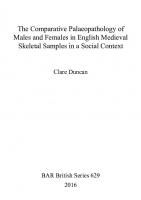
In order to identify the most appropriate assemblages on which to base the study, an inventory of excavated English medieval cemeteries was compiled. This comprised 67 sites, as summarised in appendix 1. The following sources were used to construct the list: the Ancient Monuments Laboratory ‘Human Bones Database’; the inventory of collections stored at, or reported to, the Anthropology Library at The Natural History Museum; newsletters produced by the Palaeopathology Association (British Section) and the Osteoarchaeological Research Group; published site reports and references cited within them; and enquiries made to various archaeology units. Unfortunately, few of the cemeteries listed in appendix 1 satisfied all the selection criteria. In particular, many of the samples were deemed too small for inclusion, and specialist examination of several large collections had not been completed at the time this project commenced. Furthermore, the sex ratios of the majority of samples were biased toward males. Nevertheless, seven assemblages were selected from the inventory: Abingdon Vineyard, the Fishergate church and priory assemblages, Ipswich Blackfriars, Jewbury, St Nicholas Shambles, and Wharram Percy; the locations of which are illustrated in figure 2.1. Each of the selected samples comprised more than 100 individuals, yielding a total of 2,625 articulated skeletons, of which 1,056 adults were classified as male, and 674 as female. Although males outnumbered females in all the collections, the proportion of females in each assemblage was considered to be sufficient for the purposes of analysis, at least by comparison to many of the other sites initially identified. Permission had kindly been given to use the original laboratory data, and access was granted to all the collections with the exception of Jewbury which had been reburied. The assemblages from Ipswich and Wharram Percy had been examined by the same investigator, using the same recording protocol, as had those from Fishergate. This was considered to be an advantage as it eased the number of recording protocols that had been applied across the cemeteries, and diminished the potential problem of inter-observer variation in their implementation. Excavation and examination of the selected cemeteries had taken place over a fairly contemporary period, and skeletal reports were available for most of them, if not site reports or other relevant sources of information. The chief characteristics of the assemblages are summarised in table 2.1. Further background notes on the respective cemeteries are provided below, including their historical context,
4
Material Figure 2.1 Location of the Cemeteries Selected for Study
Table 2.1 Chief Characteristics of the Cemeteries Selected for Study Cemetery
n Males
n Females
Dates of Use
Urban/Rural
Monastic/Lay
St Nicholas Shambles
91
74
11th and 12th centuries
Urban (London)
Lay
Fishergate Period 4
47
33
mid-11th to end of 12th century
Urban (York)
Lay
Fishergate Period 6
176
53
1195 - 1538
Urban (York)
Monastic
Jewbury
161
150
mid-12th century/c.1230 - 1290
Urban (York)
Lay (Jewish)
Abingdon Vineyard
223
171
c.1300 - 1540
Market town/rural
Lay
Wharram Percy
212
129
11th - 16th centuries
Rural
Lay
Ipswich Blackfriars
146
64
1263 - 1538
Urban
Monastic
5
The Comparative Paleopathology of Males and Females in English Medieval Skeletal Samples in a Social Context
circumstances of excavation, and evidence for burial practices with particular respect to gender.
‘suggestive of the burial of children with their parents’ (White 1988:48). The burials conformed to the Christian rite of east-west alignment, and all were extended inhumations, predominantly with the hands placed at the sides of the body.
2.2 The Cemetery of St Nicholas Shambles, City of London
Six burial types were identified (White 1985). The majority, 189 individuals, were simple interments, possibly placed within a coffin. Evidence for coffin use comprised the occasional find of nails or traces of wood in close proximity to some skeletons, although the cemetery report does qualify that the nails could have been intrusive deposits from underlying strata and the wood may have served some other purpose, such as grave markers. Some of the simple burials had floors of pebbles, and one of these, a female, had fragments of what was probably a linen shroud adhering to the skull. Schofield (1988:20) comments that ‘Linen shrouds were a common feature of female burial in the medieval period, whereas men were sometimes buried in hair shirts woven from coarse two-ply yarn’ (Crowfoot 1976). The five burial practices identified among the remaining 45 individuals included the use of stone pillows, presumably for inhumations without coffins; graves with chalk and mortar floors; stone and mortar cists; graves lined with dry-laid stone or tile, which may have been less prestigious imitations of stone sarcophagi; and one infant was encased in charcoal. Possible evidence for burial ritual included the placement of stones or pieces of Roman tile on three individuals, and a pebble had been placed in the mouth of four skeletons. All the burial customs identified within the cemetery of St Nicholas Shambles have parallels at other early medieval and late Saxon sites (Schofield 1988:19-26). There was no distinct age or sex distribution to the burial practices employed within the cemetery, but while acknowledging that the sample was small, Schofield (1988:26) does note ‘the apparent reverence with which old women were often treated’: three of the individuals furnished with a pebble in the mouth were elderly females, and fifteen of the twenty adults placed in pillow-graves were female. The excavated material was in a variable state of preservation, and the overall condition was described as ‘fair’. Only 36 skeletons were classed as complete, and half the individuals were described as being ‘deficient in the head region’ (White 1988:29). All the material was examined and recorded in the laboratory by a single investigator, W.J.White, on behalf of the Museum of London, and the findings from White’s analysis have been published (White 1988).
The foundations of the medieval parish church of St Nicholas Shambles and its associated cemetery were excavated by the Department of Urban Archaeology of the Museum of London between 1975 and 1979. The excavations followed the demolition of the GPO Headquarters Building at the site, located at 81 Newgate Street in the City of London, now occupied by the British Telecom Centre. A total of 234 articulated skeletons (inc. 91 males and 74 females) were retrieved from the cemetery to the north and east of the church, between 1975 and 1977. The area of excavation was governed by modern boundaries, but it is thought that almost the entire cemetery area on the north and east sides of the church was incorporated to the excavation (Schofield 1988). There is no evidence to suggest that interments were ever placed to the south or west of the church (Dyson 1988). The church of St Nicholas Shambles underwent several phases of development between its initial construction in the eleventh century to its final demolition in 1548-51. The burials are thought to derive from the early phase of the church’s history, and have been dated archaeologically to the eleventh and twelfth centuries. During this period London was flourishing. The city gained its official status as England’s capital in 1042, and continued to expand after the Norman Conquest as a thriving centre of administration, architectural advancement, religion, culture and commerce. Writing in 1173, William Fitz Stephen described London as ‘the most noble city’ of all the world (Clout 1991:39). The expansion was accompanied by massive population growth, largely fuelled by migration from the provinces and abroad. The number of inhabitants in 1086 has been estimated at around 10-15,000, which compares with a figure of 5-7,000 for York, the second city of England at that time (Butler 1982:4; Clout 1991:12,39; Unwin 1990). By the late twelfth century, London’s population exceeded 30,000 (Clout 1991:12,39). As the name implies, the parish of St Nicholas Shambles was a district for butchery, situated in the west of the walled ‘City’, which formed the mercantile and industrial centre of London (Clout 1991:39-53; White 1988:54). The cemetery of St Nicholas Shambles continued to be used until the church’s closure in 1548-51, but later cemetery strata were probably removed through development on the site after the demolition of the church. Grave-cuts were rarely discernible, so it was not possible to establish stratigraphic relationships between burials for more precise dating. The burials did not appear to be ordered in distinct rows, neither were they arranged according to sex or age. No separate area was identified for the interment of children, although two clusters were detected which comprised neonates and infants buried with adults, and these were interpreted as being
2.3 The Cemeteries of the Church and Priory of St Andrew, Fishergate, York In 1985-86, excavations were carried out by the York Archaeological Trust at the site of the former Redfearn National Glass Factory, 46-54 Fishergate, York, prior to redevelopment. The results of the investigation have been published as part of the Trust’s series entitled ‘The Archaeology of York’ (Stroud and Kemp 1993). A total of 402 articulated skeletons were reported to have been retrieved from the site and examined in the laboratory 6
Material (Stroud and Kemp 1993:130), although the data recording sheets for five individuals were absent from the archive and so omitted from the analyses conducted in this study. All the data used in this study were compiled following examination of the remains by one investigator, G.Stroud, on behalf of the York Archaeological Trust. The remains were considered to be well preserved. Approximately half the skeletons were more than 80% complete, and only 10% of the sample were represented by less than one quarter of the skeleton. Further burials are thought to exist to the south and west of the excavated area.
fragments in some graves provided evidence for the use of wooden coffins. In 1195, St Andrew’s church and the surrounding land was donated to the Gilbertine Order for the construction of a priory, also dedicated to St Andrew in 1202. The priory was continuously occupied, undergoing a series of structural modifications, until its suppression in 1538 on the orders of Henry VIII. Two hundred-and-sixty nine individuals (inc. 176 males and 53 females) were assigned to this phase, Period 6, recovered from the priory buildings and the churchyard to the east and south. All were aligned in an east-west direction. The burials are believed to include the monastic inhabitants as well as wealthy patrons from the secular community. The demographic distribution of the inhumations and variations in burial practice across the site indicated that a system of spatial zoning may have been in operation which related to social status (Knüsel et al. 1997; Stroud and Kemp 1993). Those buried in the eastern churchyard were almost exclusively male, the majority having been interred in a similar burial position with the arms placed upon the body. It is suggested that this may have been an area reserved for the resident canons. The burial ground to the south of the priory buildings contained a predominance of males, but also females and two-thirds of all subadults dated to Period 6. The strong male contingent implied the inclusion of individuals associated with the monastery in this part of the cemetery, possibly servants or lay brethren, and their families. One male had been buried with a lead alloy chalice and paten and another was buried with a paten; customs which indicate that these individuals were probably priests (Hall 1996:112-113; Rodwell 1981:155156). The inhumations within the priory buildings have been interpreted as those of lay benefactors and canons of comparatively high distinction. Adult males, females and children were retrieved, some of whom had been interred in stone or tile-lined coffins, possibly indicative of ‘high status’ burials. One male had been deposited in a shallow grave with a lime lining. Four double graves were identified, the remains from which displayed skeletal traits suggestive of familial groupings.
The excavated sample was considered to be representative of two separate burial populations associated with different archaeologically identified occupational phases, denoted as ‘Period 4’ and ‘Period 6’. The majority of burials could clearly be assigned to one of the two periods, and grave cuts enabled further stratigraphic sequencing within these phases, although Kemp (1993b:130) acknowledges that there was an element of uncertainty over the phase attributions in a small minority of cases. The skeletal data from the two periods were treated as separate assemblages for the purpose of the demographic and pathological analyses, and are often distinguished throughout the text by the abbreviations ‘Fishergate 4’ and ‘Fishergate 6’. One hundred-and-thirty individuals (inc. 47 males and 33 females) were attributed to Period 4, dating from the mideleventh to the end of the twelfth century. They were derived from a cemetery presumed to have been associated with the church of St Andrew, which is known to have existed on the site from historical evidence. Stroud (1993:251) suggests that ‘in the absence of any documentation to the contrary, it seems reasonable to expect that burials here would be of lay members of the population of York at that time’. York retained its status as England’s second city throughout these centuries, and like London, its population expanded, primarily through immigration. The city served as the ‘capital of the North’, acting as the chief administrative and judicial centre for the region. It was an important manufacturing centre and inland port. However, in the latter half of the Middle Ages, York’s wealth and prosperity was surpassed by other cities, notably Bristol and Norwich (Butler 1982:5; Hall 1996:45).
2.4 The Jewish Burial Ground at Jewbury, York The Jewbury burial ground was also located in York, situated just outside the medieval city walls in the northeastern suburbs, on the bank of the River Foss. The cemetery served the Jewish community of medieval York, although it may have contained individuals brought from Lincoln as documentary evidence suggests that the burial ground was, at least initially, shared by the Lincoln Jewry prior to the purchase of their own site at an unknown date. The York Jewry was established in the 1170’s, probably as an offshoot of that at Lincoln. The community was small, estimated to comprise 20-40 households, concentrated in the city centre around Jubbergate and Coney Street, where the synagogue was (Butler 1982:4,6; Hall 1996:59). In 1190 the community was almost wiped out by a horrific massacre and mass suicide at York castle, which was incited by a Christian mob with religious and financial
The graves from Period 4 were aligned east-west. Males, females and subadults tended to be distributed randomly across the excavated area, though there was some clustering of subadults in the western part of the site. Eighteen young males displayed unhealed blade injuries, and some had been placed in double graves, indicating they were interred concurrently. Kemp (1993a:127) speculates that these individuals may have died as a result of a single violent event, possibly representing casualties of a battle. All the inhumations were extended, although it is possible that there were preferences in arm position pertaining to sex as the majority of females had been placed with the arms upon the body whereas a more diverse range of arm positions were observed among males. The presence of clench bolts, iron nails and wood 7
The Comparative Paleopathology of Males and Females in English Medieval Skeletal Samples in a Social Context
motives (Butler 1982:4,6; Dobson 1974; Hall 1996:59; Mitchell and Leys 1967:158-162). However, the community revived to become the most prosperous Jewish community in England during the mid-thirteenth century. The date when the Jewbury cemetery was established has not been determined precisely, though it is estimated to have been founded some time between the mid twelfth century and c.1230 when it is mentioned in a land sale agreement (Dobson 1974). It remained in use until its abandonment in 1290, when Edward I expelled all Jews from England.
the site, suggesting that the Jewish tradition for equality and simplicity in burial was upheld. Most of the inhumations were extended with the arms placed at the sides of the body and there was an almost total absence of grave goods. The majority had clearly been buried in wooden coffins as some graves contained wood fragments or iron coffin fittings, and most contained iron nails. This contrasts with the modern Jewish practice of avoiding metal nails in preference for wooden pegs. Shrouds may have been used, as seven individuals were in constricted positions which suggested that they were bound prior to burial, and antler toggles, possibly shroud fasteners, were found above the skulls of two skeletons.
Approximately half the cemetery was excavated during 1982-83 by the York Archaeological Trust in advance of redevelopment, the findings from which have been published as part of the ‘The Archaeology of York’ series (Lilley et al. 1994). It represents the only large scale investigation of a medieval Jewish cemetery in England, out of the ten which are known to have existed. A total of 476 articulated skeletons (inc. 161 males and 150 females) were reported to have been recovered and examined in the laboratory, although the skeletal recording sheets for seven individuals were missing from the archive and so excluded from the analyses performed in this study. The material was described as being in a variable state of preservation, from ‘very good to poor’ (Lilley et al.1994:353). Half the sample were represented by more than 80% of the skeleton and more than three quarters were represented by at least half the skeleton. However, the anthropological examination of the remains was curtailed because when the site was confirmed as being that of the medieval Jewish cemetery, the Chief Rabbi requested the immediate reburial of the remains. Jewish traditions dictate that the dead should be treated with the utmost reverence and have the right to rest undisturbed. A team of researchers were employed under the direction of D.R. Brothwell and M.H.Williamson to record as much detail as possible under the time restrictions imposed, although the premature reburial limited the amount of information which could be recorded, as detailed in the following chapters.
2.5 Abingdon Vineyard Lay Cemetery In 1988-89 excavations were carried out in the Oxfordshire town of Abingdon, in an area known as The Vineyard, adjacent to the Municipal Park. The excavations were conducted by the Oxford Archaeological Unit prior to the construction of offices for the Vale of White Horse District Council (Allen 1990). The site encompassed part of an extensive medieval lay cemetery. Its limits could not be determined, but it is estimated that about one third of the total cemetery area was excavated (Wakely pers. comm.), from which 590 articulated skeletons (inc. 223 males and 171 females) were recovered and examined in the laboratory. The majority of the assemblage was examined and recorded at the University of Leicester under the direction of J.Wakely, who was assisted by several postgraduate students. J.W.P. Hacking co-directed the research on behalf of the Oxford Archaeological Unit, and also examined a substantial component of the sample. The completed skeletal report is currently awaiting publication (Wakely and Hacking forthcoming). During its use the cemetery was situated within the grounds of Abingdon Abbey. The Abbey operated the cemetery (Townsend 1910:40), but the possibility that it contained monastic burials has been excluded (Wakely pers. comm.). The cemetery served the secular community of Abingdon and its surrounding rural manors, and was the only site in the vicinity to hold the legal right to burial (Townsend 1910:66-67). Medieval Abingdon was a market town which developed around the Abbey, one of the richest in England until its destruction in 1538. The skeletons excavated from the cemetery have been dated archaeologically to a period spanning from c.1300 to 1540, when the land was sold following the Dissolution. There was much intercutting of burials and their stratigraphic sequence is currently being evaluated, so the skeletons can not yet be ascribed to any particular phase of cemetery use. The graves were aligned east-west and the majority were single inhumations, although group burials of up to six skeletons were also recovered. Most individuals had been placed in simple, unfurnished graves but a minority, thought to date among the earliest burials, had been placed within stone cists, or had stones placed on either side of the head, resembling ‘earmuffs’ (Allen 1990).
On site, the layout of the graves indicated that the cemetery was highly organised. The graves were arranged in distinct but irregular rows, aligned in a northeast/south west direction. Contrary to many Christian cemeteries, the graves rarely intercut, which indicates that the Jewish belief that the body should not be disturbed after burial was observed. It would also indicate that grave markers were used, perhaps earth mounds or wooden posts. A few graves contained stones which may have served to denote the grave or act as packing material to secure wooden posts. The lack of intercutting prevented any chronological differentiation between most of the burials. The distribution of males and females was largely random, although there was a cluster of twenty males in the western region of the site which was interpreted as being a possible area reserved for the burial of rabbis or other prominent people. The majority of children were focused in the north-eastern area, indicating that the segregation of child burials was practised. Burial method was extremely uniform across 8
Material 2.6 The Cemetery at the Deserted Medieval Village of Wharram Percy, North Yorkshire
shroud fragments in three graves in the churchyard (Bell and Beresford 1987).
This assemblage comprised 685 individuals (inc. 212 males and 172 females) recovered from the church and two sample areas of the churchyard at the deserted medieval village of Wharram Percy, situated on the Yorkshire Wolds, about 18 miles north-east of York. The chalk geology of the site led to exceptionally good skeletal preservation of what is one of the few large collections derived from a rural medieval community in England. Excavations of the church and churchyard were carried out between 1963 and 1978 (Mays et al. 1996) and formed part of an extensive, pioneering research programme at the village site which began in 1950 and continued for more than 40 years (Beresford and Hurst 1990). All the anthropological data used in this study was compiled following examination of the material by a single investigator, S. Mays, at the Ancient Monuments Laboratory, English Heritage. The findings from Mays analysis are to be published in a forthcoming report (Mays pers. comm.).
2.7 The Medieval Burials from the Blackfriars Friary, School Street, Ipswich Excavations conducted by Suffolk Archaeological Unit between 1983 and 1985 produced 250 skeletons (inc. 146 males and 64 females) from the site of the Blackfriars Friary, School Street, Ipswich. Like Wharram Percy, the laboratory examination of the entire skeletal collection was undertaken by S. Mays. Mays findings have been presented as part of a series of interim reports (16/91) produced by the Ancient Monuments Laboratory in advance of full publication. The burials were all retrieved from within the friary complex, principally the church nave, and it is thought that they represent both friars and wealthy lay benefactors. Fifteen named individuals are known to have been buried in the friary but these could not be associated with specific skeletons. The interments took place over a 275 year period, from when the friary was founded in 1263 to its suppression in 1538. The town of Ipswich became increasingly prosperous during this period, and by the early sixteenth century it had become the sixth richest in England (Bridgwater 1996:19,191-192; Sager 1990:14). Its wealth was largely founded on its role as an inland port and the region’s highly successful wool industry, which spawned a large, affluent middle class of clothiers and merchants.
Although the date of the village origins has not been determined, the settlement probably evolved from a shifting group of scattered farms in the Saxon period. The construction of the parish church of St Martin (Bell and Beresford 1987) in the mid-twelfth century may signify the foundation of the parish of Wharram Percy. By the late fourteenth century the village had become established as a compact community of about 30 houses which had been deliberately planned along three frontages around a central wedge-shaped green, surrounded by a methodical distribution of crofts and open fields. The parish economy centred upon both arable and livestock farming, with access to markets at nearby Driffield and Malton, as well as the large urban centres of York, Beverley and Hull (Scarre 1988:246-247). The decline of the village took place during the fifteenth century. Sixteen inhabited dwellings were documented in 1458 but by the early sixteenth century, the village was totally depopulated.
Despite much inter-cutting of graves within the cemetery, it was not possible to date the burials with any further precision. Females were located in all areas of the friary complex apart from the south range of the cloister and the chapter house, suggesting that friars and lay benefactors were not segregated except perhaps in these two areas. A statistically significant association was identified between arm position in burial and skeletal sex. A substantial proportion of males and females were buried with the arms placed at the sides of the body, whereas virtually all individuals buried with the arms placed across the chest were male. Fragments of wood and iron nails provided evidence for the use of coffins in 89 cases, although there was no association between sex or age and coffin use. Skeletal preservation at the site was highly variable, ranging from complete skeletons to soil silhouettes. Forty per cent of the skeletons were more than 80% complete, and almost 70% of the sample were represented by more than 60% of the skeleton. It is likely that the friary site was not excavated in its entirety and there may be further burials in areas which were not investigated.
The burials spanned the period from the eleventh to the sixteenth centuries. Those in the graveyard were laid out in regular rows, although there was a great deal of disturbance between earlier and later graves. The rise in ground level, particularly on the north side of the churchyard, indicated that the graveyard was used ‘for at least four cycles of burial’ (Beresford and Hurst 1990:65), although further details on burial phasing have not been located. The vast majority of the inhumations are believed to be those of ordinary peasants who resided in the parish (Mays 1996). However, chalices had been deposited with two skeletons, one of which was also buried with a paten, indicating the burial of priests; and there was evidence for the burial of men at arms, possibly relations to the lords of the manor, from grave slabs built into the walls of the church that were engraved with swords and foliated crosses (Beresford and Hurst 1990:64). A concentration of infants were noted in the north churchyard. The practice of burial in shrouds was indicated by the identification of
2.8 Discussion As the above accounts indicate, the composition of the selected cemeteries differed in a number of important respects, such as whether they primarily served an urban or rural settlement, a monastic or lay community, or differed in time span or geographical region. The majority of studies which have compared pathology between the 9
The Comparative Paleopathology of Males and Females in English Medieval Skeletal Samples in a Social Context
sexes have so far focused on single cemetery collections, but it was anticipated that the variety of cemeteries included in this project would not only enable the direct comparison of males and females within the respective assemblages, but also the comparison of males and females from a range of cemeteries that were presumed to have different compositional characteristics. It was envisaged that this might help to elucidate whether potential sex differences in pathology were generally applicable to all the samples, perhaps reflecting universal sex differences in health status, or whether the results might have been influenced by other parameters. However, it should also be emphasised that a multitude of what are largely indeterminable factors influence the demographic and cultural composition of a cemetery; and similarly, any assemblage retrieved from it (Larsen 1997:334-335). The identity of the burials are normally unknown, and an excavated sample may not be representative of that which was interred. Waldron (1997:10-27) has divided the chief sources of bias which may be introduced to a skeletal assemblage into ‘extrinsic’ and ‘intrinsic’ factors. Extrinsic factors act to diminish sample size, and include culturally determined biases in the proportion of the total dead population which are buried in a particular cemetery; the proportion lost as a result of disturbance or decomposition; the proportion of the buried population which is discovered during excavation; and the proportion of those which are actually recovered. Intrinsic factors revolve around the fact that a cemetery assemblage represents a static, dead population sample, and does not reflect the dynamic characteristics of a living population. This is particularly evident in the age profile of the two population types, as cemetery samples reflect age at death rather than the age distribution of those living in a population. Most cemeteries also develop over a protracted time span, and so contain individuals that existed many years apart. This can be particularly detrimental to the analysis of pathology prevalence because it has the effect of smoothing out potential peaks in prevalence occurring over time, and the impact of such peaks on the mean prevalence cannot be determined. Therefore, although the series of cemeteries included in the study represented an opportunity to compare data from assemblages that were presumed to display distinct compositional characteristics, there are inherent limitations surrounding such comparisons, and it should be emphasised that the inferred compositional characteristics can only be applied in loose, general terms.
10
3. Subadult Sex Determination Using Tooth Measurements 3.1 Introduction
combination of measurements which might effectively distinguish the sexes and facilitate their classification. 4. Application of the discriminant functions to predict the sex of subadults from the respective cemeteries which had formerly been classified as indeterminate.
At present, there is no consensus on a reliable method for identifying the sex of subadult human remains (Mays 1998:38-42; Mittler and Sheridan 1992; Molleson et al. 1998; Saunders 1992; Schutkowski 1993; Stone et al. 1996). Adults are primarily diagnosed from secondary characters of the pelvis and skull or from the size of the limb bones (Bass 1987), but as these traits are not fully developed until late adolescence (c.18 years), they cannot be used to sex children. This inability to sex subadults imposes a limitation on any general study of sex related pathology because it necessitates the exclusion of a significant component of the population under study.
3.2 Method The adult reference samples consisted of 43 males and 34 females from Abingdon, and 88 males and 36 females from Fishergate. The material from Fishergate was derived from both phases of cemetery use. These individuals were selected from the assemblages on the fulfilment of two criteria. Firstly, each had been reliably sexed using established methods, the efficacy of which are described in a publication by the Workshop of European Anthropologists (1980, and see chapter 5). This was imperative because the discriminant technique depends on accurate sexing of the base population sample. Ideally, the second criterion would have been that each skeleton also displayed a complete, unworn dentition because measurements missing from the data set may impede statistical analysis. However, owing to the typically fragmentary nature of the archaeological material, this criterion was modified to include any individual with a minimum of one unworn permanent tooth with the exception of second and third molars. Third molars were omitted from the investigation due to their high frequency of congenital absence (Brothwell et al. 1963; Pindborg 1970) and late age of eruption (c.21 years) which would provide little advantage over conventional methods of sex determination. Similarly, second molars were excluded due to their comparatively late age of eruption (c.12 years) coupled with a high susceptibility to attrition. This led to the selection of mostly younger individuals, under the age of c.30 years. Possession of at least one unworn permanent tooth was also the single criterion observed when selecting the skeletons of indeterminate sex.
The permanent dentition is known to form at adult size within the jaws of the immature skeleton and erupt in a predictable sequence between the ages of c.5-15 years (with the exception of the third molars), thus representing an adult trait in a child (Ubelaker 1978). Since previous studies have consistently documented sex differences in permanent tooth size (Bermúdez de Castro et al. 1993; Brace and Ryan 1980; Ditch and Rose 1972; El-Nofely and Tawfik 1995; Frayer and Wolpoff 1985; Garn, Cole and Van Alstine 1979; Garn, Cole, Wainwright and Guire 1977; Garn, Lewis and Kerewsky 1964; 1966; 1967; Garn, Lewis, Swindler and Kerewsky 1967; Goose 1967; Hanihara 1981; Hillson 1986:240-242; 1996:81-82; Langenscheidt 1983; Lunt 1969; Molleson et al. 1985; Moss 1978; Potter 1972; Rösing 1983; Sciulli et al. 1977; Scott and Parham 1979), the dentition may hold the potential to reveal the sex of children. However, the practical application of this theory has rarely been attempted (Beyer-Olsen and Alexanderson 1995; Duncan 1998; Molleson 1989; 1993). The purpose of this chapter was to investigate the validity of this hypothesis in relation to material excavated from the cemeteries of Abingdon Vineyard and St Andrew, Fishergate (see chapter 2). The hypothesis was tested on these assemblages because the remains were accessible for examination. If sex assignments could be attributed to a sample of subadults, it would enable their inclusion into the wider project, extending the comparative analysis of male and female pathology to individuals under the age of c.18 years.
The dentition of each individual was recorded using the FDI (Fédération Dentaire Internationale) tooth numbering system and occlusion classification (Baume et al. 1973). Four measurements were taken on every permanent tooth present in each dental arcade, from first molar to first molar, using 0.1mm vernier callipers: 1. Maximum mesiodistal crown diameter - distance between the points of contact with adjacent teeth, parallel to the occlusal plane. Malpositioned teeth were measured from the points where the crown would have been in a ‘normal’ occlusal relationship (Goose 1963). 2. Maximum buccolingual crown diameter - taken perpendicular to the mesiodistal diameter. 3. Crown Height - distance between the highest point of the crown and the deepest point of the cemento-enamel junction, taken obliquely on the buccal side.
The research design encompassed: 1. A metrical examination of permanent teeth in a reference sample of adult skeletons from each cemetery, whose sex had previously been determined using conventional methods. 2. Univariate analysis of the data to ascertain whether any statistically significant differences existed between the dimensions of male and female permanent teeth, from either cemetery. 3. The application of discriminant function analysis to each reference sample, with the aim of identifying a
11
The Comparative Paleopathology of Males and Females in English Medieval Skeletal Samples in a Social Context
4. Root Length - distance between the deepest point of the cemento-enamel junction and the apex, taken obliquely on the buccal side.
dentition in three individuals were measured five times. For each of the six sets of measurements, a test of the between repeats mean square against the residual mean square was carried out (Cohen and Holliday 1982:206212). The quotient of these two mean squares was distributed approximately as an F distribution with four and eight degrees of freedom. No significant between repeat effects were found at the 0.05 probability level, suggesting that the implementation of the odontometric method was unlikely to be influenced by intra-observer variation in recording technique.
These measurements are depicted in figure 3.1. This amounted to a maximum of 96 measurements per individual, although dimensions modified by attrition, calculus or pathology were excluded from measurement. In order to record the data systematically, a recording form was devised, presented in appendix 2. All measurements were taken by the author. As a test of intra-observer error in measurement, the dimensions of the right half of the
Figure 3.1 Tooth Measurements
12
Subadult Sex Determination Using Tooth Measurements 3.3 Data Analysis and Results
dimorphism which are more likely to yield better results in the ensuing discriminant analysis.
It was apparent that a substantial proportion of the measurements were missing. Tooth loss and attrition were the primary reasons for measurements being omitted. Other factors included the presence of tooth decay, postmortem tooth damage, calculus, tooth anomalies and teeth being firmly held in the jaw thus preventing root measurement. ‘Sparse data’, inherent to archaeology, is particularly problematic when conducting multivariate analyses. In contrast to univariate methods which are employed to examine single variables, multivariate techniques test relationships between several variables at once. However, it is usually necessary for each case included in the analysis to hold a complete set of values for the variables being examined, otherwise it will be ‘thrown out’. Since virtually every individual had a proportion of missing measurements, this criterion would have precluded any multivariate analysis of the data.
Two-tailed t-tests were applied to test the difference between male and female means for each of the measurements where the data appeared to be normally distributed; and the equivalent non-parametric test, the Mann-Whitney U test, was used to compare medians where the normal distribution was in doubt. In the event, both tests were applied to all the data, the U test tending to confirm the t-test result, or alternatively indicating that it should be treated with caution (tables 3.2a/b). Per cent sexual dimorphism, 100(male mean/female mean - 1.00) (Garn, Lewis, Swindler and Kerewsky 1967) was also calculated for each of the variables (tables 3.3a/b). The results of the t and U tests were generally in agreement, as demonstrated in tables 3.2a/b. Within the Abingdon sample, the male mean exceeded the female mean for all 48 measurements. The t-tests indicated that the sex difference between the Abingdon means was statistically significant (p25kg) are at increased risk of knee osteoarthritis, as are those in occupations that involve prolonged kneeling and squatting. Their study showed risk increased three-fold in those whose work entailed heavy lifting and kneeling or squatting, and rose multiplicatively in those who were also obese. An elevated risk was also found in those whose work involved walking more than two miles per day, especially females, and for males whose work involved repeatedly climbing stairs or ladders. The findings are consistent with previous reports of an association between knee osteoarthritis and heavy work, kneeling and squatting, as noted in section 8.3.1 of this study. Accordingly, Coggon and colleagues recommend that measures are taken to minimise risks in the modern workplace. In appropriate circumstances, they suggest consideration is given to classifying knee osteoarthritis as an occupational disease, with payment of compensation. No such recompense was afforded in the Middle Ages, but it is likely that many performed tasks which put them at risk of knee osteoarthritis in the course of their daily lives. As suggested in section 8.3.1, perhaps females performed tasks which placed them at greater risk.
Srikanth and colleagues (2005) have performed a metaanalysis of sex differences in osteoarthritis in different joints in modern population samples. After screening 178 studies, 34 were deemed appropriate for inclusion in the analysis. A prevailing result was that females are more likely to develop osteoarthritis in the knee, and the severity of knee osteoarthritis is greater in females, especially those aged over 55 years. A higher female prevalence of knee osteoarthritis was identified in this study (section 8.3.1), suggesting a parallel between the medieval and modern occurrence. Why females should experience a greater burden of knee osteoarthritis is not fully understood but it is a focus of current research (Boyan et al. 2013; O’Connor 2007a; 2007b; O’Connor and Hooten 2011). As a major cause of disability today, O’Connor (2007a:71) urges, ‘improving our knowledge of sex and gender differences in osteoarthritis is critical to improving treatments’. Several aetiological possibilities are being explored, some of which were considered in this project (section 8.3.1).
Prior knee injury is an established risk factor in the development of knee osteoarthritis. Lohmander and colleagues (2007) estimate that up to 50% of individuals with anterior cruciate ligament injury or meniscal tear will go on to develop knee osteoarthritis within 10 to 20 years, and similar figures have been reported in other studies (Boyan 2013; O’Connor 2007a; Øiestad et al. 2009; von Porat et al. 2004). Clinical literature in sports medicine has consistently reported that females sustain a greater incidence of anterior cruciate ligament injury than males. The female to male ratio has been estimated to be as high as 9:1, although a meta-analysis of 33 studies by Prodromos and co-workers (2007) places it at nearer 3:1 for the high-risk sports of soccer and basketball, with a tear-rate of approximately 5% for year-round female athletes. Many theories have been put forward to explain this sex difference, including a hormonal influence on ligament strength, anatomical differences and type of footwear, but the current thinking focuses on sex differences in biomechanics. Motion studies have shown that when jumping and running, the knees of females are more inwardly-turned and less bent on landing, putting greater stress on the joint and predisposing to ligament tear. Medieval females may not have participated in modern sports but their active lifestyles perhaps put them at an increased risk of knee injury and consequent osteoarthritis.
Given the increased severity of knee osteoarthritis in postmenopausal females, a hormonal component has been implicated. Oestrogen receptors have been identified in human cartilage suggesting that cartilage can respond to sex hormones, but the cellular mechanism is largely unknown (Boyan et al. 2013; O’Connor 2007a). Observational studies on the effect of hormone replacement therapy on osteoarthritis have produced conflicting results (Wei et al. 2012; Zhang and Jordan 2010). Females do appear to experience greater knee cartilage loss in later years, according to a study of 271 asymptomatic participants aged between 50 and 79 whose cartilage volume was evaluated using magnetic resonance imaging (Hanna et al. 2009). The average annual percentage loss of tibial cartilage volume was found to be 1.6% in females compared to 0.4% in males, and the average annual loss at the patella was 2.3% in females compared to 0.8% in males, differences that were statistically significant. O’Connor and Hooten (2011) relate these figures in alternative terms, equivalent to females losing cartilage at three to four times the annual rate in males. Obesity is a known risk factor for the development of knee osteoarthritis (O’Connor 2007a; Waldron 2009; Zhang and Jordan 2010) and the association is stronger for females than males (Nicolella et al. 2012). It is estimated that
Limb alignment can affect load distribution on the knee. Studies have shown that deviation from a neutral 188
Postscript: Update 2016 alignment can increase the progression of knee osteoarthritis four to five-fold (Zhang and Jordan 2010). Valgus (knock-kneed) limb alignment shifts the centre of pressure laterally increasing the load borne on the lateral compartment of the knee, and varus (bow-legged) alignment shifts the centre of pressure medially increasing load on the medial compartment, both of which are associated with accelerated structural deterioration in the compartment under greatest compressive stress. However, a literature review by Nicolella and colleagues (2012) found the effect of sex differences in lower limb alignment on knee osteoarthritis to be equivocal, although they highlight several gaps in knowledge that are still to be addressed.
especially in those with more than four children. Relative risk of knee osteoarthritis was 2.6 times as high among women with five to 12 children compared to those who had undergone one birth. The association between osteoarthritis later in life and pregnancy earlier in life led the authors to suggest there may be ‘biological events during pregnancy or childrearing which do not manifest as incident disease until many decades after the pregnancies. These may include damage to cartilage due to extra weight carried or increased clumsiness during pregnancy due to the temporary alteration of weight distribution, which may predispose to later disease without overtly causing it at the time of pregnancy. Alternatively, rather than direct damage to joints that occurs during pregnancy, it is possible that permanent changes occur in the body during pregnancy which predispose to adverse joint health in the decades following reproductive life . . . It is also possible that caring for children causes ongoing physical insults to the joints during childrearing years, which again may only manifest years later. In this sense, pregnancies might be considered to be a period of injury (due to transient increased weight, hormonal changes, etc.) similar, for example, to ligamental or meniscal injuries which predispose to the disease of osteoarthritis later in life’ (ibid.:1852). In summary, the findings suggest ‘the effect of pregnancy or children continues to express itself decades after the pregnancy and may reflect a model of joint injury engendering osteoarthritis in later years’ (ibid.:1853).
Among other anatomical factors that have been considered to explain the higher prevalence of knee osteoarthritis in females is a weaker quadriceps strength which may lessen knee stabilisation (Hunter et al. 2005; Weiss and Jurmain 2007). Nicolella and colleagues (2012) have reviewed published data and construe that quadriceps strength can predict the odds of developing symptomatic signs of knee osteoarthritis in males and females but it is not known whether sex differences in muscle function precede the development of knee osteoarthritis or contribute to its progression. Again, they identify gaps in knowledge. It was speculated in section 8.3.1 of this study that perhaps repeated pregnancies in the Middle Ages had a bearing on the higher female prevalence of knee osteoarthritis. New research suggests there may be some foundation to this suggestion. A significant association between parity and knee cartilage defects at the patella has been identified by Wei and co-workers (2012) in a study of 144 women in Australia with an average age of 36 years, 58% of whom were parous with parity ranging from one to five births. Risk of cartilage defects increased with number of births and was most evident among those who had three or more births, leading to the conclusion that higher parity may have an adverse effect on knee cartilage. The association remained significant after adjusting for confounding factors such as BMI, smoking, knee injury, physical activity and socio-economic status. Suggested explanations included weight gain during pregnancy, changes in lifestyle such as increased household chores, and hormone changes such as the release of relaxin which increases ligament laxity in advance of birth.
In their review of anthropological perspectives on osteoarthritis, Weiss and Jurmain (2007) recommend that future research focuses on epidemiological analyses of large samples, stratified by age and sex, an approach that was taken in this study. Several techniques have recently been proposed to explore osteoarthritis data. Law (2005) presents a method of calculating prevalence using an appropriate denominator for skeletal remains. It incorporates a helpful system of tabulating osteoarthritis in extant joints while accommodating missing skeletal elements, so that prevalence may be determined for individual joints and the overall sample despite the inherent problem of incomplete material. The method is illustrated using osteoarthritis but may be applied to other conditions which affect more than one site in the skeleton. Glencross and Sawchuck (2003) present a method to control for the confounding effect of age when comparing samples, termed the ‘person-years construct’. Personyears is calculated using the mid-point of each age cohort and multiplying by the number of individuals in the cohort, then adding together the person-years from all cohorts that are represented. The authors recommend its use when exploring osteoarthritis or other pathological indicators that tend to accumulate with age such as caries or fractures. Baker and Pearson (2006) propose two methods for controlling for age-structure differences between samples: age-adjustment and logistic regression. After testing several statistical techniques, Nikita and colleagues (2013) advocate the application of generalized linear models (GLM) to allow the simultaneous impact of multiple variables to be explored.
Risk of knee joint replacement due to osteoarthritis is also associated with reproductive history, according to an analysis of data collected for an ongoing study of women’s health in the United Kingdom known as the ‘Million Women Study’ (Liu et al. 2009). A cohort of 1.3 million women are participating through the National Health Service, with an average age of 56 at recruitment. Risk of knee joint replacement was found to rise with increasing parity, each additional birth increasing the relative risk by 8%. Wise and colleagues (2013) found parity to be associated with both knee osteoarthritis and knee replacement, in a study of 1,618 women in America with a mean age of 62.8, 189
The Comparative Palaeopathology of Males and Females in English Medieval Skeletal Samples in a Social Context
Weiss and Jurmain (2007) suggest an area for future osteoarthritis research as an indicator of activity may be asymmetry in distribution, advocating ‘within-body comparisons are probably the best measures for reconstructing activity patterns. That is, asymmetry evaluations of several variables (including osteoarthritis, musculoskeletal markers, cross-sectional geometry, and body size)’ (ibid.:445-446). The benefit is that ‘Asymmetry measures do not need to be standardized for body size, the whole body is experiencing the same nutrition and age, and so any differences between limbs are more likely to reflect activity patterns’ (ibid.:446). Asymmetry in osteoarthritis was identified in this study, with a tendency for joints on the right side to display a higher prevalence in the upper and lower limbs of males and females, as discussed in section 8.3.1.
the theory that Schmorl’s node formation may be associated with the process of vertebral development early in life combined with strains exerted on the vertebrae during spinal movements, particularly torsion. Plomp and colleagues (2012) undertook two-dimensional statistical shape analysis on the thoracic spines of 132 skeletons dating to the medieval and post-medieval periods. Morphological differences were identified between healthy vertebrae and those with Schmorl’s nodes. Shape of the pedicles, posterior margin of the vertebral body and larger vertebral body size in males were associated with the presence and severity of Schmorl’s nodes, leading to the suggestion that vertebral morphology may predispose to disc herniation. Williams and colleagues (2007) found that Schmorl’s nodes have a high heritability (>70%) in a study of 516 twins, indicating there may be a genetic component. Kyere and collaborators (2012) undertook a literature review of 23 articles which proposed a range of causes including disc degeneration, spinal pathologies and autoimmune involvement. Having assessed the evidence, they considered axial load or axial trauma to be the most likely independent factor contributing to the development of Schmorl’s nodes. They also concluded that while Schmorl’s nodes are often asymptomatic, in some cases they may be a source of back pain. Faccia and Williams (2008) identified an increased reporting of pain associated with Schmorl’s nodes located centrally on the vertebral body which was exacerbated when combined with osteophytes. They suggest future bioarchaeological studies consider the significance of chronic back pain and its impairment on quality of life when interpreting the implications of Schmorl’s nodes in past populations.
Referring to spinal degenerative disease, Weiss and Jurmain (2007:440) comment that it is ‘probably not a good indicator of activity’ and Jurmain and co-workers (2012:539) remark, ‘expression of osteophytes in vertebral bodies (vertebral osteophytosis, VOP) is an especially poor marker of activity, since it develops typically from universal biomechanical forces associated with bipedal locomotion rather than reflecting any influence falling under the rubric of “habitual activity”. The questionable utility of VOP in activity reconstruction is further reinforced by its extremely high heritability approaching .75 (Spector and MacGregor 2004). For all these reasons, most (but not all) osteologists have abandoned VOP in activity reconstruction of skeletal samples’. The lack of statistically meaningful results from the analysis of vertebral osteoarthritis and osteophytosis in this study (section 8.3.2) may be consistent with this viewpoint.
Chapter 9. Dental Pathology
One aspect of vertebral pathology that did produce positive results in this study was the analysis of Schmorl’s nodes, which were more prevalent in males (section 8.3.2). Other studies of archaeological and modern samples also report a higher prevalence of Schmorl’s nodes in males (Faccia and Williams 2008; Jiménez-Brobeil 2012; Kyere et al. 2012; Novak and Šlaus 2011; Üstündağ 2009, Weiss 2015:75-77). A literature review by Mattei and Rehman (2012) places the male predominance at 76% of cases. In archaeological studies, this disparity is mainly attributed to the sexual division of labour with males undertaking more strenuous physical activities. However, the aetiology of Schmorl’s nodes is still being debated (Dar et al. 2010; Kyere et al. 2012) and Weiss (2015:77) offers a different perspective, asserting ‘In order to conclude that the Schmorl’s nodes are caused by activity, a reverse sex difference needs to be discovered. If regardless of the population males have more Schmorl’s nodes than females, then the sex difference is likely to be biological’.
The analysis of caries, abscesses and antemortem tooth loss in this study identified a consistent pattern whereby females were more frequently affected than males. A similar bias was identified in other archaeological studies, as cited in section 9.3. Lukacs (Lukacs 2008; 2011; 2012; Lukacs and Thompson 2008) has now led several investigations that compare dental pathology between the sexes including an extensive meta-analysis of caries prevalence in more than 50 archaeological and 50 epidemiological and clinical studies from around the world (Lukacs and Largaespada 2006). These demonstrate that females typically exhibit a higher caries prevalence than males. Lukacs and Largaespada (2006) comment ‘This finding is generally true for diverse cultures with different subsistence systems and for a wide range of chronological periods. Exceptions exist, but are not common’. Lukacs (2011) adds, ‘The ubiquitous gender difference in caries . . . is paralleled by patterns of tooth loss in which women typically exceed age matched males’.
Research has continued into the potential aetiological factors of Schmorl’s nodes. Dar and co-workers (2010) examined the distribution of Schmorl’s nodes along the spines of 240 skeletons randomly selected from the Hamann-Todd Osteological Collection of identified individuals. No association was found between anatomical position and age, sex or ethnicity, leading to
Lukacs observes a tendency for anthropologists to interpret the sex difference in behavioural terms, particularly the sexual division of labour which may facilitate different eating habits, such as tasting during 190
Postscript: Update 2016 food preparation. However, the aetiology of caries is multifactorial, as discussed in chapter 9, and Lukacs and Largaespada (2006:544) are concerned that ‘anthropologists have not given adequate consideration to the crucial etiological influence that saliva, hormones and pregnancy have on cariogenesis’.
burgeoning field of research centres on the genetic component of caries susceptibility. Several mechanisms are being explored including genetic control of enamel formation, saliva composition and flow, taste preferences and the genetic diversity of oral bacteria (Ferraro and Vieira 2010; Lukacs 2011; 2012). Ferraro and Vieira (2010) conclude that a multifactorial approach to this multifactorial disease should be considered in future explorations of sex differences in dental pathology.
Clinical studies have revealed sex differences in the quality and quantity of saliva. Saliva performs several functions in maintaining oral health which include washing the oral cavity to flush away debris and sugars, neutralising acids, plus it contains proteins with antimicrobial properties. Oestrogen appears to alter the biochemical composition of saliva and reduce the flow rate, diminishing its protective effect. Increased oestrogen levels correspond with increased caries, whereas androgen levels have no effect on caries frequency according to animal research cited by Lukacs and Largaespada (2006). Oestrogen levels rise in puberty, fluctuate cyclically and peak during pregnancy. Therefore, Lukacs and Largaespada (2006) suggest these singular and recurring events call for a life-span perspective in understanding the inferior dental health status of females. The deleterious effects of pregnancy on oral health were discussed in section 9.3. ‘Pregnancy gingivitis’ and exacerbation of periodontal disease is now a wellrecognised condition (Figuero et al. 2013; Silk et al. 2008). Morning sickness is considered a risk factor for acid erosion of enamel. An old wives' tale purports that a mother will ‘lose a tooth for every child’, which according to folk wisdom is due to the unborn baby leaching calcium from the mother’s teeth. This cause is now known to be mistaken, but a recent study of 2,635 American women has found an association between increased parity and increased tooth loss (Russell et al. 2008). Similar results were obtained in a Japanese study of 1,211 participants, which found higher parity women were more likely to lose teeth than lower parity women, those with four or more children losing on average nearly three more teeth than those without children or one child (Ueno et al. 2013). Both studies found the bias held regardless of socioeconomic status and other confounding factors such as age and health behaviours. Poor maternal oral health also has a detrimental impact on birth outcomes and infant oral health. Periodontitis is associated with preeclampsia, premature labour and low birth weight. Maternal cariogenic flora may be transmitted to newborns through saliva, predisposing the infant to early dental decay (Bogges and Edelstein 2006). Ferraro and Vieira (2010) have conducted a systematic review of each factor that has been proposed to explain the higher prevalence of dental decay in females. They concur that hormones may lessen the protective composition and flow rate of saliva, and pregnancy is particularly detrimental to oral health. Social roles with concomitant differences in eating habits may be a contributory factor. Longer tooth exposure in the oral cavity due to a slightly earlier age of eruption in females was not found to play a meaningful role in the higher caries experience. A 191
Appendix 1. Inventory of Excavated English Medieval Cemeteries * Sites selected for inclusion into this study Site
Sample size/sex ratio
Notes (contact names withheld; references not included in the bibliography)
Abingdon Vineyard*
590 (223 male, 171 female)
Material and skeletal recording sheets available for use at Oxford Archaeology Unit and Leicester University.
Austin Friars, Leicester
26? Predominantly male
Stirland, A. 1981. The Human Bones. In Mellor, J.E. and Pearce, T. The Austin Friars, Leicester. CBA Research Report No.35, pp.168-169 and microfiche 1 of 2.
Barton-on-Humber
3000
St Mary’s Church, Barton-on-Humber. Palaeopathology Association Newsletter No.10, April 1991. Five year research grant awarded to Bristol University. Large component Saxon.
Bedford
Large medieval site, never properly worked through. Personal communication from County Archaeologist.
Bermondsey Abbey
Late 11th and 12th centuries. Grimes, W.F. 1968. The Excavation of Roman and Medieval London pp.1, 101.
Beverley Dominican Priory
10
Dawes J.D. 1987. The Human Bones. In Armstrong, P. and Tomlinson, D (eds.) Excavations at the Dominican Priory, Beverley 1960-83. Hull: Humberside County Council, pp.26-29.
Bodmin, Cornwall
25 skeletons + parts
Material in good condition and available for examination; permission granted via personal communication with Cornwall Archaeological Unit.
Bordesley Abbey
Carlisle Blackfriars
Rahtz, P.A. and Hurst, S. 1976. Bordesley Abbey, First Report on Excavations 1969-73. British Archaeological Reports 23:116-117. Rahtz, P.A.R. et al. 1981. Bordesley Abbey, Hereford and Worcester. West Midlands Archaeology. 13th-16th centuries. Good condition. Material at Carlisle Archaeological Unit, available for consultation; permission granted via personal communication with Carlisle Archaeological Unit. Henderson, J. 1990. The Human Skeletal Remains. In McCarthy, M.R. A Roman Anglian and Medieval site at Blackfriars Street, Carlisle. Cumberland and Westmorland Antiquaries and Archaeological Society 4:330-355.
214 (93 male; 52 female)
Carlisle Cathedral
Castle Hill, Scarborough
Collection ranges in date from Anglo-Scandinavian period to Post-medieval, including about 40 preNorman skeletons. Material available for examination at Birmingham University; permission granted via personal communication with Carlisle Archaeological Unit but material described as being due for reburial ‘imminently’. >100
Mostly Anglo-Saxon, some medieval. Collection stored at the Natural History Museum.
193
The Comparative Palaeopathology of Males and Females in English Medieval Skeletal Samples in a Social Context
Chelmsford Blackfriars
138 (68 male; 44 female)
Bayley, J.1975. Chelmsford Dominican Priory, Human Bone Report. Ancient Monuments Laboratory Report No. 1890. Unpublished report available from English Heritage.
Chester Blackfriars
34 (predominantly male)
West, B.A. 1990. The Human Bones. In Ward, S.W. (ed.) Excavations at Chester. The Lesser Mediaeval Religious Houses. Sites Investigated 1964-83. Grosvenor Museum Archaeological Excavation and Survey Reports No.6., pp.127-137.
Clementhorpe Nunnery, York
22 male; 57 female
Burials excavated from the Clementhorpe nunnery cemetery between 1976-1977 by the York Archaeological Trust. Burials date from the mid 12th mid 16th centuries. Noted on p.71 of Dawes, J.D. and Magilton, J.R. 1980. The Cemetery of St Helen-onthe-Walls, Aldwark. The Archaeology of York Volume 12: The Medieval Cemeteries. York: York Archaeological Trust/Council for British Archaeology.
Clopton, Cambridgeshire
Reference on p.114 of Ward, A. and Anderson, T. 1990. Excavations at Rochester Cathedral. Archaeologia Cantania 108:91-145.
Coventry Carthusian Monastery
41 (majority male, at least 2 females and 4 children)
Burials date from 1385-1582. Skeletal material available for examination; permission granted via personal communication with Coventry Museums. Report in preparation (1990).
Fishergate, York*
402 (223 male; 86 female)
Monastic and lay burials. Permission given to examine the material at York Archaeological Trust, and to use the data recording sheets at the Environmental Archaeology Unit, York. Stroud, G. and Kemp, R.L. 1993. Cemeteries of the Church and Priory of St Andrew, Fishergate. The Archaeology of York Volume 12: The Medieval Cemeteries. York: York Archaeological Trust/Council for British Archaeology.
Gloucester Blackfriars
140 (predominantly male)
Report and skeletal material available for consultation at the Calvin Wells Laboratory, University of Bradford. Report authors: Wiggins, R., Boylston, A. and Roberts, C.
Guildford Blackfriars
117 (63 male; 10 female)
Henderson, J. 1984. The Human Remains. In Poulton, R. and Woods, H. Excavations on the site of the Dominican Friary at Guildford in 1974 and 1978. Research Volume of the Surrey Archaeological Society 9:58-71.
Guildhall, London
70
Excavated by the Museum of London Archaeology Service. Bateman, N.C.W. 1997. The early 11th to mid 12th century graveyard at Guildhall, City of London. In De Boe, G. and Verhaeghe, F. (eds.) Death and Burial in Medieval Europe. Papers of the Medieval Europe Brugge 1997 Conference Volume 2. Zellik: I.A.P.
Hartlepool Greyfriars
150 (66 male; 56 female)
Birkett, D.A. 1986. The Human Burials. In Daniels, R. The excavation of the church of the Franciscans, Hartlepool, Cleveland. Archaeological Journal 143:291-299.
194
Appendix 1
Hereford Cathedral
>640
Collection currently with specialist in Bristol. Noted in Palaeopathology Association British Section News, No.16 April 1994.
Holy Trinity Priory, Aldgate.
Burials date from 11th-12th centuries. Schofield J. 1985 Excavations at Holy Trinity Priory, Aldgate, 1979. DUA Archive Report. Also, Riviere, S. 1985/86. Excavations at Mitre Street. Popular Archaeology 6 (14):37-41.
Hulton Abbey, Staffordshire
94
Burials date to the early 13th century. Part of the assemblage was excavated in the late 19th century, although most have been excavated post 1987. Klemperer, W.D. 1992. The study of burials at Hulton Abbey. Death and Burial. Proceedings of the Conference of Medieval Europe 1992, pre-printed papers volume 4:85-89. Wise, P.J. 1985. Hulton Abbey: A century of excavation. Staffordshire Archaeological Studies 2.
Hythe, Kent
112 male; 87 female
Material dates from 1100-1600. Noted on p.70 of Dawes, J.D. and Magilton, J.R. 1980. The Cemetery of St Helen-on-the-Walls, Aldwark. The Archaeology of York Volume 12: The Medieval Cemeteries. York: York Archaeological Trust/Council for British Archaeology.
Ipswich Blackfriars*
250 (146 male; 64 female)
Material and laboratory data available for consultation. Mays, S.A. The Medieval Burials from the Blackfriars Friary, School Street, Ipswich, Suffolk. Unpublished Ancient Monuments Laboratory Report 16/91.
Jarrow St Paul’s
>200
Calvin Wells. Unpublished. Listed on Ancient Monuments Laboratory Database.
Jewbury, York*
469 (161 male; 150 female)
Jewish lay cemetery. Skeletal material reburied but permission given to use the skeletal recording sheets stored at York Archaeological Trust. Lilley, J.M., Stroud, G., Brothwell, D.R. and Williamson, M.H. 1994. The Jewish Burial Ground and Jewbury. The Archaeology of York Volume 12: The Medieval Cemeteries. York: York Archaeological Trust/Council for British Archaeology.
Magdelen Street, Norwich
>400
Collection currently being examined on behalf of the Norfolk Archaeological Unit. Material will be available for consultation when specialist examination is complete and the report published. Noted in Palaeopathology Association British Section News, No.16 April 1994.
Maldon Priory, Essex
10
Skeletal report available from the Calvin Wells Laboratory, University of Bradford. Authors: King, S. and Roberts, C.
Merton Priory, Surrey
35 (34 male; 1 female)
Waldron, H.A. 1985. DISH at Merton Priory: evidence for a ‘new’ occupational disease?. British Medical Journal 291:1762-1763.
195
The Comparative Palaeopathology of Males and Females in English Medieval Skeletal Samples in a Social Context
Milton Keynes Village
97
Skeletal report available from the Calvin Wells Laboratory, University of Bradford. Authors: Ensor, S., Boylston, A. and Roberts, C.
North Elmham
167 (82 male; 76 female)
Burials predominantly Saxon in date. Reports at Bradford/Norwich Castle Museum. See also Wells, C. 1980. East Anglian Archaeology 9
Norwich Castle
Ayers, B. 1985. Excavations within the north-east bailey of Norwich Castle, 1979. East Anglian Archaeology 28:49-57.
Northampton Greyfriars
14
Griffiths, R. 1978. The Human Skeletal Remains. In Williams, J.H. Excavations at Greyfriars, Northampton. Northampton Archaeology 13:155157.
Old Sarum
28
Collection at the Natural History Museum
Oxford Castle
Hassall T.G. (ed.) Excavations at Oxford Castle, 1965-73. Oxonesia 41:271-274.
Oxford Dominican Priory
68 (53 males; 10 females)
Harman, M. 1985. The human remains. In Lambrick, G. Further excavations on the second site of the Dominican Priory, Oxford. Oxonesia 50:188-190.
Oxford Greyfriars
48 (47 male; 1 female)
Harman, M. 1985. The human remains. In Lambrick, G. Further excavations on the second site of the Dominican Priory, Oxford. Oxonesia 50:188-190.
Pontefract Priory
72 male; 15 female
Pre-reformation medieval. Skeletons recovered during excavations on the site of Pontefract Priory by C.V.Bellamy. Noted on p.75 of Dawes, J.D. and Magilton, J.R. 1980. The Cemetery of St Helen-onthe-Walls, Aldwark. The Archaeology of York Volume 12: The Medieval Cemeteries. York: York Archaeological Trust/Council for British Archaeology.
Rochester Cathedral
61 (30 male; 19 female)
Lay cemetery, burials dating from the medieval period - 19th century. Ward, A. and Anderson, T. 1990. Excavations at Rochester Cathedral. Archaeologia Cantania 108:91-145.
Royal Mint, London
>100
Large plague cemetery. Collection currently with specialist. The material will be available for consultation when specialist examination is complete and report published.
Sandwell Priory, Staffordshire
73
Burials date to the 12th century. Hodder, M.A. 1991. Excavations at Sandwell Priory and Hall 1982-8. Transactions of the South Staffordshire Archaeological and Historical Society 31.
Scarborough
43 male; 17 female
Lay burials from Scarborough Castle. Material excavated in 1921-25 and examined 1943-46. Noted on p.75-76 of Dawes, J.D. and Magilton, J.R. 1980. The Cemetery of St Helen-on-the-Walls, Aldwark. The Archaeology of York Volume 12: The Medieval Cemeteries. York: York Archaeological Trust/Council for British Archaeology.
196
Appendix 1 Scotch Street, Carlisle
>20
Poorly preserved. Excavated from an urban medieval cemetery. No plans to re-inter. Material available for examination at Birmingham University, or Carlisle Archaeological Unit when transferred for permanent storage; permission granted via personal communication with Carlisle Archaeological Unit.
Southampton Franciscan Friary
34
Burials date from 1233-1538. Condition variable, some incomplete. Material available for examination; permission granted via personal communication with Southampton Council.
St Brides, Fleet Street
>25
Burials vary in date from Saxon through postmedieval. Some skeletons housed at the Natural History Museum and available for examination. See also Grimes, W.F. 1968. The Excavation of Roman and Medieval London
St George’s, Canterbury
269 (95 male; 97 female)
84 skeletons are of medieval date, 185 post-medieval. Laboratory findings and skeletal report supplied by Canterbury Archaeological Trust.
St Giles, Yorkshire
35
Permission given to examine the skeletons, stored at Bradford University, and to consult the skeletal recording sheets.
St Gregory’s Priory, Canterbury
1339
11th-16th centuries. Monastic and parochial cemetery. Good condition. Examination of skeletons not yet complete (1995), but permission nevertheless granted by the Canterbury Archaeological Trust to examine the material and to use the laboratory findings available to date.
St Helen-on-the-Walls, York
338 male; 394 female
Skeletal data was originally stored on a mainframe computer and is no longer accessible. Dawes, J.D. and Magilton, J.R. 1980. The Cemetery of St Helenon-the-Walls, Aldwark, York. The Archaeology of York Volume 12: The Medieval Cemeteries. York: York Archaeological Trust/Council for British Archaeology.
St James’ Chichester
351 (predominantly male)
Burials excavated from Christchurch Leper Hospital, Dorset. Burials date from 12th-17th centuries. The interments were predominantly male until the 15th century when women were admitted to the hospital. Permission granted to examine the material and data recording sheets at the Calvin Wells Laboratory, University of Bradford.
St James’ Priory, Bristol
20
Burials date to the 12th century. Material poorly preserved. Available for examination; permission granted via personal communication with Bristol Archaeological Unit.
St Johns, Colchester
Norman/early medieval in date. Crummy, P. 1981. Aspects of Anglo Saxon and Norman Colchester. CBA Research Report 39:45 and Fig.37.
St Margaret’s Leper Hospital, High Wycombe
Farley, M. and Manchester K. 1989. The cemetery of the leper hospital of St Margaret, High Wycombe, Bucks. Medieval Archaeology 33:82-89.
197
The Comparative Palaeopathology of Males and Females in English Medieval Skeletal Samples in a Social Context
St Marks, Lincoln
248 (32 male; 30 female)
Material in a fragmentary condition. Gilmour, B.J. and Stocker, D.A. 1986. Trust for Lincolnshire Archaeology, The Archaeology of Lincoln 13(1).
St Mary’s Priory, Thetford
Wells, C. 1957. The Medieval Burials. In RobertsonMackay, R. Recent Excavations at the Cluniac Priory of St Mary, Thetford, Norfolk. Medieval Archaeology 1:99-103 Appendix A.
St Mary’s, Stow
Fairweather, P. 1984. Excavation Report: St Mary’s Church, Stow, Lincolnshire. Bulletin of the International Society for the Study of Church Monuments 10:218.
St Nicholas Shambles, London*
234 (91 male; 74 female)
Lay cemetery. Burials date to 11th-12th centuries. Skeletal material and data recording sheets available for consultation. White, W.J. 1988. Skeletal Remains from the Cemetery of St Nicholas Shambles, City of London. London: Museum of London and the London and Middlesex Archaeological Society.
St Oswalds Priory, Gloucester
Material largely dates to 10th-11th centuries. Heighway, C.M. 1980. Excavations at Gloucester: 5th Interim Report, St Oswalds Priory 1977-8. Antiquaries Journal 40:217
St Paul-in-the-Bail, Lincolnshire
Skeletal report to be published as part of Trust for Lincolnshire Archaeology ‘Archaeology of Lincoln Series’.
Stonar, Kent
126 (47 male; 27 female)
Urban lay cemetery. Eley, J. and Bayley, J. 1975. Stonar, Kent, Human Bone Report. Unpublished Ancient Monuments Laboratory Report No.1903.
Stratford Langthorne Abbey, London
94 (93 male; 1 female)
Stuart-Macadam, P. 1986. Health and Disease in the monks of Stratford Langthorne Abbey’. Essex Journal 21:67-71.
Trowbridge Castle
Wessex Archaeology Monograph 1993.
Westminster Abbey
Probably a small sample. Saxon/early medieval. RCHM London I: Westminster Abbey 1924: 81.
Wharram Percy*
685 (212 male; 129 female)
Rural, lay cemetery. Permission granted from the Ancient Monuments Laboratory, English Heritage, to use the material and laboratory data.
Winchester
900
Burials mainly Saxon but some of medieval date. Kjølbye-Biddle, B.K. 1975. A Cathedral Cemetery: Problems in Excavation and Interpretation’. World Archaeology 7:89-91.
York Minster
98 male; 74 female
The majority of burials are pre-12th century, and the remainder are dated to the 12th-14th centuries. Noted on p.71 of Dawes, J.D. and Magilton, J.R. 1980. The Cemetery of St Helen-on-the-Walls, Aldwark. The Archaeology of York Volume 12: The Medieval Cemeteries. York: York Archaeological Trust/Council for British Archaeology.
198
Appendix 2. Form for Recording Tooth Measurements
Cemetery:
Sex:
Skeleton Number:
Age:
Tooth Measurements/Dentition:
CH RL MD BL Tooth No.
16
15
14
13
12
11
21
22
23
24
25
26
46
45
44
43
42
41
31
32
33
34
35
36
BL MD RL CH
Key:
Enter code if measurement omitted:
BL = Buccolingual crown diameter
/ = Postmortem loss
I = Impacted
MD = Mesiodistal crown diameter
X = Antemortem loss
M = Malformed
CH = Crown Height
A = Attrition
S = Supernumerary
RL = Root length
C = Congenitally absent
T = Transposed
D = Damaged/Carious
Comments:
199
Appendix 3: Major Variables and Codes used to Compile the Database Demography Variable
Code
Value Label
Site
1 2 3 4 5 6 7
Abingdon St Nicholas Wharram Percy Ipswich Fishergate 4 Fishergate 6 Jewbury
Skelno
Enter skeleton number
Sex
1 2 3 4
Male Probable male Female Probable female
Sex2
1 2
Male/probable male (Automatic recode from Female/probable female ‘sex’ variable )
Ageyrs
Enter age estimate in years
Agecat
1 2 3 4 5 6 7 8 9
c.0-3 years c.3-13 years c.13-18 years c.18-25 years c.25-35 years c.35-45 years c.>45 years Immature Adult
Agecat2
1 2
c.18-35 years c.>35 years
Stature
Enter stature estimate
Notes
String variable for any additional comments
(Automatic recode from ‘agecat’ variable)
200
Appendix 3
Inventory Variable Siteno, Skelno etc. Each element is represented by a single variable, as indicated below
Code
Value Label Variables imported from demography file as necessary
For long bones: 1 Entire long bone present 2 Proximal end and shaft present 3 Distal end and shaft present 4 Proximal and distal ends only present 5 Proximal end only present 6 Distal end only present 7 Shaft only present For individual vertebrae: 1 Complete 2 Partial 3 Body only 4 Arch only For all other elements: 1 Bone represented . Leave cell blank (‘system missing’) if bone absent
Notes
String variable for any additional comments
201
The Comparative Palaeopathology of Males and Females in English Medieval Skeletal Samples in a Social Context
Examples of the variables used in the inventory file for the various cemeteries (adopt/add/combine/transform as necessary): Skull Frontal R Parietal L Parietal Occipital R Temporal L Temporal Sphenoid R Zygomatic L Zygomatic R Maxilla L Maxilla Facial Bones Mandible Hyoid Atlas Axis C3 C4 C5 C6 C7 T1 T2 T3 T4 T5 T6 T7 T8 T9 T10 T11 T12 (T13) L1 L2 L3 L4 L5 (L6) Sacrum Ribs Manubrium Sternum
R Clavicle L Clavicle R Scapula L Scapula R Humerus L Humerus R Radius L Radius R Ulna L Ulna R Scaphoid L Scaphoid R Lunate L Lunate R Triquetral L Triquetral R Pisiform L Pisiform R Trapezium L Trapezium R Trapezoid L Trapezoid R Capitate L Capitate R Hamate L Hamate R 1st Metacarpal L 1st Metacarpal R 2nd Metacarpal L 2nd Metacarpal R 3rd Metacarpal L 3rd Metacarpal R 4th Metacarpal L 4th Metacarpal R 5th Metacarpal L 5th Metacarpal Hand Phalanges Sesamoids R Ilium L Ilium R Ischium L Ischium R Pubis L Pubis R Femur
L Femur R Patella L Patella R Tibia L Tibia R Fibula L Fibula R Calcaneus L Calcaneus R Talus L Talus R Cuboid L Cuboid R Navicular L Navicular R 1st Cuneiform L 1st Cuneiform R 2nd Cuneiform L 2nd Cuneiform R 3rd Cuneiform L 3rd Cuneiform R 1st Metatarsal L 1st Metatarsal R 2nd Metatarsal L 2nd Metatarsal R 3rd Metatarsal L 3rd Metatarsal R 4th Metatarsal L 4th Metatarsal R 5th Metatarsal L 5th Metatarsal Foot Phalanges Sesamoids R Sternoclavicular L Sternoclavicular R Acromioclavicular L Acromioclavicular R Shoulder L Shoulder R Elbow L Elbow R Wrist L Wrist R Hand L Hand
202
R Hip L Hip R Knee L Knee R Ankle L Ankle R Foot L Foot R Acetabulum L Acetabulum Cervical Thoracic Lumbar Sacrum Spine R Carpals L Carpals R Metacarpals L Metacarpals R Tarsals L Tarsals R Metatarsals L Metatarsals . . . / add further codes as necessary
Appendix 3
Anaemia Variable
Code
Siteno, Skelno etc.
Value Label Variables imported from demography file as necessary
Orbit (Abingdon/ Fishergate)
. 0 1 2 3 4 5 6
Leave cell blank (‘System missing’) if orbit not present Normal bone surface Capillary-like impressions on the bone Scattered fine foramina Large and small scattered foramina Foramina linked into trabecular structure Outgrowth in trabecular form from outer table surface Remodelled
Orbit (Ipswich/ Wharram)
. 1 2 3
Leave blank if orbit not present Porotic Cribrotic Trabecular
Vault
0 1 2 3
Normal bone surface Mild Moderate Severe
Notes
String variable for any additional comments
Enamel Hypoplasia Variable Code Value Label Siteno, Skelno etc. Variables imported from demography file as necessary Presence
0 1
Enamel hypoplasia absent in dentition Enamel hypoplasia present in dentition
Nolines
Enter the number of lines present on the mandibular canine
Notes
String variable for any additional comments
203
The Comparative Palaeopathology of Males and Females in English Medieval Skeletal Samples in a Social Context
Infectious Disease Variable Siteno, Skelno etc.
Code
Value Label Variables imported from demography file as necessary
Infection
1 Periostitis 2 Osteomyelitis 3 Periostitis resulting from systemic infection 4 Periostitis resulting from trauma 5 Osteomyelitis resulting from trauma . . . /add further codes as necessary
Bone affected
1 Skull 2 Frontal 3 R Parietal 4 L Parietal 5 Occipital 6 R Temporal 7 L Temporal 8 Facial bones 9 Maxilla 10 Mandible 11 Spine 12 Ribs 13 Sternum 14 Scapula 15 Pelvis 16 R Clavicle 17 L Clavicle 18 R Humerus 19 L Humerus 20 R Radius 21 L Radius 22 R Ulna 23 L Ulna 24 R Femur 25 L Femur 26 R Tibia 27 L Tibia 28 R Fibula . . . /add further codes as necessary
Surface
1 Inferior 2 Superior 3 Medial 4 Lateral . . . / add further codes as necessary
Notes
String variable for any additional comments
204
Appendix 3
Trauma Variable Siteno, Skelno etc.
Code
Value Label Variables imported from demography file as necessary
Trauma
1 Healed fracture 2 Unhealed fracture 3 Fracture, healing incomplete 4 Crush fracture 5 Parry fracture 6 Depressed fracture 7 Stress fracture 8 Fracture with secondary infection 9 Fracture with associated osteoarthritis 10 Dislocation 11 Spondylolysis 12 Spondylolisthesis 13 Blade injury 14 Projectile injury . . . / add further codes as necessary
Bone
Enter bone affected using the same codes compiled for infectious disease
Location
1 Proximal 2 Distal 3 Shaft . . . / add further codes as necessary
Notes
String variable for any additional comments
205
The Comparative Palaeopathology of Males and Females in English Medieval Skeletal Samples in a Social Context
Joint Disease Variable
Code
Siteno, Skelno etc.
Value Label Variables imported from demography file as necessary
Joint disease
1 Osteoarthritis 2 Minor osteoarthritic lipping 3 Eburnation 4 Ankylosis 5 Osteoarthritis resulting from trauma 6 Osteophytosis 7 Schmorls node 8 Ankylosing spondylitis 9 DISH 10 Kyphosis 11 Scoliosis . . . / add further codes as necessary
Joint affected
1 2 3 4 5 6 7 8 9 10 11 12 13 14 15 16 17 18 19 20 21 22 23 24 25 26 27 28 29 30 31 32 33 34 35 36 37
R Sternoclavicular L Sternoclavicular R Acromioclavicular L Acromioclavicular R Shoulder L Shoulder R Elbow L Elbow R Wrist L Wrist R Hand L Hand R Hip L Hip R Knee L Knee R Ankle L Ankle R Foot L Foot R TMJ L TMJ R Acetabulum L Acetabulum Spine Cervical Thoracic Lumbar Sacrum R Carpals L Carpals R Metacarpals L Metacarpals R Tarsals L Tarsals R Metatarsals L Metatarsals
206
Appendix 3
38 R Humerus 39 L Humerus 40 R Radius 41 L Radius 42 R Ulna 43 L Ulna 44 R Tibia 45 L Tibia 46 R Fibula 47 L Fibula 48 R Humerus . . . / add further codes as necessary Location
1 Superior 2 Inferior 3 Proximal 4 Distal . . . / add further codes as necessary
Notes
String variable for any additional comments
207
The Comparative Palaeopathology of Males and Females in English Medieval Skeletal Samples in a Social Context
Dental Disease Variable Siteno, Skelno etc MAXR_M3 MAXR_M2 MAXR_M1 MAXR_PM2 MAXR_PM1 MAXR_C MAXR_I2 MAXR_I1 MAXL_I1 MAXL_I2 MAXL_C MAXL_PM1 MAXL_PM2 MAXL_M1 MAXL_M2 MAXL_M3 MANDR_M3 MANDR_M2 MANDR_M1 MANDR_PM2 MANDR_PM1 MANDR_C MANDR_I2 MANDR_I1 MANDL_I1 MANDL_I2 MANDL_C MANDL_PM1 MANDL_PM2 MANDL_M1 MANDL_M2 MANDL_M3 DMAXR_M2 DMAXR_M1 DMAXR_C DMAXR_I2 DMAXR_I1 DMAXL_I1 DMAXL_I2 DMAXL_C DMAXL_M1 DMAXL_M2 DMANDR_M2 DMANDR_M1 DMANDR_C DMANDR_I2 DMANDR_I1 DMANDL_I1 DMANDL_I2 DMANDL_C DMANDL_M1 DMANDL_M2 Notes
Variable Label
Code
Value label
Variables imported from demography file as necessary Maxillary right third molar Enter row of coding to record dentition for each skeleton. Maxillary right second molar entering the appropriate code for each tooth variable: Maxillary right first molar 1 Tooth present Maxillary right second premolar 2 Ante-mortem loss Maxillary right first premolar 3 Post-mortem loss Maxillary right canine 4 Congenitally absent Maxillary right second incisor 5 Unerupted Maxillary right first incisor 6 Partially erupted Maxillary left first incisor 7 Impacted Maxillary left second incisor 8 Root only Maxillary left canine 9 Retained primary Maxillary left first premolar 10 Supernumerary Maxillary left second premolar …/ add further codes as necessary Maxillary left first molar Maxillary left second molar Enter second row of coding to record caries: Maxillary left third molar 1 Tooth carious Mandibular right third molar …/add further codes as necessary, such as to record Mandibular right second molar the location and size of a cavity Mandibular right first molar Mandibular right second premolar Enter third row of coding to record presence of alveolar bone: Mandibular right first premolar . Leave cell blank (‘system missing’) if bone absent Mandibular right canine 1 Alveolar bone present Mandibular right second incisor Mandibular right first incisor Enter fourth row of coding to record abscesses: Mandibular left first incisor 1 Abscess cavity Mandibular left second incisor 2 Unhealed abscess cavity Mandibular left canine …/add further codes/rows of coding as necessary to record Mandibular left first premolar further pathology Mandibular left second premolar Mandibular left first molar Mandibular left second molar Mandibular left third molar Deciduous maxillary right second molar Deciduous maxillary right first molar Deciduous maxillary right canine Deciduous maxillary right second incisor Deciduous maxillary right first incisor Deciduous maxillary left first incisor Deciduous maxillary left second incisor Deciduous maxillary left canine Deciduous maxillary left first molar Deciduous maxillary left second molar Deciduous mandibular right second molar Deciduous mandibular right first molar Deciduous mandibular right canine Deciduous mandibular right second incisor Deciduous mandibular right first incisor Deciduous mandibular left first incisor Deciduous mandibular left second incisor Deciduous mandibular left canine Deciduous mandibular left first molar Deciduous mandibular left second molar String variable for any additional comments
208
Bibliography Acharya, A.B. and Mainali, S. 2008. Sex discrimination potential of buccolingual and mesiodistal tooth dimensions. Journal of Forensic Sciences 53(4):790-792. Acsádi, G. and Nemeskéri, J. 1970. History of Human Life Span and Mortality. Budapest: Akadémiai Kiadó. Adkins, L. and Adkins, R.A. 1982. The Handbook of British Archaeology. London: Macmillan. Agarwal, S.C. and Glencross, B.A. (eds.) 2011. Social Bioarchaeology. Chichester: Wiley-Blackwell. Allen, T. 1990. Abingdon. Current Archaeology 121:24-27. Anderson, T. 1994. Osteo-Archaeological Questionnaire: Results. Paleopathology Association British Section News 16:13-19. Angel, J.L. 1974. Patterns of fractures from Neolithic to modern times. Anthropologiai Kozlemenyek 18:9-18. Appleby, A.B. 1979. Diet in sixteenth-century England: sources, problems, possibilities. In Webster, C. (ed.) Health, Medicine and Mortality in the Sixteenth Century. Cambridge: Cambridge University Press, pp.97-116. Ariès, P. 1962. Centuries of Childhood. Harmondsworth: Penguin. Armelagos, G.J. 1998. Introduction: sex, gender and health status in prehistoric and contemporary populations. In Grauer, A.L. and Stuart-Macadam, P. (eds.), pp.1-10. Arthur, C.K. and Ibister, J.P. 1987. Iron deficiency: misunderstood, misdiagnosed and mistreated. Drugs 33:171-182. Ayoub, F., Cassia, A., Chartouni, S., Atiyeh, F., Rizk, A., Yehya, M., Majzoub, Z., Abi-Farah, A. 2007. Applicability of the dimodent equation of sex prediction in a Lebanese population sample. The Journal of Forensic OdontoStomatology 25(2): 36-39. Ayoub, F., Shamseddine, L., Rifai, M., Cassia, A., Diab, R., Zaarour, I., Saadeh, M. and Rouhana, G. 2014. Mandibular canine dimorphism in establishing sex identity in the Lebanese population. International Journal of Dentistry 2014, article ID 235204, doi:10.1155/2014/235204. Baggio, G., Corsini, A., Floreani, A., Giannini, S. and Zagonel, V. 2013. Gender medicine: a task for the third millennium. Clinical Chemistry and Laboratory Medicine 51(4): 713–727. Bailit, H.L., Workman, P.L., Niswander, J.D. and MacLean, C.J. 1970. Dental asymmetry as an indicator of genetic and environmental conditions in human populations. Human Biology 42:626-637. Baker, A.R.H. and Harley, J.B. (eds.) 1973. Man Made the Land. Essays in English Historical Geography. Newton Abbot: David & Charles. Baker, B.J., Dupras, T.L., Tocheri, M.W. and Wheeler, S.M. 2005. The Osteology of Infants and Children. College Station: Texas A&M University. Baker, J. and Pearson, O.M. 2006. Statistical methods for bioarchaeology: applications of age-adjustment and logistic regression to comparisons of skeletal populations with differing age-structures. Journal of Archaeological Science 33:218-226. Barker, D.J.P., Osmond, C. and Golding, J. 1990. Height and mortality in the counties of England and Wales. Annals of Human Biology 17(1):1-6. Bass, W.M. 1987. Human Osteology. A Laboratory and Field Manual (3rd edition). Columbia: Missouri Archaeological Society. Baume, L.J., Horowitz, H.S., Summers, C.J., Backer Dirks, O., Brown, W.A.B., Carlos, J.P., Cohen, J.P., Freer, T.J., Harvold, E.P., Moorrees, C.F.A., Salzmann, J.A., Schmuth, G., Solow, B. and Taatz, H. 1973. A method for measuring occlusal traits. International Dental Journal 23:530-537. Bell, R.D. and Beresford, M.W. (eds.) 1987. Wharram Percy: The Church of St Martin. London: Society for Medieval Archaeology Monograph Series 11. Bello, S.M., Thomann, A., Signoli, M., Dutour, O. and Andrews, P. 2006. Age and sex bias in the reconstruction of past population structures. American Journal of Physical Anthropology 129(1):24-38. Bennett, J.M. 1987. Women in the Medieval English Countryside. Gender and Household in Brigstock Before the Plague. Oxford: Oxford University Press. Bennett, J.M. 1988. “History that stands still”: women’s work in the European past. Feminist Studies 14(2):269-283. Bennike, P. 1985. Palaeopathology of Danish Skeletons. A Comparative Study of Demography, Disease and Injury. Copenhagen: Akademisk Forlag. Beresford, M.W. 1973. Founded towns and deserted villages of the Middle Ages. In Baker, A.R.H. and Harley, J.B. (eds.), pp.79-92. Beresford, M. and Hurst, J. 1990. Wharram Percy. Deserted Medieval Village. London: Batsford/English Heritage. Bermúdez de Castro, J.M., Durand, A.I. and Ipiña, S.L. 1993. Sexual dimorphism in the human dental sample from the SH site (Sierra de Atapuerca, Spain): a statistical approach. Journal of Human Evolution 24:43-56. Beyer-Olsen, E.M.S. and Alexandersen, V. 1995. Sex assessment of medieval Norwegian skeletons based on permanent tooth crown size. International Journal of Osteoarchaeology 5:274-281. Bielicki, T. 1986. Physical growth as a measure of the economic well-being of populations. In Falkner, F. and Tanner, J.M. (eds.) Human Growth. Volume 3 (2nd edition). New York: Plenum Press, pp. 283-305.
209
The Comparative Palaeopathology of Males and Females in English Medieval Skeletal Samples in a Social Context
Bielicki, T. and Waliszko H. 1992. Stature, upward social mobility and the nature of statural differences between social classes. Annals of Human Biology 19(6):589-593. Biller, P. 1986. Childbirth in the Middle Ages. History Today 36(Aug):42-49. Black, T.M. 1978. Sexual dimorphism in the tooth-crown diameters of the deciduous teeth. American Journal of Physical Anthropology 48:77-82. Bloch, R.H. 1991. Medieval Misogyny and the Invention of Western Romantic Love. Chicago: University of Chicago Press. Boldsen, J. 1998. Body proportions in a medieval village population: effects of early episodes of ill health. Annals of Human Biology 23(4):309-318. Bogges, K.A. and Edelstein, B.L. 2006. Oral health in women during preconception and pregnancy: implications for birth outcomes and infant oral health. Maternal and Child Health Journal 10:S169-S174. Boyan, B.D., Hart, D.A., Enoka, R.M., Nicolella, D.P., Resnick, E., Berkley, K.J., Sluka, K.A., Kwoh, C.K., Tosi, L.L., O’Connor, M.I., Coutts, R.D. and Kohrt, W.M. 2013. Hormonal modulation of connective tissue homeostasis and sex differences in risk for osteoarthritis of the knee. Biology of Sex Differences 4(3):1-10. Boylston, A. 2009. Book Reviews. The churchyard (Wharram: a study of settlement on the Yorkshire Wolds). International Journal of Osteoarchaeology 19(2): 337-339. Brace, C.L. and Ryan, A.S. 1980. Sexual dimorphism and human tooth size differences. Journal of Human Evolution 9:417-435. Breslow, N.E. & Day, N.E. 1980. Statistical Methods in Cancer Research. Volume 1 - The Analysis of Case-Control Studies. International Agency for Research on Cancer Scientific Publications 32. Lyon: World Health Organization International Agency for Research on Cancer. Brickley, M. Rib fractures in the archaeological record: a useful source of sociocultural information? International Journal of Osteoarchaeology 16:61-75. Brickley, M. and McKinley, J. (eds.) 2004. Guidelines to the Standards for Recording Human Remains. IFA Paper No.7. BABAO/IFA. Bridges, P.S. 1994. Vertebral arthritis and physical activities in the prehistoric southeastern United States. American Journal of Physical Anthropology 93:83-93. Bridgwater, B. 1996. Bridgwater’s Suffolk. Norwich: Encompass Press. Brissaud, Y. 1972. L’infanticide à la fin du Moyen-Age, ses motivations psychologiques et sa répression. Revue historique de Droit français et étranger 50:229-256. Brown, T.A. and Brown, K. 2011. Biomolecular Archaeology: An Introduction. Chichester: Wiley-Blackwell. Bromage, T.G. and Dean, M.C. 1985. Re-evaluation of the age at death of immature fossil hominids. Nature 317(10):525-527. Brothwell, D.R. (ed.) 1963. Dental Anthropology. Oxford: Pergamon Press Brothwell, D.R. 1972. Palaeodemography and earlier British populations. World Archaeology 4:75-87. Brothwell, D.R. 1981. Digging Up Bones (3rd edition). New York: Cornell University Press. Brothwell, D.R., Carbonell, V.M. and Goose, D.H. 1963. Congenital absence of teeth in human populations. In Brothwell, D.R. (ed.), pp.179-191. Buikstra, J.E. and Ubelaker, D. 1994. Standards for data collection from human skeletal remains. Arkansas Archaeological Survey Research Series No.44. Burt, B.A. 1981. The epidemiology of dental caries. In Silverstone, L.M., Johnson, N.W., Hardie, J.M. and Williams, R.A.D. Dental Caries, Aetiology, Pathology and Prevention. London: Macmillan, pp.18-47. Bush, H. and Zvelebil, M. (eds.) 1991. Health in Past Societies. Biocultural Interpretations of Human Skeletal Remains in Archaeological Contexts. Oxford: British Archaeological Reports International Series 567. Butler, R.M. 1982. Medieval York. York: Yorkshire Architectural and York Archaeological Society. Byers, S.N. 2002. Introduction to Forensic Anthropology: A Textbook. Boston: Allyn & Bacon. Caffell, A.C., Roberts, C.A., Janaway, R.C. and Wilson, A.S. 2001. Pressures on osteological collections: the importance of damage limitation. In Williams, E. (ed.) Human Remains: Conservation, Retrieval and Analysis. Proceedings of a Conference held in Williamsburg, VA, Nov 7-11th 1999. British Archaeological Reports International Series 934. Oxford: BAR Publishing, pp.187-197. Calcagno, J.M. 1981. On the applicability of sexing human skeletal material by discriminant function analysis. Journal of Human Evolution 10:189-198. Campbell, B.M.S. 1990. People and the land in the Middle Ages. In Dodgshon, R.A. and Butlin, R.A. (eds.), pp.69121. Cardoso, H.F.V. 2008. Sample-specific (universal) metric approaches for determining the sex of immature human skeletal remains using permanent tooth dimensions. Journal of Archaeological Science 35:158-168. Cardoso, H.F.V. 2010. Testing discriminant functions for sex determination from deciduous teeth. Journal of Forensic Sciences 55(6): 1557-1560. Carver, M. 1980. Underneath English Towns. Interpreting Urban Archaeology. London: Batsford. Clout, H. (ed.) 1991. The Times London History Atlas. London: Harper Collins. Coggon, D., Croft, P., Kellingray, S., Barrett, D., McLaren, M. and Cooper, C. 2000. Occupational physical activities and osteoarthritis of the knee. Arthritis and Rheumatism 43(7):1443-1449. 210
Bibliography Cohen, L. and Holliday, M. 1982. Statistics for Social Scientists. An Introductory Text with Computer Programs in BASIC. London: Harper & Row. Coleman, E. 1976. Infanticide in the early Middle Ages. In Stuard, S.M. (ed.) Women in Medieval Society. Philadelphia: The University of Pennsylvania Press, pp.47-70. Commission on Oral Health, Research and Epidemiology 1982. An epidemiological index of developmental defects of dental enamel (DDE Index). International Dental Journal 32:159-167. Connell, B., Gray Jones, A., Redfern, R. and Walker, D. 2012. A Bioarchaeological Study of Medieval Burials on the Site of St Mary Spital: Excavations at Spitalfields Market, London, E1, 1991-2007. London: Museum of London Archaeology. Connell, B. and Rauxloh, P. 2003. A Rapid Method for Recording Human Skeletal Data. Unpublished: Museum of London Report. Cooper, C., McAlindon, T., Coggon, D., Egger, P. and Dieppe, P. 1994. Occupational activity and osteoarthritis of the knee. Annals of the Rheumatic Diseases 53:90-93. Cox, M. 2000. Ageing adults from the skeleton. In Cox, M. and Mays, S., pp.61-82. Cox, M. and Mays, S. 2000. Human Osteology in Archaeology and Forensic Science. Cambridge: Cambridge University Press. Cox, M. and Scott, A. 1992. Evaluation of the obstetric significance of some pelvic characters in an 18th century British sample of known parity status. American Journal of Physical Anthropology 89:431-440. Crandall, J.J. and Zuckerman, M.K. 2013. Bones, bodies and lives: comments on the bioarchaeology of sex and gender. Anthropology News April 2013. Crosby, W.H. 1984. The rationale for treating iron deficiency anemia. Archives of Internal Medicine 144:471. Cross, J.F. and Bruce, M.F. 1989. The skeletal remains. In Stones, J.A. (ed.) Three Scottish Carmelite Friaries. Excavations at Aberdeen, Linlithgow and Perth 1980-1986. Society of Antiquaries of Scotland Monograph Series No.6, pp.119-141. Crowfoot, E. 1976. The textiles. In Hassall, T.G. Excavations at Oxford Castle. Oxoniensia 41:271-273. da Costa, Y.T.F., Lima, L.N.C., Rabello, P.M. 2012. Analysis of canine dimorphism in the estimation of sex. Brazilian Journal of Oral Sciences 11(3): 406-410. Dalarun, J. 1992. The Clerical Gaze. In Klapisch-Zuber (ed.) A History of Women. II. Silences of the Middle Ages. Cambridge: Harvard University Press, pp.15-42. Danforth, M.E., Herndon, K.S. and Propst, K.B. 1993. A preliminary study of patterns of replication in scoring linear enamel hypoplasias. International Journal of Osteoarchaeology 3:297-302. Dar, G., Masharawi, Y., Peleg, S., Steinberg, N., May, H., Medlej, B., Peled, N. and Hershkovitz, I. 2010. Schmorl’s nodes distribution in the human spine and its possible etiology. European Spine Journal 19:670-675. Darby, H.C. 1973. Domesday Book: the first land utilisation survey. In Baker, A.R.H. and Harley, J.B. (eds.), pp.3745. Dawes, J.D. and Magilton, J.R. 1980. The Cemetery of St Helen-on-the-Walls, Aldwark. The Archaeology of York Volume 12: The Medieval Cemeteries. York: York Archaeological Trust/Council for British Archaeology. Dawson, H. 2014. Unearthing Late Medieval Children: Health, Status and Burial Practice in Southern England. British Archaeological Reports British Series 593. Oxford: BAR Publishing. Dean, M.C. 1987. Growth layers and incremental markings in hard tissues; a review of the literature and some preliminary observations about enamel structure in Paranthropus boisei. Journal of Human Evolution 16:157172. DeMaitre, L. 1976-7. The idea of childhood and child care in medical writings of the Middle Ages. Journal of Psychohistory 4:461-490. DeVito, C.L.H. and Saunders, S.R. 1980. A discriminant function analysis of deciduous teeth to determine sex. Journal of Forensic Sciences 35(4):845-858. DeVito, C.L.H. and Saunders, S.R. 1989. Discriminant analysis of deciduous teeth to determine sex. American Journal of Physical Anthropology 78(2):213. DeVito, C. and Saunders, S.R. 1990. A discriminant function analysis of deciduous teeth to determine sex. Journal of Forensic Science 35(4): 845-858. DeWitte, S.N. 2010. Sex differentials in frailty in medieval England. American Journal of Physical Anthropology 143(2):285-297. Ditch, L.E. and Rose, J.C. 1972. A multivariate sexing technique. American Journal of Physical Anthropology 37:6164. Dobney, K. and Goodman, A.H. 1991. Epidemiological studies of dental enamel hypoplasias in Mexico and Bradford: their relevance to archaeological skeletal studies. In Bush, H. and Zvelebil, M. (eds.) Health in Past Societies. Biocultural Interpretations of Human Skeletal Remains in Archaeological Contexts, pp.81-100. Dobson, R.B. 1974. The Jews of Medieval York and the Massacre of March 1190. Borthwick Papers 45. York: St Anthony’s Press. Dodgshon, R.A. and Butlin, R.A. (eds.) 1990. An Historical Geography of England and Wales (2nd edition). London: Academic Press.
211
The Comparative Palaeopathology of Males and Females in English Medieval Skeletal Samples in a Social Context
Dummer, P.M.H., Kingdon, A, and Kingdon, R. 1986. Prevalence of enamel developmental defects in a group of 11and 12-year-old children in South Wales. Community Dentistry and Oral Epidemiology 14:119-122. Duncan, C.H. 1998. Sex determination using tooth measurements. In Anderson, S. and Boyle, K. (eds.) Current and Recent Research in Osteoarchaeology: Proceedings of the Third Meeting of the Osteoarchaeological Research Group 1995. Oxford: Oxbow Books. Duncan, C.H. (producer/director) 2007. David Attenborough Shares his Memories of Zoo Quest. www.bbc.co.uk http://www.bbc.co.uk/archive/attenborough/7006.shtml Accessed: 1 June 2016. Duncan, C.H. (producer/director) 2008a. The BBC Television Archive. www.bbc.co.uk http://www.bbc.co.uk/archive/tv_archive.shtml Accessed: 1 June 2016. Duncan, C.H. (producer/director) 2008b. Preserving the Television Archive. ww.bbc.co.uk http://www.bbc.co.uk/archive/preserving.shtml Accessed: 1 June 2016. Duncan, C.H. (producer/director) 2008c. The BBC Sound Archive. www.bbc.co.uk http://www.bbc.co.uk/archive/sound_archive.shtml Accessed: 1 June 2016. Duncan, C.H. (producer/director) 2008d. Preserving the Sound Archive www.bbc.co.uk http://www.bbc.co.uk/archive/preserving_sound.shtml Accessed: 1 June 2016. Duncan, C.H. (producer/director) 2008e. The BBC Written Archives. www.bbc.co.uk http://www.bbc.co.uk/archive/written.shtml Accessed: 1 June 2016. Duncan, C.H. (producer/director) 2008f. The Photographic Library. www.bbc.co.uk http://www.bbc.co.uk/archive/photo_library.shtml Accessed: 1 June 2016. Duncan, C.H. (producer/director) 2008g. Contributor Rights. www.bbc.co.uk http://www.bbc.co.uk/archive/contributor_rights.shtml Accessed: 1 June 2016. Duncan, C.H. (producer/director) 2008h. Test Cards and Ceefax. www.bbc.co.uk http://www.bbc.co.uk/archive/testcards_ceefax.shtml Accessed: 1 June 2016. Duncan, C.H. (producer/director) 2008i. The Bits in Between Programmes. www.bbc.co.uk http://www.bbc.co.uk/archive/bitsinbetween.shtml Accessed: 1 June 2016. Duncan, C.H. (producer/director) 2008j. The BBC Heritage Collection. www.bbc.co.uk http://www.bbc.co.uk/archive/heritage.shtml Accessed: 1 June 2016. Duncan, C.H. (producer/director) 2008k. Cataloguing the Archive. www.bbc.co.uk http://www.bbc.co.uk/archive/catalogue.shtml Accessed: 1 June 2016. Dupertius, C.W. and Hadden, J.A. 1951. On the reconstruction of stature from long-bones. American Journal of Physical Anthropology 9:15-53. Dyer, C.C. 1989. Standards of Living in the Later Middle Ages. Cambridge: Cambridge University Press. Dyer, C. 1994. Everyday Life in Medieval England. London: Hambledon Press. Dyson, T. 1988. Documentary evidence for the church of St Nicholas and the cemetery. In White, W.J., p.8. Eley, J. and Bayley, J. Unpublished. Stonar, Kent. Human Bone Report. Ancient Monuments Laboratory Report 1903. Ellis, H. 1975. Clinical Anatomy. A Revision and Applied Anatomy for Clinical Students (5th edition). Oxford: Blackwell Scientific Publications. El-Nofely, A. and Tawfik, W.A. 1995. On inheritance of permanent teeth crown size in a Middle Eastern population. Homo 46(1):51-62. Enwonwu, C.O. 1973. Influence of socio-economic conditions on dental development in Nigerian children. Archives of Oral Biology 18:95-107. Evison, M. 1996 Ancient HLA: A Preliminary Investigation. PhD Thesis, University of Sheffield. Faccia, K.J. and Williams, R.C. 2008. Schmorl’s nodes: clinical significance and implications for the bioarchaeological record. International Journal of Osteoarchaeology 18:28-44. Falk, F.D. and Corruccini, R. 1982. Efficacy of cranial versus dental measurements for separating human populations. American Journal of Physical Anthropology 57:123-127. Falys, C.G. and Lewis, M.E. 2011. Proposing a way forward: a review of standardisation in the use of age categories and ageing techniques in osteological analysis. International Journal of Osteoarchaeology 21(6):704-716. Fernandes, T.M., Sathler, R., Natalício, G.L., Henriques, J.F. and Pinzan, A. 2013. Comparison of mesiodistal tooth widths in Caucasian, African and Japanese individuals with Brazilian ancestry and normal occlusion. Dental Press Journal of Orthodontics 18(3), doi:10.1590/S2176-94512013000300021. Ferraro, M. and Vieira, A.R. 2010. Explaining gender differences in caries: a multifactorial approach to a multifactorial disease. International Journal of Dentistry 2010, article ID 649643, doi:10.1155/2010/649643. Fiero, G.K., Pfeffer, W. and Allain, M. 1989. Three Medieval Views of Women. New Haven: Yale University Press. Figuero, E., Carrillo-de-Albornoz, A., Martin, C., Tobias, A. and Herrera, D. 2013. Effect of pregnancy on gingival inflammation in systemically healthy women: a systematic review. Journal of Clinical Periodontology 40(5):457-473. Fildes, V. 1995. The culture and biology of breastfeeding: an historical review of western Europe. In StuartMacadam, P. and Dettwyler, K.A. (eds.), pp.101-126. Forsyth, I.H. 1976-7. Children in early medieval art: ninth through twelfth centuries. Journal of Psychohistory 4:3170. 212
Bibliography Fox, H.S.A. 1973. Going to town in thirteenth-century England. In Baker, A.R.H. and Harley, J.B. (eds.), pp.69-78. Franklin, D. 2010. Forensic age estimation in human skeletal remains: current concepts and future directions. Legal Medicine (Tokyo) 12(1):1-7. Frayer, D.W. and Wolpoff, M.H. 1985. Sexual Dimorphism. Annual Review of Anthropology 14:429-473. Furman, D, Hejblum, B.P., Simon, N., Jojic, V., Dekker, C.L., Thiébaut, R., Tibshirani, R.J. and Davis, M.M. 2014. Systems analysis of sex differences reveals an immunosuppressive role for testosterone in the response to influenza vaccination. Proceedings of the National Academy of Sciences 111(2):869-874. Gardner, D.L. 1983. The nature and causes of osteoarthrosis. British Medical Journal 286:418-424. Garn, S.M. 1992. The iron-deficiency anemias and their skeletal manifestations. In Stuart-Macadam, P. and Kent, S. (eds.), pp.33-61. Garn, S.M., Cole, P.E. and Van Alstine, W.L. 1979. Sex discriminatory effectiveness using combinations of root lengths and crown diameters. American Journal of Physical Anthropology 50:115-18. Garn, S.M., Cole, P.E., Wainwright, R.L. and Guire, K.E. 1977. Sex discriminatory effectiveness using combinations of permanent teeth. Journal of Dental Research 56:697. Garn, S.M., Lewis, A.B., Kerewsky, R.S. 1964. Sex difference in tooth size. Journal of Dental Research 43:306. Garn, S.M., Lewis, A.B., Kerewsky, R.S. 1966. Sexual dimorphism in the buccolingual tooth diameter. Journal of Dental Research 45:1819. Garn, S.M., Lewis, A.B., Kerewsky, R.S. 1967. Sex difference in tooth shape. Journal of Dental Research 46:1470. Garn, S.M., Lewis, A.B, Swindler, D.R. and Kerewsky, R.S. 1967. Genetic control of sexual dimorphism in tooth size. Journal of Dental Research 46:963-972. Garvin, H.M. and Passalacqua, N.V. 2012. Current practices by forensic anthropologists in adult skeletal age estimation. Journal of Forensic Science 57(2):427-433. Giesen, M. (ed.) 2013. Curating Human Remains: Caring for the Dead in the United Kingdom. Woodbridge: The Boydell Press. Gilchrist, R. and Sloane, B. 2005. Requiem: The Medieval Monastic Cemetery in Britain. London: Museum of London Archaeology Service. Gilbert, R.I. and Mielke, J.H. (eds.) 1985. The Analysis of Prehistoric Diets. Orlando: Academic Press. Giles, E. 1964. Sex determination by discriminant function analysis of the mandible. American Journal of Physical Anthropology 22:129-135. Giles, E. 1966. Statistical techniques for sex and race determination: some comments in defense. American Journal of Physical Anthropology 25:85-86. Giles, E. and Eliot, O. 1963. Sex determination by discriminant function analysis of crania. American Journal of Physical Anthropology 21:53-68. Glencross, B. and Sawchuk, L. 2003. The person-years construct: ageing and the prevalence of health related phenomena from skeletal samples. International Journal of Osteoarchaeology 13: 369-374. Glezerman, M. 2010. Foreword: gender-specific medicine – environment and biology. In Legato, M.J. (ed.), pp.xviixx. Goldberg, P.J.P. 1986. Female labour, service and marriage in the late medieval urban north. Northern History 22:1838. Goldberg, P.J.P. 1990. Urban identity and the poll taxes of 1377, 1379, and 1380-1. Economic History Review, 2nd Series 43:194-216. Goldberg, P.J.P. 1992. Women, work, and life cycle in a medieval economy. Women in York and Yorkshire c.13001520. Oxford: Clarendon Press. Goldberg, P.J.P. (ed.) 1997. Women in Medieval English Society. Stroud: Alan Sutton. Goldberg, P.J.P. 1997. Marriage, migration, and servanthood: the York cause paper evidence. In Goldberg, P.J.P. (ed.), pp.1-15. Goldberg, P.J.P. and Riddy, F. (eds.) 2004. Youth in the Middle Ages. Woodbridge: York Medieval Press. Goodman, A.H. and Armelagos, G.J. 1988. Childhood stress and decreased longevity in a prehistoric population. American Anthropologist 90:936-944. Goodman, A.H., Martin, D.L., Armelagos, G.J. and Clark, G. 1984. Indications of stress from bone and teeth. In Cohen and Armelagos (eds.) Palaeopathology at the Origins of Agriculture. New York: Academic Press. Goodman, A.H., Martinez, C., and Chavez, A. 1991. Nutritional supplementation and the development of linear enamel hypoplasias in children from Tezontepan, Mexico. American Journal of Clinical Nutrition 53:773781. Goodman, A.H., Pelto, G.H., Allen, L.H. and Chavez, A. 1992. Socioeconomic and anthropometric correlates of linear enamel hypoplasia in children from Solis, Mexico. In Goodman, A.H. and Capasso, L.L. (eds.) Recent Contributions to the Study of Enamel Developmental Defects. Journal of Palaeopathology Monographic Publication No.2, pp.373-380. Goodman, A.H., Thomas, R.B., Swedlund, A.C. and Armelagos, G.J. 1988. Biocultural perspectives on stress in prehistoric, historical and contemporary population research. Yearbook of Physical Anthropology 31:169-202. Goose, D.H. 1962. Reduction of palate size in modern populations. Archives of Oral Biology 7:343-350.
213
The Comparative Palaeopathology of Males and Females in English Medieval Skeletal Samples in a Social Context
Goose, D.H. 1963. Dental measurement: an assessment of its value in anthropological studies. In Brothwell, D.R. (ed.), pp.125-148. Goose, D.H. 1967. Preliminary study of tooth size in families. Journal of Dental Research 46:959-62. Gowland, R.L. and Western, A.G. 2012. Morbidity in the marshes: using spatial epidemiology to investigate skeletal evidence for malaria in Anglo-Saxon England (AD 410-1050). American Journal of Physical Anthropology 147(2):301-311. Graham, H. 1997. ‘A woman’s work…’: labour and gender in the late medieval countryside. In Goldberg, P.J.P. (ed.), pp.126-148. Grauer, A.L. 1991. Life patterns of women from medieval York. In Walde, D. and Willows, N.D. (eds.), pp.407-413. Grauer, A.L. 1993. Patterns of anemia and infection from medieval York, England. American Journal of Physical Anthropology 91:203-213. Grauer, A.L. 2008. Macroscopic analysis and data collection in palaeopathology. In Pinhasi, R. and Mays, S. (eds.), pp.57-76. Grauer, A.L. (ed.) 2012. A Companion to Palaeopathology. Chichester: Blackwell Grauer, A.L., McNamara, E.M. and Houdek, D.V. 1998. A history of their own: patterns of death in a nineteenthcentury poorhouse. In Grauer, A.L. and Stuart-Macadam, P. (eds.), pp.149-164. Grauer, A.L. and Stuart-Macadam, P. (eds.) 1998. Sex and Gender in Palaeopathological Perspective. Cambridge: Cambridge University Press. Green, N.P.O., Stout, G.W. and Taylor, D.J. 1984. Biological Science 2. Systems, Maintenance and Change. Cambridge: Cambridge University Press. Greer, G. 1991. The Female Eunuch (21st Anniversary Edition). London: Harper Collins. Greig, J. 1981. The investigation of a medieval barrel-latrine from Worcester. Journal of Archaeological Science 8:265-282. Hadley, D.M. (ed.) 1999. Masculinity in Medieval Europe. London: Longman. Hadley, D.M. and Hemer, K.A. (eds.) 2014. Medieval Childhood: Archaeological Approaches. Oxford: Oxbow Books. Halcrow, S.E. and Tayles, N. 2011. The bioarchaeological investigation of children and childhood. In Agarwal, S.C. and Glencross, B.A. (eds.), pp.333-360. Hall, R. 1996. York. London: Batsford/English Heritage. Hamburg, M.A. and Collins, F.S. 2010. The Path to Personalized Medicine. The New England Journal of Medicine 363:301-304. Hammond, P.W. 1995. Food and Feast in Medieval England. Stroud: Alan Sutton. Hanawalt, B.A. 1977. Childrearing among the lower classes of late medieval England. Journal of Interdisciplinary History 8(1):1-22. Hanawalt, B.A. 1986. The Ties that Bound: Peasant Families in Medieval England. Oxford: Oxford University Press. Hanawalt, B.A. 1993. Growing Up in Medieval London: The Experience of Childhood in History. Oxford: Oxford University Press. Hanihara, K. 1981. Sexing of Japanese skeleton and teeth by discriminant function method. Journal of the Anthropological Society of Nippon 89(4):401-417. Hanihara, T. 1989. Comparative studies of geographically isolated populations in Japan based on dental measurements. Journal of the Anthropological Society of Nippon 97(1):95-107. Hanihara, T. 1990. Affinities of the Philippine Negritos with Japanese and the Pacific populations based on dental measurements: The basic populations in east Asia, I. Journal of the Anthropological Society of Nippon 98(1):13-27. Hanna, F.S., Teichtahl, A.J., Wluka, A.E., Wang, Y., Urquhart, D.M., English, D.R., Giles, G.G. and Cicuttini, F.M. 2009. Women have increased rates of cartilage loss and progression of cartilage defects at the knee than men: a gender study of adults without clinical knee osteoarthritis. Menopause 16(4):666-70. Harvey, B. 1993. Living and Dying in England 1100-1540. The Monastic Experience. Oxford: Clarendon Press. Hassan, N.A., Brown, K.A., Eyers, J., Brown, T.A. and Mays, S. 2014. Ancient DNA study of the remains of putative infanticide victims from the Yewden Roman villa site at Hambledon, England. Journal of Archaeological Science 43:192-197. Hassett, B. 2011. Technical note: estimating sex using cervical canine odontometrics: a test using a known sex sample. American Journal of Physical Anthropology 146(3):486-489. Hatcher, J. 1977. Plague, Population and the English Economy 1348-1530. London: Macmillan. Hausfater, G. and Hrdy, S.B. (eds.) 1984. Infanticide. Comparative Evolutionary Perspectives. New York: Aldine Helmholz, R.H. 1975. Infanticide in the province of Canterbury during the fifteenth century. History of Childhood Quarterly 2:379-390. Helöe L.A. and Haugejorden, O. 1981. “The rise and fall” of dental caries: some global aspects of dental caries epidemiology. Community Dentistry and Oral Epidemiology 9:294-299. Henisch, B.A. 1976. Fast and Feast. Food in Medieval Society. London: The Pennsylvania State University Press. Henry, L. 1957. Fécondité et famille. Population 3:413-444.
214
Bibliography Herlihy, D. 1995. Medieval Children. In Herlihy, D. Women, Family and Society in Medieval Europe. Historical Essays 1978-1991. Oxford: Berghahn Books, pp.215-243. Higgins, R.L. 2003. Children of the poor: infant mortality in the Erie County Almshouse during the mid-nineteenth century. In Herring, D.A. and Swedlund, A.C. (eds.) Human Biologists in the Archives: Demography, Health, Nutrition and Genetics in Historical Populations. Cambridge: Cambridge University Press, pp.78-95. Hillson, S. 1986. Teeth. Cambridge: Cambridge University Press. Hillson, S.W. 1992a. Dental enamel growth, perikymata and hypoplasia in ancient tooth crowns. Journal of the Royal Society of Medicine 85:460-466. Hillson, S.W. 1992b. Studies of growth in dental tissues. In Lukacs, J.R. (ed.) Culture, Ecology and Dental Anthropology. Delhi: Kamla-Raj Enterprises, pp.7-23. Hillson, S. 1996. Dental Anthropology. Cambridge: Cambridge University Press. Hillson, S. and Bond, S. 1997. Relationship of enamel hypoplasia to the pattern of tooth crown growth: a discussion. American Journal of Physical Anthropology 104:89-103. Hillson, S.W. and Jones, B.K. 1989. Instruments for measuring surface profiles: an application in the study of ancient human tooth crown surfaces. Journal of Archaeological Science 16:95-105. Hillson, S., FitzGerald, C. and Flinn, H. 2005. Alternative dental measurements: proposals and relationships with other measurements. American Journal of Physical Anthropology 126:413-426. Hilton, R.H. 1982. Lords, Burgesses and Hucksters. Past and Present 97:3-15. Holdcroft, A. 2007. Gender bias in research: how does it affect evidence based medicine? Journal of the Royal Society of Medicine 100(1):2-3. Hollimon, S.E. 2011. Sex and gender in bioarchaeological research: theory, method, and interpretation. In Agarwal, S.C. and Glencross, B.A. (eds.), pp. 147-182. Holmes, U.T. 1968-9. Medieval Children. Journal of Social History 2:164-172. Hoppa, R.D. and Vaupel, J.W. (eds.) 2002. Paleodemography: Age Distributions from Skeletal Samples. Cambridge: Cambridge University Press. http://gendermeddb.charite.de GenderMed DB. Homepage. Accessed: 1 June 2015. http://osteoware.si.edu/ A brief history of osteoware. Accessed: 1 June 2015. https://gendermed.org/about-gsm/ GSM. The Foundation for Gender Specific Medicine. Accessed: 1 June 2015. Hufton, O. 1984. Women without men: widows and spinsters in Britain and France in the eighteenth century. Journal of Family History 9:355-376. Hühne-Osterloh, G. and Grupe, G. 1989. Causes of infant mortality in the Middle Ages revealed by chemical and palaeopathological analyses of skeletal remains. Zeitschrift fuer Morphologie und Anthropologie 77(3):247258. Hunt, E.E. and Gleiser, I. 1955. The estimation of age and sex of preadolescent children from bones and teeth. American Journal of Physical Anthropology 13(3):479-487. Hunter, D.J,, Niu, J., Zhang, Y., Nevitt, M.C., Xu, L., Lui, L., Yu, W., Aliabadi, P., Buchanan, T.S. and Felson, D.T. 2005. Knee height, knee pain, and knee osteoarthritis: the Beijing osteoarthritis study. Arthritis & Rheumatism 52(5):1418-1423. Huss-Ashmore, R., Goodman, A.H. and Armelagos, G.J. 1982. Nutritional Inference from Paleopathology. In Schiffer, M.B. (ed.) Advances in Archeological Method and Theory, Volume 5. London: Academic Press, pp.395-476. Inglemark, B. 1939. The skeletons. In Thordemann, B. (ed.) Armour from the Battle of Wisby, 1361. Stockholm: Vitterhets Historie och Antikvitets Akademien, pp.149-205. Işcan, M.Y., Loth, S.R. and Wright, R.K. 1984a. Metamorphosis at the sternal rib end: a new method to estimate age at death in white males. American Journal of Physical Anthropology 65:147-156. Işcan, M.Y., Loth, S.R. and Wright, R.K. 1984b. Age estimation from the rib by phase analysis: white males. Journal of Forensic Sciences 29:1094-1014. Işcan, M.Y., Loth, S.R. and Wright, R.K. 1985. Age estimation from the rib by phase analysis: white females. Journal of Forensic Sciences 30:853-863. Işcan, M.Y. and Kennedy, K.A.R. (eds.) 1989. Reconstruction of Life from the Skeleton. New York: Alan Liss. Işcan, M.Y. and Kedici, P.S. 2003. Sexual variation in bucco-lingual dimensions in Turkish dentition. Forensic Sciences International 137(2-3): 160-164. Jackes, M.K. 2000. Building the bases for paleodemographic analysis. In Katzenberg, M.A. and Saunders, S.R. (eds.), pp.417-466. Jacobi, K.P. and Danforth, M.E. 2002. Analysis of interobserver scoring patterns in porotic hyperostosis and cribra orbitalia. International Journal of Osteoarchaeology 12(4):248-258. Jilka, R.L. 1998. Cytokines, bone remodelling and oestrogen deficiency: a 1998 update. Bone 23(2):75-81. Jiménez-Brobeil, S., Roca-Rodríguez, M., Al Oumaoui and du Souich, P. 2012. Vertebral pathologies and related activity patterns in two mediaeval populations from Spain. Collegium Antropologicum 36(2):521-527. Johansson, S.R. 1984. Deferred infanticide: excess female mortality during childhood. In Hausfater, G. and Hrdy, S.B. (eds.), pp.463-485.
215
The Comparative Palaeopathology of Males and Females in English Medieval Skeletal Samples in a Social Context
Jones, A.K.G. 1985. Trichurid ova in archaeological deposits: their value as indicators of ancient faeces. In Feiller, N.J., Gilbertson, D.D. and Ralph, N.G.A. (eds.) Palaeobiological investigations. Research design, methods and data analysis. Oxford: British Archaeological Reports International Series 266, pp.105-119. Joseph, A.P., Harish, R.K., Rajeesh Mohammed, P.K., and Vinod Kumar, R.B. 2013. How reliable is sex differentiation from tooth measurements. Oral & Maxillofacial Pathology Journal 4(1): 289-292. Judd, M.A. 2002. Comparison of long bone trauma recording methods. Journal of Archaeological Science 29:12551265. Judd, M.A. and Roberts, C. 1998. Fracture patterns at the medieval leper hospital in Chichester. American Journal of Physical Anthropology 105:43-55. Jurmain, R. 1990. Paleoepidemiology of a central California prehistoric population from Ca-Ala-329: II. Degenerative disease. American Journal of Physical Anthropology 83:83-94. Jurmain, R.D. and Kilgore, L. 1995. Skeletal evidence of osteoarthritis: a palaeopathological perspective. Annals of the Rheumatic Diseases 54:443-450. Jurmain, R.D. and Kilgore, L. 1998. Sex-related patterns of trauma in humans and African apes. In Grauer, A.L. and Stuart-Macadam, P. (eds.), pp.11-26. Jurmain, R., Cardoso, F.A., Henderson, C. and Villotte, S. 2012. Bioarchaeology’s holy grail: the reconstruction of activity. In Grauer, A.L. (ed.), pp.531-552. Katzenberg, M.A., Herring, D.A. and Saunders, S.R. 1996. Weaning and infant mortality: evaluating the skeletal evidence. Yearbook of Physical Anthropology 39:177-199. Katzenberg, M.A. and Saunders, S. 2000. Biological Anthropology of the Human Skeleton. New York: Wiley-Liss. Katzenberg, M.A. and Saunders, S. 2008. Biological Anthropology of the Human Skeleton (2nd edition). New Jersey: John Wiley & Sones. Kelley, M.A. 1989. Infectious Disease. In Işcan, M.Y. and Kennedy, K.A.R. (eds.), pp.191-199. Kellum, B.A. 1974. Infanticide in England in the later Middle Ages. History of Childhood Quarterly 1(3):367-388. Kemp, R.L. 1993a. The Archaeology of 46-54 Fishergate. In Stroud G. and Kemp, R.L., pp.123-128. Kemp, R.L. 1993b. The Archaeology of the Cemetery. In Stroud G. and Kemp, R.L., pp.129-159. Kemsley, W.F.F. 1953. Changes in body weight from 1943 to 1950. Annals of Eugenics 18(1):35. Kent, S. 1986. The influence of sedentism and aggregation on porotic hyperostosis and anaemia: a case study. Man (N.S.) 21:605-636. Kent, S. 1992. Anemia through the ages: changing perspectives and their implications. In Stuart-Macadam, P. and Kent, S. (eds.), pp.1-30. Kieser, J.A., 1990. Human Adult Odontometrics. Cambridge: Cambridge University Press. Kieser, J.A. and Groeneveld, H.T. 1989a. Allocation and discrimination based on human odontometric data. American Journal of Physical Anthropology 79:331-337. Kieser, J.A. and Groeneveld, H.T. 1989b. The unreliability of sex allocation based on human odontometric data. Journal of Forensic Odonto-Stomatology 7(1):1-12. Kieser, J.A., Moggi-Cecchi, J. and Groeneveld, H.T. 1992. Sex allocation of skeletal material by analysis of the proximal tibia. Forensic Science International 56:29-36. Kilgore, L., Jurmain, R. and Van Gerven, D. 1997. Palaeoepidemiological patterns of trauma in a medieval Nubian skeletal population. International Journal of Osteoarchaeology 7:103-114. Kinnear, P.R. and Gray, C.D. 1994. SPSS for Windows Made Simple. Hove: Lawrence Erlbaum Associates. Kirchengast, S. and Hartmann, B. 2009. The male disadvantage hypothesis reconsidered: is there really a weaker sex? An analysis of gender differences in newborn somatometrics and vital parameters. Journal of Life Sciences 1(1):63-71. Klinge, I. and Wiesemann, C. (eds.) 2010. Sex and Gender in Biomedicine: Theories, Methodologies, Results. Universitätsverlag Göttingen. Knight, I. 1984. The Heights and Weights of Adults in Great Britain. London: HMSO Office of Population Censuses and Surveys. Social Survey Division. Knip, A.S. 1971. The frequencies of non-metrical variants in Tellem and Nokara skulls from the Mali Republic. Proceedings-Koninklijke Nederlandse Akademie Wan Wetenschappen Series C (Biological and Medical Sciences) 74:422-443. Knüsel, C.J., Göggel, S. and Lucy, D. 1997. Comparative degenerative joint disease of the vertebral column in the medieval monastic cemetery of the Gilbertine priory of St. Andrew, Fishergate, York, England. American Journal of Physical Anthropology 103:481-495. Kowaleski, M. 1986. Women’s work in a market town: Exeter in the late fourteenth century. In Hanawalt, B.A. (ed.) Women and Work in Preindustrial Europe. Bloomington: Indiana University Press, pp.145-164. Kreshover, S.J. and Clough, O.W. 1953. Prenatal influences on tooth development. Journal of Dental Research 32:565-572. Kroll, J. and Bachrach, B. 1986. Child care and child abuse in early Medieval Europe. Journal of the American Academy of Child Psychiatry 25(4):562-568. Kyere, K.A., Than, K.D., Wang, A.C., Rahman, S.U., Valdivia-Valdivia, J.M., La Marca, F. and Park, P. 2012. Schmorl’s nodes. European Spine Journal 21:2115-2121. 216
Bibliography Lakhanpal, M., Gupta, N., Rao, N.C. and Vashisth, S. 2013. Tooth dimension variations as a gender determinant in permanent maxillary teeth. JSM Dentistry 1(1): 1014. Lally, M. and Moore, A. (eds.) 2011. (Re)Thinking the Little Ancestors: New Perspectives on the Archaeology of Infancy and Childhood. British Archaeological Reports International Series 2271. Oxford: BAR Publishing. Langenscheidt, F. 1983. Discriminant analytical sex diagnosis based on dental measurements - applying procedures for a almost unbiased estimation of the discriminatory value. Homo 34(1):22-27. Langer, W.L. 1974a. Infanticide: a historical survey. History of Childhood Quarterly 1(3):353-365. Langer, W.L. 1974b. Further notes on the history of infanticide. History of Childhood Quarterly 1(4):129-134. Larsen, C.S. 1983. Behavioural implications of temporal change in cariogenesis. Journal of Archaeological Science 10:1-8. Larsen, C.S. 1997. Bioarchaeology. Interpreting Behavior from the Human Skeleton. Cambridge: Cambridge University Press. Larsen, C.S. 1998. Gender, health, and activity in foragers and farmers in the American southeast: implications for social organisation in the Georgia Bight. In Grauer, A.L. and Stuart-Macadam, P. (eds.), pp.165-187. Law, A. 2005. A Simple Method for Calculating the Prevalence of Disease in a Past Human Population. International Journal of Osteoarchaeology 15: 146-147. Lazenby, R.A. 1990. Continuing periosteal apposition I: documentation, hypotheses, and interpretation. American Journal of Physical Anthropology 82:451-472. Leatherman, T.L. 1998. Gender differences in health and illness among rural populations in Latin America. In Grauer, A.L. and Stuart-Macadam, P. (eds.), pp.114-132. Lees, C.A. (ed.) 1994. Medieval Masculinities. Regarding Men in the Middle Ages. Minneapolis: Minnesota. Legato, M.J. 2003. Beyond women's health the new discipline of gender-specific medicine. Medical Clinics of North America 87(5): 917-937. Legato, M.J. (ed.) 2010. Principles of Gender Specific Medicine (2nd edition). Academic Press. Legato, M.J. 2014. Eve’s Rib: The Groundbreaking Guide to Women’s Health. New York: Open Road Integrated Media. Legler, D.W. and Menaker, L. 1980. Definition, etiology, epidemiology and clinical implications of dental caries. In Menaker, L. (ed.) Biologic Basis of Dental Caries. An Oral Biology Textbook. Hagerstown: Harper & Row, pp.211-225. Lewis, M. 2000. Non-adult palaeopathology: current status and future potential. In Cox, M. and Mays, S., pp.39-58. Lewis, M. 2002. Urbanisation and Child Health in Medieval and Post-Medieval England: An Assessment of the Morbidity and Mortality of Non-Adult Skeletons from the Cemeteries of Two Urban and Two Rural Sites in England (AD 850-1859). British Archaeological Reports 339. Oxford: BAR Publishing. Lewis, M.E. 2007. The Bioarchaeology of Children: Perspectives from Biological and Forensic Anthropology. Cambridge: Cambridge University Press. Lewis, M.E. 2012. Thalassaemia: its diagnosis and interpretation in past skeletal populations. International Journal of Osteoarchaeology 22(6):685-693. Lewis, M.E. and Clegg, M. (eds.) 2009. Proceedings of the Ninth Annual Conference of the British Association for Biological Anthropology and Osteoarchaeology, Department of Archaeology, University of Reading. British Archaeological Reports International Series S1918. Oxford: BAR Publishing. Lewis, M.E. and Gowland, R. 2007. Brief and precarious lives: infant mortality in contrasting sites from medieval and post-medieval England (AD 850-1859). American Journal of Physical Anthropology 134(1):117-129. Lilley, J.M., Stroud, G., Brothwell, D.R. and Williamson, M.H. 1994. The Jewish Burial Ground and Jewbury. The Archaeology of York Volume 12: The Medieval Cemeteries. York: York Archaeological Trust/Council for British Archaeology. Ling, J.Y. and Wong, R.W. 2007. Tooth dimensions of southern Chinese. Homo 58(1):67-73. Liu, B., Balkwill, A., Cooper, C., Roddam, A., Brown, A., and Beral,V. 2009. Reproductive history, hormonal factors and the incidence of hip and knee replacement for osteoarthritis in middle-aged women. Annals of the Rheumatic Diseases 68:1165-1170. Lohmander, L.S., Englund, P.M., Dahl, L.L. and Roos, E.M. 2007. The long-term consequence of anterior cruciate ligament and meniscus injuries: osteoarthritis. The American Journal of Sports Medicine 35(10):1756-1769. Lovejoy, C.O. and Heiple, K.G. 1981. The analysis of fractures in skeletal populations with an example from the Libben site, Ottowa County, Ohio. American Journal of Physical Anthropology 55:529-541. Lovejoy, C.O., Meindl, R.S., Pryzbeck, T.R. and Mensforth, R.P. 1985a. Chronological metamorphosis of the auricular surface of the ilium: a new method for the determination of adult skeletal age at death. American Journal of Physical Anthropology 68:15-28. Lovejoy, C.O., Meindl, R.S., Mensforth, R.P. and Barton, T.J. 1985b. Multifactorial American Journal of Physical Anthropology 68:1-14. Lukacs, J.R. 1989. Dental paleopathology: methods for reconstructing dietary patterns. In Işcan, M.Y. and Kennedy, K.A.R. (eds.), pp.261-286. Lukacs, J.R. 2008. Fertility and agriculture accentuate sex differences in dental caries rates. Current Anthropology 49(5):901-914. 217
The Comparative Palaeopathology of Males and Females in English Medieval Skeletal Samples in a Social Context
Lukacs, J.R. 2011. Sex differences in dental caries experience: clinical evidence, complex etiology. Clinical Oral Investigations 15(5):649-656. Lukacs, J.R. 2012. Oral health in past populations: context, concepts and controversies. In Grauer, A.L. (ed.), pp.553581. Lukacs, J.R. and Largaespada, L.L. 2006. Explaining sex differences in dental caries prevalence: saliva, hormones, and ‘life-history’ etiologies. American Journal of Human Biology 18:540-555. Lukacs, J.R. and Thompson, L.M. 2008. Dental caries prevalence by sex in prehistory: magnitude and meaning. In Irish, J.D. and Nelson, G.C. (eds.) Technique and Application in Dental Anthropology. Cambridge: Cambridge University Press, pp.136-177. Lunt, D.A. 1969. An odontometric study of medieval Danes. Helsinki: Tilgmann. Maat, G.J.R. 1990. Growth changes in bones. A means of assessing health status and the relative position of secular growth shift in stature. In Populations of the Nordic Countries, Human Population Biology from the Present to the Mesolithic. Proceedings of the Second Seminar of Nordic Physical Anthropology, Lund 1990. University of Lund, Institute of Archaeology Report Series No.46, pp.88-93. Maddern, P. 1996. Growing up in medieval London: the experiences of childhood in history. Speculum 71(4):956958. Manchester, K. 1983. The Archaeology of Disease. Bradford: Bradford University Press. Maples, W.R. 1989. The practical application of age-estimation techniques. In Işcan, M.Y. (ed.) Age Markers in the Human Skeleton. Illinois: Charles Thomas, pp.319-324. Maresh, M.M. 1955. Linear growth of the long bones of the extremities from infancy through adolescence. American Journal of Diseases in Children 89:725-742. Markow, T.A. and Martin, J. F. 1993. Inbreeding and developmental stability in a small human population. Annals of Human Biology 20(4):389-394. Martin, D.L., Goodman, A.H. and Armelagos, G.J. 1985. Skeletal pathologies as indicators of quality and quantity of diet. In Gilbert, R.I. and Mielke, J.H. (eds.), pp.227-279. Matisoo-Smith, E. and Horsburgh, K.A. 2012. DNA for Archaeologists. Walnut Creek: Left Coast Press. Mattei, T.A. and Rehman, A.A. 2014. Schmorl’s nodes: current pathophysiological, diagnostic, and therapeutic paradigms. Neurosurgical Review 37(1):39-46. Martrille, L., Ubelaker, D.H., Cattaneo, C., Seguret, F., Tremblay, M. and Baccino, E. 2007. Comparison of four skeletal methods for the estimation of age at death on white and black adults. Journal of Forensic Science 52(2):302-307. Matsumura, H. 1989. Geographical variation of dental measurements in the Jomon population. Journal of the Anthropological Society of Nippon 97(4):493-512. Mays, S. Unpublished. The Mediaeval Burials from the Blackfriars Friary, School Street, Ipswich, Suffolk (Excavated 1983-85). Ancient Monuments Laboratory Report 16/91. Mays, S.A. 1985. The relationship between Harris line formation and bone growth and development. Journal of Archaeological Science 12:207-220. Mays, S. 1993. Infanticide in Roman Britain. Antiquity 67:883-888. Mays, S. 1995a. Killing the unwanted child. British Archaeology 2:8-9. Mays, S. 1995b. The relationship between Harris lines and other aspects of skeletal development in adults and juveniles. Journal of Archaeological Science 22:511-520. Mays, S.A. 1996. Age-dependent cortical bone loss in a mediaeval population. International Journal of Osteoarchaeology 6:144-154. Mays, S. 1997. Life and death in a medieval village. In De Boe G. and Verhaeghe F. (eds.) Death and Burial in Medieval Europe. Papers of the ‘Medieval Europe Brugge 1997’ Conference Volume 2. Zellik: I.A.P. Mays, S. 1998. The Archaeology of Human Bones. London: Routledge. Mays, S. 1999. A biomechanical study of activity patterns in a medieval human skeletal assemblage. International Journal of Osteoarchaeology 9:68-73. Mays, S. 2004. Human osteology at Wharram Percy: life and death in a medieval village. Conservation Bulletin 45: 22-23. Mays, S. 2010a. Human osteoarchaeology in the UK 2001-2007: a bibliometric perspective. International Journal of Osteoarchaeology 20:192-204. Mays, S.A. 2010b. The Archaeology of Human Bones (2nd edition). Abingdon: Routledge. Mays, S.A. 2012. The relationship between paleopathology and the clinical sciences. In Grauer, A.L. (ed.), pp.285309. Mays, S. and Faerman, M. 2001. Sex identification in some putative infanticide victims from Roman Britain using ancient DNA. Journal of Archaeological Science 28:555-559. Mays, S., Brickley, M. and Dodwell, N. 2004. Human Bones from Archaeological Sites: Guidelines for Producing Assessment Documents and Analytical Reports. London: English Heritage. Mays, S., de la Rua, C. and Molleson, T. 1995. Molar crown height as a means of evaluating existing dental wear scales for estimating age at death in human skeletal remains. Journal of Archaeological Science 22:659-670.
218
Bibliography Mays, S., Harding, C. and Heighway, C. 2007. Wharram: A Study of Settlement on the Yorkshire Wolds, XI: The Churchyard. York: University of York. Mays, S., Sloane, B., Sidell, J., White, W. and Elders, J. 2015. Large Burial Grounds. Advisory Panel on the Archaeology of Burials in England. Mays, S., Steele, J. and Ford, M. 1999. Directional asymmetry in the human clavicle. International Journal of Osteoarchaeology 9:18-28. Mays, S., Strouhal, E., Vyhnánek, and Nemecková 1996. A case of a metastatic carcinoma of medieval date from Wharram Percy, England. Journal of Paleopathology 8(1):33-42. McClure, P. 1979. Patterns of migration in the late Middle Ages: the evidence of English place-name surnames. The Economic History Review, 2nd series 32(2): 167-182. McIlvaine, B.K. 2013. Implications of reappraising the iron-deficiency anemia hypothesis. International Journal of Osteoarchaeology, doi:10.1002/oa.2383. McLaughlin, M. 1976. Survivors and surrogates: children and parents from the ninth to the thirteenth centuries. In DeMause, L. (ed.) The History of Childhood. London: Souvenir Press, pp.101-182. Meindl, R.S. and Lovejoy, C.O. 1985. Ectocranial suture closure: a revised method for the determination of skeletal age at death based on the lateral-anterior sutures. American Journal of Physical Anthropology 68:57-66. Meindl, R.S., Lovejoy, C.O, Mensforth, R.P. and Don Carlos, L. 1985. Accuracy and direction of error in the sexing of the skeleton: Implications for paleodemography. American Journal of Physical Anthropology 68:79-85. Mensforth, R.P., Lovejoy, C.O., Lallo, J.W. and Armelagos, G.J. 1978. The role of constitutional factors, diet, and infectious disease in the etiology of porotic hyperostosis and periosteal reactions in prehistoric infants and children. Medical Anthropology 2(1):1-59. Merbs, C.F. 1983. Patterns of activity-induced pathology in a Canadian Inuit population. National Museum of Man Mercury Series, Archaeological Survey of Canada Paper No.119. Ottowa: National Museums of Canada. Merbs, C.F. 1989. Trauma. In Işcan, M.Y. and Kennedy, K.A.R. (eds.), pp.161-189. Millard, A. and Roberts, C. 2003. BODIES: British and Irish On-line Database Index to Excavated Skeletons. Preliminary Outline. https://community.dur.ac.uk/a.r.millard/BODIES/ Millard, A. and Roberts, C. 2004. BODIES AHRB Resource Enhancement Proposal. https://community.dur.ac.uk/a.r.millard/BODIES/AHRB_Draft.html Miles, A.E.W. 1962. Assessment of the ages of a population of Anglo-Saxons from their dentitions. Proceedings of the Royal Society of Medicine 55:881-886. Miles, A.E.W. 1963. The dentition in the assessment of individual age in skeletal material. In Brothwell, D.R.(ed.), pp.191-209. Miles, A.E.W. 1989. An Early Christian Chapel and Burial Ground on the Isle of Ensay, Outer Hebrides, Scotland with a Study of the Skeletal Remains. Oxford: British Archaeological Reports British Series 212. Mitchell, R.J. and Leys, M.D.R. 1967. A History of the English People. London: Pan Books. Mittler, D.M. and Sheridan, S.G. 1992. Sex determination in subadults using auricular surface morphology: a forensic science perspective. Journal of Forensic Sciences 37(4):1068-1075. Mittler, D.M. and Van Gerven, D.P. 1994. Developmental, diachronic, and demographic analysis of cribra orbitalia in the medieval Christian populations of Kulubnarti. American Journal of Physical Anthropology 93:287-297. Molleson, T. 1989. Social implications of mortality patterns of juveniles from Poundbury Camp, Romano-British cemetery. Anthropologischer Anzeiger 47:27-38. Molleson, T. 1993. Part 2: the human remains. In Farwell, D.E. and Molleson, T.I. Excavations at Poundbury 196680. Volume II: The Cemeteries. Dorset Natural History & Archaeological Society Monograph Series Number 11. Dorchester: Dorset Natural History and Archaeology Society, pp.142-214. Molleson, T. and Cox, M. 1993. The Spitalfields Project. Volume 2 - The Anthropology. The Middling Sort. CBA Research Report 86. York: Council for British Archaeology. Molleson, T. and Cruse, K. and Mays, S. 1998. Some sexually dimorphic features of the human juvenile skull and their value in sex determination in immature skeletal remains. Journal of Archaeological Science 25:719-728. Molleson, T., Eames, R. and Paterson, A. 1985. Sexual dimorphism in the permanent dentition of an early British population. Annals of Human Biology 12 (2):187. Moore, M.K. 2012. Sex estimation and assessment. In DiGangi, E.A. and Moore, M.K. (eds.) Research Methods in Human Skeletal Biology. Oxford: Academic Press, pp.91-116. Moore, W.J. and Corbett, E. 1973. The distribution of dental caries in ancient British populations II. Iron age, Romano-British and mediaeval periods. Caries Research 7:139-153. Moorrees, C.F.A. 1959. The dentition of the growing child. Cambridge: Harvard University Press. Morgan, E. 1985. The Descent of Woman. The Classic Study of Evolution (Revised Edition). London: Souvenir Press. Morgan, J. 2011. Metric sexual dimorphism in permanent canines. Bulletin of the International Association for Paleodontology 5(1):4-12. Moss, M.L. 1978. Analysis of developmental processes possibly related to human dental sexual dimorphism. In Butler, P.M. and Joysey, K.A. (eds.) Development, Function and Evolution of Teeth. London: Academic Press, pp.135-148. Nathan, H. 1962. Osteophytes of the vertebral column. The Journal of Bone and Joint Surgery 44-A(2):243-268. 219
The Comparative Palaeopathology of Males and Females in English Medieval Skeletal Samples in a Social Context
Nation, W.A., Matsson, L. and Peterson, J.E. 1987. Developmental enamel defects of the primary dentition in a group of Californian children. Journal of Dentistry for Children 4:330-334. Nicholas, D. 1985. The Domestic Life of a Medieval City: Women, Children and the Family in Fourteenth-Century Ghent. London: University of Nebraska Press. Nicolella, D.P., O’Connor, M.I., Enoka, R.M., Boyan, B.D., Hart, D.A., Resnick, E., Berkley, K.J., Sluka, K.A., Kwoh, C.K., Tosi, L.L., Coutts, R.D., Havill, L.M. and Kohrt, W.M., 2012. Mechanical contributors to sex differences in idiopathic knee osteoarthritis. Biology of Sex Differences 3(28): 1-11. Nikita, E., Mattingly, D. and Mirazón Lahr, M. 2013. Methodological considerations in the statistical analysis of degenerative joint and disc disease. International Journal of Paleopathology 3:105-112. Noël Hume, I. 1975. Historical Archaeology. New York: W.W. Norton. Novak, M. and Šlaus, M. 2011. Vertebral pathologies in two early modern period (16th-19th century) populations from Croatia. American Journal of Physical Anthropology 145(2):270-281. O’Connor, M. 2007a. Sex and gender differences in hip and knee osteoarthritis. U.S. Musculoskeletal Review 72:7172. O’Connor, M. 2007b. Sex differences in osteoarthritis of the hip and knee. The Journal of the American Academy of Orthopaedic Surgeons 15 (Supplement 1):S22-25. O’Connor, M.I. and Hooten, E.G. 2011. Breakout session: gender disparities in knee osteoarthritis and TKA. Clinical Orthopaedics and Related Research 469(7):1883-1885. Oertelt-Prigione, S., Gohlke, B., Dunkel, M., Preissner, R., and Regitz-Zagrosek, V. 2014. GenderMedDB: an interactive database of sex and gender-specific medical literature. Biology of Sex Differences 5(7) doi:10.1186/2042-6410-5-7. Øiestad, B.E., Engebretsen, L., Storheim, K. and Risberg, M.A. 2009. Knee osteoarthritis after anterior cruciate ligament injury: a systematic review. The American Journal of Sports Medicine 37(7):1434-1443. Oliver, J. and Middleditch, A. 1991. Functional Anatomy of the Spine. Oxford: Butterworth Heinemann. Office for National Statistics 2010. ‘Average’ Briton highlighted on UN World Statistics Day. Press release to mark United Nations World Statistics Day 20 October 2010, www.ons.gov.uk/ons/index.html Accessed: 1 June 2015. Orme, N. 1995. The culture of children in medieval England. Past and Present 148:48-88. Oertelt-Prigione, S. and Regitz-Zagrosek, V. 2012. Sex and Gender Aspects in Clinical Medicine. London: Springer. Ortner, D, J. 1998. Male-female immune reactivity and its implications for interpreting evidence in human skeletal paleopathology. In Grauer, A.L. and Stuart-Macadam, P. (eds.), 79-92. Owsley, D.W. and Webb, R.S. 1983. Misclassification probability of dental discrimination functions for sex determination. Journal of Forensic Sciences 28(1):181-185. Oxenham, M.F. and Cavill, I. 2010. Porotic hyperostosis and cribra orbitalia: the erythropoietic response to irondeficiency anaemia. Anthropological Science 118(3):199-200. Padez, C. and Johnston, F. 1999. Secular trends in male adult height 1904-1996 in relation to place of residence and parent’s educational level in Portugal. Annals of Human Biology 26(3):287-298. Panter-Brick, C. 1997. Seasonal growth patterns in rural Nepali children. Annals of Human Biology 24(1):1-18. Park, V.M., Roberts, C.A. and Jakob, T. 2010. Palaeopathology in Britain: a critical analysis of publications with the aim of exploring recent trends (1997-2006). International Journal of Osteoarchaeology 20:497-507. Penn, S. 1983. The origins of Bristol migrants in the early fourteenth century: the surname evidence. Bristol and Gloucester Archaeological Society Transactions 101:123-130. Pennell, L.M., Galligan, C.L. and Fish, E.N. 2012. Sex affects immunity. Journal of Autoimmunity 38(2-3):J282-J291. Pereira, C., Bernardo, M., Pestana, D., Santos, J.C., and de Mendonca, M.C. 2009. Contribution of teeth in human forensic identification – discriminant function sexing odontometrical techniques in Portuguese population. Journal of Forensic and Legal Medicine 17(2):105-110. Pettenati-Soubayroux, I., Signoli, M. and Dutour, O. 2002. Sexual dimorphism in teeth: discriminatory effectiveness of permanent lower canine size observed in a XVIIIth century osteological series. Forensic Science International 126(3):227-232. Pinhasi, R. and Mays, S. (eds.) 2008. Advances in Human Palaeopathology. Chichester: Wiley. Pinhasi, R. and Turner, K.M.E. 2008. Epidemiological approaches in paleopathology. In Pinhasi, R. and Mays, S. (eds.), pp.45-56. Pike, A. 1968. Recovery of helminth eggs from archaeological excavations, and their possible usefulness in providing evidence for the purpose of an occupation. Nature 219:303-304. Pindborg, J.J. 1970. Pathology of the dental hard tissues. Copenhagen: Munksgaard. Pindborg, J.J. 1982. Aetiology of developmental enamel defects not related to fluorosis. International Dental Journal 32(2):123-134. Platt, C. 1978. Medieval England. A Social History and Archaeology from the Conquest to 1600AD. London: Routledge. Plomp, K.A., Roberts, C.A. and Viðarsdóttir, U.S. 2012. Vertebral morphology influences the development of Schmorl’s nodes in the lower thoracic vertebrae. American Journal of Physical Anthropology 149(4):572-582. Pons, J. 1955. The sexual diagnosis of isolated bones of the skeleton. Human Biology 27:12-21. 220
Bibliography Potter, R.H.Y. 1972. Univariate versus multivariate differences in tooth size according to sex. Journal of Dental Research 51(3):716. Powell, M.L. 1985. The analysis of dental wear and caries for dietary reconstruction. In Gilbert, R.I. and Mielke, J.H. (eds.), pp.307-338. Prodromos, C.C., Han, Y., Rogowski, J., Joyce, B. and Shi, K. 2007. A meta-analysis of the incidence of anterior cruciate ligament tears as a function of gender, sport, and a knee injury-reduction regimen. Arthroscopy: The Journal of Arthroscopic and Related Surgery 23(12):1320-1325. Raftis, J.A. 1964. Tenure and Mobility: Studies in the Social History of the Medieval English Village. Toronto: Pontifical Institute of Medieval Studies. Rawcliffe, C. 1995. Medicine and Society in Later Medieval England. Stroud: Alan Sutton. Razi, Z. 1980. Life, Marriage and Death in a Medieval Parish: Economy, Society and Demography in Halesowen, 1270-1400. Cambridge: Cambridge University Press. Redfern, R. and Bekvalac, J. 2013. The Museum of London: An overview of policies and practice. In Giesen, M. (ed.), pp.87-98. Redfern, R. and Walker, D. 2009. A life course perspective of growing up in medieval London: evidence of sub-adult health from St Mary Spital (London). In Lewis, M. and Clegg, M. (eds.), pp.1-11. Reeves, C. 1995. Pleasures and Pastimes in Medieval England. Stroud: Alan Sutton. Regitz-Zagrosek 2012. Sex and gender differences in health: science and society series on sex and science. EMBO Reports 13(7):596-603. Reitsema, L.J. and McIlvaine, B.K. Reconciling “stress” and “health” in physical anthropology: what can bioarchaeologists learn from the other disciplines? American Journal of Physical Anthropology 155(2):181185. Ribot, I. and Roberts, C. 1996. A study of non-specific stress indicators and skeletal growth in two medieval subadult populations. Journal of Archaeological Science 23:67-79. Richards, L.C. and Miller, S.L.J. 1991. Relationships between age and dental attrition in Australian Aboriginals. American Journal of Physical Anthropology 84:159-164. Robb, J.E. 2000. Analyzing human skeletal data. In Cox, R. and Mays, S. (eds.), pp.475-490. Roberts, C.A. 2006. A view from afar: bioarchaeology in Britain. In Buikstra, J. and Beck, L. (eds.) Bioarchaeology: Contextual Analysis of Human Remains. Elsevier. pp.417-439. Roberts, C. 2009. Health and welfare in medieval England: the human skeletal remains contexualized. In Gilchrist, R. and Reynolds, A. (eds.) Reflections: 50 Years of Medieval Archaeology, 1957-2007. Society for Medieval Archaeology Monographs, 30. Leeds: Maney, pp.307-325. Roberts, C.A. 2012. Where have we been, where are we now, and what does the future hold? Palaeopathology in the UK over the last 30 years, with a few bees in my bonnet. In Mitchell, P.D. and Buckberry, J. (eds.) Proceedings of the 12th Annual Conference of the British Association of Biological Anthropology and Osteoarchaeology. Oxford: BAR Publishing, pp.43-52. Roberts, C.A. and Cox, M. 2003. Health and Disease in Britain: from Prehistory to the Present Day. Gloucester: Sutton Publishing. Roberts, C. and Ingham, S. 2008. Using ancient DNA analysis in palaeopathology: a critical analysis of published papers, with recommendations for future work. International Journal of Osteoarchaeology 18(6):600-613. Roberts, C.A., Lewis, M.E. and Boocock, P. 1998. Infectious disease, sex, and gender: the complexity of it all. In Grauer, A.L. and Stuart-Macadam, P. (eds.), pp.93-113. Roberts, C. and Manchester, K. 1995. The Archaeology of Disease (2nd edition). Stroud: Alan Sutton. Roberts, C. and Manchester, K. 2010. The Archaeology of Disease (3rd edition). Stroud: The History Press. Roberts, C. and Mays, S. 2011. Study and restudy of curated skeletal collections in bioarchaeology: a perspective on the UK and the implications for future curation of human remains. International Journal of Osteoarchaeology 21: 626-630. Robledo, B., Trancho, G.J. and Brothwell, D. 1995. Cribra orbitalia: health indicator in the late Roman population of Cannington (Somerset, Great Britain). Journal of Paleopathology 7(3):185-193. Robling, A.G. and Stout, S.D. 2008. Histomorphometry of human cortical bone: applications to age estimation. In Katzenberg, M.A. and Saunders, S.R. (eds.), pp.149-171. Rodwell, W. 1981. The Archaeology of the English Church: A Study of Historic Churches and Churchyards. London: Batsford. Rogers, J. and Waldron, T. 1989. Infections in palaeopathology: the basis of classification according to most probable cause. Journal of Archaeological Science 16:611-625. Rogers, J. and Waldron, T. 1995. A Field Guide to Joint Disease in Archaeology. Chichester: John Wiley and Sons. Rogers, J., Waldron, T., Dieppe, P. and Watt, I. 1987. Arthropathies in palaeopathology: the basis of classification according to most probable cause. Journal of Archaeological Science 14:179-193. Rogers, J., Watt, I. and Dieppe, P. 1981. Arthritis in Saxon and mediaeval skeletons. British Medical Journal 283:1668-1670. Rogers, J., Watt, I. and Dieppe, P. 1985. Palaeopathology of spinal osteophytosis, vertebral ankylosis, ankylosing spondylitis, and vertebral hyperostosis. Annals of the Rheumatic Diseases 44:113-120. 221
The Comparative Palaeopathology of Males and Females in English Medieval Skeletal Samples in a Social Context
Rogers, J., Watt, I. and Dieppe, P. 1990. Comparison of visual and radiographic detection of bony changes at the knee joint. British Medical Journal 300:367-368. Rose, J.C., Condon, K.W. and Goodman, A.H. 1985. Diet and dentition: developmental disturbances. In Gilbert, R.I. and Mielke, J.H. (eds.), pp.281-305. Rosen, T.S. and Bateman, D. 2010. The effects of gender in neonatal medicine. In Legato, M.J. (ed.), pp.3-17. Rösing, F.W. 1983. Sexing immature human skeletons. Journal of Human Evolution 12:149-55. Russell, J.C. 1948. British Medieval Population. Albuquerque: The University of New Mexico Press. Russell, J.C. 1966. The preplague population of England. Journal of British Studies 5:1-21. Russell, S.L., Ickovics, J.R. and Yaffee, R.A. 2008. Exploring potential pathways between parity and tooth loss among American women. American Journal of Public Health 98(7):1263-1270. Ruston, D., Hoare, J., Henderson, L., Gregory, J., Bates, C.J., Prentice, A., Birch, M., Swan, G. and Farron, M. 2004. The National Diet & Nutrition Survey (Volume 4): Adults Aged 19 to 64 Years: Nutritional Status (Anthropometry and Blood Analytes), Blood Pressure and Physical Activity. London: HMSO. Sager, P. 1969. Spondylosis Cervicalis. Copenhagen: Munksgaard. Sager, P. 1990. East Anglia. Essex, Suffolk and Norfolk. London: Pallas Athene. Saunders, S.R. 1992. Subadult skeletons and growth related studies. In Saunders, S.R. and Katzenberg, M.A. (eds.), pp.1-20. Saunders, S.R. and Katzenberg, M.A. (eds.) 1992. Skeletal Biology of Past Peoples: Research Methods. New York: Wiley-Liss. Saunders, S.R., Fitzgerald, C., Rogers, T., Dudar, C. and McKillop, H. 1992. A test of several methods of skeletal age estimation using a documented archaeological sample. Canadian Society of Forensic Science 25(2):97-117. Scarre, C. (ed.) 1998. Past Worlds. The Times Atlas of Archaeology. London: Guild Publishing. Schenck-Gustafsson, K., DeCola, P.R., Pfaff, D.W. and Pisetsky, D.S. (eds.) 2012. Handbook of Clinical Gender Medicine. Basel: Karger. Scheuer, L. 2002. Application of osteology to forensic medicine. Clinical Anatomy 15:297-312. Scheuer, L. and Black, S. 2000. Developmental Juvenile Osteology. London: Elsevier Academic Press. Scheuer, L. and Black, S. 2004. The Juvenile Skeleton. London: Elsevier Academic Press. Scheuer, J.L., Musgrave, J.H. and Evans, S.P. 1980. Estimation of late foetal and perinatal age from limb bone lengths by linear and logarithmic regression. Annals of Human Biology 7:257-265. Schmitt, A., Murail, P., Cunha, E. and Rougé D. 2002. Variability of the pattern of aging on the human skeleton: evidence from bone indicators and the implications on age at death estimation. Journal of Forensic Science 47(6):1203-1209. Schofield, J. 1988. The Cemetery of St Nicholas Shambles. In White, W.J., pp.7-27. Schultz, A.H. 1967. Notes on the diseases and healed fractures of wild apes. In Brothwell, D.R. and Sandison, A.T. (eds.) Diseases in Antiquity. Springfield: Charles C. Thomas, pp.47-55. Schultz, J.A. 1991. Medieval adolescence: the claims of history and the silence of German narrative. Speculum 66:519-539. Schutkowski, H. 1993. Sex determination of infant and juvenile skeletons: 1. morphognostic features. American Journal of Physical Anthropology 90:199-205. Schwartz, G.T. and Dean, M.C. 2005. Sexual dimorphism in modern human permanent teeth. American Journal of Physical Anthropology 128(2):312-317. Sciulli, P.W., Williams, J.A. and Gugelchuk, G.M. 1977. Canine size: an aid in sexing prehistoric Amerindians. Journal of Dental Research 56:1424. Scott, G.T. and Parham, K.R. 1979. Multivariate dental sexing: discrimination of the sexes within an east Tennessee Mississippian skeletal sample. Tennessee Anthropologist 4(2):189-198. Scrimshaw, S.C.M. 1984. Infanticide in human populations: societal and individual concerns. In Hausfater, G. and Hrdy, S.B. (eds.), pp.439-462. Shah, K., McCormack, C.E. and Bradbury, N.A. Do you know the sex of your cells? American Journal of Physiology. Cell Physiology 306: C3-C18. Shahar, S. 1983. The Fourth Estate. A History of Women in the Middle Ages. London: Routledge. Shahar, S. 1992. Childhood in the Middle Ages. London: Routledge. Shorter, E. 1977. The Making of the Modern Family. London: Fontana. Silk, H., Douglass, A.B., Douglass, J.M. and Silk, L. 2008. Oral health during pregnancy. American Family Physician 77(8):1139-1144. Skinner, M. and Goodman, A.H. 1992. Anthropological uses of developmental defects of enamel. In Saunders, S.R. and Katzenberg, A.M. (eds.), pp.153-174. Šlaus, M., 2000. Biocultural analysis of sex differences in mortality profiles and stress levels in the late medieval population from Nova Rača, Croatia. American Journal of Physical Anthropology 111(2):193-209. Smith, M.O. 1996. ‘Parry fractures and female-directed interpersonal violence: implications from the Late Archaic period of west Tennessee. International Journal of Osteoarchaeology 6:84-91. Smith, M.T., Asquith-Charlton, R.M., Blodwell, L.M., Clements, C.M. and Ellam, C.J. 1993. Estimating inbreeding from the Faculty Office Registers, 1534-1540. Annals of Human Biology 20(4):357-368. 222
Bibliography Smith, R.M. 1979. Some reflections on the evidence for the origins of the ‘European marriage pattern’ in England. In Harris, C (ed.) The Sociology of the Family: New Directions for Britain. Sociology Review Monograph 28. Keele: University of Keele. Smith, R.M. 1983. Hypothèses sur la nuptialité en Angleterre aux XIIIe-XIVe siècles. Annales: ESC 38:107-136. Snell, R.S. 1973. Clinical Anatomy for Medical Students. Boston: Little, Brown and Company. Sofaer Derevenski, J. 2000. Children and Material Culture. London: Routledge. Sonika, V., Harshaminder, K., Madhushankari, G.S. and A Sri Kennath, J.A. 2011. Sexual dimorphism in the permanent maxillary first molar: a study of the Haryana population (India). Journal of Forensic Odontostomatology 29(1):37-43. Spector, T.D., Hart, D.J. and Doyle, D.V. 1994. Incidence and progression of osteoarthritis in women with unilateral knee disease in the general population: the effect of obesity. Annals of the Rheumatic Diseases 53:565-568. Spector, T.D. and MacGregor, A.J. 2004. Risk factors for osteoarthritis: genetics. Osteoarthritis Cartilage 12:S39-44. Spigelman, M., Shin, D.H. and Kahila Bar Gal, G. 2012. The promise, the problems, and the future of DNA analysis in palaeopathology studies. In Grauer, A.L. (ed.), pp.133-151. Srikanth, V.K., Fryer, J.L., Zhai, G., Winzenberg, T.M., Hosmer, D. and Jones, G. 2005. A meta-analysis of sex differences prevalence, incidence and severity of osteoarthritis. Osteoarthritis Cartilage 13:769-781. St Hoyme, L.E. and Işcan, M.Y. 1989. Determination of sex and race: accuracy and assumptions. In Işcan, M.Y. and Kennedy, K.A.R. (eds.), pp.53-93. Steckel, R.H. 1987. Growth depression and recovery: the remarkable case of American slaves. Annals of Human Biology 14(2):111-132. Steegman, A.T. 1985. 18th century British military stature: growth cessation, selective recruiting, secular trends, nutrition at birth, cold and occupation. Human Biology 57(1):77-95. Steele, J. and Mays, S. 1995. Handedness and directional asymmetry in the long bones of the human upper limb. International Journal of Osteoarchaeology 5: 39-49. Stevenson, D.K., Verter, J., Fanaroff, A.A., Oh, W., Ehrenkranz, R.A., Shankaran, S., Donovan, E.F., Wright, L.L., Lemons, J.A., Tyson, J.E., Korones, S.B., Bauer, C.R., Stoll, B.J. and Papile, L-A. 2000. Sex differences in outcomes of very low birthweight infants: the newborn male disadvantage. Archives of Disease in Childhood. Fetal and Neonatal Edition 83:F182-F185. Stini, W.A. 1985. Growth rates and sexual dimorphism in evolutionary perspective. In Gilbert, R.I. and Mielke, J.H. (eds.), pp.191-226. Stinson, S. 1985. Sex differences in environmental sensitivity during growth and development. Yearbook of Physical Anthropology 28:123-147. Stirland, A. 1988. Diagnosis of occupationally related palaeopathology: can it be done? In Ortner D.J. and Aufderheide, A.C. (eds.) Human Paleopathology. Current Syntheses and Future Options. Washington: Smithsonian Institution Press. Stodder, A.L.W. 2012. Data and data analysis issues in paleopathology. In Grauer, A.L. (ed.), pp.339-356. Stone, A.C., Milner, G.R., Pääbo, S. and Stoneking, M. 1996. Sex determination of ancient human skeletons using DNA. American Journal of Physical Anthropology 99:231-238. Stone, L. 1979. The Family, Sex and Marriage in England 1500-1800. Harmondsworth: Penguin. Stone, A.C. 2008. DNA analysis of archaeological remains. In Katzenberg, M.A. and Saunders, S.R., pp.461-483. Straface, E., Gambardella, L., Brandani, M. and Malorni, W. 2012. Sex differences at cellular level: “cells have sex”. Handbook of Experimental Pharmacology 214:49-65. Stroud, G. 1994. Demography and variation. In Lilley, J.M., Stroud, G., Brothwell, D.R. and Williamson, M.H., pp.427-449. Stroud, G. and Kemp, R.L. 1993. Cemeteries of the Church and Priory of St Andrew, Fishergate. The Archaeology of York Volume 12: The Medieval Cemeteries. York: York Archaeological Trust/Council for British Archaeology. Stroud, G. 1993. Discussion. In Stroud G. and Kemp R.L., pp.251-260. Stuard, S.M. 1992. Book Review: Shahar, S. 1992. ‘Childhood in the Middle Ages’. Speculum 67(2):481-482. Stuart-Macadam, P. 1982. A correlative study of a palaeopathology of the skull. PhD Thesis, University of Cambridge. Stuart-Macadam, P. 1985. Porotic hyperostosis: representative of a childhood condition. American Journal of Physical Anthropology 66:391-398. Stuart-Macadam, P. 1987a. A radiographic study of porotic hyperostosis. American Journal of Physical Anthropology 74:511-520. Stuart-Macadam, P. 1987b. Porotic hyperostosis: new evidence to support the anemia theory. American Journal of Physical Anthropology 74:521-526. Stuart-Macadam, P. 1988. Nutrition and anaemia in past human populations. In Kennedy, B.V. and LeMoine, G.M. (eds.) Diet and Subsistence: Current Archaeological Perspectives. Proceedings of the 19th Annual Chacmool Conference, 1986. Calgary: University of Calgary Archaeological Association. Stuart-Macadam, P. 1989. Nutritional deficiency diseases: a survey of scurvy, rickets, and iron-deficiency anemia. In Işcan, M.Y. and Kennedy, K.A.R. (eds.), pp.201-222. 223
The Comparative Palaeopathology of Males and Females in English Medieval Skeletal Samples in a Social Context
Stuart-Macadam, P. 1992. Anemia in past human populations. In Stuart-Macadam, P. and Kent, S. (eds.), pp.151-170. Stuart-Macadam, P. 1995. Breastfeeding in Prehistory. In Stuart-Macadam, P. and Dettwyler, K.A. (eds.), pp.75-99. Stuart-Macadam, P. 1998. Iron deficiency anaemia: exploring the difference. In Grauer, A.L. and Stuart-Macadam, P. (eds.), pp.45-63. Stuart-Macadam, P. and Dettwyler, K.A. (eds.) 1995. Breastfeeding. Biocultural Perspectives. New York: Aldine de Gruyter. Stuart-Macadam, P. and Kent, S. (eds.) 1992. Diet, Demography and Disease: Changing Perspectives on Anemia. New York: Walter de Gruyter. Suchey, J.M., Brooks, S.T. and Katz, D. Unpublished. Instructions for use of the Suchey-Brooks system for age determination of the female os pubis. Sullivan, A. 2005. Prevalence and etiology of acquired anemia in medieval York, England. American Journal of Physical Anthropology 128(2):252-272. Sutter, R.C. 2003. Non-metric subadult skeletal sexing traits: I. A blind test of the accuracy of eight previously proposed methods using prehistoric known-sex mummies from northern: I. A blind test of the accuracy of eight previously proposed methods using prehistoric known-sex mummies from northern Chile. Journal of Forensic Sciences 48(5): 927-935. Swain, H. (ed.) 2005. Guidance for the Care of Human Remains in Museums. London: Department for Culture, Media and Sport. Swärdstedt, T. 1966. Odontological Aspects of a Medieval Population in the Province of Jämtland/ Mid-Sweden. Stockholm: Tiden-Barnagen AB. Sweeney, E.A., Cabera, J., Urrutia, J. and Mata, L. 1969. Factors associated with linear hypoplasia of human deciduous incisors. Journal of Dental Research 48:1275-1279. Sweeney, E.A., Saffir, A.J. and deLeon, R. 1971. Linear hypoplasia of deciduous incisor teeth in malnourished children. American Journal of Clinical Nutrition 24:29-31. Temple, D.H. and Goodman, A.H. 2014. Bioarchaeology has a “health” problem: conceptualizing “stress” and “health” in bioarchaeological research. American Journal of Physical Anthropology 155(2):186-191. Teschler, N. and Prossinger, H. 1998. Sex determination using tooth dimensions. In Alt, K.W., Rösing, F.W. and Teschler-Nicola, M. (eds.) Dental Anthropology: Fundamentals, Limits and Prospects. Vienna: SpringerVerlag, pp.479-500. Thieme, F.P. and Schull, W.J. 1957. Sex determination from the skeleton. Human Biology 29:242-273. Thompson, A.R. 2013. Odontometric determination of sex at mound 72, Cahokia. American Journal of Physical Anthropology 151:408-419. Thompson, J.D. (ed.) 1975. Atlas Four. Glasgow: Collins Longman. Thrupp, S. 1965. The problem of replacement rates in late medieval English population. Economic History Review, 2nd series 18:118. Thrupp, S.A. 1965-6. Plague effects in medieval Europe. Comparative Studies in Society and History 8:474-479. Tomenchuk, J. and Mayhall, J.T. 1979. A correlation of tooth wear and age among modern Iglooik Eskimos. American Journal of Physical Anthropology 51:67-78. Townsend, J. 1910. A History of Abingdon. London: Henry Frowde. Trotter, M. 1970. Estimation of stature from intact long limb bones. In Stewart, T.D. (ed.) Personal identification in mass disasters. Washington: National Museum of Natural History, Smithsonian Institution, pp.71-83. Trotter, M. and Gleser, G.C. 1952. Estimation of stature from long-bones of American Whites and Negroes. American Journal of Physical Anthropology 10:463-514. Ubelaker, D.H. 1978. Human skeletal remains: Excavation, analysis, interpretation. Chicago: Aldine. Ueno, M., Ohara, S., Inoue, M., Tsugane, S. and Kawaguchi, Y. 2013. Association between parity and dentition status among Japanese women: Japan public health center-based oral health study. BMC Public Health 13:993. Üstündağ, H. 2009. Schmorl's nodes in a post-medieval skeletal sample from Klostermarienberg, Austria. International Journal of Osteoarchaeology 19(6):695-710. Unwin, T. 1990. Towns and Trade 1066-1500. In Dodgshon, R.A. and Butlin, R.A. (eds.), pp.125-149. Van Gerven, D.P. 1972. The contribution of size and shape variation to patterns of sexual dimorphism of the human femur. American Journal of Physical Anthropology 37:49-60. Viciano, J., Alemán, I., D’Anastasio, R., Capasso, L. and Botella, M.C. 2011. Odontometric sex discrimination in the Herculaneum sample (79AD, Naples, Italy), with application to juveniles. American Journal of Physical Anthropology 145:97-106. Viciano, J., López-Lázaro, S. and Alemán, I. 2013. Sex estimation based on deciduous and permanent dentition in a contemporary Spanish population. American Journal of Physical Anthropology 152:31-43. Vodanović, M., Demo, Z., Njemirovskij, V., Keros, J. and Brkić, H. 2007. Odontometrics: a useful method for sex determination in an archaeological skeletal population. Journal of Archaeological Science 34:905-913. von Porat, A., Roos, E.M. and Roos, H. 2004. High prevalence of osteoarthritis 14 years after an anterior cruciate ligament tear in male soccer players: a study of radiographic and patient relevant outcomes. Annals of the Rheumatic Diseases 63(3):269-273.
224
Bibliography Wadsworth, G.R. 1992. Physiological, pathological and dietary influences on the hemoglobin level. In StuartMacadam, P. and Kent, S. (eds.), pp.63-104. Wakely, J. 1996. Limits to interpretation of skeletal trauma - two case studies from medieval Abingdon, England. International Journal of Osteoarchaeology 6:76-83. Wakely, J. and Hacking, P. Human skeletal remains from the medieval lay cemetery at Abingdon. Submitted to the Oxford Archaeological Unit. Wakely, J., James, J.N. and Morgan, G.C. 1999. Skeletal Evidence of Child Health in a Rural Medieval Community. Poster presented at the Society for the Study of Human Biology Conference, Durham. Wakely, J., Manchester, K. and Roberts, C. 1991. Scanning electron microscopy of rib lesions. International Journal of Osteoarchaeology 1:185-189. Walde, D. and Willows, N.D. (eds.) The Archaeology of Gender. Proceedings of the 22nd Annual Conference of the Archaeological Association of the University of Calgary. Calgary: The University of Calgary Archaeological Association. Waldron, T. 1989. The effects of urbanisation on human health. In Serjeantson, D. and Waldron, T. (eds.) Diet and Crafts in Towns. Oxford: British Archaeological Reports British Series 199, pp. 55-73. Waldron, T. 1994. Counting the Dead. The Epidemiology of Skeletal Populations. Chichester: John Wiley & Sons. Waldron, T. 1995. Changes in the distribution of osteoarthritis over historical time. International Journal of Osteoarchaeology 5:385-389. Waldron, T. 1997. Osteoarthritis of the hip in past populations. International Journal of Osteoarchaeology 7:186-189. Waldron, T. 1998. A note on the estimation of height from long-bone measurements. International Journal of Osteoarchaeology 8:75-77. Waldron, T. 2007a. Palaeoepidemiology: The Measure of Disease in the Human Past. Walnut Creek: Left Coast Press. Waldron, T. 2007b. St Peter’s, Barton-upon-Humber, Lincolnshire – A Parish Church and its Community: Volume 2 The Human Remains. Oxford: Oxbow Books. Waldron, T. 2009. Palaeopathology. Cambridge: Cambridge University Press. Waldron, T. 2012. Operational Definitions for Palaeopathology. London: UCL Institute of Archaeology. Waldron, T. and Rogers, J. 1991. Inter-observer variation in coding osteoarthritis in human skeletal remains. International Journal of Osteoarchaeology 1:49-56. Walker, P.L. 1989. Cranial injuries as evidence of violence in prehistoric Southern California. American Journal of Physical Anthropology 80:313-323. Walker, P.L. and Hollimon, S.E. 1989. Changes in osteoarthritis associated with the development of a maritime economy among southern California Indians. International Journal of Anthropology 4(3):171-183. Walker, P.L., Johnson, J.R. and Lambert, P.M. 1988. Age and sex biases in the preservation of human skeletal remains. American Journal of Physical Anthropology 76:183-188. Walker, P.L., Bathurst, R.R., Richman, R., Gjerdrum, T. and Andrushko, V.A. 2009. The causes of porotic hyperostosis and cribra orbitalia: a reappraisal of the iron-deficiency-anemia hypothesis. American Journal of Physical Anthropology 139:109-125. Wapler, U., Crubézy, E. and Schultz, M. 2004. Is cribra orbitalia synonymous with anemia? Analysis and interpretation of cranial pathology in Sudan. American Journal of Physical Anthropology 123:333-339. Warwick, R. 1968. The Skeletal Remains. In Wenham, L.P. (ed.) The Romano-British Cemetery of Trentholme Drive, York. London: HMSO, pp.113-177. Watson, P. 1993. Dental Findings. In Stroud, G. and Kemp, R.L., pp.242-250. Watson, P. 1994. Dental Pathology. In Lilley, J.M., Stroud, G., Brothwell, D.R. and Williamson, M.H., pp.495-507. Wei, S., Jones, G., Venn, A., Cicuttini, F., March, L., Otahal, P., Cross, M., Dalton, M., Dwyer, T. and Ding, C. 2012. The association between parity and knee cartilage in young women. Rheumatology 51:2039-2045. Weinberg, E.D. 1992. Iron withholding in prevention of disease. In Stuart-Macadam, P. and Kent, S. (eds.), pp.105150. Weiss, E. 2009. Bioarchaeological science: what we have learned from human skeletal remains. New York: Nova Science Publishers. Weiss, E. 2015. Paleopathology in Perspective: Bone Health and Disease through Time. London: Roman and Littlefield. Weiss, E. and Jurmain, R. 2007. Osteoarthritis revisited: a contemporary review of aetiology. International Journal of Osteoarchaeology 17:437-450. Weiss, K.M. 1972. On the systematic bias in skeletal sexing. American Journal of Physical Anthropology 37:239-250. Wells, C. 1964. Bones, Bodies and Disease. London: Thames and Hudson. Wells, C. 1975. Ancient obstetric hazards and female mortality. Bulletin of the New York Academy of Medicine 51(11):1235-1249. Wenham, S. 1989. Anatomical interpretations of Anglo-Saxon weapon injuries. In Hawkes, S.C. (ed.) Weapons and Warfare in Anglo-Saxon England. Oxford: Oxford University Committee for Archaeology Monograph 21, pp.123-139.
225
The Comparative Palaeopathology of Males and Females in English Medieval Skeletal Samples in a Social Context
Wenham, S. and Wakely, J. 1988. Microscopic Features of Blade Injuries to Bone Surfaces in Six Anglo-Saxon Burials from Eccles, Kent. Poster presented at the Burial Archaeology Conference, Bradford. White, W.J. 1985. Changing burial practice in late medieval England. In Petre, J. (ed.) Richard III, Crown and People. London: Richard III Society. White, W.J. 1988. Skeletal Remains from the Cemetery of St Nicholas Shambles, City of London. London: Museum of London and the London & Middlesex Archaeological Society. White, W. 2008. Databases. In Pinhasi, R. and Mays, S. (eds.), pp.177-188. Whittaker, D.K. 1993. Oral Health. In Molleson, T. and Cox, M., pp.49-65. Whittaker, D.K. 2000. Ageing from the dentition. In Cox, M. and Mays, S. (eds.), pp.83-100. Wilkinson, R.G. and Van Wagenen, K.M. 1993. Violence against women: prehistoric skeletal evidence from Michigan. Mid-continent Journal of Archaeology 18:190-216. Williams, F.M.K., Manek, N.J., Sambrook, P.N., Spector, T.D. and MacGregor, A.J. 2007. Schmorl’s nodes: common, highly heritable, and related to lumbar disc disease. Arthritis Care & Research 57(5):855-860. Wise, B.L., Niu, J., Zhang, Y., Felson, D.T., Bradley, L.A., Segal, N., Keysor, J., Nevitt, M. and Lane, N.E. 2013. The association of parity with osteoarthritis and knee replacement in the Multicenter Osteoarthritis Study. Osteoarthritis and Cartilage 21:1849-1854. Wood, J.W., Milner, G.R., Harpending, H.C. and Weiss, K.M. 1992. The osteological paradox: problems of inferring prehistoric health from skeletal samples. Current Anthropology 33(4):343-370. Workshop of European Anthropologists 1980. Recommendations for age and sex diagnosis of skeletons. Journal of Human Evolution 9: 517-49. World Health Organisation 1987. Preventing the Tragedy of Maternal Deaths. Report on the International Safe Motherhood Conference, Nairobi, Kenya. Wilson, S. 1984. The myth of motherhood a myth: the historical view of European child-rearing. Social History 9(2):181-198. Wright, L.E. and Yoder, C.J. 2003. Recent progress in bioarchaeology: approaches to the osteological paradox. Journal of Archaeological Research 11(1):43-70. www.eugenmed.eu European Gender Medicine Network. Homepage. Accessed: 1 June 2015. www.isogem.com The International Society for Gender Medicine. Homepage. Accessed: 1 June 2015. Yang, X., Schadt, E. E., Wang, S., Wang, H., Arnold, A.P., Ingram-Drake, L., Drake, T.A. and Lusis, A.J. 2006. Tissue-specific expression and regulation of sexually dimorphic genes in mice. Genome Research 16:995– 1004. Yeretssian, G., Doiron, K., Shao, W., Leavitt, B.R., Hayden, M.R., Nicholson, D.W. and Saleh, M. 2009. Gender differences in expression of the human caspase-12 long variant determines susceptibility to Listeria monocytogenes infection. Proceedings of the National Academy of Sciences 106(22):9016-9020. Żądzińska, E., Karasińska, M., Jedrychowska-Dańska, K., Watala, C. and Witas, H.W. 2008. Sex diagnosis of subadult specimens from medieval Polish archaeological sites: metric analysis of deciduous dentition. HOMO – Journal of Comparative Human Biology 59(3): 175-187. Zhang, Y. and Jordan, J.M. 2010. Epidemiology of osteoarthritis. Clinics in Geriatric Medicine 26(3): 355–369. Zhou, L. 1995. Dental Enamel Defects Related to Famine Stress in Contemporary Chinese Populations - A Bioanthropological Study. PhD Thesis, Southern Illinois University. Zorba, E., Moraitis, K. and Manolis, S. 2011. Sexual dimorphism in permanent teeth of modern Greeks. Forensic Science International 210(1):74-81.
226