Textbook of Arthropod Anatomy 9781501740800
The facts of arthropod structure are presented in clear, easy-to-use fashion in this text by R. E. Snodgrass. Examples o
233 48 47MB
English Pages 378 [374] Year 2019
Recommend Papers
![Anatomy: An Essential Textbook [3 ed.]
9781684202591, 9781684202607](https://ebin.pub/img/200x200/anatomy-an-essential-textbook-3nbsped-9781684202591-9781684202607.jpg)
![Color Atlas and Textbook of Human Anatomy. Internal Organs [Volume 2, 3rd ed.]
3135334031, 9783135334035](https://ebin.pub/img/200x200/color-atlas-and-textbook-of-human-anatomy-internal-organs-volume-2-3rdnbsped-3135334031-9783135334035.jpg)
![Veterinary Anatomy of Domestic Animals: Textbook and Colour Atlas [7 ed.]
3132429333, 9783132429338](https://ebin.pub/img/200x200/veterinary-anatomy-of-domestic-animals-textbook-and-colour-atlas-7nbsped-3132429333-9783132429338.jpg)
![Bontrager's Textbook of Radiographic Positioning and Related Anatomy [9 ed.]
0323399665, 9780323399661, 9780323481311, 9780323481274](https://ebin.pub/img/200x200/bontragers-textbook-of-radiographic-positioning-and-related-anatomy-9nbsped-0323399665-9780323399661-9780323481311-9780323481274.jpg)
![Color Atlas and Textbook of Human Anatomy. Locomotor System [Volume 1]
9783135333038, 3135333035](https://ebin.pub/img/200x200/color-atlas-and-textbook-of-human-anatomy-locomotor-system-volume-1-9783135333038-3135333035.jpg)
![Color Atlas and Textbook of Human Anatomy. Nervous System and Sensory Organs [Volume 3, 3 revised]](https://ebin.pub/img/200x200/color-atlas-and-textbook-of-human-anatomy-nervous-system-and-sensory-organs-volume-3-3-revised.jpg)
![Color Atlas and Textbook of Human Anatomy. Nervous System and Sensory Organs [Volume 3]
3135335038](https://ebin.pub/img/200x200/color-atlas-and-textbook-of-human-anatomy-nervous-system-and-sensory-organs-volume-3-3135335038.jpg)
![Veterinary Anatomy of Domestic Animals: Textbook and Colour Atlas [7 ed.]
9783132429338, 9783132429345, 9783132429352](https://ebin.pub/img/200x200/veterinary-anatomy-of-domestic-animals-textbook-and-colour-atlas-7nbsped-9783132429338-9783132429345-9783132429352.jpg)

![Dyce, Sack, And Wensing's Textbook Of Veterinary Anatomy [5TH REVISED EDITION]
9780323442640, 0323442641](https://ebin.pub/img/200x200/dyce-sack-and-wensings-textbook-of-veterinary-anatomy-5th-revised-edition-9780323442640-0323442641.jpg)
- Author / Uploaded
- R. E. Snodgrass
- National Endowment for the Humanities Open Book Program
File loading please wait...
Citation preview
A 'fextbook of Arthropod Anatomy
A TE X TBOOK OF
ARTHROPOD ANATOMY By R. E. SNODGRASS Collaborator, Bureau of Entomology and Plant Quarantine, U.S. Department of Agriculture
Comstock Publishing Associates A DIVISION OF
CORNELL UNIVERSITY PRESS
ITHACA, NEW YORK, 1952
Open access edition funded by the National Endowment for the Humanities/Andrew W. Mellon Foundation Humanities Open Book Program. Copyright © 1952 by Cornell University First paperback printing 2019 The text of this book is licensed under a Creative Commons Attribution-NonCommercial-NoDerivatives 4.0 International License: https://creativecommons.org/licenses/by-nc-nd/4.0/. To use this book, or parts of this book, in any way not covered by the license, please contact Cornell University Press, Sage House, 512 East State Street, Ithaca, New York 14850. Visit our website at cornellpress.cornell.edu. Printed in the United States of America ISBN 978-1-5017-4079-4 (pbk.: alk. paper) ISBN 978-1-5017-4080-0 (pdf) ISBN 978-1-5017-4081-7 (epub/mobi) Librarians: A CIP catalog record for this book is available from the Library of Congress
PREFACE THE arthropods are a group of related invertebrates; arthropodists, for the most part, are a group of unrelated vertebrates. Each special ist, whether an entomologist, a myriapodist, a carcinologist, or an arachnologist, works in his own particular field and gives little thought to the work of specialists in other fields. As a result, the relationships of the various kinds of arthropods to one another are by some ignored, while others propose theories of arthropod phy logeny based on an insufficient knowledge of the anatomy of the arthropods in general. Since the insects are conceded to be at the top of the arthropod line of evolution, entomologists in particular have been concerned with the ancestry of insects. Some have sought to derive the insects from myriapods, others from symphylans, oth ers from crustaceans, while some would carry the insects back to the trilobites, or even in a direct line of descent to the annelid worms. Clearly, all these claims of insect ancestry cannot be true. The writer, therefore, himself for many years an entomologist, has attempted to evaluate the various theories of insect origin by brows ing around in the fields of other specialists. The final result has been the disconcerting conclusion that the facts of arthropod structure are not consistent with any proposed theory of arthropod interrela tionships. The investigation, however, has added much to the writer's own information about the comparative anatomy of the arthropods, and this information is set forth in the following chapters in the hope of making a general knowledge of the arthropods more readily available to students who expect to be specialists in one arthropod group or another. Just as a cone sits best on its base, so specialization should taper upward from a broad foundation. R. E. SNODGRASS u.s. National Museum, Washington, D.C.
C ONTENTS
I
Preface
v
Introduction
1
The Trilobita
6
General Structure-Head, or Prosoma-Body, or Opisthosoma-Appendages
II
Limulus
20
General External Structure-Eyes-Appendages Relationships of the Xiphosurida
III
The Eurypterida
41
IV
The Pycnogonida
48
The Arachnida
59
V
The Scorpion. General Structure-Appendages Feeding Apparatus, Digestion, Excretion-Res piratory Organs-Comparison with the Palpi gradi Spiders. General External Structure-Legs-Pedi palps-Chelicerae-Eyes-Organs of Ingestion, Digestion, and Excretion-Sperm Receptacles of the Female and the Epigynum-Respiratory and Circulatory Organs-Spinnerets A Tick. General Structure-Spiracles-Legs-Ca pitulum and Organs of Feeding vii
CON Tl!:N T S
VI
The Crustacea
128
Anaspides The Crayfish, Cambarus. General External Fea tures -Head -Gnathothorax -Abdomen -Ap pendages-Respiratory System An Isopod, Ligyda. Head and Mouth Parts-Thorax and Legs-Abdomen and Pleopods
VII
The Chilopoda Scutigera
193
Lithobius Otocryptops A Geophilid
VIII
The Diplopoda
225
Head and Mouth Parts-Body Segments and Legs
of a Polydesmoid Diplopod-Body Segments of a
Juliform Diplopod-Eyes of Diplopoda and Chilopoda
IX
The Pauropoda
250
X
The Symphyla
257
XI
The Hexapoda
271
General Structure-Head and Mouth Parts-Body Appendages-Life History of Scutigerella immaculata
Diplura Thysanura Pterygota : The Cockroach, Periplaneta. HeadFeeding Apparatus -Thorax -Legs -Wings Abdomen References
340
Indexes
353
viii
A 'fextbook of Arthropod Anatomy
INTRODUCTION IN THEIR fundamental organization the arthropods show that they are segmented animals related to the segmented annelid worms and the Onychophora. Though the body of the onychophoran is not segmented in the adult stage, it is fully segmented during em bryonic development, and in many respects the structure of the ma ture animal retains clear evidence of its primitive metamerism. The Annelida, the Onychophora, and the Arthropoda, therefore, may rightly be classed together as members of the superphylum An nulata ( Articulata of euvier ) . The interrelationships of the three groups, however, is a matter on which there is no specific evidence, though plenty of theoretical opinion. The arthropods, the onychophorans, and the polychaetes among the annelids have segmental locomotor appendages. The appendages of the aquatic polychaetes are bilobed, chaeta-bearing Haps along the sides of the body, known as parapodia, which normally serve for swimming, but can be used for progression on solid surfaces when the worms are taken out of the water. The polychaete parapodium, therefore, has been much exploited as the prototype of the arthropod leg. A parapodium, however, is a lateral appendage having no re semblance to the jOinted limbs of the arthropods, and moreover, the Polychaeta are a specialized group of annelids, so highly individual ized, in fact, that it is hardly to be supposed they have ever pro duced anything else than polychaete worms. Furthermore, since the antiquity of the polychaetes is not known, it is quite possible that the arthropod progenitors antedated them. The locomotor appendages of the terrestrial Onychophora are short 1
ARTHROPOD ANATOMY
legs in the position on the body of arthropod legs; they are not truly jointed, but they are transversely ringed, and some of the distal rings are individually musculated, so that the leg might be said to have an incipient segmentation. In its embryonic origin the onychophoran leg and the leg rudiment of an arthropod are both mere lobelike outgrowths of the body wall. The arthropod leg, therefore, would seem to be much more probably related in its origin to the ony chophoran leg than to the annelid parapodium, and neither the structure nor the development of the parapodium gives any reason for believing that the onychophoran leg originated from a parapo dium. In other respects also the Arthropoda have characters that are more onychophoran than annelidan. The excretory organs of the Ony chophora are direct derivations of the segmental coelomic sacs, and the coelomic excretory organs of the arthropods have essentially the same structure. The onychophoran reproductive organs are com posite coelomic sacs with a single pair of coelomic exit ducts, and furnish the basic pattern of structure for the reproductive system of the arthropods. On the whole, therefore, the arthropods appear to be much more closely related to the Onychophora than to the Poly chaeta. The annelid connections of the two groups, then, must be with some primitive member of the Annelida, far more generalized than the modern polychaetes or their immediate ancestors. The oldest known arthropods are the fossil trilobites, but cer tainly a trilobite has little resemblance to any wormlike animal, either an onychophoran or an annelid. Its appendages are fully seg mented ambulatory legs; the body is flattened, the integument sheII like, the body segments are grouped in well-defined tagmata, in the first of which the component segments are highly integrated, and the animal has filamentous antennae and compound eyes. To believe that a trilobite is a direct descendant from a polychaete worm requires much faith in a theory, and it is not any easier to visualize the descent of a trilobite from an onychophoran. In short, the only logical concept of annulate interrelationships is that, from some primitive, segmented wormlike animal, there evolved, on the one hand, a branch culminating in the· parapodia-bearing polychaetes and, on the other, a lobe-legged form ( lobopod ) with coelomic ex cretory and reproductive organs discharging through coelomaducts, which soon split into the progenitors of the Onychophora and the Arthropoda, the one developing a sclerotized integument and jOinted 2
I N T ROD U C T IO N
legs, the other remaining soft-skinned and wormlike, and sufficiently accommodated for locomotion by its primitive leg stumps. Modern Onychophora are all terrestrial animals, but there are fossils of onychophoralike forms from Cambrian and Pre-Cambrian times that must have been aquatic. A wormlike animal with legs would be much better fitted for crawlil1g out of the water and becom ing a land animal than would a parapodia-bearing polychaete, but probably no animals were able to live on land before the Devonian, and the trilobites were fully developed at the beginning of the Cam brian. Hence, a terrestrial onychophoran was not the ancestor of the arthropods. Whenever an aquatic onychophoran did take to the land, it acquired tracheal organs of a simple kind for the respiration of ail'. The terrestrial Onychophora, therefore, might be said to have invented the mode of breathing by means of ingrowths of the integument; the idea was adopted by later land arthropods, but was applied in different ways, so that the presence of tracheae is no evi dence of close relationship between different tracheate forms. The pretrilobite history of the arthropods probably never will be known, since it has been erased from the records of Pre-Cambrian time, but there can be little doubt that arthropod forms were already well differentiated before the Cambrian, since the trilobites ended their career in the Permian as trilobites, and among modern arthropods only the horseshoe crab has any resemblance to them. The arthropods are so named because they have jOinted legs, a feature clearly not distinctive of them, but the segmentation of the legs taken in combination with segmentation of the body might serve as a definition for any arthropod that preserves its ancestral form. Body segmentation is fundamentally muscle segmentation ( i.e., meso derm segmentation ) , with correlated segmentation of the nervous system, giving a more efficient mechanism for bodily movement than that possessed by an unsegmented animal; legs are adjuncts adding further locomotor efficiency. Body segmentation is practical in a soft-skinned animal, but elongate flexible legs would be of little use as locomotor organs. The arthropod type of limb, therefore, must have been developed in an animal with a hard integument, since the jointing of the appendages into individually musculated segments thus became possible and followed as a mechanical necessity. Since the segmentation of the legs is essentially the same in all the arthro pods, from trilobites to insects, the form and segmentation of the 3
A R THROPOD
A N A TO M Y
limb must have been established in some early ancestor of the group. The principal evolutionary changes in the body of the arthropods have been a grouping of the segments into different body regions, or tagmata, accompanied often by a fusion of the grouped segments, or of some of them. Thus there have been produced the characteristic features of the several arthropod classes, as the trilobites, the arach nids, the crustaceans, the myriapods, and the insects. It is to their numerous jointed limbs, however, that the arthropods owe the major part of their evolution and the multitude of activities of which they are capable. The large number of legs were at first all ambulatory in function, but it was soon found that they were not all needed for locomotion. Hence the great structural and functional diversification of the limbs in modern arthropods; no other animals carry on their bodies such an assortment of tools. The possession of tools implies the ability to use them, and their use involves a high degree of devel opment in the nervous system, which, in the arthropods, is expressed in a marvelous development of instinctive intelligence. Taxonomically the arthropods are divisible into eleven well-defined classes, namely, the Trilobita, the Xiphosurida, the Eurypterida, the Pycnogonida, the Arachnida, the Crustacea, the Chilopoda, the Di plopoda, the Pauropoda, the Symphyla, and the Hexapoda, repre sentatives of which will be the subjects of the following chapters. In a larger way the classes are separated into three major groups : the Trilobita, the Chelicerata, and the Mandibulata. The Trilobita have filamentous antennules, but their distinguishing feature is the uniformity in structure of the other appendages, which are all am bulatory legs. The Chelicerata lack antennules, and take their name from the pincerlike structure of the first pair of ventral appendages, known as chelicerae; they include the Xiphosurida, Eurypterida, Pycnogonida, and Arachnida. The Mandibulata have antennules, but their important feeding organs are the jawlike second pair of ventral appendages, the mandibles. The first pair are suppressed except in Crustacea. The mandibulate arthropods are the Crustacea, Chilop oda, Diplopoda, Pauropoda, Symphyla, and Hexapoda. Again, the Xiphosurida and Eurypterida are generally classed together as Merostomata or, together with the Trilobita, Pycnogonida, and Arach nida, as Arachnomorpha. The chilopods, diplopods, pauropods, and symphylans were formerly combined in the Myriapoda, but modern zoologists do not generally recognize the myriapods as a natural
4
I N T R O D U C T I ON
group, though the chilopods are often termed the Myriapoda opis thogoneata, and the other three the Myriapoda progoneata, because of the different position of the genital opening. Finally there are two small groups of animals, the minute Tardi grada and the parasitic Pentastomida, that are often classed with the arthropods, or thought to be somehow related to them, because they have short stumps of legs. However, since the taxonomic affinities of these creatures are very uncertain, they will not be included in the present text.
5
THE TRILOBITA THE trilobites ( fig. 1 ) are arthropods of particular interest because of their great antiquity. They appear on the geological scene at the very beginning of the Paleozoic already fully developed as trilo bites; they flourished during the Cambrian and Silurian periods, and continued in diminishing numbers through the Carboniferous into the Permian. Several thousand species have been described, referred to numerous genera and many families in four recognized orders. Such highly organized and diversified animals, therefore, must have had a long evolutionary history in Pre-Cambrian times, though the rocks of this period have so far furnished no evidence of their exist ence. The trilobites may truly be said to be the oldest of known arthropods, and in some respects they are the most generalized of known arthropods, but if the arthropods have been developed from a segmented, wormlike progenitor provided with jOinted legs, there is a vast gap between the trilobites and their vermiform ancestors. A trilobite is in no sense a primitive arthropod, and, notwithstanding all the claims that have been made in favor of a trilobite ancestry for the other arthropods, it is not probable that any other group of arthropods was derived directly from the trilobites. Any specialized form of animal produces only more specialized and diversified forms of its own kind. Very soon after the beginning of the Cambrian the trilobites are accompanied in the geologic record by representatives of the Crusta cea, the Eurypterida, and the Xiphosurida, but also there are various fossil arthropods found in the Cambrian rocks that cannot accurately be classified in these groups, but which appear to be related to them. e
THE
T R I LOBI T A
Hence, it is to be supposed that the arthropods present in Cambrian and Ordovician periods represent lines of descent from common pro genitors that lived far back in the immeasurable period before the Cambrian, and which, therefore, have little chance of ever being known. It is curious, however, that in the later Paleozoic rocks arachnids, myriapods, and insects appear as fully formed animals
Th.A
c
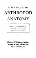
Fig. 1. Trilobita. Examples of trilobite forms. A, Olenellus vermontanus Hall ( from Walcott, 1910 ) . B, Olenellus gilberti Meek ( from Walcott, 19lO). C, Schmidtiellus mickwitzi Schmidt ( from Wal cott, 1910 ) . D, Asaphiscus wheeleri Meek ( from Walcott, 1 9 1 6 ) . E, Pero nopsis montis ( Matthew ) ( from Walcott, 1908 ) . H, head, or prosoma; Pyg, pygidium; spi, spine; Tel, telson; Th, thorax.
of their kinds, while there is nothing to show from what they came during the earlier Devonian, Ordovician, and Cambrian times, when trilobites, eurypterids, xiphosurids, and crustaceans were well pre served. Fossilization, however, depends largely on the presence of a hard skeleton, so that it is likely that animals first appear as fossils only when they have acquired a sufficiently resistant integument, and even then they must meet with favorable external conditions for fossilization, while, finally, fossils themselves may be utterly destroyed by subsequent metamorphosis of the containing rocks. 7
ARTHROPOD
ANATOMY
The form and structure o f the trilobites ( fig. 2 ) show clearly that in these ancient animals the fundamental arthropod organization was already fully developed and had attained a specific type of speCial ization. Only in the lack of differentiation in the postoral appendages are the trilobites generalized, but their speCialization is of a rela tively simple kind that fitted them for life on the ocean bottom in shallow water along the shore, where most of them lived probably in the manner of the modem Limulus, though some species are thought to have been pelagic, or even deepwater inhabitants. Lacking jaws or grasping organs of any kind other than the legs, the trilobites could not have obtained active prey by raptatory methods, and it has been thought that probably they were mud feeders, but animals with long filamentous antennae can hardly be supposed to have made a practice of burrowing in mud or sand. In some species the projecting mesal ends of the coxae ( fig. 2 B ) are armed with spines, which fact sug gests that the coxal lobes had some use in the obtaining of food; Raymond ( 1920 ) says, "The primary function of these spiny lobes of the coxa was doubtless the gathering and preparation of food, and carrying it to the mouth by passing it forward from one to the next." On the o ther hand, since the coxal lobes do not meet in the middle line, and the spines are not well developed in all species, Stormer ( 1944 ) thinks it unlikely that they functioned as jaws. In any case, whatever may have been the food of the trilobites, and however they obtained it, the great numbers of the animals would indicate that they had an ample food supply within their reach. Certainly worms of various kinds as well as other soft-bodied crea tures living in the mud or sand along the ocean shores were abundant in both Cambrian and Pre-Cambrian times. It would seem, in fact, that a trilobite should be quite fit to live under modem conditions, and paleontologists have no positive evidence to account for their early extinction. General Structure of a Trilobite
Since few perfectly preserved specimens of trilobites are known, it is not possible to give a full description of the trilobite structure in any one species, but inasmuch as the details of structure have been carefully studied in different species, we can reconstruct dia grammatically, as given in figure 2, the general form and make-up of a typical representative of the group. 8
THE
T R I L OB I T A
The body o f a simple trilobite o f usual form i s oval and dorso ventrally flattened ( figs. 1 D, 2 A ) ; it is divided transversely into three parts known as the head ( H ) , the thorax ( Til) , and the pygidium ( Pyg ) . The thorax and pygidium together, however, may be said to constitute the body as distinguished from the head, but,
Fig. 2. Trilobita. Diagrams showing the structure of a generalized trilobite. A, dorsal. B, ventral. An, anus; Ant, antenna; Dbl, doublure; E, compound eye; Glb, glabella; H, head or prosoma; Lm, labrum or hypostome; Plr, pleura ( not the pleuron of other arthropods ) ; Pyg, pygidium; Rae, rachis or axis; Th, thorax.
since the so-called head bears the first four pairs of legs, the terms prosoma and opisthosoma are preferable names for the two principal parts of the animal. The pro somatic head is unsegmented in the adult, though it may show evidence of coalesced segments; the thorax is completely segmented; and the pygidium is clearly composed of united segments. In the earlier trilobites the pygidial region is fully segmented ( fig. 1 A, B, C ) . Extending lengthwise usually through 9
A R THROPOD
A N A TO M Y
the three parts i s a rounded, median elevation, which i s flanked by wide, flattened or decurved lateral areas. On the head the median elevation is known as the glabella ( fig. 2 A, Glb ) , the lateral parts as the cheeks, or genae; on the body the median elevation is termed the axis, or rachis ( Rac ) , the lateral parts the pleurae ( Plr ) . These terms and others to be introduced later are those current in trilobite taxonomy, and the parts they connote have no necessary relation to those similarly named in other arthropod groups. The name trilobite is derived from the apparent lengthwise division of the animal into three parts. Though the trilobite as seen from above ( fig. 2 A ) looks like no other animal with which we are familiar, the undersurface ( B ) at once shows that the trilobite is an arthropod; on each side is a long row of jointed legs, and from the anterior part of the head there projects forward a pair of slender, filamentous antennae ( Ant ) . Be hind the bases of the antennae and extended over the mouth is a median lobe ( Lm ) , such as all arthropods have and which is com monly known as the labrum, though students of trilobites have gen erally called it the hypostome. The mouth presumably is covered by the labrum, and behind it is a small metastomal lobe. Of the legs, the first four pairs pertain to the head, the others to the thorax and pygidium, there being a pair for each body segment except the last. It will be noted that the legs are attached to the body at each side of a narrow median space, and that the pleural areas of the dorsum (fig. 4 A, D ) are inflected on the undersurface to the bases of the legs. The inflected ventral surfaces laterad of the leg bases constitute the doublure ( Dbl ) , which includes also the inflected undersurface of the head ( figs. 2 B, 3 K ) . A cross section through the thorax of a trilobite ( fig. 4 A ) clearly shows that the principal body cavity of the animal is in the median part, or rachis (Rac), which is strongly convex on the back, and that the so-called pleurae are merely flat hollow extensions of the body segments over the appendages. Since various other flattened arthro pods have a similar structure, the "three-lobed" character is not dis tinctive of the trilobites. The special feature of the trilobites is the uniform, leglike structure of all the appendages except the antennae, in which respect the trilobites are more generalized than any modern arthropods, since in all of the latter at least some of the appendages are modified and specialized for purposes other than that of locomo-
10
T H E TRILOBITA
tion; the trilobites apparently did not even have appendages that served specifically as jaws. Details of the leg structure will be de scribed in a later section. The Head, or Prosoma
The head section, or prosoma, of a typical adult trilobite ( fig. 2 A, H ) is usually somewhat semicircular in outline, with its rounded anterior and lateral margins produced posteriorly in a pair of large genal spines. Between the bases of the spines the head is directly attached to the body by its transverse posterior margin, without the intervention of a neck. The dorsal surface of the head, as already noted, generally presents a median, elevated glabellar area ( Glb ) and broad, lateral genal areas, but in many species the head is cov ered by a perfectly smooth, rounded, shieldlike plate. The features of the head given in the following deSCription, and illustrated dia grammatically in figure 3 L, have been made out from a study of many specimens of more generalized trilobites; the beginning student, however, is likely to see little trace of them in ordinary museum examples, owing partly to the imperfection or corrosion of the speci mens, but also to the fact that the characters themselves were sup pressed in the later evolution of the trilobites. The grooves that separate the glabella from the genae are known as the dorsal furrows ( fig. 3 L, df ) . A subdivision of the glabella into five successive parts, of which the first is the frontal lobe ( trl ) , may be indicated by lateral notches in the dorsal furrows, or by imperfect transverse grooves in the glabellar surface. Each genal area is divided lengthwise by a facial sulcus, or "suture," before and behind the eye ( afs, pfs ) , which separates it into a median part called the fixed cheek, or fixigene (F g ) , and a lateral part distinguished as the free cheek, or libragene ( Lg ) . The free cheeks are produced posteriorly into the genal spines ( gspi ) . An ocular ridge ( er ) goes from each side of the frontal lobe to the eye. The entire median part of the head shield between the facial sulci, including the glabella and the fixed cheeks, is termed the cranidium ( ern ) . A marginal furrow ( mf ) surrounds the head inside a narrow border area ( b ) . Most trilobites have a pair of large, lateral compound eyes ( E ) on the mesal parts of the free cheeks, and above each eye a protective palpebral lobe ( pbl ) on the edge of the fixed cheek. On the anterior part of the glabella in some species there is present a small tubercle, 11
A R T H R OPOD
A N ATOMY
which has been regarded as a median eye, but its ocular nature is questionable. A complete terminology for all parts of the trilobite is given by Howell, Frederickson, Lochman, Raasch, and Rasetti ( 1947 ) . Inasmuch as the trilobite head carries the first four pairs of legs, it must include at least four primitive postoral somites, and the glabellar grooves of the adult evidently represent the primary inter segmental lines of the head segments. The best understanding of the adult head has been derived from studies, such as those of Raw ( 1925 ) , Warburg ( 1925 ) , Lalicker ( 1935 ) , Stormer ( 1942, 1944 ) , and others, on specimens of very young trilobites in successive stages of development. All writers are in close agreement as to the visible facts, though they differ somewhat in their interpretations. The youngest-known developmental stage of a trilobite is a minute oval thing, from half a millimeter to a millimeter and a half in length, and is termed the protaspis ( fig. 3 A, E ) . The major part of the protaspis represents only the head of the adult trilobite. On the back of the youngest specimens a median glabellar elevation is already differentiated from wide lateral genal areas and very soon becomes divided transversely into five primary subdivisions, of which the last four ( I-IV ) represent the somites of the four pairs of legs carried by the adult head; faint intersegmental lines may be seen extending laterally in the genal areas ( E ) . The first glabellar subdivision of the protaspis includes the area of the frontal lobe of the adult 0, trl ) and its lateral extensions curving posteriorly around the anterior margin, which eventually become the free cheeks bearing the com pound eyes. This anterior, or acronal, section of the larva may be termed the acron ( A, Acr) , though the term has been used with various other applications. Its underfolded anterior part forms the doublure of the head, on which the antennae and labrum of the adult are situated ( fig. 2 B ) . The preoral antennae of the trilobites are clearly the first antennae, or antennules, of other arthropods. The four postoral pairs of legs on the head, then, should reasonably be supposed to correspond with the first four postantennular appendages of other arthropods, which primarily arise behind the mouth, these appendages being the chelicerae, the pedipalps, and the first two pairs of legs in the Chelicerata, or the second antennae, the mandibles, and the two pairs of maxillae in the Mandibulata. If the acron of the trilobite 12
THE
T RI L O BI T A
(ID ,
\
,
I.
E
"
.�C
�� ..
F J er
H
ern
_______ A______ _
, I
I I
I
L
Fig. 3. Trilobita. Postembryonic development and the structure of the adult head, or prosoma. A-D, developmental stages of Liostracus linnarssoni Brogger ( from Warburg, 1925 ) . E-I, developmental stages of Olenus gibbosus ( Wahlenberg ) ( from StOrmer, 1942 ) . J, diagram of structure of mature trilobite head, dorsal. K, diagram of undersurface of head. L, diagram of dorsal surface of head, with parts named according to Howell, Frederickson, Lochman, Raasch, and Rasetti ( 1947 ) . ACT, acron, anterior unsegmented part of larva; afs, anterior facial sulcus; b, border area; ern, cranidium; df, dorsal furrow; E, compound eye; er, ocular ridge; Fg, fixed cheek or fixigene; frl, frontal lobe; Glb, glabella; gspi, genal spine; Lg, free cheek or libragene; mf, marginal furrow; pbl, palpebral lobe; pfs, pos terior facial sulcus; I-IV, primary postacronal segments of larva.
13
ARTHRO POD
ANATOMY
larva i s a single segment, the trilobite head is, therefore, composed of five primary somites, but the frontal lobe is sometimes divided into two parts. According to Stormer ( 1942 ) , in the fourth protaspis stage of Olenus gibbosus (fig. 3 H ) there appears in the sides of the frontal lobe a pair of pits, which eventually run together in a transverse groove, so that the frontal lobe becomes divided second arily in development into two segments, which Stormer designates an antennal ( i.e., antennular ) segment and a preantennal segment. Henriksen (1926 ) and other writers, however, have contended that the posterior division of the frontal lobe is the segment of a sup pressed pair of appendages representing the second antennae of Crustacea, and that the four persistent appendages are to be identi fied with the mandibles, the two pairs of maxillae, and the first max illipeds. This contention that the trilobites have lost a pair of ap pendages, of which there is no concrete evidence, seems to be purely presumptive. "Such conclusions," StOrmer asserts, "have apparently been too much influenced by the current opinion of a crustacean nature of the trilobites." In other words, a lost "second antenna" has been arbitrarily injected in order to make the trilobites conform with their supposed crustacean descendants. However, the trilobite, as it is, conforms with either the Chelicerata or the Crustacea on the as sumption that its first legs represent the chelicerae of the former and the second antennae of the latter. From the development of the protaspis it may be deduced that the pattern of the segmental composition of the adult trilobite head must be approximately that shown diagrammatically at J of figure 3. The oculoantennal part of the head, bearing the eyes dorsally and the antennae ventrally, which for convenience is here called the acron, has extended posteriorly on the sides from the frontal lobe, forming the regions of the free cheeks ending in the genal spines. The median part of the head, including the postfrontal part of the glabella and the fixed cheeks, is the region of the four postoral leg bearing somites, but the intersegmental lines become obliterated in the adult except for remnants of them on the glabella. The Body, or Opisthosoma
Though the five primary segments of the young protaspis all go into the formation of the adult head, there is a very small region be hind the last head segment that is destined to generate the body. 14
T H E TR I L O B I T A
In the developing protaspis a stage is soon reached when a new segment appears behind the last head segment ( fig. 3 B, G ) , and this segment is followed by others successively formed between it and the extending end of the body ( C, D, H, I ) . These segments are the beginning of the series of body segments. The pygidial and thoracic segments, therefore, are secondary somites generated in the usual teloblastic manner from a subterminal zone of growth, just as are the secondary somites of the polychaetes and of those modem arthropods that have an anamorphic postembryonic development. The Thorax- The thoracic segments of the adult trilobite are usually all of similar size and shape, but as between different species they are highly variable in number, since there may be from two to forty or more, though the usual number is perhaps between seven or eight and fifteen. The segments undoubtedly were movable on each other and connected by infolded membranous conjunctivae; probably they were hinged by articulations between the tergal plates, as are the abdominal segments of the crayfish, so as to allow up and down movements, though evidently there could have been little movement in a transverse direction. Many trilobites are found rolled ventrally upon themselves in the manner of certain modem isopods. The elevated median parts of th.e thoracic terga are distinctly de marked from the long, flattened or decurved pleural lobes on the sides, but, as already noted in cross section ( fig. 4 A ) , the pleural lobes are merely lateral expansions of the body extending out be yond the leg bases. A cross section of the trilobite thorax resembles a cross secti o n of Limulus (fig. 7 A ) , and in each animal the terg a l plate, or carapace, must be interpreted as including not only the dorsal integument, but also the doublure ( Db l ) on the undersurface to the bases of the legs. The projecting ends of the pleural lobes of the trilobites generally form a row of pleural spines, which may be uniform in size and shape or varied; in some forms particular spines are greatly elongate ( fig. 1 B ) , in others all may be long and slender. The parts called "pleura" by students of trilobites are not to be identified strictly with the parts so termed in other arthropods. The true stemal area of the trilobite is the narrow ventral space between the leg bases and appears to have been a relatively soft integument. The Pygidium- The pygidium is composed of a number of seg ments that are not fully differentiated as they are generated in development, but since in some of the earlier trilobites the body is 15
A R T H R OP O D
A N AT O M Y
completely segmented ( fig. 1 A ) , i t i s probable that the pygidium has been formed in the course of evolution by a secondary union of the posterior body segments. The typical pygidium ( figs. 1 D, 2 A, Pyg ) is a large, smoothly rounded plate equal to the head in size, or sometimes larger than the head. It contains an extension of the rachis, and the pleural areas are marked by indistinct intersegmental lines. The pygidial appendages become successively smaller than those of the thorax ( fig. 2 B ) . The anus ( An ) is situated ventrally in the apical segment of the pygidium, which, therefore, is to be regarded as the telson. In forms in which the pygidial region is segmented, one or more long median spines may project from the dorsum of the anterior pygidial segments ( fig. 1 A, B, C ) . In one genus at least, the terminal segment bears a pair of long, multiarticulate tail filaments. It has been suggested that the pygidium was used as a swimming organ in the manner of the abdomen and "tail fan" of a crayfish. The Appendages
The appendages of the trilobites are now fairly well known in a number of species; they include the antennae and the· legs. In all the arthropods the antennae, or antennules, appear to be organs of a nature different from that of the other appendages, since in their normal development they never have the form or structure of ambu latory limbs, while, as the trilobites themselves attest, the other ap pendages, regardless of their form in modern arthropods, undoubt edly were all primarily walking legs. If the antennae were ever evolved from leglike appendages, they must have completed their transformation long before the time of the oldest-known trilobites. The Antennae- The long, slender, tapering antenna of the trilo bite is divided into a large number of short rings, and in appearance resembles the antenna of an orthopteroid insect, or the flagellum of a crustacean antenna, in which the rings are mere annulations with out muscles, and therefore are not true segments, as are the muscu lated subdivisions in the antennae of the myriapods and the en tognathous apterygote insects. The antennae of the adult trilobite arise from the doublure of the head anterior to the mouth (fig. 2 B, Ant ) , and are evidently the homologues of the antennules of Crusta cea, though they are not branched, and of the antennae of the myriapods and insects. 16
THE
T R I L O BI T A
The Legs- The legs o f the trilobites are typical, uniramous, seg mented arthropod limbs, with long, pinnate epipodites arising from their bases ( fig. 4 A ) . Since each leg has seven clearly marked seg ments, the segmentation was formerly thought to correspond with that of a crustacean leg, and the epipodite was regarded as an
"
A
"-
"-
Tlpd---
, \
\,
Frn
Tb
\
B
Prfm
Pat
" ,"
P-lar
\ \ \
ex
__ _
Tar
c
Fig. 4. Trilobita. Sectional structure of the body and segmentation of the legs. A, diagrammatic cross section of a trilobite, showing attachment of the legs.
B, a trilobite leg, with interpretation of segments according to Stormer ( 19 44 ) . C, leg of Neolenus serratus ( Rominger ) ( from Raymond, 1920 ) . ex, coxa; cxnd, coxal endite; D, dorsum; Dbl, doublure of dorsum; Eppd, epipodite; Fm, femur; Pat, patella; Prcx, precoxa; Prfm, prefemur; Ptar, pretar sus; Rae, rachis; Tb, tibifl; Tar, tarsus; Tlpd, telopodite; Tr, trochanter; V, venter.
exopodite, three small claws on the end of the segment being treated as mere apical spines. More recently, Stormer ( 1944 ) has shown that there is at the base of the leg a small ring by which the appendage is attached to the body, and this proximal ring Stormer regards as the true basal segment of the limb, which he calls a "precoxa," or "subcoxa" ( B, Prcx ) , the large segment following being the coxa. A 17
A R T HR O P O D
ANATOMY
petrified specimen, however, can hardly b e supposed t o give con clusive evidence that a basal ring of the appendage is a true segment in the sense of having been an independently movable section of the limb when the animal was alive, and it seems very improbable that the body muscles that moved the leg as a whole should have been attached on such a relatively small piece. In modem arthropods the basal muscles of an appendage are always attached on the coxa, unless the coxa itself has become immovable, but the base of the coxa is often set off as a proximal ring, or basicoxite, by an external groove that forms internally a strengthening ridge, or shelf, for the muscle attachments, and the basal ring of the coxa might superficially resemble a small segment. That the "precoxa" of the trilobite leg is a real limb segment, therefore, is open to question. The rest of the leg beyond the subcoxal ring is divided into seven cylindrical sections ( fig. 4 B ) , and there is no reason for supposing that these parts were not movable on each other, though, lacking any knowledge of the leg musculature, we cannot be so sure they were all true segments. The seventh apparent segment bears the three apical spines or claws, and Stormer, with good reason, has identified the claws as constituting the true end segment of the limb, since a similar three-clawed endpiece of the legs in other arthropods is always found to be a reduced, clawlike, apical segment provided with its own muscles and armed with a pair of lateral claws. A figure of an appendage of N eolenus serratus ( fig. 4 C ) given by Raymond (1920 ) leaves little doubt of the segmental nature of the apical part of the limb, though Raymond himself describes the claws as "little spines" on the end of the "dactylopodite." The median claw in modem arthropods, however, is the true dactylopodite, of which the lateral claws are secondary outgrowths. Discounting the segmental nature of the precoxa, then, the trilo bite leg appears to be an eight-segmented limb. If so, the segments are to be identified as Stormer has named them ( fig. 4 B ) : coxa ( coxopodite ) , trochanter ( basipodite ) , prefemur ( ischiopodite ) , femur ( meropodite ) , patella, tibia ( carpopodite ) , tarsus ( propo dite ) , and pretarsus ( dactylopodite ) , the names in parentheses being those used in carcinology. An eight-segmented limb depends on the coincidental occurrence of a patella and of two segments in the trochanteral region; it is not common in modem arthropods, but is present in the Pycnogonida and in some of the legs of the Solpugida 18
THE
T R ILOBITA
among the arachnids. The patella is a segment characteristic of the legs of Arachnida and Pycnogonida and is not found in those of mandibulate arthropods. A tibia and tarsus normally follow the patella, or the femur if there is no patella; but it must be noted that the tarsus is prone to subdivision, particularly into two parts, which simulate segments, but are not interconnected by muscles. With re gard to the trilobite leg, therefore, it might be questioned if the ap parent tibia and tarsus are not two tarsal sub segments and the sup posed patella the true tibia. There must, in other words, be some uncertainty as to the identity of limb segments where the muscula ture cannot be known. Yet the trilobite leg appears to be an eight segmented appendage, not counting the subcoxal ring, and, if so, it is a truly generalized arthropod limb in that it contains all the segments present in the legs of any modem arthropod, but it par ticularly resembles the arachnid leg in the possession of a patella. The long pinnate or plumose lateral branches arising from the bases of the trilobite legs ( fig. 4 A, B, Eppd ) are of particular interest. The earlier students of trilobite limbs regarded these branches as "ex opodites," and from this interpretation they drew the conclusion that the trilobite leg is a biramous appendage, and therefore closely re lates the trilobites to the Crustacea. StOrmer ( 1933 ) , however, showed that the supposed exopodite arises, as he then thought, on the coxa and not on the basipodite ( first trochanter ) as does the crustacean exopodite, from which fact it obviously is an epipodite and not an exopodite; later ( 1939 ) he assigned it to the precoxal ring ( fig. 4 B). Furthermore, StOrmer gives good reasons for regarding the organs in question as gills. The epipodites are covered by the pleural lobes of the body segments, and, if they are not branchiae, the trilobites have no evident organs of respiration. In any case, the idea that the crustacean biramous type of limb represents the primitive and fundamental structure of arthropod appendages is not supported by the trilobites. The crustacean exopodite is always borne by the basipodite, and is a special feature of the Crustacea, though not present on all their appendages.
19
LIMULUS LIM UL US, and its living relatives generally assigned to two other genera, are the animals known as "horseshoe crabs" or "king crabs." They, together with various fossil forms, constitute the class Xipho surida of the subphylum Chelicerata. Limulus polyphemus Latr., also known under the generic name of Xiphosura, lives along the Atlantic coast of North and Central America; other species are found in the waters of Asia, Japan, and the East Indies. The xiphosurids for the most part inhabit fairly shallow water over sandy bottoms along the ocean shores, but the Asiatic species, Carcinoscorpius rotundicauda Latr., is said to live in brackish estuaries, or even in rivers where the water is practically fresh ( see Annandale, 1909 ) . The horseshoe crab is an animal that adds romance to zoology; it is a relic of past ages that now stands alone in the midst of creatures of
later origins and of more modem types of structure. The ancestors of the horseshoe crabs were companions of the trilobites. General External Structure
The body of Limulus ( fig. 5 A ) is distinctly divided into an anterior prosoma and a posterior opisthosoma, the second part being freely movable up and down on the first; the opisthosoma carries a long, strong, independently movable tail spine. The animal gets its common name from the shape of the prosoma, which is covered by a convex dorsal shield, or carapace, in outline suggestive of a horseshoe. The opisthosoma is hexagonal, with its broad base at tached to the prosoma on a strong transverse hinge between the projecting posterior angles of the prosomatic carapace. The lateral
20
LIMULUS
margins of the opisthosoma are indented b y six notches o n each side, in which are seated slender movable spines; the tail spine arises from a deep indentation of the posterior margin. The body of the adult animal is entirely unsegmented. The dorsal surface of both the prosoma and the opisthosoma is elevated along the middle ( fig. 5 A ) , and on the broad lateral areas
E
An
A
B
Fig. 5. Xiphosurida. Limulus polyphemus L., dorsal ( A ) and ventral ( B ) . An, anus; Apds, opisthosomatic appendages; Chi, chilarium; Chl, chelicera; dO, simple dorsal eye; E, compound lateral eye; Epst, epistome; L, leg; Mth, mouth; Opl, operculum.
of the prosomatic shield is a pair of compound eyes ( E ) at the sides of longitudinal ridges. In a general way, therefore, in its dorsal as pect, Limulus has a rather striking resemblance to a trilobite, except for the absence of segmentation in the opisthosoma and the presence of the tail spine. Six small pits along each side of the median elevation of the opisthosoma of Limulus, however, probably mark the lines of union between primitive segments of the opisthosoma corresponding with the lateral spines. In Limulus there is a pair of very small simple eyes ( dO ) situated on the sides of a median spine on the anterior 21
A RTH R O P O D
A N AT O M Y
part of the prosoma, but in some trilobites a sense organ of some kind is present in the same position. On the undersurface of Limulus ( fig. 5 B ) the appendages are most in evidence, but the xiphosurids in common with the arachnids have no antennae. On the prosoma are six pairs of jointed, leglike limbs, and on the opisthosoma six pairs of broad, flat lobes project ing posteriorly and underlapping each other, so that the posterior five are almost covered by the first. In the differentiated form and function of the appendages, and in the lack of antennae, the xipho surids differ conspicuously from the trilobites. The prosomatic limbs include an anterior pair of small appendages termed the chelicerae ( Chl ) , which are directed forward, and five pairs of legs ( L ) ex tended laterally. Behind and between the bases of the last legs are two small spiny lobes ( Chi ) known as the chilaria, which represent a seventh pair of prosomatic appendages. The legs are attached by their dorsoventrally elongated basal segments on the sides of a deep axial part of the body ( fig. 7 A ) , and the margins of the prosomatic carapace are reHected ventrally to the leg bases as a broad doublure ( Dbl ) . In cross section, therefore, Limulus again resembles a trilo bite ( fig. 4 A ) ; the body of the animal in each case is a cylinder . containing the principal viscera, with wide Hat expansions of the . dorsum extending over the limbs. If the legs are spread apart ( fig. 6 A ) , between their bases is ex posed a median triangular area of the ventral integument, which is the sternal region of the prosoma. Anteriorly at its apex is the mouth ( Mth ) , which has a central position beneath the prosoma ( fig. 5 B ) . If the prosomatic appendages are removed from a specimen ( fig. 7 F ) , it will be seen that the bases of the chelicerae and legs are radially arranged around the median sternal area, the chelicerae being directly in front of the mouth and the first two pairs of legs diverging forward from its sides; the chilaria ( Chi ) block the poste rior end of the space between the legs. With the appendages removed, the doublure ( Dbl ) is fully exposed. Its peripheral part forms a wide horseshoe-shaped area, which has the hard, horny texture of the dorsal surface of the carapace and slopes steeply upward ( A ) . The inner part immediately surrounding the bases of the appendages takes a horizontal position and becomes membranous ( F ) , but it contains on each side a series of five small Y -shaped sclerites ( Pl ) , to the stems of which the leg bases are specifically articulated ( fig. 22
L I M UL U S
6 C ) . These sclerites, the "coxal pivots" of Benham ( 1885 ) , clearly are to be regarded as pleurites, since, in that they support the ap pendages, they correspond with the plates called pleura in most other arthropods. On the membranous part of the doublure of Limulus before the bases of the chelicerae is a median papilla ( fig. 7 F, vO ) , the supposed "ventral eye." Supporting the chelicerae is
A
Fig. 6. Xiphosurida. Limulus polyphemus L. A, the app endages in p lace on the body, ventral. B, epistome, labrum, right chelicera, and base of right first leg, anterior. C, coxae and first leg of left side, with pleural articulations. a, pleural articulations of coxae; Apds, opisthosomatic appendages; Chi, chi larium; ChI, chelicera; Cx, coxa; Dbl, doublure; Endt, coxal endite; Eppd, epipo dite; Epst, epistome; L, leg; Lm, labrum; Mth, mouth; Opl, operculum; PI, pleurites; Tr, trochanter; va, ventral sense organ; I-XIII, body segments begin ning with cheliceral segment.
a small epistomal plate ( fig. 6 B, Epst ) , which bears the labrum ( Lm ) projecting beneath the mouth ( fig. 7 F ) . On the opisthosoma the closely overlapping appendages ( fig. 5 B, ApdsV/II-XIII ) are set in a depression of the undersurface, which is surrounded laterally and posteriorly by a broad, flat doublure with a hard, polished surface. The appendages of the first pair are united to form an operculum ( Opl ) covering the other five, which
23
ARTHRO POD
ANATOMY
bear leaBike gills o n their posterior surfaces. O n the upper ( poste rior ) side of the operculum are the openings of the genital ducts, situated in each sex on a pair of small papillae ( fig. 12 B, gp ) . At the base of the tail spine is the anus ( fig. 5 B, An ) . The appendages will be more fully described in a later section. The prosoma and the opisthosoma are connected by a large median dorsal muscle, the arthrotergal muscle of Benham ( 1885 ) , that crosses the hinge line between the two parts of the body and serves to flex the opisthosoma on the prosoma. In the ventral part of the body, long muscles arising in the prosoma are distributed by branches to the ventral wall of the opisthosoma and are evidently levators of the opisthosoma; a few ventral fibers from the prosoma, however, go to anterior dorsal apodemes of the opisthosoma and are there fore flexors, or depressors, of the opisthosoma. The presence of seven pairs of appendages on the prosoma of Limulus should indicate that the xiphosurid prosoma contains at least seven segments. Studies on development show that all the ap pendages are postoral in the embryo ( fig. 8 A ) , the definitive preoral position of the chelicerae and the first two pairs of legs being a secondary result of the posterior displacement of the mouth, by which the ventral parts of the anterior segments in the adult appear to be lapped forward around the sides of the mouth and the labrum ( fig. 7 F ) . Limulus differs from a trilobite in having seven instead of four postoral segments in the prosoma. In appearance, however, there are only six pairs of appendages, since the chilaria have lost all sem blance to legs, and in this respect the Xiphosurida resemble the
Arachnida, in which the seventh segment is reduced or suppressed, and there are only six pairs of prosomatic limbs. The area of the prosoma formed by the postoral segments can not account for the whole of the prosomatic part of the animal, since there is a large preoral region in front of the cheliceral segment. I wanoff ( 1933 ) has shown in a figure of a Limulus embryo ( fig. 8 A ) the presence of a procephalic lobe ( Prc ) , which is a prominent anterior part of all arthropod embryos, and which appears to cor respond with the acronal lobe of a young trilobite ( fig. 3 A, Acr ) . Iwanoff, however, does not seem to attribute much of the adult structure of Limulus to this lobe, but a comparison of a larval stage of Limulus ( fig. 8 B ) with a trilobite would suggest that, as in the trilobites, the procephalic lobe has expanded laterally and posteriorly
24
Fig. 7. Xiphosurida. Limulus polyphemus L. A, section of prosoma behind bases of third legs.
B, base of tail spine, with muscles. C, posterior part of pro somatic carapace, turned upward and separated along hinge line from anterior part of opisthosoma, showing division of eighth tergum ( VUlT ) between the two parts of the body. D, inner surface of opisthosomatic carapace, and posterior part of prosomatic carapace, joined along hinge line (h) . E, prosomatic endosternum of specimen 47 mm. long. F, ventral surface of prosoma with pro somatic appendages removed, showing position of their bases relative to the mouth. lAp, 2Ap, first and second tergal apodemes; Cp, carapace; E, compound eye; Endst, endosternum; h, dorsal hinge between prosoma and opisthosoma; l p, 2p, external pits of first and second tergal apodemes; prsCp, prosomatic cara pace; T, tergum; V, venter; z, line of removal of ventral wall of opisthosoma. Other lettering as on figure 6.
25
A RT H R O P O D
A N AT O M Y
around the postoral segments t o form the lateral parts of the proso matic carapace, on which the compound eyes are developed. Other wise, it is difficult to account for the likeness of the prosomatic shield of Limulus to the dorsal surface of the head of a trilobite. The for ward radiation of the anterior postoral segments around the sides of the mouth indicated by IwanoH ( 1933, fig. 62 ) probably applies only to the ventral surface of the animal. A division of the procephalic area into an ocular and an antennal segment has not been observed in the xiphosurid. Segmentation of the opisthosoma in the adult of Limulus is in dicated not only by the presence of the appendages, but also by the double series of pits on the dorsal surface ( fig. 5 A ) . The pits are the points of ingrowth of apodemal processes on the inner surface of the back ( fig. 7 D ) , and, as shown by Benham ( 1885 ) , the muscle relation to the apodemes leaves no doubt that the apodemes arise on the primitive intersegmental lines. The first pair of the opis thosomatic apodemes ( 2Ap ) , however, lies well behind the hinge ( h ) between the two parts of the body, showing that the hinge is not a true intersegmental line. Moreover, there is an anteriormost pair of larger apodemes on the prosoma close to the posterior margin of the carapace ( C, D, lAp ) . Therefore, the space between these prosomatic apodemes and the first pair on the opisthosoma must represent the dorsum of a segment that has been partitioned be tween the prosoma and the opisthosoma, and which contains the intersomatic hinge. There is nothing unusual in this condition; it o ccurs in other arthropods and is merely an anatomical adjustment to the necessary mechanism of movement of one part of the body on another. Since the longitudinal muscles are primarily intrasegmen tal, the opisthosoma of the xiphosurid can become movable on the prosoma by its own muscles only by having the anterior part of its first tergum fused with the prosomatic carapace and a line of flexi bility developed across its middle. The tergal area of the eighth body segment ( fig. 7 C, VIIIT ) is discernible as a small arc on the posterior border of the prosoma marked by the pits ( lp ) of the first dorsal apodemes, and as a wider transverse area on the opisthosoma before the second apo demal pits ( 2p ) , ending laterally in large, free, pointed lobes. The limits of the following segments can be judged only by the apodemal pits and their apparent relation to the marginal spines. The posterior 26
LIMULUS
part o f the opisthosoma of Limulus must include a segment corre sponding with the telson of other arthropods, which contains the anus. The anus of Limulus lies ventrally just before the base of the tail spine ( fig. 7 D, An ) . It is questionable, therefore, if the spine itself represents the telson; it appears rather to be an appendage of the telson. The spine is not present in the embryo ( fig. 8 A ) , but grows out from the end of the body during larval development ( C, D ) . In the adult, the tail spine is an independently movable organ provided with a strong musculature ( fig. 7 B ) consisting of six long dorsal muscles and two large ventral muscles. The fibers of the median pair of dorsal muscles are distributed along the dorsal apodemes of the opisthosoma, those of the two lateral pairs arise on the opisthosomatic carapace; the ventral muscles divide each into a dorsal branch and a ventral branch. The origins of the spine muscles are thus far in front of the area of a presumed apical seg ment; their insertions, moreover, are not on the spine itself, but on a tough integument that connects the spine with the body. The spine is hollow to its tip, lined with an epithelium traversed by nerves and blood vessels. In the decapod crustaceans and in the pterygote insects there is a more or less elaborately developed endosk eleton consisting of arms, ridges, or plates that grow in from the cuticle of the body wall and serve to strengthen the exoskeleton or to give attachment to muscles. The principal structures of this kind in Limulus are the dorsal apodemes of the carapace and the tendons of muscles. In the center of the prosoma, however, is a horizontal plate suspended by muscles from the carapace, lying between the alimentary canal and the ventral nerve cord, which, though it is not a skeletal deriva tive, takes the place of a ventral endoskeleton, since all the ventral prosomatic muscles are attached on it. The plate, therefore, is gen erally termed the endosternum; Lankester ( 1884, 1885 ) and Benham ( 1885 ) call it the "entochondrite," or "plastron." It differs somewhat in shape and consistency with the age of the animal. In a small speci men two inches long the endosternum is a thick membrane. In shape it is an oblong rectangle ( fig. 7 E ) with the anterior corners produced into a pair of spatulate anterior lobes, the posterior angles drawn out into broad posterolateral lobes; laterally on the upper surface is a pair of soft, triangular dorsal lobes, and from the posterior margin a taillike extension gives attachment to numerous muscle 27
ARTHRO POD
A N AT O M Y
fibers. From the anterior end o n each side two long, slender sus pensory arms diverge outward and upward and are attached by muscles on the carapace. Numerous muscles depart from the margins of the plate and its lobes, and others arise directly on the dorsal and ventral surfaces. The endosternal muscles have been fully described and illustrated by Benham ( 1885 ) . According to Lankester ( 1884 ) , the endosternum, or "entochondrite," of the prosoma of an adult Limulus has a texture resembling that of hyaline vertebrate cartilage, though it lacks the essential constituents of cartilage. A similar en dosternal plate is present in most Arachnida, and it is probable that the ligament connecting the ventral adductor muscles of the gnathal appendages in lower Crustacea and in Diplopoda is a structure of the same nature. In its microscopic structure, Lankester ( 1884 ) says, the adult prosomatic endosternum "is a firm homogenous, or sparsely fibrillated matrix in which are imbedded nucleated cells generally arranged in rows of three, six, or even eight parallel with the adjacent lines of fibrillation." In a larval specimen 2 cm. in length, the endosternum is a thin membrane with a faintly fibrillated structure. Where muscles are attached on it, the fibrillae become more distinct and appear to run out directly into the fibrillae of the striated parts of the muscle fibers. The fibrillar continuity is plainly to be seen in the suspensory arms. In a specimen 5 mm. in length, the "plate" is a delicate mem brane resting close upon the nerve mass beneath it, but it is not essentially different from that of older specimens. Where muscles are attached, the striated fibers break up int o bundles of fine fibrillae that are soon lost in the tissue of the endosternal membrane. The endosternum is a non chitinous tissue. In the opisthosoma of Limulus the lateral systems of ventral mus cles are segmentally connected by transverse ligamentlike bands, which are termed the "mesosomatic entochondrites" by Benham ( 1885 ) because they appear to be a tissue of the same kind as that of the prosomatic endosternum. These connectives, however, lie below the nerve cord, though they are free from the epidermis of the body wall beneath them. The origin and nature of endosternal structures of arthropods have not been satisfactorily explained. Lankester ( 1885 ) suggests that the prosomatic endosternum of Limulus is derived from the sub epidermal connective tissue of the sternal surface, but he does not 28
L I M UL U S
explain how it brings with it the muscles, which presumably a t first were attached on the cuticle of the body wall, and, to account for the definitive supraneural position of the plate, he has to assume that the en dosternum of the prosoma was formed when the nerve
A
F
E
Fig. 8. Xiphosurida. Embryonic and larval stages and early fossil xiphosuri ds . A, Tachypleus gigas ( MiiIIer ) ( Limulus moluccanus Latr. ) , embryo with nine pairs of appendages ( from Iwanoff, 1933 ) . . B, Limulus polyphemus L., larva 4 mm. long. C, same, larva 5 mm. long. D, same as A, second larva just before moulting, opisthosomatic segments indicated by mesoderm bands ( from Iwanoff, 1933 ) . E, Prestwichianella rotundata, a Carboniferous fossil xiphosurid ( from Stormer, 1944, after Woodward ) . F, Aglaspis aetoni, a Cambrian fossil xipho surid ( from StOrmer, 1944, after Raasch ) . Chi, chilarium ; Chl, chelicera; Opl, op erculum ; Pre, cephalic lobe; I-VII, p ro so ma tic somites.
cords had a lateral position. Schimkewitsch ( 1895 ) has described the endosternum of Arachnida as a transformed muscle tissue. It may be noted that in some insects there is in the abdomen a series of transverse muscles over the nerve cord.
29
ARTHRO POD
ANATOMY
The Eyes
The functional eyes of Limulus include the lateral compound eyes ( fig. 5 A, E ) , and the small dorsal ocelli ( dO ) . The ventral organ lying in front of the chelicerae ( fig. 6 A, vO ) has been regarded by some investigators as a pair of degenerate ventral ocelli and by others as an olfactory organ. The Compound Eyes- Each compound eye is covered by a thick, convex cornea having an entirely smooth surface, but on looking into it one sees in its deeper part a regular, honeycomblike pattern of minute, six-sided facets. This appearance is not due to any division of the cornea; an examination of the inner surface shows that the latter is projected into numerous small, peglike processes so arranged that the spaces between them have a hexagonal shape. Each corneal process ( fig. 9 A, Ln ) serves as a lens for a group of sensory cells ( Om ) lying beneath it, which is ensheathed in long cells of the epidermis ( B ) . Each group of cells associated with a lens of the common cornea, therefore, is a light-receptive unit of the eye corresponding with an ommatidium of the compound eye of higher arthropods. The sensory cells constitute the retina, or retinula, of the ommatidium, and are closely arranged in a radial manner around a central axis ( C, rSCI ) , so that, as Demoll ( 1914 ) says, the ommatidial retina resembles a peeled orange. The number of radial cells in a single ommatidium varies from 10 to 15; their inner ends are separated by an axial space, in which, according to Demoll, is a long, slender process ( axp ) from an excentric retinal cell ( B, ecSCl ) lying outside the bases of the other cells. The narrow inner margins of the radial cells and the axial process of the excentric cell are composed of a relatively clear cytoplasm with fine striations at the surface ( B, C, sb ) . In the compound eyes of most arthropods the striated borders of the convergent retinal cells unite to form an axial, rodlike structure called a rhabdom, and are therefore termed rhabdomeres. The function of the rhabdom or the rhabdomeres is the diffraction of light from the lens into the light-sensitive parts of the retinal cells. Proximally the retinal cells are continued into nerve processes ( B, N v ) that go to the brain. The corneagenous epidermal cells of the eye ( B, CgCls ) that surround the lens and en sheath the retina are shown by Demoll to converge between the lens and the retina, but they do not form here any specific dioptric body 30
LIMULUS
such a s the crystalline cone of most compound eyes. The lateral eyes of Limulus are thus seen to be true compound eyes, but to have a simpler structure than those of higher arthropods. The Dorsal Eyes- The small, median dorsal eyes of Limulus, as C or.
A - sb
D
Fig. 9. Xiphosurida. Eyes of Limulus. A, diagrammatic section of the cornea and five ommatidia of compound eye
( from Lankester and Bourne, 1 883 ) . B, somewhat diagrammatic axial section of an ommatidium of compound eye ( from Demoll, 1914 ) . C, cross section of an ommatidium of compound eye ( outline from Demoll, 1914 ) . D, axial section of a dorsal ocellus ( from Demoll, 1914 ) . E, sagittal section of a "ventral eye" and nerve from brain ( from Demoll, 1914 ) . axp, axial process of eccentric sensory cell ( ecSC1 ) ; Br, brain; CgCls, corneag enous cells; Cor, cornea; Ct, cuticle; e, mass of undifferentiated cells; ecSCI, eccentric sensory cell; Epd, epidermis; Ln, lens; Nv, nerve; am, ommatidium; rSCI, radial sensory cell; sb, striated border; SCls, sensory cells; SO, ventral sense organ.
described by Demoll ( 1914 ) , are typical arthropod ocelli. The cornea of each of these eyes ( fig. 9 D, Cor ) is produced inwardly as a large spherical lens ( Ln ) , which is enclosed in a layer of corneag enous cells ( CgCls ) continuous at the periphery of the eye with 31
ARTHRO P O D
ANATOMY
the surrounding epidermis ( Epd ) . Beneath the corneagenous layer is the relatively thick retina, composed of the sensory cells ( SCls ) , scattered interstitial cells, and a binding network of connecting tissue that forms also a limiting membrane around the base of the eye, penetrated by the nerve processes ( Nv ) of the sensory cells. At one side of the eye is a mass of undifferentiated cells ( e ) which Demoll regards as a degenerate eye, and he draws the inference that Limulus once had a group of four dorsal ocelli. The "Ventral Eyes"- The supposed ventral eyes of Limulus ( fig. 6 A, va ) are described by Demoll ( 1914 ) as consisting of two masses of irregularly arranged cells ( fig. 9 E ) abutting on the epidermis beneath convexities of the cuticle. Each organ consists for the most part of large cells associated in groups, with connective tissue be tween them. The large cells are produced into nerve fibers that go to the brain ( Br ) , and where two of the cells adjoin each other their opposed surfaces, Demoll says, have rhabdomere borders, though they do not form rhabdoms. These cellular bodies certainly must be, or have been, sensory organs of some kind, and probably the idea that they are degenerate eyes is as good as any other; their structure does not suggest an olfactory function. The Appendages
We may now give special attention to the appendages, including the seven pairs on the prosoma and the six on the episthosoma. A full account of the embryonic development of the appendages in Limulus moluccanus is given by Iwanoff ( 1933 ) , who shows that all the appendicular organs, regardless of their differences in the adult stage, begin in the same way, as a pair of folds of the ventral body wall. The Chelicerae- Since the Xiphosurida lack antennae, the first of the prosomatic appendages are the chelicerae ( fig. 5 B, ChI ) , which, as already noted, lie directly in front of the mouth in the adult state, but take this position secondarily from a primary postoral position in the embryo ( fig. 8 A ) . The chelicerae presumably repre sent the first postoral appendages of the trilobites. In the adult of Limulus the chelicerae are articulated on a small plate ( fig. 6 B, Epst ) that eVidently is the epistome, since it supports the labrum ( Lm ) and is produced on each side into a slender arm curving for ward along the mesal border of the coxa ( Cx ) of the first leg. Each 32
LIMULUS
chelicera i s three-segmented ( fig. 1 0 D ) , but the terminal segment is a slender claw, or movable finger, opposed to a similar fixed process of the second segment, so that the two together form a pair of pincers. According to Benham ( 1885 ) , the chelicerae are movable by muscles inserted on their bases that arise on both the carapace and the endostemum. The Legs- The five pairs of legs ( fig. 5 B, L ) resemble the chelicerae in that their distal segments form pincers, but they have a greater number of segments, and in the adult male the chelae of the first legs assume an altered form ( fig. 10 F ) . The basal seg ments, or coxae, of the legs are greatly elongate dorsoventrally and are attached on the membranous lateral walls of the deep median part of the prosoma beneath the lateral extensions of the carapace ( fig. 7 A, ex ) ; their only firm articulations are with the small Y -shaped pleural sclerites in the membranous inner part of the doublure ( fig. 6 C, PI ) . The lower ends of the coxae of the first four pairs of legs are produced into large spine-covered mesal lobes, or endites ( fig. 10 H, Endt ) , the spines of which curve forward toward the mouth ( fig. 6 A ) . Above the endites of the second, third, and fourth legs is a small accessory endite arising from the membrane at the base of the coxa ( fig. 10 G, H ) . Each leg is movable anteriorly and posteriorly on the long base of its coxa ( fig. 10 C, a-b ) , but also has a transverse movement be cause of the flexible, membranous nature of the body wall on which it is attached. As shown by Benham ( 1885 ) , nine muscles are in serted on the basal rim of the coxa ( fig. 10 C ) , five of which ( 25, 26, 27, 28, 29 ) are dorsal muscles arising on the carapace, and four ( 82m, 82n, 88, 84 ) are ventral muscles with their origins on the en dostemum. Two of the ventral muscles ( 82m, 82n ) branch from a common origin. The anterior and posterior dorsal muscles evidently are promoters and remoters, that is, they rotate the coxa on its dorso ventral axis ( a-b ) ; the ventral muscles must have an adductor func tion because of the dorsal suspension of the coxa on the pleural sclerite. Since there is no evident muscular mechanism to oppose the ventral adductors, abduction of the limb apparently results from the elasticity of its basal connections. The legs of the first four pairs have each only six segments; the somewhat longer last legs, however, have seven segments, and there fore represent a more nearly complete appendage, for which reason 33
A R TH R O P O D
A N AT O M Y
they should b e studied first. The coxa of a fifth leg ( fig. 1 0 A , ex ) resembles the coxae of the other legs except for the absence of spines on the endite lobe. A short section of its upper end is set off by a groove marking the base of a strong internal ridge ( B, R ) , and bears a spatulate epipodite ( A, Eppd ) . The next segment is the trochanter ( Tr ) , which turns on the coxa in a vertical plane, and supports the longer third segment, which is the femur ( Fm ) . At the end of the femur is the "knee" bend of the leg ( k ) , beyond which are four segments. The segment adjoining the femur very evidently corresponds with the segment of an arachnid leg called the patella ( fig. 19 A, Pat ) , and the following segments of the Limulus leg should then be the tibia ( Tb ) , the tarsus ( Tar ) , and the pretarsus ( Ptar ) . A different idea concerning the identity of these segments, however, has been given by Hansen ( 1930 ) , who regards the faint circular groove near the base of the segment beyond the knee as the division between patella and tibia, so that he has to invent the name "cotibia" for the next segment. The musculature of the leg, as will presently be seen, gives no support to this interpretation. The basal groove is present also on the preceding three pairs of legs ( G, H ) , but not on the first legs ( F ) . The segmentation of the hind leg of Limulus corresponds with the apparent segmentation of a trilobite leg ( fig. 4 B ) , except for the lack of a second trochanter, or prefemur. The patellotibial joint has a characteristic structure ( fig. 11 C ) , there being two dorsal lobes on the tibia, between which a median, tonguelike process curves down from the end of the patella. The end of the tibia of the last leg ( fi g. 10 A ) b ears four bladelike appendages that conceal the cylindrical tarsus. The pre tarsus ( Ptar ) is a long, tapering, spinelike segment, which, with a shorter process projecting from the end of the tarsus, forms a slender chela. In studying the other legs, the question arises as to what segment has been eliminated to leave only six segments; the answer is fur nished by the musculature. The intrinsic musculature of the hind leg is sufficiently shown at A of figure 11, though some of the muscles of the inner, or posterior, side of the leg are not seen in the anterior view. Since there is no question as to the identity of the first three segments in all the legs, we may note first the muscles that move the part of the leg beyond the knee. In the ventral membrane of the knee joint is a small V-shaped sclerite. The arms of the V give attachment 34
LIMULUS
Fig. 10. Xiphosurida. Limulus polyphemus L . The pro somatic appendages. A, left last leg, anterior. B, inner surface of anterior wall of upper end of coxa. C, base of leg with attached muscles ( from Benham, 1885 ) . D , left
chelicera, lateral. E, movable finger and muscles of chela of first leg of adult male. F, first left leg ( pedipalp ) of adult male, anterior. G, right third leg, posterior. H, left fourth leg, anterior. I, chilaria, posterior. a, dorsal ( pleural ) articulation of coxa; b, ventral articulation of coxa; ex, coxa; Endt, coxal endite; Eppd, epipodite; Fm, femur; k, knee bend of leg; Pat, patella; PI, pleurite; Ptar, pretarsus ; R, ridge of coxal wall; Tar, tarsus; Tb, tibia; Tr, trochanter.
35
ARTHROPOD
ANATOMY
t o a pair o f broad muscles ( 8 ) arising dorsally i n the femur; the apex is produced into a long, thick, rodlike apodeme, or tendon ( t ) , that extends through the femur and gives attachment to a thick conical muscle ( 10 ) in the trochanter. These three muscles are present in all the legs and serve to flex the distal part of the limb on the femur. The patella contains six muscles inserted on the base of the tibia; two are large dorsal muscles ( 12 ) , two are fan-shaped lateral muscles ( 14 ) , and two are long slender ventral muscles ( 16 ) . Each ventral muscle has two branches, one branch ( 16a ) arising in the distal end of the femur, the other ( not seen in the figure ) in the base of the patella. The tarsal muscles in the tibia are a dorsal levator of the tarsus ( 18 ) and a two-branched ventral depressor ( 19 ) . The pretarsus has a simple levator ( 20 ) and depressor ( 21 ) arising in the tarsus. If now we compare the musculature of one of the other legs with that of a hind leg, it is found that the first segment beyond the knee has the same flexor mechanism as this segment in the hind leg, and that it contains the same muscles ( fig. 11 B ) . This segment, there fore, is the patella in all the legs ( fig. 10 A, F, G, H, Pat ) . Between it and the next segment ( fig. 11 B, Tb ) there is no suggestion of a reduced or eliminated segment, and the nature of the musculature from the patella leaves no doubt that this segment is the tibia. More over, the structure of the joint between these two segments is ex actly that of the patellotibial joint of the hind leg ( C ) . Finally, the musculature in the tibia of the anterior legs is that within the tibia of the hind leg inserted on the tarsus. We must conclude, therefore, that in all the legs but the last, the terminal segment forming the movable finger of the chela is the tarsus ( fig. 10 G, H, Tar ) , and that the opposed finger is a process of the tibia. Hansen ( 1930 ) made the same deduction without a study of the musculature. The chela of the anterior leg becomes much modified in the adult male ( fig. 10 F ) . The tibia, or "hand" of the chela ( Tb ) , is short but greatly swollen, and the long clawlike tarsus, or movable finger ( Tar ) , is thick and bent, while the immovable process of the tibia is reduced to a short thumb. The globular tibia contains a small dorsal muscle of the tarsal claw ( E ) and two great masses of closing fibers inserted on a thick knob of the tarsal base. This seemingly deformed chela of the male, therefore, evidently has great strength. The Chilaria- The appendages of the seventh postoral body seg36
LIMULUS
ment, known a s the chilaria, lie close to the posterior margin o f the venter of the prosoma ( fig. 6 A, Chi ) , between and behind the coxal endites of the last pair of legs. Each chilarium ( fig. 10 I ) is a
Pat /
4
3 Tar
1
A
c
Fig. 11. Xiphosurida. Limulus polyphemus L. Structure and musculature of a leg. A, left last leg and intrinsic musculature, anterior. B, patellar segment, and muscles of posterior half. C, patellotibial joint, dorsal, structure characteristic of this joint in all the legs. Lettering as on figure 10; muscles explained in text.
small, s imple , elongate, flattened lobe, armed with spines on its distal end and along its mesal margin The two appendages arise close together and project downward, so that they close the posterior .
37
ARTHR O P O D
ANAT O M Y
end of the space between the leg bases leading forward to the mouth ( fig. 6 A ) . From their position the chilaria have acquired their name, cheilarion being Greek for a "little lip." According to I wanoff ( 1933 ) , the chilaria correspond with only the coxal endites of the preceding appendages. They have a very weak basal muscu lature. The Opisthosomatic Appendages- The six appendages of the opisthosoma are broad, flat, closely appressed, horizontal plates lying in the ventral concavity of the opisthosoma ( fig. 5 B, ApdsVIII XIII ) , where they underlap from before backward. The first plate is a large operculum ( Opl ) mostly covering the others. Each plate is provided with a pair of promotor muscles, and a pair of remotor muscles arising on the dorsum of the body wall ( fig. 12 A, B ) , both being inserted within the cavity of the appendage, the smaller promotor ( pmel ) on the anterior lamella, the larger remotor ( rmel ) on the posterior lamella. There is little reason to suppose that these opisthosomatic appendages of Limulus are not true segmental limbs, but it is difficult to identify their parts with those of a prosomatic leg. The operculum hangs from the membranous ventral wall of the body between the prosoma and the opisthosoma, but, as we have seen, the tergum of the first opisthosomatic segment is partitioned between the opisthosoma and the prosoma. Accordingly, the pro motor muscles of the operculum arise on the posterior apodemes of the prosomatic carapace, and the remotors on the anterior margin of the opisthosoma. The operculum differs from the following ap pendages in that its lateral halves are more closely united along the mid-line. On the anterior surface ( fig. 12 A ) two median series of sclerites appear to represent the shafts of the component appendages, and the broad lateral expansions can be interpreted as basal exites, probably coxal epipodites. A transverse line of flexibility ( 1-1 ) through the distal part of the operculum cuts off the apical parts of the exites as a pair of free lateral lobes and separates the two distal segments from the long basal segments of the median shafts, the apical segments of which form a pair of small, free median lobes. The posterior surface of the operculum ( B ) , however, gives little evidence of limb structure, except for the apical lobes. At about the middle of the basal part are two small genital papillae ( gp ) , present alike in both sexes, which contain the outlets of the 38
LIMULUS
genital ducts ( Dct ) . Since the operculum of Limulus pertains t o the eighth postoral body segment, it is of interest to note that the genital opening in Arachnida also is on the eighth segment. In the next five appendages of the opisthosoma ( fig. 12 C, D ) the broad lateral parts are separated by a wide, median membranous area, beyond which the component appendages are represented by a pair of three-segmented median lobes, with a slender tongue of
Fig. 1 2. Xiphosurida. Limulus polyphemus L. Opisthosomatic appendages. A, operculum, anterior. B, same, posterior. C, united second pair of ap pendages, anterior. D, same, more enlarged, posterior. Bm, gills ( branchiae ) ; Dct, genital duct; gp, genital papilla, with genital ori fice; l, line of flexion; pmcl, promotor muscle; rmcl, remotor muscle.
the basal membrane projecting between them. On the posterior sur faces of the appendages, turned upward, are the gills ( D, Brn ) . The gills consist of two great lateral masses of thin, closely super posed branchial lamellae, at least 80 of them in a single gill mass of an adult specimen. Each gill is an extremely thin semilunar plate with a rigidly thickened margin fringed with delicate setae. The lamellae are attached transversely by their bases on the supporting appendage and slope distally on the latter. 39
ARTHROPOD
ANATOMY
Relationships o f the Xiphosurida
A comparison of Limulus with a trilobite can hardly fail to give the impression that the two are related animals, though the principal likeness is in the form and structure of the prosoma, with its wide lateral parts inflected ventrally before the mouth and around the sides of the appendages. On the other hand, in the lack of antennae, the absence of segmentation in the opisthosoma, and the differentiation of the appendages, Limulus is far more specialized than a trilobite. The xiphosurids, however, have an ancient lineage; Limulus itself in almost its present form goes back to Jurassic times in the Mesozoic Era. Its ancestors in the Carboniferous have distinctly segmented abdomens ( fig. 8 E ) , as do still earlier Paleozoic representatives that lived in the Cambrian ( F ) . The two-part bodies and the tail spine of these ancient forms leave no doubt that they are xiphosurids. The union of segments in the opisthosoma, therefore, is only a special ization of the comparatively recent Limulus. Associated in the Cambrian with the early xiphosurids and the trilobites were many other arthropods whose known remains show such a mixture of characters that paleontologists cannot agree amon g themselves just what they are, whether trilobites, xiphosurids, euryp terids, or crustaceans, which fact is probably good evidence that the animals themselves represent early intermediate forms between these several groups. Limulus, however, has survived all its ancient relatives and still flourishes in modem times, so it is not surprising that it has become the most spe cializ ed member of its tribe. Stormer ( 1944 ) has interestingly described and pictorially illustrated the evolutionary history of the xiphosurids, showing examples of the dif ferent forms found at successive paleontological levels. It is not necessary to suppose, however, that the Xiphosurida have been de rived directly from trilobites; it is a safer assumption that the two groups of animals had a common ancestry.
40
"*
III
*
THE EURYPTERIDA THE eurypterids were aquatic animals that lived during the Paleo zoic era from the Cambrian into the Permian, but they attained their greatest development in the Silurian and Devonian periods. They are known also as the Gigantostraca because some of them attained the great length of six or eight feet, though most species are from six to - 15 inches in length. The remains of the earlier forms are found in marine deposits along ancient shores or in lagoons, but later in the Devonian and Carboniferous periods the animals became fresh-water inhabitants. The body of a typical eurypterid ( fig. 13 A ) is elongate, broad in front, tapering posteriorly, and usually ends in a large tail spine. As in the trilobites and the xiphosurids, the body is divided into an unsegmented prosoma and an opisthosoma, but the opisthosoma is always fully segmented. The prosoma carries six pairs of appendages ( B ) corresponding with the chelicerae and legs of Limulus, and therefore contains at least six segments. The opisthosoma in dorsal view ( A ) presents 12 tergal plates, showing that the entire body of the animal is composed of 18 postoral segments, not counting the tail spine. On the undersurface ( B ) , however, there is evidence of only 11 segments in the opisthosoma ( VIII-XVIII ) , suggesting that the sternal part of the seventh body segment is either obliterated or united with the prosoma. The eurypterid differs from the xiphosurid in that the tergum of the seventh segment is free from the prosoma ( A, VII ) . The prosoma is covered dorsally by a carapace of variable shape, either quadrate ( fig. 13 D ) , rounded ( figs. 13 A, J; 14 B, C ) , or 41
ARTHR O P O D
ANAT O M Y
triangular ( fig. 1 4 A ) , but always o f simpler structure than that of the xiphosurids. It bears a pair of lateral compound eyes ( fig. 13 A, E ) apparently similar in structure to those of Limulu8, though in some species having an anterior position ( fig. 14 A, e ) . Medially on the carapace is a pair of simple dorsal ocelli ( fig. 13 A, dO ) . On the ventral surface of the prosoma ( fig. 13 B ) the limb bases occupy a greater area than in Limulus, because the prosoma is much less expanded and the doublure is therefore relatively narrow. The first of the appendages are a pair of three-segmented chelicerae, which are usually very small ( B, G, Chl ) , but in one genus they are long arms ( fig. 14 e ) with large, strongly toothed chelae ( E ) . The following appendages, or legs, are generally of simple form ( fig. 13 G ) , increasing in size from before backward, and also in the number of segments, but in some species they are of diversified form and size ( D, E ) . The usually very short first legs ( G ) have seven segments, counting the terminal spine as the apical segment, or pretarsus. The next two legs are successively a little longer and have eight segments each. The still longer fourth legs have nine ap parent segments, as do also the much longer and broader last legs, which in most species :rave a paddlelike shape suggestive of their having been used as swimming organs. It is difficult to account for the presence of nine segments in the last two pairs of eurypterid legs. The basal segment of these legs ( fig. 13 H ) , as of the other legs, is undoubtedly the coxa ( ex ) , and the apical segment the pretarsus ( Ptar ) . As in the case of the trilo bite leg, however, it is impossible to be sure of the identity of the intervening segments without knOWing the musculature. The penulti mate and antepenultimate "segments" might be subdivisions of the tarsus, as they are in many other arthropods, including the scorpions, but in the eurypterid they appear to be true, independently movable segments. Suspicion, therefore, falls on the two small rings at the base of the trochanteral region of the leg, which may be supposed to be secondary subdivisions of the first trochanter ( l Tr ) , since they are not present in the first three legs. In the lack of positive evi dence, then, the other segments in the last two pairs of legs may be interpreted as the second trochanter, or prefemur ( 2Tr ) , the femur ( Fm ) , the patella ( Pat ) , the tibia ( Tb ) , the tarsus ( Tar ) , and the pretarsus ( Ptar ) . In species having slender hind legs ( fig. 14 B, D ) the segmentation is the same as in the paddle-shaped legs. Between 42
H
� •
-
I
T
Ptar
�-Tar
J
PtaI'
Fig. 1 3. Eurypterida. ( A, B, F-I, outlines from Clarke and Ruedemann, 1912; C, from Stormer, 1936; D, E, J, from StOrmer, 1934. ) A, Eurypterus remipes Dekay, dorsal. B, diagram of ventral surface of a eurypterid. C, Pterygotus rhenaniae Jaekel, opercular appendage of the broad type ( female ) , ventral and dorsal. D, Mixopterus kiaeri Stormer, dorsal. E, same, ventral. F, Strabops thatcheri Beecher, upper Carboniferous merostome. G, Dolichopterus rnacrochirus Hall, left appendages and "metastome," ventral. H, same, hind leg. I, Eurypterus remipes Dekay, hind leg. J, Hughmilleria norvegica ( Kiaer ) , dorsal. Roman numerals indicate body segments beginning with cheliceral segment. Chl, chelicera; Cx, coxa; dO, dorsal ocellus; E, compound eye; Fm, femur; go, genital organ; L, leg; mpl, metastomal plate; Opl, operculum; Pat, patella; PtaT, pretarsus; Spi, tail spine; Tar, tarsus; Tb, tibia; I Tr, first trochanter; 2Tr, second trochanter.
43
ARTHR O P O D
ANATOMY
the coxae o f the last legs i s a large oval o r elongate plate called the metastome ( fig. 13 B, G, mpl ) . It has the appearance of being a sternal plate, perhaps the sternum of the seventh body segment pushed forward between the bases of the legs of the sixth segment, but some writers regard it as representing the united appendages of the seventh segment corresponding with the similarly placed chilaria of Limulus. Whatever the plate may be, it is not to be confused with the so-called "metastome" of Crustacea, which is an entirely different structure. The mouth of the eurypterid lies between the radiating bases of the prosomatic appendages and thus has a central position on the underside of the prosoma, as in the Xiphosurida. In a depression between the mouth and the metastomal plate is a lobe termed the endostoma. Some investigators claim that there is to be seen on the doublure in front of the chelicerae a small median organ, which is thought to represent the "ventral eye" of Limulus. Of the 11 segmental divisions of the undersurface of the opis thosoma ( fig. 13 B ) , the first corresponds with the eighth tergum of the dorsum ( A ) . The venter of segment VIII is covered by a pair of plates ( B, Opl ) separated medially by a deep cleft from which projects a median appendage ( go ) of different form in the two sexes. By comparison with Limulus the two lateral plates would appear to represent the two halves of the operculum of the eighth segment, and the median organ the united telopodites of the opercu lar appendages. The two types of structure presented by the median organ of the eurypterids are designated A and B. Though both are highly variable, type A is generally a long slender structure ( B, go ) , segmented in its distal part, while type B is short and broad, and not so distinctly segmented ( C ) . It has usually been assumed that type A pertains to the female, and type B to the male, but StOrmer ( 1936 ) , from studies on Pterygotus rhenaniae von Overath, con cludes that the reverse is more probably the truth. On the dorsal side of the base of the more slender organ he finds evidence of a median aperture, which he takes to be the opening of the male genital outlet duct; on the corresponding part of the broad organ ( C ) , however, are two oval apertures that Stormer assumes to be the outlets of paired oviducts of the female. The ventral surfaces of the next four segments of the opisthosoma are covered by broad sternal plates ( fig. 13 B, IX-XII ) , which are 44
THE
EURYPTERIDA
said t o be separate from the corresponding tergal plates. The part of the opisthosoma composed of the first six segments, segments VII to XII inclusive, therefore, is tenned the mesosoma, and the fol lowing part, composed of segments XIII to XVIII, in which the
Fig. 1 4. Eurypterida. ( A-D, outlines from Clarke and Ruedemann, 1912; E, from Stormer, 1936. ) A, Eusarcus scorpionis Grote and Pitt. B, Stylonurus longicaudata CIk. & Rued. C, Pterygotus butJaloensis Pohlman. D, Stylonurus longicaudatus Clk. & Rued., hind leg. E, Pterygotus rhenaniae Jaekel, reconstructed chelicera. Lettering explained under figure 13.
segments are continuously sclerotized rings, is called the metasoma. In many species, however, the opisthosoma is abruptly narrowed between segments XIII and XIV ( figs. 13 D, E, J; 14 A ) , so that the last five segments fonn a taillike part of the body, giving such eurypterids their characteristic scorpionlike appearance. The apical spine of the opisthosoma is usually straight and tapering in the 45
ARTHRO POD
ANATOMY
eurypterids ( figs. 1 3 A , B, J; 1 4 B ) a s i n Limulus, but i n the tailed species it may be curved and sharp-pointed ( figs. 13 D, E; 14 A ) , suggesting the sting of a scorpion. In some forms, however, the apical structure is broad and fan-shaped ( fig. 14 C ) . In one respect the eurypterids differ conspicuously from the xiphosurids, which is the absence of exposed branchial organs on the opisthosoma. On the concealed dorsal surfaces of the opercular plates of the eighth segment, and on the inflected upper surfaces of the next four underlapping sternal plates, however, are paired structures that are interpreted as gills. In the Eurypterida, therefore, the five pairs of supposed branchiae occur on segments VIn to XII; in modem Xiphosurida the five pairs of gills are on segments IX to XIII. The eurypterids undoubtedly must be in some way related to the xiphosurids. In the less elaborate development of the prosoma and in the segmentation of the opisthosoma they appear to be more gen eralized than the modem Limulus, but, as we have seen ( fig. 8 E, F ) , the ancient fossil xiphosurids had a segmented opisthosoma. On the other hand, the scorpionlike form of some of the eurypterids has given rise to theories of relationship between the eurypterids and the scorpions. Probably the majority of zoologists with any opin ion on the subject would regard the eurypterids as the ancestors of the scorpions, and therefore of the Arachnida in general, of which the scorpion is a member. It is certain, however, as Versluys and Demoll ( 1920 ) have emphatically asserted, that the scorpion is not a primitive arachnid; so these authors contend that the relationship is the other way around, namely, that the eurypterids and through them the xiphosurids have been evolved from scorpions, fossil scor pions being known from Silurian deposits. According to this theory the arachnids came from some independent source. Quite incon sistent with the scorpion ancestor theory, however, is the evident fact that the scorpionlike eurypterids are specialized forms, and that the earliest-known merostomes, from which the later eurypterids and modem xiphosurids must have descended, have no resemblance to scorpions, either ancient or modem. There is the Cambrian fossil Strabops ( fig. 13 F ) , for example, which looks like a much-simplified eurypterid. Though paleontologists do not agree as to whether Stra bops is a eurypterid or a xiphosurid, or whether it has 11 or 12 opisthosomatic segments, it is undoubtedly a primitive merostome, 46
THE
EURYPTERIDA
and has little to suggest that its ancestor was a scorpion. Moreover, historically the earliest eurypterids long antedate the first known scorpion. Neither theory of merostome-scorpion relationships, there fore, seems to be tenable. In the fourth chapter it will be shown that the scorpions differ in some important respects from the merostomes, and cannot be regarded even as primitive arachnids.
47
"*
IV