Upconversion Nanoparticles (UCNPs) for Functional Applications 9789819939138, 9819939135
This book explores upconversion nanoparticles (UCNPs) at both, the fundamental as well as applied levels, for functional
119 26 17MB
English Pages 495 Year 2023
Preface
Contents
Mechanisms of Luminescence in Upconversion Nanoparticles
1 Introduction
2 Transition Metal Upconversion Mechanisms
3 Lanthanide Upconversion Mechanisms
4 Upconversion Mechanisms in Organic Nanoparticles
5 Upconversion of Semiconductor Quantum Dots (UCQD)
6 Conclusion
References
Engineering of Upconversion Nanoparticles for Better Efficiency
1 Introduction
1.1 Limitations in the Efficiency of UCNPs
2 Ways to Improve the Efficiency of UCNPs
2.1 Shell Addition
2.2 Concentration of Dopants
2.3 Crystal Matrix
2.4 Thermal Enhancement
2.5 Dielectric Structures
2.6 Modification of UCNPs Properties by Other Photoactive Species
3 Strategies to Improve RET Efficiency
3.1 Improving the Donor Quantum Yield
3.2 Reducing the Acceptor–Donor Distance
3.3 Spectral Overlap
4 Concluding Remarks
References
Phenomenology of Emission Color Tunability in Upconversion Nanoparticles
1 Introduction
2 Upconversion Processes
3 Role of the Host—Generation of Colors Through UC
4 Change in Concentration and Co/Tri-doping Leading to UC Color Change
4.1 Influence of Size/Shape of NPs on UC
4.2 UC Color Tunability by Power Density
4.3 Core–Shell Structure-Based UC Color Tunability
4.4 Pulse Modulation
4.5 Dual Wavelength Excitation
4.6 Local Structure Manipulation and Changing Surrounding Environments Like Temperature and Pressure
4.7 Other Factors
5 Conclusions
References
Design of Interfacial Energy Transfer Model in Upconversion Nanoparticles
1 Introduction
2 Energy Flux in Core–shell Nanoparticles
2.1 Construct Interface in Nanomaterials
2.2 IET-Mediated Upconversion
2.3 Optimization of Energy Transfer Pathways
3 Mechanistic Understanding of Upconversion by IET
3.1 Control of Energy Transfer on the Nanoscale
3.2 Probe Energy Migration at Sublattice
3.3 Deep Insight Into Gd3+-Mediated Interfacial Interactions
4 Frontier Applications
4.1 Information Security and Anti-Counterfeiting
4.2 Upconversion Laser
4.3 Single-Particle Imaging and Sensing
4.4 Biotherapy
4.5 Lifetime Imaging
5 Conclusions
References
Upconversion Phenomenon and Its Implications in Core–Shell Architecture
1 Introduction
2 Different Strategies for Tuning the Upconversion Phenomenon in Core–Shell Architecture
2.1 Homogeneous Active Core-Inert Shell UCNP
2.2 Heterogeneous Active Core-Inert Shell UCNP
2.3 Active Core-Active Shell Structure
2.4 Suppression of Cross-Relaxation
2.5 Nanoscopic Control of Energy Transfer
3 Multicolor Modulation of Emissions/Tuning of Upconversion Colors
3.1 Functional Multiplicity of Core–Shell Particles
3.2 Emission Lifetime Modulation
3.3 Organic Dyes as the Shell Layer
4 Biomedical Applications of Core@shell UCNPs
4.1 Bioimaging
4.2 Drug Delivery
4.3 Photodynamic Therapy
4.4 Miscellaneous Applications of Core–shell UCNPs
5 Conclusions
References
Nanocomposites Based on Upconversion Nanoparticles
1 Introduction
2 Growing Applications of UCNPs-Based Nanocomposites
2.1 UCNP-Assisted Photopolymerization
3 Applications of Upconversion Nanoparticles
3.1 Biomedical
3.2 Security Anti-counterfeiting Technology
3.3 Photovoltaic (PV) and Photocatalysis
3.4 Pollutant Degradation
3.5 Anti-bacterial Activity
3.6 Hydrogen Production
4 Conclusions
References
Lanthanide-Activated Upconversion Luminescent Nanomaterials
1 Introduction
2 Energy Transfer Processes of Lanthanide-Activated Upconverters
2.1 Excited State Absorption
2.2 Energy Transfer Upconversion
2.3 Energy Migration-Mediated Upconversion
2.4 Photon Avalanche
3 Screening of Dopants and Host Matrix
3.1 Activators
3.2 Sensitizer
3.3 Host Matrix
4 Upconversion Saturation and Upconversion Quantum Yield
5 Enhancing Strategies of Lanthanide-Activated Upconversion Luminescence
5.1 Crystal Lattice and Energy Transfer Modulation
5.2 Core–Shell Structure Construction
5.3 Surface-Plasmonic Enhancement
5.4 Broadband Sensitization Strategy
5.5 Photonic Crystals Engineering and Lensing Effect
6 Conclusions
References
Photocatalysis, Anti-counterfeiting and Optical Thermometry Applications of Upconversion Nanoparticles
1 Introduction
2 Applications of UCNPs
2.1 UCNPs for Photocatalysis
2.2 UCNPs for Security Applications
2.3 Optical Thermometry
3 Conclusions
References
White Light Emitting Upconversion Nanomaterials
1 Introduction
2 Upconversion from Different Colour Centres for White Light
2.1 Upconversion from Unique Emission Centre
2.2 Dopant-Based Upconversion for White Light
2.3 Power Dependence on White Light Generation in Upconversion
2.4 Temperature-Dependent Upconversion for White Light
2.5 Power Dependence on White Light Generation in Upconversion and Display Applications
2.6 Upconversion Based Near White Light Emission and Intrinsic Optical Bistability
3 Devices Based on Upconverison White Light Nanoparticles
3.1 Upconversion White-Light-Emitting Nanoplatform for -Mediated Photodynamic Therapy and Bioimaging
3.2 Heterostructured or Multiple Layer Structured Upconversion Nanoparticles for White Light
4 Challenges of Generating White Light in Upconversion Nanoparticles
5 Conclusions
References
Upconversion Luminescence Sensitized pH-Nanoprobes
1 Introduction
2 Definition and Basics of pH
3 Various Types of Optical pH Sensing Schemes
3.1 Luminescence-Based pH Sensing
4 Types of Functionalized UCNPs for Optical Sensing Applications
4.1 Core@shell and Heavily Doped UCNPs
4.2 Hybrid UCNPs
5 Significant Works on UCNPs-Based pH Sensing
5.1 PH Sensing Based on Functionalized UCNPs
5.2 Upconversion-Based pH Sensing Membranes
6 Future Scope of Upconversion-Based pH Sensors
7 Conclusion
References
Light-Emitting Diodes Based on Upconversion Nanoparticles
1 Introduction
2 White-Light Emission LED Based on Upconversion Nanoparticles
3 Organic Light-Emitting Diodes (OLEDs) Based on Upconversion Nanoparticles
3.1 Thermally Activated Delayed Fluorescence (TADF) for Highly Efficient OLEDs
3.2 Triplet–Triplet-Annihilation (TTA) for Highly Efficient OLEDs
4 Photochemical Upconversion Light Emitting Diode (LED)
4.1 Photochemical Upconversion
4.2 Upconversion Efficiency
4.3 Triplet Exciton Decay
4.4 Annihilation Rate
4.5 Cavity Performance
4.6 Steady-State Thermal Performance
4.7 Computational Methods and Results
5 Conclusions
References
Application of Upconversion Nanoparticles in Photochemistry
1 Introduction
2 UCNPs Design
2.1 Hosts
2.2 Dopants
2.3 Architecture
3 Application of UCNPs in Photo-Chemistry
3.1 Photopolymerization Processes
3.2 Biology and Biomedicine
3.3 Security Signatures
3.4 Photovoltaic and Photocatalysis
3.5 Optical Thermometry
4 Conclusions
References
Applications of Upconversion Nanoparticles for Solar Cells
1 Importance of Up-Conversion (UC) Materials
2 Advantages of Nanocrystalline UC Materials
3 UC Mechanisms
4 Classification of UC Materials
4.1 Host Material
4.2 Upconversion in Lanthanide Ions
5 Integration of UC Layers in Solar Cells
6 Fabrication of Various Solar Cells Using UC Materials
6.1 Amorphous Silicon (a-Si) PV Cells
6.2 Crystalline Silicon (c-Si) PV Cells
6.3 Dye-Sensitized Solar Cells (DSSC)
6.4 Gallium Arsenide (GaAs) Solar Cells
6.5 Organic Solar Cells (OSCs)
6.6 Molecular Upconversion (MUC)
6.7 UC-Assisted Perovskite Solar Cells (PSCs)
7 Conclusions
References
Biosensing Based on Upconversion Nanoparticles
1 Upconversion Properties on Rare Earths Ions
1.1 Advantages of Upconversion (UC) Nanoparticles in Biosensing
2 Use of RE3+ Ions in UC System
3 FRET-Based Detection System
4 NaYF4 Doped with Different Rare-Earths
5 NaGdF4 Doped with Different Rare Earths
6 Challenge and Perspectives
7 Conclusions
References
Applications of Upconversion Nanoparticles in Bio-Imaging
1 Introduction
2 Chemistry of Upconversion NPs
3 Upconversion Luminescence and Its Mechanism
4 Host, Sensitizer, Activator, and Optimization
5 Applications
6 In Vitro and In vivo approach of UCNPs
6.1 Bioimaging
6.2 Cellular Imaging
6.3 Multimodal Imaging
7 Conclusion
References
Upconversion Luminescent Nanoheaters
1 Introduction
2 Lanthanide-Based Inorganic Upconversion Luminescent Nanoheaters
2.1 Oxides
2.2 Fluorides
2.3 Mixed Oxides
3 Hybrid Luminescent Nanoheaters
3.1 Inorganic-Inorganic Hybrid Luminescent Nanoheaters
3.2 Inorganic–Organic Hybrid Luminescent Nanoheaters
4 Conclusions and Perspectives
References
Upconversion Luminescence Materials for Latent Fingerprint Detection Applications in Forensic Science
1 Introduction
1.1 Importance of Latent Fingerprint Detection
1.2 Development of Nano-sized Luminescence Markers
2 Scheme for the Development of LFP Using UC Phosphor
3 Recent Development in UC Nanophosphors for the Application in Latent-Finger Print Detection
4 NIR-to-NIR Upconversion Luminescence
5 Dual Imaging Mode Luminescence Phosphors
6 Conclusions
References
Recommend Papers


![Functional Bionanomaterials: From Biomolecules to Nanoparticles [1st ed.]
9783030414634, 9783030414641](https://ebin.pub/img/200x200/functional-bionanomaterials-from-biomolecules-to-nanoparticles-1st-ed-9783030414634-9783030414641.jpg)


![Functional polysaccharides for biomedical applications [First edition]
9780081025567, 0081025564](https://ebin.pub/img/200x200/functional-polysaccharides-for-biomedical-applications-first-edition-9780081025567-0081025564.jpg)


- Author / Uploaded
- Vijay Kumar
- Irfan Ayoub
- Hendrik C. Swart
- Rakesh Sehgal
File loading please wait...
Citation preview
Progress in Optical Science and Photonics
Vijay Kumar Irfan Ayoub Hendrik C. Swart Rakesh Sehgal Editors
Upconversion Nanoparticles (UCNPs) for Functional Applications
Progress in Optical Science and Photonics Volume 24
Series Editors Javid Atai, Sydney, NSW, Australia Rongguang Liang, College of Optical Sciences, University of Arizona, Tucson, AZ, USA U. S. Dinish, Institute of Materials Research and Engineering (IMRE), A*STAR, Singapore, Singapore
Indexed by Scopus The purpose of the series Progress in Optical Science and Photonics is to provide a forum to disseminate the latest research findings in various areas of Optics and its applications. The intended audience are physicists, electrical and electronic engineers, applied mathematicians, biomedical engineers, and advanced graduate students.
Vijay Kumar · Irfan Ayoub · Hendrik C. Swart · Rakesh Sehgal Editors
Upconversion Nanoparticles (UCNPs) for Functional Applications
Editors Vijay Kumar Department of Physics National Institute of Technology Srinagar Jammu and Kashmir, India
Irfan Ayoub Department of Physics National Institute of Technology Srinagar Jammu and Kashmir, India
Hendrik C. Swart Department of Physics University of the Free State Bloemfontein, South Africa
Rakesh Sehgal Department of Mechanical Engineering National Institute of Technology Hamirpur Himachal Pradesh, India
ISSN 2363-5096 ISSN 2363-510X (electronic) Progress in Optical Science and Photonics ISBN 978-981-99-3912-1 ISBN 978-981-99-3913-8 (eBook) https://doi.org/10.1007/978-981-99-3913-8 © The Editor(s) (if applicable) and The Author(s), under exclusive license to Springer Nature Singapore Pte Ltd. 2023 This work is subject to copyright. All rights are solely and exclusively licensed by the Publisher, whether the whole or part of the material is concerned, specifically the rights of translation, reprinting, reuse of illustrations, recitation, broadcasting, reproduction on microfilms or in any other physical way, and transmission or information storage and retrieval, electronic adaptation, computer software, or by similar or dissimilar methodology now known or hereafter developed. The use of general descriptive names, registered names, trademarks, service marks, etc. in this publication does not imply, even in the absence of a specific statement, that such names are exempt from the relevant protective laws and regulations and therefore free for general use. The publisher, the authors, and the editors are safe to assume that the advice and information in this book are believed to be true and accurate at the date of publication. Neither the publisher nor the authors or the editors give a warranty, expressed or implied, with respect to the material contained herein or for any errors or omissions that may have been made. The publisher remains neutral with regard to jurisdictional claims in published maps and institutional affiliations. This Springer imprint is published by the registered company Springer Nature Singapore Pte Ltd. The registered company address is: 152 Beach Road, #21-01/04 Gateway East, Singapore 189721, Singapore
Preface
Recently, multifunctional lanthanide-based upconversion nanoparticles (UCNPs), which efficiently convert low-energy photons into high-energy photons, have fascinated substantial interest in the realm of materials science and biomedical applications. Their unique optical properties have advanced a wide range of applications, which include fluorescent microscopy, deep-tissue bioimaging, nanomedicine, optogenetics, solid-state lighting, solar cells, security labeling, and volumetric display. Such developments require advanced surface-functionalized hybrid materials for functional applications. Lanthanide-doped UCNPs are undergoing significant investigations in a lot of fields, mainly in biophotonics and photophysics. Moreover, the significant advancements in developing UCNPs for functional applications often demand the robust synthesis of high-quality UCNPs with precisely controlled size, shape, and composition, which has laid the basis for sorting out fundamental upconversion luminescence mechanisms. This book provided a broad understanding of UCNPs, which hold significant contributions to developing reliable systems with higher efficacy to improve health and wellness. The main objective of this book was to bridge the gap between experts with different subjective knowledge but working in identical fields. Keeping targeted functional applications as the key motivation, this book certainly connected the experts to explore appropriate analytical techniques and procedures for the optimization of UCNPs for various applications. This book is specially designed to provide an introduction to the upconversion phenomenon in luminescent materials. This book is of huge interest to researchers who are working toward their doctorate in these areas. A platform for all researchers is also made available, as it covers considerable background from past to current literature, together with the acronym used. Eventually, some new materials with promising technologies and upgraded properties that expose new potential possibilities will also be highlighted. This book is divided into seventeen chapters, each of which has its own agenda, which is briefly discussed below.
v
vi
Preface
Chapter 1 introduces the various upconversion nanoparticles and provides a description of the most efficient upconversion mechanisms that drive their luminescence, providing a guide—a brief handbook—in the world of modern upconversion nanoparticles and systems. A comprehensive introduction to efficiency and factors that influence the efficiency of UCNPs are covered in Chap. 2. A few examples of how UCNPs can be used to improve the resonance energy transfer efficiency within upconversion nanohybrid are discussed. The different aspects of color tunability in upconversion nanoparticles and the conditions that lead to changes in color output are presented in Chap. 3. Chapter 4 presented a summary of the design of the interfacial energy transfer model for upconversion and demonstrated its unique roles in manipulating energy flux in core-shell nanostructures and mechanistic understanding of ionic interactions on the nanoscale. The potential coreshell nanostructures in diverse emerging applications such as information security, upconversion lasers, optical sensing, biological therapy, and lifetime imaging are highlighted. Chapter 5 illustrates the role of core-shell architecture on the emission behavior of upconversion particles. Additionally, various strategies are being adopted to tailor the upconversion luminescence by employing core-shell architectures such as the core-inert shell and the core-active shell, which were discussed in detail. The progress made in the synthesis of UCNPs-based core-shell particles and their applications in biomedical sciences, including multimodal bioimaging, drug delivery, etc., are provided in this chapter. The nanocomposites based on UCNPs and their growing applications are discussed in Chap. 6. The recent progress of UCNP-based nanocomposites in the fields of radiation-curing, biomedicine, security anti-counterfeiting technology, photovoltaics, and photocatalysis was thoroughly discussed. Moreover, the important factors affecting UCNP-mediated photopolymerization, including UCNP concentration, laser intensity, photoinitiator concentration, and the overlap of the photoinitiator absorption spectrum with the emission spectrum of UCNPs, are also discussed. Chapter 7 focused on the recent development of energy transfer processes, lanthanide dopants, host lattices, and nonlinear quantum yield within the frame of inorganic upconversion nanosystems. The state-of-the-art approaches to enhance upconversion luminescence are also reviewed in this chapter. Chapter 8 briefly presents the fundamentals of upconversion in lanthanides and the selection of hosts and activators for the efficient upconversion process. The main emphasis was given to the mechanisms involved for each of the specific applications of the UCNPs, the available state of the art, current challenges, and future perspectives were also discussed. Chapter 9 provides an overview of the latest developments in White Light (WL) generation using UCNPs. This chapter discussed the basic principles of WL generation and then focused on the different methods that are used to generate WL using UCNPs. The challenges of achieving efficient WL generation using UCNPs are also addressed in this chapter. Additionally, the chapter highlighted the recent advancements in WL generation using UCNPs, such as the development of new UCNP materials and optimization of the UC process. The potential applications of WL generated using UCNPs, including in lighting, displays, and biomedical imaging, are covered. Chapter 10 begins with the introduction, covering a brief overview of upconverting materials and their role in sensing,
Preface
vii
followed by the importance of pH in various natural processes. The luminescencebased pH detection schemes classified on the basis of measurement used as a pH-responsive parameter have also been described. The various types of functionalized UCNPs, upconversion-based pH sensing, future prospects, and conclusions related to UC-based optical pH sensing are also reviewed. Chapter 11 described lightemitting diodes based on upconversion nanoparticles. A short overview of whitelight emission LEDs based on upconversion nanoparticles is also provided. Organic LED device designs and their related processes based on upconversion nanoparticles are reviewed. This chapter also suggests some new devices and their operations in photochemical upconversion LEDs. A brief explanation of the important factor in designing UCNPs that can affect the wavelength and intensity of upconversion luminescence is provided in Chapter 12. The recent progress in employing UCNPs in different photo-chemical applications, including photopolymerization reactions, bioimaging, biological labeling, drug delivery, security signatures, photocatalytic processes, optoelectronics, and optical photo-thermometry, is thoroughly discussed. Finally, a conclusion, current limitations, and future directions to improve the applicability of UCNPs in functional photochemical applications are presented. The main aim of Chap. 13 is to make researchers aware of the latest happenings in the area of solar cells integrated with UC materials. This chapter also offers a thorough review of the various techniques to optimize the use of UC materials as one of the components of the modified configuration of UC-assisted solar cells. Biosensing based on upconversion nanoparticles is discussed in Chap. 14. Chapter 15 highlights the unique qualities possessed by the UCNPs and the related processes. The diverse applications of UCNPs, including neuromodulation, immunotherapy, drug delivery, photothermal treatments, biosensing, and bioimaging, are discussed in this chapter. Finally, the chapter concludes by providing thoughts on the future prospects and obstacles in the bioimaging domain of UCNP-based nanotechnology research. Chapter 16 reviewed the recent research examples of upconversion luminescent nano heaters and related applications. Some research challenges are briefly discussed, and future prospects are also envisioned. Chapter 17 discussed how forensic sciences employ infrared light to visualize latent fingerprints. This chapter describes the most feasible type of nanoparticles and their potential advantages over current upconversion luminescence methods. Jammu and Kashmir, India Jammu and Kashmir, India Bloemfontein, South Africa Himachal Pradesh, India
Vijay Kumar Irfan Ayoub Hendrik C. Swart Rakesh Sehgal
Contents
Mechanisms of Luminescence in Upconversion Nanoparticles . . . . . . . . . . ´ c Aleksandar Ciri´
1
Engineering of Upconversion Nanoparticles for Better Efficiency . . . . . . . Juan Ferrera-González, Laura Francés-Soriano, María González-Béjar, and Julia Pérez-Prieto
19
Phenomenology of Emission Color Tunability in Upconversion Nanoparticles . . . . . . . . . . . . . . . . . . . . . . . . . . . . . . . . . . . . . . . . . . . . . . . . . . . . . Suresh Kumar Jakka, Upendra Kumar Kagola, and K. Pavani
47
Design of Interfacial Energy Transfer Model in Upconversion Nanoparticles . . . . . . . . . . . . . . . . . . . . . . . . . . . . . . . . . . . . . . . . . . . . . . . . . . . . . Bo Zhou and Jinshu Huang
73
Upconversion Phenomenon and Its Implications in Core–Shell Architecture . . . . . . . . . . . . . . . . . . . . . . . . . . . . . . . . . . . . . . . . . . . . . . . . . . . . . . Shivanand H. Nannuri, Pratheeksha Rao, Simranjit Singh, Superb K. Misra, and Sajan D. George
97
Nanocomposites Based on Upconversion Nanoparticles . . . . . . . . . . . . . . . 127 S. Bastani, A. Jalali Kandeloos, M. Jalili, and M. Ghahari Lanthanide-Activated Upconversion Luminescent Nanomaterials . . . . . . 165 Dekang Xu Photocatalysis, Anti-counterfeiting and Optical Thermometry Applications of Upconversion Nanoparticles . . . . . . . . . . . . . . . . . . . . . . . . . 193 Bina Chaudhary, Yuwaraj K. Kshetri, and Tae-Ho Kim White Light Emitting Upconversion Nanomaterials . . . . . . . . . . . . . . . . . . . 221 K. Pavani, Upendra Kumar Kagola, and Suresh Kumar Jakka Upconversion Luminescence Sensitized pH-Nanoprobes . . . . . . . . . . . . . . . 245 Vishab Kesarwani and Vineet Kumar Rai
ix
x
Contents
Light-Emitting Diodes Based on Upconversion Nanoparticles . . . . . . . . . . 275 Mina Neghabi and Mehdi Zadsar Application of Upconversion Nanoparticles in Photochemistry . . . . . . . . . 305 S. Bastani, A. Jalali Kandeloos, M. Jalili, and M. Ghahari Applications of Upconversion Nanoparticles for Solar Cells . . . . . . . . . . . . 339 Neetika Yadav and Ayush Khare Biosensing Based on Upconversion Nanoparticles . . . . . . . . . . . . . . . . . . . . . 369 Guilherme de Freitas Silva, Guilherme de Lima Fernandes, José Henrique Faleiro, Thaís Karine de Lima Rezende, Helliomar Pereira Barbosa, and Jefferson Luis Ferrari Applications of Upconversion Nanoparticles in Bio-Imaging . . . . . . . . . . . 405 Irfan Ayoub, Rishabh Sehgal, Vishal Sharma, Rakesh Sehgal, Hendrik C. Swart, and Vijay Kumar Upconversion Luminescent Nanoheaters . . . . . . . . . . . . . . . . . . . . . . . . . . . . . 437 Anming Li Upconversion Luminescence Materials for Latent Fingerprint Detection Applications in Forensic Science . . . . . . . . . . . . . . . . . . . . . . . . . . . 465 Rajagopalan Krishnan and Hendrik C. Swart
Mechanisms of Luminescence in Upconversion Nanoparticles ´ c Aleksandar Ciri´
Abstract Upconversion nanoparticles show an increasing interest that is not bound to stop for many years to come. The upconversion is observed in various materials, from transition metals, semiconductor quantum dots, lanthanide ions, organic nanoparticles, and their combinations. The advancement in the field and increase in luminescence efficiency is closely bound to the understanding of the underlying mechanisms behind the upconversion in each of the material types, being that excited state absorption, energy transfer upconversion, cooperative luminescence, triplet– triplet annihilation, etc. Currently, their research state ranges from purely scientific to highly applicative and industry ready, and they all possess their limitations and characteristic spectral appeal. This chapter is concerned with the introduction to these various upconversion nanoparticle types and a description of the most efficient upconversion mechanisms that drive their luminescence, providing a guide—a brief handbook in the world of modern upconverting nanoparticles and systems. Keywords Upconversion · Nanoparticles · Lanthanide ions · Luminescence mechanism · Energy transfer
1 Introduction Upconversion nanoparticles (UCNP) have received significant attention in recent years, which can be explained by their broad range of potential applications. Their perspectives range from biological labeling and imaging, [1] nanothermometers, [2] cancer treatment, or drug delivery and therapy [3]. The popularity is reflected by the increase in the number of research papers mentioning UCNP in the last three decades (see Fig. 1a). In the year 1990, the research of UCNP started, and there were only ´ c (B) A. Ciri´ Centre of Excellence for Photoconversion, Optical Materials and Spectroscopy Group, Vinˇca Institute of Nuclear Sciences—National Institute of the Republic of Serbia, University of Belgrade, P.O. Box 522, 11001 Belgrade, Serbia e-mail: [email protected] © The Author(s), under exclusive license to Springer Nature Singapore Pte Ltd. 2023 V. Kumar et al. (eds.), Upconversion Nanoparticles (UCNPs) for Functional Applications, Progress in Optical Science and Photonics 24, https://doi.org/10.1007/978-981-99-3913-8_1
1
2
´ c A. Ciri´
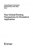
Fig. 1 a The number of published research papers per year containing the term „upconversion nanoparticles “, according to Google Scholar, and the corresponding fit to the logistic equation. b The extrapolation of the logistic S-curve to the upcoming years with an identified inflection point, growth rate, and saturation
5 papers containing the term “upconversion nanoparticles.” That trend slowly rose for another decade until the number of 127 published papers was reached in the year 2000. From that time, the interest in UCNP started to grow at an unprecedented rate: the almost tenfold increase from 951 published papers in the year 2010 to 8640 only a decade later. This research trend very precisely follows the logistic S-curve, with a very high fit quality, which is not as surprising as the function commonly used for modeling various growths [4]. The usage of the curve is justified by Tarde’s socioeconomics work on the spread of innovations, by differentiating between the three stages: (i) reception, (ii) rapid growth, (iii) saturation, and the potential appearance of opposing ideas [5]. This theory has been greatly expanded and mathematically formulated by Rogers in Ref [6]. Although it is a speculative analysis, the inflection point happened in the year 2018 (see Fig. 1b), which marks the potential decrease in the rate of popularity rise of research of UCNP. Thus, in the years 2022 and 2023, we are possibly experiencing the mature phase of the research. The saturation by the fit is predicted to happen with ca. 13,300 papers per year. The growth time is defined as the time needed to grow from 10 to 90% of the asymptotic value [4]. As 10% was reached in the year 2011 and the inflection point is estimated to had appeared in 2018, 90% of the asymptotic value is predicted to be in the year 2025. Thus, the rise in popularity of this revolutionary science and technology is very fast, with a growth rate of only 14 years. Overall, it can be safely said that the research interests in UCNP will be rising for some time, and that we can expect the increase in their applications in various scientific and industrial fields. The application and research of UCNP cannot be observed as the pure reporting of observations that the absorption of lower energy photons results in lower emission intensity of higher energy photons. The true progress in UCNP in this mature stage of research can come with substantial knowledge of underlying physical mechanisms of upconversion phenomena. The other class of luminescence, the downshifting emissions, were scientifically observed and explained much earlier than the competing UC. Sir G. G. Stokes first observed that fluorescence emission is typically at higher
Mechanisms of Luminescence in Upconversion Nanoparticles
3
wavelengths than incident radiation. As this observation happened back in 1852, the experimental apparatus was very simple [7]. The difference in energy between the incident and emitted radiation is from then on known as the Stokes shift. Anti-Stokes emissions are recognized as the ones with the higher energy of emission than excitation photons and they are always of lower intensity than the downshifting emissions and require much more sophisticated equipment for detection. Due to the energy conservation law, the anti-Stokes emission can only occur if the system absorbs another particle apart from the initial photon. Prior to the revolution in UC set by Auzel, [8] all anti-Stokes emission energies were equal to the excitation energy plus up to few kT (where k = 0.695 cm−1 K−1 is the Boltzmann constant and T is the temperature in K) [9]. In other words, the known anti-Stokes processes involved excitation from the ground level by photons, and further excitation to the higher excited levels by the thermal energy, ultimately producing the emission of higher energy than the excitation (and cooling the sample in the process). This original anti-Stokes mechanism was also exploited for the novel luminescent thermometry methods using thermalized levels of the ground multiplet, where the phonons populate the excited states of the ground multiplet of Eu3+ (7 FJ ), and then by absorbing photons they reach higher excited states 5 DJ . [10, 11]. Other known processes at that time were of low intensity, out of which the most prominent was the excited state absorption (ESA). Also known were the processes involving the virtual state, which is quantum dynamically possible, but of lower probability of occurring. In 1959 Nobel laureate Nicolaas Bloembergen proposed the detection of the NIR photons via the ESA mechanism. For ESA, the single excited ion has to “wait” with depopulation until it absorbs another photon, something which was difficult to achieve without a strong, monochromatic, collimated light source such are lasers. The first laser was fortunately created just a year later by using a Ruby crystal, setting the stage for many future discoveries in photonics. Soon, many new laser systems were invented, covering multiple wavelengths from UV to NIR region, directly or by using optical elements to separate higher order harmonics. Before 1966 the knowledge of anti-Stokes mechanisms was limited to the absorption of a photon and another photon or phonon before that single ion would emit. At that time the energy transfers’ (ET) role in upconversion was not recognized. The prior understanding of ET was limited to the depopulation of some excited level of a sensitizer in favor of exciting activator ion from its ground level. Basic mechanisms when the activator ion is in the ground state are presented in Fig. 2. The resonant transfers occur when the excitation energies of the sensitizer and activator are the same. Given that each energy level has its own width, or can be split into sublevels, the energy matching between different ions occurs regularly with certain ion combinations. If the activator’s (A) excitation spectrum overlaps with the sensitizer’s (S) emission spectrum, then the activator might catch the photon emitted by the sensitizer and experience excitation itself. This process is called radiative transfer (Fig. 2a), or trivial energy transfer. In a material transparent to the emitted radiation, the emission can be captured by an acceptor ion at any distance, but with a lower probability further from the sample. The sensitizer ion is then regarded as the omnidirectional source, thus the acquisition will depend on the area of the sphere where at the center
´ c A. Ciri´
4
Fig. 2 Energy transfer mechanisms between sensitizer (left) and activator (right) ions, when the activator is in the ground state: a sensitizer emits a photon and the activator absorbs it, b sensitizer transfers energy to the donor with resonant levels, c sensitizer transfers energy to the donor in a phonon-assisted process, d sensitizer and donor are the same ion, and the energy transfer process is called cross-relaxation
is the sensitizer and the activator ion is on its surface. The probability for a radiative ET between two ions at distance R is equal to: prad =
σA 4π R 2
AS
∫
g S (ν)g A (ν)dν
(1)
where σA is the absorption cross section of the activator ion, AS is the radiative transition probability of the sensitizer ion, equal to the inverse of the radiative lifetime, and the integral is the spectral overlap between the emission spectrum of the sensitizer and excitation spectrum of an activator ion, given in energy units. The area of the above mentioned sphere is given in the denominator. As the photons are emitted from the sensitizer regardless of the presence of the activator (activator ions do not affect, or trigger, the sensitizer ions), the ratio of emissions from the sensitizer and activator ions in the emission spectrum will depend on the concentrations, but also on the material shape. Thus, in particles of low dimensionality, such are nanoparticles or thin films, this mechanism has decreased probability. In large, bulk samples, especially single crystals, this long-range effect has a high probability of occurring. The prad in Eq. 1 will be more probable if the emission efficiency of S is large, the overlap integral is large (which can be viewed directly by comparing the normalized emission spectrum of S and the excitation spectrum of A), an absorption-cross section of A is large, and A is given in large concentrations to be able to capture the photons radiated by S. The lifetime of S will however not have any dependence on the concentration of A. If the energy is transferred without the intermediate emission of the photon, the process is non-radiative ET (Fig. 2b). The photon exchanged in the interaction is termed a virtual photon, because it is undetectable. Non-radiative ET can happen via two mechanisms. Forster ET is the mechanism with intermediate reach although it is frequently called the long-range ET. The excitation of S induces oscillating multipole moment, and that field can, via virtual photon—a Coulomb interaction, transfer the energy to the A. S returns to the ground state, losing its multipole moment, while A gets a multipole moment and is in the excited state until it radiates the excess energy hopefully in the form of photons. The energy transfer probability for multipolar interactions is given by:
Mechanisms of Luminescence in Upconversion Nanoparticles
pF R E T =
( ) 1 Rc Q/2
τS
R
,Q ∈
5
⎧ ⎨
3, dipole − dipole 4, dipole − quadrupole ⎩ 5, quadrupole − quadrupole
(2)
where Q determines the type of interaction, R is the distance between S and A, and Rc is the critical distance at which the rate of de-excitation of S is equal to the rate of FRET to A, i.e., the distance between A and S when pFRET = 50%. Note that the efficiency of the process drops with R−6 , much faster than with the radiative energy transfer. The most frequent type of interaction is dipole–dipole and is the one that Forster initially conceived. Dexter showed that other multipole interactions exist, but more importantly, showed a new type of interaction called exchange interaction. Exchange interaction requires an overlap of the wavefunctions between S and A, thus the effect can occur only at short distances, typically from 1 to 10 nm between S and A. It promulgates in the literature that the S in the excited state gives the electron to the A’s excited state, and in return, A’s ground electron transfer to the S ground state, and A can de-excite radiatively to its ground state. A more precise description is that the charge clouds overlap at these short distances. The probability or the rate of Dexter’s mechanism is given by: ) ( pdexter = K J exp − 2R L
(3)
where J is the overlap integral and L is an effective average Bohr radius of the excited and unexcited states of S and A. Because of this exponential dependence, the reach of Dexter’s mechanism has a significantly shorter reach than the Forster ET. Note that because there is a direct contact and exchange of electrons, the overlap integral J does not depend on the absorption characteristics of A. If R exceeds L then pdexter becomes very small, which is intuitive as there is almost no direct interaction, no significant overlap of the wave functions between S and A. Non-resonant non-radiative ET must be followed by a release or acquisition of at least one phonon to bridge the gap between the mismatch of energies between the excited levels of sensitizer and activator ions (Fig. 2c; only shown is the release of the phonons in the ET process). Without the release or acquisition of the phonons, this process would not have been possible, because the energy level broadening is not sufficient to account for the energy mismatch between the excited levels of S and A. In lanthanides the ET with energy gap mismatch can exceed the kTD , where TD is the Debye temperature, meaning that several phonons must be included in the process. Finally, the last process is the resonant non-radiative ET between the same ions (same ion type is both S and A), named cross-relaxation (Fig. 2d) [9]. As mentioned above, Auzel discovered the ET between two ions even when the activator is already at the excited state, and the process is termed energy transfer upconversion (ETU). These mechanisms are competitive, all with their probability/ frequency, and various combinations can occur that result in the upconversion (see Fig. 3). All the processes described so far include only the real states. Apart from
6
´ c A. Ciri´
Fig. 3 Absorption and energy transfer mechanisms that occur in the upconversion: a first GSA then ESA, b successive ET or ETU, c cooperative sensitization, d ET than ESA, e GSA than ET, f GSA and cross-relaxation, g cross-relaxation then ESA
these processes, several phenomena involving a virtual state exist: second harmonic generation when both the intermediate and final states are virtual, 2-photon absorption excitation when the intermediate state is virtual, and when two ET from sensitizers create a virtual state the process is called cooperative luminescence. All those virtual processes occur with a lower probability but can be visible especially if ETU and ESA processes are not possible in a given material. At this point, even intuitively it is reasonable to assume that the probability for the UC is proportional to the lifetime of the intermediate level, absorption cross section, and ET efficiency. This will become more evident from the particular examples that will be presented later on. The above given processes happen in inorganic materials, activators, and sensitizers being the transition metals (TM), lanthanides (Ln), or actinides, or differently termed, d-block and f-block elements. Examples of TM for which the UC has been observed are Os4+ , Re4+ , Mo3+ , Ni2+ , and Ti2+ . UC in Ln in most cases involves ETU from the Yb3+ to some other Ln3+ ion. As Yb3+ has a large absorption cross section and long lifetime of its only excited level, it is unsurprising that in several cases was demonstrated it can aid UC of TM or Ln2+ . Due to their radioactivity, the UC of actinides and Pm from the lanthanides will not be taken into consideration in this paper. UC can take place with both inorganic and organic materials. Organic UCNP achieves UC via a mechanism called triplet–triplet annihilation (TTA), a multi-step process that will be explained in more detail in a dedicated section. Ultimately, there is a new class of UCNP materials that deserves mentioning, although it is still early for their broader application by UC, quantum dots. This chapter will be dedicated to explaining mechanisms leading to the UC in these various types of nanoparticles. Each UCNP class will be presented in a separate section.
2 Transition Metal Upconversion Mechanisms TM ions have their partially filled d-orbital exposed to the significant influence of the crystal field environment. The consequence is that although there is an abundance of various TM elements, only a few of them are capable of luminescence. The UC of the TM single ion requires at least two metastable, real states to exist at positions that have an energy difference equal to the energy of the incoming photons. The transition metal ions, unlike lanthanides, have two advantages here: the large absorption cross
Mechanisms of Luminescence in Upconversion Nanoparticles
7
sections and tunability of the positions of energy levels due to the strong interaction with the crystal field. Within a single ion the UC has been observed with Ti2+ , Ni2+ , Mo3+ , Re4+ , and Os4+ ions with configurations 3d2 , 3d8 , 4d3 , 5d3 , and 5d4 , respectively. The UC mechanisms of those ions are summed up in Fig. 4. What all those ions share is the UC is observable at cryogenic temperatures and with difficulties in obtaining high efficiencies. In Ni2+ doped CsCdCl3 Wenger et al. explored the order of milliseconds long lifetime of 3 T2 level for the ESA to happen with enough probability for the UC to be observed.12 Upon the excitation to the 1 E state, the non-radiative relaxation rapidly depopulates to this long lived level. 3 T2 then experiences ESA, and since the energy
Fig. 4 Transition metal UC mechanisms. The straight upwards red arrow means absorption, the straight downwards blue arrow is emission, the wavy downwards pink arrow is nonradiative depopulation, and the green dashed arrows are energy transfer upconversion process
8
´ c A. Ciri´
difference between the 1 T2 and the 3 T2 levels is tuned to match the energy difference of the photon needed for GSA, the Ni2+ gets further excited to the 1 T2 at about 20 000 cm−1 , ultimately radiating green light. The excitation was performed in NIR with an efficient Ti3+ sapphire laser tuned to 808 nm, but today the 808 nm laser diodes are powerful and cheap and can be used instead to achieve this UC. Although Tanabe-Sugano diagrams of Re4+ are similar to that of Mo3+ , Gamelin, and Gudel demonstrated that Re4+ is capable of UC from the cryogenic temperatures all up to the room temperature [13]. This temperature stability is in part explained by the increase of the absorption intensity by 3 times from 10 K to the room temperature, compensating partially for the temperature quenching effects. After excitation into the long lived metastable 2 T1 level and rapid de-excitation to the 2 E level, this redemitting ion experiences UC thanks to the efficient ETU process. After the population of the 2 T2 level and depopulation to its energetically lowest sublevel, the emission of 725 nm photons occurs to the ground level 4 A2 . Additional benefits of Re4+ are that it possesses no channels via which the cross-relaxation could occur, thus the efficient ETU is possible. Os4+ has the mid-levels 1 E and 1 T2 right between the 1 A1 and the ground level 3 T1 , which are the perfect conditions for the UC by ET [14]. There is also a probability of GSA/ESA populating the 1 A1 level. Under the excitation of 11 226 cm−1 NIR photons, the UC was observed at 17 000 cm−1 = 588 nm, an orange light. Due to the radiative transitions from the metastable mid-levels being forbidden to the 3 T1 ground level, the lower of the two, 1 T2 , is enough long lived level (~30 μs) for the ETU or ESA to occur at low temperatures. Wegner and Gudel showed that the efficiency of UC and the mechanisms of Ti2+ doped crystals greatly depend on the host matrix, i.e., on the strength of the crystal field [15]. They doped Ti2+ in the weak CF of NaCl and the strong CF of MgCl2 . Upon excitation from the ground level to the 3 T2 , in the weak crystal field, the ESA must happen within 1.4 ms of the lifetime of the first excited level. In contrast, in the strong crystal field, the 1 T2 level is not immersed into the 3 T2 , and because the 1 T2 → 3 T1 is a forbidden transition, the radiative lifetime of the 1 T2 level is ~ 100 ms long. This has a profound effect on the UC in Ti2+ , greatly increasing its efficiency due to the increased probability of ESA. After ESA and nonradiative depopulation to the excited 3 T1 level, red emission occurs. Mo3+ also emits red light after UC. Its UC has been researched in Cs2 NaYCl6 and Cs2 NaYBr6 matrices [16]. The position of the 2 E level right between the 4 T2 and the ground 4 A2 levels makes it suitable for both GSA/ESA and ETU processes, which were indeed observed to happen simultaneously, each with its own probability. ESA was found to be the more efficient process. After the ion gets into the 2 E level, after UC to 4 T2 , it gets non-radiatively depopulated to the 2 T2 level from which Mo3+ ultimately emits. TMs have also been combined with other TM ions or Ln3+ ions to provide for new pathways of UC or increase efficiency, where either TM or Ln3+ take the role of sensitizer or activator. In the YAG matrix, Cr3+ experiences UC after ETU from Yb3+ . In Tm3+ /Ho3+ /Cr3+ co-doped materials the emissions of R1.2 levels of Cr3+ occur after UC. Mn2+ co-doping with Ni2+ is known to increase UC efficiency. Os4+ and Er3+
Mechanisms of Luminescence in Upconversion Nanoparticles
9
co-doping result in Os4+ being the sensitizer and Er3+ activator, after simultaneous ETA and ESA processes. Yb3+ can be used to sensitize the red emission of Mn2+ based on the GSA/ESA absorption resulting in UC to the 4 T2 level of Mn2+ [17].
3 Lanthanide Upconversion Mechanisms The most notable pairs of UCNP are Yb3+ /Er3+ , Yb3+ /Ho3+ , and Yb3+ /Tm3+ , although Yb3+ , due to its enormous absorption cross section of its only excited level 2 F5/2 , is also combined with other Ln or TM. 2 F5/2 level lies at approximately 10 000 cm−1 , which is perfectly matched with the emission of powerful and cheap 980 nm lasers [18]. After Yb3+ gets excited it has a relatively low probability for non-radiative de-excitation to the ground level 2 F7/2 due to the large energy gap and no levels in between and large distances from the excited level to other configurations that could lead to its depopulation. Thus, Yb3+ can either emit the photon from the lowest Stark sublevel of 2 F5/2 to one of the Stark sublevel multiplet of the 2 F7/2 ground level or if there is another ion in its vicinity with an energy level close to 10 000 cm−1 , it can transfer its energy. Thus, Yb3+ most often serves as an antenna (see Fig. 5). Er3+ is seldom capable of UC by GSA/ESA process by lending electron at the long lived 4 I11/2 level and then absorbing another photon up to the 4 F7/2 level. However, this process is inefficient in comparison to the Yb3+ /Er3+ co-doped materials, due to the almost 10 × larger absorption cross section of the 2 F5/2 level of Yb3+ over the 4 I11/2 level of Er3+ . After 4 I11/2 gets populated by the resonant energy transfer to 4 I11/2 or by directly absorbing the incident photon, Er3+ experiences UC by receiving another energy boost from the second Yb3+ ion, reaching the 4 F7/2 . Then, after multiphonon relaxation, the green emission is observed from the 2 H11/2 and 4 S3/2 levels, [19] and in some cases the red emission from the 4 F9/2 , populated by the multiphonon relaxation and the energy mechanism presented in Fig. 5 [20]. The emission from the 2 H11/2 is visible even at room temperature because of its large reduced matrix elements for the transition to the ground level, and because the energy gap of about 700 cm−1 can be successfully bridged by the thermal energy, according to the Boltzmann distribution.
Fig. 5 UC mechanisms of Er3+ , Ho3+ , and Tm3+ co-doped UCNP with sensitization from Yb3+ upon 980 nm irradiation
10
´ c A. Ciri´
Unlike Er3+ , Ho3+ and Tm3+ do not have energy levels that could be directly excited by 980 nm light. Thus, the only way for their upconversion is via ETU from the Yb3+ ion. As its energy levels closest to the 2 F5/2 are energetically lower, there are no resonant levels with Yb3+ ion, and the process has to be phonon assisted. The first step for Ho3+ is the population of the 5 I6 level, which then can depopulate to the 5 I7 or experience UC to 5 F4 from where it can radiate green emissions, NIR emission to the 5 I7 , or depopulate to 5 F5 . UC from the 5 I7 can also occur, populating the 5 F5 level, with corresponding red emission at 667 nm [21]. In Tm3+ the process is more complex as it can experience three and more ETU from Yb3+ (4th ETU that results in the UV-light emission is not depicted in Fig. 5). Note, however, that each upconversion step requires a certain probability and the probabilities for each step multiply, thus the UC efficiency to a certain level is inversely proportional to the number of ETU required [22]. Pr3+ ion has the capability of UC into the 3 PJ multiplet by initial GSA to the long lived 1 G4 level by the Nd:YAG’s emission at 1064 nm, and ESA by 836 nm. As the 1 G4 level of Pr3+ is at about the same energy as the 2 F5/2 excited level of Yb3+ , Yb3+ can absorb the 980 nm beam and perform ETU to 3 PJ levels via absorption of phonons, or even by absorption of 1064 nm photons and phonon assistance for populations of both 2 F5/2 and 3 PJ levels [23]. However, the most interesting property of Pr3+ ion UC is its rare capability to reach the levels capable of emitting UVC light, which is much needed for sterilization. In recent years this possibility has caught the immense interest of researchers, especially after the COVID-19 pandemic where UVC emitting UCNP could offer a part of the solution. After Ce3+ , whose 4f5d level emissions are used in the YAG matrix for a majority of white LED chips, Pr3+ has the lowest laying levels of that configuration. Unlike 4f levels, levels of the 4f5d configuration are strongly influenced by the CF. Thus, their position in Pr3+ greatly depends on the host matrix, from very low (4f5d capable of the creation of UVA/ UVB light), to high (e.g. in fluorides 4f5d levels are above the 1 S0 level, allowing for the quantum cutting but not UC). The desired position of the lowest of 4f5d levels is below the 1 S0 , at energies around double the energy of the 3 PJ levels or at the energy equal to the sum of energies of 3 P0 and 1 D2 levels. One of the most prominent hosts in which this is achievable is the Yttrium Orthosilicate, Y2 SiO5 , [24] in which the 4f5d emission of Pr3+ occurs from 260 to 360 nm, by two broad, overlapping emission peaks. The UC is then generated by GSA/ESA via the 3 PJ levels. Unfortunately, the lifetime of the 3 P0 level is only ~ 40 μs or less, [25] preventing the high quantum yields. If 4f5d levels are low enough, it is possible that after GSA Pr3+ de-excites from 3 PJ to 1 D2 level, where it can experience ETU from another Pr3+ ion in the 3 PJ state, ultimately reaching the 4f5d levels [26]. As the lifetime of the 1 D2 level is significantly longer than that of the 3 PJ multiplet and their overlapping 1 I6 level, there is a high probability that ETU will happen, and the resulting 4f5d population is the result of cooperation between the ETU and ESA mechanisms. Such Pr3+ doped hosts are also capable of sensitizing Gd3+ ions, which results in the sharp emission at 310 nm from the 6 P7/2 level [27]. Note that the 4f5d emission to the 4f levels is parity allowed and thus have a large probability of emission happening once the electrons are in 4f5d domain.
Mechanisms of Luminescence in Upconversion Nanoparticles
11
Nd3+ ion on itself is capable of ETU and ESA after irradiation by 808 nm initially exciting the 4 F5/2 level, with consequent emissions all up to the UV region from 2 P3/2 and even 4 D3/2 levels [28]. However, more recently, the Nd3+ has been used as a sensitizer ion, which by absorbing 808 nm can transfer its energy to the Yb3+ , which can then make ETU to any other of the combinations mentioned above [29]. Gd3+ , due to its 4f7 configuration, has the first excited level at about 32 000 cm−1 , by far the highest among all the Ln3+ . By excitation into the 6 P7/2 at about 310 nm it is possible to reach 6 G7/2 and 6 I7/2 levels with emissions at 205 nm and 242 nm, respectively [30]. By pumping into any of the 6 DJ , 6 IJ , or 6 PJ levels it is possible to reach the higher 4f7 levels and ultimately observe emission from the 6 GJ [31–33]. It is helpful that the lowest excited level of Gd3+ has a long lifetime of the millisecond order for reaching the higher excited levels, but unfortunately there are no many cheap and powerful excitation sources at these wavelengths. In Dy3+ the upconversion emission from the 4 I15/2 level is observed by pumping with 862 nm into 6 F7/2 with the phonon assistance, in simultaneous ETU and GSA/ ESA mechanisms [34]. Dy3+ is also capable of UC by ETU from the Yb3+ ion, firstly into 6 F9/2 /6 H7/2 levels and then into the 6 F3/2 , and finally to 4 F9/2 by the 3rd photon, creating emissions with the highest energy of 484 nm [35, 36] There are other lanthanide UC systems as well, for example, Yb3+ —Yb3+ , Yb3+ — 3+ Tb , Yb3+ —Eu3+ , or Eu2+ . However, due to their mechanisms being cooperative luminescence or sensitization, or two-photon absorption with low probability, the overall UC efficiency is too low (4—5 orders of magnitude less than ETU). Thus, due to the sake of brevity, there will be no further mention of their UC. Sm2+ due to the isoelectronic configuration to the Eu3+ and the energetically low 4f5d states looked as the promising material for UC, [37] however this has never been efficiently realized (Yb3+ —Sm2+ systems will not be discussed further due to the low UC efficiency). As it is already mentioned, the most crucial parameter in determining the efficiency of the UC materials is the lifetime of the metastable–intermediate state in the UC process. As a general rule, long lifetimes are achieved when the energy separation with the next energetically lower level is large so that the non-radiative relaxation is happening with low, even negligible probability. In other words, multiphonon relaxation should be improbable due to the large number of phonons that need to bridge the gap between those levels [38]. In Ln3+ the positions of the shielded 4f levels can be considered approximately host independent and constant. The correct strategy for improving the UC photoluminescence intensity then lies in choosing such isolated metastable level, but also a host with as low phonon energy as possible. For example, the energy gap between the 4 I11/2 and 4 I13/2 levels of Er3+ is equal to about 3600 cm−1 . In phosphate glass with 1200 cm−1 highest phonon energy, 3 phonons are sufficient to bridge this gap, while in LaCl3 with 240 cm−1 release of 15 simultaneous phonons is needed. Generally, the involvement of 5 or more phonons can be considered improbable. Depopulation can occur via other pathways than multiphonon relaxation, for example via crossover with some other configuration. In Ln these can be charge transfer states or 4f5d levels, whose positions depend on the selected ion and host. Charge-transfer configurations are the lowest for the Eu3+ , then Yb3+ and Dy3+ ions. An opposite trend is observed for the 4f5d energies: the
12
´ c A. Ciri´
lowest are for Ce3+ and Pr3+ all up to Gd3+ , and then again low for Tb3+ and rising up to the Yb3+ . NaYF4 is widely recognized as the most efficient host matrix for Yb3+ co-doped UC phosphors, in bulk and especially for UCNP [39]. Its success lies behind the low phonon energy, symmetry, and interaction of dopants at two different lattice sites, [40] but all the effects are not yet fully explained due to the incompleteness of the various theories describing the crystal field. Although NaYF4 has larger phonon energy than LaF3 and LaCl3 , UC efficiency by Yb3+ as a sensitizer and other Ln3+ as an activator is significantly higher in NaYF4 and its similar matrices. Another important role in UC efficiency is the site symmetry of the activator ion. In the case of the Ln3+ , this is directly explainable by the Judd–Ofelt theory [41, 42]. The hexagonal symmetry of the NaYF4 in the β phase suits the UC mechanism, much more than the cubic α phase. By doping the particular host, the host matrix gets distorted due to the mismatch of the ionic radii and/or charge. Although an equal charge with the replaced ion is desired, the distortions caused by the different ionic radii can be favorable for the UC. In NaYF4 this happens by the substitution of the Y3+ ions with Ln3+ , where the small difference in ionic radii causes local symmetry reduction. Further improvements are possible by introducing additional distortions by adding other Ln3+ or alkaline metal impurities, for example Gd3+ and Li+ , [43] respectively. In this manner the decreasing of the local site symmetry is promoted, increasing the Ω2 Judd–Ofelt parameter [44]. In Er3+ the consequence is the increased emission from the 2 H11/2 level, much needed for UCNP luminescence temperature sensing. Apart from the fluoride matrices, it was demonstrated that various oxide hosts are suitable hosts for UC. They are also transparent for the excitation and emission of co-dopants but have much higher chemical stability. As a compromise, oxysulfide UCNP, for example, La2 O2 S, has an efficiency comparable to that of lanthanides, i.e., up to several times higher than oxides, with stability inherited from the latter [45–47] When it comes to the nano-size, many quantum effects start to play an important role, and the shape of the nanoparticle and its size become important parameters. The surface area to volume ratio of UCNP is large, and surface defects, impurities, or direct contact with the environment heavily influence the effectiveness of the UC. Well known surface ligands that have vibrational modes of high energy are NH2 or OH groups, and they act as strong quenchers [40] As there are many quenching centers residing on the surface of the UCNP, the UC efficiency is necessarily lower than in bulk. Thus, it is especially important for the UCNP that the matrix has low phonon energy, as we saw with NaYF4 , but that is not the only problem with reduced particle size. Fortunately, some recent advances in the synthesis of UCNP mitigate these problems to a certain degree. One strategy is by decreasing the effective surface by controlling the shape of the nanoparticles. The other strategy lies in eliminating the surface defects by the core–shell structures, where the core is the emitting material and the shell consists of the barrier material transparent to the excitation and emission of light. Thus, the core–shell structure is based on coating the active material in order to keep the energy transfers within the core. The coating can be with the same host but this time undoped, or it can contain other dopants to allow for the tunability of
Mechanisms of Luminescence in Upconversion Nanoparticles
13
the emission, and the ET can occur on the interface. Other hosts can be used for coatings as well, the most prominent one being the SiO2 surface [48]. The most novel trend in UCNP is increasing emission intensity by heavy doping. Usually, the Yb3+ is doped from 10 to 20%, and its co-dopants up to a few percent in order to avoid cross-relaxations and concentration quenching. Efficient doping beyond these concentrations causing increased emissions was demonstrated by using several strategies. The first one is limiting the quenchers by using the core–shell nanostructures, as already explained above. The very direct strategy is by using a high-power source of collimated or laser excitation in order to simultaneously excite as many doped ions as possible, reducing the availability of the number of the ground state ions for the cross-relaxation processes. Choosing the host in which the dopants would lie with enough separation would decrease the cross-relaxation effects. The effective distance between the same ion species can be achieved by creating an almost homogeneous distribution of dopants by the layer by layer hot injection strategy [49]. Dexters and Forster energy transfer mechanisms fall at the rate of e−2R/L or R −6 , respectively, as seen above. Thus, the closeness between the sensitizer and dopant ions is highly desirable. In a special class of materials which have a negative thermal expansion (they contract with increasing temperature), the temperature causes decrease of sensitizer—activator distances, greatly increasing UC at elevated temperatures, although the room temperature UC is of week intensity [50]. Another strategy to use is by coating UCNP with dyes that act as an additional sensitizer [51]. Some novel research, for example, enhancing UC by 2D photonic crystal structures will not be presented here for the sake of brevity. It can be only mentioned that the UC efficiency is greatly increased in such structures by accumulation or convergence of the excitation light but at the cost of introducing other limitations [52].
4 Upconversion Mechanisms in Organic Nanoparticles Organic molecules can have high absorption cross sections, greater than the Ln UCNP, and can serve as a very effective sensitizer. In a triplet–triplet annihilation (TTA) UCNP organic sensitizer absorbs the initial ion into the excited singlet state. After intersystem crossing, the energy is shifted to a triplet state of lower energy. The sensitizer then performs an energy transfer to the activator ion in the triplet state. When two activators are into this triplet excited state, then they together possess enough energy for UC (via Dexter’s mechanism for ET) to reach the activator singlet state of higher energy, which ultimately radiates photons, i.e., one activator ion gets into the singlet state while the other is demoted to the ground state (see Fig. 6) [53]. It is evident that the efficient intersystem crossing and long triplet excited lifetimes are the requirements for an efficient UC by TTA—UCNP apart from the high absorption cross section by the sensitizer ions. The triplet energy of the sensitizer should be resonant with the triplet energy of the annihilator ions, and the singlet energy of the activator should be equal to twice its triplet energy.
14
´ c A. Ciri´
Fig. 6 Organic UCNP TTA mechanism
The result is more than the excellent quantum yield of 30%, very close to the thermodynamic limit for UC of ½, however, as the TTA mechanism is a bimolecular diffusion limited process, it cannot be highly effective in solids [54]. Additionally, the organic molecules suffer from chemical or thermal stability problems, limited availability of NIR fluorophores, or photobleaching, [55] which is the main reason why the main focus of the research still remains within the inorganic UCNP.
5 Upconversion of Semiconductor Quantum Dots (UCQD) QDs are a novel, promising material with potential applications in all luminescent applications, although there are health concerns with their usage for bio-medicinal purposes due to their cytotoxicity. QDs are particles of approximately 0D (0 dimensional; being a few nm in diameter), causing their optoelectronic properties to depend on their size and shape. The most common materials for QD are the semiconductors, typically PbS, PbSe, CdSe, or CdTe. The emission from QDs is preceded by the light absorption breaching the band gap, thus the band gap size directly depends on the size of the QD. Although they are mostly researched for their downshifting PL, recently it was demonstrated that they successfully UC, with the following examples. CdSe UCQDs emit at about 570 nm upon the 680 nm irradiation via the phonon assisted anti-Stokes PL [55, 56]. UC is achieved by having the interacting QDs of different sizes. In graphene quantum dots (GQD) the excitation can be performed anywhere between 600 and 1000 nm, peaking at 800 nm, and the resultant emission occurs in a broad band with a maximum of about 460 nm. The mechanism starts from the multiphonon population of the π orbital and absorption of light all to the lowest unoccupied molecular orbital. The UC in GQD is then observed as the emission to the σ orbital of the highest occupied molecular orbital [57]. Apart from the QDs experiencing UC, they have been investigated as sensitizers for luminescent organic molecules creating another UC system class of matertials [58, 59]
Mechanisms of Luminescence in Upconversion Nanoparticles
15
6 Conclusion The main mechanisms behind the anti-Stokes processes called upconversion are the energy transfer upconversion, ground state and excited state absorption, crossrelaxation, and triplet–triplet annihilation. The upconversion nanoparticles can be classified by the dominance of one of these upconversion mechanisms, or by sensitizer and activator to purely transition metal or lanthanide, organic, or quantum dots. Transition metal ion upconversion was observed by single-doping with one of the Ti2+ , Ni2+ , Mo3+ , Re4+ , and Os4+ , but mostly at cryogenic temperatures. Depending on the ion, the dominant or the only available mechanism is ETU or ESA. They have been combined with lanthanide ions for obtaining the new pathways for UC. Lanthanide ion upconverters are the most investigated and still the most popular UCNP. Although some ions are capable of ESA and other less probable mechanisms, their high popularity is owing to the co-doping with Yb3+ which has a great absorption cross section and large ETU capabilities. Yb3+ is combined with many of the lanthanide ions and in many hosts. The most efficient host up to date is the NaYF4 in the β phase, but that does mean that the improvements stopped there. Multiple strategies employed over the years and collaboration between physics and chemistry resulted in multiple increase in upconversion efficiency, one example being the core–shell structures. Organic UCNP, with their high absorption cross section and very high efficiencies for UC by the triplet–triplet annihilation mechanism is a promising material but limited by the various peculiarities of the organic molecules. Quantum dots that demonstrate upconversion are either graphene/carbon, or made from the semiconductors, such as PbS, PbSe, CdSe, or CdTe. Their appeal lies in high tunability by adjusting the size and shape of the synthesized particles. QD can be used in combination with the organic molecules for achieving UC, where the former act as a sensitizer. The research in UCNP shows no sign of stopping, although it has reached the mature stage. Novel strategies and combinations of various ions, types, or hosts never cease to surprise, surpassing the set boundaries by the sole material. A full understanding of the underlying mechanisms yields new strategies for increased UC efficiency, and an understanding of the limitations will eventually help alleviate them. With each new knowledge, there is a step forward to being closer to the thermodynamic limit of their efficiency, broader application, or industrial readiness.
References 1. F. Wang, D. Banerjee, Y. Liu, X. Chen, X. Liu, Upconversion nanoparticles in biological labeling, imaging, and therapy. Analyst 135, 1839 (2010) 2. A.M. Kaczmarek et al., Visible and NIR Upconverting Er3+ –Yb3+ Luminescent Nanorattles and Other Hybrid PMO-Inorganic Structures for In Vivo Nanothermometry. Adv. Funct. Mater. 30, 2003101 (2020) 3. G. Chen, H. Qiu, P.N. Prasad, X. Chen, Upconversion nanoparticles: design, nanochemistry, and applications in theranostics. Chem. Rev. 114, 5161–5214 (2014)
16
´ c A. Ciri´
4. D. Kucharavy, R. De Guio, Application of Logistic Growth Curve. Procedia Eng. 131, 280–290 (2015) 5. G. De Tarde, The laws of imitation. (H. Holt, 1903) 6. E.M. Rogers, Diffusion of innovations. (Simon and Schuster, 2010) 7. J.R Lakowicz, Principles of fluorescence spectroscopy. (Springer, 2006) 8. F. Auzel, Compteur quantique par transfert d’energie entre deux ions de terres rares dans un tungstate mixte et dans un verre. CR Acad. Sci. Paris 262, 1016–1019 (1966) 9. F. Auzel, Upconversion and Anti-Stokes Processes with f and d Ions in Solids. Chem. Rev. 104, 139–174 (2004) ´ c, I Zekovi´c, M Medi´c, Ž Anti´c, M.D. Drami´canin, Judd-Ofelt modelling of the dual10. A. Ciri´ excited single band ratiometric luminescence thermometry. J. Lumin. 225, (2020) ´ c, Ł Marciniak, M.D. Drami´canin, Luminescence intensity ratio squared—A new 11. A. Ciri´ luminescence thermometry method for enhanced sensitivity. J. Appl. Phys. 131, 114501 (2022) 12. O.S. Wenger, R. Valiente, H.U. Güdel, Luminescence Upconversion Under High Pressure in Ni 2+ Doped CsCdCl 3. High Press. Res. 22, 57–62 (2002) 13. D.R. Gamelin, H.U. Güdel, Spectroscopy and Dynamics of Re 4+ Near-IR-to-Visible Luminescence Upconversion. Inorg. Chem. 38, 5154–5164 (1999) 14. M. Wermuth, H.U. Güdel, Upconversion luminescence in a 5d transition-metal ion system: Cs2ZrCl6: Os4+. Chem. Phys. Lett. 281, 81–85 (1997) 15. O.S. Wenger, H.U. Güdel, Chemical tuning of the photon upconversion properties in Ti 2+ -Doped chloride host lattices. Inorg. Chem. 40, 5747–5753 (2001) 16. D.R. Gamelin, H.U. Güdel, Excited-State dynamics and sequential two-photon upconversion excitation of Mo 3+ -Doped chloro- and Bromo-elpasolites. J. Phys. Chem. B 104, 10222–10234 (2000) 17. P. Gerner, C. Reinhard, H.U. Güdel, Cooperative Near-IR to visible photon upconversion in Yb3+-Doped MnCl2 and MnBr 2: comparison with a series of Yb3+-Doped Mn2+ Halides. Chem.—A Eur. J. 10, 4735–4741 (2004) ´ c, S. Stojadinovi´c, Upconversion photoluminescence properties of ZrO2 :Ln3+ /Yb3+ (Ln 18. A. Ciri´ = Er, Ho, Tm) films formed by plasma electrolytic oxidation. in Upconversion Nanophosphors 103–118 (Elsevier, 2022). https://doi.org/10.1016/B978-0-12-822842-5.00001-7 ´ c, T. Gavrilovi´c, M.D. Drami´canin, Luminescence intensity ratio thermometry with 19. A. Ciri´ Er3+ : Performance overview. Crystals 11, 189 (2021) ´ c et al., Comparison of three ratiometric temperature readings from the Er3+ upconversion 20. A. Ciri´ emission. Nanomaterials 10, 1–10 (2020) ´ c, S. Stojadinovi´c, Structural and photoluminescence properties of Y2O3 and Y2 O3 :Ln3+ 21. A. Ciri´ (Ln = Eu, Er, Ho) films synthesized by plasma electrolytic oxidation of yttrium substrate. J. Lumin. 217, (2020) ´ c, S. Stojadinovi´c, Photoluminescence studies of ZrO2 :Tm3+ /Yb3+ coatings formed by 22. A. Ciri´ plasma electrolytic oxidation. J. Lumin. 214, 116568 (2019) 23. P.V. dos Santos et al., IR-visible upconversion and thermal effects in Pr 3+ /Yb 3+ -codoped Ga 2 O 3: La 2 S 3 chalcogenide glasses. J. Phys. Condens. Matter 12, 10003–10010 (2000) 24. C. Hu et al., Visible-to-ultraviolet upconversion in Pr3+:Y2SiO5 crystals. Chem. Phys. 325, 563–566 (2006) ´ c, Z. Risti´c, T. Barudzija, A. Srivastava, M.D. Drami´canin, Judd-Ofelt parametriza25. A. Ciri´ tion from the emission spectrum of Pr3+ doped materials: Theory, application software, and demonstration on Pr3+ doped YF3 and LaF3 . Adv. Theory Simulations 4, 2100082 (2021) 26. E.L. Cates, A.P. Wilkinson, J.-H. Kim, Visible-to-UVC upconversion efficiency and mechanisms of Lu7O6F9:Pr3+ and Y2SiO5:Pr3+ ceramics. J. Lumin. 160, 202–209 (2015) 27. E.L. Cates, M. Cho, J.-H. Kim, Converting Visible Light into UVC: Microbial Inactivation by Pr 3+ -Activated Upconversion Materials. Environ. Sci. Technol. 45, 3680–3686 (2011) 28. A.L. Mullins et al., Dual-emission luminescence thermometry using LaGaO3 :Cr3+ , Nd3+ phosphors. J. Mater. Chem. C 10, 10396–10403 (2022) 29. J. Qiu, M. Shojiya, Y. Kawamoto, Sensitized Ho3+ up-conversion luminescence in Nd3+– Yb3+–Ho3+ co-doped ZrF4-based glass. J. Appl. Phys. 86, 909–913 (1999)
Mechanisms of Luminescence in Upconversion Nanoparticles
17
30. R. Mahiou, J. Metin, J.C. Cousseins, Anti-Stokes fluorescence of Gd3+ in K2GdF5. J. Lumin. 45, 363–365 (1990) ´ c, S. Stojadinovi´c, Photoluminescence of Gd2 O3 and Gd2 O3 :Ln3+ (Ln = Eu, Er, Ho) 31. A. Ciri´ formed by plasma electrolytic oxidation of pure gadolinium substrate. Opt. Mater. (Amst). 109546 (2019) https://doi.org/10.1016/j.optmat.2019.109546 ´ c, S. Stojadinovi´c, M.D. Drami´canin, Luminescence temperature sensing using thin32. A. Ciri´ films of undoped Gd2O3 and doped with Ho3+ , Eu3+ and Er3+ prepared by plasma electrolytic oxidation. Ceram. Int. 46, 23223–23231 (2020) 33. R.T. Wegh, H. Donker, A. Meijerink, R.J. Lamminmäki, J. Hölsä, Vacuum-ultraviolet spectroscopy and quantum cutting for Gd3+ in LiYF4. Phys. Rev. B 56, 13841–13848 (1997) 34. V.K. Rai, S.B. Rai, D.K. Rai, Optical studies of Dy3+ doped tellurite glass: Observation of yellow-green upconversion. Opt. Commun. 257, 112–119 (2006) 35. Y. Yang et al., Up-conversion luminescence and near-infrared quantum cutting in Dy3+, Yb3+ co-doped BaGd2ZnO5 nanocrystal. J. Lumin. 146, 284–287 (2014) ´ c, J. Periša, I. Zekovi´c, Ž Anti´c, M.D. Drami´canin, Multilevel-cascade intensity ratio 36. A. Ciri´ temperature read-out of Dy3+ luminescence thermometers. J. Lumin. 245, 118795 (2022) ´ c, et al. Supersensitive Sm2+ -Activated Al2 O3 thermometric coatings for high-resolution 37. A. Ciri´ multiple temperature read-outs from luminescence. Adv. Mater. Technol. 2001201 (2021). https://doi.org/10.1002/admt.202001201 ´ c, M.D. Drami´canin, LumTHools—Software for fitting the temperature dependence 38. A. Ciri´ of luminescence emission intensity, lifetime, bandshift, and bandwidth and luminescence thermometry and review of the theoretical models. J. Lumin. 252, 119413 (2022) 39. N. Menyuk, K. Dwight, J.W. Pierce, NaYF 4: Yb, Er—an efficient upconversion phosphor. Appl. Phys. Lett. 21, 159–161 (1972) 40. M. Haase, H. Schäfer, Upconverting Nanoparticles. Angew. Chemie Int. Ed. 50, 5808–5829 (2011) ´ c, S. Stojadinovi´c, M. Sekuli´c, M.D. Drami´canin, JOES: An application software for 41. A. Ciri´ Judd-Ofelt analysis from Eu3+ emission spectra. J. Lumin. 205, 351–356 (2019) ´ c, S. Stojadinovi´c, M.G. Brik, M.D. Drami´canin, Judd-Ofelt parametrization from 42. A. Ciri´ emission spectra: The case study of the Eu3+ 5 D1 emitting level. Chem. Phys. 528, 110513 (2020) 43. G. Liu, Advances in the theoretical understanding of photon upconversion in rare-earth activated nanophosphors. Chem. Soc. Rev. 44, 1635–1652 (2015) ´ c, S. Stojadinovi´c, M.D. Drami´canin, An extension of the Judd-Ofelt theory to the field 44. A. Ciri´ of lanthanide thermometry. J. Lumin. 216, (2019) ´ c, M. Sekuli´c, K. Shah, B.S. Chakrabarty, M.D Drami´canin, Upconversion photolumi45. A. Ciri´ nescence of sub-micron lanthanum oxysulfide particles co-doped with Yb3+ /Ho3+ and Yb3+ / Tm3+ synthesized by optimized combustion technique. Opt. Mater. (Amst). 120, 111417 (2021) ´ c, K. Shah, M. Sekuli´c, B.S. Chakrabarty, M.D. Drami´canin, La2 O2 S:Er3+ /Yb3+ 46. A. Ciri´ nanoparticles synthesized by the optimized furnace combustion technique and their highresolution temperature sensing. Optik (Stuttg). 245, 167690 (2021) ´ c, K.V.R. Murthy, B.S. Chakrabarty, Investigation of a new way of synthesis 47. K. Shah, A. Ciri´ for Nano crystallites of La2 O2 S & 1%Ln3+ (Ln = Pr, Eu, Tb, Dy, Er) doped La2 O2 S and study their structural and optical properties. J. Alloys Compd. 851, 156725 (2021) 48. R. Abdul Jalil, Y. Zhang, Biocompatibility of silica coated NaYF4 upconversion fluorescent nanocrystals. Biomaterials 29, 4122–4128 (2008) 49. S. Wen et al., Advances in highly doped upconversion nanoparticles. Nat. Commun. 9, 2415 (2018) 50. J. Zhou et al., Activation of the surface dark-layer to enhance upconversion in a thermal field. Nat. Photonics 12, 154–158 (2018) 51. B. Chen, F. Wang, Emerging frontiers of upconversion nanoparticles. Trends Chem. 2, 427–439 (2020) 52. C. Mao et al., Enhanced upconversion luminescence by two-dimensional photonic crystal structure. ACS Photonics 6, 1882–1888 (2019)
18
´ c A. Ciri´
53. L. Huang et al., Designing next generation of photon upconversion: Recent advances in organic triplet-triplet annihilation upconversion nanoparticles. Biomaterials 201, 77–86 (2019) 54. S. Mattiello et al., Diffusion-free intramolecular triplet-triplet annihilation in engineered conjugated chromophores for sensitized photon upconversion. ACS Energy Lett. 7, 2435–2442 (2022) 55. Z. Deutsch, L. Neeman, D. Oron, Luminescence upconversion in colloidal double quantum dots. Nat. Nanotechnol. 8, 649–653 (2013) 56. J.R. Santos, M.I. Vasilevskiy, S.A. Filonovich, Cascade upconversion of photoluminescence in quantum dot ensembles. Phys. Rev. B 78, 245422 (2008) 57. J. Shen, Y. Zhu, C. Chen, X. Yang, C. Li, Facile preparation and upconversion luminescence of graphene quantum dots. Chem. Commun. 47, 2580–2582 (2011) 58. R. Lai, Y. Sang, Y. Zhao, K. Wu, Triplet sensitization and photon upconversion using InP-Based quantum dots. J. Am. Chem. Soc. 142, 19825–19829 (2020) 59. K. Okumura, K. Mase, N. Yanai, N. Kimizuka, Employing core-shell quantum dots as triplet sensitizers for photon upconversion. Chem.—A Eur. J. 22, 7721–7726 (2016)
Engineering of Upconversion Nanoparticles for Better Efficiency Juan Ferrera-González, Laura Francés-Soriano, María González-Béjar, and Julia Pérez-Prieto
Abstract Upconversion nanoparticles (UCNPs) are considered a new type of luminescent materials because they can absorb in the NIR region and emit in the UV-visNIR region. In this chapter, the modifications and strategies reported in the literature to improve the efficiency of UCNPs and upconversion nanohybrids (UCNHs) together with their use to design Resonance Energy Transfer (RET) platforms are discussed. This chapter starts with a comprehensive brief introduction about efficiency and which factors influence the efficiency of UCNPs. Then, a few examples on how UCNPs can be used to improve the RET efficiency within UCNHs will be revised. Here, the reader will find an overview of basic strategies that can be used to engineer UCNPs/UCNH to better understand which factors are crucial when designing UCNPs/UCNHs for better efficiency. Keywords Quantum yield · Upconversion nanoparticles · Upconversion nanohybrids · Efficiency · Resonance energy transfer
Juan Ferrera-González and Laura Francés-Soriano are equally contributed. J. Ferrera-González · L. Francés-Soriano · M. González-Béjar (B) · J. Pérez-Prieto (B) Departamento de Química Orgánica, Instituto de Ciencia Molecular (ICMol), Universidad de Valencia, C/Catedrático José Beltrán 2, 46980 Paterna, Valencia, Spain e-mail: [email protected] J. Pérez-Prieto e-mail: [email protected] © The Author(s), under exclusive license to Springer Nature Singapore Pte Ltd. 2023 V. Kumar et al. (eds.), Upconversion Nanoparticles (UCNPs) for Functional Applications, Progress in Optical Science and Photonics 24, https://doi.org/10.1007/978-981-99-3913-8_2
19
20
J. Ferrera-González et al.
1 Introduction Efficiency is the ability of a system to produce an effect from some resources.1 It can be defined as the ratio between the output versus input response to a system. Thus, it is a term which depends directly on the aim of the system and, consequently, the final objective must always be clarified when talking about efficiency. In the field of chemistry, and specially photochemistry, the term is associated to a specific energetic ratio, creating new terms for other non-energetic definitions of efficiency. According to the IUPAC definition, efficiency (Eq. 1) is “the ratio between the useful energy delivered or bound and the energy supplied” [1]: η=
E output E input
(1)
This definition of efficiency is commonly applied to devices such as solar cells, in which case the efficiency is the ratio between the electric power output versus the power converted into electrical power. However, when referring to the efficiency of luminescent materials, such as UCNPs/UCNHs, the key parameter is the brightness (B). Brightness (Eq. 2) is defined as the product of the luminescence quantum yield (Φ L ) of a chemical species and its molar absorption coefficient (ε) [2]. B(M −1 cm−1 ) = Φ L · ε
(2)
The Φ L accounts for how efficiently the excited species deactivates via a radiative pathway, while ε represents how strongly a chemical species absorbs light at a certain wavelength (λ). Both parameters should be kept as high as possible to maximize the luminescence properties of the material. Note that if ε is very high but the Φ L is low, the material is unlikely to be very luminescent and vice versa. This definition, with some modifications, applies to the UCNP/UCNH materials (Eq. 3), substituting the Φ L for the absolute upconversion quantum yield (UCQY), and ε is the molar absorption coefficient of the material [3]: B(M −1 cm−1 ) = UCQY(P) · εUCNP (λ)
(3)
Although for simplicity’s sake, the absorption efficiency (ξUCNP ) can be used instead of εUCNP , which is the ratio between the photons absorbed and the incident photons, in which case parameter B is dimensionless (Eq. 4). B = UCQY · ξUCNP
1
(4)
Note that UC materials that absorb in the short wavelength IR (SWIR) region of the electromagnetic spectrum are out of the focus of this review.
Engineering of Upconversion Nanoparticles for Better Efficiency
21
The UCQY and ξUCNP together with the nanometric size are limiting factors for UCNPs brightness.
1.1 Limitations in the Efficiency of UCNPs 1.1.1
Absorption
Usually, lanthanide ions (Ln3+ ) are the chemical species responsible for light absorption in UCNPs, which in turn have some advantages, such as not undergoing photobleaching due to their inherent inorganic nature. However, the very low absorption coefficient of Ln3+ is also a considerable limitation in the UC process. The 4f-4f intraconfigurational transitions of Ln3+ , which are responsible for light absorption and UC emission of UCNPs, are not allowed by parity transitions (Laporte’s parity selection rule) and only become partially allowed under the influence of non-centrosymmetric interactions inside a crystal field [4]. Nonetheless, these induced electric dipole transitions have low intensity and afford ε between 0.1 and 10 M−1 cm−1 , but very often lower than 1 M−1 cm−1 [4]. For instance, Yb3+ , which is one of the most common sensitizers applied to UCNPs, has an ε at 980 nm of only ca. 10 M−1 cm−1 [5].
1.1.2
UCQY
The efficiency of the UC emission, analogous to conventional fluorophores or downshifting phenomena, is usually expressed in absolute UCQY [6, 7]. However, UCQY is limited as it is the result of a multiphoton process in which two or more photons are transformed to give a single more energetic photon. For instance, an UC emission which emits at a certain wavelength λUC and involves the sequential absorption of two photons (with a wavelength of 2xλUC ) is limited to a 0.5 UCQY. In a similar way, an UC process which involves three or four photons is limited to 0.33 and 0.25, respectively, assuming that there are no energy losses in the form of non-radiative deactivation or back energy transfer processes. Moreover, since UC is a non-linear process, generated by laser irradiation, the UCQY drastically depends on the excitation power density and eventually becomes saturated due to its proportional relationship with the nth power of the excitation power density [8]. Measuring the UCQY is a complex task so, determining it in a relative way is not possible [9, 10]. Reliable UCQY data require spectra correction, beam profile and beam power density characterization, and determination of the focal point, since UCQY is influenced by many parameters, such as the excitation beam profile, excitation beam scattering, the primary and secondary inner-filter effects, absorbance and particle concentration, reference material, dispersion solvent and thermal effects by the excitation beam, as well as setup parameters, such as an integrating sphere size, sample position, cuvette type and sample dimensions [8, 11, 12]. Given the complexity of this measurement, it at least requires, (i) an accurate
22
J. Ferrera-González et al.
determination of the excitation power in the sample (thereby characterizing the laser profile exhaustively) [7] and (ii) knowing the dependence of the UCQY on the excitation power or expressing the result for the UCQY at the power measured together with the UCQY and power density value at which UCQY becomes saturated [10].
1.1.3
The Nanometric Scale
The nanometric scale is also a disadvantage for the UCNP luminescence efficiency. Unlike other nanometric systems in which quantum confinement improves the emissive properties of the material (e.g., QDs), in UCNPs the UC emission efficiency is one order lower than that of the bulk material and even decreases if the UCNPs are dispersed in a medium, since their optical properties are determined by surface phenomena [10, 13]. In fact, the UCQY maxima for colloidal UCNPs that emit in the visible (λexc = 980 nm) reported to date are 5 and 7.6% for LiLuF4 :Yb3+ (20%), Er3+ (1%)@LiLuF4 and LiLuF4 :Yb3+ (20%), Tm3+ (1%)@LiLuF4 (UCNP 50 nm; 127 W/cm2 ) [14] and 8.4% for NaYF4 :Yb3+ (20%), Tm3+ (1.2%) (35 nm UCNP, 140 W/ cm2 ) [15], but are typically less than 1% for core UCNPs [10], especially for sizes lower than 10 nm [16]. UCNPs have a much higher surface/volume ratio than that in the macroscopic material and, therefore, there is a significant number of Ln3+ ions on the surface exposed therefore, they can be deactivated non-radiatively, either by the medium, surface defects or ligands [13]. Additionally, energy migration processes between Ln3+ ions within the crystal lattice that are short-circuited on the surface of the material, cannot be neglected [17]. In fact, Gargas et al. [18] suggested that for UCNPs 40 kW/cm2 ), while for the low excitation regime (power density below 2 kW/cm2 ) the energy transfer enhancement also contributed.
2.6.3
Sensitization to Other Active Species
The multiple emissive bands can be used to excite species capable of absorbing these photons, such as conventional fluorophores and photoactive nanoparticles, thus opening an enormous range of possibilities and applications. For example, Zheng et al. constructed a UCNH, composed of NaYF4 :Yb3+ , Er3+ and two organic probes, that absorbed two different Er3+ bands and that allowed in vivo synchronous detection of glutathione and H2 O2 [58]. Chromophores are known to modulate the emissive properties of the UCNP, and the exact influence in the UCQY has not been clearly determined for all the systems. Nevertheless, lately, it has also been demonstrated that certain configurations can also improve the UCQY of the UCNH. In this way, Wisser et al. demonstrated that dye-decoration of UCNP improves the absolute UCQY of the material [59]. Specifically, they build an UCNH of a dye, ATTO 542, anchored to the surface of different-sized UCNP, NaY0.8-x,y Gdx Luy F4 :Yb3+ (18%), Er3+ (2%), and measured a 10 times higher UCQY for UCNH with the smallest UCNP (11 nm) in comparison to the bare UCNP. Moreover, they observed the reduction in the 540 nm Er3+ upconversion (λexc = 980 nm) and direct excitation (λexc = 520 nm) radiative rate and, therefore, determined that the enhancement of the UCQY was due to an efficient resonance energy transfer process and the much faster radiative rate of the dye, which suppressed the surface quenching pathway of the UCNP. However, as pointed-out by Jones et al. [8], plasmonic, photonic structures or organic sensitizers do not always enhance the UCQY. Their exact influence is still under debate. The reported values suffer from lack of characterization of the associated power density and on how the beam is focused. In addition, most papers report just a single UCQY value or determine UCQY by rate equations methods (i.e., nonabsolute UCQY data). The presence of a dye (antenna effect) opens new challenges for reporting UCQY values.
3 Strategies to Improve RET Efficiency In the previous section, ways to improve the efficiency of UCNPs have been discussed. UCNPs have been extensively used to build up UCNHs that serve from UCNPs as Resonance Energy Transfer (RET) platforms able to act as a NIRabsorbing antenna for other luminophores, mainly organic dyes [60, 61], but also
Engineering of Upconversion Nanoparticles for Better Efficiency
29
other nanoparticles such as QDs [62], AuNPs [63, 64], or other photoactive NP [65, 66]. RET is a photophysical phenomenon that has been widely used to study many biological interactions such as protein–protein interactions, DNA hybridization, or enzyme activity [67, 68]. Basically, RET is a non-radiative energy transfer from an excited fluorophore (energy donor, D) to a ground-state molecule (energy acceptor, A) via an intermolecular dipole–dipole interaction. There are three main pre-requirements to obtain efficient RET systems (i) good overlap between the emission spectrum of D and the absorption spectrum of A, (ii) close proximity between A and D (typically 100 for UCL intensities. Based on their comparatively modest gain in Er3+ emission, surfaces adjusted to Yb3+ would seem to be less successful at fostering UV improvements [67, 68]. Interesting applications for sensor development can be made using this strategy. All of these techniques make use of various technologies for finely adjusting the spacer length to regulate the resonant increase of UNPs’ luminescence. On the other hand, it is still unclear whether the precise relationship is amid the PR wavelengths and any reported amplification of a specific luminescence in UCNP [68]. Energy transfer inside UCNPs happens at several wavelengths thus it may be plausible that a lot of the transfers can enhance excitation rates as could be inferred from the ambiguous connection of the PR in Fig. 5c and its impacts in Fig. 5d. Last but not least, by modifying the electrical excitation of the upconversion process, surface modification of UCNPs as dye conjugation has enhanced luminosity. The process of dye sensitization involves conjugating organic dyes to UCNP’s surface in order to transfer energy from the excited dye to the activator whether directly or indirectly via a passage ion [69, 70]. Nampi et al. [71] recently created (β-NaYF4 /Yb3+ /Er3+ ) hexagonal nanocrystals for bio-detection with uniform sizes of about 50 nm (Fig. 5e). The synthesized nanoparticles’ durability in aqueous conditions was improved by using silica nanocoating, which also increased the UCL intensity of the created UCNPs. The
314
S. Bastani et al.
Fig. 5 a UCL intensity in NaYF4 :Yb3+ , Tm3+ /CaF2 UCNPs versus Yb3+ doping ratio [54]. b The correlation between limitation and UCL [55]. c PR and d the resulting UCL improvement ratio [64]. e TEM images of SiO2 -UCNPs, f UCL competitive evaluation calibration plot [71]
SiO2 -coated UCNPs were additionally treated by adding maleimide-polyethylene glycol-silane (mal-PEG-silane) to create reactive thiol functional groups. By using ofloxacin-BSA conjugate to coat the plate wells, a competitive assay may successfully capture free anti-ofloxacin IgG-UCNP conjugate. The experiment is depicted in Fig. 5f for an ofloxacin concentration range of 10–0.1 mM. For the development of biosensors, the rate and minimal observation content (10 nM) reported for the test of ofloxacin in this study are encouraging.
Application of Upconversion Nanoparticles in Photochemistry
315
3 Application of UCNPs in Photo-Chemistry Upconversion, the act of fusing many lower-energy photons into one greater-energy photon, has progressed in recent years to a growing technique with rising interest in the development of novel luminous materials. Recent developments in sophisticated chemical synthesis of nanomaterials have made it feasible to create a wide range of practical UCNPs. This section focuses on the numerous photo-chemical uses for UCNPs that are based on the wavelength of their emitting luminescence. According to the emitted wave length, UCNPs can be utilized in a variety of photochemical applications including photo-polymerization technology, biology and biomedicine, security anti-counterfeiting technology, photovoltaic (PV) and photocatalysis, optothermometry, and optoelectronic devices.
3.1 Photopolymerization Processes Upconversion materials can transform incoming light into the UV–visible portion of the electromagnetic spectrum after activation. The emitted wavelength may be adjusted in the range of UV to visible red depending on the UCP design and the wavelength of the incoming light. The upconversion materials’ emission wavelength can be utilized as a criterion to narrow down the applications for these materials. UV light is frequently used in radiation curing technology to activate photoinitiators (PIs) and initiate the photo-polymerization reaction [72–75]. However, photo-initiators that can operate in the blue and green parts of the spectrum have recently been created due to the risks and restrictions associated with UV radiation. To create radiation-curable composites, UCNPs having emission wavelengths in the UV to visible green region of the spectra can be used as internal radiation sources [76–78]. In this context, Jalili et al. [18] achieved 41 mm photopolymerization depth at around 80% degree of conversion (DC) with a uniform DC through the composite by effectively polymerizing trimethylolpropane triacrylate (TMPTA) monomer using NaLuF4 : Yb40% , Tm0.5% in the presence of Irgacure 784. The time-consuming UV/VIS layer-by-layer preparation methods have been simplified since near-infrared lasers have better penetration than UV/VIS light sources. The photopolymerization process used in this technology takes some time and isn’t naturally suited for rapid rates. By changing the amount of polymerization monomer utilized and the amount of fluorescence that UCNPs produce, it may be made better [79]. Rocheva et al. [80] have shown via theoretical calculations how the 0.4 volume percent of UCNPs can help in the construction of 3D structures. Méndez-Ramos and coworkers [20] chose K2 YbF5 :Tm0.2% UCPs to aid the curing of poly (ethylene glycol) diacrylate (PEGDA) in a brief period via regulating the PEGDA content when it was subjected to a 980 nm, 300 mW laser. Soga and coworkers [81] employed visible (530, 550 nm) emission generated by a Y2 O3 :Er3+ 5% phosphor to help photopolymerize several resin monomers. Since then,
316
S. Bastani et al.
they have focused on developing a technique for covering UCNPs with an 80 nm thick polymer shell and using these UCNPs to manufacture dental composites to demonstrate the viability of UCNP-aided photo-polymerization [82]. The preparation of light-curing dental resins by Stark et al. utilizing UV from NaYF4 , Yb3+ , and Tm3+ particles was effective [83]. Gwon et al. [84] found that confining UCNPs increased the amount of C = C bonds in the vicinity of UCNPs and improved the effectiveness of gel formation. (25) NaYF4 , Er3+ , and Yb3+ were utilized by Xiao et al. [85] to explain how microspheres developed when a 980 nm laser generating the 530 nm green band was applied. They were able to make PEGDA spheres with a maximum diameter of 3–10 μm in more than 30 min. The cure depth of composite systems can also be increased by combining photoinitiators that are triggered by light of various wavelengths. Darani et al. [17] have shown that UCPs may be employed combined with UV-blue radiation resulting in the dual cross-linking of coatings. Figure 6a, b depicts the photo-polymerization procedure using miniature UCP as UV sources. The potential applicability of a dual cross-linking procedure employing NaYF4 : Yb20% , Tm1.2% as a light source was tested using a variety of photo-initiators, including benzophenone (BP) and camphorquinone (CQ). The results showed that at laser intensities of 3W and 6W, the DC rose from 24 to 30 and 27 to 37%, respectively. These results indicate that UCPs are excellent candidates for use as small light sources to create dense 3D structures. Figure 6c displays a photo-polymerized specimen that contains 20% UCP. The majority of researchers believed that the bare UCNPs served as an internal radiation source to directly trigger photo-curing. Beyazit and colleagues [86] chose several monomers to create shells with various characteristics, allowing them to be joined with various partners. The ability to prepare a second layer with different attributes in a similar manner is great. Since the UCNPs’ emission is less intense than that of direct light, polymerization is restricted to the area immediately around them, producing core–shell particles (Fig. 6d). In addition, the emission from the UCNPs is absorbed by the use of eosin Y (EY)/triethylamine (TEA) as a subsequent initiating mechanism, which aids in the development of a polymer shell with an adjustable thickness (Fig. 6e). The polymer’s consistent molecular mass and structure, however, cannot be ensured. The temperature changes during the deep curing process are another important matter that needs to be considered in the photopolymerization reactions. Several studies have shown that the generated heat during UCNP-mediated radiation curing is relatively similar (especially for more than 10 cm cure depths) to that of the reported literature for the fabrication of dental composites [19, 87] (Fig. 7a, b). As can be seen, in the photo-curing reaction of an epoxy acrylate oligomer via NaYF4 :Yb(18%) , Tm(0.5%) UCNPs under 980 nm NIR laser, after 150 s exposure to NIR laser, the ultimate temperature of 117.8 °C emerged which was subsequently reduced after 210 s, denoting the dwindling of the photopolymerization reaction. For higher cure depths of 5 and 10 cm depths, an approximately analogous trend was seen, although the ultimate temperature was decreased to 96.3 °C and 82.2 °C, respectively [19]. However, in functional applicable systems which contain fillers and pigments that
Application of Upconversion Nanoparticles in Photochemistry
317
Fig. 6 The photopolymerization mechanism using upconversion particles as a radiation source. a Before and b after NIR excitation, c photo-polymerized specimen with 20% upconversion particles [17], d upconversion light radiated from nanoparticles is utilized to form a polymer shell around them, e TEM graphs of the fabricated core–shell structure. Scale bars: 50 nm [86]
can absorb light, the temperature alterations need to be thoroughly studied. The heat effect produced by NIR radiation can boost the radiation-curing rate. But the constant thermal effect of the NIR laser can also result in the quick reduction of the viscosity of the system, consequently leading to an extreme oxygen inhibition effect at the surface of the radiation-curable composite (Fig. 7c). Therefore, one of the main challenges when employing UCNPs for the photo-curing processes is to decrease surface oxygen inhibition throughout the photopolymerization reaction [88]. In additive manufacturing techniques, radiation-based photopolymerization enables the non-contact production of materials with great spatial and temporal resolution [89]. Single-photon curing is primarily restricted to the preparation of 2D
318
S. Bastani et al.
Fig. 7 a Time series of the IR thermal montages of UCNPs/oligomers/PI systems. b Graph of temperature at various cure depths versus irradiation time [19]. c Schematics of the NIR laserinduced thermal effect on UCNPs [88]
shapes [80]. This curing process is typically initiated via UV or blue or green light irradiation. Because it is feasible to create aggregated, miniature 3D shapes using the novel method of UCNP-assisted curing under NIR irradiation, it has garnered interest. The initiator may be excited by the fluorescence of the UCNPs to produce free radicals, which will aid in the polymerization of the monomer. In this regard, core/shell UCNPs NaYF4 :Yb3+ , Tm3+ @NaYF4 were prepared by Rocheva et al. [80] with relatively great UCL yield (η = 2%). This group created 3D shapes with millimeter and sub-micron scales resolution within a photo cross-linkable resin. Lately, Lue and coworkers [90] created NaYF4 :Yb3+ , Tm3+ UCPs for stereolithography printing (SLA). By adjusting the hydrothermal temperature, Tm3+ content (0.2–1.7 wt.%), and UCPs concentration, they demonstrated that 41 mm photopolymerization depth and 0.22 mm curing cross-section diameter can be achieved. photopolymerization Depths in the cm-scale in laser-assisted 3D printing of photopolymers were achieved by Zhakeyev et al. [91] via Green Tb3+ , Er3+ Doped Phosphors. Through the use of NIR-green light UC, a structure was created with a depth of curing of 11 mm, a cured
Application of Upconversion Nanoparticles in Photochemistry
319
Fig. 8 NIR-green light UC assisted laser patterning of a 3D structure in a single pass: a photograph, b optical microscope image, c transmittance @ 980 nm containing 5 mg/mL NaYF4 :Yb3+ 20% , Er3+ 3% at various path lengths [91]. d Overview of the UCNC synthesis with the annihilator Br/ TIPS/anthracene, e TEM of the UCNCs, f Stanford emblem, and g gear prints, put next to a coin for scale [95]
width of 496 μm, and aspect ratios of 22.2:1 (Fig. 8a, b). According to Fig. 8c, the reported formulation’s penetration depth was close to 39 mm. Numerous researchers have used two-photon absorption to produce amazing nanoscale structures [92–94]; however, the irradiation intensity needed to perform the method has limited printing size and speed, prohibiting general use over the nano-meter scale. With 100 °C without obvious overheating damage of the pork tissues [30]. Results manifested that these NaNdF4 nanocrystals were suitable for biomedical applications, such as photothermal therapy.
446
A. Li
Fig. 6 (a–j, Left) Thermographic images of the NaYbF4 :Tm3+ nanoparticles under laser irradiation for 10 s at different laser powers. (Right) Measured temperatures of the sample versus pumping laser powers, and the linear fitting curve (Reprinted with permission from Ref. [31]. Copyright 2022 Elsevier B.V.)
Crystalline α-NaYbF4 :Tm3+ nanoparticles were synthesized and exhibited intense self-sensitized UV upconversion luminescence and highly efficient optical heating properties under 980 nm laser excitation [31]. Multi-band upconversion emissions were observed in the α-NaYbF4 :Tm3+ nanoparticles, including intense ultraviolet emission peaks at 349 and 363 nm. The UV upconversion photoluminescence was caused by efficient absorption of 980 nm laser by Yb3+ and further energy transfer from Yb3+ to Tm3+ . Furthermore, these nanocrystals exhibited efficient optical heating under excitation of 980 nm laser at different laser powers. The photothermal conversion is totally repeatable and reproducible, and the slope efficiency of photothermal conversion for 10 s laser irradiation was ~100.48 °C/W, as is shown in Fig. 6.
2.3 Mixed Oxides Ca12 Al14 O33 :Yb3+ /Ho3+ upconversion luminescent crystalline nanoheaters were synthesized with solution combustion method [32]. Multiple upconversion emission bands and laser-induced heating effects of the Ca12 Al14 O33 :Yb3+ /Ho3+ nanoheaters were observed. The strongest emission peak lied in green region. The emission color tunability with power variation caused by enhanced red emissions at higher pump powers was also recorded. With the increase of pump powers, luminescent intensity ratio from the thermally coupled 5 F4 and 5 S2 levels (542 nm/552 nm) increased, indicating the laser-induced heating at elevated laser powers. For ~2 W laser pumping, the temperature of the nanoheaters was estimated to be ~535 K. YVO4 :Er3+ /Yb3+ Vanadate nanocrystals synthesized hydrothermally could also act as luminescent nanoheaters [33]. Green upconversion emissions were measured
Upconversion Luminescent Nanoheaters
447
from the nanocrystals when excited by 980 nm diode laser. FIR thermometric properties were investigated. The maximum thermometric sensitivity was 0.01169 K−1 @ 380 K. Optical heating experiments indicated that the calculated temperature from the FIR ratio of the nanocrystals reached 460 K for 50.45 W/cm−2 laser power density, which was caused by the nonradiative transitions. Under 980 nm excitation, Ba5 Gd8 Zn4 O21 :Yb3+ , Tm3+ phosphors show intense upconversion in the NIR region, and blue emissions from Tm3+ . Intense NIR upconversion emissions and several weak visible emission bands were observed in the emission spectra. Based on the Stark levels 1 G4(1) and 1 G4(2) of Tm3+ , FIR temperature sensing was realized in the temperature range of 300–510 K and optical heating was also observed. The FIR sensitivity was up to 0.0061 K−1 @ 300 K. Heated temperature of 321.8 K was recorded of the phosphors at laser power of 1802 mW [34]. Na2 Y2 B2 O7 :Tm3+ , Yb3+ nanophosphors were demonstrated to be optical heater and luminescent thermometer. Blue, red, and NIR upconversion emission bands were observed in the emission spectra of the nanophosphors. FIR temperature sensing in the temperature range of 300–623 K was realized based on the FIR ratio of blue emission bands at 47 and 488 nm from thermally-coupled 1 G4(i) Stark sublevels of Tm3+ . The maximum sensitivity of 4.54 × 10⁻³ K−1 @ 300 K [35]. When excited by 980 nm diode laser, the nanophosphors were heated and the calculated temperature was raised to 435 K when the power density is 66.88 W cm−2 , which showed potential for hyperthermia. SrWO4 :Er3+ , Yb3+ phosphors were reported to be upconversion luminescent nanoheaters. Intense green upconversion emission bands and some weak bands in the blue, red and NIR wavelength regions were recorded in the emission spectra of the phosphors when excited by 980 nm laser. Thermally coupled 2 H11/2 and 4 S3/2 levels of Er3+ ions were employed for the FIR temperature sensing with a maximum sensitivity of ~14.98 × 10⁻³ K−1 at 403 K. The calculated temperature of the SrWO4 :Er3+ , Yb3+ nanoheater was up to ∼417 K when the laser pumping power was increased to 1700 mW [36]. Na0.5 Gd0.5 MoO4 :Er3+ /Yb3+ phosphors exhibit intense green and weak red photoluminescent peaks at 980 nm laser excitation. The green emission bands came from the radiative transition from thermalized levels of Er3+ (2 H11/2 and 4 S3/2 ), which were further used for FIR temperature measurement in the temperature range from 298 to 778 K with maximum sensing sensitivity of 0.00856 K−1 at 590 K [37]. With increasing laser pump powers from 334 to 1032 mW, the Na0.5 Gd0.5 MoO4 :Er3+ / Yb3+ phosphors were optically heated, and the calculated temperature reaches the maximum value of ~328 K for laser power of 1032 mW. Sm3+ /Yb3+ doped NaY(MoO4 )2 nanoheaters were synthesized, and the optical heating properties under 980 nm laser irradiation was investigated. A small amount of NaY(MoO4 )2 :Er3+ /Yb3+ were mixed with the NaY(MoO4 )2 :Sm3+ /Yb3+ nanoheater to monitor the temperature via FIR luminescent thermometry. It was found that the Yb3+ concentration significantly affects the heating efficiency. For 30 mol% Yb3+ doping, optical heating rate reaches 768 K mm2 /W. When suspended in the PVP solution, the NaY(MoO4 )2 :Yb3+ /Sm3+ nanoheaters could realize optical heating with
448
A. Li
temperature up to 390 K [38]. Photothermal conversion and luminescent temperature sensing of NaY(WO4 )2 :Sm3+ /Nd3+ microphosphors/heaters were also investigated, and showed similar optical heating effect [39]. Bifunctional Gd2 (MoO4 )3 :Er3+ /Yb3+ nanofilm/powders were synthesized by spin-coating technique, and investigated for dual-functions of FIR luminescent thermometer and optical heater. The maximum thermometric sensitivity was 13.4 × 10⁻³ K−1 @ 295 K. The calculated temperature of the Gd2 (MoO4 )3 :Er3+ /Yb3+ nanocrystal powders reaches 617 K when pumping power increased to 3.78 W [40]. Whereas the photothermal conversion hardly occurred for the nanofilm samples. YVO4 :Nd3+ nanophosphors were investigated as sub-tissue heating and FIR thermometry. The luminescence (1063 nm) of the YVO4 :Nd3+ nanophosphors was located in the biological window II. The nanophosphors with higher Nd3+ doping concentration had higher photothermal conversion efficiency and higher thermometric sensitivity. For 4.8 at% doping of Nd3+ , the sensitivity of the nanoparticles was 0.25% K−1 . Ex vivo experiments indicated that sub-tissue heating and thermal sensing was possible [41]. Laser heating effect and luminescence intensity saturation of CaWO4 :Er3+ /Yb3+ phosphors were studied. The phosphors showed green upconversion emissions. The effects of Yb3+ and Er3+ concentration on the thermometry and temperaturedependent luminescence were investigated. The maximum FIR thermometric sensitivity of the CaWO4 phosphors was found to be 1.05 × 10⁻² K−1 @ 439 K in doping concentration of 0.1 mol% Er3+ and 3 mol% Yb3+ [42]. Laser heating effect was considered to affect the pump power-dependent luminescent saturation phenomenon. SrMoO4 :Er3+ /Yb3+ phosphors can also act as FIR thermometry and optical heating. Green upconversion emission bands were observed in the SrMoO4 :Er3+ / Yb3+ phosphors both under 980 and 808 nm excitation. The maximum thermometric sensitivity of the phosphor for 980 nm laser excitation is 25.5 × 10⁻³ K−1 @ 543 K, and 21.5 × 10⁻³ K−1 @ 465 K for 808 nm laser excitation [43]. The calculated temperature of the SrMoO4 :Er3+ /Yb3+ phosphors under 980 nm excitation ranges from 312 to 451 K with laser power density increases from 7.01 to 66.88 W/cm2 . For 808 nm laser excitation, the calculated temperature is up to 370 K @ 64.93 W/ cm2 laser power density. Effects of Yb3+ concentration on the upconversion luminescent properties and optical heating effects of Y6 O5 F8 :Yb3+ /Er3+ phosphor were investigated. The luminescent color and red/green intensity ratio vary with Yb3+ concentration. The FIR thermometric sensitivity is also enhanced at higher Yb3+ doping. For 11% Yb3+ doping, the sensitivity reaches 0.011 [44]. The slope of the plots (temperature vs. power density) for optical hearting increases with Yb3+ doping concentration. The calculated temperature reaches 389 K for 11% Yb3+ doping sample after 60 s laser irradiation. FIR temperature thermometry and optical heating of Ba3 Y4 O9 :Er3+ /Yb3+ phosphors were investigated. Thermometry based on thermally-coupled Stark sublevels 2 H11/2 /4 S3/2(1) of Er3+ showed enhanced sensitivity (88.3 × 10⁻⁴ K−1 @ 523 K) compared with traditional levels 2 H11/2 /4 S3/2 (45.8 × 10⁻⁴ K−1 @ 573 K). The calculated temperature ws up to 329.6 K for excitation power of 0.123 W [45].
Upconversion Luminescent Nanoheaters
449
The effect of Mg2+ doping on the upconversion luminescence and photothermal conversion of ZnWO4 phosphors was investigated. The incorporation of Mg2+ lead to enhanced upconversion emission intensity, lifetimes, and FIR ratio of the phosphors [46]. The maximum temperature sensitivity was 34 × 10–4 K−1 @ 300 K. Nd3+ and Yb3+ /Er3+ doped Y3 Ga5 O12 garnet nanocrystals were investigated for nanoheaters and thermometry. Under 808 or 920 nm laser excitation, luminescence and FIR thermometry were realized in the biological window I. For Nd3+ doped Y3 Ga5 O12 nanocrystals, FIR thermometry excited at 808 nm was realized on the Stark sublevels of the 4 F3/2 state of Nd3+ with thermometric sensitivity of 1.3 × 10⁻³ °C−1 @ 77 K and temperature resolution of 2 °C. For Yb3+ /Er3+ doped Y3 Ga5 O12 , photoluminescence from transitions 2 H11/2 , 4 S3/2 → 4 I13/2 (rather than ground state 4 I15/2 ) excited at 920 nm were selected for the luminescent thermometry, since these photoluminescence (centered at 798 and 860 nm) located in the biological window I. And the thermometric sensitivity was ∼8.0 × 10⁻³ °C−1 @ 77 °C [47]. Both these garnet nanocrystals could be optically heated from room temperature up to 75 °C under laser irradiation. Upconversion/down-conversion photoluminescence and FIR thermometric and optical heating properties of BaLa2 (MoO4 )4 , SrLa2 (MoO4 )4 , and CaLa2 (MoO4 )4 phosphors doped with Yb3+ /Er3+ were compared and studied. The BaLa2 (MoO4 )4 exhibited the most efficient photoluminescence with upconversion quantum yield of 1.59%, and the most sensitive thermometric properties with relative sensitivity of 1.05 ± 0.02% K−1 @ 305 K. Furthermore, BaLa2 (MoO4 )4 :Yb3+ /Er3+ also showed more efficient photothermal conversion, compared with SrLa2 (MoO4 )4 or CaLa2 (MoO4 )4 . The calculated temperature was found to be 287–422 K for the laser power variation of 7–76 W/cm2 , and the calculated photothermal conversion efficiency was calculated to be 46.7% in BaLa2 (MoO4 )4 :Yb3+ /Er3+ [48]. Li+ codoped CaWO4 :Nd3+ /Yb3+ phosphors were investigated for FIR thermometry and optical heating. The upconversion luminescence is dramatically enhanced by Li+ doping, which was believed to be ascribed to the distorted coordination symmetry of rare earth ions after Li+ doping. The FIR thermometric sensitivity was quite large in CaWO4 :Nd3+ /Yb3+ /Li+ phosphors (7.1 × 10⁻³ K−1 ), compared with other Er3+ doped FIR thermometric phosphors [49]. The calculated temperature of the phosphors is increased to 469 K when pumping laser power density of 20.5 W/cm2 . SrBi4 Ti4 O15 :Yb3+ /Tm3+ up-conversion luminescence nanoparticles were synthesized and their performance of FIR temperature sensing and optical heating were investigated. When excited by 980 nm laser, these nanoparticles showed strong NIR and weak visible upconversion emission bands. The upconversion mechanism in the SrBi4 Ti4 O15 :Yb3+ /Tm3+ nanocrystals was investigated. The FIR thermometry was realized based on thermally coupled 3 F2,3 and 3 H4 levels of Tm3+ . Repeated heating and cooling measurement cycles were carried out and showed good thermometric consistency and stability [50]. As is shown in Fig. 7, the SrBi4 Ti4 O15 :Yb3+ /Tm3+ nanoparticles exhibited obvious optical heating under laser irradiation. The temperature of the nanocrystals reaches 95 °C for 115 mW/mm2 laser irradiation of >60 s. The photothermal conversion slope efficiency is 0.81 °C mm2 /mW, which is larger than other reported Yb3+ /Er3+ doped photothermal heaters [50]. The variation of
450
A. Li
Fig. 7 a, b Infrared thermographic images of the SrBi4 Ti4 O15 :Yb3+ /Tm3+ nanocrystals at different laser power density for 60 s irradiation and at different laser irradiation times (7–63 s) with increment of 7 s (115 mW/mm2 ). c Dependence of maximal temperature with times under different laser power density (39–115 mW/mm2 ). d Plots of saturated temperatures versus laser power densities. e The variation of recorded temperatures with times in several laser ON/OFF cycles (Reprinted with permission from Ref. [50]. Copyright 2019 Elsevier B.V.)
temperatures with times in several laser on/off cycles indicated the repeatability and stability of the SrBi4 Ti4 O15 :Yb3+ /Tm3+ nanoheaters. LaPO4 :Yb3+ /Nd3+ nanoparticles were reported to have high FIR sensitivity. The application of these nanoparticles in FIR thermometric sensing and photothermal conversion were also demonstrated [51]. The nanoparticles were synthesized by coprecipitation method, and their micro-morphologies were urchin-like shape. Excited by 980 nm laser, the nanoparticles exhibited NIR and far-red upconversion/antiStokes luminescence (centered at 805, 863, and 749 nm) which was caused by efficient phonon-assisted energy transfer from Yb3+ to Nd3+ and subsequent radiative transitions within energy levels of Nd3+ . The involvement of phonon further induced
Upconversion Luminescent Nanoheaters
451
photothermal conversion of the nanoparticles. Under 980 nm laser excitation with optical power density of 3.5 W/cm2 , LaPO4 :Yb3+ /Nd3+ nanoparticles were heated to a higher temperature (~321 K) than Yb3+ /Tm3+ (~316 K) or Yb3+ /Er3+ (~309 K) co-doped counterparts. Besides, the luminescent intensity of the nanoparticles was increased with increasing temperature from 280 to 490 K, which was ascribed to more efficient phonon-assisted energy transfer process from Yb3+ to Nd3+ at elevated temperature. The absolute thermometric sensitivity reached 0.1853 K−1 @ 490 K, which was quite higher than that of other traditional FIR thermometry [51]. Ex vivo experiments using pork tissues indicated that the penetration depth of the NIR luminescence of the LaPO4 :Yb3+ /Nd3+ nanoparticles in the tissues was up to 7.5 mm, and efficient photothermal conversion of the nanoparticles which was injected into the pork tissues was observed. The measured surface temperature of the tissues and calculated temperature from FIR ratios were both increased with increasing laser power density. For laser power density of 4.4 W/cm2 , the calculated temperature was up to 328 K. Upconversion luminescent Bi2 Ti2 O7 :Yb3+ /Ho3+ nanofibers were found to have optical hating and FIR temperature sensing properties [52]. The nanofibers were synthesized by electrospinning method, and exhibited green and red upconversion emission bands in the range of 500–800 nm. FIR thermometry based on levels 5 F4 /5 S2 and 5 F5 (Ho3+ ) was established in the range of 298–498 K with relative sensitivity up to 2.44% @ 498 K and temperature uncertainty of 0.2 K. Ex vivo experiments in pork tissue injected with solution containing Bi2 Ti2 O7 :Yb3+ /Ho3+ nanofibers (1 mg/mL, 0.1 ml) under 808 nm laser excitation (0.97 W/cm2 ) was conducted. The measured surface temperature of the pork tissues was raised to 327 K after 15 min laser irradiation. Yb3+ /Nd3+ lanthanide activators were also applied in BaY2 O4 hosts [53]. Three upconversion emission bands at 820, 752, and 880 nm were observed in the emission spectra excited by 980 nm laser. Similar to LaPO4 :Yb3+ /Nd3+ nanoparticles, the photoluminescence in BaY2 O4 :Yb3+ /Nd3+ was also thermally enhanced with factor of 180 for transition 4 F7/2 → 4 I9/2 and 29 for 4 F5/2 → 4 I9/2 . Ultrasensitive temperature sensing was implemented based on the FIR ratio of thermalized levels of Nd3+ with sensitivity up to 0.6888 K−1 @ 548 K, which was almost the highest absolute thermometric sensitivity reported in FIR thermometry. Efficient optical heating was observed in the BaY2 O4 :Yb3+ /Nd3+ under 980 nm laser excitation. The monitored temperature by FIR method reached up to 319.3 K after 180 s laser irradiation [53]. Ex vivo examinations indicated that the penetration depth of the photoluminescence in tissues was ~7 mm, suggesting the BaY2 O4 :Yb3+ /Nd3+ is promising photothermal agent for deep tissue biomedical applications. Furthermore, CaSc2 O4 :Yb3+ /Nd3+ nanoparticles were also investigated and exhibited similar FIR thermometric and photothermal conversion abilities [54]. The absolute sensitivity reached 1.2044 K−1 @ 573 K, and the temperature increment of 23.8 K was found in CaSc2 O4 :Yb3+ / Nd3+ nanoparticles when excited by laser of 980 nm at power density of 17.6 mW/ mm2 for 3 min. Photoluminescence and optical heating of YVO4 , YNbO4 , YTaO4 microphosphors doped with Yb3+ /Ho3+ were investigated [55]. YNbO4 :Yb3+ , Ho3+ and
452
A. Li
YTaO4 :Yb3+ , Ho3+ emitted strong green upconversion luminescence, and intense red luminescence was observed in YVO4 :Yb3+ , Ho3+ phosphors. In mixed composition compounds such as YTa1−x Vx O4 , the upconversion emission colors were tuned with compositions. Optical heating in YVO4 :Yb3+ , Ho3+ phosphors was found to be the most efficient. The measured temperature of YVO4 :Yb3+ , Ho3+ phosphors was up to 353 K when laser power density was increased to 107.59 W/cm2 . The application of security ink was demonstrated by using these microphosphors. Li et al. reported NaGd(MoO4 )2 :Yb3+ /Tm3+ upconversion luminescent nanoheater synthesized solvothermally [56]. When excited by 980 nm laser NaGd(MoO4 )2 :Yb3+ /Tm3+ nanocrystals exhibited blue upconversion luminescence with emission peaks located in the NIR and blue region. Efficient optical hating was observed in the nanocrystals irradiated by 980 nm laser. The slope efficiency for photothermal conversion increased with increasing Yb3+ doping concentration, and 43.64 °C/W slope efficiency was recorded for 10 s laser irradiation in 15 mol% Yb3+ doping nanocrystals. Recently, solid solution nanocrystals were found to have simultaneously enhanced upconversion luminescence intensity and photothermal conversion efficiency [57]. In Na(Gdx La1−x )(MoO4 )2 nanocrystals, La3+ and Gd3+ occupied the lanthanide sites in the NaGd(MoO4 )2 hosts, forming pure phase isostructured solid solution. The upconversion emission intensities of activators Yb3+ /Er3+ in Na(Gdx La1−x )(MoO4 )2 solid solution nanocrystals were enhanced, compared with that in single composition NaGd(MoO4 )2 nanocrystals. The optical heating effect was also found to be enhanced in the solid solution nanocrystals from both the FIR ratio and infrared thermographic images (Fig. 8). The underling mechanisms of this novel phenomenon in Na(Gd/ La)(MoO4 )2 :Yb3+ /Er3+ solid solution nanocrystals was analyzed and considered to be caused by increased 4f -4f probabilities for absorption and radiation transitions within the lanthanide activators after La3+ doping in the solid solution. Modulation of composition in solid solution hosts provides an effective method for regulating photothermal conversion and photoluminescence properties simultaneously.
3 Hybrid Luminescent Nanoheaters Hybrid nanocomplexes are composed of different compounds/compositions, which can combine the advantages of different materials to form high-performance functional nanomaterials. According to compositions, there are two types of hybrid nanocomplexes for luminescent nanoheaters: (1) inorganic-inorganic, and (2) inorganic–organic.
Upconversion Luminescent Nanoheaters
453
Fig. 8 Thermographic images of single-component NaGd(MoO4 )2 :Yb3+ /Er3+ (a–k) and solid solution Na(Gd/La)(MoO4 )2 :Yb3+ /Er3+ (l–v) nanocrystals irradiated by 980 nm laser for 10 s at increasing laser powers (Reprinted with permission from Ref. [57]. Copyright 2021 Elsevier B.V.)
3.1 Inorganic-Inorganic Hybrid Luminescent Nanoheaters Debasu et al. reported (Gd, Yb, Er)2 O3 nanorods decorated with Au nanoparticles (UCNR-AuNPs) as nanoplatform for optical heater and thermometer [58]. The hybrid UCNR-AuNPs exhibited intense red and weak green upconversion emissions. The Au nanoparticles decorated on the surface of oxide nanorods acted as nanoheater, and the amounts of Au nanoparticles decorating the UCNR would affect the photothermal heating temperature of UCNR-AuNPs. The temperature sensing was implemented by two methods: FIR ratio (Boltzmann distribution) of upconversion luminescence from Er3+ (300–1050 K), and Planck’s law of blackbody radiation (1200–2000 K). The maximum sensitivity is 1.51% K−1 @ 302 K for FIR thermometry, and 0.79% K−1 @ 2000 K for blackbody radiation thermometry. The UCNR-AuNPs showed potential for biomedical applications. NaLuF4 :Yb, Er@NaLuF4 @Carbon (UCNP@C) nanocomposites were fabricated for temperature-feedback photothermal therapy (Fig. 9) [59]. The high FIR thermometric sensitivity was up to 1% K−1 and temperature resolution was ~0.5 K. The carbon shell of the UCNP@C absorbed laser energy at 730 nm, to generate optical heating, leading to a conversion efficiency of 38.1%. The UCNP@C-labelled cancer cells were selectively ablated under laser irradiation at mild apparent temperature without harming adjacent non-labelled cells.
454
A. Li
Fig. 9 Schematic of UCNP@C for accurate photothermal therapy with temperature feedback (Copyright 2019 Xingjun Zhu, Wei Feng, Jian Chang, et al. Figure reprinted under CC BY 4.0 license from Ref. [59].)
SiO2 shell/coating was found to improve the biocompatibility and dispersibility of micro/nanomaterials and enhance the luminescent intensity [60]. Yolkshell GdOF:Nd3+ /Yb3+ /Er3+ @SiO2 (GOF@Si) microcapsules were reported as thermometer-heater platforms [61]. The shell layer of SiO2 could enhance biocompatibility. Under 808 nm laser excitation, the yolk-shell GOF@Si microcrystals exhibited green and red luminescence from Er3+ . In addition, yolk-shell GOF@Si had enhanced photothermal conversion properties, compared with core–shell GOF@Si counterparts, which was considered to be caused by higher specific surface area in yolk-shell samples. The temperature of yolk-shell GOF@Si reached up to 324 K under 808 nm laser irradiation for 195 s, and the temperature increment was 34.1 K. FIR thermometry with absolute sensitivity of 0.0096 K−1 [61]. Ex vivo experiments verified the temperature monitoring in photothermal therapy and antibacterial ability. GNR@SiO2 @UCNPs nanoplatforms consisting of NaGdF4 :Er3+ , Yb3+ upconversion nanocrystals, mesoporous silica (SiO2 ), gold nanorods (GNR) and a photosensitizer, were constructed for photoheating, nanothermometry, and photodynamic therapy [62]. GNRs at the core acted as nanoheaters and photoluminescence from UCNPs were utilized for FIR nanothermometers. The upconversion emission from the GNR@SiO2 @UCNPs nanocomposite was enhanced when compared with that from bare UCNPs. ZnPc was loaded into the mesoporous SiO2 as photosensitizer, and efficient photodynamic therapy for cancer was demonstrated. Graphene-coated core–shell upconversion nanoparticles (UCNP@SiO2 / Graphene) were report to have modulated upconversion luminescence and photothermal conversion properties [63]. Upconversion luminescence can be quenched or enhanced by the CVD graphene with controlled layers. The enhancement of photoluminescence intensity can reach 30 times. Under laser (980 nm, 7.96 W/cm2 ) irradiation, the UCNP@SiO2 /Graphene was heated to 243 °C with
Upconversion Luminescent Nanoheaters
455
heating rate of 5 °C/s. The photothermal conversion efficiency was calculated to be 65% for UCNP/Graphene, and 46% for UCNP@SiO2 /Graphene. These UCNP@SiO2 /Graphene was further applied in desalination of salt water, and rapid water evaporation was observed with mass loss of 60 mg/cm2 . More than 96% of salt added into the saline water could be recovered after photothermal evaporation experiments using UCNP@SiO2 /Graphene as photothermal agents. Prussian blue (PB) coating on lanthanide luminescent nanoparticles was also reported to enhance the photothermal conversion ability, as is shown in Fig. 10 [64]. Though the luminescence intensity of the fabricated NaNdF4 @PB nanoparticles decreased after PB coating, the photothermal conversion, and photoacoustic response were both enhanced in NaNdF4 @PB nanoparticles, which was caused by the new cross-relaxation pathways between Nd3+ and PB. The more nonradiative transitions, the higher the photothermal conversion efficiency. In vitro and in vivo photoacoustic imaging and photothermal therapy experiments showed excellent imaging contrast and good therapeutic effects. By in situ grown Ag2 Se on the surface of NaYF4 :Yb/Er@NaLuF4 :Nd/ a multifunctional theranostic nanoplatform Yb@NaLuF4 @chitosan, UCNP@CS@Ag2 Se was constructed, as is shown in Fig. 11 [65]. Under 808 nm laser excitation, the UCNPs@CS@Ag2 Se nanocomposites could exhibit both upconversion and downshifting photoluminescence. Due to photothermal conversion ability, Ag2 Se nanodots grown on the surface of the nanocomposites could realize photoacoustic imaging and photothermal therapy. High X-ray attenuation of Lu3+ and Yb3+ also endowed the UCNP@CS@Ag2 Se nanocomposites with X-ray CT imaging ability. The photothermal conversion efficiency was calculated to be 37.6%. In vivo and in vitro experiments indicated the application value of the UCNP@CS@Ag2 Se nanocomposites in multimodal imaging-guided photothermal therapy. NaScF4 :Yb3+ /Er3+ /Mn2+ @NaScF4 @SiO2 nanoparticles modified by satellite Cu2 S were synthesized forming a central-satellite structure [9]. Cu2 S contributed to the photothermal heating, and upconversion photoluminescence from Er3+ was employed as FIR luminescent thermometry. The maximal sensitivity was 0.55% K−1 and the temperature resolution was superior to 0.08 K. Under 915 nm laser excitation with power density of 12 mW mm−2 , the composite nanoparticles dispersed in phosphate-buffered saline solution (1 mg mL−1 ) were heated to 316 K with temperature increment of 18 K. The photothermal conversion was further confirmed by the photothermal ablation of E. coli.
3.2 Inorganic–Organic Hybrid Luminescent Nanoheaters Core/satellite polydopamine@Nd3+ -sensitized upconversion nanocomposites (PDA@UCNP-PAA) were fabricated by decorating core–shell NaYF4 :Yb3+ , Er3+ @NaYF4 :Yb3+ , Nd3+ @NaYF4 :Nd3+ upconversion nanocrystals coated with polyacrylic acid (PAA) onto the surface of polydopamine (PDA) nanoparticles [66].
456
A. Li
Fig. 10 a Temperature increases, b thermal images, c heating and cooling cycles, e photoacoustic images of NaNdF4 nanoparticles, PB, and NaNdF4 @PB irradiated by 808 nm laser. d Temperature change of NaNdF4 @PB at different concentrations. f Photoacoustic images and g corresponding plot of the photoacoustic amplitude of NaNdF4 @PB versus the concentration of NaNdF4 @PB from 0.06 to 1 mg mL−1 ) (Reprinted with permission from Ref. [64]. Copyright 2019 John Wiley and Sons, Inc.)
Upconversion Luminescent Nanoheaters
457
Fig. 11 Synthesis of UCNPs@chitosan@Ag2 Se nanocomposites for multiple theranostic applications (Reprinted with permission from Ref. [65]. Copyright 2022 Elsevier B.V.)
The PDA core could provide high biocompatibility and photothermal conversion efficiency. The UCNP-PAA was decorated on the surface of PDA core as “satellite site”. The PDA@UCNP-PAA nanocomposites showed green and red upconversion emissions. Efficient photothermal conversion was observed in solutions containing PDA@UCNP-PAA (200 μg/mL), with temperature increment from 25.2 to 57.8 °C under 808 nm laser irradiation at power density of 2 W/cm2 for 300 s. In vitro and in vivo photothermal therapy and upconversion luminescence/CT dual-modal imaging experiments were carried out using these nanocomposites, indicating that the PDA@UCNP-PAA nanocomposites with low cytotoxicity can be applied in various biomedical fields, such as photothermal therapy of cancer. Polydopamine (PDA) coated NaYF4 :Nd3+ @NaLuF4 nanocomposites were also constructed for NIR-II/CT dual-modal imaging and photothermal therapy [67]. NaYF4 :Nd3+ @NaLuF4 nanophosphors possessed excellent X-ray attenuation and enhanced NIR photoluminescence properties. PDA coating layer endowed the nanocomposites with excellent photothermal conversion ability with conversion efficiency tuned by thicknesses of PDA shell. For 18 nm thickness, the photothermal conversion efficiency of the nanocomposites was calculated to be 51.63%. In vivo
458
A. Li
photothermal therapy was performed in tumor-bearing mice under 808 nm laser irradiation. Results indicated that these nanocomposites are potential nanotheranostic agents for cancer theranostics, etc. Palladium coated NaYF4 :Yb, Er@NaGdF4 with surface modified by poly vinylpyrrolidone (UCNP@Pd-PVP) were developed for multimodal (upconversion luminescence/magnetic resonance) imaging and photothermal therapy applications [68]. The palladium was grown on the surface of UCNP by seed-mediated heterogeneous nucleation method. The surface modification by PVP could improve water solubility and biocompatibility. Temperature increment of 51 °C was observed in the solution dispersed with these UCNP@Pd-PVP (400 μg mL−1 ) when irradiated by 808 nm laser with power density of 1.5 W cm−2 . The photothermal conversion efficiency was calculated to be ~86% for UCNP@Pd-PVP solutions (400 μg mL−1 ). Photothermal treatment of Hell cells incubated with the nanocomposites under irradiation of 808 nm laser indicated that the cell lethality rate was up to 82%. Under 980 nm laser excitation, green and red upconversion emission peaks were observed for the UCNP@Pd-PVP nanocomposites. These nanocomposites also showed excellent performance in upconversion luminescent imaging for HeLa cells and magnetic resonance imaging. A novel temperature-responsive upconversion nanoplatform using NaLuF4 :Yb, Er@NaLuF4 @SiO2 -PdPc@DPPC-DOX was designed for programmed combination cancer therapy of chemotherapy and photothermal therapy (Fig. 12) [69]. The core–shell upconversion luminescent nanoparticles (NaLuF4 :Yb, Er@NaLuF4 ) served as the FIR luminescent thermometric sensor. The octabutoxyphthalocyanine palladium-II (PdPc) worked as photothermal conversion agent. 1,2-dipalmitoyl-snglycero-3-phosphocholine (DPPC) was drug release unit for chemo drug doxorubicin (DOX). By monitoring the microscopic temperature from FIR thermometry and tuning the power density of photothermal excitation source (730 nm laser), thermal-controlled drug release and photothermal therapy can be accurately initiated stepwise. And the programmed combination therapy exhibited higher therapeutic effect at lower drug dosage, compared with conventional simultaneous combination therapy. Yb3+ /Er3+ codoped GeO2 -Ta2 O5 dispersed in poly(methyl methacrylate) (PMMA) was fabricated and exhibited higher absolute emission quantum yield (~0.1452 @ 760 W cm−2 ) [70]. The upconversion FIR thermometry was realized with relative thermometric sensitivity of ~1.1% K−1 @ 300 K and temperature uncertainty of ∼0.7 K. The composite PMMA could also work as a photothermal converter with conversion efficiency of ~44%. These hybrid PMMA composites showed potential for application in optical communications or optoelectronics. IR-806 organic dye was found to increase the light absorption of upconversion nanoparticles. The organic dye molecules attached on the surface of NaYF4 :Yb/ Er@NaYF4 :Yb/Nd core/shell upconversion nanoparticles could absorb light at ~806 nm efficiently, and transfer the energy to Er3+ via IR-806 → Nd3+ → Yb3+ → Er3+ [71]. 28-fold enhancement of upconversion luminescence was observed in the dye-sensitized upconversion nanoparticles. The temperature rise was ~11 °C
Upconversion Luminescent Nanoheaters
459
Fig. 12 Temperature responsive upconversion nanoplatform of NaLuF4 :Yb, Er@NaLuF4 @SiO2 PdPc@DPPC-DOX for programmed combination cancer therapy (Copyright 2019 Xingjun Zhu, Jiachang Li, Xiaochen Qiu, et al. Figure reprinted under CC BY 4.0 license from Ref. [69].)
for CHCl3 solution with dye-sensitized upconversion nanoparticles under laser irradiation of 808 nm, compared with that for solution without dye molecules. The upconversion luminescence intensity and photothermal conversion of dye-sensitized upconversion nanocrystals were both enhanced effectively.
4 Conclusions and Perspectives In this chapter, we have comprehensively overviewed recent advances in upconversion luminescent nanoheaters, including lanthanide-based inorganic nanoheaters and hybrid nanoheaters. Various upconversion luminescent nanoheaters with multifunctions were designed and fabricated, and their application in cancer therapy was demonstrated. Inorganic nanoheaters exhibit simple structure, and can be synthesized easily. Hybrid nanoheaters possess more functions, whereas complex structure and synthesis processes also accompany. Although a lot of research progress has been made, there still exist some challenges. 1. Further simultaneous improvement of photoluminescent and photothermal efficiency is crucial for practical applications. High efficiency means lower laser excitation power, and higher signal intensity or signal-to-noise ratio. Because of the competition relationship of energy between photoluminescence and photothermal heating, the “seesaw effect” should be avoided. For example, photoluminescence is enhanced while photoheating is reduced.
460
A. Li
2. Design of multifunctional all-in-one nanoplatform with simple structure. Hybrid luminescent nanoheaters Although hybrid nanoheaters have more functions, they have complex structures and their preparation procedures are cumbersome. How to combine the versatility of hybrid nanoheaters with the ease of synthesis of single inorganic materials remains an important challenge. 3. The intrinsic mechanism for competition between photoluminescence and photothermal conversion should be further elucidated. Generally, photoluminescence relies on radiative transitions, while photoheating relies on nonradiative transitions. Thus, the detailed electronic transition pathways for radiative and nonradiative transitions in the luminescent nanoheaters need to be clarified. This is very helpful for understanding and enhancement of photoluminescence and photothermal heating. It can also provide theoretical guidance for the structural design and application of the luminescent nanoheaters. 4. In addition to biomedical applications, luminescent nanoheaters in other application fields should also be developed. It is believed that with the further development of science and technology, upconversion luminescent nanoheaters with better performance and more intelligent functions will emerge in various application fields in the near future. Acknowledgements This manuscript was completed in the Laboratory of Upconversion Luminescent Micro/Nanocrystals (Anming Li’s Lab, https:lablam.org). The author gratefully acknowledge the support of the National Natural Science Foundation of China (No. 61905094), the Training Plan for Young Backbone Teachers in Zhengzhou Normal University (No. QNGG-211407).
References 1. B.S. Richards, D. Hudry, D. Busko, A. Turshatov, I.A. Howard, Photon upconversion for photovoltaics and photocatalysis: a critical review. Chem. Rev. 121(15), 9165–9195 (2021) 2. Y. Wang, K. Zheng, S. Song, D. Fan, H. Zhang, X. Liu, Remote manipulation of upconversion luminescence. Chem. Soc. Rev. 47, 6473–6485 (2018) 3. B. Zhou, B. Shi, D. Jin, X. Liu, Controlling upconversion nanocrystals for emerging applications. Nat. Nanotechnol. 10, 924–936 (2015) 4. C. Duan, L. Liang, L. Li, R. Zhang, Z.P. Xu, Recent progress in upconversion luminescence nanomaterials for biomedical applications. J. Mater. Chem. B 6, 192 (2018) 5. A. Gupta, S. Ghosh, M.K. Thakur, J. Zhou, K. Ostrikov, D. Jin, S. Chattopadhyay, Upconversion hybrid nanomaterials for light- and heat-driven applications. Prog. Mater. Sci. 121, 100838 (2021) 6. H. Ma, M. Xue, Recent advances in the photothermal applications of two-dimensional nanomaterials: photothermal therapy and beyond. J. Mater. Chem. A 9, 17569 (2021) 7. J. Li, W. Zhang, W. Ji, J. Wang, N. Wang, W. Wu, Q. Wu, X. Hou, W. Hu, L. Li, Near infrared photothermal conversion materials: mechanism, preparation, and photothermal cancer therapy applications. J. Mater. Chem. B 9, 7909 (2021) 8. K. Zhao, J. Sun, F. Wang, A. Song, K. Liu, H. Zhang, Lanthanide-based photothermal materials: fabrication and biomedical applications. ACS Appl. Bio Mater. 3, 3975–3986 (2020) 9. G. Xiang, Q. Xia, X. Liu, Y. Wang, S. Jiang, L. Li, X. Zhou, L. Ma, X. Wang, J. Zhang, Upconversion nanoparticles modified by Cu2 S for photothermal therapy along with real-time optical thermometry. Nanoscale 13, 7161–7168 (2021)
Upconversion Luminescent Nanoheaters
461
10. K. Lu, X. Sun, L. Xu, B. Jiang, J. Ren, J.J. Carvajal, E. Zhao, L. Liu, J. Zhang, Self-monitored biological nanoheaters operating in the first biological window based on single-band red upconversion nanoparticles fabricated through architectural design. J. Alloy Compd. 842, 155602 (2020) 11. S.K. Singh, K. Kumar, S.B. Rai, Diode laser pumped Gd2 O3 : Er3+ /Yb3+ phosphor as optical nano-heater. Appl. Phys. B 100(3), 443–446 (2010) 12. R. Dey, A. Pandey, V.K. Rai, Er3+ -Yb3+ and Eu3+ -Er3+ -Yb3+ codoped Y2 O3 phosphors as optical heater. Sens. Actuators B Chem. 190, 512–515 (2014) 13. R. Dey, A. Pandey, V.K. Rai, The Er3+ –Yb3+ codoped La2 O3 phosphor in finger print detection and optical heating. Spectrochim. Acta Part A Mol. Biomol. Spectrosc. 128, 508–513 (2014) 14. G. Liu, Z. Fu, T. Sheng et al., Investigation into optical heating and applicability of the thermal sensor bifunctional properties of Yb3+ sensitized Tm3+ doped Y2 O3 , YAG and LaAlO3 phosphors. RSC Adv. 6(100), 97676–97683 (2016) 15. I.E. Kolesnikov, A.A. Kalinichev, M.A. Kurochkin et al., Bifunctional heater-thermometer Nd3+ -doped nanoparticles with multiple temperature sensing parameters. Nanotechnology 30(14), 145501 (2019) 16. M. Pei, L. Hao, H. Chen et al., Luminous and thermal characteristics of Er3+ /Yb3+ co-doped WO3 : a heat-sensitive micro-particle. Mater. Res. Express 6(11), 115070 (2019) 17. V.K. Tikhomirov, K. Driesen, V.D. Rodriguez, P. Gredin, M. Mortier, V.V. Moshchalkov, Optical nanoheater based on the Yb3+ -Er3+ co-doped nanoparticles. Opt. Express 17(14), 11794–11798 (2009) 18. R. Lv, G. Yang, F. He, Y. Dai, S. Gai, P. Yang, LaF3 : Ln mesoporous spheres: controllable synthesis, tunable luminescence and application for dual-modal chemo-/photo-thermal therapy. Nanoscale 6(24), 14799–14809 (2014) 19. U. Rocha, K. Upendra Kumar, C. Jacinto et al., Nd3+ doped LaF3 nanoparticles as self-monitored photo-thermal agents. Appl. Phys. Lett. 104(5), 053703 (2014) 20. E. Carrasco, B. Del Rosal, F. Sanz-Rodríguez et al., Intratumoral thermal reading during photothermal therapy by multifunctional fluorescent nanoparticles. Adv. Func. Mater. 25(4), 615–626 (2015) 21. Q. Shao, L. Ouyang, L. Jin et al., Multifunctional nanoheater based on NaGdF4 : Yb3+ , Er3+ upconversion nanoparticles. Opt. Express 23(23), 30057–30066 (2015) 22. W. Xu, H. Qi, L. Zheng et al., Multifunctional nanoparticles based on the Nd3+ /Yb3+ codoped NaYF4 . Opt. Lett. 40(23), 5678–5681 (2015) 23. E.C. Ximendes, U. Rocha, C. Jacinto et al., Self-monitored photothermal nanoparticles based on core–shell engineering. Nanoscale 8(5), 3057–3066 (2016) 24. U. Rocha, C. Jacinto, K.U. Kumar et al., Real-time deep-tissue thermal sensing with sub-degree resolution by thermally improved Nd3+ : LaF3 multifunctional nanoparticles. J. Lumin. 175, 149–157 (2016) 25. Q. Shao, Z. Yang, G. Zhang et al., Multifunctional lanthanide-doped core/shell nanoparticles: integration of upconversion luminescence, temperature sensing, and photothermal conversion properties. ACS Omega 3(1), 188–197 (2018) 26. P. Du, L. Luo, X. Huang et al., Ultrafast synthesis of bifunctional Er3+ /Yb3+ -codoped NaBiF4 upconverting nanoparticles for nanothermometer and optical heater. J. Colloid Interface Sci. 514, 172–181 (2018) 27. J. Li, L. Xu, K. Lu et al., Efficient nanoheater operated in a biological window for photohyperthermia therapy. Biomed. Opt. Express 10(4), 1935–1941 (2019) 28. X. Wu, E.K.L. Yeow, Tuning the NIR downconversion luminescence and photothermal conversion efficiencies of MNdx Y1-x F4 (M = Na and Li) nanocrystals for use in anti-counterfeiting labels with opposite displays. Nanoscale 11(32), 15259–15269 (2019) 29. P. Liu, J. Liu, Y. Zhang et al., Morphology controlled synthesis of Ba4 Bi3 F17 : Er3+ , Yb3+ and the dual-functional temperature sensing and optical heating applications. J. Alloy Compd. 844, 156116 (2020) 30. L. Xu, J. Li, K. Lu et al., Sub-10 nm NaNdF4 nanoparticles as near-infrared photothermal probes with self-temperature feedback. ACS Appl. Nano Mater. 3(3), 2517–2526 (2020)
462
A. Li
31. A. Li, X. Li, J. Wang et al., Multifunctional α-NaYbF4 : Tm3+ nanocrystals with intense ultraviolet self-sensitized upconversion luminescence and highly efficient optical heating. Ceram. Int. 48(16), 22961–22966 (2022) 32. R.K. Verma, S.B. Rai, Laser induced optical heating from Yb3+ /Ho3+ : Ca12 Al14 O33 and its applicability as a thermal probe. J. Quant. Spectrosc. Radiat. Transf. 113(12), 1594–1600 (2012) 33. M.K. Mahata, K. Kumar, V.K. Rai, Er3+ –Yb3+ doped vanadate nanocrystals: a highly sensitive thermographic phosphor and its optical nanoheater behavior. Sens. Actuators B Chem. 209, 775–780 (2015) 34. H. Suo, C. Guo, Z. Yang et al., Thermometric and optical heating bi-functional properties of upconversion phosphor Ba5 Gd8 Zn4 O21 : Yb3+ /Tm3+ . J. Mater. Chem. C 3(28), 7379–7385 (2015) 35. A.K. Soni, R. Dey, V.K. Rai, Stark sublevels in Tm3+ –Yb3+ codoped Na2 Y2 B2 O7 nanophosphor for multifunctional applications. RSC Adv. 5, 34999–35009 (2015) 36. A. Pandey, V.K. Rai, V. Kumar, V. Kumar, H.C. Swart, Upconversion based temperature sensing ability of Er3+ –Yb3+ codoped SrWO4 : an optical heating phosphor. Sens. Actuators B: Chem. 209, 352–358 (2015) 37. P. Du, L. Luo, H.K. Park et al., Citric-assisted sol-gel based Er3+ /Yb3+ -codoped Na0.5 Gd0.5 MoO4 : a novel highly-efficient infrared-to-visible upconversion material for optical temperature sensors and optical heaters. Chem. Eng. J. 306, 840–848 (2016) 38. H. Zheng, B. Chen, H. Yu et al., Rod-shaped NaY(MoO4 )2 : Sm3+ /Yb3+ nanoheaters for photothermal conversion: influence of doping concentration and excitation power density. Sens. Actuators B Chem. 234, 286–293 (2016) 39. S. Xu, S. Xiang, Y. Zhang et al., 808 nm laser induced photothermal effect on Sm3+ /Nd3+ doped NaY(WO4 )2 microstructures. Sens. Actuators B Chem. 240, 386–391 (2017) 40. H. Lu, H. Hao, Y. Gao et al., Dual functions of Er3+ /Yb3+ codoped Gd2 (MoO4 )3 phosphor: temperature sensor and optical heater. J. Lumin. 191, 13–17 (2017) 41. I.E. Kolesnikov, E.V. Golyeva, A.A. Kalinichev et al., Nd3+ single doped YVO4 nanoparticles for sub-tissue heating and thermal sensing in the second biological window. Sens. Actuators B Chem. 243, 338–345 (2017) 42. W. Xu, Y. Cui, Y. Hu et al., Optical temperature sensing in Er3+ -Yb3+ codoped CaWO4 and the laser induced heating effect on the luminescence intensity saturation. J. Alloy Compd. 726, 547–555 (2017) 43. A.K. Soni, V.K. Rai, Thermal and pump power effect in SrMoO4 : Er3+ -Yb3+ phosphor for thermometry and optical heating. Chem. Phys. Lett. 667, 226–232 (2017) 44. H. Hao, Z. Lu, H. Lu et al., Yb3+ concentration on emission color, thermal sensing and optical heater behavior of Er3+ doped Y6 O5 F8 phosphor. Ceram. Int. 43(14), 10948–10954 (2017) 45. S. Liu, H. Ming, J. Cui et al., Color-tunable upconversion luminescence and multiple temperature sensing and optical heating properties of Ba3 Y4 O9 : Er3+ /Yb3+ phosphors. J. Phys. Chem. C 122(28), 16289–16303 (2018) 46. R.S. Yadav, S.J. Dhoble, S.B. Rai, Enhanced photoluminescence in Tm3+ , Yb3+ , Mg2+ tri-doped ZnWO4 phosphor: three photon upconversion, laser induced optical heating and temperature sensing. Sens. Actuators B Chem. 273, 1425–1434 (2018) 47. A.D. Lozano-Gorrín, U.R. Rodríguez-Mendoza, V. Venkatramu et al., Lanthanide-doped Y3 Ga5 O12 garnets for nanoheating and nanothermometry in the first biological window. Opt. Mater. 84, 46–51 (2018) 48. S. Sinha, K. Kumar, Studies on up/down-conversion emission of Yb3+ sensitized Er3+ doped MLa2 (MoO4 )4 (M = Ba, Sr and Ca) phosphors for thermometry and optical heating. Opt. Mater. 75, 770–780 (2018) 49. W. Xu, Y. Hu, L. Zheng et al., Enhanced NIR-NIR luminescence from CaWO4 : Nd3+ /Yb3+ phosphors by Li+ codoping for thermometry and optical heating. J. Lumin. 208, 415–423 (2019) 50. T. Wei, F. Yang, B. Jia et al., High performance temperature sensing and optical heating of Tm3+ -and Yb3+ -codoped SrBi4 Ti4 O15 up-conversion luminescence nanoparticles. Ceram. Int. 45(14), 18084–18090 (2019)
Upconversion Luminescent Nanoheaters
463
51. H. Suo, X. Zhao, Z. Zhang et al., Ultra-sensitive optical nano-thermometer LaPO4: Yb3+ /Nd3+ based on thermo-enhanced NIR-to-NIR emissions. Chem. Eng. J. 389, 124506 (2020) 52. M. Xu, W. Ge, J. Shi et al., Near infrared-stimulated heating behaviors and ultra-high temperature sensitivity in Bi2 Ti2 O7 : Yb3+ /Ho3+ nanofibers. J. Alloy Compd. 861, 158622 (2021) 53. G. Xiang, M. Yang, Z. Liu et al., Near-infrared-to-Near-infrared optical thermometer BaY2 O4 : Yb3+ /Nd3+ assembled with photothermal conversion performance. Inorg. Chem. 61(13), 5425– 5432 (2022) 54. G. Xiang, M. Yang, Z. Liu et al., Thermally enhanced near-infrared luminescence in CaSc2 O4 : Yb3+ /Nd3+ nanorods for temperature sensing and photothermal conversion. Ceram. Int. 48(16), 23436–23443 (2022) 55. A. Roy, A. Dwivedi, H. Mishra et al., Host dependent upconversion, color tunability and laser induced optical heating via NIR excitation in Ho3+ , Yb3+ doped YXO4 (X = V, Nb, Ta) phosphor materials: application as a security tool and optical heater. Mater. Res. Bull. 145, 111568 (2022) 56. A. Li, Z. Li, L. Pan, Z. Wang, W. Chen, Q. Shao, B. Wu, Y. Tang, Upconversion luminescent nanoheater based on NaGd(MoO4 )2 : Yb3+ /Tm3+ nanocrystals: surfactant-free solvothermal synthesis, upconversion photoluminescence and photothermal conversion. J. Alloy Compd. 904, 164087 (2022) 57. A. Li, D. Xu, Y. Tang et al., Solid solution Na(Gd/La)(MoO4 )2 :Yb3+ /Er3+ upconversion nanocrystals with simultaneously enhanced photothermal conversion efficiency and luminescence intensity. J. Lumin. 239, 118356 (2021) 58. M.L. Debasu, D. Ananias, I. Pastoriza-Santos et al., All-in-one optical heater-thermometer nanoplatform operative from 300 to 2000 K based on Er3+ emission and blackbody radiation. Adv. Mater. 25 (35), 4868–4874 (2013) 59. X. Zhu, W. Feng, J. Chang et al., Temperature-feedback upconversion nanocomposite for accurate photothermal therapy at facile temperature. Nat. Commun. 7, 10437 (2016) 60. M. Mondal, V.K. Rai, C. Srivastava, Influence of silica surface coating on optical properties of Er3+ -Yb3+ : YMoO4 upconverting nanoparticles. Chem. Eng. J. 327, 838–848 (2017) 61. H. Suo, X. Zhao, Z. Zhang et al., 808 nm light-triggered thermometer–heater upconverting platform based on Nd3+ -sensitized yolk–shell GdOF@SiO2 . ACS Appl. Mater. Interfaces 9(50), 43438–43448 (2017) 62. Y. Huang, A. Skripka, L. Labrador-Páez et al., Upconverting nanocomposites with combined photothermal and photodynamic effects. Nanoscale 10(2), 791–799 (2018) 63. M.K. Thakur, A. Gupta, S. Ghosh et al., Graphene-conjugated upconversion nanoparticles as fluorescence-tuned photothermal nanoheaters for desalination. ACS Appl. Nano Mater. 2(4), 2250–2259 (2019) 64. Z. Yu, W. Hu, H. Zhao et al., Generating new cross-relaxation pathways by coating prussian blue on NaNdF4 to fabricate enhanced photothermal agents. Angew. Chem. Int. Ed. 58(25), 8536–8540 (2019) 65. K. Du, P. Lei, L. Dong et al., In situ decorating of ultrasmall Ag2 Se on upconversion nanoparticles as novel nanotheranostic agent for multimodal imaging-guided cancer photothermal therapy. Appl. Mater. Today 18, 100497 (2020) 66. X. Ding, J. Liu, D. Liu et al., Multifunctional core/satellite polydopamine@ Nd3+ -sensitized upconversion nanocomposite: a single 808 nm near-infrared light-triggered theranostic platform for in vivo imaging-guided photothermal therapy. Nano Res. 10, 3434–3446 (2017) 67. Y. Dai, D. Yang, D. Yu et al., Mussel-inspired polydopamine-coated lanthanide nanoparticles for NIR-II/CT dual imaging and photothermal therapy. ACS Appl. Mater. Interfaces 9(32), 26674–26683 (2017) 68. H. Zhao, L. Zhao, Z. Wang et al., Heterogeneous growth of palladium nanocrystals on upconversion nanoparticles for multimodal imaging and photothermal therapy. J. Mater. Chem. B 7(23), 3652–3660 (2019) 69. X. Zhu, J. Li, X. Qiu et al., Upconversion nanocomposite for programming combination cancer therapy by precise control of microscopic temperature. Nat. Commun. 9, 2176 (2018)
464
A. Li
70. F.J. Caixeta, A.R.N. Bastos, A.M.P. Botas et al., High-quantum-yield upconverting Er3+ /Yb3+ organic–inorganic hybrid dual coatings for real-time temperature sensing and photothermal conversion. J. Phys. Chem. C 124(37), 19892–19903 (2020) 71. Q. Shao, X. Li, P. Hua et al., Enhancing the upconversion luminescence and photothermal conversion properties of ∼800 nm excitable core/shell nanoparticles by dye molecule sensitization. J. Colloid Interface Sci. 486, 121–127 (2017)
Upconversion Luminescence Materials for Latent Fingerprint Detection Applications in Forensic Science Rajagopalan Krishnan and Hendrik C. Swart
Abstract Latent fingerprint signatures are the most prevalent type of evidence discovered during crime scene investigations; thus, developing an effective technique for identifying fingerprints is essential in forensic science applications. As a matter of fact, conventional development methods have limitations, such as poor detectability, substantial background interference, challenging operation, and toxicity. Lanthanide ions incorporated in upconversion phosphors with nanostructures are among the most promising new types of luminescence materials for use in developing latent fingerprint impressions, a field that has flourished over the last couple of decades. The use of upconversion nanomaterials has numerous benefits, particularly excellent contrast because of the intense emission and low background disturbance, high efficiency because of the controllable nanoparticle size, and good chemical/thermal stability because of the appropriate surface properties with sufficient absorbability. Forensic professionals frequently use light sources in many fewer places; infrared radiation might reduce their exposure to risk while still providing sufficient illumination. However, it remains difficult to create upconversion nanoparticles with two distinct NIR emission pathways. In this chapter, we discuss how forensic sciences employ infrared light to visualize latent fingerprints, the most feasible type of nanoparticles, and their possible advantages over the present methods for upconversion luminescence. Keywords Nanoparticles · Latent fingerprints · Powder dusting · Upconversion luminescence · Fingerprint imaging
R. Krishnan (B) Extreme Light Infrastructure-Nuclear Physics (ELI-NP), ‘Horia-Hulubei’ National R&D Institute for Physics and Nuclear Engineering (IFIN-HH), 30 Reactorului Street, 077125 M˘agurele, Ilfov, Romania e-mail: [email protected] H. C. Swart Department of Physics, University of the Free State, Bloemfontein, South Africa © The Author(s), under exclusive license to Springer Nature Singapore Pte Ltd. 2023 V. Kumar et al. (eds.), Upconversion Nanoparticles (UCNPs) for Functional Applications, Progress in Optical Science and Photonics 24, https://doi.org/10.1007/978-981-99-3913-8_17
465
466
R. Krishnan and H. C. Swart
1 Introduction Several well-known scientific fields, such as anthropology, toxicology, pathology, entomology, odontology, and psychiatry, forensic science, were involved in investigating a crime scenario [1]. Identification of evidence, physical/chemical/biological treatment, evaluation, reviewing, and analysis of the results are various stages of evidence collection and investigation at a crime scene to apprehend the criminals quickly. Careful inspections at the crime scene can ascertain suspects with the help of circumstantial biological evidence of individuals. Importantly, forensic science focuses on collecting biological samples, blood/saliva stains, fingerprint traces, footprints, hair, sweat marks, unhindered movements, and other valuable evidence to identify the suspects [2–4]. The biological evidence collected at the crime scene matched with suspect DNA or fingerprint signatures using spectroscopic methods. The biological-based evidence mainly supports a piece of solid evidence against the suspect. Even though DNA-based evidence dominates forensic science and technology, fingerprint signatures of the person remain crucial evidence for identifying the accused person. Fingerprint science (FS) technology is simple and less expensive than DNA-based evidence-tracing technology, and fingerprint patterns are easily applied to pinpoint the offenders and support government agencies or officers in resolving criminal acts [5]. Thus, one of the fundamental disciplines utilized to lock up criminals and guarantee to help justices and attorneys to find proof against offenders in the trial without any trouble using FS technology [6, 7]. The field of analytical forensic chemistry, which encompasses a wide variety of analytical techniques for undertaking basic research, has seen the most application in forensic engineering studies. In FS research and medical diagnostics, microscopic instruments such as scanning electron microscope (SEM), transmission electron microscope (TEM), atomic force microscopy (AFM), and spectroscopic techniques such as Raman spectroscopy, Infrared (IR) spectroscopy, dynamic light scattering (DLS), and luminescence spectroscopy were used for the investigating forensic samples collected at the crime spot. The characterization tools and methods mentioned above were generally used with the help of nanoscience to investigate the constituent elements of forensic samples collected at the crime spot and composed of various forensic information [8].
1.1 Importance of Latent Fingerprint Detection The fingerprint detection method in FS technology is the most widely used and easy evidence-tracing approach for personal identification in a crime spot [9]. Every human has a distinctive fingerprint impression or pattern than the other one, which helps them accurately find their biological identification in a crime [10]. Sebum (a type of oily/waxy substance created by the human body) and sweat moisture will always remain on the surface after touching any substance resulting in fingerprint
Upconversion Luminescence Materials for Latent Fingerprint Detection …
467
impressions sticking on the surface. Based on the detailed information that a fingerprint can hold is classified into three main grades. Just the centroid of FP, ridge patterns, and some biometric characteristics are visible at grade 1. Grade 2 characterizes the ridge’s junctions, bifurcation, islands, delta, hooks, and other features of microstructural information. Importantly, grade 3 consists of clear sweat pores/ holes, which are particularly challenging to visualize [11]. The leftover fingerprint marks on the surface are somehow visible, mostly indistinguishable from the naked eye, referred to as latent fingerprints (LFP) [12]. The FPs left at every crime spot can be divided into three categories based on their visibility: visual, indented, and invisible or latent [13]. But, the retrieval of information from the hidden or latent FP traces is complicated compared to the other two categories. This is because of the issues associated with surface contrast problems, accumulation of dust particles or other contaminants on the ridges of latent FP impressions from the surroundings, and impurities of the natural secretion from the sweat glands on the palm. As the LFPs are invisible in day-light conditions, and hence the evidence necessitates further physical, chemical, or biological treatments to get high-contrast ridge patterns. Therefore, developing a high contrast and efficacious visual approach to detect LFP is a key area of concern for solving crimes. Diverse approaches, such as cyanoacrylate fuming, silver nitrate fuming, iodine fuming, iodine/benzoflavone spray, ninhydrin fuming [14, 15], etc. were established in recent decades to examine the invisible latent FP signatures under normal lighting conditions to enhance their visualization. These reported techniques have restrictions for creating at least a decent contrast picture from porosity, quasi-porous, and anti-porous surfaces. Hence, due to these restrictions, forensics investigators are searching for innovative, highly effective, high contrast, and labor-saving methods. The simple powder dusting process could be straightforward, effective, and quick to use. It entails sprinkling the powders over the location of the latent fingermark impressions, permitting the powder particulates to cling to the sweat residue left on the crime spot [10]. Commonly, white alumina particles are being used for sprinkling on the dark backdrop of the latent FP marks, and carbon black dust is utilized for touching on a light or dim-colored foundation.
1.2 Development of Nano-sized Luminescence Markers Currently, nanotechnology is also used to strengthen the detection limits, brightness, and resolution of latent fingerprints [15] in support of nanomaterials. For instance, invisible fingerprint impressions were developed using magnetic, metal-oxide nanomaterials, quantum dots, luminescence markers, etc. With a few exceptions, metal and magnetic nanoparticles successfully identify the latent FP. Normally, metal and magnetic powders exhibit low brightness and low resolution since they don’t glow and are harmful. Hence, the emergence of luminescence markers is essential and necessary to replace metal and magnetic nanoparticles in order to achieve high resolution and brightness. The invisible fingerprint patterns can be replaced with colored or photometric pictures by employing the luminescent markers made up of
468
R. Krishnan and H. C. Swart
nanoparticles onto the impressions, guided by external treatment or dusting process. There are several findings on the application of upconversion (UC) luminescence markers for the tracking of latent fingerprint impressions using the powder dusting technique emerged. The experimental results have demonstrated a good possibility of upconversion luminescence markers as future procedures that forensic professionals can be easily implemented. Due to its exclusivity, this UC markers-based approach has also proven to be helpful in security and anti-counterfeiting applications [16]. The powder dusting method, which uses UC marking agents, allows for the visualization of latent fingerprints in a very straightforward manner while also demonstrating exceptional quality concerning contrast, resolution, and minimal background obstruction. Additionally, the emission spectrum of UC luminescence markers is substantially sharper, which reduces noise interference. This characteristic makes UC luminescence markers a more precise and accurate method for determining fingermark impressions. The UC luminescence process is an anti-Stokes photon emission that involves an emissive center, in this case, lanthanide metal ions, independently converting two or more NIR light photons of longer wavelengths to generate one unique photon with a lower wavelength [17]. The UC nanoparticles showed remarkable luminescent characteristics and had a wide range of applications in solid-state lasers, photovoltaics, photonics, bio-imaging, thermometry, anti-counterfeiting, luminescent markers, etc. [18–20]. Considering lanthanide elements, they most frequently contain a variety of metastable energy levels with pretty higher lifetimes, and hence they are often chosen as a luminescence center and are particularly ideal for the UC emission process. This indicates that a more considerable period is available for multiple interactions between lanthanide ions with some photons to take place after the original photon is absorbed. The sensitizing Yb3+ ion and activator luminescent ions such as Er3+ , Ho3+ , and Tm3+ are preferred due to their significant anti-Stokes shifting, distinct luminescence profiles, strong light absorption, tuneability, etc. Hence, lanthanide ions-based phosphor nanoparticles can be used as a luminescent marking agent for the development of FP detection having an impact on the criminal justice administration. In this chapter, we explained in detail the technological transition of identifying latent FPs through the phosphor dusting process using the UC luminescence markers, and their synthesis is explored, along with a current state-of-the-art summary.
2 Scheme for the Development of LFP Using UC Phosphor Figure 1 uses a generic example to show how latent fingermarks might be developed using UC phosphor upon optical excitation. Sweat typically comes directly via three distinct glands, including the eccrine, sebaceous, and apocrine glands; nevertheless, when it comes into contact with a surface, a duct leaves behind imprints of the person’s fingermarks. Sweat, creatinine, lactic acid, fatty acids, sugar, sodium chloride, amino acids, potassium chloride, uric acid, lipids, and proteins, among other things, are the leftover components that make up a fingerprint. The halide ions in the sweat interact
Upconversion Luminescence Materials for Latent Fingerprint Detection …
469
Fig. 1 Photograph demonstrating the general scheme used for the development of LFP using UC phosphors. Reprinted with permission from Ref. [10]. Copyright 2021 Elsevier
with amino acids and esters, and certain derivatives of amides are produced as a byproduct of this reaction. In most cases, whenever a fingermark impression is left on any contact, most of the sweat residues on the body vaporize, and just a little volume of material remains. As a result of the hydrophilic character of the luminescence powder, it was able to form a non-covalent link with the remnant constituents of the fingermark impressions via a process known as physical adsorption. A physical phenomenon at its core, the powder dusting process has its basis in the sticking capability of phosphor particles to the water and oil found in fingerprint remains. In this case, it is reasonable to assume that the attraction will weaken when the fingermark remnant evaporates. The fast adsorption mechanism of the UC phosphor particles onto the patterns created on a surface by the fingermark printing was driven by the moisture and oil in the fingerprint residue. The fingermarks were then illuminated with 980 nm NIR photons to excite the UC phosphor, causing them to produce visible emission of photons, revealing the fingerprint picture. Figure 1 portrays, by using camel brush, the luminescence UC phosphor is dusted over a surface of the fingermark impression and then stimulated with NIR light at 980 nm, with a power density of 0.60 W/cm2 across a 4-cm2 area.
3 Recent Development in UC Nanophosphors for the Application in Latent-Finger Print Detection Wang et al. [21] synthesized UC nanoparticles of NaYF4 :Yb, Er using rare-earth stearate precursors by solvothermal technique. Initially, the authors dissolved the rare-earth oxide into a nitric acid solution and then heated and dried it to obtain rareearth nitrate precursors. In a flask, the stoichiometric amount of stearate acid and the obtained rare-earth nitrate powders were mixed and dissolved in ethanol solution and heated at 78 °C. In the following 30 min, a second solution was prepared by mixing
470
R. Krishnan and H. C. Swart
ethanol and NaOH, which were added dropwise to the flask. After that, the resultant solution was stirred for 40 min at 78 °C. Then, the precipitates were collected by filtration and rinsed with ethanol and water then the precursor precipitates were dried. In a separate flask, the addition of water (15 mL), ethanol (15 mL), and oleic acid (5 mL) were made, followed by stirring together. This solution was then added to the as-obtained precursor powder with NaF and was carefully mixed. Finally, the reaction product underwent a 15-min sonication process before being put in an autoclave, then closed, and subjected to a 24-h solvothermal treatment at a temperature of 180 °C. Figure 1a and b reveals the UC emission spectra of the Yb and Er-doped NaYF4 phosphor material synthesized at various solvothermal reaction times, and reaction temperatures, respectively. The UC emission spectral profile for the nanoparticles synthesized at different solvothermal reaction times and temperatures looks similar. While exciting 980 nm near-infrared laser light, the UC spectra are dominated by green emission attributed to the 4 S3/2 → 4 I15/2 hypersensitive transitions of Er3+ ions. The Yb and Er-doped NaYF4 phosphor demonstrated that the UC fluorescence intensity of the green light component was much higher than the fluorescence intensity of the red light component. Figure 2a demonstrates the upconversion emission spectra of Yb and Er-doped NaYF4 nanoparticles produced at different solvothermal reaction time intervals (2, 4, 8, 12, and 24 h) heated at 150 °C fixed temperature. It is found from the experiments that the nanoparticles synthesized at 2 and 4 h showed a very weak UC emission peak as they are in cubic α-phase. After increasing the solvothermal reaction period from 8 to 12 h, the UC emission intensity gradually raised as a result of the hexagonal NaYF4 -Yb/Er crystal structure (β-phase). When further increasing the reaction time from 12 to 24 h, the UC emission intensity increases significantly due to the formation of a pure hexagonal crystal structure of NaYF4 -Yb/Er which demonstrates the dominant nature of β-phase. Figure 2b displays the UC emission spectral profile of NaYF4 nanoparticles synthesized at various solvothermal reaction temperatures (100, 120, 150, 180, and
Fig. 2 UC emission profile (λex = 980 nm) of Er and Yb doped NaYF4 nanoparticles synthesized a at various solvothermal reaction time intervals kept at 150 °C, b at various reaction temperatures kept for 8 h. Reproduced with permission from Ref. [21]. Copyright 2015 American Chemical Society
Upconversion Luminescence Materials for Latent Fingerprint Detection …
471
Fig. 3 The development of LFP using the powder dusting method employed on various surfaces using different powders. a–c Bronze particles in a bright background, d–f Magnetic particles in a bright background, g–i Green phosphor upon 254 nm excitation in the dark background, and j–l Yb/ Er-doped NaYF4 nanophosphors excited with 980 nm NIR laser excitation in the dark background. Reprinted with permission from Ref. [21]. All of the photographs have a scale bar of 5.0 mm. Copyright 2015 American Chemical Society
200 °C) with a fixed time interval of 8 h. It is observed from Fig. 1b, the sample synthesized at temperatures 100 and 120 °C there are no significant changes in the UC emission intensity. Furthermore, when the temperature goes up from 120 to 200 °C, the UC emission intensity also improved. From the results, it is observed that a higher solvothermal synthesis temperature is required to obtain a more quantity of pure phase formation of hexagonal NaYF4 :Yb/Er crystal structure which in turn results in raise in UC emission intensity. Figure 3 represents the LFP progression executed on different surfaces using different powders achieved through the dusting process. Marble stone with intricate designs was chosen as a smooth platform in order to analyze the effect of background interference in LFP formation utilizing the Yb/Er-doped NaYF4 phosphor as a marker. As can be seen from Fig. 3a–f, due to significant background interference and low sensitivity, the LFP imprinted using bronze and the magnetic particles exhibited low brightness. Consequently, the detection of LFP on surfaces with variegated backgrounds was not feasible using standard non-luminescent powders. However, the color brightness of the LFP was noticeably improved by the green luminescence using the NaYF4 :Yb/Er nanoparticles (see Fig. 3g–l). It is confirmed that with no background noise, unambiguous and well-patterned LFP images with good contrast, adequate sharpness, and significant light emission have been obtained under optical
472
R. Krishnan and H. C. Swart
Fig. 4 The development of LFP using the powder dusting method employed on a paper, a currency, and a plastic substrate are seen in each left, middle, and right rows images, respectively, using various fluorescent substrates using different powders. a–c Bronze particles in a bright background, d–f Magnetic particles in a bright background, g–i Green phosphor upon 254 nm excitation in the dark background, and j–l Yb/ Er-doped NaYF4 nanophosphors excited with 980 nm NIR laser excitation in the dark background. All of the photographs have a scale bar of 5.0 mm. Reprinted with permission from Ref. [21]. Copyright 2015 American Chemical Society
illumination (254 or 980 nm). But even so, the brightness of the LFP is much more increased while exciting the NaYF4 :Yb/Er phosphor with 980 nm NIR light than 254 nm UV light. Further, the authors examined the effect of autofluorescence interference in LFP detection using different kinds of powders on various substrates, respectively. Figure 4a–l represents the LFP detection obtained using the bronze, magnetic, and phosphor powders through the dusting process on paper, currency, and plastic substrates. From Fig. 4a–f, the bronze and magnetic powder-dusted LFP exhibited poor developing contrast and less sensitivity due to the significant background color interference. Despite using green phosphor powders, the improving brightness was still extremely poor due to the substrate’s significant autofluorescence interference when exposed to 254 nm UV light Fig. 4g–i. Upon 980 nm NIR illumination, the LFP marked with NaYF4 :Yb/Er UC nanophosphors has the features of the visible fingerprint pattern on each substrate were identified clearly with good resolution, adequate information, and strong UC photoemission (see Fig. 4j–l). The background color interference was completely prevented using the NaYF4 :Yb/Er UC nanophosphors. In light of this, the authors conclude that NaYF4 :Yb/Er UC nanophosphors can identify LFP without autofluorescence and enable good resolution for high brightness.
Upconversion Luminescence Materials for Latent Fingerprint Detection …
473
Liu et al. [2] synthesized NaYF4 :Yb, Er, Gd nanorods, and coupled them with covalent attachment of anti-hHb. This is possibly achieved by coupling chemistry using the N-(3-Dimethylaminopropyl)-N' -ethylcarbodimide hydrochloride/Nhydroxy succinimide (EDC/NHS) for the purpose of precise identification of hemoglobin in human/animal blood fingermarks. In this particular investigation, human blood was extracted from a 25-year-old healthy human and placed in a container tube that did not include any anticoagulant. The container tube was then agitated with a mixer to avoid the aggregation of the blood when it was being diluted. A similar procedure was used to extract and preserve blood from both chickens and pigs for the purpose of the experiment. Before beginning the experiment, the person’s hands were cleansed with soap and allowed to air dry. After that, ten microliters of the blood were poured onto the tip of the participant’s thumb using a sterile pipette, and the thumb was gently massaged for five seconds so that the blood would spread equally throughout the region of the finger. In order to get blood fingerprints, a consistent downward force was applied to the test surfaces while pushing straight down. Before taking another fingerprint, the hand was wiped down with a damp paper towel in order to remove any residue. There have been 5 different kinds of blood fingermark patterns that were recorded and employed in the evaluation of the effectiveness of UCNRs and anti-hHb. Deionized water was used to infuse the blood before testing it. The following proportions were employed for the dilution: 10, 20, 40, 60, and 80. The fingermarks of water-mixed blood were pressed into the surfaces of the shiny metal sheet, and as a result, a sequence of fingerprints that spanned from viewable to faintly visible to latent was obtained. The blood of three different living being’s human, a pig, and a chicken—was decreased by a factor of 20 such that blood fingerprints could be left only on glass surfaces. Blood fingerprints that had been diluted by a factor of ten were marked onto 7 different kinds of flat and smooth substrates. These substances included nonpenetrable substances (such as steel plate, optical disk, glass plate, and thin foil), moderately penetrable substances (such as leather, and wood floor), and penetrating substances (such as magazine covers). Figure 5 illustrates the synthetic approach of the UCNRs/anti-hHb as well as the strategy behind the blood fingermark creation method. Understanding the intricate details of ridges and pores is crucial to fingerprint identification. Hence, authentication must rely on very sensitive fingerprint marks with unique properties. There are typically three tiers of spatial resolution used to categorize fingerprint characteristics. Main, or macroscale, fingerprint characteristics really aren’t unique enough to be used for identification, making them levelone details. Points’ secondary characteristics-their terminations, fractal geometry, crossings, and lakes are what make them stand out from the others. The final level three characteristics include ridge dimensions like sweat holes that may offer quantifiable information for precise fingerprint identification. Computerized enlargement of the produced fingerprint signatures has been mainly used to evaluate the resolution and accuracy. Figure 6 is an example of a fully enlarged digital fingerprint impression. The generated biometric data showed promise for recognizing latent blood
474
R. Krishnan and H. C. Swart
Fig. 5 Schematic representation of a NaYF4 :Yb, Er, Gd nanorods coupled with covalent attachment of anti-hHb. b Identification of a latent blood fingerprint impression. Reprinted with permission from Ref. [2]. Copyright 2018 Elsevier Fig. 6 Magnified image of a blood fingerprint developed by UCNRs/anti-hHb suspension with various second-level details, such as termination, bifurcation, crossover, and lake, as well as third-level details, namely sweat pores. Reprinted with permission from Ref. [2]. copyright 2018 Elsevier
fingerprints thanks to its reasonable efficiency of second and third-level information. Further, the authors employed the as-prepared UCNRs/anti-hHb as fluorescent markers for improving latent blood fingerprint impressions on a variety of seamless surfaces to test the versatility of this procedure. The authors investigated the different surface types ranging from impenetrable materials such as sheets of stainless steel, compact disks, transparent glass, and metal foil to semi-permeable such
Upconversion Luminescence Materials for Latent Fingerprint Detection …
475
as leather and wood surfaces and permeable sheets such as magazine covers. UCNRs/ anti-hHb dispersion and near-infrared laser therapy exposure also provided a sharp focus on the fingerprint pattern features. Du et al. [22] synthesized Ho3+ doped NaYbF4 upconversion nanoparticles through the facile hydrothermal method. Initially, 3 mL of deionized (DI) water were used to dissolve 0.6 g of NaOH before adding 20 mL of oleic acid and 10 mL of ethanol. Then, twenty minutes of vigorous stirring produced a clear solution. Next, while stirring constantly, 2 millimol of rare-earth nitrides and 8 millimol of NaF are then introduced to the previous mixture solution. The viscous mixture solution was then poured into an autoclave reactor made up of stainless steel with 200 mL capacity and heated to 180 °C continuously for 8 h. The last step was to centrifuge the obtained material, rinse it multiple times in deionized water and ethanol, and then allow it to dry at 80 °C for 6 h. Dispersing the 20 mg of resultant upconversion nanoparticles in a solution of 10 mL ethanol plus 0.5 mL of HCl acid yielded the complex ligand molecules free NaYbF4 :Ho3+ phosphors. Figure 7a portrays the transmission electron microscopy used to study the size distribution and morphological characteristics of the as-obtained NaYbF4 :Ho3+ upconversion nanoparticles. Highly ordered nanostructures with sizes between 10 and 25 nm can be detected in the TEM picture of the end particles (Fig. 7a). From Fig. 7b, c, the lattice plane values are found to be 2.98 and 3.12 Å and were assigned to the (110) and (111) planes of hexagonal NaYbF4 and cubic NaYbF4 crystal structures, respectively, identified from high-resolution transmission electron microscopy (HR-TEM) pictures. Results from selected area electron diffraction (SAED) (Fig. 7d) showed that the produced
Fig. 7 a Transmission electron microscope (TEM) image, b–c High-resolution TEM image, d Selected area electron diffraction (SAED) pattern, e-i Mapping of elements in NaYbF4 :Ho3+ , j Energy dispersive X-ray spectrum (EDXS) of the NaYbF4 :Ho3+ upconversion nanoparticles. Reprinted with permission from Ref. [22]. Copyright 2017 Elsevier
476
R. Krishnan and H. C. Swart
Fig. 8 The upconversion luminescence spectrum of 980 nm stimulated NaYbF4 :Ho3+ nanoparticles. The luminous picture excited by 980 nm light and the energy level structure of Ho3+ and Yb3+ ions is shown in the inset. Reprinted with permission from Ref. [22]. Copyright 2017 Elsevier
upconversion nanoparticles had mono-dispersed architectures, as shown by the existence of spots and circles in the SAED pattern. In addition, as shown in Fig. 7e–i the mapping of all the elements indicates that they were uniformly dispersed across the entire range of the nanostructures. Figure 7j represents the EDS spectrum of Ho3+ doped NaYbF4 was also used to analyze the upconversion nanoparticles’ elemental composition. The authors also investigated the luminescence behavior of the Ho3+ doped NaYbF4 upconversion nanoparticles at room temperature. Green upconversion luminescence observed at 540 nm, which is associated with the combination of 5 F4 → 5 I8 and 5 S2 → 5 I8 transitions of Ho3+ ions, was the most prominent feature in the emission spectrum. In addition, a low-intensity red upconversion emission peak is noticed at 646 nm due to the 5 F5 → 5 I8 transition. The inset of Fig. 8 shows that the as-obtained NaYbF4 :Ho3+ nanoparticles may generate green light when stimulated at 980 nm, as the intensity of the green light was much greater than the red light. The energy level graph between the Ho3+ and Yb3+ ions in the NaYbF4 :Ho3+ phosphors is shown in the inset of Fig. 9, which may be used to gain insight into the upconversion process responsible for the observed infrared-to-visible light emission. Initially, light with a photon wavelength of 980 nm was absorbed by Yb3+ ions, which then were stimulated from their 2 F7/2 lower energy level to their 2 F5/2 level. The absorbed energy is efficiently transferred to Ho3+ ions from Yb3+ ions through the 7 F5/2 (Yb3+ ) → 7 F7/2 (Yb3+ ) and then 5 I8 (Ho3+ ) → 5 I6 (Ho3+ ) transition. This is because of the high level of similarity in energy levels between the two ions. Second energy transfer process started in Yb3+ and Ho3+ ions through 5 I6 (Ho3+ ) → (5 F4 ,5 S2 ) (Ho3+ ) and 7 F5/2 (Yb3+ ) → 7 F7/2 (Yb3+ ). Then, the excited species relaxing to the lowest energy of 5 I8 follows, with a significant number of occupied (5 F4 , 5 S2 ) electrons. This results in bright green upconversion luminescence noticed. There have been two ways to occupy the 5 F5 level in order to produce the red upconverted photons. Firstly, there was the transition from the 5 F4 /5 S2 to the 5 F5 state in Ho3+ ions that occurred by non-radiative (NR) processes.
Upconversion Luminescence Materials for Latent Fingerprint Detection …
477
Fig. 9 Images captured with 980 nm NIR laser light illuminated on NaYbF4 :Ho3+ upconversion nanoparticles on three distinct materials surfaces a, b glass, c, d Petri dish plastic, and d, e coin. Reprinted with permission from Ref. [22]. Copyright 2017 Elsevier
The alternative method of populating the 5 F5 state was a result of the nonradiative transition of Yb3+ ions to Ho3+ ions via the energy transfer process and then from the 5 I7 → 5 F5 transition. In the end, the radiative depopulation from level 5 F5 to the 5 I8 level leads to the emission of red upconversion light. Figure 9 demonstrates the digital latent-fingerprint impressions that were analyzed using a constant laser power of 980 nm NIR light with a power output of 2000 mW illuminated on Ho3+ doped NaYbF4 UCNPs dusted on a variety of surfaces such as glass, petri dish made of plastic, and a metal coin. A vivid and clear green light emission is captured (see Fig. 9b–i) under 980 nm NIR laser on the glass surface, and their corresponding patterns were showing clear friction ridges and are identified from the enlargement image (Fig. 9b–ii). Figure 9c–d shows that fingerprint impression was also recognized on a plastic petri dish and a Korean coin surface, both of which had bright luminous pictures and distinct ridges. In addition, these photographs did not have any blatant examples of background contamination. Based on these findings of the authors, it is possible that Ho3+ doped NaYbF4 upconversion nanoparticles may have practical uses in covert fingerprint detection.
478
R. Krishnan and H. C. Swart
4 NIR-to-NIR Upconversion Luminescence Luminescence materials that convert near-infrared light to the visible range have been the central theme of upconversion phosphors research, design, and technology. Besides this, NIR-to-NIR upconversion nanomaterials have just recently been paid great attention to. Short-wavelength infrared emission is produced when longer wavelength NIR excitation is applied to NIR upconversion nanomaterials. For instance, 980 nm NIR light is upconverted into 800 nm NIR light photons using the NaYF4 :Tm/ Yb phosphor. Various recent research works outlined the benefits of obtaining NIRto-NIR upconversion luminescence provide, NIR-to-visible upconversion of light that is used for visualizing living things or security printing applications. In a recent investigation, Baride et al. [23] synthesized hexagonal phased NaYF4 doped with Tm3+ and Yb3+ nanoparticles and analyzed their NIR-to-NIR, NIR-to-visible upconversion luminescence mechanism successfully. The authors used the “heatup” approach proposed by Zhang et al. [24, 25] described in Fig. 10a in order to generate both the green emitting upconversion nanocrystals of YF4 doped with 2%Er/ 18%Yb and the near-infrared-emitting upconversion nanocrystals of NaYF4 doped with 2%Tm/48%Yb, respectively. Tiny cubic (α)-phased nanoparticles are formed at the beginning of the crystallization process supporting further growth mechanism; these tiny nanocrystals subsequently mature through Ostwald ripening process until some of them reach a critical size and spontaneously transform into the hexagonal (β) phase crystal structure. Supersaturation of the solution with respect to the α-phase allows the β-phase grains to quickly multiply when the α-phase material disintegrate; this happens because the α-phase particles have been more hydrophilic and soluble than the β-phase particles. To create –NaYF4:2%Tm, 48%Yb, the authors used a previously reported method to encase a part of the β-NaYF4 :2%Tm, 48%Yb material in a NaYF4 passive shell. This resulted in the formation of core–shell nanostructures of β-NaYF4 :2%Tm, 48%Yb@NaYF4, and the same is described in Fig. 10b. When a core of a different crystalline phase was present, the α-NaYF4 nanoparticles disintegrated and were deposited as a β-phase layer on the exterior of the core particles. The synthesized upconversion nanoparticles were capped with oleate complexes, making them more likely to attach to fingerprint secretions. The as-obtained upconversion nanoparticles need to be ranked by their intrinsic luminosity, or the number of photons emitted per unit volume since they are Yb sensitized. The relative brightness was calculated by multiplying the values such as the excitation irradiance, internal quantum yield, and the doping concentration of Yb3+ ions. Rather than being fixed, the internal quantum yield varies with the amount of light that strikes it. Because this is due to the fact that the upconversion mechanism involves a chain reaction of photon excitations. Generally, the degree to which NIR excitation is absorbed depends on the Yb sensitizer doping concentration. The internal quantum yield values of the NIR-to-NIR upconversion luminescence mechanism were much greater than that of NIR-to-visible upconversion mechanism with identical surface-to-volume ratios.
Upconversion Luminescence Materials for Latent Fingerprint Detection …
479
Fig. 10 a Synthesis of hexagonal phased NaYF4 doped with Er3+ /Yb3+ , Tm3+ /Yb3+ upconversion nanoparticles, b Incorporation of passive NaYF4 shell to hexagonal phased NaYF4 doped with Yb3+ /Er3+ and Yb3+ /Tm3+ , respectively. Reprinted with permission from Ref. [23] copyright 2019, ACS Publications
While a larger surface-to-volume ratio typically favors more surface-level luminescence quenching, in this instance, the percentage of internal quantum yield reported for the NIR-to-NIR upconversion emission was 19 times higher than that for the NIR-to-green upconversion emission. Additionally, the NIR-to-NIR upconversion of photons has a Yb3+ doping level that is nearly 2.7 times higher than the NIRto-green upconversion of photons. When excited by 500 mW/cm2 , the NIR-to-NIR emission of photons was really 51 times higher than that of the NIR visible emission of upconversion nanoparticles. Figure 11 illustrates the digital photographic images of developed latent fingermark patterns on a cool drink metal tin can that generates the NIR-to-NIR upconversion and NIR-to-visible upconversion at various degrees of illumination power using the β-NaYF4 :2%Tm, 48%Yb nanostructures. The digital photographic images of one below the other in each row are snapshots of the produced fingerprint impression on a metal tin can when exposed to 150, 200, 250, 300, 350, 400, 450, 500, 550, and 600 mW/cm2 of illumination power, respectively. From Fig. 11 the authors found that the infrared-to-near-infrared upconversion using the synthesized nanoparticles when exposed to 400 mW/cm2 of light (bottom row left) generates a picture with high levels of responsivity, sensitivity, and brightness. In the emission of NIR light of 800 nm, the surrounding pattern’s contrast is drastically diminished but it does not appreciably disrupt the fingerprint impressions. The red color printed writing texture on the metal tin can against its white background is virtually unreadable. Nanoparticles that convert near-infrared-to-visible light, in comparison, do not produce a viewable picture when exposed to 400 mW/cm2 of radiation (bottom row left extreme). With an increase in illumination power from 400 to 600 mW/
480
R. Krishnan and H. C. Swart
Fig. 11 Digital photographs of latent fingermark impression taken under the excitation of 976 nm wavelength on a soft drink metal tin can visualized using NIR-to-NIR light emission (top row) and NIR-to-green light emission (bottom row) upconversion nanoparticles. The photographs were captured at various laser irradiance values ranging from 150 to 600 mW/Cm2 . Reprinted with permission from Ref. [23] copyright 2019, ACS Publications
cm2 (bottom row right extreme), the writing on the white background in the metal tin can remains visible, however, the interference from the writing on the metal tin can still obfuscates the rest of the picture. The NIR-to-NIR upconversion and NIRto-visible upconversion photographs comparisons collected across a wider range of illumination irradiance are represented in Fig. 11. The fact that certain printing dyes possess little or no permeability in the NIR wavelength region is the primary reason why background color is suppressed when visualize in the NIR light. When
Upconversion Luminescence Materials for Latent Fingerprint Detection …
481
visualizing fingermarks impressions, NIR-to-NIR upconversion emission is superior to NIR-to-visible ones because they reduce background substrate patterns more effectively. Figure 12a represents the photo of the targeted latent fingerprint impression on the metal tin can to be imaged under normal room lighting conditions. Figure 12b portrays how the luminescence spectral data of NIR-to-NIR and NIR-to-visible light photon upconversion compare to the performance of two prevalent forms of room illumination (LED and fluorescent). There is a considerable amount of overlap between the upconversion spectra of the NIR-to-visible emission taken with both LED and fluorescent lamps. In the case of the fluorescent lamp, when the NIR-to-green emission of the upconversion nanoparticles nearly quite overlapped with the high Tb3+ emission noticed at around 540 nm. The authors found that the performance of the NIR-to-visible upconversion nanoparticles for the development of fingerprint impression will greatly be diminished if the surrounding illumination is too bright. Consequently, efficiency concerns mean that the performance of today’s artificial lighting systems is limited to the visible spectral region. As can be seen in Fig. 12b, the spectral output of (black dotted-line box) the LED lamp and fluorescent lamps nothing really overlaps with the 800 nm emission peak of the NIR-to-NIR photon upconversion. This means that fingerprint photos may be recorded in completely fluorescent or LED environment settings with minimal change of brightness or sharpness. Figure 12c–d and e–f demonstrate the NIR-to-NIR upconversion and NIR-to-visible upconversion emission photos, respectively using the as-synthesized nanoparticles and their corresponding digital fingerprint impression taken under black and fluorescent mode in room lighting conditions. In the case of NIR-to-NIR upconversion emission, it is found that there is a little drop of brightness to improper filtering of the visible spectrum, and find it is negligible under normal light settings. But, in the case of NIR-to-visible emission when viewed under ambient illumination, the upconversion picture tends to lose all contrast because the green emission of the fluorescent lamp is too strong to compete with the green upconversion emission by the nanoparticles. It might be because of the brightness inside the reflected room light that the print can be seen at all. These findings show this method is successful in bringing out level-1 information, such as the fingerprints’ overall topographic structure and pattern arrangement. Further, the authors were able to assess the level-2 details revealing minutiae fingerprint impression details using β-NaYF4 :2%Tm, 48%Yb nanophosphors. In this context, “Galton points” are typical minutiae, since they are located at strategic spots across the many ridgelines. Figure 13 illustrates a comparison of the finer details discovered by utilizing NIR-to-NIR upconversion nanoparticles and NIR-to-visible upconversion nanoparticles to produce latent fingerprint visualization on a drink container. Figure 13 compares an unedited, direct NIR-to-NIR picture on the left side, while the NIR-to-visible picture on the right side has been processed in order to increase the brightness and contrast to aid in the detection of more fine details. The NIR-to-NIR imaging for the development of fingerprints’ minute details is crystal evident across the whole picture. Figure 13 shows a contrast-enhanced NIR-to-visible picture, but even so, most of the detail contained inside the red paint background of
482
R. Krishnan and H. C. Swart
Fig. 12 a Latent fingerprint impression on a metal tin can under normal lighting conditions, b Fluorescence spectra of NIR-to-NIR upconversion and NIR-to-visible upconversion, c, d Photos were taken with NIR upconversion in complete darkness and a room illuminated by fluorescent lighting, respectively, e, f Photos were taken with visible upconversion in complete darkness and a room illuminated by fluorescent lighting, respectively. Reprinted with permission from Ref. [23] copyright 2019, ACS Publications
the surface is lost and cannot be read. Although the authors have not emphasized it here, most of the NIR-to-NIR imaging for the development of fingerprint detection showed clear evidence of level three details such as edge or boundary configuration. Depending on the fingerprint supplier, sometimes holes may or may not be clearly discernible. Surface pore size and structure were reliably visible in the generated photographs from some volunteers’ fingerprints, but were rarely visible from those of other volunteers. Further, the authors were added a protective β-NaYF4 shell to the NIR-to-NIR emitting β-NaYF4 :Tm/Yb core nanocrystals which improved the brightness even more. It has been determined that the surrounding core of upconversion nanoparticles with a protective NaYF4 shell delays material quenching at the surface level of the 1 μm subsurface phase and boosts the internal quantum efficiency of upconverted photons. Upconversion nanostructures composed of a central –NaYF4:Tm/Yb core had their relaxation time constant for μl m photo emission cut by that magnitude of 4.8 times, from 7329 to 1510 s−1 , when a protective β-NaYF4 shell is introduced to the β-NaYF4 :Tm/Yb core. This results in an overall contrast that enhances nearly
Upconversion Luminescence Materials for Latent Fingerprint Detection …
483
Fig. 13 Magnified detail analysis between NIR-to-NIR upconversion and NIR-to-visible upconversion emission photographs for the development of fingerprint signs on a metal tin can utilizing an incident illuminance of 400 mW/cm2 . Reprinted with permission from Ref. [23] copyright 2019, ACS Publications
6.9-fold when irradiating a photon of wavelength 976 nm with an irradiation power of 500 mWcm−2 . In addition, it increases the intrinsic quantum efficiency up to 3.6% which is nearly 6.9 times higher than 0.52%. Further to explain more the authors have carried out the experiments with two segments of the latent fingerprint impression created on a paper substrate using one section dusted with β-NaYF4 :Tm/Yb core and another part dusted with β-NaYF4 :Tm/Yb core and β-NaYF4 shell in order to compare the contrast between them. The paper substrate is filled with a design that was created using a printer. The authors concluded that the segment which was generated using β-NaYF4 :Tm/Yb core and β-NaYF4 shell nanocrystals provides an outstanding replication of the print. On the other hand, the segment that was created with the β-NaYF4 :Tm/Yb core nanocrystals generates a picture that is not clear and bright enough for wider understanding at a fixed irradiance of 50 mW/cm2 . Similarly, Li et al. [26] reported synthesis and upconversion emission spectra of NaYbF4 :Tm (core), NaYbF4 :Tm@NaYF4 :Yb (core– (core–shell-shell), and shell), NaYbF4 :Tm@NaYF4 :Yb@NaNdF4 :Yb NaYbF4:Tm@NaNdF4:Yb heterogeneous nanostructures. The authors found the fact that the fingermark impression on the non-porous polythene bags was not damaged by 808 nm light photons with the power 80 mW/cm2 , as-obtained heterogeneous nanostructures were investigated for latent fingerprint detection. The luminescence peaks for the Tm3+ and Yb3+ ions in the heterogeneous nanostructures were noticed at 696 nm (upconversion), and the 980 nm (Stokes emission) produced sharp, distinct pictures of the target surfaces. The authors used the ACEV (“analysis, comparison, evaluation, and verification”) assessment technique for relative evaluation, as it is a well-known approach for identifying fingermark information and matching topographical patterns. This magnified image of a fingermark
484
R. Krishnan and H. C. Swart
impression is represented in Fig. 14c reveals distinctive ridgeline features necessary for the positive identification, including a level-1 information’s whorl or loop, ending, and the level-2 details such as branching or bifurcation, hook, oppositional bifurcation, cage or enclosure, wrinkle or crease, etc. Picture clarity and brightness were decreased under excitation at 980 nm, and thermal impact from laser light became noticeable at 50 mW/cm2 . As a result, 980 nm light irradiation will be switched out in favor of 808 nm excitation for the latent fingermark identification on fragile substrates since the 808 nm provides superior image quality while causing less heating. The authors mentioned that the emission at 980 nm is made possible by using Nd3+ ion as a sensitizer, which reduces the wavelength of excitation from 980 to 808 nm, thereby gently avoiding laser-induced heat degradation of fingerprint samples. The authors conclude that the as-obtained heterogeneous nanostructures could offer high brightness, less heating effect, and enough details to generate latent and blood fingerprints on fragile surfaces. This research suggests a viable approach to achieving NIR-to-NIR emission using heterogeneous nanostructures for application in forensic science.
Fig. 14 a, b Digital photographs of fingerprint impression taken on non-porous polythene bags under 808 nm incident light with 80 mW/cm2 (The scale bars represent 2 mm of scale), c Magnified photos of different components of the fingerprint impressions using the NaYbF4 :Tm@NaYF4 :Yb@NaNdF4 :Yb nanocrystals and their corresponding upconversion luminescence. Reprinted with permission from Ref. [26] copyright 2016, ACS Publications
Upconversion Luminescence Materials for Latent Fingerprint Detection …
485
Fig. 15 The dual-mode functioning of NaYbF4 :Tm3+ UC nanophosphors is depicted here to show how latent fingerprints can be developed. Reprinted with permission from ref [27] copyright 2020, Elsevier
5 Dual Imaging Mode Luminescence Phosphors Latent fingerprint identification on a wide variety of surfaces sometimes shows poor imaging due to the phosphor’s single-color emitting (i.e., NIR-to-visible or NIRto-NIR) property. Especially when it comes to colored surfaces, the use of these single-color emitting phosphors is severely limited. On the other hand, achieving both excellent UC and DS emission of light with good quantum yield in a single phosphor material is a significant challenge. Dual-process compatibility between NIR-to-vis and NIR-to-NIR emission processes would result in two different fluorescence pictures from a single latent fingerprint impression. This strategy has a number of benefits, including the following: (i) a quite description of the characteristics may be derived as a result of the diverse skills and (ii) more precise characteristics that may be obtained as a result of the extremely high contrast in the design process. For example, Wang et al. [27] described the solvothermal synthesis of Tm3+ doped NaYbF4 UC nanoparticles to identify fingerprint impressions using the NIR-to-VIS and NIR-to-NIR modes of luminescence. The authors analyzed the two modes of luminescence: brightness, sensibility, and processability. In the NIR-to-visible light conversion, blue light is emitted from the fingerprint impression produced at UC nanoparticles by stopping the 800 nm emitted light and the 980 nm excitation light reaching the shutter with a 750 nm SP filter. Likewise, in the process of converting NIR-to-NIR light, a BP filter with a wavelength of 800 nm was utilized so that the camera received only emission light with a wavelength of 800 nm. This made it possible for a white light-emitting fingerprint to become visible. Figure 15 depicts how NaYbF4 :Tm UC nanoparticles may be utilized to create fingerprint imaging while operating in dual modes. The fingerprint visualization can be achieved by using a 980 nm NIR light to stimulate the upconversion nanoparticles adhered with the NaYbF4 :Tm UC nanoparticles, which causes them to release a powerful 475 nm Vis light and an extremely powerful 800 nm NIR light, respectively.
486
R. Krishnan and H. C. Swart
The studies assessed the signal-to-noise relationship, which can be considered the difference between the emerging signal from the fingerprint impression and the background noise from the material surface. The authors deposited the latent fingerprint marks onto four different types of common substrates, such as stainless steel, marble, red fluorescent paper, and Chinese paper money, to evaluate the difference between fingerprint and substrate. The FP on stainless steel is shown in Fig. 16a, which has a single-color backdrop where it fluoresces quite dimly. Figure 16b depicts the FP on marble stone, which has a multicolored background and exhibits a significant quantity of fluorescence emission. In Fig. 16c shows the FP on red fluorescent paper with a single-color backdrop while showing high fluorescence. The FP may be seen on Chinese paper money, which has multiple hues and fluoresces brightly, as seen in Fig. 16d. Under 980 nm of NIR irradiation, as demonstrated in Fig. 16a' –d' , most FP signatures produced by UC nanoparticles displayed vivid blue fluorescence due to the 1 G4 → 3 H5 transition, suggesting a highly developing signal. This was determined by comparing the fluorescence intensity to that of a standard. In addition, none of the substrates exhibit any detectable fluorescence, consistent with a low background noise level. Simultaneously, the NaYbF4 : Tm UC nanoparticle, when stimulated with 980 nm light, showed extremely robust NIR emission at 800 nm, consistent with 1 G4 → 3 H6 transition. The authors conclude that the robust fluorescence emission of the UC nanoparticles of NaYbF4 :Tm, in conjunction with the application of 980 nm NIR light as an excitation source, is beneficial to the achievement of high developing contrast in NIR-to-vis and NIR-to-NIR mode, respectively.
6 Conclusions Forensic applications are only one of several areas that have benefited from the lanthanide metal ion-activated phosphors exhibiting remarkable Stokes shifts, extended luminescent decay profiles, sensitive and sharp emission characteristics, strong chemical and photostability, and nontoxicity. The methodologies of latent fingerprint visualization were completely overhauled if lanthanide ion-activated upconversion nanophosphors capable of producing low-energy photon emission were profitably included in the forensic sciences. The possible benefits of visualization include less pollution, increased security, and compatibility with nearly all types of material surfaces. It is clear from the results presented in this chapter demonstrate that employing the NIR-to-NIR photon upconversion has several benefits and is superior to its NIR-to-visible photon upconversion process for the development of latent fingermark impressions. First, the enhanced brightness of NIR-toNIR upconversion nanoparticles allows a significantly larger contrast to be obtained. The greater Yb sensitizer substitution increases the absorption efficiency, which contributes to the improved luminance, and, therefore, the significantly increased intrinsic quantum efficiency, especially at the low power ratings employed for latent
Upconversion Luminescence Materials for Latent Fingerprint Detection …
487
Fig. 16 The NIR-to-Vis mode generation of latent fingerprints using NaYbF4 :Tm UC nanoparticles deposited on many different surfaces, including a, a' stainless steel, b, b' marble, c, c' red fluorescent paper, and d, d' Chinese paper money. a, b Bright-field views that have not been excited in any way, c, d images taken in the pitch black with UV stimulation at 254 nm, a', d' Photos taken in the pitch black with a 980-nm NIR excitation. Reprinted with permission from ref [27] copyright 2020, Elsevier
fingerprint imaging, which is the major reason for the enhanced brightness. A significantly better contrast may be achieved by employing NIR-to-NIR core–shell upconversion nanoparticles, which enable superb pictures to be acquired at irradiance power lower than 100 mW/cm2 and can be recorded using low-cost, user-friendly camera devices or smartphones. Non-NIR excitation-generated background light emission on the material surface is not a problem for either NIR-to-NIR or NIRto-visible upconversion nanoparticles. NIR-to-NIR upconversion of nanoparticles, on the other hand, further provides better optical interference filtering on written contexts on various materials’ surfaces. This occurred because the most frequently used pigments exhibit significantly lower absorption at 800 nm photon energy, which is utilized for detecting fingerprint impressions. Even though ambient lighting often includes far too little 800 nm photon emission, NIR-to-NIR UCNP nevertheless permits reliable fingerprint identification. The generated β-NaYF4 :Tm/Yb core and β-NaYF4 shell nanostructures minimized the potential for spatial resolution loss and inhomogeneous luminescence in the fingerprint ridges by preventing the aggregation of the nanoparticles. The fact that such a high percentage of minute detail
488
R. Krishnan and H. C. Swart
was discernible in the latent fingerprint impressions that were produced using the β-NaYF4 :Tm/Yb core and β-NaYF4 shell nanostructures confirms the exceptional quality of the images. The dual-mode fingerprint visualization can be achieved using the UC nanoparticles of NaYbF4 :Tm, in conjunction with the application of 980 nm NIR light as an excitation source, which is beneficial to the achievement of high developing contrast in NIR-to-vis and NIR-to-NIR mode, respectively. It is crucial that the functionality of upconversion nanoparticle-based powder dusting approaches suggested for latent fingerprint detection is not overstated and that they were tested under true and precise circumstances. The following difficulties, as identified in the literature mentioned above, are likely to be taken into account by the authors when planning future advancements and research paths for the use of latent fingerprint development: 1. The development mechanism of any new nanoparticle synthesis is complicated by the difficulty of controlling the size distribution of the phosphor particles. 2. The optical features of the phosphors underpin the obstacles and prospects of their utilization as a simple but unique technology for latent fingerprint improvement. 3. Developing new strategies for working with dual mode and/or multi-color imaging of latent fingerprints in forensic science. 4. The adherence and contact of the developing phosphors with the sweat and sebum remnants and/or the substrate are among the obstacles that must be overcome to visualize elderly latent fingermarks and are a particular difficulty. 5. Current difficulties in latent fingermark visualization include raising the bar for sensitivity, increasing color contrast, applying to a more extensive selection of substrates, and, preferably, being practical and secure in situations with little equipment.
References 1. E. Prabakaran, K. Pillay, Nanomaterials for latent fingerprint detection: a review. J. Mater. Res. Technol. 12, 1856–1885 (2021) 2. C.-M. Liu, L.-Y. Zhang, L. Li, B.-Y. Li, C.-G. Wang, T.-T. Wang, Specific detection of latent human blood fingerprints using antibody modified NaYF4: Yb, Er, Gd fluorescent upconversion nanorods. Dye. Pigment. 149, 822–829 (2018) 3. H.L. Barros, T. Mileski, C. Dillenburg, V. Stefani, Fluorescent benzazole dyes for bloodstain detection and bloody fingermark enhancement. Forensic Chem. 5, 16–25 (2017) 4. Y. Moustafa, C.M. Bridge, Distinguishing sexual lubricants from personal hygiene products for sexual assault cases. Forensic Chem. 5, 58–71 (2017) 5. M. Wood, P. Maynard, X. Spindler, C. Roux, C. Lennard, Selective targeting of fingermarks using immunogenic techniques. Aust. J. Forensic Sci. 45, 211–226 (2013) 6. C.A. Andrade, B. Telles, M.S. Sercheli, N.M. Kawano, R.M. Soares, A.N. Vicente, W.X. Camargo Filho, J.A. Gomes, Road design intervention based on traffic accident dynamics: a forensic intelligence approach. WIT Trans. Built Environ. 146, 569–578 (2015) 7. W.X. Camargo Filho, B. Telles, C.A. Andrade, M.S. Sercheli, N.M. Kawano, R.M. Soares, A.N. Vicente, R.S. Corrêa, J.A. Gomes, Forensic intelligence as a useful tool for reducing traffic fatalities: the Brazilian Federal District case. Rev. Bras. Crim. 5, 7–13 (2016)
Upconversion Luminescence Materials for Latent Fingerprint Detection …
489
8. I. Kumar, S. Kumar, M. Singh, K. Kumari, D. Kumar, K. Ansari, Application of nanotechnology in forensic DNA and help to investigations on the crime scene analysis, World. J. Pharm. Res. 5, 266–276 (2016) 9. J. Shao, J. Yan, X. Li, S. Li, T. Hu, Novel fluorescent label based on YVO4 : Bi3+ , Eu3+ for latent fingerprint detection. Dye. Pigment. 160, 555–562 (2019) 10. R. Krishnan, G.B. Nair, S.G. Menon, L. Erasmus, H.C. Swart, Synthesis of Tm2 WO6 : Er3+ upconversion phosphor for high-contrast imaging of latent-fingerprints. J. Alloys Compd. 878, 160386 (2021) 11. X. Ma, S. Fang, H. Guo, T. Lang, S. He, T. Wang, Highly efficient fingerprint detection from cation-anion doping Sr3–x Bax AlO4 F1–z Clz : yTb3+ anti-perovskite phosphor. Ceram. Int. 48, 14736–14745 (2022) 12. S.P. Tiwari, K. Kumar, V.K. Rai, Latent fingermarks detection for La2 O3 : Er3+ /Yb3+ phosphor material in upconversion emission mode: a comparative study. J. Appl. Phys. 118, 183109 (2015) 13. B. Abebe, H.C.A. Murthy, E. Amare Zereffa, Y. Dessie, Latent fingerprint enhancement techniques: a review, J. Chem. Rev. 2, 40–56 (2020) 14. H.-H. Xie, Q. Wen, H. Huang, T.-Y. Sun, P. Li, Y. Li, X.-F. Yu, Q.-Q. Wang, Synthesis of bright upconversion submicrocrystals for high-contrast imaging of latent-fingerprints with cyanoacrylate fuming. RSC Adv. 5, 79525–79531 (2015) 15. W. Song, Z. Mao, X. Liu, Y. Lu, Z. Li, B. Zhao, L. Lu, Detection of protein deposition within latent fingerprints by surface-enhanced Raman spectroscopy imaging, Royal Soc. Chem. (2012) 16. H. Suo, Q. Zhu, X. Zhang, B. Chen, J. Chen, F. Wang, High-security anti-counterfeiting through upconversion luminescence. Mater. Today Phys. 21, 100520 (2021) 17. W.J. Gee, Recent trends concerning upconversion nanoparticles and near-IR emissive lanthanide materials in the context of forensic applications. Aust. J. Chem. 72, 164–173 (2019) 18. F.K. Kanodarwala, S. Moret, X. Spindler, C. Lennard, C. Roux, Nanoparticles used for fingermark detection—a comprehensive review. Wiley Interdiscip. Rev. Forensic Sci. 1, e1341 (2019) 19. W. Ren, S. Wen, S.A. Tawfik, Q.P. Su, G. Lin, L.A. Ju, M.J. Ford, H. Ghodke, A.M. van Oijen, D. Jin, Anisotropic functionalization of upconversion nanoparticles. Chem. Sci. 9, 4352–4358 (2018) 20. T.F. Schulze, T.W. Schmidt, Photochemical upconversion: present status and prospects for its application to solar energy conversion. Energy Environ. Sci. 8, 103–125 (2015) 21. M. Wang, Y. Zhu, C. Mao, Synthesis of NIR-responsive NaYF4 :Yb, Er upconversion fluorescent nanoparticles using an optimized solvothermal method and their applications in enhanced development of latent fingerprints on various smooth substrates. Langmuir 31, 7084–7090 (2015) 22. P. Du, P. Zhang, S.H. Kang, J.S. Yu, Hydrothermal synthesis and application of Ho3+ -activated NaYbF4 bifunctional upconverting nanoparticles for in vitro cell imaging and latent fingerprint detection. Sens. Actuat. B Chem. 252, 584–591 (2017) 23. A. Baride, G. Sigdel, W.M. Cross, J.J. Kellar, P.S. May, Near infrared-to-near infrared upconversion nanocrystals for latent fingerprint development. ACS Appl. Nano Mater. 2, 4518–4527 (2019) 24. H.-S. Qian, Y. Zhang, Synthesis of hexagonal-phase core−shell NaYF4 nanocrystals with tunable upconversion fluorescence. Langmuir 24, 12123–12125 (2008) 25. Z. Li, Y. Zhang, An efficient and user-friendly method for the synthesis of hexagonalphase NaYF4 :Yb, Er/Tm nanocrystals with controllable shape and upconversion fluorescence. Nanotechnology 19, 345606 (2008) 26. J. Li, X. Zhu, M. Xue, W. Feng, R. Ma, F. Li, Nd3+ -sensitized upconversion nanostructure as a dual-channel emitting optical probe for near infrared-to-near infrared fingerprint imaging. Inorg. Chem. 55, 10278–10283 (2016) 27. M. Wang, D. Shen, Z. Zhu, J. Ju, J. Wu, Y. Zhu, M. Li, C. Yuan, C. Mao, Dual-mode fluorescent development of latent fingerprints using NaYbF4 :Tm upconversion nanomaterials. Mater. Today Adv. 8, 100113 (2020)