Nanomaterials in Biomedical Application and Biosensors (NAP-2019) (Springer Proceedings in Physics, 244) 9811539952, 9789811539954
This book covers novel and innovative technologies used in development, modeling, chemical/physical investigation and bi
140 45 11MB
English Pages 288 [270] Year 2020
Preface
Contents
Contributors
1 Bacterial Cellulose/Hydroxyapatite Printed Scaffolds for Bone Engineering
1.1 Introduction
1.2 Method and Materials
1.3 Results and Discussion
1.4 Conclusion
References
2 Delivery of Probiotic to Microbiome by Layer-by-Layer Encapsulation
2.1 Introduction
2.2 Materials and Methods
2.2.1 Preparation of Bacteria for Microencapsulation
2.2.2 Preparation of Encapsulated Probiotics
2.2.3 Quantification of Viable Bacteria in Capsules
2.2.4 Determination of Resistance of Encapsulated Bacteria to Conditions that Mimic Human Gastrointestinal Tract
2.2.5 Statistical Analysis
2.3 Results and Discussion
2.4 Conclusion
References
3 Modernization of the Preservative Solution for Red Blood Cells by Magnetite Nanoparticles (ICNB)
3.1 Introduction
3.2 Materials and Methods
3.3 Results and Discussion
3.4 Conclusions
References
4 Application of Infrared Spectroscopy to Study the Effect of Magnetite Nanoparticles (ICNB) on Molecular Structure of the Membranes of Preserved RBCs
4.1 Introduction
4.2 Materials and Methods
4.2.1 Materials
4.2.2 Objects of Research
4.3 Results and Discussion
4.4 Conclusions
References
5 Morphological Changes in Gram-Negative Microorganisms Treated with Silver and Copper Nanoparticles
5.1 Introduction
5.2 Materials and Methods
5.2.1 Materials
5.2.2 Synthesis of Cu NPs and Ag NPs
5.2.3 Cu NPs and Ag NPs Characterization
5.2.4 Antibacterial Assessment
5.2.5 Statistic
5.3 Results
5.4 Discussion
5.5 Conclusion
References
6 Regularities of Obtaining Metal-Filled Polymer Composites
6.1 Introduction
6.2 Regularity of Polypropylene Copper Plating
6.3 Conclusions
References
7 Synthesis, Characterization and Antibacterial Activity of Hydroxyapatite Composite Materials Loaded with ZnO Nanoparticles
7.1 Introduction
7.2 Experiment Details
7.2.1 Synthesis of Hydroxyapatite Nanoparticles
7.2.2 ZnO Nanoparticles Synthesis
7.2.3 Antibacterial Properties
7.2.4 Statistic
7.3 Results and Discussion
7.4 Conclusions
References
8 Plasma Electrolytic Oxidation of TiZr Alloy in ZnONPs-Contained Solution: Structural and Biological Assessment
8.1 Introduction
8.2 Materials and Methods
8.2.1 Materials
8.2.2 Synthesis of ZnO Nanocrystals
8.2.3 Plasma Electrolytic Oxidation
8.2.4 SEM
8.2.5 Contact Angle Measurement
8.2.6 SBF
8.2.7 In Vitro Experiment
8.3 Results
8.3.1 SEM, EDX and Contact Angle
8.3.2 SBF Test
8.3.3 Cell Culture
8.3.4 Bacteria Adhesion Test
8.4 Conclusions
References
9 Plasma Electrolytic Oxidation of the Titanium-Zirconium Alloy (Zr60Nb21Ti19) for Dental Implant
9.1 Introduction
9.2 Materials and Methods
9.2.1 Materials
9.2.2 Plasma Electrolytic Oxidation
9.2.3 SEM, EDX and Contact Angle Measurements
9.2.4 SBF
9.2.5 Bacteria Adhesion Test
9.2.6 Cell Culture
9.2.7 Statistics
9.3 Results
9.3.1 SEM with EDX
9.3.2 SBF
9.3.3 Cell Culture
9.3.4 Bacteria Adhesion Test
9.4 Conclusion
References
10 Nanostructured Hemostatic Sponges Made from Chitosan: Structural and Biological Evaluation
10.1 Introduction
10.2 Materials and Methods
10.2.1 Hemostatic Agents Synthesis
10.2.2 FT-IR Analysis
10.2.3 Porosity and Density
10.2.4 Swelling Properties
10.2.5 Water Vapor Transmission Rate
10.2.6 Antioxidant Activity
10.2.7 SEM Analysis
10.2.8 Time-Depending Antimicrobial Assay
10.2.9 Blood Clotting Tests
10.2.10 Scanning Electron Microscopy (SEM)
10.2.11 Cell Culture
10.2.12 Statistics
10.3 Results and Discussion
10.3.1 FT-IR Analysis
10.3.2 Morphology and Elemental Composition Study
10.3.3 Swelling Properties
10.3.4 Porosity and Density Study
10.3.5 Antioxidant Properties Study
10.3.6 Water Vapor Transmission Rate (WVTR)
10.3.7 Time-Depending Antimicrobial Assay
10.3.8 Blood-Clotting Experiment and Scanning Electron Microscopy (SEM) Analysis
10.3.9 Cell Toxicity Experiment
10.4 Conclusions
References
11 Composite Ultrafiltration Membrane Incorporated with Dispersed Oxide Nanoparticles
11.1 Introduction
11.2 Experiment Details
11.2.1 Membrane Modifying
11.2.2 Membrane Testing
11.3 Results and Discussion
11.3.1 Morphology of Membranes
11.3.2 Water Filtration. Secondary Active Layer
11.3.3 Filtration of Sugar Beet Juice
11.4 Conclusions
References
12 The Laser-Induced Coagulation Method of Biological Tissues
12.1 Introduction
12.2 Research Methodology
12.3 Results and Discussion
12.4 Conclusions
References
13 Fullerene C60-Containing Hydroxyapatite/Polymer Polyelectrolyte Composite for Dental Applications
13.1 Introduction
13.2 Experiment Details
13.2.1 Materials
13.2.2 Composite Material Obtaining
13.2.3 Analytical Methods
13.3 Results and Discussion
13.4 Conclusions
References
14 Graphene Oxide Influences on Mechanical Properties and Drug Release Ability of Hydroxyapatite Based Composite Material
14.1 Introduction
14.2 Experiment Details
14.2.1 Materials
14.2.2 Material Preparation
14.2.3 Analytical Methods
14.3 Results and Discussion
14.4 Conclusions
References
15 Effect of Surface Modification of Sputtered Ta2O5 Magnetron Ceramic Coatings on the Functional Properties of Antigen-Presenting Cells In Vitro Tests
15.1 Introduction
15.2 Materials and Methods
15.3 Results and Discussion
15.4 Conclusions
References
16 Features of Bacterial Cellulose Hydroxyapatite Nanocomposites Obtained by Two Different Techniques
16.1 Introduction
16.2 Materials and Methods
16.2.1 Synthesis of Gel Film BC
16.2.2 Development of Composite BC/HA
16.2.3 Scanning Electron Microscopy (SEM) Studies
16.2.4 The Study of the Strength of Films of Bacterial Cellulose and Composite BC/HA
16.2.5 Statistical Analysis
16.3 Results and Discussion
16.4 Conclusion
References
17 Quality Parameters of Cellulose–Chitosan Based Edible Films for Probiotic Entrapment
17.1 Introduction
17.2 Materials and Methods
17.3 Results and Discussion
17.4 Conclusion
References
18 Synthesis of Silver Nanoparticles and Therapeutic Films for Ophthalmology Based on Them
18.1 Introduction
18.2 Material and Research Methods
18.3 Results and Discussion
18.3.1 Synthesis and Optical Properties of Ag NPs
18.3.2 Ultrastructure of the Cornea in the Area of Reparative Regeneration of ACE at the Border with the Performed Stromectomy and with the Subsequent Application of the Film with Decamethoxin
18.4 Conclusions
References
19 A Hg (II) Fluorescent Sensor Based-on Bodipy Synthesized by Using Knorr Pyrrole
19.1 Introduction
19.2 Experiment Details
19.2.1 The Synthesis of Bodipy Including Alkly-Chloro (Compound 1)
19.2.2 The Synthesis of 8-(azidomethyl)-4,4-difluoro-1,3,5,7-tetramethyl-4-bora-3a, 4a-diaza-s-indacene (Compound 2)
19.2.3 Synthesis of N1, N3-di (Prop-2-yn-1-yl) Isophthalamide (Compound 3)
19.2.4 The Synthesis of Bodipy
19.3 Results and Discussion
19.4 Conclusions
References
20 Resistance of Hall Sensors Based on Graphene to Neutron Radiation
20.1 Introduction
20.2 Experiment Details
20.2.1 Hall Sensors Samples
20.2.2 Sensor Characteristics Measurement Methods
20.2.3 Temperature Testing for Samples Selection
20.2.4 In-Situ Investigations of Irradiation Stability
20.3 Results and Discussion
20.4 Conclusion
References
21 Adhesive and Barrier Sublayers for Metal Nanofilms Active Elements of Hall Sensors
21.1 Introduction
21.2 Experiment Details
21.2.1 Samples
21.2.2 Measurement Technique
21.3 Results and Discussion
21.4 Conclusions
References
22 Morphology and Luminescence Properties of Cellulose-CNT-BiPO4:Pr3+ Composites
22.1 Introduction
22.2 Experiment Details
22.3 Results and Discussion
22.4 Conclusions
References
23 Time Dependence of X-Ray Luminescence from Yttrium Oxide Nanoceramics
23.1 Introduction
23.2 Experiment
23.3 Results and Discussions
23.4 Conclusions
References
24 Electrochemical Formation of ‘Synthetic Receptors’ Based on Conducting Polymers
24.1 Introduction
24.2 Synthesis of Conducting Polymers
24.2.1 Synthesis of Conducting Polymers
24.2.2 Some Chemical and Physical Properties of Conducting Polymers
24.3 Conducting Polymers for Biosensor Design
24.4 Conclusions
References
25 Optical Immunosensor Based on Photoluminescent TiO2 Nanostructures for Determination of Bovine Leucosis Proteins. Model of Interaction Mechanism
25.1 Introduction
25.2 Experiment Details
25.3 Results and Discussion
25.3.1 Mechanism of Interaction Between TiO2 and Proteins
25.4 Conclusions
References
26 Electrical and Photoelectric Properties of Iron/Chromium Oxide Nanolayers Composite Structures
26.1 Introduction
26.2 Experiment Details
26.2.1 Materials
26.2.2 Methods
26.3 Results and Discussion
26.3.1 Surface Electronic Structure Characterization
26.3.2 Kinetics and Spectral Distribution of the Surface Photovoltage
26.3.3 Impedance Spectroscopy
26.4 Conclusions
References
Subject Index
Recommend Papers


- Author / Uploaded
- Alexander D. Pogrebnjak (editor)
- Maksym Pogorielov (editor)
- Roman Viter (editor)
File loading please wait...
Citation preview
Springer Proceedings in Physics 244
Alexander D. Pogrebnjak Maksym Pogorielov Roman Viter Editors
Nanomaterials in Biomedical Application and Biosensors (NAP2019)
Springer Proceedings in Physics Volume 244
Indexed by Scopus The series Springer Proceedings in Physics, founded in 1984, is devoted to timely reports of state-of-the-art developments in physics and related sciences. Typically based on material presented at conferences, workshops and similar scientific meetings, volumes published in this series will constitute a comprehensive up-to-date source of reference on a field or subfield of relevance in contemporary physics. Proposals must include the following: – – – – –
name, place and date of the scientific meeting a link to the committees (local organization, international advisors etc.) scientific description of the meeting list of invited/plenary speakers an estimate of the planned proceedings book parameters (number of pages/ articles, requested number of bulk copies, submission deadline).
More information about this series at http://www.springer.com/series/361
Alexander D. Pogrebnjak Maksym Pogorielov Roman Viter •
•
Editors
Nanomaterials in Biomedical Application and Biosensors (NAP-2019)
123
Editors Alexander D. Pogrebnjak Department of Nanoelectronics Sumy State University Sumy, Ukraine
Maksym Pogorielov Center of Collective Use of Scientific Equipment Sumy State University Sumy, Ukraine
Roman Viter Optical Biosensors and Functional Nanomaterials Laboratory University of Latvia Riga, Latvia
ISSN 0930-8989 ISSN 1867-4941 (electronic) Springer Proceedings in Physics ISBN 978-981-15-3995-4 ISBN 978-981-15-3996-1 (eBook) https://doi.org/10.1007/978-981-15-3996-1 © Springer Nature Singapore Pte Ltd. 2020 This work is subject to copyright. All rights are reserved by the Publisher, whether the whole or part of the material is concerned, specifically the rights of translation, reprinting, reuse of illustrations, recitation, broadcasting, reproduction on microfilms or in any other physical way, and transmission or information storage and retrieval, electronic adaptation, computer software, or by similar or dissimilar methodology now known or hereafter developed. The use of general descriptive names, registered names, trademarks, service marks, etc. in this publication does not imply, even in the absence of a specific statement, that such names are exempt from the relevant protective laws and regulations and therefore free for general use. The publisher, the authors and the editors are safe to assume that the advice and information in this book are believed to be true and accurate at the date of publication. Neither the publisher nor the authors or the editors give a warranty, expressed or implied, with respect to the material contained herein or for any errors or omissions that may have been made. The publisher remains neutral with regard to jurisdictional claims in published maps and institutional affiliations. This Springer imprint is published by the registered company Springer Nature Singapore Pte Ltd. The registered company address is: 152 Beach Road, #21-01/04 Gateway East, Singapore 189721, Singapore
Preface
This book covers novel and innovative technologies used for development, modeling, chemical and physical investigation and biomedical (in-vitro and in-vivo) trials of nanomaterials and nanocomposites for medical applications and sensors. Novel method for nanoparticle development and manufacturing highlighted as well as their safety and promising application are under consideration. This book opens a new frontier in metal, metal oxide nanoparticle, hierarchical nanostructures and organic coatings as a sensor for gases, inorganic and organic materials, including biosensors for bacteria and cancer detection. Organic nanoparticle composites for medical application (tissue engineering, tissue replacement, regeneration, etc.) including hydroxyapatite-NPs are under the special focus, including in-vitro and preclinical investigation. Nanoparticle and nanocomposites for antibacterial application are discussed in the present book with a detailed focus on NPs–bacteria interaction and cell toxicity study. Orthopedic and dental implant coatings discussed and detailed described their biological effect and safety. Sumy, Ukraine Sumy, Ukraine Riga, Latvia
Alexander D. Pogrebnjak Maksym Pogorielov Roman Viter
v
Contents
1
2
3
4
5
6
Bacterial Cellulose/Hydroxyapatite Printed Scaffolds for Bone Engineering . . . . . . . . . . . . . . . . . . . . . . . . . . . . . . . . . . . . . . . . . . . A. Turlybekuly, A. Sagidugumar, Y. Otarov, N. Magazov, A. Pogrebnjak, I. Savitskaya, K. Akatan, A. Kistaubayeva, and A. Talipova Delivery of Probiotic to Microbiome by Layer-by-Layer Encapsulation . . . . . . . . . . . . . . . . . . . . . . . . . . . . . . . . . . . . . . . . . M. A. Abdulzhanova, I. Savitskaya, A. Kistaubayeva, D. H. Shokatayeva, A. Pogrebnjak, and L. V. Ignatova Modernization of the Preservative Solution for Red Blood Cells by Magnetite Nanoparticles (ICNB) . . . . . . . . . . . . . . . . . . . . . . . . A. N. Belousov, E. I. Malygon, V. V. Yavorskiy, and E. Yu. Belousova Application of Infrared Spectroscopy to Study the Effect of Magnetite Nanoparticles (ICNB) on Molecular Structure of the Membranes of Preserved RBCs . . . . . . . . . . . . . . . . . . . . . . A. N. Belousov, E. I. Malygon, V. V. Yavorskiy, and E. Yu. Belousova Morphological Changes in Gram-Negative Microorganisms Treated with Silver and Copper Nanoparticles . . . . . . . . . . . . . . . . Ye. Husak, V. Holubnycha, V. Korniienko, P. Myronov, A. Savchenko, A. Yusupova, and V. D. Ivchenko Regularities of Obtaining Metal-Filled Polymer Composites . . . . . . A. N. Kucherenko, V. S. Moravskyi, M. Ya. Kuznetsova, O. N. Grytsenko, A. S. Masyuk, and L. Dulebova
1
9
19
35
51
59
vii
viii
7
8
9
Contents
Synthesis, Characterization and Antibacterial Activity of Hydroxyapatite Composite Materials Loaded with ZnO Nanoparticles . . . . . . . . . . . . . . . . . . . . . . . . . . . . . . . . . A. Yanovska, R. Pshenychnyi, Ye. Husak, V. Korniienko, V. Holubnycha, S. Bolshanina, and T. Dychenko Plasma Electrolytic Oxidation of TiZr Alloy in ZnONPs-Contained Solution: Structural and Biological Assessment . . . . . . . . . . . . . . . . . . . . . . . . . . . . . . . . O. Oleshko, V. Deineka, Ye. Husak, V. Korniienko, B. Dryhval, J. Dudko, O. Solodovnyk, W. Simka, J. Michalska, O. Mishchenko, K. Grundsteins, and M. Pogorielov Plasma Electrolytic Oxidation of the Titanium-Zirconium Alloy (Zr60Nb21Ti19) for Dental Implant . . . . . . . . . . . . . . . . . . . . . . . . V. Korniienko, O. Oleshko, Ye. Husak, V. Deineka, V. Holubnycha, O. Mishchenko, W. Simka, and M. Pogorielov
10 Nanostructured Hemostatic Sponges Made from Chitosan: Structural and Biological Evaluation . . . . . . . . . . . . . . . . . . . . . . . J. Radwan-Pragłowska, V. Korniienko, Ye. Husak, V. Deineka, Ł. Janus, D. Matysek, V. Holubnycha, O. Oleshko, M. Piątkowski, and M. Pogorielov
67
75
83
95
11 Composite Ultrafiltration Membrane Incorporated with Dispersed Oxide Nanoparticles . . . . . . . . . . . . . . . . . . . . . . . . 111 L. M. Rozhdestvenska, O. I. V’yunov, L. N. Ponomarova, A. V. Bilduykevich, T. V. Plisko, Y. G. Zmievskii, and V. D. Ivchenko 12 The Laser-Induced Coagulation Method of Biological Tissues . . . . 121 I. M. Lukavenko, V. V. Andryushchenko, and O. V. Yazykov 13 Fullerene C60-Containing Hydroxyapatite/Polymer Polyelectrolyte Composite for Dental Applications . . . . . . . . . . . . . 129 L. B. Sukhodub, M. A. Kumeda, L. F. Sukhodub, and Yu. I. Prylutskyy 14 Graphene Oxide Influences on Mechanical Properties and Drug Release Ability of Hydroxyapatite Based Composite Material . . . . 139 L. B. Sukhodub, L. F. Sukhodub, Yu. I. Prylutskyy, M. A. Kumeda, and U. Ritter 15 Effect of Surface Modification of Sputtered Ta2O5 Magnetron Ceramic Coatings on the Functional Properties of Antigen-Presenting Cells In Vitro Tests . . . . . . . . . . . . . . . . . . . 151 S. Yakovin, S. Dudin, A. Zykova, V. Safonov, A. Goltcev, T. Dubrava, and I. Rassokha
Contents
ix
16 Features of Bacterial Cellulose Hydroxyapatite Nanocomposites Obtained by Two Different Techniques . . . . . . . . . . . . . . . . . . . . . . 161 A. Talipova, A. Kistaubayeva, A. Pogrebnjak, A. Turlybekuly, I. Savitskaya, and S. Saidildina 17 Quality Parameters of Cellulose–Chitosan Based Edible Films for Probiotic Entrapment . . . . . . . . . . . . . . . . . . . . . . . . . . . . . . . . 169 D. H. Shokatayeva, A. Talipova, I. Savitskaya, A. Pogrebnjak, A. Kistaubayeva, and L. V. Ignatova 18 Synthesis of Silver Nanoparticles and Therapeutic Films for Ophthalmology Based on Them . . . . . . . . . . . . . . . . . . . . . . . . 179 V. Skobeeva, V. Smyntyna, V. Ulyanov, M. Makarova, V. Tkachenko, N. Malushin, and N. Molchaniuk 19 A Hg (II) Fluorescent Sensor Based-on Bodipy Synthesized by Using Knorr Pyrrole . . . . . . . . . . . . . . . . . . . . . . . . . . . . . . . . . 189 Ersin Guler, Emine Bagci, and Ahmed Nuri Kursunlu 20 Resistance of Hall Sensors Based on Graphene to Neutron Radiation . . . . . . . . . . . . . . . . . . . . . . . . . . . . . . . . . . . . . . . . . . . . 199 I. A. Bolshakova, Ya. Ya. Kost, M. I. Radishevskyi, F. M. Shurygin, O. V. Vasyliev, Z. Wang, D. Neumaier, M. Otto, M. V. Bulavin, and S. A. Kulikov 21 Adhesive and Barrier Sublayers for Metal Nanofilms Active Elements of Hall Sensors . . . . . . . . . . . . . . . . . . . . . . . . . . . . . . . . 211 I. A. Bolshakova, Ya. Ya. Kost, M. I. Radishevskyi, F. M. Shurygin, O. V. Vasyliev, I. S. Vasil’evskii, and T. Kuech 22 Morphology and Luminescence Properties of Cellulose-CNT-BiPO4:Pr3+ Composites . . . . . . . . . . . . . . . . . . . . 221 V. P. Chornii, V. V. Boyko, S. G. Nedilko, M. S. Slobodyanyk, V. P. Scherbatskyi, and K. V. Terebilenko 23 Time Dependence of X-Ray Luminescence from Yttrium Oxide Nanoceramics . . . . . . . . . . . . . . . . . . . . . . . . . . . . . . . . . . . . . . . . . 229 S. Kononenko, R. Skiba, I. Mysiura, O. Kalantaryan, V. Zhurenko, V. Chishkala, and M. Azarenkov 24 Electrochemical Formation of ‘Synthetic Receptors’ Based on Conducting Polymers . . . . . . . . . . . . . . . . . . . . . . . . . . . . 239 A. Ramanavicius, A. Tereshchenko, I. Plikusiene, V. Ratautaite, M. A. Deshmukh, V. Smyntyna, Ya. Oztekin, U. Bubniene, and A. Ramanaviciene
x
Contents
25 Optical Immunosensor Based on Photoluminescent TiO2 Nanostructures for Determination of Bovine Leucosis Proteins. Model of Interaction Mechanism . . . . . . . . . . . . . . . . . . . . . . . . . . 247 A. Tereshchenko, V. Smyntyna, U. Bubniene, and A. Ramanavicius 26 Electrical and Photoelectric Properties of Iron/Chromium Oxide Nanolayers Composite Structures . . . . . . . . . . . . . . . . . . . . . . . . . . 259 I. Demchenko, S. Mulenko, A. Smirnov, R. Savkina, M. Walczak, and N. Voloshin Subject Index . . . . . . . . . . . . . . . . . . . . . . . . . . . . . . . . . . . . . . . . . . . . . . . . 269
Contributors
M. A. Abdulzhanova Al-Farabi Kazakh National University, Almaty, Kazakhstan K. Akatan East Kazakhstan State University named after S. Amanzholov, Oskemen, Kazakhstan V. V. Andryushchenko Department of Surgery, Traumatology, Orthopedics and Phthisiatry, Sumy State University, Sumy, Ukraine M. Azarenkov V. N. Karazin Kharkiv National University, Kharkiv, Ukraine Emine Bagci Faculty of Science, Department of Chemistry, University of Selcuk, Konya, Turkey A. N. Belousov Laboratory of Applied Nanotechnology of Belousov, Belousov, Ukraine; Kharkov Medical Academy of Postgraduate Education, Kharkiv, Ukraine; Kharkov Regional Center of Blood Service, Kharkiv, Ukraine E. Yu. Belousova Laboratory of Applied Nanotechnology of Belousov, Belousov, Ukraine; Kharkov Medical Academy of Postgraduate Education, Kharkiv, Ukraine A. V. Bilduykevich Institute of Physical Organic Chemistry, National Academy of Science of Belarus, Minsk, Belarus I. A. Bolshakova Magnetic Sensor Laboratory, Lviv Polytechnic National University, Lviv, Ukraine S. Bolshanina Sumy State University, Sumy, Ukraine V. V. Boyko National University of Life and Environmental Sciences of Ukraine, Kiev, Ukraine
xi
xii
Contributors
U. Bubniene Department of Physical Chemistry, Faculty of Chemistry and Geosciences, Vilnius University, Vilnius, Lithuania; Laboratory of Nanotechnology, State Research Institute Center for Physical Sciences and Technology, Vilnius, Lithuania M. V. Bulavin Frank Laboratory of Neutron Physics, Joint Institute for Nuclear Research, Dubna, Russia V. Chishkala V. N. Karazin Kharkiv National University, Kharkiv, Ukraine V. P. Chornii National University of Life and Environmental Sciences of Ukraine, Kiev, Ukraine; Taras Shevchenko National University of Kyiv, Kiev, Ukraine V. Deineka Sumy State University, Medical Institute, Sumy, Ukraine I. Demchenko Faculty of Chemistry, University of Warsaw, Warsaw, Poland M. A. Deshmukh Department of Physical Chemistry, Faculty of Chemistry and Geosciences, Vilnius University, Vilnius, Lithuania; Faculty of Chemistry and Geosciences, NanoTechnas - Centre of Nanotechnology and Material Science, Vilnius University, Vilnius, Lithuania; Department of Physics, RUSA - Center for Advanced Sensor Technology, Dr. Babasaheb Ambedkar Marathwada University, Aurangabad, Maharashtra, India B. Dryhval Sumy State University, Sumy, Ukraine T. Dubrava Institute for Problems of Cryobiology and Cryomedicine NASU, Kharkiv, Ukraine S. Dudin V. N. Karazin Kharkiv National University, Kharkiv, Ukraine J. Dudko Sumy State University, Sumy, Ukraine L. Dulebova Technical University of Kosice, Kosice, Slovak Republic T. Dychenko Sumy State University, Sumy, Ukraine A. Goltcev Institute for Problems of Cryobiology and Cryomedicine NASU, Kharkiv, Ukraine K. Grundsteins Institute of Atomic Physics and Spectroscopy, University of Latvia, Riga, Latvia O. N. Grytsenko Lviv National Polytechnic University, Lviv, Ukraine Ersin Guler Faculty of Science, Department of Chemistry, University of Selcuk, Konya, Turkey V. Holubnycha Sumy State University, Medical Institute, Sumy, Ukraine Ye. Husak Sumy State University, Medical Institute, Sumy, Ukraine
Contributors
xiii
L. V. Ignatova Al-Farabi Kazakh National University, Almaty, Kazakhstan V. D. Ivchenko Sumy National Agrarian University, Sumy, Ukraine Ł. Janus Faculty of Chemical Engineering and Technology, Cracow University of Technology, Kraków, Poland O. Kalantaryan V. N. Karazin Kharkiv National University, Kharkiv, Ukraine A. Kistaubayeva Biotechnology University, Almaty, Kazakhstan
department,
Al-Farabi
Kazakh
National
S. Kononenko V. N. Karazin Kharkiv National University, Kharkiv, Ukraine V. Korniienko Sumy State University, Medical Institute, Sumy, Ukraine Ya. Ya. Kost Magnetic Sensor Laboratory, Lviv Polytechnic National University, Lviv, Ukraine A. N. Kucherenko Lviv National Polytechnic University, Lviv, Ukraine T. Kuech College of Engineering, University of Wisconsin-Madison, Madison, USA S. A. Kulikov Frank Laboratory of Neutron Physics, Joint Institute for Nuclear Research, Dubna, Russia M. A. Kumeda Sumy State University, Sumy, Ukraine Ahmed Nuri Kursunlu Faculty of Science, Department of Chemistry, University of Selcuk, Konya, Turkey M. Ya. Kuznetsova Lviv National Polytechnic University, Lviv, Ukraine I. M. Lukavenko Department of Surgery, Traumatology, Orthopedics and Phthisiatry, Sumy State University, Sumy, Ukraine N. Magazov Veritas, East Kazakhstan State Technical University named after D. Serikbayev, Oskemen, Kazakhstan M. Makarova The Filatov Institute of Eye Diseases and Tissue Therapy, National Academy of Medical Sciences of Ukraine Odessa, Odessa, Ukraine N. Malushin Research Institute of Physics, Odessa I. I. Mechnikov National University Odessa, Odessa, Ukraine E. I. Malygon Kharkov Medical Academy of Postgraduate Education, Kharkiv, Ukraine; Kharkov Regional Center of Blood Service, Kharkiv, Ukraine A. S. Masyuk Lviv National Polytechnic University, Lviv, Ukraine
xiv
Contributors
D. Matysek Faculty of Mining and Geology, Technical University of Ostrava, Ostrava, Czechia J. Michalska Silesian University of Technology, Gliwice, Poland O. Mishchenko Osteoplant Research and Development, Dębica, Poland N. Molchaniuk The Filatov Institute of Eye Diseases and Tissue Therapy, National Academy of Medical Sciences of Ukraine Odessa, Odessa, Ukraine V. S. Moravskyi Lviv National Polytechnic University, Lviv, Ukraine S. Mulenko G. Kurdyumov Institute of Metal Physics NAS of Ukraine, Kiev, Ukraine P. Myronov Sumy State University, Sumy, Ukraine I. Mysiura V. N. Karazin Kharkiv National University, Kharkiv, Ukraine S. G. Nedilko Taras Shevchenko National University of Kyiv, Kiev, Ukraine D. Neumaier Advanced Microelectronic Center Aachen, AMO GmbH, Aachen, Germany O. Oleshko Sumy State University, Medical Institute, Sumy, Ukraine Y. Otarov Veritas, East Kazakhstan State Technical University named after D. Serikbayev, Oskemen, Kazakhstan M. Otto Advanced Microelectronic Center Aachen, AMO GmbH, Aachen, Germany Ya. Oztekin Department of Physical Chemistry, Faculty of Chemistry and Geosciences, Vilnius University, Vilnius, Lithuania; Faculty of Chemistry and Geosciences, NanoTechnas - Centre of Nanotechnology and Material Science, Vilnius University, Vilnius, Lithuania; Faculty of Science, Department of Chemistry, Selcuk University, Konya, Turkey M. Piątkowski Faculty of Chemical Engineering and Technology, Cracow University of Technology, Kraków, Poland I. Plikusiene Department of Physical Chemistry, Faculty of Chemistry and Geosciences, Vilnius University, Vilnius, Lithuania; Laboratory of Nanotechnology, State Research Institute Center for Physical Sciences and Technology, Vilnius, Lithuania T. V. Plisko Institute of Physical Organic Chemistry, National Academy of Science of Belarus, Minsk, Belarus M. Pogorielov Sumy State University, Medical Institute, Sumy, Ukraine; Osteoplant Research and Development, Dębica, Poland
Contributors
xv
A. Pogrebnjak Department of Nanoelectronics, Sumy State University, Sumy, Ukraine L. N. Ponomarova Sumy State University, Sumy, Ukraine Yu. I. Prylutskyy Taras Shevchenko National University of Kyiv, Kiev, Ukraine R. Pshenychnyi Sumy State University, Sumy, Ukraine M. I. Radishevskyi Magnetic Sensor Laboratory, Lviv Polytechnic National University, Lviv, Ukraine J. Radwan-Pragłowska Faculty of Chemical Engineering and Technology, Cracow University of Technology, Kraków, Poland A. Ramanaviciene Faculty of Chemistry and Geosciences, NanoTechnas - Centre of Nanotechnology and Material Science, Vilnius University, Vilnius, Lithuania A. Ramanavicius Department of Physical Chemistry, Faculty of Chemistry and Geosciences, Vilnius University, Vilnius, Lithuania; Laboratory of Nanotechnology, State Research Institute Center for Physical Sciences and Technology, Vilnius, Lithuania I. Rassokha Institute for Problems of Cryobiology and Cryomedicine NASU, Kharkiv, Ukraine V. Ratautaite Department of Physical Chemistry, Faculty of Chemistry and Geosciences, Vilnius University, Vilnius, Lithuania; Laboratory of Nanotechnology, State Research Institute Center for Physical Sciences and Technology, Vilnius, Lithuania U. Ritter Technical University of Ilmenau, Ilmenau, Germany L. M. Rozhdestvenska Vernadsky Institute of General and Inorganic Chemistry, Ukrainian National Academy of Sciences, Kiev, Ukraine V. Safonov V. N. Karazin Kharkiv National University, Kharkiv, Ukraine; National Science Center “Kharkov Institute of Physics and Technology”, Kharkiv, Ukraine A. Sagidugumar Veritas, East Kazakhstan State Technical University named after D. Serikbayev, Oskemen, Kazakhstan S. Saidildina Al-Farabi Kazakh National University, Almaty, Kazakhstan A. Savchenko Sumy State University, Sumy, Ukraine I. Savitskaya Biotechnology department, Al-Farabi Kazakh National University, Almaty, Kazakhstan R. Savkina V. Lashkaryov Institute of Semiconductor Physics NAS of Ukraine, Kiev, Ukraine
xvi
Contributors
V. P. Scherbatskyi Taras Shevchenko National University of Kyiv, Kiev, Ukraine D. H. Shokatayeva Al-Farabi Kazakh National University, Almaty, Kazakhstan F. M. Shurygin Magnetic Sensor Laboratory, Lviv Polytechnic National University, Lviv, Ukraine W. Simka Silesian University of Technology, Gliwice, Poland R. Skiba V. N. Karazin Kharkiv National University, Kharkiv, Ukraine V. Skobeeva Research Institute of Physics, Odessa I. I. Mechnikov National University Odessa, Odessa, Ukraine; Odessa I. I. Mechnikov National University, Odessa, Ukraine M. S. Slobodyanyk Taras Shevchenko National University of Kyiv, Kiev, Ukraine A. Smirnov V. Lashkaryov Institute of Semiconductor Physics NAS of Ukraine, Kiev, Ukraine V. Smyntyna Department of Experimental Physics, Faculty of Mathematics, Physics and Information Technologies, Odessa I.I. Mechnikov National University, Odesa, Ukraine O. Solodovnyk Sumy State University, Sumy, Ukraine L. B. Sukhodub Sumy State University, Sumy, Ukraine L. F. Sukhodub Sumy State University, Sumy, Ukraine A. Talipova Biotechnology department, Al-Farabi Kazakh National University, Almaty, Kazakhstan K. V. Terebilenko Taras Shevchenko National University of Kyiv, Kiev, Ukraine A. Tereshchenko Department of Experimental Physics, Faculty of Mathematics, Physics and Information Technologies, Odesa National I.I. Mechnikov University, Odesa, Ukraine; Department of Physical Chemistry, Faculty of Chemistry and Geosciences, Vilnius University, Vilnius, Lithuania; Faculty of Chemistry and Geosciences, NanoTechnas - Centre of Nanotechnology and Material Science, Vilnius University, Vilnius, Lithuania V. Tkachenko Research Institute of Physics, Odessa I. I. Mechnikov National University Odessa, Odessa, Ukraine A. Turlybekuly Veritas, East Kazakhstan State Technical University named after D. Serikbayev, Oskemen, Kazakhstan V. Ulyanov Odessa National Medical University, Odessa, Ukraine
Contributors
xvii
O. I. V’yunov Vernadsky Institute of General and Inorganic Chemistry, Ukrainian National Academy of Sciences, Kiev, Ukraine I. S. Vasil’evskii Institute of Functional Nuclear Electronics, National Research Nuclear University MEPhI, Moscow, Russia O. V. Vasyliev Magnetic Sensor Laboratory, Lviv Polytechnic National University, Lviv, Ukraine N. Voloshin National Technical University of Ukraine “Igor Sikorsky Kyiv Polytechnic Institute”, Kiev, Ukraine M. Walczak School of Materials, University of Manchester, Manchester, UK Z. Wang Advanced Microelectronic Center Aachen, AMO GmbH, Aachen, Germany S. Yakovin V. N. Karazin Kharkiv National University, Kharkiv, Ukraine A. Yanovska Sumy State University, Sumy, Ukraine V. V. Yavorskiy Kharkov Medical Academy of Postgraduate Education, Kharkiv, Ukraine; Kharkov Regional Center of Blood Service, Kharkiv, Ukraine O. V. Yazykov Department of Surgery, Traumatology, Orthopedics and Phthisiatry, Sumy State University, Sumy, Ukraine A. Yusupova Sumy State University, Sumy, Ukraine V. Zhurenko V. N. Karazin Kharkiv National University, Kharkiv, Ukraine Y. G. Zmievskii Ministry of Education and Science of Ukraine, National University of Food Technologies, Kiev, Ukraine A. Zykova V. N. Karazin Kharkiv National University, Kharkiv, Ukraine; National Science Center “Kharkov Institute of Physics and Technology”, Kharkiv, Ukraine
Chapter 1
Bacterial Cellulose/Hydroxyapatite Printed Scaffolds for Bone Engineering A. Turlybekuly, A. Sagidugumar, Y. Otarov, N. Magazov, A. Pogrebnjak, I. Savitskaya, K. Akatan, A. Kistaubayeva, and A. Talipova
Abstract The bone tissue recovery after serious injuries has been a goal of regenerative medicine for more than a decades. So the issue of recovering the acetabular bone deficit has not been resolved. The typical practice of “deficient areas” filling is metal porous augments using, which is a short-term solution to this issue. It should be noted that the use of augments is not entirely justified, since for 5 years and more, bone tissue lysis also occurs due to the inevitable macrophage reaction of the body to friction products (debridement). One of the solutions, competing with auto- and/or alloplastic, is the use of composite biomimetic structures obtained by 3D printing. The paper describes the method of inkjet printing of a composite material based on bacterial cellulose(BC)/hydroxyapatite (HA). Printing inks was obtained by mixing the BC nanoparticles in an aqueous solution of CaCl2 and Na2 HPO4 with different proportions. Drying the resulting structures was carried out by freeze drying. The samples with different architecture were obtained. The samples’ microstructure, functional composition were studied. It was found that obtained composite have homogeneous structure of HA paricle’s spreading among BC matrix.
A. Turlybekuly (B) · A. Sagidugumar · Y. Otarov · N. Magazov Veritas, East Kazakhstan State Technical University named after D. Serikbayev, Oskemen, Kazakhstan e-mail: [email protected] A. Pogrebnjak Department of Nanoelectronics, Sumy State University, Sumy, Ukraine I. Savitskaya · A. Kistaubayeva · A. Talipova Biotechnology department, Al-Farabi Kazakh National University, Almaty, Kazakhstan K. Akatan East Kazakhstan State University named after S. Amanzholov, Oskemen, Kazakhstan © Springer Nature Singapore Pte Ltd. 2020 A. D. Pogrebnjak et al. (eds.), Nanomaterials in Biomedical Application and Biosensors (NAP-2019), Springer Proceedings in Physics 244, https://doi.org/10.1007/978-981-15-3996-1_1
1
2
A. Turlybekuly et al.
1.1 Introduction Hydroxyapatite (HA) opens broad possibilities in bone engineering application due to being an inorganic component of natural bone. Despite to its high biocompatibility and osteoconductivity, low range of mechanical properties makes restricts its spreading in bone engineering. The permissible way to solve this problem is a developing HA based composite materials. The authors of [1–4] suggested to incorporate HA nanocrystals into organic matrix, which has high biocompatibility and mechanical strength. Bacterial cellulose (BC) has high level of biocompatibility along with high mechanical properties comparable with aramids. It makes actual producing hybrid composite materials based on BC/HA. Such composites were investigated in [5–11], where was showed that the BC/HA nanocomposites are promising for applications in various biomedical fields: tissue engineering and drug delivery. In case of bone tissue recovery after serious injuries it becomes important, a providing appropriate conditions for fast osteoregeneration. Inspite to the great progress in that area, there is lack of techniques for the acetabular bone deficit solving. The typical practice is a filling “deficient areas” with metal porous augments, which is a short-term solution to this issue. It should be noted that the use of augments is not entirely justified, since for 5 years and more, bone tissue lysis also occurs due to the inevitable macrophage reaction of the body to friction products (debridement) [12–16]. One of the solutions, competing auto- and /or alloplastic, is the use of composite biomimetic structures obtained by 3D printing [14, 15, 17]. The paper describes the method of preparation composite based on BC/HA inks for inkjet printing.
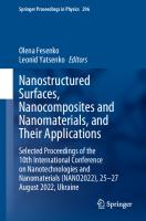
1.2 Method and Materials The printing ink of composite material was obtained by mixing in an aqueous solution two different proportion of BC powder, CaCl2 and Na2 HPO4 . The managing of BC content explains by its viscous properties. The molar ratio of Ca/P was 1.67 as in stoichiometric HA. The two BC/HA biocomposites were prepared by follows: 2.94 g of CaCl2 ·2H2 O and 4.29 g of Na2 HPO4 ·12H2 O were dissolve in 40 ml of bi-distilled water and mixed. After 0.15 g of BC was put into the solution 1.47 g of CaCl2 ·2H2 O and 2.15 g of Na2 HPO4 ·12H2 O were dissolve in 40 ml of bi-distilled water and mixed. After 0.3 g of BC was put into the solution. The resulting materials’ suspensions were milled by planetary milling machine for 3 h. The milled suspensions were washed up by distilled water and were filtered. The composite ink was put into 3D printer’s cylinder. The work principle of 3D printing setup is schematically illustrated in Fig. 1.1 The biocomposite ink presses
1 Bacterial Cellulose/Hydroxyapatite Printed Scaffolds …
3
Fig. 1.1 The BC/HA scaffolds printing process
through needle filler by piston under pressure of air. The 3D moving noodles of biocomposite put onto cooled substrate and immediately freeze. The resulting structures dried by freeze drying method. The microstructure of the samples was studied on optical microscope Olympus BX-51, a digital images were processed by SIAMS program. The functional composition was determined by FTIR spectrometry. It was found that obtained composite have homogeneous structure of HA paricle spreading among BC matrix.
1.3 Results and Discussion The microstructure of BC/HA composites with different proportions are shown in Fig. 1.2. The suspension is homogenous. The particles are evenly spaced, Fig. 1.2a. At suspension that was obtained by second method (where BC content increased for 4 times) the HA microparticles are incorporated into BC matrix, Fig. 1.2c. It also makes some problems at printing, due to high viscosity of ink. We solved it by increasing fluidity of water solution. It is notable that increasing of BC content enlarges average microparticles size from 2.98 to 3.36 μm, according to Table 1.1. Also, the maximum square of particles increased from 9.51 to 12.25 μm2 . We suppose that particles’ sizes increasing caused by viscosity of BC material, which blocks suspension fluidity at milling process (Fig. 1.3).
4
A. Turlybekuly et al.
Fig. 1.2 The BC/HA biocomposites microstructure. a, b—for lower content of BC; c, d—for BC increased content (4 times); a, c—light reflected images; b, d—light transmitted images
Table 1.1 Statistical values of biocomposite paticles’ dimensions Parameters
Statistical values BC/HA—1
BC/HA—2
Min.
Max.
Ave.
Min.
Max.
Ave.
S μm2
1.01
132.51
9.51
1.01
344.47
12.25
D μm
1.13
13.58
2.98
1.13
22.04
3.36
The Fourier transform infrared spectroscopy was used to evaluate the HA/BC interactions. All obtained IR spectra were referenced on HA spectra, (Fig. 1.4c). The 562 and 602 cm−1 bands in HA spectra belong to the O-P-O groups (ν4 bending mode) [18, 19]. The highest band at 1026 cm−1 refers to asymmetric stretching (ν3) of P-O vibrations. The carbonate groups enhancing at 1456 to 1716 cm−1 caused by BC addition and indicates the bonding of BC with HA.
1.4 Conclusion The BC/HA biocomposite materials with different BC content for use in 3D printing were obtained. The microstructure, functional groups composition were investigated.
1 Bacterial Cellulose/Hydroxyapatite Printed Scaffolds …
5
Fig. 1.3 Measurement of the size and shape of particles. Differential numerical particle size distribution
It was shown, that increasing of BC content has direct influence on particles’ size at suspension. The method of BC/HA biocomposite 3D printing is suggested.
References 1. A. Turlybekuly, B.H. Shaimardanova, S.V. Plotnikov, A.D. Pogrebnjak, N.K. Erdybaeva, G.K. Uazyrkhanova, Q. Aqatan, Bull. Univ. Karaganda-Phys. 3, 74 (2018)
6
A. Turlybekuly et al.
Fig. 1.4 FTIR spectra of the synthesized samples: BC/HA—1 (curve a), BC/HA—2 (curve b) and HA (curve c)
2. A. Turlybekuly, B.H. Shaimardanova, S.V. Plotnikov, A.D. Pogrebnjak, L.N. Yerokhina, G.K. Uazyrkhanova, A. Kasymbayev, E. Shaymardan, D.S. Dogadkin, Bull. Univ. Karaganda-Phys. 4, 51 (2018) 3. A. D. Pogrebnjak, L. F. Sukhodub, L. Sukhodub, O. V. Bondar, A. Turlybekuly, Advances in Thin Films, Nanostructured Materials, and Coatings (2019) pp. 361–368 4. A. Pogrebnjak, L. Sukhodub, L. Sukhodub, O. Bondar, M. Kumeda, B. Shaimardanova, Z. Shaimardanov, A. Turlybekuly, Ceram. Int. 45, 7504 (2019)
1 Bacterial Cellulose/Hydroxyapatite Printed Scaffolds …
7
5. S.-J. Ahn, Y.M. Shin, S.E. Kim, S.I. Jeong, J.-O. Jeong, J.-S. Park, H.-J. Gwon, D.E. Seo, Y.-C. Nho, S.S. Kang, C.-Y. Kim, J.-B. Huh, Y.-M. Lim, Biotechnol. Bioprocess. Eng. 20, 948 (2015) 6. N. Tazi, Z. Zhang, Y. Messaddeq, L. Almeida-Lopes, L.M. Zanardi, D. Levinson, M. Rouabhia, AMB Express 2, 61 (2012) 7. S. Saska, H.S. Barud, A.M.M. Gaspar, R. Marchetto, S.J.L. Ribeiro, Y. Messaddeq, Int. J. Biomater. 2011, 1 (2011) 8. N. Yin, S. Chen, Y. Ouyang, L. Tang, J. Yang, H. Wang, Prog. Nat. Sci. Mater. Int. 21, 472 (2011) 9. L.-H. Fu, C. Qi, Y.-J. Liu, W.-T. Cao, M.-G. Ma, Sci. Rep. 8, 8292 (2018) 10. P. Qi, S. Ohba, Y. Hara, M. Fuke, T. Ogawa, S. Ohta, T. Ito, Carbohydr. Polym. 189, 322 (2018) 11. K. V. Massari, G. O. Marinho, J. L. Silva, L. A. Holgado, A. L. Leão, M. M. Chaves, A. Kinoshita, Tissue reaction after subcutaneous implantation of a membrane composed of bacterial cellulose embedded with hydroxyapatite. Dent. Oral Craniofacial Res. 1, 25–30 (2015) 12. H. Mei, S. Zhao, W. Chen, Q. Wang, H. Liang, Trans. Nonferrous Met. Soc. China 28, 1368 (2018) 13. D. Feldman, J. Macromol. Sci. Part A 52, 322 (2015) 14. P.A. Rühs, F. Storz, Y.A. López Gómez, M. Haug, P. Fischer, Npj Biofilms Microbiomes 4, 21 (2018) 15. M. Sayed, H.F. El-Maghraby, F. Bondioli, S.M. Naga, J. Appl. Pharm. Sci. 8, 23 (2018) 16. M. Moniri, A. Boroumand Moghaddam, S. Azizi, R. Abdul Rahim, A. Bin Ariff, W. Zuhainis Saad, M. Navaderi, R. Mohamad, Nanomaterials 7, 257 (2017) 17. Q. Wang, J. Sun, Q. Yao, C. Ji, J. Liu, Q. Zhu, Cellulose 25, 4275 (2018) 18. R. Hu, C.-J. Lin, H.-Y. Shi, J. Biomed. Mater. Res. Part A 80A, 687 (2007) 19. I.S. Neira, Y.V. Kolen’ko, O.I. Lebedev, G. Van Tendeloo, H.S. Gupta, F. Guitián, M. Yoshimura, Cryst. Growth Des. 9, 466 (2009)
Chapter 2
Delivery of Probiotic to Microbiome by Layer-by-Layer Encapsulation M. A. Abdulzhanova, I. Savitskaya, A. Kistaubayeva, D. H. Shokatayeva, A. Pogrebnjak, and L. V. Ignatova
Abstract The probiotic alginate microcapsules of a matrix type were obtained by extrusion method. The coating with an additional layer (layer-by-layer) was carried out by placing the alginate microcapsules with bacteria included in them in a 0.4% solution of chitosan or pullulan. Microcapsules look like spherical particles, white, with a smooth surface. The size of microcapsules is 102–145 microns. The efficiency of Lactobacillus acidophilus AA-1 cells immobilization for uncoated (alginate) and coated with chitosan and pullulan microcapsules was 96.35 ± 1.65; 95.28 ± 2.31 and 94.43 ± 2.31, respectively. The survival rate of strains encapsulated in alginate-chitosan and alginate-pullan under model gastrointestinal conditions in vitro increased on average by 50–70% compared with free cells.
2.1 Introduction Technologies for the functional nutrition products production are developing at a rapid pace. A great part of this market consists of nutrition products containing probiotics. The viability of probiotic microorganisms, i.e. the number of live and active cells in a certain volume (g or ml) of probiotic food products at the time of consumption is a crucial characteristic of these products quality, which determines their effectiveness [1]. However, a significant part of probiotic cells lose their activity due to damage and death of microorganisms during the production of starter culture, storage of products, and especially in the process of passing through the gastrointestinal tract [2]. Therefore, it is important to ensure high survival of probiotic bacteria during the production and storage of the product and during its consumption. M. A. Abdulzhanova · I. Savitskaya (B) · A. Kistaubayeva · D. H. Shokatayeva · L. V. Ignatova Al-Farabi Kazakh National University, Almaty, Kazakhstan e-mail: [email protected] A. Pogrebnjak Department of Nanoelectronics, Sumy State University, Sumy, Ukraine © Springer Nature Singapore Pte Ltd. 2020 A. D. Pogrebnjak et al. (eds.), Nanomaterials in Biomedical Application and Biosensors (NAP-2019), Springer Proceedings in Physics 244, https://doi.org/10.1007/978-981-15-3996-1_2
9
10
M. A. Abdulzhanova et al.
One of the ways to solve the problem of increasing the viability of probiotics is the use of adsorption and spatial immobilization of bacteria under mild conditions [3]. Among them, increasing attention of scientists is attracted to the process of encapsulation as the most promising method for protecting and adapting cells and active substances in human body [4]. Biocapsulation refers to the creation of various polymer systems in form of hydrogel nano- and microparticles, nanoand microcapsules or polymer films with immobilized biomaterial, which can be represented by various BAS (proteins, including enzymes, DNA, peptides, low molecular weight hormones, antibiotics, etc.), as well as microbial, plant and animal cells [5]. Encapsulation provides a significant increase in cell survival under conditions of gastrointestinal tract [6, 7]. In this regard, encapsulated probiotics are used in various fermented dairy products, such as yogurt, cheese, sour cream, frozen dairy desserts, to obtain biomass, as well as in dry preparations [8]. One of the popular strategies for encapsulating probiotic cells is extrusion process, since it does not require high temperatures, use of expensive equipment and organic solvents [9]. Therefore, in order to obtain microcapsules, an extrusion method was chosen in this work. This method consists of mixing a probiotic inoculum in a hydrocolloid solution, followed by extrusion through a nozzle, and then the resulting droplets are collected in a solution of gelling agent such as sodium alginate. It is a natural polymer, widely used as an encapsulating material, since it forms a universal, biocompatible and non-toxic matrix to protect probiotic strains from harmful factors [10]. However, there are more and more reports that alginate microcapsules have insufficient strength and can break down under adverse conditions [11]. In particular, the alginate gel formed is highly sensitive to pH values, which can affect both the release and protection of encapsulated probiotic [12]. The encapsulation material can be modified by physical or chemical means to strengthen the matrix. One of such methods is coating of microcapsules with an additional layer (by-layer) of other natural polymers, for example, chitosan, which is widely used to obtain microcapsules by this method [6, 9, 13, 14]. Alginate microcapsules coated with chitosan were used as a “positive control” in this work. Another microbial “food” polysaccharide, investigated in the work for the possibility of creating an additional coating from it, is pullulan. It is capable of forming elastic, durable films, which are used as packaging material for food products, used for edible decoration in confectionery and bakery products [15–17]. This strategy was used in the work, the aim of which is—Microencapsulation of probiotics into polysaccharide matrices to improve their stability, viability and effective gastrointestinal delivery for functional products.
2 Delivery of Probiotic to Microbiome by Layer-by-Layer …
11
2.2 Materials and Methods 2.2.1 Preparation of Bacteria for Microencapsulation In this study Lactobacillus acidophilus AA-1 isolated by our group from traditional yogurt was used. The strain was stored in glycerol (50%) at −20° C. The stored cells were activated twice on MRS agar plates before use. After 48 h of growth, one colony was selected, inoculated into 20 ml MRS broth and incubated for 24 h at 37 °C. After this, the culture was transferred to fresh MRS broth and incubated at 37 °C for 18 h under anaerobic conditions. The cells were collected in the stationary growth phase by centrifugation at 6000 g for 15 min at 4 °C. The supernatant was discarded and cell pellet was washed twice with sterilized distilled water. The precipitate was resuspended in 3 ml of sterilized 0.85% sodium chloride solution. The concentration of cells after this treatment was around 2 × 10¹⁰ CFU/ml. Freshly prepared concentrated cell suspension was quantitatively evaluated by plating on MRS agar and used immediately.
2.2.2 Preparation of Encapsulated Probiotics The following coating materials were used: sodium alginate (A) from Hi Media, chitosan (Ch) and pullulan (P) from Serva. A suspension of probiotic cells was suspended in a solution of an encapsulating substance (2% A) in a 1:5 ratio to obtain a suspension containing approximately 1010 CFU/ml of cells. Then this mixture was extruded through a needle with a 0.6 mm diameter into a sterile 1% calcium chloride solution. For extrusion a syringe dispenser “Armed MP-2003” was used. The distance between the needle and calcium chloride solution was 25 cm. The drops immediately formed gel spheres. These spheres were allowed to settle for 30 min for complete solidification. Alginate microcapsules with included bacteria were placed in a 0.4% solution of Ch or P, incubated for 40 min on a shaker at 130 rpm. This speed of mixing avoided the aggregation of microcapsules. Then the capsules were removed from solution using a sterilized sieve (50 µm) and washed with sterile distilled water. The whole procedure was performed using autoclaved (121 °C, 15 min) materials and under sterile conditions in a laminar air flow box. Capsules were stored in sterile vials with lids at 4 °C and used in further experiments. The average diameter of microcapsules was measured using a laser particle size analyzer (Winner 2000ZD, Jinan Winner Particle Instruments Stock Co., Ltd., China). Optical microscopy of wet microcapsules was performed using a microscope (MDL-150-TPI) and a digital camera (Samsung 14.2). The morphology of frozen dried microcapsules was also evaluated using a scanning electron microscope (JEOL, JM6360).
12
M. A. Abdulzhanova et al.
2.2.3 Quantification of Viable Bacteria in Capsules 1.0 g of microcapsules was added to a mixed solution of 0.06 mol/l sodium citrate and 0.2 mol/l sodium bicarbonate and stirred for 1 h at 37 °C to loosen the coating. The viability of released cells was determined by plating serial dilutions of resulting suspension on MRS agar. Colony-forming units were counted after 48 h of anaerobic incubation at 37 °C. Free cells were not subjected to homogenization, since comparison of CFU of free cells before and after homogenization did not show a significant difference. The encapsulation efficiency (EI) was determined by the formula: EI % = (N × M)/N0 × 100, where N is the number of viable cells released from 1.0 g of microcapsules, M is the total mass of the collected microcapsules, and N0 is the number of free cells before microencapsulation.
2.2.4 Determination of Resistance of Encapsulated Bacteria to Conditions that Mimic Human Gastrointestinal Tract To determine the stress resistance of encapsulated cells in environments that mimic gastrointestinal tract, test samples were cultured in media with different pH levels, the content of digestive enzymes and bile salts [18]. Artificial gastric juice (AGJ) was made using a 0.2% solution of sodium chloride, the pH of which was raised to 2.0 with 1 M HCl. Pepsin with a concentration of 3.2 g/l and 1 g of lipase were added to the solution, then the mixture was filtered through a 0.22 µm membrane for sterilization. Then, 0.5 g of fresh microcapsules and a suspension of free cells (2 × 10¹⁰ CFU/ml) were suspended in 9.5 ml of AGJ and incubated at 37 °C for 120 min at 150 rpm. AGJ samples were neutralized with phosphate buffer (0.2 M, pH 7.0, 4 °C) at the end of incubation. Duodenal conditions were simulated by dissolving bile salts in physiological saline to a 0.6% final concentration, 10 g/l of each trypsin and pancreatin were added, the pH of solution was adjusted to 6.5 with 0.1 M NaOH and sterilized by membrane filtration. 0.5 g of fresh microcapsules were added to 9.5 ml of bile solution and incubated at 37 °C for 1 h at 150 rpm. To create a colon model, artificial intestinal juice (AIJ) was prepared by dissolving KH2 PO4 in distilled water to a final concentration of 6.8 g/l and at pH 7.4. 10 g/l of trypsin was added to the solution, filtered through a membrane with 0.22 µl for sterilization. 0.5 g of microcapsules were placed into 9.5 ml of AIJ and incubated at 37 °C for 4 h at 150 rpm. At the end of incubation free ones were washed with physiological saline, microcapsules were destroyed by homogenization in phosphate buffer, and cell survival was quantified by the method described above using standard plating on MRS agar.
2 Delivery of Probiotic to Microbiome by Layer-by-Layer …
13
2.2.5 Statistical Analysis Statistical comparison was performed using unpaired t-test and one-way analysis of variance (ANOVA) followed by Dunnett’s test for multiple comparisons. All statistical analyses were performed using SPSS 16.0 software package (SPSS Inc., USA).
2.3 Results and Discussion Live probiotic bacteria of Lactobacillus genus are most often used in probiotic fermented milk products. The strain Lactobacillus acidophilus AA-1 has all the necessary probiotic characteristics: a high level of acid production, adhesive and antagonistic activity. It does not have pathogenicity and toxicity, i.e. has the required level of biosafety. As a result of extrusion in a hydrocolloid alginate solution, probiotics formed a matrix-type microcapsule. Due to the fact that probiotics are present in the entire structure of particles, they can be exposed to environmental conditions, which can reduce their viability during storage and use. To coat alginate microcapsules with an additional layer, they were kept for 30–40 min in a 0.4% solution of chitosan or pullulan. Figure 2.1 shows the 3 types of microcapsules obtained by the method described above: containing alginate (A) or a mixture of alginate-chitosan (A–Ch) and alginate-pullulan (A–P) as a material-sealant. Microcapsules are spherical particles of white color with a smooth surface. In appearance, all 3 types of capsules did not differ from each other. The absence of external differences was confirmed by analysis on a scanning electron microscope. Alginate microcapsules and coated microcapsules had a similar oval shape. Moreover, the combination of polymers as an additional wall does not change the typical spherical shape. A compact, penetration-resistant surface can serve as a strong physical barrier that protects probiotic cells as they pass through the gastrointestinal tract. The SEM study showed bacteria in the capsule matrix. They are present in all 3 types of microcapsules. Table 2.1 shows data obtained in experiments on determination the encapsulation efficiency (%). For uncoated (A) and coated with Ch and P microcapsules, it was 96.35 ± 1.65; 95.28 ± 2.31 and 94.43 ± 2.31, respectively. The difference was not statistically significant (P > 0.5). The interactions between bacteria and materials are crucial for determining the efficiency of encapsulation. The high encapsulation efficiency obtained in our work showed that encapsulation process was soft and the materials were compatible with the probiotic strain. Microcapsule size has an important effect on viability of probiotics and sensory effects on food. As a rule, larger microcapsules provide better protection against probiotics [19], but the product may have undesirable sensory properties [20]. It was shown that microcapsules in the size ranging of 100–200 µm provide optimal
14
M. A. Abdulzhanova et al.
A
B
C
A
B
C
Fig. 2.1 The morphology of A (A), A-Ch (B) and A-P (C) microcapsules loaded with Lactobacillus acidophilus AA-1 Table 2.1 Encapsulation efficiency and size of probiotic microcapsule containing L. acidophilus AA-1 cells
Type of microcapsule
Encapsulation efficiency (%)
Size of microcapsules (mcm)
Alginate
96.35 ± 1.65
102.35 ± 4.31
Alginate-chitosan
95.28 ± 2.31
136.42 ± 5.73
Alginate-pullulan
94.43 ± 2.31
145.37 ± 6.25
2 Delivery of Probiotic to Microbiome by Layer-by-Layer …
15
balance between these two conflicting requirements [21]. In our study, the size of all microcapsules falls within this optimal range. After coating with chitosan and pullulan, the average microcapsule size increased by 34 and 43 µm, respectively. This is partly due to the thickness of coating layer and partly with the aggregation of microcapsules. One of the most important biopharmaceutical indicators of encapsulated probiotics is their ability to maintain integrity in acidic environment of stomach, alkaline environment of duodenum and disintegrate in neutral environment of large intestine. For this, studies were conducted on comparative survival of encapsulated cells of L. acidophilus AA-1 in the model stomach, small and large intestine. Samples were cultivated in media with different pH levels, content of hydrolytic enzymes and bile salts. The greatest inhibitory effects showed low pH levels. Within 2 h under the action of AGJ (pH 2.0) there was a significant death of free cells: the number of viable cells decreased by 6.4 lg (Fig. 2.2). The smallest degree of protection of probiotic culture cells from the action of low pH levels was provided by alginate microcapsules. After 2 h of exposure in a model stomach, the number of viable cells was only 6.4 lg CFU/g, i.e. their number decreased by 3.3 lg. Alginate capsules coated with Ch and P provided greater cell resistance to low pH values: the titer of living cells was reduced only by 1.6 lg and 1.4 lg, respectively. Consequently, the number of cells dying from exposure to low pH values in A–Ch and A–P pullan microcapsules was reduced almost 2 times in comparison with alginate ones. In both tests for tolerance to gastric juice and bile, the mortality of probiotic cells was significantly reduced when they were protected by microencapsulation compared with free cells. The reduction in probiotic population was 5.4 lg CFU/g in simulated duodenum (solution of bile salts). This result indicates that probiotic cells were highly susceptible to simulated gastrointestinal fluids, which justifies the importance of encapsulating this microorganism. Indeed, probiotics loaded into capsules of A–Ch and A–P and coated with Ch, as well as those contained in microcapsules of A–P, showed a decrease in viability of approximately 2 lg CFU/g and 0.9 lg CFU/g, respectively. In the work of M. Chávarri, similar results were shown for chitosan-coated alginate microparticles loaded with Lactobacillus gasseri. These Fig. 2.2 Survival of free and encapsulated L. acidophilus AA-1 in the model gastrointestinal tract
Free cells
А
А-Ch
А-P
lg CFU/gr
15 10 5 0
Treatment free
AGJ
Bile solution
AIJ
16
M. A. Abdulzhanova et al.
authors reported that encapsulation protected probiotics compared with free cells, reaching more than 7 lg CFU/g, whereas non-encapsulated cells could not withstand this treatment [22]. The pH of the model duodenum was 6.8, which is close to the optimum pH for this bacterial strain. But bile salts can penetrate membranes of bacterial cells, damage DNA and proteins, cause leakage of intracellular components and ultimately lead to the bacterial death [23]. The formation of an additional barrier between bile salts and cells played main protective role of these microcapsules in a bile tolerance analysis. Our results show that the barrier properties of Ch and especially P make it a promising encapsulating material for protecting probiotics from both gastric juice and bile. This can be explained by appearance of a polyelectrolyte complex shell on the structure surface and possible further propagation of reaction between macromolecules of polyelectrolytes into the core. Thus, based on the data obtained, the maximum protection of probiotic against stress effects occurring in upper parts of digestive tract was provided by A–P microcapsules. A necessary condition for implementation of beneficial properties of a probiotic is not only the prevention of damage from hostile gastric juice and bile, but also targeted release into large intestine. The revealed dependence of the probiotic cells survival on the type of encapsulating material indicates a different permeability of substances used, which determines the diffusion capabilities of capsule membranes. The main problem in production of microcapsules based on natural polymers is formation of a membrane that provides, on the one hand, the mechanical strength of a capsule, and on the other hand, permeability to nutrients needed by cells. The increase in strength of capsule membranes occurs both due to the formation of mixed adsorption layers, and by increasing the amount of film-forming substance on the particles of encapsulated product. At the same time, an increase in density due to the deposition of secondary membranes can lead to a decrease in permeability of microcapsule membranes [24]. It was shown that the obtained A–Ch and A–P probiotic microcapsules had significant permeability, due to which the cells of microorganisms diffused out of the capsules into the media, and there was a withdrawal of waste products. Moreover, the membranes of these two microcapsules provided almost the same diffusion rate of L. acidophilus AA-1 cells of them (Fig. 2.3). Since cells enclosed in capsules were capable of reproduction, at a certain stage of population development there was a sharp increase in the risk of microcapsules membrane breakthrough, as a result of which the cells could almost completely occur in the external environment. Due to the diffusion of cells from capsules, the possibility of this process was significantly limited. In fact, compared with alginate capsules, the number of viable cells after treatment with simulated intestinal juice increased by 0.2 lg CFU/g for the extra-coated microcapsules. The increase in number of viable cells was associated with the digestion of intercellular proteins by trypsin, i.e. the digestive effect of trypsin in AIJ can introduce errors in determining the exact number of cells. However, an analysis of the data showed that the inclusion of microorganisms in a encapsulating matrix made it possible to increase the stereostability of cells under
2 Delivery of Probiotic to Microbiome by Layer-by-Layer … 9
Number of cells, diffusing in a milk, lg CFU/g
Fig. 2.3 The diffusion rate of immobilized probiotic cultures from microcapsules
17
8 7 6 5 4
Alginate-pullulan capsules
3
Alginate-chitosan capsules
2 1
Time, hours
0 1
2
3
4
5
6
7
8
9
10
adverse conditions, and the type of material that forms the capsules had a significant effect on survival of probiotic microorganisms. Under various adverse conditions, encapsulating materials behaved differently, providing different degrees of survival for probiotic cultures. The experiments carried out in work and processing of obtained data allow us to conclude that immobilization of probiotic cultures in gel-forming substances of natural origin will contribute to increase of stress resistance of microorganisms in human body when passing through gastrointestinal tract.
2.4 Conclusion The smallest degree of protection of probiotic cells from the gastric juice and of bile salts solutions low pH levels action of was provided by alginate microcapsules. A–Ch and A–P microcapsules do not undergo changes and remain without damage after exposure for 2 h under stressful conditions, and provide a high degree of survival of probiotic included in them under model conditions of gastrointestinal tract. In addition to in vitro assessments, there is still a need for in vivo studies on viability of microencapsulated probiotics and their beneficial effects on human health. Nevertheless, the use of microencapsulation in biotechnology ensures efficient delivery of viable probiotic cells to intestine, thereby increasing therapeutic and prophylactic efficacy of healthy foods and probiotic preparations. Acknowledgements This work was supported by grant AP05134040 «Microencapsulation of probiotics into polysaccharide matrices for the development of functional food products based on mare’s milk» from the Science Committee of the Ministry of Education and Science of the Republic of Kazakhstan.
18
M. A. Abdulzhanova et al.
References 1. M. Fernandez, J.A. Hudson, R. Korpela, G. Reyes-Gavilan, BioMed Res. Int., pp. 1–13 (2015) 2. A.M. Mortazavian, R. Mohammadi, S. Sohrabvandi, New Advances in the Basic Clinical Gastroenterology: InTech (2012), pp. 121–146 3. C.P. Champagne, Crit. Rev. Biotechnol. 109–134, 14 (2014) 4. C. Tomaro-Duchesneau, S. Saha, M. Malhotra, I. Kahouli, S. Prakash, J. Pharm. 5, 1–19 (2013) 5. S.J. Sri, A. Seethadevi, K.S. Prabha, P. Muthuprasanna, P. Pavitra, Int. J. Pharm. Bio Sci., 3,1–23 (2012) 6. A.K. Anal, H. Singh, Trends in Food Science and Technology (2017), pp. 240–25 7. P.E. Ramos, M.A. Cerquera, J.A. Teixeira, A.A. Vecente, Crit. Rev. Food Sci. Nutr. (2018), pp. 1864–1877 8. M.E. Morales, M.A. Ruiz, Nutraceuticals 1, 627–668 (2016) 9. A. Mortazavian, S.H. Razavi, M.R. Ehsani, S. Sohrabvandi, Iranian J. Biotechnol. 5, 1–18 (2017) 10. F. Pavli, C. Tassou, G.J.E. Nychas, N. Chorianopoulos, Int. J. Mol. Sci. (2018), p. 150 11. A.B. Shori, HAYATI J. Biosci. 24, 1–5 (2017) 12. B.L. Pasin, C.G. Azón, A.M. Garriga, Técnicas yaplicaciones. Revista Venezolana de Ciencia y Tecnología de Alimentos 3(1), 130–151 (2012) 13. W. Tong, X. Song, Ch. Gao, Chem Soc Rev 41, 6103–6124 (2012) 14. A. Bepeyeva, J.M.S. de Barros, H. Albadran, A.K., J. Food Sci., pp. 1–6 (2017) 15. P. Oguzhan, F. Yangilar, African J. Food Sci. Technol. 4(3), 57–63 (2013) 16. R.S. Singh, N. Kaur, V. Rana, J.F. Kennedy, Carbohyd. Polym. 171, 102–121 (2017) 17. K.R. Sugurman, V. Ponnusami, Carbohyd. Polym. 173, 573–591 (2017) 18. H. Chen, X. Li, J. Funct. Foods 29, 248–255 (2017) 19. K. Lee, T. Heo, Appl. Environ. Microbiol. 66, 869–873 (2010) 20. R. Rajam, S. Kumar, J. Food Sci. Technol. 52, 4029–4041 (2015) 21. K.S. Nag, H. Han, Singh. Int. Dairy J. 21, 247–253 (2011) 22. M. Chávarri, I. Marañón, R. Ares, F.C. Ibáñez, Int. J. Food Microbiol. 142, 185–189 (2010) 23. J. Silva, Appl. Environ. Microbiol. 66, 2605–2612 (2013) 24. A.S. Carvalho, J. Food Sci. 68, 2538–2541 (2013)
Chapter 3
Modernization of the Preservative Solution for Red Blood Cells by Magnetite Nanoparticles (ICNB) A. N. Belousov, E. I. Malygon, V. V. Yavorskiy, and E. Yu. Belousova
Abstract This study was devoted to the learning of the use of nanotechnology to correct the functional activity of red blood cells (RBCs) at the storage stages at a positive temperature. It was established that saline NaCl, which had previously been processed by magnetite nanoparticles (ICNB) had a marked membrane-stabilizing effect, inhibits hemolysis and increasing the sedimentation stability of preserved RBCs. The complex analysis of the obtained data allowed to determine the primary mechanisms effect of the saline NaCl, which had previously been processed by ICNB on the preserved RBCs. The proposed method of additive modernization of preserved RBCs was adapted to the production process. The optimisation results were obtained in creating a simple and practical method of additive modernization of preservation solutions that does not violate the compliance requirements, improves the quality, efficiency and safety transfusion of RBCs.
3.1 Introduction Red blood cells (RBCs) transfusion is a critical, life-saving treatment for severe anemia caused by disease or chemotherapy, or by blood loss due to trauma or major surgery. For several decades RBCs components have been prepared as concentrates suspended in nutrient additive solution, which preserves and extends the shelf-life of the RBCs component, allowing up to 6–7 weeks of refrigerated storage [1]. Nevertheless, during storage RBCs undergo a complex and progressive accumulation of physicochemical changes, collectively referred to as the RBCs storage lesion [2, 3]. A. N. Belousov (B) · E. Yu. Belousova Laboratory of Applied Nanotechnology of Belousov, Belousov, Ukraine e-mail: [email protected] A. N. Belousov · E. I. Malygon · V. V. Yavorskiy · E. Yu. Belousova Kharkov Medical Academy of Postgraduate Education, Kharkiv, Ukraine A. N. Belousov · E. I. Malygon · V. V. Yavorskiy Kharkov Regional Center of Blood Service, Kharkiv, Ukraine © Springer Nature Singapore Pte Ltd. 2020 A. D. Pogrebnjak et al. (eds.), Nanomaterials in Biomedical Application and Biosensors (NAP-2019), Springer Proceedings in Physics 244, https://doi.org/10.1007/978-981-15-3996-1_3
19
20
A. N. Belousov et al.
Recent clinical studies have identified RBCs transfusion as an independent risk factor for increased morbidities and mortalities in certain groups of patients, including trauma, cardiac surgery and the critically-ill (reviewed in [4–6]). Additionally, some of these studies have identified that older stored RBCs are more strongly implicated in poorer outcomes compared to fresher RBCs [6]. In order to address these concerns, there is renewed interest to better understand the RBCs storage lesion and to find ways to ameliorate the deleterious effects of storage, thereby improving the quality, efficacy and safety of RBCs components for all transfusion recipients. While increased research effort is being directed to better understand the effects of storage on RBCs and the potential impact on transfusion outcomes [7], slower progress is being made in finding ways to deter the detrimental effects of the RBCs storage lesion. Over the past 15–20 years, research into the development of new additive solutions has focussed on ways to maintain higher intracellular levels of ATP and 2.3-DPG during storage of RBC components [1]. Despite the RBCs having been a favourite experimental model for cellular biologists and biochemists, RBCs storage research has repeatedly demonstrated that a lot of fundamental biology about RBCs is still not well understood. The complexity of the inter-relationship between RBCs biochemistry, cytoskeletal structure and membrane properties have made it difficult to predict how RBCs will respond to different storage conditions. Exposure of RBCs to non-physiological storage environments has pointed to the existence of previously unknown biochemical mechanisms in RBCs, including apoptotic-like processes, ion and osmotic channels that behave differently than expected, exposure of new or altered receptors possibly due to oxidative and/or protease/glycosidase activities or altered senescence [8–11]. The benefits gained by improved RBCs component quality should more than justify any real or perceived inconvenience to the blood services in implementing adjustments to their processing procedures or additional processing costs of the introduction of new generation RBCs additive solutions. The bigger challenge that has hindered the advancement of this field is the significant financial burden and risk for manufacturers of blood collection systems to obtain licensure and to bring a new RBCs storage system to a market that is inherently based on very low profit margins, such as the blood services sector. The financial burden to technology developers of new RBC storage systems is largely due to regulatory requirements, particularly those mandated by the FDA. In addition to in vitro data, the FDA requires in vivo data on the 24 h post transfusion recovery of transfused autologous RBCs. Recently the FDA has tightened and increased the assessment and acceptance criteria making it potentially more difficult and expensive to bring new RBCs storage systems to market. Although the regulatory agencies are to be commended for focussing on the safety of new therapies and devices for patients, there are concerns that the regulatory requirements for RBC storage systems have become excessive and are hindering progress [12]. Another significant challenge for obtaining licensure of new RBCs storage systems is the inherent donor-related variability in stored RBCs quality. It has long been recognised that RBCs from some donors do not store well, as evidenced by higher
3 Modernization of the Preservative Solution for Red Blood …
21
levels of haemolysis at RBC component expiry 14 and poorer in vivo 24 h recovery data [13]. The relationship of specific donors and poorer quality of some stored RBCs components was confirmed in a recent paired cross-over study designed to compare manual and automated whole blood processing methods [14, 15]. Technology developers are unwilling to take on the risk that a random poor quality RBCs component could jeopardise the success of licensure tests and clinical trials of their new blood storage systems and their significant financial investment. In Ukraine, the first standardized and biocompatible magnetite nanoparticles for medical use were manufactured and patented in 1998. These are intracorporal nanobiocorrector of brand ICNB, magnet-controlled sorbent of brand MCS-B, and biologically active nanodevice of brand Micromage-B [16]. It is well established that the magnetite nanoparticles effectively modulate the metabolic processes in leukocytes, regulate activity of the enzyme link of the antioxidant system in erythrocytes in healthy and sick patients [17–19]. Previously the complex investigations that were performed in the study of the influence on metabolism of cells by preparations of nanotechnology show that in whole standardized biocompatibility of magnetite nanoparticles have nonspecific and modulated effect on metabolic processes. Research of ultrastructure investigations of the reticuloendothelial system (liver, lungs and kidneys) it was proved that after injection of biocompatibility magnetite nanoparticles into a vein caused nonspecific activation of the metabolic processes, increase adaptive mechanisms and potential of organelle cells, acceleration of reparative processes a level of membranes and macromolecules [18, 20, 21]. Existing sorption and indirect (magnetic) effects not only allow selectively absorb the protein of surface membrane cells by magnetite nanoparticles (according to the principle of magnetophoreses), but also to prevent the oxidative modification of proteins by way of stabilizing the active groups, normalizing a state of receptors that are located on the surface membrane of cells, increasing activity of enzymes’ membrane-bound [22–24]. Recent scientific work related to use of magnetite nanoparticles (ICNB) in contrast means in an MRI investigation of cancer reliably was shown that nanoparticles cause reversible changes associated with a temporary increase in the mobility of hydrogen protons in the pericellular fluid that inevitably modifies the metabolism in malignant cells [25]. The results of these investigations have not only widened the understanding of the mechanisms of action of nanoparticles on condition outside and intracellular spaces but also have revealed new aspects of the cellular(cells) metabolism, determined the membrane role of cellular enzymes in the regulation processes of metabolism [23, 26–29]. Also, it was established that extracorporally processing the blood by nanoparticles of MCS-B reliably reduces activity of Ca, Mg—ATPHese of erythrocytes. Currently, studies have shown that magnetite nanoparticles are able to inhibit hemolysis of heparinized blood, increase the activity of ATP and 2.3 DPH in red blood cells, regulate transmembrane metabolism and inhibit eryptosis [23, 30, 31]. The above was the basis for the choice of the theme of this study, devoted to the learning of the use of nanotechnology to correct the functional activity of red blood cells at the storage stages at a positive temperature.
22
A. N. Belousov et al.
The main purpose of the first stage of the study is to develop a simple and practical method of additive modernization of preservation solutions that does not violate the compliance requirements, improves the quality, efficiency and safety transfusion of red blood cells.
3.2 Materials and Methods Materials: 1. Standardized intracorporeal nanobiocorrector of ICNB was taken as nanoparticles. Magnetite nanoparticles synthesized by co-precipitation method. The main physics and chemical properties of ICNB the following data and also in Tables 3.1, 3.2, 3.3 and 3.4; Figs. 3.1 and 3.2 were presented: • Concentration of the colloidal solution of magnetite nanoparticles in physiology solution of NaCl is 0.0225%. • Theoretical osmolality of colloid solution is 500 mosmol/l. • Size of magnetite nanoparticles is 6–12 nm. • Total area of surface magnetite of nanoparticles Ss = 800–1200 m2 /g. • Magnetization of saturation Is = 2.15 kA/m. • ζ—potential = −19 mV. Table 3.1 The calculated lattice parameters of the phases ´ ´ ´ Phase name a (Å) b (Å) c (Å) Alpha (gpad) Magnetite low
8.38
8.38
Magnetite low, syn
5.93
5.93
Johannsenite
9.89
9.05
8.38 14.7 5.28
Beta (gpad)
Gamma (gpad)
90
90
90
90
90
120
90
105.5
90
Table 3.2 Determination of percent composition of the ICNB by X-ray spectrometer ARL OPTIM’X (semi-quantitative analysis) Compound
wt%
Fe3 O4
97.37 2.26
CaO
Std. Err
El
Weight%/O2
0.09
Fe
68.40
0.07
Ca
1.71
Std. Err
El
wt%
Std. Err
0.07
Fe
97.62
0.09
0.05
Ca
2.3
0.07
P2 O5
0.280
0.027
Px
0.122
0.012
Px
0.157
0.015
MnO
0.255
0.013
Mn
0.198
0.010
Mn
0.278
0.014
SiO2
0.098
0.027
Si
0.046
0.013
Si
0.059
0.016
SO3
0.032
0.013
Sx
0.0126
0.0051
Sx
0.0164
0.0066
Cl
0.0280
0.0090
Cl
0.0280
0.0090
Cl
0.0380
0.012
3 Modernization of the Preservative Solution for Red Blood … Table 3.3 X-ray analysis of ICNB in X-ray diffractometer Rigaku Ultima IV (CuKα, Kβ filter—Ni), one-coordinate DTeX semiconductor detector
Table 3.4 The phases of magnetite of nanoparticles (RIR—method; error 8 ± 3%)
23
Phase
Formula
Space group
No. card database ICDD
Magnetite low
Fe2.886 O4
227: Fd-3m, choice-2
10861339 (ICDD)
Magnetite low, syn
Fe3 O4
166: R-3m, hexagonal
10716766 (ICDD)
Johannsenite
Ca Mn + 2Si2 O6
15: C12/c1, unique-b, cell-1
380413 (ICDD)
Phases (method of corundum numbers)
Content (%)
Magnetite low
71
Magnetite low, syn (hexagonal)
29
Fig. 3.1 Study of magnetite nanoparticles with use microscope ion-electronic raster-type Quanta 200 3 D
Variant 1
ICNB
Variant 2
0.9% NaCl
Variant 3
4 part of 0.9% NaCl + 1 part of 0.9% NaCl which previously was processed by ICNB in ratio 4:1
Fig. 3.2 Images of fluids that were studied in the research at the MRI
24
A. N. Belousov et al.
2. 0.9% NaCl solution 3. 0.9% NaCl solution which previously was processed by ICNB in ratio 4:1 Object of research: • red blood cells (RBCs) into bags containing anticoagulant citrate, nutrient phosphate and dextrose (CPD); • red blood cells (RBCs) into bags containing anticoagulant citrate, nutrient phosphate, dextrose and adenine (CPDA-1). Methods: Before starting the experience with red blood cells (RBCs) we used visual assessment by comparison of the brightness of the imagines different variants of solutions. These were: ICNB, 0.9% NaCl solution and 0.9% NaCl solution that had been treated by magnetite nanoparticles (ICNB). The tests were performed on the Siemens MR-tomagraphy Magneton Concerto with power magnetic-field 0.2 T. The axial tomograms were received: 1. T1—the self-weighted sequences of Echo Spin of TR 50 ms, TE 17 ms the field of review the 250 mm, the thickness cut 2 mm. 2. T2—the self-weighted sequences Echo Gradient of TR 500 ms, TE 17 ms the field of review a 180 mm, the thickness cut 4 mm. Of each bag of 3 ml amounts of red blood cells was distributed into 20 sterile glass tubes. Then, into the first 10 tubes of control were added of 2 ml amounts 0.9% NaCl solution. Into the next 10 tubes of test were added of 2 ml amounts 0.9% NaCl solution, which previously was processed by ICNB. Thus, the distribution of tubes was as the follows: Tubes of control: • 3 ml of red blood cells (CPD) +2 ml 0.9% NaCl solution (n = 10); • 3 ml of red blood cells (CPDA-1) +2 ml 0.9% NaCl solution (n = 10). Tubes of test: • 3 ml of red blood cells (CPD) +2 ml 0.9% NaCl solution that previously was processed by ICNB in ratio 4:1 (n = 10); • 3 ml of red blood cells (CPDA-1) +2 ml 0.9% NaCl solution that previously was processed by ICNB in ratio 4:1 (n = 10). The state of red blood cells was determined visually by the registration of signs of hemolysis. Also, hemolysis was controlled by photometric method by means Plasma/Low Hb and GPHP-01 devices. The centrifuge mark of SM-70M-07 was used to obtain supernatant. Hematocrit was calculated by means hematocrit ruler and using the formula: HCT =
Free Hb × (1 − HCT) × 100% Common Hb
3 Modernization of the Preservative Solution for Red Blood …
25
Morphology of the red blood cells was studied by direct microscopic method. Sedimentation stability of red blood cells was studied by Panchenkov’s method. Change in the acidity of the red blood cells was performed by means of pH metric. Tests were carried out in six stages: day 1—I, day 7—II, day 14—III, day 21—IV, day 28—V, day 35—VI. The blood after performance of the biochemical investigation was stored in the refrigerating chamber at temperature +4 °C. Statistically processing the obtained results was carried out by parametrical method of variation statistics by Student criterion. Processing the obtained data was carried out by means of Excel.
3.3 Results and Discussion Results of visual assessment of the images brightness used in experiment liquids at the MRI are represented in Fig. 3.2. Figure 3.3 illustrates the difference of image brightness in liquids compared at the MRI. The order of the brightness increase is the following: ICNB, 0.9% NaCl solution, 0.9% NaCl solution that was treated by nanoparticles of ICNB. The difference in brightness of images is explained in the following way: Variant 1. Magnetite nanoparticles of ICNB reduce the mobility of hydrogen protons in the liquid medium (0.9% NaCl solution). Therefore, the image brightness was very low in the MRI. Variant 2. Rising mobility of hydrogen protons in the intact 0.9% NaCl solution increased brightness in comparison with Variant 1. Fig. 3.3 Study of magnetite nanoparticles with use microscope electronic translucent JEM-2100
26
A. N. Belousov et al.
Variant 3. Mobility of hydrogen protons in 0.9% NaCl solution that has previously been processed ICNB nanoparticles is maximized. Therefore, the image brightness is much higher than before (in the previous variants). Thus, previously conducted research clearly shows that the nanoparticles of ICNB change the mobility and the orientation of the hydrogen atoms in liquids that are registered in the visual evaluation of MRI. The next set of studies was essential and aimed at studying of functional activity of red blood cells at the storage stages at a positive temperature after by modifying the mobility and spatial orientation of hydrogen protons in the pericellular fluid using magnetite nanopatricles of ICNB. A study of the sedimentation stability of RBCs showed a highly significant difference between control and test data. Data of sedimentation stability of the RBCs at the stages of a study were presented in Fig. 3.4. Figure 3.4 shows that sedimentation stability of RBC in test tubes are reliably more highly (p < 0.001) then in control tubes at stages of research. It should be said that negative surface charge of human RBCs’s results primarily from the presence of ionogenic carboxyl groups of sialic acids on the cell surface [32–34]. The value of the charge is determined by the amount of adenosine threephosphate (ATP). ATP is macroergic compounds, the product of glycolysis. The sedimentation stability of RBC is determined by the amount of ATP. In this case, the change of mobility and spatial orientation of the hydrogen protons in the extracellular liquid significantly increased the sedimentation stability of RBCs in the test compared to the control. For greater clarity results of RBCs sedimentation is shown in Fig. 3.5. 70 60
65
50
50
40
20
39 53
59
52
24
23
10
13
52
48
65
30
59
55
53
65
8
14
53
12
59
8
14
52
0 Control CPD (n=10)
Stage I
Test CPD (n=10)
Stage II
Stage III
Control CPDA-1 (n=10)
Stage IV
Stage V
Test CPDA1 (n=10)
Stage VI
Fig. 3.4 Study of the sedimentation stability of RBCs at the stages (M ± m; p < 0.001)
3 Modernization of the Preservative Solution for Red Blood …
Preserving agent CPD
Control (RBCs+0.9% NaCl)
Test (RBCs+ 0.9% NaCl solution which previously was processed by ICNB solution)
27
Preserving agent CPDA-1
Test (RBCs+ 0.9% NaCl solution which previously was processed by ICNB solution)
Control (RBCs+0.9% NaCl)
Fig. 3.5 Sedimentation stability of erythrocytes at the stage VI of the study
Figure 3.5 is showing that with preserved in the anticoagulant CPD the sedimentation stability in the control was 62 mm; in the test—53 mm. With preserved in the anticoagulant CPDA-1: control—58 mm; test—52 mm. Thus, following the logic of the above reasoning, if improving sedimentation stability of RBC is associate with an increase in ATP, then the isotonic solution which previously was processed by ICNB should actively stabilize the membranes of RBCs and inhibit hemolysis. Therefore, the next investigation was to study the hemolysis processes preserved of the RBCs at various stages. Results of the visual assessment hemolysis of erythrocytes in various aspects of exposure are presenting in Fig. 3.6. Figure 3.7 shows that in the control tubes at the stage VI of the study there are pronounced signs of hemolysis. In contrast, hemolysis is not recorded in test tubes. Visual analysis was supplemented by objective data of the photometric method, as well as the method of calculation of hematocrit. Objective data of free Hb and calculated HCT at the stage VI of the study are presented in Table 3.5. The data in Table 3.5 indicate that in test tubes the average content of free Hb in the VI stage of the study 6.25 ± 0.1 g/l were less than controls; and the average calculation HCT was lower by 1.42% Thus, the objective indicators of Table 3.5 are highly reliable (p < 0.001) and confirm the obtained visual effect of inhibiting hemolysis of preserved RBCs by physiological solution, which was pretreated by ICNB. Microscopic observation of changes in the morphology of RBCs was a logical continuation of the study of the
28
A. N. Belousov et al.
1
2
3
4
Fig. 3.6 Visual assessment hemolysis of erythrocytes in various aspects of exposure at the stage VI. Notes 1—RBC (preserving agent CPD) + 0.9% NaCl solution; 2—RBCs (preserving agent CPD) + 0.9% NaCl solution which previously was processed by ICNB; 3—RBCs (preserving agent CPDA-1) + 0.9% NaCl solution; 4—RBCs (preserving agent CPDA-1) + 0.9% NaCl solution which previously was processed by ICNB
Control (RBCs+ 0.9% NaCl) solution
Test (RBCs+ 0.9% NaCl solution which previously was processed by ICNB)
Fig. 3.7 The presence of free Hb in the supernatant (preserving agent CPD)
3 Modernization of the Preservative Solution for Red Blood …
29
Table 3.5 The indicators of free Hb and calculated HCT at the stage VI of the study at baseline values: Hb = 154 g/l; HCT = 0.46 Preservatives CPD CPDA-1
Variants
Stage VI
p
Free Hb (g/l)
Calculation HCT (%)
Control (n = 10)
7.8 ± 0.1
1.84 ± 0.1