Metabolic Reprogramming: Methods and Protocols (Methods in Molecular Biology, 2675) 1071632469, 9781071632468
135 52 13MB
English Pages [332]
Preface
Acknowledgments
Contents
Contributors
Chapter 1: In Vivo Tissue Lipid Uptake in Antisense Oligonucleotide (ASO)-Treated Mice
1 Introduction
2 Materials
2.1 Animal Treatments
3 Methods
3.1 Animal Treatments
3.2 Sample Preparation and Measurements
3.3 Data Analysis
4 Notes
References
Chapter 2: In Vivo Hepatic Triglyceride Secretion Rate in Antisense Oligonucleotide (ASO)-Treated Mice
1 Introduction
2 Materials
2.1 In Vivo Antisense Oligonucleotide (ASO) Treatment
2.2 Triglyceride (TG) Secretion Rate
3 Methods
3.1 In Vivo ASO Treatment
3.2 Triglyceride (TG) Secretion Rate
3.3 VLDL Isolation and Characterization
3.4 Further Processing and Use of VLDLs
4 Notes
References
Chapter 3: Measurement of Fatty Acid Oxidation by High-Resolution Respirometry: Special Considerations for Analysis of Skeleta...
1 Introduction
2 Materials
3 Methods
3.1 Air/Zero Calibration of OROBOROS Oxygraph-2K
3.2 Sample Preparation for Skeletal Muscle
3.3 Sample Preparation for Cardiac Muscle
3.4 Sample Preparation for Adipose Tissue
3.5 The Making of Adipose Tissue Slices
3.6 The Making of Adipose Tissue Homogenates
3.7 Isolation of Mitochondria from Adipose Tissue
3.8 Analysis of OXPHOS by High-Resolution Respirometry
3.9 Modifications of the SUIT Protocol
4 Notes
References
Chapter 4: In Vivo Imaging of Bone Marrow Long-Chain Fatty Acid Uptake
1 Introduction
2 Materials
2.1 Cell Culture
2.2 Lentiviral Transduction
2.3 Transplantation
2.4 In Vivo Imaging
3 Methods
3.1 Transfection of Packaging Cells to Produce pCDH-Luciferase-T2A-mCherry Virus
3.2 Lentiviral Transduction of cKit Cells
3.3 Conditioning Mice for Transplant
3.4 FFA-Luciferase Allograft Mouse Engraftment Is Monitored by In Vivo Bioluminescent Live Animal Imaging
4 Notes
References
Chapter 5: Quantification and Tracing of Stable Isotope into Cysteine and Glutathione
1 Introduction
2 Materials
2.1 Cell Culture and Buffers
2.2 LC-Ms
3 Methods
3.1 Plating of A549 Cells for Stable Isotope Labeling
3.2 Stable Isotope-Labeled Metabolite Labeling
3.3 Extraction of Cells
3.4 LC-MS Analysis
3.5 LC-MS File Conversion with Xcalibur File Converter
3.6 Stable Isotope-Labeling Analysis with El-MAVEN
3.7 Calculating Intracellular Concentrations
4 Notes
References
Chapter 6: Targeted Quantification of Amino Acids by Dansylation
1 Introduction
2 Materials
2.1 Equipment
2.2 Supplies
2.3 Standard Reagents
2.4 Dansyl Chloride (DNS) Derivatization Reagents
3 Methods
3.1 Extraction of Amino Acids from Standards and Samples
3.2 DNS Derivatization in Dark
3.3 Separation by Liquid Chromatography and Compound Detection by Electrospray Ionization Mass Spectrometry
3.4 Data Analysis
4 Notes
References
Chapter 7: Isolation of Mitochondria from Mouse Tissues for Functional Analysis
1 Introduction
2 Materials
2.1 Mitochondrial Isolations for Mouse Tissues
2.2 Isolation of Mitochondria from the Liver and Kidney
2.3 Isolation of Mitochondria from the Heart
2.4 Isolation of Mitochondria from the Brain
2.5 Isolation of Mitochondria from Skeletal Muscle
2.6 Isolation of Mitochondria from Brown and White Adipose Tissue
3 Methods
3.1 Isolation of Mitochondria from Mouse Liver
3.2 Isolation of Mitochondria from Mouse Kidney
3.3 Isolation of Mitochondria from Mouse Heart
3.4 Isolation of Mitochondria from Mouse Brain
3.5 Isolation of Mitochondria from Mouse Skeletal Muscle
3.6 Isolation of Mitochondria from Mouse Brown Adipose Tissue
3.7 Isolation of Mitochondria from Mouse White Adipose Tissue
3.8 Mitochondrial Functional Assays
4 Notes
References
Chapter 8: Methods for Monitoring Mitochondrial Biogenesis and Turnover in Cultured Hepatocytes and Mouse Liver Using MitoTime...
1 Introduction
2 Materials
2.1 Reagents
2.2 Equipment
3 Methods
3.1 MitoTimer Assay: Primary Mouse Hepatocyte Isolation
3.2 MitoTimer Assay: Adenovirus Transduction in Primary Mouse Hepatocytes
3.3 MitoTimer Assay: Image Capture
3.4 Detection of Mitochondrial Biogenesis in Mouse Liver: Adenovirus Injection and Tissue Processing
3.5 Detection of Mitochondrial Biogenesis in Mouse Liver: Image Capture
4 Notes
References
Chapter 9: Quantification of Intracellular ATP Content in Ex Vivo GC B Cells
1 Introduction
2 Materials
2.1 Sample Processing
2.2 B-Cell Purification and Maintenance (for In Vitro Experiments) (See Note 1)
2.3 Stimulus
2.4 Metabolic Modifiers (for In Vitro Experiments)
2.5 B-Cell and Quinacrine Staining (See Note 2)
2.6 Flow Cytometry
3 Methods
3.1 Sample Processing
3.2 B-Cell Purification and Culture (for In Vitro Experiments) (See Note 7)
3.3 B-Cell and Quinacrine Staining
3.4 Flow Cytometry and Analysis
4 Notes
References
Chapter 10: UHPLC-HRMS-Based Analysis of S-Hydroxymethyl-Glutathione, GSH, and GSSG in Human Cells
1 Introduction
2 Materials
2.1 Samples, Solutions, Buffers, Media, and Mobile Phases
2.2 Disposable Material
2.3 Chemical Synthesis Reagents
2.4 Instruments
3 Methods
3.1 HSMGSH Synthesis
3.2 Sample Generation and Collection
3.2.1 Cell Growth and Formaldehyde Treatment
3.2.2 Cell Wash and Quenching
3.2.3 Harvest Step
3.2.4 Metabolite Extraction
3.2.5 Protein Quantification and Cell Counting
3.3 Sample Lyophilization
3.4 Sample Reconstitution for UHPLC-HRMS Analysis
3.5 Preparation of Pooled Quality Control Samples
3.6 Preparation of System Suitability Sample and Mass Spectrometer Calibration
3.7 UHPLC-HRMS-Based Method
3.8 Determination of HSMGSH Stability by UHPLC-HRMS
3.9 Determination of Linearity of Response and Reaction Yield by UHPLC-HRMS
3.10 UHPLC-MS/MS Analysis
3.11 Sample Analysis by UHPLC-HRMS
3.12 Data Analysis
4 Notes
References
Chapter 11: Quantitation of Glutathione and Oxidized Glutathione Ratios from Biological Matrices Using LC-MS/MS
1 Introduction
2 Materials
3 Methods
3.1 Mass Spectrometer Optimization
3.2 Liquid Chromatography Optimization
3.3 Establishing Linear Ranges and Constructing Calibration Curves
3.4 Method Validation
3.5 Sample Preparation
3.6 Building Sample Batches
3.7 Data Analysis
4 Notes
References
Chapter 12: Real-Time Monitoring of Hydrogen Peroxide Levels in Yeast and Mammalian Cells
1 Introduction
2 Materials
2.1 Yeast: Plasmid and Yeast Strain Transformation (See Note 1)
2.2 Yeast Culture Media
2.3 Yeast Redox Measurement
2.4 Yeast: Calculation, Visualization, and Statistical Analysis Software
2.5 Mammalian Cells: Plasmids
2.6 Cultivation and Transfection of HEK293 Cells
2.7 Mammalian Cells: H2O2 Measurement and Analysis
2.8 Imaging Filters
2.9 Software
3 Methods
3.1 Yeast Transformation and Selection
3.2 Yeast Cell Culture and Harvesting
3.3 Sample Preparation in 96-Well Plate with Controls
3.4 Real-Time roGFP2 Fluorescence Measurement
3.5 Oxidation Degree Calculation
3.6 Mammalian Cells: Cultivation of HEK293 Cells (Day 1)
3.7 Transfection of HEK293 Cells (Day 2)
3.8 H2O2 Measurement in HEK293 Cells (Day 4)
3.9 Data Analysis
4 Notes
References
Chapter 13: Tracer-Based Metabolic Analysis by NMR in Intact Perfused Human Liver Tissue
1 Introduction
2 Materials
2.1 Media, Buffers, and Reagents for Perfusion
2.2 Reagents for NMR Sample Preparation
2.3 Reagents for Isolation of Primary Hepatocytes
3 Methods
3.1 Perfusion of Encapsulated Tissue Wedges and Sampling of Tissue
3.2 Preparation of Tissue Extracts for NMR Spectroscopy
3.3 NMR Data Acquisition
3.4 NMR Data Processing
3.5 Use of Primary Hepatocytes for NMR-Based Metabolic Experiments
4 Notes
References
Chapter 14: 13C Isotope Labeling and Mass Spectrometric Isotope Enrichment Analysis in Acute Brain Slices
1 Introduction
2 Materials
2.1 Preparation of Acute Cerebellar Slices
2.2 Metabolic Tracing of Slices Using U-13C6-Glucose
2.3 LC-MS Analysis of 13C-Labeled Amino Acids and Amines
2.4 LC-MS Analysis of 13C-Labeled Anionic Compounds
2.5 Data Analysis of 13C-Labeled Metabolites
3 Methods
3.1 Preparation of Acute Cerebellar Brain Slices
3.2 Metabolic Tracing of Slices Using U-13C6-Labeled Glucose
3.3 LC-MS Measurement and Data Analysis of 13C-Labeled Metabolites
4 Notes
References
Chapter 15: Metabolite Analyses Using Nuclear Magnetic Resonance (NMR) Spectroscopy in Plasma of Patients with Prostate Cancer
1 Introduction
2 Materials
2.1 Blood Collection
2.2 NMR Sample Preparation
2.3 NMR Experiments and Profiling
3 Methods
3.1 Blood Collection: Plasma
3.2 Blood Collection: Serum
3.3 NMR Sample Preparation
3.4 NMR: Experimental Setup
3.5 NMR: Creating of Shimming Parameters for Blood Sample
3.6 Run NMR Experiment
3.7 NMR Pre-processing
3.8 NMR Profiling
4 Notes
References
Chapter 16: Screening Kinase-Dependent Phosphorylation of Key Metabolic Reprogramming Regulators
1 Introduction
2 Materials
2.1 Plasmids and Purified Proteins
2.2 Expression and Purification of Histidine-Tagged Recombinant Proteins
2.3 Ectopic Expression and Purification of JNK1 Constitutive Active (JNK1CA) in HEK293T Cells
2.4 Purification of Endogenous Active JNK1 in Lysates of Human Cancer Cells
2.5 PKM2 Phosphorylation Assay
2.6 SDS-PAGE, Coomassie Blue Staining, and Autoradiography
3 Methods
3.1 Expression of Histidine-Tagged Recombinant Proteins
3.2 Purification of Histidine-Tagged Recombinant PKM2 Proteins (His-PKM2)
3.3 Ectopic Expression of JNK1 Constitutive Active (JNK1CA) in HEK293T Cells
3.4 Purification of Ectopically Expressed FLAG-JNK1CA
3.5 Purification of Endogenous Active JNK1
3.6 In Vitro Kinase Assay to Assess PKM2 Phosphorylation by JNK
3.7 Electrophoresis, Gel Drying, and Autoradiography
4 Notes
References
Chapter 17: Measuring the Oxidation State and Enzymatic Activity of Glyceraldehyde Phosphate Dehydrogenase (GAPDH)
1 Introduction
2 Materials
2.1 Trichloroacetic Acid (TCA) Precipitation, Lysis, and Protein Extraction
2.2 Alkylation of Free Thiols, SDS-PAGE, and Immunoblotting
2.3 Yeast Harvesting and Cell Lysis
2.4 GAPDH Activity Measurement
3 Methods
3.1 Measuring Reduced GAPDH and Probing Reversibly Oxidized GAPDH: Cell Growth and Quenching with Trichloroacetic Acid (TCA)
3.2 Measuring Reduced GAPDH and Probing Reversibly Oxidized GAPDH: TCA Lysis and Protein Extraction
3.3 Measuring Reduced GAPDH
3.4 Measuring Reversibly Oxidized GAPDH
3.5 SDS Polyacrylamide Gel Electrophoresis (SDS-PAGE)
3.6 Immunoblotting and Quantification of % GAPDH Oxidation
3.7 GAPDH Activity Measurement: Yeast Cell Growth and Preparation of Lysates
3.8 GAPDH Activity Measurement: Preparation of Assay Mixture
3.9 GAPDH Activity Measurement: Quantification of Enzymatic Activity
4 Notes
References
Chapter 18: Quantitation of Glutamine Synthetase 1 Activity in Drosophila melanogaster
1 Introduction
1.1 The Glutamine-Glutamate Cycle
1.2 Glutamine Synthetase Activity Assays
2 Materials
2.1 Enzymes
2.2 Activity Assays
3 Methods
3.1 Drosophila Extracts
3.2 Colorimetric Assay of γ-Glutamylhydroxyamate
3.3 Glutamine Synthetase Activity Assays
3.3.1 Fixed-Time Assays
3.3.2 Time-Course Assays
3.3.3 Calculation of Glutamine Synthetase Activity
4 Notes
References
Chapter 19: Fixed Cell Immunofluorescence for Quantification of Hypoxia-Induced Changes in Histone Methylation
1 Introduction
2 Materials
2.1 Cell Culture and Treatments
2.2 Immunofluorescence
3 Methods
3.1 Cell Culture Preparation and Treatment
3.2 Fixation and Permeabilization
3.2.1 Methanol Fixation and Permeabilization
3.2.2 PFA Fixation and Triton X-100 Permeabilization
3.3 Blocking
3.4 Immunostaining (See Note 14)
3.5 Image Acquisition and Analysis
4 Notes
References
Chapter 20: Metabolic Reprogramming During B-Cell Differentiation
1 Introduction
2 Materials
2.1 Cell Culture
2.2 Phenotype Analysis
3 Methods
3.1 Preparation of Irradiated CD40L-L Cells for Co-Culture of B Cells
3.2 Isolation of Peripheral Blood Mononuclear Cells (PBMCs)
3.3 Isolation of B Cells
3.4 Phenotypic Analysis by Flow Cytometry
3.5 In Vitro Culture Conditions for the Generation of Resting B Cells (Day 0)
3.6 In Vitro Culture Conditions for the Generation of Activated B Cells (Day 3)
3.7 In Vitro Culture Conditions for the Generation of Plasmablasts (Day 6)
3.8 In Vitro Culture Conditions for the Generation of Plasma Cells (Day 13 Onward)
4 Notes
References
Chapter 21: An Integrated Methodology to Quantify the Glycolytic Stress in Plasma Cell Myeloma in Response to Cytotoxic Drugs
1 Introduction
2 Materials
2.1 Reagents and Materials
2.2 Equipment
2.3 Software
3 Methods
3.1 Growing and Maintenance of Myeloma Cells
3.2 Cell Seeding, Day 1
3.3 Setting Up the XFe96 Extracellular Flux Analyzer, Day 2
3.4 Preparing a Sensor Cartridge before Running an XF Glycolysis Stress Test, Day 2
3.5 Cytotoxic Treatment, Day 2
3.6 Seeding Pre-Treated Myeloma Cells into XF96 Cell Culture Microplate, Day 3
3.7 The Making of the Injection Solutions, Day 3
3.8 Injection of the Solutions and Running Assay, Day 3
3.9 Run the XF Glycolysis Stress Test, Day 3
3.10 Data Analysis and Normalization
4 Notes
References
Chapter 22: Spatial Analysis of Nucleotide Metabolism: From CRISPR Knockout Cancer Cells to MALDI Imaging of Tumors
1 Introduction
2 Materials
2.1 Single Cell Derived CRISPR/Cas9 Knockout Clones´ Production
2.2 Xenograft Mouse Model Generation
2.3 Sample Preparation for MALDI Imaging
2.4 MALDI Imaging
3 Methods
3.1 Single Cell Derived CRISPR/Cas9 Knockout Clones´ Production
3.2 Xenograft Tumor Model: Preparation of HKP1 Cells for Injection
3.3 Xenograft Tumor Model: Subcutaneous Inoculation of Cells
3.4 Xenograft Tumor Model: Tumor Excision and Embedding
3.5 Sample Preparation for MALDI Imaging: Cryo-sectioning
3.6 Sample Preparation for MALDI Imaging: Matrix Deposition
3.7 MALDI Imaging: MALDI-TOF Measurement
3.8 MALDI Imaging: Hematoxylin and Eosin Staining
3.9 MALDI Imaging: MALDI Imaging Data Processing
4 Notes
References
Chapter 23: Assessment of Metabolic Pathways and Parameters in Extracellular Matrix-Detached Cells
1 Introduction
2 Materials
2.1 U-13C Glucose and/or U-13C Glutamine Tracing
2.2 Glucose Uptake Assay
2.3 Measuring Fatty Acid Oxidation
2.4 Measurement of ATP Levels
2.5 Assays to Measure ROS in ECM Detachment
3 Methods
3.1 U-13C Glucose and/or U-13C Glutamine Tracing in ECM Detachment
3.2 Glucose Uptake Assay
3.3 Measuring Fatty Acid Oxidation
3.4 Measurement of ATP Levels
3.5 Assay to Measure ROS Using DCFDA
3.6 Assay to Measure ROS Using CellROX (via Fluorescence Microscopy)
3.7 Assay for Measuring ROS Using MitoSOX (via Fluorescence Microscopy)
4 Notes
References
Chapter 24: Metabolic Networks: Weighted Gene Correlation Network Analysis
1 Introduction
2 Reagents
3 Methods
4 Notes
References
Index
Recommend Papers


- Author / Uploaded
- Salvatore Papa
- Concetta Bubici
File loading please wait...
Citation preview
Methods in Molecular Biology 2675
Salvatore Papa · Concetta Bubici Editors
Metabolic Reprogramming Methods and Protocols
METHODS
IN
MOLECULAR BIOLOGY
Series Editor John M. Walker School of Life and Medical Sciences University of Hertfordshire Hatfield, Hertfordshire, UK
For further volumes: http://www.springer.com/series/7651
For over 35 years, biological scientists have come to rely on the research protocols and methodologies in the critically acclaimed Methods in Molecular Biology series. The series was the first to introduce the step-by-step protocols approach that has become the standard in all biomedical protocol publishing. Each protocol is provided in readily-reproducible step-by step fashion, opening with an introductory overview, a list of the materials and reagents needed to complete the experiment, and followed by a detailed procedure that is supported with a helpful notes section offering tips and tricks of the trade as well as troubleshooting advice. These hallmark features were introduced by series editor Dr. John Walker and constitute the key ingredient in each and every volume of the Methods in Molecular Biology series. Tested and trusted, comprehensive and reliable, all protocols from the series are indexed in PubMed.
Metabolic Reprogramming Methods and Protocols
Edited by
Salvatore Papa Leeds Institute of Medical Research, St. James’s University Hospital, University of Leeds, Leeds, UK
Concetta Bubici Center for Genome Engineering and Maintenance, Department of Life Sciences, College of Health, Medicine and Life Sciences, Brunel University London, London, UK
Editors Salvatore Papa Leeds Institute of Medical Research, St. James’s University Hospital University of Leeds Leeds, UK
Concetta Bubici Center for Genome Engineering and Maintenance Department of Life Sciences, College of Health Medicine and Life Sciences Brunel University London London, UK
ISSN 1064-3745 ISSN 1940-6029 (electronic) Methods in Molecular Biology ISBN 978-1-0716-3246-8 ISBN 978-1-0716-3247-5 (eBook) https://doi.org/10.1007/978-1-0716-3247-5 © The Editor(s) (if applicable) and The Author(s), under exclusive license to Springer Science+Business Media, LLC, part of Springer Nature 2023 This work is subject to copyright. All rights are solely and exclusively licensed by the Publisher, whether the whole or part of the material is concerned, specifically the rights of translation, reprinting, reuse of illustrations, recitation, broadcasting, reproduction on microfilms or in any other physical way, and transmission or information storage and retrieval, electronic adaptation, computer software, or by similar or dissimilar methodology now known or hereafter developed. The use of general descriptive names, registered names, trademarks, service marks, etc. in this publication does not imply, even in the absence of a specific statement, that such names are exempt from the relevant protective laws and regulations and therefore free for general use. The publisher, the authors, and the editors are safe to assume that the advice and information in this book are believed to be true and accurate at the date of publication. Neither the publisher nor the authors or the editors give a warranty, expressed or implied, with respect to the material contained herein or for any errors or omissions that may have been made. The publisher remains neutral with regard to jurisdictional claims in published maps and institutional affiliations. This Humana imprint is published by the registered company Springer Science+Business Media, LLC, part of Springer Nature. The registered company address is: 1 New York Plaza, New York, NY 10004, U.S.A.
Preface Highly proliferating cells have the ability to reprogram their metabolism in order to support continuous growth, rapid proliferation, and survival (as a mechanism to counteract apoptotic cell death). The most significant metabolic alterations include: (1) a shift from oxidative phosphorylation to aerobic glycolysis for the production of ATP and intermediate metabolites for the synthesis of new molecules; (2) increased pentose phosphate pathway (PPP) flux providing new macromolecules; (3) increased glutaminolysis upregulating antioxidant capacity through the regeneration of NADPH; (4) altered lipogenesis required for the de novo synthesis of lipids as building blocks for cellular membranes and organelles; (5) increased amino acid metabolism. To advance research effort in cell metabolism, in this book of the series Methods in Molecular Biology, we have collected a series of protocols describing the development of novel methodologies in the field of cellular metabolism, including significant refinements of well-established techniques. The chapters in this book are authored by leading international researchers studying different aspects of cellular metabolism. All our appreciation goes to the authors for devoting precious time in preparing concise and detailed methodologies currently used in their laboratories. Chapters 1–4 elegantly covers methodologies measuring different aspects of lipid metabolism both in vivo and in cellular systems. Chapters 5 and 6 describe protocols quantifying the amino acids in different cellular systems. These are followed by six chapters (Chapters 7–12) describing methods for monitoring mitochondrial and intracellular by-products such as ATP, glutathione, and hydrogen peroxide. Chapters 13–15 focus on the tracing analyses of metabolites in different types of tissue. Chapters 16– 18 offer the reader the opportunity to learn about current methodologies used to detect enzyme and kinase activities implicated in the regulation of metabolic processes. The final Chapters 19–24 cover methods detecting a variety of metabolic pathways and intracellular networks including nucleotide metabolism and the metabolic response to hypoxia and cytotoxic drugs. We are confident that this book will serve as a valuable resource of information to all those “trainee” researchers who are approaching for the first time to this research area, and be a valued source of troubleshooting for those researchers who are already using these protocols in their laboratories. University of Leeds, Leeds, UK Brunel University London, London, UK
Salvatore Papa Concetta Bubici
v
Acknowledgments The editors acknowledge research support from Blood Cancer UK (17014 to CB, SP), the Kay Kendall Leukaemia Fund (KKL1361 to CB, SP), Leukaemia & Myeloma Research UK (ref. 122498 to SP), Rosetrees Trust (M894 to CB, SP), and Guts UK (DGO2019_02 to CB, SP).
vii
Contents Preface . . . . . . . . . . . . . . . . . . . . . . . . . . . . . . . . . . . . . . . . . . . . . . . . . . . . . . . . . . . . . . . . . . . . . Acknowledgments. . . . . . . . . . . . . . . . . . . . . . . . . . . . . . . . . . . . . . . . . . . . . . . . . . . . . . . . . . . . . Contributors. . . . . . . . . . . . . . . . . . . . . . . . . . . . . . . . . . . . . . . . . . . . . . . . . . . . . . . . . . . . . . . . .
v vii xi
1 In Vivo Tissue Lipid Uptake in Antisense Oligonucleotide (ASO)-Treated Mice . . . . . . . . . . . . . . . . . . . . . . . . . . . . . . . . . . . . . . . . . . . . . . . . . . . . 1 Igor Aurrekoetxea, Beatriz Gomez-Santos, Maider Apodaka-Biguri, Mikel Ruiz de Gauna, Francisco Gonzalez-Romero, Xabier Buque´, and Patricia Aspichueta 2 In Vivo Hepatic Triglyceride Secretion Rate in Antisense Oligonucleotide (ASO)-Treated Mice . . . . . . . . . . . . . . . . . . . . . . . . . . . . . . . . . . . . . 15 Beatriz Gomez-Santos, Diego Saenz de Urturi, Xabier Buque´, Igor Aurrekoetxea, Ane Nieva, Idoia Ferna´ndez-Puertas, and Patricia Aspichueta 3 Measurement of Fatty Acid Oxidation by High-Resolution Respirometry: Special Considerations for Analysis of Skeletal and Cardiac Muscle and Adipose Tissue . . . . . . . . . . . . . . . . . . . . . . . . . . . . . . . . . . . . . . . 27 Nicole T. Watt, Amanda D. V. MacCannell, and Lee D. Roberts 4 In Vivo Imaging of Bone Marrow Long-Chain Fatty Acid Uptake . . . . . . . . . . . . 43 Jayna J. Mistry and Stuart A. Rushworth 5 Quantification and Tracing of Stable Isotope into Cysteine and Glutathione . . . . . . . . . . . . . . . . . . . . . . . . . . . . . . . . . . . . . . . . . . . . . . . . . . . . . . . . 51 Yun Pyo Kang and Gina M. DeNicola 6 Targeted Quantification of Amino Acids by Dansylation . . . . . . . . . . . . . . . . . . . . . 65 Yuanyuan Liu, Haoqing Chen, and Dylan Dodd 7 Isolation of Mitochondria from Mouse Tissues for Functional Analysis . . . . . . . . 77 Rebeca Acı´n-Pe´rez, Katrina P. Montales, Kaitlyn B. Nguyen, Alexandra J. Brownstein, Linsey Stiles, and Ajit S. Divakaruni 8 Methods for Monitoring Mitochondrial Biogenesis and Turnover in Cultured Hepatocytes and Mouse Liver Using MitoTimer Reporter Assay . . . . . . . . . . . . . . . . . . . . . . . . . . . . . . . . . . . . . . . . . . . . . . . . . . . . . . . . . 97 Xiaowen Ma and Wen-Xing Ding 9 Quantification of Intracellular ATP Content in Ex Vivo GC B Cells . . . . . . . . . . . 109 Marta Iborra Pernichi, Jonathan Ruiz Garcı´a, and Nuria Martı´nez-Martı´n 10 UHPLC-HRMS-Based Analysis of S-Hydroxymethyl-Glutathione, GSH, and GSSG in Human Cells . . . . . . . . . . . . . . . . . . . . . . . . . . . . . . . . . . . . . . . . . 117 Marı´a Eugenia Monge, Manuela R. Martinefski, Mariela Bollini, and Lucas B. Pontel 11 Quantitation of Glutathione and Oxidized Glutathione Ratios from Biological Matrices Using LC-MS/MS . . . . . . . . . . . . . . . . . . . . . . . . . . . . . . . 133 Thomas P. Mathews
ix
x
12
13
14
15
16
17
18
19
20 21
22
23
24
Contents
Real-Time Monitoring of Hydrogen Peroxide Levels in Yeast and Mammalian Cells . . . . . . . . . . . . . . . . . . . . . . . . . . . . . . . . . . . . . . . . . . . . . . . . . . . Gaetano Calabrese, Lianne J. H. C. Jacobs, and Jan Riemer Tracer-Based Metabolic Analysis by NMR in Intact Perfused Human Liver Tissue. . . . . . . . . . . . . . . . . . . . . . . . . . . . . . . . . . . . . . . . . . . . . . . . . . . . . ¨ nther, Raquel Saborano, Emma Shepherd, Ulrich L. Gu and Patricia F. Lalor 13 C Isotope Labeling and Mass Spectrometric Isotope Enrichment Analysis in Acute Brain Slices . . . . . . . . . . . . . . . . . . . . . . . . . . . . . . . . . . . . . . . . . . . . . Elisa Motori and Patrick Giavalisco Metabolite Analyses Using Nuclear Magnetic Resonance (NMR) Spectroscopy in Plasma of Patients with Prostate Cancer. . . . . . . . . . . . . . . . . . . . . Dalia Ahmed, Stefano Cacciatore, and Luiz Fernando Zerbini Screening Kinase-Dependent Phosphorylation of Key Metabolic Reprogramming Regulators . . . . . . . . . . . . . . . . . . . . . . . . . . . . . . . . . . . . . . . . . . . . . . Fatma Necmiye Kaci, Alessio Lepore, Salvatore Papa, and Concetta Bubici Measuring the Oxidation State and Enzymatic Activity of Glyceraldehyde Phosphate Dehydrogenase (GAPDH) . . . . . . . . . . . . . . . . . . . . . . . . . . . . . . . . . . . . . Claudia Montllor-Albalate, Anna E. Thompson, Hyojung Kim, and Amit R. Reddi Quantitation of Glutamine Synthetase 1 Activity in Drosophila melanogaster . . . . . . . . . . . . . . . . . . . . . . . . . . . . . . . . . . . . . . . . . . . . . . . . . Teresa Vitali, Maria Antonietta Vanoni, and Paola Bellosta Fixed Cell Immunofluorescence for Quantification of Hypoxia-Induced Changes in Histone Methylation . . . . . . . . . . . . . . . . . . . . . . . . . Dilem Shakir, Michael Batie, and Sonia Rocha Metabolic Reprogramming During B-Cell Differentiation . . . . . . . . . . . . . . . . . . . Sophie Stephenson and Gina M. Doody An Integrated Methodology to Quantify the Glycolytic Stress in Plasma Cell Myeloma in Response to Cytotoxic Drugs . . . . . . . . . . . . . . . . . . . . . . Alessio Lepore, Fatma Necmiye Kaci, Concetta Bubici, and Salvatore Papa Spatial Analysis of Nucleotide Metabolism: From CRISPR Knockout Cancer Cells to MALDI Imaging of Tumors. . . . . . . . . . . . . . . . . . . . . . Petra Hyrossova, Mirko Milosevic, Ahmad Y. Alghadi, Lukas Kucera, Jan Prochazka, Radislav Sedlacek, Jakub Rohlena, and Katerina Rohlenova Assessment of Metabolic Pathways and Parameters in Extracellular Matrix-Detached Cells. . . . . . . . . . . . . . . . . . . . . . . . . . . . . . . . . . . . . . . . . . . . . . . . . . . Lisa M. Hom and Zachary T. Schafer Metabolic Networks: Weighted Gene Correlation Network Analysis . . . . . . . . . . Lise Desquilles and Orlando Musso
Index . . . . . . . . . . . . . . . . . . . . . . . . . . . . . . . . . . . . . . . . . . . . . . . . . . . . . . . . . . . . . . . . . . . . . .
149
167
181
195
205
219
237
261 271
285
297
309 317 327
Contributors REBECA ACI´N-PE´REZ • Department of Medicine, University of California, Los Angeles, Los Angeles, CA, USA DALIA AHMED • Bioinformatics Unit, International Centre for Genetic Engineering and Biotechnology, Cape Town, South Africa; Faculty of Medical Laboratory Science, Department of Histopathology and Cytology, IBN SINA University, Khartoum, Sudan; Faculty of Medical Laboratory Science, Department of Histopathology and Cytology, Omdurman Ahlia University, Omdurman, Sudan AHMAD Y. ALGHADI • Laboratory of Cellular Metabolism, Institute of Biotechnology of the Czech Academy of Sciences, Vestec, Czech Republic MAIDER APODAKA-BIGURI • Faculty of Medicine and Nursing, Department of Physiology, University of the Basque Country UPV/EHU, Leioa, Spain PATRICIA ASPICHUETA • Faculty of Medicine and Nursing, Department of Physiology, University of the Basque Country UPV/EHU, Leioa, Spain; Biocruces Bizkaia Health Research Institute, Barakaldo, Spain; National Institute for the Study of Liver and Gastrointestinal Diseases (CIBERehd, Instituto de Salud Carlos III), Madrid, Spain IGOR AURREKOETXEA • Faculty of Medicine and Nursing, Department of Physiology, University of the Basque Country UPV/EHU, Leioa, Spain; Biocruces Bizkaia Health Research Institute, Barakaldo, Spain MICHAEL BATIE • Department of Biochemistry and Systems Biology, Institute of Systems, Molecular and Integrative Biology, University of Liverpool, Liverpool, UK PAOLA BELLOSTA • Department of Cellular, Computational and Integrative Biology (CIBIO), University of Trento, Trento, Italy; Department of Medicine, New York University-Langone Medical Center, New York, NY, USA MARIELA BOLLINI • Centro de Investigaciones en Bionanociencias (CIBION), Consejo Nacional de Investigaciones Cientı´ficas y Te´cnicas (CONICET), Ciudad de Buenos Aires, Argentina ALEXANDRA J. BROWNSTEIN • Department of Medicine, University of California, Los Angeles, Los Angeles, CA, USA CONCETTA BUBICI • Center for Genome Engineering and Maintenance, Department of Life Sciences, College of Health, Medicine and Life Sciences, Brunel University London, London, UK XABIER BUQUE´ • Faculty of Medicine and Nursing, Department of Physiology, University of the Basque Country UPV/EHU, Leioa, Spain; Department of Physiology, Faculty of Medicine and Nursing, University of the Basque Country UPV/EHU, Leioa, Spain STEFANO CACCIATORE • Bioinformatics Unit, International Centre for Genetic Engineering and Biotechnology, Cape Town, South Africa GAETANO CALABRESE • Michael Smith Laboratories, University of British Columbia, Vancouver, BC, Canada HAOQING CHEN • Department of Pathology, Stanford University School of Medicine, Stanford, CA, USA GINA M. DENICOLA • Department of Metabolism & Physiology, H. Lee. Moffitt Cancer Center, Tampa, FL, USA
xi
xii
Contributors
LISE DESQUILLES • INSERM, INRAE, Univ Rennes, Nutrition Metabolisms and Cancer, Rennes, France WEN-XING DING • Department of Pharmacology, Toxicology and Therapeutics, The University of Kansas Medical Center, Kansas City, KS, USA AJIT S. DIVAKARUNI • Department of Molecular and Medical Pharmacology, University of California, Los Angeles, Los Angeles, CA, USA DYLAN DODD • Department of Pathology, Stanford University School of Medicine, Stanford, CA, USA; Department of Microbiology and Immunology, Stanford University School of Medicine, Stanford, CA, USA GINA M. DOODY • Division of Haematology and Immunology, Leeds Institute of Medical Research, University of Leeds, Leeds, UK IDOIA FERNA´NDEZ-PUERTAS • Department of Physiology, Faculty of Medicine and Nursing, University of the Basque Country UPV/EHU, Leioa, Spain PATRICK GIAVALISCO • Max Planck Institute for Biology of Ageing, Cologne, Germany BEATRIZ GOMEZ-SANTOS • Faculty of Medicine and Nursing, Department of Physiology, University of the Basque Country UPV/EHU, Leioa, Spain; Department of Physiology, Faculty of Medicine and Nursing, University of the Basque Country UPV/EHU, Leioa, Spain FRANCISCO GONZALEZ-ROMERO • Faculty of Medicine and Nursing, Department of Physiology, University of the Basque Country UPV/EHU, Leioa, Spain ULRICH L. GU¨NTHER • Institute for Chemistry and Metabolomics, University of Luebeck, Luebeck, Germany LISA M. HOM • Department of Biological Sciences, University of Notre Dame, Notre Dame, IN, USA PETRA HYROSSOVA • Laboratory of Cellular Metabolism, Institute of Biotechnology of the Czech Academy of Sciences, Vestec, Czech Republic MARTA IBORRA PERNICHI • Centro de Biologı´a Molecular Severo Ochoa, Consejo Superior de investigaciones Cientı´ficas, Universidad Aut'onoma de Madrid, Madrid, Spain LIANNE J. H. C. JACOBS • Department for Chemistry, Institute of Biochemistry, University of Cologne, Cologne, Germany FATMA NECMIYE KACI • Leeds Institute of Medical Research at St. James’, Faculty of Medicine and Health, University of Leeds, St. James’ University Hospital, Leeds, UK; Faculty of Medicine and Health, Leeds Institute of Medical Research at St. James’, University of Leeds, St. James’ University Hospital, Leeds, UK YUN PYO KANG • College of Pharmacy and Research Institute of Pharmaceutical Sciences, Seoul National University, Seoul, Republic of Korea HYOJUNG KIM • School of Chemistry and Biochemistry, Georgia Institute of Technology, Atlanta, GA, USA; Parker Petit Institute for Bioengineering and Biosciences, Atlanta, GA, USA LUKAS KUCERA • Czech Center for Phenogenomics, Institute of Molecular Genetics of the Czech Academy of Sciences, Prague, Czech Republic PATRICIA F. LALOR • Centre for Liver and Gastroenterology Research, and NIHR Birmingham Biomedical Research Centre, University of Birmingham, Birmingham, UK ALESSIO LEPORE • Leeds Institute of Medical Research at St. James’, Faculty of Medicine and Health, University of Leeds, St. James’ University Hospital, Leeds, UK; Faculty of Medicine and Health, Leeds Institute of Medical Research at St. James’, University of Leeds, St. James’ University Hospital, Leeds, UK YUANYUAN LIU • Department of Pathology, Stanford University School of Medicine, Stanford, CA, USA
Contributors
xiii
XIAOWEN MA • Department of Pharmacology, Toxicology and Therapeutics, The University of Kansas Medical Center, Kansas City, KS, USA AMANDA D. V. MACCANNELL • Leeds Institute of Cardiovascular and Metabolic Medicine, School of Medicine, University of Leeds, Leeds, UK MANUELA R. MARTINEFSKI • Centro de Investigaciones en Bionanociencias (CIBION), Consejo Nacional de Investigaciones Cientı´ficas y Te´cnicas (CONICET), Ciudad de Buenos Aires, Argentina; Facultad de Farmacia y Bioquı´mica, Departamento de Tecnologı´a Farmace´utica, UBA, CABA, Buenos Aires, Argentina NURIA MARTI´NEZ-MARTI´N • Centro de Biologı´a Molecular Severo Ochoa, Consejo Superior de investigaciones Cientı´ficas, Universidad Aut'onoma de Madrid, Madrid, Spain THOMAS P. MATHEWS • Children’s Medical Center Research Institute, University of Texas Southwestern Medical Center, Dallas, TX, USA MIRKO MILOSEVIC • Laboratory of Cellular Metabolism, Institute of Biotechnology of the Czech Academy of Sciences, Vestec, Czech Republic; Faculty of Science, Charles University, Prague, Czech Republic JAYNA J. MISTRY • The Jackson Laboratory, Bar Harbor, ME, USA MARI´A EUGENIA MONGE • Centro de Investigaciones en Bionanociencias (CIBION), Consejo Nacional de Investigaciones Cientı´ficas y Te´cnicas (CONICET), Ciudad de Buenos Aires, Argentina KATRINA P. MONTALES • Department of Medicine, University of California, Los Angeles, Los Angeles, CA, USA CLAUDIA MONTLLOR-ALBALATE • School of Chemistry and Biochemistry, Georgia Institute of Technology, Atlanta, GA, USA; Parker Petit Institute for Bioengineering and Biosciences, Atlanta, GA, USA ELISA MOTORI • Institute of Biochemistry, University of Cologne, Cologne, Germany; Cologne Excellence Cluster on Cellular Stress Responses in Aging-Associated Diseases (CECAD), University of Cologne, Cologne, Germany ORLANDO MUSSO • INSERM, INRAE, Univ Rennes, Nutrition Metabolisms and Cancer, Rennes, France KAITLYN B. NGUYEN • Department of Molecular and Medical Pharmacology, University of California, Los Angeles, Los Angeles, CA, USA ANE NIEVA • Department of Physiology, Faculty of Medicine and Nursing, University of the Basque Country UPV/EHU, Leioa, Spain SALVATORE PAPA • Leeds Institute of Medical Research, St. James’s University Hospital, University of Leeds, Leeds, UK LUCAS B. PONTEL • Instituto de Investigaci'on en Biomedicina de Buenos Aires (IBioBA), CONICET – Partner Institute of the Max Planck Society, Buenos Aires, Argentina; Josep Carreras Leukaemia Research Institute (IJC), Barcelona, Catalonia, Spain JAN PROCHAZKA • Czech Center for Phenogenomics, Institute of Molecular Genetics of the Czech Academy of Sciences, Prague, Czech Republic AMIT R. REDDI • School of Chemistry and Biochemistry, Georgia Institute of Technology, Atlanta, GA, USA; Parker Petit Institute for Bioengineering and Biosciences, Atlanta, GA, USA JAN RIEMER • Department for Chemistry, Institute of Biochemistry, University of Cologne, Cologne, Germany; Cologne Excellence Cluster on Cellular Stress Responses in AgingAssociated Diseases (CECAD), University of Cologne, Cologne, Germany LEE D. ROBERTS • Leeds Institute of Cardiovascular and Metabolic Medicine, School of Medicine, University of Leeds, Leeds, UK
xiv
Contributors
SONIA ROCHA • Department of Biochemistry and Systems Biology, Institute of Systems, Molecular and Integrative Biology, University of Liverpool, Liverpool, UK JAKUB ROHLENA • Laboratory of Cellular Metabolism, Institute of Biotechnology of the Czech Academy of Sciences, Vestec, Czech Republic KATERINA ROHLENOVA • Laboratory of Cellular Metabolism, Institute of Biotechnology of the Czech Academy of Sciences, Vestec, Czech Republic MIKEL RUIZ DE GAUNA • Faculty of Medicine and Nursing, Department of Physiology, University of the Basque Country UPV/EHU, Leioa, Spain JONATHAN RUIZ GARCI´A • Centro de Biologı´a Molecular Severo Ochoa, Consejo Superior de investigaciones Cientı´ficas, Universidad Aut'onoma de Madrid, Madrid, Spain STUART A. RUSHWORTH • Department of Molecular Haematology, Norwich Medical School, University of East Anglia, Norwich, UK RAQUEL SABORANO • Centre for Liver and Gastroenterology Research, and NIHR Birmingham Biomedical Research Centre, University of Birmingham, Birmingham, UK DIEGO SAENZ DE URTURI • Department of Physiology, Faculty of Medicine and Nursing, University of the Basque Country UPV/EHU, Leioa, Spain ZACHARY T. SCHAFER • Department of Biological Sciences, University of Notre Dame, Notre Dame, IN, USA RADISLAV SEDLACEK • Czech Center for Phenogenomics, Institute of Molecular Genetics of the Czech Academy of Sciences, Prague, Czech Republic DILEM SHAKIR • Department of Biochemistry and Systems Biology, Institute of Systems, Molecular and Integrative Biology, University of Liverpool, Liverpool, UK EMMA SHEPHERD • School of Biosciences, College of Health and Life Sciences, Aston University, Birmingham, UK SOPHIE STEPHENSON • Division of Haematology and Immunology, Leeds Institute of Medical Research, University of Leeds, Leeds, UK LINSEY STILES • Department of Medicine, University of California, Los Angeles, Los Angeles, CA, USA; Department of Molecular and Medical Pharmacology, University of California, Los Angeles, Los Angeles, CA, USA ANNA E. THOMPSON • School of Chemistry and Biochemistry, Georgia Institute of Technology, Atlanta, GA, USA; Parker Petit Institute for Bioengineering and Biosciences, Atlanta, GA, USA ` degli Studi di Milano, MARIA ANTONIETTA VANONI • Dipartimento di Bioscienze, Universita Milan, Italy ` degli Studi di Milano, Milan, Italy; TERESA VITALI • Dipartimento di Bioscienze, Universita Department of Anatomy and Cell Biology, George Washington University School of Medicine and Health Sciences, Washington, DC, USA NICOLE T. WATT • Leeds Institute of Cardiovascular and Metabolic Medicine, School of Medicine, University of Leeds, Leeds, UK LUIZ FERNANDO ZERBINI • Cancer Genomics Group, International Centre for Genetic Engineering and Biotechnology, Cape Town, South Africa
Chapter 1 In Vivo Tissue Lipid Uptake in Antisense Oligonucleotide (ASO)-Treated Mice Igor Aurrekoetxea, Beatriz Gomez-Santos, Maider Apodaka-Biguri, Mikel Ruiz de Gauna, Francisco Gonzalez-Romero, Xabier Buque´, and Patricia Aspichueta Abstract The prevalence of obesity has increased to pandemic levels over the past years. Associated comorbidities linked with the accumulation of lipids in different tissues and blood are responsible for the high mortality in these patients. The increased dietary lipid uptake contributes to these metabolic diseases. Identifying which pathways might be dysregulated in these patients will contribute to find new therapeutic targets. Thus, here, a protocol to follow up the distribution of dietary lipids in blood and tissues is provided. For this, radiolabeled triglyceride in olive oil is administered by oral gavage. To ascertain more precisely the capacity of each tissue for fatty acid uptake, not considering the intestinal barrier, the intravenous (IV) administration of radiolabeled lipids is also described. Key words Lipids, Radioactive, Tissues, Metabolism, Triglycerides
1
Introduction Obesity develops by the imbalance between the intake of calories and energy expenditure. It leads to the accumulation of abnormal amounts of lipids into the liver, adipose tissue, heart, and muscle, among others. As a consequence, comorbidities such as metabolic (dysfunction)-associated fatty liver disease (MAFLD), insulin resistance, diabetes, and cardiovascular disease (CVD) develop, being responsible for the high mortality of patients with obesity [1, 2]. Elevated postprandial plasma triglyceride (TG) concentrations are commonly associated with obesity, increased risk of CVD, and accumulation of TG in different tissues. To identify the mechanisms involved in the increased tissue lipid uptake and the contribution of dietary lipids to obesity and associated comorbidities will provide new targets of treatment.
Salvatore Papa and Concetta Bubici (eds.), Metabolic Reprogramming: Methods and Protocols, Methods in Molecular Biology, vol. 2675, https://doi.org/10.1007/978-1-0716-3247-5_1, © The Author(s), under exclusive license to Springer Science+Business Media, LLC, part of Springer Nature 2023
1
2
Igor Aurrekoetxea et al.
In this context, this protocol ascertains the capacity of several tissues for lipid uptake after the administration of an oral gavage of radiolabeled lipids. The follow-up of these lipids will give information about their absorption, the capacity of the enterocytes to secrete them to blood, and the ability of tissues for their uptake [3–5]. To identify more precisely the capacity of each tissue for fatty acid uptake, not considering the intestinal barrier, the intravenous (IV) administration of radiolabeled lipids is useful as detailed in this methodology chapter [6]. In addition, the use of antisense oligonucleotides (ASOs) as pharmacological inhibitors of target genes is becoming more and more widespread due to their ease of administration and the effect obtained, in most of the cases, in the absence of side effects [5, 7]. Thus, in this chapter, the fine-tuning of the use of ASOs in animal models has also been incorporated. During the past century, both radioactive and non-radioactive stable isotope tracers have been widely used to provide critical information on the dynamics of specific biomolecules and the pathway fluxes [8, 9]. Molecules in living organisms are in a constant state of turnover at varying rates, and despite their dynamic nature, historically metabolic research has focused mainly on static snapshot techniques such as the mRNA, protein, and metabolite quantification. However, the usage of radiolabeled compounds will provide more information on these dynamic fluxes. Given the usage of radiolabeled substances in this protocol, it is essential the inspection and approval of the facilities by the competent authorities. The laboratory personnel need to receive specific training in the handling of this type of substances, the techniques to be used, and the protocols for safety and action in case of accident prior to using this protocol. On the other hand, as in other metabolic studies, given the involvement of different organs in these processes, experiments must be performed in animal models [5, 10–12]. Thus, the approval by the corresponding ethics committee must be previously obtained.
2
Materials The list of materials that are required for the oral gavage or the IV injection is the same with the exception of the radiolabeled compounds: Triolein, [9,10-3H(N)] ([3H]-TG) is administered by oral gavage, while oleic acid, [9,10-3H(N)] ([3H]-oleate) is administered IV. Prepare all solutions with distilled water (dH2O) and at room temperature (unless indicated otherwise). As the use of radiolabeled products is needed, clean, monitor, and identify properly all the working area (see Note 1).
In Vivo Tissue Lipid Uptake
2.1 Animal Treatments
3
1. Phosphate-buffered saline (PBS): NaCl 137 mM, KCl 2.7 mM, Na2HPO4 10 mM, KH2PO4 1.8 mM, pH: 7.4. Use 8 g NaCl, 0.2 g KCl, 1.44 g Na2HPO4, and 0.24 g KH2PO4, and dissolve in 950 mL of water. Adjust pH to 7.4 and add dH2O up to 1 L. Store at 4 °C. 2. Antisense oligonucleotide (ASO): From a liquid solution, make a 10 mg/mL solution. If the ASO is in powder, dissolve 15 mg of ASO in 1.5 mL of PBS. Vortex 2 min and let the solution stand for 15 min. Filter through syringe filter (0.2 μM). Determine the concentration of the solution after filtration. For ten mice, prepare 1.5 mL. Store at 4 °C. 3. Radiolabeled [3H]-TG: Use 2 μCi of [3H]-TG in 150 μL of olive oil per mouse. For ten mice, add 40 μL of the stock of triolein, [9,10-3H(N)] (0.5 mCi/mL) in a 2 mL test tube, and evaporate the solvent (toluene/ethanol; 1:1; v/v) completely. Add 1.5 mL of olive oil and vortex 1 min to mix (see Note 2). Prepare this solution fresh. 4. Radiolabeled [3H]-oleate: Prepare the [3H]-oleate solution. Mix 0.5 mg of cold oleic acid with equimolar amount of KOH (weigh 5.6 g of KOH in 100 mL of dH2O); mix again with 1 μCi of [3H]-oleate in 100 μL of sterile saline solution (weigh 9 g of NaCl in 1 liter of dH2O) per mouse. For ten mice, weigh 5 mg of oleate, add 17.7 μL of KOH 1 M, and vortex for 1 min. Then, add to the potassium oleate 2 μL of the stock of [3H]-oleate (5 mCi/mL), and vortex for 1 min. Finally, add 1 mL of the sterile saline solution and vortex for another 2 min. Sonicate the [3H]-oleate mixture with 20 cycles of 30 s sonication and 10 s rest at a frequency of 23 kHz and with an amplitude ≤6 microns. Prepare this solution the day before the experiment and store at 4 °C until use. 5. Pentobarbital sodium: Dilute from a 400 mg/mL stock solution to a final concentration of 10 mg/mL solution in sterile PBS. Mix and store at room temperature protected from the light for no more than a week.
3
Methods
3.1 Animal Treatments
1. Administration of antisense oligonucleotides (ASOs): Select the dose that induces the highest knockdown of the target of interest without inducing damage in the mice [5]. Thus, perform an intraperitoneal (IP) injection of 25 mg/kg or 50 mg/ kg of ASO control or the ASO of interest once per week during 4 weeks. Before the injection, weigh the mice to calculate the exact volume to administrate from the ASO’s stock solutions (10 mg/mL). Hold the mouse in one hand, keeping the body
4
Igor Aurrekoetxea et al.
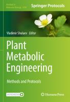
Fig. 1 Administration of antisense oligonucleotides (ASOs). For intraperitoneal (IP) administration, hold the mouse in one hand keeping the body extended to avoid damage (a). With the other hand, insert the needle (27G) a bit upper than the hip in a 30–45° angle (b, c)
extended to avoid damage. With the other hand, insert the needle (27G) a bit upper than the hip in a 45° angle (Fig. 1). Use a 1 mL syringe.
2. Administration of radiolabeled compounds by oral gavage: 2 days after the last ASO injection (maximum effect), administer by oral gavage the radiolabeled compounds (150 μL per mouse by oral gavage of the [3H]-TG mixture). Fast the mice for 12 h. Take a blood sample from the tail vein (~20 μL) as the T0 value. For this, apply a small incision in the base of the tail, and take the drop with a heparinized capillary in an Eppendorf tube. Press for 30 s or until the bleeding stops. Gavage orally the prepared [3H]-TG solution (Subheading 2.1, item 3). For this, hold the mouse in one hand, keeping the body as extended as possible to avoid damage (the cannula should enter the stomach). With the other hand, insert the cannula through the mouth until the top of the throat is felt, and raise the cannula so that the throat is aligned with the esophagus (Fig. 2). 3. Administration of radiolabeled compounds by IV: 2 days after the last ASO injection (maximum effect), administer IV the radiolabeled compounds (100 μL per mouse IV of the [3H]oleate mixture). Fast the mice for 4 h. Take a blood sample from the tail vein (5 μL) as the T0 value as detailed above. Load the syringe with 100 μL of the [3H]-oleate solution (Subheading 2.1, item 4), and ensure that there is no bubble. Warm the mice with an IR light at a distance of 30 cm for 4–5 min being extremely careful not to cause damage to the mice (see Note 3)
In Vivo Tissue Lipid Uptake
5
Fig. 2 Oral gavage of [3H]-TG in olive oil. Hold the mouse in one hand, keeping the body as extended as possible to avoid damage (a). With the other hand, insert the cannula through the mouth until the top of the throat, and raise the cannula so that the throat is aligned with the esophagus. Insert the cannula to reach the stomach (B)
Fig. 3 Infrared (IR) system. The IR system is useful to visualize the tail vein. The light must be at 30 cm distance (a)
(Fig. 3). Immediately immobilize the mice in a specific chamber to ensure the correct injection (Fig. 4). Hold the tail and sterilize the injection area (2 cm in the middle of the tail) with ethanol. In the center of the tail, there is the arteria (big dark line). At 30–45° to the left or to the right, there are the veins (smaller dark lines). Turn the tail to make one of the veins more accessible, and insert the 30G needle with the bevel up (Fig. 5).
6
Igor Aurrekoetxea et al.
Fig. 4 Immobilization of a mouse in a specific chamber. Mouse being immobilized in the chamber (a–f)
Fig. 5 Intravenous injection in the tail vein. Hold the tail and sterilize the injection area (2 cm in the middle of the tail) with ethanol. In the center of the tail, there is the arteria (big dark line). At 30–45° to the left or to the right, there are the veins (smaller dark lines). Turn the tail to leave one of the veins in the top, and insert the 30G needle with the bevel up. Inject all the volume (a)
Inject all the volume (see Note 4). After the injection, maintain for a few seconds the needle inside. Then, press for 30 s or until the bleeding stops. 4. Follow-up of lipid clearance in blood in [3H]-TG-administered mice: Extract blood samples from the tail vein (20 μL), as described above for T0 (Subheading 3.1, step 2) at 30 (T30),
In Vivo Tissue Lipid Uptake
7
Fig. 6 Place the mice for the biological sample collection. Anesthetized mouse immobilized for surgery (a); opened intraperitoneal cavity (b)
60 (T60), 90 (T90), 180 (T180), and 240 (T240) min after the oral gavage to follow up the absorption of lipids through the intestine, secretion to blood, and clearance from blood. 5. Follow-up of lipid clearance in blood in [3H]-oleate-administered mice: Extract blood samples (5 μL) after 0.5 (T0.5), 1 (T1), 2 (T2), 3 (T3), 4 (T4), 5 (T5), 6.5 (T6.5), 8 (T8), and 10 (T10) min as detailed above (Subheading 3.1, step 2). 6. Biological sample collection: 240 min after the [3H]-TG gavage or 10 min after the [3H]-oleate injection, collect the different tissues to analyze the uptake of lipids. For this, anesthetize the mice by pentobarbital IP at a dose of 60 mg/kg (150 μL for a 25 g mouse from the stock solution) (Subheading 2.1, item 5). Check the mouse is anesthetized by foot reflex. Open the abdominal cavity from the hip to the breastbone (Fig. 6). Begin with the extraction of blood from the inferior vena cava, which is behind the intestines. With a tweezer, set aside the intestines, and using a 21G needle in a 1 mL syringe, insert the needle with the bevel up, and exsanguinate the mice (~700 μL) (Fig. 7). Collect the tissues of interest (e.g., liver, epididymal white adipose tissue (eWAT), heart, quadriceps muscle, interscapular brown adipose tissue (iBAT)) (Fig. 8). Clear the tissues of connective and surrounding adipose tissue and other contaminants, wash the tissues three times with cold PBS, eliminate the excess PBS, and weigh. Store all the samples at -80 °C immediately or go ahead with the next step.
8
Igor Aurrekoetxea et al.
Fig. 7 Blood collection. With a tweezer, set aside the intestines (a), and using a 21G needle in a 1 mL syringe, insert the needle with the bevel up, and extract all the blood volume (~700 μL) (b) 3.2 Sample Preparation and Measurements
1. Measurement of the radioactivity in the assay mixtures: Add 2 μl of the prepared assay solution ([3H]-TG mixture for gavage or [3H]-oleate mixture for IV injection) in 4 mL of commercial liquid scintillation cocktail previously added to specific plastic vials. Count the radioactivity in a liquid scintillation counter. Radioactivity levels are obtained in disintegrations per min (DPM). 2. Analysis of circulating [3H]-oleate in blood after the IV administration: It is important to check the decrease in circulating [3H]-oleate in blood given that it corresponds with the increased uptake by tissues along this short period of time (10 min). For this, add the 2 μL from the blood sample (Subheading 3.1, step 5) to 4 mL of commercial liquid scintillation cocktail previously added in a specific plastic vial to measure the radioactivity in the liquid scintillation counter. Radioactivity levels are obtained in disintegrations per min (DPM). 3. Analysis of circulating TG in serum after the oral gavage: The follow-up of TG in serum along the 240 min of the assay shows a peak, corresponding with the maximum absorption from the intestine and its secretion to blood in chylomicrons, which decreases as the TG is internalized into tissues (Fig. 9a). To check this, obtain serum from the blood samples collected after the oral gavage along the 240 min (Subheading 3.1, step 4). Centrifuge the blood at 2000 × g, 4 °C for 30 min, collect the supernatant, and centrifuge again at 10,000 × g, 4 °C for 10 min. Determine the concentration of TG using any
In Vivo Tissue Lipid Uptake
9
Fig. 8 Tissue collection. Liver (a), epididymal white adipose tissue (eWAT) (b), heart (c), muscle (d), and interscapular brown adipose tissue (iBAT) (e, f)
commercial kit and following the instructions of the manufacturers. In addition, use 2 μL of serum obtained from the inferior vena cava to check the radioactivity as described above for the blood (Subheading 3.2, step 2).
4. Tissue preparation for analytical measurements: Prepare the collected tissues for radioactivity measurement. For this, remove the excess of PBS, weigh the tissues, separate 40–50 mg of each tissue for homogenization, and freeze in dry ice or liquid nitrogen the rest of each tissue. Store them at -80 °C. 5. Homogenize the tissue pieces in cold PBS at a ratio of 1:10 (1 g of tissue in 10 mL of PBS) in a bead mill homogenizer using ceramic beads (see Note 5). Sonicate homogenates by three cycles of 30 s sonication and 10 s rest at a frequency of 23 kHz and with an amplitude ≤6 microns, and measure the final
10
Igor Aurrekoetxea et al.
A
300
mg/dL
200
100
0
0
60
120
180
240
300
Time (min)
B
[3H]-lipid uptake (% incorporated/g tissue)
12 10 8 6 4 2 0
Liver
eWAT
BAT
Muscle
Heart
Fig. 9 Follow-up of dietary lipids. Fluctuations of serum triglycerides after the oral gavage (a). Amount of radioactivity (per g of tissue) incorporated into the tissues after the oral gavage as % incorporated from the total amount administered (b)
volume of the preparation (see Note 6). Add 100 μL of each tissue homogenate to 4 mL of commercial liquid scintillation cocktail previously added in a specific plastic vial to measure the radioactivity in a liquid scintillation counter. Radioactivity levels are obtained in disintegrations per min (DPM). 3.3
Data Analysis
Provide the tissue lipid uptake as DPM per g of tissue, DPM per mg of tissue protein, or percent (%) of total radioactivity administered to the mice, incorporated per g of tissue or per mg of protein. 1. Blood and/or serum radioactive lipid content. The serum radioactive lipid content shows the amount of dietary lipid (oral gavage experiment) that is still circulating. The blood radioactive oleate content shows the amount of fatty acids that are still circulating (IV experiment), which in this case
In Vivo Tissue Lipid Uptake
11
depends exclusively on the tissue uptake. Express both in DPM per mL following the formula: Quantity of radioactivity ðDPM=mlÞ =
DPM V
[DPM: amount of radioactivity expressed as disintegrations per min. V: volume of blood or serum used to measure the radioactivity in mL] 2. Tissue lipid uptake in DPM/g tissue. After collection of different tissues, homogenization, and measurement of radioactivity, use this formula: Quantity of radioactivity ðDPM=g tissueÞ =
DPM * Vt V * Wt
[DPM: amount of radioactivity expressed as disintegrations per min. Vt: total volume of homogenate after sonication in μl. V: volume of homogenate used to measure radioactivity (100 μL). Wt: amount of tissue homogenated in g] 3. Tissue lipid uptake in DPM/mg of protein. After collection of different tissues, homogenization, and measurement of radioactivity, measure the tissue protein concentration using a colorimetric commercial kit based on the bicinchoninic acid (BCA) method and following the manufacturer’s instruction, and express it in mg/mL (see Notes 7 and 8). Use the equation below: Quantity of radioactivity ðDPM=mg proteinÞ =
DPM V * ½Prot]
[DPM: amount of radioactivity expressed as disintegrations per min. V: volume of homogenate used to measure radioactivity (0.1 mL). [Prot]: protein concentration of the homogenate in mg/mL] 4. Tissue lipid uptake (per g of tissue or per mg of protein) as % of total radioactivity administered to the animal [5]: First, calculate the total radioactivity that has been administered to the animal. Use the equation below: Quantity of radioactivity administered ðDPMtÞ =
DPM * Vt V
[DPM: disintegrations per min measured in the assay mixture (Subheading 3.2, step 1). Vt: total volume administered to the mouse (150 μL per mouse by oral gavage of the [3H]-TG mixture or 100 μL per mouse IV of the [3H]-oleate mixture). V: volume of assay mixture used to measure radioactivity (2 μL)]. Next, obtain the % using the DPM/g tissue or the DPM/mg of protein and the DPM administered following the equation below (Fig. 9b):
12
Igor Aurrekoetxea et al.
%of radioactivity per g tissue or mg prot =
DPM * 100 DPMt
[DPM: amount of radioactivity per g of tissue or per mg of protein (Subheading 3.3, step 2 or Subheading 3.3, step 3). DPMt: total radioactivity administered to the animal. 100: conversion factor expressed as percentage].
4
Notes 1. Decontaminate, store, and/or remove all the materials in contact with radioactive substances following the specific protocol of each radioactive facility, previously approved by the competent authorities. 2. Given that the density and viscosity of the solution are high, avoid the syringe obstruction during the injections by maintaining the solution at 37 °C. 3. Monitorize the mouse carefully for any evidence of overheating such as fast movements. If so, remove immediately from the IR light. 4. Be careful with the IV injection in the tail vein because it is shallower than it seems. If there is any resistance, you are not inside the vein. 5. Check that the ceramic beads are proper for each tissue (WAT and BAT are easier homogenized than harder tissues like the heart and muscle). 6. Ensure an effective sonication by using an exponential probe submerged up to 13 mM into the homogenate without touching the tube walls. If there is no sufficient homogenate volume, add more PBS up to a recommended final volume of 600–800 μL. 7. Dilute 1/5 or 1/10 of the homogenate of metabolically active tissues such as the heart, liver, muscle, or BAT given their high protein content. On the other hand, do not dilute other tissues with low protein content, such as WAT. 8. Add sodium dodecyl sulfate (SDS) salt at a final concentration of 2% (w/v) to tissues with high content of lipids to avoid the underestimation of protein concentration. For this, prepare a 20% (w/v) SDS stock solution by dissolving 2 g of SDS in 10 mL of dH2O with gentle agitation to avoid excessive bubble formation. As SDS is toxic, use goggles, gloves, and an appropriate mask.
In Vivo Tissue Lipid Uptake
13
Acknowledgments This work was supported by Ayudas para apoyar las actividades de grupos de investigacio´n del sistema Universitario Vasco (IT147622); MCIU/AEI/FEDER, UE (PID2021-124425OB-I00), Basque Government, GV (2020111077 and IT.02-SPR.13.08); MCI/UE/ISCiii (PMP21/00080) and UPV/EHU (COLAB20/01); and FEEH/Juan Cordoba scholarship (2021). References 1. Blu¨her M (2019) Obesity: global epidemiology and pathogenesis. Nat Rev Endocrinol 15(5): 288–298. https://doi.org/10.1038/s41574019-0176-8 2. Vekic J, Zeljkovic A, Stefanovic A et al (2019) Obesity and dyslipidemia. Metabolism 92:71– 81. https://doi.org/10.1016/j.metabol. 2018.11.005 3. Cunarro A, Buque´ X, Casado S et al (2019) p107 deficiency increases energy expenditure by inducing Brown-fat thermogenesis and browning of white adipose tissue. Mol Nutr Food Res 63(2):e1801096. https://doi.org/ 10.1002/mnfr.201801096 4. Quiroga AD, Lian J, Lehner R (2012) Carboxylesterase1/esterase-x regulates chylomicron production in mice. PLoS One 7(11): e49515. https://doi.org/10.1371/journal. pone.0049515 5. Saenz de Urturi D, Buque´ X, Porteiro B et al (2022) Methionine adenosyltransferase 1a antisense oligonucleotides activate the liverbrown adipose tissue axis preventing obesity and associated hepatosteatosis. Nat Commun 13(1):1096. https://doi.org/10.1038/ s41467-022-28749-z 6. Ruiz de Gauna M, Biancaniello F, Gonza´lezRomero F et al (2022) Cholangiocarcinoma progression depends on the uptake and
metabolization of extracellular lipids. Hepatology. https://doi.org/10.1002/hep.32344 7. Kraus D, Yang Q, Kong D et al (2014) Nicotinamide N-methyltransferase knockdown protects against diet-induced obesity. Nature 508(7495):258–262. https://doi.org/10. 1038/nature13198 8. Bartman CR, TeSlaa T, Rabinowitz JD (2021) Quantitative flux analysis in mammals. Nat Metab 3(7):896–908. https://doi.org/10. 1038/s42255-021-00419-2 9. Kim IY, Park S, Kim Y et al (2022) Tracing metabolic flux in vivo: basic model structures of tracer methodology. Exp Mol Med. https:// doi.org/10.1038/s12276-022-00814-z ˜ a M, Varela-Rey M, Mestre D et al 10. Martinez Un (2015) S-Adenosylmethionine increases circulating very-low density lipoprotein clearance in non-alcoholic fatty liver disease. J Hepatol 62(3):673–681. https://doi.org/10.1016/j. jhep.2014.10.019 11. Wong SK, Chin KY, Suhaimi FHJ et al (2016) Animal models of metabolic syndrome: a review. Nutr Metab (London) 13:65. https:// doi.org/10.1186/s12986-016-0123-9 12. Kleinert M, Clemmensen C, Hofmann SM et al (2018) Animal models of obesity and diabetes mellitus. Nat Rev Endocrinol 14:140–162. https://doi.org/10.1038/nrendo.2017.161
Chapter 2 In Vivo Hepatic Triglyceride Secretion Rate in Antisense Oligonucleotide (ASO)-Treated Mice Beatriz Gomez-Santos, Diego Saenz de Urturi, Xabier Buque´, Igor Aurrekoetxea, Ane Nieva, Idoia Ferna´ndez-Puertas, and Patricia Aspichueta Abstract The liver is a central organ in regulating the whole body metabolic homeostasis, and, among many other processes, it plays a crucial role in lipoprotein metabolism. The liver controls the secretion of very-lowdensity lipoproteins (VLDLs), particles specialized in the transport of liver lipids, mainly triglycerides (TGs), to the adipose tissue, heart, and muscle, among other tissues, providing fatty acids to be stored or to be used as an energy source. The analysis of this metabolic process provides relevant information about the crosstalk between the liver and other organs. It also helps to identify how the liver is able to secrete lipids to reduce its accumulation. This protocol shows how to analyze the liver TG secretion rate blocking the VLDL clearance from the blood by the administration of poloxamer 407. In addition, it shows how to isolate the VLDL produced by the liver at the end of the experiment, so that the apolipoprotein and lipid content and size can be measured. Using antisense oligonucleotides (ASOs) for silencing target proteins involved in metabolic diseases has emerged as a new promising therapeutic approach. Thus, the usage of ASOs has also been included in this protocol. As a conclusion, evaluation of TG secretion rate in mice provides key information to understand the organ crosstalk in metabolic diseases and the capacity of the liver to secrete lipids to blood. Key words Lipid, Lipoprotein, Liver, Metabolism, Oligonucleotides, Triglycerides
1
Introduction The liver is a central organ in the metabolization of lipids [1– 3]. Apart from controlling its synthesis and oxidation, it modulates lipid secretion in lipoproteins to blood, where they are rapidly metabolized to provide them to other tissues [4, 5]. In metabolic diseases, the liver is exposed to different signals and environment, which leads, among others, to changes in the liver lipid composition [6–11]. This carries the alteration in the phenotypical characteristics of lipoproteins. Very-low-density lipoproteins (VLDLs) are
Salvatore Papa and Concetta Bubici (eds.), Metabolic Reprogramming: Methods and Protocols, Methods in Molecular Biology, vol. 2675, https://doi.org/10.1007/978-1-0716-3247-5_2, © The Author(s), under exclusive license to Springer Science+Business Media, LLC, part of Springer Nature 2023
15
16
Beatriz Gomez-Santos et al.
specialized in the transport of liver lipids, mainly triglycerides (TGs), to the adipose tissue, heart, and muscle, providing fatty acids to be stored (white adipose tissue) or as an energy source in some other tissues (brown adipose tissue, muscle, and heart) [12– 14]. To determine the rate of TG secretion [6] and to analyze the features of the “newly” synthesized VLDLs [8] provide relevant information to understand the impact that changes in VLDL secretion might have in liver disease and/or other metabolic diseases that implies the adipose tissue, the muscle, and dyslipidemias [6, 8]. The analysis of this metabolic process provides information about the capacity of the liver to eliminate the excess of lipids and the crosstalk with other organs. Given the relevance of this process, this protocol that analyzes the liver TG secretion (the main VLDL component) and the isolation of the “newly” secreted VLDL particles has been developed. To avoid the contribution of chylomicrons, which are synthesized in the intestine from dietary lipids and are enriched in TGs, as the VLDL, the mice should be exposed to a 4-h or, alternatively, an overnight fast. To elude the dynamic metabolization of the “newly” secreted VLDL particles, a detergent that inhibits the lipoprotein lipase activity, the poloxamer 407 (P407), needs to be used. Then, blood will be collected at several time points to measure the circulating TG concentration. Next, blood will be withdrawn and VLDL particles separated for further analysis. The use of antisense oligonucleotides (ASOs) for silencing target proteins involved in metabolic diseases has emerged as a new promising therapeutic approach. Thus, the usage of ASOs has also been included in this protocol. The liver TG secretion rate, as other metabolic fluxes that depend on the contribution of other organs, not just the liver cells, must be assayed in vivo and cannot be replaced by in vitro assays. Thus, prior to the experiments, this protocol must be approved by the authorities meeting the current ethical regulations.
2
Materials
2.1 In Vivo Antisense Oligonucleotide (ASO) Treatment
1. Phosphate-buffered saline (PBS): 137 mM NaCl, 2.7 mM KCl, 10 mM Na2HPO4, and 1.8 mM KH2PO4. To prepare 1 L of PBS, dissolve 8 g of NaCl, 0.2 g of KCl, 1.44 g of Na2HPO4, and 0.24 g of KH2PO4 in 800 mL of distilled water. Then, adjust the pH to 7.4 and finally make up to 1 L with distilled water. 2. Antisense oligonucleotide (ASO): From a liquid solution, make a 10 mg/mL solution. If the ASO is in powder, dissolve 15 mg of ASO in 1.5 mL of PBS. Vortex 2 min and let the solution
In Vivo Hepatic Triglyceride Secretion
17
stand for 15 min. Filter through syringe filter (0.2 μm). Determine the concentration of the solution after filtration. For ten mice, prepare 1.5 mL. Store at 4 °C. 3. Pentobarbital sodium: To prepare a 10 mg/mL stock solution of pentobarbital sodium in sterile PBS, use 125 μL from the commercial stock solution (400 mg/mL), and add 4.875 mL of sterile PBS (this is the amount needed for ten mice). As it is photosensitive, maintain the solution protected from the light and store at room temperature and discard after a week. 2.2 Triglyceride (TG) Secretion Rate
1. Poloxamer 407: Poloxamer 407 (P407), also known as Pluronic™ F-127, is a non-ionic detergent that inhibits lipoprotein lipase activity in vivo; thus, lipoproteins accumulate in blood due to lack of clearance. To prepare a 120 mg/mL stock solution of P407 in sterile PBS (Subheading 2.1, item 1) for ten mice, weigh 0.3 g of P407 and add 2.2 mL of sterile PBS; dissolve it by shaking or rotating overnight at room temperature. The following day, when the P407 is completely dissolved, add the volume of required sterile PBS to make up to 2.5 mL. Keep at 4 °C until use and discard after 2 days. This working solution takes time to dissolve; thus, prepare the day before (see Note 1). 2. Flotation buffer: 1.55 mM chloramphenicol, 0.2 M NaCl, 0.01 M EDTA-Na2, and 7.7 mM sodium azide solution, density 1.02 g/mL, pH 7.4. For a 30 mL of flotation buffer (required for 20 assays), weigh 0.015 g of chloramphenicol, 0.35 g of NaCl, 0.112 g of EDTA-Na2, and 0.015 g of sodium azide, and dissolve in 25 mL of distilled water. Adjust the pH to 7.4 and the final volume to 30 mL. To achieve the desired density for this buffer (1.02 g/mL), so that the VLDL particles will float given the lower density, at the end of the preparation, KBr should be added. To know the exact amount of KBr to be added, first measure the density of the buffer in a precision balance by weighing 1 mL, and obtain the g/mL (see Note 2). For an accurate measure of the buffer density, repeat this step ten times, and calculate the average. The amount of KBr that should be used will be provided by this formula: KBr ðgÞ = V i ðρf - ρi Þ=1 - ν* ρf [Vi, initial volume; ρi, initial density; ρf, desired density (1.02 g/mL); ν, KBr specific volume (0.266 mL/g)] After the addition of the amount of KBr required, weigh again 1 mL of the buffer ten times to assess the new density. In case it is still not the required density, apply the formula again until achieving the required density (see Note 3). 3. Saline solution: NaCl 0.9% (w/v). Weigh 0.9 g of NaCl and add dH2O to a final volume of 100 mL.
18
3
Beatriz Gomez-Santos et al.
Methods Fast the mice so that the dietary lipids released to the blood in chylomicrons are cleared. Consider 4 h of fasting or an overnight fasting depending on the purpose of the experiment (see Notes 4 and 5).
3.1 In Vivo ASO Treatment
1. Weigh the mice to calculate the volume of injection depending on the dose. 2. For the in vivo ASO treatments, for fine-tuning and to achieve the maximum knock-down without inducing any damage [6], consider two doses, 25 mg/kg and 50 mg/kg. 3. Prepare two groups of mice per each dose, and inject to each group the ASO control or the target ASO at the corresponding dose. Perform intraperitoneal (IP) injection once per week during 4 weeks in a 1 mL syringe. For this, hold the mouse in one hand, keeping the body extended to avoid damage. With the other hand, insert the needle (27G) a bit upper than the hip in a 45° angle (Fig. 1).
3.2 Triglyceride (TG) Secretion Rate
1. Blood collection: After the fasting period, collect an aliquot of blood (~20 μL) to measure basal levels of TG (T0). For this, apply a small incision in the tail vein, and take the drop with a heparinized capillary in an Eppendorf tube. Press for 30 s or until the bleeding stops. Weigh the mice (see Note 6), and inject IP either PBS (vehicle) or P407 (Subheading 2.2, item 1) at 1 g/kg (i.e., 250 μL to a 30 g mouse) using a 1 mL syringe with a 1/2 inch 29G needle (see Note 7). Collect ~20 μL of blood from the tail vein 3 (T3) and 5 (T5) hours after the P407 injection as detailed for T0.
Fig. 1 Mouse handling for intraperitoneal (IP) injection. For IP injection, hold the mouse with one hand while carefully injecting with the other the required volume by inserting the needle (27G) at a 45° angle
In Vivo Hepatic Triglyceride Secretion
19
Fig. 2 Lipid-rich serum in P407-injected mice. Serum from mice injected with P407 has a milky appearance due to the high concentration of lipids
2. Isolation of serum: Allow the blood to clot at room temperature for 30 min, and centrifuge the tubes to obtain the serum at 2000 × g 4 °C for 30 min, and collect the supernatant. Then, centrifuge again the supernatant at 10,000 × g 4 °C, and keep the supernatant. 3. TG measurement in serum: Use the serum obtained at T0, T3, and T5 after the P407 administration to measure the TG concentration. Use a commercial kit following the manufacturer’s instructions for TG measurement. Calculate the serum TG concentration for every time point by using a standard curve and plotting absorbance values against the concentrations of the standard. Take into account that in the P407treated group, mice will have increased TG concentration, as observed with the milky appearance of the serum (Fig. 2), caused by their inability to metabolize VLDLs. Thus, the recommended volume to use for each time point in P407treated mice is the following: 4 μL of serum for T0, 4 μL of a 1/10 dilution of serum for T3, and 2 μL of a 1/10 dilution of serum for T5. For vehicle-treated mice, use 4 μL for every time point. 4. Calculate the concentration of TG in mg/dL per mouse per time point, and represent the result as concentration per time point [6] (Fig. 3).
20
Beatriz Gomez-Santos et al.
Hepatic Triglycerides Secretion Rate 6000
mg/dL
4000
2000
0 0
1
2
3
4
5
6
Time (h)
Fig. 3 Representation of hepatic triglyceride (TG) secretion rate in chow diet-fed mice. TG secreted along the 5 h after the P407 injection. Values are represented as the mean ± SEM of an n = 3
5. Alternatively, calculate the μM/kg/h of TG secreted during ˜ a et al. [8]. This those 5 h, as described before by Martinez-Un method will be accurate if there are no big differences in body weight between the groups of study. 3.3 VLDL Isolation and Characterization
VLDL isolation should be performed the same day of the sacrifice to avoid VLDL rupture or contamination. 1. Sacrifice the mice and collection of blood to isolate the VLDL particles: 6 h after the P407 injection, sacrifice the mice by IP injection of 60 mg/kg of sodium pentobarbital (Subheading 2.1, item 3). Ensure that the mice are completely anesthetized by foot reflex. 2. Open the abdominal cavity, and set the contain (intestines) aside carefully to the right to allow to find the inferior vena cava behind the intestines (Fig. 4). Very carefully with some clean paper towel dry the cavity in case there are any remains of sodium pentobarbital. 3. Collect the blood from the inferior vena cava using a 21G needle and a 1 mL syringe with the needle bevel up and taking as much blood as possible (~700 μL) (see Note 8). Do this step as fast as possible to avoid clotting of the blood. Place the obtained blood into a 1.5 mL Eppendorf tube. 4. Obtain the serum as described in Subheading 3.2, step 2.
In Vivo Hepatic Triglyceride Secretion
21
Fig. 4 Mouse abdominal cavity opens with the inferior vena cava visible. Blood is obtained from the inferior vena cava, which is located behind the intestines and the organs of the abdominal cavity. Carefully set aside the organs and localize the inferior vena cava (indicated with an arrow in the image) (a). The 21G needle should be used to obtain as much blood as possible being careful to not break the wall of the vein (b)
5. Add 150 μL of serum and 1.35 mL of saline solution (Subheading 2.2, item 3) to a 3.2 mL polycarbonate thick wall tube (13 × 56 mm) for VLDL extraction, and store the remaining serum in aliquots to avoid freeze-thaw cycles at -80 °C. 6. Very slowly, dropwise, add 1.5 mL of the flotation buffer (Subheading 2.2, item 2), so the final volume is 3 mL (see Note 9) (Fig. 5). 7. Equilibrate the tubes for the ultracentrifuge with flotation buffer. Centrifuge the tubes at 416,000 × g for 2.5 h at 16 °C with an acceleration of 9 and coast deceleration in the microultracentrifuge. 8. After centrifugation, take the tubes out very gently because layers of separated VLDLs are fragile and can be mixed easily. In the upper layer, a whitish cloud will be visible (these are the VLDLs) and in the bottom of the tube an orange layer that will be the HDLs (Fig. 6).
22
Beatriz Gomez-Santos et al.
Fig. 5 Dropwise flotation buffer addition to the serum. Serum and NaCl 0.9% in polycarbonate tubes (13 × 56 mm) and adding the flotation buffer dropwise
Fig. 6 Separation of VLDL after ultracentrifugation in polycarbonate tubes. VLDL appears as a whitish cloud in the top of the tube after centrifugation, and HDLs appear at the bottom of the tube with a yellow-orange appearance. A dark background is used to improve the detection of the VLDL cloud
In Vivo Hepatic Triglyceride Secretion
23
Fig. 7 Extraction of the VLDL particles. Very gently take the VLDL cloud by placing the tip of the pipette in the upper part of the cloud while gently rotating the tube with the other hand
9. VLDL collection: To obtain the VLDL cloud, use a smooth pipette to avoid harsh aspiration of the cloud (see Note 10) (~1.2 mL). No more than 1.5 mL of the upper phase should be taken. While taking the upper phase, rotate the tube with the other hand. Aspirate the cloud placing the pipette tip on the wall of the tube and moving the tube, not the pipette (Fig. 7). After upper phase collection, carefully measure the exact volume by using an automatic pipette. This volume will be the 100% of the VLDL fraction. Freshly isolated VLDL should be kept in ice or at 4 °C after collection (see Note 11). 3.4 Further Processing and Use of VLDLs
Once extracted and isolated, VLDLs can be used for a variety of purposes. They can be used for characterization or for in vivo or in vitro studies. This is out of the scope of this chapter; however, here, we list some of the most useful purposes to be used:
24
Beatriz Gomez-Santos et al.
1. Characterization: To acquire the VLDL size, VLDL lipid content, and apolipoprotein content, as has been described before [8], will be useful to understand its altered clearance in blood or changes related to liver disease [8]. 2. In vivo experiments: The isolated VLDLs might be labeled (e.g., radiolabeled) and administrated intravenous (IV) to the mice to analyze the altered clearance and tissue uptake, as described before [8]. This experiment should be performed with fresh isolated VLDLs (see Note 12). 3. In vitro treatment of cells: For this, in order to preserve VLDL particles, VLDLs should be mixed with 50% (w/v) filtered sucrose so the final concentration of sucrose in the VLDLs is 10% (v/v). Measure protein and aliquot VLDLs on the desired concentration for cell culture treatment at -80 °C [15] (see Note 12).
4
Notes 1. The P407 working solution is near the maximum solubility so it takes time to dissolve, but it is necessary to prepare the stock at the indicated concentration to not exceed the injectable volume per mouse. In addition, keep in mind that P407 is a quite viscous solution, so inject it slowly and carefully. 2. Make sure that the automatic pipettes are accurately calibrated in order to make an accurate measurement of the volume and weight to calculate an adequate density. 3. This step is key for the entire protocol; ensure an accurate density of the buffer to obtain optimal results. 4. Change the cage and its content for the fasting period to ensure that no food remains are left hidden, especially relevant when using a high-fat diet or any other diet with lower consistency. 5. Circadian rhythm has shown to be a key process in many biological processes including metabolism. Thus, preforming this experimental procedure always at the same time is also highly recommended. In addition, including a control group with vehicle-injected mice is recommended in every assay. 6. The body weight is used for calculating the P407 dose and the secretion rate; thus, make a precise measurement of body weight. 7. Immediately after P407 injection, activate the stopwatch for each mouse, and write down the time difference between mice injections. Carefully place the mouse back on its cage, and keep it under control for any possible damage derived from manipulation or IP injection.
In Vivo Hepatic Triglyceride Secretion
25
8. As reference, from 700 μL of blood, around 350 μL of serum is obtained. 9. While the flotation buffer can be prepared a couple of days in advance and kept at 4 °C, warm up the buffer at room temperature to make sure that the density is correct. 10. In order to obtain the VLDL fraction as pure as possible, we recommend using a black cardboard to be able to distinguish the different colors of the tube and to be very gentle when collecting VLDL with the pipette, thus avoiding disruption of the VLDL cloud. Also, placing the tubes in a rack while doing this process is recommended to avoid an excessive movement of the tubes that might disrupt the VLDL cloud. 11. Depending on the use of the VLDLs, they can be frozen or not. For lipid content evaluation or Western blotting analysis, they can be kept at -20 °C. However, for size, microscope pictures, or in vivo assays, VLDL cannot be frozen since it causes rupture and deformation. 12. When using VLDL particles for treating cells in vitro or in vivo, make sure not to add sodium azide to the flotation buffer, since it is toxic (Subheading 2.2, item 2). In addition, the VLDLs must be dialyzed to remove the KBr as described before [8].
Acknowledgments This work was supported by Ayudas para apoyar las actividades de grupos de investigacio´n del sistema Universitario Vasco (IT147622); MCIU/AEI/FEDER, UE (PID2021-124425OB-I00), Basque Government, GV (2020111077 and IT.02-SPR.13.08); MCI/UE/ISCiii (PMP21/00080) and UPV/EHU (COLAB20/01); and FEEH/Juan Cordoba scholarship (2021). References 1. Donnelly KL, Smith CI, Schwarzenberg SJ et al (2005) Sources of fatty acids stored in liver and secreted via lipoproteins in patients with nonalcoholic fatty liver disease. J Clin Invest 115(5):1343–1351. https://doi.org/10. 1172/JCI23621 2. Bechmann LP, Hannivoort RA, Gerken G et al (2012) The interaction of hepatic lipid and glucose metabolism in liver diseases. J Hepatol 56(4):952–964. https://doi.org/10.1016/j. jhep.2011.08.025 3. Nguyen P, Leray V, Diez M et al (2008) Liver lipid metabolism. J Anim Physiol Anim Nutr (Berl) 92(3):272–283. https://doi.org/10. 1111/j.1439-0396.2007.00752.x
4. Goldberg IJ, Eckel RH, Abumrad NA (2009) Regulation of fatty acid uptake into tissues: lipoprotein lipase- and CD36-mediated pathways. J Lipid Res 50(Suppl):S86–S90. https:// doi.org/10.1194/jlr.R800085-JLR200 5. Bharadwaj KG, Hiyama Y, Hu Y et al (2010) Chylomicron- and VLDL-derived lipids enter the heart through different pathways: in vivo evidence for receptor- and non-receptormediated fatty acid uptake. J Biol Chem 285(49):37976–37986. https://doi.org/10. 1074/jbc.M110.174458 6. Sa´enz de Urturi D, Buque´ X, Porteiro B et al (2022) Methionine adenosyltransferase 1A antisense oligonucleotides activate the liver-
26
Beatriz Gomez-Santos et al.
brown adipose tissue axis preventing obesity and associated hepatosteatosis. Nat Commun 13(1):1096. https://doi.org/10.1038/ s41467-022-28749-z ˜ a M et al 7. Cano A, Buque´ X, Martı´nez-Un (2011) Methionine adenosyltransferase 1A gene deletion disrupts hepatic very low-density lipoprotein assembly in mice. Hepatology 54(6):1975–1986. https://doi. org/10.1002/hep.24607 ˜ a M, Varela-Rey M, Cano A et al 8. Martı´nez-Un (2013) Excess S-adenosylmethionine reroutes phosphatidylethanolamine towards phosphatidylcholine and triglyceride synthesis. Hepatology 58(4):1296–1305. https://doi.org/10. 1002/hep.26399 9. Aspichueta P, Pe´rez-Agote B, Pe´rez S et al (2006) Impaired response of VLDL lipid and apoB secretion to endotoxin in the fasted rat liver. J Endotoxin Res 12(3):181–192. h t t p s : // d o i . o r g / 1 0 . 1 1 7 9 / 096805106X102174 10. Friedman SL, Neuschwander-Tetri BA, Rinella M et al (2018) Mechanisms of NAFLD development and therapeutic strategies. Nat Med
24(7):908–922. https://doi.org/10.1038/ s41591-018-0104-9 11. Heeren J, Scheja L (2021) Metabolicassociated fatty liver disease and lipoprotein metabolism. Mol Metab 50:101238. https:// doi.org/10.1016/j.molmet.2021.101238 12. Tiwari S, Siddiqi SA (2012) Intracellular trafficking and secretion of VLDL. Arterioscler Thromb Vasc Biol. 32(5):1079–1086. https://doi.org/10.1161/ATVBAHA.111. 241471 13. Tchernof A, Despre´s JP (2013) Pathophysiology of human visceral obesity: an update. Physiol Rev 93(1):359–404. https://doi.org/ 10.1152/physrev.00033.2011 14. Takahashi S, Sakai J, Fujino T et al (2004) The very low-density lipoprotein (VLDL) receptor: characterization and functions as a peripheral lipoprotein receptor. J Atheroscler Thromb 11(4):200–208. https://doi.org/10.5551/ jat.11.200 15. Lu M, Gursky O (2013) Aggregation and fusion of low-density lipoproteins in vivo and in vitro. Biomol Concepts 4(5):501–518. https://doi.org/10.1515/bmc-2013-0016
Chapter 3 Measurement of Fatty Acid Oxidation by High-Resolution Respirometry: Special Considerations for Analysis of Skeletal and Cardiac Muscle and Adipose Tissue Nicole T. Watt, Amanda D. V. MacCannell, and Lee D. Roberts Abstract High-resolution respirometry is a state-of-the-art approach for the quantitation of mitochondrial function. Isolated mitochondria, cultured cells, or tissues/fibers are suspended in oxygenated respiration medium within a closed chamber and substrates or inhibitors added in a stepwise manner. The dissolved oxygen concentration decreases as aerobic metabolism in the specimen proceeds, recorded by an oxygen sensor within the chamber to give a quantifiable measure of oxygen consumption by the sample. Measuring oxygen consumption using a variety of respiratory substrates or respiratory complex-targeted inhibitors enables multiple respiratory pathways to be interrogated to determine the functional capacity of the mitochondria in real time. Using a substrate-uncoupler-inhibitor titration (SUIT) protocol, we have developed a method which makes use of differing chain length fatty acids to derive a measure of fatty acid-stimulated respiration through β-oxidation in a variety of tissue types including skeletal and cardiac muscles and brown and white adipose tissues. This report provides technical details of the protocol, and the adaptations employed, to generate robust analysis of mitochondrial fatty acid β-oxidation. Key words OROBOROS, High-resolution respirometry, Respiration, Metabolism, Mitochondrial respiration, SUIT protocol, Fatty acid oxidation, Carnitine palmitoyltransferase 1 activity, Uncoupling protein 1
1
Introduction Energy generation in mammalian cells occurs primarily through three coordinated metabolic pathways. Glycolysis, which takes place in the cellular cytoplasm, converting glucose into pyruvate and reduced nicotinamide adenine dinucleotide (NADH). The pyruvate generated is then converted to acetyl-CoA, which enters the tricarboxylic acid (TCA) cycle, an oxidative respirationdependent pathway. Alternatively, acetyl-CoA can be produced from fatty acids, which are broken down in the mitochondrial matrix through fatty acid β-oxidation. β-Oxidation contributes to
Salvatore Papa and Concetta Bubici (eds.), Metabolic Reprogramming: Methods and Protocols, Methods in Molecular Biology, vol. 2675, https://doi.org/10.1007/978-1-0716-3247-5_3, © The Author(s), under exclusive license to Springer Science+Business Media, LLC, part of Springer Nature 2023
27
28
Nicole T. Watt et al.
the generation of NADH and reduced flavin adenine dinucleotide (FADH2) required for mitochondrial electron transport chain (ETC) activity [1]. NADH:ubiquinone oxidoreductase (complex I) of the ETC oxidizes NADH (generated in the TCA cycle) to NAD+ and reduces coenzyme Q (ubiquinone) to ubiquinol [2]. Succinate dehydrogenase (complex II) is a second entry point into the ETC and catalyzes the oxidation of succinate to fumarate and the reduction of FAD to FADH2 and coenzyme Q to ubiquinol [3, 4]. It is significant to note that the ETC is not linear with electrons from complex I activity (NADH) and complex II (FADH2) converging at the Q junction and ubiquinol-cytochrome c oxidoreductase (complex III). Here, the ubiquinol from complexes I and II is oxidized, and the electrons are accepted by cytochrome c. Cytochrome c passes the electrons onto cytochrome c oxidase (complex IV) which donates them to oxygen to produce water. The activities of complexes I, III, and IV also result in proton translocation across the inner mitochondrial membrane to create an electrochemical gradient across the membrane. Protons flow back down the gradient, returning to the matrix via ATP synthase (complex V) which generates the energy required for ATP production [4]. Taken together, ETC activity and the generation of the electrochemical gradient are referred to as oxidative phosphorylation and generate the majority of cellular ATP [1] (Fig. 1). We have developed a set of novel, high-resolution respirometry protocols, using an OROBOROS Oxygraph-2k to measure mitochondrial function. The protocol employs the use of specific substrates to interrogate particular metabolic pathways and respiratory complexes of the ETC. This facilitates sensitive and quantitative measurement of mitochondrial activity in various tissue types [5]. To measure mitochondrial function, ETC complex-specific respiratory substrates are provided exogenously. Therefore, the sequence in which the reagents are applied is crucial to accurately interpreting the function of individual complexes in the context of the ETC and oxidative phosphorylation as a whole. We use a substrate-uncoupler-inhibitor titration (SUIT) protocol to assess mitochondrial function, particularly focusing on respiration mediated by fatty acid oxidation and the influence of fatty acids with differing chain lengths. We have optimized each protocol to tailor the assays to the tissue type under investigation, which include cardiac and skeletal muscles and brown and white adipose tissues. Initially, palmitoyl-carnitine (carnitine-conjugated long-chain 16C saturated fatty acid) or octanoyl-carnitine (carnitine-conjugated medium-chain 8C saturated fatty acid) is added to the sample chamber with malate also added to prime the TCA cycle. These provide substrates for fatty acid β-oxidation and delineate a fatty acid chain length specificity and preference for oxidation by the
High-Resolution Respirometry of Tissue Fat Oxidation
29
Fig. 1 Schematic representation of the mitochondrial electron transport chain (ETC) showing the key metabolites involved in the activation of the complexes. The complex-specific pharmacological inhibitors are shown. Electrons enter the ETC at complexes I and II through the reducing equivalents of NADH, succinate, and FADH2. The electrons (dashed line) pass through the ETC to ultimately be donated to oxygen at complex IV. Complexes I, II, and IV use free energy released by the oxidation-reduction reactions to pump protons from the mitochondrial matrix into the intermembrane space, generating a proton motive force which is used to convert ADP to ATP through complex V, ATP synthase
mitochondria. Malate dehydrogenase will oxidize malate to generate oxaloacetate for use in the TCA cycle as well as activating complex I [6]. Glutamate and pyruvate are added next and although they require carriers in order for them to reach the inner mitochondrial membrane, when present, will activate dehydrogenases (aspartate aminotransferase and glutamate dehydrogenase), reduce NADH, and stimulate complex I. Next, ADP is applied in excess. This provides substrate for the ATP synthase (complex V) and facilitates the measurement of coupled ETC and oxidative phosphorylation-mediated respiration (also described as state 3 respiration). Cytochrome c is added after ADP to determine the integrity of the mitochondrial membrane. Damage to the mitochondrial outer membrane can result in a partial loss of cytochrome c [6]. By measuring the rate of respiration in the presence of exogenous cytochrome c, it can be determined whether the respiration rates measured previously may have been adversely affected
30
Nicole T. Watt et al.
by damage to the mitochondrial membrane. The addition of succinate then provides the substrate for succinate dehydrogenasemediated respiration, a crucial component of complex II [3]. Electrons generated from the oxidation of NADH to NAD+ (complex I) (activated by malate, glutamate, and pyruvate) and FADH2 to FAD (complex II) (activated by the addition of succinate) are transferred to complex III of the ETC via coenzyme Q (ubiquinone) [4]. Carbonyl cyanide m-chlorophenylhydrazone (CCCP) is a potent uncoupling agent that increases the proton permeability of the inner mitochondrial membrane leading to a loss in membrane potential and mitochondrial-dependent ATP synthesis. Thus, CCCP uncouples the ETC from oxidative phosphorylation [7]. By including a titration of CCCP, a measure of maximal, albeit non-physiological, respiration can be determined. The penultimate steps of the protocol involve the use of inhibitors; rotenone acts as an inhibitor of complex I and facilitates the assessment of maximal respiration mediated from complex II of the ETC onward [8]. Antimycin A inhibits the reduction of cytochrome c in complex III, halting mitochondrial respiration and providing a measure of non-mitochondrial respiration (residual oxygen consumption) occurring in the sample [9]. As a final step, the co-application of ascorbate and tetramethyl-p-phenylenediamine (TMPD) followed by sodium azide can be used to ascertain the activity of complex IV of the ETC, as a proxy for mitochondrial density in the sample. TMPD acts as an electron donor, in the presence of ascorbate, to reduce complex IV allowing the measurement of maximal complex IV oxygen consumption [10]. Sodium azide then inhibits complex IV (cytochrome c oxidase), the final enzyme of the ETC. The SUIT protocol defines the oxygen consumption of respiratory pathways in mitochondria. However, additional measures of mitochondrial function can be derived by the substitution/inclusion of other steps in the standard procedure. Short- and mediumchain fatty acids can freely diffuse through the mitochondrial matrix as substrates for oxidation. However, long-chain fatty acids require the action of the carnitine palmitoyltransferase 1 (CPT1) to shuttle the long-chain fatty acids into the matrix. A reduction in CPT1 reduces the uptake of long-chain fatty acids into the mitochondria resulting in an increase in circulating free fatty acids. To ascertain whether there is a reduction in fatty acid oxidation due to a reduction in the initial shuttle of long-chain fatty acids into the matrix, palmitoyl-CoA can be administered separately with carnitine (rather than being delivered as a conjugated molecule) to derive a measure of CPT1 activity in tissues of interest. Uncoupling protein 1 (UCP1) is found in the inner mitochondrial membrane of brown adipose tissue (BAT) and is a key mediator of non-shivering thermogenesis. UCP1 facilitates leak of protons back through the inner mitochondrial membrane, releasing energy as heat and uncoupling substrate oxidation from ADP
High-Resolution Respirometry of Tissue Fat Oxidation
31
phosphorylation [11]. Dissipation of the mitochondrial membrane proton gradient occurs as protons pass through UCP1 instead of complex IV, thus reducing the synthesis of ATP. Guanosine 5′-diphosphate (GDP) acts as an inhibitor of UCP1 by reducing its ability to pump protons, thus reducing heat generation and instead promoting ATP synthesis. Inclusion of a GDP step in the SUIT protocol enables an indirect measure of UCP1 activity in the mitochondria.
2
Materials All solutions are prepared using ultrapure water and analytical grade reagents. Many of the reagents (palmitoyl-L-carnitine/octanoyl-Lcarnitine, malate, glutamate, ADP, cytochrome c, succinate, CCCP, rotenone, antimycin A, ascorbate, TMPD, and sodium azide) can be prepared in advance as long as they are stored under the correct conditions. Some reagents must be prepared fresh on the day of experimentation (i.e., saponin, digitonin, and pyruvate). 1. BIOPS – relaxing and preservation solution: 2.77 mM CaK2EGTA, 7.23 mM K2EGTA, 5.77 mM adenosine triphosphate (ATP), 6.65 mM magnesium chloride (MgCl2), 20 mM taurine, 15 mM phosphocreatine, 20 mM imidazole, 0.5 mM dithiothreitol (DTT), 50 mM MES, protease and phosphatase inhibitors. Make up buffer in dH2O and adjust pH 7.1 (using 5M potassium hydroxide); then aliquot and store at -20 °C. 2. Mitochondrial respiration medium, MiR05: 0.5 mM ethylene acid glycol-bis(2-aminoethylether)-N,N,N′,N′-tetraacetic (EGTA), 3 mM MgCl2, 60 mM lactobionic acid, 20 mM taurine, 10 mM potassium dihydrogen orthophosphate (KH2PO4), 20 mM HEPES, 110 mM D-sucrose, and 1 g/L BSA (fatty acid-free) [12]. For 1 L of media, use 800 mL of dH2O and mix first all compounds (except BSA and lactobionic acid); next add the lactobionic acid and adjust pH 7.1. Finally, add the BSA and adjust pH 7.1 and final volume to 1 L. Aliquots should be stored at -20 °C until required. 3. Homogenization buffer (HB) for mitochondrial isolations: 250 mM sucrose, 1 mM EGTA, 10 mM HEPES pH 7.4. Make up buffer in dH2O and adjust pH 7.4, aliquot, and store at -20 °C. 4. Substrates, uncouplers, and inhibitors: Most reagents, except for pyruvate, saponin, and digitonin, can be made up in advance and stored at -20 °C (ADP is stored at -70 °C). As the reagents undergo dilution once added to the sample chamber, all the solutions are made in concentrated stocks dissolved in dH2O, otherwise indicated:
32
Nicole T. Watt et al.
1. 100 mM octanoyl-L-carnitine. 2. 10 mM palmitoyl-L-carnitine. 3. 10 mM palmitoyl-CoA. 4. 0.5 mM carnitine. 5. 0.4 M malate (L-malic acid). 6. 2 M glutamate (L-glutamic acid). 7. 1 M succinate (succinic acid). 8. 0.5 M ADP. 9. 4 mM cytochrome c. 10. 800 mM ascorbate. 11. 200 mM (TMPD).
N,N,N′,N′-tetramethyl-p-phenylenediamine
12. 4 M sodium azide (NaN3). 13. 2 M guanosine 5′-diphosphate (GDP). 14. 20 mg/mL saponin in BIOPS. 15. 5 mg/mL digitonin in BIOPS. 16. 2 M sodium pyruvate in MiR05. 17. 4 mg/ml oligomycin in 100% ethanol. 18. 5 mM antimycin A in 100% ethanol. 19. 1 mM rotenone 100% ethanol. 20. Carbonyl cyanide m-chlorophenylhydrazone (CCCP) in dimethyl sulfoxide (DMSO).
3
Methods
3.1 Air/Zero Calibration of OROBOROS Oxygraph2K
1. Switch on the OROBOROS Oxygraph-2k. Open a calibration file in the Datlab software (OROBOROS). Check that stirrer bars have started to rotate at 750 rpm. Set temperature to 37 ° C. Wash out overnight storage solution (70% ethanol) by doing 3 × 1 min dH2O washes, followed by 3 × 1 min 100% ethanol washes, followed by a further 3 × 1 min dH20 wash before loading the sample chamber with 2.5 mL of room temperature MiRO5. 2. Wash the stoppers by doing three washes as in step 1 (dH2O, ethanol, dH20) and twist down into sample chamber to remove any air bubbles, aspirate off any excess MiR05 which may appear in the top of the stopper, and twist back up to re-introduce an air bubble. Leave to equilibrate to create an air reading. 3. Make up a solution of baker’s yeast in warm water with a few granules of sucrose to start respiration in the yeast. Twist down
High-Resolution Respirometry of Tissue Fat Oxidation
33
250
213
150
142
100
71
50
0 0:30
0:37
0:45 Air Cal
0:52 0:45 [h:min]
1:00
1:07
1B: O2 Slope uncorr. [pmol/(s*ml)]
284
200
0 1:15
Zero Cal
Fig. 2 Example trace of an air (blue box 01) and zero (blue box 02) calibration on the Oxygraph-2k (OROBOROS)
the stopper to seal the chamber, and inject 50 μL of the yeast solution into the chamber using a Hamilton syringe. Monitor the MiR05 oxygenation level (blue line) on the calibration curve, which should start to drop as the yeast use the oxygen in the respiration media. The line should reach zero and level off (see Note 1). 4. To perform an oxygen calibration on the OROBOROS Oxygraph-2k, mark the trace in the Datlab where the trace is level from the period there was an air bubble in the chamber (for an air calibration) and a second mark when the trace levelled off at zero following the addition of the yeast (zero calibration). Click on the yellow “Cal” button to confirm the calibration has been carried out on the Datlab software (Fig. 2). 5. Close the file; wash out the yeast from the sample chamber by doing 3 × 1 min wash with dH2O, 3 × 1 min wash with 70% ethanol (100% ethanol will cause the yeast to sporulate so it should not be used for these washes), and a final 3 × 1 min wash with dH2O; and finally add 2.5 mL of MiR05 to the sample chamber in anticipation of the experimental sample. Re-open a new file to store the experimental reading and leave to equilibrate with the new MiR05. 6. An air calibration should be carried out at the start of each day of experimentation to ensure quality control in your analysis. A zero calibration should be carried out fortnightly or each time the machine is used if there are more than 2 weeks between experiments. 3.2 Sample Preparation for Skeletal Muscle
1. As soon as it has been harvested (see Note 2), the muscle biopsy should be placed in ice-cold BIOPS (relaxing and preservation solution) to preserve mitochondrial function in the tissue (see Note 3). 2. Keep the sample in cold BIOPS for the next steps. Cut off a small piece of muscle (about 10 mg) for analysis. 5 mg of each sample will be used in each chamber of the OROBOROS Oxygraph-2k.
34
Nicole T. Watt et al.
3. Using a dissecting microscope, gently tease apart the individual muscle fibers, along their longitudinal axis, to enable diffusion of reagents into the tissue. 3.3 Sample Preparation for Cardiac Muscle
Analysis is carried out as above for skeletal muscle, omitting step 3. 5 mg of cardiac tissue is used per chamber of the OROBOROS Oxygraph-2k.
3.4 Sample Preparation for Adipose Tissue
This section describes the analysis of white adipose tissue (WAT) and brown adipose tissue (BAT). Before starting this type of analysis, consideration should be made as to what type of sample would best answer the experimental question being investigated. There are three potential tissue preparations, which can be considered for analysis, adipose tissue slices, adipose tissue homogenates, or isolated mitochondria from adipose tissue, with each having their own intrinsic benefits and disadvantages (see Table 1). 1. With the help of a dissecting microscope, remove as much connective tissue and blood vessel contamination of the sample as possible (see Note 4) creating a sample that is approximately 9–12 mg (BAT) or 20–25 mg (WAT). 2. If using adipose tissue slices or homogenate, carry out the dissection in ice-cold BIOPS. 3. If isolating mitochondria, place the cleaned sample into 500 μL HB.
Table 1 Comparison of sample options for respirometry analysis in adipose tissue comparing the advantages and disadvantages Sample
Advantages
Disadvantages
Adipose tissue slices
1. Fast 2. Consistent 3. No specialist equipment required
1. Increased diffusion rates require more GDP (for BAT) (not an issue with WAT as stir speed will cause selfhomogenization)
Adipose tissue homogenate
1. Decreases diffusion rate compared to slices 2. Quicker/higher yields compared with isolated mitochondria
1. Reduced consistency due to variations in homogenization 2. Can result in increased mitochondrial membrane rupture – causing an increase in cytochrome c flux
Isolated mitochondria from adipose tissue
1. Time-consuming 1. Directly analyzing mitochondria (could be considered a disadvantage 2. Low yield 3. Requires specialist equipment (i.e., depending on focus) ultracentrifuge and homogenizer) 2. Sample can be recovered and used for further analysis
High-Resolution Respirometry of Tissue Fat Oxidation
3.5 The Making of Adipose Tissue Slices
3.6 The Making of Adipose Tissue Homogenates
35
A small piece of adipose tissue (~15 mg BAT or 25 mg WAT) is dissected from a larger depot in ice-cold BIOPS and used intact (see Note 5). 1. Weigh fat pad, and add to a small Petri dish on ice containing a volume of MiR05 equivalent to 20 mg of tissue in 75 μL of buffer. 2. Chop fat pad with a scalpel blade until small pieces of tissue remain (see Note 6). Use a Pasteur pipette to add tissue homogenate to 1.5 mL microfuge tube, and homogenize on ice with tube pestles (ten times up/down, glass is preferable). 3. Once the mixture is cloudy, use Pasteur pipette to filter sample through 100 μm cell strainer or cheese cloth into a 50 mL sterile, centrifuge tube. 4. Keep homogenate on ice until ready. 5. Load 75 μL into each sample chamber (see Note 7).
3.7 Isolation of Mitochondria from Adipose Tissue
This step has been adapted from the literature [13]. 1. Mince adipose tissue sample in a 1.5 mL microfuge tube for 3 min/sample (see Note 6). 2. The minced tissue is poured from the microfuge tube into a larger glass mortar with a Teflon pestle and the volume topped up to 5 mL with homogenization buffer (HB). Homogenize the tissue (6 strokes at 100 rpm) on ice, and take care to ensure that the tissue is not trapped on the bottom of the mortar to enable a thorough breakup of the tissue. 3. Filter the homogenate through a gauze pad and centrifuge at 8700 × g for 10 min at 4 °C. 4. Quickly pour off the supernatant and resuspend the pellet in 300 μL of HB and transfer to a new centrifuge tube (see Note 8). 5. Make the buffer volume up to 5 mL again with HB and centrifuge at 800 × g for 10 min at 4 °C. 6. Transfer the supernatant to a new clean tube and discard the nuclear pellet. Centrifuge the supernatant again at 8700 × g for 10 min at 4 °C. 7. If using a BAT deposit, resuspend the pellet in HB, and centrifuge again at 8700 × g for 10 min at 4 °C. 8. If using WAT, resuspend the pellet in HB containing 1% bovine serum albumin (BSA) at 8700 × g for 10 min at 4 °C (see Note 9).
36
Nicole T. Watt et al.
9. Pour off the supernatant (both BAT and WAT) and resuspend the pellet in a further 5 mL of HB and centrifuge at 8700 × g for 10 min at 4 °C. 10. Repeat step 9. 11. If using BAT, resuspend the pellet in approximately 150 μL of MiR05 (without BSA), and keep on ice until ready to analyze. 12. If using WAT, resuspend pellet in approximately 100 μL of MiR05 (without BSA), and keep on ice until ready to analyze. 13. Quantify the total protein level in the mitochondrial preparation using the Bradford assay (see Note 10). 3.8 Analysis of OXPHOS by HighResolution Respirometry
1. Place the muscle tissue in a well of a 12-well plate with 2 mL BIOPS supplemented with 5 μL (20 mg/mL) saponin (final concentration 50 μg/mL) to permeabilize the cellular membrane tissue fibers and facilitate substrate and inhibitor tissue entry (skeletal or cardiac muscle). Place on a rocker at 4 °C for 25 min (see Note 11). Next, stop any further permeabilization by washing the muscle biopsy with 3 × 5-min washes in 1 mL of MiR05 in a 24-well plate on a rocker at 4 °C (see Note 11). 2. Blot dry and weigh the biopsy as accurately as possible. 3. Place the permeabilized muscle biopsy sample in the sample chamber of the OROBOROS Oxygraph-2k. Alternatively, add 2 μL digitonin (5 mg/mL; final concentration 10 μM) directly into the sample chamber with the adipose tissue homogenate. Partially screw down the stopper so that an air bubble remains in the chamber, hyperoxygenate the air bubble with an injection of oxygen (approximately 10 mL), and monitor the oxygen concentration of the MiR05 on the software. Once the oxygenation level reaches 450 nM/mL (see Note 12), screw down the stopper fully to prevent any further increases in oxygenation. Leave to equilibrate and the trace to settle. 4. Update the experimental details with the post-permeabilization tissue weight. 5. OXPHOS activity is monitored using a substrate-uncouplerinhibitor titration (SUIT) protocol using two fatty acids with different chain lengths (see Note 13). To compare the ability of the mitochondria to oxidize fatty acids with differing chain lengths, one chamber receives the carnitine-conjugated long-chain fatty acid palmitoyl-L-carnitine with malate, and the other receives the carnitine-conjugated medium-chain fatty acid octanoyl-L-carnitine with malate. All other compounds are added to both chambers in a stepwise manner (Table 2). The trace is monitored after the addition of each compound to determine when the added compound has had its
High-Resolution Respirometry of Tissue Fat Oxidation
37
Table 2 The order, volume, and final working concentration of the various substrates, uncouplers, and inhibitors added in the SUIT protocol
Step
Substrate
Volume added (μL)
Final working concentration
1
Palmitoyl-L-carnitine/ octanoyl-L-carnitine and malate
8 4 10
0.04 mM 0.2 mM 0.5 mM
2
Glutamate and pyruvate
10 5
10 mM 5 mM
3
ADP
10
2.5 mM
4
Cytochrome c
5
10 μM
5
Succinate
20
10 mM
6
CCCP
5+5
0.5 μM steps
15
7
Rotenone
5
0.5 μM
16
8
Antimycin A
5
2.5 μM
16
Notes
anticipated effect. This usually occurs once the reagent has been present for about 5 min. The next compound on the list can then be added to the chamber (Fig. 3) (see Note 14). 6. In order to correct for mitochondrial density using complex IV activity as a proxy (as an alternative to tissue mass), an additional step can be included. If this is carried out, the MiR05 in the sample chamber must be re-oxygenated to ensure there is sufficient oxygen available for this analysis to be performed (Table 3; see Notes 12 and 17). 7. The SUIT protocol is now complete; the sample chamber must be washed out using 3 × 1 min dH2O, 3 × 1 min 100% ethanol, and 3 × 1 min dH2O. The chamber is left containing 70% ethanol until it is next required (see Note 18). 8. The stoppers are similarly washed (dH2O, ethanol, dH2O) and inserted into the top of the chamber but not screwed down. 9. Flux values should be calculated from a single replicate for skeletal and cardiac muscle and from duplicate samples for BAT, sWAT, and vWAT. 3.9 Modifications of the SUIT Protocol
1. Analysis of CPT1 activity by measuring carnitine-supported OXPHOS: Carnitine (8 μL; 2 mM) and palmitoyl-CoA (4 μL; 50 μM) as individual components are given in combination with malate (10 μL; 0.5 mM) as step a of the SUIT protocol (Fig. 3).
38
Nicole T. Watt et al.
Fig. 3 Flow of substrate addition for high-resolution respirometry experiments. Blue boxes represent standard SUIT protocols to assess oxidative phosphorylation. Orange boxes represent protocols to assess fatty acid flux through carnitine palmitoyltransferase (CPT)
High-Resolution Respirometry of Tissue Fat Oxidation
39
Table 3 The order, volume, and final working concentration of the substrates required for the complex IV activity assay in the OROBOROS Oxygraph-2k Step
Substrate
Volume added (μL)
Final working concentration
9
Ascorbate TMPD
5 5
2 mM 0.5 mM
10
NaN3
20
20 mM
Notes
16
2. Determination of UCP1 activity using guanosine 5′-diphosphate (GDP): As an additional step, a titration of GDP (1 + 1 μL; 2 mM steps) (see Note 19) can be added into the SUIT protocol at step 9 in the analysis of adipose tissue. This should be performed after the addition of succinate but prior to CCCP.
4
Notes 1. Additional yeast solution can be added to the chamber to hasten the process. 2. If animal models are being used, euthanasia should be by cervical dislocation. One animal will provide sufficient tissue for analysis. 3. It can be stored at 4 °C in BIOPS for up to 2 h post-harvest prior to analysis. 4. Murine sWAT has a midline lymph node which should be removed during the cleaning process. 5. OXPHOS analysis can now be performed starting from Subheading 3.8, step 4. 6. This can also be done in 1.5 ml microfuge tube using a small pair of scissors to mince the tissue. 7. OXPHOS analysis can now be performed starting from Subheading 3.8, step 6. 8. Avoid contamination of the mitochondrial isolation with lipid that may be clinging to the tube at the end of the centrifugation step. 9. If the BSA is left out of step 8 in the mitochondrial isolation protocol, this will result in a lower yield of mitochondria from the preparation. 10. Using a 1:10 dilution of the isolated mitochondrial preparation will be necessary for BAT; a 1:10 dilution can also be used for WAT – though it is possible to reduce this to 1:5 if the pellet is particularly small.
40
Nicole T. Watt et al.
11. The incubation can be carried out either in a cold room or using an ice bucket placed on a rocker. 12. Do not allow the oxygenation of MiR05 to go above 600 nM/ mL as this can reverse the activity of the ATPase pump driving ATP back into the mitochondria and induce ROS formation. 13. All SUIT protocols require that the relevant substrates and modifying compounds are added in a specific order. This ensures that the mitochondrial complexes receive relevant substrates required for respiration to occur and eliminate ratelimiting effects that could influence the measurements being made. 14. All reagents remain in the sample chamber for the duration of the experiment; there are no wash-out steps within the SUIT protocol. 15. If the CCCP is liquid when it is first removed from the freezer, it has expired – it should still be frozen for it to be active – use a new aliquot. 16. The final inhibitors of mitochondrial respiratory chain activity (rotenone, antimycin A, and sodium azide) can take longer for their effects to be seen on the trace so they may require longer than 5 min. 17. Additional oxygen injections can be performed during the experiment where necessary to prevent the oxygen concentration from dropping below 200 nM/mL. These should be kept consistent across all experiments to minimize artifactual consequences on respiration rates. 18. Sodium azide and rotenone can be difficult to completely wash out of the chambers and may require a more extensive washing protocol to prevent unwanted artifact in later experiments if multiple samples are being analyzed consecutively. Start with 3 × 1 min washes with dH2O; next do 2 × 15 min washes with 100% ethanol, followed by 3 × 10 min washes with 70% ethanol. Finally, do 3 × 5 min washes with dH2O before 3 × 1 min quick washes with MiR05 before replacing with 2.5 mL of MiR05 for the next sample. 19. More GDP titration steps can be added if required to confirm that there is no further decrease in oxygen flux.
Acknowledgments This work was supported by funding from Diabetes UK (16/0005382; 19/0006049) and the Biotechnology and Biological Sciences Research Council (BB/T004231/1). ADVM is supported by a British Heart Foundation PhD studentship
High-Resolution Respirometry of Tissue Fat Oxidation
41
(FS/18/61/34182). LDR is supported by the Diabetes UK RD Lawrence Fellowship (16/0005382) and a Biotechnology and Biological Sciences Research Council Investigator Grant (BB/T004231/1). References 1. Holness M, Sugden M, Naish J (2015) Energy metabolism. In: Naish J, Syndercombe Court D (eds) Medical sciences, 2nd edn. Saunders Elsevier 2. Hirst J (2013) Mitochondrial complex I. Annu Rev Biochem 82:551–575 3. Bezawork-Galeta A, Rohlena J, Dong L, Pacal K, Neuzil J (2017) Mitochondrial complex II: at the crossroads. Trends Biochem Sci 42:312–325 4. Sousa JS, D’Imprima E, Vonck J (2018) Mitochondrial respiratory chain complexes. Subcell Biochem 87:167–227 5. Kuznetsov AV, Veksler V, Gellerich FN, Saks V, Margreiter R, Kunz WS (2008) Analysis of mitochondrial function in situ in permeabilized muscle fibers, tissues and cells. Nat Protoc 3: 965-976 6. Gnaiger E (2014) Mitochondrial pathways to Complex I: respiratory substrate control with pyruvate, malate and glutamate. In: Gnaiger E (ed) Mitochondrial pathways and respiratory control: an introduction to OXPHOS analysis. OROBOROS MiPNet Publications 7. Ruas JS, Siqueira-Santos ES, Rodrigues-Silva E, Castilho RF (2018) High glycolytic activity of tumor cells leads to underestimation of electron transport system capacity when mitochondrial ATP synthase is inhibited. Sci Rep 8: 17383. https://doi.org/10.1038/s41598018-35679-8
8. Fato R, Bergamini C, Bortolus M, Maniero AL, Leoni S, Ohnishi T, Lenaz G (2009) Differential effects of mitochondrial Complex I inhibitors on production of reactive oxygen species. Biochim Biophys Acta 1787:384–392 9. Vamecq J, Schepers L, Parmentier G, Mannaerts GP (1987) Inhibition of peroxisomal fatty acyl-CoA oxidase by antimycin A. Biochem J 248:603–607 10. Gnaiger E, Kuznetsov AV (2002) Mitochondrial respiration at low levels of oxygen and cytochrome C. Biochem Soc Trans 30:242– 248 11. Rousset S, Alves-Guerra M-C, Mozo J, B, Cassard-Doulcier A-M, Miroux Bouillaud F, Ricquier D (2004) The biology of mitochondrial uncoupling proteins. Diabetes 53(Suppl 1):S130–S135 12. Gnaiger E, Kutnetsov A, Schneeberger S, Seiler S, Brandacher G, Steurer W, Margreiter R (2000) Mitochondria in the cold. In: Heldmaier E, Klingenspor M (eds) Life in the cold: eleventh international hibernation symposium Austria, August 2000, Berlin, New York Springer, p 431. https://doi.org/ 10.1007/978-3-662-04162-8 13. McFarlane SV, Mathers KE, Staples JF (2017) Reversible temperature-dependent differences in brown adipose tissue respiration during torpor in a mammalian hibernator. Am J Physiol Regul Integr Comp Physiol 1:R434–R442
Chapter 4 In Vivo Imaging of Bone Marrow Long-Chain Fatty Acid Uptake Jayna J. Mistry and Stuart A. Rushworth Abstract In vivo imaging enables the detection and visualization of many different processes occurring within the body. Fatty acid uptake is a fundamental cellular process which is essential for the use of free fatty acids (FFAs) as a fuel source for metabolism. Detection and visualization of in vivo FFA uptake in the bone marrow has been relatively unknown. Here, we describe the process of non-invasive bioluminescent imaging of in vivo FFA uptake within the bone marrow. Key words Free fatty acids, Bone marrow, In vivo imaging, Hematopoietic cells, β-Oxidation
1
Introduction Free fatty acids (FFAs) are an important substrate in activating transcription factors and modulating signaling cascades. The uptake of FFAs as a substrate for beta (β)-oxidation and subsequent adenosine triphosphate (ATP) production [1] is universally recognized as a fundamental process which underpins cellular metabolism. The uptake of FFA, however, can be altered under different physiological and pathological conditions including stress [2], fasting [3], ischemic hypoxia [4], and cancer [5, 6]. Current methods to identify fatty acid uptake have largely been studied in vitro using tracing labeled fatty acids including fluorescence-based assay such as the QBT™ Fatty Acid Uptake Assay or radiolabeled fatty acids. While radiolabeled fatty acids can be used in vivo, disadvantages include high costs, the use of radioactive material, and the inability to dynamically monitor cell uptake of FFAs. Positron emission tomography (PET) using 18F-fluoro-2-deoxyglucose has also been used for in vivo tracing of fatty acids; however, this method is also costly, and 18F has a short half-life [7]. Detecting and quantifying FFA uptake and lipid flux in animal models following pathological, physiological, or pharmacological challenge is vital to
Salvatore Papa and Concetta Bubici (eds.), Metabolic Reprogramming: Methods and Protocols, Methods in Molecular Biology, vol. 2675, https://doi.org/10.1007/978-1-0716-3247-5_4, © The Author(s), under exclusive license to Springer Science+Business Media, LLC, part of Springer Nature 2023
43
44
Jayna J. Mistry and Stuart A. Rushworth
understanding metabolic cellular networks. Here, we demonstrate FFA uptake in the bone marrow can be tracked in vivo using a bioactivatable molecular imaging probe FFA Luciferin [8]. The probe consists of long-chain fatty acids conjugated to the reporter molecule, luciferin, by a cleavable disulfide bond. This bond is stable extracellularly but is readily reduced by glutathione inside the cell following FFA uptake [8]. The method allows for real-time, non-invasive quantitative imaging of fatty acid flux. This approach can help to further understand the basic metabolic process in both normal, malignant, and stressed conditions.
2 2.1
Materials Cell Culture
1. HEK293T cells (Cellosaurus CVCL_0063). 2. HEK293T culture medium: 500 mL DMEM high glucose, 10% fetal bovine serum (FBS). Filter-sterilize the medium through a 0.45 μm filter before use and store at 4 °C (see Note 1). 3. 100 mm dishes. 4. 0.25% trypsin. 5. 1× phosphate-buffered saline (PBS). 6. pCDH-EF1a-eFFly-mCherry. 7. Envelope plasmid pMDG (VSV-G expressor). 8. Cytomegalovirus promoter pCMV (gag-pol expressor).
2.2 Lentiviral Transduction
1. Miltenyi LS columns. 2. Miltenyi CD117 MicroBeads mouse. 3. CD117 antibody (clone 3C11). 4. MACS ® buffer: PBS, pH 7.2, 0.5% bovine serum albumin (BSA), and 2 mM EDTA. Store at 4 °C. 5. MACS ® MultiStand. 6. cKit media: StemSpan™ SFEM supplemented with 10 ng mSCF, 10 ng mIL-3, and 10 ng mIL-6. 7. 24-well tissue culture-treated plates. 8. Polybrene. 9. 50 mL falcon. 10. Trypan blue.
2.3
Transplantation
1. C57BL/6 J mice (CD45.2). 2. B6.SJL-PtprcaPep3b/BoyJ (CD45.1) (PepCboy) mice. 3. Busulfan.
Fatty Acid Uptake In Vivo
45
4. 1× PBS. 5. Dimethyl sulfoxide (DMSO). 6. Syringe. 7. 26-gauge needle. 8. Isoflurane. 9. 1× red blood cell lysis. 2.4
In Vivo Imaging
1. D-Luciferin. 2. 1× PBS. 3. Syringe. 4. 26-gauge needle. 5. Free Fatty Acid Luciferin (FFA-SS-luc). 6. BSA. 7. Isoflurane.
3
Methods All procedures are to be carried out at room temperature unless otherwise stated.
3.1 Transfection of Packaging Cells to Produce pCDHLuciferase-T2AmCherry Virus
1. Culture HEK293T cells in tissue culture-coated 100 mm dishes in 10 mL HEK293T culture medium (see Notes 2 and 3). 2. Keep cells in 37 °C incubator with a humidified atmosphere of 5% CO2. 3. Fresh HEK293T culture medium should be added to the cells every 3 days or dependent on cell growth. 4. Passage cells when at 80% confluency at a ratio of 1 in 10 using 0.25% trypsin (see Note 4). 5. Cells are ready for transfection at passage 3; split the cells at a ratio of 1 in 2 before transfection (see Note 5). 6. Defrost the three plasmids, pCDH-EF1a-eFFly-mCherry plasmid, vesicular stomatitis virus glycoprotein (VSV-G) (envelope proteins), and cytomegalovirus promoter (pCMV) (packaging protein promoter) on ice. 7. Refresh media on HEK293T cells with HEK293T culture medium before transfection. 8. Make a mastermix containing the three plasmids, 1.5 μg of pCDH-EF1a-eFFly-mCherry plasmid to 1 μg of VSVG and pCMV plasmids in 15 μLTE buffer.
46
Jayna J. Mistry and Stuart A. Rushworth
9. Prepare the transfection reagent; add 18 μL of FuGENE® 6 to 200 μL of Opti-MEM media. 10. Add plasmids mastermix to the transfection reagent and mix well. 11. Add the final mixture dropwise to the HEK293T cells and incubate at 37 °C for 24 h. 12. Refresh the media after 24 h of culture. 13. Collect the media at 48, 72, and 96 h after transfection in 75 μL aliquots, and store at -80 °C ready for use (see Note 6). 3.2 Lentiviral Transduction of cKit Cells
1. Isolate CD117+ (cKit) bone marrow cells using CD117 MicroBeads, mouse Miltenyi kit following manufacturer’s instruction (see Notes 7 and 8). Flow cytometry was used to determine enrichment by staining with cKit antibody. 2. Once cKit cells have been enriched, seed cells at a density of 2 × 10⁵ cells in 500 μL of pen-strep-free cKit medium into 24-well TC-treated plates. 3. Add 20 μL of prepared pCDH-luciferase-T2A-mCherry lentivirus to each well of cells. 4. Add 1 μg/mL polybrene to the cell suspension and gently swirl plate for equal distribution. 5. Incubate cells at 37 °C with a humidified atmosphere of 5% CO2 for 24 h. 6. Pool cells in 50 mL falcon and wash wells to collect all cells from the well (see Notes 9 and 10). 7. Centrifuge cells at 300 × g for 5 min. 8. Aspirate supernatant and resuspend cells in PBS for cell counting. 9. Resuspend cells in 1× PBS at a concentration of 1 × 10⁶ cells/ mL for transplantation into mice.
3.3 Conditioning Mice for Transplant
1. Three days before transplantation, treat mice with busulfan at 25 mg/kg/day for 3 days. 2. Make busulfan stock at 15.5 mg/mL in DMSO (see Note 11). 3. Dilute busulfan stock to 3.1 mg/mL in PBS. These steps need to be done quickly as busulfan can precipitate out of solution once diluted in PBS. 4. Inject 25 mg/kg/day busulfan interperitoneally 8–12-weekold CD45.1 mice (200 μL/25 g mouse) for three consecutive days. 5. On the day of transplantation, administer donor LK cells which have been transduced (pCDH-luciferase-T2A-mCherry) to the
Fatty Acid Uptake In Vivo
47
busulfan-treated recipient mice via intravenous (IV) tail vein injection (see Note 12). 6. Place mice in a benchtop restrainer to aid with IV injections. 7. Inject 200 μL of 2 × 10⁵ cKit cells suspended in PBS into the lateral tail vein with a sterile 26-gauge needle. 8. Place mice into a new cage for a short recovery period before returning to home cage. 9. Engraftment should be tracked at 4, 8, and 12 weeks by tail vein bleed. 10. Collect 200 μL peripheral blood from mice at time points. 11. Lyse blood with 1× red blood cell lysis for 1 min (see Note 13). 12. Centrifuge cells at 300 × g for 5 min. 13. Aspirate up supernatant and resuspend each sample in 50 μL MACS buffer. 14. Add in CD45.2 and CD45.1 antibodies and stain for 20 min at 4 °C. 15. Run sample on flow cytometer to check for CD45.2 engraftment. 3.4 FFA-Luciferase Allograft Mouse Engraftment Is Monitored by In Vivo Bioluminescent Live Animal Imaging
1. At 12 weeks post-transplant, confirm engraftment with in vivo bioluminescent live imaging. 2. Reconstitute D-luciferin in PBS at 15 mg/mL. 3. Inject mice with 150 mg/kg D-luciferin intraperitoneally. 4. Leave mice for 15 min at room temperature. 5. Anesthetize mice using a chamber filled with isoflurane with a flow rate of 3%. 6. Transfer mice to the nose cones inside the Bruker In-Vivo Xtreme. Keep the flow rate at 3% isoflurane. 7. Image mice using a pre-set method of 2-min exposure bioluminescent image, x-ray, and light image. 8. Once engraftment is confirmed, leave mice to sufficiently recover for at least 1 week post-injection and anesthesia. 9. Once mice have recovered, inject mice with 100 μL of 200 μM FFA-SS-luc (SwissLumix Sarl, Switzerland) (0.014 mg/ mouse) bound to 0.1% (w/v) BSA in PBS. 10. Leave mice in cages for 20 min at room temperature. 11. Anesthetize mice using a chamber filled with isoflurane with a flow rate of 3%. 12. Transfer mice to the nose cones inside the Bruker In-Vivo Xtreme. Keep the flow rate at 3% isoflurane.
48
Jayna J. Mistry and Stuart A. Rushworth
B
A
Fig. 1 In vivo imaging of long-chain FFA uptake. (a) Schematic diagram to explain transplant setup and experimental design. WT CD45.2 CD117+ (cKit) cells were enriched and transduced with a pCDH-luciferaseT2A-mCherry virus (cKit+FF) and transplanted into WT CD45.1 mice.12 weeks post-transplantation, mice were injected with 1 mg/kg LPS interperitoneally for 16 h and then injected with 200 μM FFA-SS-luc interperitoneally. The mice were bioluminescence imaged. (b) Representative images of LPS-treated FFASS-luc mice
13. Image mice using a pre-set method of 2-min exposure bioluminescent image, x-ray, and light image. 14. Images analyzed using ImageJ software (Fig. 1).
4
Notes 1. The HEK293T medium is stable for 6 months at 4 °C. 2. For tissue culture, always work in a laminar flow hood, make sure all solutions and equipment are sterile, and use sterile technique when working. 3. If using a non-filter flask instead of a plate, loosen cap for oxygenation/aeration. 4. The HEK293T cells should be treated very gently; they can detach very easily from the plate. When washing with PBS, wash slowly and never pipette directly onto the cells. 5. The HEK293T cells should be at least 80% confluent before doing the transfection. 6. For efficient transfection, make sure to titer virus for using Lenti-X™ titration kit. 7. Keep all reagents cold at 4 °C. 8. Filter cells before loading into columns to avoid clogging in the columns. 9. Wash wells two times when collecting cells for transplantation to avoid leaving the cells behind in the dish. Check plate under microscope to ensure no cells are left behind.
Fatty Acid Uptake In Vivo
49
10. Following a 24-h incubation, confirm successful transduction by detection of mCherry fluorescence on a fluorescent microscope. 11. To ensure busulfan is in solution, gently heat the busulfan stock solution as if solution is too cold, it will precipitate. Never store stock solution below room temperature, and always make stock solution up fresh. 12. The recipient mice should be placed in a 37 °C hot box for 10 min to vasodilate the tail vein, prior to injection. 13. Blood should turn from opaque to translucent with lysis. References 1. Kolditz C-I, Langin D (2010) Adipose tissue lipolysis. Curr Opin Clin Nutr Metab Care 13(4):377–381 2. Mistry JJ, Hellmich C, Moore JA, Jibril A, Macaulay I, Moreno-Gonzalez M et al (2021) Free fatty-acid transport via CD36 drives β-oxidation-mediated hematopoietic stem cell response to infection. Nat Commun 12(1):7130 3. Wu Q, Kazantzis M, Doege H, Ortegon AM, Tsang B, Falcon A et al (2006) Fatty acid transport protein 1 is required for nonshivering thermogenesis in brown adipose tissue. Diabetes 55(12):3229–3237 4. Hull FE, Radloff JF, Sweeley CC (1975) Fatty acid oxidation by ischemic myocardium. Recent Adv Stud Cardiac Struct Metab 8:153–165
5. Shafat MS, Oellerich T, Mohr S, Robinson SD, Edwards DR, Marlein CR et al (2017) Leukemic blasts program bone marrow adipocytes to generate a protumoral microenvironment. Blood 129(10):1320–1332 6. Ye H, Adane B, Khan N, Sullivan T, Minhajuddin M, Gasparetto M et al (2016) Leukemic stem cells evade chemotherapy by metabolic adaptation to an adipose tissue niche. Cell Stem Cell 19(1):23–37 7. Cypess AM, Kahn CR (2010) Brown fat as a therapy for obesity and diabetes. Curr Opin Endocrinol Diabetes Obes 17(2):143–149 8. Henkin AH, Cohen AS, Dubikovskaya EA, Park HM, Nikitin GF, Auzias MG et al (2012) Realtime noninvasive imaging of fatty acid uptake in vivo. ACS Chem Biol 7(11):1884–1891
Chapter 5 Quantification and Tracing of Stable Isotope into Cysteine and Glutathione Yun Pyo Kang and Gina M. DeNicola Abstract The analysis of metabolic perturbation in biological samples is crucial to understand mechanisms of metabolic diseases. Here, we describe a protocol for quantitative stable isotope-labeled metabolite tracing of cysteine metabolism in cultured cells. This protocol relies on an extraction protocol to derivatize free thiols to prevent oxidation. In addition, the quantitative tracing of serine into multiple pathways, including the glutathione synthesis pathway, allows for the interrogation of cysteine and glutathione synthesis. This protocol provides a flexible framework that can be adapted to interrogate many metabolites and pathways of interest. Key words Stable isotope tracing, Cysteine, Serine, Mass spectrometry, Quantification, Stable isotope standard, Cell culture
1
Introduction The amino acid cysteine plays important roles in cell biology due to its thiol residue, which contributes to antioxidant defense, catalysis, anabolic and catabolic metabolism, and signaling [1]. Cysteine is considered a conditionally essential amino acid due to the capacity of some tissues to synthesize cysteine de novo from methionine, which donates the sulfur group, and serine, which donates the carbon backbone [2]. Cysteine synthesis in cultured cells can be monitored using stable isotope tracing from 13C-serine. However, there are additional considerations. Because of its thiol residue, cysteine oxidizes very rapidly upon extraction. To overcome this issue, the alkylating agent N-ethylmaleimide or other thiol-reactive reagents can be included in the extraction buffer to rapidly lock the cysteine thiol in its reduced state [3]. This has the added advantage of contributing to the inactivation of metabolic enzymes to limit changes in metabolism post-harvest. The second consideration is the quantity of cysteine and cysteine-derived metabolites like
Salvatore Papa and Concetta Bubici (eds.), Metabolic Reprogramming: Methods and Protocols, Methods in Molecular Biology, vol. 2675, https://doi.org/10.1007/978-1-0716-3247-5_5, © The Author(s), under exclusive license to Springer Science+Business Media, LLC, part of Springer Nature 2023
51
52
Yun Pyo Kang and Gina M. DeNicola
glutathione in the cells of interest. Because enzymatic reactions are sensitive to substrate availability in a particular range, knowledge of metabolite levels can be very informative. Additionally, quantitative analyses are comparable between experiments. To achieve quantitative measurements, stable isotope-labeled internal standards are included in the extraction buffer at a known concentration [4]. Care must be taken to avoid mass overlap between tracerderived labeling patterns and the labeling patterns on the standards. Intracellular concentrations are calculated using information about the volume of the cells extracted. Here, we provide a protocol for the analysis of cysteine and glutathione synthesis from serine [5], although this protocol can be adapted to any tracer and pathway of interest. However, there are limitations of this method. Using an in-house library, several hundred metabolites can be detected in multiple different types of biological samples. However, the metabolite coverage of this method is limited when considering the entire metabolome. The extraction protocol has been optimized for the analysis of thiol-containing metabolites, particularly cysteine and glutathione, and metabolites related to their metabolism. This extraction protocol is inappropriate for lipid analysis. For lipid extraction, a more hydrophobic extraction solvent than the extraction solvent used in the protocol is required, such as a chloroform and methanol mixture [6]. Further, this method is inappropriate for the determination of other highly unstable metabolites such as NAD, NADH, NADP, NADPH, and tetrahydrofolate metabolites. Analysis of those molecules requires similarly optimized, but alternative, methodology that has been described elsewhere [7, 8]. Finally, the robust metabolite identification of this method is only limited to the metabolites found in the in-house library, which is generated by analyzing the retention time of metabolite standards. Although many metabolite-derived peaks can be detected using these analytical conditions, identification is difficult if they are not in the library. These limitations directly affect the coverage of metabolic pathway. Consequently, although serine participates in other pathways such as one-carbon metabolism, we cannot trace the entry of serine into all metabolites in this pathway due to the lack of coverage of folate metabolites.
2
Materials Prepare all solutions for cell culture using ultrapure water (e.g., from a Milli-Q water purification system). Prepare all solutions for LC-MS using analytical grade reagents. Follow all safety and waste disposal guidelines for chemicals.
Metabolomics Analysis of Thiols
2.1 Cell Culture and Buffers
53
1. A549 lung adenocarcinoma cells (see Note 1). 2. Dulbecco’s phosphate-buffered saline (DPBS). 3. 0.05% trypsin-EDTA. 4. Maintenance culture media: Add 50 mL of fetal bovine serum (FBS) to a 500 mL bottle of RPMI 1640 media (see Note 2) for a final concentration of 10%. If desired, add 5 mL of penicillin/ streptomycin (see Note 3) for a final concentration of 1%. 5. Serine-free RPMI (see Note 4): Dissolve 10.06 g of serine- and glutamine-free RPMI 1640 powder in 900 mL of ultrapure water (see Note 5). Add 0.3 g of unlabeled, cell culture-grade L-glutamine. Add 2 g of sodium bicarbonate, and adjust the pH to 7.4 while stirring the solution with the magnetic stir bar at room temperature. Add ultrapure water to a final volume of 1000 mL. Add dialyzed fetal bovine serum (dFBS) for a final concentration of 10% (see Note 6). Antibiotics such as penicillin/streptomycin can be added to the desired concentration. Filter-sterilize with a 0.2 μM PES vacuum filter system. Store at 4 °C until use. 6.
13
C3-serine-containing RPMI: Aliquot the desired volume of serine-free RPMI for your experiment (see Note 7). Add 0.03 g/L 13C3-serine (see Note 8). Filter-sterilize with a 0.2 μM PES vacuum filter system. If preparing a small volume, a syringe filter system can be used instead. Store at 4 °C until use.
7. 10 mM ammonium formate (pH 7.0): Dissolve 6.31 mg of ammonium formate in 100 mL of analytical grade water (see Note 9). Stir the solution with a magnetic stir bar at room temperature until it is completely dissolved. Adjust the pH to 7.0 with ammonium hydroxide and formic acid while stirring the solution with a magnetic stir bar at room temperature (see Note 10). 8. NEM-derivatized 13C3, 15N-cysteine: Dissolve 6.25 mg of N-ethylmaleimide in 10 mL of 10 mM ammonium formate. Add 1.25 mg of 13C3, 15N-cysteine to 1 mL of this solution, and incubate for 30 min at room temperature. Store the 10 mM 13C3, 15N-NEM-cysteine solution at -20 °C until you are ready to prepare the metabolite extraction solvent. 9. NEM-derivatized 2H5-NEM-GSH: Dissolve 6.25 mg of N-ethylmaleimide into 10 mL of 10 mM ammonium formate. Add 1.56 mg 2H5-GSH to 1 mL of this solution, and incubate for 30 min at room temperature. Store the 5 mM 2H5-NEMGSH solution at -20 °C until you are ready to prepare the metabolite extraction solvent.
54
Yun Pyo Kang and Gina M. DeNicola
10. Commercially available Metabolomics Amino Acid Mix Standard (Cambridge Isotope Laboratories): 2.49 mM of 13C3, 15 N-serine and 2.48 mM 13C2, 15N-glycine. 11. NEM-containing extraction buffer: Dissolve 3.13 mg of N-ethylmaleimide into 10 mL of 10 mM ammonium formate by vortexing. Combine 8 mL of LC-MS grade methanol (MeOH; see Note 11) with 2 mL of the N-ethylmaleimide/ ammonium formate solution. Add 10 μL of Metabolomics Amino Acid Mix Standard. Add 10 μL of 10 mM 13C2, 15 N-NEM-cysteine, for a final concentration of 10 μM. Add 8.3 μL of 5 mM 2H5-NEM-GSH for a final concentration of 4.18 μM. This buffer must be made fresh at the time of cell extraction. 2.2
LC-Ms
1. LC-MS vials with inserts. 2. Liquid chromatography: Vanquish UPLC system. 3. Chromatographic separation: Atlantis Premier BEH Z-HILIC 2.5 μM VanGuard Fit 2.1 × 150 mM column (see Note 12). 4. Mobile phase A: 10 mM (NH4)2CO3 and 0.05% NH4OH in H2O. 5. Mobile phase B: 100% acetonitrile (ACN; see Note 13). 6. Mass spectrometer: Q Exactive HF (QE-HF) mass spectrometer (see Note 14) equipped with a Heated Electrospray Ionization (HESI-II) Probe.
3
Methods
3.1 Plating of A549 Cells for Stable Isotope Labeling
The purpose of this step is to plate cells both for stable isotope labeling and for the calculation of both cell number and cell volume for later determination of intracellular metabolite concentrations. 1. Thaw A549 cells into maintenance culture medium. 2. Grow A549 cells until they reach an 80–90% confluency, and then passage cells by washing with PBS and detaching with 0.05% trypsin. 3. Plate A549 cells in six-well dishes with RPMI medium containing 10% dFBS so they are 70% confluent at the time of metabolite extraction (see Note 15). 4. When plating, plan for at least triplicates for each condition and an extra well to measure cell count and cell volume. 5. Incubate cells overnight in the incubator in the RPMI medium +10% dFBS to allow cells to acclimatize to the dFBS, which can modestly influence cellular proliferation and metabolism.
Metabolomics Analysis of Thiols
3.2 Stable IsotopeLabeled Metabolite Labeling
55
The purpose of this step is to introduce the stable isotope-labeled metabolite tracer to cells, which subsequently labels downstream metabolites of interest. 1. Prior to starting the labeling procedure, wash cells with 1 mL of warm serine-free RPMI in the tissue culture hood. This step removes residual unlabeled metabolites. 2. Aspirate media. 3. Add warm pre-prepared RPMI containing 13C3-serine. 4. Return cells to the incubator for the predetermined amount of time (see Note 16).
3.3 Extraction of Cells
Metabolites are extracted under conditions minimizing their changes. Because cysteine and GSH are very labile, NEM derivatization is used for serine tracing studies to prevent the oxidation of sulfhydryl residue of intracellular cysteine and GSH during sample preparation and instrumental analysis. The inclusion of NEM-derivatized internal standards facilitates the quantitative tracing of serine to cysteine and GSH. 1. Prior to metabolite extraction, freshly prepare the NEM-containing extraction buffer, and place on dry ice to chill. 2. Prior to metabolite extraction, wash the extra well for each condition with PBS, and trypsinize to detach. Measure both the cell number and cell volume and save this information for later (see Note 17). 3. Processing one plate at a time, move the plate onto wet ice. 4. Aspirate the cell culture media. 5. Moving as quickly as possible, wash the wells with 1 mL of ice-cold PBS. 6. Quickly aspirate the PBS. 7. Add 500 μL of the stable isotope-labeled internal standard containing NEM extraction solvent to each well. 8. Rock the plate to completely coat the cells. 9. Incubate for 30 min at 4 °C (see Note 18). 10. Continue processing other plates. 11. Following the incubation period, scrape the cells and extraction solvent on ice, and transfer the metabolite extract into a 1.5 mL tube. 12. Centrifuge the extract at 17,000 × g for 20 min at 4 °C to pellet the debris.
56
Yun Pyo Kang and Gina M. DeNicola
13. Carefully collect the supernatant being careful not to disturb the debris, and transfer to a new 1.5 mL tube (see Note 19). 14. Quickly move tubes containing supernatants to -80 °C for storage until LC-MS analysis. 3.4
LC-MS Analysis
1. Transfer 50 L of supernatants into inserts in LC-MS vials, and put them into the autosampler for LC-MS analysis. 2. Prepare blank samples containing 1 mL of NEM extraction buffer in LC-MS vials. 3. Set the column chamber temperature to 30 °C. 4. Set the injection volume to 5 μL. 5. Set the autosampler injector washing conditions as follows: 10 s washing with 10% MeOH at a flow rate of 1 μL/s. 6. Set the elution gradient as follows: 0–13 min, 80% to 20% of mobile phase B, and 13–15 min, 20% of mobile phase B. 7. Set the column equilibration condition between samples as follows: 5 min – 80% of mobile phase B. 8. The MS scan range is 60 m/z to 900 m/z. The orbitrap mass resolution is 120,000. 9. Acquire MS data in both positive and negative ESI ionization mode. 10. Before analyzing samples, initiate two blank sample runs followed by one conditioning sample run, which usually consists of a run of the first sample in the sequence prior to the actual sequence start to condition the column. 11. Run the sample sequence.
3.5 LC-MS File Conversion with Xcalibur File Converter
The purpose of this step is to export metabolite peaks from the LC-MS data (see Fig. 1). This step requires conversion of file format from the raw data to the data format compatible with other metabolomics data processing software, which in this protocol is El-MAVEN (see Note 20). For the robust metabolite peak identification and extraction, the library from the LC-MS analysis of the metabolite standards is required. Follow these steps to convert .raw file into .cdf file using File Converter from Xcalibur (Thermo). 1. Open the File Converter program from Xcalibur. 2. Designate the source data directory and data exportation directory. 3. Select .raw files by dragging and dropping the files using the left mouse button, and click “Add Jobs.” Select “.cdf” as the file format. 4. Click “Convert.”
Metabolomics Analysis of Thiols
57
Fig. 1 LC-MS data transformation and peak extraction. Xcalibur-based file conversion. The .raw file must be converted to the .cdf file format for use with El-MAVEN software
3.6 Stable IsotopeLabeling Analysis with El-MAVEN
In this step, the target stable isotope-labeled metabolite peaks will be extracted and the isotope-labeling patterns analyzed using El-MAVEN (see Fig. 2). 1. Open El-MAVEN (Elucidata). 2. Load the .cdf files. 3. Load the metabolite library (see Note 21). 4. Click the “Isotopes” icon. 5. Select the isotope tracer based on both the tracer used (13C) and the internal standards (D2, 13C, 15 N). 6. Click the “Peaks” icon. 7. Select the in-house library. 8. Set the EIC extraction window (±15 ppm; see Note 22). 9. Set the match retention time (±1 min), and limit the number of reported groups per compound to 1 best. 10. Click “Find Peaks.” 11. Check the quality of target isotope peaks (see Note 23). 12. Export the stable isotope-labeled metabolite peaks.
3.7 Calculating Intracellular Concentrations
Using the intracellular volumes obtained in Subheading 3.3, we can calculate intracellular concentrations of both our unlabeled (12C) and labeled (13C) serine-derived metabolites in A549 cells (see Fig. 3).
58
Yun Pyo Kang and Gina M. DeNicola
Fig. 2 El-MAVEN-based processing of stable isotope-labeled metabolite tracing data. El-MAVEN is used for the in-house library-based isotope-labeled metabolite peak identification, extraction, and exportation
1. Calculate the quantity (mol) of the internal standard (IS) in 500 μL of extraction solvent according to the following equation: Mol of IS = concentration of internal standard
Mol × 0:0005 ðLÞ L
2. Calculate the quantity (mol) of target metabolite (non-labeled or stable isotope labeled) in the 500 μL of extraction solvent as the following equation: Mol of metabolite in 500 μL of extraction solvent =
Peak area of Target metabolite × Mol of IS Peak area of Internal standard
Metabolomics Analysis of Thiols
59
Fig. 3 13C3-serine (Ser) tracing in A549 cells. (a) Chromatograms depicting cysteine (Cys), N-ethylmaleimide (NEM)-derivatized Cys (NEM-Cys), glutathione (GSH), and NEM-derivatized GSH (NEM-GSH) peaks in samples without NEM derivatization (-NEM) or with NEM-derivatization (+NEM). Note the disappearance of Cys and GSH in NEM-derivatized samples (see Note 24). (b) Schematic of 13C3-Ser tracing. (Left) Three serine carbons are incorporated to Cys (M + 3), while homocysteine (Hcys) donates one sulfur atom. In addition, Ser is metabolized to two glycine (Gly) carbons (M + 2). Both Cys (M + 3) and Gly (M + 2) can be incorporated into GSH, resulting in possible M + 2, M + 3, or M + 5 labeling. (Right) Annotation of the isotope-labeled metabolite symbol into the labeled atom and the mass value (M + x, M, m/z value of non-labeled metabolite; x, increased m/z value by the labeled isotope atoms). (c) Quantitative analysis of isotope labeling of 13C3-Ser-derived Cys, Gly, and GSH following 4 h of labeling. The M + 3 fraction of Ser indicates that most of the intracellular Ser is labeled by 13C3-Ser. The abundant M + 2-labeled fraction in both of Gly and GSH indicates that the GSH synthesis pathway is active in the A549 cells. In contrast, M + 3 labeling of Cys and GSH is not detected, indicating Cys is not synthesized from Ser. For more information, please refer to [5]
3. Calculate the total cell volume (V) following this equation: Total cell volume ðV Þ = Total cell number × Single cell volume ðLÞ 4. Calculate the molar concentration of the target metabolite in cells following this equation: Target metabolite
mol of target metabolite Mol = L Total cell volume ðV Þ
60
4
Yun Pyo Kang and Gina M. DeNicola
Notes 1. Any other adherent cell line can be substituted for A549 cells in this protocol. 2. If your cell line prefers different culture media, you can substitute it here. 3. Other antibiotics can be substituted for pen/strep, keeping in mind that antibiotics may alter the metabolism of your cells. 4. The purpose of this step is to prepare the cell culture medium lacking the metabolite that will be replaced with its stable isotope-labeled version. In this experiment, serine in the cell culture medium will later be substituted with pure, stable isotope-labeled serine at the same molar concentration. This serine-free media will also be used to wash cells free of non-labeled serine prior to feeding with the stable isotopelabeled serine-containing media. This stable isotope tracing protocol can be applied to any tracer by preparing media lacking the metabolite of interest (e.g., cystine) and adding the stable isotope-labeled version (i.e., 13C-cystine). 5. RPMI media can be substituted with other cell culture medium, such as DMEM, depending on the preference of the cell type studied. Not all culture media are available in amino acid-free formulations and may need to be ordered custom or prepared from individual components. 6. FBS contains non-labeled metabolites. To avoid contamination of cell culture medium with serum metabolites, dFBS must be used for medium preparation. 7. Because 13C3-serine is expensive and the volume of media needed for labeling may be small, only the desired volume of 13 C3-serine-containing media is prepared for each experiment. 8. Because the quantity of 13C3-serine to be added is small, care must be taken to ensure accuracy when weighing it. If needed, stock solutions of 13C3-serine can be prepared and frozen for later use. 9. Ammonium hydroxide is highly volatile and toxic. Ammonium hydroxide can cause burns in the eyes, the skin, the gastrointestinal tract, or the respiratory tract. Therefore, do not ingest or inhale ammonium hydroxide. Wear nitrile glove while handling ammonium hydroxide. Use ammonium hydroxide only in a chemical fume hood. 10. Formic acid is highly flammable, volatile, and toxic. Keep formic acid away from sources of ignition. Formic acid can cause burns in the eyes, the skin, and the respiratory tract. Formic acid can also cause kidney damage if swallowed. Therefore, do
Metabolomics Analysis of Thiols
61
not ingest or inhale formic acid. Wear nitrile glove while handling formic acid. Use formic acid only in a chemical fume hood. 11. Methanol is highly flammable, volatile, and toxic. Keep methanol away from sources of ignition. Ingestion of methanol can cause blindness or death. Methanol can be absorbed through the skin causing skin irritation. Inhalation of methanol causes damage to the respiratory tract. Therefore, do not ingest or inhale methanol. Wear nitrile gloves while handling methanol. Handle methanol only in a chemical fume hood. 12. The column and mobile phase condition can be changed based on the desired metabolite type. For instance, the chromatographic separation of hydrophobic metabolites such as fatty acids is better on a reverse-phase column (e.g., C18) compared to a HILIC column. 13. Acetonitrile is highly flammable, volatile, and toxic. Keep acetonitrile away from sources of ignition. Ingestion of acetonitrile can cause unconsciousness or death. Acetonitrile can also be absorbed through the skin causing skin irritation. Inhalation of acetonitrile causes damage to the respiratory tract. Therefore, do not ingest or inhale acetonitrile. Wear nitrile gloves while handling acetonitrile. Handle acetonitrile only in a chemical fume hood. 14. The QE-HF mass spectrometer can be substituted with an alternative mass spectrometer, such as a triple quadrupole mass spectrometer, but this requires further optimization. 15. For A549 cells, this is approximately 1x10⁶ cells/well of a six-well plate, but this should be tested prior to plating for your cell line and conditions. 16. Labeling time is dependent on the purpose of the experiment (steady state vs. analysis of flux) and should be predetermined in pilot experiments. If you observe the non-labeled target metabolite peak shape is good, but the isotope-labeled metabolite peak is not detectable, insufficient metabolite labeling might be due to too short a labeling time. Increase the labeling time to increase the labeled fraction. Also, carefully check that the appropriate metabolite tracer was used to label the target metabolite. 17. Cell volumes can be obtained from many types of cell counters, including the Coulter counter and the Millipore Scepter cell counter. 18. While other rapid extraction methods, such as quenching with liquid nitrogen followed by freeze/thawing, have been described for general metabolite extraction, this protocol relies on a 4 °C incubation step to allow for derivatization by NEM.
62
Yun Pyo Kang and Gina M. DeNicola
19. If any debris has transferred with the supernatant, recentrifuge the extract and repeat. If the column pressure rapidly increases in the middle of the sample run during LC-MS analysis, this is generally due to contamination of the samples from insoluble materials incompletely removed during sample centrifugation. To prevent this, do not touch the pellet during the metabolite extract supernatant transfer after centrifugation. If any debris is transferred, repeat the centrifugation step. 20. Alternative software such as MZmine 2 [9], XCMS [10], and Compound Discoverer (Thermo Fisher) can be used for the LC-MS data processing and metabolite peak exportation. 21. The metabolite library is generated by running metabolite standards using the same LC-MS conditions used for sample analysis. 22. The default m/z error window from the library is set to 15 ppm. This range can be changed as desired. A larger window is useful for processing low-resolution data, while a smaller window is better for high-resolution data to reduce noise. If the target metabolite signal intensity is high but the peak shape of the metabolite is poor due to abundant background, use a smaller EIC extraction window to remove background signal. If this strategy is insufficient, apply the MS/MS approach, and monitor the fragment ion which shows clean background. 23. If the LC-MS signal of some sample is much lower than other samples, check whether bubbles have formed within the metabolite extract in the LC-MS vial, which prevents loading sample into the LC-MS, and carefully remove them. If the target metabolite peak shape is poor due to its low signal, decrease the amount of extraction solvent to increase the metabolite concentration. If this is insufficient to improve the peak shape, apply the single ion monitoring (SIM) approach to increase the sensitivity of mass spectrometry for the target metabolite. 24. If NEM derivatization was only partially successful, resulting in significantly detectable non-derivatized cysteine or GSH, check whether the pH of 10 mM ammonium formate is 7.0. Also, make sure to prepare the NEM-containing extraction solvent right before the metabolite extraction as this extraction buffer is not stable.
Acknowledgments This work was supported by the New Faculty Startup Fund from Seoul National University and the National Research Foundation of Korea (NRF-2022M3A9I2017587, NRF-2022R1C1C1003619)
Metabolomics Analysis of Thiols
63
to YPK. This work was supported by NIH/NCI R37CA230042 to GMD. We thank Sang Jun Yoon and Yumi Kim for critical reading of the manuscript and helpful suggestions. Author Contributions Y.P.K and G.M.D. wrote the manuscript. Declaration of Interests The authors declare no competing interests.
References 1. Ward NP, DeNicola GM (2019) Sulfur metabolism and its contribution to malignancy. Int Rev Cell Mol Biol 347:39–103 2. Combs JA, DeNicola GM (2019) The non-essential amino acid cysteine becomes essential for tumor proliferation and survival. Cancers (Basel) 11(5):678 3. Ortmayr K, Schwaiger M, Hann S, Koellensperger G (2015) An integrated metabolomics workflow for the quantification of sulfur pathway intermediates employing thiol protection with N-ethyl maleimide and hydrophilic interaction liquid chromatography tandem mass spectrometry. Analyst 140(22):7687–7695 4. Bennett BD, Yuan J, Kimball EH, Rabinowitz JD (2008) Absolute quantitation of intracellular metabolite concentrations by an isotope ratio-based approach. Nat Protoc 3(8): 1299–1311 5. Kang YP, Mockabee-Macias A, Jiang C, Falzone A, Prieto-Farigua N et al (2021) Non-canonical glutamate-cysteine ligase activity protects against Ferroptosis. Cell Metab 33(1):174–189
6. Bligh EG, Dyer WJ (1959) A rapid method of total lipid extraction and purification. Can J Biochem Physiol 37(8):911–917 7. Lu W, Wang L, Chen L, Hui S, Rabinowitz JD (2018) Extraction and quantitation of nicotinamide adenine dinucleotide redox cofactors. Antioxid Redox Signal 28(3):167–179 8. Chen L, Ducker GS, Lu W, Teng X, Rabinowitz JD (2017) An LC-MS chemical derivatization method for the measurement of five different one-carbon states of cellular tetrahydrofolate. Anal Bioanal Chem 409(25): 5955–5964 9. Pluskal T, Castillo S, Villar-Briones A, Oresic M (2010) MZmine 2: modular framework for processing, visualizing, and analyzing mass spectrometry-based molecular profile data. BMC Bioinform 11:395 10. Tautenhahn R, Patti GJ, Rinehart D, Siuzdak G (2012) XCMS online: a web-based platform to process untargeted metabolomic data. Anal Chem 84(11):5035–5039
Chapter 6 Targeted Quantification of Amino Acids by Dansylation Yuanyuan Liu, Haoqing Chen, and Dylan Dodd Abstract Quantification of amino acids in biological samples is a critical tool for studying metabolism. Although many methods for amino acid analysis exist, important considerations include ease of sample preparation, dynamic range, reproducibility, instrument availability, and throughput. Here, we present a simple, rapid, and robust method for the analysis of amino acids by chemical derivatization and liquid chromatographymass spectrometry (LC-MS). We provide a detailed protocol for the analysis of 20 proteinogenic amino acids in biological samples which will enable straightforward implementation on modern LC-MS instruments. Key words Dansylation, Amino acid analysis, Liquid chromatography-mass spectrometry
1
Introduction The most widely used method for amino acid analysis involves cation exchange chromatography followed by post-column derivatization with ninhydrin and detection by spectroscopy. Although robust, these methods require specialized instrumentation, have a narrow dynamic range, and have limited throughput. With the widespread adoption of liquid chromatography-mass spectrometry (LC-MS), many LC-MS-based methods have been developed for the quantification of amino acids. One of the major challenges to the development of amino acid analysis methods by LC-MS is that amino acids have diverse chemical properties and do not retain well on traditional reverse-phase columns. To circumvent this limitation, three general strategies have been developed: (i) chemical derivatization where amino acids are covalently linked to a chemical that improves retention on reverse-phase columns, (ii) ion-pairing agents which are included in LC solvents to promote amino acid retention on reverse-phase columns, and (iii) hybrid columns that retain polar and non-polar amino acids or the use of multiple columns chosen to retain polar or non-polar amino acids.
Salvatore Papa and Concetta Bubici (eds.), Metabolic Reprogramming: Methods and Protocols, Methods in Molecular Biology, vol. 2675, https://doi.org/10.1007/978-1-0716-3247-5_6, © The Author(s), under exclusive license to Springer Science+Business Media, LLC, part of Springer Nature 2023
65
66
Yuanyuan Liu et al.
Our laboratory, like many others, uses our LC-MS system for a wide range of targeted and untargeted applications. Therefore, it is critical that amino acid analysis methods do not require specialized column setups, multi-position valves, dual pumps, or solvent additives that may interfere with other assays run on the instrument. Ion-pairing agents are notoriously difficult to remove from tubing, columns, valves, etc. and can cause ion suppression in other analytical modes, so their use is advised for dedicated LC instruments. Hybrid columns or methods that use multiple columns require a specialized instrument setup with multiple pumps and multi-port column selection valves which are not widely available on standard LC-MS instruments. Due to these limitations, our lab has chosen to employ pre-column chemical derivatization with 5-dimethylaminonaphthalene-1-sulfonyl chloride (dansyl chloride). Dansyl chloride (DNS) derivatization has been extensively used to analyze metabolites bearing primary and secondary amines, including amino acids [1–3], biogenic amines [4–6], polyamines [7], and adenine nucleosides [8] among others. The derivatization method (Fig. 1) is simple and robust and enables the retention of all amino acids on a single reverse-phase column and also boosts signal in positive-mode electrospray ionization. Rather than using internal standards for each amino acid which can be time-consuming and costly, we obtain robust stable isotope dilution mass spectrometry results using a limited set of internal standards. This method can be implemented on a wide variety of different LC-MS instruments using standard reverse-phase columns and requiring limited assay validation. Although this method may not be suitable for all applications, we find that it exhibits excellent performance for routine amino acid analysis in a wide range of biological samples.
COOH HN R O S O
Cl O S O + N
H2N
COOH
Na 2CO3
+
R
HCl
N
Fig. 1 Dansylation reaction. Dansyl chloride is incubated with amino acid standards or samples at room temperature in a sodium carbonate buffer for 60 min. Free amines react with dansyl chloride, yielding dansylated reaction products, which are well-retained on reverse-phase columns. The tertiary amine also helps boost signal for dansylated products in positive-mode electrospray ionization
Dansylation of Amino Acids
67
Here, we provide a detailed method for the analysis of all 20 proteinogenic amino acids within biological samples using dansyl chloride derivatization. We provide parameters and chromatography conditions for analysis using a quadrupole time-of-flight mass spectrometer which can easily be adapted for use on other mass spectrometers such as triple quadrupoles and orbitraps.
2 2.1
Materials Equipment
1. Eppendorf ThermoMixer with adapter for 96-well plates (optional). 2. Vortex mixer. 3. Centrifuge with rotor capable of holding 96-well plates. 4. Fume hood. 5. Liquid chromatography-mass spectrometry system (triple quadrupole, quadrupole time-of-flight (Q-TOF), or orbitrap). 6. Reverse-phase C18 column. 7. Computer equipped with data analysis software.
2.2
Supplies
1. Polypropylene V-bottom 96-well plates. 2. Multichannel pipettes. 3. 15 mL and 50 mL test conical tubes. 4. Polystyrene 96-well Microplate Low Evaporation Lid with Corner Notch, Condensation Rings, Sterile. 5. Round capped mats for sealing 96-well storage plates, pierceable format, or alternatively Zone-Free™ sealing films.
2.3 Standard Reagents
All the solvents and water used in calibration curve, sample preparation, and LC-MS run should be HPLC grade or higher (see Note 1). Use of chemicals with 98% + purity is preferred. Methanol, acetonitrile, and ammonium hydroxide should be handled in a fume hood. 1. Amino acid standards: Make amino acid mixtures or use commercially available amino acid stocks, for example, Amino Acid Standard Solution from Sigma (Sigma AAS18) (see Note 2). 2. Internal standards (ISTD): Stable isotope-labeled amino acids are used as internal standards (see Note 3). Internal standards are dissolved in water as final 20 mM stocks and kept at -20 °C. Dilute ISTD to 0.1 mM as working solution before adding to samples or AA-STD.
68
Yuanyuan Liu et al.
3. Acetonitrile/methanol (ACN/MeOH, 3:1) extraction solution: 30 mL of acetonitrile (ACN) and 10 mL of methanol (MeOH) are freshly mixed and stored in 50 mL test tubes to be used within 24 h. 4. Ammonia solution ≥25% in water eluent additive, for LC-MS. 5. Formic acid. 2.4 Dansyl Chloride (DNS) Derivatization Reagents
1. 100 mM sodium carbonate/bicarbonate buffer pH 9.8: Dissolve by vortexing 240.9 mg sodium bicarbonate (mw: 84.01 g/mol) and 226.0 mg sodium carbonate anhydrous (mw: 105.99 g/mol) in 45 mL water. Add water to the final volume of 50 mL. Filter through a 0.22 μM filter and store at 4 °C. Warm up to room temperature before use. 2. 50 mM DNS in 100% ACN: Dissolve 134.9 mg dansyl chloride (mw: 269.75 g/mol) in 10 mL ACN in 15 mL test tube by vortexing. The stock will be slightly opaque. Store in dark and use within 24 h (see Note 4). Immediately before derivatization, mix DNS and 100 mM sodium carbonate/bicarbonate buffer, pH 9.8, at 1:1 ratio. DNS is not stable at high pH. The mixed derivatization reagents should be used as soon as possible. 3. 10% (v/v) ammonium hydroxide in water: Add 10 μL ammonium hydroxide in 90 μL water, mix well, and use within 24 h. Ammonium hydroxide is used to quench DNS reaction. 4. 40% (v/v) acetonitrile with 0.01% formic acid: Mix 4 mL of acetonitrile with 6 mL of water in a 15 mL test tube, and then add 1 μL of formic acid. Mix well. Keep the cap tightly closed and use within 24 h.
3
Methods Reagent volumes are provided in Table 1. The provided example is in a 96-well format. The volume can be increased proportionally for preparing samples in larger scales.
3.1 Extraction of Amino Acids from Standards and Samples
1. Make amino acid standard curve (AA-STD). Dilute amino acid stocks to final 1 mM of each amino acid in water, and use as amino acid standard 1 (AA-St1). In a 96-well V-bottom polypropylene plate, add 150 μL of AA-St1 to well A01, and aliquot 75 μL water to wells A02-A12. Make a twofold serial dilution of AA-St1 through wells A02-A11 in water by mixing 75 μL of amino acid with 75 μL of LC-MS water by pipetting up and down 8 times, total 11 levels. Level 12 is water only and serves as a reagent blank (see Note 5).
Dansylation of Amino Acids
69
Table 1 Reagent volumes for extraction and dansylation
Components
Vol. (μL) Ratio
Samples or AA-STD ISTD
25 25
Total 1 vol.
3:1 ACN/MeOH
150
3 vol.
Total
200
ACN/MeOH extracted samples or AA-STD
25
1 vol.
50 mM DNS-cl
25
1 vol.
25 100 mM sodium carbonate/bicarbonate, pH 9.8
1 vol.
Total
Note The ratio of sample and ISTD can be changed as long as the final concentration of ISTD is the same in all samples and AA-STD. Matrix can be added here to account for matrix effect
Sample can be diluted in 50% ACN before adding to derivatization reaction
75
10% ammonium hydroxide 7.5 Quenched DNS derivatization mixture 40% acetonitrile (ACN) with 0.01% formic acid
8
Total
120
0.3 vol.
Quench the reaction Dilution factor can be adjusted as needed
112
2. Add ISTD to samples and AA-STD. Mix 25 μL of sample or AA-STD (1 volume) with 25 μL of ISTD (1 volume of working solution) in 96-well V-bottom polypropylene plate (see Note 6). 3. Proceed to acetonitrile/methanol (ACN/MeOH) extraction: Add 150 μL of ACN/MeOH to 50 μL of sample/ISTD and AA-STD/ISTD (3:1 volume ratio), and mix by pipetting up and down five times. Cover the plate with a microplate lid to reduce evaporation. 4. Spin down at 5000 × g for 10 min, room temperature. 5. Transfer 120 μL of supernatant to a new plate. Use 25 μL of extracted supernatant (sample can be further diluted in 50% ACN if the expected concentration of amino acids is too high) for DNS derivatization (see Note 7); save the remaining supernatant at -20 °C for short-term storage or - 80 °C for longterm storage.
70
Yuanyuan Liu et al.
3.2 DNS Derivatization in Dark
1. Immediately before use, combine (i) 100 mM sodium carbonate-bicarbonate solution, pH 9.8, and (ii) 50 mM dansyl chloride in a 1:1 ratio (see Note 8), and aliquot 50 μL of this mixture per well in a 96-well V-bottom plate. 2. Then add 25 μL of ACN/MeOH extract from Subheading 3.1, step 5 (see Note 9), and mix well by pipetting up and down five times. Seal the plate with sealing mat, or cover the plate with a microplate lid to prevent evaporation. Incubate in ThermoMixer at 25 °C with lid and shake at 300 rpm for 60 min. If ThermoMixer is not accessible, incubate the reaction in dark (i.e., in a drawer) for 30 min. 3. Next shake the plate for 1–2 min on a shaker, or mix the reaction by pipetting up and down. Continue the incubation in dark for a total 1 h. 4. Briefly spin down the plates at 1000 × g for 1 min at room temperature. Add 7.5 μL of 10% (v/v) ammonium hydroxide to each well (the vol. of 10% ammonium hydroxide = 1/10 vol. of total derivatization volume). 5. Incubate in ThermoMixer at 300 rpm, room temperature, for another 5 min to consume excess DNS (see Note 10). 6. Short spin down the plates at 1000 × g for 1 min at room temperature. Mix 8 μL of quenched reaction with 112 μL of 0.01% formic acid/40% ACN (see Note 11). Seal the plate with pierceable 96-well sealing mats or Zone-Free plate sealing films (see Note 12).
3.3 Separation by Liquid Chromatography and Compound Detection by Electrospray Ionization Mass Spectrometry 3.4
Data Analysis
1. Tune instrument according to manufacturer’s guidelines. 2. Inject samples via refrigerated autosampler into solvent, and elute compounds using the gradient shown in Table 2. 3. Acquire data in positive ionization full scan mode using parameters listed in Table 2.
Use software from the manufacturer for the quantification of compounds by assigning retention times, accurate masses, and internal standards as listed in Table 3. Manually inspect peaks to ensure integration is appropriate, and adjust peak integration as necessary. Representative chromatograms for 20 amino acids are provided in Fig. 2.
Dansylation of Amino Acids
71
Table 2 Liquid chromatography-mass spectrometry (LC-MS) parameters Dansylation derivatized positive mode Parameters
Conditions
LC-MS
Agilent infinity II UPLC with 6545XT Q-TOF
Column
Waters BEH C18 1.7 mm particle size C18 column (2.1 × 100 mm)
Gradient
Time (min)
A (%) 0.1% formic acid in water
B (%) 0.1% formic acid in methanol
Begin
0
55
45
7
50
50
11
10
90
12
10
90
12.1
1
99
13.7
1
99
13.8
55
45
End
16
55
45
Flow rate
0.4 mL/min
Injection volume
2 μL
Column temperature
60 °C
Ionization mode
ESI, positive mode
Gas temperature
300 °C
Drying gas flow
6 L/min
Nebulizer pressure
30 psi
Sheath gas temperature
275 °C
Sheath gas flow
11 L/min
Capillary voltage
4000 V
Fragmentor voltage
180 V
4
Notes 1. Pour stock solutions out into a clean glassware or disposable test tubes for short-term storage. Plastic serological pipettes are not compatible in acetonitrile and should be avoided for liquid transfer. Polypropylene pipettes are safe to use for organic solvent.
ISTD
ISTD
ISTD
ISTD
ISTD
ISTD
ISTD
ISTD
Target Arg-D7
Target Gln-D5, Phe-D5
Target Gln-D5
Target Arg-D7, Phe-D5
Target Gln-D5, Phe-D5
Target Gln-D5, Phe-D5
Target Arg-D7, Phe-D5
Target Phe-D5
Arg-D7
Gln-D5
Pro-D7
Phe-D5
Leu-D10
Lys-D8
His-13C615 N3
Tyr-D4
Arg
Asn
Gln
Ser
Glu
Asp
Gly
Thr
n/a
n/a
n/a
n/a
n/a
n/a
n/a
n/a
Type
Amino acid
ISTDa
1 dansyl
1 dansyl
1 dansyl
1 dansyl
1 dansyl
1 dansyl
1 dansyl
1 dansyl
2 dansyl
2 dansyl
2 dansyl
1 dansyl
1 dansyl
1 dansyl
1 dansyl
1 dansyl
353.1171
309.0909
367.0964
381.1121
339.1015
380.1280
366.1124
408.1706
652.2084
631.1907
621.2651
375.2163
404.1693
356.1662
385.1594
415.2139
Quantifier ion Dansylationb (m/z)
2.23
2.08
1.76
1.75
1.74
1.46
1.38
1.14
10.8
10.5
9.91
7.94
7.73
3.98
1.45
1.14
RT (min)
Positive
Positive
Positive
Positive
Positive
Positive
Positive
Positive
Positive
Positive
Positive
Positive
Positive
Positive
Positive
Positive
Ion polarity
Linear
Linear
Linear
Linear
Linear
Linear
Linear
Linear
n/a
n/a
n/a
n/a
n/a
n/a
n/a
n/a
1/x
1/x
1/x
1/x
1/x
1/x
1/x
1/x
n/a
n/a
n/a
n/a
n/a
n/a
n/a
n/a
20–2500
10–2500
20–2500 (can be higher)
20–2500 (can be higher)
40–2500 (can be higher)
20–2500 (can be higher)
20–2500 (can be higher)
10–2500
Routine measurement range Regression Weight (nM)c
Table 3 Retention times, quantifier ions, and measurement ranges for dansylated amino acid analysis by LC-MS
72 Yuanyuan Liu et al.
Target Pro-D7, Phe-D5
Target Phe-D5
Target Phe-D5
Target Trp-D5, Phe-D5
Target Leu-D10
Target Phe-D5, Leu-D10
Target Leu-D10
Target Leu-D10
Target Lys-D8
Target His-13C615 N3
Target Tyr-D4
Pro
Met
Val
Trp
Ile
Phe
Leu
Cystine
Lys
His
Tyr
648.1839
622.1795
2 dansyl 2 dansyl
613.2155
707.1338
365.1535
399.1379
365.1535
438.1488
351.1379
383.1099
349.1222
323.1066
2 dansyl
2 dansyl
1 dansyl
1 dansyl
1 dansyl
1 dansyl
1 dansyl
1 dansyl
1 dansyl
1 dansyl
10.8
10.5
9.92
9.41
8.10
7.87
7.69
5.62
5.10
4.72
4.06
2.68
Positive
Positive
Positive
Positive
Positive
Positive
Positive
Positive
Positive
Positive
Positive
Positive
Linear
Linear
Linear
Linear
Linear
Linear
Linear
Linear
Linear
Linear
Linear
Linear
1/x
1/x
1/x
1/x
1/x
1/x
1/x
1/x
1/x
1/x
1/x
1/x
10–2500
10–2500
10–2500
40–2500 (can be higher)
20–2500
5–2500
20–2500
20–2500
20–2500
5–2500
10–2500
20–2500
Internal standards are listed that in our hands work well for the indicated analytes. Other isotopes are likely to work as internal standards for these analytes, and the reader is encouraged to explore more affordable or widely available isotopes during method development/implementation b Amino acids that have two free amines or other reactive groups (such as the hydroxyl of tyrosine) become doubly dansylated c Concentration ranges that we use in our laboratory are listed. Concentrations listed are for the diluted dansylated amino acids prior to injection on the LC-MS. The range could be broader based on the instrument used and the range of standard concentrations applied
a
Target Arg-D7, Phe-D5
Ala
Dansylation of Amino Acids 73
Yuanyuan Liu et al.
Arginine Asparagine Glutamine Serine Glutamate Aspartate Glycine Threonine ion counts (scaled)
74
Alanine Proline Methionine Valine Tryptophan Isoleucine Phenylalanine Leucine Cystine Lysine Histidine Tyrosine 0
1
2
3
4
5
6
7
8
9
10
11
12
retention time (min) Fig. 2 Representative extracted ion chromatograms for dansylated amino acids. A standard solution of amino acids (Sigma AA-S18 plus Trp, Asn, and Gln) was dansylated as described in the method. Extracted ion chromatograms for each analyte are shown from the level 3 standard which corresponds to ~630 nM each at a final concentration prior to injection (cystine is ~315 nM)
Dansylation of Amino Acids
75
2. Except tyrosine, all other amino acids are dissolved in LC-MS grade water and kept at -20 °C. Up to 50 mM of tyrosine is dissolved in 0.2 N HCl with heating. Tryptophan, asparagine, and glutamine are not stable, so individual freshly prepared stocks are recommended. Cysteine is quickly oxidized to cystine in solution, and only the detection of cystine is provided in this method. 3. The best ISTD is the stable isotope version of the same amino acid, but amino acid isotopes with similar chemical properties can be used as ISTD for other amino acids (see Table 3). 4. Because we want to have excess DNS in the derivatization reaction, we make 50 mM of DNS stock, which is not completely soluble and will stay slightly turbid in ACN. The DNS solution will clear up after it mixes with carbonate/bicarbonate buffer. Both DNS and dansyl derivatives are light sensitive, and light exposure should be minimized whenever possible. 5. The concentration of AA-St1 is adjusted based on the highest estimated amino acid concentration in samples. It should be at least twofold greater than the highest sample concentration. In our hands, derivatization reduces matrix effects, especially for in vitro culture samples. However, other sample matrices (i.e., plasma) may affect peak shape and data quality. In these cases, we recommend adding the same matrix (i.e., charcoal-treated human serum for plasma samples) in the standard curve so that the proportion of the matrix in standards matches that in the samples. If matrix is added to the standard curve, one should make sure that no or only very small amounts of amino acids of interest are present in the matrix. If amino acids are present in the matrix for the STD and cannot be easily removed, one can generate standard curves using isotope-labeled amino acids, carefully choosing an appropriate ISTD, and apply this standard curve for the quantitation of the unlabeled amino acid in samples. 6. The ratio of sample and ISTD can be adjusted as long as the final concentration of ISTD is the same in all samples and AA-STD. The suggested concentration of internal standard is ~1/10–1/20 of the highest concentration of analyte in AA-St1. 7. Both ACN and MeOH are drippy; handle them quickly to ensure accurate pipetting. If electronic pipettes are available, add in an air gap step after taking up the liquid to minimize dripping. 8. Dansyl chloride hydrolyzes readily at high pH (dark yellow turns to light yellow or even clear), so the mixture needs to be used immediately, and make mixture for one plate each time.
76
Yuanyuan Liu et al.
9. The total amount of free amines in the sample or standard should be less than the amount of DNS, preferably only 1/3 of DNS to ensure full derivatization of all amino acids. Sample can be diluted in 50% ACN prior to derivatization. 10. After quenching the reaction with ammonium hydroxide, the deep yellow solution turns light yellow in a few minutes. 11. The dilution of the derivatization reaction can be adjusted. Make sure the final ACN percentage is around 40% to prevent precipitation of dansyl derivatives which may occur in the refrigerated autosampler. 12. Because of the high proportion of ACN in final sample diluent, pierceable 96-well sealing mat is recommended to minimize evaporation in autosampler when many samples are being analyzed (e.g., more than one plate).
Acknowledgments We thank Manhong Wu (Stanford) for sharing initial dansylation protocols with us which we subsequently modified. This work was supported by NIH grants K08-DK110335, R35-GM142873, and R01-AT011396 and a career development award by the American Society of Nephrology. References 1. Furst P, Pollack L, Graser TA, Godel H, Stehle P (1990) Appraisal of four pre-column derivatization methods for the high-performance liquid chromatographic determination of free amino acids in biological materials. J Chromatogr 499:557–569 2. Negro A, Garbisa S, Gotte L, Spina M (1987) The use of reverse-phase high-performance liquid chromatography and precolumn derivatization with dansyl chloride for quantitation of specific amino acids in collagen and elastin. Anal Biochem 160:39–46 3. Tapuhi Y, Schmidt DE, Lindner W, Karger BL (1981) Dansylation of amino acids for highperformance liquid chromatography analysis. Anal Biochem 115:123–129 4. Yamada H, Yamahara A, Yasuda S, Abe M, Oguri K, Fukushima S et al (2002) Dansyl chloride derivatization of methamphetamine: a method with advantages for screening and analysis of methamphetamine in urine. J Anal Toxicol 26:17–22 5. Nirogi R, Komarneni P, Kandikere V, Boggavarapu R, Bhyrapuneni G, Benade V et al
(2013) A sensitive and selective quantification of catecholamine neurotransmitters in rat microdialysates by pre-column dansyl chloride derivatization using liquid chromatography-tandem mass spectrometry. J Chromatogr B Analyt Technol Biomed Life Sci 913–914:41–47 6. Learey JJ, Crawford-Clark S, Bowen BJ, Barrow CJ, Adcock JL (2018) Detection of biogenic amines in pet food ingredients by RP-HPLC with automated dansyl chloride derivatization. J Sep Sci 41:4430–4436 7. Molins-Legua C, Campins-Falco P, SevillanoCabeza A, Pedron-Pons M (1999) Urine polyamines determination using dansyl chloride derivatization in solid-phase extraction cartridges and HPLC. Analyst 124:477–482 8. Goodwin KJ, Gangl E, Sarkar U, Pop-Damkov P, Jones N, Borodovsky A et al (2019) Development of a quantification method for adenosine in tumors by LC-MS/MS with dansyl chloride derivatization. Anal Biochem 568:78–88
Chapter 7 Isolation of Mitochondria from Mouse Tissues for Functional Analysis Rebeca Acı´n-Pe´rez, Katrina P. Montales, Kaitlyn B. Nguyen, Alexandra J. Brownstein, Linsey Stiles, and Ajit S. Divakaruni Abstract Methods for isolating mitochondria from different rodent tissues have been established for decades. Although the general principles for crude mitochondrial preparations are largely shared across tissues – tissue disruption followed by differential centrifugation – critical differences exist for isolation from different tissues to optimize mitochondrial yield and function. This protocol offers a unified resource for preparations of isolated mitochondria from mouse liver, kidney, heart, brain, skeletal muscle, and brown and white adipose tissue suitable for functional analysis. Key words Mitochondria, Liver, Kidney, Brain, Heart, Skeletal muscle, Brown adipose tissue, White adipose tissue, Oxidative phosphorylation, Bioenergetics
1
Introduction Isolation of mitochondria from rodent tissue has been conducted for many decades and is a foundational experimental technique in metabolism research [1, 2]. Although most isolation protocols were historically optimized for rat tissue given the greater mass and mitochondrial yield per animal, mitochondrial isolation from tissue of a single mouse is frequently required during studies of genetically modified animals. Fortunately, microplate-based methods have been established for essential functional analyses in isolated mitochondria (e.g., measurements of oxygen consumption [3, 4], membrane potential [5], and superoxide production [6, 7]) and enable meaningful results to be obtained from small mitochondrial populations. In this chapter, we describe protocols to isolate mitochondria from seven mouse tissues: liver, kidney, heart, brain, skeletal muscle, brown adipose tissue (BAT), and white adipose tissue (WAT). Importantly, mitochondrial isolation protocols are not uniform
Salvatore Papa and Concetta Bubici (eds.), Metabolic Reprogramming: Methods and Protocols, Methods in Molecular Biology, vol. 2675, https://doi.org/10.1007/978-1-0716-3247-5_7, © The Author(s), under exclusive license to Springer Science+Business Media, LLC, part of Springer Nature 2023
77
78
Rebeca Acı´n-Pe´rez et al.
Fig. 1 Schematic representation of the protocol steps to isolate mitochondria from different mouse tissues. The mitochondrial isolation protocols provided here all rely on differential centrifugation. However, isolation procedures for different tissues often vary critically in the composition of isolation buffer, method of tissue disruption, and centrifugation speeds
across tissues. Although each preparation relies on principles of differential centrifugation (Fig. 1), most tissues critically differ in the composition of isolation buffers used, method of tissue disruption, and speeds of centrifugation steps. Other specific steps, such as the use of a protease to isolate skeletal muscle mitochondria or the use of digitonin to permeabilize synaptosomal membranes while isolating brain mitochondria, are sometimes required to optimize quality and yield. The protocols provided yield crude preparations ideal for functional analysis, as opposed to more stringent protocols using gradient purification which often optimize purity at the expense of function and yield.
2
Materials
2.1 Mitochondrial Isolations for Mouse Tissues
1. Animals: C57BL/6J mice aged 20–28 weeks (see Notes 1 and 2). 2. Sharp-point operating scissors. 3. Straight and curved forceps. 4. Razor blades. 5. 70% (w/v) ethanol. 6. Absorbent underpads. 7. Two layers of cheesecloth or muslin pre-cut into 4 in. × 4 in. squares. 8. Plastic (or glass) beakers of various sizes for mincing tissue and decanting buffer. 9. pH meter.
Mitochondrial Isolation from Mouse Tissues
79
10. Ice buckets. 11. Refrigerated centrifuges capable of operating between 500 × g and 12,000 × g and centrifuge tubes compatible with the appropriate rotor. 12. BCA Protein Assay Kit for determination of protein concentration (see Note 3). 13. Multi-well plate spectrophotometer. 2.2 Isolation of Mitochondria from the Liver and Kidney
1. Drill-driven Teflon-on-glass homogenizer (see Note 4). 2. MSHE: 210 mM mannitol, 70 mM sucrose, 5 mM HEPES, 1 mM EGTA, pH 7.2 with KOH. Store at 4 °C (see Notes 5–7). 3. MSHE+BSA: MSHE supplemented with 0.2% (w/v) fatty acid-free (Fraction V) BSA.
2.3 Isolation of Mitochondria from the Heart
1. 7 mL glass-on-glass Dounce homogenizer with loose- and tight-fitting pestles. 2. Tissue culture six-well plates. 3. (Optional) Hand-held mechanical homogenizer or tissue disruptor (e.g., from Kinematica™ or IKA™). 4. Relaxation buffer: 100 mM KCl, 5 mM sodium pyrophosphate, 5 mM HEPES, 5 mM EGTA, pH 7.2 with KOH. Store at 4 °C. 5. SHE: 250 mM sucrose, 5 mM HEPES, 1 mM EGTA, pH 7.2 with KOH. Store at 4 °C (see Notes 5–7).
2.4 Isolation of Mitochondria from the Brain
1. 7 mL glass-on-glass Dounce homogenizer with loose- and tight-fitting pestles. 2. Petri dishes or polypropylene cutting board. 3. (Optional) guillotine; rongeurs or surgical pliers. 4. MSHE with and without 0.2% BSA (see Subheading 2.2, item 2). 5. 10% (w/v) digitonin solution (see Note 8).
2.5 Isolation of Mitochondria from Skeletal Muscle
1. 15 mL glass-on-glass Dounce homogenizer with loose- and tight-fitting pestles. 2. Petri dish or polypropylene cutting board. 3. (Optional) Hand-held mechanical homogenizer or tissue disruptor (e.g., from Kinematica™ or IKA™). 4. Chappell-Perry buffer (CP1): 100 mM KCl, 50 mM Tris-HCl, 5 mM MgCl2, 2 mM EGTA, 1 mM ATP, pH 7.4 with KOH. Store at 4 °C.
80
Rebeca Acı´n-Pe´rez et al.
5. CP2: CP1 supplemented with 0.5% (w/v) fatty acid-free (Fraction V) BSA (see Notes 5–7). 6. Type VIII protease (see Note 9). 7. SHE: 250 mM sucrose, 5 mM HEPES, 1 mM EGTA, pH 7.2 with KOH. Store at 4 °C with KOH. 2.6 Isolation of Mitochondria from Brown and White Adipose Tissue
1. 7 mL glass-on-glass Dounce with loose- and tight-fitting pestles. 2. Petri dish or polypropylene cutting board. 3. SHE with 1% BSA: 250 mM sucrose, 5 mM HEPES, 1 mM EGTA, 1% (w/v) fatty acid-free (Fraction V) BSA, pH 7.2 with KOH. Store at 4 °C (see Notes 5–7). 4. SHE without BSA. This is required for the final spin and resuspension of the final pellet, so set aside 20–30 mL of buffer prior to adding BSA on the day of the assay.
3
Methods The isolation protocols here are written for those with access to a refrigerated floor model centrifuge and rotor(s) capable of spinning 15–18 mL centrifuge tubes up to 12,000 × g. However, it is possible to adapt the protocol to use a refrigerated benchtop microcentrifuge with multiple microcentrifuge tubes for the fast-speed spins between 10,000 × g and 12,000 × g. Indications of where to combine pellets from multiple microfuge tubes into a single tube are given in the protocol. Additionally, the protocols provided are intended to be broadly accessible and therefore rely on relatively inexpensive Dounce or Potter-Elvehjem tissue homogenizers. Where appropriate, references are provided for those who have access to specialized equipment such as a hand-held mechanical tissue disruptor such as a Kinematica POLYTRON® or IKA ULTRA-TURRAX®. The yield and quality of the mitochondria will vary considerably based on the degree of homogenization: over-disruption of the tissue results in preparations with poor function and low respiratory control, and incomplete tissue disruption results in a suboptimal yield. Everything must be kept ice-cold throughout the isolation procedure, and the steps from animal sacrifice to the initial highspeed spin to pellet the mitochondria should be conducted as quickly as possible to preserve optimal mitochondrial function.
3.1 Isolation of Mitochondria from Mouse Liver
This protocol is adapted from [3], and images of the preparation and representative oxygen consumption data from mouse liver mitochondria isolated with this method are available in [8].
Mitochondrial Isolation from Mouse Tissues
81
1. Pre-chill centrifuges to 4 °C, and place homogenizers, glassware and plasticware, isolation buffers, and all tubes on ice. Ensure everything remains as cold as possible throughout the isolation procedure. Add BSA to the MSHE buffer freshly on the day of isolation, reserving 30–50 mL of MSHE without BSA for the final spin step and resuspension of the mitochondrial pellet. 2. Euthanize the animal in accordance with institutional IACUC guidelines by anesthetization with isoflurane followed by cervical dislocation. Spray the carcass with ethanol to mat the fur. Using scissors, open the peritoneal wall by making a U-shaped incision in the lower abdomen, being careful not to pierce any of the organs. Remove the liver with forceps and scissors as quickly as possible, placing the tissue in a 50 mL beaker with enough MSHE (with BSA) to cover the tissue. 3. Mince the tissue with scissors, keeping the beaker submerged in ice the entire time. At frequent intervals, let the tissue quickly settle, and decant the buffer containing blood, fat, and any connective tissue. Replace with fresh MSHE (with BSA), and repeat the process of mincing and draining the tissue until the tissue is finely minced and the MSHE buffer is clear with no remaining blood or floating debris. 4. Pour the tissue into the pre-chilled Teflon-on-glass homogenizer receptacle, washing out any remaining pieces with MSHE. For a single mouse liver, use roughly 10–12 mL of isolation buffer in a 15 mL (or 40 mL) homogenizer, taking care not to overfill the homogenizer past the narrow part of the receptacle (see Note 10). 5. Disrupt the tissue with a drill-driven Teflon pestle in two to three strokes, ensuring the mixture is homogenous and free of large debris. Transfer this mixture into pre-chilled centrifuge tubes, filling the tubes to approximately 80% of the volume with MSHE (with BSA). Depending on the size of the centrifuge and rotors available, this will usually be either one large (40–50 mL) tube or two smaller (15–18 mL) tubes. 6. To remove any remaining fat, spin at 12,000 × g for 10 min at 4 °C. After centrifugation, the fat will float to the top of the tube or stick to the sides and can be aspirated/cleaned. The pellet can then be resuspended with MSHE (with BSA) into a homogenous solution, and the isolation procedure can continue (see Note 11). 7. Spin the resuspended homogenate in a fresh tube at 800 × g for 5 min at 4 °C. 8. Decant the supernatant through two layers of cheesecloth pre-wet with MSHE and into a pre-chilled 50 mL conical tube or large centrifuge tube (or multiple microfuge tubes if using a benchtop microcentrifuge).
82
Rebeca Acı´n-Pe´rez et al.
9. Centrifuge the supernatant at 12,000 × g for 10 min at 4 °C. 10. Aspirate the supernatant and resuspend the pellet(s) in a minimal amount (