Complicated Cases in GI (Feb 15, 2009)_(1556428111)_(Slack Incorporated).pdf 9781556428111, 2008053053
111 8
English Pages [303] Year 2009
Front Cover
Section 1
Section 2
Section 3
Section 4
Section 5
Section 6
Section 7
Section 8
Index
Recommend Papers


File loading please wait...
Citation preview
Kalloo ■ Buscaglia
Complicated Cases in GI Dr. Anthony Kalloo and Dr. Jonathan M. Buscaglia have taken years of experience at Johns Hopkins Hospital and created a collection of unique and interesting cases revolving around gastrointestinal and liver diseases.
Each chapter case includes: ■
A detailed summary of a particular patient presentation
■
Associated pathology slides, radiology films, or endoscopy photographs
■
A brief evidence-based discussion focusing on the main learning objectives
■
Three to five key learning points listed to highlight the most important features
Complicated Cases in GI also includes 150 board review-like questions taken directly from the cases themselves. The questions highlight the salient aspects and are meant to reinforce the learning objectives involved in each case. They will also serve as a useful study aid for anyone preparing for an examination in general medicine, gastroenterology, or hepatology. Complicated Cases in GI is ideal for residents, fellows, practicing physicians preparing for recertification, nurses, and students looking for a quick reference text that can be read and absorbed over time—one case at a time.
MEDICAL/Gastroenterology
Complicated Cases in GI Anthony Kalloo ■ Jonathan Buscaglia
SLACK Incorporated
®
08-2166_Kalloo_Cvr.indd 1
slackbooks.com
I N C O R P O R A T E D
I N C O R P O R A T E D
SLACK
SLACK
®
Complicated Cases in GI
Complicated Cases in GI follows patient-physician interactions and includes over 50 cases that are complex in their nature, interesting in their presentations, or a representation of a unique therapeutic challenge.
12/27/2011 11:33:50 AM
Complicated Cases in GI
Complicated Cases in GI Edited by:
Anthony Kalloo, MD Johns Hopkins Hospital Baltimore, Maryland
Jonathan Buscaglia, MD Stony Brook University Medical Center Stony Brook, New York
www.slackbooks.com ISBN: 978-1-55642-811-1 Copyright © 2009 by SLACK Incorporated All rights reserved. No part of this book may be reproduced, stored in a retrieval system or transmitted in any form or by any means, electronic, mechanical, photocopying, recording or otherwise, without written permission from the publisher, except for brief quotations embodied in critical articles and reviews. The procedures and practices described in this book should be implemented in a manner consistent with the professional standards set for the circumstances that apply in each specific situation. Every effort has been made to confirm the accuracy of the information presented and to correctly relate generally accepted practices. The authors, editor, and publisher cannot accept responsibility for errors or exclusions or for the outcome of the material presented herein. There is no expressed or implied warranty of this book or information imparted by it. Care has been taken to ensure that drug selection and dosages are in accordance with currently accepted/recommended practice. Due to continuing research, changes in government policy and regulations, and various effects of drug reactions and interactions, it is recommended that the reader carefully review all materials and literature provided for each drug, especially those that are new or not frequently used. Any review or mention of specific companies or products is not intended as an endorsement by the author or publisher. SLACK Incorporated uses a review process to evaluate submitted material. Prior to publication, educators or clinicians provide important feedback on the content that we publish. We welcome feedback on this work. Published by:
SLACK Incorporated 6900 Grove Road Thorofare, NJ 08086 USA Telephone: 856-848-1000 Fax: 856-848-6091 www.slackbooks.com
Contact SLACK Incorporated for more information about other books in this field or about the availability of our books from distributors outside the United States. Library of Congress Cataloging-in-Publication Data Complicated cases in GI / edited by Anthony Kalloo, Jonathan Buscaglia. p. ; cm. Includes bibliographical references and index. ISBN 978-1-55642-811-1 (hardcovers : alk. paper) 1. Gastrointestinal system--Diseases--Case studies. I. Kalloo, Anthony, 1955- II. Buscaglia, Jonathan. [DNLM: 1. Digestive System Diseases--complications--Case Reports. 2. Digestive System Diseases--diagnosis--Case Reports. 3. Digestive System Diseases--therapy--Case Reports. WI 140 C7365 2009] RC808.C66 2009 616.3’3--dc22 2008053053 For permission to reprint material in another publication, contact SLACK Incorporated. Authorization to photocopy items for internal, personal, or academic use is granted by SLACK Incorporated provided that the appropriate fee is paid directly to Copyright Clearance Center. Prior to photocopying items, please contact the Copyright Clearance Center at 222 Rosewood Drive, Danvers, MA 01923 USA; phone: 978-750-8400; website: www.copyright.com; email: [email protected] Printed in the United States of America. Last digit is print number: 10
9
8
7
6
5
4
3
2
1
Dedication We dedicate this book to the faculty, fellows, housestaff, and most of all the patients of Johns Hopkins Hospital. From you we have learned so much, as you have enhanced our knowledge in all facets of medicine by pushing us to search deeper, think harder, and go farther.
Contents Dedication ............................................................................................................................................................. v Acknowledgments ................................................................................................................................................. xi About the Editors................................................................................................................................................xiii Contributing Authors .......................................................................................................................................... xv Preface............................................................................................................................................................... xvii Introduction........................................................................................................................................................xix Foreword ............................................................................................................................................................xxi SECTION I: ESOPHAGUS Chapter 1:
Endoscopic Therapy for an Esophageal Dissection .................................................. 3 Jonathan Buscaglia, MD
Chapter 2:
A 63-Year-Old Man With Intractable Hiccups ......................................................... 7 John Clarke, MD
Chapter 3:
“When I Eat, Food Gets Stuck” ................................................................................11 John Clarke, MD
Chapter 4:
A Benign Form of Progressive Dysphagia ............................................................... 15 Eun Ji Shin, MD
Chapter 5:
A Rare Form of Esophageal Toxicity ........................................................................19 Samuel Giday, MD
Chapter 6:
Diffuse Squamous Cell Dysplasia of the Esophagus ............................................... 23 Samuel Giday, MD SECTION II: STOMACH
Chapter 7:
Abdominal Pain and an Ovarian Mass .................................................................... 29 Michel Kafrouni, MD
Chapter 8:
Fever, Flood Water, and Gastritis............................................................................. 33 Geoffrey Nguyen, MD, PhD
Chapter 9:
A 53-Year-Old Woman With a Pelvic Mass............................................................. 37 Geoffrey Nguyen, MD, PhD
Chapter 10: AIDS and Nodular Gastric Antritis ..........................................................................41 Eun Ji Shin, MD Chapter 11: Twisted Turn of Events ............................................................................................ 45 Eun Ji Shin, MD Chapter 12: A Rare Cause of Steatorrhea, Weight Loss, and Failure to Thrive ......................... 49 Ghazaleh Aram, MD Chapter 13: A 73-Year-Old Woman With a “Juvenile” Disease................................................... 53 Ghazaleh Aram, MD Chapter 14: A Surprising Cause for Bleeding Gastric Varices .................................................... 57 Anurag Maheshwari, MD
viii
Contents SECTION III: SMALL INTESTINE
Chapter 15: Postprandial Pain and Eosinophilia .......................................................................... 63 John Clarke, MD Chapter 16: A Rare Cause of Gastrointestinal Bleeding in an Adult ......................................... 67 Jonathan Buscaglia, MD Chapter 17: A 58-Year-Old Woman With Fistulizing Crohn’s Disease .......................................71 John Clarke, MD Chapter 18: Not Your Average Case of Diverticulitis ................................................................. 75 Geoffrey Nguyen, MD, PhD Chapter 19: Diarrhea and Weight Loss in a Healthy Young Woman ......................................... 79 Kerry Dunbar, MD Chapter 20: Small Bowel Arterio-Venous Malformation ............................................................. 83 Karen Krok, MD Chapter 21: Capsule Retention in Small Bowel Diverticulosis.................................................... 87 Samuel Giday, MD Chapter 22: A 53-Year-Old Man With Crohn’s Disease and Renal Failure................................. 89 Farida Millwala, MD SECTION IV: COLON/RECTUM Chapter 23: Difficult-to-Diagnose Cecal Mass ........................................................................... 93 Jonathan Buscaglia, MD Chapter 24: Ulcerative Colitis Presenting With a Rectal Mass .................................................. 97 Geoffrey Nguyen, MD, PhD Chapter 25: Crohn’s Disease or Behcet’s Syndrome? .................................................................101 Octavia Pickett-Blakely, MD Chapter 26: A 76-Year-Old Male With Chronic Watery Diarrhea........................................... 105 Eun Ji Shin, MD Chapter 27: Sessile Serrated Adenomas: A Lesion Not to be Missed ....................................... 109 Michel Kafrouni, MD Chapter 28: Colonic Obstruction in a Patient With Multiple Medical Problems .....................113 Karen Krok, MD Chapter 29: Right Lower Quadrant Pain in a 39-Year-Old Woman ..........................................117 Karen Krok, MD Chapter 30: Abdominal Distention Within the Intensive Care Unit .........................................121 Karen Krok, MD Chapter 31: Rectal Bleeding Following Craniotomy ................................................................. 125 Karen Krok, MD Chapter 32: Constipation and Decreased Urinary Output in a 32-Year-Old Male ..................129 Ghazaleh Aram, MD Chapter 33: An Unusual Cause of Ileo-Colonic Obstruction....................................................133 Ghazaleh Aram, MD
Contents
ix
Chapter 34: An Opportunistic Infection Masquerading as Crohn’s Disease .............................137 Priscilla Magno, MD SECTION V: PANCREAS/BILIARY Chapter 35: An Unusual Cause of Recurrent Acute Pancreatitis ...............................................147 Kerry Dunbar, MD Chapter 36: A 74-Year-Old Male With Refractory Diarrhea .....................................................151 John Clarke, MD Chapter 37: Diarrhea, Abdominal Pain, and a Dilated Biliary Tree ...........................................155 Eun Ji Shin, MD Chapter 38: Biliary Drainage Through the Minor Papilla? ........................................................159 Samuel Giday, MD Chapter 39: Elevated Serum Alkaline Phosphatase in an Asymptomatic Patient ......................161 Samuel Giday, MD SECTION VI: LIVER Chapter 40: Progressive Ascites in a Patient From Sierra Leone .............................................. 167 John Clarke, MD Chapter 41: Hyperemesis Gravidarum and Ataxia .....................................................................171 Jonathan Buscaglia, MD Chapter 42: Fulminant Hepatic Failure Following an Elective Colonoscopy ............................175 Eun Ji Shin, MD Chapter 43: New-Onset Jaundice in a Bodybuilder ...................................................................179 Michel Kafrouni, MD Chapter 44: An Uncommon Cause of Ascites ............................................................................183 Karen Krok, MD Chapter 45: Elevated Liver Enzymes After In Vitro Fertilization ..............................................187 Karen Krok, MD Chapter 46: Elevated Alkaline Phosphatase Level and Renal Cell Carcinoma ..........................191 Karen Krok, MD Chapter 47: Fever and a Baltimore Alleyway..............................................................................195 James Hamilton, MD Chapter 48: Fulminant Hepatic Failure Following a Walk in the Woods ................................. 199 James Hamilton, MD Chapter 49: Primary Hepatic B-Cell Lymphoma Masquerading as a Liver Abscess ................ 203 Ghazaleh Aram, MD Chapter 50: Seaside Septicemia ................................................................................................. 207 Anurag Maheshwari, MD
x
Contents SECTION VII: GI MANIFESTATIONS OF SYSTEMIC DISEASES
Chapter 51: Unexpected Cause for Persistent Nausea and Vomiting ........................................213 Kerry Dunbar, MD Chapter 52: New-Onset Dysphagia and Electrolyte Disturbance .............................................217 Michel Kafrouni, MD Chapter 53: An Unusual Cause of Ascites and Gastrointestinal Hemorrhage ..........................221 John Clarke, MD Chapter 54: Gastrointestinal Bleeding and a Purpuric Rash ..................................................... 225 Karen Krok, MD Chapter 55: A Strange Cause for Weight Loss .......................................................................... 229 Karen Krok, MD Chapter 56: An Inherited Form of Abdominal Pain, Fever, and Arthritis ..................................233 James Hamilton, MD Chapter 57: Abdominal Pain Following Liver Transplantation ................................................. 237 Anurag Maheshwari, MD Chapter 58: An Unusual Presentation of Acute Recurrent Pancreatitis ....................................241 Priscilla Magno, MD Chapter 59: An Extraordinary Right Ankle Wound .................................................................. 245 Octavia Pickett-Blakely, MD SECTION VIII: TEST QUESTIONS AND ANSWERS Test Questions ............................................................................................................................. 249 Test Answers ................................................................................................................................ 269 Index ................................................................................................................................................................. 275
Acknowledgments We extend our deepest appreciation to the Division of Gastroenterology and Hepatology fellows at Johns Hopkins Hospital; for without you, this book would not have come to fruition. Your tireless work on the wards and continued effort during GI Grand Rounds are what makes this institution so great. Thank you for the time, diligence, and energy invested in assembling these cases and putting them into words. We will always be grateful for your efforts. Special thanks to the Departments of Pathology and Radiology at Johns Hopkins Hospital. Your professional expertise and undying efforts in patient care do not go unnoticed. We also thank you for providing us with the many slides and photographs seen throughout this book, reminding us that a picture is truly worth a thousand words.
About the Editors Jonathan Buscaglia, MD is a board-certified internal medicine physician and gastroenterologist specializing in therapeutic endoscopy. He completed medical school at the State University of New York at Buffalo, and then trained in internal medicine at Montefiore Medical Center, part of the Albert Einstein College of Medicine. From there, he completed both his general gastroenterology fellowship and his advanced endoscopy training at the Johns Hopkins Hospital, where he later served on the faculty as Instructor of Medicine. Currently, he is a full-time faculty member and Assistant Professor of Medicine at the State University of New York at Stony Brook. He serves as the Director of Endoscopy at Stony Brook University Medical Center, and also as Visiting Assistant Professor of Medicine at Johns Hopkins University School of Medicine. He is the former editor for the Fellows’ Corner section of Gastrointestinal Endoscopy (GIE), the premier peer-review journal for advanced endoscopy. He is currently a member of GIE’s Editorial Review Board, and associate editor for the DAVE Project (Digital Atlas of Video Education); an educational Web site dedicated to teaching advanced diagnostic and therapeutic endoscopy. Anthony Kalloo, MD is Professor of Medicine at Johns Hopkins University School of Medicine and is the Director of The Division of Gastroenterology and Hepatology at Johns Hopkins. After receiving his medical degree from the University of West Indies Medical School, Dr. Kalloo interned and trained in Internal Medicine at Howard University Hospital in Washington, D.C. He completed his fellowship training program at the combined Georgetown University, VA Medical Center and NIH program. He was an Instructor in Medicine at Georgetown University prior to joining the faculty at Johns Hopkins in 1988. He has special interests in Natural Orifice Surgery, therapeutic endoscopy, biliary and pancreatic diseases, and sphincter of Oddi dysfunction. He is the pioneer of Natural Orifice Translumenal Endoscopic Surgery and is a past Panel Chair for Gastroenterology and Urology Devices with the United States Food and Drug Administration. He is a past Associate Editor for Gastrointestinal Endoscopy. He is a member of the Apollo group, a think-tank endoscopy group. Dr. Kalloo and the Division of Gastroenterology and Hepatology aim to advance the understanding, diagnosis, treatment, and prevention of gastrointestinal and liver disease through patient care, education, and research.
Contributing Authors John Clarke, MD Johns Hopkins University School of Medicine Baltimore, MD
Kerry Dunbar, MD Johns Hopkins University School of Medicine Baltimore, MD
Eun Ji Shin,MD Johns Hopkins University School of Medicine Baltimore, MD
Karen Krok, MD Johns Hopkins University School of Medicine Baltimore, MD
Samuel Giday, MD Johns Hopkins University School of Medicine Baltimore, MD
Farida Millwala, MD Johns Hopkins University School of Medicine Baltimore, MD
Michel Kafrouni, MD Johns Hopkins University School of Medicine Baltimore, MD
Octavia Pickett-Blakely, MD Johns Hopkins University School of Medicine Baltimore, MD
Geoffrey Nguyen, MD, PhD Mount Sinai Hospital Toronto, Ontario, Canada
Priscilla Magno, MD Johns Hopkins University School of Medicine Baltimore, MD
Ghazaleh Aram, MD Johns Hopkins University School of Medicine Baltimore, MD
James Hamilton MD Johns Hopkins University School of Medicine Baltimore, MD
Anurag Maheshwari, MD Johns Hopkins University School of Medicine Baltimore, MD
Preface The Fellowship Program in gastroenterology and hepatology at Johns Hopkins Hospital has been in existence since 1957. As one of the oldest training programs in the country, it has long maintained the tradition of weekly GI Grand Rounds. Under the organization and guidance of Dr. Francis Giardiello, John G. Rangos Sr. Professor of Medicine, “GI Rounds” has served as an invaluable platform for faculty and fellows to discuss interesting and complicated cases involving their own patients. As former Chief of the Division of Gastroenterology and Hepatology and current Director of the Fellowship Program at Johns Hopkins since 1986, Dr. Giardiello has long supported the Fellowship’s involvement at GI Rounds. For over 25 years, the fellows have presented their cases from the inpatient wards in detailed description before an audience of faculty, fellows, and residents from the Departments of Medicine, Pathology, Surgery, and Radiology. Following each presentation is a brief research-driven lecture, delivered by the fellows themselves, that focuses on the learning objectives of each case. This book was inspired by the marvelous GI Grand Rounds at Johns Hopkins Hospital, as well as the fellows in gastroenterology and hepatology who work so hard to contribute to them. As a leader in clinical care, teaching, and research, Johns Hopkins Hospital and its staff of physicians are fortunate to be asked to care for and consult on patients with some of the most rare and complex medical problems in the world. From these patients spawn unique and fascinating clinical presentations, as well as exciting and intricate therapeutic challenges. The purpose of this book is to share these experiences with the reader and “open up the doors” of GI Rounds to all those who share our interest. All proceeds from the sale of this book will go directly to the Johns Hopkins Hospital Division of Gastroenterology and Hepatology Fellowship Fund. The Fellowship Fund was started in 2006 by Dr. Anthony Kalloo, Chief of the Division. Its primary purpose is to provide financial support to the fellows within the Program for educational endeavors and academic-enriching events. In years past, the Fellowship Fund has supported travel abroad for some fellows to obtain in depth training in a particular clinical or scientific area of expertise, or to help finance flight and hotel accommodations for the fellows to attend Digestive Disease Week—the world’s largest gathering of physicians and researchers in the fields of gastroenterology, hepatology, endoscopy, and gastrointestinal surgery. Overall, the Fellowship Fund has become an invaluable asset to the Fellowship Program at Johns Hopkins Hospital and its support is essential for the continued growth and development of this exceptional training program.
Introduction This book was written for physicians, students, nurses, and allied health professionals who share an interest in gastrointestinal and liver diseases. It is a collection of over 50 different unique and interesting cases involving Johns Hopkins patients and physicians between the years 2003 and 2007. Each case was presented during GI Grand Rounds at Johns Hopkins Hospital and has been included because of its complex nature, interesting presentation, or representation of a unique therapeutic challenge. Each section within this book is designed to concentrate on one specific anatomical region of the gastrointestinal tract. Within the chapters, each case represents an actual patient-physician encounter. The cases are designed to deliver a detailed summary of a particular patient presentation, while displaying the associated pathology slides, radiology films, or endoscopy photographs needed to better understand or appreciate the case. At the end of each case presentation, the authors have written a brief evidence-based discussion that focuses on the main learning objectives of the case. In addition, a total of three to five key learning points are listed in effort to highlight the most important and relevant features. At the conclusion of the book, the authors have provided over 150 board review-like questions derived from the cases themselves. The questions, like the key points, highlight the salient aspects and are meant to reinforce the learning objectives involved in each case. Furthermore, they serve as useful ancillary study aide for those preparing for any examination in general medicine, gastroenterology, or hepatology. In general, this book was intended to be read in “piecemeal.” That is, its case-based format allows it to be easily picked up and enjoyed even if time permits only 15 minutes of reading. The cases and their discussions are brief, but the learning objectives are far-reaching. That said, do not be surprised when your intention to read just one or two cases quickly becomes six of seven cases. Reading this book is fun, fascinating, and full of learning. Enjoy!
Foreword Gastroenterology is a fascinating specialty because of the vast array of diseases that can involve several organ systems. Endoscopy is playing an increasingly important role for diagnosis, tissue acquisition, and therapy of many GI disorders. Some of the most compelling teaching points involve the combination of a careful presentation of a history coupled with cross sectional imaging and endoscopic findings. In this textbook, a series of case presentations by faculty from Johns Hopkins and Stony Brook are presented to the reader in an easy and friendly fashion. The use of engaging histories followed by a succinct evaluation with imaging and endoscopy provide a fun read and quick learning. I was particularly drawn to the story of a 22-year-old Korean-American man with recurrent pancreatitis. A CT scan demonstrated a duodenal wall mass and an EUS determined that the lesion was a cystic structure adjacent to the ampulla. An endoscopic resection of the cystic lesion revealed that the mural lesion was a duodenal duplication cyst. Subsequently, the patient had no further episodes of pancreatitis. I would recommend Complicated Cases in GI to clinicians in Gastroenterology who enjoy focused histories and evaluations. The teaching points are excellent and a series of questions are available at the end of the presentation. William R. Brugge, MD Professor of Medicine Harvard Medical School GI Unit, Massachusetts General Hospital Boston, MA
SECTION I ESOPHAGUS
CHAPTER
1
ENDOSCOPIC THERAPY FOR AN ESOPHAGEAL DISSECTION Jonathan Buscaglia, MD
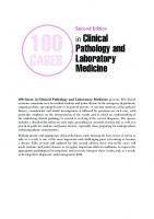
Case Report A 69-year-old man presented to the otolaryngology clinic complaining of dysphagia. He was diagnosed with T3N2bM0 squamous cell carcinoma of the hypopharynx 1 year prior. Direct laryngoscopy at that time demonstrated an extensive mass involving the right hypopharynx. Treatment consisted of induction chemotherapy followed by combined chemoradiation therapy. His course was complicated by hypopharyngeal stenosis resulting in dysphagia. This was initially managed successfully with a Maloney 60-French bougie dilator. Five months later, the patient presented to the otolaryngology clinic with recurrent dysphagia. On laryngoscopy, no distinct mass or stenosis was identified. Empiric serial dilation was performed up to a diameter of 60-French, with modest resistance encountered at the final dilation. Immediate endoscopic inspection demonstrated a patent esophageal inlet with mild bleeding but no evidence of perforation. In the recovery area, the patient complained of severe chest pain; a chest radiograph demonstrated moderate pneumomediastinum and pneumoperitoneum. Upper gastrointestinal series was performed, which showed an esophageal dissection originating in the cervical esophagus (Figure 1-1). The false lumen of the dissection extended down along the esophagus, exiting the outer wall of the distal esophagus with flow of contrast into the left lower lung parenchyma and pleural space (not shown). The patient was emergently brought to the operating room. On endoscopy, the entrance site of the false lumen could be seen just proximal to the thoracic inlet. The lumen of the false passage was the diameter of the 60-French bougie. The plane of dissection was between the submucosa and the muscularis propria, and the scope was passed along this plane until the exit site of dissection into the pleural space was identified. The true lumen was seen on the other side of the septum. A nasogastric tube was placed into the true lumen under direct visualization. Left thoracotomy was performed with heavy contamination of the pleural space. A lung decortication was performed and the inferior pulmonary ligament was divided up to the inferior pulmonary vein. Perforation of the lung was identified at the level of the inferior pulmonary ligament, which explained the presence of contrast in the lung. The puncture site was found immediately adjacent to this in the mediastinal pleura; this was opened proximally and distally in order to debride and drain the mediastinum. The distal esophagus was identified, and a defect in the muscularis propria was seen indicating the exit site of the dissection. Methylene blue was instilled into the distal esophagus with air insufflation to rule out a transmural perforation. A 19-French Blake drain was then inserted into the false lumen
3
4
Chapter 1
A
B
Figure 1-1. Upper gastrointestinal series demonstrates a cervical esophageal dissection extending down to the distal third of the esophagus. A false lumen is clearly seen to the left of the true esophageal lumen.
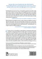
Figure 1-2. (A) Polyflex stent positioned with its proximal edge just above the upper esophageal sphincter. (B) The endotracheal tube is also in view next to the stent.
and passed retrograde, using biopsy forceps through the flexible endoscope from above to assist placement at a point 2 to 3 cm distal to the entry site of the false lumen. The distal exit site of the dissection was not closed. The Blake drain exited the esophagus at this location and was brought out through a puncture wound in the left chest wall. Esophagoscopy was performed 2 days later. The upper esophageal sphincter was identified at 16 cm from the incisors and a 10-mm mucosal defect was identified at this location, indicating the entry site of the false lumen. A Polyflex (Boston Scientific Co., Natick, Massachusetts) selfexpanding plastic stent (SEPS; 90 mm × 20 mm × 16 mm) was placed under fluoroscopic guidance with the proximal phalange at 15 cm from the incisors, just superior to the upper esophageal sphincter (Figure 1-2). Overall, the stent was well tolerated by the patient with minimal discomfort. Eleven days later, a repeat esophagram demonstrated the absence of contrast extravasation, yet there was subclinical
Endoscopic Therapy for an Esophageal Dissection
5
aspiration of contrast material noted. It was presumed that this may be due to the high position of the stent; thus, the stent was removed the following day. Another esophagram demonstrated healing of the dissection site without any further significant aspiration. The Blake drain was then slowly inched out over a period of 7 days. The patient was started on a clear liquid diet and tolerated oral intake without symptoms.
Discussion This case highlights the ever-expanding indications of removable SEPS. The use of SEPS for esophageal perforations and leaks has been well-described.1,2 However, there are few reports of the use of SEPS in the treatment of dissection of the cervical esophagus. Traditionally, placement of esophageal stents for treatment of fistulae or perforations in the cervical esophagus has been problematic because of its proximity to the cricopharyngeus muscle. Patients cannot tolerate an esophageal stent that embeds at the upper esophageal sphincter because often the pain, discomfort, and possible dysphagia are too severe. For the patient in this clinical scenario, stent placement facilitated closure of the proximal dissection entry site, preventing prolonged NPO status. The Polyflex stent has several characteristics that may enable it to be better tolerated in the distal hypopharynx or cervical esophagus. One favorable quality of this polyester mesh-reinforced silicone stent is its capability of narrowing under pressure, which, in contrast to metal stents, allows it to be more malleable in a dynamic environment such as the cervical esophagus and hypopharynx. Second, because of its nonmetal polyester covering there may be less surrounding tissue inflammation and proliferation.3 Both qualities appear to account for better tolerability near the upper esophageal sphincter, and thus success in treating benign (and possibly malignant) disease in this location. This case report illustrates that the Polyflex stent offers therapeutic stenting options in the upper cervical esophagus or distal hypopharynx that otherwise would not have been feasible with traditional esophageal metal stents.
Key Points ¾ Removable self-expanding plastic stent (SEPS) may be useful in treating fistulae, leaks, and
perforations within the upper esophagus. ¾ The placement of luminal stents in the cervical esophagus or distal hypopharynx may be
problematic due to the proximity of the cricopharyngeus muscle.
References 1. Schubert D, Scheidbach H, Kuhn R, et al. Endoscopic treatment of thoracic esophageal anastomotic leaks by using silicone-covered, self-expanding polyester stents. Gastrointest Endosc. 2005;61:891-696. 2. Radecke K, Gerken G, Treichel U. Impact of self-expanding, plastic esophageal stent on various esophageal stenoses, fistulas, and leakages: a single-center experience in 39 patients. Gastrointest Endosc. 2005;61:812-818. 3. Siersema P. Treatment of esophageal perforations and anastomotic leaks: the endoscopist is stepping into the arena. Gastrointest Endosc. 2005;61:897-900. 4. Evrard S, Le Moine O, Lazaraki G, et al. Self-expanding plastic stents for benign esophageal lesions. Gastrointest Endosc. 2004;60:894-900. 5. Dormann AJ, Wigginghaus B, Deppe H, et al. Successful treatment of esophageal perforation with a removable self-expanding plastic stent. Am J Gastroenterol. 2001;96:923-924.
CHAPTER
2
A 63-YEAR-OLD MAN WITH INTRACTABLE HICCUPS John Clarke, MD
Case Report A 63-year-old man presented to clinic with a complaint of intractable hiccups for a 2-year period. He related the onset of the hiccups to an automobile accident, but noted that there was no significant trauma associated with the event. The hiccups typically began 30 minutes after a meal and would last for several hours. Occasionally the hiccups would last as long as 5 days. Rarely, they would be relieved with self-induced vomiting but no other maneuvers had been of benefit. The size of the meal appeared to be directly related to the onset of the hiccups. Past medical history was notable for hypertension and borderline hyperlipidemia; there was a distant history of peptic ulcer disease. The patient had no surgical history and had never required hospitalization. Medications at the time of initial assessment included hydrochlorothiazide, felodipine, and aspirin. Tobacco use or any illicit drug was denied. On physical examination, the patient was well developed with mildly elevated blood pressure at 152/74 mmHG. His abdomen was soft, nondistended and nontender without organomegaly. His extremities were unremarkable, as was a complete neurological examination. A barium swallow was performed and demonstrated a small sliding hiatal hernia. Computed tomography (CT) of the abdomen and pelvis was normal other than a prominent prostate gland. Esophagogastroduodenoscopy (EGD) confirmed the presence of the hiatal hernia and showed evidence of reflux esophagitis in the distal esophagus. Magnetic resonance imaging (MRI) of the brain was normal. Basic laboratory analysis was also normal. The patient attempted therapeutic trials with the following medications: ranitidine, cimetidine, omeprazole, metoclopramide, chlorpromazine, diltiazem, and baclofen. All were unsuccessful in treating his hiccups. He also reported several failed attempts at numerous therapeutic physical maneuvers. After undergoing formal pH testing, he was ultimately referred for a laparoscopic Nissen fundoplication. Following the operation, he had nearly total resolution of his symptoms.
Discussion Singultus (from the latin “singult,” meaning to catch one’s breath while sobbing) is the medical term for the colloquial phrase “hiccups.” It is defined as an involuntary, intermittent, spasmodic
7
8
Chapter 2
contraction of the diaphragm and the inspiratory intercostal muscles that results in a sudden inspiration and ends with abrupt closure of the glottis. Several theories exist as to the etiology of hiccups. It may reflect a fetal digestive reflex preventing amniotic fluid aspiration, a means of preparing respiratory muscles for postnatal breathing, or an archaic remnant of gill ventilation. When present, hiccups generally occur at a frequency of 4 to 60 per minute (average 13 to 30). This rate is inversely associated with carbon dioxide concentration. It is commonly seen in utero and in infancy; however the incidence decreases with age. Chronic hiccups can be subcategorized into 3 distinct categories: bout, persistent, and intractable. A bout refers to an episode of recurrent hiccups lasting up to 48 hours; persistent hiccups refer to an episode lasting longer than 48 hours but less than 1 month; intractable hiccups refers to hiccups lasting longer than 1 month.1,2 For reasons not entirely clear, chronic hiccups are thought to occur more frequently in men (81% in the largest retrospective analysis).3 Typically, hiccups are a benign transient phenomenon. The most common causes include overdistention of the stomach (related to overeating or eating too quickly), alcohol ingestion, tobacco use, sudden changes in stomach temperature, ingestion of carbonated beverages, and aerophagia. Acute hiccups can be induced in 40% of patients by rapid barostat inflation in the proximal esophagus.4 The list of possible etiologies for hiccups is gargantuan. However, the multitude of causes can generally be subcharacterized into discrete categories: vagus and phrenic nerve irritation; central nervous system disorders; toxic-metabolic disorders; and psychogenic factors. Numerous physical maneuvers and home remedies can be attempted. Theoretically, these work by interrupting the vagal afferent limb of the hiccup reflex arc through nasopharyngeal stimulation or interruption of normal respiration thereby increasing vagal stimulation. Common maneuvers and remedies include breathing into a bag, drinking upside down, drinking from the opposite side of the glass, swallowing granulated sugar, ice water gargles, forceable traction on the tongue, biting on a lemon, catheter stimulation of the nasooropharynx, valsalva maneuver, breath holding, fright, noxious odors (inhaling ammonia), and compression of the eyeballs. There are also case reports of sexual intercourse5 and digital rectal exams6,7 being used as possible treatment modalities. Chlorpromazine is the only agent currently approved by the FDA. In the landmark study evaluating this remedy, 41 of 50 patients experienced immediate relief after receiving 50 mg intravenously (repeated in 2 to 4 hours if no effect).8 Other commonly used treatment regimens with reasonable clinical data but without FDA approval (for the indication of chronic hiccups) are metoclopramide,9 valproic acid,10 nifedipine,11 baclofen,12 and gabapentin.13 In addition, combination therapy has been touted by some authorities. Treatment with the combination of cisapride, omeprazole, and baclofen was described as the “treatment of choice” by researchers at the University of Heidelberg.14 The same group later reported incremental benefit with the addition of gabapentin “add-on therapy” to the combination listed previously.15 In addition to home remedies and medical therapies, therapy targeted at the phrenic nerve including phrenic nerve blocks and diaphragmatic pacing have been employed, as have more alternative approaches such as hypnosis and acupuncture.
Key Points ¾ Singultus, or hiccups, is an involuntary, intermittent, spasmodic contraction of the dia-
phragm and the inspiratory intercostal muscles that results in a sudden inspiration and ends with abrupt closure of the glottis. ¾ Chronic hiccups can be subcategorized into 3 categories: bout, persistent, and intractable.
A 63-Year-Old Man With Intractable Hiccups
9
¾ While the only approved therapy by the United States Food and Drug Administration (FDA)
is chlorpromazine, other commonly used treatment regimens include metoclopramide, valproic acid, nifedipine, baclofen, and gabapentin.
References 1. Kolodzik PW, Eilers MA. Hiccups (singultus): review and approach to management. Ann Emerg Med. 1991;20:565573. 2. Lewis JH. Hiccups: causes and cures. J Clin Gastroenterol. 1985;7:539-552. 3. Souadjian JV, Cain JC. Intractable hiccups: etiologic factors in 220 cases. Postgrad Med. 1968;43:72-77. 4. Fass R, Higa L, Kodner A, Mayer EA. Stimulus and site specific induction of hiccups in the oesophagus of normal subjects. Gut. 1997;41:590-593. 5. Peleg R, Peleg A. Case report: sexual intercourse as potential treatment for intractable hiccups. Can Fam Physician. 2000;46:1631-1632. 6. Fesmire FM. Termination of intractable hiccups with digital rectal massage. Ann Emerg Med. 1988;17:872. 7. Odeh M, Bassan H, Oliven A. Termination of intractable hiccups with digital rectal massage. J Intern Med. 1990; 227:145-146. 8. Friedgood CE, Ripstein CB. Chlorpromazine in the treatment of intractable hiccups. JAMA. 1955;157:309-310. 9. Madanagopolan N. Metoclopramide in hiccup. Cur Res Med Opin. 1975;3:371-374. 10. Jacobson PL, Messenheimer JA, Farmer TW. Treatment of intractable hiccups with valproic acid. Neurology. 1981; 31:1458-1460. 11. Lipps DC, Jabbari B, Mitchell MH, Daigh JD. Nifedipine for intractable hiccups. Neurology. 1990;40:531-532. 12. Ramirez FC, Graham DY. Treatment of intractable hiccup with baclofen: results of a double-blind randomized, controlled, cross-over study. Am J Gastroenterol. 1992;87:1789-1791. 13. Hernandez JL, Pajaron M, Garcia-Regata O, Jimenez V, Gonzalez-Macias J, Ramos-Estebanez C. Gabapentin for intractable hiccup. Am J Med. 2004;117:279-281. 14. Petroianu G, Hein G, Petroianu A, Bergler W, Rufer R. Idiopathic chronic hiccup: combination therapy with cisapride, omeprazole, and baclofen. Clin Ther. 1997;19:1031-1038. 15. Petroianu G, Hein G, Stegmeier-Petroianu A, Bergler W, Rufer R. Gabapentin “add-on therapy” for idiopathic chronic hiccup. J Clin Gastroenterol. 2000;30:321-324.
CHAPTER
3
“WHEN I EAT, FOOD GETS STUCK” John Clarke, MD
Case Report A 57-year-old man presented to gastroenterology clinic with progressive dysphagia, weight loss, and failure to thrive occurring with increasing frequency over a period of 6 months. The patients literally commented, “When I eat, food gets stuck.” The subjective site of obstruction was in the lower chest at approximately the level of the xiphoid process. Symptoms occurred entirely with solid foods; the patient noted no problems with liquids, soups, or tuna fish. Frequent regurgitation after food intake was noted, usually with an undigested appearance. The patient denied chest pain or odynophagia; heartburn was noted, but only after eating pizza. The patient claimed to have a decreased appetite and felt that he may have lost weight; however, the exact amount of weight loss was unclear. Past medical history was notable for cerebral palsy and asthma. His only medications were albuterol and salmeterol/fluticasone. He denied drug allergies. Social history was notable for steady employment as a housekeeper. He was single with no children and denied using tobacco or illicit drugs. He admitted to “a couple cans” of beer each night. Physical examination revealed a thin man in no apparent distress. Vital signs were within normal range and his BMI was 23. He had decreased oropharyngeal strength with tongue deviation to the right. No lymphadenopathy was appreciated. His abdomen was benign but further neurological examination demonstrated diffuse muscle weakness with hyperreflexia. Computed tomography (CT) scan of the neck revealed vocal cord asymmetry and some prominent left submandibular lymph nodes. CT of the chest/abdomen/pelvis was otherwise unremarkable. A flexible laryngoscopic evaluation by otolaryngology was normal. A video-fluoroscopic swallowing study was notable for weakness of the tongue base, diminished laryngeal elevation, laryngeal penetration, absence of epiglottic tilt, pharyngeal paresis, and a holdup of solid and pureed contrast at the gastroesophageal junction. Due to the patient’s underlying cerebral palsy and inability to take large amounts of volume, distention in the lower esophagus was inadequate for proper evaluation of esophageal rings or strictures. On upper endoscopy, moderate narrowing at the upper esophageal sphincter was identified and this was dilated with a controlled radial expansion (CRE) balloon inflated to 18 mm. A clear Schatzki’s ring was identified at the gastroesophageal junction; this was ruptured with a CRE balloon inflated to 20 mm. The patient tolerated the procedure well and was discharged home that day.
11
12
Chapter 3
Contrary to instructions, the patient went home that day and decided to test himself with a steak dinner. He tolerated this well and reports complete absence of his solid-food dysphagia since the procedure. He continues to have problems related to his cerebral palsy and resultant transfer dysphagia. Since his procedure, he has gained 5 lbs. At a 1-year follow-up, he continued to feel well and has not required any additional procedures.
Discussion The lower esophageal ring was initially described by Templeton in 1944.1 In 1953, two additional groups of investigators independently reported the existence of a lower esophageal ring and its association with dysphagia.2,3 Based on the 1944 study, the ring became known as Schatzki’s ring (also known as a B-ring by current classification schemes of esophageal rings).4 Using barium studies as a gold standard, the prevalence of Schatzki’s rings in the general population is believed to range from 6% to 14%. There appears to be an increasing incidence with age and the ring is rare in children.5,6 If the luminal diameter of the esophagus within the ring is less than 13 mm, symptoms are almost always present.7 For this reason, barium marshmallows of 13 mm are often employed in many institutions during radiographic swallowing studies Treatment of a Schatzki’s ring is typically aimed at dilatation or rupture. Proposed treatment approaches have included a single large diameter (>48 Fr) dilator (usually Savary-Gilliard [Cook Medical, Bloomington, IN] or Maloney),8 endoscopic biopsy obliteration of the ring,9 and, rarely, electrocautery techniques10 or pneumatic dilatation.11 The technique used in the above case study, rupturing the Schatzki’s ring with a CRE balloon, is commonly performed; however, outcomes with this approach (as compared to those listed above) are not reported in the literature. Recurrence of Schatzki’s rings is the rule rather than the exception. The landmark study by Eckardt et al8 showed that after treatment with a large-bore dilator (usually Maloney), virtually all patients were symptom free at 4 weeks. At 2 years, 42% of patients had symptom recurrence severe enough to prompt a repeat endoscopy; at 5 years, 89% of patients were noted to have recurrent symptoms.8 Whether recurrence will be diminished by antireflux medications is still an open debate.
Key Points ¾ The prevalence of Schatzki’s ring, or B-ring, in the general population is estimated to be
between 6% and 14%. ¾ Treatment is aimed at mechanical disruption of the ring. ¾ Recurrence is the rule rather than the exception and may occur in as many as 89% of patients at 5-year follow-up.
References 1. Templeton FE. X-ray examination of the stomach. In: A Description of the Roentgenologic Anatomy, Physiology, and Pathology of the Esophagus, Stomach, and Duodenum. Chicago: University of Chicago Press, 1944:94-102. 2. Schatzki R, Gary JE. Dysphagia due to diaphragm-like narrowing in lower esophagus (“lower esophageal ring”). Am J Roentgennol Radiat Ther Nucl Med. 1953;70:911-922. 3. Ingelfinger FJ, Kramer P. Dysphagia produced by contractile ring in lower esophagus. Gastroenterology. 1953; 23:419.
“When I Eat, Food Gets Stuck”
13
4. Katzka DA. Esophageal webs and rings. In: Castell DO, Richter JE, eds. The Esophagus. 4th ed. Philadelphia: Lippincott Williams & Wilkins, 2004:315-324. 5. Kramer P. Frequency of the asymptomatic lower esophageal contractile ring. N Eng J Med. 1956;254:692-694. 6. Goyal RK, Glancy JJ, Spiro HM. Lower esophageal ring. N Eng J Med. 1970;282:1298-1305. 7. Schatzki R. The lower esophageal ring. Long-term follow-up of symptomatic and asymptomatic rings. Am J Roentgenol. 1963;90:805-810. 8. Eckardt VF, Kanzler G, Willems D. Single dilatation of symptomatic Schatzki rings. A prospective evaluation of its effectiveness. Dig Dis Sci. 1992;37:577-582. 9. Chotiprasidhi P, Minocha A. Effectiveness of single dilation with Maloney dilator versus endoscopic rupture of Schatzki’s ring using biopsy forceps. Dig Dis Sci. 2000;45:281-284. 10. Burdick JS, Venu RP, Hogan WJ. Cutting the defiant lower esophageal ring. Gastrointest Endosc. 1993;39:616-619. 11. Arvanitakis C. Lower esophageal ring: endoscopic and therapeutic aspects. Gastrointest Endosc. 1977;24:17-18.
CHAPTER
4
A BENIGN FORM OF PROGRESSIVE DYSPHAGIA Eun Ji Shin, MD
Case Report A 46-year-old man presented to clinic with an 11-month history of progressive dysphagia. The patient was in his usual state of health when he noticed mild dysphagia to large vitamin pills only. Over the next 6 months, he noticed that solid food was “getting stuck” in his throat necessitating repeated swallowing motions to help the food pass. He reported no acid reflux, heartburn, or regurgitation symptoms. He sought medical attention at his local hospital for these complaints. A barium swallow study showed a stricture approximately 3 to 4 cm above the gastroesophageal (GE) junction with a residual lumen of 6 to 7 mm. He was started on acid suppressive therapy for a presumed reflux-related stricture. He underwent four separate esophagogastroduodenoscopy (EGD) procedures with dilatation in an effort to treat what was felt to be a peptic stricture and an associated Schatzki’s ring. Biopsies showed moderate reactive epithelial changes consistent with reflux esophagitis. He also underwent esophageal manometry studies showing normal relaxation of the lower esophageal sphincter (LES) but ineffective esophageal motility. An EGD revealed esophageal mucosa with a scalloped appearance and multiple nonobstructive rings throughout the esophagus (Figure 4-1). No definite stricture or evidence of esophagitis was visualized. There was a small hiatal hernia with the squamocolumnar junction displaced approximately 3 cm above the diaphragm. Biopsies revealed squamous mucosa with moderately prominent intraepithelial eosinophils consistent with eosinophilic esophagitis (Figure 4-2). The patient was started on a trial of swallowed steroids. He was prescribed fluticasone 110 mcg 2 puffs twice per day without a spacer. Shortly after initiating therapy, the patient was able to tolerate a soft mechanical diet without difficulties; he was instructed to maintain adequate nutritional and caloric supplementation with Boost (Nestle, Minneapolis, MN) and/or Ensure (Abbott Labs, Abbott Park, IL).
Discussion Eosinophilic esophagitis, or EE, is a clinical entity characterized by the presence of eosinophils in the esophageal epithelium with esophageal symptoms. It was first described in 1978 by Landres et al,1 and the incidence has been steadily increasing over the past 20 years.2-4 Patients with EE are
15
16
Chapter 4
Figure 4-1. Endoscopic view of the esophageal mucosa with scalloped appearance and multiple nonobstructive rings throughout the esophagus.
Figure 4-2. Biopsy specimen from the esophagus showing squamous mucosa with moderately prominent intraepithelial eosinophils.
more likely to be young men, with a male-to-female ratio over 3:1. EE commonly presents between the third and fourth decades of life.5 The etiology of EE is poorly understood. Environmental allergens have been proposed as a potential trigger for a mast cell response leading to release of histamine, eosinophilic chemotactic factors, and platelet activating factors. This pathway leads to the recruitment and activation of eosinophils, which release a cytotoxic cationic protein.6,7 Pollen and other aeroallergens have been shown to have an association to EE.8 Adult patients with EE typically present with dysphagia, food impaction, vomiting, or difficulty feeding.9-13 They can also present with vomiting, epigastric or chest pain, and failure to thrive.5 Dysphagia tends to be progressive in nature and often is refractory to dietary modifications and antireflux therapy.5 Diagnosis is often dependent on upper endoscopy with biopsy. The most common findings include fragility or edema of the esophageal mucosa, esophageal rings or a corrugated esophagus, esophageal strictures, and the presence of whitish exudates or papules.5 Although there is no definitive diagnostic criteria, most consider the presence of >20 eosinophils/hpf with the absence of mucosal eosinophils in the gastric and duodenal mucosa in the proper clinical setting to be diagnostic of EE.5,14,15 There is currently no consensus to the optimal therapeutic regimen for EE, which often involves multiple modalities to address control of symptoms and the underlying eosinophilic inflammation. Most physicians advocate an empiric trial of acid suppressive therapy to rule out gastroesophageal reflux disease (GERD) and to minimize esophageal acid exposure that may exacerbate eosinophilic inflammation.10 Since adults commonly present with severe dysphagia with evidence of esophageal rings or strictures, many will benefit symptomatically from esophageal dilatation. However, dilatation should be performed with caution since it has been associated with deep mucosal tears and perforations in this population.16 Given the associations with allergens, including food, elimination diets have been also considered in certain cases.17 Most published studies have been in children, so the generalizability to the adult EE patients is unknown. Corticosteroids, both systemic and topical formulations, have been used with clinical and histological improvements.18-20 Many advocate the use of fluticasone over systemic steroids, such
A Benign Form of Progressive Dysphagia
17
as prednisone, given the high relapse rate with withdrawal and the need for chronic or repeated therapy. Recently, there have been reports of clinical response with leukotriene receptor antagonists21 and humanized anti-IL-5 antibody.22
Key Points ¾ Eosinophilic esophagitis (EE) is characterized by the presence of eosinophils in the esopha-
geal epithelium with esophageal symptoms. ¾ Adults with EE often present with dysphagia, food impaction, vomiting, or difficulty feeding. ¾ Treatment of EE includes elimination diets, acid suppression, systemic and/or topical steroids, and endoscopic therapy.
References 1. Landres RT, Kuster GG, Strum WB. Eosinophilic esophagitis in a patient with vigorous achalasia. Gastroenterology. 1978;74:1298-1301. 2. Noel RJ, Putnam PE, Rothenberg ME. Eosinophilic esophagitis. N Engl J Med. 2004;351:940-941. 3. Noel RJ, Tipnis NA. Eosinophilic esophagitis—a mimic of GERD. Int J Pediatr Otorhinolaryngol. 2006;70:1147-1153. 4. Straumann A, Simon HU. Eosinophilic esophagitis: escalating epidemiology? J Allergy Clin Immunol. 2005;115:418419. 5. Sgouros SN, Bergele C, Mantides A. Eosinophilic esophagitis in adults: what is the clinical significance? Endoscopy. 2006;38:515-520. 6. Liacouras CA. Eosinophilic esophagitis: treatment in 2005. Curr Opin Gastroenterol. 2006;22:147-152. 7. Mann NS, Leung JW. Pathogenesis of esophageal rings in eosinophilic esophagitis. Med Hypotheses. 2005;64:520523. 8. Fogg MI, Ruchelli E, Spergel JM. Pollen and eosinophilic esophagitis. J Allergy Clin Immunol. 2003;112:796-797. 9. Liacouras CA, Ruchelli E. Eosinophilic esophagitis. Curr Opin Pediatr. 2004;16:560-566. 10. Orenstein SR, Shalaby TM, Di Lorenzo C, et al. The spectrum of pediatric eosinophilic esophagitis beyond infancy: a clinical series of 30 children. Am J Gastroenterol. 2000;95:1422-1430. 11. Sant’Anna AM, Rolland S, Fournet JC, Yazbeck S, Drouin E. Eosinophilic esophagitis in children: symptoms, histology and pH probe results. J Pediatr Gastroenterol Nutr. 2004;39:373-377. 12. Vasilopoulos S, Murphy P, Auerbach A, et al. The small-caliber esophagus: an unappreciated cause of dysphagia for solids in patients with eosinophilic esophagitis. Gastrointest Endosc. 2002;55:99-106. 13. Walsh SV, Antonioli DA, Goldman H, et al. Allergic esophagitis in children: a clinicopathological entity. Am J Surg Pathol. 1999;23:390-396. 14. Fox VL, Nurko S, Furuta GT. Eosinophilic esophagitis: it’s not just kid’s stuff. Gastrointest Endosc. 2002;56:260-270. 15. Furuta GT, Straumann A. Review article: the pathogenesis and management of eosinophilic oesophagitis. Aliment Pharmacol Ther. 2006;24:173-182. 16. Kaplan M, Mutlu EA, Jakate S, et al. Endoscopy in eosinophilic esophagitis: “feline” esophagus and perforation risk. Clin Gastroenterol Hepatol. 2003;1:433-437. 17. Liacouras CA, Spergel JM, Ruchelli E, et al. Eosinophilic esophagitis: a 10-year experience in 381 children. Clin Gastroenterol Hepatol. 2005;3:1198-1206. 18. Liacouras CA, Wenner WJ, Brown K, Ruchelli E. Primary eosinophilic esophagitis in children: successful treatment with oral corticosteroids. J Pediatr Gastroenterol Nutr. 1998;26:380-385. 19. Arora AS, Perrault J, Smyrk TC. Topical corticosteroid treatment of dysphagia due to eosinophilic esophagitis in adults. Mayo Clin Proc. 2003;78:830-835. 20. Faubion WA Jr, Perrault J, Burgart LJ, Zein NN, Clawson M, Freese DK. Treatment of eosinophilic esophagitis with inhaled corticosteroids. J Pediatr Gastroenterol Nutr. 1998;27:90-93. 21. Attwood SE, Lewis CJ, Bronder CS, Morris CD, Armstrong GR, Whittam J. Eosinophilic oesophagitis: a novel treatment using Montelukast. Gut. 2003;52:181-185. 22. Garrett JK, Jameson SC, Thomson B, et al. Anti-interleukin-5 (mepolizumab) therapy for hypereosinophilic syndromes. J Allergy Clin Immunol. 2004;113:115-119.
CHAPTER
5
A RARE FORM OF ESOPHAGEAL TOXICITY Samuel Giday, MD
Case Report A 61-year-old woman presented with abdominal pain and heartburn. She had a history of gouty arthritis and was taking low-dose colchicine on a daily basis. Upper endoscopy was performed and the patient was found to have long segment Barrett’s esophagus along with reflux esophagitis. She was treated with a proton-pump inhibitor and had good relief of her symptoms. On subsequent surveillance endoscopy and biopsy for the Barrett’s mucosa, she was found to have a focus of highgrade dysplasia adjacent to an area of adenocarcinoma. Further evaluation using high-frequency endoscopic ultrasound (EUS) probe examination showed that the tumor was limited to the lamina propria without involvement of the submucosa. She underwent a successful course of photodynamic therapy (PDT) and local endoscopic mucosal resection (EMR) of the sites of high-grade dysplasia and adenocarcinoma. On the histopathological evaluation of the slides, a peculiar finding was seen in the esophageal tissue adjacent to the tumor and within the area of Barrett’s esophagus. The specimen in this region was noted to display diffuse mitotic arrest, a feature distinctive of drug-induced toxicity (Figure 5-1). Colchicine was felt to be the likely culprit for these findings.
Discussion Colchicine is an agent extracted from the bulb of the meadow saffron (Colchicum autumnale), a plant that has been used for centuries in soothing pains in the joints. It is an alkaloid with antimitotic activity. The actual mechanism for causing cellular mitotic arrest is by inhibiting spindle formation and thus preventing the completion of a normal mitotic cycle. These inhibitions of spindle formation also affect cell microtubular function interfering with chemotaxis, phagocytosis, fibroblast proliferation, and extracellular collagen matrix deposition. Colchicine toxicity has a spectrum of presentation. In its mild form, it results in abdominal pain, nausea, vomiting, diarrhea, and anorexia. These gastrointestinal manifestations occur in up to 20% of patients taking colchicine.1 These symptoms are mild and easily reversible once the medication is stopped. Severe toxicity occurs in about 1% of cases and manifests itself as multiorgan failure including heart and lung failure. This mainly is due to associated myopathy and neuropathy resulting in
19
20
Chapter 5
Figure 5-1. Mitotic arrest in esophageal mucosa of a patient with colchicine toxicity.
impaired myocardial contraction and weakness of the respiratory musculature.2 Other abnormalities, such as associated severe rhabdomyolysis, neurological dysfunction, and hepatic and renal failures, have also been described.3-5 Deaths within a few days of the onset of toxicity have also been reported.6-8 Because it is mainly eliminated through the kidneys and bile, patients with impaired renal or hepatic function, elderly, and on CYP3A4 inhibitors have been found to have a higher chance of developing toxicity to colchicine. Creatinine clearance of 25 mL/min or less has been associated with colchicine toxicity9 and it has also been noted that patients on long-term colchicine therapy can develop vitamin B12 deficiency due to reduction of endocytosis in the terminal ileum.10,11 On histopathological examination, a metaphase arrest picture along with epithelial pseudostratification, loss of polarity, and villous atrophy has been documented throughout the GI tract, of which, the gastric antrum and the duodenal bulb have consistently shown to be affected. Most studies have shown a relative sparing of the gastric body. Arrest in mitosis has also been seen in other tissues such as bone marrow and genitourinary epithelium;12 Hart et al13 found a hyperplastic picture with no arrest in mitosis in biopsies from the GI tract. The management of colchicine toxicity is mainly supportive after stopping the drug. No specific treatment including the use of hemodialysis has been shown to be effective. Patients with acute toxicity should have a gastric lavage and charcoal administration to prevent further absorption of the drug. Correction of fluid and electrolyte imbalance in addition to intensive care support also should be performed in all patients with multiorgan failure. One promising agent that has been recently shown to be effective is a colchicine specific Fab fragment. This agent has been shown to be successful in many animal models and a few human case reports.14 Patients who have developed vitamin B12 deficiency should have the appropriate replacement. The majority of patients will not have lasting sequelae of the drug and subsequent biopsies would show a normal mucosa.
A Rare Form of Esophageal Toxicity
21
Key Points ¾ Colchicine can result in a life-threatening toxicity and is manifested most commonly in the
GI tract as mitotic arrest. ¾ Colchicine should be stopped immediately once toxicity is identified. ¾ A majority of the patients will not have lasting sequelae due to colchicine toxicity.
References 1. Race TF, Paes IC, Faloon WW. Intestinal malabsorption induced by oral colchicine. comparison with neomycin and cathartic agents. Am J Med Sci. 1970;259(1):32-41. 2. Ben-Chetrit E, Levy M. Colchicine: 1998 update. Semin Arthritis Rheum. 1998;28:48-59. 3. Debie K, Conraads V, Vrints C. Colchicine-induced rhabdomyolysis in a patient with chronic heart failure. Acta Cardiol. 2003;58(6):561-562. 4. Pirzada NA, Medell M, Ali II. Colchicine induced neuromyopathy in a patient with normal renal function. J Clin Rheumatol. 2001;7(6):374-376. 5. Akdag I, Ersoy A, Kahvecioglu S, Gullulu M, Dilek K. Acute colchicine intoxication during clarithromycin administration in patients with chronic renal failure. J Nephrol. 2006;19(4):515-517. 6. Borron SW, Scherrmann JM, Baud FJ. Markedly altered colchicine kinetics in a fatal intoxication: examination of contributing factors. Hum Exp Toxicol;15:885–890. 7. Hung IF, Wu AK, Cheng VC, et al. Fatal interaction between clarithromycin and colchicine in patients with renal insufficiency: a retrospective study. Clin Infect Dis. 2005;41(3):291-300. Epub 2005 Jun 23. 8. Dogukan A, Oymak FS, Taskapan H, Guven M, Tokgoz B, Utas C. Acute fatal colchicine intoxication in a patient on continuous ambulatory peritoneal dialysis (CAPD). possible role of clarithromycin administration. Clin Nephrol. 2001;55(2):181-182. 9. Ehrenfeld M, Levy M, Sharon P, Rachmilewitz D, Eliakim M. Gastrointestinal effects of long-term colchicine therapy in patients with recurrent polyserositis (familial mediterranean fever). Dig Dis Sci. 1982;27(8):723-727. 10. Stopa EG, O’Brien R, Katz M. Effect of colchicine on guinea pig intrinsic factor-vitamin B12 receptor. Gastroenterology. 1979;76(2):309-314. 11. Webb DI, Chodos RB, Mahar CQ, Faloon WW. Mechanism of vitamin B12 malabsorption in patients receiving Colchicine. N Engl J Med. 1968;279(16):845-850. 12. Iacobuzio-Donahue CA, Lee EL, Abraham SC, Yardley JH, Wu TT. Colchicine toxicity: distinct morphologic findings in gastrointestinal biopsies. Am J Surg Pathol. 2001;25(8):1067-1073. 13. Hart J, Lewin KJ, Peters RS, Schwabe AD. Effect of long-term colchicine therapy on jejunal mucosa. Dig Dis Sci. 1993;38(11):2017-2021. 14. Baud FJ, Sabouraud A, Vicaut E, et al. Brief report: treatment of severe colchicine overdose with colchicine-specific Fab fragments. N Engl J Med. 1995;332(10):642-645.
CHAPTER
6
DIFFUSE SQUAMOUS CELL DYSPLASIA OF THE ESOPHAGUS Samuel Giday, MD
Case Report A 58-year-old man of Middle Eastern origin presented complaining of intermittent dysphagia of a 2-year duration. The dysphagia occurred with both liquids and solids, was progressive in nature, and was relieved slightly with acid suppression therapy. He denied any past or present heartburn. Past medical history was significant for hypertension. The patient reported that he drank 2 glasses of wine per day and had no history of smoking or illicit drug use. There was no family history significant for malignancy. Physical examination was unremarkable. Upper endoscopy revealed the presence of diffuse esophageal erythema with friability and exudates on the esophageal mucosa. Biopsy of one exudate showed CD68 positive macrophages, suggesting herpes simplex infection. The patient was treated for 2 weeks with acyclovir and testing for HIV was found to be negative. Repeat esophagogastroduodenoscopy (EGD) was performed 8 weeks after treatment and revealed similar findings without improvement (Figure 6-1). Biopsy at this time showed diffuse squamous cell dysplasia with focal ulceration. Chromoendoscopy was then performed using Lugol’s iodine, which again confirmed the presence of diffuse nonstaining of the esophagus due to the underlying squamous dysplasia (Figure 6-2). Endoscopic ultrasound (EUS) examination showed the presence of mucosal thickening with a clear muscularis propria and the presence of an enlarged celiac axis lymph node. The patient then was referred for surgical esophageal resection due to the diffuse nature of the squamous cell dysplasia and the presence of periesophageal lymphadenopathy.
Discussion Use of Lugol’s iodine in the screening of esophageal squamous cell dysplasia is widely practiced in places of high prevalence of squamous cell carcinoma such as China. It stains the normal squamous epithelium dark brown, whereas dysplastic, carcinomatous, or nonsquamous epithelium will not be stained. It can also be utilized to delineate a specific lesion (eg, pre-endoscopic mucosal resection, EMR), diagnose short segment Barrett’s esophagus (delineate stained squamous from unstained columnar), highlight residual Barrett’s mucosa postablation, and in the evaluation of endoscopy negative gastroesophageal reflux disease (GERD).1
23
24
Chapter 6
EUS plays a pivotal role in the evaluation of esophageal precancerous lesions. EUS can accurately detect superficial alterations in the esophageal wall, thus highly impacting clinical management. Tajima et al2 showed a 10% chance of nodal metastasis when the muscularis mucosa is affected. This can be demonstrated using high frequency ultrasound probes. One study has shown an accuracy rate of 80% using standard EUS (7.5/12 MHz) for superficial lesions (T1), and a higher accuracy when utilizing high frequency probes (92% versus 76%).3 If a diagnosis of high-grade dysplasia is established, management options include surgical resection, photodynamic therapy [PDT]), and Figure 6-1. Diffuse esophageal squamous cell dysendoscopic mucosal resection (EMR). plasia. Note the whitish appearing normal squamous PDT involves the systemic injection mucosa (thin arrows) and the dysplastic epithelium of compounds that then concentrate (block arrows). in cancerous or precancerous lesions. When these cells are then exposed to UV light, it results in a photochemical reaction that forms free oxygen radicals. These free radicals then induce local inflammation and necrosis, thus destroying the targeted cells of interest. PDT has been used as an effective treatment for in situ and microinvasive carcinomas. Its use in squamous cell esophageal neoplasms was first reported by Tian et al4 in 1985. They treated 13 patients with early tumors detected by balloon cytology. Out of 13, 12 patients had no evidence of tumor recurrence at 32 months.4 EMR is a method in which the layers above the muscularis propria of the gastrointestinal (GI) tract are removed endoscopically using a variety of techniques. EMR has been used in the management of squamous cell dysplasia when the lesion is limited to the lamina propria. Survival rates up to 77.4% have been reported in patients with superficial lesions managed by EMR. The presence of multiple lugol nonstaining lesions Figure 6-2. Lugol’s iodine staining showing scant dark colored normal squamous (thin arrows) mucosa and diffuse nonstaining of has been shown to be the esophagus due to squamous cell dysplasia with nodularity (block associated with high arrows). 5 recurrence after EMR.
Endoscopic Therapy for an Esophageal Dissection
25
Surgical management (esophagectomy) provides the best option of complete treatment in patients who are fit to undergo surgery despite a higher morbidity and mortality rate. One of the most important determinants to higher complication rates is the number of cases performed per year in a given hospital. Several studies have reported a higher morbidity, mortality, and cost when these procedures are performed in “low-volume” hospitals.6
Key Points ¾ Squamous cell dysplasia is infrequently seen within the United States. ¾ Chromoendoscopy using Lugol’s iodine helps in identifying dysplastic and/or cancerous
lesions in the esophagus. ¾ Endoscopic ultrasound (EUS) provides accurate staging information for precancerous lesions. ¾ Management options for superficial squamous cell cancers or dysplasia include endoscopic mucosal resection, photodynamic therapy, and surgery.
References 1. Connor MJ, Sharma P. Chromoendoscopy and magnification endoscopy for diagnosing esophageal cancer and dysplasia. Thorac Surg Clin. 2004;14(1):87-94. Review. 2. Tajima Y, Nakanishi Y, Ochiai A, et al. Histopathologic findings predicting lymph node metastasis and prognosis of patients with superficial esophageal carcinoma: analysis of 240 surgically resected tumors. Cancer. 2000;88(6):12851293. 3. Hasegawa N, Niwa Y, Arisawa T, Hase S, Goto H, Hayakawa T. Preoperative staging of superficial esophageal carcinoma: comparison of an ultrasound probe and standard endoscopic ultrasonography. Gastrointest Endosc. 1996;44(4):388-93. 4. Tian ME, Qui SL, Ji Q. Preliminary results of hematoporphyrin derivative-laser treatment for 13 cases of early esophageal carcinoma. Adv Exp Med Biol. 1985;193:21-25. 5. Katada C, Muto M, Manabe T, Ohtsu A, Yoshida S. Local recurrence of squamous-cell carcinoma of the esophagus after EMR. Gastrointest Endosc. 2005;61(2):219-25. 6. Dimick JB, Cowan JA Jr, Ailawadi G, Wainess RM, Upchurch GR Jr. National variation in operative mortality rates for esophageal resection and the need for quality improvement. Arch Surg. 2003;138(12):1305-1309
SECTION II STOMACH
CHAPTER
7
ABDOMINAL PAIN AND AN OVARIAN MASS Michael Kafrouni, MD
Case Report A 67-year-old woman was referred from an outside hospital for further evaluation of midabdominal pain radiating to the left upper quadrant. The patient reported that the pain had been constant and progressing for 10 weeks and that it was most severe following meals; she also reported occasional postprandial vomiting. She also noted that she had experienced generalized weakness and had lost approximately 10 lbs. A computerized axial tomography (CAT) scan of the abdomen and pelvis was unremarkable, and an esophagogastroduodenoscopy (EGD) showed chronic gastritis with the absence of Helicobactor pylori on biopsies. Past medical history was notable for hypertension, hypercholesterolemia, and gastroesophageal reflux disease (GERD) for several years. The patient was status post cholecystectomy and had also undergone thyroidectomy for benign disease. Her medications included amlodipine, levothyroxine, atorvastatin, and esomeprazole. On the initial physical examination, the patient was mildly hypertensive (144/86 mmHg). She had a thyroidectomy scar, but no lymph nodes or jugular vein distention. Her abdomen was soft, and there was an area of tenderness extending underneath the left rib, which was most notable with deep palpation. There was no hepatosplenomegaly. Rectal examination was normal. On admission, blood work revealed normal liver enzymes except for an alkaline phosphatase level at 180 U/L (normal range 30 U/L to 120 U/L). The amylase was also mildly abnormal at 164 U/L (28 U/L to 100 U/L). CAT scan of the abdomen showed a moderately dilated common bile duct that was felt to be normal in size following cholecystectomy. An endoscopic retrograde cholangiopancreatography (ERCP) confirmed the dilatation of the common bile duct but showed no other abnormalities. Endoscopic ultrasound (EUS) was performed and demonstrated a normal pancreatic duct with a hypoechoic retropapillary region that was interpreted as likely inflammation rather than neoplasm. A plan was made to re-evaluate this region with a repeat EUS (and fine-needle aspiration, or FNA) in 4 weeks once the inflammation had subsided. The patient was discharged with oral pain medications. Shortly after discharge, the patient was readmitted with the same complaints. Magnetic retrograde cholangiopancreatography (MRCP) was done showing no discrete pancreatic mass, but an area of ill-defined decreased enhancement in the head of the pancreas was noted. Repeat EGD showed an abnormally thickened gastric mucosa consistent with a diffuse gastritis (Figure 7-1) and the pancreas appeared normal. Biopsies of the stomach were consistent with chronic inactive gastritis and she was eventually discharged home.
29
30
Chapter 7
A third admission with abdominal pain prompted a CAT scan of the abdomen in the emergency room. This time it showed a left ovarian mass and a fixed, abnormally thickened stomach (Figure 7-2). Another EGD demonstrated a loss of gastric folds and difficulty with air insufflation. Deep biopsies of the mucosa again revealed chronic gastritis. EUS of the gastric wall showed thickened folds (Figure 7-3) and FNA revealed benign epithelium and nondiagnostic mesenchymal tissue. Due to the recently seen ovarian mass, a laparoscopic wedge biopsy from the stomach was performed at the time of oophorectomy. In the operating room, however, the stomach appeared hard, thickened, and nonflexible and a total gastrectomy with lymph node dissection was performed. Pathology from Figure 7-1. Endoscopic image of the stomach the ovarian mass revealed a benign fibroma. showing the thickened gastric folds in the setting of chronic gastritis. Gastric pathology, however, showed a signet ring cell (SRC) carcinoma with greater than 50% involvement (Figure 7-4). The mass penetrated the serosa without invasion of adjacent structures. One of 11 lymph nodes dissected had metastases. Both proximal and distal margins as well as the resected omentum were disease free. The patient did well postoperatively and was referred to the oncology service for follow-up.
Discussion SRC carcinoma of the stomach is a histological description depending on the main cells seen in the cancer. According to the World Health Organization criteria, a gastric cancer is classified as SRC gastric carcinoma if more than half of its cells are Signet ring like. The cells are usually mucinsecreting cells that have abundant intracytoplasmic mucin pushing the nuclei to the side and giving the cells their typical form. In the literature,1-4 SRC type gastric carcinoma has been described as a diffuse, infiltrating, and undifferentiated type of cancer. It is known to arise from the mucosal layer and not from metaplastic epithelium, as in the intestinal type of gastric cancer.3 SRC carcinomas have been identified in many parts of the gastrointestinal tract including the Figure 7-2. CAT scan depicting a thickened gastric wall. small intestines and the colon.5,6
Abdominal Pain and an Ovarian Mass
31
Figure 7-3. (A) EUS of the stomach wall measuring up to 15 mm at the thickest diameter compared to a (B) normal stomach wall.
SRC gastric carcinoma accounts for 10% to 39% of all gastric cancers.7 In its advanced form, SRC gastric carcinoma is characterized by poor prognosis, especially as it has a tendency to infiltrate the wall of the stomach with metastases to surrounding lymph nodes.2,3 However, early diagnosis of the cancer has been reported to significantly improve survival.4 SRC type of gastric carcinoma is more frequent among women and younger patients.1,8 Making the diagnosis of Figure 7-4. High magnification (1000×) image of the gastric SRC gastric cancer has been cancer histology depicting a signet ring cell with its eccentric difficult due to its nonspecific nucleus. appearance on regular endoscopic evaluation. It will show thickened gastric folds and a chronic gastritis picture, the differential diagnosis of such includes infiltrative disorders like amyloidosis, lymphoma, and Menetrier’s disease as well. A biopsy of the mucosa can be negative in up to 30% of the cases,9 thus there is a need for better diagnostic techniques. Several technological advances have been implemented to make an earlier diagnosis. Among those are multislice CAT scan with delayed-phase multiplanar reformation technology, and enhanced-magnification endoscopy, which can be used to help detect small size malignant gastric lesions.10,11 EUS-guided FNA has also been reported to aid in the early diagnosis of this disease.12
32
Chapter 7
Key Points ¾ Signet ring cell (SRC) gastric carcinoma accounts for 10% to 39% of all gastric cancers. ¾ Gastric cancer is classified as SRC carcinoma if more than 50% of its cells are signet ring
like. ¾ SRC have abundant intracytoplasmic mucin pushing the nuclei to one side, giving them the classic signet ring appearance. ¾ Making the diagnosis of SRC gastric carcinoma can be challenging due to its nonspecific appearance on endoscopy and other noninvasive imaging techniques.
References 1. Yokota T, Kunii Y, Teshima S, et al. Signet ring cell carcinoma of the stomach: a clinicopathological comparison with the other histological types. Tohoku J Exp Med. 1998;186(2):121-130. 2. Lauren P. The two histological main types of gastric carcinoma: diffuse and so-called intestinal-type carcinoma. An attempt at a hist-clinical classification. Acta Pathol Microbiol Scand. 1965;64:31-49. 3. Ming Sc. Gastric carcinoma. A pathobiological classification. Cancer. 1977;39(6):2475-2485. 4. Hyung WJ, Noh SH, Lee JH, et al. Early gastric carcinoma with signet ring cell histology. Cancer. 2002;94(1):7883. 5. Makino T, Tsujinaka T, Mishima H, et al. Primary signet-ring cell carcinoma of the colon and rectum: report of eight cases and review of 154 Japanese cases. Hepatogastroenterology. 2006;53(72):845-849. 6. Tseng LJ, Jao YT, Mo LR. Signet ring cell carcinoma of major papilla. Gastrointest Endosc. 2002;56(5):733. 7. Antonioli DA, Goldman H. Changes in the location and type of gastric adenocarcinoma. Cancer. 1982;50(4):775781. 8. Kunisaki C, Shimada H, Nomura M, Matsuda G, Otsuka Y, Akiyama H. Therapeutic strategy for signet ring cell carcinoma of the stomach. Br J Surg. 2004;91(10):1319-1324. 9. Levine MS, Kong V, Rubesin SE, Laufer I, Herlinger H. Scirrhous carcinoma of the stomach: radiologic and endoscopic diagnosis. Radiology. 1990;175(1):151-154. 10. Matsui H, Anno H, Uyama I, et al. Relatively small size linitis plastica of the stomach: multislice CT detection of tissue fibrosis. Abdom Imaging. 2006 Dec 7. 11. Tanaka K, Toyoda H, Kadowaki S, et al. Features of early gastric cancer and gastric adenoma by enhanced-magnification endoscopy. J Gastroenterol. 2006;41(4):332-338. 12. Feng J, Al-Abbadi M, Kodali U, Dhar R. Cytologic diagnosis of gastric linitis plastica by endoscopic ultrasound guided fine-needle aspiration. Diagn Cytopathol. 2006;34(2):177-179.
CHAPTER
8
FEVER, FLOOD WATER, AND GASTRITIS Geoffrey Nguyen, MD, PhD
Case Report A 76-year-old woman was admitted with a 1 week history of diffuse, intermittent abdominal pain and nonbilious emesis after her home had been destroyed by a flood. She reported watery diarrhea coupled with occasional bouts of melena, as well as subjective fever on the day of admission. Past medical history was notable for diabetes mellitus, hypertension, and Paget’s disease. Medications included aspirin and omeprazole. Upon arrival in the emergency room she was febrile to 39°C, but other vital signs were normal. The remainder of her physical examination was notable only for epigastric tenderness and heme-positive stool. Nasogastric lavage was negative for gross blood; white blood cell count was 24,300 cells/cu mm and hematocrit was 32.8%. She was hypokalemic with a potassium level of 2.1 meq/dL. An abdominal computed tomography (CT) revealed pneumatosis and thickening of the gastric wall at the greater curvature (Figure 8-1). Her blood cultures grew Clostridium butyricum within 24 hours and stool studies several days later grew Aeromonas cavei. The patient was treated conservatively with broad-spectrum antibiotics for Clostridium-induced emphysematous gastritis in the setting of a recent Aeromonas-induced gastroenteritis (likely from flood water). She had a rapid clinical improvement, and a repeat CT scan 5 days later showed a normal gastric wall (Figure 8-2). An upper endoscopy 1 week after admission demonstrated gastritis with scattered areas of punctate hemorrhage at which time she was completely asymptomatic. Biopsies of the gastric mucosa were completely unremarkable.
Discussion The patient had emphysematous gastritis, which is an uncommon infection of the gastric mucosa by gas-producing organisms (eg, Clostridium) with only 42 reported cases in the literature. This disease entity was first described clinically by Fraenkel in 1889 and as a radiological diagnosis by Weens in 1946.1 This gastric mucosa and acidity usually provide efficient barriers against microorganisms; however, infection can occur following mucosal penetration or by hematogenous spread. Factors that may predispose one to emphysematous gastritis include corrosive ingestion, alcohol abuse, recent gastroenteritis, recent gastroduodenal surgery, immunosuppression, and dia-
33
34
Chapter 8
betes mellitus.2 Organisms that have been implicated in emphysematous gastritis are Streptococci, Escherichia coli, Enterobacter, Pseudomonas, Clostridium species, Staphyloccus aureus, and Mucor.2 The diagnosis of emphysematous gastritis is based on the clinical presentation coupled with radiographic evidence of circular or linear collections of gas in the gastric wall that do not change with position. Inflammatory thickening of the stomach wall is also present. Abdominal CT is the most sensitive test for detecting intramural gas and thickening.2 Endoscopy Figure 8-1. Abdominal CT demonstrating pneumatosis in the is usually not indicated early in greater curvature of the gastric wall. the disease course because of the risk of perforation and the low diagnostic yield. Other noninfectious causes of emphysematous gastritis include mechanical trauma resulting from introduction of air into the bowel wall through a mucosal defect (eg, nasogastric tube, endoscopy). Emphysematous gastritis may also arise from ruptured pulmonary blebs leading to dissection of air from the mediastinum into the bowel serosa. Finally, gastric obstruction may cause increased intraluminal pressure subsequently forcing air into bowel wall, usually through a small mucosal defect. The outcome of emphysematous gastritis may be poor Figure 8-2. Abdominal CT 5 days later showing resolution of intramural air in the greater curvature. and mortality can be as high as 80% if left untreated. Treatment primarily involves strict bowel rest and prompt administration of broad-spectrum antibiotics that cover Gram-negative aerobes and anaerobes. Cultures should be obtained from the blood, stool, and gastric aspirates. Antibiotic therapy should be specifically tailored once organisms and sensitivities become available. Appropriate aggressive fluid resuscitation and parenteral nutritional support should be initiated concurrently. Although surgical consultation should be considered for all cases, surgery is indicated for perforation, abscess formation, or stricture formation (in the healing phase).
Fever, Flood Water, and Gastritis
35
Key Points ¾ Emphysematous gastritis is a radiological diagnosis caused by an uncommon infection of the
gastric mucosa by gas-producing organisms. ¾ Factors predisposing to emphysematous gastritis include corrosive ingestion, alcohol abuse, immunosuppression, and recent gastroenteritis. ¾ Treatment involves prompt administration of broad-spectrum antibiotics that cover Gramnegative aerobes and anaerobes.
References 1. Ocepek A, Skok P, Virag M, Kamenik B, Horvat M. Emphysematous gastritis—case report and review of the literature. Z Gastroenterol. 2004;42:735-738. 2. Loi TH, See JY, Diddapur RK, Issac JR. Emphysematous gastritis: a case report and a review of literature. Ann Acad Med Singapore. 2007;36:72-73. 3. Allan K, Barriga J, Afshani M, Davila R, Tombazzi C. Emphysematous gastritis. Am J Med Sci. 2005;329:205-207.
CHAPTER
9
A 53-YEAR-OLD WOMAN WITH A PELVIC MASS Geoffrey Nguyen, MD, PhD
Case Report A 53-year-old woman presented with a chief complaint of low back pain. She was well until 1 month ago when she began having severe low back pain. This pain was not associated with walking or movement and she denied any recent trauma. In addition to the back pain, she reported intermittent nausea and postprandial bloating. These symptoms were followed by anorexia, weight loss, and generalized malaise. Past medical history included gastroesophageal reflux symptoms, for which she was not being treated and had not had an upper endoscopy. Surgical history was significant for a cholecystectomy and a hemorrhoidectomy. The only medication that she took was acetaminophen/oxycodone. She was a 30-pack/year tobacco smoker, and her family history was noncontributory. On physical examination the patient appeared thin and chronically ill. Her vital signs were stable. Notably, she had bitemporal wasting with benign abdominal examination. Rectal examination yielded heme-positive brown stool but normal sphincter tone and no masses. Laboratory studies showed significant anemia with a hemoglobin of 8 g/dl and hematocrit of 25%. She had thrombocytopenia with a platelet count of 51,000/cu mm. Chemistries were within normal limits; however, liver tests showed elevated transaminases (AST=75, ALT=48 U/L) and an alkaline phosphatase of 2000 U/L. The patient underwent an abdominal computed tomography (CT) scan that showed an ovarian mass on the right measuring 3.3 × 2.7 cm, and she had multiple lytic bony lesions in the femoral heads, thoracic spine, sternum, sacrum, and pelvis (Figure 9-1). In light of these findings, she underwent an age-appropriate malignancy work-up that included a normal mammogram and colonoscopy. Given the presence of anemia and heme-positive stool, she also had an upper endoscopy, which showed diffuse gastric erythema and a pyloric deformity that prevented passage of the endoscope into the duodenum (Figure 9-2). Ultimately, the patient underwent an exploratory laparotomy, which confirmed the right ovarian mass followed by a total abdominal hysterectomy with bilateral salpingoophorectomy. A circumferential, 4-cm pyloric mass was noted and a wedge biopsy was taken. The pathology of the ovarian mass revealed metastatic signet ring carcinoma with angiolymphatic invasion. The pyloric biopsy showed fibrous tissue and scattered atypical cells.
37
38
Chapter 9
Figure 9-1. CT abdomen and pelvis showing a large ovarian mass.
Discussion The patient’s presentation is consistent with a diagnosis of Krukenberg tumor (KT). KT was originally described by Friedrich Ernst Krukenberg, a German physician in 1896.1 KT is an ovarian tumor that contains mucin-filled signet ring cells within a cellular stroma that is ovarian in origin.2 The most common primary site is the stomach (70%), but tumors originating from colon, pancreas, gallbladder, biliary tree, appendix, breast, and urinary bladder have all been reported.2 These tumors generally present with symptoms of pain, increasing abdominal girth from ascites, or as a palpable mass. There is no uniform consensus on therapy for this Figure 9-2. Upper endoscopy showing pyloric erycondition; however, chemotherapy and thema and a mild deformity. tumor debulking have been the mainstay of therapy.3 There are prognostic factors that tend to reflect a more favorable prognosis such as absence of residual disease after therapy and more limited disease extent at the time of diagnosis (eg, disease confined to the ovaries).4 Given the advanced nature of the tumor at presentation, the prognosis is poor with mean survival rates ranging anywhere from 7 to 17 months after diagnosis.3
53-Year-Old Woman With a Pelvic Mass
39
Key Points ¾ Krukenberg tumor (KT) is an ovarian tumor that contains mucin-filled signet ring cells
within a cellular stroma that is ovarian in origin. ¾ KT should be in the differential diagnosis of a middle-aged woman with gastrointestinal (GI) symptoms and an ovarian mass. ¾ KTs often portend a poor prognosis because they frequently present at an advanced stage of disease.
References 1. Krukenberg F. Uber das Fibrosarcoma ovarii mucocellulare (carcinomatodes). Arch Gynaekol. 1896;50:287-321. 2. Kiyokawa T, Young RH, Scully RE. Krukenberg tumors of the ovary: a clinicopathologic analysis of 120 cases with emphasis on their variable pathologic manifestations. Am J Surg Pathol. 2006;30(3):277-299. 3. McGill FM, Ritter DB, Rickard, CS, Kaleya, RN, Wadler, S, Greston WM, O’Hanlan, KA. Krukenberg tumors: can management be improved? Gynecol Obstet Invest. 1999:48:61–65. 4. Kim HK, Heo DS, Bang YJ, Kim NK. Prognostic factors of Krukenberg’s tumor. Gynecol Oncol. 2001;82(1):105109.
CHAPTER
10
AIDS AND NODULAR GASTRIC ANTRITIS Eun Ji Shin, MD
Case Report A 57-year-old man presented to the emergency room complaining of a 1-week history of nausea, vomiting, dysphagia, and epigastric pain. He carried a diagnosis of HIV with a CD4 count of 65 cells/mm3 and viral load of 111 copies/mL and was taking highly active antiretroviral therapy (HAART) therapy. He had also experienced some nonspecific fatigue and generalized weakness with lightheadedness, without any frank syncopal episodes. He denied any significant weight loss during this period. He had a prior episode of esophageal candidiasis and cytomegalovirus (CMV) gastritis, which was successfully treated many years prior to the presentation. He had one episode of epigastric pain six months prior that required esophagogastroduodenoscopy (EGD). The EGD revealed extensive gastritis throughout the stomach with erosions in the antrum. The gastric biopsies were consistent with chronic gastritis and were negative for H. pylori and CMV. On the initial physical examination, there was mild tenderness to palpation in the epigastric region, but was otherwise unremarkable. There was no rebound or guarding with normal active bowel sounds. Fecal occult blood test was positive. The admission vital signs and the remaining physical examination were unremarkable, and admission labs revealed hematocrit of 28.5%, white blood cell count of 1,400 cells/mm3, and platelets of 86,000/mm3 with coagulation studies within normal values. The remaining labs were unremarkable. Admission abdominal computed tomography (CT) scan was also unremarkable. An upper endoscopy was performed that revealed multiple purplish nodular lesions in the body and antrum of the stomach (Figure 10-1). Pathology specimen was consistent with Kaposi’s sarcoma (KS) (Figure 10-2). After a full skin examination, multiple purplish patches and plaques on the plantar surfaces of both feet and groin were noted. The patient was offered Doxil chemotherapy for disseminated KS. He declined this treatment, however.
Discussion KS is a spindle-cell tumor associated with human herpes virus-8 (HHV-8). It was first described in 1872 by Moritz Kaposi, a Hungarian dermatologist, as “idiopathic multiple pigmented sarcomas
41
42
Chapter 10
A
Figure 10-1. Endoscopic view of the gastric antrum.
B
of the skin.”1 Historically, KS was exclusively seen in older men of Mediterranean and Eastern European descent. However, with the rise of the HIV epidemic in 1980s, KS became more prevalent in patients with advanced human immunodeficiency virus/acquired immunodeficiency syndrome (HIV/AIDS) disease.2 There are four clinical variants of KS:2,3 Figure 10-2. Biopsy specimen from one of the 1. The classic form of KS is characterized antral lesions. by an indolent course and historically presents in a population of older men of Eastern European and Mediterranean descent. The median age is 64 years old and the male to female ratio is as high as 5:1. 2. The endemic form of KS is significantly different from the classic form of KS. It is often seen in young black men between the ages of 15 and 40 and in young children under the age of 5. In sub-Saharan Africa, endemic KS now accounts for 25% to 50% of all soft-tissue sarcomas and 2% to 10% of all cancers in children. It is not usually associated with immunodeficiency. 3. Immunosuppression-related KS is thought to be secondary to the transmission of HHV-8 from the allograft to the immunosuppressed host. It tends to be much more aggressive than classic KS, involving lymph nodes and visceral organs. 4. AIDS-associated KS is the most common tumor seen in HIV patients. It is one of the AIDSdefining illnesses as defined by the Centers for Disease Control and Prevention (CDC) guidelines. KS is 20,000 times more common in the AIDS population than the general US population and 300 times more likely than in other immunosuppressed patients. AIDS-associated KS is typically more aggressive than the classic form, although there can be a variable clinical course. It can involve multiple organ systems, including skin, oral cavity, gastrointestinal tract, respiratory system, lymph nodes, bone marrow, and other solid organs (liver, pancreas, heart, and testes). The incidence rates of AIDS-associated KS have dramatically declined with the introduction of HAART. GI manifestations of KS are seen in 40% to 80% of the patients with AIDS-associated KS. GI KS can occur without any visible skin lesions. Any segment of the GI tract can be involved, although GI KS is most commonly found in the stomach and the duodenum. The patients are
AIDS and Nodular Gastric Antritis
43
typically asymptomatic, but can present with weight loss, abdominal pain, nausea and vomiting, or gastrointestinal bleeding (both upper and lower source). Patients can also present with malabsorption, intestinal obstruction, perforation, or diarrhea.4,5 There has been some success with local therapy for KS, including intralesional chemotherapy with vinblastine, radiation therapy, laser therapy, cryotherapy, photodynamic therapy, and topical therapy with retinoic acid, although recurrences are common with local therapy. Systemic chemotherapy is usually reserved for patients with extensive and widespread cutaneous or systemic involvement with limited responsiveness to local therapy. The first line chemotherapy is Doxil, a PEGylated liposomal formulation of doxorubicin. The response rate has been reported between 30% and 60%, which is superior to the 25% response rate reported for the traditional chemotherapeutic regimen of bleomycin, vincristine, with or without doxorubicin.6-8 The liposomal formulation of doxorubicin allows for an improved dosing regimen with decreased toxicity in the nontarget organs. For AIDS-associated KS resistant to Doxil (Ortho Biotech, Bridgewater, NJ), Taxol (Bristol-Meyers Squibb, New York, NY) has been used as a second-line agent with a partial response rate of 56% to 71% and median duration of response of 10 months.911 However, the side effect profile is not as favorable and it can have interactions with HAART. Interferon-alfa has also been used in a select population with moderate response. New emerging therapies for KS include: angiogenesis inhibitors (thalidomide), Gleevec, intralesional human chorionic gonadotropin, vitamin D and its analogs, and anti-HHV-8 therapy.12 .
Key Points ¾ Kaposi’s sarcoma (KS) is a spindle-cell tumor associated with HHV-8. ¾ There are four clinical variants of KS: classic, endemic, immunosuppression-related, and
AIDS-associated. ¾ Gastrointestinal manifestations of KS are seen in 40% to 80% of the patients with AIDSassociated KS. ¾ Systemic chemotherapy is usually reserved for patients with extensive and widespread cutaneous or systemic involvement with limited responsiveness to local therapy.
References 1. Kaposi M. Idiopathic multiple pigmented sarcomas of the skin. Arch Dermatol Syphil. 1872;4:265-273. 2. Hengge UR, Ruzicka T, Tyring SK, et al. Update on Kaposi’s sarcoma and other HHV8 associated diseases. Part 1: epidemiology, environmental predispositions, clinical manifestations, and therapy. Lancet Infect Dis. 2002;2:281292. 3. Antman K, Chang Y. Kaposi’s sarcoma. N Engl J Med. 2000;342:1027-1038. 4. Danzig JB, Brandt LJ, Reinus JF, Klein RS. Gastrointestinal malignancy in patients with AIDS. Am J Gastroenterol. 1991;86:715-718. 5. Laine L, Amerian J, Rarick M, Harb M, Gill PS. The response of symptomatic gastrointestinal Kaposi’s sarcoma to chemotherapy: a prospective evaluation using an endoscopic method of disease quantification. Am J Gastroenterol. 1990;85:959-961. 6. Stewart S, Jablonowski H, Goebel FD, et al. Randomized comparative trial of pegylated liposomal doxorubicin versus bleomycin and vincristine in the treatment of AIDS-related Kaposi’s sarcoma. International Pegylated Liposomal Doxorubicin Study Group. J Clin Oncol. 1998;16:683-691. 7. Northfelt DW, Dezube BJ, Thommes JA, et al. Pegylated-liposomal doxorubicin versus doxorubicin, bleomycin, and vincristine in the treatment of AIDS-related Kaposi’s sarcoma: results of a randomized phase III clinical trial. J Clin Oncol. 1998;16:2445-2451.
44
Chapter 10
8. Gill PS, Wernz J, Scadden DT, et al. Randomized phase III trial of liposomal daunorubicin versus doxorubicin, bleomycin, and vincristine in AIDS-related Kaposi’s sarcoma. J Clin Oncol. 1996;14:2353-2364. 9. Welles L, Saville MW, Lietzau J, et al. Phase II trial with dose titration of paclitaxel for the therapy of human immunodeficiency virus-associated Kaposi’s sarcoma. J Clin Oncol. 1998;16:1112-1121. 10. Gill PS, Tulpule A, Espina BM, et al. Paclitaxel is safe and effective in the treatment of advanced AIDS-related Kaposi’s sarcoma. J Clin Oncol. 1999;17:1876-1883. 11. Tulpule A, Groopman J, Saville MW, et al. Multicenter trial of low-dose paclitaxel in patients with advanced AIDSrelated Kaposi sarcoma. Cancer. 2002;95:147-154. 12. McGarvey ME, Tulpule A, Cai J, et al. Emerging treatments for epidemic (AIDS-related) Kaposi’s sarcoma. Curr Opin Oncol. 1998;10:413-421.
CHAPTER
11
TWISTED TURN OF EVENTS Eun Ji Shin, MD
Case Report An 89-year-old woman presented to the emergency room complaining of a 2-day history of progressively worsening abdominal pain with nausea and vomiting. She had a history of metastatic ovarian cancer and had undergone total hysterectomy with bilateral salpingo-oopherectomy and multiple postoperative chemotherapeutic regimens. She was in her usual fair health until 2 days prior to admission when she experienced increasing abdominal pain localized in the right upper quadrant; the pain gradually became generalized throughout the abdomen. She also noted worsening abdominal distention with nausea and vomiting and she was unable to tolerate any oral intake. On physical examination, she was in mild distress from the abdominal pain. She was tachycardic with heart rate of 115 bpm. The abdominal examination revealed an exquisitely tender abdomen to palpation, particularly in the right upper quadrant with involuntary guarding. The abdomen was distended with hypoactive bowel sounds. The remainder of the examination was normal and the admission labs were within normal limits. An abdominal computed tomography (CT) scan revealed torsion of the stomach that was deviated to the right and located above the liver. There was a small amount of free air in the peritoneum (Figure 11-1). A nasogastric tube was inserted for decompression and broad-spectrum IV antibiotics were started. She underwent an emergent exploratory laparotomy. The surgical team reduced the stomach and performed an anterior gastropexy. The free air was felt to be due to a small perforation in the distal terminal ileum from implants of the metastatic ovarian cancer. For this reason, the patient also underwent a small bowel resection with a primary anastomosis.
Discussion Gastric volvulus is a rare condition in which there is an abnormal rotation of the stomach more than 180 degrees along its longitudinal (organoaxial) or transverse (mesenteroaxial) axis.1-4 There are only about 400 cases reported in the literature and they are usually associated with a paraesophageal hernia. The peak age of occurrence appears to be in the fifth and sixth decades of
45
46
Chapter 11
Figure 11-1. CT scan demonstrating the gastric volvulus. The position of the stomach is severely malrotated and lying within the right side of the chest cavity, superior to the liver.
life with an equal incidence among male and female patients.5,6 Approximately 20% of the cases reported occurred in infants less than 1 year of age due to congenital diaphragmatic defects.2 Gastric volvulus can be classified according to the axis of rotation (organoaxial, mesenteroaxial, combined, or unclassified), the extent (complete or partial), the direction of rotation (anterior or posterior), the severity (acute or chronic), or etiology (idiopathic or secondary).1,6-8 In organoaxial gastric volvulus, which accounts for 59% of the acute cases, the stomach rotates on its axis running along the esophagogastric (EG) junction and pylorus. In mesenteroaxial gastric volvulus, the stomach folds on its axis running from the lesser to the greater curvature3 this type accounts for 29% of the cases. The classic clinical triad of acute gastric volvulus was first described by Borchardt in 1904. It is characterized by the sudden onset of constant and severe epigastric pain and distention, intractable retching without vomitus, and the inability to pass a nasogastric tube.9 In chronic gastric volvulus, patients may present with intermittent upper abdominal pain with nausea and vomiting. Patients may also complain of early satiety with epigastric fullness and postprandial pain. Diagnosis is usually made with imaging studies. Plain films of the abdomen reveal a gas-filled organ in the chest or the upper abdomen. Barium studies can show the characteristic radiological findings; ie, in organoaxial rotation, the greater curvature is seen cephalad to the lesser curvature (“classic upside-down stomach”), and in mesenteroaxial rotation, the pylorus is seen cephalad to the EG junction.3,8 The main goal of therapy is to reduce the volvulus and to prevent recurrence by repairing any anatomic features that can predispose to the re-formation of the volvulus.3 Predisposing factors include diaphragmatic defects, diaphragm elevation or paralysis, gastric ulcer or carcinoma, and abdominal adhesions. It is also believed that laxity in one or more of the gastric suspensory ligaments (gastrophrenic, gastrohepatic, gastrosplenic, and gastrocolic) may contribute to volvulus formation.6,10-12 Acute gastric volvulus is considered a medical and surgical emergency with mortality rate as high as 50%. Death is usually due to bowel ischemia and infarction from strangulation leading to necrosis and gangrene, perforation, and shock.12 Therefore, early diagnosis and treatment is paramount in reducing the morbidity and mortality associated with this condition.
Twisted Turn of Events
47
Key Points ¾ Gastric volvulus occurs when there is abnormal rotation of the stomach more than 180
degrees along its longitudinal (organoaxial) or transverse (mesenteroaxial) axis. ¾ Most cases are associated with a paraesophageal hernia. ¾ The classic triad of gastric volvulus is sudden and severe abdominal pain, retching, and inability to pass a nasogastric tube.
References 1. Cherian P, Khoury J, Albornoz MA. Acute epigastric pain and recurrent vomiting in an elderly man. Postgrad Med J. 1999;75:305-6. 2. Godshall D, Mossallam U, Rosenbaum R. Gastric volvulus: case report and review of the literature. J Emerg Med. 1999;17:837-40. 3. Tsang TK, Walker R, Yu DJ. Endoscopic reduction of gastric volvulus: the alpha-loop maneuver. Gastrointest Endosc. 1995;42:244-8. 4. Teague WJ, Ackroyd R, Watson DI, Devitt PG. Changing patterns in the management of gastric volvulus over 14 years. Br J Surg. 2000;87:358-61. 5. Dalgaard JB. Volvulus of the stomach case report and survey. Acta Chir Scand. 1952;103:131-53. 6. Wastell C, Ellis H. Volvulus of the stomach. A review with a report of 8 cases. Br J Surg. 1971;58:557-62. 7. Cherukupalli C, Khaneja S, Bankulla P, Schein M. CT diagnosis of acute gastric volvulus. Dig Surg. 2003;20:4979. 8. Franco A, Vaughan KG, Vukcevic Z, Thomas S, Mazariegos GV. Gastric volvulus as a complication of liver transplant. Pediatr Radiol. 2005;35:327-9. 9. Borchardt M. Zur pathologie und therapie des magenvolvulus. Arch Klin Chir. 1904;74:243-54. 10. Carter R, Brewer LA 3rd, Hinshaw DB. Acute gastric volvulus. A study of 25 cases. Am J Surg. 1980;140:99-106. 11. Llaneza PP, Salt WB 2nd. Gastric volvulus. More common than previously thought? Postgrad Med. 1986;80:279-83, 287-8. 12. Tanen DA. Acute gastric volvulus: a case report. Acad Emerg Med. 1999;6:83-4.
CHAPTER
12
A RARE CAUSE OF STEATORRHEA, WEIGHT LOSS, AND FAILURE TO THRIVE Ghazeleh Aram, MD
Case Report A 67-year-old woman presented with progressive weight loss over several months, diffuse abdominal pain, steatorrhea, and anorexia mainly due to early satiety. The patient was admitted to the gastroenterology service with a chief complaint of diffuse abdominal pain. Past medical history was notable for hypertension and irritable bowel syndrome, as well as cholecystectomy in the distant past. Medications on admission included protonix and mirtazapine. She did not smoke, drank wine occasionally, and her father had multiple myeloma. Her initial examination was notable for orthostatic hypotension, temporal wasting, abdominal distention with tympany and diffuse tenderness to palpation, but no rebound or guarding. Initial laboratory data were notable for a potassium level 3.4 mEq/L, albumin 3.4g/dL, and total protein of 5.5g/dL. A computed tomography (CT) scan of the abdomen and pelvis showed diffuse small and large bowel dilatation with some thickening of the intestinal wall on the left side of the abdomen. A small bowel series revealed an atonic, dilated c-loop, as well as a dilated and featureless appearance to the other segments of small bowel (Figure 12-1). MRI/MRA of the abdomen did not reveal any vascular compromise. On esophagogastroduodenoscopy (EGD), the duodenal mucosa was noted to be edematous, polypoid, and granular in texture with a few patches of erythema and signs of recent bleeding (Figure 12-2). A sigmoidoscopy was grossly normal in appearance, yet biopsies were obtained. Other pertinent laboratory data included a positive urine protein electrophoresis (UPEP) for Bence-Jones proteins, and a serum kappa spike seen on serum protein electrophoresis (SPEP). Skeletal survey was negative. A bone marrow biopsy was done with fluorescence in situ hybridization (FISH) analysis: a monoclonal IGH gene rearrangement was found. The duodenal biopsy revealed hyaline rich extracellular deposits throughout (Figure 12-3), and there was positive staining for congo red with green birefringence. A diagnosis of duodenal amyloidosis was made. The patient failed several attempts of oral intake and was initiated on total parenteral nutrition (TPN) with plans to start on melphalan and steroids as an outpatient for primary amyloidosis.
Discussion This case highlights the rare disease of amyloidosis and its involvement with the GI tract leading to malabsorption. The incidence of amyloidosis is about 1.3/100,000. There are several dif-
49
50
Chapter 12
A
Figure 12-1. Small bowel series showing the dilated loops of small bowel.
B
ferent kinds of amyloidosis, inlcuding primary, reactive (secondary), dialysis related, familial, and senile. In primary amyloidosis, monoclonal light chains are present and responsible for the disease. Reactive amyloidosis occurs due to some underlying systemic inflammatory state such as rheumatoid arthritis, Crohn’s disease, or lupus. Since this patient’s father had multiple myeloma, we tested her for the transthyretin gene, the protein that is mutated in the familial form of amyloidosis; this was negative. GI involvement is less common with primary amyloid, but seen in about 60% of reactive amyFigure 12-2. (A) The duodenum appears loidosis. The pathology is found in mucosal as edematous, polypoid, and granular in texture. well as neuromuscular infiltration. There is an (B) A region of erythema and signs of recent extrinsic autonomic neuropathy as well. bleeding. Although classically it is thought that the rectum is the ideal site for diagnosis of intestinal involvement with amyloid, recent studies have shown that the duodenum is a more sensitive and specific region to make a diagnosis.1 In the study by Tada et al,1 they found that the second portion of the duodenum was involved 100% of the time, 95% in the stomach, and 91% in the colorectum. They described the characteristic findings of the duodenum as polypoid and granular in appearance as was seen in our patient. The stomach, colorectal region, and the esophagus are involved in >70% of the time. Amyloid of the intestinal tract can present with weight loss, nausea, vomiting, malabsorption, bleeding, obstruction/pseudo-obstruction, or bacterial overgrowth.5 In a review of patients presenting with primary amyloidosis with gastrointestinal involvement at the Mayo Clinic between 1960 and 1998, the most common symptoms were diarrhea or steatorrhea, anorexia, and dizziness, which were all present in our patient.2 Intensive nutritional support, promotility agents, and correction of clotting disorders in bleeding patients are important aspects of treatment.3 In a separate case series, evaluation of 769 patients with primary systemic amyloidosis over a 12-year period of
A Rare Cause of Steatorrhea, Weight Loss, and Failure to Thrive
51
Figure 12-3. Amyloid deposits in the perivascular zone (arrow) in a duodenal biopsy.
time showed that 8% of patients overall had biopsy-proven gastrointestinal amyloidosis and 1% had symptomatic gastric amyloidosis.4
Key Points ¾ In GI amyloidosis, the most common sites of involvement are the second portion of the
duodenum and the colorectum. ¾ The GI tract is involved in 60% of patients with secondary, or reactive, amyloidosis. ¾ GI amyloidosis may present with weight loss, nausea, vomiting, malabsorption, bleeding, obstruction/pseudo-obstruction, or bacterial overgrowth. ¾ According to the series from the Mayo Clinic, the most common presenting symptom in GI amyloidosis is diarrhea and/or steatorrhea.
References 1. Tada S, Iida, M, Iwashita, A, et al. Endoscopic and biopsy findings of the upper digestive tract in patients with amyloidosis. Gastrointest Endosc. 1990;36;10. 2. Hayman, SR, Lacy, MQ, Kyle, RA, Gertz, MA. Primary systemic amyloidosis: a cause of malabsorption syndrome. Am J Med. 2001;111:535. 3. Lovat LB, Pepys MB, Hawkins PN. Amyloid and the gut. Dig Dis. 1997;15(3):155-71. 4. Menke DM, Kyle, RA, Fleming, CR, et al. Symptomatic gastric amyloidosis in patients with primary systemic amyloidosis. Mayo Clin Proc. 1993;68:763. 5. Battle, WM, Rubin, MR, Cohen, S, Snape, WJ. Gatrointestinal motility dysfunction in amyloidosis. N Engl J Med. 1979;301:24.
CHAPTER
13
A 73-YEAR-OLD WOMAN WITH A “JUVENILE” DISEASE Ghazaleh Aram, MD
Case Report A 73-year-old woman presented who was originally diagnosed as having familial adenomatous polyposis (FAP) and had undergone a colectomy with ileo-rectal anastomosis 5 years prior. Her first polyp was found at age 48, and for the next 25 years she underwent surveillance colonoscopy with polypectomies on an annual basis. She had a long-standing history of worsening diarrhea, transfusion dependence with intermittent gastrointestinal bleeding, and anasarca due to hypoalbuminemia. She also had numerous gastric polyps on upper endoscopy. Past medical history was also notable for gastroesophageal reflux disease (GERD), hypothyroidism, hypertension, and iron deficiency anemia. In terms of her family history, her mother had colon cancer in her 40s. She also had a maternal half-brother with colon cancer diagnosed at 50 years of age and a niece (daughter of her half-brother) diagnosed with polyposis in her 40s. Medications on admission included diltiazem, synthroid, protonix, and albuterol. Physical examination was notable for significant pitting edema/ anasarca with orthostasis. She did not have nail dystrophy, alopecia, or osteomas, nor did she have any telangiectasias, skin hyperpigmentation, or oral lesions. Laboratory data on admission were notable for a potassium of 3.1 mEq/L, hematocrit of 28%, albumin of 1.4 g/dL, and a total protein of 3.0 g/dL. An upper endoscopy and sigmoidoscopy were performed, followed by an upper GI and small bowel series. On esophagogastroduodenoscopy (EGD), there was a large mucinous, fungating, friable mass and numerous polyps of varying sizes in the fundus, body, and proximal antrum of the stomach (Figure 13-1). The uninvolved portion of the body of the stomach appeared atrophic. On proctoscopy and ileoscopy, the remaining rectal area and the small bowel appeared edematous. There were a few small 3- to 5-mm polyps in the rectum that were biopsied. Her small bowel series showed multiple diverticulae in the duodenum and jejunum, a markedly dilated stomach with a large fungating mass covering most of the body and antrum, and multiple polyps in the duodenum and proximal jejunum (Figure 13-2). Biopsy results from the mass in the stomach revealed multiple hamartomatous polyps with dilated, mucus-filled glands and normal intervening gastric mucosa. Due to the numerous small bowel diverticulae and her history of diarrhea, we treated her with tetracycline for presumed bacterial overgrowth with moderate improvement in her symptoms. Given the biopsy findings, the family history and her clinical presentation, a diagnosis of familial juvenile polyposis syndrome was made. Her blood loss was thought to be due to the numerous
53
54
A
Chapter 13
B
Figure 13-1. (A) Endoscopic view of the fundus on retroflexion. (B) View of the proximal antrum.
A
B
Figure 13-2. (A) Small bowel series demonstrating a large polypoid mass in the proximal stomach. (B) Large diverticulae seen throughout the small bowel.
polypoid lesions throughout her upper GI tract. She was advised to undergo a surgical evaluation regarding the large, mass-like collection of hamartomatous polyps in the stomach. It was recommended that all first-degree relatives be screened with upper endoscopy and colonoscopy. The patient was offered genetic testing for SMAD4 and BMPR-1A.
Discussion Juvenile polyposis syndrome (JPS) is an autosomal dominant disorder that leads to numerous polyps usually in proximal GI tract and in the colon/rectum region. Of the colorectal can-
A 73-Year-Old Woman With a “Juvenile” Disease
55
cer syndromes, juvenile polyposis falls under the category of hamartomatous polyposes, along with Peutz-Jeghers syndrome and Cowden’s syndrome. These make up