Devices and Systems for Laboratory Automation 9783527348329, 9783527829422, 9783527829439, 9783527829446
Structured Overview on the Available Systems and Devices for Laboratory Automation Choosing the right systems and devic
878 40 7MB
English Pages 502 Year 2022
Recommend Papers


File loading please wait...
Citation preview
Devices and Systems for Laboratory Automation
Devices and Systems for Laboratory Automation Kerstin Thurow Steffen Junginger
Authors Prof. Dr.-Ing. habil. Kerstin Thurow
University of Rostock Center for Life Science Automation Friedrich-Barnewitz-Straße 8 18119 Rostock Germany
All books published by WILEY-VCH are carefully produced. Nevertheless, authors, editors, and publisher do not warrant the information contained in these books, including this book, to be free of errors. Readers are advised to keep in mind that statements, data, illustrations, procedural details or other items may inadvertently be inaccurate. Library of Congress Card No.: applied for
Dr.-Ing. Steffen Junginger
University of Rostock Institute of Automation Friedrich-Barnewitz-Straße 8 18119 Rostock Germany
British Library Cataloguing-in-Publication Data
Cover Image: © sergeyryzhov/Getty
The Deutsche Nationalbibliothek lists this publication in the Deutsche Nationalbibliografie; detailed bibliographic data are available on the Internet at .
Images
A catalogue record for this book is available from the British Library. Bibliographic information published by the Deutsche Nationalbibliothek
© 2023 WILEY-VCH GmbH, Boschstraße 12, 69469 Weinheim, Germany All rights reserved (including those of translation into other languages). No part of this book may be reproduced in any form – by photoprinting, microfilm, or any other means – nor transmitted or translated into a machine language without written permission from the publishers. Registered names, trademarks, etc. used in this book, even when not specifically marked as such, are not to be considered unprotected by law. Print ISBN: 978-3-527-34832-9 ePDF ISBN: 978-3-527-82942-2 ePub ISBN: 978-3-527-82943-9 oBook ISBN: 978-3-527-82944-6 Typesetting
Straive, Chennai, India
Printed on acid-free paper
v
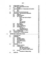
Contents
1 1.1 1.2 1.2.1 1.2.2 1.2.3 1.3 1.3.1 1.3.1.1 1.3.1.2 1.3.1.3 1.3.1.4 1.3.1.5 1.3.1.6 1.3.1.7 1.3.1.8 1.3.1.9 1.3.2 1.3.2.1 1.3.2.2 1.3.2.3 1.3.2.4 1.3.3 1.3.3.1 1.3.3.2 1.3.3.3 1.4
Introduction 1 A Short Definition of Laboratory Automation 1 Short History of Laboratory Automation 2 Early Developments in Laboratory Automation 2 Advances in the Automation of Clinical Laboratories 5 Developments in Pharmaceutical Research 6 Laboratory Applications and Requirements 9 Bioscreening and Pharmaceutical Testing 9 Enzymatic Assays 9 Cell-Based Assays 10 ELISAs 11 DNA/RNA Extraction, Purification, and Quantification 12 PCR/RT-PCR/q-PCR 12 Gene Expression Analysis 13 Next-Generation Sequencing 13 Cell Culturing 13 Requirements 14 Clinical Applications 15 Determination of Classical Parameter 15 Determination of Vitamins 18 Determination of Drugs of Abuse 19 Requirements 21 Classical Analytical Applications 23 Food Analysis 23 Environmental Analysis 26 Requirements 27 The Goal of this Book 30 References 32
2 2.1 2.1.1
Basic Concepts and Principles of Laboratory Automation 41 The LUO Concept in Laboratory Automation 41 Laboratory Unit Operation Concept 41
vi
Contents
2.1.2 2.1.3 2.2 2.2.1 2.2.2 2.2.3 2.3 2.3.1 2.3.2 2.3.3 2.3.4 2.3.5
Classes of Laboratory Systems and Devices 42 General Automation Strategies in Laboratory Automation 45 Advantages and Limitations of Laboratory Automation 50 Advantages of Laboratory Automation 50 Limitations of Laboratory Automation 53 Error Handling in Laboratory Automation 54 Economic Potential of Laboratory Automation 55 Market Dynamics 55 Market Shares by Region 56 Market Shares by Application 62 Market Shares by Users 64 Market Share by Vendors 65 References 66
3 3.1 3.1.1 3.1.2 3.1.3 3.1.3.1 3.1.3.2 3.1.3.3 3.1.3.4 3.1.3.5 3.1.3.6 3.1.4 3.1.4.1 3.1.4.2 3.2 3.2.1 3.2.2 3.2.3 3.3 3.4 3.4.1 3.4.1.1 3.4.1.2 3.4.2 3.4.2.1 3.4.2.2
Formats in Laboratory Automation 69 Formats in Biological Applications 69 Introduction 69 Characteristics of Microplates 70 Lids and Sealing Systems for Microtiter Plates 76 Lids 77 Foils and Films 77 Mats 80 RoboLid 80 Advantages and Disadvantages of Locking Systems 81 Application Areas of Locking Systems 82 Market Potential and Commercially Available Systems 82 Microtiter Plates Market 82 Market Lids and Sealing Systems 83 Formats in Clinical Applications 88 Collection of Blood Samples 90 Collection of Urine Samples 91 Collection of Further Examination Material 91 Formats in Classical Analytical Applications 94 Automated Handling of Labware 97 Automated Handling of MTP and Covers 97 Handling of Microtiter Plates and Lids 97 Automated Handling of Foils and Films 97 Automated Handling of Single Samples 100 Automated Transport 100 Automated Opening/Closing of Single Samples 104 References 107
4 4.1 4.1.1
Liquid Handling in Laboratory Automation 111 Introduction 111 Definition and General Introduction 111
Contents
4.1.2 4.1.3 4.2 4.2.1 4.2.2 4.3 4.3.1 4.3.2 4.3.3 4.3.4 4.4 4.4.1 4.4.2 4.4.3 4.4.4 4.4.5
5 5.1 5.2 5.2.1 5.2.2 5.2.3 5.2.4 5.3 5.3.1 5.3.1.1 5.3.1.2 5.3.2 5.3.2.1 5.3.2.2 5.3.3 5.3.4 5.3.5 5.4 5.4.1 5.4.2 5.5 5.5.1 5.5.2 5.5.3 5.5.4
Short History of Liquid Handling 112 Use of Liquid Handling Systems 115 Liquid Handling Technologies 116 Pipetting Technologies 116 Aspiration Methods 119 Critical Liquid Handling Parameters and Error Sources in Liquid Handling 121 Important Liquid Handling Parameters 121 Physical Influencing Factors 123 Error Sources in Liquid Handling 126 Liquid Handling Performance Monitoring 129 Market Potential and Systems 132 Market Potential for Liquid Handling Systems 132 General Channel Configurations 134 Liquid Handling Systems with 1–8 Channels 136 Multichannel Systems 144 Liquid Handling Accessories 149 References 150 Low–Volume Liquid Delivery 155 Introduction 155 Contact-Based Dispenser Technologies 158 Pin Tools 158 Dispensers with Fixed Tips 159 Dispensers with Disposable Tips 159 Summary 160 Contactless Dispenser Technologies 161 Displacement Dispensers 161 Peristaltic Pumps 161 Ceramic Pumps 161 Valve-Based Dispensers 162 Solenoid Valve Dispensers 163 Piezoelectric Valve-Based Dispensers 164 Capillary Sipper 164 Acoustic Dispensers 164 Summary 165 Application Areas and Requirements for Low-Volume Dispensing 167 Application Areas for Low-Volume Dispensing 167 Requirements for Low-Volume Dispensing 168 Overview of Low-Volume Dispensers 170 Positive Displacement Systems 170 Piezoelectric Dispenser 170 Acoustic Dispensers 173 Additional Systems 176 References 177
vii
viii
Contents
6 6.1 6.2 6.2.1 6.2.2 6.2.3 6.3 6.3.1 6.3.2 6.3.3 6.4
Solid Dispensing 181 Introduction 181 Factors Influencing the Dosing of Solids 183 Flow Behavior of Bulk Solids 184 Density of Solids 185 Fluidization of Bulk Materials 186 Solid-Dispensing Technologies 186 Volumetric Dosing Methods 186 Gravimetric Dosing Methods 188 Dosing Methods in Laboratory Automation 189 Solid Dispensing Systems 190 References 198
7 7.1 7.2 7.2.1 7.2.2 7.2.3 7.2.4 7.2.4.1 7.2.4.2 7.2.4.3 7.2.5 7.3 7.3.1 7.3.2 7.3.3 7.3.4 7.4 7.4.1 7.4.2 7.4.3 7.5 7.6 7.6.1 7.6.2 7.6.3 7.6.4 7.6.5 7.6.6 7.6.7 7.7 7.7.1 7.7.2
Devices for Sample Preparation 201 Introduction 201 Automated Heating, Cooling, and Mixing 204 Introduction 204 Automated Heating and Cooling 205 Automated Thermocycler 209 Automated Mixing/Shaking 213 Introduction 213 Automated Shaking 216 Automated Stirring 217 Combined Solutions for Mixing and Temperature Control 219 Automated Incubation 221 Introduction 221 Important Parameter 222 Incubation Systems in the Laboratory 224 Market Situation 226 Automated Centrifugation 230 Introduction 230 Requirements 233 Market Situation and Systems 235 Automated Filtration 237 Automated Solid Phase Extraction 240 Introduction and Requirements 240 Semiautomated Systems 241 Requirements for Automated SPE Systems 242 Automated Single Sample Processing Systems 243 Automated Parallel Processing Systems with Limited Parallelity 245 High Parallel Systems 247 Labware for Automated Solid Phase Extraction 250 Automated Sonication 256 Basics and Applications of Ultrasonic Systems 256 Market Situation and Systems 258
Contents
7.8 7.8.1 7.8.2 7.8.3
Automated Evaporation 262 Introduction 262 Evaporation Technologies and Application Areas 262 Market Situation 264 References 269
8 8.1 8.1.1 8.1.2 8.1.3 8.1.4 8.1.5 8.2 8.2.1 8.2.2 8.2.3 8.3 8.3.1 8.3.2 8.3.3 8.3.4 8.4 8.4.1 8.4.2 8.4.3 8.4.4 8.4.5 8.5
Robots in Laboratory Automation 281 Robots – A Definition 281 Historical Development of Laboratory Robotics 281 Basics and Definitions in Robotics 282 Robotic Configurations 285 Robot Programming 287 Advantages and Disadvantages of Laboratory Robots 288 Stationary Robots in Laboratory Automation 289 Industrial and Collaborative Robots 289 Market Potential 292 Available Stationary Robot Systems 294 Mobile Robots 300 Differentiation Between Stationary and Mobile Robots 300 Application Scenarios for Mobile Robots 300 Sensor Systems in Mobile Robotics 302 Market Situation and Available Systems 303 Gripper Systems 308 Mechanical Gripper 308 Pneumatic Gripper 310 Magnetic Gripper 311 Adaptive Gripper 311 Sensors and Safety Systems in Gripper Systems 314 Safety Aspects in Laboratory Automation 316 References 318
9 9.1 9.1.1 9.1.2 9.1.3 9.2 9.2.1 9.2.2 9.2.3 9.3 9.4 9.4.1 9.4.2 9.4.2.1
Analytical Measurement Systems 321 Absorption-Based Methods 321 Introduction 321 Physical Background 321 Application Areas of Absorption Spectroscopy 322 Fluorescence-Based Methods 327 Introduction 327 Physical Background 327 Application Areas of Fluorescence Spectroscopy 330 Market Situation and Available Reader Systems 333 Mass Spectrometric Methods 342 Introduction 342 Physical Background 343 Ionization 343
ix
x
Contents
9.4.2.2 9.4.2.3 9.4.3 9.4.4
Mass Separation Technologies 346 Detection Technologies 347 Application Areas of Mass Spectrometric Methods 348 Market Situation and Mass Spectrometry Systems 351 References 374
10 10.1 10.2 10.2.1 10.2.2 10.2.3 10.2.4 10.3 10.3.1 10.3.2 10.3.3 10.3.4
Sample Identification in Laboratory Automation 385 Introduction 385 Barcode Technology 387 Barcode Types 388 Barcode Reader Technology 392 Barcodes in Laboratory Automation 394 Market Situation for Barcode Readers 400 RFID Technology 402 RFID Methods 402 Application Areas and Design of RFID Systems 404 Advantages and Disadvantages of RFID Systems 408 Market Situation 409 References 412
11 11.1 11.2 11.3 11.3.1 11.3.2 11.3.3 11.4 11.4.1 11.4.2 11.4.3 11.4.4 11.4.5
Interfaces in Laboratory Automation 415 Introduction 415 Analog Interfaces 415 Digital Interfaces 416 Parallel Interfaces 416 Serial Interfaces 418 Network Interfaces 421 Standardization in Laboratory Automation 424 Introduction 424 SiLA 2 Standard 425 Advantages of SiLA 2 427 Disadvantages of SiLA 428 Actual Examples for SiLA Integrations 428 References 433
12 12.1 12.2 12.2.1 12.2.2 12.2.3 12.2.4 12.2.5 12.2.6 12.2.7
Laboratory Automation Software 435 Introduction 435 System Control Software/Process Control Systems 435 Introduction 435 Cellario 438 Green Button Go 440 Momentum 441 OneLab 442 Overlord 443 SAMI EX 444
Contents
12.2.8 12.2.9 12.2.10 12.3 12.3.1 12.3.2 12.3.3 12.3.4 12.3.5 12.4 12.4.1 12.4.2 12.4.3 12.4.4 12.4.5 12.5 12.5.1 12.5.2 12.6 12.6.1 12.6.2 12.7
VWorks 446 Hierarchical Workflow Management System (HWMS) 447 Summary 448 Laboratory Information Management Systems 449 Introduction 449 Core Functionalities of LIMS 450 LIMS Architectures 452 Factors Influencing the Selection of a LIMS 454 LIMS Vendors 465 Electronic Laboratory Notebooks 466 Introduction 466 Regulations and Legal Aspects 467 Functionality of ELN 468 Factors Influencing the Selection of an ELN 468 ELN Vendors 470 Laboratory Execution Systems (LES) 470 Introduction 470 LES Vendors 479 Scientific Data Management Systems (SDMS) 479 Introduction 479 SDMS Vendors 480 Additional Laboratory Automation Software 481 References 482 Index 487
xi
1
1 Introduction 1.1 A Short Definition of Laboratory Automation The term “automation” first appeared in 1936. Harder described automation as “the transfer of work tasks to machines in a production process without human intervention” [1]. In 1946, he while working as Vice President founded the Automation Department of Ford Motor Company. After World War II, two books by Diebold (1926–2005) appeared in 1952, describing automation as “automatic operation or a process for the automatic production of material goods.” Diebold defined two main meanings of automation. On the one hand, he defined automation as an automatic control through feedback. On the other hand, automation for him was also the integration of a different number of machines [2]. The Diepold concept was further developed by Bright, who described the various stages of mechanization and automation [3], and Drucker, who recognized automation as “a conceptual system beyond technology.” These three theories form the basis for understanding the concept and importance of automation [4]. “Automation” can be seen as an abbreviation for “automation technology” or “automatic operation.” Alternatively, automation is also a combination of the Greek “automotos” (means “to move yourself”) and the Latin “-ion” (means “a state”). “Mechanization” is the replacement of physical labor with machines; however, machine operation is controlled by human operators. “Automation” also replaces these control measures with machines, i.e. it replaces the physical and mental activities of humans with machines. Laboratory automation is part of automation technology and aims to develop and optimize technologies for the automation of classic laboratories. This includes a wide variety of laboratories in the fields of medical diagnostics, environmental analysis, or quality control, for example, in the pharmaceutical industry, food monitoring, or industrial production. Laboratory automation is a strongly multidisciplinary field. The main goal of automating laboratory processes has not changed since the first steps in this area and consists of increasing the number of processed samples (and thus productivity), reducing the processing times required per sample, and improving the quality of those obtained experimental data or the creation of opportunities for examinations that would not be possible without suitable laboratory automation. Laboratory automation can today be defined as a highly complex integration of robotics, liquid handling systems, sample processing, and analyzing devices and computers for process control. The most important part of laboratory automation is laboratory robotics, which Devices and Systems for Laboratory Automation, First Edition. Kerstin Thurow and Steffen Junginger. © 2023 WILEY-VCH GmbH. Published 2023 by WILEY-VCH GmbH.
2
1 Introduction
develops robots and robotic solutions adapted to the specificity of laboratory processes. Since the robots in laboratory automation systems generally only take on transport tasks, the development of suitable devices and components for the automatic execution of laboratory processes (e.g. dosing, shaking, incubating, etc.) is of immense importance. Suitable software algorithms are required to control the individual systems and to evaluate the data collected.
1.2 Short History of Laboratory Automation The main drivers of the development of automation solutions are often the development of special branches of industry as well as new and more complex requirements for specific analytical processes. Very often, the impulses for the development of new solutions result from the end-users who are confronted with several problems and inadequacies in their everyday laboratory work. For a long time, the requirements of industrial process control drove the development of automated systems.
1.2.1 Early Developments in Laboratory Automation The first reports on the use of automated devices can be traced back to 1875 [5]. The first steps that have been made accessible to automation seem very simple from today’s perspective: washing filtration residues on filter paper or liquid extractions. In 1875, Stevens described a device that made it possible to wash filter residues with water at a controlled flow rate. The wash solution was in a closed reservoir, through which air was passed through an opening. The flow rate could be controlled by the size of the opening [6]. This concept was further developed by Mitchel [7] and Lathrop [8]. In the analysis of fertilizers, the samples were washed successively with 10 ml water each until a total volume of 2500 ml was reached in order to wash out the soluble components. For this purpose, Horne developed a device for the automatic washing of the samples [9]. The first automatic burette for laboratories with recurring titrations was described by Squibb in 1894 [10]. In the same year, Greiner presented an automatic pipette, which was used for the Babcock milk test [11]. The previous developments were not suitable for slow extractions over several hours; therefore, Hibbard developed a suitable system with which flow rates of approximately 40 drops/minute were possible. A further reduction in the dripping speed could be achieved by installing a splitter [12]. The first liquid–liquid extractors were used for botanical studies. By spraying the extraction solvent into the aqueous phase, the efficiency of the extraction could be increased considerably by increasing the surface area [13]. The first devices were developed by different scientists, who were faced with different problems in the laboratory. They were very fragile systems that could be easily broken and very difficult to clean; therefore, the solutions were proprietary and did not find widespread use. The better understanding of combustion processes and the steadily growing production of electrical energy at the end of the nineteenth century revolutionized power generation. The development of automation was therefore decisively driven by the coal and power generation industry since at the beginning of the twentieth century, there was an increasing need for more precise knowledge of the quality of coal (calorific value). The first
1.2 Short History of Laboratory Automation
commercial laboratory automation device was therefore a device for grinding coal samples. The Sturtevant Automatic Coal Crasher was operated by an external motor, and it made it possible to provide representative samples [14]. Another important parameter in industrial production was the determination of carbon dioxide in flue gases for the optimization of combustion processes. A commercial system was introduced to the market by Simmance and Abady. The system could be operated unattended for longer periods of time, but only provided intermittent values. A continuous variant was proposed by Stache et al. with the development of the autolyser [15]. Taylor and Hugh developed a system for the automated determination of carbon monoxide, which was based on a change in conductivity of a solution when the gas was passed through [16]. Conductivity measurements have also been reported for the control of sulfuric acid content in papermaking. Edelmann developed a device that enabled the automatic supply of sulfuric acid based on the measured values. This had previously been done manually and therefore represented an enormous source of errors [17]. The first commercial automated laboratory devices were developed during the First World War due to an increased need for rapid gas analysis. Such systems could now be used for the detection of chemical warfare agents in armed conflicts. The first systems were based on the measurement of changes in the conductivity of a heating wire. Since there was no chromatographic separation of the components prior to the measurement, clear identification of substances was not possible. Commercial variants were sold by the Cambridge Instrument Company and others [18]. In the 1920s, new requirements came from the sugar and paper industries, where there was an increasing need for pH determinations in different production steps. An essential step is the liming of sugar cane juice to remove non-sugars, for which an automated system was first developed in 1928 [19]. This system marked the beginning of the era of the development of electrodes for pH control. The electrodes available at the time required too long equilibration times, were too complicated for use in an industrial environment and were too susceptible to poisoning from sulfur dioxide, which was used in the process. Balch and Kane used tungsten-calomel electrodes for their developments, but it turned out that these exhibited variabilities in calibration, did not last long, and were also susceptible to poisoning [20]. In 1929, the first automated titration systems were introduced, which used a photocell to detect the color change in the solution. After the color change was detected, a valve was automatically closed so that no further titrant was dosed. The authors reported that “the device was 165 times more sensitive than the human eye” [21]. Hickman and Sanford developed a much more sophisticated titration device at Eastman Kodak. The device had an option to empty the previous sample to avoid contamination. In addition, the indicator was automatically supplied [22]. With the beginning of World War II, there was a further boost in the development of automation solutions in process control. This resulted from increased demands on the production of war-relevant goods and a lack of qualified workers. Automated devices were also used to enable unskilled workers to perform complex tasks [23]. Particular attention was paid to the development of semi-automated distillation equipment; Ferguson developed a corresponding system for petroleum fractionation [24]. The automatic mercaptan titrator (Shell Oil Company) for the analysis of gasoline was also a typical example of an automation
3
4
1 Introduction
solution that arose due to the existing shortage of skilled workers. Because the system was used in a refinery, the device was locked in an explosion-proof housing. To ensure overpressure in the system and to prevent the penetration of explosive gases, compressed air was fed into the housing. A potentiometric method developed in 1941 was automated in 1943. The device could easily be operated by unskilled workers [25]. In contrast to manual titration, in which the rate of addition is adjusted around the end point, the titrant was kept constant. At the end of World War II, the use of automated systems in the chemical industry had already become routine; thus, there was an increasing need for appropriately trained specialists. New devices were developed for fraction collectors for chromatography or distillations. Electronic components were increasingly used to control valves, for example, for an automated system for paper chromatography [26]. The development of automated titrators was advanced. In 1948, a device was created that used a motor-driven syringe to add the titrant. The motor speed could be adapted for the respective titration applications and the titration curve could be printed [27]. The automated Karl Fischer titration was introduced in 1952 by the Merck company. Since this method works without water, it was not possible to use classic potentiometric methods to determine the end point. Instead, a polarization process with depolarization of the platinum electrodes used at the end point was chosen [28]. The automated coulometric Karl Fischer titration, which made it possible to recover the Karl Fischer reagent [29], represents a significant development. A summary of automated titration techniques and systems using photometric, amperometric, conductometric, thermal, and potentiometric methods can be found in Ewing [30]. The first reviews of automation technologies appeared in the 1950s [31]. From 1952, the “Instrument Engineer” journal was devoted to special automation topics. Computers related to automation were first described in 1948. The “office-size electronic computer” presented by Reeves Instrument Corporation gave researchers an opportunity to simulate their processes for the first time [32]. The first use of digital computers was described as a system for the mass spectrometric determination of hydrocarbon mixtures (Atlantic Refining Company) [33]. In the following period, computers quickly found diverse uses in laboratory automation. Cerda and Ramis described, among other things, the automation of potentiometric titrations with a Commodore VIC-20 microcomputer and with an IBM PC. In some cases, separate computers were used for data handling due to the limited storage capacity. The latter system has been described for the titration of studies on chemical equilibria as well as for titrations to determine equivalence points. A system consisting of two burettes, an autosampler, a potentiometer, and an Acer 710 to control the entire system enabled the automatic determination of boron in industrial samples. A system for ion-selective potentiometry has also been described. The authors also described automatic systems for conductometric, photometric, spectrophotometric-potentiometric, fluorometric, and thermometric titrations [34]. In addition to the development of computers, the introduction of transistors also revolutionized laboratory automation. Innovative technologies in the dosing of liquids were essential for further development of laboratory automation [35]. In 1957, Schnitger developed a new type of pipette that already had all the features of modern piston-operated pipettes today. It had a spring-loaded piston, a second coaxial spring for blowing out liquid residues, and replaceable plastic pipette tips. An air buffer separated the liquid from the reciprocating piston. The Eppendorf company
1.2 Short History of Laboratory Automation
(Hamburg, Germany) secured exclusive production and marketing rights and introduced the first industrially manufactured piston-operated pipette into the market in 1961 [36]. Today’s mechanically adjustable micropipettes are based on a model developed by Gilson, which he patented in 1974 [37]. The technical advances in the development of small motors and valves led to the introduction of semi-automated syringe-based pipetting systems in the 1970s. In 1971, the Digital Dilutor (Hamilton, Reno, NV) was introduced, which used two calibrated syringes as pipetting plungers. The establishment of microprocessor technology made it possible to create program sequences for controlling the motors and valves and this led to the first fully automatic pipetting systems. The first automated liquid handling systems emerged in the 1980s as a result of further electromechanical developments. The development of these systems has been driven by clinical radioimmunoassays. Hamilton (Reno, NV) and Tecan (Männedorf, Switzerland) cooperated in the late 1970s in the joint development of the Hamilton AMICA system, which was the basis for the later pipetting systems Hamilton 2000 Series and Tecan Sampler 500/RSP 5000 Series Workstation. Both systems were based on Cartesian robotic platforms and enabled single-channel pipetting. A short time later, systems with two separate Cartesian arms and a second pipetting channel were also available. With the Zymark Z510 Master Laboratory Station, Zymark (Hopkinton, MA) developed its own pipetting system for integration into more complex Zymark robot systems.
1.2.2
Advances in the Automation of Clinical Laboratories
Medical and clinical applications and requirements largely drove the development of laboratory automation. The first real automated systems with automated loading of samples into the system and then fully automated measurement appeared in medical laboratories in the mid-1950s. The AutoAnalyzer (Technicon), presented in December 1956, was able to determine the concentrations of urea, sugar, and calcium in blood samples within 2.5 minutes [38]. The concentration was determined by color changes that were read out using photocells [39]. The AutoAnalyzer I used flow analysis technology to increase sample throughput. Later versions enabled the simultaneous determination of 20 analytes, with a throughput of 150 samples per hour. The AutoAnalyzer started a long development in clinical automation. Devices such as the Sequential Multiple Analyzer (SMA, 1969) and Sequential Multiple Analyzer with Computer (1974) increased the throughput further [40]. The AutoAnalyzer was the first batch analyzer in clinical laboratories and led to numerous other batch analyzers, which could usually examine up to 100 samples continuously for individual analytes. In the early 1980s, the introduction of the photodiode for spectrometers with grating monochromators led to the development of systems that enabled simultaneous determination of different analytes in a sample using different specific wavelengths [41]. Another approach was followed by the Research Specialites Co., Richmond, CA, which presented the Robot Chemist in 1959 [42]. Although the Robot Chemist was able to take over all manual steps in sample preparation and enabled analysis with conventional cuvettes, it was not successful in the long term due to its excessive mechanical complexity; production stopped in 1969. The principle of batch sample processing has increasingly been replaced by discrete systems that work with positive displacement pipettes. The solutions were appropriately mixed by the dispensing steps themselves or by means of magnetic or
5
6
1 Introduction
mechanical stirrers. Temperature monitoring was implemented, as well as washing steps between the individual sub-steps. Permanent (glass) or disposable (plastic) cuvettes were used. Depending on the application, different analyzers with different lamps, including tungsten, quartz halogen, mercury, xenon, or laser, were used. The monochromators used interference filters, prisms, or diffraction gratings. The signal detection was usually carried out with photodiodes since a wide range of wavelengths can be covered in this way [43]. In addition to the development of automated analysis systems, another important step was the introduction of ready-made kits for carrying out analytical determinations, which contained all the necessary solvents and reagents as well as the corresponding work instructions. Sigma Chemical Company introduced the first kit of its kind in the 1950s. This eliminated the need for the manual production of reagents in the laboratory, which, in addition to reducing the workload, also led to considerable improvements in the quality of the analytical tests. With the beginning of the 1970s, the introduction of robots into clinical laboratories and with it the era of total automation began. A revolution in this area occurred in the 1980s when Sasaki opened the first fully automated laboratory [44, 45]. As professor and director of the Department of the Clinical Laboratory at Kochi Medical School (Kochi, Japan), he and his team built conveyor belts, robots for loading and unloading analyzers and developed the first process control software [46]. The automation efforts at this time resulted from extensive savings in technical personnel for the implementation of clinical-chemical investigations [47]. Through close cooperation with industrial partners, his ideas led to commercial products that were used in numerous clinical laboratories across Japan. Further, 72% of all university hospitals in Japan installed and used such systems [47]. In the 1990s, there were several commercial suppliers of fully automated systems for clinical laboratories [48]. Regardless of the success of these first laboratory automation systems, they remained stand-alone solutions that could not be used for smaller laboratories and institutions, particularly due to the high costs. In addition, different interfaces of devices from different manufacturers limited the general use, since communication between different devices was not possible in this way. Sasaki et al., therefore, recommended the introduction of binding standards and sizes of racks as well as the use of more flexible robotic technologies in order to achieve plug-and-play functionality in automation systems [47]. Some laboratories developed in-house solutions, but these were very proprietary systems and required a lot of maintenance. Dr. Rod Markin (University of Nebraska Medical Center) developed one of the first clinical laboratory automation management systems. His system later enabled the “plug-and-play” integration of automation systems and clinical analyzers for managing and testing patient samples. His idea was to develop an automated transport system with which various test processes with commercially available test systems are possible. He paid particular attention to the management of the test processes, which resulted in greater efficiency, improved reporting, and lower laboratory costs.
1.2.3 Developments in Pharmaceutical Research In addition to the requirements of clinical laboratories, the development of high-throughput screening (HTS) methods in the pharmaceutical industry has been of particular importance
1.2 Short History of Laboratory Automation
for the development of laboratory automation since the 1980s [49, 50]. Due to the lack of drugs for numerous diseases (especially cancer and viral diseases), the increasing resistance of microorganisms to known antibiotics and the expiry of important patents, there was great pressure for faster development and testing of new potential active ingredients. In addition to the synthesis of new active ingredients, their testing with regard to biological activity, carcinogenicity, mutagenicity, and metabolism behavior is the focus of interest. The early identification of toxic properties of the potential drug candidates contributes significantly to reducing the costs of drug development and increasing safety. The main goal of HTS is to increase the number of samples processed per unit of time. The number of samples to be examined has increased dramatically. While in the 1980s, a sample volume of around 10 000 compounds was processed per year, at the beginning of the 1990s it was already 10 000 samples per month. Only five years later, there was a requirement to process the same number of samples within a week [51]. Today, HTS can include the processing of several thousand samples per day. In the area of ultra-HTS, up to 100 000 samples have to be processed per day [52, 53]. Since processing numerous of samples is associated with considerable costs for reagents, solvents, and consumables, there is great interest in minimizing these costs by miniaturizing the experimental approaches [53]. In the period from 1998 to 2006, Novartis (Basel, Switzerland) succeeded in significantly increasing the number of compounds examined while at the same time drastically reducing the cost per substance. Parallel sample processing was increasingly used in the automation of bioscreening. The development of a uniform standardized format, the microtiter plate, played an important role. Depending on the format used (see Chapter 3), up to 384 or more samples can be processed in parallel today. This required the development of parallel working systems for the dosing of liquids, but also for the technical determination of the parameters by means of adsorption or fluorescence methods. Microtiter plate-based test methods were presented for the first time in 1986 at the Fourth International Symposium on Laboratory Robotics [54]. The systems used an early version of Zymark’s microplate management system and, thanks to interchangeable hands, were able to carry out various laboratory processes such as pipetting, washing plates or adding reagents. The systems were referred to as “one-armed chemists” [55] and were initially used for enzyme-linked immunosorbent assays (ELISAs) investigations [56]. However, their throughput and unattended operation were severely limited. The use of articulated robots (see Chapter 8) represented a very cost-intensive variant of the automation of such processes and was therefore not generally applicable. Numerous companies, therefore, developed specialized liquid handling systems based on a Cartesian robot structure. The Cetus Propette, a 12-channel pipetting system for the transfer of liquids in microtiter format, was introduced in 1996. The device originally developed for the automation of interleukin-2 assays was later used extensively in polymerase chain reaction (PCR) analysis [57]. The Biomek 1000 (Beckman Coulter), originally a development by Infinitek, was launched in 1984. It enabled the single or parallel multi-channel pipetting of several samples. The interchangeable pipetting heads were a special feature. Another Cartesian liquid handling platform, the Star 700, was introduced by Kemble (U.K.) in 1985. The MikrolabAT (Hamilton Company) was launched in 1987 for the batch screening of blood samples for HIV and hepatitis viruses. The system had 12 channels with variable span and used disposable pipette tips. The first 96-channel pipetting system was the
7
8
1 Introduction
Quadra96, developed by TomTec in 1990 [58], later followed by a variant with a 384 pipetting head. In contrast to solutions with liquid-handling hands-on articulated robot arms, Cartesian systems enable a significantly faster and better quality liquid transfer. The liquid handling workstations available today represent all further developments of these early pipetting systems. Workstation technology quickly found its way into molecular biology and genomics, as both areas of science were characterized by low throughputs and numerous labor-intensive liquid handling steps. In order to avoid bottlenecks in the metrological determination of the samples, it was also necessary to develop parallel reading systems based on absorption or fluorescence methods. One of the first automated plate readers, the EL310, was introduced by BioTek in 1984 [59]. Today’s plate readers enable the parallel reading of up to 1536 samples in microtiter plate format. Various automated systems have been described for biological studies. One of the best-known applications is the Tox21 Initiative, which was started in 2008 with the aim of determining the toxicity of environmentally relevant compounds. The Tox21 Screening System has been used to screen more than 10 000 compounds. To determine the reproducibility of the results, the substances were examined on three days each with three replicates in different well positions. Various devices such as incubators, contactless dispensers for liquid dosing in the nano range and fluorescence or luminescence-based plate readers were positioned around a central robot [60]. Approximately 40 different assays were used for the biological testing, the parallel testing of the samples was performed in the 1536 format. All results have been made accessible in public databases and are thus available to scientists worldwide for further data evaluation, the formation of new hypotheses, and the establishment of reliable QSAR models. The earliest automated systems in pharmaceutical screening were developed for finding biologically active compounds in natural products. The majority of these systems, if not all, were tailor-made in-house developments that were usually not published for reasons of competition. Therefore, no general formats and technologies could be derived and developed from these developments. One of the few published studies comes from Eli Lilly and Company (Indianapolis, IN). They used a PUMA 560 robot for inoculation of microbial colonies in sample vessels combined with a subsequent test of the antibiotic effect of the fermentation extracts [61]. Pfizer (Groton, CT) has also been using HTS methods since 1986 for the screening of natural products by replacing fermentation broths with dimethyl sulfoxide solutions of synthetic compounds using 96-well plates and reduced assay volumes of 50–100 μl. After initially 800 compounds per week examined, a volume of 7200 compounds per week was already achieved in 1989. Autoradiography and image analysis were introduced for 125 I receptor-ligand screens. The coupling of reverse transcriptase (RT), quantitative PCR, and multiplexing enabled multiple targets to be addressed in a single assay. By 1992, around 40% of the hits were produced using HTS as starting materials for the discovery portfolio. In 1995 the HTS methodology was expanded to include ADMET (absorption, distribution, metabolism, excretion, toxicity) targets. ADMET examinations require the unique identification of every single compound, which leads to the development of an automated high-throughput liquid chromatography-mass spectrometry (LC-MS). In 1996, the testing of approximately 90 compounds per week in microsomal, protein binding, and
1.3 Laboratory Applications and Requirements
serum stability assays was possible. Until 1999, the HTS for ADME examinations was completely integrated into the drug discovery process. Automated screening systems have also been used at the Genomic Institute of the Novartis Research Foundation (GNF). A system developed for genome screening was used for almost 200 genome screens from 60 000 to 100 000 wells. The system not only carried out the transports, but also enabled the plates to be transported between the liquid handlers, incubators, and plate readers. The actual measurement was carried out on an integrated ViewLux plate reader (Perkin Elmer) or, for fluorescence-based assays, on a confocal Opera 384 well system, on which the cells can be displayed directly. In order to optimally use all genomic information generated for structural biology, an automated system was developed that enables the automatic expression and purification of bacterial cells, baculoviruses and mammalian cells. Bacterial proteins were expressed using a parallel fermentation system consisting of 96 arranged 100 ml culture tubes, which enabled high-density cell growth and yields of 2–4 g cell pellet for each culture with minimal variation. Protein purification was performed using GNF’s automated protein purification system, which included a 96-tube centrifuge, sonication probes, and liquid handling and affinity purification functions. As a result, 10 mg of purified protein could be obtained per tube; the overall process took 96 hours [62].
1.3 Laboratory Applications and Requirements 1.3.1
Bioscreening and Pharmaceutical Testing
As described above, the development of laboratory automation has been largely influenced by the needs of the pharmaceutical industry since the 1980s. The need to find new potential drugs and reliable early screening for biological activity remain critical. The essential processes in this area include enzyme and cell-based assays, ELISAs, DNA/RNA extraction, purification and quantification, PCR and qPCR, gene expression experiments and next generation sequencing (NGS). 1.3.1.1 Enzymatic Assays
Enzymatic assays use the determination of enzyme activity and are used to determine substances that inhibit or activate certain enzymes as well as the enzyme kinetics. Usually, a blank value and a measured value of the sample are measured after 5–10 minutes of exposure and the extinction difference is calculated, from which quantitative statements can be derived. Enzymatic reactions use optical measurement methods. As early as 1935, Warburg described an optical-enzymatic test for measuring the enzyme activities of NAD+ reducing enzymes. A photometric measurement of the change in color intensity during the reduction from NAD+ to NADH was carried out [63]. This test was used to measure the activities of lactate dehydrogenase (LDH), malate dehydrogenase (MDH) and glutamate dehydrogenase (GLDH) [64]. The biochemical detection of enzyme activities is also possible using composite enzymatic tests. In this case, enzyme activity is measured for which no colored substrate is available. The combination of the reaction of the enzyme
9
10
1 Introduction
to be determined (indicator reaction) with a further enzymatic reaction (measurement reaction) with a change in color intensity enables the extension of the method. The second reaction partially uses the products of the first reaction. This indirectly determines the enzyme activity and quantifies it in comparison to a standard series. Examples of composite enzymatic tests are the glucose oxidase (GOD)-horseradish peroxidase (HRP) test and the GPT-LDH test. The measurement of cell metabolic activity, cytotoxicity, or cytostatic activity is of great importance in the process of drug development. The detection of cell vitality by means of the MTT test uses the reduction of the yellow, water-soluble dye 3-(4,5-dimethylthiazol-2-yl)-2,5-diphenyltetrazolium bromide (MTT) into a blue-violet, water-insoluble formazan. The conversion takes place by NAD(P) H-dependent cellular oxidoreductase, which is present in viable cells. This non-radioactive, colorimetric assay system using MTT was first described by Mosmann T and improved in subsequent years by several other investigators [65–67]. Enzymatic assays can be carried out continuously or discontinuously. The timed (discontinuous) assay measures the enzyme concentration in fixed periods of time. A common timed test method is to use a microplate reader to read multiple concentrations of the solution. Multiple dilution series are examined, which contain dilution series for the substrate, the enzyme, and for the substrate and enzyme together. After the start of the reactions, the solutions are incubated for a specified period of time. A stop solution is then added to prevent a further enzyme reaction. Continuous assays measure the formation of a product or the conversion of a substrate in real-time. The disadvantage of a continuous assay is that only one reaction can be measured at a time. The advantage, however, is the convenience of easily measurable reaction rates. Enzymatic reactions are widely used in drug development for early testing of potential drug candidates [68]. 1.3.1.2 Cell-Based Assays
A higher level of information about the biological relevance of active ingredients can be achieved through cellular assays. Investigations can take place either in the cell network or at the level of an individual cell. Cell-based assays are therefore used extensively in drug development, where they make up more than half of all tests for target validation and ADMET [69]. Classically, proliferation, migration, invasion, apoptosis, etc. are examined. Cell-based assays are analytical tools that can be used to study a mechanism or process of cell function. They typically include intact or fixed cells. The following important types of cell-based assays can be defined [70]: ●
●
●
Intracellular signal transmission: It is an important mechanism by which cells can react to their environment and extracellular signals. Cells can perceive their environment and modify gene expression, mRNA splicing, protein expression, and protein modifications to respond to these extracellular influences. Cell viability assays: These tests determine the ratio of living and dead cells. Cell viability tests are used to determine the cellular response of drug candidates as well as for the optimization of cell culture conditions. Proliferation Assays: Cell proliferation describes the biological process in which the number of cells increases over time due to cell division. They thus monitor the growth rates of cell populations. Cell proliferation is important in the regular homeostasis of tissues and cells to ensure an optimized growth, development, and maintenance of the organism.
1.3 Laboratory Applications and Requirements ●
●
●
Cytotoxicity assays: These assays determine the number of living and dead cells in a population after treatment with a drug candidate or pharmacological agent. Cell senescence assays: Assays for assessing cell health include, e.g. assays for determining the senescence of cells. One example is the detection of senescence markers associated with the activity of β-galactosidase which reflects the integrity of the cell membrane. Cell death assays:
Apoptosis (programmed cell death type 1): Apoptosis investigations are essential for the development, homeostasis, and pathogenesis of various diseases including cancer. Apoptotic cells appear in response to extrinsic or intrinsic signals. Typical signs of apoptotic cell death include the exposure of phosphatidylserine on the extracellular side of the plasma membrane, the activation of caspases, the disruption of the mitochondrial membrane potential, or the shrinkage of the cells. Other markers are DNA fragmentation and condensation. Autophagy (programmed cell death type 2): Autophagy is defined as the selective degradation of intracellular targets that serve as an important homeostatic function. This process enables the destruction of misfolded proteins by ubiquitination followed by a breakdown via the lysosomal route. Necrosis (programmed cell death type 3): Cell swelling and destruction of the plasma membrane and subcellular organelles are typical signs of necrosis. Necrotic cell death is a heterogeneous phenomenon including both, programmed and accidental cell death. ●
●
●
Antibody-dependent cell-mediated cytotoxicity (ADCC): ADCC is an immunological mechanism in which an effector cell of the immune system destroys an antibody-loaded target cell. NK cells, but also macrophages, dendritic cells, neutrophils, and eosinophils primarily take over the role of the effector cell. The ADCC thus represents a connection between the innate and the adaptive immune system. Complement depending cytotoxicity: Complement-dependent cytotoxicity (CDC) is an effector function of IgG and IgM antibodies. If they are bound to surface antigen on the target cell (e.g. bacterially or virally infected cell), the classic complement pathway is triggered by binding of the protein C1q to these antibodies. This leads to the formation of a Membrane Attack Complex (MAC) and lysis of the target cell. The complement system is efficiently activated by human IgG1, IgG3, and IgM antibodies, weakly by IgG2 antibodies and not by IgG4 antibodies [71]. It is a mechanism of action through which therapeutic antibodies [72] or antibody fragments [73] can achieve an antitumor effect [74]. Antibody-dependent cell phagocytosis (ADCP): ADCP is the mechanism by which antibody opsonized target cells activate the FcγRs on the surface of macrophages to induce phagocytosis, resulting in internalization and degradation of the target cell through phagosomal acidification.
1.3.1.3 ELISAs
ELISAs are antibody-based detection methods that belong to the enzymatic immunosorbent methods and are based on an enzymatic color reaction. The antigen to be detected is adsorptively bound and enriched via a first antibody, an enzyme-coupled second antibody (detection antibody) leads to the reaction of a dye substrate. With the help of the ELISA, proteins (e.g. SARS-CoV-2 antibodies [75]) and viruses (e.g. Zika virus [76]), but also low
11
12
1 Introduction
molecular weight compounds such as hormones [77], toxins [78], and pesticides [79] in a sample (blood serum, milk, urine, food, etc.) can be detected using the property of specific antibodies to bind to the substance to be detected (antigen). An antibody is previously marked with an enzyme. The reaction catalyzed by the reporter enzyme serves as proof of the presence of the antigen. The reporter enzymes often used are HRP, alkaline phosphatase (AP), or, less often, GOD. In the case of the alkaline phosphatase a dye substrate (synonym: chromogen), for example, p-nitrophenyl phosphate (pNPP), is added, while for peroxidase o-phenylenediamine (oPD) is mostly used. The alkaline phosphatase splits off the phosphate residue from the colorless nitrophenyl phosphate and p-nitrophenol is formed, which is pale yellow. The change in concentration of the dye produced by the enzymatic reaction can be followed with a photometer according to Lambert–Beer’s law. The color intensity changes with the concentration of the nitrophenol formed and thus also the concentration of the antigen to be determined in the sample in comparison with a dilution series with known concentrations [80]. 1.3.1.4 DNA/RNA Extraction, Purification, and Quantification
DNA extraction is one of the methods of DNA purification and involves the process of extracting DNA from cells. Usually, in the first step, the cells are concentrated by means of centrifugation, followed by cell disruption. Different procedures are required depending on the type of cells used. Plant, fungal, and bacterial cells usually require additional enzymatic or mechanical steps. Chemical cell disruption (alkaline lysis) is usually used for plasmid preparation from bacteria. The homogenate is clarified by filtration or centrifugation. DNA from mitochondria or chloroplasts is separated from the DNA of the cell nucleus by cell fractionation. Hirt extraction is used to isolate extrachromosomal DNA such as viral DNA [81]. An RNAse digestion can be performed to remove RNA. DNA extractions are usually based on two-phase extraction [82] or precipitation [83], the latter being carried out with additional selective adsorption onto a DNA-binding matrix. Some extraction processes are also combined with one another. Final ethanol precipitation usually follows [84], in some cases with the addition of ammonium acetate [85]. The quantification of DNA is possible with different methods [86]. The classic diphenylamine method uses colorimetric detection [87]. It has a detection limit of 3 μg but is very labor-intensive and time-consuming. Absorption-based methods typically use microvolume spectrophotometers and are simple and quick. Their low specificity and sensitivity to impurities are disadvantageous. The sensitivity is around 2 ng/μl. Fluorescence measurements have better detection limits (10–50 pg/μl depending on the kit used). They have high specificity but require very expensive reagents [88]. Sometimes a digital PCR is also used, which is very sensitive and specific [89]. 1.3.1.5 PCR/RT-PCR/q-PCR
The PCR is a method to reproduce genetic material (DNA) in vitro [90]. PCR uses the enzyme DNA polymerase. The term chain reaction indicates that the products of previous cycles serve as starting materials for the next cycle and thus enable exponential replication. Kleppe et al. used first a process for the amplification of DNA sections in 1971 by Kleppe et al. [91]. The actual developer of the method is considered to be Mullis (1944–2019, Nobel Prize in Chemistry 1993). The reaction usually uses volumes of 10–200 μl in small reaction
1.3 Laboratory Applications and Requirements
vessels (200–500 μl) in a thermal cycler. Today, PCR is one of the most important methods of modern molecular biology and is used in biological and clinical-diagnostic laboratories for genetic fingerprints, parentage reports, the cloning of genes, or the detection of hereditary diseases [92] and viral infections (e.g. dengue virus) [93]. The PCR test is currently the gold standard among the SARS-CoV-2 test procedures [94, 95]. Real-time quantitative PCR (qPCR or RTD-PCR) is an amplification method for nucleic acids based on the principle of ordinary PCR. In addition, it also enables the quantification of the DNA obtained. The quantification is carried out with the help of fluorescence measurements, which are recorded in real-time during a PCR cycle. 1.3.1.6 Gene Expression Analysis
The gene expression analysis examines the implementation of genetic information (gene expression) with molecular biological and biochemical methods. It enables qualitative and quantitative statements about the activity of genes and can be used for individual transcripts as well as the complete transcriptome. Typical qualitative questions are the general expression of a gene and the type of cells in which the expression takes place. In the case of quantitative analysis, the size of the difference in expression compared to a defined reference is determined. Applications can be found in cancer research [96] or the investigation of viral diseases such as Zika [97] or SARS-CoV-2 [98]. 1.3.1.7 Next-Generation Sequencing
NGS is an improved technology for DNA sequencing. In contrast to classic enzymatic (Sanger sequencing) or chemical sequencing (Maxam-Gilbert method), this method allows higher speeds and thus enables the sequencing of a complete human genome within one day [99, 100]. The NGS processes are often automated; the results are obtained in parallel with the sequencing. In addition, the results can be compared with a human reference genome. In the first step, DNA fragments are generated with the help of enzymes or centrifugation. In the next step, specific adapter oligonucleotides are bound to the fragments and a DNA library is created. The DNA fragments are bound to solid reaction media (for example a chip) and amplified. Due to the division into clusters of identical DNA, in which the actual sequencing takes place, many sequencing processes can take place parallel in a very short time. The data obtained are stored in the form of a DNA chip and analyzed using bioinformatics methods [101]. For the sequencing of the human genome, Illumina sequencing [102, 103] and SOLiD sequencing [104, 105] are mainly used. 1.3.1.8 Cell Culturing
The cultivation of animal or plant cells in a nutrient medium outside the organism is another typical application in life science laboratories. A distinction can be made between adherent cells growing on surfaces (e.g. fibroblasts, endothelial or cartilage cells) and suspension cells floating freely in the nutrient medium (e.g. lymphocytes). Further differentiation is possible in 2D and 3D cell cultures. The culture conditions differ greatly depending on the cell lines to be cultivated, which concerns both the nutrient media, pH values and the necessary nutrients. Depending on the rate of division and density of the cells, they are distributed to new vessels at regular intervals (passage or splitting). The passage number indicates the frequency with which the cells have already been passaged.
13
14
1 Introduction
In the case of adherent cells in continuous culture, the cells are regularly isolated in order to avoid confluence and the associated inhibition of cell contact. The process of cell cultivation includes numerous sub-processes of dosing nutrient medium, taking aliquots for cell counting and sowing the cells on microtiter plates. Classically, these are manual and therefore labor-intensive processes, which have been increasingly automated in recent years. Both the cultivation of 2D and 3D cells on an automation system have been described [106, 107]. Cell cultures are widely used in biological and medical research, development, and production. Cell cultivation is also of great importance for the manufacture of biotechnological products. In addition to numerous vaccines (e.g. influenza vaccines [108]), erythropoietin, a growth factor for the formation of red blood cells, is also produced in cell culture. 1.3.1.9 Requirements
Although the underlying biochemical reactions in enzymatic and cellular reactions are very complex, carrying out the corresponding assays is quite simple (see Figure 1.1). Essentially, it involves pipetting steps for dosing the components involved (enzyme solutions, substrates, possibly stop solutions and other solutions) as well as the analytical detection of the reactions, usually using optical methods. Mixing the solutions is an important point in order to achieve the most homogeneous distribution possible. All disturbances, such as Start
Pipetting
Incubation, cell disruption, concentration, dilution, etc.
Pipetting
Sample introduction (samples on microtiter plates)
Bioscreening (plate reader)
Data evaluation
End
Figure 1.1 Classical process of enzymatic and cellular assays in drug discovery.
1.3 Laboratory Applications and Requirements
the introduction of air bubbles or dust particles into the solutions, must be avoided [109]. Biological assays run under mild ambient conditions, i.e. they make little demands on temperature, pressure, or the inertness of the ambient air. The assays are usually carried out at room temperature or an assay temperature of 37 ∘ C. Aqueous, buffer-containing solutions are traditionally used, complex organic mixtures are not required. For cell-based assays, sterile conditions are also required, which can be implemented, for example, by means of high-efficiency particulate air (HEPA) filters or the use of UV lamps. Simple optical methods such as absorption spectroscopy or fluorescence spectroscopy, which do not require any preanalytical preparation of the samples, enable the detection. The sensitivity of the optical detection methods used enables working with very small volumes. In addition, the strong parallelization of the processes through the introduction of the microtiter plate (see Chapter 3) is a great advantage.
1.3.2
Clinical Applications
For a long time, clinical applications were a key driver in the development of laboratory automation. While simpler parameters were initially of interest, new requirements increasingly include the determination of a wide variety of organic compounds. Table 1.1 gives an overview of important clinical parameters. 1.3.2.1 Determination of Classical Parameter
Clinical-chemical analyses generally refer to the determination of enzymes, substrates, and metabolic products. A wide range of analytics offered in clinical-chemical laboratories is also available for near-patient diagnostics. This includes, e.g. the determination of enzymes (AP, GOT, GPT, γ-GT, amylase, CK), electrolytes (Na+ , K+ , Ca2+ , Cl− , Mg2+ ) and numerous metabolic variables (total bilirubin, HDL and LDL cholesterol, triglycerides, glucose, uric acid, creatinine, urea, and lactate) [110]. One of the most important clinical parameters to be determined is the glucose content. For this purpose, enzymatic measuring methods using the enzymes GOD and glucose dehydrogenase are almost exclusively used today. The enzyme GOD oxidizes the glucose to gluconic acid in the presence of water and oxygen. The co-factor flavin-adenine-dinucleotide (FAD) serves as the first electron acceptor, which is reduced to FADH. After that, FADH is re-oxidized by molecular oxygen (O2 ), the final electron acceptor. This creates hydrogen peroxide (H2 O2 ). The oxygen consumption or the resulting H2 O2 can then be detected using electrochemical or chromogenic methods. As chromogens, e.g. o-dianisidine, p-aminophenazone/phenol, and iodide/molybdate can be used. The chromogen is oxidized by the resulting H2 O2 and measured reflectometrically (e.g. GlucoTouch from LifeScan (Malvern, PA). The measurement reaction is highly specific, but the indicator reaction can be affected to varying degrees by reducing substances such as ascorbic acid or acetaminophen. A special analytical problem is the sometimes considerable dependence of the measurement results on the oxygen content of the sample. Here, too, the individual variants of the GOD methods must be carefully considered. Those methods that use oxygen as the last electron acceptor (Blood Gas Devices, YSI, Yellow Springs, OH or GlucoTouch) are insensitive to changing oxygen concentrations as long as there is enough oxygen in the sample. The opposite is the case for methods
15
16
1 Introduction
Table 1.1
Clinically relevant parameters and clinical areas of application.
Area of application
Parameter
Acid–base balance, blood gases
pH, pCO2 , pO2
Electrolytes
Na+ , K+ , Cl− , ionized Ca2+ , ionized Mg2+
Metabolites
Cholesterol, HDL cholesterol, triglycerides, creatinine, urea, uric acid, bilirubin, lactate, ammonia
Enzymes
Amylase, alkaline phosphatase, CK, AST, ALT, y-GT
Hemostaseology
Activated whole blood clotting time (ACT), partial thromboplastin time (PPT, aPPT), thromboplastin time (Quick-Test, INR), D-dimer, platelet function tests, bleeding time
Hematology
Hemoglobin, hematocrit, erythrocytes, leukocytes, platelets
Hemoglobin fractions
CO oximetry
Cardiac markers
Troponin T, troponin I, myoglobin, CK-MB, natriuretic peptides (BNP/NT-pro-BNP)
Diabetes mellitus
Glucose, HbA1c, minimally invasive continuous glucose measurement, ketones
Acute-phase proteins
CRP
Allergy diagnostics
Allergen specific IgE
Drug levels and drug screening
Medicines, alcohol, amphetamines, barbiturates, benzodiazepines, cannabinoids, cocaine, methadone, opiates
Infectiology
HIV, infectious mononucleosis, Chlamydia trachomatis, Trichomonas vaginalis, Plasmodium falciparum, Plasmodium vivax, Influenza A, Influenza B, Streptococci A and B
Fertility
HCG, LH, and FSH in urine
Urine diagnostics
Test strips (pH, protein, glucose, ketones, bilirubin, urobilinogen, nitrite, leukocytes, blood, specific gravity), microalbumin, bacteria
Source: Based on Junker and Gässler [110].
that use ferrocene (Precision PCx, Abbott, Chicago, IL) or hexacyanoferrate (Ascensia Elite, Bayer. Leverkusen, Germany) instead of oxygen as the last electron acceptor. Here, oxygen, as a possible electron acceptor, competes with the mediators, so that the glucose is determined to be incorrectly low in the case of increased oxygen values in the sample. Further interferences are to be expected from the body’s own metabolites and drugs with reducing properties (vitamin C, acetaminophen, dopamine, etc.). These are mostly measurement methods that use GOD-based peroxide reactions as detection methods. Processes that use glucose dehydrogenase as an enzyme are much more stable toward such substances. Whole blood, hemolysate, serum/plasma (with and without deproteinization), urine, liquor and interstitial fluid or the dialysate obtained from them can generally be used as sample materials. Hematological examinations using point of care tests (POCT) range from the measurement of the hematocrit (HC) and hemoglobin (Hb) to the complete determination of the blood count. The term blood count summarizes the count of red blood cells, leukocytes
1.3 Laboratory Applications and Requirements
(including the distribution of granulocytes, lymphocytes, and monocytes), and platelets. In addition, the determination of HK and Hb, as well as cell properties (e.g. erythrocyte indices, mean corpuscular Hb (MCH) and mean corpuscular Hb concentration (MCHC)) and other information (e.g. size distribution curves and stages of maturation of individual cell rows), are further parameters of interest. In hematology, in addition to fully-fledged machines for determining blood counts, which have a corresponding range of measurement technology, devices for determining individual hematological parameters are also used. Depending on the device system used, the required sample volume ranges from a few microliters to 200 μl; the measurement time is usually a few seconds. Global and special tests of plasma coagulation are used in large numbers in everyday clinical practice. The small blood count only records the number of platelets in the peripheral blood. Defects in primary hemostasis (defects in adhesion and aggregation of platelets in the event of injuries) are usually not measurable or only measurable to a limited extent in routine diagnostics. Specific investigations of plasmatic or thrombocytic coagulation disorders are usually carried out in special laboratories. The available methods can be divided into the analysis of plasmatic coagulation, the analysis of platelet function, and the combined recording of plasmatic coagulation, platelet function, and fibrinolysis (viscoelastic methods). When diagnosing blood coagulation, several interfering and influencing variables from the sample must be taken into account. Depending on the detection method, different instruments can react differently to variations in HC, to the influence of colloids, and to the formation of microaggregates in the circulation. The effects of specific metabolic conditions (e.g. acidosis) and environmental conditions (e.g. hypothermia) on hemostasis diagnostics are often not systematically clarified. Whole blood methods are sensitive to interfering and influencing factors from the sample matrix. The specificity of the molecular recognition of antigenic structures by antibodies is the basis for the immunoassay technology as well as for immunosensors with the antibodies on a solid phase. The most important analytical problem areas for the selective recognition of the antigen–antibody complex are the bioconjugation chemistry and the orientation of the bound antibodies, the specificity of which must not be compromised by the binding. The immunosensors include electrochemical sensors (potentiometric, amperometric, conductometric, or capacitive), optical sensors, microgravimetric sensors (quartz microbalances), and thermometric sensors. All types can be used both as direct (unlabeled) and indirect (labeled) immunosensors. The direct sensors are able to track physicochemical changes during the immune complex formation, while the indirect sensors mostly use fluorescent or chemiluminescent markings and thus enable a high level of sensitivity. There are simple strip tests for numerous analytes, which can be read off visually. Alternatively, for a less extensive analysis spectrum, smaller automatic detectors are used to read the test strips and quantify the results. Automatic detectors are mainly used in the clinical area. The spectrum of methods includes fluorescence and chromatographic detectors as well as enzyme immunoassays. The examinations usually use not only whole blood, saliva, or urine but also serum or plasma. A major problem with blood examinations with test strips is the use of capillary blood. The concentration can change when the blood is drawn so that the measured concentration of the analyte does not represent the concentration in the blood. This is particularly problematic in the case of analytes whose qualitative detection is at the analytical limit.
17
18
1 Introduction
Numerous systems are available for the metrological determination of classic laboratory parameters. If the samples are not to be centrifuged to obtain serum or plasma, a step to eliminate cellular components from the whole blood must be integrated into the analysis process. Usually, a few microliters of samples are sufficient. The actual analysis takes place within minutes. Common measuring systems usually have an interface to export data and measured values to laboratory or hospital information systems. Clinical-chemical analysis systems represent a downsizing of established laboratory systems and can analyze a wide range of parameters. Other measuring devices enable clinical-chemical analyzes in addition to other measurements. These include devices for blood gas analyzer (BGA), on which electrolytes, glucose, lactate, creatinine, bilirubin, and other parameters can be determined using electrodes or photometric detection. There are also test systems that are specially designed for individual procedures, e.g. to determine the lipid status or lactate. 1.3.2.2 Determination of Vitamins
The determination of vitamins in the human organism is becoming increasingly important, as a lack of vitamins is associated with numerous diseases. Depending on the type of vitamin that is missing, a deficient vitamin supply causes specific deficiency diseases so called avitaminoses, which have been known for centuries under names such as “Beri–Beri,” “Scurvy,” “Pellagra,” etc. With a varied and balanced healthy diet, an adequate supply of all vitamins is always guaranteed. Vitamins are found in relatively small amounts in most plant and animal foods. They are indispensable for the human organism because they functionally intervene in almost all metabolic processes in the body. Vitamins are divided into fatand water-soluble vitamins according to their solubility. These two groups have different functions in the human body. Due to the excretion and degradation processes, there is a constant need so that they have to be returned to the organism with food. The fat-soluble vitamins include vitamins A, D, E, and K. Vitamin B1, B2, B6, and B12, niacin, folic acid, and pantothenic acid as well as the most well-known vitamin C belong to the water-soluble vitamins. The analytical determination of the content of the individual vitamins is usually very difficult. Since various vitamins belong to very different chemical substance groups and for the most part do not have any common chemical properties, it is not possible to determine a large number of vitamins with a few – let alone a single – analytical method. At best, a few vitamins can be quantified together in small groups in certain cases. Very few vitamins can be determined with relatively simple processing and examination methods. This includes, for example, the enzymatic analysis of vitamin C. Some vitamins, however, are extremely difficult to determine because very complex work-ups are necessary and they do not run reproducibly. For vitamins B6, B12, niacin, pantothenic acid, and folic acid, microbiological methods are recommended for their determination. Methods for determining vitamin A have been known for a long time. Bessey et al. described in 1946 an optical method for the determination of vitamin A in small amounts of blood; the vitamin A absorption was measured at 328 μm [111]. More recent methods have been described by Xuan et al. They used an SPE-based method to extract vitamin A from 200 μl serum; the metrological determination was carried out by means of high-performance liquid chromatography (HPLC) [112].
1.3 Laboratory Applications and Requirements
The determination of vitamin C in blood and urine has also been known for a long time. A photometric method was described by Roe et al. in 1942; vitamin C was determined after derivatization with 2,4-dinitrophenylhydrazine [113]. Detection limits of up to 0.2 μmol/l can be achieved for blood samples using reversed-phase HPLC and fluorometric detection [114]. Current methods mainly use mass spectrometric methods to determine the vitamins in blood and urine after the samples have been prepared accordingly. For example, the determination of B vitamins using LC/MS/MS in concentration ranges from 0.42 to 5.0 μg/l has been reported [115]. Numerous methods have also been described for the determination of vitamin D and its metabolites. Thus, among other things, the quantitative determination of 25-hydroxyvitamin D metabolites (25OHD3, 25OHD2, and 3-epi-25OHD3) from dried blood samples after extraction and derivatization using LC/MS/MS [116]. Alternatively, solid-phase extractions can be used for the extraction of vitamin D; the automation of this process was described by Bach et al. [117]. As early as 1936, Schønheyder described a method for the quantitative determination of vitamin K from blood in connection with studies on vitamin K deficiency in chicks [118]. Here, too, HPLC-based methods are used today, which enable detection limits of 0.04 ng/ml vitamin K in plasma [119]. 1.3.2.3 Determination of Drugs of Abuse
Another large group of parameters to be determined is narcotics. Narcotics is a group of centrally effective drugs and substances, which are heavily regulated and controlled by drug and health authorities to prevent abuse and protect the population from adverse effects and addiction. Certain narcotics – for example, many potent hallucinogens – are prohibited or may only be used for medical or scientific purposes with a special permit from the authorities. Some of the substances are also referred to as “psychotropic substances” in the Narcotics Act. Structurally, narcotics are very heterogeneous. However, different groups can be distinguished within this class (see Table 1.2). The most important narcotics include opioids, benzodiazepines, barbiturates, amphetamines, and medicinal Table 1.2
Selection of classic narcotics.
Opioids
Alfentanil, buprenorphine, codeine, fentanyl, heroin, hydrocodone, methadone, morphine, oxycodone
Benzodiazepines and Z-Drugs
Alprazolam, bromazepam, diazepam, flunitrazepam, lorazepam, zolpidem alprazolam, bromazepam, diazepam, flunitrazepam, lorazepam, zolpidem
Barbiturates
Butalbital, pentobarbital, secobarbital
Amphetamines and other stimulants
Aminorex, amphetamine, dexamphetamine, cathine, cathinone, cocaine, methamphetamine, methylphenidate, phentermine
Medicinal drugs
Cannabis, coca leaves, cath, opium
Hallucinogens
Dimethyltryptamine (DMT), hallucinogenic mushrooms such as Psilocybe semilanceata, Ibogaine, LSD, Mescaline, Peyote, Psilocybin, Salvia divinorum, San Pedro
Other examples
Gamma hydroxybutyrate (GHB), Dronabinol (THC)
19
20
1 Introduction
drugs. Narcotics are often structurally related to the body’s own substances, such as neurotransmitters. Natural narcotics like opium, cannabis, and coca leaves have been used for thousands of years. The narcotics legislation is relatively young. The first regulations came into force at the beginning of the twentieth century, and in Switzerland in the 1920s. Narcotics have, among other things, analgesic, psychotropic, hallucinogenic, stimulating, euphoric, sedating, calming, and sleep-inducing properties. The drug targets are located in the central nervous system, i.e. in the brain and spinal cord. The body’s own ligands, which interact with the same target structures, are known for many active substances. Typical areas of application for narcotics are pain, sleep disorders, psychiatric disorders, and anxiety, as well as attention-deficit hyperactivity disorder (ADHD). They are also used in the treatment of dry coughs (e.g. codeine) or in anesthesia. Table 1.2 shows an overview of classic groups of narcotics and their representatives. Typical dosage forms include tablets, capsules, drops, transdermal patches, and injection preparations. As is well known, narcotics are also illegally produced, cultivated, distributed, and traded. They have a high potential for abuse. They can be used as intoxicants, party drugs, stimulants, hallucinogens, smart drugs, for suicides and poisonous murders. The selection of suitable analytical methods for the determination of narcotics depends on the type of substances to be determined. Detection and quantitative determination are usually carried out using blood, urine, or saliva. In addition, a determination in hair, nails, and teeth is also possible for numerous compounds. Blood is very well suited for testing for drugs and medication. It contains the substances to be determined from the time it is administered and transports it to all tissues, including the sites of action and the organs that remove it from the organism. Blood cannot be manipulated, its composition is quite uniform. The concentration of the active substance is in dynamic equilibrium with the concentration of absorbed substances in the central nervous system and thus, at least to a limited extent, in relation to an effect. For many questions, urine is the test material of the first choice. For other questions, the examination of urine is an important addition. As a test material, urine has the advantage that it can usually be given off in large quantities by the test person without invasive techniques. In general, the foreign substances or their metabolites are present in higher concentrations than in the blood and can be detected for longer. The broader metabolite profile can also provide additional information. The disadvantage, however, is that it can only be compared to the blood result to a limited extent. In most cases, measurable concentrations can be found in the blood immediately after consumption, while the degradation process of the drugs in the body means that detection in the urine is not yet possible or is hardly possible. As a test material, saliva offers an informative statement on current drug effects. Similar to blood samples, more up-to-date references to the time of drug consumption and the degree of effect can be made than with the examination of urine. Immunoassays in the form of test strips or test cassettes are usually used as preliminary tests for the consumption of narcotics. All assays are based on the principle of the antigen–antibody reaction, according to which the substances compete with antigens for binding with specific antibodies. The number of immune complexes formed from antibodies and analytes allows a statement to be made about the concentration of the analyte in
1.3 Laboratory Applications and Requirements
the sample. However, the antibody–antigen binding is not directly accessible analytically in most immunoassays. Coupling one of the two components, the antigen or the antibody, with an easily detectable marker substance, e.g. with an enzyme (biocatalyst) or a dye (Fluorophor), solves this problem. A conclusive quantitative determination of various drugs from a complex matrix such as the serum requires the use of a selective method. The low concentrations in the nanogram range of drugs not only in the blood but also in the saliva, make the use of complex analytical methods of determination necessary, which also enable measurements close to the detection limit. Numerous methods for identification and quantitative determination from physiological sample materials are described in the literature, the combination of liquid chromatography or gas chromatography with mass spectrometry with stable isotopes being preferred as internal standards. Gas chromatography–mass spectrometry (GC/MS) has long been known as a “definitive method” that is characterized by being “correct” and specific; it provides a definitive (correct) value as the best approximation of the “true value.” The GC/MS is also listed as a “confirmatory drug test” in the “Mandatory Guidelines for Federal Workplace Drug Testing Programs” in the USA. Furthermore, liquid chromatography, HPLC, especially coupled with a mass spectrometric detector (LC/MS), is increasingly being used as a definitive method. A high throughput method for the determination of benzodiazepines in human urine was described by Zweigenbaum et al. [120]. Using LC/MS/MS, 1000 samples could be examined within 12 hours. The LC/MS/MS method is also used in the determination of saliva, with detection limits of 0.5–5 ng/ml for different benzodiazepines being achieved [121]. In some cases, electroanalytical-based approaches are also used for the determination of benzodiazepines [122]. Numerous methods have been reported for the determination of cannabinoids in blood and urine. Using uHPLC/MS/MS, it is possible to detect Δ9 -tetrahydrocannabinol (THC), 11-hydroxy-Δ9 -tetrahydrocannabinol (11-OH-THC), and 11-nor-9-carboxy-Δ9 -tetrahydrocannabinol (THC-COOH) in whole blood with detection limits of 0.05–0.2 ng/ml [123]. An SPE-based method with subsequent LC/MS/MS analysis has been described for the determination of cannabinoids and their metabolites in urine [124]. A method for the simultaneous determination of 75 narcotics (benzodiazepines, amphetamines, opiates, opioids, cocaine, etc.) in hair samples has also been developed and validated [125]. The quantitative determination of opioids from dried blood spots is achieved by coupling on-line SPE and LC/MS/MS with detection limits of 0.1 to 50 ng/ml fentanyl [126]. 1.3.2.4 Requirements
Clinical examinations today are characterized by an increasing number of samples and a steady increase in the number of parameters to be determined. Depending on the type of analytes to be determined, this can require extensive processing steps, in particular, due to the complex matrix structure. Human sample material such as blood, plasma, or urine contains numerous matrix components that can interfere with the determination of the actual analytes. However, since there are only a few different matrices, it was possible to develop suitable, generally applicable methods for the initial preparation of the samples. In
21
22
1 Introduction
general, blood samples are initially mixed with organic solvents in order to achieve protein precipitation. At the same time, this step also serves to separate the analytes from the proteins. For example, vitamin D binds to vitamin-binding proteins. Protein precipitation using methanol, acetonitrile, 2-propanol or sodium hydroxide is used to detach vitamin D from its transport protein and to remove the proteins from the sample [127, 128]. Zinc sulfate can also be used to improve the release from protein binding [129]. After centrifugation to separate the solid constituents, the resulting supernatant is used for further processing. Numerous processes use methods which includes complete evaporation of the solutions followed by a resuspension. This requires the use of stirrers for optimal re-dissolving of the evaporated compounds. Solid-phase extraction methods are also increasingly being used, which, in addition to separating matrix components, also enable a change in the solvent. This eliminates the step of evaporation and resuspending of the sample from the process flow. However, an appropriate system is then required for the solid phase extraction (see Chapter 7). Figure 1.2 shows an example of the process for determining vitamins in the blood. Corresponding standard procedures have also been established for the preparation of urine. In the simplest case, the original urine sample is simply diluted. However, this leaves high salt loads which, under certain circumstances, can affect the measurement systems used. Other methods include enzymatic hydrolysis of urine using solid or liquid-solid-ß-glucuronidase (Helix pomotia, H-1) [130].
Serum
End
Transfer 200 µl to 1.5 ml vial
Data evaluation
Add 200 µl internal standard Add 1.000 µl hexane
LC/MS analysis
Centrifuge for 20 min @ max. U/min
Transfer sample to LC/MS vial
Transfer 800 µl to 1.5 ml vial
Shake for re-dissolving
Evaporate to dryness at 35 °C
Add 150 µl 50% MeOH for re-dissolving
Figure 1.2 Sample preparation process of blood for the determination of vitamin D with classical protein precipitation.
1.3 Laboratory Applications and Requirements
Table 1.3 Compound class
Acids
Sugars
Others
1.3.3
Enzymatic determination of ingredients in food. Wave length (nm)
Compounds
Formic acid, D/L malic acid, succinic acid, citric acid, acetic acid, D/L lactic acid, gluconic acid
340
Ascorbic acid
578
L-Glutamic
492
acid, D-3-hydroxy butyric acid
Oxalic acid
580
Tartaric acid
520/546
ß-Glucan
546
D-glucose, D-fructose,
lactose/D-galactose, maltose/sucrose, raffinose, saccharose, starch
340
Acetaldehyde, ammonia, ethanol [132], glycerine [133], urea, nitrate
340
Cholesterol
405
Iron, copper
580
D-sorbitol/xylitol
492
Classical Analytical Applications
1.3.3.1 Food Analysis
In food analysis, often enzymatic assays are used, e.g. for the determination of ingredients such as sugar, acid, or alcohol. These methods are used, in particular, for the analysis of wine, beer, fruit juice or milk and dairy products. The detection of the compounds of interest is based on an enzymatic reaction that causes a color change. Suitable test kits are often available. Sample preparation and analysis are easy to implement in this case. Automation in this area leads to a significant reduction in the required sample volumes [131]. Table 1.3 gives an overview of classic enzymatic reactions in food analysis. Enzymatic substrate determinations are very specific and sensitive (measurements in the ppm range are possible). The determination of the enzyme activity is also used as an indicator of the freshness status [134]. In addition to the above-mentioned simple compounds, numerous other substances in food must also be determined qualitatively and quantitatively. These include pesticides in high-fat foods, acrylamide, furans, etc. Usually, simple enzymatic processes cannot be used here, as they are either too complex, have too little specificity or are too cost-intensive. Numerous chromatographic methods have been developed which, often in combination with mass spectrometric detection, enable the unambiguous identification of compounds. Many of these methods are now implemented in DIN and EPA norms as standards in food control and monitoring. Table 1.4 provides a selection of the most important standards and determination methods.
23
24
1 Introduction
Table 1.4
Parameter and method in food analysis.
Parameter
Matrix
Method
Standard
Acesulfam-K, Aspartam, and Saccarin NeohesperidinDihydrochalcone
Food
HPLC
DIN EN 12856
Food
HPLC
DIN CENT/TS 15606
Acrylamide
Food Coffee and coffee products
LC-ESI-MS/MS HPLC-MS/MS, GC-MS
DIN EN 16618 DIN EN ISO 18862
Benzene
Soft drinks, other beverages, baby food
HS-GC-MS
DIN EN 16857
Benzo[a]pyrene, Benz[a]anthracene, Chrysene, and Benzo[b]fluoranthene
Food
GC/MS HPLC-FD
DIN EN 16619 DIN CEN/TS 16621
Organic compounds
Chlormequat, Mepiquat
Lowfat food
LC-MS
DIN EN 15054
Cyclamate
Food
HPLC
DIN EN 12857
Dithiocarbamate and thiuramdisulfide residues
Lowfat food
UV spectrophotometric xanthate method GC
DIN EN 12396-1, DIN 12396-2
Domoic acid
Raw shellfish, fish, cooked mussels
RP-HPLC, UV detection
DIN EN 14176
Ethylcarbamate
Fruit brandies and other spirits Low-fat food
GC-MC
DIN EN 16852
HPLC
DIN EN 14185-2
Furane
Coffee and coffee products
HS GC-MS
DIN EN 16620
Histamine
Fish and fishery products
HPLC
DIN EN ISO 19343
Melamine, cyanuric acid
Food
LC-MS/MS
DIN EN 16858
3-Monochlorpropan1,2-diol
Food
GC/MS
DIN EN 14573
N-Methylcarbamat residues
Low fat food
HPLC, SPE
DIN EN 14185-1
Pesticides and PCB
High fat food
LC-MS/MS
DIN EN 1528-4, DIN EN 1528-2; DIN EN 1528-1, DIN EN 1528-3 DIN EN 12393-1, DIN EN 12393-2, DIN EN 12393-3 DIN CEN/TS 17062
DIN EN 12396-2
Vegetable food Vegetable oils
1.3 Laboratory Applications and Requirements
Table 1.4
25
(Continued)
Parameter
Matrix
Method
Standard
Saturated mineral oil hydrocarbons (MOSH)
Vegetable oils
HPLC-GC-FID
DIN EN 16995
Sucralose
Food
HPLC
DIN EN 16155
Xanthophyllen (Astaxanthin, Canthaxanthin)
Fish
HPLC
ONR CEN/TS 16233-1, DIN CENT/TS 16233-2
Cereals, nuts, and related products Food for infants and young children
HPLC
OENORM EN ISO 16050
Hazelnuts, peanuts, pistachios, figs, paprika powder
HPLC with after column derivatization
DIN EN 14123
Algae toxins (okadaic acid group toxins, yessotoxins, azaspiric acids, pectenotoxins)
Shellfish and shellfish products
LC-MS/MS
DIN EN 16204
Citrine
Food
LC-MS/MS
DIN EN 17203
Deoxynivalenol
Cereals and products, food for babies and young children
HPLC-UV, immunoaffinity purification
DIN EN 15891
Emethic toxin (cereulide)
Food
LC-MS/MS
DIN EN ISO 18465
Ergot alkaloids
Cereals and product
dSPE, LC-MS/MS
DIN EN 17425
Fumonisin B1 and B2
Corn products Food for infants and young children
HPLC
DIN EN 14352 DIN EN 16187
Ochratoxin A
Wine, beer Barley, roasted coffee Currants, raisins, sultanas, dried fruits, dried figs
HPLC, purification immunoaffinity column
Pork and pork products Paprika, chili, pepper, cocoa, etc.
HPLC-FD
DIN EN 14133 DIN EN 14132 DIN EN 15829 DIN EN 15835 DIN EN 17251 DIN EN 17250
Phomopsin
Food
LC-MS/MS
DIN EN 17252
Saxitoxin group toxins
Shellfish
HPLC
DIN EN 14526
Zearalenone and trichothecenes including deoxynivalenol and acetylated derivatives
Cereals and products
LC-MS/MS
DIN EN 17280
Baby food
HPLC-FD
DIN EN 15850
Edible vegetable oils
LC-MS/MS
DIN EN 16924
Cereals and products
LC-MS/MS
DIN EN 17280
Toxins Aflatoxin B1 and sum Aflatoxin B1, B2, G1, G2
HPLC-UV DIN EN 15851
HPLC-FLD
(Continued)
26
1 Introduction
Table 1.4
(Continued)
Parameter
Matrix
Method
Standard
Vitamins D-Biotin
Food
HPLC
DIN EN 15607
Niacin
Food
HPLC
DIN EN 15652
Vitamin A All-E-Retinol, 13-Z-Retinol
Food
HPLC
DIN EN 12833-2 DIN EN 12823-1
Vitamin B1
Food
HPLC
DIN EN 14122
Vitamin B2
Food
HPLC
DIN EN 14152
Vitamin B6
Food
HPLC
DIN EN 14164, DIN EN 14663
Vitamin D
Food
HPLC
DIN EN 12821
Vitamin E
Food
HPLC
DIN EN 12822
Vitamin K
Food
HPLC
DIN EN 14148
Metals and inorganic components Total arsenic
Food
HGAAS
DIN EN 14546
Arsenic
Seafood
GFAAS, microwave digestion
DIN EN 14332
Inorganic arsenic
Marine food, vegetable food
AnionenaustauschHPLC-ICP-MS
DIN EN 16802
Mercury
Food
AAS cold steam technology, pressure digestion Isotope dilution GC-ICP-MS
DIN EN 13806
Methyl mercury
DIN EN 16801
Organomercury
Food marinen Ursprungs
Lead, cadmium, zinc, copper, iron
Food
AAS after microwave digestion
DIN EN 14084
Tin
Food
ICP-MS
OENORM EN 15765
Nitrate/Nitrite content
Meat products
Ion chromatography
DIN EN 12014-4
Vegetables and products
DIN EN 17266
DIN EN 12014-2
1.3.3.2 Environmental Analysis
Numerous analytical meteorological examinations and regulations are also exist in the environmental sector. This applies to the determination of inorganic and organic compounds as well as the quantitative detection of metals. Classic matrices for environmentally relevant issues are air, water, and soil. In addition, studies of waste and natural useful materials such as wood are also important. The aims of the investigations are both individual substances and sum parameters. Important compounds and groups of compounds in the environmental analysis are heavy metals (e.g. cadmium, lead, mercury), polychlorinated dibenzodioxins and dibenzofurans, polychlorinated biphenyls (PCB), pesticides (e.g. DDT,
1.3 Laboratory Applications and Requirements
lindane, toxaphene), mineral oil hydrocarbons (MKW), volatile halogenated hydrocarbons (LHKW), sulfur dioxide, nitrogen oxides, greenhouse gases (e.g. carbon dioxide, methane, etc.), ozone and fine dust. The most important total parameters include adsorbable organic halogen (AOX), chemical oxygen demand (COD), total organic carbon (TOC), and polycyclic aromatic hydrocarbons (PAHs). The modern environmental analysis uses the entire spectrum of available analytical devices. Gas and liquid chromatography (GC and LC) for separating organic substances and ion chromatography (IC) for separating ions are used to separate the substances. The actual determination and quantification of the compounds are carried out, depending on the issue, by means of element-selective (AAS, OES, ICP-MS) or structure-selective (MS, UV/vis, fluorescence spectroscopy, IR spectroscopy) methods. Table 1.5 shows a selection of classic parameters that are required in environmental analysis for the investigation of soil, air, and water samples as well as of the building fabric. 1.3.3.3 Requirements
Analytical applications from the above-mentioned areas differ significantly in their requirements from those of biological tests and screening methods. This results on the one hand from the type of compounds to be determined. In biological applications, it is generally known which reactions are taking place; the investigations should provide information about the activity of different compounds. In the areas of food monitoring, environmental analysis or general quality controls, on the other hand, it is first necessary to clearly identify a compound and then to carry out a quantitative determination. For the unambiguous identification of the substances, complex analytical measuring systems often have to be used. Thus, mass spectrometry is the method of choice in a variety of applications. The type of measurement system used also places demands on the samples to be examined. In general, no aqueous solutions are examined in gas chromatography, while in liquid chromatography no non-polar organic solvents are used for the most part. This requires the compounds to be determined to be present in a suitable solvent. Extensive pre-analytical process steps are therefore required for the transfer of the analytes (see Figure 1.3). Depending on the application, the matrix can be of different complexity. Gaseous samples make certain demands on the sampling, at the same time, a suitable transfer into the measuring systems is necessary. However, they are not matrix-loaded and thus do not require any separation steps. Liquid samples can have different matrix loads. In addition to high salt contents (e.g. in water samples), this can also include organic matter such as proteins in blood or other biological samples. The range of the matrix contamination increases further with the transition to solid samples. In soil samples, the analytes to be determined are integrated into the complex soil matrix. In addition to pure inorganic soil components, there are also large amounts of humic acids in the soil, which may bind the pollutants to be determined. Complex mixtures of fats, proteins, and carbohydrates can also be found in food samples. Different methods are used for preanalytical sample preparation. Liquid–liquid extraction is used to separate organic compounds from liquid samples and is often used for the preparation of water samples. Solid–liquid extraction is used to separate analytes from solid matrices, e.g. from soil or food samples. If the compounds to be examined are thermally stable and do not have too high boiling points, headspace technology can also be used. In
27
28
1 Introduction
Table 1.5
Frequent parameters in environmental analysis.
Parameter
Standard Germany
U.S. EPA method
Adsorbable organic halogen (AOX)
DIN 38414-18
1650
Total organic halides (TOX)
9020B
Aromatic hydrocarbons
DIN 38407-44, DIN EN ISO 15680
Chrome VI
DIN 38405-52
7196A
Total cyanide and easily releasable cyanide
DIN EN ISO 14403-1
9014; 335.4
Landfill gases CH4 , CO, CO2 , O2 , N2 , H2 S
VDI 3860
Dissolved organic carbon
BS ISO 20236
415.3
Extractable organic halogen
DIN 38414-17
9023
Formaldehyde
DIN EN 1243
1667
Highly volatile compounds
DIN ISO 16000-6
8260
Low volatile compounds
DIN ISO 12219-6
310
High volatile halogenated hydrocarbons
DIN 38407 F4/F5
8010A; 8020A; 8021A
Solvents
DIN 38407 F9
310B
Anionic surfactants
DIN EN 14668, DIN EN 14669, DIN EN 14880
5540C; 425.1
Polyaromatic hydrocarbons
DIN 38407–3
610; 550.1
Polycyclic chlorinated biphenyles (PCB)
DIN 38414–20
8082A
Phenols
DIN 38407–27, U.S. EPA 8250
528; 604
Heavy metals
AbfKläV, DIN EN 14084
200.7; 3050B
Total organic carbon
ASTM D 7573a
415.3
Pestcides
DIN EN 1528–2
1699; 508
Dioxines
DIN EN 16190, ISO 13914
23
Chlorophenols
DIN EN 12673
1653; 528
Chlorobenzenes
DIN EN ISO 6468
8260C
Organic tin compounds
DIN EN ISO 22744-1, DIN EN ISO 22744-2
8323
Explosives
DIN EN 13631-16
8095; 529
Oils, gasoline, diesel
DIN EN 15721
4030
Pyrethroids and piperonyl butoxide
VDI 4301 Blatt 4
1660
1.3 Laboratory Applications and Requirements
Start
Pipetting, dosing, re-formatting
Derivatization, microwave digestion, filtration, extraction, concentration, dilution
Pipetting, dosing, re-formatting
Sample introduction (samples in single vessels)
Sample introduction (samples on microtiter plates)
Analytical measurement (instrumental analysis)
Analytical measurement (instrumental analysis)
Data evaluation
End
Figure 1.3
Process flow for classical analytical applications.
the case of liquid samples, the vapor pressures of the respective matrices must be observed. Solid-phase extraction is increasingly finding its way into preanalytical sample preparation. It not only enables an optimal separation of the analytes from interfering matrix components, but also a change of the solvent in order to create optimal conditions for the subsequent analytical determination. The measurement of metal contents in solid samples usually requires total digestion with strong acids. For example, for the determination of mercury in wood, corresponding digestions with concentrated nitric acid are required [135]. However, to avoid damage to the measurement system, appropriate dilution steps are then required. Sampling has a very decisive influence on the quality of the measurement results. It is important to ensure that a sufficiently large sample is taken to obtain a really representative sample. Due to often large inhomogeneities of the sites to be sampled, larger sample quantities are required than in biological or clinical analysis. The processes mentioned usually consist of several sub-steps and are not easy to automate. In addition to a large number of different solvents with sometimes difficult properties
29
30
1 Introduction
(e.g. high vapor pressure, strong acids or alkalis, corrosive properties, etc.), the handling of solid compounds is also necessary in some cases. Since not all analytes can be determined in their own form by measurement technology, it is sometimes necessary to convert them into a measurable form. A wide variety of derivatization processes are used here, and the derivatization agents used are often unstable, have an intense odor, or are even sensitive to oxygen. The automation of classic processes in environmental analysis is currently still in its infancy. In some cases, automation systems have been described for special applications. Korenaga and Yono described a system for automation in environmental water analysis [136]. The system used an RV-M1 (Mitsubishi, Chiyoda, Japan) as the central robot as well as commercially available systems (automatic conductometric detector, automatic oxidation–reduction potential titrator, automated burettes, etc.).
1.4 The Goal of this Book The automation of life science processes has numerous advantages. The type of process to be automated and the desired number of samples to be processed automatically is of the greatest importance. In addition, numerous other factors influence the selection of suitable automation devices and systems. This book attempts to classify devices and systems in the life sciences that cover essential process steps for applications in the life science laboratory. The underlying principles as well as the requirements for automated systems are presented. Exemplary examples of devices are presented for each area, without claiming to be complete. The following Chapter 2 gives an introduction to general automation concepts. In addition to partial automation, the main focus will be on different variants of full automation. Due to the numerous different applications and uses in the field of life sciences, there is a multitude of formats to be handled for sample vessels and labware. Chapter 3 gives a systematization and shows the automation requirements associated with the different formats. The core and main feature of almost all processes in the life sciences is the dosing of different liquids. Chapter 4 is devoted to this topic for the dosing of larger amounts of liquid in the range >1 μl and gives an insight into the basics of liquid dosing and the factors influencing the liquid handling results. Different examples of parallel automated dosing are presented on the basis of specific commercially available systems. With increasing parallelization and miniaturization, new requirements arise for the dosing of ever-smaller amounts of liquid. Classic dosing methods as described in Chapter 4 cannot be used here. Thus, Chapter 5 describes the principles of the dosing methods that can be used for dosing amounts of liquids in the nl and pl range. The dosing of solid substances is also increasingly playing an important role in various life science applications. The underlying principles are complex and are presented in Chapter 6 as well as suitable automation solutions. Classic biological screening processes, which can be either enzyme-based or cell-based, represent very simple process sequences which, in addition, can be automated very well and easily due to the standard format of the microtiter plate. In contrast, other life science
1.4 The Goal of this Book
processes require extensive sample preparation procedures. These include automated heating and cooling, mixing of samples, incubation of sample material, centrifugation for material separation, pouring, and filtration of samples, solid-phase extraction methods, the treatment of samples with ultrasound or the evaporation of solvents. Numerous manually operated devices are available for this. The subject of Chapter 7 is the description of the requirements that exist for corresponding devices with regard to the automation of these processes. Full automation requires both, automated sample delivery to the sample processing subsystems and the transport of samples and labware between different devices and automation systems. Robotic components are predominantly used for this today. Chapter 8 mainly deals with modern developments in the field of so-called collaborative robotics. A second focus is a possibility of using mobile robots in highly complex automation systems. Solutions and possibilities for this are also presented. After the sample preparation, an analytical examination of the samples is usually carried out to determine a wide variety of parameters. Optical reader technologies are mainly used for this in the biological field. This enables the fast determination of selected parameters, even in highly parallel applications. The disadvantage is the limited information content, which usually does not allow a clear identification of components. Mass spectrometric methods are available here, which enable both element- and structure-selective measurements to be made in a highly selective manner. Principles and examples of different analytical measurement methods are dealt with in Chapter 9. To implement automated processes, continuous tracking of samples and labware is required throughout the entire process. Both optical and electrical identification methods, which are the subject of Chapter 10 of this book, are suitable for this purpose. System integration includes the connection of different devices and systems to complex automation systems. An important point here is the problem of interfaces. In contrast to computer technology, which is already characterized by a high level of standardization, there are often different, sometimes even proprietary interfaces for classic devices in the life sciences. Chapter 11 presents important interfaces and their distribution in laboratory automation. Current standardization efforts are also discussed. The final Chapter 12 is devoted to automation software. For the automation process, different levels have to be taken into account, from process control software and complex control systems to Laboratory Information Management Systems (LIMS) and electronic laboratory diaries. The number and scope of process steps in life science applications are very extensive and lead to numerous automation solutions for individual steps. The aim of this book is not to provide an all-encompassing description of all available devices. This is not possible due to the size of the market and the very dynamic development in this area. Rather, an overview of automated device systems and components should be given as well as an introduction to physical principles that are used. The reader should be able to select the suitable system structure and suitable components for their own automation projects. This book aims to be both: an introductory textbook for everyone who is dealing with the topic of automation of life sciences laboratories for the first time. On the other hand, it is also an aid in the selection of suitable components for upcoming automation projects.
31
32
1 Introduction
References 1 Hitomi, K. (1994). Automation – its concept and a short history. Technovation 14 (2): 121–128. 2 Veillette, P.T. (1959). The rise of the concept of automation. In: Automation and Society (ed. H.B. Jacobsen and J.S. Roucek). New York: Philosophical Library. 3 Bright, J.R. (1958). Automation and management. Library of Congress Catalog Card No. 58-5968. 4 Munekata, M. (1989). Theory of Technology (in Japanese). Tokyo: Dobunkan. 5 Olsen, K. (2012). The first 110 years of laboratory automation technologies, applications, and the creative scientist. Journal of Laboratory Automation 17 (6): 469–480. 6 Stevens, T.M. (1875). Rapid and automatic filtration. American Chemistry 6 (3): 102. 7 Mitchel, T.A. (1912). Automatic filter feed. Journal of Industrial and Engineering Chemistry 4 (8): 613. 8 Lathrop, E.C. (1917). Simple device for the automatic and intermittent washing precipitates. Journal of Industrial and Engineering Chemistry 9 (5): 527–528. 9 Horne, W.D. (1893). An automatic extractor. Journal of the American Chemical Society 15 (5): 270. 10 Squibb, E.R. (1894). Automatic zero burette. Journal of the American Chemical Society 16 (3): 145–148. 11 Greiner, E.A. (1894). New automatic pipette. Journal of the American Chemical Society 16 (9): 643–644. 12 Hibbard, P.L. (1930). Apparatus for percolation at a uniform rate and automatic collecting device. Industrial and Engineering Chemistry, Analytical Edition 2 (4): 404. 13 Palkin, S., Murray, A.G., and Watkins, H.R. (1925). Automatic devices for extracting alkaloidal solutions. Industrial and Engineering Chemistry 17 (6): 612. 14 Automatic coal crusher and sampler. Mining Science 48 (1737): 342 (1913). 15 Dennis, L.M. (1913). Gas Analysis. New York: Macmillan. 16 Taylor, G.B. and Hugh, S. (1922). Automatic volumetric analysis carbon monoxide recorder. Journal of Industrial and Engineering Chemistry 14 (11): 1008. 17 An automatic chemist. Popular Science Monthly 98 (1): 84 (1921). 18 Shakespear, G. and Weaver, E. (1920). Notes and correspondence: automatic methods of gas analysis depending upon thermal conductivity. Journal of Industrial and Engineering Chemistry 12810: 1027. 19 Balch, R.T. and Paine, H.S. (1928). Factory operation of automatic electrometric pH control of cane juice defecation. Industrial and Engineering Chemistry 20 (4): 348–353. 20 Holven, A.L. (1929). Experimental application of automatic pH recorders to sugar-refinery alkalinity control. Industrial and Engineering Chemistry 21 (10): 965. 21 Experiments made by robot chemist. New York Times 20 (1929). 22 Hickman, K. and Sanford, C.R. (1933). Automatic titrating devices. Industrial and Engineering Chemistry, Analytical Edition 581: 65. 23 Olsen, K. (1997). Rosie the robot, laboratory automation and the second world war, 1941 to 1945. Laboratory Robotics and Automation 9 (3): 105–112. 24 Ferguson, B. (1942). Semiautomatic fractionation: a rapid analytical method. Industrial and Engineering Chemistry, Analytical Edition 14 (6): 493.
References
25 Tamele, M., Ryland, L., and Irvine, V. (1941). Potentiometric determination of mercaptans in aqueous alkaline solutions. Industrial and Engineering Chemistry, Analytical Edition 13 (9): 618. 26 Van Duuren, B. (1960). Automatic solvent addition funnel for paper chromatography. Analytical Chemistry 32 (6): 732. 27 Lingane, J.J. (1948). Automatic potentiometric titration. Analytical Chemistry 20 (4): 285. 28 Frediani, H.A. (1952). Automatic Karl Fischer titration. Analytical Chemistry 24 (7): 1126. 29 Kelley, M.T., Stelzner, R.W., Laing, W.R., and Fischer, D.J. (1959). Automatic coulometric titrator for Karl Fischer determination of water. Analytical Chemistry 31 (2): 220. 30 Ewing, G. (1960). Automatic titrators. Journal of Chemical Education 37 (7): 380. 31 Patterson, G. (1955). Review: automatic operations. Analytical Chemistry 27 (4): 574. 32 New equipment. Chemical and Engineering News 26 (45): 3372 (1948). 33 C&EN REPORTS: Pittsburgh Conference on Analytical Chemistry and Applied Spectroscopy (1952). New device makes analysis and types out results. Chemical and Engineering News 30 (11): 1092. 34 Cerda, V. and Ramis, G. (1990). An Introduction to Laboratory Automation, 255–287. New York: Wiley. 35 SLAS. (2009). The history of automated liquid handling. Labautopedia. http://diyhpl .us/~bryan/irc/labautopedia-history-of-automated-liquid-handling.html (accessed 21 January 2020). 36 Schnitger, H. (1961). Vorrichtung zum schnellen und exakten Pipettieren kleiner Flüssigkeitsmengen. Device for fast and exact pipetting of small liquid volumes. Patentschrift Patentschrift 1 090 449, Deutsches Patentamt, München. 37 Gilson, R., Gilson, W. (1974). Adjustible pipett. US Patent 3827305, filed 24 October 1972 and granted 6 August 1974. 38 Rosenfeld, L.A. (2000). A golden age of clinical chemistry: 1948–1960. Clinical Chemistry 46: 1705–1714. 39 Plumb, R.K. (1956). Science in Review: Clinical Chemists Report on New Methods and Devices in the Field of Medicine, E13. New York Times. 40 Streitberg, G.S., Bwititi, P.T., Angel, L., and Sikaris, K.A. (2009). Automation and expert systems in a core clinical chemistry. Journal of the Association for Laboratory Automation 14 (2): 94–105. 41 Durner, J. (2010). Clinical chemistry: challenges for analytical chemistry and the nanosciences from medicine. Angewandte Chemie International Edition 49: 1026–1051. 42 Kricka, L.J. and Savory, J. (2011). International year of chemistry 2011. A guide to the history of clinical chemistry. Clinical Chemistry 57: 1118–1126. 43 Maclin, E. and Young, D.S. (1987). Automation in the clinical laboratory. In: Fundamentals of Clinical Chemistry, 3e (ed. N.W. Tietz), 160–192. Philadelphia, PA: WB Saunders Co. 44 Boyd, J. (2002). Robotic laboratory automation. Science 295 (5554): 517–518. 45 Felder, R. (2006). The clinical chemist: Masahide Sasaki, MD. Clinical Chemistry 54 (4): 791–792.
33
34
1 Introduction
46 Sarkozy, L., Simson, E., and Ramanathan, L. (2003). The effects of total laboratory automation on the management of a clinical chemistry laboratory. Retrospective analysis of 36 years. Clinica Chimica Acta 329: 89–94. 47 Sasaki, M., Kageoka, T., Ogura, K. et al. (1998). Total laboratory automation in Japan past, present and the future. Clinica Chimica Acta 278: 217–227. 48 Felder, R.A. (1998). Modular workcells; modern methods for laboratory automation. Clinica Chimica Acta 278 (2): 257–267. 49 Hamilton, S. (2002). Introduction to screening automation. In: Methods in Molecular Biology, Vol. 190. High Throughput Screening: Methods and Protocols (ed. W.P. Janzen), 169–193. Totowa, NJ: Humana Press. 50 Pereira, D.A. and Williams, J.A. (2007). Origin and evolution of high throughput screening. British Journal of Pharmacology 152 (1): 53–61. 51 Major, J. (1998). Challenges and opportunities in high throughput screening: implications for new technologies. Journal of Biomolecular Screening 3 (1): 13–17. 52 HighTech Business Decision (2002). High-Throughput Screening 2002: New Strategies and Technologies. High Tech Business Decision http://www.hightechdecisions.com/pdf/ hts.pdf. 53 Mayr, L.M. and Fuerst, P. (2008). The future of high-throughput screening. Journal of Biomolecular Screening 13 (6): 443–448. 54 Hamilton, S.D. (1986). Robotic assays for fermentation products. In: Advances in Laboratory Automation Robotics, vol. 4 (ed. J. Strimatis and G.L. Hawk), 1–23. Hopkinton, MA: Zymark. 55 Freifeld, K. and Kindel, S. (1985). The one-armed chemist. Forbes 135: 116. 56 Bente, P., Shuman, M., and Schliefer, A. (1986). A robotic microarray system: enzyme linked immunosorbent assays in a 96-well plate format. In: Advances in Laboratory Automation Robotics, vol. 4 (ed. J. Strimatis and G.L. Hawk), 201–216. Hopkinton, MA: Zymark. 57 Moses, V., Springham, D.G., and Cape, R.E. (1999). Biotechnology: The Science and the Business. CRC Press. 58 Akowitz, A. (1997). High throughput screening: a simplified protocol to verify pipetting accuracy. Laboratory Automation News 2 (2): 33–35. 59 Swank, P.R., Lewis, M.L., and Damron, K.L. (1991). A computer program for microplate reader interface and analysis of kinetic, microclot lysis assays. Thrombosis Research 64 (2): 235–242. 60 Attene-Ramos, M.S., Miller, N., Huang, R. et al. (2013). The Tox21 robotic platform for the assessment of environmental chemicals – from vision to reality. Drug Discovery Today 18 (15−16): 716–723. 61 Godfrey, O., Raas, A., and Landis, P. (1987). The application of a robotic work station to the handling of microbial colonies. In: Advances in Laboratory Automation Robotics, vol. 4 (ed. J. Strimatis and G.L. Hawk), 161–174. Hopkinton, MA: Zymark. 62 Blow, N. (2008). Lab automation: tales along the road to automation. Nature Methods 5 (1): 109–111. 63 Warburg, O., Christian, W., and Griese, A. (1935). Wasserstoffübertragendes Co-Ferment, seine Zusammensetzung und Wirkungsweise. Biochemische Zeitschrift 282: 157.
References
64 Balcke, O. (1961). Die theoretischen Grundlagen des optischen Tests nach O. Warburg. Pure and Applied Chemistry 3 (3−4): 465–472. 65 Gerlier, D. and Thomasset, N. (1986). Use of MTT colorimetric assay to measure cell activation. Journal of Immunological Methods 94 (1−2): 57–63. https://doi.org/10.1016/ 0022-1759(86)90215-2. 66 Hansen, M.B., Nielsen, S.E., and Berg, K. (1989). Re-examination and further development of a precise and rapid dye method for measuring cell growth/cell kill. Journal of Immunological Methods 119 (2): 203–210. 67 Mosmann, T. (1983). Rapid colorimetric assay for cellular growth and survival: application to proliferation and cytotoxicity assays. Journal of Immunological Methods 65 (1, 2): 55–63. 68 Pereira, S.A.P., Dyson, P.J., and Saraiva, M.L.M.F.S. (2020). Miniaturized technologies for high-throughput drug screening enzymatic assay and diagnostics – a review. Trends in Analytical Chemistry 126: 115862. 69 Zang, R., Li, D., Tang, I.-C. et al. (2012). Cell-based assays in high-throughput screening for drug discovery. International Journal of Biotechnology for Wellness Industries 1 (1): 31–51. 70 Kumar, A. (2017). What Are the Different Types of Cell-Based Assays? Enzo Life Sciences: Technote. 71 Schroeder, H.W. and Cavacini, L. (2010). Structure and function of immunoglobulins. Journal of Allergy and Clinical Immunology 125 (suppl. 2): S41–S52. 72 Zhou, X., Hu, W., and Qin, X. (2008). The role of complement in the mechanism of action of rituximab for B-cell lymphoma: implications for therapy. The Oncologist 13: 954–966. 73 Courtis, A., Gac-Breton, S., Bethou, C. et al. (2012). Complement dependent cytotoxicity activity of therapeutic antibody fragments is acquired by immunogenic glycan coupling. Biotechnology of Human Disorders 15 (5): 7. 74 Meyer, S., Leusen, J.H.W., and Boross, P. (2014). Regulation of complement and modulation of its activity in monoclonal antibody therapy of cancer. mAbs 6 (5): 1133–1144. 75 Serrano, M.M., Rodriguez, D.N., Palop, N.T. et al. (2020). Comparison of commercial lateral flow immunoassays and ELISA for SARS-CoV-2 antibody detection. Journal of Clinical Virology 129: 104529. 76 Ehmen, C., Medialdea-Carrera, R., Brown, D. et al. (2021). Accurate detection of ZIKA virus IgG using a novel immune complex binding ELISA. Tropical Medicine and International Health 26 (1): 89–101. 77 Samsonova, J.V., Osipov, A.P., and Kondakov, S.E. (2017). Strip-dried whole milk sampling technique for progesterone detection in cows by ELISA. Talanta 175 (1): 143–149. 78 Kolosova, A.Y., Shim, W.-B., Yang, Z.-Y. et al. (2005). Direct competitive ELISA based on a monoclonal antibody for the detection of aflatoxin B1. Stabilization of ELISA kit components and application to grain samples. Analytical and Bioanalytical Chemistry 384: 286–294. 79 Wang, L., Zhang, Q., Chen, D. et al. (2011). Development of a specific enzyme-linked immunosorbent assay (ELISA) for the analysis of the organophosphorus pesticied
35
36
1 Introduction
80
81 82
83 84 85 86 87
88
89 90 91
92 93
94
95
96
fenthion in real samples based on monoclonal antibodies. Analytical Letters 44 (9): 1591–1601. Sreeranjit, C.V.K. and Lal, J.J. (2003). Glucose: properties and analysis. In: The Encyclopedia of Food and Health, 1e (ed. B. Caballero, P. Finglas and F. Toldrá), 2898–2903. Oxford: Academic Press. Hirt, B. (1967). Selective extraction of polyoma DNA from infected cell cultures. Journal of Molecular Biology 26 (2): 365–369. Chomczynski, P. and Sacchi, N. (1987). Single-step method of RNA isolation by acid guanidinium thiocyanate-phenol-chloroform extraction. Analytical Biochemistry 162 (1): 156–159. Birnboim, H.C. and Doly, J. (1979). A rapid alkaline extraction procedure for screening recombinant plasmid DNA. Nucleic Acids Research 7 (6): 1513–1523. Zeugin, J.A. and Hartley, J.L. (1985). Ethanol precipitation of DNA. Focus 7 (4): 1–2. Crouse, J. and Amorese, D. (1987). Ethanol precipitation: ammonium acetate as an alternative to sodium acetate. Focus 9 (2): 3–5. Lee, S.B., McCord, B., and Buel, E. (2014). Advances in forensic DNA quantification: a review. Electrophoresis 35: 3044–3052. Zhao, Y., Xiang, S., Dai, X., and Yang, K. (2013). A simplified diphenylamine colorimetric method for growth quantification. Applied Microbiology and Biotechnology 97: 5069–5077. Goldshtein, H., Hausmann, M.J., and Douvdevani, A. (2009). A rapid direct fluorescent assay for cell-free DNA quantification in biological fluids. Annals of Clinical Biochemistry: International Journal of Laboratory Medicine 46 (6): 488–494. Gerdes, L., Gürtler, P., Iwobi, A., and Pecoraro, S. (2017). Digitale PCR. Journal of Consumer Protection and Food Safety 12 (Suppl 1): S53–S56. Mullis, K.B. (1985). Process for amplifying nucleic acid sequences. US Patent US4683202A, filed 25 October 1985 and granted 28 July 1987. Kleppe, K., Ohtsuka, E., Kleppe, R. et al. (1971). Studies on polynucleotides. XCVI. Repair replications of short synthetic DNA’s as catalyzed by DNA polymerases. Journal of Molecular Biology 56 (2): 41–361. Galbiati, S., Brisci, A., Lalatta, F. et al. (2011). Full COLD-PCR protocol for noninvasive prenatal diagnosis of genetic diseases. Clinical Chemistry 57 (1): 136–138. Ying, Z., Ramshani, Z., Waggoner, J.J. et al. (2020). A non-optical multiplexed PCR diagnostic platform for serotype-specific detection of dengue virus. Sensors and Actuators, B: Chemical 310: 127853. Bwire, G.M., Majigo, M.V., Nijro, B., and Mawazo, A. (2021). Detection profile of SARS-CoV-2 using RT-PCR in different types of clinical specimens: a systematic review and meta-analysis. Journal of Medical Virology 93 (2): 719–725. van Kasteren, P.B., van der Veer, B., van den Brink, S. et al. (2020). Comparison of seven commercial RT-PCR diagnostic kits for COVID-19. Journal of Clinical Virology 128: 104412. Cani, A.K., Hu, K., Kiu, C.-J., Siddiqui, J., Zheng, Y., Han, S. et al. (2021) Development of a whole-urine, multiplexed, next-generation RNA-sequencing assay for early detection of aggressive prostate cancer. European Urology Oncology. https://doi.org/10.1016/j .euo.2021.03.002.
References
97 Araujo, R.V., Feitosa-Suntheimer, F., Gold, A.S. et al. (2020). One-step RT-qPCR assay for ZIKV RNA detection in Aedes aegypti samples: a protocol to study infection and gene expression during ZIKV infection. Parasites and Vectors 13: 128. 98 Abe, K.T., Li, Z., Samson, R. et al. (2020). A simple protein-based surrogate neutralization assay for SARS-CoV-2. JCI Insight 5 (19): e142362. 99 Behijati, S. and Tarpey, P.S. (2013). What is next generation sequencing? Archives of Disease in Childhood Education and Practice Edition 98 (6): 236–238. 100 Hu, T., Chitnis, N., Monos, D., and Dinh, A. (2021). Next-generation sequencing technologies: an overview. Human Immunology 82 (11): 801–811. 101 Altmann, R.B., Prabhu, S., Sidow, A. et al. (2016). A research roadmap for nextgeneration sequencing informatics. Science Translational Medicine 8 (335): 335ps10. 102 Bronner, I.F., Quail, M.A., Turner, D.J., and Swerdlow, H. (2014). Improved protocols for illumina sequencing. Current Protocols https://doi.org/10.1002/0471142905 .hg1802s79. 103 Kircher, M., Heyn, P., and Kelso, J. (2011). Addressing challenges in the production and analysis of illumine sequencing data. BMC Genomics 12: https://doi.org/10.1186/ 1471-2164-12-382. 104 Lindsay, S. (2016). The promises and challenges of solid-state sequencing. Nature Nanotechnology 11: 109–111. 105 Wu, Q., Lu, Z., Li, H. et al. (2011). Next-generation sequencing of microRNAs for breast cancer detection. BioMed Research International https://doi.org/10.1155/2011/ 597145. 106 Lehmann, R., Gallert, C., Roddelkopf, T. et al. (2016). 3 dimensional cell cultures: a comparison between manually and automatically produced alginate beads. Cytotechnology 68: 1049–1062. 107 Lehmann, R., Severitt, J., Roddelkopf, T. et al. (2016). Biomek® cell workstation: a variable system for automated cell cultivation. Journal of Laboratory Automation 21 (3): 439–450. 108 Rajaram, S., Boikos, C., Gelone, D.K., and Gandhi, A. (2020). Influenza vaccines: the potential benefits of cell culture isolation and manufacturing. Therapeutic Advances in Vaccines and Immunotherapy 8: 1–10. 109 Bisswanger, H. (2014). Enzyme assays. Perspectives in Sciences 1: 41–55. 110 Junker, R. and Gässler, N. (2018). Clinical chemistry parameters. In: Point-of Care Testing (ed. P.B. Luppe and R. Junker), 63–68. Berlin/Heidelberg: Springer. 111 Bessey, O.A., Lowry, O.H., Brock, M.J., and Lopez, J.A. (1946). The determination of vitamin A and carotene in small quantities of blood serum. The Journal of Biological Chemistry 166 (1): 177–188. 112 Xuan, R., Wang, T., Hou, C. et al. (2019). Determination of vitamin A in blood serum based on solid-phase extraction using cetyltromethyl ammonium bromide-modifies attapulgite. Journal of Separation Science 42: 3521–3527. 113 Roe, J.H. and Kuether, C.A. (1942). A color reaction for dehydroascorbic acid useful in the determination of vitamin C. Science 95: 77. 114 Speek, A.J., Schrijver, J., and Schreurs, W.H.P. (1984). Fluorometric determination of total vitamin C in whole blood by high-performance liquid chromatography with
37
38
1 Introduction
115
116 117
118 119
120
121
122 123
124
125
126
127 128 129
pre-column derivatization. Journal of Chromatography B: Biomedical Sciences and Applications 305: 53–60. Kahoun, D., Fojtikova, P., Vacha, F., Novakov, E., Hypsa, V. (2021) Development and validation of an LC-MS/MS method for determination of B vitamins and some its derivatives in whole blood. bioRxiv. https://www.biorxiv.org/content/10.1101/2021.01 .18.427110v1.abstract. Binks, M.J., Bleakley, A.S., Rathnayake, G. et al. (2021). Can dried blood spots be used to accurately measure vitamin D metabolites? Clinica Chimica Acta 518: 70–77. Bach, A., Fleischer, H., Wijayawardena, B., Thurow, K. (2021) Optimization of automated sample preparation for vitamin D determination on a Biomek i7 workstation. SLAS Technology. https://doi.org/10.1177/2472630321103091. SchØnheyder, F. (1936). The quantitative determination of vitamin K.I. Biochemical Journal 30 (5): 890–896. Kamao, M., Suhara, Y., Tsugawa, N., and Okano, T. (2004). Determination of plasma vitamin K by high-performance liquid chromatography with fluorescence detection using vitamin K analogs as interna standards. Journal of Chromatography B 816 (1−2): 41–48. Zweigenbaum, J., Heinig, K., Steinborner, S. et al. (1999). High-throughput bioanalytical LC/MS/MS determination of benzodiazepiens in human urine: 1000 samples per 12 hours. Analytical Chemistry 71 (13): 2294–2300. Moore, C., Coulter, C., Crompton, K., and Zumwalt, M. (2007). Determination of benzodiazepines in oral fluid using LC-MS-MS. Journal of Analytical Toxicology 31 (9): 596–600. Honeychurch, K.C. (2019). Review of electroanalytical-based approaches for the determination of benzodiazepines. Biosensors 9 (4): 130. Jamey, C., Szwarc, E., Tracqui, A., and Ludes, B. (2008). Determination of cannabinoids in whole blood by UPLC-MS-MS. Journal of Analytical Toxicology 32 (5): 349–354. Montesano, C., Sergi, M., Odardi, S. et al. (2014). A μ-SPE procedure for the determination of cannabinoids and their metabolites in urine by LC-MS/MS. Biomedical Analysis 91: 169–175. Shin, Y., Kong, T.Y., Cheong, J.C. et al. (2019). Simultaneous determination of 75 abuse drugs including amphetamines, benzodiazepines, cocaine, opioids, piperazines, zolpidem and metabolites in human hair samples using liquid chromatography-tandem mass spectrometry. Biomedical Chromatography 33 (9): https://doi.org/10.1002/bmc .4600. Verplaetse, R. and Henion, J. (2015). Quantitative determination of opioids in whole blood using fully automated dried blood sport desorption coupled to on-line SPE-LS-MS/MS. Drug Testing and Analysis 8 (1): 30–38. Burgess, R.R. and Deutscher, M.P. (2009). Guide to Protein Purification, vol. Vol. 463. Academic Press ISBN: 978-0123-74536-1. Otto, M. (2019). Analytische Chemie. Weinheim: Wiley ISBN: 978-3-527-34465-9. Bruce, S.J., Rochat, B., and Beguin, A. (2013). Analysis and quantification of vitamin D metabolites in serum by ultra-performance liquid chromatography coupled to tandem
References
130
131 132 133 134 135
136
mass spectrometry and high-resolution mass spectrometry – a method comparison and validation. Rapid Communications in Mass Spectrometry 27: 200–206. Dwiwedi, P., Zhou, X., Powell, T.G. et al. (2018). Impact of enzymatic hydrolysis on the quantification of total urinary concentration of biochemical markers. Chemosphere 199: 256–262. R-Biopharm AG (2019) Enzymatische Foodanalytik. Firmenbroschüre. Deutsches Institut für Normung 2021. DIN 10762: Untersuchung von Honig – Bestimmung des Gehaltes an Ethanol – Enzymatisches Verfahren, Ausgabe 2004–06. Deutsches Institut für Normung 2021. DIN 10763: Untersuchung von Honig – Bestimmung des Gehaltes an Glycerin – Enzymatisches Verfahren, Ausgabe 2004–06. Mattisek, R., Steiner, G., and Fischer, M. (2010). Foodanalytik, 4e, 365–370. Berlin: Springer. Fleischer, H., Vorberg, E., and Thurow, K. (2014). Determination of total mercury content in wood materials − Part 3: miniaturization using ICP-MS. American Laboratory 46 (8): 16–20. Korenaga, T. and Yono, Y. (1991). Robotics in automation of environmental water analysis. Analytical Sciences 7: 733–736.
39
41
2 Basic Concepts and Principles of Laboratory Automation 2.1 The LUO Concept in Laboratory Automation 2.1.1
Laboratory Unit Operation Concept
As a first step, the automation of laboratory processes basically requires a detailed analysis of the overall process consisting of a few or many individual processes. A sequence of general laboratory steps which, when combined, form a unit is referred to as “Laboratory Unit Operations” (LUOs) [1]. LUOs can be divided into three main areas of sample transport, sample processing, and data acquisition and handling. The sample transport includes all transport processes of the sample between the experimental sub-steps. The simplest form is manual transport, in which samples, as well as labware and reagents, are transported by laboratory staff between individual stations of the overall process. Conveyor belts on which the samples are preferably transported in one direction are suitable for automatic operation. Alternatively, bidirectional transport is also conceivable. Depending on the size of the conveyor belts used, transport can not only be implemented within an automation system but also between different partially and fully automated stations. Because of their size, positioning systems such as turntables or Cartesian platforms are preferably used within automation systems. Robotic solutions offer a high degree of flexibility and are therefore increasingly used in automated laboratory environments. Due to the limited reach of the arm, there are also restrictions for applications within an automation system. These can be overcome by positioning the robot on a rail; the area that can be reached by the robot can thereby be increased considerably. A connection of different stations in a complex laboratory environment is possible through the use of mobile robots. Pure transport of samples within a system can also take place through fluidic components. Sample processing includes all experimental treatments of the samples to be processed. A typical example is weighing processes, i.e. the quantitative measurement of the sample mass including taring the sample vessels, opening/closing the balance, and recording the measurement data. Processing of samples requires numerous manipulation steps such as closing or crimping sample vessels, labeling, sealing plates, adding reagents, handling consumables, and general object transport (e.g. centrifuge lid, balance door, etc.). A wide variety of separation techniques such as filtration, centrifugation, precipitation, liquid–liquid
Devices and Systems for Laboratory Automation, First Edition. Kerstin Thurow and Steffen Junginger. © 2023 WILEY-VCH GmbH. Published 2023 by WILEY-VCH GmbH.
42
2 Basic Concepts and Principles of Laboratory Automation
extractions, or solid–phase extractions can also be assigned to this area. This also applies to all forms of conditioning of samples, e.g. heating, cooling, mixing, vortexing, shaking, protecting with an inert atmosphere, or grinding to reduce the particle size. The third group of LUOs includes the very complex areas of data acquisition and handling. The simplest form is the direct measurement of physical, chemical, or biological properties, which is usually done using an electronic detection system. The data acquisition describes the recording of the raw data of the direct measurements. For this purpose, information is transferred from the measuring device. This can be done manually, visually (via an indication on a display), or via electronic data transmission. The latter approach is to be preferred in order to avoid transmission errors and in the interest of complete automation. Data processing takes place in order to generate secondary data from the raw data. All data required for the process are extracted and made available for further processing (e.g. visualization of the data). Data storage, which includes both raw and secondary data, is also an essential task. In accordance with legal regulations, there are sometimes long storage periods for data. Therefore, suitable data formats and hardware configurations must be selected. The documentation, i.e. the recording of all metadata such as day, time, operator ID, sample ID, experimental conditions, batch numbers of reagents used, temperature, pressure, and humidity also belongs to this area. In principle, all laboratory processes contain at least one LUO. Even simple basic operations such as a weighing process contain several LUOs from the above-mentioned sub-areas. The manual or automated transport of the samples to or from the balance belongs to the “sample transport” category. The measuring process itself (i.e. the determination of the empty weight and sample weight), as well as the data acquisition via a visual representation on a display or direct electronic transfer to a database, are components of the category “data acquisition and handling.” In any laboratory process, some or all of the LUOs can be automated. In the course of creating an automation strategy, it must be checked which LUOs are potentially good or bad candidates for automation, i.e. how advantageous the automation of an LUO is in relation to the overall process. In addition to the technologies available and the general state of technical development, this also depends on the costs associated with the automation of the LUO.
2.1.2 Classes of Laboratory Systems and Devices Based on the concept of LUO, different classes of devices and device systems can be defined: devices, workstations, and integrated (fully automated) systems [2]. A laboratory instrument or tool that is limited to performing a single LUO is called a device. A stirrer or a vortexer is used to mix solutions; it performs exactly the LUO “mixing.” A simple incubator carries out the LUO “incubation”; a heating plate carries out the LUO “heating.” Devices can be used in standalone mode with manual sample transport and event execution. However, they can also be built into automated systems in order to carry out their individual LUOs within the overall system. A laboratory instrument that is capable of performing a limited number of laboratory operations (LUOs) (at least two) in an automated mode is called a workstation. Such a unit can contain different LUO categories. If transport is required within the workstation,
2.1 The LUO Concept in Laboratory Automation
this is done electromechanically or fluidically. Various devices such as shakers, heaters, or balances can be integrated for sample processing. Devices and systems for spectrometric and chromatographic analyses can also be integrated into the workstation; the resulting data can be transmitted electronically. All components are coordinated via the workstation controller or the device’s own software. The control software often, but not necessarily, runs on a PC and generally uses a graphical user interface (GUI) and method-based programming. More complex workstations also offer a scripting language for programming functions outside of GUI/method-based programming. The majority of available workstations also use a command language underlying GUI/method-based programming to perform the electro-mechanical functions of the device at the basic level. Programming in command language is not always accessible to workstation users and requires a high level of programming knowledge. Workstations can usually be reconfigured within the framework of their defined LUO set and be adapted to similar applications. In a few systems, the scope of the functionality, i.e. the number of possible LUOs, can be expanded. Today, workstations are used extensively in various application areas. Examples of workstations are sophisticated electronic scales, multifunction microtiter plate (MTP) readers, or automated liquid handling workstations. Automated liquid handlers, which can be equipped with additional devices such as shakers or heaters, are used most frequently. Workstations can also be used as components of an integrated system, with samples being transported to/from the workstation via a universal transport device. The simple workstations include small robot-based liquid handling systems from different manufacturers that have single or multichannel pipetting heads. These are mainly used in the non-high-throughput screening (HTS) area, e.g. in assay development. They have a small footprint and very simple control software. Standard workstations are more expensive than simple systems. With dimensions of up to 1 m in length, they are larger and provide a larger work area. In addition, they have feature-rich software in GUI style, usually allow a variable span as well as individual control of the pipetting channels, work with washable or disposable pipette tips, and support level measurements. Workstations in this category often have two robotic arms and enable simple functions such as moving plates within the system or opening/covering plates. Manufacturers in this area include Tecan (Männedorf, Switzerland), Beckman Coulter Life Sciences (Indianapolis, IN), and Hamilton (Reno, NV). A laboratory automation system that consists of several discrete devices or workstations that are connected by one or more automated universal transport systems is called an integrated system. Such systems can run many LUOs. They are usually configured specifically for a dedicated application. Depending on the system architecture, it is possible to reconfigure or expand the system with additional components. A complete plug-and-play integration has not yet been guaranteed due to the lack of standards in the protocols and interfaces of the devices/systems from different manufacturers. Integration modules are currently required for devices from different manufacturers; reconfiguration and expansion, therefore, require special system and specialist knowledge. The current efforts toward a generally applicable standard (standardization in laboratory automation, SiLA, see Chapter 11) will bring significant progress here and simplify system integration. Integrated systems are controlled by a higher-level process control system (PCS, usually on a control PC). The integrated devices and workstations can be controlled from the PCS or from their own
43
44
2 Basic Concepts and Principles of Laboratory Automation
dedicated system controller. Planning software may be required to manage the workflow of samples and labware between the integrated devices and workstations. Robots, conveyor belts, positioning systems, or combinations of these variants can be used for the transport processes within the integrated system. Integrated systems are the descendants of the early enzyme-linked immunosorbent assays (ELISA) application (see Section 1.2) with the difference that the central robot is usually only used for transporting plates. An early example of the transition from workstations to integrated systems was the BiomekSideLoader TM , which combined a Biomek1000 liquid handling system with a modified Zymark arm. The system was used to screen numerous compounds with a receptor binding assay and used the robotic arm to transport samples and pipette tips to the Biomek1000. Plate readers and incubators were later added to the system [3]. In this system configuration, the samples can be transported between the individual integrated stations as often as required. This minimizes the number of sub-stations required and the costs of the system. Suitable scheduling software is required to coordinate the individual transport processes; the transport by the robot can become the speed-determining step of the overall system. Numerous liquid handling workstations, plate readers, washers, and other devices can be integrated into integrated systems. In order to integrate more sub-stations, the working area of the robots must be enlarged, e.g. by additional movement on rails [2]. Integrated systems can also be implemented in a linear structure (see Figure 2.1). In consistently linear systems, the samples/labware are transported from the start to the end point of the system, in analogy to production lines in industrial automation. This enables the highest throughputs, whereby the speed-limiting factor is the slowest device in the process chain. This approach is usually suitable only for simple assays with simple process sequences since the cost increases considerably due to the need to duplicate workstations for complex assays (e.g. multiple incubations and reading out of measured values). The costs of the linear transport systems are much lower than those of robotic arms; the reliability of the transport processes is also higher. However, the integrated subsystems must be able to accept plates via this feed mechanism, e.g. a lift that transports the plates from the conveyor belt to the individual stations. This makes it difficult to integrate new devices into such linear systems. Since the processes are strictly linear, scheduling software is not required [2].
Figure 2.1
Linear structure of an integrated system.
2.1 The LUO Concept in Laboratory Automation
In some cases, automation systems exist today that have a greater range of functions than classic workstations but still do not have the extremely high flexibility of an integrated system. Such special cases of integrated systems that are preconfigured for a certain type or class of sample preparation methods can be referred to as work cells. In contrast to the integrated systems, which represent customer-specific solutions, they are commercially available as standard systems. They are usually more compact and cannot easily be reconfigured outside the specified process. In general, liquid handling workstations are a core component of work cells. Work cells are larger than classic workstations and have the ability to transport plates from/to other devices such as plate readers, washers, hotels, carousels, incubators, and stackers. In addition, they usually have two pipetting arms, one of which is equipped with a 96-pipette head. The integrated software usually enables automatic scheduling of the functions it performs [2]. The next stage, the connection of various partially and fully automatic systems, can be referred to as intersystem automation. Several partially or fully automated systems and devices are connected. In the simplest case, this can be done within a room/laboratory, whereby the individual stations can be connected via conveyor belts. This assumes that, on the one hand, the spatial requirements for such systems exist and, on the other hand, the individual subcomponents require identical environmental conditions. This may not be the case if, for example, a fully automated synthesis system, a fully automated system for testing the biological activity of the synthesized compounds and an automated analysis station with gas chromatography-mass spectrometry (GC/MS) and liquid chromatography-mass spectrometry (LC/MS) are to be connected to one complex system. While biological applications often require higher temperature and humidity, these should be avoided for analytical systems. The release of solvents into the environment, which cannot be completely avoided in synthesis systems, would be detectable in highly sensitive analyzers and accordingly influence the analytical results. In these cases, a strict separation of the individual functional areas is necessary. Samples and labware can be transported between the individual stations manually by laboratory staff or via mobile robots.
2.1.3
General Automation Strategies in Laboratory Automation
There are different approaches and strategies for the automation of laboratory processes. In general, a distinction can be made between partial and full automation i.e. (total laboratory automation, TLA). Full automation (TLA) is generally defined as laboratory automation that includes all pre-analytical, analytical, and post-analytical sub-processes of a complete process. If one of the sub-processes is missing in the automation system, we can define partial automation. Fully automated systems enable the complete processing of samples without manual sub-processes and intervention by laboratory staff [4]. TLA combines a wide range of sub-processes, e.g. sample sorting, opening/closing of sample vessels, dosing of liquids, centrifugation, aliquoting, transport to analyzers, and storage and archiving of samples [5]. There are different strategies for realizing full automation. A detailed description of possible automation strategies in analytical measurement technology can be found in Fleischer and Thurow [6]. These general concepts can, in principle, be transferred to all application areas of laboratory automation and lead to the following system concepts.
45
46
2 Basic Concepts and Principles of Laboratory Automation ●
Automation Systems with Central System Integrator: Complex automation systems usually use a robot as a central element. In the simplest case, the robot only takes on transport tasks, i.e. the transport of samples and labware between the individual integrated stations (e.g. liquid handlers, centrifuges, shakers, or analytical systems). The individual sub-stations are arranged around the central robot. The work area is expanded by an additional movement of the robot along a rail (see Figure 2.2). The robot acts as a system integrator. The intelligence for the processing of the samples must be included in the integrated subsystems. In system structures with a central system integrator, for example, pipetting with classic pipettes is generally not possible (some systems meanwhile also enable this function, see Chapter 4). Instead, suitable liquid handling systems that enable the fully automatic aspiration and dispensing of liquids must be integrated. All integrated sub-stations must be controllable via suitable interfaces (see Chapter 11) since the robots used usually cannot operate buttons or switches in the same way as in manual operation. The use of special equipment to carry out the process steps is associated with considerable investment costs. The speed of sample processing and thus the maximum achievable sample throughput is limited in such systems by the slowest sub-station; this can also be the robot. Automation systems that use robots as central system integrators enable the automation of complex processes depending on their functional scope. However, these must be adapted to the conditions of the automation system. A real 1 : 1 automation (completely identical transfer of manual processes to the automated system) is often not possible. The systems are controlled via a process control system, which also takes on the planning, monitoring, and scheduling of the process flows. All subcomponents can also be controlled in the central process control system. Alternatively, this takes place via the component’s control systems. This is particularly the case with heterogeneous systems in which third-party devices have to be connected to the central control system via suitable middleware.
Figure 2.2 Fully automated system with a central robot as the system integrator. The central system integrator moves on a rail and transports the samples between the different stations, including liquid handler, centrifuges, plate readers, shakers, and barcode labeler.
2.1 The LUO Concept in Laboratory Automation ●
Automation Systems with Flexible Robot: There is an increasing demand and requirement to implement automation systems with the devices available in the laboratory. This often results from the need to implement processes 1 : 1 on an automation system, otherwise extensive re-validations and certifications are required, especially in highly regulated areas of quality control, which are associated with a high expenditure of time and money. Since the devices then used do not allow fully automatic processing of the individual process steps, the approach of a concept with a central system integrator is not possible. Rather, the robot used must also have the ability to process samples, in addition to the transport tasks between the sub-stations, which is still required. The use of dual-arm robots is recommended here, which due to their human-like arm kinematics are able to carry out process steps in analogy to laboratory personnel (see Figure 2.3). Such robots enable, among other things, pipetting with classic pipettes and the operation of manual ultrasonic devices or shakers for the transfer of samples into analytical measuring systems. This also includes opening and closing of the devices for sample supply and removal. Compared to the systems with a central system integrator, this requires significantly higher effort in programming the individual movements and processes. The costs for such systems are therefore shifting to the area of programming the corresponding control software. Dual-arm robots themselves are currently still quite cost-intensive (see Chapter 8). However, the increasing development of lightweight robots will lead to new and more cost-effective solutions in the future. The control of the overall system also takes place here via a higher-level process control system, which takes on the planning, monitoring, and scheduling of the process steps. Third-party devices are usually controlled via their own component control software.
Figure 2.3 Fully automated system with a flexible dual-arm robot. Different substations (including shaker, microplate hotel, positive pressure extractor, and ultrasonic bath) are placed in the reach of the robot.
47
48
2 Basic Concepts and Principles of Laboratory Automation ●
●
Intersystem Automation: Individual applications also require the connection of several fully automated systems. The transport of samples and labware between the individual systems is usually carried out manually by the laboratory staff. Complete automation is achieved through the use of mobile robots that take on these transport tasks. Control by a central process control system is no longer possible in this case. Rather, a high-level workflow management system (see Chapter 12) is required that organizes the coordination of the individual systems. Automation Systems with Multiple Robots: There is currently a trend toward the development of new and, above all, less expensive robots. These systems, known as cobots (cooperative robots), are primarily characterized by the fact that, unlike classic laboratory robots, no extensive safety equipment is required. This enables new system concepts in laboratory automation. In this way, individual laboratory devices can each be equipped with a robot (interface robot) that takes over the equipping of the systems (see Figure 2.4). The functional scope of the robots is limited in these cases; hence, simpler robots than those in the first-mentioned system concepts can be used. The advantage of such systems is that the existing laboratory equipment can be used, and no interventions in the laboratory structures (i.e. the spatial design of the laboratories) are necessary. However, it should be remembered that with an increasing number of subcomponents that are to be loaded robotically, the investment costs also rise. In addition, the demands on the central control systems are increasing, as several components including the robotic transport units have to be planned, monitored, and scheduled. In addition, adaptations of the existing laboratory equipment may be necessary in some cases in order to enable the robot to fill it with samples.
The laboratory staff can take over the transport between the individual stations. These transport processes must be mapped in the higher-level control systems, which are given an increasingly complex structure and are more likely to be viewed as workflow management systems. Alternatively, mobile robots can also take over the transport processes, which
Figure 2.4 Distributed automation system, with different robots. Different stations (sealer, MTP hotel, shaker, reader, etc.) are combined with a robot for loading and unloading of the devices.
2.1 The LUO Concept in Laboratory Automation
Figure 2.5 Distributed automation system with different robots, and complete automation with mobile robots. Structure identical to Figure 2.4, but transport between the different stations is realized by mobile robots. Suitable transfer stations are integrated.
then enables the step toward complete automation (see Figure 2.5). The mobile robots have the function of system integrators, but the degree of complexity of the overall system is greater due to the mobility of the robots. A further increase in complexity occurs when several mobile robots (robot swarms) are used, the use of which must be coordinated accordingly. The highest level of automation can be achieved if mobile, flexible robots are used in a distributed automation system. These then not only take over the transport of samples and labware between the different sub-systems, but also the supply of the samples to the sub-stations or, to a certain extent, the processing of the samples. With this approach (see Figure 2.6), the number of robots required at the sub-stations can be drastically reduced. However, the demands on the positioning accuracy of mobile robots are increasing, which requires corresponding technical requirements and developments (see Chapter 8). Fleischer and Thurow [6] made a general distinction between open and closed systems. Closed systems are proprietary systems that have only been developed and implemented for a specific application. A change in the process flows is possible only to a very limited extent, depending on the type and number of integrated subsystems and on the design of the control software. The systems are characterized by relatively low costs; an expansion of the systems is not possible without considerable costs. For applications that are as flexible as possible, as is desired and required in many life science laboratories, the use of open systems is more appropriate. Open systems enable both, the implementation of different processes (always depending on the type and number of existing subsystems) and system expansion. They usually have higher costs, which result primarily from the expenditure in the design and implementation of the necessary process control systems.
49
50
2 Basic Concepts and Principles of Laboratory Automation
Figure 2.6 Distributed automation system with mobile flexible robots. The robot serves as a transporter but is also able to process samples.
2.2 Advantages and Limitations of Laboratory Automation In all areas of life sciences, laboratory managers are faced with the task of processing ever-larger numbers of samples. These result from increased requirements in the area of quality control, but in clinical diagnostics, they are mainly due to an aging population and new diagnostic options. At the same time, there is an increasing lack of qualified personnel to process the samples. In addition to increased training of staff in this area, increasing automation is an important way of solving existing and future growing problems [7]. Innovative technologies can lead to cost savings in health systems around the world.
2.2.1 Advantages of Laboratory Automation The automation of laboratory processes has numerous advantages. The first statements on the advantages of laboratory automation can already be found in Nelson [8], who described
2.2 Advantages and Limitations of Laboratory Automation
faster testing, increased productivity, and a reduction in the cost per sample as the main advantages of laboratory automation. Today, automation in the laboratory often leads to impressive increases in productivity. Most aspects of laboratory medicine can be automated, e.g. phlebotomy, sample transport, sample quality testing, sorting, aliquoting, preanalytical processing, analysis, and finally storage and collection [9]. Further studies showed that efficient laboratory automation systems, among other things, can successfully reduce costs of laboratory diagnostics [10]. In principle, the points mentioned below generally apply to automation systems in the laboratory. ●
●
●
Lower costs: Even if laboratory automation systems, in particular fully automated systems, have high initial investment costs, there are long-term cost savings. With automated systems, a significantly larger number of samples can be processed, which significantly reduces the costs per sample. The economic savings through the automation of laboratory processes depend to a decisive extent on the automation solution and the number of samples to be processed. The higher the number of samples to be processed, the higher the economic income will be. As a consequence, this means that when designing a laboratory automation system, a precise analysis of all process steps and their contribution to the overall process must be carried out in order to determine the exact and economically sensible scope of automation. Overengineering, i.e. cost-intensive automation of steps that have little use in the overall process, should be avoided, as well as savings in individual steps, which can bring significant temporal and thus economic advantages. Less overloading of the laboratory staff: Manual processing often takes place at distributed stations within a laboratory. An optimized layout of the devices and workstations integrated with an automation system can significantly reduce the transport routes between the individual stations. The overall higher capacity of the automation systems also reduces the workload on individual laboratory employees, particularly with regard to the increasing number of samples. Increased efficiency: Automation systems in the laboratory help increase efficiency. They allow a higher number of samples to be processed. Throughput increases of 75–80% have been reported for individual applications [9]. It should be noted here that automation systems do not always execute the process steps faster than human operators do. Higher sample numbers result from a stronger parallelization of the sample processing as well as longer working hours of the systems, preferably in 24/7 mode. (Note: A real 24/7 mode cannot be achieved for several reasons. On the one hand, times must be planned for equipping the systems with samples and labware. On the other hand, the high complexity of the systems requires regular maintenance). A major advantage of automated systems is the increasing connection of modern preanalytical workstations with analytical platforms [11]. Higher efficiency also results from the fact that flexible automation systems, in particular, can be used for a wide variety of applications. As a result, the process repertoire can be expanded, and new test methods that were previously not possible due to capacity reasons can be offered and carried out. The automation of the sample transport also results in a considerable reduction in the times required and thus improved efficiency. Sarkozi et al. reported that complete automation increased the efficiency of their clinical laboratory by a factor of 9.3 over a period of 36 years. In comparison, the consumer price index rose by only 5.5%. Automation reduced the cost per sample by 80%; the turn-around time could be reduced to such an extent that even high-priority samples
51
52
2 Basic Concepts and Principles of Laboratory Automation
can be processed in the normal process sequence [12]. Increases in efficiency also result from improved use of space by 30–50% and up to 80% reduction in the distances covered by laboratory staff [9]. An optimal analysis of the workflow in a laboratory and constant monitoring of the automation systems used are important prerequisites for implementing the most efficient solution. Adjustments to the process flow may also be necessary, i.e. individual process steps can be modified in order to manage the high number of samples. ●
●
●
●
Improved sample management/traceability: A very important advantage of automated processes is the better traceability of the samples. Modern laboratory systems have suitable software modules for the efficient management of the samples. Based on process-specific criteria, decision rules can be created which, for example, enable an automatic reanalysis of suspicious samples and thus contribute significantly to improving the quality of the measurements [13]. Improved standardization for accreditations/certifications: Accreditations and certifications require a high level of standardization of all processes, i.e. exact compliance and documentation of all process steps in accordance with the underlying standard operating procedures [14]. Automated systems offer considerable advantages here. In particular, the use of a common work area for different applications significantly reduces the administrative effort. Automated process steps are usually also characterized by increased accuracy and repeatability, which also has significant advantages with regard to standardization. Improved quality of measurements: The complete takeover of all process steps by automated systems (e.g. sorting samples, sample feeding, centrifugation, opening, closing, and aliquoting of samples) eliminates the differences that arise from different operators. This also reduces the variances between individual samples [15]. Overall, errors that arise as a result of highly manual process steps in the preanalytical phase can be avoided in this way. Hawker et al. showed that the introduction of an automated transport and sorting system in a clinical laboratory led to a considerable reduction in the loss of samples (approximately 50%) [16]. Other authors reported that the automation of laboratories leads to a 10% reduction in laboratory errors [9]. Automated systems generally have higher levels of accuracy and repeatability in sample processing, which also represents a significant improvement in the quality of the examinations. A frequent problem in experimental work is its poor reproducibility by other scientists. Reliable data are the basis for scientific work, for example, high-quality data on pharmacokinetics, pharmacodynamics, and toxicokinetics are essential for the development of new active ingredients [17]. Here, too, automated systems deliver better reproducibility and can therefore make a significant contribution to gain knowledge. Lower sample volumes: The first step in automation is the precise analysis of all process steps and process requirements. When creating a suitable automation concept, it is often checked whether a reduction in the solvents and reagents used is possible in order to keep the space requirements of the automation systems as low as possible. For example, if 100 samples with a respective extraction volume of 20 ml are to be processed, solvent capacities of 2 l must be kept in the system. A reduction to 2 ml or even 200 μl reduces the solvent to be kept to 200 ml or 20 ml. In addition to a considerable reduction in space
2.2 Advantages and Limitations of Laboratory Automation
●
●
requirements, the economically and environmentally relevant aspects of reducing the consumption of chemicals are also important. In addition, the required sample volumes can be reduced if complex automation systems allow the analytical determination of different species. This is useful, among other things, in clinical diagnostics, where to date several blood samples have to be taken to determine different parameters. If the parameters are determined in one system, the required blood volume is significantly reduced, which is particularly advantageous for newborns, anemic patients, or patients with recurring blood tests [18]. Lower chemical/biological risk for laboratory staff: One of the main advantages of the automation of laboratory processes is the work safety for the laboratory staff. Automated systems protect employees from carrying out dangerous process steps and handling toxic or biohazardous substances [19]. Staff training: The introduction of automated systems in the laboratory area is associated with the minimization of labor-intensive manual laboratory work. This requires qualified existing staff with regard to the operation of the automation systems and the monitoring of the workflow. At the same time, however, it is also possible to reassign work tasks to the existing staff, who can take on more creative tasks in the areas of quality control and the implementation of new test procedures that support the trend toward personalized (laboratory) medicine in clinical laboratories. For the employees, qualification measures are intellectually satisfying and can thus increase the work ethic and the productivity of the laboratory staff.
2.2.2
Limitations of Laboratory Automation
In addition to the numerous advantages of laboratory automation, some disadvantages can also be mentioned. The disadvantages listed include among others [10]: ●
●
●
High initial investment: The costs for purchasing the automated devices and systems are very high compared to classic laboratory equipment. This results in the need for high initial investment costs. The higher the degree of automation and thus the functionality of the systems, the higher are the costs. The costs also depend on the flexibility of the systems and the accuracy and precision of the systems. The disadvantage of the high costs is only compensated for after a while. Higher operating costs: The use of automation solutions increases the need for energy, water, compressed air, and operating gases. In addition, there are higher costs for the maintenance of the devices and systems, which, depending on the scope, amount to between 2% and 5% of the initial acquisition costs annually. Another important aspect of budget planning is the cost considerations of the associated consumables. While the costs for workstations and pipetting devices are usually one-time investments, the costs for consumables such as tubes, pipette tips, and plates are ongoing costs apart from maintenance costs. A distinction must be made between generic plastic items, which form part of the general budget of every laboratory, and system-specific tips, tubes, and plates, which are available at a higher price. Space requirements: Fully automated systems and automation lines are characterized by a considerable amount of space. This is not always available in classic laboratories;
53
54
2 Basic Concepts and Principles of Laboratory Automation
●
●
●
●
hence, the previous laboratory space must be made available for the automation systems. In the most negative case, it will be necessary to create new laboratory space capacities. Higher stress for laboratory staff: Automation systems work with a variety of both electrical and mechanical components. This results in higher exposure to noise, vibrations, and heat for the laboratory staff. Higher risk of downtimes: The higher the complexity of an automation system, the higher the probability of potential failures. A distinction must be made between the failure of individual components and the failure of the entire system. The failure of individual system components can be mitigated by redundant device components, but this goes hand in hand with additional investment costs. Total failures of the automation systems should be safeguarded by an emergency power supply and redundant operation of the process control software. In addition, manual devices can be kept available for emergencies in order to ensure that the samples can be processed. As an essential measure, the possibility of manually loading samples into the analyzers should always be retained in emergencies [20]. Psychological dependence on the automation systems: So far, there are no comprehensive studies on the effects of the automation of laboratory work on laboratory staff. However, some potential impacts are discussed. This includes, on the one hand, a rapid reduction in manual skills and the associated inefficient manual processing of the samples if the automated systems fail. In addition, there may be problems with the automation systems among laboratory staff, including fear, uncertainty, or even total rejection of the automated solutions (e.g. due to fear of losing their job) [21]. Creation of potential bottlenecks: In addition to routine samples, priority samples, especially in clinical laboratories, must be given priority. The greater the total number of samples, the greater is the risk that high-priority samples will not be processed within the required turnaround time. This requires highly flexible automation systems that allow samples to be prioritized in ongoing processes. The systems must also be easily adaptable to changes in the process sequences, e.g. changes such as external centrifugation or manual feeding of the prioritized samples to the analyzers should be possible.
2.2.3 Error Handling in Laboratory Automation Unattended operation of the systems is desirable in order to fully utilize the advantages of laboratory automation. On the part of the user, however, there are concerns about the reliability of the systems used. Extensive measures must therefore be taken for error prevention, error detection, error correction, and error reporting. The selection of a suitable strategy must take into account a sensible system design as well as an evaluation of the sample stability, potential hazards, or the possibility of the destruction of individual sub-devices. Extensively trained staff is essential for troubleshooting [22]. The best guarantee for a fault-free automation system is a suitable automation concept. In the first step, this includes a detailed analysis of the process to be automated. All process steps, including the associated requirements and any potential hazards, must be analyzed extensively. On the basis of the process analysis, suitable devices and workstations are then selected for the automated implementation of the individual process steps. It may also be
2.3 Economic Potential of Laboratory Automation
necessary to adapt the process to the available laboratory equipment if certain requirements and parameters cannot be met. Even with the most careful planning of the system concept, unexpected errors can occur. The first step in error handling is error detection. This can be done event-based by monitoring the occurrence or non-occurrence of a temporal event (e.g. a data transfer) as an indicator for the error-free function of the system. The transport of plates and liquids is an essential task within large screening systems. The confirmation that the necessary transport steps have taken place is of considerable importance for the error-free functioning of an automation system. Switches, optical sensors, or proximity sensors can be used here to check the correct positioning of plates or the position of an incubator or centrifuge doors or valve positions. Many liquid handling systems have the option of level detection (see Chapter 4). Robot arms can be equipped with tactile sensors that enable detection of the position of the object to be gripped in the gripper of the robot (see Chapter 8). This is probably the most common form of error detection in automation systems. If errors are detected in the automation system, suitable routines for troubleshooting must be initiated. In the best case, the system has options for correcting the error so that the subsequent processes can be continued without further impairment. This places high demands on the process control software since the troubleshooting must be so sophisticated that different variants of occurring errors can be recognized and rectified. Incorrect error messages must also be recognized, reported, and processed by the system. Error correction routines are intended to restore the functionality of the automation system and not lead to major problems. If, for example, an error was detected in the pick-up of the pipette tips, although this was correctly performed, executing the command to pick up the pipette tips again would result in considerable problems. In this case, therefore, it must either be recognized that the error message is incorrect or, for safety reasons, all pipette tips must first be discarded before the command to pick them up can be carried out again. If an error was detected during aspiration, the pipette tips must first be completely emptied before the command to aspirate can be carried out again. Otherwise, it is not possible to correctly dose the required quantities. In the worst case, the system can be contaminated by leaking liquids. If an unrecoverable error occurs, i.e. even multiple attempts to correct the error by the system are unsuccessful, there are basically two procedures. If the error is stored in the system as acceptable, the automation system can bypass the error or ignore it. Otherwise, the system must be stopped, and the error indicated in a suitable way (visually, acoustically, or by message to the operator, etc.). In this case, the intervention of the laboratory staff is necessary [22].
2.3 Economic Potential of Laboratory Automation 2.3.1
Market Dynamics
Laboratory automation has high market potential. For the period 2017–2026, an annual growth rate of 6.58% is forecasted for the global laboratory automation market [23]. Numerous factors such as an increasing shortage of (qualified) laboratory personnel and high
55
56
2 Basic Concepts and Principles of Laboratory Automation
demand for diagnostic examinations with short processing times are important factors for future market growth. The growing demand for diagnostic examinations is primarily due to an increasingly aging world population. The total number of the geriatric population is expected to increase from 0.62 billion in 2016 to an estimated 1.6 billion in 2050. Clinical laboratories must use new, innovative, and cost-effective methods to increase productivity in order to survive in the highly competitive market. Automation also leads to the avoidance of multiple measurements and a reduction in the need for consumables. Studies show that, for example, the processing times for the analysis of troponin I in the emergency rooms could be reduced by 26% from 56.5 to 41.6 minutes through the introduction of fully automated processes [23]. The drivers of laboratory automation also include steadily increasing R&D expenditures in companies in the life science industry. Processes of drug development, proteomics, and genomics are lengthy development cycles that are associated with high costs and a high-risk potential, especially due to human error. For example, the development of a new active ingredient requires a period of more than five years combined with a cost of over USD 900 million. The automation of individual processes in this area, such as liquid transfer, microtiter plate formatting, assay screening, element detection, toxicity studies, and nucleic acid extraction leads to a drastic reduction in the time required and enables a significant reduction in human errors. For these reasons, numerous large pharmaceutical companies have invested huge amounts of money in HTS systems in their operating units. Stringent regulations with regard to the quality of diagnostic examinations also promote the further growth of the laboratory automation market. Studies show that the automation of clinical laboratories can reduce pre-and post-analytical processes by 92% and 14%, and errors in metrological examinations by 88%. On the other hand, the high costs associated with automation technologies and the different perceptions of the importance of laboratory automation in small and medium-sized companies prevent the even faster spread of automated systems. This slows down the development of this market segment. Other challenges include operational problems when using laboratory management software and an insufficient number of regulations that expressly support the automation of processes. The opportunities for laboratory automation are obvious. A key factor here is the growing demand for corresponding technologies from emerging countries. The gross expenditures for R&D projects increased considerably in the years 2014–2017 (see Figure 2.7). The front-runner here is China with an increase of 25% to USD 429.54 billion. There is also a significant need to develop comprehensive contract research facility automation technologies. Such service facilities are often confronted with low project budgets, stringent time limits, and regulations that they can only meet with extensive automation.
2.3.2 Market Shares by Region Due to the advantages mentioned above, as well as a steadily increasing need for more and faster analysis, laboratory automation has a high economic potential. The global market potential for laboratory automation in 2013 was USD 4.761 million. However, the distribution was very uneven. Asia has the largest market share with around 38.5%, followed by
2.3 Economic Potential of Laboratory Automation Gross and RD expenditures, by country, in billion USD 90
Expenditures in Billion USD
80 70 60 50 2014
2015
2016
2017
40 30 20 10 0 South Korea
India
Taiwan
Singapore
Malaysia
Indonesia
Bangladesh
Figure 2.7 Gross expenditures for R&D projects in selected emerging countries, 2014–2017. Source: BIS [23]. Market potential for laboratory automation, worldwide 2008–2018 7000 6000
Mio USD
5000 4000 3000 2000 1000 0
2008
Figure 2.8
2009
2010
2011
2012
2013
2014
2015
2016
2017
2018
Market potential for laboratory automation. Source: Parker [24].
Europe with 25.2% and North America with 22.8% [24]. Parker predicts annual growth of the market in the field of laboratory automation of 3.2–3.3% in 2012, and the total market should thus increase to approximately USD 6 billion in 2018 (see Figure 2.8). The most important markets in Europe include Germany, Great Britain, France, Spain, and Russia (see Figure 2.9). Looking at the global situation, the USA with 18.3% and China with 16.8% emerge as the most important markets in laboratory automation (see Figure 2.10). India is a strongly growing market with a forecast share of 6.25% in 2018, which even exceeds the share of Japan (5.33%). A current study on the laboratory automation market was carried out by Mordor Intelligence for the period 2019–2024 [25]. According to a study by Allied Market Research, the global laboratory automation market for 2018 is stated at USD 4883.83 million. For the year 2026, an annual growth rate of 6.9% is expected to reach USD 8423.38 million [26]. Another current study by BIS Research puts the global laboratory automation market at
57
2 Basic Concepts and Principles of Laboratory Automation Market potential, Europe 2018 4.5 4
Market potential in %
3.5 3 2.5 2 1.5 1 0.5
s
K U
d Th
e
N
et
he
rla
nd
en
la n
itz er
in
Sw
Sw ed
si a
Sp a
ga l
R us
d
tu Po r
Po la n
ly
or wa y
Ita
N
G er
m
an y
m ar k Fi nl an d Fr an ce
en
D
Au s
tr i a Be lg ie n
0
Figure 2.9 Projected market potential for laboratory automation in Europe for 2018 (percentage share of states compared to global potential). Source: Parker [24]. Market potential by country, world wide, 2018
20 18
Market potential in %
16 14 12 10 8 6 4 2
U
ni te d
St at es
om
n
gd
ai U
ni te d
Ki n
Sp
si a R us
ex ic o M
Ja pa n
Ita ly
di a In
m
an y
ce
er G
Fr an
da
hi na C
Ca na
nt
in ia
0
Ar ge
58
Figure 2.10 Projected market potential for laboratory automation worldwide 2018 (percentage share of states, others: 35%). Source: Parker [24].
USD 5050 million in 2016 and forecasts a volume of USD 9560 million for the year 2026 with the annual growth rate of 6.58%. A comparative overview of the forecast increases in sales and growth rates of the global players in the field of laboratory automation can be found in Figures 2.11–2.13. The USA has the largest market share, due to increasing spending and a high prioritization of R&D opportunities in both the public and private sectors. In addition, there is an increasing shortage of qualified laboratory personnel, which is seen as a further motor for the expansion of laboratory automation. At present, the required capacity of laboratory staff can only be covered to around 40% annually. Substantial resources are currently being invested in research and development, particularly in the area of health research. In 2018
2.3 Economic Potential of Laboratory Automation Market potential for laboratory automation by region, forecast 2024 10000 9000
Mordor Intelligence
8000
Allied Market Research BIS Research
Mio USD
7000 6000 5000 4000 3000 2000 1000 0
North America
Figure 2.11
Europe
Asia-pacific
Rest of the world
Total
Laboratory automation market potential by region, forecast 2024. Market potential laboratory automation by country, forecast 2024
4000 3500 Mordor Intelligence
3000
Allied Market Research Mio USD
2500
BIS Research
2000 1500 1000 500 0 USA
Canada Germany France
UK
Italy
Spain
Rest of Europe
Japan
China
India
South Korea
Figure 2.12 Projected market potential worldwide 2024 (projected revenues of selected countries in million USD). Compound annual growth rate by country, forecast 2019–2024% 16 14
Mordor Intelligence Allied Market Research
Growth rate in %
12
BIS Research
10 8 6 4 2 0 USA
Canada Germany France
UK
Italy
Spain
Rest of Europe
Japan
China
India
South Korea
Figure 2.13 Projected growth rates (compound annual growth rate, CAGR) in percentages for selected countries.
59
60
2 Basic Concepts and Principles of Laboratory Automation
alone, the US government spent USD 492 million in this area, which corresponds to 12% of total expenditure or 2% of gross domestic product. Another important motor for the high potential of laboratory automation in the USA is the fact that numerous device-building companies and potential users (genomics, proteomics, and pharmaceutical industry) are based in the USA [25]. In addition to the USA, a market for laboratory automation is also increasingly developing in Canada. The Canadian government is gradually increasing its research and development capacities. For the 2019/2020 financial year, for example, spending is expected to increase by 4.7% to a total of USD 2.2 billion. The projected growth to USD 661.4 million in 2024 will primarily be driven by the clinical area. Clinical examinations and tests are carried out by hospital laboratories, private laboratories, doctor’s offices, and public laboratories. It is expected that private laboratories will increasingly be integrated into hospital laboratories for clinical diagnostics. Numerous US laboratories place diagnostic orders in Canada, which can be seen as an essential promoter for the development of laboratory automation in Canada [25]. In the UK, too, the large proportion of companies active in genomics, human deoxyribonucleic acid (DNA), and biochemical sectors will have a positive impact on the demand for laboratory automation technologies. More than 100 000 genome projects have been completed in the UK by 2018, and another project worth EUR 200 million is currently being supported by the Department of Business, Energy, and Industrial Strategy. The increasing shortage of qualified laboratory personnel is also an important driver for further automation of laboratories in the UK. In 2002, around 84 000 people were still employed in research and development in the pharmaceutical industry. In 2018, this proportion fell to just 64 000. For the period 2018–2024, an increase in sales from USD 564.2–728.2 million is expected [25]. The second most important market for laboratory automation in Europe is Germany; a growth of USD 478.7–641.8 million is forecast for the period 2018–2024. The pharmaceutical sector is dominated here by pharmaceutical companies that employ fewer than 500 people. In 2016, there were a total of 580, mostly owner-managed, pharmaceutical companies with 130 000 employees. The Commission of Experts for Research and Innovation (EFI) estimates that companies in Germany invest around 14% of their sales in research and development. As a result of these activities, 36 new drugs were introduced in the market in Germany in 2018. German pharmaceutical companies are increasingly entering into strategic collaborations in the development of new drugs. For example, there is a cooperation between Bayer and the Japanese company RIKE Innovation Co. for undertaking joint development of new possibilities for drug development. The strong focus on the research area is a key driver of the further development of laboratory automation in Germany [25]. Spain focuses its research and development budget on clinical research. In 2017 alone, EUR 662.11 million flowed into this sector. The country also has a well-developed health system, with many hospitals and central clinical laboratories. Spain can therefore be seen as a growth market in the laboratory automation sector; the authors of the study forecast growth from USD 179.5 (2018) to 273.9 million in 2024 [25]. In Asia, China is by far the largest market for laboratory automation. The growth of the Chinese pharmaceutical industry as well as increasing research in the field of genomics are the main drivers for the forecast growth from USD 346.8 (2018) to 591.2 million
2.3 Economic Potential of Laboratory Automation
(2024). China is on the way to become one of the leading pharmaceutical manufacturers worldwide. Further development of modular laboratory automation is also being driven by the increasing biomedical duel between China and the US, both of which are striving for leadership in the field of precision medicine. For example, there is currently a real race to patent blood tests for diagnostic purposes. The American non-profit Society for Laboratory Automation and Screening, which is aimed at users and developers of laboratory automation, is strengthening its presence in China due to the increasing number of members from this country. Japan has traditionally been one of the leading countries in the field of laboratory automation. By 2024, sales are forecast to increase to USD 450 million, with annual growth rates of 7.8%. One of the most important social challenges here is an increasing shortage of labor; the proportion of the population of working age has now fallen to 54% of the total population. This problem can only be solved through further increased use of robots and artificial intelligence (AI). Numerous laboratories in Japan jointly use automated platforms for faster and better development of new active substances. In particular, current and future developments are characterized by the increased integration of AI components. The National Institute of Advanced Industrial Science and Technology recently opened a Cyber-Physical Systems Research Facility at the AI Research Center in Tokyo, which has simulated manufacturing, logistics, and environments for drug development and further development of human-machine collaboration [25]. Very high annual growth rates of 8.6% are expected for South Korea. The country is increasingly developing into a global biotech and pharmaceutical center due to international partnerships, biosimilars, growth in the export of end products, and a robust generic market. Numerous companies are now investing in the biopharmaceutical sector in South Korea, including AstraZeneca (Cambridge, UK), which is investing USD 630 million. Such initiatives will lead to further development in the field of laboratory automation. By 2024, sales are expected to increase to USD 199.1 million. Another important point is the amazing growth witnessed in clinical trials. Clinical experience, existing infrastructure, and population density have made Seoul one of the top 10 places to conduct clinical trials. Other regions of the world are also characterized by increasing laboratory automation. The largest automated clinical laboratory ever installed was built by Hermes Pardini Group in Brazil. More than 100 analyzers and 7 clinical specialties are to be combined in the world’s largest laboratory automation platform. The company Flow-FX (Mokena, IL) will conduct the first study in Colombia on its innovative flow screw system for the delivery of intraosseous antibiotics. An increasing number of clinical studies are also being carried out in Egypt. Saudi Arabia, which has a well-defined regulatory infrastructure, is also becoming increasingly interested in this sector. The reasons for the increasing use of emerging regions for clinical studies are not only cost advantages, but above all the large patient pool available and faster processes. Saudi Arabia is increasingly using laboratory automation in the health sector, thereby achieving reduced processing times and greater cost efficiency. Israel is one of the emerging regions in the life science industry, with a special focus on the health sector. Over the past 10 years, the Israel Innovation Authority has invested more than USD 100 million through various programs to support business start-ups and the growth of established companies in the life science sector. These programs lead to further demands for the automation of laboratories.
61
2 Basic Concepts and Principles of Laboratory Automation
2.3.3 Market Shares by Application Information on future developments in laboratory automation in relation to the fields of application can also be found in current studies by Allied Market Research [26] and BIS Research [23]. Figure 2.14 shows a comparison of the forecasts from the various market studies. While very similar prognoses are made for the areas of clinical diagnostics and proteomics (slightly lower values at BIS Research), there are considerable differences in the areas of drug development and genomics. In particular, the area of drug development is rated significantly higher by Mordor Intelligence than by the other studies. Information on potential sales volumes in the areas of cell culturing and forensics can only be found at BIS Research. However, the authors of the Allied Market Research Study forecast slightly higher growth rates of 5.9–10.0% by 2024 compared to Mordor Intelligence (5.8–8.7%). In the area of drug development, automation is of particular interest for repetitive processes while saving laboratory personnel. The main advantages of automation in this area are higher cost efficiency and lower error rates. In recent years, drug development laboratories have increasingly relied on automation to identify potential drugs at an early stage. By using laboratory automation, workflows can be optimized with fewer errors and better precision. According to estimates by the pharmaceutical industry, the development costs for 75% of all newly approved drugs cannot be covered. Automation helps reduce this rate and make the development process faster overall. The forecast growth rates are 6.4% annually; a total potential of USD 2599.85 million is expected for 2026. The European market with USD 1078.95 million has the greatest potential in the drug discovery market, followed by North America (USD 989.71 million) and, by a considerable margin, the Asia-Pacific region (USD 460.95 million). Country-wise, the USA has the largest share with USD 586.33 million, which is expected to grow to USD 902.90 million by 2026. The highest growth rates are expected for China at 13.5%. In 2026, China will be the third largest player in this area after the USA (34.7%) and Germany (8.2%) (see Figure 2.15) [26]. Clinical diagnostics have proven to be very advantageous and useful in the treatment of infectious and chronic diseases. According to current estimates by the World Health Organization (WHO), chronic diseases such as cardiovascular disease, cancer, and diseases Market potential by application, forecast 2024 4000 Mordor intelligence Allied market research BIS research
3500 3000 2500 Mio USD
62
2000 1500 1000 500 0 Drug discovery
Figure 2.14
Genomics
Proteomics
Clinical diagnostics
Laboratory automation by application.
Cell culturing
Forensic
2.3 Economic Potential of Laboratory Automation Market share by country, forecast by 2026 in % 40 35
Market share in %
30
Drug discovery
Clinical diagnostics
Genomics
Proteomics
25 20 15 10 5 0 USA
Canada Germany France
UK
Italy
Spain
Rest of Japan Europe
China
India
South Korea
Figure 2.15 Market share by country and application field, forecast by 2026 in %. Source: Telungunta and Sumant [26].
of the respiratory tract are responsible for 62% of global deaths. This proves the high demand for sufficient diagnostic possibilities and capacities. Laboratory automation in clinical diagnostics goes hand in hand with reduced personnel costs, increased productivity, better networking with laboratory information systems, and additional protection of the laboratory personnel. Further advantages such as improved sample handling, in-house traceability of samples, short processing times, and a reduction in errors are the drivers for the development and use of laboratory automation in this sector. The forecast growth rates are 5.9% annually; a total potential of USD 3179.52 million is expected for 2026 [26]. The clinical diagnostics market is dominated by North America and Europe, for which the potential of USD 1256.65 million and USD 1226.61 million, respectively, is expected in 2026. Here, too, the USA has the largest share with a forecast share of 36.1% in 2026 (1147.49 million USD). Again, the highest growth rates are forecasted for China (13%); thus, worldwide second place (8.4% market share) is forecasted for 2026, ahead of Germany (7.7% market share). In recent years, knowledge about genome sequencing has improved significantly due to developments in data analysis. Previously unknown correlations and patterns could be clarified, especially when large amounts of data are available. In this area, laboratory automation enables greater throughputs, greater flexibility, and affordable equipment solutions. Genotyping and DNA sequencing can now be carried out inexpensively, which leads to increasing growth rates in this area. Genomic applications that use automated systems in their processes include nucleic acid isolation, RNAi screening, clustered regularly interspaced short palindromic repeats (CRISPRs) analyses, polymerase chain reaction (PCR), gene expression analyses, etc. The forecast growth rates are 10% annually; a total potential of USD 1525.36 million is expected for 2026 [26]. As in 2018, Europe will have the largest share of the genomics market with an estimated market potential of USD 666.49 million, followed by North America with USD 570.43 million and the Asia-Pacific region with USD 249.4 million. The USA again has the highest share with 34.0%, followed by Germany (8.4%) and China (7.5%). The highest growth rates are forecast for China (17.5%) and India (14.5%).
63
64
2 Basic Concepts and Principles of Laboratory Automation
The automation market in the field of proteomics is expected to develop further with the introduction of new protein-focused spectroscopic systems. In recent years, mass spectrometry has been the most important growth area in the proteomics sector. Currently, automation in proteomics is being driven by factors such as increasing funding for proteomics projects, increasing R&D spending, technological advances, and a rapidly growing demand for personalized medicine. The forecast growth rates are 6.9% annually; a total potential of USD 1118.66 million is expected for 2026. In the proteomics sector, Europe will also have the largest market share in 2026 and 2018 (USD 502.38 million), followed by North America (USD 405.27 million), and the Asia-Pacific region (USD 182.86 million). A market share of 32.0% is forecast for the USA; Germany and China follow at the second and third place with 8.6% and 7.4% share, respectively [26]. The global laboratory automation market for forensic applications was about USD 658.7 million in 2016. For the year 2026, a sales volume of USD 1176.8 million is forecasted with annual growth rates of 5.98%. The growth can be attributed to increasing demands for laboratory automation systems for fast DNA analysis. The success of forensic DNA analyzes has greatly increased the demand for forensic services, which, in part, has led to a backlog of unprocessed samples due to a lack of qualified laboratory personnel [23]. The global laboratory automation market for cell culture systems was USD 578 million in 2016. A volume of USD 1031.5 million is expected for the year 2026; the annual growth rates are forecast at 5.96%. The growth is due to an increasing need for automated and reliable solutions for cell cultivation. In addition to reducing contamination and cross-contamination, automated solutions in this application area are essential for efficient monitoring of growth conditions (such as temperature, pH, and humidity) and proliferation rates. In addition, the automation of cell cultivation routines such as preparation of cell culture media, cell harvest, colony picking, or cell viability assays is of increasing importance for high-throughput processes [23].
2.3.4 Market Shares by Users In terms of users, the laboratory automation market can be categorized into industrial users (biotechnology and pharmaceutical industries), research institutions, and other users. By far, the largest segment is represented by industrial users with an expected sales volume of USD 4450.25 million in 2026 and annual growth rates of 5.7%. Further growth is mainly promoted by a steady increase in R&D projects in this branch of industry. North America was the largest market in 2018; by 2026, this position will be taken over by Europe with a forecast sales volume of 1821.46 million USD (US 1695.31 million USD). The highest growth rate of 7.0% is expected for the Asia-Pacific region. The USA has currently the largest share in this segment with a forecast market share of 34.8%, followed by Germany (8.2%) and China (8.0%) (see Figure 2.15) [26]. Research institutions are mainly active in the identification of proteins, nucleic acids, and their functions as well as the analysis of new protocols for the treatment of autoimmune diseases, genetic disorders, or infectious diseases. Automation opens up numerous advantages, such as the reduction of time and errors and increased safety for laboratory staff. These are the main drivers for the use of laboratory automation by this user class. A sales volume of USD 2217.80 million with annual growth rates of 8.4% is forecast for the research facilities. The largest market potential will be in Europe in 2026 with a total volume
2.3 Economic Potential of Laboratory Automation Market share by country, forecast by 2026 in % 40 35
Market share in %
30
Industry Research institutions Others
25 20 15 10 5 0 USA
Canada Germany France
UK
Italy
Spain
Rest of Japan Europe
China
India
South Korea
Figure 2.16 Market share by country and user, forecast by 2026 in %. Source: Telungunta and Sumant [26].
of USD 945.27 million, followed by North America with USD 829.55 million. The highest growth rates are expected for Europe (8.8%) and the Asia-Pacific region (9.7%). Here, too, the USA is the largest market with 34.1%, Germany and China occupy places 2 and 3 with 8.3% and 8.0%, respectively (Figure 2.16) [26]. Hospitals, private and clinical laboratories, and academic institutions will have a total market share of USD 1744.33 million in 2026. Private and clinical laboratories use laboratory automation to diagnose various diseases, with the need to analyze larger numbers of samples in shorter times. Annual growth rates of 8.7% are forecast for this market segment. The European market has the largest market share here with a total volume of USD 707.7 million, followed by North America (USD 697.21 million). Again, the USA is the largest market with 36.1%. The greatest growth rates are expected for China (16%) so that in 2026 China will be in third place with a market share of 7.6% after the USA and Germany (7.7%). A current market study by BIS Research [23] comes to similar conclusions. The authors of the study divide the users into four groups, namely (i) pharmaceutical and biopharmaceutical industry and Contract Research Organizations (CROs), (ii) commercial biotechnology companies, (iii) clinical laboratories, and (iv) academic institutions and research institutes. The largest share is provided by pharmaceutical/biopharmaceutical companies with a forecast total volume of USD 3.535 million in 2026 with annual growth rates of 6.71%. The share of commercial biotechnology companies, with an expected sales volume of USD 2914.7 million (compound annual growth rate [CAGR] 6.66%), is only slightly higher than that of clinical laboratories (volume USD 2415.7 million, CAGR 6.46%). Academic institutions have a projected share of just USD 690.1 million in 2026 with the lowest annual growth rate of 6.05%.
2.3.5
Market Share by Vendors
The laboratory automation market is highly competitive and is currently dominated by Thermo Fisher Scientific (Waltham, MA), Danaher (Washington, DC), and Hoffmann-La
65
2 Basic Concepts and Principles of Laboratory Automation Global laboratory automation market, by revenue, in %
35 30 Total Market share in %
66
25
Systems Software
20 15 10 5 0 Thermo Fischer Scientific
Figure 2.17
Others
Danaher F. Hoffmann- Perkin Elmer Agilent bioMerieux La Roche Inc. Technologies SA Ltd. Inc.
Abbott
Tecan Quiagen N.V. Becton, Trading AG Dickinson and Company
Global laboratory automation systems market revenue, by companies.
Roche Ltd. (Basel Switzerland), who held around 50% of the market in 2016. In the software sector, they have a total market share of 55%; here, the share of smaller companies specializing in software products is significantly higher than in the case of systems (see Figure 2.17).
References 1 Hurst, W.J. and Mortimer, J.W. (1987). Laboratory Robotics, a Guide to Planning, Programming and Applications. Wiley-VCH. 2 Hamilton, S.D. (2009). Automation LUOs: strategy and scale. http://Labautopedia.org, http://www.labautopedia.org/mw/Automating_LUO%27s:_Strategy_and_scale (accessed 21 January 2020). 3 McRorie, D.K. and Baudry, M. (1989). Automation of binding assays for glutamate receptor subtypes using Biomek1000 laboratory workstation equipped with the Biomek SL robotic arm. In: Advances in Laboratory Automation Robotics, vol. Vol. 6 (ed. J. Strimatis and G.L. Hawk), 357–368. Hopkinton, MA: Zymark. 4 Hawker, C.D. (2007). Laboratory automation: total and subtotal. Clinics in Laboratory Medicine 27: 749–770. 5 Streitberg, G.S., Angel, L., Sikaris, K.A., and Bwititi, P.T. (2012). Automation in clinical biochemistry: core, peripheral, STAT, and specialist laboratories in Australia. Journal of Laboratory Automation 17: 387–394. 6 Fleischer, H. and Thurow, K. (2018). Automation Solutions for Analytical Measurement – Concepts and Applications. Weinheim (Germany): Wiley. 7 White, R. (2018). Laboratory automation is no longer optional. https://www.mlo-online .com/information-technology/automation/article/13017029/laboratory-automation-is-nolonger-optional (accessed 15 March 2022). 8 Nelson, M.G. (1969). Automation in the laboratory. Journal of Clinical Pathology 22: 1–10.
References
9 Felder, R.A. (2015). The future of the automation of laboratory medicine. Critical Values 8 (4): 24–27. 10 Lippi, G. and Da Rin, G. (2019). Advantages and limitations of total laboratory automation: a personal review. Clinical Chemistry and Laboratory Medicine 57 (6): 802–811. 11 Da Ring, G. (2009). Pre-analytical workstations: a tool for reducing laboratory errors. Clinica Chimica Acta 404: 68–74. 12 Sakozi, L., Simson, E., and Ramanathan, L. (2003). The effects of total laboratory automation on the management of a clinical chemistry laboratory. Retrospective analysis of 36 years. Clinica Chimica Acta 329: 89–94. 13 Mlinaric, A., Milos, M., Coen Herak, D. et al. (2018). Autovalidation and automation of the postanalytical phase of routine hematology and coagulation analyses in a university hospital laboratory. Clinical Chemistry and Laboratory Medicine 56: 454–462. 14 Sciacovelli, L., Secchiero, S., Padoan, A., and Plebani, M. (2018). External quality assessment programs in the context of ISO 15189 accreditation. Clinical Chemistry and Laboratory Medicine 56: 1644–1654. 15 Searberg, R.S., Stallone, R.O., and Statland, B.E. (2000). The role of total laboratory automation in a consolidated laboratory network. Clinical Chemistry 46: 751–756. 16 Hawker, C.D., Robert, W.L., Garr, S.B. et al. (2002). Automated transport and sorting system in a large reference laboratory: part 2. Implementation of the system and performance measures over three years. Clinical Chemistry 48: 1761–1767. 17 Li, M. (2015). Laboratory automation: letting scientists focus on science. Bioanalysis 7 (14): 1699–1701. 18 Drews, R.E. (2003). Critical issues in hematology: anemia, thrombocytopenia, coagulopathy, and blood product transfusions in critically ill patients. Clinics in Chest Medicine 24: 607–622. 19 Genzen, J.R., Burnham, C.D., Felder, R.A. et al. (2018). Challenges and opportunities in implementing total laboratory automation. Clinical Chemistry 64: 259–264. 20 Young, D.S. (2000). Laboratory automation: smart strategies and practical applications. Clinical Chemistry 46: 740–745. 21 Wickens, C.D., Hollands, J.G., Banburry, S., and Parasuraman, R. (2015). Engineering Psychology and Human Performance, 3e. London, UK: Psychology Press. 22 Hamilton, S.D. (1989). Avoiding and handling errors during unattended operations of automated laboratory equipment. Laboratory Robotics and Automation 1: 53–62. 23 BIS Research. (2018). Global laboratory automation system market – analysis and forecast 2017–2026. 24 Parker, P.M. (2012). The 2013–2018 World Outlook for Laboratory Automation Technologies. Icon Group International. 25 Mordor. (2018). Modular laboratory automation market (2019–2024). MordorIntelligence, Industry Rep., www.mordorintelligence.com. 26 Telungunta, R. and Sumant, O. (2019). Laboratory Automation Market: Global Opportunity Analysis and Industry Forecast, 2019–2026. Portland, OR: Allied Market Research.
67
69
3 Formats in Laboratory Automation 3.1 Formats in Biological Applications One of the most common tools in life science research facilities is the microtiter plate (MTP), which contains a fixed number of wells and is arranged in a rectangular pattern. It enables large numbers of samples to be processed at the same time, greatly reducing the time required for diagnostic tests, research studies, cell cultures, and other applications. For decades, microtiter plates have been an important part of laboratory equipment and are used worldwide in diagnostics and research. The invention led to diverse uses in clinical diagnostics, molecular and cell biology, food analysis, and pharmacy. Microtiter plates are used in cell culture, polymerase chain reaction (PCR) tests, development of new drugs, immunological tests, e.g. enzyme-linked immunosorbent assays (ELISA), or in enzymatic and spectroscopic analyses. For high-throughput screenings, they open up the possibility of rapid, automated analysis of several compounds, which contributed significantly to an enormous increase in efficiency.
3.1.1
Introduction
In 1951, the first plated was cast manually. The idea for this arose out of the need to develop quick and easy test methods for use in the context of an influenza epidemic. The physicist, Dr. Takatsy placed several sample tubes on a plate and was able to fill the sample vessels at the same time. Thus, he could significantly increase the sample throughput [1]. This approach was later replaced by the 8 × 12 format, and thus, the first microtiter plate was developed. The plates, initially made of acrylic, were very complex to manufacture and could not yet be produced in the large quantities required today. With the development of injection molding processes for plastic materials, Cooke Labs (now Dynex Technologies, Chantilly, VA) brought the first commercial microtiter plate onto the market. In 1968, a patent was granted for the term “microtiter” and registered as a trademark of the Cooke Engineering Company [2]. Polystyrene quickly established itself as a material instead of acrylic. In 1991, today’s company Bio-Rad (Hercules, CA) developed the first 384-well microtiter plate, which increased the throughput of a machine to over 10 000 samples per day. By 1990, there were more than 15 companies that supplied a wide range of microtiter plates with different characteristics and sizes [3]. In the 1990s, there was a further increase in the number of wells on the plates. Whatman (today part of GE Healthcare, Devices and Systems for Laboratory Automation, First Edition. Kerstin Thurow and Steffen Junginger. © 2023 WILEY-VCH GmbH. Published 2023 by WILEY-VCH GmbH.
3 Formats in Laboratory Automation
Chicago, IL) introduced 1536 and 3456 well plates onto the market. Today’s market for ultra-high-throughput applications is limited. To reduce the experimental costs, in particular to reduce the consumption of chemicals and reagents, low-volume plates in 96 and 384 formats were introduced around 2000. The transition from manual to automated handling was an important milestone in the history of the microtiter plate. In 1996, the Society for Biomolecular Screening (SBS), later known as the Society for Biomolecular Sciences, set itself the goal of creating a standard definition of a microtiter plate. The standardization led to an improvement in the automation of the devices for moving, sorting, and washing plates. By 2003, official standards were approved by the American National Standards Institute (ANSI) and the Society for Laboratory Automation and Screening (SLAS). The standard prescribes specific dimensions of microtiter plates and their wells so that different automation systems can process microtiter plates from different manufacturers [3].
3.1.2 Characteristics of Microplates Microtiter plates are rectangular and consist of many wells in rows and columns (see Figure 3.1). The dimensions are defined in accordance with the ANSI standard (length: 127.76 mm, width: 85.48 mm, and height: 14.35 mm). These standards serve to ensure the uniform size of all microtiter plates worldwide. The standards regulate various properties of a microplate, such as plate properties (dimensions), and also borehole dimensions (e.g. diameter, distance, and depth). These enable cooperation between microtiter plates, instruments, and equipment from different companies. These standards are of great importance for laboratory automation [4].
1
2
3
4
5
6
7
8
9
10
11
12
A 85.48 ± 0.5 mm (3.3654 ± 0.0098 in.)
70
B C D E F G H
127.76 ± 0.5 mm (5.0299 ± 0.0197 in.)
Figure 3.1
Standard MTP Format.
3.1 Formats in Biological Applications
Table 3.1
Wells and volumes of microplates.
Number of wells
Matrix
Filling volume (𝛍l)
6
2×3
2000–5000
12
3×4
2000–4000
24
4×6
500–3000
48
6×8
500–1500
98
8 × 12
100–300
384
16 × 24
30–100
1,536
32 × 48
5–15
3,456
48 × 72
1–5
F-Bottom
Figure 3.2
V-Bottom
U-Bottom
C-Bottom
Bottom profiles of microplates.
There is a wide variety of formats with the same footprint but a different number of wells. Plates with 6, 12, 24, or 48 wells are mainly used in cell cultivation. Plates with 96 or 384 wells are used as standard in high-throughput screening. In some cases, plates with 1536 or even 3456 wells are also used for ultra-high-throughput screening. The filling volume changes depending on the number of wells (see Table 3.1) [5]. Microtiter plates are available with different types of bottoms. In principle, a distinction can be made between microplates with flat (F-Bottom), conical (V-Bottom), round (U-Bottom), and C-bottoms (see Figure 3.2). The flat bottoms are particularly suitable for microplate readers and cell culture applications that read from below and thus enable precise measurements and microscopic applications. The tapered bottoms (“V-bottom”) are ideal for precise pipetting, simple sample removal or sample recovery and sample storage. The round bottoms (“U-bottom”) are used due to the rounded corners for better mixing and washing of samples. They also enable easy and residue-free pipetting. The C-bottom combines a flat bottom profile with rounded corners. This combines and enables residue-free pipetting and precise optical measurement. Half-area bottoms (HA bottom) are also a suitable alternative for minimizing the sample volume. The 2.0 plates represent a special case (Thermo Fisher, Waltham, MA). They have a circumferential reservoir that can be filled with water or medium, that reduces evaporation rates during longer incubation times. This enables more uniform cell growth without marginal effects. In addition to the shape of the wells, the color of the plates also has a decisive influence on their use. Depending on the application, transparent plates are used for optical or colorimetric experiments. Black microtiter plates are recommended for fluorescence measurements
71
72
3 Formats in Laboratory Automation
due to their minimal backscatter and low background fluorescence. White microtiter plates, on the other hand, are widely used in luminescence measurements because of their maximum reflection, minimal crosstalk between the wells and low autoluminescence. Microtiter plates made of different materials are available for different applications and requirements [6]. Polystyrene (PS) and polypropylene (PP) are traditionally used. PS is the most widely used material and is characterized by very low production costs. This plastic is hydrophobic and already has good binding properties to biomolecules and proteins, and is therefore well suited for immunological investigations. In addition, PS also has good optical properties, which favors optical measurement. The material has an amorphous structure, high rigidity, and high brittleness [7]. For this reason, PS is particularly used for assay plates, as these are used for shorter periods of time compared with the “compound” plates [8]. When working with cell cultures, PS microtiter plates are also preferred. As is the case with colorimetric assays, clear PS is used. In addition to transparent PS, there are also versions in black or white that are specially used for fluorescence or luminescence assays. For cell-based assays and microscopic applications in which measurements are taken from below or above, black or white PS plates with a transparent bottom are available [5]. Like PS, PP is one of the typical plastics for microtiter plates. It is not quite as hydrophobic as PS, and is accordingly less attractive to proteins. It is also chemically inert to chemicals and common laboratory solvents and also resistant to dimethyl sulfoxide (DMSO), which is used as an anti-freeze agent in cell cultures [8]. In addition, the material is thermally stable (−196 to 121 ∘ C). The very high stability at low temperatures, which is still guaranteed even in liquid nitrogen, is of particular importance. Thus, microtiter plates made of PP are ideal for storage and for assays that have to be highly resistant to solvents , such as DMSO and ethanol. Black or white PP plates are used for luminescence assays. PP is less transparent than PS, and is therefore less suitable for microscopy or for use in fluorescence analysis. PP has a crystalline structure and is mainly used for “compound” plates, as it is a more durable material than PS or cyclic olefin copolymer (COC) [7, 8]. A frequently used material for microtiter plates is flexible polyvinyl chloride (PVC), which is characterized above all by high transparency and economic efficiency. Compared to PP, it has better chemical stability; in particular, it is stable to oils (except essential oils) and has a very low permeability for most gases. Due to its high flexibility and the associated low mechanical stability, however, it is not used in automated applications. Compared with PS, COC has the advantage of lower brittleness and thus improved breaking strength. Therefore, this material can be used for “assay” plates as well as for “compound” plates [8]. It also has optical properties that are similar to those of glass. This makes it suitable for applications in high content screening or absorption measurements of nucleic acids with ultraviolet light. The polymer of the bicyclic hydrocarbon norbornene is particularly suitable. It shows a negligibly low level of self-fluorescence when irradiated with UV light and remains mechanically stable over a wide temperature range from −80 to 120 ∘ C. This makes it particularly suitable for cold storage and for thermocycling processes [7]. In addition, it is chemically inert to numerous solvents (e.g. DMSO) and can therefore be used universally [8]. The surface is similarly hydrophobic to that of PS and can be adapted to the respective application thanks to hydrophilic groups or coatings. The material also has a high level of biocompatibility [7].
3.1 Formats in Biological Applications
Table 3.2
Surface modifications for cell culture applications.
Surface modification
Effect/application area
Non-treated polystyrene
Suspension cultures, biochemical assays, own coatings
High-binding surfaces
Binding medium-sized (>10 kDa) and large molecules
Non-binding surfaces
Minimization of the molecular interaction, reduction of protein, and nucleic acid binding
Nunclon Delta
Promotion of cell adhesion and cell growth
Nunclon Vita
Attachment and expansion of human induced pluripotent stem cells
Nunclon sphera
Formation of spheroid, organoid, and 3D cell cultures
Up-cell surface
Trypsin-free harvest and detachment of adherent cells
Poly-D-lysin and collagen coating
Stimulation of adhesion, growth, and differentiation of cells
Coatings with matrigel, fibronection, laminin
Improvement of the cell adherence of hard-to-attach cells
Tissue culture-treated surfaces
Standard surface for adherent cells, promoting adhesion, and cell growth
Ultra-low attachment
Minimizing cell adhesion, protein absorption, and enzyme activation
CellBind modification
Improved tissue culture-treated surface, better cell adherence
Glass microtiter plates are made of special high-purity, temperature-resistant borosilicate glass with acid-polished surfaces. While standard plates are 15 mm high, the deep-well plates have a height of 45 mm. They are available with 24, 96, or 384 cavities, and are manufactured in accordance with the SBS standard. The glass MTPs are very resistant to water, neutral, and acidic salt solutions, strong acids, as well as to chlorine, bromine, iodine, and organic substances. Only hydrofluoric acid and solutions that contain fluorides, such as ammonium fluoride, very hot phosphoric acid and strongly alkaline solutions increasingly attack the glass surface at higher concentrations and temperatures. Glass MTPs are primarily designed for chemical applications and are resistant to temperatures up to 530 ∘ C. Areas of application include catalytic hydrogenation in microtiter plate format [9], process development [10], or mineral surface chemistry applications [11]. Glass microplates can be autoclaved. These plates are not recommended for use in optical detection screening experiments. In many biochemical and cell-based tests, as well as in cell cultivation, the surface plays an important role. There are high-binding, non-binding, sulfhydryl, carbohydrate, and amine-binding surfaces (see Table 3.2). Each plate can be adapted with the respective surface modifications for the corresponding cell type. Untreated PS is hydrophobic and is suitable for suspension cultures that grow without adhesion. In addition, users can apply their coatings for a variety of biochemical assays. High-binding surfaces enable medium-sized (>10 kDa) and large molecules to be bound. Non-binding surfaces minimize the molecular interaction and are ideal for reducing protein or nucleic acid binding. Cell culture-treated surfaces include the Nunclon Delta material, which is manufactured
73
74
3 Formats in Laboratory Automation
by many companies. It is hydrophilic and promotes cell adhesion and cell growth. Correspondingly, coated plates are used, among other things, in cancer research [12] or stem cell research [13]. There are different, special variations of the Nunclon surfaces available. Nunclon Vita is a modification specifically for the attachment and expansion of human induced pluripotent stem cells. Nunclon Sphera enables the formation of spheroid, organoid and 3D cultures by growing and aggregating without cell adhesion [14]. The UpCell surface is suitable for trypsin-free harvesting and the loosening of adherent cells by lowering the temperature. Since no trypsin is used, the surface proteins are protected. At the same time, it enables the harvest and the passage of single-cell suspensions, “cell sheets,” and the generation of 3D models [15]. Poly-D-lysine and collagen coated plates are ideal for cells with low adhesion or growth. The optimized coating creates a positive charge that stimulates adhesion, growth, and differentiation of the cells. Other biological coatings such as matrigel, fibronectin, and laminin also significantly improve the adherence of cells that are difficult to adhere. Tissue culture (TC) treated surfaces can be used as a standard surface for adherent cells. They promote adhesion and the growth of cells. The ultra-low attachment is a covalently bound hydrogel which minimizes cell adhesion, protein absorption, and enzyme activation, and is used in drug development [16]. The CellBind surface is an improved TC-treated surface with a higher charge density in the plastic, which causes better cell adherence [17]. The application and type of assay must be considered in the correct selection of MTPs. These are decisive for the selection of the suitable number of wells, bottom shape, color, material, and suitable surface modifications. Table 3.3 lists exemplary application examples with the special requirements for the MTPs to be used [18]. In addition to the classic microtiter plates, there are also numerous special plates available. These include both deepwell plates and filter or extraction plates. Deepwell plates are mainly made of PP and offer storage security for assays. They are manufactured in the same ANSI standard, but differ in height. Due to a greater well depth, higher volumes are possible compared with classic microtiter plates (see Table 3.4). Deepwell plates are also compatible with automated liquid handling techniques for high-throughput workflows. Like conventional plates, they have a variety of bottom shapes and thus offer optimal solutions for sample storage and for the handling, storage, and transport of dilution and aliquots. Filtration microplates have been developed to optimize throughput in protein purification and filtering. They enable DNA separation, binding studies, plasmid isolation, as well as general filtration applications and sample purification. In their simplest form, the plates are used to remove particles from liquids, where either the particles or the filtrate are required for investigations. The filtration plates enable parallel filtration of 24, 48, 96, or 384 samples. The plate’s filters are made of, e.g. polyethylene terephthalate (PET), glass fibers, nylon or polyvinylidene fluoride (PVDF). A hydrophilic PVDF membrane with a pore size of 0.2–0.45 μm is used, for example, to support chromatography, for use in receptor–ligand binding, general sample preparation, in enzyme assays with precipitation and DNA purification [19]. Hydrophobic PVDF membranes are used for protein interaction assays, avidin–biotin compounds [20] and DNA-binding proteins. A glass fiber membrane 0.25–0.66 mm thick is used for PCR purification as well as for plasmid DNA purification and high-throughput screening, such as ligand–receptor binding and cell-based assays. Table 3.5 summarizes exemplary filter materials for special applications.
3.1 Formats in Biological Applications
Table 3.3
Application and requirements for microplates.
Applications
Requirements
Colorimetric Assays Heterogeneous colorimetric assays, e.g. ELISA
Good binding ability
Protein tyrosin phosphatase activity-assay
Low unspecific binding (non-treated microplate)
MTT colorimetric cell proliferation assay
Good cell adhesion (TC treated, Corning®, CellBIND® surface, poly-D-lysine, etc.) Transparent to light
Transparent to visible light Transparent to visible light
Luminescence Assays AlphaScreen® assay
White microplates Low unspecific binding (non-treated microplates)
Aequorin Ca2+ flux assay
White microplate with clear bottom Excellent cell adhesion (TC-treated, Corning®, CellBIND® surface, poly-D-lysine, etc.)
Fluorescence Assays Calcium flux assay
Black microplates with clear bottom Excellent cell adhesion (TC-treated, Corning®, CellBIND® surface, poly-D-lysine, etc.)
High Content Analysis Morphology
Clear bottom Excellent cell adhesion (TC-treated, Corning®, CellBIND® surface, poly-D-lysine, etc.)
Table 3.4 Cavities and filling volume for deepwell plates. Wells
Filling volume/well
6