Wnt Signaling: Methods and Protocols (Methods in Molecular Biology, 1481) 9781493963911, 1493963910
Now poised to evaluate the potential in modulating Wnt signaling for therapeutic agendas in cancer, wound healing, and d
129 13 7MB
English Pages 209 [203] Year 2016
Preface
Contents
Contributors
Chapter 1: Visualizing Wnt Palmitoylation in Single Cells
1 Introduction
2 Materials
2.1 Click Chemistry for Labeling Proteome Palmitoylation
2.2 Proximity Ligation Imaging Assay for Visualization of Palmitoylated Wnt3a
3 Methods
3.1 Click Chemistry for Labeling Proteome Palmitoylation
3.2 Proximity Ligation Imaging Assay for Visualizing Palmitoylated Wnt3a
3.3 Imaging Palmitoylated Wnt3a with a Confocal Fluorescence Microscope
4 Notes
References
Chapter 2: Monitoring Wnt Protein Acylation Using an In Vitro Cyclo-Addition Reaction
1 Introduction
2 Materials
2.1 Cell Labeling Components
2.2 Protein Purification Reagents
2.3 Cycloaddition Reaction Reagents
2.4 SDS-PAGE and Immunoblotting Components
3 Methods
3.1 Labeling Wnt Protein
3.2 Purifying Wnt Protein
3.3 Azide Attack
3.4 SDS-PAGE and Immunoblotting
3.5 Detection
4 Notes
References
Chapter 3: Biochemical Methods to Analyze Wnt Protein Secretion
1 Introduction
2 Materials
2.1 Production of Wnt Conditioned Medium (CM)
2.2 Blue Sepharose Purification of Wnts
2.3 Purification of Extracellular Vesicles
2.4 Preparation of Control Lysate
2.5 Immunoblotting
2.6 Wnt/TCF4 Reporter Assay
3 Methods
3.1 Production of Wnt Conditioned Medium (CM)
3.2 Blue Sepharose Purification of Wnts
3.3 Purification of Extracellular Vesicles
3.4 Preparation of Control Lysate
3.5 Immunoblotting
3.6 Wnt/TCF4 Reporter Assay (Paracrine)
4 Notes
References
Chapter 4: Methods for Studying Wnt Protein Modifications/Inactivations by Extracellular Enzymes, Tiki and Notum
1 Introduction
2 Materials
2.1 Cell Culture and Transfection
2.2 Protein Purification
2.3 Metabolic Labeling of Wnt3a Acylation and Click Chemistry Reaction
3 Methods
3.1 In Vitro Cleavage of Wnt3a by TIKI2
3.2 Metabolic Labeling of Wnt3a Acylation
3.3 In Vitro Deacylation of Wnt3a by Notum
4 Notes
References
Chapter 5: Probing Wnt Receptor Turnover: A Critical Regulatory Point of Wnt Pathway
1 Introduction
2 Materials
2.1 Cell Culture
2.2 Flow Cytometry
2.3 Surface Biotinylation Reagents
2.4 SDS-PAGE and Western Blot
2.5 SNAP-tag, SNAP-tag Probe
3 Methods
3.1 Flow Cytometry to Detect Cell Surface Wnt Receptors FZD and LRP6
3.2 FZD Ubiquitination
3.2.1 Surface Labeling and Lysis
3.2.2 NeutrAvidin Pull-down
3.2.3 Myc Pull-down
3.3 Surface FZD Labeling in Live Cells
4 Notes
References
Chapter 6: A Simple Method to Assess Abundance of the β-Catenin Signaling Pool in Cells
1 Introduction
2 Materials
2.1 Equipment and Disposables
2.2 Reagents for GST-ICAT Fusion Protein Purification
2.3 Reagents for GST-ICAT Pull-Down
2.4 Reagents for Western Blot
3 Methods
3.1 Transforming Protein-Competent Bacterial Cells with GST-ICAT
3.2 Purification of GST-ICAT Fusion Protein
3.3 Lysing Cells
3.4 Preparing Glutathione-
3.5 GST-ICAT Pull-Down
3.6 Western Blot for β-Catenin
4 Notes
References
Chapter 7: Wnt-Dependent Control of Cell Polarity in Cultured Cells
1 Introduction
2 Materials
2.1 Measuring Wnt5a-Induced Polarization of Cells
2.2 Live Cell Imaging of Wnt5a-Induced Polarity
2.3 Wnt5a-Mediated Polarization in Response to a Directional Cue
3 Methods
3.1 Measuring Wnt5a-Induced Polarization of Cells
3.2 Live Cell Imaging of Wnt5a-Induced Polarity
3.3 Wnt5a-Mediated Polarization in Response to a Directional Cue
4 Notes
References
Chapter 8: The Use of Chick Embryos to Study Wnt Activity Gradients
1 Introduction
2 Materials
2.1 Electroporation
2.2 Embedding and Sectioning
2.3 Immunostaining
3 Methods
3.1 Incubating Eggs
3.2 Loading Needles
3.3 Windowing Eggs
3.4 Injecting and Electroporating
3.5 Harvesting and Embedding Embryos
3.6 Immunostaining Embryos
3.7 Imaging Embryos
4 Notes
References
Chapter 9: Monitoring Wnt Signaling in Zebrafish Using Fluorescent Biosensors
1 Introduction
2 Materials
2.1 Generation of Transgenic Lines: Cloning and Microinjection
2.2 Zebrafish Embryo Medium Preparation
2.3 In Vivo Embryo Treatment
2.4 Image Analysis and Statistics
3 Methods
3.1 Generation of Wnt-Responsive Vectors Using the MultiSite Gateway System
3.2 In Vitro Transcription of Capped Tol2 Transposase mRNA
3.3 Injection of Tol2 Plasmid and Transposase mRNA into Zebrafish Embryos
3.4 Generation of Stable Wnt-Reporter Zebrafish Lines
3.5 Pharmacological Validation of Tissue-Specific Wnt Reporter Fish
3.6 Digital Quantification of Integrated Density Using Fiji Image Processing Software
4 Notes
References
Chapter 10: Biochemical Analysis of Tankyrase Activity in Zebrafish In Vitro and In Vivo
1 Introduction
2 Materials
2.1 Derivation of a Cell Culture from Zebrafish Embryos
2.2 Drug Treatment and Tissue Collection from Zebrafish Adult Fish
2.3 Western Blotting of Zebrafish Cell Cultures and Adult Tissues
3 Methods
3.1 Derivation of Embryonic Cell Culture from Zebrafish Embryos
3.2 Drug Treatment and Tissue Collection from Zebrafish Adult Fish
3.3 Western Blotting of Zebrafish Cell Cultures and Adult Tissues
4 Notes
References
Chapter 11: Reconstitution of the Cytoplasmic Regulation of the Wnt Signaling Pathway Using Xenopus Egg Extracts
1 Introduction
1.1 The Wnt Signaling Pathway
1.2 Use of Xenopus Egg Extracts to Study the Wnt Signaling Pathway
1.3 Xenopus Egg Extract as a Tool for Drug Discovery
2 Materials
3 Methods
3.1 Harvesting Xenopus Eggs
3.1.1 Inject Female Xenopus laevis
3.1.2 Harvest Xenopus Eggs
3.2 Preparation of Xenopus Egg Extract
3.2.1 Centrifugation of Xenopus Eggs
3.2.2 Collect and Prepare the Cytoplasmic Layer of Extract
3.3 Depletion of Extracts
3.3.1 Deplete Xenopus Egg Extracts of Endogenous Axin
3.4 Degradation Assay
3.4.1 Prepare Radiolabeled β-catenin
3.4.2 Prepare Egg Xenopus Extract for Degradation Assay
3.4.3 Perform Degradation Assay
3.4.4 Perform β-Catenin Luciferase Degradation Assay
4 Notes
References
Chapter 12: Delivery of the Porcupine Inhibitor WNT974 in Mice
1 Introduction
2 Materials
2.1 Drug Preparation
2.2 Drug Delivery
3 Methods
3.1 Drug Preparation
3.2 Drug Delivery
4 Notes
References
Chapter 13: Use of Primary Calvarial Osteoblasts to Evaluate the Function of Wnt Signaling in Osteogenesis
1 Introduction
2 Materials
2.1 Calvarial Cell Isolation
2.2 Gene Knockout with Adenoviral Infection
2.3 Osteogenic Differentiation
2.4 Alkaline Phosphatase Quantification
2.5 Alizarin Red Staining
2.6 Von Kossa Staining
3 Methods
3.1 Calvarial Cell Isolation
3.2 Gene Knockout with Adenoviral Infection
3.3 Osteogenic Differentiation
3.4 Alkaline Phosphatase Quantification
3.5 Alizarin Red Staining
3.6 Von Kossa Staining
4 Notes
References
Chapter 14: Monitoring Wnt/β-Catenin Signaling in Skin
1 Introduction
2 Materials
2.1 Wnt Reporter Mice
2.2 Tissue Embedding Components
2.3 Tissue Fixation Reagents and Components
2.4 Tissue Sectioning and Staining Components
2.5 Primary and Secondary Antibodies
2.6 X-gal Staining
2.7 Microscopy and Imaging Setup
3 Methods
3.1 Harvesting Backskin Tissue
3.2 Prefixation of Tissue for GFP Preservation
3.3 Embedding Backskin in OCT Block
3.4 Cryosectioning
3.5 Visualizing GFP Signal in GFP Reporter Mouse Skin
3.6 X-gal Staining of lacZ Reporter Mouse Skin
3.7 Immunostaining for Nuclear β-catenin
3.7.1 Dehydration and Antigen Retrieval of FFPE Sample
3.7.2 Blocking and Antibody Staining with M.O.M Kit
3.7.3 Signal Amplification and Development
3.7.4 Hematoxylin Counterstain and Imaging
4 Notes
References
Chapter 15: The Generation of Organoids for Studying Wnt Signaling
1 Introduction
2 Materials
2.1 Generation and Maintenance of Organoid Cultures Derived from Human Intestinal Tissue
2.1.1 Isolation of Crypts from Human Intestinal Tissue
2.1.2 Culturing of Human Intestinal Crypts
2.1.3 Passaging of Human Intestinal Organoids
2.2 Lentiviral Transduction of Mouse and Human Intestinal Organoids
2.2.1 Production of Lentiviruses
2.2.2 Lentiviral Transduction of Intestinal Organoids
2.3 CRISPR/Cas9-mediated Genome Editing in Mouse and Human Intestinal Organoids
2.3.1 Design and Cloning of the Cas9 and sgRNA Expression Construct
2.3.2 Bacteria Transformation with sgRNA Constructs
2.3.3 Plasmid Purification and Sequencing
2.3.4 Preparation of Lipofection Mixes
2.3.5 Preparation and Lipofection of Mouse and Human Intestinal Organoids
2.3.6 Genotyping Organoids: Genomic DNA Extraction from Organoid Culture
2.3.7 Genotyping Organoids: PCR Amplification of the Targeted Region
2.3.8 Genotyping Organoids: Cloning of the PCR Product and Sequencing of the Cloned Fragment
3 Methods
3.1 Generation and Maintenance of Organoid Cultures Derived from Human Intestinal Tissue
3.1.1 Isolation of Crypts from Human Intestinal Tissue
3.1.2 Culturing of Human Intestinal Crypts
3.1.3 Passaging of Human Intestinal Organoids
3.2 Lentiviral Transduction of Intestinal Organoids
3.2.1 Production of Lentiviruses for the Transduction of Intestinal Organoids
3.2.2 Lentiviral Transduction of Mouse and Human Intestinal Organoids
3.3 CRISPR/Cas9-mediated Genome Editing in Mouse and Human Intestinal Organoids
3.3.1 Design and Cloning of the Cas9 and sgRNA Expression Construct
3.3.2 Bacteria Transformation with sgRNA Constructs
3.3.3 Plasmid Purification and Sequencing
3.3.4 Preparation of Lipofection Mixes
3.3.5 Preparation and Lipofection of Mouse and Human Intestinal Organoids
3.3.6 Genotyping Organoids: Genomic DNA Extraction from Organoid Culture
3.3.7 Genotyping Organoids: PCR Amplification of the Targeted Region
3.3.8 Genotyping Organoids: Cloning of the PCR Product and Sequencing of the Cloned Fragment
4 Notes
References
Chapter 16: Methods to Manipulate and Monitor Wnt Signaling in Human Pluripotent Stem Cells
1 Introduction
2 Materials
2.1 Equipment
2.2 Reagents, Stock Solutions and Consumables
3 Methods
3.1 Passaging hPSCs
3.2 Manipulating WNT Signaling in hPSCs
3.3 Detecting Wnt Activity
3.3.1 Morphology Based Detection by Light Microscopy
3.3.2 Gene Expression Based Detection by qPCR
3.3.3 Reporter Based Detection with Flow Cytometry
4 Notes
References
Chapter 17: Directed Endothelial Progenitor Differentiation from Human Pluripotent Stem Cells Via Wnt Activation Under Defined Conditions
1 Introduction
2 Materials
2.1 Cell Culture and Differentiation Reagents
2.2 Magnetic-
2.3 Characterization of hPSC-derived Endothelial Cells
2.4 Cryostorage and Thawing of Cells
3 Methods
3.1 Endothelial Progenitor Differentiation with Gsk3 Inhibitor
3.2 Purification of Bipotent Endothelial Progenitors
3.3 Extended Culture of hPSC-derived Endothelial Progenitors to Endothelial Cells
3.4 Characterization of hPSC-derived Endothelial Cells and Their Progenitors
3.5 Cryostorage and Thawing of Endothelial Cells and Their Progenitors
4 Notes
References
ERRATUM TO
Index
Recommend Papers


- Author / Uploaded
- Quinn Barrett (editor)
- Lawrence Lum (editor)
File loading please wait...
Citation preview
Methods in Molecular Biology 1481
Quinn Barrett Lawrence Lum Editors
Wnt Signaling Methods and Protocols
METHODS
IN
MOLECULAR BIOLOGY
Series Editor John M. Walker School of Life and Medical Sciences University of Hertfordshire Hatfield, Hertfordshire, AL10 9AB, UK
For further volumes: http://www.springer.com/series/7651
Wnt Signaling Methods and Protocols
Edited by
Quinn Barrett University of Texas Southwestern Medical Center, Dallas, TX, USA
Lawrence Lum Department of Cell Biology, University of Texas Southwestern Medical Center, Dallas, TX, USA
Editors Quinn Barrett University of Texas Southwestern Medical Center Dallas, TX, USA
Lawrence Lum Department of Cell Biology University of Texas Southwestern Medical Center Dallas, TX, USA
ISSN 1064-3745 ISSN 1940-6029 (electronic) Methods in Molecular Biology ISBN 978-1-4939-6391-1 ISBN 978-1-4939-6393-5 (eBook) DOI 10.1007/978-1-4939-6393-5 Library of Congress Control Number: 2016942570 © Springer Science+Business Media New York 2016 This work is subject to copyright. All rights are reserved by the Publisher, whether the whole or part of the material is concerned, specifically the rights of translation, reprinting, reuse of illustrations, recitation, broadcasting, reproduction on microfilms or in any other physical way, and transmission or information storage and retrieval, electronic adaptation, computer software, or by similar or dissimilar methodology now known or hereafter developed. The use of general descriptive names, registered names, trademarks, service marks, etc. in this publication does not imply, even in the absence of a specific statement, that such names are exempt from the relevant protective laws and regulations and therefore free for general use. The publisher, the authors and the editors are safe to assume that the advice and information in this book are believed to be true and accurate at the date of publication. Neither the publisher nor the authors or the editors give a warranty, express or implied, with respect to the material contained herein or for any errors or omissions that may have been made. Printed on acid-free paper This Humana Press imprint is published by Springer Nature The registered company is Springer Science+Business Media LLC New York
Preface Secreted Wnt molecules are essential to the coordination of cell fate outcomes in developing and adult tissues and are thus indispensable to animal life. The past few decades of intense research have led to tremendous advances in the assembly of the genetic components necessary for the production of Wnt ligands and for eliciting cellular responses to Wnts. Having leveraged this knowledge to engineer the tools capable of achieving control of Wnt signaling in diverse adult animal tissues, we are now poised to evaluate the potential in modulating Wnt signaling for therapeutic agendas in cancer, wound healing, and degenerative disease. This collection of protocols should facilitate these efforts by providing step-by-step guidance for successfully evaluating researcher-inspired hypotheses. Included are methods for using Wnt modulating chemicals in engineering tissues from induced pluripotent and embryonic stem cells, for monitoring Wnt transcriptional responses in diverse tissues such as bone and skin, and for using specific biochemical markers of Wnt signaling to either screen molecular libraries or evaluate novel reagents. These protocols also leverage unique experimental strengths from five different model organisms. We also hope these selected protocols are representative of the diverse interests and awe-inspiring creativity that has placed a premium on understanding Wnt signal transduction in an effort to improve outcomes in regenerative medicine and cancer management. Dallas, TX, USA
Quinn Barrett Lawrence Lum
v
Contents Preface. . . . . . . . . . . . . . . . . . . . . . . . . . . . . . . . . . . . . . . . . . . . . . . . . . . . . . . . . . Contributors. . . . . . . . . . . . . . . . . . . . . . . . . . . . . . . . . . . . . . . . . . . . . . . . . . . . . .
v ix
1 Visualizing Wnt Palmitoylation in Single Cells. . . . . . . . . . . . . . . . . . . . . . . . . Xinxin Gao and Rami N. Hannoush 2 Monitoring Wnt Protein Acylation Using an In Vitro Cyclo-Addition Reaction. . . . . . . . . . . . . . . . . . . . . . . . . . . . . . . . . . . . . . . . . Rubina Tuladhar, Nageswari Yarravarapu, and Lawrence Lum 3 Biochemical Methods to Analyze Wnt Protein Secretion . . . . . . . . . . . . . . . . . Kathrin Glaeser, Michael Boutros, and Julia Christina Gross 4 Methods for Studying Wnt Protein Modifications/Inactivations by Extracellular Enzymes, Tiki and Notum . . . . . . . . . . . . . . . . . . . . . . . . . . . Xinjun Zhang and Xi He 5 Probing Wnt Receptor Turnover: A Critical Regulatory Point of Wnt Pathway . . . . . . . . . . . . . . . . . . . . . . . . . . . . . . . . . . . . . . . . . . . . . . . Xiaomo Jiang and Feng Cong 6 A Simple Method to Assess Abundance of the β-Catenin Signaling Pool in Cells . . . . . . . . . . . . . . . . . . . . . . . . . . . . . . . . . . . . . . . . . . . . . . . . . . Annette S. Flozak, Anna P. Lam, and Cara J. Gottardi 7 Wnt-Dependent Control of Cell Polarity in Cultured Cells . . . . . . . . . . . . . . . Kristin B. Runkle and Eric S. Witze 8 The Use of Chick Embryos to Study Wnt Activity Gradients . . . . . . . . . . . . . . Lisa M. Galli, Tiffany Barnes, and Laura W. Burrus 9 Monitoring Wnt Signaling in Zebrafish Using Fluorescent Biosensors . . . . . . . Nicola Facchinello, Marco Schiavone, Andrea Vettori, Francesco Argenton, and Natascia Tiso 10 Biochemical Analysis of Tankyrase Activity in Zebrafish In Vitro and In Vivo. . . . . . . . . . . . . . . . . . . . . . . . . . . . . . . . . . . . . . . . . . . . . . . . . . . Jesung Moon and James F. Amatruda 11 Reconstitution of the Cytoplasmic Regulation of the Wnt Signaling Pathway Using Xenopus Egg Extracts . . . . . . . . . . . . . . . . . . . . . . . . . . . . . . . Annastasia Simone Hyde, Brian I. Hang, and Ethan Lee 12 Delivery of the Porcupine Inhibitor WNT974 in Mice . . . . . . . . . . . . . . . . . . Li-shu Zhang and Lawrence Lum 13 Use of Primary Calvarial Osteoblasts to Evaluate the Function of Wnt Signaling in Osteogenesis . . . . . . . . . . . . . . . . . . . . . . . . . . . . . . . . . . Zhendong A. Zhong, Nicole J. Ethen, and Bart O. Williams
1
vii
11 17
29
39
49 61 69 81
95
101 111
119
viii
Contents
14 Monitoring Wnt/β-Catenin Signaling in Skin . . . . . . . . . . . . . . . . . . . . . . . . . Amy T. Ku, Qi Miao, and Hoang Nguyen 15 The Generation of Organoids for Studying Wnt Signaling . . . . . . . . . . . . . . . . Jarno Drost, Benedetta Artegiani, and Hans Clevers 16 Methods to Manipulate and Monitor Wnt Signaling in Human Pluripotent Stem Cells . . . . . . . . . . . . . . . . . . . . . . . . . . . . . . . . . . . . . . . . . . Ian J. Huggins, David Brafman, and Karl Willert 17 Directed Endothelial Progenitor Differentiation from Human Pluripotent Stem Cells Via Wnt Activation Under Defined Conditions . . . . . . Xiaoping Bao, Xiaojun Lian, and Sean P. Palecek
127
Erratum to: Delivery of the Porcupine Inhibitor WNT974 in Mice . . . . . . . . . . . .
E1
Index . . . . . . . . . . . . . . . . . . . . . . . . . . . . . . . . . . . . . . . . . . . . . . . . . . . . . . . . . . .
197
141
161
183
Contributors JAMES F. AMATRUDA • Pediatrics, Molecular Biology and Internal Medicine, University of Texas Southwestern Medical Center, Dallas, TX, USA FRANCESCO ARGENTON • Dipartimento di Biologia, Università degli Studi di Padova, Padova, Italy BENEDETTA ARTEGIANI • Hubrecht Institute-KNAW & University Medical Center Utrecht, Utrecht, The Netherlands XIAOPING BAO • Department of Chemical & Biological Engineering, University of Wisconsin, Madison, WI, USA TIFFANY BARNES • Department of Biology, San Francisco State University, San Francisco, CA, USA MICHAEL BOUTROS • Division Signaling and Functional Genomics, German Cancer Research Center (DKFZ), Heidelberg University, Heidelberg, Germany; Department for Cell and Molecular Biology, Heidelberg University, Heidelberg, Germany DAVID BRAFMAN • School of Biological and Health Systems Engineering, Arizona State University, Tempe, AZ, USA LAURA W. BURRUS • Department of Biology, San Francisco State University, San Francisco, CA, USA HANS CLEVERS • Hubrecht Institute-KNAW & University Medical Center Utrecht, Utrecht, The Netherlands FENG CONG • Developmental & Molecular Pathways, Novartis Institutes for Biomedical Research, Cambridge, MA, USA JARNO DROST • Hubrecht Institute-KNAW & University Medical Center Utrecht, Utrecht, The Netherlands NICOLE J. ETHEN • Program for Skeletal Disease and Tumor Microenvironment and Center for Cancer and Cell Biology, Van Andel Research Institute, Grand Rapids, MI, USA NICOLA FACCHINELLO • Dipartimento di Biologia, Università degli Studi di Padova, Padova, Italy ANNETTE S. FLOZAK • Department of Pulmonary Medicine, Northwestern University Feinberg School of Medicine, Chicago, IL, USA LISA M. GALLI • Department of Biology, San Francisco State University, San Francisco, CA, USA XINXIN GAO • Department of Early Discovery Biochemistry, Genentech, South San Francisco, CA, USA KATHRIN GLAESER • Division Signaling and Functional Genomics, German Cancer Research Center (DKFZ), Heidelberg University, Heidelberg, Germany; Department for Cell and Molecular Biology, Heidelberg University, Heidelberg, Germany CARA J. GOTTARDI • Department of Pulmonary Medicine, Northwestern University Feinberg School of Medicine, Chicago, IL, USA; Department of Cellular and Molecular Biology, Northwestern University Feinberg School of Medicine, Chicago, IL, USA
ix
x
Contributors
JULIA CHRISTINA GROSS • Haematology and Oncology and Developmental Biochemistry, University Medicine Göttingen, Göttingen Center for Molecular Biosciences (GZMB), Göttingen, Germany BRIAN I. HANG • Department of Cell and Developmental Biology and Program in Developmental Biology, Vanderbilt University School of Medicine, Nashville, TN, USA RAMI N. HANNOUSH • Department of Early Discovery Biochemistry, Genentech, South San Francisco, CA, USA XI HE • The F.M. Kirby Neurobiology Center, Boston Children’s Hospital, Department of Neurology, Harvard Medical School, Boston, MA, USA IAN HUGGINS • Department of Cellular and Molecular Medicine, University of California San Diego, La Jolla, CA, USA ANNASTASIA SIMONE HYDE • Department of Cell and Developmental Biology and Program in Developmental Biology, Vanderbilt University School of Medicine, Nashville, TN, USA XIAOMO JIANG • Immuno-oncology, Novartis Institutes for Biomedical Research, Cambridge, MA, USA AMY T. KU • Stem Cell and Regenerative Medicine Center, Baylor College of Medicine, Houston, TX, USA; Center for Cell and Gene Therapy, Baylor College of Medicine, Houston, TX, USA; Interdepartmental Program in Translational Biology and Molecular Medicine, Baylor College of Medicine, Houston, TX, USA ANNA P. LAM • Department of Pulmonary Medicine, Northwestern University Feinberg School of Medicine, Chicago, IL, USA ETHAN LEE • Department of Cell and Developmental Biology and Program in Developmental Biology, Vanderbilt University School of Medicine, Nashville, TN, USA XIAOJUN LIAN • Department of Chemical & Biological Engineering, University of Wisconsin, Madison, WI, USA; Department of Biomedical Engineering, Huck Institutes of the Life Science, The Pennsylvania State University, University Park, PA, USA; Department of Biology, The Pennsylvania State University, University Park, PA, USA LAWRENCE LUM • Department of Cell Biology, University of Texas Southwestern Medical Center, Dallas, TX, USA QI MIAO • Stem Cell and Regenerative Medicine Center, Baylor College of Medicine, Houston, TX, USA; Center for Cell and Gene Therapy, Baylor College of Medicine, Houston, TX, USA JESUNG MOON • University of Texas Southwestern Medical Center, Dallas, TX, USA HOANG NGUYEN • Stem Cell and Regenerative Medicine Center, Baylor College of Medicine, Houston, TX, USA; Center for Cell and Gene Therapy, Baylor College of Medicine, Houston, TX, USA; Department of Molecular and Cellular Biology, Baylor College of Medicine, Houston, TX, USA; Department of Dermatology, Baylor College of Medicine, Houston, TX, USA; Program in Developmental Biology, Baylor College of Medicine, Houston, TX, USA SEAN P. PALECEK • Department of Chemical & Biological Engineering, University of Wisconsin, Madison, WI, USA KRISTIN B. RUNKLE • Department of Cancer Biology, Perelman School of Medicine, University of Pennsylvania, Philadelphia, PA, USA MARCO SCHIAVONE • Dipartimento di Biologia, Università degli Studi di Padova, Padova, Italy NATASCIA TISO • Dipartimento di Biologia, Università degli Studi di Padova, Padova, Italy RUBINA TULADHAR • Department of Cell Biology, University of Texas Southwestern Medical Center, Dallas, TX, USA
Contributors
xi
ANDREA VETTORI • Dipartimento di Biologia, Università degli Studi di Padova, Padova, Italy KARL WILLERT • Department of Cellular and Molecular Medicine, University of California San Diego, La Jolla, CA, USA BART O. WILLIAMS • Program for Skeletal Disease and Tumor Microenvironment and Center for Cancer and Cell Biology, Van Andel Research Institute, Grand Rapids, MI, USA ERIC S. WITZE • Department of Cancer Biology, Abramson Family Cancer Research Institute, and Perelman School of Medicine, University of Pennsylvania, Philadelphia, PA, USA NAGESWARI YARRAVARAPU • Department of Cell Biology, University of Texas Southwestern Medical Center, Dallas, TX, USA LI-SHU ZHANG • Department of Cell Biology, University of Texas Southwestern Medical Center, Dallas, TX, USA XINJUN ZHANG • Department of Neurology, The F.M. Kirby Neurobiology Center, Boston Children’s Hospital, Harvard Medical School, Boston, MA, USA ZHENDONG A. ZHONG • Program for Skeletal Disease and Tumor Microenvironment and Center for Cancer and Cell Biology, Van Andel Research Institute, Grand Rapids, MI, USA
Chapter 1 Visualizing Wnt Palmitoylation in Single Cells Xinxin Gao and Rami N. Hannoush Abstract Wnt palmitoylation regulates its secretion and signaling activity in cells. Methods to monitor cellular Wnt palmitoylation are instrumental in investigating Wnt activity, secretion, and its interaction with cellular membrane compartments. This protocol describes a method we have recently developed to detect cellular Wnt palmitoylation. The method, combining click chemistry, bio-orthogonal fatty acid probes, and proximity ligation assay (PLA), provides high sensitivity and subcellular resolution for detection of Wnt palmitoylation. It is also compatible with multiple imaging platforms, and is applicable to detecting palmitoylated forms of other fatty acylated proteins. Key words Wnt palmitoylation, Alkyne fatty acid, Click chemistry, Proximity ligation, Microscopy
1
Introduction Protein palmitoylation, a posttranslational event involving the addition of a 16-carbon palmitic acid onto proteins, is an important modification that mediates protein functions, stability, subcellular localization, as well as protein–protein and protein–membrane interactions [1, 2]. One such protein, Wnt, is O-palmitoylated at a serine residue [3, 4], and this modification is critical in regulating Wnt activity, secretion, and its interaction with cellular membrane compartments [4–7]. Aberrant Wnt signaling promotes cancerous growth [8, 9], and represents a high-priority therapeutic target [10, 11]. Therefore, the development of novel tools and assays for monitoring the production of lipidated Wnt proteins could find great utility in drug discovery campaigns and diagnostic biomarker applications. Traditionally, detection of Wnt palmitoylation requires metabolic labeling of Wnt proteins with radioactive fatty acids (typically a palmitate which is converted to palmitoylate prior to attachment to Wnt proteins by the Wnt acyltransferase porcupine), followed by immunoprecipitation [12–14]. However, this method does not enable the in vivo characterization of the role lipidation plays in supporting Wnt production. To address the
Quinn Barrett and Lawrence Lum (eds.), Wnt Signaling: Methods and Protocols, Methods in Molecular Biology, vol. 1481, DOI 10.1007/978-1-4939-6393-5_1, © Springer Science+Business Media New York 2016
1
2
Xinxin Gao and Rami N. Hannoush
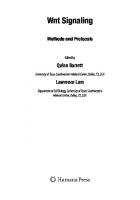
Fig. 1 Development of the imaging protocol for visualizing cellular palmitoylated Wnt proteins. Cells are metabolically labeled with Alk-C16, fixed, permeabilized, processed for click chemistry with biotin-azide, and then proximity ligated. Wnt palmitoylation can be visualized using a fluorescence microscope
unmet need for robust, sensitive, and non-radioactive methods for detecting Wnt palmitoylation, we have reported a click chemistryproximity ligation-based method to track palmitoylated Wnt in single cells [3, 15, 16]. This imaging method provides high sensitivity and subcellular resolution for detection of Wnt palmitoylation, and has been used to reveal novel insights into Wnt trafficking, as well as its regulation by porcupine [3]. The imaging assay comprises metabolic labeling of the palmitoylated proteome with bio-orthogonal fatty acid probes [17–19] and click chemistry, followed by proximity ligation assay (PLA) [20–22] to detect the lipidated forms of Wnt proteins (Fig. 1). Using alkyne palmitic acid probe (Alk-C16) as an example, the fatty acid probe is incubated with live cells, and then the cells are fixed and permeabilized. The Alk-C16-labeled proteins are then conjugated to azide-tagged biotin or a fluorescent dye through a Cu(I)-catalyzed Huisgen 1,3-dipolar cycloaddition reaction [17– 19, 23]. Two primary antibodies are used to recognize Wnt and the biotin/dye on palmitoylated proteins. These two primary antibodies are in turn recognized by two different PLA secondary antibodies which are conjugated to unique oligonucleotides. When the two antibodies are in close proximity (