Trafficking Inside Cells: Pathways, Mechanisms and Regulation (Molecular Biology Intelligence Unit) [1 ed.] 0387938761, 9780387938769
375 20 61MB
English Pages 459 Year 2009
Recommend Papers
![Molecular Mechanisms of Smooth Muscle Contraction (Molecular Biology Intelligence Unit) [1st ed.]
1570595666, 9781570595660, 9780585408781](https://ebin.pub/img/200x200/molecular-mechanisms-of-smooth-muscle-contraction-molecular-biology-intelligence-unit-1stnbsped-1570595666-9781570595660-9780585408781.jpg)
![Lysosomal Pathways of Protein Degradation (Molecular Biology Intelligence Unit) [1st ed.]
1587060035, 9781587060038, 9780585408750](https://ebin.pub/img/200x200/lysosomal-pathways-of-protein-degradation-molecular-biology-intelligence-unit-1stnbsped-1587060035-9781587060038-9780585408750.jpg)

![Translation Mechanisms (Molecular Biology Intelligence Unit) [1 ed.]
0306478390, 9780306478390, 9781417545780](https://ebin.pub/img/200x200/translation-mechanisms-molecular-biology-intelligence-unit-1nbsped-0306478390-9780306478390-9781417545780.jpg)
![Regulation of G1 Phase Progression (Molecular Biology Intelligence Unit) [1 ed.]
9780306478314, 0306478315, 9781417545414](https://ebin.pub/img/200x200/regulation-of-g1-phase-progression-molecular-biology-intelligence-unit-1nbsped-9780306478314-0306478315-9781417545414.jpg)


![Trafficking Inside Cells: Pathways, Mechanisms and Regulation (Molecular Biology Intelligence Unit) [1 ed.]
0387938761, 9780387938769](https://ebin.pub/img/200x200/trafficking-inside-cells-pathways-mechanisms-and-regulation-molecular-biology-intelligence-unit-1nbsped-0387938761-9780387938769.jpg)
- Author / Uploaded
- Aixa Alfonso
- Gregory S. Payne
- Julie Donaldson
- Nava Segev
File loading please wait...
Citation preview
MOLECULAR BIOWGY
INTELLIGENCE UNIT
Trafficking Inside Cells Pathways, Mechanisms and Regulation
Nava Segev, PhD Department of Biological Sciences Laboratory for Molecular Biology University of Illinois at Chicago Chicago, Illinois, USA
LANDES BIOSCIENCE
AUSTIN, TEXAS
USA
SPRINGER SCIENCEtBuSINESS MEDIA
NEW YORK, NEW YORK USA
TRAFFICKING INSIDE CELLS: PATHWAYS, MECHANISMS AND REGULATION Molecular Biology Intelligence Unit Landes Bioscience Springer Science-Business Media, LLC ISBN: 978 -0-387-93876-9
Printed on acid-free paper .
Copyright ©2009 Landes Bioscience and Springer Science-Business Med ia, LLC
All rights reserved. This work may not be translated or copied in whole or in part without the written permi ssion of the publisher, except for brief excerpts in connection with reviews or scholarly analysis. Use in connection with any form of information storage and retrieval, electronic adaptation, computer software , or by similar or dissimilar methodology now known or hereafter developed is forbidden . The use in the publication of trade names, trademarks, service marks and similar terms even if they are not identified as such, is not to be taken as an expression of opin ion as to whether or not they are subject to prop rietary rights . While the authors, editors and publi sher believe that drug selection and dosage and the specifications and usage of equipment and devices, as set forth in this book, are in accord with current recommendations and practice at the time of publication , they make no warranty, expressed or implied, with respect to mat erial described in this book. In view of the ongoing research, equipment development, changes in governmental regulations and the rapid accumulation of information relating to the biomedical sciences, the reader is urged to carefully reviewand evaluate the information provided herein. Springer Science-Business Media , LLC, 233 Spring Street, New York, New York 10013 , USA http://www.springer.com Please address all inqu iries to the publishers: Landes Bioscience, 1002 West Avenue, Austin , Texas 7870 1, USA Phone: 512/637 6050; FAX: 512/637 6079 http://www.landesbioscience.com The chapters in this book are available in the Madame Curie Bioscience Database . http://www.landesbioscience.com/curie Printed in rhe United States ofAmerica .
98 7 65 4 3 2 1
Library of Congress Cataloging-in-Publication Data Trafficking inside cells : pathways, mechanisms, and regulat ion I [edited by] Nava Segev. p. ; ern. -- (Molecular biology intelligence unit) Includes bibliographical references and index. ISBN 978-0-387-93876-9 (alk. paper) 1. Proteins--Physiological transport. I. Segev, Nava. II. Series: Molecular biology intelligence unit (Unnumbered : 2003) [DNLM: 1. Protein Transport. 2. Cell Membrane--metabolism. 3. Cell Physiological Processes. 4. Signal Transduction. QU 55 T764 2009] QP551.T72352009 612 .3'98--dc22 2009028590A
Dedication This book isdedicated to allthe past, presentand future researchers whose contributions are invaluableto the rapid progression of the field of trafficking inside the cell.
Nava Segev, PhD
About the Editor... ,,..
.... '" . .
NAVA SEGEV is a Professor in the Department of Biological Sciences at the University of Illinois at Chicago. Her laboratory studies the regulation of intracellular trafficking by GTPases using molecular, cellular and genetic approaches. Recently, her main research interests have focused on the role of GTPases in the integration ofindividual transport steps into whole pathways and the coordination ofthese pathways with other cellularprocesses. She teaches genetics to undergraduate students and protein trafficking to graduate students. Dr. Segev received her PhD in Microbial Genetics from Tel-Aviv University in Israel and was a postdoctoral fellow with David Botstein at Massachusetts Institute of Technology, Cambridge, and Genentech, Inc. at San Francisco, where she picked up yeast as a model system. She currently serveson the Editorial Board ofMolecular Biology ofthe Cell, and is a member of the Genetic Society ofAmerica and the American Society for Cell Biology.
About the Associate Editors ... AIXA ALFONSO is an Associate Professor in the Department of Biological Sciences and the Laboratory for Integrative Neuroscience at the University of Illinois at Chicago. Her research interests include the study of the genes involved in (1) sorting and trafficking of neuronal specific proteins (cell biology) , and (2) specification of neuronal identity (development and differentiation) using the nematode C. elegans as a model system . She received her PhD from the University ofWisconsin at Madison and is a member of the Society for Neuroscience, the American Society for Cell Biology and the Society for the Advancement of Chicanos and Native Americans in Science. JULIE G . DONALDSON is a Senior Investigator in the Laboratory of Cell Biology in the National Heart, Lung, and Blood Institute at the National Institutes of Health in Bethesda, Maryland. Her research interests are in understanding the mechanisms and regulation ofendosomal and secretory membrane traffic in the cell. She holds a PhD from the University of Maryland and was a Postdoctoral Fellow in the National Institute of Child Health and Human Development prior to her current position. GREGORY S. PAYNE is a Professor in the Department of Biological Chemistry, UCLA School of Medicine, Los Angeles, California. His research involves cell biological, biochemical, molecular and standard genetic and genomic approaches to understand vesicle mediated traffic in yeast, with particular emphasis on endocytosis and transport between the TGN and endosomes . Dr. Payne received his BS in Cell Biology with Honors in Drama from University of Michigan in 1977 and his PhD in Biochemistry in the lab of Harold Varmus from University of California, San Francisco. He was a postdoctoral fellow with Randy Schekman at University of California, Berkeley. He currently serves on the Editorial Boards of Traffic and the Journal ofCell Science and is a member of the American Society of Cell Biology.
r;:::::::::::============= CONTENTS ============:::::::=;"] Preface ......•........................................................•................ •..............• xix
NavaSegev
Section I. Compartments and Pathways 1. Overview ofIntracellular Compartments and Trafficking Pathways
3
AndreiA. Tokareo, AixaAlfonso andNaua Segeu Abstract Introduction How We Study Intracellular Trafficking The Exocyric Pathway The Endocytic Pathway Cross-Talk between the Exocyric and Endocytic Pathways Regulated Trafficking Compartment Dynamics and Biogenesis Summary and Future Perspectives
3 4 5 6 7 7 9 12 12
15 2. How We Study Protein Transport Mary 1. Preuss, Peggy Weidman and ErikNielsen Abstract............................................ .......................................... ......... 15 Model Cargo Proteins for the Analysis of Intracellular Transport 16 The Reconstitution of Membrane Trafficking In Vitro 19 25 Genetic Analysis of Transport in Yeast Tools for Imaging Membrane Trafficking 30 Summary and Perspectives 34 3. The Golgi Apparatus
42
Zhaolin Hua and ToddR Graham Abstract Introduction Structure of the Golgi Apparatus Posttranslational Modifications Catalyzed with in the Golgi Apparatus Protein Transport and Sorting in the Golgi Apparatus Inheritance of the Golgi Apparatus Summary
42 43 44 48 54 60 61
4. The Endocytic Pathway ....................................................•.................. 67
Elizabeth Conibear and Yuen Yi C. Tam Abstract Initial Steps in Internalization Transport through Endosomes Retrograde Transport to the Secretory Pathway Membrane Domains and Compartment Identity Conclusion
67 68 71 74 77 77
5. Regulated Secretion ........... ................ .....•..................... ........................ 84 Naueen Nagarajan, Kenneth L. Custer and Sandra Bajjalieh Abstract 85 Introduction 85 Adapting the Core Machinery of Constitutive Secretion for Regulated Release 85 Adding Regulation to the Core Machinery 88 Secretion at Neuronal Synapses 93 Summary and Conclusion 95
Section II. Mechanisms 6. Overview of Protein Trafficking Mechanisms
105
Giancarlo Costaguta and Gregory S. Payne Abstract ............................................................................................. Introduction Translocation and Protein Folding in the ER Coated Vesicle Formation Dense Core Secretory Granule Formation Carrier Motility and Organelle Positioning Vesicle Tethering and Fusion Role of Lipids in Protein Trafficking Summary
105 105 108 109 111 112 113 114 115
7. Entry into the Endoplasmic Reticulum: Protein Translocation, Folding and Quality Control 119 Sbeara "Iv. Fewell andJeffrey L. Brodsky Abstract ................................... .......................................................... 119 Introduction 119 Protein Translocation across the ER Membrane 120 Quality Control in the ER 128 The Unfolded Protein Response (UPR) 131 131 ER and Human Health Concluding Remarks 133 8. COP-Mediated Vesicle Transport
143
Siloere Pagant and Elizabeth Miller Abstract.................................................. ........................ Introduction: Principles ofVesicular Traffic Initiating Vesicle Formation: A GTPase Cycle Regulates Coat Assembly Sculpting the Membrane : Generating and Capturing Membrane Curvature Populating the Vesicle: Cargo-Coat Interactions Specify Efficient Cargo Capture
143 144 144 148 150
Complexity in COP-Mediated Traffic: What Remains To Be Learned Conclusion 9. Clathrin-Mediated Endocytosis
152 155 159
Peter S. McPherson, Brigitte Ritterand Beverly Wendland Abstract Introduction Mechanisms of CCV Formation Actin Major Unresolved Questions 10. Biogenesis of Dense-Core Secretory Granules
159 160 162 173 175 183
GrantR Bowman, Andrew T. Cowan andAaron P. Turkewitz Abstract .................................. .......... Introduction Protein Sorting into ISGs Vesicle Budding and Maturation Conclusion 11. Lipid-Dependent Membrane Remodelling in Protein Trafficking
183 184 187 195 201
210
Priya P. Chandra and Nicholas T. Ktistakis Abstract Introduction and Overview Transport Pathways Coated Vesicle Formation Primarily Depends on Three Types of Coats: Clathrin; COPII and COPI Structural and Signaling Lipids in Membrane Transport Evidence That Lipids Regulate Trafficking Pathways How Does It Work? Some Emerging Principles Future Directions 12. Carrier Motility
210 211 211 213 215 218 222 227 233
Marcin J Wozniak and Victoria J Allan Abstract Introduction Microtubules and Their Motors Actin Filaments and Their Motors To the Golgi and Back-The Early Secretory Pathway TGN and Post-Golgi Trafficking Endocytosis Cooperation between Motor Proteins Future Perspectives
233 233 235 237 240 242 244 247 247
13. Tethering Factors
254
Vladimir Lupasbin andElizabeth Sztul Abstract Introduction Role of Coiled-Coil Tethers in Membrane Traffic Role of Multi-Subunit Tethering Complexes in Membrane Traffic Unconfirmed Tethers Models for Function of Tethering Proteins in Membrane Traffic Conclusion and Perspectives 14. Intracellular Membrane Fusion
254 255 256 261 266 268 272 282
DaluXu andJesse C Hay Abstract Fusion of Phospholipid Bilayers: Biophysical Mechanism General Mechanisms of Protein-Assisted Membrane Fusion Membrane Fusion of Enveloped Viruses Intracellular Membrane Fusion Calcium-Activated Membrane Fusion Perspectives
283 283 284 284 287 308 311
Section III. Regulation and Coordination
with Other Cellular Processes 15. Regulation and Coordination ofIntracellular Trafficking: An Overview
329
JulieDonaldson andNavaSegev Abstract Introduction Regulation ofIndividuai Transport Steps Transport Step Coordination Coordination of Intracellular Trafficking with Other Cellular Processes Traffic Regulation and Human Disease Future Perspectives
329 330 330 333 334 337 338
16. Regulation of Protein Trafficking by GTP-Binding Proteins
342
Michel Franco, Philippe Chavrier andFlorence Niedergang Abstract Introduction Small GTP Binding Proteins: General Properties and Mechan isms of Regulation Methods to Study GTP-Binding Proteins Role in Protein Trafficking Concluding Remarks
342 343 343 347 349 357
17. Posttranslational Control of Protein Trafficking in the Post-Golgi Secretory and Endoeytic Pathway
363
Robert Piper andNia Bryant Abstract Introduction Control of Protein Traffic by Phosphorylation Control of Protein Traffic by Ubiquitination Concluding Remarks
363 364 364 369 378
18. Actin Doesn't Do the Locomotion: Secretion Drives Cell Polarization
388
Mabasin Osman andRichardA. Cerione Abstract Introduction Establishing Cell Polarity Maintaining Cell Polariry Cytokinesis The Role of Scaffolds The Role of Membrane Microdomains Perspectives 19. Intracellular Trafficking and Signaling: The Role of Endoeytic Rab GTPase
388 389 391 394 396 398 399 399
405
M Alejandro Barbieri, Marisa J Wainszelbaum and Philip D. Stahl Abstract Introduction Endoeytic Rabs Rab Proteins: An Interface for Receptor Trafficking and Signaling Conclusion and Perspectives: Small GTPases in Cell Biology 20 . The Exoeytic Pathway and Development
405 406 406 409 412 419
Hans Schotman and Catherine Rabouille Abstract Introduction Alterations of the Exoeytic Pathway Lead to Severe Development Defects Epithelial Development Depends on the Exocytic Pathway Concluding Remarks and Perspectives Index
420 420 420 426 432 439
r.:::==================== EDITOR =====================;l Nava Segev, PhD Department of Biological Sciences Laboratory for Molecular Biology University of Illinois at Chicago Chicago, Illinois, USA Email: [email protected] Chapters 1,15
1:=::========= ASSOCIATE
EDITORS ============1
Aixa Alfonso, PhD University of Illinois Chicago, Illinois, USA Email: [email protected] Chapter 1
Julie Donaldson, PhD Laboratory of Cell Biology NHLBI, National Institutes of Health Bethesda, Maryland, USA Email: [email protected] Chapter 15
Gregory S. Payne, PhD Department of Biological Chemistry David Geffen School of Medicine at UCLA Los Angeles, California, USA Email: [email protected] Chapter 6
r;::::=::======== CONTRIBUTORS =========::::::;l Note: Emailaddresses areprovidedfor corresponding authors ofeach chapter. VictoriaJ. Allan Department of Life Sciences University of Manchester Manchester, UK Email: [email protected] Chapter 12
PriyaP. Chandra Signalling Programme Babraham Institute Babraham, Cambridge, UK Chapter 11
Sandra Bajjalieh Department of Pharmacology University of Washington . Seattle, Washington, USA Email: [email protected] Chapter 5
PhilippeChavrier Membraneand Cytoskeleton Dynamics Group Institut Curie CNRSUMR 144 Paris, France Email: philippe.chavrier@curieJr Chapter 16
M. Alejandro Barbieri Department of Biological Sciences FloridaInternationalUniversity Miami, Florida, USA Chapter 19 Grant R. Bowman Department of Developmental Biology StanfordUniversity School of Medicine PaloAlto, California, USA Chapter 10 Jeffrey L. Brodsky Department of Biological Sciences University of Pittsburgh Pittsburgh, Pensylvannia, USA Email: [email protected] Chapter 7 Nia Bryant Department of Biochemistry and Cell Biology University of Glasgow Glasgow, UK Chapter 17 Richard A. Cerione Department of Molecular Medicine College of Veterinary Medicine Cornell University Ithaca, New York, USA Email: [email protected] Chapter 18
Elizabeth Conibear Centre for Molecular Medicine and Therapeutics University of British Columbia Vancouver, BritishColumbia, Canada Email: conibearis'cmmt.ubc.ca Chapter 4 Giancarlo Costaguta Department of Biological Chemistry David Geffen Schoolof Medicine at UCLA Los Angeles, California, USA Chapter 6 AndrewT. Cowan Department of Otolaryngology Temple University Schoolof Medicine Philadelphia, Pennsylvania, USA Chapter 10 Kenneth L. Custer Graduate Program in Neurobiology and Behavior and Department of Pharmacology University of Washington Seattle, Washington, USA Chapter 5
ShearaW. Fewell Department of BiologicalSciences University of Pittsburgh Pittsburgh, Pensylvannia, USA Chapter 7 Michel Franco Institut de Pharmacologie Moleculaire et Cellu1aire, UPR411 CNRS Sophia-Anripolis, Valbonne, France Chapter 16 Todd R. Graham Department of Biological Sciences Vanderbilt University Nashville, Tennessee, USA Email: [email protected] Chapter 3 Jesse C. Hay The University of Montana Divisionof Biological Sciences and Center for Structural and Functional Neuroscience Missoula, Montana, USA Email: [email protected] Chapter 14 Zhaolin Hua Department of Biological Sciences Vanderbilt University Nashville, Tennessee, USA Chapter 3 NicholasT. Kristakis Signalling Programme Babraham Institute Babraham, Cambridge, UK Email: nicholas.ktisrakiss'bbsrc.ac.uk Chapter 11
Vladimir Lupashin Department of Physiology and Biophysics University of Arkansas for Medical Sciences Little Rock, Arkansas, USA Chapter 13 Peter S. McPherson Department of Neurology and Neurosurgery Montreal Neurological Institute McGill University Montreal, Quebec, Canada Chapter 9 Elizabeth Miller Department of Biological Sciences Columbia University New York, New York, USA Email: [email protected] Chapter 8 Naveen Nagarajan Eccles Institute of Human Genetics HHMI, University of Utah Salt LakeCity, Utah, USA Chapter 5 Florence Niedergang Phagocytosis and Bacterial Invasion Group Insritut Cochin INSERM U 567, CNRS UMR8104 Universite Paris Descartes Paris, France Chapter 16 Erik Nielsen Department of Molecular, Cellular and Developmental Biology University of Michigan Ann Arbor, Michigan, USA Email: [email protected] Chapter 2
MahasinOsman Institute for Biotechnology and Life Sciences Cornell University Ithaca, New York, USA Chapter 18 Silvere Pagant Department of Biological Sciences Columbia University New York, New York, USA Chapter 8 Robert Piper Physiology and Biophysics University ofIowa Iowa City, Iowa, USA Email: [email protected] Chapter 17 Mary L. Preuss Donald Danforth Plant Science Center St. Louis, Missouri, USA Chapter 2 Catherine Rabouille Cell Microscopy Centre Department of Cell Biology and Institute of Biomembrane University Medical Center Utrecht, The Netherlands Email: c.rabouillets'umcutrechr.nl Chapter 20 BrigitteRitter Department of Neurology and Neurosurgery Montreal Neurological Institute McGill University Montreal, Quebec, Canada Chapter 9
Hans Scherman Cell Microscopy Centre Department of Cell Biology and Institute of Biomembrane University MedicalCenter Utrecht, The Netherlands Chapter 20 Philip D. Stahl Department of Cell Biology and Physiology Washington University School of Medicine St. Louis, Missouri, USA Email: [email protected] Chapter 19 Elizabeth Sztul Department of Cell Biology University of Alabama at Birmingham Birmingham,Alabama, USA Email: [email protected] Chapter 13 Yuen Yi C. Tam Department of Biochemistry and MolecularBiology University of BritishColumbia Vancouver, British Columbia, Canada Chapter 4 Andrei A Tokarev Department of Biological Sciences University of Illinoisat Chicago Chicago,Illinois, USA Chapter 1 Aaron P. Turkewitz Department of MolecularGenetics and Cell Biology University of Chicago Chicago, Illinois, USA Email: [email protected] Chapter 10
MarisaJ. Wainszelbaum Depanment of Cell Biology and Physiology Washington University School of Medicine St. Louis, Missouri, USA Chapter 19
MarcinJ. Wozniak The Bristol Institute for Transfusion Sciences National Health Service Blood and Transplant Pilton, Bristol, UK Chapter 12
Peggy Weidman Officeof Scientific Review National Institute of General Medical Sciences Bethesda, Maryland, USA Chapter 2
DaluXu Institute of Biochemistry II FrankfurtMedical School University Hospital Frankfurt,Germany Chapter 14
Beverly Wendland Department of Biology Johns Hopkins University Baltimore, Maryland, USA Email: [email protected] Chapter 9
================ PREFACE ================ The human body is made up oftrillions oftiny cellsthat cannot be seen by the naked eye.The functioning units inside these cellsare macromolecules that need to travel in the three-dimensional cell-space to distances ten thousand times their size. This movement is highly ordered, requires energy and takes place on molecular tracks that serve as a sophisticated transport system-somewhat equivalent to the multimodal rail-highway-river networks of large metropolises. All the systems of the human body depend on the efficient delivery of macromolecules to their right destination at the right time-both within and between cells. Breakdown of this traffic system results in a variety ofdiseases including diabetes, cancer and heart disease, as well as immunological, neurological and developmental disorders . During the last half a century, scientists have made a quantum leap in unraveling the mysteries of trafficking inside cells. The three sections of this book together cover the past, present and future of this rapidly developing and intriguing field. The first section is about the compartments and pathways defined more than 50 years ago by the pioneering studies of George Palade, who received the Nobel Prize for this work. However, as shown in the chapters in this section , new approaches that allow us to study the dynamics of these compartments and pathways have revealed that the compartments are not as stable as was previously thought. Even in this section, several issues are still controversial . The second and largest section, on mechanisms, covers what the field has been focused on during the last 20 -25 years. Starting with the work of James Rothman and Randy Schekman, components of the machinery were identified and mechanisms deciphered. Using in vivo and in vitro approaches combined with genomics and proteomics, the highly conserved molecular machines that move vesicles between cellular compartments are being characterized. This phase is also not complete yet, but a clear picture is beginning to emerge. Basedon the foundation ofthe pathways and the machinery components, the field is now embarking on understanding how individual transport steps are regulated, how successive steps are integrated into whole pathways, and how these pathways are coordinated with other cellular processes. The book's third section, documenting the promise ofthis current research,belongs to the future. The next generation of scientists will, no doubt, continue to move this field forward. This book is intended to help them do so.
Nava Segev, PhD
Acknowledgments First, I would like to thank the authors of the chapters; a truly international group. They are all experts in the topic on which they wrote and have made important contributions to their respectivefields. I asked the authors to write about the current state oftheir field and to include their opinion on its future . I am grateful to each of them for taking the time to write excellent contemporary reviews from which I learned so much. Second, I am grateful to the Associate Editors of the three book sections: Aixa Alfonso, Greg Payne and Julie Donaldson. These colleagues helped me through all the steps of the evolvement of the book chapters; from recruiting the authors to reviewing the chapters. The Associate Editors also contributed to the writing of the overviews that combine the individual book chapters into sections. I also would like to thank other colleagues who helped review book chapters, Bruno Goud, Teresa Orenic and Andrea Holgado De Brigueda, and Eran Segev for text editing. Last, I am indebted to the publisher, Ron Landes. Ron came into my office with the idea ofediting a book on intracellular trafficking when I was preparing a new graduate course on this topic. So, he was there from the budding of the idea to the fusion of the chapters into a whole book; alwayssupportive and helpful. I would also like to thank the crew at Landes Bioscience who helped with all the steps of publishing: Celeste Carlton, Cynthia Conomos and Megan Klein.
Nava Seget; PhD
SECTION
I
Compartments and Pathways
CHAPTER
1
Overview of Intracellular Compartments
and Trafficking Pathways Andrei A. Tokarev, Aixa Alfonso and Nava Segev* Contents Abstract Introduction How We Study Intracellular Trafficking The Exocytic Pathway The Endocytic Pathway Cross-Talk between the Exocytic and Endocytic Pathways Trafficking between the Golgi and Endosomes T ranscytosis Late Endosome-to-Plasma Membrane Regulated Trafficking Regulated Exocytosis Regulated Receptor Endocytosis Autophagy Compartment Dynamics and Biogenesis Summary and Future Perspectives
3 4 5 6 7 7 7 9 9 9 9 11 11 12 12
Abstract ll eukaryotic cells contain membrane-bounded compartments that interact with the cell's environment. Vesicles transport proteins and lipids between these compartments via two major pathways: the outwards, exocytic pathway, carries material synthesized in the cytoplasm to the cell milieu , and the inwards, endocytic pathway, internalizes material from the environment to the inside of the cell. This communication of the cell with its environment is crucial for all tissue and organ function. Here, we summarize progress made during the last two decades in our understanding of bi-directional transport pathways between intracellular compartments. The accumulated knowledge of intracellular compartments and pathways that connect them formed the basis for advancements made in our understanding of the molecular machinery components, mechanisms and regulation of intracellular trafficking . Whereas the major compartments and pathways are well defined, less is known about the dynamic nature and biogenesis of compartments.
A
*Corresponding Author: Nava Segev-Department of Biological Sciences, Universityof Illinois at Chicago, Chicago, Illinois 60607, USA. Email: [email protected].
Trafficking Inside Cells: Pathways, Mechanisms and Regulation, edited by Nava Segev, Editor, with Associate Editors: Aixa Alfonso, Gregory Payne and Julie Donaldson. ©2009 Landes Bioscience and Springer Science-Business Media.
4
Trafficking Inside Cells: Pathways, Mechanisms andRegulation
Endocytic pathway
Secretory vesicles
Exocytic pathway & Secreted eo.rgo
•
Plasma membrallC receptor
+ Lysosomal enzyme
SlgllC1l1lng molecule
8
セ
DIgcstednutrient
Ribosome Ribosomes onmRNA
Endoeytoscd nutrient
e
Growing polypeptide
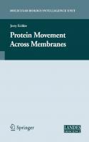
Figure 1.A diagram ofthetwomajorintracellular trafficking pathways andthecompartments theyconnect: The exocytic pathway carries proteins and lipids from the endoplasmic reticulum through the Golgi apparatus to the plasma membrane (PM). The endocytic pathway internalizes cargo from the cellmilieu or the PM through a set of endosomes to the degradative cellular compartment, the lysosome. The two pathways are connected by bi-directional transport between the Golgi and endosomes. Various proteins follow theirspecific routestowards theirdestination; e.g., secreted cargo andPM receptors and transporters to the PM; newly synthesizedendosomal and lysosomal proteinsto endosomes and lysosomes; signaling molecules and PM receptors to early endosmes; and nutrientsto lysosomes.
Introduction All cells are surrounded by a membrane that serves as a barrier between the inside of a cell and its environment. Moreover, different cellular processes occur on membranes, e.g., DNA replication and respiration. Most prokaryotic cells contain only one membrane, the plasma membrane (PM) , which surrounds the cell, and all membrane-attached processes occur on it. In some prokaryotes, specific patches of the PM specialize in separate functions. This specialization is more advanced in eukaryotic cells, which contain membrane-bound intracellular compartments that carry out specific functions, e.g., nucleus for DNA replication and mitochondria for respiration. Membrane expansion and compartmentalization in eukaryotic cells enabled the development oflarger cells (1000-10,000 fold increase in volume) and an efficient separation of cell functions. However, at the same time compartmentalization creates a new problem, namely the need for communication between the different cellular compartments. A major process of communication between the compartments that connect the cell with its environment is achieved by vesicular transport. In this process, cargo-loaded vesicles form at a donor compartment with the help of specificcoat and adaptor proteins (e.g., COPI, COPII and
Oueruieu: ofIntracellular Compartments and Trafficking Pathways
5
clathrin). These vesicles are then targeted to the appropriate acceptor compartment, to which they attach with the help of tethers, and with which they fuse with the help of SNAREs. 1 Vesicular transport enables proteins in membrane-bound vesicles to move between the cell compartments, including the outer-cell membrane, the PM. The first section of this book focuseson the different trafficking pathways and cellular compartments connected by vesiculartransport (Fig. 1).2 Two major cellular pathways shuttle material outward and inward. In the exocytic pathway, proteins synthesized in the cytoplasm are translocated into the endoplasmic reticulum (ER). Rough ER is the site of synthes is of all secreted proteins, and resident proteins for all compartments connected by vesicular transport. The ER is also the site where synthesis of most of the lipids in the cells begins. From the ER, membranous vesicles shuttle cargo to the Golgi apparatus. ER-derived cargo enters the Golgi in its cis cisterna, and moves through the medial and trans cisternae. In the trans Golgi, proteins destined for secretion or to be presented on the PM are packed into secretory vesicles that subsequently fuse with the PM . This fusion occurs either constitutively or, as in the case of regulated secretion, in response to an external signal (summarized in Chapters 3 and 5, respectively refs. 3 and 4). The Golgi apparatus is the major sorting compartment of the cell because in the Golgi cargo is sorted not only to the PM for constitutive and regulated secretion, but also to endosomes and Iysosomes, or back to the ER (see below). In the endocyric pathway, proteins and membrane are internalized from the cell environment via a set ofendosomes, early and late, to the lysosome (summarized in Chapter 4, ref. 5). The lysosome is a major degradation site for both internalized and cellular proteins . Thus, cellular proteins can get to lysosomes either from the PM via the endocytic pathway or from the cytoplasm via the autophagy and the cytoplasm-to -vacuole targeting (CVT) pathways." In addition, there is cross-talk between the exocyticand endocytic pathways. First, endosomal and lysosomalresident proteins and enzymesare shuttled from the ER via the Golgi to endosomes and lysosomes," Second, in polarized cells, proteins can be moved between two different environments, from one side of the cell to the other, via the transcytotic pathway.s Lastly, macromolecules can be releasedfrom cellsin small vesicles called exosomesby fusion oflate endosomes, also known as rnultivesicular bodies (MVBs), with the PM.9 Transport of lipids and proteins between companments creates another problem , which is how compartment identity is maintained in the context of the flow of material through the compartments. In addition, massive membrane flow needs to be balanced to maintain compartmental size.Therefore, for each step of forward transport, both in the exocyticand endocytic pathways, there is a retrograde transport step in which membrane and resident proteins are recycled back to their original compartment. This bi-directional trafficking requires sophisticated machinery and has to be regulated (summarized in th e second and third sections of this book, respectively ref 2). The progress in our understanding of the pathways, machin ery and regulation of vesicular transport was made possible by the development of novel techniques (summarized in Chapter 2, ref 10). In particular, live-cellmicroscopy approaches provide dynamic views of intracellular trafficking. Recent live-cell studies have challenged the prevailing paradigm of compartments as static "bus stations. " The dynamic view envisions compartments as constantly changing entities in response to the cell needs. Here, we summarize our current understanding of the major intracellular compartments and trafficking pathways that connect them.
HowWeStudyIntracellular Trafficking The exocytic pathway and its compartments were defined in the I%Os by Palade and coworkers using pulse-chase analysis combined with electron microscopy. 11 The endocytic pathway and its compartments were defined in the early 1970s by Brown and Goldstein, while studying human mutations that result in atheroscleros is due to defects in the recycling of low-density lipoprotein (LOL) receptors. 12 The idea that all the steps of any biological pathway can be ident ified by mutations was further exploited during the early 1980s using
6
Trafficking ImideCells: Pathways, Mechanisms andRegulation
yeast genetics to un cover all the steps of the exocytic pathway and define the genes whose products mediate these steps.13 At around the same time, reconstitution ofprotein transport steps in cell extracts combined with protein purification techniques allowed a complementary approach to identify transport machinery components. 14 Progress in the intracellular trafficking field during the last two decades was made possible by further advances in available techniques (summarized in Chapter 2, ref. 10), and especially by combining these techniques. First, a powerful combination of genetic and biochemical strategies allowed the identification ofvesicular trafficking machinery components and regulators. Genomics and proteomics studies carry the promise for the identification of the full inventory of these components in the near future . Various protein interaction studies placed these components into "molecular machines". Second, combining fluorescence and electron microscopy with molecular genetics made it possible to localize these machinery components to their cellular compartments. The most exciting recent development in cell biology,which will shape the future ofthis field, is the development offluorescent tags and cutting edge fluorescence microscopy,which together allow following single molecules in live cells. 15 Because it is clear that proteins function in complexes, the future ofthis field also belongs to techniques like fluorescence resonance energy transfer (FRET) and bi-molecular fluorescent complementation (BiFC),16 which allow identification of protein interactions in situ. Together, studies using these techniques should provide a detailed picture of the molecular machines that mediate intracellular trafficking in real time.
The Exoeytic Pathway The exoeytic pathway moves cargo from the ER through the Golgi to the PM (Fig. 1). In the ER and the Golgi, proteins are modified by the addition of sugars and lipids. These modifications are highly ordered and occur successively in the ER and in the three cisternae of the Golgi, cis, medial and trans. Cargo -packed vesiclesformed at the trans-Golgi fuse with the PM to deliver PM resident proteins such as receptors, channels and pumps and secreted proteins such as extracellular matrix components and signaling molecules. These vesiclesalso allow the expansion of the PM during cell growth . Proteins enter the ER during their translat ion via the translocon pore. This entry requires a tag, the "signal sequence", on the entering protein and signal recognition machinery on the ER membrane. Once in the ER, proteins stay either on or inside membranes. To exit the ER, proteins must get through a quality-control surveillance that ensures proper folding and assembly.17 From regions on the ER called ER exit sites, vesiclesform and move to the cis Golgi. The area between the ER and the cis Golgi, termed intermediate compartment (IC), is filled with vesicles and tubules ; the IC is not well defined functionally. IS The three Golgi cisternae are well-defined biochemically.' Different protein-modifying enzymes are enriched in each cisterna. Currently, the way in which cargo or Golgi enzymes move between the three Golgi cisternae is still controversial. The vesicular transport model suggests that vesicles move cargo forward and resident proteins backward between the Golgi cisternae. The cisternal maturation model suggests that cargo stays enclosed inside a Golgi cisterna, which matures by fusing with retrograde vesicles carrying Golgi enzymes from a more mature cisterna and by giving rise to retrograde vesicles that return Golgi enzymes to younger cisternae. The rapid partitioning model suggests that Golgi cisternae within a stack are continuous, with cargo proteins equilibrating rapidly between the cisternae. In this model , the partitioning of enzymes into the different gッャセゥ cisternae is a result of differential distri bution of lipids within the continuous cisternae. I Future experiments should help resolve this controversy. In the last step of the exocytic pathway, exoeytosis, secretory vesiclesform at the trans-Golgi and fuse with the PM to deliver their protein and lipid cargo. Therefore, there are two major steps in the exocytic pathway mediated by vesicles: ER-to-cis Golgi and trans Golgi-to-PM. Vesicles mediating these two steps differ in size and coat composition.20, 21
Overview ofIntracellular Compartments and Trafficking Pathways
7
The forward exocytic pathway delivers more membrane than needed for PM expansion. In addition, resident proteins that move to the next compartment have to be retrieved back to the original compartment for maintenance of compartment identity. Therefore, for every step of forward transport, there is a corresponding retrograde transport step. The two major intersections of th is bi-directional trafficking are the IC, which recycles proteins back to the ER, and recycling endosomes, which recycle proteins back to the PM or the Golgi. 22
The Endocytic Pathway In the endoeytic pathway, cargo is internalized from the cell milieu (Fig. 1, summarized in Chapter 4, ref. 5). Cargo can be internalized at the PM by a number of routes. Membrane receptors are mainly internalized via clathrin-coated vesicles, whereas other proteins and viruses are internalized by caviolar- or raft-dependent routes. These three internalization routes depend on the GTPase dynamin for fission of the form ing PM vesicle. However, fluid-phase cargo can also enter the cell via a dynamin-independent process. Each of these internalization routes delivers cargo to an internal compartment, endosornes, although the nature of the endosomal compartments may differ between routes . The best characterized endoeytic pathway proceeds from clathrin-coated vesicles through early and late endosomes to lysosomes. In the first set of endosomes , the sorting endosomes, cargo is sorted for recycling back to the PM (or the Golgi) via recycling endosomes, or to the lysosome via late endosomes . Patches of lare endosomal membranes are internalized as vesicles to form multivesicular bodies (MVBs), which fuse with lysosomes. The lysosome is a major degradation site for internalized material and for cellular membrane proteins. Like transport through the exoeytic pathway, the first and last steps of the endoeytic pathway are mediated by vesicular transport machinery: PM -ro-early endosome and late endosome to lysosome. Using 3-dimensional time-lapse fluorescence microscopy (4D microscopy) and multiple fluorescent chromophores, it was shown that movement from early-to -late endosomes is achieved by endosome maturation, which is in turn mediated by Rab conversion.f'' Future research in the endoeytic pathway field will address the nature of the signals for the various internalization routes and the way in which cargo is sorted in sorting endosomes. This sorting is crucial for cell signaling because it determines the ratio between receptors that recycle back to the PM and continue to signal, and receptors that are shuttled to the lysosome for degradation. Cargo sorting is also of crucial importance for the function of neurons or neurosecretory cells as protein components of synaptic vesicles have to be retrieved efficiently to maintain PM identity.
Cross-Talk between the Exocytic and Endocytic Pathways There are a few examples of cross-talk between the exocytic and endoeytic pathways: bi-directional transport between the Golgi and endosomes, transport from one side of a polarized cell to the other and secretion of material from late endosomes.
Trafficking between the Golgi and Endosomes Becausealmost all proteins and lipids destined to reside and function in any of the compartments connected by vesicular transport are translocated first into the ER, there should be a pathway to transport newly synthesized endosomal and lysosomal proteins and lipids to endoeytic compartments. Indeed, cargo can be shuttled from the trans Golgi not only to the PM via exocytosis, but also to endosomes and lysosomes (Fig. 2A). In mammalian cells, most endosomal and lysosomalproteins are labeledwith mannose-6-phosphate (M6P) in the Golgi. In the trans Golgi, M6P-labeled cargo issorted by M6P receptors(M6PR) into vesicles that are targetedto the endocytic compartments. Lower pH in endosomes causes dissociation of the cargo from the M6PR for its further delivery to the right endosomal compartment. Retrograde transport recycles M6PRs back from endosomes to the Golgi for further functioning.? Thus, bi-directional trafficking between the Golgi, endosomes and lysosomesconnects the two major intracellular trafficking pathways.
Trafficking Inside Cells: Pathways, Mechanisms andRegulation
8
A. Golgi-to-Endosome
GD/gi
(PH7) • MannoSlZ-6-pho$phat ___ Lysosomel protein .../ MannoSlZ -6-pho$phatCl RQceptor B. Trcnsc:ytosis
Apical
Basolateral C. MVB-to - PM
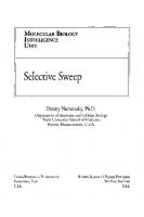
Figure 2. Three examples of cross-talk between the exoeytic and endoeytic pathways. A) Bi-direetional transport between the Golgi and endosomes using a signal and a receptor. B) In the transeytotic pathway, proteins can be shurtled from one side of a polarized cell to the other. C) MVBs can fuse with the plasma membrane to deliver exosomes. See text for details.
Overview of Intracellular Compartments and Trafficking Pathways
9
Transcytosis Polarized cells, such as epithelial cells and neurons, contain distinct functional PM domains: apical and basolateralor somatodendritic and axonal, respectivelr The mechanisms by which this cell polarity is establishedand maintained are still not clear. 2 Regardless, polarized cells use the endocyric pathway to shuttle cargo between their distinct PM domains. Here, cargo, soluble or membranous, is internalized from the PM on one side of the cell, e.g., the apical side of epithelialcells, which faces the lumen of organs. In this case, cargo delivered first to apicalearlyendosomescan be shuttled viaa common set oflare endosomes,and then through basolateral early endosomes, to the PM of the basolateral side of the cell (Fig. 2B). Thus, transcytosis can selectively move material through cellsacrosstissuebarriers;for example, from the luminal (apical) side to the underlying interstitium (basolateral) side of endothelium that lines blood vessels or epithelium that lines the intestines.8 It seemsthat even though this transport is mediated by endosomes, exoeyticmachinerrs components, like the tethering complex exocystand SNAREs, are required for this process. 5
Late Endosome-to-Plasma Membrane This is the newestaddition to the connection betweenthe endoeyticand exoeytic pathways. Here, transport of macromolecules from a late endoeytic compartment is redirected to the PM and secreted inside small vesicles, termed exosomes, to the cell's surroundings. MVBs are late endosomes that contain internal membrane-surrounded cargo. Usually, MVBs fuse with lysosomes and send their cargo for degradation. However, under cenain conditions MVBs can fuse with the PM, thus secreting exosomes to the cellmilieu (Fig. 2C).9This process is important for communication between cells and has been implicated in secretion of components to the blood stream and as a signaling device. On the other hand, exosomes might playa role in spreading infectiousagents; for example, viruses like HN can hijack this route to be released from cells. 2 Currently, the regulationand function of this process is still unknown.
Regulated Trafficking Trafficking through the exocyric and endocyricpathways is coordinated by internal regulators that ensure fidelity and uninterrupted flow. 27 In addition, some trafficking steps can be regulated by external signals. For example, transport of membranes and proteins to and from the PM can be regulated by extracellular signalingmolecules, while the autophagy pathwaycan be induced under stressconditions.
Regulated Exocytosis At the trans Golgi, specific proreins can be sorted into specialsecretoryvesicles that accumulate and fusewith the PM only when triggered by an extracellular signal (Fig. 3A). In these systems, the level of the signal controls the rate of exocytosis, The best-studied examples of regulated exocytosis are secretion of neurotransmitters in synaptic vesicles by neurons and secretion of hormones in secretorygranules by endocrine cells.4 However, even in yeast there are examples of regulated exocytosis, such as the regulated sortin of a general amino-acid permease to the PM in response to external nitrogen availability. 2 The basicmachineryof regulatedexocytosis, in both endocrineand neuronal cells, isadapted from the core vesiculartransport machinery. In the caseofsecretorygranules, regulatedexocytosis starts with the sorting step that occurs at the trans-Golgi. In this step, appropriate cargo proteins often form aggregates, which are then packaged into immature secretory granules. These vesicles undergo maturation by the recycling of membrane and Golgi-residentproteins back to the Golgi. As a result, cargo in mature vesicles becomescondensed to form dense-core granules.29 In addition, some polypeptidesare proteolyticallyprocessed in the maturing vesicles to generate active hormones or neuropeptides. Mechanisms of synaptic vesicle biogenesis remain unresolved, with potential sorting stepsat the TGN and at differentstages of the endocyric pathway.30 In the cases of both secretorygranulesand synaptic vesicles, a fraction of the mature
f
Trafficking Inside Cells: Pathways, Mechanisms and Regulation
10
A. Regulated exocyto=i:
•
• Co·
B.
C. Autophagy
Figure3. Three examples of regulated rrafficking. A) In regulated exocytosis, the last srep of the exocytic pathway, fusionof secretoryvesicles with the plasmamembranecan be regulated by a requiredexternal signal. B) Regulated internalization of plasma membrane receprors. The firsrsrep of selectiveendocytosis can be regulated by a requiredrecepror ligand. C) Starvation can inducethe autophagypathway. See text forderails.
Overview ofIntracellular Compartments and Trafficking Pathways
11
granules, called "primed" vesicles,attach to the PM and are ready to fuse in response to a signal. Signals, like hormones or neurotransmitters, interact with PM surface receptors to cause calcium influx through membrane channels, which results in a transient increase in cytoplasmic calcium near the prospective vesiclefusion site. The machinery components that mediate secretory granule and synaptic vesicle attachment and fusion are modified to function only upon stimulation by specific regulators. These specific regulators are calcium sensors that ensure vesicle attachment at the right place and fusion only upon elevation of local calcium levels. In addition, a specific feature of secretion in neuronal synapses is that synaptic vesiclescan undergo multiple rounds of fusion. This is achieved by two mechanisms unique to synapses. First, vesiclescan be refilled with neurotransmitters from the cytoplasm by transporters present in the vesicle membrane. In addition, fast release of neurotransmitters in the synapse can be facilitated by a transient link of vesicleswith a fusion pore on the PM, in a mechanism called "kiss and run". Because regulated exocytosis is crucial for proper funct ioning of two major body systems, endocrine and neuronal, uncovering the details of this process is important for understanding and treating neural and endocrine dysfunctions. Future studies should help to identify calcium sensors that ensure vesiclefusion only upon excitation and determine the way by which these sensors regulate the precise rate of vesicle fusion.
Regulated Receptor Endocytosis Endocytosis of signaling receptors and plasma membrane transporters also can be regulated by extracellular signals. One well-characterized example involves G-protein coupled receptors (GPCR), the largest family of signaling receptors HセYP in mammalian cells). Internal ization of some GPCR can be induced by the addition of their cognate signal (Fig. 3B). This induction is mediated by phosphorylation of activated receptors, which elicits arrestin binding and uncoupling of the receptor from the G-protein. Phosphorylated receptorlarrestin complexes then interact with specific clathrin coat adaptors that mediate their concentration in clathrin-coated pits. Subsequently, activated receptors are internalized via clarhrin-coared vesicles to early endosomes, where they can be sorted to recycling endosomes for recycling back to the PM , or to late endosomes for degradation in the lysosome. This regulated internalization and sorting ofactivated receptors determines the length and amplitude of multiple cell-signaling processes. The specific internalization mechanisms for many GPCRs that regulate important cell functions are still unknown, and future studies should elucidate these mechanlsms."
Autophagy Under nutrient deprivation conditions, cells can induce the autophagy pathway, which allows them to engulf areas of their cytoplasm, including membrane-bounded organelles, and deliver the material for degradation in the lysosome to generate nutrients (Fig. 3C) . In mammalian cells, autophagy is crucial for multiple processes such as programmed cell death and cellular defense against pathogens. Improper r1ulation of autophagy can result in cancer and in muscular and neurodegenerative disorders. 3 The machinery components of the autophagy pathway, first defined in yeast, are conserved. This pathway is regulated by the target-of-rapamycin (TOR) kinase, which inhibits autophagy under normal growth conditions. Once TOR inhibition is removed, a new organelle, the autophagosome, is generated de novo. In this process, a membrane "sac" engulfs portions of the cytoplasm and closure of this sac results in the formation of the double-membrane autophagosome. Fusion of the outer membrane of the autophagosome with the lysosome results in the exposure ofthe inner membrane and its content to lysosomal hydrolases, leading to their degradation. 33, 34 Much is known about the steps of the autophagy pathway and its machinery components. However, little is currently known about the beginning of the process, especially how the "sac" is generated.
12
TraffickingInside Cells: Pathways, Mechanisms andRegulation
Compartment Dynamics and Biogenesis Until recently, compartments were viewed as stable entities, like "bus stations", with "bus-like carriers" moving cargo between them. This view was challenged especially when live-cell microscopy allowed observation of compartment dynamics. It became clear that compartments can disappear and reappear depending on the cell cycle, environmental cues and cargo waves. One of the best-studied examples of compartment dynamics is the Golgi complex. In most eukaryotic cells, the Golgi apparatus disintegrates during mitosis. Golgi disintegration can also be induced by drugs like Brefeldin A (BFA). At the end of mitosis, or upon removal of the drug, the Golgi apparatus reassembles. Mechanistic questions addressed in the field are: what happens to Golgi resident proteins during disintegration and how does the Golgi reassemble. Currently these questions are under active investigation with one model suggesting that the Golgi contents completely recycle through the ER and another model propo sing that Golgi fragments form the stage for its reassembly.35. 36 Recent findings suggest that compartments change continuously, depending on cargo passing through them . For example, an extension of the cisternal maturation model suggests that the entire Golgi apparatus assembles and disassembles continuously. In this model , the cis Golgi cisterna is generated by fusion of ER-derived COPII vesicles that contain cargo, with retrograde COPI vesicles that contain cis-Golgi enzymes. On the other end of the Golgi, the trans cisterna is consumed as anterogade vesicles form to carry cargo to the PM or endosomes, and retrograde vesiclesare generated to carry trans-Golgi enzymes to the medial compartment. This latrer event is required for the maturation of the medial- to trans-Golgi cisterna. Thus, this model proposes the Golgi to be a dynamic compartment that changes not only during cell cycle, but also in the context of cargo rransport. Y Therefore, intracellular compartments may be more like "bus stations" comprised of a collection of "buses" without a static structure. Ano ther important question is how compartments are inherited into newly divided cells. Do compartments self assemble de novo, with or without template , or do they grow and divide? Studies in yeast suggest that the Golgi is formed de novo without a template whereas the perinuclear ER, together with the nucleus, is partitioned between the two newly formed cells. In mammalian cells and some protozoa, the suggested mechanism for Golgi biogenesis is self-assembly that requires a template. 38 The autophagosome is a non-essential companment formed de novo under deprivation conditions. 34 However, it is not clear whether phagosomes need a template for assembly. For example, yeast cells that grow under normal conditions have the cytoplasm-to-vacuole targeting, cvr, pathway to tran sport special proteins from the cytoplasm directly to the lysosome, called vacuole in yeast. Many components are shared between the cvr and autophagy parhwaysf Therefore, here again it is possible that under deprivation conditions, phagosomes use preexisting cvr structures as a template for their assembly.
Summary and Future Perspectives Major advances in technology have made substantial progress in the intracellular trafficking field possible. During the past two decades, the field gained detailed understanding of the nature of cellular compartments and the connecting pathways. Each compartment is defined by its lipid and protein composition. Maintenance of compartment identiry during massive internal flow of proteins and membrane is probably achieved by active recycling of proteins and lipids to their original compartment. However, there are still unanswered questions and areas of controversy. The intracellular membrane-surrounded compartments can be clearly visualized by electron microscopy and the inventory of compartment components is almost complete (see Section II of this book , ref 2). Does this mean that we know what compartments look like? It would be like trying to imagine how a car looks based on the inventory of its parts without actually seeing the car. Currently, very little is known about the architecture ofintracellular compartments. The first glimpse into compartment architecture was recently provided for synaptic vesicles (SVs). A quantitative study of purified SVs was used for modeling an average Sv. This model suggests
Overview of Intracellular Compartments and TraffickingPathways
13
that the outsideof the SVisdensely covered with proteins, that the proteins arehighlydivergent and includemore than onefercent of our proteome, and that abundant proteinsare presentin multiplecopies per vesicle.3 Majorquestions arestillopen asto whetherthe protein divergence reflects averaging of sub-populations of Sv, whether multiple copies of abundant proteinsare distributed randomly over the surface of the SV or found concentrated in patches, and the nature of the architectureoflarger, morecomplex compartments. The most controversial topic in the areaof trafficking pathways has been how cargo moves through compartments, and especially through the Golgicisternae. It seems that between compartments, e.g., ER and Golgi, or Golgi to the PM, cargo moves via vesicles. In contrast, between sub-compartments, e.g., cis-, medial- and trans-Golgi, or early-to-late endosomes, vesicles are probablynot the carriers of cargo. 19 The jury is still out as to whether intra-Golgi transport occurs by vesicular transport, cisternal maturation or gated transport through connecting tubules. Another major open question concerns intracellular compartment biogenesis. The Golgi apparatus is the best-studied organelle for this question because it naturallydisintegrates during mitosis. Here too, there are diverse results for Golgi biogenesis in differentorganisms and the question remains open as to whichGolgisub-structures or proteins,if any, form a template for assembly of the new Golgiafter each mitotic division. 38 Future studieswill hopefully help solve these cellmysteries.
Acknowledgments The authors thank GregoryPayne for critical readingof the manuscript, Eran Segev for text editing, and acknowledge support from the National Institutes of Health GM45-444 to N. S. and from the National Science Foundation to A A while workingat the Foundation.
References 1. Costaguta G, Payne G. Overview of protein trafficking mechanisms. In: Segev N, ed. Trafficking Inside Cells: Pathways, Mechanisms and Regulation. Austin/New York: Landes Bioscience/Springer
Science-Business Media, 2009:105-14, this volume . 2. Segev N, ed. Trafficking Inside Cells: Pathways, Mechanisms and Regulation. Austin : Landes Bioscience and Springer Science-Business Media , 2009 :103-438, this volume . 3. Hua Z, Graham T . The Golgi apparatus. In: Segev N , ed, Trafficking Inside Cells: Pathways, Mechan isms and Regulation . Austin/New York: Landes Bioscience/Springer Science-Business Media, 2009:42-66, this volume . 4. Nagarajan N , Custer K, Bajjalieh S. Regulated secretion. In: Segev N , ed. Trafficking Inside Cells: Pathways, Mechanisms and Regulation. AustinlNew York: Landes Bioscience/Springer Science-Business Media, 2009:84-102 , this volume . 5. Conibear E, Tam Y. The endocytic pathway . In: Segev N, ed. Trafficking Inside Cells: Pathways, Mechanisms and Regulation . Austin/New York: Landes Bioscience/Springer Science-Business Media, 2009 :67-83 , this volume . 6. Wang CW, K1ionsky OJ . The molecular mechanism of autophagy. Mol Med 2003; 9(3-4) :65-76 . 7. Ghosh P, Dahms NM , Kornfeld S. Mannose 6-phosphate receptors : new twists in the tale. Nat Rev Mol Cell BioI 2003 ; 4(3) :202-12. 8. Tuma PL, Hubbard AL. Transeytosis: crossing cellular barriers. Physiol Rev 2003 ; 83(3) :871-932. 9. Stoorvogel W, Kleijmeer MJ, Geuze HJ et aI. The biogenesis and funct ions of exosomes. Traffic 2002 ; 3(5) :321-30 . 10. Pruess M, Weidman P, Nielsen E. How we study protein tran sport. In: Segev N , ed. Trafficking Inside Cells: Pathways, Mechani sms and Regulation . Austin/New York: Landes Bioscience/Springer Science-Business Media, 2009 :15-41, this volume . II. Palade G. Intracellular Aspects of the Process of Protein Secretion . Science 1975 ; 189(4206):867 . 12. Brown MS, Goldstein JL. A receptor-mediated pathway for cholesterol homeostasis. Science 1986; 232(4746):34-47. 13. Schekman R. Genetic and biochemical analysis of vesicular traffic in yeast. Curr Opin Cell BioI 1992; 4(4) :587-92 . 14. Rothman JE, Orci L. Molecular dissection of the secretory pathway. Nature 1992; 355(6359): 409-15.
14
Trafficking Inside Cells: Pathways, Mechanisms andRegulation
15. Walter NG, Huang CY, Manzo AJ er al. Do-it-yourself guide: how to use the modern single-molecule toolkit. Nat Methods 2008 ; 5(6):475-89. 16. Ciruela F. Fluorescence-based methods in the study of protein-protein interactions in living cells. Curr Opin Biotechnol 2008; 19(4):338-43. 17. Fewell S, Brodsky J. Entry into the endoplasmic reticulum: protein translocation, folding and quality control. In: Segev N, ed. Trafficking Inside Cells: Pathways, Mechanisms and Regulation . Austin/New York: Landes Bioscience/Springer Science-Business Media, 2009 :119-42, this volume . 18. Appenzeller-Herzog C, Hauri HP . The ER-Golgi intermediate compartment (ERGIC): in search of its identity and function. J Cell Sci 2006; 119(Pt 11):2173-83. 19. Simon SM. Golgi governance : the third way. Cell 2008; 133(6) :951-3. 20. McPherson P, Ritter B, Wendland B. Clarhrin-rnediared endocytosis . In: Segev N, ed. Trafficking Inside Cells: Pathways, Mechanisms and Regulation. Austin/New York: Landes Bioscience/Springer Science-Business Media, 2009:159-82, this volume . 21. Pagant S, Miller E. COP-Mediated vesicle transport. In: Segev N , ed. Trafficking Inside Cells: Pathways, Mechanisms and Regulation. Austin/New York: Landes Bioscience/Springer Science-Business Media, 2009 :143-58 , this volume. 22. Saraste J, Goud B. Functional symmetry of endomembranes. Mol Bioi Cell 2007; 18(4):1430-6. 23. Rink J, Ghigo E, Kalaidzidis Y et aI. Rab conversion as a mechanism of progression from early to late endosomes . Cell 2005; 122(5):735-49. 24. Prydz K, Dick G, Tveit H. How many ways through the Golgi maze? Traffic 2008 ;9(3) :299-304. 25. Mostov KE, Verges M, Altschuler Y. Membrane traffic in polarized epithelial cells. Curr Opin Cell BioI 2000 ; 12(4):483-90. 26. Schorey JS, Bhatnagar S. Exosome function: from tumor immunology to pathogen biology. Traffic 2008 ; 9(6) :871-81. 27. Donaldson J, Segev N. Regulation and coordination of intracellular trafficking : an overview. In: Segev N, ed. Trafficking Inside Cells: Pathways, Mechanisms and Regulation . Austin/New York: Landes Bioscience/Springer Science-Business Media, 2009 :329-41 , this volume . 28. Magasanik B, Kaiser CA. Nitrogen regulation in Saccharomyces cerevisiae. Gene 2002 ; 290(1-2) :1-18. 29. Bowman G, Cowman A, Turkewitz A. Biogenesis of dense-core secretory granules. In: Segev N , ed. Trafficking Inside Cells: Pathways, Mechanisms and Regulation . AustinlNew York: Landes Bioscience/Springer Science-Business Media, 2009 :183-209, this volume. 30. Fei H, Grygoruk A, Brooks ES et aI. Trafficking of vesicular neurotransmitter transporters. Traffic 2008; 9(9):1425-36. 31. Wolfe BL, Trejo J. Clathrin-dependent mechanisms of G protein-coupled receptor endocytosis. Traffic 2007; 8(5) :462-70. 32. Shintani T, K1ionsky OJ . Autophagy in health and disease: a double-edged sword . Science 2004; 306(5698):990-5 . 33. Suzuki K, Ohsumi Y. Molecular machinery of aurophagosorne formation in yeast, Saccharomyces cerevisiae. FEBS Lett 2007; 581(11) :2156 -61. 34 . Xie Z, K1ionsky OJ . Autophagosome formation : core machinery and adaptations. Nat Cell Bioi 2007; 9(10) :1102-9. 35 . Colanzi A, Suetterlin C, Malhotra V. Cell-cycle-specific Golgi fragmentation: how and why? Curr Opin Cell BioI 2003; 15(4):462-7. 36. Storrie B. Maintenance of Golgi apparatus structure in the face of continuous protein recycling to the endoplasmic reticulum: making ends meet. Inr Rev Cytol 2005; 244:69-94 . 37. Mironov AA, Beznoussenko GV, Polishchuk RS et aI. Intra-Golgi transport: a way to a new paradigm ? Biochim Biophys Acta 2005; 1744(3) :340-50. 38. Lowe M , Barr FA. Inheritance and biogenesis of organelles in the secretory pathway. Nat Rev Mol Cell Bioi 2007; 8(6) :429-39. 39 . Taltamori S, Holt M, Stenius K et aI. Molecular anatomy of a trafficking organelle. Cell 2006; 127(4) :831-46.
CHAPTER
2
How We Study Protein Transport Mary L. Preuss, PeggyWeidman and Erik Nielsen* Contents Abstract. Model Cargo Proteins for the Analysis of Intracellular Transport Model Cargo Proteins for the Secretory Pathway Model Proteins for the Endoeytic Pathway . The Reconstitution of Membrane Trafficking In Vitro General Design Principles Conditions for the Reconstitution of Transport The Analysis of Reconstituted Transport Genetic Analysis ofTranspon in Yeast General Principles of Genetic Analysis in Yeast Screening and Analysis of Trafficking Mutants Phenotypes of Trafficking Mutants Information Derived from Double Mutant Analyses Identification of Interacting Proteins by Genetic Analysis Tools for Imaging Membrane Trafficking Types of Microscopy Tools in Microscopy Green Fluorescent Protein Summary and Perspectives
15 16 16 18 19 19 22 23 25 25 27 27 28 29 30 30 31 32 34
Abstract
F
or the greater part of the last century, research in the field of protein transport was synonymous with microscopy. Before the end of the century, this view was dramatically changed by the emergence ofinnovative genetic, molecular and biochemical approaches that revolutionized and invigorated the field. Far from being displaced as an essential tool, microscopy techniques have also evolved.What was once largely a science of "dead cells" has been transformed into a window on the inner workings of living cells. The objective of this chapter is to provide an overviewof the major approaches that are employed in the analysis of protein transport within the membrane trafficking system of eukaryotic cells. In particular, we discuss the identification of several of the common model cargo proteins for studying both secretory and endoeytic membrane trafficking in both mammalian and yeast systems.We then discuss the development of both in vivo and in vitro techniques to study the transport of these model cargo proteins within cells, and explain some of the common principles involved in *Corresponding Author: Erik Nielsen-Department of Molecular, Cellular & Developmental Biology, Univers ity of M ich igan, 830 North Univers ity Avenue, Ann Arbor, M148109, USA. Email: [email protected]
Trafficking Inside Cells: Pathways, Mechanisms and Regulation, edited by Nava Segev, Editor, with Associate Editors: Aixa Alfonso, Gregory Payne and Julie Donaldson. ©2009 Landes Bioscience and Springer Science-Business Media.
16
Trafficking Inside Cells: Pathways, Mechanisms andRegulation
these assays. Finally, we discuss some of the recent advances in imaging techniques and technology that have driven the recent "renaissance" in the use of microscopic techniques in the investigation of membrane trafficking events in living cells.
Model Cargo Proteins for the Analysis of Intracellular Transport The early studies of protein transport pathways often focused on a limited set of model cargo proteins. These were chosen primarily because they are expressed in a variety ofcell types and are relatively abundant and/or easy to detect . These model proteins are still frequently used in morphological and biochemical studies of transport because their intracellular itineraries have been so thoroughly documented. It is thus appropriate to begin with a brief introduction to the commonly used model cargo proteins (Table I).
Model Cargo Proteins for the Secretory Pathway In mammalian systems, Vesicular Stomatitis Virus Glycoprotein (VSV-G) is the most frequently used model cargo protein for secretory transport. VSV is an enveloped virus consisting of a nucleocapsid surrounded by a lipid bilayer studded with the spike glycoprotein, VSV-G . During an infection, VSV exploits the host cell's secretory machinery to produce prod igious amounts ofVSV-G and deliver it to the plasma membrane for virus assembly and budding. The synthesis, processing, and transport ofVSV-G protein from the ER to the cell surface is indistinguishable from normal cellular membrane glycoproteins. Modification ofthe two VSV-G N-linked carbohydrate chains during secretory transport has been well documented (ref. 1 and references therein) (Fig. I). These modifications can be detected as changes in VSV-G size and the sensitivity of its carbohydrates to digestion by endo- and exoglycosidases.r VSV-G can also be used in conjunction with the spike glycoprotein of influenza virus, hemagglutinin (HA), to
Table 1. Model cargo proteins in the analysis of intracellular transport pathways Pathway
System
Cargo Protein
Secretory pathway M
Vesicular stomatitus virus glycoprotein M Influenza virus hemagglutin in Y Invertase Y Pro-a-factor Y Carboxypeptidase y* Secretory pathway to lysosome/vacuole M CI-mannose phosphate receptor Y Vacuolar Protein Sorting protein 10 Bulk phase endocytos is M Horseradish peroxidase M fl-galactosidase Constitutive receptor-med iated endocytosi s M Low density lipoprotein (receptor) M Transferrin (receptor) Regulated receptor-mediated endocytos is M Epidermal growth factor (receptor) Y a-factor (receptor) (M = mammals, Y = yeast)
Abbrev.
Biochemical Detection
VSV-G
glycosylation
HA
glycosylation glycosylation Glycosylation, proteo lysis Glycosylation, proteolysis
Inv Pro-of CPY C1-MPR VPSlOp
Cathepsin 0 activity Carboxypeptidase Y activity
HRP fl-gal
Enzyme activity Enzyme activity
LDL(R) Tf(R)
LDL degradation Apo-Tf release Iron accumulation
EGF(R) aF(R)
EGF degradation a-factor degradation
How We Study Protein Transport
17
ER
Golgi Cis
Trans
Medial
•
Endo H
EndoH EndoO
N-ac:etylglucosaminidase
TranslTGN
0
Il-Galactosidase
-
tHcetyIgIUcIoU..... e
mamose ga CIOSG
• neAla and Thr203->Tyr results in yellow fluorescent protein (YFP). B) Top view indicating the position of amino acid variants within the interior ofthe l3-barrelstructure. A color version of this figure is available online at www.landesbioscience.com/curie.
33
How We Study Protein Transport
growth
ER
,11 40 °C
localized
J,
shift
10 30·C
Golgi localized
J, en route to plasma membrane
J, plasma membrane localized
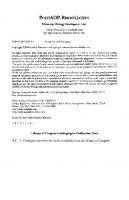
Figure 7. In vivo Analysis ofVSVG-GFP Transport. A figure showing howGFP-fusions are used to monitor and analyze the intracellular transport of cellular components, in this case the fluorescent fusion protein monitored isVSVG-GFP. At the nonpermissive temperature of 40·C , theVSVG-GFP islocalized to the ER. When switched to 32·C, VSVG-GFP moves into the Golgi, then getsexported to the plasma membrane.158
Additional techniques to examine the dynamics or the local environment of the fluorescent fusion proteins have also been devised. One of these techniques, termed fluorescence recovery after phorobleaching, (FRAP) and fluorescence loss in photobleaching (FLIP) were used initially to observe the lateral mobility of fluorescent molecules.I60-164 In this technique an intense focused laser beam is used to photobleach an area of interest , and subsequent movement of surrounding nonbleached fluorescent molecules into the photobleached area can be observed. With the use of GFP as a tag for protein localization and movement, this technique can be applied to assessthe mobility offluorescent proteins. This technique has been used to examine ER formation and structure 165 the mechanisms of protein retention in the ER,I66-168 and cycling between the ER and Golgi.169.170 Additional variants of GFP have been made which further alter the emission spectra of the protein (Fig. 6, refs. 148,149, reviewed in ref 151). These variants include blue, cyan and yellow fluorescent proteins. A novel red fluorescent protein (DsRed) was also identified from
34
Trafficking Imide Cells: Pathways, Mechanisms andRegulation
Dicosoma sp., which has been useful due to its long wavelength excitation and emission maxima.171 These differently colored fluorescent proteins enabled the ability to do dual localization and protein-protein interaction studies in vivo. Rizzutol 72 first showed that different GFP variants could be coexpressedin the same cell type to look for colocalizationofproteins or interactions berween organelles. FluorescenceResonanceEnergyTransfer(FRET) occurs berweenrwo fluorescent molecules. A donor fluorophore is excited and transfers its energy to an acceptor fluorophore instead of emitting it. This will only occur if the donor and acceptor are in closeproximity to one another (l -lOnm). Known FRET pairs that are commonly used are CFPIYFP, BFP/GFP, GFPI Rhodamine, and FITCICY3. Heim and Tsien149 linked BFP and GFP together via a spacer and demonstrated that FRET could efficiently occur berween the rwo proteins. FRET was then disrupted by the proteolytic cleavage ofthese rwo proteins. Protein-protein interactions in the secretory pathway have since been successfully observed using FRET.1 73-1 78 In the last decade of the last century fluorescence microscopy techniques using fluorescent protein fusions emerged as a widespread technique to visualize subcellular dynamics of proteins within living cells. The concurrent evolution of time-lapse video microscopy from differential interference contrast imaging techniques performed in high intensity light conditions, to integrated, computer-controlled imaging systems that allow for low-intensity fluorescence imaging in living cells is providing a novel set of techniques to further study the processes of protein sorting and trafficking. A more complete understanding of the in vivo activities of proteins identified and initially characterized using biochemical and genetic techniques can now be attained using these recent advances in microscopic imaging techniques.
Summary and Perspectives Over the last 25 years the development of molecular techniques, namely identification of model cargo proteins, and the development of genetics and biochemical methods to identify components of the membrane trafficking machinery have dramatically increased our understanding of the molecular mechanisms that the cell employs to organize and sort cargo within the endomembrane trafficking pathways. This combined with recent advances in high resolution microscopy, and use of fluorescently-tagged proteins to allow imaging of these events within living cells, has served as the basis for an increasingly detailed understanding of the dynamic nature of these processes in eukaryotic cells. In this chapter we have attempted to highlight some of the more important general advances in genetics, biochemical reconstitution of membrane trafficking, and in vivo microscopic techniques that have been employed over the last rwo decades. The development of these techniques has helped drive our current understanding of the molecular machinery involved in proper sorting and trafficking events in the endomembrane trafficking pathways of eukaryotic cells. The continued refinement of microscopic techniques and ever more sensitive and powerful imaging technologies is sure to allow even more detailed analysis of individual membrane trafficking events. However, as our understanding of the various aspects of membrane trafficking improves it is increasingly clear that this dynamic process is tightly tied into other aspects of cellular growth both at the single cell level, and as cells within the various tissues and organs of multicellular organisms. While regulated secretion is a well documented phenomenon within specialized cell types (e.g., neurons, mast cells), there are tantalizing hints that cellular signaling plays important and broad roles in regulation at multiple, if not all, stages of the endomembrane trafficking system. Further, while most studies of membrane trafficking occur at the single cell level, several lines of study indicate that membrane trafficking pathways within cells respond to and interact with the trafficking systems of neighboring cells). These questions are likely to provide new opportunities and challenges for researchers in their quest to better understand the roles and mechanisms of intracellular prote in transport in eukaryotic cells.
How We Study Protein Transport
35
References 1. Balch WE, Wagner KR, Keller OS. Reconstitution of transport of vesicular stomatitis virus G protein from the endoplasmic reticulum to the Golgi complex using a cell-free system. J Cell Bioi 1987; 104(3):749-60. 2. Schwaninger R, Beckers C], Balch WE. Sequential transport of protein between the endoplasmic reticulum and successive Golgi compartments in semi-intact cells. J Bioi Chern 1991; 266:13055-63. 3. Keller P, Simons K. Post-Golgi biosynthetic trafficking. J Cell Sci 1997; 110(part 24):3001-9. 4. Yoshimori T , Keller P, Roth MG er al. Different biosynthetic transport routes to the plasma membrane in BHK and CHO cells. J Cell Bioi 1996; 133(2):247-56. 5. Gallione C], Rose JK. A single amino acid substitution in a hydrophobic domain causes temperature-sensitive cell-surface transport of a mutant viral glycoprotein. J Virol 1985; 54(2):374-82. 6. Bergman J. Using temperature sensitive mutants of VSV to study membrane protein biogenesis. Meth Cell Bioi 1989; 32:85-110. 7. Kuismanen E, Saraste J. Low temperature-induced transport blocks as tools to manipulate membrane traffic. Methods Cell Bioi 1989; 32:257-74. 8. Saraste J, Palade GE, Farquhar MG. Temperature-sensitive steps in the transport of secretory proteins through the Golgi complex in exocrine pancreatic cells. Proc Nat! Acad Sci USA 1986; 83(17):6425-9 . 9. Rose JK, Bergmann JE. Expression from cloned eDNA of cell-surface secreted forms of the glycoprotein of vesicular stomatitis virus in eucaryotic cells. Cell 1982; 30(3):753-62. 10. Gallione C], Rose JK. Nucleotide sequence of a eDNA clone encoding the entire glycoprotein from the New Jersey serotype of vesicular stomatitis virus. J Virol 1983; 46(1):162-9. 11. Hirschberg K, Miller CM, Ellenberg J et al. Kinetic analysis of secretory protein traffic and characterization of Golgi to plasma membrane transport intermediates in living cells. J Cell Bioi 1998: 143(6):1485-503. 12. Esmon B, Novick P, Schekman R. Compartmentalized assembly of oligosaccharides on exported glycoproteins in yeast. Cell 1981; 25(2):451-60. 13. Graham TR, Emr SO. Compartmental organization of Golgi-specific protein modification and vacuolar protein sorting events defined in a yeast seel8 (NSF) mutant. J Cell Bioi 1991; 114(2):207-18. 14. Stevens T, Esmon B, Schekman R. Early stages in the yeast secretory pathway are required for transport of carboxypeptidase Y to the vacuole. Cell 1982; 30(2):439-48. 15. Stoorvogel W. Analysis of the endocytic system by using horseradish peroxidase. Trends Cell Bioi 1998; 8(12):503-5. 16. Maxfield FR, McGraw TE. Endocytic recycling. Nat Rev Mol Cell Bioi 2004; 5(2):121-32. 17. Jeon H, Blacklow Sc. Structure and physiologic function of the low-density lipoprotein receptor. Annu Rev Biochern 2005; 74:535-62. 18. Qian ZM, Li H , Sun H et al. Targeted drug delivery via the transferrin receptor-mediated endocytosis pathway. Pharmacol Rev 2002: 54(4):561-87. 19. Woods JW, Doriaux M, Farquhar MG. Transferrin receptors recycle to cis and middle as well as trans Golgi cisternae in Ig-secreting myeloma cells. J Cell Bioi 1986: 103(1):277-86. 20. Anderson RG, Brown MS, Beisiegel U er al. Surface distribution and recycling of the low density lipoprotein receptor as visualized with anrireceptor antibodies. J Cell Bioi 1982; 93(3):523-31. 21. Sorkin A, Von Zastrow M. Signal transduction and endocytosis: Close encounters of many kinds. Nat Rev Mol Cell Bioi 2002; 3(8):600-14. 22. Marmor MD, Yarden Y. Role of protein ubiquirylation in regulating endocytosis of receptor tyrosine kinases. Oncogene 2004; 23(11):2057-70. 23. Hicke L, Riezman H. Ubiquitination of a yeast plasma membrane receptor signals its ligand-stimulated endocytosis. Cell 1996; 84(2):277-87. 24. Geli Ml, Riezman H. Endocytic internalization in yeast and animal cells: Similar and different. J Cell Sci 1998; 111(Pt 8):1031-7. 25. Balch WE, Dunph y WG , Braell WA et al. Reconstitution of the transport of protein between successive compartments of the Golgi measured by the coupled incorporation of N-acetylglucosamine. Cell 1984; 39:405-16. 26. Balch WE, Rothman JE. Characterization of protein transport between successive compartments of the Golgi apparatus: Asymmetric properties of donor and acceptor activities in a cell-free system. Arch Biochem Biophys 1985; 240:413-25. 27. Balch WE, Glick BS, Rothman JE. Sequential intermediates in the pathway of inrercompartmenral transport in a cell-free system. Cell 1984; 39:525-36.
36
Trafficking Imide Cells: Pathways, Mechanisms andRegulation
28. Block MR, Glick BS, Wilcox CA et al. Purification of an N-ethylmaleimide-sensitive protein catalyzing vesicular transport. Proc Natl Acad Sci USA 1988; 85:7852-6. 29. Orci L, Malhotra V, Amherdt M er al. Dissection of a single round of vesicular transport: Sequential coated and uncoated intermediates mediate inrercisternal movement in the Golgi stack. Cell 1988; 56:357-8. 30. Cook NR, Davidson HW . In vitro assays of vesicular transport. Traffic 2001; 2(1):19-25. 31. Rothman JE. Methods in Enzymology: Reconstitution of Intracellular Transport . Elsevier 1992; 219:1-438. 32. Diaz R, Mayorga L, Stahl P. In vitro fusion of endosomes following receptor-mediated endocytosis. J BioI Chern 1988; 263(13):6093-100. 33. Gruenberg JE, Howell KE. Reconstitution of vesicle fusions occurring in endocytosiswith a cell-free system. EMBO J 1986; 5(12):3091-101. 34. Veir B, Yucel JK, Malhotra V. Microtubule independent vesiculation of Golgi membranes and the reassembly of vesicles into Goigi stacks. J Cell Bioi 1993; 122:1197-206. 35. Rabouille C, Misteli T, Watson R et al. Reassembly of Golgi stacks from mitotic Golgi fragments in a cell-free system. J Cell Bioi 1995; 129(3):605-18. 36. Beckers DJM, Keller DS, Balch WE. Semi-intact cells permeable to macromolecules: Use in reconstitut ion of protein transport from the endoplasmic reticulum to the Golgi complex. Cell 1987; 50:523-34. 37. Simons K, Virta H . Perforated MDCK cells support intracellular transport . EMBO J 1987; 6(8):2241-7. 38. Gravorta D, Adesnik M, Sabatini DD . Transport of influenza HA from the trans-Golgi network to the apical surface of MDCK cells permeabilized in their basolateral plasma membranes: Energy dependence and involvement of GTP-binding proteins. J Cell Bioi 1990; 111(6 Pt 2):2893-908. 39. Miller SG, Moore HP. Reconstitution of constitutive secretion using semi-intact cells: Regulation by GTP but not calcium. J Cell Bioi 1991; 112(1):39-54. 40. Braell WA. Detection of endoeytic vesicle fusion in vitro, using assay based on avidin-biotin association reaction. Methods Enzymol 1992; 219:12-2 1. 41. Mayer A, Wickner W. Docking of yeast vacuoles is catalyzed by the RAS-like GTPase YPT7P mer symmetric priming by SEC18P (NSF). J Cell Bioi 1997; 136(2):307-17. 42. Misteli T, Warren G. A role for tubular networks and a COP I-independent pathway in the mitotic fragmentation of GoIgi stacks in a cell-free system. J Cell Bioi 1995; 130(5):1027-39. 43. Groesch ME, Ruohola H, Bacon R et al. Isolation of a functional vesicular intermediate that mediates ER to Golgi transport in yeast. J Cell Bioi 1990; 111(1):45-53. 44. Bennett MK, Wandinger-Ness A, Simons K. Release of putative exocytic transport vesicles from perforated MDCK cells. EMBO J 1988; 7(13):4075-85. 45. Salamero J, Stzul ES, Howell KE.Exocytic transport vesicles generated in vitro from the transGolgi network carry secretory and plasma membrane proteins. Proc Natl Acad Sci USA 1990; 87:7717-21. 46. Martin TF , Walent JH . A new method for cell permeabilization reveals a cytosolic protein requirement for Ca2+ -activated secretion in GH3 pituitary cells. J Bioi Chern 1989; 264(17):10299-308. 47. Clary DO , Rothman JE. Purification of three related peripheral membrane proteins needed for vesicular transport. J Bioi Chern 1990; 265:10109-17. 48. Waters MG, Clary DO , Rothman JE. A novel 115-kD peripheral membrane protein is required for intercisternal transport in the Golgi stack. J Cell Bioi 1992; 118:1015-26. 49. Griff IC, Schekman R, Rothman JE et al. The yeast SEC17 gene product is functionally equivalent to mammalian a-SNAP protein. J Bioi Chern 1992; 267:12106-15. 50. Wilson DW, Wilcox CA, Flynn GC et al. A fusion protein required for vesicle-mediated transport in both mammalian cells and yeast. Nature 1989; 339:335-9. 51. Baker D, Hicke 1., Rexach M et al. Reconstitutionof SEC gene product-dependent intercornparrmenral protein transport. Cell 1988; 54(3):335-44. 52. Spang A, Schekman R. Reconstitution of retrograde transporr from the Golgi to the ER in vitro. J Cell Bioi 1998; 143(3):589-99. 53. Conradt B, Haas A, Wickner W. Determination of four biochemically distinct, sequential stages during vacuole inheritance in vitro. J Cell Bioi 1994; 126(1):99-110. 54. Bonifacino JS, Glick BS. The mechanisms of vesicle budding and fusion. Cell 2004; 116(2):153-66. 55. Haselbeck A, Schekman R. Interorganelle transfer and glycosylation of yeast invertase in vitro. Proc Natl Acad Sci USA 1986; 83(7):2017-21. 56. Ruohola H, Kabcenell AK, Ferro-Novick S. Reconstitution of protein transport from the endoplasmic reticulum to the Golgi complex in yeast: The acceptor Golgi compartment is defective in the sec23 mutant. J Cell Bioi 1988; 107(4):1465-76.
How We Study Protein Transport
37
57. Muniz M. Martin ME, Hidalgo J et aI. Protein kinase A activity is required for the budding of constitutive transport vesicles from the trans-Golgi network. Proc Nad Acad Sci USA 1997; 94(26):14461-6. 58. Peter F. Wong SH, Subramaniam VN er aI. Alpha-SNAP but not gamma-SNAP is required for ER-Golgi transport after vesicle budding and the Rabl-requiring step but before the EGTA-sensitive step. J Cell Sci 1998; 111(Part 17):2625-33. 59. Simon JP, Shen TH. Ivanov IE et aI. Coatomer, but not P200 myosin II. is required for the in vitro formation of trans-Golgi network-derived vesicles containing the envelope glycoprotein of vesicular stomatitis virus. Proc Natl Acad Sci USA 1998; 95(3):1073-8. 60. Tang BL, Peter F. Krijnselocker J et aI. The mammalian homolog of yeast Sec13p is enriched in the intermediate compartment and is essential for protein transport from the endoplasmic reticulum to the Golgi apparatus. Mol Cell Bioi 1997; 17(1):256-66. 61. Taylor TC. Kanstein M, Weidman P et aI. Cytosolic ARFs are required for vesicle formation but not for cell-free intra-Golgi transport: Evidence for coated vesicle-independent transport. Mol Bioi Cell 1994; 5:237-52. 62. Sonnichsen B. Lowe M, Levine T et aI. Role for giantin in docking COPI vesicles to Golgi membranes. J Cell Bioi 1998; 140(5):1013-21. 63. Kahn RA. Randazzo P. Serafini T et aI. The amino terminus of ADP-ribosylation factor (ARF) is a critical determinant of ARF activities and is a potent and specific inhibitor of protein transport. J Bioi Chern 1992; 267:13039-46. 64. Plutner H, Schwaninger R. Pind S et aI. Synthetic peptides of the Rab effector domain inhibit vesicular transport through the secretory pathway. EMBO J 1990; 9:2375-83. 65. GutiErrez LM, Chnaves JM, Ferrer-Montiel AV et aI. A peptide that mimics the carboxy-terminal domain of SNAP-25 blocks Ca2+-dependent exocytosis in chromaffin cells. FEBS Lett 1995; 372(1):39-43. 66. Brown WJ, Dewald DB, Emr SO et aI. Role for phosphatidylinositol 3-kinase in the sorting and transport of newly synthe sized lysosomal enzymes in mammalian cells. J Cell Bioi 1995; 130(4):781-96. 67. Taylor TC. Melancon P. ADP-ribosylation factor (ARF) mediates the effect of GTPyS on a cell-free intra-Colgi transport assay. J Cell Bioi 1991; 115:245. 68. Weidman PJ, Winter WM . The G protein-activating peptide, masroparan, and the synthetic NH 2-terminal ARF peptide, ARFpI3 , inhibit in vitro Golgi transport by irreversibly damaging membranes. J Cell Bioi 1994; 127:1815-27. 69. Li L. Fleming N. Aluminum fluoride inhibit s phospholipase 0 activation by a GTP-binding protein-independent mechanism. FEBS Lett 1999; 458(3):419-23. 70. Maclean CM. Law G], Edwardson ]M . Stimularion of exocytotic membrane fusion by modified peprides of the rab3 effector domain: Reevaluation of the role of rab3 in regulated exocyrosis. Biochem] 1993; 294(Pt 2):325-8. 7 1. Fensome A, Cunni ngham E. Tro ung 0 et aI. ARFI(2-17) does nor specifically interact with ARFI-dependent pathways. Inhibition by peptide of phospholipases C beta. 0 and exocytosis in HL60 cells. FEBS Lett 1994; 349(1):34-8. 72. Malhotra V, Orci L. Glick GS et al. Role of an N-ethylmaleimide-sensitive transport component in promoting fusion of transport vesicles with cisternae of the Golgi stack. Cell 1988; 54:221-7. 73. Mayer A. Wickner W, Haas A. Sec18p (NSF)-driven release of sec17p (Alpha-SNAP) can precede docking and fusion of yeast vacuoles. Cell 1996; 85(1):83-94 . 74. Beckers ClM. Balch WE. Calcium and GTP : Essential components in vesicular trafficking between the endoplasmic reticulum and Golgi apparatus. J Cell Bioi 1989; 108:1245-56. 75. Wickner W. Yeast vacuoles and membrane fusion pathways. EMBO J 2002; 21(6):1241-7. 76. Takei K, Slepnev VI, Haucke V er aI. Functional partnership between amphiphysin and dynamin in clathrin-rnediated endocytosis. Nature Cell Biology 1999; 1(1):33-9. 77. Zhu YX, Drake MT , Kornfeld S. ADP-ribosylation factor 1 dependent clathrin-coat assembly on synthetic liposomes. Proc Nad Acad Sci USA 1999; 96(9):5013-8. 78. Drake MT, Zhu YX, Kornfeld S. The assemblyof AP-3 adaptor complex-containing clarhrin-coared vesicles on synthetic Iiposomes. Mol Bioi Cell 2000; 11(11):3723-36. 79. Antonny B, Madden 0 , Hamamoto S et aI. Dynamics of the COPII coat with GTP and stable analogues. Nat Cell Bioi 2001; 3(6):531-7. 80. Nickel W, Wieland FT. Receptor-dependent formation of COPI -coated vesicles from chemically defined donor Iiposomes [Review] . Regulators And Effectors Of Small Gtpases, Pr E: Gtpases Involved In Vesicular Traffic 2001; 329:388-404. 81. Puertollano R, Randazzo PA, Presley]F et aI. The GGAs promote ARF-dependent recruitment of clathrin to the TGN. Cell 200 1; 105(1):93-102.
38
Trafficking Imide Cells: Pathways, Mechanisms andRegulation
82. Miller E, Antonny B, Hamamoto S et aI. Cargo selection into COPII vesicles is driven by the Sec24p subunit . EMBO J 2002; 21(22):6105-13. 83. Reinhard C, Schweikert M, Wieland FT er aI. Functional reconstitution of COPl coat assembly and disassembly using chemically defined components. Proc Nat! Acad Sci USA 2003; 100(14):8253-7. 84. Sato K, Nakano A. Reconstitution of coat protein complex II (COPII) vesicle formation from cargo-reconstituted proteoliposomes reveals the potential role of GTP hydrolysis by Sarl p in protein sorting. J Bioi Chern 2004; 279(2):1330-5. 85. Nickel W, Weber T, McNew JA et aI. Content mixing and membrane integrity during membrane fusion driven by pairing of isolated v-SNAREs and t-SNAREs. Proc Nat! Acad Sci USA 1999; 96(22):12571-6. 86. Bai JH , Earles CA, Lewis JL et aI. Membrane-embedded synaptotagmin penetrates cis or trans target membranes and clusters via a novel mechanism. J Bioi Chern 2000; 275(33):25427-35. 87. Parlati F, McNew JA, Fukuda R et aI. Topological restriction of SNARE-dependent membrane fusion. Nature 2000; 407(6801) :194-8. 88. de Haro L, Ferracci G, Opi S et aI. Ca2+/calmodulin transfers the membrane-proximal lipid-binding domain of the v-SNARE synaptobrevin from cis to trans bilayers. Proc Nat! Acad Sci USA 2004; 101(6):1578-83. 89. Schuette CG, Hatsuzawa K, Margittai M et aI. Determinants of liposome fusion mediated by synaptic SNARE proteins. Proc Nat! Acad Sci USA 2004; 101(9):2858-63. 90. Bowen ME, Weninger K, Brunger AT et aI. Single molecule observation of liposome-bilayerfusion thermally induced by soluble N-ethyl maleimide sensitive-factor attachment protein receptors (SNAREs). Biophys J 2004; 87(5):3569-84. 91. Tucker we, Weber T , Chapman ER. Reconstitution of Ca2+-regulated membrane fusion by synaptotagmin and SNAREs. Science 2004; 304(5669):435 -8. 92. Zhang Y, Su Z, Zhang F et aI. A partially zipped SNARE complex stabilized by the membrane. J Bioi Chern 2005; 280(16):15595-600. 93. Lu X, Zhang F, McNew JA et aI. Membrane fusion induced by neuronal SNAREs transits through hemifusion, J Bioi Chern 2005; 280(34):30538-41. 94. Liu T, Tucker WC, Bhalla A et aI. SNARE-driven, 25-millisecond vesicle fusion in vitro. Biophys J 2005; 89(4):2458-72. 95. Xu Y, Zhang F, Su Z et aI. Hemifusion in SNARE-mediated membrane fusion. Nat Struct Mol Bioi 2005; 12(5):417-22. 96. Lu X, Xu Y, Zhang F et aI. Synaptotagmin I and Ca(2+) promote half fusion more than full fusion in SNARE-mediated bilayer fusion. FEBS Lett 2006; 580(9):2238-46. 97. Fix M, Melia TJ , Jaiswal JK et aI. Imaging single membrane fusion events mediated by SNARE proteins. Proc Nat! Acad Sci USA 2004; 101(19):7311-6. 98. Antonny B, Bigay J, Casella JF er aI. Membrane curvature and the control of GTP hydrolysis in Arfl during COPl vesicle formation. Biochem Soc Trans 2005; 33(Pt 4):619-22. 99. Bielli A, Haney C], Gabreski G et aI. Regulation of Sarl NH2 terminus by GTP binding and hydrolysis promotes membrane deformation to control COPII vesicle fission. J Cell Bioi 2005; 171(6):919-24. 100. Bigay J, Gounon P, Robineau S et aI. Lipid packing sensed by ArfGAPl couples COPl coat disassembly to membrane bilayer curvature. Nature 2003; 426(6966):563-6. 101. Lee MC, Orci L, Hamamoto S et aI. Sar1p N-terminal helix initiates membrane curvature and completes the fission of a COPII vesicle. Cell 2005; 122(4):605-17. 102. Matsuo H , Chevallier J, Mayran N et aI. Role of LBPA and Alix in multivesicular Iiposome formation and endosome organization. Science 2004; 303(5657) :531-4. 103. Peter BJ, Kent HM, Mills lG et aI. BAR domains as sensors of membrane curvature : The amphiphysin BAR structure. Science 2004; 303(5657) :495-9. 104. Roux A, Cappello G, Cartaud J et aI. A minimal system allowing tubulation with molecular motors pulling on giant liposomes. Proc Nat! Acad Sci USA 2002; 99(8):5394-9. 105. Yoshida Y, Kinuta M, Abe T et aI. The stimulatory action of amphiphysin on dynamin function is dependent on lipid bilayer curvature. EMBO J 2004; 23(17):3483-91. 106. Ferre-Novick S, Novick P, Field C et aI. Yeast secretory mutants that block the formation of active cell surface enzymes. J Cell Bioi 1984; 98(1):35-43. 107. Novick P, Field C, Schekman R. Identification of 23 complementation groups required for posttranslational events in the yeast secretory pathway. Cell 1980; 21(1):205-15. 108. Perro-Novick S, [ahn R. Vesicle fusion from yeast to man. Nature 1994; 370:191-3. 109. Franzusoff A. Beauty and the yeast: Compartmental organization of the secretory pathway. Semin Cell Bioi 1992; 3(5):309-24.
How We Study Protein Transport
39
110. Schekman R, Novick P. 23 genes, 23 years later. Cell 2004; 116(2 Suppl):S13-5, (1 p following S19). 111. Bankaitis VA, Johnson LM, Emr SO. Isolation of yeast mutants defective in protein targeting to the vacuole. Proc Nat! Acad Sci USA 1986; 83(23):9075-9. 112. Rothman JH, Stevens TH. Protein sorting in yeast: Mutants defective in vacuole biogenesis mislocalize vacuolar proteins into the late secretory pathway. Cell 1986; 47(6):1041-51. 113. Banta LM, Robinson JS, Klionsky OJ et aI. Organelle assembly in yeast: Characterization of yeast mutants defective in vacuolar biogenesis and protein sorting. J Cell Bioi 1988; 107(4):1369-83. 114. Vida TA, Emr SO. A new vital stain fot visualizing vacuolar membrane dynamics and endocytosis in yeast. J Cell Bioi 1995; 128(5):779-92. 115. Robinson JS, Graham TR, Erne SO. A putative zinc finger protein, Saccharomyces cerevisiae Vps18p, affects late Golgi functions required for vacuolar protein sorting and efficient alpha-factor prohormone maturation . Mol Cell Bioi 1991; 11(12):5813-24. 116. Kaiser CA, Schekman R. Distinct sets of SEC genes govern transport vesicle formation and fusion early in the secretory pathway. Cell 1990; 61:723-33. 117. Rieder SE, Emr SO. A novel ring finger protein complex essential for a late step in protein transport to the yeast vacuole. Mol Bioi Cell 1997; 8(11):2307-27. 118. Prescianottobaschong C, Riezman H . Morphology of the yeast endocytic pathway. Mol Bioi Cell 1998; 9(1):173-89. 119. Raymond CK, Howald-Stevenson I, Vater CA et al, Morphological classification of the yeast vacuolar protein sorting mutants: Evidence for a prevacuolar compartment in class E vps mutants. Mol Bioi Cell 1992; 3(12):1389-402. 120. Rothman JH, Howald I, Stevens TH. Characterization of genes required for protein sorting and vacuolar function in the yeast Saccharomyces cerevisiae. EMBO J 1989; 8(7):2057-65. 121. Sciorra VA, Audhya A, Parsons AB et al, Synthetic genetic array analysis of the PtdIns 4-kinase Piklp identifies components in a Golgi-specific Ypt3l/rab-GTPase signaling pathway. Mol Bioi Cell 2005; 16(2):776-93. 122. Singer-Kruger B, Srenmark H , Zerial M. Yeast Ypt51p and mammalian Rab5: Counterparts with similar function in the early endocytic pathway. J Cell Sci 1995; 108(Pt 11):3509-21. 123. Zerial M, McBride H . Rab proteins as membrane organizers. Nat Rev Mol Cell Bioi 2001; 2(2):107-17. 124. Giaever G, Chu AM, Ni L er aI. Functional profiling of the Saccharomyces cerevisiae genome. Nature 2002; 418(6896) :387-91. 125. Ooi SL, Pan X, Peyser BD er al. Global synthetic-lethality analysis and yeast functional profiling. Trends Genet 2006; 22(1):56-63. 126. Allen RD, Brown DT, Gilbert SP et aI. Transport of vesicles along filaments dissociated from squid axoplasm. Bioi Bull 1983; 165:523. 127. Hayden JH , Allen RD, Goldman RD. Cytoplasmic transport in keratocytes: Direct visualization of particle translocation along microtubules. Cell Motil 1983; 3(1):1-19. 128. Vale RD, Reese TS, Sheerz MP. Identification of a novel force-generating protein, kinesin, involved in microtubule-based motility. Cell 1985; 42(1):39-50. 129. Presley JF, Cole NB, Schroer TA er aI. ER-to-Golgi transport visualized in living cells. Nature 1997; 389(6646) :81-5. 130. Jones AT, Clague MJ. Phospharidylinosirol 3-kinase activity is required for early endosome fusion. Biochem J 1995; 311(Pt 1):31-4. 131. Li G, D'Souza-Schorey C, Barbieri MA et al, Evidence for phosphatidylinositol 3-kinase as a regulator of endocytosis via activation of Rab5. Proc Nat! Acad Sci USA 1995; 92(22):10207-11. 132. Spiro OJ, Boll W, Kirchhausen T et aI. Wortmannin alters the transferrin receptor endocytic pathway in vivo and in vitro. Mol Bioi Cell 1996; 7(3):355-67. 133. Nakanishi S, Catt KJ, Balla T . A wortmannin-sensitive phosphatidylinosirol 4-kinase that regulates hormone-sensitive pools of inositolphospholipids. Proc Nat! Acad Sci USA 1995; 92(12):5317-21. 134. Preuss ML, Schmirz AJ, Thole JM et al. A role for the RabA4b effector protein PI-4Klbeta}1 in polarized expansion of root hair cells in Arabidopsis thaliana, J Cell Bioi 2006; 172(7):991-8. 135. Bomsel M, Prydz K, Parton RG et al, Endocytosis in filter-grown Madin-Darby canine kidney cells. J Cell Bioi 1989; 109(6 Pt 2):3243-58. 136. Murphy RF. Analysis and isolation of endocytic vesicles by flow cytometry and sorting: Demonstration of three kinetically distinct compartments involved in fluid-phase endocytosis. Proc Nat! Acad Sci USA 1985; 82(24):8523-6. 137. Merion M, Schlesinger P, Brooks RM et aI. Defective acidification of endosomes in Chinese hamster ovary cell mutants "cross-resistant" to toxins and viruses. Proc Nat! Acad Sci USA 1983; 80(17):5315-9.
40
Trafficking Inside Cells: Pathways, Mechanisms andRegulation
138. Shurery W, Stewart NL, Stow JL er al. Fluid-phase markers in the basolateral endoeyric pathway accumulate in response to the actin assembly-promoting drug Jasplakinolide. Mol Bioi Cell 1998; 9(4):957-75. 139. von Bonsdorff CH , Fuller SD, Simons K. Apical and basolareral endoeyrosis in Madin-Darby canine kidney (MDCK) cells grown on nitrocellulose filters. EMBO J 1985; 4(11):2781-92. 140. Walter RJ, Berlin RD, Pfeiffer JR et al. Polarization of endoeyrosis and receptor topography on cultured macrophages. J Cell Bioi 1980; 86(1):199-211. 141. Schmid SL, Fuchs R, Male P et al. Two distinct subpopulations of endosomes involved in membrane recycling and transport to lysosomes. Cell 1988; 52(1):73-83. 142. Ben WJ, Mao F, Smith CB. Imaging exoeyrosis and endocytosis. Curr Opin Neurobiol 1996; 6(3):365-71. 143. Cochilla AJ, Angleson JK, Ben WJ. Monitoring secretory membrane with FMI-43 fluorescence. Annu Rev Neurosci 1999; 22:1-10. 144. Vida TA, Emr SD. A new vital stain for visualizing vacuolar membrane dynamics and endoeyrosis in yeast. J Cell Bioi 1995; 128(5):779-92. 145. Prasher DC , Eckenrode VK, Ward WW er al. Primary structure of the Aequorea victoria green-fluorescent protein. Gene 1992; 111(2):229-33. 146. Cubitr AB, Heim R, Adams SR et aI. Understanding, improving and using green fluorescent proteins. Trends Biochem Sci 1995; 20(11):448-55. 147. Heim R, Cubitt AB, Tsien RY. Improved green fluorescence. Nature 1995; 373(6516):663-4 . 148. Heim R, Prasher DC, Tsien RY. Wavelength mutations and posrrranslational autoxidation of green fluorescent protein. Proc NarI Acad Sci USA 1994; 91(26):12501-4. 149. Heim R, Tsien RY. Engineering green fluorescent protein for improved brightness, longer wavelengths and fluorescence resonance energy transfer. Curr Bioi 1996; 6(2):178-82. 150. Cormack BP, Valdivia RH, Falkow S. FACS-optimized mutants of the green fluorescent protein (GFP). Gene 1996; 173(1 Spec No):33-8 . 151. Tsien RY. The green fluorescent protein. Annu Rev Biochem 1998; 67:509-44. 152. Wang S, Hazelrigg T. Implications for bed mRNA localization from spatial distribution of exu protein in Drosophila oogenesis. Nature 1994; 369(6479):400-3. 153. Rizzuto R, Brini M, Pizzo P et al, Chimeric green fluorescent protein as a tool for visualizing subcellular organelles in living cells. Curr Bioi 1995; 5(6):635-42. 154. Kaether C, Gerdes HH . Visualization of protein transport along the secretory pathway using green fluorescent protein. FEBS Lett 1995; 369(2-3):267-71. 155. Hampton RY, Koning A, Wright R et al. In vivo examination of membrane protein localization and degradation with green fluorescent protein. Proc NarI Acad Sci USA 1996; 93(2):828-33. 156. Scales SJ, Pepperkok R, Kreis TE . Visualization of ER-to-Golgi transport in living cells reveals a sequential mode of action for COPlI and COPL Cell 1997; 90(6):1137-48. 157. Storrie B, White J, Rottger S et aI. Recycling of golgi-resident glycosyltransferases through the ER reveals a novel pathway and provides an explanation for nocodazole-induced Golgi scattering. J Cell Bioi 1998; 143(6):1505-21. 158. Hirschberg K, Miller CM, Ellenberg J et aI. Kinetic analysis of secretory protein traffic and characterization of golgi to plasma membrane transport intermediates in living cells. J Cell Bioi 1998; 143(6):1485-503. 159. Bergmann JE. Using temperature-sensitive mutants of VSV to study membrane protein biogenesis. Methods Cell Bioi 1989; 32:85-110. 160. Poo M, Cone RA. Lateral diffusion of rhodopsin in the photoreceptor membrane. Nature 1974; 247(441):438-41. 161. Peters R, Peters J, Tews KH er aI. A microfluorimetric study of translational diffusion in erythroeyre membranes. Biochim Biophys Acta 1974; 367(3):282-94. 162. Edidin M, Zagyansky Y, Lardner TJ. Measurement of membrane protein lateral diffusion in single cells. Science 1976; 191(4226):466-8. 163. Jacobson K, Derzko Z, Wu ES et aI. Measurement of the lateral mobility of cell surface components in single, living cells by fluorescence recovery after photobleaching. J Supramol Strucr 1976; 5(4):565(417)-576(428) . 164. Schlessinger J, Koppel DE, Axelrod D et al. Lateral transport on cell membranes: Mobility of concanavalin A receptors on myoblasts. Proc NarI Acad Sci USA 1976; 73(7):2409-13. 165. Ellenberg J, Siggia ED, Moreira JE er al, Nuclear membrane dynamics and reassembly in living cells: Targeting of an inner nuclear membrane protein in interphase and mitosis. J Cell Bioi 1997; 138(6):1193-206. 166. Cole NB, Sciaky N, Marotta A et aI. Golgi dispersal during microtubule disruption: Regeneration of Golgi stacks at peripheral endoplasmic reticulum exit sites. Mol Bioi Cell 1996; 7(4):631-50.
How We Study Protein Transport
41
167. Nehls S, Snapp EL, Cole NB et al. Dynamics and retention of misfolded proteins in native ER membranes. Nat Cell Bioi 2000; 2(5):288-95. 168. Snapp EL, Hegde RS, Francolini M et aI. Formation of stacked ER cisternae by low affinity protein interactions. J Cell Bioi 2003; 163(2):257-69. 169. Zaal KJ, Smith CL, Polishchuk RS et aI. Golgi membranes are absorbed into and reemerge from the ER during mitosis. Cell 1999; 99(6):589-601. 170. Ward TH, Polishchuk RS, Caplan S et aI. Maintenance of Golgi structure and function depends on the integrity of ER export. J Cell Bioi 2001; 155(4):557-70. 171. Man MV, Fradkov AF, Labas YA et aI. Fluorescent proteins from nonbioluminescent Anthozoa species. Nat Biotechnol 1999; 17(10):969-73. 172. Rizzuto R, Brini M, De Giorgi F et al. Double labelling of subcellular structures with organelle-targeted GFP mutants in vivo. CUrt BioI 1996; 6(2):183-8. 173. Majoull, Straub M, Hell SW et al, KDEL-cargo regulates interactions between proteins involved in COPI vesicle traffic: Measurements in living cells using FRET. Dev Cell 2001; 1(1):139-53. 174. Sorkina T, Doolen S, GaIperin E et aI. Oligomerization of dopamine transporters visuaIized in living cells by fluorescence resonance energy transfer microscopy. J Bioi Chern 2003 ; 278(30):28274-83. 175. Floyd DH, Geva A, Bruinsma SP et aI. C5a receptor oligomerization. 11. Fluorescence resonance energy transfer studies of a human G protein-coupled receptor expressed in yeast. J BioI Chern 2003; 278(37):35354-61. 176. Giraudo CG, Maccioni HJ. Ganglioside g1ycosyltransferases organize in distinct multienzyrne complexes in CHO-Kl cells. J BioI Chern 2003; 278(41):40262 -71. 177. Miranda M, Sorkina T, Grammatopoulos TN et aI. Multiple molecular determinants in the carboxyl terminus regulate dopamine transporter export from endoplasmic reticulum. J BioI Chern 2004; 279(29):30760-70 . 178. Saro K, Nakano A. Dissection of CO PI1 subunit-cargo assembly and disassembly kinetics during Sarlp-GTP hydrolysis. Nat Srruct Mol BioI 2005; 12(2):167-74. 179. Ormo M, Cubitt AB, Kallio K et aI. Crystal structure of the Aequorea victoria green fluorescent protein. Science 1996; 273(5280):1392-5. 180. Goda Y, Pfeffer SR. Selective recycling of the mannose 6-phosphate/IGF-11 receptor to the trans Golgi network in vitro. Cell 1988; 55:309-20. 181. Vida TA, Graham TR, Emr SD. In vitro reconstitution of intercompartmental protein transport to the yeast vacuole. J Cell Bioi 1990; 111(6 Pt 2):2871-84. 182. Wessling-Resnick M, Braell WA. The sorting and segregation mechanism of the endocytic pathway is functional in a cell-free system. J BioI Chern 1990; 265(2):690-9. 183. Gruenberg], Griffiths G, Howell KE. Characterization of the early endosome and putative endoeytic carrier vesicles in vivo and with an assayof vesicle fusion in vitro.] Cell BioI 1989; 108(4):1301-16. 184. Mullock BM, Branch W], van Schaik M et aI. Reconstitution of an endosome-lysosome interaction in a cell-free system. ] Cell BioI 1989; 108(6):2093-9. 185. Conradt B, Shaw], Vida T et aI. In vitro reactions of vacuole inheritance in Saccharomycescerevisiae. ] Cell Bioi 1992; 119(6):1469-79.
CHAPTER
3
The Golgi Apparatus Zhaolin Hua andTodd R. Graham* Contents Abstract Introduction The Secretory Pathway History of the Golgi Apparatus Structure of the Golgi Apparatus Cisternal Organization Three Dimensional (3D) Structure of the Golgi Apparatus Structure of the Golgi Apparatus in S. cereuisiae Protein Composition of the Golgi Apparatus Posttranslational Modifications Catalyzed within the Golgi Apparatus Production of Glycoconjugates N-Glycan Processing in the Mammalian Golgi Apparatus N-Glycan Processing in the Yeast Goigi Apparatus Proteolytic Processing Protein Transport and Sorting in the Golgi Apparatus General Mechanisms and Pathways Golgi Protein Localization Protein Transport through the Golgi Apparatus Inheritance of the Golgi Apparatus Summary Note Added in Proof
42 43 43 43 44 44 47 47 48 48 48 53 53 54 54 54 56 57 60 61 61
Abstract ecretion of proteins from eukaryotic cells requires the coordinated function of multiple organelles and cellular machineries. After synthesis and translocation into the endoplasmic reticulum, proteins are exported to the Golgi apparatus, a multi -compartment organelle that is the protein modifying, packaging and distribution center of the secretory pathway. This chapter providesa brief historical account of the discovery of the Golgi apparatus, a description ofits unique structure and organization, and its role in glycoprotein biosynthesis, sorting and secretion. The biogenesis ofthe Goigi through localization of resident proteins and its inheritance dur ing cell division is also described. Emphasis is placed on processes, such as protein transport through the Golgi, that are incompletely understood and remain focal points for current research in this field.
S
*Corresponding Author: Todd R. Graham-Department of Biological Sciences,Vanderbilt University, Nashville, TN 37235-1634, USA. Emai l: [email protected]
Trafficking Inside Cells: Pathways, Mechanisms and Regulation, edited by Nava Segev, Editor, with Associate Editors: Aixa Alfonso, Gregory Payne and Julie Donaldson. ©2009 Landes Bioscience and Springer Science-Business Media.
The Goigi Apparatus
43
Introduction
The Secretory Pathway The transport of proteins and lipids from their site of synthesis at the endoplasmic reticulum (ER) to the cell surface is mediated by the secretory pathway and is an essential process in eukaryotic organisms. A great variety of molecules are extruded from cells by the action of the secretory pathway, including extracellular matrix components that provide the foundation for constructing tissues and organs. Moreover, this pathway plays a major role in the biogenesis of the plasma membrane and its expansion before cell division. Therefore, without secretion there would be no cells, tissues or organs, and so it is safe to say that we owe our very existence to the secretory pathway. To understand the process of secretion we must learn about the organelles that compose the secretory pathway; the ER and Golgi apparatus, and the transport vesicles these organelles produce. The membrane of these organelles is primarily synthesized and assembled at the ER but with contributions from mitochondria (phosphatidylethanolamine) and the Golgi apparatus (sphingolipids). Newly synthesized proteins destined for secretion gain entry into the secretory pathway by translocation across the ER membrane. This translocation apparatus also integrates proteins into the membrane and establishes their topology with respect to the lipid bilayer (see Chapter 7). Many secretory proteins are covalently modified with oligosaccharides to produce glycoproteins, a biosynthetic process initiated in the ER and continued in the Golgi apparatus. Once proteins are properly folded and modified in the ER, they are allowed to leave and are ushered into COPlI-coated carrier vesicles forming at specific exit sites (see Chapters 1 and 8). After budding, the COPlI vesicles uncoat and deliver their contents to an amazing protein modifying, transporting and sorting machine called the Golgi apparatus, which will be the focus of this chapter. From the Golgi , proteins can be delivered back to the ER, to the endosomal-lysosomal system or to the plasma mem brane in different membrane-bound carriers.
History ofthe Golgi Apparatus Nature must be amused at our attempts to understand the Golgi apparatus. This organelle has been steeped in controversy for more than 100 years and will likely continue this fine tradition for years to come. It was discovered by its namesake , Camillo Golgi, and was first described in 1898 as an intracellular reticular apparatus stained by the "black reaction" in neuronal cells. 1 This histochemical stain is caused by the reduction of silver nitrate or osmium in compartments of the Golgi apparatus. In the early 1900s, Golgi, Negri, Cajal and others used th is method to visualize a similar reticular apparatus in many different cells, 1·3 leading to the current view that the Golgi apparatus is ubiquitous in eukaryotic cells. This intracellular entity was initially known as the "reticular structure of Golgi", although the term "Golgi apparatus", first used by Nusbaum in 1913, has slowly become the most popular name for this organelle.t Fuchs first speculated on a role for the Golgi apparatus in protein secretion in 1902,5 and the morphological studies ofNassonov6 and Bowen? strongly supported this opin ion. However, many detractors, such as Baker, argued that the Golgi apparatus was simply an artifact ofthe staining method used to observe it, and was perhaps no more than the deposition ofmetal in "empty spaces" between other cellular structures.f This debate raged until the 1950s when the Golgi afRaratus was visualized by Felix and Dalton using the newly developed electron microscope. ' 0 While others pointed out similar structures in papers published concomitantl y, 11,12 it was Felix and Dalton that demonstrated the osmiophilic nature of the organelle, correlating it to the Golgi apparatus characterized by light microscopy after impregnation with OS04. These authors also introduced the term "Golgi complex" to emph asize its multi-component nature and this name is still favored by many cell biologists. 10 A more complete accounting of the Golgi apparatus history can be found in a few excellent books and reviews (refs. 4, 13-16).
Trafficking Imide Cells: Pathways, Mechanisms andRegulation
Structure of the Golgi Apparatus Cisternal Organization As visualized by electron microscopy (EM) of ultrathin sections, the Golgi apparatuS appears as a stack containing multiple flattened, disk-shaped membranes called cisternae (Fig. 1). To interpret these images, it helps to imagine a stack of pancakes (cisternae), cut down the center, and viewed from the edge of the cut face ofthe stack. The number of cisternae in a stack can vary from 4-8, as typically seen in mammal ian cells, to more than 30 in scale-secreting algae. The rims of the cisternae are often dilated and extended into tubules or tubular networks. C isternal membranes are smooth from the lack of ribosomes and are usually curved, sometime s to the point of forming a circle rather than an arc. Gaps along the length of a cisterna are typical and represent holes, or fenestrae, within the disk. Different types ofvesicles
Figure 1. Appearance of the Golgi in cells from the mouse epididymus after im pregnation with OS04. The electron micrograph is from cells sectioned perpendicular to the long axis of the cell while the cells in the light micrograph inset (upper right ) were sectioned parallel to the long axis. Note the distinctive polariry ofthe stacks indicated by preferential osmification (blackening) ofthe cis cisternae in the electron micrograph . The cells photograph ed by light microscopy demonstrates the ribbon-like stru cture of the Golgi. C = Golgi cistern ae, V = vacuole, MVB = mul tivesicular bod y, YES = vesicles. Reprinted from the American Journal ofAnatomy (Arner J Anat 117, 145) by permission from John Wil ey and Sons.
45
TheGoigi Apparatus
appear to bud from cisternal rims and carry proteins to other organelles (ER, plasma membrane, endosomes or lysosomes) or to other cisternae within the Golgiapparatus. Depending on the cell type, an individual cell section may contain many Golgi stacks that appear to be separate structures, but this often represents a single Golgi ribbon winding in and out of the sectionplane.This can be seenin the electron micrograph of epithelial cells impregnated with osmium shown in Figure 1, which appears to show multiple Golgistacks per cell. However, the inset shows a lateral view of a comparable sample, visualized with a light microscope, wherethe "classically stained" Golgiapparatus shows its ribbon-like character as it twists and turns through the apical portion of the cell. l ? In addition, adjacent stacks of cisternae are often separated by what appear to be clusters of vesicles in thin sections, but when viewed in thick sections are actually tubular connections, or noncomfact regions, between cisternae at equivalent positions in the stacks {labeled NCR in Fig. 2).1 Thus, it is generally thought, although difficult to prove, that the Golgiapparatus isa single-copy organelle in mostmammalian
. セncrM
.
.
.
.
:
:
:
ZMcr
Z⦅ncrM
Z
Figure2. Ultrastructure of the Golgi apparatus.The upper diagram represents the Golgi reticulumsurrounding the nucleusof a spinalganglioncelland issimilarto the originaldrawings ofGolgi. A portion of the network(box) isrepresented belowin three-dimensions to displaythe ultrastructureofthe Golgiribbon basedon the work ofRambourg and Clermont. NCR = noncompactregion, CR =compact region, CTN =cis-tubular network,TTN =trans-tubularnetwork,V =vesicles FS =fenestrated spheres. Reprintedfrom reference 26, "The Golgi Apparatus", edited by Berger and Roth with permission from BirkhauserVerlag.
46
Trafficking Inside Cells: Pathways, Mechanisms and Regulation
cells. However, other cell types, such as plant cells specialized for secretion, may have several hundred separate Golgi stacks per cell (called dictyosomes in the older literature). 19 The distinct polarity of the Golgi stack was apparent from early EM studies. As seen in Figure 1, cisternae at one edge ofthe Golgi stack preferentially reduced osmium and were blackened. This osmiophilic side of the Golgi apparatus is now known as the cis, or forming, face of the organelle. It is often adjacent to ER exit sites and small vesicular-tubular clusters (VTCs),20 representing ER to Golgi transport intermediates, are found between the ER and cis Golgi. The collection ofVTCs, which are thought to be formed by the coalescence of COPII vesicles budding from the ER, represent the ER-Golgi intermediate compartment, or ERGIC. 21 Medial cisternae comprise the middle ofthe stack and trans cisternae compose the exit face ofthe Golgi apparatus. The trans cisternae can be preferentially stained for acid phosphatase and thiamine pyrophosphatase,22 ,23 demonstrating a distinct enzyme composition for the trans face. In fact, each region appears to have a distinct composition as indicated by the observation that Golgi markers for the cis, medial and trans regions can be separated by density -gradient centrifugation. 24 The last one or two cisternae at the trans side tend to be more reticulated and often appear to be peeling off of the stack (Fig. 2). This part of the Golgi apparatus is called the trans-Golgi network (TGN)25 or trans-tubular network (TTN)26 and produces large secretory granules in specialized secretory cells. Clathrin-coated vesiclesspecifically bud from the TGN in all cell types and provide a morphological signpost for the TGN. 27 It is important to note that the cis, medial, trans, and TGN labels apply to regions within a continuum ofcisternae and it is usually impossible to know, for example, where the cis-Golgi ends and the medial Golgi begins. The position ofthe Golgi within the cellvarieswith cell type and species.The Golgi apparatus will often sit between the nucleus and the apical membrane of polarized epithelial cells (Fig. 1), but takes a perinuclear position in nonpolarized mammalian cells (Fig. 3). This is controlled by the microtubule network of the cell and dynein/dynacrin motor proteins that transport Golgi
Figure 3. Perinuclear position of the Golgi in HeLa cells expressing the trans-Golgi network protein galactosyltransferase tagged with GFP and visualized by confocal fluorescence microscopy. The nucleus (N) is the large, dark oval next to the Golgi region and the weak fluorescence throughout the cell is ER conraining a portion of the fusion protein. Boundaries of the cell are indicated with a dashed line. Modified from Molecular Biology ofthe Cell(Presley et al, Mol BioI Cell 9:1617) with permission from The American Society for Cell Biology.
The Golgi Apparatus
47
28 elements to the microtubule organizing center sitting adjacent to the nucleus. In plant cells, the Golgi complex appears to be randomly distributed throughout the cytoplasm.'?
Three-Dimensional (3D) Structure ofthe GolgiApparatus The heterogenous and convoluted structure of the Golgi apparatus presents a challenge to visualizing its 3D structure. One approach has been reconstruction from serial thin sections, in which EM images are collected from adjacent thin sections « 100 nrn) of the same sample and used to reconstruct the whole. In addition, Rarnbourg and Clermont were the first to investigate the 3D structure of Golgi using a stereoscopic approach,29 where two photographs of the same thick section (150-200 nrn) are taken at two different angles by EM and then viewed with a stereoscope. These studies contributed to the view that the Golgi is a single-copy organelle, 18 but even with these techniques, controversies over the autonomy of each Golgi cisternae were not resolved. For example, is each cisternae a separate compartment, or are adjacent cisternae connected by tubular elements? With new techniques developed in recent years, the 3D ultra-structure ofGolgi apparatus is being obtained at higher resolution, which has helped to refine our understanding of the relationship between its structure and function. 30-32The traditional chemical fixation ofsamples is replaced with fixation by ultra-rapid freezing, followed by freeze substitution. This method enables the immobilization ofall molecules in a cell within milliseconds, thus reducing fixation artifacts to a minimum. Another improvement is the use of dual-axis, high-voltage electron microscope (HVEM) tomography to analyze of a series of relatively thick sections (1 urn compared to less than 100 nm in common transmission EM) . The sections are tilted from +60' to -60' and photographed every 1.5' to generate a tilt series. The specimen is then rotated 90' in the plane of the grid and a second tilt series is taken. Compared to reconstruction ofserial thin sections, less information is lost since fewer sections are required and the dual axis tilt series provides a resolution approaching 5 nm. Two gres of cultured mammalian cells were initially examined by this method with similar results. 3O• .33 The Golgi apparatus from both of these cells contained 7 cisternae and adjacent stacks of Golgi were connected by tubular bridges between the cisternae at equivalent levels. However, there did not appear to be tubular connections between adjacent cisternae in the cis-trans direction, supporting the view that each cisterna is an autonomous structure. However, later reconstructions of Golgi in cells stimulated to increase secretory protein load showed evidence of tubular connections between nonadjacent cisternae that could mediate flow of protein independently of vesicular transport. 34•35 All cisternae in the reconstructions were fenestrated and large cisternal holes were aligned to form "wells"within the stack. These "wells"are probably regions active in protein transport since they are filled with free vesicles and budding profiles. Tubules with budding tips extended perpendicularly from the margins of both cis and trans cisternae. The tubules from cis-side cisternae reached into the ERGIC region, and tubules from trans-side cisternae reached into the TGN regions. These tubules appear to playa role in protein transport, either by further fragmentation into transport vesicles, or release of an entire tubule carrying proteins to the cell surface. All cisternae displayed coated buds that were mainly located at their margins and at the edge ofholes. Only the trans-most cisterna displayed clathrin-coated buds , whereas the others only displayed non-clathrin coated buds. One reconstructed segment of a ribbon contained セ 2100 vesiclesin the Golgi region, giving the impression ofa tremendous flux of membrane by vesicular transport meachanisms.I' The trans cisternae were wrapped with a specialized ER membrane, which lacked ribosome association at the face adjacent to the Golgi cisternae. The reason for an association between the trans Golgi and the ER is unknown, although it is possible that lipid exchange might occur in these regions of contact.
Structure ofthe GolgiApparatus in S. cerevisiae The budding yeast Saccharomyces cereuisiae deserves special mention because it has become an important system for studying the secretory pathway and it was originally thought to be an exception to the rule that all eukaryotes have a Golgi apparatus. This is because stacked cisternae
48
Trafficking Inside Cells: Pathways, Mechanisms andRegulation
are rarely seen in S. cereuisiae. Instead, the Golgi cisternae are dispersed throughout the cytoplasm and are primarily disk-like or tubular networks. 36.37 By staining cells with reduced osmium and viewing thick sections, these small tubular networks were found throughout the cells and were considered to correspond to Golgi strucrures.Y The tubules form a meshwork with dilated nodules at the intersections that appear to concentrate secretory cargo and give rise to secretory vesicles HセQ 00 nm vesicles).These structures disappear in mutants that block protein transport from the ER to the Golgi apparatus,37 and expand into structures more similar to mammalian cisternae (Berkeley bod ies) in mutants that block protein exit from the Golgi apparatus. 38 Protein transpon and modification studies indicate that yeast have functionally distinct cis, medial , trans and TGN compartments, just as in other cell types.39.40 These studies used a temperature-sensitive seel8 (NSF) murant to define the steps in glycoprotein maturation requiring vesicle-mediatedprotein transport (Fig. 4) and strongly argue that multiple SNARE-dependent membrane fusion events are required for intra-Golgi transpon. Immunofluorescent localization of Golgi markers ウセ・エ that there are approximately 5-10 cisternae for each functionally distinct companment. セS
Protein Composition ofthe Golgi Apparatus The three primary functions ofthe Golgi apparatus are the transport, sorting and modification of both protein and lipid, and the protein composition of the organelle reflects these functions. It is estimated that up to 1000 proteins make up the mammalian Golgi apparatus, and about 200 ofthese have been identified so far.44With the development ofhigh-throughput tandem mass spectrometry (MS) and the increasing availability offull genome sequences, subcellular proteomics has allowed more and more Golgi components to be identified.45.46These include proteins required for: (1) glycosylation of proteins and lipids; (2) proteolytic processing of hormones and neuropeptides; (3) protein transport and sorting; (4) lipid synthesis and modification; (5) translocation of ions, heavy metals or lipids across the membrane; (6) the structure or inheritance of the Golgi app aratus (Golgi matrix proteins); and (7) cytoskeleton association and regulation. Table 1 provides examples of membrane proteins found associated with the mammalian and yeast Golgi apparatus that often serve as markers for this organelle.
Posttranslational Modifications Catalyzed within the GolgiApparatus Production ofGlycoconjugates The Golgi apparatus is an assembly line for the production ofglycoconjugates through the sequential action ofglycosyltransferases that add a diverse set ofcarbohydrates to both proteins and lipids . These glycoconjugates are important for many biological and diseaset,rocesses , and playa critical role in self-nonself recognition, both across and within species.' For example, host-pathogen interactions are often mediated through binding to specific carbohydrates. In addition, glycoconjugates are a major component of sperm -egg recognition and help prevent cross-species fertilization . Glycocon jugates are also major transplant antigens with the ABO blood system being the best-known example. In fact, transgenic pigs are being developed that express human Golgi glycosyltransferases with the goal of harvesting pig organs for transplantation that will not be rejected by the human immune system.48.49 A single species will produce a large number of different glycoconjugates, from glycoproteins (mostly protein) to proteoglycans (mostly carbohydrate) to glycolipids, and requires a large number of glycosyltransferases for this task. Each glycosyltransferase is specific for the sugar it transfers (e.g., galactose vs, sialic acid), the linkage used to attach the sugar to a growing polymer (e.g., 01->6 vs. 01->2), and the substrate receiving the sugar (e.g., N- vs a-linked oligosaccharides) . Different species express different sets of Golgi glycosyltransferases and thus produce different glycoconjugates. An entire textbook is required for a full description of glycoconjugate biosynthesis, and so we will primarily restrict this discussion to the generation of a "typical" N -linked oligosaccharide on glycoproteins from mammals and yeast.
⦅セ⦅ィNQioM
core
ER
,, - . . -
-
extremely high mannose
N _t_J'_
S-oa6 m.ID e
Seel8
o
セg
j
I
G
G I
L
L
trans
G
medlnl 0
G
core
N·lllJ ean
+
Furin
O SIaIl c Acid
+
Gal
+
1>·.1GIcNAc .1man O GIcNAc
+
1>·GleNAe .1maa
o G Ic." .\ e
• mannose
セ Ni
イ
S-KR-
_ "'-IUl_
-
G セ _""_KR_ Qセ
イセ
セ
_'1_KII--
high mannose M6P moldy
tセ
iセ L
compl ex
M セ ZイM
7
M
- N---KR-
セセ
_ "-.1\1(_
Mammals
Figure 4. Compartmental organization ofglycoprotein maturation events in the yeast and mammalian Golgi . Yeast produce "extremely high rnannose" (left) and "high man nose" (right) N-glycans. Mammals produce high man nose N-glycans bearing the mannose 6-phosphate recognition moiety on lysosomal enzymes (left) and a variety of complex N-glycans (example shown on the right) . Proteolytic processing of proproteins occurs within the TGN or downstream compartments (Kex2, furin). Steps in yeast glycoprotein maturation blocked in asec18 (NSF) mutant indicate a requirement 'for vesicle-mediated protein transport and suggests that each processing event occurs within a functionally distinct compartment. H indicates the site of endo H cleavage and mammalian N -glycan forms sensitive to cleavage. All yeast N-glycan forms are sensitive to endo H . man = mannose, aman = man nose removal, P-GIcNAc = 6-phospho-N-acetylglucosamine, Gal = galactose .
セ
セ
N- K K -
M 。 jMセ
-
M
enUl.n ose UG k :-1.\ e
N-llIJCAn
IV.. . r [4 l:( r r-.
Yeast
I
セ
セ
セ
g
;;:
o'q-
セ
セ
"
Type II, trans-TGN , N-glycan synthesis
Type II, trans-TGN, N-glycan synthesis
IH,4-Ga ltransferase
a 1,2-Sialy ltransferase
Furin Prohorm on e convertases Carboxype pti dase E
Site 1 and 2 prote ase Il, y-secretases
O ther proteases
Cleaves SRE BP, cho lesterol regulation Membr ane proteases, cleaves amyloi d precursor
Type I, TGN , Cleave proproteins at dibasic sites, process hormone and neuropept ide precur sors Type I, TGN , Processes pro insuli n
Pol ytop ic proteins, transport nucleotide sugar into lumen coup led w ith antiport of corresponding nucleoside monophosphate. Type II, medial - trans, hydroly ze UDP or G DP into UMP or GM P and Pi
Type II, N-glycan synthesis
a 1,6-fucosy ltransferase
CM P-sialic aci d U Dp-galactose UDP-GlcNAc UDPase GD Pase
Type ll .ci s-trans, N-glycan tri mming ERGIC-cis, transfers P-GlcNAc to lysosomal glycoprotei ns, fi rst step in Man-6-P synthesis Type II, media l-trans, N-glyca n synthesis
Comment
a-Ma nnosidase I, II Glc NAc phosphotransferase Gl cNA c transferase I
Mammalian Proteins
Proprotein processing
Proteases
Nu cleotide sugar transport ers (examp les) Nu cleoside dip hosphatases
G lycosidases G lycosyltransferases (examples)
Glycosylation
---
Category
Table 1. Membrane proteins of the mammalian and yeast Goigi apparatus
non e none
Kex1
Kex2 Ste13
Hvg1 Gd a1
M nn2, 5 Mnn1 Vrg4
M nn9, 10, 11, Hoc1, Anp1 Mnn 6
Mn n9, Van1
none Oc h1
Yeast Prote ins
continued on next page
Type I, TGN, endopeptidase, a-factor processing Type II, TGN , dipeptidy lami nopepti dase A, a-factor processing Type I, TGN, carboxy pepti dase, a-factor processing
Type II, cis, initiating a 1,6-ma nnosyltransferase Type II, cis, a 1,6-mannosyltransferase (M an Pol l) Type II, ci s, a 1,6-mannosy ltransferase (Ma n Pol II) Type II, mannosylph osphate transferase, Type II, medi al, a 1,2- mannosyltransferase Type II, trans-TG N, a 1,3-ma nnosyltransferase Pol ytopic, GDP -mannose transporter, likely fun ction s as a G DP-man/G MP anti porter Potent ial GD P-mannose transport er Type II, ci s-trans, GDPase
Comment
セ
セ
N
l:;
セ s,
セ
セ Q
セ
セ
::;i
セ;:;
;:;
セN
;,-
I
;,
;:;
セ
.....
::;.
セ
セ ;-0
セ
'vi
Tethers/ matrix
M embrane fusion (SNAREs)
Small GTPases
Sorti ng receptors
Protein transport
Category
Syntaxin 6 Syntaxin 16 Vti1a VAMP 3 0r4 Giantin GRASP65
Rab1, 1b, 2, 6, 6b, Q Sセ「 10,11 , Qセ Gmh1 Syntaxin 5 ERS24 M embr in rBet1 Syntaxin 5 mYkt6 GOS28 GS15
p58/ERGIC53, VIP36 TGN38 ARF1-5
Poly topic, cis-med ial, interacts w ith ARF-G EF Type II. Syntaxin 5/ Ers24 / M embrin form a t-SNAREcompl ex on early Gol gi membranes and rBet1 is the co mplementary v-SNARE. ER to Golgi transport . Type II except mYkt6 (prenylated). Synta xin 5 / Ykt6 / GOS28 for m at-SNA RE complex in media l or trans Golgi membranes and GS15 is the compleme ntary v-SNA RE. Intra-Golgi transport. Type II. Syntaxin 6/1 6/ Vti1a for m a t-SNARE complex in the TGN and VAMP3 or VAMP4 is the complementary v-SNARE. Endosom e to TGN transpo rt. Type II, may serve asa vesicle tethering protein Myristoylated, Gol gi stacki ng, in heritance
Polytopic, ERGIC-cis, retrieve KD EL pro teins from Gol gi back to ER Type I, ER-cis, potential cargo receptors cycl ing betw een ER and Gol gi Type I, ER-ci s, lectin , cargo receptor s Type I, TGN, potenti al cargo receptor M yr istoyl ated. Recruits COPI and adaptins/c1athrin to Golgi membran es Prenyl ated, vesicle traffic ki ng
KDEL receptor
p24 proteins
Type I, TGN, lysosomal protein sorting
Comment
M an-6-P recepto r
Mammalian Proteins
Table 1. Continued
Grh1
Tlg1 Tlg2 Vt i1 Snc1 or 2
Ypt1,6, 31, 32,Sec4 Gmh1 Sed5 Sec22 Bos1 Betl Sed5 Ykt6 Gos1 Sft1
ARF1 , 2
Vp s10 Mrl1 Erd2 Rer1 Emp24, Erv25, Erp1-6 Emp46/4 7
Yeast Proteins
unch aracterized
continued on next page
Polytopi c, cis-m edial, interacts w ith ARF-G EF Type II. Sed5 / Sec22 / Bos1 form a t-SNARE complex o n early Gol gi membran es and Bet1 is the co mpleme ntary v-SNARE. ER to Gol gi transport. Type II except Ykt6 (prenvlated), Syntaxin 5/ Ykt6 / Gos1 form at-SNARE complex in Golgi membranes and Sft1 is the co mplementary v-SNARE. Intra-Golgi transport. Type II. Tlg1 /Tlg2 /Vti 1 form a t-SN A RE complex in the TGN and Snc1 or Snc2 is the complementary v-SNARE. Endosome to TGN transport .
Myristoyl ated. Recrui ts COPI and adapt in s/c1 athr in to Golgi membranes Prenyl ated, vesicle traff ickin g
Type I, TGN, Carbox ypeptidase Y recepto r Type I, TGN , Sim il ar to M an-6-P recept or Polytopi c, cis, HDEL receptor Polytopic , recycles ER membrane protei ns (Sec12) Type 1, ER-ci s, p24 family proteins, potenti al cargo receptors cycl ing between ER and Golgi Type I, cis-media l, lect in, cargo receptors
Comment
;;;:i
$! ....
i::
I':::
セ
セ
セ
I '" セ
Channels
ATP-dependent pumps
Pumps/ exchanger
Category
Na+,K+fH+ exc hangers
CIC chloride channels
ATPase II (ATP8A 1)
Ca++/Mn++ ATPase
Cu- ATPase
V-type AT Pase
Mammalian Proteins
Table 1. Continued
Multisubu nit, H+-ATPase, acidify late Go lgi cisternae Cop per transporters, ATP7a, Menkes di sease protei n Calcium/manganese transporter, ATP2Cl, H ail ey-H aile y disease Potenti al aminop hospholi pi d translocase, generates memb rane asymmetry Negative charge shunts, potentia l regul ator of pH Lumenal ion homeostasis, potential regulator of pH
Comment
Nh xl
Drs2, Neal and Dn f prote ins Gefl
Pmrl
Ccc2 and Pcal
Vma genes
Yeast Proteins
Potential amino phospholipid translocases, invo lved in protein transport from the Go lgi Potential Chloride channel, cation hom eostasis Na+fH+ excha nger, prim arily endosoma l
Calcium/Manganese transporter
Multisubun it, W-ATPase, acidify late Gol gi cisternae Cop per transporters
Comment
セ
セ
セ
セ
セ S-
セ
Q
i'
N
>l--
::;-l
セ
Nセ
:::l
...
セ ;:
::tl
l:>...
l:>. :::l
セ N
:::l
セ
l:: :-
セ
The Goigi Apparatus
53
The process of N-linked glycosylation of proteins starts in the ER by the addition of a N-acetylglucosamine2Mannose9Glucose3 (GlcNAc2Man9Glc3) oligosaccharide structure on asparagine (N) residues in the sequence Asn-X-SerlThr. This oligosaccharide is pre-assembled on dolichol, a long chain lipid, and transferred en bloc by oligosaccharyltransferase to a nascent polypeptide emerging from the translocon. Then, the three glucose residues and typically one mannose residue are removed in the ER to generate the Asn-GlcNAc2Mans "high-mannose" glycoprotein that is exported to the Golgi apparatus for further processing.50 The structure of the dolichol-GlcNAczMan9Glc3 donor and subsequent glycan processing events in the ER appears to be remarkably well conserved in all eukaryotes, in contrast to the modification events in the Golgi apparatus. O-glycosylation also appears to be initiated in the ER, but in this case by transfer of a monosaccharide from either a dolichol-linked (Dol-P-Man in yeast) or sugar nucleotide (UDP-GalNAc in mammals) donor to serine or threonine residues.
N-Glycan Processing in the Mammalian GolgiApparatus In mammalian cells, production of "complex" N-glycans is initiated within early cisternae of the Golgi apparatus by the trimming of several more man nose residues by mannosidase I and II to produce an Asn-GlcNAcZMan3 structure. The chain is then extended by the sequential addition of GlcNAc, galactose and sialic acid in medial to TGN cisternae (Fig. 4). Fucose can also be added to the first GlcNAc attached to the Asn. 50 Not all N-glycans are processed to this complex form . In particular, N-glycans on lysosomal enzymes are not as extensively pro cessed by the Golgi mannosidases, leaving them in the high mannose form , and instead they are modified with a phospho-GlcNAc on the 6 position of certain mannose residues. This modification occurs in the cis Golgi and the GlcNAc is removed in a later compartment to generate the mannose-6-phosphate moiery required for sorting these glycoproteins to the lysosome (Fig. 4).5' High mannose and complex N -glycans can be distinguished experimentally by their sensitiviry to endoglycosidase H (Endo H) . Endo H can cleave high man nose N-glycans on glycoproteins in transit through the ER and early Golgi, but they become Endo H resistant as they are trimmed ofmannose in cis Golgi cisternae and modified with GlcNAc in the medial Golgi. It is noteworthy that due to the diversity of the modifying enzymes in different cell types, even within an individual, the mature N-glycan structure attached to proteins are extremely variable. Specific glycosyltransferases catalyze the transfer of the sugars described above from sugar nucleotide donors (UDP-GlcNAc, UDP-Gal, GDP-fucose and CMP-sialic acid) to the growing oligosaccharide chain. For most reactions, this generates a nucleotide diphosphate, which is then cleaved to the monophosphate by a nucleotide diphosphatase. Anriporrers in the Golgi membrane then exchange the nucleotide monophosphate for a fresh sugar nucleotide. In the cytosol, the monophosf:hates are converted to rriphosphates and enter the pool used to form new sugar nucleotides, Z This one-for-one exchange mediated by the anti porters ensures the availability of sugar nucleotide donors "on demand" in the Golgi lumen, without a wasteful accumulation of this energetically expensive precursor.
N-Glycan Processing in the Yeast GolgiApparatus In yeast, the process ofN-glycosylation in the ER is the same as described above. However, complex N-glycans are not produced in the Golgi apparatus and yeast glycoproteins can be classified as "high mannose" and "extremely high mannose". This is because the yeast Golgi apparatus lacks n-rnannosidases and contains several different mannosyltransferases that extend the N-glycans with mannose. Glycoproteins destined for intracellular organelles receive a limited number of mannose residues (-5 per N-glycan) in the Golgi apparatus, while many secreted glycoproteins are modified with 25 to more than 100 mannose residues to generate mannoproteins, an important component of the cell wall. This apparent simplicity in sugar content belies the large number of mannosyltransferases required to produce these glycoproteins (seeTable 1). Mannose is added sequentially in three different linkages, al ->6, al->2 and
54
Trafficking Inside Cells: Pathways, Mechanisms andRegulation
aI->3, in cis, medial, and trans cisternae, respectively (Fig. 4). The extent ofmannose addition is determined by whether a single aI->6-mannose, or a long chain ofaI->6- mannose is added to the N -glycan.53 Intermediates in this biosynthetic pathway can be identified using linkage-specific antibodies to the oligosaccharides and specific glycosidases. These reagents have been extremely useful for monitoring the progression ofnewly synthesized glycoproteins through the Golgi apparatus. 39
Proteolytic Processing A large number of secreted proteins, such as serum albumin, insulin, glucagon and many other peptide hormones, are initially synthesized as high molecular weight precursors called proproteins. Proteolytic processing of the proprotein is initiated by cleavage at dibasic sites (Arg-Arg, Arg-Lys or Lys-Lys) within the TGN or secretory granules formed from the TGN. A family of subtilisin-like proteases responsible for this processing event includes furin and PCI - PC? (prohormone convertase) from mammals and Kex2 from yeast. These endoproteases often work in concert with carboxypeptidases and/or aminopeptidases to process proproteins to their biologically active mature form. 54-56 In recent years, the Brown and Goldstein lab has discovered another set of Golgi proteases involved in processing a high molecular weight, membrane-bound precursor ofthe sterol regulatory element binding protein (SREBP), a transcription factor that regulates expression of cholesterol and fatty acid biosynthetic genes.57 The SREBP precursor spans the membrane twice, with the N-terminal transcription factor and C-terminal regulatory domains facing the cytosol. When cholesterol levels in the ER membrane drop, SREBP is transported to the Golgi apparatus where it encounters site-I protease (SIP) and site-2 protease (S2P). SIP cleaves the lumenal loop ofSREBP separating the two halvesofthis protein, and S2P releases the N-terminal transcription factor domain by cleaving within the second transmembrane domain. This unusual proteolytic activity of S2p, occurring within the hydrophobic confines of the membrane bilayer, is shared by the presenilin-dependent y-secretase, another Golgi-associated protease that produces the amyloid fl peptide thought to cause Alzheimer's disease.58 Therefore, regulated intramembrane proteolyis, or RIp, within the Golgi apparatus plays a critically important role in cardiovascular and mental health .57
Protein Transport and Sorting in the Golgi Apparatus General Mechanisms and Pathways In eukaryotes, different cellular functions are confined to specific membrane-bound organelles. Enzymes that mediate these functions are synthesized on cytosolic ribosomes (with the exception of proteins encoded by mitochondrial and chloroplast genomes) and thus need to be sorted and delivered from this site to the appropriate organelle. As originally described by Blobel,59 non-cytosolic proteins must contain a signal, or address label, that tells the cell where to put them . Other proteins (receptors) act as postmen reading the address labels by molecular recognition and delivering their protein cargo to their home organelle, or a "delivery truck" (transport vesicles or tubules) heading in the right direction. The Golgi apparatus is the sorting, packaging and distribution center of the exocytic pathway, handling proteins and lipids destined for the ER, plasma membrane, endosomes and lysosomes or the Golgi itself (Fig. 5). Membrane-bound vesicles, often キ・。イセ a proteinacious coat, mediate protein transport from the Golgi apparams to other organelles. These vesicle coat proteins are thought to bend the membrane during vesiclebudding, and also help to select and concentrate cargo proteins within the vesicles. Thus, the coat components often define the identity of these vesicles. A few types of coated vesicles generated from the Golgi apparatus have been well characterized that mediate different steps ofprotein transport. COPI-coated vesicles bud from all levelsofthe Golgi and are required for the retrograde transport ofescaped ER residents back to the ER These vesicles also appear to mediate protein transport between Golgi cisternae, although whether they mediate
55
The Golgi Apparatus
A.
tationary Cisternae ER
PM
B.
PM
Cisternal Maturati on ER
ERGle
ci. nwdlal Iron I
TGI'
Ap iul
P\I
PM
P\I
Figure 5. Models forproteintransportthroughthe Golgi. Proteins areimportedto theGolgiinCOPII-coated vesicles, which bud from the ER and fuse togetherto form vesicular-tubular clusters (VfCs). Important differences between the two models liein whetherCOPI-coatedvesicles mediate both retrograde and anterograde protein transport from cisternae that are stable cellular compartments (A) or whetherthesevesicles mediateonlyretrograde transportfromcisternaethataretransient intermediatestotheproductionofpost-Golgi vesicles (B). In thestationarycisternae model(A), theVICs aretransported to theGolgiregion wheretheyfuse with a preexisting cis-eisterna. Secretory cargo isthen packaged into COPI-eoatedvesicles that bud fromthe cis-eisterna and fuse with the nextcompartmentdownthe stack. This process isthen repeated until the cargo arrives in theTGN whereit issortedand packaged intodifferent membrane-bound carriers fordelivery to the plasma membrane (PM)or endosomes. In thecisternal maturationmodel(B),VICs fuse witheachotherand retrograde vesicles carrying cis-Golgi enzymes to producea newcis-eisterna and displace the old cis-cisterna one position down the stack. The cisterna matures by shedding ER and earlyGolgienzymes into COPI retrograde vesicles and acquiring laterGolgienzymes fromoldercompartments. The TGN isthen consumed as it fragments into membrane-bound carriers. Solid arrows represent major pathways of membrane and proteinflow and dashedarrows are minor pathways.
56
Trafficking Inside Cells: Pathways, Mechanisms andRegulation
anterograde (forward) transport, retrograde transport, or both has been the subject of intense debate. Clathrin-coated vesicles (CCVs) form at the TGN, or immature secretory granules (ISG in Fig. 5), and carry proteins to endosomes. For example, lysosomal enzymes bearing the mannose-6-phosphate (M6P) determinant bind to the M6P receptor in the TGN and the complex is packaged into CCVs for initial deliveryto an endosome. The lysosomalenzymes dissociate from the M6P receptor in the acidic environment of the endosome, allowing the receptor to recycleback to the TGN and the subsequent delivery of enzymes to the lysosome.63 In add ition, the TGN produces several different secretory vesicles with no known coat. Some cells will produce both "constitutive" vesicles (or tubules) that will fuse to the plasma membrane without the need for a stimulus, as well as "regulated" vesicles (or secretory granules) that require a calcium influx to drive fusion with the plasma membrane (Chapter 5). from basolateral proteins in the TGN and packMoreover, polarized cells will segregate セゥ」。ャ age these proteins into distinct vesicles. The basolateral proteins appear to be packaged into CCVs at the TGN and whether these vesiclesare targeted directly to the basolateral membrane or initially to an endosome is unclear.65.66 Mechanisms for transport vesicle formation and targeting will be covered in greater detail in other chapters.
Golgi Protein Localization Golgi resident proteins, such as the glycosyltransferases, are preferentially localized in different Golgi regions but the mechanism for localizing these enzymes to specific cisternae is unknown. All known Golgi glycosyltransferases are type II integral membrane proteins and localization signals have been found within the cyrosolic tails, transmembrane domains (TMDs) and lumenal domains for different enzymes.67 No specific sequence comprising a Golgi localization ウゥセョ。ャ that is shared by multiple proteins, such as the KDEL motiffound on soluble ER proteins, 8 has been identified thus far. Nor has any "receptor" been defined that recognizes a Golgi localization signal operating in the cis - trans cisterna. The situation is a little better for TGN resident proteins such as furin and Kex2p, where specific signals in the cytosolic tails mediate localization.55.69 These proteins appear to cycle to endosomes , and! or the plasma membrane as part oftheir normal trafficking itinerary. The cyrosolic tail signals of these proteins are similar to endocytosis signals and operate in retrieval from endosomes back to the TGN.55The rest of this discussion will focus on proteins localized in cis - trans cisternae. For many years, it was assumed that Golgi cisternae were stable structures and that resident enzymes were statically retained within a cisterna. Models for Golgi protein localization that were popular in the 1990s reflected this bias. For example, the "oligomerization" or "kin recognition" hypothesis suggested that residents ofa particular cisterna (kin) would form aggregates tha t were too large to enter into vesicles moving cargo in the anterograde direction, and thus these aggregates were retained in the Golgi cisterna in which they were formed .7o.71 While there is evidence for interaction between Golgi proteins,72 they do not appear to form large oligomers in vivo.73 In addition, the Golgi apparatus appears to transport large oligomers, such as collagen or algal scales, through the stack fairly efficiently.74.75 Therefore, formation of large oligomers per se would not prevent movement through the Golgi, and the "kin recognition" hypothesis, at least as originally proposed , appears to be untenable. A second "bilayer-thickness" hypothesis stemmed from the observation that the length of a TMD Golgi localization signal seemed more important than its amino acid sequence.76 Bretscher and Munro noted that Golgi enzymes tend to have shoner TMDs than plasma membrane proteins. They suggested that differences in membrane thickness across the Golgi stack, determined by differences in cholesterol content, caused Golgi enzymes to partition into mem branes with an appropriate bilayer thickness to fit the length of their TMDs. 77 This partitioning would prevent the lateral diffusion of Golgi enzymes into forming anterograde vesicles with thicker bilayers. However, studies in insect cells indicate that cholesterol is not a major determinant of Golgi protein localization,78 and whether or not bilayer thickness, controlled by another means, contributes to this process has not been experimentally tested. Moreover, it appears that bilayer thickness is determined primarily by the protein component rather than
The Golgi Apparatus
57
the lipid component of mernbranes. i" Therefore, the high concentration of Golgi enzymes likely determines the thickness of the bilayer in Golgi membranes, rather than cholesterol content, and perhaps this serves a mechanism to reinforcesegregation of Golgi enzymes from non-Golgi membrane proteins. It should also be noted that the bilayer thickness model suggestsa mechanism for how localization signals within the transmembrane segment function, but does not explain how localization signals in the eytosolic tails and lumenal domains of different Golgi proteins operate. Studies on the localization of two different Golgi glycosyltransferases from yeast and one from mammalian cells suggested that these proteins were not significantly "retained" in their compartment of residence, but were actively retrieved from later Golgi compartments.80-82 These observations suggested a more dynamic mechanism for Golgi protein localization than previously considered, analogous to the KDEL-dependent retrieval of ER proteins from the Golgi apparatus back to the ER in COPI-coated transport vesicles.83 In fact, COPI-coated vesicles appear to mediate イ・エセ。、 transport of Golgi enzymes back to the ER84 and from later to earlierGolgi cisternae.f -87 At leastsome Golgi proteins continuously cycle all the way back into the ER as part of their normal traffickingitinerary, and it has been argued that all Golgi proteins continuously transit through the ER.84,88-90 Thus, while the mechanism for Golgi protein recognition (i.e., a sorting receptor) is not defined, it appears that retrograde transport plays an important role in Golgi protein localization. The growing realization that Golgi enzymes are not static residentsof cisternaehas impacted current views on how proteins move through the Golgi in the anterogradedirection.
Protein Transport through the GolgiApparatus The mechanism by which secretory cargo moves through the Golgi apparatus is unknown although two verydifferent modelshavebeen proposed and hotly debated. The "stationary cisternae I vesicular transport" model suggests that each cisternaof the Golgi is a stableentity and secretory cargois transportedfrom one cisternato the next in vesicles movingin the anterograde direction. Proteinsenter the Golgi by fusion ofVTCs with a preexisting cis-mostcisternaand exit from the TGN by being packaged into largersecretory vesicles for delivery to the plasma membrane (Fig. 5A).The alternative "cisternal maturation"(or cisternal progression) modelsuggests that the ciscisternaformsde novobyfusionofER-derived membrane (VTCs) and progressively moves down the stack towards the trans faceas though on a conveyer belt, maturing into medialand then trans cisternae along the way. The TGN then fragments into vesicles and is thus consumed (Fig. 5B). Cisternae are thought to mature by exporting cis Golgi enzymes in transport vesicles (COPI-coated) to a youngercisternaforming in the rear, whileacquiringlater Golgi enzymes from oldercisternae. The lattermodelsuggests that the Golgiisan outgrowth of the ER and this is consistent with the effect ofbrefeldin A on the Golgi.This drug inducesa collapse of earlyGolgi cisternae into the ER and later compartmentswith endosomes, but after the drug is removed, Golgienzymes are exportedfrom the ER and the stackis rebuilt.91•92 Remarkably, the entire organelle can be disassembled by brefeldin A and rebuilt within a few minutes after the drug is removed, indicatinga tremendousplasticity for this organelle. The history of the two models for protein transport through the Golgi is quite interesting. Electron microscopists studying the Golgi apparatus in the late 1950s and 1960s originally suggested that cisterna are produced on the "forming face", progress across the stack and are consumedinto secretory vesicles at the "maturingface".13,93-96 However, asvarious Golgimarkers became better characterized, it was argued that cisternal progression couldn't adequately explain how resident proteins stay in the Golgi apparatus as secretory material quickly passes through. Nor did it explainhow the Golgi residents could be concentrated in specific cisternae or the role of the numerous small vesicles surrounding the stack.IS Moreover, secretorycargo seemed to move quite efficiently between two different Golgi stacks in experimentally fused cells.97 Thus, in the 1980s several investigators suggested that a stable compartment model with secretory material passing from cisterna to cisterna in vesicles would better explain the
58
Trafficking ImideCells: Pathways, Mechanisms andRegulation
available data. I 5.24,98,99 This model was boosted by the reconstitution ofvesicle-mediated protein transport between Golgi cisternae by the Rothman lab,lOo which provided a tremendous advance in defining the molecular mechanisms of protein transport. This in vitro assay was designed to measure the movement ofVSV-G protein (secretory cargo) in transport vesicles from purified "donor" Golgi membranes deficient for GlcNAc transferase to "acceptor" Golgi membranes containing this enzyme but lacking VSV-G. It led to the discovery of coatorner (COPI),101 the role of the small GTP-binding protein ARF in budding COPI vesicles,102 and the function of NSF and SNARE proteins in vesicle targeting and fusion. 103-105 While the stationary cisternae/vesicular transport model enjoyed substantial popularity in the 1980's and most of the 1990's, it was not universally accepted. 106 Morphologists studying secretion of scales from algae continued to make a particularly good case for cisternal progression ,?4 These carbohydrate rich structures are large enough to be visible in electron micrographs, and in many species the scalesare significantly larger than the COPI vesiclessurrounding the Golgi apparatus. A wave of scale secretion can be induced by deflagellating the algae, and the scalesare observed to move across the Golgi stack without entering into small transport has vesicles.107 This mode of transport does not appear to be unique to algae as other セイッオ ウ made similar observations for the secretion of collagen from mammalian fibroblasts. 5,10 Collagen is a 300 nm long, rod-shaped protein that forms large electron-dense aggregates within the Golgi apparatus. Its folding into a triple helical conformation within the ER requires an unusual hydroxyproline modification and the iron-dependent prolyl hydroxylase can be reversibly inhibited by iron chelators. Thus, cells treated with the chelator accumulate unfolded procollagen in the ER and a wave of collagen secretion can be induced by removing the chelator. In these experiments, collagen was observed to travel across the stack of Golgi cisternae without entering into smaller vesicles,?5 However, this interpretation is partly based on the assumption that anterograde vesiclesare constrained to a 50-60 nm diameter. In similar studies, the Rothman and Orci groups argued that a different protein aggregte in transit through the Goigi could be found in large "megavesicles" adjacent to cisternae. 9 While these studies did not resolve the controversy, the Rendulum of consensus view started swinging back toward the cisternal maturation model. l l O- 12 The role of COPI-coated vesicles is another important distinguishing feature of these two models and the discovery that COPI vesicles mediate retrograde transport to the ER further swayed opinion towards the cisternal maturation model. In the stationary cisternae model, COPI vesicles are proposed to carry cargo in the forward (anterograde) direction between cisternae, whereas with the cisternal maturation model, these vesicles are proposed to carry resident Golgi enzymes in the retrograde direction. COPI was first implicated in retrograde protein transport from a genetic screen in yeast for mutants defective in Golgi to ER retrograde transport ofa reporter protein bearing a "KKXX" ER retrieval signal. Mutant allelesfor most of the COPI subunits were isolated in this screen, which demanded a functional secretory pathway for delivery of the KKXX-reporter protein to the plasma mernbrane.U'' COPI mutants isolated in this and other unbiased screens38,114 all exhibit a defect in retrograde transport but transport many proteins efficiently through the Golgi to the cell surface at the nonpermissive temperature. Some proteins are trapped in the ER of COPI mutants and this is thought to reflect a defect in recycling cargo receptors needed for packaging these prote ins in COPII vesicles.114 The role of COPI in mediating Golgi to ER retrograde transport is now well esrablished,61 ,62 but remember that COPI was initially discovered using an in vitro assay that reconstituted vesicle-mediated transport between Golgi cisternae.'?' However, this assay may reconstitute packaging ofthe GlcNAc transferaseinto COPI vesicles and the deliveryofthis modifying enzyme to Golgi cisternae containing VSV_G.86,87 These findings suggest that COPI vesicles can also mediate retrograde transport of resident Golgi enzymes between Golgi cisternae. Immunoelectron microscopy has also been used to probe the contents of peri-Golgi COPI vesicleswith conflicting reports. I 15 One group reported finding significant levels of Golgi resident enzymes and the KDEL -receptor in these vesicles while VSV-G was largely absent .85
The Golgi Apparatus
59
Another group reported finding two populations of COPI vesicles, one containing retrograde cargo (the KDEL-receptor) and the other containing VSV-G. 116 The latter observation led to a hybrid model postulating that cisternal maturation was a "slow track" through the Golgi apparatus while smaller cargo could speed through the stack using the COPI vesicle "fast track".117 However, a direct comparison of the rate of transport for collagen and VSV-G suggests that these two proteins move synchronously through the stack. 118 Discrepancies in reports of eOPI vesicle content and function may be explained by the growing evidence for discrete subpopulations of these vesicles.The Golgi region contains a number of large, coiled coil protein complexes called golgins that can tether vesicles to Golgi cisternae and perhaps control movement of vesicles across the stack. The CASP/golgin-84 complex can specifically bind to eOPI vesicles containing Golgi enzymes but lacking ER retrograde or anterograde markers, whereas the p 115-golgin tether can select COPI vesicles containing an anterograde cargo but lacking Golgi enzymes. ll9 Thus, it appears that not all eOPI vesicles are created equal and perhaps each distinct transport step between Golgi cisternae uses a specific subpopulation of COPI vesicles. However, the prevailing view that eOPI vesicles are the major mediator of protein flux through the Golgi (anterograde or retrograde) may be inaccurate, and therefore the premise of using eOPI vesiclecontent to distinguish competing models may not rest on a firm foundation. Yeast genetic studies indicate that anterograde transport of secretory cargo through the Golgi apparatus is efficient in the absence ofCOPI function, 113.114 while inactivation ofSecl8 (NSF), and thereblo SNARE function, immediately blocks anterograde movement of cargo within the Golgi.' 0.121 This suggests that anterograde transport requires multiple membrane fusion events that are independent of a eOPI vesicle intermediate. Some Golgi .rroteins are mislocalized to a downstream compartment (the vacuole) in the eOPI mutants/' .122 notably those that cycle back to the ER, although the Golgi retains sufficient enzyme content to terminally glycosylate secretory cargo. 123 If COPI is the sole mediator of retrograde transport of Golgi enzymes during cisternal maturation, we would expect a wholesale loss of the Golgi enzymes after inactivating COPI, which does not seem to occur. Moreover, there is good evidence for eOPI-independent retrograde transport of Golgi enzymes to the ER in mammalian cells via a mechanism requiring Rab6 but no known vesicle coat protein. What mediates protein flux through this organelle if not eOPI? One possibility is that transient tubular connections between unlike cisternae provide a conduit for .the flow of Golgi enzymes in the retrograde direction to drive cisternal maturation, or anterograde flow of cargo through stable cisternae. 34.35 These tubular connections would presumably require the SNARE machinery to form without a need for coat proteins, but how the directionality of protein flow would be controlled by this mechanism is unclear. Another possibility is that transport is mediated by an undefined vesicular intermediate that does not require eOPI for formation. Perhaps the most direct test ofthe cisternal maturation model is to visualizeindividual cisternae in living cells over time to determine if the content of resident enzymes changes or stays the same. Because of their close proximity to each other, individual cisternae of the mammalian Golgi cannot be distinguished by light microscopy, but the scattered cisternae of Saccharomyces cereoisiae are ideal for this type of analysis. Two different groups have used GFP and RFP fused to different Golgi proteins (markers of cis, medial and trans compartments) to monitor the residence time of these markers in individual cisterna.124.125 By the stationary cisternae model, one would expect a relatively long residence time for these proteins within their cisternae. Instead, cis-Golgi cisternae marked with a GFP fusion protein rapidly lost their green color while they acquired the red color of a trans-Golgi protein fused to RFP. Importantly, the rate of this color change was very similar to the rate of anterograde cargo transport through the Golgi. In addition, the color changes were always unidirectional in the cis to trans direction; a trans cisterna never acquired cis-Golgi enzymes. These data are inconsistent with a stationary cisternae model and strongly support a cisternal maturation model. Interestingly, Golgi cisternae matured in a eOPI mutant although the rate of maturation was slower relative to a wild-type cell.124
60
Trafficking Inside Cells: Pathways, Mechanisms andRegulation
The majority of evidence in the literature now favors the cisternal maturation model and most investigators in the field have returned to this original view of a dynamic organelle in constant flux. I10 ,1l 2,126 However, some observations are difficult to reconcile with cisternal maturation. For example, incubation of mammalian cells at 20"C blocks exit of secreted pro teins from the TGN while transport from the ER to the Golgi and through the stack is not inhibited. By the cisternal maturation model, one might anticipate that the 20"C block would cause an increase in the number of Golgi cisterna, but this does not occur. Instead cargo accumulates in bulging domains in the last few cisternae . 32 To be consistent with a cisternal maturation mechanism, the TGN cisternae that are not consumed by fragmentation at 20C would have to join together by a homotypic fusion mechanism, which would suggest the number of cisternae in the cell is somehow tightly regulated.
Inheritance of the Golgi Apparatus As a single-copy organelle, the Golgi apparatus must be partitioned during the process ofcell division to ensure both daughter cells inherit a functional Golgi. 127 The strategy for doing this appears to vary in different organisms. Some single-celled organ isms divide the Golgi stack down the middle and separate the two halves to daughter cells.128,129 In mammalian cells, the Golgi apparatus undergoes a massive disassembly process during mitotic division, which is accompanied by a block in protein transport through the secretory pathway. This process is initiated in prophase with the fragmentation of the Golgi ribbon into multiple stacks that distribute around the nucleus in association with the mitotic spindle. A second stage ofdisassembly occurs in prometaphase when intrinsic Golgi proteins become finely dispersed throughout the cytoplasm (Fig. 6).130 The fate of Golgi proteins and membranes during this second stage has been a subject of controversy. Some investigators argue that all Golgi membranes are completely absorbed back into the ER and are then reassembled de novo starting in telophase.P' Others have presented evidence that the Golgi breaks down into numerous small vesiclesand clustered Golgi fragments that remain separate from the ER 132-134 In this case, reassembly would only require fusion ofvesiclesand fragments derived from the same cisternae. Peripherally associated
Figure6. Fragmentationof the Golgiduring mitosis.Cellsexpressing a galactosyltransferase-GFP fusion to mark the Golgi wereimagedover 120 minutes. The Golgi undergoesinitial fragmentation into large puncta during prophase(36 min) followed bya secondphaseoffragmentation to givedispersedgranular appearance(38 - 40 min). The Golgi isrebuilt during telophaseand cytokinesis. Reprinted with permission from Zaal et al, Cell 1999; 99:589-601;1 31 with permissionfrom Elsevier.
The Go/gi Apparatus
61
Golgi matrix proteins (such as GM130 and GRASP65) also undergo the initial fragmentation in prophase and remain associated with Golgi stacks, but these proteins do not appear to disperse throughout the cytosol in prometaphase. In addition, these matrix fragments can be segregated normally during mitosis in cells treated with brefeldin A This suggests that the Golgi matrix can be segregated independently of Golgi membranes and may be the partitioning unit of inheritance, which then serves to nucleate Golgi reassembly adjacent to daughter cell nuclei during telophase. 135 Interestingly, injection of a GRASP65 peptide into cells inhibits the initial fragmentation in vivo and blocks mitosis. However, these cells will enter mitosis when also treated with brefeldin A to disrupt the Golgi, suggesting that Golgi fragmentation is an essential prelude to mitosis. 136 In vitro assays using semi-intact cells or purified Golgi stacks have been used to probe the mechanism of Golgi fragmentation. The initial stage of Golgi disassembly appears to be regulated by polo-like and MEKI kinases,136,137 although the substrates relevant to Golgi breakdown are not yet known. The second stage of Golgi disassembly is thought to occur by the budding of COPI vesicles from cisternal rims and inhibition of their subsequent fusion (NSF-dependent heterotypic fusion) with target membranes.Pf The central core of each cisterna then fragments in a COPI-independent fashion that may result from inhibition of homotypic membrane fusion driven by the NSF-like ATPase p97. 139 Normal fissioning of the cisternae (by an unknown mechanism) would then fail to be balanced by fusion and lead to fragmentation. These events are controlled, at least in vitro, by the cyclin-dependent kinase CDKl. 140,141 In telophase, when CDKI activity drops, the Golgi vesicles and fragments cluster together and fuse to regenerate the Golgi apparatus in the new cells. The reassembly process seems to be driven by SNARE-dependent membrane fusion requiring both NSF and p97 ATPases. 127
Summary This chapter has emphasized the numerous issues surrounding the Golgi apparatus that are unresolved. These include a basic understanding of the relationship between form and function, how the Golgi is assembled and inherited, and how proteins move through this organelle. The pendulum has swung back to cisternal maruration as the most popular model to describe anterograde uanspon of secretory proteins through this organelle. However, many questions remain concerning the mechanism of cisternal maturation. For example, what is the precise role of COPI-coated vesicles in the maturation process. What is the contribution of transient intercisternal tubular connections? Do these direct intercisternal connections represent a COPI-independent mode of retrograde transport or are there undiscovered classesof transport vesicles that contribute to cisternal maturation? What governs the trafficking of resident Golgi enzymes and determines their steady-state localization to different cisternae in the stack? To what extent does cisternal maturation rely on cycling ofGolgi enzymes through the ER relative to retrograde transport back one step to a younger cisterna? What triggers fragmentation ofthe TGN into multiple transport carriers with distinct cargos? Is clathrin and clathrin adaptors the only coat proteins that drives this protein sorting and fragmentation process or are other undiscovered coat proteins involved? Young investigators entering this field should find ample opportunity for making new discoveries that will help answer some of these questions.
Note Added in Proof Two recent publications present a major breakthrough in our understanding of the mechanisms of Golgi glycosyltransferase localization. 142,143 A Golgi localization signal defined by the consensus sequence (F/L)-(L1IIV)-X-X-(RlK) was identified in the N-terminal eytosolic tail of several yeast glycosyltransferases. Vps74p, a eytosolic protein that also binds COPI, recognizes this signal and is required for glycosyltransferase Golgi localization. Vps74p is homologous to human GMx33 Golgi matrix proteins, which can functionally replace Vps74p in yeast. These studies suggest that Vps74p serves as an adaptor for sorting Golgi glycosyltransferases into COPI vesicles in order to prevent their mislocalization to downstream compartments.
62
Trafficking Inside Cells: Pathways, Mechanisms andRegulation
References 1. Goigi C. Sur Ie structure des cellules nerveuses. Arch Ital Bioi 1898; 30:60-71. 2. Cajal SR. A1gunas variaciones fisiologicas y patologicas del aparato reticular de Goigi. Trab Lab Inv Bioi Madr 1914; 12:127-227. 3. Negri A. Di una fina particolarita de struttura delle cellule di alcune ghiandole dei mammiferi. Boll Soc rned-chir di Pavia 1900; 13-14:69-71. 4. Bentivoglio M. 1898: The Goigi apparatus emerges from nerve cells. Trends Neurosci 1998; 21(5):195-200 . 5. Fuchs H . Uber das epithel im nebenhoden der maus. Anar Hefte 1902; 19:313-47. 6. Nassonov ON . Das Goigische binnennetz und seine beziehungen zu der sekretion: Untersuchungen uber einige Amphibiendrusen. Arch Mikrosk Anat 1923; 97:136-86. 7. Bowen RH. The cytology of glandular secretion. Quart Rev Bioi 1929; 4(299-324):484-519. 8. Baker JR. What is the Goigi controversy? J Roy Micr Soc 1955; 74:217-21. 9. Dalton AJ, Felix MD. Cytologic and cytochemical characteristics of the Goigi substance of epithelial cells of the epididym is in situ, in homogenates and after isolation . Am J Anat 1954; 94(2):171-207. 10. Dalton AJ, Felix MD . A comparative study of the Golgi complex. J Biophys Biochem Cytol 1956; 2(4, Suppl):79-84. 11. Farquhar MG, Rinehart JF. Endocrin 1954; 55:857-76. 12. Sjostrand FS, Hanzon V. Ultrastructure of Golgi apparatus of exocrine cells of mouse pancreas. Exp Cell Res 1954; 7:415-29. 13. Beams HW, Kessel RG. The Goigi apparatus : Structure and function . Int Rev Cytol 1968; 23:209-76 . 14. Berger EG. The Goigi apparatus: From discovery to contemporary studies. In: Roth J, ed. The Goigi Apparatus. Basel, Boston and Berlin: Birkhauser Verlag, 1997:37-62. 15. Farquhar MG, Palade GE. The Golgi apparatus (complexHI954-198l)-from artifact to center stage. J Cell Bioi 1981; 91(3 Pt 2):77s-103s. 16. Whaley WG. The Goigi apparatus. Vienna and New York: Springer-Verlag, 1975. 17. Friend OS, Murray MJ. Osmium impregnation of the Goigi apparatus . Am J Anat 1965; 117:135-49. 18. Rarnbourg A, Clermont Y, Hermo L. Three-dimensional architecture of the Golgi apparatus in Sertoli cells of the rat. Am J Anat 1979; 154:455-76. 19. Dupree P, Sherrier OJ. The plant Goigi apparatus. Biochim Biophys Acta 1998; 1404(1-2):259-70. 20. Balch WE, McCaffery JM, Plumer H er aI. Vesicular stomatitis virus glycoprotein is sorted and concentrated during export from the endoplasmic reticulum. Cell 1994; 76(5):841-52. 21. Hauri HP, SchweizerA. The endoplasmic reticuium-Goigi intermediate compartment . Curr Opin Cell Bioi 1992; 4(4):600-8. 22. Novikoff PM, Novikoff AB, Quintana N et aI. Goigi apparatus, GERL, and Iysosomes of neurons in rat dorsal root ganglia, studied by thick section and thin section cytochemistry. J Cell Bioi 1971; 50(3):859-86. 23. Goldfischer S, Essner E, Novikoff AB. The localization of phosphatase activities at the level of ultrastructure. J Histochem Cytochem 1964; 12:72-95. 24. Dunphy WG, Rothman JE. Compartmental organization of the Golgi stack. Cell 1985; 42(1):13-21. 25. Griffiths G, Simons K. The trans Goigi network: Sorting at the exit site of the Goigi complex. Science 1986; 234(4775):438-43. 26. Rarnbourg A, Clermont Y. Three-dimensional structure of the Goigi apparatus in mammalian cells. In: Roth J, ed. The Golgi apparatus. Basel, Boston and Berlin: Birkhauser Verlag, 1997:1-36. 27. Pearse BM, Robinson MS. Clathrin , adaptors, and sorting. Annu Rev Cell Bioi 1990; 6:151-171. 28. Rios RM, Bornens M. The Golgi apparatus at the cell centre. CUrt Opin Cell Bioi 2003; 15(1):60-6. 29. Rarnbourg A, Clermont Y, Marraud A. Three-dimensional structure of the osmium-impregnated Goigi apparatus as seen in the high voltage electron microscope. Am J Anat 1974; 140(1):27-45. 30. Ladinsky MS, Mastronarde ON, Mclntosh JR et aI. Goigi structure in three dimensions: Functional insights from the normal rat kidney cell. J Cell Bioi 1999; 144(6):1135-49. 31. Marsh BJ, Mastronarde ON , Buttle KF er aI. Organellar relationships in the Golgi region of the pancreatic beta cell line, HIT-T 15, visualized by high resolution electron tomography. Proc Natl Acad Sci USA 2001; 98(5):2399-406 . 32. Ladinsky MS, Wu CC, McIntosh S et aI. Structure of the Goigi and distribution of reporter molecules at 20 degrees C reveals the complexity of the exit compartments. Mol Bioi Cell 2002; 13(8):2810-25.
The Golgi Apparatus
63
33. Marsh BJ, Mastronarde ON , Mcintosh JR et aI. Structural evidence for multiple transport mechanisms through the Golgi in the pancreatic beta-cell line, HIT-TI5. Biochem Soc Trans 2001 ; 29(Pt 4):461-7. 34. Trucco A, Polishchuk RS, Martella a et aI. Secretory uaffic triggers the formation of tubular continuities across Golgi sub-compartments. Nat Cell BioI 2004; 6(11):1071-81. 35. Marsh BJ, Volkmann N , Mcintosh JR et aI. Direct continuities between cisternae at different levels of the Golgi complex in glucose-stimulated mouse islet beta cells. Proc Natl Acad Sci USA 2004: 101(15):5565-70. 36. Preuss 0, Mulholland J, Franzusoff A et aI. Characterization of the Saccharomyces Golgi complex through the cell cycle by immunoelectron microscopy. Mol Bioi Cell 1992; 3(7):789-803 . 37. Rarnbourg A, Jackson CL, Clermont Y. Three dimensional configuration of the secretory pathway and segregation of secretion granules in the yeast Saccharomyces cerevisiae. J Cell Sci 2001; 114(Pt 12):2231-9. 38. Novick P, Field C, Schekman R. Identification of 23 complementation groups required for post-translational events in the yeast secretory pathway. Cell 1980; 21:205-15 . 39. Brigance wr, Barlowe C, Graham TR. Organization of the yeast Golgi complex into at least four functionally distinct compartments. Mol Bioi Cell 2000 : 11(1):171-82 . 40. Graham TR , Emr SO. Compartmental organization of Golgi-specific protein modification and vacuolar protein sorting event s defined in a yeast sec18 (NSF) mutant. J Cell Bioi 1991 ; 114(2):207-18. 41. Redding K, Holcomb C, Fuller RS. Immunolocalization of Kex2 protease identifies a putative late Golgi compartment in the yeast Saccharomyces cerevisiae. J Cell BioI 1991; 113(3):527 -38 . 42. Graham TR, Seeger M, Payne GS er al. Clathrin-dependent localization of alpha 1,3 mannosyltransferase to the Golgi complex of Saccharomyces cerevisiae . J Cell Bioi 1994 ; 127(3) :667-78. 43. Rayner JC, Munro S. Identification of the MNN2 and MNN5 mannosyltransferases required for forming and extending the mannose branches of the outer chain mannans of Saccharomyces cerevisiae. J Bioi Chern 1998; 273(41) :26836-43 . 44. Taylor RS, Wu CC , Hays LG er aI. Proteomics of rat liver Golgi complex: Minor proteins are identified through sequential fractionation . Electrophoresis 2000; 21(16) :3441-59. 45. Wu CC, Yates IIIrd JR, Neville MC er aI. Proteomic analysis of two functional states of the Golgi complex in mammary epithelial cells. Traffic 2000 ; 1(10):769-82. 46. Bell AW, Ward MA, Blackstock WP er aI. Proteomics characterization of abundant Golgi membrane proteins. J BioI Chern 2001; 276(7):5152-65 . 47. Varki A. Biological roles of oligosaccharides: All of the theories are correct. Glycobiology 1993; 3(2):97-130 . 48. Sharma A, Okabe J, Birch P et aI. Reduction in the level of GaI(alphal ,3)GaI in transgenic mice and pigs by the expression of an alpha(1,2)fucosyltransferase. Proc Natl Acad Sci USA 1996; 93(14) :7190-5. 49. Lai L, Kolber-Simonds 0 , Park KW et aI. Production of alpha-l ,3-gaIactosyluansferase knockout pigs by nuclear transfer cloning. Science 2002; 295(5557):1089-92. 50. Helenius A, Aebi M. Intracellular functions of N-Iinked g1ycans. Science 2001; 291(5512) :2364-9. 51. Goldberg DE, Kornfeld S. Evidence for extensive subcellular organization of asparagine-linked oligosaccharide processing and lysosomal enzyme phosphorylation. J BioI Chern 1983; 258(5):3159-65 . 52. Berninsone PM, H irschberg CB. Nucleotide sugar transporters of the Golgi apparatus. Curr Opin Struct Bioi 2000: 10(5):542-7 . 53. Dean N . Asparagine-linked glycosylation in the yeast Golgi . Biochim Biophys Acta 1999 ; 1426(2):309-22. 54. Steiner OF. The proprotein convertases. Curr Opin Chern Bioi 1998; 2(1):31-9. 55. Thomas G. Furin at the cutting edge: From protein traffic to embryogenesis and disease. Nat Rev Mol Cell BioI 2002; 3(10):753-66 . 56. Fuller RS, Sterne RE, Thorner J. Enzymes required for yeast prohormone processing. Annu Rev Physiol 1988; 50:345-62. 57. Brown MS, Ye J, Rawson RB er aI. Regulated intramembrane proteolysis: A control mechanism conserved from bacteria to humans. Cell 2000 ; 100(4):391-8 . 58. Medina M, Doni CG. RiPped out by presenilin-dependent garnma-secretase. Cell Signal 2003; 15(9):829-41. 59. Blobel G. Protein targeting (Nobel lecture). Chembiochem 2000; 1(2):86-102 . 60. Bonifacino JS, Lippincott-Schwartz J. Coat proteins: Shaping membrane transport. Nat Rev Mol Cell BioI 2003 ; 4(5):409-14 .
64
Trafficking Imide Cells: Pathways, Mechanisms and Regulation
61. Kirchhausen T . Three ways to make a vesicle. Nat Rev Mol Cell Bioi 2000: 1(3):187-98. 62. Springer S, Spang A, Schekman R. A primer on vesicle budding. Cell 1999: 97(2):145-8 . 63. Ghosh P, Dahms NM, Kornfeld S. Mannose 6-phosphate receptors: New twists in the tale. Nat Rev Mol Cell Bioi 2003; 4(3):202-12. 64. Traub LM, Kornfeld S. The trans-Golgi network: A late secretory sorting station. Curr Opin Cell Bioi 1997; 9(4):527-33. 65. Folsch H, Pypaert M, Maday S et al. The AP-IA and AP-IB clathrin adaptor complexes define biochemically and functionally distinct membrane domains. J Cell Bioi 2003: 163(2):351-62. 66. Folsch H, Ohno H, Bonifacino JS et aI. A novel clathrin adaptor complex mediates basolateral targeting in polarized epithelial cells. Cell 1999: 99(2):189-98. 67. Opat AS, van Vliet C, Gleeson PA. Trafficking and localisation of resident Golgi glycosylation enzymes. Biochimie 2001; 83(8):763-73. 68. Munro S, Pelham HR. A C-terminal signal prevents secretion of luminal ER proteins. Cell 1987; 48(5):899-907 . 69. Redding K, Seeger M, Payne GS et al. The effects of clathrin inactivation on localization of Kex2 protease are independent of the TGN localization signal in the cyrosolic tail of Kex2p. Mol Bioi Cell 1996; 7(11):1667-77. 70. Nilsson T, Slusarewicz P, Hoe MH et al, Kin recognition: A model for the retention of Golgi enzymes. FEBS Lerters 1993: 330:1-4. 71. Weisz OA, Swift AM, Machamer CEo Oligomerization of a membrane protein correlates with its retention in the Golgi complex. J Cell Bioi 1993: 122(6):1185-96. 72. Nilsson T, Hoe MH, Slusarewicz P et al. Kin recognition between medial Golgi enzymes in HeLa cells. EMBO J 1994; (13):562-74 . 73. Cole NB, Smith CL, Sciaky N et al, Diffusional mobility of Golgi proteins in membranes of living cells. Science 1996; 273(5276) :797-801. 74. Becker B, Melkonian M. The secretory pathway of protists: Spatial and functional organization and evolution. Microbiol Rev 1996; 60(4):697-721. 75. Bonfanti L, Mironov jr AA, Martinez-Menarguez JA et al. Procollagen traverses the Golgi stack without leaving the lumen of cisternae: Evidence for cisternal maturation. Cell 1998; 95(7):993-1003. 76. Munro S. An investigation of the role of rransmembrane domains in Golgi protein retention. EMBO J 1995: 14(19):4695-704 . 77. Bretscher MS, Munro S. Cholesterol and the Golgi apparatus. Science 1993: 261(5126) :1280-1. 78. Rolls MM, Marquardt MT, Kielian M er al, Cholesterol-independent targeting of Golgi membrane proteins in insect cells. Mol Bioi Cell 1997; 8(11):2111-8 . 79. Mirra K, Ubarrerxena-Belandia I, Taguchi T et al. Modulation of the bilayer thickness of exocyric pathway membranes by membrane proteins rather than cholesterol. Proc Natl Acad Sci USA 2004 : 101(12):4083-8 . 80. Graham TR , Krasnov VA. Sorting of yeast alpha 1,3 mannosylrransferase is mediated by a lumenal domain interaction , and a rransmembrane domain signal that can confer clathrin-dependenr Golgi localization to a secreted protein. Mol Bioi Cell 1995: 6(7):809-24. 81. Harris SL, Waters MG. Localization of a yeast early Golgi mannosyltransferase, Och lp, involves retrograde transport . Journal of Cell Biology 1996: 132(6):985-98. 82. Hoe MH, Slusarewicz P, Misteli T et al, Evidence for recycling of the resident medial/trans Golgi enzyme, N-acetylglucosaminyltransferase I, in IdlD cells. J Bioi Chern 1995; 270(42):25057-63 . 83. Pelham HR. Sorting and retrieval between the endoplasmic reticulum and Golgi apparatus. Curr Opin Cell Bioi 1995: 7(4):530-5. 84. Todorow Z, Spang A, Carmack E et al. Active recycling of yeast Golgi mannosyltransferase complexes through the endoplasmic reticulum. Proc Nat! Acad Sci USA 2000: 97(25):13643-8. 85. Martinez-Menarguez JA, Prekeris R, Oorschot VM et al. Peri-Golgi vesicles contain retrograde but not anterograde proteins consistent with the cisternal progression model of intra-Golgi transport . J Cell Bioi 2001 ; 155(7):1213-24 . 86. Love HD , Lin CC, Short CS er aI. Isolation of functional Golgi-derived vesicles with a possible role in retrograde transport . J Cell Bioi 1998; 140(3):541-51. 87. Lanoix J, Ouwendijk J, Lin CC er al. GTP hydrolysis by arf-I mediates sorting and concentration of Golgi resident enzymes into functional COP I vesicles. EMBO J 1999: 18(18):4935-48. 88. Wooding S, Pelham HR . The dynamics of golgi protein traffic visualized in living yeast cells. Mol Bioi Cell 1998; 9(9):2667-80. 89. Storrie B, White J, Rottger S et al. Recycling of golgi-residenr glycosyltransferases through the ER reveals a novel pathway and provides an explanation for nocodazole-induced Golgi scattering. J Cell Bioi 1998; 143(6):1505-21.
The Golgi Apparatus
65
90. Cole NB, Ellenberg J, Song J et al. Retrograde transport of Colgi-Iocalized proreins to the ER. J Cell Bioi 1998; 140(1):1-15. 91. Lippincott-Schwartz J, Yuan L, Tipper C et al. Brefeldin A's effects on endosomes, lysosomes, and the TGN suggest a general mechanism for regulating organelle structure and membrane traffic. Cell 1991; 67(3):601-16. 92. Lippincott-Schwartz J, Yuan LC, Bonifacino JS et aI. Rapid redistribution of Go1gi proteins into the ER in cells treated with brefeldin A: Evidence for membrane cycling from Golgi to ER. Cell 1989; 56(5):801-13. 93. Grasse PP. Ultrastructure, polarity and reproduction of Golgi apparatus. C R Hebd Seances Acad Sci 1957; 245(16):1278-81. 94. Grimstone AV. Fine structure and morphogenesis in Protozoa. Bioi Rev Camb Philos Soc 1961; 36:97-150. 95. Mollenhauer HH, Whaley WG . An observation on the functioning of the Golgi apparatus. J Cell Bioi 1963; 17:222-5. 96. Mollenhauer HH, Morre OJ. Golgi apparatus and plant secretion. Ann Rev Plant Physiol 1966; 17:27-46. 97. Rothman JE, Miller RL, Urbani LJ. Intercornpartmental transport in the Golgi complex is a dissociative process: Facile transfer of membrane protein between two Golgi populations. J Cell Bioi 1984; 99(1 Pt 1):260-271. 98. Farquhar MG. Progress in unraveling pathways of Golgi traffic. Annu Rev Cell Bioi 1985; 1:447-88. 99. Rothman JE. The golgi apparatus: Two organelles in tandem. Science 1981; 213(4513) :1212-9. 100. Balch WE, Dunphy WG, Braell WA et aI. Reconstitution of the transport of protein between successive compartments of the Golgi measured by the coupled incorporation of N-acetylglucosarnine. Cell 1984; 39(2 Pt 1):405-16. 101. Waters MG, Serafini T, Rothman JE. 'Coatorner': A eytosolic protein complex containing subunits of non-clathrin-coared Golgi transport vesicles. Nature 1991; 349(6306):248-51. 102. Serafini T, Orci L, Amherdr M et al. AOP-ribosylation factor is a subunit of the coat of Golgi-derived COP-coated vesicles: A novel role for a GTP-binding protein. Cell 1991; 67(2):239-53. 103. Malhotra V, Orci L, Glick BS et al. Role of an Nserhylmaleimide-sensitive transport component in promoting fusion of transport vesicles with cisternae of the Golgi stack. Cell 1988; 54(2):221-7. 104. Weidman PJ, Melancon P, Block MR er aI. Binding of an Nserhylmaleimide-sensirive fusion protein to Golgi membranes requires both a soluble proteinls) and an integral membrane receptor. J Cell Bioi 1989; 108(5):1589-96. 105. Sollner T, Whiteheart SW, Brunner M et aI. SNAP receptors implicated in vesicle targeting and fusion. Nature 1993; 362(6418):318-24. 106. Mollenhauer HH, Morre OJ. Perspectives on Golgi apparatus form and function. J Electron Microsc Tech 1991; 17(1):2-14. 107. McFadden GI, Melkonian M. Golgi apparatus activity and membrane flow during scale biogenesis in the green flagellate Scherffelia dubia (Prasinophyceae). I. Flagellar regeneration. Protoplasma 1986; 130(186-98). 108. Leblond CPo Synthesis and secretion of collagen by cells of connective tissue, bone, and dentin. Anat Rec 1989; RTHI ZQRSセX N 109. Volchuk A, Amherdt M, Ravazwla M et aI. Megavesicles implicated in the rapid transport of intracisternal aggregates across the Golgi stack. Cell 2000; 102(3):335-48. 110. Allan BB, Balch WE. Protein sorting by directed maturation of Golgi compartments. Science 1999; 285(5424) :63-6. Ill. Glick BS, Malhotra V. The curious status of the Golgi apparatus [comment]. Cell 1998; 95(7):883-9. 112. Pelham HR. Getting through the Golgi complex. Trends Cell Bioi 1998; 8(1):45-9. 113. Letourneur F, Gaynor EC, Hennecke S et aI. Coaromer is essential for retrieval of dilysine-tagged proteins to the endoplasmic reticulum. Cell 1994; 79(7):1199-1207. 114. Gaynor EC, Emr SO. COPI -independent anterograde transport: Cargo-selective ER to Golgi protein transport in yeast COPI mutants. J Cell Bioi 1997; 136(4):789-802. 115. Rabouille C, Klumperrnan J. Opinion : The maturing role of COPI vesicles in intra-Golgi transport. Nat Rev Mol Cell Bioi 2005; 6(10):812-7. 116. Orci L, Stamnes M, Ravazzola M et al. Bidirectional tran sport by distinct populations of COPI-coated vesicles. Cell 1997; 90(2):335-49 . 117. Pelham HR, Rothman JE. The debate about transport in the Golgi-two sides of the same coin? Cell 2000; 102(6):713-9. 118. Mironov AA, Beznoussenko GV, Nicoziani P er aI. Small cargo proteins and large aggregates can traverse the Golgi by a common mechanism without leaving the lumen of cisternae. J Cell Bioi 2001; 155(7):1225-38.
66
Trafficking Inside Cells: Pathways, Mechanisms andRegulation
119. Malsam J, Satoh A, Pelletier L et aI. Golgin tethers define subpopulations of COPI vesicles. Science 2005; 307(5712) :1095-8. 120. Graham TR, Emr SD. Compartmental organization of Golgi-specific protein modification and vacuolar protein sorting events defined in a secl8(NSF) mutant . JCB 1991; 114:207-18. 121. Brigance wr, Barlowe C, Graham TR . Organization of the yeast Golgi complex into at least four functionally distinct compartments. Mol BioI Cell 2000; 11(1):171-82. 122. Sato K, Sato M, Nakano A. Rerlp, a retrieval receptor for endoplasmic reticulum membrane proteins, is dynamically localized to the Golgi apparatus by coatorner. J Cell BioI 2001; 152(5):935-44. 123. Gaynor EC, Emr SD. COPI- independent anterograde transport: Cargo-selective ER to Golgi protein transport in yeast COPI mutants. JCB 1997; 136(4):789-802. 124. Matsuura-Tokita K, Takeuchi M, Ichihara A et al. Live imaging of yeast Golgi cisternal maturation. Nature 2006. 125. Losev E, Reinke CA, [ellen J et al, Golgi maturation visualized in living yeast. Nature 2006. 126. Glick BS, Malhotra V. The curious status of the Golgi apparatus. Cell 1998; 95(7):883-9. 127. Shorter J, Warren G. Golgi architecture and inheritance. Annu Rev Cell Dev Bioi 2002; 18:379-420. 128. Benchimol M, Ribeiro KC, Mariante RM et al. Structure and division of the Golgi complex in Trichomonas vaginalis and Tritrichomonas foetus. Eur J Cell BioI 2001; 80(9):593-607. 129. Pelletier L, Stern CA, Pypaert M et aI. Golgi biogenesis in Toxoplasma gondii. Nature 2002; 418(6897) :548-52. 130. Colanzi A, Suetterlin C, Malhotra V. Cell-cycle-specific Golgi fragmentation: How and why? CUff Opin Cell BioI 2003; 15(4):462-7. 131. Zaal KJ, Smith CL, Polishchuk RS et al. Golgi membranes are absorbed into and reemerge from the ER during mitosis. Cell 1999; 99(6):589-601. 132. Shima DT , Haldar K, Pepperkok R et al. Partitioning of the Golgi apparatus during mitosis in living HeLa cells. J Cell Bioi 1997; 137(6):1211-28. 133. jokitalo E, Cabrera-Poch N, Warren G et al. Golgi clusters and vesicles mediate mitotic inheritance independently of the endoplasmic reticulum. J Cell BioI 2001; 154(2):317-30. 134. [esch SA, Linstedt AD. The Golgi and endoplasmic reticulum remain independent during mitosis in HeLa cells. Mol Bioi Cell 1998; 9(3):623-35. 135. Seemann J, Pypaert M, Taguchi T et aI. Partitioning of the matrix fraction of the Golgi apparatus during mitosis in animal cells. Science 2002; 295(5556):848-51. 136. Sutterlin C, Hsu P, Mallabiabarrena A et al, Fragmentation and dispersal of the pericentriolar Golgi complex is required for entry into mitosis in mammalian cells. Cell 2002; 109(3):359-69. 137. Acharya U, Mallabiabarrena A, Acharya JK et al. Signaling via mitogen-activated protein kinase kinase (MEKl) is required for Golgi fragmentation during mitosis. Cell 1998; 92(2):183-92. 138. Misteli T , Warren G. COP-coated vesicles are involved in the mitotic fragmentation of Golgi stacks in a cell-free system. J Cell Bioi 1994; 125(2):269-82. 139. Misteli T, Warren G. A role for tubular nerworks and a COP I-independent pathway in the mitotic fragmentation of Golgi stacks in a cell-free system. J Cell Bioi 1995; 130(5):1027-39. 140. Lowe M, Rabouille C, Nakamura N et al. Cdc2 kinase directly phosphorylares the cis-Golgi matrix protein GM130 and is required for Golgi fragmentation in mitosis. Cell 1998; 94(6):783-93. 141. Kano F, Takenaka K, Yamamoto A et al. MEK and Cdc2 kinase are sequentially required for Golgi disassembly in MDCK cells by the mitotic Xenopus extracts.J Cell BioI 2000; 149(2):357-68. 142. Tu L., Tai WCS, Chen L, Banfield DK. Signal-mediated dynamic retention of glycosyltransferases in the Golgi. Science 2008; 321(5887):404-7. 143. Schmitz KR, Liu J, Li S et al, Golgi localization of glycosyltransferases requires a Vps74p oligomer. Dev Cell 2008; 14(4):523-34.
CHAPTER
4
The Endoeytic Pathway Elizabeth Conibear* andYuen Vi C. Tam Content Abstract Initial Steps in Internalization Clathrin-Dependent Endocytosis Caveolar Endocytosis Raft-Dependent Internalization Dynamin-Independent Uptake Internalization in Yeast Transport through Endosomes Formation of Sorting Endosomes Formation of Multivesicular Bodies Delivery to the Lysosome Recycling to the Plasma Membrane Retrograde Transport to the Secretory Pathway Transport from Late Endosomes to the TGN Early Endosome-to-TGN Transport Membrane Domains and Compartment Identity Conclusion
67 68 68 69 70 70 71 71 71 72 73 74 74 75 75 77 77
Abstract the interface between the intracellular and extracellular environments , the plasma membrane forms a barrier to the uptake of nutrients and other macromolecules as well a defense against pathogens. Specializedendoeytic mechanisms direct the internalization of plasma membrane components, together with extracellular fluid, into vesicles that bud into the cytoplasm and deliver their contents to endosom es. Endosomal sorting processeslead to the delivery ofsome internalized molecules to the lysosome for degradation , while others are recycled back to the cell surface or routed to other intracellular compartments, including those of the secretory pathway. Here, we summarize the main mechanisms of internalization, describe the endocytic compartments and the pathways that connect them, and examine the processes that direct sorting along these different pathways.
A:
'Corresponding Aut hor: Elizabeth Conibear-Centre for Molecular Med icine and Therapeutics, University of British Columbia, 980 W 28th Ave, Vancouve r, BC V5Z 4H4 Canada. Email: [email protected]
Trafficking Imide Cells: Pathways, Mechanisms and Regulation, edited by Nava Segev, Editor, with Associate Editors : Aixa Alfonso, Gregory Payne and Julie Donaldson. ©2009 Landes Bioscience and Springer Science-Business Media.
68
Trafficking Imide Cells: Pathways. Mechanisms and Regulation
GPI-AP fluid phas e
sorting endosome
GEEC
Figure 1. Different uptake mechanisms operate in parallel. Each pathway selects specific cargo for internalization,and delivers it to a distinct endosomalcompartment. While clathrin-rnediatedendocytosis (1). caveolar uptake (2). and raft-dependent internalization (3) all require dynamin at the final scission step, the internalization of fluid-phase markers (4) is dynamin-independent, Raft-dependent internalization may be a functional correlateof caveolar uptake in cells lackingcaveolin.
Initial Steps in Internalization Until recently, it was thought that the selective uptake of cargo was mediated primarily by clathrin-coated pits. With the development of new molecular reagents that inhibit particular internalization pathways , together with studies directed at a wider variety of cargo proteins, it has become apparent that there are many routes into the cell.1,2 Different uptake pathways lead to the initial delivery of cargo to distinct endosomal compartments. The exact number ofsuch pathways and the point at which they intersect classical endocytic compartments is still unclear. Here , we will focus on pathways that are found in most cells types for the uptake ofsmall volumes ofextracellular medium, including clathrin-dependent, caveolae-mediated, and clathrin & caveolin independent endocytosis (Fig. 1). Mechanisms for the uptake of larger volumes that are restricted to specialized cell types, including macropinocytosis and phagocytosis , have been reviewed elsewhere. 1,2
Clathrin-Dependent Endocytosis During clathrin-mediared endocytosis, cargo molecules-typically receptors such as the transferrin receptor (TfR) , the epidermal growth factor receptor (EGFR), or the low-density lipoprotein receptor (LDLR) together with their bound ligands-are concentrated into clathrin-coated regions of the plasma membrane. Coated vesicles were first observed by electron microscopy in 19643 and clathrin was identified 12 years later as the main component of the protein coat. 4 Clathrin self-assembly is thought to drive or at least stabilize membrane invagination, whereas sorting signals on receptor cytosolic domains are recognized by adaptor proteins that link receptors to the clathrin lattice. The best characterized clathrin adaptor complex , the hereroretrarneric AP-2 complex, interacts simultaneously with clathrin, receptor sorting signals (e.g., YXX0, where X is any amino acid and 0 is a bulky hydrophobic residue), the plasma membrane lipid PI4 ,5P 2 and a number of regulatory proteins, including the adaptor-associated kinase AAK1. 5-7 Clarhrin-stimulared phosphorylation ofAP-2 by the AAKI kinase together with PIP 2 binding is thought to cause a conformational change that increases the affinity ofAP-2 for endocytic signals, thus coupling
The Endocytic Pathway
69
coat assemblyto cargo recognition and membrane recruitment.6,8 However, reduction of AP-2 levels br. siRNA inhibits the clathrin-mediated uptake ofTfR, but not that of the LDLR or EGFR, ,9 indicating that heterotetrameric adaptors are not essentialfor clathrin-coated vesicle formation, and that alternative adaptors must exist. IO· \2 セM。イ・ウエゥョL ARH, Dab2 (disabled), numb, API80/CALM, and epsin interact with cargo, clathrin and PI4,5P2 to stimulate coat formation and areall likelyto act ascargo adaptors. Other accessory proteins, including Epsl S, endophilin, and amphiphysin, havebeen implicated in clarhrin-mediaredendocytosis, although they may playa regulatory rather than a structural role.2,13,14 Dominant-negative forms of many of these proteins have provided useful tools to block clathrin-mediated endocytosisand dissect intracellular transport pathways.'5,16 During the finalstageof vesicle formationat the plasmamembrane, the largeGTPasedynamin self-assembles into イゥョセ around the neckof the formingvesicle to drivescission. How it does this remains controversial. 7 Some models propose that GTP hydrolysis resultsin a conformational change that severs the neck of the vesicle by constriction ("pinchase"), by a spring-likeaction ("poppase"), or by enhancing membrane curvature through direct interactions with the membrane. Yetother models propose that dynamin recruits or activates effectors that are themselves responsible for vesicle scission.18 Dynamin is essential only for the scission of plasma-membrane vesicles, and does not seem to be required at other transport steps.19 Once scission is complete, vesicle uncoating is coupled to the hydrolysis ofPI4,5P2 by the lipid phosphatasesynaptojanin, thus restrictingthe distribution of this lipid to the plasmamembrane.2o Uncoating also イ・セオゥウ auxilin and cyclin G-associated kinase, which recruit Hsc70 to clathrin-coared vesicles.2 In a finalstep, the uncoated vesicles recruitcomponents of the Rab5/Eeal fusion machineryand fuse with each other and with preexisting "sorting" endosomes(seebelow).
Caveolar Endocytosis Caveolae were identified in the 1950s, 10 years before clathrin coated pits, but were not extensively studied until caveolin was discovered in 1992.22,23 These 50-80nm flask-shaped invaginationsof the plasma membrane, which are present in many (but not all) cell types, are enriched in caveolin, cholesterol, sphingolipids and signaling rnolecules.f'' They have been implicated in the uptake of lipids (glycosphingolipids and lactosylceramide), GPI-anchored proteins, cholera toxin, folic acid, AMF (autocrine motility factor), albumin, and viruses including SV40 and Polyomavirus.25·27 Caveolin is a small (21kDa) cholesterol-binding protein that inserts as a hairpin into the cytosolicleafletof the plasma membrane, and self-associates to form ridges that can be visualized on the caveolarsurface by electron microscopy.P Caveolin is not only a key structural component of caveolae but is clearly required for their biogenesis: caveolae are absent from caveolinknockout mice, and the expression of caveolinin cell types from which it is normally absent is sufficient to induce caveolar formation.25,28 However, caveolin-deficient mice have no overt phenotype, and therefore caveolae are unlikely to mediate a vital constitutive process. Several recent observations counter the idea that caveolinacts as a coat protein to promote clathrin-independent internalization. Caveolae labeledwith GFP-caveolinare immobile at the cell surface and loss of caveolin enhances, rather than inhibits, the uptake of caveolar cargo proteins, suggestingthat it negatively regulates internalization.26,29 Internalization of caveolae appearsto be regulatedby tyrosinephosphorylation of specific caveolar components and changes in cytoskeletal organization.29' 31 In fact, a recent genome-wide study of human kinases has identified a specific set of kinases that have roles in caveloae-rnediared endocytosis.Y Six of these kinaseshavebeen shown to regulatevarioussteps of caveolardynamics.33 Caveolin enters cells along with cargo during caveolaruptake and is delivered to the caveosome, a specialized caveolin-positive, nonacidic compartment that is distinct from sorting endosomes and does not contain TfR or fluid-phasemarkers.34 Caveolarvesicles can alsodock with earlyendosomes. Unlike clathrin-coared vesicles, caveolin coats do not dissociate during transport and fusion, but form permanent, stable scaffolds.35
70
Trafficking ImideCells: Pathways, Mechanisms andRegulation
Caveolar uptake, like clathrin-mediared endocytosis, requires dynamin, which is transiently recruited to the neck at the time of the final membrane scission step.31,36,37 Caveolae also contain elements of the SNARE-based machinery required for fusion with subsequent companmenrsf Therefore, the process of caveolar uptake displays many of the features of a bona fide vesicle transport process.
Raft-Dependent Internalization Cargo taken up via clathrin-independent endocytosis, unlike cargo taken up by clathrin-coated pits, is generally found in lipid rafts,39which are discrete domains within the membrane formed by the spontaneous association of cholesterol and glycosphingolipids. The partitioning of proteins into these microdomains results in clustering that is thought to form the basis of sorting and subsequent trafficking events. In fact, clathrin-independent internalization is blocked by cholesterol depletion, which disrupts lipid rafts.40-42 Caveolae contain raft-forming lipids, and caveolin itself is found in lipid micro domains. Because caveolin is a negative regulator of caveolar uptake and is not present in all cells, it has been proposed that there is underlying raft-dependent pathway responsible for caveolar internalization, and that such a pathway operates constitutively in cells that lack caveolin. 43,44 The interleukin-Z receptor (IL2R) is the only known endogenous transmembrane cargo of raft endocytosis. Its internalization is dependent on dynamin, and independent ofclathrin, caveolin, and Eps15 .40 A similar pathway is proposed to function in the constitutive transport of sphingolipids and some GPI-linked proteins from plasma membrane to Golgi .41,42 However, the role oflipid microdomains in transport remains conrroversial.i'' Raft association is usually demonstrated by cofractionation with detergent-resistant membranes (DRMs), an assay that does not represent the native state of lipid rafts in cell membranes.45 Sufficient levelsofcholesterol extraction can affect both clathrin-dependent and independent pathways.46 Furthermore, classic markers ofclathrin-coated pits such as the EGFR become associated with lipid microdomains upon stimulation.V and a number of other cargo are transported by both clathrin-dependent and independent pathways.48.49 Therefore, incontrovertible proof that lipid rafts are the basis of clathrin-independenr sorting is still lacking.
Dynamin-Independent Uptake The clathrin, caveolar, and raft-dependent pathways described above are all dependent on dynamin. However, dynamin mutants are still competent for fluid-phase internalization, implying the existence of even more uptake mechanisms.i'' Mayor and colleagues have recently characterized a novel pathway for fluid-phase uptake and GPI anchored protein (GPI -AP) internalization. This pathway is dynarnin-independent and delivers cargo to new class ofendosome distinct from early/sorting endosomes referred to as a "GEEC" (GPI-AP enriched early endosomal compartment).50.51 GPI -anchored proteins, which have long been considered markers of the caveolar pathway, are in fact taken up by a number of distinct mechanisms. GPI-APs are not constitutively enriched in caveolae but enter them when cross-linked.30,52 Specific GPI-APs can also be internalized in clathrin-coated pits, perhaps through interactions of their N-terminal domains with other cargo molecules. What, therefore, is the signal that determines entry into this novel dynamin-independent pathway? Sorting signals may lie in the hydrophobic or glycan portion ofthe GPI anchor itself, or in N- or O-linked carbohydrate modifications. BecauseGPI-anchored proteins do not span the lipid bilayer, recognition ofsuch a signal would require an interaction with other membrane proteins, or an association with lipid microdomains. The GEEC pathway can be differentiated from caveolar uptake not only because it is dynamin-independent, but because it delivers cargo to a distinct, acidified endosome. In contrast, caveosomes marked by internalized SV40 virus are not acidified, contain caveolin, and do not take up fluid-phase markers. 34 This novel pathway can also be distinguished from other types of fluid-phase endocytosis by its sole requirement for the Rho GTPase Cdc42,5o whereas macropinocytosis requires two Rho -like GTPases: Cdc42 and Rac1. 53
The Endocytic Pathway
71
Internalization in ]Teast Vesicletransport is conserved in yeast and mammalian cells; however, the requirements for endocytosis in yeast are somewhat different. Yeastdoes not have caveolin homologs. Furthermore, receptor internalization from the yeast cell surface is independent of dynamin-related proteins,54 and loss of clathrin has only a partial effect on the uptake of the pheromone receptors Ste2p and Ste3p.55.56 Instead, actin assembly is required for the initial step of internalization in yeast. According to established models, actin does not play an essential role in mammalian cell endocytosis. However, recent studies using total internal reflection fluorescence microscopy (TIR-FM) have provided insights into the spatial and temporal requirements for clathrin, accessoryproteins and actin during endocytosis which suggest ・ョ、ッセ」 mechanisms in yeast and mammalian cells are more similar than previously thought. I 8,57. 8 Newpher and coworkers used TIR-FM to provide the first visual evidence of clathrin at the yeast cell cortex.57 Subsequent analysis of the temporal recruitment of fluorescently labeled ・ョ、ッセ ゥ」 proteins in various mutants suggested yeast endocytosis involves four protein modules.' According to this model, endocytosis begins when a coat module containing clathrin, the Eps15 homolog Pan lp, and the Hip1Rhomoiog SIa2p assembles on the membrane. Next, the WASP/Myosin module together with an actin regulatory module (containing yeast homologs of capping protein, fimbrin and the Arp2/3 complex) stimulate the polymerization of an actin filament network between the plasma membrane and the invaginating vesicle that may contribute to force generation. In a final step, the recruitment of the amphiphysin homologs Rvs161p and Rvs167p is believed to result in vesiclescission. The observation that virtually all endocytic sites contain both clathrin and actin, yet the loss of clathrin does not entirely prevent endocytic vesicle formation, explains the partial clathrin requirement for yeast endocytosis.58 Real time fluorescence microscopy also supports an active role for actin in mammalian cell endocytosis. F-actin is recruited to sites of int ernalization during macropinocytosis as well as clathrin- and caveolar-dependent uptake. 31,59 Using an assay that follows the formation of clathrin coated vesicles in living cells, the actin inhibitor Latrunculin B was found to inhibit the dynamics of coated pit formation and to block vesicle scission by 80%.60 .
Transport through Endosomes Each of the internalization pathways described above may deliver their cargo to separate early endocytic compartments that include the early/sorting endosome, the caveosome, and GEEC (Fig. 1). It seems that the pathway by which a protein is internalized does not necessarily determine its subsequent fate, and that most uptake pathways subsequently merge at common endosomal compartments. Figure 2 illustrates the predominant endocytic pathways in the cell. The reasons for such a diverse array of endosomes, and the profusion of transport pathways that connect them , are not clear. It may be that different endocyric pathways allow the uptake and transport of different classesof cargo to be individually regulated. Throughout the text, the term "early endosome" covers both sorting and recycling endosomes in mammalian cells whereas the term "late endosome" refers to maturing multivesicular bodies.
Formation ofSortingEndosomes Soon after clathrin-coared vesicles form , they uncoat and use the Rab5/Eea1 machine2" to fuse with each other and with preexisting compartments to form "sorting" endosomes. I Sorting endosomes are peripheral, tubular-vesicular compartments in which internalized receptors such as the LDLR or the TfR appear within 2-5 minutes of uptake.62,63The low pH of this compartment (pH6.0) induces the dissociation of receptorlligand complexes, freeing the receptor to recycle back to the cell surface while the ligand is targeted for eventual delivery to the lysosome. Sorting endosomes are aptly named: they receive traffic not only from the cell surface but also from the biosyntheric pathway, and sort cargo into a variety of different pathways which
Trafficking Inside Cells: Pathways, Mechanisms andRegulation
72
Goigi
Lys
Figure 2. Parhways connectingendoeytic comparrmems in mammalian cells. SE, sortingendosome; LE, late endosome; Lys, lysosome; RE, recycling endosome; cay, caveosorne, GEEC, GPI-APenrichedearly endosomal compartment; ER, endoplasmic reticulum. will be described in more detail below. Their tubular/vesicular morphology may contribute to sorting, since membrane proteins are enriched in narrow tubules relative to soluble cargo thanks to the larger surface-to-volume ratio of the tubular portions.64•65 As tubules pinch off, they carry membrane proteins into a recycling pathway, leaving behind a vesicular portion that gradually matures into a late endosome. During this maturation process, the sorting endosome loses its capacity to fuse with endoeyric vesicles,accumulates internal vesicles, becomes increasingly acidic, and acquires a different complement of marker proteins and lipids, including man nose 6-phosphate receptors (MPRs) and lipids such as triglycerides, cholesterol esters, and lysobisphosphatidic acid (LBPA).66,67
Formation ofMultivesicular Bodies The accumulation ofvesicleswithin the maturing endosome results in a distinctive appearance by electron microscopy that has given rise to the term multivesicular body (MVB). Membrane proteins that are destined for degradation are sorted into these internal vesicles by a process ofinvagination from the limiting endosomal membrane. 68,69 Internal vesiclesare delivered to the lysosome along with soluble cargo in a subsequent fusion step.67 A subset of proteins, including tetraspannins and the MHCII complex, appear to be stable in internal vesicles?O Fusion of MVBs with the plasma membrane in antigen-presenting cells releases these vesicles as "exosomes" which may have immunoregulatory role? Ubiquirylation determines the sorting and downregulation ofmost MVB cargo examined to date, includinll the pheromone receptors Ste2p and Ste3p in yeast, and the EGFR in mammalian cells. 2.73 Polar amino acids in transmembrane domains may also direct sorting into MVBs through a ubiqu itinylated intermediate. 74,75 However, sorting of at least one protein is ubiquirin-independent. i'' suggesting that other mechanisms must exist. Lipids as well as proteins undergo sorting at the MVB . In yeast, the lipophilic dye FM4-64 accumulates on the limiting membrane of the vacuole whereas the fluorescent lipid analog NBD-PC resides on internal vesicles.77,78 In mammalian cells, internal membranes of MVBs are enriched in LBPA and PI3p' 79,80
The Endocytic Pathway
73
The machinery for protein sorting at the MVB is conserved from yeast to human. Most of the proteins implicated in cargo recognition and MVB formation were identified in yeast genetic screens for vacuolar protein sorting (vps) mutants. Eleven of the vps mutants defective in complexes called ESCRT-I1 vesicle invagination at the MVB participate in three ュオャエゥセイッ・ョ WIll (endosomal sorting complex required for transport). 1,81 The recent discovery that additional proteins with ESCRT-related functions interact with known components of ESCRT complexes suggests that the machinery responsible for vesicle invagination is even more extensive than previously recognized.V It appears that at least two protein complexes individually recognize the ubiquitin signal that marks cargo for MVB sorting. 83 The Vps27/Hselp (yeast Hrs/STAM) complex binds ubiquitin through VIM domains, whereas the Vps23p subunit ofthe ESCRT-I complex (yeast TsgIOl) interacts with ubiquitin through a DEV (ubiquitin E2 variant) domain. Assembly of the ESCRT machinery is thought to occur in a stepwise manner: the Vps27/HseI complex is first recruited to early endosomes rich in PI3P,which in turn leads to the recruitment ofESCRT-I, ESCRT-II, and finally ESCRT-Ill. The de-ubiquitinaring enzyme Doa4p associates with ESCRT-Ill components and cleaves the ubiquitin tag from the cargo before the invagination step is complete. In a final step, the Vps4p AAATPasecatalyzes the dissociation of the ESCRT complexes from the membrane.69,81 The sequential assembly of the ESCRT machinery may well parallel the endosomal maturation process in mammalian cells. Hrs, the mammalian homolog ofVps27p, localizes primarily to early endosomes whereas Tsg101, the mammalian homolog ofthe ESCRT-I component Vps23p, ispredominantly associatedwith late endosornes.P' However, the two proteins colocalize on a subpopulation of endosomes that also contain LBPA. These different populations may define funct ionally distinct stages of endosomal maturation, and support the idea that Hrs (mVps27 p) works primarily at an initial step in cargo recognition, whereas Tsgl 0 1 (rnVps23p) and the rest of the ESCRT machinery act later in the invagination process. Expression of dominant-negative mVps4p createsan aberrant endosomal compartment similar to that found in yeast cells and leads to comparable defects in protein and lipid sorting.85,86 Although generally considered a late endosomal compartment, the yeast MVB appears in many ways to be the functional correlate of the mammalian sorting endosome. Homologs of several mammalian sorting endosome markers have been implicated in yeast MVB function, including the yeast homologs of Rab5 (Vps21p) , Hrs (Vps27p), Rabenosyn-5 (Vacl p) and the syntaxin-like SNARE Synl3 (Pep12p).87 However, the recycling of int ernalized yeast membrane proteins such as Sncl p does not require ESCRT proteins and other MVB components, suggesting there is an upstream early endosomal compartment in yeast genetically distinct from the MVB. 88 Due to the difficulties in drawing direct parallels between yeast and mammalian endosomes, this yeast early endosome is often referred to as the PGE (post-Golgi endosome), whereas the MVB is alternately described as the PVE (prevacuolar endosome) .89 The discrepancies in the current nomenclature do not necessarily indicate fundamental differences in the trafficking pathways of yeast and higher cells, but instead reflect difficulties in clearly defining the diverse endosome subtypes found in each system.
Delivery to the Lysosome Although late endosomes may arise by maturation, cargo is delivered to the lysosome in a SNARE-mediated fusion event. The fusion of late endosomes with each other as well as with lysosomes requires Rab7 and syntaxin 7,90-92 and related proteins (Ypt7p and Vam3p respectively) mediate fusion of multivesicular endosomes with the vacuole in yeast. Lysosome biogenesis also requires the delivery of newly synthesized hydrolases from the biosynthetic pathway. In mammalian cells, MPRs are sorted by GGA proteins, a family of clathrin-associated proteins that facilitate transport out of the Gol ,93 and delivered to sorting endosomes along with newly synthesized lysosomal enzymes.94-9 Similarly, yeast vacuolar hydrolases bound to their receptor VpslOp are recognized by the clathrin/GGA machinery at the late Golgi and
fi
Trafficking Inside Cells: Pathways, Mechanisms andRegulation delivered to the MVB .98 Other membrane proteins are sorted at the Golgi for delivery to the lysosome/vacuole through an alternative pathway involving the AP-3 adaptor complex. This route bypasses the MVB but may involve other intermediate compartments before reaching the lysosome.91 In yeast, a similar pathway allows the SNAREs Vam3p and Nyv1P to maintain their localization at the limiting membrane ofthe vacuole and avoid being sorting into internal vesicles at the MVB.89
Recycling to the Plasma Membrane Most membrane proteins and lipids that are delivered to the sorting endosome are not transported to lysosomes but instead are rapidly recycled back to the cell surface. Fluorescently-labeled lipids and membrane proteins such as the TfR exit sorting endosomes with a half-time of 2 minutes. 64,65,99 Although 50% of internalized lipid analogs recycle directly back to the cell surface on the "fast" route , the remainder are transported together with the TfR to a pericentriolar cluster of vesicles and tubules referred to as the "endosomal recycling compartment" or "recycling endosome"(RE} and reach the cell surface more slowly.65,loo Not all cargo that reaches the RE is recycled to the cell surface: instead, proteins such as TGN38 and Shiga toxin are transported to the trans-Golgi network (TGN}.IOI,102 The machinery that directs sorting at the RE has not been well characterized. Rabll and EHDlI Rme1 appear to regulate the exit ofall cargo from this compartment, becausedominant-negative forms of theselroteins block the recycling ofTfR as well as the Golgi transport ofTGN38 and Shiga toxin . lo ,104 Cell surface transport of the TfR does not require frsosolic sequences, and therefore is unlikely to be mediated by a traditional protein coat.63,1 5 Lipid microdomains may influence trafficking through the RE, which is relatively enriched in cholesterol and sphingolipids. I06 GPI-anchored proteins normally exit the RE 3-fold more slowr than the TfR, but recycle to the cell surface at the same rate when cholesterol is depleted.l" The role ofRab GTPases in regulating sorting at endosomes has been highlighted by functional studies that indicate Rab5 controls fusion at sorting endosomes, Rab4 regulates recycling to the cell surface, and Rab 11 mediates transport through the recycling endosome. Using multicolor imaging, each of these Rabs can be visualized in discrete domains that can coexist on the same endosomal organelle, with Rab4/5 domains found primarily on sorting endosomes, and Rab4/11 domains on recycling endosomes.l0 8 Recycling receptors are predicted to interact with each domain sequentially, first encountering the Rab5 domain on the vesicular portion of the sorting endosomes and segregating into Rab4 tubular domains before finally being delivered to Rabll domains on recycling endosomes . Bivalent effector proteins may coordinate transfer between these domains: overexpression of the Rab4/5-binding protein Rabenosyn5 increases the colocalization of Rab4 and Rab5 on sorting endosomes and stimulates TfR recycling on the fast pathway to the cell surface. l09 Separate sorting and recycling endosomes have not been defined in yeast. The lipid dye FM4-64 and membrane proteins such Ste3f first reach an early endosome compartment before recycling back to the cell surfaceyo,lI Recycling is rapid and extensive, since a major fraction ofinternalized FM4-64 is resecreted from the cellswithin a few minutes ofinternalization in a process that requires the t-SNARES TlgI p and Tlg2p and the F-box protein Rcy1p.110 Recently, the Rab l l -related GTPases Ypt3I p and Ypt32p were shown to regulate the localization and stability of Rcyl p.1I2Ypt3lp and Ypt32p have also been shown to have an essential role at the Golgi,113 and together with Sec-ip, constitute a Rab cascade that regulates exocytosisY4 It is unclear ifYpt31132p associate with other Rab GTPases to form Rab domains, as described for mammalian cells; although yeast have at least three Rab5 homologs (Vps211 Ypt5lp, Ypt52p, Ypt53p) they have no homolog of Rab4.
Retrograde Transport to the Secretory Pathway Retrograde traffic from the endocytic pathway is needed to recycle proteins and lipids used in secretion and retrieve resident proteins of secretory pathway organelles. It also allows bacterial
TheEndocytic Pathway
75
toxins that are taken up into endosomal compartments to avoid degradation in the lysosome, and instead to be transported to the Golgi and ER where they escape to the cytosol to cause toxicity to cells.There are at least two ways in which proteins in endocytic compartments can be transported back to organelles of the secretory pathway. The best characterized is a Rab9-dependent route used by furin and the mannose 6-phosphate receptor (MPR) in recycling from late endosomes back to the TGN. In contrast, TGN38 and Shiga toxin B-fragment (STB) appears to follow an alternative, Rab9-independent pathway that leads from early endosomes directly to the TGN, bypassing late endosomes.102,115
Transportfrom Late Endosomes to the TGN The late-endosome-to-TGN pathway is used by the MPR during the sorting of lysosomal hydrolases, and by furin to maintain its TGN localization. 116,117 Retromer is a conserved protein complex consisting ofVPS35 , VPS29, VPS26, SNXI and SNX2, whose role in late endosome recycling was demonstrated first in yeast. I IS The yeast vacuolar protein sorting receRtor Vpsl0p requires the retromer for its retrieval from the late endosome back to the Golgi.' In mammals, the retrorner has been shown to colocalizewith Rab5 and EEAl to earlyendosornes'j" and , by immunoelectron microcopy, to associate with tubules emerging from endosomes and multivesicular bodies l21 suggesting that retrorner-mediated retrograde transport may take place during endosome maturation. Loss of VPS26 leads to increased levels of cell surface and endosomal MPR, and VPS35 has been shown to interact directly with the MPR cytoplasmic tail. 121 Together, these observations support a role for the retromer complex in the late endosome retrieval of the MPR. The MPR is enriched in Rab9 domains, and because activated forms ofRab9 enhance the interaction between the MPR cytoplasmic tail and the cargo adaptor TIN? it seems likely that Rab9 domains promote sorting of cargo into the recycling pathway.122,123 Although Rab9 and retromer do not appear to colocalize,121 it is possible that they function sequentially in the retrieval pathway. TIP4? is not involved in furin recycling.124 Instead, the retrograde transport of furin is directed by an acidic cluster motif that is recognized by PACS1, which in turn interacts with the AP-l clarhrin adaptor complex. 125,126 PACSI is likely to be involved in the sorting of multiple cargoes, because expression of a dominant-negative PACSI mutant also induces the mislocalization of MPR 125 The involvement of PACS1, AP-l and TIP4? in MPR transport could indicate that its retrograde trafficking involves more sorting steps than anticipated, or may simply reflect cell type-specific differences.
Early Endosome-to-TGN Transport The TGN resident protein TGN38, the cation-dependent MPR (MPR46) and Shiga toxin B-fragment (STB) are all trans&orted on a Rab9-independent pathway leading from sorting endosomes to the Golgi. 101,102, 7 It is not clear if this セ。エィキケ involves obligatory transport to the recycling endosome before reaching the TGN. 12 The Golgi delivery of STB is at least partially impaired by overexpression of Rabll sUffesting transport via recycling endosomes, but transport ofMPR46 is Rahl l-Independent .l'' , 15,127Instead, Rab6a' seems to have a more important role in retrograde transport of MPR46. 115,127,129 Rab6a' and the t-SNAREs synraxin 6, syntaxin 16 and Vtil a have been identified as components of the fusion machinery required for the retrograde trafficking of Shim toxin to the late Golgi, using a permeabilized cell system that reconstitutes this pathway. 5 This fusion machinery appears to be highly conserved. The yeast Saccharomyces cereuisiae has a single Rab6-like protein, Ypttip, that is most closely related to Rab6a' and that also functions in retrograde traffic to the yeast late Golgi from early endosomes.130,131 In addition, yeast homologs of syntaxin 6 and syntaxin 16, Tlgl P and Tlg2p, are t-SNAREs implicated in vesicle fusion with the TGN in yeast. 132 The multi-subunit GARP (Golgi-associated retrograde protein) complex interacts specifically with the yeast Rab6a' homolog Ypt6p and the Syntaxin6 homolog Tlgl p to regulate fusion of two populations of endosome-derived vesicles, one
76
Trafficking Imide Cells: Pathways, Mechanisms andRegulation
Rcy1p Ypt31/32p Snx4l41p Snx4l42p Grd19p Mvp1p
GARP Ypt6p
.c: Vps10p
Figure3. Model of retrograde transport to the yeastGolgi. Snc1p and Vps1Op are retrieved from EE and MVB, respectively, to the Golgi. Regulatory factors discussed in the text are shown in black. EE, early endosome: MVB, multivesicular body. derived from the MVB and the other from early endosomes, with the yeast TGN. 131,133,134 Human homologues of three of the four GARP subunits form a complex that interacts with Rab6 135 and may share a conserved function, though the remaining GARP subunit, Vps'i lp, does not appear to have a mammalian counterpart. The coat proteins responsible for sorting at endosomes are largely uncharacterized, but are likely to include AP-l and clathrin96,I02 or COPI. 136,137 Mouse knock-out studies support a role for AP-l in the retrieval ofMPR46 from early endosornes r''' In yeast, AP-l is also needed for retrieval of a subset of cargo proteins from early endosomes to the TGN, but is dispensable for the retrieval ofVpslOp from the MVB. 138 By electron microscopy, budding clathrin-coared of sorting endosomes, whereas flat clathrin lattices are vesicles can be seen on tubular イ・セゥッョウ present on vesicular portions. 139,1 These flat patches lack adaptins, but accumulate Hrs and ubiquitinated forms of internalized receptors , and may assist in the lateral segregation of cargo without contributing to vesicle formation. Sorting nexins are a subclass of Phox homology (PX) domain proteins that have been localized to early endosomes and may have a general role in retrieval.141-143 In yeast, they can be divided into two groups: those required for retrieval of cargo from the MVB (Vps'ip, Vpsl Zp, Mvpl p, Grd19p), and those that act specifically at the early endosome (Snx4/41/42p). Vps5p and Vps17R are subunits of the retromer complex, which regulates retrograde transport from the MVB. 18 Grd19p and Mvplp are less well characterized but are also implicated in the retrieval from the same compartment.144-146 In contrast, Snx4p associateswith Snx41p or Snx42p to form two complexes that are individually required for the retrieval of Snc1p from early endosomes but have no known role at the MVB (Fig. 3).146 Interestingly, Snx4p is not required for the recycling of other cargo transported on the same pathway, including Tlgl p, Tlg2p, or Chs3p. The observed cargo specificity of many sorting nexins could reflect a role in the direct recognition of sorting signals, or may indicate that endosomal sorting mechanisms are even more complex than anticipated. Because mammalian homologs of yeast retromer subunits form a complex l47 and overexpression of sorting nexins in mammalian cells affects endosomal sorting, it seems that sorting nexins playa conserved role in trafficking at endosomes. 141,148,149 Many cargo that follow the retrograde route from early endosomes to the Golgi, including STB and fluorescent derivatives ofsphingomyelin, are internalized by both clarhrin-dependent and independent pathways.48,15 0These cargo are associated with detergent-resistant membranes, suggesting that sorting into the retrograde pathway ar endosomes may involve the recognition
°
The Endocytic Pathway
77
oflipid rafts. 151,152 In agreement with this idea, cholesterol extraction inhibits the Golgi transport of STB without affecting TfR recycling, and in cells such as monoeytes and macrophages, where STB is not associated with detergent-resistant membranes, it is not targeted to the Golgi but instead is transported to late endosomes.P! Defects in ergosterol and sphingolipid biosynthesis are also proposed to affect post-Golgi trafficking in yeast.153-155 However, treatments that reduce the production of ceramides and sphingolipids may destabilize GPI-anchored proteins and other membrane proteins in the lipid bilayer and affect sorting indireccly.45,156
Membrane Domains and Compartment Identity With so many different types of endosomes in the cell, what makes them distinct? Current models favor the idea that compartment identity is determined by Rab GTPases, phosphoinositol (PI) phospholipids, or a combination of both. 157 The observation that certain classes of PI lipids are restricted to specific compartments has led to suggestions that PI li&ids "mark" organelles and recruit proteins that have corresponding lipid-binding domains.6, 8 For example, in yeast, PI4P generated at the Golgi membrane by Piklp helps recruit PI4P-specific PH doat the main proteins, whereas PI4,5P 2, created by the phosphorylation of PI4P by mウTセ plasma membrane, is recognized by ENTH or PH motifs on endoeytic proteins. 159,1 0 FYVE and PX domains found in early endosomal sorting factors bind PI3P that is generated at early endosomes by the PI3K Vps34p.80,161 The localized production of lipids at a given compartment could lead to the localized recruitment ofeytosolic proteins that bind them, creating the Rab domains observed on many types of endosomes . However, this model does not entirely solve the problem of organelle identity, because each compartment would first have to recruit the appropriate lipid kinases and phosphatases . Instead, membrane recruitment of the sorting machinery may be specified by combinatorial interactions involving Rab GTPases and PI lipids. For example, both Rab5 and PI3P contribute to the membrane association ofthe Rab5 effectors EEAl and Rabenosyn_5. 162,163 Furthermore, Rab5-GTP binds the PI3K hVps34p as well as its own exchange factor, Rabex_5.164 ,165 Coupling the local production ofPI3P to Rab5 activation in this way may promote the formation of the Rab5 domains on early endosomes . 108 It remains to be seen if proteins that localize to Rab domains at other organelles participate in similar combinatorial interactions, but these data do support a general view of Rab proteins as membrane organizers.108.165.166
Conclusion Recent years have seen the discovery of new internalization pathways and the identification of novel endoeytic compartments. However, the nature of the sorting signals that direct cargo into these clathrin-independent uptake pathways is still not known. The prevailing paradigm, that vesicle coat proteins recognize eytosolic sorting signals on cargo proteins, may not hold true for sorting mechanisms that act on lipids and on proteins that bind only the extracellular leaflet of the lipid bilayer. Instead, the partitioning of lipids and proteins into membrane microdomains may play an important role in sorting, both at the plasma membrane and at intracellular compartments. Because it is not easy to perturb lipid microdomains without generally affecting the structure of membranes and the stability of membrane proteins, the requirement for lipid rafts in trafficking is not easy to evaluate and remains controversial. Although proteins must navigate a complex web of trafficking routes linking endoeytic compartments, they do so with surprising fidelity. For many sorting steps, a great deal ofprogress has been made in defin ing specific sets of proteins that regulate the fusion of incoming vesicles and sort cargo into downstream pathways. The discovery that much of the sorting machinery is organized into discrete domains is changing the way we think about these processes. Future research will need to address how such domains are organized, and how cargo is transferred from one domain to another. Why is endoeytic trafficking so complicated? An abundance of uptake pathways could allow the internalization ofdifferent cargo to be differentially regulated . The particular endocytic
78
Trafficking Inside Cells: Pathways, Mechanisms andRegulation
mechanism used might also determine subsequent transport at endosomal compartments in ways that are not currently understood. Once we learn more about the fundamental mechanisms that regulate endosome identity and cargo selection, we may discover functional relationships that underlie seemingly distinct transport pathways.
References 1. Aderem A, Underhill DM. Mechanisms of phagocyrosis in macrophages. Annu Rev Immunol 1999; 17:593-623 . 2. Conner SD, Schmid SL. Regulared portals of entry into the cell. Nature 2003; 422(6927) :37-44. 3. Roth TF , Porter KR. Yolk protein uptake in the oocyre of the mosquito aedes aegypti. L. J Cell BioI 1964; 20:313-32. 4. Pearse BM. Clathrin : A unique protein associated with intracellular transfer of membrane by coated vesicles. Proc Nat! Acad Sci USA 1976; 73(4):1255-59. 5. Conner SD, Schmid SL. Identification of an adaptor-associated kinase, AAKl , as a regulator of clarhrin-mediared endocytosis. J Cell BioI 2002; 156(5):921-9. 6. Owen DJ. Linking endocyric cargo to clathrin: Structural and functional insights into coated vesicle formation . Biochem Soc Trans 2004; 32(Pt 1):1-14. 7. Hinrichsen L, Harborth J, Andrees Let al. Effect of clathrin heavy chain- and alpha-adaptin-specific small inhibitory RNAs on endocyric accessory proteins and receptor trafficking in HeLa cells. J BioI Chern 2003; 278(46):45160-70. 8. Jackson AP, Flett A, Smythe C et al. Clathrin promotes incorporation of cargo into coated pits by activation of the AP2 adaptor rnicroz kinase. J Cell BioI 2003; 163(2):231-6. 9. Motley A, Bright NA, Seaman MN et al. Clathrin-mediated endocyrosis in AP-2-depleted cells. J Cell BioI 2003 ; 162(5):909-18. 10. Huang KM, D'Hondt K, Riezman H et al. Clarhrin functions in the absence of heteroretrameric adaptors and AP180-related proteins in yeast. EMBO J 1999; 18(14):3897-908 . 11. Yeung BG, Phan HL, Payne GS. Adaptor complex-independent clathrin function in yeast. Mol BioI Cell 1999; 10(11):3643-59 . 12. Traub LM. Sorting it out : AP-2 and alternate clathrin adaptors in endocyric cargo selection. J Cell BioI 2003; 163(2):203-8. 13. Confalonieri S, Salcini AE, Puri C et aI. Tyrosine phosphorylation of Epsl5 is required for ligand-regulated, but not constitutive, endocytosis. J Cell BioI 2000; 150(4):905-12. 14. Zhang B, Zelhof AC. Amphiphysins: Raising the BAR for synaptic vesicle recycling and membrane dynamics. Bin-Amphiphysin-Rvsp, Traffic 2002; 3(7):452-60 . 15. Benmerah A, Bayrou M, Cerf-Bensussan N er aI. Inhibition of clathrin-coared pit assembly by an Eps15 mutant. J Cell Sci 1999; 112(Pt 9):1303-11. 16. Chen H, Fre S, Slepnev VI et al. Epsin is an EH-domain-bind ing protein implicated in clathrin-mediated endocyrosis. Nature 1998; 394(6695) :793-7. 17. Sever S, Damke H, Schmid SL. Garrotes, springs, ratchets, and whips: Putting dynamin models to the test. Traffic 2000; 1(5):385-92. 18. Merrifield C], Perrais D, Zenisek D. Coupling between clarhrin-coated-pir invagination, corractin recruitment, and membrane scission observed in live cells. Cell 2005; 121(4):593-606. 19. Damke H , Baba T, Warnock DE et al. Induction of mutant dynamin specifically blocks endocyric coated vesicle formation. J Cell BioI 1994; 127(4):915-34. 20. Crernona 0 , De Camilli P. Phosphoinositides in membrane traffic at the synapse. J Cell Sci 2001; 114(Pt 6):1041-52. 21. Lemmon SK. Clathr in uncoating: Auxilin comes to life. Curr BioI 2001; 11(2):R49-52. 22. Kurzchalia TV, Dup ree P, Parton RG et al. VIP21, a 21-kD membrane protein is an integral component of trans-Golgi-nerwork-derived transport vesicles. J Cell BioI 1992; 118(5):1003-14. 23. Rothberg KG, Heuser JE, Donzell WC et aI. Caveolin, a protein component of caveolae membrane coats. Cell 1992; 68(4):673-82. 24. Anderson RG. The caveolae membrane system. Annu Rev Biochem 1998; 67:199-225 . 25. Drab M, Verkade P, Elger M et aI. Loss of caveolae, vascular dysfunction, and pulmonary defects in caveolin-I gene-disrupted mice. Science 2001; 293(5539) :2449-52. 26. Le PU, Guay G, Altschuler Y et aI. Caveolin-I is a negative regulator of caveolae-mediated endocytosis to the endoplasmic reticulum. J BioI Chern 2002; 277(5) :3371-9. 27. Pelkmans L. Secrets of caveolae- and lipid raft-mediated endocyrosis revealed by mammalian viruses. Biochim Biophys Acta 2005; 1746(3) :295-304. 28. Fra AM, Williamson E, Simons K er aI. De novo formation of caveolae in lymphocytes by expression of VIP21-caveolin. Proc Natl Acad Sci USA 1995; 92(19):8655-9.
TheEndocytic Pathway
79
29. Thomsen P, Roepstorff K, Stahlhut M et a1. Caveolae are highly immobile plasma membrane microdomains, which are not involved in constitutive endocytic trafficking. Mol Bioi Cell 2002; 13(1):238-50. 30. Parton RG, Joggerst B, Simons K. Regulated internalization of caveolae. J Cell Bioi 1994; 127(5):1199-215. 31. Pelkmans L, Puntener 0, Helenius A. Local actin polymerization and dynamin recruitment in SV40-induced internalization of caveolae. Science 2002; 296(5567):535-9. 32. Pelkmans L, Fava E, Grabner H et al, Genome-wide analysis of human kinases in clathrin- and caveolae/raft-mediated endocytosis. Nature 2005; 436(7047) :78-86. 33. Pelkmans L, Zerial M. Kinase-regulated quantal assemblies and kiss-and-run recycling of caveolae. Natur e 2005; 436(7047) :128-33. 34. Pelkmans L, Kartenbeck J, Helenius A. Caveolar endocytosis of simian virus 40 reveals a new two-step vesicular-transport pathway to the ER. Nat Cell Bioi 2001; 3(5):473-83. 35. Pelkmans L, Burli T, Zerial M et a1. Caveolin-stabilized membrane domains as multifunctional uansport and sorting devices in endocytic membrane traffic. Cell 2004; 118(6):767-80. 36. Henley JR, Krueger EW, Oswald BJ et a1. Dynamin-rnediated internalization of caveolae. J Cell BioI 1998; 141(1):85-99. 37.0h P, Mclntosh DP, Schnitzer JE. Dynamin at the neck of caveolae mediates their budding to form transport vesicles by GTP-driven fission from the plasma membrane of endothelium. J Cell Bioi 1998; 141(1):101-14. 38. Schnitzer JE, Liu J, Oh P. Endothelial caveolae have the molecular transport machinery for vesicle budding, docking, and fusion including VAMP, NSF, SNAP, annexins, and GTPases. J BioI Chern 1995; 270(24):14399-404. 39. Melkonian KA, Ostermeyer AG, Chen JZ et a1. Role of lipid modifications in targeting proteins to detergent-resistant membrane rafts. Many raft proteins are acylated, while few are prenylated. J Bioi Chern 1999; 274(6):3910-7. 40. Lamaze C, Dujeancourr A, Baba T et a1. Interleukin 2 receptors and detergent-resistant membrane domains define a clathrin-independent endocytic pathway. Mol Cell 2001; 7(3):661-71. 41. Nichols BJ, Kenworthy AK, Polishchuk RS et a1. Rapid cycling of lipid raft markers between the cell surface and Golgi complex. J Cell Bioi 2001; 153(3):529-41. 42. Puri V, Watanabe R, Singh RD et a1. Clarhrin-dependenr and -independent internalization of plasma membrane sphingolipids initiates two Golg i targeting pathways. J Cell Bioi 2001; 154(3):535-47. 43. Nabi IR, Le PU. Caveolaelraft-dependent endocytosis. J Cell Bioi 2003; 161(4):673-7. 44. Nichols B. Caveosomes and endocytosis of lipid rafts. J Cell Sci 2003; 116(Pt 23):4707-14. 45. Munro S. Lipid rafts: Elusive or illusive? Cell 2003; 115(4):377-88. 46. Mukherjee S, Ghosh RN, Maxfield FR. Endocytosis. Physiol Rev 1997; 77(3):759-803. 47. Mineo C, Gill GN , Anderson RG. Regulated migration of epidermal growth factor receptor from caveolae. J Bioi Chern 1999; 274(43):30636-43. 48. Sandvig K, Olsne s S, Brown JE et al. Endocytosis from coated pits of Shiga toxin : A glycolipid-binding protein from Shigella dysenteriae 1. J Cell BioI 1989; 108(4):1331-43. 49. Torgersen ML, Skretting G, van Deurs B et al. Internalization of cholera toxin by different endocytic mechanisms. J Cell Sci 2001; 114(Pt 20):3737-47. 50. Sabharanjak S, Sharma P, Parton RG er al, GPI-anchored proteins are delivered to recycling endosomes via a distinct cdc42-regulated, c1athrin-independent pinocytic pathway. Dev Cell 2002; 2(4):411-23. 51. Guha A, Sriram V, Krishnan KS et a1. Shibire mutations reveal distinct dynamin-independent and -dependenr endocytic pathways in primary cultures of Drosophila hemocytes. J Cell Sci 2003; 116(Pt 16):3373-86. 52. Mayor S, Rothberg KG, Maxfield FR. Sequestration of GPI-anchored proteins in caveolae triggered by cross-linking. Science 1994; 264(5167):1948-51. 53. Nobes C, Marsh M. Dendritic cells: New roles for Cdc42 and Rae in antigen uptake? Curr Bioi 2000; 10(20):R739-R741. 54. Nothwehr SF, Conibear E, Stevens TH . Golgi and vacuolar membrane proteins reach the vacuole in vpsl mutant yeast cells via the plasma membrane. J Cell BioI 1995; 129(1):35-46. 55. Payne GS, Baker 0, van Tu inen E et a1. Protein transport to the vacuole and receptor-mediated endocytosis by c1athrin heavy chain-deficient yeast. J Cell Bioi 1988; 06(5):1453-61. 56. Tan PK, Davis NG, Sprague GF et al. Clathrin facilitates the internalization of seven transmembrane segment receptors for mating pheromones in yeast. J Cell BioI 1993; 23(6 Pt 2):1707-16. 57. Newpher TM , Smith RP, Lemmon Vet a1. In vivo dynamics of clathrin and its adaptor-dependent recruitment to the actin-based endocytic machinery in yeast. Dev Cell 2005; (1):87-98.
80
Trafficking ImideCells: Pathways, Mechanisms andRegulation
58. Kaksonen M, Toret CP, Drubin DG. A modular design for the clarhrin- and actin-mediated endocytosis machinery. Cell 2005; 23(2):305-20. 59. Merrifield C], Feldman ME, Wan L et aI. Imaging actin and dynamin recruitment during invagination of single clarhrin-coared pits. Nat Cell Bioi 2002; (9):691-8. 60. Perrais D, Merrifield C]. Dynamics of endocytic vesicle creation. Dev Cell 2005; (5):581-92. 61. Woodman PG. Biogenesis of the sorting endosome: The role of Rab5. Traffic 2000; (9):695-701. 62. Presley ]F , Mayor S, McGraw TE et aI. Bafilomycin Al treatment retards transferrin receptor recycling more than bulk membrane recycling. ] Bioi Chern 1997; 72(21):13929-36. 63. Johnson LS, Dunn !CW, Pytowski B et al. Endosome acidification and receptot trafficking: bafilomycin Al slows receptor externalization by a mechanism involving the receptor's internalization motif. Mol Bioi Cell 1993; (12):1251-66. 64. Dunn !CW, McGraw TE, Maxfield FR. Iterative fractionation of recycling receptors from lysosomally destined ligands in an early sorting endosome. ] Cell Bioi 1989; 109(6 Pt 2):3303-14. 65. Mayor S, Presley]F, Maxfield FR. Sorting of membrane components from endosomes and subsequent recycling to the cell surface occurs by a bulk flow process.] Cell Bioi 1993; 121(6):1257-69. 66. Griffiths G, Matteoni R, Back R et aI. Characterization of the cation-independent mannose 6-phosphate receptor-enriched prelysosomal compartment in NRK cells.] Cell Sci 1990; 95(Pt 3):441-61. 67. Futter CE, Pearse A, Hewlett L] er al, Multivesicular endosomes containing internalized EGF-EGF receptor complexes mature and then fuse directly with Iysosomes . ] Cell Bioi 1996 ; 132(6):1011-23. 68. Felder S, Miller K, Moehren G et aI. Kinase activity controls the sorting of the epidermal growth factor receptor within the multivesicular body. Cell 1990; 61(4):623-34. 69. Katzmann D], Odorizzi G, Emr SD. Receptor downregulation and multivesicular-body sorting. Nat Rev Mol Cell Bioi 2002; 3(12):893-905. 70. Escola ]M, K1eijmeer M], Sroorvogel W et al. Selective enrichment of tetraspan proteins on the internal vesicles of multivesicular endosomes and on exosomes secreted by human B-Iymphocytes. ] Bioi Chern 1998; 273(32):20121-7. 71. Denzer K, K1eijmeer M], Heijnen HF er al. Exosome: From internal vesicle of the multivesicular body to intercellular signaling device. ] Cell Sci 2000; 113(Pt 19):3365-74. 72. Piper RC, Luzio ]P . Late endosomes: Sorting and partitioning in multivesicular bodies. Traffic 2001; 2(9):612-21. 73. Longva KE, Blystad FD, Stang E et al. Ubiquirination and proteasomal activity is required for ttansport of the EGF receptor to inner membranes of multivesicular bodies. ] Cell Bioi 2002; 156(5):843-54. 74. Reggiori F, Black MW, Pelham HR. Polar transmembrane domains target proteins to the interior of the yeast vacuole. Mol Bioi Cell 2000; 11(11):3737-49. 75. Reggiori F, Pelham HR. A transmembrane ubiquitin ligase required to sort membrane proteins into multivesicular bodies. Nat Cell Bioi 2002; 4(2):117-23. 76. Reggiori F, Pelham HR. Sorting of proteins inro multivesicular bodies: Ubiquirin-dependent and -independent targeting. EMBO J 2001; 20(18):5176-86. 77. Vida TA, Emr SD. A new vital stain for visualizing vacuolar membrane dynamics and endocytosis in yeast. J Cell Bioi 1995; 128(5):779-92. 78. Grant AM, Hanson PK, Malone L er al. NBD-Iabeled phosphatidylcholine and phospharidylethanolamine are internalized by transbilayer transport across the yeast plasma membrane. Traffic 2001; 2(1):37-50. 79. Kobayashi T , Stang E, Fang KS er al, A lipid associated with the antiphospholipid syndrome regulates endosome structure and function. Nature 1998; 392(6672) :193-7. 80. Gillooly D] , Morrow IC, Lindsay M er al. Localization of phosphatidylinositol 3-phosphate in yeast and mammalian cells. EMBO J 2000; 19(17):4577-88. 81. Karzrnann D], Stefan C], Babst M et al. Vps27 recruits ESCRT machinery to endosomes during MVB sorting. J Cell Bioi 2003; 162(3):413-23. 82. Yeo Sc. Xu L, Ren ] er aI. Vps20p and Vtal p interact with Vps4p and function in multivesicular body sorting and endosomaI transport in Saccharomyces cerevisiae. J Cell Sci 2003 ; 116(Pt 19):3957-70. 83. Bilodeau PS, Winistorfer SC, Kearney WR et al. Vps27-Hse1 and ESCRT-I complexes cooperate to increase efficiency of sorting ubiquitinated proteins at the endosome . ] Cell Bioi 2003 ; 163(2):237-43. 84. Bache KG: Brech A, Mehlum A et al. Hrs regulates multivesicular body formation via ESCRT recruitment to endosomes. J Cell Bioi 2003; 162(3):435-42. 85. Bishop N, Woodman P. ATPase-defective mammalian VPS4 localizes to aberrant endosomes and impairs cholesterol trafficking. Mol BiolCell 2000; 11(1):227-39.
The Endocytic Pathway
81
86. Bishop N. Woodman P. TSGI01lmammaiian VPS23 and mammalian VPS28 interact directly and are recruited to VPS4-induced endosomes. J Bioi Chern 2001; 276(15):11735-42. 87. Nielsen E. Christoforidis S, Uttenweiler-Joseph S et aI. Rabenosyn-S, a novel Rab5 effector, is complexed with hVPS45 and recruited to endosomes through a FYVE finger domain. J Cell Bioi 2000; 151(3):601-12. 88. Lewis MJ. Nichols BJ, Prescianorro-Baschong C et aI. Specific retrieval of the exocyric SNARE Snc1p from early yeast endosomes. Mol Bioi Cell 2000; 11(1):23-38. 89. Pelham HR. Insights from yeast endosomes. Curr Opin Cell Bioi 2002; 14(4):454-62. 90. Antonin W, Holroyd C, Fasshauer 0 er aI. A SNARE complex mediating fusion of late endosomes defines conserved properties of SNARE structure and function . EMBO J 2000; 19(23):6453-64. 91. Luzio JP, Rous BA, Bright NA et aI. Lysosome-endosome fusion and lysosome biogenesis. J Cell Sci 2000; 113(Pt 9):1515-24. 92. Mullock BM. Smith CW, Ihrke G et aI. Synraxin 7 is localized to late endosome compartments. associates with vamp 8. and Is required for late endosome-lysosome fusion. Mol Bioi Cell 2000; 11(9):3137-53. 93. Bonifacino JS. The GGA proteins: Adaptors on the move. Nat Rev Mol Cell Bioi 2004; 5(1):23-32. 94. Puertollano R, Aguilar RC. Gorshkova I et aI. Sorting of mannose 6-phosphate receptors mediated by the GGAs. Science 2001; 292(5522):1712-16 . 95. Zhu Y, Doray B. Poussu A et aI. Binding of GGA2 to the lysosomal enzyme sorting motif of the mannose 6-phosphate teceptor. Science 2001; 292(5522) :1716-8. 96. Meyer C. Zizioli D. Lausmann S et aI. muIA-adaptin-deficient mice: Lethality. loss of AP-l binding and rerouting of mannose 6-phosphate receptors. EMBO J 2000; 19(10):2193-203. 97. Press B, Feng Y, Hoflack B et aI. Mutant Rab7 causes the accumulation of cathepsin 0 and cation-independent mannose 6-phosphate receptor in an early endoeytic compartment. J Cell Bioi 1998; 140(5):1075-89. 98. Costagura G. Stefan CJ, Bensen ES er aI. Yeast Gga coat proteins function with clarhrin in Golgi to endosome transport. Mol Bioi Cell 2001; 12(6):1885-96. 99. Sheff DR. Daro EA. Hull M er al, The receptor recycling pathway contains two distinct populations of early endosomes with different sorting functions. J Cell Bioi 1999; 145(1):123-39. 100. Hao M, Maxfield FR. Characterization of rapid membrane internalization and recycling. J Bioi Chern 2000; 275(20):15279-86. 101. Ghosh RN, Mallet WG, Soe IT et aI. An endocyrosed TGN38 chimeric protein is delivered to the TGN after uafficking through the endocyric recycling compartment in CHO cells. J Cell Bioi 1998; 142(4):923-36. 102. Mallard F. Antony C. Tenza 0 et aI. Direct pathway from early/recycling endosomes to the Golgi apparatus revealed through the study of shiga toxin B-fragment transport . J Cell Bioi 1998; 143(4):973-90. 103. Wilcke M, Johannes L, Galli T er aI. Rabll regulates the compartmentalization of early endosomes required for efficient transport from early endosomes to the trans-golgi network. J Cell Bioi 2000; 151(6):1207-20. 104. Lin SX. Grant B, Hirsh 0 et aI. Rrne-I regulates the distribution and function of the endoeytic recycling compartment in mammalian cells. Nat Cell Bioi 2001; 3(6):567-72. 105. Jing SQ, Spencer T, Miller K et aI. Role of the human transferrin receptor cytoplasmic domain in endocyrosis: Localization of a specific signal sequence for internalization. J Cell Bioi 1990; 110(2):283-94. 106. Gruenberg J. The endocytic pathway: A mosaic of domains. Nat Rev Mol Cell Bioi 2001; 2(10):721-30. 107. Mayor S. Sabharanjak S. Maxfield FR. Cholesterol-dependent retention of GPI-anchored proteins in endosomes. EMBO J 1998; 17(16):4626-38. 108. Sonnichsen B, De Renzis S, Nielsen E et aI. Distinct membrane domains on endosomes in the recycling pathway visualized by multicolor imaging of Rab4, Rab'i, and Rabl l , J Cell Bioi 2000; 149(4):901-14. 109. de Renzis S, Sonnichsen B. Zerial M. Divalent Rab effectors regulate the sub-compartmental organization and sorting of early endosomes. Nat Cell Bioi 2002; 4(2):124-33. 110. Wiederkehr A. Avaro S, Prescianotto-Baschong C et aI. The F-box protein Rcylp is involved in endocyric membrane traffic and recycling out of an early endosome in Saccharomyces cerevisiae. J Cell Bioi 2000; 149(2):397-410. Ill. Chen L, Davis NG. Recycling of the yeast a-factor receptor. J Cell Bioi 2000; 151(3):731-8. 112. Chen SH. Chen S, Tokarev AA et aI. Ypt31132 GTPases and their novel F-box effector protein Rcyl regulate protein recycling. Mol Bioi Cell 2005; 16(1):178-92.
82
Trafficking Imide Cells: Pathways, Mechanisms andRegulation
113.1edd G, Mulholland 1, Segev N. Two new Ypt GTPases are required for exit from the yeast trans-Golgi compartment . 1 Cell Bioi 1997: 137(3):563-80. 114. Ortiz D, Medkova M, Walch-Solimena C et al. Ypt32 recruits the Sec4p guanine nucleotide exchange factor, Sec2p, to secretory vesicles; evidence for a Rab cascade in yeast. 1 Cell Bioi 2002; 157(6):1005-15. 115. Mallard F, Tang BL, Galli T et al. Early/recycling endosornes-ro-TGN transport involves two SNARE complexes and a Rab6 isoforrn. 1 Cell Bioi 2002; 156(4):653-64. 116. Hirst 1, Futter CE, Hopkins CR. The kinetics of mannose 6-phosphate receptor trafficking in the endocytic pathway in HEp-2 cells: The receptor enters and rapidly leaves multivesicular endosomes without accumulating in a prelysosomal compartment . Mol Bioi Cell 1998; 9(4):809-16. 117. Mallet WG , Maxfield FR. Chimeric forms of furin and TGN38 are transported with the plasma membrane in the trans-Golgi network via distinct endosomal pathways. 1 Cell Bioi 1999: 146(2):345-59. 118. Seaman MN, McCaffery 1M, Emr SD. A membrane coat complex essential for endosome-to-Golgi retrograde transport in yeast. 1 Cell BioI 1998: 142(3):665-81. 119. Seaman MN, Marcusson EG, Cereghino [L et al. Endosome to Golgi retrieval of the vacuolar protein sorting receptor, Vpsl Op, requires the function of the VPS29, VPS30, and VPS35 gene products. 1 Cell Bioi 1997; 137(1):79-92. 120. Seaman MN. Cargo-selective endosomal sorting for retrieval to the Golgi requires retromer. 1 Cell Bioi 2004: 165(1):111-22. 121. Arighi CN, Hartnell LM, Aguilar RC et al. Role of the mammalian retromer in sorting of the cation-independent mannose 6-phosphate receptor. 1 Cell Bioi 2004; 165(1):123-33. 122. Carroll KS, Hanna 1, Simon 1 et al. Role of Rab9 GTPase in facilitating receptor recruitment by TIP47. Science 2001: 292(5520):1373-6. 123. Barbero P, Bittova L, Pfeffer SR. Visualization of Rab9-mediated vesicle transport from endosomes to the trans-Golgi in living cells. J Cell Bioi 2002: 156(3):511-8. 124. Krise lP, Sincock PM, Orsel lG et al. Quantitative analysis ofTIP47-receptor cytoplasmic domain interactions: Implications for endosome-to-trans Golgi network trafficking. 1 Bioi Chern 2000: 275(33):25188-93. 125. Voorhees P, Deignan E, van Donselaar E et al. An acidic sequence within the cytoplasmic domain of furin functions as a determinant of trans-Golgi network localization and internalization from the cell surface. EMBO J 1995; 14(20):4961-75. 126. Crump CM, Xiang Y, ThomasL et al. PACS-1 binding to adaptors is required for acidic cluster motif-mediated protein traffic. EMBO J 2001: 20(9):2191-201. 127. Medigeshi GR, Schu P. Characterization of the in vitro retrograde transport of MPR46. Traffic 2003: 4(11):802-11. 128. Sandvig K, van Deurs B. Transport of protein toxins into cells: Pathways used by ricin, cholera toxin and Shiga toxin. FEBS Lett 2002: 529(1):49-53. 129. Iversen TG, Skretting G, Llorente A er al. Endosome to Golgi transport of ricin is independent of clathrin and of the Rab9- and Rabll-GTPases. Mol Bioi Cell 2001: 12(7):2099-107. 130. Bensen ES, Yeung BG, Payne GS. Riclp and the Ypt6p GTPase function in a common pathway requ ired for localization of trans-Golgi network membrane proteins . Mol Bioi Cell 2001: 12(1):13-26. 131. Siniossoglou S, Pelham HR. An effector ofYpt6p binds the SNARE Tlg1p and mediates selective fusion of vesicles with late Golgi membranes. EMBO 12001: 20(21):5991-8. 132. Brickner j H, Blanchette 1M, Sipos G et al. The Tlg SNARE complex is required for TGN homorypic fusion. 1 Cell Bioi 2001; 155(6):969-78. 133. Conibear E, Stevens TH. Vps52p, Vps53p, and Vps54p form a novel multisubunit complex required for protein sorting at the yeast late Golgi. Mol Bioi Cell 2000: 11(1):305-23. 134. Conibear E, Cleek lN, Stevens TH . Vps51p mediates the association of the GARP (Vps52/53/54) complex with the late Golgi t-SNARE Tlglp. Mol Bioi Cell 2003: 14(4):1610-23. 135. Liewen H, Meinhold-Heerlein I, Oliveira V et al. Characterization of the human GARP (Golgi associated retrograde prorein) complex. Exp Cell Res 2005: 306(1):24-34. 136. Daro E, Sheff D, Gomez M et al. Inhibition of endosome function in CHO cells bearing a temperature-sensitive defect in the coatorner (COPI) component epsilon-COP. J Cell Bioi 1997: 139(7):1747-59. 137. Piguet V, Gu F, Fori M et al. Nef-induced CD4 degradation: A diacidic-based motif in Nef functions as a lysosomal targeting signal through the binding of beta-COP in endosomes. Cell 1999: 97(1):63-73. 138. Valdivia RH, Baggott D, Chuang lS et al. The yeast clathrin adaptor protein complex 1 is required for the efficient retention of a subset of late Golgi membrane proteins. Dev Cell 2002: 2(3):283-94.
The Endocytic Pathway
83
139. Raiborg C, Bache KG, Gillooly OJ et aI. Hrs sorts ubiquirinated proteins into clathrin-coared microdomains of early endosomes. Nat Cell Bioi 2002; 4(5):394-8. 140. Sachse M, Urbe S, Oorschot V et aI. Bilayered c1athrin coats on endosomal vacuoles are involved in protein sorting toward Iysosomes. Mol Bioi Cell 2002; 13(4):1313-28. 141. Kurten RC, Cadena OL, Gill GN. Enhanced degradation of EGF receptors by a sorting nexin, SNX1. Science 1996; 272(5264) :1008-10. 142. Teasdale RD, Loci 0, Houghton F et aI. A large family of endosome-localized proteins related to sorting nexin 1. Biochem J 2001; 358(Pt 1):7-16. 143. Xu Y, Hortsman H, Seet L et aI. SNX3 regulates endosomal function through its PX-domain-mediated interaction with PtdIns(3)P. Nat Cell Bioi 2001; 3(7):658-66. 144. Ekena K, Stevens TH. The Saccharomyces cerevisiae MVP1 gene interacts with VPS1 and is required for vacuolar protein sorting. Mol Cell Bioi 1995; 15(3):1671-8. 145. Voos W, Stevens TH. Retrieval of resident late-Golgi membrane proteins from the prevacuolar compartment of Saccharomyces cerevisiae is dependent on the function of Grd19p . J Cell Bioi 1998; 140(3):577-90. 146. Hettema EH, Lewis MJ, Black MW et aI. Retromer and the sorting nexins Snx4/41/42 mediate distinct retrieval pathways from yeast endosomes. EMBO J 2003; 22(3):548-57. 147. Haft CR, de la Luz Sierra M, Bafford Ret aI. Human orthologs of yeast vacuolar protein sorting proteins Vps26, 29 , and 35: Assembly into multimeric complexes . Mol Bioi Cell 2000; 11(12):4105-16. 148. Haft CR, de la Luz Sierra M, Barr VA et aI. Identification of a family of sorting nexin molecules and characterization of their association with receptors. Mol Cell Bioi 1998; 18(12):7278-87. 149. Zheng B, Ma YC, Ostrom RS et aI. RGS-PXl, a GAP for GalphaS and sorting nexin in vesicular trafficking. Science 2001; 294(5548) :1939-42. 150. Schapiro FB, Lingwood C, Furuya W et aI. pH-independent retrograde targeting of glycolipids to the Golgi complex. Am J Physiol 1998; 274(2 Pt 1):C319-32. 151. FalguieresT , Mallard F, Baron C et aI. Targeting of Shiga toxin B-subunit to retrograde transport route in association with detergent-resistant membranes. Mol Bioi Cell 2001; 12(8):2453-68. 152. Kovbasnjuk 0, Edidin M, Donowitz M. Role of lipid rafts in Shiga toxin 1 interaction with the apical surface of Caco-2 cells. J Cell Sci 2001; 114(Pt 22):4025-31. 153. Sievi E, Suntio T, Makarow M. Proteolytic function of GPI-anchored plasma membrane protease Yps1p in the yeast vacuole and Golgi. Traffic 2001; 2(12):896-907. 154. Bagnat M, Simons K. Lipid rafts in protein sorting and cell polarity in budding yeast Saccharomyces cerevisiae. Bioi Chern 2002; 383(10):1475-80. 155. Umebayashi K, Nakano A. Ergosterol is required for targeting of tryptophan permease to the yeast plasma membrane. J Cell Bioi 2003; 161(6):1117-31. 156. Watanabe R, Funaro K, Venkatararnan K et aI. Sphingolipids are required for the stable membrane association of glycosylphosphatidylinositol-anchored proteins in yeast. J Bioi Chern 2002 ; 277(51):49538-44. 157. Munro S. Organelle identity and the targeting of peripheral membrane proteins. Curr Opin Cell Bioi 2002; 14(4):506-14. 158. Hurley JH , Meyer T . Subcellular targeting by membrane lipids. Curr Opin Cell Bioi 2001; 13(2):146-52. 159. Levine TP , Munro S. Targeting of golgi-specific plecksrrin homology domains involves both Ptdins 4-Kinase-dependent and -Independent components. Curr Bioi 2002; 12(9):695-704. 160. Stefan CJ, Audhya A, Emr SO. The yeast synaptojanin-Iike proteins control the cellular distribution of phosphatidylinositol (4,5)-bisphosphate. Mol Bioi Cell 2002; 13(2):542-57. 161. Bravo J, Karathanassis 0, Pacold CM er aI. The crystal structure of the PX domain from p40(phox) bound to phosphatidylinosirol 3-phosphate. Mol Cell 2001; 8(4):829-39. 162. Simonsen A, Lippe R, Christoforidis S et aI. EEA1 links PI(3)K function to Rab5 regulation of endosome fusion. Nature 1998; 394(6692) :494-8. 163. McBride HM , Rybin V, Murphy C et aI. Oligomeric complexes link Rab5 effectors with NSF and drive membrane fusion via interactions between EEAl and synraxin 13. Cell 1999; 98(3):377-86. 164. Lippe R, Miaczynska M, Rybin V et aI. Functional synergy between Rab5 effector Rabaptin-5 and exchange factor Rabex-5 when physically associated in a complex. Mol Bioi Cell 2001 ; 12(7):2219-28. 165. Chrisroforidis S, Miaczynska M, Ashman K et aI. Phosphatidylinositol-3-0H kinases are Rab5 effectors. Nat Cell Bioi 1999; 1(4):249-52. 166. Zerial M, McBride H . Rab proteins as membrane organizers. Nat Rev Mol Cell Bioi 2001; 2(2):107-17.
CHAPTER
5
Regulated Secretion Naveen Nagarajan, Kenneth L. Custer and Sandra Bajjalieh* Contents Abstract Introduction Adapting the Core Mach inery of Constitutive Secretion for Regulated Release Targeting Proteins SNARE Complex Formation The SNAREs SM Prote ins NSF Vesicle Fusion Protein-M ediated Fusion Lipid-M ediated Fusion Adding Regulation to the Core Machinery Creating a Readily Releasable Pool-Vesicle Priming Multi-Domain Priming Proteins RIM Rabphilin Munc13
CAPS SNARE Binding Prote ins Tomosyn Snap in Complexin Other Priming Factors Inositol Phospholipids SV2 Calcium Dependence Synaptotagmin VAMP/Calmodulin Secretion at Neuronal Synapses The Reserve Vesicle Pool The Cyromatrix
85 85 85 86 86 86 87 87 88 88 88 88 89 89 89 89 89 90 90 90 90 91 91 91 91 92 92 93 93 93 94
*Co rrespo nding Author : Sandra Bajjalieh-Department of Pharmacolo gy, University of Washington , Seattle, Washington, USA. Email: [email protected]
Trafficking Inside Cells: Pathways, Mechanismsand Regulation, edited by Nava Segev, Editor, with Associate Editors: Aixa Alfonso, Gregory Payne and Julie Donaldson. ©2009 Landes Bioscienceand Springer Science-Business Media.
85
Regulated Secretion
Bassoon/Piccolo Mint/Xll Liprin-alpha Vesicle Recycling-Exo/Endoeytosis Summary and Conclusion
94 94 94 95 95
Abstract egulated secretion is a defining feature of neurons and endocrine cells. It produces the precisely timed release of chemical messengers that is crucial for the coordina tion of the complex systems that regulate thought, behavior and body homeostasis. The molecular reactions that underlie regulated secretion are an adaptation of constitutive membrane trafficking. Changes in the structure of the proteins that mediate the targeting, attachment and fusion of transmitter-containing vesicles combine with unique regulators to produce secretion that is tightly linked to increasesin cytoplasmic calcium concentrations. At neuronal synapses this process is further modified to provide sustained, localized release of transmitters. This chapter surveys the components of regulated secretion that create these distinctive features.
R
Introduction Secretion, the releaseof signaling factors to the outside world , is accomplished via exocytosis, the fusion of membranous vesicleswith the plasma membrane. All cells utilize exocytosis to deliver protein and lipid to the plasma membrane. In signaling cells, soluble transmitters inside the vesicle are released into the extracellular milieu. The molecular processes that produce secretion are conserved from the simplest eukaryotes through humans. Indeed, much of what we know about the secretory process has come from the isolation and characterization of yeast secretion mutants. ' All secretion requires the targeting and tethering of secretory vesicles to the plasma membrane. In simple secretion this frocess is mediated by the formation of protein complexes coordinated by small GTPases. Vesicle attachment at the plasma membrane allows formation of the SNARE complex.f whose assembly is coordinated by the chaperone-action of SM proteins (reviewed in ref 4). The SNARE complex enlists proteins from both the vesicle and plasma membrane and initiates vesicle fusion. After fusion, the AAA-ATPase NSF dissociates SNARE complexes (reviewed in refs. 5,6) and they are sorted back to their prefusion locations. These processes are reviewed in detail in Chapters 13 and 14 of this book. In regulated secretion vesicle fusion occurs only when cytoplasmic calcium is elevated and even then only a small percentage of the vesicles undergo exocytosis. Regulated secretion at neuronal synapses is further modified so that vesicles fuse only in a limited section of the plasma membrane and then recycle through multiple rounds of filling and fusion. These features arise from both modifications of the basic secretion machinery as well as the addition of regulatory proteins. In this chapter we discuss first how the core components of simple secretion are modified in regulated secretion. We then address how additional regulatory proteins further modify the process to provide the calcium regulation and how neuronal secretion is further modified to spatially limit secretion and provide for prolonged signaling.
Adapting the Core Machinery of ConstitutiveSecretion for Regulated Release In endocrine cells and at neuronal synapses the homologs of generic targeting and fusion proteins have been adapted to create specialized sites of modulation. These adaptations include additional domains as well as altered roles. We discuss these changes in targeting proteins, SNAREs and the fusion machinery (see Table 1).
86
Trafficking Inside Cells: Pathways, Mechanisms and Regulation
Table 1. Comparison between the features and protein components underlying generic and regulated secretion in endocrine cellsandat neuronal synapses Generic Secretion
Calcium-Regulated Secretion
Synaptic Secretion
Features
Features
Features
Targeting to plasmafusion
Calcium-dependent fusion
Spatial precision Speed Plasticity
Protein components
Protein components
Protein components
Exocyst/targeting proteins Rabs SNAREs SM Proteins NSF H+/ATPase
Scaffolds/organizing proteins RIM Rabphilin Munc13 CAPS Regulators of SNARE action Tomosyn Snapin Complexin Calcium sensors Synaptotagmin Calmodulin Regulatory/priming factors SV2 PI kinase
Cytomatrix Bassoon Piccolo Mint/Xll Liprin AP2 Synapsin
Targeting Proteins sites on the plasma membrane is In yeast, the targeting of secretory vesicles to セ・」ゥヲ mediated by an octorneric protein complex, term, that includes the sec proteins secdp, 6p, Bp, l Op, 15p, and 64p along with proteins termed Ex070 and Ex084. The sequential assembly of exocyst proteins appears to be modulated by several small GTPases that influence the ability of exocyst proteins to associate with each other and with secretory vesicles.f Because their interactions are specific and dependent on a GTP-bound state, the GTPases provide both quality control to targeting and a site of regulation that is sensitive to cellular energy states and other regulators. Exocyst components are present in neurons and endocrine cells and a functional exocyst complex is required for normal regulated secretion in endocrine cells.9 At the synapse, however, the exocyst does not appear to play the same role in vesicleattachment and fusion that it does in yeast. Evidence for this is the observation that lossof the homolog ofthe exocystcomponent sec5p does not block neurotransmission at the Drosophila neuromuscular juncrion.l'' Recent evidence linking the exocyst to microtubulesv ' suggests that the complex may retain a portion of irs targeting function in regulated secretion, but that additional regulators mediate the final steps of vesicle targeting in endocrine cells and at neuronal synapses.
SNARE Complex Formation TheSNAREs All types of membrane fusion, with the single exception of mitochondrial fusion,12 require the formation of a protein complex termed the SNARE complex.f which consists of proteins in the vesicle and plasma (or target) membrane. Formation of the SNARE
Regulated Secretion
87
complex mediates the apposition of vesicle and plasma membranes. SNARE protein isoforms are specific to each type of transport vesicle and target membrane, and evidence suggests that each can contribute to membrane fusion only when coupled with the appropriate partners.P In regulated secretion the SNARE complex consists of three membrane proteins , the vesicle protein VAMP/synaptobrevin l4 and the plasma membrane proteins syntaxin 1S.16 and SNAP-25 17 (reviewed in ref. IS). SNARE proteins assemble into a higher order complex consisting of 4 interrwined parallel alpha-helices.'? Purified, recombinant SNAREs are sufficient to drive fusion when reconstituted into proteoliposomes. 20.21 The fusion they produce is slow and inefficient, however, indicating that other factors contribute to fusion in vivo.
SM Proteins Efficient assembly of the SNARE complex in vivo requires chaperones termed SM (sec! munclS) proteins. SM proteins interact with the target membrane SNARE syntaxin in multiple conformations facilitating complex formation. SM proteins bind monomeric syntaxin promoting a conformation that is conducive to syntaxin's interaction with the t-SNARE SNAP25. 22 SM proteins also bind to syntaxin in the SNARE complexv' and are hypothesized to facilitate the fusogenic action of the complex. The essential role of SM proteins is illustrated by the observation that disruption of the gene encoding nsecl/munclS produces the most complete block of secretion observed in mouse mutants. 24 Although all SM-SNARE interactions are likely to share a common structural basis, the SM-SNARE interaction in regulated secretion is more complex than those involved in generic membrane trafficking. The syntax ins of regulated secretion contain an additional domain-a three-helix "abc" domain-that forms an intramolecular four-helix bundle with the helix that syntaxin contributes to SNARE complexes. This intramolecular bundling prevents syntaxin from en§agingin SNARE complexes. The SM proteins of regulated secretion, nsecl/munclS 2S.2 interact with the abc domain of syntax in and in doing so promote its "closed" configurationY This interaction, which is specific to SM proteins and SNAREs involved in regulated secretion, keeps セョエ。クゥ out of inappropriate interactions and thus limits fusion to precise sites and times. 2 Thus in addition to promoting exoeytosis, the SM-SNARE interaction of regulated secretion appears to regulate when SNARE complexes form.
NSF SNARE complex disassembly is mediated by NSF (N-ethylmaleimide (NEM)-sensitive factor), a member of the AAA--ATPase family of enzymatic chaperones. 29.3o NSF associates with SNARE complexes, via an adaptor protein called Soluble NSF Attachment Protein (SNAP)31 which binds the SNARE complex via electrostatic inreractions.Y The conformational rearrangement produced by SNAP/NSF action produces a reduction in the binding efficiency ofVAMP/synaptobrevin to syntaxin ,33 which leads to dissolution of the complex. Because the pairing of SNAREs across membranes is necessary for fusion, NSF action is required to dissociate SNAREs found in the same membrane, for example in the homotypic fusion of lysosomes or recycling vesicles at the synapse. Unlike SNARE proteins, which are present in numerous isoforms specific to a single trafficking step, a single NSF acts on all SNARE complexes. Although this suggests that NSF action is not modified in regulated secretion, studies in neuronal preparations hint at additional actions of NSF in neurotransmission, Injection of peptides that inhibit the ATPase activity of NSF into the presynaptic terminal of the squid giant synapse not only reduced the levelofstimulated neurotransmission but also slowed releasekinetics and increased the number of vesicles docked at the plasma membrane. 34.3s This finding suggests that NSF may also play an additional, prefusion role in regulated secretion.
88
Trafficking Imide Cells: Pathways, Mechanisms andRegulation
Vesicle Fusion Protein-Mediated Fusion Physiological studies ofsecretory granule exoeytosis in mast cellssuggest that fusion begins with the formation ofa porelike structure that demonstrates a conductance similar to that ofan ion channel. 36 This suggests that a proteinaceous fusion pore mediates the release of vesicle contents. This, combined with the observation that SNAREs can mediate the fusion of liposomes has led to the hypothesis that SNARE coils form the fusion pore. This hypothesis is supported by the observation that mutations in the membrane-panning domain of syntaxin affect membrane fusion.37 On the other hand, genetic studies in yeast and Drosophila suggest that fusion requires the poreforming (Yo) domain of the H+/ATPase 38,39 a multi-protein complex that includes six membrane spanning proteolipids. According to this model, SNARE proteins associate with the proteoliposome pore in each membrane and guide them together when trans-membrane SNARE complexes form. This model is consistent with the observation that both the neuronal and yeast vacuole vesicular SNAREs form a complex with the Vo component of the H+/ATPase. Yet, Vo cannot be a sole fusion pore since some membrane fusion occurs even in its absence. 38 This suggests that multiple fusion mechanisms have evolved and that all are present in regulated secretion. Lipid-Mediated Fusion An alternative to the concept of a proteinaceous fusion pore is fusion via changes in membrane conformation. In vitro studies of pure liposome fusion suggest that fusion can occur via a membrane intermediate termed a fusion stalk, a nonbilayer structure that permits mixing of membrane contents between comparrmenrs.t" Crucial to this type offusion is membrane lipid composition, as some lipids are more conducive to nonbilayer conformations. In particular, acidic phospholipids like phosphatidic acid 41 and ceramide I-phosphate42 have been shown to enhance membrane fusion. The idea that lipid composition contributes to membrane fusion is supported by the observation that generation of phosphatidic acid by Phospholipase D is required for normal regulated secretionY--45 Perhaps even more compelling is the finding that the generation of phosphatidic acid is the one feature common to all types of membrane fusion, as evidenced by the finding that Phospholipase D activity is essential to normal mito chondrial fusion .46 The presence of a ceramide kinase on synaptic vesicles47 and the enhancement of regulated secretion by over expression of ceramide kinase48 suggest that regulated secretion may have enlisted additional pathways to generate fusogenic membranes.
Adding Regulation to the CoreMachinery Regulated secretion employs multiple un ique regulators that perform two major functions, they control the rate at which vesicles become competent for fusion and render fusion depen dent on increased intracellular calcium concentrations. This extra regulation controls how many vesicles fuse and how rapidly, and thus provides a dynamic range to secretion. It also provides many steps at which the amount of exoeytosis can be regulated and so contributes to the plasticity characteristic of synaptic secretion (see Table 1). Regulated secretory vesicles have been operationally defined as belonging to one of sevetal pools, based on their ability to undergo fusion. Vesicles that have yet to contact the plasma membrane constitute the largest pool, and are termed the depot or reservepool. After associating with the membrane, vesicles enter the Unprimed Pool. They then undergo a series of priming reactions that render them able to fuse in response to elevated セッーャ。ウュゥ」 calcium. Vesicles in this state are referred to as the Readily ReleasablePool (RRP),49- 2which can be further resolved into a Slowly Releasable Pool and a Rapidly Releasable Pool.53 Slowly releasing vesicles achieve releasecompetency within 100 ms in contrast to the production ofrapidly-releasingvesiclesthat occur on the order offew seconds. One possibility for the slownessofproducing rapidly releasing vesicles may be the placement of vesicles in the vicinity of calcium channels. In calcium rich
&gu/atedSecretion
89
environment this maturation is thought to accelerate depolarization-induced release by a factor of ten while increasing the intrinsic release rate by a factor of two, This could result in vesicle 'superpriming' whereby readily-releasablevesicles might interact with active zone proteins.54
Creating a Readily Releasable Pool-Vesicle Priming Proteins involved in vesicle priming can be classified in to three categories; (1) large, multi-domain proteins that coordinate the interaction of multiple proteins , (2) proteins that interact directly with SNARE proteins and control their availability to form complexes or with the assembled SNARE complex, and (3) other regulators that regulate inositol phospholipid concentrations or work through unknown mechanisms. The conclusion that these proteins contribute to vesicle priming is based largely on analyses of mutants in which individual proteins have been mutated or knocked out.
Multi-Domain Priming Proteins RIM RIM (Rab3-interacting molecule) was identified based on its affinity for the synaptic vesicle-associatedGTPase Rab3A. 55 RIM binds multiple proteins that function at many different stages of exocytosis, including rab3, muncl3, synaptotagmin and calcium channels. 56,57 Thus it appears to be a scaffold that coordinates the actions of proteins involved in exocytosis, a hypothesis supported by gene disruption studies. Mice lacking the most prevalent isoform of RIM , RIMI-alpha display reduced evoked excitatory neurotransmission, which was traced to a reduction in the size of the readily releasable pool of vesicles. At the same time, a greater proportion of neurotransmission in RIMI-alpha knockout neurons was synchronous. 58 These observations suggest that RIM functions both to assist the priming of vesicles into the readily releasable pool and at the same time to regulate the proportion of RRP vesicles that undergo fast synchronous exocytosis, Interestingly, steady state neurotransmission is normal in neurons from RIM knockouts, an observation that suggests RIM helps to stabilize a reversible priming step in quiescent neurons.
Rabphi[in The peripheral membrane protein Rabphilin also binds the synaptic vesicle-associated GTPase Rab3A. 59 Rabphilin also contains multiple interaction domains including a N -terminal zinc-finger domain, which interacts with Rab proteins, as well as two C2 domains , the second ofwhich mediates binding to the SNARE protein SNAP-25. 60 Rabphilin regulates rab3 GTPase activity, stimulating it while at the same time inhibiting GTPase-acrivaring (GAP)-simulated GTPase activity. Neuronal synapses from mice lacking Rabphilin demonstrate normal evoked neurotransmission but recover more slowly following prolonged stimulation. 61 Although, lentivirus-rnediared expression of Rabphilin reversed this deficit, although, a version of Rabphilin lacking its C2B domain failed to rescue the slower recovery from synaptic depression. This suggests that the interaction between Rabphilin and SNAP-25 plays an important role in repriming of vesicles during multiple rounds of exoytosis.
Munc13 The Muncl3 proteins combine the multi-domain structure with direct SNARE binding. The mammalian homologues62,63 of the protein encoded by the C e/egans unc_13,64,65 Munc-13 have multiple conserved domains , which suggestsa role in coordinating protein-protein interactions. The C-terminal region of Muncl3 contains two domains known as the Munc-homology domains (MHD), which mediate binding to the amino terminus of the t-SNARE syntaxin, which, as discussed above, is unique to SNAREs involved in regulated secretion. Genetic studies in mice have revealedMuncl3 to be essential to regulated secretion. Neurorransmission is completely abolished in mice lacking the two most prevalent isoforms of Muncl 3, Muncl3-1 and Muncl3-2. Neurons from these mice do not even exhibit spontaneous neurotransrnission.Y a
90
Trafficking Inside Cells: Pathways, Mechanisms andRegulation
phenotype similar to that observed in mice lacking the SM protein MunclS. However, unlike neurons lacking Muncl8, neurons lacking Muncl3 still releaseneurotransmitter in response to alpha-latrotoxin, a spider toxin that is assumed to act very late in the secretory process. This suggests that Muncl3 acts downstream ofMunclS in vesiclepriming. Together with the observation that both Muncl8 and Muncl3 can interact with the amino terminus of syntaxin, these findings suggest that Muncl3 relievesMunc-18-mediated sequestration ofsyntaxin. This model is supported by two findings (l) that viral-mediated expression ofonly the region containing the two MHD domains restores some neurotransmission in Muncl3 double knockout neurons,67 and (2) that overexpression of a fragment of Muncl3 containing the MHD domains enhances depolarization-induced secretion in wildrype chromaffin granules, an effect that was reversed by a single point mutation that disrupts binding to syntaxin.68 In addition to its syntaxin-binding domain, Muncl3 contains multiple lipid binding domains including three acidic phospholipids-binding C2 domains as well as a single diacylglycerol-bind ing Cl domain. The Cl domain of Muncl 3 also binds phorbol esters and in doing so increases exoeytosis.69 Although it is currently debated whether Muncl3 constisite of phorbol ester, and thus diacylglycerol action in modulating regulated tutes the セイゥュ。ケ secretion, 0.71 it is clear that it is a central player in the effects oflipids on exoeytosis.
CAPS CAPS (Calcium Activated Protein for Secretion) is another multi -domain protein that shares structural similarity to the Munc-l J. CAPS was discovered as a soluble factor required for fusion of secretory granules. 72 Using PC12 cells, Grishanin et al, found that CAPS is essential for calcium-triggered fusion of dense-core vesicles and acts at a calcium dependent prefusion step to the initial rate ofcalcium triggered exoeytosis.?3Although CAPS was proposed to function exclusively in endocrine cells, loss of CAPS produces a deficit in synaptic vesicle priming in quiescent neurons,74.75 indicating that CAPS contributes to vesiclepriming in both neurons and endocrine cells. Even though the proteins are structurally similar and appear to perform similar priming actions, overexpression of Muncl3 does not compensate for loss of CAPS in either neurons from CAPS mouse knockouts. i" Together these findings suggest that there are multiple priming events, each of which requires a distinct priming factor containing C2 domains . CAPS binds phosphatidylinosirol bisphosphare (PIP2},73 indicating that, like Muncl J, CAPS coordinates vesicle fusion with membrane lipid content.
SNARE Binding Proteins
Tomosyn
Tomosyn 76 and its homolog amysin 77 contain SNARE domains that bind the t-SNAREs Syntaxin and SNAP-25 and are capable offorming a SNARE helix that lacks the vesicleSNARE VAMP. Overexpression ッヲtュセョ inhibits secretion in endocrine cells,78 as does overexpression of its isolated SNARE dornain.f Consistent with this synaptic transmission is increased in C. e!egans mutants that lack Tomosyn, as is the size of readily releasable fool ofvesicles.79.80 Loss ofTomosyn partially rescues mutations in munc_13 79.8o and CAPS. 8 Together these findings suggest that Tomosyn and amysin regulate the amount of regulated secretion by sequestering t-SNAREs. While this was presumed to be via its ability to mimic a v-SNARE, mutational analysis suggests that the SNARE domain is not required for inhibition,82,83 suggesting that Tomosyn regulates SNARE action via other domains.
Snapin Snapin was identified in a yeast two-hybrid screen for proteins that bind the SNARE SNAP-25. Snapin is a small 15 kDa protein with a single N-terminal transmembrane domain region and a C-terminal region, which is predicted to form a coiled-coil structure. Biochemical studies of Snapin interactions suggest that it regulates the ability of the calcium binding protein synaptotagmin (discussed below) to interact with SNARE complexes. Introduction of
Regulated Secretion
91
peptides corresponding to the C terminus of Snapin inhibited neurotransmission in cultured superior ganglion neurons. 84 Adrenal chromaffin cells from mice lacking Snapin show impaired calcium-dependent exocytosisdue to a reduction in the readily releasablepool ofvesicles.85 The interaction of Snapin with SNARE complex is modulated by Protein Kinase A phosphorylation ofSnapin86 and this has been shown to both increase regulated secretion in endocrine cells86 and decrease release in neurons,87 suggesting that Snapin may playa different role in these two types of regulated secretion. Snapin has been reported to interact with many other proteins including type VI adenylate cyclase,88 cypin, a protein involved in dendritic patterning,89 a transient receptor potential (TRP) ion channel TRPM7,90 components of BLOC-l (biogenesis of lysosome-related orL YQ the ryanodine receptor calcium channel,92 the microtubule protein ganelles 」ッュセャ・クMI dysbindin-l , 3 casein kinase 1 delta,94 the urea transporter UT_Al,95 and Exo 70, a component of the exocyst complex. 96 This suggests that Snapin has multiple actions. Indeed, one stud )' has called into question Snapin binding to SNAREs and its effects on neurotransmission.97 The phenotype of Snapin knock-out mice-impaired calcium-dependent exoeytosis of large dense-core vesicles-indicates that it plays a crucial role in modulating neurosecretion, though how it does so is not clear.
Complexin In addition to regulating the assembly of the SNARE complex, the complex itself is also a target of modulation. The best understood modulators ofthe SNARE coil are the Complexins, small acidic proteins." Complexins bind with high affinity to assembled SNARE complexes and show only weak (syntaxin) or no (SNAP25 , VAMP) binding to individual SNAREs. 98 Neurons from mice lacking, Complexins I and II demonstrate reduced neurotransmission, though not due to a decrease in the RRP, as in neurons lacking priming factors. Rather loss of Complexins led to a decrease in the ability of primed vesicles to demonstrate synchronized fusion. 99 This phenotype suggests that complexins act at a very late stage of exoeytosis. In vitro studies ofliposome fusion l OO and protein interactions 'Y' suggest that complexins bind to and clamp the full assembly of SNARE proteins complexes, thus freezing partially assembled SNARE helices in an intermediate state. Careful structure/function analyses, however, suggest that the role ofComplexin may be more complicated. 102These studies found that the ability of truncated forms of Complexin to rescue neurotransmission in neurons from Complexin knockout mice revealed both a negative, clamping role as well as a positive facilitating role. In addition a central alpha helix that binds SNARE complexes is necessary but not sufficient for neurotransmitter release.Thus Complexin I appears to have multiple roles in the regulation of fast exocytosis,
Other Priming Factors
InositolPhospholipids The presence of C2 domains in many priming factors suggests that the production of acidic membrane lipids could regulate the location or extent of vesicle priming. Indeed, early studies of regulated secretion in endocrine cells revealed an ATP-dependent priming processlO3 that was later determined to reflect the phosphorylation ofmembrane inositol containing phospholipids. IM ,105 The proteins that mediate this process include ーィセ。エゥ、ケャョッウ transfer protein (PITP), which also fclays a role in constitutive secretion, I ,107,108 and at least two phospharidylinosirol kinases. 09Together these proteins produce PI-4-5-P2 in the plasma membrane , which acts as a scaffold for factors that promote exoeytosis.
SV2 Synaptic Vesicle Protein 2 (SV2) is a component of all regulated sectetory vesicles in neurons and endocrine cells.One ofthe first proteins to be identified in the early characterization ofsynaptic vesicle proteins,110 SV2 has the topology and signature motifs of Major Facilitator ttansporter
92
Trafficking Inside Cells: Pathways, Mechanisms andRegulation
proteins. 111,112,113 Despiteitsverydifferent structure, SV2appears ro playa rolein primingvesicles similarro that of CAPS, RIM and Snapin.Neuronsand endocrine cells from SV2knockout mice release fewer vesicles in response rostimulation,114.115.116 a decrease due ro a reduced readily releasable pool of vesicles. Like neuronslacking CAPS,74 RIM,58 and Snapin,85 neuronslacking SV2 demonstrate normalsteady state neurorransmission,116 supporting thehypothesis thatvesicle priming is reversible and that facrors specific ro regulated secretion provide a meansof stabilizing a primed state. Further support for this hypothesis comes from the observation that the SV2 knockout phenotypecan be transiently rescued by high frequency stimulus trains. I 16 Although its structure suggests it functions as a transporter, like adenylate cyclase SV2 may havelostitstransportactionwhileretaining a transporter-like structure. SV2doesnot bindSNARE complexes but doesinfluence the formationof SNAREcomplexes. I 15 SV2 bindsthe SNAREand calcium-binding proteinsynaptotagminl17.118 and caninfluence the recycling ofsynaptoragmin.I'" Thus SV2 may influence vesicle priming by modulatingsynaprotagmin action.
Calcium Dependence The feature that defines regulated secretion is a dependence on elevated cytoplasmic calcium. Early calculations of the calcium dependence of release indicated that the fusion of a singlevesicle required 3-4 calcium ions, suggesting either multiple calcium"sensors" or a calcium sensor that requires multiple calcium ions ro become active. 120 Calcium can induce the fusion of liposomes, and the rate and extent of fusion is increased by the presence of fusogenic lipidssuch as phosphatidicacid41 and ceramide l-phosphare.Y These observations suggest that calcium-dependentfusion is, at leastin part, a featureof membranes, one that can be regulated by regulating membrane composition and the positioning of vesicles near calcium channels. Fusion that relies on an inherent propertyoflipids alsohas the potential for speed, sinceneither enzymaticreactions or conformationalchanges are required. On the other hand, regulated secretion clearly involves calcium-binding proteins that are essential for evokedexocytosis. Currently, the two bestcandidates forcalciumsensorarethe synaptotagmins and VAMP/calmodulin. Synaptotagmin All regulatedsecretoryvesicles contain synaprotagmin (reviewed in refs. 121,122), an evolutionarily conserved integral membrane protein identified in one of the initial screens for synaptic vesicle proteins.123 There are 16 different synaptotagmin genes in mammals, all encoding proteins whose defining feature is the presence of two acidic phospholipid binding C2 domains. Synaprotagmin binds negatively charged phospholipids in a complex with calcium, making its association with the membrane calcium dependent.124 Synaptotagmin isoforms specific to neural and endocrine cells play a crucial role in calcium-stimulated secretion. Disruption of the gene encoding the neuroendocrine-specific isoform ウケョ。ーエッセュゥ I abolishes the initial, synchronous component of neurotransmission in mouse neurons. 25 Mutations that removethe calcium-bindingsite in synaprotagmin's second C2 domain have a similar effect, indicating that calcium binding ro synaprotagmin is an essential part of its action.69.126 Mutations that altersynaptotagmin's affinityfor calciumchange the calcium sensitivity of neurotransmission,127,128 consistent with synaprotagmin being the primary calciumsensor. On the other hand, asynchronous neurosecretion is left intact in neurons lacking synaptotagmin I, suggesting that this component of regulated secretion is controlled by another sensor. 125.129 While it's clearthat synaprotagminplaysan important role in imparting calcium regulation to secretion, how it does so is still not undersrood. C2 domains mediate numerous interactions, many of which are regulated by calcium. Two interactions that appear essential to synaptotagmin'saction as a calciumsensorare its binding ro acidicphospholipidsl24.130 and to SNARE complexes.15.131 Evidence for an essential role of phospholipid binding comes from geneticstudies.Mutations that disrupt calcium-stimulated phospholipid binding resultin nonfunctional synaproragmin.Y' 126.132 Evidence of the importance of SNARE complex binding comes from studies of proteoliposome fusion. Addition of synaptotagmin to SNARE
Regulated Secretion
93
proteoliposomes facilitates fusion and renders it calcium sensitive. 133 On the other hand, more recent studies using liposome fusion assays suggest that synaptotagmin does not influence SNARE action, rather that synaptotagmin's ability to bind lipids in the acceptor membrane is the pr imary site of its action. 134 An alternate model arises from the observation that synaptotagmin competes with complexin for binding to SNARE complexes. 100,101This model proposes that by replacing complexin on the SNARE complex , synaptotagmin releases the damp on full SNARE complex assembly. The model is supported by the observation that fast exocytosis is selectively impaired by increasing the local concentration of complexin without significantly affecting the other forms of SNARE-dependent fusion .
VAMP/Calmodulin As a SNARE, VAMP is an essential component of all regulated secretory vesicles. In addition to its SNARE domain; however, VAMP also contains a lipid-calcium/calmodulin binding domain. 135 Binding of calcium/calmodulin increases the helical nature of VAMP's SNARE domain and thus is predicted to increase the ability ofVAMP to enter into SNARE complexes. Calcium/calmodulin binding also shifts VAMP lipid binding from cis to trans mernbranes'P'' facilitating interaction with t-SNAREs. While loss of VAMP leads to significant loss of both spontaneous and evoked neurotransmission, the effect on evoked transmission is 1O-fold higher than on spontaneous release, suggesting that VAMP contributes to translating increased calcium into exocytosis.1 37
Secretion at Neuronal Synapses Regulated secretion at neuronal synapses has all of the features of regulated secretion in endocrine cells with added modifications. In the majority of neuronal synapses the reserve or depot pool vesicles are tethered to the actin cytoskeleton and their release is under the regulation of signaling pathways. This allows vesicle availability to be controlled by signaling events and thus contributes to plasticity in secretory responses. In addition, synaptic secretion is localized to sub regions of the plasma membrane directly opposed to clusters of receptors on the postsynaptic cell, providing for rapid transmission. Finally unlike endocrine peptide hormones , which synthesized and processed in the endoplasmic reticulum and Golgi, the transmitters released from synaptic vesicles are synthesized in the cytopl asm and transported into vesicles by vesicular neurotransmitter transporters. Thus synaptic vesicles are able to undergo multiple rounds of fusion . These features-tethering of the reserve pool, localization of secretion to active wnes and vesicle recycling-are added to regulated secretion by proteins specific to neuronal synapses (see Table 1).
The Reserve Vesicle Pool Bysequestering vesicles away from their site offusion neurons accumulate a store ofvesicles that can be called into use during times ofsustained stimulation. A family ofproteins known as the synapsins mediates the establishment of the synapsins reserve vesicle pools. Synapsins were discovered as slanaptic phosphoproteins that interact with both synaptic vesicles and the actin cytoskeleton. 1 8 This led to the hypothesis that synapsin regulates vesicle availability either by forming a cage around synaptic vesicles or by cross-linking them to the actin cytoskeleton in the nerve terminal and that changes in synapsins phosphorylation state modulate its interaction s and thus the numbers of sequestered, reserve pool vesicles.139 Consistent with this hypothesis, the phosphorylation state of synapsin 1has been shown to influence synaptic plasticity.140 Likewise, phosphorylation of synapsin by Src tyrosine kinases 141 and Protein Kinase A142 influence synaptic secretion . Multiple other kinases have been reported to affect synapsin action including Ca 2•/calmodulin-dependent protein kinase,143 cAMP-dependent protein kinase 144 and MAP kinase. 145 These kinases appear to influence synapsin action differently. For example , vesicle mobilization at low stimulus frequencies is affected by CaM kinase phosphorylation of synapsin while MAP kinaselcalcineurin rhosphorylation of synapsin is essential to vesicle mobilization at high stimulus frequencies.l"
94
Trafficking Inside Cells: Pathways, Mechanisms andRegulation
Hypotheses of synapsin action have been tested in synapsin knockout mice. Disruption of all three synapsin genes enhances the rate of synaptic depression in cultured hippocampal neurons without affecting basal synaptic transmission, supporting a role for synapsin in regulating the reserve pool of synaptic vesicles. This conclusion is further supported by studies of synaptic vesicle density in neurons transfected with GFP-tagged synaptobrevin 2, a synaptic vesicle protein. Neurons from triple synapsin knockouts showed less synaprobrevin fluorescence when compared with neurons from wild type mice , consistent with a reduction in synaptic vesicle density.146
The Cytomatrix Secretory sites contain a matrix of proteins that are hypothesized to organize vesicle fusion at active zones. Cyromarrix proteins are rich in protein interaction domains, suggesting that they coordinate multiple molecular interactions. Neuronal synapses contain three classes of scaffold-like proteins that target secretion to specific regions of the plasma membrane that are both rich in calcium channels and directly opposed to postsynaptic receptors. They appear to work in concert with multi-domain priming factors to add spatial restriction to the temporal regulation synapses share with endocrine cells.
Bassoon/Piccolo The neuron-specific froteins Bassoon and Piccolo were discovered in a screen for brain synaptic junction proteins.I 7Each contains two nucleotide binding, zinc finger domains and three coiled-coiled domains. Additionally, Piccolo contains two acidic lipid-binding domains termed C2 domains and a PDZ protein interaction domain. This multitude of interacting domains suggests that Piccolo and Bassoon coordinate multiple molecular events. Mice lacking Bassoon form CNS synapses with apparent normal morphology, but hippocampal neurons cultured from these mice demonstrate reduced excitatory neurotransmission. Further examination revealed a roughly 50% reduction in the size of the readily releasable pool ofvesicles.148 While this initially suggested that Bassoon facilitates priming of synaptic vesicles, imaging experiments revealed that roughly one halfofthe synaptic terminals in Bassoon knockout terminals were functionally inactivated while the remaining terminals functioned normally. This suggests that Bassoon! Piccolo may be essential for vesicle priming and that the functional terminals were supported by the presence of the homologous protein Piccolo. To date a Piccolo/Bassoon double mutant has not been reponed. Based on the phenotype of the Bassoon knockout it is predicted that removing both components of these cyromatrix will inactivate 100% of synapses.
MintIXll MintIX11 is a ヲ。ュゥセ of multi-domain proteins identified by their ability to bind the SM protein Munc 18. 149,15 Brain-specific Mints (Mints 1&2) interact with multiple proteins in addition to SM proteins, including APP,151-154 presenilin, ISS calcium channels (reviewed in ref. 156) and CASK, a scaffolding protein that anchors membrane receptors.157 LossofMint 1158 or Mint 2 does not affect viability, whereas loss ofboth is lethal.159 Neurons from Mint 1/2 knockout mice demonstrate a reduced RRP, of a magnitude similar to that produced by the loss of priming factors like RIM, CAPS and SV2 (see above). However, they also demonstrate reduced spontaneous release. This suggests that they contribute to later steps in the fusion process than priming factors. Loss of Mints 1&2 lead to elevated levels of the SM protein Munc 18 and the decreased spontaneous release phenotype can be mimicked in wild type neurons by overexpression ofMunc 18. 159Therefore Mints appear to contribute to regulated secretion at synapses both by directing the location of SM proteins and also by regulating their action .
Liprin-alpha The link between the plasma membrane active zone and secretion factors is mediated by liprin-alpha, a multidomain adaptor protein that bridges membrane glycoproteins to multiple
Regulated Secretion
95
soluble scaffolds including CASK, Mint 1 and Rim (reviewed in refs. 160,161). Although originally described as a protein essential to synapse formation, Liprin alpha also plays a role in transpon of synaptic components down the axon as well as transmitter release. Liprin alpha is required for RIM localization to synaptic active zones l62 and it also binds a protein complex that contains Mint 1.163 Loss ofLiprin alpha in invertebrates leads to impaired synaptic transmission,I64,165 consistent with it helping to organize vesicle priming factors at active zones. Its multiple actions suggest that it coordinates the trafficking ofsynaptic constituents with synaptic secretion and thus insures that active zones form where secretion happens.
Vesicle Recycling-Exo/Endocytosis Early EM studies ofsynaptic exocyrosis produced evidence for two modes ofvesicle fusion, one in which vesiclesare transiently linked via a fusion pore to the plasma membrane, a type of fusion termed "kiss and run", 166 and a second in which vesicles merge fully with the plasma membrane, termed full, or standard, fUsion.167 The resulting debate ofhow regulated secret0ty vesicles fuse evolved with patch clamp studies ofgranule fusion, which revealed that fusion was pS. In some events, the conductance would preceded by a membrane conductance of セRSP increase leading to exocytosis, though it just as often "flickered" shut. These studies led to the concept ofa "fusion pore" that initially resembles an ion channel.36It was easy to see how such a pore could produce either type of fusion. Subsequent studies employing simultaneous measures of membrane capacitance and transmitter release in rat chromaffin cells suggested that both types offusion occur and that the proportion offusion occurring via kiss and run is higher in elevated extracelluar calcium concentrations. These findings suggested that regulated secretion employs a unique type of fusion and that calcium plays an important role promoting one type of fusion over the other. 168 Because exocytosis in synapses is exceedingly fast and involves vesicles that are recycled, the idea ofkiss and run fusion provided a compelling molecular mechanism that could explain both オョゥセ・ features. Studies of kiss and run fusion, also termed rapid endoeyrosis, in endocrine cells 69 suggested that, unlike standard endoeyrosis, kiss and run fusion is clathrin-independent and employs a unique endoeytic GTPase, dynamin 1, a fission protein expressed in the CNS. 170 More recent studies using optical tracking of synaptic vesicle exo- and endoeyrosis in cultured hippocampal neurons have produced a range of conflicting results. Ghandi and Stevens reported three modes of vesicle recycling linked to synaptic release probability, with the proportion of kiss and run events becoming as high as 75% a low release probabilities. V! Harata and Tsien l72 reported that kiss and run fusion can also constitute a large percentage of synaptic events, though they found it more likely in synapses with high release probabilities. On the other hand, Granseth and Lagnado observed only slower vesicle retrieval and reponed that all vesicle recycling was eliminated when clathrin expression was knocked down . 173 Although the debate has yet to be resolved, studies of neurons from Dynamin I knockouts suggest it acts preferentially in steady state neurotransmission. Neurons from Dynamin I knockout mice demonstrate a selective deficit in high frequency neurorransmission' 70% of all casesof CF. Class 2 mutations result in ERAD, leading to insuli n resistance. Mutant proteins are ERAD substrates; loss of receptor results in increased cholesterol levels. Tyrosinase functions in melanin biosynthesis; mutations lead to ER retention and ERAD, resulting in melanocyte dedifferentiation. This water channel is necessaryfor urine concentration; some mutant proteins are ERAD substrates, others accumulate in the ER. Patients become dehydrated. Mutations in this elastaseinhibitor cause ER retention and ERAD, resulting in unregulated degradation of lung connective tissue; some mutant protein also polymerizes in the ER, causing hepatic cell death. D isruption of pro-collagen folding and assembly leads to ERAD; connective tissue defects result.
Accumulation in ER: Charcot-Marie-Toath disease Charcot-Marie-Toath disease Juvenile Parkinson's disease
Connexin, Peripheral myelin protein 22 Pael -receptor
Mutant proteins accumulate in the ER and muscular degeneration results. Accumulation in ER causesstress-induced neuronal cell death. Accumulation in ER causes ER stress and apoptosis of dopaminergic neurons; protein is normally targeted for ERAD by Parkin, an E3 ubiquitin ligase.
Protein aggregation in the cytoplasm after ER interaction: Alzheimer disease
Spongiform Encephalopathy Retinit is pigmentosa
Amyloid precursor protein (APP) Presinilin-l, Presinilin-2 PrP
Rhodopsin
Mutations in APP or defects in APP processing by ER-residentenzymes might lead to the accumulation of amyloid peptide and amyloid plaque formation . Accumulation of cellular PrP in the cytoplasm promotes conversion to the aggregation-prone Prpsc-like protein . Mutant rhodopsins are ERAD substrates; Retinal degeneration is caused by accumulation of ubiquitinated rhodop sin aggregates in the cytosol.
Viruses: HCMV
MHC-I
HIV
CD4
Viral proteins US2 and USll promote MHC-I retrotranslocation and cytosol ic degradation ; thus HCMV-infected cells do not present antigens. The viral Vpu protein targets its receptor, CD4 , for ERAD, wh ich prevents viral super-infection.
continued on next page
133
Entryinto theEndoplasmic Reticulum
Table 2. Continued Disease
Relevant Protein
Description
Ricin Shigatoxin Pertussis toxin
Toxinsenter the ER through the endocytic pathway and co-opt the ERAD machinery to retrotranslocateto the cytosol. The low lysine content of these toxins enables them to evade proteasomal degradation due to poor ubiquitination.
Bacterial toxins:
Pseudomona
exotoxin A Cholera toxin See also references 176, 248 and 249.
specific disorder may arise from gain-of-function or loss-of function mutations in a single gene that affect the folding and trafficking of the corresponding protein differently.242 Because molecular chaperones are involved in protein folding and ERAD they are potential therapeutic targets for many of these disorders. Support for this approach stems from studies demonstrating that if the CFTRA508 conformation is stabilized by lowering the temperature or by chemical chaperones, such as trimethylamine oxide or glycerol, the protein can reach the plasma membrane and is at least transiently active.243-245 It is anticipated that pharmacological modulation of chaperone activities or cellular chaperone concentrations might ameliorate disease phenotypes by similar means. 234,235 Severalviruses and bacterial toxins have also coopted the ERAD pathway to evade detection by the immune system (Table 2). Human cytomegalovirus (HCMV) encodes two proteins, US2 and US11, that promote the rapid ERAD ofMHC class I heavy chains, thus preventing the presentation of viral ant igens on the surface of infected cells.246 Many bacterial toxins traffic to the ER following endocytosis and are recognized as ERAD substrates . Molecular chaperones in the ER may facilitate unfolding of the toxins so that they can retrotranslocare through the Sec61 pore to the cytoplasm where they exert their toxic effects. Investigation of cholera toxin trafficking, indicates that protein disulfide isomerae mediates unfolding of this toxin and may subsequently direct it to the pore for export.247
Concluding Remarks As presented in this chapter, a combination ofbiochemical, genetic, and cell biological tools have aided significantly toward a deeper understanding of ER protein translocation and retrotranslocation. More recently, the three-dimensional structures of several of the components of the machineries involved in these processes have been visualized. As in most fields, each advance has met with a greater number of unanswered questions. For example, it is unknown whether and how translocation efficiency can be modulated, as might occur when the UPR is induced. Although it is becoming clear that cells adapt to defects in translocation or ERAD via UPR induction, and that the long and short term effects ofthese adaptations impact cellular physiology and can trigger apoptosis , the signaling pathways for these phenomena are ill-defined . Specific mechanistic questions also remain: How does Sec61 reengineer itself for translocation and retro-translocation and how is this channel gated? How does SRP release preproteins upon interacting with Sec61? How are ERAD substrates selected and actively transported to Sec61 and on to the cytoplasm? Indeed, most studies on protein translocation and quality control have utilized only a small number of "model" substrates, and thus the current cast of players is most likely missing many important actors. However, as a greater number of substrates are examined, and new biochemical assays and genetic tools become available, we anticipate that the rules and participants in the processes by which proteins are targeted , folded, and subjected to quality control in the ER will continue to become clearer.
134
TraffickingInsideCells: Pathways, Mechanisms andRegulation
References 1. Manting EH , Driessen AJ. Escherichia coli translocase: The unravelling of a molecular machine . Mol Microbiol 2000 ; 37(2) :226-38 . 2. Mori H , Cline K. Post-translational protein translocation into thylakoids by the Sec and DeltapH-dependent pathways. Biochim Biophys Acta 2001; 1541(1-2) :80-90 . 3. pfanner N , Chacinska A. The mitochondrial import machinery : Preprotein -conducting channels with binding sites for presequences. Biochim Biophys Acta 2002 ; 1592(1):15-24. 4. Gorlich D, Kuray U. Transport between the cell nucleus and the cytoplasm. Annu Rev Cell Dev Bioi 1999; 15:607-60. 5. Subramani S, Koller A, Snyder WB. Import of peroxisomal matrix and membrane proteins . Annu Rev Biochem 2000 ; 69:399-418. 6. Schnell DJ, Hebert DN. Protein translocons : Multifunctional mediators of protein translocation across membranes . Cell 2003 ; 112(4):491-505. 7. Martoglio B, Dobberstein B. Signal sequences: More than just greasy peptides . Trends Cell Bioi 1998; 8(10):410-15. 8. Weihofen A, Binns K, Lemberg MK et aI. Identification of signal peptide peptidase, a presenilin-rype aspartic protease. Science 2002 ; 296(5576):2215 -8. 9. Nilsson I, Wh itley P, von Heijne G. The COOH-terminal ends of internal signal and signal-anchor sequences are positioned differently in the ER translocase. J Cell Bioi 1994; 126(5) :1127-32. 10. Ng DT, Brown JD , Walter P. Signal sequences specify the targeting route to the endoplasmic reticulum membrane. J Cell Bioi 1996; 134(2):269-78. 11. Nicchitta CV. A platform for compartmentalized protein synthesis: Protein translation and translocation in the ER. Curr Opin Cell Bioi 2002 ; 14(4):412-6. 12. Keenan RJ, Freymann DM, Stroud RM et al. The signal recognition particle. Annu Rev Biochem 2001; 70 :755-75. 13. Wild K, Weichenrieder 0, Strub K et al, Towards the structure of the mammalian signal recognition part icle. Curr Op in Srrucr Bioi 2002 ; 12(l):72-81. 14. Batey RT , Rambo RP, Lucast Let al, Crystal structure of the ribonucleoprotein core of the signal recognition particle. Science 2000; 287(5456):1232-9. 15. Wild K, Sinning I, Cusack S. Crystal structure of an early protein-RNA assembly complex of the signal recognition particle. Science 2001 ; 294(5542):598-601. 16. Weichenrieder 0 , Stehlin C, Kapp U et al, Hierarchical assembly of the Alu domain of the mammalian signal recognition particle. RNA 2001 ; 7(5):731-40. 17. Flanagan JJ, Chen JC , Miao Y er al. Signal recognition particle binds to ribosome-bound signal sequences with fluorescence-detected subnanomolar affinity that does not diminish as the nascent chain lengthens. J Bioi Chem 2003 ; 278(20) :18628-37 . 18. Wiedmann B, Sakai H , Davis TA et al, A protein complex required for signal-sequence-specific sorting and translocation. Nature 1994; 370(6489):434-40. 19. Neuhof A, Rolls MM , Jungnickel B et al. Binding of signal recognition particle gives ribosome! nascent chain complexes a competitive advantage in endoplasmic reticulum membrane interaction. Mol Bioi Cell 1998; 9(1) :103-15. 20. Raden D, Gilmore R. Signal recognition particle-dependent targeting of ribosomes to the rough endoplasmic reticulum in the absence and presence of the nascent polypept ide-associated complex. Mol Bioi Cell 1998; 9(1) :117-30 . 21. Bernstein HD, Poritz MA, Strub K er al, Model for signal sequence recognition from amino-acid sequence of 54K subunit of signal recognition particle. Nature 1989; 340(6233):482-6 . 22. Romisch K, Webb J, Herz J et al. Homology of 54K protein of signal-recognition particle, docking protein and two E. coli proteins with putative GTP-binding domains . Nature 1989 ; 340(6233):478-82 . 23. Connolly T , Gilmore R. The signal recognition particle receptor mediates the GTP-dependent displacement of SRP from the signal sequence of the nascent polypeptide. Cell 1989; 57(4):599-610 . 24. Rapiejko PJ, Gilmore R. Empty site forms of the SRP54 and SR alpha GTP ases mediate targeting of ribosome -nascent chain complexes to the endoplasmic reticulum . Cell 1997; 89(5) :703-13 . 25. Connolly T, Rapiejko PJ, Gilmore R. Requirement of GTP hydrolysis for dissociation of the signal recognition particle from its receptor. Science 1991; 252(5010):1171-3. 26. Miller JD, Wilhelm H, Gierasch L et al. GTP binding and hydrolysis by the signal recognition particle during initiation of protein translocation . Nature 1993; 366(6453):351-4 . 27. Bacher G, Luecke H, Jungnickel B er al. Regulation by the ribosome of the GTPase of the signal-recognition particle during protein targeting . Nature 1996; 381(6579):248-51. 28. Althoff S, Selinger D, Wise JA. Molecular evolution of SRP cycle components: functional implications. Nucleic Acids Res 1994; 22(11) :1933-47.
Entry intotheEndoplasmic Reticulum
135
29. Schwanz T, Blobel G. Structural basis for the function of the beta subunit of the eukaryotic signal recognition particle receptor. Cell 2003; 112(6):793-803 . of the protein-conducting channel of the 30. Helmers], Schmidt D, Glavy ]S et aI. The セMウオ「ョゥエ of endoplasmic reticulum functions as the guanine nucleotide exchange factor for the セMウオ「ョゥエ the signal recognition particle receptor. ] BioI Chern 2003; 278(26) :23686-90. 31. Savitz A], Meyer Dl. Identification of a ribosome receptor in the rough endoplasmic reticulum. Nature 1990; 346(6284):540-4. 32. Prinz A, Hartmann E, Kalies KU. Sec61p is the main ribosome receptor in the endoplasmic reticulum of Saccharomyces cerevisiae. BioI Chern 2000; 381(9-10) :1025-9. 33. Morrow MW, Brodsky ]L. Yeast ribosomes bind to highly purified reconstituted Sec61p complex and to mammalian p180. Traffic 2001; 2(10):705-16. 34. Song W, Raden D, Mandon EC et al. Role of Sec61a1pha in the regulated transfer of the ribosome-nascent chain complex from the signal recognition particle to the translocation channel. Cell 2000; 100(3):333-43. 35. Potter MD, Nicchitta CV. Regulation of ribosome detachment from the mammalian endoplasmic reticulum membrane. J BioI Chern 2000; 275(43):33828-35. 36. Seiser RM, Nicchitta CV. The fate of membrane-bound ribosomes following the termination of protein synthesis. ] BioI Chern 2000; 275(43):33820-7. 37. Potter MD, Nicchitta CV. Endoplasmicreticulum-bound ribosomes reside in stableassociation with the rranslocon following termination of protein synthesis. ] BioI Chern 2002; 277(26):23314-20. 38. Diehn M, Eisen MB, Botstein D et al. Large-scale identification of secreted and membrane-associated gene products using DNA microarrays. Nat Genet 2000 ; 25(1):58-62. 39. Christensen AK, Bourne CM. Shape of large bound polysomes in cultured fibroblasts and thyroid epithelial cells. Anat Rec 1999; 255(2):116-29. 40. Hann BC, Walter P. The signal recognition particle in S. cerevisiae. Cell 1991; 67(1):131-44 . 41. Ogg SC, Porirz MA, Walter P. Signal recognition particle receptor is important for cell growth and protein secretion in Saccharomyces cerevisiae. Mol BioI Cell 1992; 3(8):895-911. 42. Brown ]D, Hann BC, Medzihradszky KF er aI. Subunits of the Saccharomyces cerevisiae signal recognition particle required for its functional expression. EMBO] 1994; 13(18):4390-400 . 43. Rorhblan ]A, Deshaies R], Sanders SL et aI. Multiple genes are required for proper insertion of secretory proteins into the endoplasmic reticulum in yeast. ] Cell Bioi 1989; 109(6 Pr 1):2641-52. 44. Zimmermann R, Sagstetter M, Lewis M] et aI. Seventy-kilodalton heat shock proteins and an additional component from reticulocyre lysate stimulate import of M13 procoat protein into rnicrosomes. EMBO ] 1988; 7(9):2875-80. 45. Deshaies RJ, Koch BD, Werner-Washburne M et aI. A subfamily of stress proteins facilitates translocation of secretory and mitochondrial precursor polypeptides. Nature 1988; 332(6167) :800-5. 46. Plath K, Rapoport T A. Spontaneous release of cyrosolic proteins from posrrranslational substrates before their transport into the endoplasmic reticulum. ] Cell BioI 2000; 151(1):167-78. 47. Ngosuwan J, Wang NM , Fung KL er aI. Roles of cyrosolic Hsp70 and Hsp40 molecular chaperones in post-translational translocation of presecretory proteins into the endoplasmic reticulum. ] BioI Chern 2003; 278(9):7034-42 . 48. Chirico Wi, Waters MG, Blobel G. 70K heat shock related proteins stimulate protein translocation into microsomes. Nature 1988; 332(6167) :805-10. 49. Matlack KE, Misselwitz B, Plath K et aI. BiP acts as a molecular ratchet during posttranslational transport of prepro-alpha factor across the ER membrane . Cell 1999; 97(5):553-64 . 50. Caplan A], Douglas MG . Characterization of YD]I: A yeast homologue of the bacterial dna] protein. J Cell Bioi 1991; 114(4):609-21. 51. Becker ] , Walter W, Yan W er aI. Functional interaction of cyrosolic hsp70 and a Dnaj-related protein, Ydjlp, in protein translocation in vivo. Mol Cell Bioi 1996; 16(8):4378-86. 52. McClellan A], Brodsky ]L. Mutation of the ATP-binding pocket of SSAI indicates that a functional interaction between Ssal p and Ydj1P is required for post-translational translocation into the yeast endoplasmic reticulum. Genetics 2000; 156(2):501-12. 53. Deshaies RJ, Schekman R. Structural and functional dissection of Sec62p, a membrane-bound component of the yeast endoplasmic reticulum protein import machinery. Mol Cell BioI 1990; 10(11):6024-35. 54. Musch A, Wiedmann M, Rapoport TA. Yeast sec proteins interact with polypeptides traversing the endoplasmic reticulum membrane. Cell 1992; 69(2):343-52 . 55. Lyman SK, Schekman R. Binding of secretory precursor polypeptides to a translocon subcomplex is regulated by BiP. Cell 1997; 88(1):85-96. 56. Plath K, Mothes W, Wilkinson BM et aI. Signal sequence recognition in posttranslational protein transport across the yeast ER membrane. Cell 1998; 94(6):795-807 .
136
Trafficking ImideCells: Pathways, Mechanisms andRegulation
57. Dunnwald M, Varshavsky A, Johnsson N . Detection of transient in vivo interactions between substrate and transporter during protein translocation into the endoplasmic reticulum . Mol Bioi Cell 1999; 10(2):329-44. 58. Kutay U, Ahnert-Hilger G, Hartmann E et al. Transport route for synaptobrevin via a novel pathway of insertion into the endoplasmic reticulum membrane . EMBO J 1995; 14(2):217-23. 59. Wattenberg B, Lithgow T . Targeting of C-terminal (taill-anchored proteins : Understanding how cytoplasmic activities are anchored to intracellular membranes . Traffic 2001 ; 2(1):66-71. 60. Yabal M, Brambillasca S, Soffientini P et al. Translocation of the C terminus of a tail-anchored protein across the endoplasmic reticulum membrane in yeast mutants defective in signal peptide-driven translocation . J Bioi Chern 2003 ; 278(5) :3489 -96. 61. Beilharz T, Egan B, Silver PA et aI. Bipartite signals mediate subcellular targeting of tail-anchored membrane proteins in Saccharomyces cerevisiae. J Bioi Chern 2003 : 278(10):8219-23. 62. Steel GJ, Brownsword J, Stirling C]. Tail -anchored protein insertion into yeast ER requires a novel posttranslational mechanism which is independent of the SEC machin ery. Biochemistry 2002; 41(39) :11914-20. 63. Blobel G, Dobberstein B. Transfer of proteins across membranes. 1. Presence of proteolytically processed and unproce ssed nascent immunoglobulin light chains on membrane-bound ribosomes of mur ine myeloma. J Cell Bioi 1975: 67(3) :835-51. 64. Deshaies RJ, Schekman R. A yeast mutant defective at an early stage in import of secretory protein precursors into the endoplasmic reticulum . J Cell Bioi 1987; 105(2):633-45. 65. Stirling C] , Rothblatr J, Hosobuchi M et aI. Protein translocation mutants defective in the insertion of integral membrane proteins into the endoplasmic reticulum . Mol Bioi Cell 1992: 3(2):129-42. 66. High S, Gorlich D , Wiedmann M et al. The identification of proteins in the proximity of signal-anchor sequences during their targeting to and insertion into the membrane of the ER. J Cell Bioi 1991: 113(1):35-44 . 67. High S, Martoglio B, Gorlich D et al. Site-specific phorocross-linking reveals that Sec61p and TRAM contact different regions of a membrane-inserted signal sequence. J Bioi Chern 1993: 268(35):26745-51. 68 . Kellaris KV, Bowen S, Gilmore R. ER translocation intermediates are adjacent to a nonglycosylated 34-kD integral membrane protein. J Cell Bioi 1991: 114(1):21-33. 69. Thrift RN, Andrews DW , Walter P er al. A nascent membrane protein is located adjacent to ER membrane prote ins throughout its integration and translation. J Cell Bioi 1991: 112(5):809-821. 70. Gorlich D, Hartmann E, Prehn S et al, A protein of the endoplasmic reticulum involved early in polypeptide translocation . Nature 1992: 357(6373):47-52. 71. Mothes W, Prehn S, Rapoport TA. Systematic probing of the environment of a translocating secretory protein during translocation through the ER membrane. EMBO J 1994: 13(17):3973-3982. 72. Jungnickel B, Rapoport TA. A posttargeting signal sequence recognition event in the endoplasmi c reticulum membrane . Cell 1995: 82(2) :261-70 . 73. Panzner S, Dreier L, Hartmann E et al. Posttranslational protein transport in yeast reconstituted with a purified complex of Sec proteins and Kar2p. Cell 1995: 81(4) :561-70 . 74 . Finke K, Plath K, Panzner S et al. A second trimeric complex containing homologs of the Sec61p complex functions in protein transport across the ER membrane of S. cerevisiae. EMBO J 1996: 15(7):1482-94. 75. Hanein D, Matlack KE, Jungnickel B et al, Oligomeric rings of the Sec61p complex induced by ligands required for protein translocation. Cell 1996; 87(4) :721-32. 76. Menerrer JF, Neuhof A, Morgan DG et al, The structure of ribosome-channel complexes engaged in protein translocation. Mol Cell 2000: 6(5):1219 -32. 77. Beckmann R, Spabn CM, Eswar N et al. Architecture of the protein-conducting channel associated with the translat ing 80S ribosome. Cell 2001: 107(3):361-72 . 78. Hamman BD, Chen JC, Johnson EE et al. The aqueous pore through the translocon has a diameter of 40-60 A during cotranslational protein translocation at the ER membrane. Cell 1997; 89(4) :535-44. 79. Hamman BD, Hendershot LM, Johnson AE. BiP maintains the permeability barrier of the ER membrane by sealing the lumenal end of the translocon pore before and early in translocat ion. Cell 1998; 92(6):747-58 . 80. Beckmann R, Bubeck D, Grassucci R er aI. Alignment of conduits for the nascent polypeptide chain in the ribosome-Sec61 complex. Science 1997; 278(5346):2123-6 . 81. Van den Berg B, Clemons Jr WM , Collinson I er al. X-ray structure of a protein-conducting channel. Nature 2004 : 427(6969):36-44 . 82. Kowarik M, Kung S, Martoglio B et aI. Protein folding during cotranslarional translocation in the endoplasmic reticulum. Mol Cell 2002 ; 10(4):769-78 .
Entryinto theEndoplasmic Reticulum
137
83. Simon SM, Blobel G. Signal peptides open protein-conducting channels in E. coli. Cell 1992; 69(4):677-84. 84. Crowley KS, Liao S, Worrell VE et al. Secretory proteins move through the endoplasmic reticulum membrane via an aqueous, gated pore. Cell 1994; 78(3) :461-71. 85. Nicchitta CV, Zheng T . Regulation of the ribosome-membrane junction at early stages of presecretory protein translocation in the mammalian endoplasmic reticulum. ] Cell Bioi 1997; 139(7):1697-708. 86. Chuck SL, Yao Z, Blackhart BD et al. New variation on the translocation of proteins during early biogenesis of apolipoprotein B. Nature 1990; 346(6282):382-5. 87. Chuck SL, Lingappa VR. Pause transfer: A topogenic sequence in apolipoprotein B mediates stopping and restarting of translocation. Cell 1992; 68(l):9-21. 88. Hegde RS, Lingappa VR. Sequence-specific alteration of the ribosome-membrane junction exposes nascent secretory proteins to the cytosol. Cell 1996; 85(2):217-28. 89. Liao S, Lin ], Do H et al. Both lumenal and cytosolic gating of the aqueous ER translocon pore are regulated from inside the ribosome during membrane protein integration. Cell 1997; 90(l):31-41. 90. Haigh NG, Johnson AE. A new role for BiP: Closing the aqueous translocon pore during protein integration into the ER membrane. ] Cell Bioi 2002; 156(2):261-70. 91. Gorlich D, Rapoport TA. Protein translocation into proteoliposomes reconstituted from purified components of the endoplasmic reticulum membrane. Cell 1993; 75(4):615-30. 92. Meyer HA, Grau H, Kraft R et al. Mammalian sec61 is associated with sec62 andsec63. ] Bioi Chern 2000; 275(19) :14550-7. 93. Tyedmers], Lerner M, Bies C er al. Homologs of the yeast sec complex subunits Sec62p and Sec63p are abundant proteins in dog pancreas microsomes. Proc Nat! Acad Sci USA 2000; 97(l3):7214-9. 94. Deshaies R], Sanders SL, Feldheim DA et al. Assembly of yeast Sec proteins involved in translocation into the endoplasmic reticulum into a membrane-bound multisubunit complex. Nature 1991; 349(6312) :806-8. 95. Feldheim D, Yoshimura K, Admon A et al. Structural and functional characterization of Sec66p, a new subunit of the polypeptide translocation apparatus in the yeast endoplasmic reticulum. Mol Bioi Cell 1993; 4(9):931-9. 96. Kurihara T , Silver P. Suppression of a sec63 mutation identifies a novel component of the yeast endoplasmic reticulum translocation apparatus. Mol Bioi Cell 1993; 4(9):919-30. 97. Feldheim D, Rothblatt ], Schekman R. Topology and functional domains of Sec63p, an endoplasmic reticulum membrane protein required for secretory protein translocation. Mol Cell Bioi 1992; 12(7):3288-96. 98. Brodsky ]L, Schekman R. A Sec63p-BiP complex from yeast is required for protein translocation in a reconstituted proteoliposome. ] Cell Bioi 1993; 123(6 Pt 1):1355-63. 99. Sanders SL, Whitfield KM, Vogel JP et al. Sec61p and BiP directly facilitate polypeptide translocation into the ER. Cell 1992; 69(2):353-65. 100. Wilkinson BM, Tyson JR, Stirling C], Sshlp determines the translocation and dislocation capacities of the yeast endoplasmic reticulum. Dev Cell 2001; 1(3):401-9. 101. Wittke S, Dunnwald M, Albertsen M et al. Recognition of a subset of signal sequences by Sshl p, a Sec61p-relared protein in the membrane of endoplasmic reticulum of yeast Saccharomyces cerevisiae. Mol BioI Cell 2002; 13(7):2223-32 . 102. Voigt S, Jungnickel B, Hartmann E et al. Signal sequence-dependent function of the TRAM protein during early phases of protein transport across the endoplasmic reticulum membrane. J Cell BioI 1996; 134(l):25-35. 103. Do H , Falcone D, Lin] et al. The cotranslational integration of membrane proteins into the phospholipid bilayer is a multistep process. Cell 1996; 85(3):369-78. 104. Hegde RS, Voigt S, Rapoport TA et al. TRAM regulates the exposure of nascent secretory proteins to the cytosol during translocation into the endoplasmic reticulum. Cell 1998; 92(5):621-31. 105. Fons RD, Bogert BA, Hegde RS. Substrate-specific function of the translocon-associared protein complex during translocation across the ER membrane. J Cell Bioi 2003; 160(4):529-39. 106. Meacock SL, Lecomte F], Crawshaw SG et al. Different transmembrane domains associate with distinct endoplasmic reticulum components during membrane integration of a polytopic protein. Mol BioI Cell 2002; 13(l2):4114-29. 107. Schroder K, Martoglio B, Hofmann M et al. Control of g1ycosylation of MHC class Il-associated invariant chain by translocon-associated RAMP4. EMBO ] 1999; 18(17):4804-15. 108. Kalies KU, Rapoport TA, Hartmann E. The beta subunit of the Sec61 complex facilitates cotranslational protein transport and interacts with the signal peptidase during translocation. ] Cell BioI 1998; 141(4):887-94 . 109. Kelleher D], Kreibich G, Gilmore R. Oligosaccharyltransferase activiry is associated with a protein complex composed of ribophorins I and II and a 48 kd protein. Cell 1992; 69(l):55-65.
138
Trafficking ImideCells: Pathways, Mechanisms andRegulation
110. Corsi AK, Schekman R. The lumenal domain of Sec63p stimulates the ATPase activity of BiP and mediates BiP recruitment to the translocon in Saccharomyces cerevisiae. J Cell Bioi 1997 ; 137(7):1483-93. 111. McClellan AJ, Endres JB, Vogel JP et al. Specific molecular chaperone interactions and an ATP-dependent conformational change are required during posttranslational protein translocation into the yeast ER. Mol Bioi Cell 1998; 9(12):3533-45. 112. Misselwitz B, Staeck 0, Rapoport TA. J proteins catalytically activate Hsp70 molecules to trap a wide range of peptide sequences. Mol Cell 1998; 2(5):593-603. 113. Vogel JP, Misra LM, Rose MD . Loss of BiP/GRP78 function blocks translocation of secretory proteins in yeast. J Cell BioI 1990; 110(6):1885-95. 114. Brodsky JL, Hamamoto S, Feldheim D et aI. Reconstitution of protein translocation from solubilized yeast membranes reveals topologically distinct roles for BiP and eytosolic Hsc70. J Cell BioI 1993; 120(1):95-102. 115. Simon SM, Peskin CS, Oster GF. What drives the translocation of proteins? Proc Natl Acad Sci USA 1992; 89(9):3770-4. 116. Liebermeister W, Rapoport TA, Heinrich R. Ratcheting in post-translational protein translocation: A mathematical model. J Mol Bioi 2001; 305(3):643-56. 117. Tyedmers J, Lerner M, Wiedmann M et aI. Polypeptide-binding proteins mediate completion of cotranslational protein translocation into the mammalian endoplasmic reticulum. EMBO Rep 2003; 4(5):505-10. 118. Lyman SK, Schekman R. Interaction between BiP and Sec63p is required for the completion of protein translocation into the ER of Saccharomyces cerevisiae. J Cell BioI 1995; 131(5):1163-71. 119. Brodsky JL, Goeckeler J, Schekman R. BiP and Sec63p are required for both co and posttranslational protein translocation into the yeast endoplasmic reticulum. Proc Natl Acad Sci USA 1995 ; 92(21):9643-6. 120. Young BP, Craven RA, Reid PJ et aI. Sec63p and Kar2p are required for the translocation of SRP-dependent precursors into the yeast endoplasmic reticulum in vivo. EMBO J 2001 ; 20(1-2):262-71. 121. Martoglio B, Hofmann MW, Brunner J et aI. The protein-conducting channel in the membrane of the endoplasmic reticulum is open laterally toward the lipid bilayer. Cell 1995; 81(2):207-14. 122. Morhes W, Heinrich SU, Graf Ret aI. Molecular mechanism of membrane protein integration into the endoplasmic reticulum. Cell 1997; 89(4):523-33. 123. Heinrich SU, Mothes W, Brunner J et al, The Sec61p complex mediates the integration of a membrane protein by allowing lipid partitioning of the transmembrane domain. Cell 2000; 102(2):233-44. 124. McCormick PJ, Miao Y, Shao Y et a1. Cotranslational protein integration into the ER membrane is mediated by the binding of nascent chains to translocon proteins. Mol Cell 2003; 12(2):329-41. 125. Heinrich SU, Rapoport TA. Cooperation of transmembrane segments during the integration of a double-spanning protein into the ER membrane. EMBO J 2003; 22(14):3654-63. 126. Hartmann E, Rapoport TA, Lodish HF. Predicting the orientation of eukaryotic membrane-spanning proteins. Proc Natl Acad Sci USA 1989; 86(15):5786-90. 127. Mingarro I, Nilsson I, Whitley P et a1. Different conformations of nascent polypeptides during translocation across the ER membrane. BMC Cell Bioi 2000; 1(1):3. 128. Bulleid NJ, Bassel-Duby RS, Freedman RB et aI. Cell-free synthesis of enzymically active tissue-type plasminogen activator. Protein folding determines the extent of N-Iinked glycosylation. Biochem J 1992; 286(Pt 1):275-80. 129. Whitley P, Nilsson 1M, von Heijne G. A nascent secretory protein may traverse the ribosome/endoplasmic reticulum translocase complex as an extended chain. J BioI Chem 1996; 271(11):6241-4. 130. Chen X, VanValkenburgh C, Liang H et al, Signal peptidase and oligosaccharyltransferase interact in a sequential and dependent manner within the endoplasmic reticulum. J Bioi Chem 2001 ; 276(4):2411-6. 131. Helenius A, Aebi M. Intracellular functions of N-linked glycans. Science 2001; 291(5512):2364-9. 132. Gaut JR, Hendershot LM. The modification and assembly of proteins in the endoplasmic reticulum. Curr Opin Cell Bioi 1993; 5(4):589-95. 133. Hartl FU. Molecular chaperones in cellular protein folding. Nature 1996; 381(6583):571-9. 134. Frand AR, Cuozzo JW, Kaiser CA. Pathways for protein disulphide bond formation. Trends Cell BioI 2000; 10(5):203-10. 135. Fewell SW, Travers KJ, Weissman JS et a1. The action of molecular chaperones in the early secretory pathway. Annu Rev Genet 2001; 35:149-91. 136. Meun ier L, Usherwood YK, Chung KT et aI. A subset of chaperones and folding enzymes form multiprotein complexes in endoplasmic reticulum to bind nascent proteins. Mol BioI Cell 2002 ; 13(12):4456-69.
Entry into theEndoplasmicReticulum
139
137. Ellgaard L, Molinari M, Helenius A. Setting the standards: Quality control in the secretory pathway. Science 1999; 286(5446) :1882-8. 138. Oliver ]D, Roderick HL, Llewellyn DH et aI. ERp57 functions as a subunit of specific complexes formed with the ER lectins calreticulin and calnexin. Mol Bioi Cell 1999; 10(8):2573-82. 139. Hebert DN , Foellmer B, Helenius A. Glucose trimming and reglucosylation determine glycoprorein association with calnexin in rhe endoplasmic reticulum. Cell 1995; 81(3):425-33 . 140. Parodi A]. Protein glucosylation and its role in protein folding. Annu Rev Biochem 2000; 69:69-93. 141. Fernandez F, D'Alessio C, Fanchiorti S er aI. A misfolded prorein conformation is not a sufficient condition for in vivo glucosylation by the UDP-GIc:glycoprorein glucosylrransferase. EMBO ] 1998; 17(20):5877-86. 142. Ritter C, Helenius A. Recognition of local glycoprotein misfolding by the ER folding sensor UDP-glucose: Glycoprotein glucosyltransferase. Nar Srruct Bioi 2000; 7(4):278-80. 143. Sousa MaP AJ. The molecular basis for rhe recognition of misfolded glycoproreins by the UDP-GIc: Glycoprotein glucosylttansferase. EMBO J 1995; 14:4196-203. 144. Caramelo JJ, Castro OA, Alonso LG et aI. UDP -GIc: Glycoprotein g1ucosyltransferase recognizes structured and solvent accessible hydrophobic patches in molten globule-like folding intermediates. Proc Nad Acad Sci USA 2003; 100(1):86-91. 145. Daniels R, Kurowski B, Johnson AE et aI. N-linked glycans direct the cotranslational folding pathway of influenza hemagglutinin. Mol Cell 2003; 11(1):79-90. 146. Molinari M, Helenius A. Chaperone selection during glycoprotein translocation into the endoplasmic reticulum. Science 2000; 288(5464):331-3. 147. Harter C, Wieland F. The secretory pathway: Mechanisms of protein sorting and transport. Biochim Biophys Acta 1996; 1286(2):75-93. 148. Barlowe C. Traffic COPs of the early secretory pathway. Traffic 2000; 1(5):371-7. 149. Antonny B, Schekman R. ER export: Public transportation by the copn coach. Curr Opin Cell Bioi 2001 ; 13(4):438-43 . 150. Letourneur F, Gaynor EC, Hennecke S et aI. Coatorner is essential for retrieval of dilysine-ragged proteins to the endoplasmic reticulum. Cell 1994; 79(7):1199-207 . 151. Majoul I, Straub M, Hell SW et aI. KDEL-cargo regulates interactions between proteins involved in COP I vesicle traffic: Measurements in living cells using FRET. Dev Cell 2001; 1(1):139-53. 152. Lewis MJ, Pelham HR . A human homologue of the yeast HDEL receptor. Nature 1990 ; 348(6297) :162-3. 153. Semenza JC, Hardwick KG, Dean N et aI. ERD2, a yeast gene required for the receptor-mediated retrieval of luminal ER proteins from the secretory pathway. Cell 1990; 61(7):1349-57. 154. Ma Da] LY. ER transport signals and trafficking of potassium channels and receptors. CUff Opin Neurobiol 2002; 12:287-92. 155. McCracken AA, Brodsky JL. Assembly of Ek-associared protein degradation in vitro: Dependen ce on cytosol, calnexin, and ATP. J Cell Bioi 1996; 132(3):291-8. 156. Tsai B, Ye Y, Rapoport TA. Retro-translocation of proteins from the endoplasmic reticulum into the eyrosol. Nat Rev Mol Cell Bioi 2002; 3(4):246-55 . 157. Hampton RY. ER-associated degradation in protein quality conrrol and cellular regulation. Curr Opin Cell Bioi 2002 ; 14(4):476-82. 158. Kostova Z, Wolf DH . New EMBO member's review: For whom the bell tolls: Protein quality control of the endoplasmic reticulum and the ubiquitin-proteasome connection. EMBO J 2003; 22(10):2309-17. 159. Patil C, Walter P. Intracellular signaling from the endoplasmic reticulum to the nucleus: The unfolded protein response in yeast and mammals. Curr Opin Cell BioI 2001; 13(3):349-55. 160. Bukau B, Horwich AL. The Hsp70 and Hsp60 chaperone machines. Cell 1998; 92(3):351-66. 161. Plemper RK, Bohmler S, Bordallo J er aI. Mutant analysis links the translocon and BiP to retrograde protein transport for ER degradation. Nature 1997; 388(6645) :891-5. 162. Brodsky JL, Werner ED, Dubas ME et aI. The requirement for molecular chaperones during endoplasmic reticulum-associated protein degradation demonstrates that protein export and import are mechanistically distinct. J Bioi Chern 1999; 274(6) :3453-60 . 163. Gillece P, Luz JM, Lennarz WJ er aI. Export of a cysteine-free misfolded secretory protein from the endoplasmic reticulum for degradation requires interaction with protein disulfide isomerase. J Cell Bioi 1999; 147(7):1443-56 . 164. Yang Y, janich S, Cohn JA et aI. The common variant of cystic fibrosis transmembrane conductance regulator is recognized by hsp70 and degraded ina preGolgi nonlysosomal compartment. Proc Natl Acad Sci USA 1993; 90:9480-4. 165. Pind S, Riordan JR, Williams DB. Participation of the endoplasmic reticulum chaperone calnexin (p88, IP90) in the biogenesis of the cystic fibrosis transmembrane conductance regulator. J Bioi Chern 1994; 269(17):12784-8.
140
Trafficking Imide Cells: Pathways, Mechanisms and Regulation
166. Molinari M, Galli C, Piccaluga V et al. Sequential assistance of molecular chaperones and transient formation of covalent complexes during protein degradation from the ER. J Cell Bioi 2002; 158(2):247-57. 167. Skowronek MH, Hendershot LM, H aas IG . The variable domain of nonassembled Ig light chains determines both their half-life and bind ing to the chaperone BiP. Proc Natl Acad Sci USA 1998; 95(4) :1574 -8. 168. Chillaron J, Haas IG. Dissociation from BiP and retrotranslocation of unassembled immunoglobulin light chains are tightl y cou pled to proteasome activity. Mol Bioi Cell 2000; 11(1):2 17-26. 169. N ishikawa SI, Fewell SW, Kato Y et al. Molecular chaperones in the yeast endoplasmic reticulum maintain the solubili ty of proteins for retrotranslocation and deg radation . J Cell Bioi 2001 ; 153(5):1061 -70. 170. Kabani M, Kelley SS, Morrow MM et al. Dependence of endoplasmic reticulum associared degradation (ERAD) on the peptide binding domain and concentration of BiP. Mol Bioi Cell 2003; 14:3437-48. 171. H osokawa N , Wada 1, Hasegawa K et al, A novel ER alpha -mannosidase-like protein accelerates ER-associated degrada tion . EMBO Rep 200 1; 2(5):415-22. 172. Nakatsukasa K, Ni shikawa S, Hosokawa N et al, Mnl lp, an alph a -mannosidase-like protein in yeast Saccharomyces cerevisiae, is required for endoplasmic reticulum-associated degradation of glycoproteins. J Bioi Chern 2001; 27 6(12):8635-8. 173. Jakob CA, Bodmer 0 , Spirig U et al, Htm lp, a mannosidase-like protein, is involved in glycoprotein degradation in yeast. EMBO Rep 2001 ; 2(5) :423 -30 . 174 . Molinari M, Calanca V, Galli C et al, Role of EDEM in the release of misfolded glycoproteins from the calnexin cycle. Science 2003 ; 299(5611):1397-400. 175 . Oda Y, Hosokawa N, Wada I et al. EDEM as an acceptor of term inally misfolded glycoproteins released from calnexin . Science 200 3; 299(5611) :1394 -7. 176. McCr acken AA, Brodsky JL. Evolving questions and paradigm shifts in the endoplasmic reticulum associated degradation (ERAD). BioAssays 200 3; 25 :868- 77. 177 . Wu Y, Swulius MT, Moremen KW er al, Elucidation of the molecular logic by which misfolded {alph a! l -antitrypsin is preferentially selected for degradation. Proc Natl Acad Sci USA 2003 . 178 . Ho sokawa N , Tremblay LO , You Z et al, Enhancement of ER degradarion of misfolded null Hong Kong alpha I-antitrypsin by human ER mannosidase 1. J Bioi Chern 2003. 179. Swanton E, H igh S, Woodman P. Role of calnexin in the glycan-independent quality control of proreolipid prote in . EMBO J 2003; 22(1 2):294 8-58. 180. Arvan P, Zhao X, Ramos-Castaneda J et al. Secretory path way quali ty contro l operating in Golg i, plasmalemma], and endo somal systems. Traffic 2002; 3(1 1):771 -80. 181. Coughlan CM, Walke r JL, Cochran JC et al, Degradation of mutated bovine pancreatic tryp sin inhibitor (BPT I) in the yeast vacuole suggests post-endoplasmic reticulu m protein qual ity control. J Bioi Ch ern 2004; 279:15289-97. 182. Spear ED , Ng D. Stress tolerance of misfolded carboxypeptidase Y requ ires maintenance of prote in trafficking and degradative pathways. Mol Bioi Cell 2003; 14:27 56-67. 183. W iertz EJ, Tortorella 0 , Bogyo M et al, Sec61-mediated tran sfer of a membrane prote in from the endoplasmic reticulum to the proteasome for destruction . Nature 1996 ; 384( 6608):432-8. 184. Pilon M , Schekman R, Romisch K. Sec61p med iates export of a misfolded secretory prote in from the endoplasmic reticulum to the cytosol for degradation. EMBO J 1997; 16(15):4540-8. 185 . Wil kinson BM, Tyson JR , Reid PJ er al. Distinct domains within yeast Sec61p involved in post-translational translocation and prot ein dislocation . J Bioi Chern 2000 ; 275(1 ):521- 9. 186. Zhou M, Schekman R. T he engagem ent of Sec61p in the ER dislocation process. Mol Cell 1999; 4(6):92 5-34. 187. Caldwell SR, Hill KJ, Cooper AA. Degradation of endoplasmic reticulum (ER) quality control substrates requires transport between the ER and Golgi . J Bioi Chern 2001; 276(26):23296-303. 188. Vashist S, Kim W, Belden WJ et al, Distinct retrieval and retent ion mechani sms are required for the qual ity control of endoplasmic reticulum protein folding. J Cell Bioi 2001 ; 155(3):355 -68 . 189. Tax is C, Vogel F, Wolf DH. ER-golgi traffic is a prerequisite for efficient ER degradation. Mol Bioi Cell 200 2; 13(6):1806- 18. 190. Johnson AE, H aigh NG. T he ER translocon and retrotranslocation: Is the shift into reverse manual or automatic? Cell 2000; 102(6):709 -12. 191. Greenfield JJ, H igh S. The Sec61 com plex is located in both th e ER and the ER-Golgi intermediate compartment. J Cell Sci 1999; 112 (Pt 10):1477-86. 192. Zuber C, Fan JY, Guhl B et al. Immunolocalization of U D P-glucose: Glycoprotein glucosyltransferase indicates ゥ セカ ッ ャカ ・ ュ ・ ョエ of preGolgi int ermediates in protein quality cont rol. Proc Natl Acad Sci USA 200 1; 98(19): 10710-5. 193. Pickart CM. Mechanis ms unde rlying ubiqu itination, Annu Rev Biochem 200 1; 70:503-3 3. 194. Meacham GC, Patterson C, Zhang W er al, T he Hsc70 cochaperon e CHIP targets immatu re CFTR for proteasomal degradation. Nat C ell Bioi 200 1; 3(1):100-5 .
Entry intotheEndoplasmic Reticulum
141
195. Bays NW , Gardner RG, Seelig LP et al. Hrd1plDer3p is a membrane-anchored ubiquitin ligase required for ER-associated degradation. Nat Cell Bioi 2001; 3(l):24-9. 196.Deak PM, Wolf DH. Membrane topology and function of Der3/Hrd1p as a ubiquitin-protein ligase (E3) involved in endoplasmic reticulum degradation. ) Bioi Chern 2001; 276(l4):10663-9. 197. Imai Y, Soda M, Hatakeyarna S et al. CHIP is associated with Parkin, a gene responsible for familial Parkinson's disease, and enhances its ubiquitin ligase activity. Mol Cell 2002; lO(l ):55-67. 198. Liang )S, Kim T , Fang S et al. Overexpression of the tumor autocrine motility factor receptor Gp78, a ubiquitin protein ligase, results in increased ubiquitinylation and decreased secretion of apolipoprotein BI00 in HepG2 cells. ) Bioi Chern 2003; 278(26):23984-8. 199. de Virgilio M, Weninger H, Ivessa NE. Ubiquitination is required for the reno-translocation of a short-lived luminal endoplasmic reticulum glycoprotein to the cytosol for degradation by the proteasome. ) Bioi Chern 1998; 273(l6):9734-43. 200. Yu H, Kopito RR. Th e role of multiubiquitination in dislocation and degradation of the alpha subunit of the T cell antigen receptor. ) Bioi Chern 1999; 274(52):36852-8. 201. )arosch E, Taxis C, Volkwein C et al. Protein dislocation from the ER requires polyubiquitination and the AAA-ATPase Cdc48. Nat Cell Bioi 2002; 4(2):134-9. 202. Ferrell K, Wilkinson CR, Dubiel W et al. Regulatory subunit interactions of the 26S proteasome, a complex problem. Trends Biochem Sci 2000; 25(2):83-8. 203. Braun BC, Glickman M, Kraft R et al. The base of the proteasorne regulatory particle exhibits chaperone-like activity. Nat Cell Bioi 1999; 1(4):221-6. 204. Strickland E, Hakala K, Thomas P) et al. Recognition of misfolding proteins by PA700, the regulatory subcomplex of the 26 S proteasome. J Bioi Chern 2000; 275(8):5565-72 . 205. Lam YA, Lawson TG , Velayutham M et al. A proteasomal ATPase subunit recognizes the polyubiquitin degradation signal. Nature 2002; 416(6882) :763-7. 206. Enenkel C, Lehmann A, Kloetzel PM. GFP-Iabelling of 26S proteasomes in living yeast: Insight into proteasomal functions at the nuclear envelope/rough ER. Mol Bioi Rep 1999; 26(l-2):131-5. 207. Russell S), Steger KA, Johnston SA. Subcellular localization, stoichiometry, and protein levels of 26 S proteasome subunits in yeast. I Bioi Chern 1999; 274(31):21943-52 . 208. Lee R), Liu C, Harry C et al. Retro-translocation and degradation can be uncoupled during the ER associated degradation (ERAD) of a soluble protein. EMBO I 2004; 23:2206-15. 209. Mayer TU, Braun T , Jentsch S. Role of the proteasome in membrane extraction of a short-lived ER-transmembrane protein. EMBO J 1998; 17(l2):3251-7. 210. Liu CW, Corboy M), DeMartino GN et al. Endoproteolyric activity of the proteasome. Science 2003; 299(5605):408-11. 211. Walter ), Urban ) , Volkwein C et al. Sec61p-independent degradation of the tail-anchored ER membrane protein Ubc6p. EMBO J 2001; 20(l2):3124-31. 212. Verma R, Chen S, Feldman Ret al. Proteasomal proteomics: Identification of nucleotide-sensitive proteasorne-interacting proteins by mass spectrometric analysis of affinity-purified proteasomes. Mol Bioi Cell 2000; 11(lO):3425-39. 213. Connell P, Ballinger CA, Jiang ) er al. The cochaperone CHIP regulates protein triage decisions mediated by heat-shock proteins. Nat Cell Bioi 2001; 3(l):93-6. 214. Luders ] , Demand), Hohfeld). The ubiquitin-related BAG-1 provides a link between the molecular chaperones Hsc70IHsp70 and the proteasome, ) Bioi Chern 2000; 275(7):4613-7. 215. Bays NW, Hampton RY. Cdc48-Ufd1-Np14: Stuck in the middle with Ub. Curr Bioi 2002; 12(lO):R366-71. 216. Liu CY, Kaufman R). The unfolded protein response. ) Cell Sci 2003; 116(Pt 10):1861-2. 217. Casagrande R, Stern P, Diehn M et al. Degradation of proteins from the ER of S. cerevisiae requires an intact unfolded protein response pathway. Mol Cell 2000; 5(4):729-35. 218. Travers K), Patil CK, Wodicka L et al. Functional and genomic analyses revealan essential coordination between the unfolded protein response and ER-associated degradation. Cell 2000; 101(3):249-58. 219. Cox )S, Shamu CE, Walter P. Transcriptional induction of genes encoding endoplasmic reticulum resident proteins requires a transmembrane protein kinase. Cell 1993; 73(6):1197-206. 220. Shamu CE, Walter P. Oligomerization and phosphorylation of the Irelp kinase during intracellular signaling from the endoplasmic reticulum to the nucleus. EMBO ) 1996; 15(l2):3028-39. 221. Welihinda AA, Kaufman R). The unfolded protein response pathway in Saccharomyces cerevisiae. Oligomerization and trans-phosphorylation of Irelp (Ernlp) are required for kinase activation. ) Bioi Chern 1996; 271(30):18181-7. 222. Sidrauski C, Walter P. The transmembrane kinase Ire1p is a site-specific endonuclease that initiates mRNA splicing in the unfolded protein response. Cell 1997; 90(6):1031-9 . 223. Kawahara T, Yanagi H, Yura T et al. Endoplasmic reticulum stress-induced mRNA splicing permits synthesis of transcription factor Had p/Ern4p that activates the unfolded protein response. Mol Bioi Cell 1997; 8(lO):1845-62.
142
Trafficking Imide Cells: Pathways, Mechanisms and Regulation
224. Tirasophon W, Welihinda AA, Kaufman R]. A stress response parhway from rhe endoplasmic reticulum ro rhe nucleus requires a novel bifuncrional protein kinase/endoribonuclease (Irelp) in mammalian cells. Genes Dev 1998; 12(12):1812-24. 225. Wang XZ, Harding HP, Zhang Y et aI. Cloning of mammalian Irel reveals diversity in the ER stress responses. EMBO J 1998; 17(19):5708-17. 226. Yoshida H, Matsui T, Yamamoto A et aI. XBPI mRNA is induced by ATF6 and spliced by lREI in response to ER stress to produce a highly active transcription factor. Cell 2001; 107(7):881-91. 227. Shi Y, Vattern KM, Sood R et al, Identification and characterization of pancreatic eukaryotic initiation factor 2 alpha-subunit kinase, PEK, involved in translational control. Mol Cell Bioi 1998; 18(12):7499-509. 228. Harding HP, Zhang Y, Ron D . Protein translation and folding are coupled by an endoplasmic-reticulum-resident kinase. Nature 1999 ; 397(6716):271-4. 229. Haze K, Yoshida H, Yanagi H er al. Mammalian transcription factor ATF6 is synthesized as a transmembrane protein and activated by proteolysis in response to endoplasmic reticulum stress. Mol Bioi Cell 1999; 10(11):3787-99. 230. Ye ], Rawson RB, Komuro Ret al. ER stress induces cleavage of membrane-bound ATF6 by the same proteases that process SREBPs. Mol Cell 2000; 6(6):1355-64. 231. Okamura K, Kirnata Y, Higashio H et al. Dissociation of Kar2p/BiP from an ER sensoty molecule, Irelp, triggers the unfolded protein response in yeast. Biochem Biophys Res Commun 2000; 279(2):445-50. 232. Bertolotti A, Zhang Y, Hendershot LM er al. Dynamic interaction of BiP and ER stress transducers in the unfolded-protein response. Nat Cell Bioi 2000; 2(6):326-32. 233. Shen ], Chen X, Hendershot L et al. ER stress regulation of ATF6 localization by dissociation of BiP/GRP78 binding and unmasking of Golgi localization signals. Dev Cell 2002; 3(1):99-111. 234. Brodsky ]L. Chaperoning the maturation of the cystic fibrosis transmembrane conductance regulator. Am] Physiol Lung Cell Mol Physiol 2001; 281(1):L39-42. 235. Gelman MS, Kopito RR. Rescuing protein conformation: Prospects for pharmacological therapy in cystic fibrosis. J Clin Invest 2002; 110(11):1591-7. 236. Imai Y, Soda M, Inoue H et aI. An unfolded putative transmembrane polypeptide, which can lead to endoplasmic reticulum stress, is a substrate of Parkin. Cell 2001; 105(7):891-902. 237. Saliba RS, Munro PM, Luthert P] et aI. The cellular fate of mutant rhodopsin: Quality control, degradation and aggresome formation. J Cell Sci 2002; 115(Pt 14):2907-18. 238. Ma ] , Lindquist S. Wild-type PrP and a mutant associated with prion disease are subject to retrograde transport and proteasome degradation. Proc Nat! Acad Sci USA 2001; 98(26):14955-60. 239. Ma ] , Lindquist S. Conversion of PrP to a self-perpetuating PrPSc-like conformation in the cytosol. Science 2002; 298(5599):1785-8. 240. Ma ], Wollmann R, Lindquist S. Neurotoxicity and neurodegeneration when PrP accumulates in the cytosol. Science 2002; 298(5599) :1781-5. 241. Drisaldi B, Stewart RS, Adles C et al. Mutant PrP is delayed in its exit from the endoplasmic reticulum, but neither wild-type nor mutant PrP undergoes retrotranslocation prior to proteasomal degradation. ] Bioi Chern 2003; 278(24):21732-43. 242. Kamsteeg E], Wormhoudt TA, Rijss ]P et al. An impaired routing of wild-type aquaporin-2 after tetramerization with an aquaporin-2 mutant explains dominant nephrogenic diabetes insipidus. EMBO] 1999; 18(9):2394-400. 243. Brown CR, Hong-Brown LQ, Biwersi ] er al. Chemical chaperones correct the mutant phenotype of the delta F508 cystic fibrosis transmembrane conductance regulator protein. Cell Stress Chaperones 1996; 1(2):117-25. 244. Denning GM, Anderson MP, Amara]F er al. Processing of mutant cystic fibrosis transmembrane conductance regulator is temperaturesensitive. Nature 1992; 358(6389) :761-4. 245. Sato S, Ward CL, Krouse ME et al. Glycerol reverses the misfolding phenotype of the most common cystic fibrosis mutation . ] Bioi Chern 1996; 271(2):635-8. 246. Furman MH, Ploegh HL, Tortorella D. Membrane-specific, host-derived factors are required for US2- and USll-mediated degradation of major histocompatibility complex class I molecules. ] BioI Chern 2002; 277(5):3258-67. 247. Tsai B, Rodighiero C, Lencer WI et al. Protein disulfide isomerase acts as a redox-dependent chaperone to unfold cholera toxin. Cell 2001; 104(6):937-48. 248. Aridor M, Hannan LA. Traffic jam: A compendium of human diseases that affect intracellular transport processes. Traffic 2000; 1(11):836-51. 249. Plemper RK, Wolf DH . Retrograde protein translocation: ERADication of secretory proteins in health and disease. Trends Biochem Sci 1999; 24(7):266-70.
CHAPTER
8
COP-Mediated Vesicle Transport Silvere Pagant and Elizabeth Miller* Contents Abstract . 143 Introduction : Principles of Vesicular Traffic 144 In itiating Vesicle Form ation : A GTPase Cycl e Regulates Coat Assembly 144 GTPase Cycle 144 Guanine Nucleotide Exchange Factors 145 Coat Assembly: COPII and COPI 146 Coat D isassembly: COPII and COPI 147 Sculpting th e M embrane: Generating and Capturing M embrane Curvature 148 Populating the Vesicle: Cargo-Coat Interact ions Specify Efficien t Cargo Capture 150 COPII Membrane Cargo Selection 150 COPI Membrane Cargo Selection 150 Soluble Cargo Selection 152 Complexity in COP-Mediated T raffle: What Remains To Be Learned .. 152 Kinetic Regulation ofVesicle Formation 152 153 Spatial Regulation of Protein Traffic Accomodation of Diverse Cargoes: Vesicles and Tubules 154 Conclusion 155
Abstract
T
ransport of lipid and protein within the early secretory pathway is mediated by small transport vesicles that act as molecular taxis, shuttling cargoes between the endoplasmic reticulum (ER) and Golgi apparatus and within the Golgi. These vesicles are sculpted from donor organelles by distinct sets ofcytoplasmic coat proteins that deform the lipid bilayer into a highly curved structure while selecting specific cargo proteins for efficient delivery to the acceptor organelle. The COPII coat generates vesicles from the ER membrane that transport newly synthesized proteins to the Golgi, whereas the COPI coat creates vesicles that mediate both retrograde Golgi-to-ER and int ra-Golgi trafficking. These distinct cytoplasmic coats represent the minimal machinery required for vesicle formation and share some common mechanisms to drive int racellular protein transport. This chapter highlights the molecular details of COPII- and COPI-mediated vesicular traffic. "Co rrespond tng Author: Elizabeth Miller-Department
of Biological Sciences, Columbia University, New York, NY, 10027, USA. Email: [email protected]
Trafficking Inside Cells: Pathways, Mechanisms and Regulation, edited by Nava Segev, Editor, with Associate Editors: Aixa Alfonso, Gregory Payne and Julie Donaldson. ©2009 Landes Bioscience and Springer Science- Business Media.
144
Trafficking Inside Cells: Pathways, Mechanisms andRegulation
Introduction: Principles ofVesieular Traffic Vectorial transfer of protein and lipid within the early secretory pathway is mediated by distinct sets of transport vesicles; COPlI vesiclesferry proteins from their site ofsynthesis in the endoplasmic reticulum (ER) to the Golgi apparatus whereas COPI vesiclesfunction in retrieval of proteins back to the ER as well as transport within the Golgi. The nomenclature of these vesicles derives from the distinct sets of cytoplasmic coatprotomer complexes that represent the minimal machinery required to generate these vesicles and populate them with specific cargo proteins. These cytoplasmic coat proteins are recruited to appropriate membrane sites and cooperate to locally deform the donor membrane, sculpting small HセXPMio nm) transport vesicles. Nascent vesiclesare populated with specific cargo proteins and efficient capture of proteins into transport vesicles is achieved through direct interaction between coat subunits and sorting signals found on cargo proteins . Following vesicle scission, coat proteins are released from the membrane to expose fusogenic proteins that drive delivery of the vesicle contents to the appropriate acceptor compartment. Thus an essential part of the process of vesicular transport is to ensure that the machinery required for delivery and fusion is incorporated into every vesicle. In vitro reconstitution ofER-Golgi and intra-Golgi trafficking events has been instrumental in dissecting the various processes that combine to drive protein trafficking within the early secretory pathway. 1,2 Although the specific protein components that generate COPlI and COPI vesicles are markedly different, both processes rely on the stepwise recruitment ofdifferent coat subunits, a process initiated in each case by a structurally similar small GTPase : Sarl for COPlI vesicles and Arf1 for COPI vesicles.The cycle of GTP binding and hydrolysis by these proteins thus represents the primary mode of regulation of vesicle formation. Membrane-associated Sar1 and Arfl subsequently recruit additional coat components that function in cargo capture and propagation of vesicle biogenesis. The COPlI coat is composed of an additional two subunits: a heterodimer of Sec23/Sec24, and a heteroretramer of Sec13/Sec31. 2 The COPI coat contains seven subunits comprising two subcomplexes: セML fl-, y- and l,;-COP form the F subcomplex, a-, Wand e-COP form the B subcomplex' The COPI subunits are unrelated to the COPlI components, but share some structural homology with coat complexes involved in clathrin-rnediated protein traffic.4 This chapter focuses on the molecular mechanisms that the COP coats employ to drive vesicular transport in the early secretory pathway. Since the down stream delivery, tethering and fusion of these vesicles with their target compartments will be covered elsewhere in this book, we will focus largely on the biogenesis of the vesicular carriers, highlighting how the combination of genetic, biochemical and structural analyses of these proteins and processes haveyielded enormous insight into the means by which cellstrafficproteins.
Initiating Vesicle Formation: A GTPase Cycle Regulates Coat Assembly GTPase Cycle Vesicle biogenesis is initiated by local recruitment of small G-proteins, which follow the classic GTPase cycle employed by the many cellular processes that are regulated by these binary switches (Fig. 1). G-proteins are "inactive" when bound to GDp, until exchange of GDP for GTP causes a conformational change that alters the affinity of the G-protein for downstream effectors. In the case of the monomeric GTPases that mediate vesicle budding, GTP-binding induces the G-protein to become membrane-associated and initiates the recruitment of additional cytoplasmic coat components. The GTPase cycle that regulates the process of vesicle
budding is completed when GTP is hydrolyzed to GDP, causing coat components to disassemble. Both branches of this cycle (GDP-GTP exchange and GTP hydrolysis) are facilitated by the action of accessory proteins. The exchange of GDP for GTP that primes coat recruitment is catalyzed by guanine-nucleotide exchange factors (GEFs) that serve to load the G-proteins in a spatially distinct manner to ensure that vesicle budding is initiated at the correct site. The GTPase cycle is completed by the concerted action of the G-protein and a GTPase-activating protein (GAP) that serves to stimulate the intrinsically low GTPase activity of these enzymes.
145
COP-Mediated Vesicle Transport
A.
B.
Arf1·GTP
Arf1·GDP
Figure 1. A GTP cycle regulates coat assembly. A. The canonical GTP cycle operates in many cellular processes. An inactive,GDP-bound G-protein isswitchedto an activestatethrough the action ofa guanine nucleotide exchangefactor (GEF). The active,GTP-bound G-protein interactswith downstreameffector proteins, likelymediated by a conformational change upon GTP loading. Most of these G-proteins have lowintrinsic GTPaseactivity, and relyon the action of a GTPaseactivatingprotein (GAP)to facilitateGTP hydrolysis and return to the inactive GDP-bound state. B. Arfl, in its GDP-bound state (left),sequesters an N-terminal amphipathic helix in a hydrophobic surfacepocket. Upon loading of GTP into the nucleotidebinding site(right), movementof fWO switchregionscausesdisplacementofoneoftheseswitchregions into this groove, causingthe helixto be extrudedto the surfaceof the protein. The amphipathic nature of the helixrequiresa suitablehydrophobicenvironment, providedby the lipids of the donor membrane in vivo. In the inactive GOP-bound state both Sari andArfl are soluble eyroplasmic proteins (Fig. IB); exchange ofGDP for GTP causes a conformational chancf.e that extrudes an N-terminal amphipathic a-helix, which promotes membrane association. 5, This amphipathic helix, which is unique to the Sar/Arf family of G -proteins, is sequestered in a surface pocket on Sar/Arf when GOP is bound in the nucleotide-binding site. G,? Upon nucleotide exchange, the y-phosphate of GTP is accommodated in this site by rearrangement of two "switch" regions that in turn causes a conformational chan resulting in the insertion of a セMィ。ゥイーョ into the pocket that houses the N -terminal helix. This forces the helix out of its pocket, but the amphiparhic nature of the helix necessitates accommodation of this domain in a suitably hydrophobic environment. In this manner, GTP-Ioading ofSar/Arfin cells is coupled to membrane recruitment, since the lipid bilayer of the donor membrane provides the appropriate hydrophobic milieu for the amphipathic helix (Fig. IB).
ge
Guanine Nucleotide Exchange Factors The process of GDP/GTP exchange is facilitated by specific guanine nucleotide exchange factors (GEFs) that not only catalyze nucleotide exchange but also specify the site of vesicle biogenesis. Secl2, the GEF for Sar I is exclusively: located in the ER, thereby ensuring that Sari is only recruited to the appropriate membrane.f In yeast, the ER localization of Secl2 is so critical that it is ensured by an essential retrieval mechanism: Secl2 that escapes the ER is rapidly
146
Trafficking ImideCells: Pathways, Mechanisms andRegulation
retrieved from the Golgi by the action ofRer1.9,10 The GEFs that activate Arfl are more diverse, perhaps reflecting the multiple roles that Arfl plays in traffic between various compartments. Unlike Secl2, the ARF-GEFs are all soluble cytoplasmic proteins, which are themselves recruited to membranes by largely unknown means but that constantly cycle between cytosolic and membrane-associated pools. An additional twist in the story of Arf1 activation is that Arfl can be recruited to Golgi membranes independently of the GEF, through a low-affinity interaction between an N-terminal myristo(;lgroup and the lipid bilayer, and by binding putative cargo proteins or other spatial landmarks. I Recent studies have implicated an additional posrtranslational modification, N-terminal acetylation, in governing recruitment of an Arf-like protein, Ar13,to Golgi membranes via an acetylation-dependent interaction with a resident Golgi membrane protein, Sysl.I2,13 Similar reversibleprotein-protein interactions may govern recruitment of Arf family members to the various membranes on which these different proteins function. Therefore, the action of the GEF serves as a localized activator of Arfl rather than a localized recruiter of Arfl : the effect is still the same, the action oftheARF-GEF in converting ArfeGDP to ArfeGTP triggers a conformational change that both increases the affinity of Arf1 for the membrane and initiates the subsequent recruitment of additional coat components. Thus the localizedactivity ofGEFs coupled with lipid-dependent nucleotide exchangeensures that COPlI and COPI vesicle formation is initiated in the appropriate spot.
Coat Assembly: COPII and COPI Membrane-associated, GTP-bound Sar/Arfserves as the initial landmark that subsequently recruits add itional components in a stepwise manner (Fig. 2). Sarl°GTP recruits the Sec23/24 heterodimer through direct int eraction with Sec23. 14 Binding of Sec23/24 to synthetic lipid bilayers also requires the presence of acidic phospholipids.P the crystal structure of the Sec23/ 24 dimer shows a distinct concave face enriched in basic amino acids that is thought to "zipper" the dimer to the negatively charged phospholipid membrane.i'' Sec23/24 functions both in cargo recruitment (through Sec24) and in stimulating the GTPase activity of Sarl (through Sec23). The SarloGTP/Sec23/24 complex recruits Sed3/31, which likely forms the "outer shell" of the coat complex. Sed3/31 binds to both Sec23 and Sec24 and is thought to form a structural scaffold that would integrate adjacent Sec23/24 complexes into a laterally propagating nascent vesicle. Chemical cross-linking experiments suggest that Sec23/24 is intimately associated with the lipid bilayer whereas Sed3/31 is not. 16 Recent cryo-electron microscopy of purified Sed3/31 has demonstrated that this outer coat component, like clathrin, has an intrinsic capacity to self-assemble into "cages"reminiscent of the size and shape ofa vesicle, albeit with a distinct geometry to that ofclathrin cages.17 Like clathrin, Sec13/31 contains WD-repeats that likely adopt a セMーイッ・ャ fold that could serve to cross-link adjacent Sar1/Sec23/24 complexes; however more detailed structural analysis of Sed3/31 will be required before the polymerization event can be fully understood. Assembly of the COPI coat onto ArfloGTP is more simplet'' although the coat can be separated by high salt treatment into two subcomplexes, cytoplasmic COPI is largely recruited kDa to membrane-associated Arfl-C'Tl' (Fig. 2). en bloc as a single heptameric unit of セWP Like the COPlI coat, COPI has a distinct preference for a particular phospholipid composi tion and although structural data have yet to support this, it seems likely that components of this coat also make intimate contact with the lipid bilayer.IS Although the COPI coat can be purified from cytosol as a single entity and seems to be recruited as such to membranes, it probably also forms a two-tiered structure comprising the inner F-subcomplex , which binds cargo proteins and would closely afPose the membrane, and the outer B-subcomplex, which is structurally analogous to clathrin. The nature (or existence) of the polymerization event that would cluster adjacent COPI coat complexes into a spherical bud remains to be fully elucidated ; however two ofthe B-subcomplex components, a-COP and W -COP, contain WD-repeats domains, a common structural refrain in outer coat components of that could form セMーイッ・i diverse vesicle budding machinery.
147
COP-Mediated Vesicle Transport
B.
Copl coatomer complex
cargo
Figure 2. Stepwise assembly of copn and COPI coats. A) copn coatassembly isinitiated bythe action ofSecl2, the GEFforSarlo GTP-boundSari initiates localized membrane curvature and rapidly recruits Sec23/24. The intrinsic curvature ofSec23/24 likely captures thisinitial curvature, andcargo-binding sites onSec24 recruit cargo proteins to the nascent bud. Sec13/31 issubsequently recruited andlikely functions asascaffold to furtherpropagate membrane bending andgatheradjacent Sarl oGDP/Sec23/24 complexes. TheGTPase activity ofSar1ismaximallystimulated bythepresence ofthefull COPll coat, allowing release of SarI from the membrane. B) COPI coat assembly is also driven by GEFs, but Arfl is likely already associated with the Golgi membrane through its rnyristoyl groupand other protein-protein interactions. GTP-boundArfl recruits the entireCOPI coaten blocfromthe cytosol, and cargo proteins areco-opted into the nascent bud. ARFGAP is also recruited to the nascent bud, but remains inactive untilsufficient membrane curvature hasbeengenerated to allow fullactivity, which in turn releases Arfl from the bud.
Coat Disassembly: COPII and COP] A key feamre of vesicular traffic is that coat disassembly must be built into the system: vesiclesthat fail to uncoat will not expose the machinery required for targeting and fusion with downstream acceptor companments. Indeed, generation of vesicles in vitro with nonhydroJysable GTP analogs creates vesicles that remain coated and are incapable of fusion.2 COPII coats ensure disassembly by incorporating the GAP that stimulates GTP hydrolysis into the vesicle coat: Sec23 provides key catalytic residues that facilitate GTP hydrolysis by Sar1. 14•19 However, even the Sec23-stimulated GTPase activity of Sar1 is relatively inefficient - full activity during early stages of vesicleformation would lead to premature coat dissociation and futile rounds of SarllSec23/24 recruitment and release. COPII vesiclesavoid this frustrated cycle of budding by imposing an additional layer of regulation on the Sarl GTPase cycle in the form ofSecl3/ 31. In addition to functioning as a structural component ofthe COPII coat, Secl3/31 also acts catalytically to stimulate the Sec23-modulated GTPase activity of Sar1. 19 The precise mechanism by which the outer coat promotes GTP hydrolysis by Sarl is not clear, but may involve inducing allosteric changes in Sec23 that influence the intimate contacts between Sec23 and Sarl in the nucleotide-binding pocket. By requiring the full COPII coat for maximal GTPase
148
TraffickingInside Cells: Pathways, Mechanisms andRegulation
activity ofSar1, final coat assembly is intrinsically linked to the GTPase cycle that governs coat recruitment and release. The COPI coat manages the completion of the GTPase cycle in a slightly different manner. Instead of the GTPase-activating protein comprising an integral part of the core COPI coat, ARF-GAP is a separate protein that is not required for assembly of the coat on synthetic liposomes. ARF-GAP was originally thought to be recruited to COPI vesiclesafter fission from the donor membrane, facilitating uncoating of the liberated vesicle. More recent evidence, however, implicates ARF-GAP as a more central player during vesicle biogenesis. In vivo imaging of mammalian ARF-GAP dynamics suggest that ARF-GAP can be recruited to Golgi mem branes independent ofeither An or the COPI coat. 20 This recruitment event may be driven by interactions with cargo proteins, some of which depend on the action of ARF-GAP for efficient uptake into COPI vesicles, although the precise mechanism of this requirement remains unclear.21.22 Membrane-boundARF-GAP is stabilized by interactions with either Arfl and/or the COPI coat; simultaneous engagement ofARF-GAP with Arf1 and the COPI coat would lead to a productive vesicle budding event. 20 One might imagine that incorporation of the GAP early during vesicle biogenesis would be detrimental; however, ARF-GAP has unique properties that likely prevent premature action on AnI. On synthetic liposomes the catalytic activity of ARF-GAP is greatly enhanced by highly curved membranes, leading to a model whereby ARF-GAP is recruited to the growing COPI coat early during vesicle formation but only after sufficient membrane deformation has occurred is the protein fully active. 23 Thus at the apex ofa nascent bud, where the membrane has adopted the high curvature associated with the vesicle proper (discussed further below), ARF-GAP activity is triggered to induce GTP hydrolysis by Arfl , resulting in the ultimate instability of the coat following vesicle release.
Sculpting the Membrane: Generating and Capturing Membrane Curvature A key feature of transport vesicles as vectors that shuttle proteins within the cell is their distinctive size and shape: the large, flattened sheets ofdonor membrane are sculpted into small spheres of セ 60-100 nm diameter that ferry proteins between compartments (Fig. 3). This major transformation of the membrane is almost certainly driven by the cytoplasmic coat pro teins: purified coat components assembled onto synthetic liposomes transform these large structures into small, coated vesicles.15.18 In the process of deforming a large flat membrane into a highly curved 100 nm vesicle, a large differential in the amount of lipid in the inner and outer leaflets of the vesicle must be created (Fig. 3A). The mechanisms by which the cytosolic coat proteins likelyachieve this dramatic transformation are starting to be understood . The N-terminal amphipathic a-helix of SarI is capable of generating elongated tubules from large synthetic liposomes. 24 This is thought to be the result of the "bilayer couple" hypothesis that posits that selective insertion ofan amphipathic substance into one leaflet of the lipid bilayer causes asymmetry that drives membrane curvature (Fig. 3B),25 Similar insertion ofan amphipathic helix is thought to assist in membrane bending dur ing biogenesis of clathrin vesicles,although in this casethe helix is contributed by an accessoryprotein, epsin, rather than by the regulatory GTPase.4 Thus it seems likely that Arfl will contribute to the membrane curvature of COPI vesiclesin a similar manner, although it remains to be demonstrated. Indeed, the myrisroyl group of the N-terminus of Arfl may facilitate initial curvature by contributing to the bilayer asymmetry, although in a less dramatic fashion than the helical domain. Insertion of the amphipathic helix is not the entire story of membrane deformation associated with vesicle biogenesis. In the presence ofa truncated Sar1 that lacks the N-terminal helix, Sec23/24 and Sed3/31 are still capable ofgenerating coated buds ofdistinct vesicle-likecurvature, but these small blebs do not seem capable of release from the donor liposome.f'' The additional curvature-generating capability of these COPII components likely derives from a scaffolding mechanism of these peripheral proteins (Fig. 3C) .4 Sec23/24 has an intrinsically concave surface that is enriched in basic residues.14 Although the crystal structure suggests that
COP-Mediated Vesicle Transport
149
Figure3. Generatingmembranecurvature. In order to achieve the dimensions of highlycurved, 100 nm vesicle, a flat donor membranemust be transformed. A) In the context of pure lipid, a planar bilayer will comprisetwo leaflets of equal mass, whereas in a smallvesicle, the number of molecules in the inner and outer leaflets of the bilayer must be adjustedso that the massof the inner leaflet willbe lessthan that of the outer leaflet. B) In the contextof a nascentvesicle, insertionof the amphipathichelixofSarl/ArfI specificallyadds mass to the outer leaflet, therebycontributing to the differential betweenthe inner and outer leaflets. C) Additionalcurvatureof the donor membranelikelycomesfrom the outer coat components, which have intrinsic curvature in their oligomeric structures.These scaffolds would put the lipid of the vesicle understress, such that oncethe scaffold isreleased, the vesicle maybe morehighlyprone to fuse with an acceptormembranein order to relieve that stress.
this concave face conforms to the dimensions ofa セ 100 nm vesicle,it remains to be determined whether this structure in solution is rigid enough to impose its curvature on a lipid membrane. The mechanism of curvature generation by Sec23124 is reminiscent of that seen in a clathrin accessory protein, amphiphysin, which contains a BAR domain that oligomerizes to form a curved dimer interface that preferentially binds to membranes of high curvature and likely captures curvature that is initiated by insertion of an amphipathic helix.26 A similar structural component has not been identified for COPI vesicles. Yetanother layer ofscaffold-mediated membrane bending is likely contributed by the polymerization of the coat. In the absence oflipid, purified Sed3/31 can oligomerize to form an empty "cage" that is spherical in nature and reminiscent of the well-known clathrin cage.17 Thus the energy of polymerization of the coat into an intrinsically spherical structure may help capture existing curvature and further impose a distinct geometry on the nascent bud. Again, the equivalent function in COPI vesicles has not been described, but by analogy with the clathrin system, the B-subcomplex is the prime candidate as this additional force-generator. Final imposition of membrane curvature by the structural lattice of the outer coat may contribute to improve the efficiency of downstream vesicle fusion events. If the high curvature of transport vesicles is not entirely driven by a lipid/protein differential between the inner and outer bilayers, the scaffold function of the outer coat may impose significant stress on the membrane. Once the vesicle has uncoated, residual membrane tension could facilitate bilayer fusion; when the vesicle is brought into intimate contact with an acceptor compartment it is primed to release the built-up tension by fusing with the opposing lipid bilayer.
150
Trafficking Inside Cells: Pathways, Mechanisms andRegulation
Populating the Vesicle: Cargo-Coat Interactions Specify Efficient Cargo Capture Perhaps the most critical part of vesiclebiogenesis is the population of nascent vesicles with cargo proteins. Transport of cargo proteins is the primary raison d'etre of vesicular carriers, so incorporation of specific cargoes is a fundamental part of this process. Efficient capture of proteins into vesicles is achieved through specific sorting signals found on cargo proteins. 27 These sorting signals function as molecular address labels, specifying uptake into a vesiclewith a particular destination. Although the complete catalog of sorting signals that specify diverse protein trafficking events remains to be fully elucidated, a picture is emerging that direct protein-protein interaction between these sorting signals and the vesicle coat proteins drives uptake of cargo proteins into transport vesicles. The most complete picture of the molecular mechanism of cargo packaging into COP-coated vesicles derives from the combined structural, biochemical and genetic analyses of the COPII coat.
COPII Membrane Cargo Selection The Sec24 subunit of the COPII coat was first implicated as the cargo-selection appara tus when a specific Sec24 homolog, Lsrl , was shown to be required for efficient ER export of a plasma membrane protein, Pma1. 28 This diversity in cargo adaptors was reminiscent of the clathrin/AP2 endocytic pathway, where additional accessory proteins serve to increase the repertoire of cargo. In the case of Lstl , this "accessory" protein likely forms an integral part of the COPII coat in cells, since it binds Sec23 and can replace Sec24 in an in vitro vesicle budding assay: vesicles produced in this reaction contain a distinct subset of cargo proteins compared to those made with Sec24. 29,30 Detailed biochemical and structural characterization of interactions between Sec24 and the cytoplasmic tails of additional cargo proteins defined two cargo-binding sites on Sec24: the ''A-site" bound to a YxxxNPF motif found on the Golgi synraxin-like protein, Sed5, whereas the "B-site" bound to three independent motifs (DxE , LxxLE and LxxME) found on Sysl , Betl and Sed'i, respectively (Fig. 4).31 The "B-site" and a third "C-site" were identified independently by scanning mutagenesis of Sec24 in vivo (Fig. 4).32 The "C -site" likely binds to an unidentified sorting signal on the ER-Golgi SNARE , Sec22. Genetic ablation of these three cargo-binding sites specifically abrogated packaging of the respective cargo proteins, consistent with an essential role for signal-mediated interaction between coat and cargo to ensure efficient capture into COPII vesicles.32,33 Given the diversity of known ER export motifs, and the many proteins for which these signals are unknown, there are likely to be many more sites of interaction between cargo and coat that remain to be identified. Additional real estate on Sec24 likely provides further binding capaci7' and combined with the Sec24 homologs and the structurally similar surfaces on Sec23,1 there would seem to be plenty of space to accommodate many diverse cargoes.
COPI Membrane Cargo Selection Unlike ER export signals, which remained elusive for many years and now seem to be diverse in their form , the ER retrieval motif that specifies capture into COPI vesicles for delivery of membrane proteins back to the ER has long been known as a simple C-terminal motif, KKxx on the cytoplasmic tail. 34 The molecular details of the interaction between the KKxx signal and the COPI coat remain unknown. Indeed, different studies have even suggested different components of the coat function as the cargo adaptors: photocrosslinking of the KKxx motif of a p24 protein implicated y-COP as the sole binding site35,36 whereas in G /s -complex vitro pull-down and yeast two-hybrid studies suggested an interaction via the 。Oセ or a -COP and W-COP respectively.34,37 In reality, COPI may contain multiple binding sites for this critical motif that is essential for maintaining the protein composition of the ER. More detailed structural characterization of these components is likely to best resolve this question definitively. Since COPI vesicles function not only in ER retrieval but also in
COP-Mediated Vesicle Transport
151
Figure4. Multiplecargo-binding sitesexiston Sec24. The crystal structure of the SarJlSec23/Sec24 complexreveals an intrinsically curvedshape that likelycontributes to curvature of the membrane (blueline). Three independentcargo-binding siteshavebeenidentifiedon the surface ofSec24: theA-site(blue),B-site (red) and C-site (orange). transport within the Golgi proper, additional sites for binding more diverse signals are likely to exist on this oligomeric coat. Unlike the relatively well-defined status of Golgi-ER retrieval, there is still ongoing debate over the nature of the cargo contained within intra-Golgi-derived COPI vesicles, therefore the signals that might mediate uptake are even more obscure.38 Finally, given the structural similarity berween the COPI coat and clathrinl AP coat components, the involvement of additional accessory proteins in cargo selection may also play important roles in diversifying the clientele of these multifunctional vesicles. Although such proteins remain to be identified, a prime candidate is the yeast protein, Dsll, which contains multiple binding sites for both the inner and outer COPI coat complexes and is required for Golgi-ER retrieval of ER resident proteins.i"
152
Trafficking Inside Cells: Pathways, Mechanisms and Regulation
Soluble Cargo Selection Unlike membrane proteins , which can interact directly with the vesicle coat, packaging of soluble proteins that are separated from the coat by a lipid bilayer likely depends on cargo receptors that provide a direct link between the lumenal environment and the cytoplasmic face of the nascent vesicle. In yeast, Erv29 is a cargo receptor for the soluble mating pheromone, pro-a-factor,40 likely binding simultaneously to a hydrophobic signal on pro-a-factor41 and to the COPII coat (via an unidentified motif) . Similarly, mammalian ERGIC-53 is a lectin that functions as a COPII vesicle 」。イセッ adaptor for several soluble glycoproteins, including the blood clotting factors V and VIII. 2 In ER retrieval via COPI vesicles, soluble proteins contain a C-terminal H!KDEL motif that interacts with a receptor, Erd2, that itself contains a noncanonical di-lysine motif that likely mediates interaction with COPI. 43 Each of these receptors constantly cycles between the ER and Golgi, picking up and releasing cargo in their roles as molecular taxicabs. The precise mechanisms by which these receptors bind their cargo in the donor compartment and release them in the acceptor compartment remain to be fully elucidated, but probably involve changes in pH and other physiological conditions, similar to those described for receptor-mediated intracellular delivery via clathrin-coated vesicles.
Complexity in COP-Mediated Traffic: What Remains To Be Learned Through the combined genetic, biochemical and structural analyses of the vesicle budding machinery, great progress has been made in elucidating the molecular mechanisms of coat recruitment, membrane deformation and cargo capture. However, there remain many unanswered questions about the complex process ofgenerating a transport vesicle.The kinetic regulation of this process, complicated by the in-built timing switch of the GTP cycle, the spatial regulation that ensures efficiency and specificity, and the ability to accommodate a diverse array of cargo of different shapes and sizes are three outstanding issues that we will address.
Kinetic RegulAtion ofVesicle Formation As described above, the intrinsic instability of vesiclecoats is ensured not just by the GTPase activity of the SarIArf proteins, but also by the incorporation of GTPase activating proteins as integral parts of the coat. This paradoxical organization likely necessitates additional layers of regulation of the GTPase cyclesuch that premature GTP hydrolysis does not result in nonproductive cyclesof coat assembly and disassembly. Although the COPI coat seems to address this problem by incorporating a GAP that responds to the curvature of a nascent bud, there are likely to be additional mechanisms that contribute to productive formation of vesicles by modulating the function of the vesicle coat proteins. Firstly, the guanine nucleotide exchange factors, or GEFs, that activate the GTPases likely play key roles in stabilizing the GTPases during early stages ofvesicle biogenesis. The catalytic cytoplasmic domain of Sed2 (Sed2AC) activates SarI with a turnover I O-foldhigher than the GAP activity of Sec23, even when stimulated by Sed3!3l. When liposomes were incubated with the COPII coat, GTP and the catalytic domain ofSed2, COPII assembly was stabilized and numerous COPII budding profiles could be observed.44 A similar role for ARFGEFs in the stabilization of the COPI coat has not been directly demonstrated. FRAP experiments showed that ARFGEFs continuously cyclebetween the cytosol and membrane, showing stabilization on the membranes when complexed with Arf-GDP and release from the membrane after nucleotide exchange from GDP to GTP on ARFI occurs. The fact that activated Arfl remains associated with the membrane after dissociation of the ARFGEF suggests a diminished role for ARFGEFs in coat stabilization.45,46 A second possible mechanism of coat stabilization involves an active role for cargo proteins in regulating the GTPase cycle. This mechanism was first identified for the COPI coat, where the cytoplasmic tails of a p24 cargo protein inhibited the coat-stimulated GTPase activity of Arfl. 47 Whether this inhibition acts directly through interaction with Arfl , or by modifying the activity of ARF-GAP remains to be determined. However, this observation leads to an
COP-Mediated Vesicle Transport
153
appealing model whereby productive engagement of the coat machinery with bona fide cargo proteins delays the intrinsic instability of the coat until some time after vesicle release. In contrast to the inhibitory effect of cargo on the COPI GTP cycle, the in vitro GTPase activity of Sar1 was accelerated, albeit modestly, by incorporation ofcargo proteins into proteoliposomes.Y Interestingly, this stimulation of Sar1 hydrolysis was accompanied by stabilization of the binding ofSec23124 to these liposomes. This putative function for cargo proteins as "GAPs" seems to contradict a role in coat stabilization, but may suggest a role in "priming" vesicle biogenesis. In this model the capture of cargo proteins by SarllSec23/Sec24 would first stabilize the coat and then stimulate GTP hydrolysis by Sarl , allowing its release in order to initiate another round of budding. This cargo-dependent simultaneous stimulation and stabilization might allow the vesicle budding process to be kinetically enhanced in areas of high cargo concentration: when sufficient cargo has been incorporated to fully stabilize the coat, the requirement for additional Sarl would be relieved, allowing release and recycling ofthis key regulator. Whether this priming function is a direct effect of the cargo proteins on Sarl or instead acts via the GEF or GAP is still unknown. The fact that elevated Sarl GTP hydrolysis was also observed on proteoliposomes containing a Bet1 mutant unable to bind Sec23124 but that still interacts with Sarl directly favors a direct influence on Sari, at least for this cargo.48 Finally, the prolonged presence of the GTPase may not necessarily be a prerequisite for vesicle production: additional protein-protein and protein-lipid interactions likely stabilize the other coat components even after GTP hydrolysis and SarlIArf release. Indeed, as mentioned above, GTP hydrolysis by Sar1 has been shown to be faster than the rate ofcoat dissociation in presence of cargo, suggesting that Sarl is released from the cargo-coat complex before the completion of vesicle formation. 48 Accordingly, COPII coated vesicles devoid of Sarl were observed when made in vitro in the presence of GTp' 2 Moreover, in vivo real time studies revealed that the halflife of Sarl (1.1 +- 0.1 s) at a single ER exit site is 3 times faster than that ofSec23 (3.7 +- O.3s) or Sec24 (3.9 +- 0.3s).49 Similarly, FRAP experiments showed that the COPI coat is stabilized on membranes even after Arfl undergoes dissociation. 50 Thus, continual action of the GEF, combined with the stabilization effect of cargo (either by providing increased affinity ofCOPs for the membrane or through affecting the GTP cycle),likelysynergize to prevent the premature release ofthe GTPases and prolong the lifetime ofthe assembled coat during vesicle biogenesis. This dynamic process may "prime" the vesicle budding machinery by permitting the coat to sample the membrane for the appropriate landscape such that an inappropriate environment (e.g., devoid of cargo) would release nonproductive coat proteins for subsequent rounds of budding.
Spatial Regulation ofProtein Traffic
In mammalian cells51 and the budding yeast, Pichia pastoris,52 COPII-coated vesicles are produced at a specialized ER subdomain known as ER exit sites or the transitional ER (tER) . The number of tER sites per cell ranges from one in certain protists to several hundred in vertebrates.53.51These ribosome-free ER subdomains are approximately 0.5 urn in diameter,54.55 are largely immobile'! and are generated de novo. 56 These privileged vesicle budding zones have been predominantly described morphologically: EM analysis of highly active secretory tissues revealed ribosome -free regions of the ER that were surrounded by numerous vesicles and budding profiles.57.58 More recently, application of fluorescence microscopy has started to define these sites with respect to various vesicle budding proteins and cargo, although the full definition of proteins that mark these sites remains to be determined. There is some evidence that these tER sites exclude proteins that are improperly folded and might thereby contribute to the specificity ofjrotein transport by regulating access of newly synthesized cargo proteins to COPII vesicles.5 Live cell imaging in both Pichia pastori?6 and mammalian cells51 suggests that tER sites are long-lived and are intimately connected to the Golgi. These sites in Pichia arise de novo but the nature of this event remains obscure.56 Initial characterization of tER sites in Pichia suggested
154
Trafficking ImideCells: Pathways, Mechanisms andRegulation
the COPII GEF, Sec12, as a unique landmark of tER sites that might be considered a fenerative force in creating these discrete zones, given its role in initiating vesicle budding.? However, in Saccharomyces cerevisiat?2and mammalian cells,60 Sec12 is distributed throughout the ER, and replacing Piccbia Secl2 with a chimeric version that shows general ER staining has no effect on the organization of tER sites.61These observations suggest that Secl2 can recruit Sar1 throughout the ER, and that Sar10 G T P then diffuses along the ER surface, until it is captured by a tER scaffold. A genetic screen in Pichia pastoris for temperarure sensitive mutants with defects in tER organization identified Secl6 as an indispensable factor required for tER format ion. 62 Secl6 is a large multidomain protein peripherally bound to ER membranes. It binds to various COPII components including Sec23, Sec24 and Sec31.63 Although Secl6 is an essential protein in Saccharomyces cereuisiae, its precise role in COPII vesicle budding remains obscure. Using in vitro vesicle budding reactions, Secl6 was seen to stimulate the generation ofCOPII vesiclesin the presence of GTp, suggesting a role in the stabilization of the coat following hydrolysis of GTp' 64 Given the extensive interactions between Secl6 and the various components of the COPII coat, a scaffolding function for Secl6 in creating and/or maintaining ER exit sites might nucleate nascent vesicle formation. Spatial regulation of vesicle budding may serve to improve the efficiency and specificity of these critical events. However, there is no equivalent to the tER zone in the case of COPI vesiclebiogenesis. In part this may reflect the diverse narure ofCOPI vesiclebudding: Golgi-ER retrieval is likely to originate from regions distinct from those that give rise to intra-Golgi transport. Within the Golgi proper, COPI proteins seem to localize to the dilated rims of the various flattened Golgi cisternae,65 however whether these can be considered stable privileged budding zones is unclear.
Accommodation ofDiverse Cargoes: Vesicles and Tubules A striking feature of intracellular vesicular traffic is the vast array of diverse cargo proteins that must be accommodated by these transport shuttles. One example of how cells may cope with such cargo diversity is to create distinct transport vesicles that specialize in traffic of a discrete subset ofcargoes. Yeastcellsseem to segregate GPI -anchored proteins from other transmembrane cargoes and capture these lipid-tethered proteins into a distinct set of ER-derived transport vesicles.66 Since these vesicles presumably go on to fuse with the same downstream compartments as other COPlI vesicles, the precise reason for this early segregation is unclear. One possibility is that the GPI-anchor creates a lipid environment that is recalcitrant to mem brane curvature, necessitating a distinct, but still undefined, mechanism to create a transport carrier. Whether other classes of distinct cargo proteins are similarly segregated in other cell types remains to be seen. Another manifestation ofcargo diversity is the physical constraint that large macromolecular assemblies impose on the membrane transport machinery. Typical transport vesicles that traffic proteins between the compartments of the secretory pathway are small spherical structures of 60-1 00 nm that are clearly too small to contain very large oligomeric complexes, like collagen fibrils and large lipid storage particles. How cells accommodate these extraordinary cargoes is not well understood; however, tER sites have been shown giving rise to large tubular structures in addition to numerous small COPII vesicles.67.68 The combined use of quantitative COPlI immunolabelling and 3D electron tomography on thick sections of a human hepatoma cell line revealed that COPlI proteins decorated both round 50-60 nm vesicles and 110-200 nm rubules. 68 These tubules were dumbbell-shaped with dilated ends and were only partially coated by COPlI proteins. Transport of procollagen I (PC), which forms 300 nm rigid trimers within the ERoffibroblasts, has been linked to even larger tubule-like formations that apparently lack any distinguishable coat. 67 Furthermore, large lipid panicles known as chylomicrons (I 50-500 nrn) are assembled in the ER of int estinal cells and biogenesis of these specialized structures seems to require a specific isoform of Sar1.69 Clearly, in diverse cell types,
COP-Mediated Vesicle Transport
155
the ER can giverise to pleiomorphic transport carriersbut the precisemechanisms that generate these dramatically different structures remain to be fully characterized. Tubules may arise from the budding and subsequent homotypic fusion of partiallyuncoated COPII vesicles,7° or may arisede novo via COPII-dependent or -independent means. Given the structural features of the COPII coat that seem intrinsically linked to generating the defined morphology and dimensions of the canonical COPII vesicle, can we reconcilea direct role for COPII in generating these diverse carriers? Indeed, the unusual geometry of the empty "cages" formed by Sed3!31 suggests that different arrangements of these oligomerswithin the lattice may allow the coat to expand to accommodate larger cargoes.17 Additional variability in the dimensions of CO PII-generared transport carriersmay also be driven by distinct isoformsof the other coat components. A Sarl isoform, SARA2, is required for generating large chylornicrons.P'' and vesicles generated in vitro with a combination of yeastSec24 isoformsyieldeddistinct morphological sizes from those generated with Sec24 alone.30 Thus each of the COPII components likelycontributes to the flexibility of this vesicle budding machinery that would be required to accommodate diversecargoes. These same diverse cargoes that exit the ER in pleiomorphic carriers must also traverse the Golgi; mammalian cellsmust handle largecollagenfibrils,whereas algalcellssecretelargemacromolecular assemblages that are オャエゥュ。・セ deposited on the cell surfacebut are clearlyvisible within Golgi cisternae prior to secretion. 3 Maturation of these large cargoes has long been seen to be evidence for the model of "cisternal maturation" whereby COPI vesicles are not responsible for anterograde traffic, but instead mediate retrieval of Golgi residents to previous cisternae as each cisterna moves forward along the assembly line of the secretory pathway.38 This model is still activelydebated and indeed, a direct role for the COPI coat in generating carriersthat might house these odd cargoes has not been demonstrated. However, morphological analyses and tomographic reconstruction of the Golgi apparatus of different types of mammalian cellshavevisualized tubular connections between Golgi cisternaethat raisethe possibility of direct transfer of lumenal contents between adjacent cisternae.7l •n Indeed, specialized secretorycellsmay be able to adapt their cellularmachinery to respond to the presenceof high amounts of secretory cargo, or to the presence of cargoes of extraordinary size. The molecular details of such adaptation remain to be characterized. 38
Conclusion in recent yearsin elucidating the molecular mechanisms Significantprogresshas been セ。、・ by which cytoplasmiccoat proteins deform the lipid bilayer, co-opt cargo proteins and generate transport vesicles. Our understanding of the details of COPII and COPI -mediated transport comes from a synthesisof genetic, morphological, biochemicaland structural studies that haveyieldeda comprehensivepicture of how theseremarkablestructures are formed. However, there remains a great deal to be learned about the regulation and diversityof these intracellular transport events. As more biophysical tools are developedand adapted to studying cell biological questions, real time analysis of these eventswill allow us to even further dissectthe mechanisms that give rise to these fundamentally important structures.
References 1. Balch WE, Rothman JE. Characterization of protein transport between successive compartments of the Golgi apparatus: Asymmetric properties of donor and acceptor activities in a cell-free system. Arch Biochem Biophys 1985; 240(1):413-25. 2. Barlowe C. Orci L, Yeung T er aI. COPlI: A membrane coat formed by Sec proteins that drive vesicle budding from the endoplasmic reticulum. Cell 1994; 77(6):895-907. 3. Fiedler K, Veit M,Starnnes MA et aI. Bimodal interaction of coatorner with the p24 family of putative cargo receptors. Science 1996; 273(5280):1396-9. 4. McMahon HT , Mills IG. COP and clathrin-coated vesicle budding: Different pathways, common approaches. Cure Opin Cell Bioi 2004; 16(4):379-91. 5. Antonny B, Huber I, Paris S et aI. Activation of ADP-ribosylation factor 1 GTPase-activating protein by phosphatidylcholine-derived diacylglycerols. J Bioi Chern 1997; 272(49):30848-51.
156
Trafficking Inside Cells: Pathways, Mechanisms andRegulation
6. Huang M, Weissman JT, Beraud-Dufour S er al. Crystal structure of Sarl-GDP at 1.7 A resolution and the role of the NH2 terminus in ER export. J Cell Bioi 2001; 155(6):937-48. 7. Goldberg J. Structural basis for activation of ARF GTPase: Mechanisms of guanine nucleotide exchange and GTP-myristoyl switching. Cell 1998; 95(2):237-48. 8. Barlowe C, Schekman R. SEC12 encodes a guanine-nucleotide-exchange factor essential for transport vesicle budding from the ER. Nature 1993; 365(6444):347-9. 9. Sato K, Sato M, Nakano A. Rerlp as common machinery for the endoplasmic reticulum localization of membrane proteins. Proc Natl Acad Sci USA 1997; 94(18):9693-8. 10. Sato M, Sato K, Nakano A. Endoplasmic reticulum localization of Sed2p is achieved by two mechanisms: Rerl p-dependent retrieval that requires the transmembrane domain and Rerl p-independent retention that involves the cytoplasmic domain. J Cell Bioi 1996; 134(2):279-93. 11. Gommel DU, Memon AR, Heiss A er al. Recruitment to Golgi membranes of ADP-ribosylation factor 1 is mediated by the cytoplasmic domain of p23. EMBO J 2001; 20(23):6751-60. 12. Behnia R, Panic B, Whyte JR et al. Targeting of the Arf-like GTPase Arl3p to the Golgi requires Nvterminal acetylation and the membrane protein Syslp. Nat Cell Bioi 2004; 6(5):405-13. 13. Setty SR, Strochlic TI , Tong AH er al. Golgi targeting of ARF-like GTPase Arl3p requires its Nalpha-acerylation and the integral membrane protein Syslp . Nat Cell Bioi 2004; 6(5):414-9. 14. Bi X, Corpina RA, Goldberg J. Structure of the Sec23/24-Sarl prebudding complex of the COPII vesicle coat. Nature 2002; 419(6904):271-7. 15. Matsuoka K, Orci L, Amherdt M et al. COPII-coated vesicle formation reconstituted with purified coat proteins and chemically defined liposomes. Cell 1998; 93(2):263-75. 16. Matsuoka K, Schekman R, Orci L er al. Surface structure of the COPII -coated vesicle. Proc Natl Acad Sci USA 2001; 98(24):13705-9. 17. Stagg SM, Gurkan C, Fowler DM et al. Structure of the Sed3/31 COPII coat cage. Nature 2006; 439(7073):234-8. 18. Spang A, Matsuoka K, Hamamoto S et al. Coatorner, Arflp, and nucleotide are required to bud coat protein complex l-coated vesicles from large synthetic liposomes. Proc Natl Acad Sci USA 1998; 95(19):11199-204. 19. Antonny B, Madden D, Hamamoto S et al. Dynamics of the COPII coat with GTP and stable analogues. Nat Cell Bioi 2001; 3(6):531-7. 20. Liu W, Duden R, Phair RD er al. ArfGAPI dynamics and its role in COPI coat assembly on Golgi membranes of living cells. J Cell Bioi 2005; 168(7):1053-63. 21. Lee SY, Yang JS, Hong W et al. ARFGAPI plays a central role in coupling COPI cargo sorting with vesicle formation. J Cell Bioi 2005; 168(2):281-90. 22. Rein U, Andag U, Duden R et al. ARF-GAP-mediated interaction between the ER-Golgi v-SNAREs and the COPI coat. J Cell Bioi 2002; 157(3):395-404. 23. Bigay J, Gounon P, Robineau S et al. Lipid packing sensed by ArfGAPI couples COPI coar disassembly to membrane bilayer curvature. Nature 2003; 426(6966) :563-6. 24. Lee MC, Orci L, Hamamoto S et al. Sarlp N-terminal helix initiates membrane curvature and completes the fission of a COPII vesicle. Cell 2005; 122(4):605-17. 25. Sheetz MP, Singer SJ. Biological membranes as bilayer couples: A molecular mechanism of drug-erythrocyte interactions. Proc Natl Acad Sci USA 1974; 71(11):4457-61. 26. Peter BJ, Kent HM, Mills IG er al. BAR domains as sensors of membrane curvature : The amphiphysin BAR structure. Science 2004; 303(5657):495-9. 27. Barlowe C. Signals for COPII -dependent export from the ER: What 's the ticket out? Trends Cell Bioi 2003; 13(6):295-300. 28. Roberg KJ, Crotwell M, Espenshade P et al. LSTI is a SEC24 homologue used for selective export of the plasma membrane ATPase from the endoplasmic reticulum. J Cell Bioi 1999; 145(4):659-72. 29. Miller E, Antonny B, Hamamoto S et al. Cargo selection into COPII vesicles is driven by the Sec24p subunit. EMBO J 2002; 21(22):6105-13. 30. Shimoni Y, Kurihara T, Ravazzola M et al. Lstlp and Sec24p cooperate in sorting of the plasma membrane ATPase into COPII vesicles in Saccharomyces cerevisiae. J Cell Bioi 2000; 151(5):973-84. 31. Mossessova E, Bickford LC, Goldberg ] . SNARE selectivity of the caPII coat. Cell 2003; 114(4):483-95. 32. Miller EA, Beilharz TH, Malkus PN et al. Multiple cargo binding sites on the COPII subunit Sec24p ensure capture of diverse membrane proteins into transport vesicles. Cell 2003 ; 114(4):497-509. 33. Miller EA, Liu Y, Barlowe C et al. ER-Golgi transport defects are associated with mutations in the Sed5p-binding domain of the COPII coat subunit, Sec24p. Mol Bioi Cell 2005; 16(8):3719-26. 34. Cosson P, Letourneur F. Coatomer interaction with di-lysineendoplasmic reticulum retention motifs. Science 1994; 263(5153):1629-31.
COP-Mediated Vesicle Transport
157
35. Harter C, Wieland FT. A single binding site for dilysine retrieval motifs and p23 within the gamma subunit of coatomer. Proc Nat! Acad Sci USA 1998; 95(20):11649-54. 36. Harter C, Pavel J, Coccia F et al. Nonclarhrin coat protein gamma, a subunit of coatomer, binds to the cytoplasmic dilysine motif of membrane proteins of the early secretory pathway. Proc Nat! Acad Sci USA 1996; 93(5):1902-6. 37. Eugster A, Frigerio G, Dale M er al. The alpha- and beta' -COP WD40 domains mediate cargo-selective interactions with distinct di-lysine motifs. Mol Bioi Cell 2004; 15(3):1011-23. 38. Rabouille C, Klumperman J. Opinion: The maturing role of COPI vesicles in intra-Golgi transport. Nat Rev Mol Cell Bioi 2005; 6(lO):812-7. 39. Andag U, Schmitt HD . Dsllp, an essential component of the Golgi-endoplasmic reticulum retrieval system in yeast, uses the same sequence motif to interact with different subunits of the COPI vesicle coat. J Bioi Chern 2003; 278(51):51722-34. 40. Belden WJ, Barlowe C. Role of Erv29p in collecting soluble secretory proteins into ER-derived transport vesicles. Science 2001; 294(5546):1528-31. 41. Otre S, Barlowe C. Sorting signals can direct receptor-mediated export of soluble proteins into COPII vesicles. Nat Cell Bioi 2004; 6(l2):1189-94. 42. Appenzeller C, Andersson H, Kappeler F er al. The lectin ERGIC-53 is a cargo transport receptor for glycoproteins. Nat Cell Bioi 1999; 1(6):330-4. 43. Cabrera M, Muniz M, Hidalgo J er al. The retrieval function of the KDEL receptor requires PKA phosphorylation of its C-terminus. Mol Bioi Cell 2003; 14(10):4114-25. 44. Futai E, Hamamoto S, Orci L et al. GTP/GDP exchange by Secl2p enables COPII vesicle bud formation on synthetic liposomes. EMBO J 2004; 23(21):4146-55. 45. Niu TK, Pfeifer AC, Lippincott-Schwartz J et al. Dynamics of GBF!, a Brefeldin A-sensitive Arfl exchange factor at the Golgi. Mol Bioi Cell 2005; 16(3):1213-22. 46. Szul T, Garcia-Mara R, Brandon E et al. Dissection of membrane dynamics of the ARF-guanine nucleotide exchange factor GBF1. Traffic 2005; 6(5):374-85. 47. Goldberg J. Decoding of sorting signals by coatomer through a GTPase switch in the COPI coat complex. Cell 2000; 100(6):671-9. 48. Sato K, Nakano A. Dissection of COPII subunit-cargo assembly and disassembly kinetics during Sarlp-GTP hydrolysis. Nat Struct Mol Bioi 2005; 12(2):167-74. 49. Forster R, Weiss M, Zimmermann T er al. Secretory cargo regulates the turnover of COPII subunits at single ER exit sites. Curr Bioi 2006; 16(2):173-9. 50. PresleyJF, Ward TH, Pfeifer AC et al. Dissection of COPI and Arfl dynamics in vivo and role in Golgi membrane transport. Nature 2002; 417(6885):187-93. 51. Hammond AT, Glick BS. Dynamics of transitional endoplasmic reticulum sites in vertebrate cells. Mol Bioi Cell 2000; 11(9):3013-30. 52. Rossanese OW, Soderholm J, Bevis BJ er al. Golgi structure correlates with transitional endoplasmic reticulum organization in Pichia pastoris and Saccharomyces cerevisiae. J Cell Bioi 1999; 145(l):69-81. 53. Becker B, Bolinger B, Melkonian M. Anterograde transport of algal scales through the Golgi complex is not mediated by vesicles. Trends Cell Bioi 1995; 5(8):305-7. 54. Palade G. Intracellular aspects of the process of protein synthesis. Science 1975; 189(4200):347-58. 55. Bannykh SI, Balch WE. Membrane dynamics at the endoplasmic reticulum-Golgi interface. J Cell Bioi 1997; 138(l):1-4. 56. Bevis BJ, Hammond AT, Reinke CA et al. De novo formation of transitional ER sites and Golgi structures in Pichia pastoris. Nat Cell Bioi 2002; 4(lO):750-6. 57. Orci L. Macro- and micro-domains in the endocrine pancreas. Diabetes 1982; 31(6 Pr 1):538-65. 58. Orci L, Ravazzola M, Meda P et al. Mammalian Sec23p homologue is restricted to the endoplasmic reticulum transitional cytoplasm. Proc Nat! Acad Sci USA 1991; 88(l9):8611-5. 59. Mezzacasa A, Helenius A. The transitional ER defines a boundary for quality control in the secretion of ts045 VSV glycoprotein. Traffic 2002; 3(l1):833-49. 60. Weissman JT, Plumer H, Balch WE. The mammalian guanine nucleotide exchange factor mSecl2 is essential for activation of the SarI GTPase directing endoplasmic reticulum export. Traffic 2001; 2(7):465-75. 61. Soderholm J, Bhattacharyya D, Strongin D er al. The transitional ER localization mechanism of Pichia pastoris Secl2. Dev Cell 2004; 6(5):649-59. 62. Connerly PL, Esaki M, Montegna EA et al. Secl6 is a determinant of transitional ER organization. Curr Bioi 2005; 15(l6):1439-47. 63. Espenshade P, Gimeno RE, Holzmacher E et al. Yeast SEC16 gene encodes a multidomain vesicle coat protein that interacts with Sec23p. J Cell Bioi 1995; 131(2):311-24.
158
TraffickingInside Cells: Pathways, Mechanisms andRegulation
64. Supek F, Madden DT, Hamamoto S er a1. Secl6p potentiates the action of COPlI proteins to bud transport vesicles. J Cell BioI 2002; 158(6):1029-38. 65. Kweon HS, Beznoussenko GV, Micaroni M er a1. Golgi enzymes are enriched in perforated zones of golgi cisternae but are depleted in COP I vesicles. Mol BioI Cell 2004; 15(10):4710-4724. 66. Muniz M, Morsomme P, Riezman H. Protein sorting upon exit from the endoplasmic reticulum. Cell 200 1; 104(2):313-20. 67. Mironov AA, Mironov jr AA, Beznoussenko GV er a1. ER-to-Golgi carriers arise through direct en bloc protrusion and multistage maturation of specialized ER exit domains . Dev Cell 2003 ; 5(4):583-94. 68. Zeuschner D, Geerts WJ, van Donselaar E et a1. Irnrnuno-electron tomography of ER exit sites reveals the existence of free COPlI-coated transport carriers. Nat Cell Bioi 2006. 69. Jones B, Jones EL, Bonney SA et a1. Mutations in a Sarl GTPase of COPlI vesicles are associated with lipid absorption disorders. Nat Genet 2003; 34(1):29-31. 70. Xu D, Hay Jc. Reconstitution of COPlI vesicle fusion to generate a pre-Golgi intermediate compartment. J Cell BioI 2004; 167(6):997-1003. 71. Marsh BJ, Volkmann N, McIntosh JR er a1. Direct continuities between cisternae at different levels of the Golgi complex in glucose-stimulated mouse islet beta cells. Proc Natl Acad Sci USA 2004; 101(15):5565-70. 72. Trucco A, Polishchuk RS, Martella et al. Secretory traffic triggers the formation of tubular continuities across Golgi sub-compartments. Nat Cell Bioi 2004; 6(11):1071-81.
a
CHAPTER
9
Clathrin-Mediated Endocytosis Peter S. McPherson, Brigitte Ritter and Beverly Wendland* Contents Abstract Introduction Mechanisms of CCV Form ation Initiation of a Clathrin Coated Pit Role of Clathrin and AP-2 .. Role ofPtdIns(4,5)P2 Contribution ofAltern ative Adaptors Sites of Nucleation (Role of Intersectin) Role of Cargo Role of Phosphorylation in Regulating Coat Assembly Positive Roles Negative Roles............ .. Phosphatases Membrane Curvature ENTH Domains Curvature of COPII Vesicles BAR and N-BAR Domains F-BAR Domains, Coupling Actin to Endocytosis Scission Uncoating Actin Resolution of the Order of Endocytic Events in Yeast by Time-Lapse Microscopy Role of Actin in Mammalian Systems Major Unresolved Questions
159 160 162 162 162 164 165 165 166 166 167 167 168 169 169 170 170 171 171 172 173 173 175 175
Abstract
E
ukaryotic cells use multiple pathways for the endocytic entry of proteins and lipids at the plasma membrane. To date, the best characterized pathway is clathrin-mediated endocytosis. This chapter presents an overviewofthe mechanisms of clathrin-mediated endocytosis and how it is regulated. We provide a mechanistic description of how a clathrin-coated vesicle (CCV) is formed, from the stages of initiation to scission to uncoaring, · Corresponding Author:Beverly Wendland-Department of Biology, The Johns Hopkins University, 3400 North Charles Street, Baltimore MD, 21218, USA. Email: [email protected]
Trafficking Imide Cells: Pathways, Mechanisms and Regulation, edited by Nava Segev, Editor, with AssociateEditors: Aixa Alfonso, Gregory Payne and Julie Donaldson. ©2009 Landes Bioscience and Springer Science-Business Media.
Trafficking Inside Cells: Pathways, Mechanisms andRegulation
160
as well as address important regulation by protein and lip id kinases and phosphatases. Endocyric events are initiated through the concerted action of the clarhrin coat and adaptor proteins that select the transmembrane proteins (cargo) that will be carried into the cell in endocytic vesicles. Accessory proteins and the GTPase dynamin work together with forces provided by actin polymerization to complete the formation of the CCv. The ATPase chaperone Hsc70 and the protein auxilin promote CCV uncoating, a necessary step for the vesicle to fuse with endosomes. The synergistic convergence of powerful experimental strategies such as structural, biochemical and genomic approaches , in vitro assays,and real-time imaging in vivo, have combined to allow the new breakthroughs that are discussed.
Introduction Eukaryotic cellsemploy numerous portals for the endocytic entry of proteins and lipids at the plasma membrane. These entry pathways include phagocytosis, macropinocytosis, and clathrinor caveolin-mediated endocytosis' and of these, clathrin-mediated endocytosis (CME) is the most extensivelystudied and best understood. Many clathrin-dependent endocytic events mediate cargo transport needed in essentially all cell types. These "housekeeping" forms of CME include the turnover of plasma membrane proteins and lipids, uptake of nutrients such as low-density lipoproteins and iron-saturated transferrin, and endocytosis of a plethora of growth factor receptors following their aceivation.'-3 Because of its ubiquitous nature , pathogens such as the influenza virus4 and bacterial toxins such as Shiga toxirr' subvert CME to gain entry into cells. The steps required for CME are depicted in Figure 1, and the molecular mechanisms that underlie each of these steps are described in detail in this chapter. Forming a clarhrin-coated vesicle (CCV) is a
AS EMBLY
CURVATURE
FISSION
A"/IQI('ALII
••,u-a;·
/1/1'
llJC70
S)'lrtlpl rJj a"J"
J
UNCOATING
Figure1.An overviewof the stepsin formationofacargo-laden ccv. The major domainsofvariousproteins regulating CCV formation are indicated. Proteins have been assigned to relevant steps in the process (assembly, curvature, or fission), but in some cases they can function at additional steps. Forsimplicity, the cargo receptor has been omitted at later steps. .
161
Clathrin-Mediated. Endocytosis
non-polarized cell cell type:
famous for:
commonly stud ied endocytJc cargos: where endocytosis occurs: stre ngth s and appli cati ons:
polarized cells
fibroblast
yeast
neuron
studyi ng mechanism s of clath rl n-dependen t and -Independent uptake
discovery of major roles for acti n and ub lqu lt ln In endocytosis (clathrln cont ribute s. but Is not requ ired)
major roles for clathr ln and pho sphorylation of lipids and prote ins (acti n
transfe rrin, lDl and EGF receptors
mating pheromone receptors and nutrient transporters
exo-endocytlc recycling of synaptic ves icle prote ins
random Inltl etl on
polarized to buds and cytokinesis necks
compensatory endocytosis In response to synapti c ves icle fus ion
In vitro mod el and bioch emistry
genetic mode l and bioch emistry
In vllro model , phys iology and bioch emistry
surrounds zones of
exocytosls, where endocytosis occurs]
Figure 2. The three major systems in which endocytosis has been studied include non-polarized cells (fibroblasts), and polarized cells (yeast and neurons). For each system, the major discoveries made, the commonly studied endocytic cargos followed, differences in sites of endocytosis. and the strengths and applicationsare highlighted. multi-step process that requires the sequential function ofmore than fIfty different proteins. The major protein classes that mediate the formation of a CCV are: (1) the adaptors that select the transmembrane cargo proteins and link the cargo selection/concentration to the polymerization of the c1athrin coat, (2) the scission factors such as the GTPase dynarnin and its binding partners that couple to force generating events such as actin polymerization, and (3) auxi1in and Hsc70 that facilitate the uncoating of the endoeytic vesicle. Clathrin-rnediated membrane budding also occurs at the membranes of the trans-Golgi network (TGN), contributing to the generation ofcarrie r vesicles that transport cargo from the TGN to the endosornal system .6 One such set of cargo proteins are th e mannose-S-p hosphare receptors, which bind to mannose-6-phosphate tagged lysosomal hydrolases in the lumen of the TGN and package th ese enzymes in to CCVs for transport to endosomes/lysosomes.7 D eficiencies in these pathways lead to secretion of lysosomal hydrolases with resultant abn or malities in lysosomal functio n and the development oflysosomal storage disease.8 C lathrin-mediated trafficking has also been im plicated in the retrograde pathway from endosomes to the TGN .9.10 Various systems have been used to study these housekeeping functions of endocytosis, including fibroblasts and the baker's yeast Saccharomyces cereuisiae (Fig. 2). Some tissues and cell types have specialized trafficking needs that are also met through clathrin-mediated mechanisms . For example, in specialized secretory cells, clathrin coats are involved in the formation of secreto ry granules at the TGN ll and in polarized cells, CCVs are used for the trafficking of certain receptors from the TGN to the basolateral membrane, necessary for the maintenance of polarity.2 Epithelial cells in rat and placental cells in humans use CME for the uptake of maternal immunoglobulins, necessary for the development of maternal derived immunity.12 Perhaps the most striking example ofa specialized function for CCVs is seen in neurons (Fig. 2), wh ich communicate by releasing neurotransmitters through fusion ofsynaptic vesicles (SVs) with the
162
Trafficking InsideCells: Pathways, Mechanisms andRegulation
plasma membrane. This leads to the insertion of SV membranes and membrane proteins into the plasma membrane. The endoeytic machinery is thus faced with the challenge of the timely and precise retrieval of these components. To overcome this challenge, SV components may retain their unique composition even while embedded in the plasma mernbrane.P Alternatively, there may be a large reservoir ofSV proteins in the synaptic or axonal plasma membrane that serves as a source for endoeytic retrieval.i" Under either circumstance, CCVs are able to selectively retrieve the appropriate protein components and to reform SVs. 15 Thus, clarhrin-mediared membrane budding contributes to a wide variety of critical cellular processes that are key to the function of essentially all cell types. In this chapter, we will describe the mechanisms involved in the formation ofCCVs with a particular emphasis on CCVs that form at the plasma membrane and are involved in CME. The reader is referred to an excellent recent review that summarizes the mechanisms involved in CCV formation at the TGN and compares and contrasts the formation of CCVs at these two cellular sites.16
Mechanisms of CCVFormation Initiation ofa Clathrin Coated Pit (CCP) Role of Clathrin and AP-2 Central to the formation of CCPs and CCVs is clathrin itself The assembly unit of the clathrin coat is the triskelion, composed of three copies of the clathrin-heavy chain (CHC) linked at their C-termini through a trim erization domain. 2 The CHCs radiate from this central hub with a characteristic curl that allows the protein to be subdivided into segments referred to as the proximal leg, the knee, the distal leg and the ankle, ending in the N-terminal domain (TD). When triskelia assemble into a clathrin coat, the legs interdigitate to form a lattice of open hexagonal and pentagonal faces with a trimerization domain at each vertex and numerous weak contacts between leg segments stabilizing the lattice. I? Electron cryomicroscopy has revealed that in the assembled coat, the trimerization domain projects inward and makes contacts with the ankle regions of three additional triskelia, each centered two vertices away.18 It is proposed that these contacts are invariant and provide critical stability to the lattice. 18,19 Thus, destabilization of the lattice needed for coat disassembly following release ofCCVs from the membrane (see below) is likely to be strongly influenced by disruption ofthis interaction. 19 In brain, each CHC is associatedin a 1:1 stoichiometty with either oftwo セ 30 kDa clathrin-lighr chains (CLCs).20-22 The CLCs lie along the proximal leg segment near the trimerization domain. In vitro, at physiological pH, purified triskelia spontaneously assemble into clathrin cageswhen of CLCs and assembly is inhibited upon readdition of CLCs at molar ratios they are ウエイゥセヲ・、 close to 1:1. Thus, the prevailing model is that CLCs regulate assembly in vivo by interfering with contacts between CHCs and preventing unwanted assembly in the cytosol. However, in non -neuronal tissues, CLCs are substoichiometric to CHCs, which calls into question the universalrole ofCLCs as regulators ofclathrin assembly.24 Interestingly, the electron cryomicroscopy analysis ofCCVs suggests that CLCs are oriented toward the cytosol, which may better position them to interact with cytosolic regulatory proteins, such as huntingrin-inreracting proteins than to regulate CHC assembly.18 Thus, CLCs may function as scaffolding proteins. As coat assembly begins on the membrane, the triskelia initially form a lattice that functions as a scaffold to recruit a diverse array of clarhrin-associared proteins that drive membrane curvature and recruit cargo for subsequent vesicle transport. However, what initiates the formation of CCPs remains elusive. Triskelia do not bind directly to membranes and thus other factors are needed to recruit clathrin and to stabilize its interaction with the membrane. These factors are collectively known as adaptors, and many proteins that fulfill this role have been identifled. 25 In the case of CME, one key adaptor is adaptor protein 2 (AP-2), a multi-subunit complex composed of two large subunits, a - and セR M。、ーエゥョ and two smaller subunits, !J.2and 02-adaptin. 26 a - and セRM。、ーエゥョ are composed oflarge N-terminal regions that along with !J.2 and 02 form the core of the AP-2 complex (Figs. 1 and 3). The C-terminal regions contain
163
Clathrin-Mediated Endocytosis
........
:
Cog;)
Figure 3. A model ofthe initiation ofclathrin-coated pit formation. A) AP-2 randomly samples the membrane through kinetic action , but with weak affinity. Thus, the equilibrium is predominantly towards the cytosolic pool (equilibrium arrows on left) and the majority of AP-2 is in the cytosol (box). B) The recruitment oflipid kinases generates patches of PtdIns(4,5)pz [PI(4,5)Pz] that interact with the AP-2 a subunit, shifting the equilibrium kinetic towards the membrane. The 112 subunit can also undergo a conformational change allowing it to interact with PI(4,5)Pz but the equilibrium kinetic is primarily towards the closed state at this time (curved solid arrows). C) The recruitment of MKI through the ear domain ofthe CI.subunit leads to phosphorylation of 112, which shifts the equilibrium kinetics for!J.2 towards the open state (solid curved arrows), allowing it to interact with PI(4,5)Pz. Simultaneously, recruitment of accessory proteins and clathrin to the forming pit further stabilizes AP-2 at the membrane (equilibrium arrows on left and box). D) Final stabilization is mediated through cargo recruitment.
164
Trafficking Inside Cells: Pathways, Mechanisms andRegulation
globular, bi-Iobed structures referred to as the a- and セR M・。イウ (also termed appendages), which attach to the core via flexible linkers.27Triskelia bind through the TD ofthe CHC to a consensus motif in the linker region ッヲセRM。、ーエゥョ referred to as a clathrin box.28,29 CHC also uses a region outside the TD to target an independent site in the セR M・。イ and simultanous engagement of both sites is necessary for full triskelia binding efficiency.28,30,31 Two recent studies used mutational analyses to identify the CHC binding site in the セR⦅・。イNSP LSQ Schmid et a1 30 assign 31 the CHC binding site to the セR platform domain while Edeling et a1 attribute binding to the セR sandwich domain. An independent study by Brodsky and coworkers32 on the interaction of c1athrin with the ear of the TGN adaptor GGAI identified an extended surface on the ear that is targeted by the CHC ankle. As the fold of the GGAI ear corresponds to the sandwich domain of the セRM・。イ and as ankle mutants affect the GGAI ear and the セRM・。イ in a similar way,32 it seems most likely that the セRM・。イ sandwich domain harbors the c1athrin binding site. siRNA-mediated depletion of AP-2leads to a substantial decrease in membrane association of c1athrin,32.33 indicating the importance of this interaction for c1athrin recruitment. Therefore, AP-2 is a key component for the nucleation ofCCPs at the plasma membrane. Now the question becomes, what recruits AP-2? An early assumption was that AP-2 would be recruited to membranes through interactions with the cytoplasmic tails of receptors that were destined to become cargo ofCCVs. Transmembrane proteins require an internalization signal for rapid CME and among the numerous endocytic motifs known , YXX"
.,::::
I セセ
R
? 1050 a.a. 220 -222
172,1 78,396
171,333,34 5
For each mammal ian SNARE, subce llular loca lizatio n, funct io nal data, and tissue d istribu t ion is summarized . Wh ere appli cab le, a yeast ort ho log is suggested, alt hough it is not known w hether a d irect functiona l co rrespondence exists in all cases. Structural rol es ind icate w hic h posit ion in a 4-helix bu nd le each SNARE is predicted to occupy, based upon bioch em ical studies and/or protein profiling.99 N -term in al dom ai n structures are indicated . Althou gh the structure ofthe H abc domains of all syntaxi ns have not been determined, their conservatio n is assumed . Likewise, several non -synt axin Q- SNA REs are known to possess H abc-l ike 3-helix bun dl es (3H B); others have been predi cted to have the same170 and are denoted 3H B. Those SNAREs fo r w hich an ind ependent N- term inal domain is u nli kely (because the protei n is too small) are marked " none", and those that are l ikely to co ntai n one (by protein size and secondary structure predictio n) bu t whose nature is unkn own are marked " I", To save space, pre-1998 ci tat io ns that we re i ncl uded in a 199 7 rev iew 333 are not indi vidu all y ci ted here. LE, late endo some; EE, early endosome, L, lyso som e, SG, secreto ry granule; ISG, immatur e secretory granule; RE recycl ing endo some; PM, plasma memb rane; TGN, t rans-Go lgi network; TMD, transmembrane dom ain ; VTC, vesicul ar tu bu lar cl usters.
to mosyn amisyn
Gol gi, targeted by longin domain to un ique cytosolic spotty structure; highly enr ic hed in brain; intra-Go lgi?; Go lgi-to-L?; no TMD, prenyl ated, >50% cytosolic nerve terminals; neurotransm ission , mast cell exoc ytosis; enric hed in brain ; binds syntax in 1 and SNAP-23/25; no TMD; SNA RE modul ator
R
Ykt6p
ykt6
longin
longin
ER, VTCs, COPII buds; early ER-to-G; G-to- ER?; COPII hom oty pic vesicle fusion ?
R
Sec22p
sec22b
118,181 ,337, 39 2-395
non e
EE, LE, apica l RE in po larized epithelia ; homo typic EE and LE fusion, pl atelet granule secretio n, fi nal stages of cytoki nesis, exocytosis of pancreatic aci nar zymo gen granules; also called endobrevin
R
Nyvl p
180,181,349, 390 ,39 1
longin
VAM P 8
LE, L, TG N; endosome-tool, neurite extensio n, apica l exoc ytosis in polarized epit helia; also called TI-VAM P
R
VAMP 7
389
no ne
358,387,388
PM, peri pheral vesicles; induced in differe ntia ting myotu bes; expressed in skeletal mu scl e and heart; not in brain; also called myobrevin
non e
TGN, immatu re SGs; TGN-to-LE?; endosome-to-Go lgi ; bind s AP-l
R
Refs.
N-Term. Domain
Mammalian Local ization; Functional Role;Tissue Distribution; Notes; Alternate Names
R
Str, Role
VAMP 4
Yeast Ortho.
VAM P 5
M ammalian SNARE
Table 1. Continued
セ
セ
セ
セ
セ
セセ
N
セ
::l
g.
l:;-
セ
l
セ N
::l
セ
!:;
S-
;p
セ
セ Q
セ
セ
セ
Intracellular Membrane Fusion
293
fusion,107 it is almost safe to assume that SNAREs are the core membrane fusion machinery for most intracellular vesicle transport and organelle biogenesis events. A few systems have reported no requirement, or else partial requirements for SNAREs in membrane fusion. For example, sea urchin cortical vesicle fusion with the plasma membrane is not perturbed by destruction of SNAREs with limiting doses of セイッエ・。ウ L but still retain dependence on other, unknown, more protease-resistant proteins. 08,109 Likewise, homotypic fusion of gastric parietal cell H,K-ATPase-containing rubulovesicles required proteins in only one fusion partner, seemingly inconsistent with a requirement for trans-SNARE complexes to trigger fusion .110 These somewhat unusual systems require further work to understand what the fusion triggering proteins are, and how they may potentially obviate the requirement for trans-SNARE complexes. Since the SNAREs constitute a large protein family with compartment-specific localizations, the hypothesis was put forth that specific pairing between compatible sets of SNAREs determined membrane fusion compatibility, and therefore specificity, for fusion. 103 This hypothesis was cast into doubt, however, when purified soluble SNARE proteins in vitro did not demonstrate binding discrimination between cognate and noncognate complexes. 111,112 Despite these demonstrations, liposome fusion catalyzed by purified, recombinant SNAREs-displayed significant, though not complete, specificity for cognate sets of SNAREs. l13 How can the promiscuity of SNAREs in binding reactions be reconciled with these demonstrations of SNARE-based specificity?The in vitro binding studies , carried out with soluble SNAREs, may not accurately portray the ability ofSNARE complexes to act in membrane fusion . When two membranes approach one another, the initial contact between the SNAREs likely occurs at the distal end of the helical domain, and then spreads toward the transmembrane domain as the membranes are pulled together. 114 Hence, an initial, presumably partially assembled SNARE complex is an important transition state in the overall process of thermostable SNARE complex formation. Perhaps the initial, topologically constrained intermediate in SNARE complex formation demonstrates greater specificity compared to the ultimate thermostable complex. The solution studies would necessarily ignore the contributions to specificity of these partially assembled, topologically strained intermediates. On the other hand, the specificity of SNARE-dependent liposome fusion was not as great as it may first appear; many of the "noncognate" combinations tested which did not elicit fusion were in fact not even structurally compatible with complex formation,l13 i.e., did not contain the requisite complement of SNARE motif structural types described below. Furthermore, the liposome fusion studies using purified SNAREs suffer from the limitation that other docking and tethering mechanisms that act upstream of the SNAREs, and thus potentially exert a rate-limiting influence on specificity, are absent . Numerous proteins that may affect transport-step specificity upstream of SNARE complex formation, including the rab GTPases and the so-called membrane tethering complexes. Tethering complexes, which are composed of long fibrous proteins or large multisubunit protein complexes on both interacting membranes, appear to represent the initial point of attachment (i.e., pre-SNARE pairing) between two fusing membranes (see previous chapter). By directly mediating specific membrane attach ment, the tethers may dramatically increase the probability ofSNARE pairing and hence accelerate specific membrane fusion . Members of tethering complexes act as rab GTPase effectors, and may even directly interact with and activate SNAREs for productive pairing (see below). In light of the existence of stage-specific tethering systems for various transport steps, it now seems unlikely that SNAREs are the sole limiting factor in specificity. Although most of the specificity may have already been determined prior to SNARE pairing, SNARE complexes would be the ultimate guarantor ofspecificity,and, unlike tethering factors that only accelerate correct membrane pairing, SNAREs appear to have veto power. Although many researchers agree about the essential role of SNAREs in initiating membrane fusion events, an important and controversial question is whether in fact membrane fusion automatically follows SNARE complex formation, or, on the other hand, whether other rate-limiting protein-mediated steps follow SNAREs in a membrane fusion pathway.
294
Trafficking Inside Cells: Pathways, Mechanisms and Regulation
This is perhaps something that could vary between different cellular membranes, depending upon their intrinsic biophysical properties (i.e., membrane curvature) and regulatory protein machinery present. In homotypic yeastvacuolefusion, inhibitors of trans-SNAREpairs appear to arrestthe fusionpathwaydemonstrably オセウエイ・。ュ oflater-acting inhibitorssuch ascalmodulin inhibitors, protein phosphataseinhibitors,II or manipulationsthat preventmembrane-bridging interactions of the Va-ATPase assembly.37,116 These resultssuggestthat in this system, SNARE pairing is required for fusion, but is not the most distal triggerof membrane fusion. More work needs to be done to better understand the most distal events in the apparent pathway. Until it is clear how these events are tied mechanistically to SNARE pairing, one could still argue that the "later" eventssimply recreatea permissive condition for vacuolefusion that has dissipated in the processof vacuoleisolation. For example,they may restorethe presenceoflabile lipidsor other membrane constituents that facilitatehemifusion or fusion pore intermediates following SNARE complex assembly. This kind of role is important for our understanding of membrane fusion, but is not the same as their being part of a sequential, dependent pathway leading to membrane fusion.
Conserved Features ofSNARE Complexes The four-helix bundle structure exhibited by the exocyric SNARE complex (see Fig. 3) appears to represent a template which applies to diverse SNARE complexes throughout the endomembrane system. Figure 5 displays schematics of the neuronal core complex as well as three apparently physiological, intracellular SNARE complexes that are well-definedin terms of subunit stoichiometry and assembly characteristics. These include a yeast complex presumably involved in homotypic vacuole fusion (Vam3p-Vti1p-Vam7p-Nyv1p),117 a mammalian comiex that appears to function in endosome fusion (syntaxin 7-vri1b -syntaxin 8-VAMP 8) II and for which a crystalstructure is available,119 and a mammalian complex presumably involved in ER->-Golgi transport (syntaxin5-membrin-rbetl -sec22b).120,121 The three intracellular SNARE complexes are each quaternary complexes where each protein contributes one SNARE motif. Thus, while the four-helix bundle appears to be conserved, rhe number of individual proteins varies between three, as in exocytosis, where SNAP-25 contributes two helical domains, and four, where each of four proteins contributes one. Another parallelfeature is the distribution of gluraminesand argininesin the center layerof each complex.The 16 contact points among inward-facing residues in the bundle can be represented as 16 layers to which each chain contributes one residue.95 Thus, each layer of inward-facingresiduesalong the bundle contains four residues which must be compatible with one another in sizeand chemical nature to pack together inside the helix bundle. The convention is to number the layers of a SNARE complex from -7 to +8 in the amino-to-carboxy direction. The large majority of these layers are composed of hydrophobic residues; however, almost all SNARE proteins contain either a glutamine or an arginine in the very center, or "0" -layer of the helical bundle. The a-layer residues participate in multiple hydrogen bonds with each other.This observationled to the classification of SNAREsas Q- and R-SNAREs,122 and each of the SNARE complexes characterized to date and everyset of SNAREsthat can fuse liposomes contains three Q-SNARE and one R-SNARE motif. Furthermore, as depicted in Figure5, the Q-SNARE helices are typicallyassociated with one membrane and the R-SNARE with the other, opposing membrane. In most heterotypic fusion steps, the R-SNAREwould be present on a transport vesicle and is sometimes referrred to a vesicle- or "v-SNARE" whereas the Q-SNAREsareassociated with the targetmembrane and comprisethe target-or ''t-SNARE'' complex. Figure 6 presents an alignment of the SNARE motifs from the synaptic and ERJ Golgi SNARE complexes, illustrating the hydrophobic and ionic a-layersand emphasizingthe parallel organization of SNARE complexes throughout the endomembrane system. More extensive alignments of all SNARE motifs from the genomes have been published elsewhere.f" The precisefunction of the a-layer Q and R residues in the SNARE life-cycleis still uncertain. One obvious structural role would be to provide correct regisrration of the four helicesin the assemblyof SNARE complexes. An additional possibility is provided by the observation
295
Intracellular Membrane Fusion
A SYNAPTIC VESICLE
B PLAS A
E BRANE LATE E DOSOME
c
Figure 5. Subunit composition and organization ofseveral characterized SNARE complexes. A) SNARE complex controlling synaptic exocytosis. B) SNARE complex catalyzing homotypic late endosome fusion . C) SNARE complex for an early step in ER-to-Golgi transport, perhaps homotypic copn vesicle fusion . D) SNARE complex active in homotypic vacuole fusion in yeast. Each SNARE mot if is labeled R, Qa, Qb, Qc, depending upon its position in the complex. SNARE amino-terminal (NT) domains, are indicated with varying shapes. Note that the complex in (D) may be the yeast homolog of the complex in (B). Adapted from: Hay [C. SNARE complex structure and function. Exp Cell Res 2001; 271:10-21; ©200 1 with permission from Elsevier.
that the shielding provided by the adjacent hydrophobic layers would create an area of low dialectric for the O-layer hydrogen bonds, emphasizing their stabilizing impact on complex formation. 95 The zone of low dialectric could then be punctured by chaperones such as N-ethylmaleimide-sensitive factor (NSF) as a means to break up the remarkably stable structure after membrane fusion so that the SNAREs can be reused. Thus far, functional tests indicate that the O-layer residues are not important for membrane fusion per se, but do have an essential function at some point in the SNARE lifecycle. In live chromaffin cells, a Q.....L mutation in the SNAP-25 C-terminal coil had no effect on the initial burst of catecholamine secretion from release-ready vesicles. 123 In permeabilized PCl2 cells, mutations in the O-layer of either SNAP-25 helix, even potentially disruptive Q..... R mutations, had no effect on the
296
Trafficking Imide Cells: Pathways, Mechanisms andRegulation
layer.
Qb
SHAP-2S Dembr1n
Qc
SH.\I'-2S rbetl
R
-5 -4
-3 -2
-1
0
+1 +2
-1
0
+1 +2
+3 +4
+5 +6
I
VAMP2 aec22b -3 -2
Figure 6. Alignmentof SNARE motifsfrom the mammalian synapticand ERiGoigi SNAREcomplexes. EachsynagticSNAREmotif isalignedwith the corresponding SNARE motiffromthe ERiGoigi SNARE complex.' 0.121 More comprehensive alignments of all SNAREmotifshave been published elsewhere. 99 Alignments are grouped and labeled by structuraltype (Qa, Qb, Qc, and R) and core residue layers are indicated with vertical lines. Adapted with permission from: Joglekar AP et al. J Bioi Chern 2003; 278(16):14121-14133; ©2003 American Society for Biochemistry and Molecular BiologyYo ability of SNAP -25 constructs to rescue secretion after Botulinum E treatment. 124 On the other hand, genetic experiments with yeast exoeytic SNAREs found that g-+R mutations in Sso1p or Sec9p that created 2Q:2R SNARE complexes were not tolerated. 12 ,126 These disruptive mutations could be efficiently suppressed by R-+Q mutations in Snc2p, indicating that it does not matter which helix in the layer carries the one arginine residue, as long as there is only one . Interestingly, R-+Q mutations in Snc2p had no discernible effect, indicating that 4Q:OR complexes can function normally. However, the R must playa very important role in yeast Sec22p, since the R-+G mutation in the O-layerof 5ec22-3 produces essentially a null allele (it seems unlikely that the glycine merely acts as a helix breaker since, as discussed below, SNARE helices can adapt efficiently to local disruptions). In addition, in the newly described role of yeast ykt6p in biosynthetic transport to the vacuole, the O-layer R could definitely not be functionally replaced by Q.127Taken all together, the evidence so far would be most consistent with an essential role for the zero layer residues in the disruption, recycling, or activation of SNAREs. Whatever the reason for the zero layer conservation, the 3Q: 1R "rule" has so far been a fairly reliable predictor of the subunit makeup of SNARE complexes and their membrane topology in fusion reactions. How conserved are the structural roles of each of the four helices in SNARE complexes? For example, does the amino terminal SNAP-25 helix have a direct structural homolog in every SNARE complex that plays a superimposable role? Although only the exocytic and endosomal complexes have had their structures precisely determined, the evidence so far indicates a very similar structure and positional organization for all SNARE complexes. Scheller and colleagues used protein profiling analysis to categorize all SNARE helical domains as most related to one offour helix profiles: 99 QA, those helices that resemble most the exocytic syntaxins; Qa, those helices that resemble the amino terminal helix ofSNAP-25 and Sec9p; Qc, those that resemble the carboxy terminal helix ofSNAP-25 and Sec9p; and R, those that resemble the R-SNAREs VAMP and Sncl p. The most likely structural role of each of the 39 mammalian and 24 yeast SNAREs (as predicted by profiling) is prov ided in Table 1. Notice that syntaxin 6 , 8, and 10 are classified as Qc-SNAREs, rather than QA-SNAREs as are the "traditional" syntaxins. These three SNAREs are predicted to occupy the position of the second SNAP-25 helix rather than syntaxin and were apparently mis-named originally. In support of this, syntaxin 7 and syntaxin 8 are two members ofa single physiological endosomal quaternary complex. I IS But how closely do the SNAREs actually behave as one or the other of the four predicted positions? In the profiling analysis, certain SNAREs such as membrin did not cleanly fall int o just one of the four profiles . The crystal structure of the endosomal SNARE complex was very superimposable on the synaptic complex, with the four SNARE
Intracellular Membrane Fusion
297
motifs corresponding exactly to the position predicted by the profiling analysis described above. Likewise, SNARE substitution analysis using the ER/Golgi quaternary complex indicated that each of the four endogenous helices can be substituted by exactly one of the synaptic SNARE helices-the one best predicted by the profiling analysis. Furthermore, the ER/Golgi quaternary complex has been demonstrated by mutagenesis studies to contain salt bridges between rbetl and sec22b in the same positions where the SNAP-25 and VAMP helices interact ion ically.120 Thus, all evidence points to virtually superimposable structures for SNARE 4-helix bundles where the QA,Qll,Qc, and R helices occupy conserved positions and undergo conserved interchain interactions.
SNARE Mechanism ofAction An outline of how SNAREs may catalyze membrane fusion has been suggested, but little detail is available concerning int ermediates in this process. The primary mechanism likely used by SNAREs to trigger membrane fusion would be overcoming the hydrostatic pressure keeping two membranes apart. Presumably, the mechanical force provided by SNARE four-helix bundle format ion would be sufficient to displace water hydrating the two cis membrane leaflets. Once dehydrated, lipid mixing may spontaneously occur between the membranes, perhaps aided by a lipid-disrupting or membrane-deforming force transduced from the SNARE motifs to the transmembrane domains. In support of a mechanical role slightly more complex than simply pinning two membranes closely together, at least one helical proteinaceous transmembrane domain is required on each fusing membrane; lipid anchors cannot suffice to promote membrane fusion ofliposomes in vitro. 97 Likewise, artificially lipid-anchored SNAREs have been shown to act as dominant-negative inhibitors of transport reactions.128 These results have been int erpreted to mean that the transmembrane domains, which are presumably helical and continuous with the SNARE motif, may transduce torsional force to the membrane that is necessary to promote membrane fusion . However, precisely how the transmembrane domains may contribute to fusion and whether SNAREs participate in actively promoting lipidic intermediates such as lipid stalk formation are interesting questions for future biophysical studies . With present information, it is still possible that the requirement for transmembrane domains observed in liposome fusion were a direct effect of transmembrane domains on membrane fragility, an effect that may not be physiologically relevant to SNARE function. A remarkable feature of the SNARE motif is its ability to fold and unfold in the course of normal SNARE funct ion. Most SNARE helical domains are entirely unstructured when unbound. SNAP-25 and VAMP in isolation showed little helicity by CD spectroscopy, but helicity dramatically increased when these proteins were allowed to interact with syntaxin,98 establishing a pattern of binding-induced structure and conformational adaptability that appears to be a hallmark of SNARE motifs. The unfolded monomeric state does not appear to be an artifact ofpurified recombinant proteins, at least in the case ofVAMp, since botulinum B-neurotoxin, capable of rapid cleavage of VAMP in cells, binds its substrate in an unfolded, fully extended conformation.129 The syntaxin 1 SNARE motif is unstructured when unbound at low concentrations, 130 helical with an unstructured C terminus in the closed conformarion'P! (see below, SNARE Regulation: NT domains), helical with a flexible center 132 and/or disordered ends 130 in the t-SNARE complex with SNAP-25, and fully helical in the ternary complex. 95 In ternary helical up to the point oftruncation complexes containing truncated VAMP,syntaxin was ヲオャセ of VAMp, beyond which it was completely unstructured. 30 Similar multiple foldin¥ states and binding-induced structure have been observed for the yeast exocytic SNAREs. 13 Taken together, the evidence implies that SNARE helices can form progressively from one end (like a zipper) and exist in various degrees of completion. Presumably, the helical interaction would begin at the membrane-distal end of the SNARE motif, and move progressively toward the membrane, simultaneously folding, extending the interaction surface, pulling the membranes together and possibly disturbing membrane structure. Indeed, there is experimental evidence for partially zipped SNARE complex intermediates in synapses where an initial precomplex
298
TraffickingInside Cells: Pathways, Mechanisms andRegulation
protects the membrane-distal portion, but not the membrane-proximal portion of the VAMP SNARE motif from clostridial neurotoxin cleavage. Later, upon nerve stimulation (and presumably the completion of zippering), both portions of the SNARE motif are protecred.I'" Similar conclusions have been drawn from high-resolution kinetic studies of regulated exocytosis in chromaffin cells, where distinct kinetic components of catecholamine secretion, putatively arising from unassembled, loosely assembled, and fully assembled SNARE complexes, displayed different sensitivitiesto an anti-SNAP-25 monoclonal antibody.133 Also, in semi-intact PCl2 cells, catecholamine secretion was inhibited by N-terminal syntaxin IA SNARE motif peptides, but not by C-terminal SNARE motif peptides , although both peptides interact well with SNAP-25, implying that fusogenic SNARE complexes form in the N- to C-direction.134 On the other hand, kinetic studies of yeast exoeytic SNARE complex formation in artificial membranes could not resolve distinct timecourses for assembly of the amino- and carboxy-terminal portions of the Snc2p SNARE motif, so the zipper model should be treated with caution.135 Kinetic studies ofSNARE complex formation in solution have indicated that the zippering up of the SNARE motifs into a four-helix bundle is unlikely to be the rate-limiting step. Rather, SNARE complex formation may be limited by the slow formation of an intermediate which , once formed, leads to rapid four-helix bundle zippering. 136 Since C-terminal deletions do not impair the rate of SNARE complex formation, but N -terminal deletions inhib it it, the "nucleating" intermediate likely represents an interact ion among the N-terminal ends of the SNARE motifs, and subsequent zippering proceeds toward the C-terminal ends.!37 Precisely what the nucleating intermediate or "trigger" might be remains a topic for speculation. The kinetic solution studies suggested that at least partial formation of the Q-SNARE complex may precede R-SNARE binding and four-helix bundle formation,137 so the trigger intermediate could be composed of the membrane distal end of the R-SNARE interacting with the partially assembled Q-SNARE three-helix bundle. Several observations would be consistent with this sequence. For example, the synaptic core complex can assemble in a two-step fashion in vitro. In the case of the synaptic Q-SNARE complex composed of syntaxin IA and SNAP-25, an additional molecule of syntaxin, rou;,hly taking the place of VAMp, completes the complex making it a parallel4-helix bundle.13 In vitro, when VAMP is added to this precursor, it binds with high affinity as the extra molecule of syntaxin is ejected from the complex.98 The yeast exocytic complex can likewise assemble in this two-step mechanisrn.P'' although there does not 。セヲN・イ to be an extra "placeholder" molecule of syntaxin in the preassembled Q complex.' ,!39 In vivo, there is evidence that syntaxin and SNAP-25 may exist in a precomplex on the plasma membrane,140consistent with the in vitro studies suggesting the involvement of an intermediate. However, the syntaxin-SNAP-25 complex observed in vivo may not be the same species as the syntaxin-SNAP-25 four-helix bundle containing two copies of syntaxin observed in solution. Rather, detailed fluorescence resonance energy transfer (FRET) studies indicated that the in vivo syntaxin-SNAP-25 complex did not require the C-terminal SNARE motif of SNAP-25 and may have an important role played by the palmitoylated SNAP-25 linker region.141Whether this mysterious speciesis the true immediate precursor to the fusogenicSNARE complex remains to be seen. On the other hand, some SNARE complexes may assemble more or less simultaneously from four individual helices. The mammalian ERlGoigi SNAREs can also form a stable, high affinity Q-SNARE complex , but in this case, the preassembled Q-SNARE complex cannot bind sec22b . Rather, formation of the quaternary complex requires simultaneous presentation of all four proteins.!21 Interestingly, with the yeast ERlGoigi set of SNAREs, in vitro liposome fusion catalyzed by Sed'ip, Bosl p, Betlp and Sec22p only arose when the Q-SNARE Betl p, not the R-SNARE Sec22p, opposed the other three SNAREs. 142 In this case, the lack of fusion in the "expected" topology, with the R-SNARE Sec22p opposing a Sed5p-Bosl p-Betl p Q-SNARE complex , could result from preformation of the Q-SNARE
Intracellular Membrane Fusion
299
complex, thereby preventing simultaneous assembly. Conversely, fusion by the "unexpected" combination, with the Q-SNARE Betl p as the solo vesicle SNARE, was able to occur because the R-SNARE sec22p and the Q-SNAREs Boslp and Sed5p are unable to form a three-helix bundle when present together in the opposing Iiposome, This arrangement uniquely allowed simultaneous assembly of the four SNARE motifs . Also note that Sec22p promoted fusion in the "expected" topology when opposing the noncognate exoeytic Q-SNARE complex, which has been shown to be receptive to R-SNARE binding. l13 Thus, the ERiGoigi SNARE complex may be an exception to the idea ofordered assembly involving a preassembled Q-SNARE complex. The stable ERiGoigi Q-SNARE complex observed in vitro 121 may be an off-pathway intermediate that does not form in vivo, where regulatory factors may prevent its formation or reverse it's nonreceptivity to sec22b. Whatever the case, this example demonstrates that it is risky to draw conclusions about the path of assembly or topological requirements of SNAREs in artificial systems, either in solution, or on Iiposomes. In vitro SNARE complex assembly/disassembly exhibits a profound hysteresis,136 mean ing that transitions between assembled and unassembled SNAREs are primarily kinetically (or pathway) controlled. This is likely a reflection ofthe need for partially assembled intermediate states as described above. One consequence of this hysteresis is that SNARE complex formation is not in equilibrium and is essentially irreversible under physiological conditions.143 This may be adaptive in that it would drive zippering unidirectionally toward the C-terminus thus allowing multiple, sequentially-initiated SNARE complexes to contribute cumulatively to the formation of a single fusion site. Once each SNARE complex had achieved an essentially irreversible state, it would be effectively "trapped" and prevented from dissociation while more SNARE complexes are initiated. When the number of "trapped" contributing SNARE complexes reaches a threshold at which enough energy is available for membrane fusion, completion of zippering would proceed in unison leading to membrane fusion . Interactions between multiple SNARE complexes in the plane of the membrane may be critical for their ability to catalyzefusion. It seems possible that unorganized, individual SNARE complexes could actually preventmembrane fusion if the helical bundles blocked the int imate approach of two membranes. On the other hand, this steric problem might be avoided by a ring or some other organized pattern ofSNARE complexes surrounding a patch ofSNARE-free membrane where lipid mixing could occur. Apparent rings of SNARE complexes on apposed, reconstituted, Iiposomes were observed by atomic force microscopy, and the formation of rings correlated with opening of a fusion pore.144 This demonstrates that additional regulatory factors are not required for SNARE organization around a fusion site. However, it is not known whether the rings involved protein-protein int eractions among the SNARE complexes as opposed to a spontaneous organization induced by docking. There is considerable biochemical evidence for intercomplex SNARE interactions in vitro. Purified SNARE complexes appear significantly larger than anticipated for single complexes when measured by multiangle laser light scattering (MALLS), gel filtration, analytical ultracentrifugation, mobility ofSDS-resistant complexes on gels, and estimation of the cooperariviry of soluble SNARE motif inhibition of regulated exoeytosis.96,121 ,130,145,146 These results are suggestive of dimers or trimers, meaning that the functional unit in membrane fusion could contain two or three four-helix bundles linked by intercomplex contacts . The structural determinants of intercomplex SNARE interactions are not fully understood, but two possibilities have appeared. Crosslinking of certain SNAREs in cells reveals the presence ofhomodimers. These homomeric interactions appear to involve SNARE transmembrane domains,147,148 but whether or not homomeric transmembrane domain interactions provide the basis for intercomplex SNARE interactions has not been addressed. Another possible mechanism for intercomplex SNARE interactions involves residues on the helix bundle surface. Certain surface residues on the synaptic bundle were observed to be uncharacteristically immobile in spin labeling studies of the 4-helix bundle, implying that they may be in contact with surface residues on adjacent bundles. 130
300
Trafficking Imide Cells: Pathways, Mechanisms and Regulation
SNARE Regulation Lipids A very exciting development was the discovery that the membrane-proximal portion of the VAMP2/synaptobrevin SNARE motif is partially buried in the membrane, and that this membrane interaction may be a key regulator of synaptic SNARE funetion.149,150 Two juxtamembrane tryptophan residues partially insert into the bilayer and suppress trans SNARE complex formation, and also apparently suppress liposome fusion 151 as well, presumably by sequestering this region from the Q-SNARE motifs. A similar sequestration ofthe R-SNARE motifis observed on purified synaptic vesicles and dense core granules. 152 Sequence analysis indicates that other R-SNAREs may possesssimilar lipid-binding motifs near the membrane. Thus, negative regulation by membrane insertion of the juxtamembrane regionmay bea conservedfeatureofR-SNAREs. Ancillary proteins could potentially control the insertion state of the R-SNARE motif through regulated binding to the juxtamembrane region. Suggestively, the VAMP juxramembrane region has been found to undergo calcium-dependent interactions with calmodulin that are mutually exclusive with lipid binding.153-155 One interesting possibility is that calcium and calmodulin, shown to be required at most if not all transport steps, may exert their actions through regulation of the insertion state of the juxtamembrane R-SNARE region. Another potentially important consequence of lipid interactions with the juxtamembrane region is that trans-SNARE complex formation could lead to interaction of the same region with the opposing membrane bilayer, which could assist in the pulling of the two membranes into close proximity. Interestingly, exocytic SNAREs have been found to be enriched in cholesterol- and sphingolipid-rich detergent-resistent "lipid rafts" in the plasma and vesicle membranes.156-158 Depletion of cholesterol impairs exoeytosis, implying that the presence of SNARE -containing rafts is functionally significant, perhaps because it concentrates or clusters SNAREs and! or SNARE regulators. Phosphoinositide- and sterol-dependent clustering of SNAREs at mem brane fusion sites in yeast vacuole fusion may represent an intracellular parallel to the role of rafts in exocytosis.P'' In addition to concentrating the SNAREs, the distinct lipid composition of rafts could influence directly the propensity for lipid stalk and fusion pore formation. Furthermore, the thickness of the bilayer is greater in lipid rafts than in nonraft membrane regions. Greater bilayer thickness has been predicted to lessen the tilt of the syntaxin lA transmembrane helix and increase interactions with phospholipids of the relativelyflexiblesyntaxin linker region between the SNARE motifand transmembrane helix. These parameters have been suggested to increase the transduction of force associated with SNARE complex assembly, although this is yet to be verified experimentally.160,161
NTDomains Many SNAREs contain amino terminal (NT) domains that are distinct from and independently structured from the SNARE motifs. Table 1 lists whether each mammalian SNARE is known or suspected to contain an independently structured amino terminal domain. 25 out of 36 human SNAREs are predicted to contain such domains, including some from each structural type, OA, QB, Qc and R. The diversity of SNARE NT domains and their distribution in several known SNARE complexes is depicted in Figure 5. A number of these domains amino terminal domains are anhave been studied extensively structurally. The セ M s n a r e tiparallel bundles of three alpha helices,132,162 termed Habc. These domains consist of three-helix bundles that, in the case of exoeytic syntaxins, can fold back to pack against the SNARE motif and inhibit its entry into SNARE complexes. 132,162-164 This so-called "closed" conformation of a syntaxin is depicted in Figure 4. Important residues for the interaction between the Habc domain and the Sso 1p coil domain were identified, and mutation of these residues accelerated in vitro SNARE complex formation, consistent with the proposed negative regulatory role of the domain. The precise step in SNARE complex assembly that is affected by the Habc domains is not clear. Interestingly, the presence of the Habc domain in
Intracellular Membrane Fusion
301
Ssolp only retarded the binding between Ssolp and Sec9p, and not the binding of the R-SNARE Sncl p to the Sso1p-Sec9p Q-SNARE complex. 139 This evidence argues that Habc may regulate a very early step in SNARE complex formation. However, this seems unlikely to be a complete explanation: in liposome fusion assays using the synaptic SNAREs, removal of the syntaxin Habc domain greatly accelerated fusion even though the syntaxin-SNAP-25 binary complex was preformed prior to reconstitution into liposomes.165 These data suggest a negative role more proximal to membrane fusion, perhaps in the rate of trans-SNARE complex formation. The mechanism of such a role is unknown. Recently, the universality of the autoinhibitoty role for Habc domains has become controversial. In support of a conserved auroinhibirory function for Habc domains, the ERiGoigi syntaxin 5 Habc domain interacts with the syntaxin 5 SNARE motiP 66 and potently retards SNARE complex assembly in vitro. 121 Likewise, syntaxin ? was recently shown to adopt a closed conformation in solution. 167Thus, the exocytic syntaxins and at least two nonexocytic syntaxins possess the autoinhibitoty Habc domain feature. On the other hand, there is also evidence against a conserved autoinhibitory role for Habc domains. For example, structural studies of several other syntaxins, including Vamdp, p・ーャRセ and Tlg2p/syntaxin 16 indicated that they did not adopt closed conformations in vitro. 168, 69 Cons istent with an absence of HabclSNARE intramolecular interactions detected in these proteins, the definitive structure of the Vam3p Habc domain demonstrates the absence of the deep SNARE binding groove present on the syntaxin 1 Habc domain. 169 In addition, the Ssolp Habc domain, although autoinhibitory, is required for SNARE function; constitutively open mutants are tolerated but removal of the domain is lethal. l64 Thus, syntaxin Habc domains may have multiple roles, both negative as well as positive. One idea is that these domains act as platforms for the recruitment of SNARE regulatory proteins. The open/closed conformational switch could regulate these recruitment interactions. Nonsyntaxin Q-SNAREs also possess NT domains with little sequence similarity to the syntaxin Habc domains but which also exhibit the antiparallel three-helix bundle structure, for example syntaxin 6,170 syntaxin 8167 and vtilb. 167 Furthermore, sequence analysis indicates that other Q-SNARE NT domains likely possess the three-helix bundle structure. 170 Consistent with an absence of intramolecular interactions, the syntaxin 6 NT domain lacks a SNARE binding groove.170 So far, no evidence suppons a closed conformation nor autoinhibirory effects for any of these SNARE NT doma ins. The function of these domains is therefore even more mysterious than that of the syntaxin Habc dom ains. R-SNARE amino terminal domains do not contain the three-helix bundle structure. A group of structurally-related R-SNARE NT domains are the so-called "longin" domains of sec22b, VAMP? and ykt6. 171,172 The structures of the longin domains of sec22b and yeast Ykt6p have been determined and appear to represent antiparallel five-stranded セMウィ・エ supporting a pair of anti parallel a-helices on one face and a single a -helix on the other.171,172 This structure is similar to that of the potential tethering ?rotein sedlin,173,174 the actin-binding protein profilin , the signaling scaffold protein MP-l , I 5 and to GAF and PAS regulatory domains found within many signaling proteins unlikely to be evolutionarily related to SNAREs. Interestingly, sequence analysis indicates the presence of this domain in two non-SNARE isoforms of sec22b, called sec22a l76 and sec22c. 177 The functions of the longin domains are mostly mysterious; however,a few hints suggest that one of the important roles involvesSNARE intracellular localization. For example, mammalian ykt6 , an unusual R-SNARE expressed primarily in neurons, requires its longin domain for localization to a specialized particulate structure. 178 The targeting site or receptor for th is domain on the membrane has not been identified. Likewise, the VAMP?longin domain localizesthe SNARE to late endosomes, presumably by its direct interaction with coat adaptor AP_3b.179 Another potential function for the longin domains is in regulation of SNARE complex assembly. In particular, yeast Ykt6p longin domain folded back on the SNARE motif and modestly slowed SNARE complex formation in vitro. 172 It was also shown that mutations in
302
Trafficking Inside Cells: Pathways, Mechanisms andRegulation
the ykt6p-Iongin domain surface that destabilized this intramolecular interaction disrupted biosynthetic transport to the vacuole, indicating that the ykt6p longin domain plays a required regulatory role in SNARE function. Aswith Habc domains, it is unknown how the potentially inhibitory role translates into a required role. Interestingly, the VAMP710ngin domain, which is likely to have a similar fold as sec22b-NT and 6'kt6p-NT, was found to negatively modulate neurite extension in differentiated PCl2 cells,18 and heterotypic endosomellysosome fusion in NRK cells,181 suggesting important functions for both VAMP 7 and regulation by its longin domain. The mechanism of this effect is unknown, although it was noted that removal of the longin domain resulted in a higher proportion ofVAMP7 in SNARE complexes, once again suggesting a negative modulation of SNARE pairing. On the other hand , the sec22b longin domain did not influence SNARE complex assembly in vitro, suggesting that its function may involve other aspects of the sec22b lifecycle.171 Likewise, one would predict other functions for these domains, since they do not always occur on SNAREs. The sec22a and sec22c homologs of sec22b lack a functional SNARE motif but contain well-conserved longin domains, and sedlin, mutations in which cause spondyloepiphyseal displasia tarda,174 consists of nothing but a longin domain. There is currently little information as to other functions of SNARE longin domains . iョエ・イウゥヲセ G the mammalian ykt610ngin domain, aside from its direct function in SNARE localization, also serves as a lipid chaperone for the palmitoylatedlfarnesylated C-terminus of ykt6, suppressing spurious insertion of the protein into random membranes and rendering a significant pool of the lipidated SNARE soluble in the eytoplasm. 182,183And amazingly, the yeast Ykt6p login domain may function on the vacuole to regulate protein palmitoylation, since it has been demonstrated to bind palitoyl-coA and facilitate its covalent transfer to substrate proteins in the absence of other proteins . 184 The functions of longin domains and their relationship to membrane fusion is a rapidly evolving area in SNARE biology. Other SNARE NT domains besides the longins have been implicated in SNARE intracellular localization. For example, this appears to be the case for syntaxin 5, where a 54-residue extension amino terminal to the Habc domain on one isoform of syntaxin 5 confersan ER-centric localization, while the isoform lacking the extension predominates in the intermediate compartment and Golgi. 185 A localization function has also been described for the NT domain of the Qc-SNARE Vam7p in yeast. Vam7p conta ins a phox-homology (PX) motif which appears to target the protein to endosomal membranes by virtue of specific interactions with phosphatidylinositol-3-phosphates.186 Interestingly,none of the mammalian SNAREs, including syntaxin 8, a putative Vam7p ortholog, seem to have the phox domain. This localization mechanism may be specific to Vam7p, which lacks a transmembrane anchor. Other SNARE NT domain functions include controlling cell cycle-dependent changes in the subsets of SNAREs that control a given transport step.187 The Sp020p NT domain contains an acidic phospholipid-binding region that appears to target the protein to the prospore membrane in a phosphatidic acid-dependent manner. The same NT domain contains a nuclear localization signal that sequesters the protein in the nucleus during vegatative growth. 188 Understanding the roles of SNARE NT domains in regulating SNARE function and localization is one of the greatest challenges in the field of intracellular membrane fusion.
8M Proteins Another potential regulator of SNARE complex formation is the sed/mund8 (SM) protein family. These peripheral membrane proteins are universally required for all physiological membrane fusion ウエ・セL and, like SNAREs, comprise a multi-gene family with transport step-specific members . I 9,190 The most salient feature of SM proteins is their specific interaction with syntaxins . One general hypothesis of SM protein function is that these proteins represent conformational regulators of syntaxins. Initially, the predominant model was as negative regulators that bind to the closed SNARE, reinforcing the autoinhibitory role of the Habc domains. 191This may be part of the role ofN-sed in synaptic transmission;
Intracellular Membrane Fusion
303
however, in most systems, SM セイッエ・ゥョウ seem to play predominantly required, positive roles, rather than inhibitory ones. 189, 90 This has led to the suggestion that SM proteins may somehow facilitate SNARE complex formation. In support ofa SNARE complex-promoting role, depletion ofthe SM proteins Vps45p or vセウSヲL causes a reduced level of the endosomal and vacuolar SNARE complexes, respectively. I 2,19 In addition, the SM protein Sly1P promoted immunoprecipitation ofERlGolgi SNARE complexes in vitro. 194 How might SM proteins favor SNARE complex assembly? One conjecture has been that SM proteins may promote SNARE complex formation by favoring the open, or otherwise trans-interaction-available, conformation of syntaxins. 190 In support of th is, the structure of the syntaxin IAIN-secl binary complex indicates that N-secl may put strain on the closed conformation of the SNARE , perhaRs exposing a SNAP-25 binding site and/or favoring the transition to an open conformation. 5 Furthermore, in Golgi-to-endosome transport in yeast, the SM protein Vps45p was requ ired for formation of the Tlg2p-containing SNARE complex ; however, this requirement could be bypassed by removal of the Tlg2p Habc domain. In On the other hand, a recent study on mammalian ER to Golgi transport was suggestive of a later role in SNARE complex formation or function. This study found that the SM protein rsly1 binding to syntaxin 5 was required for rslyl function in transport, but that this interaction did not significantly affect the pool of monomeric or conformationally open syntaxin 5. 166 These results are consistent with several possible mechanisms of action subsequent to the maintenance of syntaxin 5 availability (see Fig. 4). The possibility that SM proteins possessa general, conserved role in SNARE complex formation is, however, cast into doubt by recent demonstrations of diverse modes of interaction between SM proteins and syntaxins. In the neuronal system, N-secl binds only the closed syntaxin. 196 In contrast, yeast exoeyricSecl p interacts エセィャケ with the fully assembled SNARE complex and the Ssol p-Sec9p t-SNARE complex. 197,1 Yeastvacuolar Vps33p on the other hand, associates with its syntaxin, Vam3p, indirectly through several other proteins . 193 And to deepen the complexity, the int racellular SM proteins Sly1pi rsly1 and Vps45p bind to their syntaxins, Sed5p/syntaxin 5 and Tlg2p, respectively, via a short N-terminal peptide. 168,199,200 Whether these N-terminal peptide binding sites are required for SM protein function is a matter of debate I66,200,201 and it is possible that newly discovered SM protein interactions with nonsyntaxin SNAREs are more critical for function. 201 The diversity in binding mechanisms could indicate diverse functions for SM proteins at different transport steps. It could also suggest that the interactions with SNAREs are relevant to SM protein function only in that they concentrate the SM protein to the site of membrane fusion, where they perform a function, perhaps unrelated to SNAREs, in controlling the late stages of exoeyrosis. For example, SM proteins may regulate fusion pore dynamics or other aspects of fusion kinetics.65,202 Intr iguingly, rslyl appears to undergo a significant conformational shift upon binding syntaxin 5, leading to the suggestion that syntaxin binding is not the function per se, but rather the activation mechanism of rsly1.203 On the other hand, the diversity in binding mechanisms could also be reconciled with a conserved role in SNARE complex formation if one postulated that the various types of interactions represent different stages in a series of distinct interactions that SM proteins undergo with syntaxins. Intriguingly, Vps45p appeared to be recruited to the cis-SNARE complex containingTlg2p, remain bound through Secl8p-dependent SNARE dissociation, and then dissociate from Tlg2p during a late stage of trans-SNARE complex formation or fusion.204 likewise, Sly1P prebound to Sed5p remained bound during SNARE complex formation in vitro. 205 These reports are consistent with a given SM protein binding to its syntaxin in multiple conformation states, perhaps employing multiple interaction surfaces. The quest to understand SM protein function in vesicle traffic is underscored by the discovery that mutations in the human vps33b gene, encoding an SM protein localized to late endosomes and lysosomes, cause the fatal diseasearthrogryposis-renal dysfunction-cholestasis (ARC) characterized by widespread organ failure and platelet dysfunction.206
304
Trafficking Inside Cells: Pathways, Mechanisms andRegulation
Tethering Proteins Tethering comprises the initial attachment of intracellular trans1'0rt vesicles to target membranes, a process which appears to be mediated by Rab GTPases207 and their effectors consisting of either long, coiled-coil proteins or large oligomeric prot ein complexes.208 The tethering machinery is discussed extensively in the preceding chapter; here we specifically review only the relationship between membrane tethering and SNARE function. Tethering appears to occur upstream of SNARE actions and is independent of SNARE proteins. For example, genetic analysis in yeast indicated that Usol p (called 1'115 in mammals) was required for assembly of the ERiGoigi SNARE complex in vivo. 209 Later biochem ical analysis demonstrated a direct requirement for p 115 in attachment of ER-derived vesicles to the Golgi and a lack of influence on this process of SNARE mutations. 2 10 Likewise, morphological "docking" of synaptic vesicles at the plasma membrane occurs in the absence of VAMp, demonstrating that synaptic SNAREs act downstream of the initial attachment event. 2 11 In endosome fusion, EEAl-mediated endosome tetherin§ was not sensitive to SNARE-based inhibitors, for example dominant-negative a_SNAP.21 That tethering proceeds independently of SNAREs does not necessarily imply that SNARE function is independent of tethering. In fact, there is increasing evidence for a connection between tethering and SNARE activation. The rationale for such a connection would be that activation of SNARE binding would occur only after correct membrane tethering and only in the focal contact area between the two membranes. Mechan istic and spatial coupling of tethering and SNARE complex formation would increase the overall membrane fusion specificity by suppressing premature or incorrect SNARE pairing while ma intaining a "multi-layered" proofreading system for each fusion event. Suggestions of such coupling have appeared for various transport steps. In ER-to -Golg i transport, rab 1 apparently recruits the teth ering factor P115 to copn vesicleswhere P115 interacts with ERiGoigi SNAREs. 213 A later study found that, at least in detergent extracts, P115 can bind to syntaxin 5 and other ERiGoigi SNAREs, stimulating assembl y of the GSI5-rkt6-GOS28-synraxin 5 and memb rin-rbetl-rsec22-synrax in 5 SNARE complexes. 14 On the othe r hand, the PU5-SNARE interaction is not requi red for PU5-medi ated tethering and occurs downstream of teth ering. 2 14 It has also been found that the Golgi Sec34/35 tethering compl ex (also referred to as COG) interacts genetically and physically with the rab protein Yptl p and the ERiGoigi SNAREs Sed'ip , Gosl p, Yktrip, and Sec22p, as well as with the Golgi vesicle coat compl ex COPI. 215 Interplay between tethers and SNAREs has also been observed in endosome fusion and Golgi-to-endosome transport. The early endosome tethering protein and rab5 effector EEAl participates in a large oligomeric complex that transiently recruits syntaxin 13, a SNARE required for endosome fusion. The interaction between EEAl and syntaxin 13 is direct and is required to drive endosome fusion. 216 An interesting case of tether/SNARE interactions has been observed in yeast endosome-to-Colgi transport. Here , a four-subunit tethering complex called GARP or VFT, an effector of the rab GTPase Ypt6p, interacts with the SNARE Tlgl p N-terminal domain. 217,218 Although the mechanistic consequences of this interaction have not yet been investigated, it is interesting to speculate that the tether could potentially directly regulate the availability of the Tlgl p SNARE motif by influencing the open/closed tran sition of the SNARE. Other examples of tether/SNARE interactions have been reported in yeast that will not be furthe r discussed. 193.219 In summary, it is safe to say that interactions between teth ers and SNAREs exist in most well-studied transport steps. However, there is no case to dat e where a precise mechanism is known by which these interactions affect SNARE conformation or function. Thus, the regulation of SNAREs by the teth ering machinery remains for now an attractive area for future study.
Other SNARE Regulatory Proteins A number of proteins bind to SNAREs and exert negative or positive effects on SNARE complex formation. Th e effectsof these SNARE regulators on SNARE complex formation has
Intracellular Membrane Fusion
305
generally been characterized in vitro. Unfortunately, in very few cases is it known how these regulatory interactions are linked to other metabolic processes or whether the putative SNARE regulators act in a negative or positive fashion on SNARE interactions in vivo.
Tomosyn/Amisyn One mechanism of action of SNARE regulators involves regulatory proteins that contain coiled-coil domains that participate in a specific SNARE helix bundle nonproductively, thus effectively displacing the endogenous SNARE motif that it "mimics" and inhibiting its function . Two examples are tomosyn and amisyn, related proteins lacking transmembrane domains that take up the R-SNARE position in specific SNARE complexes. Both amisyn and tomosyn form thermostable SNARE complexes with syntaxin 1 and SNAP-25 . Likewise, both proteins inhibit regulated secretion when present at high concentrations.22o-222 These results have been interpreted to mean that they have negative regulatory roles in vivo. Indeed, a negative role for tomosyn seems to be favored by the structural properties of the tomosyn-SNARE complex, which is very similar to the VAMP-SNARE complex and is extremely stable; tomosyn does not represent a loosely bound "place-holder" and cannot be displaced by VAMP binding. 223 That the tomosyn SNARE complex represents an inhibitory end -product is also supported by the observation that this complex forms primarily at the posterior or "palm" end of growth cones, apparently inhibiting fusion of vesicles there and thus promoting further transport to the leading edge of neuritis. 224 On the other hand, it cannot be ruled out that tomosyn and/or amisyn, when present at physiological concentrations, actually facilitates productive SNARE complex formation by holding the Q-SNARE complex in a conformation that somehow facilitates subsequent R-SNARE binding. Consistent with a positive role in exocytosis, loss of the apparently tomosyn-related yeast proteins Sro7p and Sro77p correlates with a severe defect in exocytosis.225 Hrs Another case of a SNARE-mimetic protein that does not mediate membrane fusion is hepatocyte responsive serum phosphoprotein or Hrs. This large, multifunctional, multidomain peripheral membrane protein interacts with a number of vesicle trafficking proteins, including the Q-SNARE, SNAP-25. Addition of purified Hrs, or one of its two coiled-coil domains to an in vitro early endosome fusion assay inhib ited membrane fusion, apparently by interacting with SNAP-25 and/or the SNAP-25/syntaxin 13 Q-SNARE complex and inhibiting VAMP2 binding. 226 Excitingly, calcium reversed the SNAP-25-Hrs interaction, suggesting a mechanism by which Hrs could prevent SNARE complex formation until the appropriate signal (calcium effiux from the endosome lumen) reverses the inhibition and allows SNARE docking. 227 Hence, Hrs may be one effector of the nearly universal requirement for calcium in SNARE-mediated membrane fusion. Other calcium-dependent mechanisms ofSNARE regulation are discussed in detail below. Whether Hrs also plays a facilitating role in endosome fusion by leaving SNAP-25 in a particularly reactive conformation has not been explored.
Munc-13 UNC-13 in nematodes and Munc-13 in mammals is one ofthe better-understood SNARE regulators. This diacylglycerol-sensitiveneuronal protein apparently regulates the "priming" of partially docked synaptic vesicles, and is required to maintain a readily-releasable pool of vesicles for evoked neurotransmission.228 Munc-13 binds to the amino-terminal Habc domain of syntaxin 1,229 and has been shown to displace the SM protein, Nsec1/Munc-18, which stabilizesa "closed" syntaxin conforrnation.P'' Intriguingly, constitutively "open" mutants of syntaxin 1 in which the Habc domain and SNARE motif do not interact can rescue the phenotype of unc-13 mutant worms.231 Although it has not been shown directly, these results imply that the function of Munc-13 may be to facilitate the transition of syntaxin from a closed to an open conformation. Thus, Munc-13 could catalyze the step represented by a curved, downward-pointing arrow in Figure 4.
306
Trafficking Inside Cells: Pathways, Mechanisms andRegulation
Complexin Complexin is a neuron-specific, highly charged eytosolic protein that interacts specifically with the assembled synaptic SNARE complex, but not with individual synaptic SNAREs. 232 Complexin IIII double-knockout mice exhibit drastically reduced Ca 2+-triggered neurotransmitter release.233 Furthermore, functional studies involving overexpression and peptide microinjection suggest that complexin plays a gositive modulatory role in exocyrosis through its interaction with the SNARE complex. 6.234 High resolution structures of complexin bound to the synaptic SNARE complex are available, and demonstrate that complexin binds with a 1:1 stoichiometry to the groove between syntaxin and VAMP in an antiparallel orientation.235.236 However, the mechanism by which this binding exerts a positive effect on membrane fusion is controversial. No major SNARE conformational changes are observed upon complexin binding. 237 One commonly proposed mechanism is that complexin binding stabilizes the fully zippered SNARE complex, thus favoring activation of fusion . However, evidence has also been セイ・ウョエ、 that complexin organizes multiple SNARE complexes into higher-order oligomers . 34 Other reports challenge the effect on oligomerization . 237 One recent biochemical study employing full-length SNARE proteins found that complexin stimulated the interaction between syntaxin and VAMP transmembrane do mains. 238 It was suggested that this action could give a forming SNARE complex the extra "push" needed to extend the zippering process through the transmembrane domains and stimulate membrane fusion at a very late stage.
ARF-GAPs One of the more unexpected recent developments in SNARE regulation was the finding that ADP-ribosylation factor GTPase activating protein, or ARF-GAP, appears to regulate SNARE protein interactions, surprisingly, independently of ARF.239 Transient interaction with ARF-GAP was found to somehow "prime" the yeast ERlGoigi SNARE Betlp for interactions with the COPI and copn coats. Interactions between SNAREs and coats are hypothesized to be important for efficient SNARE packaging into vesiclesand thus for SNARE targeting and dynamics in the cell.240 Thus the ARF-GAP-Betl p interaction could be an important regulator of Betl p localization and dynamics. One speculation was that ARF-GAP may promote Bed p bundling with other SNAREs or with itself and that the SNARE bundles have an increased affinity for coats. This does not seem like a viable hypothesis, however, since in mammals, rbetl localization, dynamics and interactions with COPI were independent of SNARE bundles . 120 One alternative hypothesis would be that ARF-GAP binds Bed p in an extended , nonbundled conformation and that the extended conformation is then better recognized by the coats.
GATE-16 GATE-16 is a member of a family of ubiquitin-fold proteins involved in transport (UFTs) that may modulate the conformation or receptivity of noncomplexed SNAREs. 24 GATE-16, which was first discovered as a soluble factor イ・セオゥ、 for cell-free intra-Golgi transport, binds to NSF and stimulates its ATPase activity. 42 GATE -16 also binds to GOS-28 in an NSF-dependent manner, perhaps to stabilize the monomeric SNARE following SNARE complex disassembly. This suggests that GATE-16 may couple SNARE complex disassembly,catalyzed by NSF (see Fig. 4), to stabilization ofthe monomeric SNARE. The transfer of GATE-16 from NSF to GOS-28 may underlie an apparently ATP-independent activity of NSF required for in vitro Golgi assembly.243 It remains to be seen whether UFT proteins play important roles in generally stabilizing monomeric SNAREs in vivo. Dynamins A recent study suggests mutual control of membrane fusion and fission proteins. 244 Dynamins are a family of mechanochemical GTPases required for a variety ofvesicle budding events. The dynamin-related protein Vps1p was found to function unexpectedly during the
iョセ。オャイaヲ「ュ・fキ
307 SNARE "priming" stage of yeast vacuole fusion to prepare membranes for subsequent SNARE-mediated fusion . This activity may be related to the abiliry ofVpsip to bind the uncomplexed OA-SNARE Vam3p and mediate its clustering into large oligomers. These SNARENpsip oligimers are then disassembled by yeast NSF (Sec18p). Since Vpslp is required for SNARE-dependent vacuole fusion, the data imply that the clustering ofVam3p provided by Vpsl p somehow facilitated subsequent SNARE complex assembly. Likewise, the disassembly ofVpsipNam3p oligomers by NSF is proposed to coordinately inactivate the fission activiry ofVpsi p, coupling the termination of fission to activation of fusogeniciry.
SNARE Regulation by Posttrans/ationalModifications Several SNAREs have been documented to undergo phosphorylation, and this modification can either directly or indirectly affect SNARE interactions. Perhaps the best documented example is that of protein kinase A phosphorylation of the yeast exocytic synraxin, Sso1p. Ssolp and Ss02p proteins are phosphorylated in vivo and dephosphorylared in response to ceramide treatment; dephosphorylation promotes t-SNARE assembly in vivo. Consistent with regulation of t-SNARE assembly by PKA, mutation of a PKA site (Ser79 to Ala79) in Ssol resulted in a decrease in phosphorylation and increased binding of sec9 in vivo.245 PKA phosphorylation of the N-terminal domain of Sso inhibits binding to Sec9p in vitro and inhibits exocytosis in vivo.There appears to be an indirect mechanism for inhib ition ofSNARE assembly: phosphorylation of the Ssol p Habc domain promotes 「ゥョ、セ ofthe soluble protein Vsmi p, which seems to preclude interaction between Sso1p and Sec9p.2 6 One possibiliry is that binding ofVsm 1p to Sso1p may stabilize the closed conformation ofthe Habc domain. It is not yet established whether phosphorylation-dependent recruitment of inhibitory factors is a general mechanism employed to regulate syntaxins at other transpon steps; however,ceremide-activared dephosphorylation has been shown to regulate endocytic syntaxins in vivo.247 Several reports have documented phosphorylation of the synaptic SNAREs and in some cases phosphorylation-dependent regulation of synaptic core complex assembly. For example, VAMP can be phosphorylated by endogenous calcium/calmodulin-dependent £rotein kinase II (CaMKII) and casein kinase II (CasKII) associated with synaptic vesicles. 248,2 9 Phosphorylation of exocytic t-SNAREs has been studied more extensivelyin vitro. Protein kinase C (PKC) phosphorylates SNAP-25 in vivo and in vitro and this results in decreasedaffinity for synraxin.250 SNAP-25 can also be phosphog;lated by protein kinase A (PKA) on Thr13B , although no effect on binding was observed.f I Syntaxins IA and 4 are substrates for CasKII, and syntaxin 3A can be phosphorylated by CaMKII in vitro. The syntaxin isoforms are phosphorylated primarily within their amino-terminal Habc domains. Phosphorylated syntaxin 4 reduces interaction with SNAP-25 in vitro, whereas phosphorylated syntaxin IA increases interaction with synaptotagmin 1.251 Very recently, syntaxin IA was discovered to be a substrate for death associated protein kinase (DAP-kinase) , a calcium/calmodulin dependent serine/threonine kinase. DAP-kinase phosphorylares syntaxin lA in a calcium-dependent manner in vitro and in vivo, and this event dramatically reducesassociation ofthe SNARE with the SM protein N-Sec1/ Munc-IB.252Thus, calcium-induced synraxin phosphorylation could in principle regulate synaptic activity through modulation of SM protein binding (see above for a discussion of SM protein role(s) in regulation of SNARE activiry). The nonneuronal t-SNARE SNAP-23 is phosphorylated in vitro and in vivo by the novel human SNARE kinase (SNAK). Only SNAP-23 that is not assembled into t-SNARE complexes is phosphorylated by SNAK, and phospho-SNAP-23 exists primarily in a cytosolic pool. Phosphorylation by SNAK stabilizes monomeric SNAP-23 against degradation, and thus increases incorporation ofSNAP-23 into t-SNARE complexes with syntaxin in vivo.253 Interestingly, SNAP -23 also undergoes rapid phosphorylation when mast cells or platelets are triggered to undergo exocytosis, Although several serine residues have been identified as the sights of regulated phosphorylation, the specific kinase(s) involved and functional consequences of the phosphorylation evenus) remain to be established. 254,255
308
TraffickingImide Cells: Pathways, Mechanisms andRegulation
An amazing new example ofSNARE regulation involves the down-modulation ofendothelial granule exocytosis through nitric oxide-mediated S-nitrosylation ofNSE S-nitrosylation of NSF inhibits its SNARE disassembling activity, resulting in the accumulation of cis-SNARE 」ッュセャ・クウL inhibition of membrane fusion , and a consequent reduction in vascular inflammation . 56 It will be interesting to see whether other physiological processes such as neurotransmission are also modulated by NO-mediated SNARE suppression .
Calcium-Activated Membrane Fusion
Ca 2+ is required for many (if not all) fusion events in the cell. The most well known example is regulated exocytosis at the neuronal synapse which will be discussed in detail below. In other fusion steps, there is evidence that Ca 2+ is required for fusion of ER-derived vesicles,257-259 early endosome fusion 26o and homotypic vacuole fusion. 261 In addition, these fusion events require calmodulin,260-263 which was suggested as the calcium sensor for intracellular membrane fusion .154.264 At the neuronal synapse, synaptic vesicles (SVs) containing neurotransmitters are targeted to the presynaptic plasma membrane and dock near the specializedregions called active wnes.265 After docking, they undergo priming reactions that make them fusion competent. 266 Fusion is triggered by Ca 2+ influx via voltage-activated ci+ channels. Ca 2+ triggers synaptic vesicle exocytosis with a delay of less than one millisecond, possibly less than 100 microseconds.267 The speed of release suggests that only a few molecular rearrangements mediate the final stages of exocytosis, and the Ca 2+ sensor must respond to Ca 2+ with very rapid kinetics. Occupancy of multiple Ca 2+ binding sites must drive fusion since there is a steep exponential relationship between Ca 2+ concentration and release.268 Ca 2+ triggers release at micromolar concentrations msec) that rewith at least two time components: a synchronous, rapid component HセP NQMU slower component HセUM P msec) quires higher Ca 2+ concentrations and an セョ」ィイッオウL that is activated at lower Ca 2+ concentrations. 69-271 The fast component dominates at low, and the slow component at high stimulation frequencies.272
Role ofSynaptotagmin in Regulated Exocytosis
What is the Ca 2+ sensor for regulated exocytosis? Calmodulin is not a good candidate in neurotransmitter release as the off-rate for calcium dissociation is too slow to account for the transient nature of the response to elevated calcium in the nerve terminal .264 However, it is possible that calmodulin mediates a calcium-dependent preparatory step preceding the rapid, final triggering step. 154 Currently the leading candidates for the distal calcium sensor for exocytosis are members of the synaptotagmin gene family. In forebrain synapses, the Ca 2+ sensor for the fast component is most likely the synaptic vesicle protein synaptotagmin 1.273.274 Synaptotagmin I is a transmembrane protein whose cytoplasmic domain is composed of tandem C2 -domains. 275 The amino-terminal C2-domain is called C2A, the carboxy-terminal C2-domain C2B. C2-domains were initially named after the conserved or constant sequence 2 among protein kinase C (PKC) isoforms. Calcium binding to the C2-domain leads to the association of PKC with the iasma membrane.162.276 The C2A domain of synaptotagmin coordinates three Ca 2+ ions27 and C2B two Ca 2+ ions. 278 Both C2 -domains form phospholipid complexes that bind Ca 2+ with an apparent affinity of 3-30 uM free Ca 2+, similar to the apparent affinity of fast release.279.280 Synaptotagmin gene disruption studies in flies, nematodes, and mice revealedthat the evoked release of neurotransmitter was compromised.281-284 In synaptotagmin I-null mice, disruption ofsynaptotagmin I largely abolished the rapid component ofsynaptic transmission 284 and this phenotype probably does not result from impaired docking or priming since a later study showed that the total number of vesicles that are released by hypertonic sucrose was not com promised in these mutant synapses.285The mechanism by which hypertonic sucrose drives SV exocytosis is unclear, but it is independent of Ca 2+ and is thought to act on only docked and primed vesicles, via SNAREs. 286 Time-resolved capacitance measurements from chromaffin
Intracellular Membrane Fusion
309
cells isolated from synaptotagmin l-deficient mice showed a 50% decrease in the extent of exocytosis, and all of this could be ascribed to the complete loss of the rapid component of secretion. Much longer delays between rises in Ca 2+ and exoeytosis were also observed. However, sustained release was not compromised, indicating that vesicle supply is not critically dependent on synaptotagmin I. From these results two models were suggested: synaptotagmin could function to trigger the fusion of the rapidly releasable pool, or synaptotagmin could be required for the formation/stability of the readily releasable pool.287 Genetic stud ies also revealed that synaptotagmin inhibits spontaneous fusion. Mutations in Drosophilasynaptotagmin result in increasesin miniature potential frequency,288,289 and overexpressionof synaptotagmins I and IV sUPEresses mini-frequency.290 Similar observations have been made in cultured Xenopus neurons 1 and in Drosophila without synaptotagmin I synchronous release is abolished and a kinetically distinct delayed asynchronous releasepathway is uncovered. 292To sum up the above observations, one function of synaptotagmin may be to drive evoked, rapid, synchronous fusion in response to Ca 2+, while another function may be to 'clamp' spontaneous fusion.293 These potential roles are not necessarily mutually exclusive. Synaptotagmin has been implicated in the regulation of fusion pore dynamics, which is consistent with it acting to promote membrane fusion at a late ウセN TIme-resolvedamperometry experiments oflarge dense core vesicle exocytosis in PCl2 cells showed that increases in the copy number of synaptotagmin IV, an isoform that senses ci+ weakly,290,294,295 decreases the kinetics of secretion and destabilizes fusion pores. In contrast, increasing the copy number of synaptotagmin I did not affect the kinetics of secretion, but stabilized fusion pores. Because synaptotagmins 0Iigomerize273,275,296-300 (see below) and are likely to function in vivo as multirneric complexes ,289 it is tempting to speculate that overexpressed isoforms hetero-oligomerize with the endogenous synaptotagmins to yield fusion complexes with altered properties. 293 When synaptotagmin was incorporated into a SNARE-mediated liposome fusion assay, it stimulated membrane fusion independently of Ca 2+. 301 Although the reason for the calcium-independence is not known , this result seems to argue for a positive role for synaptotagmin in membrane fusion, as opposed to a clamp mechanism. How does synaptotagmin I trigger fusion in response to Ca 2+?Three calcium-dependent binding properties of synaptotagmin have attracted attention as potential modes of action : Synaptoragmin I interacts with (1) phospholipids, (2) SNAREs, and (3) can undergo homoand herero-oligornerizarion in response to Ca 2+. Below we summarize evidence that phospholipid-binding and/or SNARE-binding by synaptotagmin triggers fast exoeytosis. Self-oligomerization of synaptoragmin could play an essential role in either mechanism .
ct!+ -Dependent Interaction ofSynaptotagmin with Phospholipids Single fluorescent reporters were placed on different surfaces of the CZA-domain of synaptotagmin. Using membrane-embedded fluorescence quenchers , it was found that reporters placed in one of the Ca 2+ binding loops (loop 3) directly penetrated into bilayers.302 These observations were extended to the second Ca 2+ binding loop (loop 1)303 with both loops セ・ョエイ。ゥァ to the extent of about one-sixth into the hydrophobic phase of the bilayer.3 4 Also remarkably, C2A-membrane interactions are highly sensitive to ionic strength, indicating that binding is largely mediated by electrostatic interactions,302 which may explain the rapid response time of this C2-domain. 303 In support of calcium-induced synaptotagmin-membrane binding being an essential part of the release activation mechanism, knock-in mice harboring a point mutation in synaptotagmin I that increases the Ca 2+ requirements for CZA-membrane int eractions by two-fold exhibit a two-fold reduction in the calcium sensitivity of exoeytosis.305 However, these results are in conflict with findings in Drosophila, where synaptotagmin containing a mutant C2A domain that does not have Cal. dependent binding to either anionic phospholipids or syntaxin was able to restore robust and highly coupled evoked transm itter release to fly lines lacking endogenous synaptotagmin.306 Subsequent work has implicated the C2B domain interactions with pho spholipid, but not
310
Trafficking Inside Cells: Pathways, Mechanisms andRegulation
SNAREs, as essential to the mechanism of fast release in mice and flies,307.308 Perhaps calcium-dependent C2-domain binding to phospholipids changes membrane properties and promotes lipidic transformations such as stalk-ro-hemifusion or hemifusion-to-fusion (see Fig. 1). This could lead to rapid , calcium-activated completion of membrane fusion initiated by SNARE complexes prior to calcium entry. In fact the synaptotagmins have been demonstrated to impact the extent and duration of fusion pore flickering, and the synaptotagmin isoforms present on dense-core chromaffin granules seem to influence whether fusion occurs through an incomplete fusion event (called "kiss-and-run") or complete fusion pore opening and membrane merger.309
ctl+-Dependent Interaction ofSynaptotagmin with SNAREs Specific, direct interactions have been documented between synaptotagmin and synaptic SNAREs. For example, syntaxin I was found to be one ofa limited number of proteins associated with synaptotagmin in immunoprecipitation studies using detergent extracts ofrat brain,310 and syntaxin I immunoprecipitations yielded the reciprocal finding. 311 Subsequent studies demonstrated that binding was promoted by Ca 2+ and might serve as a coupling step in exocytosis.312.313 Domain mapping showed that all high affinity binding was localized to the C-terminal end of syntaxin, corresponding to the SNARE motif, and the transmembrane domain .312 Synaptotagmin has also been shown to bind to SNAP_25314 and to be regulated by Ca 2+ in the same manner as syntaxin I binding.315.316 Mutations in SNAP-25 that selectively reduce synaptotagmin binding result in diminished exoeytosis from PCl2 cells,317 and inhibi binding to syntaxin I or SNAP-25 rapidly block release from cracked tors of ウケョセエッ 。ァュゥョ PCl2 cells, 15 indicating that synaptotagmin must bind SNAREs in order for fusion to proceed. Interestingly, synaptotagmin C2-domains possess a groove in their 'sides' that precisely matches the diameter of the SNARE complex four-helix bundle, wompting models in which synaptotagmin engages the SNARE complex via this groove303,3 8 and is able to bind membranes and SNAREs at the same rime. What are the consequences of synaptotagmin-SNARE interactions? Si:?aptotagmin may regulate the assembly of SNARE complexes. Mutations that 、ゥウイオセエ the Ca +-sensing ability of synaptotagmin block the assembly of SNARE complexes in vivo. 19 Also, using purified cytoplasmic domains, synaptotagmin was shown to facilitate assembly of SNARE complexes.319 Furthermore, calcium-induced self-oligomerizatiorr'r" ofsynaptotagmin could in principle cause a rearrangement of SNAREs/SNARE complexes in the membrane. In vitro, calcium and synaptotagmin drove the cross-linking of SNARE complexes into dimmers. 319 Perhaps individual SNARE complexes form in the absence of calcium , but do not lead to fusion until the synaptotagmin-dependent clustering of SNARE complexes provides a particular geometry or arrangement of SNARE complexes.
CAPS and Dense-Core Vesicle Exocytosis Calcium-dependent activator prote in for secretion (CAPS) is a 145-kD eytosolic protein that binds calcium 321 and phospholipids 322 and is required for exoeytosis of large dense core vesicles at a stage beyond docking and priming, but is not required for $V exoeytosis.323,324 Drosophila CAPS null mutant neuromuscular junctions contained a striking accumulation of dense core vesicles at synaptic terminals. 325 CAPS possesses two membrane association domains with distinct binding specificities which may bind to both plasma membrane and dense core vesicles to facilitate fusion. 326 CAPS appears to act at a rate-limiting, calcium-requiring step preceding SNARE-dependent membrane fusion. CAPS recruitment to fusion sites depends upon both calcium and focalsynthesisofphosphatidylinositol-4 ,5-bisphosphate (PIP2}.327 However, add itional calcium-activated SNARE-dependent step(s) remain following CAPS recruitment, indicating that CAPS action "primes" vesicles for exoeytosis but does not directly trigger fusion . The precise mechanism of CAPS action and the molecular features that differentiate dense core vesicle fusion from synaptic vesicle fusion remain to be discovered.
Intracellular Membrane Fusion
311
Calcium and SNARE Specialization It is an open question whether the synaptic SNAREs themselves possess specialized features that impart strict calcium regulation to exocytosis. Interestingly, the constitutively expressed Drosophila R-SNARE synaptobrevin isoform required for general cell viability could be replaced with the synaptic synaptobrevin isoform without noticeable phenotypic consequences, and reciprocally, the constitutive isoform could support neurotransmission. Thus, at least between these two closely related R-SNAREs, no particular specialization is required to carry out these very different types of exocytosis, 328 Quite to the contrary, dramatic differences were observed between the ability of the synaptic OBc-SNARE SNAP-25 isoforms and the nonsynaptic OBc-SNARE SNAP-23 to support rapid calcium-triggered exocytosis, SNAP-23 was apparently unable to produce a standing pool of primed vesicles, consistent with SNARE structure determining at least some aspects of calcium regulation. 329 In this case, both SNAP-25 isoforms and SNAP-23 could produce calcium -triggered exocytosis, but only the SNAP-25 isoforms supported a readily releasable, rapid component. Likewise, in permeabilized neuroendocrine cells, relative expression levels of SNAP-25 vs. SNAP-23 seemed to determine the calcium concentration necessary for hormone secretion, with SNAP-25 apparently imposing a high (I I-tM) calcium requirement and SNAP-23 favoring low (IOO nM) calcium requirements.33o This work seemed to indicate that the mode of vesicle release, i.e., whether it occurred under basal calcium conditions or required elevated calcium , was encoded in the SNARE itself The differences in SNARE structure required to produce such subtle differentiation of Q-SNARE function remain to be established, but at least two possibilities have been proposed to account for potential functional differences between calcium-regulated and constitutive R-SNAREs. One possibility is suggested by the recent identification of two structural subgroups within R-SNAREs, termed "RD -SNAREs" and "RG_SNAREs".331 R-SNARE motifs containing the RD signature pattern are found only in metazoans exhibiting fast , calcium-activated exocytosis and include VAMPs 1-3 but not, for example, yeast Sndl2p. It was proposed that the RD and RG signatures may provide binding surfaces for distinct regulatory factors that control the rate of fusion. Substitution analysiswill likely determine whether these sequence signatures indeed carry regulatory consequences. Another possible structural determinant of calcium-activated, as opposed to constitutive, SNARE complex formation is the degree to which several juxtamembrane R-SNARE motif tryptophan residues are buried and thus sequestered in the nonpolar membrane core. As discussed above in the section on SNARE regulation by lipids, membrane sequestration ofthese residuesin the R-SNARE VAMP appears to have a major negative impact on SNARE complex formation, and their insertion state may be the subject of calcium/calmodulin regulation. Consistent with the potential to determine regulated membrane fusion , the corresponding residues of the yeast exocytic R-SNARE, Snczp, are inserted dramatically less deeply in the membrane and, perhaps as a consequence, the SNARE motifis constitutively availablefor SNARE complex formation and membrane fusion.332
Perspectives Our understanding of intracellular membrane fusion has been revolutionized in the last ten years by the discovery and characterization of SNAREs and their regulators. On the other hand , some very fundamental questions remain unanswered, and these will motivate the field for years to come. For example, it remains to be established in vivo whether full membrane fusion automatically follows SNARE zippering or whether downstream factors play a rate-limiting role. For example, do proteins regulate fusion pore dynamics? But perhaps the greatest area for expansion will be in regulation of SNARE activities by tethering proteins, SM proteins, and by SNARE NT domains and their presumed effectors. Furthermore, the precise mechanisms of control of rapid synaptic exocytosis by SNAREs, synaptotagmin and calcium will undoubtedly keep us busy for the foreseeablefuture.
312
Trafficking ImideCells: Pathways, Mechanisms andRegulation
Acknowledgement The authors were supported by NIH grant GM59378
to
]CH.
References 1. Hui SW, Stewart TP, Boni LT er al. Membrane fusion through point defects in bilayers. Science 1981; 212(4 497) :921-3. 2. Siegel DP . Energetics of intermediates in membrane fusion: Comparison of stalk and inverted micellar int ermediate mechanisms . Biophys J 1993; 65(5) :2124-40. 3. Leikin SL, Kozlov MM, Chernomordik LV et a1. Membrane fusion: Overcoming of the hydration barrier and local restructuring, J Theor BioI 1987; 129(4):411-25 . 4. Kozlov MM , Markin VS. Possible mechanism of membrane fusion. Biofizika 1983; 28 (2):242-7. 5. Chernomordik L, Kozlov MM, Zimmerberg J. Lipids in biological membrane fusion. J Membr BioI 1995; 146 (1):1-14. 6. Siegel DP. The modified stalk mechanism of lamellar/inverted phase transitions and its implications for membrane fusion. Biophys J 1999; 76(1 Pt 1):291-313. 7. Kuzmin PI, Zimmerberg J, Chizmadzhev YA et al. A quantitative model for membrane fusion based on low-energy intermediates. Proc Natl Acad Sci USA 2001 ; 98(13) :7235-40. 8. Markin VS, Kozlov MM , Borovjagin VL. On the theory of membrane fusion. The stalk mechanism . Gen Physiol Biophys 1984; 3(5):361-77. 9. Kozlovsky Y, Kozlov MM . Stalk model of membrane fusion: Solution of energy crisis. Biophys J 2002 ; 82(2):882-95. 10. Markin VS, A1banesi JP . Membrane fusion: Stalk model revisited. Biophys J 2002 ; 82(2):693-712. 11. Lee J, Lentz BR. Evolution of lipidic structures during model membrane fusion and the relation of this process to cell membrane fusion. Biochem istry 1997; 36 (21):6251-9. 12. Chernomordik LV, Melikyan GB, Chizmadzhev YA Biomembrane fusion: A new concept derived from model studies using two interacting planar lipid bilayers . Biochim Biophys Acta 1987; 906(3):309-52. 13. Chanturiya A, Chernomordik LV, Zimmerberg J. Flickering fusion pores comparable with initial exocytotic pores occur in protein-free phospholipid bilayers. PNAS 1997; 94(26):14423-8. 14. Ch ernomordik L, Chanruriya A, Green J et al, The hemifusion int ermediate and its conversion to complete fusion : Regulation by membran e composition . Bioph ys J 1995; 69(3):922-9. 15. Lentz BR, Lee JK. Poly(ethylene glycol) (PEG)-mediated fusion between pure lipid bilayers: A mechanism in common with viral fusion and secretory vesicle release? Mol Membr BioI 1999 ; 16(4):279-96. 16. Pantazaros DP, MacDo nald RC. Directly observed membrane fusion between oppositely charged phospholipid bilayers. J Membr BioI 1999; 170(1) :27-38. 17. Pincet F, Lebeau L, Cribier S. Short-range specific forces are able to induce hemifusion. Eur Biophys J 2001 ; 30 (2):91-7. 18. Villar AV, Alonso A, Gon i FM. Leaky vesicle fusion induced by phosphatidylinosirol-specific phospholipase C: Observation of mixing of vesicular inner monolayers. Biochemistry 2000; 39 (46):14012-8 . 19. Kemble GW, Dan ieli T , White JM . Lipid-anchored influenza hemagglutinin promotes hemifusion, not complete fusion. Cell 1994; 76(2):383-9 1. 20. Melikyan GB, White JM , Cohen FS. GP1-anchored influenza hemagglutinin induces hemifusion to both red blood cell and planar bilayer membranes . J Cell BioI 1995; 131(3):679-91. 21. Bagai S, Lamb RA. Truncation of the COOH-terminal region of the paramyxovirus SV5 fusion protein leads to hemifusion but not complete fusion. J Cell BioI 1996; 135(1):73-8 4. 22 . Munoz-Barroso I, Durell S, Sakaguchi K et al, Dilation of the human immunodeficiency virus-I envelope glycoprotein fusion pore revealed by the inhibitory action of a syntheti c peptide from gp41. J Cell BioI 1998; 140(2):315-23. 23. Chernomordik LV, Frolov VA, Leikina E er al. The pathway of membrane fusion catalyzed by influenza hemagglutinin: Restriction of lipids, hemifusion, and lipidic fusion pore format ion. J Cell BioI 1998; 140 (6):1369-82. 24. Qia o H , Armstrong RT, Melikyan GB et al, A specific point mutant at position 1 of the influenza hemagglutinin fusion peptide displays a hemifusion phenotype. Mol Biol Cell 1999; 10(8):2759-69 . 25. Razinkov VI, Melikyan GB, Cohen FS. Hemifusion between cells expressing hemagglut inin of influenza virus and planar membranes can precede the formation of fusion pores that subsequently fully enlarge. Biophys J 1999; 77(6):3 144-5 1. 26. Armstrong RT , Kushnir AS, Wh ite JM . T he transmembrane domain of influenza hemagglutin in exhibits a stringent length requirement to support the hemifusion to fusion transition. J Cell BioI 2000 ; 151(2):425-37 .
Intracellular Membrane Fusion
313
27. Leikina E, Chernomordik LV. Reversible merger of membranes at the early stage of influenza hemagglutinin-mediated fusion. Mol BioI Cell 2000; 11(7):2359-71. 28. Markosyan RM, Cohen FS, Melikyan GB. The lipid-anchored ectodomain of influenza virus hemagglutinin (GPI-HA) is capable of inducing nonenlarging fusion pores. Mol Bioi Cell 2000; 11(4):1143-52. 29. Melikyan GB, Markosyan RM, Roth MG et aI. A point mutation in the transmembrane domain of the hemagglutinin of influenza virus stabilizes a hemifusion intermediate that can transit to fusion. Mol BioI Cell 2000; 11(11):3765-75. 30. Leikina E, LeDuc OL, Macosko JC et aI. The 1-127 HA2 construct of influenza virus hemagglutinin induces cell-cell hemifusion, Biochemistry 2001; 40(28):8378-86 . 31. Kozlov MM, Chernomordik LV. The protein coat in membrane fusion: Lessons from fission. Traffic 2002; 3(4):256-67 . 32. Breckenridge LJ, Almers W . Currents through the fusion pore that forms during exocytosis of a secretory vesicle. Nature 1987; 328(6133):814-7. 33. Fernandez JM, Neher E, Gomperts BO. Capacitance measurements reveal stepwise fusion events in degranulating mast cells. Nature 1984; 312(5993):453 -5. 34. Lindau M, Almers W . Structure and function of fusion pores in exoeyrosis and ectoplasmic membrane fusion. Curr Opin Cell BioI 1995; 7(4):509-17. 35. Almers W, Tse FW. Transmitte r release from synapses: Does a preassembled fusion pore initiate exocyrosisi Neuron 1990; 4(6):813-8. 36. Mayer A. What drives membrane fusion in eukaryotes? Trends Biochem Sci 2001; 26(12):717-23. 37. Peters C, Bayer MJ, Buhler S et aI. Trans-complex formation by proteolipid channels in the terminaI phase of membrane fusion. Nature 2001; 409(6820):581-8. 38. Burger KN. Greasing membrane fusion and fission machineries. Traffic 2000; 1(8):605-13. 39. Basanez G. Membrane fusion: The process and its energy suppliers. Cell Mol Life Sci 2002 ; 59(9):1478-90. 40. Basanez G, Goni FM, Alonso A. Effect of single chain lipids on phospholipase C-promoted vesicle fusion. A test for the stalk hypothesis of membrane fusion. Biochemistry 1998; 37(11):3901-8. 41. Zimmerberg J, Vogel SS, Chernomordik LV. Mechanisms of membrane fusion. Annu Rev·Biophys Biomol Struct 1993; 22:433-66. 42. Mayorga LS, Colombo MI, Lennartz M et aI. Inhibition of endosome fusion by phospholipase A2 (PLA2) inhibitors points to a role for PLA2 in endocyto sis. Proc Nacl Acad Sci USA 1993; 90(21):10255-9. 43. Nagao T, Kubo T , Fujimoto Ret aI. Ca(2+)-independent fusion of secretory granules with phospholipase A2-treated plasma membranes in vitro. Biochem J 1995; 307(Pt 2):563-9. 44. Blackwood RA, Transue AT, Harsh OM et aI. PLA2 promores fusion between PMN-specific granules and complex liposomes. J Leukoc Bioi 1996; 59(5):663-70. 45. Blackwood RA, Smolen JE, Transue A et aI. Phospholipase 0 activity facilitates Ca2+-induced aggregation and fusion of complex liposomes. Am J Physiol 1997; 272(4 Pr l):CI279-85. 46. Goni FM, Alonso A. Membrane fusion induced by phospholipase C and sphingomyelinases. Biosci Rep 2000; 20(6):443-63. 47. Harsh OM , Blackwood RA. Phospholipase A(2)-mediated fusion of neutrophil-derived membranes is augmented by phosphatidic acid. Biochem Biophys Res Commun 2001; 282(2):480-6. 48. Cohen JS, Brown HA. Phospholipases stimulate secretion in RBL mast cells. Biochemistry 2001; 40(22):6589-97. 49. Vitale N, Caumont AS, Chasseror-Colaz S et aI. Phospholipase 01: A key factor for the exocytotic machinery in neuroendocrine cells. EMBO J 2001; 20(10):2424-34. 50. Humeau Y, Vitale N , Chasserot-Golaz S er al. A role for phospholipase 01 in neurotransmitter release. Proc Nacl Acad Sci USA 2001; 98(26):15300-5. 51. Stieglitz KA, Seaton BA, Roberts MF. Binding of proteolytically processed phospholipase 0 from Streptomyces chromofuscus to phospharidylcholine membranes facilitates vesicle aggregation and fusion. Biochemistry 2001; 40(46):13954-63. 52. de Figueiredo P, Drecktrah 0, Polizotro RS et al, Phospholipase A2 antagonists inhibit constitutive retrograde membrane traffic to the endoplasmic reticulum. Traffic 2000; 1(6):504-11. 53. Basanez G, Ruiz-Arguello MB, Alonso A et aI. Morphological changes induced by phospholipase C and by sphingomyelinase on large unilamellar vesicles: A cryo-rransmission electron microscopy study of liposome fusion. Biophys J 1997; 72(6):2630-7. 54. Schmidt A, Wolde M, Thiele C et al, Endophilin I mediates synaptic vesicle formation by transfer of arachidonate to lysophosphatidic acid. Nature 1999; 401(6749) :133-41. 55. Weigert R, Silletta MG , Spano S et aI. CtBP/BARS induces fission of Golgi membranes byacylating lysophosphatidic acid. Nature 1999; 402(6760) :429-33.
314
Trafficking Imide Cells: Pathways, Mechanisms and Regulation
56. Barr FA, Shorter J. Membrane traffic: Do cones mark sites of fission? Curr Bioi 2000; 10(4):RI41-144. 57. Epand RM, Epand RF. Modulation of membrane curvature by peptides. Biopolymers 2000 ; 55(5):358-63. 58. Hanson PI, Roth R, Morisaki H et al. Structure and conformational changes in NSF and irs membrane receptor complexes visualized by quick-freeze!deep-etch e1ecrron microscopy. Cell 1997; 90(3):523-35. 59. Orrer-Nilsson M, Hendriks R, Pecheur-Huet EI et al. Cyrosolic ATPases, p97 and NSF, are sufficient to mediate rapid membrane fusion. EMBO J 1999; 18(8):2074-83. 60. Pollard HB, Caohuy H, Minton AP et al. Synexin (annexin VII) hypothesis for Ca2+!GTP-regulated exocycosis. Adv Pharmacol 1998; 42:81-7. 61. Oshry L, Meers P, Mealy T er al. Annexin-rnediared membrane fusion in human neutrophils, Trans Assoc Am Physicians 1991; 104:213-20. 62. Chernomordik LV, Leikina E, Kozlov MM et al. Structural intermediates in influenza haemagglutinin-mediated fusion. Mol Membr Bioi 1999; 16(1):33-42 . 63. Graham ME, Burgoyne RD. Comparison of cysteine string protein (Csp) and mutant alpha-SNAP overexpression reveals a role for csp in late steps of membrane fusion in dense-core granule exocycosis in adrenal chromaffin cells. J Neurosci 2000; 20(4):1281-9. 64. Wang CT, Grishanin R, Earles CA et al. Synaptoragrnin modulation of fusion pore kinetics in regulated exocyrosis of dense-core vesicles. Science 2001; 294(5544):1111-5. 65. Fisher RJ, Pevsner J, Burgoyne RD. Control of fusion pore dynamics during exocytosis by Muncl8. Science 2001; 291(5505) :875-8. 66. Archer DA, Graham ME, Burgoyne RD. Complexin regulates the closure of the fusion pore during regulated vesicle exocytosis, J Bioi Chern 2002; 277(21) :18249-52. 67. Eckert OM, Kim PS. Mechanisms of viral membrane fusion and irs inhibition . Annu Rev Biochem 2001; 70:777-810. 68. Harrer C, James P, Bachi T er al. Hydrophobic binding of the ectodomain of influenza hemagglutinin to membranes occurs through the "fusion peptide". J Bioi Chern 1989; 264(11):6459-64. 69. Stegmann T , Delfino JM, Richards FM et aI. The HA2 subunit of influenza hemagglutinin inserts into the target membrane prior to fusion. J Bioi Chern 1991; 266(27):18404-10 . 70. Brunner J, Tsurudome M, eds. Fusion-Protein Membrane Interactions as Studied by Hydrophobic Photolabeling. Boca Raton: CRS Press, 1993. 71. Marrin I, Ruysschaert JM. Common properties of fusion peptides from diverse systems. Biosci Rep 2000; 20(6):483-500. 72. Wilson lA, Skehel JJ, Wiley DC. Structure of the haemagglutinin membrane glycoprotein of influenza virus at 3 A resolution. Nature 1981; 289(5796) :366-73. 73. Weis WI, Cusack SC, Brown JH et aI. The structure of a membrane fusion mutant of the influenza virus haemagglutinin. EMBO J 1990; 9(1):17-24. 74. Bullough PA,Hughson FM, Skehel JJ et aI. Structure of influenza haemagglutinin at the pH of membrane fusion. Nature 1994; 371(6492) :37-43. 75. Chen J, Skehel JJ, Wiley DC. N- and C-terminal residues combine in the fusion-pH influenza hemagglutinin HA(2) subunit to form an N cap that terminates the triple-stranded coiled coil. Proc Nad Acad Sci USA 1999; 96(16):8967-72. 76. Skehel JJ, Wiley DC. Receptor binding and membrane fusion in virus entry: The influenza hemagglutinin. Annu Rev Biochem 2000; 69:531-69. 77. Weissenhorn W, Dessen A, Harrison SC et aI. Atomic structure of the ectodomain from HIV-l gp41. Nature 1997; 387(6631):426-30. 78. Tan K, Liu J, Wang J et aI. Atomic structure of a thermostable subdomain of HIV-l gp41. Proc Nad Acad Sci USA 1997; 94(23):12303-8. 79. Chan DC, Fass D, Berger JM et aI. Core structure of gp41 from the HIV envelope glycoprotein. Cell 1997; 89(2):263-73 . 80. Caffrey M, Cai M, Kaufman J et aI. Three-dimensional solution structure of the 44 kDa ectodomain of SIV gp41. EMBO J 1998; 17(16):4572-84. 81. Fass D, Harrison SC, Kim PS. Retrovirus envelope domain at 1.7 angstrom resolution. Nat Struct Bioi 1996; 3(5):465-9. 82. Kobe B, Center RJ, Kemp BE et aI. Crystal structure of human T cell leukemia virus type 1 gp21 ectodomain crystallizedas a maltose-binding protein chimera reveals structural evolution of rerroviral transmembrane proteins. Proc Nad Acad Sci USA 1999; 96(8):4319-24. 83. Weissenhorn W, Carfi A, Lee KH et aI. Crystal structure of the Ebola virus membrane fusion subunit, GP2, from the envelope glycoprotein ectodornain. Mol Cell 1998; 2(5):605-16. 84. Baker KA, Dutch RE, Lamb RA et aI. Structural basis for paramyxovirus-mediated membrane fusion. Mol Cell 1999; 3(3):309-19.
Intracellular Membrane Fusion
315
85. Skehel lJ, Wiley DC. Coiled coils in both intracellular vesicle and viral membrane fusion. Cell 1998; 95(7):871-4. 86. Wild CT, Shugars DC, Greenwell TK et aI. Peptides corresponding to a predictive alpha-helical domain of human immunodeficiency virus type 1 gp41 are potent inhibitors of virus infection. Proc Natl Acad Sci USA 1994; 91(21):9770-4. 87. Wild C, Greenwell T , Matthews T. A synthetic peptide from HIV-l gp41 is a potent inhibitor of virus-mediated cell-cell fusion. AIDS Res Hum Retroviruses 1993; 9(11):1051-3. 88. Wild C, Oas T, McDanal C et aI. A synthetic peptide inhibitor of human immunodeficiency virus replication: Correlation between solution structure and viral inhibition . Proc Natl Acad Sci USA 1992; 89(21):10537-41. 89. Lu M, Blacklow SC, Kim PS. A trimeric structural domain of the HIV-l transmembrane glycoprotein. Nat Struct BioI 1995; 2(12):1075-82. 90. Jiang S, Lin K, Strick N et al. HIV-l inhibition by a peptide. Nature 1993; 365(6442):113. 91. Chen CH, Matthews TJ, McDanal CB et aI. A molecular clasp in the human immunodeficiency virus (HIV) type 1 TM protein determines the anti-HIV activity of gp41 derivatives: Implication for viral fusion. J Virol 1995; 69(6):3771-7. 92. Furuta RA, Wild CT, Weng Y et aI. Capture of an early fusion-active conformation of HIV-l gp41. Nat Struct Bioi 1998; 5(4):276-9. 93. Tse FW, Iwata A, A1mers W. Membrane flux through the pore formed by a fusogenic viral envelope protein during cell fusion. J Cell BioI 1993: 121(3):543-52. 94. Stegmann T , Doms RW, Helenius A Protein-mediated membrane fusion. Annu Rev Biophys Biophys Chem 1989; 18:187-211. 95. Sutton RB, Fasshauer 0 , Jahn R et al. Crystal structure of a SNARE complex involved in synaptic exocyrosis at 2.4 A resolution. Nature 1998; 395(6700):347-53. 96. Poirier MA, Xiao W, Macosko JC et al. The synaptic SNARE complex is a parallel four-stranded helical bundle. Nat Strucr BioI 1998; 5(9):765-9. 97. McNew JA, Weber T, Parlati F et al. Close is not enough: SNAREdependent membrane fusion requires an active mechanism that trans duces force to membrane anchors. J Cell BioI 2000 : 150(1):105-17. 98. Fasshauer 0, Otto H, Eliason WK et al. Structural changes are associated with soluble N-ethylmaleimide- sensitive fusion protein attachment protein receptor complex formation. J BioI Chem 1997; 272(44):28036-41. 99. Bock JB, Marern HT , Peden M et al, A genomic perspective on membrane compartment organization. Nature 2001; 409(6822) :839-41. 100. Lewis MJ, Pelham HR. A new yeast endosomal SNARE related to mammalian syntaxin 8. Traffic 2002: 3(12):922-9. 101. Dilcher M, Veith B, Chidambaram S er al. Uselp is a yeast SNARE protein required for retrograde traffic to the ER. EMBO J 2003; 22(14):3664-74. 102. Burri L, Varlamov 0 , Doege CA er aI. A SNARE required for retrograde transport to the endoplasmic reticulum. Proc Nat! Acad Sci USA 2003: 100(17):9873-7. 103. Sollner T, Whiteheart SW, Brunner M et aI. SNAP receptors implicated in vesicle targeting and fusion. Nature 1993; 362(6418):318-24. 104. Chen YA, Scales SJ, Patel SM et aI. SNARE complex formation is triggered by Ca2+ and drives membrane fusion. Cell 1999; 97(2):165-74. 105. Fix M, Melia TJ, Jaiswal JK et aI. Imaging single membrane fusion events mediated by SNARE proteins. Proc Natl Acad Sci USA 2004; 101(19):7311-6. 106. Nickel W, Weber T , McNew JA et aI. Content mixing and membrane integrity during membrane fusion driven by pairing of isolated v-SNAREs and t-SNAREs. Proc Natl Acad Sci USA 1999; 96(22):12571-6. 107. Hu C, Ahmed M , Melia TJ er al. Fusion of cells by flipped SNAREs. Science 2003; 300(5626): 1745-9. 108. Coorssen JR, Blank PS, A1bertorio F et aI. Regulated secretion: SNARE density, vesicle fusion and calcium dependence. J Cell Sci 2003; 116(Pt 10):2087-97. 109. Szule JA, Coorssen JR. Revisiting the role of SNAREs in exocyrosis and membrane fusion. Biochim Biophys Acta 2003; 1641(2-3):121-35. 110. Duman JG, Singh G, Lee GY er aI. Ca(2+) and Mg(2+)/ATP independently trigger homotypic membrane fusion in gastric secretory membranes. Traffic 2002: 3(3):203-17. Ill. Yang B, Gonzalez Jr L, Prekeris Ret al. SNARE interactions are not selective. Implications for membrane fusion specificity. J BioI Chern 1999; 274(9):5649-53. 112. Fasshauer 0, Antonin W, Margittai M et aI. Mixed and noncognate SNARE complexes. Characterization of assembly and biophysical properties. J Bioi Chern 1999; 274(22):15440-6.
316
Trafficking Imide Cells: Pathways, Mechanisms andRegulation
113. McNew JA, Parlati F, Fukuda R et al. Compartmental specificity of cellular membrane fusion encoded in SNARE proteins. Nature 2000; 407(6801):153-9 . 114. Hua SY, Charlton MP. Activity-dependent changes in partial VAMP complexes during neurotransmitter release. Nat Neurosci 1999; 2(l2):1078-83. 115. Ungermann C, Saro K, Wickner W. Defining the functions of trans-SNARE pairs. Nature 1998; 396(6711) :543-8. 116. Bayer MJ, Reese C, Buhler S et al. Vacuole membrane fusion: VO functions after trans-SNARE pairing and is coupled to the Ca2+-releasing channel. J Cell BioI 2003; 162(2):211-22. 117. Fukuda R, McNew JA, Weber T er al. Functional architecture of an intracellular membrane t-SNARE. Nature 2000; 407(6801) :198-202. 118. Antonin W, Holroyd C, Fasshauer D et al, A SNARE complex mediating fusion of late endosomes defines conserved properties of SNARE structure and function . EMBO J 2000; 19(23):6453-64. 119. Antonin W, Fasshauer D, Becker S et al, Crystal structure of the endosomal SNARE complex reveals common structural principles of all SNAREs. Nat Struct Bioi 2002; 14:14. 120. Joglekar AP, Xu D, Rigotti DJ et al. The SNARE motif contributes to rbetl intracellular targeting and dynamics independently of SNARE interactions. J BioI Chern 2003; 278(l6):14121-33. 121. Xu D, Joglekar AP, Williams Al, et al, Subunit structure of a mammalian ERiGoigi SNARE complex. J BioI Chern 2000; 275(50):39631-9. 122. Fasshauer D, Sutton RB, Brunger AT et al, Conserved structural features of the synaptic fusion complex: SNARE proteins reclassified as Q- and R-SNAREs. Proc Nat! Acad Sci USA 1998; 95(26):15781-6. 123. Wei S, Xu T, Ashery U er al, Exocyrotic mechanism studied by truncated and zero layer mutants of the C-terminus of SNAP-25. EMBO J 2000; 19(6):1279-9. 124. Scales SJ, Chen YA, Yoo BY et al. SNAREs contribute to the specificity of membrane fusion. Neuron 2000; 26(2):457-64 . 125. Katz L, Brennwald P. Testing the 3Q:IR "rule": Mutational analysis of the ionic "zero" layer in the yeast exocytic SNARE complex reveals no requirement for arginine. Mol BioI Cell 2000; 11(11):3849-58. 126. Ossig R, Schmitt HD, de Groot B er al. Exocyrosis requires asymmetry in the central layer of the SNARE complex. EMBO J 2000; 19(22):6000-10. 127. Dilcher M, Kohler B, Fischer von Mollard G. Genetic interactions with the yeast Q-SNARE VTIl reveal novel functions for the R-SNARE YKT6. J BioI Chern 2001; 276(37):34537-44 . 128. Grote E, Baba M, Ohsumi Y et al. Geranylgeranylated SNAREs are dominant inhibitors of membrane fusion. J Cell BioI 2000; 151(2):453-66. 129. Hanson MA, Stevens RC. Cocrystal structure of synaptobrevin-II bound to botulinum neurotoxin type B at 2.0 A resolution. Nat Strucr Bioi 2000; 7(8):687-92. 130. Margittai M, Fasshauer D, Pabst S et al. Homo - and heterooligomeric snare complexes studied by site-directed spin labeling. J BioI Chern 2001; 276(l6):13169-77. 131. Dulubova I, Sugita S, Hill S et al. A conformational switch in syntaxin during exocyrosis: Role of muncl S. EMBO J 1999; 18(l6):4372-82. 132. Fiebig KM, Rice LM, Pollock E et al. Folding intermediates of SNARE complex assembly. Nat Struct Bioi 1999; 6(2):117-23. 133. Xu T, Rammner B, Margittai M et al. Inhibition of SNARE complex assembly differentially affects kinetic components of exocyrosis. Cell 1999; 99(7):713-22. 134. Matos MF, Mukherjee K, Chen X et al, Evidence for SNARE zippering during Ca2+-triggered exocytosis in PC12 cells. Neuropharmacology 2003; 45(6):777-86. 135. Zhang F, Chen Y, Su Z et al, SNARE assembly and membrane fusion, a kinetic analysis. J BioI Chern 2004; 279(37) :38668-72. 136. Fasshauer D, Anronin W, Subramaniam V et al. SNARE assembly and disassembly exhibit a pronounced hysteresis. Nat Struct BioI 2002; 14:14. 137. Fasshauer D, Margittai M. A transient interaction of SNAP-25 and syntaxin nucleates SNARE assembly. J BioI Chern 2003. 138. Xiao W, Poirier MA, Bennett MK et al. The neuronal t-SNARE complex is a parallel four-helix bundle. Nat Srrucr BioI 2001; 8(4):308-311. 139. Nicholson KL, Munson M, Miller RB et al. Regulation of SNARE complex assembly by an N-terminal domain of the t - SNARE SsoIp . Nat Srruct BioI 1998; 5(9):793-802. 140. Rickman C, Meunier FA, Binz T et al, High affinity interaction of syntaxin and SNAP-25 on the plasma membrane is abolished by botulinum toxin E. J Bioi Chern 2003. 141. An SJ, A1mers W. Tracking SNARE complex formation in live endocrine cells. Science 2004 ; 306(5698):1042-6. 142. Parlati F, McNew JA, Fukuda R et al. Topological restriction of SNARE-dependent membrane fusion. Nature 2000; 407(6801):194-8.
Intracellular Membrane Fusion
317
143. Fasshauer O. Structural insights into the SNARE mechanism. Biochim Biophys Acta 2003; 1641(2-3);87-97 . 144. Cho SJ, Kelly M, Rognlien KT et a1. SNAREs in opposing bilayers interact in a circular array to form conducting pores. Biophys J 2002; 83(5):2522-7. 145. Fasshauer 0 , Eliason WK, Brunger AT er a1. Identification of a minimal core of the synaptic SNARE complex sufficient for reversible assembly and disassembly. Biochemistry 1998; 37(29):10354-62 . 146. Hua Y, Scheller RH . Three SNARE complexes cooperate to mediate membrane fusion. Proc Natl Acad Sci USA 2001; 98(14):8065-70. 147. Laage R, Rohde J, Brosig Bet al, A conserved membrane-spanning amino acid motif drives homomeric and supports heteromeric assembly of presynaptic SNARE proteins. J Bioi Chern 2000; 275(23):17481-7. 148. Roy R, Laage R, Langosch O. Synaptobrevin transmembrane domain dimerization-revisited. Biochemistry 2004; 43(17):4964-70. 149. Kweon OH, Kim CS, Shin YK. Regulation of neuronal SNARE assembly by the membrane. Nat Struct Bioi 2003; 10(6):440-7. 150. Kweon OH, Kim CS, Shin YK. Insertion of the membrane-proximal region of the neuronal SNARE coiled coil into the membrane. J Bioi Chern 2003; 278(14):12367- 73. 151. Hu K, Carroll J, Fedorovich S et al. Vesicular restriction of synaptobrevin suggests a role for calcium in membrane fusion. Nature 2002; 415(6872) :646-50. 152. Hu K, Rickman C, Carroll J et a1. A common mechanism for regulation of vesicular SNAREs on phospholipid membranes. Biochem J 2004; 377(Pt 3):781-5. 153. De Haro L, Quetglas S, Iborra C et a1. Calmodulin-dependent regulation of a lipid binding domain in the v-SNARE synaptobrevin and its role in vesicular fusion. Bioi Cell 2003; 95(7):459-64. 154. Quetglas S, Iborra C, Sasakawa N et a1. Calmodulin and lipid binding to synaptobrevin regulates calcium-dependent exocytosis. EMBO J 2002; 21(15):3970-9 . 155. Quetglas S, Leveque C, Miquelis R et a1. Ca2+-dependent regulation of synaptic SNARE complex assembly via a calmodulin- and phospholipid-bind ing domain of synaptobrevin. Proc Natl Acad Sci USA 2000; 97(17):9695-700. 156. Lang T , Bruns D, Wenzel D et a1. SNAREs are concentrated in cholesterol-dependent clusters that define docking and fusion sites for exocytosis. EMBO J 2001; 20(9):2202-13. 157. Chamberlain LH, Burgoyne RD, Gould GW . SNARE proteins are highly enriched in lipid rafts in PC12 cells: Implications for the spatial control of exocytosis. Proc Nat! Acad Sci USA 2001 ; 98(10):5619-24. 158. Chamberlain LH, Gould GW . The vesicle- and target-SNARE proteins that mediate Glut4 vesicle fusion are localized in detergent-insoluble lipid rafts present on distinct intracellular membranes. J BioI Chern 2002; 277(51):49750-4. 159. Fratti RA, jun Y, Merz AJ et a1. Interdependent assembly of specific regulatory lipids and membrane fusion proteins into the vertex ring domain of docked vacuoles. J Cell BioI 2004; 167(6):1087-98. 160. Salaun C, James OJ, Chamberlain LH. Lipid rafts and the regulation of exocytosis. Traffic 2004; 5(4):255-64 . 161. Knecht V, Grubmuller H. Mechanical coupling via the membrane fusion SNARE protein syntaxin lA: A molecular dynamics study. Biophys J 2003; 84(3):1527-47. 162. Fernandez I, Ubach J, Oulubova I et a1. Three-dimensional structure of an evolutionarily conserved N-terminal domain of syntaxin lA. Cell 1998; 94(6):841-9. 163. Munson M, Chen X, Cocina AE et a1. Interactions within the yeast t-SNARE Ssolp that control SNARE complex assembly. Nat Struct Bioi 2000; 7(10):894-902. 164. Munson M, Hughson FM. Conformational regulation of SNARE assembly and disassembly in vivo. J BioI Chern 2002; 277(11):9375-81. 165. Parlati F, Weber T, McNew JA et a1. Rapid and efficient fusion of phospholipid vesicles by the alpha- Helical core of a SNARE complex in the absence of an N-terminal regulatory domain. Proc Nat! Acad Sci USA 1999; 96(22):12565-70. 166. Williams AL, Ehm S, Jacobson NC et al, rslyl binding to syntaxin 5 is required for endoplasmic reticulum-to-Golgi transport but does not promote SNARE motif accessibiliry. Mol BioI Cell 2004; 15(1):162-75. 167. Anton in W, Dulubova I, Arac D et a1. The N-terminal domains of syntaxin 7 and vti lb form three-helix bundles that differ in their ability to regulate SNARE complex assembly. J Bioi Chern 2002; 277(39):36449-56. 168. Oulubova I, Yamaguchi T , Gao Y et a1. How Tlg2p/syntaxin 16 'snares' Vps45. EMBO J 2002; 21(14):3620-31. 169. Oulubova I, Yamaguchi T, Wang Y et a1. Varn3p structure reveals conserved and divergent properties of syntaxins. Nat Strucr BioI 2001; 8(3):258-64.
318
Trafficking Inside Cells: Pathways, Mechanisms andRegulation
170. Misura KM, Bock JB, Gonzalez Jr LC er a1. Three-dimensional structure of the amino-terminal domain of syntaxin 6, a SNAP-25 C homolog. Proc Natl Acad Sci USA 2002; 99(14) :9184-9 . 171. Gonzalez Jr LC, Weis WI, Scheller RH . A novel SNARE N-terminal domain revealed by the crystal structure of Sec22b. J Bioi Chern 2001; 17:17. 172. Tochio H, Tsui MM , Banfield OK et a1. An autoinhibirory mechanism for nonsyntaxin SNARE proteins revealed by the structure of Ykt6p. Science 2001; 293:698-702. 173. Gedeon AK, Colley A, Jamieson R et al. Identification of the gene (SEDL) causing X-linked spondyloepiphyseal dysplasia tarda. Nat Genet 1999; 22(4):400-4. 174. Jang SB, Kim YG, Cho YS et a1. Crystal structure of SEDL and its implieations for a genetic disease spondyloepiphyseal dysplasia tarda, J Bioi Chern 2002 ; 277(51) :49863-9. 175. Lunin W, Munger C , Wagner J et al, The structure of the MAPK scaffold, MPl, bound to its partner, p14. A complex with a critical role in endosomal map kinase signaling. J BioI Chern 2004 ; 279(22):23422-30. 176. Hay JC , Hiding H, Scheller RH . Mammalian vesicle trafficking proteins of the endoplasmic reticulum and Golgi apparatus . J Bioi Chern 1996; 271(10) :5671-9. 177. Tang BL, Low DY, Hong W . Hsec22c: A homolog of yeast Sec22p and mammalian rsec22a and rnsec22b/ERS-24 . Biochem Biophys Res Commun 1998; 243(3) :885-91. 178. Hasegawa H , Zinsser S, Rhee Y et al. Mammalian Ykt6 is a neuronal SNARE targeted to a specialized compartment by its profilin-like amino terminal domain. Mol Bioi Cell 2003 ; 14(2):698-720 . 179. Martinez-Area S, Rudge R, Vacca M et a1. A dual mechanism controlling the localization and function of exocytic v-SNAREs. Proc Natl Aead Sci USA 2003; 100(15):9011-6 . 180. Martinez-Area S, Alberts P, Zahraoui A et al, Role of tetanus neurotoxin insensitive vesicle-associated membrane protein (TI-VAMP) in vesicular transport mediating neurite outgrowth. J Cell BioI 2000 ; 149(4):889-900. 181. Pryor PR, Mullock BM, Bright NA et a1. Comb inatorial SNARE complexes with VAMP7 or VAMP8 define different late endocytic fusion events. EMBO Rep 2004; 5(6):590-5. 182. Hasegawa H, Yang Z, Oltedal L er a1. Intramolecular protein-protein and protein-lipid interactions control the conformation and subcellular targeting of neuronal Ykt6. J Cell Sci 2004 ; 117(Pt 19):4495-508. 183. Fukasawa M, Varlamov 0, Eng WS er al, Localization and activiry of the SNARE Ykt6 determ ined by its regulatory domain and palmitoylation , Proc Natl Aead Sci USA 2004; 101(14):4815-20 . 184. Dietrich EP, Gurezka R, Veit M et a1. The SNARE Ykt6 mediates protein palrniroylation during an early stage of homotypic vacuole fusion. EMBO J 2004; 23(1):45-53 . 185. Hui N, Nakamura N, Sonnichsen B et a1. An isoform of the Golgi t-SNARE, syntaxin 5, with an endoplasmic reticulum retrieval signal. Mol BioI Cell 1997; 8(9):1777-87 . 186. Cheever ML, Saw TK, de Beer T et a1. Phox domain interaction with PtdIns(3)P targets the Vam7 t-SNARE to vacuole membranes. Nat Cell BioI 2001 ; 3(7) :613-8. 187. Neiman AM, Katz L, Brennwald PJ. Identification of domains required for developmentally regulated SNARE function in Saccharomyces cerevisiae. Genetics 2000; 155(4):1643 -55. 188. Nakanishi H , de los Santos P, Neiman AM. Positive and negative regulation of a SNARE protein by control of intracellular localization. Mol BioI Cell 2004 ; 15(4):1802-15 . 189. Gallwitz 0, jahn R. The riddle of the Secl/Munc-18 proteins - New twists added to their interactions with SNAREs. Trends Biochem Sci 2003 ; 28(3):113-6 . 190. Toonen RF, Verhage M . Vesicle trafficking: Pleasure and pain from SM genes. Trends Cell Bioi 2003 ; 13(4):177-86 . 191. Pevsner J, Hsu SC, Braun JE et a1. Specificity and regulation of a synaptic vesicle docking complex. Neuron 1994; 13(2):353-61. 192. Bryant NJ, James DE. Vps45p stabilizes the syntaxin homologue T1g2p and positively regulates SNARE complex formation. EMBO J 2001 2001 ; 20(13) :3380-8 . 193. Saw TK , Rehling P, Peterson MR er a1. Class C Vps protein complex regulates vacuolar SNARE pairing and is requited for vesicle docking/fusion . Mol Cell 2000 ; 6(3):661-71. 194. Kosodo Y, Noda Y, Adachi H et a1. Binding of Slyl to Sed5 enhances formation of the yeast early Golgi SNARE complex. J Cell Sci 2002 ; 115(Pt 18):3683-91. 195. Misura KM, Scheller RH, Weis WI. Three-dimensional structure of the neuronal-Secl -synraxin la complex. Nature 2000; 404(6776):355-62 . 196. Yang B, Steegmaier M, Gonzalez Jr LC et a1. Nsecl binds a closed conformation of syntaxinlA. J Cell Bioi 2000 ; 148(2):247-52 . 197. Scott BL, Van Komen JS, Irshad H er a1. Seclp directly stimulates SNAREmediated membrane fusion in vitro. J Cell Bioi 2004 ; 167(1):75-85 . 198. Carr CM , Grote E, Munson M et a1. Seclp binds to SNARE complexes and concentrates at sites of secretion. J Cell Bioi 1999; 146(2):333-44.
Intracellular Membrane Fusion
319
199. Bracher A, Weissenhorn W. Structural basis for the Golgi membrane recruitmenr of Slylp by Sed5p. EMBO J 2002; 21(22):6114-24. 200. Yamaguchi T , Dulubova I, Min SW et aI. Slyl binds to Golgi and ER syntaxins via a conserved Nvterminal peptide motif. Dev Cell 2002; 2(3):295-305. 201. Peng R, Gallwitz D. Multiple SNARE interactions of an SM protein: Sed5p/Slyip binding is dispensable for transport. EMBO J 2004; 23(20):3939-49. 202. Graham ME, Barclay JW, Burgoyne RD. Syntaxin/Munc18 inreractions in the late events during vesicle fusion and release in exocytosis. J BioI Chern 2004; 279(31):32751-60. 203. Arac D, Dulubova I, Pei J et aI. Three-dimensional structure of the rSlyl N-terminal domain reveals a conformational change induced by binding to syntaxin 5. J Mol BioI 2005; 346(2):589-601. 204. Bryant NJ, James DE. The Sec1p/Munc18 (SM) protein, Vps45p, cycles on and off membranes during vesicle transport. J Cell BioI 2003; 161(4):691-6. 205. Peng R, Gallwitz D. Slyl protein bound to Golgi syntaxin Sed5p allows assembly and contributes to specificity of SNARE fusion complexes. J Cell BioI 2002; 157(4):645-55. 206. Gissen P, Johnson CA, Morgan NV et aI. Mutations in VPS33B , encoding a regulator of SNAREdependenr membrane fusion, cause arthrogryposis-renal dysfunction-cholestasis (ARC) syndrome. Nat Genet 2004; 36(4):400-4. 207. Zerial M, McBride H. Rab proteins as membrane organizers. Nat Rev Mol Cell Bioi 2001: 2(2):107-17. 208. Whyte JR, Munro S. Vesicle tethering complexes in membrane traffic. J Cell Sci 2002; 115(Pt 13):2627-37. 209. Sapperstein SK, Lupashin VV, Schmitt HD et aI. Assembly of the ER to Golgi SNARE complex requires Usol p. J Cell BioI 1996; 132(5):755-67. 210. Cao X, Ballew N, Barlowe C. Initial docking of ER-derived vesicles requires Usolp and Yptlp but is independenr of SNARE proteins. EMBO J 1998; 17(8):2156-65. 211. Broadie K, Prokop A, Bellen HJ et al. Syntaxin and synaptobrevin function downstream of vesicle docking in Drosophila. Neuron 1995; 15(3):663-73. 212. Christoforidis S, McBride HM , Burgoyne RD et al. The Rab5 effector EEAl is a core componenr of endosome docking. Nature 1999; 397(6720) :621-5. 213. Allan BB, Moyer BD, Balch WE. Rabl recruitmenr of p115 into a cis-SNARE complex: Programming budding COPIl vesicles for fusion. Science 2000; 289(5478):444 -8. 214. Shorter J, Beard MB, Seemann J er al. Sequenrial tethering of Golgins and catalysis of SNAREpin assembly by the vesicle-tethering protein p115. J Cell BioI 2002; 157(1):45-62. 215. Suvorova ES, Duden R, Lupashin VV. The Sec34/Sec35p complex, a Yptlp effector required for retrograde intra-Golgi trafficking, inreracts with Golgi SNAREs and COPI vesicle coat proteins. J Cell Bioi 2002; 157(4):631-43. 216. McBride HM, Rybin V, Murphy C et al. Oligomeric complexes link Rab5 effectors with NSF and drive membrane fusion via interactions between EEAl and syntaxin 13. Cell 1999: 98(3):377-86. 217. Siniossoglou S, Pelham HR . Vps51p links the VFT complex to the SNARE Tlglp. J BioI Chern 2002; 277(50) :48318-24. 218. Conibear E, Cleek IN, Stevens TH. Vps51p mediates the association of the GARP (Vps52/53/54) complex with the late Golgi t-SNARE Tlglp. Mol Bioi Cell 2003: 14(4):1610-23. 219. Laage R, Ungermann C. The N-terminal domain of the t-SNARE Vam3p coordinates priming and docking in yeast vacuole fusion. Mol BioI Cell 2001: 12(11):3375-85. 220. Fujita Y, Shirataki H, Sakisaka T et al. Tomosyn: A syntaxin-l-binding protein that forms a novel complex in the neurotransmitter release process. Neuron 1998; 20(5):905-15. 221. Scales SJ, Hesser BA, Masuda ES et aI. Amisyn, a novel syntaxin-binding protein that may regulate SNARE complex assembly. J Bioi Chern 2002; 277(31):28271-9. 222. Hatsuzawa K, Lang T , Fasshauer D et al. The R-SNARE motif of tomosyn forms SNARE core complexes with syntaxin 1 and SNAP-25 and down-regulates exocytosis. J Bioi Chern 2003; 278(33) :31159-66. 223. Pobbati AV, Razeto A, Boddener M er al. Structural basis for the inhibitory role of tomosyn in exocytosis. J BioI Chern 2004; 279(45):47192 -200. 224. Sakisaka T, Baba T, Tanaka S et aI. Regulation of SNAREs by tomosyn and ROCK: Implication in extension and retraction of neurites, J Cell Bioi 2004; 166(1):17-25. 225. Lehman K, Rossi G, Adamo JE er al. Yeast homologues of tomosyn and lethal giant larvae function in exocytosis and are associated with the plasma membrane SNARE, Sec9. J Cell Bioi 1999; 146(1):125-40. 226. Sun W, Yan Q, Vida TA et al. Hrs regulates early endosome fusion by inhibiting formation of an endosomal SNARE complex. J Cell BioI 2003; 162(1):125-37. 227. Yan Q, Sun W, McNew JA et al, Ca2+ and N-ethylmaleimide-sensitive factor differenrially regulate disassembly of SNARE complexes on early endosomes. J Bioi Chern 2004: 279(18):18270-6 .
320
Trafficking Inside Cells: Pathways, Mechanisms andRegulation
228. Martin TF . Prime movers of synaptic vesicle exocytosis. Neuron 2002; 34(1):9-12. 229. Berz A, Okamoto M, Benseler F er aI. Direct interaction of the rat unc-13 homologue Mund3-1 with the N terminus of syntaxin, J Bioi Chern 1997; 272(4):2520-6. 230. Sassa T, Harada S, Ogawa H et aI. Regulation of the UNC-18-Caenorhabditis elegans syntaxin complex by UNC-13. J Neurosci 1999; 19(12):4772-7. 231. Richmond JE, Weimer RM, Jorgensen EM. An open form of syntaxin bypasses the requirement for UNC-13 in vesicle priming. Nature 2001; 412(6844) :338-41. 232. McMahon HT, Missler M, Li C er aI. Complexins: Cytosolic proteins that regulate SNAP receptor function. Cell 1995; 83(1):111-9. 233. Reim K, Mansour M, Varoqueaux F et aI. Complexins regulate a late step in Ca2+-dependent neurotransmitter release. Cell 2001; 104(1):71-81. 234. Tokumaru H , Umayahara K, Pellegrini LL et aI. SNARE complex oligomerization by synaphinl complexin is essential for synaptic vesicle exocytosis. Cell 2001; 104(3):421-32. 235 . Chen X, Tomchick DR, Kovrigin E et aI. Three-dimensional structure of the complexin/SNARE complex. Neuron 2002; 33(3):397-409. 236. Bracher A, Kadlec J, Betz H et aI. X-ray structure of a neuronal complexin-SNARE complex from squid . J Bioi Chern 2002; 277(29) :26517-23 . 237. Pabst S, Margittai M, Vainius D er aI. Rapid and selective binding to the synaptic SNARE complex suggests a modulatory role of complexins in neuroexocytosis. J Bioi Chern 2002; 277(10):7838-48. 238. Hu K, Carroll J, Rickman C et aI. Action of complexin on SNARE complex. J Bioi Chern 2002 ; 277(44) :41652-6. 239 . Rein U, Andag U, Duden Ret aI. ARF-GAP-mediated interaction between the ER-Golgi v-SNAREs and the COPI coat. J Cell Bioi 2002; 157(3):395-404. 240 . Springer S, Schekman R. Nucleation of COPII vesicular coat complex by endoplasmic reticulum to Golgi vesicle SNAREs. Science 1998; 281(5377) :698-700. 241. Legesse-MillerA, Sagiv Y, Glozman R et aI. Aut7p , a soluble autophagic factor, participates in multiple membrane trafficking processes. J Bioi Chern 2000; 275(42) :32966-73 . 242. Sagiv Y, Legesse-Miller A, Porat A et aI. GATE-16, a membrane transport modulator, interacts with NSF and the Golgi v-SNARE GOS -28. EMBO J 2000 ; 19(7):1494-504. 243. Muller JM , Shorter J, Newman Ret aI. Sequential SNARE disassembly and GATE-16-GOS-28 complex assembly mediated by distinct NSF activities drives Golgi membrane fusion. J Cell Bioi 2002; 157(7):1161-73 . 244. Peters C, Baars TL, Buhler S et aI. Mutual control of membrane fission and fusion proteins. Cell 2004; 119(5):667-78 . 245. Marash M, Gerst JE. t-SNARE dephosphorylation promotes SNARE assembly and exocytosis in yeast. EMBO J 2001 ; 20(3):411-21. 246. Marash M, Gerst JE. Phosphorylation of the autoinhibirory domain of the Sso t-SNAREs promotes binding of the Vsml SNARE regulator in yeast. Mol Bioi Cell 2003; 14(8):3114-25 . 247. Gurunathan S, Marash M, Weinberger A et aI. t-SNARE phosphorylation regulates endocytosis in yeast. Mol Bioi Cell 2002; 13(5):1594-607. 248. Hiding H, Scheller RH . Phosphorylation of synaptic vesicle proteins: Modulation of the alpha SNAP interaction with the core complex. Proc Nat! Acad Sci USA 1996; 93(21):11945-9 . 249. Nielander HB, Ono&i F, Valtorta F et aI. Phosphorylation ofVAMP/synaptobrevin in synaptic vesicles by endogenous protein kinases. J Neurochem 1995; 65(4):1712-20. 250 . Shimazaki Y, Nishiki T , Omori A et aI. Phosphorylation of 25-kDa synaptosome-associated protein . Possible involvement in protein kinase C-mediated regulation of neurotransmitter release. J Bioi Chern 1996; 271(24):14548-53. 251. Risinger C, Bennett MK. Differential phosphorylation of syntaxin and synaptosome-associated protein of 25 kDa (SNAP-25) isoforms, J Neurochem 1999; 72(2):614 -24. 252. Tian JH, Das S, Sheng ZH. Caz--dependenc phosphorylation of syntaxin-IA by the death-associated protein (DAP) kinase regulates its interaction with Mund8. J Bioi Chern 2003; 278(28) :26265-74. 253. Cabaniols JP, Ravichandran V, Roche PA. Phosphorylation of SNAP-23 by the novel kinase SNAK regulates t-SNARE complex assembly. Mol Bioi Cell 1999; 10(12):4033-41. 254 . Hepp R, Puri N, Hohen stein AC et aI. Phosphorylation of SNAP-23 regulates exocytosis from mast cells. J Bioi Chern 2005 ; 280(8):6610-20. 255. Polgar J, Lane WS, Chung SH et aI. Phosphorylation of SNAP-23 in activated human platelets. J Bioi Chern 2003; 278(45) :44369-76. 256. Matsushita K, Morrell CN , Carnbien B et aI. Nitric oxide regulates exocyrosis by S-nitrosylation of N-ethylmaleim ide-sensitive factor. Cell 2003; 115(2):139-50. 257 . Beckers C], Balch WE. Calcium and GTP : Essential components in vesicular trafficking between the endoplasmic reticulum and Golgi apparatus. J Cell Bioi 1989; 108(4):1245-56.
Intracellular Membrane Fusion
321
258. Chen JL, Ahluwalia JP, Stamnes M. Selective effects of calcium chelarors on anterograde and retrograde protein transport in the cell. J Bioi Chern 2002 ; 277(38):35682-7. 259. Rexach MF, Schekrnan RW. Distinct biochemical requirements for the budding, targeting, and fusion of ER-derived transport vesicles. J Cell Bioi 1991; 114(2):219-29 . 260. MiIls IG, Urbe S, Clague MJ. Relationships between EEAl binding partners and their role in endosome fusion. J Cell Sci 2001 ; 114(Pt 10):1959-65 . 261. Peters C, Mayer A. Ca2+/calmodulin signals the completion of docking and triggers a late step of vacuole fusion. Nature 1998; 396(6711):575-80 . 262. Porat A, Elazar Z. Regulation of inrra-Golgi membrane transport by calcium. J Bioi Chern 2000 ; 275(38) :29233-7. 263 . Colombo MI, Beron W , Stahl PD. Calmodulin regulares endosome fusion. J Bioi Chern 1997; 272(12) :7707-12. 264. Burgoyne RD , Clague MJ. Calcium and calmodulin in membrane fusion. Biochim Biophys Acta 2003 ; 1641(2-3):137-43 . 265. Garner CC, Kindler S, Gundelfinger ED . Molecular determinants of presynaptic active zones. Curr Opin Neurobiol 2000 ; 10(3):321-7 . 266. Klenchin VA, Martin TF . Priming in exoeytosis: Attaining fusion-competence after vesicle docking. Biochimie 2000; 82(5) :399-407. 267. Sabatini BL, Regehr WG . Timing of neurotransmission at fast synapses in the mammalian brain. Nature 1996; 384(6605):170-2. 268. Heidelberger R, Heinemann C, Neher E et al. Calcium dependence of the rate of exocytosis in a synaptic terminal. Nature 1994; 371(6497):513-5 . 269. Barrett EF, Stevens CF. The kinetics of transmitter release ar the frog neuromuscular junction. J Physiol 1972; 227(3) :691-708. 270. Goda Y, Stevens CF. Two components of transmitter release at a central synapse. Proc Natl Acad Sci USA 1994; 91(26) :12942-6. 271. Atluri PP, Regehr WG . Delayed release of neurotransmitter from cerebellar granule cells. J Neurosci 1998: 18(20):8214 -27 . 272. Hagler jr DJ, Goda Y. Properties of synchronous and asynchronous release during pulse train depression in cultured hippocampal neurons . J Neurophysiol 2001 : 85(6):2324-34. 273. Brose N, Petrenko AG, Sudhof TC et al. Synaptoragmin : A calcium sensor on the synaptic vesicle surface. Science 1992; 256(5059):1021-25. 274. Perin MS, Fried VA, Mignery GA er al. Phospholipid binding by a synaptic vesicle protein homologous to the regulatory region of prorein kinase C. Nature 1990; 345(6272):260-3 . 275. Perin MS, Brose N, jahn R et al. Domain structure of synaptoragmin (p65). J Bioi Chern 1991: 266(1):623-9. 276. Corbalan-Garcia S, Rodriguez-Alfaro JA, Gomez-Fernandez Jc. Determination of the calcium-binding sites of the C2 domain of protein kinase Calpha that are critical for its translocation to the plasma membrane . Biochem J 1999: 337(Pc 3):513-21. 277. Ubach J, Zhang X, Shao X et al, Ca2+ binding to synaptotagmin: How many Ca2+ ions bind to the tip of a C2-domain? EMBO J 1998: 17(14):3921-30 . 278. Fernandez I, Arac D, Ubach J et al. Three-dimensional structure of the synaptotagrnin 1 C2B-domain: Synaptotagrnin 1 as a phospholipid binding machine. Neuron 2001; 32(6):1057-69. 279. Schneggenburger R, Neher E. Intracellular calcium dependence of transmitter release rates at a fast central synapse. Nature 2000; 406(6798):889-93. 280. Bollmann JH , Sakrnann B, Borst JG . Calcium sensitiviry of glutamate release in a calyx-type terminal. Science 2000 ; 289(5481):953-7. 281. DiAntonio A, Parfitt KD, Schwarz TL. Synaptic transmission persists in synaptotagrnin mutants of Drosophila. Cell 1993; 73(7):1281-90 . 282. Littleton JT, Stern M, Schulze K et al. Mutational analysis of Drosophila synaptotagrnin demonstrates its essential role in Ca(2+)-activated neurotransmitter release. Cell 1993: 74(6):1125-34. 283. Nonet ML, Grundahl K, Meyer BJ er al. Synaptic function is impaired but not eliminated in C. elegans mutants lacking synaptotagmin. Cell 1993; 73(7):1291-1305 . 284. Geppert M, Goda Y, Hammer RE et al. Synaptotagrnin I: A major Ca2+ sensor for transmitter release at a central synapse. Cell 1994: 79(4) :717-27 . 285. Geppert M, Goda Y, Stevens CF et al. The small GTP-binding protein Rab3A regulates a late step in synaptic vesicle fusion. Nature 1997; 387(6635):810-4 . 286. Aravamudan B, Fergestad T, Davis WS et al. Drosophila UNC-13 is essential for synaptic transmission. Nat Neurosci 1999; 2(11):965 -71. 287 . Voets T, Moser T, Lund PE et al, Intracellular calcium dependence of large dense-core vesicle exocytosis in the absence of synaptotagmin 1. Proc Natl Acad Sci USA 2001 ; 98(20) :11680-5.
322
Trafficking Inside Cells: Pathways, Mechanisms andRegulation
288. DiAntonio A, Schwarz TL. The effect on synaptic physiology of synaptotagmin mutations in Drosophila. Neuron 1994; 12(4):909-20. 289. Littleton JT, Stern M, Perin M et aI. Calcium dependence of neurotransmitter release and rate of spontaneous vesicle fusions are altered in Drosophila synaptotagmin mutants. Proc Natl Acad Sci USA 1994; 91(23):10888-92. 290. Littleton JT, Serano TL, Rubin GM et aI. Synaptic function modulated by changes in the ratio of synaptotagmin I and IV. Nature 1999; 400(6746) :757-60. 291. Morimoto T , Wang XH, Poo MM . Overexpression of synaptotagmin modulates short-term synaptic plasticity at developing neuromuscular junctions. Neuroscience 1998; 82(4):969-78. 292. Yoshihara M, Littleton JT . Synaptotagmin I functions as a calcium sensor to synchronize neurotransmitter release. Neuron 2002; 36(5):897-908. 293. Tucker WC, Chapman ER. Role of synaptoragmin in Ca2+-triggered exocytosis. Biochem J 2002; 366(Pt 1):1-13. 294. von Poser C, Ichtchenko K, Shao X er aI. The evolutionary pressure to inactivate. A subclass of synaptoragmins with an amino acid substitution that abolishes Ca2+ binding. J Bioi Chern 1997; 272(22): 14314-9. 295. Bai J, Wang P, Chapman ER. C2A activates a cryptic Ca(2+)-triggered membrane penetration activity within the C2B domain of synaptotagmin 1. Proc Natl Acad Sci USA 2002; 99(3):1665-70 . 296. Desai RC, Vyas B, Earles CA et aI. The C2B domain of synaptotagmin is a Ca(2+)-sensing module essential for exoeytosis. J Cell Bioi 2000; 150(5):1125-36. 297. Damer CK, Creutz CEo Calcium-dependent self-association of synaptocagrnin 1. J Neurochem 1996; 67(4):1661-8. 298. Schoch S, Castillo PE, [o T et aI. RIMla1pha forms a protein scaffold for regulating neurotransmitter release at the active zone. Nature 2002; 415(6869):321-6. 299. Fukuda M, Mikoshiba K. Distinct self-oligomerization activities of synaptotagmin family. Unique calcium-dependent oligomerization properties of synaptotagmin VII . J Bioi Chern 2000 ; 275(36):28180-5. 300. Osborne SL, Herreros J, Bastiaens PI et aI. Calcium-dependent oligomerization of synaprotagmins I and II. Synaptotagmins I and II are localized on the same synaptic vesicle and heterodimerize in the presence of calcium. J Bioi Chern 1999; 274(1):59-66. 301. Mahal LK, Sequeira SM, Gureasko JM et aI. Calcium-independent srimulation of membrane fusion and SNAREpin formation by synaptotagmin 1. J Cell Bioi 2002; 158(2):273-82. 302. Chapman ER, Davis AF. Direct interaction of a Ca2+-binding loop of synaptotagmin with lipid bilayers. J Bioi Chern 1998; 273(22):13995-14001. 303. Davis AF, Bai J, Fasshauer 0 et aI. Kinetics of synaptoragmin responses to Ca2+ and assembly with the core SNARE complex onto membranes. Neuron 1999; 24(2):363-76. 304. Bai J, Earles CA, Lewis JL er aI. Membrane-embedded synaptotagmin penetrates cis or trans target membranes and clusters via a novel mechanism. J Bioi Chern 2000; 275(33):25427-35. 305. Femandez-Chacon R, Konigstorfer A, Gerber SH et aI. Synaptotagmin I functions as a calcium regulator of release probability. Nature 2001; 410(6824):41-9. 306. Robinson 1M, Ranjan R, Schwarz TL. Synaptocagrnins I and IV promote transmitter release independently of Ca(2+) binding in the C(2)A domain. Nature 2002; 418(6895) :336-40. 307. Shin OH, Rhee JS, Tang Jet aI. Sr2+ binding to the Ca2+ binding site of the synaptotagmin 1 C2B domain triggers fast exocytosis without stimulating SNARE interactions. Neuron 2003; 37(1):99-108. 308. Mackler JM, Drummond JA, Loewen CA et aI. The C(2)B Ca(2+)-binding motif of synaptoragmin is required for synaptic transmission in vivo. Nature 2002; 418(6895) :340-4. 309. Wang CT, Lu JC, Bai J et aI. Different domains of synaptotagmin control the choice between kiss-and-run and full fusion. Nature 2003; 424(6951) :943-7. 310. Bennett MK, Calakos N, Scheller RH . Syntaxin: A synaptic protein implicated in docking of synaptic vesicles at presynaptic active zones. Science 1992; 257(5067):255 -9. 311. Sollner T, Bennett MK, Whitehem SW et aI. A protein assembly-disassembly pathway in vitro that may correspond to sequential steps of synaptic vesicle docking, activation, and fusion. Cell 1993; 75(3):409-18. 312. Chapman ER, Hanson PI, An S er aI. Ca2+ regulates the interaction between synaptoragmin and syntaxin 1. J Bioi Chern 1995; 270(40):23667-71. 313. Li C, Ullrich B, Zhang JZ et aI. Ca(2+)-dependent and -independent activitiesof neural and nonneural synaptotagmins. Nature 1995; 375(6532):594-9. 314. Schiavo G, Stenbeck G, Rothman JE et aI. Binding of the synaptic vesiclev-SNARE, synaptotagmin, to the plasma membrane t-SNARE, SNAP-25, can explain docked vesicles at neurotoxin-treated synapses. Proc Natl Acad Sci USA 1997; 94(3):997-100 1.
Intracellular Membrane Fusion
323
315. Earles CA, Bai J, Wang P er al. The tandem C2 domains of synaptotagmin contain redundant Ca2+ binding sites that cooperate to engage t-SNAREs and trigger exocytosis. J Cell Bioi 200 I: 154(6):1117-23. 316. Gerona RR, Larsen EC, Kowalchyk JA er al. The C terminus of SNAP25 is essent ial for Ca(2+)-dependent binding of synaptotagmin to SNARE complexes. J Bioi Chern 2000: 275(9):6328-36. 317. Zhang X, Kim-Miller MJ, Fukuda Met al. Ca2+-dependent synaptotagmin binding to SNAP-25 is essential for Ca2+-triggered exocytosis, Neuron 2002; 34(4):599-611. 318. Sutton RB, Ernst JA, Brunger AT. Crystal structure of the cytosolic C2A-C2B domains of synaptotagmin Ill . Implications for Ca(+2)-independent snare complex interaction. J Cell Bioi 1999; 147(3):589-98. 319. Littleton JT, Bai J, Vyas B et aI. synaptotagmin mutants reveal essential functions for the C2B domain in Ca2+-triggered fusion and recycling of synaptic vesicles in vivo. J Neurosci 200 I: 21(5):1421-33. 320. Chapman ER, An S, Edwardson JM et al. A novel function for the second C2 domain of synaptotagmin . Ca2+-triggered dimerization. J Bioi Chern 1996; 271(lO) :5844-9. 321. Ann K, Kowalchyk JA, Loyet KM et al. Novel Ca2+-binding protein (CAPS) related to UNC-31 required for Caz--acrivated exocytosis. J Bioi Chern 1997: 272(32):19637-40. 322. Loyet KM, Kowalchyk JA, Chaudhary A et aI. Specificbinding of phosphatidylinositol 4,5-bisphosphate to calcium-dependent activator protein for secretion (CAPS), a potential phosphoinositide effector protein for regulated exocytosis. J Bioi Chern 1998: 273(l4):8337-43. 323. Berwin B, Floor E, Martin TF . CAPS (mammalian UNC-3l) protein localizes to membranes involved in dense-core vesicle exocyrosis, Neuron 1998; 21(l):137-45. 324. Tandon A, Bannykh S, Kowalchyk JA et al. Differential regulation of exocytosis by calcium and CAPS in semi-intact synaptosomes. Neuron 1998; 21(1):147-54. 325. Renden R. Berwin B, Davis W et al. Drosophila CAPS is an essential gene that regulates dense-cote vesicle release and synaptic vesicle fusion. Neuron 2001; 31(3):421-37. 326. Grishanin RN, Klenchin VA, Loyet KM et aI. Membrane association domains in Ca2+-dependent activator protein for secretion mediate plasma membrane and dense-core vesicle binding required for Ca2+-dependent exocytosis. J Bioi Chem 2002; 277(24):22025 -34. 327. Grishanin RN, KowalchykJA, Klenchin VA et aI. CAPS acts at a prefusion step in dense-core vesicle exoeytosis as a PIP2 binding protein. Neuron 2004: 43(4):551-62. 328. Bhattacharya S, Stewart BA, Niemeyer BA er aI. Members of the synaptobrevin/vesicle-associated membrane protein (VAMP) family in Drosophila are functionally interchangeable in vivo for neurotransmitter release and cell viability. Proc Natl Acad Sci USA 2002; 99(21):13867-72. 329. Sorensen JB, Nagy G, Varoqueaux F er al. Differential control of the releasable vesicle pools by SNAP-25 splice variants and SNAP-23. Cell 2003; 114(1):75-86. 330. Chieregatti E, Chicka MC , Chapman ER er al. SNAP-23 functions in docking/fusion of granules at low Ca2+. Mol Bioi Cell 2004; 15(4):1918-30. 331. Rossi V, Picco R, Vacca M et al. VAMP subfamilies identified by specific R-SNARE motifs. Bioi Cell 2004; 96(4):251-6. 332. Chen Y, Xu Y, Zhang F et aI. Constitutive versus regulated SNARE assembly: A structural basis. EMBO J 2004; 23(4):681-9. 333. Hay JC, Scheller RH . SNAREs and NSF in targeted membrane fusion. Curr Opin Cell Bioi 1997: 9(4):505-12. 334. Chen D, Bernstein AM, Lemons PP et al. Molecular mechanisms of platelet exocytosis: Role of SNAP-23 and syntaxin 2 in dense core granule release. Blood 2000: 95(3):921-9. 335. Qu inones B, Riento K, Olkkonen VM et aI. Syntaxin 2 splice variants exhibit differential expression patterns, biochemical properties and subcellular localizations. J Cell Sci 1999; 112(Pt 23):4291-304. 336. Katafuchi K, Mori T , Toshimori K et al. Localization of a syntaxin isoform, syntaxin 2, to the acrosomal region of rodent spermatozoa, Mol Reprod Dev 2000; 57(4):375-83. 337. Low SH, Li X, Miura M et al. Syntaxin 2 and endobrevin are required for the terminal step of cytokinesis in mammalian cells. Dev Cell 2003; 4(5):753-9 . 338. Abonyo BO, Gou D, Wang P et aI. Syntaxin 2 and SNAP-23 are required for regulared surfactant secretion. Biochemistry 2004; 43(l2):3499-506. 339. Morgans CW , Brandstatter JH , Kellerman J et al. A SNARE complex containing syntaxin 3 is present in ribbon synapses of the retina. J Neurosci 1996; 16(21):6713-21. 340. Breuza L, Fransen J, Le Bivic A. Transport and function of syntaxin 3 in human epithelial intestinal cells. Am J Physiol Cell Physiol 2000; 279(4):CI239-48. 341. Paumet F, Le Mao J, Martin S er al. Soluble NSF attachment protein receptors (SNAREs) in RBL-2H3 mast cells: Functional role of syntaxin 4 in exocytosis and identification of a vesicle-associated membrane protein 8-containing secretory compartment. J Immunol 2000; 164(ll):5850-7.
324
Trafficking Imide Cells: Pathways, Mechanisms andRegulation
342. Min J, Okada S, Kanzaki M et aI. Synip: A novel insulin-regulated syntaxin 4-binding protein mediating GLUT4 translocation in adipocytes. Mol Cell 1999; 3(6):751-60. 343. Flaumenhaft R, Croce K, Chen E et al. Proteins of the exocytotic core complex mediate platelet alpha-granule secretion. Roles of vesicle-associated membrane protein, SNAP-23, and syntaxin 4. J Bioi Chern 1999; 274(4):2492-501. 344. Fu J, Naren AP, Gao X et al. Protease-activated receptor-I activation of endothelial cells induces protein kinase Calpha-dependent phosphorylation of syntaxin 4 and Munc18c: Role in signaling p-selectin expression. J Bioi Chern 2005; 280(5):3178-84. 345. Hay JC , Klumperman J, Oorschot V et al, Localization, dynamics, and protein interactions reveal distinct roles for ER and Golgi SNAREs. J Cell Bioi 1998; 141(7):1489-502. 346. Rowe T , Dascher C, Bannykh S et al, Role of vesicle-associated syntaxin 5 in the assemblyof preGolgi intermediates. Science 1998; 279(5351):696-700. 347. Wong SH, Xu Y, Zhang T et al. Syntaxin 7, a novel syntaxin member associated with the early endosomal compartment. J Bioi Chern 1998; 273(1):375-80. 348. Prekeris R, Yang B, Oorschot V et al, Differential roles of syntaxin 7 and syntaxin 8 in endosomal trafficking. Mol Bioi Cell 1999; 10(11):3891-908. 349. Ward DM, PevsnerJ, ScullionMA et al. Syntaxin 7 and VAMP-7 are soluble Nethylrnaleimide-sensitive factor attachment protein receptors requited for late endosome-lysosome and homotypic lysosome fusion in alveolar macrophages. Mol Bioi Cell 2000; 11(7):2327-33. 350. Mullock BM, Smith CW, Ihrke G et al, Syntaxin 7 is localized to late endosome compartments, associates with Vamp 8, and Is required for late endosome-lysosome fusion. Mol Bioi Cell 2000; 11(9):3137-53. 351. Valdez AC, Cabaniols JP, Brown MJ et al. Syntaxin 11 is associated with SNAP-23 on late endosomes and the trans- Golgi network. J Cell Sci 1999; 112(Pt 6):845-54. 352. Prekeris R, Klumperman J, Scheller RH. Syntaxin 11 is an atypical SNARE abundant in the immune system. Eur J Cell Bioi 2000; 79(11):771-80. 353. Hiding H, Steiner P, Chaperon C et al. Syntaxin 13 is a developmentally regulated SNARE involved in neurite outgrowth and endosomal trafficking. Eur J Neurosci 2000; 12(6):1913-23. 354. Huang L, Kuo YM, Gitschier J. The pallid gene encodes a novel, syntaxin 13-interacting protein involved in platelet storage pool deficiency. Nat Genet 1999; 23(3):329-32. 355. Prekeris R, Klumperman J, Chen YA et al. Syntaxin 13 mediates cycling of plasma membrane proteins via tubulovesicular recycling endosomes. J Cell BioI 1998; 143(4):957-71. 356. Tang BL, Low DY, Lee SS er al. Molecular cloning and localization of human syntaxin 16, a member of the syntaxin family of SNARE proteins. Biochem Biophys Res Commun 1998; 242(3):673-9. 357. Simonsen A, Bremnes B, Ronning E er al. Syntaxin-16, a putative Golgi t-SNARE. Eur J Cell Bioi 1998; 75(3):223-31. 358. Mallard F, Tang BL, Galli T et al. Earlylrecyclingendosomes-to-TGN transport involves two SNARE complexes and a Rab6 isoform. J Cell Bioi 2002; 156(4):653-64. 359. Xu H, Boulianne GL, Tr imble WS. Drosophila syntaxin 16 is a Q-SNARE implicated in Golgi dynamics. J Cell Sci 2002; 115(Pt 23):4447-55. 360. Steegrnaier M, Oorschot V, Klumperman J et aI. Syntaxin 17 is abundant in steroidogenic cells and implicated in smooth endoplasmic reticulum membrane dynamics. Mol Bioi Cell 2000; 11(8):2719-31. 361. Hatsuzawa K, Hirose H, Tani K et al. Syntaxin 18, a SNAP receptor that functions in the endoplasmic reticulum, intermediate compartment, and cis-Golgi vesicle trafficking. J Bioi Chern 2000; 275(18):13713-20. 362. Xu Y, Wong SH, Tang BL et al, A 29-kilodalton Golgi soluble N-ethylmaleimide-sensitive factor attachment protein receptor (Vtil-rpz) implicated in protein trafficking in the secretory pathway. J Bioi Chern 1998; 273(34):21783-9. 363. Anronin W, Riedel D, von Mollard GF. The SNARE Vrila-beta is localized to small synaptic vesicles and participates in a novel SNARE complex. J Neurosci 2000; 20(15):5724-32. 364. Kreykenbohm V, Wenzel D, Antonin Wet al. The SNAREs vtila and vtilb have distinct localization and SNARE complex partners. Eur J Cell Bioi 2002; 81(5):273-80. 365. Chidambaram S, Muliers N, Wiederhold K et al, Specific interaction between SNAREs and epsin N-terminal homology (ENTH) domains of epsin-related proteins in trans-Golgi network to endosome transport. J Bioi Chern 2004; 279(6):4175-9 . 366. Burri L, Lithgow T. A complete set of SNAREs in yeast. Traffic 2004; 5(1):45-52. 367. Nakajima K, Hirose H, Tan iguchi M et al. Involvement of BNIPI in apoptosis and endoplasmic reticulum membrane fusion. EMBO J 2004; 23(16):3216-26. 368. Charest A, Lane K, McMahon K er al. Association of a novel PDZ domain-containing peripheral Golgi protein with the Q-SNARE protein syntaxin 6. J Bioi Chern 2001; 276:29456-5.
iョセ。ャオイaQ「・fッ
325 369. Martin-Martin B, Nabokina SM, Blasi J er al. Involvement of SNAP-23 and syntaxin 6 in human neutrophil exocytosis. Blood 2000; 96(7):2574-83. 370. Simonsen A, Gaullier JM, D'Arrigo A et al. The Rab5 effector EEAI interacts directly with syntaxin-6. J Bioi Chern 1999; 274(41):28857-60. 371. Wendler F, Page L, Urbe S et al. Homotypic fusion of immature secretory granules during maturation requires syntaxin 6. Mol BioI Cell 2001; 12(6):1699-709. 372. Kuliawat R, Kalinina E, Bock J er al. Syntaxin-6 SNARE involvement in secretory and endocytic pathways of cultured pancreatic beta-cells. Mol Bioi Cell 2004; 15(4):1690-701. 373. Murray RZ, Wylie FG, Khromykh T et al. Syntaxin 6 and Vtilb form a novel SNARE complex, which is up-regulated in activated macrophages to facilitate exocytosis of tumor necrosis Factor-alpha. J BioI Chern 2005; 280(11):10478-83. 374. Subramaniam VN , Loh E, Horstmann H er al. Preferential association of syntaxin 8 with the early endosome. J Cell Sci 2000; 113(Pt 6):997-1008. 375. Tang BL, Low DY, Tan AE et al. Syntaxin 10: A member of the syntaxin family localized to the trans- Golgi network. Biochem Biophys Res Commun 1998; 242(2):345-50. 376. Zhang T, Wong SH, Tang BL et al. The mammalian protein (rbetl) homologous to yeast Betlp is primarily associated with the preGolgi intermediate compartment and is involved in vesicular transport from the endoplasmic reticulum to the Golgi apparatus. J Cell BioI 1997; 139(5):1157-68. 377. Xu Y, Wong SH, Zhang T et al. GSI5 , a 15-kilodalton Golgi soluble N-ethylmaleimide-sensitive factor attachment protein receptor (SNARE) homologous to rberl . J BioI Chern 1997; 272(32):20162-6. 378. Xu Y, Martin S, James DE er al. GS15 forms a SNARE complex with syntaxin 5, GS28, and Ykt6 and is implicated in traffic in the early cisternae of the Golgi apparatus. Mol Bioi Cell 2002; 13(10):3493-507. 379. Chen 0 , Bernstein AM, Lemons PP et al. Molecular mechanisms of platelet exocytosis: Role of SNAP-23 and syntaxin 2 in dense core granule release. Blood 2000; 95(3):921-9. 380. Gaisano HY, Sheu L, Wong PP et al. SNAP-23 is located in the basolateral plasma membrane of rat pancreatic acinar cells. FEBS Lett 1997; 414(2):298-302. 381. Leung SM, Chen 0, DasGupta BR et al. SNAP-23 requirementfor transferrin recycling in Streptolysin-Opermeabilized Madin-Darby canine kidney cells. J BioI Chern 1998; 273(28):17732-41. 382. Guo Z, Turner C, Castle D. Relocation of the t-SNARE SNAP-23 from larnellipodia-like cell surface projections regulates compound exocyrosis in mast cells. Cell 1998; 94(4):537-48. 383. Steegmaier M, Yang B, Yoo JS et al. Three novel proteins of the syntaxin/SNAP-25 family. J Bioi Chern 1998; 273(51):34171-9. 384. Wong SH, Xu Y, Zhang T et al. GS32, a novel Golgi SNARE of 32 kDa, interacts preferentially with syntaxin 6. Mol BioI Cell 1999; 10(1):119-34. 385. Hohenstein AC, Roche PA. SNAP-29 is a promiscuous syntaxin-binding SNARE. Biochem Biophys Res Commun 2001; 285(2):167-71. 386. Su Q, Mochida S, Tian JH er al. SNAP-29: A general SNARE protein that inhibits SNARE disassembly and is implicated in synaptic transmission. Proc Nat! Acad Sci USA 2001; 98(24):14038-43. 387. Steegmaier M, Klumperman J, Poletti DL et al. Vesicle-associated membrane protein 4 is implicated in trans-Golgi network vesicle trafficking. Mol BioI Cell 1999; 10(6):1957-72. 388. Peden AA, Park GY, Scheller RH. The Di-leucine motif of vesicle-associated membrane protein 4 is required for its localization and AP-l binding. J BioI Chern 2001; 276(52):49183- 7. 389. Zeng Q, Subramaniam VN, Wong SH et al. A novel synaptobrevinNAMP homologous protein (VAMP5) is increased during in vitro myogenesis and present in the plasma membrane. Mol BioI Cell 1998; 9(9):2423-37. 390. Advani RJ, Yang B, Prekeris R er al. VAMP-7 mediates vesicular transport from endosomes to lysosomes. J Cell Bioi 1999; 146(4):765-76. 391. Lafont F, Verkade P, Galli T et al. Raft association of SNAP receptors acting in apical trafficking in Madin-Darby canine kidney cells. Proc Natl Acad Sci USA 1999; 96(7):3734-8. 392. Nagamatsu S, Nakarnichi Y, Watanabe T et al. Localization of cellubrevin-related peptide, endobrevin, in the early endosome in pancreatic beta cells and its physiological function in exo- Endocytosis of secretory granules. J Cell Sci 2001; 114(Pt 1):219-27. 393. Steegmaier M, Lee KC, Prekeris R et al. SNARE protein trafficking in polarized MOCK cells. Traffic 2000; 1(7):553-60. 394. Polgar J, Chung SH, Reed GL. Vesicle-associated membrane protein 3 (VAMP-3) and VAMP-8 are present in human platelets and are required for granule secretion. Blood 2002; 100(3):1081-3. 395. Wang CC, Ng CP, Lu L et al. A role of VAMP8/endobrevin in regulated exocytosis of pancreatic acinar cells. Dev Cell 2004; 7(3):359-71. 396. Zhang T, Hong W. Ykt6 forms a SNARE complex with syntaxin 5, GS28 and Betl and participates in a late stage in ER-Golgi transport. J BioI Chern 2001; 276:27480-7.
SECTION
III
Regulation and Coordination with Other Cellular Processes
CHAPTER
15
Regulation and Coordination of Intracellular Trafficking: An Overview Julie Donaldson andNavaSegev* Contents Abstract Introduction ; Regulation ofIndividual Transport Steps GTPases Regulating Individual Vesicular Transport Steps Posttranslational Modifications Regulating Cargo Sort ing Transport Step Coordination Coordination ofIndividual Vesicular Transport Steps Integration ofIndividual Transport Steps into Whole Pathways Coordination ofIntracellularTrafficking with Other Processes Intracellular Trafficking and Cell Polarity Intracellular Trafficking and Signal Transduction Intracellular Trafficking and D evelopment Traffic Regulation and Human Disease Future Perspectives
329 330 330 330 332 333 333 333 334 334 336 337 337 338
Abstract
D
uring the last two decades, efforts in the protein trafficking field have focused primarily on the identification of the machinery compon ents of vesicular transport and mechanisms that und erlie it. In addition, researchhas started to revealhow intracellular trafficking is regulated. Here, we summarize the current state of our knowledge about the regulation of vesicular transport and its coordination with other cellular processes. At the most basic level, individual transport steps are regulated spatially and temporally in two different ways. First, molecular switches of the Arf, Rab and Rho GTPase families regulate the assembly of components of the vesicular transport machinery on membranes, mediating the formation, targeting and fusion of vesicles that shuttle cargo between intracellular compartments. Second, reversibl e posmanslational modifications, like phosphorylation and ubiquitinarion, allow efficient cargo sorting and machinery component recycling. At a higher level, individual transport steps are integrated into whole pathways, with GTPases as a mechanism for this integration. Finally, intracellular trafficking pathways are coordinated with other cellular processes. Here too, GTPases appear to playa role by orchestrating coordination . *Correspo nding Author: Nava Segev-Department of Biolog ical Sciences, University of Illinois at Ch icago, Ch icago , Illinois 60607, USA. Email address: nava @uic.edu
Trafficking Inside Cells: Pathways, Mechanisms and Regulation, edited by Nava Segev, Editor , with Associate Editors: Aixa Alfonso, Gregory Payne and Julie Don aldson. ©2009 Landes Bioscience and Springer Science-Business Media.
330
Trafficking Inside Cells: Pathways, Mechanisms andRegulation
Introduction Eukaryotic cells have a complex array of exocytic and endocytic membrane systems. Movement of membranes and cargos between organelles must occur efficiently while maintaining the integrity and structure of the organelles. Such maintenance requires sorting of proteins for forward transport while retaining resident proteins, as well as recycling of membranes and resident proteins back to donor organelles. Our current knowledge of the intracellular compartments and pathways are the subject of the first section of this book. I Transport between organelles is mediated by membrane-bounded vesicles, which move membranes and proteins in both directions. Identification of the machinery components ofvesicular transport and the mechanisms by which they function, the major issue the field has dealt with during the last two decades, is summarized in Section II. The progress made in these studies has made it possible to embark on the next major challenge in this field: understanding the spatial and temporal regulation of vesicular transport and the integration of ind ividual transport steps into whole pathways in the context of the cell. This topic is the subject of Section III. Two different mechanisms regulate individual vesicular transport steps. The first involves monomeric GTPases that act as molecular switches. These proteins regulate all aspects ofvesicle life, from formation at the donor compartment to fusion with the acceptor compartment and are the subject of Chapter 16.2 The second type of regulatoty mechanism uses posttranslational modifications, i.e., phosphorylation and ubiquitination of proteins. The best-characterized examples ofthis type ofregulation occur in the endocytic pathway, where both endocyric cargo and endocytic machinery components are modified in a reversible way to allow cargo sorting and machinery component recycling.This type of regulation is discussed in Chapter 17.3 Individual transport steps require coordination to allow integration of the steps into whole pathways. Monomeric GTPases and their upstream regulators playa key role in this process too. GTPase cascades were shown to regulate other cellular processes," and there is growing evidence that such cascades act in intracellular trafficking as well.5•6 It has become clear that intracellular trafficking needs to be coordinated with other processes to allow for proper cell function. Evidence of such coordination is beginning to emerge and thee examples are discussed in Chapters 18-20. First, intracellular trafficking is important for polarized cell growth? Second, intracellular trafficking is crucial for proper signaling, with Rab GTPases playing a role in this coordination too.8 Finally, both exocytosis'' and endocytosisl O are required for development of multi-cellular organisms. Here, we summarize what is currently known about the regulation of intracellular trafficking, its coordination with other processes and the importance of this regulation to human health, and we discuss future perspectives in this field.
Regulation of Individual Transport Steps Components of the trafficking machinery cannot by themselves drive efficient vesicular transport. For example, specific SNARE combinations can drive synthetic membrane fusion; however, the fusion reaction is extremely slow.II Two types of highly conserved regulations ensure that intracellular trafficking is a specific and efficient process: monomeric GTPases and posrtranslational modifications of cargo and machinery components.
GTPases Regulating Individual Vesicular Transport Steps Monomeric GTPases of the Arf, Rab, Rho and dynamin families control specific vesicular transport steps. GTPases in general act as molecular switches as they cycle between the inactive GDP-hound and the activeGTP-bound forms. This switching is catalyzedby guanine-nucleotide exchange factors (GEFs) that activate the GTPases and by GTPase activating proteins (GAPs) that inactivate them. When in the active state, GTPases that regulate intracellular trafficking interact with downstream effectors. These effectors and their binding proteins mediate the various steps of vesicle life, from formation at the donor compartment to fusion with the acceptor cornpartmenr./
331
Regulation and Coordination ofIntracellular Trafficking: An Overview
6TPases: Arts
DynamIn
Arts
Rabs
セI . CQQt
Assembly 、セ
MounbnJne a.wtIre
Rabs
•
Rabs
Rabs
セHM
Vesicle
Vesicle
Vesick
Fission
Vesicle
Vulclc
IJrtcDat{ttg
Mo1fIlty
I>ock1ng
FusIon
Selection
.....
..J'
"V""
"V'
"I
VulcJ. Targeting
Vulde FormatfOfl • t:tzrgD セャy」・ーエッイ
• coat
Figure 1. Regulation of individual vesicular transportstepsbymonomericGTPases fromtheArf Rab, Rho and dynamin families (top). Vesicle formation involves a number of processes: coat assembly, cargo sorting, membrane curvature, vesicle fission and vesicle un-coating. All these processes are regulated by GTPases (Fig. 1). Members of the Arf and Rho families regulate assembly of specific coats and coat adaptors required for cargo sorting. For example, Sar1, a member of the Arffamily, recruits the ER coat COPII, Arfl recruits the Golgi coat COPI, and Arf6 and Rho GTPases recruit the clathrin coat at the plasma membrane. Rabs were also suggested to function in vesicle formation and cargo sorting I2•14, even though specific coats have not been implicated yet in Rab-mediated vesicle formation. Furthermore, protein coats induce membrane curvature into spherical buds . Therefore, the regulation of coat assembly and 、ゥウ。・ュ「セ by GTPases plays a role in both cargo sorting and membrane curvature of budding vesicles. I Vesiclefission at the neck is mediated by dynamin GTPases. 16.17 Finally, Arfs and Rabs were implicated in vesicle un -coating in the exocytic and endocytic pathways, respectively.18.19 Members of the Ypr/Rab GTPase family regulate all the steps that follow vesicle formation (Fig. 1). Because individual Ypt/Rabs can recruit multiple effectors, these GTPases can control processes as diverse as vesicle motility, tethering and fusion .2o Currently, Rabs are envisioned as organizers ofmembrane micro-domains, definers ofcompartment identity, and drivers ofcompartment maturation. 21•22 These roles might explain the high number of Rabs (70 human Rabs) relative to Arfs (six human Arfs), which are involved only in vesicle formation. Indeed, a global genomic study suggests that Rabs define membrane identity, membrome, of different cell and organ systems. 23 Progresshas been made in recent years in the identification ofupstream regulators ofGTPases that control intracellular trafficking. For the Arf and Rho families, numerous GEFs and GAPs have been identified. All Arf GEFs contain a Sec7 domain, whereas Rho GEFs contain the Dbl homology (DH) domain; these domains comprise the catalytic core of the GEFs. There are numerous Arf GAPs that can be identified by a Zn-finger GAP domain in addition to other regulatory and protein -protein interaction domains. Similarly, a conserved YptlRab GAP domain allows the identification of multiple Rab GAPs. In contrast, there is a paucity of identified Rab GEFs. The reason for this shortage is that the known Rab GEFs do not share similarity, which
332
Trafficking ImideCells: Pathways, Mechanisms andRegulation
makes it harder to identify them. Interestingly, the number of GAPs for GTPases that regulate intracellular trafficking is higher than the number of GEFs. For example, many more mammalian GAPs for Arfs have been identified than Arfs themselves. This observation suggests that either GAPs are more cell- or stage-specific than the GTPases themselves, or that GAPs are also effectors that act in feedback inhibition of their GTPase recruiters. The major open questions in the GTPase field concern the nature of the molecular mechanisms by which GTPases are regulated and how the GTPases control their downstream effectors. To this end, the full inventory of players is being identified in proteomic studies and molecular mechanisms are being determined in vitro using biochemistry and structural studies, as well as in vivo using knockdown experiments and expression of dominant negative mutations.
Posttranslational Modifications Regulating Cargo Sorting Posttranslational modifications (PTMs) regulate sorting of membrane proteins en route to their degradation in lysosomes. This process is important for down regulation of plasma-membrane (PM) receptors and for quality control of membrane proteins in the Golgi and the PM. Two types of PTMs are known to regulate sorting of membrane proteins to endosomes: phosphorylation and ubiquitination.' Signals for these PTMs are found in the cytoplasmic tails of the membrane proteins and on components of the PTM machinery. Phosphorylation is required for internalization ofa number ofPM receptors,notably G-protein coupled receptors (GPCRs). In this case, protein kinases phosphorylate the cytoplasmic tails of receptors, resulting in a signalfor interaction with arrestins.Arrestins, in turn, recruit the endocytic machinery, resulting in the internalization and down regulation of the receptors.24 Ubiquitin (Ub) is a highly conserved 76-amino acid polypeptide that can be attached covalently to lysines in other proteins and in Ub itself. Because Ub can be linked to itself, poly-ubiquitin chains can accumulate on cellular proteins. The ubiquitination reaction is carried by a set of enzymes that act successively, with the E3 ubiquitin ligase acting at the end of the reaction. The original role assigned to ubiquitination was for protein degradation in the cytoplasm by proteasomes. However, subsequently ubiquitin was shown to serve as a signal for sorting proteins into endosomes. On endosomes, the ESCRT (endosomal sorting complex required for transport) machinery assembles to target ubiquitinared proteins to intra-lumenal vesicles (ILVs) that bud into endosomes, forming multivesicular bodies (MVBs) . Fusion of MVBs with Iysosomes results in the degradation of the sequestered proteins. One question that the field addressed is how the Ub signal that sends proteins for degradation in lysosomes is different from the signal that sends proteins to proteosomes. One suggestion was that the difference lies in the numbers ofUb ligands attached to the protein, mono -Ub for endocytosis en route to the lysosome and poly-Db for sending proteins to the proteasome. Current thinking is that the difference lies not in the number, but in the type of poly-ubiquitination: Ub K48 for the proteosome and Ub K63 for sorting into MVBs. Endocytosis-related ubiquitination is performed by the conserved Rsp5/Nedd4 Ub ligase.This ligase recognizes PY motifs on the cytoplasmic tails of membrane proteins, or on adaptors that attach to these tails in the Golgi and the PM. Both phosphorylation and ubiquitination are reversible PTMs. This reversibility might be required for cargo sorting to lysosomes,i.e., the Db required for sending proteins to the lysosome has to be removed before the proteins enter this compartment. Alternatively,the reversereactions might be important because machinery components that perform these PTMs are also modified. For example, the AP-2 chlathrin adaptors can be phosphorylated and the Ub-adaptors arrestins, as well as ESCRT subunits, can be ubiquitinated. In this scenario, reversiblePTMs are important for the activity of the machinery components or for their recycling. There are a number of open questions in this field. For example, it is not clear whether phosphorylation and ubiquirination are linked. A recent study suggests that arrestin-related proteins serve as ubiquitin ligase adaptors for PM proteins in yeast.25 Because arrestins can
Regulation and Coordination ofIntracellular Trafficking: An Overview
333
recognize the phosphorylated cytoplasmic tails of PM proteins , this finding suggests a link between the two PTMs, phosphorylation and ubiquitination. Other open questions are how ESCRT promotes the formation ofILVs and whether PTMs can sequester proteins into routes other than degradation, e.g., recycling to the PM.26
Transport Step Coordination Coordination of individual transport steps can occur at two levels. First, each transport step between any two compartments, e.g., ER-to -Golgi or Golgi-to-PM, involves a number of vesicular transport steps: from vesicle packaging and formation, through its delivery and docking, to its final fusion with the acceptor compartment. These individual vesicular transport steps have to be coordinated. The second level involves integration ofindividual transport steps of the same pathway, e.g., the various steps of the exocytic and endocytic pathways. Evidence is emerging that monomeric GTPases playa role not only in the regulation of individual vesicular transport steps, but also in the step-integration process.
Coordination ofIndividual Vesicular Transport Steps It makes sense that mechanisms exist for ensuring that a cargo-loaded vesicle that forms at any donor compartment has the capability to be targeted efficiently to the right acceptor compartment and fuse with it. Becausemonomeric GTPases regulate the multiple individual steps ofvesicular transport , they are also obvious candidates for the integration process. The GTPase-dependent cooperation idea was first suggested for the integration ofexoeyticpathway steps based on genetic between studies in yeast. Genetic interactions between ArfGEFs and YptlRabs suggested 」ッオーャゥョセ Arf-dependenr vesicle formation with Ypt/Rab-dependent vesicle targeting and fusion. More recently, a number of specific GTPase cascades that couple vesicular transport steps were described (Fig. 2). In the exocytic pathway, interaction of the Golgi coiled-coil protein pl15 with the Arf GEF GBFI , Rab l and SNAREs was suggested as a way for integrating ER-to -Golgi vesicleformation, tethering and fusion in mammalian cells.28 Another example of cooperation between two Ypt/Rab GTPases that regulate individual vesicular transport steps of Golgi-to-PM transport has also been shown in yeast. In this case, the Ypt31132 functional pair required for Golgi vesicle formation and motility13.29 interacts with Sec2, a GEF for the Yptl Rab Sec4, which is required for the fusion of these vesicles with the PM. 30 Two recent papers suggest cooperation between GTPases in both packaging and tethering of endosome-to-TGN vesicles. The first paper demonstrates that cooperation between Rab5 and Rab7 is required for recruitment of the two parts of the retomer complex. These two parts of the retromer are needed for formation of vesicles containing the mannose 6-phosphate receptor (MPR) ,31 A second paper identifies interaction of the golgin GCCl85 with multiple GTPases, Rab9, Rab6 andArll , as a requirement for the tethering of MPR-containing vesicles to the TGN. 32
Integration ofIndividual Transport Steps into Whole Pathways To ensure unobstructed transport flow through a pathway as well as maintenance of compartment size, individual steps must be coordinated/' Evidence ofsuch coordination by monomeric GTPases and their GEF activators is beginning to emerge. One example is sequential activation ofYptlRab GTPases that regulate Golgi entry and exit in yeast by the modular GEFI tethering complex TRAPp' 33 TRAPP is found on the Golgi in two confirmations: TRAPPI at the cis Golg i and TRAPPII at the trans Golgi. The finding that these two complexes act as GEFs for the Golgi YptlRab gatekeepers, Yptl and Ypt31132, raises the exciting possibility that sequent ial activation of the YptlRabs coordinates Golgi entry and exit.34 Another example of a Rab cascade was suggested co drive endosome maruration. In this case, a conversion of early-co-late endosome is driven by Rab5 on early endosomes , recruiting the GEF for the late-endosome organizer Rab7. 22
334
Trafficking Inside Cells: Pathways, Mechanisms and Regulation
セ セ
c> ..
Qセ]
·
Mi
iセ]Q
Formation
A.
(Arf GEf)
I pus
! MDtillty
i ------r--
----+----
(Got n)
Tethering
:
!
.
Fusion
RUl
.
SNAREa ER ••••••• ••••••• ·t•••••••••• :, •••••••••••• •-Ie:, •••••••••••• セ
·,,,
--r :
B.
Yptsl!32
Go
,, ,
Myo2
(motor)
!
i
:
セ
Sed
, tSIC4 .............. セ ,, ..........•..............: .: ,. ...•セ I
セ
セ
,, ,
p
I
Figure 2. Coordination of individual vesiculartransport steps by GTPases and their accessory factors: (A)The mammalian Golgin p l IS interacts with GBFl , a GEF for ArfGTPase, aswellas Rab l GTPase, and SNAREs, to coordinate formation, tethering and fusion of ER-to-Golgi vesicles, respectively. (B) In yeast, Ypt31132 GTPases regulate trans-Golgi vesicle formation and interact with the My02 motor and Sec2, the GEF for Sec4 GTPase. Th is cascade of interactions coordinates Golgi-to-PM vesicle formation, motiliry and targeting. Machinery components needed for the vesiculartransport steps are shown in the boxes: coats drivecargo selection and vesicleformation at the donor compartment; motors (e.g., myosin) move vesicleson the cytoskeleton (e.g., actin); tethering factors mediate vesicledocking at the acceptor compartment ; SNAREs (i.e., v-SNARE and t-SNARE) facilitate membrane fusion.
Coordination of Intracellular Trafficking with Other Cellular Processes Proper cell function requires the coordination ofall cellular processes, including intracellular traffic. This section describes th e mechanisms by wh ich intracellular trafficking is coordinated with cell polarization, efficient signal transduction and development.
Intracellular Trafficking and CellPolarity In polarized cells, compartments and functions are distributed asymmetrically. Therefore , during the establishment of cell polarity, PM sym metry has to be broken and the newly established asymmetry has to be ma intained. In yeast, cell polari;r is important for both asym m etric cell division.f and for response to mating pheromones.f In multi-cellular organisms, cell polarization is important for the functioning of polarized tissues; ・ Nセ L asym metry of the apical and basolateral surfaces is required for epithelial cell function, 6 asymmetry of the axonal and dendritic sides is important for neuronal cell function 37 and asymmetry dur ing stern cell division is crucial for the ir differ entiation.r" Consequently, disturbance of cell polarity can result in cancer, problems in transmis sion of information in the brain and developmental abnormalities.
Regulation andCoordination oflntracellular Trafficking: An Overview
335
Symmetry R4p
セ
Positioning of PolarityCues
cケエZセ・ッョ
Aab')RaI':ocytOSIS
(Rho
Reorganization
IWlrlvrtatlon
Polari1y
Figure 3. Couplingof intracellular trafficking with the establishment ofcellpolarity. Monomeric GTPases from the Rap, Rho, Ral and Rab families play key roles in allsteps.
Extern4I
•••
Receptor Recycling
Receptor
•
Degradatlon
• -t
-.
.
Figure 4. Interdependence between signaling and intracellular trafficking. Specific Rab GTPases control each process: Signaling stimulates Rab5-mediated endocytosis, and activated receptors can continue to signal on endosmes. The balance between Rab4/11-mediated receptor recycling and Rab7/9-mediated receptor degradation determines the amplirude and duration of signaling.
336
Trafficking Imide Cells: Pathways, Mechanisms andRegulation
Establishment of cell polarity requires coordination between a number of cellular machineries (Fig. 3). First, polarity cues must be positioned on the PM in response to internal or external signals. Second, the polarity cues have to be decoded and the actin cytoskeleton and the exocytic pathway have to reorient towards these positional cues. Third, maintenance of cell polarity depends on microrubules.V Exocytosis and endocytosis are required for breaking the cell symmetry and for maintaining the asymmetry.38,40 Monomeric GTPases playa role in the establishment of cell polarity and its coordination with intracellular trafficking (Fig. 3). These GTPases regulate the positioning of the cues, the reorientation of the actin cytoskeleton and the coordination between the actin cytoskeleton and the trafficking machinery. In budding yeast, a GTPase cascade plays a role in this coordination. The Rap GTPase Budl and its upstream regulators playa role in positioning cues for the emerging bud. Bud l recruits another GTPase, Cdc42. The Rho GTPase Cdc42 and its down stream effectors are required for establishing a landmark for the actin cytoskeleton reorientation and polarized secretion'? In the latter process, the exocyst complex, which serves as a tether between secretory vesiclesand the PM , plays a role in the special regulation of exocytosis. Rab, Ral and Rho GTPases regulate the assembly and activation of the exocyst.41 All these components are conserved in all eukaryotes and the emerging basic mechanisms are similar in all eukaryotic cells.
IntraceOular Trafficking and Signal Transduction Transduction ofexternal signals through the PM is essential for the interaction of cells with their environment. In this process, receptors present on the cell surface bind external ligands, such as growth factors, neurotransmitters or hormones, and transduce the signal to the inside of the cell. This process is crucial for the functioning of all cells, tissues and organs, and its disruption results in aberrant cell growth and function leading to human disease. Interdependence between intracellular trafficking and signaling is key for proper response to external signals (Fig. 4). On the one hand, signal transduction also regulates endocytosis (Fig. 4). For example, a stimulated G-protein coupled receptor (GPCR) interacts with f3-arrestin. This interaction induces the assembly of the endocytic machinery and internalization of the GPRC receptor. Activation of endocytosis by signaling is achieved through regulation of endocytic Rabs. For example, activation of the EGFR leads to the activation of Rab5 and stimulation of endocytosis, thus leading to internalization of the EGF receptor/' Conversely, the endocytic pathway is required for signaling regulation. The established role for endocytosis in signaling is in the internalization of ligand -bound receptors, which serves as a mechanism for signaling down-regulation. Internalized receptors are delivered to early endosomes and then can be either recycled back to the PM for further signaling or transported to lysosomes for degradation. The balance between receptor recycling and degradation determines signaling amplitude and duration. Therefore, Rab GTPases that regulate endocytosis play an important role in the regulation of signal transduction (Fig. 4). In addition, there is evidence that signaling events occur not only on the PM, but also on endosomes. For example, the internalized epidermal growth factor receptor (EGFR) remains active and associates with its downstream signaling molecules, like She, GRb2 and 50S, on endosomes.f Moreover, some signaling events require endocytosis . For example, inhibition of endocytosis results in inhibition of some signaling pathways, like the PI3K and ERKl/2 path ways downstream of insulin receptors, but not others, like the insulin receptor Akr pathway. 42 The functional importance ofendocytosis for signaling was recently shown for the stimulation of cell migration by receptor tyrosine kinases (RTKs). Activation of RTKs results in a Rab5-dej.endent endocytosis ofRac GTPase to endososmes, where Rae is activated by its GEF Tiaml. 4 Together, these findings imply that in addition to down-regulation, GTPase-dependent endocytosis plays a positive role in signaling (Fig. 4).
Regulation and Coordination ofIntracellular Trafficking: An Overview
337
IntraceUular Trafficking and Development Development of multi-cellular organisms is regulated at the transcriptional level. However, cell-fate transcriptional regulation depends on the dynamic secretion of signals by some cells and on correct responses to these signals by receiving cells.Therefore, proper regulation ofboth exocytosis and endocytosis is as important for development as it is for any other process that depends on signaling (see above). In addition, development of some tissues, like epithelia, neurons, and stem cells, requires polarization that also depends on intracellular trafficking (see above). Therefore, a field that interfaces between development and intracellular trafficking has now blossomed. Of particular importance, because development engages multiple tissues and organs, this field involves an extra level of complexity; this complexity is discussed here. The role of the endocytic pathway in development is well established. During cell-fate decision, gradients of signaling molecules, morphogens, are formed, and the slope of these gradients determine the signaling range. These gradients and their slope depend not only on diffusion, but also on vesicular trafficking of morphogens through cells. For example, formation of a gradient of the Drosophila セMfgt homologue Dpp depends on receptor-mediated endocytosis of this ligand. The slope of the Dpp gradient depends on the ratio between its sorting in endosomes for recyclingto the PM or degradation in lysosomes. 44 Therefore, endocytic Rabs that regulate this ratio playa key role in this process. A role for exocytosis in development, including cellularization, establishment of polarized tissues and cell-fate determination, is beginning to emerge. During cellularization, the fertilized egg undergoes synchronous divisions to generate the primary epithilia. Exocytic compartments , like ER and Golgi, and vesicular transport components, like SNAREs, are required for this process. Establishment of polarized tissues, like epithilia and neurons, requires components of the secretory vesicle fusion machinery; e.g., the exocyst complex and PM SNAREs. Cell-fate determination depends on secretion of morphogens from source cells, as well as presentation of receptors on the PM of receiving cells. Both processes depend on exocytosis. For example, in Drosophila, the Wnt and Hedgedog morphogens are glycoproteins that also undergo acylation by the acyltransferases Procupine and Ski, respectively. This lipid modification occurs in the ER and is required for the secretion of these morphogens. In addition, a specific chaperone, Evi, is required for shuttling Wnt from the Gogli to the PM. 9 Therefore, it is not surprising that there are multiple examples of mutations in genes encoding intracellular components and regulators that result in impaired development. A temperature-sensitive mutation in the Drosophila dynamin, shibire, allowed studying the effects of inhibition of endocytosis on development.l" Mutations in the exocytic machinery, like cargo receptors, vesiclecoats, tethering factors and SNAREs, result in developmental defects in Drosophila, mice and humans. Finally, mutations in GTPases from the Arf/Sar 1 and Rab families that regulate the endocytic and exocytic pathways also affect development.f
Traffic Regulation and Human Disease Impairment of secretion of substances like hormones, antibodies and neuro-transmitters, defects in presentation of receptors on the plasma membrane, and obstruction of uptake of ligands from the environment can result in malfunctioning of various body systems and, therefore, can cause human diseases. Thus, it is expected that disruption of traffic regulation and coordina tion would result in human diseasesas well. Becausedown-regulation ofPM receptors and quality control of PM transporters and channels are important for the response of cells to environmental signals, the regulation of these processes by PTM has also been implicated in human disease. For example, dysfunction of the ESCRT machinery, which is required for targeting ubiquinated proteins into MVBs, was shown to contribute to cancer and neuro-degeneration.P In the past few years, malfunctioning of trafficking GTPases and their upstream regulators were implicated in various human disorders. Because GTPases are expressed ubiquitously, it is reasonable that they would be involved in common multifactorial disorders. Indeed, Rabs,
338
Trafficking ImideCells: Pathways, Mechanisms andRegulation
rr:
Arfs, Rhos and their associated have been implicated in endocrinological diseases like diabetes,46 immunity disorders, cancer,48 heart disease,49 and brain disorders like Parkinson's.50 In addition, GTPases and their associated proteins were also implicated in rare monogenic diseases. This implication isErobably due to differential expression of these regulators in specific tissues at specific times. 3 Examples include : the ALS2 mutation in a Rab5 GEF is associated with the neurodegenerative disease ALS (Amyotrophic Lateral Sclerosislr'! mutations in Rab27 result in the rare Griscelli syndrome;52 Rab8 was implicated in Huntington disease;53 mutations in Rab25 were linked to cancer aggressiveness.54 Finally, infectious viruses, bacteria and other pathogens can take over the cell by altering the regulation of cellular trafficking for their purposes. Enveloped viruses, like HIV, exploit the ESCRT machinery for their budding.55 Other intracellular pathogens exploit GTPases or their regulators for their reproduction. Examples include : Legionella fneumophila recruit Arfduring an early step of its pathogenesis using its own Arf GEF RalF;5 it also expresses its own Rab 1 GEF, DrrA, the GAP LepB,57 and the GDF SidM,58to recruit Rabl to its membrane. TBC1D20 expressed by the Hepatitis C virus is aRab 1 GAP.59 The obligate pathogens Chlamydiae recruit key Rabs into their replication inclusion.60 Salmonella expresses SopE to recruit Rab5 to phagosomes as an evasion mechanism of transport into lysosomes.61 The HIV-l gene HRB , required for viral replication, contains an ARF GAP domain.62 Another HIV-l gene, Nef, induces Arf6-mediated endocytosis required for MHC-I down-regulation and viral immuno-evasion. 63 In summary, better understanding of how unobstructed intracellular trafficking flow is achieved will directly impinge on our ability to treat human diseases caused by obstruction of this flow. We expect that in the near future GTPases and their associated proteins, as well as trafficking-specific PTM machinery, will emerge as drug intervention targets for both common and rare human diseases and pathogen infections.
Future Perspectives The study of regulation of individual transport steps, their integration to whole pathways, and coordination of intracellular trafficking with other processes form the future challenge of the intracellular trafficking field. Whereas some cues are already available, unanswered questions abound. The most advanced among these research fronts to date is how GTPases and posttranslational modifications regulate individual transport steps. Understanding how GTPases and their upstream regulators integrate individual transport steps into whole pathways is just beginning to be unraveled. The field is just beginning to scratch the surface of how intracellular trafficking is coordinated with other cellular processes. The ultimate goal of the cell biology research is to understand how all the various processes are integrated to form an efficient living cell. Why is it important to understand how intracellular trafficking is regulated? Unobstructed flow of proteins and membranes inside cells is crucial for proper functioning of all eukaryotic cells and therefore for all body processes. Malfunctioning of intracellular trafficking has been implicated in multiple human disorders, from rare monogenic disorders to common multifactorial diseases like diabetes, cancer, heart disease and degenerative brain disorders. Therefore, studying the regulation of this process is extremely important for human health and drug intervention in multiple diseases. How will such questions be addressed in the future? No doubt, researchers in the field will employ traditional and continually improving cell biological, molecular, biochemical and genetic approaches to address questions of regulation and coordination. In addition, mounting information from structural analyses regarding structures of large protein complexes will help elucidate molecular interaction between trafficking components and regulators. Finally,emerging information from multiple global genomics and proteomics studies will be especially important for advancing our understanding of intracellular trafficking coordination with other cellular processes.
Regulation and Coordination ofIntracellular Trafficking:An Overview
339
Acknowledgements The authors thank Gregory Payne and Andrei Tokarev for critical reading of the manuscript, Andrei Tokarev forhelpwiththefigures, EranSegev fortextediting, Andrei Tokarev for helpwiththefigures, and acknowledge supportfrom National Institutes ofHealth GM45-444 to N.S. and the Division of Intramural Research of the National Heart, Lung, and Blood Institute, NIH to J.G.D.
References 1. Segev N, ed. Trafficking Inside Cells: Pathways, Mechanisms and Regulation. Austin/New York: Landes Biosciences/Springer Science-Business Media, 2009 :1-102, this volume. 2. Franco M, Chavrier P, Niedergang F. Regulation of protein trafficking by GTP-binding proteins. Segev N , ed. Trafficking Inside Cells: Pathways, Mechanisms and Regulation. Austin/New York: Landes Biosciences/Springer Science-Business Media, 2009:342-62, this volume. 3. Piper R, Bryant N . Posttranslational control of protein trafficking in the post-Golgi secretory and endocytic pathway. In: Segev N , ed. Trafficking Inside Cells: Pathways, Mechanisms and Regulation. Austin/New York: Landes Biosciences/Springer Science-Business Media, 2009:363-87, this volume. 4. Chant J, Stowers L. GTPase cascades choreographing cellular behavior: movement, morphogenesis, and more. Cell 1995; 81(1):1-4. 5. Markgraf OF, Peplowska K, Ungermann C. Rab cascadesand tethering factors in the endomembrane system. FEBS Lett 2007 ; 581(11) :2125-30. 6. Segev N. Yptlrab grpases: regulators of protein trafficking. Sci STKE 2001; 2001(100):REll. 7. Osman M, Cerione R. Actin doesn't do the locomotion : Secretion drives cell polarization. In: Segev N, ed. Trafficking Inside Cells: Pathways, Mechanisms and Regulation. Austin/New York: Landes Biosciences/Springer Science-Business Media, 2009 :388-404 , this volume. 8. Barbieri M, Wainszelbaum M, Stahl P. Intracellular trafficking and signaling: The role of endocytic Rab GTPases. In: Segev N, ed. Trafficking Inside Cells: Pathways, Mechanisms and Regulation. Austin/New York: Landes Biosciences/Springer Science-Business Media, 2009:405-18, this volume. 9. Schotman H, Rabouille C. The exocytic pathway and development. In: Segev N, ed. Trafficking Inside Cells: Pathways, Mechanisms and Regulation. Austin/New York: Landes Biosciences/Springer Science-Business Media, 2009 :419-38, this volume. 10. Oudu V, Pantazis P, Gonzalez-Gaitan M. Membrane traffic during embryonic development: epithelial formation, cell fate decisions and differentiation . Curr Opin Cell Bioi 2004; 16(4):407-14. 11. Weber T, Zemelman BV, McNew JA et al. SNAREpins: minimal machinery for membrane fusion. Cell 1998; 92(6):759-72. 12. Carroll KS, Hanna J, Simon I et al. Role of Rab9 GTPase in facilitating receptor recruitment by TIP4 7. Science 2001; 292(5520) :1373-6. 13. Jedd G, Mulholland J, Segev N. Two new Ypt GTPases are required for exit from the yeast trans-Golgi compartment. J Cell Bioi 1997; 137(3):563-80 . 14. McLauchlan H, Newell J, Morrice N et al. A novel role for Rab5-GOI in ligand sequestration into clathrin-coated pits. Curr Bioi 1998; 8(1):34-45. 15. Antonny B. Membrane deformation by protein coats. Cure Opin Cell Bioi 2006; 18(4):386-394. 16. Song BO, Schmid SL. A molecular motor or a regulator? Dynamin's in a class of its own. Biochemistry 2003; 42(6):1369-76 . 17. Kruchten AE, McNiven MA. Dynamin as a mover and pincher during cell migration and invasion. J Cell Sci 2006 ; 119(Pt 9):1683-90. 18. Semerdj ieva S, Shortt B, Maxwell E et al. Coordinated regulation of AP2 uncoating from clathrin -coated vesicles by rab5 and hRME-6. J Cell Bioi 2008; 183(3):499-511. 19. Tanigawa G, Orci L, Amherdt M et al. Hydrolysis of bound GTP by ARF protein triggers uncoating of Golgi-derived COP-coated vesicles. J Cell Bioi 1993; 123(6 Pt 1):1365-71. 20. Segev N. Ypt and Rab GTPases: insight into functions through novel interactions. Curr Opin Cell Bioi 2001; 13(4):500-11. 21. Barbero P, Birtova L, Pfeffer SR. Visualization of Rab9-mediated vesicle transport from endosomes to the trans-Golgi in living cells. J Cell Bioi 2002; 156(3):511-8 . 22. Rink J, Ghigo E, Kalaidzidis Y, Zerial M. Rab conversion as a mechanism of progression from early to late endosomes. Cell 2005; 122(5):735-49. 23. Gurkan C, Lapp H, A10ry C et al. Large-scale profiling of Rab GTPase trafficking networks: the membrome. Mol Bioi Cell 2005; 16(8):3847-64 .
340
Trafficking Inside Cells: Pathways, Mechanisms andRegulation
24. Prossnitz ER. Novel roles for arrestins in the post-endocytic trafficking of G protein-coupled receptors. Life Sci 2004; 75(8):893-9. 25. Lin CH , Macgurn JA, Chu T et al. Arresrin-related ubiquitin-ligase adaptors regulate endocytosis and protein rurnover at the cell surface. Cell 2008; 135(4):714-25. 26. Chen SH, Chen S, Tokarev AA er al. Ypt31132 GTPases and their novel F-box effector protein Rcy1 regulate protein recycling. Mol Bioi Cell 2005; 16(1):178-92. 27. Jones S, Jedd G, Kahn RA et al. Genetic interactions in yeast between Ypt GTPases and Arf guanine nucleotide exchangers. Genetics 1999; 152(4):1543-56. 28. Garcia-Mara R, Szrul E. The membrane-tethering protein p115 interacts with GBFl, an ARF guanine-nucleotide-exchange factor. EMBO Rep 2003; 4(3):320-5. 29. Lipatova Z, Tokarev AA, Jin Yet al. Direct interaction between a myosin V motor and the Rab GTPases Ypt31132 is required for polarized secretion. Mol Bioi Cell 2008; 19(10):4177-87. 30. Ortiz D, Medkova M, Walch-Solimena C, Novick P. Ypt32 recruits the Sec4p guanine nucleotide exchange factor, Seczp, to secretory vesicles; evidence for a Rab cascade in yeast. J Cell Bioi 2002; 157(6):1005-15. 31. Rojas R, van Vlijmen T, Mardones GA er al. Regulation of retromer recruitment to endosomes by sequential action of Rab5 and Rab7. J Cell Bioi 2008; 183(3):513-26. 32. Hayes GL, Brown FC, Haas AK et al, Multiple Rab GTPase Binding Sites in GCC185 Suggest a Model for Vesicle Tethering at the Trans Golgi. Mol Bioi Cell 2009; 20(1):209-17. 33. Sacher M, Kim YG, Lavie A er al. The TRAPP Complex: insights into its architecrure and function. Traffic 2008; 9(12):2032-42. 34. Morozova N, Liang Y, Tokarev AA et al, TRAPPII subunits are required for the specificity switch of a Ypt-Rab GEF. Nat Cell Bioi 2006; 8(11):1263-9. 35. Park HO , Bi E. Central roles of small GTPases in the development of cell polarity in yeast and beyond. Microbiol Mol Bioi Rev 2007; 71(1):48-96. 36. Bryant DM , Mostov KE. From cells to organs: building polarized tissue. Nat Rev Mol Cell Bioi 2008; 9(11):887-901. 37. Barnes AP, Solecki D, Polleux F. New insights into the molecular mechanisms specifying neuronal polarity in vivo. CUrt Opin Neurobiol 2008; 18(1):44-52. 38. Coumailleau F, Gonzalez-Gaitan M. From endocytosis to tumors through asymmetric cell division of stem cells. CUrt Op in Cell Bioi 2008; 20(4):462-9. 39. Li R, Gundersen GG . Beyond polymer polarity: how the cytoskeleton builds a polarized cell. Nat Rev Mol Cell Bioi 2008; 9(11):860-73. 40. Mellman I, Nelson WJ. Coordinated protein sorting, targeting and distribution in polarized cells. Nat Rev Mol Cell Bioi 2008; 9(11):833-45. 41. Wu H, Rossi G, Brennwald P. The ghost in the machine: small GTPases as spatial regulators of exocytosis. Trends Cell BioI 2008; 18(9):397-404. 42. Sadowski L, Pilecka I, Miaczynska M. Signaling from endosomes: Locarion makes a difference. Exp Cell Res 2009; 315(9):1601-9. 43. Palamidessi A, Frittoli E, Garre M et aI. Endocytic trafficking of Rae is required for the spatial restriction of signaling in cell migration. Cell 2008; 134(1):135-47. 44. Entchev EV, Gonzalez-Gaitan MA. Morphogen gradient formation and vesicular trafficking. Traffic 2002; 3(2):98-109. 45. Slagsvold T, Parmi K, Malerod L, Stenrnark H. Endosomal and non-endosornal functions of ESCRT proteins. Trends Cell BioI 2006; 16(6):317-26. 46. Dugani CB, Klip A. Glucose transporter 4: cycling, compartments and controversies. EMBO Rep 2005; 6(12):1137-42. 47. Janka GE. Familial and acquired hemophagocytic lymphohistiocytosis, Eur J Pediatr 2007; 166(2):95-109. 48. Tang Y, Olufemi L, Wang MT, Nie D. Role of Rho GTPases in breast cancer. Front Biosci 2008; 13:759-76. 49. Wu G, Yussman MG, Barrett TJ er al. Increased myocardial Rab GTPase expression: a consequence and cause of cardiomyopathy. Circ Res 2001; 89(12):1130-7. 50. Chua CE, Tang BL. alpha-synuclein and Parkinson's disease: the first roadblock. J Cell Mol Med 2006; 10(4):837-46. 51. Otomo A, Hadano S, Okada T et al. ALS2, a novel guanine nucleotide exchangefaeror for the small GTPase Rab5, is implicated in endosomal dynamics. Hum Mol Genet 2003; 12(14):1671-87. 52. Barral DC , Ramalho J5, Anders R et aI. Functional redundancy of Rab27 proteins and the pathogenesis of Griscelli syndrome. J Clin Invest 2002; 110(2):247-57. 53. Hauula K, Peranen J. FIP-2, a coiled-coil protein, links Huntingrin to Rab8 and modulates cellular morphogenesis. CUrt Bioi 2000; 10(24):1603-6.
Regulation and Coordination ofIntracellular Trafficking:An Overview
341
54. Cheng KW, Lahad JP, Kuo WL et al. The RAB25 small GTPase determines aggressiveness of ovatian and breast cancers. Nat Med 2004; 10(l1):1251-6. 55. Marrin-Serrano J. The role of ubiquitin in rerroviral egress. Traffic 2007; 8(10):1297-303. 56. Nagai H , Kagan JC , Zhu X et al. A bacterial guanine nucleotide exchange factor activates ARF on Legionella phagosomes. Science 2002; 295(5555) :679-82. 57. Ingmundson A, Delpraro A, Lambright DG , Roy CR. Legionella pneumophila proteins that regulate Rabl membrane cycling. Nature 2007; 450(7168) :365-9. 58. Machner MP, Isberg RR. A bifunctional bacterial protein links GDI displacement to Rabl activation. Science 2007; 318(5852) :974-7. 59. Sklan EH, Serrano RL, Einav S et al. TBCID20 is aRabI GTPase-activating protein that mediates hepatitis C virus replication. J Bioi Chern 2007; 282(50):36354-61. 60. Rzomp KA, Scholtes LD, Btiggs BJ et al. Rab GTPases are recruited to chlamydial inclusions in both a species-dependent and species-independent manner. Infect Immun 2003; 71(lO):5855-70. 61. Madan R, Krishnamurthy G, Mukhopadhyay A. Sopli-mediared recruitment of host Rab5 on phagosomes inhibits Salmonella transport to Iysosomes. Methods Mol Bioi 2008; 445:417-37. 62. Panaro MA, Mitolo V, Cianciulli A et al. The HIV-l Rev binding family of proteins: the dog proteins as a study model. Endocr Metab Immune Disord Drug Targets 2008; 8(l):30-46. 63. Blagoveshchenskaya AD, Thomas L, Feliciangeli SF et al. HIV-l Nef downregulates MHC-l by a PACS-l- and P13K-regulated ARF6 endocytic pathway. Cell 2002; 1l1(6):853-66.
CHAPTER
16
Regulation of Protein Trafficking by GTP-Binding Proteins Michel Franco, Philippe Chavrier* and Florence Niedergang Contents Abstract 342 Introduction 343 Small GTP Binding Proteins: General Properties and Mechanisms of Regulation 343 Exchange of GOP to GTP 345 GTP Hydrolysis 345 Membrane Association 346 Methods to Study GTP-Binding Proteins 347 Blocking the GTP Cycle 347 Monitoring the Activation ofGTP-Binding Proteins in Real-Time .. 347 Role in Protein Trafficking 349 Formation of Coats and Budding of Vesicles 350 Role of the SariCOPII Machinery in ER-to-Golgi Transport 350 The Arf/COPI Machinery 350 The ARF/APs Machinery 352 Regulation of Vesicle Budding from the Plasma Membrane by Rho Proteins 353 Membrane Fission 354 Transport 354 Regulation ofVesicle Tethering and Docking by Small GTP-Binding Prot eins 355 Concluding Remarks 357
Abstract n eukaryotic cells, specificmechanismsallowselective packagingof proteins and lipids into transport vesicles, which can then specifically recognize the membrane of the acceptor compartment and fuse with it to deliver their cargo. Formation, transport and docking of vesicles are based on a complex network of interactions between regulatory molecules and structural components. Small GTP-binding proteins have emerged as master regulators of all steps of vesicle trafficking. In this chapter, we will first present the general mechanisms of
I
·Corresponding Author: Philippe Chavrier-Institut Curie - CNRS UMR 144, 26 rue d'Ulm, 75248 Paris, France. Email: [email protected]
Trafficking Inside Cells: Pathways, Mechanisms and Regulation, edited by Nava Segev, Editor, with Associate Editors: Aixa Alfonso, Gregory Payne and Julie Donaldson . ©2009 Landes Bioscienceand Springer Science-Business Media.
Regulation ofProtein Trafficking by GTP-Binding Proteins
343
GTP-binding protein function that are based on their ability to bind to and hydrolyze GTP. Specific methods commonly used to study GTP-binding protein activation will be briefly described. The last section will then review, through selected examples, the different ways by which proteins belonging to the different families ofsmall GTP-binding proteins control various aspects of intracellular vesicle trafficking.
Introduction The regulation of protein trafficking in cells is controlled by a complex, evolutionarily conserved machinery of proteins . GTP-binding proteins have emerged as master components of this regulatory machinery. GTP-binding proteins belong to a large family of proteins that are all able to bind and hydrolyse GTP. They function as molecular switches that cycle between an active form, when bound to GTP, and an inactive GDP-bound form. Based on structural criteria, they can be classified into three major categories: the heterotrimeric G-proteins, the large monomeric GTP-binding proteins and the small monomeric GTP-binding proteins. Heterotrimeric G-proteins, which were the first to be described, form a large and heterogeneous family of proteins with 3 subunits, a , セ and y. They play key roles in signal transduction (for review see ref I), but are not directly involved in protein trafficking, and therefore, will not be discussed further here. The large monomeric GTP-binding proteins are about 900 amino acid residues long. The best-characterized member of this small family is dynamin, which is involved in vesicle formation. Small monomeric GTP-binding proteins (about 200 amino acid residues) form the largest group. The prototype of this family is the proto-oncogene Ras. Based on sequence and structural homologies, the Ras superfamily can be divided into five subgroups, named Ras, Rho, Ran, Rab, and ARF/ARLISarl. Ras-like proteins are mainly involved in the regulation of cell proliferation. Rho (Ras Homolog) proteins are crucial for cytoskeletal rearrangement and signal transduction, and have been recently implicated in membrane trafficking (for review see ref 2). Ran, which is involved in nucleo-cytoplasmic transport, will not be discussed further. Rab proteins , which form the largest subfamily, are key regulators ofvesicle traffic (for a review see ref 3). Members of the ARF (ADP ribosylarion factor}/Sarl family are mainly involved in the recruitment of coat proteins on membranes, thereby promoting vesicle budding and formation (for a review see re£ 4). ARL proteins share some sequence homology with the ARF subgroup but their function is less understood (for review see ref 5). In this chapter, we will first describe general mechanisms of activation of GTP-binding proteins , and briefly summarize the methods used to study the function of GTP-binding proteins in protein trafficking. We will further discuss the various functions of small monomeric GTP-binding proteins and dynamin in regulating the different steps of intracellular protein transport.
Small GTP Binding Proteins: General Properties and Mechanisms of Regulation The Ras superfamily comprises a large group of structurally related proteins that serve as molecular binary switches by cycling between a GDP-bound "OFF" and a GTP-bound "ON" state. 6 In their GTP-bound active conformation, small GTP-binding proteins can interact with effector proteins thereby affecting a variety of cellular functions (see Fig. l) .2-4 In the simplest view, in their ON state , small GTP-binding proteins serve to build and stabilize complexes of proteins endowed with specific function (enzymatic activity, scaffolding .. .). Furthernore, GTP-binding proteins hold these complexes in the correct location in the cell, until these complexes carry out their functions. The binding pocket for guanine nucleotides (and the associated Mg 2+ ion) is formed by the P-loop, a highly conserved motif within the GTP-binding protein family (characteristic GxxxxGKS/T sequence, where X is any amino acid), together with the more variable switch-I and -2 regions. A number of elegant structural studies comparing the GDP and GTP-bound conformations of various small GTP-binding proteins of different subgroups have demonstrated that the classical structural
Trafficking Imide Cells: Pathways, Mechanisms andRegulation
344
A membrane
ODI
Pi'( (
ODP
ODI
biological function
B
n
(
ODP
.. ArC
J
.. セ
biological function
Figure 1. Model of the functional cycleof small GTP-binding proteins. A) Rab and Rho family proteins exist in the cytosol in an inactive GDP-bound conformation in a complexwith a negativeregulator called GOP dissociationinhibitor (GDI) (distinct GDIsworkon Rabor Rho proteins). In theGDI/GTP-binding protein complex, the C-terminal prenyl group (zigzag line) of the GTP-binding protein is hidden . Membrane binding of the GDP-Rab/GDI or GDP-Rho/GDI complex is coupled to GOP/GTP exchange catalyzed by a guanine nucleotide exchange factor (GEF). Conversion of the Rab/Rho proteins to the GTP-bound conformation enables binding and activation of a select set of effectorswith various biological functions . Duration of the active state is controlled by GTPase activating proteins (GAPs). GAPsstimulate GTP hydrolysisby inserting specificresidues,which are involvedin the caralyricprocess, in the nucleotide-binding pocket of the small GTP -binding protein. GDI releases GDP-Rho or -Rab from membranes and Rho-Rab/GDI complexesare reutilized for another round . B) GOP -bound ARF, which has a low affinity for membrane phospholipids through its myristoyl group (zigzagline), binds to cellular membranes and interacts with a membrane-associated Sec? domain-containing GEF that promotes nucleotide exchange. The conformational changes induced during the GDP/GTP transition trigger the exposure of the amphipathic N-terminal helix of GTP-ARF (wavyline) that becomes stably associated with the lipid bilayer. As for Rho/Rab proteins, inactivation of ARF involves GAPs that promote GTP hydrolysis and the releaseofGDP-ARF in the cytosol.
GDP/GTP switch is characterized by conformational changes at the switch-I and -2 regions. When the GTP-binding protein is in the GTP-bound conformation, the switch regions form the main interface for recognition of and binding to specific effectors (for review see ref 7). Turning on the switch in response to upstream signals requires guanine nucleotide exchange factors (GEFs), which catalyze the dissociation ofGDP and its replacement by GTp' 8 Hydrolysis ofbound GTP to GDP (and phosphate) is the mechanism that returns the small GTP-binding proteins to the OFF state thereby completing the cycle." GTPase-activating proteins (GAPs) are the factors that stimulate by several orders of magnitude the low (or even nonexistent as for ARF family members) intrinsic GTPase activity of small GTP-binding proteins, thus causing their inactivation (see Fig. 1).10,11 Although Rho, Rab and ARF proteins show differences that underlie their classification into distinct subgroups, overall they are structurally very similar
Regulation ofProtein Trafficking by GTP-Binding Proteins
345
raisingthe question whether their regulators, GEFsand GAPs, would be relatedor not. Studies from many laboratories demonstrated that usuallyGEFs and GAPs workingon one branch of small GTP-binding proteins sharestructural similarities that are restrictedto the catalyticdomain. On the contrary, GEFs, or GAPs acting on different small GTP-binding protein subgroups are not related.
ExchangeofGDP to GTP This section focuses on the mechanisms of activation of monomeric small GTP-binding proteins belongingto the Rab, Rho and ARFsubfamilies. To date, only fewspecific GEFshave been documented acting specifically on Rab GTPases with no clear sequence conservation between them. l2-l5 In contrast, several GEFs working on ARF or Rho proteins have been identified. l6,l7 ArfGEFs sharea domain of セ 200 amino acids, termed the Sec?domain, which catalyzes guanine nucleotide exchange on ARF family members exclusively. Within the Sec? family, which comprises more than a dozen members in human, GEFs displayspecificity towards distinct members of the ARF subgroup: in vitro studies revealed that eytohesin-4 activatesARFI and ARF5 but not ARF6,whereas EFA6A actsspecifically on ARF6.lS.l9The same holds true for Rho-specific GEFs that are collectively referred to as Dbl-familyGEFs. GEFsof the Dbl-family represent a large group of proto-oncogenes involved in varioussignalingcascades controlling essential cell biological processes.i'' The signature of Dbl-familyRho-GEFs consists of a セ 150 amino acid Dbl homology(DH) domain responsible for ウー・」ゥヲ。ャセ activating Rho protein{s) immediatelyfollowed by a pleckstrin homology (PH) domain.f PH domains are known to bind membrane lipids, particularlyphosphoinositides, and are therefore thought to targetthe GEF to itscellular siteofactionthroughlipidbinding. Sec?- and Dol-family GEFs are usually multidomain proteins that, in addition to a Sec? or a DH{-PH) catalytic module, harbor one or several domains involved in protein-protein and protein-lipid interactions. These additional signalingmodules are thought to couple GEF activityto specific upstream signals that ultimately results in the activationof a particular small GTP-binding protein in a time- and space-controlled manner. l6,20 The crystal structuresof representative membersof the Dbl and Sec? domain families have been solved, revealing core catalytic domains built on a few blocks of highly conserved residues, forming the signature of these domains, interspaced by regions oflow conservation.2o-22 Crystal structures of isolated catalytic domains or these domains in complex with their GTP-binding protein substrates, along with biochemical studies, provided insights into the multi-step mechanismof activation of smallGTP-binding proteins. First, contactsare formed between the catalyticdomain (DH or Sec?) and switch I and II regions of the GDP-bound target.SThese contactsinduce conformationalchanges in the nucleotide-bindingpocket of the smallGTP-binding protein such that affinityfor nucleotideis considerably reducedand GDP is dislodged. Activation is completed by rebinding ofGTP in the pocket (GTP concentration is about ten -fold higher than GDP in the cytosol) and dissociation of the GEES Three-dimensionalstructures also highlightedthe fact that, although they act on similarsubstrates and perform a similar function (the dissociation ofGDP), catalyticSec? and DH domains are structurally unrelated.
GTP Hydrolysis GTP-hydrolysis by GTP-binding proteins is a veV slowprocess that can be accelerated by orders of magnitude upon stimulation with GAPs. l GAPs for small GTP-binding proteins belongingto the differentsubgroupshave been identified and as for GEFs, catalytic domains of GAPs working within each subgroup are related, while GAPs for membersof the different branchesshare no obvioussequencesimilarity. With the postgenomicera it has become possibleto carry out genome-wise searches to identify largefamilies of structurallyrelated, evolutionarily conserved, and typically multidomain GAPs. Recently, a survey predicted some 2? ARF-GAPs, 43 Rab-GAPs and 68 potential Rho-GAPs in the human genomell {Bernard and Settlernans' updated GAP databases 23 areavailable on the web at http://www.massgeneral.orgl
346
TraffickingInside Cells: Pathways, Mechanisms and Regulation
cancerlresearch/bas idccr/faculty/GAPs.html). An unexpected finding from these analyses is that GAP encoding genes (including Rab, ARF, Rho, Rap, Ras, Ran and Sar-GAPs) may account for 0.5% of the genes in the human genome (173 predicted GAPs out of a total of 35,000 genes).11 ,23 A number ofstructural studies revealed that, although they share no overall sequence similarity, GAP domains acting on different subgroups employ a common catalytic mechanism. Thus, unrelated Rab-GAP, Rho-GAP and ARF-GAP domains all include an essential arginine residue (the so-called "arginine finger") that inserts into the GTPase active site of the small GTP-binding protein and directly participates in caralysis.l''
Membrane Association Superimposed on this basic conformational switch mechanism that contributes to the temporal regulation ofsmall GTP-binding proteins, a cytosol-to-membrane cycle is implemented as a spatial determinant for activity control. Coupling of GTP loading and membrane attachment offers a means to regulate positioning of an effector cascade on the cytoplasmic face of the appropriate membrane compartment (see Fig. 1). Members of the ARF, Rab and Rho subgroups are found to cycle between a membrane-bound and a cytosolic pool. Reversible membrane associationis mediated by posttranslational modifications: a geranylgeranylor farnesyl (prenyl) group added to a cysteine residue at the C-terminus of Rho-family members, geranylgeranyl groups added to one or two C-terminal cysteine residues ofRab proteins , and a myrisroyl group attached to the N-terminus of ARFs. A class of specialized factors known as guanine nucleotide dissociation inhibitors (GDIs) , maintain Rab and Rho proteins in their GOP OFF state in the cytosol, keeping the prenyl group(s) shielded (Fig. IA).24,25 Two families ofstructurally unrelated GOIs exist for both Rab and Rho proteins comprising three members each (RhoGDI-I, -2 and -3: セVPMWE identity in the C-terminal region, and RabGDI-a, セL and y: セXPE overallidentity). GDI moleculesalso function as recyclingfactors b! reextracting Rab and Rho proteins from the membranes upon GTP hydrolysis (Fig. IA).24,2 Recent elegant biochemical studies have started to unravel the mechanisms whereby membrane translocation is coupled with nucleotide exchange. In the case of the Rho protein Rac1, a two-step mechanism has been proposed in which GOP-bound Rae first dissociates from RhoGOI and translocares to the membrane, and then a Obi GEF (Tiam in this study) catalyzes replacement of GOP by GTp' 26 The precise mechanism ensuring that Rho proteins are inserted at the correct membranes to perform their function is not yet fully clarified. One study found that the morphogenetic protein ezrin, acting as a linker between the plasma membrane and cortical actin filarnents, interacts with RhoGOI causing the dissociation of a RhoAlRhoGOI complex, a step preceding RhoA activation (for reviewsee refs. 27,28) . More recently, integrins (plasma membrane receptors mediating adhesion of cells with the extra cellular matrix) were found to dissociate Rac1 from RhoGOI at the cell edge, allowing active Rac1-GTP to interact with downstream effectors thereby promoting lamellipodial extension and cell migration .29 Owing to the variety of Rho-mediated pathways, diverse mechanisms for the spatial control of Rho signaling can be expected. In the case of Rab proteins , a factor able to dissociate Rab/RabGOI complexes had been identified and termed GOF (GOI-displacement factor).3o Recently, GOF as been molecularly identified as the human homologue of yeast yip3p , an essential protein interacting with all yeast Rabs. Yip3, which is localized to the late Golgi and endocytic pathway, appears to act catalytically to displace endosomal Rabs (Rab5, Rab7 and Rab9), but not Golgi Rabs (Rab l and Rab2) from GOI. 31 There are at least five proteins related to yip3 in mammals with distinct intracellular locations suggesting the existence of a family ofYip3-related proteins acting as GOF on distinct Rabs and controlling different transport steps.31 Finally, in the case of ARFI and ARF6, the coupling between nucleotide exchange and membrane translocation occurs through a unique conformational change that affects the position of the myristoylated N-terminal helix. In the GOP-bound protein, this helix is retracted and the hydrophobic motifs are hidden within the core structure, while in the GTP-bound conformation, the helix is extruded and exposes its hydrophobic side and N-terminal myristoyl group for membrane
Regulation ofProtein Trafficking by GTP-Binding Proteins
347
inreractions.l Asa consequence ofthis retraction-extrusion switch, myristoylated GDP-bound ARF is soluble in the cytosol without need for a GDI (see Fig. IB).
Methods to Study GTP-Binding Proteins Because of their conserved biochemical properties, technical approaches have been developed that can be applied to the functional characterization ofall Ras-related small GTP-binding proteins.
Blocking the GTP Cycle When loaded in the nucleotide binding site, nonhydrolysable GTP analogs such as guanosine 5'[y-thio]triphosphate (GTPyS) or guanosine 5'-[beta, gamma-imido]triphosphate (GPPNHP) lock the GTP-binding protein in a constitutively activated conformation. Because these nucleotide analogs are not able to cross the plasma membrane, their use is limited to cell-free assays or to semi-permeabilized cell systems. Mutations of conserved residues in the nucleotide-binding pocket that interfere with the GDP/GTP cycle have proven to be invaluable tools for characterizing the function of small GTP-binding proteins. In the so-called constitutively active mutants, a conserved glutamine residue in the switch II region (position 61 in Ras) is replaced by a leucine, resulting in the complete inhibition of both spontaneous and GAP-stimulated hydrolysis ofGTP. Expression of this type of mutant in cells (by transfection or microinjection) leads to the constitutive activation of effector pathways downstream of the corresponding small GTP-binding protein. Dominant inhibitory mutant forms are classicallyobtained by replacing a serine (or threonine) residue by an asparagine in the phosphate-binding P-loop (position 17 in Ras). This mutation of the nucleotide and the associated results in the improper coordination of the セMーィッウ。エ・ Mg 2+ ion and a lowered affinity for both GDP and GTP. When expressed in cells, this mutant binds to GEFs, preventing activation of the wild type endogenous GTP-binding protein (reviewed in ref. 32). However, as they often have a low affinity for GEFs, these mutants should be expressed in excessas compared to the endogenous GTP-binding prote in. In addition, care should be taken when using these mutant proteins, as one GEF may be active on several GTP-binding proteins that will all be affected by expression of the dominant inhibitory mutant. Finally, owing to their low affinity for nucleotides, these mutants often accumulate in a nucleotide-free unstable conformation that may result in abnormal cell localization, not reflecting the real distribution of the GDP-bound protein.33
Monitoring the Activation ofGTP-Binding Proteins in Real- Time The fluorescence of tryptophan residues of some GTP-binding proteins can be used to mon itor the nucleotide status of the protein in vitro in real-time using a spectrofluorometer (Kahn and Gilman, 1986). In the case of ARF family members, the intrinsic fluorescence increases about two-fold upon exchange ofGDP for GTP (after addition ofa specificARF-GEF to the reaction, see Fig. 2A). Two tryptophan residues located in the switch regions of ARFI (W66 and W78), are probably responsible for this shift in fluorescence. Other techn iques have been developed in order to determine the levelofactivation ofsmall GTP-binding proteins within cells. In so-called pull-down assays, a GTP-binding protein in its GTP-bound conformation is precipitated from a cell lysate with beads coated with the interacting domain of an effector protein specific for this GTP-binding protein. The amount of active GTP-binding protein pulled down by the beads can be quantified by Western blotting (Fig. 2B). Activation of GTP-binding proteins can also be monitored in living cells by fluorescence resonance energy transfer (FRET) occurring between two fluorophores, which are brought into close proximity. In this system, the GTP-binding prote in and one of its specific effectors are coupled to two different fluorophores (which can be two derivatives of GFP). Upon activation, the effector binds to the GTP-bound prote in resulting in an increase in FRET signal between the two fluorophores)34.35 (Fig. 2e). Another approach consists of the use of antibodies recognizing specifically the active conformation of a GTP-binding protein.
348
Trafficking Inside Cells: Pathways, Mechanisms andRegulation
A
B
PULLDOWN ASSAY
FLUORESCENCE MEASUREMENT
G protein bindinc domain
GST Glutathlon
8epluuose bead
G
"o"trifIIlatlo" (GTP·loaded protein)
+
GTP
6DS-PAGE
WB (anU-G prot. ,
c
FRET ASSAY GFP
D
CONFORMATlOIfAL RECOMBINANT ANTIBODLES
Figure 2. General principle of commonly used assays for GTP-bound small GTP-binding proteins . A) Online measurement of fluorescence in vitro . A purified recombinant GTP-binding protein, bound to GDP, is incubated with an excess of GTP (arrows). Spontaneous exchange of GDP for GTP is monitored by online measurement of tryptophan fluorescence in a spectrofluoremeter (blue plot). The slow kinetics of spontaneous exchange are dramatically increased upon addition of a GEF to the reaction (red plot) . B) Effector pulldown assay. The G-protein-binding domain of a specific effector is fused with glutathione S-transferase (GST) and coupled to glutathione sepharose beads which are used to affinity-precipitate GTP-bound G-protein from a cell extract. Ant ibodies directed against the G-protein are used to detect the activated GTP-bind ing protein by Western blotting. C) FRET-based assay. The G-protein-interacting domain of a downstream effector is fluorescently labeled (purple tag) and microinjected into cells expressing the GTP-binding protein expressed as a fusion with green fluorescent protein (GFP, green tag). The effector-binding domain binds only to the GTP-bound G-protein, and not to the GDP-bound form . This association brings the fluorophore dye on the effector domain near the GFP on the GTP-binding protein to produce fluorescence resonance energy transfer (FRET, blue waved arrow) . FRET can be quantified to monitor changing levels and distribution ofGTP-binding protein activation. D) Recombinant antibodies as conformation sensors ofGTP-binding protein. Recombinant antibodies recognizing the GTP-bound conformation of a GTP-binding protein can be selected from combinatorial libraries . Since recombinant antibodies consist of single-chain fragment V (scFv), they can be expressed as a fusion with a GFP tag, and used to follow dynamics ofGTP-binding protein activation in living cells. A color version of this figure is available online at www.landesbioscience.com/curie.
Regulation ofProtein Trafficking by GTP-BindingProteins
349
Recently, recombinant antibodies specificfor the GTP-bound form ofRab6 have been selected from an antibody phage display library.36 In principle, recombinant antibodies can be used to quantify the amount of GTP-bound protein by immunoprecipitation from a cell lysate. In addition, recombinant antibodies may be expressed in cells in fusion with GFP in order to visualizethe intracellular distribution of the activated GTP-binding protein in living cells (Fig. 2D). A possible drawback of these techniques is that there may be a competition between the endogenous effectors and probes used to label the GTP-loaded proteins.
Role in Protein Trafficking Protein and lipid trafficking via carrier vesicles proceeds through successive steps, starting with the budding of a vesiclefrom the donor compartment. Budding is facilitated by recruitment of protein coats that help the invagination of the donor membrane, and participate in sorting cargo proteins within the forming vesicle (see Fig. 3). The vesicle can then pinch off and travel to its target acceptor membrane . Targeting requires molecular motors that carry
C
B FISSION
A
COAT
MOVEMENT
FORMATION
D TETHERING DOCKING
E FUSION
dyaamln p
iNセgdp エi ャョセ
I
dynamln
I
セ
M
「U
Rab6 Rab7
Rab3a Rab27 dynamln RhoD
ARF6 Rab5 )'JIt7p Rholp Cdc42p
Figure 3. Model for GTP-bindingprotein regulation of formation, transpon, and fusion of transpon vesicle. A) Coat proteins (blue ovals) are recruited from the cytosol to the donor membrane through specific interactions with members ofthe ARFtSar1 family. B) A coated vesicle detaches fromthe donor membrane helpedbydynamin (green spheres), alargeGTPase that isrecruited to the neckoftheforming vesicle and whichpromotes vesicle scission in a process that requires GTP hydrolysis. At somestage, the coatproteins diffuse backinto the cytosol and canbe recycled. Rhofamily members mayalsoplaya role in vesicle formation by inducinglipid modification of the donor membrane and/or local remodeling of thecortical actincytoskeleton (see text) . C)Thecarrier vesicle translocates alongcytoskeletal microtubule (purplelines) and actin filament (redarrowheads) structures. Transport of vesicles involves motor proteins (wavy blueline)that arerecruited to the vesiclethrough interactionswith specific members of the Rab family, which are incorporated into a transport vesicle either during or after its formation. Rho proteins mayalso contribute to vesiclemovement by actingdirectly on the organization of cytoskeletal elements. D) Targeting of the carrier vesicle to the correct acceptor companmentis ensured bydocking complexes and tetheringproteins (yellow line),whichinteractwithdedicatedmembers of theARF, Rab, and Rhosubfamilies presenr ar thesurface of the vesicle or on the acceptor membrane. E) Fusion occurs upon activation of the SNARE machinery (yellow pins), through interactions with SNAREs regulatory proteins. Forclariry GTP-bindingproteins arenot represented. A colorversion of this figure isavailable online at www.landesbioscience.com/curie.
Trafficking Inside Cells: Pathways, Mechanisms andRegulation
350
their cargo along actin filaments and/or microtubules. Following coat disassembly, the vesicle docks on its destined acceptor membrane, and the two lipid bilayers eventually fuse. Regulation of these different steps involves a variety of proteins, among which, the members of the Rab, Rho and ARF families have major contributions. The next sections cover some of the crucial functions of small GTP-binding proteins in regulating the different steps of vesicle formation and transport. Because space is limited, the discussion will focus on few selected examples (see Fig. 3).
Formation
0/Coats and Budding o/Vesicles
Coat proteins deform lipid membranes and concentrate macromolecules into small coated transport vesicles (Fig. 3A) (for review see ref 37). Small GTP-binding proteins belonging to the ARF/Sar and Rho sub-groups are instrumental in the process ofvesiclebudding by controlling the assembly of coat components on cellular membranes .
Roleof the Sar/COPII Machinery in ER-to-Golgi Transport Forward transport between the endoplasmic reticulum (ER) and the Golgi apparatus requires the COPII (Coat complex II) machinery, which is conserved from yeast to humans (see Fig. 4C) . The small GTP-binding protein Sarl , a close relative ofARF, triggers assembly of COPII coat on the eytosolic face of the ER membrane (for review see ref. 38). As for other ARF-family members, GDP-bound Sarl is eytosolic whereas GTP-Sarl is tightly bound to membranes. Activation of Sar1 is catalyzed by an ER-associated trans-membrane GEF called Secl2, whose regulation may involve a kinase. 39 Once Sarl gets activated, two large protein sub-complexes consisting of the Sec23/Sec24 and Sed3/Sec3I subunits, bind sequentially to ER membranes. The 3-D structure of a complex comprising GTP-Sarl bound to Sec23/ 24p (named the prebudding complex) revealed that activated Sar1 interacts extensively and exclusivelywith Sec23p via its switch and interswitch regions. The inner faceofthe prebudding complex appears slightly concave suggesting that it could shape the contacting lipids .4o Under defined conditions in solution, Sec23/24p and Sed 313 I!: sub-complexes can self assemble to mimic the polymerization state of the COPII coat, 1 however reconstitution experiments with purified proteins and synthetic liposomes revealed that three components: GTP-Sarl, Sec23/24, and Sed3/3I constitute the minimal COPII machinery required for budding and cargo sorting. 42,43 All together, these results suggest that recruitment of COPII on membrane requires GTP-bound Sar1p, but once initiated by Sar1p, COPII assembly may remain stable even in the absence of the GTP-binding protein raising the question of coat disassembly. GTP hydrolysis on Sarl appears to be strictly required for uncoating.44 Sec23p acts as a GAP for Sar1p, whose activity is probably mediated by the insertion of an arginine from Sec23 into the nucleotide-binding site of Sar1, and is greatly increased by the addition ofSed3/3I P:45,46 This suggests that GTP hydrolysis occurs only upon completion of Sec23/24-Sed3/3I sub-complex assembly. Another fundamental aspect ofvesicular transport is how coat (COPII) assembly drives cargo recruitment. It has been shown that cargo recognition is initiated by selective interactions between the GTP-Sarl-Sec23/24p prebudding complex and cargo. Although Sec24p seems to play the primary role, GTP-Sarl p may also participate in the process of cargo selection either by interacting directly with cargo or by modulating the affinity of Sec24p for cargo,47-49
The Arf/COPI Machinery Following export from the ER mediated by COPII-coated vesicles, cargo proteins are first delivered to preGolgi intermediates. From these intermediates, ER resident proteins are recycled back to the ER while proteins destined to be transported further, reach Golgi cisternae. Coat Complex I (COPI), a stable complex formed of seven protein subunits, operates through these different path ways50 (Fig. 4B). The entire process of Golgi vesicle budding can be reproduced with pure components in vitro (for reviewsee ref. 4). The simplest system consists ofCOPI and
Regulation ofProtein Trafficking by GTP-Binding Proteins
A
PLASMA MEMBRANE
351
CLATHRIN (AP2 ) ARF6
elathrln-eoated
RhoA Rael
non-elathrin-eoated
ARF6
et>
0 0
RhoA
e e
Rae Cde42
et> et>
maeroplnocytosls
B
GOLGI APPARATUS
OP I
ARF
CLATHRIN IAP/GGA)
ARF
C
ENDOPLASMIC RETICULUM
COP II Sarl
Figure 4. Specificity ofcoat protein recruitment and vesicle formation by small GTP-binding proteins. A) ARF6 and Rho/RaclCdc42 have been shown to control the formation of endocytic clathrin-coated or noncoated vesicles at the plasma membrane, mainly through modification of the lipid composition of the membrane and reorganization of the cortical actin cytoskeleton. For instance, the stimulatoty role ofARF6 on AP-2/clathrin recruitment is mediated by an increase in the plasma membrane content of a specific phospholipid (phosphoinosiride 4,5-biphosphate), which directly interacts with AP-2 and other endocytic proteins. RaclCdc42 are involved in macropinocytosis through the induction of actin-driven membrane ruffles that seal shut to make a vacuole. B) ARF 1 is involved in the formation of vesicles from Golgi membranes by recruiting coat proteins: the GGAs, the adaptor protein (AP) complexeswhich mediate binding to clathrin , and the CaPI coat proteins. C) The formation of co PH-coated ER-to-Golgi carrier vesicles is controlled by Sar 1.
GTP-bound ARFI added to liposomes of defined composition that support the formation of small (40-70 nm diameter) coated vesicles.5l GTP-ARFI has been shown to interact with the セ subunit of the COPI complex.52 The main feature of ARF proteins is that, in addition to the classical structural GDP/GTP switch, exposure of the N-terminal amphipathic a-helix of GTP-bound ARFl drives interaction with membranes via both the myristoyl group and hydrophobic and basic residues from the N-terminal a-helix (see ref 53 and above). Activation of ARF 1 by Golgi-localized GEF(s) is therefore a keyevent to controlling the timing and the site of
352
Trafficking Inside Cells: Pathways, Mechanisms andRegulation
vesiclebudding. Activation of the CaPI machinery is mediated by two groups ofhigh molecular weight Sec? domain-containing ARF-GEFs, called GENGBF and Sec?/BIG , which are both localized to the Golgi apparatus and sensitive to inhibition by brefeldin A (BFA).IG When added to cells, the fungal toxin BFA dissociates caPI complexes from Golgi membranes and inhib its vesicle uanspon by causing a redistribution of the Golgi apparatus into the ER. The basis of this effect is now clearly understood. BFA binds tightly to and stabilizes an abortive GDP-ARF/GEF complex inhibiting the production of GTP-ARF1 (ref 54 and herein). CaPI-coated vesiclesefficiently capture proteins carrying a sorting motifofthe form KKXX (di-lysine motif, X is any amino acid) or KXKXX by a direct interaction with the y subunit of caPI (reviewed in ref 55). Interestingly, while ARF, GTP-y-S, and caPI are sufficient to form coated vesicles when added to liposomes, they are not able to concentrate the cargo herein. Hydrolysis ofGTP byARF1 is required for efficient cargo selection.56GTP hydrolysisis thought to cause a conformational change in the caPI complex and/or in the cytoplasmic tail of cargo that increases the stability of the CaPIICargo assembly. This effect could be mediated by ArfGAP1, a GAP protein specific for ARF1 known to playa role in cargo sorting into CaPI vesiclesby interacting with the transmembrane KDEL receptor and members ofp24 family.57.58 It has been suggested that hydrolysis ofGTP and dissociation ofARF1 from membranes act as a timer to trigger uncoating (as for Sarl and the caPlI complex). The presence of caPI accelerates ArfGAPl-catalysed GTP hydrolysis on ARF1. 59 In addition, ArfGAPl activity has recently been shown to increase with the curvature of the lipid membrane. It has been proposed that such a mechanism would prevent unproductive GTP hydrolysis by ARF1 on a flat surface before caPI recruitment. GO GTP hydrolysis activity is also regulated by the presence of the cargo, as interaction between cargo and ArfGAP1 appears to inhibit GTP hydrolysis on ARF 1.58 Thus, the sensitivity ofArfGAPl to caPI state, the presence ofcargo, and membrane curvature determ ines a spatial and temporal program for GTP hydrolysis in a caPI bud.
The ARF/APs Machinery ARF proteins are also responsible for clathrin coat assembly by controlling the recruitment of heterotetrameric protein complexes, called adaptor protein (AP) complexes, consisting of two large, a medium-sized and a small subunit (Fig. 4A). Four AP complexes have been identified so far (AP-1, -2, -3 and -4), which attach clathrin to the membrane, contribute to cargo selection, and recruit accessory proteins that regulate vesicle formation (for review see refs. 61 ,62). AP-2 recruits clathrin to the plasma membrane and is involved in formation of clarhrin-coated endocytic vesicles. The recruitment ofAP-1 , -3 and -4 to trans Golgi network (TGN) and endosomal membranes is regulated by the GDP/GTP cycle ofARF but the specificity of these interactions with different ARF family members is not clearly understood. G34;5 The interacting regions ofARF and AP subunits have been roughly mapped (reviewed in ref 66); however crystal structures of the complexes will be needed for a more detailed analysis of the interaction and its specificity. It has recently been shown that ARF6 may stimulate clathrin/ AP-2 recruitment by activating a phospharidylinositol 4-phosphate 5-kinaseG7 (Fig. 4A). In addition, a direct interaction ofARF6 with AP2 cannot be excluded at this stage. In addition to AP complexes, three other clathrin-adaptor related proteins, the GGAs (Golgi-localized, y ear-containing, ARF-binding domain proteins) have been recently identified. These proteins, working as monomers, are able to interact with cargo, AP-1, clathrin and GTP-ARF suggesting that they might function as ARF-dependent adaptors for clathrin recruitment to the TGN, but their precise role remains to be established.V Little is known about GEFs involved in activating the ARF/AP machinery. As the intracellular localization ofAP-1, AP-3 and AP-4 appears to be affected by BFA,G5.G8 it is thought that high molecular weight BFA-sensitive ARF-GEFs such as BIG2, which when overexpressed blocks the BFA-induced redistribution ofAP-1 but not of capI complex on membranes, are implicared.Y Finally, the uncoating mechanism appears fundamentally different in CaPIIII versus AP/clathrin pathways. In the CaPIIII pathways, uncoating results from a change in coat components in response to GTP hydrolysis by ARF and Sarl , respectively (see above). In
Regulation ofProtein Trafficking by GTP-Binding Proteins
353
the case of clathrin coats, others factors such as HSC70 and protein phosphatase 2A (PP2A) which do not participate in coat assembly are required for uncoating probably independently of ARF (reviewed in ref. 61).
Regulation of Vesicle Budding from thePlasma Membrane by Rho Proteins It is only very recently that Rho GTP-binding proteins have been recognized as master regulators of clathrin- and nonclathrin-mediated endocytosis based on observations that indicated a role for actin polymerization in endocytosis (Fig. 4A),?0-72 Rae and RhoA first appeared as key regulators of endocytosis based on the fact that GTPase-defective mutant forms ofthese Rho family members blocked endocytosis ofthe transferrin receptor and influenced the formation of clathrin-coared vesicles at the plasma membrane .73 As Rho proteins have critical roles in actin filament dynamics, a likely explanation for the active mutant effects may be through an increased polymerization of cortical actin filaments, which may interfere with plasma membrane invagination, rather than a direct role for specialised RaclRhoA effectors with dedicated functions in endocytosis. Another possibility is that modification(s) ofthe lipid composition ofthe plasma membrane as a result ofthe expression of the active Rho proteins may affect the rate of endocytosis. In this respect, the level of phosphatidylinositol (4,5)-biphosphate, which plays a prominent role in the regulation of endocytosis, may be influenced by synaprojanin-Z, a ubiquitous phospharidylinosirol 5'-phosphatase which is recruited by GTP-Rac at the plasma membrane,?4 Contrasting to its negative effect on receptor-mediated endocytosis, active RhoA has been reponed to increase fluid-phase endocytosis. Regarding clathrin -independent endocytosis, the internalization ofthe interleukin-2 receptor is inh ibited by the dominant negative mutants of both Rae and RhoA, and by expression ofRhoGDI, a negative regulator ofRho proteins,?5 Thus, mutant forms ofRhoA and Rae have opposite effects on clathrin-dependent or independent endocytosis (see Fig. 4). Another Rho-family member, Cdc42 has been implicated in clathrin-mediated endocytosis through its association with two proteins named intersectin and Ack (activated Cdc42-associated tyrosine kinase)'?o The DH/PH domain-containing protein Intersectin is a Cdc42-GEF, which also int eracts with Ets15 and dynamin, two proteins playing instrumental roles in clathrin-coated pit formarion.i The exchange activity of Intersectin on Cdc42 can be stimulated by N-WASP, an effector of Cdc42 that activates the Arp2/3 actin nucleating complex'?? All together, these findings suggest the existence of a positive feedback loop involving N-WASP and Intersectin that drives Cdc42 activation and actin polymerization in the vicinity of clathrin coated pits, where these proteins localize. Ack is a Cdc42 effector, which interacts with the clathrin heavy chain and competes for the binding of clathrin to AP-2 adaptors ,?8.?9 Moreover, Ack binds to and phosphorylates sorting nexin 9 (SNX9), a protein involved in the internalization and degradation ofepidermal growth factor receptor.80 Ack forms a ternary complex with clathrin and SNX9, which could target SNX9 into clathrin-coated pits. The exact mechanism involving clathrin and SNX9 in the sorting of the receptors for degradation remains to be elucidated. A network of proteins seems to gather around Cdc42, highlighting a crosstalk between activators and effectors of this GTP-binding protein. In addition, both Rae and Cdc42 control macropinocytosis, which is defined as the internalization oflarge vacuoles produced after sealing of plasma membrane ruffles, which depends on actin polymerization for their formation. Macropinocytosis is constitutive in immature dendritic cells and under Rae and Cdc42 control, while it can be induced by growth factors in fibroblasts upon activation ofRac.81-83 It should be noted that bacteria such as Salmonella and Shigella induce their own internalization into cells, mimicking the macropinocytic process. These pathogens are able to activate Rae and Cdc42 intracellularly, due to toxins that are secreted by the bacteria and injected into the host cell, where they act as GEFs. 84 Interestingly, although these toxins possess GEF activity, they share no sequence homology with host cell Rho-GEFs (for comprehensive review on this topic see refs. 85,86).
354
Trafficking Inside Cells: Pathways, Mechanisms andRegulation
Membrane Fission The detachment ofa vesiclefrom the donor compartment is controlled by dynamin as first evidenced by the identification ofa thermo-sensitive mutant in Drosophila, called shibire (Fig. 3B) (for reviewsee refs. 87-90). After shifting to the restrictive temperature, mutant flies exhibited paralysis and nerve terminals showed elongated necks connecting the synaptic membrane to vesicles. Similar structures were observed in cells incubated with GTPyS or expressing GTP-binding or GTPase-defective mutant forms of dynamin. These mutant forms ofdynamin were found to self-assemble along the necks of vesicles that do not separate from the membrane. These findings indicate that both GTP binding and hydrolysis by dynamin are necessary for pinching off vesicles from the plasma membrane. It was proposed that, upon GTP binding, dynamin redistributes around the neck of the invaginated vesicle. Hydrolysis of GTP would then be accompanied by a change in dynamin conformation, allowing detachment of the vesicle from the donor membrane. This model, which considers dynamin as a mechanochemical enzyme, is called the "pinchase" model. An alternate view is that dynamin behaves like a classical G-protein, recruiting effectors that are responsible for its function. Definitive experiments still have to be performed to distinguish between the two possibilities.f" Three dynamin isoforms have been identified. Dynamin 1 is expressed in neuronal cells, dynamin 3 in testis, whereas dynamin 2 is more ubiquitous. In addition to its role in pinching off endoeyric vesiclesfrom the plasma membrane, for both c1athrin-dependent and independent vesicles,dynamin is also imflicated in the formation of secretory vesicles from the TGN, and from recycling endosomes. 8 ,91
Transport The integrity of intracellular compartments, as well as the motility of intermediate vesicles, relies on interactions with the eyroskeleton, especially actin filaments and microtubules. The movements are powered by molecular motors: a sub-group of myosins, which move on actin filaments, and dyneins and kinesins, which move their cargo towards plus- and minus-ends of microtubules, respectively.92 Although it is known that small GTP-binding proteins control the transport from a donor compartment to an acceptor, the exact level of regulation is not clearly defined. Regulation of movement by GTP-binding proteins seems to occur mainly through the recruitment of motors onto vesicles (Fig. 3C). Direct links between Rabs and the cytoskeleton have been described in a few circumstances. 93 The molecular mechanism for transport of melanosomes, the pigment-producing organelles in melanoeyres, is the best understood. Rab27a, which is associated with melanosomes, recruits the effector melanophilin, which in turn binds to myosin Va.94 Recently, another Rab27a effector, MyRIP, has been shown to serve as a bridge between Rab27a and actin to mediate the movement of secretory granules towards the plasma membrane in neu roendocrine cells. 95 Similarly, the involvement of Rabll and Rab8 in the transport of endosomal vesicles could implicate the participation ofmyosin V, although cognate effectors have to be identified. In addition, Rabphilin-A, a Rab3A effector, interacts with the actin-bundling protein u-actinin. This could allow binding to actin cytoskeleton prior to fusion of synaptic vesicles with the plasma membrane. Rab'i, which regulates the homotypic fusion between early endosomes, also stimulates the association ofearly endosomes to microtubules, and promotes their movement towards microtubule minus-ends in a phosphatidylinosirol J-kinase -dependent manner. 96 It is known that microtubule-dependent movement of endosomes is inhibited by antibodies directed against kinesin-family members; however the molecular motor involved has not been identified . Along the same line, Rabkinesin -6, a kinesin-like protein, was identified as an effector of Rab6A, a Golgi-associated Rab.97 Furthermore, Rab6 is able to bind to the dynaerin complex,98 thus linking Rab6-positive membranes to dynein motors and microtubules. In addition, an effector of the late endosome/lysosome Rab7 protein, called Rab7-interacting lysosomal protein (RILP), which contains a domain conserved in myosin-like proteins, is involved in transport to degradative compartments by recruiting the dynein-dynactin motor complex.99•1OO
Regulation ofProtein Trafficking by GTP-Binding Proteins
355
Rho proteins have also been implicated in vesicle motility. For instance, RhoD is associated with early endosomes and upon overexpression, leads to a decrease in endosome fusion and endosome motility, and a concomitant dim inution in stress fibers. As reported recentlr' one effector of RhoD is mDia2C, a formin -family protein that binds to actin stress fibers. lo This interaction could be crucial for endosomes to leave microtubules tracks and bind to actin cables, providing an opportunity for endosomes to stop trafficking or reorient their movement. Finally, the large GTPase Dynamin is also directly involved in the formation of actin comets that funct ions in the separation ofendocytic vesiclesfrom the plasma membrane and in the transport step of the vesicles after internalization.102
Regulation of Vesicle Tethering and Dockingby Small GTP-Binding Proteins Fusion events occur through the tethering of transport vesicles to the target membrane, preceding the close apposition of, and the fusion of the two lipid bilayers (Fig. 3D-E). Fusion of cellular membranes is regulated by a complex machinery ofproteins including the SNAREs (Soluble N-ethylmaleimide-sensitive factor attachment protein receptors), a family of conserved integral membrane proteins that reside on the vesicleand target membranes {theso-called v-SNAREs and t-SNAREs, respectively).103 According to the SNARE hypothesis , pairing between a vesicular v-SNARE and target membrane t-SNAREs is the active grinciple determining membrane compatibility, and the driving force for membrane fusion .10 However, it is also recognized that SNAREs must act within a network of molecules contributing to specificity, and facilitating tethering and docking of the vesicles.104 In this section, we will review some of the data implicating members ofthe Rab, Rho, and Arffamilies, together with their membrane tethering effectors, as key mediators of the membrane attachment step. This section will also discuss findings connecting directly the Rab and SNARE machineries that cooperate during the fusion process. In yeast Saccharomyces cereoisiae, a subset of eight SEC proteins (proteins required for secretion in yeast) comprising Sec3p, Sec5p, Secep, Sec8p, Secl Op, Secl5Fc' Ex070, and Ex084 has been shown to assemble in a large 19.5S complex called the exocyst. 05 S. cerevisiae yeast cells reproduce by budding, a process that requires the polarized delivery of secretory vesicles to support growth of the bud. Mutant yeasts deficient in individual exocyst components accumulate secretory vesicles and show growth defects, suggesting that the exocyst complex controls the docking of secretory vesicles to their site of fusion at the plasma membrane (for review see refs. 105,106). Several exocyst subunits have been found to localize preferentially to regions of active membrane growth at the tip of the bud, and at the mother-daughter cell connection during cytokinesis. Among these, Sec3p interacts with GTP-Rho1p and -Cdc-izp, two master polarity proteins belonging to the yeast Rho family.107-109 In addition, an association ofEx070 with GTP-Rh03p has been reported that may be involved in the docking of secretory vesicles with the plasma membrane.II 0 Another exocystcomponent, Secl Sp, interacts with GTP-Sec-ip, a vesicular Rab protein that is necessary for secretion. I I I It has been proposed that the Secl5p/ Sec4p int eraction may trigger the association ofSecl5p with other exocyst components on the vesicular membrane. III Therefore, the role ofthe exocyst complex in vesicle docking appears to be, in part, directed by interactions between individual components of the complex and different GTP-binding proteins on donor (Golgi apparatus and secretory vesicles) and acceptor (bud) membranes (for review see ref. 112). As for most yeast SEC proteins, counterparts of the exocyst subunits have been found in higher eukaryotes.113.114 In mammalian epithelial cells, the exocyst complex undergoes relocation from the cytosol to the plasma membrane at regions of cell-cell adhesion, and interfering with exocyst funct ion partially blocks delivery of basolateral plasma membrane proteins and affects polarity.1I5.116 In neuroendocrine cells, the exocyst is present in regions of membrane addition, and promotes neurite outgrowth. 1I7.II S In various cell types, different exocyst subunits have been observed on intracellular perinuclear compartments corresponding to the TGN
356
Trafficking Inside Cells: Pathways, Mechanisms and Regulation
and transferrin receptor-positive recyclingendosomes, as well as on the plasma membrane .IIS-121 All together, these findings indicate that, as in yeast, the mammalian exocyst complex may target transport vesicles, originating from perinuclear compartments, to sites of rapid plasma membrane expansion. The notion that small GTP-binding proteins may influence vesicle docking through regulation of exocyst complex function is also confirmed by several studies in mammalian cells. GTP-bound Ral, a small GTP-binding protein implicated in the regulation of end0fr.tosis, actin cytoskeletal dynamics, and cell proliferation, interacts with Sec5 and Ex084 . 122-1 5The Ral/exocyst interaction, which appears to be important for regulating assembly of the complex,124,125 is implicated in regulated exoeytosis in neuronal and neuroendocrine cells, as well as in filopodia formation at the edge of fibroblasts. 123,124,126 In adipocytes, insulin signaling triggers the activation ofTC10, a member of the Rho family (for review see ref 127). In turn, GTP-SedO recruits the exocyst complex to the plasma membrane through interac tion with Ex070. Inhibition ofEx070 function blocks th e delivery of the glucose transporter Glut4 to the plasma membrane. All together, these data suggest a crucial role for the TC 101 exocyst interaction in controlling Glut4 trafficking in response to insulin. 12s Another study revealed an interaction between Secl O and GTP-bound ARF6 . 121 ARF6 regulates membrane recycling through the endocytic pathway to regions of plasma membrane remodeling (for review see ref. 4). SedO was found to redistribute to ruffling areas of the plasma membrane in cells expressing a constitutively active ARF6 mutant form, while dominant inhibition of Sed 0 interfered with ARF6-induced cell spreading. These data, together with the observation that Sed 0 localizesto recyclingendosomes, lead to the hypothesis that GTP-ARF6 may specify the delivery of endoeytic recycling vesicles to regions of plasma membrane remodeling through interaction with the vesicle-tethering exocyst complex. Overall, these findings are highly suggestive of functional relations between small GTP-binding proteins and the exocyst complex as a means of controllin vesicle attachment to dynamic regions of the plasma membrane in response to signaling. 12 ,129 Other multisubunit vesicle-docking complexes connected to small GTP-binding proteins have been described. In yeast, tethering of ER-derived vesicles to Golgi membranes is controlled by Yptl p, an early-Golgi Rab protein that interacts with the Sec34/35 vesicle-tethering complex (also designed as conserved oligomeric Golgi (COG) complex).130.131 TRAPP (transport protein particle) complexes (two in yeast) are large multisubunit complexes, which are associated with the Golgi apparatus where they are required for tethering of COPlI vesicles. Remarkably, TRAPP complexes exhibit GEF activity towards Yptlp that can be stimulated upon binding of the complex to vesicles.132 TRAPP complexes may act in concert with a long coiled-coil protein called Uso1p (pl15 in mammals), which tethers COPlI vesicles to the Golgi upon binding to GTP-Yptlp (for review see ref 133). Similar interactions have been reported in mammalian cells between Rab1 and p1l5.134 The hexameric HOPS (homotypic vacuole fusion and protein sorting) complex (also called Class C VPS complex) is able to promote GDP/GTP exchange on the vacuolar Ypt7p Rab protein. The HOPS complex, which also acts as a Ypt7p effector, is part of a complex machinery including vacuolar SNAREs, that regulates homotypic fusion of the yeast vacuole (for reviewsee ref 135). The Golgi-associated retrograde protein (GARP) complex, a tetrameric complex which functions in retrograde transport from endosomes to the late Golgi in yeast, interacts with the Rab protein Ypt6p, and the Golgi SNARE Tlg1 p.133 An interaction of the GARP complex with the Golgi-localized ARF-like protein ARL1 has also been reported .136 ARL proteins share some conserved structural features with the ARF subgroup including an amino-terminal amph iparhic helix and a consensus sequence for N-myristoylation, but their function is less understood (for review see ref 5). GTP-bound ARL1 interacts also with the conserved GRIP domain of a protein called Golgin-245. 137 GRIP domain-containing golgins are large coiled-coil proteins that are found on the Golgi apparatus where they are implicated in tethering of transport vesicles to Golgi membranes, and in maintenance of Golgi structure (for review see ref 138). EEA1 (Early
ff
Regulation ofProtein Trafficking by GTP-Binding Proteins
357
endosome antigen 1) is another such coiled-coil protein 。」エゥョセ as a tethering factor during homotypic fusion of early endosomes in mammalian cells.13 Through direct binding to GTP-Rab5 and phosphatidylinositol 3-phosphate, EEAI can associate with early endosomal membranes where, together with other Rab5 effectors and the early endosome SNARE machinery, it can promote endosome fusion (for review see ref. 3). It appears therefore that this class of coiled-coil proteins including UsoIp/p115, Golgins, and EEAI, act in parallel with multisubunit docking complexes, small GTP-binding proteins and the SNARE machinery to drive vesicle fusion in the endoeytic and secretory pathways.133
ConcludingRemarks Since the late eighties when the role of small GTP-binding proteins as molecular "O N " and "OFF" switches controlling the directionality of intracellular transport steps was first recognized,140 our understanding of the underlying molecular mechanisms has made remarkable progress. In this chapter, which covers the vast issue of the function of small GTP-binding proteins and dynamin in membrane trafficking, only the main aspects of the regulation and mechanisms of action of this broad family of proteins is discussed. Although the list of GTP-binding proteins is probably close to completion, thanks to genome-sequencing efforts, we are still far from having a full picture of their various regulators and effectors acting within complex networks and various signaling cascades. In this respect, although grouping of these various proteins within distinct sub-families is structurally relevant and helpful, this classification may turn out to be not so relevant on a functional level. For instance, although it is clearly established that Rho GTP-binding proteins exert essential functions in regulating eytoskeletal organization and dynamics, the roles of Rho proteins in various aspects of membrane trafficking has become more and more evident. Reciprocally, examples ofRab andARF family members regulating the complex organization of actin and microtubule assemblies implicated in membrane trafficking are also numerous . Regulators and/or effectors with connections with two, sometimes several, GTP-binding proteins belonging to the same or distinct sub-families have been described and may serve as nodes connecting different pathways. The full description of these networks and pathways is probably one of the main challenges for the years to come. Another critical issue will be to understand the temporal and spatial regulation of these networks. In that respect, improvement of methods already available to monitor the activation status of GTP-binding proteins in real time and development of new approaches including high spatial and temporal resolution imaging techniques is an absolute requirement.
Acknowledgements D. Meur is specially thanked for skillful assistance in preparing the figures of this manuscript. We are grateful to Dr, ]. Plastino for critical reading of this manuscr ipt. This work was supported by grants of the Institut Curie, the CNRS, and the Ligue Nationale contre Ie Cancer toPe.
References 1. De Vries L, Zheng B, Fischer T et al. The regulator of G protein signaling family. Annu Rev Pharmacol Toxicol 2000; 40:235-21. 2. Etienne-Manneville S, Hall A. Rho GTPases in cell biology. Nature 2002; 420(6916) :629-35. 3. Zerial M, McBride H. Rab proteins as membrane organizers. Nat Rev Mol Cell Bioi 2001 ; 2(2):107-17. 4. Chavrier P, Goud B. The role of ARF and rab GTPases in membrane transport . Curr Op in Cell Bioi 1999; 11:466-75. 5. Pasqualaro S, Renault L, Cherfils J. Arf, Arl, Arp and Sar proteins: A family of GTP-binding proteins with a structural device for 'front-back' communication. EMBO Rep 2002; 3(11):1035-41. 6. Bourne HR, Sanders DA, McCormick F. The GTPase superfamily: Conserved structure and molecular mechanism. Nature 1991; 349:117-27.
358
Trafficking ImideCells: Pathways, Mechanisms andRegulation
7. Corbett KD, Alber T. The many faces of Ras: Recognition of small GTP-binding proteins. Trends Biochem Sci 2001; 26(12):710-6. 8. Cherfils J, Chardin P. GEFs: Structural basis for their activation of small GTP-binding proteins. Trends Biochem Sci 1999; 24(8):306-1 I. 9. Boguski MS, McCormick F. Proteins regulating Ras and its relatives. Nature 1993; 366:643-654. 10. Scheffz.ek K, Ahmadian MR, Wittinghofer A. GTPase-activating proteins: Helping hands to complement an active site. Trends Biochem Sci 1998; 23(7):257-62. 11. Bernards A. GAPs galore! A survey of putative Ras superfamily GTPase activating proteins in man and Drosophila. Biochim Biophys Acta 2003; 1603(2):47-82. 12. Horiuchi H, Lippe R, McBride HM et al. A novel Rab5 GDP/GTP exchange factor complexed to Rabaptin-5 links nucleotide exchange to effector recruitment and function. Cell 1997; 90(6):II49-59. 13. Wada M, Nakanishi H, Satoh A et al. Isolation and characterization of a GDP/GTP exchange protein specific for the Rab3 subfamily small G proteins. J Bioi Chern 1997; 272(7):3875-8. 14. Walch-Solimena C, Collins RN, Novick PJ. Sec2p mediates nucleotide exchange on Sec4p and is involved in polarized delivery of post-Golgi vesicles. J Cell Bioi 1997; 137(7):1495-509. 15. Jones S, Richardson C] , Litt RJ er al. Identification of regulators for Yptl GTPase nucleotide cycling. Mol Bioi Cell 1998; 9(10):2819-37. 16. Donaldson JG, Jackson CL. Regulators and effectors of the ARF GTPases. Curr Opin Cell Bioi 2000; 12:475-82. 17. Rossman KL, Der C], Sondek J. GEF means go: Turning on RHO GTPases with guanine nucleotide-exchange factors. Nat Rev Mol Cell Bioi 2005; 6(2):167-80. 18. Ogasawara M, Kim SC, Adamik R et al. Similarities in function and gene structure of cytohesin-4 and cytohesin-I , guanine nucleotide-exchange proteins for ADP-ribosylation factors. J Bioi Chern 2000; 275:3221-30. 19. Franco M, Peters PJ, Boretto J et al. EFA6, a sec7 domain-containing exchange factor for ARF6, coordinates membrane recyclingand actin cytoskeleton organization. EMBO J 1999; 18:1480-1491. 20. Hoffman GR, Cerione RA. Signaling to the Rho GTPases: Networking with the DH domain. FEBS Lett 2002; 513(1):85-91. 21. Cherfils J, Menetrey J, Mathieu M et al. Structure of the Sec7 domain of the Arf exchange factor ARNO. Nature 1998; 392:101-5. 22. Goldberg J. Structural basis for activation of ARF GTPase: Mechanisms of guanine nucleotide exchange and GTP-myristoyl switching. Cell 1998; 95(2):237-48. 23. Bernards A, Settleman J. GAP control: Regulating the regulators of small GTPases. Trends Cell Bioi 2004 ; 14(7):377-85. 24. Alory C, Balch WE. Organization of the Rab-GDIICHM superfamily: The functional basis for choroideremia disease. Traffic 2001; 2(8):532-43. 25. Olofsson B. Rho guanine dissociation inhibitors: Pivotal molecules in cellular signalling. Cell Signal 1999; II (8):545-54. 26. Robbe K, Orro-Bruc A, Chardin P er al. Dissociation of GDP dissociation inhibitor and membrane translocation are required for efficient activation of Rae by the Dbl homology-pleckstrin homology region of Tiam. J Bioi Chern 2003; 278(7):4756-62. 27. Hirao M, Sato N, Kondo T et al. Regulation mechanism of ERM (Ezrin/Radixin/Moesin) proteinl plasma membrane association: Possible involvement of phosphatidylinositol turnover and Rho-dependent signaling pathway. J Cell Bioi 1996; 135:37-51. 28. Bretscher A. Regulation of conical structure by the ezrin-radixin-rnoesin protein family. CUrt Opin Cell Bioi 1999; 11:109-I6. 29. Del Pozo MA, KiossesWB, Alderson NB et al. Integrins regulate GTP-Rac localized effector interactions through dissociation of Rho-GDI. Nat Cell Bioi 2002; 4(3):232-9. 30. Dirac-Svejstrup AB, Sumizawa T, Pfeffer SR. Identification of a GDI displacement factor that releases endosomal Rab GTPases from Rab-GDI. EMBO J 1997; 16(3):465-72. 31. Sivars U, Aivazian D, Pfeffer SR. Yip3 catalyses the dissociation of endosomal Rab-GDI complexes. Nature 2003; 425(6960) :856-9. 32. Feig LA. Tools of the trade: Use of dominant-inhibitory mutants of Ras-family GTPases. Nat Cell Bioi 1999; I (2):E25-7. 33. Macia E, Luton F, Partisani M er al. The GDP-bound form of Arf6 is located at the plasma membrane. J Cell Sci 2004; 117(Pt 11):2389-98. 34. Mochizuki .N, Yamashita S, Kurokawa K et al. Spario-temporal images of growth-factor-induced activation of Ras and Rap!. Nature 2001; 4II(6841):1065-8. 35. Kraynov VS, Chamberlain C, Bokoch GM et al. Localized rae activation dynamics visualized in living cells. Science 2000; 290:333-7.
Regulation ofProtein Trafficking by GTP-BindingProteins
359
36. Nizak C, Monier S, del Nery E et aI. Recombinant antibodies to the small GTPase Rab6 as conformation sensors. Science 2003; 300(5621):984-7. 37. Bonifacino JS, Lippincott-Schwartz J. Coat proteins: Shaping membrane transport. Nat Rev Mol Cell BioI 2003; 4(5):409-14. 38. Antonny B, Schekman R. ER export: Public transportation by the COPII coach. Curr Opin Cell BioI 2001; 13(4):438-43. 39. Aridor M, Balch WE. Kinase signaling initiates coat complex II (COPlI) recruitment and export from the mammalian endoplasmic reticulum. J BioI Chern 2000; 275(46):35673-6. 40. Bi X, Corpina RA, Goldberg J. Structure of the Sec23/24-Sarl prebudding complex of the COPII vesicle coat. Nature 2002; 419(6904) :271-7. 41. Antonny B, Gounon P, Schekman R et aI. Self-assembly of minimal COPII cages. EMBO Rep 2003; 4(4):419-24. 42. Barlowe C, Orci L, Yeung T et aI. COPlI: A membrane coat formed by Sec proteins that drive vesicle budding from the endoplasmic reticulum. Cell 1994; 77(6):895-907. 43. Matsuoka K, Orci L, Amherdt M et aI. CaPrI-coated vesicleformation reconstituted with purified coat proteins and chemically defined liposomes. Cell 1998; 93:263-75. 44. Oka T, Nakano A. Inhibition of GTP hydrolysis by Sarlp causes accumulation of vesicles that are a functional intermediate of the ER-to-Golgi transport in yeast. J Cell BioI 1994; 124(4):425-34. 45. Antonny B, Madden D, Hamamoto S et aI. Dynamics of the COPII coat with GTP and stable analogues. Nat Cell BioI 2001; 3(6):531-7. 46. Yoshihisa T, Barlowe C, Schekman R. Requirement for a GTPase-activating protein in vesicle budding from the endoplasmic reticulum. Science 1993; 259(5100):1466-8. 47. Springer S, Schekman R. Nucleation of COPII vesicular coat complex by endoplasmic reticulum to Golgi vesicle SNAREs. Science 1998; 281:698-700. 48. Miller E, Antonny B, Hamamoto S et aI. Cargo selection into COPII vesicles is driven by the Sec24p subunit. EMBO J 2002; 21(22):6105-13. 49. Aridor M, Fish KN, Bannykh S et aI. The SarI GTPase coordinates biosyntheric cargo selection with endoplasmic reticulum export site assembly. J Cell BioI 2001; 152(1):213-29. 50. Waters MG, Serafini T, Rothman JE. 'Coatorner': A cytosolic protein complex containing subunits of nonclathrin-coated Golgi transport vesicles. Nature 1991; 349(6306) :248-51. 51. Spang A, Matsuoka K, Hamamoto S et aI. Coatorner, ArfIp, and nucleotide are required to bud coat protein complex l-coated vesicles from large synthetic Iiposomes. Proc Natl Acad Sci USA 1998; 95(19):11199-204 . 52. Zhao L, Helms JB, Brugger B et aI. Direct and GTP-dependent interaction of ADP ribosylation factor 1 with coatorner subunit beta. Proc Natl Acad Sci USA 1997; 94(9):4418-23. 53. Franco M, Chardin P, Chabre M et al. Myristoylation of ADP-ribosylation factor I facilitates nucleotide exchange at physiological Mg2+ levels. J.Biol Chern 1995; 270:1337-41. 54. Renault L, Guibert B, Cherfils J. Structural snapshots of the mechanism and inhibition of a guanine nucleotide exchange factor. Nature 2003; 426(6966) :525-30. 55. Kirchhausen T. Three ways to make a vesicle. Nat Rev Mol Cell BioI 2000; 1(3):187-98. 56. Lanoix J, Ouwendijk J, Lin CC et aI. GTP hydrolysis by arf-l mediates sorting and concentration of Golgi resident enzymes into functional COP 1 vesicles. EMBO J 1999; 18(18):4935-48. 57. Aoe T , Huber I, Vasudevan C er aI. The KDEL receptor regulates a GTPase-activating protein for ADP-ribosylation factor 1 by interacting with its noncatalyric domain . J Bioi Chern 1999; 274(29):20545-9. 58. Lanoix J, Ouwendijk J, Stark A et aI. Sorting of Golgi resident proteins into different subpopulations of COPI vesicles: A role for ArfGAP1. J Cell BioI 2001; 155(7):1199-212. 59. Goldberg J. Structural and functional analysis of the ARFl -ARFGAP complex reveals a role for coatomer in GTP hydrolysis. Cell 1999; 96(6):893-902. 60. BigayJ, Gounon P, Robineau S et aI. Lipid packing sensed by ArfGAPI couples COPI coat disassembly to membrane bilayer curvature. Nature 2003; 426(6966) :563-6. 61. Kirchhausen T. Clathrin. Annu Rev Biochem 2000; 69:699-727 . 62. Robinson MS, Bonifacino JS. Adaptor-related proteins. Curr Opin Cell BioI 2001; 13(4):444-453. 63. Stamnes MA, Rothman JE. The binding of AP-l clathrin adaptor particles to Golgi membranes requires ADP-ribosylation factor, a small GTP-binding protein. Cell 1993; 73(5):999-1005. 64. Ooi CE, Dell'Angelica EC, Bonifacino JS. ADP-ribosylation factor It(ARFl) regulates recruitment of the AP-3 adaptor complex to membranes. J Cell BioI 1998; 142(2):391-402. 65. Boehm M, Aguilar RC, Bonifacino JS. Functional and physical interactions of the adaptor protein complex AP-4 with ADP-ribosylation factors (ARFs). EMBO J 2001; 20(22):6265-76. 66. Nie Z, Hirsch DS, Randazzo PA. Arf and its many interactors. Curr Opin Cell Bioi 2003; 15(4):396-404.
360
TraffickingInside Cells: Pathways, Mechanisms andRegulation
67. Krauss M, Kinuta M, Wenk MR er aI. ARF6 stimulates clathrin/AP-2 recruitment to synaptic membranes by activating phosphatidylinositol phosphate kinase type Igamma. J Cell BioI 2003; 162(1):113-24 . 68. Robinson MS, Kreis TE . Recruitment of coat proteins onto Golgi membranes in intact and permeabilized cells: Effects of brefeldin A and G protein activators. Cell 1992; 69(1) :129-38. 69. Shinotsuka C, Yoshida Y, Kawamoto K et aI. Overexpression of an ADP-ribosylation factor-guanine nucleotide exchange factor, BIG2, uncouples Brefeldin A-induced adaptor protein -I coat dissociation and membrane tubulation . J BioI Chern 2002; 277(11):9468-73. 70. Symons M, Rusk N . Control of vesicular trafficking by rho GTPases. Curt BioI 2003; 13(19):1747. 71. Qualmann B, Mellor H. Regulation of endocytic traffic by Rho GTPases. Biochem J 2003; 371(Pt 2):233-41. 72. Ridley AJ. Rho proteins: Linking signaling with membrane trafficking. Traffic 2001; 2(5):303-310. 73. Lamaze C, Chuang TH, Terlecky LJ et aI. Regulation of receptor-mediated endocytosis by Rho and Rae. Nature 1996; 382:177-9. 74. Malecz N, McCabe PC, Spaargaren C er aI. Synaptojanin 2, a novel Racl effector that regulates clathrin-med iared endocytosis. Curr BioI 2000; 10(21):1383-6. 75. Lamaze C, Dujeancourt A, Baba T et aI. Interleukin 2 receptors and detergent-resistant membrane domains define a clathrin-independent endoeytic pathway. Mol Cell 2001; 7(3):661-71. 76. O'Bryan JP, Mohney RP, Oldham CEo Mitogenesis and endocytosis: What's at the INTERSECTION? Oncogene 2001; 20(44):6300-8. 77. Hussain NK, [enna S, Glogauer M et aI. Endoeytic protein inrersectin-l regulates actin assembly via Cdc42 and N-WASP. Nat Cell BioI 2001; 3(10):927-32. 78. Yang W, 10 CG, Dispenza T et aI. The Cdc42 target ACK2 directly interacts with clathrin and influences clarhrin assembly. J BioI Chern 2001; 276(20):17468-73. 79. Teo M, Tan L, Lim Let aI. The tyrosine kinaseACKI associates with clathrin-coated vesicles through a binding motif shared by arrestin and other adaptors. J Bioi Chern 2001; 276(21):18392-8. 80. Lin Q, 10 CG, Cerione RA et aI. The Cdc42 target ACK2 interacts with sorting nexin 9 (SH3PX1) to regulate epidermal growth factor receptor degradation. J Bioi Chern 2002; 277(12):10134-8. 81. Ridley AJ, Paterson HF, Johnston CL et aI. The small GTP-binding protein rae regulates growth factor-induced membrane ruffiing. Cell 1992; 70:401-10. 82. West MA, Prescott AR, Eskelinen EL et aI. Rae is required for constitutive macropinocytosis by dendritic cells but does not control its downregulation. CUrt Bioi 2000; 10(14):839-48. 83. Garret WS, Chen LM, Kroschewski R et aI. Developmental control of endocytosis in dendritic cells by Cdc42. Cell 2000; 102:325-34. 84. Galan JE. Salmonella interactions with host cells: Type 1Il secretion at work. Annu Rev Cell Dev BioI 2001; 17:53-86. 85. Stebbins CE, Galan JE. Structural mimicry in bacterial virulence. Nature 2001; 412(6848):701-5. 86. Boquet P, Lemichez E. Bacrerial virulence factors targeting Rho GTPases: Parasitism or symbiosis? Trends Cell Bioi 2003; 13(5):238-46. 87. McNiven MA, Cao H, Pitts KR et aI. The dynamin family of mechanoenzymes: Pinching in new places. Trends Biochem Sci 2000; 25(3):115-20. 88. Hinshaw JE. Dynamin and its role in membrane fission. Annu Rev Cell Dev BioI 2000; 16:483-519. 89. Sever S. Dynamin and endocytosis. Curr Opin Cell Bioi 2002; 14(4):463-7. 90. Song BD, Schmid SL. A molecular motor or a regulator? Dynamin's in a class of its own. Biochemistry 2003; 42(6):1369-76. 91. van Dam EM, StoorvogelW. Dynamin-dependent transferrin receptor recyclingby endosome-derived clathrin-coared vesicles. Mol Bioi Cell 2002; 13(1):169-82. 92. Schliwa M, Woehlke G. Molecular motors. Nature 2003; 422(6933):759-65. 93. Hammer lIlrd JA, Wu XS. Rabs grab motors: Defining the connections between Rab GTPases and motor proteins. Curr Opin Cell Bioi 2002; 14(1):69-75. 94. Wu XS, Rao K, Zhang H et aI. Identification of an organelle receptor for myosin-Va. Nat Cell BioI 2002; 4(4):271-8. 95. Desnos C, Schonn JS, Huet S er aI. Rab27A and its effector MyRIP link secretory granules to F-actin and control their motion towards release sites. J Cell Bioi 2003; 163(3):559-70. 96. Nielsen E, Severin F, Backer JM et aI. Rab5 regulates motiliry of early endosomes on microtubuIes. Nat Cell Bioi 1999; 1(6):376-82 . 97. Echard A, Jollivet F, Marrinez 0 et aI. Interaction of a Golgi-associated kinesin-like protein with Rab6. Science 1998; 279:580-5. 98. Short B, Preisinger C, Schaletzky J et aI. The Rab6 GTPase regulates recruitment of the dynactin complex to Golgi membranes. Curr BioI 2002; 12(20):1792-5.
Regulation o/Protein Trafficking by GTP-Binding Proteins
361
99. jordens I, Fernandez-Borja M, Marsman M et al. The Rab7 effector protein RILP controls lysosomal transport by inducing the recruitment of dynein-dynactin motors. CUrt Bioi 2001 ; 11(21):1680-5. 100. Cantalupo G, A1ifano P, Roberti V er al, Rab-interacting lysosomal protein (RILP): The Rab7 effector required for transport to lysosomes, EMBO J 2001; 20(4):683-93. 101. Gasman S, Kalaidzidis Y, Zerial M. RhoD regulatesendosome dynamics through Diaphanous-related Formin and Src tyrosine kinase. Nat Cell Bioi 2003; 5(3):195-204. 102. Orrh JD , McNiven MA. Dynamin at the actin-membrane interface. Curr Opin Cell Bioi 2003; 15(1):31-9. 103. Rothman JE. Mechanisms of intracellular protein transport. Nature 1994; 372(6501):55-63. 104. Pfeffer SR. Transport-vesicle targeting: Tethers before SNAREs. Nat Cell Bioi 1999; 1(1):EI7-22. 105. Finger FP, Novick P. Spatial regulation of exocytosis: Lessons from yeast. J Cell Bioi 1998; 142:609-12. 106. Hsu SC, Hazuka CD, Foleni DL et al. Targeting vesicles to specific sites on the plasma membrane: The role of the sec6/8 complex. Trends Biochem Sci 1999; 9:150-3. 107. Finger FP, Hughes TE, Novick P. Sec3p is a spatial landmark for polarized secretion in budding yeast. Cell 1998; 92:559-71. 108. Guo W, Tamanoi F, Novick P. Spatial regulation of the exocyst complex by Rho1 GTPase. Nat Cell Bioi 2001; 3(4):353-60. 109. Zhang X, Bi E, Novick P et al, Cdc42 interacts with the exocyst and regulates polarized secretion. J Bioi Chern 2001; 276(50):46745-50. 110. Robinson NG, Guo L, Imai J et al. Rh03 of Saccharomyces cerevisiae, which regulates the actin cytoskeleton and exocytosis, is a GTPase which interacts with My02 and Ex070. Mol Cell Bioi 1999; 19(5):3580-7. Ill. Guo W, Roth 0, Walch-Solimena C et al, The exocyst is an effector for Sec-ip, targeting secretory vesicles to sites of exocytosis. EMBO J 1999; 18:1071-80. 112. Novick P, Guo W. Ras family therapy: Rab, Rho and Ral talk to the exocyst, Trends Cell Bioi 2002; 12(6):247-9. 113. Hsu SC, Ting AE, Hazuka CD et aI. The mammalian brain rsec6/8 complex. Neuron 1996; 17:1209-19. 114. Kee Y, Yoo JS, Hazuka CD et al, Subunit structure of the mammalian exocyst complex. Proc Nad Acad Sci USA 1997; 94:14438-43. 115. Grindstaff KK, Yeaman C, Anandasabapathy N et al. Sec6l8 complex is recruited to cell-cell contacts and specifies transport vesicle delivery to the basal-lateral membrane in epithelial cells. Cell 1998; 93:731-40. 116. Lipschutz JH , Guo W, O'Brien LE er al. Exocyst is involved in cystogenesis and tubulogenesis and acts by modulating synthesis and delivery of basolateral plasma membrane and secretory proteins. Mol Bioi Cell 2000; 11:4259-75. 117. Hazuka CD, Folerti DL, Hsu SC et al, The sec6/8 complex is located at neurite outgrowth and axonal synapse-assembly domains. J Neurosci 1999; 19(4):1324-34. 118. Vega IE, Hsu The exocyst complex associates with microtubules to mediate vesicle targeting and neurite outgrowth. J Neurosci 2001; 21(11):3839-48. 119. Yeaman C, Grindstaff KK, Wright JR et al. Sec6l8 complexes on trans-Colgi network and plasma membrane regulate late stages of exocytosis in mammalian cells. J Cell Bioi 2001; 155(4):593-604. 120. Folsch H, Pypaert M, Maday S er al. The AP-IA and AP-1B clathrin adaptor complexes define biochemically and functionally distinct membrane domains. J Cell Bioi 2003; 163(2):351-62. 121. Prigent M, Dubois T , Raposo G et al. ARF6 controls post-endocytic recycling through its downstream exocyst complex effector. J Cell Bioi 2003; 163(5):1111-21. 122. Brymora A, Valova VA, Larsen MR et al. The brain exocyst complex interacts with RaJA in a GTP-dependent manner: Identification of a novel mammalian Sec3 gene and a second Secl5 gene. J Bioi Chern 2001; 276(32):29792-7. 123. Sugihara K, Asano S, Tanaka K er aI. The exocyst complex binds the small GTPase RaJA to mediate filopodia formation. Nat Cell Bioi 2002; 4(1):73-8. 124. Moskalenko S, Henry DO, Rosse C et al, The exocyst is a RaI effector complex. Nat Cell Bioi 2002; 4(1):66-72. 125. Moskalenko S, Tong C, Rosse C er al, RaI GTPases regulate exocyst assembly through dual subunit interactions. J Bioi Chern 2003; 278(51):51743-8. 126. Polzin A, Shipitsin M, Goi T er al. RaI-GTPase influences the regulation of the readily releasable pool of synaptic vesicles. Mol Cell Bioi 2002; 22(6):1714-22. 127. Saltiel AR, Pessin JE. Insulin signaling pathways in time and space. Trends Cell Bioi 2002 ; 12(2):65-71.
sc.
362
TraffickingInside Cells: Pathways, Mechanisms andRegulation
128. Inoue M, Chang L, Hwang J et aI. The exocyst complex is required for targeting of Glut4 to the plasma membrane by insulin. Narure 2003: 422(6932) :629-33. 129. Lipschutz JH , Mostov KE. Exocytosis: The many masters of the exocyst , Curr Bioi 2002 : 12(6):R212-4. 130. Whyte JR, Munro S. The Sec34/35 Golgi transport complex is related to the exocyst, defining a family of complexes involved in multiple steps of membrane traffic. Dev Cell 2001; 1(4):527-37. 131. Suvorova ES, Duden R, Lupashin W . The Sec34/Sec35p complex, a Yptlp effector required for retrograde intra-Golgi trafficking, interacts with Golgi SNAREs and COPI vesicle coat proteins. J Cell Bioi 2002; 157(4):631-43. 132. Sacher M, Barrowman J, Wang W et aI. TRAPP I implicated in the specificity of tethering in ER-to-Golgi transport. Mol Cell 2001; 7(2):433-42. 133. Whyte JR, Munro S. Vesicle tethering complexes in membrane traffic. J Cell Sci 2002; 115(Pt 13):2627-37. 134. Allan BB, Moyer BD, Balch WE. Rabl recruitment of p1l 5 into a cis-SNARE complex: Programming budding COPII vesicles for fusion. Science 2000: 289(5478):444-8. 135. Wiclmer W. Yeast vacuoles and membrane fusion pathways. EMBO J 2002; 21(6):1241-7. 136. Panic B, Whyte JR, Munro S. The ARF-like GTPases Arllp and Arl3p act in a pathway that interacts with vesicle-tethering factors at the Golgi appararus. CUrt Bioi 2003; 13(5):405-10. 137. Panic B, Perisic 0, Veprintsev DB et aI. Structural basis for Arll-dependent targeting of homodimeric GRIP domains to the Golgi appararus. Mol Cell 2003; 12(4):863-74. 138. Short B, Barr FA. Membrane traffic: A glitch in the Golgi matrix. Curr Bioi 2003; 13(8):R311-313. 139. Christoforidis S, McBride HM , Burgoyne RD et aI. The Rab5 effector EEA1 is a core component of endosome docking. Narure 1999: 397:621-5. 140. Bourne HR. Do GTPases direct membrane traffic in secretion? Cell 1988; 53(5):669-71.
CHAPTER
17
Posttranslational Control of Protein Trafficking in the Post-Golgi Secretory and Endocytic Pathway Robert Piper andNia Bryant Contents Abstract Introduction Control of Protein Traffic by Phosphorylation Control of Sorting Motifs by Phosphorylation Phosphorylation and Dephosphorylation Controls Clathrin Coated Vesicle Formation Control of Membrane Fusion by Phosphorylation Control of Protein Traffic by Ubiquitination Ubiquitin Works as a Sorting Signalfor Membrane Proteins Which Sorting StepsAre Conferred by Ubiquitin A Role for Internalization A Role for Sorting into the MVB Lumen Other Sorting Pathways Control of Ubiquitin Ligation Key Ubiquitin Ligases Location of Ubiquitination Ubiquitin Recognition Machinery Other Regulation by Ubiquitination Concluding Remarks
363 364 364 364 365 368 369 369 371 372 372 373 373 374 374 375 376 378
Abstract embrane proteins are sorted throughout the secretory and endocytic pathway by cis-acting sortingmotifs that arerecognized in transbya host of proteinmachinery. Whilesorting information for some proteins can be an intrinsic nonregu1ated property embedded within their primary sequence, sorting information for other proteins can be revealed, concealed or appended by posttranslational modification. In addition, the machinery that decodes sorting signals can be regulated byposttranslational modification. This chapter will highlight howmembrane traffic through the late-secretory and endocytic pathway is controlled by the phosphorylation and ubiquitination of both cargo and the protein sorting machinery.
M
"Robert Piper-Physiology and Biophysics, University of Iowa, Iowa City , Iowa 52242, USA. Email: [email protected]
Trafficking Inside Cells: Pathways, Mechanisms and Regulation, edited by NavaSegev, Editor, with Associate Editors: Aixa Alfonso, Gregory Payneand Julie Donaldson. ©2009 Landes Bioscience and Springer Science-Business Media.
364
Trafficking Inside Cells: Pathways, Mechanisms andRegulation
Introduction Protein sorting is mediated by the interaction between the sorting motifs of cargo proteins and the cellular machinery that recognizes these motifs. Although the trafficking of many cargo proteins relies on intrinsic sorting signals, their constitutive presence limits the ability to regulate the sorting process. Posttranslational modification of sorting signals regulates movement of particular proteins and ensures that proteins move to the right place in the cell at the right time . Machinery that incorporates cargo into transport vesiclesor controls vesicle fusion presents a related regulatory problem. This machinery includes a vast array of interacting proteins that cannot merely rely on their self-assembly properties to execute a complex series of interactions. Posttranslational control of the protein sorting machinery provides a mechanism for temporally and spatially coordinated assembly and disassembly.This chapter will highlight several recent examples of how protein phosphorylation and ubiquitination exert control on membrane trafficking within the late secretory and endocytic pathways. This chapter will highlight several examples of how phosphorylation can alter the activity of intrinsic sorting signals and the ability of the coat protein machinery to recognize these signals. Similarly, phosphorylation can also regulate vesicle formation and fusion by modifying coat proteins and SNARE complexes. Finally, we will explore how the addition of ubiquitin not only serves as a signal for delivery to the lysosome when attached to a variety of cell surface proteins, but also now it may control the activity of vesicle formation machinery.
Control of Protein Traffic by Phosphorylation Control o/Sorting Motifs by Phosphorylation There are several examples of how phosphorylation creates or destroys sorting signals within cargo proteins. One is the ligand-induced down regulation of G-Protein Coupled Receptors Hセ MarI receptor.' Ligand binding induces phosphorylation (GPCR) such as the セM。、イ・ョァゥ」 of GPCRs by one of7 GRK kinases (G-coupled Receptor Kinase). Phosphorylation not only prevents further activation ofheterotrimeric G-protein by its receptor, but also provides a binding site for one of four arrestins.2,3Phosphorylation of セMar by GRK2 results in the translocation ofsoluble セM。イ・ウエゥョ to activated receptors at the cell surface. Beta-arrestin binds to the clathrin adaptor AP-2 and ushers セMar into nascent clathrin coated pits prior to internalization.! Beta-AR is quickly dephosphorylated in endosomal compartments and recycled to the cell surface as a "resensitized" receptor. However, for some GCPR that have been termed "group 2", セ M。イ・ウエゥョ stays associated with the receptor which then recycles less efficiently and undergoes a higher level of lysosomal degradation.4'6 These data suggest that arrestin association may provide intracellular sorting functions. Recent experiments sUl;fest that these functions may in part be (see below). provided by ubiquitination of セM。イ ・ウエゥョ Another example of how phosphorylation activates a sorting signal is the recognition of acidic cluster sorting determinants by the PACS-l adaptor protein (Phosphofurin Acidic Cluster Sorting protein-I). These motifs are present in proteins such as CI -MPR, Furin, and the HN nef protein and are phospho?,lated by Casein Kinase II (CK-II) . Phsophorylation allows subsequent binding of PACS-1. 1, PACS-l is required for both the transport of furin from endosomes back to the TGN and the nef-mediated transport of MHC-I to the TGN. 1,g,9 PACS-l also associates with the AP-l adaptor protein and this association is also required for endosome-to-TGN transport.i'' Thus, like セ M。イ・ウエゥョL PACS-l appears to be a type of dedicated adaptor protein that recognizes phospo-sorting motifs. Activation of Receptor Tyrosine Kinases (RTK) can stimulate their endocytosis. In the case of the EGF-R and the c-MET hepatocyte growth factor receptor, recent studies have indicated a role for tyrosine phosphorylation and the binding of c-Cbl in the incorporation of receptor into AP-2 coated vesiclesat the cell surface.l! Phosphorylation of tyrosine residues by activated receptors allows c-Cbl , which itself binds to CIN85/SETNRuk, to associate with these receptors.!2,13 CIN85, in turn, associates with a plethora of machinery involved in AP-2 Clathrin coated vesicle (CCV) formation at the plasma membrane.!4,15
Posttranslational Control ofProtein Trafficking in thePost-Golgi Secretory andEndocytic Pathway
365
Phosphorylation can also control the fidelity of otherwise constitutive endocytic signals. Internalization at the plasmamembranecan be mediatedby both tyrosine basedsignals in the form of Yxxe (where 0 is any bulky hydrophobicresidue) and dileucinemotifs ({DE}xxxLL), both of which bind AP_2. 16 For someproteins,the tyrosine residue within YXX0 motifscan be phosphorylated in response to RTK activation. Tyrosine phosphorylation within YXX0 motifs not only abolishes binding to AP-2, but may also mediate binding to other proteins, such as SH2 domain containing proteins, that would occlude interactionwith adaptors. An example of such inhibition is the CTLA-4 protein that uses a YKVM motif for internalization. I? Activation ofT-cells causes phosphorylation of this motif thus blockingassociation with AP_2 18 and promotes binding of p85 PI-3 kinase and SYP/SHP.2.19,20 Likewise, the cell adhesion molecule Ll is internalized via a YRSLE motif; however, cell-cell contact induces tyrosine phosphorylation of this motif, resulting in the stabilization of Ll at the cell surface. 21 The TGN proteinTGN38 may undergosimilarregulation.22The TGN38 YXX0 motif requiredfor efficient internalization and TGN localization can be phosphorylated in vitro by the insulin receptor resulting in less binding to AP2 and instead provide a binding site for the SH2 domain ofSyk. Insulin causes redistribution ofTGN38 to the cellsurface, although it remains to be established how much of thiseffect isdue to phosphorylation ofTGN38 in vivo. Finally, the Neu-I neuraminidase is typically in lysosomes and its targetingrequires a YGTLsequence. Yet, in activated lymphocytes, Neuraminidase is both tyrosine phosphorylated at this motif and found on the cellsurface where it may function in cytokineproduction.F' endocytosis of Certain dileucine motifs are activated by phosphorylation. For ・ク。ュセャL CD4 is accelerated by phosphorylation near its dileucine motif by PKc. 24,2 One consequence of this phosphorylation is an increase in CD4's ability to bind AP_2.24 Ligand-binding of T-Cell receptors activates PKC and downregulates CD3 protein.26The increase in CD3 internalization can be attributed to the ph01horylation of a dileucine motif within CD3 that increases its internalization rate 1O-fold.2 -29 Likethe phosphorylation of CD4, phosphorylation of CD3 creates an acidic patch just upstream of the dileucine residues, which increases binding of API and AP2.
Phosphorylation and Dephosphorylation Controls Clathrin CoatedVesicle Formation In addition to regulating the activity of sortingsignals there is regulation of the machinery that recognizes thesesignals. One example of howsuch regulation is usedis during the internalization of cell surface proteins via the clathrin/AP-2 pathway.30 Clathrin and AP-2 assemble onto the plasmamembrane wherethey incorporate cargoproteinsinto clathrin coatedvesicles (CCV). This activity is tightly coordinated with a variety of factors that must execute their functions at the right time and placeduring CCV formation. 3o Some of thesefactors include Epsins and Eps15 (whichmay coordinate recognition of ubiquitinated proteins), the scaffolding proteinamphiphysin, the PI 5-phosphatase synaptojanin, the assembly factorAP180, POB1, and Dynamin,whichhelpspinch offCCVs from the plasmamembrane.30,31 What hasbecome clearfrom in vitro bindingstudiesand recentin vivo studiesis that the phosphorylation stateof thesecomponentsisimportant for their functionand is cyclically modulatedduring the process ofCCV formationand uncoating. The componentsof this process bind to eachother indirectly or directly via an intricate networkof interactions. Some of theseinteractions are exclusive of one another such as the binding of synaptojanin with either amphiphysin or endophilin in vivo,32 or the association of clathrin with AP-2, which prevents its association with EpslS, AP180, or Epsin.33The idea that theseinteractions are spatially and temporally regulated during the sequential assembly and scission of CCVs has been verified by in vivoexperiments that show ordered recruitment of dynamin and actin to clathrin coated pits undergoing the latter stages of vesicle formation. 34 One of the key mechanisms that accounts for the sequential assemblyof these proteins is their phosphorylation and dephosphorylation. All of theseproteins arephosphoproteins and manyare targets of multiple kinases. Their phosphorylation correlates with their endocytic activityand/or their abilityto bind to other CCV components.
366
Trafficking Inside Cells; Pathways, Mechanisms andRegulation
Kinase activity has long been recognized to be present within CCVs and to copurify with AP-2. In addition, many CCV comtonents become phosphorylated when purified CCVs are incubated in the presence of ATp'3 -37 Two of the major kinases found within these preparations are CK-II and GAK/auxilin2.38 Inhibitors ofCK-II inhibit internalization of both TfR and Invariant chain (Ii).39PKC can also phosphorylate many ofthe CCV proteins in vitro and is likely to play an important role in the phosphorylation ofthese factors in vivo.4o Many CCV proteins are also targets ofmitotic kinases that reduce their interaction with various other CCV components,4'-43 Interestingly, clathrin mediated endocytosis is inhibited during mitosis and phosphovlation of CCV components by Cdc2 kinase may largely account for this phenornenon.44,4 Also, cyclin-dependent kinase 5, Cdk'i, can phosphorylate dynamin, amphiphysin, and synaptojanin and alter their ability to bind CCV components.46-48 AP-2 appears to be differentially regulated by phosphorylation. AP-2 is found in both membrane-bound and soluble pools. It is phosphorylated in hinge regions of both the a and セ chains in the domain that mediates binding to clathrin.49 Binding experiments in vitro show that the dephosphorylation of these chains is required for their association with clathrin.49 Furthermore, the pool of AP-2 found predominantly in the cytosol is phosphorylated, while the membrane bound pool of AP-2 is dephosphorylated. This provides a mechanism to keep AP-2 inactive in the cytosol, unable to bind clathrin in its phosphorylated state. Phosphorylation control ofAP2 function is much more complex, however, as both binding to YXX0 internalization signals and binding to membranes is accentuated by different phosphorylation events.50 The enhanced ability of phosphorylated AP-2 to bind internalization signals can be attributed to the phosphorylation of the I! chain, which binds directly to Y XX0 motifs.51,52 The AP-2 I! chain is the target of AAK (adaptor associated kinase).53-55 Phosphorylation of the I! chain increases its affinity for YXX0 motifs several hundred fold and is required for CCV formation. 50,56 Recent structural analysis of the nonphosphorylated core AP-2 tetramer shows that the YXX0 binding site is inaccessible, indicating that AAK phosphorylation may induce a conformational change to allow the I! chain access to YXX0 motifs within eytosolic tails of cargo prote ins.57 As proposed previously,50 these data lead to a cycle of phosphorylation/dephosphorylation events that work sequentially to control AP-2 function. AP-2 is first phosphorylated to stimulate its association with internalization motifs as well as membranes via association with PtdIns (4,5) P2.58 Once association is initiated, AP-2 may undergo dephosphorylation, probably by Protein Phosphatase 2A (PP2A) ,59,60 and bind tightly to clathrin to induce CCV format ion and to allow for disassembly after completion CCV formation. Protein-protein interactions between CCV components are also controlled by phosphorylation . In general, binding interactions among these proteins are prevented by their phosphorylation and are triggered by their dephosphorylarion. r'' For instance, PKC can phosphorylate dynamin, which inhibits binding to phospholipidsf and increases GTPase activity.62 Dynamin is also phosphorylated by the minibrain kinase, which reduces its binding with Arnphiphysin and Endophilin. 63 Finally, Dynamin phosphorylated by activity in brain cytosol is unable to bind Amphiphysin.f" Epsin and Epsl5 are further examples of phospho-regulated CCV components. Epsl5 and Epsin form complexes with AP-2 and POBI (partner of RalBPl) ,65 and these associations are blocked by mitotic kinase-dependent phosphorylation ofEpsin, Epsl S, and POB1. 41,42 Epsin and Epsl5 are also phosphorylated in isolated synaptosomes and their assembly into multimeric complexes is concomitant with their dephosphorylation.P" All of the kinases that contribute to the phosphorylation ofthese components have yet to be determined; however, strong in vitro and in vivo data support a role for Cdk5 in the phosphorylation of Dynamin. 46,47 In yeast both the Epsin and Epsl5 homologues (Entl p, Enrzp, and Panlp) are phosphorylated by Prkl p,66 a member of the ARK (actin regulated kinase) family of kinases which includes the GAK/auxlin2 kinase found in mammalian CCVs. 67 The clathrin assembly protein APl80 is also a phosphoprotein in vivo and can be phosphorylated by CK-II . This phosphorylation inhibits AP180's ability to bind AP-2 and assemble clarhrin polymers. 68 Insight into how phosphorylation controls CCV formation has come from studying endocytosis in isolated nerve terminals or synaptosomes. In resting synaptosomes which have low
Posttranslational Control ofProtein Traffickingin thePost-Golgi Secretory andEndocytic Pathway
367
Figure 1.Phosphorylation coordinatesCI-MPRsorting byGGAsandAP-1. Initially, CI-MPRin theTGN is bound by GGA coat proteins that localize to membranes in part by binding membrane-associated セitgMヲイa The GGAsbind to the C-terminalLLmotifwithin the CI-MPRwhileassociating withc1athrin. Phosphorylation ofGGAs blocks GGAassociation with CI-MPR. Phosphorylation ofAP-I u chain now allows bindingofAP-I to the YXX0 sortingsignal withinthe CI-MPR tail. Phosphorylation of the CI-MPR tail itselfalsoincreases affinityofAP-I forthe CI-MPR tail.Finally, the 13AP-I subunit isdephosphorylated allowing it to associate withc1athrin and ultimately formaAP-I ccv that containsCI-MPRbutis relatively depletedofGGA.
endocytic activity, Dynarnin , Amphiphysin, Synaptojanin, API 80, Epsin and Eps15 are highly phosphorylated and are referred to collectively as Dephosphins since they are coordinately dephosphorylated upon depolarization.t'' Depolarization induces exocytosisofsynaptic vesicle proteins and the influx of calcium, which in turn activates the phosphatase calcinerin. The resulting dephosphorylation of the deph01hins allows the assembly of CCVs that mediate endocytosis of synaptic vesicle proteins. 40, Importantly, dephosphorylation is not merely an "on/off" switch, but rather part of a necessary phosphorylation/dephosphorylation cycle that may be required to synchronize CCV assembly. Synaptosomes treated with PKC inhibitors that block rephosphorylation of Dynamin and Synaprojanin can still undergo one round of depolarization-stimulated endocytosis. However, repeated rounds of endocytosis are blocked implying that Dynamin and/or Synaptojanin phosphorylation must proceed to reset the CCV formation cycle.69 Assembly of CCVs at the TGN is also controlled by both phosphorylation and dephosphorylation, which serves to coordinate activity of both the AP-l and GGA coat complexes (GyA Fig. 1).70 The CI-MPR contains a C-terminal dileucine motif that binds to the VHS domain of the monomeric GGA coat proteins. 71-73 GGAs are localized to the TGN and interact with both Arf GTPase and clathrin. 74,75 Activated Arf also recruits AP-l, which has an associated CK-II activity. CK-II not only phosphorylates the CI-MPR tail, but also phosphorylates the GGA protein region near a cryptic dileucine-like motif. Once GGA is phosphorylated it binds itself rather than its cargo. At the same time , CK-II phosphorylation of
368
Trafficking Inside Cells: Pathways, Mechanisms and Regulation
the CI-MPR tail provides for better binding to the AP-l !! chain via an YXX0 sorting motif.6o.76 Thus, phosphorylation of both GGA and the cargo protein facilitates the transfer of cargo from one coat complex to the other. AP-l is also regulated by phosphorylation in a manner similar to the reffulation of AP-2. Both the AP-l セ chain and rhe u chain can un dergo phosphorylation.P The phosphorylated セ chain is found in the cytosol and is unable to interact with clathrin but is recruited to membranes via association with Arf and PtdIns(4,5)P2.49.77 PP2A can dephosphorylate セ 1, allowing it to bind clathrin. 6o The u chain of membrane bound AP-l becomes phosphorylated, most likely by GAK, which binds to the gamma subunit ear domain. 78 This phosphorylation increases the affinity of AP-l for YXX0 sorting signals presumably by the same mechanism used by AP-2Y Mter AP-I-CCV formation, GAKlauxilin and PP2A associate with Hsc70 to promote uncoating,79 These or other interactions may allow PP2A to be more active on the AP-l !! chain, inhibiting the ability of'u chain to associate with YXX0 signals in cargo proteins, while GAK (or some other kinase) phosphorylates セャL inhibiting its ability to bind clathrin,
Control ofMembraneFusion by Phosphorylation Protein phosphorylation has been implicated in the control of exocytosis for many years. Neurotransmitter releaseis controlled by phosphorylation ofthe synapsin family of proteins.80 Synapsins are neuron-specific phosphoproteins that tether synaptic vesicles to actin filaments in a phosphorylation-dependent manner, controlling the number of vesicles available for release at the nerve terminus. In addition to this specialized role, it is becoming clear that phosphorylation helps control membrane traffic throughout the secretory pathway in more general ways. As a multi-step process there are many points at which membrane fusion events may be controlled. SNARE proteins are the minimal machinery required for catalysis of membrane fusion.81 Members of the v-SNARE (or "vesicle" associated SNARE) family of proteins interact with cognate members of the t-SNARE ("target" membrane SNARE) family in a specific manner to promote bilayer mixing.82While SNARE proteins are sufficient to catalyzedocking and fusion of artificial membrane liposomes in vitro, it is clear that other factors are required in vivo, suggesting multiple levels of regulation. Prior to SNARE complex formation , priming and tethering reactions function to dissociate SNAREs from inactive complexes and to juxtapose compartments close enough to allow for productive SNARE complex formation. One family of proteins that has been implicated in the control of SNARE complex assembly is the Sec1p/Munc18 (SM) family.83 SM proteins are peripheral membrane proteins that bind to their cognate Syntaxins with high affinity.Another level of regulation is accomplished via sets of "tethering" proteins that coordinate SNARE function with other events to assure proper recognition of prospective fusion partners. 84 Controlling the assemblyofthese complexestherefore offers a mechanism by which the cell may control membrane fusion and recent data show that phosphorylation events may be pivotal in this regulation. The three biochemically distinct stages of SNARE-mediated membrane fusion, priming, docking and bilayer-fusion, have been well defined using an in vitro assay that reproduces yeast vacuolar homotypic fusion. 85 Molecules involved in each of the three stages have been identified through the use of inhibitors that block the reaction at each of these three stages.86 The serine-threonine phosphatase inhibitor microcystin-LR has been shown to block the final stage of the reaction, at the bilayer fusion stage.87 Proteomic studies identified the product of the GLC7 gene as the target of this inhibition, and cells harboring temperature-sensitive alleles of GLC7 display membrane trafficking defects.88 Intriguingly, the association of the yeast syntaxin Tlg2 with its SM protein, Vps45 , is disrupted upon loss of Glc7 function implicating phosphorylation in the control ofSNARE complex assembly.89 A growing body of evidence showing phosphorylation ofSM proteins is being gathered from a host of experimental systems, although the significance of these phosphorylation events remains unknown. In addition, there is evidence that members of the Syntaxin family are phosphorylated. Perhaps the best example of the control of SNARE complex assembly by phosphorylation comes from the observation that activation of a ceramlde-acrivared protein
Posttranslational Control ofProtein Trafficking in thePost-Golgi Secretory andEndocytic Pathway
369
phosphatase (CAPP) results in dephosphorylation ofthe yeast Syntaxin Sso and an enhanced assembly of Sse-containing SNARE complexes. 90,91 Similarly, CAPP activation stimulates the assembly of the Syntaxins TlgI and Tlg2 into SNARE complexes in cells blocked for endocytosis.l" Finally, another t-SNARE protein, SNAP-25 has been found to be phosphorylated by Protein Kinase A. Interestingly, PKA phosphorylation ofSNAP-25 appears not to regulate fusion once triggered but rather to regulate the number of synaptic vesicles that accumulate at the plasma membrane into a "readily-releasable pool. "92
Control of Protein Traffic by Ubiquitination Ubiquitin WOrks as a SortingSignalfor Membrane Proteins Another exciting development in trafficking regulation has been the discovery that ubiquitin (Ub) can serve as a sorting signal when attached to various integral membrane proteins. Identification of Ub's role has allowed a tremendous advance in understanding how cell surface proteins, particularly receptors, are down-regulated by the endosomal/lysosomal system. Part of the elegance of using ubiquitination as a sorting signal is that Ub can be added to and deleted from cargo at various stages within the endocytic system, allowing exquisite specificity through the competing actions ofUb ligasesand deubiquitinating peptidases . Thus, signals for lysosomal sorting and degradation do not need to be built into the primary structure of cargo proteins, but rather added and deleted depending on the needs of the cell. Ubiquitin is a 76 amino acid peptide that is covalently linked to lysine residues via an isopeptide bond.93 Ub is highly conserved among eukaryotes and a number ofubiquitin-relared proteins share the overall 3-dimensional structure of Ub . Ub attachment is mediated by the sequential action ofUb activating enzymes (El), Ub conjugating enzymes (E2) and Ub ligases (E3). The specificity for ubi2uitin attachment (or ubiquitination) is mediated by a variety of Ubc (E2) and E3 ligases.93,9 Ub itself can be ubiqutinated on one of several lysine residues to subsequently form a polyubiquitin chain." The foremost role of Ub is to target eytosolic proteins for 、・セイ。エゥッョ by the Proteasome upon addition ofpolyubiquitin chains linked through K48 ofUb. 3 Targeting to the Proteasome by polyubiquitination also controls the degradation of integral membrane proteins that do not fold properly and are retained in the ER. 95 Over the last decade, ubiquitination has also been recognized as a mechanism for degrading post-ER integral membrane proteins. However, rather than targeting these proteins for Proteasorne-mediated degradation, ubiquitination (typically monoubiquitination) serves as a sorting tag that ultimately guides ubiquitinated cargo proteins (Ub-cargo) to the lysosome for degradation. 96-98 Degradation of integral membrane proteins is mediated by their incorporation into lumenal membranes within late endosomes, also termed multives iculated endosomes or bodies (MVBs) (Fig. 2). Sorting into these subcompartments followed by the delivery and destruction of those intralumenal membranes within Iysosomes assures complete destruction of both eytosolic and lumenal domains ofintegral membrane proteins. With only rare exceptions , sorting into MVBs is the major mechanism by which post-ER integral membrane proteins are degraded. Many earlier studies indicated that these proteins are targeted to the Proteasome, possibly mediating selective degradation of eytosolic domains. These conclusions stemmed from the finding that Proteasome inhibitors blocked degradation of cell surface receptors.99 However, recent studies have shown that these same inhibitors block sorting of proteins into MVBs by an unknown mechanism, thereby discounting a direct role of the Proteasome in mediated Ub -cargo degradation of protein such as IL2R, GHR and METR. 1OO- 104 As a sorting signal, Ub mediates sorting of cell surface receptors and other integral membrane proteins to the lysosome for degradation.96,97 This mediation has been shown for a variety of proteins (Table I) in both yeast and animal cells by the strong correlation between receptor ubiquitination and subsequent degradation. Three lines ofevidence have established a direct and physiological role ofUb in lysosomal sorting. Elimination ofacceptor lysines within cargo proteins blocks both ubiquitination and delivery to and degradation in Iysosomes. For
Trafficking Inside Cells: Pathways, Mechanisms and Regulation
370
Cell Surface
セ
Cell Surface
セM
) Early Endosome
セB
セ
Early Endosome
• •
ESCRT·I
Ly osome
ESCRT-II ESCRT·III
MVB Interior
Figure 2. Ubiquitin in post-Golgi sorting. Ubiquitin attachment to integral membrane proteins can drive several intracellular sorting events. At the cell surface, it can act to accelerate internalization. At the early endosornes, ubiquitin attachment serves to divert proteins from the recycling pathway into a pathway that delivers them to the internal membranes ofthe MVB. This results in their delivery and degradation in the lysosome. Finally, ubiquitin attachment to newly synthesized proteins can divert them from entering secretory vesiclesand cause them instead to move to the endosome and MVB directly. GGA proteins at the Golgi work to divert ubiquitinated proteins towards endosomes preventing them from reaching the cell surface. Epsins and Epsl5-related proteins work at the cell surface to bind ubiquitinated proteins and accelerate their internalization. Once ubiquirinared proteins arrive at the endosome, the are recognized by the Hrs-STAMNps27-Hsel, the ESCRT-I complex, and possibly the ESCRT-II complex. Many of these Ub-Sorting Receptor complexes interact with each other, possibly to shuttle cargo from one complex to another (right) . In the model, ubiquitinated cargo is shuttled from one Ub-binding complex to the other at different locations in the cell to effect final delivery to the MVB interior. Eps15 and Epsin first work at the cell surface to facilitate internalization of ubiquitinated proteins. Eps15 then binds the Stam-Hrs complex to help transfer ubiquitinated cargo. The Srarn-Hrs complex then binds the TSG 101-ESCRT-I complex which, in turn, transfers cargo to the ESCRT-II complex on its way toward the MVB lumen.
instance alteration of acceptor lysines in several GPCRs (Ste2, Ste3, B-AR, CXCR4), cell surface transporters (Gapl, Fur4, Tat2, Zrtl) and GLR-l dramatically stabilizes them against lysosomal/vacuole degradation. 105·113 In contrast, placement ofUb as an inframe fusion to the cell surface protein Pmal, or placement of sequences that direct ubiquitination onto Pmal result in vacuolar degradation. 14,115 Another line of evidence comes from the effects of altering particular Ub ligases. Increasing ligase activity accelerates lysosomal degradation of their targets while blocking their activity stabilizes their targets against delivery to lysosomal compartments. For instance, the ubiquitination of a wide variety of cell surface proteins in yeast relies on the HECT-type Ub E3 ligase Rsp5. 116 Although Rsp5 is an essential protein, partial loss of Rsp5 function attenuates both the extent of cell surface protein ubiquitination and the degradation of these proteins. The direct association of the mammalian Rsp5 homolog Nedd4
Posttmnslational Control ofProtein Trafficking in thePost-Golgi Secretory andEndocytic Pathway
371
Table 1. Proteins that undergo ubiquitin-mediated sorting Yeast
Reference
Animal
Reference
Alrl BAP-2 Fur4 Gal2 Gapl Hxt6 Hxt7 Itrl Mal61 Pdr5 Smfl Ste2 Ste3 Ste6 Tat2
224 225 174,227 228 229 231,232 231,232 237 126,238 172 240 108 149 170 112 105
ClC-5 cMet/HGF-R Commissureless CSCR4 CSF-l R Delta E-cadherin EGF-R ENaC Glutamate-R (AMPA-R) Glycine-R IGF1-R MHC-l PDGF-R
166 13,226 165 106 230 233-235 236 122 239 129 168 160 103,241,242 243 109 244 245 166
Zrtl
セRMar
TCR!PreTCR VEGF-R ClC-5
with the Epithelial Sodium Channel (ENaC) is responsible for ENaC ubiqutination and localization at the cell surface. 117,118 Mutations in the C-terminal tail of ENaC that disrupt Nedd4 binding lead to increased levels of cell surface ENaC and increased Na absorption and hypertension. 119 Finally, association of RTKs with the c-Cbl Db E3 ligase influences their degradation. C-Cbl binds to phosphotyrosine motifs within activated RTKs, resulting in their nbiqutination.V" Overexpression of c-Cbl increases receptor ubiquitnation and degradation, but not ubiquitination tend to while dominant mutants of c-Cbl capable of RTK 「ゥョ、セ stabilize cell surface receptors and potentiate signaling. 121,12 A third line of evidence involves modulation of cellular Db levels, which in turn results in altering the extent of cell surface protein ubiqu itination and the rates of degradation. In yeast this has been accomplished by depleting Db pools by mutating the deubiquitinating enzyme Doa4/Npi2. Normally, Doa4 removes Db from cargo proteins just prior to the ir entry into endosomal lumenal membranes. 123,124 Without Doa4, Db pools are rapidly degraded in the vacuole. Limited availability of free Db causes under-ubiquitination of cell surface proteins such as the セ。ャ」エッウ・ transporter, maltose transporter, Fur4, and Gap l , preventing their degradation. 125- 28 Conversely, clearance of Glutamate receptors from the cell surface is increased upon overexpression of Db in GLR-expressing cells in C. elegam. 129
Which Sorting Steps Are Conferred by Ubiquitin Much attention has been focused on which sorting steps Db mediates. Given that Db -attachment ultimately guides cell surface proteins to the lysosome, Db could serve as a signal both at the plasma membrane for internalization, and within the endosomal system to enable sorting from early to late endosomes and ultimately for incorporation into MVBs. To date there are data to support a role for Db at many distinct sorting steps. Some of the most compelling data come from identifying various proteins that bind Db and require Db binding to mediate sorting of ubiquitinated proteins at particular steps along the endocytic pathway. Importantly, ubiquitination of particular proteins may only mediate a subset of sorting steps.
372
Trafficking Imide Cells: Pathways, Mechanisms andRegulation
A Role for Intemalization The first role proposed for Ub mediated sorting was to mediate internalization from the plasma membrane. This role is most convincingly shown by a series of experiments detailing how the yeast alpha -factor GPCR is inrernalized .P" Upon li§and binding and phosphorylation, Ste2 is ubiquitinared and undergoes internalization. I I Mutation of acceptor lysine residues within the cytosolic tail of Ste2 decreases its endocyric rate.107 Efficient internalization of this mutant Ste2 is restored when Ub is added as an inframe fusion to the C-terminus of Ste2. 114,132 Ub can also mediate internalization in animal cells. When fused on the cytosolie side of the Tac reporter protein, the invariant chain (Ii), or the ligand binding domain of the EGFR, Ub can mediate internalization.133,134 Significantly, while Ub has the abiliry to mediate internalization ofSte2 as well as certain reporter proteins, its effecton down-regulating other cell surface proteins may not be primarily due to its activity as an internalization signal. Down -regulation is a combination of both internalization and intracellular sorting to lysosomes. The yeast a-factor receptor Std lacking ubiquitination sites is internalized with or CSCR4 lacking acceptor lysines is also wildrype kinetics. ll o Similarly a mutant internalized like the wildtype receptor.l'' ,109 Depletion of Ub in doa4 mutants results in retarding SteGentry into the vacuole lumen, but does not cause appreciable accumulation at the plasma membrane.135 Also, while overexpression of c-Cbl promotes オ「ゥセエョ。ッ and degradation of EGFR, it does not accelerate the internalization of EGFR. I 2 It should be noted that while the absence of c-Cbl or disruption of its binding site to EGFR slows EGFR internalization in some studies, the effects on internalization can be explained by the recently described association of c-Cbl with CIN85 , which incorporates EGFR into CCVs via its association with other CCV components. II Thus, while ubiquitination can promote internalization of some proteins, this may not be the main physiological effect for all proteins. Instead, ubiquitination may downregulate particular proteins by mediating other intracellular sorting steps.
f -AR
A Role for Sorting into the MVB Lumen The formation of lumenal membranes that comprise the mulrivesiculated body is a process that begins in the early endosome and is completed upon formation of the late endosome.96,97 Incorporation of membrane proteins into these lumenal membranes is a prerequisite for their complete degradation in the lysosome and likely synonymous with their sorting from early endosomes to late endosomes . It has long been recognized that proteins destined for degradation are partitioned to these MVB subdomains in animal cells. In yeast, MVBs are difficult to distinguish. However, delivery to the endosomallumen is easily detected by the accumulation of proteins within the lumen of the vacuole. Recent studies on model substrates have demonstrated that Ub mediates sorting of proteins into the MVB interior, providing another step at which ubiquitination can mediate downregulation of physiological substrates.96-98 In yeast, in frame fusion ofUb to recycling Golgi proteins or proteins that localize to the limiting membrane of the vacuole results in their sorting to the vacuole lumen. 136,1 37 Proteins that are sorted into the MVB along the biosynrheric route that do not transit to the cell surface are ubiquitinared, and loss of acceptor lysines or depletion of free Ub levels abolishes their MVB sorting. 137,1 38 Finally, transfer of a motif capable of ubiquitination confers MVB sorting onto proteins that otherwise do not sort to internal membranes. 138 In animal cells, Ub fusion onto proteins such as EGFR and TfR results in their intracellular retention, degradation and delivery to late endosomes and lysesomes. 133,139,140 Mutations that alter the extent of cargo ubiquirination do not block the accumulation ofSte6, Ste3, or Fur4 to endocytic compartments but do block their transport to the endosomallumen.llO,135,141 Similarly, mutation of acceptor lysine residues in IL2-R, セMar L and CSCR4 results in blocking their degradation, but not their internalization from is to the plasma membrane, suggesting that the physiological role of their オ「ゥセエョ。ッ mediate an intracellular sort ing event within the endosomal system. 10l ,106,1 9
Posttranslational Control ofProtein Trafficking in thePost-Golgi Secretory and Endocytic Pathway
373
Other Sorting Pathways A number of studies in yeast suggest that Ub might also serve as a sorting signal earlier in the secretory pathway, possibly at the TGN. Ub attachment may divert proteins from the secretory pathway directly to endosomal/lysosomal compartments so that they never reach the plasma membrane. Temperature-sensitive versions ofSte2 have been shown to be ubiquitinated at the nonpermissive temperature. Not only does temperature shift induce Ste2 clearance from the cell surface, but also it blocks transport of newly synthesized defective Ste2 from arriving at the plasma membrane altogether. 142 Instead, these defective Ste2 proteins are sorted to the vacuole for degradation. The very long-lived plasma membrane protein Pmal can also be induced to undergo th is type of sorting event. Acute inhibition of sphingolipid synthesis (with lcbl-ts alleles) results not only in ubiquitination and vacuole delivery of cell surface Pmal, but also in the direct routing ofnewly-synthesized Pmal to the vacuole bypassing the plasma mernbrane . ' 43 This sorting event may represent another quality control mechanism that prevents inappropriate delivery of proteins to the cell surface that have otherwise escaped normally from the ER.! 44 While the above illustrates what can happen to mutant proteins, this sorting pathway may also work for wildtype physiological substrates. Two amino acid permeases, Gapl and Tat2 undergo Ub-dependent sorting to the vacuole within the biosynthetic pathway. General Amino acid Permease (Gapl) is transcriptionally activated under limiting nitrogen conditions and is stably localized to the cell surface. When nitrogen conditions are better, Gap 1 is destabilized and undergoes ubiquitination and delivery to the vacuole. II 1,145,146 Newly synthesized Gapl under nitrogen replete conditions is rapidly degraded in the vacuole, and its degradation is not blocked in mutants unable to internalize cell surface proteins. l ll ,145 Under these conditions, Gapl is diverted to the vacuole without routing to the cell surface. The rationale for this regulation of Gap 1 is that Gap 1 is best used when nitrogen is limited since it has a very wide substrate specificity. In nitrogen replete conditions, deployment ofGap 1 to the cell surface may be detrimental to the cell as it might allow for the transpott of unwanted metabolites. The specific amino acid transporter Tat2 is regulated in a similar way, except that its Ub-dependent delivery to the vacuole is induced under conditions ofnitrogen srarvation. 112,147 At exactlywhich step these Ub-driven sotting decisions are made is difficult in pin down, but these experiments do show that Ub can divert proteins effectively from secretory vesicles destined for the plasma membrane. While this sotting decision could take place at the TGN, it may also occur at posr-Golgi endosomal compartments where protein entry into vesicles destined for recycling to the plasma membrane would complete with Ub-driven entry into the MVB (Fig. 2).
Control ofUbiquitin Ligation In contrast to Proteasome-dependent degradation that relies on polyubiquitin chains offour or more linked via Ub K48,94 Ub-dependent internalization and MVB sotting can be mediated by monoubiquitination.96.148 Moreover, observed "polyubiquitination' of cell surface receptors appears to be due to ubiquitination of multiple accefJtor sites on these receptors with l -2linked Ub peptides rather than long polyubiquitin chains. I 1,125,133,140,149 Overexpression experiments using Ub lysine substitution mutants have shown that the small "oligo-ubiquirin" adducts are linked via K63 rather than K48. 125 Thus, it appears that Ub-dependent sorting relies on recognition of Ub itself rather than a particular Ub-linkage, While a single Ub may suffice for mediating internalization or MVB sorting, the addition of multiple Ubs tends to increase the efficiency of sotting. 125,1 27 In contrast to MVB sorting, direct sorting of ubiquitinated membrane proteins from the Golgi to endosomes may rely on a higher level of multi-ubiquitinarion, although monoubiquitination can confer some level of sorting via this pathway.150 Direct Ub-dependenr Gapl sorting to the vacuole requires the Rsp5 binding proteins Bull and Bu12, which have been proposed to act as Vb E4 proteins that promote polyubiquitin chain addition to Gap 1.146 Also, while fusion of a single Ub to Ste2 is sufficient to confer Ub-dependent internalization, it is not sufficient to block Ste2 sorting to the cell surface. I 14 Likewise, in frame
374
Trafficking Inside Cells: Pathways, Mechanisms andRegulation
fusion of ubiquitin to Fur4 acceleratesdownregulation via the MVB pathway but only confers inefficient sorting via the direct Golgi-to-endosome pathway.151
Key Uhi'1uitin Ligases A few key ligases have been shown to play central roles in the ubiquitination and degradation of cell surface proteins. The E3 ligase c-Cbl along with its isoforms plays a central role in the ubiquitination and downregulation of RTKs. C-Cbl binds to tyrosine-containing motifs within receptor tails upon their phosphorylation by activated kinases.12o Its interaction with RTKs is mediated by an N-terminal interaction domain that is followed by a RING finger involved in coordinating ubiquitination in concert with particular Ubc E2 enzymes. C-Cbl also appears to ubiquitinate (directly or indirectly) other receptor associated proteins including PI 3-kinase, CIN85, Eps15 and itself 12,152-154 Modification ofthese latter proteins might alter their activity or causing their degradation by the proteasome or incorporation into the MVB (see below). The Rsp5/Nedd4 family of HECT-domain E3 ligases have been shown to play essential roles in the downregulation of many cell surface proteins . In yeast, Rsp5 appears to be required for ubiquitination and downregulation of all cell surface proteins so far examined.I16 It is also involved in ubiquitination of proteins that go directly from the Golgi to endosomes along the biosynthetic route. 155- 158 The ENaC and IGF-IR are examples of receptors in animal cells that rely on this family of ligases. 159,160 In addition to the catalytic C-terminal HECT domain, Rsp5/Nedd4 proteins possess a lipid binding C2 domain and several WW domains that may direct substrate recognition. WW domains can bind to proline rich regions as well as phosphoserine/threonine residues. Mutation of individual WW domains of Rsp5 or Nedd4 results in different phenotyfJes consistent with a specific rolets) for each WW domain in specifying targets. 1l7,141 ,156,158,1 1-164 These protein interaction modules may mediate recognition of yeast proteins that contain phosphorylated PEST sequences or other phosphorylated domains that are required for ubiquitinarion and downregulation. Although direct evidence is lacking in yeast, the pervading model is that these motifs are Rsp5 binding targets. A direct association between Rsp5/Nedd4 and its targets is supported セ the association of Nedd4 with ENac, CIC-5, IGFI-R, and Drosophila Commissuerless.u 6,1 0,165,166
Location of Ubiquitinatio» For many cell surface proteins, ubiquitination begins at the cell surface. For example, after activation ofEGFR, the cytosolic pool of c-Cblligase translocates to the cell surface concomitant with ubiquitinarion of EGFR. 167 Fractionation of the glycine receptor also shows ubiquitinarion of the cell surface pool.I68 Kinetic analysis of GHR internalization, together with inhibition of clathrin mediated endocytosis provide data that are cons istent with ubiquitination prior to internalization. 169 Further, in yeast mutants defective for endocytosis, proteins such as Pdr'i, Ste6, Ste2, Ste3, Fur4, and Gapl accumulate in their ubiquitinated form. 149,170-174 While ubiqu itination of proteins may initially occur at the plasma membrane, some data suggests that complete transit to the vacuole/lysosome may require continual ubiquitinarion ofcargo, perhaps to counteract the activity ofdeubiquitinating pepridases, This reubiquitination may take place in endosomal compartments. For instance, although Gap 1p undergoes ubiquirination when trapped at the cell surface in endocytic mutants, the extent of its nitrogen activated ubiquitination is greatly attenuated when its entry into the endocytic system is blocked. 171 In further support of a requirement for reubiquitination is the observation that during the degradation of EGFR, c-Cbl not only moves to the plasma membrane upon EGFR activation, but also translocates to endosomal compartments with the receptor presumably continuing its action. 175 Interestingly, while ubiquitination mediates the downregulation ofcell surface proteins, the extent of their ubiquitination is typically very low. Under certain conditions, up to 50% of EGFR is derraded after ligand stimulation, yet less than 1% ofthe receptor appears to be ubiquirinared.if This discrepancy may be accounted for
PosttranslationalControl ofProtein Trafficking in thePost-Golgi Secretory andEndocytic Pathway
375
by the fact that Ub adducts are unstable during cell lysis procedures, resulting in underestimation of ubiquitination. However, it is equally likely that Ub adducts are as unstable in vivo since they are continually counteracted by Ub peptidases. On-going deubiquitinating activity may require repeated rounds of ubiquitination to ensure final delivery of cell surface proteins to the lysosome. Recent experiments that examine the fate of EGFR upon stimulation with different ligands support this model. 102 Both TGFalpha and EGF both bind, activate, and cause the initial ubiquitination the EGFR. However, TGFalpha stimulation does not coincide with sustained ubiquirination of EGFR, the translocation of c Cbl to endosomes, or appreciable degradation or lysosomal sorting of the EGFR.
Ubiquitin Recognition Machinery Understanding how Ub mediates sorting to the lysosome relies on the identification of the protein machinery that recognizes the Ub sorting tag. Recently, good progress has been made in identifying some of the key players in this process, which include the Hrs-STAM complex, GGA proteins, the ESCRT-I complex, and Epsl5-Epsin (Fig. 2).97 Much of this work has been facilitated by bioinfomatic searches that have defined a set of domains that mediate interaction with Ub including: DIM (Ubiquitin Interaction Motif), UEV (Ubiquitin E2 Variant domain), UBA (Ubiquitin-Associated domain), CUE (a domain shared with CUEJ, a factor for Coupling of Ubiquitin conjugation to ER degradation), and GAT (GGA and TOM1).176-183 A complex picture is emerging wherein ubiquitinated proteins interact sequentially with several factors during their journey to the lysosome.This model reinforces the belief that there are many places where ubiquitination and deubiquitinarion reactions compete for final disposition of a given cell surface protein. Both Epsin and Eps15 along with their yeast counterparts EntllEnt2 and Ede1 contain domains that interact with Ub. YeastEde1 contains a UBA domain while the remaining proteins contain UIM domains . 176.177 While particular UBA and DIM domains differ in their ability to bind Ub , these motifs generally recognize monoubiquitin, albeit at low affinity.184·186 Both Epsins and Eps15 facilitate AP-2/ clathrin mediated endocytosis at the plasma membrane, while Ent 1/2 in yeast are also required for internalization. 187 Entl and Ent2 form a complex with Ede1 and deletion of the DIM domains of Entl in an ent2A. edel A. background inhibits internalization of Ste2.188.189 Thus, these proteins may be the key components mediating Db-dependent internalization at the cell surface. Although it remains to be determined whether the ubiquitin-binding function of these proteins is specific for internalizing ubiquitinated cell surface proteins or whether their ability to bind Ub serves a different, perhaps regulatory role (see below). Interestingly, both the Epsins and Eps15 also localize to clarhrin-positive intracellular compartments (TGN and/or endosomal compartments) suggesting that they might also act elsewhere in their capacity as Db binding proteins. 190-192 Eps15 interacts with 2 other Db-binding proteins that localizeto endosomal compartments. Hrs and Starn are homologous to the yeast proteins Vps27 and Hse1. 193 Both Vf,s27 and Hse1 are required for MVB formation and bind Db via multiple DIM domains. I 4 Loss of Hrs function in mouse E2 cells and dイッウセィゥャ。 embryos results in trafficking defects associated with a block in lysosomal degradation. 5.196 Overexpression ofHrs perturbs EGFR degradation and alters trafficking of a TfR-Db reporter fusion protein in a manner dependent on Hrs' DIM domains .197•198 Importantly, mutation of the DIM domains ofthe Vps27-Hsel complex specifically blocks the delivery of ubiquirinated cargo proteins into the endosomallumen without affect ing other Vps27-Hse 1 dependent sorting steps, including the transport of ubiquitin-independent proteins into the endo some lumen. 194 Moreover, mutant forrns of ubiquitin that no longer bind the Vps27-Hse1 complex cannot confer sorting to the MVB lumen. 199 These data clearly demonstrate that the ubiquitin -binding function of the Vps27-Hse1/Hrs-STAM complex is requited only for sorting ubiquitinated proteins and not required for the overall function of these proteins. This correlation between Db binding and Db sorting strongly indicates that the Hrs-Stam complex serves as a Db-sorting receptor at the
376
Trafficking Imide Cells: Pathways, Mechanisms andRegulation
endosome, helping usher proteins into MVB lumenal membranes. Hrs coiocalizes with clathrin on specificsubdomains ofthe early endosome where lumenal membrane formation is thought to originate. 139,200 Vps23 and its mammalian counterpart TSGIOI are other important proteins involved in MVB formation is. Each of these proteins is part of a trimer that constitutes the ESCRT-I complex. 138,201,202 Like Vps27 and Hsel , Vps23 is required for MVB formation. Disruption ofTSGlOl function results in excess recycling of internalized EGFR back to the cell surface.203 Both Vps23 and TsgIOl bind Ub via an N-terminal UEV domain (Ub E2 variant) that resembles Ub E2 conjugation enzymes. 180,199,204,205 However, a clear correlation between ubiquitin binding and ubiquitin-dependent protein sorting into the MVB has not been established.l3 8 TSG 10 1 is also directly involved in the budding of retroviruses (and viruses such as Ebola) 「セ binding to a PS/TAP motifwithin the "late" region of gag proteins that mediate budding. I 0 Virus budding is topologically similar to internal membrane formation at the MVB, and thus the two processesare likely to use several of the same proteins. Interestingly, Hrs and Vps27 interact with ESCRT-I; Hrs contains a PSAP motif capable of イ・」オゥエョセ TSGlOl and Vps27 contains two PSDP motifs required for Vps23 binding. 199,20 -208 This provides a mechanism for the coupling of the Hrs-STAM ubiquitin sorting receptor with the ESCRT-I ubiquitin sorting complex.The associationof these Ub-binding proteins may not only create a more avid Ub recognition complex due to cooperative interaction with Ub, but may facilitate transfer of Ub-cargo from one sorting complex to the next. Consistent with this idea, disruption ofthe interface between these two ubiquitin-binding complexes reduces the efficiency of ubiqu itinated protein sorting into MVBs. 199,208 The ESCRT-I complex also interacts with another heteromeric complex required for MVB formation termed ESCRT_II. 209 The ESCRT-II complex contains Vps36, which can bind to Ub via an NZF (Npl4 Zinc Finger) domain .210 This presents the possibility that ubiquitinared cargo can be passed again from ESCRT-I onto the ESCRT-II complex; however, it remains to be shown whether the Ub-binding activity of ESCRT-II is used specifically for sorting ubiquitinated cargo. Progress has also been made towards identifying machinery that could recognize ubiquitinated proteins at the TGN and transport them directly to endosomes thus bypassing the cell surface. The best candidates are the Gga proteins, which bind Ub via a subregion of their GAT doma in, encompassed by a C-terminal three-helix bundle that is distinct from the N-terminal region required for Arf-GTP binding. 18 1,183 Th is three-helix bundle region, re?ion, is also present in the Tom 1 protein where it contribbut not the Arf-GTP 「 ゥョ、 セ utes to Ub binding byToml. 2,2 1 Yeastbearing GGA2carrying a deletion ofthis three-helix bundle as their sole copy of GGA were defective in diverting Gap 1 toward endosomes under conditions when Gap 1 is rypically sorted to the vacuole and bypasses the cell surface. Importantly, other GGA-dependent functions remain unaltered indicating that the ubiquitin-binding function of Gga serves to bind ubiquitinated cargo rather than serve a general regulatory role. 183 In agreement, Gga proteins are also required to divert ubiquitinared mutant forms of the Pmal ATPase from the TGN to endosome s. 212 These data support a model whereby Gga proteins bind ubiquitinared cargo at the TGN and usher it into transport vesicles targeted to endosomes, thus subverting the delivery of ubiquitinated cargo to the plasma membrane.
Other Regulation by Ubiquitination Ubiquitination of other proteins besides cargo also has the potential to affect protein sorting. In particular, proteins that have UIM, CUE, or GAT domains can become ubiqu itinated themselvesproviding a potential mechanisms whereby ensuing intramolecular interactions could regulate the protein machinery. 152,178,181 Alternatively, monoubiquitination of the sorting machinery may be due to its ability to bind Ub, allowing these proteins to associate with proteins undergoing active ubiquitination by E3 ligases. Thus, Ub-binding
Posttranslational Control ofProtein Trafficking in thePost-Golgi Secretory andEndocytic Pathway
377
proteins become ubiquirinared themselves as bystanders owing to their avidity for Ub. Indeed, studies with chimeric proteins 」ッョエ。ゥ セ UIM domains have demonstrated that the sites of ubiquitination are fairly nonspecific .i' Distinguishing between these possibilities remains an important question since exact mechanisms for a regulatory role of ubiquitination has not been ascertained. The possibility that ubiquitination regulates machinery is supported by studies on GHR internalization and degradation. Cells bearing a temperaturesensitive mutation in an El Ub activating enzyme are defective for GHR internalization at the nonrermissive temperature, but are normal for endocytosis of constitutively recycling proteins.f 4 Although GHR itself its ubiqutination is not important for its internalization or traffickbecomes オ「ゥセエョ。・、L ing to MVBs. 15,216 Thus, it is likely that ubiquitination of some other protein(s) is required to allow ligand-dependent endocytosis and degradation of GHR. As mentioned above, putative Ub-sorting proteins that contain UIM domains can be ubiquitinated. Some proteins, like Epsl S, undergo increased ubiquitination upon engagement with activated EGFR. 152,217 Eps15 also undergoes EGFR dependent phosphorylation, and this phosphorylation is required for entry of EGFR int o clathrin coated vesicles at the plasma membrane. 218 In resting synaptosomes , some ofEps15 and Epsin are ubiquirinated. Interestingly, ubiquitinated forms ofEpsin cannot associate with PtdIns(4,5)P2-containing liposomes or clathrin. 219 When endocytosis is triggered in synaptosomes by depolarization and activation of the calcineurin phosphatase, Eps15 and Epsin become deubiquitinated by deubiquitinating peptidase related to fat-facets, which interacts genetically with Epsin in C. elegans.22o These studies provide an important example of how ubiquitination and deubiquitination of the endocytic machinery itself might be used as a mode of regulation. However, unlike the correlation between phosphorylation and endocytic activity of these proteins (see Dephosphins above), the levels of ubiquitinared machinery are fairly low making it difficult to know whether ubiquitination is the cause or correlate to low endocytic activity in synaptosome セイ・ 。イ エゥッョウN Aside from the association of Epsin with deubiquitinating enzymes.l! ,21 the Vps27 -Hse1/Hrs-STAM complex also associates with deubiquitinating enzymes as well as ubiquitin ligases.209,221-223 Again, this provides the possibility that a cycle of ubiquitination-deubiquitination of the sorting machinery may operate to control the activity of the machinery. Alternatively, these associations may instead drive ubiquitination and deubiquitination ofthe cargo proteins themselves depending on the needs of the cell. Thus, these enzymes could either reinforce or veto the decision by prior Ub ligases to send a particular protein to the lysosome. Coupling these enzymes tightly to the Ub-sorting machinery may provide an efficient way to do this given the labile nature of the Ub adduct. One particularly striking example ofphosphorylation and ubiquitination control protein sorting is the ligand-induced internalization and degradation of the セMarN As detailed above, セ Mar is phosphorylated upon ligand stimulation, which in turn stimulates association with セ M。イ・ウエゥョ and allows entry into clathrin coated vesicles. For セMarL セ M。イ・ウエゥョ does not follow receptor into the endocytic pathway and セMar recycles quickly to the cell surface. Other GPCRs retain their association with arrestin throughout the endosomal pathway and recycle more slowly. Both セMar and セM。イ・ウエゥョ are ubiquitinated: セM。イ・ウエゥョ by MDMI and セ Mar by an unknown Ub ligase. 109 Mutation of セ Mar ubiquitination acceptor residues blocks lysosomal degradation but not internalization suggesting receptor ubiquitination mediates MVB ubiquitination does block セMar internalization. sorting. 109 In contrast, blocking セM。イ・ウエゥョ fusion dramatically alters セ Mar trafficking. The Moreover, expression of a セ M。イ・ウエゥョu「 arrestin -Ub fusion now follows セMar into the endosomal system, and converts セMar from a not fast recycling receptor into a slow recycling recepcor.f These studies imply that セM。イ ・ウエゥョ only works as an adaptor for the clathrinlAP-2 machinery but also acts as an adaptor for the Ub -sorting machinery within the endocytic system, diverting proteins away from the recycling pathway.
378
Trafficking ImideCells: Pathways, Mechanisms andRegulation
ConcludingRemarks Proper presentation and recognition protein sorting information requires precise spatial and temporal regulation. Cycles of phosphorylation and dephosphorylation appear to be at least one way to regulate cargo recognition and assembly ofvarious proteins involved in transport vesicle formation. Much of the data demonstrating the capability of phosphorylation for control of membrane traffic has come from extreme circumstances engineered to reveal what might occur but limited in showing what might proceed under physiological conditions in vivo. For instance, it is clear that phosphorylation can playa number of key steps in the assembly of clathrin coated vesicles at the plasma membrane. However, understanding how these phosphorylation events actually contribute to an ongoing cycle ofvesicle formation and consumption in vivo will rely on new techniques, tools and well characterized alleles deficient in specific aspects of phosphorylation control. The discovery of ubiquitin as a modular lysosomal degradation signal has quickly led to a model wherein cargo is recognized by a series of proteins in a pathway ultimately leading to the incorporation of cargo into lumenal membranes of the MVB. Future work is aimed at two main fronts: understanding how individual proteins are selected for ubiquitination in the first place, and understanding how ubiquitinared cargo is recognized and moved, ultimately to lysosomes. Currently, an increasing number ofE3ligases are being identified that are designed to ubiquitinate specific proteins in highly regulated ways throughout the endomembrane system. Other efforts to determine whether these ligasesare restricted to particular compartments and whether cargo is obliged to undergo many rounds of ubiquitination throughout the endomembrane system will be increasingly important to decipher the precise role of these ligases. We also have yet to understand whether the many deubiquitinating enzymes may be biased toward a particular classofcargo or work at particular compartments. Determining how ubiquirinated cargo is moved within the cell has logically focused on identifying a series of ubiquitin binding proteins that are distributed in the cell in such a way that implicates them in direct sorting of ubiquitinated cargo. Special attention is now required to determine whether these candidate ubiquitin sorting receptors effect protein sorting by recognizing ubiquitin on cargo proteins themselves or ubiquitin in some other context that provides for a more generalized function of the protein. This distinction has become increasingly important since much of the ubiquitin recognition machinery described so far undergoes ubiquitination itself posing the possibility that the ubiquitin binding domains catalyzeconformational changes in response to ubiquitination rather than act to recognize ubiquitinated cargo.
References 1. Pitcher ]A, Freedman N] , Lefkowitz RJ. G protein-coupled receptor kinases. Annu Rev Biochem 1998; 67:653-92. 2. Luttrell LM, Lefkowitz R]. The role of beta-arrestins in the termination and transduction of G-protein-coupled receptor signals. J Cell Sci 2002; 115(Pt 3):455-65. 3. Kohout TA, Lefkowitz R]. Regulation of G protein-coupled receptor kinases and arrestins during receptor desensitization. Mol Pharmacol 2003; 63(1):9-18. 4. Oakley RH, Laporte SA, Holt ]A et al. Differential affinities of visual arrestin, beta arrestinl , and beta arrestin2 for G protein-coupled receptors delineate two major classes of receptors. ] Bioi Chern 2000; 275(22) :17201-10. 5. Oakley RH, Laporte SA, Holt ]A et al. Molecular determinants underlying the formation of stable intracellular G protein -coupled receptor-beta-arrestin complexes after receptor endocytosis. ] Bioi Chern 2001; 276(22) :19452-60 . 6. Oakley RH , Laporte SA, Holt JA et al. Association of beta-arresrin with G protein-coupled receptors during clarhrin-rnediared endocytosis dictates the profile of receptor resensitization. ] Bioi Chern 1999; 274(45):32248-57. 7. Shenoy SK, Lefkowitz RJ. Trafficking patterns of beta-arrestin and G protein-coupled receptors determined by the kinetics of beta-arrestin deubiquitination .] Bioi Chern 2003; 278(16):14498-506. 8. Wan L, Molloy 5S, Thomas L et al. Thomas . PACS-l defines a novel gene family of cytosolic sorting proteins required for trans-Golgi network localization. Cell 1998; 94(2):205-16.
Posttranslational Control ofProtein Trafficking in thePost-Golgi Secretory andEndoeytic Pathway
379
9. Blagoveshchenskaya AD, Thomas L, Feliciangeli SF et al. HN-l Nef downregulates MHC-I by a PACS-l - and PI3K-regulated ARF6 endocytic pathway. Cell 2002; 111(6):853-66. 10. Crump CM, Xiang Y, Thomas L et aI. PACS-l binding to adaptors is required for acidic cluster motif-mediated protein traffic. EMBO J 2001; 20(9):2191-201. 11. Dikic I, Giordano S. Negative receptor signalling. Curr Opin Cell Bioi 2003; 15(2):128-35. 12. Verdier F, Valovka T , Zhyvoloup A et al. Waterfield, and 1. Gout . Ruk is ubiquitinared but not degraded by the proteasome. Eur J Biochem 2002; 269(14) :3402-8. 13. Petrelli A, Gilestro GF , Lanzardo S et al. The endophilin-CIN85-Cbl complex mediates ligand-dependent downregulation of c-Met. Nature 2002; 416(6877) :187-90. 14. Kowanetz K, Terzic J, Dikic 1. Dab2 links CIN85 with clathrin-med iated receptor internalization. FEBS Lett 2003; 554{1-2):81-7. 15. Kowanetz K, Husnjak K, Holler D et al, CIN85 Associates with multiple effectors controlling intracellular trafficking of epidermal growth factor receptors. Mol BioI Cell 2004; 15(7):3155-66. 16. Bonifacino JS, Traub LM. Signals for sorting of transmembrane proteins to endosomes and lysesomes. Annu Rev Biochem 2003; 72:395-447. 17. Chuang E, Alegre ML, Duckett CS et al, Interaction of CTLA-4 with the clathrin-associared protein AP50 results in ligand-independent endocytosis that limits cell surface expression. J Immunol 1997; 159(1):144-51. 18. Bradshaw JD , Lu P, Leytze G et al, Interaction of the cytoplasmic tail of CTLA-4 (CD152) with a clathrin-associared protein is negatively regulated by tyrosine phosphorylation. Biochemistry 1997; 36(50) :15975-82. 19. Marengere LE, Waterhouse P, Duncan GS et al. Regulation of T cell receptor signaling by ryrosine phosphatase SYP association with CTLA-4. Science 1996; 272(5265) :1170-3. 20. Schneider H, Prasad KY, Shoelson SE et al. CTLA-4 binding to the lipid kinase phosphatidylinositol 3-kinase in T cells. J Exp Med 1995; 181(1):351-5. 21. Schaefer AW, Kamei Y, Kamiguchi H er al. L1 endocytosis is controlled by a phosphorylation-dephosphorylation cycle stimulated by outside-in signaling by L1. J Cell BioI 2002; 157(7):1223-32. 22. Stephens DJ, BantingG. Insulin dependent tyrosine phosphorylation of the tyrosine intemalisation motif of TGN38 creates a specific Sill domain binding site. FEBS Lett 1997; 416(1):27-9. 23. Lukong KE, Seyrantepe V, Landry K et aI. Intracellular distribution of lysosomal sialidase is controlled by the internalization signal in its cytoplasmic tail. J BioI Chern 2001 ; 276(49) :46172-81. 24. Pitcher C, Honing S, Fingerhut A et aI. Cluster of differentiation antigen 4 (CD4) endocytosis and adaptor complex binding require activation of the CD4 endocytosis signal by serine phosphorylation. Mol BioI Cell 1999; 10(3):677-91. 25. Pelchen-Manhews A, Parsons 11, Marsh M. Phorbol ester-induced downregulation of CD4 is a multistep process involving dissociation from p56lck, increased association with clathrin-coated pits, and altered endosomal sorting. J Exp Med 1993; 178(4):1209-22. 26. Cantrell DA, Davies AA, Crumpton MJ. Activators of protein kinase C down-regulate and phosphorylate the T3/T-cell antigen receptor complex of human T lymphocytes. Proc Nat! Acad Sci USA 1985; 82(23):8158-62. 27. Dietrich J, Hou X, Wegener AM er al, CD3 gamma contains a phosphoserine-dependent di-leucine motif involved in down-regulation of the T cell receptor. EMBO J 1994; 13(9):2156-66. 28. Dietrich J, Kastrup J, Nielsen BL et aI. Regulation and function of the CD3garnma DxxxLL motif: A binding site for adaptor protein-I and adaptor protein-2 in vitro. J Cell BioI 1997; 138(2):271-81. 29. Menne C, Moller Sorensen T , Siersma V et aI. Endo- and exocytic rate constants for spontaneous and protein kinase C-activated T cell receptor cycling. Eur J Immunol 2002; 32(3):616-26. 30. Brodsky FM, Chen CY, Knuehl C et al, Biological basket weaving: Formation and function of clathrin-coared vesicles. Annu Rev Cell Dev Bioi 2001; 17:517-68. 31. Lafer EM. Clarhrin-protein interactions. Traffic 2002; 3(8):513-20 . 32. Micheva KD, Kay BK, McPherson PS. Synaptojanin forms two separate complexes in the nerve terminal. Interactions with endophilin and arnphiphysin. J BioI Chern 1997; 272(43):27239-45. 33. Owen DJ, Vallis Y, Pearse BM et al, The structure and function of the beta 2-adaptin appendage domain . EMBO J 2000; 19(16):4216-27. 34. Merrifield C], Feldman ME, Wan L et al. Imaging actin and dynamin recruitment during invagination of single clarhrin-coared pits. Nat Cell Bioi 2002 ; 4(9):691-8. 35. Pfeffer SR, Drubin DG, Kelly RB. Identification of three coated vesicle components as alpha- and beta-tubulin linked to a phosphorylated 50,000-dalton polypeptide. J Cell Bioi 1983; 97(1) :40-7. 36. Pauloin A, Loeb J, jolles P. Protein kinasefs) in bovine brain coated vesicles. Biochim Biophys Acta 1984; 799(3):238-45.
380
Trafficking Inside Cells: Pathways, Mechanisms andRegulation
37. Bar-Zvi 0 , Branton D. Clarhrin-coared vesicles contain two protein kinase activities. Phosphorylation of clathrin beta-light chain by casein kinase II. J BioI Chem 1986; 261(21):9614-21. 38. Korolchuk VI, Banting G. CK2 and GAK/auxilin2 are major protein kinases in clarhrin-coated vesicles. Traffic 2002; 3(6):428-39. 39. Cotlin LF, Siddiqui MA, Simpson F et al. Casein kinase II activiry is required for transferrin receptor endocytosis, J BioI Chern 1999; 274(43):30550-6. 40. Cousin MA, Robinson PJ. The dephosphins: Dephosphorylation by calcineurin triggers synaptic vesicle endocytosis, Trends Neurosci 2001; 24(11):659-65. 41. Chen H, Slepnev VI, Di Fiore PP et al. The interaction of epsin and Eps15 with the clathrin adaptor AP-2 is inhibited by mitotic phosphorylation and enhanced by stimulation-dependent dephosphorylation in nerve terminals. J Bioi Chern 1999; 274(6):3257-60. 42. Kariya K, Koyama S, Nakashima S et al. Regulation of complex formation of POBI/epsin/adaptor protein complex 2 by mitotic phosphorylation. J BioI Chem 2000; 275(24):18399-406. 43. Floyd SR, Porro EB, Slepnev VI et al. Amphiphysin 1 binds the cyclin-dependenr kinase (cdk) 5 regulatory subunit p35 and is phosphorylated by cdk5 and cdc2 . J BioI Chern 2001; 276(11) :8104-10. 44. Pypaert M, Lucocq JM, Warren G. Coated pits in interphase and mitotic A431 cells. Eur J Cell Bioi 1987; 45(1):23-9. 45. Pypaert M, Mundy 0, Souter E et al. Mitotic cytosol inhibits invagination of coated pits in broken mitotic cells. J Cell BioI 1991; 114(6):1159-66. 46. Tomizawa K, Sunada S, Lu YF et al. Cophosphorylation of amphiphysin I and dynamin I by Cdk5 regulates clathrin-mediated endocytosis of synaptic vesicles. J Cell BioI 2003; 163(4):813-24. 47. Tan TC, Valova VA, Malladi CS et al. Cdk5 is essential for synaptic vesicle endocytosis. Nat Cell Bioi 2003; 5(8):701-10. 48. Lee SY, Wenk MR, Kim Y et al. Regulation of synaptojanin 1 by eyclin-dependent kinase 5 at synapses. Proc Natl Acad Sci USA 2004; 101(2):546-51. 49. Wilde A, Brodsky FM. In vivo phosphorylation of adaptors regulates their interaction with clathrin. J Cell BioI 1996; 135(3):635-45. 50. Fingerhut A, von Figura K, Honing S. Binding of AP2 to sorting signals is modulated by AP2 phosphorylation. J BioI Chern 2001; 276(8):5476-82. 51. Ohno H, Stewart J, Fournier MC et al. Interaction of tyrosine-based sorting signals with clathrin-associated proteins. Science 1995; 269(5232):1872-5. 52. Owen OJ, Evans PR. A structural explanation for the recognition of tyrosine-based endocytotic signals. Science 1998; 282(5392):1327-32 . 53. Ricotta 0 , Conner SO, Schmid SL et al. Phosphorylation of the AP2 mu subunit by AAKI mediates high affiniry binding to membrane protein sorting signals. J Cell BioI 2002; 156(5):791-5. 54. Conner SO, Schmid SL. Identification of an adaptor-associated kinase, AAKl, as a regulator of clarhrin-mediated endoeyrosis. J Cell BioI 2002; 156(5):921-9. 55. Smythe E. Regulating the clathrin-coared vesicle eycle by AP2 subunit phosphorylation. Trends Cell Bioi 2002; 12(8):352-4. 56. Olusanya 0, Andrews PO, Swedlow JR et al. Phosphorylation of threonine 156 of the mu2 subunit of the AP2 complex is essential for endocytosis in vitro and in vivo. Curr Bioi 2001 ; 11(11):896-900. 57. Collins BM, McCoy AJ, Kent HM er al. Molecular architecture and functional model of the endocytic AP2 complex. Cell 2002; 109(4):523-35. 58. Rohde G, Wenzel 0 , Haucke V. A phosphatidylinositol (4,5)-bisphosphate binding site within mu2-adaptin regulates clathrin-rnediated endoeyrosis. J Cell Bioi 2002; 158(2):209-14. 59. Lauritsen JP, Menne C, Kastrup Jet al. betaZ-adaptin is constitutively de-phosphorylated by serine/ threonine protein phosphatase PP2A and phosphorylated by a sraurosporine-sensirive kinase. Biochim Biophys Acta 2000; 1497(3):297-307. 60. Ghosh P, Kornfeld S. AP-l binding to sorting signals and release from clathrin-coated vesicles is regulated by phosphorylation. J Cell Bioi 2003; 160(5):699-708. 61. Powell KA, Valova VA, Malladi CS er aI. Phosphorylation of dynamin I on Ser-795 by protein kinase C blocks its association with phospholipids. J Bioi Chern 2000; 275(16):11610-7. 62. Robinson PJ, Sontag JM, Liu JP er aI. Dynamin GTPase regulated by protein kinase C phosphorylation in nerve terminals. Nature 1993; 365(6442) :163-6. 63. Cheri-Hwang MC, Chen HR, Elzinga M et al. Dynamin is a minibrain kinase/dual specificity Yakl-related kinase lA substrate. J BioI Chern 2002; 277(20):17597-604. 64. Slepnev VI, Ochoa GC, Butler MH et aI. Role of phosphorylation in regulation of the assembly of endoeyric coat complexes. Science 1998; 281(5378):821-4.
Posttranslational Control ofProtein Trafficking in thePost-Golgi Secretory andEndocytic Pathway
381
65. Ikeda M, Ishida 0, Hinoi T et al, Identification and characterization of a novel protein interacting with RaJ-binding protein 1, a putative effector protein of RaJ. J Bioi Chern 1998; 273(2) :814-21. 66. Watson HA, Cope MJ, Groen AC er aI. In vivo role for actin-regulating kinases in endocyrosis and yeast epsin phosphorylation. Mol Bioi Cell 2001; 12(11):3668-79. 67. Smythe E, Ayscough KR. The Ark1/Prk1 family of protein kinases. Regulators of endocyrosis and the actin skeleton. EMBO Rep 2003 ; 4(3):246-51. 68. Hao W, Luo Z, Zheng Let al, AP180 and AP-2 interact directly in a complex that cooperatively assembles clathrin . J Bioi Chern 1999; 274(32) :22785-94 . 69. Cousin MA, Tan TC, Robinson PJ. Protein phosphorylation is required for endocyrosis in nerve terminals : Potential role for the dephosphins dynamin I and synaptojanin, but not AP180 or amphiphysin. J Neurochem 2001 ; 76(1):105-16. 70. Hinners 1, Tooze SA. Changing directions: Clathrin-mediared transport between the Golgi and endosomes. J Cell Sci 2003; 116(Pt 5):763-71. 71. Zhu Y, Doray B, Poussu A er al, Binding of GGA2 to the lysosomal enzyme sorting motif of the mannose 6-phosphate receptor. Science 2001 ; 292(5522):1716-8. 72. Puertollano R, Aguilar RC, Gorshkova I et aI. Sorting of mannose 6-phosphate receptors mediated by the GGAs. Science 2001; 292(5522):1712-6 . 73. Takatsu H, Katoh Y, Shiba Y et aI. Golgi-Iocalizing, gamma-adaptin eat homology domain , ADP-ribosylation factor-binding (GGA) protein s interact with acidic dileucine sequences within the cyroplasmic domains of sorting receptors through their Vps27p/Hrs/STAM (VHS) domains. J Bioi Chern 2001 ; 276(30) :28541-5 . 74. Boman AL. GGA proteins: New players in the sorting game. J Cell Sci 2001; 114(Pt 19):3413-8 . 75. Boman AL, Zhang C, Zhu X et al, A family of ADP-ribosylation factor effectors that can alter membrane transport through the trans-Golgi . Mol Bioi Cell 2000 ; 11(4):1241-55 . 76. Mauxion F, Le Borgne R, Mun ier-Lehmann H et al. A casein kinase II phosphorylation site in the cyroplasmic domain of the cation-dependent mannose 6-phosphate receptor determines the high affinity interaction of the AP-l Golgi assembly proteins with membranes. J Bioi Chern 1996; 271(4) :2171-8 . 77. Crottet P, Meyer DM, Rohrer J er al, ARF1.GTP, tyrosine-based signals, and phospharidylinosirol 4,5-bisphosphate constitute a minimal machinery to recruit the AP-1 clathrin adaptor to membranes. Mol Bioi Cell 2002 ; 13(10):3672-82. 78. Umeda A, Meyerholz A, Ungewickell E. Identification of the universal cofactor (auxilin 2) in clathrin coar dissociation. Eur J Cell Bioi 2000; 79(5) :336-42 . 79. Lemmon SK. Clathrin uncoating: Auxilin comes to life. Curr Bioi 2001 ; 11(2):R49-52 . 80. Hilfiker S, Pieribone VA, Czernik AJ et al, Synapsins as regulators of neurotransmitter release. Philos Trans R Soc Lond B Bioi Sci 1999; 354(1 381) :269-79 . 81. Weber T, Zemelman BV, McNew JA er al. SNAREpins: Minimal machinery for membrane fusion. Cell 1998; 92(6) :759-72 . 82. Rothman JE. Mechanisms of intracellular protein transport. Nature 1994; 372(6501):55-63. 83. jahn R. Sec1/Munc18 proteins : Mediators of membrane fusion moving to center stage. Neuron 2000 ; 27(2) :201-4 . 84. Whyte JR, Munro S. Vesicle tethering complexes in membrane traffic. J Cell Sci 2002 ; 115(Pt 13):2627-37. 85. Conradt B. Haas A, Wickner W . Determ ination of four biochemically distinct, sequential srages during vacuole inheritance in vitro. J Cell Bioi 1994; 126(1):99-110 . 86. Wickner W. Yeast vacuoles and membrane fusion pathways. EMBO J 2002; 21(6):1241-7. 87. Mayer A, Wickner W, Haas A. Sec18p (NSF)-driven release of Sec17p (alpha-SNAP) can precede docking and fusion of yeast vacuoles. Cell 1996; 85(1) :83-94 . 88. Peters C, Andrews PD, Stark MJ et al. Control of the terminal step of intracellular membrane fusion by protein phosphatase 1. Science 1999; 285(5430):1084-7. 89. Bryant NJ, James DE. The Sec1p/Munc18 (SM) protein, Vps-ifip, cycles on and off membranes during vesicle transport. J Cell Bioi 2003; 161(4):691-6 . 90. Marash M, Gerst JE. t-SNARE dephosphorylation promotes SNARE assembly and exocyrosis in yeast. EMBO J 2001; 20(3):411-21. 91. Gurunathan S, Marash M, Weinberger A et al. t-SNARE phosphorylation regulates endocyrosis in yeast. Mol Bioi Cell 2002 ; 13(5):1594-607. 92. Nagy G, Reim K, Matti U et al. Regulation of releasable vesicle pool sizes by protein kinase A-dependent phosphorylation of SNAP-25. Neuron 2004; 41(3) :417-29. 93. Hershko A, Ciechanover A. The ubiquitin system. Annu Rev Biochem 1998; 67:425-79. 94. Pickart CM. Mechanisms underlying ubiquitination. Annu Rev Biochem 2001 ; 70:503-33.
382
Trafficking Inside Cells: Pathways, Mechanisms andRegulation
95. Hampton RY. ER-associated degradation in protein quality control and cellular regulation. Curr Op in Cell BioI 2002; 14(4):476-82. 96. Piper RC, Luzio JP. Late endosomes: Sorting and partitioning in multivesicular bodies. Traffic 2001; 2(9):612-21. 97. Karzmann DJ, Odor izzi G, Emr SD. Receptor downregulation and multivesicular-body sorting. Nat Rev Mol Cell BioI 2002; 3(12):893-905. 98. Dupre S, Volland C, Haguenauer-Tsapis R. Membrane transport: Ubiquitylation in endosomal sorting. Curr BioI 2001; 1I (22):R932-4. 99. Strous GJ, Govers R. The ubiquitin-proreasome system and endocytosis, J Cell Sci 1999; 112(Pt 10):1417-23. 100. Hammond DE, Urbe S, Vande Woude GF et al. Down-regulation of MET, the receptor for hepatoeyre growth factor. Oncogene 2001; 20(22):2761-70. 101. Rocca A, Lamaze C, Subtil A et al. Involvement of the ubiquitin/proteasome system in sorting of the interleukin 2 receptor beta chain to late endoeyric compartments . Mol Bioi Cell 2001; 12(5):1293-301. 102. Longva KE, Blystad FD, Stang E et al. Ubiquirination and proteasomal activity is required for transport of the EGF receptor to inner membranes of rnultivesicular bodies. J Cell BioI 2002; 156(5):843-54. 103. Lorenzo ME, Jung JU , Ploegh HL. Kaposi's sarcoma-associated herpesvirus K3 utilizes the ubiquitin-proreasorne system in routing class major histocompatibility complexes to late endocytic compartments. J Virol 2002; 76(11):5522-31. 104. van Kerkhof P, Alves dos Santos M. Sachse J et al, Proteasome inhibitors block a late step in lysosomal transport of selected membrane but not soluble proteins. Mol Bioi Cell 2001; 12(8):2556-66. 105. Gitan RS, Eide DJ. Zinc-regulated ubiquitin conjugation signals endoeyrosis of the yeast ZRT1 zinc transporter. Biochem J 2000; 346(Pt 2):329-36. 106. Marchese A, Benovic JL. Agonist-promoted ubiquitination of the G protein-coupled receptor CXCR4 mediates lysosomal sorting. J Bioi Chern 2001; 276(49):45509-12. 107. Rohrer J, Benedetti H, Zanolari B et al. Identification of a novel sequence mediating regulated endoeyrosis of the G protein-coupled alpha-pheromone receptor in yeast. Mol Bioi Cell 1993; 4(5):511-21. 108. H icke L, Riezman H . Ubiquirinar ion of a yeast plasm a membrane receptor signals its ligand-stimulated endoeyrosis. Cell 1996; 84(2):277-87. 109. Shenoy SK, McDonald PH, Kohout TA et al. Regulation of receptor fate by ubiquitination of activated beta 2-adrenergic receptor and bera-arrestin. Science 2001; 294(5545):1307-13. 110. Chen L, Davis NG . Ubiquitin-independent entry into the yeast recycling pathway. Traffic 2002; 3(2):110-23. I l l . Soetens 0 , De Craene JO , Andre B. Ubiquitin is required for sorting to the vacuole of the yeast general amino acid permease, Gapl. J BioI Chern 2001; 276(47):43949-57. 112. Beck T , Schmidt A, Hall MN . Starvation induces vacuolar targeting and degradation of the rryptophan permease in yeast. J Cell Bioi 1999; 146(6):1227-38. 113. Marchal C, Haguenauer-Tsapis R, Urban-Grimal D. Casein kinase I-dependent phosphorylation within a PEST sequence and ubiquitination at nearby Iysines signal endoeyrosis of yeast uracil permease. J Bioi Chern 2000; 275(31):23608-14. 114. Shih SC, Sloper-Mould KE, Hicke L. Monoubiquitin carries a novel internalization signal that is appended to activated receptors. EMBO J 2000; 19(2):187-98. 115. Roth AF, Sullivan DM, Davis NG . A large PEST-like sequence directs the ubiquitination , endocytosis, and vacuolar degradation of the yeast a-factor receptor. J Cell BioI 1998; 142(4):949-61. 116. Rotin D, Staub 0, Haguenauer-Tsapis R. Ubiquitination and endoeyrosis of plasma membrane proteins: Role of Nedd4/Rsp5p family of ubiquitin-prorein ligases. J Membr Biol 2000; 176(1):1-17. 117. Henry PC, Kanelis V, O'Brien MC et al. Affinity and specificity of interactions between Nedd4 Isoforms and the epithelial Na- channel. J Bioi Chern 2003; 278(22):20019-28. 118. Staub 0 , Gautschi I, Ishikawa T et al. Regulation of srability and function of the epithelial Nachannel (ENaC) by ubiquitination. EMBO J 1997; 16(21):6325-36. 119. Snyder PM, Price MP, McDonald FJ et al. Mechanism by which Liddle's syndrome murations increase activity of a human epithelial Na- channel. Cell 1995; 83(6):969-78. 120. Th ien CB, Langdon WY. Cbl: Many adaptations to regulate protein tyrosine kinases. Nat Rev Mol Cell Bioi 2001; 2(4):294-307. 121. Lill NL, Douillard P, Awwad RA et al. Th e evolutionarily conserved N-terminal region of Cbl is sufficient to enhance down-regulation of the epidermal growth factor receptor. J BioI Chern 2000; 275(1):367-77.
Posttranslational Control ofProtein Trafficking in thePost-Golgi Secretory andEndocytic Pathway
383
122. Levkowitz G, Waterman H, Ettenberg SA er aI. Ubiquitin ligase activity and tyrosine phosphorylation underlie suppression of growth factor signaling by c-CbIlSli-l. Mol Cell 1999; 4(6):1029-40 . 123. Amerik AY, Nowak 1, Swaminathan S er aI. The Doa4 deubiquitinating enzyme is functionally linked to the vacuolar protein-sorting and endocytic pathways. Mol Bioi Cell 2000; 11(10):3365-80. 124. Swaminathan S, Amerik AY, Hochstrasser M. The Doa4 deubiquitinating enzyme is required for ubiquitin homeostasis in yeast. Mol Bioi Cell 1999: 10(8):2583-94. 125. Galan JM, Haguenauer-Tsapis R. Ubiquitin lys63 is involved in ubiquit ination of a yeast plasma membrane protein . EMBO J 1997: 16(19):5847-54. 126. Lucero P, Lagunas R. Catabolite inactivation of the yeast maltose transporter requires ubiquitin-ligase npil/rsp5 and ubiquitin-hydrolase npi2/doa4 . FEMS Microbiol Lett 1997: 147(2):273-7. 127. Springael JY, Galan JM, Haguenauer-Tsapis Ret aI. NH4+-induced down-regulation of the Saccharomyces cerevisiae Gap 1p permease involves its ubiquitination with lysine-63-linked chains. J Cell Sci 1999: 112(Pt 9):1375-83. 128. Horak J, Wolf DH. Glucose-induced monoubiquitination of the Saccharomyces cerevisiae galactose transporter is sufficient to signal its internalization . J Bacteriol 2001: 183(10):3083-8. 129. Burbea M, Dreier L, Dittman JS et aI. Ubiquitin and AP180 regulate the abundance of GLR-l glutamate receptors at postsynaptic elements in C. elegans. Neuron 2002: 35(1):107-20 . 130. Hicke L. Gettin' down with ubiquitin: Turning off cell-surface receptors, transporters and channels. Trends Cell Bioi 1999: 9(3):107-12. 131. Zanolari B, Raths S, Singer-Kruger B et al. Yeast pheromone receptor endocytosis and hyperphosphorylarion are independent of G protein-mediated signal transduction. Cell 1992: 71(5):755-63. 132. Terrell J, Shih S, Dunn Ret aI. A function for monoubiquitination in the internalization of a G protein-coupled receptor. Mol Cell 1998: 1(2):193-202. 133. Haglund K, Sigismund S, Polo S et aI. Multiple monoubiquitination of RTKs is sufficient for their endocytosis and degradation. Nat Cell BioI 2003: 5(5):461-6. 134. Nakatsu F, Sakuma M, Matsuo Y er aI. A Di-leucine signal in the ubiquitin moiety. Possible involvement in ubiquitination-rnediated endocytosis. J Bioi Chern 2000: 275(34):26213-9. 135. Lasko S, Kopp F, Kranz A et aI. Uptake of the ATP-binding cassette (ABC) transporter Ste6 into the yeast vacuole is blocked in the doa4 Mutant. Mol Bioi Cell 2001: 12(4):1047-59. 136. Urbanowski JL, Piper RC. Ubiquitin sorts proteins into the intralumenal degradative compartment of the late-endosome/vacuole. Traffic 2001: 2(9):622-30 . 137. Reggiori F, Pelham HR. Sorting of proteins into multivesicular bodies: Ubiquitin-dependent and -independent targeting. EMBO J 2001: 20(18):5176-86. 138. Karzrnann D1, Babsr M, Emr SD. Ubiquitin-dependent sorting into the multivesicular body pathway requires the function of a conserved endosornal protein sorting complex. ESCRT-I. Cell 2001: 106(2):145-55. 139. Raiborg C. Bache KG, Gillooly DJ et aI. Hrs sorts ubiquitinated proteins into clathrin-coared microdomains of early endosomes. Nat Cell BioI 2002: 4(5):394-8. 140. Mosesson Y, Shriegman K, Katz M et aI. Endoeytsosis of receptor tyrosine kinases is driven by rnono-, not poly-ubiquitylation. J BioI Chern 2003: 278(24):21323-6. 141. Marchal C, Dupre S, Urban-Grimal D. Casein kinase I controls a late step in the endocytic trafficking of yeast uracil permease. J Cell Sci 2002; 115(Pt 1):217-26. 142. Jenness DD, Li Y, Tipper C er al. Elimination of defective alpha-factor pheromone receptors. Mol Cell Bioi 1997: 17(11):6236-45. 143. Wang Q, Chang A. Sphingoid base synthesis is required for oligomerization and cell surface stability of the yeast plasma membrane ATPase, Pma1. Proc Natl Acad Sci USA 2002: 99(20):12853-8. 144. Arvan P, Zhao X, Ramos-Castaneda J et aI. Secretory pathway qualiry control operating in Golgi, plasmalemma], and endosomal systems. Traffic 2002: 3(11):771-80. 145. Roberg KJ, Rowley N , Kaiser CA. Physiological regulation of membrane protein sorting late in the secretory pathway of Saccharomyces cerevisiae. J Cell BioI 1997; 137(7):1469-82. 146. Helliwell SB, Losko S, Kaiser CA. Components of a ubiquirin ligase complex specify polyubiquitination and intracellular trafficking of the general amino acid permease. J Cell Bioi 2001: 153(4):649-62 . 147. Schmidt A, Beck T , Koller A et aI. The TOR nutrient signalling pathway phosphorylates NPRI and inhibits turnover of the tryptophan permease. EMBO J 1998: 17(23):6924-31. 148. Hicke L. Protein regulation by rnonoubiquitin, Nat Rev Mol Cell BioI 2001: 2(3):195-201. 149. Roth AF. Davis NG. Ubiquitination of the yeast a-factor receptor. J Cell BioI 1996: 134(3):661-74. 150. Reggiori F, Pelham HR. A transmembrane ubiquitin ligase required to sort membrane proteins into multivesicular bodies. Nat Cell Bioi 2002: 4(2):117-23 .
384
Trafficking Imide Cells: Pathways, Mechanisms andRegalation
151. Blondel MO, Morvan J, Dupre S et al. Direct sorting of the yeast uracil permease to the endosomal system is controlled by uracil binding and Rsp5p-dependent ubiquitylation. Mol Bioi Cell 2004 ; 15(2) :883-95. 152. Polo S, Sigismund S, Faretta M et al, A single motif responsible for ubiquitin recognition and monoubiquitination in endocytic proteins. Nature 2002; 416(6879):451-5 . 153. Haglund K, Shimokawa N, Szyrnkiewicz I et a1. Cbl-directed monoubiquitination of CIN85 is involved in regulation of ligand-induced degradation of EGF receptor s. Proc Natl Acad Sci USA 2002 ; 99(19):12191-6. 154. Fang D, Liu yc. Proteolysis-independent regulation of PI3K by Cbl -b-rnediared ubiquitination in T cells. Nat Immunol 2001; 2(9) :870 -5 . 155. Hettema EH , Valdez-Taubas J, Pelham HR. Bsd2 binds the ubiquitin ligase Rsp5 and mediates the ubiquirination of transmembrane proteins. EMBO J 2004; 23(6) :1279-88. 156. Karzmann DJ , Sarkar S, Chu T et a1. Multives icular body sorting: Ubiquitin ligase Rsp5 is required for the modification and sorting of carboxypeptidase S. Mol Bioi Cell 2004; 15(2) :468 -80 . 157. Dunn R, Klos DA, Adler AS et a1. The C2 domain of the Rsp5 ubiquitin ligase binds membrane phosphoinositides and directs ubiquitination of endosomal cargo. J Cell Bioi 2004 ; 165(1) :135-44. 158. Morvan J, Froissard M, Haguenauer-Tsapis R er a1. The ubiquirin ligase Rsp5p is required for modification and sorting of membrane proteins into multivesicular bodies. Traffic 2004 ; 5(5):383-92. 159. Rorin D, Kanelis V, Schild L. Trafficking and cell surface stability of ENaC. Am J Physiol Renal Physiol 2001; 281(3):F391-9. 160. Vecchione A, Marchese A, Henry P et al, The Grbl0/Nedd4 complex regulates ligand-induced ubiquitination and stability of the insulin-like growth factor I receptor. Mol Cell Bioi 2003; 23(9):3363-72. 161. Fotia AB, Dinudom A, Shearwin KE et a1. The role of individual Nedd4-2 (KIAA0439) WW domains in binding and regulating epithelial sodium channels. FASEB J 2003; 17(1):70-2. 162. Snyder PM , Olson DR, McDonald FJ et al. Multiple WW domains, but nor the C2 domain, are required for inhibition of the epithelial Na- channel by human Nedd4. J Bioi Chern 2001; 276(30):28321-6. 163 . Dunn R, Hicke L. Domains of the Rsp5 ubiquirin-prorein ligase required for receptor-mediated and fluid-phase endocytosis. Mol Bioi Cell 2001 ; 12(2) :421-35. 164. Gajewska B, Karninska J, Jesionowska A et a1. WW domains of Rsp5p define different functions: Dererminarion of roles in fluid phase and uracil permease endocytosis in Saccharomyces cerevisiae. Generics 2001; 157(1) :91-101. . 165 . Myar A, Henry P, McCabe V er al. Drosophila Nedd4, a ubiquirin ligase, is recru ited by Commissure/ess to control cell surface levels of the roundabour recepror. Neuron 2002 ; 35(3):447-59. 166. Schwake M, Friedrich T , jentsch TJ . An internalizarion signal in CIC-5, an endosomal Cl-channel mutated in dent's disease. J Bioi Chern 2001 ; 276(15):12049-54 . 167. Stang E, Johannessen LE, Knardal SL et al, Polyubiquitination of the epidermal growth facror receptor occurs at the plasma membrane upon ligand-induced acrivarion . J Bioi Chern 2000; 275(18): 13940-7. 168. Burtner C, Sadder S, Leyendecker A et al, Schmalzing. Ubiquirination precedes internalization and proreolytic cleavage of plasma membrane-bound glycine receptors . J Bioi Chern 2001; 276(46) :42978-85 . 169. van Kerkhof P, Sachse M, Klumperman J et al. Growth hormone receptor ubiquitinarion coincides with recruitment to clathrin-coated membrane domains. J Bioi Chern 2001 ; 276(6):3778-84. 170. Kolling R, Hollenberg CPo The ABC-transporter Ste6 accumulates in the plasma membrane in a ubiquirinated form in endocytosis mutants. EMBO J 1994; 13(14) :3261-71. 171. Springael JY, Andre B. Nitrogen-regulated ubiquitination of the Gapl permease of Saccharomyces cerevisiae. Mol Bioi Cell 1998; 9(6):1253-63. 172. Egner R, Kuchler K. The yeast mult idrug transporter Pdr5 of the plasma membrane is ubiquirinated prior to endocytosis and degradation in the vacuole. FEBS Lett 1996; 378(2): 177-81. 173. Hicke L, Zanolari B, Riezman H . Cytoplasmic tail phosphorylation of the alpha-factor receptor is required for irs ubiquirinarion and internalization. J Cell Bioi 1998 ; 141(2) :349-58. 174. Galan JM , Moreau V, Andre B et al, Ubiquirination mediated by the Npilp/Rsp5p ubiquitin-protein ligase is required for endocytosis of the yeast uracil permease. J BioI Chern 1996; 271(18):10946-52. 175 . de Melker AA, van der Horst G, Calafat J er al, c-Cbl ubiquitinates the EGF receptor at the plasma membrane and remains receptor associated throughout the endocyr ic route . J Cell Sci 2001; 114(Pr 11):2167-78 . 176 . Hofmann K, Bucher P. The UBA domain: A sequence motif present in mulriple enzyme classes of the ub iquitination pathway. Trends Biochem Sci 1996; 21 (5):172 -3.
Posttranslational Control ofProtein Trafficking in thePost-Golgi Secretory andEndocytic Pathway
385
177. Hofmann K. Falquet L. A ubiquitin-interacting motif conserved in components of the proteasomal and lysosomal protein degradation systems. Trends Biochem Sci 2001; 26(6):347-50. 178. Shih SC, Prag G, Francis SA et aI. A ubiquitin-binding mot if required for intramolecular monoubiquirylation, the CUE domain. EMBO J 2003; 22(6):1273-81. 179. Donaldson KM, Yin H, Gekakis N et al. Ubiquitin signals protein trafficking via interaction with a novel ubiquitin binding domain in the membrane fusion regulator. Vps9p. CUff Bioi 2003; 13(3):258-62. 180. Pornillos O. Alam SL, Rich RL et al. Structure and functional interactions of the TsgI0l UEV domain. EMBO ] 2002; 21(10):2397-406. 181. Shiba Y, Katoh Y, Shiba T et aI. GAT (GGA and Tom1) domain responsible for ubiquitin binding and ubiquitination. J Bioi Chern 2004; 279(8):7105-11. 182. Yamakami M, Yoshimori T . Yokosawa H. Tom l , a VHS domain-containing protein, interacts with Tollip, ubiquitin, and clathrin. ] Bioi Chern 2003; 278(52):52865-72. 183. Scott PM, Bilodeau PS, Zhdankina 0 et aI. GGA proteins bind ubiquitin to facilitate sorting at the trans-Golgi network. Nat Cell Bioi 2004; 6(3):252-9. 184. Wilkinson CR, Seeger M, Hartmann-Petersen R et aI. Proteins containing the UBA domain are able to bind to multi-ubiquitin chains. Nat Cell Bioi 2001; 3(10):939-43. 185. Walters K]. K1eijnen MF, Goh AM et al. Structural studies of the interaction between ubiquitin family proteins and proteasome subunit S5a. Biochemistry 2002; 41(6):1767-77. 186. Young P, Deveraux Q, Beal RE et aI. Characterization of two polyubiquitin binding sites in the 26 S protease subunit 5a. J Bioi Chern 1998; 273(10):5461-7. 187. Hurley ]H , Wendland B. Endocytosis: Driving membranes around the bend . Cell 2002 ; 111(2):143-6. 188. Aguilar RC, Watson HA, Wendland B. The yeast Epsin Ent l is recruited to membranes through multiple independent interactions. J BioI Chern 2003; 278(12):10737-43. 189. Shih SC, Karzmann D], Schnell ]D et aI. Epsins and Vps27p/Hrs contain ubiquitin-bind ing domains that function in receptor endocytosis. Nat Cell Bioi 2002; 4(5):389-93. 190. Torrisi MR, Lotti LV, Belleudi F et aI. Eps15 is recruited to the plasma membrane upon epidermal growth factor receptor activation and localizes to components of the endocytic pathway during receptor internalization. Mol Bioi Cell 1999; 10(2):417-34. 191. Chen H , Fre S, Slepnev VI et aI. Epsin is an EH-domain-binding protein implicated in clarhrin-rnediated endocytosis. Nature 1998; 394(6695 ):793-7. 192. van Delft S, Schumacher C. Hage Wet al. Association and colocalization of Eps15 with adaptor protein-2 and clathrin, J Cell Bioi 1997; 136(4):811-21. 193. Komada M, Kitamura N. Hrs and hbp: Possible regulators of endocytosis and exocytosis. Biochem Biophys Res Commun 2001; 281(5):1065-9. 194. Bilodeau PS, Urbanowski JL, Winistorfer SC er aI. The Vps27p Hselp complex binds ubiquitin and mediates endosomal protein sorting. Nat Cell Bioi 2002; 4(7):534-9. 195. Komada M, Soriano P. Hrs, a FYVE finger protein localized to early endosomes, is implicated in vesicular traffic and required for ventral folding morphogenesis. Genes Dev 1999; 13(11):1475-85. 196. Lloyd TE, Atkinson R. Wu MN et aI. Hrs regulates endosome membrane invagination and tyrosine kinase receptor signaling in Drosophila. Cell 2002; 108(2):261-9. 197. Raiborg C. Bache KG. Mehlum A et aI. Hrs recruits c1athrin to early endosomes. EMBO J 2001; 20(17):5008-21. 198. Katz M, Shtiegman K, Tal-Or P et aI. Ligand-independent degradation of epidermal growth factor receptor involves receptor ubiquitylation and Hgs, an adaptor whose ubiquitin-interacting motif targets ubiquirylation by Nedd4. Traffic 2002; 3(10):740-51. 199. Bilodeau PS. Winistorfer SC, Kearney WR er aI. Vps27-Hsel and ESCRT-I complexes cooperate to increase efficiency of sorting ubiquitinared proteins at the endosome . J Cell Bioi 2003 ; 163(2):237-43. 200. Sachse M, Urbe S, Oorschot V et aI. Bilayered clathrin coats on endosomal vacuoles are involved in protein sorting toward Iysosomes. Mol Bioi Cell 2002; 13(4):1313-28. 201. Bishop N, Horman A. Woodman P. Mammalian class E vps proteins recognize ubiquitin and act in the removal of endosomal prorein-ubiquitin conjugates. ] Cell Bioi 2002; 157(1):91-101. 202. Bishop N, Woodman P. TSGI0l/mammalian VPS23 and mammalian VPS28 interact directly and are recruited to VPS4-induced endosomes. ] Bioi Chern 2001; 276(15):11735-42 . 203. Babst M, Odorizzi G, Estepa EJ et aI. Mammalian tumor susceptibility gene 101 (TSGI01) and the yeast homologue, Vps23p. both function in late endosomal trafficking. Traffic 2000; 1(3):248-58. 204. Teo H , Veprintsev DB, Williams RL. Structural insights into endosomal sorting complex required for transport (ESCRT- I) recognition of ubiquitinated proteins. J Bioi Chern 2004; 279(27):28689-96.
386
Trafficking ImideCells: Pathways, Mechanisms andRegulation
205 . Sundquist WI, Schubert HL, Kelly BN et aI. Ubiquitin recognition by the human TSGI0l protein. Mol Cell 2004 ; 13(6):783-9 . 206 . Katzmann DJ, Stefan q, Babst M et aI. Vps27 recruits ESCRT machinery to endosomes during MVB sorting. J Cell BioI 2003 ; 162(3):413-23 . 207. Bache KG, Brech A, Mehlum A et aJ. Hrs regulates multivesicular body formation via ESCRT recruitment to endosomes. J Cell Bioi 2003 ; 162(3):435-42 . 208. Lu Q, Hope LW, Brasch M et aI. TSGI0l interaction with HRS mediates endosomal trafficking and receptor down-regulation . Proc Natl Acad Sci USA 2003; 100(13):7626-31. 209. Bowers K, Lottridge J, Helliwell SB et aI. Protein-protein interactions of ESCRT complexes in the yeast Saccharomyces cerevisiae. Traffic 2004; 5(3):194-210. 210. Alam SL, Sun J, Payne M er al. Ubiquitin interactions of NZF zinc fingers. EMBO J 2004; 23(7) :1411-21. 211. Katoh Y, Shiba Y, Mitsuhashi H et al. Tollip and Toml form a complex and recruit ubiquitin-conjugared proteins onto early endosomes. J BioI Chern 2004; 279(23) :24435-43 . 212 . Pizzirusso M, Chang A. Ubiqu itin-rnediated targeting of a mutant plasma membrane ATPase, Pmal-Z, to the endosomal/vacuolar system in yeast. Mol Bioi Cell 2004; 15(5):2401-9. 213 . Oldham CE, Mohney RP, Miller SL et aI. The ubiquitin-interacting motifs target the endocytic adaptor protein epsin for ubiquitination. Curr Bioi 2002 ; 12(13):1112-6. 214. Strous GJ, Gent J. Dimerization, ubiquitylation and endocytosis go together in growth hormone receptor function. FEBS Lett 2002 ; 529(1):102-9 . 215. Govers R, ten Broeke T , van Kerkhof P et aI. Identification of a novel ubiquitin conjugation motif, required for ligand-induced internalization of the growth hormone receptor. EMBO J 1999; 18(1):28-36. 216. Sachse M, van Kerkhof P, Strous GJ et al. The ubiquitin-dependent endocytosis motif is required for efficient incorporation of growth hormone receptor in clathrin-coated pits, but not clathrin-coated lattices. J Cell Sci 2001 ; 114(Pt 21):3943-52. 217 . van Delft S, Govers R, Strous GJ er aI. Epidermal growth factor induces ubiquit ination of Epsl5. J Bioi Chern 1997; 272(22) :14013-6. 218. Confalonieri S, Salcini AE, Puri C et aI. Tyrosine phosphorylation of Eps15 is required for ligand-regulated, bur not constitutive , endocytosis. J Cell BioI 2000 ; 150(4):905-12 . 219. Chen H, Polo S, Di Fiore PP et al. Rapid Ca2+-dependent decrease of protein ubiquitination at synapses. Proc Natl Acad Sci USA 2003 ; 100(25):14908-13. 220. Chen X, Zhang B, Fischer JA. A specific protein substrate for a deubiquitinating enzyme: Liquid facets is the substrate of Fat facets. Genes Dev 2002 ; 16(3):289-94 . 221. Kato M, Miyazawa K, Kitamura N . A deubiquitinating enzyme VBPY interacts with the Src homology 3 domain of Hrs-binding protein via a novel binding motif PX(V/l)(D/N)RXXKP. J BioI Chern 2000 ; 275(48) :3748 1-7. 222. Tanaka N, Kaneko K, Asao H et al. Possible involvement of a novel STAM-associated molecule "AMSH " in intracellular signal transduction mediated by cytokines. J Bioi Chern 1999; 274(27) :19129-35 . 223. Marchese A, Raiborg C, Santini F et aI. The E3 ubiquitin ligase AIP4 mediates ubiquirination and sorting of the G protein-coupled receptor CXCR4. Dev Cell 2003; 5(5):709-22 . 224. Graschopf A, Stadler JA, Hoellerer MK et aI. The yeast plasma membrane protein AIr1 controls Mg2+ homeostasis and is subject to Mg2+-dependent control of its synthesis and degradation. J Bioi Chern 2001; 276(19) :16216-22 . 225. Omura F, Kodama Y, Ashikari T . The N-terminal domain of the yeast permease Bap2p plays a role in its degradation. Biochem Biophys Res Commun 2001; 287(5):1045-50. 226. Jeffers M, Taylor GA, Weidner KM er aI. Degradation of the Met tyrosine kinase receptor by the ubiquitin-proteasome pathway. Mol Cell BioI 1997; 17(2):799-808 . 227. Galan JM, Volland C, Urban-Grimal D et al. The yeast plasma membrane uracil permease is stabilized against stress induced degradation by a point mutation in a eyelin-like "destruction box". Biochem Biophys Res Commun 1994; 201(2) :769-75 . 228. Horak J, Wolf DH . Catabolite inactivation of the galactose transporter in the yeast Saccharomyces cerevisiae: Ubiquitinarion, endocytosis, and degradation in the vacuole . J Bacteriol 1997 ; 179(5):1541-9. 229 . Hein C, Springael JY, Volland C er aI. Nl'l l , an essential yeast gene involved in induced degradation of Gapl and Fur4 permeases, encodes the Rsp5 ubiquitin-protein ligase. Mol Microbiol 1995; 18(1):77-87. 230 . Lee PS, Wang Y, Dom inguez MG et al. The Cbl protooncoprotein stimulates CSF-l receptor multiubiquitinarion and endocytosis, and attenuates macrophage proliferation . EMBO J 1999; 18(13):3616-28.
Posttranslational Control ofProtein Trafficking in the Post-Golgi Secretory andEndocytic Pathway
387
231. Krampe S, Stamm 0, Hollenberg CP et a1. Catabolite inactivation of the high-affinity hexose transporters Hxt6 and Hxt7 of Saccharomyces cerevisiae occurs in the vacuole after internalization by endocytosis. FEBS Lett 1998: 441(3):343-7 . 232. Springael JY, Nikko E, Andre B et a1. Yeast Npi3/Brol is involved in ubiquirin-dependent control of permease trafficking. FEBS Lett 2002; 517(1-3):103-9. 233. Pavlopoulos E, Pitsouli C, Klueg KM et a1. Neuralized Encodes a peripheral membrane protein involved in delta signaling and endocytosis. Dev Cell 2001; 1(6):807-16. 234. Deblandre GA, Lai EC, Kintner C. Xenopus neuralized is a ubiquitin ligase that interacts with XDeltal and regulates Notch signaling. Dev Cell 2001: 1(6):795-806. 235. Lai EC, Deblandre GA, Kintner C et a1. Drosophila neuralized is a ubiquitin ligase that promotes the internalization and degradation of delta. Dev Cell 2001: 1(6):783-94. 236. Fujita Y, Krause G, Scheffner M et a1. Hakai, a c-Cbl-like protein, ubiquitinates and induces endocytosis of the E-cadherin complex. Nat Cell Bioi 2002: 4(3):222-31. 237. Lai K, Bolognese CP, Swift S et a1. Regulation of inositol transport in Saccharomyces cerevisiae involves inositol-induced changes in permease stability and endocytic degradation in the vacuole. 1 Bioi Chern 1995: 270(6):2525-34. 238. Medintz I, Wang X, Hradek T er al. A PEST-like sequence in the N vterminal cytoplasmic domain of Saccharomyces maltose permease is required for glucose-induced proteolysis and rapid inactivation of transport activity. Biochemistry 2000: 39(15):4518-26. 239. Snyder PM. The epithelial Na- channel: Cell surface insertion and retrieval in Na- homeostasis and hypertension. Endocr Rev 2002: 23(2):258-75. 240. Portnoy ME, Liu XF, Culotta Vc. Saccharomyces cerevisiae expresses three functionally distinct homologues of the nramp family of metal transporters. Mol Cell Bioi 2000: 20(21):7893-902. 241. Coscoy L, Sanchez 01, Ganem D. A novel class of herpesvirus-encoded membrane-bound E3 ubiquitin Iigases regulates endocytosis of proteins involved in immune recognition. 1 Cell Bioi 2001: 155(7):1265-73. 242. Hewitt EW, Duncan L, Mufti 0 et a1. Ubiquirylation of MHC class I by the K3 viral protein signals internalization and TSGI0l-dependent degradation. EMBO 12002: 21(10):2418-29. 243. Miyake S, Mullane-Robinson KP, Lill NL et a1. Cbl-rnediared negative regulation of platelet-derived growth factor receptor-dependent cell proliferation. A critical role for Cbl tyrosine kinase-binding domain. 1 Bioi Chern 1999: 274(23):16619-28. 244. Panigada M, Porcellini S, Barbier E et a1. Constitutive endocytosis and degradation of the preT cell receptor. 1 Exp Med 2002: 195(12):1585-97. 245. Duval M, Bedard-Goulet S, Delisle C et al. Vascular endothelial growth factor-dependent Down-regulation of Flk-lIKDR Involves Cbl-rnediated ubiquitination: Consequences on nitric oxide production from endothelial cells. J BioI Chern 2003: 278(22):20091-7.
CHAPTER
18
Actin Doesn't Do the Locomotion: Secretion Drives Cell Polarization Mahasin Osman and Richard A. Cerione* Contents Abstract Introduction Establishing Cell Polarity The Role of the Positional Cues The Link between the Positional Cues and the Polarization Machinery The Default Mechanism The Secretory Pathway Localizes Polarity Factors Maintaining Cell Polarity The Role of Small G-Protein Signaling The Role of the Polarisome Cytokinesis Actomyosin Ring Contractions vs. Membrane Expansion and Septum Deposition Iqg1p Links Polarity and Cytokinesis Determinants to Secretion Markers The Role of Scaffolds The Septins The IQGAPs The Role of Membrane Microdomains Perspectives
388 389 391 391 392 393 394 394 395 396 396 396 397 398 398 398 399 399
Abstract ell polarity refers to the asymmetry in cell shape resulting from asymmetrical protein distribution within a cell in order to serve a specializedcell function or directional cell division. Mechanisms of cell polarization are conserved through evolution and are achieved by conserved multiprotein complexes. Recent advances have revealed that protein transport plays a key role in both the mechanisms and the regulation of cell polarity.
C
·Correspond ing Author : Richard A. Cerione-Department of Molecular Med icine, College of Veterinary Medicine, Cornell University, Ithaca, New York 14853-6401, USA. Email: [email protected].
Trafficking Inside Cells: Pathways, Mechanisms and Regulation, edited by Nava Segev, Editor, with Associate Editors: Aixa Alfonso, Gregory Payne and Julie Donaldson. ©2009 Landes Bioscience and Springer Science-Business Media.
ActinDoesn't Do theLocomotion
389
Introduction Cell polarity defines the asymmetry in cell shape resulting from asymmetrical distribution of proteins and other macromolecules in the cell. Cell polarity can be of two types, either as a feature of specialized cell function or directional cell division. Polarized cell growth is determined by highly conserved multiprotein complexes and cells of all kinds, from the simple bacterium to the more complex epithelial cells of multicellular organisms, adapt a core mechanism to generate cell polarity. I Polarized cell growth associated with cell function and cell-cell interactions lead to the generation of cellular structures important for these processes and is generally controlled by extrinsic signals. Examples of this include cell movement such as chemotaxis, cell spreading, and directed migration of metastatic cancer cells, plant fertilization, absorption of nutrients by microvilli of epithelial cells, transcytosis of epithelial cells, the function of neutrophils in the immune system, and mating projections formed by haploid yeast. These have been covered elsewhere2-s and will not be considered further. Polarized cell growth that leads to directional cell division plays a fundamental role in developmental events and is mainly controlled by intrinsic signals. Examples of this include budding of the unicellular yeast, as well as the specification of cell lineage, differentiation, early embryogenesisand neurogenesis in multicellular organisms. Thus, directional polarity is crucial for development in higher organisms and will be the subject of this discussion. In general, polarized cell growth is achieved via the coordination of a hierarchy of spatially and temporally regulated events. The first step requires the cell to determine and establish the exact site of polarization. This is achieved by intrinsic positional cues often inherited from the previous cell cycle and is responsible for specifying the axis of the eventual cell bisection. The second step involves G-proteins, mainly from the Ras superfamily becoming activated at the specified site and recognizing the resident positional cues to maintain the cell polarity. Third, the actin and microtublule cytoskeletons polarize towards the site of G-protein activation leading to a hallmark change in cell shape. In the fourth step, secretory vesicles together with a host of other proteins are recruited at the polarization site to maintain and expand the membrane. This hierarchy is by no means purely sequential. Rather, these are diverse, interconnected processes in which many proteins have overlapping functions . The tight functional overlap between the determinants of cell polarity and the components of the secretory pathway best illustrates this point. The final step in directional cell division requires that the polarized growth reverses course and reorients toward the cytokinesis plane to bisect the cell into two progenies. Recently, the issue of cell polarity and the signaling pathways involved have been extensively reviewed. 1-7,9-1 1 A previously contentious idea is steadily becoming the rule. This idea states that the exocyticlendocytic pathway, and not actin polymerization , drives the locomotion of moving cells and may be applicable to polarity in various systems. 12 ,13 The emphasis is being placed on the role of membrane expansion while assigning a supportive role for the actin cytoskeleton to act as tracks for the movement of the secretory vesicles to the site of membrane expansion (polarity) .13 Increasingly, evidence from a variety of model systems is highlighting the role of secretion in polarized growth . The budding yeast Saccharomyces cereuisiae presents an excellent model system for cell polarity studies . Except for a brief period at G 1, yeast cells are always polarized, during budding , mating, and when undergoing pseudohyphal growth under low nutrient conditions (Fig. lA). Budding in yeast is coordinated with and indicates the stage of the cell division cycle (Fig. 1B), thus , budding assays provide a readout for cell polarity as well as the cell cycle progression. Here, we focus the discussion on recent advances in our understanding of polarized growth during cell division and its coordination with the secretory machinery in the budding yeast Saccharomyces cereuisiae drawing parallels with other organisms where feasible.
Trafficking Inside Cells: Pathways, Mechanisms and Regulation
390
A
pseudollyphal growth
Gl
a cell
"cell mahng
B
nucleus septin ring bud scar :;... actin cables
o
actin filaments secret ory vessicles microlubule
Figure 1. A) The budding yeast is a good model system for polarity studies . Except for a short period at G 1, yeast cells are polarized throughout their life cycle. On rich media, yeast cells are constantly growing by budding. Under conditions oflow nutrients (low nitrogen), yeast cells alrer their growth pattern and polarize to form pseudohyphae to enable the cells to forge for nutrients. When haploid yeast (a or a type) encounter an opposite gradient ofpheromone secreted by the opposite mating type, they arrest at G 1 and polarize to form a mating projection known as ashmoo in the direction ofthe higher pheromone gradient. They then fuse with the opposite mating type to form a diploid (ala). B) Budding yeast life cycle. At G 1, yeast cells are unpolarized. A signal at the presumptive bud-site (solid circle) is believed to be generated (templated) from the previous bud-site, now the bud scar (dotted circle). This signal includes the assembly of a sept in ring and other molecules (see Fig. 3 and text for details) depending on whether the cell ishapoloid or diploid . As a result , in early S phase, actin cables, microrubules, secretion vesiclesand a host of other molecules are directed (polar ized) towards that site. This leads to the growth of a bud at the specified site (i.e., the onset of apical growth) . The bud continues to grow at the tip rhrough G2 . The nucleus moves to the neck and divides at the onset of the M phase while the growth of the bud occurs throughout its entire volume (isotropic growth) . When the bud is about two-thirds the size ofthe mother, the direction of microtubule, actin and secretory vesicles changes toward the neck between mother and daughter where the septin ring resides-denoting the beginning of cytokinesis and leading to the separation of the two cells. Arrows depict the signal from previous bud-scar to the new site.
Actin Doesn 'tDotheLocomotion
391
Establishing Cell Polarity Critical to directional cell division is the proper placement of the division plane, which in turn determines the proper segregation of one intact genome into each cell progeny. Genetic studies in yeast (discussedbelow)have further uncoupled this step into two recognizableevents. First, choosing the site for polarization on the plasma membrane of the cell, which appears to be achieved by inherent stochastic mechanisms, perhaps representing a default mechanism. Second, determining the specific direction of that site on the membrane, which is achieved by positional landmark cues. Eukaryotic organisms utilize diverse mechanisms to define their division planes, and these are only beginning to be understood. ll •I 4-17
The Role ofthe PositionalCues The budding yeast, Saccharomyces cereuisiae, uses cortical positional cues to mark the division-site by initiating a bud early in Gl, thereby committing to its division axis (see Figs. 2_4).18 Yeast cellsbud according to two spatial programs determined by the mating locus: axial pattern for haploid and bipolar pattern for diploid (Fig. 2).19 Proteins involved in bud-site selection are classifiedinto three groups; those specificfor axial budding pattern; those specific for bipolar pattern; and those required for both patterns (Fig. 3).
Cytokinesis
G1
s
o セ o
Diploid ala ceO.
d : セBG」
,- ,."...._. セ :)Q P | セ セNj
セ セ
Resulting pattern
Axial 0'
Bipolar
Figure 2. Budding patternsin yeast. The upperpanel depicts the axial buddingpattern ofhaploid yeast while the lower panel illustrates the bipolar pattern of the diploid cells. The septin ring (and other molecules, seeFig. 3). divides duringcytokinesis, decorating eachcell witha ringdenotingthesiteofthe previous bud sitewhichwill dictatethe position of the newbud sitein the following cell divisioncycle. Therefore, proteins localized duringcytokinesis at the end of a cell division cycle specify the position of the newbudsitein thefollowing celldivision cycle. Thisisknownasthe"cytokinesis tagmodel" (see text and refs. 2, 18, 78) forbuddinginyeast. Cytokinesis leads to the productionoftwocells oftwodifferent fates. with the daughter (D) cell remaining in G 1 for a longerperiodwhile the mother(M)enters a new cycle after the completion of cytokinesis. In haploid cells, the new bud-site is always adjacent to the previous siteat onepoleofthe cell. Diploidcells canbud fromeitherpole(bipolar) but stilladjacent to a previous bud site [anexception isa newdaughter cell (virgin) that can initiate a bud-site opposite the birthscar(theringleftaftertheseparation from themother). Solid arrows showtheaxis ofpolarity, dotted arrows depictthe signal from previous bud scarto the newsite.
392
Trafficking ImideCells: Pathways. Mechanisms and Regulation
Mutant
iqglsec3 bud1; bud2 or-5
Phenotype:
Budding: Wild type phenotype
BIPOLAR -
RANDOM
IBudS
(GEF)
I
セ
I I Bud! GDP
セ
iqgl bud40rbudJ
!BUdl GTP
I
Landmarks Bud8p Bud9p Rax2p
.....セ
AXIAL
FZセL
Landmarks: Bud4p Bud3p Axllp BudlOp/Axl2
IBud2(GAP) I
Figure 3. Positional cues involved in bud-site selection (see text for details). Mutations in the Budlp module cause random budding whereas mutations in Iqgl p or Bud4p cause bipolar budding. The iqglsec3 double null results in random budding similar to mutations in the Budlp CTPase module, perhaps becauseSec3p is involved in both axialand bipolar budding. Haploid yeast divide in such a man net that a bud is formed next to the previous division site, thus resulting in an axial budding pattern. 19 It is bdieved that a septin ring, composed ofCdc3p, Cdcl Op, Cdcl l p, and CdclZp, marks the division-site at Gland persists to assemble other axial markers as well as proteins involved in cytokinesis (reviewed by Chant, refs. 4, 18). Earlier genetic analyses have identifi ed Budfp, Bud-ip, Axll , and the trans-membrane protein Axl2/BudlOp as axial markers involved in bud- site selection (for reviews see refs. 2, 4, 18,20). Recent work has implicated yet a fifth protein, Iqg1p, the yeast homologue of the mammalian IQGAPs, in determining the axial budding in haploid yeast (Fig 4). Iqg1P binds and helps the localization of the axial markers Bud4p and the septin Cdcl 2p to the sites of active growth .21 Mu tation in 。ョ セ of these six axial markers or the septins causes the cell to assume a bipolar pattern of budding. 1 ,21 T he diploid budding pattern (Fig. 2, lower pand) is more complex and is determined by persistent, membrane-bound positional cues localized at both poles of the cell. 19,22,23 Bud8p resides at the distal site, which was the cell polarization site in the r evious cell cycle, and Bud9p resides at the proximal site, i.e., the site of previous cell division ? Accordingly, bud8 mutations cause the cells to bud from the proximal site while bud9mutants bud only from the distal site. A th ird protein, Rax2, has recently been identified as a stable membrane protein involved in distal pole budding. 22 Axl1p is a protease homologous to the human insulin-degrading factor and is bdieved to be responsible for a haploid-diploid switch in the budding pattern.24 The GTPase Bud1 plRsr1p, a homologue ofthe mammalian Rab proteins, its guanine nucleotide exchange factor (GEF), Bud5p, and its GTPase activating protein (GAP) , Bud2p, are required for both budding patterns. Mutations in any of the members of thi s GTPase module cause random budding, suggesting th at random budding is the default pattern 19 (see Fig. 3) . In add ition, Bud1p is responsible for activating the GTPase Cdc42p at th e positional landmark site to maintain cell polar ity (discussed below) ,
The Link between the Positional Cues and the Polarization Machinery Because polarized cell growth entails polarized secretion and membrane expansion, it is energetically efficient for the cell to couple th e positional markers for the secretory pathway with the bud-site selection markers. Thus, Secdp, the landmark セイッエ・ゥョ for the exocytosis protein complex, the exocyst, is also involved in bud-site selection 5,26 and physically associates with the cortical positional cues.21 Sec3p associates with both Iqg1 p and Bud4p , and cells harboring th e double deletion iqgl sec3 bud rand omly (Fig. 4). Thus, Sec3p is required for both axial21 and bipolar budding patterns.25 On th e other hand, Iqg 1p appears to be involved in secretion and its
393
Actin Doesn 't Do the Locomotion
Budl p·GTPase
セ
PI:llarlsome growth factors
Sec3p
Cdc24p(GEF) ( \ . Cdc42p GTPase . . . .
1
ア セ Q
セ セM
!
; Cdc12p GTPase
bオ、T
\
pM g t p 。 ウ ・ セ
@) Targeting patch
Normal
, セ
budd ing
Bud's ite selection \
I
(
• Define polarity and cleavage planes
\
Cyt/OkineSiS
. ""'A
Aberra nt budd ing
Figure 4. A model depicting the role ofIqgl p in determining polarity and cytokinesis by organizing a polarity (bud-site) targeting patch (consisting of Bud-ip, Sec3p and the septins) operating as a checkpoint for cytokinesis. This targeting patch connects the polarity establishment GTPases with the positional cues. In the absence of an Iqg lp-protein complex, alternative pathways (dotted line) lead to a second round of (aberrant) budd ing resulting in polarity and cytokinesis defects. By binding and localizing both Cdc42p (double arrow; Osman and Cerione, 1998) and Bud4p, Iqgl p connects the polarity establishment modules to the bud-site selection tags. Cdc42 also directly binds Sec3p in vitro (Zhang er al, 2001). (Modified by permission from Osman MA, Konopka ]B, Cerione RA. ]CB 2002 ; 159:601-611. 21)
deletion causes a delay in secretory vesicle fusion at the セイッキゥョァ bud.27 Moreover, the double mutants sec3bud4 and iqglsec3 lack septum deposition, 1 a process believed to be driven by secretion (reviewed in refs. 28, 29). In addition, it was previously observed that other members of the exocyst such as the Rab GTPase Sec4p localize to the tip of the growing bud .30
The Default Mechanism In the absence of the positional cortical cues, intrinsic stochastic factors are believed to generate cell polarity (reviewed in ref 31) . Indeed, deletion of positional cues causes cells to adopt a default budding program.l" In iqgl null and igqlsec3 double mutant strains, yeast cells still polar ize their growth to produce a bud, albeit at a bipolar or random position, respecrively /! The basis for spontaneous cell polarity was suggested to involve mechanisms of localized positive feedback of activators and/or global inhibitors. A recent study suggested that neutrophil polarity (not associated with cell division) is achieved through a self-organizing pattern generated by a positive feedback loop involving the phosphatidylinositol-3 kinase and Rho GTPase. 32 On the other hand, mathematical models combined with studies of high levelsofactive Cdc42p have suggested that a positive feedback loop causing the hyper-activation of Cdc42p is responsible for establishing cell polarity in budding yeast33 (also see below). Thus, the positional markers while important for the proper orientation of the polarity axis, are not required for cell polarization per se. Under laboratory conditions, and not in their natural habitat, these positional cues may be dispensable for yeast cell division. However, in multicellular organisms where determining cell fate is imperative, lack of directional division can conceivably bring about catastrophic consequences. In animal cells the picture is less clear, but microtubules together with the partitioning, PAR, proteins have been implicated in positioning the polarity siteY However, emerging evidence suggests that the mechanism by which yeast cells choose their division plane may be
394
Trafficking Imide Cells: Pathways, Mechanisms and Regulation
conserved in mammals. Through an elegant series of experiments in which the animal or the vegetal poles were removed, duplicated or transplanted, Plusa et al l5 have concluded that a spatial cue at the animal pole, the site of the previous meiotic division of the mouse egg, is responsible for orienting the first division plane. This first division is crucial for the subsequent development of the fertilized egg. Although the molecular nature of this spatial cue has yet to be ident ified, many of the positional markers such as Iqglp, the septins, and the exocyst are evolutionarily conserved, leading to the exciting possibility that IQGAPI and potential partners playa similar role in development.
The Secretory Pathway Localizes Polarity Factors The positional cues localize to sites of active cell growth and membrane expansion during different stages of the cell cycle (reviewed in refs. 10 and 11). The axial markers such as the septins, Bud3p, Bud4p and BudlO/Axl2, and the bipolarmarkers such as Bud8p and Bud9p , localize as fWO rings at the mother-daughter junction during cytokinesis. After cell separation, this two-ring structure splits into single rings inherited by each progeny thus marking the previous site ofcell division (reviewed in refs. 2, 4, 18). However, the patt ern oflocalization for Iqgl p ar.fears to be slightly different and more dynamic. It exhibits a diffuse/punctate pattern at G 1,2 . 4 whereas in the isotropically growing bud,27 it localizes as a patch at the エゥセ of the incipient bud 27 and as one ring during cytokinesis at the mother-bud junction.27.34.3 While the GTPase Budlp uniformly localizes to the plasma membrane, its regulators Bud2p and Bud5p associate with and show a similar localization as the cortical landmarks. 36 Thus, it is the localized activation ofBudlp at the positional markers that is believed to give rise to polarization. 11 Because the localization of the positional cues occurs during cytokinesis and persists through the G 1 phase of the next cell cycle to determine the bud-site, yeast cells are thought to utilize a cytokinesis tag as a spatial memory to establish the site of polarity (Fig. 2).2.18 How these landmark proteins arrive at the intended site is a subject of active research in cell biology, and has only begun to be understood. Recent evidence indicates that a pulse of gene expression coupled with the direction of the secretory pathway at a specific stage of the cell cycle determ ines the localization of the polarity markers. A recent study suggests that a pulse of BUDJO expression in late Gl (when the secretory pathway is directed to th e future bud site) allows for the delivery of Budl Op to the incipient bud site.37 Simila rly, the timing of the expression of the bipolar markers Bud8p and Bud9p with the direction of the general secretory pathway determines their localization to the distal and proximal poles, respectively.38 More direct evidence was provided by the finding that the polarisome protein (see below) Bud6p/Aip3p is localized via th e secretory machinery, carried on post-Colgi vesicles, and that the polarized localization of Bud6p/Aip3p is specifically abolished by disruption of the secretory pathway.39 Localization of some polarity factors , such as Bud3p, Bud4p and Bud9p, is also septin-dependent. 37.38 While the mechanism of this dependence is not clear, septins are also known to tether and concentrate secretory vesicles and are actively involved in secretion in higher eukaryotes (see below), possibly explaining their importance in localizing polarity factors. Thus, a picture is emerging that implicates the secretory pathw ay both directly in determining cell polarity through the participation of its components such as Sec3p in bud -site selection, as well as indirectly, by transporting positional cues to their destinations at specific points during the cell cycle. A secretory-based targeting of proteins dur ing the cell cycle could allow for the plasticity necessary for changing protein localization with alterations in cellshape.37
Maintaining Cell Polarity This step involves the role of fWO GTPas es, Bud l p and Cdc42p , the polymerization of the actin cytoskeleton, the orientation of rnicrotubules , the directional protein transport toward the polarity site and the polarized localization of a protein complex collectively known as the polarisome. H ere we will focus on the roles of the small GTPases and the polarisome.
ActinDoesn 't Do theLocomotion
395
The Role ofSmaO G-Protein Signaling Once the site of polarityhas beenselected, the cellhas to maintain and polarize its growth toward that site. It is believed that the positional signal imposed by the landmark proteins is recognized and interpretedbythe GTPase Budl p, whichisrequired forgeneral budding. Budl p recruits the Cdc42-GEF, Cdc24p, whichactivates the polarityestablishment GTPase, Cdc42p, to polarize the actin eytoskeleton,40,41 which in turn polarizes the secretory pathway.26,42 How the signal is communicated between the GTPase modules and the positional landmark is another important question. Different lines of study have provided complementary evidence for a directlink between the GTPases and the landmark molecules. Bud5p binds the C-terminalof the transmembrane proteinAxllp and its localization to growthsitesdependson both axial and bipolar markers in the respective cell types. 36,43 These findings may provide a mechanism for the selective activation of BudIp at the landmark.II Activated BudIp then binds the Cdc42-GEF, Cdc24p, leadingto the direct activation of Cdc42p at the landmarks (reviewed in ref 3). The best candidatefor bridgingthe signal between the landmark proteins and the activated GTPaseCdc42p appears to be Iqgl p, the yeasthomologueof the mammalian IQGAPs. Iqgl p binds activated Cdc42p27 and Bud4p and determines the axial budding pattern (Fig. 4: ref. 21), thus connectingthe GTPases to the axial cues. Although Cdc42p is considered to be a major regulatorof cellpolarization, its precise role in polarityhas been debated. Analyses of the budlcdc24 double mutant suggested that the role of Cdc42g aswell as Budl p is to stabilize the axis of polarityand that in their absence this axis wanders. 4 On the other hand, hYferactivation of Cdc42p in budding experiments led to a similarconclusion. Cavistonet al,4 analyzed a set of novel cdc42 mutants that produced multiple buds in the same cell cycle and concluded that the role of Cdc42p is to determine the buddingfrequency.Alloftheanalyzed mutations affected residue 60 (located in the GTP-binding and hydrolysis domain), causingCdc42p to be GTPase-defective and thus constitutively active, resulting in the production of multiple polarity axes. This result highlights the need for GAPs to ensurea finite lifetimeof activation for Cdc42p. Furthermore,a recentstudy combining cell biological and mathematical modeling has suggested that a positive feedback loop generatinf hyperactive Cdc42p alone can potentially give rise to a single and stable axis of polariry' This may be difficult to definitively provein a budding or a mating reaction but it suggests that Cdc42p may participateboth in establishing and maintaining cellpolarity. Once the bud site is selected, polarized secretion is directed to that site {Fig. IB).46 Fusion of the secretory vesicles at the target site on the plasmamembrane requires the evolutionarily conserved hepta-subunit protein complex known as the exocyst, for which Sec3pis the positionallandmark. 26,47'51 The exocyst is composedof the Sec3, -5, -6, -8, -10, -15, Ex070 and Ex084lroteins. Some of the exocyst members, such as Sec3pand Sec-ip, localize to the bud tip.47,5 Sec3plocalizes to growthsitesindependent of the other exocyst components,actin, or septins.26 The polarized localization of Sec3p has been debated and ウオセ・エ、 to require the kinase Cdc28p,26 as well as the small GTPases Rhol,53 and Cdc42p.5 Various lines of evidencesuggest that Iqgl p plays an essential rolein thisstep. Iqgl p binds the secretion landmark Secdp, helps its localization to growth sites, and the double deletionstrain iqglsec3 buds randomlyand produces more than one bud per cell cycle. 21Evidence from in vitrostudiesshowed that activated Cdc42p directlybinds Sec3p.54 Although the functional outcome of this binding is unclear, it suggests a direct rolefor Cdc42p in protein transport. In agreement, a direct role for Cdc42p in docking and fusion of secretory vesicles at the plasmamembrane in the earlystages of bud formation wasrevealed through the analysis of a novel mutant, cdc42-6. 55 Analr,es of otheralleles suchas cdc42-123, revealed a rolefor Cdc42p in vacuole membranefusion.5 Consistentwith this role, recentlocalization studiesof Cdc42p in livecells indicate, contrary to previous reports, that Cdc42p localizes to the plasmamembrane around the entire cell periphery as well as to the vacuolar and nuclear membranes.57 Cdc42p alsoplays an indirect rolein exocytosis by polarizing the actin cables believedto act as tracksfor directional secretory vesicles (see below).
396
Trafficking Inside Cells: Pathways, Mechanisms andRegulation
The Role ofthe Polarisome Once Cdc42p is activated at the presumptive bud-site, it is believed to nucleate and assemble actin cables (and セ。エ」ィ・ウ reviewed in ref 58) through the action of its effectors, the formins, Bnil p and Bnr 1p. 9-61 Actin cables provide tracks for directed secretion via the type V myosin, My02p, to deliver secretory vesiclesand segregate organelles to growth sites (reviewed in refs. lO, 42) . Required for stable polarization and further bud growth is a host of other proteins, such as the RablYpt GTPase , Secdp, which was also found to interact with the components of the exocyst.30 Another group ofproteins include the multi-protein complex known as the polarisome, composed of Spa2p, Bud6p/Aip3p and Pea2p. Sec4p and the polarisome localize to sites of active cell growth and membrane expansion. 62 In addition, Spa2 may act as a scaffold to recruit members of the family of mitogen-activated protein kinases (MAPKs) to sites of active cell growth. 62,63 Consistent with this view, Spa2p was shown to bind to the formin, Bnil p, as well as to the Rho 1p GTPase, and the septin, Shs 1p.64.65 When the polarity site is maintained, the bud undergoes apical growth during bud emergence, which then switches to an isotropic growth ᆳ over the entire volume ofthe bud before it switches again to concentrate growth at the セッォゥョ・ sis plate.66 It is believed that the apical-isotropic switch is controlled by the septin ring.6 Septins diffusion ofthe exocyst, the polarisome and other conical factors from act to prevent the セ。ウゥカ・ the bud conex.68, 9 Thus, in the absence of septins, the switch to isotropic growth is prevented through a morphogenetic checkpoint involving the Swe1p kinase.68
Cytokinesis With regard to cell polarity, cytokinesis represents the reversal ofpolarized growth from the bud to the mother-daughter junction (Fig. 1B). Relative to the cell cycle, cytokinesis is the final step that partitions the cytoplasm of one cell into two daughter cells. The mechanisms that determine cell polarity appear to be directly involved in determining cytokinesis (Figs. 2-4) and we have discussed above the cytokinesis tag model predicting that proteins Important for cytokinesis are also important for bud-site selection . In S. cereuisiae, this step of cell partitioning involves the deposition ofa cross-wall, the septum, between the mother and daughter cells. First, a primary septum is deposited by chitin secretion as the actomysin ring is invaginating the membrane. 28 This requires membrane expansion, and thus the action of the secretory pathway. This step is followed by the deposition ofsecondary septa by both the mother and the daughter cells, so that the complete septum has a trilaminar structure.70 Finally, chitinase hydrolyzes the primary septum to separate the two cells.28 Thus, cytokinesis involves two apparently distinct events; septum formation that requires chitin deposition and cell separation that requires the actions of actomyos in ring contraction, membrane expansion and chitinase . All of these events involve the function of the secretory pathway to target growth materials and enzymes to the cytokinesis plate.
Actomyosin Ring Contractions os, Membrane Expansion and Septum Deposition In recent years, substantial advances have been made in our understanding of the mechanisms and regulation of cytokinesis (for reviews, see refs. 29, 71 and 72) . Especially significant has been the discovery and the ensuing debate about the role of an actomyosin-based contractile ring, composed of Type II Myosin, Myolp, and F-actin, in yeast cytokinesis. 34,73-77 As a result of these studies , it is becoming clear that yeast cytokinesis is a function of both the mechanistic action ofthe actomyosin ring and ofseptum and membrane deposition, which are driven by the secretory pathway,29,74,76 with the latter apparently playing the more important role compared to the action of the actomyosin ring. In this process, septins play an essential role (reviewed in ref 66), apparently because of their involvement in multiple aspects of cytokinesis , from specifying the cleavage plane 18 to anchoring and compartmentalizing morphogenetic factors and perhaps exocytosis. Here, we detail recent advances concerning the direct role of secretion in the process of cytokinesis.
ActinDoesn't DotheLocomotion
397
After nuclear division, the exocyst reorients (see above; Fig. 1B) to the mother-bud neck to promote cytokinesis. 26 Thus, as components required for bud-site selection are important for cytokinesis,78 budding and cytokinesis both involve directed secretion . New evidence for direct involvement of the secretory pathway in cytokinesis came from genetic analysis in S. pombe. A mutation in Sec8, a member of the exocyst, caused a specific defect in cell separation, but not in other aspects of cytokinesis. This suggests that the fission yeast exocyst targets the enzymes required for septum degradation.79 In support of this view, all three chitin synthase enzymes (ChsI-III) responsible for septum formation in S. cereuisiae are membrane-bound and are carried to their destination by a special subset ofsecretory vesicles, the chitosome (reviewed in ref. 28), enforcing the role of the secretory pathway in this process. On the other hand, it is becoming more evident that the contraction of the actomyosin ring and both the formation and the separation of the primary septum may not be separable events. Evidence was provided from studies of the secretion pattern of Chitinase l , the enzyme responsible for septum separation. Chitinase 1 secretion is cell cycle regulated and is asymmetrically localized to the daughter side in the primary septum. 80 In myol mutant cells deficient for actomyosin ring contraction, this regulation pattern became constitutive and Chitinase 1 delocalized to the septum on both the mother and the daughter sides.81Thus, type II myosin, Myolp, plays an important function in regulating both the secretion and the asymmetric localization of the Chitinase 1 enzyme. Additional evidence was presented by comparative analyses of myol and chs2 single and double mutants. These studies concluded that there is a mutual requirement between primary septum formation and contractile ring closure,7°suggesting that they are parts of the same process. Moreover, these mutants displayed an aberrant budding pattern and decreased protein levelsofsome positional cues and polarity maintenance proteins,7° thus providing further evidence for the interconnection between budding, secretion , and cytokinesis.
Iqglp Links Polarity and Cytokinesis Determinants to the Secretion Markers It has been established that Iqgl p plays an essential role in cytokinesis .21.27.34,35 The nature of this role is becoming clear. Although earlier work has ゥュセャ」。エ・、 Iqglp in an actomyosin ring contraction,34.35 this role has been heavily debated.21,2 ,75,82 It appears more likely that Iqgl p plays a role in septum formation,21 ,29 perhaps by recruiting the exocysr'! or through a direct role in secretion. Iqgl p binds Secfp, the landmark for the exocyst, and the double mutam iqgIsec3 displays septum deposition defects, multiple budding, and a random budding pattern.21 On the other hand, bud4sec3 double mutants displayed a cytokinesis defect, apparently due to lack of both septum formation and separarlon.r! This, in addition to the Iqgl p role in axial bud-site selection, suggests that the Iqgl p-complex functions as a sensor for cytokinesis to prevent multiple budding before cell cycle completion. In the absence of the formation of such an Iqgl-protein complex, stochastic or alternative mechanisms of budding prevail but cytokinesis fails21 (Fig. 4). iァセャ P binds the myosin light chain, MlclJl1 .83.84 required for actomyosin ring assembly83. 4 and localization of Iqgl p to the neck. 4 In addition to binding the IQ motifs of the type II myosin Myo l, MId P also binds the IQ motifs of the type V myosin My02,85 a rrotein required for vesicle trafficking.86.88 Both interactions are required for cytokinesis.83-8 Thus, Iqgl p connects bud-site selection proteins to proteins involved in secretion. Recent evidence has suggested a mechanism of action for Mid p in cytokinesis that appears to be independent of (or alternative to) its interaction with both Iqgl p and mケッャセN Mid p, cooperating with My02, appears to function in septum formation. Wagner et al 9 demonstrated that Mid p itself transported by secretory vesicles, interacts with My02p and the exocyst component, Secdp, to target secretory vesicles to the center, and not to the sides, of the septum. Moreover, the cytokinesis-defective mlcl -I mutation was ameliorated when combined with deletion of the IQ motifs of My02 (myo2t16IIQ mlcl-Ti, without restoring Iqgl p localization to the neck. This suggests that neither Iqgl p localization to the neck nor its interaction with Mlcl p is essential for cyrokinesis. C' Apparently, this double mutation
398
Trafficking Inside Cells: Pathways, Mechanisms andRegulation
bypassed the requirement of Iqgl p for cytokinesis. It would be of interest to learn whether
myo2t16IIQmlcl-I displays a budding pattern defect , as this would help to determine whether the function(s) of Iqgl p in bud-site selection and in cytokinesis are separable events. All these data combined suggest the existence of alternative pathways leading to cytokinesis and highlight the importance ofsecretion in this process. They also suggest that My02p, Myo 1p, Mlcl P and Iqgl p play differential roles in cytokinesis that involve a positive effect on one hand and an inhibitorylregulatory function on the other. It will be fascinating to sort out the precise roles that these proteins play in cytokinesis.
The Role of Scaffolds Scaffolds play key roles in signal transduction pathways.89,90 Ofparticular relevance is their proposed role in localizing and concentrating signaling components in time and place.89,9o A prototype of a scaffold is Ste'ip, which anchors a MAPK cascade to transmit a mating signal in yeast. As polarity entails the temporal and spatial coordination of multi-protein complexes, perhaps the involvement of more than one scaffolding protein is required. Here we discuss a previously known role for septins as scaffolds and propose one for Iqgl p.
The Septins The members of the septin family of proteins were first identified in yeast as heat-sensitive mutants affecting cell division at the step of cytokinesis. These conditional mutations identified the coding genes for Cdc3p, Cdcl Op, Cdcl lp, and Cdcl2 p91.9 3 and recently Shslpl Sep7p.65,94 Septins and their function are well-conserved in evolution and have been the subject of several recent reviews.66,95.98 Relevant to this discussion is the ability of purified septins to polymerize in vitro,99,IOO possibly explaining the nature of the 10 nm filamentous structure observed in the yeast neck ofwhich septins are the major component. On this basis, septins are proposed to act as rigid and stable scaffolds which serve as a sE,atial memory to assemble bud-site selection proteins and as a barrier to maintain cell polarity. ,101 As discussed earlier, in addition to their roles in bud-site assembly (Fig. 18) , septins function as scaffolds to target J;roteins to the bud neck including bud-site selection and morphogenetic checkpoint proteins. ,102,103 They seem to participate in many aspects of cytokinesis. Septins recruit and maintain Myolp at the neck, thus participating in actomyosin ring assemblrr:34,74 They also anchor chitin synthases to the neck and are thus involved in septum formation. 01 Septins also function as a diffusion barrier to maintain the polarization of morphoAenesis factors , including Sec3p, in the bud to prevent their diffusion back to the mother. ,69 In addition, septins are directly implicated in secretion. Work from mammalian cells has shown that septins associate with the exocyst,49 bind syntaxin and affect secretion in a GTP-dependent manner.104 These roles highlight the importance of septins as scaffolds to localize, concentrate and compartmentalize polarity proteins.
TheIQGAPs IQGAPs are evolutionarily conserved multi-domain proteins initially identified as putative targetleffectors for Cdc42p.27,105-107 The yeast homologue, Iqgl p, is suggested to integrate signals from different pathways. Indeed, recent work has demonstrated that Iqgl p binds the Rho-type path GTPase, Cdc42p,27as well as Temlp,108 a GTPase controlling the mitotic exit ウゥセョ。ャァ way,I09 calmodulin, Cmdl p,27,108 actin,27,34,35 and the myosin light chain, Mlcl p21 , 3,84 involved in directing secretory vesicles to the primary septum.82 In addition, Iqgl p binds the positional landmark Bud4p and the secretion landmark Sec3p21 to function in bud-site selection and cytokinesis.Thus, the Cdc42p targetleffector, Iqgl p, exhibits the hallmark feature of a scaffold functioning in the establishment ofcell polarity by interfacing proteins involved in a key polarity-dependent process (axial budding) with proteins involved in exocytosis/secretion and cytokinesis.21 Furthermore, several lines of evidence suggest that sCWtins and Iqgl p work together as scaffolds. The phenotype of a population of iqf,lt1 cells,2l, resembles the phenotype of a septin checkpoint defect described by Barral et al. 7 In addition, in iqglL1sec3t1 double mutant cells,
Actin Doesn 't Do theLocomotion
399
the mother cells grew larger and failed to direct growth material to the small bud. 21 This phenotype is reminiscent of ede12 -1 swe],1 double mutants that also failed to direct secretion to the bud. 6s Since Iqglp is required for septin localizarion.i' it seems plausible that Iqglp promotes a role for septins in serving as scaffolds to tether and concentrate secretory vesicles carrying proteins needed for budding and cytokines is. This would explain their mutual role in secretion, bud-site selection and cytokinesis.
The Role of Membrane Microdomains Lipid rafts play an essential role in generating cell polarity-dependent processes that require a functional secretory pathway. Lipid rafts are defined as discrete detergent-resistant microdomains of the plasma membrane formed by lateral association of sphingolipids and cholestrol and are ubiquitous in eukaryotes.110-114 These domains are believed to organize signal transduction complexes, participate in protein traffic, sorting, endocytosis, transcytosis, T-cell activation, and in mediating mast cell reactions. I14-1 16 Thus, lipid rafts may provide environments for spatial concentration and interaction ofa specific group ofproteins to increase the efficiency and specificity ofsignal transduction. II? Recent studies in the fission yeast S. pombe have shown that lipid rafts distribute to the plasma membrane in a cell cycle-dependent manner, through a process that requires a functional secretory pathway and not F-actin, and are important for cell polarity and cytokinesis. 1l3 Treating the cells with Brefeldin-A (BFA), which inhibits transport from the endoplasmic reticulum to the Golgi apparatus, abolished the polarized localization of lipid rafts to the medial region. In addition, overexpression of the C -4 sterol methyl oxidase disrupted the integrity of the lipid rafts microdomains, resulting in cytokinesis defects. 113 Upon treatment with pheromone, S. cereoisiae cells polarized their membrane and clustered lipid rafts at the mating projection (shmoo}.I12Mutations in the lipid biosynthetic pathways that affected lipid raft function resulted in mating defects, specifically because mating proteins were not retained at the tip of the shmoo by lipid rafts.I12 In addition, Sur-ip, a protein involved in the synthesis of long chain fatty acids required for the generation of sph ingololipids, is also necessary for both axial and bipolar budding.IISThus, lipid rafts are required for generation and maintenance of polarity during mating and cytokinesis in both fission and budding yeast.
Perspectives The wealth of information accumulated in the past few years regarding the underlying mechanisms responsible for the establishment and regulation of cell polarization is likely to represent only the tip of the iceberg. A number of important issues still remain to be addressed and will direct future research efforts. These include the identification of and the coordination between positive and inhibitory cues that signal the establishment of polarity and cytokinesis, as well as how the secretory pathway is linked to polarity establishment and becomes reoriented during cytokinesis. A particularly important set of questions centers around the molecular regulation of the activation and deactivation of Cdc42p throughout its critical role in the establishment of cell polariry during the budding of yeast cells. Exactly how is the activity of the Cdc42-GEF regulated and how is GEF activity coordinated with the stimulation of GTP hydrolysis by Cdc42-GAPs? To what extent is the continuous cycling of Cdc42 between its GDP- and GTP-bound states essential for its ability to participate in the establishment of polarity? This becomes an especially intriguing question in light of the apparent importance of the GTP-bindinglGTPase cycleofCdc42 in the regulation ofmammalian cell growth . GTPasedefective mutants of Cdc42 often inhibit the growth of mammalian cells, whereas a Cdc42 mutant [Cdc42(F28L}) which is capable ofconstitutively exchanging GDP for GTp, while still showing full GTP hydrolytic activity, gives rise to the transformation of NIH 3T3 cells.I19 Still more questions arise regarding the possible connections between cell polarity establishment, cell growth regulation and intracellular trafficking in mammalian cells. Certain mammalian targets for Cdc42, like WASp,120-125 provide obvious molecular links to the regulation of the actin cytoskeleton and cell polarity in higher eukaryotes; however, an important question is where the mammalian IQGAP fits into these processes?The work from yeast certainly
400
Trafficking ImideCells: Pathways, Mechanisms andRegulation
suggests an important role for this Cdc42-target in interfacing actin cytoskeletal rearrangements important for cell division with intracellular trafficking events, and studies in mammalian cells have suggested that Cdc42-IQGAP interactions can influence the association of セ M」。エ・ョゥ with u-catenin and the cadherens. 126 It will be particularly interesting to see whether IQGAP also binds to and/or influences proteins involved in trafficking and secretion, and whether such interactions play an important role in the establishment of mammalian cell polarity, especially within the context of regulating cell growth and cytokinesis. Finally, there are already a number of indications linking Cdc42 to intracellular trafficking events, and in fact, one study has demonstrated a role for the asymmetric transport of proteins to the basolateral membranes in polarized MDCK cells. 127 The endocytic protein intersecrin-L, which servesas a specificGEF for Cdc42, has been shown to regulate actin assemblyvia N -WASP, perhaps as a means for driving endocytic vesicletransport processes. 128 Moreover, Cdc42 interactions with the y-coatomer subunit of the COPI was shown to be essential for the cellular transformation induced by hyperacrivated mutants of Cdc42 , 119 and another specific Cdc42-target, the nonreceptor tyrosine kinase ACK2, has been shown to bind to the heavy chain of clathrin in cells and to work together with the sorting nexin SH3PXl (Sorting nexin 9) to promote the endosomal sorting and degradation of EGF receptors. 129,130 Thus, we are being led to the inescapable conclusion that the cellular functions of Cdc42, as well as its close relatives Rae, Rho and TClO proteins, are not exclusivelylimited to directing actin cytoskeletal changes. Rather, they likely serve much more sophisticated functions as molecular switches to coordinate actin cytoskeleral changes with intracellular trafficking events as a means for mediating cell polarity-dependent processes that are essential for cell growth and division. There seems little doubt that Cdc42-targets like IQGAP and PAR6 will play critical roles in coordinating these events. Thus, exciting times lay ahead in unraveling the molecular mechanisms that accomplish the intricate coordination of protein-protein interactions necessary for the establishment of cell polarity.
Acknowledgements The authors would like to thank Dr. Anronella Ragnini-Wilson ofthe Institute ofMicrobiology and Genetics, University of Vienna, Biocenter and Drs. Bruce Kornreich, and Reina Fuji, Department of Molecular Medicine , Cornell University, for critical reading of this chapter. We thank Cindy Westmiller for expert technical assistance with the manuscript. MAO was supported by a grant from ACS-IRG.
References 1. Nelson WJ. Adaptation of core mechanisms to generate cell polarity. Nature 2003; 422:766-74 . 2. Madden K, Snyder M. Cell polarity and morphogenesis in budding yeast. Annu Rev Microbiol 1998; 52:687-744. 3. Johnson DI. Cdc42: An essential Rho-type GTPase controlling eukaryotic cell polarity. Micro Molec Bioi Rev 1999; 63:54-105. 4. Chant J. Cell polarity in yeast. Annu Rev Cell Dev Bioi 1999; 15:365-91. 5. Pruyne 0, Bretscher A. Polarization of cell growth in yeast. I. Establishment and maintenance of polarity states. J Cell Sci 2000; 113:365-75. 6. Pruyne 0, Bretscher A. Polarization of cell growth in yeast. II. The role of the cortical actin cytoskeleton. J Cell Sci 2000; 113:571-85. 7. Wodarz A. Establishing cell polarity in development. Nature Cell Bioi 2002; 4:E39-44. 8. Johnson K, Wodarz A. A genetic hierarchy controlling cell polarity. Nature Cell Bioi 2003; 5:12-4. 9.Dohlman HG , Thorner JW. Regulation of G protein-initiated signal transduction in yeast: Paradigms and principles. Annu Rev Biochem 2001; 70:703-54. 10. CasamayorA, Snyder M. Bud-site selection and cell polarity in budding yeast. CUrt Opin Microbiol 2002; 5:179-86. 11. Chang F, Peter M. Yeasts make their mark. Nature Cell Bioi 2003; 5:294-9. 12. Thompson CRL, Bretscher MS. Cell polarity and locomotion, as well as endocytosis, depend on NSF. Development 2002; 129:4185-92. 13. Bretscher MS, Aguado-Velasco C. Membrane traffic during cell locomotion. Curr Op in Cell Bioi 1998; 10:537-41.
Actin Doesn 't Do theLocomotion
401
14. Chang F. Studies in fission yeast on mechanisms of cell division site placement. Cell Srruct Funct 2001; 26:539-44. 15. Plusa B, Grabarek JB, Piotrowska K et al. Site of the previous meiotic division defines cleavage orientation in the mouse embryo. Nature Cell Bioi 2002; 4:811-5. 16. Bisgrove SR. Henderson DC, Kropf DL. Asymmetric division in fucoid zygotes is positioned by telophase nuclei. Plant Cell 2003; 15:854-62. 17. Ahringer J. Control of cell polarity and mitotic spindle positioning in animal cells. Curr Opin Cell Bioi 2003; 15:73-81. 18. Chant J. Septin scaffolds and cleavage planes in Saccharomyces. Cell 1996; 84:187-90. 19. Chant J, Herskowitz 1. Genetic control of bud site selection in yeast by a set of gene products that comprise a morphogenetic pathway. Cell 1991; 65:1203-12. 20. Sanders SL, Fields C. Bud-site selection is only skin deep. Curr Bioi 1995; 5:1213-5. 21. Osman MA, Konopka JB, Cerione RA. Iqglp links spatial and secretion landmarks to polarity and cytokinesis. J Cell Bioi 2002; 159:601-11. 22. Chen T. Hiroko T. Chaudhuri A et al. Multigenerarional cortical inheritance of the Rax2 protein in orienting polarity and division in yeast. Science 2000; 290:1975-8. 23. Harkins HA. Page N, Schenkman LR et al. Bud8p and Bud9p, proteins that may mark the sites for bipolar budding in yeast. Molec BioI Cell 2001; 12:2497-518. 24. Fujita A, Oka C. Arikawa Y et al. A yeast gene necessary for bud-site selection encodes a protein similar to insulin-degrading enzymes. Nature 1994; 372:567-70. 25. Haarer BK. Corbett A, Kweon Y et al. SEC3 mutations are synthetically lethal with profilin mutations and cause defects in diploid-specific bud selection. Genetics 1996; 144:495-510. 26. Finger FP, Hughes TE, Novick P. Sec3p is a spatial landmark for polarized secretion in budding yeast. Cell 1998; 92:559-71. 27. Osman M, Cerione RA. Iqglp, a yeast homologue of the mammalian IQGAPs, mediates Cdc42p effects on the actin cytoskeleton. J Cell Bioi 1998; 142:443-55. 28. Cabib E, Roh DH, Schmidt M et al. The yeast cell wall and septum as paradigms of cell growth and morphogenesis. J Bioi Chern 2001; 276:19679-82. 29. Bi E. Cytokinesis in budding yeast: The relationship between actomyosin ring function and septum formation. Cell Struct Funct 2001; 26:529-37. 30. Guo W, Roth D, Walch-Solimena C et al. The exocyst is an effector for Sec-ip, targeting secretory vesicles to sites of exocytosis, EMBO J 1999; 18:1071-80. 31. Wedlich-Soldner R, Li R. Spontaneous cell polarization: Undermining determinism. Nature Cell Bioi 2003; 5:267-70. 32. Weiner OD , Neilsen PO, Prestwich GD et al. A Ptdlnsl'j- and Rho GTPase-mediated positive feedback loop regulates neutrophil polarity. Nature Cell Bioi 2002: 4:509-12. 33. Wedlich-Soldner R, Altschuler S, Wu Let al. Spontaneous cell polarization through actomyosin-based delivery of the Cdc42 GTPase. Science 2003; 299:1231-5. 34. Lippincott J, Li R. Sequential assembly of myosin II, an IQGAP-like protein, and filamentous actin to a ring structure involved in budding yeast cytokinesis. J Cell Bioi 1998; 140:355-66. 35. Epp AJ, Chant J. An IQGAP-related protein controls actin-ring formation and cytokinesis in yeast. Curr BioI 1997; 7:921-9. 36. Marston AL, Chen T, Yang MC et aI. A localized GTPase exchange factor, Bud'S, determines the orientation of division axes in yeast. Current Bioi 2001; 11:803-7. 37. Lord M, Yang MC, Mischke M er aI. Cell cycle programs of gene experssion control morphogenetic protein localization. J Cell BioI 2000; 151:1501-11. 38. Schenkman LR, Caruso C, Page N er aI. The role of cell cycle-regulated expression in the localization of spatial landmark proteins in yeast. J Cell BioI 2002; 156:829-41. 39. Jin H, Amberg DC . The secretory pathway mediates localization of the cell polarity regulator Aip3p/Bud6p . Mol Bioi Cell 2000: 11:647-61. 40. Johnson DI, Pringle JR. Molecular characterization of CDC42 , a Saccharomyces cerevisiae gene involved in the development of cell polarity. J Cell Bioi 1990; 111:143-52. 41. Pringle JR, Bi E, Harkins HA et al. Establishment of cell polarity in yeast. Cold Spring Harb Symp Quant Bioi 1995; 60:729-44. 42. Brerscher A. Polarized growth and organelle segregation in yeast: The tracks, motors, and receptors. J Cell Bioi 2003; 160:811-6. 43. Kang PJ, Sanson A, Lee B et aI. A GDP/GTP exchange factor involved in linking a spatial landmark to cell polarity. Science 2001: 292:1376-8. 44. Nern A, Arkowitz RA. G Proteins mediate changes in cell shape by stabilizing the axis of polarity. Molec Cell 2000; 5:853-64.
402
Trafficking Inside Cells: Pathways, Mechanisms andRegulation
45. Caviston JP, Tcheperegine SE, Bi E. Singularity in budding: A role for the evolutionarily conserved small GTPase Cdc42p. PNAS 2002; 99:12185-90. 46. Lew OJ, Reed Sr. Cell cycle control of morphogenesis in budding yeast. Curr Opin Genet Dev 1995; 5:17-23. 47. TerBush DR, Novick P. Sec6, Sec8, and Sec15 are components of a multisubunit complex which localizes to small bud tips in Saccharomyces cerevisiae. J Cell Bioi 1995; 130:299-312. 48. Grindstaff KK, Yeaman C, Anandasabapathy N er al. Sec6/8 complex is recruited to cell-cell contacts and specifies transport vesicle deliveryt to the basal-lateral membrane in epithelial cells. Cell 1998; 93:731-40. 49. Hsu SC, Hazuka CD, Roth R et al. Subunit composition, protein interactions, and structures of the mammalian brain sec6l8 complex and septin filaments. Neuron 1998; 20:1111-22. 50. Hsu SC, Hazuka CD, Foletti DL et al. Targeting vesicles to specific sites on the plasma membrane: The role of the sec6l8 complex. Trends Cell BioI 1999; 9:150-3. 51. Matern HT, Yeaman C, Nelson WJ er al. The Sec6/8 complex in mammalian cells: Characterization of mammalian Seed, subunit interactions , and expression of subunits in polarized cells. PNAS 2001 ; 98:9648-53 . 52. Bowser R, Muller H, Govidan B et al. Sec8p and Sec15p are components of a plasma membrane-associated 19.5 S particle that may function downstream of Sec4p to control exocytosis, J Cell BioI 1992; 118:1041-56. 53. Guo W, Tamanoi F, Novick P. Spatial regulation of the exocyst complex by Rhol GTPase. Nat Cell BioI 2001; 3:353-60. 54. Zhang X, Bi E, Novick P et al. Cdc42 interacts with the exocyst and regulates polarized secretion. J BioI Chern 2001; 276:46745-50. 55. Adamo JE, Moskow 11, Gladfelter AS et al. Yeast Cdc42 functions at a late step in exocytosis, specifically during polarized growth of the emerging bud. J Cell Bioi 2001; 155:581-92. 56. Miiller 0, Johnson 01, Mayer A. Cdc42p functions at the docking stage of yeast vacuole membrane fusion. EMBO J 2001; 20:5657-65. 57. Richman TJ, Sawyer MM, Johnson Dr. Saccharomyces cerevisiae Cdc42p localizes to cellular membranes and clusters at sites of polarized growth. Eukaryotic Cell 2002; 1:458-68. 58. Schott 0, Huffaker T, Bretscher A. Microfilamenrs and microtubules: The news from yeast. Curr Opin Microbiol 2002; 5:564-74. 59. Evangelista M, Pruyne 0 , Amberg DC et al. Formins direct Arp2/3-independent actin filament assembly to polarize cell growth in yeast. Nat Cell Bioi 2002; 4:32-41. 60. Sagot I, Klee SK, Pellman D. Yeast formins regulate cell polarity by controlling the assembly of actin cables. Nature Cell Bioi 2002; 4:42-50. 61. Pruyne 0, Evangelista M, Yang C er al. Role of formins in actin assembly: Nucleation and barbed end association. Science 2002; 297:612-5. 62. Sheu YJ, Santos B, Fortin N et al. Spa2p interacts with cell polarity proteins and signaling components involved in yeast cell morphogenesis. Molec Cell BioI 1998; 18:4053-69. 63. van Orogen F, Peter M. Spa2p functions as a scaffold-like protein to recruit the Mpk lp MAP kinase module to sites of polarized growth. Curr BioI 2002; 12:1698-703. 64. Fujiwara T, Tanaka K, Mino A et al. Rholp-Bnilp-Spa2p interactions: Implication in localization of Bni1p at the bud site and regulation of the actin eyroskeleton in Saccharomyces cerevisiae. Molec Bioi Cell 1998; 9:1221-33. 65. Mino A, Tanaka K, Kamei T et al. Shslp: A novel member of septin that interacts with Spa2p, involved in polarized growth in Saccharomyces cerevisiae. Biochem Biophys Res Comm 1998; 251:732-6. 66. Faty M, Fink M, BarralY. Septins: A ring to part mother and daughter. Curr Genet 2002; 41:123-31. 67. Barral Y, Parra M, Bidlingmaier S er al. Niml-related kinases coordinate cell cycle progression with organization of the peripheral eyroskeleton. Genes Dev 1999; 13:176-87. 68. Barral Y, Mermall V, Mooseker MS et al. Compartmentalization of the cell cortex by septins is required for maintenance of cell polarity in yeast. Mol Cell 2000; 5:841-51. 69. Takizawa PA, DeRisi JL, Wilhelm JE et aI. Plasma membrane compartmentalization in yeast by messenger RNA transport and a septin diffusion barrier. Science 2000; 290:341-4. 70. Schmidt M, Bowers B, Varma A et al. In budding yeast, contraction of the actomyosin ring and formation of the primary septum at cytokinesis depend on each other. J Cell Sci 2001; 115:293-302. 71. Field C, Li R, Oegema K. Cytokinesis in eukaryotes: A mechanistic comparison. Curr Opin Cell BioI 1999; 11:68-80. 72. Hales KG, Bi E, Wu JQ et al. Cytokinesis: An emerging unified theory for eukaryotes? Curr Op in Cell BioI 1999; 11:717-25. 73. Gould KL, Simanis V. The control of septum formation in fission yeast. Genes Dev 1997; 11:2939-51.
Actin Doesn't Do theLocomotion
403
74. Bi E, Maddox P, Lew OJ et al. Involvement of an actomyosin ring in Saccharomyces cerevisiae. J Cell Bioi 1998; 142:1301-12. 75. Korinek WS, Bi E, Epp JA et aI. Cyk3, a novel SH3-domain protein, affects cytokinesis in yeast. CUrt Bioi 2000: 10:947-50. 76. Vallen EA, Caviston J, Bi E. Roles of Hoflp, Bnilp , Bnr lp , and Myolp in cytokinesis in Saccharomyces cerevisiae. Molec Bioi Cell 2000: 11:593-611. 77. Tolliday N, Pitcher M, Li R. Direct evidence for a critical role of Myosin II in budding yeast cytokinesis and the evolvabiIiry of new cytokinetic mechanisms in the absence of Myosin II. Molec Bioi Cell 2003; 14:798-809. 78. Flescher EG, Madden K, Snyder M. Components required for cytokinesis are important for bud site selection in yeasr. J Cell BioI 1993: 122:373-86. 79. Wang H, Tang X, Liu J et aI. The mulriprorein exocyst complex is essential for cell separation in Schizosaccharomyces pombe. Molec BioI Cell 2002; 13:515-29. 80. Colman-Lerner A, Chin TE , Brent R. Yeast Cbkl and Mob2 activate daughter-specific genetic programs to induce asymmetric cell fates. Cell 2001; 107:739-50. 81. Rfos-Munoz W, Ramirez MI, Molina FR et aI. Myosin II is important for maintaining regulated secretion and asymmetric localization of chitinase 1 in the budding yeast. Arch Biochem Biophys 2003: 409:411-3. 82. Wagner W, Bielli P, Wacha S er aI. MIc1p promotes septum closure during cytokinesis via the IQ motifs of the vesicle motor My02p. EMBO J 2002: 21:6397-408 . 83. Boyne JR, Yosuf HM , Bieganowski P et aI. Yeast myosin light chain Mlcl p, interacts with both IQGAP and class II myosin to effect cytokinesis. J Cell Sci 2000; 113:4533-43. 84. Shannon KB, Li RA. Myosin light chain mediates the localization of the budding yeast IQGAP-like protein during contractile ring formation. Curr BioI 2000: 10:727-30. 85. Stevens RC, Davis TN. Mlc1p is a light chain for the unconventional myosin My02p in Saccharomyces cerevisiae. J Cell BioI 1998; 142:711-22. 86. Johnston GC, Prendergast JA, Singer RA. The Saccharomyces cerevisiae MY02 gene encodes an essential myosin for vectorial transport of vesicles. J Cell BioI 1991; 113:539-51. 87. Pruyne OW, Schon DH , Bretscher A. Tropomyosin-containign actin cables direct the Myo2p-dependent polarized delivery of secretoryvesicles in budding yeast. J Cell BioI 1998; 143:1931-45. 88. Schott 0 , Ho J, Pruyne 0 et aI. The COOH-terminal domain of My02p, a yeast myosin V, has a direct role in secretory vesicle targeting. J Cell Bioi 1999: 147:791-808 . 89. Burack WR, Shaw AS. Signal transduction : Hanging on a scaffold. CUrt Opin Cell Bioi 2000; 12:211-6. 90. Ferrell Jr JE. What do scaffold proteins really do? Sci STKE 2000; 52:1-3. 91. Hartwell LH. Genetic control of the cell division cycle in yeast. IV. Genes controlling bud emergence and cytokinesis. Exp Cell Res 1971; 69:265-76. 92. Hartwell L, Culorti J, Reid B. Genetic control of the cell-division cycle in yeast. 1. Detection of mutants. PNAS 1970; 66:352-9. 93. Hartwell LH, Culotti J, Pringle JR et aI. Genetic control of the cell division cycle in yeast. Science 1974: 183:46-51. 94. Carroll CW, Altman R, Schieltz 0 er aI. The septins are required for the mitosis-specific activtion of the Gin4 kinase. J Cell Bioi 1998; 143:709-17 . 95. Field CM, Kellogg D. Septins: Cytoskeleral polymers or signaling GTPases? Trend Cell BioI 1999: 9:387-94. 96. Trimbl e WS. Septins: A highly conserved family of membrane-associated GTPases with functions in cell division and beyond. J Membrane BioI 1999; 169:75-81. 97. Gladfelter AS, Pringle JR, Lew OJ. The septin cortex at the yeast mother-bud neck. Curr Opin Micro 2001; 4:681-9. 98. Kinoshita M, Noda M. Roles of septins in the mammalian cytokinesis machinery. Cell Struct Funct 2001; 26:667-70 . 99. Oegema K, Desai A, Wong ML er aI. Purification and assay of a septin complex from Drosophila embryos. Meth Enzymol 1998; 298:279-95 . 100. Frazier JA, Wong ML, Longrine MS er aI. Polymerization of purified yeast septins: Evidence that organized filament arrays may not be required for septin function . J Cell Bioi 1998: 143:737-49. 101. DeMarini OJ, Adams AE, Fares H er aI. A septin-based hierarchy of proteins required for localized deposition of chitin in the Saccharomyces cerevisiae cell wall. J Cell BioI 1997: 139:75-93. 102. Longrine MS, DeMarini OJ, Valencik ML et aI. The septins: Roles in cytokinesis and other processes. Curr Opin Cell Bioi 1996; 8:106-19. 103. Longrine MS, Theesfeld CL, McMillan IN er aI. Septin-dependent assembly of a cell cycle-regulatory module in Saccharomyces cerevisiae. Mol Cell BioI 2000: 20:4049-61.
404
Trafficking Inside Cells: Pathways, Mechanisms andRegulation
104. Beires CL, Xie H, Bowser R er aI. The septin CDCrel-l binds syntaxin and inhibits exocytosis. Nature Neurosci 1999; 2:434-9. 105. Hart MJ, Callow MG, Souza B et aI. IQGAPl, a calmodulin-binding protein with a RasGAP-related domain, is a potential effector for Cdc42Hs. EMBO J 1996; 15:2997-3005. 106. McCallum SJ, WU WJ, Cerione RA. Identification of a putative effector for Cdc42Hs wirh high sequence similarity to the RasGAP-related protein IQGAPI and a Cdc42Hs bind ing partner IQGAP2. J Bioi Chern 1996; 271:21732-7. 107. Erickson JW, Cerione RA, Hart MJ. Identification of an actin cytoskeleton complex that includes IQGAP and rhe Cdc42 GTPase. J Bioi Chern 1997; 272:24443-7. 108. Shannon KB, Li R. The multiple roles of Cyklp in rhe assembly and function of rhe actomyosin ring in budding yeast. Mol Bioi Cell 1999; 10:283-96. 109. McCollum 0, Gould KL. Timing is everything: Regulation of mitotic exit and cytokinesis by rhe MEN and SIN. Trends Cell Bioi 2001; 11:89-95. 110. Melkonian KA, Ostermeyer AG, Chen JZ et aI. Role of lipid modifications in targeting proteins to detergent-resistant membrane rafts. Many raft proteins are acylated, while few are prenylated. J Bioi Chern 1999; 274:3910-7. 111. Bagnat M, Keranen S, Shevchenko A er aI. Lipid rafts function in biosynthetic delivery of proteins to the cell surface in yeast. PNAS 2000; 97:3254-9. 112 Bagnat M, Simons K. Cell surface polarization during yeast mating. PNAS 2002; 99:14183-8. 113. Wachtler V, Rajagopalan S, Balasubramanian MK. Sterol-rich plasma membrane domains in the fission yeast Schizosaccharomyces pombe. J Cell Sci 2003; 116:867-74. 114. Dickson RC, Lester RL. Sphingolipid functions in Saccharomyces cerevisiae. Biochimica et Biophysica Acta 2002; 1583:13-25. 115. Horejsi VV. The roles of membrane microdomains (rafts) in T cell activation. Immunol Rev 2003; 191:148-64. 116. Young RM, Holowka 0, Baird B. A lipid raft environment promotes increased Lyn kinase specific activity by protecting its active site tyrosine from dephosphorylation. J Bioi Chern 2003 ; 278(23):20746-52 . 117. Moffett S, Brown DA, Linder ME. Lipid-dependent targeting of G proteins into rafts. J Bioi Chern 2000; 275:2191-8. 118. David D, Sundarababu S, Gerst JE. Involvement of long fatty acid elongation in the trafficking of secretory vesicles in yeast. J Cell Bioi 1998; 143:1167-82. 119. Wu W, Erickson J, Lin R et al. The y-subunit of the coatorner complex binds Cdc42 to mediate transformation. Nature 2000; 405:800-4. 120. Symons M, Derry J, Karlak B et al. Wiskott-aldrich syndrome protein, a novel effector for the GTPase Cdc42Hs, is implicated in actin polymerization. Cell 1996; 84:723-34. 121. Miki H, Sasaki T, Takai Y et aI. Induction of filopodium format ion by a WASP-telated actin-depolymerizing protein N-WASP. Nature 1998; 391:93-6. 122. Rohargi R, Ma L, Miki H et aI. The interaction between N-WASP and the Arp2/3 complex links Cdc42-dependent signals to actin assembly. Cell 1999; 97:221-31. 123. [oberty G, Peterson C, Gao L er al. The cell-polarity protein Par6 links Par3 and the atypical protein kinase C to Cdc42. Nat Cell Bioi 2000; 2:531-9. 124. Lin D, Edwards A, Fawcett J et al. A mammalian PAR-3-PAR-6 complex implicated in Cdc42/ Rac1 and aPKC signalling and cell polarity. Nat Cell Bioi 2000; 2:540-7. 125. Qiu R, Abo A, Martin G. A human homolog of the C. e1egans polarity determinant Par-6 links Rae and Cdc42 to PCkl; signaling and cell transformation. Curr Bioi 2000; 10:697-707. 126. Kuroda S, Fukata M, Nakagawa M er al. Role of IQGAPl, a target of the small GTPases Cdc42 and Racl , in regulation of E-cadherin-mediated cell-cell adhesion. Science 1998; 281:832-5. 127. Kroschewski R, Hall A, Mellman I. Cdc42 controls secretory and endocytic transport to the basolateral plasma membrane of MOCK cells. Nat Cell Bioi 1999; 1:8-13. 128. Hussain NK, Jenna S, Glogauer M er al. Endocytic protein intersectin-I regulates actin assembly via Cdc42 and N-WASP. Nat Cell Bioi 2001; 3:927-32. 129. Yang W, La CG, Dispenza T et al. The Cdc42-target ACK2 directly interacts with clathrin and influences clathrin assembly. J Bioi Chern 2001; 276:17468-73. 130. Lin Q, Lo C, Cerione RA et aI. The Cdc42 target ACK2 interacts with sorting nexin 9 (SH3PXl) to regulate epidermal growrh factor receptor degradation. J Bioi Chern 2002; 277:10134-8.
CHAPTER
19
Intracellular Trafficking and Signaling: The Role of Endoeytic Rab GTPase M. Alejandro Barbieri, MarisaJ. Wainszelbaum and Philip D. Stahl* Contents Abstract Introduction Endoeytic Rabs Rab Proteins: An Interface for Receptor Trafficking and Signaling Receptor Tyrosine Kinase Signaling G- Protein-Coupled Receptor Signaling Rab5 Function and EGFR Signaling Cbl, EGFR Signaling and Ubiquitination Conclusion and Perspectives: Small GTPases in Cell Biology
405 406 406 409 409 409 410 411 412
Abstract inding of growth factors and other cell-activatingagents to cellsurface receptors is known to trigger a complex series of events that initiate signal transduction. Ligand activation of many signal-transducing receptors accelerates receptor endocytosis. The classical view is that receptor internalization is primarily a mechanism of signal attenuation and receptor degradation, but more recent evidence suggests that internalization may mediate the formation of specialized signaling complexes on intracellular vesicles. The small Rab GTPases, master regulators of vesicle transport, can influence both receptor trafficking and receptor signaling pathways. They are localized to specific organelles and domains where they not only med iate vesicle docking and fusion but also influence the recruitment of effector proteins that mediate signal transduction and vesicle motility. It is interesting to speculate that extracellular stimuli contribute to the endocytosis of cell surface components for survival, defense, repair, storage and degradation. In addition, traffic regulation by external stimuli emphasizes the possible role in infection, aging, cancer and several degenerative diseases. Thus, receptor-mediated endocytosis regulation by small Rab GTPases not only prov ides a mechanism for attenuation of signaling but may also determine the quality of signal output by providing different combinations of downstream effectors at various endocytic compartments.
B
*Corresponding Author: Philip D. Stahl-Department of Cell Biologyand Physiology, Washington University School of Medicine, 660 S. Euclid, Campus Box 8228, St. Louis, Missouri 63110, USA. Email: [email protected]
Trafficking Inside Cells: Pathways, Mechanisms and Regulation, edited by Nava Segev, Editor, with Associate Editors: Aixa Alfonso, Gregoty Payne and Julie Donaldson. ©2009 Landes Bioscience and Springer Science-Business Media.
406
Trafficking Inside Cells: Pathways, Mechanisms andRegulation
Introduction A snapshot of the endomembrane network in cells, reveals an elaborate system of interconnecting tubules and vesicles that mediate the movement of fluid and selected membrane proteins. Vesicles formed at the cell surface, dock and fuse with the early endocytic network (EEN), delivering cargo bound to receptors and signaling receptors, including G-protein-coupled receptors (GPCR). Sorting at the EEN is a complex process that utilizes both membrane tubulation and new vesicle formation as mechanisms to dispatch internalized membrane proteins and fluid content to different intracellular destinations. These intracellular destinations include recycling to the plasma membrane, the degradative pathway, the Golgi apparatus and in polarized cells, trans-cellular transport. In addition, membrane vesicles in the exocytic and endocytic pathways have distinctive spatial and compositional identities. Rab proteins and their effectors coordinate several stages ofvesicle transport, such as vesicle formation, budding and fission, vesicle fusion, vesicle motility and tethering of vesicles to their target compartments. Rab proteins are highly compartmentalized and determine the specificity of both pathways . In this chapter, we discuss how Rab GTPases regulate receptor tyrosine kinase (RTK}-mediated membrane trafficking and signaling, and the role that internalized signaling complexes may play in severalRTK pathways. In general, it appears that some tropomyosin receptor kinases depend heavily on endocytosis for proper signaling (e.g., the TrkA that binds nerve growth factor). In contrast, components of insulin receptor (IR) signaling appear to be less dependent on endosomallocalization for signaling. Signaling via epidermal growth factor receptor (EGFR) appears to be an intermediate example: some signals are specifically generated from the cell surface, while others appear to be generated from within endosomes. Thus, Rab proteins, which regulate intracellular membrane trafficking , may play key roles in receptor signaling, depending not only on the specific form of RTK but also on the specific cell and tissue type.
Endoeytic Rabs Rab proteins, which constitute the largest family ofmonomeric small GTPases, are found in all eukaryotic cells, although they are numerically more prevalent in complex organisms. As many as 60 family members have been identified and are expressed in humans, whereas only 11 Rab (Yptp/Sec4p) proteins have been found in the yeast Saccharomyces cereuisiae, which is devoid of many of the signaling pathways found in higher organisms , e.g., the RTK receptor family.1-3 Specialized cells found in more highly evolved and complex organisms require greater cell organization and intracellular membrane transport-hence, the higher incidence of Rab proteins. Numerous studies have established that Rab proteins mediate transport between organelles and also that they are localized to unique intracellular comparrments.l The basic regulatory activities of Rab proteins, as with other GTPases, are facilitated by their ability to function as molecular switches that change between GTP- and GDP-bound conformations, with the GTP-bound form being considered the "active" form. This on-off regulatory function is limited to the membrane compartments where they are localized. One of the most well established functions of Rab proteins is their participation in tethering/docking of vesicles to their target compartment, leading to membrane fusion. However, Rab proteins have also been implicated in vesicle budding'' and more recently, in the int eraction of vesicleswith cytoskeletal e1ements. 6 Thus, Rab proteins have several functions, which suggests that most steps of vesicle transport could be coordinated by elements of the same regulatory machinery. Each transport step requires that activated Rab proteins interact with soluble factors that act as "effectors" to produce a Rab GTPase signal in the transport mechanism. Many Rab effector proteins and regulators have been identified and characterized. Examples include, pl15/Usolp, Rabaptin-5 and early endo some antigen 1 (EEAl), which all contain
Intracellular Trafficking and Signaling
407
predicted coiled-coil regions , phosphoinositol (PI) 3-kinase (p85a1p 11 Db) and Rabenosyn-fi. I Many other putative but uncharacterized Rab effectors exist as well. 8 Six guanine nucleotide exchange factor (GEF) activities have been described for Rab'i, so far: Rabex5 ,9 Rin l , 10 Rin2, Rin3,tlla ,lOb,IOc Alsin (Als2)lOd,lOe and GaPex5. lOf,IOg Rinl is a newly described GEF for Rab5 and its activity is regulated by Ras,lo as will be discussed below. Moreover, Rab5 is expressed as three isoforms, Rab5a, Rab5b and Rab5c that may have both redundant and nonoverlapping functions. Three early endosomal Rab GTPases form the key elements for regulation of early endo some function. Rab5 and two small GTPases involved in recycling, Rab4 and Rab l l that show a distinct, but partially overlapping distribution in vivo, which presumably corresponds to different effector platforms. II However, the precise functions of Rab4 and Rab 11 are not clear. Rab5 was initially identified on early endosomes and on clathrin-coated vesicles.12,13 It is now known to be involved in endosome fusion and to be a key regulator of endocytic rate. 14,15 One of the most striking phenotypes obtained after expression ofRab5a:WT and Rab5a:Q79L, a GTP hydrolys is deficient mutant, is the expansion of early endosomes.1 6 These observations were directly correlated with the stimulation ofhomotypic early endosome fusion. 17,18 In con trast, dominant-negative Rab5a:S34N, which preferentially binds GDP, and Rab5a:N133I mutant, which shows a low affinity for guanine nucleotides, both inhibit endosome fusion and cause fragmentation of early endosomes.V Rab5a:WT and Rab5a:Q79L selectively increase In the case of EGF, receptor transport to the rate of fluid-phase, transferrin and EGF オセエ。ォ・N the lysosome compartment is also enhanced. 1 In contrast, Rab5a:S34N inhibits fluid-phase endocytosis, transferrin and EGF uptake, which suggests that Rab5 is involved in both the efficiencyofvesicleformation and vesicledelivery, via fusion with endosomes .16.1 9 Vesicledocking was shown to be accompanied by the recruitment of GFP-Rab5a into a brightly fluorescent spot in the bridge region between fusing vesicles, that persists for the duration offusion events.20 Apart from a role in vesicle fusion, Rab 5 also appears to be involved in the formation ofvesicles from the plasma membrane.' Rab4 is another small GTPase that localizes to early endosomes and appears to control the function or formation of endosomes involved in recycling of internalized transferrin back to the plasma membrane. Thus, expression of Rab4 :WT reduced the intracellular accumulation of the fluid-r.hase marker and caused a redistribution of transferrin receptor to the plasma membrane.i -23 Rab4 is also reversibly phosphorylated on Ser 196 during mitosis by p34cdc2 kinase. 24 Therefore, it is thought that phosphorylation of key components is respons ible for inhibition of membrane transport in the endocytic pathway. Mitotic phosphorylation ofRab4 might, consequently, be part of the mechanism to downregulate endocytosis during mitosis .25 Recently, it has been shown that Rab4-GTP acts as a scaffold for a rabaptin-5a-y (l)-adaptin complex on recycling endosomes and that interactions between Rab4 , rabaptin-5a and y (l )-adaptin regulate membrane recycling.26 Rahl l is localized on the perinuclear recycling endosome and on the trans -Golgi network (TGN), and regulates recycling of the transferrin receptor to the plasma membrane. Together with Rab5 and Rab4, Rabll is the third Rab protein that is associated with earlyendocytic vesiclesY Rab l l mutants that are defective in GTP binding (Rabll:S25N) cause a fragmentation of the recycling endosomes; however, the expression of GTP hydrolysis defective mutants ofRabll induce accumulation ofthe recycling endosomesY Interestingly, Ren et al have shown that expression of Rabll:S25N inhibits transferrin recycling, whereas expression of Rab II:WT and the constitutively active mutant (Rab 11:Q 7DL) does not. 28 This finding suggests that activation ofRabl1 (i.e., Rabl1-GTP bound form) is required for exit of the transferrin receptor either from the recycling endosomes to the TGN or from the recycling endosomes/ sorting endosomes to the plasma membrane. Thus, it is possible to speculate that at least two Rab proteins, Rab4 and Rabll , are required for recycling of transferrin from the sorting endosomes to the plasma membrane. In addition , Rabll-FIP4, a new Rabll-GTP effector, may mediate Rab 11 function in other ways than transferrin recycling. 29
408
Trafficking Inside Cells: Pathways, Mechanisms and Regulation
Rab7 and Rab9 regulate transport to and from late endocytic compartments. Late endosomes provide an important crossroads both for transport of materials targeted to Iysosomes for degradation and for transport ofcation -independent mannose 6-phosphate receptor (CI-MPR) and lysosomal enzymes to and from the TGN. 3o Importantly, the expression ofGTP-binding defective mutants of Rab7 inhibited the cleavage of paramyxovirus SV5 haemagglutin neuraminidase and the internalization ofVSV-G f'rotein. 31 It also caused the accumulation of cathepsin D and CI-MPR in early endosomes.31,32 Rab7 and its effector, RILp, interact with the microtube motor dynein to facilitate movement of late endosome and Iysosomes on microtubules.Y Thus, Rab7 is involved in transport from early to late endosomes. However, in Saccharomyces cereuisiae, Rab7 homologue (Ypt7) primarily regulates transporr from late endosomes to the vacuole. Ypt7 binds to and regulates the membrane localization of a multiprotein complex HOPS (homotypic fusion and vacuole protein sortins) .34-36Rab9, on the other hand, is involved in transport from late endosomes to the TGN, in the recycling of CI-MPR from late endosomes to the TGN, and in the delivery of newly synthesized lysosomal enzymes to late endosomes. 37-39 Recent work indicates that the activation ofsequentially functioning Rabs is linked.40,40. Other Rab proteins are associated with endocytic organelles. For example, Rab 17 colocalizes with internalized transferrin and on endosomes close to the plasma membrane ofkidney proximal tubule epithelial cells.41-43 Rab18 and Rab20 were detected on the apical tubules underlying the plasma membrane.t" and Rab22, mainly localized to large perinuclear vesicles, has been shown to regulate the sorting of transferrin to recycling endosomes .45,46,46. The role of these Rab proteins in endocytosis has not been fully investigated and awaits functional expression studies. More extensive analysis ofRab effectors, taking into account their structural and functional properties, will be necessary to further characterize and categorize these molecules. The model of membrane sub-compartmentalization of Rab proteins and their interacting parmers can help us to understand the structural and functional properties of endosomes. First, the Rab proteins that perm it membranes to fuse with and recyclefrom early endosomes should be arranged within defined membrane areas or domains . This is consistent with the partitioning of earIr, endosomes in morphologically distinct sub-compartments, vesicles, cisternae and tubules4 ,48 and also with the fact that Rab5 is specifically localized on endosomes in the region where the endosomes fuse.20 Second, the molecular interactions that lead to partitioning of the Rab effector complexes in the endosome membrane would also ensure the maintenance of these domains, despite the extensive transport of membrane proteins through early endosomes. Why are there so many endosomal Rabs and Rab isoforms, and what function might they subserve? One way to approach this question is to identity cell biological functions associated with the early endocytic compartment (e.g., antigen processing) and then speculate as to whether a simple endosomal sorting model would permit these function s to be efficientlycarried out. The "simple sorting model" would have all internalized membrane proteins and fluid accessing a common compartment where sorting takes place. Specialized cellular functions might include signal transduction from endosomal membranes, peptide transport for MHC class II antigen presentation, activation of a peptide mediator or prohormone, iron or ion transport and multivesicular endosome formation. To control these functions, collecting the necessary enzymatic or transport machinery in one location might be required for efficient regulation. This could be accomplished via the use of domains in the endosomal membrane. Moreover, functions could be separated from one another by selectivelyaccumulating them in endocytic tubules or vesicles with subsequent transport to other intracellular destinations. Rabs may playa role both in the generation of endocytic domains (via the delivery of membrane components) and possibly maintaining them by recruiting cytosolic proteins necessary for the function of the domain. SNARE family members have been found in recycling endosomes and were reported to function in the pathway. Examples include the v-SNAREs celiubrevin,49 endobrevinNAMP in the apical pathway ofl'0larized cells50 and the t-SNARE syntaxin 13,51 which is also involved in endosome fusion.5
Intracellular Trafficking andSignaling
409
Rab Proteins: An Interface for Receptor Trafficking and Signaling Receptor Tyrosine Kinase Signaling It is clear that activation of RTKs (e.g., EGFR and IR) can stimulate receptor internalization . Receptor activation may also have secondary effects on general membrane dynam ics. One of the earliest reported effects of EGF in membrane trafficking was the stimulation of membrane ruffiing and macropinocytosis.P A rapid increase in fluid-phase endocytosis has been observed in response to EGF, insulin and IGF-I stimulation, but the internalization rates of cargo receptors, such as transferrin receptors, do not appear to be affected. 53.54 Some evidence suggests that this "increase" of fluid-phase endocytosis is a compensatory effect in response to the translocation of a population of intracellular vesicles to the cell surface in response to RTK activation,55 which appears to be mainly mediated by activation ofthe Ras pathway.56It is clear that a shift in membrane compartments is part of the biological response to growth factor stimulation. However, there is no clear evidence that these processes are linked to the trafficking of the growth factor receptors themselves.55-58 Although degradation is the ultimate fate of internalized receptors, the rate of receptor degradation is much slower than their rate of internalization. Thus, substantial intracellular pools of receptors and ligands can accumulate on intracellular vesicles.59 It is well known that receptors are initially activated at the plasma membrane . Activated receptors are also found on intracellular structures-probably endosomes-but until they are degraded , the degree to which int ernalized receptors remain active is unclear. Early experiments done in rat liver have demonstrated that following the administration of EGF, endosomal EGFR is associated with She, Grb2, and mSOS. 60 These signaling cofactors are thought to be responsible for initiating signals at the cell surface.61 Additionally, other receptor substrates, such as c-Srcand Rho-Bare enriched in endosomes.62.63 Some of the strongest evidence supporting the signaling endosome hypothesis comes from recent genetic and biochemical experiments with the EGFR and the adrenergic receptor. Vieira and colleagues used a conditional dynam in mutant to block EGFR endocytosis, resulting in specific signal transduction pathways being upregulated and others being attenuated.64 In similar experiments with both dynam in and セ M。イ・ウエゥョ muthe adrenergic receptor, endocytosis was inhibited by オウゥセ tants . This resulted in inhibition ofErkl/2 activation. 65.
G-Protein-Coupled Receptor Signaling Links between signal transduction and endocytosis are not unique to RTKs. G-protein-coupled receptor (GPCR) signaling and endocytosis is regulated through interactions with the scaffolding protein セM。イ・ウエゥョL in addition to signaling through G-protein a and セy subunits. The セ M。イ ・ウエゥョ proteins function as adapter proteins that promote the association of signaling proteins with GPCRs; e.g., they link the セR M。、イ・ョァゥ」 receptor HセRarI to Src, thus triggering tyrosine kinase activity.67 Similar recruitment of Src family kinases has been observed for several GPCRs. Activated GPCRs are desensitized by phosphorylation and subsequent セM。イ ・ウエゥョ binding, which induces endocytosis of receptors and prevents further interaction with G-protein effectors. The recruitment of GPCRs to clathrin-coated pits is aided by L and endocytosis is mediated the interaction of clathrin adaptor protein AP-2 with セM。イ・ウエゥョ by a direct セ M。イ・ウエゥョ」ャィ interaction.68-71 The funct ion of b-arrestin is not limited to regulating membrane trafficking of GPCRs; セM。イ・ウエゥョ are necessaryfor activation ofthe mitogen-activated protein kinase (MAPK) pathway by internalized receptors'?o Some GPCRs activate MAPK signaling pathways only after being with GPCRs in clathrin-coated pits activates Ras, internalized.72 The interaction of セM。イ ・ウエゥョ but endocytosis is required for MAPK activation by several gセイッエ・ゥョM」オーャ、 receptors HセRarL serotonin 5-HTlA, mlAChR, and I!- and o-opioid receptors). Endocytosis ッヲgpcrMセ M。イ・ウエゥョ complexes triggers a redistribution of severalcellular components that appear to bind b-arrestin. Thus, セM。イ・ウエゥョ may playa central role in triggering a second wave of intracellular signaling by forming a scaffolding complex consisting of a number of different signaling molecules.
410
Trafficking Inside Cells: Pathways, Mechanisms and Regulation
Ligands for GPCR are capable of activating mitogenic receptor tyrosine kinases, in addition to the MAPK signaling pathway and classic G-protein-dependent signaling pathways involving adenylyl cyclaseand phospholipase. Many examplesoftransactivation of mitogenic growth factor receptors in response to GPCR signaling have now been reported. In each case, dimerization and tyrosine phosphorylation ofRTKs occurs, followed by association ofreceptors with tyrosine phosphorylated adaptor proteins and Ras-dependent activation of MAP kinases,?4.75 A variety of diverse ligands for GPCRs, including isoproterenol, thrombin, lysophosphatidic acid (LPA), endothelin, thyrotropin-releasing hormone, carbachol and angiotensin II, rapidly increase EGFR autophosphorylation. 76.78 Recently, it has been shown that activation of receptor-linked tyrosine kinases expressed in nerves (RTKs) , can also occur via a G-protein-coupled receptor mechanism, without involvement of neurotrophins.79.80 Adenosine and adenosine agonists can activate Trk phosphorylation specifically through the seven transmembrane spanning adenosine 2A (AlA) receptor. Severalfeatures ofTrk transactivation are significantly different from other transactivarion events.81.83 Trk transactivation is slower and results in a selective increase in activation of Akr. Therefore, GPCR and receptor tyrosine kinases can specifically activate both Akt and Erkl/2 kinase signaling. Furthermore, it was discovered that Ga (Gas and Gai) could directly stimulate Src family tyrosine kinase activity.84.86 This novel regulation of Src tyrosine kinase by G-proteins provides insights into the adenylyl cyclase-independent signaling mechanisms involved in ligand-induced receptor desensitization, internalization and other physiological processes.
Rab5 Function and EGFR Signaling
It has been established that Rinl is a novel Rab5 guanine nucleotide ・ク」ィ。ョセ factor. 10 Rinl initially was identified based on its ability to block Ras-induced cell death 7 and was also found in both plasma membrane and endosomes .88 Rinl also binds BCR-ABL and 14-3-3 proteins, as well as activated Ras.89.91 Sequence analysis ofRinl reveals the presence of several domains: a Src homology domain (SH z) and a proline-rich domain (Pro) in the amino terminal region, and a Ras-binding domain (RBD) in the carboxyl-terminal region. Our work also indicates that Rinl contains a region (delimited between amino acids 443-569) that is homolo§ous to the catalytic domain of the Vps9p-like Rab5 guanine nucleotide exchange factor. 1 We also found that the SH z region is necessary and sufficient for interaction with the EGFR tail. 88 Expression ofRinl not only altered EGF uptake but also affected the stimulation ofRAF as well as Erkl/2 activation. To our surprise, the expression ofRinl:WT did not affect the activation ofRas, which suggests that Rinl inhibits activation of the RAF/ MEK/Erk pathway through its RBD . Interestingly, it has been found that Ras and EGFR colocalize with both Rinl and Rab5 on "enlarged" early endosomes.P' These observations indicate that endosomes enriched in signaling proteins such as EGFR, Ras, PI3-kinase, Rinl and Rab5 may constitute a platform for the generation of specific and unique signals. Consistent with this idea, fluid-phase endocytosis in A431 cells is increased by stimulation of EGF receptors , resulting in enlarged endocytic vesicles,92 and this appears to be due at least in part to the activation of Rab5a. 19 Similarly, activation of p21Ras (by dominant active mutation) is accompanied by increased endocytos is.P and thi s effect is mediated by Rab5a. 1O•19 Overexpression ofa Rab5a exchange factor, which would be expected to increase Rab5a-GTP levels , increases the rate of EGF -receptor endocytosis. Recently, the stress-activated p38 kinase has been shown to phosphorylate Rab-GDI, thereby increasing its ability to bind Rab5a-GDp' 94 With phosphorylated RabGDI, more Rab5a is recycled to the cytosol for subsequent delivery to and activation at endosomal membranes. Thus, endocytosis is accelerated after stress (e.g., HzO z), and this acceleration is not observed in cells lacking p38 kinase. Despite this positive evidence, it has been argued that EGFR signal transduction is primarily restricted to the cell surface. 95 To a large extent, this idea is based on the correlation between low rates of EGFR internalization and cell transformation.86.87 Supporting this
Intracellular Trafficking andSignaling
411
argument is the observation that EGFR:c'973, which does not internalize, promotes cell transformation92 and also acts in v-Cbl transformed cells, at least in part, by shunting EGFR back to the cell surface.96 Signaling through the phospholipase C-y and PI3-kinase pathways appears to be limited to the cell surface, whereas signaling throuJih the Ras pathway occurs through both the cell surface and intracellular compartments.V' These data not only suggest that signaling can arise from endosomes, but also that receptor trafficking can modulate both the specificity and the duration of the signal transduction process.
Cbl, EGFR Signaling and Ubiquitination Molecules such as c-Cbl can clearly modify the lifetime of activated EGFR-ligand complexes, but how would this affect signaling? Many different studies have shown that internalized EGFRs are ・ョセュ。エゥ」ャケ active, hyperphosphorylated and associated with Ras-GAP, She, Grb2, and mSOS.9 ,100 The tyrosine kinase adaptor protein She seems to be strongly associated with active EGFRs at both the cell surface and during endoeytic trafficking.99 This indicates that internalized EGFRs are capable of activating the same signaling pathways assurface-localized receptors. However, some studies have suggested that specific EGFR signaling pathways are triggered within endosomes. IOI Recent studies have demonstrated a strong correlation between c-Cbl-rnediared ubiquirination of the EGFR and accelerated degradation.l" c-Cbl functions as an ubiquitin-protein ligase or E3.102-104 Expression of the truncated form of c-Cbl also regulates セi。エ・ャM、イゥカ growth factor (PDGF) and colony-stimulated factor-I (CSF-I) receptors.' 5,106 It has been suggested that c-Cbl is the primary regulator of EGFR trafficking between the early and late endosomes,102 but this seems unlikely for a number of reasons. First, the activity of c-Cbl requires receptor kinase activity and phosphorylation at residue 1045 . 102 However, numerous studies have shown that endosomal sorting and lysosomal targeting do not require receptor kinase activity.101,107,lOS Furthermore, EGFRs truncated to residue 1022, which lack a c-Cbl binding site, are internalized and degraded at a rate indistinguishable from full-length receptors. In addition, a c-Cbl associated protein "sprouty" acts as a positive regulator of EGF signaling . 109 Even though c-Cbl is unlikely to be an obligatory component of the endoeytic sorting machinery, it does appear to be an important regulator of activated RTKs. Knockout mice lacking c-Cbl show hyperproliferation and excess branching in the mammary epithelium, as would be expected of a negative regulator of intracellular EGFR pools.ros Overexpression of c-Cbl significantly stimulates the ligand-induced degradation ofEGFR as well as PDGF イ・」 セᆳ tors. Truncated forms of Cbl can also act as dominant-negative inhibitors of EGFR sorting . 6 This indicates that c-Cbl interacts with receptor sites crucial for normal receptor trafficking. Although receptor tyrosine kinase activity is not required for lysosomal targeting of the EGFR, it can significantly enhance the sorting of full-length receptors,110 consistent with a role for c-Cbl as a modulator ofEGFR degradation . A model compatible with most current data is that c-Cbl binds to kinase-active EGFR, mediates receptor ubiquitination and then dissociates. The ubiquitin functions as a receptor "tag" that increases receptor affinity for the lysosomal sorting machinery or stimulates vesiculation of multivesicular bodies, resulting in enhanced receptor degradation.111 Alternately, ubiquitinated receptors could be degraded by an alternate proteasorne-mediared pathway that does not involve lysosomes.10 2 Insight into the cargo selection machinery has come from studies of a class of yeast vacuolar protein sorting (vps) mutants that have endosomal trafficking defects. ESCRT-I (endosomal sorting complex required for transport), a conserved350-kDa complex consisting of the Vps proteins Vps23, Vps28 and Vps37, is a strong candidate for the cargo receptor that recognizes the ubiquitin tag (see chapter by Peter et al).ll2 ESCRT-I binds ubiquitin in vitro and ubiquitinared cargo in vivo; although these interactions are not necessarily direct, they do require a presumed ubiquitin-binding domain in Vps23. Mutation of this domain not only prevents the sorting of ubiquitinated cargo into internal MVB vesicles but also prevents the formation of the vesicles
412
Trafficking Imide Cells: Pathways, Mechanisms andRegulation
themselves, suggesting that the recognition of cargo by ESCRT-I activates downstream components ofthe vesiclebudding machinery. These downstream components are known as ESCRT-II and -III. The identification of these two ESCRT complexes has provided essential information about the machinery that regulates the budding ofvesicles into the endosomallumen. Recently, a number of publications have appeared that suggest that ubiquitination is necessary for internalization of the EGFR. 113,114 Although suggestive, these reports are far from definitive and are inconsistent with much of the current literature. For example, receptor domains that are required for c-Cbl binding and ubiquitination can be removed from the EGFR without significantly altering the rate of endocytosis.l'" A major problem with these recent studies is that they do not directly measure endocytosis, but instead measure receptor loss or redistribution. Because of the complexity of the trafficking pathway, alterations at multiple steps can yield this phenotype. Overexpression ofpotent regulatory molecules also complicates the interpretation of these studies, and the disruption of the dynamics of EGFR docking proteins can have secondary consequences unrelated to their primary function . Even antiphosphotyrosine antibodies that bind to the EGFR can block endocytosis by interfering with normal receptor function.11 5 The high level of activated EGFR found in endosomes is a direct result of ligand-induced endocytosis. However, the situation with other human epidermal growrh factor receptor family members is quite different : the overall signaling by HER2 and HER4 will not necessarily be longer despite their slower internalization rates because it may be compensated by more rapid constitutive targeting to the lysosomes.l '" Thus, in the case of EGFR signaling, endosomes represent the primary signaling compartment, whereas the cell surface represents the primary compartment for the other members of the family.
Conclusion and Perspectives: Small GTPases in Cell Biology Important progress has been made in unraveling the complex network of interactions between different signaling pathways and regulation of membrane trafficking. However, much work is still needed to unravel these networks in order to further characterize the precise role of each component at the mechanistic level. Signal transduction pathways appear to control not only internalization steps, but also the fate of internalized molecules along recycling and/or degradation pathways, by modulating trafficking routes. We could hypothes ize that extracellular stimuli contribute to the endocytosis ofcell surface components for survival, defense, repair, storage and degradation. Traffic regulation by external stimuli also emphasizes the potential role of this cross talk in infection , aging, cancer and a number ofdegenerative diseases. In infectious diseasescaused by intracellular micro-organisms, the function of endocytic Rabs is altered either by or as part of host defenses or as part of the survival strategy of the pathogen. I 16,1 17TBCl D3 , a newly described RabS interacting protein, that regulates EGFR signaling and trafficking, appears to be amplified in prostate cancer. 118,118a Rabaptin'i, a RabS effector protein, may have a novel function in chronic myelomonocytic leukemia. I 19 Moreover, RabS appears to be overexpressed in lung and stomach human carcinomas. 120,121 In genetic diseases, mutations in Rab27a result in Griscelli syndrome,122 which is caused by defects in melanosome transport in melanocytes and loss of cytotoxic killing activity in T cells. Other genetic diseases are caused by partial dysfunction of multiple Rab proteins, resulting from mutations in regulators ofRab activity,e.g., Rab escort protein-l (choroderemia) , Rab geranylgeranyl tran sferase (Hermansky-Pudlak syndrome) and Rab GDP dissociation inhibitor-alpha {X-linked mental retardation).123 Strikingly, a link between the Arf and Rab pathways has been identified. 124 Rabaf.tin-S and Rabenosyn-S are bivalent Rab effectors that interact with both Rab4 and RabSY 5 Rab coupling protein (RCP) is also a bivalent Rab effector that binds both Rab4 and Rab 11. 126 Notably, Arfophilin-lIFIP3 and Arfophilin-2/FIP4 are dual Arf/Rab effectors that relate with both Arfs (ArfS and Arf6) and Rab 11,127 suggesting possible roles in integrating signals from Arfs and Rabll in order to regulate endosomal trafficking.
Intracellular TraffickingandSignaling
413
An additional cross talk between the Arf and Rab pathways also occurs as a result of the cooperationbetween the Arfeffector GGA and the Rab effector Rabaptin-5. Rabaptin-5 interacts with GGAs in a bivalent manner. 127a,127b As described above for Rabs, Arf-controlled pathways appearto be requiredby a variety of gathogens. Several intracellular bacteriaactivate Arf, including Chlamidia128 and Legionella. 29 Arf proteins may also be redirected by viruses.130,m Finally, Arf6has recently becomea prime focus in cancercells because of its rolein invasion 132,133 and angiogenesis.134 References 1. SegevN. Ypr and Rab GTPases: Insight into functions through novel interactions. Curr Opin Cell Bioi 2001; 13:500-11. 2. Segev N. Yptlrab grpases: Regulators of protein trafficking. Sci STKE 2001, (REll). 3. Segev N. Cell biology: A TIP about Rabs, Science 2001; 292:1313-4. 4. Novick P, Zerial M. The diversity of Rab proteins in vesicle transport. Curr Opin Cell Bioi 1997; 9:496-504. 5. Mclauchlan H , Newell J, Morrice N et al. A novel role for Rab5-GDI in ligand sequestration into clarhrin-coared pits. Curr Bioi 1998; 8:34-45. 6. Hammer IIrd JA, Wu XS. Rabs grab motors: Defining the connections between Rab GTPases and motor proteins. Curr Opin Cell Bioi 2002; 14:69-75. 7. de Renzis S, Sonnichsen B, Zerial M. Divalent Rab effectors regulate the sub-compartmental organization and sorting of early endosomes. Nat Cell Bioi 2002; 4:124-33. 8. Christoforidis S, Zerial M. Purification and identification of novel Rab effectors using affinity chromatography. Methods 2000; 20:403-10. 9. Horiuchi H, Lippe R, McBride HM et al. A novel Rab5 GDP/GTP exchange factor complexed to Rabaptin-5 links nucleotide exchange to effector recruitment and function. Cell 1997; 90:1149-59. 10. Tall GG, Barbieri MA, Stahl PO et al, Ras-activated endocytosis is mediated by the Rab5 guanine nucleotide exchange activity of RINI. Dev Cell 2001; 1:73-82. lOa. Saito K, Murai J, Kajiho H er aI. A novel binding protein composed of homophilic tetramer exhibits unique properties for the small GTPase Rab5. J Bioi Chern 2002; 277:3412-8. lOb. Kimura T , Sakisaka T, Baba T et al, Involvement of the Ras-Ras-activated Rab5 guanine nucleotide exchange factor RIN2-Rab5 pathway in the hepatocyte growth factor-induced endocytosis of E-cadherin. J Bioi Chern 2006; 281:10598-609. 10c. Kajiho H, Saito K, Tsujira K et al. RIN3: a novel Rab5 GEF interacting with amphiphysin II involved in the early endocytic pathway. J Cell Sci 2003; 116:4159-68. 10d. Hadano S, Benn SC, Kakuta S et al. Mice deficient in the Rab5 guanine nucleotide exchange factor ALS2/alsin exhibit age-dependent neurological deficits and altered endosome trafficking. Hum Mol Genet 2006; 15:233-50. 10e. Topp JD , Gray NW, Gerard RD, Horazdovsky BF. Alsin is a Rab5 and Rac1 guanine nucleotide exchange factor. J Bioi Chern 2004; 279:24612-23 10£ Su X, Lodhi IJ, Saltiel AR, Stahl PD. Insulin-stimulated interaction between insulin receptor substrate 1 and p85alpha and activation of protein kinase B/Akt require Rab5. J Bioi Chern 2006; 281:27982-90. 109. Hunker CM, Galvis A, Kruk I er al. Rab5-activating protein 6, a novel endosomal protein with a role in endocytosis. Biochem Biophys Res Commun 2006; 340:967-75. 11. Sonnichsen B, De Renzis S, Nielsen E et al. Distinct membrane domains on endosomes in the recycling pathway visualized by multicolor imaging of Rab4, Rab'i, and Rabl1. J Cell Bioi 2000; 149:901-14. 12. Bucci C, Parton RG, Mather IH et al. The small GTPase rab5 functions as a regulatory factor in the early endocytic pathway. Cell 1992; 70:715-28. 13. Gorvel JP, Chavrier P, Zerial M et al. rab5 controls early endosome fusion in vitro. Cell 1991; 64:915-25. 14. Li G, Barbieri MA, Colombo MI er al. Structural features of the GTP-binding defective Rab5 mutants required for their inhibitory activity on endocytosis. J Bioi Chern 1994; 269:14631-5. . 15. Li G, Stahl PD. Structure-function relationship of the small GTPase rab5. J Bioi Chern 1993; 268:24475-80. 16. Stenmark H, Parton RG, Steele-Mortimer 0 et al. Inhibition of rab5 GTPase activity stimulates membrane fusion in endocytosis. EMBO J 1994; 13:1287-96. 17. Barbieri MA, Hoffenberg S, Roberts Ret al. Evidence for a symmetrical requirement for Rab5-GTP in in vitro endosome-endosome fusion. J Bioi Chern 1998; 273:25850-5.
414
Trafficking Inside Cells: Pathways, Mechanisms andRegulation
18. Rubino M. Miaczynska M, Lippe Ret al. Selective membrane recruitment of EEAI suggests a role in directional transport of clathrin-coared vesicles to early endosomes. J Bioi Chern 2000; 275:3745-8. 19. Barbieri MA, Roberts RL. Gumusboga A er al. Epidermal growth factor and membrane trafficking. EGF receptor activation of endocyrosis requires Rab5a. J Cell Bioi 2000; 151:539-50. 20. Roberts RL. Barbieri MA, Pryse KM et aI. Endosome fusion in living cells overexpressing GFP-rab5. J Cell Sci 1999; 112(Pt 21):3667-75. 21. Van Oer Sluijs P, Hull M. Zahraoui A et aI. The small GTP-binding protein rab4 is associated with early endosomes. Proc Nacl Acad Sci USA 1991; 88:6313-7. 22. Bottger G. Nagelkerken B, van der Sluijs P. Rab4 and Rab7 define distinct nonoverlapping endosomal compartments. J Bioi Chern 1996; 271:29191-7. 23. van der Sluijs P, Hull M. Webster P et aI. The small GTP-binding protein rab4 controls an early sorting event on the endocyric pathway. Cell 1992; 70:729-40. 24. Bailly E, McCaffrey M, Touchot N et al. Phosphorylation of two small GTP-binding proteins of the Rab family by p34cdc2. Nature 1991; 350:715-8. 25. van der Sluijs P, Hull M. Huber LA et al. Reversible phosphorylation-dephosphorylation determines the localization of rab4 during the cell cycle. EMBO J 1992; 11:4379-89. 26. Nagelkerken B, Van Anken E, Van Raak M er al. Rabaptin4, a novel effector of the small GTPase rab-la, is recruited to perinuclear recycling vesicles. Biochem J 2000; 346(Pt 3):593-601. 27. Ullrich 0 , Reinsch S. Urbe S et aI. Rabl l regulates recycling through the pericentriolar recycling endosome. J Cell Bioi 1996; 135:913-24. 28. Ren M, Xu G, Zeng J et al. Hydrolysis of GTP on rabl l is required for the direct delivery of transferrin from the pericentriolar recycling compartment to the cell surface but not from sorting endosomes. Proc Natl Acad Sci USA 1998; 95:6187-92. 29. Wallace OM, LindsayAJ, Hendrick AG et aI. Rabll-FIP4 interacts with Rabll in a GTP-dependent manner and its overexpression condenses the Rabl l positive compartment in HeLa cells. Biochem Biophys Res Commun 2002; 299:770-9. 30. Kornfeld S, Mellman I. The biogenesis of lysosomes. Annu Rev Cell BioI 1989; 5:483-525. 31. Feng Y, Press B, Wandinger-Ness A. Rab 7: An important regulator of late endocyric membrane traffic. J Cell Bioi 1995; 131:1435-52. 32. Press B. Feng Y. Hoflack B er al. Mutant Rab7 causes the accumulation of cathepsin 0 and cation-independent mannose 6-phosphate receptor in an early endocyric compartment. J Cell BioI 1998; 140:1075-89. 33. Jordens I. Fernandez-Borja M. Marsman M er aI. The Rab7 effector protein RILP controls lysosomal transport by inducing the recruitment of dynein-dynactin motors. Curr Bioi 200 1; 1l:1680-5. 34. Rieder SE. Emr SO. A novel RING finger protein complex essential for a late step in protein transport to the yeast vacuole. Mol Bioi Cell 1997; 8:2307-27. 35. Price A, Seals O. Wickner W et al. The docking stage of yeast vacuole fusion requires the transfer of proteins from a cis-SNARE complex to a RablYpt protein. J Cell Bioi 2000; 148:1231-8. 36. Seals OF, Eitzen G. Margolis N et aI. A Ypt/Rab effector complex containing the Sed homolog Vps33p is required for homotypic vacuole fusion. Proc Natl Acad Sci USA 2000; 97:9402-7. 37. Lombardi O. Soldati T, Riederer MA er al. Rab9 functions in transport between late endosomes and the trans Golgi network. EMBO J 1993; 12:677-82. 38. Diaz E, Schimmoller F, Pfeffer SR. A novel Rab9 effector required for endosome-to-TGN transport. J Cell Bioi 1997; 138:283-90. 39. Riederer MA, Soldati T, Shapiro AD et aI. Lysosome biogenesis requires Rab9 function and receptor recycling from endosomes to the trans-Golgi network. J Cell BioI 1994; 125:573-82. 40. Rink J, Ghigo E. Kalaidzidis Yet al. Rab conversion as a mechanism of progression from early to late endosomes. Cell 2005; 122:735-49. 40a. Rink J, Ghigo E, Kalaidzidis Y, Zerial M. Rab conversion as a mechanism of progression from early to late endosomes. Cell 2005; 122:735-49. 41. Lutcke A. Jansson S. Parton RG et aI. Rab17, a novel small GTPase, is specific for epithelial cells and is induced during cell polarization. J Cell Bioi 1993; 121:553-64. 42. Zacchi P, Stenrnark H, Parron RG et aI. Rab17 regulates membrane trafficking through apical recycling endosomes in polarized epithelial cells. J Cell Bioi 1998; 140:1039-53. 43. Hunziker W, Peters PJ. Rab17 localizes to recycling endosomes and regulates receptor-mediated transcytosis in epithelial cells. J Bioi Chern 1998; 273:15734-4 1. 44. Olkkonen VM, Dupree P, Killisch I er aI. Molecular cloning and subcellular localization of three GTP-binding proteins of the rab subfamily. J Cell Sci 1993; 106(Pt 4):1249-61. 45. Mesa R, Salomon C, Roggero M et aI. Rab22a affects the morphology and function of the endocyric pathway. J Cell Sci 2001; 114:4041-9.
Intracellular Trafficking and Signaling
415
46. Kauppi M, Simonsen A, Bremnes B er al, The small GTPase Rab22 interacts with EEAl and controls endosomal membrane trafficking. J Cell Sci 2002; 115:899-911. 46a. Magadan JG, Barbieri MA, Mesa R et al. Rab22a regulates the sorting of transferrin to recycling endosomes. Mol Cell Bioi 2006: 26:2595-61. 47. Geuze HJ, Slot JW, Strous GJ et al. The pathway of the asialoglycoprotein-ligand during receptor-mediated endocytosis: A morphological study with colloidal gold/ligand in the human hepatoma cell line, Hep G2. Eur J Cell Bioi 1983: 32:38-44. 48. Griffiths G, Back R, Marsh M. A quantitative analysis of the endocytic pathway in baby hamster kidney cells. J Cell Bioi 1989: 109:2703-20. 49. Galli T , McPherson PS, De Camilli P. The VO sector of the V-ATPase, synaptobrevin, and synaptophysin are associated on synaptic vesicles in a Triton X-I00-resistant, freeze-thawing sensitive, complex. J Bioi Chern 1996: 271:2193-8. 50. Sreegmaier M, Lee KC, Prekeris R et al. SNARE protein trafficking in polarized MOCK cells. Traffic 2000: 1:553-60. 51. Prekeris R, Klumperman J, Chen YA et al. Synraxin 13 mediates cycling of plasma membrane proteins via tubulovesicular recycling endosomes. J Cell Bioi 1998; 143:957-71. 52. McBride HM, Rybin V, Murphy C et al, Oligomeric complexes link Rab5 effectors with NSF and drive membrane fusion via interactions between EEAl and syntaxin 13. Cell 1999; 98:377-86. 53. Haigler HT , McKanna JA, Cohen S. Rapid stimulation of pinocytosis in human carcinoma cells A-431 by epidermal growth factor. J Cell Bioi 1979: 83:82-90. 54. Wiley HS, Cunningham DO . Epidermal growth factor stimulates fluid phase endocytosis in human fibroblasts through a signal generated at the cell surface. J Cell Biochem 1982: 19:383-94. 55. Wiley HS, Kaplan J. Epidermal growth factor rapidly induces a redistribution of transferrin receptor pools in human fibroblasts. Proc Nat! Acad Sci USA 1984: 81:7456-60. 56. Bretscher MS, Aguado-Velasco C. EGF induces recycling membrane to form ruffles. Curr Bioi 1998: 8:721-4. 57. Wiley HS. Anomalous binding of epidermal growth factor to A431 cells is due to the effect of high receptor densities and a saturable endocytic system. J Cell Bioi 1988: 107:801-10. 58. West MA, Bretscher MS, Watts C. Distinct endocytotic pathways in epidermal growth factor-stimulated human carcinoma A431 cells. J Cell Bioi 1989; 109:2731-9. 59. Wiley HS, VanNostrand W, McKinley ON er aI. Intracellular processing of epidermal growth factor and its effect on ligand-receptor interactions. J Bioi Chern 1985: 260:5290-5. 60. Di Guglielmo GM, Baass PC, Ou WJ et al, Compartmentalization of SHC, GRB2 and mSOS, and hyperphosphorylarion of Raf-l by EGF but not insulin in liver parenchyma. EMBO J 1994: 13:4269-77. 61. van der Geer P, Hunter T, Lindberg RA. Receptor protein-tyrosine kinases and their signal transduction pathways. Annu Rev Cell Bioi 1994: 10:251-337. 62. Adamson P, Paterson HF, Hall A. Intracellular localization of the P21rho proteins. J Cell Bioi 1992: 119:617-27. 63. Kaplan KB, Swedlow JR, Varmus HE er al. Association of p60c-src with endosomal membranes in mammalian fibroblasts. J Cell Bioi 1992: 118:321-33. 64. Vieira AV, Lamaze C, Schmid SL. Control of EGF receptor signaling by clathrin-mediared endocytosis. Science 1996; 274:2086-9. 65. Ahn S, Maudsley S, Luttrell LM er al. Src-rnediared tyrosine phosphorylation of dynamin is required for beta2-adrenergic receptor internalization and mitogen-activated protein kinase signaling. J Bioi Chern 1999: 274:1185-8. 66. Daaka Y, Luttrell LM, Ahn S et al, Essential role for G protein-coupled receptor endocytosis in the activation of mitogen-activated protein kinase. J Bioi Chern 1998: 273:685-8. 67. Luttrell LM, Ferguson SS, Daaka Y er al, Beta-arrestin-dependenr formation of beta2 adrenergic receptor-Src protein kinase complexes. Science 1999: 283:655-61. 68. Laporte SA, Oakley RH, Zhang J et al. The beta2-adrenergic receptor/betaarrestin complex recruits the clathrin adaptor AP-2 during endocytosis. Proc Nat! Acad Sci USA 1999; 96:3712-7. 69. Ferguson SS. Evolving concepts in G protein-coupled receptor endocytosis: The role in receptor desensitization and signaling. Pharmacol Rev 2001; 53:1-24. 70. McDonald PH, Lefkowirz RJ. Bera-Arrestins: New roles in regulating heptahelical receptors' functions. Cell Signal 2001; 13:683-9. 71. Laporte SA, Miller WE, Kim KM et aI. beta-Arrestin/AP-2 interaction in G protein-coupled receptor internalization: Identification of a beta-arrestin binging site in beta 2-adaptin. J Bioi Chern 2002: 277:9247-54. 72. Luttrell LM, Lefkowitz RJ. The role of beta-arrestins in the termination and transduction of G-protein-coupled receptor signals. J Cell Sci 2002: 115:455-65.
416
TraffickingInside Cells: Pathways, Mechanisms and Regulation
73. McPherson PS, Kay BK, Hussain NK. Signaling on the endocytic pathway. Traffic 2001; 2:375-84. 74. Luttrell LM, Della Rocca GJ, van Biesen T et aI. Gbetagamma subunits mediate Src-dependent phosphorylation of the epidermal growth factor receptor . A scaffold for G protein -coupled receptor-mediated Ras activation. J BioI Chern 1997; 272:4637-44. 75. Hackel PO, Zwick E, Prenzel N et aI. Epidermal growth factor receptors: Critical mediators of multiple receptor pathways. CUrt Opin Cell BioI 1999; 11:184-9. 76. Daub H, Weiss FU, Wallasch C et aI. Role of transactivation of the EGF receptor in signalling by G-protein-coupled receptors. Nature 1996; 379:557-60. 77. Daub H, Wallasch C, Lankenau A et aI. Signal characteristics of G protein-transacrivated EGF receptor. EMBO J 1997; 16:7032-44. 78. Zwick E, Wallasch C, Daub H et aI. Distinct calcium-dependent pathways of epidermal growth factor receptor transactivation and PYK2 tyrosine phosphorylation in PC12 cells. J Bioi Chern 1999; 274:20989-96 . 79. Lee FS, Chao MV. Activation ofTrk neurotrophin receptors in the absence of neurotrophins. Proc Natl Acad Sci USA 2001; 98:3555-60. 80. Ralevic V, Burnstock G. Receptors for purines and pyrimidines. Pharmacol Rev 1998; 50:413-92. 81. Berg MM , Sternberg DW, Parada LF et aI. K-252a inhibits nerve growth factor-induced trk proto-oncogene tyrosine phosphorylation and kinase activity. J BioI Chern 1992; 267:13-6. 82. Zwick E, Hackel PO, Prenzel N et aI. The EGF receptor as central transducer of heterologous signalling systems. Trends Pharmacol Sci 1999; 20:408-12. 83. Eguchi S, Numaguchi K, Iwasaki H et aI. Calcium-dependent epidermal growth factor receptor transactivation mediates the angiotensin II-induced mitogen-activated protein kinase activation in vascular smooth muscle cells. J BioI Chern 1998; 273:8890-6. 84. Gao Z, Chen T , Weber MJ et aI. AlB adenosine and P2Y2 receptors stimulate mitogen-activated protein kinase in human embryonic kidney-293 cells. cross-talk between cyclic AMP and protein kinase c pathways. J Bioi Chern 1999; 274:5972-80. 85. Seidel MG, Klinger M, Freissmuth M er aI. Activation of mitogen-activated protein kinase by the A(2A)-adenosine receptor via a rapl-dependenr and via a p21(ras)-dependent pathway. J Bioi Chern 1999; 274:25833-41. 86. Sexl V, Mancusi G, Holler C et aI. Stimulation of the mitogen-activated protein kinase via the AlA-adeno sine receptor in primary human endothelial cells. J BioI Chern 1997; 272:5792-9. 87. Han L, Colicelli J. A human protein selected for interference with Ras function interacts directly with Ras and competes with Ran . Mol Cell BioI 1995; 15:1318-23. 88. Barbieri MA, Kong C, Chen PI et aI. The SRC homology 2 domain of Rinl mediates its binding to the epidermal growth factor receptor and regulates receptor endocytosis. J Bioi Chern 2003; 278:32027-36. 89. Lim YM, Wong S, Lau G et aI. BCRlABL inhibition by an escort/phosphatase fusion protein. Proc Natl Acad Sci USA 2000; 97:12233-8. 90. Afar DE, Han L, Mclaughlin J et aI. Regulation of the oncogenic activity of BCR-ABL by a tightly bound substrate protein RlN1. Immunity 1997; 6:773-82. 91. Han L, Wong D, Dhaka A et aI. Protein binding and signaling properties of RINI suggest a unique effector function. Proc Natl Acad Sci USA 1997; 94:4954-9. 92. Wells A, Welsh JB, Lazar CS et aI. Ligand-induced transformation by a noninternalizing epidermal growth factor receptor. Science 1990; 247:962-4. 93. Bar-Sagi D, Feramisco JR. Induction of membrane ruffling and fluid-phase pinocytosis in quiescent fibroblasts by ras proteins. Science 1986; 233:1061-8. 94. Cavalli V, Vilbois F, Corti M er aI. The stress-induced MAP kinase p38 regulates endocytic trafficking via the GDI :Rab5 complex. Mol Cell 2001; 7:421-32. 95. Di Fiore PP, Gill GN . Endocytosis and mitogenic signaling. Curr Opin Cell BioI 1999; 11:483-8. 96. Levkowitz G, Waterman H, Zamir E er aI. c-Cbl/Sli-I regulates endocytic sorting and ubiquitination of the epidermal growth factor receptor. Genes Dev 1998; 12:3663-74. 97. Haugh JM, Huang AC, Wiley HS et aI. Internalized epidermal growth factor receptors participate in the activation of p21(ras) in fibroblasts. J Bioi Chern 1999; 274:34350-60. 98. Haugh JM, Meyer T . Active EGF receptors have limited access to PtdIns(4,5)P(2) in endosomes: Implications for phospholipase C and PI 3-kinase signaling. J Cell Sci 2002; 115:303-10. 99. Chang CP, Kao JP, Lazar CS er aI. Ligand-induced internalization and increased cell calcium are mediated via distinct structural elements in the carboxyl terminus of the epidermal growth factor receptor. J BioI Chern 1991; 266:23467-70. 100. Sorkin A, Von Zastrow M. Signal transduction and endocytosis: Close encounters of many kinds. Nat Rev Mol Cell Bioi 2002; 3:600-14.
Intracellular Trafficking andSignaling
417
101. Herbst JJ, Opresko LK, Walsh BJ et aI. Regulation of postendocytic trafficking of the epidermal growth factor receptor through endosomal retention . J BioI Chern 1994; 269:12865-73. 102. Levkowitz G, Waterman H, Ettenberg SA et al, Ubiquitin ligase activity and tyrosine phosphorylation underlie suppression of growth factor signaling by c-CbIlSIi-1. Mol Cell 1999; 4:1029-40. 103. Yokouchi M, Kondo T, Houghton A et aI. Ligand-induced ubiquitination of the epidermal growth factor receptor involves the interaction of the c-Cbl RING finger and UbcH 7. J BioI Chern 1999; 274:31707-12 . 104. [ongeward GO, Clandinin TR. Sternberg PW. sli-I, a negative regulator of let-23-mediated signaling in C. e1egans. Genetics 1995; 139:1553-66. 105. Miyake S, Mullane-Robinson KP, LiII NL et aI. Cbl-mediated negative regulation of platelet-derived growth factor receptor-dependent cell proliferation. A critical role for Cbl tyrosine kinase-binding domain . J BioI Chern 1999; 274:16619-28. 106. Lee PS, Wang Y, Dominguez MG et aI. The Cbl protooncoprotein stimulates CSF-l receptor multiubiquitination and endocytosis, and attenuates macrophage proliferation. EMBO J 1999; 18:3616-28 . 107. French AR, Sudlow GP, Wiley HS et aI. Postendocytic uafficking of epidermal growth factor-receptor complexes is mediated through saturable and specific endosomal interactions. J BioI Chern 1994; 269:15749-55. 108. Wiley HS, Herbst JJ, Walsh BJ et al. The role of tyrosine kinase activity in endocytosis , cornpartmentation, and down-regulation of the epidermal growth factor receptor. J BioI Chern 1991; 266:11083 -94. 109. Murphy MA, Schnall RG, Venter OJ et aI. Tissue hyperplasia and enhanced T -cell signalling via ZAP-70 in c-Cbl-deficienr mice. Mol Cell BioI 1998; 18:4872-82. 110. Waterman H, Sabanai I, Geiger B et aI. Alternative intracellular routing of ErbB receptors may determine signaling potency. J Bioi Chern 1998; 273:13819-27. Ill. Katzmann OJ, Babst M, Emr SO. Ubiquitin-dependent sorting into the multivesicular body pathway requires the function of a conserved endosomal protein sorting complex, ESCRT-I. Cell 2001; 106:145-55. 112. Katzmann OJ, Stefan C], Babst M er aI. Vps27 recruits ESCRT machinery to endosomes during MVB sorting. J Cell BioI 2003 ; 162:413-23. 113. Wong ES, Fong CWo Lim J et aI. Sprouty2 attenuates epidermal growth factor receptor ubiquitylation and endocytosis, and consequently enhances Ras/ERK signalling. EMBO J 2002; 21:4796-808. 114. Soubeyran P, Kowanetz K, Szymkiewicz I et al. Cbl-CIN85-endophilin complex mediates ligand-induced downregulation of EGF receptors. Nature 2002; 416:183-7. 115. Glenney jr JR, Chen WS, Lazar CS et aI. Ligand-induced endocytosis of the EGF receptor is blocked by mutational inactivation and by micro injection of anti-phospho tyrosine antibodies. Cell 1988; 52:675-84. 116. Gorvel JP, Moreno E. Brucella intracellular life: From invasion to intracellular replication. Vet Microbiol 2002; 90, 281-97. 117. Vieira OV. Botelho R]. Grinstein S. Phagosome maturation: Aging gracefully. Biochem ] 2002; 366:689-704. 118. Pei L, Peng Y, Yang Y et al. PRCI7, a novel oncogene encoding a Rab GTPase-activating protein, is amplified in prostate cancer. Cancer Res 2002; 62:5420 -4. 118a.Wainszeibaum MJ, Charron AJ, Kong C et aI. The hominoid-specific oncogene TBCID3 activates Ras and modulates epidermal growth factor receptor signaling and trafficking. J BioI Chern 2008; 283:13233-42 119. Magnusson MK, Meade KE, Brown KE et aI. Rabaptin-5 is a novel fusion partner to platelet-derived growth factor beta receptor in chronic myelornonocyric leukemia. Blood 2001; 98:2518-25. 120. Liu FL, Li Y, Gao LH er aI. Studies of the cellular biological function of expression change of RAB5A gene in human lung adenocarcinoma GLC-82 and SPC-al. Yi Chuan Xue Bao 2002; 29:1043-7. 121. Li Y, Meng X, Feng H et aI. Over-expression of the RAB5 gene in human lung adenocarcinoma cells with high metastatic potential. Chin Med Sci J 1999; 14:96-101. 122. Barral DC, Ramalho JS, Anders R et aI. Functional redundancy of Rab27 proteins and the pathogenesis of Griscelli syndrome. J Clin Invest 2002; 110:247-57. 123. Seabra MC, Mules EH , Hume AN. Rab GTPases, intracellular traffic and disease. Trends Mol Med 2002; 8:23-30. 124. Kawasaki M, Nakayama K, Wakatsuki S. Membrane recruiunent of effector proteins by Arf and Rab GTPases. Curr Opin Strucr Bioi 2005; 15:681-9. 125. Vitale G, Rybin V, Christoforidis S et aI. Distinct Rab-binding domains mediate the interaction of Rabaptin-5 with GTP-bound Rab4 and Rab5. EMBO J 1998; 17:1941-51.
418
Trafficking InsideCells: Pathways, Mechanisms andRegulation
126. Lindsay AJ, Hendrick AG, Cantalupo G et aI. Rab coupling protein (RCP), a novel Rab4 and Rabll effector protein. J Bioi Chern 2002; 277:12190-9. 127. Hickson GR, Matheson J, Riggs B et aI. Arfophilins are dual Arf/Rab 11 binding proteins that regulate recycling endosome distribution and are related to Drosophila nuclear fallout. Mol BioI Cell 2003; 14:2908-20. 127a.Mattera R, Arighi CN, Lodge Ret aI. Divalent interaction of the GGAs with the Rabaptin-5-Rabex-5 complex. EMBO J 2003; 22(1):78-88. 127b.Jacques KM, Nie Z, Stauffer S et aI. Arfl dissociates from the clathrin adaptor GGA prior to being inactivated by Arf GTPase-activating proteins. J Bioi Chern 2002; 277(49):47235-41. 128. Balana ME, Niedergang F, Subtil A et aI. ARF6 GTPase controls bacterial invasion by actin remodelling. J Cell Sci 2005; 118:2201-10. 129. Arnor JC , Swails J, Zhu X er aI. The structure of RalF, an ADP-ribosylation factor guanine nucleotide exchange factor from Legionella pneumophila, reveals the presence of a cap over the active site. J Bioi Chern 2005; 280:1392-400. 130. Faure J, Stalder R, Borel C et aI. ARFI regulates Nef-induced CD4 degradation. CUff BioI 2004; 14:1056-64. 131. Belov GA, Fogg MH, Ehrenfeld E. Poliovirus proteins induce membrane association of GTPase ADP-ribosylation factor. J Virol 2005; 79:7207-16. 132. Hashimoto S, Onodera Y, Hashimoto A et aI. Requirement for Arf6 in breast cancer invasive activities. Proc Natl Acad Sci USA 2004; 101:6647-52. 133. Tague SE, Muralidharan V, D'Souza-Schorey C. ADP-ribosylation factor 6 regulates tumor cell invasion through the activation of the MEKIERK signaling pathway. Proc Natl Acad Sci USA 2004; 101:9671-6. 134. Ikeda S, Ushio-Fukai M, Zuo L et al. Novel role of ARF6 in vascular endothelial growth factor-induced signaling and angiogenesis. Circ Res 2005; 96:467-75.
CHAPTER
20
The Exoeytic Pathway and Development Hans Schotmanand Catherine Rabouille* Contents Abstract Introduction Alterations of the Exoeytic PathwayLead to Severe Development Defects Exit from the ER Sec23A and Craniofacial Diseases Sar1B and Chylomicron Retention Cargo Receptors Vesicle Tethering, Docking and Fusion Tethering The SNAREMachinery SNAP and Hydrocephaly Bitesize and Epithelial Integrity Protein Glycosylation Congenital Disorders of Glycosylation Fringe The GRASP65/55 Protein Family The Exocyst Epithelial Development Depends on the Exoeytic Pathway The Formation of Epithelial Cells Cellularization in DrosophilaEmbryo The Exoeytic Pathwayand Cellularization The Establishment of Epithelial Cell Polarity General Principles Polarised Exoeytosis The Formation of Epithelial CellJunctions The Junctions Intracellular Trafficking and Junctions Epithelium Dynamics and the Exoeytic Pathway Planar Polarity The Secretion of Morphogens Lipid Modifications ofWnt and Hh: An ER Based Event?
420 420 420 420 420 421 421 421 422 422 422 423 423 423 423 424 425 426 426 426 426 427 427 428 428 428 429 429 430 430 430
*Corresponding Author: Catherine Rabouille-Cell Microscopy Centre, Department of Cell Biology and Institute of Biomembrane, UMC Utrecht, The Netherlands. Email: [email protected]
Trafficking Inside Cells: Pathways, Mechanisms and Regulation, editedby Nava Segev, Editor, withAssociate Editors: Aixa Alfonso, Gregory Payne and Julie Donaldson. ©2009 Landes Bioscience and Springer Science-Business Media.
TraffickingInside Cells: Pathways, Mechanisms andRegulation
420
Lipoprotein Particle Binding Wm, Evi and the Retromer Concluding Remarks and Perspectives
43 1 431 432
Abstract
T
he development of a multicellular organism is mostly controlled at the transcriptional level but it has also been shown to require the uanspon of membrane and prote ins through the exocytic pathway to the plasma membrane and the extracellular med ium. As they are transported in the different compartments making up this pathway, newly synt hesized proteins are modified and dispatched to their final destinations. In this chapter, we will first outline how mutations in genes encoding key proteins of thi s pathway, such as components of the capn coat , tethers, components ofthe SNARE machinery, glycosylation enzymes, etc, lead to severe developmental defects. In the second part, we will describe how specific steps of epithelial development, such as epithelial cell formation , establishment of polariry, junction formation and morphogen secretion, are controlled or regulated by the exocytic machinery.
Introduction The developmental journey from a single cell to an adult organism requires its proliferation followed by the differentiation of its progenitors. This is essential to shape a wide range of organs and structures that sustain the many functions a body performs. Cell proliferation and differentiation followed by organogenesis is mostly controlled at the transcriptional level, but it is clear that other cellular events and pathways are critical. Among these are membrane and protein traffic in the secretory and endocytidlysosomal pathways. T he role of endocytos is in certain aspects of development has recently been well (reviewed by Dudu et al (2004) and Emery and Knobl ish (2006) , refs. 1,2), so we will focus here on the exocytic pathway. We first will introduce different molecular components , outlining the functional organisation of this pathway, and we will pinpoint developm ental disorders brough t about by mutations in the genes encoding key components of th is pathway. This will shed a new light on how pro teins fulfil specific funct ions in a multic ellular organism. Some of this has been summarised a few years ago by Aridor and Hannan (2000, 2002), 3,4 but exciting and unexpected recent discoveries have been made that we review here. In the second part, we will describe how certain steps of epithelial development depend on the exocytic pathway for their completion.
Alterations of the Exocytic Pathway Lead to Severe Development Defects Proteins destined for secretion to the extracellular space or to the plasma membrane are synthesized and transported through a series of membrane bound organelles, making up the exocycic pathway.' The proteins enter the exocycic pathway at the endoplasmic reticulum (ER) as newly synthesized proteins where they are g1ycosylated, folded and oligomerised before exiting the ER to be transported toward the Golgi apparatus from which they are sorted to their final destination.
Exitfrom the ER Proteins exit the ER at specific sites called the ER exit sites, or tER sites, characterized by the presence ofCapn coated vesicles.The capn coat machinery includ es the small GTPase Sarl and its GEF (guanidine exchange factor), the transmembrane prot ein Sed2 as well as the Sec23!24 complex and the Sed3!31 scaffold6 (see chapter by Paganr et al).
Sec23A and Craniofacial Diseases Mutations in the human and zebrafish sec23a gene have recently been shown to lead to bone malformations (especially the cranio-facial bones in human) due to the seemingly specific retention ofcollagen in the ER that shows a large expansion. 7,8 The cranial bones are seemingly
The ExocyticPathway andDevelopment
421
more affected than other tissues due to the lowSec23B level that can therefore not compensate for the loss of function of Sec23A. The mutation in sec23A leading to craniofacial diseases is subtle (replacement from a phenylalanine to a leucine). It does not affect binding to Sec24and does not changeits intrinsic GAPactivity. However, the formation of COPII vesicles is inhibited, as the Sed3/3l complex isnot recruited. Surprisingly, the inhibition is much moreprominent when the mutant Sec23A protein is combined with SarlB than with SarlA. When combined to SarlA, COPII vesicles can still form, at least in vitro. This modulation could be explained by the higher affinity of Sed3/3l complex for SarlA than for SarlB, indicating that their binding might involve at leastone differential aminoacid." The lower affinity between Sed3/3l and SarlB could lessen the constraintswithin the COPII cage and increase its flexibility, perhaps allowing the formation of COPII coated ER derived carriers largerthan the typical60-70nm COPII vesicles (discussed in detail in Hughes and Stephens, 2007).10 Sar1b could therefore be implicatedin the ER exit of larger cargo.
Sar1B and Chylomicron Retention In this context, it is interestingto point out that mutations in the human gene encoding Sar1B leads to clinically important defects in lipoprotein metabolism11 such as the Anderson's disease, in which the retention of chylomicron-like particle in membrane bound compartments isobserved. 11,12 Mutationsin Sar1B eithercausetruncation or significant changes in the immediatevicinityof the GTP binding site of the protein. Chylomicrons are largelipoprotein particles that once secreted into the bloodstream, transport exogenous lipids to the liver, cardiac and skeletal muscle tissue. In patients carrying mutations in Sar1B, theseparticles are not released and seem to be retained in the ER. SarlA and B havealmost identical sequences and probably tightlyoverlapping distribution but Sar1Bhasat least onefunctionthat is nonredundant with Sarl A, As mentionedabove, Sar1B could form a more flexible cage requiredfor the packagingoflarge chylomicron lipoproteinparticles. Alternatively, this difference could lie in coupling to a specific packaging receptorfor lipoprotein particles, or involve the mobilization of lipid rather than protein. This remains to be investigated as the packaging of other largecargo do not seem to be affected in thesepatients. In a Drosophila sarl mutant, a block in anterograde transport from the dendrite Golgi outpost results in a specific reduction of dendrite outgrowthwithout affecting axon development as well as a completedispersion of theseoutposts,13 in agreementwith data in tissuecell cultures (CR personal communication). Cargo Receptors Cargo selection is a crucial ster, in the protein export out of the ER. The COPII subunit Sec24plays a rolein thisselection, 4 but several other cargoreceptors alsohavebeenidentified. One of them is the mannosebinding transmembrane protein ERGIC53.15,16 Mutations in the gene encodingthis protein results specifically in a combined deficiency of factorV and factor VIII (F5F8D) causingan autosomal recessive bleedin§ disorder characterized by coordinate reduction of the secretion of both clotting proteins.1?,1 The Erv protein family19 alsodisplays cargosorting properties. In Drosophila, mutation in comichon, whichencodes the homologueofS. cereuisiae Ervl4p, an integral membraneprotein involved in sorting ofAxl2,20 leads to a strong ventralization of the Drosophila egg. This is due to the lackof the ER exportof theTGFa-like growthfactorGurken,whichcauses the deficient release of the Gurken bioactive peptide that normally signals to the adjacent follicle cells to adopt a dorsalfate.21,22
Vesicle Tethering, Dockingand Fusion Upon exiting the ER via COPII vesicles, the newlysynthesized proteins reach the Golgi. A consensual view is that the COPII vesicles uncoat, fuse together or with a pre-existing intermediate compartment to reach the cis Golgi. Transport through the Golgi might occur
422
Trafficking ImideCells: Pathways, Mechanisms andRegulation
through cisternal maturation, anterograde transport mediated by COPI vesicles or by tubular connection of cisternae within the same stack. In the cisternal maturation model, the COPI vesicles would mediate the retrograde movement of resident Golgi enzymesY
Tethering In any case, the net forward movement of newly synthesized proteins is mediated by fusion ofvesicles to their cognate acceptor compartment. This is preceded by their tethering and docking, respectively. The small GTPase Rab family has been clearly involved in tethering. A key characteristic of the Rab proteins is that they undergo a cycle of GTP hydrolysis that controls their membrane association and often their effector binding. In a GTP loaded form, they are membrane bound and are able to deliver their effectors to this target membrane . These effectors might then bind factors also present in the target membrane and therefore mediate vesicle tethering.24 One class of factors recruited by Rab proteins are the Golgins, long coiled coil proteins that are involved in the functional organisation of the Golgi apparatus. 25 However, so far, no developmental phenotypes have yet been linked to mutations in the Golgins . On the other hand, the Golgi localised Rab6 that is involved in severalsteps of intracellular trafficking has been shown to be triply required during Drosophila oogenesis. 26,27 First, it is needed for the general organization and growth of the egg chamber. Indeed, in rab6 null eggchambers, exocytosis is greatly affected, especially in the nurse cells, in agreement with a role for Rab6 in TGN to plasma membrane transport.28 Second, Rab6 is required for the polarization of the oocyte microtubule cytoskeleton and localization of the polarity determinant, oskar mRNA, an effect that is mediated by the formation of a complex with Bicaudal-D.29 Third, this complex is also required for the properly delivery of a second polarity determinant, the TGFa homolog Gurken. Recently, two other types of tethering complexes have been identified 30 (see Ch;pter by Lupashin et al), namely the TRAPP I and II complexes,31.32 and the COG complex.' Mutations in the genes encoding the COG subunits lead to metabolic disorders and developmental defects (see below).
The SNARE Machinery Fusion ofvesiclesis mediated by SNAREs (soluble N-ethylmaleimide-sensitive attachment protein receptors), a family of type II membrane proteins all related to three different neuronal proteins , Synaptobrevin, Syntaxin1, and SNAP-25 34-36 (see Chapter by Xu et al). The specific role of the SNAREs in membrane fusion is still to be precisely defined and seems difficult to resolve due to the redundancy and promiscuity of SNAREs. Hence , clear phenotypes from deletions/siRNA are usually unclear. What is clear, however, is that they do not only playa crucial role in synaptic transmissiorrf but also in other steps of development. 38,39 For instance, Drosophila carrying thermosensitive null allelesofSNAP-25 die at the pharare adult stage due to the inhibition in fusion ofsynaptic vesicles at the synapse.40 Furthermore, proper formation of the Drosophila embryo exoskeleton, the cuticle, requires the plasma membrane t-SNARE Syntaxin 1A. Syntaxin 1A is required for the fusion of secretory vesicles with the apical plasma membrane in the polarized cells of the epidermisy-43 Syntaxin 1A seems therefore necessaryfor the bundle formation and secretion of chitin microfibrils in cuticle laminae.44
SNAP and Hydrocephaly An interesting twist in the role of the SNARE fusion machinery in development comes from SNAP (the Soluble NSF attachment protein), normally involved in SNARE priming. The hyh (hydrocephalus withhop gait) phenotype in mice has been mapped to a mutation in SNAP, in which the methionine 105 is changed to an isoleucine. However, this methionine mutation does not change the structure and the function of the protein in its ability to bind and dissociate SNARE pairs, at least in vitro. The mRNA only seems slightly more unstable.
TheExocytic Pathway andDevelopment
423
Nevertheless, the fate of cells within the cerebral cortex is compromised in the mutant mice due to the reduced polarity ofapical markers, leading to precocious neurogenesis. For instance, the localization ofVamp7, a vesicleSNARE typically involved in apical membrane transport in epithelial cells and neurons, was strongly apical in normal neuroepithelial cells and profoundly disrupted in hyh mutants. This suggests that a partial loss of SNAP disrupts its apical targeting (as well as that of many markers) without disrupting general transport or fusion, thus highlighting a novel function of SNAP.45-47
Bitesize and Epithelial Integrity Another unexpected result comes from studying the srnaptotagmin-like protein Bitesize. Synaptotagmins regulate SNARE complex formation. 4 .49 But recently Bitesize has been shown to be critical in epithelial integrity and in the stabilization of the adherens junction,50 functioning seemingly independently from SNAREs. Bitesize binds Moesin, a cytoplasmic protein that is believed to mediate mernbrane-cytoskeletal interactions at the apical domain of polarized epithelial cells51 and also apical F-actin assembly at the adherens junction.50 In bitesize embryo mutants, the integrity of the epithelium was disrupted due to the instability of adherens junctions.
Protein Glycosylation In the Golgi apparatus, proteins en route to the cell surface and the extracellular medium are further modified, proteolytically cleaved and sorted. One important Golgi-based modification is the maturation!completion of complex oligosaccharides moieties attached by these proteins either through N- or a-linked glycosylation.
Congenital Disorders of Glycosylation Years ofresearch have led to the understanding that glycosylation is critical in the biological function ofthe largenumber of secretedproteins or plasma membrane receptors,and an emerging family of developmental disorders, the Congenital Disorders of Glycosylation (CDG) exemplifies this importance.52 The CDG are characterised by mutations in genes encoding proteins affecting 0 - and N-linked glycosylation53,54 (http://www.euroglycanet.orgl) . Very recently, mutations in genes encoding 4 of the 8 subunits of the COG complex have been shown to result in a CDG.55 The COG complex is a tethering complex involved in retrograde transport. 33 One hypothes is is that mutation in the COG complex would alter its function in this transport step that in turn would lead to the lossof Golgi structural integrity. Protein glycosylation would, as a result, be affected, leading to serious developmental defects. The importance ofglycosylation in development has also been shown by the generation of knockout mice for genes with crucial functions in N-linked glycosylation, such as the gene encoding Mannosidase II 56,57 leading to auto-immune disease. 58,59 Furthermore, N-acetylglucosamine transferase I (NAGTlIMagtl)60 and NAGTS/MgatS 61 have also been associated to diseases. The particularity of this latter enzyme is that its activity is increased in carcinomas and this could be a primary cause of cancer as it is able, through specific interactions with pTEN and/or galectin, to influence tumor formation and progression.62.63
Fringe a-linked glycosylation has also been linked to developmental defects in Drosophila and mammals. Wing development requires that the dorsoventral margin is properly defined and Notch has been shown to be involved . Notch is a transmembrane protein localised at the plasma membrane of all cells across the dorsoventral margin and it acts as a receptor for proteins on the surface of neighbouring cells. The ligands for Notch on the cells on the ventral side of the margin is Delta and that on dorsal cells is Serrate. Crucially, Delta only activates Notch on cells on the dorsal side of the margin and Serrate only activates Notch on cells on the ventral side of the margin. The mechanism beh ind th is specificity depends on Fringe.
424
Trafficking Inside Cells: Pathways, Mechanisms andRegulation
Fringe is a Golgi resident N-acetylglusosamine transferase to a-linked fucose residues, and Notch is a substrate of Fringe in Drosophila.64•65 Once modified through this single sugar addition, Notch has a greater affinity for Delta than for Serrate, and this differential affinity is critical for the formation and maintenance of the dorsoventral wing marSin. In .fringe mutants, this margin is not maintained and the wings fail to develop properly.64. Notch is also a substrate of Fringe in mammals.P'' The role of Lunatic Fringe (one of the three mammalian homologues of Fringe in mice) in Notch signaling has also been studied in the formation of the short whiskers that develop on the upper lip ofthe mouse (the vibrissae).67 Here, Lunatic Fringe seems also lead to differential substrate modifications, one in conjunction with Notch2 and Deltal in the formation ofdermal papilla, and the other in conjunction with Notchl and Serrate2 in the segregation of the hair placodes (both being zones within the hair shaft that are involved in the formation of vibrissae in the embryo). Furthermore, mice carrying homozygous disruptions of the genes that encode Notch l , Deltal and Lunatic Fringe have been shown to die in mid -gestation with severe defects in the repetitive segmented structures of the somites, the precursors for the vertebrae .68 Recently elegant work provided evidence for the existence of a segmentation clock working in synchrony with the formation of each somite. This clock comprises the cyclical transcription of most of the genes involved in the Notch signaling pathway69 including the transcriptional oscillation of lunatic.fringe. 70 This supports the idea that cyclic activations of Notch signaling by lunatic.fringe are essential for somite formation and patterning.
The GRASP65155 Protein Family Mirroring the complex and multiple functions that it carries out, the Golgi apparatus has a unique and remarkable architecture/ (see Chapter by Hua et al). It is characterized by stacks of flattened membrane bound compartments, the Golgi cisternae . In mammalian cells, the Golgi stacks are connected laterally by tubules to form the Golgi ribbon or reticulum capping the nucleus. In Drosophila, the stacks have been shown to be paired and are always found associated to tER sites, thus forming the tER-Golgi units .72 The stack architecture is a unique feature of the Golgi apparatus and the Golgi localized peripherally associated GRASP55 and 65 have been shown to mediate this stacking in vitro.73•74 However, depletion of these proteins from mammalian or Drosophila cells does not lead to significant disruption in stacking75.76 leading to the notion that this family of proteins could have additional, perhaps unrelated functions. This has been investigated in Dictyostelium that has a single gene encoding a GRASP pro tein, GrhA, the removal ofwhich does not cause lethality. However, the spores from the fruiting bodies are not fully viable. The analysis of this defect has revealed that GrhA is required at the plasma membrane of the spores to mediate the nonconventional secretion of a cellular nonmembrane associated factor, AcbA. AcbA is produced in the cytoplasm of the spore cells and released in the extracellular medium, where it binds to a specific spore receptor and elicits signaling leading to spore development.n ,78 Drosophila GRASP (dGRASP) has also recently been shown to sustain a nonconventional secretion, but of a seemingly different kind. Drosophila mutants for dGRASP show a strong epithelial disorganization in the wing and the follicular epithelium covering the oocyte. This is due to the fact that the alpha integrin subunit PSI is not transported properly to the plasma membrane at very specific stages of development, though anterograde transport as a whole is not affected. This specific integrin deposition requires dGRASP to adopt a plasma membrane localization and seems to bypass the Golgi as it is insensitive to BFA and to the loss ofSyntaxin 5.79 Although different in the nature of its substrate and the type of secretion , it is remarkable that GRASp, a bonafide Golgi protein, exhibits an additional function at the plasma membrane, both in Dicryostelium and Drosophila epithelium.
425
The Exocytic Pathway andDevelopment
Glycosylation
ER cargoreceptors -ERGIC53: Coagulopalhy oComlchon: Embryo venlTll/lsation
(I.i.e)
GolgI tetheringfactors .Rab6: DefectsIn general exocylO$/s and oocyte polarity .cOG: Congenitaldisorder of glycosylatlon (1.2.al
.congenital disorder of glycoaylatlon ....nnosldas. II: Autoimmune d..... -Mgat5: Tumor progression -Fringe: Wing and somlt1l d9velopment through Notch
(1.3)
GRASP65I55 family -Sporesterility Tn Dlctycnte1lum -Epithelium
Goigi COPII components -sec23A:Cranio
facial d..... (l.i.a) -sartB: Chylomicron retentlon -Reduction ofdendrite outgrowth
disorganisation (1.4)
SNARE machinery -Syntaxln fA: Weak cuticle -SNAP2S: pharate lethality (l.2.b) -SNAP: Hydroeephaly In mice (l.2.c) ·SynllPlotllgmfn: epithelial disorganisation ('-2.d)
(I.i.b)
Figure 1. Schematic representation of the exoeytic pathway and a summaryof mammaliangeneticdiseases and developmental defects (in italics) occurringwhen genes encoding for proteins functioning in this pathwayare mutated. A detailedexplanation and the references are given in the pan of the text indicated in brackets.
The Exocyst At the exit face of the Golgi (the Trans Golgi Network), the modified proteins are dispatched toward their correct final destination. A great deal is known about the sorting of proteins destined to the endosomal system, particularly the trafficking ofthe mannose-6-phosphate receptors and their ligands, a process that requires Clathrin and GGA80,81 The formation and transport ofvesicles 」。イケゥョセヲッエ・ウ destined to the plasma membrane has recently been characterized at a molecular level. PKD is clearly involved in the formation! fission ofTGN to cell surface transport carriers.83 Developmental defects are associated with overexpression, mutation or silencing ofPKD in Drosophila,84 but it is unclear that it relates to defects in membrane trafficking as PKD is involved in many different pathways.85 What is established, however, is the composition and function ofa machinery tethering the incoming vesicles to specific sites of the plasma membrane, called the exocyst,86which is an octarneric complex 」ッューイゥウセ Sec3p, Sec'ip, Sec6p, Sec8p Sed Op,Secl Sp, Ex070p and Ex084p. Sed5 can bind Sec4-GTp' 8 Sec2 is the GEF for Sec4 and this activation is necessary for the polarized delivery of vesicles88 before the SNARE complex assembly.89 The exocyst complex plays a role in a wide variety ofcells セヲ・ウ N In polarized epithelial cells, it is required for transport ofvesicles to the lateral membrane. Disruption ofSec6/8 function of a muin MDCK cells causes mis-sorting of basal-lateral membrane proteins." eクセイ・ウ ゥッョ tant form of Sec8 or Sed 0 subunits blocks neurite outgrowth in PC12 cells, 2 and expression of a mutant form of Ex070 blocks insulin-dependent GLUT-4 translocation to the plasma membrane of adipocytes, 93 Furthermore, the study of a truncated form of Sec5 in Drosophila has revealed a role in endocytosis, at least in the oocyte, possibly by tethering recycling vesicles from endosomes to the plasma membrane"
426
Trafficking Imide Cells: Pathways, Mechanisms andRegulation
All together, this data suggests that a large number of genetic diseases and developmental defects are caused by mutations in the genes encoding keys proteins functioning in the exocycic pathway (summarized in Figure 1). This is likely the tip of the iceberg. Study of developmental disorders in a multicellular organism will continue to reveal specific pro teins functions that so far have been missed while studying them in tissue culture cells, as well as revealing details of crucial aminoacid sequences and critical folding required for a wild type function.
Epithelial Development Depends on the Exoeytic Pathway As mentioned in the introduction, development requires cell proliferation and differentiation , leading to the formation offour major classesoftissues, connective, neuronal, the muscular and the epithelial tissues. Although this latter class comprises a large variety of cell types, they share a number of characteristics. In this part, we describe some these common features and how the exocytic pathway is involved in their development.
The Formation ofEpithelial Cells Model organisms such as Drosophila have been very useful to study the biology of epithelial cells. In this model organism, they emerge from the blastoderm embryo after a process called cellularization95 (see below) and lead to the formation ofprimary epithelia, such as the larval and adult epidermis, the fore and hindgut, the Malphigian tubules, the trachea and the salivary glands.
Cellularization in Drosophila Embryo Cellularization is a process by which 6000 cells are formed in a synchronous fashion . Two hours after egg fertilization, the egg nucleus undergoes 13 synchronous divisions within a single cytoplasm , yielding approximately 6000 nuclei that are found positioned very close to the plasma membrane of the so-called syncytial embryo . The cellularization process starts by the formation of shallow invaginations of the plasma membrane, called furrows, between the adjacent nuclei. After reaching a length of about 5 microns, the furrow recruits components, such as F-actin, Myosin II, aniline, cofilin, spectrins, septins, formins/diaphanous, at its tip, to form a donut shaped structure called the furrow canal that represents the leading edge of the invaginating membrane. 96- 105
The Exocytic Pathway and Cellularization During invagination, the furrow canal is pulled inwards by an actin-myosin based mechanism, and a very large amount of membrane is needed to make the newly formed plasma membrane. From elegant studies in live embryos, membrane delivery has been shown to be mediated by Golgi derived membrane, or posr-Golgi vesicles. 106 The furrow canal ーイッセ・ウゥョ is inhibited upon injection of potent inhibitor ofER to Golgi transport, Brefeldin A.99. 07 One of the proteins involved in cellularization is the Golgi peripheral protein Lava Lamp (Lva).99There are no mutations known for Iva but the protein function was assessed by injection of inactivating antibodies. This inhibited furrow progression and the Golgi seemed fragmented. 99 Lva was also shown to interact with microtubules, which suggests that Golgi derived membrane vesicle transport is a key mechanism in cellularization .99 Accordingly, depolymerization ofmicrotubules at the beginning ofcellularization blocks the post-Golgi transport ofthe transmembrane protein neurotactin to the plasma membrane, indicating again the requirement for microtubules in this process. lOG At the beginn ing of cellularization, the incorporation of new membrane occurs primarily site of the forming cell. Later, membrane delivery is targeted closer to the toward the 。セゥ」ャ furrow canal. 06 This targeted delivery is mediated largely by the concentration of the exocytic machinery (ER and Golgi) near the site of membrane insertions . 108 The fusion of vesicles delivering new membrane needed for the invaginating plasma membrane requires Syntaxin I
427
TheExocytic Pathway and Development
Vertebrate epithelia Tight j unctions Claudins Occludins JAM CRB1
Apical
Adherens ju nctions E-cadherin p-catenin a-catenin
Drosophila epithelia Sub-aplcal regi on Par6 Bazooka (Par3) aPKC
rumbs Stardus t Discs lost
Septate junctions
l01-3 ERMs
Megalrachea Sinous Neurexin IV Fasciclln III Gliotactin Contaclin Coracle Disc large Products of Scribble the nTSG Lgl
Desmosome
Basolateral
Figure 2. Aschematic representation ofthejunctions invertebrate andDrosophila epithelia together with thepolaritycomplexes.Thetransmembrane p,roteins areindicated instraight characters andtheseaffoldingl cytoplasmic proteins areindicated in italics 45 (after Knust and Bossinger, 2002). function. syntaxinl mutant embryos fail to celluiarize l 09 and Syntaxin 1 staining was found localized both at the progressing furrow and at the entire lateral membrane.
The Establishment ofEpithelial CeOPolarity General Principles One of the characteristics of the epithelial cells is their apical/basal polarity. The primary epithelia that form from the cellularization process describedaboveare likely to be inherently polarized by the formation of physical junctions (seepart 11.3 below). There are, however, so-called secondary epithelia, such as the midgut, the heart and the follicular epithelia. These are formed from mesenchymal-epithelial transitions during which nonpolarized mesenchymal cells receive cues from their environment that resultsin the establishment of an initial polarity. Ultimately, these cells will form adhesive contacts and form a tight polarizedepithelium110 (seepart 1I,3). The initiation of polarity starts at cortical landmarks, which serve to orient the cytoskeleton, and to target vesicle traffic pathways.III,112 This initial asymmetry is reinforced by the localisation and the fine interplay of at least three complexes that leads to the establishmentof polarity. The Bazooka (PAR3) (BazookalPAR6/aPKC) localizes to the sub apical region (just apically of where will adherens junction will form, see below, Fig. 2), and acts first in the hierarchy to specifythe apical domain;113,114 the Scribblecomplex (comprisingthe neoplastic tumor suppressor genes products Scribble, Disc large, and Lethal giant larvae) is found just
428
Trafficking Inside Cells: Pathways, Mechanisms and Regulation
basolateral of the adherens junction and functions as basolateral determinant by repressing the apicalizing activity of the Bazooka complex . Finally, the Bazooka complex recruits the Crumbs complex (comprising the transmembrane protein Crumbs and two scaffolding proteins Disc-lost and Stardust that antagonizes the activity of the Scribble complex . I 15 Most ofthese gene products display an asymmetric localization within epithelial cells.' 16," 7
Polarised Exoeytosis This now established polarity reinforces, in turn, the cytoskeleton rearrangement, and the polarized delivery of transmembrane (or secreted) proteins to different plasma membrane domains, referred to as polarised exocytosis. Evidence for this model includes the observations that yeast (Sro7 and 77) and mammalian Lethal giant larvae (LgI) interact with SNAREs , Sec9 and Syntaxin 4. Analysis ofcold sensitive sro? and sr077yeast mutants show a secretion phenotype identical to that of sec9 mutants, that is, a block in the docking and fusion of post-Golgi secretory vesicles to the cell surface, leading to an accumulation of vesicles primarily in the bud. I IS A similar situation is observed in polarised mammalian cells. MDCK cells achieve and maintain their polarity by direct targeting ofapical and basolateral proteins in separate exocytic carriers to their respective surface domains. I 19 The presence ofspecific t-SNAREs at the different plasma membrane domains defines distinct membrane fusion events. MDCK cells express the post-Golgi SNARE Syntaxin 3 at the apical surface and Syntaxin 4 at the basolateral surface.120 The homologue ofLgl, Mlgl, has been shown to interact with Syntaxin 4 and therefore becomes associated with the basolateral membrane when the MDCK cells polarize, suggesting a role for Migi in regulating basolateral exocytosis in epithelial cells.121 The neuronal cell Lgl-related protein, Tomosyn , is found in a complex with the plasma membrane t-SNARE Syntaxinl, and antibodies to Tomosyn inhibit the exocytosis of dense core vesicles from PCl2 cells in vitro. 122 This process, though, does not seem to be polarised. So far, mutations in these SNAREs have not been described to be related to developmental defects. On the other hand, PARI, the serinelthreonine protein kinase ofthe MARK/KIN family, is also localized on the membrane of the exocytic pathway.123 The S. cereuisiae ParI orthologues Kinl and Kin2 are proposed to act downstream of the Rab-GTPase Sec4, its GEF Sec2, and several other components ofthe exocyst (see introduction). Furthermore, Kinl and Kin2 phosphorylates the t-SNARE Sec9, and binds Sro'Z, which itself binds to Sec9,124 indicating that PARI modulates the function of the exocytic pathway via phosphorylation of SNAREs.
The Formation ofEpithelial CellJunctions The second feature that characterizes differentiated epithelial cells is that they exhibit physical junctions, the adherens and tight junctions that are critical for carrying out their barrier property and maintaining their apicallbasal polarity.
The Junctions The first cell-cell junction are the adherens junctions that in addition to the membrane associated Bazooka and Crumbs polarity complexes (see above) require E-Cadherin and Armadillo/B-Catenin for their formation and the maintenance ofapical-basal polarity. This has been shown in a series of Drosophila mutants. The DE-Cadherin, f3-catenin, bazooka, stardust, crumbs and discs lost mutants all show striking セィ・ョッエケーウ in which the adherens junction formation is disrupted and cell polarity is often lost. 25-130 In the Drosophila embryo, formation ofadherens junctions starts during the process ofcellularization (see above). When the furrows have formed and progressed, junctional proteins such as Discs-lost are also recruited to the membrane invaginations and are essential for furrow formation. 130,131 Upon completion ofthe cellularization process, the typical apical adherens junction will form to connect adjacent cells in the newly formed epithelium and maintain the integrity of the tissue. The second type ofjunctions is the tight/septate junctions. In mammalian cells, tight junctions are more apical than the adherens junctions, but their Drosophila equivalent, the septate
TheExocytic Pathway andDevelopment
429
junctions are more baso-lateral l32 (Fig. 2). The tight junctions are formed by a series of integral membrane セイッエ・ゥョウL such as the occludins, claudins and junctional adhesion molecules OAMs).133-1 6 In Drosophila the septate junction proteins Megatrachea l37 and Sinuous l38 are the homologues ofvertebrate claudins, but the other integral septate junction membrane proteins, such as Neurexin IV, Gliotactin, Contactin, Neuroglian , Faseiclin III and NaK _ATPaseI39-146 show no overall structure similarities with those in tight junctions.
Intracellular Trafficking andJunctions Not much is known about the transport and specific incorporation of these integral membrane junctional proteins, especiallythe regulation oftheir deposition, whether they form complexes earlier in the exocytic pathway and what happens to the junctions if these complexes cannot form. However slowly, investigations on the regulation of E-Cadherin trafficking are starting to shed light on how exocytosis contributes to the steady state distribution of functional proteins in polarized cells. as a complex.147-149 Newly synthesized E-cadherin has been shown to traffic with セM」。エ・ョゥ Recent data showed that E-Cadherin positive post-Colgi carriers emerge from the TGN as pleiomorphic tubulo-vesicular structures .P'' This exit seems to be mediated by golgin-97 that belongs to a group oflarge coil-coiled membrane associated tethering proteins localised to the Golgi (see part 1.2). siRNA mediated knockdown ofgolgin -97 leads to the accumulation ofE-Cadherin in an intracellular pool, demonstrating a role for this class of proteins in post-Golgi transport.P'' Furthermore, in MDCK cells, the eMc。、ィ・イゥョャセ」エ complex is sorted to the basolateral plasma membrane through the recognition of a dileucine motif in the cytoplasmic tail of E-Cadherin.149.151 This motif is highly conserved and is an essential sorting signal, though its cognate receptor is yet to be identified. In the absence of this motif e M」。、ィ・イゥョOセM」。エ・ョゥ containing post Golgi carriers are rnissorted to the apical surface resulting in the loss ofepithelial polarity and integrity.149.151 The exocyst complex (see introduction) is also clearly involved in the maintenance of the junctions. Mutation of the Ex084 homologue in Drosophila results not only in the accumula tion of Crumbs, but also of the scaffolding proteins Bazooka, aPKC and Discs lost, in large aggregates along the apical-basal axisl52 away from where they are normally deposited near the adher ens junctions. Disruption of the Drosophila Sec'i, Sec6, or Sec15 leads to the accumulation ofE-Cadherin, Armadillo and u-catenin in enlarged Rab 11 positive recycling endosomes.P'' ind icating, as mentioned in the int roduction, that the exocyst mediates the tethering!docking of vesicles coming from recycling endosornes.F' This suggests that the transmembrane and scaffolding proteins are not only delivered through the exocytic pathway, but that they can be recycled or stored in the endocyric pathway and used for the maintenance and development of epithelial junctions.
Epithelium Dynamics andthe Exocytic Pathway A very important aspect of epithelial biology is the remodeling and rearrangement of epithelia to create new tissues and make organs. In many cases, such as germ band extension, this requires the cell-cell interactions to be disrupted to allow cells to dramatically chanfe shape in a coordinated fashion.154.155 Similar events take place during tube formationl56.15 and many other processes. Junction remodeling involves in principle the degradation of the junctional components after endocytosis, followed by recycling, but until recently, a role for exocyrosis was limited to the requirement of the exocyst in the tethering of vesicles coming from the Rabll positive endosomes. So far, no clear involvement of the exocytic pathway has been clearly exemplified. Organogenesis and morphogenesis also requires that adhesion ofthe epithelia to the extracellular matrix is altered. Adhesion largely relies on integrins that interact with extracellular matrix on one side of the cell, and recruit many cytoplasmic components to form focal adhesions. 158
430
Trafficking Inside Cells: Pathways, Mechanisms and Regulation
Modulation of adhesion is largely mediated by (del-phosphorylation of focal adhesion components,159 the endocytosis of integrins,160 but could also be modulated by the exocytosis of newly synthesised integrins when adhesion needs to be upregulated. Recently,a novel pathway for the deliveryofintegrins in the Drosophila follicular epithelium required during epithelial remodelling has been identified. This pathway differsfrom the typical exocyticpathway in that it requires the Golgi protein dGRASP, but seems to be independent of the Golgi apparatus79 (seeabove, 1.4).
Planar Polarity Other aspects relate to the epithelial planar polarity and asymmetric divisions but again, these two events have been shown to require proteins playing a role in endocytosis, not in exocytosis.This has been reviewed in Le Borgne et al (2005) for the planar ーッャ。イゥセ 161 and in Knoblich , (2006) ; and Somers and Chia, (2005) for the asymmetric division. 162,1 3
The Secretion ofMorphogens Within a continuous epithelium, cells can acquire differential properties, such as growing a hair on a surface. These differential properties are manifestations of cell fate which , in many cases, is specified by morphogens. Morphogens are signaling molecules produced and secreted from a restricted region of a developing tissue that spread to form a concentration gradient that provides the receiving cells with positional information. One response of cells to these gradients is differential gene expression, leading to differential developmental programmes . By far, the epithelia forming the Drosophila larval wing imaginal discs is the best understood model for tissues that produce, and respond to morphogens, in particular Wnt and Hedgehog (Hh) . Members of the Wnt family are secreted glycoproteins implicated in a variety of developmental processes and in tumorgenesis, as regulators of cell proliferation, migration, and differentiation. 164 The founding of the family is the Drosophila Wingless . The members of the Hedgehog family are essential secreted ウゥセョ。ャァ molecules controlling growth and patterning in both vertebrates and invertebrates. 65 Unlike Drosophila, which has only one member of the Hedgehog family, mammals have three hedgehog genes sonic, desert, and Indian, of which Sonic Hedgehog is the best studied. A large effort in the community has been focused on how the receiving cells respond to these morphogens, that is, how they bind to specific receptors to elicit signaling pathways and subsequent transcriptional events. It is now clear that they are endocytosed upon 「ゥョ、 セ to their receptors , leading to a very important downregulation of the signaling cascade.16 A more recent effort, however, has been made to identify components involved in the secretion of these morphogens by the producing cells. As mentioned above, Wnt and Hh are secreted by different subsets of wing disc cells but they are both synthesized in the ER and lipid_modified. 167,168
Lipid Modifications ofWnt and Hh: An ER Based Event? Wingless harbors two lipid modifications . The first one is the addition of a palmitate group to Cys 93. 169,1 70A second addition of an unsaturated fatty acid (palmitoleic acid) as seen for the murine Wnt3a has not been reported for Wingless. However these modifications are thought to be conserved on all mature mammalian Wnt molecules since the lipidated aminoacid and the surrounding residues are conserved (Cys 77 and Ser209 in murine Wnt3a). These lipid modifications seem to serve two important functions. Ser209 acylation is required for correct intracellular targeting and secretion. 171 Mutation of Ser209 to Ala results in a retention of Wnt3A in the ER, showing that this lipid modification is crucial to its intracellular transport. Cys 77 acylation, on the other hand, seems to be required for the signaling activity of the secreted Wnt protein.169 Indeed Cys77 to Ala mutant Wnt molecule is still secreted but has little to no signaling activity, at least in vitro. The acyltransferase Porcupine has been proposed to be the enzyme that catalyses the addition of an acyl groups to Ser209.169.171 Porcupine, a
TheExocytic Pathway andDevelopment
431
putative multipass transmembrane protein belongs to the membrane bound O-acyltransferase superfamily and localizes to the ER.ln.173 In the absence of porcupine Wnt secretion is blocked.174.175 The mature Drosophila Hh is synthesized as precursor protein that undergoes a series of posttranslational modifications, probably in the ER, leading to the covalent attachment of a cholesterol moiety at its C-terminus and a palmitic acid at its N-terminus. The understanding of the role of palmitoylation in Hh signaling came from the identification of the Drosophila sightless/skinny hedgehog/central missing/rasp gene (skt) .176-179 Fly mutants deficient in both maternal and zygotic Ski function have strong developmental defects. They die during embryogenesis with aberrant patterning resembling that observed in other mutants defective in Hh signaling. Drosophila Ski encodesa putative acyltransferase, presumably catalyzingthe transfer of a palmitoyl moiety to the Hh N-terminus. Mosaic analysis indicates that Ski is required in Hh-producing cells, and thus likely plays an essential role in the maturation or secretion of the biologically active Hh.176-179
Lipoprotein Particle Binding As secreted proteins , W nt and Hh should in principle follow the exocyric pathway for their delivery to the extracellular medium. However, their lipid modifications might alter their trafficking. For instance, their lipids could mediate 「ゥョ、セ to lipoprotein particles as it has been shown for the Drosophila Wnt protein, Wingless. 180.1 I It is proposed the binding to these particles could help secretion though the exocytic pathway, or that they could extract Wingless from the plasma membrane and help establish the gradient. However, the involvement of these particles has not yet been extended ro other organisms.
Wnt, Eviandthe Retromer To identify novel components involved in the secretion ofWnt and Hh proteins, RNAi screens in Drosophila S2 cells have been performed . Using such screens, the protein Evi was identified to be essential for Wingless secretion. 182 At the same time, a genetic screen for the Wnt gain-of-function phenotype in the Drosophila eye was performed and identified Wntless, identical to Evi.183 EvilWntiess is a conserved transmembrane protein compr ising 7 transmembrane domains that bind Wingless. However, its localisation remains elusive as it has been reported to be on the ER, the Golgi and at the cell surface. EvilWntiess is reminiscent of the multipass transmembrane protein Dispatched (Disp), a protein needed for Hh release from producing cells.184 In disp mutants, lipid-modified Hh accumulates intracellularly. Although the mechan ism of Hh release is not known, it has been suggested that Disp contains a sterol-sensing domain that recognizes lipid-modified Hh and subsequently is involved in its packaging into freely diffusing aggregates.185 Recently, a novel component involved in Wnt secretion has been identified in C.elegans. This novel component is a subunit of the retromer complexI7o.186.187 that has a clear role in the retrograde movement of proteins from the endosomes to the TGN. Loss of the core protein of the retromer complex , Vps35, blocks Wnt signaling in C.elegans,186.187 and the knockdown ofVps35 in mammalian cells and Xenopus eggs inhibits Wnt target gene expression ,186 probably because W nt is not produced. Based on the role ofthe retromer complex in endosome to TGN trafficking , the retromer is proposed to recycle a molecule/facror critical for Wnt secretion. This candidate factor has recently been identified as EvilWntiess by five independent labs.188-192 The consensus model so far in Drosophila and C.elegam is that Evi binds Wingless in the TGN. The complex then travels ro the plasma membrane where Wingless is released in the extracellular space, whereas Evi is retrieved back to endosomes where the retromer takes care of its retrograde movement to the TGN for another round of Wingless transport to the plasma membrane. 193
432
TraffickingImideCells: Pathways, Mechanisms andRegulation
Conclusion and Perspectives Mutations in key components of the exocytic pathway lead to severe developmental disorders shedding light on specific functions of proteins in cells within tissue (see part I). Conversely, the regulation of the transpon of key proteins involved in critical development steps depends on (and sometimes modifies) the proper functioning of the exocytic pathway. Here, we have focused on certain aspects of epithelial development that depends on a coordinated and regulated transport of a myriad of transmembrane proteins whose deposition needs to be regulated in time and space. We have reviewed what we know, but again, this is only the tip of the iceberg. In reality, an integrated picture of the regulation of these transport events within the exocytic pathway is missing. This picture is now slowly emerging for endocytosis and we can only hope that in the near future, we will have a broader view on how both transport pathways cooperate to bring about the development of such a complex tissue, the epithelia. The study of protein traffic during development is becoming a field on its own as a part of the "Cell biology of developing tissues" aimed at elucidating developmental processes at the cellular level within a tissue. This creates yet a new bridge between cell and developmental biology allowing us to move from gene to protein function but also to study protein function in a living organism . Studying processesin model organisms for which the genome is sequenced and annotated is now not only possible but offers many exciting prospects for the merging of genetics, developmental biology and cell biology.
Acknowledgements We thank all our colleagues in the department and in the field for helpful discussions. HS is supported by a Nederlandse Organisatie voor Wetenschappelijk Onderzoek (NWO) ZonMw grant (912-04-009) to CR.
References I. Dudu v, Pantazis P, Gonzalez-Gaitan
M. Membrane traffic during embryonic development: Epithelial formation, cell fate decisions and differentiation. Curr Opi Cell Bioi 2004; 16:407-14. 2. Emery G, Knoblich JA. Endosome dynamics during development. Curr Opi Cell Bioi 2006; 18:407-15. 3. Aridor M, Hannan LA. Traffic jam: A compendium of human diseases that affect intracellular transport processes. Traffic 2000; 1:836-51. 4. Aridor M, Hannan LA. Traffic jams II: An update of diseases of intracellular transport . Traffic 2002; 3:781-90 . 5. Mellman I, Warren G. The road taken: Past and future foundations of membrane traffic. Cell 2000; 100:99-112. 6. Aridor M. Visiting the ER: The endoplasmic reticulum as a target for therapeutics in traffic related diseases. Adv Drug Del Rev 2007; 59:759-81. 7. Boyadjiev SA, Fromme JC, Ben J et aI. Cranio-lenticulo-surural dysplasia is caused by a SEC23A mutation leading to abnormal endoplasmic-rericulum-to-Golgi trafficking. Nat Genet 2006; 38:1192-7. 8. Lang MR, Lapierre LA, Frorscher M et aI. Secretory COPII coat component Sec23a is essential for craniofacial chondrocyte maturation . Nat Genet 2006; 38:1198-203. 9. Fromme Jc, Ravazzola M, Hamamoto S et aI. The genetic basis of a craniofacial disease provides insight into COPII coat assembly. Dev Cell 2007; 13:623-34 . 10. Hughes H , Stephens OJ. Assembly, organization, and function of the COPII coat. Histochem Cell Bioi 2008 ; 129(2):129-51. 11. Jones B, Jones El., Bonney SA et al, Mutations in a Sarl GTPase of COPII vesicles are associated with lipid absorption disorders. Nat Genet 2003; 34:29-31. 12. Shoulders CC, Stephens OJ, Jones B. The intracellular transport of chylomicrons requires the small GTPase, Sarlb. Curr Opi lipid 2004; 15:191-7. 13. Ye B, Zhang Y, Song W et aI. Growing dendrites and axons differ in their reliance on the secretory pathway. Cell 2007; 130:717-29. 14. Miller EA, Beilharz TH, Malkus PN et al. Multiple cargo binding sites on the COPII subunit Sec24p ensure capture of diverse membrane proteins into transport vesicles. Cell 2003; 114:497-509 . 15. Appenzeller C, Andersson H, Kappeler F er al. The lectin ERGIC-53 is a cargo transport receptor for glycoproteins. Nat Cell BioI 1999; 1:330-4.
TheExocytic Pathway andDevelopment
433
16. Hauri HP, Kappeler F, Andersson H et al. ERGIC-53 and traffic in the secretory pathway. J Cell Sci 2000; 113:587-96. 17. Nichols WC, Seligsohn U, Zivelin A et al. Mutations in the ER-Golgi intermediate compartment protein ERGIC-53 cause combined deficiencyof coagulation factors V and VIII. Cell 1998; 93:61-70. 18. Zhang B, Cunningham MA, Nichols WC et al. Bleeding due to disruption of a cargo-specific ER-to-Golgi transport complex. Nat Genet 2003; 34:220-5. 19. Barlowe C. Signals for COPII-dependent export from the ER: What's the ticket out? Trend Cell Bioi 2003; 13:295-300. 20. Powers J, Barlowe C. Transport ofaxl2p depends on ervl-ip, an ER-vesicle protein related to the Drosophila cornichon gene product. J Cell Bioi 1998; 142:1209-22. 21. Roth S, Neuman-Silberberg FS, Barcelo G et al. cornichon and the EGF receptor signaling process are necessary for both anterior-posterior and dorsal-ventral pattern formation in Drosophila. Cell 1995; 81:967-78. 22. Bokel C, Dass S, Wilsch-Brauninger M et al. Drosophila Cornichon acts as cargo receptor for ER export of the TGFalpha-like growth factor Gurken. Development 2006; 133:459-70. 23. Rabouille C, K1umperman J. Opinion: The maturing role of COPI vesicles in intra-Golgi transport. Nat Rev Mol Cell Bioi 2005; 6:812-7. 24. Gillingham AK, Munro S. The small G proteins of the ARF family and their regulators. Annu Rev Cell Dev Bioi 2007; 23:579-611. 25. Short B, Haas A, Barr FA. Golgins and GTPases, giving identiry and structure to the Golgi apparatus. Biochim Biophys Acta 2005; 1744:383-95. 26. Coutelis JB, Ephrussi A. Rab6 mediates membrane organization and determinant localization during Drosophila oogenesis. Development 2007; 134:1419-30. 27. Januschke J, Nicolas E, Compagnon J et al. Rab6 and the secretory pathway affect oocyte polarity in Drosophila. Development 2007; 134:3419-25. 28. Grigoriev I, Splinter D, Keijzer N et al. Rab6 regulates transport and targeting of exocyrotic carriers. Dev Cell 2007; 13:305-14. 29. Maranis T, Akhmanova A, Wulf Pet al. Bicaudal-D regulates COPI-independent Golgi-ER transport by recruiting the dynein-dynactin motor complex. Nat Cell Bioi 2002; 4:986-92. 30. Oka T, Krieger M. Multi-component protein complexes and Golgi membrane trafficking. J Biochem 2005; 137:109-14. 31. Haas AK, Barr FA. COP sets TRAPP for vesicles. Dev Cell 2007; 12:326-7. 32. Morowva N, Liang Y, Tokarev AA et al. TRAPPII subunits are required for the specificity switch of a Ypt-Rab GEF. Nat Cell Bioi 2006; 8:1263-9. 33. Ungar D, Oka T, Krieger M er al. Retrograde transport on the COG railway. Trend Cell Bioi 2006; 16:113-20. 34. Sollner T , Wbiteheart SW, Brunner M et al. SNAP receptors implicated in vesicle targeting and fusion. Nature 1993; 362:318-24. 35. Rothman JE. Mechanisms of intracellular protein transport. Nature 1994; 372:55-63 . 36. Jahn R, Scheller RH . SNAREs-engines for membrane fusion. Nature Rev Mol Cell Bioi 2006; 7:631-43. 37. Wu MN , Bellen HJ . Genetic dissection of synaptic transmission in Drosophila. Curr Opi Neurobiol 1997; 7:624-30. 38. Hepp R, Langley K. SNAREs during development. Cell Tissue Res 2001; 305:247-53. 39. Stewart BA. Membrane trafficking in Drosophila wing and eye development. Sem Cell Dev Bioi 2002; 13:91-7. 40. Rao SS, Stewarr BA, Rivlin PK er al. Two distinct effects on neurotransmission in a temperaturesensitive SNAP-25 mutant . EMBO J 2001; 20:6761-71. 41. Littleton JT . A genomic analysis of membrane trafficking and neurotransmitter release in Drosophila. J Cell Bioi 2000; 150:F77-82. 42. Schulze KL, Broadie K, Perin MS et al. Genetic and e1ectrophysiological studies of Drosophila syntaxin-IA demonstrate its role in nonneuronal secretion and neurotransmission. Cell 1995; 80:311-20. 43. Sharma N, Low SH, Misra S et al. Apical targeting of syntaxin 3 is essential for epithelial cell polariry. J Bioi Chern 2006; 173:937-48. 44. Moussian B, Veerkamp J, Muller U er al. Assembly of the Drosophila larval exoskeleton requires controlled secretion and shaping of the apical plasma membrane. Matrix Bioi 2007; 26:337-47. 45. Chae TH, Kim S, Man KE et al. The hyh mutation uncovers roles for alpha Snap in apical protein localization and control of neural cell fate. Nat Genet 2004; 36:264-70. 46. Hong HK, Chakravarti A, Takahashi ]S. The gene for soluble N-ethylmaleimide sensitive factor attachment protein alpha is mutated in hydrocephaly with hop gait (hyh) mice. Proc Natl Acad Sci (USA) 2004; 101:1748-53 .
434
Trafficking Inside Cells: Pathways, Mechanisms andRegulation
47. Bajjalieh S. Trafficking in cell fate. Nat Genet 2004; 36:216-7. 48. Sudhof TC, De Camilli P, Niemann H et al, Membrane fusion machinery: Insights from synaptic proteins. Cell 1993; 75:1-4. 49. Littleton JT, Bellen HJ. Presynaptic proteins involved in exocytosis in Drosophila melanogaster: A genetic analysis. Invert Neurosci 1995; 1:3-13. 50. Pilot F, Philippe JM, Lemmers C et aI. Spatial control of actin organization at adherens junctions by a synaptoragmin-like protein Btsz. Nature 2006; 442:580-4 . 51. Bretscher A, Edwards K, Fehon RG. ERM proteins and merlin: Integrators at the cell cortex. Nat Rev Mol Cell Bioi 2002; 3:586-99. 52. Grunewald S. Congenital disorders of glycosylation: Rapidly enlarging group of (neuro)metabolic disorders. Early Hum Dev 2007; 83:825-30. 53. Leroy JG . Congenital disorders of N-glycosylation including diseases associated with 0 - as well as N-glycosylation defects. Pediatric Res 2006; 60:643-56. 54. Freeze HH. Congenital disorders of glycosylation: CDG -I, CDG-II, and beyond. Curr Mol Med 2007; 7:389-96. 55. Zeevaert R, Foulquier F, Jaeken J et al. Deficiencies in subunits of the Conserved Oligomeric Golgi (COG) complex define a novel group of Congenital Disorders of Glycosylation. Mol Genet Metab 2007; 93:15-21. 56. Chui D, Oh-Eda M, Liao YF et al, Alpha-mannosidase-Il deficiency results in dyserythropoiesis and unveils an alternate pathway in oligosaccharide biosynthesis. Cell 1997; 90:157-67. 57. Akama TO, Nakagawa H , Wong NK et al. Essential and mutually compensatory roles of a -mannosidase II and a-mannosidase IIx in N-glycan processing in vivo in mice. PNAS USA 2006; 103:8983-8. 58. Chui D, Sellakumar G, Green R et aI. Genetic remodeling of protein glycosylation in vivo induces autoimmune disease. PNAS USA 2001; 98:1142-7. 59. Green RS, Stone EL, Tenno M et al. Mammalian N-glycan branching protects against innate immune self-recognition and inflammation in autoimmune disease pathogenesis. Immunity 2007 ; 27:308-20. 60. Campbell RM, Metzler M, Granovsky M et al. Complex asparagine-linked oligosaccharides in Mgatl-null embryos. Glycobiology 1995; 5:535-43. 61. Mendelsohn R, Cheung P, Berger L et al, Complex N-glycan and merabolic control in tumor cells. Cancer Res 2007; 67:9771-80. 62. Lagana A, Goetz JG, Cheung P et al, Galectin binding to Mgat5-modified N-glycans regulates fibronectin matrix remodeling in tumor cells. Mol Cell Bioi 2006; 26:3181-93. 63. Cheung P, Dennis JW. Mgat5 and Pten interact to regulate cell growth and polarity. Glycobiology 2007; 17:767-73. 64. Bruckner K, Perez L, Clausen H et al. Glycosyltransferase activity of Fringe modulates Notch-Delta interactions. Nature 2000; 406:411-5. 65. Munro S, Freeman M. The notch signalling regulator fringe acts in the Golgi apparatus and requires the glycosyltransferase signature motif DXD. Curr Bioi 2000; 10:813-20. 66. Rampal R, Li AS, Moloney DJ et al. Lunatic fringe, manic fringe, and radical fringe recognize similar specificity determinants in O-fucosylated epidermal growth factor-like repeats. J Bioi Chern 2005; 280:42454-63 . 67. Favier B, Fliniaux I, Thelu J er aI. Localisation of members of the notch system and the differentiation of vibrissa hair follicles: Receptors, ligands, and fringe modulators. Dev Dyn 2000; 218:426-37. 68. Moloney DJ, Panin VM, Johnston SH et aI. Fringe is a glycosyltransferase that modifies Notch . Nature 2000; 406:369-75. 69. Aulehla A, Herrmann BG. Segmentation in vertebrates: Clock and gradient finally joined. Genes Dev 2004; 18:2060-7. 70. Serth K, Schuster-Gossler K, Cordes R et al, Transcriptional oscillation of lunatic fringe is essential for somitogenesis. Genes Dev 2003; 17:912-25. 71. Rabouille C, Warren G. The changes in the architecture of the Golgi apparatus during mitosis. In: Berger EG, Roth, eds. The Golgi Apparatus. Basel/Switzerland: Birkhauser Verlag, 1997. 72. Kondylis V, van Nispen tot Pannerden HE , Herpers B et al. The Golgi comprises a paired stack that is separated at G 2 by modulation of the actin cytoskeleton through Abi and ScarlWAYE. Dev Cell 2007; 12:901-15. 73. Barr FA, Puype M, Vandekerckhove J et al, GRASP65, a protein involved in the stacking of Golgi cisternae. Cell 1997; 91:253-62. 74. Shorter J, Watson R, Giannakou ME et al, GRASP55, a second mammalian GRASP protein involved in the stacking of Golgi cisternae in a cell-free system. EMBO J 1999; 18:4949-60.
TheExocytic Pathway andDevelopment
435
75. Kondylis V, SpoorendonkKM, Rabouille C. dGRASP localization and function in the earlyexocytic pathway in DrosophilaS2 cells. Mol BioI Cell 2005; 16:4061-72. 76. Siitterlin C, Polishchuk RS, Pecot M et aI. The Golgi-associated protein GRASP65 regulates spindle dynamics and is essential for cell division. Mol BioI Cell 2005; 16:3211-22. 77. Kinseth MA, Anjard C, Fuller 0 et aI. The Golgi-associated protein GRASP is required for unconventional protein secretion during development. Cell 2007; 130:524-34. 78. Levi SK, Glick BS. GRASPing unconventional secretion. Cell 2007; 130:407-9. 79. Schotman H, Karhinen L, Rabouille C. The dGRASP mediated noncanonical integrin secretion is required for Drosophilaepithelial remodelling. Dev Cell 2008; 14:171-82. 80. Bonifacino ]S, Traub LM. Signals for sorting of transmembrane proteins to endosomes and Iysosomes , Annu Rev Bioch 2003; 72:395-447. 81. Bonifacino ]S. The GGA proteins: Adaptors on the move. Nat Rev Mol Cell BioI 2004; 5:23-32. 82. Ponnambalam S, Baldwin SA Constitutive protein secretion from the trans-Golgi network to the plasma membrane. Mol Memb BioI 2003; 20:129-39. 83. Bossard C, Bresson 0, Polishchuk RS et aI. Dimeric PKD regulates membrane fission to form transport carriers at the TGN . ] Cell BioI 2007; 179:1123-31. 84. Maier 0, Nagel AC, Gloc H et aI. Protein kinase 0 regulates several aspects of development in Drosophilamelanogaster. BMC Dev BioI 2007; 7:74-81. 85. Van Lint], Rykx A, Maeda Y et aI. Protein kinase 0 : An intracellular traffic regulator on the move. Trends Cell BioI 2002; 12:193-200. 86. Hsu SC, TerBush 0 , Abraham M et aI. The exocyst complex in polarized exocytosis, Int Rev Cyrol 2004; 233:243-65. 87. Guo W, Roth 0, Walch-Solimena C et aI. The exocyst is an effector for Sec4p, targeting secretory vesicles to sites of exocytosis, EMBO] 1999; 18:1071-80. 88. Walch-Solimena C, Collins RN, Novick Pl. Sec2p mediates nucleotide exchange on Sec4p and is involved in polarized delivery of post-Golgi vesicles. ] Cell BioI 1997; 137:1495-509. 89. Grote E, Carr CM, Novick Pl. Ordering the final events in yeast exocytosis. I Cell BioI 2000; 151:439-52. 90. GrindstaffKK, Yeaman C, Anandasabapathy N et aI. Sec6l8 complexis recruited to cell-cell contacts and specifies transport vesicle delivery to the basal-lateral membrane in epithelial cells. Cell 1998; 93:731-40. 91. Moskalenko S, Henry DO, Rosse C et aI. The exocyst is a RaJ effector complex. Nat Cell BioI 2002; 4:66-72. 92. Vega IE, Hsu Sc. The exocyst complex associates with microtubules to mediate vesicle targetingand neurite outgrowth. ] Neurosci 2001; 21:3839-48. 93. Inoue M, Chang L, Hwang J et aI. The exocyst complex is required for targeting of Glut4 to the plasma membrane by insulin. Nature 2003; 422:629-33. 94. Sommer B, Oprins A, RabouiIle C et aI. The exocyst component Sec5 is present on endocytic vesicles in the oocyte of Drosophila melanogaster. ] Cell BioI 2005; 169:953-63. 95. Foe VE, Odell GM, Edgar BA. Mitosis and morphogenesis in the Drosophilaembryo. In: Bate M, Martinez-Arias A, eds. The Development of Drosophila melanogaster, Cold Spring Harbor: Cold Spring Harbor Laboratory, i993:149-300. 96. Warn RM, Robert-Nicoud M. F-actin organization during the cellularization of the Drosophila embryo as revealed with a confocal laser scanning microscope. J Cell Sci 1990; 96:35-42. 97. Lecuit T, Sarnanta R, Wieschaus E. slam encodes a developmental regulator of polarized membrane growth during cleavage of the Drosophila embryo. Dev Cell 2002; 2:425-36. 98. Royou A, Field C, Sisson ]C er aI. Reassessing the role and dynamics of nonmuscle myosin II during furrow formation in early Drosophila embryos. Mol BioI Cell 2004; 15:838-50. 99. Sisson ]C, Field C, Ventura R et aI. Lava lamp, a novel peripheral golgi protein, is required for Drosophilamelanogaster cellularization. ] Cell BioI 2000; 151:905-18. 100. Young PE, Pesacrera TC, Kiehart DP. Dynamic changes in the distribution of cytoplasmic myosin during Drosophilaembryogenesis. Development 1991; 111:1-14. 101. Field CM, Alberts BM. AniIlin, a contractile ring protein that cycles from the nucleus to the cell cortex. J Cell BioI 1995; 131:165-78. 102. Thomas GH, Williams ]A. Dynamic rearrangement of the spectrin membrane skeleton during the generation of epithelial polarity in Drosophila. ] Cell Sci 1999; 112:2843-52. 103. Adam ]C, Pringle]R, Peifer M. Evidence for functional differentiation among Drosophila septins in cytokinesis and cellularization. Mol BioI Cell 2000; 11:3123-35. 104. Fares H, Peifer M, Pringle]R. Localization and possible functions of Drosophila septins. Mol BioI Cell 1995; 6:1843-59.
436
Trafficking Inside Cells: Pathways, Mechanisms andRegulation
105. Afshar K, Stuart B, Wasserman SA. Functional analysis of the Drosophila diaphanous FH protein in early embryonic development. Development 2000; 127:1887-97. 106. Lecuit T . Polarized insertion of new membrane from a cytoplasmic reservoir during cleavage of the Drosophila embryo. J Cell Bioi 2000; 150:849-60. 107. Chardin P, McCormick F. Brefeldin A: The advantage of being uncompetitive. Cell 1999; 97:153-5 . 108. Frescas D, Mavrakis M, Lorenz H et al. The secretory membrane system in the Drosophila syncytial blastoderm embryo exists as functionally compartmentalized units around individual nuclei. J Cell Bioi 2006; 173:219-30. 109. Burgess RW, Deitcher DL, Schwarz TL. The synaptic protein syntaxin1 is required for cellularization of Drosophila embryos. J Cell Bioi 1997; 138:861-75 . 110. Fremion F, Astier M, Zaffran S et aI. The heterotrimeric protein Go is required for the formation of heart epithelium in Drosophila . J Cell Bioi 1999; 145:1063-76. 111. Nelson WJ . Cytoskeleton functions in membrane traffic in polarized epithelial cells. Semin Cell Bioi 1991; 2:375-85 . 112. Mays RW, Beck KA, Nelson W). Organization and function of the cytoskeleton in polarized epithelial cells: A component of the protein sorting machinery. Curr Opin Cell Bioi 1994; 6:16-24. 113. Tanenrzapf G, Tepass U. Interactions berween the crumbs, lethal giant larvae and bazooka pathways in epithelial polarization. Nat Cell Bioi 2003 ; 5:46-52 . 114. Bilder D, Schober M, Perrimon N . Integrated activity of PDZ protein complexes regulates epithelial polarity. Nat Cell Bioi 2003; 5:53-8. 115. Horne-Badovinac S, Bilder D. Mass transit: Epithelial morphogenesis in the Drosophila egg chamber. Dev Dyn 2005 ; 232:559-74 . 116. Nelson W). Adaptat ion of core mechanisms to generate cell polarity. Nature 2003; 422 :766-74 . 117. Bilder D . Epithelial polarity and proliferation control : Links from the Drosophila neoplastic tumor suppressors. Genes Dev 2004; 18:1909-25 . 118. Lehman K, Rossi G, Adamo JE et aI. Yeast homologues of tomosyn and lethal giant larvae function in exocytosis and are associated with the plasma membrane SNARE, Sec9. J Cell Bioi 1999 ; 146:125-40. 119. Mostov KE, Verges M, Altschuler Y. Membrane traffic in polarized epithelial cells. Curr Opi Cell Bioi 2000 ; 12:483-90 . 120. Low SH , Chapin SJ, Weimbs T et aI. Differential localization of synraxin isoforms in polarized Madin-Darby canine kidney cells. Mol Bioi Cell 1996; 7:2007-18. 121. Musch A, Cohen D, Yeaman C et aI. Mammalian homolog of Drosophila tumor suppressor lethal (2) giant larvae interacts with basolateral exocytic machinery in Madin-Darby canine kidney cells. Mol Bioi Cell 2002; 13:158-68. 122. Fujita Y, Shiraraki H, Sakisaka T et al. Tomosyn : A syntaxin-1-binding protein that forms a novel complex in the neurotransmitter release process. Neuron 1998; 20:905-15 . 123. Vaccari T, Rabouille C, Ephrussi A. The Drosophila PAR-1 spacer domain is required for lateral membrane association and for polarization of follicular epithelial cells. Curr Bioi 2005 ; 15:255-61. 124. Elbert M, Rossi G, Brennwald P. The yeasr par-I homologs kin1 and kin2 show genetic and physical interactions with components of the exocytic machinery. Mol Bioi Cell 2005 ; 16:532-49 . 125. Tepass U, Gruszynski-DeFeo E, Haag TA er al. shotgun encodes Drosophila E-cadherin and is preferentially required during cell rearrangement in the neurectoderm and other morphogenetically active epithelia. Genes and Development 1996; 10:672-85 . 126. Uemura T , Oda H , Kraut R et al. Zygotic Drosophila E-cadherin expression is required for processes of dynamic epithelial cell rearrangement in the Drosophila embryo. Gene Dev 1996; 10:659-71. 127. Muller HA, Wieschaus E. armadillo, bazooka, and stardust are critical for early stages in formation of the zonula adherens and maintenance of the polarized blastoderm epithelium in Drosophila. J Cell Bioi 1996; 134:149-63. 128. Tepass U, Theres C, Knust E. Crumbs encodes an EGF-like protein expressed on apical membranes of Drosophila epithelial cells and required for organization of epithelia. Cell 1990; 61:787-99 . 129. Wodarz A, Hinz U, Engelberr M et al. Expression of crumbs confers apical character on plasma membrane domains of ectodermal epithelia of Drosophila. Cell 1995; 82:67-76. 130. Bhat MA, lzaddoost S, Lu Y er al. Discs Lost, a novel multi-PDZ domain protein, establishes and maintains epithelial polarity. Cell 1999; 96:833-45 . 131. Pielage J, Stork T, Bunse I er al. The Drosophila cell survival gene discs lost encodes a cytoplasmic Codanin-1-like protein, not a homo log of tight junction PDZ protein Patj, Dev Cell 2003; 5:841-51. 132. Tepass U, Tanenrzapf G, Ward R et al. Epithelial cell polarity and cell junctions in Drosophila. Annu Rev Genet 2001 ; 35:747-84. 133. Tsukita S, Furuse M , Itoh M . Multifunctional strands in tight junctions . Nat Rev Mol Cell Bioi 2001 ; 2:285-93.
The Exocytic Pathway andDevelopment
437
134. Ebnet K, Suzuki A, Ohno S et aI. Junctional adhesion molecules OAMs): More molecules with dual functions? J Cell Sci 2004; 117:19-29. 135. Hirabayashi S, Tajima M, Yao I et aI. JAM4, a junctional cell adhesion molecule interacting with a tight junction protein, MAGI-I. Mol Cell Bioi 2003; 23:4267-82. 136. Van Itallie CM, Anderson JM. The molecular physiology of tight junction pores. Physiology 2004; 19:331-8. 137. Behr M, Riedel D, Schuh R. The claudin-like megauachea is essential in septate junctions for the epithelial barrier function in Drosophila. Dev Cell 2003; 5:611-20. 138. Wu VM, Schulte J, Hirschi A et al, Sinuous is a Drosophila claudin required for septate junction organization and epithelial tube size control. J Cell BioI 2004; 164:313-23. 139. Baumgarmer S, Littleton JT, Broadie K et aI. A Drosophila neurexin is required for septate junction and blood-nerve barrier formation and function. Cell 1996; 87:1059-68. 140. Genova JL, Fehon RG. Neuroglian, Gliotactin, and the Na+/K+ ATPase are essential for septate junction function in Drosophila. J Cell Bioi 2003; 161:979-89. 141. Faivre-Sarrailh C, Banerjee S, Li J et al, Drosophila contactin, a homolog of vertebrate contactin, is required for septate junction organization and paracellular barrier function. Development 2004 ; 131:4931-42. 142. Auld YJ, Fetter RD, Broadie K et al. Gliotacrin, a novel transmembrane protein on peripheral glia, is required to form the blood-nerve barrier in Drosophila. Cell 1995; 81:757-67 . 143. Schulte J, Tepass U, Auld YJ. Gliotactin, a novel marker of tricellular junctions, is necessary for septate junction development in Drosophila. J Cell BioI 2003; 161:991-1000. 144. Snow PM, Bieber AJ, Goodman CS. Fasciclin III: A novel homophilic adhesion molecule in Drosophila. Cell 1989; 59:313-23. 145. Knust, Bossinger. Composition and formation of intercellular junctions in epithelial cells. Science 2002; 298:1955-9. 146. Paul SM, Ternet M, Salvaterra PM er al. The Na+/K+ ATPase is required for septate junction function and epithelial tube-size control in the Drosophila tracheal system . Development 2003; 130:4963-74. 147. Huber AH, Stewart DB, Laurents DV et al. The cadherin cytoplasmic domain is unstructured in the absence of beta-carenin: A possible mechanism for regulating cadherin turnover. J BioI Chern 2001; 276:12301-9. 148. Chen IT, Stewart DB, Nelson WJ. Coupling assembly of the E-cadherin/beta-eatenin complex to efficient endoplasmic reticulum exit and basal-lateral membrane targeting of E-cadherin in polarized MDCK cells. J Cell BioI 1999; 144:687-99. 149. Miranda KC, Joseph SR, Yap AS et aI. Contextual binding of p120cm to E-eadherin at the basolateral plasma membrane in polarized epithelia. J Bioi Chern 2003; 278:43480-8. 150. Lock ]G, Hammond LA, Houghton F et aI. E-cadherin transport from the trans-Golgi network in tubulovesicular carriers is selectively regulated by golgin-97. Traffic 2005; 6:1142-56. 151. Miranda KC, Khromykh T , Christy P et aI. A dileucine motif targets E-cadherin to the basolateral cell surface in Madin-Darby canine kidney and LLC-PKI epithelial cells. ] Bioi Chern 2001 ; 276:22565-72. 152. Blankenship JT , Fuller MT , Zallen ]A. The Drosophila homolog of the Ex084 exocyst subunit promotes apical epithelial identity. I Cell Sci 2007; 120:3099-110. 153. Langevin ], Morgan M], Sibarita ]B er aI. Drosophila exocyst components Sec5, Sec6, and Secl5 regulate DE-Cadherin trafficking from recycling endosomes to the plasma membrane. Dev Cell 2005; 9:355-76. 154. Zallen JA, Wieschaus E. Patterned gene expression directs bipolar planar polarity in Drosophila. Developmental Cell 2004; 6:343-55. 155. Benet C, Sulak L, Lecuit T . Myosin-dependent junction remodelling controls planar cell intercalation and axis elongation. Nature 2004; 429:667-71. 156. Lubarsky B, Krasnow MA. Tube morphogenesis: Making and shaping biological tubes. Cell 2003; 112:19-28. 157. Neumann M, Affolter M. Remodelling epithelial tubes through cell rearrangements: From cells to molecules. EMBO Rep 2006; 7:36-40. 158. Hynes RO. Inregrins: Versatility, modulation, and signaling in cell adhesion. Cell 1992; 69:11-25. 159. Cohen LA, Guan ]L. Mechanisms of focal adhesion kinase regulation. Curr Cancer Drug Targets 2005; 5:629-43. 160. Caswell PT, Norman Jc. Integrin trafficking and the control of cell Migration. Traffic 2006; 7:14-21. 161. Le Borgne R, Bardin A, Schweisguth F. The roles of receptor and ligand endocytosis in regulating Notch signaling. Development 2005; 132:1751-62. 162. Knoblich JA. Sara splits the signal. Science 2006; 314:1094-6.
438
TraffickingInside Cells: Pathways, Mechanisms andRegulation
163. Somers WG, Chia W. Recycling polarity. Dev Cell 2005; 9:312-3. 164. Logan CY, Nusse R. The wnr signaling pathway in development and disease. Annu Rev Cell Dev BioI 2004: 20:781-810. 165. Ingham PW, McMahon AP. Hedgehog signaling in animal development: Paradigms and principles. Genes Dev 2001: 15:3059-87. 166. Vincent JP, Dubois L. Morphogen transport along epithelia, an integrated trafficking problem. Dev Cell 2002: 3:615-23. 167. Hausmann G, Banziger C, Basler K. Helping Wingless take flight: How WNT proteins are secrered. Nat Rev Mol Cell BioI 2007: 8:331-6. 168. Guerrero I, Chiang e. A conserved mechanism of Hedgehog gradient formation by lipid modifications. Trend Cell Bioi 2006: 17:1-5. 169. Willert K, Brown JD, Danenberg E er al. Wnt proteins are lipid-modified and can act as stem cell growth factors. Nature 2003: 423:448-52. 170. Coudreuse D, Korswagen He. The making ofWnt: New insighrs into Wnt maturation , sorting and secretion. Development 2007; 134:3-12. 171. Takada R, Satomi Y, Kurata T er al. Monounsaturated fatty acid modification of Wnt protein: Irs role in Wnt secretion. Dev Cell 2006; 11:791-801. 172. Hofmann K. A superfamily of membrane-bound O-acyltransferases with implications for wnt signaling. Trend Biochem Sci 2000: 25:111-2. 173. Kadowaki T , Wilder E, Klingensmith J et al. The segment polarity gene porcupine encodes a putative multitransmembrane protein involved in Wingless processing. Genes Dev 1996; 10:3116-28. 174. van den Heuvel M, Harryman-Sames C, Klingensmith J er al. Mutations in the segment polarity genes wingless and porcupine impair secretion of the wingless protein. EMBO J 1993: 12:5293-302 . 175. van den Heuvel M, Nusse R, Johnston P et al. Distribution of the wingless gene product in Drosophila embryos: A protein involved in cell-cell commun ication. Cell 1989; 59:739-49. 176. Amanai K, Jiang J. Distinct roles of Central missing and Dispatched in sending the Hedgehog signal. Development 2001; 128:5119-27. 177. Chamoun Z, Mann RK, Nellen D et aI. Skinny hedgehog, an acyltransferase required for palmitoylation and activity of the hedgehog signal. Science 2001; 293:2080-4. 178. Lee JD, Treisman JE. Sightless has homology to transmembrane acyltransferases and is required to generate active Hedgehog protein. Curr Bioi 2001: 11:1147-52. 179. Micchelli CA, The I, Selva E et al. Rasp, a putative transmembrane acyltransferase, is required for Hedgehog signaling. Development 2002: 129:843-51. 180. Panakova D, Sprong H, Marois E et al. Lipoprotein particles are required for Hedgehog and Wingless signalling. Natu re 2005: 435:58-65. 181. Eaton S. Release and trafficking of lipid-linked morphogens. Curr Opi Gen Dev 2006; 16:17-22. 182. Barrscherer K, Pelte N , Ingelfinger D et al. Secretion of Wnt ligands requires Evi, a conserved transmembrane protein. Cell 2006: 125:523-33. 183. Banziger C, Soldini D, Schurr C et al. Wntless, a conserved membrane protein dedicated to the secretion of Wnt proteins from signaling cells. Cell 2006; 125:509-22. 184. Burke R, Nellen D, Bellorro M et al. Dispatched, a novel sterol-sensing domain protein dedicated to the release of cholesterol-modified hedgehog from signaling cells. Cell 1999: 99:803-15. 185. Zeng X, Goetz JA, Suber LM et al. A freely diffusible form of Sonic hedgehog mediates long-range signalling. Nature 2001; 411:716-20. 186. Coudreuse DY, Roel G, Berist MC et al. Wnt gradient formation requires retromer function in Wnt-producing cells. Science 2006; 312:921-4. 187. Prasad BC, Clark SG. Wnt signaling establishes anteroposterior neuronal polarity and requires retromer in e. elegans. Development 2006: 133:1757-66. 188. Belenkaya TY, Wu Y, Tang X et al. The retromer complex influences wnt secretion by recycling wntless from endosomes to the trans-Golgi network. Dev Cell 2008; 14:120-31. 189. Franch-Marro X, Wendler F, Guidato S et al. Wingless secretion requires endosorne-to-Colgi retrieval of Wntiess/Evi/Sprinter by the retromer complex. Nat Cell Bioi 2008; 10:170-7. 190. Pan CL, Baum PD, Gu M er al. e. elegans AP-2 and Retromer Control Wnt Signaling by Regulating MIG-14/Wntless. Dev Cell 2008: 14:132-9. 191. Port F, Kuster M, Herr P et al. Wingless secretion promotes and requires retromer-dependent cycling ofWntiess. Nat Cell Bioi 2008; 10:178-85. 192. Yang PT, Lorenowicz M, Silhankova M et al. Wnt Signaling Requires Retromer-Dependent Recycling of MIG -14/Wntless in Wnr-Producing Cells. Dev Cell 2008: 14:140-7. 193. Eaton S. Rerromer retrieves Wntless. Dev Cell 2008; 14:4-6.
Index A
B
Actin 71,93,101,112,160,161,166,171, 173-175,179,180,182,211 ,212,220, 233,234,236,237,239,241,244-247, 253,265,273,301,334,336,346, 349-351,353-357,361,365,366,368, 381,388-390,394 -396,398-400,423, 426 Adaptot 4,11 ,37,61,68,69,74,75,87,94, 112, 115, 150, 152, 160-162, 164, 165, 167,169,170,193-195,197 ,211,213, 214,219-223,225,226,244,301 ,331 , 332,351-353 ,364-366, 377,409-411 ADP-ribosylation factor(ARF) 37,38,51 , 58, 1l0, 113, 116, 146, 148, 152, 195, 197, 199,213,221,255,306,338,343-347, 349,-353, 356, 357 Aggregation 111, 116, 128, 130-132, 185, 187,188,190-194,196,200,201 ,204, 208,209,323 Arnphiphysin 37,69,71 , 149, 167, 168, 170, 220,221,365-367 Anterograde transport 28,59,61,66,421 , 422, 424 ANTH domain 169,219 llP-l 75,76,169,196,214,219,220,223, 234,292,352,364,365,367,368 AP-2 68,69,86,150,162-169 ,171 ,172, 174,21 4,220,222,223,332,351-353, 364-366,368,375,377,409 AP180 69,165,167,169,220,221 ,365 -367 Apical membrane 17,46,264,423 Arf 110, 115, 145, 146, 148, 152, 153, 164, 213,214 ,217,219-221,223,256,268, 272,329-334,337,338,345,350,355, 367,368,376,412,413 ARF-GAP 148, 152,219-221,331,306,338, 345,346 ARF-GEF 51,146,220,331,333,338,345, 347,352 ArfGTPase 110,113,272,334,367 Arp2/3 71,173,174,353 Autosomal recessive hypercholesteremia (ARH) 69,165
BAR domain 149,170,171,221 Basolateral membrane 17,56,161,264,400, 428 BiP 123,125-131,137,138,140,142,267 Bone 167,420 Brefeldin A (BFA) 12,31 ,57,61 ,197,200, 239,242,260,352,399,424,426 Bud4392-395,397, 398
c Calcium(Ca2+) 11, 52, 56, 85, 86, 88-95, 111, 114, 124, 168, 186, 188-192, 199-201,225,264,283,289,300, 305-311,367 Calcium-regulated secretion 86, 310 Calmodulin 86, 92, 93, 240, 247, 294, 300, 307,308,311 ,398 CJ\PS 86,90,92,94,186,310 Carboxypeptidase E (CPE) 50, 191, 192, 194, 195 Carboxypeptidase Y (CPY) 16,18,21,51, 266 Cargo 4-7,9,12, 13, 15-24,27,28,31 ,34, 48,51 ,54-61 ,68-78,106-113 ,115,116, 128, 143, 144, 146-148, 150-155, 160-167,174,175 ,183-187,191-193 , 195-202,211-213 ,217,220,222-224 , 234-237,240-245 ,247,248,255,258, 260,263,264,267,269,272,329-334, 337,342,349,350,352,354,363-372, 374-378,406,409,411 ,412,421 Cargoreceptor 51,58,152,160,241,272, 337,409,411 ,421 Cargoselection 78,109,111 ,I13,1l6, 150-152,161 ,213,255,272,334,350, 352,411,421 Caveolae 68-70, 168,211 c-Cbl 364,371 ,372,374,411 ,412 Cdc42 70,166,1 74,234,241 ,265,336, 351,353,355,392-395,396,398-400 Celljunction 264, 428 Cell polarity 9, 175, 334-336, 388, 389, 391-396,398-400 ,427, 428
440
Trafficking Inside Cells: Pathways, Mechanisms andRegulation
Cellular compartment 3-6, 12, 13, 55,67,77, 128,193,211,283,329,330,354,375, 406,411 Cellularcompartment biogenesis 13 Cellularization 337, 426 Chaperone 85,87,120,124,126-128 ,130, 133,160,188,288,302,337 Charge-coupled device (CCD) camera 31 Chemiluminescent light 31 ChromograninA (CgA) 188, 190, 192, 193 ChromograninB (CgB) 32, 188, 189, 191-194, 199 Chylomicron retention 421 Cisternal maturation 6, 12, 13, 55, 57, 58, 59,60,61,155,422 Clathrin 5,7,11,18,46,47,51,56,61, 68-71,73,75-77,95,106,109-111,115, 116,144,146,148-152,159-169, 171-175,185,196-198,200,201, 212-216,219-225,234,243,244,260, 265,284,331,351-354,364-368 , 374-378,400,407,409,425 Clathrin coatedvesicle (CCV) 56,71, 106, 110, Ill, 159-162, 164, 166-169, 171-173,175,197,200,201,243,244, 260,261,364-368,372,377,378 Clathrin-coated vesicle (CCV) 7, 11, 46, 56, 69,71,76,109-111,116,152, 159, 160, 175,196,201 ,215,216,260,353,407 Coat protein 54, 59,61 ,69,76,77, 107, 108, 110,112,113,115,116,128, 143, 144, QYセ 196, 148, 150, 152, 153, 155, QWセ 198,200,211,213-215,217,223,263, 343,349-351,364,367 Coatedvesicle 7, 11,25,46,54, 55-58,61, 68,69,71,76,106-111,114-116,148, 150, 152, 153, 159, 160, 167, 170, 175, 196-198,200,201 ,213-217 ,219, 221-225,234 ,240,241,243,244,260, 349-353,364,365,377,378,407,420 Coil-coiled protein 429 Complementation analysis 25, 27 Complexin 86,91,93,284,306 Condensation 111,184,186,187,195,199, 200,225 Confocal microscopy 31 Conformational change 18,68,69,92,109, 110,112,116,121,125,126,144-146, 163,164,166,172,271,285,287,306, 344-346,352,366,378
Congenitaldisorders of glycosylation (CDG) 263,423 Conserved oligomeric Golgi(COG) 255, 261-264,266,269,271-273,304,356, 422,423 COPI 4,12,51,54,55,57,58,59,61,76, 106,109 -111,116,128,143,144, 146-155,212-214,219,222,223,234, 241,242,255-261,263,264,267,272, 304,306,331 ,350-352,400,422 COPII 4,12,43,46,55,58,106,109-111, 128, 143, 144, 146-148, 150, 152-155, 170,212-215,219,223,240,241, 256-259,261 ,262,266,267,272,292, 295,304,306,331,350-352,356,420, 421 Craniofacial disease 420,421 Cytokinesis 60,242,246,264,290,292,355, 389-394, 396-400 Cytomatrix 86, 94
o Dab2 69,165,234,244 Densecoregranule (DCG) 9,106,111,114, 116,183-202,290,300 Dephosphin 167,367,377 Development 4-6, 15, 19,25,31,34,48,68, 161,165,169,202,227,245,300,306, 330,334,337,357,369,389,394, 419-426, 428-432 Differential interference contrast (DIC) 30, 34 Diploid 25-27,29,30,390-392 Docking 23,108,109,113-116,121, 123-125,225,226,244,258,261,264, 265,269,293,299,304,305,308,310, 333,334,342,349,355-357,368,395, 405-407,412,421 ,422,428,429 Down-regulation 72,218,219,336-338,374, 430 DsRed 33 Dynamin 7,68-71,95, no, 111, 116, 160, 161,165-174,195-197,219,220,224, 306,330,331,337,343,349,353-355, 357,365-367,409 Dynein 46, 112,233-237,240-242,244-248 , 272, 354, 408
441
Index
E Earlyendocytic companment 71, 408 Endocytic pathway 3-5,7-9, 18,21 ,21,67, 71,74, 105-107, 109, 112, 114, 116, 150,168,172,212,234,243-247,255, 261,330,331,333,336,337,346,356, 363,364,371,377,389,406,407,429 Endocytosis 10,11,16,18-20,22,25,31 ,56, 68-71,85,95,112,133,159-161, 164-169,171,173-175,212,214, 220-222,224-226,239,244,261,263, 283,330,332,335-338,353,356, 364-367,369,374,375,377,399, 405-410,412,420,425 ,429,430,432 Endoplasmic reticulum (ER) 4-7, 12, 13, 16-19,21-23,25,28-33,43-48,51, 53-61,72,75,93,105-109,112,113, 115,116,119-133,143-145 ,150-155, 170,184,190,199,200,211-213 , 222-235,237,239-242,244,248, 255-257,260,263,267,268,271-273, 284,290-292 ,294-299,301-304,306, 308,331,333,334,337,350-352,356, 369,373,375,399,420,421,426,430, 431 Endoplasmic reticulum associated degradation (ERAD) 109,119, 127-133 Endosome 4,5,7-9,11 -13,18,19,21,45, 51,54-57,67,69-78,106,107,109, 110, 112, 115, 116, 160, 161,167, 169, 183,212,214,218,220-223,226,234, 235,240,244-246,255,260,261,263, 265-267,290-292,294,295,301 -305, 308,332,333,336,337,354-357,364, 369-376,406-412,425,429,431 ENTH domain 169,170,221,290 Epidermal growthfactor receptor (EGFR) 19, 68-70,72,167,168,336,372,374-377, 406,409-412 Epistasis analysis 24, 28 Epsin 69, 148, 165, 168, 169,219-222, 365-367,370 ,375,377 ER exitsite (ERES) 6,46,128,153,154, 222,240-242 ,248,272,420 ER export motif 150 ER retrieval motif 150 ER-Golgiintermediate compartment (ERGIC) 17,22,46,47,50,51,106,107,109, 112,130,152,212,213,234,235, 239-242,256,421
Evi 337,431 Exocyst 9,86,91,255,261,262,264-266, 271,336,337,355,356,392-398,425, 428,429 Exocytic pathway 3,-7,9,10,54,333,336, 337,419,420,425,426,428-432 Exocytosis 6,7,9-11,22,74,85,87-93,95, 106,114,166,167,175,184-186,188, 190,191 ,193,199-202,220,225,226, 264,283,289-292,294,295,298-300, 303,305-311,330,336,337,356,367, 368,392,395,396,398,422,428-430 Exosome 5, 8, 9, 72
F Fluorescence lossin photobleaching (FLIP) 33 Fluorescence microscopy 6,7,22,27,32,34, 46,71,153 Fluorescence recovery after photobleaching (FRJlP) 33, 152, 153 Fluorescence resonance energy transfer(FREn 6,34,298,347,348 Fluorescent styryldye 31 Fringe 423, 424
G G-protein 11,16,17, 19,22,58,131,144, 145,195 ,197,199,219,220,223-225, 260,332,336,343,348,354,364,389, 395,406,408-410 G-protein coupled receptor (GPCR) 11, 332, 336,364,370,372,377,406,409,410 Geneticanalysis 25-29,150, 186,255,304, 392,397 Glycoprotein biosynthesis 42 Glycosyltransferase 20,48, 53,56,57,61 Golgi 4-9,12,13, 17-19,21-23,25,28-33, 42-61,70,73-77,93,106,107,109, 112, 113, 127, 128, 130, 131, 143, 144, 146-148, 150-155, 161, 174, 183-186, 195, 197,200,212-214,218-220,222, 223,225,234,235,237,239-242 ,244, 246,248,255-268,271-273 ,290-292, 294-304,306,331-334,337,346, 350-352,354-356,363,370,372-374, 394,399,406,407,420-431
442
Trafficking Inside Cells: Pathways, Mechanisms and Regulation
Golgi apparatus 4,5, 12, 13,42-48, 50, 53, 54,56-61,109,112,143,144,155,184, 185,234,235,237,239-242,244,248, 257,260,350,352,355,356,399,406, 420, 422-424, 430 Golgiinheritance 48, 60 GRJ\SP 51,61,257-259,266,267,272,424, 430 Greenfluorescent protein (GFP) 17, 32-34, 46,59,60,69,94,166,173,241 ,242, 272,347-349,407 GTP-binding protein 58,213,214,257, 342-351, 353-357 GTP hydrolysis 69, 110, 121, 122, 144, 145, 147,148,152,153,172,221 ,344-346, 349,350,352,399,407,422 GTPase 7,51,69,70,74,77,85,86,89,95, 109-113,116 ,121,122,144-148,152, 153, 160, 161, 164, 170-172, 195, 196, 201,213,214,221,224,226,241, 255-257,260,261,265,266,268, 271-273,293,304,306,329-338, 344-346,349,353-355,366,367, 392-396,398,399,405-407,412,420, 422,428 GTPase activating protein (GAP) 89, 110, 113,144,145,147,148,152,153,171, 172,219-221,261,306,330,331,332, 338,344-347,350,352,392,395,399, 411,421 Guanine nucleotide exchange factor (GEF) 51,110,113,144-147,152-154,166, 220,261,263,265,330-334,336,338, 344-348,350-353,356,392,395,399, 400,407,410,420,425,428
H Haploid 25-30, 389-392 Hedgehog(Hh) 430, 431 Hemagglutinin (HA) 16,17,31,285-287 Heterotypicfusion 61,255,261,294 Homotypic fusion 19,25,60,87,155,201, 225,255,256,261,265,291,293,354, 356,357,368,408 Hormone secretion 311 Human disease 116,131,186,336-338 Huntingtin-interacting protein 1 (HIP1) 165, 173 Hydrocephaly 422
I Immaturesecretory granule(ISG) 9, 56, 106, 111,183 ,184,186-190,193,195-202, 291,292 budding 195-197,199,200,202 maturation 198, 199,201 ,202 Immunofluorescence 31, 48 Immunoloca1ization 31 Immunoprecipitation (IP) 21,303,310,349 Integrin 346, 424, 429, 430 Internalization 3-5,7,9-11 ,19,67-71 ,73, 74,76,77,107,164,165,167,168, 173-175,212,213,222,267,332,336, 353,355,364-366,370-377,405, 408-410,412 Intersectin 165,166, 173, 174,353,400 Intracellular compartment 3-5, 12, 13,67,77, 193,329,330,354,375,406,411 Intracellular trafficking 3-7, 12, 19,29,30, 32,210,211,329-332,334-400 ,405, 422,429 Intracellular transport 16,25,29,33,69, 155, 191,211-213,215,216,218,222,226, 304,357,430 Invertase (Inv) 16-18,257 IQGAP1 394
K K}nase 11,19,22,61 ,68,69,86,88,91,93, 111,113,131,164,167,168,172,197, 198,216,218-220,224,226,247,307, 308,350,353,354,364-366,368,369, 374,393,395,396,400,406,407, 409-411,428 Kinesin 30, 112, 219, 233-235, 237, 238, 240, 242-248, 354
L Lipidbilayer 16,25,43,70,77, 105, 107, 109,113,119,125-127,143,145,146, 148,149,152,155,211,221 ,223,224, 283,284,287,288,344,350,355 Lipiddynamic 210 Lipid heterogeneiry 116,211 ,222 Lipidinterconvertibility 211 Lipid raft 192,300,399 Liprin 85, 86, 94, 95
443
Index
Low-density lipoprotein receptor (LDLR) 18, 68,69,71,165 Lysosome 4,5,7,9,11,12,16, 18, 19,21, 45,53,54,56,67,71 -75,87,91,106, 107, 112, 129, 161, 183, 185, 187, 193, 195,202,212,214,218,220,234, 244-247,292,302,303,332,336-338, 354,364,365,369-372,374,375,377, 378,407,408,411,412
M Membrane curvature 25,69,110, Ill, 115, 147-149, 154,162,165,169-171,195,221, 223,224,284,294,331,352 fission 171, 172,211,213,219,223-225, 284,354 fusion 20,23,25,48, 59,61,86-88, 108, 114,184,200,201,211,265, 267-269,271, 273, 282-289, 293, 294, 295, 297, 299-306, 308-311, 330,334,355,368,395,406,422, 428 trafficking 15, 16, 19,20,22,24,28,30, 31 ,34,85,87,175,184,188,233, 237,242,248,255,256,261,263, 264,266-268,273,343,357,363, 364,368,378,406,409,412,425 trafficking, reconstitution ofin vitro 21 transport 11,19,25, 109, 154,215,219, 406,407,422,423 tubule 171,242 Microtubule 30,31, 46, 47,86,91, 112,211, 212,233-235,237,239-247,272,336, 349,350,354,355,357,390,393,394, 408,422,426 Model cargo protein 15-18,31, 34 Molecularchaperone 119,120,124,126-128, 130,133 Morphogenesis 25,398, 429 Motor 46, 107, 112, 113, 115, 116, 126, 174, 191,211,219,233,234,235,236,237, 238,239,240,241,242,243,244,245, 246,247,248,269,334,349,354,408 Multivesicular body (MVB) 5,7-9,19,44, 71-76,222,332,337,369-378,411 Munc13 86,89,90,220,225,226 Myosin 71,112, 174, 175,233,234,237, 239,240,242-248,257,334,354,396, 397,398,426
N Neurotransmission 9, 11,86,87,89-95, 116, 161,165,225,226,289,290,292,305, 306,308,311,336,368
p P-selectin 193, 194 ptl 7,18,19,52,71, Ill , 152, 162, 188-192,199,201,222,284-287 Phosphatase 21,46, 69, 77, 110, 160, 164, 166,168,169,172-174,193,195,216, 218,226,294,353,365,366-369,377 Phosphoinositide 114,115, 170,216-220, 222,260,265,300,345,351 Phospholipid 52,77,88,89,90,91,92, 114, 126,127,146,164,173,213,214,215, 219,220,221,223,224,235,283,300, 302,308,309,310,344,351,366 Phosphorylation 11,68 ,69,77,91,93, 131, 163,164,166-169,198,218,225,247, 307,329,330,332,333,363-369,372, 374,377,378,407,409-411 ,428,430 Plasma membrane (PM) 4-13,16,19,21, 22, 31-33,43,45,54-58,67-72,74,77, 85-88,91 ,93-95,106,107,109, Ill, 112, 114, 120, 125, 133, 150, 159, 160, 162, 164-167, 169, 171, 172, 174, 183, 184,191,198,200,202,212-214,219, 221 ,222,224-226,234,237,242-247, 255,260,261,264,265,267,284,287, 289-293,298,304,308,310,331-334, 336,337,346,347,351-356,364,365, 369,371-378,391,394,395,399, 406-410,420,422-426,428,429,431 Polarity 9,44,46, 161, 175,235,239,243, 246,264,334-336,355,388-400,420, 422, 423, 427-430 Postrranslational modification (PTM) 48, 146,212,283,329,307,330,332,333, 337,338,346,363,364,431 Pro-a-factor 16-18,152 Pro-opiornelanocortin (pOMe) 188,191, 192 Proprotein process 198-200 Proteasome 109,119,128,130-332,369, 373,374,411
444
Trafficking Inside Cells: Pathways, Mechanisms andRegulation
Protein soning 16, 19,25,27 ,28,34,51,61,73 , 75, Ill , 175, 185-187, 193, 196, 197, 201 ,202,225,248,263,265,356, 363,364,376-378,411 trafficking 27,30,105 ,109,112-115,117, 131,143,144,150,153,210,266, 329,342,343,349,363,364,369, 399,420,432 translocation 107, 115, 119-121 , 123, 126, 133 transport 6,15, 16, 18,30,31,34,42, 47-49,51 ,52 ,54,55 ,57,58,60,105, 109, 113, セQ 116, 128, 143, 153, 343,388,394,395 Proteolytic processing 48,49,54, Ill, 184, 199,200 Ptdlns(4,5)P2 163-169,171, 172, 174,368, 377
R Rab 7,74,77,89,112,113,116,184,197, 225,233,234,244-247,255,257,258, 261 ,263-265,268,271-273,304, 329-331,333,335-337,343-346,349, 350,354-357,392,393,396, 405-410, 41 2, 413,422, 428 Rab GTPase 74,77, 112, 113, 116,257,265, 271 ,272,293,304,330,33 1,333,335, 336,345,393,405-407 Rab3-interacting molecule (RIM) 86,89,92, 94,95 Rab5 69,71,73-75, 77,220,226,234,245, 247,261,271,304,333,335,336,338, 346,354,357,407,408,410,412 Rab6 59,75 ,76,234,242,244,245,247, 266,272,333,349,354,422 Readily releasable pool (RRP) 88-92, 94, 309 Receptor tyrosine kinase (RTK) 169,336, 364,365,371,374,400,406,409-411 Regulated trafficking 9, 10,211 Retrograde transport 5,7,23, 54-59,61, 74, 75, 76,109,260,356,423 Retromer 75,7 6, 333, 431 Rho 70, 168, 265,273, 329-331,335, 336, 338,343-346,349,350, 351 ,353, 355-357,393,398,400,409 Rho GTPase 70,329,331,336, 393 Rin1407, 410
s Saccharomyces cerevisiae 17,25, 47,48 , 59, 75,
154,161,211,234,264,3 55,389,391, 396,397,399,406,408, 421,428 Su 145,146,152,3 46, 350 SuI 110, 144-149, 151-155, 170,331, 337, 343, 349-352, 420 Scission 23,68-71 ,108-111,116,1 44,159, 161,171-174,195,220,3 49,365 Sec3265,393, 395 Sec12 51 ,145-147,152,154,350,420 Sed3!31 146-149,1 52, 155, 170,350,420, 421 Sed6 154 Sec23 144,146-154,170,234,240,241, 350,420 Sec23124 146-149,153,350,420 Sec61 109,120,122-127, 129, 130, 133 Secretion 5,7,9 ,11,17,25,27,32,34,42, 43,46,58,74,84-96,120,155,161, 184-187, 190-193, 196, 198,212,218, 225,244,261,263,290-292,295,296, 298,305,309-311 , 336,337,355, 388-390, 392-400, 420-422, 424, 428, 430,431 Secretogranin III 190, 192 Septin 390,391,392, 393,3 94,395,396, 398,399, 426 Short interfering RNA (siRNA) 69,164,165, 168,257,258,267, 422,429 Signal recognition 6, 109, 121, 123 Signal recognition particle (SRP) 109, 115, 116,120-125, 133 Signal sequence 6,109,115, 116, 120-126 Signal transduction 109, 168,334,336,343, 398,399,405,408-412 Signaling 4,6, 7,9, 11,34,69,85,93, 114, 115,124,131,133, 166, セVQ 169, 175, 184,211,215-217,225,244,247,301, 330,335-337,345,346,356,357,371, 389,395,398,405,406,409-412,424, 430,431 Signaling lipid 114, 115,216 SNARE 5,9,48,51,58 ,59,61,70,73-75, 85-93,96, 108, Ill , 113-116, 124, 150, 184,193,200,201,211,224,225,241, 255,257-259,261 ,263-271,273,283, 284,287-311 ,330,333,334, 337,349, 355-357, 364,368, 369,408, 420,422, 423,425, 428
445
Index Soluble NSF attachment protein (SNAP) 23, 87,89-91,114,266,288,290-292, 294-298,301,303-305,307,310,311 , 369,422,423 Sorting by aggregation 111, 183, 187, 188, 190,196,199,201 Sorting receptor 57,68,75,188,191 ,194, 196,370,375,376,378 Soningsignal 68,70,76,77,110,111,115, 144,150,164,187,193,363-365, 367-369,373 ,429 Specificity 76,107,113-116, 169, 171, 193, 211,220-222 ,224,225,268,270,272, 293,304,310,345,351,352,355,369, 373,399,406,411 ,423 SRP receptor (SR) 109,115 ,116,121 -124 SV2 86,91 ,92,94 Synapsin 86, 93, 94, 368 Synaprojanin 69,95,164-168,170,171 ,173, 174,226,353,365-367 Synaptotagmin 86,89,90,92,93, 114,200, 220,284,291,307-311,423 Syntheticlethality 27-29
290-292,333,352,354,355,364,365, 367,373,375,376,407,408,422,425, 429,431 Transcyrosis 9, 246, 389, 399 Transferrin receptor(TfR) 18,68,69,71 ,74, 77,244,246,291,353,356,366,372, 375,407,409 Transportregulation 211 Transportstep coordination 333
T
Vesicle coat protein complex 128 Vesicle priming 89-92, 94, 95 Vesicular stomatitis virusglycoprotein (VSV-G) 16, 17,21,22,31-33,58,59, 240,260,267,408 Vesicular trafficking 6,51 ,116,184,198, 225,226,265,303,305,337,342,343, 397,427 Vesicular transporr 4-7,9, 13, 19,24,47, 57, 58,70,71 ,114-116,143,144,162,260, 265,293,329-331,333,334,337,350, 352,400,405,406,426 Vesicular-tubular cluster (VTC) 46,55,57, 241, 290-292 Video microscopy 30,31 ,34
Temperature sensitive mutant 154,257 tER site 153, 154,420 ,424 Tethering 9,23,51 ,85,93,107, 113, 115, 116,144,211,254,255-263,265 -273, 283,293,301,304,311,331 ,333,334, 337,349,355-357,368,406,421-423, 425,429 Tetheringcomplex 9,260-262, 266, 269, 270,273,293,304, 333,356,422,423 Trafficking 3-7,9,10, 12, 13, 15, 16, 19,20, 22,24,25,27-32,34,51 ,56,57,61,70, 73-77,85,87,95,105,113-117,120, 131, 133, 143, 144, 150, 161, 167, 169, 174,175,184,198,199,210,211 , 218-220,225 -227,233,237,239,240, 242-244,246,258,260,261,263-266, 268,273,305,329-332,334-338,342, 343,349,355-357,363,364,368,369, 375,377,397,399,400,405,406,409, 411,412,422,425,429,431 Trans Golgi network (TGN) 9, 17, 18,21, 22,28,46-51,53-57,60,61,74-76,106, 107,109-111,115,116,161,162,164, 167, 169, 183-188, 190-193, 195-202, 212-214,218,220,224,225,234,239, 242-246,255,260,264,266,267,272,
u Ubiquitin (Ub) 19,72,73, 109, 130, 132, 306,332,364,369-378,411 Ubiquitination 19,130,131,133,273,329, 330,332,333,363,364,369-378,411 , 412 Uncoating 23,69, 110, 148, 159, 160, 161, 168,172,173,350,352,353,365,368 Unfoldedprotein response (UPR) 109,128 , 131, 133
v
w Wingless 430, 431 セョエ 337,430,431 セッョュ。 ゥョ 31
y Ypt GTPase 396