Stroke and Cerebrovascular Disease in Childhood [1 ed.] 9781907655357, 9781898683346
199 60 95MB
English Pages 429 Year 2011
Recommend Papers


![New Concepts in Stroke Diagnosis and Therapy, (Current Developments in Stroke, Volume 1) [1 ed.]
9781681084213, 9781681084220](https://ebin.pub/img/200x200/new-concepts-in-stroke-diagnosis-and-therapy-current-developments-in-stroke-volume-1-1nbsped-9781681084213-9781681084220.jpg)

![Stroke and Cerebrovascular Disease in Childhood [1 ed.]
9781907655357, 9781898683346](https://ebin.pub/img/200x200/stroke-and-cerebrovascular-disease-in-childhood-1nbsped-9781907655357-9781898683346.jpg)
- Author / Uploaded
- Vijeya Ganesan
- Fenella Kirkham
File loading please wait...
Citation preview
9781898683346_1_pre.qxd
3/14/11
1:17 PM
Page i
INTERNATIONAL REVIEW OF CHILD NEUROLOGY SERIES
STROKE AND CEREBROVASCULAR DISEASE IN CHILDHOOD Edited by
Vijeya Ganesan and Fenella J. Kirkham
9781898683346_1_pre.qxd
3/14/11
1:17 PM
Page ii
INTERNATIONAL REVIEW OF CHILD NEUROLOGY SERIES SENIOR EDITOR Charles RJC Newton Institute of Child Health University College London London, United Kingdom and Great Ormond Street Hospital London, United Kingdom EMERITUS SENIOR EDITOR Peter Procopis The Children’s Hospital at Westmead Sydney, NSW, Australia FOUNDING EDITOR John Stobo Prichard EDITORIAL BOARD Peter Baxter Department of Paediatric Neurology Sheffield Children’s NHS Trust, Sheffield, UK
Paolo Curatolo Department of Paediatric Neurology and Psychiatry Tor Vergata University Rome, Italy Lieven Lagae Department of Paediatric Neurology University Hospitals KULeuven Leuven, Belgium Makiko Osawa Department of Pediatrics Tokyo Women’s Medical University Tokyo, Japan Ingrid Tein Division of Neurology Hospital for Sick Children University of Toronto Toronto, Ontario, Canada Jo Wilmshurst Department of Paediatric Neurology Red Cross Children’s Hospital School of Child and Adolescent Health University of Cape Town Cape Town, South Africa
9781898683346_1_pre.qxd
3/14/11
1:17 PM
Page iii
INTERNATIONAL REVIEW OF CHILD NEUROLOGY SERIES
STROKE AND CEREBROVASCULAR DISEASE IN CHILDHOOD
Edited by
VIJEYA GANESAN Senior Lecturer in Paediatric Neurology, University College London, Institute of Child Health; Neurology Department, Great Ormond Street Hospital for Children NHS Trust, Great Ormond Street, London and
FENELLA J. KIRKHAM Professor of Paediatric Neurology, University College London, Institute of Child Health, London; Consultant Paediatric Neurologist, Department of Child Health, Southampton General Hospital, Southampton; Honorary Professor of Paediatric Neurology, University of Southampton
2011 MAC KEITH PRESS for the INTERNATIONAL CHILD NEUROLOGY ASSOCIATION
9781898683346_1_pre.qxd
3/14/11
1:17 PM
Page iv
© 2011 Mac Keith Press 6 Market Road, London N7 9PW Editor: Hilary Hart Managing Director: Caroline Black Production Manager: Udoka Ohuonu Project Manager: Mirjana Misina The views and opinions expressed herein are those of the authors and do not necessarily represent those of the publisher All rights reserved. No part of this publication may be reproduced, stored in a retrieval system, or transmitted in any form or by any means, electronic, mechanical, photocopying, recording or otherwise, without the prior permission of the publisher First published in this edition 2011 British Library Cataloguing-in-Publication data A catalogue record for this book is available from the British Library ISBN: 978-1-898683-34-6 Typeset by Graphicraft Limited, Hong Kong Printed by TJ International, Padstow, Cornwall, UK
Cover image courtesy of Dr John Millar, Consultant Neuroradiologist, Wessex Neurological Centre, Southampton General Hospital, Southampton, UK (see Fig. 16.4, p. 237) Mac Keith Press is supported by Scope
9781898683346_1_pre.qxd
3/14/11
1:18 PM
Page v
CONTENTS
1.
AUTHORS’ APPOINTMENTS
ix
CHILDHOOD STROKE – THE UNKINDEST CUT OF ALL: A HISTORY OF CEREBROVASCULAR DISEASE IN CHILDHOOD
1
Andrew N. Williams and Fenella J. Kirkham
2.
DEVELOPMENT OF THE BRAIN CIRCULATION
17
Chao Bao Luo, Marina Sachet Ferreira, Hortensia Alvarez, Georges Rodesch and Pierre L. Lasjaunias
3.
THE EPIDEMIOLOGY OF CHILDHOOD STROKE
22
Gabrielle deVeber
4.
CLINICAL AND IMAGING FEATURES OF CHILDHOOD STROKE
27
4.1
CLINICAL SYNDROMES
27
4.1.1
27
ANTERIOR CIRCULATION STROKE Fenella J. Kirkham
4.1.2
POSTERIOR CIRCULATION STROKES IN CHILDREN
38
Bhuwan P. Garg 4.1.3
SPINAL CORD STROKE
43
Bettina Harms, Jason MacDonald and Fenella J. Kirkham 4.2 NEUROIMAGING 4.2.1
48
IMAGING OF PAEDIATRIC STROKE
48
Dawn E. Saunders and W.K. Kling Chong 4.2.2
RECENT ADVANCES IN MAGNETIC RESONANCE IMAGING
57
Fernando Calamante, Jacques-Donald Tournier and Alan Connelly
5.
RISK FACTORS AND TRIGGERS FOR CRYPTOGENIC (AND SYMPTOMATIC) STROKE
84
5.1
84
CEREBROVASCULAR DISEASES FROM INFECTIOUS/INFLAMMATORY ORIGIN Guillaume Sébire and Benedict Michel (with a section on human immunodeficiency virus by Nomazulu Dlamini and Fenella J. Kirkham)
5.2
THROMBOPHILIA AND CHILDHOOD STROKE Ulrike Nowak-Göttl and Ronald Sträter
91
5.3
HYPERTENSION AND CHILDHOOD STROKE Juan C. Kupferman, Fenella J. Kirkham and Steven Pavlakis
94
5.4
TRAUMA
96
Fenella J. Kirkham 5.5
IRON DEFICIENCY
96
Fenella J. Kirkham
v
9781898683346_1_pre.qxd
3/14/11
1:18 PM
Page vi
Contents 5.6
PATENT FORAMEN OVALE AND PARADOXICAL EMBOLISM
96
Vijeya Ganesan and Fenella J. Kirkham
6.
ARTERIAL ISCHAEMIC STROKE AND VASCULAR DISEASE IN CHILDHOOD
107
6.1
107
CERVICOCEPHALIC ARTERIAL DISSECTIONS IN CHILDHOOD AND ADOLESCENCE Göran Darius Hildebrand and Stéphane Chabrier
6.2
TRANSIENT CEREBRAL ARTERIOPATHY
115
Guillaume Sébire 6.3
MOYAMOYA SYNDROME IN CHILDHOOD
120
Vijeya Ganesan and Brian G.R. Neville 6.4
CEREBRAL VASCULITIS
127
Russell C. Dale 6.5
VASOSPASM
131
Fenella J. Kirkham 6.6
TORTUOSITY AND DOLICHOECTASIA
132
Katharine M.L. Forrest 6.7
DEVELOPMENTAL VENOUS ANOMALIES
133
Mara Prengler and Fenella J. Kirkham
7.
CEREBAL SINOVENOUS (VENOUS SINUS) THROMBOSIS
145
Gabrielle deVeber
8.
HAEMORRHAGIC STROKE
160
8.1
160
NON-TRAUMATIC INTRACRANIAL HAEMORRHAGE IN CHILDREN Vijeya Ganesan and Dawn E. Saunders
8.2
SURGICAL TREATMENT OF HAEMORRHAGIC STROKE IN CHILDREN
169
Joan P. Grieve and Neil D. Kitchen 8.3
INTERVENTIONAL NEURORADIOLOGY IN THE MANAGEMENT OF ANEURYSMS AND ARTERIOVENOUS SHUNTS IN CHILDREN
172
Chao Bao Luo, Marina Sachet Ferreira, Hortensia Alvarez, Georges Rodesch, Pierre L. Lasjuanias and Stefan K. Brew 8.4
STEREOTACTIC RADIOSURGERY IN THE MANAGEMENT OF ARTERIOVENOUS MALFORMATIONS IN CHILDREN
177
M.W.R. Radatz and A.A. Kemeny
9.
NEONATAL STROKE
183
9.1
183
PERINATAL ARTERIAL STROKE Frances Cowan and Eugenio Mercuri
9.2
INTRACRANIAL HAEMORRHAGE IN THE NEWBORN
194
Fenella J. Kirkham and Frances Cowan
10.
VEIN OF GALEN ANEURYSMAL MALFORMATION Chao Bao Luo, Marina Sachet Ferreira, Hortensia Alvarez, Georges Rodesch, Pierre L. Lasjaunias and Stefan K. Brew
vi
204
9781898683346_1_pre.qxd
3/14/11
1:18 PM
Page vii
Contents
11.
STURGE-WEBER SYDROME
212
Sarah Aylett
12.
HEMPILEGIC MIGRAINE
217
Sarah Benton, Michel D. Ferrari, Fenella J. Kirkham and Peter J. Goadsby
13.
ALTERNATING HEMIPLEGIA OF CHILDHOOD
225
Jean Aicardi and Fenella J. Kirkham
14.
INBORN METABOLIC DISEASE AND ACUTE FOCAL NEUROLOGY
233
Robert Surtees and Fenella J. Kirkham
15.
SYMPTOMATIC STROKE
245
15.1 CARDIAC DISORDERS
245
Vijeya Ganesen 15.2 GENETICALLY DETERMINED VASCULOPATHIES AND STROKE SYNDROMES WITHOUT NEUROCUTANEOUS STIGMATA
251
Fenella J. Kirkham and Nomazulu Dlamini 15.3 GENETICALLY DETERMINED VASCULOPATHIES AND STROKE SYNDROMES WITH NEUROCUTANEOUS SYNDROMES
256
Fenella J. Kirkham 15.4 CONGENITAL AND ACQUIRED ANAEMIAS
261
Dimitrios Zafeiriou, Mara Prengler, Steven Pavlakis and Fenella J. Kirkham 15.5 IMMUNODEFICIENCY AND BONE MARROW TRANSPLANTATION
287
Fenella J. Kirkham 15.6 PREMATURE AGEING SYDROMES (PROGERIAS)
287
Fenella J. Kirkham 15.7 STROKE IN CHILDREN WITH CANCER
288
Fenella J. Kirkham 15.8 INFLAMMATORY VASCULOPATHIES
293
Fenella J. Kirkham
16.
ACUTE MANAGEMENT
319
Rebecca Ichord, Anne Yardumian, Fenella J. Kirkham and Gabrielle DeVeber
17.
RECURRENCE, OUTCOME AND REHABILITATION
337
17.1 RECURRENCE AND SECONDARY PREVENTION
337
Vijeya Ganesan and Fenella J. Kirkham 17.2 OUTCOME AFTER STROKE IN CHILDHOOD
340
Anne Gordon, Lucinda Carr, Vijeya Ganesan, Fenella J. Kirkham and Gabrielle deVeber 17.3 ONTOGENETIC SPECIALIZATION OF HEMISPHERIC FUNCTION
354
Faraneh Vargha-Khadem, Elizabeth Isaacs, Kate Watkins and Mortimer Mishkin 17.4 NEURODEVELOPMENTAL OUTCOME IN CHILDREN WITH CONGENITAL HEART DEFECTS Amy Savage, Catherine Hill, Fenella J. Kirkham and Alexandra Hogan
vii
362
9781898683346_1_pre.qxd
3/14/11
1:18 PM
Page viii
Contents 17.5 SICKLE CELL DISEASE: COGNITIVE AND BEHAVIOURAL OUTCOME
370
Alexandra Hogan 17.6 REHABILITATION AFTER STROKE IN CHILDREN
380
Anne Gordon and Lucinda Carr
18. FUTURE DIRECTIONS
398
Vijeya Ganesan and Fenella J. Kirkham
INDEX
403
Colour plates fall between pages 212 and 213.
viii
9781898683346_1_pre.qxd
3/14/11
1:19 PM
Page ix
AUTHORS’ APPOINTMENTS
Jean Aicardi Hortensia Alvarez Sarah Aylett Sarah Benton Stefan K. Brew Fernando Calamante Lucinda Carr Stéphane Chabrier W.K. Kling Chong Alan Connelly
Frances Cowan Russell C. Dale
Gabrielle deVeber Nomazulu Dlamini
Michel D. Ferrari Marina Sachet Ferreira Katharine M.L. Forrest Vijeya Ganesan
Bhuwan P. Garg Peter J. Goadsby
Neurosciences Unit, UCL Institute of Child Health, The Wolfson Centre, London, UK Service de Neuroradiologie Diagnostique et Thérapeutique, Hôpital Bicêtre, Le Kremlin Bicêtre, France Neurosciences Unit, UCL Institute of Child Health, Great Ormond Street Hospital for Children NHS Trust, London, UK Previously at Neurology Department, Great Ormond Street Hospital for Children NHS Trust, London, UK (deceased) National Hospital for Neurological Diseases, Queen Square and Great Ormond Street Hospital for Children NHS Trust, London, UK Brain Research Institute, Neurosciences Building, Heidelberg West, Victoria, Australia Department of Neurology, Great Ormond Street Hospital for Children NHS Trust, London, UK Service de pédiatrie et génétique, Hôpital Nord, Saint-Etienne Cedex, France Radiology Department, Great Ormond Street Hospital for Children NHS Trust, London, UK Radiology and Physics Unit, Institute of Child Health, University College London, London, UK; Brain Research Institute, Neurosciences Building, Heidelberg West, Victoria, Australia Department of Paediatrics, Imperial College (Hammersmith Hospital), London, UK Neuroimmunology Group, Institute of Neuroscience and Muscle Research, The Children’s Hospital at Westmead, University of Sydney, Australia Division of Neurology, Hospital for Sick Children, Toronto, Canada Neurosciences Unit, Institute of Child Health, University College London, and the Evelina Children’s Hospital, Guy’s and St Thomas’ NHS Trust, London, UK Department of Neurology, Chair, Leiden Centre for Translational Neuroscience, Leiden University Medical Centre, The Netherlands Service de Neuroradiologie Diagnostique et Thérapeutique, Hôpital Bicêtre, Le Kremlin Bicêtre, France Department of Paediatric Neurology, Southampton General Hospital, Southampton, UK Neurosciences Unit, University College London, Institute of Child Health; Neurology Department, Great Ormond Street Hospital for Children NHS Trust, London, UK Division of Pediatric Neurology, Department of Neurology, Indiana University School of Medicine, Indianapolis, Indiana, USA Headache Group, Department of Neurology, University of California, San Francisco, California, USA and Hospital for Sick Children, Great Ormond Street Hospital for Children NHS Trust, London, UK ix
9781898683346_1_pre.qxd
3/14/11
1:19 PM
Page x
Authors’ Appointments
Anne Gordon
Joan P. Grieve Bettina Harms Göran Darius Hildebrand Catherine Hill Alexandra Hogan Rebecca Ichord Elizabeth Isaacs
A.A. Kemeny Fenella J. Kirkham
Neil D. Kitchen Juan C. Kupferman Pierre L. Lasjaunias Chao Bao Luo Jason MacDonald Eugenio Mercuri Benedicte Michel Mortimer Mishkin Brian G.R. Neville Ulrike Nowak-Göttl Steven Pavlakis Mara Prengler M.W.R. Radatz Georges Rodesch Dawn E. Saunders Amy Savage
Department of Paediatric Neuroscience, Evelina Children’s Hospital, London, UK; Murdoch Children’s Research Institute, Royal Children’s Hospital, Melbourne, Australia Victor Horsley Department of Neurosurgery, National Hospital Queen Square, UCLH NHS Foundation Trust, London, UK Department of Child Health, Southampton University Hospitals NHS Trust, Southampton, UK King Edward VII Hospital, Windsor; Royal Berkshire Hospital NHS Trust, Reading; University of Oxford, Oxford, UK Department of Child Health, Southampton University Hospital NHS Trust, Southampton, UK Developmental Cognitive Neuroscience Unit, UCL Institute of Child Health, University College London, UK Department of Neurology, Children’s Hospital of Philadelphia, Philadelphia, USA Developmental Cognitive Neuroscience Unit, UCL Institute of Child Health, University College London and Great Ormond Street Hospital for Children NHS Trust, London, UK National Centre for Stereotactic Radiosurgery, Royal Hallamshire Hospital, Sheffield, UK Neurosciences Unit, University College London, Institute of Child Health, The Wolfson Centre, London; Department of Child Health, Southampton General Hospital, Southampton, UK Victor Horsley Department of Neurosurgery, National Hospital Queen Square, UCLH NHS Foundation Trust, London, UK Maimonides Infants and Children’s Hospital, State University of New York, Downstate, New York, USA Previously at Service de Neuroradiologie Diagnostique et Thérapeutique, Hôpital Bicêtre, Le Kremlin Bicêtre, France (deceased) Service de Neuroradiologie Diagnostique et Thérapeutique, Hôpital Bicêtre, Le Kremlin Bicêtre, France Neuroradiology Department, Wessex Neurological Centre, Southampton University Hospital NHS Trust, Southampton, UK Department of Paediatrics, Imperial College (Hammersmith Hospital), London, UK and Rome, Italy Département de Pédiatrie, Cliniques Universitaires St-Luc, Bruxelles, Belgium Laboratory of Neuropsychology, National Institute of Mental Health, Bethesda, Maryland, USA Emeritus Professor of Paediatric Neurology, UCL-Institute of Child Health, London, UK Department of Paediatric Haematology/Oncology, University of Münster, Berlin, Germany Maimonides Infants and Children’s Hospital, Mount Sinai School of Medicine, New York, USA Neurosciences Unit, UCL Institute of Child Health, University College London, London, UK National Centre for Stereotactic Radiosurgery, Royal Hallamshire Hospital, Sheffield, UK Service de Neuroradiologie Diagnostique et Thérapeutique, Hôpital FOCH, Suresnes, France Radiology Department, Great Ormond Street Hospital for Children NHS Trust, London, UK Department of Child Health, Southampton University Hospitals NHS Trust, Southampton, UK x
9781898683346_1_pre.qxd
3/14/11
1:19 PM
Page xi
Authors’ Appointments
Guillaume Sébire Ronald Sträter Robert Surtees Jacques-Donald Tournier
Faraneh Vargha-Khadem
Kate Watkins
Andrew N. Williams Anne Yardumian Dimitrios Zafeiriou
Service de neuropédiatrie, CHU Fleurimont, Université de Sherbrooke, Canada Department of Paediatric Haematology/Oncology, University of Münster, Germany Previously at Neurosciences Unit, UCL Institute of Child Health, University College London, London, UK (deceased) Radiology and Physics Unit, UCL Institute of Child Health, University College London, London, UK; Brain Research Institute, Neurosciences Building, Heidelberg West, Victoria, Australia Developmental Cognitive Neuroscience Unit, UCL Institute of Child Health, University College London and Great Ormond Street Hospital for Children NHS Trust, London, UK Developmental Cognitive Neuroscience Unit, Institute of Child Health, University College London, Great Ormond Street Hospital for Children NHS Trust, London; University of Oxford, Oxford, UK Virtual Academic Unit, Child Development Centre, Northampton General Hospital, Northampton, UK Department of Haematology, North Middlesex Hospital NHS Trust, London, UK 1st Department of Pediatrics, Aristotle University of Thessaloniki, Greece
xi
9781898683346_1_pre.qxd
3/14/11
1:19 PM
Page xii
9781898683346_4_001.qxd
3/14/11
11:24 AM
Page 1
1 CHILDHOOD STROKE – THE UNKINDEST CUT OF ALL: A HISTORY OF CEREBROVASCULAR DISEASE IN CHILDHOOD Andrew N. Williams and Fenella J. Kirkham
diseases of children and those of adults and therefore their needs in terms of investigation and rehabilitation.19,20 The increase in research into childhood health and disease in general has at long last started to redress the deficiency.
For rightly is truth called the daughter of time, not of authority Francis Bacon, 16201 The course and the result of cerebral paralysis depend upon the extent of injury to the brain, its nature and the age at which it is inflicted – all these being conditions which are beyond the power of the physician to modify or control. The treatment of cerebral palsy is therefore extremely unsatisfactory L. Emmett Holt, 18992
History of childhood stroke The First Description? The very earliest medical literature (Table 1.1) provides an account suggestive of childhood stroke in the context of convulsions. Other Early Clinical Descriptions In some ancient cultures, for example the Egyptian, the early historical record may only be pictorial,4 probably at least in part because of a lack of practical experience with the body’s anatomy, while others, including ancient Greece or Renaissance Italy, tolerated anatomical dissection and physiological empiricism and made scientific advances which are the basis of our current understanding. We should not be dismissive of these frameworks for thought, for to their practitioners they were, and indeed are, rational, causal and predictive. Traditional Chinese medicine is a contemporary example, which is constructed upon a completely different understanding from Western medicine. Ischaemic stroke is categorized under apoplexy and is mainly related to wind, fire, phlegm, stasis and deficiency.21 There would not appear to be any specific case reports of childhood stroke within its vast historical canon – in present practice, cases would be managed as cerebral palsy (Professor Virginia Wong, personal communication, 2003). Hippocrates (c.430bc) delineated sudden collapse, typically with aphasia and often hemiparesis, as apoplexy and recognized that it was more common in males and older people (although he did describe a boy),22 as well as the relatively poor
Perhaps the main social responsibility of physicians is hard thinking in our daily work . . . Sometimes the doctor finds no cause and goes on to say “there’s nothing wrong”. He was answering his own question . . . forgetting to ask why the patient got it, we shall never arrive at prevention. Ronnie Mac Keith, 19713 Introduction Depictions and descriptions of children who have had a stroke (or apoplexy or acute hemiplegia) have been around for millennia.4–8 The concept of children at any age suffering and even dying from a stroke appears anachronistic, given our own experiences that it is typically a disease of the elderly. It is perhaps this very incongruity that has made the occurrence of stroke in childhood the more striking to medical observers often better known for other contributions.9 Understanding of aetiology had to await the development of the concepts of the systemic and cerebrovascular circulation in adults from the seventeenth century onwards,10–17 the distinction between upper and lower motor lesions and therefore between spinal (usually poliomyelitis) and cerebral paralysis,4 the emergence of the concept of childhood18 and the later twentieth century focus on differences between
TABLE 1.1 The earliest known reference to a movement disorder and aphasia after childhood convulsions, found in a Mesopotamian library of the 1st millennium bc. It may well be a copy of an earlier Sumerian work8 (translated by M. Coleman and J. Scurlock) DIwLÚ.TUR MU1.KÁM MU2.KÁM MU.3, KÁM MU.4.KÁM wu-ub-bu-us-ma te-be-a ù ú zu-uz-za la i-le- ´ -e NINDA a-ka-la la i-le- ´ -e KA-wú su ub-bu-ut-ma da-ba-ba la i-le- ´ -e re-hu-ut (DIzER) vùl-pa-è-a NU SI.SÁ
1
If an infant of one year, two years, three years, four years writhes in contortion so he is not able to get up and stand, his mouth is ‘seized’ so that he is not able to talk. [It is] ‘spawn of Sulpaea’; he will not straighten up.
9781898683346_4_001.qxd
3/14/11
11:24 AM
Page 2
Stroke and Cerebrovascular Disease in Childhood TABLE 1.2 ‘The Sacred Disease’: Hippocrates (translated by Chadwick and Lloyd7) Infants who suffer from this disease usually die if the phlegm is copious and if the weather is southerly. Their little blood vessels are too narrow to absorb a large quantity of inspissated phlegm and so the blood is at once chilled and frozen, thus causing death. If the amount of phlegm is small and enters both main vessels, or if it enters but one of them, the patient survives but bears the stigmata. Thus the mouth may be distorted, or an eye, a hand or the neck: according to the part of the body in which some blood vessel become filled and obstructed with phlegm and thus rendered inadequate. As a result of this damage to the blood vessel, the corresponding part of the body must necessarily be weakened. Taking a long view such a happening is generally a good thing because a child is not liable to another attack after an attack which has produced some permanent damage. The reason for this is that the strain of the attack causes injury to and some narrowing of the remaining blood vessels. As a result of this they will no longer admit the entry of phlegm to the same extent, although they will admit air. It is, however only to be expected that such deterioration in the condition of the blood vessels will lead to some weakening of the limbs. Those who have a very small discharge at a time when the weather is northerly recover without any permanent injury, but there is a danger in such cases that the disease will remain with the child as he grows older.
cerebral functions to the rete mirabile but does not appear to have realized that this vascular structure did not exist in Man. In addition, although he linked haemoptysis to vascular rupture, there is no evidence that he linked apoplexy to the rupture of cerebral sex.25 Galen commented on the association of apoplexy with lack of exercise,25 and the different pulse seen in paralysis, suggesting that he may have recognized the link with cardiac dysrhythmia.27 He mentions the protective effect of female sex,25 the aetiology of which is still an active issue, attributing it to the loss of blood at menstruation, which remains a potentially important hypothesis, but did not refer directly to childhood stroke. Within traditional Indian Medicine,28 ailments that would today be described as the effects of stroke are usually classified as various types of paralysis, convulsion and spasm.29 The existence of the heart and various ducts was acknowledged, but because it was taboo for high caste individuals to touch the dead, anatomical knowledge could only be gained by observing the decomposition of a body in flowing water, which meant that the anatomy of the bones was better understood than that of the soft tissues. The four humours, including blood, were recognized, and were considered to be carried by a multitude of vessels arising from the navel, while consciousness was considered to reside in the heart, the brain was considered insignificant and the causes of neurological illness were speculative. Diseases specific to children were well recognized30 and there is a reference in the work of the Ayurvedic canon of Susrata (fifth century ad) to children being a vulnerable group, susceptible (in Sanskrit Ardita) to facial palsy,29 attributed by some interpreters as indicating apoplexy and by others as Bell’s palsy (Table 1.3). We might recognize this condition now as poliomyelitis or perhaps as posterior reversible encephalopathy syndrome (PRES). The
prognosis for mild as well as severe cases. The term apoplexy was understood by the general public and appeared in the literature until the twentieth century. In adults the distinction between cardiovascular, cerebrovascular and other causes of collapse and coma became clearer during the nineteenth and twentieth centuries, but confusion has remained over how best to separate the aetiologies of the acute neurological presentations of childhood.23 In ‘The Sacred Disease’, Hippocrates describes the motor outcome for epilepsy in children which we would recognize as hemiparesis (Table 1.2). Confusion about the overlap between the sequelae of acute convulsions (or hemiseizure-hemiplegia-epilepsy)24 and childhood stroke secondary to cerebrovascular disease remains to this day, at least in part because so many children with a vascular aetiology present with seizures, although access to emergency imaging has improved the distinction between aetiologies in the majority of cases. Galen distinguished paralysis, a deficit restricted to part of the body, from epilepsy, stupor, drowsiness and stiffness, and from apoplexy, which implied generalized loss of movement and sensation, but covered a number of non-neurological, as well as catastrophic neurological diagnoses.25 He considered that apoplexy was caused by at least two mechanisms: an excess of abnormal phlegm in the ventricles and an excess of blood in the vessels.25 He recognized the potential importance of the cerebral arteries and vessels of the rete mirabile, whose description he attributed to Herophilus, in the animals he was dissecting. However, he considered that the veins transported blood and nutrients while the vital pneuma was drawn up through the arteries to the rete mirabile where it spent enough time to be transformed into psychic or animal pneuma, in an analogy with the formation of breast milk, or of semen in the similar but less complex plexus of the testis.26 He ascribed higher
TABLE 1.3 Description of acute focal neurology in the Sanskrit Ayurvedic canon of Susrata (SUxRUTA; 5th century ad; in Wujastyk)29 The wind reaches the junctions of the head, nose lips, chin, forehead, and eyes, and hurts the face. Then it brings about lateral palsy of the face. This can happen to pregnant women, recently delivered women, children, old or emaciated people, or to those who have lost blood. One half of the mouth becomes crooked and the neck is twisted away. The head shakes and the speech is slurred, and the eyes and so on are disfigured. And the neck, chin and teeth hurt on that side.
2
9781898683346_4_001.qxd
3/14/11
11:24 AM
Page 3
Childhood Stroke: the Unkindest Cut of all: a History of Cerebrovascular Disease in Childhood distinction between upper and lower motor neurone facial palsy is not always easy to make in children even now and facial weakness is well recognized in hypertension, which is probably the most common cause of PRES. The emphasis was on treatment with several medi-cines to cover all possibilities, with psychological support in addition. Ibn Sina, known in the West as Avicenna, distilled the previous Egyptian, Greek, Indian and Arab work in the Al-Qanum fi al-Tibb.31,32 He published his observations on Laqve or facial paralysis, clearly distinguishing between upper and lower motor neurone facial palsy.33,34 His work was translated into Latin as the Canon of Medicine; this and other Arabian and arabicized ancient texts were used in European universities until the seventeenth century, and were crucial for the transmission of ancient knowledge through the Dark Ages in Europe,35 when there was little empirical work but many theories linking apoplexy to the brain or the vessels but not specifically to the cerebral vessels.36,37 There is little clinical material specific to neonatal and childhood stroke in the historical record until the nineteenth century, although parallel developments in anatomy38 and the scientific method39 laid the groundwork from the Renaissance onwards which led eventually to the concepts of the systemic and cerebrovascular circulation which we recognize today.10–17 In 1605 Francis Bacon (1561–1626) in his Advancement of Learning defined the problem:
way of the printing press to Padua, where Johann Wepfer (1620–1690) was studying44 as well as to Thomas Willis (1621–1675); both accepted Harvey’s findings, and independently injected dye into the cerebral circulation,14,16 documenting the functional anastomoses of the circle which eventually led to further understanding of the vascular territories involved in stroke and the discrepancies between diseased vessels and cerebral pathology. Connecting Apoplexy to the Brain and Cerebral Vessels in Adults and Children Despite the improved understanding of anatomy, progress on the pathology of disease was relatively slow as there were relatively few autopsies correlated to the clinical symptoms of the patient in life. The earliest unambiguous case descriptions of vascular causes of apoplexy (i.e. strokes) in adults and children, with confirmation that there was vascular pathology in the brain, were published in the seventeenth century. Jakob Wepfer described cases of cerebral haemorrhage in adults with apoplexy.11,12,44 Thomas Willis invented the term neurology and also cared for children.14,45,46 He described intracranial haemorrhage in an infant at post mortem but there is a good case for considering that this was subdural haemorrhage secondary to child abuse rather than a stroke.47 The earliest childhood case of venous sinus thrombosis was described by Willis in De anima brutorum in 1672 (Table 1.4). Recent comments on this case felt the history and postmortem findings were in keeping with a venous infarction of the brain following a septic thrombosis of the vein of Galen or the straight sinus.48,49 However, Willis’s own reasoning was that ‘without doubt in this case the headaches and subsequent delirium were caused by the incursion of the effervescent blood into the meninges and its accumulation there which caused a phlegmon’, another in a long list of hypothesese for this condition. Many of these cases were collected together with appropriate attribution in the Swiss Théophile Bonet’s Sepulchretum sive Anatomia Practica (1679),50,51 which was widely used by physicians during the seventeenth and eighteenth centuries. Morgagni, working in Padua in the Anatomical tradition of his predecessors, published his magnum opus De Sedibus et causis morborum per anomem indagatis as letters to a younger disciple at the end of his life in 1761, which was widely read in Latin and translated into English 8 years later.52 Morgagni recognized subarachnoid haemorrhage as a cause of death in a 14-year-old boy but although he considered that cerebral aneurysms might be relevant, he did not report any in his cases and as a ‘solidist’ rather than a ‘humourist’ considered that the sudden onset of apoplexy was related to brain compression.53 Whereas Bonet had included a number of patients who had sustained head injury prior to their apoplexy, Morgagni’s series is mainly of spontaneous cases; whether or not to include post-traumatic cases of stroke, for example secondary to arterial dissection, remains a problem in the twenty-first century.
In the enquiry which is made by anatomy I find much deficience: for they enquire of the parts, and the substance, figures and collocations: but they enquire not of the diversities of the parts, the secrecies of the passages, and the seats or nestling of the humours. Bacon39 Anatomy The return to human dissection with illustration in the universities of Renaissance Italy, particularly Padua, led by the Flemish Andreas Vesalius,38 was of fundamental importance in challenging Greek and medieval misconceptions, and soon afterwards, the anastomotic circle at the base of the brain was described by Gabriele Fallopius in the Observationes Anatomica (1561),40 published with humility as corrections to Vesalius’ work but without illustrations, probably because it was at his own expense. Understanding of the cerebral circulation followed on from the key observation by William Harvey that the blood circulated through the body in response to the pumping of the heart.10 Although medicine in Europe remained part of the Aristotelian and Galenic tradition, passed down through Ibn Sina’s canon, as well as having a basis in alchemy, the Cartesian split between body and mind, the intellectual challenge to describe the limits of knowledge characteristic of the development of the Enlightenment in Europe and the preference for experiment opened up the possibility of describing new mechanisms for neurological diseases such as apoplexy.41–43 Harvey’s work spread by 3
9781898683346_4_001.qxd
3/14/11
11:24 AM
Page 4
Stroke and Cerebrovascular Disease in Childhood TABLE 1.4 The first unambiguous description of childhood stroke by Thomas Willis in 1672 (translated by the late Professor Alfred Moritz). Contemporary sources (Aubrey*) relate to boys attending Oxford and Cambridge from their early teens Olim juvenis Academicus cum per duas septimas de gravissimo capitis dolore ipsum incessanter affligente conquestus, erat tandem febre aucta, mox vigiliae, motus convulsivi, ac confabulatio delira succedebant: quo tempore medicus accersitus, phlebotomia, enematis, emplastris, revulsivis, vesicatoriis, item remediis internis, quae fluxionem sanguinis, ac humorunia capite devocent, sedulo adhibitis, nilhil proficere potuit, quin mors brevi successerit. Calvaria aperta, vasa meningas obducentia erant sanguine replenta et plurimum distenta, quasi cruoris massa illuc tota confluxerat, ita ut sinibus dissectis et apertis, cruor affatim erumpens, ad plures uncias, supra lib. Fs. Pondus effexerit: porro ipsae membrane, tumore phlegmonide per totum affectae, discolores apparebant. Willis, 1672
Once a young scholar complained for two weeks of being incessantly afflicted by the most severe headache. This was eventually aggravated by a fever, and succeeded by insomnia, convulsive movements and delirious talk. At that time a doctor was called in and industriously applied phlebotomy, enemas, plasters, emetics and blistering and also internal remedies to divert the flow of blood and humours from the head. But he could make no progress to prevent the quick onset of death. When the skull was opened the vessels leading towards the meninges were full of blood and greatly distended as if all the arterial blood had flowed together there. When the sinuses were dissected and opened the blood burst out in abundance to the weight of several ounces above half a pint. In addition the membranes themselves, affected throughout by an inflamed swelling, appeared discoloured. When these coverings were removed all the bends of the brain and its ventricles were full of water and its substance, inasmuch as it was excessively watered was soaked and insufficiently firm.
* Aubrey, Brief Lives, edited Barber R.W. Boydell and Brewer, 1982.
Occasionally, survivors of acute paralysis in childhood were also reported; for example, Wepfer described a stroke in a child with hemiplegic migraine published posthumously in 1727.12 The next 200 years continued in this vein with parallel descriptions of surviving cases with putative clinical triggers including infection and more convincing pathology in case series of those who died.
hemiplegia in the quest to find a single universal cause. Charles West (1816–1898), who founded Great Ormond Street Hospital in London in the middle of the nineteenth century, noted in 1852 that: Disturbance of the nervous system shows itself in children as well by loss of the motor power as by the occurrence of involuntary movements; and such an accident as the palsy of a limb naturally occasions parents the greatest anxiety. In the adult, a paralytic seizure is generally the result of a very serious disease either in the brain or spinal cord, and the sign of the commencement of a series of morbid processes which issue sooner or later in the destruction of the patient’s life. Non-professional persons are aware of this fact, and suppose that the same rule holds good in the case of a child as in that of the adult; but you may in most instances quiet their fears with the assurance that paralysis in infancy and childhood seldom betokens any peril to life, though the affection is often very slow in disappearing, and sometime is quite incurable. West, 185257
Speculation on Aetiology: Case Series of Childhood Hemiplegia The first case series of ischaemic stroke presenting in infancy was published by Cazauvieilh in 1827,6 which was acknowledged as being an important landmark.7 L’espèce de paralysie décrite dans ce mémoire, survenue chez le foetus ou dans la première enfance, dépend de deux états différens de l’encéphale. Le premier est un défaut de développement sans altération de tissu: le second est une altération de tissu, accompagnée d’un défaut de développement de la partie affectée et des parties environnantes. Cazauvieilh, 1827 6 Cerebral palsy as presented in this thesis and originating in fetal life or infancy is associated with two distinct forms of brain defect: firstly a primary developmental defect – i.e. without any focal damage – and secondly an acquired focal brain insult with secondary developmental effects. Cazauvieilh, 1827 6
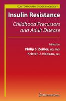
Interest in the subject during the late nineteenth century was stimulated by similar observations on outcome and the interest in cerebral localization in adults58–60 alongside the further addition to the pathological literature of cases with porencephaly by Richard Heschl and Hans Kundrat, the suggestion by Joseph Parrott, a pioneering paediatrician in Paris, that venous congestion was important and Ernst Von Strümpell’s (Fig. 1.1) unifying theory invoking a post polioencephalitic cause.54,61 Several physicians in Europe and North America, including Thomas Barlow, Aletta Jacobs, Ernest Gaudard, Otto Huebner, Pierre Marie, L. Emmett Holt (Fig. 1.1), Sarah McNutt, William Osler, Bernard Sachs and Frederick Peterson (Fig. 1.2), published their experience with congenital, infantile and childhood hemiplegias between 1875 and 1900.7,62–74 Osler, who had trained with Virchow and had conducted large numbers of autopsies as part of his research at McGill University, wrote his monograph after moving to Philadelphia, where his interest turned to clinical follow-up of various
In Cazauvieilh’s footsteps, many of the giants of nineteenth century medicine followed.7,9,54 Jules Cotard induced cerebral atrophy by the experimental embolization of cerebral arteries in animals and suggested that this might be a mechanism in children as well as in adults, although he also noted some differences, for example in the likelihood of aphasia following apoplexy.55 The work of Virchow shed considerable light on the pathology of thrombosis and embolus in vessels and was recognized to be of importance in adult stroke,56 although its relevance to infantile hemiplegia continued to be an area of controversy. In the latter half of the nineteenth century, debates raged about the apparently better outcome for apoplexy sustained in childhood and about the aetiology of congenital and acquired 4
9781898683346_4_001.qxd
3/14/11
11:24 AM
Page 5
Childhood Stroke: the Unkindest Cut of all: a History of Cerebrovascular Disease in Childhood as being important and was the first female member of the American Association for Neurology, described hemiplegia after traumatic delivery with meningeal haemorrhage.65–68 Osler mentions trauma as a trigger in older children in addition.72 There was general agreement that infection was an important association but skepticism about the relationship with the flaccid paralysis of poliomyelitis. In 1860, Jacob von Heine had described hemiplegia after scarlet fever and after vaccination.7 Gibotteau, who reported milder cases with improvement as well as those left with a residual hemiparesis, considered that all cases had an infectious aetiology.71 Osler mentions measles, scarlet fever, diphtheria and whooping cough while Freud discusses congenital syphilis and smallpox (but not chickenpox)7 and these infectious aetiologies were noted as triggers throughout the twentieth century. Interestingly, poliovirus was documented in the motor cortex of patients dying of the paralytic form of the disease54,80 but the protagonists lost interest in the controversy once the disease was eradicated by the same author and his rival in the development of a safe vaccine.81 Most of these diseases have either been eradicated or considerably reduced in prevalence at least in Western medicine through public health measures, immunization and antibiotics but children have continued to develop acute hemiplegia after infections. Ford and Schaffer in 1927 cited previously reported cases of focal encephalitis and monoplegia post-chickenpox and reported a case of their own.82 The development of ventilation for patients with paralytic poliomyelitis has had benefits for the management of children presenting acutely with other neurological disorders including convulsions, coma and hemiplegia which, although it is difficult to prove, has probably improved outcome alongside emergency protocols for status epilepticus of any cause. Acquired was clearly separated from congenital hemiplegia in the early nineteenth century French literature6 and in the writings of Osler, Sachs and Freud.7,72,74 In the field of neonatal stroke, clinical descriptions were typically included as ‘congenital hemiplegia’ as part of works on the outcome for birth asphyxia or the aetiology of cerebral palsies.4,7,54,65–69,72–74,83 Many conditions presenting as acquired hemiplegia now known to have separate aetiologies, such as unilateral status epilepticus,82 were published in the same series until the 1970s,84 –91 as were patients who presented in coma.23,92,93 Nevertheless, some of those presenting with convulsions or coma almost certainly had a vascular aetiology, reminding us of the importance of broad nosological terms unless we really understand the pathology.83,94 –98 Otitis media as a cause of cerebral venous thrombosis with ‘the infection being carried by the blood vessels’99 was previously well recognized as a cause of childhood and adolescent stroke. Interestingly, the debate as to the relative importance of arterial disease and venous thrombosis as a cause of childhood stroke is equally longstanding. In 1887 Abercrombie70 was disputing Gowers’ opinion69 that ‘thrombosis in arteries is a very rare lesion in childhood, far more rare than combined thrombosis in sinuses and veins’, feeling instead that arterial
“It is of use from time to time to take stock . . . of a particular disease, to see exactly where we stand in regard to it, to enquire to what conclusions the accumulated facts seem to point and to ascertain in what direction we may look for fruitful investigations in the future.” OSLER
Fig 1.1. A case of childhood stroke reported by L.E. Holt in 1897.
conditions including the cerebral palsies, a term that he coined.73 Both before and after he was President of the Clinical Section of the Royal Society of Medicine in London, cases were presented in various sections75–78 in the era when the majority of children were cared for by general physicians and neurologists before paediatrics became a separate speciality.79 Sigmund Freud, who had described hemianopia in association with childhood hemiplegia, in Infantile Cerebral Paralysis,7 undertook an extensive literature review and produced what can be regarded as a definitive nineteenth century opinion on childhood stroke. Reading this mighty work today, although we may have a greater understanding of the pathophysiology leading to a stroke in terms of practical effective management for the majority of cases, there has been little real progress beyond supportive care. Lack of therapy does not mean there is no chance for improvement or therapy. We know it is typical for many forms of infantile cerebral paralyses to tend to improvement, that there are light cases of the disease, and that even a complete recovery has occurred in a number of cases. An active interference on the part of the physician that can affect the process appears to be possible . . . . . . yet even under such conditions one must consider successful treatment as questionable. Freud, 1897 7 It was clear to most authors that there were multiple risk factors for infantile hemiplegia, rather than a single cause. Sarah McNutt, whose work was recognized by Osler and Gowers 5
9781898683346_4_001.qxd
3/14/11
11:24 AM
Page 6
Stroke and Cerebrovascular Disease in Childhood
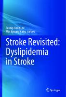
Fig. 1.2. The debate on aetiology of infantile hemiplegia in the late nineteenth century. Top row: Ernst Von Strümpell, Sarah McNutt, William Gowers. Bottom row: William Osler, Sigmund Freud and Bernard Sachs.
obstruction ‘will hold good in the great majority in the other cases’. Although cerebral venous sinus thrombosis continues to be underdiagnosed in the twenty-first century, unless transient thrombosis in the venous circulation turns out to be common when sensitive imaging becomes available for all neurological emergencies in childhood, the passage of time has probably vindicated Abercrombie’s judgement for the present day but it is impossible to obtain a clear answer for his own time or for the twentieth century.100 Otitis media is frequently cited as the most common cause of cerebral venous thrombosis in the pre-antibiotic era, but as late as the 1960s venous occlusive disease made up 60% of cases in some autopsy series.101 As antibiotics are prescribed less in the twenty-first than in the twentieth century, we need to maintain vigilance for this important and treatable cause of childhood death and disability. These series of cases presenting to adult or paediatric neurologists rarely included children with underlying conditions that we consider common aetiologies now, such as sickle cell disease or cardiac disease. The importance of sickle cell disease102 as an aetiological factor for childhood stroke went largely unrecognized for decades.103–107 MacKenzie in the first volume of Brain108 reported a case of cerebral embolus in a girl who appears to have endocarditis, probably secondary to rheumatic heart disease. He mentions the work of Kossuchin,
a Russian pathologist who published on the pathology of cerebral embolus in Virchow’s Archives in 1874.109 In addition to the recognition of the association of cerebral aneurysm with coarctation,110 reports of stroke in congenital heart disease appeared,76,82,111–113 although whether the mechanism was embolic remained controversial even when pathological studies were available. Interestingly Wood’s case of a child with pulmonary stenosis and an atrial septal defect with severe cyanosis mentions venous congestion and the occipital softening may well have been secondary to venous thrombosis,112 anticipating the current interest in the wide range of stroke syndromes in children with pre-existing problems. Epidemiology Over the last century, the epidemiology of childhood stroke appears to have changed with a decline in childhood stroke mortality being seen during the latter half of the twentieth century. Children less than 1-year old consistently have the highest mortality rate, the majority of deaths being due to haemorrhagic stroke.114,115 This understanding has come about because there has been increased recognition of cerebral haemorrhage, at least in part because population-based studies have been undertaken and have included children who died.116–121 Sachs74 emphasized the relative rarity of intracerebral haemorrhage as a 6
9781898683346_4_001.qxd
3/14/11
11:24 AM
Page 7
Childhood Stroke: the Unkindest Cut of all: a History of Cerebrovascular Disease in Childhood cause of stroke, whilst Broderick121 demonstrated that it made up to 50% of cases seen. The fall in childhood stroke mortality seen during the late twentieth century has been mainly due to a fall in haemorrhagic stroke as opposed to other stroke subtypes. It could well be a reflection of changing aetiological factors that have been sustained now through several generations. On the other hand, the aggressive management of seizures has led to a marked reduction in cases of postictal hemiplegia and the severe and irreversible brain damage seen as a result.24 However, iatrogenic causes have become more important as a consequence of medical advances allowing management to extend beyond supportive care.
arated out those with periventricular leukomalacia,101,133 who only occasionally had demonstrable venous pathology, although she pointed out that the distribution of lesions suggested that these vessels were involved in aetiology, a subject of interest in the twenty-first century. When pathology could only be confirmed at autopsy, either in the acute phase or many years after the event, the timing of focal injury in infancy could only be guessed at, and many conditions, including cerebral venous sinus thrombosis142,153 and arterial dissection,141 were considered to have a poor prognosis because they may leave little trace after recovery so autopsy descriptions were considered representative. Even now we may not have the tools to exclude these conditions in settings in which its diagnosis and appropriate management might reduce childhood disability.
Scientific Contributions from Anatomy and Pathology Scientific advance was largely dominated by post-mortem studies until the middle of the twentieth century (Fig. 1.3), with increasingly accurate descriptions of the arterial and venous anatomy by meticulous neuropathologists and embryologists including Helena Riggs, Charles Rupp and Dorcas Padget.122–127 Dorcas Padget’s career is of particular interest as she originally trained as an illustrator and worked for many years with Walter Dandy, a pioneer of paediatric neurosurgery.125,126 There were also steady advances in the understanding of cryptogenic and symptomatic arterial and venous stroke, periventricular leukomalacia, central pontine myelinolysis, hypertensive encephalopathy, Sturge–Weber syndrome and vein of Galen anomaly from the use of histopathological techniques and their detailed illustration or photography by Wilfred Le Gros Clark and Dorothy Russell, Morgan Berthrong, Betty Banker, Jeanne-Claudie Larroche, Dorcas Padget, Lucy Balian Rorke, Gilles Lyon and Ronald Norman amongst others.23,101,128–152 Betty Banker pointed out the importance of cerebrovascular pathology as a cause of death in childhood as this was present in nearly 9% of 555 consecutive autopsies at the Children’s Hospital of Philadelphia.101 She re-emphasized the importance of cerebral venous pathology, particularly in those with congenital heart disease,101 and sep-
Outcome: Relation to Age at Injury There have been relatively few long-term prospective followup studies of motor or cognitive outcome after childhood stroke before the twenty-first century (see chapter 17) and some potentially important material is not currently available in English.154 As part of a series looking at aetiology and outcome of 334 children with spastic hemiplegia,155,156 Philip Hood undertook statistical analyses for his doctorate under the supervision of Harold Westlake of the School of Speech at Northwestern University in Chicago and published the findings with Meyer Perlstein, who had a longstanding interest in cerebral palsy.9 They looked at motor and language development156 and at intelligence,155 finding no difference in mean intelligence quotient between left and right hemiparesis or between congenital and postnatally acquired hemiparesis, although the developmental milestones were achieved a little earlier in the latter group. When computed tomography (CT) became available, several series were published linking presence, size and site of lesion to motor and cognitive outcome for congenital and acquired hemiplegia.50,157–161 Interest in the effect of age on outcome for focal damage to the hemisphere dates back to the nineteenth century when,
Fig. 1.3. Post mortem sequelae of acute hemiplegia. (From Le Gross Clark WE, Russell DS. Atrophy of the thalamus in a case of acquired hemiplegia associated with diffuse porencephaly and sclerosis of the left cerebral hemisphere. J Neurol Psychiatry 1940; 3:123–140; with permission from the BMJ Publishing Group.128)
7
9781898683346_4_001.qxd
3/14/11
11:24 AM
Page 8
Stroke and Cerebrovascular Disease in Childhood soon after the description of cerebral lateralization in adults, Felix Vulpian described experiments in young animals that appeared to move on both sides despite focal decortication.162,163 Otto Soltmann hypothesized that motor function in the neonatal period was not reliant on the cerebral cortex and undertook experiments in dogs which showed that electrical stimulation in the cortex in the neonate did not lead to movement in the limbs and that unilateral lesions lead to late walking but not spasticity.164,165 It became established that aphasia in adults typically resulted from lesions in the left hemisphere,59,60 but the publication of an apparently transient case of aphasia from Great Ormond Street in a child with a right and then a left hemiplegia62,63 led to the development of theories about equipotentiality and the development of specialization (see Chapter 17) which continue to be influential today. Margaret Kennard worked at Yale during the 1930s and was undertaking experiments to look at the effects of cortical ablation in primates when she developed a collaboration with her obstetric colleagues which allowed her to study neonatal animals in addition. She and her colleagues were very surprised to realize that neonatal animals with focal cortical lesions did not develop spasticity and that this age effect persisted when lesions were performed in one- and two-year-old animals, although those operated on at later ages did worse than the neonates but less badly than the animals operated on as adults. Kennard’s work was very influential163,166–168 and was interpreted rather overenthusiatically to mean that outcome after focal
R
lesions was generally better in the young. However, permanent aphasia may follow focal lesions sustained at a young age169 and many children grow into their cognitive deficits even if language and motor function are relatively spared. The effect of age on motor and neuropsychological function after focal injury continues to be an area of controversy to the present day. The advent of imaging The twentieth century marked a decisive change in the visualization of cerebrovascular disease and its consequences in terms of haemorrhage or infarction. When previously only clinical acumen and craniotomy or trephination for extradural or subdural haematoma could reveal the cause of apoplexy in the living child,170 imaging the living brain marked a decisive step,149 even if this was initially only gross pathology.171 Some of these techniques, such as pneumoencephalography,172 although groundbreaking in their day, have become redundant and are now forgotten outside an historical review.98 A limited understanding of ventricular size could be obtained from a skiagram173 or pneumoencephalogram24 (Fig. 1.4) and occasional series of unselected cases of acute hemiplegia in childhood were published, although the results were not necessarily illuminating except in showing atrophy independent of any vascular territory after status epilepticus24 and unilateral brain swelling in the acute phase of hemiplegia.90,95,174 –176 Understanding of the relative importance of various cerebrovascular pathologies improved with the advent of contrast
Fig. 1.4. Air encephalogram. (From Isler W. Post ictal hemiplegia. In: Acute Hemiplegias and Hemisyndromes in Childhood. London: William Heinemann Medical Books, 1971; 139; with permission.217)
R
8
9781898683346_4_001.qxd
3/14/11
11:24 AM
Page 9
Childhood Stroke: the Unkindest Cut of all: a History of Cerebrovascular Disease in Childhood cerebral arteriography during the 1920s and 1930s.177 Moniz included an 11-year-old girl with a tumour in an early series178 but for stroke, the technique was used mainly in adults and only occasionally in children179–185 unless surgery, for example for intractable epilepsy186–188 or to prevent recurrence,189–194 was considered, because of the perceived risk of the procedures after the experience with pneumoencephalography195–200 and the perception that childhood stroke had a good prognosis and rarely recurred. However, the series of Charles Poser and Juan Taveras in New York, Kenneth Till and Dick Hoare in London and Edwin Bickerstaff in Birmingham, UK, John Shillito in Boston, Mark Dyken in Indianapolis and Derek Harwood Nash in Toronto, amongst others, made it clear that the test was commonly abnormal when haemorrhagic or ischaemic stroke was suspected clinically and that the benefit of making an ante-mortem diagnosis probably outweighed the relatively small risk for arteriography in their experienced hands.98,195,201–205 Werner Isler in his series, published as a book,98 acknowledged his debt to his predecessors and particularly to the participants at the Spastics Society meetings on Congenital Hemiplegia in Bristol83 (Fig. 1.5) and on Acquired Hemiplegia in Clevedon in 1961,97 which brought together clinicians and pathologists as well as the renowned anatomist Alf Brodal. Arteriography was felt to be safer and more useful in establishing a diagnosis than pneumoencephalography, so that even before cross-sectional imaging was available, case series
including the systematic use of vascular imaging in ischaemic stroke in childhood with clinical and laboratory information began to appear with the series of Hugh Greer and Arthur Waltz from the Mayo Clinic,206 Jean Aicardi, Françoise Goutières and Jean-Jacques Chevrie in Paris,90,207 Gail Solomon85 and Sadek Hilal86,208 from Arnold Gold’s department at Colombia Prebyterian in New York, Tibbles and Brown from Canada,209 Janaki210 and Malik211 from India, and Gösta Blennow212 and Orvar Eeg-Olofsson from Scandinavia.213 Systematic use of conventional angiography also allowed the distinction of cases with acute hemiplegia without cerebrovascular disease,94,96 facilitating the diagnosis in life of venous sinus thrombosis,207 which had been deduced previously,100 the distinction of alternative diagnoses such as alternating hemiplegia214 and the exclusion of hemiconvulsion-hemiplegia-epilepsy,215,216 which had been previously included in many case series of acute hemiplegia in childhood.90,94,98,217 These remain important issues as there is an important proportion of childhood stroke cases without vascular occlusion who must be excluded from any treatment trials to be conducted, for example of clot-busting drugs. The advent of less invasive ultrasound, CT and magnetic resonance imaging (MRI)106,160,218–234 has led to a renaissance of interest in the subject. Our perspective on childhood stroke has been revolutionized, allowing new perspectives including accurate identification of high risk groups with sickle cell disease235–237, the diagnosis of silent or covert infarction227 and
Fig. 1.5. Participants at the National Spastics Society Study group meeting on hemiplegic cerebral palsy in children and adults, held at the Wills Hall, University of Bristol, 1961. Ronnie Mac Keith is in the front row on the extreme right.83
9
9781898683346_4_001.qxd
3/14/11
11:24 AM
Page 10
Stroke and Cerebrovascular Disease in Childhood comparisons with adults as well as giving us a greater understanding of the whole realm of cerebrovascular disease.
of the pioneers, she also trained up a number of distinguished clinical academics who continue to try to understand these complex problems.
The Dawn of Physiology and Pathophysiology Once investigation of the cerebrovascular circulation in life became possible, some workers began to explore the physiology as well as the anatomy associated with cerebrovascular disease in childhood. Cerebral blood flow was found to be much higher in normal children than in adults238 using the nitrous oxide clearance method of Kety and Schmidt239 and these data have proved useful in understanding the pathophysiology of sickle cell anaemia.240,241 Using the Kety-Schmidt technique, Dyken found evidence that the least damaged hemisphere received blood from the contralateral carotid system in people who were long-term inpatients because they had sustained infantile hemiplegia242 but these data are unlikely to be representative as most patients who have had a childhood stroke live independently as adults. Chronic focal perfusion deficits have been shown using other techniques,243–245 but the relevance of these studies to outcome awaits less invasive methodologies which can be used in all children in the acute and chronic phases of stroke. Although electroencephalography (EEG) was a breakthrough for the management of epilepsy, it was not a particularly sensitive technique for demonstrating the effect of focal infarction.246 Attempts to use it for the diagnosis of paediatric neurological problems,247 including congenital248–250 and acute hemiplegia94,251 did not bear fruit as it did not distinguish aetiology or predict prognosis. However, monitoring EEG during cardiopulmonary bypass using single channel devices such as the cerebral function monitor did lead to understanding of the role of intra-operative microembolization in the aetiology of poor neurological outcome in children as well as adults and thus to the introduction of filters which reduced the incidence.252 Interest in the physiology of sleep in the young dates back at least to the work of the Russian Maria Mikhaiovna Manasseina-Korkunov, also known as Marie von Manassein and Marie de Manacéïne, one of the first women doctors in Europe. In a paper presented at a meeting in Italy, she reported that depriving puppies of sleep led to reduced red cell counts and to death, with the youngest animals dying earliest, typically with localized intracerebral haemorrhage and abnormal blood vessels.253–255 Her findings were followed up and largely replicated by Italian scientists,254 but although links between sleep physiology and stroke have been reported in adults256 and children, particularly those with sickle cell anaemia,257,258 relatively little attention has been paid to the effects of duration since then, although this might be important in the preterm neonates vulnerable to intraventricular haemorrhage, in addition to the effect of nocturnal hypoxia on endothelial function. Interaction with haematologists and other laboratory scientists has been increasingly important in understanding the pathophysiology of childhood stroke, particularly as Maureen Andrew showed very clearly that haemostasis is affected by a number of factors, including age and hypoxia.259,260 Like many
Conclusion The change in attitude is reflected in the increased medical literature devoted to this topic. Additionally there have been important changes in nomenclature. Osler’s book gives a useful list of terms previously used in the nineteenth century,72 while works from the mid-twentieth century tended to use his term ‘cerebral palsy’4,54 or, following Freud, ‘hemiplegia’, as did the early volumes of the Clinics in Developmental Medicine series supported by the Medical Advisory Committee of the National Spastics Society.83,97,98 The different titles to the volumes published by key players based in Toronto, San Francisco, Chicago, Dallas and Paris in the 1980s and 1990s261–264 reflect a more recent and wider understanding of the vascular nature of the majority of these conditions, as well as changing public attitudes. The definition of what we are including in clinical research has both hardened and widened in this period from that first suggested 40 years ago, when hemiplegia lasting less than one week was not included at Ronnie Mac Keith’s insistence.97 In contrast, most contemporary authors include not only patients with clinical stroke on the World Health Organization (WHO) criteria of a focal event lasting >24 hours with no cause other than vascular, but also those with transient ischaemic attacks lasting 1 year 820 1300 120 2278 94 98 239 80 8 22 153 31 117
Ischaemic stroke incidencea
Haemorrhagic stroke incidencea
Ratio ischaemic to haemorrhagic
0.6
1.9
0.33
0.2 1.2
1.5
0.78
7.9 1.1 0.58 3.3 AIS 2.6; SVT 0.7 7.8 1.17 1.2
5.1 2.3 0.71 (excluded SAH) – 2.9 0.89 1.2 (0.8 intracranial, 0.4 subarachnoid)
1.8 1.05 2.1 1.96
0.4 0.5 0.81 – ~2 1.31
Combined incidencea 3.1 2.5 2 2.1 3.0 2.7 6.4 13.2 3.4 1.29 3.3 13.5 2.06
1
2.3
2.57
2.1
0.5
2.19
1.94
0.74 1.7
2.67
4.3 2.7 (29.7) (27.1)
Incidence = per 100 000 children per year. Figures compare stroke with total paediatric admissions, not paediatric population. AIS, arterial ischaemic stroke; SAH, subarachnoid haemorrhage; SVT, sinovenous thrombosis. a
b
excluded.31 There appears to be an excess of strokes in those of black ethnicity31 which is not fully explained by the prevalence of sickle cell disease in this population.16 Asians have a similar incidence, while the incidence in Hispanics appears to be lower.31 The first incidence data for childhood stroke appear similar whether studies were confined to children and based in single large cities,11,12 or drew from a wider population base as part of a study which included adults.14,15 However, numbers of cases reported were small (Table 3.1). For example, four cases were identified in the Rochester, Minnesota population, resulting in an overall incidence for childhood stroke of 2.5/100 000 children/year with haemorrhagic stroke three times more frequent than ischaemic stroke.11 In the Rochester study, neonates, children older than 14 years, and children with head trauma were excluded. Sixteen strokes (nine haemorrhagic and seven ischaemic) were identified in metropolitan Cincinnati, Ohio, yielding an incidence of 2.7/100 000/year, with haemorrhagic stroke 1.25 times more frequent than ischaemic stroke.12 This study reported that blacks were at increased risk for childhood stroke with an overall incidence rate of 3.1 per 100 000 children per year, compared with
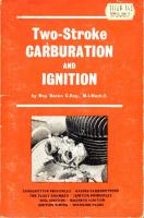
survival from these primary diseases reflects the development of more effective treatments, which can themselves be risk factors for stroke. Estimates of the incidence and relative proportions of stroke types in different studies are widely disparate, as shown in Table 3.1. Among the studies listed in Table 3.1, the inclusion criteria, methods of patient identification and geographical population evaluated have differed, with each study contributing differently to our understanding of incidence and subtypes of stroke. Stroke is undoubtedly more common in neonates with an incidence of at least 25–30 per 100 000 or 1 in 2300 – 4000 live births1,26–28 and a recent study from Estonia had findings of 63/100 000.29 Estimates depend on inclusion criteria: all ischaemic strokes or only arterial; patients with motor impairment as an outcome (17 per 100 00030) or all patients with focal ischaemia in an arterial distribution (20 per 100 00026). Whether neonates are excluded or not, the peak age for ischaemic stroke and intracerebral haemorrhage is the first year of life, with around one-third of cases presenting in this age group,31,32 while subarachnoid haemorrhage is more common in teenagers.31 Boys are at higher risk than girls in all studies, even when cases involving a history of trauma are
23
9781898683346_4_003.qxd
3/14/11
3:44 PM
Page 24
Stroke and Cerebrovascular Disease in Childhood whites who had an incidence rate of 2.6.12 Earley16 reported 35 children with stroke identified from the central region of the state of Maryland and from Washington, DC. The diagnosis of stroke was confirmed by chart review in all cases. Excluding neonates and children older than 15 years, the incidence rate for all stroke was 1.29 per 100 000 children per year. Even excluding subarachnoid haemorrhage, haemorrhagic stroke was more frequent than ischaemic stroke. In the UK, regional and national studies have reported an incidence of about 2/100 00017,18 (Fig. 3.1). There may have been significant lack of recognition of ischaemic stroke in early studies, as the World Health Organization definition (focal neurological deficit lasting more than 24 hours) was used and neuroimaging was often not undertaken, so that infarction in children with seizures or focal deficits of short duration would have been missed. In Europe, Giroud reported 28 children with stroke in Dijon, France, resulting in an overall incidence rate of 13.02/100 000.21 For the first time, the Dijon study reported that ischaemic stroke was more frequent than haemorrhagic stroke in children.21 The explanation for the greatly increased stroke incidence rates reported in the latter study is not known, but may relate to the geography, with all patients in the region referred to one hospital where the stroke team was led by a dual-accredited paediatrician and neurologist, so that all study participants had their diagnosis confirmed by CT or MRI. In a single institutional study in Montreal, Canada, ischaemic strokes were more than twice as frequent as intracerebral haemorrhage, although subarachnoid haemorrhage was again excluded.33 Paediatric ischaemic stroke is therefore likely to be an important health problem; in a study34 based in Salt Lake City, Utah, children less than age 18 comprised 22% of ischaemic strokes in the ‘young’ (aged less than 40 years) despite the exclusion of neonates. In recent years, data from large national studies looking specifically at stroke in children have become available. In the
Canadian Pediatric Ischemic Stroke Registry over 1000 children with ischaemic stroke (from term birth to 18 years) have been identified at all children’s health care centres in the country during the initial 10 years, resulting in an estimated incidence rate of ischaemic stroke of at least 3.3 per 100 000 children per year (95% confidence intervals 3.02 –3.63).35 Diagnostic code searches with ICD-9 codes and standardized chart reviews were conducted in all cases to confirm the diagnosis. The Canadian Registry provides the first data on the incidence of cerebral venous sinus thrombosis.36 In the Registry, neonates comprised 43% of cerebral venous sinus thrombosis and 25% of arterial ischaemic strokes. The sex distribution was similar to previous studies, with 54% of cerebral venous sinus thrombosis and 58% of arterial ischaemic stroke occurring in males. In the largest study of childhood stroke to date, paediatric stroke cases were identified by hospital discharge diagnosis searches with ICD-9 codes in the US National Hospital Discharge Survey (NHDS).1 The NHDS is a continuous nationwide sample survey of short-stay hospitals in the USA. The incidence of stroke for infants from birth (predominantly at term) to 30 days of age was 26.4/100 000. The rates for ischaemic stroke and haemorrhagic stroke were 17.8/100 000 and 6.7/100 000 live births per year respectively. The rates for children 0–18 years were 13.5/100 000 children per year for all stroke codes; 7.8/100 000 for ischaemic stroke codes (ICD-9 433–437, excluding 435 ‘transient cerebral ischaemia’) and 2.9/100 000 for haemorrhagic stroke codes (ICD-9 430 – 431).1 The latter study provides overall incidence rates which are similar to those from Dijon. It is possible that the rates of stroke were overestimated by the hospital ICD-9 coding, since confirmatory chart review to verify stroke diagnosis was not part of the study. In addition, the inclusion of codes for related cerebrovascular disease, including ICD-9 codes 436 (which includes ‘cerebral seizure’) and 437 (which includes moyamoya, hypertensive encephalopathy and unruptured aneurysms) may have resulted in an overestimate. However, the rates for neonates with ischaemic stroke are less than those from the Canadian Registry and the Estonian study. Lynch’s study1 has provided the most detailed information to date on the incidence rates for all subtypes of childhood cerebrovascular diseases and the stroke-related mortality rates, and over time will provide important race-specific stroke rates in children. A Northern California a study ascertained 205 validated pediatric ischemic strokes yeilding an incidence of 2.4/100000 children/year.21 Overall, the mean combined incidence rates for ischaemic and haemorrhagic stroke in children is 6.8 per 100 000 children per year. The relative proportion of ischaemic to haemorrhagic stroke is not entirely clear, and reported ratios of ischaemic to haemorrhagic strokes range from 0.33 to 2.0 with a mean rate of 0.86, which translates into haemorrhagic stroke being 1.2 times more common than ischaemic stroke. A recent study based on a large number of childhood strokes actually found a 1:1 ratio.31
80
Number
60
40
20
0 5
10
15
Age (years)
Fig. 3.1. Age distribution of 296 children with stroke (38 neonates) presenting in the UK and Eire in 2001 (median age 4.76 years).
24
9781898683346_4_003.qxd
3/14/11
3:44 PM
Page 25
The Epidemiology of Childhood Stroke addition to the high prevalence of predisposing conditions such as sickle cell anaemia and the additional risk posed by Westernization, infections commonly causing stroke (see Chapter 5, Section 5.1) may not have been controlled. Osuntokun’s41 study from Nigeria quoted an incidence of 2 per 100 000, but there were no cases under the age of 10 years, probably because they likely presented with seizures and were not recognized.43 Smadja’s42 study from Martinique found an incidence of 8 per 100 000 for those aged 0–34 years; their youngest patient was 3. It is important to develop global epidemiological data50 in order to provide evidence of the importance of childhood stroke across populations, including the subtypes and outcomes of stroke. Multi national populationbased studies are forming, and over time will provide these data, which will serve as the foundation for the first clinical trials in childhood stroke. Through clinical trials, evidence will be obtained which will direct treatment in individual patients, and reduce the current, unacceptably high cost of stroke in children.
Health care utilization Incidence data only partially describe the impact of a given disease. For stroke, health care utilization costs and loss of productive years when significant neurological or psychological deficits result are significant contributors to the burden of the illness. In adults, the lifetime cost of a first stroke is estimated to be $90 981.37 The types and magnitude of health care costs generated by childhood stroke have only recently been assessed.38 In the US the cost per pediatric stroke acute admission ranges from $20 000 to $81 000 with 5-year costs of $51 000 for neonates and $135 000 for children39,40. The median cost for the first year after diagnosis is $43 212. In one study length of inpatient stay and degree of physical and social impairment correlated with higher cost so that stroke cost may act as an indicator of stroke severity. The cost of treating haemorrhagic stroke was higher than for ischaemic stroke care, but infarct volume did not correlate with cost. However children’s inpatient stay is longer than that of adults. Part of the cost for health care utilization includes aetiological investigations and procedures which include multiple radiographic tests, laboratory tests and cardiac echocardiography. In children with stroke, in whom risk factors are heterogeneous and multiple, the extent and costs of investigations are likely to be increased compared with adults. In contrast, the costs of certain stroke treatments including t-PA and endarterectomy not currently utilized in children with stroke will be decreased. The costs of acute and chronic rehabilitation and the treatment of disease-related comorbid conditions add significant health care costs to the acute hospitalization costs. Finally, the cost of recurrent strokes over the lifetime of children with stroke may be considerably increased compared with adults.
REFERENCES 1. Lynch JK, Hirtz DG, deVeber G, Nelson KB. Report of the National Institute of Neurological Disorders and Stroke workshop on perinatal and childhood stroke. Pediatrics 2002; 109(1):116–123. 2. Mallick AA, Ganesan V, O’Callaghan FJ. Mortality from childhood stroke in England and Wales, 1921–2000. Arch Dis Child 2010; 95(1):12–19. 3. Adams RJ, McKie VC, Hsu L, Files B, Vichinsky E, Pegelow C et al. Prevention of a first stroke by transfusions in children with sickle cell anemia and abnormal results on transcranial Doppler ultrasonography. N Engl J Med 1998; 339(1):5–11. 4. Fullerton HJ, Adams RJ, Zhao S, Johnston SC. Declining stroke rates in Californian children with sickle cell disease. Blood 2004; 104(2):336–339. 5. Telfer P, Coen P, Chakravorty S, Wilkey O, Evans J, Newell H et al. Clinical outcomes in children with sickle cell disease living in England: a neonatal cohort in East London. Haematologica 2007; 92(7):905–912. 6. McCarville MB, Goodin GS, Fortner G, Li CS, Smeltzer MP, Adams R et al. Evaluation of a comprehensive transcranial doppler screening program for children with sickle cell anemia. Pediatr Blood Cancer 2008; 50(4):818–821. 7. Fullerton HJ, Chetkovich DM, Wu YW, Smith WS, Johnston SC. Deaths from stroke in US children, 1979 to 1998. Neurology 2002; 59(1):34–39. 8. Kleindorfer D, Khoury J, Kissela B, Alwell K, Woo D, Miller R et al. Temporal trends in the incidence and case fatality of stroke in children and adolescents. J Child Neurol 2006; 21(5):415–418. 9. deVeber G, Andrew M, Adams C, Bjornson B, Booth F, Buckley DJ et al. Cerebral sinovenous thrombosis in children. N Engl J Med 2001; 345(6):417–423. 10. Roach ES, Riela AR. Vasculopathies of the nervous system. In: Pediatric Cerebrovascular Disorders, 2nd edn (eds ES Roach, AR Riela AR). Aronk, New York: Futura Publishing Company, 1995; 163–180. 11. Schoenberg BS, Mellinger JF, Schoenberg DG. Cerebrovascular disease in infants and children: a study of incidence, clinical features, and survival. Neurology 1978; 28(8):763–768. 12. Broderick J, Talbot GT, Prenger E, Leach A, Brott T. Stroke in children within a major metropolitan area: the surprising importance of intracerebral hemorrhage. J Child Neurol 1993; 8(3):250–255. 13. Eeg-Olofsson O, Ringheim Y. Stroke in children. Clinical characteristics and prognosis. Acta Paediatr Scand 1983; 72(3):391–395. 14. Bamford J, Sandercock P, Dennis M, Warlow C, Jones L, McPherson K et al. A prospective study of acute cerebrovascular disease in the community: the Oxfordshire Community Stroke Project
Conclusion Stroke in adults is preventable, and stroke in children may be preventable as well. However, in contrast to adults, evidencebased treatments for childhood stroke are not available because no clinical trials have been conducted. Health care priorities and the funding of stroke care programmes have not included childhood stroke to date. Until the true burden of illness and impact on the health care system of each initial and recurrent childhood stroke is appreciated, this situation will change only slowly. The available epidemiological data indicate that the incidence of stroke in children is increasing and currently exceeds 6 per 100 000 children per year. Children with stroke face rates of adverse outcomes which include death in 10 –40%, and in survivors, significant neurological disability, learning problems or seizures in least 50%. Since children with stroke are expected to live a full lifespan, the burden of illness per individual will last for 50 –70 years. However, the true impact of childhood stroke is unknown because large population-based studies on the incidence, health care utilization and outcomes for all subtypes of childhood stroke are sparse. In addition, there are very few populationbased data from resource-poor countries,41,42 where, in 25
9781898683346_4_003.qxd
3/14/11
3:44 PM
Page 26
Stroke and Cerebrovascular Disease in Childhood
15.
16.
17.
18. 19.
20.
21.
22.
23.
24. 25.
26.
27.
28.
29.
30.
31.
1981–86. 1. Methodology, demography and incident cases of firstever stroke. J Neurol Neurosurg Psychiatry 1988; 51(11):1373– 1380. Gudmundsson G, Benedikz JE. Epidemiological investigation of cerebrovascular disease in Iceland, 1958–1968 (ages 0–35 years): a total population survey. Stroke 1977; 8(3):329–331. Earley CJ, Kittner SJ, Feeser BR, Gardner J, Epstein A, Wozniak MA et al. Stroke in children and sickle-cell disease: BaltimoreWashington Cooperative Young Stroke Study. Neurology 1998; 51(1):169–176. Williams AN, Eunson PD, McShane MA, Lynn R, Green S, Kirkham F. Childhood cerebrovascular disease and stroke like illness in the United Kingdom and Eire, a descriptive epidemiological study. Arch Dis Child 2002; 86 supplement 1:A40(G113). Williams AN, Kirkham FJ. Childhood stroke, stroke-like illness and cerebrovascular disease. Stroke 2004; 35 supplement 1:311. Steinlin M, Pfister I, Pavlovic J, Everts R, Boltshauser E, Capone MA et al. The first three years of the Swiss Neuropaediatric Stroke Registry (SNPSR): a population-based study of incidence, symptoms and risk factors. Neuropediatrics 2005; 36(2):90 –97. Agrawal N, Johnston SC, Wu YW, Sidney S, Fullerton H. Imaging data reveal a higher pediatric stroke incidence than prior US estimates. Stroke 2009; 40(11):3415–3421. Giroud M, Lemesle M, Gouyon JB, Nivelon JL, Milan C, Dumas R. Cerebrovascular disease in children under 16 years of age in the city of Dijon, France: a study of incidence and clinical features from 1985 to 1993. J Clin Epidemiol 1995; 48(11):1343–1348. deVeber GA, MacGregor D, Curtis R, Mayank S. Neurologic outcome in survivors of childhood arterial ischemic stroke and sinovenous thrombosis. J Child Neurol 2000; 15(5):316 –324. Zahuranec DB, Brown DL, Lisabeth LD, Morgenstern LB. Is it time for a large, collaborative study of pediatric stroke? Stroke 2005; 36(9):1825–1829. Al-Sulaiman A, Bademosi O, Ismail H, Magboll G. Stroke in Saudi children. J Child Neurol 1999; 14(5):295–298. Salih MA, bdel-Gader AG, Al-Jarallah AA, Kentab AY, Alorainy IA, Hassan HH et al. Stroke in Saudi children. Epidemiology, clinical features and risk factors. Saudi Med J 2006; 27 Suppl 1:S12–S20. Lee J, Croen LA, Lindan C, Nash KB, Yoshida CK, Ferriero DM et al. Predictors of outcome in perinatal arterial stroke: a population-based study. Ann Neurol 2005; 58(2):303–308. Lee J, Croen LA, Backstrand KH, Yoshida CK, Henning LH, Lindan C et al. Maternal and infant characteristics associated with perinatal arterial stroke in the infant. JAMA 2005; 293(6):723–729. Schulzke S, Weber P, Luetschg J, Fahnenstich H. Incidence and diagnosis of unilateral arterial cerebral infarction in newborn infants. J Perinat Med 2005; 33(2):170 –175. Laugesaar R, Kolk A, Tomberg T, Metsvaht T, Lintrop M, Varendi H et al. Acutely and retrospectively diagnosed perinatal stroke: a population-based study. Stroke 2007; 38(8):2234 –2240. Wu YW, March WM, Croen LA, Grether JK, Escobar GJ, Newman TB. Perinatal stroke in children with motor impairment: a populationbased study. Pediatrics 2004; 114(3):612– 619. Fullerton HJ, Wu YW, Zhao S, Johnston SC. Risk of stroke in children: ethnic and gender disparities. Neurology 2003; 61(2):189– 194.
32. Barnes C, Newall F, Furmedge J, Mackay M, Monagle P. Arterial ischaemic stroke in children. J Paediatr Child Health 2004; 40(7):384–387. 33. Lanthier S, Carmant L, David M, Larbrisseau A, de Veber G. Stroke in children: the coexistence of multiple risk factors predicts poor outcome. Neurology 2000; 54(2):371–378. 34. Kerr LM, Anderson DM, Thompson JA, Lyver SM, Call GK. Ischemic stroke in the young: evaluation and age comparison of patients six months to thirty-nine years. J Child Neurol 1993; 8(3):266–270. 35. deVeber GA, MacGregor D, Curtis R, Mayank S. Neurologic outcome in survivors of childhood arterial ischemic stroke and sinovenous thrombosis. J Child Neurol 2000; 15(5):316–324. 36. deVeber G, Andrew M, The Canadian Paediatric Ischemic Stroke Study group. The epidemiology and outcome of sinovenous thrombosis in pediatric patients. New Engl J Med 2001; 345(6):417–423. 37. Taylor TN, Davis PH, Torner JC, Holmes J, Meyer JW, Jacobson MF. Lifetime cost of stroke in the United States. Stroke 1996; 27(9):1459–1466. 38. Lo W, Zamel K, Ponnappa K, Allen A, Chisolm D, Tang M et al. The cost of pediatric stroke care and rehabilitation. Stroke 2008; 39(1):161–165. 39. Gardner MA, Hills NK, Sidney S, Johnston SC, Fullerton HJ. The 5-year direct medical costs of neonatal and childhood stroke in a population-based cohort. Neurology 2010; 74(5):372–378. 40. Perkins E, Stephens J, Xiang H, Lo W. The cost of pedatric stroke acute care in the United States. Stroke 2009; 40(8):2820–2827. 41. Osuntokun BO, Bademosi O, Akinkugbe OO, Oyediran AB, Carlisle R. Incidence of stroke in an African City: results from the Stroke Registry at Ibadan, Nigeria, 1973–1975. Stroke 1979; 10(2):205–207. 42. Smadja D, Cabre P, May F, Fanon JL, Rene-Corail P, Riocreux C et al. ERMANCIA: Epidemiology of Stroke in Martinique, French West Indies: Part I: methodology, incidence, and 30-day case fatality rate. Stroke 2001; 32(12):2741–2747. 43. Chadehumbe MA, Khatri P, Khoury JC, Alwell K, Szaflarski JP, Broderick JP et al. Seizures are common in the acute setting of childhood stroke: a population-based study. J Child Neurol 2009; 24(1):9–12. 44. Satoh S, Shirane R, Yoshimoto T. Clinical survey of ischemic cerebrovascular disease in children in a district of Japan. Stroke 1991; 22(5):586–589. 45. Merino Arribas JM, de Pablo CR, Grande GT, Sanchez MJ, Gonzalez de la Rosa JB. Nontraumatic hemorrhagic stroke in children after the neonatal period. An Esp Pediatr 1997; 47(4):392–396. 46. deVeber G, Chan A. Aspirin versus low-molecular-weight heparin for ischemic stroke in children: an unanswered question. Stroke 2002; 33(8):1947–1948. 47. Chung B, Wong V. Pediatric stroke among Hong Kong Chinese subjects. Pediatrics 2004; 114:e206–e212. 48. Simma B, Martin G, Muller T, Huemer M. Risk factors for pediatric stroke: consequences for therapy and quality of life. Pediatr Neurol 2007; 37(2):121–126. 49. Fullerton HJ, Wu YW, Sidney S, Johnston SC. Recurrent hemorrhagic stroke in children: a population-based cohort study. Stroke 2007; 38(10):2658–2662. 50. Mallick AA, O’Callaghan FJK. The epidemiology of childhood stroke. Eur J Paediatr Neurol 2010; 14(3):197–205.
26
9781898683346_4_004.qxd
3/14/11
4:05 PM
Page 27
4 CLINICAL AND IMAGING FEATURES OF CHILDHOOD STROKE 4.1 Clinical syndromes 4.1.1 Anterior circulation stroke Fenella J. Kirkham
year.13,14 At least 300 children present with stroke in the UK each year15 Half of the events are haemorrhagic (Figs 4.2[column a] and 4.3), secondary to arteriovenous malformations, aneurysms, clotting disorders16 or venous sinus thrombosis.17 Sickle cell disease is probably the most common cause of childhood stroke world-wide18,19 with a very wide variety of stroke syndromes20 (see Chapter 15). Other predisposing conditions for arterial (Figs 4.2[columns b,c,d,f1] and 4.4) and venous (Figs 4.2[column e] and 4.5) ischaemic cerebrovascular accidents in children include congenital heart malformations (see Chapter 15), chromosomal disorders such as Down (see Chapter 15), neurocutaneous syndromes (see Chapter 15) and infections such as meningitis (see Chapter 5), but around half of such accidents occur in children who were previously well (cryptogenic stroke).12
Introduction Stroke and cerebrovascular disease can cause considerable anxiety to the paediatrician. There is a large number of individual conditions each of which is relatively rare,1 the differential diagnosis includes a wide range of alternative pathologies2– 4 (Fig. 4.1) and protocols for investigation and treatment are not well worked out. For symptomatic stroke, the pre-existing diagnosis, and for cryptogenic stroke, the trigger(s) are often important clinical clues to diagnosis. In adults, as a result of controlled trials of thrombolysis and of stroke units, there is an increased awareness of the need for rapid assessment and appropriate management of acute stroke and the concept of ‘brain attack’ has received widespread publicity. Thrombolysis must begin within 4.5 hours, however, and even fewer children than adults are triaged this quickly.2,5 Although in one study the median time at diagnosis was 22.7 hours,6 in many children the correct diagnosis may not be made for days or even weeks.2 Nevertheless, there is little doubt that stroke units save adult lives and improve outcome in survivors,7 in part because a team’s experience may lead to rapid diagnosis and appropriate management of common and rare stroke syndromes. Currently, services for children are not organized in this way in the UK, and it has not yet proved possible to conduct large randomized trials of treatment in groups of children with similar pathologies. Physicians have found an advisory service helpful,8 although this type of service may be difficult to maintain because of issues of indemnity. There is no universally recognized system for classifying stroke in childhood.9–11 One of the main problems is that there is a wide differential diagnosis for acute focal neurological deficit in childhood.2,3,12 There are a number of ‘stroke mimics’ which may be benign and require no treatment3 while cerebrovascular disease may present without stroke. Emergency magnetic resonance imaging (MRI) studies may provide information to guide management.
Clinical presentation Acute hemiparesis in childhood can be symptomatic of a variety of pathologies. The World Health Organization (WHO) definition of stroke is ‘rapidly developing clinical signs of focal (or global) disturbance of cerebral function, with symptoms lasting 24 hours or longer, or leading to death, with no apparent cause other than of vascular origin’. Patients whose signs resolve within 24 hours have transient ischaemic attacks (TIA) by definition, but many have recent cerebral infarction or haemorrhage on imaging.12 The rapidity of onset may help to decide which investigations should be undertaken first as an abrupt onset (