Root Diseases and Soil-Borne Pathogens [Reprint 2020 ed.] 9780520339941
142 86 100MB
English Pages 264 [248] Year 2020
Recommend Papers
![Tropical Infectious Diseases. Principles, Pathogens and Practice [3 ed.]
9780702039355](https://ebin.pub/img/200x200/tropical-infectious-diseases-principles-pathogens-and-practice-3nbsped-9780702039355.jpg)


![Root Diseases and Soil-Borne Pathogens [Reprint 2020 ed.]
9780520339941](https://ebin.pub/img/200x200/root-diseases-and-soil-borne-pathogens-reprint-2020nbsped-9780520339941.jpg)
- Author / Uploaded
- T. A. Toussoun (editor)
- Robert V. Bega (editor)
- Paul E. Nelson (editor)
File loading please wait...
Citation preview
ROOT DISEASES AND SOIL-BORNE PATHOGENS
A cleared strawberry rootlet showing mycelium and vesicles of the mycorrhizal fungus Rhizophagus, in the cortex. (Courtesy S. Wilhelm.)
ROOT DISEASES AND SOIL-BORNE PATHOGENS
Second International Symposium on Factors Determining the Behavior of Plant Pathogens in Soil Held at Imperial College, London, July 14-28,1968 in Conjunction with the First International Congress of Plant Pathology Edited by T. A. TOUSSOUN, ROBERT V. BEGA, and PAUL E. NELSON
QJ V) ÜJ CO < LÜ
in Q
IO
IO 2
IO 3
INOCULUM DENSITY
(propagules/g)
INOCULUM
DENSITY
( propagules/g )
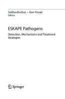
Fig. 2. Relationships of inoculum density to disease severity in A, Fusarium root and foot rot of cereals and B, Verticillium wilt of mint. Data from Cook (1968) and Lacy and Horner (1965). Models also may be useful in explaining why and how certain phenomena operate. An example of this application may be found when examining inoculum densities found in nature (Baker and McClintock, 1965). For the particular ecological group in which most soil-borne pathogens are found, reported densities are no more than approximately 3,000 propagules/ g of soil. Usually they are less. It is interesting that these densities fall in a range above which large increases in inoculum are required to decrease distance between propagules significantly. Rao and Rao (1963, 1966a, 1966fc) have explored one of the exceptions to the rule that inoculum density is directly proportional to disease severity. Disease development in cotton increased with increases of inoculum of Fusarium oxysporum f. sp. vasinfectum from 0.5% to 10% of an oatmeal-sand culture in soil. Further increases extended the incubation period. This was probably not due to the inoculum itself, however, as the organic substrates upon which the inoculum was grown also extended the time required for symptoms to appear. This underlines the importance of using inoculum in as "natural" a state as possible. This system should conform to Model II; however, relatively large inoculum potentials applied to the cotton seedlings may have induced somewhat atypical infection of the hypocotyl more characteristic of Model I. Thus the data may be difficult to analyze. One of the most valuable applications of population studies is related to the ability to detect actual fluctua-
tions of inoculum resulting from various treatments. A good example is the study on populations of pathogens involved in the root rot-Fusarium wilt complex of peas under various crop sequences (Kerr, 1963). Mechanisms of biological control may be detected also by assays of inoculum density before and after treatment. Thus, there was no significant change in inoculum density of F. solani f. sp. phaseoli following incorporation of cellulose into soil even though control of bean root rot was accomplished (Baker and Nash, 1965; Papavizas, Lewis, and Adams, 1968). This strongly suggests that the most significant mechanism of biological control is competition (Snyder, Schroth, and Christou, 1959; Maurer and Baker, 1965) rather than lysis or antibiosis. The relation of population levels of plant pathogens in soils to fungicidal dosage also is of interest and more attention to this type of research is needed. An excellent example is that given by Richardson and Munnecke (1963). Their findings confirmed previous work (reviewed in their paper) indicating that fungicide dosage required to control soil pathogens is proportional to the inoculum density. Thus, more attention will have to be given to the importance of standardizing inoculum in soil-fungicide evaluation tests. The relation of this work to mathematical models is treated elsewhere (Baker, 1968). Ultimately, quantitative analysis of all significant factors contributing to disease severity should have value in achieving a basic understanding of the epi-
PART II. POPULATION
DYNAMICS
OF PATHOGENS
IN
SOIL
14
Fig. 3. Interrelationships of the influence of inoculum density, C:N ratio, and temperature on number of lesions incited by Fusarium solani f. sp. phaseoli on bean hypocotyls. demiology of soil-borne pathogens. The equation: disease severity = inoculum potential X disease potential has been suggested as a basis (Baker, 1 9 6 5 ) . Such a project will require extensive systems analysis and should be developed with better theory than is presently available. A first attempt has been made to interrelate some of the major factors influencing disease severity for bean root rot (Baker and Maurer, 1967). Among these are inoculum density, C : N ratio, and temperature (Fig. 3). Since these experiments were done in growth chambers under controlled conditions, the results cannot be directly applicable to field conditions. They do illustrate, however, a pos-
sible direction for systems analysis applicable to disease situations. The examples noted above are but a few of the uses of studies concerned with inoculum potential. Other papers in this section will treat the influence of environmental factors, host response, and the significance of inoculum potential in specific pathogen-host interactions. L I T E R A T U R E CITED
1965. The dynamics of inoculum, p. 395403. In K. F. Baker and W. C. Snyder [ed.j, Ecology of soil-borne plant pathogens. Univ. of California Press, Berkeley and Los Angeles.
BAKER,
R.
Use of Population Studies in Research on Plant Pathogens in Soil R. 1968. Mechanisms of biological control of soil-bome pathogens. Ann. Rev. Phytopathol. 6 : 2 6 3 -
BAKER,
294. BAKER,
R., and D. L. M C C L I N T O C K . 1965. Populations of pathogens in soils. Phytopathology 55:495. B A K E R , R . , and C . L . M A U R E R . 1967. Interaction of major factors influencing severity of bean root rot. Phytopathology 5 7 : 8 0 2 . (Abstr.) BAKER, R . , C . L . MAURER, a n d RUTH A. MAURER.
1967.
Ecology of plant pathogens in soil. VII. Mathematical models and inoculum density. Phytopathology 57:662666. B A K E R , R . , and S H I R L E Y M . N A S H . 1965. Ecology of plant pathogens in soil. VI. Inoculum density of Fusarium solani f. sp. phaseoli in bean rhizosphere as affected by cellulose and supplemental nitrogen. Phytopathology 5 5 : 1 3 8 1 - 1 3 8 2 . B Y T H E R , R. S. 1968. Etiological studies on foot rot of wheat caused by Cercosporella herpotrichoides. Ph.D. thesis, Oregon State University, Corvallis. COOK, R. J. 1968. Fusarium root and foot rot of cereals in the Pacific Northwest. Phytopathology 58:127-131. D I M O N D , A. E., and J . G. H O R S F A L L . 1965. The theory of inoculum, p. 404-415. In K. F. Baker and W. C. Snyder [ed.], Ecology of soil-borne plant pathogens. Univ. of California Press, Berkeley and Los Angeles. KERR, A. 1963. The root rot-Fusarium wilt complex of peas. Australian J . of Biol. Sei. 1 6 : 5 5 - 6 9 . L A C Y , M . L . , and C . E . H O R N E R . 1965. Verticillium wilt of mint: interactions of inoculum density and host resistance. Phytopathology 5 5 : 1 1 7 6 - 1 1 7 8 . L E A C H , L . D., and A. E . D A V E Y . 1938. Determining the sclerotial population of Sclerotium rolfsii by soil analysis and predicting losses of sugar beet on the basis of these analyses. J. Agr. Res. 56:619-632. M A U R E R , C. L., and R. B A K E R . 1965. Ecology of plant
15
pathogens in soil. II. Influence of glucose, cellulose, and inorganic nitrogen amendments on development of bean root rot. Phytopathology 55:69-72. N A S H , S H I R L E Y M., and W. C. S N Y D E R . 1962. Quantitative estimations by plate counts of propagules of the bean root rot fusarium in field soils. Phytopathology 52: 567-572.
PAPAVIZAS, G . C . , J . A . L E W I S , a n d P . B . A D A M S .
1968.
Survival of root-infecting fungi in soil. II. Influence of amendment and soil carbon-to-nitrogen balance on Fusarium root rot of beans. Phytopathology 58:365-372. P L A N K , J . E . VAN DER. 1963. Plant diseases: epidemics and control. Academic Press, New York. 349 p. RAO, A. S., and M. V. RAO. 1963. Inoculum potential and the Fusarial wilt of cotton. Nature (London) 200: 598—599 RAO, M. V., and A. S. RAO. 1966a. Fusarium wilt of cotton in relation to inoculum potential. Trans. Br. Mycol. Soc. 49:403-409. RAO, M. V., and A. S. RAO. 1966fo. The influence of the inoculum potential of Fusarium oxysporum f. vasinfecturn on its development in cotton roots. Phytopathol. Z. 56:393-397. RICHARDSON, L . T . ,
and D.
E.
MUNNECKE.
1964.
Ef-
fective fungicide dosage in relation to inoculum concentration in soil. Can. J. Botany 42:301-306.
SNEH,
B.,
J.
KATAN,
Y.
HENIS,
and
I.
WALD.
1966.
SNYDER, W . C . , M . N . SCHROTH, a n d T . CHRISTOU.
1959.
Methods for evaluating inoculum density of Rhizoctonia in naturally infested soils. Phytopathology 5 6 : 7 4 - 7 8 .
Effect of plant residues on root rot of bean. Phytopathology 49:755-756. T R U J I L L O , E. E., and W. C. S N Y D E R . 1963. Uneven distribution of Fusarium oxysporum f. cúbense in Honduras soils. Phytopathology 53:167-170.
•
Factors Affecting Plant Pathogen Population in Soil J. D. MENZIES—Soil Microbiologist, Agriculture, Beltsville,
Soil and Water Conservation Research Division, U.S. Department
of
Maryland.
•
considered in relation to stages of soil infestation. Inoculum must first be produced by the pathogen, that is, infective units must be increased somewhere in the life cycle. This inoculum must then be dispersed into the soil. There it must survive and possibly increase. The resulting pathogen population is determined by factors influencing all these processes.
INTRODUCTION.—Soil-borne plant disease, by definition, requires the presence of the pathogen in the soil, but the amount of disease is not clearly related to the size of the pathogen population. Many soil factors influence the final expression of disease, and it is seldom clearly established whether this effect has been upon the amount of the pathogen or upon its pathogenic success. Because of the indirect relation between disease and amount of inoculum, wide changes in pathogen population will usually be required to produce a significant change in disease damage (Dimond and Horsfall, 1965). Since the major objective of research on soil-bome pathogens is to improve methods of disease control, this discussion will center on those factors most capable of reducing pathogen populations drastically enough to be of practical control value. The pathogen population is expressed as the mass or number of propagative units of the pathogen per unit of soil. This is the "inoculum density" or "inoculum intensity" of different authors (Baker, Maurer, and Maurer, 1967; Dimond and Horsfall, 1965). It is usually reported as numbers of propagules and should not include consideration of the infective vigor of the inoculum (inoculum capacity). To quantify inoculum density in soil as distinct from disease incidence is not easy because units of inoculum are difficult to define and more difficult to measure directly (Menzies, 1963). The generalizations that follow are based on rather scanty data, a deficiency which may at least stimulate more intensive efforts to evaluate the quantitative features of inoculum in soil. Factors affecting pathogen populations can best be
INOCULUM PRODUCTION.—The initial inoculum is the high point of population for many soil-borne pathogens. Subsequent events control how fast this population disappears. It seems safe to generalize that soil-borne pathogens characteristically overproduce inoculum. In a natural plant community a pathogen survives by producing and dispersing so much inoculum that somewhere a small residue reaches a new host at just the right time and with just the right conditions to infect. All the rest is lost. The inoculumproducing systems developed by soil-borne pathogens may be barely adequate to cope with natural mixedplant communities, but they are far over-designed for monoculture. Pathogen populations on the order of millions per cubic foot of top soil are the result (Table 1). Factors associated with the initial production of inoculum are just as important as factors affecting later survival and they may be easier to apply in population control. A very practical method for preventing initial infestation is the removal and destruction of diseased plant material before it enters the soil or otherwise releases the inoculum. This process of field sanitation is widely recommended but not widely practiced— partly because of expense, partly through lack of
TABLE 1. Some reported populations of soil-bome pathogens in infested field soils Propagules Organism Fusarium solani f. sp. phaseoli F. roseurn f. sp. culmorum Verticillium dahliae Verticillium dahliae Helminthosporium sativum Thielaviopsis basicola
per gram of soil 1,000-3,000 1,600-10,000 100-300 100-1,200 8-900 3,000-5,000
100 propagules/gram = 2 million/cu ft ( ± )
16
Reference Nash and Snyder ( 1 9 6 2 ) Cook and Bruehl ( 1 9 6 8 ) Evans, Wilhelm, and Snyder ( 1 9 6 7 ) Menzies (unpublished) Chinn, Ledingham, and Sallans ( 1 9 6 0 ) Papavizas ( 1 9 6 4 )
Factors Affecting Plant Pathogen Population in Soil
17
equipment, and partly through reluctance to destroy organic matter. In the case of a pathogen like Verticillium dahliae, initial inoculum production is crucial because this organism survives in soil principally as a population of microsclerotia, all originating in the dead tissue of diseased plants. Potato stems killed by V. dahliae, if left on the field, will become filled with sclerotia. A one-inch segment of stem may contain 20,000-50,000 viable sclerotia. Populations of 1,000 per gram of soil have been found by direct assay in fields cropped repeatedly to potatoes. This is enough to insure that for every potato plant the next year there will be something like 50 million sclerotia in the soil mass occupied by its roots. Practically all of this inoculum could be excluded from the field by removal of the infested plant material before winter. Sclerotial production by V. dahliae in dead vines in high-rainfall areas is usually much less than in the dry irrigated areas. Perhaps the numerous saprophytic fungi that grow luxuriously on plant debris in wet regions competitively inhibit production of sclerotia by V". dahliae. An indication of this has been observed in culturing the organism on sterile potato stems in flasks. If such a culture becomes contaminated with Penicillium or Aspergillus, sclerotial formation is prevented in the area overrun by the contaminant. This may be a more effective way to use antagonistic organisms than as biological control agents against the sclerotia in the soil or as inhibitors of infection at the root surface. Verticillium dahliae has been used as an example where initial inoculum production established the soil population. The same is true for other root pathogens that produce more or less passive propagules—Fusarium solani, Sclerotium rolfsii, Thielaviopsis basicola, and Sclerotinia spp., to name only a few. There may be a number of other factors that can be manipulated to influence the amount of inoculum produced by these pathogens, but destruction of the diseased tissue before dispersal is an obvious solution.
Dispersion also affects population by exposing the propagules to a wider range of microenvironments in the soil. Although dispersal usually involves the separation of the pathogen propagules from their host tissue—and it is generally true that the pathogen survives better in the host tissue than when free in the soil— the more widely the inoculum is dispersed the greater will be the number of infective units finding a microenvironment suitable for survival. Pathogens with abilities for competitive saprophytic colonization may be affected by dispersion in a somewhat different way. Bruehl and Lai (1968), in an ingenious study of the wheat root rot organism Cephalosporium gramineum, found that when equal amounts of infested and clean straw were mixed in soil they could expect one secondary invasion of healthy straw for every 3 pieces of infested straw. Each infestation of straw, original or secondary, has a life expectancy of 2 - 3 years or until the straw is decomposed. They calculated that the observed degree of secondary saprophytic colonization did not significantly affect the population survival curve after the first two years. However, we can speculate that a tillage operation like discing would break up both healthy and infested straws and increase the frequency of contact. The net result should be more secondary infection sites with each representing a smaller piece of straw. The apparent population is increased, but the inoculum capacity of each infective unit may be lower because of reduced food base or shorter survival time. Other pathogens with competitive saprophytic ability may also increase this way in the field, but it should be noted that although numerous laboratory studies have shown that Fusarium roseum f. sp. cerealis 'Culmorum' is one of this type, Cook and Bruehl (1968) were unable to show such saprophytism in the field. Experimental work has not been done, so far as I know, on the role of dispersion by cultivation on the saprophytic increase of this kind of a pathogen.
INOCULUM DISPERSION.—One way in which dispersion influences the apparent pathogen population is by increasing the frequency of infective sites. Before dispersal occurs, a plant containing a population of pathogen propagules can be considered as a single infective unit. To use V. dahliae again as an example, the infected plants may be left standing over winter, and when disced into the soil for seedbed preparation, a certain amount of dispersion may take place. However, the great mass of sclerotia are still embedded in host tissue. These overwintered sclerotia are almost all viable. During the second season the tissue decays and the sclerotia are freed into the soil at that spot. Tillage operations during and after the second season gradually distribute these clusters of sclerotia. So, if one considers the effective population of sclerotia from a single infected plant, it is seen that this population increases with tillage over several years as multiple inoculum clumps disperse into separate units.
INOCULUM SURVIVAL.—Given a certain population of pathogen propagules produced on host tissue and then dispersed in the soil, a great many factors, biological or nonbiological, influence the fate of this population. Probably the first thing to ask about a dispersed population of pathogens is whether it has the capabilities and mechanisms for saprophytic increase. This turns out to be a question of degree. Where careful experiments are done, it is found that even those forms apparently persisting in soil only as dormant propagules may increase in population in unsterile soil if sufficient readily-available energy is added. In our work with Verticillium dahliae (Menzies and Griebel, 1967) we have shown that the population can increase 4- to 10-fold in response to additions of sucrose. Papavizas, Lewis, and Adams (1968) have published similar data for Fusarium solani showing population increases on addition of
PART II. POPULATION
DYNAMICS OF PATHOGENS
soluble carbohydrates. In cases like these the increased population may be conidia arising from sclerotia or chlamydospores. However, there is evidence that in soil, Verticittium sclerotia can form small secondary ones (Emmatty and Green, 1967) and it is known that Fusarium macroconidia readily convert to new chlamydospores, (Schroth and Hendrix, 1962). Our data on Verticillium indicate that the extra population produced by saprophytic multiplication in soil is short-lived and probably is made up of conidia (Menzies and Griebel, 1967). In this case the increase in soil population of propagules may not represent any increase in inoculum potential since the conidia may not be effective agents for infection. Garrett and his associates at Cambridge have made detailed studies on several important cereal foot-rot fungi that possess various saprophytic abilities but which generally die out before the straw is completely decomposed, some sooner than others. One of their most interesting observations has been that an abundant supply of available nitrogen in the soil prolongs the survival of Ophiobolus graminis but shortens that of Helminthosporium sativum (Garrett, 1963). Obviously there is more to the story than merely promoting more rapid decay of the straw through additions of nitrogen. Garrett (1966) postulates that the efficiency with which these pathogens utilize the cellulose of the straw for mycelial growth when N is added explains their differential survival (Table 2). TABLE 2. Effect of nitrogen on saprophytic survival of cereal foot rot fungi as related to cellulosis adequacy index*
Pathogen Ophiobolus graminis Fusarium culmorum Cercospora herpotrichoides Curvularia ramosa Helminthosporium sativum
Effect of N
Cellulosis
on survival
Adequacy index f
+ + + + +
-t—
—
.59 .68 .69 1.25 3.50
* Adapted from S. D. Garrett. Trans. Brit. Mycol. Soc. 4 9 : 5 7 - 6 8 , 1966. t Index = relative amount of cellulose decomposed per unit of mycelial growth.
The fact that N fertilization inhibits one pathogen but favors another is an unfortunate complication that points up the need for detailed understanding of the consequences of manipulating soil factors to control specific diseases. In the normal soil situation energy substrates other than host tissue are not usually introduced in sufficient excess to stimulate pathogen populations except by root exudation. There does not seem to be as much specificity in the stimulation effect of root exudates as was once thought. The work reported by Schroth and Hendrix (1962) on the bean root rot Fusarium illustrates this point. Chlamydospores of Fusarium are stimulated by various amino acids and sugars to germi-
IN
SOIL
18
nate and to form new chlamydospores. There are differences in the degree of response from root systems of different crops, but several nonhosts such as lettuce and tomato induce population increases (Table 3). TABLE 3. Effect on nonhost roots on populations of Fusarium solani f. sp. phaseoli* Nonhost
Propagules per gram
Crop
Rhizosphere
Corn Lettuce Tomato Onion
268 140 247 364
Nonrhizosphere 108 73 136 1,680
* From M. N. Schroth and F. F. Hendrix. Phytopathology 5 2 : 9 0 6 - 9 0 9 , 1962.
Populations were apparently reduced in the rhizosphere of onion, but it is possible that this may result from inhibitory substances rather than from the absence of the right kind of amino acid or sugar. If nonhost root systems can induce germination and growth of dormant pathogen propagules, this seems more likely to result in a mild population increase rather than the hoped-for decrease. The same may be true when other plant residues are incorporated in soil. This may be happening because the stimuli for germination are accompanied by ample substrate for continued growth. In recent work in our laboratory (Menzies and Gilbert, 1967) we have found that plant residues give off volatile compounds that have a remarkable stimulatory effect on germination and respiration of soil organisms. Among the active components are several low-molecular-weight aldehydes and alcohols (Owens et al., 1969) some of which have been shown by others (Sussman, 1965) to stimulate spore germination in vitro. By using these volatiles alone instead of with the carbohydrate substrate with which they are usually associated, we may be able to induce pathogen growth in the absence of sufficient exogenous substrate to permit population replenishment. This could lead to decrease of the population through competition and starvation. Initial trials with this approach against V. dahliae are quite encouraging (Gilbert and Griebel, 1969). The gradual decrease in a pathogen population in soil as a result of various adverse factors, biological or environmental, takes time. Historically, not knowing how to speed up these processes, we have withheld planting of host crops until these factors have had time to be practically effective. This is the essence of crop rotation for disease control. There is serious question, however, whether this practice can survive the industrial revolution in agriculture. To the modern farmer time is money, and he does not wish either to wait or to fix his cropping pattern into a long rotation scheme. Since the effectiveness of these inoculum reducing factors is a function of both time and intensity, research emphasis needs to be placed on intensity. For example, the way antibiosis is used in medicine is
Factors Affecting Plant Pathogen Population in Soil not by smearing mold cultures on wounds but by isolating the antibiotics and applying them at much higher concentrations than are found in nature. It is reasonable to expect that the factors effective during crop rotation act by reducing the pathogen population, but the evidence for this is meager. Maloy and Burkholder (1959) used the most-probablenumber method for estimating F. solani f. sp. phaseoli populations in fields following different nonhost crops. They found no great differences in the number of infection units of F. solani f. sp. phaseoli in field soil following any of the various rotations tested. Incidentally, they also failed to find any direct relation between root rot severity and pathogen populations in these fields. Nash and Snyder (1962), using a sensitive selective plate count assay for F. solani f. sp. phaseoli from California fields, found that the propagule population had dropped considerably after a crop of tomatoes, whereas after a crop of barley the population was even higher than after continuous beans. The amount of disease, however, was consistently lower after barley, again emphasizing the poor correlation between pathogen population and disease severity. There is ample evidence that, other things being equal, disease severity correlates rather well with inoculum density (Baker, Maurer, and Maurer, 1967; Dimond and Horsfall, 1965). This evidence comes from controlled experiments where inoculum density was varied by adding the pathogen to soil at different rates. This is entirely different from modifying the system to induce population changes and then measuring subsequent disease. When a pathogen population is increased by a soil treatment, the increased population may differ in kind from the original. For example, sclerotia may produce smaller sclerotia, conidia, or hyphae, and chlamydospores may form conidia or new chlamydospores. The inoculum potential of these new propagules may differ from the original inoculum. Also the soil treatment inducing the population change affects the associated microflora and the host as well, which in turn can modify the disease reaction. O V E R R I D I N G F A C T O R S . — A S was pointed out earlier in this discussion, factors influencing pathogen populations will be useful for disease control only if they have capabilities for almost complete eradication. Furthermore, such factors should be effective throughout the soil mass, over a wide range of microenvironmental niches, and against a wide range of pathogens. Chemical fumigation is a process that very adequately meets these specifications. Were it not for high cost and perhaps some residue problems, there would be little need to look further. However, in this discussion, we are interested in naturally occurring factors. Among natural influences, the three most "overriding" factors in pathogen survival are temperature, moisture, and aeration. Temperature effects on root disease have been well studied, but again the direct
19 effect on pathogen populations has seldom been determined. Soil temperature is believed to be important in the geographic distribution of root pathogens as, for example, the restriction of Phymatotrichum omnivorum (the cotton root rot organism) to the southern U.S.A., and the restriction of the dark mycelial form of Verticillium albo-atrum to more northern regions. The possibilities of manipulating soil temperature in the field to reduce pathogen populations are limited. Even though lethal temperatures for most plant pathogens are relatively moderate (SOMJO'C for 30 minutes) the energy required to heat the soil mass to these temperatures is so great as to be generally impractical at present except on very high value crops (Baker, 1962). Nevertheless, it is possible that atomic power will be so abundant and cheap in the future that heat treatment of soils in general will become feasible. Inability to use heat for pathogen control more widely is frustrating. It is a truly overriding factor, being able to penetrate all microenvironments much more uniformly than any biologic factor or chemical agent. Heat is effective against all plant pathogenic organisms and against insects and weeds as well. It leaves no residue. We should not be deterred by present costs from doing more research on the field use of various sources of heat as a means of reducing pathogen populations in soil. The second overriding factor is moisture, more specifically desiccation. (Excess water in soil also affects pathogen populations; this will be considered along with aeration). Large areas of arid lands are fanned under irrigation where it is quite possible to dry soils in the field to very high moisture tensions. A classic case of pathogen control by dry summer fallow is that reported by Elmer (1942), who concluded that Rhizoctonia solani was unable to survive summer fallow conditions in Kansas during dry years. The cotton root rot fungus Phymatotrichum omnivorum survives in soil by means of sclerotia that appear to be quite sensitive to drying (King, Loomis, and Hope, 1931). Dry cultivation, especially deep tillage, often produces a remarkable control of root rot in cotton (Earl Burnett, personal communication). Desiccation may be an important factor in this disease control. The microsclerotia of Verticillium dahliae are quite resistant to drying in the fresh state, but after they have been incubated in soil and induced to go through a period of multiplication they can be killed by a mild air drying of the soil (Menzies and Griebel, 1967). It is probably generally true that if pathogen propagules can be enticed out of dormancy they can then be severely affected by drying. Soils dry by evaporation from the surface, however, and capillary water moves up from lower horizons as this process goes on. To reduce the plow layer of soil to a sufficiently low moisture content will require dry tillage and possibly several weeks of dry fallow. The last of the three "overriding" factors to be mentioned is aeration. Poor aeration in soil is generally
PART II. POPULATION
DYNAMICS OF PATHOGENS
detrimental to pathogen propagules, although some do well in very wet soil. Poor aeration in the field is generally associated with high CO2 and high water content as well as low O2. Sewell (1965) reviewed these interrelations recently. It should be emphasized that a shortage of oxygen in soil, especially in the presence of ample decomposable organic material, will favor production of fatty acids, alcohols, and other anaerobic microbial metabolites which can be quite harmful to the obligately aerobic pathogens. In unpublished work in our laboratory we have found that pathogens in culture can be killed by accumulation of toxic metabolites when placed under anaerobiosis if these substances cannot diffuse away. Perhaps a similar killing of pathogens occurs in local sites in soil when green manure is turned under and moisture content is high. Such killing may not be very useful in disease control because of the spotty distribution of such sites. With more research, however, we may find ways of imposing a general anaerobiosis in soil sufficient to eradicate pathogens. Flooding for pathogen control has had some success, but the flood water is oxygenated and downward percolation prevents accumulation of toxic substances. This method might be more dependable if we could find a practical scheme for excluding oxygen from field soil while utilizing organic amendments to impose a severe oxygen stress. CONCLUSION.—There are a great many factors
known to affect pathogen populations in soil. We are interested in those that reduce populations and particularly in those that we can manipulate. Modern agriculture features large acreages of one crop, tends towards monoculture, and uses crop varieties often lacking in root-disease resistance. This has so weighted the balance in favor of massive buildups of pathogen populations in soil that reduction of these populations has to be drastic to be effective in disease control. The most direct way to prevent high pathogen populations is to interfere with production and dispersion of the initial propagules. Practically, this is accomplished by various sanitation practices. Once the propagules are dispersed in soil, effective reduction in population requires the application of an intense pervasive factor such as fumigants, heat, anaerobiosis, or desiccation. The more subtle biologic factors are likely to be specific for certain pathogens, certain soils, and certain microsites. They are more likely to act through modifying the disease expression than by directly reducing the pathogen population. To use these biological controls in the field, especially for population control, we need to find the key factors involved and then devise means of applying them at a much greater intensity than nature provides. LITERATURE CITED
BAKER, K. F. 1962. Principles of heat treatment of soil and planting material. J. Australian Inst. Agr. Sci. 28: 118-126.
BAKER, RALPH.
1968.
Mechanisms of biological control
20
IN SOIL
of soil-borne pathogens. Ann. Rev. Phytopathol. 6:263— 294.
BAKER, R . ,
C.
L.
MAURER, and R . A .
MAURER.
1967.
Ecology of plant pathogens in soil. VII. Mathematical models and inoculum density. Phytopathology 5 7 : 6 2 2 666.
BRUEHL, G. W . , a n d P . LAI.
1968.
T h e p r o b a b l e sig-
nificance of saprophytic colonization of wheat straw in the field by Cephalosporinm gramineum. Phytopathol-
ogy 58:464—466. CHINN, S. H . F . , R. J . LEDINGHAM, a n d B . J . SALLANS.
1960. Population and viability studies of Helminthosporium sativum in field soils. Can. J. Botany 3 8 : 5 3 3 539.
COOK, R. J . , a n d G. W . BRUEHL.
1968.
Relative sig-
nificance of parasitism versus saprophytism in colonization of wheat straw by Fusarium roseurn 'Culmorum' in the field. Phytopathology 58:306-308.
DIMOND, A . E . , a n d J . G . HORSFALL.
1965.
T h e theory
of inoculum, p. 404-415. In K. F. Baker and W. C. Snyder [ed.j, Ecology of soil-borne plant pathogens. Univ. of California Press, Berkeley and Los Angeles. ELMER, O. H. 1942. Effect of environment on the prevalence of soil-borne Rhizoctonia. Phytopathology 32: 972-977. EMMATTY, D . A., a n d R. J . GREEN, JR.
1967.
Germina-
tion of microsclerotia of Verticillium albo-atrum in soil. Phytopathology 57:810-811. (Abstr.).
EVANS,
G.,
S.
WILHELM,
and
W.
C.
SNYDER.
1967.
Quantitative studies by plate counts of propagules of the Verticillium wilt fungus in cotton field soils. Phytopathology
57:1250-1255.
GARRETT, S. D. 1963. A comparison of cellulosedecomposing ability in five fungi causing cereal foot rots. Trans. Brit. Mycol. Soc. 46:572-576. GARRETT, S. D. 1966. Cellulose-decomposing ability of some cereal foot rot fungi in relation to their saprophytic survival. Trans. Brit. Mycol. Soc. 4 9 : 5 7 - 6 8 . GILBERT, R. G., a n d G. E . GRIEBEL.
1969.
T h e influ-
ence of volatile substances from alfalfa on growth and survival of Verticillium dahliae in soil. Phytopathology (in press). GLYNNE, MARY D. 1965. Crop sequence in relation to soil-borne plant pathogens, p. 423-433. In K. F. Baker and W. C. Snyder [ed.], Ecology of soil-borne plant pathogens. Univ. of California Press, Berkeley and Los Angeles. KING, C . J . , H . F . LOOMIS, a n d C . HOPE.
1931.
Studies
on sclerotia and mycelial strands of the cotton root rot fungus. J. Agr. Res. 42:827-840.
MALOY, O . C . , a n d W . H . BUHKHOLDER.
1959.
Some ef-
fects of crop rotation on the Fusarium root rot of bean.
Phytopathology
49:583-587.
MENZIES, J. D. 1963. The direct assay of plant pathogen populations in soil. Ann. Rev. Phytopathol. 1 : 1 2 7 142. MENZIES, J . D . , a n d R . G. GILBERT.
1967.
Responses of
MENZIES, J . D . , a n d G. E . GRIEBEL.
1967.
Survival a n d
the soil microflora to volatile compounds in plant residues. Soil Sci. Soc. Amer. Proc. 31:495-496. saprophytic growth of Verticillium dahliae cropped soil. Phytopathology 57:703-709.
NASH, SHIRLEY M., a n d W . C . SNYDER.
1962.
in un-
Quanti-
tative estimations by plate counts of propagules of the bean root rot Fusarium in field soils. Phytopathology
52:567-572. OWENS, L . D . , R . G. GILBERT, G. E . GRIEBEL, a n d J . D .
MENZIES. 1969. Identification of plant volatiles that stimulate microbial respiration and growth in soil. Phytopathology (in press). PAPAVIZAS, G. C. 1964. New medium for the isolation of Thielaviopsis basicola on dilution plates from soil and rhizosphere. Phytopathology 54:1475-1481.
PAPAVIZAS, G . C . , J . A. LEWIS, a n d P . B . ADAMS.
1968.
Factors Affecting Plant Pathogen Population in Soil Survival of root infecting fungi in soil. II. Influence of amendment and soil carbon-to-nitrogen balance on Fusarium root rot of bean. Phytopathology 58:365-372.
SCHROTH, M . N . , a n d F . F . HENDRIX, JR.
1962.
Influ-
ence of non-susceptible plants on the survival of Fusarium solani f. phaseoli in soil. Phytopathology 52: 906909.
SEWELL, G. W. F. 1965. The effect of altered physical condition of soil on biological control, p. 479—493. In
21 K. F. Baker and W. C. Snyder [ed.], Ecology of soilborne plant pathogens, Univ. of California Press, Berkeley and Los Angeles. SUSSMAN, A. S. 1965. Dormancy of soil microorganisms in relation to survival, p. 99-109. In K. F. Baker and W. C. Snyder [ed.], Ecology of soil-borne plant pathogens. Univ. of California Press, Berkeley and Los Angeles.
•
Significance of Populations of Major Plant Pathogens in Soil: Bacteria Including Streptomyces A. R. WEINHOLD—Department of Plant Pathology, University of California,
(Schroth, Thompson, and Hildebrand, 1965). This medium is inhibitory to all soil bacteria except A. radiobacter and A. tumefaciens. Since A. radiobacter is not pathogenic, it was necessary to determine the proportion of the crown gall bacterium within this Agrobacterium group. This was accomplished by isolating individual colonies and inoculating small tomato plants. Those organisms causing galls were designated A. tumefaciens. The population of A. tumefaciens was determined by placing lg soil in 10 ml of water, shaking the suspension, and placing 0.1 ml on the selective media in petri plates. Colonies were selected from the plates and tested for pathogenicity. In tests where known quantities of the bacteria were added to soil, the recovery efficiency of this method was found to be about 38%. Therefore, population data from field soil were corrected accordingly. The ratio of A. radiobacter to A. tumefaciens in most soils averaged 100-500 to 1. Several soils with varying crop histories were assayed. The value for A. tumefaciens population in a pasture soil was 42/g, as compared with 200/g for soil from a stone-fruit orchard and 316/g from a fruit-tree nursery soil. Susceptible plants grown on the latter two soils would be expected to have 10-20% crown gall whereas the pasture soil produced a low percentage of diseased plants (Table 1).
To understand the significance of pathogen population in soil it is necessary to know the relationship between population and disease severity. The key to this is the development of methods for the determination of the population of the pathogen as it exists in the field. The use of host-indexing methods is of limited value because of the lack of information on the number of propagules required for infection. With bacteria including Streptomyces the problem is further complicated by the absence of distinctive morphological characteristics with consequent dependence on a pathogenicity test to confirm identification. The first obstacle is one of numbers. Bacteria and Streptomyces spp. constitute a large proportion of the total soil microflora. To isolate a particular plant pathogen which comprises only a very small part of this population, it is first necessary to devise a method whereby a large segment of the nonpathogens can be excluded. This can often be accomplished by soildilution techniques and selective media. However, it is extremely difficult to develop a medium on which only the plant pathogenic species in question will grow. There remains, therefore, the question of how best to identify the pathogen following isolation. Approaches to this problem are dictated by the specific organism in question. I wish to present information on the crown gall organism, Agrobacterium tumefaciens (M. N. Schroth, personal communication), and the potato scab pathogen, Streptomyces scabies.
POPULATION
POPULATION STUDIES ON AGROBACTERIUM
Berkeley.
STUDIES
ON STREPTOMYCES
CIENS.—The studies on A. tumefaciens were made possible by the development of a highly selective medium
TABLE 1. Relationship between pathogen population in soil and disease severity for crown gall and potato scab Pathogen
A. A. A. S. S.
tumefaciens tumefaciens tumefaciens scabies scabies
SCABIES.—
Streptomyces spp. are relatively easy to isolate on soildilution plates, and the population in soil can be determined, subject to the limitations of this technique.
TUMEFA-
Source Pasture Stone-fruit orchard Fruit-tree nursery Rotation plot Variety testing plot
Population (propagules/g soil)
Disease severity
42 200 316 6,000-7,000 5,000-6,000
Trace to 1* 10-20* 10-20* 10-llt ll-21f
* Percentage of plants with crown gall. t Scab index—average percentage of tuber surface covered by scab lesions.
22
Significance of Populations of Major Plant Pathogens in Soil However, to determine the population of a specific organism such as S. scabies is a rather formidable task. To obtain some information on this point we combined two approaches. Several investigators have reported that one of the distinguishing characteristics of S. scabies is the production of melanin pigments when grown on suitable media (Menzies and Dade, 1959; Taylor and Decker, 1947). In our studies, 10-15« of the Streptomyces spp. present in soil from the central valley of California produced this pigment. By simply transferring colonies from the dilution plates (medium used: 2g agar and O.lg tyrosine/liter) to peptone agar and selecting melanin-positive isolates, it was possible to eliminate 85-90% of the total population. However, since many nonpathogenic Streptomyces spp. also produce melanin pigments, additional selection was needed. For this we turned Ho serology. Serological investigations indicated that S. scabies belongs to a serologically closely related group which includes some nonpathogens (Bowman and Weinhold, 1963). Fifteen isolates of S. scabies and 55 nonpathogens were tested, using the agar-gel double diffusion technique. All of the pathogens and 2 nonpathogens reacted with the pathogen-specific antisera. This suggested that serology could be used to screen Streptomyces spp. isolated from soil and single out those most likely to be S. scabies, thus reducing timeand space-consuming greenhouse testing. In an attempt to isolate S. scabies directly from soil, dilution plates were prepared. Soil was collected from the rotation plots at the U.S. Cotton Station, Shafter, California. Six hundred colonies were transferred to a potato-dextrose-peptone medium, and those which produced melanin pigment (about 10%) were tested serologically. Two isolates reacted as pathogens, and subsequent pathogenicity tests confirmed their identity as S. scabies. Based on the total Streptomyces spp. population of this soil, as determined by dilution plates, the population of S. scabies was about 6,000 to 7,000/g of soil (Table 1). The soil used in this work was taken from a field which, for the past two years, had produced tubers with a scab index of 10 to 11. This means that the average tuber has 10-11% of its surface covered with scab lesions. In a second trial to determine the population of S. scabies, a field which had a past history of severe scab was selected. This field had been used for many years to test breeding stock for scab resistance and was also located on the U.S. Cotton Research Station in Shafter. A similar process of selection for melanin pigment producing isolates followed by serological testing produced 2 isolates of suspected S. scabies from 775 isolates taken from dilution plates. This would represent a population of S. scabies of about 5.000-6,000/g of soil. Over a period of several years the scab index for tubers taken from this area ranged from 11 to 21 (Table 1). The above data present an estimate of the population of S. scabies in soil which produces severely scabbed tubers. Because of the relatively small sam-
23
ple, these values are only approximate, but they are not too far out of line with population data on some other soil-borne pathogens (Nash and Snyder, 1965). Other evidence is available which suggests a rather direct relationship between S. scabies population and disease severity. In a long-term field experiment to study the effects of cover-crop incorporation on scab buildup, it was revealed that soybean completely prevented an increase in disease severity over a period of 13 years (Weinhold et al., 1964). At the beginning of the test, only a trace of disease (scab index 0.3-0.5) was present on tubers produced in the plots. In the control areas there was a lag phase of 4 years during which the scab index increased only to 2.5. However, during the next 3 years there was a rapid linear increase in disease severity, reaching a peak scab index of 15. In subsequent years, disease severity was relatively constant, with a definite trend toward decreasing severity. In plots on which soybeans were grown and incorporated as green manure, scab index never exceeded a value of 1.0. There are several lines of evidence which suggest that increase in scab severity reflected an increase in the population of the pathogen. (1) The shape of the curve for disease increase was consistent with that expected for an increase in population; (2) in certain plots soybeans were grown for the first time in the second year of the 3-year period of rapid disease increase. This appeared to halt the increase, but did not result in any decrease in disease severity, suggesting that the effect was on population rather than pathogenic activity; and (3) cessation of the soybean cover crop for a 3-year period after completion of the test did not result in an increase in disease. This indicates that the pathogen population had never increased above the initial low level. If one assumes on the basis of the above information that there exists a rather direct relationship between population of S. scabies and scab severity, it is possible to interpolate from the available population data and arrive at an estimate of the minimum population required for disease occurrence. On this basis, a value of approximately 500 propagules/g of soil would be predicted to result in a scab index of 1.0. DISCUSSION.—To compare the population of S. scabies and A. tumefaciens in soil, it is necessary to consider the nature of the diseases caused by these pathogens. With crown gall, a single infection of a seedling can result in an enlarging gall and the plant considered diseased. With potato scab, a single infection results in only one scab lesion. A disease situation where the average tuber had 10% of the surface covered by lesions would require many separate infections for each tuber. Therefore, one might expect that severe scab would require many more infections than that required for a severe crown gall situation and, thus, a higher population of the organisms. This is precisely what our data show. However, such a comparison must be qualified by the realization that we
PART II. POPULATION
DYNAMICS OF PATHOGENS
have no information on sporulation of S. scabies in scab lesions or the possible importance of secondary infection. The studies on S. scabies and A. tumefaciens provide information which indicates a close relationship between disease severity and pathogen population. There are, of course, other factors such as the effects of the physical and biological environment and the vigor and nutritional status of the inoculum in regard to the infection process. The placing of these many variables in proper perspective must await the results of future research. However, it appears certain that pathogen population as regulated by factors affecting buildup and survival is of vital importance in the occurrence and severity of diseases caused by soil-borne bacteria including Streptomyces.
24
species of Streptomyces. Nature (London) 2 0 0 : 5 9 9 600. MENZIES, J . D . , a n d CAROLINE E . DADE.
1959.
A selec-
457-458. NASH, SHIRLEY M . , a n d W .
1965.
Quanti-
tive indicator medium for isolation of Streptomyces scabies from potato tuber or soil. Phytopathology 49: C . SNYDER.
tative and qualitative comparisons of Fusarium populations in cultivated fields and noncultivated parent soil. Can. J. Botany 4 3 : 9 3 9 - 9 4 5 .
SCHROTH, M . N . , J . P . T H O M P S O N , a n d D . C . H I L D E B R A N D .
1965. Isolation of Agrobacterium tumefaciens, A. radiobacter group from soil. Phytopathology 5 5 : 6 4 5 647.
TAYLOR, C . F . , a n d P . DECKER.
1963.
Serolog-
ical relationships of potato scab organisms and other
1947.
A correlation be-
tween pathogenicity and cultural characteristics in the genus Actinomyces. Phytopathology 3 7 : 4 9 - 5 8 .
WEINHOLD, A . R . , J . W . OSWALD, T U L L Y BOWMAN, J A M E S B I S H O P , a n d DAVID W R I G H T . 1964. Influence of green
manures and crop rotation on common scab of potato. Am. Potato J. 4 1 : 2 6 5 - 2 7 3 .
L I T E R A T U R E CITED BOWMAN, TULLY, a n d A. R . WEINHOLD.
IN SOIL
• Significance of Populations of Pythium and Phytophthora in Soil A. F. SCHMITTHENNER—Plant Pathology Department, Ohio Agricultural Research and
Development
Center and the Ohio State University, Columbus.
•
Most Pythium spp. can be quantitatively isolated directly from soil using polyene antibiotics to inhibit nonpythiaceous fungi and wide-spectrum antibiotics to inhibit bacteria and actinomycetes. Combinations of pimaricin, penicillin, and polymyxin (Eckert and Tsao, 1962); endomycin and streptomycin (Schmitthenner, 1962); Mycostatin and streptomycin, (Hendrix and Kuhlman, 1965); and Mycostatin and vancomycin (McCain, Holtzmann, and Trujillo, 1967) have been used successfully. Pythium graminicola and most Phytophthora spp. have not been quantitatively isolated directly from soil, even though the above antibiotic combinations are suitable for isolating them from diseased tissues. Phytophthora cinnamomi, an exception, produces large chlamydospores which germinate readily, and it has been isolated on Mycostatin media flooded with relatively large amounts of suspended soil or sieved organic particles (Hendrix and Kuhlman, 1965; McCain, Holtzmann, and Trujillo, 1967). Pythium ultimum (Agnihotri and Vaartaja, 1967a), Pythium aphanidermatum, and Phytophthora parasitica (Trujillo and Hine, 1965) have been studied with a soil-smear technique. Saprophytic colonization has been used successfully for isolating Pythium (Hine and Luna, 1963). Host bioassays are still the major method of detecting most Phytophthora species in soil. R E L A T I O N S H I P B E T W E E N POPULATIONS OF P Y T H I U M AND P H Y T O P H T H O R A AND D I S E A S E
SEVERITY.—Vaar-
taja and co-workers (Agnihotri and Vaartaja, 1967a; Vaartaja and Agnihotri, 1967; Vaartaja and Bumbieris, 1964) have concluded that environmental factors primarily determine severity of seedling root diseases in forest nursery beds, at levels of Pythium commonly isolated. In rotation studies I found (unpublished data) that populations of Pythium ultimum and incidence of alfalfa seedling blight caused by P. ultimum fluctuated independently. Campbell and Hendrix (1967) concluded that Pythium spp. are damaging only during or following periods of excessive moisture, but that disease severity may be less dependent upon soil moisture when Pythium populations become excessively high. A number of investigators have reported increase in diseases caused by Pyth-
ium following addition of residues (Trujillo and Hine, 1965), seed (Singh, 1965), or high-carbohydrate amendments (Barton, 1960; Liu and Vaughn, 1966; Yarwood, 1966) to soil. Large amounts of amendments were used and undoubtedly produced unrealistically high Pythium populations. For example, Kendrick and Wilbur (1965) concluded that more than 500 propagules Pythium irreguläre/ g soil were needed for severe preemergence kill of lima bean, after building up populations with repeated plantings of lima bean seed. In most studies (Campbell and Hendrix, 1967; Schmitthenner, 1962; Vaartaja and Bumbieris, 1964) populations of pythia/g soil generally were fewer, and pathogenic types (e.g. Pythium ultimum, Pythium irreguläre, and Pythium aphanidermatum) generally were less than 100/g soil. High soil moisture apparently is necessary for development of root and seed rots caused by Pythium and Phytophthora (Griifin, 1963). Soil water, directly or indirectly, may affect the fungus, the host, and the host-pathogen interaction. E F F E C T O F S O I L M O I S T U R E ON SURVIVAL OF P Y T H -
IUM AND PHYTOPHTHORA.—Survival of Pythium definitely is linked to soil moisture. Watson (1966) reported that Pythium drastically decreased in soil in summer after a 10-day drought. The fungus could not be recovered following a heavy rain, but built up gradually with the recurrence of wet conditions during the autumn. Apparently Pythium became constitutively dormant, sensu Sussman and Halvorson (1966), under dry conditions, and was activated after prolonged wetting. A similar type of dormancy was dramatically demonstrated by Hoppe (1966), who stored air-dried soil for 12 years at room temperature. He found high levels of Pythium using a corn seedling assay, after 6 years, if the soil was kept wet for 15 days. After 12 years, wetting soil 3 months was required before high levels of Pythium were detected. Either sporangia or oospores of Pythium survive in soil, depending on the species (Trujillo and Hine, 1965; Vaartaja and Agnihotri, 1967). Mycelium rapidly disappears (Agnihotri and Vaartaja, 1967a; Lockwood, 1960). All Phytophthora structures except chlamydospores
PART II. POPULATION
DYNAMICS OF PATHOGENS
and oospores die under dry conditions (Mircetich and Zentmyer, 1967; Turner, 1965). Sporangia or encysted zoospores or mycelia may persist in moist soils for considerable periods, but under some conditions are rapidly lysed (Mircetich and Zentmyer, 1967). Oospores of all and chlamydospores of some Phytophthora spp. may be constitutively dormant. Populations of these Phytophthora spp. can be determined only if some method of germinating dormant spores en masse is found. EFFECT
OF
SOIL
MOISTURE
ON
SAPROPHYTIC
G R O W T H O F P Y T H I U M AND P H Y T O P H T H O R A I N S O I L . —
All nonconstitutively dormant structures of Pythium and Phytophthora apparently are exogenously dormant in soil sensu Sussman and Halvorson (1966). Germination and growth of these structures are inhibited in soil except when assimilable nutrients are available (Agnihotri and Vaartaja, 1967a; Turner, 1964; Vaartaja and Agnihotri, 1967) with one exception (Tsao and Bricker, 1968). Also, there is evidence that soil extracts may be less inhibitory to germination of Pythium. ultimum when diluted (Agnihotri and Vaartaja, 1967a). Pythium may grow a considerable distance from a rich food base and colonize many carbohydrate-rich substrates (Barton, 1960; Liu and Vaughn, 1966; Singh, 1965; Yarwood, 1966), but activity of Phytophthora in soil is more limited (Trujillo and Hine, 1965; Turner, 1965; Zentmyer and Mircetich, 1966). Neither group can grow in residues already colonized by other microorganisms (Barton, 1961; Hine and Trujillo, 1966). Colonization of residues and amendments is best under high moisture conditions (Barton, 1960; Zentmyer and Mircetich, 1966).
The greatest effects of high soil moisture are on the availability of seed and root exudates (Burstrom, 1965; Kerr, 1964; Schroth and Cook, 1965). Kerr (1964) showed that pea seed exudation was much greater under high soil moisture and that seed rot caused by Pythium was correlated with both high moisture and increased seed exudation. Seed genotype and damage, and soil temperature also affect exudation (Kraft and Erwin, 1967; Schroth and Cook, 1964). The primary effect of high soil moisture is due to decreased oxygen (Burstrom, 1965). Brown and Kennedy (1966) showed that exudation from soybean seed was much greater under anaerobic conditions. Pythium seed and root rot of soybeans were severe under low oxygen in naturally infested soils, and under high oxygen only when cornmeal was added. Root exudates may reverse inhibition of spore germination of Phytophthora and Pythium in soil (Agnihotri and Vaartaja, 19676; Turner, 1963), and the level of nutrients may determine whether mycelia or zoospores are formed upon germination (Aragaki, Mobley, and Hine, 1967; Tsao and Bricker, 1968). Apparently zoospore aggregation on roots is essential for good infection with some fungus-host combinations (Kraft,
IN
26
SOIL
Endo, and Erwin, 1967). Water and root exudates are required for formation, movement, and accumulation of zoospores on susceptible roots (Hickman and Ho, 1966). S O I L M O I S T U R E E F F E C T S ON H O S T T O P Y T H I U M AND
SUSCEPTIBILITY
PHYTOPHTHORA.—High soil moisture
and accompanying anaerobic conditions may directly affect roots and thus influence root-rot severity and the correlation between soil populations and disease levels. Both Pythium and Phytophthora grow well in low oxygen that may damage higher plants (Brown and Kennedy, 1966; Klotz, Stolzy, and DeWolfe, 1963). Root growth is retarded under anaerobic conditions, and considerable proliferation of secondary roots may occur (Burstrom, 1965), increasing numbers of immature roots for colonization. Anaerobic conditions also alter root-tissue maturation and suberization and lignification (Burstrom, 1965). Suberized endodermis is the primary barrier to colonization of Pythium (Miller et al., 1966). Single zoospore infections caused considerable damage to cotton under high moisture but were reduced under dry conditions (Spencer and Cooper, 1967). Respiratory changes, such as those reported in roots of tomato under low oxygen (Fulton, Erickson, and Tolbert, 1964), might alter the resistance of roots to pathogens. Cruikshank and Perrin, (1967) reported that phaseolin and pisatin production in bean and pea, respectively, were inhibited under low oxygen to such an extent that Monilinia fructicola, a nonpathogen, was capable of rotting pod tissue. CONCLUSIONS.—Pathogenic Pythium spp. are widespread in soil and sufficiently abundant to damage crops. Information of Phytophthora populations in soil is still meager. Neither genus is capable of causing disease (assuming suitable genotype, temperature, etc.) unless: (1) the propagules are not constitutively dormant, (2) there are sufficient readily assimilable nutrients, and (3) the host is in a susceptible condition. All these requirements are met under high soil moisture (decreased oxygen) conditions. Disease severity, therefore, would be expected to fluctuate independently from population levels under normal moisture conditions. Probably, selective baits used under high moisture conditions would be best for predicting disease severity, but would be valid only if the population were not dormant. Pythium and Phytophthora might be controlled more easily by manipulating soil moisture, where this is possible, than by attempting to eliminate the pathogens. LITERATUHE CITED
P., and O. V A A R T A J A . 1967a. Root exudates from red pine seedlings and their effects on Pythium ultimum. Can. J. Botany 45:1031-1040. AGNIHOTRI, V . P., and O. V A A R T A J A . 1967&. Effects of amendments, soil moisture contents, and temperatures on germination of Pythium sporangia under influence of soil mycostasis. Phytopathology 57:111&-1120. AGNIHOTRI, V .
Significance of Populations of Pythium and Phytophthora in Soil ARAGAKI, M., R. D . MOBLEY, and R . B . HINE.
Sporangial germination of Phytophthora
1967.
from papaya.
Mycologia 5 9 : 9 3 - 1 0 2 .
BARTON, R. 1960. Saprophytic activity of Pythium mamillatum in soils. I. Influence of substrate composition and soil environment. Trans. Brit. Mycol. Soc. 43:529540. BARTON, R. 1961. Saprophytic activity of Pythium mamillatum in soils. II. Factors restricting P. mamillatum to pioneer colonization of substrates. Trans. Brit. Mycol. Soc. 4 4 : 1 0 5 - 1 1 8 .
BROWN, G . E . , and B . W . KENNEDY.
1966.
E f f e c t of
oxygen concentration on Pythium seed rot of soybeans. Phytopathology 5 6 : 4 0 7 - 4 1 1 .
BURSTROM, H. G. 1965. The physiology of plant roots, p. 154-169. In K. F. Baker and W. C. Snyder [ed.], Ecology of soil-borne plant pathogens. Univ. of California Press, Berkeley and Los Angeles. CAMPBELL, W . A., and F . F . HENDRIX.
1967.
Pythium
and Phytophthora species in forest soils in the southeastern U.S. Plant Disease Reptr. 51:929-932.
CRUIKSHANK, I . A. M., and D . R . PERRIN.
1967.
Studies
on Phytoalexins. X. Effect of oxygen tension on the biosynthesis of pisatin and phaseolin. Phytopathol. Z.
60:335-342. ECKERT, J . W . , and P. H . TSAO.
1962.
A selective m e -
dium for isolation of Phytophthora and Pythium from plant roots. Phytopathology 52:771-777.
FULTON, J . M., A. E . ERICKSON, and N . E . TOLBERT.
1964. Distribution of C 1 4 among metabolites of flooded and aerobically grown tomato plants. Agron. J. 56:527529. GRIFFIN, D. M. 1963. Soil moisture and ecology of soil fungi. Biol. Rev. 38:141-166. HENDRIX, F . F . , and E . G . KUHLMAN.
1965.
Factors
affecting direct recovery of Phytophthora cinnamomi from soil. Phytopathology 55:1183-1187. HICKMAN, C. J., and H. H. Ho. 1966. Behavior of zoospores in plant pathogenic Phycomycetes. Ann. Rev. Phytopathol. 4:195-220. HINE, R . B . , and L . V . LUNA.
1963.
isolating Pythium aphanidermatum pathology 53:727-728.
HINE, R . B . , and E . E . TRUJILLO.
A technique for
from soil. Phyto-
1966.
Monometric
studies on residue colonization in soil by Pythium aphanidermatum and Phytophthora parasitica. Phytopathology 5 6 : 3 3 4 - 3 3 6 .
HOPPE, P. E. 1966. Pythium species still viable after 12 years in air-dry muck soil. Phytopathology 56:1411. KENDRICK, J . B . , JR., and W . D . WILBUR.
1965.
T h e re-
lationship of population density of Pythium irregulare to pre-emergence death of lima bean seedlings. Phytopathology 55:1064. (Abstr.) KERR, A. 1964. The influence of soil moisture on infection of peas by Pythium ultimum. Australian J. Biol.
L i u , SHU-YEN, and E . K . VAUGHN.
1966.
Control of
Pythium infection of table beet seedlings by antagonistic microorganisms. Phytopathology 56:986-989. LOCKWOOD, J. L. 1960. Lysis of mycelium of plantpathogenic fungi by natural soil. Phytopathology
50:787-789. MCCAIN, A. H., O . V . HOLTZMANN, and E . E . TRUJILLO.
1967. Concentration of Phytophthora cinnamomi chlamydospores by soil sieving. Phytopathology 57:
1134-1135. MILLER, C . R., W . M . DOWLER, D . H . PETERSEN, and R .
P. ASHWORTH. 1966. Observations on the mode of infection of Pythium ultimum and Phytophthora cactorum on young roots of peach. Phytopathology 56:46-
49. MIRCETICH, S. M., and G . A. ZENTMYER.
1967.
SCHROTH, M. N., and R. J. COOK.
Seed exudation
Produc-
tion of oospores and chlamydospores of Phytophthora cinnamomi in roots and soil. Phytopathology 57:100. (Abstr.) SCHMITTHENNER, A. F. 1962. Isolation of Pythium from soil particles. Phytopathology 52:1133-1138. 1964.
and its influence on pre-emergence damping-off of bean. Phytopathology 5 4 : 6 7 0 - 6 7 3 .
SINGH, R. S. 1965. Development of Pythium ultimum in soil in relation to presence and germination of seeds of different crops. Mycopathol. Mycol. Appl. 27:155160. SPENCER, J . A., and W . E . COOPER.
1967.
Pathogenesis
of cotton (Gossypium hirsutum) by Pythium species: zoospore and mycelium attraction and infectivity. Phytopathology 5 7 : 1 3 3 2 - 1 3 3 8 .
SUSSMAN, A. S., and H . O . HALVORSON.
1966.
Spores:
TSAO, P. H., and J . L . BRICKER.
Germination of
their dormancy and germination. Harper and Row, New York and London. 354 p. 1968.
chlamydospores of Phytophthora parasitica in soil. Phytopathology 58:1070. (Abstr.)
TRUJILLO, E . E . , and R. B . HINE.
1965.
T h e role of
papaya residues in papaya root rot caused by Pythium aphanidermatum and Phytophthora parasitica. Phytopathology 5 5 : 1 2 9 3 - 1 2 9 8 .
TURNER, P. D. 1963. Influence of root exudates of Cacao and other plants on spore development of Phytophthora palmivora. Phytopathology 53:1337-1339. TURNER, P. D. 1965. Behavior of Phytophthora palmivora in soil. Plant Disease Reptr. 49:135-137. VAARTAJA, O., and V . P . AGNIHOTRI.
1967.
Inhibition
of Pythium and Thanatephorus (Rhizoctonia) by leachates from a nursery soil. Phytopathol. Z. 60:63-72.
VAARTAJA, O., a n d M . BUMBIERIS.
1964.
Abundance
1963.
Stimulation of
YARWOOD, C. E. 1966. Detection of Pythium in soil. Plant Disease Reptr. 50:791-792.
Oxygen requirements of three root-rotting fungi in a liquid media. Phytopathology 53:302-305. 1967.
fection of primary roots of bentgrass by zoospores of Pythium aphanidermatum. Phytopathology 57:86-90.
of Pythium species in nursery soils in South Australia. Australian J. Biol. Sci. 17:436-445. WATSON, A. G. 1966. Seasonal variation in inoculum potentials of spermosphere fungi. N. Z. J. Agr. Res.
Sci. 1 7 : 6 7 6 - 6 8 5 . KLOTZ, L . J . , L . H . STOLZY, and T . A. DEWOLFE.
KRAFT, J . M . , and D . C . ERWIN.
27
Pythium aphanidermatum by exudates from mung bean seeds. Phytopathology 57:866-868.
KRAFT, J . M., R . M . ENDO, and D . C . ERWIN.
1967.
In-
9:956-963.
ZENTMYER, G . A., and S. M . MIRCETICH.
1966.
phytism and persistence in soil by Phytophthora namomi. Phytopathology 66:710-712.
Sapro-
cin-
• The Significance of Populations of Pathogenic Fusaria in Soil SHIRLEY NASH SUITH-Ministry
of Agriculture, P. B. 757, Marandellas,
Rhodesia.
•
Commonly pathogenic formae of Fusaria has been found existing in soil as chlamydospores (Bywater, 1959; Cook, 1968; French and Nielson, 1966; Nash, Christou, and Snyder, 1961; Trujillo and Snyder, 1963), and we can assume that the propagules enumerated by plating or "most probable number" techniques usually are these structures—sclerotia, conidia, and hyphal strands normally being of minor or temporary importance. It has also been established that these chlamydospores require an external carbon and nitrogen source for germination and infection (Cook and Schroth, 1965; Maurer and Baker, 1965; Schroth, Toussoun, and Snyder, 1963; Toussoun, Nash, and Snyder, 1960). This makes the nutritional status of the soil itself regarding these elements one of the parameters to be considered, in addition to inoculum level, when disease severity is evaluated in a particular soil (Couch and Bedford, 1966; Maurer and Baker, 1965; Papavizas, Lewis, and Adams, 1968; Toussoun and Snyder, 1961).
12-15% of the isolates from random sampling of the infested field soils were pathogenic. French and Neilson (1966) observed that the macroconidia of F. oxysporum f. sp. batatas, which formed abundantly at ground level on diseased sweet potato stems, converted to chlamydospores quickly in soil. In infested California fields, however, the only place this forma was detected in countable numbers was at diseased plant sites (S. M. Nash, unpublished). F. solani f. sp. cucurbitae (race 1) also forms masses of sporodochia on diseased plants at ground level. These wash into the soil and form chlamydospores which are all relatively short-lived (Nash and Alexander, 1965). Conroy's (1953) effective recommendation of a 3-year rotation and clean seed for control of squash foot rot can be explained on the basis that this fungus is truly a "soil invader" (Garrett, 1956). PERSISTENCE.—Unlike F. solani f. sp. cucurbitae chlamydospores and most of those formed by F. oxysporum formae, individual chlamydospores of F. solani f. sp. phaseoli survive for long periods in soils. Eventually numerous clonal types accumulate and can be found distributed evenly throughout a field (Nash and Alexander, 1965; Snyder, Nash, and Trujillo, 1959). Often 1,000-4,000 propagules/g of soil have been found in fields on which beans have frequently grown. Alexander (1964) examined the ultrastructure of the chlamydospore walls of F. solani f. sp. phaseoli and f. sp. cucurbitae as well as a F. oxysporum f. sp. cubense clone and obtained evidence suggesting that the chlamydospores of the bean root rot Fusarium are comparatively the best suited to survival. F. solani f. sp. pisi appears to survive well in soil (Cook, personal communication). Although most of the propagules of F. oxysporum formae disappear soon after the host crop has been removed, a few may persist in soils for very long periods (Jones and Gilman, 1915). Stover (1962) in Central America and Rishbeth (1955) in Jamaica reported banana wilt quickly recurring in replanted plantations which had been abandoned more than 20 years previously. However, it was not established how the propagules survived—whether in a dormant state or by repeated germination and formation of new propagules. The percentage surviving must have
DISTRIBUTION.—When a relatively high propagule count of any particular forma specialis of a Fusarium species occurs in cultivated soil, the implication is that either a rather severely infected crop has recently grown, or the chlamydospores of the particular forma persist well in soil. Rishbeth (1955) detected as many as 300 propagules of F. oxysporum f. sp. cubense per gram in soil immediately around banana plants, but this population quickly declined after the plants were removed. Trujillo and Snyder (1963) observed that the propagules of this forma were distributed sparsely and unevenly through the soils of infested banana plantations, existing as chlamydospores inside decaying host-root tissue. Since Stover and Waite (1954) previously reported that this fungus is not a frequent saprophytic colonizer of dead banana tissue in soil, the chlamydospores in the tissue were most probably formed in diseased plants. Wensley and McKeen (1963) obtained populations of F. oxysporum f. sp. melonis ranging upward to 3,300/g in field soils at sites of wilted muskmelon plants, but mean population in field soils declined steadily during the 9-month interval between crops. At harvest 70% of the F. oxysporum isolates at plant sites and 21% from the intersites were pathogenic. Just a few months later only
28
29
The Significance of Populations of Pathogenic Fusaria in Soil been very small. Rishbeth could not detect 2/g (the smallest detectable population using a comparison technique with hosts and known dilutions of inoculated soils) in soil in which nevertheless only 2% Gros Michel bananas survived until flowering. Waite and Dunlap (1953) found native grass species which harbored the fungus and which may be responsible for some of the persistance of this forma. Similarly Armstrong and Armstrong (1948) and Hendrix and Nielson (1958) found that pathogenic F. oxysporum formae could parasitize roots of plants other than susceptible hosts without pathogenesis. The differences between the progress of cortical rots and vascular wilt diseases in plants has a bearing on how the soil populations influence disease severity. One can parallel this comparison to that which Garrett (1956) made between take-all and vascular wilts. In wilts, once infection has occurred, spread of the fungus is systemic in the vascular tract and is faster in relation to plant growth than in cortical lesions. In cortical rots the extent of infection is proportional to the numbers of individual lesions incited by the numerous propagules as well as subsequent lesion growth. A single propagule of a wilt-causing fungus that succeeds in entering the vascular tract can multiply greatly inside the plant. Furthermore, Baker, Maurer, and Maurer (1967) point out that in Fusarium cortical rots the infection court (hypocotyl) is fixed, rather than motile as is the case with the wilts where the infection court is near the root tip. Inoculum level is not likely to be as significant a factor for disease severity when the infection court is motile as when it is fixed. In a crop like bananas, which remain in the soil for a relatively long period, and whose roots search through large volumes of soil, a small undetectable inoculum level in the soil can lead to serious disease. S A P R O P H Y T I C ABILITY.—There is no clear-cut evidence for any of the formae specialis being as efficient soil saprophytes as the strictly saprophytic members of the genus. On the other hand, all soil-borne Fusarium pathogens must live saprophytically prior to host invasion. The chlamydospore first germinates and forms a small thallus in response to plant exudates (Cook and Snyder, 1965; Papavizas, Lewis, and Adams, 1968; Schroth and Snyder, 1961) or other available nutrients (Toussoun, Patrick, and Snyder, 1963; Toussoun and Snyder, 1961). If it does not succeed in parasitizing a plant, the hypha lyses, but a replacement chlamydospore may first form. Papavizas (1967); and Papavizas, Adams, and Lewis (1968) have reported that populations of soil-borne pathogenic Fusaria may increase in this manner. Sometimes Fusaria that are ordinarily soil saprophytes are pathogenic to seedlings, mature fruit, or roots in combination with nematodes. Thus 'Gibbosum' and 'Culmorum' cultivars of F. roseum—which are nonpathogenic to cereals, and which are aggressive soil saprophytes and colonizers of dead tissue, and
which occur abundantly in cultivated soils the world over—attack maturing cucurbit fruits in the field (Gabrielson, personal communication; Nash and Alexander, 1965). F. roseum 'Gibbosum' is also an important causative agent of banana fruit rots (Berg, 1968; Lukesic and Kaiser, 1966). F. oxysporum, clones that cause sweet potato surface rots (Harter and Weimer, 1919) ordinarily exist as soil saprophytes. F. solani damages citrus roots in combination with nematodes (Feder and Feldmesser, 1961; Van Gundy and Tsao, 1963). However, soil-borne Fusaria such as these do not rank as pathogens in the same category with the formae specialis. It is their large numbers in soil or litter as well as the inactivity or senescence of host tissue that allows them to be pathogenic. In infested California fields F. oxysporum f. sp. vasinfectum was found a year after the cotton was harvested and some areas contained 1,000 propagules/ g of soil (S. M. Nash and F. Dachille, unpublished data). Although counts had declined between crops, the drastic drop reported for the muskmelon wilt Fusarium (Wensley and McKeen, 1963) did not occur. F. oxysporum f. sp. vasinfectum was found to invade bean lesions primarily infected by F. solani f. sp. phaseoli as frequently as saprophytic F. oxysporum in greenhouse tests (Nash and Snyder, 1967). These findings point out that F. oxysporum f. sp. vasinfectum has a rather high degree of competitive saprophytic ability, agreeing with Garrett's (1956) contention. F. roseum f. sp. cerealis 'Culmorum' is another Fusarium that has been found to have strong competitive saprophytic ability (Butler, 1953; Garrett, 1956; Sadasivan, 1939), readily colonizing buried wheat straw. Cook and Bruehl (1968) suggest, however, that this ability may depend on specific conditions. Like several other cortical invaders, this pathogen may require rather high soil populations to do significant crop damage—up to 10,000/g has been reported. Warm, dry climatic conditions are also important to disease development (Cook, 1968; Cook and Bruehl, 1968; Garrett, 1956). The 'Avenaceum' and 'Graminearum' cultivars of F. roseum f. sp. cerealis can cause cereal foot rots, similar to those produced by 'Culmorum' (Cook, 1968; Oswald, 1950), as well as head blights. However, since neither of these cultivars is capable of producing large numbers of chlamydospores, they are probably soil invaders rather than soil inhabitants. 'Avenaceum' was found to exist at quite high numbers (360/g) in the litter layer of the Rothamsted "wilderness" area, but was a rather infrequent isolate of the field soils of the cereal rotation plots of the station, whereas 'Culmorum' was prevalent in the cultivated soils, particularly those heavily fertilized with nitrogen (Snyder and Nash, 1968). LITERATURE CITED 1964. A study of the ultra-structure in the development and germination of chlamydospores of Fusarium. Ph.D. dissertation, University of California, Berkeley. 87 p.
ALEXANDER, J . V .
PART II. POPULATION
DYNAMICS
OF PATHOGENS
ARMSTRONG, G . M . , a n d JOANNE K . ARMSTRONG.
1948.
BAKER, R . , C . L . MAURER, a n d RUTH A . MAURER.
1967.
Non-susceptible hosts as carriers of wilt Fusaria. Phytopathology 38:808-826.
Ecology of plant pathogens in soil. VII. Mathematical models and inoculum density. Phytopathology 5 7 : 6 6 2 666. BERG, L. A. 1968. Diamond spot of bananas caused by Fusarium roseum 'Gibbosum'. Phytopathology 58:388389. B U T L E R , F. C. 1953. Saprophytic behavior of some cereal root-rot fungi. I. Saprophytic colonization of wheat straw. Ann. Appl. Biol. 40:284-297. B Y W A T E R , JOAN. 1959. Infection of peas by Fusarium solani var. martii forma 2 and the spread of the pathogen. Trans. Brit. Mycol. Soc. 4 2 : 2 0 1 - 2 1 2 . C O N R O Y , R. J. 1953. Fusarium root rot of cucurbits in New South Wales. J. Australian Inst. Agr. Sei. 19:106108. COOK, R. J., 1968. Fusarium root and foot rot of cereals in the Pacific Northwest. Phytopathology 58:127-131. C O O K , R. J., and G. W. B R U E H L . 1968. Relative significance of parasitism versus saprophytism in colonization of wheat straw by Fusarium roseum 'Culmorum' in the field. Phytopathology 58:306-308. C O O K , R. J., and M. N. S C H R O T H . 1965. Carbon and nitrogen compounds and germination of chlamydospores of Fusarium solani f. phaseoli. Phytopathology 55:254256. C O O K , R . J . , and W . C . S N Y D E R . 1965. Influence of host exudates on growth and survival of germlings of Fusarium solani f. phaseoli in soil. Phytopathology 5 5 : 1 0 2 1 1025. COUCH, H .
B . , and E . R . B E D F O R D . 1966. Fusarium blight of turf-grasses. Phytopathology 5 6 : 7 8 1 - 7 8 6 . F E D E R , W. A., and J . F E L D M E S S E R . 1961. The spreading decline complex: The separate and combined effects of Fusarium spp. and Radopholus similis on the growth of Duncan grapefruit seedlings in the greenhouse. Phytopathology 51:724-726. F R E N C H , E . R . , and L . W . N I E L S O N . 1966. Production of macroconidia of Fusarium oxysporum f. batatas and their conversion to chlamydospores. Phytopathology 56:
IN
SOIL
30
M., and W. C. S N Y D E R . 1967. Comparative ability of pathogenic and saprophytic Fusaria to colonize primary lesions. Phytopathology 57:293-296. OSWALD, J . W . 1950. Etiology of cereal root and foot rots in California. Hilgardia 1 9 : 4 4 7 - 4 6 2 . P A P A V I Z A S , G. C. 1967. Survival of root-infecting fungi in soil. I. A quantitative propagule assay method of observation. Phytopathology 57:1242-1246.
NASH, SHIRLEY
PAPAVIZAS, G . C . , P . B . ADAMS, a n d J . A . L E W I S .
1968.
PAPAVIZAS, G . C . , J . A . L E W I S , a n d P . B . A D A M S .
1968.
Survival of root-infecting fungi in soil. V. Saprophytic multiplication of Fusarium solani f. phaseoli in soil. Phytopathology 58:414-420.
Survival of root-infecting fungi in soil. II. Influence of amendment and soil carbon-to-nitrogen balance on Fusarium root rot of beans. Phytopathology 58:365-372. R I S H B E T H , J. 1955. Fusarium wilt of banana in Jamaica. I. Some observations on the epidemiology of the disease. Ann. Botany 19:293-328. SADASIVAN, T. S . 1939. Succession of fungi decomposing wheat straw in different soils, with special reference to Fusarium culmorum. Ann. Appl. Biol. 26:497-508. S C H R O T H , M. N., and W. C. S N Y D E R . 1961. Effect of host exudates on chlamydospore germination of the bean root rot fungus, Fusarium solani f. phaseoli. Phytopathology 51:389-393. SCHROTH, M .
N.,
T.
A . TOUSSOUN, a n d W .
C.
SNYDER.
1963. Effect of certain constituents of bean exudate on germination of chlamydospores of Fusarium solani f. phaseoli in soil. Phytopathology 53:809-812. S N Y D E R , W. C., and S H I R L E Y M. N A S H . 1968. Relative incidence of Fusarium pathogens of cereals in rotation plots at Rothamsted. Trans. Brit. Myc. Soc. (In press.) SNYDER, W .
C . , SHIRLEY M . NASH, a n d E . E .
TRUJILLO.
1959. Multiple clonal types of Fusarium solani f. phaseoli in field soil. Phytopathology 49:310-312. S T O V E R , R. H. 1962. Fusarial wilt (Panama Disease) of bananas and other Musa species. Commonwealth Mycol. Inst. Phytopathological Paper No. 4. 117 p. S T O V E R , R. H., and B. H. W A I T E . 1954. Colonization of banana roots by Fusarium oxysporum f. cubense and other soil fungi. Phytopathology 44:689-693. TOUSSOUN, T . A . , S H I R L E Y M . N A S H , a n d W . C .
SNYDER.
1956. Biology of root-infecting fungi. Cambridge University Press, London and New York. 293 p. H A R T E R , L . L . , and J . L . W E I M E R . 1919. The surface rot of sweet potatoes. Phytopathology 9 : 4 6 5 — 4 7 0 .
1960. The effect of nitrogen sources and glucose on the pathogenesis of Fusarium solani f. phaseoli. Phytopathology 50:137-140. TOUSSOUN, T . A., and W. C. S N Y D E R . 1961. Germination of chlamydospores of Fusarium solani f. phaseoli in unsterilized soils. Phytopathology 51:620-623.
HENDRIX,
TOUSSOUN,
SNYDER.
1322-1323. G A R R E T T , S. D.
F.
F.,
JR.,
and
L.
W.
NIELSON.
1958.
In-
vasion and infection of crops other than the forma suscept by Fusarium oxysporum f. batatas and other formae. Phytopathology 4 8 : 2 2 4 - 2 2 8 . J O N E S , L. R., and J . C. G I L M A N . 1915. The control of cabbage yellows through disease resistance. Res. Bull. Wis. Agr. Exp. Sta. 38. L U K E S I C , F. L., and W. J. K A I S E R . 1966. Aerobiology of Fusarium roseum 'Gibbosum' associated with crown rot of boxed bananas. Phytopathology 56:545-548. M A U R E R , C . L . , and R . B A K E R . 1965. Ecology of plant pathogens in soil. II. Influence of glucose, cellulose, and inorganic nitrogen amendments on development of bean root rot. Phytopathology 5 5 : 6 9 - 7 2 . NASH, SHIRLEY M . , a n d J . V . ALEXANDER.
1965.
Com-
parative survival of Fusarium solani f. Cucurbitae and F. solani f. phaseoli in soil. Phytopathology 5 5 : 9 6 3 - 9 6 6 .
NASH,
SHIRLEY
M.,
T.
CHRISTOU,
and
W.
C.
SNYDER.
1961. Existence of Fusarium solani f. phaseoli as chlamydospores in soil. Phytopathology 51:164-168.
T.
A.,
Z.
A.
PATRICK,
and
W.
C.
1963. Influence of crop residue decomposition products on the germination of Fusarium solani f. phaseoli chlamydospores in soil. Nature (London) 197:13141316. T R U J I L L O , E. E., and W. C. S N Y D E R . 1963. Uneven distribution of Fusarium oxysporum f. cubense in Honduras soils. Phytopathology 53:167-170. V A N G U N D Y , S. D., and P. H. T S A O . 1963. Growth reduction of citrus seedlings by Fusarium solani as influenced by the citrus nematode and other soil factors. Phytopathology 53:488-489. W A T T E , B. H., and V . C. D U N L A P . 1953. Preliminary host range studies with Fusarium oxysporum f. cubense. Plant Disease Reptr. 37:79-80. W E N S L E Y , R. N., and C. D. M C K E E N . 1963. Populations of Fusarium oxysporum. f. melonis and their relations to the wilt potential of two soils. Can. J. Microbiol. 9:237-249.
•
Significance of Population Level of Verticillium in Soil1 R. L. POWELSON—Department of Botany and Plant Pathology, Oregon State University, Corvallis. •
The two major pathogenic types of Verticillium are the microsclerotium (MS) and the "dauermycelium" (DM) producers. There is taxonomic controversy on whether the MS type should be called V. dahliae or V. albo-atrum. This subject has been recently reviewed by Isaac (1967) who has shown that these types can be separated into two distinct ecological groups. The inoculum potential of soil-borne populations of the DM type declines rapidly (2-3 years) in the absence of susceptible hosts (Keyworth, 1942; Robinson, Larson, and Walker, 1957; Sewell and Wilson, 1966), whereas the MS type may maintain a high inoculum potential for many years (Nelson, 1950; Wilhelm, 1955; Purss, 1961; Schreiber and Green, 1962; Martinson and Horner, 1962). Most investigators classify Verticillium as a soilinvading or root-inhabiting fungus, sensu Garrett (1956). These fungi are characterized by "an expanding parasitic phase on the living host plants, and by a declining saprophytic phase after its death." According to the host-parasite interactions described by Baker, Maurer, and Maurer (1967), Verticillium wilt diseases would fall into class II "non-motile inoculum invaded by a motile infection court (e.g. a moving root tip)." The amount of root infection increases as new root growth occurs and comes in contact with the inoculum. Thus the number of contacts increases greatly with small increases in inoculum. There is also the implication that when inoculum densities are high, substantial reduction in inoculum would be required to significantly reduce disease severity.
buried in moist ground (Wilhelm, 1950, 1955, and 1956; Evans, Snyder, and Wilhelm, 1966). This is the primary method by which the population level of Verticillium increases in soil. While weed hosts have been implicated in the buildup of Verticillium (MS) in soil (Wilhelm and Thomas, 1952; Guthrie, 1960; Oshima, Livingston, and Harrison, 1963), nonpathogenic infection of plant roots may play an important role in the maintenance of populations. Wilhelm (1959) and Baker (1959) have pointed out that the epidermis and cortex of weakened, senescent roots are universally invaded by numerous fungal parasites. This cortical invasion of supposed "nonhosts" allows for survival of these rootinfecting fungi. Martinson and Horner (1962) and Lacy and Horner (1966) found that many plants, including members of the Gramineae previously thought to be immune to Verticillium (MS) could be infected without showing disease symptoms, and that the fungus could form resting bodies in the roots of these plants. Wilhelm (1951&) reported that Verticillium (MS) was incapable of growing saprophytically in unamended soil, and mycelium and conidia were short-lived under natural conditions. Several workers (Isaac, 1946, 1953, 1956; Green, I960; Schreiber and Green, 1962) have confirmed this in experiments where mycelium and conidial inoculum was noninfective after relatively short incubation periods in soil. Powelson (unpublished), however, found that potato plants became infected when planted in soil infested six months earlier with conidia of Verticillium (MS). While conidia appear to be ephemeral in soil, given an available energy source, germination and sporulation commonly occurs (Powelson and Patil, 1963; Powelson, 1966). Sewell (1959) has observed sporulation of Verticillium (DM) in situ on senescent roots, and Lacy and Horner (1966) found population increases of Verticillium (MS) in the rhizosphere of mint roots. Menzies and Griebel (1967); Powelson (1966; unpublished) found that an initial population increase of Verticillium (MS) propagules occurred when organic amendments were added to infested soil. Carlstrom (1969) assayed microsclerotia in infected soils under field conditions over a 5-year period and found seasonal sporulation cycles.
SOURCES OF SOIL-BORNE INOCULUM.—Verticillium
infects susceptible host plants by penetration of the root cortex followed by systemic invasion of the xylem vessels (Rudolph, 1931; Robinson, Larson, and Walker, 1957; Garber and Huston, 1966). When infected plants become senescent and die, the fungus leaves the xylem and readily permeates the surrounding tissues (Keyworth, 1942; Nelson, 1950). Microsclerotia or dark mycelia are formed within the dead tissue under a moist cool environment or when crop residues are 1 Published with the approval of the Director of the Oregon Agricultural Experiment Station as Technical Paper No. 2602.
31
PART II. POPULATION
DYNAMICS
OF PATHOGENS
I N O C U L U M P O T E N T I A L . — T h e probability of a successful infection is determined by the "inoculum potential," a term which has been given several definitions (Garrett, 1960; Dimond and Horsfall, 1960; Martinson, 1963). The mass of inoculum per unit of soil or "inoculum density" has been used to estimate the inoculum potential. The measurement of inoculum potential usually involves the use of host plants and the assignment of quantitative values to disease severity, yield, growth, etc. (Wilhelm, 1950; Tolmsoff and Young, 1947; Schnathorst, 1963; Lacy and Horner, 1965). Wilhelm (1950) added to soil various amounts of infested stem pieces and diluted infested soil with noninfested soil (1951a) to obtain different inoculum densities. He found that the percentage of tomato plants showing symptoms of Verticillium wilt was directly proportional to the amount of Verticillium (MS) in the soil. Tolmsoff (1959), working with potato plants, found that an increase in inoculum density of Verticillium (MS) resulted in (1) earlier development of wilt symptoms, (2) more, severe symptoms and earlier plant death, and (3) decreased tuber yields. Tolmsoff and Powelson (1964) also found a correlation between inoculum density, number of root infections, and disease severity. Root infection did not occur until after initial tuber formation (approximately 38 days after planting) but did occur at approximately the same time for all inoculum densities. At low inoculum densities, systemic infection occurred, but no recognizable symptoms were produced. Lacy and Horner (1965) grew peppermint in soil infested with 10, 100, and 1,000 microsclerotia per gram of soil. Disease severity increased with increasing inoculum density, but the reduction in mint herb yield was relatively greater at the low inoculum densities.
Both direct and bioassays of field soils indicate that 1 0 - 2 0 0 propagules of Verticillium (MS) per gram of soil may cause severe disease. One of the primary problems in determining populations of soil-borne plant pathogenic fungi is the lack of adequate techniques for isolating and estimating the quantity of propagules present. The relatively low number of Verticillium (MS) propagules, which occurs in naturally infested soils, is difficult to detect with direct assay methods (Nadakavukaren and Horner, 1959; Menzies and Griebel, 1967). Recently (Harrison and Livingston, 1966; Evans et al., 1966, 1967) methods have been developed to mechanically concentrate the microsclerotia before plating on selective media. Martinson and Horner (1962) assayed soil from a potato field with a history of severe Verticillium wilt and found only 1 0 - 4 0 viable Verticillium (MS) propagules per gram of soil. Easton (1967) found populations of 4 0 - 1 0 2 propagules per gram of soil from potato fields in Washington. Large quantities of microsclerotia are produced in cotton infected with Verticillium wilt (Evans, Snyder, and Wilhelm, 1966). Soil assays made by Evans et al. ( 1 9 6 6 , 1 9 6 7 ) shortly after cotton harvest gave counts of 3 0 0 - 4 0 0 propagules per
IN
32
SOIL
gram of soil. Assays made a few months later showed that the population had stabilized at around 4 0 - 1 2 0 propagules per gram of soil. Populations of Verticillium (MS) remain amazingly stable from one year to the next. Carlstrom (1969) found that, while populations stabilized at different densities, depending on the soil type and climatic location, they remained nearly constant over a 5-year period. Crop rotation has been effective in controlling the D M type of Verticillium (Keyworth, 1942; Robinson, Larson, and Walker, 1957; Edgington, 1962; Sewell and Wilson, 1966) but not the MS type (Guba, 1934; Nelson, 1950; Wilhelm, 1955; Purss, 1961; Horner, 1962; Powelson, 1965). Sewell and Wilson (1966) found that the hop-infecting D M type of Verticillium was eradicated within 3 to 5 years under bare fallow or grass cover conditions, but the MS type was not. Passive survival is affected by soil type, moisture, and temperature. Green (1960) assayed soils 82 weeks after infestation and found the population was reduced 81.7% in a mineral soil and only 19.7% in a muck soil. Flooding has been used to produce anaerobic soil conditions which eliminates Verticillium (MS) (Menzies, 1962) and reduces disease severity (Watson, 1964). Nadakavukaren (1961) has shown that high soil moisture and temperature caused a rapid decline in the number of soil-borne Verticillium (MS) propagules. Menzies and Griebel (1967) found that alternate drying and wetting of soil rapidly reduced the number of viable Verticillium propagules. They suggested that germinated microsclerotia lose their resistance to desiccation. The high survival potential of the microsclerotium and the ability of MS strains to infect a wide range of host and nonhost plants makes MS types endemic to soils favorable for survival. Apparently nothing is known about actual population levels of D M strains in soil, but the use of pathogen-free planting material and short rotations with nonhost crops gives good control. LITERATURE CITED
BAKER, K. F.
1959.
Symposium on soil microbiology
and root disease fungi, p. 309-379. In C. S. Holton et al. [ed.], Plant Pathology—problems and progress 1908-1958. University of Wisconsin Press, Madison.
BAXER, RALPH, CHARLES L . MAURER, a n d RUTH A .
MAU-
RER. 1967. Ecology of plant pathogens in soil. VII. Mathematical models and inoculum density. Phytopathology 57:662-666. CARLSTROM, R . C . 1969. Survival of Verticillium dahliae Kleb. in soils. Ph.D. thesis, Oregon State University. D I M O N D , A. E., and J. G. H O R S F A L L . 1960. Inoculum and the diseased plant. Vol. 3, p. 1-22. In J. G. Horsfall and A. E. Dimond [ed.], Plant Pathology, an advanced treatise. Academic Press, New York. E A S T O N , G. D. 1967. The number of Verticillium propagules in field soils in Washington. Phytopathology 57:1004. (Abstr.) 1962. Influence of Connecticut EDGINGTON, L . V. temperatures on the relative pathogenicity of Maine and Connecticut Verticillium isolates. Am. Potato J. 39:261— 265. EVANS, G . , W . C . SNYDER, a n d S . W I L H E L M .
1966.
In-
Significance of Population Level of Verticillium in Soil
33
oculum increase of the Verticillium wilt fungus in cotton. Phytopathology 56:590-594.
R. L. 1964. Studies on Verticillium wilt of potatoes in Oregon. Am. Potato J. 41:303. (Abstr.) P O W E L S O N , R. L. 1965. Verticillium wilt of potatoes in arid regions of the Pacific Northwest, pp. 14-15. Proceedings, 4th Annual Washington State Potato Conference. P O W E L S O N , R. L. 1966. Availability of diffusable nutrients for germination and growth of Verticillium dahliae in soils amended with oat and alfalfa residues. Phytopathology 56:895. (Abstr.) P O W E L S O N , R. L., and S. S. P A T I L . 1963. Influence of oat and alfalfa residues on soil respiration, fungistasis, and survival of Verticillium albo-atrum. Phytopathology 53:1141. (Abstr.) P U R S S , G . S. 1961. Wilt of peanut (Arachis hypogea L . ) in Queensland, with particular reference to Verticillium wilt. Qd. J. Agr. Sci. 18:453-462.
EVANS,
G.,
S.
WILHELM,
and
W.
C.
SNYDER.
1967.
Quantitative studies by plate counts of propagules of the Verticillium wilt fungus in cotton field soils. Phytopathology 5 7 : 1 2 5 0 - 1 2 5 5 . G A R B E R , R . H . , and B . R . H O U S T O N . 1966. Penetration and development of Verticillium albo-atrum in the cotton plant. Phytopathology 5 6 : 1 1 2 1 - 1 1 2 6 . G A R R E T T , S. D. 1956. Biology of root-infecting fungi. Cambridge University Press, London and New York. 292 p. GARRETT, S. D . 1960. Inoculum potential, Vol. 3 , p. 2 3 - 5 6 . In J. G . Horsfall and A. E. Dimond [ed.], Plant Pathology, an advanced treatise. Academic Press, New York and London. GREEN, R . J., JR. 1960. The survival of Verticillium albo-atrum, in muck soils. Phytopathology 5 0 : 6 3 7 . (Abstr.) GUBA, E. F. 1934. Control of the Verticillium wilt of eggplant. Phytopathology 24:906-915. GUTHRIE, J . W . 1960. Early dying (Verticillium wilt) of potatoes in Idaho. Idaho Agr. Exp. Sta. Res. Bull. No. 45.24 p. HARRISON,
M.
D.,
and
LIVINGSTON,
C.
H.
1966.
A
method for isolating Verticillium from field soil. Plant Disease Reptr. 5 0 : 8 9 7 - 8 9 9 . H O R N E R , C. E. 1962. Oregon State University mint wilt research project, p. 6-16. In Proceedings of the thirteenth annual meeting, Oregon Essential Oil Growers League, Corvallis. ISAAC, I . 1946. Verticillium wilt of sainfoin. Ann. Appl. Biol. 33:28-34. ISAAC, I . 1953. Studies in the interaction between species oi Verticillium. Ann. Appl. Biol. 40:623-629. ISAAC, I . 1956. Some soil factors affecting Verticillium wilt of antirrhinum. Ann. Appl. Biol. 44:105-112. ISAAC, I . 1967. Speciation in Verticillium. Ann. Rev. Phytopathol. 5:201-222. KEYWORTH, W. G. 1942. Verticillium wilt of the hop (Humulus lupulus). Ann. Appl. Biol. 29:346-357. L A C Y , M . L . , and C . E . H O R N E R . 1965. Verticillium wilt of mint: Interaction of inoculum density and host resistance. Phytopathology 5 5 : 1 1 7 6 - 1 1 7 8 . L A C Y , M. L., and C. E. H O R N E R . 1966. Behaviour of Verticillium dahliae in the rhizosphere and on roots of hosts and non-hosts. Phytopathology 56:427-430. MARTINSON, C . A . 1963. Inoculum potential relationships of Rhizoctonia solani measured with soil microbiological sampling tubes. Phytopathology 5 3 : 6 3 4 - 6 3 8 . M A R T I N S O N , C . A., and C . E. H O R N E R . 1962. Importance of nonhosts in maintaining the inoculum potential of Verticillium. Phytopathology 5 2 : 7 4 2 . (Abstr.) MENZIES, J . D . 1962. Effect of anaerobic fermentation in soil on survival of sclerotia of Verticillium dahliae. Phytopathology 5 2 : 7 4 3 . (Abstr.) M E N Z I E S , J . D., and G . E. G R I E B E L . 1967. Survival and saprophytic growth of Verticillium dahliae in uncropped soil. Phytopathology 57:703-709. NADAKAVUKAREN, M. J. 1961. Influence of soil moisture and temperature on survival of Verticillium microsclerotia. Phytopathology 51:66. (Abstr.) NADAKAVUKAREN, M . J . , a n d C . E . H O R N E R .
1959.
An
OSHIMA,
HARRISON.
alcohol agar medium selective for determining Verticillium microsclerotia in soil. Phytopathology 49:527-528. NELSON, R . 1950. Verticillium wilt of peppermint. Michigan Agr. Exp. Sta. Tech. Bull. No. 221, 259 p. N.,
C.
H.
LIVINGSTON,
and
M.
D.
1963. Weeds as carriers of two potato pathogens in Colorado. Plant Disease Reptr. 47:466-469.
POWELSON,
ROBINSON, D . B . , R . H . LARSON, a n d J . C . W A L K E R .
1957.
Verticillium wilt of potato in relation to symptoms, epidemiology and variability of the pathogen. Madison, Wisconsin. Agr. Expt. Sta. Res. Bull. No. 202, 49 p. R U D O L P H , B. A. 1931. Verticillium hadromycosis. Hilgardia 5:197-361. S C H N A T H O R S T , W. C. 1963. Theoretical relationships between inoculum potential and disease severity based on a study of the variation in virulence among isolates of Verticillium albo-atrum. Phytopathology 54:906. (Abstr.) S C H R E I B E R , L. R.. and R. J . G R E E N , J R . 1962. Comparative survival of mycelium, conidia, and microsclerotia of Verticillium albo-atrum in mineral soil. Phytopathology 52:288-289. S E W E L L , G . W. F. 1959. Direct observation of Verticillium albo-atrum in soil. Trans. Brit. Mycol. Soc. 42:312321. S E W E L L , G . W . F., and J . F. W I L S O N . 1966. Verticillium wilt of the hop: the survival of V. albo-atrum in soil. Ann. Appl. Biol. 58:241-249. T O L M S O F F , W. J . 1959. The influence of quantity of inoculum on the severity of Verticillium wilt. M.Sc. thesis, Oregon State University. T O L M S O F F , W. J . , and R O Y A. YOUNG. 1947. Relation of inoculum potential of Verticillium albo-atrum to development and severity of wilt in potatoes. Phytopathology 47:536. (Abstr.) W A T S O N , R. D. 1964. Eradication of soil fungi by a combination of crop residue, flooding and anaerobic fermentation. Phytopathology 54:1437. (Abstr.) W I L H E L M , S. 1950. Vertical distribution of Verticillium albo-atrum in soils. Phytopathology 40:368-376. W I L H E L M , S. 1951a. Effect of various soil amendments on the inoculum potential of the Verticillium wilt fungus. Phytopathology 41:684-690. W I L H E L M , S. 1951fc. Is Verticillium albo-atrum a soil invader or soil inhabitant? Phytopathology 41:944-945. (Abstr.) 1955. Longevity of the Verticillium wilt W I L H E L M , S. fungus in the laboratory and field. Phytopathology 45: 180-181. W I L H E L M , S. 1956. A sand culture technique for the isolation of fungi associated with roots. Phytopathology 46:293-295. W I L H E L M , S. 1959. Parasitism and pathogenesis of root-disease fungi, p. 356-366. In C. S. Holton et al. [ed.], Plant pathology—Problems and Progress 19081958. Univ. of Wisconsin Press, Madison. W I L H E L M , S., and H. E. T H O M A S . 1952. Solanum sarachoides an important weed host to Verticillium alboatrum. Phytopathology 42:519-520. (Abstr.)
• Significance of Population Level of Rhizoctonia solani in Soil Y. HENIS—The Hebrew University of Jerusalem, Faculty of Agriculture, Rehovot,
Israel.
• The significance of the population level (i.e. inoculum density) of Rhizoctonia solani in soil to disease severity is determined by the host and by the particular strain of R. solani, as well as the environment (Baker, 1965; Baker, Maurer, and Maurer, 1967; Baker and Martinson, 1969; Papavizas, 1969). The problem may be examined from two points of view: first, the relative importance of the population level of R. solani to disease severity, and second, significance of the quantitative estimation of population level of R. solani as a means to predict disease. IMPORTANCE OF POPULATION L E V E L TO
lum, or to self-inhibition of R. solani propagules as a result of CO2 production (Papavizas, 1969) which is also stimulated by the added organic matter. T H E QUANTITATIVE E S T I M A T I O N OF RHIZOCTONIA
P R E D I C T DISEASE.—Papavizas (1969) has summarized reports on the ability of R. solani to proliferate saprophytically in the soil. Perhaps we shall not be too far from the real situation if we assume that at the time of planting a host, most of the population of R. solani exists in the soil in the form of multicellular propagules of heterogeneous size, composed of sclerotia and of hyphal cells at various stages of development, embedded in and associated with soil organic matter (Boosalis and Scharen, 1959). The relative importance of this fraction of the R. solani population in soil was recently emphasized by Papavizas (1968). These propagules may be regarded as "resting" bodies in which death and multiplication rates have reached a certain dynamic equilibrium. We have to date no method of measuring the total number and size of propagules of R. solani in the soil. The abundance of the plant debris which harbor the fungus can be estimated by plating the isolated debris on a suitable agar medium and examining for R. solani (Boosalis and Scharen, 1959). Other methods, such as saprophytic colonization (plant segment colonization, soil microbiological sampling tube) and host infection, also estimate population level indirectly (Menzies, 1963; Martinson, 1963; Sneh et al., 1966; Papavizas, 1969). The use of these methods make it possible to find a relationship between inoculum density and disease in artificially and naturally infested soil (Baker and Martinson, 1968). As suggested by Baker (1965), disease severity depends on inoculum potential sensu Garrett and disease potential sensu Granger. Other factors being constant, the population level of R. solani in soil should reflect disease severity. On the other hand, similar population levels of strains differing in inoculum and disease potential may affect the host differently. Although a direct relationship between saprophytism and pathogenesis of R. solani has been demonstrated by several authors (Martinson, 1963; Papavizas and Ayers, 1965; Sneh et al., 1966), this has not been done with many strains of R. solani. Furthermore, according to Sanford (1941), such a correlation may not exist. TO
DISEASE
SEVERITY.—A relationship between disease severity and the abundance of R. solani in the soil has been observed in both field and laboratory experiments. Thus, Boosalis and Scharen (1959) found 6,800 out of 80,000 plant debris to be infested with R. solani in 100 g of soil taken from a field with a history of high incidence of crown rot, as compared with 600 infested particles out of 30,500 found in a field with a low disease incidence. According to Martinson (1963), 8,000 ppm of sand-cornmeal-grown inoculum of R. solani mixed with Rhizoctonia-free soil were required in order to cause preemergence damping-off of radish seedlings in the greenhouse. Henis, Ben-Yephet, and Katan (unpublished) compared the distribution and the severity of damping-off disease in bean seedlings, using various fractions of propagules of uniform size and avoiding the addition of nutrients to the soil along with the inoculum. Both propagule number and size were of importance to disease incidence and severity. Thus, propagules 0.15-0.25 mm in diameter did not incite disease when added to soil at a concentration of 4/g (dry weight) soil, whereas with propagules 0.25-1.0 mm in diameter about 10% of the host population showed disease symptoms when the soil contained as little as 1 propagule/g; no decrease in disease severity could be observed at high concentration of inoculum. The lower effectiveness of heavy inocula reported by Jacks (1951), and the decrease in pathogenicity at high inoculum concentrations observed by Sanford (1941) and Das and Western (1959), is probably due to the stimulation of the soil microflora by the organic matter which is often added to the soil along with the inocu34
Significance of Population Level of Rhizoctonia solani in Soil Whatever assay method is used, it should be based on the assumptions that (a) during the period elapsing from assay to planting, the population of R. solani in the field remains constant; (b) the period during which the host is susceptible to R. solani is relatively short; and (c) multiplication of the pathogen during the assay procedure is similar to that occurring later in the field during the period of susceptibility. A typical example for such a case is damping-off. The situation may be quite different in the case of root rot, which requires a relatively long time to develop (Baker and Martinson, 1968). Here, population level at time of assay does not necessarily reflect the level at infection time. R. solani is capable of building up in the soil; moreover, pathogenicity tests may not help in predicting disease severity, as new strains may develop during the growing season and adapt themselves to the host (Papavizas and Davey, 1962; Daniels, 1963). Nevertheless, any nonselective method used for the population estimation should suffice for a rough prediction of disease severity. MATHEMATICAL MODELS.—It appears that both colonization and disease are not linearly correlated to inoculum density at high population levels (van der Plank, 1963; Baker and Martinson, 1968; Dimond and Horsfall, 1965; Sneh et al., 1966). Several models have been proposed which explain the behaviour of soil-borne plant pathogens in the soil. In the model proposed by Baker (1965) and further developed by Baker, Maurer, and Maurer (1967), equations are derived which describe the correlation between inoculum density and the number of successful infections of a host population, assuming a uniform spatial distribution of the pathogen in the soil and a possible mutual growth of propagules and host roots toward each other.
It may be expected that the number of successful infections will be positively related to disease severity of the total population, as measured by the infection index method. However, distribution of disease severity within the host population is not considered in this model. Moreover, no methods are available at present which estimate quantitatively the number of infections caused by R. solani as well as the relationship between disease severity and infection. Other models (van der Plank, 1963; Dimond and Horsfall, 1965) deal with the correlation between inoculum density and the proportion of diseased vs healthy host plants, assuming a Poisson distribution of the pathogen in the soil and taking into account the occurrence of multiple infections. Martinson (1963) obtained a nearly linear increase in colonization of tubes (SMST) and preemergence damping-off disease of radish seedlings with the logarithmic increase of inoculum of R. solani to 8,000 ppm. This might imply that Baker's model is valid here. On the other hand, Sneh et al. (1966) used Poisson equations to explain the relationship between in-
35
oculum density and disease caused in bean seedlings by R. solani, and interpreted their results on the basis of the "multiple infection" theory. More studies are required in order to assess the usefulness of these models for the quantitative estimation and understanding of the relationship between inoculum and disease caused by R. solani. Whatever the model may be, it should express the different correlations at various concentrations of inoculum, between inoculum density and colonization or disease incidence, as well as the increase in the proportions of severely diseased host plants observed at increased inoculum density (Martinson, 1963; Sneh et al., 1966). The slope of the straight line obtained by any mathematical treatment of the experimental data (Martinson, 1963; Sneh et al., 1966; Baker, Maurer, and Maurer, 1967) will depend upon the soil type and microflora, temperature and moisture conditions, nature of the infected host, age of host, the particular strain of R. solani involved, and on its external and internal nutriment (Papavizas, 1969; Baker and Martinson, 1969; Baker, Maurer, and Maurer, 1967). CONCLUSIONS.—Population level of R. solani influences disease incidence and severity at infection time, depending on the particular strain, on propagule number, distribution, and size, on the host, and on the environment. Other factors being constant, at low propagule concentrations the correlation between population level and disease incidence and severity is close to linearity, and any reduction in this level is reflected in concomitant decrease in disease incidence and severity. At high population levels, however, a relatively greater change in this level is required in order to obtain a decrease in disease incidence and severity. At present, the methods available for the quantitative estimation of R. solani in soil can be used only for a rough prediction of disease severity. A possible practical solution to the prediction of severity of damping-off disease may involve the random collection of soil samples from the infested field, testing the particular host in these samples by the infection index method, and predicting the probability of disease severity in the whole field using precalculated statistical tables. More defined systems which take into account inoculum potential, disease potential, and field conditions need to be developed, and mathematical models should be used in order to obtain a better prediction and an understanding of the mechanisms involved. Does the positive correlation between disease severity and inoculum density mean that the Rhizoctonia population in soil should be reduced in order to obtain disease control (Baker and Martinson, 1969), or would the arrest of its saprophytic activity be sufficient? The control of R. solani with PCNB which acts as a fungistat at low concentrations (Kendrick and Middleton, 1954) indicates that eradication is not obligatory. Mild means, such as organic amendments (Papavizas, 1965) could be useful in control in those cases in which host susceptibility is of short duration.
PART II. POPULATION
DYNAMICS
OF PATHOGENS
A C K N O W L E D G E M E N T S . — I wish to express my gratitude to G. C. Papavizas, J. D. Menzies, and J. Katan for most fruitful discussions and comments on the topics covered in this paper. LITERATURE CITED
1965. The dynamics of inoculum, p. 395-403. In K. F. Baker and W. C. Snyder [ed.], Ecology of soil-borne plant pathogens. Univ. of California Press, Berkeley and Los Angeles.
BAKER,
RALPH.
BAKER, R . , C . L . MAURER, a n d RUTH A. MAURER.
1967.
Ecology of plant pathogens in soil. VII. Mathematical models and inoculum density. Phytopathology 57:662— 666. B A K E R , R . , and C . A. M A R T I N S O N . 1969. Epidemiology of diseases caused by Rhizoctonia solani. APS Symposium on Rhizoctonia solani and related forms. Miami, Florida, 3-7 October, 1965. (In press.) B O O S A L I S , M. G . , and A . L . S C H A R E N . 1959. Methods for microscopic detection of Aphanomyces euteiches and Rhizoctonia solani and for isolation of Rhizoctonia solani associated with plant debris. Phytopathology 49: 192-198. DANIELS, JOAN.
1963. Saprophytic and parasitic activities of some isolates of Corticium solani. Trans. Brit. Mycol. Soc. 4 6 : 4 8 5 - 5 0 2 . D A S , A. C . , and J . H . W E S T E R N . 1959. The effect of inorganic manures, moisture and inoculum on the incidence of root disease caused by Rhizoctonia solani Kuhn in cultivated soil. Ann. Appl. Biol. 47:37-38. D I M O N D , A. E., and J . G . H O R S F A L L . 1965. The theory of inoculum, p. 404-415. In K. F. Baker and W. C. Snyder [ed.], Ecology of soil-borne plant pathogens. Univ. of California Press, Berkeley and Los Angeles.
IN
36
SOIL
H. 1951. The efficiency of chemical treatments of vegetable seeds against seed-borne and soil-borne organisms. Ann. Appl. Biol. 38:135-160.
JACKS,
KENDRICK, J . B . , J R . , a n d J . T . MIDDLETON.
1954.
The
efficacy of certain chemicals as fungicides for a variety of fruit, root and vascular pathogens. Plant Disease Reptr. 38:350-353. M A R T I N S O N , C. A. 1963. Inoculum potential relationships of Rhizoctonia solani measured with soil microbiological sampling tubes. Phytopathology 53:634-638. M E N Z I E S , J . D. 1963. The direct assay of plant pathogen propagules in soil. Ann. Rev. Phytopathol. 1:127142. PAPAVIZAS, G . C. 1968. Survival of root-infecting fungi in soil. VIII. Distribution of Rhizoctonia solani in various physical fractions of naturally and artificially infested soils. Phytopathology 58:746-751. PAPAVIZAS, G . C. 1969. Colonization and growth of Rhizoctonia solani in soil. APS Symposium on Rhizoctonia solani and related forms. Miami, Florida, 3-7 October, 1965. (In press.) PAPAVIZAS, G . C., and C. B. D A V E Y . 1962. Isolation and pathogenicity of Rhizoctonia saprophytically existing in soil. Phytopathology 52:834-840. PAPAVIZAS, G . C., and W . A . A Y E R S . 1965. Virulence, host range and pectolytic enzymes of single-basidiospore isolates of Rhizoctonia praticola and Rhizoctonia solani. Phytopathology 55:111-116. P L A N K , J . E. VAN DER. 1963. Plant Diseases: epidemics and control. Academic Press, New York. 349 p. SANFORD, G . B. 1941. Studies on Rhizoctonia solani Kuhn. V. Virulence in steam sterilized and natural soil. Can. J. Res. ( C ) 19:1-8. SNEH,
B.,
J.
KATAN,
Y.
HENIS,
and
I.
WAHL.
1966.
Methods for evaluating inoculum density of Rhizoctonia in naturally infested soil. Phytopathology 5 6 : 7 4 - 7 8 .
• Measurement of Host Reactions to Soil-borne Pathogens J. G. BALD—Department of Plant Pathology, University of California,
Riverside.
• Pythium ultimum (Bald and Munnecke, 1962). The infested soil was diluted to various concentrations with sterilized soil and planted in 20 pots on 3 successive occasions with aster seed. The data were tabulated as occurrence of symptoms in each pot on 0, 1, 2, or 3 occasions (Table 1), and an estimate of the probability of infection, p, was given by the proportion of the readings in which damping-off was recorded. The proportion of readings without recorded infections was q, p + q = 1, and the terms of the expansion (q = p )3 X 20 = 20(q 3 + 3q2p + 3qp 2 + p 3 ) gave the numbers of pots expected to yield 0, 1, 2, or 3 positive readings. The calculated and observed values in Table 1 are very similar. Numbers of infections were related to inoculum concentration, but apart from this, dampingoff in individual pots seemed to be largely governed by chance. The common picture of susceptible roots penetrating infested soil and meeting propagules or hyphae of pathogens by chance fits these results.
Symptoms are the qualitative changes that distinguish diseased from healthy plants. They may be expressed in numbers by counting, measuring, or estimating; and almost any characteristic that is consistently altered by infection may thus be made to serve as an index of disease. The farmer's index is usually the reduction in yield and quality of the product he harvests from an infected crop, and this has been much used in agricultural research. In experimental work, however, yield data generally give less information than measurements taken during the development of a disease. Aspects of the general subject of host reactions to soil-borne infection are taken to include leaf areas or growth rates of diseased and healthy plants in soil as a substitute for terminal measurements of yield; numbers of diseased plants in relation to numbers of pathogen propagules; and the direct eifects of infection, in terms of (a) single lesions and (b) distribution and level of lesion populations. Here the emphasis will be on quantal estimates of plant reactions to infections with soil-borne pathogens. Other aspects are discussed elsewhere (Bald, 1969). QUANTAL DATA.—The binomial
The Poisson series.—The Poisson series is the limiting case of the binomial series where p, the probability of an event, is very small. According to Fisher (1932) "it may be shown theoretically that if the probability of an event is exceedingly small, but if a sufficiently large number of independent cases are taken to obtain a number of occurrences, then this number will be distributed in the Poisson series." In a volume of soil large enough to contain the root system of a plant, a single pathogenic fungal or bacterial propagule is unlikely to meet and infect a single susceptible cell; but if there is a large number of propagules distributed at random in the soil and it contains a susceptible root system consisting of millions of cells, the probability
distribution.—In
research on plant diseases many attributes of diseased and healthy plants are estimated by whole numbers, e.g., numbers of diseased plants in a sample of plants or in a field, or numbers of lesions on single plants. The binomial distribution is the basis for the analysis of quantal data of this type. However, the binomial itself is less often applicable than the derivative Poisson and normal distributions, which are appropriate under certain limiting conditions. Use of the binomial series is illustrated by data from an experiment on damping-off of aster seedlings by
T A B L E 1. Numbers of pots among 2 0 per treatment in which preemergence damping-off and/or postemergence symptoms occurred. Results are fitted with binomials of the form y = (q + p ) 3
Dilution
Not inf.
q3 calc.
Inf. 1 time
3pq 2 calc.
Inf. 2 times
3p 2 q calc.
Inf. 3 times
p3 calc.
~~I 10 100
0 2 10
02 1.6 9.6
1 6 7
L9 6.4 8.0
15 8 3
77 8.4 2.2
9 4 0
Ia2 3.6 0.2
"
37
PART II. POPULATION DYNAMICS OF PATHOGENS IN SOIL of infecting a cell somewhere on that root system may be high. If the original infection readily expands to a visible lesion involving many cells, the probability of a lesion occurring may also be high. If y = the probability of finding one lesion or more on a root system, the proportion of infected root systems in a group of N susceptible plants growing in the same infested soil, will be given by an equation of the form y = 1 — e~m (Equation 1) Here e is the base of natural logarithms, and m is the number of lesions on the N plants, divided by N. If lesions become confluent, m is the number of original infections expanding to form lesions. Agreement with the Poisson series may be illustrated by fitting data of Martinson (1963). He compared samples of biologically buffered soil containing different concentrations of Rhizoctonia inoculum. He measured simultaneously the capacity of the Rhizoctonia to enter 16 holes in a soil-sampling tube, and to cause damping-off in radish seedlings. Damping-off reached 100% at the highest concentrations of inoculum, but the maximum number of holes penetrated was around 13, i.e., an average of 3 holes in a tube seemed blocked to penetration by the Rhizoctonia. Poisson series were calculated and imposed on a tracing of Martinson's Fig. 2 (Fig. 1). The agreement was good, indicating that the Poisson series was applicable under the conditions of these experiments both to infection of seedlings and to penetration of soil-sampling tubes by Rhizoctonia. An example of departure from a Poisson distribution is a soil-dilution series with Verticillium and tomato seedlings by Wilhelm (1951). His results cannot be fitted directly, but if it is assumed that for some reason about 20% of inoculated seedlings were protected from infection, a fit can be obtained. This as-
PPM SAND-CORNMEAL AGAR Fig. 1. Data of Martinson (1963) (dots or circles) fitted with Poisson series (curves). Percentage of preemergence damping-off of radish seedlings by Rhizoctonia solani, and holes invaded per microbiological sampling tube by R. solani, at diiferent inoculum densities.
38
sumption might suggest that Wilhelm's Verticillium experiment was analogous to Martinson's experiments on the penetration of soil-sampling tubes by Rhizoctonia. Infection might be blocked, for example, by a strongly competitive microflora on the seedling roots. By revealing possibilities of this kind, deviation from a Poisson distribution may in itself be a valuable indication of influences at work in the pathogen-host interaction. The first requirement for agreement with the Poisson series is that single propagules shall be capable of causing infection. Other requirements often stated without qualification are, in fact, subject to qualification. They include uniform virulence of propagules, uniform susceptibility of the host, and random distribution of the propagules over all host plants in an experiment or in a field. If these requirements are not absolute, the question arises how much variability can there be without serious departure from a Poisson distribution? In the absence of a mathematical definition of allowable heterogeneity, one can obtain a partial answer by examining hypothetical examples given specific arithmetical values. When such tests were made, they indicated agreement below 50% infection between the numbers of plants infected in 2 contrasting situations, (a) when the m of equation 1 was constant and (b) when m varied within a set of samples and the chances of infecting variable plants were nearnormally distributed. As the range of normally distributed variation increased, values for numbers of infected plants fell below the calculated values in the range above 55 or 60%, but the divergence was less than might have been expected. If in practice such divergence is extreme, a skewed or bimodal distribution, of variability or some other cause for the deviation should be considered, as in the example drawn from Wilhelm's data. The normal distribution.—As the sample size of a binomial distribution increases, the readings, which we shall assume to be numbers of plants infected, tend more and more to fall evenly on either side of a mean value. The distribution is discontinuous, i.e., it is confined to whole numbers. When the numbers of plants, n, in a sample rises to 10, and p, the probability of infection, ranges from 0.2 to 0.8, it is possible to draw a normal curve through the plot of binomial values without radical deviations. As the sample size increases, curves for the discontinuous variable with limits 0 and n approach normal curves for a continuous random variable with limits — a and + a, provided p is not too small nor too close to 1. The normal distribution can be used for quantal data where such mathematical requirements are satisfied, and the hostpathogen system may result in an approximately normal distribution of infective power at points of penetration. For example, there are relationships in which a single propagule may infect, but if one enters, others follow. Some species of nematodes and Phycomycetes act this way. In such instances variations in infectivity
Measurement of Host Reactions to Soil-Borne
39
Pathogens
might be considered equivalent to a post-factum dosage effect. The normal distribution could very well apply to them, and also to examples in which there was extreme variability in virulence and susceptibility or in distribution of inoculum. Many such pathogenhost combinations need examination directly for their statistical relations (see below). In the meantime it would be advisable, wherever the statistical relations are important, to test first for agreement with the Poisson distribution. If there is significant disagreement, the type and extent of divergence will most likely give useful information about the system under study. TRANSFORMATIONS.—Probits versus the multipleinfection transformation— Leach and Davey (1938) sampled sclerotia of Sclerotium rolfsii from the soil of sugar beet fields before planting. Within the subsequent crop in each sampled field they counted numbers of infected plants, and related sclerotia per unit of soil before planting with percentages of infected beets. There was a positive association which enabled them to predict from the counts of sclerotia whether planting would be safe in a particular field. There were departures from this association (a) when extreme localization of inoculum was observed; (b) when high soil nitrogen or a high water table affected the host plants. The data from such fields were discarded, and percentages of infection (1) as probits, the transformation appropriate to a normal distribution, and (2) as log multiple-infection transformations, appropriate to a Poisson distribution, were plotted for the remaining 33 fields against log numbers of sclerotia. The discarded data, when included, increased variability but did not affect the comparison between transformations. The plot of log multiple-infection transformation against log number of sclerotia (Fig. 2) was well fitted by a straight line of slope I, as required by agreement with the Poisson series (see next section). Any slight systematic disagreement was concentrated in a tendency for low infection values at higher concentrations of sclerotia. Probits seemed to fall more on an upward curve than on a straight line. The opposing tendencies for departure from straight-line relationships may provide a rule of thumb for use of the 2 transformations; when probit values plotted against log numbers of propagules fall on a rising curve, use the multiple-infection transformation; when the multiple-infection transformation gives values falling away markedly in the higher ranges, use probits. Other useful transformations.—The standard transformation for quantal data with variation distributed according to the binomial series is the angular transformation (Fisher and Yates, 1963). If infections are in agreement with the Poisson series, the mean number of infections per plant, m, should be directly related to the amount of inoculum in the soil. Log m, calculated from percentage of infection and plotted
1
2
3
LOG NUMBER OF S C L E R O T I A Fig. 2. Data of Leach and Davey (1938). Log number of Sclerotium rolfsii sclerotia sampled before planting from soils in 33 sugar beet fields, plotted against the probit transformation (white circles) and log m transformation (black dots) of subsequent percentage of infection with S. rolfsii. against log number of propagules in the soil, should give a straight line of slope 1. If infection is less than 90 or 95%, m can be read off from Gregory's (1948) multiple-infection table or van der Plank's (1963) table of loge
Either m or log m can be analyzed ac1—x cording to the way in which the data are to be used. When the numbers of infections per plant, root system, or standard length of root average more than 3, a square-root transformation of lesion numbers may give the best data for analysis. If infections are distributed at random, about 95% of plants will be infected when m = 3. If there are zero values, the transformation, X + 1, where X = number of lesions, may be used. According to Fisher and Yates (1953) the square-root transformation has the same properties as the angular transformation at either end of the scale when the observations on each plot (of an experiment or field trial) are distributed according to a Poisson distribution: for this reason the angular transformation
PART II. POPULATION
DYNAMICS
OF PATHOGENS
is sometimes used for analysis of data agreeing with the Poisson as well as the binomial distribution. The probit transformation is most familiar in its application to toxicity tests, where dosage (= log dose) of a substance is the experimental variable, and variation of the test subjects, organisms or tissues, is not subject to control. The main difference between this and the usual infection system is that dosage becomes almost a continuous variable, and a minimum active dose may consist of a large number of identical molecules. Usually, in a host-pathogen system, propagules are units, any one of which may cause infection. It is a matter of chance which of many active propagules does cause infection. When a "dose" is applied, serious departures from a normal distribution in the susceptibility of organisms or in the chances of a toxic substance reaching individual organisms may make the use of probits inaccurate or unreliable (Bald and Jefferson, 1956); when unitary propagules are applied, variability in virulence, susceptibility, and the chances of inoculum reaching the roots of individual plants, and particularly the demand that two or more propagules acting together are necessary for infection—all these situations can make the use of multiple-infection transformation inaccurate or misleading. However, the binomial, Poisson, and normal distributions can stand a lot of abuse from natural systems without responding badly; if this were not so, the statistical analysis of many forms of data would be very difficult. In practice, one or other of the transformations mentioned above will generally reduce infection data to a usable form when arithmetic values are inadequate. T O T A L A R E A OF LESIONS.—This section will discuss the basis for measuring the surface area of tissue covered or affected by a population of lesions. There are 2 general cases: (1) lesions developing on a continuous surface, i.e. stems, tubers, bulbs, rhizomes, etc., and (2) lesions developing on a branching, finely divided surface, i.e. on a root system. A continuous surface is very small and compact per unit of substrate or mass compared with the divided surface of a branching root system. Multiple lesions on a stem or tuber may rapidly cover the surface; the same number on a root system may remain discrete. The type of interference in the former instance is direct—lesions become confluent or repress each other; in the latter, indirect—lesions may develop distally to one another on the same section of the root system. A lesion near the base of a root causing serious damage will partly or wholly nullify the effects of distal lesions. For a first analysis assume the 2 kinds of interference to be merely statistical; lesions develop independently of other lesions until they meet on a continuous surface, or they may arise serially and at random along a section of root system. The proportion of a continuous tissue surface covered by lesions, or of a root system damaged by lesions, should be related to the concentration of inoculum in the soil by a Poisson series, which may be reduced to the form of equation 1.
IN
SOIL
40
These cases were formulated by the writer, and a similar formulation was given independently by van der Plank (1963, p. 115) for the leaf area of crops affected by "local lesion" diseases. Van der Plank ak wrote, "One interprets - g - (= m of equation 1) as the area of diseased tissue, relative to total leaf area, that one would expect if there were no overlapping of lesions." Essentially the same assumption applied to a continuous surface was tested by a detailed series of trials with a model system (Bald and Tsao, unpublished), and applied to data for infection of carrot discs from soil infested with Thielaviopsis basicola (Tsao, 1962). The model gave results in agreement with a Poisson series, but the results with Thielaviopsis were widely divergent. If Thielaviopsis lesions had been distributed at random and had expanded without mutual interference, the percentages of carrot disc surfaces covered by lesions should have been calculable from the concentration of inoculum, simply by fitting Poisson series with values of m proportional to the dilution of the original infested soil. The most probable explanation for the departure from theory was that lesions developing from crowded infection foci tended to suppress each other. The most-probable-number method (Maloy and Alexander, 1958), which has been tested by Tsao and Canetta (1964) and others for estimating the inoculum concentration in soil, is based on agreement with the Poisson distribution (Halvorson and Ziegler, 1933). In our results there was agreement at concentrations of inoculum where the most-probable-number method was applicable. In Tsao and Canetta's estimates of soil-borne propagules and probably in many others this essential condition was satisfied. The mostprobable-number method is useful both in itself and in providing a basis for testing less laborious methods of measurement. The serial-dilution endpoint is a less accurate method meant to achieve a somewhat similar end. CONCLUSION.—Some methods previously outlined for estimating amounts or intensity of inoculum in the environment by tissue or plant reactions (see summaries by Large, 1966; Dimond and Horsfall, 1965) now appear sound and farsighted; and the principles elaborated here may be considered an effort to continue some of the earlier work toward the point where the subject can be submitted to mathematicians for adequate treatment. The essential features of the discussion are contained in the examples. Among the quantal data some agree with simple probabilistic models, while others are widely divergent. Agreement indicates that events occurred or disease propagules were distributed more or less at random; divergence indicates a variable environment or nonrandom events, often unforeseen, or definitely nonrandom distributions of propagules. Qualitative methods or summation and comparison of simple numbers often fail to reveal either agreement with, or significant differ-
41
Measurement of Host Reactions to Soil-Borne Pathogens ences from, theoretical models. The revelation of disagreement with theory can sometimes be more useful than evidence of agreement. Deviations from simple mathematical relations like those discussed above tend to open up new ideas and new sources of information. LITERATURE CITED
G., and R. N. J E F F E R S O N . 1956. Interpretation of results from a soil fumigation trial. Plant Disease Reptr. 40:840-846. B A L D , J . G., and D. E. M U N N E C K E . 1962. Determination of inoculum potential of infested soil. Phytopathology 52:724 (Abstr.) B A L D , J . G. 1969. Estimation of leaf area and lesion sizes for studies on soil-borne pathogens. Phytopathology. (In press.) D I M O N D , A. E., and J . G. H O R S F A L L . 1965. Theory of inoculum, p. 404-415. In K. F. Baker and W. C. Snyder [ed.], Ecology of soil-borne plant pathogens. Univ. of California Press, Berkeley and Los Angeles. F I S H E R , R. A. 1932. Statistical methods for research workers. Oliver and Boyd, Edinburgh. 307 p. F I S H E R , R. A., and F . Y A T E S . 1963. Statistical tables for biological, agricultural and medical research. Oliver and Boyd, Edinburgh. 146 p. G R E G O R Y , P. H. 1948. The multiple-infection transformation. Ann. Appl. Biol. 35:412-417. BALD, J .
and N. R . Z I E G L E R . 1 9 3 3 . Application of statistics to problems in bacteriology. I. A means of determining bacterial populations by the dilution method. J . Bacterid. 2 5 : 1 0 1 - 1 2 1 . L A R G E , E. C. 1966. Measuring plant disease. Ann. Rev. Phytopathol. 4:9-25. L E A C H , L . D., and A. E. D A V E Y . 1938. Determining the sclerotial population of S clerotium rolfsii by soil analysis and predicting losses of sugar beets on the basis of these analyses. J. Agr. Res. 56:619-631. M A L O Y , O . C . , and M . A L E X A N D E R . 1958. The "most probable number" method for estimating populations of plant pathogenic organisms in the soil. Phytopathology HALVORSON, H . O . ,
48:126-128. MARTINSON, C .
A. 1963. Inoculum potential relationships of Rhizoctonia solani measured with soil microbiological sampling tubes. Phytopathology 5 3 : 6 3 4 - 6 3 8 . P L A N K , J . E. VAN DER. 1963. Plant diseases. Epidemics and control. Academic Press, New York and London. 349 p. T S A O , P. H. 1962. A quantitative technique for estimating the degree of soil infestation by Thielaviopsis basicola. Phytopathology 52:366. T S A O , P. H . , and A L I C E C . C A N E T T A . 1964. Comparative studies of quantitative methods used for estimating the population of Thielaviopsis basicola in soil. Phytopathology 5 4 : 6 3 3 - 6 3 5 . WILHELM, S. 1951. Effect of various soil amendments on the inoculum potential of the Verticillium wilt fungus. Phytopathology 4 1 : 6 8 4 R - 6 9 0 .
•
Genetical Aspects of Pathogenic and Saprophytic Behaviour of Soil-borne Fungi: BcLsidiomycetes with Special Reference to Thanatephorus cucumeris N. T. FLENTJE—Department of Plant Pathology, Waite Agricultural Research Institute, University of Adelaide, South Australia.
• (c) parasexual recombination, (d) sexual recombination. Variation may also occur by adaptation, but this does not involve genetic change. Most of these mechanisms have been demonstrated to occur among the Basidiomycetes, although parasexual recombination has been demonstrated in only a few cases, e.g. Ustilago mayáis and Schizophyllum commune, and the extent to which it occurs is unknown. Adaptation as a mechanism of variation is still somewhat controversial and was reviewed by Buxton (1960). The major problem here is to distinguish between nongenetical changes on one hand and mutation followed by selection on the other hand. There is strong evidence for the stimulation of enzyme production in response to suitable substrates, so it seems likely that both pathogenic and saprophytic abilities of a fungus could be adaptively influenced, but much more critical investigation is needed in this field.
INTRODUCTION.—There are relatively few Basidiomycetes which are soil-borne and which cause serious plant diseases. Armillaria mellea, Fomes spp., Helicobasidium purpureum, Poria spp., Marasmius spp., and Thanatephorus cucumeris are among the most important. The plant pathologist is interested mainly in two aspects of these fungi: first, their ability to attack plants, and second, their ability to survive and grow in soil. In the past he has frequently either overlooked the fact that each fungus is capable of considerable variation, or catalogued variants without recognizing their dynamic character and potential value as tools for investigating their pathogenic and saprophytic behaviour. In recent years, however, there has been much greater interest in the application of genetical studies to the investigation of soil-borne fungi, and an accompanying realization that a detailed background of genetical information is required for each fungus to be investigated. In the past ten years preliminary genetical studies have been carried out on T. cucumeris and closely related species in several laboratories. Relatively little genetical work has been carried out on the other fungi mentioned. There are, however, a number of other Basidiomycetes which, although soil bome, do not cause diseases in higher plants, but which have been subjected to detailed genetical analysis for certain characters. These fungi include Agaricus spp., Coprinus spp. and Schizophyllum commune. The genetical analyses have been concerned mostly with mating type behaviour and compatability, characters which, although not directly influencing pathogenic and saprophytic behaviour, will influence mechanisms of variation and are likely to be important in the soil-borne pathogens. Finally the rust and smut fungi, although not soil bome, are important pathogens, and the extensive genetical analyses of pathogenicity carried out with these fungi may help in investigating the pathogenicity of the soil-borne fungi.
PATHOGENIC BEHAVIOUR.—Pathogenic behaviour may be considered in two parts, virulence and host range. Virulence is a measure of the severity of attack of a fungus on a host, whereas host range is the measure of the different types of host—whether varieties, species, genera, or families—which are attacked.
Virulence.—In both rusts and smuts virulence to a particular host variety appears to be determined by a small number of genes, and changes in virulence occur by a range of mechanisms including mutation. Avirulence generally appears to be dominant. It should be pointed out, however, that because of our present inability to grow the rusts and the dicaryon of the smuts on laboratory media, the changes in virulence investigated have been restricted to those changes which affect the growth of the fungus within the host tissue. There is little information on changes which might affect the initial penetration of the host. It is quite likely, then, that there is a much larger number of genes affecting virulence than is known at present. As the rust and smut fungi grow extensively through living host cells, they must achieve a close physiological balance with the host cells and it is therefore quite feasible that small genetic changes in the fungus could
MECHANISMS OF VARIATION.—It is generally accepted that genetical variation in a range of characters in fungi can occur by (a) mutation, (b) heterocaryosis,
45
PART III. GENETICS
OF PATHOGENIC
AND SAPROPHYTIC
upset this balance, leading to either rapid death of the host cells or inability to provide essential nutrients and consequently limited lesion or avirulent reactions. There is still little information, however, on the physiological changes influencing virulence. Holliday (1961) has attempted to investigate the influence of various amino acids on virulence by using auxotrophic mutants of U. maydis, but the results, like those of Kline, Boone, and Keitt (1958) with Venturia, are inconclusive. A range of studies with T. cucumeris has provided further genetical information on variation in virulence. T. cucumeris isolates studied so far are homothallic (Stretton, Flentje, and McKenzie, 1967), but various workers have shown that they generally occur in the field as heterocaryons. When maintained in vegetative culture in the laboratory, they show little if any variation in virulence. However, single basidiospore cultures derived from one parent show quite a wide variation in virulence. But as with the obligate parasites, there is little information on physiological changes affecting virulence. Papavizas and Ayers (1965) found no correlation between virulence and production of various pectinase enzymes. Flentje, Stretton, and McKenzie (1967) and McKenzie et al. (1969), using a homocaryon single-spore line of an isolate of T. cucumeris pathogenic to radish, obtained a range of 16 avirulent mutants which occurred spontaneously or in response to ultraviolet irradiation. The process of infection of radish by a virulent isolate is known in detail and occurs as a series of stages, namely (a) attachment of hyphae to the stem, (b) growth along the stems following the lines of junction of epidermal cells, (c) multiple short branching of side branches to form infection cushions, (d) penetration from the base of the cushions, and (e) intercellular and intracellular development of hyphae, causing spreading lesions. The avirulent mutants were tested to determine at which stage of the infection process they failed. Of 16 mutants tested, 8 failed at stages (a) and (b), 3 failed at stage (c), 2 failed at stage (d), and 3 failed at stage (e). Heterocaryons formed from pairs of the mutants were as virulent as the original parent. Thus it appears, on present evidence at least, that in T. cucumeris virulence is dominant and is controlled by a large number of genes most of which affect prepenetration growth and only a small number of which, as in the rusts and smuts, affect growth within the host tissue. The fact that virulence is dominant and that vegetative cells are multinucleate would explain why virulence is maintained during laboratory culture. One aspect of avirulence, namely the hypersensitive reaction, is of particular interest. With obligate parasites it has been suggested (Hare, 1966) that the hypersensitive reaction occurs in response to some toxic material produced by the fungus-host interaction which causes the death first of host and then of fungus cells. Some avirulent mutants of T. cucumeris produce a hypersensitive reaction in the host, but if
BEHAVIOR
46
these mutants are allowed to form a heterocaryon, the latter is now virulent and does not produce a hypersensitive reaction. These results in themselves give little further light on the nature of the hypersensitive reaction, but the avirulent mutants of a facultative pathogen such as T. cucumeris may be very useful tools for investigating the mechanism. Isolates of the same pathogenic strain of T. cucumeris obtained from different geographic locations appear to be closely similar in the genetic factors controlling virulence (Fig. 1, 2). Avirulent mutants from
Fig. 1. Virulent and avirulent mutants from a "crucifer strain" isolate of T. cucumeris from Clare, South Australia. Top: avirulent, virulent, virulent. Bottom, avirulent, avirulent, avirulent.
Fig. 2. Avirulent mutants from a "crucifer strain" isolate of T. cucumeris from the Waite Institute, South Australia. the different isolates (Fig. 3) complement each other for virulence in the heterocaryon, and recombination occurs in the sexual stage to yield virulent wild type progeny which do not vary markedly in cultural appearance (Flentje and Stretton, unpublished).
Genetical
Aspects of Pathogenic
and Saprophytic
Behaviour
Fig. 3. Range in variation in recombinants from crossing two mutants obtained from two different "crucifer strain" isolates of T. cucumeris. Top line, mutant 1, mutant 2, heterocaryon between 1 and 2. Remainder, single basidiospore cultures from the heterocaryon.
Host range.—With the rust and smut fungi attention has been largely concentrated on varietal host range with regard to resistance breeding. There is evidence that an increase in varietal host range can occur by mutation or by heterocaryon formation followed by recombination. This latter mechanism has led to surprisingly wide increases in varietal susceptibility in some studies which are not explained by our present knowledge. Changes in generic host range have also been demonstrated by several workers (Johnson, 1960; Day, 1966). Watson and Luig (1962) showed that Puccinia graminis var. secalis when inbred on barberry gave rise to strains which readily attacked wheat as well as rye. There is limited capacity, however, for interspecific crossing between the smuts or the rusts because, in general, the different varieties or species are incompatible (Johnson, 1960). In T. cucumeris one of the outstanding features is the apparent lack of varietal resistance, the only record being that of Kernkamp et al. (1952) in potato varieties. We have tested several field isolates and a large number of homocaryotic single-spore cultures derived from them, against different varieties of susceptible hosts and have found no real indication of varietal resistance. Also, a number of the avirulent mutants of T. cucumeris described above were tested and found to be avirulent on a range of radish varieties
of Soil-Borne
Fungi
47
and related cruciferous hosts. Thus lack of varietal resistance to field isolates of T. cucumeris is apparently not due to their heterocaryotic condition. It might be suggested that the mechanisms of pathogenesis for an organism like T. cucumeris, resulting in the death of invaded host cells, are much less delicately balanced with the host cells than in the rusts and smuts and that therefore much greater differences in host-cell composition would be required for varietal resistance than with the rust and smut fungi where the gene-for-gene hypothesis of pathogenicity suggests that small differences are required for resistance. However, the fact that mutants of T. cucumeris can be obtained which either fail to enter the host or produce a hypersensitive reaction in the host cells, and on the other hand, the fact that some rusts can attack several host genera, would throw doubt on this argument. Much more thorough investigation of varietal resistance to T. cucumeris is, in my opinion, warranted. In an attempt to investigate factors determining pathogenic specialization Flentje and co-workers (unpublished) have attempted to form heterocaryons between large numbers of mutants derived from isolates of T. cucumeris pathogenic to different hosts. From some 200 attempts none has been successful. Microscopic study of opposing hyphae from the paired mutants has shown that heterocaryon formation fails because the hyphae react in one or other of the following ways: (a) they are repelled and do not make contact; (b) they make contact but do not fuse; (c) the cell walls fuse but there is no cytoplasmic connection; (d) cytoplasmic connection is established, but the participating cells together with several neighbouring cells on either side are killed because of an incompatibility reaction. There is indication from studies with Neurospora (Holloway, 1955) that these failures may be controlled by several different genes. The incompatibility reaction is similar to the vegetative incompatibility described by Esser and Keunen (1965), but it has no apparent relation with the sexual stage. McKenzie (1966) showed that it occurred, apparently with different degrees of severity, between mutants from one parent as well as between mutants from different parents. In mutants from one parent, however, it was not severe enough to prevent heterocaryon formation, therefore some modification of the nuclear behaviour apparently occurs when different nuclei are associated in common cytoplasm. An attempt was made by Garza-Chapa and Anderson (1966) to obtain further genetical information on factors determining heterocaryon formation. They produced some evidence that it was determined by more than 2 alleles, but because of the lack of specific markers for the identification of recovered progeny, their evidence cannot be regarded as satisfactory. Parmeter and Sherwood (personal communication) have recently carried out a study of anastomosis with a wide range of field isolates and have shown that the isolates could be separated into several groups on their ability to anastomose with each other. This appears to
PART III. GENETICS OF PATHOGENIC AND SAPROPHYTIC BEHAVIOR be a very promising line of investigation which could greatly assist an understanding of genetical relationships. Several workers have shown that anastomosis can be influenced by environmental factors such as culture media (Bouchier, 1957; Cabral, 1951) and temperature (McKenzie, 1966), and it is well known that the monocaryon lines of smuts, while readily forming the dicaryon in the host, will not do so on culture media. Anastomosis and particularly the incompatibility reaction in T. cucumeris warrant detailed study. Incompatibility would lead to biological isolation and, with subsequent mutation followed by selection, could lead to the development of quite separate strains. The fact that incompatibility occurs within single-spore isolates from one parent would facilitate such divergent development. As vegetative incompatibility appears at present to be a serious obstacle to investigating factors that determine pathogenicity to different hosts, an alternative possibility would be through the study of segregation in single-spore cultures and mutants from a parent isolate which has a wide host range. Papavizas and Ayers (1965) tested the pathogenicity of single-spore cultures obtained from an isolate which attacked five different hosts, namely cabbage, cotton, lettuce, radish, and sugar beet. Six of the eight single-spore cultures failed to attack cotton but were still pathogenic on the remaining hosts. Flentje and Stretton (unpublished) have obtained similar results with single-spore isolates from two different parent cultures which had a wide host range. Thus it would appear that in isolates which have a wide host range, at least some genes may determine pathogenicity to specific hosts and can be inherited independently of genes determining pathogenicity for other hosts. This information, although tentative, would indicate that this might be a profitable field of investigation. SAPROPHYTIC BEHAVIOUR.—The saprophytic growth and survival in soil of many different fungi have been compared, but there are usually so many differences between the fungi that it has proved impossible to define the factors responsible for their different behaviour. Presence or absence of so-called survival structures such as sclerotia, ability to utilize different substrates, and tolerance or resistance to antibiotics have all been suggested as important factors. Baker et al. (1967), Olsen, Flentje, and Baker (1967), and Papavizas (1964) have made extensive analysis of the survival of different field isolates and single basidiospore cultures of T. cucumeris in several soils. Single basidiospore cultures obtained from one parent show widely different survival ability in soil, some of the single-spore cultures surviving even better than the parent isolates. Olsen, Flentje, and Baker (1967) showed that ability to grow through soil immediately after inoculation was not necessarily an indication that an isolate would survive well, as some
48
single-spore isolates which grew prolifically survived poorly. Apparently then, we have to make more careful distinction between growth and survival in soil. Olsen, Flentje, and Baker (1967) also showed that the single-spore cultures which survived best apparently did not produce sclerotia in soil. In attempts to better understand growth and survival in soil, various studies have been made of the growth of single basidiospore cultures in the laboratory. Many such cultures obtained from the one parent grew as well on complete laboratory media as did the parent, but they exhibited a wide variation in cultural appearance (Whitney and Parmeter, 1963; Flentje and Stretton, 1964; Papavizas, 1964), in their ability to utilize different forms of nitrogen (Whitney and Parmeter, 1963), in enzyme production, especially pectinases (Papavizas and Ayers, 1965), in tolerance to C 0 2 , to antibiotics and other toxic chemicals, and in ability to survive in previously colonized substrates (Papavizas, 1964). All of these characters might be expected to influence saprophytic growth and survival, but as yet no detailed genetical analysis of them has been carried out. Whitney and Parmeter (1963) obtained some preliminary evidence that ability to use different forms of nitrogen may be simply inherited. Olsen, Flentje, and Baker (1967) showed that single-spore cultures from one parent differing in survival ability would anastomose readily, and McKenzie et al. (1969) have shown that recombination between the same isolates used by Olsen, Flentje, and Baker (1967) can be readily achieved. It appears then that useful tools are now available for genetical study of the physiological factors affecting growth and survival, at least of T. cucumeris, in soils. GENERAL DISCUSSION.—With respect to pathogenicity there appear to be many points of similarity between the obligate airborne pathogens like the rusts and smuts and the facultative soil-bome pathogens. The major differences are in the abundance of varieties resistant to the obligate pathogens and the almost complete absence of varietal resistance to the facultative pathogens. I feel that, in the future, because of the similarities that exist, much more use could be made of the genetical information about the obligate pathogens in investigations of soil-borne fungi. With regard to the soil-borne fungi, we are beginning to accumulate substantial genetical information about T. cucumeris. It accumulates in the soil as different pathogenic strains which also differ in saprophytic ability. Many of these strains apparently are identical in the basidial stage, and it might be argued that they had a common origin. If this were so, variants would have arisen initially by mutation and would have become segregated either by the formation of basidiospores or by fragmentation of vegetative hyphae. Although single basidiospore cultures may survive well when inoculated into soil, there is some doubt (Flentje and Stretton, 1964) as to whether the
Genetical
Aspects of Pathogenic
and Saprophytic
Behaviour
spores themselves, if introduced into soil, can establish colonies. If we assume they can, there is now ample evidence that they would give rise to cultures which vary in virulence and saprophytic ability. Cultures of similar host range could anastomose, and the various factors determining survival in soil would cause selection of the types best adapted for the particular soil. Transfer of the fungus to another soil environment with different survival factors would result, however, in selection of a different type from the genetic pool available. There is no clear evidence as to how isolates differing in host range arise. From the evidence available at present it might be argued that host range is a far more stable character than virulence or saprophytic ability. A more likely explanation is that a change in host range involves a series of mutations subject to continuous selection in one or other direction. The types of mutation involved might well concern the various stages, outlined earlier, which appear to be necessary for penetration. For example, a change in host range may require first a mutation which will cause attachment to the stem of a new host, then a mutation that will allow a response to the exudate of a new host to form infection structures, and so on. If such a succession of changes is required, it will be difficult to investigate in T. cucumeris, as there is at present no rapid method of obtaining and screening large numbers of mutants for such changes. It is also a matter of conjecture how often such changes would occur in the field unless the basidial stage is freely formed. The number of different pathogenic races occurring naturally suggests it must happen with reasonable frequency. Investigation of these mechanisms of pathogenicity and the possibility of obtaining varietal resistance to the soil-borne fungi is of urgent importance, for resistance would offer valuable possibilities for control. LITERATURE CITED BAKER, K . F . , N. T . FLENTJE, C . M . OLSEN, and HELENA
M. STRETTON. 1967. Effect of antagonists on growth and survival of Rhizoctonia solani in soil. Phytopathology 5 7 : 5 9 1 - 5 9 7 .
BOUCHIER, R. J. 1957. Variation in cultural conditions and its effect on hyphal fusion in Corticium vittercum. Mycologia 4 9 : 2 0 - 2 8 .
BUXTON, E. W. 1960. Heterokaryosis, saltation and adaptation, Vol. 2, p. 359-405. In J. G. Horsfall and A. E. Dimond [ed.], Plant pathology, an advanced treatise. Academic Press, New York. CABRAL, R. V. DE G. 1951. Anastomosis meceliais. Bol. Soc. Broteriana Ser. [ 2 ] 2 5 : 2 9 1 - 3 6 2 .
of Soil-Borne
49
Fungi
DAY, P. R. 1966. Recent developments in the genetics of the host parasite system. Ann. Rev. Phytopathol. 4: 245-268. ESSER, K., and R . KEUNEN.
Springer, Berlin. 497 p.
FLENTJE,
N.
T.,
and
1965.
Genetik der Pilze.
HELENA M .
STRETTON.
1964.
Mechanisms of variation in Thanatephorus cucumeris and T. praticolus. Australian J. Biol. Sei. 17:686-704.
FLENTJE, N. T . , HELENA M . STRETTON, and A. R . MC-
KENZIE. 1967. Mutation in Thanatephorus eris. Australian J. Biol. Sei. 20:1173-1180.
GARZA-CHAPA, R., and N. A. ANDERSON.
cucum-
1966.
Behav-
iour of single spore isolates and heterocaryons of Rhizoctonia solani from flax. Phytopathology 56:1260-1268. HARE, R. C. 1966. Physiology of resistance to fungal diseases in plants. Bot. Rev. 32:95-137. HOLLIDAY, R. 1961. The genetics of Ustilago mayáis. Genet. Res. 2:204-230. HOLLOWAY, B. W. 1955. Genetic control of heterocaryosis in Neurospora crassa. Genetics 40:117-129. JOHNSON, T. 1960. Genetics of pathogenicity, Vol. 2, p. 407-509. In J. G. Horsfall and A. E. Dimond [ed.], Plant pathology, an advanced treatise. Academic Press, New York. KERNKAMP, M. F . , D . J . DE ZEEUW, S. M . CHEN, B . C . ORTEGA, C . T . TSIANG, and A. M . KHAN. 1 9 5 2 . In-
vestigations on physiologic specialization and parasitism of Rhizoctonia solani. Tech. Bull. Minn. Agr. Expt. Sta., No. 200, p. 1-36.
KLINE, D . M . , D . M . BOONE, and G . W . KEITT.
1958.
Venturia inaequalis (CRC) Wint. XIV. Nutritional control of pathogenicity of certain induced biochemical mutants. Am. J. Botany 44:797-803. MCKENZIE, A. R. 1966. Studies on genetically controlled variation in T. cucumeris. Ph.D. thesis, University of Adelaide. MCKENZIE, A. R., N. T . FLENTJE, HELENA M . STRETTON,
and M. JEAN MAYO.
1969.
Heterocaryon formation
and genetic recombination in Thanatephorus cucumeris. Australian J. Biol. Sei. 22. (In press.)
OLSEN, C. M . , N. T . FLENTJE, and K. F . BAKER.
1967.
Comparative survival of monobasidial cultures of Thanatephorus cucumeris in soil. Phytopathology 57:598601. PAPAVIZAS, G. C. 1964. Survival of single basidiospore isolates of Rhizoctonia practicóla and Rhizoctonia solani. Can. J. Microbiol. 10:739-746.
PAPAVIZAS, G . C . , and W . A. AYERS.
1965.
Virulence,
host range and pectolytic enzymes of single basidiospore isolates of Rhizoctonia practicóla and Rhizoctonia solani. Phytopathology 55:111-116.
STRETTON, HELENA M . , N. T . FLENTJE, and A. R . MC-
KENZIE. 1967. Homothallism in Thanatephorus cucumeris. Australian J. Biol. Sei. 20:113-120.
WATSON, I . A., and N . H . LUIG.
1962.
Selecting for
virulence on wheat while inbreeding Puccinia graminis var. secalis. Proc. Linn. Soc. of N.S.W. 87:39-44.
WHITNEY, H . S., and J . R. PARMETER.
1963.
Synthesis
of heterocaryons in Rhizoctonia solani Kuhn. Can. J. Botany 4 1 : 8 7 9 - 5 8 6 .
•
Genetical Aspects of Pathogenic and Saprophytic Behavior of the Phycomycetes with Special Reference to Phytophthora1 M. E. GALLEGLY—Department of Plant Pathology and Bacteriology, West Virginia University, Morgantown.
•
The important soil-borne plant pathogens among the Phycomycetes include species of Aphanomyces, Pyihium, and Phytophthora. Common to all these species is the inability to grow, compete, and survive as a saprophyte in soil. Long-term survival depends on resting structures such as oospores and chlamydospores. The genetical information on these Oomycetes has been obtained mostly from studies of variation in artificial culture and variation in pathogenicity. Most of the genetical studies have been with species of Phytophthora. Recent notable exceptions are the cytological studies of species of Pythium by Sansome (1961, 1963), and the observation of heterothallism in Pythium sylvaticum (Campbell and Hendrix, 1967). Prior to this discovery, all species of Pythium were considered to be homothallic (Middleton, 1943). Pythium sylvaticum produced oospores on hemp-seed agar only when certain cultures were paired. Among five isolates, three produced only antheridia, and two produced only oogonia (Papa, Campbell, and Hendrix, 1967). This indicates that the two mating types differ in morphological sex rather than in compatibility factors as noted below for species of Phytophthora. The cytological studies of Sansome (1963, 1965) indicate that certain species of Pythium and Phytophthora are diploid in their vegetative stages, and that meiosis occurs just prior to fertilization. This concept, if proven to be correct, materially alters interpretations of genetical data obtained from these organisms heretofore considered to be haploid. This concept will be discussed later. S E X U A L P A T T E R N S IN S P E C I E S O F P H Y T O P H T H O R A . —
The recent studies by Savage et al. (1968) have eliminated much of the confusion and controversy regarding sexuality in Phytophthora. The literature was reviewed by them and will not be included here. They presented the following groupings. Homothallic with predominantly paragynous antheridia: P. cactorum (Leb. and Cohn) Schroet., P. 1 Published with the approval of the Director of the West Virginia University Agricultural Experiment Station as Scientific Paper No. 1037. The unpublished studies reported were supported in part by National Science Foundation Grant GB-3858.
citricola Saw., P. lateralis Tucker and Milb., P. megasperma Drechs., P. porri Foister, P. sojae Kaufmann and Gerdemann, P. syringae (Kleb.) Kleb. Homothallic with amphigynous antheridia: P. boehmeriae Saw., P. erythroseptica Pethyb., P. fragariae Hickman, P. heveae Thompson, P. hibemalis Crane, P. ilicis Buddenhagen and Young, P. phaseoli Thaxt., P. richardiae Buis. Heterothallic with compatibility types A1 and A2 and amphigynous antheridia: P. arecae (Colem.) Pethyb., P. cambivora (Petri.) Buis., P. capsici Leonian, P. cinnamomi Rands, P. citrophthora (R. E. Sm. and E. H. Sm.), P. colocasiae Rac., P. cryptogea Pethyb. and Laff., P. drechsleri Tucker, P. infestans (Mont.) de By., P. meadii McRae, P. mexicana Hotson and Hartge, P. palmivora (Butl.) Butl., P. parasitica Dast., P. parasitica (Dast.) var. nicotianae Tucker. Perhaps the most interesting discovery was that the compatibility types A1 and A2, previously described in P. infestans by Galindo and Gallegly (1960), were present in 13 species and 1 variety of the genus. There was no evidence of additional compatibility factors. Equally interesting was the simultaneous discovery of relatively free interspecific mating between the heterothallic species, but only when an A1 isolate was paired with an A2 isolate. Proof of true interspecific hybridization awaits the establishment of singlezoospore cultures from single oospores obtained from interspecific matings. However, the direct observation of the fusion of gametangia of P. capsici and P. infestans by Savage et al. (1968), with the formation of mature oospores, is strong indication that interspecific hybridization occurs in some species. In considering the implications of interspecific hybridization in relation to development of root-rot diseases, it. must be remembered that these fungi do not grow for long in the soil. Thus, there probably would be little chance for mating to occur, other than in the infected host tissue. Host-specific pathogenicity among species would further limit interspecific hybridization, as would the type of tissue normally invaded (e.g. foliage vs roots). However, the root and other tissues of some hosts are susceptible to several species. For instance, buckeye rot of tomato may be caused by P. parasitica, P. capsici, and P. drechsleri;
Genetical Aspects of Pathogenic and Saprophytic Behavior of the isolations from citrus roots have yielded P. citrophthora and P. parasitica. Similarly P. palmivora, P. meadii, and P. arecae frequently occur on the same host. Simultaneous infections of the same host by two or more species of opposite compatibility types could provide a means for variation through interspecific sexual recombination. Perhaps taxonomic difficulties encountered among certain species could be attributed to such hybridization. SEXUAL MECHANISMS.—The antheridia of all species of Aphanomyces and Pythium are paragynous, whereas both paragyny and amphigyny occur among species of Phytophthora. When paragyny occurs the fertilization tube from the antheridium enters the oogonium directly through the oogonial wall. In species with amphigynous antheridia the oogonial hypha first penetrates the antheridium and then passes through to form the oogonium. A fertilization tube penetrates the part of the oogonial stalk within the antheridium, or, as suggested by Galindo and Zentmyer (1967), the antheridial contents are discharged into the oogonium through a pore in the oogonial stalk. Gallegly (1968) has described the fertilization process for Phytophthora infestans. Following fertilization the protoplasm in the oogonium rounds into an oospore which becomes thick-walled. Light stimulates oospore germination (Berg, 1966) which may occur in situ in agar cultures at 20° C. Germination is first marked by a swelling of the oospore (rehydration) followed by the emergence of a germ tube from the oospore wall. The germ tube usually is terminated by a single germ-sporangium, but branching of the germ tube and continued hyphal growth sometimes occurs. The terminal sporangium (germ-sporangium) usually liberates zoospores when held in water at low temperatures (12° C for P. infestans). In P. infestans 16 zoospores are usually produced, but as many as 38 have been liberated by a single germ-sporangium (Laviola, 1968). The current controversy concerning the sexual mechanism centers on where meiosis occurs. It has generally been assumed that meiosis occurs in the oospore, following fertilization, and that the singlenucleate zoospores in the germ sporangium are haploid. The studies of Sansome (1966), and Galindo and Zentmyer (1967) suggest that meiosis occurs in the gametangia prior to fertilization, and that upon germination the zoospores in the germ sporangium are diploid. CYTOLOGY.-Sansome ( 1 9 6 1 , 1 9 6 3 , 1 9 6 5 , 1 9 6 6 ) has
provided valuable information concerning the cytology of the sexual stages of certain members of the Oomycetes, including species of Pythium and Phytophthora; Galindo and Zentmyer (1967) presented information on the cytology of the sexual stages of Phytophthora drechsleri; and Marks (1965) has studied the cytology of the asexual stages of Phytophthora infestans. Sansome (1963) observed an association of four
51
Phycomycetes
chromosomes in dividing nuclei in the antheridium and oogonium of Pythium debaryanum. This observation coupled with simultaneous division of the nuclei in one oogonium, the occurrence of a distinct metaphase stage in dividing oogonial nuclei, and the absence of metaphases in the vegetative hyphae and sporangia, indicated that meiosis occurs in the gametangia prior to fertilization, and that the vegetative stages are diploid. Sansome (1965) reached similar conclusions in studies with Phytophthora cactorum and P. erythroseptica. Two nuclear divisions occurred in the gametangia, with the first division having a long prophase. The nuclei were about half the size of vegetative nuclei following the second division. A bridge and fragment were observed at anaphase in P. cactorum, and multivalents were observed in nuclei considered to be polyploid following treatment of gametangia of P. cactorum, and P. erythroseptica with camphor. These observations were regarded as critical evidence that the divisions in the gametangia were meiotic. Sansome has suggested that Phytophthora species (n = 9 ca.) and Pythium species (n = 18 ca.) belong to a polyploid series. The observations by Marks (1965) in cytological studies of the asexual stages of Phytophthora infestans, a heterothallic species, do not support Sansome's conclusions for homothallic species. Marks noted that mitotic stages in the hyphal tips of P. infestans resembled those in the oogonia and antheridia of Pythium debaryanum regarded by Sansome as meiotic stages. Gallegly (1968) speculated that the heterothallic species may be haploid and the homothallic ones diploid. However, the cytological observations by Galindo and Zentmyer (1967) with Phytophthora drechsleri, a heterothallic species, were similar to those of Sansome. It is obvious that additional cytological studies are needed to resolve the question of ploidy in species of Phytophthora and Pythium. INHERITANCE
OF
PATHOGENICITY
AND
OTHER
CHARACTERS.—The first evidence that genetic recombination occurs via the sexual stage of species of Phytophthora was presented by Gough (1957) and Smoot et al. (1958) with P. infestans, but only two single-oospore cultures were established. Savage and Gallegly (I960) established two additional oospore cultures which also were recombinants. More recently, Galindo and Zentmyer (1967) with Phytophthora drechsleri, Satour and Butler (1968) with P. capsici, Romero (1967) with P. infestans, and Laviola (1968) with P. infestans, have studied progenies with larger numbers of individuals.
Phytophthora drechsleri— Recombination was observed for compatibility type and several other genetic markers among 173 single-oospore colonies established by Galindo and Zentmyer (1967) from oospores and single-zoospore cultures. However, only one phenotype was obtained from each oospore, even in the six cases where colonies were established from different
PART III. GENETICS OF PATHOGENIC AND SAPROPHYTIC zoospores from the same germ sporangium. Phenotypic ratios among over 100 single-oospore cultures derived from one cross were: A 1 to A 2 compatibility type, 1:1; repulsion to stimulation, 2:1; and white to gray colonies, 2:1. Ratios among 17 single-oospore cultures from one backcross were: A1 to A2 type, 1:1; arbuscular to thread branching of mycelia, 1:1; and brown-pigmented to colorless colony, 1:1. Phytophthora capsici.—Satour and Butler (1968) observed recombination for several genetic markers in the F j of A1 and A2 crosses of P. capsici. The parent A 1 isolate produced star-appearing colonies, its sporangia liberated zoospores, and it was pathogenic to tomato and pepper. The parent A 2 isolate was nonstar in colony appearance, its sporangia liberated zoospores, and it was pathogenic to tomato and pepper. Thus, among these markers the parent isolates differed only in compatibility type and colony appearance. However, some of the isolates in the Fx were nonpathogenic to tomato and. pepper, and the sporangia of some failed to liberate zoospores. Similarly, star patterns different from the pattern in the parent appeared in the Fi. Segregation ratios among 51 single-oospore cultures from one cross were as follows: star to nonstar colonies, 2:1; zoospore to nonzoospore formation, 2:1; A1 to A 2 compatibility type, 2:1; pathogenic to nonpathogenic to tomato, 2:1; and pathogenic to nonpathogenic to pepper, 1:2. Only one phenotype was detected in each oospore culture. Cultures from single zoospores liberated by germ sporangia were not established, but single-zoospore and hyphal-tip cultures obtained from previously established single-oospore cultures were phenotypically the same. Phytophthora infestans.— In potato, nine R-genes (Ri, R 2 , R 3 , etc.) have been identified (Gallegly, 1968), and each is inherited in a monogenic dominant manner to confer resistance expressed as a hypersensitive reaction following inoculation with an apathogenic race of P. infestans. In the fungus, pathogenic races have been identified and labeled according to the R-gene hosts which they will attack. For example, race 0 is pathogenic only on plants without R-genes (recessive), race 1 is pathogenic only on recessive and Rx plants, race 2 is pathogenic only on the recessive and R2 plants, race 1,3 is pathogenic on recessive Ri, R3, and R1R3 plants, and race 1,2,3,4 is pathogenic on the 16 host genotypes possible with genes Ri, R2, R3, and R4. Romero and Erwin (1968) found recombinations of pathogenic race characters and compatibility types among the progenies from three crosses of different races of P. infestans. Segregation ratios for the presence to absence of the four race characters carried by one of the parent isolates was 10:10 for race 1, 3:17 for race 2, 9:11 for race 3, and 10:10 for the race 4 character. Cultures from single zoospores liberated by germ sporangia were not analyzed by Romero and Erwin to determine whether segregation occurred during oospore germination. However, an-
BEHAVIOR
52
alysis of single-zoospore cultures from previously established single-oospore cultures showed that each germinating oospore gave rise to only one phenotype. Romero (1967) also noted that four cultures established from bodies considered to be parthenogenic oospores, which occasionally appeared in single cultures of their isolate 445, were of the same phenotype as the parent 445. Laviola (1968) has studied the pathogenic race and compatibility type characteristics of individuals in the Fi from four crosses of P. infestans. Twenty-nine cultures were established from single oospores producing germ tubes directly from germ sporangia, and 194 cultures were established from single zoospores liberated by the germ sporangia of 59 oospores. With one exception, the F i from a cross between weakly pathogenic isolates of race 3, compatibility type A 1 X race 1, 2, compatibility type A2 were nonpathogenic on all the differential hosts including recessive plants. The one pathogenic isolate was race 4. The predominance of nonpathogenicity among the offspring was attributed to a predominance of nonpathogenic nuclei in the heterocaryotic mycelium of the parents. The appearance of race 4 in one isolate was not unexpected since Gallegly and Eichenmuller (1959) observed that this character frequently appeared spontaneously in the isolates studied. Among 44 Fi cultures of cross 3 A1 X 0 A2 (8 singleoospore and 36 single-zoospore cultures from 24 oospores), seven were nonpathogenic. The cultures were either race 3 or race 0 except for the appearance of the race 4 character in a few isolates to give races 3,4 or 4. Recombination for compatibility type and pathogenic race was evident among the progeny. Segregation for compatibility type was in a ratio slightly less than 3:1. All F 1 cultures from cross 1,2,3,4 A 1 X 1,2 A 2 (three single-oospore and 14 single-zoospore cultures from five oospores) were of compatibility type A 1 , and all but one carried pathogenic race characters I and 2. Segregation for the race 3 and 4 characters occurred, with recombination for compatibility type and pathogenic race being evident among the offspring, e.g. race 1,2 of type A1. The cultures established from cross 1,2,3,4 A 1 X 0 A2 included 7 single-oospore and 140 single-zoospore cultures from 26 germinated oospores. Segregation for pathogenic race and compatibility type occurred, with all but three of the possible 16 races appearing among the F i ; races 2, 2,4, and 1,2,4 were not detected. The ratio for A 1 :A 2 was approximately 3:1. Among the phenotypes in the progeny, segregation ratios for the presence to absence of race characters 1, 3, and 4 was about 1:1; for the race 2 character the ratio was 6:33. In six instances Laviola (1968) found more than one phenotype among the cultures established from zoospores liberated by the germ sporangium produced by a single oospore. Thus, recombination for pathogenic race was evident among the individuals of a single oospore.
Genetical Aspects of Pathogenic and Saprophytic Behavior of the Phycomycetes Abundant oospores were produced in single culture by two single-oospore isolates and 28 single-zoospore isolates established by Laviola (1968). Only three isolates lost this selfing ability upon serial transfer. Further studies are necessary to determine whether any of these are true homothallic isolates, whether they consist of intermingled hyphae of A 1 and A2 types, or whether the A 1 and A2 characters are present in the same hypha in a heterocaryotic condition. ASEXUAL VARIATION.—Asexual variation in the fungi is generally attributed to mutation, heterocaryosis, parasexuality, physiological adaptation, and cytoplasmic control. The extent of variation in the genus Phytophthora has been reviewed thoroughly by Erwin et al. (1963). An excellent discussion of asexual variation in Phytophthora infestans has been presented by Caten and Jinks (1967). Asexual variation in regard to pathogenicity of P. infestans has been reviewed recently by Gallegly (1968). It has been assumed that new pathogenic-race characters of P. infestans arise through mutation (Gallegly, 1968; Gallegly and Niederhauser, 1959) because the new R-gene-specific races are stable and do not revert to race 0 when cultured on a recessive host, and because such races first appear in nature only under severe epiphytotic conditions when the inoculum level is high. However, in the laboratory, new pathogenic races appear regularly after serial passage of an apathogenic race through senescent or juvenile leaf tissue of resistant potato hosts (Graham, Dionne, and Hodgson, 1961). In contrast, efforts to artificially induce pathogenic race mutants through the use of conventional mutagenic agents have met with little success (Wilde, 1961). The relative ease in securing new races through host passage may mean that the host-passage technique simply provides a method of working with large populations of the pathogen on resistant tissue where a specific mutant will grow when it appears. Heterocaryosis is a possible mechanism for asexual variation in these Phycomycetes. However, anastomosis seems to be rare among them, especially in species of Phytophthora. It is possible for heterocaryons to originate from zoospore fusions (Hiddema and Kole, 1954), but the most likely way would be from mutations in certain nuclei which would be perpetuated along with the original nuclei. Gallegly and Eichenmuller (1959) explained the frequent appearance of the race 4 character in many isolates of P. infestans on this basis. Parasexuality is not known in species of Phytophthora and Pythium. However, Sansome (1966) suggested that the asexual stages may be diploid. If so, the mitotic crossing over and nondisjunction might be involved in variation in these organisms. Sansome (1965) suggested that the continuous variation observed by Buddenhagen (1958) in single-zoospore cultures of Phytophthora cactorum could be explained by the diploid hypothesis. Caten and Jinks (1967) concluded that the con-
53
tinuous variation in cultural characteristics among single-zoospore cultures of P. infestans could be explained best on the basis of cytoplasmic control. The results of Buddenhagen (1958) may perhaps also be explained on the basis of cytoplasmic control. Examination of the inheritance data presented above suggests that control of compatibility type may be cytoplasmic. Galindo and Gallegly (1960) pointed out that isolates of P. infestans varied in relative sexual strength. Some isolates were strong males, others strong females, and others intermediate in relative sexual strength. If compatibility type is under cytoplasmic control, presumably the cytoplasm of the oogonium, rather than that contributed by the antheridium during fertilization, would determine the type of cultures established from single oospores. Ratios of segregation for compatibility type would then be determined by the relative sexual strength of the parent isolates. DISCUSSION O F I N H E R I T A N C E S T U D I E S . — G a l i n d o a n d
Gallegly (1960) assumed that P. infestans was haploid in its vegetative stages and therefore considered the compatibility types A 1 and A2 to be allelic. The occurrence of the two types in Mexico (Gallegly and Galindo, 1958) in a ratio of 1:1 was cited as supportive evidence since the expected ratio of segregation in haploid organisms of monogenically controlled characters is 1:1. However, the data of Romero (1967) and Laviola (1968) with P. infestans showed a predominance of A 1 types in the progeny of A ' X A 2 crosses. Galindo and Zentmeyer (1967) obtained the expected 1:1 ratio in progeny from crosses of P. drechsleri, whereas Satour and Butler (1968) obtained a 2 : 1 ratio in progeny of P. capsici. It is possible that compatibility type may be cytoplasmically controlled with individuals among the progeny having the same type as that of the parent which produced the oogonium. If compatibility type is under intranuclear control, another explanation for the predominance of the A 1 type must be found. If species of Phytophthora prove to be diploid in their vegetative stages, as suggested by the cytological studies of Sansome (1965) and Galindo and Zentmyer (1967), a different interpretation of genetic control of compatibility type would be needed; if intranuclear and monogenically controlled, perhaps dominance and recessiveness might be involved. One type could be aa and the other Aa; aa X Aa should yield segregation ratios of 1:1 in the Fi. Whereas the cytological studies indicate that species of Phytophthora may be diploid, the results from the inheritance studies are more indicative of haploidy in these species. If they were diploid, it seems only logical that one of the parents would be homozygous for at least one of the genetic markers studied. However, the segregation data showed recombination in the Fi for every character. The data of Laviola (1968) particularly support the premise that these heterothallic species are haploid in their asexual stages. In addition to observing recombination in the F i ,
PART III. GENETICS
OF PATHOGENIC
AND
SAPROPHYTIC
and segregation ratios of 1:1 for the presence to absence of pathogenic race characters, Laviola (1968) obtained more than one phenotype from one oospore in six cases. His data strongly suggest that meiosis occurs following fertilization, and not before as suggested by Sansome (1965). Laviola's data further indicate that segregation may occur in both the first and second division. However, Laviola found that most commonly a single germinated oospore gave rise to only one phenotype, as found in every instance by previous workers who studied recombination in progeny from A 1 X A 2 crosses of species of Phytophthora. The finding of only one phenotype per oospore has been used as evidence to support the diploid hypothesis. Although Galindo and Zentmyer's (1967) cytological observations supported the diploid hypothesis, their genetic data did not. They proposed that all but one of the meiotic nuclei possibly degenerated during oospore germination so that only one genotype was present among the zoospores of the germ sporangium of an oospore. Laviola's (1968) results certainly support this hypothesis as the usual behavior, but point out that in about one of ten cases more than one genotype can be detected among the individual cultures established from zoospores released by a germ sporangium. Thus, if degeneration occurs, it is apparent that occasionally not all of the products of meiosis degenerate, and that more than one meiotic nucleus may appear in the germ sporangium. If meiosis occurred prior to fertilization, and the resulting diploid nuclei divided mitotically, an explanation of more than one genotype being present in a germ sporangium would involve more than one fertilized nucleus per oospore. Additional discussions of the inheritance studies of species of Phytophthora, and other aspects of the genetics of pathogenicity of P. infestans, have been presented by Gallegly (1968). LITERATURE CITED
Influence of light and other factors on oospore germination in species of Phytophthora. Ph.D. dissertation, West Virginia Univ., Morgantown. 106 p. BUDDENHAGEN, I. W . 1958. Induced mutations and variability in Phytophthora cactorum. Am. J. Botany
BERG, L . A.
1966.
45:355-365. C A M P B E L L , W . A . , a n d F . F . HENDRIX, JR.
1967.
A new
heterothallic Pythium from southeastern United States. Mycologia 5 9 : 2 7 4 - 2 7 8 . C A T E N , C . E., and J . L. J I N K S . 1967. Spontaneous variability of single isolates of Phytophthora infestans. I. Cultural variation. Can. J. Botany 46:329-348.
ERWIN, D . C., G. NIEDERHAUSER.
A . ZENTMYER, J . GALINDO, a n d J . S . 1963. Variation in the genus Phytophthora. Ann. Rev. Phytopathol. 1 : 3 7 5 - 3 9 6 . G A L I N D O - A . , J., and M. E. G A L L E G L Y . 1960. The nature
of sexuality in Phytophthora infestans. Phytopathology 50:123-128. G A L I N D O - A . , J., and G. A . Z E N T M Y E R . 1967. Genetical and cytological studies of Phytophthora strains pathogenic to pepper plants. Phytopathology 57:1300-1304. G A L L E G L Y , M. E . 1968. Genetics of pathogenicity of Phutophthora infestans. Ann. Rev. Phytopathol. 6:375396.
54
BEHAVIOR
GALLEGLY, M . E . , a n d J . J . EICHENMULLER.
1959.
The
G A L L E G L Y , M . E . , a n d J . S . NIEDERHAUSER.
1959.
Ge-
spontaneous appearance of the potato race 4 character in cultures of Phytophthora infestans. Am. Potato J. 36:45-51. G A L L E G L Y , M. E., and J. G A L I N D O . 1958. Mating types and oospores of Phytophthora infestans in nature in Mexico. Phytopathology 48:274-277.
netic controls of host-parasite interactions in the Phytophthora late blight disease, p. 168-182. In C. S. Holton et al. (ed.) Plant pathology-problems and progress 1908-1958. Univ. of Wisconsin Press, Madison, 588 p. G O U G H , F . J. 1957. The sexual stage of Phytophthora infestans, the cause of potato and tomato late blight. Ph.D. dissertation, West Virginia Univ., Morganton. 46 p. GRAHAM,
K.
M.,
L.
A.
DIONNE,
and
W.
A.
HODGSON.
1961. Mutability of Phytophthora infestans on blightresistant selections of potato and tomato. Phytopathology 51:264-265. H I D D E M A , J . , and A . P. K O L E . 1954. Enkele waarnemingen over versmelten van zoosporen bis Phytophthora infestans (Mont.) de Bary. Tijdschr. Plantenziekten 60: 1 3 8 - 1 3 9 . (English Summary.) L A V I O L A , C. 1968. Studies on the genetics of Phytophthora infestans. Ph.D. dissertation, West Virginia Univ., Morgantown, 74 p. MARKS, G . E . 1965. The cytology of Phytophthora infestans. Chromosoma 1 6 : 6 8 1 - 6 9 2 . M I D D L E T O N , J. T. 1943. The taxonomy, host range and geographic distributions of the genus Pythium. Mem. Torrey Botan. Club 20:1-171. PAPA, K . E . , W .
A . CAMPBELL, a n d F . F . HENDRIX,
JR.
1967. Sexuality in Pythium sylvaticum: heterothallism. Mycologia 59:589-595. ROMERO-C., S. 1967. Effects of genetic recombination on the pathogenicity of Phytophthora infestans. Ph.D. dissertation, Univ. of California, Riverside. 68 p. R O M E R O , S., and D. C. E R W I N . 1969. Variation in pathogenicity of progeny from germinated oospores of Phytophthora infestans (Mont.) de Bary. Phytopathology. (In press.) SANSOME, EVA. 1961. Meiosis in the oogonium and antheridium of Pythium debaryanum Hesse. Nature (London) 191:827-828. SANSOME, EVA. 1963. Meiosis in Pythium debaryanum Hesse and its significance in the life history of the biflagellatae. Trans. Brit. Mycol. Soc. 46:63-72. SANSOME, EVA. 1965. Meiosis in diploid and polyploid sex organs of Phytophthora and Achlya. Cytologia 30: 103-117. SANSOME, EVA. 1966. Meiosis in the sex organs of the Oomycetes, Vol. I, p. 77-83. In C. D. Darlington and K. R. Lewis [ed.], Chromosomes today. Oliver and Boyd, Edinburgh, Scotland. S A T O U R , M. M., and E. E. B U T L E R . 1968. Comparative morphological and physiological studies of the progenies from intraspecific matings of Phytophthora capsici. Phytopathology 58:183-192. SAVAGE, E . J . , C . W . C L A Y T O N , J . H . H U N T E R , BRENNEMAN, C . LAVIOLA, a n d M . E . GALLEGLY.
J. A. 1968.
Homothallism, heterothallism, and interspecific hybridization in the genus Phytophthora. Phytopathology 58:
1004-1021. SAVAGE, E. J.,
and M. E. G A L L E G L Y . 1960. with germination of oospores of Phytophthora Phytopathology 50:573. (Abstr.)
Problems infestans.
SMOOT, J . J . , F . J . GOUGH, H . A . L A M E Y , J . J . E I C H E N M U L L E R , and M. E . G A L L E G L Y . 1958. Production and
germination of oospores of Phytophthora infestans. Phytopathology 48:165-171. W I L D E , P. 1961. Ein Beitrag zur Kenntnis der Variabilität von Phytophthora infestans (Mont.) de Bary. Arch. Microbiol. 40:163-195.
• The Genetics of Asexual Phytopathogenic Fungi with Special Reference to Verticillium A. C. HASTIE—Botany Department, The University, Dundee,
Scotland.
• Prospects for genetic analysis in asexual fungi were first reviewed by Pontecorvo (1956) where he suggested that the parasexual cycle could open the way to genetic studies with asexual plant pathogenic fungi. Ten years after Pontecorvo's original review, Roper (1966) concluded that nothing was known of the role, or even the existence, of the parasexual cycle in wild fungal populations. Parmeter, Snyder, and Reichle (1963) reached essentially the same conclusion regarding heterokaryosis in plant pathogenic fungi. Progress toward an appraisal of the role of heterokaryosis and parasexuality in the biology of asexual plant pathogenic fungi has been slow. This paper briefly reviews the limited progress made recently and proposes that an examination of the techniques used to study parasexual recombination in these species is necessary. The information which parasexual genetic analysis can provide may make little direct contribution to the control of plant diseases, but it is nevertheless vital to an appreciation of the general biology of the fungi responsible. This information will certainly not be provided by applying present techniques in the laboratory to an ever increasing range of species—a superficial treatment which has been in vogue over the past ten years. PARASEXUAL RECOMBINATION IN ASPERGILLUS AND
VERTICILLIUM.—The possibility of conducting genetic analysis through the parasexual cycle was first explored and developed with Aspergillus nidulans (Pontecorvo, 1956). The techniques developed with that species have since been widely adopted for studies with asexual plant pathogens. The essential steps in demonstrating the parasexual cycle are (1) isolation of complementary auxotrophic mutants; (2) synthesis and selection of a heterokaryon using these auxotrophs; (3) selection of heterozygous diploid strains from the heterokaryon; (4) recovery of diploid and haploid segregant genotypes from the heterozygous strain. The subsequent analysis is based upon the information provided by the types and proportions of the various novel segegant genotypes (Pontecorvo, 1959). Most diploid segregants of Aspergillus are formed by mitotic recombination, which requires crossing over and therefore gives information about the rela-
tive positions of genes located in one linkage group. Formation of haploid segregants is by haploidisation, involving mitotic nondisjunction and therefore reassortment of whole chromosomes. It follows that haploid segregants give the information necessary for assigning particular genes to their linkage groups. Both mitotic recombination and haploidisation are rather infrequent in Aspergillus and selective devices are employed to recover the segregants formed. Pontecorvo (1959) gave estimates of 1% and 0.1% respectively for the proportions of diploid and haploid segregants which are recovered in random samples of conidia from segregating Aspergillus diploids. More recent estimates by Kafer (1961) indicate that either mitotic recombination or mitotic nondisjunction occurs in about 1 in 25 vegetative nuclear divisions of A. nidulans. Certainly the two processes are sufficiently infrequent to make their coincident occurence in one nuclear lineage very rare during the growth of a colony. The parasexual cycle of Verticillium albo-atrum has been described in outline by Hastie (1964). These studies together with more recent investigations show some differences in detail from the Aspergillus model. Heterokaryons of Verticillium are much less stable than those of Aspergillus when both are investigated at temperatures giving optimum vegetative growth. Verticillium hyphal cells are usually uninucleate (Caroselli, 1957) while those of Aspergillus are multinucleate (Clutterbuck and Roper, 1966), and this difference could obviously account for the more frequent segregation of Verticillium heterokaryons. In my experience the easiest method of demonstrating that heterokaryosis has occured in laboratory cultures of Verticillium is simply to search for heterozygous diploids. Their recovery implies at least a transient heterokaryotic phase. Heterozygous diploids of V. albo-atrum are also less stable than those of A. nidulans. This relative instability has been detected using haploids derived from hops (Hastie, 1962) and from potato and tomato (Hastie, 1964). Isolates of V. albo-atrum from different geographical localities have also consistently shown this instability. All the Verticillium heterozygotes investigated form homozygous diploid segregants very frequently, and most of these are formed
PART III. GENETICS
OF PATHOGENIC
AND SAPROPHYTIC
by mitotic recombination.. These diploid segregants are easily found in random samples of conidia from segregating cultures (Fig. 1) and their formation can
Fig. 1. Replicates of 26 random monoconidial isolates from a heterzygous diploid of V. albo-atrum. Top right, replica on complete medium. One of the four possible requirements is omitted from each of the other plates. be followed in families of conidia borne on single conidiophore phialides (Hastie, 1967). The cytological events in phialides have been observed by MacGarvie and Isaac (1966) and by Hastie (1967). Each phialide is initially uninucleate. Nuclear division in a phialide is immediately followed by the formation of a uninucleate conidium so that the phialide again becomes uninucleate and the sequence is repeated. It follows that there is a simple arithmetical relationship between the number of nuclear divisions in a phialide and the number of conidia it bears. The conidia from single phialides (phialide families) can be isolated by micromanipulation, and segregation of genes because of mitotic recombination in the formation of the families can be detected. The information obtained makes chromosome mapping possible and gives a direct estimate of the frequency of mitotic recombination per nuclear division. Hastie (1967) has estimated a frequency of 0.021 for a gene affecting uracil syn-
BEHAVIOR
56
thesis (ur-1). It has been roughly estimated that the general frequency of mitotic recombination affecting some part of the V. albo-atrum genome may be at least 0.2 per nuclear division. These frequencies refer to the mitotic recombination at nuclear divisions in phialides, and there is now some indication that the corresponding frequencies for nuclear divisions at conidial germination may be lower (Hastie, 1968). Most diploids of Verticillium synthesised in the laboratory to date also appear to show higher frequency of mitotic nondisjunction than that found in A. nidulans. Random samples of about 100 conidia from a segregating two-week-old monoconidial culture contain few diploids (Hastie, 1964). The scarcity of diploids in these samples is due partly to frequent mitotic nondisjunction and partly to more rapid growth and prolific conidiation of haploid mycelium. Mitotic nondisjunction, like mitotic recombination, can also be detected in phialide families, but there are unresolved complications in estimating its frequency in these less stable Verticillium diploids. Other Verticillium diploids synthesised by Ingram (1968) show very infrequent mitotic nondisjunction and therefore haploidisation. In that respect they resemble Aspergillus diploids, but mitotic recombination in them is frequent. These stable VerticiUium diploids all have at least one parent derived from V. dahliae var. longisporum, and the significance of this will be discussed later. In summary the main difference between the parasexual systems of A. nidulans and V. albo-atrum is in the frequencies with which new genotypes are formed. Pontecorvo (1959) has estimated, in general terms and under laboratory conditions, that the sexual cycle of A. nidulans is about five hundred times more active than the parasexual cycle in forming recombinant genotypes. The much higher frequency with which recombinants are formed parasexually in V. alboatrum brings it closer to the activity of a sexual system. Little information is available on the frequency of segregational events in diploids of other imperfect plant pathogens, but there are certainly indications that the Fusarium system may be nearer to that of V. albo-atrum. Buxton (1956) found segregant genotypes in random asexual progeny of F. oxysporum f. sp. pisi, and Hoffmann (1966a) has found them in F. oxysporum f. sp. callistephi. Similarly, Sanderson and Srb (1965), and Tuveson and Coy (1961), did not require selective techniques to detect segregation in heterozygous diploids of Ascochyta imperfecta and Cephalos-porium mycophilum respectively. These observations are in accord with the generalisation made by Pontecorvo (1959) that parasexual segregation seems to be more frequent in those fungi which apparently do not have a sexual stage. T H E PABASEXUAL C Y C L E IN W I L D POPULATIONS.—
Until very recently nothing was known of the occurrence of the parasexual cycle in wild fungal populations. We have progressed only slightly from this position to the recognition of all the essential phases
The Genetics of Asexual Phytopathogenie
Fungi
of the system in wild-type isolates of Verticillium. There is now no doubt that hyphal anastomosis occurs on natural substrates. Schreiber and Green (1966) have demonstrated hyphal fusions between germinating conidia of V. albo-atrum in contact with soil. This confirms the fact that Verticillium has at least the potential for transient heterokaryosis in natural environs—the minimum requirements for the formation of heterozygous diploids in these conditions. Stark (1961) isolated from diseased horseradish a wild variant of V. dahliae with large uninucleate conidia. He named it V. dahliae var. longisporum. Relatively large uninucleate conidia are precisely what is expected of diploid strains, and Ingram (1968) has now given convincing indirect evidence that V. dahliae var. longisporum is a stable diploid. Her observations are indirect only in the sense that chromosome counts have so far proved impossible to obtain. Three observations support the hypothesis that it is diploid. First, Ingram (1968) has shown that it forms smallspored variants when grown on media containing p-fluorophenylalanine. This is a standard technique used to induce haploids of A. nidulans and other species (Lhoas, 1961), and the results with V. dahliae var. longisporum are clearly analogous. A second indirect test of diploidy was provided by a search for auxotrophic mutants from both type V. dahliae var. longisporum and also from the small-spored variants derived from it. Auxotrophic mutants were detected only in the latter. Experience with all fungi suggests that auxotrophic mutants are always recessive, and attempts to obtain dominant auxotrophs in diploid yeast have failed (Snow, as quoted by Day, 1966). This clearly agrees with the hypothesis that V. dahliae var. longisporum is diploid and auxotrophic mutations induced in it are concealed by their nonmutant dominant alleles. Further support for this hypothesis could be obtained by progeny-testing mutagen-treated "longisporum" conidia. This is essentially the procedure used by Snow (1966) in demonstrating that recessive auxotrophic mutants were induced in diploid Saccharomyces cereviseae. Finally Ingram (1968) has used complementary auxotrophs from V. dahliae var. longisporum, obtained indirectly in small-spored variants, to synthesise diploids which are morphologically indistinguishable from Stark's type culture. These diploids produce frequent mitotic recombinants, but are stable in that spontaneous hapliods from them have not been detected. Ingram (unpublished) has also succeeded in selecting heterozygotes composed of a haploid genome from "longisporum" and a haploid derived from either V. dahliae or V. albo-atrum. Diploids of both these origins behave as if heterozygous for genetic factors controlling the frequency of haploidisation. The precise genetic basis of this variation in stability is not yet known, but clearly the fact that it occurs may be of considerable importance in regard to the genetic structure and potentialities of wild populations. Kafer (1963) has shown how mitotic recombination in Aspergillus diploids heterozygous for chromosomal rear-
57 rangements may lead to the formation of unstable clones. Haploid segregants formed in these clones are at a considerable selective advantage, and quickly grow out. This situation resembles that seen with the relatively unstable Verticillium diploids, but linkage studies have not revealed the existence of translocations in our stocks. The currently available information on the diverse stability of Verticillium diploids does not, however, rigorously exclude their occurrence. It is possible that the diverse stability is a consequence of chromosomal rearrangements caused by the mutagen treatments used to obtain auxotrophs rather than a reflection of genetic variation in wild strains. In Stark's (1961) original studies with V. dahliae var. longisporum he isolated it along with typical V. dahliae, and in recent correspondence he has pointed out that the two were sometimes obtained from the same root. It is tempting to speculate that Stark was here observing the occurence of spontaneous haploidisation in the wild diploid. Clearly this is a hypothesis which is easily tested by inoculation experiments. At least it can now be claimed there is minimal evidence for the operation of the parasexual cycle in wild Verticillium populations. Future work will show whether it is prevalent. V. dahliae var. longisporum was detected as a largespored variant because it is relatively stable. Other wild Verticillium diploids may be much less stable and therefore more difficult to find. The large-spored variant with multinucleate conidia reported by Roth and Brandt (1964) is the only other appropriate reference I have found in the literature, but reports of bimodal frequency distributions of conidial lengths do occur (e.g. Van den Ende, 1958) and this is the pattern of conidial dimensions one expects in samples of conidia from rapidly segregating Verticillium colonies. It would be nai've to pretend that ploidy is the only factor influencing conidial size; but it is the most convenient criterion of ploidy, and searches for large-spored variants would be a simple way of assessing the prevalence of the parasexual cycle in field conditions. Most mycologists and plant pathologists undoubtedly work with Verticillium isolates from diseased plants. Perhaps searches elsewhere, and also with other fungi, would be more fruitful. One can expect cytological evidence of parasexual phenomena at the nuclear level to be difficult to obtain in view of the size of fungal chromosomes and the uncertainly regarding the details of regular nuclear divisions in vegetative mycelium (Robinow and Bakerspigal, 1965). Bakerspigal (1965) could find no evidence in the many species he examined. I suggest that a comparison of V. dahliae var. longisporum and V. dahliae might yield interesting results. G E N E T I C S O F SURVIVAL AND S A P R O P H Y T I C G R O W T H .
—Survival of a potential pathogen, from a practical viewpoint, is the persistence of the organism over the period intervening between planting successive susceptible crops. This may involve either resting structures or a prolonged period of saprophytic growth.
PART III. GENETICS
OF PATHOGENIC
AND SAPROPHYTIC
Both these factors may operate in many species of root-infecting fungi, and Park (1965) has pointed out that fungi which persist by active growth in soil have greater opportunities for variation than those which rely entirely upon resting structures. Mutations may occur in resting structures, but other mechanisms like parasexual phenomena will be restricted to actively growing mycelium. The capacity of stable heterokaryons to shelter recessive variation in culture led to speculation that heterokaryons may function in this way in the biology of soil fungi (Buxton, 1959). It is indeed conceivable that genotypes adapted to either a saprophytic or a parasitic habit may temporarily or permanently coexist in the same cytoplasm, the nuclear ratio changing in response to prevailing environmental factors. Parmeter, Snyder, and Reichle (1963) emphasised the lack of convincing evidence for the existence of heterokaryons in wild populations of phytopathogenic fungi, and they urged a critical reexamination of the issue. Limited progress in this direction can now be claimed although many recent papers on heterokaryosis in these fungi, like the earlier reports, refer to experiments in laboratory conditions with auxotrophic mutants. Stephan (1967) has demonstrated heterokaryosis in Colletotrichum gloeosporioid.es using wild morphological variants; Menzinger (1966) also used morphological traits to investigate heterokaryons of Botrytis spp.; Ming, Lin, and Yu (1966) similarly demonstrated heterokaryons in Fusarium fujikuroi, and Hoffmann (1966a) used auxotrophs to study heterokaryons in Fusarium oxysporum f. sp. callistephi. Stable heterokaryons are clearly necessary if heterokaryosis is to offer a functional alternative to heterozygosity as a store of recessive variability. On any definition of heterokaryosis, stability is primarily dependent upon the number of nuclei per hyphal cell and the frequency of hyphal fusions. Hoffmann (1964), working with F. oxysporum f. sp. callistephi has shown that environmental factors such as temperature, pH and C / N ratio of the culture medium may influence the number of nuclei in hyphal cells. Generally, in temperatures promoting optimum vegetative growth, he found that hyphal tip cells were multinucleate (average 5.8 nuclei/cell) but most other cells uninucleate. At suboptimal temperatures there were more multinucleate cells. Hoffmann (1966&) has also examined the frequency of hyphal fusions in the same fungus on artificial media and has concluded that conditions for maintaining heterokaryons were far from ideal in these circumstances. We are all aware of the dangers in extrapolating from results obtained in pure culture, and there are additional hazards in attempting to predict the behaviour of heterokaryons from results obtained with homokaryons, but Hoffmann's contributions indicate that we may reasonably expect greater heterokaryon stability on some natural substrates than others. There are probably many species of soil mycoflora of which certain genotypes persist as heterokaryons in albeit restricted environmental conditions and which
BEHAVIOR
58
are therefore not detectable as heterokaryons in culture. The standard technique for proving heterokaryosis by isolating single hyphal tips has the limitations discussed by Davis (1966). Any appraisal of its importance in nature is also biased by the selection of species which have been studied. It is a reasonable expectation that the capacity of a fungus to form heterokaryotic conidia has an adaptive value for the species. In neo-Darwinian terms one expects the value to arise from the success of such fungi to disperse as heterokaryons and therefore, by extension, that heterokaryosis has particular significance in the biology of these fungi. This is a tenuous argument. I offer it merely to emphasise that, with two exceptions, the fungi used in the laboratory studies of heterokaryosis and parasexuality are not of this type. The exceptions known to me are Helminthosporium sativum (Tinline, 1962) and Botrytis spp. (Stephan, 1967). Fungi which form uninucleate (or homokaryotic) conidia are more convenient to use in genetic studies, but there is surely greater likelihood of finding an important role for heterokaryosis in fungi which form heterokaryotic conidia. The report by Ming, Lin, and Yu (1966) of heterokaryosis in Fusarium fujikuroi is particularly interesting, for the wild heterokaryotic isolates they describe probably represent precisely the sort of model about which we have probably all speculated at some time or other, (e.g. Buxton, 1959). They describe three distinctly coloured strains—purple, red, and white— which propagate true to type from single uninucleate microconidia. These three forms differ significantly in the total amounts of gibberellin which they produce in culture. No attempt was made to distinguish the types of gibberellins involved. The white form produced very little gibberellin, the purple form at least five hundred times as much, and the red form was intermediate. Gibberellin production was closely correlated with virulence, and indeed the white form was practically avirulent. Special interest is attached to their observation that these three genotypes occur regularly as components of wild heterokaryotic isolates in China. Most heterokaryons they found contained only two of the three genotypes in a single hyphal tip. It is not stated by Ming, Lin, and Yu (1966) whether the original heterokaryotic isolates were from soil or from infected plants, and it would obviously be of further interest to know the frequency of heterokaryotic isolates from these different sources. Spector and Phinney (1968), using the sexual form Gibberella fujikuroi, have demonstrated two genes controlling gibberellin synthesis, and there can be little doubt that the variation observed by Ming, Lin, and Yu (1966) had a nuclear basis. It is one of the few references to heterokaryosis with respect to natural variation in virulence in a largely asexual fungus. That heterokaryosis may be important in the same connection in other fungi awaits investigation. There is no direct evidence of stable Verticillium heterokaryons in field conditions. Heterokaryons synthesised in the laboratory are unstable (Hastie, 1962,
The Genetics of Asexual Phytopathogenic
Fungi
Heale, 1966), but in view of Hoffmann's studies perhaps we should be cautious about generalising (Hoffmann, 1964, 1966b). Even if wild heterokaryons are unstable, they can nevertheless provide the necessary opportunity for nuclear fusions so that transient heterokaryosis may meet the requirements for genetic recombination. Survival of Verticillium spp. in soil is a function of both resting structures and saprophytic growth. The former are especially important in regard to short-term survival, and the latter for survival over prolonged periods. Isaac (1967) has reviewed the evidence for this. Sewell and Wilson (1966) have found V. alboatrum associated with dicotyledonous weeds which therefore promote long-term persistence of the fungus. However, a grass cover over 3 - 5 years, and associated with the suppression of dicot weeds, effectively reduced the population of V. albo-atrum although it was ineffective against V. dahliae. There is no knowledge of the genetic basis of these species differences, and there is indeed disagreement over whether microsclerotial and dark mycelial types should be ranked as different species (Isaac, 1967). The evidence for differences in the potential survival of these forms in various environments is now compelling, and it is surely wrong to ignore the pleas of Sewell and Wilson (1966) and Isaac (1967) that they should be distinguished for obvious practical reasons. In the context of disease control, elementary practical considerations must outweigh the fact that hyphal fusions and genetic recombination can occur between the two forms (Fordyce and Green, 1964). The dark resting mycelium of V. albo-atrum maintains its viability in soil for at least nine months under the conditions used by Heale and Isaac (1963). Hyaline variants, forming no resting mycelium, are rarely isolated from infected plants although they occur frequently in laboratory cultures. Isaac (1967) takes this, together with their reduced virulence on inoculation, as evidence of the important role of the dark mycelial in the biology of the fungus. The cultural variability of the trait has been studied frequently, and it is widely recognised that the expression of the trait is greatly influenced by cultural conditions (Robinson, Larson, and Walker, 1957; Heale and Isaac, 1965). My experience with auxotrophic mutants is that these also may influence its expression. The genetic factors responsible for the formation of the dark torulose hyphae are apparently complex, and extranuclear factors are involved. Information implicating extranuclear factors comes from both heterokaryons (Hastie, 1962; Heale, 1966) and heterozygous diploids (Hastie, 1962). The evidence from heterokaryosis follows the rationale developed by Jinks (1963) in connection with the almost unique opportunities which heterokaryosis offers for studying extranuclear inheritance. Essentially, if any given pair of mutually exclusive characters is entirely determined by nuclear material, then uninucleate (monoconidial) haploid segregants from an appropriate heterokaryon will exhibit either one or the other trait. This is consistently observed with nutri-
59 tional mutants of Verticillium but not for the presence and absence of dark torulose hyphae. Heterokaryons composed of complementary black and white auxotrophs show some reassortment of mycelial colour relative to the auxotrophic nuclear markers. Segregation from a heterozygous diploid selected from such a heterokaryon shows that mycelial colour is not inherited as a simple monogenic trait (Hastie, 1962). The prototrophic diploid had black hyphae, and the auxotrophic markers, which certainly had chromosomal loci, segregated regularly and frequently among asexual progeny. None of the segregants had exclusively white mycelium, and there was great variation amongst them, both regarding the time required for black hyphae to form, and also in the amount of dark mycelium each ultimately produced. Certain laboratory mutants of V. albo-atrum show that pigmentation can be influenced independently of the morphogenetic requirements for the formation of torulose hyphae. These I refer to as sooty mutants to distinguish them from the wild black phenotype (Hastie, in press). There is not yet compelling biochemical evidence to prove that the pigment in sooty mutants is identical to that in wild type, but the fact that pigmentation is influenced by the same environmental factors in both types is suggestive. Apart from the absence of torulose hyphae in sooty strains, the main difference between sooty and black wild types is the time required for pigment to develop and the area of the colony which becomes pigmented (Fig. 2). Sooty mutants form completely black colonies in four or five days, and the pigmentation extends to all the
Fig. 2. Wild type and sooty (black) mutant of V. alboatrum after six days growth at 23°C.
PART III. GENETICS
OF PATHOGENIC
AND SAPROPHYTIC
hyphae, and finally to the hyphal tips when growth of the colony is arrested. This sooty character segregates as a monogenic trait, and its genetic linkage to an arginineless mutant is established (Hastie, in press). P R O S P E C T S F O R T H E G E N E T I C ANALYSIS O F E C O L O G -
I M P O R T A N T TRAITS.—Apart from the probable occurence and significance of the parasexual cycle in wild populations, it offers the possibility of conducting formal genetic analysis of ecologically important traits. In Verticillium these include variations in host range, virulence, survival in soil, and the albo-atrum dimorphism (or polymorphism). It is this possibility which has motivated most investigations into the genetics of asexual plant pathogens, and it has always been assumed that such analyses are possible with the techniques borrowed from studies with Aspergillus (Pontecorvo, 1959). I should like to think they are, but there are considerable obstacles to be overcome, at least with Verticillium, and there is evidence of their wider application. The first and essential requirement for conducting a genetic analysis is getting heterozygous diploids, and it is at this point that I feel our techniques are perhaps unsuitable. The standard techniques, as already outlined, rely upon the use of induced auxotrophic mutants for the selection and identification of heterozygotes. It has long been known that these mutations often interfere with the development of a disease syndrome and therefore with the assessment ICALLY
Fig. 3. Left and center, tomato plants infected with wild type V. albo-atrum, and a methionine requiring mutant respectively. The plant on the right was not inoculated. of pathogenicity and virulence as determined by other genes (Keitt and Boone, 1956; and see Fig. 3.). Furthermore, and vitally important, this interference cannot always be alleviated by applications of the appropriate metabolites to the host plant (Beraha, Garber, and Stromnaes, 1964). It was precisely this sort of interference which confused my attempts to
BEHAVIOR
60
find segregants with new host specificities from synthetic diploids of V. albo-atrum. That this interference, apparently arising from pleiotrophic effects of auxotrophic mutations, is more generally applicable is evident from similar interference with resting structure formation in Verticillium and also with penicillin production in Penicillium chrysogenum (MacDonald, Hutchison, and Gillett, 1963). More than half the auxotrophs isolated in Penicillium give reduced penicillin yields when compared with the original strain, and as with virulence in other fungi, this yield is often not restored by supplying the required metabolites in quantities exceeding those necessary to give wild type growth. In theory it is of course possible to avoid this interference by selecting out the auxotrophic alleles and scoring segregation of the ecological traits in the progeny of the then twice-selected diploids. Potential hosts or particular soil conditions could be utilised for this selection. No doubt such methods could be readily devised, but in practice they will be at best tedious, and with fungi like Verticillium exhibiting a high frequency of mitotic recombination, they may often be fruitless because of homozygosity established before the second selection is applied. They will be less tedious, and more fruitful either with fungi which have a lower frequency of mitotic recombination or in species with which smaller quantities of inoculum can be conveniently used in pathogenicity tests. Wild populations offer an alternative source of diploids which do not incorporate auxotrophic mutants. Given their availability, they are likely to be homozygous in fungi which recombine freely. Ingram (unpublished) has found no evidence of heterozygosity in V. dahliae var. longisporum. It is of course possible that there are genes in wild Verticillium which influence the frequency of mitotic recombination, just as there appear to be genetic factors influencing the frequency of nondisjunction. Finding such a variant would encourage optimism, and it is worth noting that this in itself is an ecologically important trait. My views on this aspect of my subject are deliberately pessimistic and are intended to provoke discussion of the techniques employed relative to the sort of information wanted. The ideal system for studying the segregation of wild-type traits would be one in which conidia could be hybridized without first obtaining auxotrophic mutants. These mutants may have some value in studying host-parasite interactions at the biochemical level, but despite optimistic hopes, they have not yet provided anything more than a microbiological assay of the readily available metabolites in the host. It is difficult to imagine what could replace them as selective markers for identifying heterozygotes in parasexual studies. Perhaps intensive biochemical studies will reveal natural variation which can be used in this way. Protoplasts prepared from some fungi tend to aggregate in suspension (Lopez-Belmonte, GarciaAcha, and Villanueva, 1966). It would be interesting to see how these behave in hybridization experiments. Given that such a system yields heterozygous diploids,
61
The Genetics of Asexual Phytopathogenic Fungi it would still be necessary to have a device for recognising them. Modifications of present techniques certainly seem desirable. The problem is perhaps analogous to that encountered in applying parasexual techniques to mammalian somatic cell cultures; it is essentially a matter of finding the right markers (Pontecorvo, 1962). LITERATURE CITED
A. 1 9 6 5 . Cytological investigation of the parasexual cycle in fungi. I. Nuclear fusion. Mycopathol. Mycol. Appl. 2 6 : 2 3 3 - 2 4 0 .
BAKERSPIGAL,
BERAHA, L . , E . D .
G A B B E R , a n d O . STROMNAES.
1964.
Genetics of phytopathogenic fungi. Virulence of colour and nutritionally deficient mutants of PeniciUium italicum and Penicillium digitatum. Can. J. Botany 4 2 : 4 2 9 436.
E . W. 1956. Heterokaryosis and parasexual recombination in pathogenic strains of Fusarium oxysporum. J. Gen. Microbiol. 15:133-139. B U X T O N , E . W. 1959. Mechanisms of variation in Fusarium oxysporum in relation to host parasite interactions, p. 183-191. In C. S. Holton et al. [ed.] Plant Pathology—problems and progress, 1908-1958. Univ. of Wisconsin Press, Madison. CAROSELLI, N . E . 1957. Verticillium wilt of Maple. Rhode Island Agr. Exp. Sta. Bull. 335. C L U T T E R B U C K , A. J., and J. A. R O P E R . 1966. A direct determination of nuclear distribution in heterokaryons of Aspergillus nidulans. Genet. Res. 7:185-194. D A V I S , R. H. 1966. Heterokaryosis, Vol. II, p. 567-586. In G. C. Ainsworth and A. S. Sussman [ed.], The fungi. Academic Press, New York. DAY, P. R. 1966. Recent developments in the genetics of the host-parasite system. Ann. Rev. Phytopathol. 4: 245-268. F O R D Y C E , C., and R. J. G R E E N . 1964. Mechanisms of variation in Verticillium albo-atrum. Phytopathology 54: 795-798. HASTIE, A. C . 1962. Genetic recombination in the hop wilt fungus Verticillium albo-atrum. J. Gen. Microbiol.
BUXTON,
27:373-382. H A S T I E , A. C.
1964. The parasexual cycle in Verticillium albo-atrum. Genet. Res. 5:305-315. H A S T I E , A. C. 1967. Mitotic recombination in conidiophores of Verticillium albo-atrum. Nature (London) 214:249-252. HASTIE, A. C . 1968. Phialide analysis of mitotic recombination in Verticillium. Molec. Gen. Genetics 102: 232-240. H E A L E , J. B.
1966. Heterokaryon synthesis and morphogenesis in Verticillium. J. Gen. Microbiol. 45:419-427. H E A L E , J. B., and I. I S \ A C . 1963. Wilt of lucerne caused by species of Verticillium. IV. Pathogenicity of V. alboatrum and V. dahliae to lucerne and other crops; spread and survival of V. albo-atrum in soil and weeds; effect upon lucerne production. Ann. Appl. Biol. 52:439-451. H E A L E , J. B. and I . I S A A C . 1965. Environmental factors in the production of dark resting structures in Verticillium albo-atrum, V. dahliae and V. Tricorpus. Trans. Brit. Mycol. Soc. 48:39-50. H O F F M A N N , G. M. 1964. Untersuchungen über die Kernverhaltnisse bei Fusarium oxysporum f. callistephi. Arch. Mikrobiol. 49:51-63. H O F F M A N N , G. M. 1966a. Untersuchungen über die heterokaryosebildung und den parasexualcyclus bei Fusarium oxysporum. III. Paarungsversuche mit auxotrophen mutanten von Fusarium oxysporum f. callistephi. Arch. Mikrobiol. 56:40-59. H O F F M A N N , G. M. 1966fc. Untersuchungen über die heterokaryosebildung und den parasexualcyclus bei Fusarium oxysporum. I. Anastomosenbildung in mycel
und kernverhaltnisse bei der conidienentwicklung. Arch. Mikrobiol. 53:331-347. INGRAM, RUTH. 1968. Verticillium dahliae Kleb. var. longisporum Stark: a stable diploid. Trans. Brit. Mycol. Soc. 51:339-341. ISAAC, I . 1967. Speciation in Verticillium. Ann. Rev. Phytopathol. 5:201-222. JINKS, J . 1963. Cytoplasmic inheritance in fungi, p. 325-354. In W. J. Burdette [ed.], Methodology in basic genetics. Holden-Day, Inc. KÄFER, ETTA. 1961. The processes of spontaneous recombination in vegetative nuclei of Aspergillus nidulans. Genetics 46:1581-1609. KAFER, ETTA. 1963. Radiation effects and mitotic recombination in diploids of Aspergillus nidulans. Genetics 48:27-45. K E I T T , G. W., and D. M. B O O N E . 1956. In Genetics and plant breeding. Brookhaven Symp. Biol. No. 9:209225. L H O A S , P. 1961. Mitotic haploidisation by treatment of Aspergillus niger with para-fluorophenylalanine. Nature (London) 190:744. L O P E Z - B E L M O N T E , F . , I . GARCIA-ACHA, a n d J . R . V I L L A NUEVA. 1966. Observations on the protoplasts of
Fusarium culmorum and their fusion. J. Gen. Microbiol. 45:127-134.
MACDONALD, K . D . , J . M . HUTCHISON, a n d W . A . G I L L E T T .
1963. Isolation of auxotrophs of Penicillium chrysogenum and their penicillin yields. J. Gen. Microbiol. 33:365-374. M A C G A R V I E , Q. D., and I . ISAAC. 1966. Structure and behaviour of the nuclei of Verticillium spp. Trans. Brit. Mycol. Soc. 49:687-693. M E N Z I N G E R , W. 1966. Zur Variabilität und Taxonomie von arten und formen der gattung Botrytis Mich. II. Untersuchungen zur Variabilität des Kulturtyps under konstanten kulturbedingungen. Zentralbl. Bakteriol., Parasitenk., Infektionskrankh.-u. Hyg. II. Abt. 120:179196. M I N G , Y. N., P. C. L I N , and T. F . Yu. 1966. Heterokaryosis in Fusarium fujikuroi. (Sacc.) Wr. Scientia Sinica XV: 371-378. P A R K , D. 1965. Survival of microorganisms in soil, p. 82-98. In K. F. Baker and W. C. Snyder [ed.], Ecology of soil-borne plant pathogens. Univ. of California Press, Berkeley and Los Angeles. PARMETER,
J.
ROBINSON,
D.
R.,
W.
C.
SNYDER,
R.
M.
LARSON,
and
R.
E.
REICHLE.
J.
C.
WALKER.
1963. Heterokaryosis and variability in plant pathogenic fungi. Ann. Rev. Phytopathol. 1:51-76. PONTECORVO, G. 1956. The parasexual cycle in fungi. Ann. Rev. Microbiol. 10:343-400. PONTECORVO, G. 1959. Trends in genetic analysis. Columbia University Press, New York. PONTECORVO, G. 1962. Methods in microbial genetics in an approach to human genetics. Brit. Med. Bull. 18: 81-84. R O B I N O W , C. F . , and A. B A K E R S P I G A L . 1965. Somatic nuclei and forms of mitosis in fungi, Vol. I, p. 119-142. In G. C. Ainsworth and A. S. Sussman [ed.], The fungi. Academic Press, New York. B.,
and
1957. Verticillium wilt of potato in relation to epidemiology, symptoms and variability of the pathogen. Univ. of Wisconsin Res. Bull. 202. 1966. The parasexual cycle, Vol. II, p. R O P E R , J . A. 589-617. In G. C. Ainsworth and A. S. Sussman [ed.], The fungi. Academic Press, New York. R O T H , J . N., and W. H. B R A N D T . 1964. Nuclei in spores and mycelium of Verticillium. Phytopathology 54:363364. SANDERSON, K. E., and A. M. S R B . 1965. Heterokaryosis and pansexuality in the fungus Ascochyta imperfecta. Am. J. Botany 52:72-81. S C H R I E B E R , L. R., and R. J . G R E E N . 1966. Anastomosis
PART III. GENETICS
OF PATHOGENIC
AND
in Verticiüium albo-atrum in soil. Phytopathology 56: 110.
SEWELL, G . W . F . , a n d J . F . WILSON.
1966.
Verticil-
lium wilt of hop: The survival of Verticillium albo-atrum in soil. Ann. Appl. Biol. 58:241-249. SNOW, R. 1966. An enrichment method for auxotrophic yeast mutants using the antibiotic nystatin. Nature (London) 211:206-207.
SPECTOR, C . ,
and B.
O . PHINNEY.
1968.
Gibberellin
biosynthesis: Genetic studies in Gibberella fujikuroi. Physiol. Plant. 21:127-136. STARK, C. 1961. Das auftreten der Verticillium tracheomykosen in Hamburg gartenbaukulturen. Die Gartenbaukulturen. Die Gartenbauwissenschaft 26:493-528.
SAPROPHYTIC
62
BEHAVIOR
STEPHAN, B. R. 1967. Untersuchungen über die Variabilität bei Colletotrichum gloeosporioides Penzig in Verbindung mit heterokaryose. III. Versuche zum nachweis der heterokaryose. Zentrabi. Bakteriol., Parasitenk., Infekionskrankh.-u Hyg., I I I Abt. 1 2 1 : 7 3 - 8 3 .
TINLINE, R. D. 1962. Cochliobolus sativum. V. Heterokaryosis and parasexuality. Can. J. Botany 40:425-437.
TUVESON, R. W . , and D . O. COY.
1961.
Heterokaryosis
and somatic recombination in Cephalosporium mycophilum. Mycologia 53:244—253. VAN DEN ENDE, G. 1958. Untersuchungen über der phlanzenparasiten Verticillium albo-atrum. Acta Botan. Neerl. 7 : 6 6 5 - 7 4 0 .
• Mechanisms of Variation in Culture and Soil J. R. PARMETER, JR.—Department of Plant Pathology, University of California,
Berkeley.
• Variation is the rule in most root-infecting fungi. In order to understand the biology and pathology of these fungi and to anticipate new variants that might arise following changes in crop culture, genetic modification of hosts, physical or chemical modification of the soil environment, or accidental introduction of new genetic material into a regional or local gene pool, it is necessary for us to understand how variants arise and how they are propagated and disseminated. The mechanisms of genetic variation, excluding adaptation and cytoplasmic inheritance, can be conveniently discussed under four broad headings: (1) mutations, (2) sexual and parasexual mechanisms of genetic recombination, (3) mechanisms of plasmogamy and nuclear reassortment that permit (or preclude) sexual or parasexual recombination, and (4) mechanisms by which genetic diversity is conserved and expressed. Many aspects of these mechanisms have recently been reviewed and discussed in detail (Caten and Jinks, 1966; Davis, 1966; Emerson, 1966; Esser, 1966; Esser and Kuenen, 1967; Fincham and Day, 1965; Jinks, 1966; Raper, 1966a, b; Roper, 1966), and further review at this time is superfluous. I would like here to range briefly over the entire field, emphasizing aspects that have received little previous comment or that have special implications in the study of root-infecting fungi.
range (Yang and Hagedorn, 1968) and chemical tolerance (Georgopoulos and Panopoulos, 1966) have been demonstrated; however, as pointed out by Flentje and Hastie (this volume), most mutations adversely affect basic processes. Undoubtedly, new pathogenic or ecologic capabilities arise occasionally from single mutations, but geneticists generally agree that mutation without additional mechanisms for genetic recombination is inadequate to account for most of the observed variations in pathogenic or ecologic behavior (Roper, 1966). SEXUAL
AND
PARASEXUAL
MECHANISMS.—Sexual
and parasexual mechanisms per se in fungi are discussed in detail by Emerson (1966), Esser and Kuenen (1967), Fincham and Day (1965), and Roper (1966), and require little comment. Increasing evidence indicates that parasexual phenomena may be common in fungi and that these phenomena and those associated with sexual recombination provide the basic processes of evolution. Opportunities for sexual or parasexual genetic recombination depend on a variety of mechanisms governing plasmogamy and nuclear reassortment. PLASMOGAMY AND N U C L E A R
REASSORTMENT.—The
capacity for genetic recombination or nuclear reassortment in root-infecting fungi, so far as we know, rests entirely with anastomosis, a process about which little is known. Whether anastomosis involves special sexual structures or unspecialized hyphae, cell contact, cellwall dissolution, and plasmogamy appear essential to genetic interaction, as indicated by Flentje (this volume). Unfortunately, anastomosis is seldom considered when compatibility is discussed, and it is therefore difficult to determine to what extent anastomosis behavior mediates genetic exchange or at what phase of anastomosis incompatibility might be expressed. Tropic responses have been observed frequently between hyphae of various fungi in cultures. We do not know if such responses are essential to anastomosis or if they merely increase the frequency of potential anastomosis encounters. Nor do we know if tropic responses operate well in soil or in plant tissue. The phenomenon exists, and its likely function in special-
MUTATION AND G E N E T I C R E C O M B I N A T I O N . — W e c a n
accept a priori that mutations are common to all fungi and ultimately underlie most of the observed variation. It is not known whether the "protective" soil environment reduces the likelihood of mutation or whether chemical and biological soil factors might increase mutation. Regardless of the rate at which it has arisen, extensive genetic diversity is characteristic of most, if not all, root-infecting fungi. Our main concern as plant pathologists is the "strategy" adopted by these fungi to "utilize" genetic diversity and how knowledge of the pathogen's strategy can aid in the study and control of plant disease. Hypothetically, the simplest strategy would involve accumulation of mutations in a regularly haploid, imperfect fungus capable of prolific asexual sporulation. Until recently, this appeared to be the only possible strategy open to many fungi without perfect states. Single mutations affecting such characteristics as host 63
PART III. GENETICS
OF PATHOGENIC
AND SAPROPHYTIC
ized sexual structures has been demonstrated (see Esser and Kuenen, 1967). Following hyphal encounters in Thanatephorus cucumeris, further growth may be arrested on contact, or hyphae may grow around each other and continue terminal growth. Presumably, arrested tip growth and perhaps some form of "attachment" are necessary preludes to cell-wall dissolution, but mechanisms that might arrest growth or promote attachment have not been described. Cell-wall dissolution must involve special physiological mechanisms that are operative only under special circumstances. Otherwise, cell-wall integrity would be difficult to maintain. The factors triggering mutual cell-wall dissolution at points of contact are unknown, but they undoubtedly affect the capacity to anastomose and, therefore, to exchange nuclei. Failure of cell-wall dissolution certainly constitutes a potential incompatibility mechanism. Work with T. cucumeris (see Flentje, this volume) indicates also that the cytoplasm of fused cells may fail to fuse, presumably because the cytoplasmic membranes remain intact. This would constitute an additional incompatibility mechanism, but no detailed investigations have been made. Present concepts of compatibility (Esser, 1966; Esser and Kuenen, 1967; Raper, 1966a,b) are based mainly on the assumption that within a species hyphae anastomose freely and that incompatibility results from cytoplasmic or genetic phenomena that follow anastomosis. Failure to anastomose is usually considered an indication that mycelia represent different species (see Parmeter, Snyder, and Reichle, 1963). Work with T. cucumeris (Flentje, this volume; Kernkamp, et al., 1953; Richter and Schneider, 1953; Schultz, 1936) and Ceratobasidium sp. (Parmeter, Whitney, and Piatt, 1967) indicates that intraspecific anastomosis failure is common in these species. Present evidence (Parmeter and Sherwood, unpublished) suggests that T. cucumeris includes several gene pools genetically isolated by anastomosis failure, and Flentje (this volume) has shown that some sibling isolates from the same parent may fail to anastomose. Burnett (1965), in discussing intersterility among populations within a species, cites examples of restricted anastomosis between members of different populations, and Tinline (1962) indicates that anastomosis between hyphae of opposite mating types is rare in Cochliobolus sativus. The study of anastomosis failure as an isolating mechanism and perhaps as an indication of evolutionary divergence deserves emphasis. Flentje (in press) has suggested that genetic isolation may involve a series of steps from compatible anastomosis, to anastomosis with cell killing, to anastomosis without cytoplasmic fusion, to failure of cellwall fusion and absolute isolation. Presently we have little information on anastomosis failure in rootinfecting fungi. Clarification of anastomosis capacity within and between species of these fungi would provide a needed basis for assessing potential genetic interaction.
BEHAVIOR
64
The study of anastomosis in root-infecting fungi may be complicated by environmental influences. Earlier studies (see Parmeter, Snyder, and Reichle, 1963) and recent studies by Hoffmann (1964) and McKenzie (1966) indicate that such factors as temperature, pH, and nutrient status may markedly influence the success or failure of anastomosis. Schreiber and Green (1966) have shown that materials in soil may promote anastomosis in Verticillium albo-atrum at some levels, but apparently inhibit it at others. Until the genetic and environmental factors mediating anastomosis in root-infecting fungi have been adequately investigated, our knowledge of compatibility and the operation of compatibility factors in field populations can only be considered fragmentary. Where anastomosis occurs among isolates, mechanisms affecting sexual or heterokaryon compatibility may restrict nuclear exchange or genetic recombination. These mechanisms have been thoroughly reviewed in recent papers (Davis, 1966; Esser, 1966; Esser and Kuenen, 1967; Raper, 1966a, b). Regrettably, few workers have considered compatibility in root-infecting fungi, and the situation is not clear in those that have been studied, especially since the mechanisms conditioning heterokaryon compatibility in the vegetative state may not be the same as those conditioning sexual compatibility. Most of the studies involving root-infecting fungi have relied on formation of the perfect state as a criterion of compatibility. As discussed by Davis (1966), vegetative heterokaryons occur among like mating types in Neurospora crassa. Thus, crosses compatible for sexual recombination are incompatible for heterokaryon formation. If similar compatibility patterns are common to root-infecting Ascomycetes, then isolates considered incompatible on the basis of perfect-state formation may be compatible on the basis of heterokaryon formation and pansexuality. Until details of incompatibility in these fungi are clearly understood, the capacity for genetic recombination or nuclear reassortment is difficult to assess. T. cucumeris, a basically homothallic fungus, provides a good example of compatibility complexities involving failure of cell-wall fusion, failure of cytoplasmic fusion, and death of fused cells. Flentje (this volume), Whitney and Parmeter (1963) suggested a bipolar heterokaryon compatibility among sibling isolates. Vest and Anderson (1968) indicated that multiple alleles were involved. Flentje, Stretton, and Parmeter (unpublished) found that heterokaryon compatibility among many isolates could not be explained by any simple bipolar or tetrapolar system. Among other root-infecting fungi, the mechanisms moderating the capacity to produce sexual recombinants or heterokaryons and parasexual recombinants are even less clearly understood. Virtually nothing is known about compatibility factors in such widespread and damaging fungi as Armillaria mellea, Fomes annosus, F. lignosus, Polyporus tomentosus, or Helicobasidium purpureum, to name only a few important Basidiomycetes. Recent studies showing hyphal aver-
Mechanisms of Variation in Culture and Soil sion among four strains of Sclerotium rolfsii (Weerapat and Schroeder, 1966) suggest that anastomosis failure may occur in this species. Few root-infecting ascomycetous or imperfect fungi have been studied. Available evidence (Hastie, this volume) suggests that anastomosis and pansexuality are common among isolates of Verticillium dahliae and V. albo-atrum. Details on possible incompatibility factors are yet wanting. Heterokaryosis and pansexuality have also been demonstrated in Fusarium oxy-
65
Cook, Ford, and Snyder (1967) have shown that sex and mating types of F. solani f. sp. pisi are discontinuously distributed and that knowledge of discontinuities is useful in determining the likely pattern of spread by man from one geographic area to another. Mating types appear to be geographically separated in F. rigidiusculum f. sp. theobromae (Ford, Bourret, and Snyder, 1967). Mating types in Phytophthora infestans show a similar discontinuity (Emerson, 1966), supposedly resulting from introduction into Europe sporum, F. solani, F. fujikuroi, Helminthosporium and North America of only one mating type from sativum, Ascochyta imperfecta, and Cephalosporium ' the presumed center of origin in Central or South America. mycophilum (Hastie, this volume). Again, details on possible compatibility factors are needed. Nelson and Future studies with root-infecting fungi will no Kline (1964) have shown that 11 among 79 cultures doubt show discontinuities in the distributions of comof Helminthosporium with similar conidial morphol- patibility types of many species. These might have ogy were sexually isolated, while 68 could be divided arisen either from evolutionary divergence or from into two compatibility types with varying degrees of introductions by man. In either case, delineation of fertility. Whether the 11 cultures were isolated by discontinuities may provide valuable information anastomosis failure or postanastomosis incompatibility about past spread of root-infecting fungi by man and was not indicated. may also point up dangers that might result from fuThe situation among Phycomycetes is equally conture introductions of exotic isolates with the potential fused. As indicated by Gallegly (this volume), intrato interact genetically with local isolates. specific matings between the two recognized compatiCompatibility studies may also provide a valuable bility types are unrestricted in heterothallie species. basis for ecological investigations among various hyThis suggests that anastomosis, at least between sexual menomycetes, many of which infect roots. Adams and organs of compatible types, is unrestricted. MechaRoth (1967) discuss some previous attempts to use nisms preventing interaction in incompatible mating the interaction of paired isolates as a method of types are unknown. Anastomosis of vegetative hyphae identifying clones and their distribution in nature. has not been studied, but the possibility that vegetaUnfortunately, the nature of the "barrage" or "line of tive hyphae are diploid presents an interesting aspect demarkation" between dikaryons or between di-mon for study. Can a hypha carry two distinct diploid pairings is not clearly understood. Clarification of this genomes? If not, is such a form of heterokaryosis prephenomenon might provide a better understanding of vented by anastomosis failure or some postanastomosis the interrelationships among diverse isolates on the mechanism? Evidence that more than two genomes local or intercontinental level. from different sources can exist compatibly in a hypha Care in evaluating the significance of discontinuities is limited (see Parmeter, Snyder, and Reichle, 1963). in the occurrence of compatibility types is important. This brief discussion of mechanisms affecting nuIf, as appears to be the case with some Ascomycetes, clear exchange and genetic interaction serves to point sexual compatibility reaction is the opposite of heteroout that we still know very little about the factors that karyon compatibility reaction, then introduction of govern them in root-infecting fungi. The need for isolates of the same mating type, while leading to no such knowledge is more than academic. Our ability to danger of sexual recombinations, may lead to parasexunderstand the present occurrence and distribution ual recombinations. Clearly, all of the details of comof variants and to anticipate potentially damaging patibility must be understood before we can say that changes in pathogenic or ecologic behavior of rootwe know the biology of a root-infecting fungus. infecting fungi depends on this knowledge. CONSERVATION AND EXPRESSION OF VARIATIONS.— Present limited information on discontinuities in the The previous section has dealt with origins of variation geographic distribution of compatibility factors emand the mechanisms promoting or precluding genetic phasizes the need for more study. Burnett (1965) cites a number of examples, mostly among higher Basidio- recombination or nuclear reassortment. Knowledge of mycetes, of geographic discontinuity in the occurrence mechanisms by which variations are conserved and expressed is also important in understanding the biolof intersterile groups within species; sympatric occurogy of root-infecting fungi. These mechanisms generrence of intersterile groups is also described. Among ally must involve some form of mosaic, heterokaryotic, root-infecting fungi, Schippers and Snyder (1967) have shown that sexual mating types of F. solani f. sp. or diploid association, since mutations frequently are recessive and detrimental and would rapidly be elimicucurbitae race I include only 9 + isolates in Calinated from any population restricted to homokaryotic, fornia, 2" — isolates in the Netherlands, and + isolates in Australia. Perithecia are unknown in nature. haploid vegetative growth. It is not known whether there is a center of origin Little is known about conservation of genetic variawhere both mating types occur or whether these three tion in root-infecting fungi. Basidiomycetes appear occurrences represent independent origins. Similarly, characteristically to grow vegetatively as heterokary-
PART III. GENETICS
OF PATHOGENIC
AND SAPROPHYTIC
ons, excepting monokaryotic phases in rust fungi, with either regularly binucleate or multinucleate hyphal cells. Nuclear balance is maintained through the mechanism of dikaryotic pairing (Raper, 1966a). Ascomycetous and imperfect fungi apparently lack a mechanism for maintaining nuclear balance in the vegetative state, and heterokaryons are therefore usually unstable. A mosaic condition arising from repeated anastomosis among homokaryotic branch hyphae appears to be more characteristic of these fungi (Davis, 1966). Limited evidence suggests that diploidy may also occur commonly in hyphae of root-infecting fungi (Brushaber, Wilson, and Aist, 1967) and that stable diploids may exist (Hastie, this volume). Evidence suggests that vegetative diploidy in Pythium and Phytophthora (Gallegly, this volume) may conserve genetic variants in the thallus. None of the above associations has been investigated in sufficient depth, and nuclear behavior patterns are not well established. Dissociation of heterokaryon components may be essential to survival. We have found recently in T. cucumeris that certain heterokaryons are tolerant to PCNB, but that such heterokaryons are slow growing and would not likely compete with faster growing isolates in the absence of PCNB. As long as PCNB is present, the nuclear association is stable, but in the absence of PCNB, faster growing homokaryotic components quickly sector out and dominate culture plates. No doubt there are many nuclear associations among root-infecting fungi that have survival value in one situation but not in another. The capacity to dissociate, therefore, may be a critical mechanism conferring flexibility in the expression of variation. At least four possible mechanisms for nuclear dissociation are apparent: (a) formation of conidia with one or only a few of the possible variant nuclei present; (b) failure of clamp connections to fuse with the preceding cell, resulting in independent homokaryotic growth of the clamp process; (c) septal partitioning of hyphae into cells with fewer nuclei than occur in advancing tip cells; and (d) adjustment of nuclear ratios to produce a shift toward the predominance of one genome. Again, information on these mechanisms in root-infecting fungi is very limited. Dissociation of nuclei by means of uninucleate conidia (or conidia derived from uninucleate conidiophores) has been demonstrated repeatedly among ascomycetous and imperfect fungi, including a number of root-infecting fungi (Davis, 1966). Conidia are rarely produced by root-infecting Basidiomycetes, and fungi in the Mycelia Sterilia lack this mechanism for nuclear dissociation. The situation in Oomycetes needs clarification (Caten and Jinks, 1968; Galindo and Zentmyer, 1967). The possibility of stable diploidy in the vegetative state suggests that vegetative dissociation of haploid genomes during sporangial or zoospore formation is unlikely. Independent growth of clamp-connection processes, as described by Miles and Raper (1956), might serve as a mechanism of dissociation in such root-
BEHAVIOR
66
infecting fungi as Sclerotium rolfsii or Rhizoctonia carotae, but this has not been demonstrated. Evidence indicates that partitioning of multinucleate cells by secondary septa provides a mechanism of dissociation in the multinucleate T. cucumeris. Flentje and Stretton (1964) have shown that short cells from fragmented colonies yield variant cultures, whereas hyphal-tip isolates yield colonies like the parent. These short cells contain fewer nuclei than do cells without secondary septa, and apparently different combinations of the several nuclei that constitute the normal complement can be released by germination of the short cells. The extent to which this mechanism may operate among other species with normally multinucleate hyphal cells is unknown. Among ascomycetous and imperfect fungi, sectoring is common. Since these fungi apparently lack a mechanism for insuring dikaryotic pairing, and since they are often quite irregular with regard to the nuclear condition of hyphal cells, accidental separation of nuclear components during septation must be common. In those forms, such as Verticillium spp. (MacGarvie and Isaac, 1966), possessing multinucleate hyphal-tip cells and predominately uninucleate cells behind the tip, separation of nuclei is certain. Branching and growth of such uninucleate cells must lead to nuclear dissociation. Hoffmann (1964) has shown that temperature, pH, and substrate may affect the numbers of nuclei in cells of F. oxysporum f. sp. callistephi. This suggests that the soil environment might influence the capacity of some fungi to sector out nuclear components. Differential adjustment of nuclear ratios, as discussed by Davis (1966), may also provide a mechanism for dissociation, given selection pressure for a change in ratio. Information about this phenomenon is less than adequate to determine how ratios might be shifted in single cells such as multinucleate hyphal tips. Presumably, nuclei might divide at different rates. However, nuclear ratios measured by conidial isolation or by isolating from interior cells of a colony might reflect differential growth of homokaryotic branches. This phenomenon certainly warrants additional study in root-infecting fungi. The above information suggests that the conservation and expression of genetic variation in many fungi involves some sort of balance between factors leading to the formation of heterokaryons and factors that promote dissociation of the nuclei in heterokaryons. Whether vegetative heterokaryosis serves mainly to provide for nuclear reassortment and conservation of genetic diversity or for parasexual recombination, a mechanism for the liberation of nuclei is essential to the flexible expression of variants carried in heterokaryotic hyphae. Aspects of conservation and expression of variants in root-infecting fungi in nature are poorly outlined at present, and we know virtually nothing about the moderating influence of the soil environment on these aspects. It has been suggested that heterokaryotic thalli of many fungi, especially ascomycetous and imperfect
67
Mechanisms of Variation in Culture and Soil fungi, may consist mainly of mosaics of cells' containing different nuclei but connected by anastomosis into a continuous cytoplasm in which the different nuclei contribute to the whole through cytoplasmic streaming (Davis, 1966). Such a system requires the maintenance of thallus integrity, which may be more difficult in soil than in the laboratory. In soil, mites, nematodes, insects, parasitic fungi, and other agents destroy cells, and it is likely that thallus integrity is jeopardized. Unfortunately, we have no clear picture of colony character or integrity in soil. Apparent dissociation of nuclei in cells partitioned by secondary septa has been demonstrated in the laboratory by fragmenting and replating thalli. Replating is necessary presumably because "staling" products accumulate in the medium and inhibit growth of interior cells. Whether such "staling" products accumulate in soil substrates is questionable. If growth from old cells is less inhibited in soil, dissociations of this kind may be much more common than those which occur on laboratory medium. One type of apparent dissociation deserves special mention. The dual phenomenon, first described by Hansen (1938), is a common laboratory phenomenon that has never received adequate explanation. Hansen and Snyder (1943) and Bistis (1959, 1961) suggest that M types arise by mutation. Jinks (1960) indicates that extranuclear determinants may underlie the dual phenomenon. Regardless of whether the dual phenomenon has a genetic or cytoplasmic origin (or both), it apparently confers on internal colony cells the capacity to produce dissociated nuclear types which can grow through the staled substrate. Two possible ways in which such a mechanism might be significant are evident. It might provide for the expression of variants arising through partitioning of interior cells by secondary septa. It might also provide a mechanism whereby nuclear types left behind by various mechanisms of dissociation could grow through the "staled" substrate to the margin of a thallus where they could contribute to sexual reproduction or to the re-formation of heterokaryosis in marginal cells. Certainly the possible function of the dual phenomenon in the biology of fungi deserves attention. Again, knowledge of "staling" in natural soil and plant substrates needs investigation. One of the functions of sclerotia may also involve conservation of variation. In T. cucumeris, where isolations from cells partitioned by secondary septa yield variant colonies, isolations from sclerotial cells generally yield colonies similar to the parent thallus. Thus, available evidence indicates that sclerotial cells tend to preserve all of the genomes originally present in the thallus. Since hyphal cells of T. cucumeris are long lived and quite resistant to adverse conditions, sclerotial cells may function as much to conserve genetic diversity that might be lost in partitioned hyphal cells as they do to withstand adverse conditions. C O N C L U S I O N S . — T h i s discussion has included comments on only a few of the many ramifications associated with mechanisms of variation. Because of the
excellent reviews on mechanisms of variation available now, I have elected to treat the subject in general terms and to point up some of the problems involved in relating our limited knowledge to what might be involved in the variation of root-infecting fungi in nature. The occurrences in nature of varieties, formae, races, and variant clones of root-infecting fungi differing in host range, mode of pathogenesis, and reaction to physical and chemical factors of the environment are too numerous and too well known to receive detailed citation. In spite of this diversity, the introduction of resistant varieties, new cultural practices, or new chemical controls has not led generally to the rapid development and spread of new strains of rootinfecting fungi. Whether this involves limits in the capacity to vary or in the spread and expression of variance is a question in need of an answer. Where new strains have appeared, we rarely know whether these have arisen from genetic mechanisms within existing populations or whether they represent the spread of existing strains into new areas. We need only the example of Phytophthora infestans, however, to indicate that, given appropriate mating types, new strains can arise rapidly. Information on spread of root-infecting fungi, especially those that are soil inhabitants, is sketchy. Most examples where massive invasions of new areas by root-disease fungi have occurred can be traced to the activities of man. It is likely, as is apparently the case with P. infestans, that entire gene pools have not been included in man's introductions. It is also likely that geographic isolation has led to differences in local gene pools of many root-infecting fungi. This may have some bearing on the comparatively infrequent rate at which new strains of root-infecting fungi appear, at least as contrasted to such fungi as rusts and smuts. Thorough understanding of the genetic diversity of our major root-infecting fungi, the mechanisms by which this diversity is developed and expressed, and the present geographic distribution of gene pools and their potentials to interact is essential in assessing the problems in variation and distribution we observe today and the future problems that may arise in the control of root-disease fungi. LITERATURE CITED ADAMS,
D.
H.,
and
L.
N.
1959.
F.
ROTH.
1967.
Demarcation
lines in paired cultures of Fomes cajanderi as a basis for detecting genetically distinct mycelia. Can. J. Botanv X 45:1583-1589.
BISTIS,
G.
Pleomorphism
in
the
dermato-
phytes. Myeolojna 5 1 : 4 4 0 - 4 5 2 . BISTIS, G. N. 1961. Pleomorphism and growth cycles in Trichophyton mentagrophytes. Mycologia 5 2 : 3 9 4 - 4 0 9 . BRUSHABER, J. A., C . L . WILSON, a n d J. R . AIST.
1967.
Asexual nuclear behavior of some plant pathogenic fungi. Phytopathology 57:43—46. BURNETT, J. H. 1965. The natural history of recombination systems, p. 9 8 - 1 1 3 . In K. Esser and J. R. Raper [ed.], Incompatibility in fungi. Springer Verlag New York, Inc. 124 p. CATEN, C . E . , a n d J . L . JINKS.
1966.
Heterokaryosis:
PART III. GENETICS
OF PATHOGENIC
AND SAPROPHYTIC
its significance in wild homothallic Ascomycetes and Fungi imperfecta Trans. Brit. Mycol. Soc. 49:81-93. C A T E N , C. E. and J . L. J I N K S . 1968. Spontaneous variability of single isolates of Phytophthora infestans. I. Cultural variation. Can. J. Botany 46:329-348.
C O O K , R . J . , E . J . FOHD, a n d W . C . S N Y D E R .
1968.
Mat-
ing types, sex, dissemination, and possible sources of clones of Hypomces (Fusarium) solani f. pisi in South Australia. Australian J. Agr. Res. 1 9 : 2 5 3 - 2 5 9 . DAVIS, R . H . 1966. Mechanisms of inheritance. 2 . Heterokaryosis, Vol. II, p. 5 6 7 - 5 8 8 . In G. C. Ainsworth and A. S. Sussman [ed.J, The fungi. Academic Press, New York and London. E M E R S O N , S. 1966. Mechanisms of inheritance. 1 . Mendelian, Vol. II, p. 5 1 3 - 5 6 6 . In G. C. Ainsworth and A. S. Sussman [ed.], The fungi. Academic Press, New York and London. ESSER, K . 1966. Incompatibility, Vol. II, p. 6 6 1 - 6 7 6 . In G. C. Ainsworth and A. S. Sussman [ed.], The fungi. Academic Press, New York and London. E S S E R , K., and R. K U E N E N . 1967. Genetics of fungi (Transl. E. Steiner). Springer Verlag New York, Inc. 500 p. F I N C H A M , J . R . S . , and P. R . D A Y . 1965. Fungal genetics, 2nd ed. Blackwell, Oxford. 326 p.
68
BEHAVIOR
A. R. 1966. Studies on genetically controlled variation in T. Cucumeris. Ph.D. thesis, University of Adelaide. M I L E S , P. G., and J . R . R A P E R . 1956. Recovery of the component strains from dikaryotic mycelia. Mycologia 48:484—494. N E L S O N R. R., and D. M. K L I N E . 1964. Evolution of sexuality and pathogenicity. III. Effects of geographic origin and host association on cross-fertility between isolates of Helminthosporium with similar conidial morphology. Phytopathology 54:963-967.
MCKENZIE,
P A R M E T E R , J . R . , J R . , W . C . SNYDER, a n d R . E .
REICHLE.
1963. Heterokaryosis and variability in plantpathogenic fungi. Ann. Rev. Phytopathol. 1:51-76.
PARMETER, J . R., JR., H . S. WHITNEY, a n d W . D .
PLATT.
1967. Affinities of some Rhizoctonia species that resemble mycelium of Thanatephorus cucumeris. Phytopathology 57:218-223. RAPER, J . R . 1966a. Life cycles, basic patterns of sexuality, and sexual mechanisms, Vol. II, p. 473-511. In G. C. Ainsworth and A. S. Sussman [ed.], The fungi. Academic Press, New York and London. R A P E R , J . R. 1966b. Genetics of sexuality in higher fungi. The Ronald Press Co., New York. 283 p. R I C H T E R , H . , and R . S C H N E I D E R . 1953. Untersuchungen zur morphologischen und biologischen DifferenF L E N T J E , N . T . , a n d HELENA M . STRETTON. 1964. zierung von Rhizoctonia solani K. Phytopath. Z. 20:167Mechanism of variation in Thanatephorus cucumeris 226. and T. praticolus. Australian J. Biol. Sei. 17:686-704. R O P E R , J . A. 1966. Mechanisms of inheritance. 3 . The FORD, E . J . , J . A . BOURRET, a n d W . C . SNYDER. 1967. parasexual cycle, Vol. II, p. 589-617. In G. C. AinsBiological specialization in Calonectria (Fusarium) rigworth and A. S. Sussman [ed.], The fungi. Academic idiuscula in relation to green point gall of cocoa. PhytoPress, New York and London. pathology 5 7 : 7 1 0 - 7 1 2 . G A L I N D O , J., and G . A . Z E N T M Y E R . 1967. Genetical and S C H I P P E R S , B., and W. C. S N Y D E R . 1967. Mating type and sex of Fusarium solani f. Cucurbitae race 1 in the cytological studies of Phytophthora strains pathogenic to Netherlands. Phytopathology 57:328. pepper plants. Phytopathology 5 7 : 1 3 0 0 - 1 3 0 4 . S C H R E I B E R , L. R., and R. H. G R E E N , J R . 1966. AnasG E O R G O P O U L O S , S . G . , a n d N . J . PANOPOULOS. 1966. tomosis in VerticiUium albo-atrum in soil. (Nuclear exThe relative mutability of the cnb loci in Hypomyces. change, genetic recombination, microsclerotia, relation Can. J . Genet. Cytol. 8 : 3 4 7 - 3 4 9 . to soil fungistatis.) Phytopathology 56:1110-1111. H A N S E N , H . N. 1938. The dual phenomenon in imperS C H U L T Z , H. 1936. Vergleichende Untersuchungen zur fect fungi. Mycologia 30:442-455. Ökologie, Morphologie, und Systematik des "VermehH A N S E N , H . N . , and W . C . S N Y D E R . 1943. The dual rungpilzes." Arb. Biol. Reichanst. Land-u. Forstwirtsch., phenomenon and sex in Hypomyces solani f. Cucurbitae. Berlin 22:1-41. Am. J. Botany 3 0 : 4 1 9 - 4 2 2 . T I N L I N E , R. D. 1962. Cochliobolus sativus. V. HeteroH O F F M A N N , G. M. 1964. Untersuchungen über die kernkaryosis and parasexuality. Can. J. Botany 40:425-437. verhaltnisse bei Fusarium oxysporum f. callistephi. V E S T , G., and N. A . ANDERSON. 1968. Studies on hetArch. Mikrobiol. 49:51-63. erokaryosis and virulence of Rhizoctonia solani isolates J I N K S , J . L. 1960. The genetic basis of "duality" in imfromflax.Phytopathology 58:802-807. perfect fungi. Heredity 13:525-528. J I N K S , J . L. 1966. Mechanisms of inheritance. 4 . Extra- W E E R A P A T , P., and H . W . S C H R O E D E R . 1966. Effect of soil temperature on resistance of rice (Oryza sativa) to nuclear inheritance, Vol. II, p. 619-660. In G. C. Ainsseedling blight caused by Sclerotium rolfsii. Phytopaworth and A. S. Sussman [ed.], The fungi. Academic thology 56:640-644. Press, New York and London. KERNKAMP, M . F . , D . J . D E ZEEUW, S. M . CHEN, B . C . ORTEGA, C . T . TSIANG, a n d A . M . KHAN. 1952. In-
vestigations on physiologic specialization and parasitism of Rhizoctonia solani. Minnesota Agr. Exp. Sta. Tech. Bull. 200. 36 p. M A C G A R V I E , Q., and I . I S A A C . 1966. Structure and behavior of the nuclei of VerticiUium spp. Brit. Mvcol. Soc. Trans. 49:687-963.
WHITNEY, H . S., a n d J . R . PARMETER, JR.
1963.
Syn-
thesis of heterokaryons in Rhizoctonia solani Kühn. Can. J.Botany 4 1 : 8 7 9 - 8 8 6 . YANG, S. M., and D. J . HAGEDORN. 1968. Cultural and pathogenicity studies of induced variants of bean and pea root rot Fusarium species. Phytopathology 58:639643.
• The Significance of Genetic Mechanisms in Soil Fungi P. R. DAY—The Connecticut Agricultural Experiment Station, New Haven. •
INTRODUCTION.—Few, if any, biologists today need to be reminded that the genetic mechanism of any organism has great significance. Twenty years ago few of us appreciated the importance of genetics and even then some of our ideas were naive. I recall a comment at a meeting to discuss the genetic hazards of X rays in medicine and public health held in London in 1949. A physician suggested that since evolution was the result of mutation and selection, exposure of a man's gonads in an X-ray shoe-fitting machine might increase his chances of having a child who was a genius. Knowledge of genetics among laymen, like shoe-fitting methods, has evolved a good deal since then. A freshman undergraduate of today is familiar with the details of DNA replication in a test tube, but we may ask him and ourselves whether we are aware of what genetic mechanisms do in the field and what advantages this knowledge gives us to devise better control methods. In thinking about this paper it seemed to me that a frankly speculative approach would at least hold your attention. The preceding papers have gone over the factual ground work. I must needsflounderin an area where few angels have had courage to tread. First I shall make some generalizations about soil fungi, then describe some of the controls which govern the chief genetic mechanisms, illustrating my remarks as far as possible with soil fungi, and then I will discuss approaches to the problem of controlling soil pathogens.
propagated and hence made up of genetically identical individuals. Soil treatments and cultivation will disturb the interactions in the diagram. Many authors have pointed out the pitfalls of trying to study these interactions under laboratory conditions. Genetic experiments with soil fungi are particularly subject to these criticisms, for we often have little or no way of assessing whether or not behavior under laboratory or greenhouse conditions is representative of what takes place in the soil.
/
H
S
single most impressive feature of soil fungi is the bewildering complexity of their environment. Park (1963) diagramed the main interactions involving soil pathogens where H stands for host plant, S for soil microflora and P for pathogen (Fig. 1). All three components are living and their genetic systems interact along the 6 paths shown in the diagram. No stable equilibrium is reached at the level of the microenvironment, in fact there is a tendency for the microbial succession to reduce to zero as the substrates become exhausted (Garrett, 1956). On a larger scale, dynamic equilibria are reached, for example, in the development of natural climax vegetation characteristic of the soil type and climate. Agricultural practice, however, introduces important and systematic changes. For example, the host plants are often a single-crop variety which may be clonally SOIL FUNGI.—The
Fig. 1. The interactions involving soil pathogens (P), soil microflora (S) and host plant (H). From Park (1963). In reading about the kinds of fungi isolated from soil, I find little that suggests they are peculiar and not representative of the fungi as a whole apart from the fact that they are well adapted for competition and survival in what appears to be a very inhospitable environment. Soil organisms are a source of antibiotics and other mycostatic materials (Jackson, 1965). Many soil fungi are resistant to many antibiotics but not all have been shown to produce such materials themselves. I cannot recall seeing an account of antibiotic production by a Phycomycete, for example, and yet these are an important group in the soil. The few pathogenic soil fungi that have been studied in some 69
PART III. GENETICS
OF PATHOGENIC
AND SAPROPHYTIC
detail offer a chance to compare genetic mechanisms with similar forms which are not found in the soil and which are well known in the laboratory. But an important difficulty arises. Many geneticists restrict their studies to one wild type strain in order to minimize variation in genetic background. All the laboratory strains of Aspergillus nidulans developed in Glasgow are derived from 3 wild type strains (Pontecorvo et al., 1953). Only 5 wild type strains have contributed to our knowledge of Neurospora crassa (Frost, 1961). For our purpose we need to know the genetic properties of populations through study of many strains of fungi. However, detailed knowledge of a few strains must come first so that methods can be developed. T H E G E N E T I C MECHANISMS.—Several categories of genetic mechanism can be defined in the fungi. These are sexual mechanisms based on meiosis, heterokaryosis which recombines whole nuclei, parasexual mechanisms which recombine whole chromosomes or parts of chromosomes, cytoplasmic systems, and the mitotic cycle. We might also include adaptational responses which do not appear to involve genetic change but which may be important in contributing to what we recognize as phenotypic variation (Buxton, 1960).
The mitotic cycle.—Of all these mechanisms the mitotic cycle is undoubtedly the most important. It ensures that information coded in DNA is passed on from one cell generation to the next. The exactness of the replication mechanism guarantees identity among daughter cells and continuity of the genotype. An example of the inherent stability in the mitotic cycle comes from the work of Burnett and Evans (1966), who examined the number of mating-type factors present among fruit bodies of 8 fairy rings formed by the soil fungus Marasmius oread.es. All fruit bodies from the same ring possessed the same two matingtype factors. Marasmius is bipolar. The two nuclei in every cell of a dikaryon each carry a different allele of the mating-type locus. There are a number of such alleles in the population at large which are currently presumed to have arisen by mutation. The largest ring, which had a radius of 3.87 meters, was estimated to be between 77 and 500 years old. All 12 fruit bodies from this ring segregated the same two mating-type alleles, so that the ring probably represents a single colony at least 100 years old. A crude estimate of the number of cell divisions to obtain these fruit bodies from the original inoculum can be made by dividing the average cell length into the colony radius and multiplying this by 12 on the assumption that the 12 radii defined by each fruit body are independent. If we assume the average cell length is 50p, then close to 10 6 cells or that many mitotic divisions are involved. During this time there was no mutation or recombination involving the mating-type factor which was detected at the time of sampling. Spontaneous mutation rates in Neurospora have been estimated at less than 1 0 " 6 per nucleus per gene for forward mutations to auxotrophy. Reverse mutations, which are much
BEHAVIOR
70
easier to measure, may be as infrequent as 1 per 100 million conidia (Fincham and Day, 1965). However, these estimates were obtained over a short time span of a few days rather than 100 years or more. Auerbach (1959) has shown that spontaneous mutation occurs in dry Neurospora macroconidia kept at 32" C. At this temperature the proportion of conidia carrying recessive lethals increased at the rate of 0.3% per week. The rate at 4° C was less than half. One might expect mutations to accumulate during the growth of a dikaryon since only dominant lethals would be eliminated by selection. Indeed an example of this comes from Miles's (1964) comparison of old and newly isolated dikaryons of Schizophyllum commune, one of which was deposited with the culture collection at Baarn by Kniep in 1920. Newly isolated dikaryons gave sexual progenies with few aberrant forms, while progenies from the older dikaryons frequently had a high percentage of aberrant mycelia resulting from the presence of mutant genes. Mutations of the mating-type locus allowing homokaryotic or haploid fruiting would tend to be eliminated by the uncovering of recessive lethals. Mating-type mutations of this general kind are among the most common in tetrapolar agarics. The stability of the mating-type locus in Marasmius can thus be accounted for by selection against mutations permitting haploid fruiting. The problem of how the different alleles arose remains. Shantz and Piemeisel (1917) recorded rings of Calvatia cyathiformis as much as 200 meters in diameter in Colorado, likely to have been more than 420 years old. Examination of rings of this size could be very instructive. A variety of chemical and physical agents are known which upset or derange the mitotic cycle. Many of these, including ultraviolet light, X-rays, heat shocks, and many chemicals, are mutagenic. It has become clear in recent years that sensitivity to these agents is under genetic control. For example, radiation-sensitive strains of Aspergillus nidulans (Lanier, Tuveson, and Lennox, 1968), A. rugulosus (Lennox and Tuveson, 1967), and Ustilago maydis (Holliday, 1967) are killed by relatively small doses of UV because they lack the enzymic repair mechanisms which are normally present in wild strains of these species and which correct errors in the DNA. However, most of these mutants are reported to be almost completely sexually sterile in the homozygous condition. The reverse situation, radiation resistance, is not well known among the fungi. The difficulty of obtaining auxotrophic mutants in cultures of Phytophthora infestans following treatment with mutagens has been used as evidence to support the idea that this organism is predominantly diploid in nature (Clarke and Robertson, 1966). It seems likely that fungi which are diploid also carry a burden of recessive deleterious mutations in the intervals between haploidization or recombination. The relative extent of haploid, dikaryotic, and diploid mycelia or resting structures in the soil is a matter for conjecture. Only those Basidiomycetes
The Significance of Genetic Mechanisms in Soil Fungi
71
which form clamp-bearing mycelia can be readily identified. The ingenious method used by Ingram (1968) (see Hastie, this volume) to establish that an isolate of Verticillium dahliae was diploid is by no means adaptable to surveys. On the other hand, diploid solo-pathogenic lines of Ustilago maydis have a characteristic phenotype on certain media which would make them readily detectable (Puhalla, 1968). In Schizophyllum Koltin and Raper (1968) reported a gene dik which when present in two mated compatible homokaryons resulted in the formation of a diploid mycelium instead of a dikaryon. This diploid is particularly interesting since it is phenotypically like the haploid parents except for its universal compatibility and, when the parent strains are auxotrophic, its prototrophy. The mycelium is made up of uninucleate cells and has no clamp connections. While dik has so far only been encountered once by chance, it is difficult to be sure that it is uncommon, since it may well have been observed before under circumstances where the phenotype of the diploid would have been interpreted as an incompatible reaction. The dominant allele dik+ evidently defers nuclear fusion until the basidia are formed, at which time the diploid nucleus does not divide mitotically but immediately undergoes meiosis. The readiness with which derived haploid products of the diploid form of Verticillium from nature reform the diploid suggests that a control analagous to dik may be found here. There is certainly reason to expect that our increasing familiarity with what these organisms do in the laboratory on defined media will enable us to find out far more about their life in the soil. The replication and separation of the genetic information in the nucleus is a main function of the mitotic cycle, but the formation of cross walls after mitosis also results in a partitioning of cytoplasm between the daughter cells. In many fungi this partitioning process is never complete because a cytoplasmic continuity exists from cell to cell in the vegetative hyphae through a pore in each cross wall. The chief cytoplasmic discontinuity occurs at spore formation when a packet of cytoplasm containing one or more nuclei is constricted off from the main body of the mycelium. Unless the information contained in this cytoplasm is complete, a mutant phenotype will result on spore germination if such a spore is viable. In practice, from Jink's (1956) work on Aspergillus glaucus it seems that if the cytoplasm is defective, spore formation does not even take place. The ability to produce perithecia is thus lost rather easily during vegetative transfer of mycelium, suggesting that only a complete cytoplasm will produce the sexual apparatus. We will return to cytoplasmic mechanisms later. The mitotic cycle ensures that those genotypes which give each species its peculiar competitive advantage in a particular ecological niche are reproduced precisely and in large numbers. However, as an environment changes, so the fitness of an es-
tablished genotype will usually decline. Evolution therefore calls for variation, so that a range of niches can be occupied. Most genetic mechanisms reach a compromise between the need for variation and the stable reproduction of genetic combinations with high survival value. Sometimes the compromise seems at first sight surprising. The smuts, for example, do not reproduce clonally as dikaryons. Dissemination appears to occur largely through the brand spores which germinate and undergo meiosis. Since 50% of meiotic products of most smuts are compatible (Whitehouse, 1951), and probably are not efficiently dispersed, inbreeding may well be common. Although the haploid meiotic products form discrete cells, the sporidia, it is not known if they exist independently in the soil or what their role is if they do. Puhalla (1968) finds that haploid sporidial lines of U. maydis of certain types produce an inhibitor which prevents the growth of nearby sensitive strains. Such an antibiotic material may have a role in the soil or on the host surface. It is much more difficult to be sure that an organism shows no recombination, sexual or parasexual, between different clones and thus relies solely on mutation as a source of variation. I must admit to being sorely tempted to think that Cladosporium fulvum, the tomato pathogen, might be an example. Sexual mechanisms.—In the fungi self-fertilization is minimized or entirely prevented in a variety of ways. The resultant outbreeding pattern ensures genetic reassortment and, in diploid or dikaryotic forms, delays elimination of recessive genes which may reduce fitness in certain environments when homozygous. However, there are inbreeders or forms which are homothallic. Here the need for genetic diversity is either met by chance outcrossing or by other recombination mechanisms, or else sexual reproduction offers other advantages such as more efficient sporedispersal mechanisms, or dormant or resistant structures which facilitate overwintering. In the Phycomycetes, as Burnett (1965) has pointed out, zygospores are very rarely observed in the soil in spite of the equal distribution of the two mating types. They are generally only observed on homothallic forms and, even when formed, do not appear to germinate readily. In the main potato-growing regions of the world only one mating type (A2) of Phytophthora infestans is found, which partly explains why these fungus populations are sexually sterile. Common sense suggests that it is advantageous to man for them to remain so, for the host population is also sexually sterile in the sense that it consists of a fairly limited number of clonally propagated potato varieties. Even so, the degree of variation available to P. infestans by other means than sexual reproduction has been sufficient to cause a trend by breeders to abandon use of majorgene, or hypersensitivity, resistance in favor of minorgene or field resistance. It should be pointed out of course that such a trend was considerably accelerated by the discovery of a prolific source of physiologic
PART III. GENETICS
OF PATHOGENIC
AND
SAPROPHYTIC
races in central Mexico, attributed by Niederhauser (1956) to the occurrence there of the sexual stage. In many heterothallic Basidiomycetes the predominant form of the organism is the dikaryon. In these species the two component nuclear types carry different mating-type alleles at one or two loci. The question arises as to whether the mating-type loci play a part in determining either pathogenicity or decay-producing ability. There is now good evidence from the work of Amburgey (1967, and unpublished) on Lenzites trabea and Bell and Burnett (1966) on Polyporus betulinus that decay activity or cellulase activity are under the control of several genes which are independent of mating type. Indeed, the activity of certain homokaryotic strains is only barely exceeded by the best dikaryons. The question then arises, are there any soil Basidiomycetes in which a differentiation of function between haploid homokaryon and dikaryon occurs which involves more than the production of fruit bodies? Apart from the smuts where homokaryons are nonpathogenic, I cannot think of such an example. Indeed, the economically important pathogenic soil Basidiomycetes, such as Rhizoctonia and Armillaria, appear to be homothallic and to lack a clear distinction between the two phases. In heterothallic fungi mating-type genes regulate the occurrence of meiosis. There is now evidence that the degree of recombination which takes place is controlled by genes which determine how much crossing over occurs in a given chromosome interval. In crosses between stocks of Schizophyllum carrying different wild-type factors, Simchen (1967) and Stamberg (1968) have shown that the frequency of recombination between the subunits of the A and B matingtype loci is controlled by several genes. For example, recombination between the two subunits of the A factor ranges from less than 1% to more than 20% depending on which controlling alleles are present. In Neurospora crassa there are similar controls (Catcheside, 1966; Smith, 1966). Their adaptive significance may be that they permit the buildup and stabilization of chromosome segments or gene clusters. Heterokaryosis and parasexual mechanisms.— It is almost an axiom in fungal genetics that for different strains of the same fungus to effect any kind of genetic transfer or recombination a cytoplasmic continuity has to be established in which the exchange can take place. Attempts to carry out transformation in fungi have generally been inconclusive (Shockley and Tatum, 1962). However, Sen, Mishra, and Nandi (1968) working with Aspergillus niger recently claimed to have obtained transformation of 6 different auxotrophic strains with DNA preparations from wild type in frequencies of the order of one or two per 100,000 cells. In sexual mechanisms fusion sometimes occurs as a result of hormonal interaction bringing about directed growth of the two organs. In U. maydis fusion appears to be determined by a single locus with only two alleles. In still other forms, e.g. Coprinus,
BEHAVIOR
72
Schizophyllum, hyphal fusion is apparently uncontrolled and takes place in compatible and incompatible matings. On the other hand, many examples of nonsexual fusion which lead to the establishment of heterokaryons have been shown to be under strict genetic control. For the most part these controls work in such a way that only mycelia which carry common alleles are able to fuse, establish cytoplasmic continuity, and form a functional heterokaryon. As Caten and Jinks (1966) have pointed out, such a mechanism may be supposed to greatly restrict the extent of heterokaryon formation among natural isolates and therefore genetic exchange among them. These systems are mainly found in Ascomycetes and related forms of imperfect fungi. Since heterokaryon formation is controlled in this way, we might ask what advantage accrues. One possibility is protection against genetic infection and we will return to this presently. Another possibility is that parasexual exchange occurs in nature more frequently than we suspect. Pontecorvo (1959) has estimated that in Aspergillus nidulans genetic recombination occurring through the parasexual cycle is about 500 times less than that through the sexual cycle. In A. niger and Penicillium chrysogenum, which are both asexual, its frequency, in the laboratory, is much higher. Heterokaryon incompatibility systems may function like the isolating mechanisms found in higher plants. They are a feature of highly evolved forms which are particularly well adapted to their ecological niches. In Rhizoctonia solani Garza-Chapa and Anderson (1966) reported that the establishment of heterokaryons was controlled by alleles at a single locus such that only stocks carrying different alleles would form heterokaryons. Three such alleles were described. The evidence for heterokaryosis, formation of a mycelial tuft at the contact zone, and a difference in pathogenicity, included the fact that the heterokaryon would not form mycelial tufts with tester stocks carrying the parental alleles or the third allele. If strictly correct, the incompatibility of a heterokaryon with either parental isolate is an unusual situation. In all heterothallic Basidiomycetes di-mon matings, that is, matings between a dikaryon and a homokaryon with or without a nucleus common to the dikaryon, occur freely. Although now known to occur in a number of fungi (Roper, 1966), the significance of the parasexual cycle in nature cannot yet be assessed because, as others have pointed out, we have little or no idea of its frequency. Even while we know so little about what happens in the soil there are still many questions to be answered from laboratory studies. In the Phycomycetes, hyphal anastamosis has been reported but rarely (Griffin and Perrin, 1960). Could this be a consequence of diploidy? Storage of genetic variation in a heterokaryon is of little value in a diploid since heterozygosity achieves the same end. In forced heterokaryons, do fusion nuclei occur as readily as they do in some haploid forms? Although zygospore germination is difficult in the laboratory, it may be sufficiently fre-
The Significance of Genetic Mechanisms in Soil Fungi
73
quent in the soil for the fusion product to be useful both as a resistant spore and as a source of variation. On the other hand, sexual fusion and zygospore formation seem to occur readily even in certain interspecific matings in the Peronosporales (Gallegly, this volume). Is the low rate of germination an isolating mechanism and zygospores an evolutionary dead end? If Phycomycetes are predominantly diploid, can we isolate haploid forms? We might try using para fluorophenylalanine (Lhoas, 1961).
But such speculations stray too far from the real world of disease-control problems. The answer is clear. Few can tell what applications of basic knowledge of these genetic mechanisms may be made in the future, but we are certain that our increasing familiarity with these adversaries will one day pay dividends.
The cytoplasm.—The studies of cytoplasmic inheritance in fungi at Birmingham have clearly established its major role in variation (Jinks, 1966). The distinction between somatic segregation due to the cytoplasm and that due to heterokaryosis is not always easy to make (Caten and Jinks, 1968). Like heterokaryons, heteroplasmons may arise by mutation or by fusion of forms with different cytoplasmic determinants. Frequently the result of such heteroplasmic interactions is suppression of the normal by the mutant, a state of affairs which rarely occurs in heterokaryons. This leads me to suggest that hyphal-incompatibility systems which restrict anastomosis to closely related strains may have value in protecting mycelia from invasion by suppressive cytoplasmic determinants. We need to know whether transmission of suppressive elements only occurs between strains which are heterokaryon compatible. THE FUTURE.—1The complexity of the soil as an environment and the sophistication of the genetic mechanisms available to soil fungi could make the pathologist throw up his hands in despair over the problem of selective control of soil pathogens. One method which has worked is to let these mechanisms take care of themselves. Flax grown on flax-sick soil is killed outright if susceptible to Fusarium wilt. Continued testing and selection of wilt-resistant varieties has produced a succession which can control the disease in spite of new races (Allard, 1960). The present tendency for breeders to use what van der Plank (1963) has called horizontal resistance, or field resistance, should be encouraged, since physiologic specialization seems not to occur. Our knowledge of the genetics of soil fungi suggests the possibility of treating the soil, or the host, with materials designed to have specific effects on the pathogens. Agents which either completely block, or greatly promote, hyphal anastomosis or which have similar extreme effects on sexual development might be sought. These materials would upset the balance of the organism in such a way that it would no longer compete in the soil environment. The search for fungicides which suppress sporulation (Horsfall and Lukens, 1968) is based on a similar rationale. If the pathogen cannot reproduce, it will no longer menace crops. Materials which induce suppressive cytoplasmic variants with high frequency could be effective eradicants.
LITERATURE CITED
Principles of plant breeding, p. 363. Wiley, New York. A M B U R G E Y , T. L. 1967. Decay capacities of monokaryotic and dikaryotic isolates of Lenzites trabea. Phytopathology 57:486-491. A U E R B A C H , C. 1959. Spontaneous mutations in dry Neurospora conidia. Heredity 13:414. (Abstr.) B E L L , M. K., and J. H. B U R N E T T . 1966. Cellulase activity of Polyporus hetulinus Basidiomycetes. Ann. Appl. Biol. 58:123-130. B U R N E T T , J. H. 1965. The natural history of recombination systems, p. 98-113. In K. Esser and J. R. Raper [ed.], Incompatibility in fungi. Springer-Verlag New York Inc., N.Y. B U R N E T T , J. H., and E . J. E V A N S . 1966. Genetical homogeneity and the stability of the mating-type factors of 'Fairy Rings' of Marasmius oreades. Nature (London) 210:1368-1369. B U X T O N , E. W. 1960. Heterokaryosis, saltation and adaptation, Vol. 2, p. 359-405. In J. G. Horsfall and A. E. Dimond [ed.], Plant pathology. Academic Press, London and New York. C A T C H E S I D E , D. G. 1966. A second gene controlling allelic recombination in Neurospora crassa. Australian J. Biol. Sci. 19:1039-1046. C A T E N , C . E., and J . L. J I N K S . 1966. Heterokaryosis: its significance in wild homothallic Ascomycetes and Fungi imperfecti. Trans. Brit. Mycol. Soc. 49:81-93. C A T E N , C. E., and J . L. J I N K S . 1968. Spontaneous variability of single isolates of Phytophthora infestans. 1. Cultural variation. Can. J. Botany 46:329-348. C L A R K E , D. D., and N. F. R O B E R T S O N . 1966. Mutational studies on Phytophthora infestans. Eur. Potato J. 9:208-215. F I N C H A M , J. R. S., and P. R. D A Y . 1965. Fungal genetics. 2nd ed. 326 p. Blackwell, Oxford. FROST, L . C . 1961. Heterogeneity in recombination frequencies in Neurospora crassa. Genetical Res. 2:4362. G A R R E T T , S. D. 1956. Biology of root-infecting fungi. Cambridge Univ. Press, 293 p. G A R Z A - C H A P A , R., and N. A . ANDERSON. 1966. Behaviour of single basidiospore isolates and heterokaryons of Rhizoctonia solani from flax. Phytopathology 56:12601268. G R I F F I N , D . M . , and H . N . P E R R I N . 1960. Anastomosis in the Phycomycetes. Nature (London) 1 8 7 : 1 0 3 9 . H O L L I D A Y , R. 1967. Altered recombination frequencies in radiation sensitive strains of Ustilago. Mutation Res. ALLABD, R . W .
1960.
4:275-288. H O R S F A L L , J. G . ,
and R. J. L U K E N S . 1 9 6 8 . Aldehyde traps as antisporulants for fungi. Conn. Agr. Exp. Sta. Bull. 694. 23 p. I N G R A M , R. 1968. Verticillium dahliae Kleb. var. longisporum Stark: A stable diploid. Trans. Brit. Mycol. Soc. 51:339-341. JACKSON, R. M.
1965. Antibiosis and fungistasis of soil microorganisms, p. 363-369. In K. F. Baker and W. C. Snyder [ed.]. Ecology of soil-borne plant pathogens. Univ. of California Press, Berkeley and Los Angeles. JINKS, J . L. 1956. Naturally occurring cytoplasmic changes in fungi. Compt. Rend. Trav. Lab. Carlsberg, Ser. Physiol. 26:183-203. J I N K S , J. L. 1966. Mechanisms of inheritance: Extra-
PART III. GENETICS OF PATHOGENIC AND SAPROPHYTIC BEHAVIOR nuclear inheritance, Vol. II, p. 619-660. In G. C. Ainsworth and A. S. Sussman [ed.], The fungi. Academic Press, London and New York.
KOLTIN, Y . , a n d J . R . RAPER.
1968.
determination in Schizophyllum.
Dikaryosis: Genetic
Science 160:85-86.
LANIER, W . B . , R . W . TUVESON, a n d J . E . LENNOX.
A radiation-sensitive mutant of Aspergillus Mutation Res. 5:23-31.
LENNOX, J . E . , a n d R . W . TUVESON.
1967.
1968.
nidulans.
T h e isolation
of ultraviolet-sensitive mutants from Aspergillus regulosus. Radiation Res. 31:382-388. LHOAS, P. 1961. Mitotic haploidization by treatment of Aspergillus niger diploids with para-fluorophenylalanine. Nature (London) 190:744. MILES, P. G. 1964. Possible role of dikaryons in selection of spontaneous mutants in Schizophyllum commune. Botan. Gaz. 125:301-306. NIEDERHAUSER, J. S. 1956. The blight, the blighter and the blighted. Trans. N.Y. Acad. Sei. Ser. II. 19:55-63. PARK, D. 1963. The ecology of soil-borne fungal disease. Ann. Rev. Phytopathol. 1:241-258. PLANK, J. E. VAN DER. 1963. Plant diseases: epidemics and control. Academic Press, New York and London. 349 p. PONTECORVO, G. 1959. Trends in genetic analysis. Columbia Univ. Press, New York. 145 p. PONTECORVO, G . , J . A . ROPER, L . M . HEMMONS, K . D . MACDONALD, a n d A . W . J . BUFTON. 1953. T h e genetics
of Aspergillus nidulans. Adv. in Genetics 5:141-238.
74
PUHALLA, J. E. 1968. Compatibility reactions on solid medium and interstrain inhibition in Ustilago maydis. Genetics
60:461-474.
ROPER, J. A. 1966. Mechanisms of inheritance: The parasexual cycle, Vol. 2, p. 589-617. In G. C. Ainsworth and A. S. Sussman [ed.], The fungi. Academic Press, New York and London. SEN, K . , A . K . MISHRA, a n d P . NANDI.
1968.
D N A in-
duced transformation of nutritional markers in Aspergillus niger. Microb. Gen. Bull. 28:8-9.
SHANTZ, H . L . , a n d R . L . PIEMEISEL.
1917.
Fungus
fairy rings in eastern Colorado and their effect on vege-
tation. J . Agr. Res. 1 1 : 1 9 1 - 2 4 5 . SHOCKLEY, T . E . , a n d E . L . TATUM.
1962.
A search for
genetic transformation in Neurospora crassa. Biochem. Biophys. Acta 61:567-572. SIMCHEN, G. 1967. Genetic control of recombination and the incompatibility system in Schizophyllum commune. Genet Res., Cambridge 9:195-210. SMITH, B. R. 1966. Genetic controls of recombination. 1. The recombination-2 gene of Neurospora crassa. Heredity 2 1 : 4 8 1 - 4 9 8 .
STAMBERG, J. 1968. Ph.D. thesis, Harvard University, Cambridge, Mass. WHITEHOUSE, H. L. K. 1951. A survey of heterothallism in the Ustilaginales. Trans. Brit. Mycol. Soc. 34: 340-355.
• Effect of Soil Moisture and Aeration on Fungal Activity: an Introduction D. M. GRIFFIN—Department of Agricultural Botany, University of Sydney, New South Wales, Australia.
S O I L WATER—Maine suction.—'The activity of a fungus in soil at a given matric suction depends not only upon the direct response of the fungus to the suction but also on the ability of the fungus to compete with the rest of the microflora in the colonization of the substrate under the specific environmental conditions. This effect of the environment on competitive saprophytic colonization is now well demonstrated for temperature (Persson-Huppel, 1963; Taylor, 1964; Harris, Allen, and Chesters, 1966; Burgess and Griffin, 1967) and for matric suction (Griffin, 19636, 1966; Kouyeas, 1964; Chen and Griffin, 1966a, b). Fig. 2 illustrates the effect of matric suction on some fungi colonizing hair at 25°C (data from Chen, 1965). It is known that the details of the pattern of colonization change with substrate (Chen and Griffin, 1966a; Griffin, 1966) and temperature (Chen and Griffin, 19666) (Fig. 3), but its general validity
Change in the water content of a soil causes change in many physical and chemical properties of the soil and these changes are of biological significance. Fig. 1 presents with some simplification the relationships between some of the factors in the "soil water" complex. For the plant pathologist the situation is further complicated by the necessity to consider simultaneously two diverse organisms—the higher plant and the pathogenic microorganism. I have recently noted pertinent concepts in soil physics and have reviewed much of the relevant literature of mycology and plant pathology (Griffin, 1963a, 1969). The role of water as a factor influencing the incidence of root diseases is considered in detail by Cook and Papendick (this volume). I shall, therefore, now confine myself to a short introduction to the effect of soil water and aeration on fungal and bacterial activity and root diseases. DISEASE INCIDENCE
FUNGAL ACTIVITY
Affoctad
by
SOIL WATER"
WATER CONTENT
SUCTION (POTENTIAL)
OSMOTIC
SOLUTE
MATRIC
SOIL AERATION
DIFFUSION
OXYGEN
CARBON
DIOXIDE
Interaction
with p H
MOBILITY OF BACTERIA AND
ZOOSPORES
BICARBONATE I O N
Fig. 1. Relationship between various factors, associated with the soil water regime, that affect the activity of fungi plai disease. and the incidence of" plant
77
PART IV. SOIL MOISTURE
AND
78
AERATION Aspergillus restrictus
.
Pénicillium thornii —
Pénicillium nigricans Majority of Fungi —
Fusarium oxysporum Pénicillium vermiculatum
^ -
I 100
A
s
p
e
r
g
i
—
l
l
u
s
.
•
•
200
300
Suction
sydowi
Pénicillium c i t r i n u m 1
400
(bar)
Fig. 2. Relationship between matric suction and frequency of isolation of fungi from hair which had been colonized by soil-borne fungi at 25°C. (Full line—frequent; broken line—less frequent.) is supported by other data (Dommergues, 1962; Griffin, 1963&, 1966; Kouyeas, 1964). Thus, at suctions greater than pF 4.2, there is a gradual impoverishment of the active mycoflora and an increasing dominance of Pénicillium and especially Aspergillus. Osmotic suction.—The influence of the osmotic suction exerted by the soil solution on fungi has been little investigated but in moist soils it is probably negligible. Chen (1964) showed that many soil fungi were able to grow on media of high osmotic suction. Members of the Aspergillus glaucus group were particularly noteworthy in this respect, in accord with their known activity under high moisture stress in many other situations. There is good evidence that soil salinity is important in determining the distribution in soil of the human pathogen Coccidioid.es immitis (Egeberg, Elconin and Egeberg, 1964; Elconin, Egeberg and Egeberg, 1964) but no similar dependence has been demonstrated for plant pathogens. B A C T E R I A L MOBILITY.—The interactions between the various components of the soil microflora are still poorly understood. One factor of likely importance is the difference between the means by which bacteria (and other motile microorganisms) and fungi spread in soil. Fungi, by virtue of their filamentous structure, can cross air-filled pores whereas bacteria are restricted to movement in water-filled pores or water films. The limitations imposed on bacterial mobility by the physical nature of a wet soil have been analysed for a small number of systems (Griffin and Quail, 1968). If these results have general validity, there can be little movement of bacteria by Brownian or flagellar activity in soils much drier than field capacity. This is probably important in the relationship between fungi and bacteria.
Fig. 3. Diagram of the interaction between temperature ( 15-35°C) and relative humidity (75-100% R.H.) in determining the pattern of activity of fungi in colonizing hair. Group 1 includes Pénicillium chrysosenum, P. frequentarne, group 2, P. citrinum, Aspergillus versicolor-, group 3, A. flavus, A. niger, A. terreus. (A. W.-G. Chen and D. M. Griffin. 1966b. )
79 Fig. 4. Partial moisture characteristics (drying) for three hypothetical soils (A, B, and C). Bacteria are unlikely to move rapidly and over appreciable distances in a soil at h cm water suction unless at least 0.11 cm3 water per cm3 soil are held in pores which would drain between h and 1,000 cm suction (Griffin and Quail, 1968). The moisture characteristics reveal that such movement of bacteria will occur in the three soils at 300 cm suction but only in soil A at 600 cm suction, even though the water content of soil B is then much greater than that of soil A.
Suction (cm water]
Suction (cm water)
The analysis of bacterial movement in particulate systems cannot be performed without at least a partial moisture characteristic, that is, a relationship between water contents and the corresponding suctions (Griffin, 1963a). The utility of these characteristics is demonstrated in Fig. 4: they are essential for the precise evaluation of many other problems in soil ecology. AERATION.—The importance of oxygen in soil ecology has been widely recognised. I have recently discussed many of the issues and have referred to much of the literature relevant to plant pathology (Griffin, 1968). The interrelated effects of soil water and aeration are well illustrated by reference to the rhizomorphproducing basidiomycete, Armillariella elegans. (I am indebted to Mr. A. M. Smith, of the University of Sydney, for permission to refer to his as-yet unpublished work.) Whereas the undifferentiated mycelium of this fungus is not physiologically remarkable in the present context, the rhizomorphs exhibit many unusual features. For effective growth, a high rate of oxygen diffusion to the meristematic centre of the rhizomorph tip is necessary and this is maintained by diffusion in the gas phase along the hollow medulla. Factors which reduce the rate of diffusion, for example, the presence of a water film over the origin of the rhizomorph, retard or prevent elongation. An apparent second requirement for rhizomorph growth is that the tip must be covered by a film of water. Currently, this requirement is interpreted in terms of the reduced rate of melanin formation at slightly reduced oxygen concentrations. Such an explanation is supported by the high value of the Michaelis constant (ca. 5 X 1 0 - 5 M) for the reaction between oxygen and the phenol oxidase, a reaction which yields the precursors of melanin. In turn, it is thought that melanins are mycostatic (Farkas and Ledingham, 1959), protecting the mature rhizomorph from lysis (Potgeiter and Alexander, 1966; Bloomfield and Alexander, 1967; Kuo and Alexander, 1967) but inhibiting elongation of the tip if they are formed too rapidly. Clearly, the distribution of water films within the soil must play a key role in the ecology of this fungus, and presumably of the closely related Armillaria mellea. INTERACTIONS
BETWEEN
PHYSICAL
FACTORS.—It
has been noted above that temperature and suction interact to affect fungal activity. This is but one ex-
P A R T IV. SOIL
MOISTURE
AND
ample of many interactions between various components of the physical regime of the soil which need elucidation. Other examples are the interactions between temperature and aeration (Bunt and Rovira, 1955) and between concentration of CO2 and pH. This latter interaction determines the concentration of the biochemically-active bicarbonate ion, which is probably more important than the concentration of C 0 2 itself in affecting the growth of some fungi (Macauley, 1968). LITERATURE CITED
B. J., and M. A L E X A N D E R . 1967. Melanins and resistance of fungi to lysis. J. Bacteriol. 93:12951280. B U N T , J . S . , and A. D . R O V I R A . 1955. The effect of temperature and heat treatment on soil metabolism. J. Soil Sci. 6 : 1 2 9 - 1 3 6 . B U R G E S S , L . W . , and D . M . G R I F F I N . 1967. Competitive saprophytic colonization of wheat straw. Ann. Appl. Biol. 6 0 : 1 3 7 - 1 4 2 . CHEN, A. W.-C. 1964. Soil fungi with high salt tolerance. Trans. Kansas Acad. Sci. 6 7 : 3 6 - 4 0 . CHEN, A. W.-C. 1965. Studies in the ecology of soil fungi. Ph.D. thesis, University of Sydney. C H E N , A. W . - C . , and D. M. G R I F F I N . 1966a. Soil physical factors and the ecology of fungi. V. Further studies in relatively dry soils. Trans. Brit. Mycol. Soc. 49:419426. C H E N , A. W.-C., and D . M. G R I F F I N . 1966b. Soil physical factors and the ecology of fungi. VI. Interaction between temperature and soil moisture. Trans. Brit. Mycol. Soc. 49:551-561. D O M M E R G U E S , Y. 1962. Contribution à l'étude de la dynamique microbienne des sols en zone semi-aride et en zone tropicale sèche. Ann. Agron. 13:265-324, 379469.
BLOOMFIELD,
EGEBERG,
R.
O.,
A.
F.
ELCONIN,
and
80
AERATION
M.
C.
EGEBERG.
1964. Effect of salinity and temperature on Coccidioides immitis and three antagonistic soil saprophytes. J. Bacteriol. 88:473-476.
ELCONIN,
A.
F.,
R.
O.
EGEBERG,
and
M.
C.
EGEBERG.
1964. Significance of soil salinity on the ecology of Coccidioides immitis. J. Bacteriol. 87:500-503. F A R K A S , G. L., and G. A. L E D I N G H A M . 1959. Studies on the polyphenol-polyphenoloxidase system of wheat stem rust uredospores. Can. J. Microbiol. 5:37—46. G R I F F I N , D. M. 1963a. Soil moisture and the ecology of soil fungi. Biol. Rev. Cambridge Phil. Soc. 38:141166. G R I F F I N , D. M. 19632». Soil physical factors and the ecology of fungi. III. Activity of fungi in relatively dry soil. Trans. Brit. Mycol. Soc. 46:373-377. G R I F F I N , D. M. 1966. Fungi attacking seeds in dry seedbeds. Proc. Linnean Soc. N.S. Wales 91:84-89. G R I F F I N , D. M. 1968. A theoretical study relating the concentration and diffusion of oxygen to the biology of organisms in soil. New Phytol. 67:561-577. G R I F F I N , D. M. 1969. Soil water in the ecology of fungi. Ann. Rev. Phytopathol. 7: (In press.) G R I F F I N , D. M., and E . G . Q U A I L . 1968. Movement of bacteria in moist particulate systems. Australian J. Biol. Sci. 2 1 : 5 7 9 - 5 8 2 . HARRIS, R . F . , O . N . ALLEN, a n d G .
CHESTERS.
1966.
Microbial colonization of soil aggregates as affected by temperature. Plant Soil 25:361-371. K O U Y E A S , V. 1964. An approach to the study of moisture relations of soil fungi. Plant Soil 20:351-363. Kuo, M.-J., and M. A L E X A N D E R . 1967. Inhibition of the lysis of fungi by melanins. J. Bacteriol. 94:624-629. M A C A U L E Y , B. J. 1968. A study of the effects of carbon dioxide and the bicarbonate ion on soil fungi. Ph.D. thesis, University of Sydney. PERSSON-HUPPEL, A. 1963. The influence of temperature on the antagonistic effect of Trichoderma viride Fr. on Fomes annosus (FT.) Cke. Studia For. Suec. 4, 1 3 p. P O T G E I T E R , J. H., and M. A L E X A N D E R . 1964. Susceptibility and resistance of several fungi to microbial lysis. J. Bacteriol. 91:1526-1533. T A Y L O R , G. S. 1964. Fusarium oxysporutn and Cylindocarpon radicicola in relation to their association with plant roots. Trans. Brit. Mycol. Soc. 4 7 : 3 8 1 - 3 9 2 .
•
Effect of Soil Water on Microbial Growth, Antagonism, and Nutrient Availability in Relation to Soil-borne Fungal Diseases of Plants R. J. COOK and R. I. PAPENDICK—Respectively, Pathologist, Crops Research Division, and Soil Scientist, Soil and Water Conservation Research Division, Agricultural Research Service, U. S. Department of Agriculture, Washington
State University,
Pullman.
•
Soil water is one of the most important and probably the most poorly understood aspects of the soil environment influencing the growth and survival of soil-borne plant pathogens. A knowledge of soil water in relation to microbial growth, antagonism, host exudation, and other factors which affect pathogens in the soil is basic to plant disease control. It is essential, therefore, that investigations of soil water-soilborne plant-disease relationships be pursued. Understanding the effects of soil water on the hostpathogen-soil microfloral system is becoming possible only because of an increased understanding and application of physical concepts of soil water. Griffin (1963a), Raney (1965), and Couch, Purdy, and Henderson (1967) have discussed the importance of concepts of soil water physics in plant disease investigations, and Griffin (1963a) in particular has stressed soil water energy. Soil water energy, better known as the soil water potential, is determined by the forces binding water in soil. Unless an organism (either a pathogen or an antagonist to a pathogen) can overcome the forces that bind water in soil, it will not obtain water for its life processes. Organisms differ in ability to remove bound water; thus, water potentials limiting to one species may not limit another species (Griffin, 1963a, 1963d, Chen and Griffin, 1966; and Clark and Kemper, 1967). Knowledge of growth and reproduction of organisms in soil in relation to the soil water potential is basic, therefore, to an understanding of how soil water affects belowground fungal diseases of plants. THE
S O I L W A T E R P O T E N T I A L AND ITS
MEASURE-
MENT.—Consider an organism in contact with pure, free water and assume that the amount of work that must be done by that organism to obtain a given quantity of water under this condition is zero. Now consider the same organism having to obtain the same quantity of water from unsaturated soil. Such water is bound by certain forces associated with the presence of soil. The difference in work that must be done on the soil water relative to pure free water in order for the organism to obtain water is the soil water potential. By definition, the soil water potential decreases with decreased soil water content.
81
Total soil water potential may be divided into components based on origin of forces acting on the water (International Society of Soil Science, 1963). These are: (1) osmotic potential due to solutes in the soil water; (2) matric or capillary potential which includes both adsorption and capillary effects due to the solid phase; (3) gravitational potential caused by elevation differences from the reference; and (4) pressure potential caused by external atmospheric pressure on the soil water. The components are additive, the sum being equal to the total water potential. Water potential is commonly expressed in units of pressure, i.e., bars, atmospheres (1 atm.=1.013 bars), or cm of water or mercury rather than in units of free energy with which it is more properly identified. Free energy units used are joules/cm3 or ergs/cm3 (joules/ gm or ergs/gm if expressed on a unit mass basis). To convert from free energy units to pressure, multiply the former by the specific volume of water which is unity. Total pressure and total suction have the same meaning as total soil water potential. Total pressure is "the pressure (positive or negative) relative to external gas pressure on the soil water to which a pool of pure water must be subjected in order to be in equilibrium through a semipermeable membrane with the water in the soil" (Soil Science Society of America, 1965). For suction, as the name implies, the "pressure" is explicitly stated to be "negative gauge pressure" (International Society of Soil Science, 1963), in order to avoid the use of negative quantities for expression of soil water energy. As with water potential, pressure and suction terminologies are expressed in pressure units such as cm of water or mercury, atmospheres, or bars. Moreover, total pressure and total suction both may be divided into parts, i.e., osmotic pressure or suction [identified with the osmotic potential] and soil water pressure, or matric suction (International Society of Soil Science, 1963; Soil Science Society of America, 1965) [identified with the matric potential]. Again the sum of the components equals the total. Soil water tension is "the negative gauge pressure relative to the external gas pressure to which a solution identical in composition to the soil water must
PART IV. SOIL MOISTURE AND
82
AERATION
be subjected in order to be in equilibrium through a porous permeable wall with the soil water." By definition, soil water tension is identical to the matric potential and not the total soil water potential. The term "total soil water stress" has no formal definition but is sometimes used to convey the concept of total soil water potential. A common misconception among plant pathologists and microbiologists is that liquid soil water is more available to organisms than the associated vapor phase. However, the liquid and vapor phases within any given microsite are in equilibrium and thus both phases are at the same potential. Thermodynamically, total soil water potential is identical to the partial molar free energy of the soil water relative to free water at the same temperature. In this case, if water vapor can be treated as an ideal gas (a valid assumption over the range of vapor pressures normally encountered), the difference in the partial molar free energy of the soil water and of free water at a given temperature is a function of the relative humidity, i.e., T = R T log relative humidity V~ where T is the water potential, R is the ideal gas constant, T is the absolute temperature, V is the volume of a mole of water, and log is the natural or Naperian logarithm. In essence, the equation states that in a system containing liquid water in equilibrium with its vapor, measurements of the relative humidity can be used to estimate the water potential. Thus, knowing the relative humidity limiting to growth of an organism, one can infer the water potential at which growth may be limited in soil. Fig. 1 shows the relationship between water potential and the equilibrium relative humidity of the soil atmosphere. Soil water potential is related to soil water content; however, one property cannot be inferred from the other, since the relations are multiple-valued functions that depend on the wetting history of the system, and on certain soil properties. Thus, coarsetextured soils (sands) retain less water at a given water potential than do finer-textured soils. Also, the amount of water retained at a given energy depends on whether the point was reached by wetting or by drying the soil, a phenomenon known as hysteresis. The hysteresis effect and water retention, as influenced by soil type, have been discussed in relation to soil water-soil biology studies by Griffin (1963a). Much of the work in the past on the measurement of soil water energy in relation to growth or activity of higher plants and microorganisms has been concentrated on matric potential rather than the total soil water potential. Apparatuses such as the tensiometer, gypsum or fiberglass electrical resistance unit, the pressure plate and pressure membrane, and the sintered glass plate-Biichner funnel are used to measure matric potential (Bouyoucous and Mick, 1940; Richards, 1947; Taylor, Evans, and Kemper, 1961; and U.S. Salinity Laboratory Staff, 1954) and, in many cases, the results approximate the total soil water
- 8 0
-
-160
-
-200
-
-240
-
.2
c
CJ
O Û. S
—280 L 0.970 0.82
I 0.980 0.87 Relative
0.990 0.92
1.00 0.97
Humidity
Fig. 1. Water potential-equilibrium relative humidity relationship at 25° C. The dashed curve shows water potential for relative humidities between 1.0 and 0.970. A reduced scale is used for the heavy curve, which shows water potential for relative humidities between 0.97 and 0.82.
potential. With these methods, however, the salt effect (osmotic potential) is not measured. The osmotic potential may be important in highly fertilized soils or in soils amended with nutrients for experimental stimulation of microbial growth. Moreover, the tensiometer measures matric potential to only about —0.75 bar. The gypsum and fiberglass resistance units will measure somewhat lower potentials but are slow to equilibrate in other than fairly wet soil. The pressureplate and pressure-membrane apparatuses measure the desorption arm only of the water content-matric potential curve and are limited to laboratory use. None of these above methods cover the entire range of potentials important to the soil microflora. More recently, thermocouple psychrometry has been developed for obtaining highly accurate estimates of total water potential in soils and plants
83
Effect of Soil Water on Microbial Growth, Antagonism, and Nutrients (Montieth and Owen, 1958; Spanner, 1951; and Richards and Ogata, 1958). This method involves a measurement of the equilibrium relative humidity and is highly reliable over a wide range of water potentials, including those important to most plants and to many microorganisms. Determination of soil relative humidity by thermocouple psychrometry is accomplished by effectively surrounding a thermocouple junction pair with the soil sample. In practice, this is best accomplished using a multiple-sample changer described by Campbell, Zollinger, and Taylor (1966). During the measurement one junction is kept wet and the other junction, which serves as a reference junction, is kept dry. If the soil water content is less than saturation (relative humidity 2 evolved after different incubation times are presented in Fig. 2. There was less glucose uptake in the presence of ethanol than in its absence, and considerably less radioactivity in
Fig. 2. Glucose-U-C 14 uptake and C 1 4 0 2 evolved by 7-day cultures of Armillaria mellea after different incubation times on a medium with ( G + E ) and without ( G ) an ethanol supplement ( 5 0 0 p p m ) .
the C 0 2 evolved. This indicated that less of the glucose taken up in the presence of ethanol was utilized for energy. Although ethanol appeared to suppress glucose metabolism, a previous study showed that it had no significant effect on fungal respiration (Garraway, 1966). Ethanol concentration— Seven-day cultures were incubated for 12 hr in jars containing media with cold glucose (5 g/liter), glucose-U-C 14 (1 uc/ml), and different concentrations of ethanol supplement as used previously (Table 1). The cultures were then extracted with hot 8035 ethanol, and the radioactivity in each fraction (Fig. 3) was determined (Garraway and Weinhold, 19686). Radioactivity was expressed as a percentage of that present in corresponding fractions of the controls, which were incubated on a glucose medium without ethanol. Ethanol caused a decrease in glucose-C 14 uptake. It also caused a reduction in the radioactivity from glucose-C 14 which entered the CO2, basic, acidic, and lipid fractions (Fig. 3). This was accompanied by an increase in the level of soluble (intracellular) glucoseC 1 4 . Thus, the concentrations of ethanol which stimulated growth and rhizomorph formation suppressed glucose uptake and its conversion to other metabolites (Garraway and Weinhold, 19686). DISCUSSION.—Since ethanol behaves like a growthpromoting substance, the studies reported here and
PART V. EFFECT
giao \>|40 -
OF ROOT EXUDATES
UPTAKE
N.000 e * * / * *
{To,
1,770
"
"
2
ACtXC FRACTION
2,090 "
'
9
K»FM>
BASIC FRACTION
u«o
•
4
LIPID FRACTION
460
2SOM« SOOpffn
SOL.
6LUC0M-C*
I.S00
•
1
3 •
•
ON ROOT
124
INFECTION
secreted by roots (Bolton, 1966). Therefore, ethanol itself may be important as a growth factor in nature. Knowledge of the role of ethanol in morphogenesis in A. mellea may give clues concerning the nature of action of other natural-growth substances in initiation and development of structures associated with infection, survival, and reproduction of plant pathogenic fungi.
CONTROL son*
1.000PP«
L I T E R A T U R E CITED
V. L., and J. E H R L I C H . 1 9 4 1 . Variation in culture of several isolates of Armillaria mellea from western white pine. Phytopathology 3 1 : 8 0 3 - 8 1 1 . BLISS, D . E . 1946. Relation of soil temperature to the development of Armillaria root rot. Phytopathology 36: BENTON,
I 3 4 I UPTAKE
I
1 2 3 4 9 « LIPID
12 4 9 « SOLUBLE GLUCOSE-C 1 *
Fig. 3. Effect of different concentrations of ethanol supplement on uptake and metabolism of glucose-U-C14 by 7-day cultures of Armillaria meUea, following 12 hr of incubation. elsewhere (Garraway, 1966; Garraway and Weinhold, 1968a) may relate to the significance of growth factors in the biology of A. mellea. Thus, growth-promoting substances may be required for initiation of rhizomorphs from undifferentiated mycelia. Once rhizomorphs have developed, however, they may be able to utilize the carbohydrates derived from a food base, such as woody substrates (Bliss, 1946; Garrett, 1956) or infected roots (Marsh, 1952) for growth through soil and infection of other roots. The finding that ethanol suppressed uptake and metabolism of glucose may relate to the growthpromoting effects of ethanol. Thus, glucose which is utilized in the absence of ethanol could possibly be converted to metabolites which selectively inhibit growth of young mycelia and initiation of rhizomorphs. Possibly, ethanol releases this inhibition of growth by suppressing glucose uptake and subsequent conversion to these inhibitory metabolites. Concurrently, ethanol could enhance the utilization of glucose for growth by redirecting a greater proportion of that which is taken up into other metabolic pathways, e.g., cell-wall polysaccharide synthesis (Garraway and Weinhold, 19686). The work presented here is of interest because ethanol is produced by soil microorganisms (Pentland, 1967), and under certain conditions it is produced and
302-318.
E. F. 1966. Effects of soil flooding on ethanol content of tomato plants related to certain environmental conditions. Ph.D. thesis, Michigan State University (Dissertation Abstr. 28, 1 - B ) . D A R L E Y , E. F., and W . D . W I L B U R . 1954. Some relationships of carbon disulfide and Trichoderma viride in control of Armillaria mellea. Phytopathology 44: 485. (Abstr.) G A R R A W A Y , M. O. 1966. Nutrition and metabolism of Amillaria mellea (Vahl ex Fr. ) Quél, in relation to growth and rhizomorph formation. Ph.D. thesis, University of California, Berkeley. G A R R A W A Y , M. O., and A. R. W E I N H O L D . 1968a. Period of access to ethanol in relation to carbon utilization and rhizomorph initiation and growth in Armillaria mellea. Phytopathology 58:1190-1191. G A R R A W A Y , M. O., and A. R. W E I N H O L D . 19686. Influence of ethanol on the distribution of glucose-C14 assimilated by Armillaria mellea. Phytopathology 58: 1652-1657. G A R R E T T , S. D. 1956. Rhizomorph behavior in Armillaria mellea (Vahl) Quél. II. Logistics of infection. Ann. Botany (London) [N.S.] 20:193-209. L E A C H , R. 1937. Observations on the parasitism and control of Armillaria mellea. Proc. Royal Soc. B. 121: 561-573. M A R S H , R. W . 1952. Field observations on the spread of Armillaria mellea in apple orchards and in a black current plantation. Trans. Brit. Mycol. Soc. 35:201-207. P E N T L A N D , GERTRUDE D . 1967. Ethanol produced by Aureobasidium pullulans and its effect on the growth of Armillaria mellea. Can. J. Microbiol. 13:1631-1639. THOMAS, H . E. 1934. Studies on Armillaria mellea (Vahl) Quél., infection parasitism, and host resistance. J. Agr. Res. 48:187-218. W E I N H O L D , A. R. 1963. Rhizomorph production by Armillaria mellea induced by ethanol and related compounds. Science 142:1065-1066. W E I N H O L D , A. R., and M. O. G A R R A W A Y . 1966. Nitrogen and carbon nutrition of Armillaria mellea in relation to growth-promoting effects of ethanol. Phytopathology 56:108-112.
BOLTON,
•
The Significance of Vesicular-arbuscular Mycorrhizae in Plant Nutrition }. W. GERDEMANN—Department of Plant Pathology, University of Illinois, Urbana. •
The majority of plant species growing under natural conditions are dual organisms in that the organs through which water and nutrients are absorbed consist of root and fungus tissue. These organs are called mycorrhizae. The ectotrophic mycorrhizae of certain forest trees are familiar to most plant scientists, and the importance of this type of symbiosis in plant nutrition has long been recognized by foresters. However, the endotrophic, vesicular-arbuscular (VA) mycorrhizae, which occur on a much greater number of plant species, including most agricultural crops, are less familiar. In fact, agronomists, horticulturists, plant physiologists, and plant pathologists often do not realize that the plant species that they study are normally mycorrhizal. Since VA mycorrhiza also has an effect on the physiology of the host, it is important that it receive greater recognition. The tendency to disregard VA mycorrhiza has probably resulted from the fact that such infections produce very little change in external root morphology, and the fungi involved cannot be readily isolated and grown in pure culture. M E T H O D S O F STUDY.—Slight morphological changes have been reported to result from VA mycorrhizal infections (Gerdemann, 1968), but they have not proven useful in distinguishing mycorrhizae from nonmycorrhizal roots. VA mycorrhizae can be recognized on plant species with relatively thin unsuberized roots by their bright yellow color; however, this color disappears rapidly if they are exposed to bright light. VA mycorrhizae are also surrounded by an extensive hyphal growth that extends for considerable distances into the soil. However, unless roots are very carefully removed from soil and washed with great care, this hyphal growth is generally lost. The best way to diagnose VA infections is with a clearing and staining technique. This simple process makes the coarse irregular hyphae, arbuscules, and vesicles easily visible under relatively low magnification. There are many excellent descriptions and illustrations of VA mycorrhizae in the literature. Several recent reviews of this subject provide comprehensive lists of references (Mosse, 1963; Harley, 1965; Nicolson, 1967; Gerdemann, 1968). There have been many attempts to isolate and grow the VA endophytes in pure culture. With one excep-
125
tion, however, investigators have obtained only limited growth of the endophyte on artificial media. Barrett (1961), using a complex hemp-seed baiting technique, obtained a number of isolates which he termed Rhizophagus, a name given by Dangeard to the fungus he observed in a VA mycorrhizae. With such isolates, Barrett was able to synthesize typical VA mycorrhizae. I have successfully repeated Barrett's isolation, but I have not been able to obtain infection with my isolates or with one obtained from Barrett. The bulk of our recently acquired knowledge concerning VA mycorrhizae has not been obtained with the use of pure cultures. Mosse (1956), using spores and sporocarps that she found attached to mycorrhizal strawberry roots, was the first to demonstrate experimentally that a species of Endogone could form VA mycorrhizae. She produced "pot cultures" of this species by growing it on the roots of living plants in partially sterilized soil. In this way she produced inoculum that was used in many important experiments. The process of wet-sieving and decanting has been used to demonstrate the abundance of Endogone spores in most soils (Gerdemann 1955, 1961; Gerdemann and Nicolson, 1963; Mosse and Bowen, 1968). A number of Endogone species obtained in this way have been shown to be mycorrhizal, and "pot cultures" of them have been established. Inoculum, consisting of spores (Fig. 1), sporocarps, or infected roots from such cultures, has proven useful in studying the taxonomy of species (Nicolson and Gerdemann, 1968), their host ranges, effect on nutrient uptake and growth of their hosts. Spores sieved directly from field soil have also been used as inoculum (Clark, in press). Baylis (1959, 1967) inoculated plants by growing seedlings in natural soils containing VA mycorrhizal fungi. Plants that failed to become infected when grown under the same conditions were used as controls. In most experiments designed to determine the effect of VA mycorrhiza on nutrient absorption and plant growth, pure inoculum has not been used. Therefore, two criticisms can be directed against them: (1) Organisms other than mycorrhizal fungi were undoubtedly present on the inoculum and it is conceivable that they may have made nutrients more
PART V. EFFECT
OF ROOT EXUDATES
ON ROOT
available or stimulated growth of the test plants in some other way (Nicholas, 1965; Bowen and Rovira, 1966). (2) Partially sterilized soil has been used in many experiments. Soil sterilization, especially with heat, sometimes results in soil toxicity (Warcup, 1957). Since microorganisms can neutralize such toxins (Rovira and Bowen, 1966), it is possible that the observed growth stimulation could be caused by neutralization of toxins by either the contaminating microorganisms or the mycorrhizal fungi. Various workers have tried to eliminate these criticisms. In an attempt to introduce contaminating microorganisms into the soil of control pots, the inoculum has been washed and the wash-water either decanted or filtered from the inoculum and then added to control soil. Murdoch, Jackobs, and Gerdemann (1967) tested the effect of such a filtrate on the growth of sudan grass. The inoculum, consisting of maize roots infected with E. mosseae, was washed and the water was passed through filter paper. This filtrate, which probably contained many microorganisms, significantly stimulated growth; however, this stimulation was much less than that obtained from inoculation with living mycorrhizae and only the mycorrhizal plants had a significantly higher percentage of phosphorus (Table 1). Clark (in press), who inoculated tree seedlings with E. gigantea spores that he sieved from maize fields, went so far as to "inoculate" his control pots with an equal number of E. gigantea spores that he had killed by piercing them with a needle.
Fig. 1. Chlamydospore of E. mosseae. Such spores or sporocarps containing several spores are produced on the roots of living plants and they can be used for inoculum. ( X 275)
126
INFECTION
TABLE 1. The effect of mycorrhiza and of a waterfiltrate, from the mycorrhizae used as inoculum, on the growth of sudan grass* Treatment No treatment Filtrate from mycorrhizae Heat-killed mycorrhizae Living mycorrhizae
Dry matter ( g ) 8.3 12.2 9.6 20.1
Phosphorus % .061 .056 .059 .071
•From C. L. Murdoch, J. A. Jackobs, and J. W. Gerdemann. 1967.
This laborious procedure should supply both mycorrhizal and nonmycorrhizal plants with the same microbial contaminants. The inoculation procedure used by Baylis (1959) should also result in the introduction of a similar microbial flora in both the mycorrhizal and nonmycorrhizal treatments. The addition of contaminating microorganisms on the inoculum to the control soil also helps to counter the criticism directed against the use of partially sterilized soil. This criticism, of course, does not apply to experiments in which plants were grown in sand and watered with a nutrient solution, or to experiments in which nonsterile subsoil was used. The fact that similar results have been obtained from many experiments using diverse inoculation techniques constitutes good evidence that the VA mycorrhizal infections, rather than contaminating microorganisms, are primarily responsible for the observed stimulation in growth. T H E E F F E C T ON GROWTH.—Most investigators have found that VA mycorrhizae stimulate plant growth. Increases in growth of mycorrhizal plants have been obtained under the following conditions: infected roots as inoculum (Winter and Meloh, 1958; Meloh, 1963), or spores (Daft and Nicolson, 1966), with the plants grown in sand culture; mycorrhizae "implanted" in roots, with plants grown in water culture (Peuss, 1958); infected roots as inoculum, with plants grown in partially sterilized soil (Clark, 1963; Gerdemann, 1965; Murdoch, Jackobs, and Gerdemann, 1967); sporocarps or spores as inoculum, with plants grown in partially sterilized soil (Mosse, 1957; Gerdemann, 1964, 1965; Holevas, 1966; Clark, in press); infected roots as inoculum, with plants grown in nonsterile fallow soil or subsoil (Peuss, 1958); sporocarps as inoculum, with plants grown in nonsterile subsoil (Gerdemann, 1964); plants inoculated by seeding them in nonsterile soil, with the effect on growth tested in steamed soil (Baylis, 1959, 1967). Increases in growth have been obtained with the following Endogone species: unknown species (Peuss, 1958; Winter and Meloh, 1958- Meloh, 1963; Baylis, 1959, 1967); E. mosseae (Mosse, 1957; Gerdemann, 1964; Holevas, 1966; Daft and Nicolson, 1966; Murdoch, Jackobs, and Gerdemann, 1967); E. macrocarpa var. Caledonia and E. macrocarpa var. geospora (Daft and Nicolson, 1966); E. fasciculata (Gerdemann, 1-965); and E. gigantea (Clark, in press). Meloh (1963), who inoculated plants with a form of E. mos-
The Significance
of Vesicular-Arbuscular
Mycorrhizae
seae that produces large chlamydospores within the roots, obtained a slight reduction in growth of mycorrhizal plants. Differences in growth between mycorrhizal and nonmycorrhizal plants are often very striking, especially when the plants are grown in infertile soil. Maize inoculated with sporocarps of E. mosseae with "washwater" decanted from the sporocarps added to the controls was grown in a steamed soil deficient in phosphorus (Gerdemann, 1964). The mean dry weight of tops of mycorrhizal plants was 10.4g while that of nonmycorrhizal plants was only 2.7g. These differences were so consistent that they were significant at the 1% level (Fig. 2). One uninoculated plant grew as well as the inoculated plants, and it proved to be the only control that had become mycorrhizal. At higher nutrient levels there is less difference in growth between mycorrhizal and nonmycorrhizal plants. Only one investigator (Peuss, 1958) has reported that mycorrhizal plants grew much larger than the nonmycorrhizal at both low and high fertility levels. Holevas (1966), Daft and Nicolson (1966), Baylis (1967), and Murdoch, Jackobs, and Gerdemann (1967) grew plants at low and at high phosphorus levels. At the low phosphorus levels mycorrhizal infection greatly stimulated growth of the test plants, whereas at the high phosphorus levels the experiments were not sensitive enough to determine if the slight differences in growth obtained were statistically significant. Information is needed on the effect of VA mycorrhiza on plants growing in fertile soil under the best of conditions. Crop plants in Illinois may be highly mycorrhizal when growing in very fertile soils. Even a slight increase or decrease in yield resulting from mycorrhizal infection would be of considerable importance. At the higher nutrient levels, experiments will need to be replicated many times in order to determine whether slight differences in growth are sta-
Fig. 2. Maize plants growing in soil low in phosphorus. Left to right, mycorrhizal (Endogone mosseae), nonmycorrhizal.
in Plant
127
Nutrition
tistically significant. Ideally such experiments should be done under field conditions; however, the nearly universal distribution of Endogone species in natural soils will make it extremely difficult to obtain nonmycorrhizal controls. T H E E F F E C T ON N U T R I E N T A B S O R P T I O N . — T h e r e
is
evidence that VA mycorrhizae increase the ability of plants to absorb nutrients. The quantity of phosphorus and potassium in the soil is reduced more by mycorrhizal plants than by nonmycorrhizal plants (Gerdemann, 1964,1965) (Table 2). Since mycorrhizal plants T A B L E 2. Phosphorus and potassium content of soils in which mycorrhizal and nonmycorrhizal maize had grown for 93 days"
Soil from pots of mycorrhizal maize Soil from pots of nonmycorrhizal maize
P-lt lb per acre
P-2f lb per acre
Kf lb per acre
7.4
12.5
167
8.8§
14.9§
181|
* From Gerdemann. 1965. t P-l, available phosphorus; P-2, available and acidsoluble phosphorus; K, exchangeable potassium. } Difference significant at the 5% level. § Difference significant at the 1% level. are often much larger than nonmycorrhizal plants, they may contain larger quantities of most essential elements. In addition, mycorrhizal plants may contain a higher concentration of certain elements (Mosse, 1957; Baylis, 1959). When grown in phosphorusdeficient soils, mycorrhizal plants generally have higher percentages of phosphorus than nonmycorrhizal controls (Baylis, 1959, 1967; Gerdemann, 1964; Holevas, 1966). Mycorrhizal plants are better able to utilize less available forms of phosphorus. Daft and Nicolson (1966) obtained the largest increases in growth of mycorrhizal plants under conditions of low phosphorus availability. Murdoch, Jackobs, and Gerdemann (1967) found that maize inoculated with E. mosseae did not grow significantly better than uninoculated maize when both were given superphosphate or monocalcium phosphate. When the phosphorus source was rock phosphate or tricalcium phosphate, inoculated maize grew much larger and had a higher phosphorus content than nonmycorrhizal maize (Table 3). The enhanced ability of plants with VA mycorrhizae to absorb phosphorus has also been demonstrated with p32. Roots of mycorrhizal tuliptrees and foliage of mycorrhizal sweet gum accumulated more p32 than did the nonmycorrhizal controls (Gray and Gerdemann, 1967). The roots and tops of mycorrhizal onion also accumulated much larger quantities of p32 than did nonmycorrhizal onions (Gray and Gerdemann, 1969). Bowen and Mosse (Bowen and Theodorou, 1967) found that mycorrhizal onion and clover plants absorbed larger quantities of p32 than
P A R T V. EFFECT
OF ROOT
EXUDATES
ON ROOT
TABLE 3. Dry matter yield and phosphorus content of mycorrhizal and nonmycorrhizal maize grown in a steamed soil low in phosphorus with added phosphorus sources of different availability* Dry matter (g)
Phosphorus (*)
Check 7.5 Check mycorrhizal 12.4 Tricalcium phosphate 8.2 Tricalcium phosphate mycorrhizal 23.1 Monocalcium phosphate 40.4 Monocalcium phosphate mycorrhizal 42.0 Rock phosphate 8.7 Rock phosphate mycorrhizal 20.8 Superphosphate 40.2 Superphosphate mycorrhizal 40.8
.061 .065 .067 .088 .075 .080 .068 .094 .070 .079
* From Murdoch, Jackobs, Gerdemann. 1967. nonmycorrhizal plants. Detached mycorrhizal nodules of Agathis australis absorbed more phosphate than did nonmycorrhizal nodules (Morrison and English, 1967). There is evidence that the fungus is directly responsible for the increased uptake of phosphorus. Mycorrhizae of onion accumulated larger quantities of p 3 2 than did comparable nonmycorrhizal root segments of root tips (Gray and Gerdemann, 1 9 6 9 ) (Table 4). ApTABLE 4. Radioactivity in mycorrhizal and nonmycorrhizal root segments of onion supplied with p 3 2 " Plants and plant parts
Count/Min/Segment
Mycorrhizal plants Mycorrhizal segments Uninfected segments Root tips Nonmycorrhizal plants Root segments Root tips
5,635f 214 212 10 10
* From Gray and Gerdemann. Plant Soil ( in press ). 1969.
f Mean of mycorrhizal segments significantly different from other treatment means at greater than the 5% level. plication of a fungicide, known to effectively suppress mycorrhizal formation of Endogone, destroyed the enhanced ability of mycorrhizae to accumulate p32. Bowen and Mosse (Bowen and Theodorou, 1 9 6 7 ) showed by means of autoradiography that mycorrhizal portions of roots exposed to p 3 2 were most radioactive and that the radioactivity was concentrated in fungal structures. Pathologists have studied root exudates, root infection, and root pathology. I believe it is time we studied exudates produced by mycorrhizae, infection of mycorrhizae by pathogens, and pathology of mycorrhizae. If we wish to understand plants as they grow in nature, we cannot ignore the most common and most highly specialized fungus-root relationship, the VA mycorrhizae. LITERATURE CITED
BARRETT, J. T. 1961. Isolation, culture, and host relation of the phycomycetoid vesicular arbuscular mycor-
128
INFECTION
rhizal endophyte Rhizophagus, p. 1725-1727. In Recent Advances in Botany. Univ. of Toronto Press, Toronto. BAYLIS, G. T. S. 1959. The effect of vesicular-arbuscular mycorrhizas on growth of Griselinia littoralis (Cornaceae). New Phytologist 58:274-280. BAYLIS, G. T. S. 1967. Experiments on the ecological significance of phycomycetous mycorrhizas. New Phytologist 66:231-24.3. BOWEN,
G.
D.,
BOWEN,
G. D.,
and
A.
D.
ROVIRA.
1966.
Microbial
factor in short-term phosphate uptake studies with plant roots. Nature (London) 211:665-666. a n d C . THEODOROU.
1967.
Studies
on
Effect
of
phosphate uptake by mycorrhizas. Proc. 14th IUFRO, Munich 5:116-138. CLARK, F. B. 1963. Endotrophic mycorrhizae influence yellow poplar seedling growth. Science 140:1220-1221. CLARK, F. B. Endotrophic mycorrhizal infection of tree seedlings with Endogone spores. Forest Sei. (In press.) DAFT,
M.
Endogone ogist
J.,
and
T.
H.
NICOLSON.
1966.
mycorrhiza on plant growth. New Phytol-
65:343-350.
GERDEMANN, J. W. 1955. Relation of a large soil-borne spore to phycomycetous mycorrhizal infections. Mycologia 47:619-632. G E R D E M A N N , J. W. 1961. A species of Endogone from corn causing vesicular-arbuscular mycorrhiza. Mycologia 53:254-261. GERDEMANN, J. W. 1964. The effect of mycorrhiza on the growth of maize. Mycologia 56:342-349. GERDEMANN, J. W. 1965. Vesicular-arbuscular mycorrhizae formed on maize and tuliptree by Endogone fasciculata. Mycologia 57:562-575. GERDEMANN, J. W. 1968. Vesicular-arbuscular mycorrhiza and plant growth. Ann. Rev. Phytopathol. 6: (In press.) G E R D E M A N N , J . W . , a n d T . H . NICOLSON.
1963.
Spores
of mycorrhizal Endogone species extracted from soil by wet sieving and decanting. Trans. Brit. Mycol. Soc. 46: 235-244.
GRAY, L . E . , a n d J . W .
GERDEMANN.
1967.
Influence
of vesicular-arbuscular mycorrhizas on the uptake of phosphorus-32 by Liriodendron tulipifera and Liquidambar styraciflua. Nature (London) 2 1 3 : 1 0 6 - 1 0 7 .
GRAY, L . E . , a n d J . W . GERDEMANN.
1969.
Uptake
of
Mycorrhiza, p. 2 1 8 - 2 3 0 . I n K.
F.
phosphorus-32 by vesicular-arbuscular Plant Soil. (In press.)
HARLEY, J . L .
1965.
mycorrhizae.
Baker and W. C. Snyder [ed.], Ecology of soil-borne plant pathogens. Univ. of California Press, Berkeley and Los Angeles. HOLEVAS, C. D. 1966. The effect of a vesicular-arbuscular mycorrhiza on the uptake of soil phosphorus by strawberry (Fragaria sp. var. Cambridge Favorite) J. Hort.
Sei.
41:57-64.
MELOH, K. A. 1963. Untersuchungen zur Biologie der endotrophen Mycorrhiza bei Zea mays L. und Avena sativa L. Arch. Mikrobiol. 4 6 : 3 6 9 - 3 8 1 . MORRISON, T . M . , a n d D . A . ENGLISH.
1967.
T h e signif-
icance of mycorrhizal nodules of Agathis australis. New
Phytologist 6 6 : 2 4 5 - 2 5 0 .
B. 1956. Fructifications of an Endogone species causing endotrophic mycorrhiza on fruit plants. Ann. Botany (London) [N.S.] 20:349-362. MOSSE, B. 1957. Growth and chemical composition of mycorrhizal and non-mycorrhizal apples. Nature (London) 179:922-924. MOSSE, B. 1963. Vesicular-arbuscular mycorrhiza: An extreme form of fungal adaptation, p. 146-170. In P. S. Nutman and B. Mosse [ed.], Symbiotic Associations. 13th Symp. of the Soc. for Gen. Microbiol. Cambridge Univ. Press, London. MOSSE,
MOSSE, B . , a n d G . D . BOWEN.
1968.
The
distribution
of Endogone spores in some Australian and New Zealand soils, and in an experimental field soil at Rothamsted. Trans. Brit. Mycol. Soc. 51. (In press.)
The Significance of Vesicular-Arbuscular MURDOCH, C . L . , J . A . J A C K O B S , a n d J . W .
Mycorrhizae in Plant Nutrition GERDEMANN.
1967. Utilization of phosphorus sources of different availability by mycorrhizal and nonmycorrhizal maize. Plant Soil 27:329-334. N I C H O L A S , D. J. D. 1965. Influence of the rhizosphere on the mineral nutrition of the plant, p. 210-217. In K. F. Baker and W. C. Snyder [ed.], Ecology of soil-borne plant pathogens. Univ. of California Press, Berkeley and Los Angeles. NICOLSON, T . H . 1967. Vesicular-arbuscular mycorrhiza —a universal plant symbiosis. Sci. Progr. (Oxford) 55: 561-581. NICOLSON, T .
H., and J. W .
GERDEMANN. 1968. My60:313-325.
corrhizal Endogone species. Mycologia
129
H. 1958. Untersuchungen zur Ökologie und Bedeutung der Tabakmycorrhiza. Arch. Mikrobiol. 29: 112-142. R O V I R A , A. D., and G. D. B O W E N . 1966. The effects of micro-organisms upon plant growth. II. Detoxication of heat-sterilized soils by fungi and bacteria. Plant Soil 25: 129-142. WARCUP, J. H . 1957. Chemical and biological aspects of soil sterilization. Soils fertilizers 2 0 : 1 - 5 . W I N T E R , A. G., and K. A. M E L O H . 1958. Untersuchungen über den Einfluss der endotrophen Mycorrhiza auf die Entwicklung von Zea mays L. Naturwissenschaften. 45:319. PEUSS,
Response of Resting Structures of Root-Infecting Fungi to Host An Example of Specificity
Exudates:
J. R. COLEY-SMITH and J. E. KING-Department of Botany, University of Hull, England.
Responses of fungal resting structures to the presence of plants or to exudates from these have been recorded on numerous occasions (Schroth and Hildebrand, 1964). For the most part the responses appear to be nonspecific in that germination occurred equally well in the presence of host and nonhost species. There are, nevertheless, a few cases where specific responses have been claimed, and the account which follows will describe one such example. Sclerotium cepivorum is a typical root-infecting fungus (Garrett, 1956). It is a poor competitor with soil saprophytes and will only grow in soil from an infected food base (Scott, 1956). When the food supply is exhausted, growth ceases. In the absence of the host, S. cepivorum survives in the form of sclerotia which are produced in large numbers on the bulbs of infected plants. These sclerotia are extremely resistant structures and can persist for many years either on or beneath the soil surface (Coley-Smith, 1959).
extracts of non-Allium species or simple sugar and amino-acid solutions had no effect on germination in soil.
E F F E C T OF A L L I U M S P E C I E S ON G E R M I N A T I O N O F
SCLEROTIA.—During the last few years a study has been made of the conditions under which sclerotia of Sclerotium, cepivorum germinate. The fungus has a narrow host range and only infects members of the genus Allium. Using a glass-tube-and-nylon-strip technique (Fig. 1) it was found, with a limited number of plant types (Coley-Smith, 1960), that in soil sclerotia reacted by mass germination (Fig. 2) to the presence of Allium species, whereas in the absence of these plants little or no germination occurred. In spite of a report to the contrary by Tichelaar (1961), the specific effect of Allium on germination has since been confirmed, using a much more extensive range of plants (Coley-Smith and Holt, 1966). This stimulatory effect has been shown to occur under field conditions as well as in the laboratory (Coley-Smith, 1960). From early experiments with seedlings it was suspected that a chemical effect was involved. Sclerotia frequently germinated at distances of up to a centimetre from the nearest Allium root. It was also found that stimulatory effects could be obtained with water extracts of Allium roots, bulbs, or even leaves (ColeySmith, 1960; Coley-Smith and Holt, 1966), thus confirming that a chemical response was involved. Water
130
Fig. 1. Nylon strip-soil tube method for observing germination of sclerotia of Sclerotium cepivorum.
Response of Resting Structures of Root-Infecting
Fungi to Host
Exudates
131
filter-paper discs and on columns of soil (Coléy-Smith et al., 1967). It was found that under aseptic conditions there was no specific response to Allium extracts. Sclerotia were shown to b e subject to soil mycostasis (Dobbs and Hinson, 1953). T h e r e was no obligate requirement for materials in Alliums for sclerotial germination, but in some way only species of this genus appeared to be capable of annulling the mycostatic effect of soil on sclerotia. Attempts have been made to discover how this reversal of mycostasis is effected (Coley-Smith et al., 1968). Allium species are known to contain substances, the alkythiolsulphinates (Cavallito and Bailey, 1 9 4 4 ; Virtanen and Matikkala, 1 9 5 9 ) , which exhibit marked antibiotic properties. I t was thought that these materials might b e exuded in sufficient quantities to affect the microflora of the soil and bring about a consequent reduction of its fungistatic effect. However, in the first place, no evidence has been found of any reduction in bacterial numbers in soil in the presence of Allium species. Secondly, extracts of the latter were shown to stimulate germination of sclerotia at concentrations which were far too dilute to cause antibiotic effects in vitro against bacteria. Finally, methylmethanethiolsulphinate, one of the main antibiotic constituents of onions, was synthesized and shown to be without appreciable effect on germination of sclerotia (Coley-Smith et al., 1968). I t is clear that however Allium species annul mycostasis, they do not do so b y exuding antibiotic compounds. Unfortunately the lack of a specific response under aseptic conditions means that at present the soil-tube method, although cumbersome, is the only technique which is sufficiently reliable for a biochemical investigation of the nature of the stimulatory principle. VOLATILE STIMULANTS IN A L L I U M S P E C I E S . — S o m e
Fig. 2. Sclerotium of Sclerotium cepivorum ing on a strip of nylon fabric in a soil tube.
germinat-
B y placing sclerotia on different parts of seedling roots of Allium, it was found that there was a marked positional effect on germination. T h e highest levels were recorded at the root tip region. On the other hand, when the root surface was injured, not only was germination considerably faster but the positional effect disappeared. It is quite clear that the root tip is the main site of exudation of the stimulatory principle, at least as far as seedlings are concerned. GERMINATION OF SCLEROTIA UNDER ASEPTIC CON-
DITIONS.—The soil-tube method was used in the initial stages of this work in order to be certain that the stimulatory effect occurred under conditions similar to those which might b e expected in nature. F o r recent biochemical work, attempts have been made to find alternative methods so that smaller quantities of test solutions might be used. T h e response of sclerotia to a number of plant extracts and nutrient solutions has been examined, aseptically, on agars, silica gels,
years ago (Coley-Smith, 1 9 6 0 ) slight evidence was reported which indicated that volatile components given off b y onion seedlings might be important in stimulating germination of sclerotia. Since this original report additional evidence of the importance of volatile materials has been obtained (King and Coley-Smith, 1968). B y using a modified soil-tube technique (Fig. 3 ) it was shown that actively growing onion and leek seedlings gave off volatile materials which induced germination of sclerotia. Air which was bubbled through onion and garlic extracts (Fig. 4 ) also stimulated sclerotial germination. T h e vapours evolved at room temperature b y garlic extract were collected as a condensate in a tube immersed in liquid air, and this condensate induced germination of sclerotia. Stimulatory activity has also been found in steam distillates of garlic juice. T H E E F F E C T OF ORGANIC SULPHIDES ON GERMINA-
TION.—Many of the volatile components of Allium species have been identified b y other workers (Carson and W o n g , 1 9 6 1 ; Oaks, Hartmann, and Dimick, 1 9 6 4 ) . T h e majority, though not all, of these are organic sulphides, methyl and propyl sulphides being the prin-
PART V. EFFECT
OF ROOT EXUDATES
ON ROOT
•To suction pump
INFECTION
132
Absorbent c o t t o n - w o o l filter
I . N y l o n fabric supporting sclerotia
Soil N y l o n fabric supporting soil Perforated rubber bung o
Absorbent cotton-wool plug
/o
To suction pump Allium seedlings
\ / Garlic extract
Fig. 4. Method of testing effect on germination of sclerotia of volatile compounds arising from plant extracts. (J. E. King and J. R. Coley-Smith. 1968.)
Sand covering seeds Soil or potting compost
N y l o n fabric cover Fig. 3. Method of testing effect on germination of sclerotia of volatile compounds arising from seedlings. (J. E. King and J. R. Coley-Smith. 1968.)
cipal constituents of onion, and methyl and allyl sulphides of garlic. A number of these organic sulphides, which are available commercially, have been tested for their effects on germination of sclerotia, using, so far, a single isolate of Sclerotium cepivorum (A.T.C.C. 11793). Preliminary results which have been obtained
T A B L E 1.
are summarized in Table 1. These results should at the moment be treated with caution since S. cepivorum A.T.C.C. 11793 is a somewhat atypical isolate in that it normally germinates much more rapidly and shows a greater response to Allium species than a number of other isolates (Coley-Smith and Holt, 1966). In addition, the majority of the compounds which have been tested have been shown by gas-liquid chromatography and mass spectrometry to contain substantial quantities of impurities. Experiments are now in progress with a more extensive range of sulphides, and ultimately, those which are active will be purified by preparative gas-liquid chromatography and retested. Attempts will then be made to discover whether these sulphides are given off as gases by undamaged Allium plants and whether they occur in solution in root exudates. At present it seems likely, however, that alkyl sulphides are at least part of the source of stimulation in Allium species.
Effect of sulphur compounds on germination in soil of sclerotia of Sclerotium cepivorum
Compound
Formula
Effect on germination of sclerotia
Methyl mercaptan Dimethyl sulphide Dimethyl disulphide Methyl-n-propyl sulphide
CHsSH (CH 3 ) 2 S (CH3S)2 CH3SC3H7
Inactive Inactive Inactive Active down to 1 ug/ml
Di-n-propyl sulphide Di-n-propyl disulphide
(C 3 H 7 )2S (C 3 H 7 S)2
Active down to 10 (ig/ml Active down to 10 |ig/ml
Di allyl sulphide Di allyl disulphide
(C 3 H 5 )2S (C 3 H 5 S)2
Active down to 1-10 ug/ml Active down to 1-10 (ig/ml
Main impurities in compounds Not tested Not tested Not tested Traces of unknown compounds (C 3 H 7 S) 2 Traces of unknown compounds (C 3 H 5 S)2 (C 3 H 5 )2S
Response of Resting Structures of Root-Infecting GERMINATION OF SCLEROTIA OF O T H E R
Fungi to Host
FUNGI.—It
is clear that germination of sclerotia of Sclerotium cepivorum is an unusual example of specificity in the response of fungal sclerotia to the presence of host plants and it is interesting to consider why this should be so. In the first place, Sclerotium cepivorum has a very narrow host range, being confined to plants of a single genus. This situation itself is not always the case. With many sclerotial fungi the host range is extremely wide. Sclerotinia sclerotiorum, Sclerotium rolfsii, and Verticillium dahliae are well-known examples of wide-hostrange types. Specific responses to hosts would hardly be expected amongst organisms of this type. Other sclerotial fungi have alternative mechanisms by which survival is ensured. Some, unlike S. cepivorum, possess sclerotia which can germinate many times, e.g. Botrytis cinerea (Vanev, 1966), Phymatotrichum omnivorum (Taubenhaus and Ezekiel, 1930), and Rhizoctonia solani (Pitt, 1964). A host-specific response is not perhaps as important where there is more than a single chance of germination. S. cepivorum usually germinates only once (Coley-Smith, 1960), the whole of the sclerotial contents being used up in the process. Another type of survival mechanism is the rapid production of secondary sclerotia following germination, e.g. Sclerotinia sclerotiorum (Williams and Western, 1965) and Verticillium dahliae (Wilhelm, 1954). The sclerotia of many fungi produce fruiting bodies, e.g. Claviceps purpurea, Sclerotinia sclerotiorum, and Typhula spp. (Potatosova, 1960), or spores e.g. Botrytis cinerea (Hockey, 1950), Helminthosporium sigmoideum (Nonaka and Yoshii, 1956), and Gloeocercospora sorghi (Dean, 1968). With these there is obviously a greater chance of reaching suitable plants in the absence of host-stimulated germination than with a mycelium-producing type like S. cepivorum. Lastly, it should be emphasized that specific host stimulation of Sclerotium cepivorum is only apparent under unsterile conditions in soil. It is possible that other host-specific reactions may yet be discovered by a search amongst appropriate fungal types using soil techniques similar to those described here. ACKNOWLEDGEMENT.—The authors wish to thank the Agricultural Research Council for a grant supporting this work. LITERATURE CITED
F., and F. F. W O N G . 1 9 6 1 . The volatile flavor components of onions. J. Agr. Food Chem. 9:140143. C A V A L L I T O , C . J., and J. H. B A I L E Y . 1944. Allicin, the antibacterial principle of Allium sativum. I. Isolation, physical properties and antibacterial action. J. Am. Chem. Soc. 66:1950-1951. C O L E Y - S M I T H , J. R. 1959. Studies of the biology of Sclerotium cepivorum Berk. III. Host range: persistence and viability of sclerotia. Ann. Appl. Biol. 47:511-518. C O L E Y - S M I T H , J. R. 1960. Studies of the biology of Sclerotium cepivorum Berk. IV. Germination of sclerotia. Ann. Appl. Biol. 48:8-18. CARSON,
J.
Exudates
COLEY-SMITH,
J.
R.,
133 D.
J.
DICKINSON,
J.
E.
KING,
and
R. W. HOLT. 1968. The effect of species of Allium on soil bacteria in relation to germination of sclerotia of Sclerotium cepivorum Berk. Ann. Appl. Biol. 62:103111.
COLEY-SMITH, J. R., a n d R . W .
HOLT.
1966.
The
ef-
fect of species of Allium on germination in soil of sclerotia of Sclerotium cepivorum Berk. Ann. Appl. Biol. 58:273-278.
COLEY-SMITH,
J.
R.,
J.
E.
KING,
D. J.
DICKINSON,
and
R. W. HOLT. 1967. Germination of sclerotia of Sclerotium cepivorum Berk, under aseptic conditions. Ann. Appl. Biol. 60:109-115. DEAN, J. L. 1968. Germination and overwintering of sclerotia of Gloeocercospora sorghi. Phytopathology 58: 113-114. D O B B S , C . G., and W . H . H I N S O N . 1953. A widespread fungistasis in the soil. Nature (London) 1 7 2 : 1 9 7 . G A R R E T T , S. D. 1956. Biology of root-infecting fungi. Cambridge University Press, London and New York. 293 p. HOCKEY, J . F . 1950. A Botrytis cane blight of raspberry. Proc. Can. Phytopathol. Soc. 1 8 : 1 1 . K I N G , J . E., and J . R . C O L E Y - S M I T H . 1968. Effects of volatile products of Allium species and their extracts on germination of sclerotia of Sclerotium cepivorum Berk. Ann. Appl. Biol. 6 1 : 4 0 7 - 4 1 4 . NONAKA, F., and H . Y O S H I I . 1956. Observations of the conidia of rice stem rot fungus (Helminthosporium sigmoideum Cav.). I. Dissemination of conidia in fields and their sporulation from sclerotia. Sei. Bull. Fac. Agric. Kyushu Univ. 1 5 : 4 3 5 - 4 4 0 . OAKS, D . M . , H . HARTMANN, a n d K . P . DIMICK.
1964.
Analysis of sulfur compounds with electron capture/ hydrogen flame dual channel gas chromatography. Anal. Chem. 3 6 : 1 5 6 0 - 1 5 6 5 . PITT, D. 1964. Studies on sharp eyespot disease of cereals. II. Viability of sclerotia: persistence of the causal fungus, Rhizoctonia solani Kühn. Ann. Appl. Biol. 54: 231-240. POTATOSOVA, E . G . 1960. Conditions of germination of the sclerotia of the fungi of the genus Typhula. Zashch. Rast. Vred. Bol. 5:40. SCHROTH, M. N., and D. C. H I L D E B R A N D . 1964. Influence of plant exudates on root infecting fungi. Ann. Rev. Phytopathol. 2:101-132. SCOTT, M . R . 1956. Studies of the biology of Sclerotium cepivorum Berk. I. Growth of the mycelium in soil. Ann. Appl. Biol. 4 4 : 5 7 6 - 5 8 3 . T A U B E N H A U S , J. J., and W. N. E Z E K I E L . 1930. Studies on the overwintering of Phymatotrichum root rot. Phytopathology 20:761-785. T I C H E L A A R , G. M. 1961. De invloed van Gladiolus op de Kieming van Sclerotien van Sclerotium cepivorum. Tijdschr. PIZiekt. 67:290-295. V A N E V , S. 1966. Bioekoloeichni prouchvaniya vurkhu Botrytis cinerea Pers.—prichinitel na sivoto gniene na Grozdeto. II. Formirane i prorastvane na sklerotsiite. (Bioecological studies on B. cinerea, the causal agent of grey mould of vine. II. Formation and germination of sclerotia). Isv. botan. Inst., Sof. 16:183-204. VIRTANEN, A . I . , a n d E . J . MATIKKALA.
1959.
The
iso-
lation of S-methylcysteine-sulphoxide and S-n-propylcysteine sulphoxide from onion (Allium cepa) and the antibiotic activity of crushed onion. Acta. Chem. Scand. 13:1898-1900.
S. 1954. Aerial microsclerotia of Verticillium resulting from conidial anastomosis. Phytopathology 44:609-610. W I L L I A M S , G. H., and J. H. W E S T E R N . 1965. The biology of Sclerotinia trifoliorum Erikss. and other species of sclerotium forming fungi. II. The survival of sclerotia in soil. Ann. Appl. Biol. 56:261-268. WILHELM,
•
The Influence of Root Exudates on the Activity of Some Plant-parasitic Nematodes AUDREY M. SHEPHERD—Nematology Department, Rothamsted Experimental Station,
Harpenden,
Herts, England.
•
Nematodes are organisms equipped with a neurosensory system, and their behaviour patterns are influenced by their environment. Most plant-parasitic nematodes spend part of their lives in pore spaces in the soil near roots, where they are exposed to dissolved salts, to exudates from roots, and to substances produced by soil microorganisms. These all influence the physiology, behaviour, and distribution of some nematode species, as has been demonstrated in the hatching of eggs, in moulting, and in host-finding. Root exudates are suspected of playing a part in the selection of feeding or entry sites and in activating free-living stages in the soil. From recent studies using cin6 and time-lapse photography, much has been learnt about the behaviour of nematodes during different stages in their life-cycles. Improved methods, especially microtechniques, should tell us more of their biochemistry and physiology, and improvement in preparing nematodes for electron microscopy will give further details of their histology and cytology. In the meantime we can only speculate on the sites of action and the nature of the reactions involved between nematodes and root exudates. HATCHING.—Cyst-forming nematodes (Heterodera spp.) demonstrate the survival value of a quiescent stage that becomes active only when near a source of food. The eggs are retained and protected inside the dead female, of which only the tanned cuticle remains, forming the so-called cyst, a horny envelope containing 200-600 eggs. The egg within the cyst develops until it contains a larva that has undergone the first of its four moults. This larva remains quiescent and does not hatch from the egg and emerge from the cyst into the soil unless stimulated by exudates from plant roots, usually, but not invariably, roots of host plants. In the field some larvae hatch "spontaneously" without the stimulus of a root exudate, but with most species this "water hatch" is much smaller than with an active root exudate. How the hatching stimulus works is unknown, although filming eggs in hatching agents has shown the sequence of events when they are stimulated (Doncaster and Shepherd, 1967). Soon after exposure to root exudate, the larva begins to move around inter134
mittently inside the eggshell. Periods of movement increase in duration and frequency until, after some days, the larva begins" to thrust its mouth spear at one end of the eggshell. Soon it does this more precisely and more purposefully, so that a neat slit is cut in the shell, through which the larva emerges when the slit is long enough. When kept in distilled water, larvae make occasional slow movements, but never become really active. After stimulation the dorsal pharyngeal glands become active even before the larvae move, but there is no evidence that their secretions, whose main function is in feeding, assist the larva to escape. Many organic and inorganic compounds hatch larvae of Heterodera species, but the chemical composition of natural hatching factors for the various species has not been elucidated. Concentrating and separating the relatively unstable active material from the many other constituents of root exudates in amounts large enough for analysis is beset with problems. Purification is slow and laborious, the bio-assay of resultant fractions is lengthy and tedious, and interpreting results is difficult when the concentration of hatch inhibitors fluctuates simultaneously with that of hatch stimulants. In the meantime, the mode of action of hatch stimulants has been approached empirically by studying artificial hatching agents. Similarities and differences between natural and artificial hatching agents are clearer when those of several species are studied. For example, H. schachtii (beet cyst-nematode) has a wide host range and is stimulated by root exudates of many plant hosts and a few nonhosts (Shepherd, 1962a). Many synthetic and inorganic compounds hatch eggs of this species as well as, or even better than, root exudates (Winner, 1957; Shepherd, 19626; Clarke and Shepherd, 1964, 1965, 1966a, h, 1967; Viglierchio and Yu, 1966). Of 455 compounds tested by Clarke and Shepherd, 63 were very active and 156 were moderately active. The host range of H. rostochiensis (potato cyst-nematode) is more limited, and only root exudates from a few solanaceous species cause its eggs to hatch. Very few artificial hatching agents are known for this species (Marrian et al., 1947; Clarke and Shepherd, 19666, c, 1968). Of 513 compounds tested, only 3 were very active and 20 were moderately active. A wide range of organic compounds hatch H.
Influence
of Root Exudates on the Activity of Some Plant-Parasitic
schachtii well. Some dyes are very active stimulants, especially redox dyes; so, too, are some quinones, esters and lactones, phenols, and aldehydes. No amino acids, sugars, or Krebs-cycle acids hatched H. schachtii or H. rostochiensis well, which suggests that hatching is not associated with the availability to the larva of a common metabolite (Table 1). Some inorganic TABLE 1. Hatching activity of some organic compounds
with Heterodera schachtii, beet cyst-nematode Class of compound
No. moderately active*
No. very active f
67 10 11 10 22 10 63 31 24 34
21 1 1 2 8 1 29 11 6 14
6 0 0 1 1 0 12 15 3 2
14 32 11 15
4 11 3 3
1 5 1 5
58 5
23 0
7 0
No. tested
Acids (excluding Krebs-cycle) Acids (Krebs-cycle) Alcohols Aldehydes Amines Amino acids Dyes (excluding redox) Dyes (redox) Esters and lactones Heterocyclic compounds (excluding pyridine derivatives) Ketones Phenols Pyridine derivatives Quinones Miscellaneous organic compounds Sugars
* Hatched more larvae than water but fewer than the appropriate root exudate. f Hatched as many larvae as the appropriate root exudate, or more.
ions hatch Heterodera eggs as well as root exudates (Clarke and Shepherd, 19666). Most are highly specific, but Zn 2+ is an exception in being nonspecific. Zinc salts are the only compounds yet found that hatch almost all the species tested. Clarke and Shepherd (1964) thought that hatching factors might act as electron acceptors, but this cannot be generally so because some known organic and inorganic hatching agents, e.g. Zn 2+ , are unlikely to act in this way (Clarke and Shepherd, 1968). It seems more probable that many hatching agents are taken up by some constituent of the egg or larva and that this alters the structure and function of the binding material. A feature common to many of the more active organic compounds is a pair of polarisable atoms or groups connected by a conjugated unsaturated chain, such as 0 - C = C - C = 0 or 0 - C = C - 0 = N . Clarke and Shepherd (1968) speculated that specifity of hatching factors might be related to the length of this chain, H. rostochiensis, for example, needing a chain length of about 6.7À, and H. schachtii 4A. Protein has been suggested as a likely substrate (Marrian et al., 1947) and this is supported by the fact that the most effective hatching agents include compounds such as picric, picrolonic, and flavianic acids, which are amine and amino-acid précipitants. Natural hatching factors in root exudates may not act in the same way as the artificial hatching agents,
Nematodes
135
because there may be more than one way in which quiescence can be broken. However, studies of stimulated larvae of H. rostochiensis show that the behaviour patterns and the sequence of events during hatching are essentially similar whether stimulation is by root exudate or by sodium metavanadate, one of the few compounds that hatch this species as well as root exudate. Mouth-spear movements are less well coordinated in metavanadate, which also seems to soften the eggshell (Doncaster and Shepherd, 1967), but from the nature, range, and specificity of artificial hatching agents, it is unlikely that they affect the eggshell alone. The very small concentrations of natural factor (down to 1 part in 10,000,000) that stimulate hatch bring it within the limits of nervous perception, with a sensitivity on the part of the nematode approaching that in organs of smell. Artificial hatching agents need to be more concentrated and most have a threshold at about 20 ppm. MOULTING.—The migratory, root-feeding nematode genus Paratylenchus is the first in which the influence of root exudates on moulting has been demonstrated. In this genus, the fourth-stage larva, that is the preadult, is the survival stage, although not necessarily quiescent. The fourth-stage larvae of P. projectus and P. dianthus survive in moist soil for long periods without feeding. Rhoades and Linford (1959) showed that in water 1-2% P. dianthus and 16% P. projectus larvae moulted to the adult stage, whereas in host-root exudates 85% P. dianthus and 100% P. projectus larvae moulted. Exudates from roots of some plants that are not hosts also stimulated moulting. Fisher (1966) found that root exudates trigger the moulting of P. nanus. Even when only briefly exposed and then transferred to water, the larvae moulted. The stimulus seems to be received in the anterior half of the nematode because, when ligatured, only this half of the larva moulted. Boiling exudate for 10 minutes removed the moulting stimulus; hydrogen-ion concentration had no effect between pH 4 - 8 . HOST-FINDING.—Attraction of nematodes to roots was reviewed by Jones (1960), Seinhorst (1961), Klingler (1965), and Flegg (1965). Several authors observed nematodes grouped around root tips and at the points of origin of lateral roots. The question is whether they arrive there by random movements, and once there remain, or whether they respond to a concentration gradient of some constituent of the exudates from the roots (Wallace, 1960). Response to a gradient could be either by taxis (direct alignment of the body in the gradient) or by kinesis (a change in rate of linear movement or of frequency of turning moments), or both. Accumulation at the source of the stimulant by kinetic responses could occur (a) if response to increased concentration was slowed movement (this has not been observed), or (b) if the nematode at first turned less as concentration increased but adapted quickly to the new concentration. This would eventually bring it to the source and retain it there if any movement down-gradient (i.e. away
PART V. EFFECT
OF ROOT EXUDATES
ON ROOT
INFECTION
136
from the source) caused random turning until movement was up-gradient again (i.e. back to the source). Kinetic response has been recorded from work on the attraction of male Heterodera to females (Green, personal communication), by observing nematode tracks on agar and by time-lapse cinematography. There seems every reason to suppose that similar behaviour patterns might occur in root-exudate gradients, although this has yet to be demonstrated. With Heterodera males, kinetic response to the female sex attractant starts with stimulation into activity basically similar to the initial stages of hatching stimulation, when quiescent larvae begin to move. Klingler (1961, 1963, 1965) favoured the view that CO2 concentration plays a major part in attracting nematodes to roots in soil, but Peacock (1961) showed that, whereas removing root exudates by charcoal greatly lessened the number of larvae of Meloidogyne incognita invading host roots, removing C 0 2 by basic ion-exchange resins had little effect. There is, however, little doubt that many factors associated with the presence of plant roots can influence soil organisms, and it is probably unrealistic to consider these factors in isolation. F E E D I N G AND E N T R Y SITES.—Having arrived at the root, different species choose different sites to feed or penetrate. Heterodera larvae tend to accumulate around and penetrate the zone of elongation just behind the root tip, or at the points of origin of lateral roots (Widdowson, Doncaster, and Fenwick, 1958). Whether selection of these sites is determined chemically or physically is unknown. Where a larva of Heterodera or Meloidogyne has entered the root, others cluster around the point of entry, probably in response to sap leaking from the damaged cells (Godfrey and Oliviera, 1932; Linford, 1939, 1942; Peacock, 1959; Doncaster, personal communication). Using time-lapse cinematography through windows in an underground root-observation chamber, Pitcher (1967) demonstrated the massing of the ectoparasitic nematode Trichodorus viruliferus around the zone of elongation of young apple roots (Fig. 1). Only white, rapidly extending roots were attractive to nematodes, not older roots or the fine feeder roots. The mass of nematodes, containing often 100-300, moved forward with the tip as it elongated, and dispersed when root growth stopped. Nematodes aggregated within four days of the formation of an attractive root tip, so the mass was not the result of multiplication, and knowing the mean population densities of the nematodes, Pitcher calculated they must have accumulated by moving to the tip from a radius of at least 8 cm, so that a chemical stimulus exuded by the root may have had a greater range than is usually assumed (Wallace, 1958; Blake, 1962). Luc (1961) also found that soil taken from up to 40 cm away from millet roots attracted Hemicycliophora paradoxa. GENERAL AMOUNT OF NEMATODE
ACTIVITY.—That
activity of free-living stages of plant-parasitic nematodes fluctuates not only with temperature changes
Fig. 1. Trichodorus viruliferus massed around the zone of elongation of an extending apple root. (From Pitcher, 1967.) but also with chemical changes in their environment is suggested by several recent observations. For example, Heterodera males first react to a sex attractant released by females by becoming more active (Green, personal communication). There is no direct evidence to confirm that root exudates excite nematodes, but several observations suggest they may. Seinhorst (1968) reported that populations of Pratylenchus penetrans and Tylenchorhynchus dubius decreased faster during the first month after a host crop was removed than afterwards, and concluded that nematodes need more food when stimulated to activity by host roots. Van Gundy (1966) reported that larvae of Meloidogyne javanica and Tylenchulus semipenetrans lost their body contents more rapidly in soil with host roots than without. Away from roots nematodes seem quiescent, need less food, and survive longer. Harrison and Hooper (1963) found that Longidorus elongatus and several other species survived for more than a year without feeding in soil stored in polythene bags, and there are many other examples of long life without a source of food. Weischer (1959) reported that Heterodera larvae moved faster and further in soil when in contact with root exudate. The hatching stimulus (see earlier) acts by stimulating larvae to move, after which the behaviour pattern leads to hatching.
Influence of Root Exudates on the Activity of Some Plant-Parasitic Seinhorst (1968) suggested that activation of freeliving stages in the soil is similar to hatching of Heterodera eggs. This may well be so, with the stimulus to move being followed by the stimulus to feed. However, more direct observations are needed to test such a hypothesis. In brief, the action of root exudates on plantparasitic nematodes is two-fold. (1) Quiescent individuals are activated and a behaviour pattern is set in train, accompanied by physiological changes such as secretion by the salivary glands. (2) The behaviour of activated individuals probably helps them to find their host by responding to gradients of stimulants in exudates, which arise from their food source. The response to root exudates has survival value, for quiescence away from roots avoids squandering food reserves in useless activity, whereas response to them increases the chance of finding a feeding site on or in the host plant. LITERATURE CITED
Some observations on the orientation of Ditylenchus dipsaci and the invasion of oat seedlings. Nematologica 8 : 1 7 7 - 1 9 2 . C L A R K E , A . J . , and A . M . S H E P H E R D . 1964. Synthetic hatching agents for Heterodera schachtii Schm. and their mode of action. Nematologica 1 0 : 4 3 1 - 4 5 3 . C L A R K E , A . J . , and A . M . S H E P H E R D . 1965. Zinc and other metallic ions as hatching agents for the beet cyst nematode, Heterodera schachtii Schm. Nature (LonBLAKE, C . D .
1962.
don) 208:502. C L A R K E , A. J . , and
A. M . S H E P H E R D . 1966a. The action of nabam, metham-sodium and other sulphur compounds on Heterodera schachtii cysts. Ann. Appl. Biol. 57:241-255.' C L A R K E , A. J., and A. M. S H E P H E R D . 1966b. Inorganic ions and the hatching of Heterodera spp. Ann. Appl. Biol. 58:497-508. C L A R K E , A. J., and A. M. S H E P H E R D . 1966c. Picrolonic acid as a hatching agent for the potato cyst nematode, Heterodera rostochiensis Woll. Nature (London) 211:546. C L A R K E , A . J . , and A . M . S H E P H E R D . 1967. Flavianic acid as a hatching agent for Heterodera cruciferae Franklin and other cyst nematodes. Nature (London) 213:419-420. C L A R K E , A. J.,
and A. M. S H E P H E R D . 1968. Hatching agents for the potato cyst-nematode, Heterodera rostochiensis Woll. Ann. Appl. Biol. 61:139-149.
DONCASTER,
C.
C.,
and
A.
M.
SHEPHERD.
1967.
The
behaviour of second-stage Heterodera rostochiensis larvae leading to their emergence from the egg. Nematologica 13:476-478. F I S H E R , J. M. 1966. Observations on moulting of fourth stage larvae of Paratylenchus nanus. Australian J. Biol. Sci. 19:1073;1079. FLEGG, J. J. M. 1965. The plant-pathogen relationship (Blackman Essay). Rept. E. Mailing Res. Sta. Year 1964, p. 62-70. G O D F R E Y , G . H., and J. O L I V I E R A . 1932. The development of root knot nematode in relation to root tissues of pineapple and cowpea. Phytopathology 22:325-348. H A R R I S O N , B. D., and D. J. H O O P E R . 1963. Longevity of Longidorus elongatus (de Man) and other nematodes in soil kept in polythene bags. Nematologica 9: 159-160. J O N E S , F . G. W . 1960. Some observations and reflections on host finding by plant nematodes. Meded. LandbHoogesch. OpzoekStns Gent 2 5 : 1 0 0 9 - 1 0 2 4 .
Nematodes
137
J. 1961. Anziehungsversuche mit Ditylenchus dipsaci unter Berkücksichtigung der Wirkung des Kohlendioxyds, des Redoxpotentials und ander Faktoren. Nematologica 6:69-84. KLINGLER, J . 1963. Die Orientierung von Ditylenchus dipsaci in gemessenen künstlichen und biologischen C0 2 -Gradienten. Nematologica 9 : 1 8 5 - 1 9 9 . KLINGLER, J . 1965. On the orientation of plant nematodes and of some other soil animals. Nematologica 11: KLINGLER,
4-18. LINFORD,
M. B. 1939. Attractiveness of roots and excised shoot tissues to certain nematodes. Proc. Helminthol. Soc. Wash. D.C. 6:11-18. L I N F O R D , M. B. 1942. The transient feeding of rootknot nematode larvae. Phytopathology 32:580-589. Luc, M. 1961. Note préliminaire sur le déplacement de Hemicycliophora paradoxa Luc ( Nematoda-Criconematidae) dans le sol. Nematologica 6:95-106. M A R R I A N , D . H . , P . B . R U S S E L L , A . R . TODD, a n d W . S . WARING. 1947. The structure of anhydrotetronic
acid. J. Chem. Soc., Year 1947, p. 1365-1369. F. C. 1959. The development of a technique for studying the host-parasite relationship of the root knot nematode Meloidogyne incognita under controlled conditions. Nematologica 4:43-55. PEACOCK, F. C. 1961. A note on the attractiveness of roots to plant parasitic nematodes. Nematologica 6: 85-86. P I T C H E R , R. S. 1967. The host-parasite relations and ecology of Trichodorus viruliferus on apple roots, as observed from an underground laboratory. Nematologica 13:547-557. R H O A D E S , H. L . , and M. B. L I N F O R D . 1959. Molting of preadult nematodes of the genus Paratylenchus stimulated by root diffusâtes. Science 130:1476-1477. S E I N H O R S T , J . W. 1961. Plant-nematode inter-relationships. Ann. Rev. Microbiol. 15:177-196. S E I N H O R S T , J . W. 1968. Mortality of plant-parasitic nematodes shortly after removal of the host plant. Compt. Rend. 8eme Symp. Int. Nématologie, Antibes, 1965, p. 41-42. S H E P H E R D , A. M. 1962a. The emergence of larvae from cysts in the genus Heterodera. Tech. Comm. No. 32, Commonwealth Bureau of Helminthology, St. Albans, Herts. Commonwealth Agric. Bureaux. 90 p. S H E P H E R D , A. M. 1962&. Dyes as artificial hatching agents for beet eelworm, Heterodera schachtii Schm. Nature (London) 196:391-392. Van Gundy, S. D. 1966. Survival of nematodes in fallow soil. Proc. Symp. Nematode Control, Univ. Hawaii, Sept. 1966. p. 6-12. V I G L I E R C H I O , D . R . , and P. K . Yu. 1966. On the nature of hatching of Heterodera schachtii. III. Principles of hatching activity. J. Am. Soc. Sugar Beet Technol. 13: PEACOCK,
698-715. WALLACE, H . R .
1958. Observations on the emergence from cysts and the orientation of larvae of three species of the genus Heterodera in the presence of host plant roots. Nematologica 3:236-243. W A L L A C E , H . R. 1960. Movement of eelworms. VI. The influence of soil type, moisture gradients and host plant roots on the migration of the potato-root eelworm Heterodera rostochiensis Wollenweber. Ann. Appl. Biol. 48:107-120. WEISCHER, B. 1959. Experimentelle Untersuchungen über die Wanderung von Nematoden. Nematologica 4: 172-186. WIDDOWSON, E . , C . C . DONCASTER, a n d D . W .
FENWICK.
1958. Observations on the development of Heterodera rostochiensis Woll. in sterile root cultures. Nematologica 3:308-314. W I N N E R , C. 1957. Über die aktiveriende Wirkung von Aminoacridinen auf Heterodera schachtii. Nematologica 2:126-130.
•
The Role of Basidiospores in Stump Infection by Armillaria mellea }. RISHBETH—Botany School, University of Cambridge,
England.
•
In Britain Armillaria mellea is very common in old deciduous and mixed woodland, and the great majority of stump infections which it causes there result from penetration by rhizomorphs or, perhaps more rarely, direct mycelial transfer, below ground (Greig, 1962; Redfern, 1966). The suggestion of Hiley (1919) that spores are the common means by which the fungus attacks dead, but uninfected, stumps has received little support from more recent investigations. However, even if stump colonization by spores were relatively rare, it could still play a significant part in establishing new infection foci. This would have important implications for forestry in the damage caused, for instance, by root attacks on conifers, whilst the extreme longevity of A. mellea, once established, in the presence of hardwoods might well cause concern. The creation of new infection centres in orchards, parks, and gardens is also potentially important. The investigation described here was started at Cambridge in 1960; a brief summary of the initial work has been given earlier (Rishbeth, 1964). At sites where A. mellea is already well established it would be very difficult to detect natural stump infection by basidiospores owing to rapid invasion of stumps by rhizomorphs. Circumstantial evidence of spore infection was discovered, however, in a firstrotation, 35-year-old stand of Douglas fir, Pseudotsuga menziesii, on former arable at Thetford Chase, East Anglia, which also contained a few Scots pine, Pinus sylvestris. A stump of the latter species had been colonized by A. mellea some 50 m from a prolific spore source, and the fungus had spread to an adjacent stump of Douglas fir; there was convincing evidence that it had not spread underground from the spore source. Several recent attacks on trees or shrubs in Cambridge were investigated and found to be associated with a hardwood stump containing the fungus. These were discrete areas of infection, and the circumstances again suggested stump infection by spores, although the possibility that the trees were infected before felling could not be excluded altogether. In East Anglia the maximum development of A. mellea fruit bodies usually occurs during October and November. Over this period spore production is so profuse that in fairly calm conditions deposition rates of up to 1,000 viable spores/dm2/min may be re-
corded close to fruit bodies, and spores drifting away from their sources may be detected by trapping on muslin screens (Rishbeth, 1959; Swift, 1964). Sporophores may be produced sporadically in smaller numbers from late July onwards and have also been observed to survive moderate ground frost ( —10°C) in early December: the total period over which spores may be liberated in this part of Britain is thus nearly 6 months. In Sweden, Molin and Rennerfelt (1959) recorded A. mellea spores in the air over the period July to September by exposing freshly cut wood discs. Such evidence, taken in conjunction with reports of sporophore production in other countries, suggests that spores are readily available for colonizing stumps at the appropriate season in forests throughout the temperate zone, and also in some parts of the tropics. In practice, however, it has been hard to establish that such colonization occurs. One difficulty is that the most suitable trees for experiments are often located at sites where A. mellea is already abundant, so that great care is needed for interpreting the results of stump inoculation. During the early part of the investigation many hundreds of trees, varying widely in age and species, were felled and the stumps inoculated with viable basidiospores of A. mellea, without obtaining a single infection. This behaviour, strikingly different from that of Fomes annosus, might be due to a wide variety of factors. It could be postulated, for instance, that physical conditions within the stump, such as moisture content or temperature, are often unsuitable, or that nutrient conditions are seldom satisfied. Important differences, in the latter respect, might occur between tree species and even between individuals within a species. Changing nutrient levels in woody tissues might determine that a certain period within the wider one of spore availability was particularly suitable for colonization of the stump created by felling. Again, stumps might differ considerably in resistance to invasion by A. mellea, especially as a result of meristematic activity. Whereas with some exceptions conifer stumps enter a phase of decline from the time of felling so that the broad outline of fungal succession is generally predictable, hardwood stumps often have great potentialities for callus formation and regrowth; the final outcome of fungal infections is correspondingly more open. Competitive or synergistic effects between A. mellea and other stump colonizers
141
PART VI. ROOT
DISEASES
OF FOREST
CROPS
Fig. 1. A stem length of Prunus spinosa which had been inoculated with basidiospores of Armillaria mellea and then incubated in moist sand for 4 months at 20°C. The bark has been folded back (left) to show colonization common with hardwoods involving broad, fanlike mycelial sheets. ( X 3 / 4 ) must also be borne in mind. Since results from stump experiments are often obtainable only after a period of 12 to 18 months, there is clearly a good case for trying to learn something about the operation of such factors in the laboratory. An account will therefore be given of one such line of research before considering the present outcome of field experiments. LABORATORY STUDIES.—The following method was devised to test the ability of A. mellea to colonize woody stems after inoculation with basidiospores. Deposits from sporophores of local origin, obtained in petri dishes and stored at about 2°C until required, served as inoculum. Freshly cut stems 3 - 4 cm in diameter were thoroughly washed and cut into 6 cm lengths. After being inoculated at one end with about 0.2 ml suspension containing at least 5 x 10 3 viable basidiospores, lengths were put into clean 1 lb jars and surrounded with steamed quarry sand having a moisture content of 8-10%. Jars were covered with polythene sheeting and incubated in polythene bags for 4 months at 20°C. For any given treatment six or more replicates were prepared. Lengths were examined by separating off the bark and looking for mycelium of A. mellea. Mycelial growth was scored 0 - 3 on the basis of the proportion of cambial surface in-
142
Fig. 2. A stem length of Pseudotsuga menziesii inoculated with basidiospores of A. mellea and incubated as in Fig. 1. The bark has been removed to show the characteristic pattern of colonization in the cambial region. The woody tissues were appreciably darker in the vicinity of the narrow mycelial strands. ( X 1/2) vaded. Patterns of colonization varied: in one common type flattened, branching mycelial strands invaded the cambial zone irregularly from the inoculated end (Fig. 1); more rarely the strands were narrow and grew vertically (Fig. 2). In later stages strands expanded into broader sheets, which ramified extensively in the inner and middle bark. The outermost wood was soon colonized, but progress further into the wood was relatively slow. Rhizomorphs were generally formed when the stem lengths had been extensively colonized, and these were scored 0 - 3 on the basis of abundance. However, since initiation and subsequent growth of rhizomorphs are not necessarily governed by the same factors as initial colonization, which is the immediate concern here, no further reference to the former will be made. Variation with different tree species— First to be tested was the ability of A. mellea to colonize stem samples from different species of tree, which proved to be very variable. Of the hardwoods, sycamore, Acer pseudoplatanus, for example, gave high mean ratings of 1.5-3.0, whereas many others gave generally lower mean ratings; examples were oak, Quercus robur (0—
The Role of Basidiospores
in Stump Infection by Armillaria mellea
1.2), birch, Betula verrucosa (0-1.5), and beech, Fagus sylvatica (0—1.8). By contrast lime, Tilia vulgaris, and aspen, Populus tremula, were not colonized at all. With stem lengths of some species, such as willow, Salix alba, and elm, Ulmus carpinifolia, this failure of A. mellea to become established was associated with the production of shoots and sometimes also roots. Conifers often gave high mean ratings, for example, Norway spruce, Picea abies (2.0-3.0) and Scots pine (0.7-2.5). Such results, obtained with random samples from young stems, do not necessarily reflect the suitability of stump tissues for colonization by A. mellea. Samples were therefore also taken from stumps of older trees used in field experiments: when prepared for tests in jars they comprised the outer wood and the bark, the surface of which was removed to limit contamination. The mean values for hardwoods thus obtained were somewhat lower, as with sycamore (0.6-1.7), oak (0.2-0.5), and beech (0.20.6). Variation within a species was well illustrated by field maple, Acer campestre, for which ratings of 0.25, 0.25, 2.5, 0, and 0 were obtained with five individual stumps (mean of four samples from each). Mean ratings for conifers were again high, examples being Norway spruce (2.2) and Scots pine (2.7). Five individual stumps of the former species gave mean ratings of 3.0, 3.0, 0.5, 2.0, and 2.0. Hence, even with species having stumps generally favourable for colonization by A. mellea, unsuitable individuals apparently occur; the converse may well be true. On the basis of these tests, stumps of conifers in general appear to be more readily colonized than those of hardwoods. Inoculum dosage.—The effect on colonization of varying the dosage of basidiospores was studied by inoculating stem lengths of sycamore and Norway spruce. For the latter species, the mean ratings (ten replicates) obtained with successive ten-fold dilutions of a heavy spore suspension were as follows, the dosage quoted being the number of viable spores applied to each stem length: 8 x 10 4 , 2.7; 8 x 10 3 , 2.6; 8 x 10 2 , 2.0; 80, 0.9; 8, 0.3. Colonization was nearly maximal at the two highest dosages, and thereafter fell off markedly with further dilution of the inoculum. With sycamore a rating of 0.1 was recorded with a dosage of five basidiospores. Under these conditions A. mellea becomes established with very low dosages. With some other tree species, by contrast, even a huge dosage provides insufficient inoculum for colonization to proceed. Thus in one experiment with hazel, Corylus avellana, no growth occurred after inoculating stem lengths with 4 x 10 4 viable spores, whereas complete colonization was obtained when similar lengths were inoculated by attaching a small wood disc permeated by mycelium of A. mellea. Moisture content.—It seemed likely that availability of moisture might limit the ability of A. mellea spores to colonize wood lengths, especially since Benton and Ehrlich (1941) found the optimum moisture content for growth of the fungus in wood of Pinus monticola
143
to be as high as 150%. In an experiment with holly, Ilex aquifolium, no growth of A. mellea was recorded when inoculated lengths were stored at 20°C for 4 months in sand initially containing 6% moisture, but a mean rating of 2.1 was obtained when similar lengths were held in sand with a moisture content of 11%. With lengths of chestnut, Castanea sativa, no colonization occurred with sand having initial moisture contents of 4, 8, or 13%, but with sand at 20% moisture content a mean rating of 2.5 was recorded. It was found subsequently that during storage in jars, wood lengths, initially at field moisture content (40— 55%), steadily withdrew water from the sand. Unless the sand was sufficiently moist at the outset its water content fell to a critical value, probably about 1%, at which little or no colonization by A. mellea occurred. Such failure occurred when the aggregate moisture content of the wood was as high as 56%. By contrast complete colonization occurred when wood containing a similar amount of moisture was surrounded by sand whose moisture content did not drop much below 3% during storage. Two related effects might be expected to follow desiccation of the sand. Firstly, the outermost woody tissues might themselves lose sufficient moisture to prevent or restrict colonization by A. mellea-. this loss would hardly be detectable by determining aggregate moisture content. Secondly, drying of the wood surface might stimulate meristematic activity, leading in some instances to formation of callus. This in fact often happens with hardwood lengths, in which case A. mellea rarely becomes established. It is noteworthy that callus formation tends to be maximal in the very region initially colonized by the fungus. It seems probable that under natural conditions desiccation of the stump surface limits colonization by spores of A. mellea. Temperature.—Stem lengths of sycamore and Norway spruce were inoculated with a heavy spore suspension to determine the effect of temperature on colonization. After incubation for a standard period of 4 months the following mean ratings (six replicates) were obtained with the former species at temperatures (°C) of: 5°, 0.3; 10°, 3.0; 15°, 1.5; 20°, 0.5; 25°, 0.5; 27°, 0.2; 30°, 0. Only one replicate was colonized at 5°. The rating at 20° was unusually low. With Norway spruce, on which growth was meagre throughout, the lowest temperature at which colonization occurred was 10° and the highest again 27°. Since there was little or no growth at 5°, it was thought desirable to find out whether A. mellea could develop on inoculated wood lengths held for a considerable period at a low temperature and then incubated at 20°. This would partially simulate natural conditions where spores are deposited on stumps in late autumn and temperatures are subsequently too low to permit appreciable growth of the fungus until the spring. In late October wood lengths of two species were selected—sycamore, which is usually colonized by A. mellea with ease, and willow, generally colonized only with difficulty. As a basis for comparison such lengths
PART VI. ROOT DISEASES
OF FOREST
CROPS
were inoculated as usual and incubated for 4 months at 20°, when the mean ratings (ten replicates) for growth of A. mellea were 2.1 and 0 respectively. In the main experiment similar lengths were inoculated and kept for 6 months at 5°, after which period no growth by A. mellea was detected in representative samples. The remainder were then transferred to 20° for another 4 months, when the mean ratings were 0.9 for sycamore and 0.8 for willow. Other inoculated lengths were stored first in a shallow, covered soil pit for 6 months, where they were subjected to a fluctuating low temperature (minimum — 5°) and then transferred to 20° as before. The respective ratings for the two species after this period were 0.4 and 0.8. Apart from showing that under these conditions colonization may indeed take place in the spring from an inoculum of spores applied the previous autumn, the experiment reveals in the case of willow a reduced resistance to infection associated with prolonged storage at low temperatures. It is well to remember that under natural conditions the surface tissues of stumps may dry out considerably during the winter: moisture content might then become limiting for growth of A. mellea. Nutrient status— Autoclaving renders stem lengths of all woody species so far tested readily colonizable by spores of A. mellea, so that the failures described earlier are probably due to the inaccessibility of nutrients in fresh stems rather than to actual deficiency. Nonetheless, in view of earlier work with the fungus it was thought desirable to determine whether its ability to colonize was related to starch content of the tissues. This was estimated visually after applying a solution of iodine to a freshly cut surface. No such relationship was found. The tissue regions preferentially colonized by A. mellea strongly suggest that nitrogen content may be important. Thus Anderson and Pigman (1947) give values for percentage nitrogen (Kjeldahl) in tissues of black spruce, Picea manana, as follows: outer bark, 0.32; inner bark, 0.60; cambial zone, 1.09; young sapwood, 0.26; sapwood, 0.05; heartwood, 0.06. More recently Merrill and Cowling (1966) have recorded similar distributions of nitrogen in tissues of conifers and hardwoods and have discussed their significance for wood-inhabiting fungi. In the current investigation plum trees of the variety Victoria were liberally fertilized with nitrogen during the growing season and then felled in November.. The total nitrogen in the bark of such trees was about \2% higher than that in the bark of unfertilized ones. Samples from the stem base were then tested for colonization by A. mellea when those from fertilized and unfertilized trees gave mean ratings (twelve replicates) of 1.2 and 0.3 respectively. This difference was not significant at the 5% level. In each series significantly higher ratings were obtained with samples from the upper branches than with those from the stem base. This aspect of the problem needs further investigation. Availability of nutrients is linked with resistance of
144 the woody tissues to invasion, which resistance can be modified by application of tissue poisons. Perhaps especially interesting, in the light of forestry practice, is the effect of 2,4,5-trichlorophenoxyacetic acid. When a 1.5% solution of the butyl ester in oil was applied to the bark surface of stem lengths before inoculation, species such as willow, lime, and hawthorn, Crataegus monogyna, normally colonized little if at all, were rapidly invaded by A. mellea. In the case of willow, production of shoots and roots by stem lengths during incubation was correspondingly inhibited (Fig. 3). Under natural conditions the ability of stumps to produce regrowth may similarly be correlated with resistance to infection by spores of A. mellea: thus at one experimental site three chestnut stumps, samples from which gave low mean ratings (0.2, 0, and 0.2), subsequently produced much regrowth, whereas another, samples from which gave a high mean rating (2.5), produced none. Interactions with other organisms.—Lastly may be considered the effect of inoculating stem lengths simultaneously with A. mellea and other microorganisms. With hazel, a species not very readily colonized by A. mellea, more rapid and abundant growth of the fungus was obtained by joint inoculation with bacteria such as Bacillus polymyxa and certain strains of Pectohacterium carotovorum. All bacteria showing this synergistic effect were proteolytic, although not all the proteolytic bacteria tested were effective. It seems likely that the enhanced growth of A. mellea resulted
Fig. 3. Stem lengths of Salix alba similarly inoculated and incubated. Left, an otherwise untreated length which produced a sprout and was not colonized by the fungus. Right, a length which had been treated with 2,4,5-T at the time of inoculation. Colonization and subsequent rhizomorph production were extensive; no sprouting occurred. ( X 3/4)
The Role of Basidiospores in Stump Infection by Armillaria mellea
145
from rapid killing of the woody tissues and a subsequent release of nutrients. By contrast, colonization by A. mellea was reduced when the fungus Xylosphaera polymorpha was also inoculated, although it was later found that competition from this fungus was reduced where moisture content of the sand was high and the type of wood was more favourable for growth of A. mellea. Colonization of sycamore lengths, for example, was less affected by joint inoculation with X. polymorpha and was virtually unaffected by joint inoculation with primary stump colonizers such as Poltjporus adustus, Polystictus versicolor, and Stereum hirsutum. The growth rates of A. mellea, P. versicolor, and S. hirsutum in lengths of sycamore stem were estimated by attaching a small wood inoculum at one end and placing them in jars of moist sand. At suitable intervals the lengths were cut serially into discs 0.5 cm thick, and these were reincubated on moist sand to determine the depth to which the fungus had penetrated. At 20°C, for instance, the three species all grew in the wood at a rate of 0.4 mm/day, whereas A. mellea alone grew in the cambial zone at a rate of 2.4 mm/day. If this finding is applicable to stump colonization by spores, fungi such as P. versicolor and S. hirsutum might have an initial advantage in the central wood over A. mellea, which penetrates to this region relatively late. Below the stump surface, however, such an advantage might be progressively lost through the ability of A. mellea to outflank the woodcolonizing fungi in the bark. STUMP EXPERIMENTS.—Inoculations of conifer stumps with spores of A. mellea were generally carried out in first-rotation stands; stumps were examined at intervals after a period of 12 months. In an experiment with 12-year-old Scots pines, two stumps out of twelve became infected (Fig. 4) after inoculation in late September, the weather which followed being unusually mild and with well distributed rainfall. More commonly, however, such inoculations entirely failed, perhaps because stumps tended to dry out fairly rapidly in the sandy soils. Later experiments were performed at much moister sites in a fen valley during late July. In 9-year-old Norway spruce having a generally open canopy, six stumps out of ten became infected by A. mellea when they were covered with moist peat and polythene sheeting after inoculation, whereas only one out of ten, in a well-shaded position, was infected when the stump surface was left exposed. In 24-year-old Scots pines having a closed canopy the same proportion of stumps, two out of ten, became infected in covered and uncovered series. There is thus reasonably strong support for the contention that colonization of stumps by spores of A. mellea is often prevented by their being too dry. Another limitation is suggested by the result of inoculating similar Norway spruce stumps in November, rather than July, and then covering them: here A. mellea failed to appear the following spring. Conceivably with some types of stump A. mellea overwinters only if spore
Fig. 4. A stump of Pinus sylvestris which had been inoculated with basidiospores of A. mellea 8 months before. The bark has been partially removed to show the branching mycelial sheets. ( X 2/5)
infection occurs early enough to permit some prior mycelial growth. In the experiment with Scots pine just described some stumps, subsequently covered, were inoculated both with A. mellea and Peniophora gigantea. One out of ten such stumps was colonized by A. mellea, a result almost certainly reflecting its facility for rapid growth in the bark. Under comparable conditions of joint inoculation with P. gigantea, Fomes annosus is not to be found in pine stumps after a year has elapsed (Rishbeth, 1963). Examination of the conifer stumps inoculated with spores of A. mellea revealed that after a year the fungus had occupied the cambial zone in
PART VI. ROOT DISEASES OF FOREST
146
CROPS
the stump and proximal portions of stump roots. After 3 years it had extensively invaded the wood both within the stump and far out into the lateral roots. This type of colonization, first seen in laboratory experiments, seems effective under natural conditions. Inoculation of hardwood stumps has been much less successful. With stumps of young trees vigorous regrowth regularly occurred and no colonization by A. mellea, or indeed most other stump fungi, took place. Treatment of such stumps with 2.4.5-T did not promote colonization by A. mellea from spores, though if rhizomorphs were already present, invasion by these was greatly enhanced. With stumps of much older trees, on which little or no regrowth developed, inoculations at dry sites failed, possibly because the stump surfaces were not kept sufficiently moist. The only success was obtained at a wet site on heavy clay where the stump of an ash, Fraxinus excelsior, became infected after inoculation in spring; it produced minimal regrowth. Experimental evidence thus tends to confirm that stump infection by basidiospores of A. mellea is rare. The association of callus formation and regrowth with resistance to colonization by A. mellea basidiospores accords well with the belief that it is not a very strong parasite. Under the formerly widespread system of hardwood management involving coppice and standards, felling of standards might well have created occasional opportunities for establishment of new infection centres, whereas coppicing was almost ideally suited for perpetuating them. Although caution is
required for interpreting the results obtained under artificial conditions in the laboratory, some of them appear to be relevant to field conditions; further work is required before a more realistic evaluation can be made. LITERATURE CITED ANDERSON, E . , a n d W . W . PIGMAN.
1947.
A study of
the inner bark and cambial zone of black spruce (Picea mariana B.S.P.). Science 105:601-602.
BENTON, V. L . ,
a n d J . EHRLICH.
1941.
Variation
in
culture of several isolates of Armillaria mellea from western white pine. Phytopathology 31:803-811. GREIG, B. J. W. 1962. Fomes annosus (Fr.) Cke. and other root-rotting fungi in conifers on ex-hardwood sites. Forest. 3 5 : 1 6 4 - 1 8 2 .
HILEY, W. E. 1919. The fungal diseases of the common larch. Clarendon Press, Oxford. 204 p. MERRILL, W . , and E . B. COWLING.
1966.
Role of nitro-
MOLIN, N . , a n d E . RENNERFELT.
1959.
Honungsskiv-
gen in wood deterioration: amounts and distribution of nitrogen in tree stems. Can. J. Botany 44:1555-1580.
lingen, Armillaria mellea (Vahl) Quel., som parasit pa barrtrad. Meddn. St. Skogsforsk Inst. 48 ( 1 0 ) : 1-26. REDFERN, D. B. 1966. Root infection by Armillaria mellea. Ph.D. thesis, University of Cambridge. RISHBETH, J. 1959. Dispersal of Fomes annosus Fr. and Peniophora gigantea ( F r . ) Massee. Trans. Brit. Mycol. Soc. 4 2 : 2 4 3 - 2 6 0 .
RISHBETH, J. 1963. Stump protection against Fomes annosus. III. Inoculation with Peniophora gigantea. Trans. Brit. Mycol. Soc. 52:63-77. RISHBETH, J. 1964. Stump infection by basidiospores of Armillaria mellea. Trans. Brit. Mycol. Soc. 47:460. (Abstr.) SWIFT, M. J. 1964. The biology of Armillaria mellea in Central Africa. Ph.D. thesis, University of London.
• The Ecology of Armillaria mellea: Rhizomorph Growth Through Soil D. B. REDFERN—Department of Forestry and Rural Development, Fredericton,
New Brunswick,
Forest Research
Laboratory,
Canada.
•
The rhizomorphs and mycelial strands of most specialized root-infecting fungi are not usually found growing through soil other than on root systems (Garrett, 1960), where soil conditions are modified by root exudates and bark scales. The rhizomorphs of Armillaria mellea are less closely associated with root systems: they grow out from a food base into the soil, which can thus exert a direct influence. The effects of food base and soil type on rhizomorph growth have, with a few notable exceptions, been little studied. This paper mainly describes work on this topic begun recently in England at the Botany School, University of Cambridge, and continued in Canada. S O I L T Y P E AND R H I Z O M O R P H GROWTH.—In contrast to temperate countries, rhizomorph production is sparse or nonexistent in parts of West and Central Africa (Leach, 1939; Fox, 1964). While investigating this phenomenon, Swift (1964) demonstrated the presence in a Rhodesian soil of a substance which totally inhibits rhizomorph growth. In Britain, rhizo-
morphs have been observed so widely that if any such inhibitor is present, it either occurs very locally or does not cause total inhibition. Field observations regarding the influence of soil type on rhizomorph growth and disease development are of limited value because they are so affected by host resistance, and by the type and distribution of food bases. However, the abundance of such observations, together with experiments by Bliss (1941), indicate that in temperate countries rhizomorphs develop in soils differing widely in type and reaction. Even so, some soils are more favorable for growth than others. In one experiment by the author, segments of oak (Quercus sp.) stem, 4.0 cm long and 1.5 cm in diameter, were autoclaved and colonized by A. mellea under pure-culture conditions in the manner devised and described by Garrett (1956) and buried in soils in glass jars capped with plastic sheet. After 4 months incubation at 25°C segments were carefully washed free of soil and the rhizomorph systems counted, dried, and weighed (Table 1). Alkaline soils were
TABLE 1. The number and dry weight of rhizomorphs produced in a variety of soils from oak stem segments colonized by A. mellea Rhizomorphs
%
Soil type
saturation
Ac. sand »»
»
Al. sand »
"
Ac. loam Al. clay Ac. peat (amorphous) Ac. peat (fibrous) Al. loam
,» »» »
"
Segments* Segments* producing with living A. mellea rhizomorphs
Mean no. systems per segment*
Mean d.w. per segment (rag)
Mean d.w. per system (mg)
25 50 25 50 50 50 50
10 10 10 10 10 10 6
7 2 10 9 0 6 0
1.5 0.2 4.9 4.0 0.0 1.7 0.0
5.9 1.6 57.6 48.9 0.0 34.1 0.0
3.9 8.0 11.7 12.2 0.0 20.1 0.0
50
2
0
0.0
0.0
0.0
25 50 75 100
10 10 10 0
10 10 10 0
5.6 4.9 6.1 0.0
124.5 167.7 144.9 0.0
22.3 34.3 23.8 0.0
2.7
50.5
Significant difference ( P = 0 . 0 5 ) f * Out of 10. t Snedecor's ( 1 9 5 6 ) modification of Tukey's test.
147
PART VI. ROOT DISEASES
OF FOREST
CROPS
more favorable to rhizomorph initiation- and growth than were acid ones. Indeed, A. mellea died in some segments buried in the acid-fibrous and acidamorphous peat soils. Moisture content, in those soils in which it was varied, had no significant effect on either rhizomorph initiation or growth, at least over the range 25-75% saturation. Garrett (1956) also found this to be true for an alkaline loam over the range 40-80% saturation. The few rhizomorphs initiated in alkaline clay grew well (Table 1, last column), which stresses the need to assess both initiation and subsequent growth in such experiments. Although the above experiment provides a useful comparison of rhizomorph growth in several soils, total inhibition cannot be assumed in those soils in which growth did not occur: the acid loam tested was from a site heavily infested by A. melleal Strains of this fungus vary in their ability to form rhizomorphs in vitro (Benton and Ehrlich, 1941; Raabe, 1966, 1967), and similar variations may be expected in vivo. The method of inoculum preparation may also be important in experiments of this nature. Inocula of willow (Salix sp.) prepared by Garrett's method (1956) produced fewer rhizomorphs in acid sand, a mean of 4.9 per segment, than inocula prepared by infecting live segments under unsterile conditions, which produced 16.4 rhizomorphs per segment. The latter technique is described more fully below. Recent pure-culture experiments by Weinhold (1963) have shown that ethanol and other shortchain alcohols greatly stimulate rhizomorph production in vitro. Aureobasidium pullulans was found to have a similar effect through production of ethanol (Pentland, 1967). Pentland commented that ethanol may be continuously produced in soil and may be encountered by A. mellea, and that soils may differ in the amount available to a fungus. F O O D B A S E AND R H I Z O M O R P H
GROWTH.—Although
substances in soil may vitally affect the growth of A. mellea, probably the bulk of its growth requirements are obtained from root tissues, the quality of which may greatly influence rhizomorph production. Day (1929) commented that "In natural or seminatural forest, it [A. mellea] seems to be confined to broadleaved forest, pure or mixed with conifers. In pure natural conifer forest it seems at least to be rare, but in pure conifer plantations established on the site of old broadleaved forest it is extremely common at least during the first rotation." Later authors apparently agreed with these observations, but made no attempt to study their implications. Greig (1962) made the equally significant observation that in conifer plantations established on hardwood sites, "the vigour of A. mellea appears to decline" as the hardwood stumps disintegrate. Conifer stumps apparently do not act as suitable food bases, although they are readily invaded: a point of considerable importance in plantation forestry. If killing occurs, can it be expected to continue unabated in succeeding rotations, or to decline?
148 A preliminary experiment was performed in England to assess the value of the food base provided by roots of various tree species. Segments of conifer roots infected by A. mellea produced the same total length and dry weight of rhizomorphs as hardwood segments of the same volume, but the latter produced significantly more rhizomorph systems. Thus, surprisingly, the conifer roots apparently provided as effective a food base for A. mellea as did the hardwood roots. However, because rhizomorph growing tips may be considered the "infective propagules" of A. mellea, the different numbers of rhizomorph systems produced from the two substrates may be of considerable importance. These results raise the question of what criteria should be adopted as a measure of food base quality. Although it seems reasonable to suppose that any superiority of hardwood over conifer roots as a food base would be expressed in the total amount of fungal material produced, this is not necessarily so. Even though A. mellea can compete as a saprophyte, probably more successfully than most fungi considered to be specialized parasites (Garrett, 1960), long-term survival depends on its ability to infect living trees. This ability may or may not be positively correlated with either the number, growth rate, or duration of growth of rhizomorphs produced from a given food base. The experiment just mentioned was repeated in Canada to allow more detailed examination of the rhizomorphs produced. Freshly excised and undamaged roots of Picea rubens and Acer rubrum, 1.5 cm in diameter and without side roots, were cut into segments approximately 5.0 cm long and of equal volume (8.0 cm3). Autoclaved maple inoculum segments of the same diameter, prepared 3 months earlier, were held firmly to the end of each living root segment by two rubber bands and the resulting joined segments were incubated in moist, nonsterile sand. When infection was complete, a process requiring approximately 6 weeks, the joined segments were separated and each newly infected root segment was placed in dry soil (a sandy loam of pH 4.1) in a glass jar. Water was added to bring the soil to 50% saturation and the jars were loosely capped with metal covers. After 6 months incubation at 25°C, during which time the soil was maintained at 50% saturation by addition of water at monthly intervals, the 37 replicate segments of each substrate, and the rhizomorphs produced from them, were carefully washed from the jars. As before, significantly (5% level) more rhizomorph systems were formed from the hardwood substrates (13.9) than from the conifer substrates (9.3), but this time the hardwood material also produced a significantly greater total length and dry weight of rhizomorphs: 109.6cm and 29.2mg respectively for maple, in contrast to 75.8cm and 19.5mg respectively for spruce. However, the two types of substrate segment, although of equal volume, were of different initial densities: that of the spruce being 0.407 g c m - 3 and the maple 0.465 g c m - 3 . After correcting on this basis, the length and weight of rhizomorphs produced
149
The Ecology of Armillaria mellea: Rhizomorph Growth Through Soil by maple no longer differed significantly from that of rhizomorphs produced by the spruce. However, the numbers of rhizomorph systems and rhizomorph growing tips remained significantly different even when corrected: 12.2 and 3.1 respectively for maple and 9.3 and 1.2 respectively for spruce. In the light of Garrett's (1956) work on inoculum potential, this correction was considered not only justified but necessary. Losses in dry weight of the two substrates, 33% for the maple and 35% for the spruce, suggest that both species would have become exhausted as food bases at about the same time if rotting had been allowed to continue. It is unlikely that the postulated superiority of hardwood roots over conifer roots as food bases for A. mellea can be fully explained by the fact that hardwood root segments apparently permit growth of a slightly greater amount of fungal material than conifer segments of equal volume. The association of disease caused by this fungus with hardwood sites alone is too consistent for the reason to lie only in small growth differences. Either the postulate is incorrect and there is another explanation for the field observations or, more probably, the parameters chosen to measure food base quality are not related to the parasitic ability of A. mellea; although the number of rhizomorph growing tips may be an exception. Because of these shortcomings additional experiments are being conducted on food base quality and the host-parasite relationship. ACKNOWLEDGEMENTS.—That portion of the work
carried out in England forms part of a thesis submitted for the degree of Ph.D. at the University of Cambridge. I am indebted to the U.K. Science Research Council for the award of a Research Studentship which enabled me to carry out this work. I would like
to thank my supervisor, Dr. J. Rishbeth, for his advice and encouragement and also for helpful criticism of this manuscript. LITERATURE CITED BENTON, V . L . , and J . EHRLICH.
1941.
Variation
in
culture of several isolates of Armillaria mellea from western white pine. Phytopathology 31:803-811. BLISS, D. E. 1941. Artificial inoculation of plants with Armillaria mellea. Phytopathology 31:859. DAY, W. R. 1929. Environment and disease. A discussion on the parasitism of Armillaria mellea (Vahl). Fr. Forest. 3 : 9 4 - 1 0 3 .
Fox, R. A. 1964. A report on a visit to Nigeria ( 9 - 3 0 May, 1963 ) undertaken to make a preliminary study of root diseases of Rubber. Docum. Res. Arch. Rubber Res. Inst. Malaya 27. 34 p. GARRETT, S. D. 1956. Rhizomorph behaviour in Armillaria mellea (Vahl) Quél. II. Logistics of infection. Ann. Botany (London) (N.S.) 2 0 : 1 9 3 - 2 0 9 . GARRETT, S. D . 1960. Rhizomorph behaviour in Armillaria mellea ( F r . ) Quél. III. Saprophytic colonization of woody substrates in soil. Ann. Botany (London) (N.S.) 24:275-285. GREIC, B. J. W. 1962. Fomes annosus ( Fr. ) Cke. and other root-rotting fungi in conifers on ex-hardwood sites. Forest. 35:164-182. LEACH, R. 1939. Biological control and ecology of Armillaria mellea (Vahl). Fr. Trans. Brit. Mycol. Soc. 23:320-329. PENTLAND, G. D. 1967. Ethanol produced by Aureobasidium pullulans and its effect on the growth of Armillaria mellea. Can. J. Microbiol. 13:1631-1639. RAABE, R. D. 1966. Variation of Armillaria mellea in culture. Phytopathology 56:1241-1244. RAABE, R. D. 1967. Variation in pathogenicity and virulence in Armillaria mellea. Phytopathology 5 7 : 7 3 75.
SNEDECOR, G. W. 1956. Statistical methods. Iowa State University Press, Ames. 534 p. SWIFT, M. J. 1964. The biology of Armillaria mellea in Central Africa. Ph.D. thesis, University of London. WEINHOLD, A. R. 1963. Rhizomorph production by Armillaria mellea induced by ethanol and related compounds. Science 142:1065-1066.
•
Armillaria mellea (Vöhl ex Fries) Kummer in Central Africa: Studies on Substrate Colonisation Relating to the Mechanism of Biological Control by Ring-barking M. J. SWIFT—Botany Department, University College of Rhodesia).
Birkbeck College, University of London, England (formerly at
• Interest in Armillaria mellea in Central Africa has mainly centred round the pioneering work of Leach (1937, 1939) in Malawi, which resulted in the development of his method of biological control. The use of this method has successfully reduced the incidence of A. mellea deaths in tea gardens throughout the area to negligible proportions compared with the very heavy losses reported by Leach and others in prewar years (Wiehe, 1952). In other crops where application of the method is uneconomical, Armillaria continues to be a serious danger. A situation of this kind has recently been reported for conifer crops in Rhodesia (Swift, 1964, 1968). In this instance the disease follows the pattern first described by Leach and since confirmed elsewhere (Gibson and Goodchild, 1960); attacks in plantation crops result from a buildup of a large inoculum in the stumps of felled indigenous hardwoods e.g. Brachystegia spiciformis. Armillaria is present on a low percentage of these trees prior to felling, though generally in a nonpathogenic form. When the tree is felled, the resistance to invasion is lowered and a rapid growth of the fungus in the stump ensues. Subsequent to penetration of a root, hyphae of A. mellea appear to follow the path of high starch concentration within the root tissues (Leach, 1937,1939). Leach therefore suggested that the critical factor for invasion might be the concentration of stored carbohydrate within the roots. Thus if the quantity of carbohydrate in the roots can be lowered before felling, buildup of Armillaria in stumps may be avoided. Ringbarking the trees two years before felling is a way of achieving such a change in the carbohydrate balance of the root and provides the basis of Leach's successful method of control. Recent observations, however, suggest that A. mellea may persist within ring-barked areas. Second plantings may show a recurrence of Armillaria disease; in other instances shade trees (e.g. Grevillea robusta) in tea gardens, which had been ring-barked prior to felling, acted as foci for outbreaks in adjacent tea bushes. Redfem (1968) has described situations in temperate areas where ring-barking ap-
pears to enhance, rather than inhibit, invasion of hardwood root systems by A. mellea. I N F E C T I O N B E H A V I O U R OF A . M E L L E A I N T H E F I E L D .
—Natural Infection.—A sample of 150 trees from Brachystegia spiciformis woodland was felled and examined after eighteen months for A. mellea; 14% of the stumps showed colonisation by the fungus. Close examination revealed that in the majority of cases colonisation had originated from localised infections present on tap or lateral roots prior to felling. Artificial Infection.—One hundred replicates comprising four different treatments of Brachystegia were inoculated with A. mellea by tying infected woodblocks to several roots and were examined six months later. No Armillaria was found on the roots of trees left standing nor on stumps of trees that had been ring-barked twelve months or more prior to felling. Successful colonisation by A. mellea was found in eleven of the stumps that were felled at the same time as inoculation, and in three of those that were left standing but were ring-barked at the time of inoculation. Colonisation of the latter, however, originated from infections already present on the roots prior to treatment rather than from the artificial inoculum. Conclusions.—Although ring-barking is effective in preventing invasion of stumps from an external (saprophytic) inoculum source of A. mellea, the spread of the fungus from parasitic lesions is not inhibited. In the latter case ring-barking has an effect comparable to that of felling in lowering host resistance to invasion. G R O W T H A C T I V I T Y O F A . M E L L E A I N R E L A T I O N TO
of root tissue by A. mellea.—The fungus was grown on 20 gm samples of sterile sawdust from the root-wood of B. spiciformis. Cultures were sampled in duplicate every four weeks over a period of fifteen months and the loss in dry
150
SAWDUST S U B S T R A T E S . — B r e a k d o w n
Armillaria mellea (Vahl ex Fries) Kummer in Central Africa weight of the sawdust was determined. The sawdust was then chemically analysed for a number of fractions. Starch is rapidly utilised; 46% of the original starch content disappears during the initial 9% loss in the dry weight, and 98% by the time the sawdust has been 53% decomposed. Over the same periods losses of lignin were 8% and 67%, and of cellulose, 8% and 56%. Thus, although cellulose breakdown is the most important component of weight loss in the sawdust, on a weight-for-weight basis lignin and starch are more extensively degraded. The growth of A. mellea in sawdust substrates of varying composition.—Growth media for A. mellea were prepared by suspending 0.5 gm of sterile sawdusts of different compositions (Table 1), in 15 ml of 2% water agar in a petri plate. The plates were centrally inoculated with A. mellea and incubated in the dark at 25°C. Growth was estimated after ten days as total rhizomorph length. A series of control plates was established in which 0.05% yeast extract was added to the suspensions. This was to ascertain whether alterations in the content of materials other than carbohydrates was affecting the growth of the fungus. In all cases but lignin, the mean growth rate on these control plates was the same as that shown in Table 1, indicating that the results may be interpreted in terms of the altered carbohydrate constitution of the media. Gross changes in the content of sugars and starch significantly affect the rate of growth of the fungus (Table 1). Growth on lignin alone was minimal but was stimulated to the level of that on cellulose plus lignin by the addition of 0.05% yeast extract. The yeast extract used contains roughly 50% amino acids, 10% carbohydrates, and a wide range of growth factors and inorganic salts. The stimulatory component was found to be sugar; when grown in liquid medium with a basal medium of salts A. mellea showed significantly greater growth in glucose and lignin (63.6 ± 1.77 mg) when supplied at concentrations of 0.05% and 0.5% respectively, than on glucose alone (28.7 ± 0.81 mg), or lignin alone (4.9 ± 0.31 mg). This synergistic effect of glucose and lignin is similar to that found by Gottlieb, Day, and Pelczar (1950) for three species of Polyporus, and is suggestive of an increased efficiency of lignin utilisation in the presence of sugars. TABLE
1. Growth of A.
mellea
151
Conclusions—A. mellea is able to utilise all the major structural and storage compounds of wood tissue as growth substrates, but growth is most markedly related to the availability of sugars in a readily metabolised form. C O M P E T I T I V E A B I L I T Y O F A . M E L L E A ON S A W D U S T
success of Armillaria in occupying natural substrates such as stump tissues will depend not only on its ability to utilise such tissues as growth substrates but on its ability to do so in a competitive situation. Four unidentified fungi, two of which (C and D in Table 2) produced clamp connections in agar culture, were isolated from the tissues of ring-barked stumps and tested as antagonists to the growth of A. mellea. These fungi showed no depression of growth rate on starch-free sawdust in direct contrast to that recorded for Armillaria. The fungi were co-inoculated with Armillaria on plates of sawdust media, the inocula being initially 2 cm apart. The subsequent interaction of the fungi was noted and scored according to the relative dominance of Armillaria (Table 2). Interactions were compared on two media differing in their starch content and in situations where A. mellea was inoculated either at the same time or five days prior to the antagonist. The results confirm that the starch concentration of the substrate strongly affects the competitive ability of A. mellea. Prior possession of the substrate, however, can modify the effect of low starch. This effect is limited, for after twenty days the Armillaria scores in row 3 of Table 2 were lower. SUBSTRATES.—The
Conclusions.—'The competitive ability of A. mellea on sawdust substrates is enhanced by the presence of starch. In the absence of starch Armillaria may be replaced by other fungi. D I S C U S S I O N . — I t is important to distinguish between the two types of effect ring-barking might have: (a) on the rate of spread of A. mellea within stumps already parasitised, and (b) on the rate of spread between stumps. In the latter case Leach's conclusions concerning the effect of starch concentration are borne out by this work. The former case is more difficult to interpret. The initial effect of ring-barking is to stimulate invasion, as has also been demonstrated by
on sawdust media of different composition* Growth as total rhizomorph length in mm Type of sawdust substrate
Meanf Differences t
Sawdust 3.8% sugars 17.8% starch
Sawdust 11.5% starch
28.5
17.2 11.3
Cellulose Hemicellulose Lignin 10.6 6.6
Cellulose Lignin 11.1 0
Lignin Yeast Extract
Lignin alone
11.1 0
1.4 9.2
* Media prepared from Brachystegia root-wood sawdust by extraction with diastase (starch), alkali (hemicellulose); lignin by method of Brauns (1939). t Ten replicates for each treatment. j Analysis of variance gives D ( = S x Q) = 6.6 at p = 0 . 0 5 , i.e. differences greater than this are significant.
PART VI. ROOT DISEASES OF FOREST
TABLE 2. Growth of A. mellea on sawdust media in mixed culture Relative dominance of A. mellea on 10 plates* Antagonist fungus Medium Starch present Starch absent
Inoculation Lapsed
152
CROPS
A 11
B 16
C 10
D 12
Simultaneous
9
7
8
10
Lapsed
2
6
7
8
Simultaneous
0
0
0
0
* The dominance of Armillaria is expressed on a scale related to three types of observed reaction between the fungi on the plates ( a ) , A. mellea overgrows antagonist (score = 2 ) ; ( b ) antagonist overgrows A. mellea (score = 0 ) ; ( c ) neither overgrown (score = 1 ) . Assessment made after ten days for ten replicates in each series (i.e. maximum score = 2 0 ) .
Redfern (1968). However, as Redfern indicates, it is probable that infectivity is lowered. This is probably both due to the extended period between invasion and contact with the planted crop, and to the very rapid exhaustion of starch reserves. This view is compatible with the observations quoted earlier of survival of A. mellea in plantations apparently free of it and also with the results of the mixed-culture experiments, where saprophytic replacement is enhanced by time and starch concentration. Although A. mellea can degrade both cellulose and lignin in pure culture, its ability to occupy substrates largely composed of these substances in competition with fungi of similar capability is dependent on the additional presence of sugars or starch. This evidence indicates that A. mellea belongs to the physiological category of "sugar fungi," whereas the four fungi tested as antagonists resemble the typical late colonisers, i.e. cellulose or lignin fungi, of Burges (1939) and Garrett (1956). Woody substrates, however, may prove to be specialised materials compared with the relatively transient plant residues that have been more commonly studied. Recent evidence indicates that in pine stumps (Meredith, 1960) and in woody beech litter (Swift, unpublished data) the succession of fungi is the reverse of that shown by Burges and Garrett; lignin decomposers provide the primary flora and are succeeded by those of the "sugar-fungus" type. This may explain the observations of Garrett (1960), who demonstrated the ability of A. mellea to colonise autoclaved willow segments even when they had been buried in soil for three weeks. As he points out, the conditions of Garrett's experiments were such as to make it probable that the competing flora was largely
composed of sugar fungi rather than of types capable of causing extensive decay of wood. In this situation it is the lignin- and cellulosedecomposing ability which confers high competitive ability on Armillaria in contrast with the situation described above. In general the distribution of A. mellea is most closely related to its parasitic ability. Indeed, in Garrett's experiments the colonisation of living willow segments was always quantitatively more successful than that of autoclaved segments. All too little is known of pathogenic mechanisms in the wood-rotting fungi. One thing is certain, however— they must relate to features distinct from those of saprophytic importance such as lignin- or cellulosedegrading ability. ACKNOWLEDGEMENT.—The work described in this paper represents part of a thesis presented for the Ph.D. degree of the University of London. The research was financed by the Rockefeller Foundation. I am also grateful to Dr. A. R. Loveless for supervision and to the British South Africa Company for providing facilities in the field. LITERATURE CITED
BRAUNS, F . E . 1939. Native Lignin. Its isolation and methylation. J. Am. Chem. Soc. 6 1 : 2 1 2 0 - 2 1 2 7 . BURGES, A. 1939. Soil fungi and root infection. Broteria 8:64-81. GARRETT, S. D. 1956. Biology of root-infecting fungi. Cambridge Univ. Press, London and New York. 2 9 3 p. GARRETT, S. D. 1960. Rhizomorph behaviour in Armillaria mellea (Vahl) Quél. III. Saprophytic colonisation of woody substrates in the soil. Ann. Botany ( London ) [N.S.] 2 4 : 2 7 5 - 2 8 5 . GIBSON, I . A . S . , a n d N . A . GOODCHILD.
1960.
Armil-
laria mellea in Kenya forests. E . Afr. Agr. J. 2 6 : 1 4 2 143.
GOTTLIEB,
S.,
W.
C.
DAY,
and
M.
J.
PELCZAR.
1950.
The biological degradation of lignin. II. The adaptation of white-rot fungi to growth on lignin media. Phytopathology 4 0 : 9 2 6 - 9 3 5 . LEACH, R. 1937. Observations on the parasitism and control of Armillaria mellea. Proc. Roy. Soc. Series B 121:561-575. LEACH, R. 1939. Biological control and ecology of Armillaria mellea. Trans. Brit. Mycol. Soc. 2 3 : 3 2 0 - 3 2 9 . MEREDITH, D. S. 1960. Further observations on fungi inhabiting pine stumps. Ann. Botany (London) [N.S.] 24:65-78.
REDFERN, D. B. 1968. The ecology of Armillaria mellea in Britain: biological control. Ann. Botany ( L o n d o n ) [N.S.]
32:293-300.
SWIFT, M. J. 1964. The biology of Armillaria mellea in Central Africa. Ph.D. thesis, University of London. SWIFT, M. J. 1968. Inhibition of rhizomorph development by Armillaria mellea in Rhodesian forest soils. Trans. Brit. Mycol. Soc. 5 1 : 2 4 1 - 2 4 7 . WIEHE, P. O. 1952. The spread of Armillaria mellea ( F r . ) Quél, in tung orchards. E . Afr. Agr. J. 1 8 : 6 7 - 7 2 .
•
Fomes annosus in the Southern United States C. S. HODGES—Southeastern Forest Experiment Station, Forest Service, U.S. Department of Agriculture, Research Triangle Park, North Carolina.
•
Fomes annosus was first collected in the southern United States from a pine log in Alabama in 1897 (Underwood), and first reported as a pathogen on white pine (Pinus strobus) in North Carolina in 1923 (Haasis). Subsequently the fungus has been found killing slash (Pinus elliottii var. eUiottii), loblolly (P. taeda), longleaf (P. palustris), shortleaf (P. echinata), and pitch (P. rigida) pines, eastern redcedar (Juniperus virginiana), Atlantic white cedar (Chamaecyparis thyoides), and Fraser fir (Abies fraseri). With respect to symptomatology and factors which influence disease development, the hosts attacked by F. annosus fall into three distinct categories: (1) eastern redcedar, (2) white pine, and (3) slash, loblolly, and other hard pine species. In the Piedmont of the southeastern United States, eastern redcedar is primarily an understory species in stands of loblolly and shortleaf pines. Most of these stands are on moderately heavy clay soils of low pH. Fomes annosus often colonizes a high percentage of pine stumps resulting from logging operations, but the residual pine is rarely attacked. Redcedar mortality, however, may be 50% or more in some stands. Miller (1943) attributes the susceptibility of redcedar under these conditions to general unthriftiness and suppressed growth. However, loblolly and shortleaf pines of suppressed and intermediate crown classes growing adjacent to killed redcedar are not affected. No explanation presently accounts for the differential susceptibility of pines and redcedar on Piedmont sites. Fomes annosus infection on redcedar causes a typical root rot, seldom growing more than a few centimeters above the root collar. The sapwood at the base of infected trees may be badly decayed but the heartwood is resistant to decay. This has been attributed to a higher content of cedarwood oil in heartwood (Dwyer, 1951). Occasionally, infected redcedar trees are windthrown and remain alive on the ground for a considerable time. Usually, however, they die upright and are readily recognized by the brick-red foliage. In the southeastern United States, eastern white pine is restricted to the upper Piedmont and mountain regions, where it is the most economically important conifer. F. annosus occurs in both natural and planted
stands but causes much greater losses in the latter. As with other hosts, F. annosus in most white pine stands is related to the presence of stumps that result from thinning operations. The fungus also may be found in unthinned stands, but it is uncommon (Boyce, 1962). White pine differs from other pine hosts in that heart rot is an important part of the disease syndrome. Heart rot can be found several meters high in trees that are less than 50 years of age. In this respect the disease on white pine is more similar to that on spruce and fir. The reason for the occurrence of heart rot in white pine is not known, but Piatt, Cowling, and Hodges (1965) showed that the relative decay susceptibility of root wood and stem wood of white pine was approximately the same, whereas stem wood of southern pine species was considerably more resistant to decay than root wood. Although similar to spruce and fir with respect to heart rot, white pine roots are also extensively rotted. Both tap and lateral roots may be affected. Standing trees rarely die, however, even when most of the roots are decayed. Trees generally die after windthrow. White pine infected by F. annosus seldom exhibit crown symptoms, other than a slight yellowing and thinning. Some known infected trees under observation for 11 years have exhibited no crown symptoms and no detectable decrease in diameter increment. Production of sporophores on these infected trees was highly variable, ranging from new sporophores almost yearly to one or two during the observation period. Slash and loblolly pines are the most economically important hosts of F. annosus in the southern United States. During the last 15 years, over one-half million acres of pines have been planted annually. Slash and loblolly pines account for more than 95% of this total. These species are planted principally in the Coastal Plain under optimum conditions for development of annosus root rot. In 1960, a survey of thinned and unthinned natural and planted stands in the southern United States (Powers and Verrall, 1962) confirmed what has long been observed in Europe—that the fungus is more prevalent and more damaging in thinned than in unthinned plantations and thinned or unthinned natural
153
PART VI. ROOT DISEASES
OF FOREST
CROPS
stands. The fungus was found in 59 and 44% of the thinned plantations of loblolly and slash pine, respectively. Overall losses were small, with only 2.8 and 2.2% of the trees in loblolly and slash pine plantations, respectively, killed or infected by F. annosus. Infection in individual plantations, however, ranged up to 30%, which emphasizes the potential threat in thinned plantations by this disease. Only 0.07% of the trees in natural slash pine were dead or dying. The survey also revealed that light sandy soils, deep forest litter, stands on slopes, and increasing depth of the A horizon were correlated with increased damage from F. annosus (Powers and Verrall, 1962). The disease caused by F. annosus on loblolly and slash pines, as well as other southern pines, is a typical root rot. Heart rot, except for some discoloration several centimeters above the root collar, has not been observed. Infected loblolly and slash pines usually die upright. Windthrow of trees with badly decayed roots but no crown symptoms is not uncommon, however, especially on certain sites. Detailed observations of several hundred infected loblolly and slash pines over the past 10 years have revealed no distinct patterns of symptomatology. Infected trees sometimes show progressive crown symptoms over a period of years, consisting of needle yellowing, decrease in needle length and leader growth, and loss of all but the current year's needles. Usually, however, trees die without any outward indication of infection. Lack of symptoms, plus the fact that trees usually die singly and scattered over a large area, makes salvage impractical. As on white pine, production of sporophores on loblolly and slash pines is quite variable. New sporophores are sometimes produced on infected trees for several years before and after death, whereas on some trees they are never produced. Because of their location in or beneath the litter, or in some cases below ground level in rodent tunnels, they seldom persist for more than one year because of the activity of fungi and insects. An interesting symptom sometimes associated with infected slash and loblolly pines has been termed "bark resin soaking" by the author. The bark of dead trees showing this symptom may be partially or completely soaked with resin for up to 3 meters above ground level, giving the base of the tree the appearance of being saturated with oil. Resin exudation or "bleeding," similar to that associated with canker diseases, does not occur. The wood beneath the bark is infiltrated with resin for a depth of several centimeters. F. annosus can always be isolated from the infiltrated wood. This wood remains free from blue stain and other fungi for several months after death of the tree. Bark resin soaking has not been observed on living trees. Of the small number of dead trees exhibiting resin soaking in permanent observation plots, most showed advanced crown symptoms before death. Trees with resin-soaked bark are visible for some distance in the woods and are helpful in detecting infection centers.
154 In infection centers created by inoculation of stumps in unthinned stands, some trees within 2 meters of inoculated stumps died 10 months following stump inoculation. In one case, a tree situated 5 meters from the nearest infected stump died after 2 years. F. annosus can grow about 2 meters per year through the roots of pine stumps (Hodges, 1968). Growth rate through the roots of living trees is considerably less, averaging from 1.3 cm per month (Towers and Stambaugh, 1968) to 4.3 cm per month (Miller and Kelman, 1966) in inoculated roots of loblolly pine. These growth rates of F. annosus through stump and tree roots cannot account for the death of trees 2 meters away from stumps whose surfaces were infected only 10 months before. Rishbeth (1951) observed external growth of F. annosus on roots of trees growing in alkaline soil up to 1 meter beyond the point of invasion in living tissue, but no superficial growth was noted on roots in acid soil. Observations in slash pine plantations on soils of about pH 5 disclosed no discernible superficial mycelial growth on the'surface of roots or under bark scales, either at or beyond the area where F. annosus had invaded the wood. On many roots, however, small isolated patches of heavy resin exudation developed along the root for a considerable distance ahead of wood colonized by F. annosus. When bark scales from beneath the exuded resin and from points between the areas of resin exudation were plated on a selective medium (Kuhlman and Hendrix, 1962), F. annosus was recovered. The fungus was apparently growing on or between the bark scales but had not formed strands of mycelium visible to the unaided eye. No data are available on the rate of growth of the fungus in the bark scales, but it is obviously considerably faster than through roots of living trees. This faster growth, plus multiple infection, could account for the relatively rapid death of residual trees following stump infection. Examination of the isolated areas on roots exhibiting resin exudation revealed that both resin exudation and some resin infiltration of the xylem occurred before the fungus was present in the xylem. This suggests that a toxin produced by the fungus may be involved in pathogenesis. Whether it is the toxin reported by Persson (1957) or Bassett et al. (1967) is not known. Although most infection centers are in thinned stands, centers in unthinned stands are not uncommon. Observations made in a number of these centers indicate that initial infection took place in trees broken near the ground at cankers caused by Cronartium fusiforme Hedge, and Hunt ex Cumm. In others, intact standing trees of suppressed or intermediate crown class were apparently the first to become infected. It is possible that the roots of these trees became infected directly, as has been reported by Hendrix and Kuhlman (1964). Spread of the fungus in unthinned stands is often rapid because of the large number of root contacts. In one slash pine plantation, 70 trees were killed or in-
Fomes annosus in the Southern United
155
States
fected in one center in 8 years. Spread is more rapid along the rows than across the rows. As in other areas where F. annosus is found, reforestation of infested sites is a management problem. In the southern United States, seedling losses have been observed on sites clear-cut because of heavy infestation by F. annosus and where F. annosus was believed to have become established after the residual stand was cleared (Hendrix et al., 1964). In both cases, seedling losses have not averaged more than about 1% per year. The fungus is still active after periods of up to 8 years, however, and some root-toroot contact between infected and healthy trees is taking place. This may result in continued losses. Mortality of natural regeneration in infection centers is seldom noted; this suggests that direct seeding of infested sites is a possible means of reducing losses on these sites. Two control measures for annosus root rot are currently recommended in the southern United States. One involves sprinkling dry, powdered borax on the surface of the stump immediately after felling (Driver, 1963). This material gives excellent protection against F. annosus at the stump surface, is easy to apply, cheap, and safe to use. One disadvantage is that stumps remain alive longer, which may result in more direct infection of the stump roots by F. annosus (Hodges, 1968). The second recommended control measure is to thin during the summer months when temperatures at the stump surface reach levels that are lethal to F. annosus (40°C for 2 hours) (Ross, 1967). In the more southern areas of the region, no or very little stump infection takes place from April through September; in the northern portion of the region, it is safe to thin only from mid-June through August. In addition to unfavorable temperatures for stump infection, spore production by the fungus is lower during these periods. LITERATURE CITED BASSETT, C . , R . T . SHERWOOD, J . A . K E P L E R , a n d P . B . HAMILTON. 1967. Production and biological activity
of fomannosin, a toxic sesquiterpene metabolite of Fomes annosus. Phytopathology 57:1046-1052. BOYCE, J . S. 1962. Fomes annosus in white pine in North Carolina. J . Forest. 6 0 : 5 5 3 - 5 5 7 . DRIVER, C . H . 1963. Further data on borax as a control of surface infection of slash pine stumps by Fomes annosus. Plant Disease Reptr. 4 7 : 1 0 0 6 - 1 0 0 9 . D W Y E R , W. W., J R . 1951. Fomes annosus on eastern red cedar in two Piedmont forests. J. Forest. 49:259262. HAASIS, F. W. 1923. Root rot as a factor in survival. J. Forest. 21:506. H E N D R I X , F. F., and E. G. K U H L M A N . 1964. Root infection of Pinus elliottii by Fomes annosus. Nature (London) 201:55-56. HENDRIX, F . F . , J R . , E . G . K U H L M A N , C H A R L E S S . HODGES, JR., and E L D O N W. ROSS. 1964. Fomes annosus—a. se-
rious threat to regeneration of pine. U. S. Forest Serv., S. E. Forest Exp. Sta., Res. Note SE-24. 4 p. HODGES, C. S. 1968. Evaluation of stump treatment chemicals for control of Fomes annosus. (In press.) K U H L M A N , E. G., and F. F. H E N D R I X . 1962. A selective medium for the isolation of Fomes annosus. Phytopathology 52:1310-1312. MILLER, J. K. 1943. Fomes annosus and red cedar. J. Forest. 4 1 : 3 7 ^ 0 . M I L L E R , THOMAS, and A . K E L M A N . 1966. Growth of Fomes annosus in roots of suppressed and dominant loblolly pines. Forest Sci. 1 2 : 2 2 5 - 2 3 3 . PERSSON, E. 1957. Uber den Stoffwechsel und eine antibiotisch wirksame substanz von Polyporus annosus Fr. Phytopathol. Z. 30:45-86. P L A T T , W . D „ E . B . COWLING, a n d C . S . HODGES.
1965.
Comparative resistance of coniferous root wood and stem wood to decay by isolates of Fomes annosus. Phytopathology 55:1347-1353. P O W E R S , H . R . , and A . F. V E R R A L . 1962. A closer look at Fomes annosus. Forest Farmer 2 1 : 8 - 9 , 1 6 - 1 7 . R I S H B E T H , J. .1951. Observations on the biology of Fomes annosus, with particular reference to East Anglian pine plantations. III. Natural and experimental infection of pines, and some factors affecting severity of the disease. Ann. Botany (London), [N.S.] 15:221246. Ross, E. W. 1967. Practical control of Fomes annosus in southern pines. XIV IUFRO Congress, Sect. 24, p. 321-324. T O W E R S , B A R R Y , a n d W . J . STAMBAUGH.
1968.
T h e in-
fluence of induced soil moisture stress upon Fomes annosus root rot of loblolly pine. Phytopathology 58:
269-272. UNDERWOOD,
L. M. 1897. Some new fungi, chiefly from Alabama. Bull. Torrey Botan. Club 24:81-86.
•
Fomes annosus in Eastern Canada DAVID PUNTER—Department of Forestry and Rural Development,
Sault Ste. Marie, Ontario, Canada.
N A T U R A L L O N G - R A N G E D I S P E R S A L . — T h e only process which warrants serious consideration is aerial dissemination of spores. Between 1962 and 1967 more than 500 muslin-square spore traps were exposed throughout Ontario in the manner described by Rishbeth (1959). At first, the trapping was in summer at various locations, but from June 1966 to October 1967 muslins were exposed at two-week intervals at ten fixed sampling stations (Fig. 2). Samples of red pine foliage also were collected at some locations. Spores washed from the muslins and foliage were plated on fresh sections of red pine stem. The sixteen occurrences of F. annosus detailed in Fig. 2 were widely distributed in space but confined in time to the period between August and January. In addition to this province-wide program, a regular schedule of trapping was followed from 1963 to 1967 near St. Williams, Ontario. Four fresh red pine stem sections, 8 cm in diameter, and one muslin square were exposed concurrently for three to four hours during the last week of each month. The average counts of viable F. annosus spores from three locations are presented in Fig. 3. They indicate that the numbers of airborne spores may undergo intense seasonal fluctuations even in an area where F. annosus is abundant. The peak values recorded during the fall agreed with those obtained by Rishbeth (1959) in East Anglia, and similar seasonal fluctuations occurred in New York State (Sinclair, 1964). These seaX sonal changes at St. Williams reflected the effects of climate (Fig. 4) upon sporophore activity. The sporoV. L phores remain frozen during most of January and February; those which are still capable of spore liberation after the spring thaw are quickly inactivated \ by hot, dry weather, and it is only with the production ) " ^ S J * h | L. Huron of new pore layers in the fall that spore discharge is • 1 resumed. Rishbeth's (1959) studies indicate that spores of F. annosus can travel hundreds of miles f L. Ontario ^ without loss of viability, but since spores were trapped in northern Ontario only during the fall, it is probable that their source was limited by climate. ^ ^ 0 .«?0 200 The large catch from near Geraldton (location b, Fig. K m L. Erie 2) suggested a local source but none was revealed by careful searching. The infectivity of these airborne spores was checked Fig. 1. Distribution of Fomes annosus in Ontario showing the eight principal locations on record in June 1968. by providing a natural substrate for colonization.
Fomes annosus is found in most regions of the northern hemisphere on a wide range of vascular plants. Its significance as a pathogen is greatest in areas of coniferous forest (Anonymous, 1954, 1962; Bega, 1962, 1963; Sinclair, 1964). Collections from the northeastern states of the U.S.A. date back to the early years of forest mycology; however, there are no corresponding records from the adjacent provinces of Canada. A specimen from Nova Scotia (Somers, 1880) was almost certainly misidentified as Polyporus annosus, and Dr. Pomerleau (personal communication) now considers that the basis for the single Quebec record (Pomerleau, 1963) is a collection of Trametes serialis and not F. annosus. F. annosus was first reported in Ontario by Jorgensen (1956) and is now recorded from eight principal locations (Fig. 1). Although red pine (Pinus resinosa) has been the commonest host species, other pines and two native hardwoods (Punter and Cafley, 1968) also have been attacked. The concentration of reliable records in the plantations of southern Ontario has led to speculation that F. annosus may be a recent introduction to eastern Canada. Since F. annosus is a serious forest pathogen, it is important to know if it is native to the area and to understand the factors which control its spread.
156
Fomes annosus in Eastern
157
Canada LEGEND
Occasionai trapping ttation»
O Dryden
Provoiling Mn id (month
OWhitt Rivtr Swattlka O L. SUPERIOR
DETAILS OF POSITIVE RESULTS
Patowowai
Viobl* »per«* per O l Oem trap turlac* per hour
KtmptvllleO
Fig. 2. Summary of trapping for airborne spores of Fomes annosus in Ontario between 1963 and 1967 with details of positive results (solid symbols). («1 d a m * » Simcot, Ont r*coid — nom available for s t wufiamt
1•
Sam tati - Woo«
!
\\ \\ i/ \ V
!.
1L «
il
.1
L\,,, * . J
d-d
Fig. 3. Average counts of viable Fomes annosus spores trapped at three locations near St. Williams, Ontario. Muslin squares and wood sections were exposed at monthly intervals. Each month, while the traps were being exposed at St. Williams, 100 red pines were felled in a nearby
J
n/.
» ! 1
Fig. 4. Monthly mean maximum and minimum temperatures and total precipitation at St. Williams, Ontario. (Canada Department of Transport) plantation. Six months later, sections were cut from the tops of the resulting stumps and examined, after incubation, for evidence of F . annosus. The percentage frequencies of colonization are given in Fig. 5.
PART VI. ROOT DISEASES
OF FOREST
CROPS
»Q
2Q *
I ISl
J'
S
O«
01 ' 'M' ' V
'S
Dl ' 'M'
1
• J" ' "s1 " 'D' ' V
SW UOINI