Ribozymes in Gene Therapy of Cancer (Biotechnology Intelligence Unit) [1 ed.] 1570595526, 9781570595523, 9780585417356
259 48 3MB
English Pages 236 [241] Year 1998
Cover......Page 1
Inside Cover......Page 2
Copyright......Page 3
Table of Contents......Page 5
Contributors......Page 9
Foreword......Page 12
Section I Biochemistry of Ribozymes......Page 13
tRNA......Page 14
Required bases......Page 26
RNA polymerase II......Page 34
Section II Expression and Delivery of Ribozymes......Page 50
Vascular endothelial growth factor (VEGF)......Page 51
L6......Page 71
CHAPTER 6 Using Ribozymes to Attenuate Gene Expression in Transgenic Mice......Page 88
Titer......Page 96
Adenoviral vector......Page 110
Section III Ribozyme Targets for Gene Therapy......Page 131
Transitional cell carcinoma......Page 132
C-erbB-2......Page 142
CHAPTER 11 Therapeutic Application of an Anti-ras Ribozyme in Human Pancreatic Cancer......Page 149
K-Ras......Page 157
Prostate cancer......Page 171
CHAPTER 14 Ribozymes in Targeting Tumor Suppressor Genes......Page 180
CHAPTER 15 Inhibition of the Multidrug Resistance Phenotype by Different Delivery Systems of an Anti-mdr Ribozyme......Page 188
CHAPTER 16 Anti-BCR-ABL Ribozymes......Page 200
CHAPTER 17 Potential Design and Facilitation of Hammerhead Ribozyme Turnover by Cellular Proteins......Page 218
Oral cancer......Page 228
L......Page 239
Host range......Page 104
Folate-polylysine......Page 53
Catalytic activity......Page 15
Ras (ras)......Page 133
Minizyme......Page 19
PHb Apr-1-neo......Page 136
Bioconjugates......Page 61
Bioerodible polymer......Page 64
Breast cancer......Page 65
BT-474 cell......Page 144
CD34......Page 106
Cell culture......Page 41
Triple ribozyme......Page 172
Dimeric minizyme......Page 72
Intraarticular......Page 55
Exonucleases......Page 219
Transduction......Page 99
Intravenous......Page 20
HIV......Page 97
Interleukin-2 (IL-2)......Page 105
Sustained release......Page 57
Iontophoresis......Page 59
Kinetic analysis......Page 84
V......Page 240
Structure......Page 16
Multidrug resistance (MDR)......Page 182
Polycations......Page 52
P-glycoprotein......Page 189
Papillomavirus......Page 185
Probasin......Page 175
RNA polymerase III......Page 79
Protein kinase C (PKC)......Page 223
Ribozyme transfection......Page 205
RT-PCR......Page 90
Southern blot......Page 210
Tat......Page 100
Tissue-specific expression......Page 183
TNF-a......Page 220
tRNAVal promoter......Page 81
Tumor suppressor gene......Page 98
Back Cover......Page 241
Recommend Papers


![Gene Therapy of Cancer: Methods and Protocols [1 ed.]
9780896037144, 0-89603-714-2, 0896038432](https://ebin.pub/img/200x200/gene-therapy-of-cancer-methods-and-protocols-1nbsped-9780896037144-0-89603-714-2-0896038432.jpg)


![Gene Therapy [1 ed.]
9780313337604, 0313337608](https://ebin.pub/img/200x200/gene-therapy-1nbsped-9780313337604-0313337608.jpg)
![Ribozymes in Gene Therapy of Cancer (Biotechnology Intelligence Unit) [1 ed.]
1570595526, 9781570595523, 9780585417356](https://ebin.pub/img/200x200/ribozymes-in-gene-therapy-of-cancer-biotechnology-intelligence-unit-1nbsped-1570595526-9781570595523-9780585417356.jpg)
File loading please wait...
Citation preview
R.G.LANDES
COMPANY
MEDICAL INTELLIGENCE UNIT
4
SCANLON • KASHANI-SABET
Kevin J. Scanlon • Mohammed Kashani-Sabet
Ribozymes in the Gene Therapy of Cancer
MIU
4
Ribozymes in the Gene Therapy of Cancer R.G. LANDES C OM PA N Y
MEDICAL INTELLIGENCE UNIT 4
Ribozymes in the Gene Therapy of Cancer Kevin J. Scanlon, Ph.D. Berlex Biosciences Cancer Research Department Richmond, California, U.S.A.
Mohammed Kashani-Sabet, M.D. U.C.S.F. Cancer Center University of California San Francisco San Francisco, California, U.S.A.
R.G. LANDES COMPANY AUSTIN, TEXAS U.S.A.
MEDICAL INTELLIGENCE UNIT Ribozymes in the Gene Therapy of Cancer R.G. LANDES COMPANY Austin, Texas, U.S.A. Copyright © 1998 R.G. Landes Company All rights reserved. No part of this book may be reproduced or transmitted in any form or by any means, electronic or mechanical, including photocopy, recording, or any information storage and retrieval system, without permission in writing from the publisher. Printed in the U.S.A. Please address all inquiries to the Publishers: R.G. Landes Company, 810 South Church Street, Georgetown, Texas, U.S.A. 78626 Phone: 512/ 863 7762; FAX: 512/ 863 0081
ISBN: 1-57059-552-6
While the authors, editors and publisher believe that drug selection and dosage and the specifications and usage of equipment and devices, as set forth in this book, are in accord with current recommendations and practice at the time of publication, they make no warranty, expressed or implied, with respect to material described in this book. In view of the ongoing research, equipment development, changes in governmental regulations and the rapid accumulation of information relating to the biomedical sciences, the reader is urged to carefully review and evaluate the information provided herein.
Library of Congress Cataloging-in-Publication Data
Ribozymes in gene therapy of cancer / [edited by] Kevin J. Scanlon, Mohammed Kashani-Sabet. p. cm.--(Biotechnology intelligence unit) Includes bibliographical references and index. ISBN 1-57059-552-6 1. Cancer--Gene therapy. 2. Catalytic RNA--Therapeutic use. I. Scanlon, Kevin J., 1947- . II. Kashani-Sabet, Mohammed. III. Series. [DNLM: 1. Neoplasms--therapy. 2 RNA, Catalytic--therapeutic use. 3. Gene Therapy--methods. QZ 266 R486 1998] RC271.G45R53 1998 616.99'4042--dc21 DNLM/DLC 98-28767 for Library of Congress CIP
MEDICAL INTELLIGENCE UNIT 4 PUBLISHER’S NOTE
Ribozymes in the Gene Therapy of Cancer
Landes Bioscience produces books in six Intelligence Unit series: Medical, Molecular Biology, Neuroscience, Tissue Engineering, Biotechnology and Environmental. The authors of our books are acknowledged leaders in their fields. Topics are unique; almost without exception, no similar books exist on these topics. Our goal is to publish books in important and rapidly changing areas of bioscience for sophisticated researchers and clinicians. To achieve this goal, we have accelerated our publishing program to conform to the fast pace at which information grows in bioscience. Most of our books are published within 90 to 120 days of receipt of Berlex Biosciences the manuscript.Cancer We would like thank our readers for their ResearchtoDepartment continuing interest and welcome any comments or suggestions they Richmond, California, U.S.A. may have for future books.
Kevin J. Scanlon, Ph.D.
Mohammed Kashani-Sabet, M.D. Judith Kemper
U.C.S.F. Cancer CenterProduction Manager University of California San Francisco R.G. Landes Company San Francisco, California, U.S.A.
R.G. LANDES COMPANY AUSTIN, TEXAS U.S.A.
CONTENTS Section I: Biochemistry of Ribozymes 1. The Biochemistry of the Hammerhead Ribozyme .................................. 3 Philip C. Turner The Hammerhead Motif ......................................................................... 3 Mutagenesis and Sequence Requirements ............................................. 4 Hammerhead Ribozyme Structure ......................................................... 5 Reaction Mechanism and Kinetics ......................................................... 5 Specificity ................................................................................................. 7 Modified Hammerhead Ribozymes ....................................................... 8 Pharmacokinetic and Cellular Uptake Studies ...................................... 9 Making Hammerhead Ribozymes Work In Vivo .................................. 9 Target Site Selection .............................................................................. 10 Hammerhead Ribozyme Design for In Vivo Expression .................... 10 Conclusion ............................................................................................. 11 2. Biochemistry of the Hairpin Ribozyme ................................................. 15 Andrew Siwkowski and Arnold Hampel Introduction ........................................................................................... 15 Biochemistry and Mechanism of the Reaction .................................... 15 Conclusions ........................................................................................... 21 3. Biochemistry of Hepatitis Delta Virus Catalytic RNAs ........................ 23 N. Kyle Tanner Introduction ........................................................................................... 23 Properties of HDV ................................................................................. 23 Properties of the Catalytic Domains ..................................................... 24 Strain Comparisons ............................................................................... 27 Data Summary ....................................................................................... 28 Self-Cleavage Reaction .......................................................................... 30 Biological Relevance .............................................................................. 30 Trans-Cleaving Activity ......................................................................... 32 Applications ........................................................................................... 34 Concluding Remarks ............................................................................. 34 Section II: Expression and Delivery of Ribozymes ......................................... 39 4. Exogenous Delivery of Ribozymes ......................................................... 41 Mark A. Reynolds Introduction ........................................................................................... 41 Tissue Culture Studies ........................................................................... 41 Localized Delivery In Vivo .................................................................... 45 Systemic Delivery ................................................................................... 50 Future Applications ............................................................................... 54
5. Novel RNA Motif (Dimeric Minizyme) Capable of Cleaving L6 BCR-ABL Fusion (b2a2) mRNA with High Specificity ........................ 61 Tomoko Kuwabara, Masaki Warashina and Kazunari Taira Introduction ........................................................................................... 61 Current Research ................................................................................... 62 Future Prospects .................................................................................... 74 6. Using Ribozymes to Attenuate Gene Expression in Transgenic Mice .................................................................................. 79 Shimon Efrat Introduction ........................................................................................... 79 Examples of Ribozyme Applications in Transgenic Mice ................... 79 Future Directions ................................................................................... 85 7. Retroviral Delivery of Ribozymes .......................................................... 87 Lun-Quan Sun and Geoff Symonds Retroviral Properties and Life Cycle ..................................................... 87 Retroviral Vectors and Their Design .................................................... 87 Cell Type Specific Promoters ................................................................ 90 Packaging Cell Lines .............................................................................. 90 Targeting of Retroviruses ...................................................................... 90 Ribozyme Action ................................................................................... 91 Murine Model Systems for Retroviral-Ribozyme Delivery and Inhibition of Gene Expression .................................................. 92 Anti-HIV Retroviral Ribozyme Constructs ......................................... 93 General Aspects of Retroviral and Other Delivery Systems ................ 95 Clinical Application of Retroviral Delivery of Ribozymes .................. 96 Concluding Remarks ............................................................................. 97 8. Adeno-Associated Virus (AAV) Mediated Ribozyme Expression in Mammalian Cells .............................................................................. 101 Piruz Nahreini and Beth Roberts Introduction ......................................................................................... 101 Generation of RecombinantAAV ....................................................... 104 rAAV Infection and Transgene Expression ........................................ 109 Concluding Remarks ........................................................................... 116 Section III: Ribozyme Targets for Gene Therapy .......................................... 123 9. Applications of Anti-Oncogene Ribozymes for the Treatment of Bladder Cancer .................................................................................. 125 Akira Irie and Eric J. Small Overview of Bladder Cancer ............................................................... 125 Molecular Genetics of Bladder Cancer ............................................... 126 H-ras Oncogene in Bladder Cancer .................................................... 127
Anti-Oncogene Ribozymes Against Bladder Cancer ......................... 128 Delivery of Ribozymes to Bladder Tumors ........................................ 130 Conclusions ......................................................................................... 131 10. Gene Therapy of Breast Cancer ............................................................ 135 T. Suzuki and B. Anderegg Introduction ......................................................................................... 135 Anti-c-erbB-2 Ribozyme Strategy ....................................................... 135 Anti-c-erbB-2 Ribozyme Expression .................................................. 136 Future Prospects .................................................................................. 138 Conclusions ......................................................................................... 139 11. Therapeutic Application of an Anti-ras Ribozyme in Human Pancreatic Cancer .................................................................................. 143 Hiroshi Kijima and Kevin J. Scanlon Abstract ................................................................................................ 143 Introduction ......................................................................................... 143 Results .................................................................................................. 144 Discussion ............................................................................................ 146 Conclusion ........................................................................................... 147 12. The Use of Ribozymes for Gene Therapy of Lung Cancer .................. 151 Alex W. Tong, Yu-An Zhang, David Y. Bouffard, John Nemunaitis Summary .............................................................................................. 151 Introduction ......................................................................................... 152 Growth Inhibition by Antisense Oligonucleotides (AS-ODN) ........ 152 Growth Inhibition by Ribozyme ......................................................... 153 Future Considerations ......................................................................... 158 13. Ribozymes in Gene Therapy of Prostate Cancer ................................. 165 Dale J. Voeks, Gary A. Clawson, and James S. Norris Introduction ......................................................................................... 165 Ribozymes in Prostate Cancer ............................................................ 166 Conclusions ......................................................................................... 170 14. Ribozymes in Targeting Tumor Suppressor Genes ............................. 175 Tapas Mukhopadhyay and Jack A. Roth Introduction ......................................................................................... 175 General Considerations for Ribozyme Function ............................... 176 Targets for Ribozymes in Cancer ........................................................ 176 Cellular Genes Targeted by Ribozyme Molecules ............................. 177 Conclusion ........................................................................................... 180
15. Inhibition of the Multidrug Resistance Phenotype by Different Delivery Systems of an Anti-mdr Ribozyme .................. 183 Per Sonne Holm, David T. Curiel and Manfred Dietel Introduction ......................................................................................... 183 Catalytic Activity of the mdr Ribozyme ............................................. 184 Delivery Systems of the Anti-mdr Ribozyme ..................................... 186 Conclusion ........................................................................................... 191 16. Anti-BCR-ABL Ribozymes ................................................................... 195 Lance H. Leopold, Scott K. Shore and E. Premkumar Reddy Introduction ......................................................................................... 195 Current Research ................................................................................. 196 Future Directions ................................................................................. 206 17. Potential Design and Facilitation of Hammerhead Ribozyme Turnover by Cellular Proteins .............................................................. 213 Mouldy Sioud Introduction ......................................................................................... 213 General Aspects for Ribozyme Design ............................................... 214 Effect of Proteins on Ribozyme Cleavage ........................................... 215 Ribozymes as Gene Therapy in Cancer Treatment ........................... 216 18. Human Papillomaviruses ..................................................................... 223 E.J. Shillitoe Introduction ......................................................................................... 223 Papillomaviruses .................................................................................. 223 The Viral Genome ............................................................................... 224 Mechanism of Oncogenesis ................................................................ 224 Anti-Papillomavirus Gene Therapy .................................................... 226 Antisense Inhibition of Expression of HPV-Genes ........................... 226 Inhibition of HPV by Ribozymes ....................................................... 227 Delivery Methods for Anti-HPV Ribozymes ..................................... 228 Future Directions for Research ........................................................... 229 Index ................................................................................................................ 235
EDITORS Kevin J. Scanlon, Ph.D. Cancer Research Department Berlex Biosciences Richmond, California, U.S.A. Chapter 11 Mohammed Kashani-Sabet, M.D. U.C.S.F. Cancer Center University of California San Francisco San Francisco, California, U.S.A.
CONTRIBUTORS B. Anderegg, Ph.D. Department of Cancer Research Berlex Biosciences Richmond, Califonia, U.S.A. Chapter 10 David Y. Bouffard, Ph.D. Berlex Biosciences Richmond, California, U.S.A. Chapter 12 Gary A. Clawson Penn State University Hershey, Pennsylvania, U.S.A. Chapter 13 David T. Curiel, M.D. Gene Therapy Program The University of Alabama at Birmingham Birmingham, Alabama, USA Chapter 15 Manfred Dietel, Prof., M.D. Universitätsklinikum Charité Institute of Pathology Berlin, Germany Chapter 15
Shimon Efrat Department of Molecular Pharmacology Albert Einstein College of Medicine Bronx, New York, U.S.A. Chapter 6 Arnold Hampel, Ph.D. Department of Biological Sciences Northern Illinois University DeKalb, Illinois, U.S.A. Chapter 2 Per Sonne Holm, Ph.D. Universitätsklinikum Charité Institute of Pathology Berlin, Germany Chapter 15 Akira Irie, M.D. Department of Cancer Research Berlex Biosciences Richmond, California, U.S.A. Chapter 9 Hiroshi Kijima, M.D. Department of Pathology Tokai University School of Medicine Bohseidai, Isehara, Kanagawa, Japan Chapter 11
Tomoko Kuwabara, Ph.D. National Institute for Advanced Interdisciplinary Research National Institute of Bioscience and Human Technology; and Institute of Applied Biochemistry University of Tsukuba Tsukuba Science City, Japan Chapter 5
Mark A. Reynolds, Ph.D. Ribozyme Pharmaceuticals, Inc. Boulder, Colorado, U.S.A. Chapter 4
Lance H. Leopold, M.D. The Fels Institute for Cancer Research and Molecular Biology Temple University School of Medicine Philadelphia, Pennsylvania, U.S.A. Chapter 16
Jack A. Roth, M.D. Section of Thoracic Molecular Oncology Department of Thoracic and Cardiovascular Surgery and Department of Tumor Biology The University of Texas M.D. Anderson Cancer Center Houston, Texas, U.S.A. Chapter 14
Tapas Mukhopadhyay, Ph.D. Section of Thoracic Molecular OncologyDepartment of Thoracic and Cardiovascular Surgery The University of Texas M.D. Anderson Cancer Center Houston, Texas, U.S.A. Chapter 14 Piruz Nahreini, Ph.D. Ribozyme Pharmaceuticals Inc. Boulder, Colorado, U.S.A. Chapter 8 John Nemunaitis, M.D. Mary Crowley Cancer Research Program Baylor Research Institute Baylor University Medical Center Dallas, Texas, U.S.A. Chapter 12 James S. Norris, Ph.D. Medical University of South Carolina Charleston, South Carolina, U.S.A. Chapter 13 E. Premkumar Reddy, Ph.D. Department of Biochemistry Temple University Philadelphia, Pennsylvania, U.S.A. Chapter 16
Beth Roberts, M.S. Ribozyme Pharmaceuticals Inc. Boulder, Colorado, U.S.A. Chapter 8
E.J. Shillitoe Department of Microbiology and Immunology SUNY College of Medicine Syracuse, New York, U.S.A. Chapter 18 Scott K. Shore The Fels Institute for Cancer Research and Molecular Biology Temple University School of Medicine Philadelphia, Pennsylvania, U.S.A. Chapter 16 Mouldy Sioud Department of Immunology Institute for Cancer Research The Norwegian Radium Hospital Montebello, Oslo, Norway Chapter 17 Andrew Siwkowski, M.S. Department of Biological Sciences Northern Illinois University DeKalb, Illinois, U.S.A. Chapter 2
Eric J. Small Department of Medicine and Urology University of California at San Francisco San Francisco, California, U.S.A. Chapter 9 Lun-Quan Sun, Ph.D. Johnson & Johnson Research Laboratories Sydney, Australia Chapter 7 T. Suzuki, M.D. Department of Cancer Research Berlex Biosciences Richmond, Califonia, U.S.A. Chapter 10 Geoff Symonds, Ph.D. Johnson & Johnson Research Laboratories Sydney, Australia Chapter 7 Kazunari Taira, Prof. Ph.D. National Institute for Advanced Interdisciplinary Research National Institute of Bioscience and Human Technology; and Institute of Applied Biochemistry University of Tsukuba Tsukuba Science City, Japan Chapter 5 N. Kyle Tanner, Ph.D. Département de Biochimie Médicale Centre Médical Univeritaire Genéve, Switzerland Chapter 3
Alex W. Tong, Ph.D. Cancer Immunology Research Laboratory Baylor-Sammons Cancer Center Baylor University Medical Center Dallas, Texas, U.S.A. Chapter 12 Philip C. Turner, Ph.D. School of Biological Sciences University of Liverpool Liverpool,U.K. Chapter 1 Dale J. Voeks Medical University of South Carolina Charleston, South Carolina, U.S.A. Chapter 13 Masaki Warashina, Ph.D. National Institute for Advanced Interdisciplinary Research National Institute of Bioscience and Human Technology; and Institute of Applied Biochemistry University of Tsukuba Tsukuba Science City, Japan Chapter 5 Yu-An Zhang Cancer Immunology Research Laboratory Baylor-Sammons Cancer Center Baylor University Medical Center Dallas, Texas, U.S.A. Chapter 12
FOREWORD
T
he field of ribozymes has come a long way since the initial observations of cis-acting self-cleaving molecules in Tetrahymena and RNase P. Along with the discovery of different types of catalytic RNAs in diverse biological systems has come the demonstration of the biological utility of ribozyme-mediated genetic manipulation. Ribozymes have clearly found a place alongside antibodies and antisense oligonucleotides in the armamentarium used to disrupt target-specific gene expression. Beyond target validation, ribozymes are being increasingly investigated for their therapeutic potential. The time is therefore ripe to review some of the recent developments in the field of ribozyme biochemistry and to examine their use in biological systems related to cancer. Advances in gene therapy offer the promise of a significant impact on the management of intractable diseases. Cancer represents one of the most important targets of gene therapy studies and, not surprisingly, much of the biological investigations using ribozymes have occurred in the realm of cancer. In order to conduct a comprehensive review of the relevant aspects of ribozyme technology, this Medical Intelligence Unit volume is composed of three sections. The first section serves as an overview of the biochemistry of hammerhead, hairpin and hepatitis delta ribozymes. This is followed by five chapters discussing the ways in which optimal delivery and expression of ribozymes may be achieved. The issue of adenoviral delivery of ribozymes has been left to the individual chapters in which this modality has been utilized. The final section contains manuscripts discussing the multiple targets of ribozyme action with relevance to cancer research. We hope that the reader will enjoy these up-to-date discussions on the latest developments in ribozyme technology which will pave its path to the clinical arena. Mohammed Kashani-Sabet, M.D. Kevin J. Scanlon, Ph.D.
Section I Biochemistry of Ribozymes
CHAPTER 1
The Biochemistry of the Hammerhead Ribozyme Philip C. Turner
E
xperiments in the early nineteen eighties led to the discovery of catalytic RNAs (ribozymes), including self-cleaving introns1,2 and the RNA component of the tRNA processing enzyme RNase P.3,4 Later, examples of the class of ribozymes known as hammerhead ribozymes were discovered during studies of the replication of plant viruses and virusoids.5-7 These small, pathogenic plant RNAs undergo a self-catalyzed cleavage reaction to generate monomeric genomes, and this reaction can be replicated in vitro at neutral pH in the presence of Mg2+.8,9 Other types of ribozymes have been discovered, including the hairpin ribozyme10,11 and the hepatitis delta virus ribozyme12 which are discussed in chapters 2 and 3.
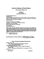
The Hammerhead Motif By comparing sequences of the hammerhead catalytic motif from a variety of sources, and combining it with information from mutagenesis studies, the conserved and important features of the motif have been elucidated; they are shown in Figure 1.1. For a recent detailed review, see ref. 13. It was found to consist of 3 base paired helices, or stems, (I-III), with helices I and III flanking the cleavage site which is on the 3' side of an unpaired, or bulged, nucleotide. Helix II is connected to the other helices by two single-stranded regions that contain most of the conserved nucleotides in the hammerhead motif. The numbering system of Hertel et al14 is useful for descriptive and comparative purposes and is used in Figure 1.1. As shown, of the nucleotides in the single stranded regions which presumably form the hammerhead’s catalytic core, only the U7 position shows variations in nature.15 The sequences of helices I-III show little conservation except at the ends of helix II, where a purine, R10.1, pairs with a pyrimidine Y11.1, and helix III where A15.1 base pairs with U16.1. Hammerhead ribozymes, as they exist in nature, self-cleave and hence work in cis. This is achieved by the RNA folding to form loops at the ends of two of the helices. In vitro, however, it is possible to assemble the catalytically active motif shown in Figure 1.1 in a variety of ways, using either 1, 2 or 3 RNA molecules.16-20 If only one RNA molecule is involved, then two of the helices are joined by loops and cleavage is in cis (self-cleavage). If two RNA molecules are used, then only one loop is required between a pair of helices and cleavage occurs in trans.20 Finally, using 3 RNA molecules there are no loops connecting the ends of the helices and cleavage is also in trans. In the case of two RNA molecules, there are three ways of dividing the hammerhead motif, which have been called I/II, II/III and I/III Ribozymes in the Gene Therapy of Cancer, edited by Kevin J. Scanlon and Mohammed Kashani-Sabet. ©1998 R.G. Landes Company.
4
Ribozymes in the Gene Therapy of Cancer
Fig. 1.1. Diagram of the hammerhead ribozyme. The bond cleaved is indicated by an arrow. N stands for any nucleotide, H is A, C or U, but not G. Y (pyrimidine) is C or U and R (purine) is A or G. Numbering is according to ref. 14. In nature one of the helices is not terminated by a loop. Conserved nucleotides are all those other than N, with the exception of U7. Domain I of the catalytic core is nucleotides C3-A6 and domain II is U7-A9 and G12-A14.
depending on which helices are used to bind to the substrate, or cleaved, strand. The design called I/III, in which the remaining loop terminates helix II, contains almost all the conserved residues in the enzyme strand.17 Hence, the substrate strand has only a minimal sequence requirement (5'-U-H-3'). This design has been widely utilized because it is not only ideal for use in attempts to downregulate gene expression, but also permits relatively easy chemical synthesis of the short enzyme strand of RNA. In this design the substrate strand is commonly referred to as the target RNA which is to be cleaved, and the enzyme strand is generally called the ribozyme.
Mutagenesis and Sequence Requirements In the single stranded regions of the hammerhead ribozyme’s catalytic core, nucleotide substitutions at all positions except U7 destroy catalytic activity and this fact can be utilized to create non-cleaving ribozymes as negative controls, for example, to determine antisense effects. Ruffner et al15 have shown that catalytic rates in vitro are best with U at position 7, but G>A>C, which is only 20% as active. Conserved nucleotides are not present in helix I, but helix II contains two immediately adjacent to the catalytic core. These are most commonly a G-C base pair at R10.1 - Y11.1, and this combination is the most active in vitro. Although only one nucleotide pair is conserved in helix II, its absolute length has an effect on ribozyme activity in vitro.7,15,21,22 If
The Biochemistry of the Hammerhead Ribozyme
5
helix II is shorter than 2 bp, activity is severely reduced. The loop terminating helix II, which is usually 4 nucleotides long, has little apparent influence on activity in vitro and can even be replaced by non-nucleoside linkers23-25 or deoxynucleotides26 without total loss of activity. Helix III contains the conserved A15.1 - U16.1 base pair and the U16.1 is found as part of the cleaved triplet, which is most often 5'-GUC-3', although the triplets 5'-GUA-3' and 5'-AUA-3' are seen as cleavage sites in nature, though rarely.27,28 In vitro analysis of systematic mutations in the GUC sequence in the target have shown that cleavage can occur after any NUH triplet15,29-31 (H means any nucleotide but G), though efficiency is usually much reduced compared to GUC. Shimayama et al30 showed that, in vitro, the efficiency of cleavage at the various triplet combinations depended on the relative concentrations of the ribozyme and substrate. If either were saturating, cleavage depended on kcat and GUC, AUC>GUA, AUA, CUC were much more efficient than other versions of NUH. If ribozyme or substrate was limiting, then GUC>CUC and all other combinations were much less efficient. The extent to which these observations can be applied to cleavage in vivo, particularly of long target RNAs by either chemically modified, exogenously applied ribozymes or ribozymes transcribed in vivo which contain additional sequences, is not clear and at least one report suggests they are not applicable.32
Hammerhead Ribozyme Structure The structure of the hammerhead ribozyme has been studied both in solution32-35 and in crystalline form.36,37 Two X-ray diffraction crystal structures show that the hammerhead ribozyme has a y-shape in which helices II and III stack colinearly with helix I adjacent to helix II (Fig. 1.2). General features of the crystal structures agree with the proposed solution structures. In the colinear stacking of helices II and III, nucleotides in part of the catalytic core, called domain II (consisting of G12, A13, A14 and U7, G8, A9) form non-WatsonCrick base pairs and result in a pseudocontinuous helix. Domain I of the catalytic core (C3, U4, G5, A6) forms a uridine turn motif, allowing the sugar phosphate backbone of helix II (at U7) to turn and form the bottom of helix I (after C3). In trying to account for the hammerhead ribozyme’s catalytic requirement for Mg2+, at least two Mg2+ binding sites have been proposed.37 One of these is thought to be mainly structural and involves a pentahydrated Mg2+, binding mainly to the 5' phosphate of A9 with additional contacts to G8, G10.1 and G12. Another site, in which Mg(H2O)62+ may bind to C3 and C17 as well as other groups in the vicinity of the catalytic pocket, is closer to the cleavage site (3' to C17) and is thought to be involved in the catalytic mechanism by facilitating deprotonation of the 2'-hydroxyl of C17.37,38 Electrophoretic mobility studies39 indicate that there is a Mg2+ threshold above which helix I is adjacent to helix II (as in the crystal and catalytically active form) and below which helix I is adjacent to helix III.
Reaction Mechanism and Kinetics Magnesium ions, or certain other divalent metal ions, are essential for hammerhead ribozyme catalysis. As suggested by the structural studies, the divalent metal ions are required firstly to promote correct folding of the catalytic core, and secondly as a reaction cofactor.40 Zn2+ and Cd2+ can only perform this second, cofactor function and therefore require spermine to help fold the RNA. By studying variation in reaction rates with pH and with pKa values of hydrated metal ions, it was concluded that the metal ion acts as a base in the reaction mechanism and that only one deprotonation event was involved.41 Although some details of the reaction mechanism are still unclear (such as a second metal ion requirement), the overall cleavage reaction is outlined in Figure 1.3. A hydrated Mg2+ acts as a base to attack the essential 2'-OH group of C17, which then acts as a nucleophile attacking the scissile phosphate. After cleavage, C17 has a 2',3' cyclic phosphate and N1.1 has a 5'-OH.
6
Ribozymes in the Gene Therapy of Cancer
Fig. 1.2. Diagram of the hammerhead ribozyme based on the X-ray crystal structure. For explanation of symbols see Fig. 1.1. Note how helix III stacks colinearly with helix II. Reversed-Hoogsteen base pairs form between A9 and G12 and between G8 and A13 and non-Watson Crick base pairs are also present between U7 and A14 as well as between A15.1 and U16.1.
The hammerhead ribozyme catalytic reaction can be fitted to the Michalis-Menten equation as long as: 1. the ribozyme concentration is much less than the substrate concentration; 2. the step R + S R.S is rapid and reversible; and 3. the rate determining step is R.S R.P (where R = ribozyme, S = substrate and P = products).42-45 Under these multiple turnover conditions, the values derived from the Michalis-Menten equation for Km (which is a measure of the ribozyme’s affinity for substrate) are usually around 50-500 nM and for kcat (which is the rate of product production) are about 1 min–1, which is much lower than for protein enzymes (≈500 min–1). These conditions are usually only met in vitro, and for short ribozyme and target molecules, where alternative (probably inactive) conformations for ribozyme and substrate are few or nonexistent.46,47 When these conditions are not met, Km and kcat can often be determined under single turnover conditions where the ribozyme concentration is made to exceed the substrate concentration.48
The Biochemistry of the Hammerhead Ribozyme
7
Fig. 1.3. Proposed reaction mechanism for phosphodiester bond cleavage by the hammerhead ribozyme. A hydrated Mg2+ ion deprotonates the 2' OH of N1 and the 2' oxygen acts as a nucleophile to attack the scissile phosphate. A 2',3' cyclic phosphate is formed on N1.
Specificity Ribozymes bind to target RNAs according to the rules of RNA duplex formation, at a rate of around 5 x 10⁸ M, the association being largely independent of length and sequence.49 The specificity of a ribozyme is its ability to cleave at one particular site and is usually a very important consideration when one intends to cleave only one target RNA species in a complex mixture, in which some RNAs could be similar to the target. Most studies on the specificity of hammerhead ribozymes have been performed in vitro with simple ribozyme and substrate molecules and in these conditions specificity is determined by the rate of cleavage compared to substrate dissociation.50 Higher specificity will be achieved with a slower cleavage step or an increased rate of substrate dissociation.
8
Ribozymes in the Gene Therapy of Cancer
Hertel et al examined the effects of shortening helices I and III, combined with introducing single mismatches in helix III, and concluded that the total target recognition length (of helices I and III) could be 12 nucleotides without a reduction in specificity.51 These experimental conditions differ from those expected in vivo, and hence it is likely that high specificity can be retained in vivo with duplex regions longer than 12 bp, especially if the number of G-C base pairs is not excessive. Mismatches close to the cleavage site can also be used to give high specificity such as when targeting a mutant oncogene transcript which may differ by only one nucleotide from the wild type mRNA.52
Modified Hammerhead Ribozymes A considerable variety of modifications to the basic, all-RNA type I/III hammerhead ribozyme have been carried out, mostly with the intention of improving the efficiency of target cleavage by the ribozyme as a way of developing these molecules as therapeutic compounds for exogenous delivery. Some modifications, such as the asymmetric hammerhead ribozyme design, can also be applied when ribozymes are to be transcribed within cells from gene constructs. Chemical modifications of the hammerhead ribozyme that have been tried include replacement of some of the 2'-OH moieties with allyl, amino, deoxy, fluoro or O-methyl groups.53-55 In addition, the modification of parts of the backbone of the ribozyme from phosphates to phosphorothioates has the dramatic effect of increasing the resistance of the ribozyme to the action of cellular and serum nucleases.56 The key nucleases are pyrimidinespecific endonucleases, which will attack C3, U4 and U7 of the unmodified core, as well as 3'-exonucleases. Most of these chemical modifications do not significantly increase the catalytic activity; in fact reductions are more common. However, significant increases in stability and/or useful pharmacological properties outweigh these drawbacks and make modified hammerhead ribozymes an exciting class of potential therapeutic reagents. The positions U4 and U7 have been modified and stabilized to pyrimidine-specific endonucleases by both 2'-C-allyl and 2'-amino nucleotides without loss of catalytic activity,54,56 and there is one report of improved catalytic activity by replacing U7 with pyrimidine-4-one.57 Some very encouraging results have been obtained by direct injection of such chemically protected ribozymes into mouse jaws58 and rabbit knee joints59 in which specific target mRNA levels were transiently reduced or eliminated. Both chemically modified and unmodified ribozymes have also been delivered to cells encapsulated in cationic liposomes60-62 and have resulted in the desired phenotypic changes, such as restoration of drug sensitivity,63 cell proliferation60 and secretion of TNFα.64 Liposomes do have some drawbacks and other delivery systems are being evaluated.65 Modifications to reduce helix II and its connecting loop have established a minimum version of the hammerhead ribozyme called a minizyme which has no base pairs in helix II, such that A9 is connected to G12 only by a loop that can be DNA or RNA.26,66 When helix II still retains 1 bp the ribozyme is called a miniribozyme. These kinds of modifications can reduce the costs of chemical synthesis, but generally yield ribozymes with decreased chemical cleavage rates as measured using short targets in vitro. However, against long target RNAs in vivo, this may not be a problem, as cleavage is not likely to be rate limiting in this situation.66,67 The introduction of DNA, or DNA combined with some of the other modifications listed above, into helices I and III, but not helix II, can not only make the ribozyme more nuclease resistant, but may also stimulate the overall reaction several fold. This may work by enhancing either the cleavage rate or the turnover rate.26 Asymmetric hammerhead ribozymes are a modification of the basic hammerhead ribozyme in which either helix I or helix III is significantly longer. Where the long helix is
The Biochemistry of the Hammerhead Ribozyme
9
greater than about 30 residues, these molecules are best produced by transcription. Shorter types can be made by either chemical synthesis or transcription. In vitro experiments with relatively short substrates have shown that asymmetric hammerhead ribozymes can cleave their target when helix I is as short as 2 or 3 bp.51 Interestingly, by varying helix I and III lengths, it has also been observed that, in vitro, longer helix III ribozyme constructs could cleave one to two orders of magnitude more rapidly than longer helix I constructs with the same total base pairing capacity.48 This would be an important design feature if generally true and applicable in vivo. An additional form of modification of the hammerhead ribozyme that may be generally beneficial in vivo has been reported by Sioud and Jespersen.68 A region of a TNFα ribozyme that is bound by the enzyme GAPDH in cells can be linked to other ribozymes and causes their catalytic activity to be increased in vitro and in vivo.
Pharmacokinetic and Cellular Uptake Studies For chemically modified hammerhead ribozymes to be used as therapeutic compounds, it is important to study their pharmacokinetic properties and the first comprehensive study has recently appeared.69 A symmetric 38-mer ribozyme against a cytochrome P450 containing 2'-O-allyl ribonucleotides at all positions outside the catalytic core and at positions C3, U4 and U7 was administered as a single intravenous injection of 0.25 mg into adult male rats. This ribozyme binds to serum albumin at binding site I, as do linear phosphorothioate oligonucleotides. The plasma elimination half life, at 6.5 hours, was shorter than that of these more stable phosphorothioates, but intact ribozyme was still detectable after 48 hours. Metabolism is via endonuclease attack on the internal residues that are not 2'-O-allyl protected, and a 27-mer metabolite was found to accumulate in brain tissue, which does not happen with phosphorothioates. The main organs of accumulation were kidney and liver. Renal excretion of the ribozyme was minor compared to phosphorothioates, perhaps due to high reabsorption in the proximal tubule. As no tissue toxicity was observed, the 2'-O-allyl modified ribozyme seemed promising as a therapeutic. The cellular uptake properties of a 2'-O-methyl modified hammerhead ribozyme containing 2'-amino groups on residues U4 and U7 have recently been examined.70 This symmetric ribozyme against EGFR had 7 bp arms and was taken up by glioma cells in a temperature, energy and pH dependent fashion which could be competed with a variety of other oligonucleotides and polyanions. The ribozyme had a punctate, extranuclear localization. Overall, ribozyme uptake seemed to be via a similar endocytic mechanism to that for other oligodeoxynucleotides and also showed some cell-type specific differences.70,71
Making Hammerhead Ribozymes Work In Vivo There are two distinct parts to this problem. The first involves selection of a suitable and efficient site for cleavage in the target molecule, which is usually an mRNA molecule of several hundred nucleotides in length. The second problem involves the optimal design of the ribozyme. A significant part of this second problem concerns how the ribozyme will be administered, i.e., will it be transcribed from a gene that is introduced into cells (endogenous expression), or will the ribozyme be synthesized and administered as a drug (exogenously delivered)? The engineering of hammerhead ribozymes for exogenous delivery has recently been reviewed55 and the mechanics of delivery are considered in chapter 4 of this text and hence will not be further discussed. As chapters 5-8 cover various aspects of expressing ribozymes endogenously, only general points regarding hammerhead ribozyme design for endogenous expression will be made here.
10
Ribozymes in the Gene Therapy of Cancer
Target Site Selection Naturally occurring RNA molecules that may be chosen for cleavage by hammerhead ribozymes are sufficiently long that they can fold into complex secondary and tertiary structures. This means that not all of the 5'-UH-3' sequences along the target RNA will be equally accessible to ribozyme attack. Initial attempts to predict accessible 5'-UH-3' sites using computer programs72-74 did demonstrate some successes, but this method does not guarantee that any particular 5'-UH-3' sequence would be efficiently cleavable in vitro, let alone in vivo. A more empirical method of selecting accessible target sites is to determine experimentally which parts of a target RNA molecule are able to bind short antisense oligodeoxynucleotides and thus be sites of nuclease (RNase H) sensitivity.60,75,76 Usually these sites at which oligodeoxynucleotides can anneal are also accessible to ribozymes, at least in vitro. Sites chosen in this way might well be cleavable in vivo, but may not be the best possible sites for in vivo cleavage. A method which permits selection of efficient ribozyme cleavage sites from a large number of possibilities in an in vivo system would clearly be superior. Lieber and Strauss77 have developed one such system in which a library of ribozymes, containing a constant ribozyme core sequence but random hybridizing arm sequences, is mixed with target RNA in cellular extracts. The various ribozymes will cleave the target RNA in different positions and to different degrees, but the most efficient will produce cleavage products that are the most abundant. The cleavage products are cloned by making cDNA and using PCR, and the sequences of the cleavage sites determined by sequencing the clones. With this knowledge of the cleavage site, efficient ribozymes can be designed that work well in vivo.78 Some other in vivo methods of selecting efficient ribozyme target sites are being developed, but as yet do not permit exhaustive screening of all possible sites. Luciferase has been used as a reporter in which target sequences have been linked upstream of the luciferase gene. Ribozymes introduced, or co-expressed, in cells expressing the construct should cause a reduction in luciferase activity if they are active.32,79 An E. coli based system utilizing positive selection with trimethoprim for hammerhead ribozymes cleaving in cis80 and a yeast system that requires cell cycle arrest81 may also be worth considering if they are further developed.
Hammerhead Ribozyme Design for In Vivo Expression For hammerhead ribozymes expressed in vivo, there seems little point in changing the length of helix II and its terminating loop as, in vivo, extensions to helix II seem to inhibit activity.82 Maintaining the standard catalytic core, except when creating an inactive ribozyme control, is also prudent. Altering the length of the arms that anneal to the target RNA is an important factor, but it is not yet clear what the best design is for in vivo activity. Conflicting reports exist in the literature, some suggesting that short arms of 6-8 nucleotides are more efficient77,82,83 and others that hybridizing arms need to be in excess of 25 nt.84,85 These differences in efficiency are most likely due to differences in the accessibility of the various target sites used, but differences in the sequences adjacent to the ribozyme, as well as the cell system and cellular compartment in which cleavage was being tested, will also play a part. A recent study looking at both subcellular localization and the length of helix III of asymmetric hammerhead ribozymes concluded that shorter ribozymes were superior when injected into the cytoplasm, whereas the helix III region needed to be greater than 51 nt to be effective when injected into the nucleus.86 Clearly, efforts should be made to express ribozymes in a way that will ensure an appropriate subcellular localization for the target in question.
The Biochemistry of the Hammerhead Ribozyme
11
Conclusion Since their discovery less than 15 years ago, hammerhead ribozymes have become the most studied type of catalytic RNA. Armed with this accruing knowledge, which includes the recent crystal structure and the wealth of information on mutagenesis and chemical modification, researchers in the field of medicine have engineered hammerhead ribozymes which are now poised to become essential therapeutic agents.87 They hold promise for exogenous administration in a variety of disorders, and will be invaluable tools for gene therapists in their efforts to combat genetic disease and cancer, as is described in Section III.
References 1. Kruger K, Grabowski PJ, Zuang AJ et al. Self-splicing RNA: Autoexcision and autocyclization of the ribosomal RNA intervening sequence of Tetrahymena. Cell 1982; 31:147-157. 2. Cech TR. Self-splicing of group I introns. Annu Rev Biochem 1990; 59:543-568. 3. Guerrier-Takada C, Gardiner K, Marsh T et al. The RNA moiety of ribonuclease P is the catalytic subunit of the enzyme. Cell 1983; 35:849-857. 4. Altman S. Ribonuclease P. J Biol Chem 1990; 265:20053-20056. 5. Buzayan JM, Gerlach WL, Bruening G. Non-enzymic cleavage and ligation of RNAs complementary to a plant virus satellite RNA. Nature 1986; 323:349-353. 6. Forster AC, Symons RH. Self-cleavage of plus and minus RNAs of a virusoid and a structural model for the active sites. Cell 1987; 49:211-220. 7. Symons RH. Small catalytic RNAs. Annu Rev Biochem 1992; 61:641-671. 8. Hutchins CJ, Rathjen PD, Forster AC et al. Self-cleavage of plus and minus RNA transcripts of avocado sunblotch viroid. Nucleic Acids Res 1986; 14:3627-3640. 9. Prody GA, Bakos JT, Buzayan JM et al. Autolytic processing of dimeric plant virus satellite RNA. Science 1986; 231:1577-1580. 10. Haseloff J, Gerlach WL. Sequences required for self-catalyzed cleavage of the satellite RNA of tobacco ringspot virus. Gene 1989; 82:43-52. 11. Hampel A, Tritz R. RNA catalytic properties of the minimum (-)sTRSV sequence. Biochemistry 1989; 28:4929-4933. 12. Wu H-N, Lin Y-J, Lin F-P et al. Human hepatitis delta virus RNA subfragments contain an autocleavage activity. Proc Natl Acad Sci USA 1989; 86:1831-1835. 13. Birikh KR, Heaton PA, Eckstein F. The structure, function and application of the hammerhead ribozyme. Eur J Biochem 1997; 245:1-16. 14. Hertel KJ, Pardi A, Uhlenbeck OC et al. Numbering system for the hammerhead ribozyme. Nucleic Acids Res 1992; 20:3252. 15. Ruffner DE, Stormo GD, Uhlenbeck OC. Sequence requirements of the hammerhead RNA self-cleavage reaction. Biochemistry 1990; 29:10695-10702. 16. Uhlenbeck OC. A small catalytic oligoribonucleotide. Nature 1987; 328:596-600. 17. Haseloff J, Gerlach WL. Simple RNA enzymes with new and highly specific endoribonuclease activity. Nature 1988; 334:585-591. 18. Jeffries AC, Symons RH. A catalytic 13-mer ribozyme. Nucleic Acids Res 1989; 17:1371-1377. 19. Odai O, Hiroaki H, Tanaka T et al. Properties of a hammerhead-type RNA enzyme system that consists of 3 RNA oligomer strands. Nucleosides and Nucleotides 1994; 13:1569-1579. 20. Clouet-D’Orval B, Uhlenbeck OC. Kinetic characterisation of I/II format hammerhead ribozymes. RNA 1996; 2:483-491. 21. Tuschl T, Eckstein F. Hammerhead ribozymes: Importance of stem-loop II for activity. Proc Natl Acad Sci USA 1993; 90: 6991-6994. 22. Nakamaye KL, Eckstein F. AUA-Cleaving hammerhead ribozymes: Attempted selection for improved cleavage. Biochemistry 1994; 33:1271-1277. 23. Benseler F, Fu D-J, Ludwig J et al. Hammerhead-like molecules containing non-nucleoside linkers are active RNA catalyzts. J Am Chem Soc 1993; 115:8483-8484. 24. Thomson JB, Tuschl T, Eckstein F. Activity of hammerhead ribozyme containing nonnucleotidic linkers. Nucleic Acids Res 1993; 21:5600-5603.
12
Ribozymes in the Gene Therapy of Cancer
25. Fu D-J, Benseler F, McLaughlin LW. Hammerhead ribozymes containing non-nucleoside linkers are active RNA catalysts. J Am Chem Soc 1994; 116:4591-4598. 26. Hendry P, McCall MJ, Santiago FS et al. In vitro activity of minimised hammerhead ribozymes. Nucleic Acids Res 1995; 23:3922-3927. 27. Keese P, Bruening G, Symons RH. Comparative sequence and structure of circular RNAs from two isolates of lucerne transient streak virus. FEBS Lett 1983; 159:185-190. 28. Miller WA, Hercus T, Waterhouse PM et al. A satellite RNA of barley yellow dwarf virus contains a novel hammerhead structure in the self-cleaving domain. Virology 1991; 183:711-720. 29. Perriman R, Delves A, Gerlach WL. Extended target-site specificity for a hammerhead ribozyme. Gene 1992; 113:157-163. 30. Shimayama T, Nishikawa S, Taira K. Generality of the NUX rule—kinetic analysis of the results of systematic mutations in the trinucleotide at the cleavage site of hammerhead ribozymes. Biochemistry 1995; 34:3649-3654. 31. Zoumadakis M, Tabler M. Comparative analysis of cleavage rates after systematic permutation of the NUX’ consensus target motif for hammerhead ribozymes. Nucleic Acids Res 1995; 23:1192-1196. 32. Kawasaki H, Ohkawa J, Tanishige N et al. Selection of the best target site for ribozymemediated cleavage within a fusion gene for adenovirus E1A-associated 300 kDA protein (p300) and luciferase. Nucleic Acids Res 1996; 24:3010-3016. 33. Amiri KMA, Hagerman PJ. Global conformation of a self-cleaving hammerhead RNA. Biochemistry 1994; 33:13172-13177. 34. Tuschl T, Gohlke C, Jovin TM et al. A three-dimensional model for the hammerhead ribozyme based on fluorescence measurements. Science 1994; 266:785-789. 35. Bassi GS, Mollegaard NE, Murchie AIH et al. Ionic interactions and the global conformations of the hammerhead ribozyme. Nat Struct Biol 1995; 2:45-55. 36. Pley HW, Flaherty KM, McKay DB. Three-dimensional structure of a hammerhead ribozyme. Nature 1994; 372:68-74. 37. Scott WG, Finch JT, Klug A. The crystal structure of an all-RNA hammerhead ribozyme— a proposed mechanism for RNA catalytic cleavage. Cell 1995; 81:991-1002. 38. Murray JB, Adams CJ, Arnold JRP et al. The roles of the conserved pyrimidine bases in hammerhead ribozyme catalysis—evidence for a magnesium ion binding site. Biochem J 1995; 311:487-494. 39. Bassi GS, Murchie AIH, Lilley DMJ. The ion-induced folding of the hammerhead ribozyme: Core sequence changes that perturb folding into the inactive conformation. RNA 1996; 2:756-768. 40. Dahm SC, Uhlenbeck OC. Role of divalent metal ions in the hammerhead ribozyme cleavage reaction. Biochemistry 1991; 30:9464-9469. 41. Dahm SC, Derrick WB, Uhlenbeck OC. Evidence for the role of solvated metal hydroxide in the hammerhead cleavage mechanism. Biochemistry 1993; 32:13040-13045. 42. McConnell T. Theoretical considerations in measuring reaction parameters. In: Turner PC, ed. Ribozyme Protocols. Totowa: Humana Press Inc., 1997:187-198. 43. McConnell T. Experimental approaches for measuring reaction parameters. In: Turner PC, ed. Ribozyme Protocols. Totowa: Humana Press Inc., 1997:199-208. 44. DeYoung MB, Siwkowski A, Hampel A. Determination of catalytic parameters for hairpin ribozymes. In: Turner PC, ed. Ribozyme Protocols. Totowa: Humana Press Inc., 1997:209-220. 45. Hendry P, McCall MJ, Lockett TJ. Characterising ribozyme cleavage reactions. In: Turner PC, ed. Ribozyme Protocols. Totowa: Humana Press Inc., 1997:221-229. 46. Fedor MJ, Uhlenbeck OC. Kinetics of intermolecular cleavage by hammerhead ribozymes. Biochemistry 1992; 31:12042-12054. 47. Hertel KJ, Herschlag D, Uhlenbeck OC. A kinetic and thermodynamic framework for the hammerhead ribozyme reaction. Biochemistry 1994; 33:3374-3385. 48. Hendry P, McCall MJ. Unexpected anisotropy in substrate cleavage rates by asymmetric hammerhead ribozymes. Nucleic Acids Res 1996; 24:2679-2684.
The Biochemistry of the Hammerhead Ribozyme
13
49. Nelson JW, Tinoco I Jr. Comparison of the kinetics of ribo-oligonucleotide, deoxyribooligonucleotide and hybrid oligonucleotide double-strand formation by temperature-jump kinetics. Biochemistry 1982; 21:5289-5295. 50. Herschlag D. Implications of ribozyme kinetics for targeting the cleavage of specific RNA molecules in vivo: More isn’t always better. Proc Natl Acad Sci USA 1991; 88:6921-6925. 51. Hertel KJ, Herschlag D, Uhlenbeck OC. Specificity of hammerhead ribozyme cleavage. EMBO J 1996; 15:3751-3757. 52. Funato T, Shitara T, Tone T et al. Suppression of H-ras-mediated transformation in NIH3T3 cells by a ras ribozyme. Biochem Pharmacol 1994; 48:1471-1475. 53. Pieken WA, Olsen DB, Benseler F et al. Kinetic characterisation of ribonuclease-resistant 2'-modified hammerhead ribozymes. Science 1991; 253:314-317. 54. Heidenreich O, Benseler F, Fahrenholz A et al. High activity and stability of hammerhead ribozymes containing 2'-modified pyrimidine nucleosides and phosphorothioates. J Biol Chem 1994; 269:2131-2138. 55. Usman N, Beigelman L, McSwiggen JA. Hammerhead ribozyme engineering. Curr Opin Struct Biol 1996; 1:527-533. 56. Beigelman L, McSwiggen JA, Draper KG et al. Chemical modification of hammerhead ribozymes: Catalytic activity and nuclease resistance. J Biol Chem 1995; 270:25702-25708. 57. Burgin AB, Gonzalez C, Matulic-Adamic J et al. Chemically modified hammerhead ribozymes with improved catalytic rates. Biochemistry 1996; 35:14090-14097. 58. Lyngstdaas SP, Risnes S, Sproat BS et al. A synthetic, chemically modified ribozyme eliminates amelogenin, the major translation product in developing mouse enamel in vivo. EMBO J 1995; 14:5224-5229. 59. Flory CM, Pavco PA, Jarvis TC et al. Nuclease-resistant ribozymes decrease stromelysin mRNA levels in rabbit synovium following exogenous delivery to the knee joint. Proc Natl Acad Sci USA 1996; 93:754-758. 60. Jarvis TC, Alby JA, Beaudry AA et al. Inhibition of vascular smooth muscle cell proliferation by ribozymes that cleave c-myb mRNA. RNA 1996; 2:419-428. 61. Castanotto D, Bertrand E, Rossi JJ. Exogenous cellular delivery of ribozymes and ribozyme encoding DNAs. In: Turner PC, ed. Ribozyme Protocols. Totowa: Humana Press Inc., 1997:429-439. 62. Brown SA, Jarvis TC. Optimization of lipid-mediated ribozyme delivery to cells in culture. In: Turner PC, ed. Ribozyme Protocols. Totowa: Humana Press Inc., 1997:441-449. 63. Kiehntopf M, Brach MA, Licht T et al. Ribozyme-mediated cleavage of the MDR-1 transcript restores chemosensitivity in previously resistant cancer cells. EMBO J 1994; 13:4645-4652. 64. Kisich KO, Stecha PF, Harter HA et al. Inhibition of TNF-alpha secretion by murine macrophages following in vivo and in vitro ribozyme treatment. J Cell Biochem 1995; 19A:221. 65. Leopold HL, Shore SK, Newkirk TA et al. Multiunit ribozyme-mediated cleavage of bcrabl mRNA in myeloid leukemias. Blood 1995; 85:2162-2170. 66. McCall MJ, Hendry P, Lockett TJ. Minimized hammerhead ribozymes. In: Turner PC, ed. Ribozyme Protocols. Totowa: Humana Press Inc., 1997:151-159. 67. Sioud M. Effects of variations in length of hammerhead ribozyme antisense arms upon the cleavage of longer RNA substrates. Nucleic Acids Res 1997; 25:333-338. 68. Sioud M, Jespersen L. Enhancement of hammerhead ribozyme catalysis by glyceraldehyde3-phosphate dehydrogenase. J Mol Biol 1996; 257:775-789. 69. Desjardins JP, Sproat BS, Beijer B et al. Pharmacokinetics of a synthetic, chemically-modified hammerhead ribozyme against the rat cytochrome-P-450 3A2 messenger-RNA after single intravenous injections. J Pharmacol Exp Ther 1996; 278:1419-1427. 70. Fell PL, Hudson AJ, Reynolds MA. Cellular uptake properties of a 2'-amino/2'-O-methylmodified chimeric hammerhead ribozyme targeted to the epidermal growth factor receptor mRNA. Antisense and Nucleic Acid Drug Development 1997; 7:319-326. 71. Hawley P, Gibson I. Interaction of oligodeoxynucleotides with mammalian cells. Antisense and Nucleic Acid Drug Development 1996; 6:185-195.
14
Ribozymes in the Gene Therapy of Cancer
72. Christoffersen RE, McSwiggen J, Konings D. Application of computational technologies to ribozyme biotechnology products. J Mol Struct 1994; 311:273-284. 73. Sczakiel G, Tabler M. Computer-aided calculation of the local folding potential of target RNA and its use for ribozyme design. In: Turner PC, ed. Ribozyme Protocols. Totowa: Humana Press Inc., 1997:11-15. 74. James W, Cowe E. Computational approaches to the identification of ribozyme target sites. In: Turner PC, ed. Ribozyme Protocols. Totowa: Humana Press Inc., 1997:17-26. 75. Frank BL, Goodchild J. Selection of accessible sites for ribozymes on large RNA transcripts. In: Turner PC, ed. Ribozyme Protocols. Totowa: Humana Press Inc., 1997:37-43. 76. Birikh KR, Berlin YA, Soreq H et al. Probing accessible sites for ribozymes on human acetylcholinesterase RNA. 1997; 3:429-437. 77. Lieber A, Strauss M. Selection of efficient cleavage sites in target RNAs by using a ribozyme expression library. Mol Cell Biol 1995; 15:540-551. 78. Lieber A, Kay MA. Adenovirus mediated expression of ribozymes in mice. J Virol 1996; 70:3153-3158. 79. Scherr M, Grez R, Ganser A. Specific hammerhead ribozyme-mediated cleavage of mutant N-ras mRNA in vitro and ex vivo—oligoribonucleotides as therapeutic agents. J Biol Chem 1997; 272:14304-14313. 80. Fujita S, Koguma T, Ohkawa J. Discrimination of a single base change in a ribozyme using the gene for dihydrofolate reductase as a selective marker in Escherichia coli. Proc Natl Acad Sci USA 1997; 94:391-396. 81. Ferbeyre G, Bratty J, Chen H et al. Cell-cycle arrest promotes trans-hammerhead ribozyme action in yeast. J Biol Chem 1996; 271:19318-19323. 82. Homann M, Tabler M, Tzortzakaki S et al. Extension of helix II of an HIV-1-directed hammerhead ribozyme with long antisense flanks does not alter kinetic parameters in vitro but causes loss of inhibitory potential in living cells. Nucleic Acids Res 1994; 22:3951-3957. 83. Bertrand EL, Rossi JJ. Facilitation of hammerhead ribozyme catalysis by the nucleocapsid protein of HIV-1 and the heterogeneous nuclear ribonucleoprotein A1. EMBO J 1994: 13:2904-2912. 84. Crissell P, Thompson S, James W. Inhibition of HIV-1 replication by ribozymes that show poor catalytic activity in vitro. Nucleic Acids Res 1993; 21:5251-5255. 85. Beck J, Nassal M. Efficient hammerhead ribozyme-mediated cleavage of the structured hepatitis B virus encapsidation signal in vitro and in cell extracts, but not in intact cells. Nucleic Acids Res 1995; 23:4954-4962. 86. Hormes R, Homann M, Oelze I et al. The subcellular localization and length of hammerhead ribozymes determine efficacy in human cells. Nucleic Acids Res 1997; 25:769-775. 87. James HA, Gibson I. The therapeutic potential of ribozymes. Blood 1998; 91:371-382.
CHAPTER 2
Biochemistry of the Hairpin Ribozyme Andrew Siwkowski and Arnold Hampel
Introduction
T
he observation that the 359 nt negative strand of the satellite RNA of tobacco ringspot virus [(-)sTRSV] was autocatalytic1 led to identification of the minimal catalytic center consisting of a 50 nt enzyme-like RNA and a 14 nt substrate.2 This structure was named the hairpin ribozyme.3 The ribozyme/substrate consisted of 4 helices, helices 1, 2, 3, 4, and five loops, loops 1, 2, 3, 4, and 5 (Fig. 2.1). Helix 1, between the ribozyme and substrate, can vary in length and have a variable sequence as long as base pairing is maintained. Helix 2, also between the ribozyme and substrate is fixed at 4 bp; however it also can vary in sequence as long as base pairing is maintained. Helix 3 is a 4 bp helix found in the ribozyme separated from helix 2 by a single unpaired A15, which serves as a hinge.4-6 Helix 4, in the native sequence, contains three Watson-Crick base pairs and one non-canonical A:G base pair.7 Thus a total of 18 bp exist in the two-dimensional structure of the hairpin ribozyme. The helices are separated by five single stranded loop regions. Loop 5 is dispensable and can be replaced by other structures as long as a strong helix 4 is maintained. Loops 1, 2, 4, and 5, however, contain required bases. Thus the hairpin ribozyme consists of essentially two domains—domain I (helices 1 and 2; loops 1 and 5) and domain II (helices 3 and 4 and loops 2 and 4).8 Cleavage takes place in the substrate at loop 5 by breakage of the phosphodiester bond at ApG to produce a 5' cleavage fragment with a 2',3'-cyclic phosphate terminus and 3' cleavage fragment with a 5'-OH terminus. The hairpin ribozyme can also produce a ligated product.9 The ligation reaction uses the same termini as produced with cleavage, and thus appears to be a simple reversal of the forward (cleavage) reaction.10
Biochemistry and Mechanism of the Reaction The (-)sTRSV ribozyme supports multiple cleavage events, has a temperature optimum of 37°C, and an energy of activation of 19 kcal/mol.2 Recent reports of kinetic parameters for the cleavage event, determined by several groups,11-14 fall within a range of 19-96 nM for Km and 0.12-0.36/min for kcat. Substrate dissociation rates are so much slower than cleavage rates that virtually every substrate that binds is cleaved, and the rate of ligation was found to be 10X faster than cleavage—indicating that the hairpin ribozyme is truly a unique catalytic system among known catalytic RNAs.12 The hairpin ribozyme catalyzes cleavage of substrate containing an Rp phosphorothioate substitution at ApG of the cleavage site.15 The cleavage reaction proceeds with inversion of configuration of the phosphorus in the product with respect to that of the substrate, suggesting an in-line attack mechanism.16 A low sulfur effect was associated with catalysis at an Ribozymes in the Gene Therapy of Cancer, edited by Kevin J. Scanlon and Mohammed Kashani-Sabet. ©1998 R.G. Landes Company.
16
Ribozymes in the Gene Therapy of Cancer
Fig. 2.1. The hairpin ribozyme/substrate complex. The hairpin ribozyme complexed with its substrate forms a structure with four helical regions interspersed by five loop regions. These exist as two domains, domain I and domain II.
Rp phosphorothioate substitution at the cleavage site.17,18 Efficient cleavage of the Sp phosphorothioate isomer in the presence of Mg2+ suggests that it too is not directly coordinated with a metal cofactor during cleavage;18 however, outer sphere coordination with either Rp or Sp phosphate oxygen is still possible. The classic model of RNA catalysis is supplied by bovine pancreatic RNase. The numerous similarities between RNA catalysis by this RNase and the hairpin ribozyme suggest a common basic mechanism. This protein cleaves RNA in a sequence specific manner using an in-line attack mechanism.19 The 2'-OH is deprotonated by His-12, and then is available for nucleophilic attack on the phosphorus. The resulting trigonal bipyramid contains both attacking and leaving groups at axial positions. The cleavage results in products with 5'-OH (leaving group) and 2',3'-cyclic phosphate termini. In the case of RNase, the trigonal bipyramidal intermediate is thought to be stabilized by interactions between Lys-41 and a non-bridging phosphate oxygen.20 This particular role may be filled in the hairpin ribozyme by a hexahydrated Mg2+ or Co(NH3)63+ complex.18 Hexahydrated Mg2+ has been shown to coordinate to phosphate oxygens in yeast tRNAPhe,21 and cobalt hexaammine has been shown to coordinate to a non-bridging phosphate oxygen in the stabilization of a Z-DNA structure.22 The fact that the 2'-OH of A10 is directly across from the cleavage site and that it is
Biochemistry of the Hairpin Ribozyme
17 Fig. 2.2. Mechanism of catalysis by the hairpin ribozyme. Direct involvement of metals in the catalytic step is not the case. Rather, functional groups on the ribozyme/substrate complex itself are likely to participate in the catalytic step.
Table 2.1. Unimolecular rate constants and pKa values for cleavage of the hairpin ribozmyme Metal
k (min–1)
pKa
Mg2+ Ca2+ Sr2+ Ba2+
1.8 ± 0.2 3.6 ± 0.7 2.2 ± 0.4 2.2 ± 0.7
11.4 12.8 13.3 13.5
critical to catalysis, is particularly significant in light of this possible role for the metal. Given the fact that As5 can accommodate any base identity and retain substantial catalytic activity,23 it seems likely that this base is oriented outside of the base stacking system of helix 2. The cleavage site could therefore be positioned closer to nucleotide A10, facilitating the formation of a binding pocket for a hydrated metal. The pocket would consist of outer sphere coordination sites, including the 2'-OH of A10, the cleavage site non-bridging phosphate oxygen, and possibly N3 of A10. Additional coordination sites are likely to be supplied by functional groups in loops 2 and 4 in the folded ribozyme-substrate structure (Fig. 2.2). Since deprotonation of the 2'-OH in the case of RNase A is carried out by a histidine, and obviously no such histidine exists in the ribozyme, the question arises: What deprotonates the 2'-OH in the hairpin ribozyme? In the hammerhead, it has been suggested that deprotonation is mediated by a partially hydrated metal cofactor in the reaction. This same hypothesis is not supported in the hairpin ribozyme, since a variety of metals support high cleavage rates regardless of the differing pKa values of their hydrated complexes (Table 2.1). The answer to this question is even more difficult to ascertain given the fact that the pH optimum of the reaction has not been reached. Consequently, no significant pKa has been determined. The deprotonation may occur by action of an RNA functional group or a solvent molecule whose pKa has been perturbed by the molecular environment created by folding of the hairpin ribozyme. It appears that at least two metal binding sites are used for cleavage by the hairpin ribozyme in its active structure. Evidence suggesting this is as follows: 1. two cofactors support cleavage when either one alone could not;24 and 2. the sigmoidal shape of the curve showing cleavage reaction rate as a function of Mg2+ concentration, indicative of multiple binding sites (see ref. 25 and Siwkowski, unpublished data). To date, however, it has not been definitively determined exactly how many binding sites exist. Generally, metals have been proposed to serve two different roles in ribozyme-mediated catalysis; structural and catalytic.26,27 The role the metal plays in catalysis by the hairpin ribozyme has been suggested to be structural rather than catalytic. The finding that
18
Ribozymes in the Gene Therapy of Cancer
hexaammine cobalt chloride supports cleavage by the hairpin ribozyme in the absence of other metals suggests that the metal normally carries out its role as a fully hydrated complex, thereby working through outer sphere interactions with its coordinated waters rather than through an inner sphere mechanism.18 Outer sphere coordination between Mg2+ and a phosphodiester is strongly favored over inner sphere complex formation from a thermodynamic standpoint.28 The Co(NH3)63+ complex stabilizes an RNA helix junction structure and, albeit to a lesser degree than Mg2+, tertiary structure.29 Given the high pKa of the coordinated amine groups with hexaammine cobalt chloride, as well as the slow ligand exchange rate, it seems unlikely that the complex can promote the deprotonation of the 2'-OH, which is likely to be the first step of the cleavage reaction pathway. These findings point to the metal serving a structural role in the formation of the transition state structure; however, a possible catalytic role, wherein the metal coordinates to a phosphate oxygen to prepare the phosphorus for nucleophilic attack, cannot be entirely excluded. Using a cis-cleaving ribozyme (Fig. 2.3b), kinetic cleavage rate constants supported by Mg2+, Ca2+, Sr2+, and Ba2+ were all very similar (Table 2.1). These results show rate of cleavage is independent of the ionic radius or coordination number of the metal cofactor. Furthermore, the cleavage rate constant is not dependent on the pKa of the hydrated metal. While Mn2+ supports cleavage, Co2+ does not, nor does Li+, Na+, K+, or Cs+. This pattern is very similar to that reported for a class of proposed structural sites in the Tetrahymena ribozyme,27 further supporting the concept that the metal serves to stabilize structure in the hairpin ribozyme. The importance of inter-domain interactions for catalysis is particularly clear when reviewing the evidence obtained from several groups—all showing that catalytic rate is dependent on the distance between the 5' end of the substrate and the 3' end of the ribozyme. A series of linkers, each consisting of a different number of bases, when joining the 3' end of the ribozyme to the 5' end of the substrate (Fig. 2.3c), corresponded directly to increased levels of circularization (i.e., ligation) with increasing linker length.4 When 1,3-propanediol phosphate units for nucleotide residues were used as linker units, similar results were obtained.6 When the method was revised to retain the linker between the 3' end of the ribozyme and the 5' end of the substrate and the removal of the bond between ribozyme positions A15 and C16, along with the addition of a single additional base 5' to C16 (Fig. 2.3d), the results were consistent wherein catalysis was dependent on linker length.14 A hairpin ribozyme was constructed in which the two domains were attached in the opposite manner as that found in the native structure, where the two domains are joined by formation of a new helix containing variable-length linkers between the 5' end of the ribozyme and the 3' end of the cytidine immediately preceding loop 3 (Fig. 2.3e).8 In this particular construct, the substrate region and ribozyme region containing loop 2 are separate RNAs, so that the final cleavage reaction is trimolecular. The same pattern associating increasing linker length with increasing cleavage rate was observed. The domains can be totally separated (Fig. 2.3f) and, when the reaction has high concentrations of the RNA comprising the domains, cleavage rates were obtained which were similar to those of the standard bimolecular reaction between hairpin ribozyme and substrate (Fig. 2.3a).30 Using a slightly different construct (Fig. 2.3g), this ability to obtain cleavage activity when physically separated domains were combined was again demonstrated.31 When linkers of varying lengths were inserted between A14 and A15, a different trend from the previous studies was observed. With the exception of an increase in activity accompanying the insertion of a single nucleotide, the cleavage levels associated with remaining linker lengths followed the basic trend of increasing linker length causing lower cleavage activity. This result suggested that close restraint of one domain to the other facilitated cleavage rather than decreasing it as was seen in the previous studies.
Biochemistry of the Hairpin Ribozyme
19
Fig. 2.3. Forms of the hairpin ribozyme used for structure/function studies. (a) conventional form for bimolecular trans reactions;5 (b) cis-cleaving form with 3' end of substrate linked to 5' end of ribozyme;7 (c) 5' end of substrate linked to 3' end of ribozyme;6 (d) 5' end of substrate linked to 3' end of ribozyme with break at A15;14 (e) construct of Komatsu et al, 1995;8 (f) domain I and domain II are separate;30,31 (g) with a linker placed at A15;31 (h) Tripartite construct used for functional group studies.13,25,33
Complexes between 3' cleavage products and ribozymes are much stronger than predicted from simple helix association,12 suggesting that the remainder of the molecule is contributing to stabilization of the binding—perhaps by folding over at the hinge region.32 Such a folding at A15 would allow helices 2 and 4 to interact and perhaps contribute to the stabilization. The exact nature of these interactions is unknown; however, it is reasonable to expect that critical tertiary interactions could also occur between any or all of the required loop regions. It is very likely that components of loops 1, 2, 4, and 5 interact in some way. The interactive groups could be any of the six required positions in these loops.7,23
20
Ribozymes in the Gene Therapy of Cancer
The study of specific functional group requirements has done much to explain the catalytic mechanism, as well as structure. When the role of 2'-OH groups was analyzed, there were four positions, A10, G11, A24, and C25, where substitution with either a 2'-H or 2'-O-methyl resulted in drastic reductions in cleavage rate.33 Phosphorothioate substitutions on the hairpin ribozyme revealed that, while there are three positions which accompany a modest reduction in cleavage (5' to A7, A9, and A10), no phosphorothioate substitution within the ribozyme appears to prevent cleavage.34 Another form of functional group substitution study, base substitution, determined the secondary structure of the hairpin ribozyme. This method has been employed using two different schemes, each with success. These are mutational analysis and in vitro selection. Mutation analysis, wherein base substitutions are made precisely, has the advantage of being able to directly interrogate the molecule regarding the importance of a specific base identity. The second scheme, in vitro selection, has the potential advantage of allowing one to rapidly scan for more globally significant interactions, but its interpretation is often difficult. It has greater potential utility for co-variation of multiple sites wherein each site is randomly mutagenized and then selected by an in vitro method. However, randomization of approximately 15 bases in a given experiment is the upper limit, since it is necessary to produce a reasonable number of each sequence variant in a reaction. Additionally, with increasing numbers of bases randomized, larger numbers of selected sequences have to be analyzed, particularly when selection conditions are stringent. This is true because, under very strong selection pressures, only the most active sequences will be retained to a high degree. Both of these schemes have been used to locate the base pairs in the helical regions of the ribozyme-substrate complex. The seventeen Watson-Crick base pairs in the structure were all originally identified by mutagenesis3,5 while the 18th base pair found, the A:G pair in helix 4, was originally identified by in vitro selection and then confirmed by mutagenesis.7,35 Both direct mutagenesis and in vitro selection have been successful in identifying key bases in the loop regions.7,23,35 However, no covariations have been identified to date between any of the four required loops. Mutational analysis data show that there are six base positions in the ribozyme-substrate complex which, when mutated to any of the non-native variants, result in cleavage rates below the lower limit of detection. These are ribozyme base positions G8, A22, A23, C25, and A38, as well as substrate base position Gs6.7,23 The remaining base positions show varying sensitivities to change with respect to their supporting cleavage. The five required ribozyme bases are involved either in structure or in the catalytic event itself—exactly which is at present unknown. The required substrate base, Gs6, the base immediately 3' of the cleavage site, has an exocyclic amino group which is absolutely required for cleavage and may have particular significance to the catalytic structure at the cleavage site.36 With the advent of the more exotic RNA phosphoramidites, the determination of requirements for specific functional groups other than merely base or 2'-OH moieties was possible. Two studies that exemplify this manner of investigation examined functional groups in the loop regions of the hairpin ribozyme.13,25 These studies have been helpful in suggesting important sites within nucleotide residues of the RNA where key interactions may occur. The use of propyl linkers as well as abasic substitutions at specific positions within the molecule have also been used to demonstrate the relative unimportance of several positions as well. When combined with mutational analysis data, these studies have gone far in suggesting sites used in secondary and possible tertiary interactions.
Biochemistry of the Hairpin Ribozyme
21
Conclusions The hairpin ribozyme, while in the class of ribozymes which carry out cleavage to yield 2',3'-cyclic phosphate and 5'-OH termini, has a structure and mechanism unique among all known ribozymes. It alone is capable of facile ligation and it has excellent catalytic parameters. Substrate recognition occurs by formation of two helices, helix 1 and helix 2. Helix 1 is of variable length and helix 2 is fixed at 4 bp. The scissile phosphate is at the ApG in the substrate where the G is required and is part of a preferred BN*GUC sequence.5 The catalytic reaction itself likely occurs without the direct involvement of a multivalent cation— again a unique aspect of the hairpin ribozyme.
References 1. Gerlach WL, Buzayan JM, Schneider IR et al. Satellite tobacco ringspot virus RNA: Biological activity of DNA clones and their in vitro transcripts. Virology 1986; 151:172-185. 2. Hampel A, Tritz R. RNA catalytic properties of the minimum (-)sTRSV sequence. Biochemistry 1989; 28:4929-4933. 3. Hampel A, Tritz R, Hicks M et al. ‘Hairpin’ catalytic RNA model: Evidence for helices and sequence requirement for substrate RNA. Nucleic Acids Res 1990; 18:299-304. 4. Feldstein P, Bruening G. Catalytically active geometry in the reversible circularization of ‘mini-monomer’ RNAs derived from the complementary strand of tobacco ringspot virus satellite RNA. Nucleic Acids Res 1993; 21:1991-1998. 5. Anderson P, Monforte J, Tritz R. Mutagenesis of the hairpin ribozyme. Nucleic Acids Res 1994; 22:1096-1100. 6. Komatsu Y, Koizumi M, Nakamura H et al. Loop-size variation to probe a bent structure of a hairpin ribozyme. J Am Chem Soc 1994; 116:3692-3696. 7. Siwkowski A, Shippy R, Hampel A. Analysis of hairpin ribozyme base mutations in loops 2 and 4 and their effects on cis-cleavage in vitro. Biochemistry 1997; 36:3930-3940. 8. Komatsu Y, Kanzaki I, Ohtsuka E. Enhanced folding of hairpin ribozymes with replaced domains. Biochemistry 1996; 35:9815-9820. 9. Buzayan JM, Gerlach WL, Bruening G. Non-enzymatic cleavage and ligation of RNAs complementary to a plant virus satellite RNA. Nature 1986; 323:349-353. 10. Buzayan JM, Hampel A, Bruening G. Nucleotide sequence and newly formed phosphodiester bond of spontaneously ligated satellite tobacco ringspot virus RNA. Nucleic Acids Res 1986; 14:9729-9743. 11. DeYoung MB, Siwkowski AM, Lian Y et al. Catalytic properties of hairpin ribozymes derived from chicory yellow mottle virus and arabis mosaic virus satellite RNAs. Biochemistry 1995; 34:15785-15791. 12. Hegg LA, Fedor MJ. Kinetics and thermodynamics of intermolecular catalysis by hairpin ribozymes. Biochemistry 1995; 34:15813-15828. 13. Schmidt S, Beigelman L, Karpeisky A et al. Base and sugar requirements for RNA cleavage of essential nucleoside residues in internal loop B of the hairpin ribozyme: Implications for secondary structure. Nucleic Acids Res 1996; 24:573-581. 14. Komatsu Y, Kanzaki I, Koizumi M et al. Modification of primary structures of hairpin ribozymes for probing active conformations. J Mol Biol 1995; 252:296-304. 15. Buzayan JM, Feldstein PA, Bruening G et al. RNA mediated formation of a phosphorothioate diester bond. Biochem Biophys Res Commun 1988; 156:340-347. 16. van Tol H, Buzayan JM, Feldstein PA et al. Two autolytic processing reactions of a satellite RNA proceed with inversion of configuration. Nucleic Acids Res 1990; 18:1971-1975. 17. Chowrira BM, Burke JM. Binding and cleavage of nucleic acids by the ‘hairpin’ ribozyme. Biochemistry 1991; 30:8518-8522. 18. Hampel A, Cowan JA. A unique mechanism for RNA catalysis: The role of metal cofactors in hairpin ribozyme cleavage. Chemistry and Biology 1997; 4:513-517. 19. Usher DA, Erenrich ES, Eckstein F. Geometry of the first step in the action of ribonuclease A. Proc Nat Acad Sci 1972; 69:115-118.
22
Ribozymes in the Gene Therapy of Cancer
20. Fersht A. The structures and mechanisms of selected enzymes. In: Enzyme Structure and Mechanism. 2nd ed. New York: W.H. Freeman and Co., 1984:389-452. 21. Saenger W. Principles of Nucleic Acid Structure. Cantor CR, ed. New York: Springer-Verlag, 1984:331-349. 22. Gessner RV, Quigley GJ, Wang AH-J et al. Structural basis for stabilization of Z-DNA by cobalt hexaammine and magnesium cations. Biochemistry 1985; 24:237-240. 23. Shippy R, Siwkowski A, Hampel A. Mutational analysis of loops 1 and 5 of the hairpin ribozyme. Biochemistry 1998; 37:564-578. 24. Chowrira BM, Berzal-Herranz A, Burke JM. Ionic requirements for RNA binding, cleavage, and ligation by the hairpin ribozyme. Biochemistry 1993a; 32:1088-1095. 25. Grasby JA, Mersmann K, Singh M et al. Purine functional groups in essential residues of the hairpin ribozyme required for catalytic cleavage of RNA. Biochemistry 1995; 34:4068-4076. 26. Guerrier-Takada C, Haydock K, Allen L et al. Metal ion requirements and other aspects of the reaction catalyzed by M1 RNA, the RNA subunit of ribonuclease P from Escherichia coli. Biochemistry 1986; 25:1509-1515. 27. Grosshans CA, Cech TR. Metal ion requirements for sequence-specific endoribonuclease activity of the tetrahymena ribozyme. Biochemistry 1989; 28:6888-6894. 28. Cowan JA. Biological chemistry of magnesium ion with physiological metabolites, nucleic acids, and drug molecules. In: Cowan JA, ed. The Biological Chemistry of Magnesium. New York: VCH Publishers, Inc., 1995:185-209. 29. Laing LG, Gluick TC, Draper DE. Stabilization of RNA structure by Mg ions: Specific and non-specific effects. J Mol Biol 1994; 237:577-587. 30. Butcher SE, Heckman JE, Burke JM. Reconstitution of hairpin ribozyme activity following separation of functional domains. J Biol Chem 1995; 270:29648-29651. 31. Shin C, Choi JN, Song SI et al. The loop B domain is physically separable from the loop A domain in the hairpin ribozyme. Nucleic Acids Res 1996; 24:2685-2689. 32. Walter NG, Burke JM. Real-time monitoring of hairpin ribozyme kinetics through basespecific quenching of fluorescein-labeled substrates. RNA 1997; 3:392-404. 33. Chowrira BM, Berzal-Heranz A, Keller CF et al. Four ribose 2'-hydroxyl groups essential for catalytic function of the hairpin ribozyme. J Biol Chem 1993b; 268:19458-19462. 34. Chowrira BM, Burke JM. Extensive phosphorothioate substitution yields highly active and nuclease-resistant hairpin ribozymes. Nucleic Acids Res 1992; 20:2835-2840. 35. Siwkowski A, Humphrey M, DeYoung MB, Hampel A. Screening for important base identities in the hairpin ribozyme by in vitro selection for cleavage. BioTechniques 1998; 24:278-284. 36. Chowrira BM, Berzal-Herranz A, Burke JM. Novel guanosine requirement for catalysis by the hairpin ribozyme. Nature 1991; 354:320-322.
CHAPTER 3
Biochemistry of Hepatitis Delta Virus Catalytic RNAs N. Kyle Tanner
Introduction
H
epatitis delta virus (HDV) is a unique human pathogen with a world-wide distribution. It is associated with a high incidence of fulminant hepatitis and premature death, although the severity of the disease varies widely depending on the geographic location (reviewed in refs. 1-5). HDV is a satellite virus of hepatitis B (HBV), but HDV is completely unrelated to its helper virus. It requires the coat proteins encoded by HBV for encapsulation and formation of infectious particles, but infection and replication occur independently. HDV is a unique mammalian virus, but it does share a number of features with certain pathogens found in plants: the viroids and viroid-like satellite viruses (reviewed in refs. 6, 7). These features include a single-stranded RNA genome, an RNA-dependent rolling-circle mode of replication, and the ability of the isolated RNA to self-cleave in vitro. The selfcleaving reaction is probably used in vivo to process the intermediates generated during rolling-circle replication into unit-length progeny (Fig. 3.1). However, unlike the hammerhead and hairpin (paper clip) motifs found in the plant viruses, the sequences constituting the HDV self-cleaving domains are unique, and they form an entirely different catalytic motif. This review will summarize the characteristics of the HDV self-cleaving domains, show how they can be converted into trans-cleaving ribozymes, and discuss how the properties of the catalytic domains can be exploited for developing therapeutic or prophylactic agents.
Properties of HDV To better understand the catalytic domains, it is first useful to briefly discuss some general properties of the virus (reviewed in refs. 1-5). A cartoon of the viral life cycle is shown in Figure 3.1. HDV consists of a circular, single-stranded RNA strand of about 1700 nucleotides (nt). The RNA has a high content of guanines and cytidines (60%), and it is capable of making extensive intramolecular base pairs (~70%) to form a long rod-shaped structure that is visible by electron microscopy (Fig. 3.2).8,9 The infectious genomic (-) strand is found in large excess over the complementary antigenomic (+) strand, which is a replicative intermediate. Small quantities of linear dimer and trimer molecules are also found, which is consistent with a rolling-circle mechanism of replication. No DNA intermediates are evident, and replication is most likely due to an RNA-dependent activity of the host’s RNA pol II.10-12 Unlike the plant viruses, HDV expresses a protein (HDAg) from a single open reading frame (ORF). Early in infection, this is a 195 amino acid (aa) protein (small Ribozymes in the Gene Therapy of Cancer, edited by Kevin J. Scanlon and Mohammed Kashani-Sabet. ©1998 R.G. Landes Company.
Ribozymes in the Gene Therapy of Cancer
24
Fig. 3.1. Cartoon for HDV replication. The virion enters the cell as a circular genomic strand within a nucleoprotein complex (not shown). This is transported to the nucleus, and the circular RNA serves as a template for a host-derived, RNA-dependent, RNA polymerase (most likely RNA pol II). The small antigen protein, and perhaps other host-specific factors, is required for replication, and it is perhaps associated with the polymerase. Linear multimers are generated that are site-specifically cleaved at the catalytic domains (shown as diamonds). These are then ligated by an unknown mechanism to form the closed-circular antigenomic strand, which similarly acts as a template for rolling-circle replication. The genomic strand is used to express the HDAg mRNA. Late in infection, the closed-circular genomic strand, as a nucleoprotein complex with the HDAg proteins, is encapsulated with the coat proteins of HBV. The catalytic domains are not shown within the circular forms for clarity.
HDAg) that is required for replication (reviewed in ref. 4). Later in infection, a specific RNA editing event eliminates a stop codon and extends the ORF by 19 aa (refs. 13, 14 and references therein). The large HDAg inhibits replication and promotes encapsulation. Both the large and small HDAg will stimulate self-cleavage in transfected cells, but they are not required for activity (ref. 15 and references therein). The small HDAg has some homology to a human transcription factor,16 although there is some dispute over the significance of this.17 The genomic and antigenomic RNAs have self-cleaving activity. The antigenomic catalytic domain is located just downstream of the HDAg ORF (Fig. 3.2), and it is close to one end of the predicted rod-shaped structure. The genomic self-cleaving domain is located in a position that is juxtaposed to the antigenomic domain in this structure; that is, the antisense of the antigenomic domain has extensive complementarity to the genomic domain sequence (Fig. 3.2). Hence, the two catalytic domains are similar—but not identical—in sequence. The integrity of both the genomic and antigenomic ribozymes is required for replication in transfected cells (see below). However, some mutations within the catalytic domains, which otherwise do not affect catalytic activity, block replication of HDV in transfected cells.18 Thus, sequences within the catalytic domains are important for other roles in the viral life cycle besides self-cleavage. An intact antigenomic catalytic domain stabilizes the 3' transcript after cleavage at the polyadenylation site (ref. 19 and references therein).
Properties of the Catalytic Domains The catalytic domains of HDV have been the subject of extensive analyses. This material is reviewed in detail elsewhere,20-22 so I will limit myself here to summarizing the characteristics and incorporating some recent observations. The minimum sequence required
Fig. 3.2. Features within the HDV RNA. (a) shows the genomic RNA as a closed-circular, rod-shaped structure. Intramolecular base pairing is not shown for clarity. The genomic RNA is the template for transcribing the HDAg mRNA. Early in infection, a 195 aa ORF is expressed. Late in infection, an RNA editing event eliminates a stop codon and extends the ORF by 19 aa. The position of the polyadenylation signal is as indicated. The antigenomic catalytic domain is just downstream of the ORF. (b) is an enlargement of one end of the rod-shaped structure showing the sequence details of the catalytic domains and flanking elements. This folding was done according to ref. 20 and the HDV numbering is according to ref. 9. Note that other variations of this folding are possible and that there is no experimental evidence for this particular structure. The catalytic domains are in juxtaposition to each other within this structure; that is, the genomic domain has extensive complementarity to the antisense sequence of the antigenomic domain.
b
a
Biochemistry of Hepatitis Delta Virus Catalytic RNAs 25
26
Ribozymes in the Gene Therapy of Cancer
for optimum catalytic activity consists of one nt 5' and 84 nt 3' to the cleavage site for both the genomic and antigenomic domains. Slightly shorter constructs retain activity, but they are less reactive and less stable to denaturants. Constructs with longer 3' extensions are also often less reactive, apparently because they form competing interactions that are inhibitory. High temperatures and/or denaturants are needed to melt out these interactions and partially restore activity. However, strain comparisons and experiments with trans-cleaving forms indicate that a small number of additional 3' sequences may be beneficial (see below). Sequences 5' to the +1 nt (relative to the cleavage site) will enhance the activity of some constructs, although the mechanism for this is still unclear.23,24 The minimum sequence requirement 5' to the cleavage site is a characteristic also shared by the catalytic domain isolated from the Neurospora VS RNA, but it is clear that the catalytic domains are not the same (ref. 25 and references therein). A number of secondary structure models have been proposed (reviewed in refs. 20, 22), but it now generally agreed that the pseudoknot model, first presented by Been and coworkers,26,27 best fits the experimental data for both the genomic and antigenomic domains (Fig. 3.3), and it will form the basis for my further discussions. This structure consists of two helical regions (I, II) and two hairpins (III, IV; nomenclature of ref. 26). The sequences forming helix II are discontinuous and they constitute the pseudoknot interaction. For comparison, other secondary structure models generally agree with the general character of helix I and hairpin IV. Moreover, many of the models form hairpin III with some minor alterations of the folding. However, none of the other models form helix II. Rather, these sequences are generally shown interacting with sequences 5' to the cleavage site. The secondary structure models of HDV have been extensively tested by site-specific mutagenesis, chemical probing, enzymatic digestion and analog substitution (reviewed in refs. 20-22). Unfortunately, these data are often very difficult to analyze and compare because of differences in the constructs and methods of analysis. As admonished by Uhlenbeck,28 mutations within structured RNAs can have unanticipated effects. For HDV, many mutations affect both the reaction rate and the ability of the catalytic domains to fold into active structures. Analyses that measure the time it takes for half the material to react inevitably incorporate the rate of the reaction plus the time it takes for misfolded molecules to assume an active conformation. The catalytic domains of HDV seem to be particularly prone to misfolding, even with constructs containing the wild type sequence, and widely different interpretations of some of the data have resulted. My laboratory distinguished between these two different phenomena by measuring the reaction rates at early times and by comparing these rates at different reaction temperatures.29 Higher temperatures increase the conformational flexibility of RNA and thereby facilitate the formation of active molecules. This is reflected in the reaction profiles at the different temperatures. Nevertheless, despite these difficulties, the various analyses are largely consistent with the pseudoknot model. Additional studies were made to determine the spatial relationships of the different nucleotides. Bravo et al30 used deoxy-4-thiouracil-substituted substrates in a trans-cleaving construct of the antigenomic catalytic domain (see Fig 3.5a). The 4-thiouracil is activated by long UV light, and it forms crosslinks with nucleotides in close proximity. They found crosslinks between the -1 and -2 positions and C24, G28 and C76 (numbering for cis-cleaving construct). Likewise, Rosenstein and Been31 used a photo-activatable azidophenacyl group tethered to the phosphate at the cleavage site in a similar system and in a cis-cleaving system. They obtained crosslinks within junction IV/II, the 5' half of helix III and within loop III, showing that these regions are in close proximity. Oddly, the in trans form gave a somewhat different crosslinking pattern than the in cis form. Lead-cleavage studies also indicate that these regions are close together in tertiary space.32 Recent mutagenic data indicate that
Biochemistry of Hepatitis Delta Virus Catalytic RNAs
27
Fig. 3.3. The pseudoknot secondary-structure models of the catalytic domains. The minimal sequence requirements for optimal activity are shown. Numbering is relative to the cleavage site and nomenclature is according to refs. 26, 27. The cleavage sites are indicated by arrowheads. Nucleotides that are important for catalytic activity are indicated in bold.
G41 and G75 in the antigenomic domain are probably stacked at the base of hairpin IV, and they may form non Watson-Crick base pairs.33 Similar interactions are possible for G40 and G74 in the genomic domain, but the data for this are still inconclusive.33 Probing with Fe(II)-EDTA, which is sensitive to exposed ribose functionalities, shows that the junction regions I/IV and IV/II, and loop III are largely protected from the solvent.31 This result is consistent with phosphorothiolate-interference studies.34 Ethidium bromide and time-resolved fluorescence spectroscopy indicate that the secondary structure of the antigenomic domain is highly structured even in 95% formamide at 25°C.35 In the same vein, NMR studies of a model RNA designed to mimic the antigenomic hairpin III, with helix II coaxially stacked at its base, show that the structure is particularly stable, and that it forms unusual interactions.36
Strain Comparisons Phylogenetic comparisons are a powerful technique for elucidating important sequences involved in secondary and tertiary interactions in RNA.6,37,38 However, since the genomic and antigenomic HDV RNAs are the sole examples of the pseudoknot catalytic motif, this methodology might be expected to be of limited use. Fortunately, this is not the case. Like many RNA viruses, HDV has a very high frequency of mutations during replication, and there is significant sequence variability between different isolates. Currently, there are three classified genotypes of the virus (ref. 39 and reference therein). Genotype I is the most widespread, and it is associated with a range of clinical symptoms, from very mild to severe.
28
Ribozymes in the Gene Therapy of Cancer
Genotype II is less widespread, and it is associated with a mild form of the disease. It is 88.5 to 95.6% similar to genotype I.40 In contrast, genotype III, which also has a limited distribution, is associated with a particularly severe form of the disease.41 It is more divergent from the other two genotypes and shares only 61 to 80% similarity.40,41 A phylogenetic tree was derived for much of this sequence data.42 Sequence alignments of the catalytic domains were made previously,20,22 and they support the pseudoknot model as shown in Figure 3.4. However, additional sequences were added to the databases since these publications. These data largely confirm the previous interpretations, but they provide some additional information. This material is summarized in Figure 3.4. However, I should state that the events leading to the changes within highly variable regions are unknown. Sequence alignments are therefore based on the simplest interpretation of the data that involve the fewest alterations. Recent data provide evidence for slightly different interpretations of the variability within hairpin IV and the 3' flanking sequences than previously shown.20,22 Nevertheless, despite the high degree of isolate variability, it is clear that the core sequences of the catalytic domains are strongly conserved. Most of the changes occur within hairpin IV, which mutagenic data indicate to be very malleable. These changes rearrange but do not alter the general characteristics of the structure. For convenience in the further discussions, genomic sequences will be indicated as (-) and the antigenomic as (+). The equivalent nucleotides G76(-) and U77(+) were shown to be alterable by site-specific mutagenesis and the substitution of an A or C at this position is not surprising.43,44 The A78G(+) change is unexpected since changing this position to a uridine, or the equivalent A77U(-), significantly reduces activity in vitro.43,44 However, a purine substitution may be better tolerated. U23C(-) and the equivalent U26C(+) maintain the pyrimidine character of the loop, which is consistent with the mutagenic data (reviewed in refs. 20, 22). The more extensive alterations of the genomic loop III sequence in the Lai et al isolate45 (k) are somewhat surprising, and they may reflect an artifact; nevertheless, they make the spatial relationship of the sequence more like that found in the antigenomic loop. As previously noted,20 the sequence variability of the different isolates is consistent with stem II forming and with its elongation by one nucleotide for the genomic domain (generally) and five nt for the antigenomic domain (always). Finally, the 3' flanking sequence gives a suggestion for the enormous potential for variability within the HDV sequence. The fact that the 5' flanking sequences show less variability could reflect the importance of these sequences in catalytic activity as previously proposed.23,24 However, it is probable that they are important for other aspects of the viral life cycle.
Data Summary The experimental data and sequence comparisons are consistent with the pseudoknotted structure as shown in Figure 3.3. Helix I, helix II and hairpin IV are mostly structural elements in which the sequences can be altered as long as the structural features are maintained. There are some sequence requirements around the cleavage site in helix I, and this will be discussed in more detail in the section on trans-cleaving ribozymes. Helix III is also a structural element, but it has sequence-specific requirements. It is most likely coaxially stacked with helix II, rather than with helix I; as discussed below, this is needed to maintain the spatial relationship of the loop III sequences relative to the catalytic site. The singlestranded regions loop III, junction I/IV, and junction IV/II have specific sequence requirements and together with helices I and III they probably form the catalytic core of the ribozyme. They may be involved in chelating the divalent cation(s) and orienting the scissile linkage.
Biochemistry of Hepatitis Delta Virus Catalytic RNAs
29
Fig. 3.4. Sequence variability of HDV isolates. The primary sequences are those most commonly found. These sequences were isolated from woodchuck (which had passed through chimpanzee and human),78 human,79 chimpanzee,80 Central African Republic isolate passed through woodchuck,81 North American (14 clones),82 Italian isolate passed through chimpanzee (origin of HDV used to infect woodchucks),78,9 and from several French patients.78,83 Note that while the sequence of isolates was the same in the region shown, they varied at other positions. Positions of sequence variability are as shown: ◊ indicates insertions; ∆ indicates deletions. However, in highly variable regions (helix IV and the 3' flanking sequence) of some isolates, the nature of the changes are unclear. The changes shown represent the simplest interpretation that is consistent with the alignment of all the strains. The parentheses indicate the isolate: (a) human US-2;41 b) northern South American Peru-1;41 (c) South Pacific island of Nauru;84 (d) Taiwan;85 (e) Japan, patient M (9/20/86);86 (f) Japan, patient M (7/6/89);86 (g) Japan, patient S (7/18/83);86 (h) Japan, patient S (8/6/87);86 (i) Japan;87 (j) woodchuck;88 (k) human;45 (l) Lebanon;89 (m) Taiwan strain Taiwan 3;40 (n) central China;90 (o) patient from southern California;82 and (p), (q), (r) three patients from Los Angeles.85 Hairpin IV is redrawn in the case of the Peru-1 isolate for the genomic sequence to clarify the nature of the changes (indicated by the wedges). Microheterogeneity (quasispecies) within clones is not shown because of uncertainty as to the source of the difference.
A three-dimensional model of the genomic ribozyme was derived from these data with an interactive graphical computer.44 This model brings helix I, loop III, junction I/IV and junction VI/II into close proximity, and it has helix II and hairpin IV pointing off from the catalytic core. A similar model was also made for the antigenomic strand.30 A different model was proposed for the antigenomic model based on the axehead secondary structure.46 This model is similar to the previous two models, but it is more open and junction IV/II follows a different pathway. However, recent data are largely consistent with the pseudoknot threedimensional model. Crosslinking studies with photo-activatable agents close to the scissile linkage show the close proximity of loop III and junction IV/II to the cleavage site.30,31 Fe(II)EDTA protection31 and phosphorothiolate interference34 experiments also indicate that these regions are protected from the solvent, as predicted by the model. However, details of the model need to be refined. The crosslinking studies of Been and Rosenstein31 and biophysical
30
Ribozymes in the Gene Therapy of Cancer
studies of Kolk et al36 indicate that the loop III sequence has a slightly different orientation with respect to the cleavage site. Moreover, junction I/IV is more protected than expected from the model.31 Clearly additional studies are needed to further clarify the spatial relationships of the different elements.
Self-Cleavage Reaction The catalytic requirements for HDV were reviewed in detail previously;20-22 I will simply summarize that information here and add some recent information. Like all catalytic RNAs, HDV has an absolute requirement for a divalent cation, such as Mg2+, Ca2+ or Mn2+. Very low concentrations (< 0.1 mM) of these cations are sufficient to cleave optimized constructs. Monovalent cations do not support the reaction. Circular dichroism measurements indicate that there are three bound Mg2+, although this was done with a three-strand construct (Fig. 3.5f) that required 100 mM Mg2+ for optimum activity.47 Like the other self-cleaving RNAs, the reaction generates products with 2',3-cyclic phosphates and 5' hydroxyls. A 2' deoxyribose at the scissile linkage blocks cleavage.48 Previous work showed that the reaction was independent of pH from 5.0 to 9.0, but recent experiments show a linear increase of the reaction rate with increasing pH from 4.0 to 6.0, with a pH optimum around 7.0 to 7.5.47,49 This latter result is more consistent with what one would expect if the observed reaction rate actually corresponds to the chemical cleavage step. The temperature optimum is around 55°C to 65°C for constructs near the optimal size.20 Phosphorothiolate substitutions have also been made at the scissile linkage.34,49 A pro-Rp phosphorothiolate is poorly cleaved, and it is not recovered by using Mn2+. The pro-Sp phosphorothiolate was slightly less reactive than the normal linkage. These results suggest that Mg2+ does not interact directly with the pro-R oxygen.49 A similar result was also recently obtained for the hammerhead ribozyme.50 Previous studies indicated that the catalytic domains also catalyze a ligation reaction, which might be used to generate the circular templates used in replication and encapsulation (Fig. 3.1).51,52 However, in one case this was shown to be an artifact of the assay conditions53,54 and in the other the reaction conditions were not physiological and an equal proportion of both 2',5' and 3',5' linkages was formed.52 Currently, the best indication is that the RNAs are circularized by a host-encoded ligase.55,56
Biological Relevance Recently, a number of experiments were conducted to determine the biological relevance of the catalytic domains in the life cycle of the virus. Mutations that disrupt catalytic activity in vitro will similarly block replication of the virus in cell culture.56,18 Moreover, shortening the rod-shaped structure to within a few nt of the 3' ends of the catalytic domains reduces replication to barely detectable levels.57 Additional studies have also looked at the cleavage activity independent of viral replication.18 These studies indicate that catalytic activity is essential for replication, but that sequence alterations that maintain selfcleavage activity are not always sufficient to maintain replication. Hence, the sequences within the catalytic domains play other roles in the life cycle of the virus besides self-cleavage. This may account for the high degree of sequence conservation within the catalytic domains, which is in contrast to the flexibility revealed by the mutagenesis experiments. Finally, mutations were made and tested in cell culture to distinguish among three common HDV secondary structure models.18 These experiments show that the sequences defining the pseudoknot structure are required for activity and that this structure is used in vivo. Since the self-cleaving domains are regenerated with the ligation of the linear progeny, the catalytic activity of HDV must be regulated in some fashion during the viral life cycle to prevent inappropriate cleavage of the template or encapsulated RNA. A model was pro-
Biochemistry of Hepatitis Delta Virus Catalytic RNAs
31
Fig. 3.5. Trans-cleaving forms of the catalytic domains. (a) The catalytic domain is separated within junction I/II. This is the most commonly used form, and it was originally designed by Been and coworkers.26,27 (b) is a variant where the transcriptional start and stop have been circularly permuted.48,66 (c) is a completely closed circular ribozyme that was constructed with the aid of a permuted catalytic intron.66 (d) This construct was originally designed based on the ax head secondary structure,23 but it is equally applicable to the pseudoknot model. (e) is a variant of (d) where the transcriptional start and stop sites are circularly permuted.64 (f) is a trans-cleaving form made from three separate strands.63,47 Genomic, antigenomic and synthetic sequences are used (see text and Table 3.1).
posed (ref. 58 and references therein) where the rod-shaped structure of the HDV RNA (Fig. 3.2) sequesters the catalytic domain sequences and prevents formation of the active structure. However, during replication the catalytic domain rapidly forms and cleaves the precursor before these interactions occur. According to this model, the dimer and trimer molecules found in vivo are dead-end replication products that are blocked for self-cleavage. Evidence for this model is that constructs containing sequences that are complementary to the catalytic domain (attenuators), which are either HDV-derived 3' to the catalytic domain or artificial (5' or 3' to the catalytic domain), are processed to circular products in human hepatoma cells.55 They are not processed (self-cleaved or ligated) in vitro. The HDVencoded HDAg enhances cleavage, but it is not necessary.15 These results indicate that hostencoded proteins, in conjunction with the attenuator sequence, participate in regulating
32
Ribozymes in the Gene Therapy of Cancer
the catalytic activity. Moreover, constructs containing attenuators are properly processed in mammalian cells but not in yeast or Escherichia coli, which give results similar to those obtained in vitro.55 Thus, the factor(s) is (are) probably mammalian-specific. An intriguing observation in this light is the sequence similarity between the genomic catalytic domain and human 7S RNA.59
Trans-Cleaving Activity As with the other catalytic RNAs, the cis-cleaving activity of HDV can be converted into a trans-cleaving activity by dividing the catalytic domain into separate “substrate” and “ribozyme.” I put these in quotations because both are required for the formation of the active structure and because there are many ways of making such constructs. Hence, the substrate is defined as the molecule that is cleaved and the ribozyme is the unaltered, reusable component. Figure 3.5 shows different variants of the HDV ribozymes, which are applicable for both the genomic and the antigenomic forms. It is clear that a number of variants are possible. The catalytic domains have been successfully divided within junction I/II (Fig. 3.5a,b,c)48,60-62 and within loop IV (Fig. 3.5d,e).23,62 It is also possible to make a construct, consisting of three separate strands, that is divided in both regions (Fig. 3.5f).47,63 In some instances, at least, molecules can be separated at the 3' extremity of loop III,64 although cleaving within loop III would normally be expected to eliminate activity.63,65 It is not possible to separate the molecule within junctions II/III, III/I, I/IV and IV/II.63-65 Separation within helix IV is possible, but constructs separated within helix I or II are less effective (Lescure and Tanner, unpublished data).64 The transcription starts and stops can be circularly permuted within these structures (Fig. 3.5b,e).60,64,66 A particularly intriguing variant takes advantage of permuted exon-intron sequences within a self-splicing group I intron to generate a completely closed-circular molecule (Fig. 3.5c).66 This molecule is resistant to nucleases found in serum66 and recently it was successfully generated within E. coli.67 A comparison of the catalytic properties of these constructs is shown in Table 3.1. Care must be exercised in evaluating this table because the details of the individual constructs vary substantially. Many of the constructs have an artificial hairpin IV, some have an abbreviated stem II and others are hybrid constructs containing sequence elements from both the genomic and antigenomic domains. Moreover, no distinction is made between single turnover (ribozyme saturating) and multiple turnover (substrate saturating) conditions. The latter can be 10-fold less than the former, which probably reflects rate-limiting product release.48 Association constants are shown, where available, but they are consistent with the values expected for Watson-Crick base pairing between the substrate and ribozyme. Up to 12-fold turnover was obtained with various hybrid constructs at physiological temperatures.66 However, these were obtained using Ca2+ as the divalent cation; Mg2+ was less effective. The most frequently used designs of the trans-cleaving ribozymes are variants of constructs separated within junction I/II (Fig. 3.5a,b,c), where the substrate consists of an 8 nucleotide sequence that binds to the ribozyme to form helix I. There are minimal sequence requirements for this construct (see below), and product release is not expected to be as limiting as in the other constructs. On the other hand, a target site of 7 to 8 nt is of limited specificity. Unfortunately, as discussed below, it is not yet possible to extend helix I. Moreover, making base pairs between sequences 5' to the -1 nucleotide and junction I/IV are strongly inhibitory (Lescure and Tanner, unpublished data).22 In contrast, ribozymes separated within loop IV contain extensive base pairs (Fig. 3.5d). However, in this case the substrate has many of the conserved nucleotides, and these are unlikely to be found in a targeted RNA. The circularly permuted variant (Fig. 3.5e) reduces the number of conserved nt within the substrate, but it imposes other limitations.
Biochemistry of Hepatitis Delta Virus Catalytic RNAs
33
Table 3.1. Comparison of trans-cleaving ribozymes Construct*
Cleavage Rate (kobs)
Turnover
Conditions
(a) Genomic65
0.022 min–1 (1.7 min–1)‡
ND
pH 7.1, 37°C, 1 mM MgCl2
(a) Genomic49,68
~1.90 min–1
ND
pH 7.4, 37°C, 10 mM MgCl2
Genomic63
ND
ND
pH 8.0, 37°C, 10 mM MgCl2
(a) Antigenomic48
~2.8 min–1
6-fold
pH 8.0, 55°C, 10 mM MgCl2
(a)
(a) Antigenomic61
ND
ND
(a) Hybrid60
1.5 min–1
6-fold
pH 8.0, 37°C, 10 mM MgCl2
(a) Hybrid66
~0.25 min–1 (~5-fold)¤
~12-fold
pH 8.0, 37°C, 10 mM CaCl2
2.45 min–1
ND
pH 7.4, 37°C, 10 mM MgCl2
(a) Selected68 (b)
Hybrid60
0.52
min–1
55°C, 10 mM MgCl2
3-fold
pH 8.0, 37°C, 10 mM MgCl2
(b) Hybrid66
~0.25 min–1 (~3.5-fold)¤
~12-fold
pH 8.0, 37°C, 10 mM CaCl2
(c) Hybrid66
0.29 min–1 (~3-fold)¤
~12-fold
pH 8.0, 37°C, 10 mM CaCl2
(d) Genomic62
0.011 min–1
ND
pH 7.2, 50°C, 12 mM MgCl2
(d) Genomic/
ND
ND
pH 7.5, 50°C, 5 mM MgCl2
(e) Hybrid64
0.66 min–1
6-fold
pH 7.5, 50°C, 12 mM MgCl2
(e) Hybrid91
0.17 min–1 (0.98 min–1)‡
8-fold (3-fold)‡
pH 7.5, 50°C, 12 mM MgCl2
Antigenomic23
(f) Genomic63 (f) Hybrid47
ND
ND
pH 8.0, 37°C, 10 mM MgCl2
~0.09 min–1 (0.38 min–1)#
ND
pH 8, 37°C, 10 mM MgCl2
* the letters in parentheses refer to the constructs shown in Figure 3.5. ‘Hybrid’ is a combination of genomic and antigenomic sequences, and it generally contains a synthetic hairpin IV. ‘Selected’ is an in vitro selected sequence. ‡, longer helix II; ¤, turnover in the presence of 10 mM MgCl ; #, reaction in 100 mM MgCl . 2 2
In general, the antigenomic sequence is more effective than the genomic sequence as a trans-cleaving ribozyme. The reasons for this are unclear, but it appears to reflect the greater propensity of the genomic catalytic domain to misfold or for the substrate to bind in a nonproductive fashion (Lescure and Tanner, unpublished data). Interestingly, in vitro selection experiments of a randomly mutagenized genomic sequence yields a construct that is very similar to the antigenomic sequence.68 For this reason, many workers combined different sequence elements from the two domains to make HDV hybrids. These normally have the antigenomic helix I and loop III, and the genomic helix II, helix III, junction I/IV, and junction IV/II. Hairpin IV often consists of a shorter artificial sequence. Extending helix II will enhance the catalytic rate of at least some constructs (Table 3.1).65,68
34
Ribozymes in the Gene Therapy of Cancer
While not all possible basepair combinations have been tested, it appears there are few sequence requirements for helix I other than that basepairing is maintained and that the helix is 7 base pairs long. There is little evidence for a base pair at the -1 position, although it is reasonable to expect that the -1 nt is constrained in some fashion for cleavage to occur. Extending the helix or shortening it drastically reduces activity.44,69 Watson-Crick base pairs are preferred and G-U base pairs at positions other than the +1 position are inhibitory.20,69 The -1 nucleotide preference is C = U>A>>G for the antigenomic43 and U>C>A>>G for the genomic,70 although except for G the differences are not large. The +1 position is preferably a purine with a pyrimidine base pair (G-U>A-C>G-C>A-U for the antigenomic43 and G-U>G-C>A-U for the genomic).70 Interestingly, a G-C base pair was obtained at the +1 position in the in vitro selection experiments.68 In the same set of in vitro selection experiments, a variant catalytic domain was isolated that cleaves between +1 and +2 when there is a U-G, G-G or A-G base pair at the +1 position.70 This phenomenon is unique for the selected construct, so its significance is unclear. The mechanism for this aberrant cleavage site is unknown, but this construct may be useful in elucidating the characteristics of the catalytic core.
Applications The applications of the trans-cleaving forms of HDV are rather limited to date. Researchers generally do not publish their failed experiments, so it is unclear how often the trans-cleaving ribozymes have been tested. However, to my knowledge there is no successful application of the various trans-cleaving forms in vivo or in cell culture. This may reflect the difficulty of obtaining effective ribozymes at physiological temperatures. Effective, in this case, means both high activity and turnover. The major problem seems to be maintaining the ribozyme element in a conformation that is both competent for binding the substrate and functional for catalysis. Fortunately, people are devising clever ways of reducing these problems, and future constructs may suffer less from these problems. This bodes well for their future use in vivo. However, because of the minimum sequence requirements 5' to the cleavage site, the cis-cleaving form has found a number of applications. It is used to trim the ends of in vitro transcribed RNAs to generate discrete products for biophysical studies.71 Likewise, it is used to release ribozymes that are synthesized as part of a long transcript, and the HDV ribozyme was compared with the hammerhead and hairpin as releasing elements for processing ribozyme cassettes generated in vitro and in cell culture.72 Finally, it seems to be a popular tool amongst negative-strand RNA virologists for generating the discrete ends of cDNA transcripts in vivo that are required for replication.73-76 Moreover, since the HDV pathogen is a serious health problem and since the catalytic domains are essential for replication, the HDV ribozymes are potential targets themselves for therapeutic agents. Some antibiotics are effective inhibitors of catalytic activity in vitro at µmolar concentrations.32 Although there is little evidence that these agents are equally effective in vivo, they nevertheless provide the basis for combinatorial screenings to isolate forms that may prove effective.77 Antisense oligonucleotides are also effective inhibitors of catalytic activity in vitro (Thill, Crain-Denoyelle and Tanner, unpublished data) and in cell culture (Sureau, unpublished data).
Concluding Remarks Catalytic RNAs are widely found in plants, bacteria, bacteriophage and lower eukaryotes. However, HDV is the only ribozyme found in man, and its catalytic activity is probably optimized for this cellular environment. Indeed, as described above, the self-cleaving reaction of HDV is regulated only within mammalian cells. Moreover, the HDV virion can be a
Biochemistry of Hepatitis Delta Virus Catalytic RNAs
35
vector for delivering the ribozyme to the intended cell. While earlier constructs were uninspiring, recent trans-cleaving ribozymes show high activity and turnover under physiological conditions. With the aid of the three-dimensional model, it may be possible to further optimize the activity and to design constructs with greater sequence specificity. There is still much to learn about the HDV catalytic domains.
Acknowledgments I am grateful to Patrick Linder and to members of the Department of Medical Biochemistry for their support. I thank Josette Banroques for checking through the text. This work was supported, in part, with a grant from the Roche Research Foundation.
References 1. Taylor JM. Human hepatitis delta virus. Curr Top Microbiol Immunol 1991; 168:141-166. 2. Taylor JM. The structure and replication of hepatitis delta virus. Annu Rev Microbiol 1992; 46:253-276. 3. Lai MM. Molecular biologic and pathogenetic analysis of hepatitis delta virus. J Hepatol 1995; 22:127-131. 4. Lai MMC. The molecular biology of hepatitis delta virus. Annu Rev Biochem 1995; 64:259-286. 5. Poisson F, Roingeard P, Goudeau A. The hepatitis delta virus: A singular replication. M SMed Sci 1995; 11:1379-1387. 6. Symons RH. Small catalytic RNAs. Annu Rev Biochem 1992; 61:641-671. 7. Symons RH. Plant pathogenic RNAs and RNA catalysis. Nucleic Acids Res 1997; 25:2683-2689. 8. Kos A, Dijkema R, Arnberg AC et al. The hepatitis delta (δ) virus possesses a circular RNA. Nature 1986; 323:558-560. 9. Wang KS, Choo QL, Weiner AJ et al. Structure, sequence and expression of the hepatitis delta (δ) viral genome [published erratum appears in Nature 1987; 328:456]. Nature 1986; 323:508-514. 10. MacNaughton TB, Gowans EJ, McNamara SP et al. Hepatitis delta antigen is necessary for access of hepatitis delta virus RNA to the cell transcriptional machinery but is not part of the transcriptional complex. Virology 1991; 184:387-390. 11. Fu TB, Taylor J. The RNAs of hepatitis delta virus are copied by RNA polymerase II in nuclear homogenates. J Virol 1993; 67:6965-6972. 12. Beard MR, MacNaughton TB, Gowans EJ. Identification and characterization of a hepatitis delta virus RNA transcriptional promoter. J Virol 1996; 70:4986-4995. 13. Casey JL, Gerin JL. Hepatitis D virus RNA editing: Specific modification of adenosine in the antigenomic RNA. J Virol 1995; 69:7593-7600. 14. Polson AG, Bass BL, Casey JL. RNA editing of hepatitis delta virus antigenome by dsRNAadenosine deaminase. Nature 1996; 380:454-456. 15. Jeng KS, Su PY, Lai MM. Hepatitis delta antigens enhance the ribozyme activities of hepatitis delta virus RNA in vivo. J Virol 1996; 70:4205-4209. 16. Brazas R, Ganem D. A cellular homolog of hepatitis delta antigen: Implications for viral replication and evolution. Science 1996; 274:90-94. 17. Long M, de Souza SJ, Gilbert W. Delta-interacting protein A and the origin of hepatitis delta antigen. Science 1997; 276:824-825. 18. Jeng KS, Daniel A, Lai MM. A pseudoknot ribozyme structure is active in vivo and required for hepatitis delta virus RNA replication. J Virol 1996; 70:2403-2410. 19. Hsieh SY, Yang PY, Chu CM et al. Role of the ribozyme of hepatitis delta virus on the transcription after polyadenylation. Biochem Biophys Res Commun 1994; 205:864-871. 20. Tanner NK. The catalytic RNAs from hepatitis delta virus: Structure, function and applications. In: Dinter-Gottlieb G, ed. The Unique Hepatitis Delta Virus. Austin: R.G. Landes, 1995:11-31.
36
Ribozymes in the Gene Therapy of Cancer
21. Been MD. Cis- and trans-acting ribozymes from a human pathogen, hepatitis delta virus. Trends Biochem Sci 1994; 19:251-256. 22. Been MD, Wickham GS. Self-cleaving ribozymes of hepatitis delta virus RNA. Eur J Biochem 1997; 247:741-753. 23. Branch AD, Robertson HD. Efficient trans cleavage and a common structural motif for the ribozymes of the human hepatitis delta agent. Proc Natl Acad Sci USA 1991; 88:10163-10167. 24. Gottlieb PA, Prasad Y, Smith JB et al. Evidence that alternate foldings of the hepatitis delta RNA confer varying rates of self-cleavage. Biochemistry 1994; 33:2802-2808. 25. Beattie TL, Collins RA. Identification of functional domains in the self-cleaving Neurospora VS ribozyme using damage selection. J Mol Biol 1997; 267:830-840. 26. Perrotta AT, Been MD. A pseudoknot-like structure required for efficient self-cleavage of hepatitis delta virus RNA. Nature 1991; 350:434-436. 27. Rosenstein SP, Been MD. Evidence that genomic and antigenomic RNA self-cleaving elements from hepatitis delta virus have similar secondary structures. Nucleic Acids Res 1991; 19:5409-5416. 28. Uhlenbeck OC. Keeping RNA happy. RNA 1995; 1:4-6. 29. Thill G, Vasseur M, Tanner NK. Structural and sequence elements required for the selfcleaving activity of the hepatitis delta virus ribozyme. Biochemistry 1993; 32:4254-4262. 30. Bravo C, Lescure F, Laugaa P et al. Folding of the HDV antigenomic ribozyme pseudoknot structure deduced from long-range photocrosslinks. Nucleic Acids Res 1996; 24:1351-1359. 31. Rosenstein SP, Been MD. Hepatitis delta virus ribozymes fold to generate a solvent-inaccessible core with essential nucleotides near the cleavage site phosphate. Biochemistry 1996; 35:11403-11413. 32. Rogers J, Chang AH, von Ahsen U et al. Inhibition of the self-cleavage reaction of the human hepatitis delta virus ribozyme by antibiotics. J Mol Biol 1996; 259:916-925. 33. Been MD, Perrotta AT. Optimal self-cleavage activity of the hepatitis delta virus RNA is dependent on a homopurine base pair in the ribozyme core. RNA 1995; 1:1061-1070. 34. Jeoung YH, Kumar PK, Suh YA et al. Identification of phosphate oxygens that are important for self-cleavage activity of the HDV ribozyme by phosphorothioate substitution interference analysis. Nucleic Acids Res 1994; 22:3722-3727. 35. Duhamel J, Liu DM, Evilia C et al. Secondary structure content of the HDV ribozyme in 95% formamide. Nucleic Acids Res 1996; 24:3911-3917. 36. Kolk MH, Heus HA, Hilbers CW. The structure of the isolated, central hairpin of the HDV antigenomic ribozyme: Novel structural features and similarity of the loop in the ribozyme and free in solution. EMBO J 1997; 16:3685-3692. 37. Kirsebom LA. RNase P—a ‘Scarlet Pimpernel.’ Mol Microbiol 1995; 17:411-20. 38. Saldanha R, Mohr G, Belfort M et al. Group I and group II introns. FASEB J 1993; 7:15-24. 39. Shakil AO, Hadziyannis S, Hoofnagle JH et al. Geographic distribution and genetic variability of hepatitis delta virus genotype I. Virology 1997; 234:160-167. 40. Lee CM, Changchien CS, Chung JC et al. Characterization of a new genotype II hepatitis delta virus from Taiwan. J Med Virol 1996; 49:145-154. 41. Casey JL, Brown TL, Colan EJ et al. A genotype of hepatitis D virus that occurs in northern South America. Proc Natl Acad Sci USA 1993; 90:9016-9020. 42. Krushkal J, Li WH. Substitution rates in hepatitis delta virus. J Mol Evol 1995; 41:721-726. 43. Perrotta AT, Been MD. Core sequences and a cleavage site wobble pair required for HDV antigenomic ribozyme self-cleavage. Nucleic Acids Res 1996; 24:1314-1321. 44. Tanner NK, Schaff S, Thill G et al. A three-dimensional model of hepatitis delta virus ribozyme based on biochemical and mutational analyses. Curr Biol 1994; 4:488-498. 45. Lai ME, Mazzoleni AP, Porru A et al. Unpublished 1995; accession number X85253. 46. Branch AD, Polaskova JA. 3-D models of the antigenomic ribozyme of the hepatitis delta agent with eight new contacts suggested by sequence analysis of 188 cDNA clones. Nucleic Acids Res 1995; 23:4180-4189. 47. Sakamoto T, Tanaka Y, Kuwabara T et al. Properties of hepatitis delta virus ribozyme, which consists of three RNA oligomer strands. J Biochem Tokyo 1997; 121:1123-1128.
Biochemistry of Hepatitis Delta Virus Catalytic RNAs
37
48. Perrotta AT, Been MD. Cleavage of oligoribonucleotides by a ribozyme derived from the hepatitis delta virus RNA sequence. Biochemistry 1992; 31:16-21. 49. Fauzi H, Kawakami J, Nishikawa F et al. Analysis of the cleavage reaction of a trans-acting human hepatitis delta virus ribozyme. Nucleic Acids Res 1997; 25:3124-3130. 50. Zhou DM, Kumar PKR, Zhang LH et al. Ribozyme mechanism revisited: Evidence against direct coordination of a Mg2+ ion with the pro-R oxygen of the scissile phosphate in the transition state of a hammerhead ribozyme-catalyzed reaction. J Am Chem Soc 1996; 118:8969-8970. 51. Wu HN, Lai MM. Reversible cleavage and ligation of hepatitis delta virus RNA. Science 1989; 243:652-654. 52. Sharmeen L, Kuo MY, Taylor J. Self-ligating RNA sequences on the antigenome of human hepatitis delta virus. J Virol 1989; 63:1428-1430. 53. Rosenstein SP, Been MD. Self-cleavage of hepatitis delta virus genomic strand RNA is enhanced under partially denaturing conditions. Biochemistry 1990; 29:8011-8016. 54. Wu HN, Lai MM. RNA conformational requirements of self-cleavage of hepatitis delta virus RNA. Mol Cell Biol 1990; 10:5575-5579. 55. Lazinski DW, Taylor JM. Intracellular cleavage and ligation of hepatitis delta virus genomic RNA: Regulation of ribozyme activity by cis-acting sequences and host factors. J Virol 1995; 69:1190-1200. 56. Macnaughton TB, Wang YJ, Lai MM. Replication of hepatitis delta virus RNA: Effect of mutations of the autocatalytic cleavage sites. J Virol 1993; 67:2228-2234. 57. Wu TT, Netter HJ, Lazinski DW et al. Effects of nucleotide changes on the ability of hepatitis delta virus to transcribe, process, and accumulate unit-length, circular RNA. J Virol 1997; 71:5408-5414. 58. Lazinski DW, Taylor JM. Regulation of the hepatitis delta virus ribozymes: To cleave or not to cleave? RNA 1995; 1:225-233. 59. Negro F, Gerin JL, Purcell RH et al. Basis of hepatitis delta virus disease? Nature 1989; 341:111. 60. Been MD, Perrotta AT, Rosenstein SP. Secondary structure of the self-cleaving RNA of hepatitis delta virus: Applications to catalytic RNA design. Biochemistry 1992; 31: 11843-11852. 61. Prasad Y, Smith JB, Gottlieb PA et al. Deriving a 67-nucleotide trans-cleaving ribozyme from the hepatitis delta virus antigenomic RNA. Antisense Res Dev 1992; 2:267-277. 62. Wu HN, Wang YJ, Hung CF et al. Sequence and structure of the catalytic RNA of hepatitis delta virus genomic RNA. J Mol Biol 1992; 223:233-245. 63. Lescure F, Blumenfeld M, Thill G et al. Trans cleavage of RNA substrates by an HDVderived ribozyme. Prog Clin Biol Res 1993; 382:99-108. 64. Lai YC, Lee JY, Liu HJ et al. Effects of circular permutation on the cis-cleavage reaction of a hepatitis delta virus ribozyme: Application to trans-acting ribozyme design. Biochemistry 1996; 35:124-131. 65. Kawakami J, Yuda K, Suh YA et al. Constructing an efficient trans-acting genomic HDV ribozyme. FEBS Lett 1996; 394:132-136. 66. Puttaraju M, Perrotta AT, Been MD. A circular trans-acting hepatitis delta virus ribozyme. Nucleic Acids Res 1993; 21:4253-4258. 67. Puttaraju M, Been MD. Circular ribozymes generated in Escherichia coli using group I selfsplicing permuted intron-exon sequences. J Biol Chem 1996; 271:26081-26087. 68. Nishikawa F, Kawakami J, Chiba A et al. Selection in vitro of trans-acting genomic human hepatitis delta virus (HDV) ribozymes. Eur J Biochem 1996; 237:712-718. 69. Wu HN, Lee JY, Huang HW et al. Mutagenesis analysis of a hepatitis delta virus genomic ribozyme. Nucleic Acids Res 1993; 21:4193-4199. 70. Nishikawa F, Fauzi H, Nishikawa S. Detailed analysis of base preferences at the cleavage site of a trans-acting HDV ribozyme: A mutation that changes cleavage site specificity. Nucleic Acids Res 1997; 25:1605-1610.
38
Ribozymes in the Gene Therapy of Cancer
71. Ferre-D’Amare AR, Doudna JA. Use of cis- and trans-ribozymes to remove 5' and 3' heterogeneities from milligrams of in vitro transcribed RNA. Nucleic Acids Res 1996; 24:977-978. 72. Chowrira BM, Pavco PA, McSwiggen JA. In vitro and in vivo comparison of hammerhead, hairpin, and hepatitis delta virus self-processing ribozyme cassettes. J Biol Chem 1994; 269:25856-25864. 73. Bridgen A, Elliott RM. Rescue of a segmented negative-strand RNA virus entirely from cloned complementary DNAs. Proc Natl Acad Sci USA 1996; 93:15400-15404. 74. Conzelmann KK, Schnell M. Rescue of synthetic genomic RNA analogs of rabies virus by plasmid-encoded proteins. J Virol 1994; 68:713-719. 75. Sidhu MS, Chan J, Kaelin K et al. Rescue of synthetic measles virus minireplicons: Measles genomic termini direct efficient expression and propagation of a reporter gene. Virology 1995; 208:800-807. 76. Pattnaik AK, Ball LA, LeGrone AW et al. Infectious defective interfering particles of VSV from transcripts of a cDNA clone. Cell 1992; 69:1011-1020. 77. Mei HY, Cui M, Sutton ST et al. Inhibition of self-splicing group I intron RNA: Highthroughput screening assays. Nucleic Acids Res 1996; 24:5051-5053. 78. Deny P, Zignego AL, Rascalou N et al. Nucleotide sequence analysis of three different hepatitis delta viruses isolated from a woodchuck and humans. J Gen Virol 1991; 72:735-739. 79. Houghton M, Wang K-S, Choo Q-L et al. Hepatitis delta diagnostics and vaccines, their preparation and use. Patent 1988; EP 0251575-A1. 80. Kos T, Molijn A, van Doorn LJ et al. Hepatitis delta virus cDNA sequence from an acutely HBV-infected chimpanzee: Sequence conservation in experimental animals. J Med Virol 1991; 34:268-279. 81. Langon T, Pichoud C, Jamard C et al. Analysis of a hepatitis delta virus (HDV) isolate from the Central African Republic. Unpublished 1997; accession number AJ000558. 82. Makino S, Chang MF, Shieh CK et al. Molecular cloning and sequencing of a human hepatitis delta (δ) virus RNA. Nature 1987; 329:343-346. 83. Saldanha JA, Thomas HC, Monjardino JP. Cloning and sequencing of RNA of hepatitis delta virus isolated from human serum. J Gen Virol 1990; 71:1603-1606. 84. Chao YC, Chang MF, Gust I et al. Sequence conservation and divergence of hepatitis delta virus RNA. Virology 1990; 178:384-392. 85. Chao YC, Lee CM, Tang HS et al. Molecular cloning and characterization of an isolate of hepatitis delta virus from Taiwan. Hepatology 1991; 13:345-352. 86. Imazeki F, Omata M, Ohto M. Heterogeneity and evolution rates of delta virus RNA sequences. J Virol 1990; 64:5594-5599. 87. Imazeki F, Omata M, Ohto M. Complete nucleotide sequence of hepatitis delta virus RNA in Japan. Nucleic Acids Res 1991; 19:5439. 88. Kuo MY, Goldberg J, Coates L et al. Molecular cloning of hepatitis delta virus RNA from an infected woodchuck liver: Sequence, structure, and applications. J Virol 1988; 62:1855-1861. 89. Lee CM, Bih FY, Chao YC et al. Evolution of hepatitis delta virus RNA during chronic infection. Virology 1992; 188:265-273. 90. Liu S, Yi Y, Cong X et al. Molecular cloning and sequencing of a hepatitis delta antigencoding cDNA from China. Ping Tu Hsueh Pao 1994; 9:15-22. 91. Lee CB, Lai YC, Ping YH et al. The importance of the helix 2 region for the cis-cleaving and trans-cleaving activities of hepatitis delta virus ribozymes. Biochemistry 1996; 35:12303-12312.
Section II Expression and Delivery of Ribozymes
CHAPTER 4
Exogenous Delivery of Ribozymes Mark A. Reynolds
Introduction
H
ammerhead ribozymes are trans-acting, RNA-based molecules that specifically cleave target mRNA transcripts in the absence of protein-based cofactors or enzymes.1,2 Over the past several years, considerable progress has been made in our laboratories toward the development of these molecules as therapeutic agents.3 Chemically-modified (stabilized) hammerhead ribozymes are highly resistant to nuclease degradation while retaining catalytic cleavage rates that are similar to their parent, unmodified (all-ribo) versions.4 These stabilized ribozymes specifically inhibit smooth muscle cell proliferation in culture,5 prevent cytokine-stimulated (interleukin-1) expression of stromelysin mRNA in a rabbit-knee osteoarthritis model6 and inhibit VEGF-stimulated angiogenesis in a rat cornea model.7 For the two in vivo studies just listed, a single administration of the ribozyme in a saline vehicle permitted sufficient tissue trafficking and cellular uptake for activity to be observed in a dose-dependent manner. The ability to observe pharmacodynamic effects with free (saline) ribozyme formulations in vivo is very important, since it provides us with a baseline for comparing alternative delivery technologies. Although the field of exogenous ribozyme delivery is still relatively new, there is a wealth of information pertaining to the delivery of antisense oligonucleotides and DNA that may be directly applicable to ribozymes. While this chapter will focus primarily on the exogenous delivery of synthetic hammerhead ribozymes, many of the drug delivery systems described below may also be applicable to plasmid-based ribozyme expression vectors.8-10 While local delivery of ribozymes is now emerging as clinically viable, sustained-release delivery systems offer the potential for enhanced potency and/or less frequent dosing. Furthermore, the development of drug carriers for systemic delivery would bring this technology to a much greater number of human diseases, including cancer.
Tissue Culture Studies The study of cellular uptake in tissue culture is probably not applicable to in vivo pharmacokinetic modeling. For example, cell culture studies typically require drug carriers such as cationic lipids for uptake and efficacy,11,12 whereas in vivo studies have shown that antisense oligonucleotides13-19 and ribozymes6,7,20 are efficacious in the absence of any carrier. Nevertheless, cell culture studies are an important first step toward identifying lead ribozymes against a given target sequence. Such studies have indicated that oligonucleotides enter cells primarily via pinocytotic or endocytotic mechanisms.21-24 When fluorescent-tagged oligonucleotides are used, they can be observed inside the cell in the form of punctate Ribozymes in the Gene Therapy of Cancer, edited by Kevin J. Scanlon and Mohammed Kashani-Sabet. ©1998 R.G. Landes Company.
42
Ribozymes in the Gene Therapy of Cancer
Fig. 4.1. Schematic for the identification of lead cationic lipid formulations for ribozyme delivery to a given cell type. A similar scheme can be used with other carriers (e.g., polycations, positively charged nanoparticles, etc.).
(presumably endosomal) bodies. Since the targets for antisense oligonucleotides and ribozymes reside in the cytoplasm and nucleus, many laboratories have focused on drug carriers that can mediate endosome release. In order to identify lead formulations for ribozyme delivery in tissue culture, we have developed a screening method using fluorescent-tagged ribozymes that examines total cellular uptake, subcellular trafficking and cytotoxicity (Fig. 4.1). Microinjection experiments with fluorescent-tagged ribozyme show fairly rapid trafficking from the cytoplasm to the nucleus of the cell (typically within about 5 min after injection, unpublished data). Similar observations have been reported for antisense oligonucleotides.25,26 Thus, we use nuclear fluorescence as a measure of the ability of a formulation to promote endosomal release. Total cellular uptake can be estimated by fluorescence activated cell sorting (FACS) analysis, using standardized fluorescent microbeads for calibration. This is a convenient method for estimating the efflux of ribozymes over time following a single treatment (Fig. 4.2). Finally, the cytotoxicity of the drug carrier/ribozyme complex is an important parameter that can be conveniently measured by colorimetric metabolic activity analysis (e.g., MTS assay). We find that drug carrier/ribozyme formulations can usually be optimized to reduce cytotoxicity (e.g., by varying the charge ratios in the case of cationic carriers).
Cationic Lipids Cationic lipids are amphiphiles that contain a positively charged head group, for binding polynucleotides through charge interactions, and a hydrophobic domain capable of associating with membranes or lipid bilayers. A considerable number of these compounds have been described for the transfection of DNA plasmids,27-29 oligonucleotides11,12,30,31 and ribozymes.5 Typically, cationic lipids are mixed with a fusogenic lipid such as dioleoyl phosphatidylethanolamine (DOPE), which is thought to promote endosome release.32 It appears that cationic lipid complexes must enter the endosomal pathway for fusion to occur, and that binding to the cell surface is not sufficient for cytoplasmic release.33,34 Perhaps one reason why so many different cationic lipids have been described is that lipids which work well for one cell type do not always work for another. Panels of cationic lipids have been described that enable the researcher to identify an optimal formulation for a given cell type.29
Exogenous Delivery of Ribozymes
43
Fig. 4.2. Ribozyme delivery to human aortic smooth muscle cells in culture. Cells were treated with cationic lipid formulations as indicated, containing carboxyfluorescein-labeled ribozyme, for 4 h in serum-free media. For the t = 4 h timepoint, the cells were washed and assayed immediately for ribozyme internalization (based on fluorescence) using a FACScan instrument (Bectin-Dickinson). For the t = 24 h timepoint, the cells were washed and then incubated in serum-containing media for 20 h prior to analysis. The mean number of ribozymes internalized per cell was determined using fluorescent microbead standards for calibration (Molecular Probes, Inc.).
In our hands, the identification of lead cationic lipid reagents and the method by which they are complexed with ribozymes are equally important. For example, the charge ratio (+/-, molar ratio of cationic lipid to phosphodiester bonds) can dramatically affect the physical properties of the complex, as can the choice of cell culture media to use (Table 4.1). Cationic lipid/ribozyme complexes have a tendency to aggregate in solution, particularly in the presence of some types of cell culture media, which may adversely affect cellular uptake. The charge ratio can also affect the ability of the complex to get taken up by cells and to release the packaged ribozymes into the cytoplasm and nucleus (Fig. 4.3). Methods for characterizing and optimizing cationic lipid formulations have been described.35 In particular, it is important to determine the size distribution of the complexes over time, since this is a measure of the tendency for a given lipid:ribozyme combination to aggregate during the treatment regimen.
Polycations Polycations that have been used for oligonucleotide and DNA plasmid delivery include polylysines, polyethylenimine and polyamidoamine (PAMAM) dendrimers. These compounds condense polynucleotides to form small particles that are taken up by cells either through pinocytotic or endocytotic mechanisms. Polylysines are known to promote cellular uptake, but lack an endosome-release mechanism. Various ligands such as transferrin,36,37 glycosyl moieties38 and folate39,40 have been conjugated to polylysine to promote receptor-mediated uptake. In this case, it is important that the polylysine/oligonucleotide complexes are small (i.e., 70% of the cells in a field for a given cell type, unpublished results).
pH-Sensitive Liposomes A number of investigators have employed liposomes for the delivery of antisense oligonucleotides47-52 and ribozymes53,54 in cell culture. Most of this work has been conducted with small unilamellar vesicles (SUVs), which can be prepared in a size range sufficient for cellular uptake via endocytosis (90% ↓ expression
70% ↓ growth
40% ↓ growth
84% ↓ clonogenicity Reversible growth inhibition
40% ↓ growth
62-72% ↓ growth
potentiates apoptosis
up to 90% ↓ growth
47-90% ↓ growth
In Vitro
Efficacy
NT
NT
NT
Prolonged survival NT
NT
NT
NT
87% of treated (vs. 10% untreated) mice are tumor-free
NT
In Vivo
non-small cell lung cancer; SCLC: small cell lung cancer; IGF-IR: insulin growth factor-I receptor; MRP: multidrug resistant protein; AS: antisense; RTV:retroviral vector; ODN: oligodeoxynucleotides; ADV: adenoviral vector; NT: not tested.
aNSCLC:
AS-ADV (15) AS-ODN (16)
c-kit proto-oncogene L-myc
H209, H510, H82
SCLC SBC-1 line
AS-RTV (17)
AS-ADV (12) AS-ODN (13)
AS-ADV (18,20)
K-ras codon 61 mutant K-ras codon 61
NSCLCa H460a line
Agent (Reference)
H460 line
Targeta
Cell Type
Table 12.2. Targets of gene therapy in human lung cancer
154 Ribozymes in the Gene Therapy of Cancer
The Use of Ribozymes for Gene Therapy of Lung Cancer
155
GGC, AGC or UGC triplet sequences are poorly cleaved. Sense-anti-sense complementation by flanking sequences (helix I or helix III of the ribozyme but probably not both)30 ensures sequence specificity of the cleavage reaction and juxtaposes the conserved 13 nucleotide-catalytic core across from the cleavage site. Hammerhead ribozymes are recognized as metalloenzymes, since divalent cations (Mg2+) are captured by the catalytic domain and play an integral role in the multiple turnover site-specific trans-cleavage process by nonhydrolytic transesterification.31 Efficacy of cleavage is dependent on the length of the complementary flanking sequence (optimal length, 12-13 nt), and composition and accessibility of the cleavage site.32,33 Based on the current understanding of the ribozyme cleavage reaction, the criteria for efficient functioning of ribozyme within a cell are: 1. the target RNA sequence must be accessible for ribozyme hybridization; 2. the ribozyme and target RNAs must be present in the same subcellular compartment; 3. the ribozyme must be stable in order to attain a high ribozyme RNA:target RNA ratio; and 4. the ribozyme should be able to access all the target cells within a particular tissue,30,34 except in circumstances where downregulation of the target RNA has a “bystander” effect on neighboring cells. Ribozymes that target the expression products of mutant or amplified genes have been used to modulate growth and/or drug-resistance activities of human cancer cells.24 The rasencoded p21ras protein is a guanine nucleotide binding GTPase.35,36 Activating ras mutations, invariably found in the GTP binding regions of p21ras, produce a constitutively activated GTP-locked p21ras which is believed to contribute to uncontrolled malignant growth. The mutant ras oncogene is an attractive target for ribozyme-mediated gene modulation, in view of its high mutation incidence, the restricted localization in the ras gene, and the crucial role of ras in signal transduction during neoplastic growth. Treatment with a hammerhead ribozyme against the H-ras codon 12 mutation (GGC→GUC) inhibited H-ras mutant tumor cell growth and partially reverted the malignant phenotype in human bladder carcinoma, human melanoma, and murine NIH3T3 models, while normal H-ras proto-oncogene function was unaffected.37 In vivo treatment similarly reduced tumorigenicity of the H-ras mutant bladder carcinoma xenograft with no identifiable toxicity.38 By comparison, mutant ribozymes that lack cleavage activity did not exert a tumor-suppressive effect, despite having the appropriate complementary antisense sequence.37 Thus cleavage function appears integral to the ribozyme’s ability to alter H-ras gene expression and cell growth. Approximately 30% of NSCLCs carry a ras mutation, which occur primarily in lung adenocarcinomas and small numbers of large cell undifferentiated and squamous cell lung carcinomas. NSCLC patients with ras mutation and/or p21ras overexpression have a significantly poorer prognosis (reviewed in refs. 5, 8). In US and European studies, nearly all mutations occur in the K-ras gene, although N-ras and H-ras mutations have been detected occasionally.8 K-ras mutations occur predominantly in codon 12 with a G→T transversion (Table 12.3). Roth and coworkers have demonstrated that K-ras AS-viral constructs are effective for inhibiting human NSCLC cell growth in vitro and in vivo.17,20 However, these studies are based on the H460a cell line model, which has a homozygous K-ras codon 61 mutation. We recently examined the growth-modulating effect of a hammerhead ribozyme that is specific for the K-ras codon 12 mutant sequence GUU, given the findings that 1. nearly all NSCLC ras mutations occur in K-ras codon 12 in the United States (Table 12.3; also ref. 8); 2. anti-K-ras, anti-H- as well as anti-N-ras hammerhead ribozymes are potent growth inhibitors in various human cancers tested;24,39-42 and
Ribozymes in the Gene Therapy of Cancer
156
Table 12.3. K-ras mutations and potential ribozyme substrate sites in non-small cell lung cancers Mutation* Codon Sequence 12 12 12 12 12 12 13
TGT GAT GTT GCT AGT TTT TGC
Corresponding Ribozyme Substrate Sequence
% of Total Mutations
UGU GAU GUU GCU AGU UUU UGC
55 18 12 3 3 3 6
*wild type sequence is GGT for codon 12, GGC for codon 13. Mutation was determined by PCR/ gene sequencing with 103 paraffin-embedded, formalin-fixed archived specimens of histologically proven lung cancer.6 Reprinted with permission from Tong AW et al. In: Scanlon K, ed. Therapeutic Applications of Ribozymes. Methods in Molecular Medicine, 1997.
3. comparative studies show that ribozymes directed at oncogenes (K- and H-ras, c-fos, BCR-ABL) or viral proteins are more effective than their antisense counterpart;43 Double-stranded DNA dideoxy end-label sequencing of the human lung adenocarcinoma cell line H1725 genome DNA demonstrated a heterozygous GGT/GTT mutation at codon 12 of the K-ras gene.44 This cell line is relevant in the study of mutant ras gene modulation, since the point mutation resulting in a heterozygous single base pair mismatch is expected to be more prevalent than homozygous ras mutations. Earlier transfection studies were performed using this ribozyme construct (KRbz) cloned into the pHβ Apr-1-neo plasmid and transfected into H1725 cells by electroporation.44,45 Expression of the transgene was demonstrated in isolated G418-resistant clones by RT-PCR. The growth rate of KRbz-transfected H1725 cells was reduced by 81% as compared with less than 15% decrease in growth rate for mock-transfected cultures, based on cell number determinations at day 8 (Fig. 12.1, Table 12.4). Parent monolayer culture cells were of irregular morphology with long, spindly processes that rapidly spread and overlap one another. By contrast, KRbz-transfected cells were round, had fewer spindly processes and tended to grow in patches, suggesting a reversion of the malignant phenotype. Culture doubling time was substantially longer for KRbz-treated H1725 cells (>45 hours) as compared with untreated H1725 cells (28 hours; Table 12.4). By contrast, KRbz did not significantly affect the growth of the NSCLC cell line H460 (Table 12.4), which lacks the relevant K-ras mutation.44 These observations suggest that KRbz treatment is effective in inhibiting the growth of lung adenocarcinoma cells carrying the relevant heterozygous K-ras codon 12 mutation. For potential in vivo applications, it is necessary that the ribozyme be delivered effectively to the desired tissue in a non-toxic manner. Recent studies have utilized plasmids, retroviruses, adenoviruses, adeno-associated viruses (AAV), and cationic lipids for ribozyme delivery. A problem of lipofection (cationic liposome) is its high toxicity in currently available formulations.46 Among the various viral vectors, the adenovirus is preferred because of its high efficacy of gene transfer in many types of human cancers and its ability to infect both dividing and non-dividing cells. Adenoviruses are stable and can be obtained, in many systems, in higher titer than retrovirus (1010 vs. 108 PFU/ml, respectively), which is advantageous in clinical situations that require large quantities of viral particles. Adenoviral vector
The Use of Ribozymes for Gene Therapy of Lung Cancer
157 Fig. 12.1. Growth inhibition of NSCLC H1725 Cells by the KRbz-Plasmid vector. H1725 cells were transfected with KRbzpHβ Apr-1 or pHβ Apr-1 empty cassette by electroporation (200 V, 960 µ Fd). Transfected cells were selected and cloned in presence of G418. Growth rate of KRbz-treated and mock-transfected cultures was compared by the number of viable cells determined at a given day. Duplicate variations are 90% growth inhibition in the K-ras codon 12 heterozygous mutant H1725 cells.
Future Considerations Our observations confirm that the mutant ras gene could serve as a specific target for cancer gene modulation.9,24,38 Ribozymes directed at N-, H- and K-ras has been shown to selectively discriminate mutated oncogenes from proto-oncogenes and reversed the transformed phenotype of human melanoma, pancreatic and bladder carcinomas.37,38,40,48 The moderate suppression of p21ras expression in KRbz-treated cultures may be explained by the heterozygous genotype of H1725 cells, where normal p21ras may continue to be expressed and unaffected by KRbz treatment. Nevertheless, modulation of K-ras codon 12 mutant gene expression by plasmid or ADV was effective in suppressing >90% of lung cancer cell growth. Thus the same treatment may be potentially applicable for the treatment of lung cancers having the same single base pair substitution in K-ras codon 12. We plan further studies to verify the enhanced growth-inhibitory efficacy of this KRbz-Adv vector in comparison with K-ras AS-ODNs of the same specificity. Based on kinetic studies, we have optimized the anti-K-ras ribozyme with a 12 base optimal length flanking sequence to maximize the turnover rate of the ribozyme and to enhance its efficacy. Recent studies suggest that modifications of the hammerhead ribozyme basic structure may further enhance its efficacy. 2'-fluoro-2'-deoxyuridine/cytidine-substituted N-ras ribozymes have prolonged stability in vitro and comparable catalytic activity as their unmodified counterpart.41 The introduction of terminal phosphorothioate groups may further improve stability without loss of efficacy.41 Alternatively, oligonucleotide facili-
The Use of Ribozymes for Gene Therapy of Lung Cancer
159
Fig. 12.2. Generation of KRbz-ADV. The K-ras ribozyme hammerhead sequence targets the GUU mutant mRNA sequence of K-ras codon 12. The ribozyme sequence was subcloned into the HindIII and SalI site of pACCMVpLpARS (+) to form the shuttle plasmid KRbz-pACCMVpLpA. This ribozyme expression shuttle vector and the adenoviral packaging plasmid pJM17 were cotransfected into HEK293 cells to generate the KRbz-ADV by homologous recombination. Standard protocols were used for purification of viral vectors from transfected cultures, verification of KRbz insert in viral DNA and determination of viral stock titer.60
tators that are inserted at 3' and 5' ends of the flanking sequence can potentially pre-form the substrate for ribozyme attack,49 thereby promoting ribozyme-substrate association as well as the cleavage rate. These facilitators produce the most pronounced improvements for long substrates (>900 bp) cleavage, although short substrate (