Polymeric gene delivery: principles and applications [1 ed.] 084931934X, 9780849319341, 9780203500477
To treat disease or correct genetic disorders using gene therapy, the most suitable vehicle must be able to deliver gene
239 39 7MB
English Pages 850 Year 2004
Book Cover......Page 1
Half-Title......Page 2
Title......Page 4
Copyright......Page 5
Dedication......Page 6
Preface......Page 8
Contributors......Page 11
Table of contents......Page 19
1 Introduction......Page 22
Part I Gene delvery: Challenges and opportunities......Page 24
2 Tissue-and specific targeting for the delivery of genetic information......Page 25
3 Biological barriers to gene transfer......Page 52
4 Cellular uptake and trafficking......Page 67
5 Pharmacokinetics of polymer-plasmid DNA complex......Page 88
Part II Condesing polymeric systems......Page 104
A. Non-degeradable polymers......Page 105
6 Poly(L-Lysine) and copolymers for gene delivery......Page 106
7 Gene delivery using polyethylenimine abd copolymers......Page 125
8 Poly((2-(dimethylamino)ethyl methacrylate)-Based polymers for the delivery of genes in vitro and in vivo......Page 137
9 Cationic dendrimers as gene transfection vectors: Dendri-poly(amidoamines ) and Dendri-poly(propylenimines)......Page 173
10 Poly(ethylene glycol)-conjugated cationic dendrimers......Page 202
11 Water Soluble Lipopolymers for Gene Delivery......Page 221
12 Cyclodextrin-Containing Polymers for Gene Delivery......Page 236
B. Biodegradable Polymers......Page 266
13 Gene Delivery Using Polyimidazoles and Related Polymers......Page 267
14 Degradable Poly(β-amino ester)s for Gene Delivery......Page 283
15 Cationic Polyesters as Biodegradable Polymeric Gene Delivery Carriers......Page 302
16 Poly(amidoamine)s for Gene Delivery......Page 317
17 Cationic Polysaccharides for Gene Delivery......Page 343
18 Gene Delivery Using Chitosan and Chitosan Derivatives......Page 369
PART III Non-Condensing Polymeric Systems......Page 381
19 Pluronic® Block Copolymers for Nonviral Gene Delivery......Page 382
20 Use of Poly(N-vinyl pyrrolidone) with Noncondensed Plasmid DMA Formulations for Gene Therapy and Vaccines......Page 401
21 Use of HPMA Copolymers in Gene Delivery......Page 427
PART IV Polymeric Nanospheres and Microspheres......Page 438
A. Polymeric Nanospheres......Page 439
22 Biodegradable Nanoparticles as a Gene Expression Vector......Page 440
23 Nanoparticles Made of Poly(lactic acid) and Poly(ethylene oxide) as Carriers of Plasmid DMA......Page 453
24 Poly(alkylcyanoacrylate) Nanoparticles for Nucleic Acid Delivery......Page 476
25 Layer-by-Layer Nanoengineering with Polyelectrolytes for Delivery of Bioactive Materials......Page 490
26 Ex Vivo and In Vivo Adenovirus-Mediated Gene Delivery into Refractory Cells via Nanoparticle Hydrogel Formulation......Page 513
27 Protein Nanoparticles for Gene Delivery......Page 527
B. Polymeric Microspheres......Page 551
28 Gene Delivery Using Poly(lactide-co-glycolide) Microspheres......Page 552
29 Polyanhydride Microspheres for Gene Delivery......Page 573
30 Microspheres Formulated from Native Hyaluronan for Applications in Gene Therapy......Page 582
PART V Specialized Delivery Systems......Page 598
31 Genetically Engineered Protein-Based Polymers: Potential in Gene Delivery......Page 599
32 Glycopolymer Tools for Studying Targeted Nonviral Gene Delivery......Page 624
33 Targeted Gene Delivery via the Folate Receptor......Page 639
34 Transferrin Receptor-Targeted Gene Delivery Systems......Page 654
35 Gene Delivery to the Lungs......Page 678
36 Cutaneous Gene Delivery......Page 697
37 Enhancement of Wound Repair by Sustained Gene Transfer via Hyaluronan Matrices......Page 717
38 Gene Delivery from Tissue Engineering Matrices......Page 745
39 Gene Therapy Stents for In-Stent Restenosis......Page 760
40 Gene Delivery Using BioMEMS......Page 803
Index......Page 818
Recommend Papers


![Gene Therapy and Gene Delivery Systems [1 ed.]
3540284044, 9783540284048](https://ebin.pub/img/200x200/gene-therapy-and-gene-delivery-systems-1nbsped-3540284044-9783540284048.jpg)
![Non-viral gene therapy: gene design and delivery [1 ed.]
4431251227, 9784431251224](https://ebin.pub/img/200x200/non-viral-gene-therapy-gene-design-and-delivery-1nbsped-4431251227-9784431251224.jpg)


![Principles of Gene Manipulation [6 ed.]](https://ebin.pub/img/200x200/principles-of-gene-manipulation-6nbsped.jpg)
![Polymeric gene delivery: principles and applications [1 ed.]
084931934X, 9780849319341, 9780203500477](https://ebin.pub/img/200x200/polymeric-gene-delivery-principles-and-applications-1nbsped-084931934x-9780849319341-9780203500477.jpg)
- Author / Uploaded
- Mansoor M. Amiji
File loading please wait...
Citation preview
POLYMERIC GENE DELIVERY Principles and Applications
POLYMERIC GENE DELIVERY Principles and Applications EDITED BY
Mansoor M.Amiji
CRC PRESS Boca Raton London New York Washington, D.C.
This edition published in the Taylor & Francis e-Library, 2005. “ To purchase your own copy of this or any of Taylor & Francis or Routledge’s collection of thousands of eBooks please go to http://www.ebookstore.tandf.co.uk/.” Cover image illustrates the expression of green fluorescent protein in NIH-3T3 mouse fibroblast cells transfected with EGFP-N1 plasmid DNA using poly(beta-amino ester) nanoparticles. Library of Congress Cataloging-in-Publication Data Polymeric gene delivery: principles and applications/editor, Mansoor M.Amiji. p. cm. Includes bibliographical references and index. ISBN 0-8493-1934-X (alk. paper) 1. Gene therapy. 2. Polymeric drug delivery systems. I. Amiji, Mansoor M. RB155.8.P656 2004 615.8′95–dc22 2004049668 This book contains information obtained from authentic and highly regarded sources. Reprinted material is quoted with permission, and sources are indicated. A wide variety of references are listed. Reasonable efforts have been made to publish reliable data and information, but the author and the publisher cannot assume responsibility for the validity of all materials or for the consequences of their use. Neither this book nor any part may be reproduced or transmitted in any form or by any means, electronic or mechanical, including photocopying, microfilming, and recording, or by any information storage or retrieval system, without prior permission in writing from the publisher. All rights reserved. Authorization to photocopy items for internal or personal use, or the personal or internal use of specific clients, may be granted by CRC Press LLC, provided that $1.50 per page photocopied is paid directly to Copyright Clearance Center, 222 Rosewood Drive, Danvers, MA 01923 USA. The fee code for users of the Transactional Reporting Service is ISBN 0-8493-1934X (Print Edition)/05/$0.00+$1.50. The fee is subject to change without notice. For organizations that have been granted a photocopy license by the CCC, a separate system of payment has been arranged. The consent of CRC Press LLC does not extend to copying for general distribution, for promotion, for creating new works, or for resale. Specific permission must be obtained in writing from CRC Press LLC for such copying. Direct all inquiries to CRC Press LLC, 2000 N.W.Corporate Blvd., Boca Raton, Florida 33431. Trademark Notice: Product or corporate names may be trademarks or registered trademarks, and are used only for identification and explanation, without intent to infringe. Visit the CRC Press Web site at www.crcpress.com © 2005 by CRC Press LLC No claim to original U.S. Government works ISBN Master e-book ISBN
International Standard Book Number 0-8493-1934-X (Print Edition) Library of Congress Card Number 2004049668
Dedication This book is dedicated to my lovely wife, Tusneem, and to my daughters—Zahra, Anisa, and Salima
Preface The primary goals of gene therapy are to correct the genetic defects that underlie a disease process and to provide supplemental therapeutic modality through genetic engineering. In order to correct genetic deficiency or treat diseases by gene therapy approaches, it is necessary to formulate and evaluate vector systems that can deliver the genes to the appropriate tissues and cells in the body in a specific and efficient manner. Over 75% of current gene therapy is performed using viruses as gene delivery vehicles. However, with viruses, there are serious concerns over issues of toxicity, immunogenicity, pay load gene size limitations, and difficulty in scale-up for industrial production. Nonviral vectors, therefore, have attracted attention from academic and industrial scientists. Among the nonviral vectors, polymeric systems offer several important advantages. First, polymers are tremendously versatile and can provide physical, chemical, and biological properties that are necessary for gene delivery applications. Second, polymers can be synthesized in a parallel synthesis pathway for high-throughput screening of biocompatibility and transfection efficiency. Third, various formulations, designs, and geometries can be made from polymeric materials for specific types of gene delivery application. Lastly, the surface of polymeric carriers can be easily modified with biological ligands for specific recognition in the body. Polymeric Gene Delivery: Principles and Applications is intended to serve as an upto-date guide and to promote further research by encouraging more scientists to contribute to this exciting field. In the 40 chapters of Polymeric Gene Delivery: Principles and Applications, academic and industrial scientists from around the world who are conducting research in the area of polymeric gene delivery systems are united to provide the most comprehensive treatment of the subject. The authors of each chapter are leaders in their respective fields and have contributed to cutting-edge research. Starting from the introductory chapter by Bob Langer, the book is divided into five sections that deal with challenges and opportunities in gene delivery (Part I), condensing polymeric systems (Part II), non-condensing polymeric systems (Part III), microspheres and nanospheres (Part IV), and specialized delivery systems (Part V). First of all, I am deeply indebted to all the chapter contributors, who have done a superb job in writing for this book. I am especially grateful to Bob Langer for initiating my interest in polymeric gene delivery when I did my sabbatical work in his laboratory at MIT in 2000, and to Dave Lynn, who was a post-doc in Bob’s lab (now at the University of Wisconsin, Madison), for allowing me to follow along as he marvelously synthesized and tested many different types of poly( -amino ester)s. Special thanks are due to my colleagues and friends at Northeastern University and in the Boston area with whom I have collaborated on many different projects. Most importantly, I am grateful to the wonderful people at CRC Press, including Stephen Zollo, Barbara Uetrecht-Pierre,
Patricia Roberson, and Susan Fox, for transforming the concept of a polymeric gene delivery book into a reality. The editor would greatly appreciate receiving comments and constructive criticisms of the book. They may be sent by email to [email protected]. Mansoor M.Amiji Boston, Massachusetts
Contributors Akin Akinc Massachusetts Institute of Technology Cambridge, Massachusetts Valery Yu. Alakhov Supratek Pharma, Inc. Dorval, Quebec, Canada Maria Jose Alonso University of Santiago de Compostela Santiago de Compostela, Spain Mansoor M.Amiji Northeastern University Boston, Massachusetts Daniel G.Anderson Massachusetts Institute of Technology Cambridge, Massachusetts Alexei A.Antipov Max-Planck Institute of Colloids and Interfaces Potsdam/Golm, Germany Tony Azzam The Hebrew University—Hadassah Medical School Jerusalem, Israel Zain Bengali Northwestern University Evanston, Illinois James C.Birchall Cardiff University Cardiff, Wales, United Kingdom Maytal Bivas-Benita Leiden University Leiden, The Netherlands
Gerrit Borchard Leiden University Leiden, The Netherlands Gianluca Carlesso Department of Veteran Affairs Medical Center Nashville, Tennessee Polly Chang SRI International Menlo Park, California Daniel M.Checkla Clear Solutions Biotech, Inc. Stony Brook, New York Weiliam Chen State University of New York at Stony Brook Stony Brook, New York Shih-Jiuan Chiu The Ohio State University Columbus, Ohio Joon Sig Choi Chungnam National University Daejeon, Korea Patrick Couvreur University of Paris-Sud Paris, France Dan J.A.Crommelin Utrecht Institute for Pharmaceutical Sciences Utrecht, The Netherlands Noemi Csaba University of Santiago de Compostela Santiago de Compostela, Spain Jeffrey M.Davidson Department of Veteran Affairs Medical Center Nashville, Tennessee Mark E.Davis California Institute of Technology
Pasadena, California Philip Dehazya Clear Solutions Biotech, Inc. Stony Brook, New York Charles L.Densmore Baylor College of Medicine Houston, Texas Sujatha Dokka Isis Pharmaceuticals Carlsbad, California Abraham J.Domb The Hebrew University—Hadassah Medical School Jerusalem, Israel Elias Fattal University of Paris-Sud Paris, France Paolo Ferruti University of Milan Milan, Italy Ilia Fishbein University of Pennsylvania Medical Center and Children’s Hospital of Philadelphia Philadelphia, Pennsylvania Jacopo Franchini University of Milan Milan, Italy Hamidreza Ghandehari University of Maryland Baltimore, Maryland Mitsuru Hashida Kyoto University Kyoto, Japan Mary Lynne Hedley Zycos, Inc. Lexington, Massachusetts
Wim E.Hennink Utrecht Institute for Pharmaceutical Sciences Utrecht, The Netherlands Suzie Hwang Pun University of Washington Seattle, Washington Yong S.Jong Spherics Inc. Lincoln, Rhode Island Alexander V.Kabanov University of Nebraska Medical Center Omaha, Nebraska Yasufumi Kaneda Osaka University Osaka, Japan Goldie Kaul Northeastern University Boston, Massachusetts Berma M.Kinsey Baylor College of Medicine Houston, Texas Angela Kim Clear Solutions Biotech, Inc. Stony Brook, New York Ji-Seon Kim University of Iowa Iowa City, Iowa Sung Wan Kim University of Utah Salt Lake City, Utah Tae-il Kim Seoul National University Seoul, Korea Ralf Kircheis Igeneon Immunotherapy of Cancer
Vienna, Austria Lori A.Kubasiak Dendritic Nanotechnologies, Inc. Mt. Pleasant, Missouri Vinod Labhasetwar University of Nebraska Medical Center Omaha, Nebraska Robert Langer Massachusetts Institute of Technology Cambridge, Massachusetts Minhyung Lee University of Utah Salt Lake City, Utah and Inha University Medical School Inchon, Korea Robert J.Lee The Ohio State University Columbus, Ohio Yan Lee Seoul National University Seoul, Korea Robert Levy University of Pennsylvania Medical Center and Children’s Hospital of Philadelphia Philadelphia, Pennsylvania Yong-beom Lim Seoul National University Seoul, Korea Dijie Liu University of Iowa Iowa City, Iowa David M.Lynn University of Wisconsin Madison, Wisconsin Wenxue Ma University of Nebraska Medical Center
Omaha, Nebraska Zaki Mageed University of Maryland Baltimore, Maryland Ram I.Mahato University of Tennessee Health Science Center Memphis, Tennessee Edith Mathiowitz Brown University Providence, Rhode Island Randall J.Mrsny University of Wales Wales, United Kingdom Mark Newman Epimmune, Inc. San Diego, California Michael Nicolaou Epimmune, Inc. San Diego, California Makiya Nishikawa Kyoto University Kyoto, Japan Manfred Ogris Centre of Drug Research LMU Munich, Germany Frank M.Orson Baylor College of Medicine Houston, Texas David Oupicky Wayne State University Detroit, Michigan Jong-Sang Park Seoul National University Seoul, Korea
Celso Perez University of Santiago de Compostela Santiago de Compostela, Spain Itay Perlstein University of Pennsylvania Medical Center and Children’s Hospital of Philadelphia Philadelphia, Pennsylvania Swayam Prabha University of Nebraska Medical Center Omaha, Nebraska Ales Prokop Vanderbilt University Nashville, Tennessee David Putnam Cornell University Ithaca, New York Kevin G.Rice University of Iowa Iowa City, Iowa Christopher B.Rives Northwestern University Evanston, Illinois Yon Rojanasakul West Virginia University Morgantown, West Virginia Krishnendu Roy University of Texas Austin, Texas Alejandro Sanchez University of Santiago de Compostela Santiago de Compostela, Spain Camilla A.Santos Spherics Inc. Lincoln, Rhode Island Lonnie D.Shea Northwestern University
Evanston, Illinois Dinesh B.Shenoy Max-Planck Institute of Colloids and Interfaces Potsdam/Golm, Germany Srikanth Sriadibhatla University of Nebraska Medical Center Omaha, Nebraska Gert Storm Utrecht Institute for Pharmaceutical Sciences Utrecht, The Netherlands Gleb B.Sukhorukov Max-Planck Institute of Colloids and Interfaces Potsdam/Golm, Germany Yoshinobu Takakura Kyoto University Kyoto, Japan Donald A.Tomalia Dendritic Nanotechnologies, Inc. Mt. Pleasant, Missouri Ferry J.Verbaan Utrecht Institute for Pharmaceutical Sciences Utrecht, The Netherlands Ernst Wagner Ludwig-Maximilians University Munich, Germany Don Wen Clear Solutions Biotech, Inc. Stony Brook, New York Sharon Wong Cornell University Ithaca, New York Yang H.Yun State University of New York at Stony Brook Stony Brook, New York
Table of Contents Chapter1 Introduction Robert Langer
1
Part I Gene Delivery: Challenges and Opportunities Chapter 2 Tissue- and Cell-Specific Targeting for the Delivery of Genetic Information Randall J.Mrsny Chapter 3 Biological Barriers to Gene Transfer Yasufumi Kaneda Chapter 4 Cellular Uptake and Trafficking Sujatha Dokka and Yon Rojanasakul Chapter 5 Pharmacokinetics of Polymer-Plasmid DNA Complex Makiya Nishikawa, Yoshinobu Takakura, and Mitsuru Hashida
4
31 46 67
Part II Condensing Polymeric Systems A. Non-Degradable Polymers Chapter 6 Poly(L-Lysine) and Copolymers for Gene Delivery Minhyung Lee and Sung Wan Kim Chapter 7 Gene Delivery Using Polyethylenimine and Copolymers Manfred Ogris Chapter 8 Poly(2-(dimethylamino)ethyl methacrylate)-Based Polymers for the Delivery of Genes In Vitro and In Vivo F.J.Verbaan, D.J.A.Crommelin, W.E.Hennink, and G.Storm Chapter 9 Cationic Dendrimers as Gene Transfection Vectors: DerdriPoly(amidoamines) and Dendri-Poly(propylenimines) Lori A.Kubasiak and Donald A.Tomalia Chapter10 Poly(ethylene glycol)-Conjugated Cationic Dendrimers Joon Sig Choi, Tae-il Kim, and Jong-Sang Park Chapter11 Water-Soluble Lipopolymers for Gene Delivery Ram I.Mahato and Sung Wan Kim Chapter12 Cyclodextrin-Containing Polymers for Gene Delivery Suzie H.Pun and Mark E.Davis
85 104 116
152
181 200 215
B. Biodegradable Polymers Chapter13 Gene Delivery Using Polyimidazoles and Related Polymers Sharon Wong and David Putnam Chapter14 Degradable Poly( -amino ester)s for Gene Delivery David M.Lynn, Daniel G.Anderson, Akin Akinc, and Robert Langer Chapter15 Cationic Polyesters as Biodegradable Polymeric Gene Delivery Carriers Yong-Beom Lim, Yan Lee, and Jong-Sang Park Chapter16 Poly(amidoamine)s for Gene Delivery Paolo Ferruti and Jacopo Franchini Chapter17 Cationic Polysaccharides for Gene Delivery Tony Azzam and Abraham J.Domb Chapter18 Gene Delivery Using Chitosan and Chitosan Derivatives Gerrit Borchard and Maytal Bivas-Benita
246 262
281
296 322 348
Part III Non-Condensing Polymeric Systems Chapter19 Pluronic® Block Copolymers for Nonviral Gene Delivery Alexander V.Kabanov, Srikanth Sriadibhatla, and Valery Yu. Alakhov Chapter20 Use of Poly(N-vinyl pyrrolidone) with Noncondensed Plasmid DNA Formulations for Gene Therapy and Vaccines Michael Nicolaou, Polly Chang, and Mark J.Newman Chapter21 Use of HPMA Copolymers in Gene Delivery David Oupicky
361
380
406
Part IV Polymeric Nanospheres and Microspheres A. Polymeric Nanospheres Chapter22 Biodegradable Nanoparticles as a Gene Expression Vector Swayam Prabha, Wenxue Ma, and Vinod Labhasetwar Chapter23 Nanoparticles Made of Poly(lactic acid) and Poly(ethylene oxide) as Carriers of Plasmid DNA Noémi Csaba, Celso Pérez, Alejandro Sánchez, and Maria José Alonso Chapter24 Poly(alkylcyanoacrylate) Nanoparticles for Nucleic Acid Delivery Elias Fattal and Patrick Couvreur Chapter25 Layer-by-Layer Nanoengineering with Polyelectrolytes for Delivery of Bioactive Materials Dinesh Shenoy, Alexei Antipov, and Gleb Sukhorukov
419 432
455 469
Chapter26 Ex Vivo and In Vivo Adenovirus-Mediated Gene Delivery into Refractory Cells via Nanoparticle Hydrogel Formulation Ales Prokop, Gianluca Carlesso, and Jeffrey M.Davidson Protein Nanoparticles for Gene Delivery Chapter27 Goldie Kaul and Mansoor Amiji
492
506
B. Polymeric Microspheres 531 Chapter28 Gene Delivery Using Poly(lactide-co-glycolide) Microspheres Mary Lynne Hedley 552 Chapter29 Polyanhydride Microspheres for Gene Delivery Yong S.Jong, Camilla A.Santos, and Edith Mathiowitz Chapter30 Microspheres Formulated from Native Hyaluronan for Applications 561 in Gene Therapy Yang H.Yun and Weiliam Chen Part V Specialized Delivery Systems Chapter31 Genetically Engineered Protein-Based Polymers: Potential in Gene Delivery Zaki Megeed and Hamidreza Ghandehari Chapter32 Glycopolymer Tools for Studying Targeted Nonviral Gene Delivery Kevin G.Rice, Je-Seon Kim, and Dijie Liu Chapter33 Targeted Gene Delivery via the Folate Receptor Shih-Jiuan Chiu and Robert J.Lee Chapter34 Transferrin Receptor-Targeted Gene Delivery Systems Ralf Kircheis and Ernst Wagner Chapter35 Gene Delivery to the Lungs Berma M.Kinsey, Charles L.Densmore, and Frank M.Orson Chapter36 Cutaneous Gene Delivery James C.Birchall Chapter37 Enhancement of Wound Repair by Sustained Gene Transfer via Hyaluronan Matrices Angela Kim, Daniel M.Checkla, Don Wen, Philip Dehazya, and Weiliam Chen Chapter38 Gene Delivery from Tissue Engineering Matrices Zain Bengali, Christopher B.Rives, and Lonnie D.Shea Chapter39 Gene Therapy Stents for In-Stent Restenosis Ilia Fishbein, Itay Perlstein, and Robert Levy Chapter40 Gene Delivery Using BioMEMS Krishnendu Roy Index
578
603 618 633 657 676 696
724 739 782 797
CHAPTER 1 Introduction Robert Langer
Gene therapy holds enormous promise for correcting genetic defects and treating countless diseases. However, perhaps the single biggest challenge that many scientists face today in gene therapy is delivery. Two general approaches have been used for gene delivery. The first is viral vectors, which are highly effective. In this type of approach the viruses can act so as to sneak foreign genes into cells. Unfortunately, viruses, even disabled ones, can cause serious side effects. The death of gene therapy trial volunteer Jesse Gelsinger in 1999 is a striking example of this. The second approach involves using a nonviral vector such as a polymer, lipid, or liposome. While potentially safer, these synthetic systems are not as effective as viral vectors. Thus, it is critically important that efforts toward creating synthetic gene therapy vectors such as polymers be developed. This book discusses, in broad terms, polymeric delivery systems of all types as well as the significant challenges that one must face in gene therapy delivery. Specifically, gene therapy delivery involves major design initiatives. To succeed, polymers must be able to condense or package DNA into small sizes so that it can be taken up by cells, stabilize the DNA before and after cellular uptake, bypass or escape the cell’s endocytotic pathways, deliver the DNA to the cell’s nucleus, and unpackage DNA by releasing it in active form. In Part I of this book, a number of authors cover these important issues, discussing biological barriers, cellular uptake, trafficking, and even ways of targeting genes to specific cells and tissues. Clearly a major challenge in trying to develop a polymeric gene therapy system is the polymer itself. Part II of this book discusses condensing polymer systems. Various examples of nondegradable polymers such as polylysine, polyethylene imine, polyethylene glycol conjugates, and novel cyclodextrins are discussed in the first section of Part II. This is followed by a second set of chapters on degradable polymer systems where novel polymers such as poly( -amino ester)s, poly(amidoamine)s, polyimidazoles, various polysaccharides, and chitosans are discussed. Part III of this book involves noncondensing polymer systems. Here poly(ethylene oxide)/poly(propylene oxide) copolymers are discussed, as are various poly(N-vinyl pyrrolidone) systems and hydroxypropyl methacrylate copolymers. 0–8493–1934–X/05/$0.00+$1.50 © 2005 by CRC Press LLC
Polymeric gene delivery
2
Part IV of the book discusses polymeric nanospheres and microspheres. These types of systems might provide controlled release or could be taken up by cells. Biodegradable polymers such as poly(ethylene oxide) and poly(alkylcyanoacrylate) nanoparticles are discussed. In addition, this section covers protein nanoparticles and adenovirus-based systems. Section B of Part IV discusses poly(lactide-coglycolide) microspheres, polyanhydrides, and hyaluronic acid systems. Finally, the book examines various specialized systems that may be useful for gene therapy delivery. These include protein constructs and glycosylated polymers, as well as systems that could be targeted to different receptor systems, such as folate or transferrin. The book then discusses approaches for delivering genes to or through different places in the body. Specifically, ways of delivering genes to the central nervous systems, to the lungs, and through the skin are discussed. Important issues in wound repair and gene delivery by tissue engineering are also examined. The last two chapters discuss ways of using gene therapy in polymers to effect restenosis and biologically relevant microelectro-mechanical systems (MEMs) systems to deliver genes. The chapters in this book provide a comprehensive overview of the many ways in which scientists are trying to deliver genes using novel polymeric approaches. With the enormous amount of effort going into this area, it is hoped that gene therapy will reach its full promise in some day addressing the countless problems that it has the potential to solve.
PART I Gene Delivery: Challenges and Opportunities
CHAPTER 2 Tissue-and Cell-Specific Targeting for the Delivery of Genetic Information Randall J.Mrsny
2.1 INTRODUCTION Delivery of genetic information has been proposed to provide a plethora of novel therapeutic and prophylactic treatments. Although this field has made rather remarkable progress in the 50 years since the structure of DNA was initially reported,1 it is sobering to consider that gene therapy is far from a standard of medical practice.2 Ten years ago, experts in this field3 projected that gene therapy would become commonplace if three technical breakthroughs could be made: 1.Development of a gene transfer vector that could be taken off the shelf and used to target a desired tissue or cell type 2.Identification of methods to integrate delivered genetic material safely in a homologous recombinant fashion with the genetic target it is intended to replace or modulate 3.Introduction of genetic information where it would be responsive to appropriate physiological stimuli or signals Although progress has been made in all three of these areas, there are still more problems than solutions for each. These three technological advances focus on concerns related primarily to gene replacement strategies. A variety of alternative methods have been examined, however, for their effectiveness in altering the biological outcomes of a defective gene, including a wide variety of gene-modulating approaches involving either antisense oligonucleotides4 or ribozymes5 or interfering RNA6 to diminish the function of selected genes. Delivery of a trans-splicing ribozyme has been shown to repair defective messenger RNA.7 In some ways, these approaches are in opposition to the “classical” concept of gene therapy, where a defective cellular function is rectified by the delivery of a genetic material that results in the expression of a desired gene product. These lateral efforts using ribozymes, interfering RNA, and so on have recently come to the fore with the hope that some of these approaches would have fewer technological hurdles than those observed for gene therapy. Unfortunately, application of both gene therapy and these gene-modulating technologies is compromised by the absence of the simple, off-the-shelf delivery system mentioned above, one that would act specifically to target a desired cell or tissue. 0–8493–1934–X/05/$0.00+$1.50 © 2005 by CRC Press LLC
Tissue- and cell-specific targeting for the delivery of genetic information
5
The delivery strategy for both gene therapy and gene modulation typically involves targeting the cytoplasm of target cells, where these materials can either act on cytoplasmic components (e.g., messenger RNA) or access the nucleus, where they might achieve its optimal action(s). Although it has been suggested that in some cases these nucleic acid-based drugs only require direct interaction with cell surfaces to induce a biological outcome,8 by accessing endogenous intracellular transport trafficking pathways to the nucleus. Indeed, oligonucleotides tend to collect in the nucleus following direct injection into the cytoplasm of a cell (as discussed by Stull and Szoka9), and cytoplasmic delivery of DNA complexes seem to have a similar fate. In consideration of this, the steps in a successful, optimized targeted gene delivery would be as follows: 1.Successful transport or access to a particular tissue of the body 2.Evasion of the immune system 3.Binding to a cell-specific structure 4.Internalization through a mechanism that culminates in entry into the cell’s cytoplasm 5.Targeting to the nucleus (or mitochondria, another potentially important gene therapy target) 6.Unfolding or relaxation of the delivered genetic material for optimal transcription/integration events10 Since there are so many ways for these systems to become inactivated or misdirected, analytical tools are required to expedite the process of identifying successfully targeted gene therapy and gene modulation systems. Advances in achieving these goals have involved, and will continue to require, accurate methods to assess steps one through six above and not simply the final outcome. Further, novel methods of improved characterization of in vivo events, such as the use of positron emission tomography to follow the fate of oligonucleotides11–13 will be important in evaluating the success of various approaches. This chapter will discuss some current methods for targeting certain tissues and cells selectively for the delivery of genetic material and will relate such targeting issues and approaches to the six steps noted above for successful delivery of nucleotide-based therapies.
2.2 BACKGROUND ISSUES Any successful strategy for targeted delivery of genetic material must acknowledge and incorporate concerns related to the basic physical and chemical characteristics of oligonucleotides. The types of oligonucleotide molecules used in these therapeutic efforts are generally large (sometimes extremely large), highly charged (usually one phosphate group per residue) and bulky (due to charge-charge repulsion of the extensive negative charge profile). Some nucleotide-based therapies have been modified to reduce some of these concerns, e.g., size minimization and charge neutralization. Any method used to target genetic material to a specific cell or tissue must consider the physical and chemical properties of oligonucleotides that affect their stability since nucleotide-based therapies must be delivered intact for an efficacious outcome. There are concerns of physical damage; very large macromolecules can break merely by handling, such as passage through a pipette tip or syringe needle. Potential enzymatic degradation is a constant
Polymeric gene delivery
6
concern; RNAses are ubiquitous. Thus, there are many challenges in any approach to delivery of nucleotide-based therapies. Although it seems as though there is a nearly infinite list of potential targets for nucleotide-based therapies, the reality is that only a limited set are likely to be considered for gene therapy or genetic manipulation in the near future. Many genetic defects have been experimentally associated with unsuccessful fetal development. These targets would require not only early in utero intervention, but also extremely early detection. Although some inroads have been made into fetal gene therapy intervention,14 the events required to achieve this goal extend beyond our current practical and ethical understanding. There are also fewer genes in the human genome than initially expected,15 supporting the suggestion that protein interactions may be modulated through the temporal and spatial expression of an individual protein and that this acts as an additional layer of complexity in genome function.16 Introduction of a “corrected” gene with unclear functions or under conditions where it might not be properly regulated by appropriate physiological signals could lead to unanticipated systemic outcomes. To further complicate these outcomes, it appears that genetically regulated events do not occur only through modulation of expressed, functional proteins. A recent analysis concluded that approximately 200 to 255 human genes (nearly 1% of the human genome) encode short strands of RNA, termed microRNA, that may produce biological outcomes through regulating the actions of individual genes or sets of genes.17 Thus, of the genetic defects that clearly correlate with a known condition or disease, we understand only a few sufficiently to provide some assurance that using a nucleotide-based therapy will have acceptable longterm safety. At present, inherited diseases such as cystic fibrosis and adenosine deaminase deficiency and acquired diseases such as AIDS and cancer are the prominent areas being examined for gene therapies. Typically, the delivery of genes or genetic material is considered a therapeutic intervention. There are, however, potential applications in other areas such as vaccination through the induced expression of antigens or adjuvants (agents that stimulate or accentuate immune responses). Although this topic will not be specifically addressed, it brings up the issue of transient versus sustained transfection events. The original concept of gene therapy was for the permanent correction of a defective cellular function—a sustained outcome achieved using transfection strategies that target cells having either a long-term life expectancy or where genetic information is integrated into a cell that retains the ability to replicate. In the case of vaccination, establishment of potentially sustained transfection would result in a transient event, since cells expressing the introduced antigen will ultimately be targeted for destruction by the immune system. Similarly, there is concern that individuals exposed for the first time to the protein product of a previously defective gene could raise an immune response as a consequence of non-self antigen recognition. This concern is magnified since the “new” protein would be expressed for the first time in a cell that may also carry viral markers residual from the delivery system used for transfection. Thus, for many safety-related reasons, it may be more desirable to establish transient transfections in some cases. Most importantly, it is critical that the delivery system selected provide the desired outcome, whether it is sustained or transient. Many topics related to successful gene therapy following cell or tissue targeting will not be discussed in this chapter, such as mechanisms of intracellular targeting and ways
Tissue- and cell-specific targeting for the delivery of genetic information
7
of overcoming limitations of expression outcomes. There are a number of other factors that could affect the outcomes of tissue- and cell-specific targeting schemes (e.g., methods of stabilizing nucleic acids, intracellular targeting strategies, etc.) that will only be briefly discussed since these are the topics of other chapters in this text. In advance it must also be stated that the number of studies performed to date in the areas of tissueand cell-specific targeting related to the delivery of gene modulating structures is vast. Thus, recent reviews have been frequently cited to provide the reader with archives of information and additional perspectives that might be useful in explaining broad, general principles. In most cases, no reference has been made to the many excellent references that describe original, seminal findings in these areas.
2.3 GENERAL FACTORS TO CONSIDER 2.3.1 Viral Systems A survey of published gene therapy will reveal two main strategies—those involving viral delivery systems and those utilizing condensed DNA complexes (nonviral systems). The goal of both approaches is to achieve efficient uptake and expression of genetic information. In the case of viral delivery systems, aspects of how the virus packages genetic information and the number (and size) of genes required for the synthesis of the mature virus dictate the amount of exogenous genetic information that can be incorporated (Figure 2.1). In general, viruses carry the minimal amount of genetic material essential for the next round of replication, leaving little room to place a large amount of additional genetic information. This is particularly true if that genetic material is encapsulated within a constrained three-dimensional capsid (nonenveloped) structure. Enveloped viruses typically have more flexibility in the amount of additional genetic material they can incorporate (Table 2.1). One way to increase available room is to delete some of the endogenous genetic material of the virus.18 This frequently works out well since it can be advantageous to remove certain viral genes associated with pathogenic function.10 It may also be desirable to remove genetic information that dictates viral tropism, since this natural targeting may compromise the intended, modified targeting scheme of a viral gene delivery vehicle. Since viral tropism is optimized for entry into and actions within a particular cell type, viral delivery systems with modified or unnatural targeting components may have reduced potential use for gene therapy. An example of this would be the observation that modification of the tropism of murine leukemia virus (a retrovirus) through alteration of envelope proteins does not alter its requirement for a replicating cell host,19 thus limiting potential applications of this gene delivery platform. Viral particles typically have a defined tropism that dictates an optimized scheme of infection, one that is coordinated with successful dissemination and reinfection (Table 2.1). Some of these principles are rather obvious; others are more subtle. For example, cytomegalovirus uses the epidermal growth factor receptor for entry into target cells.20 Retroviruses are more promiscuous in their cell surface tropism but integrate genetic information into the host genome; for this integration to occur, a mitotic division is required. Thus, it would not make sense for a retrovirus to be modified to have a tropism
Polymeric gene delivery
8
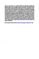
for a cell, such as a neuron, that is essentially postmitotic. Interestingly, some of the most aggressively dividing cells can be those of a tumor, making retroviruses not only a good vector for targeting these cells but also an opportunist for infecting them should they arise in a patient treated for some other reason. Ebola and Marburg are members of the Filoviridae family of viruses. These viruses are similar to retroviruses (Table 2.1) in that they express a surface glycoprotein that mediates receptor binding with specific host cells. Exchange of these glycoproteins between lentiviruses and filoviruses has been used to retarget these potential viral gene therapy delivery vectors.21 Although such modifications can lead to a retargeting of these viral vectors, the receptor through which they target is sometimes poorly understood.22,23 It is also possible to focus a delivery therapy based upon functional aspects of the target cell population. In the case of cancer cells expressing an activated Ras/Raf/mitogen-activated protein kinase pathway, regulated replication of a viral gene delivery system can be used to provide selective expression
Figure 2.1 Generalized classification of some viruses used for gene therapy.
Tissue- and cell-specific targeting for the delivery of genetic information
9
of delivered genetic information.24 Thus, prior to using the targeting capacity of viral particles, it would be important to understand their tropisms as completely as possible. One concern is that the tropism of a virus may differ due to the route of entry into the body. Most viruses infect individuals via a mucosal exposure. As an example, both the Coxsackievirus and the adenovirus bind to a receptor on the surface of epithelial cells located at the tight junction structures that hold adjacent cells together;25 this same receptor is distributed differently on non-polarized cells. Additionally, adenovirus has been suggested to target hematopoietic stem cells,26 making the route of entry of this vector critical to its potential targeting. Rarely do viruses infect in the manner (by injection) or in the numbers (as much as 1011 particle forming units [PFUs]) that are used in most gene therapy protocols. A few viruses do produce natural infection by injection, and their delivery by injection would emulate their natural route of infection. The Dengue virus infects via an injection from an infected mosquito.27 Once injected into the body, the Dengue virus attaches to the host cell and enters cells via the major envelope glycoprotein E, a protein capable of locally disrupting host cell membranes, allowing entry of genetic information.27 As stated above, many other viruses infect initially at a mucosal surface and not by an injected route. Tropism of a viral particle may be different—somewhat less selective—when delivered in an unnatural fashion and in very high numbers. Additionally, many viral vectors have been modified to reduce or eliminate a particular pathogenic characteristic (as discussed above), and this may also affect the outcome of their capacity to target to a specific cell type or tissue.
Table 2.1 Virus Families Commonly Used for Delivery of Genetic Information Virus
Packaging Capacity
Cell/Tissue Application(s)
Outcome of Gene Therapy
Poxviridae (e.g., vaccinia, variola, avipox)
25 kb
Epidermal, respiratory, various epithelia
Adenoviridae (e.g., adenovirus—more than 50 serotypes)
30 kb
Broad: stem cells, neurons, muscles
Durable expression, low toxicity with some mutant strains.
Retrovirus (e.g., marine 8 kb leukemia virus, human immunodeficiency virus-1, equine infectious anemia virus, Maloney murine leukemia virus)
Mostly restricted to dividing cells: tumor cells, lymphocytes, hepatocytes, bone marrow cells
Genome integration, durable expression, safety concerns
Parvovirus (e.g., adenoassociated virus-2)
Broad: infects both Slow expression onset, dividing and nondividing can be both integrating cells and nonintegrating,
50 mM) at lower N/P ratios.18,19 The net positive surface charge (expressed as the zeta potential) and an excess of free PEI during complex formation can prevent aggregation by repulsion of positive charges; an increase of salt concentration reduces the hydrated layer around the particles and promotes their aggregation. At N/P ratios greater than 2, free PEI is present in the complex solution, and values between 50% and 86% for free PEI have been reported.20,21 Our own observations indicate that about two-thirds of PEI is unbound when mixing complexes at N/P 6.81
7.5 PEI POLYPLEXES FOR NUCLEIC ACID DELIVERY For the delivery of DNA into cells a broad range of molecular weights of PEI can be used. Early studies have used branched PEI at 800 kDa,1 but branched PEI at 25 kDa is also an efficient transfection reagent exhibiting reduced toxicity in vivo.19 When further decreasing the molecular weight of branched PEI used for transfection, both cellular toxicity and transfection efficiency are decreased.22 PEI polyplexes used for gene transfer experiments usually bear a net positive surface charge. Due to electrostatic interactions— e.g., with proteoglycanes23—the particles are bound to the cell surface and are subsequently internalized into intracellular vesicles by adsorptive endocytosis.24 Cointernalized proteoglycanes are also supposed to influence intracellular routing of PEI polyplexes and the efficiency of transgene expression.25 The size of PEI polyplexes can also influence transfection efficiency in vitro: aggregated complexes result in increased cellular association due to sedimentation onto cells and improved intracellular delivery (see below).18 LPEI at 22 kDa was found to be far more efficient in gene delivery both in vitro and in vivo.19,26,27 Apparently, polyplexes with LPEI exhibit a different biophysical behavior, resulting in the aggregation of initially small particles on the cell surface, which could be one reason for the exceptionally high transfection efficiency. For delivery of short nucleic acids such as mRNA, only LMW PEI (2 kDa or less) can lead to an efficient biological effect of the transferred nucleic acids. Complexes with higher molecular weight PEI are too stable and do not release their payload already in the cytoplasm of cells.17
Gene delivery using polyethylenimine and copolymers
119
7.6 LIGAND-PEI CONJUGATES FOR GENE DELIVERY Different ligands have been coupled with PEI to enhance the internalization of polyplexes into target cells (see Table 7.1). The use of targeting ligands incorporated into the polyplex can greatly increase the extent of binding and the specificity of internalization into endosomes. Transferrinmediated internalization into transferrin receptor positive cells occurs quickly (in less than 1 h) compared to polyplexes lacking the ligand.28 (Transferrin-targeted gene delivery systems are discussed in greater detail elsewhere in this Chapter 34.) Similar effects were observed for antibody
Table 7.1 Receptor-Mediated Targeting of Polyethylenimine Conjugate to Specific Cell Populations Receptor
Ligand
Target Cells
ASGP receptor Galactosylated PEI Hepatocytes, hepatoma
Refs. 70–72
CD-3
Anti-CD-3
T-cell derived leukemia cells, activated PBMCs 6,73
CD-71
Transferrin
Different tumor cell lines
6
EGF receptor
EGF
HepG2, HuH7, KB, A431
11,74,75
KB, CT-26
76,77
Folate receptor Folate Mannose
Mannosylated PEI Dendritic cells
78
PECAM
Anti-PECAM
Lung endothelial cells
79
Vitronectin-R
RGD-peptide
Angiogenic cells, different tumor cell lines
13,29,30,80
targeted polyplexes (anti-CD3 on Jurkat cells) and polyplexes containing the epidermal growth factor (EGF). Short peptidic ligands can be chemically synthesized and coupled to PEI via standard chemistry (see above). The sequence Arg-Gly-Asp (RGD) can bind specifically to the vitronectin receptor αv 3, an integrin which is highly upregulated in certain tumor cells and the tumor endothelium. RGD-PEI conjugates used for DNA transfection significantly increase transgene expression in different tumor cell lines13,29 and angiogenic cells.30
7.7 INTRACELLULAR ASPECTS OF PEI-MEDIATED GENE DELIVERY After internalization, gene transfer complexes are mostly found in intracellular compartments, which can be potentially acidified, such as endosomes or lysosomes, and their subsequent release into the cytoplasm represents a major bottleneck for gene delivery.31,32 The protonation profile of PEI, bearing an intrinsic buffer capacity (see above), and its high transfection capacity led to the development of the “proton sponge” hypothesis: the buffering of the endosome by PEI causes proton accumulation and subsequent influx of chloride into the vesicle. Osmotic swelling by influx of water can
Polymeric gene delivery
108
thereafter lead to disruption of the vesicle and release of the polyplex into the cytoplasm.2 This theory was at least partially confirmed by the finding that bafilomycin A, a potent inhibitor of the intracellular proton pump, selectively inhibits transgene expression of PEI polyplexes, whereas the efficiency of DOTAP, a monocationic lipid, was not significantly altered.33 Nevertheless, additional mechanisms for cytoplasmic delivery of PEI polyplexes have been suggested, which include the possibility that PEI can rupture the lysosome directly by interacting with the membrane.24 Studies based on flow cytometry revealed that endosomes are not completely buffered to neutral pH by PEI polyplexes,7 and the resulting average pH was between pH 5.9 for branched PEI and 5.0 for linear PEI.34 Apparently, the mechanism is not yet fully understood and will have to be further clarified. Release of PEI polyplexes from endosomes can be significantly hampered when using either low molecular weight PEI22 or small PEI polyplexes at low concentrations.18 To promote endosomolysis of PEI polyplexes, inactivated adenovirus particles35 or membrane active peptides6,17,18,36 were incorporated into the polyplex and significantly augmented reporter gene expression. Passive diffusion within the cytoplasm is limited for DNA above a certain size limit, which is due to a crowding effect caused by components of the cytoplasm.37 PEI protects DNA from nuclease attack within the cytoplasm,38 and the transport within the cytoplasm is supposed to be at least partially dependent on microtubuli,39 although it cannot be excluded that the intracellular transport of PEI polyplexes is mediated within vesicles. Nuclear import of PEI polyplexes is a major bottleneck for transfection, as only one out of 1000 copies of PEI condensed plasmid molecules will reach the nucleus after microinjection into the cytoplasm.38 Brunner and colleagues demonstrated that there is an approximately 30-fold decrease in reporter gene expression transfecting cells either logarithmically growing or synchronized in the early G1 phase of the cell cycle.27 Interestingly, polymer mediated transfection using linear PEI was far less dependent on cell cycle compared to branched PEI.40 In accordance with this observation, large amounts of LPEI polyplexes were found within the nucleus of transfected cells after 7 hours.19 Although the detailed mechanism of linear PEI is not completely clear, nuclear access due membrane rupture by aggregated polyplexes could add to this effect. As in the case of endosomal release, different moieties were added to PEI polyplexes in order to improve their access to the nucleus. Adenoviral hexon protein is supposed to permit the access of viral DNA to the nucleus.41 Hence, purified hexon protein was coupled to PEI, amplifying nuclear entry and leading to an increase in the efficiency of transgene expression.8 The cationic, membrane active peptide melittin coupled to PEI polyplexes not only improved endosomal release, but also aided in the transfection of cells that divided slowly and cells that did not divide.42
7.8 IN VIVO APPLICATIONS Gene delivery in vivo with nonviral gene delivery systems still remains a challenging task; depending on the therapeutic strategy different routes of administration have to be chosen. Local applications have the advantage of circumventing unwanted interaction of gene transfer complexes with blood components and the reticulo-endothelial system,
Gene delivery using polyethylenimine and copolymers
121
although the need for diffusible particles within the tissue remains. Systemic administration is the most demanding, but offers the possibility of reaching the disseminated target throughout the organism, e.g., tumor metastases. We and others have developed PEI-based systems which enable systemic delivery to disseminated, well vascularized tumors (see Chapter 34) by shielding the complex surface with PEG or transferrin. 7.8.1 Local Delivery Early studies with PEI polyplexes were conducted by stereotactic intracerebral injection into the brain.1,43 Elevated expression levels and increased diffusibility within the brain were obtained using LPEI polyplexes generated in 5% glucose.44 The small polyplexes were able to transfect both glia cells and neurons. Recently, repeated intrahecal injection of PEG-modified PEI polyplexes led to prolonged transgene expression.45 In contrast, plain PEI polyplexes led to attenuated gene expression after repeated injection and also resulted in cellular damage. This indicates the importance of a nontoxic gene transfer formulation consisting of small polyplexes that enable diffusibility within the target tissue. Local delivery to the liver can be achieved by application of gene transfer complexes via the hepatic artery or by direct injection into the tissue. Gharwan et al. compared the transfection efficiency of naked DNA and different PEI polyplexes by liver injection into fetal, neonatal, and adult mice.46 At low DNA doses, LPEI polyplexes were more efficient than naked DNA, and maximal gene expression was achieved in neonatal mice, although relevant therapeutic levels of reporter gene expression could not be achieved. Injection of polyplexes into the renal artery of rats led to preferential reporter gene expression in proximal tubular cells,47 but only PEI polyplexes below 100 nm in diameter were able to reach the target cells.48 Local administration to the lung can be achieved via instillation or nebulization of the gene transfer particles. Nebulized PEI polyplexes applied into mice lungs led to significant levels of reporter gene expression, which peaked after 24 h and was still detectable after 8 days.49 Masking the surface of PEI polyplexes with hydrophilic polymers reduced interaction with bronchoalveolar lavage, although plain PEI polyplexes remained the most efficient.50 Direct intratumoral injections of plain PEI polyplexes were less efficient, presumably due to poor diffusion of the polyplexes within the tumor mass. Application via a micropump was much more successful, resulting in long-lasting reporter gene expression for more than 2 weeks.51 7.8.2 Systemic Application In principle, three major targets can be reached after application of gene transfer complexes into the bloodstream. Highly positively charged particles will rapidly aggregate (partially by interaction with blood components) and end up in the lung, the first vascular bed encountered.19,52 Protecting the particle surface with hydrophilic polymers enables the circulation of PEI polyplexes in the bloodstream and thereafter leads them to the liver53 or enables a (passive) accumulation in implanted tumors.10,54
Polymeric gene delivery
110
Especially in the lung very high levels of reporter gene expression can be found using LPEI polyplexes, although toxic side effects can be observed when using either high molecular weight PEI and/or high N/P ratios.10,52,55 The toxic effect is mostly due to the aggregating properties of PEI polyplexes and leads to activation of lung endothelium and liver damage.55 Repeated administration of PEI polyplexes also caused sites of inflammation in liver tissue,56 which could be due to the prolonged retention of PEI in the liver. PEI labeled with radioactive iodine could be still detected several days after intravenous injection, and was mainly found in the lysosomal fraction of the cells.57 Absence of a toxic PEI-based gene delivery formulation will be a key point for their application in the clinic, and this highlights the importance of optimizing such formulations for reduced toxicity. This can be achieved by preventing the aggregation with blood components by surface shielding or reducing the N/P ratio of PEI polyplexes.58 In this case, even without surface shielding, tumor tissue was efficiently transfected (see below).
7.9 THERAPEUTIC GENE DELIVERY WITH PEI PEI has already been used for the delivery of therapeutic DNA, oligonucleotides, or different types of RNA. Glycosylated PEI was used for the hepatic delivery of DNA-RNA oligonucleotides, also called chimeraplasts.59,60 The genomic sequence was successfully modified by sitedirected mutagenesis and was still measurable 2 years after treatment.61 Intratumoral application of LMW PEI complexed ribozymes directed against the growth factor pleiotropin resulted in significant reduction in tumor growth.62 Delivery of LPEI condensed antisense oligodeoxynuclotides directed against hepatitis virus inhibited viral replication in a duck liver infection model.63 PEI-based transfection systems are potentially suited for different strategies to treat malignant diseases (for review see Ogris and Wagner64): direct killing of tumor cells with bioactive proteins, e.g., cytokines or apoptosis inducers; suicide gene therapy by combining prodrugs with the expression of prodrug activating enzymes; or the induction of chemoprotection in combination with high dose chemotherapy. The following therapeutic genes have already been successfully delivered with PEI-based transfection systems in vivo. The p53 gene is the most frequently altered gene in human cancers. Its normal function is to protect cellular DNA by coordinately blocking cell proliferation, stimulating DNA repair, and promoting apoptotic cell death. Overexpression of wild-type p53 is a promising approach to treat cancers caused by p53 mutations. Mice xenografted with head and neck cancer were treated by intratumoral injections of p53 expressing plasmid complexed with glycosylated PEI.65 PEI polyxplexes were found to diffuse within the viable tumor mass, transfecting cells in the periphery of the tumor. A treatment schedule with injections twice a week led to apoptosis of tumor cells and inhibited tumor growth. Mice bearing experimental syngeneic melanoma metastases were treated by multiple applications of nebulized PEI polyplexes carrying the p53 gene.66 The treatment led to significant reduction of tumor burden in the lung and increased the median survival time compared to treatment with control plasmid. A similar effect was found in a human
Gene delivery using polyethylenimine and copolymers
123
osteosarcoma lung metastasis model in nude mice.67 Systemic application of PEI polyplexes (at the low N/P ratio of 2.7) in an orthotopic bladder cancer model led to a 14fold higher reporter gene expression in the tumor compared to the lung.58 Treatment with 6 µg PEI polyplexes every 3 days for 3 weeks resulted in a 70% reduction in tumor size. Tumor necrosis factor a (TNF-α) is a potent cytokine that induces hemorrhagic tumor necrosis and tumor regression. Kircheis et al. demonstrated that the local expression of TNF-α in tumors after systemic application of transferring-shielded PEI polyplexes reduced tumor growth, and in certain tumor models resulted in a cure rate of 60%.68 Somatostatin is a cyclic nonapeptide known to negatively regulate the growth of different cell types, including tumor cells. The antiproliferative effect mediated by the somatostatin receptor subtype 2 (sst2) was utilized for treatment of pancreatic cancer in an orthotopic syngeneic hamster model.69 The therapeutic effects of LPEI polyplexes and recombinant adenovirus encoding sst2 were compared by intratumoral injection. Both recombinant adenovirus and LPEI polyplexes significantly reduced tumor growth.
7.10 CONCLUSIONS Polyethylenimine is a transfection reagent that is suitable for a broad range of different gene transfer applications. The high content of primary amino groups enables the chemical coupling of targeting moieties or intracellular active components; the high density of positive charges in the molecule allows for a tight compaction of nucleic acids. PEI can influence different steps of the transfection process, i.e., endosomal release and nuclear entry. These processes have to be further clarified. Suitable formulations of PEI polyplexes with low toxicity have to be chosen for in vivo use, which will allow for multiple applications of the therapeutic gene. Several preclinical studies have already revealed the applicability of PEI-based gene therapy systems for a broad range of applications.
REFERENCES 1. Boussif, O. et al., A versatile vector for gene and oligonucleotide transfer into cells in culture and in vivo: polyethylenimine, Proc. Nat. Acad. Sci. U.S.A. 92, 7297, 1995. 2. Kichler, A., Behr, J.P., and Erbacher, P., Polyethylenimines: a family of potent polymers for nucleic acid delivery, in Nonviral Vectors for Gene Therapy, Huang, L., Hung, M.C., and Wagner, E. (Eds.), Academic Press, San Diego, 1999, Chapter 9. 3. Suh, J., Paik, H.J., and Hwang, P.K., lonization of polyethylenimine and polyellylamine at various pHs, Bioorg. Chem. 22, 318, 1994. 4. Godbey, W.T., Wu, K.K., and Mikos, A.G., Poly(ethylenimine) and its role in gene delivery, J. Control. Release 60, 149, 1999. 5. Brissault, B. et al., Synthesis of linear polyethylenimine derivatives for DNA transfection, Bioconjug. Chem. 14, 581, 2003. 6. Kircheis, R. et al., Coupling of cell-binding ligands to polyethylenimine for targeted gene delivery, Gene Ther. 4, 409, 1997. 7. Ogris, M. et al., Melittin enables efficient vesicular escape and enhanced nuclear access of nonviral gene delivery vectors, J. Biol. Chem. 12, 12, 2001.
Polymeric gene delivery
112
8. Carlisle, R.C. et al., Adenovirus hexon protein enhances nuclear delivery and increases transgene expression of polyethylenimine/plasmid DNA vectors, Mol Ther. 4, 473, 2001. 9. Kursa, M. et al., Novel shielded transferrin-polyethylene glycol-polyethylenimine/DNA complexes for systemic tumor-targeted gene transfer, Bioconjug. Chem. 14, 222, 2003. 10. Ogris, M. et al., PEGylated DNA/transferrin-PEI complexes: reduced interaction with blood components, extended circulation in blood and potential for systemic gene delivery, Gene Ther. 6, 595, 1999. 11. Blessing, T. et al., Different strategies for formation of pegylated EGF-conjugated PEI/DNA complexes for targeted gene delivery, Bioconjug. Chem. 12, 529, 2001. 12. Erbacher, P. et al., Transfection and physical properties of various saccharide, poly(ethylene glycol), and antibody-derivatized polyethylenimines (PEI), J. Gene Med. 1, 210, 1999. 13. Erbacher, P., Remy, J.S., and Behr, J.P., Gene transfer with synthetic virus-like particles via the integrin-mediated endocytosis pathway. Gene Ther. 6, 138, 1999. 14. Ungaro, F. et al., Spectrophotometric determination of polyethylenimine in the presence of an oligo-nucleotide for the characterization of controlled release formulations, J. Pharm. Biomed. Anal. 31, 143, 2003. 15. Tang, M.X. and Szoka, F.C., The influence of polymer structure on the interactions of cationic polymers with DNA and morphology of the resulting complexes, Gene Ther. 4, 823, 1997. 16. Dunlap, D.D. et al., Nanoscopic structure of DNA condensed for gene delivery, Nucleic Acids Res. 25, 3095, 1997. 17. Bettinger, T. et al., Peptide-mediated RNA delivery: a novel approach for enhanced transfection of primary and post-mitotic cells, Nucleic Acids Res. 29, 3882, 2001. 18. Ogris, M. et al., The size of DNA/transferrin-PEI complexes is an important factor for gene expression in cultured cells, Gene Ther. 5, 1425, 1998. 19. Wightman, L. et al., Different behavior of branched and linear polyethylenimine for gene delivery in vitro and in vivo, J. Gene Med. 3, 362, 2001. 20. Finsinger, D. et al., Protective copolymers for nonviral gene vectors: synthesis, vector characterization and application in gene delivery, Gene Ther. 7, 1183, 2000. 21. Clamme, J.P, Azoulay, J., and Mely, Y., Monitoring of the formation and dissociation of polyethyl-enimine/DNA complexes by two photon fluorescence correlation spectroscopy, Biophys. J. 84, 1960, 2003. 22. Godbey, W.T., Wu, K.K., and Mikos, A.G., Poly(ethylenimine)-mediated gene delivery affects endothelial cell function and viability, Biomaterials 22, 471, 2001. 23. Mislick, K.A. and Baldeschwieler, J.D., Evidence for the role of proteoglycans in cationmediated gene transfer, Proc. Nat. Acad. Sci. U.S.A. 93, 12349, 1996. 24. Bieber, T. et al., Intracellular route and transcriptional competence of polyethylenimine-DNA complexes, J. Control. Release 82, 441, 2002. 25. Ruponen, M. et al., Extracellular glycosaminoglycans modify cellular trafficking of lipoplexes and polyplexes, J. Biol. Chem. 276, 33875, 2001. 26. Ferrari, S. et al., ExGen 500 is an efficient vector for gene delivery to lung epithelial cells in vitro and in vivo, Gene Ther. 4, 1100, 1997. 27. Brunner, S. et al., Cell cycle dependence of gene transfer by lipoplex, polyplex and recombinant adenovirus, Gene Ther. 7, 401, 2000. 28. Ogris, M. et al., DNA/polyethylenimine transfection particles: Influence of ligands, polymer size, and PEGylation on internalization and gene expression, AAPS PharmSci. 3, E21, 2001. 29. Kunath, K. et al., Integrin targeting using RGD-PEI conjugates for in vitro gene transfer, J. Gene Med. 5, 588, 2003. 30. Suh, W. et al., An angiogenic, endothelial-cell-targeted polymeric gene carrier, Mol. Ther. 6, 664, 2002. 31. Zabner, J. et al., Cellular and molecular barriers to gene transfer by a cationic lipid, J. Biol. Chem. 270, 18997, 1995.
Gene delivery using polyethylenimine and copolymers
125
32. Labat Moleur, F. et al., An electron microscopy study into the mechanism of gene transfer with lipopolyamines, Gene Ther. 3, 1010, 1996. 33. Kichler, A. et al., Polyethylenimine-mediated gene delivery: a mechanistic study, J. Gene Med. 3, 135, 2001. 34. Akinc, A. and Langer, R., Measuring the pH environment of DNA delivered using nonviral vectors: implications for lysosomal trafficking, Biotechnol. Bioeng. 78, 503, 2002. 35. Baker, A. et al., Polyethylenimine (PEI) is a simple, inexpensive and effective reagent for condensing and linking plasmid DNA to adenovirus for gene delivery, Gene Ther. 4, 773, 1997. 36. Lee, H., Jeong, J.H., and Park, T.G., A new gene delivery formulation of polyethylenimine/DNA complexes coated with PEG conjugated fusogenic peptide, J. Control. Release 76, 183, 2001. 37. Lukacs, G.L. et al., Size-dependent DNA mobility in cytoplasm and nucleus, J. Biol. Chem. 275, 1625, 2000. 38. Pollard, H. et al., Polyethylenimine but not cationic lipids promotes transgene delivery to the nucleus in mammalian cells, J. Biol. Chem. 273, 7507, 1998. 39. Suh, J., Wirtz, D. and Hanes, J., Efficient active transport of gene nanocarriers to the cell nucleus, Proc. Nat. Acad. Sci. U.S.A. 100, 3878, 2003. 40. Brunner, S. et al., Overcoming the nuclear barrier: cell cycle independent nonviral gene transfer with linear polyethylenimine or electroporation, Mol Ther. 5, 80, 2002. 41. Saphire, A.C. et al., Nuclear import of adenovirus DNA in vitro involves the nuclear protein import pathway and hsc70, J. Biol. Chem. 275, 4298, 2000. 42. Ogris, M. et al., Melittin enables efficient vesicular escape and enhanced nuclear access of nonviral gene delivery vectors, J. Biol. Chem. 276, 47550, 2001. 43. Abdallah, B. et al., A powerful nonviral vector for in vivo gene transfer into the adult mammalian brain: polyethylenimine, Hum Gene Ther. 7, 1947, 1996. 44. Goula, D. et al., Size, diffusibility and transfection performance of linear PEI/DNA complexes in the mouse central nervous system, Gene Ther. 5, 712, 1998. 45. Shi, L. et al., Repeated intrathecal administration of plasmid DNA complexed with polyethylene glycol-grafted polyethylenimine led to prolonged transgene expression in the spinal cord, Gene Ther. 10, 1179, 2003. 46. Gharwan, H. et al., Nonviral gene transfer into fetal mouse livers (a comparison between the cationic polymer PEI and naked DNA), Gene Ther. 10, 810, 2003. 47. Boletta, A. et al., Nonviral gene delivery to the rat kidney with polyethylenimine, Hum Gene Ther. 8, 1243, 1997. 48. Foglieni, C. et al., Glomerular filtration is required for transfection of proximal tubular cells in the rat kidney following injection of DNA complexes into the renal artery, Gene Ther. 7, 279, 2000. 49. Gautam, A. et al., Enhanced gene expression in mouse lung after PEI-DNA aerosol delivery, Mol. Ther. 2, 63, 2000. 50. Rudolph, C. et al., Nonviral gene delivery to the lung with copolymer-protected and transferrinmodified polyethylenimine, Biochim. Biophys. Acta 1573, 75, 2002. 51. Coll, J.L. et al., In vivo delivery to tumors of DNA complexed with linear polyethylenimine, Hum Gene Ther. 10, 1659, 1999. 52. Goula, D. et al., Polyethylenimine-based intravenous delivery of transgenes to mouse lung, Gene. Ther. 5, 1291, 1998. 53. Nguyen, H.K. et al., Evaluation of polyether-polyethyleneimine graft copolymers as gene transfer agents, Gene Ther. 7, 126, 2000. 54. Oupicky, D. et al., Importance of lateral and steric stabilization of polyelectrolyte gene delivery vectors for extended systemic circulation, Mol. Ther. 5, 463, 2002. 55. Chollet, P. et al., Side-effects of a systemic injection of linear polyethylenimine-DNA complexes, J. Gene Med. 4, 84, 2002.
Polymeric gene delivery
114
56. Oh, Y.K. et al., Prolonged organ retention and safety of plasmid DNA administered in polyethylenimine complexes, Gene Ther. 8, 1587, 2001. 57. Lecocq, M. et al., Uptake and intracellular fate of polyethylenimine in vivo, Biochem. Biophys. Res. Commun. 278, 414, 2000. 58. Sweeney, P. et al., Efficient therapeutic gene delivery after systemic administration of a novel polyethylenimine/DNA vector in an orthotopic bladder cancer model, Cancer Res. 63, 4017, 2003. 59. Kren, B.T. et al., Correction of the UDP-glucuronosyltransferase gene defect in the gunn rat model of crigler-najjar syndrome type I with a chimeric oligonucleotide, Proc. Nat. Acad. Sci. U.S.A. 96, 10349, 1999. 60. Kren, B.T., Bandyopadhyay, P., and Steer, C.J., In vivo site-directed mutagenesis of the factor IX gene by chimeric RNA/DNA oligonucleotides, Nat. Med. 4, 285, 1998. 61. Kren, B.T. et al., Modification of hepatic genomic DNA using RNA/DNA oligonucleotides, Gene Ther. 9, 686, 2002. 62. Aigner, A. et al., Delivery of unmodified bioactive ribozymes by an RNA-stabilizing polyethylenimine (LMW-PEI) efficiently down-regulates gene expression, Gene Ther. 9, 1700, 2002. 63. Robaczewska, M. et al., Inhibition of hepadnaviral replication by polyethylenimine-based intravenous delivery of antisense phosphodiester oligodeoxynucleotides to the liver, Gene Ther. 8, 874, 2001. 64. Ogris, M. and Wagner, E., Targeting tumors with non-viral gene delivery systems, Drug Discov. Today 7, 479, 2002. 65. Dolivet, G. et al., In vivo growth inhibitory effect of iterative wild-type p53 gene transfer in human head and neck carcinoma xenografts using glucosylated polyethylenimine nonviral vector, Cancer Gene Ther. 9, 708, 2002. 66. Gautam, A., Densmore, C.L., and Waldrep, J.C., Inhibition of experimental lung metastasis by aerosol delivery of PEI-p53 complexes, Mol Ther. 2, 318, 2000. 67. Densmore, C.L. et al., Growth suppression of established human osteosarcoma lung metastases in mice by aerosol Gene Ther. with PEI-p53 complexes, Cancer Gene Ther. 8, 619, 2001. 68. Kircheis, R. et al., Tumor-targeted gene delivery of tumor necrosis factor-alpha induces tumor necrosis and tumor regression without systemic toxicity, Cancer Gene Ther. 9, 673, 2002. 69. Vernejoul, F. et al., Antitumor effect of in vivo somatostatin receptor subtype 2 gene transfer in primary and metastatic pancreatic cancer models, Cancer Res. 62, 6124, 2002. 70. Zanta, M.A. et al., In vitro gene delivery to hepatocytes with galactosylated polyethylenimine, Bio-conjug. Chem. 8 (6), 839, 1997. 71. Bettinger, T., Remy, J.S., and Erbacher, P., Size reduction of galactosylated PEI/DNA complexes improves lectin-mediated gene transfer into hepatocytes, Bioconjug. Chem. 10, 558, 1999. 72. Morimoto, K. et al., Molecular weight-dependent gene transfection activity of unmodified and galactosylated polyethyleneimine on hepatoma cells and mouse liver, Mol. Ther. 7, 254, 2003. 73. O’Neill, M.M. et al., Receptor-mediated gene delivery to human peripheral blood mononuclear cells using anti-CD3 antibody coupled to polyethylenimine, Gene Ther. 8, 362, 2001. 74. Wolschek, M.F. et al., Specific systemic nonviral gene delivery to human hepatocellular carcinoma xenografts in SCID mice, Hepatology 36, 1106, 2002. 75. Lee, H., Kim, T.H.. and Park, T.G., A receptor-mediated gene delivery system using streptavidin and biotin-derivatized, pegylated epidermal growth factor, J. Control. Release 83, 109, 2002. 76. Guo, W. and Lee, R.J., Receptor-targeted gene delivery via folate-conjugated polyethylenimine, AAPS PharmSci. 1, Article 19, 1999. 77. Benns, J.M., Mahato, R.I., and Kim, S.W., Optimization of factors influencing the transfection efficiency of folate-PEG-folate-graft-polyethylenimine, J. Control. Release 79, 255, 2002.
Gene delivery using polyethylenimine and copolymers
127
78. Diebold, S.S. et al., Mannose polyethylenimine conjugates for targeted DNA delivery into dendritic cells, J. Biol. Chem. 274, 19087, 1999. 79. Li, S. et al., Targeted gene delivery to pulmonary endothelium by anti-PECAM antibody, Am. J. Physiol. Lung Cell Mol. Physiol. 278, L504-L511, 2000. 80. Muller, K. et al., Highly efficient transduction of endothelial cells by targeted artificial viruslike particles, Cancer Gene Ther. 8, 107, 2001. 81. Boeckle, S. et al., Purification of polyethylenimine polyplexes highlights the role of free polycations in gene transfer, J. Gene Med., in press, 2004.
CHAPTER 8 Poly(2-(dimethylamino)ethyl methacrylate)-Based Polymers for the Delivery of Genes In Vitro and In Vivo F.J.Verbaan, D.J.A.Crommelin, W.E.Hennink, and G.Storm
8.1 INTRODUCTION Advances in molecular biology have resulted in a new concept in the treatment of diseases, the concept of gene therapy. The unraveling of the human genome opens up the opportunity to treat diseases at their genetic origin rather than treating symptoms of diseases. However, gene therapy is not limited to the treatment of genetic defects. Therefore, gene therapy strategies are being devised for the treatment of other diseases as well. Plasmid DNA-based approaches to gene therapy involve administration of DNA (“naked DNA”) or formulations of DNA. The fundamental challenge of gene delivery originates from the fact that DNA has a charged, colloidal nature, is very labile
Figure 8.1 Chemical structure of pDMAEMA. in the biological environment, and does not cross biological barriers effectively, such as an intact endothelium, the plasma membrane, or the nuclear membrane.1 The need for an efficient and safe gene delivery system is obvious. Therefore, there is a growing interest in the development of target-cell-specific nonviral carriers for the delivery of genes. 0–8493–1934–X/05/$0.00+$1.50 © 2005 by CRC Press LLC
Poly(2-(dimethylamino)ethyl methacrylate)
117
Methacrylate polymers have been applied for the microencapsulation of cells, such as erythrocytes, fibroblasts, lymphomas, and the beta cells in the islets of Langerhans. Because of the biocompatibility with living tissue, the methacrylate polymers are also used to encapsulate cells in order to prevent tissue rejection upon cell transplantation.2,3 The combination of their biocompatibility, their relatively easy synthesis, and the possibility of functionalizing the polymers (e.g., making targeted or PEGylated derivatives) make methacrylates interesting candidates for gene delivery studies. In the Department of Pharmaceutics at Utrecht University this led to the idea of synthesizing poly(2-[dimethylamino]ethyl methacrylate) (pDMAEMA)-based polymers as “lead compounds” for the purpose of gene delivery. The chemical structure of the cationic pDMAEMA polymer is depicted in Figure 8.1. The purpose of this chapter is to give an overview of the different aspects of the development of pDMAEMA polymers toward an applicable gene transfectant in vivo. First, the in vitro characteristics of complexes of the pDMAEMA polymer and pDMAEMA analogs (“polyplexes”) are described. Second, pharmaceutical aspects of pDMAEMA-polyplex formulations are addressed. Finally, the in vivo application of pDMAEMA polyplexes for tumor targeting is discussed.
8.2 IN VITRO TRANSFECTION EFFICIENCY OF PDMAEMABASED COMPLEXES: CRITICAL PARAMETERS 8.2.1 Physicochemical Properties It has been reported that the extent of cellular uptake of the particulate system in question depends strongly on the size and charge.4,5 Thus, the physical characteristics of pDMAEMA-polyplexes were investigated. The effect of the pDMAEMA:plasmid ratio on the size and zeta potential of the formed particles was studied by dynamic light scattering measurements and electrophoretic mobility measurements, respectively. Figure 8.2 shows that in an aqueous buffer solution (20 mM Hepes, pH 7.4), naked plasmid (5 µg/ml) has a rather large hydrodynamic size (0.3–0.4 µm). Small and stable polymerplasmid complexes (size ~0.1–0.2 µm) could be formed under the same conditions as used for naked DNA and at polymer:plasmid ratios above 3:1 (w/w), demonstrating that the cationic polymer is able to condense plasmid DNA. Zeta potential measurements revealed that naked DNA possesses a negative zeta potential (−22 mV). After the addition of pDMAEMA, a positive zeta potential was observed which leveled off at polymen:plasmid ratios above 3:1 (w/w).6 The in vitro transfection efficiency of the pDMAEMA polyplexes is shown in Figure 8.3. The transfection efficiency shows a bell-shaped dependence on the polymen:plasmid ratio (w/w). Such dependence is observed frequently for both lipid-based7 and cationic polymer-based transfection systems8 and can be explained as follows. At low polymer:plasmid ratios the polyplexes have a negative
Polymeric gene delivery
118
Figure 8.2 The effect of the pDMAEMA:plasmid ratio on size (as determined by DLS ■) and zeta potential (●) of pDMAEMA-plasmid polyplexes. The plasmid concentration was fixed at 5 µg/ml. The results are expressed as mean values ±SD of three experiments.
Figure 8.3 The effect of the pDMAEMA:plasmid ratio on the number of transfected cells (●) and on the relative cell viability (▲). The plasmid concentration was fixed at 5 µg/ml. The results are expressed as
Poly(2-(dimethylamino)ethyl methacrylate)
119
mean values ±SEM of three to five experiments. zeta potential and are relatively large in size (Figure 8.2). Both characteristics are unfavorable for cellular association and internalization. At polymer:plasmid ratios above 3:1 (w/w), the polyplexes are small and have a positive zeta potential. Both properties are a favorable factor for transfection. The decreasing transfection efficiency at ratios > 3:1 can be explained by the toxicity of the free polymer present in the transfection medium at these higher ratios (see Figure 8.3). Besides the effect of the pDMAEMA:DNA ratio, we also studied the effect of the polymer molecular weight on DNA condensation.9 Although it has been reported that low-molecular-weight polymers are able to give small and stable polyplexes,10 we found for pDMAEMA that the average molecular weight has to be above 100,000 g/mol to give sufficient condensation of plasmid DNA. 8.2.2 Design of pDMAEMA-Based Copolymers A common feature of cationic polymers is their cytotoxicity.9,11 The design of copolymers is an interesting route for optimizing the efficacy: toxicity ratio of polymeric gene transfer systems. We evaluated copolymers of pDMAEMA and hydrophobic monomers such as methyl methacrylate (MMA), or hydrophilic monomers like N-vinylpyrrolidone (NVP) and ethoxytriethylene glycol (triEGMA) as transfectants,6,12 as shown in Figure 8.4. Polyplexes containing a copolymer with 20 mol % of MMA showed reduced transfection efficiency and a substantially increased cytotoxicity as compared to polyplexes formed with the pDMAEMA homopolymer of the same molecular weight. This suggests that the introduction of hydrophobic monomers is a less attractive approach to optimize the efficacy: toxicity ratio. On the other hand, polyplexes of plasmid and copolymers with a low content (20 mol %) of a hydrophilic comonomer (either NVP or triEGMA) had the same transfection and cytotoxicity properties as pDMAEMA of comparable molecular weight (around 100,000). Increasing the NVP or triEGMA content to 50 mol % resulted in less toxic polyplexes with improved transfection properties. This shows that reduction of the charge density of pDMAEMA polymers is an attractive route for optimizing the efficacy: toxicity ratio. However, a high-molecular-weight pDMAEMA (mol wt >105) is still superior in mediating transfection
Polymeric gene delivery
120
Figure 8.4 Transfection efficiency (A and C) and effect on relative cell viability (B and D) of (co)polymerplasmid polyplexes as a function of the (co)polymer: plasmid ratio. The plasmid concentration was 5 µg/ml, the transfection time (incubation time of the complexes with OVCAR-3 cells) and the coloring time (X-Gal) were 1 and 16 h, respectively. The transfection values are normalized to the maximum number of transfected cells after incubation with plasmid complexed to pDMAEMA (Mw 100kD, polymer: plasmid ratio 3:1 [w/w]). The results are shown as mean values (± SEM) of 3–5 experiments. Mw of (co)polymers was around 100 kD. (●) pDMAEMA, (■) poly(DMAEMA-co-triEGMA) 79/21 (mol/mol), (▼) poly(DMAEMA-co-triEGMA) 52/48
Poly(2-(dimethylamino)ethyl methacrylate)
121
(mol/mol), (A) poly(DMAEMA-coNVP) 86/14 poly(DMAEMA-coNVP) 46/54 (mol/mol), (□) poly(DMAEMA-co-NVP) 80/20 (mol/mol). compared to the other evaluated polymers (data not shown), which can be ascribed to the low capability of the low-molecular-weight polymer to condense the plasmid. The transfection capability of other water-soluble cationic polymers with structures closely related to pDMAEMA was evaluated to obtain insight into the relationship between structure and activity.13 The following parameters were studied. • A propyl side chain instead of an ethyl side chain with poly(3-[dimethylammo]propyl methacrylate) (pDMAPMA) • Substitution of the ester group in pDMAEMA by an amide group with poly(2[dimethylamino] ethyl methacrylamide) (pDMAEMAm). • A longer side chain and an amide group with poly(3-[dimethylamino]propyl methacrylamide) (pDMAPMAm) • Quaternary amino groups versus tertiary amino groups with poly(2[trimethylamino]ethyl methacrylate) (pTMAEMA) • Modification of the amino group of pDMAEMA with poly(2-[diethylamino] ethyl methacrylate) (pDEAEMA) Almost all of the investigated cationic polymers were able to condense plasmid DNA, yielding polymer-plasmid complexes with a size of 0.1 to 0.3 µm and a positive zeta potential. However, the transfection efficiency and cytotoxicity differed widely (Figure 8.5). The highest transfection efficiency and toxicity were observed for the “parent” compound pDMAEMA. Assuming that polyplexes enter the cell via endocytosis, pDMAEMA apparently has advantageous properties to escape the endosomes. A possible explanation is that, because of its average pKa value of 7.5, pDMAEMA is partially protonated at physiological pH and might behave as a proton sponge. This might cause a disruption of the endosomes, which results in a release of both polyplexes and cytotoxic endosomal/lysosomal enzymes in the cytosol. On the other hand, the analogs of pDMAEMA have a higher average pKa value and have, consequently, a higher degree of protonation and a lower buffering capacity. This might be associated with a lower tendency to destabilize the endosomes, resulting in both a lower transfection efficiency and a lower cytotoxicity. Also, molecular modeling studies showed that
Polymeric gene delivery
122
Figure 8.5 The effect of polymer/plasmid ratio on the transfection efficiency. The plasmid concentration was 5 µg/ml, the transfection time (incubation time of the polyplexes with OVCAR-3 cells) was 1 h. Transfection values are normalized to the maximum number of transfected cells after incubation with plasmid complexed to pDMAEMA. The results are shown as mean values (±SEM) of 3 experiments. (■) pDMAPMA, (▼) pDMAPMAm, pTMAEMA, pDMAEMAm, (o) pDMAEMA
Poly(2-(dimethylamino)ethyl methacrylate)
123
Figure 8.6 (See color insert following page 336) Confocal laser fluorescence microscopy (CLFM) study of OVCAR-3 cells, upon 1 h incubation at 37°C with DNA alone (A, B) and with polyplexes of pDMAEMA-DNA (w/w) at ratios of 0.9 (C, D), 0.75 (E, F), and 3 (G, H), followed by 2 days culturing at 37°C. Pictures B, D, F, and H are the direct transmitted light pictures taken simultaneously with the CLFM pictures A, C, E, and G. The plasmid was labeled with Ethd-1 and the width of a picture corresponds to 40 µm. pDMAEMA has the lowest interaction intensity with DNA. The relative ease of dissociation was indeed demonstrated using surface plasmon resonance and gel
Polymeric gene delivery
124
electrophoresis.14,15 It is, therefore, hypothesized that the superior transfection efficiency of pDMAEMA can be ascribed to an intrinsic property of pDMAEMA to destabilize the endosomes combined with relatively easy dissociation of the polyplexes once present in the cytosol or the nucleus. 8.2.3 Cellular Interactions of pDMAEMA-Based Polyplexes The cellular interactions of pDMAEMA-based polyplexes with human ovarian cancer (OVCAR-3) cells were investigated using fluorescence-activated cell sorting (FACS), confocal laser fluorescence microscopy (CLFM), and electron microscopy (EM).16 To gain more insight into the mode of plasmid delivery to OVCAR-3 cells, the effects of the following physicochemical characteristics on the cellular interaction of polyplexes with OVCAR-3 cells were investigated. • Charge (manipulated by the polymen:plasmid ratio) • Size (manipulated by the polymer molecular weight)
• Presence of tertiary versus quaternary amino groups, measured by comparing pDMAEMA with its quaternary ammonium analogue, poly(2-[trimethyl] amino ethyl methacrylate) • Modification of polyplex surface characteristics with the hydrophilic polymer polyethylene glycol (PEG) using a graft copolymer of pDMAEMA and PEG
Poly(2-(dimethylamino)ethyl methacrylate)
125
The effects of endocytosis inhibitors on cellular interaction and transfection activity were evaluated as well. Cellular association and subsequent internalization only occurred when the polyplexes exhibited a positive zeta potential (i.e., a pDMAEMA:plasmid ratio of 3:1 [w/w]), as can be observed in Figures 8.6A-H. Small polyplexes have an advantage over large complexes regarding cellular entry. The combined cellular interaction and transfection results suggest that the quaternary ammonium analogue pTMAEMA does not have an intrinsic endosomal escape property, as opposed to the proton sponge effect proposed for pDMAEMA (data not shown). PEGylation of pDMAEMA effectively shielded the surface charge and yielded a notably lower degree of cellular interaction (Figures 8.7A and B). Data on the effects of the presence of endocytosis inhibitors (N-ethylmaleimide and monensin) on cellular interaction (Figures 8.8E-H)) and transfection activity of pDMAEMA-based polyplexes confirm the hypothesis that endocytosis is the principal pathway for the intracellular delivery of plasmid. Neither the CLFM nor the EM studies revealed the presence of polyplexes or plasmid outside the endocytic vesicles or within the nucleus, suggesting that intracellular trafficking from the endosomes to the nucleus is a very inefficient process.
Figure 8.7 (See color insert) Confocal laser fluorescence microscopy (CLFM) study of OVCAR-3 cells, upon 1 h incubation at 37°C with polyplexes based on pDMAEMA-g-PEG-DNA at a 4:1 (w/w) ratio (A). Figure B shows the direct transmitted light pictures
Polymeric gene delivery
126
taken simultaneously with the CLFM picture A. The plasmid was labeled with Ethd-1 and the width of a picture corresponds to 40 µm. 8.2.4 Targeting of pDMAEMA-Based Polyplexes Lack of cell specificity, cytotoxicity, and blood-induced aggregation are major hurdles hindering the application of pDMAEMA-based polyplexes for in vivo plasmid delivery. In our laboratory, two approaches are under investigation to overcome these hurdles. First, a detergent removal method to coat the cationic polyplexes with anionic lipids has been developed.17 The intention was to create polyplexes that are negatively charged rather than positively charged, as a positive charge was supposed to be the main mediator of the above-mentioned hurdles. This method is based on the preferential formation of a lipid coat around the positively charged polyplexes upon the transition of phospholipids from the mixed micellar to the lamellar state induced by slow removal of the detergent (Figure 8.9). With this method spherical particles (referred to as lipopolyplexes) with a negative charge and a size of around 125 nm were obtained which were protected from destabilization by polyanions like hyaluronic acid. Hyaluronic acid is a component of the peritoneal ascites fluid, which probably induced a detrimental effect on the in vivo transfection capability of pDMAEMA polyplexes after intraperitoneal application to nude mice with OVCAR-3 cells growing in the peritoneal cavity (see Section 3.1). However, it was shown that the lipopolyplexes are much less effective in transfecting OVCAR-3 cells in vitro compared to positively charged pDMAEMA polyplexes, presumably due to a lower degree of cellular interaction. By conjugating Fab’ fragments directed against the epithelial glycoprotein-2 to the lipidic surface of the lipopolyplexes, target cell specific transfection of OVCAR-3 cells could be obtained in vitro (Figure 8.10). Second, we explored an active targeting strategy based on the use of folate as a targeting ligand directed against the folate receptor overexpressed on OVCAR-3 cells. Folate was coupled to preformed polyplexes with PEG as a spacer.18 A succinimidylactivated folate-PEG conjugate was synthesized, prepared from PEG-bisamine by subsequent conjugation with NHS-activated folate10 and disuccinimidyl suberate (Figure 8.11A). As the polymeric vector, a copolymer of pDMAEMA with 40% N-3-aminopropyl methacrylamide (NAPMAm) was used (Figure 8.11B). Folate-mediated targeting of the polyplexes was studied with an in vitro transfection assay using OVCAR-3 cells. A substantial zeta potential drop occurred upon PEGylation of the polyplexes, reflecting effective shielding of the positive surface charge (Figure 8.12). The PEGylated polyplexes (nontargeted polyplexes) showed markedly decreased gene expression when compared to uncoated polyplexes. Coupling of folate to the terminal ends of the PEG chains (targeted polyplexes) restored the transfection activity to the level observed for the uncoated polyplexes (Figure 8.13).
Poly(2-(dimethylamino)ethyl methacrylate)
127
Figure 8.8 (See color insert) Confocal laser fluorescence microscopy (CLFM) study of OVCAR-3 cells, upon 1 h incubation at 37°C with pDMAEMADNA 3:1 (w/w) polyplexes in the presence of 10 µM N-ethyl-maleimide (A, B) and in the presence of 10 µM monensin (C, D). Figures A and B are the direct transmitted light pictures taken simultaneously with the CLFM picture C and D, respectively. The plasmid was labeled with Ethd-1 and the width of a picture corresponds to 40 µm.
Polymeric gene delivery
128
Figure 8.9 Schematic representation of lipopolyplex formation. (1) Plasmid DNA is condensed by adding the cationic polymer pDMAEMA to the DNA at a ratio of 3:1 (w/w). The formed polyplexes (2) are added to mixed micelles containing the detergent n-octyl- -Dgalactopyranoside (OG) and a total amount of 3 µM of detergentsolubilized lipids (3). Upon slow removal of detergent by adsorption to hydrophobic beads, lipid coats are preferentially formed around positively charged polyplexes due to electrostatic interactions (4).
Figure 8.10 Influence of hyaluronic acid (HA) on the transfection efficiency of polyplexes versus immunolipopolyplexes (ILPP).
Poly(2-(dimethylamino)ethyl methacrylate)
129
OVCAR-3 cells were exposed for 1 h at 37°C to (A) polyplexes or ILPP in the absence (−HA) or presence (+HA) of 2.5 mg/ml HA. Gene carriers were removed by washing and cell culture was continued for another 47 h prior to evaluation for -galactosidase expression. 8.3 PHARMACEUTICAL ASPECTS OF PDMAEMA-BASED COMPLEXES 8.3.1 Preparation of Highly Concentrated pDMAEMA-Polyplexes The results presented in the earlier sections of this chapter were obtained at a relatively low plasmid concentration (5 µg/ml). At higher concentrations, however, severe aggregation of the polyplexes was sometimes observed. This is problematic, because in vivo studies require administration of relatively high concentrations of plasmid DNA to obtain detectable transfection levels.19,20 Using experimental design methodology, we therefore carried out a study to gain insight into formulation parameters (pH, polymer:plasmid ratio, ionic strength, temperature, viscosity, and the presence of excipients) that might affect the polyplex size at a relatively high plasmid concentration (200 µg/ml).21 pDMAEMA and plasmid DNA bind to each other via electrostatic interactions. Since the charge density of the polymer depends on the pH of the aqueous solution, we selected pH values ranging from pH 5.0, where the tertiary amine side groups of the polymer are all protonated, to pH 8.0, where approximately 25% of the side-groups are protonated. At a pH of 5.0, the formation of small polyplexes was enhanced as compared to pH 7.4 and 8.0, supposedly because of strong plasmid-polymer interactions resulting from a high charge density of the polymer.
Polymeric gene delivery
130
Figure 8.11 (A) Schematic representation of the synthesis of NHS-PEG-folate conjugate. (B) Structure of a copolymer of pDMAEMA with 40% N-3aminopropyl methacrylamide (NAPMAm) (pDMAEMAcoNAPMAm)
Figure 8.12 Particle size (squares) and zeta potential (triangles) of PEG-folic-
Poly(2-(dimethylamino)ethyl methacrylate)
131
acid (FA) coated pDMAEMAcoNAPMAm-DNA (N/P=5) complexes with PEG to primary amine ratio varying from 0 to 5 (mole/mole). Polyplexes were measured in 5 mM Hepes, pH 7.4. Data represent the means of three independently prepared formulations ±SEM.
Figure 8.13 Comparison of in vitro transfection efficiencies of OVCAR-3 cells mediated by nontargeted and folic acid (FA)-targeted PEG polyplexes at various PEG-to-primary-amine ratios, at an N/P ratio of 10. Efficiencies were determined by measuring the galactosidase activity. Open bars: PEG; horizontally hatched bars: PEGFA. Data represent the mean of three experiments ±SEM. For pDMAEMA: plasmid ratios above 1 (w/w) and at low ionic strengths, small and stable polyplexes could be obtained. At lower ratios aggregation occurred, irrespective of the other parameters investigated. Conditions of low ionic strength seem to favor the formation of small polyplexes, most likely because the polymer-DNA interaction increases with decreasing ionic strength.
Polymeric gene delivery
132
The presence of stabilizers (PEG 600, Tween 80) as well as the temperature at which the complexes were prepared (4–40°C) had a marginal effect on the size of the polyplexes. A preferable lyoprotectant when polyplexes are freeze-dried is sucrose. We evaluated the effect of sucrose on the size of the complexes. For concentrations of sucrose between 0 and 20% (w/v), the particle size of the polyplexes decreased. The higher viscosity of the 20% sucrose solution (compared with the viscosity of Hepes buffer) may prevent aggregation of formed polyplexes. At higher sucrose concentrations (up to 40%), the size of the formed polyplexes increased, possibly because under these conditions a viscous “barrier” hinders the interaction between the polymer and the plasmid. 8.3.2 Structural Properties of Plasmid DNA The structure of plasmid DNA (pDNA) can be described, analogous to proteins, at four levels,22 as summarized in Table 8.1. The primary structure is the nucleotide sequence of the plasmid, which contains the genetic information and is of major importance for its functional properties. The impact of the secondary, tertiary, and quaternary plasmid structure on the efficacy of transfection is still
Table 8.1 Structural Levels of Plasmid DNA and Detection Methods for Each Level Structure
Description
Detection Methods
Primary
Nucleotide sequence
Enzymatic assays (restriction mapping), hybridization techniques, chemical assays, mass spectrometry
Secondary
Double-helix conformations Circular dichroism, FTIR, UV spectroscopy (e.g., A, B, C form)
Tertiary (topology)
Supercoiled, open-circular, linear
Gel electrophoresis, anion-exchange chromatography, microscopic techniques (EM, AFM)
Quaternary
Oligomers (noncovalent)
Gel electrophoresis, light scattering, microscopic techniques (EM, AFM)Table
under investigation. Most of our work in this area has been focused on the effect of the tertiary structure—often referred to as topology—on the binding characteristics with pDMAEMA and the transfection properties in vitro.23 Among the techniques available to probe the different structural levels of DNA (see Table 8.1), we selected agarose gel electrophoresis for analyzing the tertiary structure and circular dichroism (CD) spectroscopy for studying the effect of complexation with pDMAEMA on the secondary structure of the different forms of the plasmid. Anion-exchange chromatography was used for the separation of supercoiled and opencircular plasmid from a plasmid stock solution. Linear plasmids were prepared by two different endonucleases, which cleaved the plasmid either in the promoter region or in a region that was nonspecific for expression (the ampicillin resistance region). Finally,
Poly(2-(dimethylamino)ethyl methacrylate)
133
pDNA was also heat-denatured for 6 h at 70°C, resulting in mainly open-circular and oligomeric forms. No differences in the size of the complexes or in the quenching of the DNA-intercalating fluorophore acridine orange were found as a function of the topology. However, CD spectroscopy revealed differences between the secondary structure of the different topoisomers, particularly after complexation with pDMAEMA (Figure 8.14). As compared to naked plasmid, which has a positive peak at 274 nm and a negative one at 246 nm (not shown), the peak maxima of the circular (supercoiled and open-circular) forms of plasmid complexed with pDMAEMA were red-shifted to 290 and 254 nm, respectively (Figure 8.14), indicating a change in the secondary structure from B-type to C-type.24 Moreover, the intensity of the negative peak was increased, suggesting the presence of parallel DNA helices in the polyplexes, so-called -DNA.25 In contrast, the CD spectra of complexed linearized forms and heat-denatured plasmid resembled those of naked plasmid. This indicates that the interactions of these topological forms and pDMAEMA were less tight and less organized as compared with supercoiled and opencircular plasmid. The transfection efficiency in two different cell lines depended on the topology of the DNA in the order supercoiled open circular heat-denatured linear DNA prepared by cleaving in the nonspecific region > linear DNA prepared by cleaving in the promoter region (Figure 8.15). Remarkably, a recent study by Bergan et al. showed that after complexation with cationic lipids, the supercoiled and the relaxed forms have the same transfection efficiency both in vitro and in vivo.26
Figure 8.14 CD spectra of plasmid DNA of different topologies in the presence of pDMAEMA at a polymer:DNA 3:1 (w/w) ratio. (+) Stock (mixture of supercoiled and open circular); (□) supercoiled; (●)
Polymeric gene delivery
134
open circular; heat denatured; linear, cleaved in promoter region; (▼) linear, cleaved in ampicillin region.
Figure 8.15 Relative transfection efficiency of polyplexes as a function of the pDMAEMA concentration at a fixed concentration of plasmid (5 µg/ml) with different topologies: (○) Stock (mixture of supercoiled and open circular); supercoiled; (□) open circular; (■) heat denatured; (●) linear, cleaved in promoter region; (A) linear, cleaved in ampicillin region. The transfection studies were performed with OVCAR-3 cells and the results are expressed as mean values ±SD of three to five experiments. 8.3.3 Stability of pDMAEMA-Polyplexes When stored as aqueous dispersions, polyplex formulations are expected to have a limited stability. Polyplex formulations are thermodynamically unstable colloidal systems. These systems will always show aggregation depending on the charge of the particles and the ionic strength of the medium. Furthermore, in aqueous solution, DNA has a limited chemical stability. Depurination can occur and interestingly, this reaction is catalyzed by amines so that when complexed with cationic compounds (polymers or
Poly(2-(dimethylamino)ethyl methacrylate)
135
lipids) this reaction may proceed even faster.27,28 Besides chemical degradation by hydrolysis, DNA is also susceptible to oxidative degradation.29,30 Finally, the introduction of cationic polymers that degrade as such by hydrolysis31,32 requires stabilization strategies. Based on what we have learned from protein-based Pharmaceuticals,33 it is logical to consider freeze-drying as an important option for the design of polyplex formulations that fulfill the stability requirements for pharmaceutical formulations. 8.3.3.1 Freeze-Drying and Freeze-Thawing of pDMAEMA-Polyplexes pDMAEMA-plasmid polyplexes were prepared (pDMAEMA 120 µg/ml, plasmid 40 µg/ml) with sucrose concentrations varying from 0 to 5% (w/v) and the effect of freezethawing on size and transfection efficiency of the complexes was evaluated.34 Figure 8.16A shows that, in the absence of sucrose, large particles with a reduced transfection efficiency were formed. On the other hand, in the presence of sucrose, both the particle size and the transfection potential of the complexes were preserved. Polyplex formulations consisting of pDMAEMA (120 µg/ml), plasmid (40 µg/ml), and varying higher concentrations of lyoprotectant ranging from 1.25 to 20% (w/v) were prepared next. Figure 8.16B shows that after rehydration of the freeze-dried samples prepared at a low sucrose concentration (1.25%), large particles with a relatively low transfection efficiency were present. Obviously, this amount of sucrose is not enough to prevent aggregation of the polyplexes. When comparing Figures 8.16A and B, it can be concluded that, at this low sucrose concentration, damage to the complexes occurs during the drying process. Figure 8.16B also shows that freeze-drying of polyplexes at sucrose concentrations greater than 2.5% yielded small particles after rehydration. Moreover, the transfection efficiency of these particles did not differ significantly from that of freshly prepared particles. Knowing that sucrose is an effective lyoprotectant for polyplexes, we compared its performance with two other frequently used lyoprotectants (trehalose and maltose). Freeze-drying and freeze-thawing in the presence of 10% (w/v) of any of the investigated sugars did not affect the transfection efficiency.34 Dynamic light scattering (DLS) experiments demonstrated that the size of the transfection complexes only slightly increased from around 0.18 µm (directly after preparation) to approximately 0.22 µm after freeze-thawing or freeze-drying. This increase in particle size was associated with a slight increase in the polydispersity index, suggesting that only limited aggregation had occurred. Interestingly, the type of lyoprotectant had no effect on either the size or the transfection efficiency of the complexes after freeze-drying and freeze-thawing.
Polymeric gene delivery
136
Figure 8.16 Size (●) and relative transfection efficiency (■) of polyplexes after (A) freeze-thawing and (B) freeze-drying and rehydration. The percentage of sucrose refers to the concentration present in the aqueous polymer-plasmid dispersion before the freezing step. The results are expressed as mean values ±SD of three to five experiments.
Poly(2-(dimethylamino)ethyl methacrylate)
137
8.3.3.2 Long-Term Stability pDMAEMA polyplexes formulated with 10% (w/v) sucrose were aged for a period of up to 10 months at three different temperatures (4, 20, and 40°C) as aqueous dispersion as well as in the freeze-dried state.35 The highest aging temperature (40°C) was selected because at this temperature the matrix is still in its glassy state (Tg=50°C). Naked plasmid was aged at 40°C as a control. We evaluated the aged polyplexes for both their physico-chemical characteristics and their transfection potential. Since we demonstrated that no significant hydrolysis of the ester side-chains of pDMAEMA occurred under physiological conditions,36 we focused our attention on changes that might occur in the secondary and tertiary structure of the plasmid both in its free form and complexed with polymer. DLS measurements revealed that after rehydration of the cake, the size of the lyophilized polyplexes remained constant over time (diameter approximately 0.15 µm; Figure 8.17). The figure also shows that, similarly, no change in particle size was observed for the polyplexes aged as aqueous dispersions at 4 and 20°C. On the other hand, the size of the liquid polyplexes aged at 40°C gradually increased with time (from 0.15 to 0.35 µm after 10 months), suggesting that limited aggregation occurred. The size of the polyplexes obtained by complexing the polymer with aged plasmid (lyophilized or aqueous) was not affected by the aging period and amounted to 0.15 µm (results not shown). No changes in the charge of the polyplexes (zeta potential 25–30 mV) were observed for the polyplexes aged under the different conditions investigated. The secondary DNA structure of aged polyplex formulations was monitored by CD spectroscopy. Only the CD spectrum of the liquid formulations aged at 40°C changed dramatically, suggesting that the interaction between the polymer and the plasmid became stronger over time. In contrast, the CD spectra of the other aged polyplexes were similar to those of freshly prepared polyplexes (results not shown). Electrophoresis in agarose gels was used to study possible changes in the tertiary structure of plasmid DNA. Poly(aspartic acid) was used to dissociate the plasmid
Figure 8.17 Size of polyplexes aged in 10% sucrose-aqueous solutions at 4 (●), 20 (▲), and 40°C (■) and in the freeze-dried state at 4 (○), 20 (∆), and
Polymeric gene delivery
138
40°C (□). The results are expressed as mean values ±SD of three experiments.
Figure 8.18 Agarose gel electrophoresis of plasmid. Lane 1: naked plasmid after storage for 10 months at 4°C. Lane 2: plasmid after dissociation of freshly prepared polyplexes; plasmid after dissociation of aged polyplexes in sucrose-aqueous solutions after 10 months at 4 (lane 3), 20 (lane 4), and 40°C (lane 5). Plasmid after dissociation of aged polyplexes in freeze-dried form after 10 months at 4 (lane 6), 20 (lane 7), and 40°C (lane 8). Dissociation of the polyplexes was established by poly(aspartic acid). from the polymer in the polyplex. The plasmid used in this study consisted of a mixture of supercoiled and open-circular DNA, with some traces of multimers (oligomers of DNA) (Figure 8.18, lane 1). The freshly prepared plasmid showed an identical pattern on agarose gel as naked plasmid after storage for 10 months at 4°C. The same pattern was observed (lane 2) when polyplexes were treated with poly(aspartic acid) shortly after preparation, which indicates that complexation of polymer with plasmid does not lead to changes in the tertiary structure of the plasmid. In addition, when the plasmid was liberated from lyophilized polyplexes with poly(aspartic acid) after 10 months of aging, the same electrophoretic pattern was observed as for freshly prepared naked plasmid (lane 6–8). Similarly, no large changes in
Poly(2-(dimethylamino)ethyl methacrylate)
139
the tertiary structure of the plasmid were observed when the polyplexes were aged as an aqueous dispersion at 4°C (Figure 8.18, lane 3). However, supercoiled and open-circular plasmid were hardly found in the polyplexes aged in aqueous media at 20 and 40°C after 10 months, and an intense band close to the application site was observed, particularly for the sample aged at 40°C (Figure 8.18, lanes 4 and 5, respectively). Apparently, poly(aspartic acid) could not dissociate these polyplexes. We also analyzed the tertiary structure of naked plasmid aged as an aqueous solution and in the lyophilized state. After 10 months of storage of naked plasmid in freeze-dried form at 40°C, the electrophoretic pattern did not differ significantly from that of fresh plasmid. However, naked plasmid aged in an aqueous solution was largely converted to the opencircular form after 1 month at 40°C, whereas only a minor part was still in the supercoiled form. Further aging resulted in the disappearance of the supercoiled form and the concomitant formation of open-circular, linear, and oligomeric DNA (results not shown). Figure 8.19 shows the transfection potential of polyplexes aged as aqueous dispersions containing 10% (w/v) sucrose and in the freeze-dried state. The liquid polyplex formulations almost fully preserved their transfection potential after aging at 4 and 20°C (Figure 8.19A). However, polyplexes aged at 40°C were rather unstable and lost their transfection potential (having a half-life of around 2 months). When naked plasmid DNA was aged in solution at 40°C and complexed with polymer just before the transfection experiment, a reduction in its transfection capability was also observed albeit to a lesser extent (a half-life of around 4 months). Similar results were obtained in the absence of sucrose (data not shown). After 10 months, no change in the morphology of the freeze-dried samples was observed on visual inspection. The moisture content (0.5%) also remained constant over time. As observed for the polyplexes aged in aqueous media at 4 and 20°C, freeze-dried polyplexes aged at the same temperatures preserved their full transfection potential (Figure 8.19B). The figure also shows that, although some reduction in transfection efficiency was observed for the freeze-dried polyplexes aged at 40°C (20–30% reduction after 10 months), their stability was superior compared to polyplexes aged in aqueous media. Interestingly, the naked plasmid in the freeze-dried form fully preserved its transfection potential, which suggests that plasmid complexed with polymer in freeze-dried form also lost its transfection potential faster than naked plasmid. A number of recent studies demonstrate that freeze-drying is also an important method for preserving the biophysical and transfection properties of lipoplex- and peptide-based formulations.37–40 A future challenge is definitely to stabilize the more sophisticated nonviral systems, which apart from a plasmid and a condensing agent (cationic polymer or lipid), also contain a PEG-coating, a suitable homing device, and endosome disruptive and/or nuclear localization motifs. These data demonstrate that by rational investigation of the parameters of importance for the characteristics of pDMAEMA-based polyplexes, stable systems can be prepared showing transfection efficiency in vitro. Further, we have demonstrated that freezedrying is an excellent method for preserving the transfectivity of pDMAEMA-based polyplexes. Besides the pharmaceutical aspects summarized in this chapter, other items also require full attention in order to develop these systems further. These include among
Polymeric gene delivery
140
others the removal of impurities, the development of sterilization protocols, upscaling, and batch-to-batch consistency.
Figure 8.19 Relative transfection efficiency of aged polymer-plasmid complexes at 4 (●), 20 (▲), and 40°C (■), and naked plasmid at 40°C The complexes were prepared in the presence of 10% (w/v) sucrose. Aging was done (A) as a liquid dispersion or (B) in freeze-dried form. Naked plasmid was complexed with polymer just before the transfection experiment. The results are expressed as mean values ±SD of three experiments.
Poly(2-(dimethylamino)ethyl methacrylate)
141
8.4 IN VIVO DELIVERY OF PDMAEMA-BASED COMPLEXES TO TUMORS 8.4.1 Intraperitoneal Administration Ovarian cancer is one of the most common fatal gynecological malignancies. The OVCAR-3 human ovarian carcinoma cell line growing intraperitoneally in nude mice provides a model system suitable for studying ovarian cancer.41 Since ovarian cancer remains confined to the peritoneal cavity throughout most of its lifetime, it is believed that ready access to the peritoneal cavity and containment of the disease progress within the peritoneal cavity favor development of anticancer gene therapy strategies. Therefore, we have defined intraperitoneally localized OVCAR-3 cells as transfection targets which should be accessible to DNA delivery systems injected directly into the peritoneal cavity.42 The approach taken to investigate whether OVCAR-3 cells can be transfected in vivo was a comparative in vitro-ex vivo-in vivo study utilizing similar exposure conditions of the cells to the pDMAEMA transfection complexes in vitro and in vivo. The transfection results can be summarized as follows: pDMAEMA-plasmid (pCMVLacZ) complexes can transfect OVCAR-3 cells in vitro with an overall transfection efficiency of 10%. Cells grown in vivo can be transfected ex vivo with pDMAEMA-plasmid complexes with an overall transfection efficiency of 1 to 2%. However, cells grown in vivo were very difficult to transfect in vivo: transfection of intraperitoneally localized OVCAR-3 cells was negligible after injection of the transfection complexes into nude mice bearing OVCAR-3 cells in the peritoneal cavity.42 The following reasons might explain this discrepancy. The polyplexes may have formed aggregates induced by one or more components of the ascites fluid. We have previously observed that large-sized polyplex structures are less efficient in transfection.9 A potential reason for the differences found in vitro and in vivo transfection experiments may be sought in the clustering of cells growing in the peritoneal cavity. OVCAR-3 cells cultured in vitro grow adherently while in vivo cells grow in suspension in the peritoneal cavity. Clusters of cells are formed with, consequently, a reduced accessibility of a major fraction of the cells. In order to investigate whether declustering of the cells would result in improved accessibility and consequently higher transfection efficiency ex vivo, cells isolated from mice were treated with trypsin before incubation with the transfection complexes. Trypsin-mediated declustering did not improve transfection. Another difference between the in vitro and the in vivo situation is the presence of body fluids, peritoneal ascites fluid in case of the particular tumor used here. The influence of ascites fluid on the transfection activity of the polyplexes was investigated in vitro. In parallel, the influence of fetal calf serum (FCS) was studied in the same experiment. When ascites and FCS are absent during the experiment, the transfection optimum was observed at a polymen:plasmid ratio of 1.6 (w/w). With an increasing ascites or FCS concentration, the optimum polymen:plasmid ratio shifted to higher values. This is in good agreement with the results obtained by Yang and Huang43 who showed that the inhibitory effect of serum on lipofection could be overcome by
Polymeric gene delivery
142
increasing the cationic lipid:DNA ratio. The transfection activity was increased twofold in the presence of FCS at the optimum ratio. This is possibly caused by a stimulating effect of certain FCS components on the interaction of the polyplexes with the cells. However, the in vitro transfection activity was strongly reduced in the presence of ascites fluid. To elucidate which component(s) of ascites had such a detrimental effect on the in vitro transfection activity, the influence of hyaluronic acid, which has been reported to be present in relatively high concentrations
Figure 8.20 Transfection of human ovarian cancer (OVCAR-3) cells after intraperitoneal administration of pDMAEMA polyplexes and DOTAP lipoplexes. Data represent the mean ±SEM (n=3). in ascites44,45 on the transfection activity, was studied. Hyaluronic acid is a polymer consisting of a regularly repeating sequence of disaccharide units (glucuronic acid and Nacetylglucosamine). Due to its polyanionic character, hyaluronic acid might have interacted with the positively charged polyplexes resulting in a reduction of the transfection activity, as has been reported for the effect of heparin on lipoplexes.46,47 As shown in Figure 8.10, indeed, the in vitro transfection activity was strongly reduced in the presence of hyaluronic acid in concentrations which are in the range (up to 11 mg/ml) present in peritoneal effusions from cancer patients.45,48 No negative effect of hyaluronic acid on cell viability was observed. This outcome suggests that one of the components of ascites fluid, hyaluronic acid, may have induced a negative effect on the transfection capability of pDMAEMA-based polyplexes. However, relatively low DNA doses were used in this study (lower than 15 µg DNA per mouse), because of the inability to prepare polyplex dispersions with relatively high DNA concentrations. Aggregation phenomena prevented the use of dispersions with a DNA concentration higher than 40 µg/ml. Later, Cherng et al. described a procedure to prepare highly concentrated polyplex dispersions containing a DNA concentration up to 150 µg/ml.21 pDMAEMA-polyplexes prepared according to this procedure were
Poly(2-(dimethylamino)ethyl methacrylate)
143
administered to tumor bearing mice as well (Figure 8.20). In line with expectations, administration of naked pLuc plasmid did not result in any significant gene expression. Administration of pDMAEMA-based polyplexes at a DNA dose of 120 µg and an N/P ratio of 15 resulted in low but detectable gene expression. In comparison, i.p. injection of DOTAP -based lipoplexes at the same DNA dose and N/P ratio yielded a 60-fold higher level of gene expression. Lowering the DNA dose to 75 µg and the N/P ratio to 5 yielded a 520-fold enhancement of the transfection activity of pDMAEMA-based polyplexes. The increased transfection efficiency observed upon lowering the DNA dose and N/P ratio might relate to decreased toxicity of the polyplex system under these experimental conditions. Figure 8.21 shows that i.p. injection of pDMAEMA-based polyplexes not only leads to transfection of the cells present in the peritoneal fluid. Relatively high gene expression was also observed in several other tissues, including the spleen, diaphragm, uterus, and abdominal muscles. Low, but still significant gene expression was found in the liver, lungs, heart, colon, mesenteries, and connective tissue. In line with the tumor cell transfection results, pDMAEMA-based complexes injected at a 75 µg DNA dose and an N/P ratio of 5 yielded higher transfection levels than pDMAEMA-based polyplexes injected at a 120 µg DNA dose and an N/P ratio of 15. For the 120 µg DNA dose and an N/P ratio of 15, DOTAP-based complexes yielded higher levels of transfection in the uterus, mesenteries, and spleen when compared to pDMAEMA-based polyplexes of the same dose and N/P ratio. The transfection associated with the investigated organs after intraperitoneal administration of the transfection systems could very well be transfection of the mesothelial layer
Figure 8.21 Distribution of luciferase gene expression after intraperitoneal administration of pDMAEMA polyplexes and DOTAP lipoplexes.
Polymeric gene delivery
144
Horizontally hatched bars: pDMAEMA, N/P=15, 120 µg pLuc; filled bars: pDMAEMA, N/P=15, 75 µg pLuc; hatched bars: pDMAEMA, N/P=5, 75 µg pLuc; left diagonally hatched bars: DOTAP, N/P=15, 120 µg pLuc; right diagonally hatched bars: naked pLuc, 120 µg. Data represent the mean ±SEM (n=3). lining the peritoneal cavity and covering the tissues examined. Transfection of the mesothelial layer has also been observed after i.p. injection of viral vectors.49 8.4.2 Intravenous Administration Direct injection of gene medicines in the cavity containing the tumor burden, as in the case of intraperitoneally localized ovarian carcinoma metastases, provides better chances for contact with the target tissue when compared to intravenous injection of gene medicines when the target tissue is outside the bloodstream. To determine the biodistribution of pDMAEMA-based polyplexes, pDNA (encoding for the firefly luciferase enzyme) was labeled with [α-32P]-dCTP by nick translation. Approximately 6week-old female Balb/c mice received positively charged polyplexes (pDMAEMA:DNA ratio 3:1 [w/w]) labeled with trace amounts of radioactivity in an injection volume of 200 µl by tail vein injection. At various times, blood was collected from the vena cava under ether anaesthesia, and subsequently the mice were killed. Radioactivity levels in each organ were determined. It was observed that the positively charged pDMAEMA-[32P]DNA polyplexes distributed primarily to the lungs (Figure 8.22). Within minutes 80% of the injected intravenous dose was recovered from the lungs. In a second set of experiments, distribution of transfection activity was studied. Twenty-four hours after intravenous administration of pDMAEMA-based polyplexes (pDMAEMA:DNA ratio 3:1 [w/w]), luciferase levels were determined in lungs, liver, spleen, kidneys, and heart (Figure 8.23). The results showed that the gene expression profile matched the biodistribution profile of the administered positively charged polyplexes. Most of the expression
Poly(2-(dimethylamino)ethyl methacrylate)
145
Figure 8.22 The influence of DNA dose and N/P ratio on the biodistribution and clearance of pDMAEMA-[32P]-pLuc polyplexes after intravenous injection in mice. Mice received radiolabeled DNA (complexed to pDMAEMA at an N/P ratio of 5, corresponding to a polymer:DNA ratio of 3 [w/w]) in an injection volume of 200 µl and a dose of 30 µg DNA. At the indicated time points, mice were killed and organs were excised and counted for radioactivity. (A) Lungs, (■) Liver, (▼) Kidneys, Spleen, (●) Heart, (a) Blood. Results are expressed as the average percentage of the injected dose ± SEM (n=3).
Polymeric gene delivery
146
Figure 8.23 Tissue distribution of luciferase expression in mice organs after intravenous administration of pDMAEMA complexes into mice (pDMAEMA:DNA ratio 3:1 [w/w], 200 µl). Mice were injected in the tail vein with 30 µg pLuc DNA complexed to pDMAEMA. After 24 h, the mice were killed and their organs excised and homogenized, and luciferase activity was determined. Results are expressed as the average relative light units ±SEM (n=3). was seen in the lungs. A third set of experiments was designed to shed more light on the mechanism involved in the dominant lung uptake of polyplexes. In vitro turbidity experiments in serum were performed providing evidence for severe aggregation occurring upon addition of the polyplexes to the serum (data not shown). Hemagglutination experiments provided evidence that positively charged complexes induce the formation of extremely large structures upon addition to erythrocytes (Figure 8.24). If formed in vivo, such large aggregates can be trapped in the small capillaries of the lungs, blocking the blood flow. Another potential in vivo factor may be electrostatic interaction between the cationic polyplexes and the negatively charged lung endothelial cell membranes. However, incubation of polyplexes with serum albumin showed that the zeta potential of the complexes drops to negative values, making electrostatic interactions less likely. Clearly, this "first pass" distribution severely hampers systemic tumor targeting with nonviral cationic transfection systems. To reach a tumor after intravenous administration, the gene delivery system should fulfill several important criteria. First, the interactions
Poly(2-(dimethylamino)ethyl methacrylate)
147
with biological components like erythrocytes and serum proteins should be minimal. This avoids aggregation and subsequent uptake by the lungs.50,51 Second, if unfavorable interactions with blood constituents are avoided, the gene delivery systems should be sufficiently small (260 µg) resulted in the formation of graft thrombosis when delivered via the coronary artery. Subsequent studies by Wang and colleagues demonstrated enhanced dendrimer-mediated transfer of -galac-tosidase pDNA in murine cardiac transplants using electroporation.65 Electroporation is thought to form pores in membranes, through which DNA can be electrophoresed into cells.70 In the study, G5 DNA–dendrimer complexes were transferred to murine cardiac grafts by immersion or intrac-oronary transfer, then electroporated with 20 pulses at a strength of 200 V/cm before being transplanted into
Polymeric gene delivery
170
recipient mice. Successful graft transfection depended on the presence of den-drimer and electroporation, with an enhanced expression of -galactosidase from 10- to 45-fold. Indication of gene delivery was found throughout the myocardium, even penetrating to deeper cells without signs of myocardial cell death. Using the above-described electroporation protocol, trans-plant recipients survived the several week observation period. These results encourage subsequent studies investigating ex vivo gene delivery of therapeutic genes. 9.2.8.6 In Vivo Gene Delivery The ultimate goal of gene transfection is to achieve safe therapeutic gene expression in the clinical setting. Successful gene delivery systems would be nontoxic and nonimmunogenic, and would result in therapeutically useful levels of gene expression.63 Viral vectors, which show highly efficient gene transfer, have limited clinical use due to their immunogenicity. Nonviral gene delivery methods such as cationic lipids have been examined; however, inflammatory responses have been associated with their use.71 Due to their apparent lack of cytotoxicity in vitro, PAMAM dendrimers have also been investigated for their utility in in vivo gene delivery. Studies employing the use of PAMAM dendrimers in transfection use a variety of administration routes, including intravascular and intraperitoneal injection and intranasal delivery.72–73 Oral avail-ability of dendrimers has not been fully defined, so this route of administration is not commonly used.74 9.2.8.7 Biodistribution The biodistribution of dendrimers has been investigated using 14C-labeled G3, G5, and G7 PAMAM dendrimers, as well as 125I-labeled G3, G4 (cationic), G2.5, G3.5, and G4.5 (anionic) dendrimers.72,75 A study performed by Roberts and colleagues evaluated the biodistribution of radiolabeled dendrimers in male Swiss-Webster mice over 48 h.72 Subsequent analysis of several organs was performed and data were recorded as percent injected dose (ID) per gram of tissue. Generation 3 dendrimers showed highest accumulation in the kidney, while G5 and G7 dendrimers were primarily localized to the pancreas. Interestingly, G7 also showed a high urinary excretion rate of 74% ID at 4 h. The liver showed a relatively low accumulation of dendrimer (< 10% ID). These results contrast with results obtained in a later study performed by Malik and coworkers.75 Using a Wistar rat model, dendrimer biodistribution in blood and liver was investigated using several different dendrimers differing in generation and surface charge, as well as intravascular and intraperitoneal administration routes. With intravascular administration, G3 and G4 dendrimers cleared from the blood much faster than G2.3, G3.5, and G5.5 dendrimers, resulting in levels less than 2% and 40% of initial dose, respectively, after 1 h. Remaining radioactive dendrimers were localized to the liver. Finally, dendrimers delivered by intraperitoneal administration entered the bloodstream within an hour and showed the same biodistribution as those dendrimers administered intravascularly. Potential reasons for discrepancy between the two studies include duration of biodistribution analysis as well as the animal model used. Additional biodistribution studies are needed, then, in order to obtain a complete understanding of
Cationic dendrimers as gene transfection vectors
171
the effects of surface charge and modifications, generation, and size on biodistribution in animals. 9.2.8.8 In Vivo Toxicity In addition to distribution studies, Roberts and colleagues also performed in vivo toxicity experiments.72 Male Wistar rats (5 per condition) were injected intravascularly with G3, G5, and G7 PAMAM dendrimers dissolved in phosphate-buffered saline (PBS) at doses of 5×10⁻⁴ to 5 × 10⁻⁶ mM/kg. Control animals were injected with PBS alone. Toxicity was evaluated by means of behavioral abnormalities and change in body weight in three different studies lasting 7 days, 30 days, and 6 months. Animals in the 7- and 30-day studies received one injection at the beginning of the study. All dendrimer treatments, regardless of generation and dose, had similar effects on body weight compared to the untreated control, which were attributed to normal animal growth over the time period. Animals in the 6 month studies were injected with 5×10⁻⁴ mM/kg dendrimer once a week. One animal treated with G7 dendrimer died within 24 h of administration in both the 7-day and 6-month studies. Subsequent studies utilizing a lower dose (5×10⁻⁵ mM/kg) established an appropriate safety margin for PAMAM G7 cationic dendrimers. At this concentration level, there was no significant difference in body weight patterns when comparing treated and untreated animals. Interestingly, although cationic dendrimers have exhibited no serious toxicity effects in animal models at appropriate dose levels, some hemolysing effects have been reported with red blood cell (RBC) incubations.75,76 Malik and colleagues performed an extensive study on cationic dendrimers of different generations and cores.75 Dendrimers were added to solutions of rat RBCs prepared in PBS and incubated, shaking, for 1 h at 37°C. Free hemoglobin in the supernatant was used as an indicator of hemolysis. The aminetermined dendrimers induced hemolysis above 1 mg/ml den-drimer concentration with the exception of PAMAM (G1). The hemolytic effects of PAMAM dendrimers were shown to be generation dependent, wherein lysis levels increased with generation. Furthermore, scanning electron microscopy (SEM) revealed significant RBC morphological changes at lower concentration (i.e., 10 µg/ml) dendrimer treatment. In vitro the cells appeared to exhibit aggregation tendencies; however, this in vitro observation appears to have no effect on animal viability. This suggests that the physiological environment may favorably alter dendrimer iteractions with RBCs.76 Recent reports of successful in vivo dendrimer-based vector experiments support the potential future use of dendrimers in therapeutical applications. One study reported dendrimer-mediated gene therapy for prostate cancer.77 Prostate-cancer-derived tumors were established in severe combined immunodeficiency mice. Intratumoral injections of dendrimer complexed with Fas ligand plasmid, a death ligand important in initiating apoptosis, resulted in the apoptosis of the tumor cells and significant growth suppression of the tumors. Another group reported the use of angiostatin and tissue inhibitor of metalloproteinase (TIMP-2) genes in an attempt to inhibit tumor growth and angiogenesis.78 Intratumoral injection of dendrimers complexed with angiostatin or TIMP-2 plasmids significantly inhibited tumor growth by 71% and 84%, respectively, and transfection combin-ing the two plasmids resulted in growth inhibition by 96%.
Polymeric gene delivery
172
These data support the viable use of dendrimer-mediated therapeutic gene delivery in animal models.
9.3 POLY(PROPYLENIMINE) (PPI) DENDRIMERS The second class of cationic dendrimers presented in this chapter includes the commercially available compositional class referred to as dendri-poly(propylenimines). Much less work has been performed on these polymers related to gene transfection when compared to PAMAM dendrimers. The available literature is presented below. 9.3.1 Synthesis Initial synthetic approaches involved the Michael addition of amines to acrylonitrile followed by the reduction of the nitriles to primary amines with various reducing agents. This synthesis was capable of producing low molecular weight dendritic polyamines (5.00
0.01±0.01
0.05±0.01
>5.00
>5.00
Poly(L-lysine) Dextran
5650 0.05±0.01 70000 >5.00
Polymeric gene delivery
a
314
Expressed as IC 50 (mg/ml)±S.D.
Figure 16.10 Structures of ISA 1, ISA 4, ISA 22, and ISA 23. Table 16.4 Body Distribution of ISA 4 and ISA 22 Tissue
After 1 h ISA 22
After 1 h ISA 4
After 5 h ISA 22
After 5 h ISA 4
Blood
~70%
~1.5%
~20%
~3%
Kidneys
~2%
~7%
~1%
~4%
Liver
~10%
~83%
~8%
~85%
Lungs
~2%
~4%
~4%
~3%
Urine
~13%
~0%
~65%
~5%
16.4.2.4 PAAs as Polymer Carriers for Anticancer Drugs Following the rationale adopted for design of other polymer-drug conjugates,41 PAAs have systematically been developed as water-soluble carriers for known anticancer agents including Mitomycin C (MMC)42 and Platinates.43
Poly(amidoamine)s for gene delivery
315
Figure 16.11 Scheme of the EPR effect. Table 16.5 Distribution into Tumoral Tissues Due to EPR Effect of 125l-Labeled ISA 22 after Intravenous Administration to Mice with B16F10 Cell Lines Tissue
After 1h(%)
After 5h (%)
Blood
~10.7
~4.5
Liver
~2.1
~2
Kidneys
~2.2
~1.8
Tumor
~1.8
~2.2
PAA-MMC adducts were synthesized from ISA 4. After in vitro studies, preliminary experi-ments were carried out to investigate the efficacy and toxicity of these PAA-MMC conjugates in vivo. The PAA-MMC conjugates were equally active compared to MMC, given the i.p. route, and indeed in this pilot experiment a long-term survivor was observed in each group treated with conjugate. It was found that PAA-MMC conjugates were less toxic than free MMC when admin-istered at an MMC-equivalent dose of 5 mg/kg, the maximum tolerated dose of free MMC that can be given to animals. ISA 23 and two other PA As containing pendant -cyclodextrins were subsequently used to synthesise PAA-platinates by reaction with cisplatin.43 The conjugates had a content of 8–70 wt% platinum, and in vitro at pH 5.5 and pH 7.4 they released low-molecularweight platinum species (0–20% in 72 h). In vitro the PAA-platinates were generally less toxic than cisplatin towards lung tumor cell lines (IC50 for cisplatin was 2−5 µg/ml and 1–130 µg/ml for the PAA-platinates). In in vivo experiments the PAA-Pt derivatives were equally active compared to cisplatin in an i.p. L1210 leukaemia model,43 thus confirming their ability to liberate biologically active platinum species. Whereas ISA 23-platinate was significantly less toxic in vivo than cisplatin, the cyclodextrin-containing PAAs did show significant toxicity on repeated dosing and thus are unsuitable for further development. As regards activity, preliminary experiments have shown that the ISA 23-platinum conjugate displayed similar antitumor activity to that reported for other platinum conjugates of similar molecular weight.44–46 Further experiments, however, are needed to
Polymeric gene delivery
316
establish the maximum tolerated dose of ISA 23-platinate and thus its therapeutic index. PAA-platinates have the advantage of good water solubility, which is maintained during storage, in contrast with other polymer-platinates, which often show reduced solubility after very short times (1 to 2 weeks).47 16.4.2.5 pH-Mediated Membrane Activity, DNA Completing, and Transfection Ability of PAAs A peculiar quality of several PAAs is that they display pH-mediated activity on cell membranes. It had been previously found that PAAs bearing two aminic nitrogens in their repeating unit show a marked conformational change during movement from a neutral pH to acidic one as a result of the modification of their average charge, this effect being more pronounced when the aminic nitrogens were separated by only two methylene groups. This property provides, in principle, the possibility of designing polymer-drug conjugates that are, following intravenous administration, relatively compacted, thus protecting a drug pay load in the circulation, where the pH is 7.4, but following pinocytic internalization into acidic (pH 5.5) intracellular compartments unfold, permit-ting pHtriggered intracellular drug delivery.48 Owing to their pH-dependent membrane activity, PAAs have a definite potential to act as fusagenic polymers.49 Fusagenic polymers are able to form complexes with the DNA chains retarding their degradation. Amphoteric PAAs, even those that at pH 7.4 have a negative net average charge, may form complexes with DNA, probably because positively and negatively charged sites are located in different positions along the polymer chain. For a synthetic fusagenic system to be effective in vivo, it must not only be tolerated by the body, but also demonstrate an appropriate pharmacokinetic profile, i.e., a plasma residence time appropriate to its function and the ability to afford tissue tropic targeting. PAAs, for instance ISA 1 and ISA 23, when studied in this respect did in fact exhibit remarkable efficiency as transfection promoters, the latter being as active as polyethylenimine and LipofectIN, and more active than LipofectACE.49 16.4.2.6 PAAs as Promoters of Intracellular Trafficking of Proteins Plant and bacterial toxins have been widely explored as anticancer agents, particularly in the form of immunotoxins. Therefore, we have chosen to investigate the ability of PAAs to mediate intracellular delivery of two ribosome-inactivating toxins, ricin and gelonin. Ricin, derived from Ricinus communis beans, is a highly cytotoxic protein when present in the native dimeric form, consisting of an A-chain (RTA) and a B-chain (RTB) linked by a disulfide bridge. RTB is the cell-binding moiety that acts to promote binding and endocytosis of RTA. Once inside the cell, RTA is believed to utilize the E-R associated protein degradation pathway (ERAD pathway) and is translocated to the cytosol in a partially unfolded state. Once in the cytosol, RTA refolds and exhibits pharmacological activity by cleavage of the N-glycosidic bond of adenosine nucleoside leading to inhibition of protein synthesis. RTA alone does show some inherent toxicity towards cells but this is a very inefficient process. This effect is greatly reduced compared to the toxicity of the holotoxin. Gelonin
Poly(amidoamine)s for gene delivery
317
toxin does not contain the cell-binding subunit of ricin and is completely nontoxic to intact cells. PAAs shown previously to display pH-dependent haemol-ysis were chosen to investigate their ability to promote intracytoplasmic delivery. Two PAAs, ISA 1 and ISA 4, were incubated with B16F10 cells in vitro together with two nonpermeant toxins: either ricin A-chain (RTA) or gelonin. The relatively nontoxic PAAs restored activity to the inherently inert toxins. Ricin and RTA displayed IC50 values of 0.3 and 1.4 µg/ml, respectively, and gelonin was nontoxic over the range studied. In all further experiments, nontoxic concentrations of RTA (250 ng/ml) and gelonin (1.4 µg/ml) were used. When incubated with B16F10 cells, ISA 1 and 4 were relatively nontoxic, showing IC50 values of more than 2 and 1.8 mg/ml, respectively. When B16F10 cells were incubated with a combination of ISA 1 and RTA (250 ng/ml), the observed IC50 fell to 0.65 ±0.05 mg/ml. Similarly, when a combination of ISA 4 and RTA (250 ng/ml) was added, the IC50 value fell to 0.57±0.03 mg/ml. When dextran or ISA 22 was added together with RTA (250 ng/ml), no increase in cytotoxicity was observed. Similarly, when ISA 1 and 4 were incubated in combination with gelonin (1.4 µg/ml), its toxicity toward B16F10 cells remarkably increased. No increased gelonin cytotoxicity was seen in the presence of dextran or ISA 22. As these experiments show that ISA 1 and 4 but not ISA 22 were able to promote both RTA and gelonin cytotoxicity, subsequent experiments were designed to elucidate the relative potency of the ISA 4-RTA combination in a comparison to ricin. Cytotoxicity assays were again performed using B16F10 cells but in this case a fixed concentration of ISA 4 was combined with increasing concentrations of RTA (0–10 µg/ml). As the ISA 4 concentration increased, less RTA was required to achieve the IC50 level.50 It can be clearly seen that a mixture of ISA 4 and RTA can be used to promote cytotoxicity to a greater level than seen for the holotoxin at an ISA 4 concentration of 1.5 mg/ml. These observations suggest that specific PAA-toxin combinations warrant further development as novel therapeutics. 16.4.2.7 PAA-Grafted Proteins and Future Trends As a further development of the above studies, we are presently studying the grafting of PAAs on toxins in order to create a macromolecule with the same chemical and biological properties of both its constituents. The first step was the determination of the best reacting conditions and of the best the way to isolate our products. A possibly general process has been developed using bovine serum albumine (BSA). The structure of BSA is characterized as a single, carbohydratefree polypeptide chain consisting of 580–585 amino acid residues stabilized by 17 S-S bridges. It was chosen as a protein model for other proteins and toxins. BSA contains 60 Lysine units and therefore has 60 free -NH2 groups. It was modified by grafting PAA chains via the hydrogen-transfer addition reactions of vinyl-terminated PAAs. The PAAs used were ISA 23 and ISA 1 (structures reported in Figure 16.10). Scheme 16.5 shows the reaction scheme for the grafting of ISA 23 on a protein. The reaction scheme for ISA 1 was similar. Tests on different ratios of BSA compared to total amount of monomers have shown that a 20% ratio of protein and a 15% dilution give the best results as regards yield and chemical properties. The grafting reaction requires very mild conditions. It takes place in
Polymeric gene delivery
318
water at room temperature and without an added catalyst. The grafting reactions were controlled on the crude product by SEC analysis (Table 16.6).
Scheme 16.5 Synthetic mechanism of the grafting reaction. Table 16.6 SECa Results of the BSA Grafting Reaction Retention Time (min)
Mnb
Mw b
Mpb
ISA 23—BSA 20%
16–22
32,000
48,000
43,500
ISA 1—BSA 20%
18–26
89,000
108,000
82,000
BSA
20–24
25,800
30,500
28,300
Product
a
Elution conditions: columns TSK Gel G5000 PW, TSK Gel G4000 PW, TSK Gel G3000 PW connected in series purchased from Toso Haas; mobile phase: 0.1 M Tris/hydrochloric acid buffer pH=8±0.05+0.2 M sodium chloride; flow rate: 1 ml/min. b Apparent molecular weight determined against poly(N-vinyl-pyrrolidinone) standards.
The grafted products exhibited lower retention times with respect to BSA, thus indicating an increase of molecular weight, which is consistent with a successful grafting reaction.
Poly(amidoamine)s for gene delivery
319
REFERENCES 1. Duncan, R., The dawning era of polymer therapeutics, Nature reviews, Drug discovery vol. 2, 347, 2003. 2. Danusso, F. and Ferruti, P., Synthesis of tertiary amine polimers, Polymer 11, 88, 1970. 3. Ferruti, P., Marchisio, M.A., and Barbucci, R., Synthesis physico-chemical properties and biomedical applications of poly(amido-amine)s, Polymer 26, 1336, 1985. 4. Ferruti, P., lon-chelating polymers (medical applications), in Polymeric Materials Enciclopedia, vol. 5, Salamone, J.C. (Ed.), CRC Press, Boca Raton, FL, 1996, 3334–3359. 5. Malgesini, B., Verpilio, I., Duncan, R., and Ferruti, P., Poly(amido-amines) carrying primary amino groups as side substituents, Macromol Biosci. 3, 59–66, 2003. 6. Ferruti, P., Knobloch, S., Ranucci, E., Duncan, R., and Gianasi, E., A novel modification of poly(Llysine) leading to a soluble cationic polymer with reduced toxicity and with potential as a transfection agent, Macromol Chem. Phys. 199, 2565, 1998. 7. Lide, D.R. (Ed.), Handbook of Chemistry and Physics, 76th edition, CRC Press, Boca Raton, FL, 1995, Sect. 8, 47. 8. Caldwell, G., Neuse, E., and Stephanou, A., Synthesis of water-soluble polyamidoamines for biomed-ical applications. II. Polymers possessing intrachain-type secondary amino groups suitable for sidechain attachment., J. Appl. Polym. Sci. 50, 393, 1993. 9. Peppas, N.A., Biomaterials Science, Ratner, B.D., Hoffman, A.S., Schoen, F.D., and Lemons, J.E. (Eds), Academic Press, San Diego, 1996, 60. 10. Andreani, F., Angeloni, A.S., Angiolini, L., Costa Bizzarri, P., Della Casa, C., Fini, A., Ghedini, N., Tramontini, M., Ferruti, P., Poly(b-aminoketone)s by polycondensation of bis(bdialkylaminoketone)s with bisamines, J. Polym. Sci. Polym. Lett. Ed. 19, 443, 1981. 11. Angeloni, A.S., Ferruti, P., Tramontini, M., and Casolaro, M., The Mannich bases in polymer synthesis. 3. Reduction of poly( -amino ketone)s to poly( -amino alcohol)s and their Nalkylation to poly( -hydroxy quaternary ammonium salt)s., Polymer 8, 549, 1982. 12. Angeloni, A.S., Ferruti, P., Laus, M., Tramontini, M., Chiellini, E., and Galli, G., The Mannich bases in polymer synthesis: 4. Further studies of synthesis and characterization of some poly(baminoketone)s containing potentially mesogenic groups, Polym. Commun. 24, 87, 1983. 13. Ferruti, P. and Ranucci, E., Poly(l,4-piperazinediylethylenedithioethylene) a first example of a new family of easily biodegradable polymers containing tertiary amino groups, Makromol. Chem. Rapid Commun. 8, 549, 1987. 14. Ferruti, P. and Ranucci, E., Poly(esterthioetheramines), a new family of tertiary amino polymers, J. Polym. Sci. Polym. Lett. Ed. 26, 357, 1988. 15. Ferruti, P., Ranucci, E., and Depero, L., New basic multifunctional polymers, 4. Polyaddition of 2,2′alkylenediiminodiethanethiols to divinylsulfone, Makromol. Chem., Rapid Commun. 9, 807, 1988. 16. Ferruti, P., Ranucci, E., and Depero, L., New basic multifunctional polymers. III. Synthesis and properties of poly(amidothioetheramine)s, Polym. Commun. 30, 157, 1989. 17. Ranucci, E. and Ferruti, P., New basic multifunctional polymers: 5. Poly(esterthioetheramine)s by polyaddition of 2,2′-alkylenediimino diethanethiols to bisacrylic and bismethacrylic esters, Polymer 32, 2876, 1991. 18. Ranucci, E., Bigotti, F., Cancelli, E., and Ferruti, P., Tertiary amino polymers by polyaddition of 1,4piperazinediylethylenedithioethylene to 1,4-bis-methacryloyl piperazine, Polym. J. 25, 625, 1993. 19. Tanzi, M.C., Barzaghi, B., Anouchinski, R., Bilenkis, S., Penhasi, A., and Cohn, D., Grafting reactions and heparin adsorption of poly(amidoamine)-grafted poly(urethane amide)s, Biomaterials 13, 425, 1992.
Polymeric gene delivery
320
20. Tanzi, M.C., Pieghi, G., Botto, P., Barozzi, C., and Cardillo, P., Synthesis and characterization of poly(amido amine)s belonging to two different homologous series., Biomaterials 5, 357, 1984. 21. Bignotti, F., Sozzani, P., Ranucci, E., and Ferruti, P., NMR studies, molecular characterization, and degradation behavior of poly(amido amine)s. 1. Poly(amido amine) deriving from the polyaddition of 2-methylpiperazine to l,4-bis(acryloyl) piperazine, Macromolecules 27, 7171, 1994. 22. Ferruti, P., Manzoni, S., Richardson, S.C.W., Duncan, R., Pattrick, N.G., Mendichi, R., and Casolaro, M., Amphoteric linear poly(amidoamine)s as endosomolytic polymers: correlation between physicochemical and biological properties. Macromolecules 33, 7793, 2000. 23. Ferruti, P., Ranucci, E., Sartore, L., Bigotti, F., Marchisio, M.A., Bianciardi, P., and Veronese, F.M., Recent results on functional polymers and macromonomers of interest as biomaterials or for biomaterial modification, Biomaterials 15, 1235, 1994. 24. Ferruti, P., Ranucci, E., Bigotti, F., Sartore, L., Bianciardi, P., and Marchisio, M.A., Degradation behaviour of ionic stepwise polyaddition polymers of medical interest, J. Biomater. Sci. Polym. Ed. 6, 833, 1994. 25. Ranucci, E., Spagnoli, G., Ferruti, P., Sgouras, D., and Duncan, R., Poly(amidoamine)s with potential as drug carriers: degradation and cellular toxicity, J. Biomater. Sci. Polym. Ed. 2, 303, 1991. 26. Mandel, M., in Encyclopaedia of Polymer Science and Technology, vol. 11, Mark, H.F., Gay lord, N.G., and Bikales, N.M. (Eds.), Interscience, New York, 1992, 739–829. 27. Ferruti, P., Marchisio, M.A., and Duncan, R., Poly(amido-amine)s: biomedical applications, Macromol. Rapid Commun. 23, 332–335, 2002, and references therefrom. 28. Marchisio, M.A., Longo, T., Ferruti, P., and Danusso, F., Eur. Surg. Res. 3, 240, 1971. 29. Marchisio, M.A., Ferruti, P., and Longo, T., Eur. Surg. Res. 4, 312, 1972. 30. Marchisio, M.A., Longo, T., and Ferruti, P., A selective de-heparinizer filter made of new cross-linked polymers of a poly-amido-amine structure, Experientia 29, 93, 1973. 31. Ferruti, P., Casini, G., Tempesti, F., Barbucci, R., Mastacchi, R., and Sarret, M., Heparinizable materials. (III). Heparin retention power of a poly(amido-amine) either as crosslinked resin, or surface-grafted on PVC, Biomaterials 5, 234, 1984. 32. Marchisio, M.A., Ferruti, P., Bertoli, S., Barbiano di Belgiojoso, G., Samour, C.M., and Wolter, K.D., A novel approach to the problems of heparin in haemodialysis: the use of a deheparinizing filter, in Polymers in Medicine II, Migliaresi, C. and Chiellini, E. (Eds.), Elsevier Science Publishers B.V., Amsterdam, 1988, 11–118. 33. Marchisio, M.A., Ferruti, P., Longo, T., and Danusso, F., U.S. Patent 3,865,723, 1975; Chem. Abstr. 286, 112677J, 1974. 34. Albanese, A., Barbucci, R., Belleville, J., Browry, S., Eloy, R., Lemke, H.D., and Sabatini, L., Biomaterials 15, 129, 1994. 35. Barbucci, R., Benvenuti, M., Dal Maso, G., Ferruti, P., Nocentini, M., Russo, R., Tempesti, F., Duncan, R., Bridges, J.F., and McCornick, L.M., A new material for biomedical application, in Polymers in Medicine II, Migliaresi, C. and Chiellini, E. (Eds.), Elsevier Science Publishers B.V., Amsterdam, 1988, 3–18. 36. Tanzi, M.C. and Levi, M., Heparinizable segmented polyurethanes containing polyamidoamine blocks, J. Biomed. Mater. Res. 23, 863, 1989. 37. Dey, R.K. and Ray, A.R., Biomaterials 24 (18), 2985–2993, 2003. 38. Ringsdorf, H., J. Polym. Sci. Polym. Symp. 51, 135–153, 1981. 39. Richardson, S.C.W., Ph.D. Thesis at Center of Polymer Therapeutics, School of Pharmacy, Faculty of Medicine, University of London, 1998. 40. Richardson, S., Ferruti, P., and Duncan, R., Poly(amidoamine)s as potential endosomolytic polymers: evaluation in vitro and in vivo body distribution in normal and tumour-bearing animals, J. Drug Target. 6, 391, 1999. 41. Duncan, R., Anti-Cancer Drugs 3, 175, 1992.
Poly(amidoamine)s for gene delivery
321
42. Schacht, E., Ferruti, P., and Duncan, R., Drug delivery agents incorporating mitomycin, W.O. 9505,200, 1994; Chem. Abstr. 595, 248301a, 1995. 43. Ferruti, P., Ranucci, E., Trotta, F., Gianasi, E., Evagorou, E.G., Wasil, M., Wilson, G., and Duncan, R., Synthesis, characterisation and antitumor activity of platinum(II) complexes of novel functionalised poly(amido-amine)s, Macromol Chem. Phys. 200, 1644, 1999. 44. Ohya, Y., Masunaga, T., Baba, T., and Ouchi, T., J. Macromol. Sci. Pure Appl. Chem. A33(8), 1005, 1996. 45. Sohn, Y.S., Baek, H., Cho, Y.H., Lee, Y., Jung, O., Lee, C.O., and Kim, Y.S., Int. J. Pharm. 153, 79, 1997. 46. Gianasi, E., Wasil, M., Evagorou, E.G., Keddle, A., Wilson, G., and Duncan, R., HPMA copolymer platinates as novel antitumor agents: in vitro properties, pharmacokinetics and antitumor activity in vivo, Eur. J. Cancer 35, 994, 1999. 47. Neuse, E.W., Caldwell, G., and Perlwitz, A.G., J. Inorg. Organomet. Polym. 5, 195, 1995. 48. Duncan, R., Ferruti, P., Sgouras, D., Toboku-Metzger, A., Ranucci, E., and Bignotti, F., A polymerTriton X-100 conjugate capable of pH-dependent red blood cell lysis: a model system illustrating the possibility of drug delivery acidic intracellular compartments, J. Drug Target. 2, 341, 1994. 49. Richardson, S.C., Pattrick, N.G., Man, Y.K., Ferruti, P., and Duncan, R., Poly(amidoamine)s as potential nonviral vectors: ability to form interpolyelectrolyte complexes and to mediate transfection in vitro, Biomacromolecules 2, 1023–1028, 2001. 50. Pattrick, N.G., Richardson, S.C.W., Casolaro, M., Ferruti, P., and Duncan, R., Poly(amidoamine)mediated intracytoplasmic delivery of ricin A-chain and gelonin, J. Control. Rel. 77(3), 225–232, 2001. 51. Ferruti, P., Ranucci, E., Chiellini, F, and Cavalli, R., Italian patent application to Universita di Milano, March 24, 2004, MI2004A000435.
CHAPTER 17 Cationic Polysaccharides for Gene Delivery Tony Azzam and Abraham J.Domb
17.1 INTRODUCTION DNA can be delivered into the cell nucleus using physical means or using specific carriers that carry the genes into the cells for gene expression. Of the various methods developed for delivering genes, gene carriers have been extensively investigated as transfecting agents for therapeutic genes in gene therapy. Gene carriers are divided into two main groups: viral carriers, where the DNA to be delivered is inserted into a virus; and cationic molecular carriers that form electrostatic interactions with DNA. Successful gene therapy depends on the efficient delivery of genetic materials into the cells nucleus and its effective expression within these cells.1 Although at present, the in vivo expression levels of synthetic molecular gene vectors are lower than for viral vectors and gene expression is transient; these vehicles are likely to present several advantages including safety, low immunogenicity, capacity to deliver large genes, and large-scale production at low cost. The two leading classes of synthetic gene delivery systems that have been mostly investigated involve the use of either cationic lipids or cationic polymers.2 17.1.1 Cationic Lipids Since the introduction of the transfection reagent Lipofectin™, a cationic lipid composed of a 1:1 (w/w) mixture of the quaternary ammonium lipid N(1-[2,3-dioleyloxy]propyl)N,N,N-trimethylammonium chloride (DOTMA) and a colipid dioleylphosphatidylethanolamine (DOPE),3 an increasing number of cationic lipids have been developed. Cationic lipids commonly used in gene delivery and transfection can be classified into several categories including quaternary ammonium salt lipids,4–6 lipopolyamines,1,7,8 amidinium salt cationic lipids,9,10 and imidazole-, phosphonium-, and arsonium-based salt lipids.11 The driving force of such interaction (cationic lipid-DNA) is the release of low-molecular-weight counter-ions associated with the charged lipids into the external media, which is accompanied by a substantial entropy gain.12,13 The lipid moieties play the role of forming and maintaining a self-assembly system with the DNA and enhancing fusion of the complex through the cell membrane.14,15
0–8493–1934–X/05/$0.00+$1.50 © 2005 by CRC Press LLC
Cationic polysaccharides for gene delivery
323
17.1.2 Cationic Polymers Cationic polymers, commonly called polycations, are a leading class of molecular genedelivery systems, in part because their molecular diversity can be modified in order to fine-tune their physicochemical properties.16,17 Polyelectrolyte complexes (PEC) formed between DNA and polycations have been shown to pack the DNA tightly in the PEC complex, so that the entrapped DNA is shielded from contact with DNase.18 The polycations commonly used in gene delivery and transfection include polyethyleneimine,19 poly(L-lysine),20 dendrimers,21 polybrene,22 gelatin,23 24 25 tetraminofullerene, poly(L-histidine)-graft-poly(L-lysine), and cationic polysaccharides. Although PEC systems have some advantages over viral vectors—e.g., low immunogenicity and easy manufacture26,27—several problems such as toxicity, lack of biodegradability, low biocompatibility, and, in particular, low transfection efficiency need to be solved prior to practical use.28 Polycations used in gene delivery are polyamines that become cationic at physiologic conditions. All polymers contain primary, secondary, tertiary, or quaternary amino groups capable of forming electrostatic complexes with DNA under physiological conditions. The highest transfection activity is obtained usually at a 1.1−1.5 charge ratio of polycation to DNA, respectively. Such polycations exhibit a random distribution of cationic sites along the polymer chains. Most polycations are toxic to cells and nonbiodegradable, while the polymers based on amino acids such as poly(L-lysines) are immunogenic.29 More advanced polymeric gene delivery systems employ macromolecules with high cationic charge density that act as endosomal buffering systems, thus suppressing the endosomal enzymes activity and protecting the DNA from degradation.19 Among the various polycations used in gene delivery and transfection, cationic polysaccharides are considered to be the most attractive candidates. They are natural, nontoxic, biodegradable, biocompatible materials that can be simply modified for improved physicochemical properties.30,31 This review focuses mainly on the chitosan derivatives, diethylaminoethyl dextran (DEAE-dextran), and polysaccharide-oligoamine based conjugates as gene delivery vectors in vitro and in vivo.
17.2 CHITOSAN 17.2.1 Structure, Chemistry, Physico-Chemical Properties, and Application The use of chitosan as a gene vector is described in chapter 18 of this book. This section focuses on the chemistry and physicochemical properties of chitosan, condensation with DNA, and appli-cations of chitosan derivatives in gene delivery. Chitosan is a biodegradable polysaccharide composed of two subunits, D-glucosamine and N-acetyl-D-glucosamine, linked together by −(1,4) glycosidic bonds (Figure 17.1). It is a deacetylated form of chitin, an abundant polysaccharide present in crustacean shells.32,33 Even though the discovery of chitosan dates from the 19th century, it has only been over the last 2 decades that this polymer has received attention as a material for biomedical and drug delivery applications.34 The amino groups of chitosan present in the N-deacetylated subunits conferring a highly positive charge density.35,36 These amino
Polymeric gene delivery
324
groups exhibit intrinsic pKa values of 6.5 and thus chitosan behaves as a polycation at acidic and neutral pH.37 The term chitosan is generally used to describe a series of chitosan polymers with different molecular weights (50 kDa to 2000 kDa), viscosities, and degrees of deacetylation.38 Chitosan is a linear polyamine with many primary amine groups readily available for chemical modification and salt formation with acids. Important characteristics of chitosan polymer are its molecular weight, viscosity, degree of deacetylation,39,40 crystallinity index, number of monomeric units (n), water retention value, pKa, and energy of hydration.41 Chitosan is soluble at acidic media and practically insoluble at an alkaline and neutral pH. Upon dissolution, amine groups of the polymer become protonated, positively charged polysaccharides (RNH3+). However, chitosan salts (glutamate, chloride, etc.) are usually soluble in water, and their solubility is dependent on the degree of deacetylation. For example, chitosan with a low degree of deacetylation (40%) has been found to be soluble up to pH 9, whereas chitosan with a degree of deacetylation of about 85% is soluble up to pH 6.5. Solubility is also greatly influenced by the addition of salt to the solution. The higher the ionic strength, the lower the solubility of chitosan in aqueous media.42,43 Chitosans with various molecular weights and degrees of deacetylation are cheap and commercially available.44,45 The unique physicochemical and biological properties of chitosan led to the recognition of this polymer as a promising candidate for drug delivery, and more specifically, for the delivery of delicate macromolecules such as DNAs and proteins. From a technological point of view, it is extremely important that chitosan be hydrosoluble and positively charged.33 These unique electric properties enable it to interact with negatively charged polymers, macromolecules, and certain polyanions in an aqueous environment. These interactive forces can be exploited for nanoencapsulation purposes. From a biopharmaceutical point of view, chitosan has the special feature of adhering to mucosal surfaces, a fact that makes it a useful polymer for mucosal drug delivery.46 Chitosan has the potential to open tight junctions between epithelial cells, thus facilitating the transport of macromolecules through well-organized epithelia.47 The interesting biopharmaceutical characteristics of this polymer are accompanied by
Figure 17.1 Structures of the naturally occurring chitin (A) and the deacetylated form, chitosan (B). its well-documented biodegradability, biocompatibility, and low toxicity.30,31,44,48 The electrostatic interactions of chitosan with polyions, including indomethacin,49 sodium hyaluronate,50 pectin, and acacia polysaccharide51 have been well characterized in the literature. Chitosan has been investigated for a number of pharmaceutical applications such as drug delivery,52,53 controlled drug delivery,51,54–57 peptide delivery,58–61 drug
Cationic polysaccharides for gene delivery
325
delivery for drugs across the intestinal and nasal mucosa,62–64 colon delivery,65,66 and so on. The chemical modification of chitin and chitosan is expected to exploit their full potential. Recent studies on the chemical modification of chitin and chitosan are reviewed by Kurita et al.67,68 The C-2 groups of chitin are fully acetylated, but usually 5– 15% deacetylation takes place by strong alkaline treatment during the production process of chitin. The degree of deacetylation is an important factor in characterizing the properties of both chitin and chitosan. The methods of determining the degree of deacetylation have been reported, including NMR, UV, IR, CD, and colloidal titration.69– 71 For soluble chitin and chitosan in solvents, the NMR method has been successfully adapted for the determination of the degree of deacetylation.72,73 The N-acetyl groups of chitin can be deacetylated by aqueous sodium hydroxide in heterogeneous conditions to obtain the deacetylated chitin (DAC). However, maximum deacetylation usually stopped at around 70% under these conditions.74 To obtain further deacetylated chitin, it is necessary to isolate and treat the deacetylated chitin repeatedly with alkali in the same manner.75,76 Acetylation of chitosan can be easily achieved upon treating the polymer with acetic anhydride or acetyl chloride in homoge-nous/heterogeneous conditions, with relatively good yields. The degree of acetylation is controllable by the reaction conditions and the acetylation proceeds preferentially at the free amine groups of chitosan and with lower extent at the hydroxyl groups.77 17.2.2 Chitosan Derivatives Mumper et al.78,79 were the first to describe the potential use of chitosan as a gene carrier. The low toxicity and its nature make chitosan attractive for gene delivery purposes.80 Chitosan has also been shown to bind mammalian and microbial cells by interacting with surface glycoproteins,81 and some studies have indicated that chitosan may actually be endocytosed into the cell.31 Several chitosan derivatives have been synthesized in the last few years in order to obtain modified carrier with altered physico-chemical characteristics.82 Such modifications include quaternization of amine groups to increase the net positive charge of the complex, ligand attachment for targeting purposes, conjugation with hydrophilic polymers to increase stability of chitosan-plasmid complex against degrading enzymes, and conjugation with endosomolytic peptides to increase the efficiency of transfection. The following sections summarize the important chitosan derivatives and their potential applications. 17.2.2.1 Deoxycholic Acid-Modified Chitosan Deoxycholic acid was conjugated to chitosan in methanol and water media using EDC as a coupling agent.82,83 The degree of substitution (DS) was determined to be 5.1 (5.1 deoxycholic acid groups substituted per 100 anhydroglucose units). Hydrophobically modified chitosan provides colloidally stable self-aggregates in aqueous media having mean diameter of ~160 nm with a unimodal size distribution. Self-aggregate-DNA complexes were formed in aqueous media and found useful in transfecting mammalian cells in vitro. The transfection efficiency of this system was relatively higher in
Polymeric gene delivery
326
comparison to naked DNA but significantly lower than the Lipofectamine®-DNA formulation. 17.2.2.2 Quaternized Chitosan Despite advantageous properties, chitosan is insoluble at physiological pH values. Free amine groups of chitosan can form ammonium salts with inorganic and organic acids. Reaction of chitosan with excess of methyl iodide in alkaline conditions gives Ntrimethyl chitosan derivative. Such quaternary chitosan derivatives are useful for applications using their electronic properties, because the derivatives can keep their cationic character independent of the external conditions, including pH of medium.84 Trimethyl chitosan oligomers (TMO) of 40% (TMO-40) and 50% (TMO-50) degrees of quaternization were synthesized and examined for their transfection efficiencies in two cell lines: COS-1 and Caco-2.85,86 Chitosan raises the transfection efficiency 2–4 times compared to the control value (i.e., naked DNA). TMO-50 markedly increases the transfection efficiencies from 5-fold (for complexes with a DNA:oligomer ratio of 1:6) to 52-fold (for a ratio of 1:14). TMO-40 displays even higher transfection efficiencies, ranging from 26-fold (for a ratio of 1:6) to 131-fold (for a ratio of 1:14). However, none of the TMO-based vectors was able to increase the transfection efficiency in differentiated cells like Caco-2. Chitosan and TMO oligomers were found to exhibit significantly lower cytotoxicity than DOTAP, a well-known cationic lipid formulation commonly used in gene transfection. 17.2.2.3 Chitosan Modified with Hydrophilic Polymers Modification of chitin and chitosan with a hydrophilic polymer such as polyethylene glycol (PEG) would be expected to result in hydrophilic chitin or chitosan while keeping the fundamental skeleton intact. Multiple methods have developed for the grafting of hydrophilic polymers onto chitin or chitosan87,88 to improve affinity to water or organic solvents. PEG-chitosan derivatives with various molecular weights (mol wt=550, 2000, 5000) of PEG and DSs were synthesized, and the water solubility of these derivatives was evaluated at pH values of 4, 7.2, and 10.32 Almost all PEG-chitosan derivatives were soluble in acidic buffer (pH 4). Furthermore, some derivatives dissolved in neutral (pH 7.2) and alkaline buffers (pH 10). The weight ratio of PEG in the derivatives seems to dominate its water solubility. Higher molecular weight PEG was found to enhance water solubility of chitosan with a lower DS of PEG in comparison with the lower molecular weight PEG. PEG modification was found to minimize aggregation and prolong the transfection potency for at least 1 month in storage. Intravenous injection of chitosanDNA nanoparticles and PEGylatedchitosan-DNA nanoparticles resulted in majority of nanoparticles to localize in kidney and liver within the first 15 min. The clearance of the PEGylated nanoparticles was slightly slower in comparison to nonPEGylated nanoparticles.89
Cationic polysaccharides for gene delivery
327
17.2.2.4 Galactosylated Chitosan Although, in many cases, uptake of chitosan-DNA nanoparticles appears to occur in the absence of ligand-receptor interaction, Park et al.90,91 prepared and examined galactosylated chitosan-graft-dextran-DNA complexes. Galactose groups were chemically bound to chitosan for liver-targeted delivery and dextran was grafted for enhancing the complex stability in aqueous media. This system was found to efficiently transfect Chang liver cells expressing asialoglycoprotein receptor (ASGP-R), which specifically recognize the galactose ligands on modified chitosan. In parallel work, galactosylated chitosan-graft-PEG (CGP)92 was developed for the same purpose. CGPDNA complexes were found to be stable due to hydrophilic PEG shielding and increased the protection against DNase. Also, CGP-DNA complexes were found to enhance transfection in HepG2 cells having ASGP-R, indicating galactosylated chitosan will be an effective hepatocyte-targeted gene carrier. A galactosylated chitosan-graft-poly(vinyl pyrolidone) (CGPVP) was also synthesized93 and showed improved physicochemical properties over the unmodified chitosan. Erbacher et al.94 synthesized lactosylatedmodified chitosan derivatives (having various DSs) and tested their transfection efficiencies in many cell lines. However, the in vitro transfection was found to be celltype dependent. HeLa cells were efficiently transfected by this modified carrier even in the presence of 10% serum, but neither chitosan nor lactosylated chitosans have been able to transfect HepG2 and BNL CL2 cells. 17.2.2.5 Chitosan Conjugated with Transferrin, KNOB, and Endosomolytic Proteins The transferrin receptor responsible for iron import to cells is found in many mammalian cells.95 As a ligand, transferrin could efficiently transfer low-molecular-weight drugs, macromolecules, and liposomes, through a receptor-mediated endocytosis mechanism.96 Transferrin has been applied to deliver plasmid DNA (pDNA) and oligonucleotide.97,98 Mao et al.89 explored two strategies to bind transferrin onto the surface of chitosan-DNA complex. In the first strategy, aldehyde groups were introduced in transferrin (glycoprotein) after oxidation with periodate, and thereafter allowed to react with chitosan amine groups via the formation of Schiff-base linkages. The transfection efficiencies of transferrin-modified chitosan carriers (at varying degrees of modification) were examined in the HEK293 cell line and found to produce a twofold transgene expression in comparison to unmodified chitosan carrier. In the second strategy, transferrin was introduced to the nanoparticle surface through a disulfide bond. The transferrin-conjugated carrier only resulted in a maximum of fourfold increase in transfection efficiency in HEK293 cells and only 50% increase in HeLa cells. The negligible increase in the transgene efficiencies as a result of ligand-modification (e.g., alactose and transferrin) led the investigators to speculate that chitosan nanospheres may enter the cell via a unique endocytic pathway.90,93,99–101 To further enhance the transfection efficiency, KNOB (C-terminal globular domain on the fiber protein) was conjugated to the chitosan by the disulfide linkages as well. Conjugation of KNOB to the chitosan-DNA nanoparticles was found to improve gene expression level in HeLa cells approximately 130-fold. Also, the inclusion of pH-sensitive endosomolytic peptide GM227.3 in the formulation enhanced the level of expression in vitro. Expression of a
Polymeric gene delivery
328
plasmid-chitosan-GM225.1 formulation in rabbits after administration in the upper small intestine and colon was observed, in contrast with naked plasmid, which gave no expression. The lipidic formulation DOTMA-DOPE was used as control and was not expressed to as high an extent as the chitosan-lytic peptide formulation.79
17.3 DEAE-DEXTRAN MEDIATED TRANSFECTING AGENT Diethylaminoethyl-dextran (DEAE-Dextran) is a polycationic derivative of dextran, and is obtained by reacting diethylaminoethyl chloride with dextran in basic aqueous medium.102 Commercial DEAE-Dextran (Sigma-Aldrich, Promega®, Amersham Biosciences®, etc.) is supplied in the hydrochloride form, and the degree of substitution corresponds to approximately one DEAE subunit per three glucose units (Figure 17.2). The average molecular weight (mol wt) of commercial DEAE-Dextran is greater than 500,000 Da, and is carefully purified to remove contaminants arising from the derivatization. As depicted in Figure 17.2, DEAE-Dextran has two types of subunits: the single tertiary DEAE-group and “tandem” groups with a quaternary amine group. The quaternary group is strongly basic (pKb 14), whereas the tandem DEAE group has a pKb of 5.7 and the single DEAE group has a pKb of 9.5.103 The polymer is usually supplied as a white, hygroscopic powder, is freely soluble in water, and buffers within a pH range of 4 to 10. The nitrogen content of DEAE-Dextran is in the range of 3–3.5%. Numerous reports attest to the effect of DEAE-Dextran as an adjuvant in vaccine production. Most reports are related to veterinary applications such as somatropin release inhibiting factor in
Figure 17.2 Structure of diethylaminoethyl-dextran (DEAEDextran).
Cationic polysaccharides for gene delivery
329
lambs104 and effective adjuvant for cholera vaccine in mice.105 A 10–15% solution of DEAE-Dextran is usually added to an antigen prior to injection. DEAE-Dextran in combination with low molecular weight alcohols was also reported to preserve the activity of vacuum-dried glycerol kinase106 and oxidase-based biosensors.107 DEAE-Dextran was one of the first chemical reagents used for transfer of the foreign genes into cultured mammalian cells.108 As a positive charged polymer, DEAE-Dextran can associate with negatively charged nucleic acids.109 An excess of positive charge, contributed by the polymer in the DNA-polymer complex, allows the complex to come into close association with the negatively charged cell membrane.110 Uptake of the complex presumably takes place by means of an endocytosis process. The DEAEDextran mediated gene transfection is successful for delivery of nucleic acids into cells for transient expression. Detailed and modified protocols for transfection techniques using the DEAE-Dextran vector have been published.108,111,112 Also, DEAE-Dextran was found to allow superior transfection compared to other techniques in the transfer of DNA to human macrophages.108 However, the efficiency of DEAE-Dextran in transfecting a wide range of cell lines is still very low in comparison to “modern” cationic vectors such as polybrene, PEI, and dendrimers. In gene therapy, DEAE-Dextran was found to increase adenovirus mediated gene therapy without any additional toxicity.113 Transfection techniques applying DEAEDextran for gene therapy approaches to colon diseases have been reported.114 Also, complexes of the cystic fibrosis transmembrane conductance regulator with DEAEDextran have been described as having potential for gene therapy.115
17.4 POLYSACCHARIDE-OLIGOAMINE CONJUGATES FOR GENE DELIVERY In designing universal polycation systems for gene delivery, one should consider the way in which a plasmid becomes active in the cell and tissue. The plasmid has first to be protected from DNA-degrading enzymes in the extracellular medium, then must penetrate the cell wall, protected from degrading systems such as lysosome and enzymes in the intracellular medium until it is internalized in the nucleus. The plasmid must then allow for the insertion of the genetic material in its active form and, finally, must biodegrade and eliminate from the cell and tissue without causing toxicity.116
Polymeric gene delivery
330
Figure 17.3 Synthesis of dextran— oligoamine-based conjugate. Names and structures of part of the oligoamines used for conjugation are summarized in the square above. In recent publications,117,118 we reported on a new type of biodegradable polycation based on grafted oligoamine residues on natural polysaccharides, which are effective in delivering plasmids for a high biological effect. The grafting concept, where side chain oligomers are attached to either a linear or branched hydrophilic polysaccharide backbone (Figure 17.3), allows two- and three-dimensional interaction with an anionic surface area typical to the double or single strand DNA chain. This type of flexible cationic area coverage is not available with nongrafted polycations or low-molecularweight cations.14,15 Low-molecular-weight cations and their lipid derivatives, such as the Lipofectin™ and Lipofectamine®, have a localized effect on the DNA, where the degree of complexation is dependent on how these small molecules organize around the anionic DNA.2 Each molecule has to be synchronized with the other molecules at all times of the transfection process, whereas when the oligoamines are grafted on a polymer they are already synchronized and each side chain helps the other side chain to be arranged to fit the anionic surface of a given DNA.119 Grafting the functional groups as an average distribution along a polymer chain at a certain distance from each other (for example, grafting an oligoamine chain every one, two, three, or four monomer units) may provide optimal complexation with various DNAs. The use of biodegradable polysaccharide carriers is especially suitable for transfection and biological applications because they are
Cationic polysaccharides for gene delivery
331
water soluble, can be readily transported to cells in vivo by known biological processes, and can act as effective vehicles for transporting agents complexed with them.120 17.4.1 Synthesis of Polysaccharide-Oligoamine Conjugates Polysaccharides used in this study were the highly branched arabinogalactan (AG) (19 kDa) and the linear dextran with an average molecular weight ranging between 9.3 and 500 kDa. The representative polysaccharide was oxidized by reacting it with one equimolar amount of potassium periodate (to saccharide units) in water. Various cationic polysaccharides were prepared by reaction between the desired oligoamine and the corresponding oxidized polysaccharide in basic aqueous medium. The resulting iminebased conjugates were then treated with an excess of sodium borohydride to obtain the stable amine-based conjugates in relatively good yields. The oligoamines used for conjugation were the naturally occurring spermine and spermidine, synthetic spermine analogues (i.e., tetramines at 3:3:3, 3:2:3, and 2:3:2), alkandimines (NH2[CH2]nNH2, where n=2, 3, 4, 6, and 8), N,N-dimethyl propandiamines, and others (Figure 17.3). The resulting cationic polysaccharides were characterized by nitrogen elemental analysis, primary amine content (TNBS), average molecular weight (GPC), FT-IT, and 1HNMR.117 17.4.2 Condensation Studies of Polysaccharide-Oligoamine Conjugates with DNA Reduction in ethidium bromide-DNA fluorescence can be used to indicate condensation of the DNA. Excited ethidium bromide fluoresces upon intercalating into DNA, while packing of DNA with a polycation results in ethidium bromide expulsion and fluorescence quenching.121 A series of polycations were tested for their ability to condense DNA as functions of charge ratios (+/−) and the ionic strength of the medium.122 Figure 17.4 shows a typical condensation profile of various cationic polymers complexed with pLuc-DNA at various charge ratios ranging from 0.1 to 2 (+/−) in 20 mM HBS (pH 7.4). The charge ratios were expressed as primary amine content (TNBS) for the positive charge of the polymer, and phosphate groups for the negative charge of the DNA. Dextran-spermine based conjugate (Figure 17.4 [●]) was found to condense DNA efficiently in comparison to other grafted oligoamines. At 0.1 charge ratio (+/−) nearly 44% condensation of DNA helices was obtained with dextranspermine, while conjugates grafted with spermidine and spermine analogues (i.e., synthetic tetramines) resulted in a low degree of condensation (~10%). A higher charge ratio (i.e., 0.25 +/−) resulted in maximum condensation in spermine and 3:2:3 tetramine (Figure 17.4, [●] and [▲], respectively). At 0.5 to 2 charge ratios (+/−) maximum condensations (~95%) were obtained in all tested polycations, indicating a complete saturation of the negative charge of the DNA helices. The reason for the complete DNA condensation at low charge ratios (i.e., 0.25–0.5 [+/−]) is probably a result of a contribution of the electrostatic interactions between secondary amine functionalities and negative phosphate groups. In addition to primary amine groups, each oligoamine moiety
Polymeric gene delivery
332
Figure 17.4 Condensation of pLucDNA with cationic dextran conjugated with spermidine (●), N,N-Bis(3aminopropyl)-1,3-propanediamine (■), N,N-Bis(3-aminopropyl)ethylenediamine (▲), and N,N-Bis(2aminoethyl)-1,3-propanediamine (*). 20 mMHBS (pH 7.4) was used as the medium buffer.
Figure 17.5 Condensation of pLucDNA with cationic dextran-spermine based conjugate in DDW (●), 150 mM NaCl 1.5 M NaCl (■), and 20 mM HBS pH 7.4 (▲).
Cationic polysaccharides for gene delivery
333
contains at least two secondary amine groups in the case of spermidine (triamine) and three secondary amine functionalities in the case of spermine and spermine analogues. These secondary amine groups are probably the reason for the ability of these polymers to mask the negative charge of the DNA in relatively low polymer concentrations. In a similar condensation experiment, dextran-spermine based conjugate was allowed to react with pLuc-DNA in various media and ionic strengths. Figure 17.5 summarizes the condensation profile of dextran-spermine-DNA applying the ethidium bromide quenching assay. When double-distilled water (DDW) was used as the condensation medium, maximum condensation (~90%) was obtained at a 0.25 charge ratio (primary amine groups and phosphate groups, respectively). Further addition of polycation (i.e., 0.5 to 2 [+/−]) did not increase the degree of condensation, probably due to full saturation of the negative charge of the DNA. Media of low ionic strengths (20 mM HBS and 150 mMNaCl, Figure 17.5) resulted in large condensation disruption at low charge ratios (0.1 and 0.25 [+/−]) and minor disruption at 0.5 to 2 charge ratios where nearly ~90% condensation was noticed. High ionic strength medium (1.5 M NaCI) resulted in drastic disruption in the polymer-DNA condensation at all tested charge ratios. At 0.1 to 0.5 charge ratio, slight and negligible condensation was obtained. The highest tested charge ratio (i.e., 2 [+/−]) resulted only in 45% condensation. The dissociation of the DNApolycation complexes at high salt concentration is a well-known phenomenon and was previously reported.123,124 The importance of the primary amine groups for condensation was tested in a cationic dextran lacking primary amine functionalities. N,N-dimethyl 1,3-propane diamine was conjugated to oxidized dextran and this polycation was allowed to react with pLuc-DNA and the condensation profile (ethidium bromide assay) was recorded as a function of charge ratios and ionic strengths of the medium (Figure 17.6). It can be seen clearly from Figure 17.6 that the absence of primary amine groups in this type of polymer strongly reduces the cationic nature of the polymer and hence the low capability of DNA condensation. In DDW, only 40% condensation was obtained with a 10 charge ratio (+/−). Higher charge ratios (up to 20 [+/−]) did not significantly improve condensation (data not shown). On the contrary, when condensation was conducted in low and high ionic strength conditions (i.e., 20 mMHBS, 150 mM and 1.5 M NaCl), low and negligible condensation were recorded at all tested charge ratios. Similar condensation studies were conducted with arabinoga-lactan- and pullulan-based conjugates. These studies resulted with nearly similar condensation profiles to the dextran-oligoamine based conjugates (data not shown).
Polymeric gene delivery
334
Figure 17.6 Condensation of pLucDNA with dextran-(N,N-dimethyl 1,3propanediamine) based conjugate in DDW (●), 150 mM NaCl 1.5 M NaCl (■), and 20 mM HBS pH 7.4 (▲). The charge ratio in this case was expressed as nitrogen content (%N, elemental analysis) divided by the calculated amount of phosphate groups. 17.4.3 In Vitro Transfection of Polysaccharide-Oligoamine Conjugates Transfection experiments were performed using three different cell lines: HEK-293, NIH-3T3, and EPC. Plasmids used for these studies were pCMV-GFP encoding to green fluorescent protein, pLNC-luciferase encoding to luciferase protein and pCMV-hGH encoding to the human growth hormone. DOTAP-Chol (Avanti®) and Transfast™ (Promega®) cationic lipids, as well as calcium phosphate (CaPO4) precipitating technique, were used as positive controls. Each single polymer was tested at a wide range of charge ratios (−/+) (phosphate:nitrogen) from 1 to 0.05. Figure 17.7 shows a series of transfection results applying EPC cells and pLNC-luciferase as the marker gene.
Cationic polysaccharides for gene delivery
335
Figure 17.7 Transfection efficiencies of pLNC-luc in EPC cells applying dextran and arabinogalactan grafted oligoamines as vectors. Abbreviations: N-[2]-N, Ethanediamine; N-[3]-N, Propanediamine; N-[4]-N, Butanediamine; N-[6]-N, Hexanediamine; N-[8]-N, Octanediamine; N-[CH2CH2O]2CH2CH2N, Triethylene glycol diamine; N-[2]-N-[2]-N, Diethylene triamine; N-[3]-N-[4]-N, Spermidine; N-[3]-N-[4]-N-[3]-N, Spermine; N-[2]N-[3]-N-[2]-N, N,N′-bis(2aminoethyl)-1,3-propanediamine; N[3]-N-[3]-N-[3]-N, N,N′-bis(3aminopropyl)-1,3-propanediamine; N[3]-N-[2]-N-[3]-N, N,N′-bis(3aminopropyl) ethylenediamine; PEI600, Polyethylenimine with average molecular weight of 600 Da; N-[3]-N(CH3)2, N,N′dimethylpropylene diamine; N-[2]N(CH3)2, N,N′-dimethylethylene diamine. DOTAP-Chol (1:1) and Transfast™ lipid formulations were applied as positive controls.
Polymeric gene delivery
336
Transfection results in Figure 17.7 were accounted as luminescence relative light units (LRLUs) and recorded at the best charge ratio relative to Transfast™ control. When simple diamines were applied as the grafting oligoamines (i.e., ethylene, propylene, butane, hexane and octane diamines), no transfection was obtained in both dextran- and arabinigalactan-based conjugates at all tested charge ratios. PEI600, N,N′dimethylethylene diamine, and N,N′-dimethylpropylene diamine were also grafted on both dextran and arabinogalactan representative polysaccharides. These conjugates were also found to be inactive and showed no transfection at all tested charge ratios. On the contrary, when spermine was used as the grafting oligoamine, a slight protein expression was detected in arabinogalactan-based conjugate (~20% to Transfast™ control). The most active form of these synthetic polymers was dextran-spermine-based conjugate (TA1–129A, Figure 17.7). Applying this polymer as a gene vector resulted in similar transfection to controls (Transfast™ and DOTAP:Chol 1:1). Replacement of spermine with spermidine as the grafted oligoamine resulted in a drastic decrease in transfection in the polysaccharide-based conjugates. In addition, spermine analogues were grafted in the same manner to both dextran- and arabinogalactan-based polysaccharides. When N,N-bis(3-aminopropyl)-1,3propanediamine (N-3-N-3-N-3-N) was grafted to dextran, a low protein expression was observed (~20% to Transfast™). On the contrary, when N,N-bis(2-aminoethyl)-1,3propanediamine (N-2-N-3-N-2-N) and N,N-bis(3-aminopropyl)-ethylenediamine (N-3-N2-N-3-N) were used, low and negligible transfection were obtained. Figure 17.8 shows a typical fluorescence imaging of pCMV-GFP transfected HEK293 and NIH3T3 cells, which shows that Transfast™ (part A) and dextran-spermine-based conjugate (parts B and D) possess a strong expression of GFP (~50% transfection) in HEK293 cells and ~30% transfection in NIH3T3 cells, similar to the calcium phosphate control (part C). The in vitro transfection of the potential dextran-spermine-based conjugate was also tested in various cell lines (HCT116, CHO, COS-7, C2C12, and HeLa) and found to be as active as the commercial DOTAP-Chol 1:1 transfecting reagent. In conclusion, more than 300 different polycations were prepared starting from various polysaccharides and oligoamines having two to four amino groups. Although most of these conjugates formed stable complexes with various plasmids as determined by the ethidium-bromide quenching assay, only the dextran-spermine-based polycations were found to be active in transfecting cells in vitro. The reason for the transfection efficiency of certain polycations (i.e., dextran-spermine) is probably due to the unique complexation properties formed between the DNA helices and the grafted spermine moieties, which play a crucial role in cell transfection. These results suggest that the structure of the polycation has a significant role in the transfection activity. Current studies focus on understanding the specificity found in the compounds by means of physical characterization of the polycation-DNA complex and trafficking studies.125
Cationic polysaccharides for gene delivery
337
Figure 17.8 (See color insert following page 336) Inverted fluorescent microscope of pCMV-GFP transfected HEK293 and NIH3T3 cells. Parts A and B represent the Transfast™ control and dextranspermine (respectively) transfected HEK293 cells. Parts C and D represent the calcium phosphate precipitating technique and dextran–spermine (respectively) transfected NIH3T3 cells. 17.4.4 In Vivo Transfection with Dextran-Spermine Vector as a Gene Carrier Complexation of dextran-spermine with pDNA for the in vivo studies was performed by mixing the two components at various weight-mixing ratios in physiological solution. The complex solutions containing varying plasmid contents at increasing weight-mixing ratios (polycation:plasmid) were gently agitated to form cationized dextran-plasmid-DNA complexes.126
Polymeric gene delivery
338
17.4.4.1 Intramuscular Injection of pSV-LacZ Gene Complex The pSV-LacZ-cationized-dextran complexes containing different amounts of pSV-LacZ were intramuscularly injected at the sutured points of mice muscles. Mice were sacrificed and the muscle samples were taken around the marking points 0.5, 1, 2, 3, 4, and 5 days after administration and analyzed for gene expression. Figure 17.9 shows the average specific -Gal activities (mU/mg protein) of mice muscles as a function of complex weight-mixing ratio (polycation:pSV-LacZ) 2 days after administration. Intramuscular injection with the control buffer (i.e., PBS) resulted with
Figure 17.9 In vivo -Gal activities (mU/mg protein) of mice muscles as a function of weight-mixing ratio (polycation:DNA). Mice were intramuscularly injected with dextranspermine-pSV-LacZ complex at weight-mixing ratios of 0.25 to 10 (polycation:plasmid DNA) and the gene level was evaluated 2 days after injection. PBS, dextran-spermine (DSPM), and free plasmid DNA solutions were injected as control references.
Cationic polysaccharides for gene delivery
339
Figure 17.10 In vivo -Gal activities in mice livers as a function of percentage of PEGylation of dextran-spermine polycation. PBS, free plasmid DNA, and PEGylated dextran-spermine derivative (without DNA) solutions were injected as control references. background -Gal reading (~7±2 mU/mg protein). Similar background -Gal activities were obtained with naked pSV-LacZ DNA and the dextran-spermine polycation (coded as D-SPM) control solutions. At complex weight-mixing ratios of 0.25 to 1 (polycation:pSV-LacZ), low to negligible -Gal activities were obtained. At a 2.5 weightmixing ratio, high gene expression was detected (25±7 mU/mg protein). The highest Gal activity was obtained at a 5 weight-mixing ratio where nearly 34±8 (mU/mg protein) was obtained. Further increase in the polycation ratio (i.e., 10 polycation:pSV-LacZ) resulted in a decrease in the gene expression. 17.4.4.2 Intravenous Injection of pSV-LacZ Gene Complex Preliminary intravenous injection in mice of free pDNA and its complex formulation with dextran-spermine polycation did not induce gene expression in all tested organs. Modification of the dextran-spermine polycations with the nonionic hydrophilic polymer PEG remarkably improved the in vitro transfection efficiency of the polycation in serumrich media.126 The level of gene expression in the liver after intravenous injection in mice of PEGylated dextran-spermine-pSV-LacZ complex was monitored as a function of percentage of PEGylation and time after treatment. Figure 17.10 shows the calculated Gal activities in the liver, 2 days after intravenous injection of PEGylated dextranspermine-pSV-LacZ complex solutions. The percentages of PEGylated dextran-spermine were 1, 3, and 5% mol/mol (PEG to -NH2). PBS, free plasmid DNA, and the PEGylated polycations (without DNA) solutions were similarly injected as controls and resulted as expected with no gene expression. One percent PEGylated polymer showed minor
Polymeric gene delivery
340
transfection activity, while the 3 and 5% PEGylated dextran-spermine showed high -Gal transfection yields of 23 and 30 mU/mg protein, respectively. The highest level of gene expression was obtained 2 days after injection, both in intramuscular and intravenous administration.
17.5 CONCLUSIONS When 10% FCS culture medium was applied instead of SFM in the in vitro transfection experiments applying the dextran-spermine vector, 80% reduction in the gene expression was observed.126 This phenomenon is well known and is attributed to protein (serum component) adsorption on the surface of the complex (i.e., polycation-DNA), which in part could induce the
Figure 17.11 Suppression effect of preinjection of blocking agents on the in vivo gene expression of liver 2 days after intravenous injection of 5% PEGylated dextranñspermineñpSVLacZ complex with or without blocking agents. One hour prior to complex injections, mice were preinjected with a 20 mg/kg dose of arabinogalactan), D-galactosylated bovine albumin and Dmannosylated bovine albumin (■). A direct injection of the complex without preinjection of blocking agents (□) was
Cationic polysaccharides for gene delivery
341
applied as a positive control. PBS, free plasmid DNA, and 5% PEGylated dextran-spermine polycation (without DNA) solutions were injected as negative controls. aggregation and deactivation of the complex and finally to reduction in the transfection efficiencies.127 In accordance with previous studies, it was decided to modify our developed polycations with a hydrophilic polymer such as PEG in order to increase their stability and transfection efficiencies in serum-rich media.127,128 PEG molecules are considered to be the most attractive materials for this purpose. They are safe, nontoxic, and cheap, and do not interact with plasma components.129 In vitro transfection efficiencies in 10% FCS medium applying the PEGylated dextran-spermine polycations as the gene carriers resulted in a high gene level, indicating a remarkable increase in complex stability.126 Intramuscular injection of the dextran-spermine-pSV-LacZ complex solution to mice resulted in high local gene level in comparison to the naked DNA, which resulted with no gene expression. The optimal weight-mixing ratio for proper expression was found to be 5 (polycation:DNA) applying a 50 µg per mouse plasmid injected dose. The nonPEGylated dextran-spermine-pSV-LacZ complex, when applied by intravenous injection to mice, did not show gene expression. On the other hand, when PEGylated dextran-spermine derivatives were applied, a high gene level was obtained in the liver. The 5% PEGylated dextran-spermine was found to be the most active PEGylated derivative for systemic administration. The gene expression in the liver by the PEGylated dextran-spermine-pSV-LacZ complex was markedly reduced by previous administration of both arabinogalactan and D-galactosylated bovine albumin, whereas the mannosylated albumin had no influence on the liver level of gene expression (Figure 17.11). It is well recognized that both arabinogalactan and D-galactosylated bovine albumin interact with asialoglycoprotein receptor located in the surface of liver parechymal cells in vitro and in vivo.130,131 On the other hand, albumin bovine-α-D-mannopyranosyl phenyl isothiocyanate is bound to the mannose receptor, which is located on the surface of the nonparenchymal cells and is responsible for this uptake.132 Taken together, the results strongly indicate that the targeting of PEGylated dextran-spermine-pDNA complex to the liver is probably mediated by galactose receptor of the liver parenchymal cells, rather than the mannose receptor of liver nonparenchymal cells.126
REFERENCES 1. Gaucheron, J. et al., In vitro cationic lipid-mediated gene delivery with fluorinated glycerophospho-ethanolamine helper lipids, Bioconj. Chem. 12 (6), 949–963, 2001. 2. Kabanov, A.V., Taking polycation gene delivery systems from in vitro to in vivo, Pharm. Sci. Technol. Today 2 (9), 365–372, 1999. 3. Felgner, P.L. et al., Lipofection: a highly efficient, lipid-mediated DNA-transfection procedure, Proc. Nat. Acad. Sci. U.S.A. 84 (21), 7413–7417, 1987.
Polymeric gene delivery
342
4. Templeton, N.S. et al., Improved DNA: liposome complexes for increased systemic delivery and gene expression, Nature Biotechnol. 15 (7), 647–652, 1997. 5. Zuidam, N.J. et al., Chiral DNA packaging in DNA-cationic liposome assemblies, FEBS Lett. 457 (3), 419–422, 1999. 6. Tseng, W.C. et al., Transfection by cationic liposomes using simultaneous single cell measurements of plasmid delivery and transgene expression, J. Biol. Chem. 272 (41), 25641– 25647, 1997. 7. Vierling, P. et al., Highly fluorinated amphiphiles as drug and gene carrier and delivery systems, J. Fluor. Chem. 107 (2), 337–354, 2001. 8. Gaucheron, J. et al., Highly fluorinated lipospermines for gene transfer: synthesis and evaluation of their in vitro transfection efficiency, Bioconj. Chem. 12 (1), 114–128, 2001. 9. Lewis, J.G. et al., A serum-resistant cytofectin for cellular delivery of antisense oligodeoxynucleotides and plasmid DNA, Proc. Nat. Acad. Sci. U.S.A. 93 (8), 3176–3181, 1996. 10. Byk, G. et al., Novel nonviral vectors for gene delivery: synthesis and applications, Lett. Peptide Sci. 4 (4–6), 263–267, 1997. 11. Solodin, I. et al., A novel series of amphiphilic imidazolinium compounds for in vitro and in vivo gene delivery, Biochemistry 34 (41), 13537–13544, 1995. 12. Radler, J.O. et al., Structure of DNA-cationic liposome complexes: DNA intercalation in multilamellar membranes in distinct interhelical packing regimes, Science 275 (5301), 810–814, 1997. 13. Gershon, H. et al., Mode of formation and structural features of DNA cationic liposome complexes used for transfection, Biochemistry 32 (28), 7143–7151, 1993. 14. Koltover, I. et al., An inverted hexagonal phase of cationic liposome-DNA complexes related to DNA release and delivery, Science 281 (5373), 78–81, 1998. 15. Koltover, I. et al., DNA condensation in two dimensions, Proc. Nat. Acad. Sci. U.S.A. 97 (26), 14046–14051, 2000. 16. Domb, A.J. and Levy, M., Polymers in Gene Therapy: Frontiers in Biological Polymer Application, Ottenbrite, R.M. (Ed.), Technomic, vol. 2., Lancaster, PA, 1999, 1–16. 17. Putnam, D. et al., Polymer-based gene delivery with low cytotoxicity by a unique balance of side-chain termini, Proc. Nat. Acad. Sci. U.S.A. 98 (3), 1200, 2001. 18. Sato, T. et al., Preparation and characterization of DNA-lipoglutamate complexes, Bull. Chem. Soc. Jap. 68 (9), 2709–2715, 1995. 19. Boussif, O. et al., A versatile vector for gene and oligonucleotide transfer into cells in culture and in vivo: polyethylenimine, Proc. Nat. Acad. Sci. U.S.A. 92 (16), 7297–7301, 1995. 20. Oupicky, D. et al., Steric stabilization of poly-L-lysine/DNA complexes by the covalent attachment of semitelechelic poly N-(2-hydroxypropyl)methacrylamide, Bioconj. Chem. 11 (4), 492–501, 2000. 21. Bielinska, A.U. et al., Application of membrane-based dendrimer/DNA complexes for solid phase transfection in vitro and in vivo, Biomaterials 21 (9), 877–887, 2000. 22. Mumper, R.J. et al., Poly vinyl derivatives as novel interactive polymers for controlled gene delivery to muscle, Pharm. Res. 13 (5), 701–709, 1996. 23. Leong, K.W. et al., DNA-polycation nanospheres as non-viral gene delivery vehicles, J. Control. Release 53 (1–3), 183–193, 1998. 24. Isobe, H. et al., Atomic force microscope studies on condensation of plasmid DNA with functionalized fullerenes, Angew Chem. Int. Ed. 40 (18), 3364, 2001. 25. Benns, J.M. et al., pH-sensitive cationic polymer gene delivery vehicle: N-Ac-poly(Lhistidine)-graft-poly(L-lysine) comb shaped polymer, Bioconj. Chem. 11 (5), 637–645, 2000. 26. Deshpande, D. et al., Target specific optimization of cationic lipid-based systems for pulmonary gene therapy, Pharm. Res. 15 (9), 1340–1347, 1998.
Cationic polysaccharides for gene delivery
343
27. Oupicky, D. et al., Laterally stabilized complexes of DNA with linear reducible polycations: strategy for triggered intracellular activation of DNA delivery vectors, J. Am. Chem. Soc. 124 (1), 8–9, 2002. 28. Pouton, C.W. and Seymour, L.W., Key issues in non-viral gene delivery, Adv. Drug Deliv. Rev. 46 (1–3), 187–203, 2001. 29. Vanderkerken, S. et al., Synthesis and evaluation of poly (ethylene glycol)-polyly sine block copolymers as carriers for gene delivery, J. Bioact. Compat. Polym. 15 (2), 115–138, 2000. 30. Berscht, P.C. et al., In vitro evaluation of biocompatibility of different wound dressing materials, J. Mater. Sci. Mater. Med. 6 (4), 201–205, 1995. 31. CarrenoGomez, B. and Duncan, R., Evaluation of the biological properties of soluble chitosan and chitosan microspheres, Int. J. Pharm. 148 (2), 231–240, 1997. 32. Morimoto, M. et al., Control of functions of chitin and chitosan by chemical modification, Trends Glycosci. Glycotechnol. 14 (78), 205–222, 2002. 33. Singla, A.K. and Chawla, M., Chitosan: some pharmaceutical and biological aspects: an update, J. Pharm. Pharmacol. 53 (8), 1047–167, 2001. 34. Janes, K.A. et al., Polysaccharide colloidal particles as delivery systems for macromolecules, Adv. Drug Deliv. Rev. 47 (1), 83–97, 2001. 35. Henriksen, I. et al., Interactions between liposomes and chitosan. 2. Effect of selected parameters on aggregation and leakage, Int. J. Pharm. 146 (2), 193–203, 1997. 36. Henriksen, I. et al., Interactions between liposomes and chitosan, Int. J. Pharm. 101 (3), 227– 236, 1994. 37. Schipper, N.G.M. et al., Chitosans as absorption enhancers for poorly absorbable drugs. 1. Influence of molecular weight and degree of acetylation on drug transport across human intestinal epithelial (Caco-2) cells, Pharm. Res. 13 (11), 1686–1692, 1996. 38. Ilium, L., Chitosan and its use as a pharmaceutical excipient, Pharm. Res. 15 (9), 1326–1331, 1998. 39. Bodek, K.H., Potentiometric method for determination of the degree of acetylation of chitosan, in Chitin World, Karnicki, Z.S., Pajak, A.W., Breziski, M.M., and Bylowsky, PJ. (Eds.), Springer-Verlag, Bremerharen, Germany, 1994, 456–461. 40. Ferreira, M.C. et al., Optimisation of the measuring of chitin/chitosan degree of acetylation by FT-IR spectroscopy, in Chitin World, Karnicki, Z.S., Pajak, A.W., Breziski, M.M., and Bylowsky, P.J. (Eds.), Springer-Verlag, Bremerharen, Germany, 1994, 480–488. 41. Kas, H.S., Chitosan: properties, preparations and application to microparticulate systems, J. Microencapsul. 14 (6), 689–711, 1997. 42. Skaugrud, O. et al., Biomedical and pharmaceutical applications of alginate and chitosan, in Biotechnology and Genetic Engineering Reviews, vol. 16, 1999, 23–40. 43. Dornish, M. et al., Standards and guidelines for biopolymers in tissue-engineered medical products: ASTM alginate and chitosan standard guides, in Bioartificial Organs III: Tissue Sourcing, Immunoisolation, and Clinical Trials, 2001, 388–397. 44. Richardson, S.C.W. et al., Potential of low molecular mass chitosan as a DNA delivery system: biocompatibility, body distribution and ability to complex and protect DNA, Int. J. Pharm. 178 (2), 231–243, 1999. 45. Risbud, H. et al., Chitosan-polyvinyl pyrrolidone hydrogels as candidate for islet immunoisolation: in vitro biocompatibility evaluation, Cell Transplant. 9 (1), 25–31, 2000. 46. Lehr, C.M. et al., In vitro evaluation of mucoadhesive properties of chitosan and some other natural polymers, Int. J. Pharm. 78 (1), 43–48, 1992. 47. Artursson, P. et al., Effect of chitosan on the permeability of monolayers of intestinal epithelialcells (caco-2), Pharm. Res. 11 (9), 1358–1361, 1994. 48. Mi, F.L. et al., In vivo biocompatibility and degradability of a novel injectable-chitosan-based implant, Biomaterials 23 (1), 181–191, 2002.
Polymeric gene delivery
344
49. Imai, T. et al., Interaction of indomethacin with low-molecular-weight chitosan, and improvements of some pharmaceutical properties of indomethacin by low-molecular-weight chitosans, Int. J. Pharm. 67 (1), 11–20, 1991. 50. Takayama, K. et al., Effect of interpolymer complex-formation on bioadhesive property and drug release phenomenon of compressed tablet consisting of chitosan and sodium hyaluronate, Chem. Pharm. Bull. 38 (7), 1993–1997, 1990. 51. Meshali, M.M. and Gabr, K.E., Effect of interpolymer complex-formation of chitosan with pectin or acacia on the release behavior of chlorpromazine Hcl, Int. J. Pharm. 89 (3), 177–181, 1993. 52. Sabnis, S. and Block, L.H., Improved infrared spectroscopic method for the analysis of degree of N-deacetylation of chitosan, Polym. Bull. 39 (1), 67–71, 1997. 53. Sabnis, S. and Block, L.H., Chitosan as an enabling excipient for drug delivery systems. I. Molecular modifications, Int. J. Biol. Macromol. 27 (3), 181–186, 2000. 54. Kristmundsdottir, T. et al., Chitosan matrix tablets: the influence of excipients on drug release, Drug Dev. Ind. Pharm. 21 (13), 1591–1598, 1995. 55. Luessen, H.L. et al., Bioadhesive polymers for the peroral delivery of peptide drugs, J. Control. Release 29 (3), 329–338, 1994. 56. He, P. et al., In vitro evaluation of the mucoadhesive properties of chitosan microspheres, Int. J. Pharm. 166 (1), 75–88, 1998. 57. He, P. et al., Sustained release chitosan microspheres prepared by novel spray drying methods, J. Microencapsul 16 (3), 343–355, 1999. 58. Bernkop-Schnurch, A., Chitosan and its derivatives: potential excipients for peroral peptide delivery systems, Int. J. Pharm. 194 (1), 1–13, 2000. 59. Nishiyama, Y. et al., A conjugate from a laminin-related peptide, Tyr-Ile-Gly-Ser-Arg, and chitosan: efficient and regioselective conjugation and significant inhibitory activity against experimental cancer metastasis, J. Chem. Soc. Perkin Trans. 7(1), 1161–1165, 2000. 60. Bernkop-Schnurch, A. and Walker, G., Multifunctional matrices for oral peptide delivery, Crit. Rev. Ther. Drug Carr. Syst. 18 (5), 459–501, 2001. 61. Guggi, D. and Bernkop-Schnurch, A., In vitro evaluation of polymeric excipients protecting calcitonin against degradation by intestinal serine proteases, Int. J. Pharm. 252 (1–2), 187–196, 2003. 62. Fernandez-Urrusuno, R. et al., Enhancement of nasal absorption of insulin using chitosan nanoparticles, Pharm. Res. 16 (10), 1576–1581, 1999. 63. Snyman, D. et al., Evaluation of the mucoadhesive properties of N-trimethyl chitosan chloride, Drug Dev. Ind. Pharm. 29 (1), 61–69, 2003. 64. Hamman, J.H. et al., N-trimethyl chitosan chloride: optimum degree of quaternization for drug absorption enhancement across epithelial cells, Drug Dev. Ind. Pharm. 29 (2), 161–172, 2003. 65. Tozaki, H. et al., Chitosan capsules for colon-specific drug delivery: enhanced localization of 5aminosalicylic acid in the large intestine accelerates healing of TNBS-induced colitis in rats, J. Control. Release 82 (1), 51–61, 2002. 66. Takeuchi, H. et al., Mucoadhesive properties of carbopol or chitosan-coated liposomes and their effectiveness in the oral administration of calcitonin to rats, J. Control. Release 86 (2–3), 235– 242, 2003. 67. Kurita, K. et al., Synthesis and some properties of nonnatural amino polysaccharides: branched chitin and chitosan, Macromolecules 33 (13), 4711–4716, 2000. 68. Kurita, K., Controlled functionalization of the polysaccharide chitin, Prog. Polym. Sci. 26 (9), 1921–1971, 2001. 69. Muzzarelli, R.A.A. et al., Methods for the determination of the degree of acetylation of chitan and chitosan, in Chitin Handbook, Muzzarelli, R.A.A. and Peters, M.G., (Eds.), European Chitan Society, Ancona, Italy, 1997, p. 109–119.
Cationic polysaccharides for gene delivery
345
70. Baxter, A., Dillon, M., Taylor, K.D.A., and Roberts, G.A.F., Improved method for i.r. determination of the degree of N-acetylation of chitosan, Intl. J. Biol. Macromol., 14, 166–169, 1992. 71. Shigemasa, Y. et al., Evaluation of different absorbance ratios from infrared spectroscopy for analyzing the degree of deacetylation in chitin, Int. J. Biol. Macromol. 18 (3), 237–242, 1996. 72. Varum, K.M. et al., High-field Nmr-spectroscopy of partially N-deacetylated chitins (chitosans). 1. Determination of the degree of N-acetylation and the distribution of N-acetyl groups in partially N-deacetylated chitins (chitosans) by high-field Nmr-spectroscopy, Carbohydr. Res. 211 (1), 17–23, 1991. 73. Hirai, A. et al., Determination of degree of deacetylation of chitosan by H-1-Nmr spectroscopy, Polym. Bull. 26 (1), 87–94, 1991. 74. Kurita, K. et al., Studies on chitin. 4. Evidence for formation of block and random copolymers of N-acetyl-D-glucosamine and D-glucosamine by heterogeneous and homogeneous hydrolyses, Macromol. Chem. Phys. 178 (12), 3197–3202, 1977. 75. Domard, A. and Rinaudo, M., Preparation and characterization of fully deacetylated chitosan, Int. J. Biol. Macromol 5 (1), 49–52, 1983. 76. Mima, S. et al., Highly deacetylated chitosan and its properties, J. Appl. Polym. Sci. 28 (6), 1909–1917, 1983. 77. Kurita, K. et al., Studies on chitin. 14. N-Acetylation behavior of chitosan with acetyl-chloride and acetic-anhydride in a highly swelled state, Bull. Chem. Soc. Jap. 61 (3), 927–930, 1988. 78. Mumper, R.J. et al., Novel polymeric condensing carriers for gene delivery, Proc. Int. Symp. Control. Release Bioact. Mater. 22, 178–179, 1995. 79. MacLaughlin, F.C. et al., Chitosan and depolymerized chitosan oligomers as condensing carriers for in vivo plasmid delivery, J. Control. Release 56 (1–3), 259–272, 1998. 80. Rolland, A.P., From genes to gene medicines: recent advances in nonviral gene delivery, Crit. Rev. Ther. Drug Carr. Syst. 15 (2), 143–198, 1998. 81. Olsen, R. et al., Biomedical applications of chitin and its derivatives, in Chitin and Chitosan Sources, Chemistry and Biochemistry, Physical Properties and Application, Skjak-Braek, G., Antonsen, T., and Sandford, P. (Eds.), Elsevier, New York, 1989, 813–827. 82. Liu, W.G. and De Yao, K., Chitosan and its derivatives: a promising non-viral vector for gene transfection, J. Control. Release 83 (1), 1–11, 2002. 83. Lee, K.Y. et al., Preparation of chitosan self-aggregates as a gene delivery system, J. Control. Release 51 (2–3), 213–220, 1998. 84. Nudga, L.A. et al., N-alkylation of chitosan, Zhur. Obshchei Khimii 43 (12), 2756–2760, 1973. 85. Thanou, M. et al., Quaternized chitosan oligomers as gene delivery vectors in vitro, J. Control. Release 87 (1–3), 294–295, 2003. 86. Jansma, C.A. et al., Preparation and characterization of 6-O-carboxymethyl-N-trimethyl chitosan derivative as a potential carrier for targeted polymeric gene and drug delivery, STP Pharm. Sci. 13 (1), 63–67, 2003. 87. Yalpani, M. et al., Syntheses of poly(3-hydroxyalkanoate) (Pha) conjugates: Pha carbohydrate and Pha synthetic-polymer conjugates, Macromolecules 24 (22), 6046–6049, 1991. 88. Aoi, K. et al., Synthesis of novel chitin derivatives having poly(2-alkyl-2-oxazoline) sidechains, Macromol. Chem. Phys. 195 (12), 3835–3844, 1994. 89. Mao, H.Q. et al., Chitosan-DNA nanoparticles as gene carriers: synthesis, characterization and transfection efficiency, J. Control. Release 70 (3), 399–421, 2001. 90. Park, I.K., et al., Galactosylated chitosan-graft-dextran as hepatocyte-targeting DNA carrier, J. Control. Release 69 (1), 97–108, 2000. 91. Park, I.K. et al., Galactosylated chitosan-graft-dextran as hepatocyte-targeting DNA carrier, J. Control. Release 69 (1), 97–108, 2000. 92. Park, I.K. et al., Galactosylated chitosan-graft-poly(ethylene glycol) as hepatocyte-targeting DNA carrier, J. Control. Release 75 (3), 433, 2001.
Polymeric gene delivery
346
93. Park, I.K. et al., Galactosylated chitosan (GC)-graft-poly(vinyl pyrrolidone) (PVP) as hepatocytetargeting DNA carrier: preparation and physicochemical characterization of GCgraft-PVP/DNA complex (1), J. Control. Release 86 (2–3), 349–359, 2003. 94. Erbacher, P. et al., Chitosan-based vector/DNA complexes for gene delivery: biophysical characteristics and transfection ability, Pharm. Res. 15 (9), 1332–1339, 1998. 95. Feelders, R.A. et al., Structure, function and clinical significance of transferrin receptors, Clin. Chem. Lab. Med. 31 (1), 1–10, 1999. 96. Wagner, E. et al., Delivery of drugs, proteins and genes into cells using transferrin as a ligand for receptor-mediated endocytosis, Adv. Drug Deliv. Rev. 14 (1), 113–135, 1994. 97. Cheng, P.W., Receptor ligand-facilitated gene transfer: enhancement of liposome-mediated gene transfer and expression by transferrin, Hum. Gene Ther. 7 (3), 275–282, 1996. 98. Wightman, L. et al., Development of transferrin-polycation/DNA based vectors for gene delivery to melanoma cells, J. Drug Target. 7 (4), 293–303, 1999. 99. Gao, S.Y. et al., Galactosylated low molecular weight chitosan as DNA carrier for hepatocytetargeting, Int. J. Pharm. 255 (1–2), 57–68, 2003. 100. Saito, A. et al., Characteristics of a Streptomyces coelicolor A3(2) extracellular protein targeting chitin and chitosan, Appl. Environ. Microbiol 67 (3), 1268–1273, 2001. 101. Dufes, C. et al., Niosomes and polymeric chitosan based vesicles bearing transferrin and glucose ligands for drug targeting, Pharm. Res. 17 (10), 1250–1258, 2000. 102. Kamidate, T. et al., DEAE-Dextran enhanced firefly bioluminescent assay of ATP, Chem. Lett. 3, 237–238, 1996. 103. DEAE-Dextran, Amersham Biosciences(R), http://wwwapczechcz/pdf/DF_DEAE_dextranpdf. 104. Westbrook, S.L. and McDowell, G.H., Immunization of lambs against somatotropin release inhibiting factor to improve productivity: comparison of adjuvants, Aust. J. Agric. Res. 45 (8), 1693–1700, 1994. 105. Joo, I. and Emod, J., Adjuvant effect of DEAE-dextran on cholera vaccines, Vaccine 6 (3), 233–237, 1988. 106. Gibson, T.D. et al., Stabilization of analytical enzymes using a novel polymer carbohydrate system and the production of a stabilized, single reagent for alcohol analysis, Analyst 111 (8), 1293–1297, 1992. 107. Gavalas, V.G. and Chaniotakis, N.A., Polyelectrolyte stabilized oxidase based biosensors: effect of diethylaminoethyl-dextran on the stabilization of glucose and lactate oxidases into porous conductive carbon, Anal. Chim. Acta 404 (1), 67–73, 2000. 108. Mack, K.D. et al., A novel method for DEAE-dextran mediated transfection of adherent primary cultured human macrophages, J. Immunol. Methods 211 (1–2), 79–86, 1998. 109. Ya-Wun, Y. and Jyh-Chyang, Y., Studies of DEAE-dextran-mediated gene transfer, Biotechnology and Applied Biochemistry 25 (1), 47–51, 1997. 110. Calderwood, S.K. et al., The polycation DEAE dextran binds to the CHO cell-surface causing marked modulation of treatment response and surface properties, Int. J. Radial. Oncol. Biol. Phys. 10 (9), 1801–1802, 1984. 111. Pazzagli, M. et al., Use of bacterial and firefly luciferases as reporter genes in DEAE-dextranmediated transfection of mammalian cells, Anal. Biochem. 204 (2), 315–323, 1992. 112. Gonzalez, A.L. and Joly, E., A simple procedure to increase efficiency of DEAE-dextran transfection of COS cells, Trends Gen. 11 (6), 216–217, 1995. 113. Kaplan, J.M. et al., Potentiation of gene transfer to the mouse lung by complexes of adenovirus vector and polycations improves therapeutic potential, Hum. Gene Ther. 9 (10), 1469–1479, 1998. 114. Liptay, S. et al., Colon epithelium can be transiently transfected with liposomes, calcium phosphate precipitation and DEAE dextran in vivo, Digestion 59 (2), 142–147, 1998. 115. Welsh, M.J. and Fasbender, A.J., Complexes of adenovirus with cationic molecules for gene therapy, U.S. Patent Appl. 5,962,429, 1999.
Cationic polysaccharides for gene delivery
347
116. Huang, L. and Viroonchatapan, E., in Nonviral Vectors for Gene Therapy, Huang, L. (Ed.), Academic Press, San Diego, CA, 1999, 3–22. 117. Azzam, T. et al., Polysaccharide-oligoamine based conjugates for gene delivery, J. Med. Chem. 45 (9), 1817–1824, 2002. 118. Azzam, T. et al., Cationic polysaccharides for gene delivery, Macromolecules 35 (27), 9947– 9953, 2002. 119. Tang, M.X. and Szoka, F.C., The influence of polymer structure on the interactions of cationic polymers with DNA and morphology of the resulting complexes, Gene Ther. 4 (8), 823–832, 1997. 120. Larsen, C., in Dextran Prodrugs, Christesen, V.A. (Ed.), Copenhagen, Denmark, 1990. 121. Toncheva, V. et al., Novel vectors for gene delivery formed by self-assembly of DNA with poly(L-lysine) grafted with hydrophilic polymers, Biochim. Biophys. Acta 1380 (3), 354–368, 1998. 122. Azzam, T., Eliyahu, H., Makovitzki, A. and Domb, A.J., Dextran-spermine conjugate: An efficient vector for gene delivery, Macromolecular Symposia, 195, 247–261, 2003. 123. Izumrudov, V.A. and Zhiryakova, M.V., Stability of DNA-containing interpolyelectrolyte complexes in water-salt solutions, Macromol. Chem. Phys. 200 (11), 2533–2540, 1999. 124. Parker, A.L. et al., Methodologies for monitoring, nanoparticle formation by self-assembly of DNA with poly(L-lysine), Anal. Biochem. 302 (1), 75–80, 2002. 125. Eliyahu, H., Makovitzki, A., Azzam, T., Zlotkin, A., Joseph, A., Gazit, D., Barenholz, Y., and Domb, A.J., Novel dextran-spermine conjugates as transfecting agents: comparing watersoluble and micellar polymers, Gene Therapy, in press. 126. Hosseinkhani, H., Azzam, T., Tabata, Y., and Domb, A.J., Dextran-spermine polycation: an efficient nonviral vector for in vitro and in vivo gene transfection, Gene Therapy, 11 (2), 194– 203, 2004. 127. Ogris, M. et al., PEGylated DNA/transferrin-PEI complexes: reduced interaction with blood components, extended circulation in blood and potential for systemic gene delivery, Gene Ther. 6 (4), 595–605, 1999. 128. Woodle, M.C. et al., New amphipatic polymer lipid conjugates forming long-circulating reticuloendothelial system-evading liposomes, Bioconj. Chem. 5 (6), 493–496, 1994. 129. Pepinsky, R.B. et al., Long-acting forms of sonic hedgehog with improved pharmacokinetic and pharmacodynamic properties are efficacious in a nerve injury model, J. Pharm. Sci. 91 (2), 371–387, 2002. 130. Kaneo, Y. et al., Pharmacokinetics and biodisposition of fluorescein-labeled arabinogalactan in rats, Int. J. Pharm. 201 (1), 59–69, 2000. 131. Hashida, M. et al., Targeted delivery of plasmid DNA complexed with galactosylated poly(Llysine), J. Control. Release 53 (1–3), 301–310, 1998. 132. Ogawara, K.I. et al., Pharmacokinetic evaluation of mannosylated bovine serum albumin as a liver cell-specific carrier: quantitative comparison with other hepatotropic ligands, J. Drug Target. 6 (5), 349–360, 1999.
CHAPTER 18 Gene Delivery Using Chitosan and Chitosan Derivatives Gerrit Borchard and Maytal Bivas-Benita
18.1 PROPERTIES OF CHITOSAN AND CHITOSAN DERIVATIVES Chitosan, a (1→4) 2-amino-2-deoxy- -D-glucan, is a linear cationic polysaccharide derived by partial alkaline deacetylation of chitin,1,2 a polymer abundant in nature. Chitin is mostly obtained from exoskeletons of Crustacea; main sources are shell wastes of shrimp and crab. The backbone of the copolymer consists of two subunits, Dglucosamine and N-acetyl-D-glucosamine, which are linked by 1→4 glycosidic bonds. Chitosans, except for high molecular weight chitosans of specific salt forms,3 are generally shown to be nontoxic,4 partially due to their biodegradability. Chitosan is degraded by lysozymes into a common amino sugar, which is incorporated into the synthetic pathway of glycoproteins, and is subsequently excreted as carbon dioxide.5 Due to chitosan’s biocompatibility and toxicity profile (LD50 in rats of 16g/kg),6 it has been evaluated as carrier material for drug and gene delivery.7 Depending on the molecular weight and degree of deacetylation, which are both determined by the conditions of the deacetylation process, the physicochemical properties of chitosan can differ. Chitosan polymers are available at a molecular weight (mol wt) range between 50 and 2000 kDa, with N-deacetylation degrees between 40 and 98%. At acidic pH, the primary amine functions are protonated, resulting in a cationic polymer of high charge density, which can form stable complexes with plasmid DNA (pDNA), partially protecting DNA from nuclease degradation.8 Very low-molecular-weight chitosan (VLMWC) of molecular weights of less than 10 kDa show pKa 0–8493–1934–X/05/$0.00+$1.50 © 2005 by CRC Press LLC
Gene delivery using chitosan and chitosan derivatives
349
Figure 18.1 Chitosan, a (1->4) 2amino-2-deoxy- -D-glucan, is a linear cationic polysaccharide derived by partial alkaline deacetylation of chitin. The amino function may be methylated to obtain more water-soluble N,N,NTrimethyl-chitosan (TMC) derivatives. Chitosan can be modified at the amine function or the C6-position to attach, e.g., peptides or saccharides to the polymer. These may act as targeting moieties to achieve cell-specific targeting and receptor-mediated endocytosis of chitosan-DNA complexes.52,53 values with a maximum of 6.8.9 Low-molecular-weight chitosan (LMWC) (mol wt 22 kDa), however, is highly soluble in water.10 Chitosan polymers of molecular weights less than 50 kDa show pKa values of about 5.5, and are therefore insoluble at neutral or basic pH. To increase charge density of chitosan at physiological pH, and to overcome solubility problems, chitosan derivative N,N,N-trimethyl chitosan chloride (TMC) has been synthesized and characterized.11 Quaternized chitosan shows higher aqueous solubility than chitosan over a broader pH range. The reason for this improved solubility is the substitution of the primary amine with methyl groups and the prevention of hydrogen bond formation between the amine and the hydroxylic groups of the chitosan backbone. N,N,N,-trimethyl-chitosan-galactose conjugates12 and TMCs of low molecular weight (TMOs) have also been tested for their usefulness as nonviral gene delivery materials with regard to transfection efficiency and cytotoxicity.13 Compared to other nonviral delivery systems, chitosan has the ability to transfect several cell types both in vitro and in vivo. As will be described in this chapter, these characteristics and properties render chitosan a suitable and safe candidate as carrier material for gene and vaccine delivery.14
Polymeric gene delivery
350
18.2 INTERACTIONS OF CHITOSAN WITH CELL MEMBRANES The first step in the transfection process is the mode of interaction of the gene carrier with (parts of) the membrane of the target cell. Recently, several reports on the interaction of chitosan with lipid bilayers, mimicking cell membranes, have appeared in literature.15,16,17 Examining competitive binding of HeLa cells to chitosan beads in the presence of hypertonic salt solution or 1 M methyl-α-D-mannopyranoside, Venkatesh and Smith15 concluded that cellular attachment prior to internalization occurs through interaction of the chitosan carbohydrate backbone with the cell membrane. Chan et al.16 employed bilayers of dipamitoyl-sn-glycero-3-phosphocholine (DPPC), a major component of cell membranes, to examine the membrane-destabilizing effect of chitosan. Chitosan interacted strongly with DPPC multilamellar vesicles (MLV) by penetration into the acyl chains of the phospholipid bilayer, disrupting membrane organization resulting in reduction of phase transition enthalpy. Disturbances of a lipid bilayer established on a mercaptopropionic acid gold surface by LMWC (4,200 Da, acetylation degree 10%) were measured by Xang et al.,17 employing cyclic voltammetry, electrochemical impedance spectroscopy, and surface plasmon resonance (SPR). The lipid bilayer consisted of didodecyl dimethylammonium bromide, and interaction was measured using a 0.01 molar chitosan solution in phosphate buffered saline at neutral pH. Incubation with chitosan led to disturbance of the bilayer structure, possibly through repulsion forces between the cationic polymer and the positively charged bilayer. Hydrophobic interactions between chitosan’s N-acetyl groups and the lipophilic core of the membrane were considered as another driving force of the disturbance measured. These studies suggest that chitosan interacts with cell membranes not only by electrostatic forces, but also by interactions with carbohydrate moieties on cell membranes. These interactions lead to a disturbance of the bilayer structure.
18.3 PREPARATION OF CHITOSAN-BASED GENE DELIVERY SYSTEMS 18.3.1 Polyplexes Preparation of self-assembling polymeric and oligomeric chitosan-DNA complexes was first described in 199518 by mixing a solution of chitosan with pDNA. The complex sizes yielded (150–500 nm) were found to depend upon the molecular weight (108–540 kDa) of the chitosan used, but not on the buffer composition or presence of sugars. Other authors reported comparable complex sizes and the dependence on the molecular weight of chitosan, but also on the chitosan: DNA ratio.13,19 The chitosan backbone of glucosamine units shows a high density of amino groups, and requires pH below 6 to be soluble. At physiological pH, not all of the amino groups are protonated. Chitosan:DNA ratios in complexes are therefore better expressed as N/P ratios, the number of polymer nitrogen (N) per DNA phosphate (P). Erbacher et al.20 found an increase in complex sizes ranging from 1 to 5 µm at an N/P ratio of 2. This coincided with a zeta potential of the complexes close to 0 mV, indicating full retardation of pDNA and aggregation of complexes at neutral complex charge.
Gene delivery using chitosan and chitosan derivatives
351
Chitosan, hydrophobically modified by binding of deoxycholic acid, yielded spherical self-aggregates of a mean diameter of 160 nm.21 Other authors noted donut or rod shapes of chitosan-DNA complexes.20 In a recent study, Koping-Hoggard et al.22 examined the impact of the physical shape and complex stability on the transfection efficiency of polyplexes prepared with LMWC (F127 >P105>P94>L122>L61. The copolymers with high HLB (hydrophilic) markedly improved transfection of NIH 3T3 cells in the presence of 10 to 50% fetal bovine serum. Also, higher concentrations (1 and 3%) of these copolymers were more potent than their lower concentrations (0.01%). Overall the authors of this study suggest that Pluronic® block copolymers bind with the polyplexes and prevent their aggregation.69 However, further studies are needed to demonstrate and characterize self-assembly of Pluronic® block copolymers with these polyplexes.
19.6 USE OF PLURONICS® AS STRUCTURAL COMPONENTS IN POLYPLEXES Block-graft copolymers synthesized by covalent conjugation of Pluronic® and branched PEI were used as materials for preparation of second generation polyplexes. Such polyplexes usually contain three components: DNA, Pluronic®–PEI conjugate, and free Pluronic® (Figure 19.4). The formulations were prepared with both pDNA and oligonucleotides (ODN), resulting in stable polyplex dispersions with the particle size in the ranges of ~100 to 200 nm. These polyplexes were used successfully for delivery of pDNA and antisense ODN in vitro and in vivo.5,70,71 One such polyplex system, based on a Pluronic® 123 polycation graft, namely P123g-PEI(2K), has demonstrated improved gene delivery characteristics, relative to PEI alone, both in vitro and in vivo. One major advantage of this system is that the complexes formed by P123-g-PEI(2K) and DNA in the presence of the free Pluronic® are stable in a variety of conditions, particularly in the presence of serum proteins.5 Confocal microscopy clearly demonstrated that although both systems delivered significant quantity of the pDNA, there were apparent differences in both the quantity and the localization of the internalized DNA; which was likely the result of divergent mechanisms of internalization between the systems.33 The transfection efficiency of the P123-g-PEI (2K)-based polyplexes was evaluated using a luciferase expression reporter construct, demonstrating that transfection with these polyplexes results in high transgene expression.5,33 In particular, the panel of polycations for gene delivery was examined included ExGen™ 500, branched PEI, PEVP, Superfect®, poly(propyleneimine) dendrimer (Astramol®), and P123-g-PEI(2K) (Figure 19.5). Using a panel of cell lines, ExGen™ 500, Superfect®, branched PEI (25 kDa), and P123-g-PEI(2K) were determined as systems displaying highest transfection activity while exhibiting relatively low cytotoxicity. These systems had activity higher than or comparable to lipid
Pluronic® block copolymers for nonviral gene delivery
369
transfection reagents (Lipofectin®, LipofectAMINE®, CeLLFECTIN® and DMRIE-C) but displayed less serum dependence and were less toxic than the lipids. Most importantly, however, the P123-g-PEI(2K)-based system revealed high transfection activity in in vivo experiments, exhibiting improved in vivo biodistribution compared to unmodified PEI.5 Furthermore, gene expression was observed in spleen, heart, lungs, and liver 24 h after intravenous injection of this polyplex in mice, revealing a more uniform distribution of gene expression, compared to first generation polyplex and a lipid-based system, between these organs, and allowing for a significant improvement of gene expression in the liver.5 This was subsequently used for intravenous delivery of the gene encoding the murine ICAM-1 molecule in liver in transgenic ICAM1 deficient mice.70 The RT-PCR analysis of ICAM-1 mRNA expression showed that this conjugate induced a dose-dependent expression of ICAM-1 in the liver. Furthermore, this expression of ICAM-1 induced neutrophil invasion in the liver, while no such invasion was observed in mice injected with formulated control plasmid or naked DNA.
Figure 19.4 Second generation polyplexes containing three components: (1) plasmid DNA; (2) Pluronic® P123–PEI conjugate; and (3) free Pluronic® P123. Without free Pluronic® P123 DNA and Pluronic® P123–PEI conjugate formed large particles of ~600 nm (not presented). Addition of the free Pluronic® P123 resulted in formation of the small stable particles of ~160 nm in diameter. No particle aggregation was observed in the presence of serum and are stable even in presence of serum. Based on the data presented by Gebhart et al.33
Polymeric gene delivery
370
Figure 19.5 Relative transfection activity of various polyplexes in Cos-7 cells as determined using luciferase as a reporter gene. Based on the data presented by Gebhart et al.66 Overall, although studies of Pluronic® in gene delivery are incomplete, the results obtained suggest that Pluronic® block copolymers are promising agents for utilization in gene therapy applications. Recently other investigators started developing similar delivery systems, in particular those based on conjugates of Pluronic® block copolymers with polylysine.72
19.7 PLURONICS® ENHANCE EXPRESSION IN STABLY TRANSFECTED CELLS Traditional thinking in polymer-based gene delivery field is that polymers are needed as structural and functional components to enhance delivery of DNA into the cells (the “artificial virus” or polyplex paradigm). At the same time it is entirely possible that some of these polymer components, capable of various nonspecific interactions within the cell, can affect the gene expression machinery, resulting in changes in transgene expression after the DNA is already delivered. To test this hypothesis we recently developed a cell model representing NIH 3T3 cells stably transformed to express luciferase or green fluorescent protein (GFP) genes (both genes under control of the CMV promoter). By exposing these cells to various free Pluronic® block copolymers as well as Pluronic®PEI for 4 h followed by 24 h incubation in fresh media, we evaluated whether these molecules have an effect on expression of corresponding reporter genes (unpublished data). This study suggested that selected copolymers, such as Pluronic® P85 and P123, as well as polyplexes obtained using P123-g-PEI(2K), substantially (by over 10 times) increased gene expression. This result is illustrated in Figure 19.6, using Pluronic® P85 and P123 and cells expressing the luciferase gene. We believe that this effect of enhanced
Pluronic® block copolymers for nonviral gene delivery
371
gene expression in the presence of Pluronic® could be important for understanding the effects of the block copolymers on gene expression with polyplexes. It could also contribute to enhancements of gene expression in virus-based formulations supplemented with the block copolymers. Finally, it could be directly related to the effects of Pluronic® on gene expression during administration of naked DNA in the tissues, which are considered in the following section.
19.8 EFFECT OF PLURONICS® ON EXPRESSION OF NAKED DNA IN VIVO pDNA can be injected into skeletal muscle and can generate therapeutically meaningful levels of gene expression.73 This application of naked DNA was demonstrated using therapeutic genes
Figure 19.6 Effect of Pluronics on gene expression in NIH3T3 cells stably transformed with gWIZluc vector (Gene Therapy Systems, San Diego, CA). Transformed cells were treated with the block copolymer solutions for 4 h, washed with PBS, incubated in fresh media for 24 h, and then lysed for luciferase analysis. including systemically acting secreted proteins, such as erythropoietin (EPO) and interleukin-5,74,75 as well as locally acting proteins, such as basic fibroblast growth factor, vascular endothelial growth factor, and dystrophin.76–78 However, in many cases the relatively low level of gene expression achieved limits the applicability of naked DNA as a therapeutic agent. Furthermore, commercially available cationic carriers, such as cationic dendrimers and lipids, which are known to improve gene expression in other tissues, significantly inhibit intramuscular gene expression (unpublished data).
Polymeric gene delivery
372
Therefore, an alternative approach has been to identify compounds that can enhance gene expression in the muscle. One such compound, poly(N-vinyl pyrrolidone) (PVP), has been widely discussed in the literature as a potential booster of the gene expression using the naked DNA.79–85 This polymer does not condense the DNA at the concentrations used, a marked distinction compared to the cationic polymers which bind electrostatically to the DNA molecule and cause its condensation.80 It was recently reported that certain Pluronic® block copolymers significantly increase expression of pDNA in skeletal muscle in mice.38,86 In particular, a formulation based on the mixture of the block copolymers Pluronic® L61 and Pluronic® F127 increased expression of both reporter and therapeutic genes by 5- to 20-fold compared to naked DNA. This formulation was termed SP1017. Unlike cationic DNA carriers, SP1017 does not condense DNA. SP1017 exhibited maximal activity at concentrations that are close to the CMC values of the block copolymers present in the SP1017 mixture (Figure 19.7). This suggests Pluronic® unimers, not the micelles, are involved in this activity. With such low doses required for enhanced gene expression, the block copolymer formulation provides for at least a 500-fold safety margin in animals (see next section). Compared to PVP, SP1017 was found to be more efficient and requiring less DNA to produce the same amount of transgene.38 Furthermore, the histology experiments using -galactosidase as reporter gene demonstrated that SP1017 significantly enhanced distribution of transgene expression through the tissue, thus increasing the “bioavailability” of the expressed transgene. Another block copolymer, Pluronic® L64 (PE6400), was also shown to enable gene expression in up to 35% of muscle fibers from mouse tibial cranial muscle and with an efficiency similar to that obtained with electrotransfer in case of skeletal muscle.87 It was also reported that improvement in gene expression over naked DNA was observed irrespective of the size of the reporter gene, ranging from 0.7 to 3.4 kb. More recently, SP1017 and Pluronic® P123 were shown to enhance and prolong transgene expression following administration of DNA in solid tumors in a manner similar to that observed in the case of DNA administration in the muscles.39 Finally, Pluronic® F68 (Poloxamer 188) was also used as a carrier for topical gene delivery in eye drops formulation.88 Following 2 days of administration of the block copolymer-based formulation (three times a day),
Figure 19.7 Time-course of gene expression in muscles: C57B1/6 mice
Pluronic® block copolymers for nonviral gene delivery
373
(6–8 week old females) were injected with 5µg of pCMV-luc plasmid alone or pCMV-luc formulated with SP1017 in the tibialis anterior muscle. The muscles were harvested from day 1 to day 35 after injection and extracted with a lysis buffer to measure the luciferase activity. (Based on the data presented by Lemieux et al.38) the intense expression of the -galactosidase reporter gene was observed in ocular tissue. In contrast, administration of the copolymer-free DNA did not result in any detectable gene expression. The capacity of SP1017 to enhance plasmid-driven expression of therapeutic genes was illustrated using the murine EPO as an example.38 The physiological effect that resulted from the expression of EPO was readily quantifiable by measuring the hematocrit levels from the blood of the treated mice. In this study, the hematocrits of mice injected with pCMV-mEPO plasmid were greatly increased when the plasmid was formulated with SP1017. Furthermore, in the case of SP1017 formulated DNA the hematocrit increases were sustained for much longer periods compared to those observed after injection of copolymer-free DNA (50 days versus 21 days after injection). These increases in hematocrits correlated with elevated EPO levels in the blood and muscle, as detected on day 7 after injection. Furthermore, the dose escalation study demonstrated that at high DNA doses (50 µg per mouse) the hematocrits reached the levels comparable to those observed for adeno-associated virus gene delivery. The mechanism by which the block copolymers enhance gene expression in tissues is still unknown. Pluronic® formulations have no apparent effect of delivery and expression of naked DNA in vitro in the murine muscle cell line C2C12,38 isolated cardiomyocytes, or Cos-7 cell lines.87 However, this result could indicate that the delivery of the naked DNA from the external solution into the cell, a limiting step for transgene expression, is not affected by Pluronic®. At the same time, when DNA was microinjected into the cell cytoplasm, Pluronic® L64 promoted DNA translocation into the nucleus and induced gene expression.87 In other words, Pluronic® might have critical effects in the processes beyond just DNA delivery, which is consistent with the observations described in the previous section.
19.9 TOXICOLOGICAL CONSIDERATIONS Considerable toxicity data are available for many block copolymers. This includes acute and chronic oral toxicity studies, skin and eye sensitivity studies, acute intravenous studies, a three-generation reproduction study, and other toxicological data.42 For example, the intravenous toxicity studies performed on SP1017 in rodents and nonrodents determined the maximal tolerated doses of this carrier as follows: 1350 mg/kg
Polymeric gene delivery
374
(rats) and 1080 mg/kg (dogs) for acute toxicity, and 337 mg/kg/day (rats) and 225 mg/kg/day (dogs) for subacute toxicity (14 daily injections). The muscle toxicity caused by injection of both single and multiple doses of various block copolymers was evaluated by morphological examination of the muscle tissue and by monitoring creatine phosphokinase levels. This study concluded that the toxicity of the block copolymers was proportional to their lipophilicity—the more lipophilic the copolymer, the more severe the lesions. Overall, Pluronic® F88 and Pluronic® F127 were considered suitable for incorporation into gel formulations, while Pluronic® P105 and Pluronic® P123 appeared to be more toxic than other accepted vehicles for intramuscular use, such as normal saline, Cremophor EL, and peanut oil.89 It is noteworthy that the vehicles tested in this study contained a 25% wt Pluronic® gel, which was over a thousand times higher than the concentrations of block copolymers used in The gene delivery applications. Therefore, at least as far as intramuscular administration of block copolymers is concerned, the Pluronic®-based gene delivery systems may have a very substantial safety margin.41
19.10 CONCLUSIONS Although the studies described in this paper are relatively recent, the results obtained already suggest that Pluronic® block copolymers are promising agents for gene therapy applications. Pluronic® block copolymers are versatile molecules displaying potential as adjuvants in naked DNA delivery and also as structural elements in novel self-assembling gene delivery systems that may be superior to currently known vectors. Results from our studies and those of other researchers indicate that Pluronic® block copolymers are potentially useful gene delivery systems capable of delivering a multitude of therapeutic genes. Based on these studies, the use of block copolymers in gene delivery is a very exciting area of research, in which new and important developments are expected in the near future.
ACKNOWLEDGMENTS The original studies by the authors described in this chapter have been supported by grants and contracts from the National Science Foundation, Supratek Pharma, Inc., and the Nebraska Research Initiative Gene Therapy Program. Alexander V.Kabanov is a consultant and Valery Yu.Alakhov is an employee of Supratek Pharma, Inc.
REFERENCES 1. Gruenert, D.C., Novelli, G., Dallapiccola, B., and Colosimo, A., Genome medicine: gene therapy for the millennium, Gene Ther. 9, 653–657, 2002. 2. Kabanov, A.V., Kiselev, V.I., Chikindas, M.L., Astafieva, I.V., Glukhov, A.I., Gordeev, S.A., Izumrudov, V.A., Zezin, A.B., Levashov, A.V., Severin, E.S., and Kabanov, V.A., Increasing of transforming activity of plasmid DNA by incorporating it into an interpolyelectrolyte complex with a carbon chain polycation, Dokl Akad. Nauk SSSR (in Russian) 306, 226–229, 1989.
Pluronic® block copolymers for nonviral gene delivery
375
3. Haensler, J., and Szoka, F.C., Jr., Polyamidoamine cascade polymers mediate efficient transfection of cells in culture, Bioconj. Chem. 4, 372–379, 1993. 4. Wolfert, M.A., Schacht, E.H., Toncheva, V., Ulbrich, K., Nazarova, O., and Seymour, L.W., Characterization of vectors for gene therapy formed by self-assembly of DNA with synthetic block copolymers, Hum. Gene Ther. 7, 2123–2133, 1996. 5. Nguyen, H.K., Lemieux, P., Vinogradov, S.V., Gebhart, C.L., Guerin, N., Paradis, G., Bronich, T.K., Alakhov, V.Y., and Kabanov, A.V., Evaluation of poly ether-poly ethyleneimine graft copolymers as gene transfer agents, Gene Ther. 7, 126–138, 2000. 6. Ogris, M., Steinlein, P., Kursa, M., Mechtler, K., Kircheis, R., and Wagner, E., The size of DNA/transferrin-PEI complexes is an important factor for gene expression in cultured cells, Gene Ther. 5, 1425–1433, 1998. 7. Elferink, J.G., Cytolytic effect of polylysine on rabbit polymorphonuclear leukocytes, Inflammation 9, 321–331, 1985. 8. Plank, C., Zatloukal, K., Gotten, M., Mechtler, K., and Wagner, E., Gene transfer into hepatocytes using asialoglycoprotein receptor mediated endocytosis of DNA complexed with an artificial tetraantennary galactose ligand, Bioconj. Chem. 3, 533–539, 1992. 9. Clark, R.A., Olsson, I., and Klebanoff, S.J., Cytotoxicity for tumor cells of cationic proteins from human neutrophil granules, J. Cell Biol 70, 719–23, 1976. 10. Dash, P.R., Read, M.L., Barrett, L.B., Wolfert, M.A., and Seymour, L.W., Factors affecting blood clearance and in vivo distribution of polyelectrolyte complexes for gene delivery, Gene Ther. 6, 643–650, 1999. 11. Hashida, M., Takemura, S., Nishikawa, M., and Takakura, Y., Targeted delivery of plasmid DNA complexed with galactosylated poly(L-lysine), J. Control Release 53, 301–310, 1998. 12. Oupicky, D., Ogris, M., Howard, K.A., Dash, PR., Ulbrich, K., and Seymour, L.W., Importance of lateral and steric stabilization of polyelectrolyte gene delivery vectors for extended systemic circulation, Mol Ther. 5, 463–472, 2002. 13. Ward, C.M., Read, M.L., and Seymour, L.W., Systemic circulation of poly(L-lysine)/DNA vectors is influenced by polycation molecular weight and type of DNA: differential circulation in mice and rats and the implications for human gene therapy, Blood 97, 2221–2229, 2001. 14. Ferrari, S., Moro, E., Pettenazzo, A., Behr, J.P., Zacchello, F., and Scarpa, M., ExGen 500 is an efficient vector for gene delivery to lung epithelial cells in vitro and in vivo, Gene Ther. 4, 1100–1106, 1997. 15. Goula, D., Benoist, C., Mantero, S., Merlo, G., Levi, G., and Demeneix, B.A., Polyethylenimine-based intravenous delivery of transgenes to mouse lung, Gene Ther. 5, 1291– 1295, 1998. 16. Zou, S.M., Erbacher, P., Remy, J.S., and Behr, J.P., Systemic linear polyethylenimine (L-PEI)mediated gene delivery in the mouse, J. Gene Med. 2, 128–134, 2000. 17. Kabanov, A.V., and Kabanov, V.A., DNA complexes with polycations for the delivery of genetic material into cells, Bioconj. Chem. 6, 7–20, 1995. 18. Katayose, S., and Kataoka, K., Water-soluble polyion complex associates of DNA and poly(ethylene glycol)-poly(L-lysine) block copolymer, Bioconj. Chem. 8, 702–707, 1997. 19. Choi, Y.H., Liu, F., Kim, J.S., Choi, Y.K., Park, J.S., and Kim, S.W., Polyethylene glycolgrafted polyL-lysine as polymeric gene carrier, J. Control Release 54, 39–48, 1998. 20. Kabanov, V.A., and Kabanov, A.V., Interpolyelectrolyte and block ionomer complexes for gene delivery: physico-chemical aspects, Adv. Drug Deliv. Rev. 30, 49–60, 1998. 21. Vinogradov, S.V., Bronich, T.K., and Kabanov, A.V., Self-assembly of polyaminepoly(ethylene glycol) copolymers with phosphorothioate oligonucleotides, Bioconj. Chem. 9, 805–812, 1998. 22. Itaka, K., Harada, A., Nakamura, K., Kawaguchi, H., and Kataoka, K., Evaluation by fluorescence resonance energy transfer of the stability of nonviral gene delivery vectors under physiological conditions, Biomacromolecules 3, 841–845, 2002.
Polymeric gene delivery
376
23. Ogris, M., Brunner, S., Schuller, S., Kircheis, R., and Wagner, E., PEGylated DNA/transferrinPEI complexes: reduced interaction with blood components, extended circulation in blood and potential for systemic gene delivery, Gene Ther. 6, 595–605, 1999. 24. Harada-Shiba, M., Yamauchi, K., Harada, A., Takamisawa, I., Shimokado, K., and Kataoka, K., Polyion complex micelles as vectors in gene therapy—pharmacokinetics and in vivo gene transfer, Gene Ther. 9, 407–414, 2002. 25. Oupicky, D., Ogris, M., and Seymour, L.W., Development of long-circulating polyelectrolyte complexes for systemic delivery of genes, J. Drug Target. 10, 93–98, 2002. 26. Choi, Y.H., Liu, F., Park, J.S., and Kim, S.W., Lactose-poly(ethylene glycol)-grafted poly-Llysine as hepatoma cell-tapgeted gene carrier, Bioconj. Chem. 9, 708–718, 1998. 27. Ward, C.M., Pechar, M., Oupicky, D., Ulbrich, K., and Seymour, L.W., Modification of pLL/DNA complexes with a multivalent hydrophilic polymer permits folate-mediated targeting in vitro and prolonged plasma circulation in vivo, J. Gene Med. 4, 536–547, 2002. 28. Vinogradov, S., Batrakova, E., Li, S., and Kabanov, A., Polyion complex micelles with proteinmodified corona for receptor-mediated delivery of oligonucleotides into cells, Bioconj. Chem. 10, 851–860, 1999. 29. Petersen, H., Fechner, P.M., Martin, A.L., Kunath, K., Stolnik, S., Roberts, C.J., Fischer, D., Davies, M.C., and Kissel, T., Polyethylenimine-graft-poly(ethylene glycol) copolymers: influence of copolymer block structure on DNA complexation and biological activities as gene delivery system, Bioconj. Chem. 13, 845–854, 2002. 30. Murthy, N., Campbell, J., Fausto, N., Hoffman, A.S., and Stayton, P.S., Design and synthesis of pH-responsive polymeric carriers that target uptake and enhance the intracellular delivery of oligonucleotides, J. Control. Release 89, 365–374, 2003. 31. Murthy, N., Campbell, J., Fausto, N., Hoffman, A.S., and Stayton, P.S., Bioinspired pHResponsive Polymers for the Intracellular Delivery of Biomolecular Drugs, Bioconj. Chem. 14, 412–419, 2003. 32. Stayton, P.S., Hoffman, A.S., Murthy, N., Lackey, C., Cheung, C., Tan, P., Klumb, L.A., Chilkoti, A., Wilbur, F.S., and Press, O.W., Molecular engineering of proteins and polymers for targeting and intracellular delivery of therapeutics, J. Control. Release 65, 203–220, 2000. 33. Gebhart, C.L., Sriadibhatla, S., Vinogradov, S., Lemieux, P., Alakhov, V., and Kabanov, A.V., Design and formulation of polyplexes based on pluronic-polyethyleneimine conjugates for gene transfer, Bioconj. Chem. 13, 937–944, 2002. 34. Batrakova, E.V., Li, S., Vinogradov, S.V., Alakhov, V.Y., Miller, D.W., and Kabanov, A.V., Mechanism of pluronic effect on P-glycoprotein efflux system in blood-brain barrier: contributions of energy depletion and membrane fluidization, J. Pharmacol. Exp. Ther. 299, 483–493, 2001. 35. Batrakova, E.V., Li, S., Alakhov, V.Y., Miller, D.W., and Kabanov, A.V., Optimal structure requirements for pluronic block copolymers in modifying P-glycoprotein drug efflux transporter activity in bovine brain microvessel endothelial cells, J. Pharmacol. Exp. Ther. 304, 845–854, 2003. 36. Melik-Nubarov, N.S., Pomaz, O.O., Dorodnych, T., Badun, G.A., Ksenofontov, A.L., Schemchukova, O.B., and Arzhakov, S.A., Interaction of tumor and normal blood cells with ethylene oxide and propylene oxide block copolymers, FEBS Lett. 446, 194–198, 1999. 37. Alakhov, V.Y., and Kabanov, A.V., Block copolymeric biotransport carriers as versatile vehicles for drug delivery, Exp. Op. Invest. Drugs 7, 1453–1473, 1998. 38. Lemieux, P., Guerin, N., Paradis, G., Proulx, R., Chistyakova, L., Kabanov, A., and Alakhov, V., A combination of poloxamers increases gene expression of plasmid DNA in skeletal muscle, Gene Ther. 7, 986–991, 2000. 39. Gebhart, C., Alakhov, V.Y., and Kabanov, A.V., Pluronic block copolymers enhance local transgene expression in skeletal muscle and solid tumor, in Controlled Release Society, Glasgow, Scotland, UK, 2003.
Pluronic® block copolymers for nonviral gene delivery
377
40. Sang, H., Pisarev, V.M., Munger, C., Robinson, S., Chavez, J., Hatcher, L., Parajuli, P., Guo, Y., and Talmadge, J.E., Regional, but not systemic recruitment/expansion of dendritic cells by a pluronic-formulated Flt3-ligand plasmid with vaccine adjuvant activity, Vaccine 21, 3019–3029, 2003. 41. Kabanov, A.V., and Alakhov, V.Y., Pluronic block copolymers in drug delivery: from micellar nano-containers to biological response modifiers, Crit. Rev. Ther. Drug Carrier Syst. 19, 1–72, 2002. 42. Schmolka, I.R., Poloxamers in the pharmaceutical industry, in Polymers for Controlled Drug Delivery, Tarcha, P.J. (Ed.), CRC Press, Boca Raton, FL, 1991, 189–214. 43. Schmolka, I.R., A review of block polymer surfactants, J. Am. Oil Chem. Soc. 54, 110–116, 1977. 44. Kabanov, A.V., Lemieux, P., Vinogradov, S., and Alakhov, V., Pluronic block copolymers: novel functional molecules for gene therapy, Adv. Drug Deliv. Rev. 54, 223–233, 2002. 45. Batrakova, E.V., Lee, S., Li, S., Venne, A., Alakhov, V., and Kabanov, A., Fundamental relationships between the composition of pluronic block copolymers and their hypersensitization effect in MDR cancer cells, Pharm. Res. 16, 1373–1379, 1999. 46. Leach, R.E., and Henry, R.L., Reduction of postoperative adhesions in the rat uterine horn model with poloxamer 407, Am. J. Obstet. Gynecol 162, 1317–1319, 1990. 47. Vlahos, A., Yu, P., Lucas, C.E., and Ledgerwood, A.M., Effect of a composite membrane of chitosan and poloxamer gel on postoperative adhesive interactions, Am. Surg. 67, 15–21, 2001. 48. Hannig, J., Zhang, D., Canaday, D.J., Beckett, M.A., Astumian, R.D., Weichselbaum, R.R., and Lee, R.C., Surfactant sealing of membranes permeabilized by ionizing radiation, Radiat. Res. 154,171–177, 2000. 49. Lee, R.C., Canaday, D.J., and Hammer, S.M., Transient and stable ionic permeabilization of isolated skeletal muscle cells after electrical shock, J. Burn. Care Rehabil. 14, 528–540, 1993. 50. Lee, R.C., Hannig, J., Matthews, K.L., Myerov, A., and Chen, C.T., Pharmaceutical therapies for sealing of permeabilized cell membranes in electrical injuries, Ann. NY Acad. Sci 888, 266– 273, 1999. 51. March, K.L., Madison, J.E., and Trapnell, B.C., Pharmacokinetics of adenoviral vectormediated gene delivery to vascular smooth muscle cells: modulation by poloxamer 407 and implications for cardiovascular gene therapy, Hum. Gene Then 6, 41–53, 1995. 52. Feldman, L.J., Pastore, C.J., Aubailly, N., Kearney, M., Chen, D., Perricaudet, M., Steg, P.G., and Isner, J.M., Improved efficiency of arterial gene transfer by use of poloxamer 407 as a vehicle for adenoviral vectors, Gene Ther. 4, 189–198, 1997. 53. Dishart, K.L., Denby, L., George, S.J., Nicklin, S.A., Yendluri, S., Tuerk, M.J., Kelley, M.P., Donahue, B.A., Newby, A.C., Harding, T., and Baker, A.H., Third-generation lentivirus vectors efficiently transduce and phenotypically modify vascular cells: implications for gene therapy, J. Mol. Cell Cardiol. 35, 739–748, 2003. 54. Kabanov, A.V., Batrakova, E.V., and Alakhov, V.Y., Pluronic block copolymers as novel polymer therapeutics for drug and gene delivery, J. Control Release 82, 189–212, 2002. 55. Kabanov, A.V., Batrakova, E.V., and Yu Alakhov, V., An essential relationship between ATP depletion and chemosensitizing activity of Pluronic((R)) block copolymers, J. Control. Release 91, 75–83, 2003. 56. Kabanov, A.V., Batrakova, E.V., and Miller, D.W., Pluronic((R)) block copolymers as modulators of drug efflux transporter activity in the blood-brain barrier, Adv. Drug Deliv. Rev. 55, 151–164, 2003. 57. Kabanov, A.V., Batrakova, E.V., and Alakhov, V.Y., Pluronic block copolymers for overcoming drug resistance in cancer, Adv. Drug Deliv. Rev. 54, 759–779, 2002. 58. Batrakova, E.V., Han, H.Y., Miller, D.W., and Kabanov, A.V., Effects of pluronic P85 unimers and micelles on drug permeability in polarized BBMEC and Caco-2 cells, Pharm. Res. 15, 1525–1532, 1998.
Polymeric gene delivery
378
59. Batrakova, E.V., Li, S., Miller, D.W., and Kabanov, A.V., Pluronic P85 increases permeability of a broad spectrum of drugs in polarized BBMEC and Caco-2 cell monolayers, Pharm. Res. 16, 1366–1372, 1999. 60. Alakhov, V.Y., Moskaleva, E.Y., Batrakova, E.V., and Kabanov, A.V., Hypersensitization of multidrug resistant human ovarian carcinoma cells by pluronic P85 block copolymer, Bioconj. Chem. 7, 209–216, 1996. 61. Batrakova, E.V., Li, S., Elmquist, W.F., Miller, D.W., Alakhov, V.Y., and Kabanov, A.V., Mechanism of sensitization of MDR cancer cells by Pluronic block copolymers: Selective energy depletion, Br. J. Cancer 85, 1987–1997, 2001. 62. Venne, A., Li, S., Mandeville, R., Kabanov, A., and Alakhov, V., Hypersensitizing effect of pluronic L61 on cytotoxic activity, transport, and subcellular distribution of doxorubicin in multiple drug-resistant cells, Cancer Res. 56, 3626–3629, 1996. 63. Alakhov, V., Klinski, E., Li, S., Pietrzynski, G., Venne, A., Batrakova, E., Bronitch, T., and Kabanov, A.V., Block copolymer-based formulation of doxorubicin. From cell screen to clinical trials, Colloids Surf. B: Biointerf. 16, 113–134, 1999. 64. Ranson, M., Ferry, D., Kerr, D., Radford, J., Dickens, D., Alakhov, V., Brampton, M., and Margison, J., Results of a cancer research campaign phase I dose escalation trial of SP1049C in patients with advanced cancer, in 5th International Symposium on Polymer Therapeutics: From Laboratory to Clinical Practice, The Welsh School of Pharmacy, Cardiff University, Cardiff, UK, 2002, 15. 65. Astafieva, L., Maksimova, I., Lukanidin, E., Alakhov, V., and Kabanov, A., Enhancement of the polycation-mediated DNA uptake and cell transfection with Pluronic P85 block copolymer, FEBS Lett. 389, 278–280, 1996. 66. Gebhart, C.L., and Kabanov, A.V., Evaluation of polyplexes as gene transfer agents, J. Contr. Release 73, 401–416, 2001. 67. Cho, C.-W., Cho, Y.-S., Lee, H.-K., Yeom, Y.I., Park, S.-N., and Yoon, D.-Y., Improvement of receptor-mediated gene delivery to HepG2 cells using an amphiphilic gelling agent, Biotechnol. Appl. Biochem. 32, 21–26, 2000. 68. Cho, C.-W., Cho, Y.-S., Kang, B.T., Hwang, J.S., Park, S.-N., and Yoon, D.-Y., Improvement of gene transfer to cervical cancer cell lines using non-viral agents, Cancer Lett. 162, 75–85, 2001. 69. Kuo, J.H., Effect of Pluronic-block copolymers on the reduction of serum-mediated inhibition of gene transfer of polyethyleneimine-DNA complexes, Biotechnol. Appl. Biochem. 37, 267– 271, 2003. 70. Ochietti, B., Lemieux, P., Kabanov, A.V., Vinogradov, S., St-Pierre, Y., and Alakhov, V., Inducing neutrophil recruitment in the liver of IC AM-1-deficient mice using polyethyleneimine grafted with Pluronic P123 as an organ-specific carrier for transgenic ICAM-1, Gene Ther. 9, 939–945, 2002. 71. Ochietti, B., Guerin, N., Vinogradov, S.V., St-Pierre, Y., Lemieux, P., Kabanov, A.V., and Alakhov, V.Y., Altered organ accumulation of oligonucleotides using polyethyleneimine grafted with poly(ethylene oxide) or pluronic as carriers, J. Drug Target. 10, 113–121, 2002. 72. Jeon, E., Kim, H.D., and Kim, J.S., Pluronic-grafted poly-(L)-lysine as a new synthetic gene carrier, J. Biomed. Mater. Res. 66A, 854–859, 2003. 73. Wolff, J.A., Malone, R.W., Williams, P., Chong, W., Acsadi, G., Jani, A., and Feigner, PL., Direct gene transfer into mouse muscle in vivo, Science 247, 1465–1468, 1990. 74. Tokui, M., Takei, I., Tashiro, F., Shimada, A., Kasuga, A., Ishii, M., Ishii, T., Takatsu, K., Saruta, T., and Miyazaki, J., Intramuscular injection of expression plasmid DNA is an effective means of longterm systemic delivery of interleukin-5, Biochem. Biophys. Res. Commun. 233, 527–531, 1997. 75. Tripathy, S.K., Svensson, E.G., Black, H.B., Goldwasser, E., Margalith, M., Hobart, P.M., and Leiden, J.M., Long-term expression of erythropoietin in the systemic circulation of mice after
Pluronic® block copolymers for nonviral gene delivery
379
intramuscular injection of a plasmid DNA vector, Proc. Nat. Acad. Sci. U.S.A. 93, 10876– 10880, 1996. 76. Laham, R.J., Chronos, N.A., Pike, M., Leimbach, M.E., Udelson, J.E., Pearlman, J.D., Pettigrew, R.I., Whitehouse, M.J., Yoshizawa, C., and Simons, M., Intracoronary basic fibroblast growth factor (FGF-2) in patients with severe ischemic heart disease: results of a phase I open-label dose escalation study, J. Am. Coll. Cardiol 36, 2132–2139, 2000. 77. Lathi, K.G., Vale, PR., Losordo, D.W., Cespedes, R.M., Symes, J.F., Esakof, D.D., Maysky, M., and Isner, J.M., Gene therapy with vascular endothelial growth factor for inoperable coronary artery disease: anesthetic management and results, Anesth. Analg. 92, 19–25, 2001. 78. Braun, S., Thioudellet, C., Rodriguez, P., Ali-Hadji, D., Perraud, F., Accart, N., Balloul, J.M., Halluard, C., Acres, B., Cavallini, B., and Pavirani, A., Immune rejection of human dystrophin following intramuscular injections of naked DNA in mdx mice, Gene Ther. 7, 1447–1457, 2000. 79. Rolland, A.P., and Mumper, R.J., Plasmid delivery to muscle: Recent advances in polymer delivery systems, Adv. Drug Deliv. Rev. 30, 151–172, 1998. 80. Mumper, R.J., Wang, J., Klakamp, S.L., Nitta, H., Anwer, K., Tagliaferri, F., and Rolland, A.P., Protective interactive noncondensing (PINC) polymers for enhanced plasmid distribution and expression in rat skeletal muscle, J. Control Release 52, 191–203, 1998. 81. Mumper, R.J., Duguid, J.G., Anwer, K., Barron, M.K., Nitta, H., and Rolland, A.P., Polyvinyl derivatives as novel interactive polymers for controlled gene delivery to muscle, Pharm. Res. 13, 701–709, 1996. 82. Fewell, J.G., MacLaughlin, E., Mehta, V., Gondo, M., Nicol, F., Wilson, E., and Smith, L.C., Gene therapy for the treatment of hemophilia B using PINC-formulated plasmid delivered to muscle with electroporation, Mol Ther. 3, 574–583, 2001. 83. Morse, M.A., Technology evaluation: VEGF165 gene therapy, Valentis Inc, Curr. Opin. Mol Ther. 3, 97–101, 2001. 84. Mendiratta, S.K., Quezada, A., Matar, M., Wang, J., Hebel, H.L., Long, S., Nordstrom, J.L., and Pericle, E., Intratumoral delivery of IL-12 gene by poly vinyl polymeric vector system to murine renal and colon carcinoma results in potent antitumor immunity, Gene Ther. 6, 833–839, 1999. 85. Mendiratta, S.K., Quezada, A., Matar, M., Thull, N.M., Bishop, J.S., Nordstrom, J.L., and Pericle, F., Combination of interleukin 12 and interferon alpha gene therapy induces a synergistic antitumor response against colon and renal cell carcinoma, Hum. Gene Ther. 11, 1851–1862, 2000. 86. Alakhov, V., Klinski, E., Lemieux, P., Pietrzynski, G., and Kabanov, A., Block copolymeric biotransport carriers as versatile vehicles for drug delivery, Expert Opin. Biol Ther. 1, 583–602, 2001. 87. Pitard, B., Pollard, H., Agbulut, O., Lambert, O., Vilquin, J.-T., Cherel, Y., Abadie, J., Samuel, J.-L., Rigaud, J.-L., Menoret, S., Anegon, I., and Escande, D., A nonionic amphiphile agent promotes gene delivery in vivo to skeletal and cardiac muscles, Hum. Gene Ther. 13, 1767– 1775, 2002. 88. Liaw, J., Chang, S.F., and Hsiao, F.C., In vivo gene delivery into ocular tissues by eye drops of poly(ethylene oxide)-poly(propylene oxide)-poly(ethylene oxide) (PEO-PPO-PEO) polymeric micelles, Gene Ther. 8, 999–1004, 2001. 89. Johnston, T.P., and Miller, S.C., Toxicological evaluation of poloxamer vehicles for intramuscular use, J. Parenter. Sci Technol 39, 83–89, 1985.
CHAPTER 20 Use of Poly(N-vinyl pyrrolidone) with Noncondensed Plasmid DMA Formulations for Gene Therapy and Vaccines Michael Nicolaou, Polly Chang, and Mark J.Newman
20.1 INTRODUCTION Products and the associated technologies for delivering genes into human tissues for gene therapy can be generally classified into two major categories: viral vectors and plasmid DNA (pDNA). The use of viral vectors and pDNA-based systems to deliver genes that encode vaccine immunogens has evolved, in part, from the field of gene therapy. For vaccination, the primary goal is the same as for gene therapy, to induce in vivo protein expression in the host. The most commonly ascribed goal of genetic vaccination is to provide a gene product to the vaccinee in a way that mimics intracellular pathogens and which is, therefore, capable of inducing Major Histocompatibility Complex (MHC) Class I and Class II restricted immune responses, cytotoxic T-lymphocyte (CTL) and helper Tlymphocyte (HTL) responses, respectively. Viral vectors, which are typically based on attenuated or replication incompetent viruses, have been used for the delivery of multiple experimental gene therapy and vaccine products. Viral vectors were developed prior to pDNA because they have multiple intrinsic attributes that contribute to their utility for delivering genes, including the following: 1. Their ability to condense DNA 2. Specific interaction with target cells mediated through interactions with cell-surface expressed receptors 3. Their ability to efficiently gain entry to cells, generally through a process that includes membrane fusion 4. Their ability to facilitate the transport of DNA into the cytoplasm and/or across nuclear membranes 0–8493–1934–X/05/$0.00+$1.50 © 2005 by CRC Press LLC
Use of poly(N-vinyl pyrrolidone)
381
Viral vector systems that have been or are currently under study for uses in gene therapy include, but are not limited to, adenovirus, adeno-associated virus, herpes viruses, retroviruses, and alphavirus.1–3 Vectors based on these viruses are also being developed for vaccine delivery although vaccinia viruses, the related modified vaccinia virus ankara (MVA) and canary pox viruses, and adenoviruses are in the most advanced stages of development.4,5 Despite these attributes, a number of disadvantages are associated with the use of viral vectors in gene therapy and vaccine delivery. Limitations in both fields include manufacturing difficulties and related stability issues, pre-existing immunity against the vector and safety in target populations where the immune system may be suppressed to the point that attenuated viruses remain pathogenic, such as cancer chemotherapy patients and HIV-1 infected people. When used in vaccines, immunodominance of viral vector epitopes over the vaccine epitopes has also been a problem. Because of these limitations, nonviral gene delivery systems incorporating pDNA have continued to be developed for use in numerous vaccine fields.
20.2 DEVELOPMENT OF pDNA VACCINES Initial studies of pDNA vaccines were completed using vaccine without delivery components, commonly referred to as naked DNA vaccines, and were tested in inbred mice, and other small animals, with promising results.6–10 Based on these studies, several pDNA plasmid vaccines were developed for Phase I clinical trials. One of the first pDNA candidate vaccines tested encoded the Plasmodium falciparum circumsporozoite protein (PfCSP) and was developed for use against human malaria. An open label, doseescalation trial of a PfCSP pDNA vaccine was completed in 20 healthy volunteers. The experimental vaccine was delivered by intramuscular needle injection, in phosphate buffered saline, 3 times at 4-week intervals using a dose escalation format, 20, 100, 500, and 2500 µg per dose, based on pDNA content. There were no significant adverse events reported in this study, demonstrating the safety of the pDNA, or naked DNA, vaccine format. Unfortunately, the vaccine failed to induce measurable antibody responses and antigen-specific CTL responses were measured only sporadically, even within individuals.11,12 The CTL responses were observed somewhat more often after the second immunization, compared with the first immunization, and following administration of the higher vaccine doses. The same PfCSP pDNA malaria vaccine was subsequently evaluated using rabbits and then in a second clinical trial designed to allow for comparison of intramuscular needle injection to the use of a needle-free injection device (NFID), BioJector (BioJect Inc., Portland, OR). The vaccine again proved to be safe and immunogenicity was increased by delivery using the NFID.13 It was assumed that the NFID increased the efficiency of vaccine uptake by cells, presumably through the increased level of localized destruction of muscle and skin at the injection site, and that this resulted in increased vaccine potency. The findings of these clinical studies, and several other early studies completed using vaccines for HIV,14–17 demonstrated that the pDNA vaccine format can safely deliver vaccine immunogens. However, the naked DNA vaccine format has typically failed to
Polymeric gene delivery
382
produce therapeutic or immunogenic amounts of protein reproducibly, presumably due to pDNA instability and inefficient uptake by cells with minimal nuclear delivery. These limitations mandate the use of additional delivery technologies that impart some, or all, of the properties associated with efficient delivery of genes by viral vectors.
20.3 DELIVERY OPTIMIZATION FOR pDNA VACCINES AND IMMUNE-SYSTEM-BASED THERAPEUTICS While the need to increase the potency of pDNA vaccines is obvious, the mechanism for accomplishing this goal is not. There are several logical ways to improve the utility of pDNA vaccines, including the following: 1. The use of simple formulation excipients that increase delivery efficiency of pDNA delivery to cells and/or the nucleus18–21 2. Approaches to modifying the DNA plasmid, through the use of different promoters, or optimization of the gene encoding the vaccine immunogen so that it is more efficiently produced and/or targeted to the appropriate cellular compartments22–25 3. The use of biological response modifiers, such as common adjuvants, cytokines and costimulatory molecules, to augment, modify, or direct the development of particular immune responses following their induction26–28 These different approaches are not mutually exclusive and it is conceivable to incorporate multiple technologies into a single pDNA vaccine. However, when bias is applied toward vaccine safety, the use of conceptually simple devices, such as the NFID, or formulation excipients to increase vaccine delivery to cells and tissues is highly logical and the focus of significant effort within the field. Many commonly used pDNA delivery materials have the ability to associate with pDNA and to condense it, thus providing a form that is more amenable to cellular uptake and less likely to be degraded extracellularly. For example, numerous polycations are known to interact ionically with pDNA and to induce condensation. These types of materials are usually lipids or liposomes and peptides, and they augment pDNA delivery to both the cellular cytoplasm and nucleus.18–20,29– 32 Formulation components to enhance delivery of pDNA to the nucleus, such as DNA-binding nuclear proteins and histone, can also be added as components of lipid-based formulations.33,34 Thus, there are numerous pDNA-delivery formulations where condensation of the plasmid is central to their function. Biodegradable polymeric encapsulation, based primarily on the use of pory(lactide-cogrycolide) (PLG), represents an alternative approach. The logic behind the use of polymeric microspheres is that noncondensed, encapsulated pDNA is directly targeted to phagocytic cells at the injection site or draining lymph nodes due to the particulate nature of the formulation. These phagocytic cells are presumably macrophages or dendritic cells which represent professional and very efficient antigen-presenting cells (APCs)35 and as such, this delivery approach obviates expression of the plasmid in muscle or skin cells. When pDNA is encapsulated, it is protected from extracellular degradation until the polymeric structure of the microsphere breaks down, presumably within a cell. In vitro experiments, based on the use of a murine macrophage cell line (P388D1 cells), were
Use of poly(N-vinyl pyrrolidone)
383
used to demonstrate that encapsulated pDNA encoding a reporter gene, luciferase, was released and expressed intracellularly over a period of 3 to 5 days following phagocytosis.36 Companion immunogenicity studies completed in mice clearly support the value of this type of vaccine delivery. Measurable CTL responses were induced following a single vaccination with encapsulated pDNA vaccine, which were greater than responses induced after two immunizations with nonformulated DNA and protection in an experimental tumor challenge model was demonstrated.37,38 These data were used to support the development and testing of a therapeutic cervical cancer pDNA vaccine based on human papilloma virus gene. While it is difficult to compare data reported from different clinical trials, this PLG-encapsulated vaccine appeared to be at least as potent for inducing CTL responses as the PfCSP pDNA vaccine for malaria and, more importantly, clinical benefit was reported.36,39 Thus, the utility of a delivery technology that is not dependent on condensation of pDNA has been established.
20.4 DEVELOPMENT OF POLY(N-VINYL PYRROLIDONE) (PVP) AS A pDNA DELIVERY AGENT Poly(N-vinyl pyrrolidone) (PVP) is a synthetic homopolymer with mild adhesive properties; it is “sticky” when wet. It is water-soluble, with its maximum concentration being limited only by viscosity, and it does not form micelles or particulate structures in standard formulations. PVP is commonly used in the pharmaceutical industry to formulate drugs that are poorly soluble in aqueous buffers. The most common use is as a tablet binder and tablet coating aid, but it is also used in formulations of antibiotics, chemotherapeutic drugs, hormones, and ophthalmic and topical solutions. It is produced in a wide molecular weight range, 8–1300 kDa, and sold under several trade names including Plasdone (International Specialty Products, Wayne, NJ) and Kollidon (BASF, Ludwigshafen, Germany). PVP used for pDNA delivery and DNA vaccines is typically in the size range of 10–50 kDa.40 PVP can bind to the base pairs in the major groove of DNA, through simple hydrogen bonding, at pH 4–6. The PVP–pDNA complex is more hydrophobic, and less negatively charged, than nonformulated pDNA, presumably due to the vinyl backbone of the polymer (Figure 20.1). This interaction between PVP and pDNA does not induce plasmid condensation, which distinguishes it from cationic materials. PVP-formulated pDNA differs from that formulated in PLG because the final product is not particulate nor is the DNA encapsulated. However, the binding of PVP to pDNA decreases its susceptibility to extracellular degradation and thereby increases the likelihood of uptake by cells.40,41 We believe the mechanism through which PVP improves the efficiency of DNA uptake by cells is multifactorial. First, the increased hydrophobicity and charge shielding effect contributed by the vinyl backbone of PVP should cause the pDNA complex to become less accessible to the active sites of degradative enzymes, which have evolved to interact with the charged hydrophilic backbone of DNA. This will increase the stability and half-life of the pDNA in vivo. Second, shielding of the pDNA negative charge, as the result of PVP presence, should decrease charge-based repulsion between the pDNA and the anionic cellular membrane, which would increase the likelihood of
Polymeric gene delivery
384
Figure 20.1 The chemical structure of PVP. Under acidic conditions, PVP forms positively charged complexes that pair with anions (X-). A representative example is the disinfectant povidone iodine where Xis interaction with cellular membranes and subsequent internalization. Third, hydrogen bonding of water with pDNA will be reduced in the presence of PVP The unfavorable desolvation energy needed, as pDNA partitions into hydrophobic membranes, including the nuclear membrane, will therefore be minimized. Thus the energetics of the pDNA– PVP complex for membrane diffusion will be favored. Finally, the efficiency of intracellular delivery of the pDNA following pinocytosis may be positively affected by PVP because it should inhibit fusion of pinocytotic vesicles and endosomes with lysosomes, which are a known site of degradation within cells. Alternatively, direct destabilization of pinocytotic vesicles and endosomes would promote release of the pDNA into the cytoplasm. Either way, these events would increase the probability of intact pDNA reaching the nucleus. A series of studies were completed using rats, pDNA encoding the -galactosidase gene, and formulations that were varied with respect to PVP concentration, pH, and salt concentration. Following intramuscular delivery, -galactosidase activity was measured and used to determine relative amounts of pDNA uptake. The PVP-formulated pDNA was effectively taken up by muscle cells, approximately 10-fold more efficiently than nonformulated DNA.40,42 In these studies, formulations at pH 4 and a DNA:PVP ratio of approximately 1:17 proved to be most effective. The use of PVP, and other structurally related materials used as formulation excipients for pDNA delivery, was developed as the Polymer Interaction Non-Condensing (PINC) system by Gene Medicine, Inc. (Houston, TX) and subsequently Valentis, Inc. (Burlingame, CA).18 PVP was tested with success in a murine model of cancer immunotherapy using two different tumors: Renca, a spontaneously arising murine renal cell carcinoma, and CT26,
Use of poly(N-vinyl pyrrolidone)
385
a poorly immunogenic adenocarcinoma. Formulations consisting of PVP supplemented pDNA encoding human cytokines, specifically interleukin-12 (IL-12) or alpha-interferon (α-IFN), were administered directly into established tumors.43–46 Readily detectable levels of these cytokines were produced in the tumors 24 h after injection, and antitumor effects, mediated primarily by CD8+ T-lymphocytes, presumably CTL, were induced by treatments involving the IL-12 encoding plasmid or a combination of both plasmids. Cellular infiltration of tumor sites by activated CD4+ and CD8+ T-lymphocytes, HTL and CTL respectively, as well as natural killer cells and macrophages, or potentially dendritic cells, was also associated with the treatment. Comparison studies using the plasmids encoding the cytokines without PVP were not completed so direct evidence of the effect of the PVP was not demonstrated. However, comparison of protein expression data from the individual studies indicates that the levels of pDNA delivery using PVP were likely comparable to, or exceeded 2- to 20-fold, those obtained using the cationic lipid (DOTMA). Valentis, Inc., evaluated the IL-12 pDNA tumor immunotherapy therapy product in a Phase I clinical trial for the treatment of squamous cell carcinoma of the head and neck; results of the study have not yet been published. The pDNA encoding -galactosidase gene was also used in experimental vaccine studies completed using pigs and beagle dogs wherein the -galactosidase activity in muscle and immune responses, antibody production, and T-lymphocyte proliferation were measured. The use of a NFID was also incorporated into the study to investigate potential synergistic effects of these two delivery methods. The incorporation of the PVP into the formulation increased the potency of the pDNA vaccine; antibody titers were 3to 4-fold higher than those induced using non-formulated pDNA. When the PVP complexed pDNA was delivered with a NFID, antibody titers were increased more than 15- to 20-fold, compared to nonformulated pDNA, demonstrating a synergistic effect of PVP and NFID.47 Interestingly, the use of the NFID did not appear to significantly increase the amount of gene delivered to muscle cells, or subsequent expression of the gene product. The increased potency of the vaccines may be due to delivery of the pDNA to other resident cell types such as APC. Thus, PVP may function to deliver the pDNA to muscle, skin, and potentially, directly to APC.
20.5 USE OF PVP IN T-LYMPHOCYTE EPITOPE-BASED pDNA VACCINES A clear understanding of how T-lymphocytes recognize antigens has emerged over the past decade. It is now well established that small fragments of protein antigens are generated, defined as peptide epitopes, which bind to MHC molecules expressed on the cell surface. These epitope-MHC complexes represent the ligands recognized by Tlymphocytes through the function of T-cell receptors (TCR).48,49 The CD8+ CTL recognize epitope peptides bound to MHC Class I molecules, whereas the CD4+ HTL recognize epitope peptides bound to MHC Class II antigens. The Epimmune, Inc., approach to vaccine design is based on the use of highly defined epitopes that are used in sets. There are several vaccine delivery methods amenable for use with epitopes. Synthetic peptides representing CTL or HTL epitopes derived from HIV-1 have been tested in
Polymeric gene delivery
386
clinical trials delivered in the high quality Incomplete Freund’s Adjuvant50,51 or as lipidated peptides.52 The numbers of epitopes that can be included in a vaccine is generally limited to only a few and these types of vaccine formats can be difficult to manufacture and maintain as stable formulations. We believe the incorporation of multiple CTL epitopes restricted to numerous HLA types into a vaccine is key to developing this approach for generalized use in human vaccines and therapeutics. Thus, we elected to investigate other vaccine delivery formats. An alternative approach to multi-epitope vaccine design is to construct “minigenes” and to produce these as pDNA vaccines or viral vectored vaccines. Vaccines of this format were developed in a relatively predictable step wise manner by several research groups.53–57 The unique attributes of the Epimmune program include the use of amino acid spacers between T-lymphocyte epitopes, to optimize proteosomal processing,58,59 and incorporation of the universal HTL epitope, termed PADRE, to augment CTL responses.57,60 Since the Epimmune vaccines are composed entirely of T-lymphocyte epitopes restricted to human HLA, they cannot be readily tested in common animals models. We therefore utilize HLA-A*0201, -A*1101, and -B*0702 transgenic mice to assess immune responses to CTL epitopes.57 The most advanced multi-epitope pDNA vaccine in the Epimmune pipeline is the EP HIV-1090 vaccine for HIV therapy and prophylaxis (Figure 20.2).61 This pDNA vaccine encodes 21 CTL epitopes, 7 each restricted to HLA-A*0201, -A* 1101, and -B*0702, and other related HLA allelic products. The HLA-A transgenic mice will often respond very well to certain CTL epitopes but only poorly to others that are known to be immunogenic in humans. Thus, the successful outcome of testing PVP in multi-epitope pDNA vaccines is augmentation of responses to the less immunogenic epitopes while not impacting responses to others. With this goal in mind, testing was completed using HLA-A*0201 transgenic mice and the clinical candidate HIV vaccine, EP HIV-1090. Initial studies were completed to assess basic formulation parameters because of our bias to formulate vaccines at pH 7; formulations commonly used for gene therapy applications by Gene Medicine and Valentis were produced at pH 4.40,41 Acetate or phosphate buffered formulations, ranging from pH 4–8, with a 1:17 ratio of pDNA:PVP (1 mg/ml pDNA), were tested for immunogenicity. Surprisingly, pDNA vaccines formulated at pH 4 were inactive whereas those formulated at higher pH were immunogenic (Figure 20.3), although this is in contrast to results observed for gene therapy formulations. Different ratios of pDNA:PVP were also tested, ranging from 1:10 to 1:20, but no effect on vaccine potency was observed (data not shown). Thus, pH 7. 0 and the 1:17 pDNA:PVP ratio were selected for use in the clinical product. The clinical product was formulated as 2 mg/ml EP HIV-1090 pDNA vaccine, 3.4% PVP in 100 mM sodium phosphate (pH 7. 0) and 150 mM NaCl. The effect of PVP on the potency of HLA-A*0201 restricted epitopes in HLA transgenic mice is shown in Figure 20.4. The responses to 3 of the 4 epitopes that were weakly to moderately immunogenic in the nonformulated pDNA vaccine, Vpr 62, Gag 386, Pol 448, and Pol 498, were increased approximately tenfold, while responses to other epitopes were not impacted significantly; CTL responses were measured as a function of -INF production. These responses were induced after only a single immunization with
Use of poly(N-vinyl pyrrolidone)
387
Figure 20.2 Schematic of EP HIV1090 pDNA vaccine. The epitopes included in the vaccine insert are illustrated. The HLA restriction is shown above each epitope. The flanking amino acids inserted between epitopes are also shown. The vaccine insert is cloned into the multiple cloning sites of the expression backbone pMB75.6. The functional elements of the expression vector are also indicated.
Figure 20.3 PVP augments the potency of CTL epitopes in the EPHIV-1090 pDNA vaccine at neutral pH. EP-HIV-1090 (100 µg per mouse)
Polymeric gene delivery
388
formulated with PVP at pH 4 and pH 7 was used to immunize HLA-A*0201 transgenic mice. Eleven days after immunization, splenocytes were harvested and stimulated in vitro for 6 days with peptides (1 µg/ml) representing epitopes encoded in the pDNA vaccine and irradiated splenocytes that had been activated for 3 days with LPS and dextran sulfate. After 6 days of in vitro culture, CTL were washed once and tested for IFN production in response to peptide stimulation in an in situ -IFN ELISA.76 a suboptimal dose of the vaccine, 20 µg per mouse. As such, the measured CTL responses represent primary responses. Due to the nontoxic nature of the PVP, repeat immunizations can be administered at different sites or repeat administrations can be given within 24 h and response levels can be
Figure 20.4 PVP augments the potency of weakly to moderately CTL epitopes in the EP-HIV-1090 pDNA vaccine. EP-HIV-1090 (20 µg per mouse) formulated with and without
Use of poly(N-vinyl pyrrolidone)
389
PVP was used to immunize HLAA*0201 transgenic mice. Eleven days after immunization, splenocytes were harvested and the in situ -IFN ELISA used to assess CTL responses. increased. This is a significant advantage when compared to delivery products with less desirable toxicity profiles, such as cationic lipids or viral vectors.
20.6 SAFETY PROFILE OF PVP USED AS A DRUG AND GENE THERAPY FORMULATION EXCIPIENT As noted, PVP is used in the pharmaceutical industry as a formulation excipient and as such, there exists an extensive database on product safety;62 PVP has been shown to be essentially nontoxic. The oral LD50 for PVP is greater than 100 gm/kg for rats and 40 gm/kg for mice and 12 gin/kg when delivered intraperitoneally to mice. PVP is biocompatible but not biodegradable and it is therefore cleared intact from the body, primarily though the kidneys, at rates that increase with molecular weight. For example, more than 90% of low molecular weight PVP (30 kDa) is typically excreted within 72 h after intravenous administration, whereas higher molecular weight PVP (≥ 100 kDa) requires 7–10 days to be excreted to similar levels. Studies supporting product development that are probably most relevant for use in vaccines and gene therapy are associated with parenterally administered drugs, such as injectable forms of hormones, antibiotics, and analgesics. Low molecular weight (25 kDa) pyrogen-free PVP has been most widely used in these types of products, without significant toxicity. PVP with molecular weight>25 kDa, material similar to the Plasdone used in the EP HIV-1090 pDNA vaccine, is used as a formulation excipient for some intramuscularly injected drugs, such as vasopressin, to delay adsorption from the injection site, again without toxicity.62 It is interesting to note that this effect
Figure 8.6
Polymeric gene delivery
Figure 8.8
Figure 8.7
390
Use of poly(N-vinyl pyrrolidone)
Figure 12.10
Figure 16.9
Figure 30.6
Figure 17.8
391
Polymeric gene delivery
Figure 31.9
Figure 34.4
Figure 36.1
392
Use of poly(N-vinyl pyrrolidone)
Figure 37.11
Figure 39.2
393
Polymeric gene delivery
Figure 39.3
Figure 39.4
394
Use of poly(N-vinyl pyrrolidone)
395
Figure 39.5 of retaining drugs in injected muscle likely contributes to the utility of PVP in pDNA vaccines administered similarly. As noted previously, Valentis developed a gene therapy pDNA based product designed to produce the cytokine IL-12 in vivo in head and neck cancer patients and toxicity studies for the PVP–pDNA formulation were completed in cynomolgus monkeys.63 Animals received doses of up to 18 mg/kg, administered subcutaneously twice weekly for 4 weeks, and were evaluated at multiple time points, including after a 4week treatment recovery period. Total PVP dose for these animals over the 4-week treatment was 144 mg/kg. Weights of the male animals ranged from 2.4 to 4.0 kg, which means the cumulative PVP dose could have ranged from 346 to 576 mg. Toxic effects associated with administration of the formulation were not observed; the results of serum chemistry, hematology, urinalysis, and coagulation testing were unaffected. The authors of this report stated that the use of PVP at the level of 5% in the formulation was safe. For the EP HIV-1090 Phase I clinical studies, the highest monthly dose will be 68 mg, for an individual volunteer, not per kg, and the highest cumulative dose over the study period will be 272 mg, assuming 4 vaccinations are administered. Clearly, the anticipated dose levels of PVP needed for pDNA vaccines are well below those established to be same in animal studies.
20.7 SAFETY TESTING OF PVP FORMULATED EP HIV-1090 pDNA VACCINE FOR HIV THERAPY AND PROPHYLAXIS Criteria for the types and design of laboratory animal-based preclinical safety studies needed to support clinical testing of pDNA vaccines are now well established in the industry. They typically include the measurements of pDNA biodistribution and clearance after one or more doses followed by genomic integration studies in tissues
Polymeric gene delivery
396
where total clearance is not observed. Generalized toxicity testing following multiple vaccine doses with assessments completed immediately after treatment, acute phase, and following a rest period, recovery phase, is also commonly employed. Animal species used include mice,39,64–66 rabbits67,68 and nonhuman primates.69 Using these testing approaches, nonformulated pDNA vaccines were shown to be safe and nontoxic, except for some sporadic, but transient, local reactogenicity due to tissue trauma associated with intramuscular injections. The repeat immunization study is a standard requirement for vaccine testing (Table 20.1). This study was designed to determine the safety of the EP HIV-1090 vaccine formulation at a dose that exceeds the highest dose to be used in the clinical trial; “human” dose equivalents are administered, and the vaccine dose is not adjusted for weight differences. The clinical trial was designed as a dose escalation using doses ranging from 0.5 mg/dose to 4 mg/dose. For the animal studies, dose levels of 1 mg and 6 mg per injection were selected. Five injections were administered over a 44 day period, which is a dosing schedule that is compressed when compared to the proposed clinical dose schedule of 1 injection every 4 weeks. The vaccine was administered at a uniform concentration of 2 mg/ml, with the control and high dose animals receiving a total of 6 injections in volumes of 0.5 ml at each time point. The low-dose animals received a single 0.5 ml injection at each immunization time point. Half the animals (3 per sex per group) were sacrificed 3 days after the last immunization and the remaining animals were allowed to recover for approximately 4 weeks before necropsy. Measurement of toxicity was completed using physical examination of the injection site, monitoring of body weight, ophthalmologic exam before and after treatment, clinical chemistry and hematology, urinalysis, and necropsy, with detailed histopathological evaluation of all major organs and tissues. Significant, treatment-associated alterations in normal clinical chemistry of hematology parameters or damage to tissues and/or organs were not observed for any of the animals. Thus, the EP HIV-1090 vaccine formulation did not induce systemic acute or chronic toxicity. All
Table 20.1 Safety Studies to Evaluate the EP HIV-1090 pDNA Vaccine Study Design
Test Routea Doseb Dose Number of Treatment System Multiple Treatments Period of per Animal Human Dosec
1. Rabbit Biodistributlon
IM
1
a. Low dose
1 mg
7
b. High dose
6 mg
42
6 mg
42
2. Integration
Rabbit
IM
3 Repeat dose
Rabbit
IM
1 day
Assay Time Points
1 week 8 weeks
1
1 day
10 weeks 16 weeks
5
6 weeks
6 weeks
Use of poly(N-vinyl pyrrolidone)
a. Low dose
5 mg
8.7
b. High dose
30 mg 52.5
397
(acute) 10 weeks (recovery)
a
IM=Intramuscular. Cumulative dose. c Based on total plasmid DNA and on a mg/kg ratio assuming the average human at 70 kg and the average rabbit at 2.5 kg. For the biodistribution and integration studies, the dose multiple was calculated based on a single human dose of 4 mg, the highest single dose planned for this clinical study. The dose multiple for the repeated-dose safety studies is based on the cumulative highest dose from the multiple injections; four injections will be administered to study volunteers and, therefore, the highest dose will be 4 mg, for a cumulative dose of 16 mg. b
animals survived until their scheduled necropsy and no test article-related effects on body temperature, body weights, or food consumption were observed. The typical biodistribution study is designed to determine the fate of the injected pDNA vaccine, specifically distribution throughout the body and the rate of clearance. The distribution of EP HIV-1090 plasmid vaccine following a single intramuscular immunization in rabbits was evaluated at 2 doses, 1 and 6 mg of EP HIV-1090 in PVP. Tissues were harvested 1, 7, and 56 days after treatment. Control animals were injected with the vehicle, consisting of 3.4% PVP in phosphate buffered saline, and necropsied at the same time points as the test animals. Vaccine-treated and vehicle control groups consisted of five animals per sex for each dose and each observation period. To evaluate the distribution of the pDNA, a PCR-based assay with a detection limit of 10 copies of plasmid per 0.1 µg of genomic DNA was used with multiple tissues at each time point after immunization.70 The presence of EP HIV-1090 was evaluated in the skin and muscle of the injection site, lymph nodes, spleen, bone marrow, liver, thymus, kidney, heart, brain, lungs, gonads, and plasma. Quantitation of the plasmid was based on SYBR Green double-stranded DNA binding fluorescence techniques. One day after a single high dose treatment with EP HIV-1090, approximately 108 copies of the plasmid were detected in 0.1 µg of genomic DNA obtained from the injection site skin samples and this level was maintained for the first week (Figure 20.5). An average of approximately 106 copies of the plasmid were detected in same amount of genomic DNA obtained from the injection site muscle samples and this level dropped to an average of approximately 104 copies 1 week later. Levels of pDNA recorded 1 week after administrations are assumed to definitively represent pDNA taken up by cells. Up to 106 copies of plasmid were detected in 0.1 µg of genomic DNA obtained draining lymph nodes and lower levels were detected all other distal organs tested one day after administration. The pDNA was cleared from most of these tissues seven days after injection, with less than 103 copies per 0.1 µg genomic DNA detected; no pDNA was detected in these tissues on study day 56. A similar pattern of pDNA distribution and clearance was noted for the low dose animals with proportionately lower copy numbers of pDNA measured (Figure 20.6). Approximately 103 to 104 copies of EP HIV-1090 pDNA persisted in 0.1 µg of genomic DNA in the site of injection tissues 8 weeks after injection. These tissues were therefore further examined to evaluate the possibility of genomic integration of the EP HIV-1090 pDNA. Genomic DNA was
Polymeric gene delivery
398
Figure 20.5 Kinetics of distribution and clearance of EP HIV-1090 pDNA vaccine from multiple tissues following high dose administration. Testing was completed following a single high dose (6 mg) treatment. Each data point designates the mean number of copies of pDNA in 0.1 µg of genomic DMA for each tissue at the time of necropsy plus the standard error. Each sample was tested at least two times and tissues from 10 animals were tested for each time point.
Figure 20.6 Kinetics of distribution and clearance of EP HIV-1090 pDNA vaccine from multiple tissues
Use of poly(N-vinyl pyrrolidone)
399
following low dose administration. Testing was completed following a single low dose (1 mg) treatment. Each data point designates the mean number of copies of pDNA in 0.1 µg of genomic DNA for each tissue at the time of necropsy plus the standard error. Each sample was tested at least two times and tissues from 10 animals were tested for each time point. extracted from injection site tissues obtained on study days 70 and 112 and assayed for pDNA. Samples were restriction digested with the restriction enzyme Xho I, which is a “rare base cutter” for genomic DNA that cleaves EP HIV-1090 plasmid concatemers into linear monomers. The digested genomic DNA samples were gel purified and PCR analyzed. With the exception of one animal, a specific band at the expected amplicon size for the EP HIV-1090 pDNA was not observed, indicating that all of the detectable plasmid was extra-chromosomal and not integrated into the muscle genome. After 3 rounds of gel purification of the DNA from the final animal, fewer than 20 copies of EP HIV-1090 appeared to be present per 0.1 µg of genomic in the skin genome. Thus, the results of these PCR studies demonstrate that the detected pDNA was only loosely associated with the genomic DNA, and therefore not integrated into the genome. Differences in study design, such as animal species used and vaccine doses administered, make it difficult to compare data and to determine the potential advantages and limitations of different pDNA vaccine delivery technologies accurately. However, comparison of published data do reveal certain similarities and differences.66,67,71–74 Of prime importance is the fact that all of the pDNA products evaluated thus far have proved to be generally safe and nontoxic. For both formulated and nonformulated pDNA vaccines, the initial spread of the pDNA to many tissues immediately after injection is commonly observed. This is followed by rapid clearance of the pDNA from all tissues, usually within 1 to 2 weeks, except injection site tissues and lymph nodes that drain the injection sites. With respect to the injection site, the decline of pDNA copy numbers in skin generally occurs at a slower rate, compared to muscle. Finally, definitive evidence for genomic integration has not been observed. Thus, the use of pDNA vaccines appears to be inherently safe. Our studies indicate that PVP-formulated pDNA behaves similar to other pDNA products after intramuscular injection. The efficiency of pDNA uptake by muscle and skin cells appears to be inceased and the rate of clearance slower than for many of the other products. This may reflect increased levels of pDNA uptake by cells compared to nonformulated pDNA, which is an idea supported by the biodistribution study data, and if this is true, vaccine potency should be augmented. However, delayed clearance may also indicate that the rabbits in the study were incapable of inducing immune responses to the vaccine because it is based totally on epitopes restricted to human HLA; immune system mediated destruction of transfected muscle cells is known to contribute to pDNA vaccine
Polymeric gene delivery
400
clearance.75 Data obtained in clinical studies will be the only realistic measure of pDNA vaccine utility and such studies are ongoing.
20.8 SUMMARY AND CONCLUSIONS Vaccines based on pDNA hold significant promise for use against pathogens where cellular immune responses are needed. Vaccines composed of nonformulated pDNA have proved to be immunogenic in several species of laboratory animals but only marginally immunogenic in humans. The incorporation of PVP as a delivery agent is a logical approach due to its low toxicity and ease of formulation. Safety and immunogenicity studies completed in laboratory animals have provided data to support the continued development of PVP-pDNA vaccines.
ACKNOWLEDGMENTS Data specific to the Epimmune HIV program were generated with the support of a grant (Dr. Newman, P01-AI48238) and access to contracted services (Dr. Chang) from the Division of AIDS, National Institute of Allergy and Infectious Diseases, National Institutes of Health.
REFERENCES 1. Markovitz, N.S. and Roizman, B., Replication-competent herpes simplex viral vectors for cancer therapy, in Advances in Virus Research, Maramorosch, K., Murphy, F.A., and Shatkin, AJ. (Eds.), Academic Press, New York, NY, 2000, 409. 2. Hitt, M. and Graham, F.L., Adenovirus vectors for human gene therapy, in Advances in Virus Research, Maramorosch, K., Murphy, F.A., and Shatkin, AJ. (Eds.), Academic Press, New York, NY, 2000, 479. 3. Schlesinger, S., Alphavirus expression vectors, in Advances in Virus Research, Maramorosch, K., Murphy, F.A., and Shatkin, AJ. (Eds.), Academic Press, New York, NY, 2000, 565. 4. Cairns, J.S. and Sarver, N., New viral vectors for HIV vaccine delivery, AIDS Res. Hum. Retrovir. 14, 1501, 1998. 5. Voltan, R. and Robert-Guroff, M., Live recombinant vectors for AIDS vaccine development, Curr. Mol Med. 3, 273, 2003. 6. Davis, H.L., Michel, M.L., Mancini, M., Schleef, M., and Whalen, R.G., Direct gene transfer in skeletal muscle: plasmid DNA-based immunization against the hepatitis B virus surface antigen, Vaccine 12, 1503, 1994. 7. Robinson, H.L. and Pertmer, T.M., DNA vaccines for viral infections: basic studies and applications, in Advances in Virus Research, Maramorosch, K., Murphy, F.A., and Shatkin, AJ. (Eds.), Academic Press, New York, NY, 2000, 1. 8. Weiner, D.B. and Kennedy, R.C. Genetic vaccines, Sci. Am. 281, 50, 1999. 9. Donnelly, J.J., Ulmer, J.B., and Liu, M.A., Immunization with DNA, J. Immunol. Methods 176, 145, 1994. 10. Moelling, K., DNA for genetic vaccination and therapy, Cytokines Cell Mol. Ther. 3, 127, 1997.
Use of poly(N-vinyl pyrrolidone)
401
11. Wang, R., Doolan, D., Le, T., Hedstrom, R., Coonan, K., Charoenvit, Y., Jones, T., Hobart, P., Margalith, M., Ng, J., Weiss, W., Sedegah, M., De Taisne, C., Norman, J., and Hoffman, S., Induction of antigen-specific cytotoxic T lymphocytes in humans by a malaria DNA vaccine, Science 282, 476, 1998. 12. Le, T.P., Coonan, K.M., Hedstrom, R.C., Charoenvit, Y., Sedegah, M., Epstein, J.E., Kumar, S., Wang, R., Doolan, D.L., Maguire, J.D., Parker, S.E., Hobart, P., Norman, J., and Hoffman, S.L., Safety, tolerability and humoral immune responses after intramuscular administration of a malaria DNA vaccine to healthy adult volunteers, Vaccine 18, 1893, 2000. 13. Wang, R., Epstein, J., Baraceros, P.M., Gorak, E.J., Charoenvit, Y., Carucci, D.J., Hedstrom, R.C., Rahardjo, N., Gay, T., Hobart, P., Stout, R., Jones, T.R., Richie, T.L., Parker, S.E., Doolan, D.L., Norman, J., and Hoffman, S.L., Induction of CD4(+) T cell-dependent CD8(+) type 1 responses in humans by a malaria DNA vaccine, Proc. Nat. Acad. Sci. U.S.A. 98, 10817, 2001. 14. Boyer, J.D., Chattergoon, M.A., Ugen, K.E., Shah, A., Bennett, M., Cohen, A., Nyland, S., Lacy, K.E., Bagarazzi, M.L., Higgins, T.J., Baine, Y., Ciccarelli, R.B., Ginsberg, R.S., MacGregor, R.R., and Weiner, D.B., Enhancement of cellular immune response in HIV-1 seropositive individuals: a DNA-based trial, Clin. Immunol. 90, 100, 1999. 15. MacGregor, R.R., Boyer, J., Ugen, K., Lacy, K., Gluckman, S., Bagarazzi, M., Chattergoon, M., Baine, Y., Higgins, T., Ciccarelli, R., Coney, L., Ginsberg, R.S., and Weiner, D., First human trial of a DNA-based vaccine for treatment of Human Immunodeficiency Virus Type 1 infection: safety and host response, J. Infect. Dis. 178, 92, 1998. 16. Boyer, J.D., Cohen, A.D., Vogt, S., Schumann, K., Nath, B., Ahn, L., Lacy, K., Bagarazzi, M.L., Higgins, T.J., Baine, Y., Ciccarelli, R.B., Ginsberg, R.S., MacGregor, R.R., and Weiner, D.B., Vaccination of seronegative volunteers with a Human Immunodeficiency Virus Type 1 env/rev DNA vaccine induces antigen-specific proliferation and lymphocyte production of chemokines, J. Infect. Dis. 181, 476, 2000. 17. Calarota, S., Leandersson, A.C., Bratt, G., Hinkula, J., Klinman, D.M., Weinhold, K.J., Sandström, E., and Wahren, B., Immune responses in asymptomatic HIV-1-infected patients after HIV-DNA immunization followed by highly active antiretroviral treatment, J. Immunol. 163, 2330, 1999. 18. Rolland, A.P., From genes to gene medicines: recent advances in nonviral gene delivery, Crit. Rev. Ther. Drug Carrier Syst. 15, 143, 1998. 19. Mahato, R.I., Rolland, A.P., and Tomlinson, E., Cationic lipid-based gene delivery systems: pharmaceutical perspectives, Pharm. Res. 14, 853–859, 1997. 20. Feigner, P.L., Tsai, Y.J., Sukhu, L., Wheeler, C.J., Manthrope, M., Marshall, J.A., and Cheng, S.H., Improved cationic lipid formulations for in vivo gene therapy, Ann. NY Acad. Sci. 27, 126, 1995. 21. O’Hagan, D., Singh, M., Ugozzoli, M., Wild, C., Barnett, S., Chen, M., Schaefer, M., Doe, B., Otten, G.R., and Ulmer, J.B., Induction of potent immune responses by cationic microparticles with adsorbed Human Immunodeficiency Virus DNA vaccines, J. Virol. 75, 9037, 2001. 22. Fu, T.M., Guan, L., Friedman, A., Ulmer, J.B., Liu, M.A., and Donnelly, J.J., Induction of MHC class I-restricted CTL response by DNA immunization with ubiquitin-influenza virus nucleoprotein fusion antigens, Vaccine 16, 1711, 1998. 23. Yokoyama, M., Zhang, J., and Whitton, J.L., DNA immunization: Effects of vehicle and route of administration on the induction of protective antiviral immunity, FEMS Immunol Med. Microbiol 14, 221, 1996. 24. Rodriguez, F., Harkins, S., Redwine, J.M., de Pereda, J.M., and Whitton, J.L., CD4(+) T cells induced by a DNA vaccine: immunological consequences of epitope-specific lysosomal targeting, J. Virol. 75, 10421, 2001. 25. Whitton, J.L., Rodriguez, F., Zhang, J., and Hassett, D.E., DNA immunization: mechanistic studies, Vaccine 17, 1612, 1999.
Polymeric gene delivery
402
26. Kim, J.J., Trivedi, N.N., Nottingham, L.K., Morrison, L., Tsai, A., Hu, Y., Mahalingam, S., Dang, K.S., Ahn, L., Doyle, N.K., Wilson, D.M., Chattergoon, M.A., Chalian, A.A., Boyer, J.D., Agadjanyan, M.G., and Weiner, D.B., Modulation of amplitude and direction of in vivo immune responses by co-administration of cytokine gene expression cassettes with DNA immunogens, Eur. J. Immunol. 28, 1089, 1998. 27. Sin, J.I. and Weiner, D.B., Improving DNA vaccines targeting viral infection, Intervirology 43, 233, 2000. 28. Boyer, J.D., Chattergoon, M., Muthumani, K., Kudchodkar, S., Kim, J., Bagarazzi, M., Pavlakis, G., Sekaly, R., and Weiner, D.B., Next generation DNA vaccines for HIV-1, J. Liposome Res. 12, 137, 2002. 29. Legendre, J.Y. and Szoka, F.C., Jr., Delivery of plasmid DNA into mammalian cell lines using pH-sensitive liposomes: comparison with cationic liposomes, Pharm. Res. 9, 1235, 1992. 30. Freimark, B.D., Blezinger, H.P., Florack, V.J., Nordstrom, J.L., Long, S.D., Deshpande, D.S., Nochumson, S., and Petrak, K.L., Cationic lipids enhance cytokine and cell influx levels in the lung following administration of plasmid: cationic lipid complexes, J. Immunol. 160, 4580, 1998. 31. Mahato, R.I., Monera, O.D., Smith, L.C., and Rolland, A.P., Peptide-based gene delivery, Curr. Top. Microbiol Immunol. 1, 226, 1999. 32. MacLaughlin, F.C., Mumper, R.J., Wang, J., Tagliaferri, J.M., Gill, In., Hinchcliffe, M., and Rolland, A.P., Chitosan and depolymerized chitosan oligomers as condensing carriers for in vivo plasmid delivery, J. Control. Release 56, 259, 1998. 33. Kaneda, Y., Iwai, K., and Uchida, T., Increased expression of DNA cointroduced with nuclear protein in adult rat liver, Science 243, 375, 1989. 34. Kato, K., Nakanishi, M., Kaneda, Y., Uchida, T., and Okada, Y., Expression of hepatitis B virus surface antigen in adult rat liver. Co-introduction of DNA and nuclear protein by a simplified liposome method, J. Biol Chem. 266, 3361, 1991. 35. Lunsford, L., McKeever, U., Eckstein, V., and Medley, M.L., Tissue distribution and persistence in mice of plasmid DNA encapsulated in a PLGA-based microsphere delivery vehicle, J. Drug Target. 8, 39, 2000. 36. Hao, T., McKeever, U., and Hedley, M.L., Biological potency of microsphere encapsulated plasmid DNA, J. Control. Release 69, 249, 2000. 37. Hedley, M.L., Curley, J., and Urban, R., Microspheres containing plasmid-encoded antigens elicit cytotoxic T-cell responses. Nat. Med. 4, 365, 1998. 38. McKeever, U., Barman, S., Hao, T., Chambers, P., Song, S., Lunsford, L., Hsu, Y.Y., Roy, K., and Hedley, M.L., Protective immune responses elicited in mice by immunization with formulations of poly(lactide-co-glycolide) microparticles. Vaccine 20, 1524, 2002. 39. Sheets, E.E., Urban, R.G., Crum, C.P., Hedley, M.L., Politch, J.A., Gold, M.A., Muderspach, L.I., Cole, G.A., and Crowley-Nowick, P.A., Immunotherapy of human cervical high-grade cervical intraepithelial neoplasia with microparticle-delivered human papillomavirus 16 E7 plasmid DNA, Am. J. Obstet. Gynecol 188, 916, 2003. 40. Mumper, R.J., Duguid, J.G., Anwer, K., Barron, M.K., Nitta, H., and Rolland, A.P., Polyvinyl derivatives as novel interactive polymers for controlled gene delivery to muscle, Pharm. Res. 13, 701, 1996. 41. Mumper, R.J., Wang, J., Klakamp, S.L., Nitta, H., Anwer, K., Tagliaferri, F., and Rolland, A.P., Protective interactive noncondensing (PINC) polymers for enhanced plasmid distribution and expression in rat skeletal muscle, J. Control Release 52, 191, 1998. 42. Alila, H., Coleman, M.E., Nitta, H., French, M., Anwer, K., Liu, Q., Meyer, T., Wang, J., Mumper, R.J., Oubari, D., Long, S.D., Nordstrom, J.L., and Rolland, A.P., Expression of biologically active human insulin-like growth factor-I following intramuscular injection of a formulated plasmid in rats, Hum. Gene Ther. 8, 1785, 1997.
Use of poly(N-vinyl pyrrolidone)
403
43. Coleman, M., Muller, S., Quezada, A., Mendiratta, S.K., Wang, J., Thull, N.M., Bishop, J., Matar, M., Mester, J., and Pericle, E., Nonviral interferon alpha gene therapy inhibits growth of established tumors by eliciting a systemic immune response, Hum. Gene Ther. 9, 2223, 1998. 44. Bishop, J.S., Thull, N.M., Matar, M., Quezada, A., Munger, WE., Batten, T.L., Muller, S., and Pericle, E., Antitumoral effect of a nonviral interleukin-2 gene therapy is enhanced by combination with 5-fluorouracil, Cancer Gene Ther. 7, 1165, 2000. 45. Mendiratta, S.K., Quezada, A., Matar, M., Wang, J., Hebel, H.L., Long, S., Nordstrom, J.L., and Pericle, E., Intratumoral delivery of IL-12 gene by poly vinyl polymeric vector system to murine renal and colon carcinoma results in potent antitumor immunity, Gene Ther. 6, 833, 1999. 46. Mendiratta, S.K., Quezada, A., Matar, M., Thull, N.M., Bishop, J.S., Nordstrom, J.L., and Pericle, E., Combination of interleukin 12 and interferon alpha gene therapy induces a synergistic antitumor response against colon and renal cell carcinoma, Hum. Gene Ther. 11, 1851, 2000. 47. Anwer, K., Earle, K.A., Shi, M., Wang, J., Mumper, R.J., Proctor, B., Jansa, K., Ledebur, H.C., Davis, S.S., Eaglstein, W., and Rolland, A.P., Synergistic effect of formulated plasmid and needle-free injection for genetic vaccines, Pharm. Res. 16, 889, 1999. 48. Townsend, A. and Bodmer, H., Antigen recognition by Class I-restricted T lymphocytes, Ann. Rev. Immunol. 7, 601, 1989. 49. Germain, R.N. and Margulies, D.H., The biochemistry and cell biology of antigen processing and presentation, Ann. Rev. Immunol. 11, 403, 1993. 50. Bartlett, J.A., Wasserman, S.S., Hicks, C.B., Dodge, R.T., Weinhold, K.J., Tacket, C.O., Ketter, N., Wittek, A.E., Palker, T.J., and Haynes, B.F., Safety and immunogenicity of an HLA-based HIV envelope polyvalent synthetic peptide immunogen, DATRI 010 Study Group, Division of AIDS Treatment Research Initiative, AIDS 12, 1291, 1998. 51. Pinto, L.A., Berzofsky, J.A., Fowke, K.R., Little, R.F., Merced-Galindez, E., Humphrey, R., Ahlers, J., Dunlop, N., Cohen, R.B., Steinberg, S.M., Nara, P., Shearer, G.M., and Yarchoan, R., HlV-specific immunity following immunization with HIV synthetic envelope peptides in asymptomatic HIV-infected patients, AIDS 13, 2003, 1999. 52. Gahéry-Ségard, H., Pialoux, G., Charmeteau, B., Sennet, S., Poncelet, H., Raux, M., Tartar, A., Levy, J.P. Gras-Masse, H., and Guillet, J.G., Multiepitopic B- and T-cell responses induced in humans by a Human Immunodeficiency Virus Type 1 lipopeptide vaccine, J. Virol. 74, 1694, 2000. 53. An, L.L., Rodriguez, E., Harkins, S., Zhang, J., and Whitton, J.L., Quantitative and qualitative analyses of the immune responses induced by a multivalent minigene DNA vaccine, Vaccine 18, 2132, 2000. 54. An, L.L. and Whitton, J.L., Multivalent minigene vaccines against infectious disease, Curr. Opin. Mol Ther. 1, 16, 1999. 55. Hanke, T., Schneider, J., Gilbert, S.C., Hill, A.VS., and McMichael, A., DNA multi-CTL epitope vaccines for HIV and Plasmodium falciparum: immunogenicity in mice, Vaccine 16, 426, 1998. 56. Hanke, T., Neumann, V.C., Blanchard, T.K., Sweeney, P., Hill, A.V.S., Smith, G.L., and McMichael, A., Effective induction of HIV-specific CTL by multi-epitope using gene gun in a combined vaccination regime, Vaccine 17, 589, 1999. 57. Ishioka, G.Y., Fikes, J., Hermanson, G., Livingston, B., Crimi, C., Qin, M., del Guercio, M.E., Oseroff, C., Dahlberg, C., Alexander, J., Chesnut, R.W., and Sette, A., Utilization of MHC Class I transgenic mice for development of minigene DNA vaccines encoding multiple HLArestricted CTL epitopes, J. Immunol. 162, 3915, 1999. 58. Livingston, B.D., Newman, M., Crimi, C., McKinney, D., Chesnut, R., and Sette, A., Optimization of epitope processing enhances immunogenicity of multi-epitope DNA vaccines, Vaccine 19, 4652, 2001.
Polymeric gene delivery
404
59. Livingston, B., Crimi, C., Newman, M., Higashimoto, Y., Appella, E., Sidney, J., and Sette, A.A., Rational strategy to design multi-epitope immunogens based on multiple Th-lymphocyte epitopes, J. Immunol 168, 5499, 2002. 60. Alexander, J., Sidney, J., Southwood, S., Ruppert, J., Oseroff, C., Maewal, A., Snoke, K., Serra, H.M., Kubo, R.T., and Sette, A., Development of high potency universal DR-restricted helper epitopes by modification of high affinity DR-blocking peptides, Immunity 1, 751, 1994. 61. Wilson, C., McKinney, D.M., Anders, M., MaWhinney, S., Forster, J., Crimi, C., Southwood, S., Sette, A., Chesnut, R., Newman, M., and Livingston, B., Development of a DNA vaccine designed to induce cytotoxic t lymphocyte responses to multiple conserved epitopes in HIV-1, J. Immunol, 171, 5611–5623, 2003. 62. Robinson, B.V., Sullivan, P.M., Borzelleca, J.F., and Schwartz, S.L., PVP: A Critical Review of the Kinetics and Toxicology of Polyvinylpyrrolidone (Povidone), Lewis Publishers, 1990, 1. 63. Quezada, A., Horner, M.J., Loera, D., French, M., Pericle, F., Johnson, R., Perrard, J., Jenkins, M., and Coleman, M., Safety toxicity study of plasmid-based IL-12 therapy in Cynomolgus monkeys, J. Pharm. Pharmacol 54, 241, 2002. 64. Martin, T., Parker, S.E., Hedstrom, R., Le, T., Hoffman, S.L., Norman, J., Hobart, P., and Lew, D., Plasmid DNA malaria vaccine: the potential for genomic integration after intramuscular injection, Hum. Gene Ther. 10, 759, 1999. 65. Kawabata, K., Takakura, Y., and Hashida, M., The fate of plasmid DNA after intravenous injection in mice: involvement of scavenger receptors in its hepatic uptake, Pharm. Res. 12, 825, 1995. 66. Ledwith, B.J., Manam, S., Troilo, P.J., Barnum, A.B., Pauley, C.J., Griffiths, T.G., Harper, L.B., Beare, C.M., Bagdon, W.J., and Nichols, W.W., Plasmid DNA vaccines: investigation of integration into host cellular DNA following intramuscular injection in mice, Intervirology 43, 258, 2000. 67. Parker, S.E., Borellini, F., Wenk, M.L., Hobart, P., Hoffman, S.L., Hedstrom, R., Le, T., and Norman, J.A., Plasmid DNA malaria vaccine: tissue distribution and safety studies in mice and rabbits, Hum. Gene Ther. 10, 741, 1999. 68. Winegar, R.A., Monforte, J.A., Suing, K.D., O’Loughlin, K.G., Rudd, C.J., and Macgregor, J.T., Determination of tissue distribution of an intramuscular plasmid vaccine using PCR and in situ DNA hybridization, Hum. Gene Ther. 7, 2185, 1996. 69. Bagarazzi, M.L., Boyer, J.D., Ugen, K.E., Javadian, M.A., Chattergoon, M., Shah, A., Bennett, M., Ciccarelli, R., Carrano, R., Coney, L., and Weiner, D.B., Safety and immunogenicity of HIV-1 DNA constructs in chimpanzees, Vaccine 16, 1836, 1998. 70. Haworth, R. and Pilling, A.M., The PCR assay in the preclinical safety evaluation of nucleic acid medicines, Hum. Exp. Toxicol. 19, 267, 2000. 71. Winegar, R.A., Monforte, J.A., Suing, K.D., O’Loughlin, K.G., Rudd, C.J., and Macgregor, J.T., Determination of tissue distribution of an intramuscular plasmid vaccine using PCR and in situ DNA hybridization, Hum. Gene Ther. 7, 2185, 1996. 72. Martin, T., Parker, S.E., Hedstrom, R.C., Le, T., Hoffman, S.L., Norman, J., Hobart, P., and Lew, D., Plasmid DNA malaria vaccine: the potential for genomic integration after intramuscular injection, Hum. Gene Ther. 10, 759, 1999. 73. Ledwith, B.J., Manam, S., Troilo, P.J., Barnum, A.B., Pauley, C.J., Griffiths, T.G., Harper, L.B., Schock, H.B., Zhang, H., Paris, J.E., Way, P.A., Beare, C.M., Bagdon, W.J., and Nichols, W.W., Plasmid DNA vaccines: assay for integration into host genomic DNA, Dev. Biol. 104, 33, 2000. 74. Manam, S., Ledwith, B.J., Barnum, A.B., Troilo, P.J., Pauley, C.J., Harper, L.B., Griffiths II, T.G., Niu, Z., Denisova, L., Follmer, T.T., Pacchione, S.J., Wang, Z., Beare, C.M., Bagdon, W.J., and Nichols, W.W., Plasmid DNA vaccines: tissue distribution and effects of DNA sequence, adjuvants and delivery method on integration into host DNA, Intervirology 43, 273, 2000.
Use of poly(N-vinyl pyrrolidone)
405
75. Payette, P.J., Weeratna, R.D., McCluskie, M.J., and Davis, H.L., Immune-mediated destruction of transfected myocytes following DNA vaccination occurs via multiple mechanisms, Gene Ther. 8, 1395, 2001. 76. McKinney, D.M., Skvoretz, R., Qin, M.S., Ishioka, G., and Sette, A., Characterization of an in situ IFN- ELISA assay which is able to detect specific peptide responses from freshly isolated splenocytes induced by DNA minigene immunization, J. Immunol. Methods 237, 105, 2000.
CHAPTER 21 Use of HPMA Copolymers in Gene Delivery David Oupicky
21.1 INTRODUCTION Effective therapeutic application of gene drugs requires noninvasive and cost-effective methods for gene delivery, which should be safe for repetitive use and provide reproducible therapeutic effect. Systematic research efforts in the area of poly electrolyte complexes of DNA with polycations (polyplexes) resulted in an increased understanding of the principles governing the biological activity of polyplexes and now permit rational design of increasingly more efficient vectors. Despite this tremendous progress, significant constrains limiting the efficiency of the gene delivery process mediated by polyplexes still remain, particularly when they are administered systemically.1 The growing emphasis on the systemic delivery of genes via intravenous injection reflects the need to gain access to disseminated and widespread disease targets, such as cancer cells, and to expand the available therapeutic modalities. To construct a delivery system suitable for systemic administration, a thorough understanding of the in vivo pharmacokinetic and disposition characteristics of the vector is important.2 The distribution and elimination patterns of systemically administered polyplexes largely follow general behavior observed for other macromolecules and nanoparticles, and depend mainly on the physicochemical properties of the carrier, such as size and molecular weight, electric charge, and hydrophilic/hydrophobic balance.3,4 Based on the accurate understanding of the relationship between the physicochemical properties of macromolecular carriers and their pharmacokinetics, it is often possible to effectively control their disposition properties. Distribution of intravenously injected nanoparticles, such as polyplexes, is usually restricted to the intravascular space due to low capillary permeability in most organs with continuous capillary bed. In such cases, the liver plays the critical function in the disposition of the nanoparticles circulating in the blood stream. The discontinuous endothelial capillaries with about 100 nm fenestrae in the liver permit free contact of 0–8493–1934–X/05/$0.00+$1.50 © 2005 by CRC Press LLC
Use of HPMA copolymers in gene delivery
407
circulating nanoparticles with the surface of parenchymal cells and are responsible for the typical wide distribution of nanoparticles in the liver. Similar vascular permeability of many tumor tissues combined with the lack of functional lymphatic drainage often permits accumulation of macromolecules and nanoparticles in solid tumors by the mechanism of passive targeting.4–6 Macromolecular drugs and nanoparticle delivery systems with adequate molecular weight and size and a slightly anionic nature show prolonged retention in the plasma circulation and relatively large accumulation in the target tumor, which can be often further enhanced by active targeting. High positive surface charge and poor solubility in physiological conditions, coupled with nonspecific charge-mediated interactions with proteins and cells, are the leading factors responsible for rapid (often first-pass) hepatic clearance of polyplexes from the bloodstream.7–9 Extension of the plasma circulation times and reduction of the hepatic clearance are the necessary requirements to permit effective systemic delivery of polyplexes into distant targets, such as metastatic tumors. The widely applicable methodology for manipulating in vivo disposition profiles of macromolecules and nanoparticles relies on the use of hydrophilic nonionic polymers such as polyethylene glycol (PEG), dextran, and poly(N-[2-hydroxypropyl] methacrylamide) (PHPMA). This approach has been used for modification of proteins, nanoparticles, liposomes, and recently also polyplexes and viruses in gene delivery. The presence of the hydrophilic polymers increases solubility and reduces recognition of the macromolecules and nanoparticles by the immune system, and subsequently leads to prolonged plasma circulation times.10 HPMA copolymers and their conjugates with drugs are among the most extensively studied in the field of macromolecular therapeutics. PHPMA itself was developed in the late 1960s as a new hydrophilic and biocompatible polymer and extensively tested as a blood plasma expander. The chemical properties of HPMA permit straightforward access to linear polymers with easy control of molecular weight distribution. This enables tailoring the PHPMA properties and ensures easy elimination from the organism. An important observation that oligopeptide sequences attached to HPMA copolymers were degradable in vivo and thus had a potential as drug attachment and release sites encouraged development of HPMA-based macromolecular therapeutics. Their biocompatibility was validated in numerous clinical trials, which also established activity of HPMA anticancer drug conjugates against numerous cancer models. HPMA copolymers were also used in a hydrogel form for controlled release of drugs and for preparation of protein conjugates.11,12 The use of the HPMA copolymers in the design of viral and nonviral gene delivery vectors focuses on the protective function of the copolymers during systemic gene delivery. This contribution summarizes the findings that resulted from using HPMAcontaining copolymers for the design of gene delivery vectors suitable for systemic intravenous application. It focuses on the correlation between physicochemical properties of the delivery vectors and in vivo disposition properties, using random copolymers of HPMA with cationic monomers, block and graft copolymers with poly cations, and semitelechelic and multivalent HPMA copolymers for surface modifications of polyplexes and adenoviruses.
Polymeric gene delivery
408
21.2 RANDOM COPOLYMERS OF HPMA WITH CATIONIC COMONOMERS Random copolymers of HPMA with cationic comonomers were studied for their potential to reduce high positive surface charge and increase solubility of polyplexes. These copolymers also permit examination of the effect of charge spacing on the properties of polyplexes. For instance, copolymers of HPMA with 2-(trimethylammonio)ethyl methacrylate (P(TMAEM-co-HPMA))
Figure 21.1 Structures of selected HPMA copolymers used in the design of gene delivery vectors (A–D) and the structure of reducible poly(L-lysine) analog (E). with HPMA content ranging from 25 to 95 mol% were studied (Figure 21.1A).13,14 Decreasing the charge spacing leads to reduced ability of the P(TMAEM-co-HPMA) copolymers to condense DNA into discrete particles and also negatively affects the ability of the copolymers to protect DNA against enzymatic degradation. In particular,
Use of HPMA copolymers in gene delivery
409
copolymers containing 50% or less cationic units often form extended structures, with strands of DNA protruding from denser complex cores probably representing DNA complexes which are not subject to charge-neutralization-driven self-assembly. In addition, these copolymers are capable of mediating only a very low transfection activity. Increased hydrophilic character and solubility of DNA complexes containing P(TMAEM-co-HPMA) copolymers is probably responsible for the observed decrease of association with isolated leucocytes (especially with granulocytes and monocytes) compared to the parent polycation PTMAEM. This encouraging in vitro behavior is however not correlated with improved pharmacokinetics of the polyplexes based on the random copolymers (Figure 21.2). In conclusion, random P(TMAEM-co-HPMA) copolymers cannot drive hydrophobic self-assembly of DNA complexes and instead they form extended structures that are not capable of reducing hepatic clearance and extending plasma circulation time of the polyplexes. As such they are not suitable for systemic intravenous administration.
Figure 21.2 Influence of various HPMA copolymers on the levels of polyplexes remaining in the blood circulation 30 min after intravenous injection in mice. (Data adopted from References 14, 17, 22, 23, and 28.)
Polymeric gene delivery
410
21.3 BLOCK AND GRAFT COPOLYMERS OF HPMA WITH POLYCATIONS The block and graft copolymers of hydrophilic nonionic and cationic polymers have been introduced to increase the hydrophilic character of the polyplexes. Separating the DNAbinding domain from the hydrophilic nonionic polymer, the approach relies on the concept of oriented self-assembly to introduce a surface hydrophilic layer protecting the polyplexes from unwanted interactions with components of the immune system (steric stabilization) (Scheme 21.1). Studies were performed using A-B type block copolymers and graft copolymers in which polycations were grafted with hydrophilic nonionic polymers.15–20 Among the studied copolymers were block copolymers PHPMA-bPTMAEM (Figure 21.1B) as well as graft copolymers containing more than one PHPMA block linked to PTMAEM or PLL. Both the block and graft copolymers usually exhibit a good ability to bind and condense DNA.16,19 The presence of the PHPMA blocks in the polyplexes results in an increased solubility in physiological conditions and a reduction of the surface charge (zeta potential) compatible with a level of surface shielding. Despite these improvements of physicochemical properties, in vivo biodistribution studies show that these complexes are removed very quickly from the murine bloodstream and often even faster than the polyplexes formed between DNA and simple polycations (Figure 21.2).17 The reasons for the apparent discrepancy between the behavior of the polyplexes in the in vitro tests and poor performance in vivo can be traced to deviations of the actual morphology of the polyplexes from the expected core-shell structure (Scheme 21.1). Considering that the typical size of polyplexes is about 100 nm, it is likely that a considerable amount of hydrophilic PHPMA blocks and grafts becomes entrapped within the core of the polyplexes and will prevent efficient microphase separation which can lead to thermodynamic destabilization of the polyplexes. This possibility gains experimental support from the observation of significantly decreased stability of the polyplexes against polyelectrolyte exchange reactions and from decreasing structural density of the
Scheme 21.1 Schematic representation of self-assembly of PHPMA block copolymers with plasmid DNA resulting in polyplexes with core-shell
Use of HPMA copolymers in gene delivery
411
structure (oriented self-assembly) and polyplexes exhibiting internal swelling by PHPMA blocks. polyplexes with increasing PHPMA content in the block and graft copolymers, suggesting effective internal swelling of the polyplexes with the hydrophilic PHPMA (Figure 21.2).20 Better microphase separation of the nucleic acid/polycation core from the PHPMA blocks can be expected when comparably sized polyelectrolytes are used. The HPMA block and graft copolymers were therefore also tested for delivery of antisense oligonucleotides (ASON) in order to improve their biocompatibility and modulate their pharmacokinetic behavior.21 Examining the ability of various block and graft copolymers to condense the ASON reveals that the copolymer PHPMA-b-PTMAEM with the size of the cationic block matching the size of ASON produces the smallest and least charged complexes with sizes of 36 nm, high stability in physiological conditions, and high solubility that permits achieving DNA concentrations up to 0.5 mg/ml without significant aggregation. Using the ASON complexes with the copolymer PHPMA-b-PTMAEM reduces the urinary clearance of the ASON after intravenous injection into mice but does not prolong blood circulation times, indicating that the polymer-derived complexes are not stable enough in the blood circulation.
21.4 SEMITELECHELIC HPMA COPOLYMERS FOR SURFACE MODIFICATION OF POLYPLEXES Alternative approaches to oriented self-assembly of block and graft copolymers with DNA that would yield sterically stabilized polyplexes were investigated. Covalent attachment of semitelechelic PHPMA (stPHPMA) (Figure 21.1C) to the surface of the preformed polyplexes was suggested as the possible approach leading to sterically stabilized polyplexes without compromising the stability and compactness of the hydrophobic polycation–DNA core. It was hypothesized that this approach could avoid the incorporation of PHPMA blocks into the hydrophobic core of the polyplex during the self-assembly reaction, resulting in more stable complexes with better microphase separation.22 PLL–DNA polyplexes were prepared prior to the covalent attachment of stPHPMA to the surface and the effect of the coating on their physicochemical stability, phagocytic uptake in vitro, and pharmacokinetics was examined. The stPHPMA-coated polyplexes show good stability against aggregation in physiological conditions as well as reduced binding of albumin, which was chosen as a model for the study of the interactions of the polyplexes with plasma proteins. Further, no effect of the stPHPMA coating on the morphology of the polyplexes can be observed by transmission electron microscopy. To address the possibility that the stPHPMA-coated polyplexes may be able to evade phagocytic capture in vivo, an in vitro model was employed using mouse peritoneal macrophages. Coated polyplexes showed significantly decreased levels of macrophage uptake compared to the parent PLL–DNA. However, despite all the improvements in resistance to protein binding and reduced phagocytic uptake, no correlation can be observed between these in vitro results and the in vivo pharmacokinetic
Polymeric gene delivery
412
behavior (Figure 21.2). No reduction in the rates of hepatic clearance and no increase in the circulation times is observed. The stPHPMA-coated polyplexes are cleared from the mouse bloodstream even more rapidly than unmodified polyplexes. This suggests that the in vivo fate of the polyplexes is significantly influenced by factors not reflected in the in vitro cell culture model. Results obtained from the studies of polyelectrolyte exchange reactions with heparin and PLL suggest that the presence of the stPHPMA surface layer has no beneficial effect on the susceptibility to these types of reactions compared to parent PLL–DNA polyplexes. In fact, the results suggest that the stPHPMA layer may be easily lost, notably by displacement of the modified polycations from the polyplex, and this can lead to the observed rapid plasma elimination of stPHPMA–coated polyplexes.
21.5 MULTIVALENT HPMA COPOLYMERS FOR SURFACE MODIFICATION OF POLYPLEXES AND ADENOVIRUS 21.5.1 Surface Modification of Polyplexes The tendency to undergo polyelectrolyte exchange reactions in the bloodstream is one of the most challenging aspects of the instability of polyplexes. Many cells, surfaces and macromolecules in vivo have a polyionic character and can compete with the polyions in the polyplexes for binding, leading to destabilization of the polyplexes and rapid clearance from the plasma. Hypothesizing that not only careful control of surface properties and steric stability but also significantly increased resistance against polyelectrolyte exchange reactions are required to permit increased circulation times of polyplexes, multivalent HPMA copolymers (mPHPMA) were used for surface modification of preformed polyplexes.23 The multivalent HPMA copolymers are prepared by copolymerization of HPMA with methacryloylglycylglycine 4-nitrophenyl ester and contain 8 mol% of the reactive comonomer (Figure 21.1D). The mPHPMA binds around the surface of the polyplex particles, linking together surface amino groups and providing the polyplexes with a combination of steric and lateral stability (Scheme 21.2). Similar to stPHPMA, the multivalent HPMA copolymers endow polyplex particles with steric stability, which is manifested by increased resistance to salt-induced aggregation and reduced uptake by macrophages in vitro. The lateral stabilization is distinctly demonstrated by increased resistance to polyelectrolyte exchange reactions and reduced hepatic clearance in vivo. Whereas typical polyplexes or stPHPMA-coated polyplexes release DNA following incubation with polyanions, polyplexes coated with mPHPMA are laterally stabilized and resistant to polyelectrolyte exchange reactions. The mPHPMA coating also reverses the surface charge of the polyplex particles, leaving them negatively charged. The negative charge arises from a partial hydrolysis of the reactive 4-nitrophenyl groups during the coating, yielding negatively charged carboxylate groups. Noticeably however, the physicochemical properties of the formed polyplex nanoparticles are predominantly determined by the coating polymer, and are not significantly affected by the used poly cation. Greatly improved stability and solubility of the polyplex nanoparticles modified
Use of HPMA copolymers in gene delivery
413
with mPHPMA permit easy cryopreservation and achieve concentrations up to at least 1 mg/ml of DNA without excessive aggregation.
Scheme 21.2 Schematic representation of the approach leading to targeted polyplexes with combined lateral and steric stability achieved by surface modification with multivalent PHPMA and the principle of intracellular specific activation of rPLL-containing polyplexes. PLL–DNA and PEI–DNA coated with mPHPMA exhibit significantly extended plasma circulation compared with the parent polyplexes.23 Factors that affect the pharmacokinetics of the mPHPMA coated polyplexes include molecular weight of the polycation, molecular weight of the mPHPMA, and the amount of mPHPMA attached to the polyplexes. For example, increasing the concentration and molecular weight of mPHPMA results in increased lateral stability, reduced rate of hepatic clearance, reduced dose-dependence of the pharmacokinetics, and increased circulation times. Of the two poly cations tested, PEI-based polyplexes coated with mPHPMA exhibit higher circulation times than PLL–based polyplexes (Figure 21.2). Using the optimized formulation of mPHPMA-coated PEI–DNA polyplexes leads to circulation half-lives of almost 90 min in mice, a significant improvement over the typical 3–5 min observed for typical polycation–DNA polyplexes. The prolonged plasma circulation also enables passive accumulation of the mPHPMA–coated polyplexes in solid subcutaneous tumors at the levels comparable to sterically stabilized liposomes.
Polymeric gene delivery
414
Using mPHPMA for coating of the polyplexes permits simultaneous easy linkage of protein targeting ligands to the surface of the polyplexes without the need for heterobifunctional chemistry (Scheme 21.2).24,25 Such polyplexes then offer an open platform, where a variety of bioactive compounds can be incorporated, allowing an easy control of the tropism of the polyplexes. Vascular endothelial growth factor (VEGF), transferrin, and basic fibroblast growth factor (bFGF) have each been linked to mPHPMA-stabilized polyplexes.25 Such ligand-targeted polyplexes demonstrate increased uptake into receptor-positive cells that could be antagonized with excess free ligand. Targeted polyplexes also exhibit increased transfection activity and resistance to inhibition by serum when compared to nontargeted polyplexes. Analysis using fluorescence microscopy confirmed enhanced uptake of bFGF-targeted complexes and their efficient delivery into the nucleus, with accumulation of more than 100,000 plasmids per cell within distinct intranuclear compartments. However, relatively low levels of gene expression are typically observed, suggesting that the DNA is not readily available for transcription due to the retention of the polymer coating, restricting enzymatic access, and transcription efficiency. This indicates that the very characteristic that permits to improve the pharmacokinetic profile of the polyplexes is detrimental to their transfection activity. An intracellularly specific activation mechanism was developed to address the limited transcriptional availability of DNA from laterally stabilized polyplexes.26 The activation mechanism utilizes the existence of a high redox potential gradient between oxidizing extracellular space and the reducing environment of certain intracellular compartments. The design of the polyplexes relies on the strong dependence of the lateral stabilization induced by mPHPMA on the molecular weight of the polycation. High-molecular-weight reducible PLL analogues containing disulfide bonds in the backbone (rPLL, Figure 21.1E) were synthesized by oxidative poly condensation. These polycations are capable of reductive degradation into low-molecular-weight species in the reducing intracellular environment; they permit reversal of lateral stabilization and facilitate release of the DNA (Scheme 21.2).26 The design of the rPLL-based vectors hence satisfies the contradictory requirements for high stability in the extracellular environment and easy intracellular availability of DNA. 21.5.2 Surface Modification of Adenovirus Adenovirus is a very efficient vector for gene delivery because of its high infectivity in both dividing and nondividing cells. The main limitations restricting therapeutic use of adenovirus largely to local or direct administration are neutralization of adenovirus by pre-existing antibodies and wide tissue distribution of the coxsackie and adenovirus receptor (CAR) that precludes specific targeting. Various attempts were made to reengineer adenovirus in order to improve its systemic properties. Coating the surface of the adenovirus with multivalent HPMA copolymers in the same fashion as polyplexes provides a simple nongenetic approach.27 Easy incorporation of targeting ligands such as bFGF and VEGF on to the polymer-coated virus produces ligand-mediated, CARindependent binding and uptake into cells bearing appropriate receptors. Modification of the surface of adenovirus with mPHPMA suppresses CAR binding, shields the virus from neutralizing antibodies, and permits incorporation of new ligands
Use of HPMA copolymers in gene delivery
415
for virus retargeting. The remarkable finding that adenovirus retargeted to bFGF and VEGF receptors can infect cells efficiently following receptor binding, despite extensive covalent surface modification with a non-biodegradable polymer, has important implications for the development of gene therapy vectors. The possibility to retarget adenoviruses to selected receptors provides the opportunity to refine their tissue tropism and their disposition properties in vivo. The avoidance of CAR-mediated infection of nontarget tissues and evasion of neutralization by pre-existing anti-adenovirus antibodies should permit the use of lower viral doses. This can decrease the toxicity associated with adenovirus gene therapy and could significantly improve the efficacy and safety of the approach. PHPMA proves to be an attractive polymer for virus modification, since it is not fusogenic like PEG, hence allowing development of a stealth-like virus without enhanced infection through nonspecific membrane activity. Indeed, studies using PEG to modify the surface of adenovirus have not resulted in such favorable improvements of the adenovirus properties as modification with mPHPMA.
21.6 CONCLUSIONS Over the last 30 years, HPMA copolymers found use in a variety of biomedical applications. The most recent addition to the utility of HPMA copolymers as components of gene delivery vectors further confirms the biomedical application flexibility of the HPMA copolymers. In particular, surface modification of gene delivery vectors with multivalent HPMA copolymers proves to be an effective way of changing the tropism and interaction with the immune system of both viral and nonviral vectors. The simplicity and generic applicability of this system promises to fulfill several key requirements for successful application of gene therapy. The demonstration of extended plasma circulation of mPHPMA-coated polyplexes is a landmark achievement, and the next major challenge is to combine extended plasma circulation and targeting with efficient transgene expression following arrival at the target site.
REFERENCES 1. Wiethoff, C.M., and Middaugh, C.R., Barriers to nonviral gene delivery, J. Pharm. Sci. 92, 203, 2003. 2. Suzuki, H. et al., Design of a drug delivery system for targeting based on pharmacokinetic consideration, Adv. Drug Deliv. Rev. 19, 335, 1996. 3. Takakura, Y. et al., Development of gene drug delivery systems based on pharmacokinetic studies, Eur. J. Pharm. Sci. 13, 71, 2001. 4. Takakura, Y. et al., Control of pharmacokinetic profiles of drug-macromolecule conjugates, Adv. Drug Deliv. Rev. 19, 377, 1996. 5. Nishikawa, M., and Hashida, M., Pharmacokinetics of anticancer drugs, plasmid DNA, and their delivery systems in tissue-isolated perfused tumors, Adv. Drug Deliv. Rev. 40, 19, 1999. 6. Jain, R.K., Delivery of molecular and cellular medicine to solid tumors, Adv. Drug Deliv. Rev. 46, 149, 2001. 7. Takakura, Y. et al., Influence of physicochemical properties on pharmacokinetics of non-viral vectors for gene delivery, J. Drug Target. 10, 99, 2002.
Polymeric gene delivery
416
8. Opanasopit, P., Nishikawa, M., and Hashida, M., Factors affecting drug and gene delivery: Effects of interaction with blood components, Crit. Rev. Ther. Drug Carr. Syst. 19, 191, 2002. 9. Dash, PR. et al., Factors affecting blood clearance and in vivo distribution of poly electrolyte complexes for gene delivery, Gene Ther. 6, 643, 1999. 10. Lasic, D.D., and Needham, D., The “stealth” liposome: a prototypical biomaterial, Chem. Rev. 95, 2601, 1995. 11. Putnam, D., and Kopecek, J., Polymer conjugates with anticancer activity, Adv. Polym. Sci. 122, 57, 1995. 12. Kopecek, J. et al., HPMA copolymer-anticancer drug conjugates: design, activity, and mechanism of action, Eur. J. Pharm. Biopharm. 50, 61, 2000. 13. Wolfert, M.A. et al., Polyelectrolyte vectors for gene delivery: Influence of cationic polymer on biophysical properties of complexes formed with DNA, Bioconj. Chem. 10, 993, 1999. 14. Howard, K.A. et al., Influence of hydrophilicity of cationic polymers on the biophysical properties of poly electrolyte complexes formed by self-assembly with DNA, Biochim. Biophys. Acta 1475, 245, 2000. 15. Toncheva, V. et al., Novel vectors for gene delivery formed by self-assembly of DNA with poly(L-lysine) grafted with hydrophilic polymers, Biochim. Biophys. Acta 1380, 354, 1998. 16. Wolfert, M.A. et al., Characterization of vectors for gene therapy formed by self-assembly of DNA with synthetic block copolymers, Hum. Gene Ther. 7, 2123, 1996. 17. Oupicky, D. et al., Effect of albumin and polyanion on the structure of DNA complexes with poly cation containing hydrophilic nonionic block, Bioconj. Chem. 10, 764, 1999. 18. Oupicky, D., Konak, C., and Ulbrich, K., Preparation of DNA complexes with diblock copolymers of poly[N(2-hydroxypropyl)methacrylamide] and poly cations, Mater. Sci. Eng. C: Biom. Supramol. Syst. 7, 59, 1999. 19. Oupicky, D., Konak, C., and Ulbrich, K., DNA complexes with block and graft copolymers of N-(2-hydroxypropyl) methacrylamide and 2-(trimethylammonio)ethyl methacrylate, J. Biomater. Sci. Polym. Ed. 10, 573, 1999. 20. Oupicky, D., et al., DNA delivery systems based on complexes of DNA with synthetic poly cations and their copolymers, J. Control. Rel 65, 149, 2000. 21. Read, M.L. et al., Physicochemical and biological characterisation of an antisense oligonucleotide targeted against the bcl-2 mRNA complexed with cationic-hydrophilic copolymers, Eur. J. Pharm. Sci. 10, 169, 2000. 22. Oupicky, D. et al., Steric stabilization of poly-L-lysine/DNA complexes by the covalent attachment of semitelechelic poly [N-(2-hydroxypropyl)methacrylamide], Bioconj. Chem. 11, 492, 2000. 23. Oupicky, D. et al., Importance of lateral and steric stabilization of polyelectrolyte gene delivery vectors for extended systemic circulation, Mol Ther. 5, 463, 2002. 24. Dash, P.R. et al., Decreased binding to proteins and cells of polymeric gene delivery vectors surface modified with a multivalent hydrophilic polymer and retargeting through attachment of transferrin, J. Biol Chem. 275, 3793, 2000. 25. Fisher, K.D. et al., A versatile system for receptor-mediated gene delivery permits increased entry of DNA into target cells, enhanced delivery to the nucleus and elevated rates of transgene expression, Gene Ther. 7, 1337, 2000. 26. Oupicky, D., Parker, A.L., and Seymour, L.W., Laterally stabilized complexes of DNA with linear reducible polycations: Strategy for triggered intracellular activation of DNA delivery vectors, J. Am. Chem. Soc. 124, 8, 2002. 27. Fisher, K.D. et al., Polymer-coated adenovirus permits efficient retargeting and evades neutralising antibodies, Gene Ther. 8, 341, 2001. 28. Oupicky, D., and Seymour, L.W., unpublished data, 2002.
PART IV Polymeric Nanospheres and Microspheres
A. Polymeric Nanospheres
CHAPTER 22 Biodegradable Nanoparticles as a Gene Expression Vector Swayam Prabha, Wenxue Ma, and Vinod Labhasetwar
22.1 INTRODUCTION With advances in polymer science and with the advent of various synthetic biodegradable and biocompatible polymeric materials, the field of polymer-based gene expression vectors has been expanding and gaining significant interest in recent years.1,2 Some of these polymers are modified or custom synthesized specifically for the delivery of nucleic acids.3,4 Recent experiences with viral vectors suggest that the safety of these expression vectors is still an unresolved issue5 and that it cannot be overlooked if the gene therapy approach is to succeed in clinical trials. It is anticipated that the polymer-based nonviral gene delivery systems will be safer, and versatile and efficient in gene expression. In general, DNA delivery to the nucleus encounters major cellular barriers such as intracellular uptake of a vector, escape of the vector from the endo-lysosomal compartment to the cytoplasmic compartment, dissociation of DNA from the vector inside the cell, and successful localization of DNA into the nucleus.6 In addition, the protection of DNA from degradation due to nucleases is equally important to achieve efficient gene expression. Among the various nonviral gene delivery systems that have been investigated, cationic lipids, liposomes, polymers, and molecular conjugates have shown promising results. However, these vectors still suffer from low transfection efficiencies compared to viral vectors and some of these systems are toxic or not suitable for in vivo applications because of their instability in the presence of serum.7 Current research is focused on investigating various approaches to increase the transfection efficiency while reducing the toxicity associated with these systems. Another limiting factor with the above nonviral systems is the transient gene expression, requiring frequent administration of the vector, which could cause toxicity. Although various polymeric systems are under investigation, our efforts are focused on investigating biodegradable nanoparticles as a gene delivery system. These nanoparticles are formulated using biodegradable polymers, poly(lactide-co-glycolide) (PLG) and polylactide (PLA), with a plasmid DNA (pDNA) entrapped into the nanoparticle polymer matrix.8 Biodegradable nano- and microparticles have been extensively investigated as carriers for various therapeutic agents, including 0–8493–1934–X/05/$0.00+$1.50 © 2005 by CRC Press LLC
Polymeric gene delivery
420
macromolecules such as proteins and peptides.8 However, their application as a gene expression vector is recent and has yet to be explored completely. The main advantage of nano- and micro-particles is their polymeric nature, which makes the system devoid of the toxicity and immunogenic concerns that are associated with viral vectors. Furthermore, the slow release of the encapsulated DNA from nano- or microparticles is expected to provide sustained gene expression.9,10 Significant information is available on the formulation and characterization of nanoand microparticles for drug delivery applications; however, very little is known about their desired formulation characteristics that may be critical to developing nanoparticles as an efficient gene delivery system. Furthermore, the intracellular trafficking of nanoparticles is relatively unknown, and could be important in understanding the mechanism of nanoparticle-mediated gene expression. A strategy based on better understanding of the factors that affect the intracellular uptake of nanoparticles, retention, and their escape from the endo-lysosomal compartment could further enhance the efficacy of nanoparticle-mediated gene expression. The discussion below will review some of these issues and the prospects of nanoparticle-mediated gene therapy.
22.2 NANOPARTICLES VERSUS MICROPARTICLES: THE EFFECT OF SIZE Nano- and microparticles formulated using biodegradable polymers such as PLA and PLG offer the advantage of being able to control the dosing and drug release kinetics, and are used for sustaining drug levels in tissues and cells.11 Further, the polymers used for nanoparticle formulation are biocompatible and biodegradable, and are approved by the Food and Drug Administration for human use. Nanoparticles are submicron in size and have been demonstrated to have significantly higher cellular and tissue uptake as compared to microparticles. It has been shown that in some cell lines, only the submicron size particles are taken up inside the cells.12,13 Thus, there is a size-dependent cutoff for the cellular and tissue uptake of particles, with the exception of macrophages in which larger size particles are also taken up efficiently.12 Since gene expression requires intracellular delivery of pDNA, nanoparticles could be a better gene delivery vehicle than microparticles.
22.3 DNA INCORPORATION AND THE EFFECT OF FORMULATION CONDITIONS In general, a multiple water-in-oil-in-water (w/o/w) solvent evaporation technique is used to incorporate DNA in nano- or microparticles. In this procedure, typically a DNA solution in Tris-EDTA buffer is emulsified into a polymer solution in an organic solvent to form a water-in-oil (w/o) emulsion, which is then further emulsified into an aqueous phase containing an emulsifier to form a w/o/w emulsion. Either homogenization or sonication energy is used to form the emulsion. Depending upon the energy input during the emulsification step, particle size could be either in nano or in micron size range. In our studies, nanoparticles used for gene delivery are typically 100 nm in diameter with
Biodegradable nanoparticles as a gene expression vector
421
pDNA encoding for the protein of interest encapsulated inside the polymeric matrix.14 Unlike viral vectors, PLG–PLA nanoparticles can be used to encapsulate large DNA molecules (>45 Kb). Since the DNA is encapsulated inside the polymeric matrix, it is protected from the nuclease degradation. DNA loading in PLG–PLA nanoparticles has been reported from about 0.5% w/w to 2.5% w/w in different studies.9,15 Our recent study has demonstrated that the efficiency of encapsulation of DNA in nanoparticles is influenced by various factors including polymer composition, molecular weight of polymer, and the emulsifier used for the stabilization of emulsion.16 One of the limiting factors in enhancing DNA loading further using the above procedure is the increase in viscosity of DNA solution with concentration that influences its emulsification into the polymer solution and entrapment in nanoparticles. Following encapsulation in nanoparticles, DNA is mostly in supercoiled and opencircular forms. There are reports of partial transformation of supercoiled DNA to opencircular form following its encapsulation in nano- and microparticles. However, the follow-up studies have demonstrated that the transfection of each form of DNA extracted from nanoparticles and then separated on agarose gel is almost similar upon testing with a conventional method of transfection such as by using calcium phosphate.17 The results thus suggest that there is no or minimal loss in the DNA activity following its incorporation in nanoparticles. DNA released from nanoparticles under in vitro conditions over a period of time has also been evaluated for transfectivity. There may be some inactivation of DNA as it is released, which could be either because of the exposure of DNA to releasing medium and conditions or due to the acidic microenvironment that is created inside the nanoparticles with the degradation of PLG–PLA polymer. However, several studies have demonstrated retention in biological activity of the DNA that is released from nanoparticles with time.9,14,17 The results thus indicate the ability of nanoparticles to provide sustained release of functional DNA. To enhance DNA entrapment efficiency in nanoparticles, the recent approaches include modifying PLG–PLA with cationic polymers or co-incorporating cationic polymers so that the DNA can be encapsulated or coated onto the nanoparticle surface in a condensed form. Furthermore, it is hypothesized that the condensed DNA would have better transfection efficiency than uncondensed DNA. For example, nanoparticles formulated with PL A grafted with polysaccharide or cationic surfactants have been studied as a DNA carrier system.18–20 Significant progress has been made in the approach of coating DNA onto cationic PLG particles. DNA-coated particles have been investigated to induce immune response against HIV and also as a cancer vaccine.20,21
22.4 MECHANISM OF NANOPARTICLE-MEDIATED SUSTAINED GENE TRANSFECTION Different strategies and polymer systems have been investigated for the formulation of nanoparticles; however, their intracellular uptake and trafficking is a relatively less investigated aspect. In order to understand the mechanism of nanoparticle-mediated gene transfection, it is necessary to understand how nanoparticles deliver DNA into cells. Previous studies in our laboratory have shown that nanoparticles are internalized
Polymeric gene delivery
422
efficiently into cells through a concentration- and time-dependent endocytic process.22 The efficiency of intracellular nanoparticle uptake is reduced at higher doses and with incubation time, suggesting that the uptake pathway is saturable or reaches an equilibrium uptake.23 Our results demonstrated that the nanoparticle uptake reaches a plateau within 2 h of incubation in most cell lines, suggesting a rapid process of nanoparticle uptake. Particulate systems could enter the cells through various endocytic processes, such as by phagocytosis, fluid phase pinocytosis, or receptor-mediated endocytosis. We have demonstrated that PLG nanoparticles are internalized, in part, through fluid-phase pinocytosis and in part through clathrin-coated pits in vascular smooth muscle cells. Caveoli and phagocytosis are not involved in nanoparticle uptake in this cell line. Following their cellular uptake, nanoparticles are transported to primary endosomes and then to sorting endosomes. From sorting endosomes, a fraction is sorted back to the outside of the cell through recycling endosomes while the rest is transported to the secondary endosomes and lysosomes. It was further demonstrated that nanoparticles rapidly escape the endo-lysosomes and enter the cytosolic compartment within 10 min following incubation of cells with nanoparticles. Reversal of the nanoparticle surface charge selectively in the acidic pH of the endo-lysosomes has been proposed as the mechanism responsible for the endo-lysosomal escape of nanoparticles. This reversal of the surface charge probably occurs through the transfer of protons from the bulk solution to the nanoparticle surface in the acidic pH of secondary endosomes. Protonation of PLG particles in the acidic pH has been demonstrated by Makino et al.24 and is attributed to the transfer of protons from the bulk solution to the surface of nanoparticles. We hypothesize that the cationization of nanoparticles results in their interaction with vesicular membranes leading to transient and localized destabilization of the membrane and the escape of nanoparticles into the cytoplasmic compartment.22 Following their localization into the cytoplasmic compartment, nanoparticles could release the encapsulated DNA intracellularly at a slow rate for nuclear localization. Although we have demonstrated rapid escape of nanoparticles from the endolysosomal compartment, the subsequent study demonstrated that a significant fraction of nanoparticles undergoes exocytosis25 (Figure 22.1). Thus any modification in the nanoparticle properties that would reduce the exocytosis of nanoparticles is expected to increase the intracellular retention of nanoparticles and hence gene expression. Escape of the expression vector from the endo-lysosomal compartment is a key step in gene expression. Cationic polymers, for example polyethylenimines, have been demonstrated to escape the endo-lysosomal compartment through a “proton sponge” effect, in which the osmotic pressure developed inside the endosomes causes swelling, rupture, and escape of the vector.26 This mechanism of escape of the vector, in part, is responsible for the toxicity associated with cationic polymeric systems.27 Furthermore, the interactions of cationic polymers with cell membrane and cytoplasmic
Biodegradable nanoparticles as a gene expression vector
423
Figure 22.1 Schematic depicting intracellular trafficking of nanoparticles. Nanoparticles are taken up by cells via endocytic process. A fraction of nanoparticles following uptake undergoes exocytosis25 whereas a fraction enters in the secondary endosomes. Cationization of nanoparticles in the acidic pH of secondary endosomes causes nanoparticles to interact with the endosomal vesicles, resulting in transient destabilization of the endosomal membrane and escape of nanoparticles in the cytoplasmic compartment.22 It is hypothesized that the DNA is released slowly from the nanoparticles localized in the cytoplasmic compartment, resulting in sustained gene expression. proteins have been attributed to their toxicity. In our recent studies, we have demonstrated that polyethylenimine causes significant toxicity. With PLG nanoparticles, we did not observe cell toxicity even at a concentration of 1,000 µg/ml in vitro in vascular smooth muscle cells, whereas 80% of cell death occurred when they were treated with polyethylenimine (25 kDa) at 2 µg/ml concentration.28 Efforts are under way
Polymeric gene delivery
424
to reduce the toxicity of cationic polymers by chemical modification to balance the cationic charge on the polymers so that it can condense the DNA without causing toxicity.27 However, the cellular toxicity with these polymers still remains an issue despite their better transfection efficiency than other polymeric systems.
22.5 FACTORS AFFECTING NANOPARTICLE-MEDIATED GENE TRANSFECTION Based on the intracellular trafficking scheme proposed above, nanoparticle uptake and retention, endo-lysosomal escape, and DNA release from nanoparticles are important factors that could influence gene transfection. Various formulation factors such as the type of the polymer and the emulsifier used in the nanoparticle formulation have been shown to influence these parameters, and hence the nanoparticle-mediated gene transfection. 22.5.1 Intracellular Uptake and Endo-Lysosomal Escape Intracellular uptake of nanoparticles is affected by a number of factors, such as particle size, and surface characteristics, such as hydrophilicity and zeta potential. PLG–PLA nanoparticles prepared by emulsion solvent evaporation technique have a heterogeneous size distribution. In our studies, the nanoparticles fractionated into greater than 100 nm and less than 100 nm sizes demonstrated different levels of gene expression. The smaller size fraction of nanoparticles demonstrated 27-fold higher transfection in COS-7 cells than the larger particle size fraction. However, higher gene transfection with the smaller size fraction was not associated to its higher cellular uptake on weight basis or DNA release rate as compared to the larger size fraction.14 Although this is yet to be investigated, it could be speculated that the higher gene transfection with smaller size nanoparticles could be due to their greater number as compared to the larger size fraction per unit weight, or it could be that they have different intracellular distribution than the larger size fraction. Effect of particle size on gene transfection has been reported for other systems such as polyplex and DNA-lipid complexes, with smaller size complexes demonstrating better transfection than larger size complexes or aggregates.29 Thus the smaller size with a uniform particle size distribution is expected to enhance the gene expression of nanoparticles. Since the nanoparticle surface comes in contact with the cell surface, it is anticipated that the interfacial properties of nanoparticles could influence their cellular uptake properties as well as gene expression. The most ignored aspect with PLG–PLA nanoparticles is the role of surface-associated polyvinyl alcohol (PVA), which is a commonly used emulsifier in the formulation of PLG–PLA nanoparticles, on the interfacial properties of nanoparticles, and its influence on cellular uptake. A fraction of PVA remains associated with the nanoparticle surface and cannot be removed even by multiple washing of nanoparticles.30 It has been proposed that the hydrophobic region of PVA molecules penetrates into the nanoparticle matrix while the hydrophilic region is present on the surface. PLG–PLA nanoparticle surface can be covered by up to five layers of PVA molecules. In our studies, the surface-associated PVA has been shown to
Biodegradable nanoparticles as a gene expression vector
425
influence the physical as well as the cellular properties of nanoparticles.31 The amount of PVA associated with nanoparticle surface depends on its concentration used as an emulsifier in the formulation of nanoparticles. Nanoparticles formulated using 2% w/v PVA had about 3% w/w surface-associated PVA whereas those formulated using 5% w/v PVA had about 5% w/w surface associated PVA. In our studies, we observed about 2.8fold higher nanoparticle uptake in vascular smooth muscle cells for the formulation with lower
Figure 22.2 Effect of surface associated polyvinyl alcohol (PVA) on nanoparticle uptake in vascular smooth muscle cells (VSMCs). Nanoparticles were formulated using 2% and 5% w/v PVA as an emulsifier. VSMCs (50,000 cells per plate in 24-well plate) were allowed to attach for 24 h, the medium was changed with a suspension of nanoparticles (100 µg/ml) prepared either in serum (SM) or serum free (SMF) medium. Cells were incubated with particles for 1 h, washed, and the nanoparticle levels in the cell were determined by HPLC.23 Nanoparticles formulated with 2% PVA demonstrated greater uptake than the uptake of nanoparticles formulated with 5% PVA despite their similar particle size (A). Gene expression of nanoparticles formulated with 2% PVA as an emulsifier was significantly
Polymeric gene delivery
426
greater than the gene expression with nanoparticles formulated using 5% PVA in MCF-7 cells, despite similar DNA loading and particle size (B). For gene transfection, MCF-7 cells (35,000 per well in 24-well plate) were incubated with nanoparticles (450 µg/ml per well, DNA dose=13 µg) for 1 day and then the medium in wells was replaced with fresh medium (without nanoparticles). Medium was changed on every alternate day thereafter. (Figure 22.2A is reproduced from Sahoo, S.K. et al., Residual polyvinyl alcohol associated with poly (D,L-lactide-co-glycolide) nanoparticles affects their physical properties and cellular uptake, J. Control. Release 82 (1), 105, 2002. with permission from Elsevier.) amount of surface associated PVA than nanoparticles with a higher amount of surface associated PVA (Figure 22.2A). In our studies, we attributed the reduced cellular uptake of nanoparticles with increase in surface associated PVA to the increase in the hydrophilicity of the nanoparticle surface.31 In our recent studies, the surface-associated PVA has been demonstrated to influence the intracellular distribution of nanoparticles. Nanoparticles with a higher amount of surface-associated PVA (formulated using 5% w/v PVA) demonstrated lower nanoparticle levels in the cytoplasmic fraction in MCF-7 cells as compared to the levels for the formulation with lower amount of surface-associated PVA (formulated using 2% PVA).32 It is suggested that the PVA present on the nanoparticles’ surface shields the charge reversal of nanoparticles in the endo-lysosomes, resulting in a lower number of nanoparticles escaping into the cytoplasm. The lower uptake and the reduced cytoplasmic levels for the nanoparticles with higher amount of surface-associated PVA reflected the gene transfection of these nanoparticles. In MCF-7 cells, nanoparticles with higher amount of surface associated PVA demonstrated 12- to 20-fold lower gene expression as compared to that of nanoparticles with a lower amount of surface-associated PVA (Figure 22.2B). We have further demonstrated that the gene expression of nanoparticles depends on the molecular weight of PVA used as well as on its degree of hydrolyzation. Thus the interfacial properties of nanoparticles, influenced by the surface-associated emulsifier, affect their cellular uptake, intracellular distribution, and gene expression.
Biodegradable nanoparticles as a gene expression vector
427
22.5.2 DNA Release Release of the encapsulated DNA from nanoparticles inside the cells could be a critical factor in determining the transfection levels, since it would provide the estimate of the amount of DNA that is available in the cytoplasmic compartment for nuclear localization. Release of therapeutic
Figure 22.3 Effect of molecular weight of PLG polymer on particle size (A), DNA loading (B), zeta potential (C), and gene expression of nanoparticles in MCF-7 cells (D). The same dose of nanoparticles (450 µg/ml per well) was used in the transfection study. agents from PLG matrices has been shown to occur via diffusion through the polymer matrix during early stages and then by degradation of the polymeric matrix at later stages. However, for a hydrophilic and high-molecular-weight agent such as pDNA, release could occur through aqueous channels and pores created inside the nanoparticle polymer matrix.33 Thus, the factors that could influence the DNA loading, diffusion of DNA through polymer matrix, and degradation of the nanoparticle polymer matrix would influence the DNA release from nanoparticles.
Polymeric gene delivery
428
DNA loading in nanoparticles is influenced by the factors that affect emulsion stability during nanoparticle formulation (Figure 22.3). In our studies, nanoparticles formulated using higher molecular weight polymer demonstrated relatively smaller particle size, lower polydispersity index, higher DNA loading, and lower zeta potential. The increase in DNA loading with increase in molecular weight of polymer could be because of higher viscosity of the polymer phase and its better emulsification, resulting in greater DNA entrapment in nanoparticles.15 Thus, due to relatively higher DNA loading and higher DNA release, the nanoparticles formulated using higher molecular weight polymer (143 kDa) demonstrated 10- to 100-fold higher transfection as compared to the transfection with nanoparticles formulated using lower molecular weight polymers (12–53 kDa) (Figure 22.3D). The difference in the transfection levels has been attributed to the DNA loading and its release from nanoparticles. Higher DNA loading resulted in greater DNA release and hence higher gene transfection. It is known that polymer hydrophobicity affects the release of encapsulated agent from nanoparticles. In our studies, we observed that nanoparticles formulated with 100% lactide (PLA) had slower DNA release as compared to the release from nanoparticles formulated with PLG. Increasing the polymer hydrophobicity could result in reduced diffusion of hydrophilic DNA and also could reduce the rate of polymer degradation. The gene transfection studies demonstrated that the nanoparticles formulated from 50/50 glycolide/lactide demonstrate 10-fold higher transfection compared to the transfection of nanoparticles formulated using 100% lactide. Thus, the DNA loading and DNA release rate from nanoparticles seem to be critical determinants in nanoparticle-mediated gene transfection.
22.6 APPLICATIONS OF NANOPARTICLE-MEDIATED GENE TRANSFECTION In our previous studies, sustained gene expression has been demonstrated both in vitro and in vivo using nanoparticles. In a rat bone osteotomy model, sustained gene expression was observed in the bone-gap tissue that was filled with marker DNA loaded nanoparticles and retrieved 5 weeks after the initial surgery.17 This gene delivery strategy could be used to facilitate bone healing using therapeutic genes such as bone morphogenic protein. Also, PLG emulsion containing alkaline phosphatase as a marker gene was used for coating a gut suture. The gene-coated suture was used to close an incision in the rat skeletal muscles. Two weeks after the surgery, the tissue from the incision site demonstrated gene expression.17 Gene-coated sutures encoding a growth factor such as vascular endothelial growth factor could facilitate wound healing. In another study, Cohen et al. have demonstrated that the gene transfection with nanoparticles was 1 to 2 orders of magnitude greater than that with a liposomal formulation at 7 days following intramuscular injection mice, despite significantly lower transfection observed in vitro with nanoparticles as compared to liposomes.9 Further, the nanoparticle-mediated in vivo gene transfection was seen up to 28 days in this study, suggesting sustained gene activity of nanoparticles in vivo. Sustained gene expression is a key feature of nanoparticle-mediated gene delivery. When one discusses the relative efficiency of gene expression for different vectors, it is
Biodegradable nanoparticles as a gene expression vector
429
usually the peak gene expression level that is taken into consideration, but not the duration of gene expression. In our studies, we have observed that transfection with a commercially available transfecting agent (FuGENE 6) peaks at 1 to 2 days in COS-7 cells following transfection but then declines rapidly. With nanoparticles, however, the gene expression is relatively lower than the transfecting agent but remains sustained.16 Thus, the nanoparticle-based strategy is particularly appealing for treating disease conditions which require sustained level of gene transfection. For example, sustained level of growth factor expression in the ischemic tissue to induce angiogenesis is considered better than higher but transient level of gene expression in order to develop functional and matured blood vessels.34 Unlike polyplexes and lipoplexes, nanoparticles are anionic in physiologic pH and do not aggregate under physiologic conditions or in the presence of serum, and hence nanoparticles are suitable for in vivo administration. Analogous to drug therapy, where optimal drug concentration in the target tissue for a desired duration is necessary, the gene therapy approach could similarly be optimized for the level and duration of gene expression based upon the need of the disease condition to achieve a desired therapeutic effect. Biodegradable nanoparticles thus could be one such promising vector that can achieve sustained gene expression in the target tissue without the risk of toxicity concerns.
22.7 SUMMARY Biodegradable nanoparticles thus offer a nontoxic and nonimmunogenic gene delivery system that is capable of delivering a gene of interest at a slow rate, resulting in sustained gene delivery. Many factors, such as DNA loading, release of the encapsulated DNA from nanoparticles, cellular uptake of nanoparticles, and endo-lysosomal escape of the internalized nanoparticles, could influence nanoparticle-mediated gene transfection. Nanoparticles conjugated to specific ligands could further enhance the potential of the system as a targeted gene delivery vector.
ACKNOWLEDGMENTS We gratefully acknowledge financial support from the Nebraska Research Initiative, Gene Therapy Program and the National Institutes of Health (HL57234). Swayam Prabha is supported by a predoctoral fellowship from the Department of Defense, U.S. Army Medical Research and Materiel Command (DAMD17–02–1–0506). We would also like to thank Ms. Elaine Payne for providing administrative support.
REFERENCES 1. Maheshwari, A. et al., Biodegradable polymer-based interleukin-12 gene delivery: role of induced cytokines, tumor infiltrating cells and nitric oxide in anti-tumor activity, Gene Ther. 9 (16), 1075, 2002. 2. Maheshwari, A. et al., Soluble biodegradable polymer-based cytokine gene delivery for cancer treatment, Mol Ther. 2 (2), 121, 2000.
Polymeric gene delivery
430
3. Han, S., Mahato, R.I., and Kim, S.W., Water-soluble lipopolymer for gene delivery, Bioconjug Chem 12 (3), 337, 2001. 4. Han, S. et al., Development of biomaterials for gene therapy, Mol Ther. 2 (4), 302, 2000. 5. Thomas, C.E., Ehrhardt, A., and Kay, M.A., Progress and problems with the use of viral vectors for gene therapy, Nat. Rev. Genet. 4 (5), 346, 2003. 6. Lechardeur, D. and Lukacs, G.L., Intracellular barriers to non-viral gene transfer, Curr. Gene Ther. 2 (2), 183, 2002. 7. Li, S. and Huang, L., Nonviral gene therapy: promises and challenges, Gene Ther. 1 (1), 31, 2000. 8. Panyam, J. and Labhasetwar, V., Biodegradable nanoparticles for drug and gene delivery to cells and tissue, Adv. Drug Deliv. Rev. 55 (3), 329, 2003. 9. Cohen, H. et al., Sustained delivery and expression of DNA encapsulated in polymeric nanoparticles, Gene Ther. 1 (22), 1896, 2000. 10. Wang, J. et al., Enhanced gene expression in mouse muscle by sustained release of plasmid DNA using PPE-EA as a carrier, Gene Ther. 9 (18), 1254, 2002. 11. Shive, M.S. and Anderson, J.M., Biodegradation and biocompatibility of PLA and PLGA microspheres, Adv. Drug Deliv. Rev. 28 (1), 5, 1997. 12. Ikada, Y. and Tabata, Y., Phagocytosis of polymer microspheres by macrophages, Adv. Polym. Sci. 94, 107, 1990. 13. Desai, M.P. et al., The mechanism of uptake of biodegradable microparticles in Caco-2 cells is size dependent, Pharm. Res. 14 (11), 1568, 1997. 14. Prabha, S. et al., Size-dependency of nanoparticle-mediated gene transfection: studies with fractionated nanoparticles, Int. J. Pharm. 244 (1–2), 105, 2002. 15. Prabha, S. and Labhasetwar, V., Effect of polymer molecular weight and composition on nanoparticle-mediated gene transfection, Mol Ther. 1 (5), S73, 2003. 16. Prabha, S. and Labhasetwar, V., Critical determinants in PLGA/PLA nanoparticle-mediated gene expression, Pharm. Res., 21 (2), 354–364, 2004. 17. Labhasetwar, V. et al., Gene transfection using biodegradable nanospheres: results in tissue culture and a rat osteotomy model, Coll Surf. B 16 (1−4), 281, 1999. 18. Cui, Z. and Mumper, R.J., Plasmid DNA-entrapped nanoparticles engineered from microemulsion precursors: in vitro and in vivo evaluation, Bioconj. Chem. 13 (6), 1319, 2002. 19. Cui, Z. and Mumper, R.J., Genetic immunization using nanoparticles engineered from microemulsion precursors, Pharm. Res. 19 (7), 939, 2002. 20. Cui, Z. and Mumper, R.J., Topical immunization using nanoengineered genetic vaccines, J. Control Release 81 (1–2), 173, 2002. 21. Ott, G. et al., A cationic sub-micron emulsion (MF59/DOTAP) is an effective delivery system for DNA vaccines, J. Control. Release 19 (1–3), 1, 2002. 22. Panyam, J. et al., Rapid endo-lysosomal escape of poly(DL-lactide-co-glycolide) nanoparticles: implications for drug and gene delivery, FASEB J. 16 (10), 1217, 2002. 23. Davda, J. and Labhasetwar, V., Characterization of nanoparticle uptake by endothelial cells, Int. J. Pharm. 233 (1–2), 51, 2002. 24. Makino, K., Ohshima, H., and Kondo, T., Transfer of protons from bulk solution to the surface of poly(L-lactide) microcapsules, J. Microencapsul. 3 (3), 195, 1986. 25. Panyam, J. and Labhasetwar, V., Dynamics of endocytosis and exocytosis of poly(D,L-lactideco-glycolide) nanoparticles in vascular smooth muscle cells, Pharm. Res. 20 (2), 212, 2003. 26. Boussif, O., Zanta, M.A., and Behr, J.P., Optimized galenics improve in vitro gene transfer with cationic molecules up to 1000-fold, Gene Ther. 3 (12), 1074, 1996. 27. Putnam, D. et al., Polymer-based gene delivery with low cytotoxicity by a unique balance of sidechain termini, Proc. Nat. Acad. Sci. U.S.A. 98 (3), 1200, 2001. 28. Panyam, J. et al., Fluorescence and electron microscopy probes for cell and tissue uptake of poly (D,L-lactide-co-glycolide) nanoparticles., Int. J. Pharm., 262 (1–2) 1–11, 2003.
Biodegradable nanoparticles as a gene expression vector
431
29. Cherng, J.Y. et al., Effect of size and serum proteins on transfection efficiency of poly ((2dimethylamino)ethyl methacrylate)-plasmid nanoparticles, Pharm. Res. 13 (7), 1038, 1996. 30. Murakami, H. et al., Preparation of poly(DL-lactide-co-glycolide) nanoparticles by modified spontaneous emulsification solvent diffusion method, Int. J. Pharm. 187 (2), 143, 1999. 31. Sahoo, S.K. et al., Residual polyvinyl alcohol associated with poly (D,L-lactide-co-glycolide) nanoparticles affects their physical properties and cellular uptake, J. Control. Release 82 (1), 105, 2002. 32. Prabha, S. and Labhasetwar, V., Effect of residual polyvinyl alcohol on nanoparticle-mediated gene transfection in breast cancer cells, Mol. Ther. 1 (5 Suppl.), S67, 2003. 33. Langer, R. et al., Polymers for sustained release of macromolecules: applications and control of release kinetics, in Controlled Release of Bioactive Materials, Baker, R. (Ed.), Academic Press, Bend, OR, 1980, 83. 34. Lee, R.J. et al., VEGF gene delivery to myocardium: deleterious effects of unregulated expression, Circulation 102 (8), 898, 2000.
CHAPTER 23 Nanoparticles Made of Poly(lactic acid) and Poly(ethylene oxide) as Carriers of Plasmid DMA Noémi Csaba, Celso Pérez, Alejandro Sánchez, and Maria José Alonso
23.1 INTRODUCTION The advantageous properties of biodegradable colloidal systems have already been clearly evidenced during the past few decades. These carriers can be successfully used for the delivery of a wide variety of drugs, including macromolecules that are poorly soluble or that are unstable in the environmental conditions of the living body.1–3 The association of the drug molecules can be carried out by encapsulation inside the particle matrix or by adsorption onto the particle surface. There exist already numerous techniques for the formation of nanoparticles, the selection of which normally depends on the characteristics of the drug molecule, and also those of the polymer.4,5 A particularly important feature of these vehicles is that, due to their nanometric size, they are able to overcome biological membranes and even well-organized mucosae, thereby transporting the associated active compound.6,7 This specific ability of nanoparticles to provide intracellular penetration for the drug molecules has a great importance in gene delivery. Nevertheless, besides particle size, factors such as the surface characteristics and composition also have strong influence on the stability and on the in vivo fate of both the drug and the carrier.8,9 Poly(lactic acid) (PLA) and poly(lactic-co-glycolic acid) (PLGA) are probably the most commonly used biodegradable polymers used for the formation of particulate drug delivery systems. These polymers are well known for their biodegradability, biocompatibility, and ability to control drug release.10 However, upon degradation, these polymers are converted into highly reactive oligomers, which generate a very acidic microenvironment inside the particles with dramatic consequences on the stability of the incorporated molecules.11,12 This is especially important in the case of macromolecules, such as proteins and genes, which can lose their biological activity due to these undesired 0-8493-1934-X/05/$0.00+$1.50 © 2005 by CRC Press LLC
Nanoparticles made of poly(lactic acid) and poly(ethylene oxide)
433
interactions with the surrounding nanoparticle matrix. The incorporation of additional excipients to the polymer matrix, such as poly(ethylene oxide) and derivatives, has been proposed as an efficient approach for overcoming this limitation.13 Interactions between PLA- and PLGA-based nanoparticles and the in vivo environment can also compromise their efficiency as drug delivery systems. It has been described that, when administered intravenously, these particles are easily recognized by the mononuclear phagocyte system (MPS) and rapidly cleared from the blood stream.14 This effect can also be attributed to the hydrophobic character of the particle forming polymers that favour the adsorption of blood components, such as opsonins; a process that represents the first step of their elimination from the circulation. On the other hand, it has also been shown that polyester-based nanoparticles can suffer significant aggregation or flocculation in biological media that are rich in enzymes, such as gastrointestinal fluids.15 The development of surface-modified nanoparticles appears to be an appropriate and efficient solution for these problems.16
23.2 POLYETHYLENE OXIDE) AND DERIVATIVES: PHYSICOCHEMICAL PROPERTIES AND BIOLOGICAL BEHAVIOR The terms poly(ethylene oxide) (PEO) and poly(ethylene glycol) (PEG) refer to the same compound. However, the term PEO is normally preferred when referring to highmolecular-weight polymers and also, irrespective of the molecular weight, when referring to block copolymers such as poly(ethylene oxide)–poly(propylene oxide) (PEO-PPO). The usual names of these latter copolymers, as indicated in the USPNF, are poloxamer and poloxamine. PEO and its derivatives (poloxamers and poloxamines) are neutral and hydrophilic molecules with flexible polymer chains. Additionally, in the case of poloxamine derivatives, apart from the PEO and PPO units, the polymer also comprises an ethylenediamine molecule as a linking element. The chemical structure of these polymers is presented in Figure 23.1. The differences between these molecules rely on their chain lengths, and their PEO and PPO content. These variations in the chemical structure have a consequence on their solubility and surface active properties. Poloxamers and poloxamines can be obtained from BASF Corporation under the commercial names Pluronic® and Tetronic®, respectively. These commercial names are usually followed by a number of 2 or 3 digits in which additional information is given about the composition and the approximate molecular weight of the molecules. In the case of poloxamers, this additional information also
Polymeric gene delivery
434
Figure 23.1 Chemical structure of PEG and its derivatives. includes a letter that indicates the physical form of the compound. According to this, the designation “L,” “P,” and “F” stand for liquid, paste, and solid (flake) forms. PEO and its derivatives (poloxamers and poloxamines) have been extensively used as biomaterials. More recently, due to their good biocompatibility and low toxicity, these biomaterials have attracted great attention for the design of advanced drug delivery systems. However, these polymers are not biodegradable and, consequently, their potential application in drug delivery will largely depend on their molecular weight. Thus, while very high molecular weight polyethylene oxide (up to 7000 kDa) is acceptable for the design of oral drug delivery systems,17,18 only those with a relative low molecular weight (normally less than 30 kDa and preferably 5–10 kDa) have been proposed for parenteral administration.19–21 This is obviously due to the difficulties for the glomerular filtration and, hence, elimination of high molecular weight compounds from the blood circulation. In the particular case of poloxamers, their potential has been continuously increasing due to their versatility and their intrinsic biological behavior, which varies depending on their composition. For example, some specific poloxamers have the ability to evolve reversibly from a sol to a gel state upon physiological conditions, usually upon changes in temperature.22,23 Due to this interesting behavior these polymers are known as thermosensitive and “in situ gelling systems.” They have already been used for a wide variety of purposes, their application for ocular drug delivery being of special interest.24,25 Another interesting property of poloxamers relies on their ability to increase the permeability of very well-organized epithelia. This specific property and its connotations in drug delivery have been very well illustrated by their successful application as penetration enhancers through the blood-brain barrier.26–28 The potential that poloxamers offer as carriers in gene therapy is especially promising. More specifically, there is recent evidence of the ability of these polymers to complex and protect plasmid DNA (pDNA), thereby enhancing gene expression. Furthermore, the
Nanoparticles made of poly(lactic acid) and poly(ethylene oxide)
435
efficacy of a combination of two poloxamers, Pluronic® L61 and Pluronic® F 127, as a gene carrier was also confirmed in vivo.29,30 Besides the intrinsic potential of PEO and its derivatives, which will be extensively reviewed in a different chapter of this book, these polymers are probably the most popular materials for the surface modification of drug delivery vehicles (liposomes, micelles, and nanoparticles). This surface modification can be achieved by adsorbing them onto preformed nanoparticles or by covalent linking to the core forming polymer (i.e., PLA, PLGA) prior to particle formation. More recently, the interest of introducing them into the nanoparticle matrix has also been taken into consideration.
23.3 POLYETHYLENE OXIDE) AND DERIVATIVES: NEW APPROACHES FOR IMPROVING DRUG DELIVERY FROM NANOPARTICULATE CARRIERS Due to the interesting biological properties of PEO and derivatives, there has been a growing interest in developing technological approaches for engineering nanoparticles consisting of a combination of polyesters and PEO. These approaches have been based upon two main principles: (1) physical entrapment or adsorption of PEO and derivatives into or onto PLA and PLGA nanomatrices, and (2) chemical modification of the matrixforming polymer (PLA and PLGA) by covalent linking with PEO. An illustration of the different structures that can be formed is shown in Figure 23.2. In the first case, the modification will be in the core or on the coating, depending on whether PEO is introduced in the nanoparticles preparation process (Figure 23.2A) or simply adsorbed onto preformed particles (Figure 23.2B). In the second case, the nanostructural organization of the PEG fragments of the copolymer will largely depend on the nanoparticles preparation technique. Accordingly, PEG chains will be oriented toward the surface of the particles (Figure 23.2C) and they can also form small reservoirs in the core of the particles (Figure 23.2D). As indicated in the introductory section, there are two main purposes for the incorporation of PEO and derivatives to the classical PLA and PLGA nanoparticles. One purpose has been to avoid the interaction of these hydrophobic particles with proteins and enzymes by providing them with a protective hydrophilic stealth. The original idea was to prevent the rapid opsonization of the particles, thus prolonging their circulation time following intravenous administration. At present, it is known that this hydrophilic coating is also beneficial for preventing aggregation of the particles, following mucosal administration (oral, nasal, ocular) and their subsequent transport across mucosal barriers. Moreover, the PEG coating is now considered a very good strategy for introducing ligands on the particle surface and, hence, designing active-targeting delivery vehicles.31,32 On the other hand, the purpose of the incorporation of PEO and derivatives into the nanoparticle matrix has been the improvement of the stability of nanoencapsulated molecules. As indicated above, the
Polymeric gene delivery
436
Figure 23.2 Schematic view of the various structures of nanoparticles made of polyesters and PEO and derivatives (poloxamer/poloxamine). (A) Poloxamer/poloxamine-containing nanoparticle. (B) Poloxamer/poloxamine-coated nanoparticle. (C), (D) Nanoparticles made of PLA–PEG copolymers. dilution of PLA and PLGA with these polymers, forming an intimate blend, will reduce the acidification and, hence, provide a more friendly environment for proteins, oligonucleotides and plasmid DNA. Additionally, it has been hypothesized that the presence of poloxamer and poloxamine, together with plasmid DNA in the nanomatrix, might help improving the transfection efficiency. The next section describes the technological approaches for introducing polyoxyethylene and its derivatives within and/or onto the nanostructures.
23.4 DESIGN OF NANOPARTICLES MADE OF POLYESTERS, PEO, AND DERIVATIVES 23.4.1 Poloxamer/Poloxamine-Coated PLGA Nanoparticles The easiest approach for surface modification of PLA and PLGA nanoparticles consisted of a simple physical adsorption of poloxamers and poloxamines. It has been reported that
Nanoparticles made of poly(lactic acid) and poly(ethylene oxide)
437
this straight-forward surface modification permits the stabilization of polymeric nanoparticles in biological fluids and prolongs their half-life in the blood stream.21,33 The adsorption of these polymers onto hydrophobic nanoparticles was possible since the hydrophobic part can easily anchor to the nanoparticles surface whereas the hydrophilic part protrudes toward the aqueous surrounding medium (Figure 23.2B). The protective effect of these polymers can be explained by the steric hindrance that the hydrophilic coating material displays over the hydrophobic matrix polymer. Additionally, from the results of studies performed with radiolabeled particles, it was concluded that these coatings are able to modify the biodistribution of the vehicle. For example, it was observed that some poloxamers and poloxamines enhance the lymphatic drainage and the uptake of particles by the macrophages of the regional lymph nodes.34,35 This effect could also be advantageous in the development and administration of vaccines and drugs whose target is the lymphatic or the immune system. Despite the number of articles reporting the biological significance of these coatings, some authors have argued that due to the nonspecific attachment, these polymers can be easily displaced by serum proteins.36 This unwanted displacement reduces the stability of the vehicles in the circulation and exposes the particles to aggregation. As indicated above, an alternative approach to provide the nanoparticles with a PEG coating would be the synthesis of a copolymer with a PEG fragment (PLA–PEG, PLGA–PEG) and the use of an adequate manufacturing technique that favours the orientation of the PEG fragment toward the particles surface. This will be discussed in the corresponding section. 23.4.2 Poloxamer/Poloxamine-Containing PLGA Nanoparticles The incorporation of poloxamers into PLA or PLGA micro- and nanoparticles has been a useful tool for the modification of the release profile and for the improvement of the stability of the encapsulated molecules.13,37–39 The internal organization of poloxamers within the particle structure varies depending on the particles preparation technique. An illustration of a possible structure is presented in Figure 23.2A. For example, we have produced PLGA nanoparticles containing poloxamer reservoirs using the double emulsion–solvent extraction/evaporation technique.40 In this case, poloxamer 188 was dissolved in the inner water phase of the emulsion, thus forcing its partial entrapment upon precipitation of PLGA. The idea behind this approach was to stabilize the macromolecule that was coencapsulated with the poloxamer. However, this technique is limited to the incorporation of a small amount of a poloxamer with a high HLB. More recently, we have developed a more versatile technique that allowed us to obtain PLGA– poloxamer/poloxamine blends in the form of nanoparticles. The technique is based upon the principle of the solvent diffusion or solvent displacement. In this case, both PLGA and poloxamer/poloxamine are codissolved in the same organic solvent and, then, coprecipitated upon
Polymeric gene delivery
438
Figure 23.3 Nanoparticles prepared from PLGA:poloxamer (Pluronic® F68, HLB=29) (A) and PLGA:poloxamine (Tetronic® 904, HLB=14.5) blends (B). addition to a miscible organic solvent. This technique allowed the effective incorporation of a series of poloxamers and poloxamines with different HLB values into PLGA nanoparticles, as confirmed by 1H-NMR analysis.41 The formation of these mixed systems is based on the phenomenon that polyesters and poloxamers/poloxamines are readily miscible via hydrogen bonding. Similar to other matrices based on an intimate polymer mixture,13 the incorporation efficiency of the poloxamer/poloxamine into the particle matrix structure is determined by its thermodynamic compatibility with the PLGA component and also by the solubility of the poloxamer/poloxamine in the nanoparticles external formation medium. Thus, the composition of the nanomatrix can be adjusted by selecting different polyether types and varying the preparation conditions. The physical appearance of some of these nanoparticles is shown in Figure 23.3. The recognized advantages of the biodegradable PLGA nanoparticles and the previously mentioned transfection-enhancing effect of poloxamer-based gene delivery systems suggest that these new nanostructures formed by an intimate blend of these two components could be very interesting devices for DNA encapsulation. This will be a specific subject of discussion in the corresponding section. 23.4.3 Design of Nanoparticles from Copolymers of PLA and PEG Although PEG has also been physically entrapped into PLGA matrices for improving the stability of the encapsulated peptides,42 the main purpose of using this polymer in the field of colloidal drug carriers has been to modify their surface properties. The only successful approach for forming surface-modified PLA–PLGA nanoparticles with a stable PEG protective coating has been the covalent attachment of PEG to the hydrophobic PLA–PLGA polymers. These copolymers consisting of PLA or PLGA and PEG can be synthesized by a ring opening polymerization reaction of lactide monomers on the hydroxyl-end of a monomethoxy-poly(ethylene glycol) component in the presence
Nanoparticles made of poly(lactic acid) and poly(ethylene oxide)
439
of stannous octoate as catalyst.19 Depending on the weight ratio of the reaction mixture, a series of PLA–PEG copolymers with different solubilities and hydrophilicities can be obtained. Moreover, besides varying the molecular weights, it is also possible to synthesize multiblock copolymers, for example, consisting of PLGA–PEG–PLGA or PLA–(PEG)3 type block structures.20,43 These amphiphilic copolymers have been used to produce surface-modified particles in two different ways: they can be adsorbed onto preformed PLA–PLGA nanoparticles and used as coating materials or, alternatively, they can be processed in order to make nanoparticles with the appropriate surface-core configuration. With respect to the first approach, the adsorption of PLA–PEG onto the particles is favored, as compared to that of PEG alone, due to the amphiphilic properties of the copolymer. However, in this situation, it is necessary to use low molecular weight PLA–PEG since it must be dissolved in the coating incubation medium. The few reports on the use of this approach indicated that this kind of coating permits a prolongation of the blood circulation time of nanoparticles.44,45 The alternative approach, based on the processing of the copolymer to produce PEGcoated PLA nanoparticles, has been the subject of an important number of articles.19,20,46,48 The results published so far have indicated that not only the exact composition of the polymer, but also the nanoparticles manufacturing technique have a major effect on the polymer organization within the nanostructures and, hence in their surface composition. Most commonly, nanoparticles have been prepared by emulsion-sol vent evaporation or by solvent diffusion techniques.49,50 Because of the different solubility of the PLA and the PEG block, a phase separated structure is formed between the organic solvent and the aqueous phase during particle preparation, leading to the projection of the hydrophilic PEG chains toward the aqueous medium, thus overlaying the solidifying nanoparticle core.46 The formation of this so-called core-shell structure has been confirmed by various surface analysis techniques19 and by 1H-NMR.20,51 Classically, the emulsion–solvent extraction/evaporation techniques have required high energy sources (sonication, homogenization) and also the use of surfactants, i.e., cholic acid or poly vinyl alcohol, in order to form submicrometric emulsions. However, very recently, we presented the formation of these PLA–PEG nanoparticles without surfactants and using a mild agitation technique. The key for the formation of these particles relied on the appropriate selection of formulation variables such as polymer molecular weight and concentration.52 For an appropriate steric hindrance effect, it is necessary to have a homogeneous distribution of the hydrophilic chains on the nanoparticle surface. We have investigated the influence of some formulation parameters (copolymer molecular weight and preparation technique) on the density of the PEG coating, as determined by 1H-NMR.52,53 The results showed that the PEG coating density (amount of PEG per weight of particles) can be modulated by adjusting the formulation conditions. This is particularly important if we take into account that the density of the PEG brush has a remarkable influence on plasma protein adsorption and particle aggregation. Indeed, it has been shown that a high PEG surface density is desirable for preventing protein adsorption53 and also phagocytic uptake.47,55,56 In addition, we have observed that the PEG coating has a key role in preventing particle aggregation following oral and nasal administration and facilitating particle permeation across mucosal surfaces.57,58 More specifically, in a recent work
Polymeric gene delivery
440
performed in our laboratory we found that nanoparticles prepared by solvent evaporation (single or double emulsion) crossed the nasal mucosa more easily than those prepared by nanoprecipitation, whose PEG surface density was less important.53 Depending on the preparation technique, some of the PEG chains can be situated inside the polymeric matrix. For example, using the double emulsion-solvent evaporation technique, some PEG molecules have the chance of being oriented toward the internal aqueous phase of the double emulsion, thus forming, upon solidification, little reservoirs in which the active molecule is entrapped. The presence of these PEG reservoirs increases water uptake, thereby influencing the degradation of the core forming polymer and also the release characteristics of the vehicles.59 The PEG component present in the interior of the particle can also act upon the stability of the entrapped molecules by blocking unfavorable interactions of the drug with the surrounding environment or with the degradation products of the core polymer. Additionally, the fact that the reactive carboxylic end group of the polyester remains blocked by the attachment of the PEG molecule can also help in preserving the structural integrity of the drug. These protective effects could be confirmed in the case of tetanus toxoid, which maintained its biological activity when encapsulated inside PLGA–PEG–PLGA microspheres43 and PLA–PEG nanospheres.60
23.5 ENCAPSULATION AND DELIVERY OF OLIGONUCLEOTIDES AND PLASMID DNA FROM NANOPARTICLES Surprisingly, despite the important amount of work in the design of nanostructures consisting of polyesters and PEO and derivatives, there are only a few examples in the literature aimed at exploring the advantages of these novel carriers for gene/oligonucleotide delivery. The main purpose of this section is to describe in detail the advances made so far on the nanoencapsulation and controlled release of these complex macromolecular compounds. 23.5.1 Encapsulation of Oligonucleotides and Plasmid DNA into PLAPEG Nanoparticles An interesting approach for the association of oligonucleotides to PLA–PEG nanoparticles was explored by Emile et al.61 Two different antisense oligonucleotide sequences containing the same bases in reverse orientation, INV-val 13 and AS-val 13, were used for these studies.62,63 In order to reduce their negative charge, these oligonucleotides were complexed with lysine-containing cationic oligopeptides, prior to their encapsulation. After this charge neutralizing process, the complex was solubilized together with the PLA–PEG copolymer in acetone and then coprecipitated in an aqueous medium. Nanoparticles were prepared of PLA–PEG copolymers with different PEG chain lengths (mol wt 2000 and 5000 Da) and constant PLA content (mol wt 30000 Da). The results showed that the charge neutralization of the oligonucletoide by the positive charges of the oligopeptide is an effective method for improving the encapsulation efficiency (from 5% to about 55%). On the contrary, the type of oligonucleotide and
Nanoparticles made of poly(lactic acid) and poly(ethylene oxide)
441
oligopeptide did not have strong influence on the encapsulation. Despite the efficacy of this formulation approach, no information regarding the stability of the oligonucleotide and its release from the nanoparticles has been reported so far. Nevertheless, the previous research on the microencapsulation of oligonucleotides within PLGA micro- and nanospheres (to be covered in Chapters 22 and 28 of this book), suggests that there are formulation options that remain to be explored in order to optimize encapsulation and release of oligonucleotides from nanoparticles. We have recently investigated the effect of the preparation technique and the presence of protective excipients on the pDNA (pCMV-Luc) encapsulation and delivery from PLA–PEG nanoparticles.64 For this purpose, we selected an optimized water-in-oil-inwater (w/o/w) solvent evaporation40 and a newly adapted water-in-oil (w/o) solvent diffusion technique. In both cases, the emulsification energy (sonication) was minimized in order to avoid damage of the pDNA molecules. In addition, we evaluated the potential benefit of the co-encapsulation of protective interacting non-condensing polymers, such as poly vinyl alcohol (PVA) and poly vinyl pirrolidone (PVP). These so-called PINC polymers were reported to be able to protect DNA by complex formation by hydrogen bonds and through this improved stability, they can also enhance the efficiency of transfection.65,66 Therefore, in the above-mentioned study we incubated the plasmid DNA with PVA and PVP, allowing their complexation in an aqueous medium prior to their encapsulation within PLA–PEG nanoparticles. The scheme for the formation of the pDNA-loaded nanoparticles is presented in Figure 23.4. Under these conditions, all resulting formulations were of spherical shape (Figure 23.5), in nanometric size range (less than 300 nm), and had surprisingly high DNA loadings. The physico-chemical characteristics of the formulations were shown to be dependent on the nanoparticle preparation method, however, they were not affected by the coencapsulation of PVA and PVP. The w/o/w solvent evaporation technique yielded nanoparticles that were larger (270–300 nm) and less negative (~−20 mV) than those prepared by the solvent diffusion technique (125–130 nm, between −22 and −33 mV). These changes were attributed to the different mechanisms of nanoparticle formation and, in particular, to the environment that the different external phases (ethanol or water) provide for particle precipitation.
Polymeric gene delivery
442
Figure 23.4 (A) Encapsulation of plasmid DNA into PLA–PEG nanoparticles by the emulsificationsolvent diffusion technique. (B) Encapsulation of plasmid DNA into PLA–PEG nanoparticles by the water/oil/water double emulsion technique.
Nanoparticles made of poly(lactic acid) and poly(ethylene oxide)
443
Figure 23.5 TEM micrograph of a plasmid-loaded nanoparticle prepared by the solvent evaporation technique. (From Perez, C., Sanchez, A., Putnam, D., Ting, D., Langer, R., and Alonso, M.J., Poly(lactic acid)-poly(ethylene glycol) nanoparticles as new carriers for the delivery of plasmid DNA, J. Control. Release 75, 211–224, 2001. With permission.) Interestingly, similar tendencies could be observed for the encapsulation efficiencies that were influenced by the particle preparation technique but not by the presence of PVA or PVP In all cases, the encapsulation efficiencies of the pCMV-Luc were extremely high, around 60–80% for the solvent evaporation and 80–90% for the solvent diffusion method. The higher efficiency provided by the latter technique can be explained by the very quick nanoprecipitation of the polymer upon the diffusion of the polymer dissolving solvent into the polymer precipitating solvent. An additional fact that facilitates high entrapment is the complete insolubility of DNA in the ethanolic external phase of this formulation. However, it was also observed that the addition of the plasmid led to a significant increase in the negative zeta potential of the particles prepared by the solvent diffusion technique. This result suggests that a considerable amount of the plasmid may be entrapped near or associated to the surface of the nanoparticles. One of the most interesting observations in the above-mentioned work was the positive effect that the PEG in the polymer chain has in the encapsulation efficiency. Indeed, the encapsulation efficiency of pCMV-Luc into PL A nanospheres without PEG was as low as 25%, thus suggesting that PEG favors the interaction and, consequently, the entrapment of the plasmid into the nanoparticles. This interaction could be confirmed by incubating blank PLA–PEG nanospheres with free pDNA. The results showed that 29% of the plasmid was bounded to the surface of the nanoparticles, thus evidencing the interaction between the PEG chains and DNA. The positive role of PEG in the encapsulation of plasmids was later corroborated for PLA–PEG microspheres67 where the encapsulation efficiencies of DNA were significantly improved as compared to microspheres prepared from PLGA alone.68,69
Polymeric gene delivery
444
These authors indicated that the hydrophilic PEG chains are able to increase the compatibility between the DNA and the polymer molecules.70 Other investigations of Liu et al. also revealed the impact of the DNA structure and molecular weight on its encapsulation efficiency in PLA-PEG particles. Their results suggested that small and compact DNA molecules can be more easily incorporated into the particle matrix than large molecules.71 23.5.2 Encapsulation of Plasmid DNA into PLGA Nanoparticles Containing Poloxamers and Poloxamines Very recently, we have investigated the potential of PLGA-poloxamer and PLGApoloxamine blend nanostructures for DNA delivery.41,72 Previous results obtained for proteins encapsulated into poloxamer-containing blend matrices suggested that this system might be beneficial for the stability of the DNA molecule. With this objective in mind, we applied a very mild technique that permits the incorporation of poloxamers (Pluronic® F68 and L121) and poloxamines (Tetronic® 908 and 904) within the nanoparticle’s structure. This emulsification-solvent diffusion technique differs from the previously applied version64 in the sense that it does not require any sonication steps (Figure 23.6). The complete elimination of high-energy sources was expected to greatly contribute to the preservation of the initial configuration of the encapsulated plasmid. The above-described technique yielded plasmid-loaded blend particles in the size range of 150–300 nm, with a spherical shape and a negative surface charge. In general, pDNA encapsulation did not significantly alter particle size. The only exception that could be observed was in the case of the nanoparticles composed of a PLGA–poloxamine 908 blend where nanoparticle size increased significantly (from 170 to 270 nm) upon pDNA incorporation. This phenomenon could probably be explained by a different kind of interaction with DNA that might not have been produced with the other derivatives used. The efficiencies of plasmid encapsulation into all blend formulations were found to be higher (35%) than those of pure PLA vehicles (25%).
Figure 23.6 Encapsulation of plasmid DNA into PLGA-poloxamer blend nanoparticles by a modified
Nanoparticles made of poly(lactic acid) and poly(ethylene oxide)
445
emulsification-solvent diffusion technique. 23.5.3 Controlled Release of Plasmid DNA from Nanoparticles of PLA-PLGA and PEO and Derivatives The release of macromolecules, i.e., proteins, from PLA–PEG matrices has been studied and documented.50,59,60 It has been generally accepted that macromolecules are released by a combined mechanism that includes polymer degradation followed by erosion, and also diffusion through the aqueous channels, the dimensions of which increase during polymer degradation and protein release.59 As compared to the classical PLA–PLGA matrices, those containing PEG are known to have a greater water uptake which might potentially contribute to the release of the encapsulated macromolecule. Nevertheless, despite the work dedicated toward this aim, a clear conclusion of the effect of PEG on protein release has not been established yet. This is due to the additional benefit on the stability of encapsulated macromolecules that has been attributed to PEG, as will be commented on in the following section. Similarly, the co-incorporation of poloxamer within PLGA matrices has been found to affect protein release and stability of macromolecules such as tetanus toxoid and alpha interferon.13,38 To our knowledge, the only report on the release of plasmid DNA from PLA–PEG matrices was the one presented by our group for PLA–PEG nanoparticles.64 The results of this work showed that the nanoparticle’s preparation technique had a great impact on the pDNA release, whereas the co-encapsulation of the protective excipients, such as PVP or PVA, did not alter the plasmid release pattern. More specifically, nanoparticles prepared by the solvent diffusion technique led to a very quick plasmid release, whereas those prepared by the double emulsion–solvent evaporation technique provided a slow and continuous release. In other words, for the nanoparticles prepared by the solvent diffusion technique, the polymer matrix did not appear to have a role in the release, a situation that could in part be attributed to the presence of the previously mentioned surface-associated DNA molecules. A complementary explication was additionally found in the nonaqueous nature of the external phase of this preparation method. The plasmid encapsulated by this anhydrous process can create a considerable osmotic pressure inside the particles that can induce a very quick release by breaking up the particles. The development of this osmotic pressure in the interior of a particle has already been observed for proteins encapsulated similarly in nonaqueous conditions.73 On the other hand, the nanoparticles prepared by the double emulsion technique displayed a typical biphasic pattern that provides plasmid release in its active form for at least a time period of 28 days. These formulations have an initial fast release that corresponds to the dissociation of the surface-bound DNA, followed by a second, continuous phase that is governed by the degradation of the PLA matrix and by the erosion of the particles.74,75 On the other hand, in agreement with what was reported for PLGA nanoparticles,76 we observed that changes in the theoretical plasmid loadings (0.5–1–2%) did not alter significantly the release pattern of PLA–PEG nanoparticles. These results together with the lack of effect of the PINC polymers coencapsulated with the plasmid suggest that the degradation of the polymer matrix is the
Polymeric gene delivery
446
most important factor in determining the release characteristics of the nanoparticles prepared by the double emulsion technique. As indicated in the previous section, we have recently attempted the substitution of a pure polyester matrix by a blend structure comprising a poloxamer-type compound and evaluated its effect on the controlled release of pDNA. A major advantage of these blend systems is that they usually reduce significantly the initial typical burst release of the encapsulated molecules. More specifically, it has been argued that the presence of the poloxamer in the particle can prevent rapid release of the drug by filling the pores of the polymeric core. In addition, poloxamer affects the water uptake of the vehicles and, consequently, the polymer degradation and drug release.37,77 In agreement with these mechanisms, we have recently observed that the incorporation of poloxamers and poloxamines into PLGA nanoparticles led to an important reduction of the pDNA burst release (normally less than 10% of the encapsulated molecule), as compared to that typically observed for PLGA and PLA–PEG nanoparticles (more than 20%). This reduction of the burst effect was particularly remarkable for the nanostructures that comprise poloxamers and poloxamines with relatively low HLB values (i.e., Pluronic® L121 HLB=1 and Tetronic® 904 HLB=14.5). Moreover, the HLB of the poloxamers and poloxamines incorporated into the nanostrucures was observed to affect the pDNA release rate, the rate being slower for those having a lower HLB. Interestingly, for these blend systems, a closely linear pDNA release profile could be observed during a time interval of 2 weeks. Consequently, these results indicated that, as expected, the incorporation of poloxamers and poloxamines into the nanoparticle’s structure opens new prospects on the controlled release of pDNA from nanocarriers. Before making an extrapolation of the in vitro release behavior of pDNA from nanoparticles to the in vivo situation one must be very cautious. Indeed, a firm conclusion from the important amount of information accumulated in the protein delivery field is that in vitro conditions do not reflect the in vivo environment to which biodegradable colloidal systems are exposed. Furthermore, it has been clearly shown that in vitro release conditions often have a deleterious effect on the stability of the macromolecule being released from the particles.38 23.5.4 Stability of Plasmid DNA Released from Nanoparticles of PLAPLGA and PEO and Derivatives One of the major challenges in plasmid encapsulation into nanoparticles is the protection of the structural integrity and conformation of the DNA molecules. Indeed, pDNA, as a macromolecule, is very sensitive against the shear and high-energy sources that are normally required for the nanoparticle formation.78,79 Being conscious of this important limitation, a few years ago Ando et al. proposed the microencapsulation of pDNA within PLGA microspheres using very low temperatures during the emulsification process.80 The authors indicated that using this cryopreparation approach the exposure of the frozen pDNA to the sonication/homogenization energy was minimized. However, using this technique the authors were able to produce PLGA microparticles rather than nanoparticles. More recently, we investigated the effect of the nanoencapsulation conditions on the stability of pDNA incorporated into PLA–PEG nanoparticles. More specifically, we attempted to minimize the plasmid exposure to shear and cavitation-
Nanoparticles made of poly(lactic acid) and poly(ethylene oxide)
447
induced stress by developing a new solvent diffusion technique and using protective polymers (PVA, PVP). The electrophoretic analysis of the
Figure 23.7 In vitro release of plasmid DNA from PLA-PEG nanoparticles prepared by double emulsion technique, encapsulated in a free form or associated with PVP or PVA. (From Perez, C., Sanchez, A., Putnam, D., Ting, D., Langer, R., and Alonso, M.J., Poly(lactic acid)-poly(ethylene glycol) nanoparticles as new carriers for the delivery of plasmid DNA, J. Control. Release 75, 211–224, 2001. With permission.) plasmid released from the particles after a 1-day incubation period revealed a conversion of the supercoiled form to the relaxed isoforms. This conversion is not too critical if we take into account that open-circular form still retains an important transfection capacity. However, these results indicate as well that further improvements in particle preparation are necessary in order to maintain intact the original plasmid conformation. An important discovery in this regard is the recently reported approach for producing PLA–PEG nanoparticles without high energy sources and without surfactants.52 Therefore, it could be assumed that using this single and straightforward procedure the deleterious effect of sonication could be avoided. These new developments hold promise for an optimized DNA nanoencapsulation approach.
Polymeric gene delivery
448
Unfortunately, in addition to the shearing and cavitation forces used during encapsulation, the microenvironment generated in the course of the polymer degradation was found to be very critical for the stability of the encapsulated plasmids.69,81 As indicated in the introductory section, the pH inside PLA–PLGA matrices increases significantly in the course of the polymer degradation due to the accumulation of oligomers.82 In this regard, it is important to keep in mind that, due to their smaller size and, hence, more important specific surface area, nanoparticles are expected to release more easily the polymer degradation products than microparticles. On the other hand, this very harmful environment, to which proteins are also very sensitive, could be improved thanks to the incorporation of poly(oxyethylene) derivatives. For example, we have verified the positive effect of this strategy for very delicate proteins such as tetanus toxoid and interferon alpha.13,38 More recently, we have also investigated the stability of pDNA encapsulated in PLGA–poloxamer blend nanostructures and concluded that the incorporation of poloxamer has a very positive effect in the preservation of the pDNA conformation.83 Overall, the results published until now indicate that a combination of a mild nanoencapsulation technique with the appropriate stabilizing excipients is required for the adequate formulation of pDNA in nanoparticles. Within this frame, the use of poloxamers and poloxamines as stabilizing excipients appears to be a promising approach.
23.6 IN VIVO ADMINISTRATION OF PLASMID-CONTAINING PLA–PEG NANOPARTICLES Although nanoparticles composed of a PLA–PEG copolymer were originally conceived with the idea of increasing blood circulation times of drugs administered intravenously, the beneficial effects provided by the PEG molecules have proven to be promising for other drug delivery applications. For example, a few years ago we reported the positive effect of a PEG coating around PLA nanoparticles on the nasal/intestinal transport of nanoencapsulated proteins.58 Moreover, following nasal administration of a model protein (tetanus vaccine), we observed a high and longlasting immune response.84 In our attempt to find an explanation for this special behavior we observed that, while PLA nanoparticles aggregated massively upon contact with proteins and enzymes present in mucosal fluids, those coated with PEG remained quite stable. More recently, we were able to visualize the nasal transport of PLA–PEG fluorescent nanoparticles using confocal fluorescent microscopy, and concluded that these PEG-coated particles can cross mucosal surfaces and that the intensity of their transport is affected by the PEG coating.53 Whether or not this is a direct consequence of their improved stability in the nasal mucosa requires further investigation. Nevertheless, this work clearly supports the potential application of PEG-coated nanoparticles as transmucosal carriers for macromolecules. Fortunately, the special behavior of PEG-coated particles as transmucosal carriers could be corroborated further for plasmid DNA delivery. In this study PLA–PEG nanoparticles containing the plasmid-encoding beta galactosidase (pCMV -Gal) were administered intranasally to mice51 and the immune response elicited by -Gal determined by enzyme-linked immunosorbent assays (ELISA). As shown in Figure 23.8,
Nanoparticles made of poly(lactic acid) and poly(ethylene oxide)
449
the IgG levels elicited by the encapsulated plasmid were significantly higher (80 ng/ml) than those corresponding to the naked plasmid (20 ng/ml). Moreover, the levels were similar to those elicited by the naked plasmid following intramuscular injection. Consequently, these results clearly show that PLA–PEG nanoparticles act as transmucosal carriers for pDNA. In addition, the fact that the encapsulated plasmid DNA could induce systemic immune response after the intranasal administration indicates that its incorporation into PLA–PEG nanoparticles does preserve the biological function of the plasmid.
Figure 23.8 IgG antibody levels in mice after intranasal (i.n.) and intramuscular (i.m.) administration of DNA, free and encapsulated in PLA– PEG. (From Vila, A., Sanchez, A., Perez, C., and Alonso, M.J., PLA-PEG nanospheres: New carriers for transmucosal delivery of proteins and plasmid DNA, Polym. Adv. Technol. 13, 851–858, 2002. With permission.) As a main conclusion, these studies reinforced that PLA-PEG nanoparticles hold great potential as carriers through mucosal surfaces. Furthermore, from these studies it has become clear that the use of these vehicles can be extended from protein drugs to pDNA, with the subsequent consequences for the development of new genetic vaccines.
23.7 CONCLUDING REMARKS Over the last few years, important advances in the design of nanoparticles consisting of biodegradable polyesters (PLA–PLGA) and PEO and derivatives have led to the
Polymeric gene delivery
450
development of nanostructures with a variety of well-defined core and coat compositions. Even though the number of reports on the utility of these systems for gene delivery is still very limited, a clear conclusion from the results published so far is that these optimized compositions can be beneficially adapted for the adequate encapsulation and controlled release of plasmid DNA. Furthermore, preliminary in vivo experiments have evidenced the potential of these new systems as transmucosal DNA carriers. It is expected that additional future information on the intracellular fate of these new carriers will help with their further optimization and, hopefully, their exploitation as gene delivery systems.
ACKNOWLEDGMENTS The authors are grateful to the Fullbright Commission (Ref. 99112), to the Xunta de Galicia (Ref. PGIDTOOBIO20301PR) (Spain) and to the Agencia Española de Cooperación Internacional for the support of some of the work presented in this chapter.
REFERENCES 1. Hirosue, S., Muller, B.G., Mulligan, R.C., and Langer, R., Plasmid DNA encapsulation and release from solvent diffusion nanospheres, J. Control. Release 70, 231–242, 2001. 2. Kawashima, Y., Yamamoto, H., Takeuchi, H., Hino, T., and Niwa, T., Properties of a peptide containing DL-lactide/glycolide copolymer nanospheres prepared by novel emulsion diffusion methods. Eur. J. Pharm. Biopharm. 45, 41–48, 1998. 3. Sanchez, A., Vila-Jato, J.L., and Alonso, M.J., Development of biodegradable microspheres and nanospheres for the controlled release of cyclosporin A. Int. J. Pharm. 99, 263–273, 1993. 4. Alonso, M.J., Nanoparticulate drug carrier technology, in Microparticulate Systems for the Delivery of Proteins and Vaccines, Cohen, S. and Bernstein, H. (Eds.), Marcel-Dekker, New York, 1996, 203–242. 5. Quintanar-Guerrero, D., Alleman, E., Fessi, H., and Doelker, E., Preparation techniques and mechanisms of formation of biodegradable nanoparticles from preformed polymers, Drug. Dev. Ind. Pharm. 24, 1113–1128, 1998. 6. Florence, A.T., The oral absorption of micro- and nanoparticulates: Neither exceptional nor unusual. Pharm. Res. 14, 259–266. 7. Chen, H. and Langer, R. (1998). Oral particulate delivery: status and future trends, Adv. Drug Del. Rev. 34, 339–350, 1997. 8. Luck, M., Pistel, K., Li, Y., Blunk, T., Muller, R.H., and Kissel, T., Plasma protein adsorption on biodegradable microspheres consisting of poly(D,L-lactide-co-glycolide), poly(L-lactide) or ABA triblock copolymers containing poly(oxyethylene) Influence of production method and polymer composition, J. Control. Release 55, 107–120, 1998. 9. Norris, D.A., Puri, N., and Sinko, P.J., Effect of physical barriers and properties on the oral absorption of particulates, Adv. Drug Del. Rev. 34, 135–154, 1998. 10. Johansen, P., Men, Y., Merkle, H.P., and Gander, B., Revisiting PLA/PLGA microspheres: an analysis of their potential in pareteral vaccination, Eur. J. Pharm. Biopharm. 50, 129–146, 2000. 11. Fu, K., Pack, D.W., Klibanov, A.M., and Langer, R., Visual evidence of acidic environment within degrading poly(lactic-co-glycolic acid) (PLGA) microspheres, Pharm. Res. 17, 100–106, 2000.
Nanoparticles made of poly(lactic acid) and poly(ethylene oxide)
451
12. Zhu, G., Mallery, S.R., and Schwendeman, S.P., Stabilization of proteins encapsulated in injectable poly(lactide-co-glycolide), Nat. Biotechnol. 18, 52–57, 2000. 13. Tobio, M., Nolley, J., Guo, Y., Mclver, J., and Alonso, M.J., A novel system based on a poloxamer/PLGA blend as a tetanus toxoid delivery vehicle, Pharm. Res. 16, 682–688, 1999. 14. Soppimath, K.S., Aminabhavi, T.M., Kulkarni, A.R., and Rudzinski, W.E., Biodegradable polymeric nanoparticles as drug delivery devices, J. Control. Release 70, 1–20, 2001. 15. Landry, F.B., Bazile, D.V., Spenlehauer, G., Veillard, M., and Kreuter, J., Influence of coating agents on the degradation of poly(D,L-lactic acid) nanoparticles in model digestive fluids (USP XXII), S.T.P. Pharm. Sci. 6, 195–202, 1996. 16. Tobio, M., Sanchez, A., Vila, A., Soriano, I., Evora, C., Vila-Jato, J.L., and Alonso, M.J., The role of PEG on the stability in digestive fluids and in vivo fate of PEG-PLA nanoparticles following oral administration, Coll. Surf. B: Biointerf. 18, 315–323, 2000. 17. Apicella, A., Cappello, B., Del Nobile, M.A., La Rotonda, M.I., Mensitieri, G., and Nicolais, L., Poly(ethylene oxide) (PEO) and different molecular weight PEO blends monolithic devices for drug release, Biomaterials 14, 83–90, 1993. 18. Moroni, A. and Ghebre-Sellassie, I., Application of poly(oxyethylene) homopolymers in sustained release solid formulations, Drug. Dev. Ind. Pharm. 21, 1411–1428, 1995. 19. Gref, R., Minamitake, Y., Peracchia, M.T., Trubetskoy, V., Torchilin, V., and Langer, R., Biodegradable long circulating polymeric nanospheres, Science 263, 1600–1603, 1994. 20. Peracchia, M.T., Gref, R., Minamitake, Y., Domb, A.J., Lotan, N., and Langer, R., PEG-coated nanospheres from amphiphilicdiblock and multiblock copolymers: Investigation of their drug encapsulation and release characteristics, J. Control. Release 46, 223–231, 1997. 21. Redhead, H.M., Davis, S.S., and Illum, L., Drug delivery in poly(lactide-co-glycolid) nanoparticles surface modified with poloxamer 407 and poloxamine 908: in vitro characterisation and in vivo evaluation, J. Control. Release 70, 353–363, 2001. 22. Barichello, J.M., Morishita, M., Takayama, K., and Nagai, T., Absorption of insulin from Pluronic F-127 gels following subcutaneous administration to rats, Int. J. Pharm. 184, 189–198, 1999. 23. Moghimi, S.M. and Hunter, A.C., Poloxamers and poloxamines in nanoparticle engineering and experimental medicine, Tibtech 18, 412–420, 2003. 24. Bochot, A., Fattal, E., Grossiord, Y., Puisieux, F., and Couvreur, P., Characterisation of a new ocular delivery system based on a dispersion of liposomes in a thermosensitive gel, Int. J. Pharm. 162, 119–127, 1998. 25. Felt, O., Baeyens, V., Zignani, M., Bury, P., Gurny, R., Mucosal drug delivery, ocular, in Encyclopedia of Controlled Drug Delivery, Vol. 1, Mathiowitz, E. (Ed.), John Wiley & Sons, New York, 1999, 605–626. 26. Batrakova, E.V., Miller, D.W., Li, S., Alakhov, V., Kabanov, A.V., and Elmquist, W.F., Pluronic P85 enhances the delivery of digoxin to the brain: in vitro and in vivo studies, J. Pharmacol. Exp. Ther. 296, 551–557, 2001. 27. Kabanov, A.V., Chekhonin, P., Alakhov, V., Yu, V., Batrakova, E.V., Lebedev, A.S., and Melnik-Nubarov, N.S., The neuroleptic activity of haloperidol increases after its solubilization in surfactant micelles. Micelles as microcontainers for drug targeting, FEBS Lett. 258, 343–345, 1989. 28. Kabanov, A.V., Batrakova, E.V., and Miller, D.W., Pluronic block copolymers as modulators of drug efflux transporter activity in the blood-brain barrier, Adv. Drug Del. Rev. 55, 151–164, 2003. 29. Lemieux, P., Guérin, N., Paradis, G., Proulx, R., Chistyakova, L., Kabanov, V.A., and Alakhov, V., A combination of poloxamers increases gene expression of plasmid DNA in skeletal muscle, Gene Ther. 7, 986–991, 2000. 30. Lemieux, P., Vinogradov, S.V., Gebhart, C.L., Guérin, N., Paradis, G., Nguyen, H.K., Ochietti, B., Suzdaltseva, Y.G., Bartakova, E.V., Bronich, T.K., St-Pierre, Y., Alakhov, V., and Kabanov,
Polymeric gene delivery
452
V.A., Block and graft copolymers and Nanogel compolymer networks for DNA delivery into cell, J. Drug Target. 8, 91–105, 2000. 31. Benns, J.M. and Kim, S.W., Tailoring new gene delivery designs for specific targets, J. Drug Target. 8, 1–12, 2000. 32. Otsuka, H., Nagasaki, Y., and Kazunori, K., PEGylated nanoparticles for biological and pharmaceutical applications, Adv. Drug Del. Rev. 55, 403–419, 2003. 33. Reich, G., In vitro stability of poly(D,L-lactide) and poly(D,L-lactide)/poloxamer nanoparticles in gastrointestinal fluids, Drug. Dev. Ind. Pharm. 23, 1191–1200, 1997. 34. Hawley, A.E., Illum, L., and Davis, S.S., Lymph node localisation of biodegradable nanospheres surface modified with poloxamer and poloxamine block copolymers, FEES Lett. 400, 319–323, 1997. 35. Moghimi, S.M., Hawley, A.E., Christy, N.M., Gray, T., Illum, L., and Davis, S.S., Surface engineered nanospheres with enhanced drainage into lymphatics and uptake by macrophages of the regional lymph node, FEES Lett. 344, 25–30, 1994. 36. Neal, J.C., Stolnik, S., Schacht, E., Kenawy, E.R., Garnett, M.C., Davis, S.S., and Illum, L., In vitro displacement by rat serum of adsorbed radiolabeled poloxamer and poloxamine copolymers from model biodegradable nanospheres, J. Pharm. Sci. 87, 1242–1248, 1998. 37. Park, T.G., Cohen, S., and Langer, R., Controlled protein release from polyethyleneiminecoated poly(L-lactic acid)/Pluronic blend matrices, Pharm. Res. 9, 37–39, 1992. 38. Sanchez, A., Tobio, M., Gonzalez, L., Fabra, A., and Alonso, M.J., Biodegradable micro- and nanoparticles as long-term delivery vehicles for interferon-alpha, Eur. J. Pharm. Sci. 18, 221– 229, 2003. 39. Yeh, M., Davis, S.S., and Coombes, A.G.A., Improving protein delivery from microparticles using blends of poly(DL lactide co-glycolide) and poly(ethylene oxide)-poly(propylene oxide) copolymers, Pharm. Res. 13, 1693–1698, 1996. 40. Blanco, D. and Alonso, M.J., Development and characterization of protein-loaded poly(lactideco-glycolide) nanospheres, Eur. J. Pharm. Biopharm. 43, 287–294, 1997. 41. Csaba, N., Sanchez, A., and Alonso, M.J., Composite blend nanoparticles for the delivery of plasmid DNA, 1st EUFEPS Conference on Optimising Drug Delivery and Formulation, Versailles, 2003. 42. Kissel, T., Li, X., Volland, C., Gorich, S., and Koneberg, R., Parenteral protein delivery systems using biodegradable polyesters of ABA block structure, containing hydrophobic poly(lactide-co-glycolide) A blocks and hydrophilic poly(ethylene oxide) B blocks, J. Control. Release 39, 315–326, 1996. 43. Pean, J.M., Boury, F., Venier-Julienne, M.C., Menei, P., Proust, J.E., and Benoit, J.P., Why does PEG 400 co-encapsulation imprve NGF stability and release from PLGA biodegradable microspheres? Pharm. Res. 8, 1294, 1999. 44. Stolnik, S., Dunn, S.E., Garnett, M.C., Davies, M.C., Coombes, A.G.A., Taylor, D.C., Irving, M.P., Purkiss, S.C., Tadros, T.F., Davis, S.S., and Illum, L., Surface modification of poly(lactide-co-glycolide) nanospheres by biodegradable poly(lactide)-poly(ethylene glycol) copolymers, Pharm. Res. 11, 1800–1808, 1994. 45. Hawley, A.E., Illum, L., and Davis, S.S., Preparation of biodegradable, surface engineered PLGA nanospheres with enhanced lymphatic drainage and lymph node uptake, Pharm. Res. 14, 657–661, 1997. 46. Gref, R., Domb, A.J., Quellec, P., Blunk, T., Muller, R.H., Verbavatz, J.M., and Langer, R., The controlled intavenous delivery of drugs using PEG-coated sterically stabilized nanospheres, Adv. Drug Del. Rev. 16, 215–233, 1995. 47. Gref, R., Lück, M., Quellec, P., Marchand, M., Dellacherie, E., Harnisch, S., Blunk, T., and Müller, R.H., “Stealth” corona-core nanoparticle surface modified by polyethylene glycol (PEG): influence of the corona (PEG chain length and surface density) and of the core composition on phagocytic uptake and plasma protein adsortion, Coll. Surf. B: Biointerf. 18, 301–313, 2000.
Nanoparticles made of poly(lactic acid) and poly(ethylene oxide)
453
48. Gref, R., Minamitake, Y., Peracchia, M.T., and Langer, R., Poly(Ethylene Glycol)-coated biodegradable nanospheres for intravenous drug administration, in Microparticulate Systems for the Delivery of Proteins and Vaccines, Cohen, S. and Bernstein, H. (Eds.), Marcel-Dekker, New York, 2003, 279–306. 49. Govender, T., Riley, T., Ehtezazi, T., Garnett, M.C., Stolnik, S., Illum, L., and Davis, S.S., Defining the drug incorporation properties of PLA-PEG nanoparticles, Int. J. Pharm. 199, 95– 110, 2000. 50. Quellec, P., Gref, R., Perrin, D.E., Dellacherie, E., Sommer, F., Verbavatz, J.M., and Alonso, M.J., Protein encapsulation within poly(ethylene glycol)-coated nanospheres I. Physicochemical characterization, J. Biomed. Mater. Res. 42, 45–54, 1998. 51. Vila, A., Sanchez, A., Perez, C., and Alonso, M.J., PLA-PEG nanospheres: New carriers for transmucosal delivery of proteins and plasmid DNA, Polym. Adv. Technol 13, 851–858, 2002. 52. Vila, A., Gill, H., McCallion, O., and Alonso, M.J., PLA-PEG nanoparticles as nasal drug carriers: approaches to the modulation of the PEG coating, Proceedings of the 30th Annual Meeting of the Controlled Release Society, Glasgow, 2003, 19. 53. Vila, A., Gill, H., McCallion, O., and Alonso, M.J., Towards the optimisation of PLA-PEG nanoparticles production, Proceedings of the 30th Annual Meeting of the Controlled Release Society, Glasgow, 2003, 335. 54. Jeon, S.I., Lee, J.H., Andrade, J.D., and De Gennes, P.G., Protein-surface interactions in the presence of polyethylene oxide I. Simplified theory, J. Colloid Interf. Sci. 142, 149–158, 1991. 55. Jeon, S.I. and Andrade, J.D., Protein-surface interactions in the presence of polyethylene oxyde II. Effect of protein size, J. Colloid Interf. Sci. 142, 159–166, 1991. 56. Mosqueira, V., Legrand, P., Gref, R., Heurtault, B., Appel, M., and Barratt, G., Interactions between a macrophage cell line (J774Al)and surface-modified poly(D,L-lactide) nanocapsules bearing polyethylene glycol), J. Drug Target. 7, 65–78, 1999. 57. Bazile, D., PrudHomme, C., Bassoullet, M., Marlard, M., Spenlehauer, G., and Veillard, M., Stealth Me.PEG-PLA nanoparticles avoid uptake by the mononuclear phagocytes system, J. Pharm. Sci. 84, 493–498, 1995. 58. Vila, A., Sanchez, A., Tobio, M., Calvo, P., and Alonso, M.J., Design of biodegradable particles for protein delivery, J. Control. Release 78, 15–24, 2002. 59. Quellec, P., Gref, R., Dellacherie, E., Sommer, F., Tran, M.D., and Alonso, M.J., Protein encapsulation within poly(ethylene glycol)-coated nanospheres II. Controlled release properties, J. Biomed. Mater. Res. 47, 388–395, 1999. 60. Tobío, M., Gref, R., Sánchez, A., Langer, R., and Alonso, M.J., Stealth PLA-PEG nanoparticles as protein carriers for nasal administration, Pharm. Res. 15, 270–276, 1998. 61. Emile, C., Bazile, D., Herman, E., Helene, C., and Veillard, M., Encapsulation of oligonucleotides in stealth Me.PEG-PLA50 nanoparticles by complexation with structured oligopeptides, Drug Del. 3, 187–195, 1996. 62. Saison-Behmoaras, T., Tocque, B., Rey, I., Chassignol, N.T., Thuong, N.T., and Helene, C., Short modified antisense oligonucleotides directed against Ha-ras point mutation induce selective cleavage of the mRNA and inhibit T24 cells proliferation, EMBO J. 10, 1111–1118, 1991. 63. Schwab, G., Chavany, C., Duroux, L., Goubin, G., Lebeau, J., and Helene, C., Antisense oligonucleotides adsorbed to polyalkylcyanoacrylate nanoparticles specifically inhibit mutated Ha-ras mediated cell proliferation and tumorigenicity in nude mice, Proc. Nat. Acad. Sci. U.S.A. 91, 10460–10464. 1994. 64. Perez, C., Sanchez, A., Putnam, D., Ting, D., Langer, R., and Alonso, M.J., Poly(lactic acid)poly(ethylene glycol) nanoparticles as new carriers for the delivery of plasmid DNA, J. Control. Release 75, 211–224, 2001. 65. Mumper, J.R., Duguid, J.D., Anwer, K., Barron, M.K., Nitta, H., and Rolland, A.P., Poly vinyl derivatives as novel interactive polymers for controlled gene delivery to muscle, Pharm. Res. 13, 701–709, 1996.
Polymeric gene delivery
454
66. Mumper, J.R., Wang, J., Klakamp, S.L., Nitta, H., Anwer, K., Tagliaferri, E., and Rolland, A.P., Protective interactive noncondensing (PINC) polymers for enhanced plasmid distribution and expression in rat skeletal muscle, J. Control. Release 52, 191–203, 1998. 67. Liu, Y. and Deng, X., Influences of preparation conditions on particle size and DNA-loading efficiency for poly(DL-lactic acid-polyethylene glycol) microspheres entrapping free DNA, J. Control. Release 83, 147–155, 2002. 68. Jones, D.H., Clegg, J.C.S., and Farrar, G.H., Oral delivery of microencapsulated DNA vaccines, Dev. Biol. Stand. 92, 149–155, 1998. 69. Tinsley-Brown, A.M., Fretwell, R., Dowsett, A.B., Davis, S.L., and Farrar, G.H., Formulation of poly(D,L-lactic-co-glycolic acid) microparticles for rapid plasmid DNA delivery, J. Control. Release 66, 229–241, 2000. 70. Deng, X., Liu, T., Yuan, M.L., and Li, X.H., Preparation and characterization of poly(DLlactide)-co-poly(ethylene glycol) microspheres containing lambda DNA, J. Appl. Polym., in press, 2002. 71. Liu, Y., Deng, X., and Li, X.H., Investigation of poly-DL-lactide-co-poly(ethylene glycol)-bpoly(DL-lactide) microspheres containing plasmid DNA, Chinese J. Polymer Sci., 22(3), 205– 214. 72. Csaba, N., Sanchez, A., and Alonso, M.J., Design and characterisation of nanoparticulate PLGA-poloxamer blend systems for drug delivery, 30th Annual Meeting of the Controlled Release Society, Glasgow, CRS, 2003, 231. 73. Schwendeman, S.P., Tobio, M., Joworowitz, M., and Alonso, M.J., New startegies for the microencapsulation of tetanus vaccine, J. Microencaps. 15, 299–318, 1998. 74. Luo, D., Woodrow-Mumford, K., Belcheva, N., and Saltzman, W.M., Controlled DNA delivery systems, Pharm. Res. 16, 1300–1308, 1999. 75. Wang, D., Robinson, D.R., Kwon, G.S., and Samuel, J., Encapsulation of plasmid DNA in biodegradable poly(D,L-lactic-co-glycolic acid) microspheres as a novel approach for immunogene delivery, J. Control Release 57, 9–18, 1999. 76. Cohen, H., Levy, R.J., Gao, J., Fishbein, I., Kousaev, V., Sosnowski, S., Slomkowski, S., and Golomb, G., Sustained delivery and expression of DNA encapsulated in polymeric nanoparticles, Gene Ther. 7, 1896–1905, 2000. 77. Park, T.G., Cohen, S., and Langer, R., Poly(lactic acid)/Pluronic blends: characterization of phase separation behavior, degradation, and morphology and use as protein-releasing matrices, Macromolecules 25, 116–122, 1992. 78. Capan, Y., Woo, B.H., Gebredikan, S., Ahmed, S., and DeLuca, P., Influence of formulation parameters on the characteristics of poly(D,L-lactide-co-glycolide) microspheres containing poly(L-lysine) complexed plasmid DNA, J. Control. Release 60, 279–286, 1999. 79. Lengsfeld, C.S., Manning, M.C., and Randolph, T.W., Encapsulating DNA within biodegradable polymeric microparticles, Curr. Pharm. Biotechnol. 3, 227–235, 2002. 80. Ando, S., Putnam, D., Pack, D.W., and Langer, R., PLGA microspheres containing plasmid DNA: preservation of supercoiled DNA via cryopreparation and carbohydrate stabilization, J. Pharm. Sci. 88, 126–130, 1999. 81. Walter, E., Moelling, K., Pavlovic, J., and Merkle, H.P., Microencapsulation of DNA using poly(D,L-lactide-co-glycolide): stability issues and release characteristics, J. Control. Release 61, 361–374, 1999. 82. Middaugh, C.R., Evans, R.K., Montgomery, D.L., and Casimiro, D.R., Analysis of plasmid DNA from a pharmaceutical perspective, J. Pharm. Sci. 87, 130–146, 1998. 83. Csaba, N., Sanchez, A., and Alonso, M.J., PLGA-poloxamer and PLGA-poloxamine blend nanoparticles: new carriers for gene delivery, submitted, 2003. 84. Vila, A., Sanchez, A., Evora, C., Soriano, I., Vila-Jato, J.L., and Alonso, M.J., PEG-PLA nanoparticles as carriers for nasal vaccine delivery, J. Aerosol Med., 17(2), 174–185, 2004.
CHAPTER 24 Poly(alkylcyanoacrylate) Nanoparticles for Nucleic Acid Delivery Elias Fattal and Patrick Couvreur
24.1 INTRODUCTION Nanotechnologies form the basis of support for a number of applications, including biomedical applications such as drug delivery and drug targeting. Among the several submicronic particles developed for drug targeting, biodegradable nanoparticles have been widely used to carry different types of therapeutic agents such as antisense Oligonucleotides.1,2 Once they get inside the cell, these short gene fragments are able to control gene expression and can be potentially used to treat oncogene-related cancers, viral infections, or inflammatory diseases. Because of their low intracellular penetration and poor stability, antisense Oligonucleotides need to be transported by a carrier. The choice of nanoparticles was made because of their stability in the systemic circulation and their ability to achieve tissue or cellular targeting. For oligonucleotide delivery, the poly(alkylcyanoacrylate) (PACA) polymer has been the subject of several studies. This review will summarize the achievements that were recently made in the development of this approach.
24.2 ANTISENSE STRATEGY AND THE NEED FOR PARTICULATE FORMULATIONS There are two main mechanisms described so far to control gene expression by nucleic acids: antisense oligonucleotides, and the more recently described small interfering RNA (siRNA) methods. The aim of the antisense strategy is to interfere with gene expression by preventing the translation of proteins from mRNA. Theoretically, an antisense oligonucleotide is a short gene fragment (from 15 to 20 sequence bases) of deoxynucleotides that have a sequence complementary to a portion of the targeted mRNA. The antisense oligonucleotide then hybridizes with the mRNA by Watson-Crick base pairing and blocks sterically the translation of this transcript into a protein.3 This mechanism is referred to as translational arrest (Figure 24.1). Another mechanism that has been widely described is the destruction 0–8493–1934–X/05/$0.00+$1.50 © 2005 by CRC Press LLC
Polymeric gene delivery
456
of antisense–mRNA hybrids by an enzyme which gets activated: the RNAse H.4Inactivation of gene expression by oligonucleotides might also be exerted by triple helix formation between genomic double-stranded DNA and oligonucleotides. This sequence-specific binding is achieved through Hoogsteen hydrogen bonds between thymidine and TA base pairs and between protonated cytosine and CG base pairs.5 A second motif for triple helix recognition of double-stranded DNA is comprised by a homopurine motif in which a purine-rich oligonucleotide binds to DNA antiparallel to the Watson-Crick purine strand.6 Pyrimidine unmodified oligodeoxynucleotides or backbone-modified oligonucleotides are able to block gene transcription in a sequencespecific manner.5,7,8 Another mechanism of RNA inactivation is mediated by ribozymes. The 5′ and 3′ ends of these ribonucleotides are complementary to the target RNA and contain an intramolecular hairpin loop, which induces the cleavage of the target RNA.9 However, the limitations of ribozymes as potential antisense therapeutics are similar to those described below for antisense RNA. Finally, it has been recently described that a hairpin structure in the target mRNA can be cleaved by an antisense oligodeoxynucleotide bound to both sides of the hairpin in the presence of Cu2+ ions and a reducing agent.10
Figure 24.1 Main mechanisms of action of antisense oligonucleotides. Antisense oligonucleotides consist in natural phosphodiester compounds. However, crucial problems such as the poor stability of these molecules versus nuclease activity in vitro and in vivo and their low intracellular penetration have limited their use in therapeutics.11,12 As a result, practical application of antisense oligonucleotides has required modifications with the aim of retaining the hybridization capacity while increasing stability and cellular penetration. The chemical modifications have mainly
Poly(alkylcyanoacrylate) nanoparticles for nucleic acid delivery
457
focused on the phosphodiester backbone and/or the sugar moiety. Replacement of the nonbridging oxygen of the phosphodiester backbone by sulfur results in a phosphorothioate with enhanced stability to enzymatic degradation. Although the duplex formed with the target RNA has a lower melting temperature (Tm) (i.e., a lower affinity) than the phosphodiester parent compound, it remains a substrate for RNase H. Another approach is the replacement of the nonbridging oxygen by a methyl group which results in a greater hydrophobicity due to the loss of the negative charge. However in this case, RNase H activation is also reduced. Alternatively, replacing the hydrogen at the deoxyribose 2′ position by a hydroxymethyl group converts the sugar to a modified ribose. The resultant RNA–RNA duplex formed with the target RNA is more stable, as indicated by the elevated Tm. One modification can also be provided by replacement of the deoxyribose phosphate backbone as in peptide nucleic acids.13 Thus, by these modifications one can synthesize tailored antisense oligonucleotides with a balance of characteristics of hybridization affinity, hydrophobicity, and capacity to recruit RNAse H-mediated hydrolysis of the target RNA. Although these chemical modifications have provided an improvement of stability and cell penetration, they have also resulted in a variety of non-antisense activities. Thus, when evaluating an oligonucleotide that has been designed as antisense to a target RNA, assaying the pharmacological activity alone is insufficient and may be misleading. The so-called aptameric effect of oligonucleotide is based on the fact that oligonucleotides can fold into three-dimensional structures.14 Oligonucleotide aptamers are able to bind to receptors, enzymes, and other proteins and affect their function; this can occur in a nonspecific fashion. In this view, oligonucleotides containing four repeated G bases interact specifically with proteins.15,16 In addition, phosphorothiate oligonucleotides were described to bind to a wide number of proteins in a sequence-independent manner.17 They can also affect clotting and complement systems.18 Finally, oligonucleotides containing unmethylated CpG dinucleotides were shown to display immunomodulation properties.19 A more recent approach to knocking down the gene is the use of siRNA. In fact, it was shown that introducing long double-stranded RNA (dsRNA) into a variety of hosts can trigger posttranscriptional silencing of all homologous host genes and transgenes. siRNA were discovered after observing that within the intracellular compartment, the long dsRNA molecules are metabolized to small 21–23 nucleotide interfering RNAs by the action of an endogenous ribonuclease, Dicer.20 The siRNA molecules bind to a protein complex, termed RNA-induced silencing complex (RISC), which contains (1) a helicase activity that unwinds the two strands of RNA molecules, allowing the antisense strand to bind to the targeted RNA molecule,21 and (2) an endonuclease activity which hydrolyzes the target RNA at the site where the antisense strand is bound. It is unknown whether the antisense RNA molecule is also hydrolyzed or recycles and binds to another RNA molecule. Therefore, as shown in Figure 24.2, RNA interference is an antisense mechanism of action, as ultimately a single-strand RNA molecule binds to the target RNA molecule by Watson-Crick base pairing rules and recruits a ribonuclease that degrades the target RNA. This mechanism makes feasible the use of small doublestranded siRNA in therapeutics instead of oligonucleotides. Indeed, it has been recently shown that unexpectedly small double-stranded RNAs (siRNAs) appear to be very ecient agents to inhibit gene expression in mammalian cells.22,23 Bertrand et al.24 have compared the eciency of a nuclease resistant antisense oligonucleotide with siRNA, both being
Polymeric gene delivery
458
targeted against the green fluorescent protein stably expressed in HeLa cells in cell cultures and in xenografted mice. Using Cytofectin GSV to deliver both inhibitors, the siRNAs appeared to be quantitatively more
Figure 24.2 Mechanism of action of siRNA. RISC is an RNase I Mike enzyme. DICER: RNA-induced silencing complexes. efficient, and their effects lasted for a longer time in cell culture. In mice, they observed an activity of siRNAs but not of antisense oligonucleotides. The absence of efficiency of antisense oligonucleotides is probably due to their lower resistance to nuclease degradation. However, the need of an appropriate carrier to improve intracellular penetration is still an important parameter to observe any effect. Again, more recently, siRNAs were designed to target the bcr-abl oncogene, which causes chronic myeloid leukemia (CML) and bcr-abl-positive acute lymphoblastic leukemia.25 The data demonstrated that siRNA can specifically and efficiently interfere with the expression of the oncogenic fusion gene in hematopoietic cells.25 This higher efficiency of siRNA seems however to be controversial, since Vickers et al.26 have recently shown that both
Poly(alkylcyanoacrylate) nanoparticles for nucleic acid delivery
459
siRNA and RNase H-dependent oligonucleotides displayed the same inhibitor activity over the target gene. Thus, in order to increase nucleic acid stability, to improve cell penetration, and to avoid nonspecific aptameric effect, the use of particulate carriers such as liposomes or nanoparticles may be considered the more realistic approach to deliver short nucleic acids. Indeed, colloidal carriers were found to be able to protect natural unmodified nucleic acids against degradation and, since they are taken up by endocytosis, they could also increase cell penetration of oligodeoxynucleotide (ODN) or siRNA. Among drug carriers, PACA nanoparticles have shown interesting potentialities to bind and deliver ODNs in an efficient manner.2
24.3 POLY(ALKYLCYANCRYLATE) NANOPARTICLES: THEIR POTENTIAL FOR ODN DELIVERY Nanoparticles are defined as being submicronic (1), complexes assemble into daisy-shaped particles with loops of uncondensed DNA that inhibit aggregation of the particles. When the charge ratio is less than 1, then DNA condenses completely within particles that customarily adopt a toroid or rod-like morphology. The applicability of such assemblies as such is limited by poor transfection efficiency due to their tendency to undergo aggregation in physiological salt solution.109,110 When subjected to surface nanoengineering with the appropriate species or technique, one could derive at a novel nonviral delivery system that meets the requirement for effective trafficking of genetic materials. It is expected that additional charged layers would protect DNA–polycation complex from substitution by the charged plasma components during its voyage within the systemic circulation as well as from nuclease degradation inside the cell. The DNA payload of the system is generally higher than that of other fabricated systems, as it is presented as a complex (polyplex or lipoplex or lipopolyplex) without encountering any complicated encapsulation procedure, and hence encounters minimal losses. The secondary derivation of the complex is achieved by using charged substances under conditions that offer maximal stability for the primary DNA complex. As stated in the introduction, the LbL self-assembly process can accommodate composite materials that can be decorated with nanometric precision. Along with several other groups, we are engaged in research which could allow inclusion of two categories of substances driven by electrostatic assembly: those that increase the circulation time of the nanoparticles (using block copolymers having a charged polymer linked usually to polyethylene glycol) and those that can incorporate means to maneuver the nanoparticles upon administration (homing systems, such as ligands, magnetic materials, or even elements that can help in tracking the pathway of the nanoparticle itself). In coming years, we anticipate an upsurge in this particular nanotechnology frontier due to the striking simplicity with which one can incorporate several desirable features and elements in one system. 25.3.2 DNA Encapsulation as Shell Component RNA and DNA are strong polyanions due to the phosphate esters in their backbone, and thus deposit easily.107,111–114 DNA has been incorporated as one of the charged components in ultrathin films fabricated by the LbL self-assembly technique on noncolloidal substrates.115–117 Thin films of alternating DNA and poly(L-lysine) (PLL) layers have been fabricated on different surfaces by the LbL self-assembly technique with uniform layers of DNA being fully adsorbed onto each alternative PLL layer. While it was observed that the DNA film thickness increased with the ionic strength of the adsorbing solution, the shape of the DNA molecule was found to undergo a transition from extended linear structure to a more circular or coiled configuration.17 Besides linear and branched polyelectrolytes, dendronized polymers with protonated amine groups at the periphery and different cylindrically shaped dendronic nanoobjects whose radii and linear charge densities can be varied systematically have been complexed with DNA and subsequently adsorbed on planar surfaces.118
Polymeric gene delivery
482
Experimental observations for the formation of a smooth film on the colloid particles have been reported, with coatings composed of only one component (DNA), or DNA with an uncharged polymer (e.g., dextran), or DNA as complex with oppositely charged ions or polyelectrolytes.74,119,120 DNA and dextran were precipitated on the particle surface by the drop-wise addition of ethanol in aqueous solution leading to surfacecontrolled precipitation (SCP)—a tool for obtaining thick coating on colloidal particles. As revealed by confocal microscopy images, the resulting coating was homogeneous for the investigated systems. The SCP technique is advantageous in a way as it can be applied to compounds that cannot be deposited by means of LbL assembly. The thickness of the polymeric film on colloids can be tuned to the level of a few monomolecular layers by this method.74,121 In other words, one can increase the DNA payload within the shell if the loading is done this way using appropriate polymer that glues the DNA to the core particle. DNA has been incorporated as a polyelectrolyte complex along with speramine for coating by the LbL technique onto decomposable PS particles for studying the effect of salt on the complex.120 Biodegradable polyester-based particles (such as PLA or PLGA) have provided good antigen delivery systems for proteins and DNA. Such a particlebased approach has been investigated by incorporating the DNA via LbL assembly to form the shell along with a polycation.122 The polycation formed the first adsorbed layer, which reversed the charge of the PLA particle (originally negative), and onto which plasmid DNA was adsorbed to complete the assembly. Surface immobilization of DNA via complexation with a suitable counter-ion on a colloidal support could be an elegant technique similar to the method outlined in Section 25.3.2, as further surface-stabilization or tailoring could be done with a variety of advanced materials. 25.3.3 Scope for Active Loading of DNA into Preformed Polyelectrolyte Hollow Capsules The evidence gathered in Section 25.3.2 opens up new avenues for maneuvering active loading of charged/uncharged compounds into preformed micro- or nanocapsules containing a drug-retaining substance via electrostatic or pH- and polarity-based mechanisms. We are engaged in research pertaining to capture of DNA by a matching reaction sequence, which in principle should result in a stable delivery system having a reasonable payload. The LbL engineered capsule (even without a drug capturing material within) could be still loaded, in principle, by utilizing porous diffusion of the compound of interest (when exposed to a highly porous template such as calcium carbonate microparticle) or by taking advantage of the switchable permeability of the polyelectrolyte multilayers constituting the capsule’s shell by pH-, solvent-, and ionmediated mechanisms.63 The nanocomposite system could be further armed by attachment of ligands onto the surface of the capsule, which would enhance the target specificity of the assembly. All this is still in the theoretical stages or the preliminary stages of research. With the progress in understanding the molecular mechanics of the active loading of the preformed polyelectrolyte capsules, we expect to come out with additional data that would open up new avenues for development of DNA-loaded biomimicking novel vectors.
Layer-by-layer nanoengineering
483
25.4 CONCLUSIONS AND OUTLOOK There are several DNA-based therapeutics under development in the biotechnology and pharmaceutical industries. Key delivery challenges must be met before many of these biomacromolecular therapeutic techniques reach the clinic. Two important barriers are the effective targeting of drugs to specific tissues or cells and the subsequent intracellular trafficking into the cellular compartments. The inherent flexibility and unparalleled simplicity of the LbL method has opened new, exciting applications in the physical and life sciences and now could find exciting applications in gene delivery as well. Figure 25.5 gives an idea about the overall applicability and versatility of the LbL technique wherein protocol has been extended to coat, or more specifically to encapsulate, biologically significant materials in the form of bio-colloids. In this chapter, many aspects have only been considered qualitatively since we only have a quantitative understanding in some cases. Most examples in the area of colloid chemistry utilizing the LbL surface nanoengineering technique are from the work of our lab since there are not many groups actively engaged in this emerging field. This chapter is meant to introduce and spread the technique to the rapidly advancing and explosive area of gene delivery systems, because the application perspectives are broad and because much work remains to be done before the basic aspects can be understood. We hope that this chapter stimulates and motivates more medicinal delivery groups to join in this endeavor.
Figure 25.5 Scheme illustrating probable applications of the LbL technique in loading and delivery of bioactive materials: (a) the colloidal particle could be a sacrificial template
Polymeric gene delivery
484
or a bioactive compound in a suitable condensed form; (b) dissolution of the sacrificial core leading to formation of hollow polyelectrolyte capsule (see SEM image, top left); (c) direct surface modification of the colloidal particle— novel drug formulation; (d) hollow capsules could be loaded with macromolecules or tuned to show controlled permeability (confocal image showing “closed” capsules to a fluorescent high-molecular-weight marker, top right) or could be used to cage enzymes, luminescent nanoparticles; (e) capsules having a polymer prefill leading to special physico-chemical microenvironment within—used to load (see SEM image of a drug-filled capsule) or synthesize metal nanoparticles (see TEM image of microcapsules having iron nanoparticles synthesized within). ACKNOWLEDGMENTS We thank our many wonderful colleagues from all over the world, whose names are listed in the bibliography, for their excellent contributions to the research. We thank Prof. H.Möhwald, Director, Department of Interfaces, for support of this work. Generous financial assistance from the Sofja Kovalevskaja program of the Alexander von Humboldt Foundation and the German Ministry of Education and Research, the Volkswagen Foundation, the DFG, the DAAD, and the BMBF is gratefully acknowledged.
REFERENCES 1. Blodgett, K.B., Films built by depositing successive monomolecular layers on a solid surface, J. Amer. Chem. Soc. 57 (6), 1007–1022, 1935. 2. Blodgett, K.B. and Langmuir, L., Built-up films of barium stearate and their optical properties, Phys. Rev. 51 (11), 964–982, 1937.
Layer-by-layer nanoengineering
485
3. Ulman, A., Formation and structure of self-assembled monolayers, Chem. Rev. 96 (4), 1533– 1554, 1996. 4. Sano, M., Lvov, Y., and Kunitake, T., Formation of ultrathin polymer layers on solid substrates by means of polymerization-induced epitaxy and alternate adsorption, Ann. Rev. Mater. Sci. 26, 153–187, 1996. 5. Bertrand, P., Jonas, A., Laschewsky, A., and Legras, R., Ultrathin polymer coatings by complexation of poly electrolytes at interfaces: suitable materials, structure and properties, Macromol. Rapid Comm. 21 (7), 319–348, 2000. 6. Decher, G., Poly electrolyte multilayers, an overview, in Multilayer Thin Films: Sequential Assembly of Nanocomposite Materials, Schlenoff, J.B. (Ed.), Wiley/VCH Verlag, Weinheim, 2002, 1–46. 7. Decher, G., Layered nanoarchitectures via directed assembly of anionic and cationic molecules, in Comprehensive Supramolecular Chemistry (Templating, Self-Assembly and SelfOrganization), Hosseini, M.W. (Ed.), Pergamon Press, Oxford, 1996, 507–528. 8. Decher, G., Fuzzy nanoassemblies: toward layered polymeric multicomposites, Science 211 (5330), 1232–1237, 1997. 9. Arys, X., Jonas, A.M., Laschewsky, A., and Legras, R., Supramolecular polyelectrolyte assemblies, in Supramolecular Polymers, Ciferri, A. (Ed.), Marcel-Dekker, New York, 2000, 505–564. 10. Dubas, S.T. and Schlenoff, J.B., Factors controlling the growth of polyelectrolyte multilayers, Macromolecules 32 (24), 8153–8160, 1999. 11. Schwarz, S., Eichhorn, K.J., Wischerhoff, E., and Laschewsky, A., Polyelectrolyte adsorption onto planar surfaces: a study by streaming potential and ellipsometry measurements, Coll. Surf. A: Physicochem. Eng. Aspects 159 (2–3), 491–501, 1999. 12. Kotov, N.A., Layer-by-layer self-assembly: the contribution of hydrophobic interactions, Nanostruct. Mater. 12 (5–8), 789–796, 1999. 13. Sukhishvili, S.A. and Granick, S., Layered, erasable polymer multilayers formed by hydrogenbonded sequential self-assembly, Macromolecules 35 (1), 301–310, 2002. 14. Messina, R., Holm, C., and Kremer, K., Polyelectrolyte multilayering on a charged sphere, Langmuir 19 (10), 4473–4482, 2003. 15. Sukhorukov, G.B., Mohwald, H., Decher, G., and Lvov, Y.M., Assembly of polyelectrolyte multilayer films by consecutively alternating adsorption of polynucleotides and polycations, Thin Solid Films 285, 220–223, 1996. 16. Sukhorukov, G.B., Montrel, M.M., Petrov, A.I., Shabarchina, L.I., and Sukhorukov, B.I., Multilayer films containing immobilized nucleic acids. Their structure and possibilities in biosensor applications, Biosen. Bioelec. 11 (9), 913–922, 1996. 17. Shi, X.Y., Sanedrin, R.J., and Zhou, P.M., Structural characterization of multilayered DNA and polylysine composite films: influence of ionic strength of DNA solutions on the extent of DNA incorporation, J. Phys. Chem. B 106 (6), 1173–1180, 2002. 18. He, J.A., Samuelson, L., Li, L., Kumar, J., and Tripathy, S.K., Bacteriorhodopsin thin film assemblies: immobilization, properties, and applications, Adv. Mater. 11 (6), 435, 1999. 19. Ariga, K., Sasaki, Y., Horiguchi, H., Horiuchi, N., and Kikuchi, J., Layered nanoarchitectures between cationic and anionic materials: composite assemblies of polyions, lipid bilayers, and proteins, in Soft Chemistry Leading to Novel Materials, Scitec, Uetikon-Zuerich, 2001, 35–59. 20. Ai, H., Jones, S.A., and Lvov, Y.M., Biomedical applications of electrostatic layer-by-layer nanoassembly of polymers, enzymes, and nanoparticles, Cell Biochem. Biophys. 39 (1), 23–43, 2003. 21. Lvov Yu, M. and Sukhorukov, G.B., Protein architecture: assembly of ordered films by means of alternated adsorption of oppositely charged macromolecules, Membr. Cell. Biol. 11 (3), 277– 303, 1997.
Polymeric gene delivery
486
22. Lvov, Y., Haas, H., Decher, G., Mohwald, H., Mikhailov, A., Mtchedlishvily, B., Morgunova, E., and Vainshtein, B., Successive deposition of alternate layers of poly electrolytes and a charged virus, Langmuir 10 (11), 4232–4236, 1994. 23. Donath, E., Sukhorukov, G.B., Caruso, F., Davis, S.A., and Mohwald, H., Novel hollow polymer shells by colloid-templated assembly of polyelectrolytes, Angew. Chem. Int. Ed. 37 (16), 2202–2205, 1998. 24. Voigt, A., Donath, E., and Mohwald, H., Preparation of microcapsules of strong polyelectrolyte couples by one-step complex surface precipitation, Macromol. Mater. Eng. 282 (9), 13–16, 2000. 25. Diaspro, A., Silvano, D., Krol, S., Cavalleri, O., and Gliozzi, A., Single living cell encapsulation in nano-organized polyelectrolyte shells, Langmuir 18 (13), 5047–5050, 2002. 26. Donath, E., Soukhorukov, G.B., Moya, S., Voigt, A., Lichtenfeld, H., Dahne, L., Gao, C.Y., and Mohwald, H., Poly electrolyte-multilayer capsules templated on latex particles and biological cells: fabrication and properties, Abstr. Pap. Am. Chem. Soc. 219, 464–465, 2000. 27. Caruso, F., Nanoengineering of particle surfaces, Adv. Mater. 13 (1), 11, 2001. 28. Fuchs, A. and Killmann, E., Adsorption of poly electrolytes on colloidal latex particles, electrostatic interactions and stability behaviour, Coll. Polym. Sci. 279 (1), 53–60, 2001. 29. Solodovnik, V.D., Microcapsulation, Chimiya, Moscow, 1980. 30. Spange, S., Silica surface modification by cationic polymerization and carbenium intermediates, Prog. Polym. Sci. 25 (6), 781–849, 2000. 31. Li, H., Han, J., Panioukhine, A., and Kumacheva, E., From heterocoagulated colloids to coreshell particles, J. Coll. Interf. Sci. 255 (1), 119–128, 2002. 32. Pham, H.H. and Kumacheva, E., Core-shell particles: building blocks for advanced polymer materials, Macromol. Symp. 192, 191–205, 2003. 33. Okubo, M., He, Y., and Ichikawa, K., Studies on suspension and emulsion: analysis of stepwise heterocoagulation process of small Cationic polymer particles onto large anionic polymer particles using dynamic light-scattering, Coll Polym. Sci. 269 (2), 125–130, 1991. 34. Antonietti, M. and Landfester, K., Polyreactions in miniemulsions, Prog. Polym. Sci. 27 (4), 689–757, 2002. 35. Bourgeat-Lami, E., Organic-inorganic nanostructured colloids, J. Nanosci. Nanotechnol. 2 (1), 1–24, 2002. 36. Sharifi-Sanjani, N., Naderi, N., and Soltan-Dehghan, M., Organic-inorganic nanocomposites with core-shell structure via sol-gel process, in Advanced Materials Processing II, Lai, M.O. and Lu, L., Eds., Trans Tech Publications, Zurich-Uetikon, 2003, 419–422. 37. Antipov, A.A., Sukhorukov, G.B., Fedutik, Y.A., Hartmann, J., Giersig, M., and Mohwald, H., Fabrication of a novel type of metallized colloids and hollow capsules, Langmuir 18 (17), 6687– 6693, 2002. 38. Breen, M.L., Dinsmore, A.D., Pink, R.H., Qadri, S.B., and Ratna, B.R., Sonochemically produced ZnS-coated polystyrene core-shell particles for use in photonic crystals, Langmuir 17 (3), 903–907, 2001. 39. Sukhorukov, G.B., Donath, E., Lichtenfeld, H., Knippel, E., Knippel, M., Budde, A., and Mohwald, H., Layer-by-layer self assembly of poly electrolytes on colloidal particles, Coll. Surf. A: Physicochem. Eng. Aspects 137 (1–3), 253–266, 1998. 40. Sukhorukov, G.B., Donath, E., Davis, S., Lichtenfeld, H., Caruso, F., Popov, V.I., and Mohwald, H., Stepwise poly electrolyte assembly on particle surfaces: a novel approach to colloid design, Polym. Adv. Technol. 9 (10–11), 759–767, 1998. 41. Caruso, F. and Sukhorukov, G.B., Coated colloids: preparation, characterization, assembly and utilization, in Multilayer Thin Films: Sequential Assembly of Nanocomposite Materials, Decher, G. and Schlenoff, J.B., (Ed.), Wiley/VCH Verlag, Weinheim, 2002, 331–362. 42. Sukhorukov, G.B., Multilayer hollow microspheres, in Dendrimers, Assemblies, Nanocomposites, Arshady, R. and Guyot, A. (Ed.), Citus Books, London, 2002, 111–147.
Layer-by-layer nanoengineering
487
43. Sukhorukov, G.B., Designed nano-engineered polymer films on colloidal particles and capsules, in Novel Methods to Study Interfacial Layers, Moebius, D. and Miller, R. (Eds.), Elsevier, 2001, 384–415. 44. Caruso, F., Trau, D., Mohwald, H., and Renneberg, R., Enzyme encapsulation in layer-by-layer engineered polymer multilayer capsules, Langmuir 16 (4), 1485–1488, 2000. 45. Balabushevitch, N.G., Sukhorukov, G.B., Moroz, N.A., Volodkin, D.V., Larionova, N.I., Donath, E., and Mohwald, H., Encapsulation of proteins by layer-by-layer adsorption of poly electrolytes onto protein aggregates: factors regulating the protein release, Biotechnol. Bioeng. 76 (3), 207–213, 2001. 46. Bobreshova, M.E., Sukhorukov, G.B., Saburova, E.A., Elfimova, L.I., Shabarchina, L.I., and Sukhorukov, B.I., Lactate dehydrogenase in interpolyelectrolyte complex. Function and stability, Biofizika 44 (5), 813–820, 1999. 47. Tiourina, O.P., Antipov, A.A., Sukhorukov, G.B., Larionova, N.L., Lvov, Y., and Mohwald, H., Entrapment of alpha-chymotrypsin into hollow polyelectrolyte microcapsules, Macromol. Biosci. 1 (5), 209–214, 2001. 48. Tiourina, O.P. and Sukhorukov, G.B., Multilayer alginate/protamine microsized capsules: encapsulation of [alpha]-chymotrypsin and controlled release study, Int. J. Pharm. 242 (1–2), 155–161, 2002. 49. Tieke, B., vanAckern, F., Krasemann, L., and Toutianoush, A., Ultrathin selfassembledpolyelectrolyte multilayer membranes, Eur. Phys. J. E 5 (1), 29–39, 2001. 50. Ai, H., Jones, S.A., de Villiers, M.M., and Lvov, Y.M., Nano-encapsulation of furosemide microcrystals for controlled drug release, J. Control Release 86 (1), 59–68, 2003. 51. Qiu, X.P., Leporatti, S., Donath, E., and Mohwald, H., Studies on the drug release properties of polysaccharide multilayers encapsulated ibuprofen microparticles, Langmuir 17 (17), 5375– 5380, 2001. 52. Antipov, A.A., Sukhorukov, G.B., Donath, E., and Mohwald, H., Sustained release properties of polyelectrolyte multilayer capsules, J. Phys. Chem. B 105 (12), 2281–2284, 2001. 53. Qiu, X.P., Donath, E., and Mohwald, H., Permeability of ibuprofen in various polyelectrolyte multilayers, Macromol Mater. Eng. 286 (10), 591–597, 2001. 54. Shenoy, D.B. and Sukhorukov, G.B., Unpublished results describing the synthesis of surface stabilized microcrystals of naproxen and subsequent direct surface modification with biocompatible polyelectrolytes for sterile controlled release applications. 55. Sukhorukov, G.B., Donath, E., Moya, S., Susha, A.S., Voigt, A., Hartmann, J., and Mohwald, H., Microencapsulation by means of step-wise adsorption of poly electrolytes, J. Microencap. 17 (2), 177–185, 2000. 56. Moya, S., Donath, E., Sukhorukov, G.B., Auch, M., Baumler, H., Lichtenfeld, H., and Mohwald, H., Lipid coating on polyelectrolyte surface modified colloidal particles and polyelectrolyte capsules, Macromolecules 33 (12), 4538–4544, 2000. 57. Neu, B., Voigt, A., Mitlohner, R., Leporatti, S., Gao, C.Y., Donath, E., Kiesewetter, H., Mohwald, H., Meiselman, H.J., and Baumler, H., Biological cells as templates for hollow microcapsules, J. Microencap. 18 (3), 385–395, 2001. 58. Leporatti, S., Voigt, A., Mitlohner, R., Sukhorukov, G., Donath, E., and Mohwald, H., Scanning force microscopy investigation of polyelectrolyte nano- and microcapsule wall texture, Langmuir 16 (9), 4059–4063, 2000. 59. Moya, S., Dahne, L., Voigt, A., Leporatti, S., Donath, E., and Mohwald, H., Polyelectrolyte multilayer capsules templated on biological cells: core oxidation influences layer chemistry, Coll. Surf. A: Physicochem. Eng. Aspects 183, 27–40, 2001. 60. Caruso, F., Hollow capsule processing through colloidal templating and self-assembly, Chem. Eur. J. 6 (3), 413–419, 2000. 61. Ibarz, G., Dahne, L., Donath, E., and Mohwald, H., Resealing of polyelectrolyte capsules after core removal, Macromol. Rapid Comm. 23 (8), 474–478, 2002.
Polymeric gene delivery
488
62. Antipov, A.A., Shchukin, D., Fedutik, Y., Petrov, A.I., Sukhorukov, G.B., and Mohwald, H., Carbonate microparticles for hollow polyelectrolyte capsules fabrication, Coll. Surf. A: Physicochem. Eng. Aspects 224 (1–3), 175–183, 2003. 63. Antipov, A.A., Sukhorukov, G.B., Leporatti, S., Radtchenko, I.L., Donath, E., and Mohwald, H., Polyelectrolyte multilayer capsule permeability control, Coll. Surf. A: Physicochem. Eng. Aspects 198, 535–541, 2002. 64. Donath, E., Sukhorukov, G.B., and Mohwald, H., Submicrometric and micrometric polyelectrolyte capsules, Nachricht. Chem. Tech. Labor. 47 (4), 400, 1999. 65. Shenoy, D.B., Antipov, A.A., Sukhorukov, G.B., and Mohwald, H., Layer-by-layer engineering of biocompatible, decomposable core-shell structures, Biomacromolecules 4 (2), 265–272, 2003. 66. Caruso, F., Hollow inorganic capsules via colloid-templated layer-by-layer electrostatic assembly, in Current Topics in Chemistry—Colloid Chemistry II, Antonietti, M. (Ed.), SpringerVerlag, Berlin, 2003, 145–168. 67. Radtchenko, L., Sukhorukov, G., and Mohwald, H., A novel method for encapsulation of poorly water-soluble drugs: precipitation in polyelectrolyte multilayer shells, Int. J. Pharm. 242 (1–2), 219–223, 2002. 68. Shchukin, D.G., Radtchenko, I.L., and Sukhorukov, G.B., Micron-scale hollow polyelectrolyte capsules with nanosized magnetic Fe3O4 inside, Mater. Lett., 57, 1743–1747, 2003. 69. Sukhorukov, G., Dahne, L., Hartmann, J., Donath, E., and Mohwald, H., Controlled precipitation of dyes into hollow polyelectrolyte capsules based on colloids and biocolloids, Adv. Mater. 12 (2), 112–115, 2000. 70. Dahne, L., Leporatti, S., Donath, E., and Mohwald, H., Fabrication of micro reaction cages with tailored properties, J. Amer. Chem. Soc. 123 (23), 5431–5436, 2001. 71. Antipov, A., Shchukin, D., Fedutik, Y., Zanaveskina, L., Klechkovskaya, V., Sukhorukov, G., and Mohwald, H., Urease-catalyzed carbonate precipitation inside the restricted volume of poly electrolyte capsules, Macromol Rapid Comm. 24 (3), 274–277, 2003. 72. Moehwald, H., Donath, E., and Sukhorukov, G.B., Smart capsules, in Multilayer Thin Films: Sequential Assembly of Nanocomposite Materials, Decher, G. and Schlenoff, J.B. (Eds.), Wiley/VCH Verlag, Weinheim, 2002, 363–392. 73. Sukhorukov, G.B., Brumen, M., Donath, E., and Mohwald, H., Hollow poly electrolyte shells: exclusion of polymers and donnan equilibrium, J. Phys. Chem. B 103 (31), 6434–6440, 1999. 74. Radtchenko, I.L., Sukhorukov, G.B., and Mohwald, H., Incorporation of macromolecules into polyelectrolyte micro- and nanocapsules via surface controlled precipitation on colloidal particles, Coll. Surf. A: Physicochem. Eng. Aspects 202 (2–3), 127–133, 2002. 75. Radtchenko, I.L., Giersig, M., and Sukhorukov, G.B., Inorganic particle synthesis in confined micronsized polyelectrolyte capsules, Langmuir 18 (21), 8204–8208, 2002. 76. Radtchenko, I.L., Sukhorukov, G.B., Leporatti, S., Khomutov, G.B., Donath, E., and Mohwald, H., Assembly of alternated multivalent ion/polyelectrolyte layers on colloidal particles. Stability of the multilayers and encapsulation of macromolecules into polyelectrolyte capsules, J. Coll Interf. Sci. 230 (2), 272–280, 2000. 77. Lvov, Y., Antipov, A.A., Mamedov, A., Mohwald, H., and Sukhorukov, G.B., Urease encapsulation in nanoorganized microshells, Nano Lett. 1 (3), 125–128, 2001. 78. Sukhorukov, G.B., Antipov, A.A., Voigt, A., Donath, E., and Mohwald, H., pH-controlled macromolecule encapsulation in and release from polyelectrolyte multilayer nanocapsules, Macromol. Rapid Comm. 22 (1), 44–46, 2001. 79. Saphiannikova, M., Radtchenko, I., Sukhorukov, G., Shchukin, D., Yakimansky, A., and Ilnytskyi, J., Molecular-dynamics simulations and x-ray analysis of dye precipitates in the polyelectrolyte microcapsules, J. Chem. Phys. 118 (19), 9007–9014, 2003. 80. Shenoy, D.B. and Sukhorukov, G.B., Unpublished results, PSS filled polyelectrolyte capsules were fabricated using a method as described in reference 74 and exposed to natamycin, which led to active loading of the drug within the capsules.
Layer-by-layer nanoengineering
489
81. Reschel, T., Konak, C., Oupicky, D., Seymour, L.W., and Ulbrich, K., Physical properties and in vitro transfection efficiency of gene delivery vectors based on complexes of DNA with synthetic poly cations, J. Control. Release 81 (1–2), 201–217, 2002. 82. Wolfert, M.A., Schacht, E.H., Toncheva, V., Ulbrich, K., Nazarova, O., and Seymour, L.W., Characterization of vectors for gene therapy formed by self-assembly of DNA with synthetic block co-polymers, Hum. Gene Ther. 7 (17), 2123–2133, 1996. 83. Thomas, M. and Klibanov, A.M., Non-viral gene therapy: polycation-mediated DNA delivery, Appl. Microbiol. Biotechnol. 62 (1), 27–34, 2003. 84. Merdan, T., Kopecek, J., and Kissel, T., Prospects for cationic polymers in gene and oligonucleotide therapy against cancer, Adv. Drug Deliv. Rev. 54 (5), 715–758, 2002. 85. Oupicky, D., Ogris, M., Howard, K.A., Dash, PR., Ulbrich, K., and Seymour, L.W., Importance of lateral and steric stabilization of polyelectrolyte gene delivery vectors for extended systemic circulation, Mol. Ther. 5 (4), 463–472, 2002. 86. Zhang, S., Fabrication of novel biomaterials through molecular self-assembly, Nat. Biotechnol. 21 (10), 1171–1178, 2003. 87. Safinya, C.R., Structures of lipid-DNA complexes: supramolecular assembly and gene delivery, Curr. Op. Struct. Biol. 11 (4), 440–448, 2001. 88. Pouton, C.W., Nuclear import of polypeptides, polynucleotides and supramolecular complexes, Adv. Drug Del. Rev. 34 (1), 51–64, 1998. 89. Pouton, C.W. and Seymour, L.W., Key issues in non-viral gene delivery, Adv. Drug Del. Rev. 34 (1), 3–19, 1998. 90. Niemeyer, C.M. and Blohm, D., DNA microarrays, Angew. Chem. Int. Ed. 38 (19), 2865–2869, 1999. 91. Niemeyer, C.M., Boldt, L., Ceyhan, B., and Blohm, D., Evaluation of single-stranded nucleic acids as carriers in the DNA-directed assembly of macromolecules, J. Biomol Struct. Dyn. 17 (3), 527–538, 1999. 92. Zuidam, N.J., Barenholz, Y., and Minsky, A., Chiral DNA packaging in DNA-cationic liposome assemblies, FEES Lett. 457 (3), 419–422, 1999. 93. Trubetskoy, V.S., Budker, V.G., Hanson, L.J., Slattum, P.M., Wolff, J.A., and Hagstrom, J.E., Self-assembly of DNA-polymer complexes using template polymerization, Nucleic Acids Res. 26 (18), 4178–4185, 1998. 94. Katayose, S. and Kataoka, K., Remarkable increase in nuclease resistance of plasmid DNA through supramolecular assembly with poly(ethylene glycol) poly(L-lysine) block copolymer, J. Pharm. Sci. 87 (2), 160–163, 1998. 95. Liu, F. and Huang, L., Development of non-viral vectors for systemic gene delivery, J. Control Release 78 (1–3), 259–266, 2002. 96. Oupicky, D., Ogris, M., and Seymour, L.W., Development of long-circulating polyelectrolyte complexes for systemic delivery of genes, J. Drug Target. 10 (2), 93–98, 2002. 97. Howard, K.A. and Alpar, H.O., The development of polyplex-based DNA vaccines, J. Drug Target. 10 (2), 143–151, 2002. 98. Benns, J.M. and Kim, S.W., Tailoring new gene delivery designs for specific targets, J. Drug Target. 8 (1), 1–12, 2000. 99. Kirby, A.J., Camilleri, P., Engberts, J.B., Feiters, M.C., Nolte, R.J., Soderman, O., Bergsma, M., Bell, PC., Fielden, M.L., Garcia Rodriguez, C.L., Guedat, P., Kremer, A., McGregor, C., Perrin, C., Ronsin, G., and van Eijk, M.C., Gemini surfactants: new synthetic vectors for gene transfection, Angew. Chem. Int. Ed. 42 (13), 1448–1457, 2003. 100. Duzgunes, N., De Ilarduya, C.T., Simoes, S., Zhdanov, R.I., Konopka, K., and Pedroso de Lima, M.C., Cationic liposomes for gene delivery: novel cationic lipids and enhancement by proteins and peptides, Curr. Med. Chem. 10 (14), 1213–1220, 2003. 101. Zhdanov, R.I., Podobed, O.V., and Vlassov, V.V., Cationic lipid-DNA complexes-lipoplexesfor gene transfer and therapy, Bioelectrochemistry 58 (1), 53–64, 2002.
Polymeric gene delivery
490
102. Dass, C.R. and Burton, M.A., Lipoplexes and tumours: a review, J. Pharm. Pharmacol 51 (7), 755–770, 1999. 103. Mahato, R.I., Takakura, Y., and Hashida, M., Nonviral vectors for in vivo gene delivery: physicochemical and pharmacokinetic considerations, Crit. Rev. Then Drug Carrier Syst. 14 (2), 133–172, 1997. 104. Wolfert, M.A., Schacht, E.H., Toncheva, V., Ulbrich, K., Nazarova, O., and Seymour, L.W., Characterization of vectors for gene therapy formed by self-assembly of DNA with synthetic block co-polymers, Hum. Gene Ther. 7 (17), 2123–2133, 1996. 105. Wolfert, M.A., Dash, PR., Nazarova, O., Oupicky, D., Seymour, L.W., Smart, S., Strohalm, J., and Ulbrich, K., Polyelectrolyte vectors for gene delivery: influence of cationic polymer on biophysical properties of complexes formed with DNA, Bioconj. Chem. 10 (6), 993–1004, 1999. 106. Toncheva, V., Wolfert, M.A., Dash, PR., Oupicky, D., Ulbrich, K., Seymour, L.W., and Schacht, E.H., Novel vectors for gene delivery formed by self-assembly of DNA with poly(Llysine) grafted with hydrophilic polymers, Biochim. Biophys. Acta 1380 (3), 354–368, 1998. 107. Trubetskoy, V.S., Loomis, A., Hagstrom, J.E., Budker, V.G., and Wolff, J.A., Layer-by-layer deposition of oppositely charged poly electrolytes on the surface of condensed DNA particles, Nucleic Acids Res. 27 (15), 3090–3095, 1999. 108. Trubetskoy, V.S., Wong, S.C., Subbotin, V., Budker, V.G., Loomis, A., Hagstrom, J.E., and Wolff, J.A., Recharging cationic DNA complexes with highly charged polyanions for in vitro and in vivo gene delivery, Gene Ther. 10 (3), 261–271, 2003. 109. Ogris, M., Steinlein, P., Kursa, M., Mechtler, K., Kircheis, R., and Wagner, E., The size of DNA/transferrin-PEI complexes is an important factor for gene expression in cultured cells, Gene Ther. 5 (10), 1425–1433, 1998. 110. Ross, PC. and Hui, S.W., Lipoplex size is a major determinant of in vitro lipofection efficiency, Gene Ther. 6 (4), 651–659, 1999. 111. Montrel, M.M., Sukhorukov, G.B., Shabarchina, L.I., Apolonnik, N.V., and Sukhorukov, B.I., Proton-induced conformation changes and sensoric properties of polycytidylic acid immobilized in Langmuir and polyelectrolyte multilayer films, Mater. Sci. Eng. C: Biomimet. Mater. Sens. Sys. 5 (3–4), 275–279, 1998. 112. Montrel, M.M., Sukhorukov, G.B., Petrov, A.I., Shabarchina, L.I., and Sukhorukov, B.I., Spectroscopic study of thin multilayer films of the complexes of nucleic acids with cationic amphiphiles and polycations: their possible use as sensor elements, Sens. Actual. B: Chem. 42 (3), 225–231, 1997. 113. Zu, X.L., Lu, Z.Q., Zhang, Z., Schenkman, J.B., and Rusling, J.F., Electroenzyme-catalyzed oxidation of styrene and cis-beta-methylstyrene using thin films of cytochrome P450cam and myoglobin, Langmuir 15 (21), 7372–7377, 1999. 114. Lvov, Y., Decher, G., and Sukhorukov, G., Assembly of thin-films by means of successive deposition of alternate layers of DNA and poly (ally lamine), Macromolecules 26 (20), 5396– 5399, 1993. 115. Lvov, Y.M. and Sukhorukov, G.B., Protein architecture: assembly of ordered films by means alternated adsorption of opposite charged macromolecules, Biolog. Membr. 14 (3), 229–250, 1997. 116. Pei, R., Cui, X., Yang, X., and Wang, E., Assembly of alternating polycation and DNA multilayer films by electrostatic layer-by-layer adsorption, Biomacromolecules 2 (2), 463–468, 2001. 117. Dennany, L., Forster, R.J., and Rusling, J.F., Simultaneous direct electrochemiluminescence and catalytic voltammetry detection of DNA in ultrathin films, J. Am. Chem. Soc. 125 (17), 5213–5218, 2003. 118. Gossl, L., Shu, L., Schluter, A.D., and Rabe, J.P., Molecular structure of single DNA complexes with positively charged dendronized polymers, J. Am. Chem. Soc. 124 (24), 6860– 6865, 2002.
Layer-by-layer nanoengineering
491
119. Mumper, RJ. and Cui, Z., Genetic immunization by jet injection of targeted pDNA-coated nanoparticles, Methods 31 (3), 255–262, 2003. 120. Schuler, C. and Caruso, F., Decomposable hollow biopolymer-based capsules, Biomacromolecules 2 (3), 921–926, 2001. 121. Radtchenko, I.L., Sukhorukov, G.B., Gaponik, N., Kornowski, A., Rogach, A.L., and Mohwald, H., Core-shell structures formed by the solvent-controlled precipitation of luminescent CdTe nanocrystals on latex spheres, Adv. Mater. 13 (22), 1684–1687, 2001. 122. Trimaille, T., Messai, L., Pichot, C., and Delair, T., Elaboration of PLA-based DNA particulate vectors via the layer-by-layer approach, in 30th Annual meeting of the Controlled Release Society, Glasgow, Scottland, 2003.
CHAPTER 26 Ex Vivo and In Vivo Adenovirus-Mediated Gene Delivery into Refractory Cells via Nanoparticle Hydrogel Formulation Ales Prokop, Gianluca Carlesso, and Jeffrey M.Davidson
26.1 POLYMER-DRIVEN ADENOVIRAL GENE DELIVERY The field of nonviral gene delivery was created as a result of breakthrough work involving the utilization of cationic lipids for gene transfer.1 In general, the ability to vary and control process parameters in liposome dispersions is relatively limited. In addition, the lipoplex (DNA–lipid complex) systems are poorly water soluble and their macroscopic characteristics are unstable over time, limiting their pharmaceutical applications. The lipoplex instability is due to the fact that such complexes may dissociate because of the charge screening effect of the electrolytes (and polyelectrolytes) in biological fluids. In contrast, self-assembly of polyplexes (DNA–polymer complexes) offer greater flexibility by varying the composition, the polymer molecular mass, and the poly cation architecture. However, all such systems are inherently less efficient as compared to viral gene systems. The estimated gap between the efficiency of viral and nonviral systems can be as much as 10,000-fold.2 Lately, it has been reported that adenoviral transduction of cells that are normally refractory to infection can be enhanced by complexing virus particles with cationic lipids or cationic polymers. The mechanism of cellular uptake of adeno virus has recently been clarified and shown to consist of two distinct steps. Initial attachment to the cell occurs via an interaction between the fiber knob and several cellular receptors that are still being identified but include the Coxsackie’s virus and adenovirus receptor (CAR)3,4 and the α2 domain of major histocompatibility complex (MHC) class I molecules.5 This initial binding allows close proximity with the cell and subsequent binding of the viral penton base protein to cellular αv 3 and αv 5 integrins, promoting receptor-mediated endocytosis of the viral particle.6 The abundance of the CAR receptor, MHC class I receptors, and αv 3/αv 5 integrins on the surface of different cell types greatly influences the level of infection by adenoviral vectors.7,8 Thus, the cells lacking such receptors are 0–8493–1934–X/05/$0.00+$1.50 © 2005 by CRC Press LLC
Ex vivo and in vivo adenovirus-mediated
493
refractory to AdV gene transfer. The cationic lipid component ensures efficient cellular uptake, but endosomal escape and nuclear uptake are significant barriers to non–virusmediated gene transfer.9 Adenovirus possesses an inherent capacity to destabilize the endosome and a nuclear targeting function,10 and a combination of polycations and adenovirus should permit efficient delivery to the nucleus. Precomplexing adenovirus with polycations enables a smaller dose of adenovirus to be administered in comparison with adenovirus alone, to achieve efficient transduction. In addition, a partial protection of adenovirus particles against the neutralizing effect of adenovirus antiserum when complexed with polycations has been observed.11 As to the selection of viral vectors, retroviral vectors have been used with limited success. The limitations include the lower concentration attainable (number of infectious units) and the necessity of cell division for effective gene transfer.12,13 Adenoviral vectors provide advantages in that they can be produced in high concentrations (infectious units), exhibit substantially higher stability, and can easily infect both dividing and nondividing cells. The nonintegration into the host chromosome obviates the fear of insertional oncogenesis, in addition to the benefit of transient expression that doesn’t have to consider lingering transgenic proteins staying beyond their welcome. The adenovirus vectors account for nearly 27% of current gene therapy trials.14 These vectors have been used successfully for in vivo gene transfer in several therapeutic fields.15–29 Polymer- and cationic-lipid-enhanced adenoviral-mediated delivery has been reported extensively.23–39 When the AdV vector is complexed with cationic lipids or polymers, gene transfer is improved. It appears that the binding of cationic substances is dependent on an electrostatic interaction with the viral and cell surfaces. The entry does not require an interaction of adenovirus fiber protein with the cell surface and the complexes then enter cells via a pathway different from that utilized by adenovirus alone.40 The enhanced adenoviral infection via polymer components offers the greater advantage for cells that are not otherwise easily infected. Among the polymers, poly(ethylene) glycol (PEG) occupies a special prominence. A special coating technique has been invented to shield viral proteins by a simple coat (via a simple physical adsorption) or by coupling the surface of the adenovirus using bifunctional PEG molecules.40,41 The hydrophilic coat of PEG decreases an access of antibody to virus and produces a two- to threefold increase in transduction in the lung when administered by intratracheal injection and a fivefold increase in transduction in the liver when administered intravenously. In addition, the storage stability of such conjugated adenovirus particles is improved at higher temperature. At −20° C, even glycerol could be eliminated from the formulation.42 Romanczuk41 also employed a homing (targeting) peptide, obtained by a phage display library screen, to target the PEGmodified AdV to airway epithelium NHBE cells. A re-targeted delivery of AdV vectors through FGF receptors involves another “biopolymer”-based retargeting.43 In fact, a methacrylamide-based linker has been also used to attach FGF-2 and VEGF165 as targeting sequences, allowing for adenovirus type 5 (AdV5) 10-fold enhancement of gene transfer into tumor cells.44 Some research has been reported on the effect of polymers to facilitate retroviral gene transfer. Thus RV–Polybrene-chondroitin sulfate complex increased 10- to 20-fold the in vitro gene transfer in NIH 3T3 murine fibroblasts and primary human fibroblasts.45 Again, as with AdV-polymer complexes, charged polymers are thought to act on an early
Polymeric gene delivery
494
receptor-independent step of infection and increase the electrostatic repulsion between viruses and cells.46,47 By capturing a large percentage of viral particles (>80%), the polymer complex may be able to deliver virus more effectively to cells than by simple diffusion. The mechanism of gene enhancement is not understood. Polybrene-enhanced RV delivery was reported as well as a simple mathematical model to describe essential steps of gene transfer, allowing a quantification of transduction rate constant.48 Such a model could be applied for polymer-mediated AdV delivery as well. Another modality for enhancing the AdV gene transfer is the formulation of nanoparticulate (or microparticulate) delivery vehicles.49–53 In the case of nanoparticles, the mechanism of enhancement involves intracellular uptake of nanoparticles and rapid escape from the endo-lysosomal compartment into cytosol. DNA entrapment is enhanced by prior condensing with polymers prior to their entrapment in nanoparticles or by synthesizing novel polymers with cationic groups that can condense DNA into the nanoparticles.49,50 The sustained gene effect is another benefit. That has been shown for microparticles.52–54 For certain microparticle categories, however, the intracellular uptake is also possible, particularly for macrophages and dendritic cells.55 The employment of polymers or micro- and nanoparticles provides for a possibility by targeting of AdV vectors via changing tropism of AdV particles. A more specific molecular targeting to required sites and reduction in the unwanted systemic toxicity (e.g., in liver) has been discussed in several papers.56,57
26.2 GENE TRANSFER INTO DENDRITIC CELLS Developments of in vitro methods for generation of large numbers of dendritic cells (DCs) from CD34+ hematopoietic stem cells or peripheral blood mononuclear cells (PBMCs) make DC a suitable candidate for immunotherapy (vaccine development) against infectious and malignant diseases.58–61 Regarding viral gene transfer, previous reports demonstrate that human DCs can be successfully transduced by RV or AdV vectors ex vivo, resulting in 40–90% transgene expression,62–64 and that DCs are relatively refractory to adenoviral-mediated gene transfer at low multiplicity of infection (MOI), but the transduction rate increases substantially at MOI of 100–1000.65–67 Dendritic cells are a heterogeneous population of antigen-presenting cells (APCs) identified in various tissues, including the skin (Langerhans cells), lymph nodes (interdigitating and follicular DC), spleen, and thymus. They are described as professional APCs because of their superior T-cell stimulatory capacity. For this reason, attention is being focused on using DCs for clinical applications to treat cancer patients. Ex vivo strategies include gene delivery into tumor cells and into cellular components of the immune system, including cytotoxic T-cells, NK, macrophages, and dendritic cells. Although preclinical studies are promising, the majority of clinical studies with DC have not fulfilled expectations.68 The field of DC biology has progressed rapidly over the past years, leading to several options for the improvement of vaccination. The parameter efficiency and biological and functional consequences of different gene transfer methods into different subsets of human DC should be studied. Another important consideration for DC-based vaccination is the elucidation of the role of maturation and apoptosis during DC differentiation.
Ex vivo and in vivo adenovirus-mediated
495
Properties of DC include the following: • The ability to capture, process, and present foreign antigens (especially in early, immature state) • The ability to migrate to lymphoid-rich tissue • The ability to stimulate innate and adaptive antigen-specific immune responses Until recently, the ability to study DC has been limited by the lack of adequate culture systems.68 It is now known that specific cytokines can be used to expand DC to numbers sufficient for their in vitro evaluation and for their use in human immunotherapy trials. Human DC can be derived from hematopoietic progenitors (CD34+-derived DC) or from adherent peripheral blood monocytes (monocyte-derived DC). Cultured DC can be recognized by a typical veiled morphologic appearance and expression of surface markers that include major histocompatibility complex class II, CD86/B7.2, CD80/B7.1, CD83, and CD1a. DCs are susceptible to a variety of gene transfer protocols, which can be used to enhance biological function in vivo. Transduction of DC with genes for defined tumor (or pathogen) antigens results in sustained protein expression and presentation of multiple tumors peptide antigens to host T-cells. Alternatively, DC may be transduced with genes for chemokines or immunostimulatory cytokines. Although the combination of ex vivo DC expansion and gene transfer is relatively new, preliminary studies suggest that injection of genetically modified autologous DC may be capable of generating antitumor immune responses in patients with cancer or may be able to function as a vaccine.68 The working hypothesis for utilization of DC-based cancer vaccines is that lack of efficient tumor antigen presentation on mature DC, which is frequently observed in tumor-bearing individuals, can be bypassed by direct loading of DC with oncoproteins in vitro, thus ensuring the transfer of immunostimulatory peptides on the respective antigenpresenting molecules.69 To enhance loading of DC with oncoproteins in vitro and to increase the efficacy of the vaccines, a variety of genetic manipulations have been proposed and shown to be efficient in experimental tumor models. DC were transfected with oligonucleotides, DNA or RNA coding for tumor-associated antigens, or with DNA encoding immunostimulatory cytokines and costimulatory molecules. The delivery of genes coding for antigenic epitopes or other molecules with a recombinant retrovirus, adenovirus, or poxvirus into dendritic cells has also been used for transduction and therapy.70 DC can be pulsed ex vivo with antigens and then reinfused to autologous or syngeneic recipients.71 A key obstacle to research is the preparation of large number of DCs for purposes of vaccination and their subsequent maturation. Granulocyte-macrophage colony-stimulating factor (GM-CSF) and IL-4 are critical cytokines being used to expand DCs from proliferating precursors and for differentiating DCs from nonprolifcrating monocyte precursors.72–75 The selection of a specific stage of development of DCs is also critical. More mature DCs are more immunogenic in mice and in humans;76,77 on the other hand, only immature DCs are capable of uptake of particulate antigen. To induce DCs maturation and to prolong their life span, a treatment with CD40L (a T-cell product) is required,78 since most DCs in injected inoculum do not survive more than 2 days. The development of methods for loading DCs with antigen is also needed. One of them is gene transfer.
Polymeric gene delivery
496
To facilitate gene transfer, both nonviral and viral-vector-mediated methods have been used for DC-based immunotherapy in animal models and human clinical trials, with the majority of viral-mediated transductions employing recombinant adenovirus or retroviral vectors.79–81 DC can be transduced directly with retroviral vectors or in the presence of cationic polymers, such as polybrene and protamine sulfate. Results show a low efficiency of retroviral infection of DC in the presence of polybrene. This cationic polymer was found to be directly cytotoxic to murine DC and thus favored the growth of contaminating macrophages. This effect was not observed using protamine sulfate.82 Mature DCs isolated from mouse spleen have been reported to be refractory to AdV infection.83 More recent reports indicate modest transfection efficiencies for DC generated from mouse spleen or bone marrow84 and human peripheral blood.85 Very often, AdV5 and the pAdEasy-1 vector are used. The pAdEasy-1 vector is a 33.4 kb plasmid, containing most of the human adenovirus serotype 5 (ad5) genome, with deletions in the E1 and E3 genes (thus it is replication-defective). This vector has been developed to infect but not replicate in nonpermissive target cells. The AdEasy adenoviral vector system simplifies the production of recombinant adenoviruses for versatile gene delivery and expression. The construction of a recombinant adenoviral vector is a two-step system in which the desired expression cassette is first subcloned into a shuttle vector, and subsequently transferred into the adenoviral genome by means of homologous recombination in E. coli. This method allows one to save weeks of time and easily select the recombinant adenovirus vectors compared to traditional methods. AdV5 derived vectors offer significant promise in gene, cancer, and vaccine gene therapy. AdV5 is a DNA virus that primarily causes asymptomatic or mild respiratory infections in young children and induces life long immunity. Death due to AdV5 is extremely rare. AdV5 is easily grown and very well studied. Its viral genome can be manipulated by mutagenesis and insertion of foreign sequences. An example of application of nanoparticulate- (and AdV-) mediated AdV gene transfer into DC cells is presented in Figure 26.1.
Figure 26.1 Gene transfer into dendritic cells, 3 days post-infection; relative units, as compared to free
Ex vivo and in vivo adenovirus-mediated
497
AdV. NPs-AV: nanoparticles loaded with adenovirus (AV); AV: free adenovirus; empty NPs: no adenovirus; cond. media: only conditioned medium used. The nanoparticles were assembled using a proprietary technology86 and employing a protocol already published.87 Particles were generated using a droplet-forming polyanionic solution composed of 0.05 wt% HV sodium alginate (SA-HV), 0.05 wt% CS in water, required amount of adenoviral gene construct (titer tested by plaque assay using 293 cells) and also containing 2 wt% NaCl (Sigma), and a corona-forming polycationic solution composed of 0.05 wt% SH, 0.05 wt% PMCG hydrochloride, 0.05 wt% calcium chloride, and 1 wt% F-68 in water. Poly(methylene-coguanidine) hydrochloride (PMCG) is from Scientific Polymer Products, Inc. (Ontario, NY), average molecular weight 5000, a peptide mimetic. F-68 is a Pluronic polymer from Sigma, a water-soluble nonionic block polymer composed of polyoxyethylene and polyoxypropylene segments. The particles were formed instantaneously via mixing 2 ml of the core solution with 20 mls of the corona solution that were allowed to react for 1 h with stirring. The encapsulation efficiency was 40% based on DNA measurements. The nanoparticle size and charge was evaluated in the reaction mixture by centrifugation at 15,000 g. The average size was 330
Polymeric gene delivery
498
nm and the average charge +15.2 mV. Dendritic cell generation: peripheral blood mononuclear cells were separated from buffy coats of healthy donors through Ficoll-Hypaque (Pharmacia). To generate immature dendritic cells (DCs), CD14+ monocytes were purified using the MACS system (Milteni Biotech) and cultured in the presence of IL-4 (100 ng/ml) and GM-CSF (50 ng/ml) (both from R&D) for 4–6 days. DCs were then cultured with varying concentration of adenovirus– nanoparticle complexes. After two days, DCs were stained with PEconjugated anti-CD86 (which is upregulated in mature DCs) and analyzed using flow cytometry (FACS calibur) and FlowJo software. CD40 ligand is a 33-kDa type II membrane protein, a member of the tumor necrosis factor (TNF) gene family that is expressed on activated CD4+ T-cells.88 The receptor for CD40L is CD40, a member of the TNF receptor family.89 CD40 is expressed on antigen presenting cells, including DC. The CD40-CD40L interaction, besides other functions, is essential in activating the Th1 cells, leading to production of cytokines that favor development of Th1 type immune response.90 The inefficiency of AdV-mediated gene transfer into DC (due to deficiencies in CAR) can be overcome by surface modification of AdV particles (or of AdV-gene polymeric delivery vehicles), employing the CD40L molecule.91 In addition, vaccination with DC engineered to express CD40L, via transfection of DC with recombinant AdV-CD40L, can promote DC maturation.92,93 We propose that the incorporation of CD40L into the AdV delivery system would result in enhanced DC targeting and maturation strategy, thus increasing the efficacy of DC-based vaccination.
26.3 GENE TRANSFER INTO PANCREATIC CELLS Pancreatic cells represent another category of cells, which are refractory to gene delivery. The transplantation of pancreatic islets has great potential as an effective means of treating Type-1 diabetes. Despite recent success, islet transplantation still lags behind other organ transplants primarily because a large number of transplanted islets often fail
Ex vivo and in vivo adenovirus-mediated
499
to function. This results in the need for multiple islet infusions to cure a single patient, which necessitates the use of more than one donor. This loss of function affects about 50% of all islet grafts and is ascribed to technical failures during isolation and purification, or to nonspecific inflammation that results in islet destruction following transplantation. Moreover, the small proportion of functioning islet grafts is gradually destroyed presumably due to alloimmune rejection or autoimmune destruction.94–96 To achieve successful islet transplantation, rejection has to be eliminated by addressing all of the above factors simultaneously. Previous work has resulted in improved recovery and viability of human islets by optimization of islet culture conditions.97 A nonobese diabetic–severe combined immunodeficient (NOD-SCID) mouse model, which allows more accurate assessment of islet in vivo function, has been developed.97 Islet destruction following transplantation can be prevented by genetically engineering -cells to express (1) growth factor genes to promote revascularization, and (2) antiapoptotic and anti-inflammatory genes to prevent apoptosis of the islets.98–101 Gene therapy strategies can provide high local concentrations of therapeutic proteins in and around the islets without resulting in high systemic toxicity.102 Adenoviral and adenoassociated viral vectors have shown some promise in abrogation of primary islet nonfunction.103,104 However, high MOI is often required, which leads to excess fibrosis and malignant matrix formation. Therefore, there is a growing need to develop nonviral gene carriers and plasmid-based expression systems which would not elicit counterproductive immune response or cytotoxicity. Besides the fact that islets are terminally differentiated and do not proliferate, the low transfection efficiency is due to the islets being a cluster of approximately 1000 (nondividing) cells. Some polymer-mediated (not AdV) gene transfer has been reported.105 Generally, polymer-based formulations can transfect genes into human islets, but the efficiency is low and requires prolonged incubation.105 Our data on AdV- (and nanoparticle-) mediated gene transfer into mouse islets are presented in Figure 26.2. The nanoparticulate chemistry is identical to that of Figure 26.1.
26.4 IN VIVO GENE DELIVERY As an example of our successful application of nanoparticulate delivery vehicles for in vivo targeted gene delivery, nanoparticles were traced by incorporation of an adenoviral luciferase vector (Ad-luc) into the nanoparticulate core and corona. To allow retention of the targeting peptide (a peptide fragment of thrombospondin-1 protein, in this case TSP517106) within nanoparticles, the sequence was conjugated to polyethylene glycol (MR 50,000). Conjugate was separated from free peptide by dialysis and then purified by affinity chromatography on heparin-Sepharose. Interestingly, the highest yields of conjugate were obtained with a 2:1 ratio of PEG to peptide, shown by a gradient elution and mobility by SDS-PAGE. The conjugate was incorporated into the nanoparticle corona during their fabrication. The chemistry was similar to that of Figure 26.1 and Figure 26.2. To evaluate the biodistribution of targeted nanoparticles mice were administered either free Ad-luc or conjugated TSP–PEG nanoparticles containing the same amount of adenovirus by tail vein injection. Luciferase activity was evaluated 4 days after injection. Free virus localized predominantly to liver with minor distribution to
Polymeric gene delivery
500
lung and spleen. It also partitioned heavily into neovasculature in an animal model (polymeric sponge107). In contrast, luciferase expression was more widely distributed in TSP–PEG nanoparticles. The lung was a significant reservoir, and significant luciferase activity was detected in sponge homogenates. The targeted nanoparticles were much less
Figure 26.2 Gene transfer into mouse islets, 1 to 2 days post-infection, normalized per protein, as compared to free AdV. NP: nanoparticulate formulation of AdV; virus: free virus. The NP chemistry was identical to that in Figure 26.1. partitioned into the RES system (to the same degree, in fact, as for nontargeted nanoparticles) and more into proliferating endothelial cells and pericytes of neovascular models. It is likely that the optimization of conjugate loading will further modify the distribution of nanoparticles. In addition, the reporter system used in the pilot studies uses a virus that may contribute to nanoparticulate distribution. Original data will be reported in an upcoming paper.
REFERENCES 1. Lee, R.J. and Huang, L., Lipidic vector systems for gene transfer, Crit. Rev. Then Drug Carrier Syst. 14, 173, 1997. 2. Kabanov, A.V., Taking polycation gene delivery systems from in vitro to in vivo, Pharm. Sci. Technol Today 2, 365, 1999.
Ex vivo and in vivo adenovirus-mediated
501
3. Tomko, R.P., Xu, R., and Phipson, L., HCAR and MCAR: the human and mouse cellular receptors for subgroup C adenovirus and group B coxsackievirus, Proc. Nat. Acad. Sci. U.S.A. 94, 3352, 1997. 4. Bergelson, J.M. et al., The murine CAR homologue is a receptor for coxsackie B viruses and adenovirus, J. Virol. 72, 415, 1998. 5. Hong, S.S. et al., Adenovirus type 5 fiber knob binds to MHC class I alpha2 domain at the surface of human epithelial and B lymphoblastoid cells, EMBO J. 16, 2294, 1997. 6. Wickham, T.J. et al., Integrin αv 5 selectively promotes adenovirus mediated cell membrane permeabilisation, J. Cell Biol. 127, 257, 1994. 7. Croyle, M.A. et al., Role of integrin expression in adenovirus mediated gene delivery to the intestinal epithelium, Hum. Gene Ther. 9, 561, 1998. 8. Takayama, K. et al., The levels of integrin αv 5 may predict the susceptibility to adenovirusmediated gene transfer in human lung cancer cells, Gene Ther. 5, 361, 1998. 9. Zabner, J. et al., Cellular and molecular barriers to gene transfer by cationic lipid, J. Biol. Chem., 270 18997, 1985. 10. Seth P., Mechanism of adenovirus-mediated endosome lysis: role of the intact adenovirus capsid, Biochem. Biophys. Res. Commun. 205, 1318, 1994. 11. Dodds, E. et al., Cationic lipids and polymers are able to enhance adenoviral infection of cultured mouse myotubes, J. Neurochem. 72, 2105, 1999. 12. Kotani, H. et al., Improved methods of retro viral vector transduction and production for gene therapy, Hum. Gene Ther. 5, 19, 1994. 13. Miller, D.G., Adamand, M.A., and Miller, A.D., Gene transfer by retrovirus vectors occurs only in cells that are actively replicating at the time of infection, Mol Cell Biol. 10, 4239, 1990. 14. Anonymous, Gene therapy clinical trials (charts and statistics), http://www.wiley.co.uk/genmed/%20clinical/. 15. Makower, D. et al., Phase II clinical trial of intralesional administration of the oncolytic adenovirus ONYX-015 in patients with hepatobiliary tumors with correlative p53 studies, Clin. Cancer Res. 9, 693, 2003. 16. Reid, T., Warren, R., and Kirn, D., Intravascular adenoviral agents in cancer patients: lessons from clinical trials, Cancer Gene Ther. 9, 979, 2002. 17. Rajagopalan, S. et al., Phase I study of direct administration of a replication deficient adenovirus vector containing the vascular endothelial growth factor cDNA (CI-1023) to patients with claudication, Am. J. Cardiol. 90, 512, 2002. 18. Buller, R.E. et al., A phase I/II trial of rAd/p53 (SCH 58500) gene replacement in recurrent ovarian cancer, Cancer Gene Ther. 9, 553, 2002. 19. Fuster, V., Charlton, P., and Boyd, A., Clinical protocol: a phase IIb, randomized, multicenter, double-blind study of the efficacy and safety of Trinam (EG004) in stenosis prevention at the graft-vein anastomosis site in dialysis patients, Hum. Gene Ther. 12, 2025, 2001. 20. Merritt, J.A., Roth, J.A., and Logothetis, C.J., Clinical evaluation of adenoviral-mediated p53 gene transfer: review of INGN 201 studies, Semin. Oncol 28 (5 Suppl. 16), 105, 2001. 21. Miles, B.J. et al., Prostate-specific antigen response and systemic T cell activation after in situ gene therapy in prostate cancer patients failing radiotherapy, Hum. Gene Ther. 12, 1955, 2001. 22. Hillenkamp, J. et al., Topical treatment of acute adenoviral keratoconjunctivitis with 0.2% cidofovir and 1% cyclosporine: a controlled clinical pilot study, Arch. Ophthalmol. 119, 1487, 2001. 23. Chung-Faye, G. et al., Virus-directed, enzyme prodrug therapy with nitroimidazole reductase: a phase I and pharmacokinetic study of its prodrug, CB1954, Clin. Cancer Res. 7, 2662, 2001. 24. Sung, M.W. et al., Intratumoral adenovirus-mediated suicide gene transfer for hepatic metastases from colorectal adenocarcinoma: results of a phase I clinical trial, Mol. Ther. 4, 182, 2001. 25. Nanda, D., Driesse, M.J., and Sillevis Smitt, P.A., Clinical trials of adenoviral-mediated suicide gene therapy of malignant gliomas, Prog. Brain Res. 132, 699, 2001.
Polymeric gene delivery
502
26. Hoffman, J.A. et al., Adenoviral infections and a prospective trial of cidofovir in pediatric hematopoietic stem cell transplantation, Biol. Blood Marrow Transplant. 7, 388, 2001. 27. Benjamin, R. et al., A phase I/II dose escalation and activity study of intravenous injections of OCaPl for subjects with refractory osteosarcoma metastatic to lung, Hum. Gene Ther. 12, 1591, 2001. 28. Runde, V. et al., Adenoviral infection after allogeneic stem cell transplantation (SCT): report on 130 patients from a single SCT unit involved in a prospective multi center surveillance study, Bone Marrow Transplant. 28, 51, 2001. 29. Perricone, M.A., et al., Aerosol and lobar administration of a recombinant adenovirus to individuals with cystic fibrosis. II. Transfection efficiency in airway epithelium, Hum. Gene Ther. 12, 1383, 2001. 30. Toyoda, K. et al., Cationic polymer and lipids enhance adenovirus-mediated gene transfer to rabbit carotid artery, Stroke, 29, 2181, 1998. 31. Goldman, C.K. et al., In vitro and in vivo gene delivery mediated by a synthetic polycationic amino polymer, Nat. Biotechnol. 15, 462, 1997. 32. Feldman, LJ. et al., Improved efficiency of arterial gene transfer by use of poloxamer 407 as a vehicle for adenoviral vectors, Gene Ther. 4, 189, 1997. 33. Steg, P.G. et al., Arterial gene transfer to rabbit endothelial and smooth muscle cells using percutaneous delivery of an adenoviral vector, Circulation, 90, 1648, 1994. 34. Doebis, C. et al., Efficient in vitro transduction of epithelial cells and keratinocytes with improved adenoviral gene transfer for the application in skin tissue engineering, Transpl. Immunol. 9, 323, 2002. 35. Rosser, C.J., Benedict, W.F., and Dinner, C.P., Gene therapy for superficial bladder cancer, Expert Rev. Anticancer Ther. 1, 531, 2001. 36. Kobayashi, N. et al., Rapidly functional immobilization of immortalized human hepatocytes using cell adhesive GRGDS peptide-carrying cellulose microspheres, Cell Transpl. 10, 387, 2001. 37. Marini, F.C. et al., Advances in gene transfer into haematopoietic stem cells by adenoviral vectors, Expert Opin. Biol Ther. 2, 847, 2002. 38. O’Ricordan, C.R., Song, A., and Manciotti, J., Strategies to adapt adenoviral vectors for targeted delivery, Methods Mol. Med. 76, 89, 2003. 39. Meurnir-Durmont, C. et al., Adenovirus enhancement of polyethyleneimine-mediated transfer of regulated genes in differentiated cells, Gene Ther. 4, 808, 1997. 40. Chillon, M. et al., Adenovirus complexed with polyethylene glycol and cationic lipid is shielded from neutralizing antibodies in vitro, Gene Ther. 5, 995, 1998. 41. Romanczuk, H. et al., Modification of an adenoviral vector with biologically selected peptides: a novel strategy for gene delivery to cells of choice, Hum. Gene Ther. 10, 2615, 1999. 42. Croyle, M.A., Qian-Chun, Y.U., and Wilson, J.M., Development of a rapid method for the PEGylation of adenovirus with enhanced transduction and improved stability under harsh storage conditions, Hum. Gene Ther. 11, 1713, 2000. 43. Doukas, J. et al., Retargeted delivery of adenoviral vectors through fiborblast growth factor receptors involves unique cellular pathways, FASEB J. 13, 1459, 1999. 44. Fisher, K.D. et al., Polymer-coated adenovirus permits efficient retargeting and evades netralizing antobodies, Gene Ther. 8, 341, 2001. 45. Le Doux, J.M. et al., Complexation of retrovirus with cationic and anionic polymers increases the efficiency of gene transfer, Hum. Gene Ther. 12, 1611, 2001. 46. Toyoshima, K. and Vogt, P.K., Enhancement and inhibition of avian sacroma viruses by poly cations and polyanions, Virology, 38, 414, 1969. 47. Manning, J.S., Hackett, A.J., and Darby, N.B., Jr., Effect of polycations on sensitivity of BALB/3T3 cells to morine leukemia and sacoma virus infectivity, Appl. Microbiol. 22, 1162, 1971.
Ex vivo and in vivo adenovirus-mediated
503
48. Kwon, Y.J. and Peng, C.-A., Transduction rate constant as more realiable index quantifying efficiency of retroviral gene delivery, Biotechnol Bioeng. 77, 668, 2002. 49. Panyam, J. and Labhasetwar, V., Biodegradable nanoparticle for drug and gene delivery to cells and tissues, Adv. Drug Del. Rev. 55, 329, 2003. 50. Leong, K.W. et al., DNA-polycation nanospheres as non-viral gene delivery vehicles, J. Control. Release 53, 183, 1998. 51. Beer, S.J. et al., Poly (lactic-glycolic) acid copolymer encapsulation of recombinant adenovirus reduces immunogenicity in vivo, Gene Ther. 5, 740, 1998. 52. Sailaja, G. et al., Encapsulation of recombinant adenovirus into alginate microspheres circumvent vector-specific immune response, Gene Ther. 9, 1722, 2002. 53. Cavanagh, H.M.A. et al., Cell contact dependent extended release of adenovirus by microparticles in vitro, J. Virol. Meth. 95, 57, 2001. 54. Beer, S.J., Hilfinger, J.M., and Davidson, B.L., Extended release of adenovirus from polymer microspheres: potential use in gene therapy for brain tumors, Adv. Drug Del Rev. 27, 59, 1997. 55. Lomotan, E.A. et al., Aqueous-based microcapsules are detected primarily in gut-associated dendritic cells after oral inoculation of mice, Vaccine 15, 1959, 1997. 56. Einfeld, D.A., and Roelvink, P.W., Advances towards targetable adenovirus vectors for gene therapy, Curr. Opin. Mol Ther. 4, 444, 2002. 57. Palmer, D.H., Mautner, V., and Kerr, D.J., Clinical experience with adneovirus in cancer therapy, Curr. Opin. Mol Ther. 4, 423, 2002. 58. Gilboa, E., Immunotherapy of cancer with DC-based vaccines, Cancer Immunol Immunother. 46, 82, 1998. 59. Sato, K., Nagayama, H., and Takahashi, T.A., Generation of dendritic cells from fresh and frozen cord blood CD34+ cells, Cryobiology, 37, 362, 1998. 60. Thurner, B. et al., Generation of large numbers of fully mature and stable dendritic cells from leukapheresis products for clinical application, J. Immunol. Methods 223, 1, 1999. 61. Timares, L., Quantitative analysis of the immunopotency of genetically transfected DC, Proc. Nat. Acad. Sci. U.S.A. 95, 13147, 1998. 62. Song, W. et al., Dendritic cells genetically modified with an adenovirus vector encoding the cDNA for a model antigen induce protective and therapeutic antitumor immunity, J. Exp. Med. 186, 1247, 1997. 63. Butterfield, L.H. et al., Generation of melanoma-specific cytotoxic T lymphocytes by dendritic cells transduced with a MART-1 adenovirus, J. Immunol. 161, 5607, 1998. 64. Reeves, M.E. et al., Retroviral transduction of human dendritic cells with a tumor-associated antigen gene, Cancer Res. 56, 5672, 1996. 65. Bregni, M. et al., Adenovirus vectors for gene transduction into mobilized blood CD34+ cells, Gene Ther. 5, 465, 1998. 66. Dietz, A.B., and Vuk-Pavlovic, S., High efficiency adenovirus-mediated gene transfer to human dendritic cells, Blood 91, 392, 1998. 67. Tillman, B.W. et al., Maturation of dendritic cells accompanies high-efficiency gene transfer by a CD-40-targeted adenoviral vector, J. Immunol. 162, 6378, 1999. 68. Foley, R., Tozer, R., and Wan, Y., Genetically modified dendritic cells in cancer therapy: implications for transfusion medicine, Transf. Med. Rev. 15, 292, 2001. 69. Lundqvist, A. and Pisa, P., Gene-modified dendritic cells for immunotherapy against cancer, Med. Oncol. 19, 197, 2002. 70. Bubenik, J., Genetically engineered dendritic cell-based cancer vaccines (review), Int. J. Oncol. 18, 475, 2001. 71. Inaba, K. et al., Dendritic cells pulsed with protein antigens in vitro can prime antigen-specific, MHC-restricted T-cells in situ, J. Exp. Med. 172, 631, 1990. 72. Inaba, K. et al., Generation of large numbers of dendritic cells from mouse bone marrow cultures supplemented with granulocyte/macrophage colony-stimulating factor, J. Exp. Med. 176, 1693, 1992.
Polymeric gene delivery
504
73. Romani, N. et al., Generation of mature dendritic cells from human blood: An improved method with special regard to clinical applicability, J. Immunol. Meth. 196, 137, 1996. 74. Araki, H. et al., Efficient ex vivo generation of dendritic cell from CD 14+ blood monocytes in the presence of human serum albumin for use in clinical vaccine trials, Brit. J. Haematol. 114, 681, 1991. 75. Lundqvist, A. et al., Recombinant adenovirus vector activates and protects human monocytederived dendritic cells from apoptosis, Hum. Gene Ther. 13, 1541, 1992. 76. Inaba, K. et al., The formation of immunogenic MHC class II-peptide ligands in lysosomal compartments of dendritic cells is regulated by inflammatory stimuli, J. Exp. Med. 191, 927, 2002. 77. Dhodapkar, M.V. et al., Antigen specific inhibition of effector T-cell function in humans after injection of immature dendritic cells, J. Exp. Med. 193, 233, 2001. 78. Korst, RJ. et al., Effect of adenovirus gene transfer vectors on the immunologic functions of mouse dendritic cells, Molec. Ther. 5, 307, 2002. 79. Arthur, J.F. et al., A comparison of gene transfer methods in human dendritic cells, Cancer Gene Ther. 4, 17, 1997. 80. De Veerman, M. et al., Retrovirally transduced bone marrow-derived dendritic cells require CD4+ T cell help to elicit protective and therapeutic antitumor immunity, J. Immunol. 162, 144, 1999. 81. Timmerman, J.M., and Levy, R., Dendritic cell vaccines for cancer immunotherapy, Annu. Rev. Med. 50, 507, 1999. 82. Fresnay, S. et al., Polybrene and interleukin-4: two opposing factors for retroviral transduction of bone-marrow-derived dendritic cells, J. Gene Med. 4, 601, 2002. 83. Jooss, K. et al., Transduction of dendritic cells by DNA viral vectors directs the immune response to transgene products in muscle fibers, J. Virol. 72, 4212, 1998. 84. Zhang, Y. et al., CD40 ligand-dependent activation of cytotoxic T lymphocytes by adenoassociated virus vectors in vivo: Role of immature dendritic cell, J. Virol. 74, 8003, 2000. 85. Liu, Y. et al., Transduction and utility of the granulocyte-macrophage colony stimulating factor gene into monocytes and dendritic cells by adeno-associated virus, J. Interferon Cytokine Res. 20, 21, 2000. 86. Prokop, A., Micro- and nano-particulate polymeric delivery system, 1997, U.S. patent pending, International patent appl. WO 99/18934; Prokop, A. et al., Polymeric encapsulation system promoting angiogenesis, U.S. patent 6,383,478, May 7, 2002; also PCT application WO 0064954; Prokop, A., Drug delivery system exhibiting permeability control, U.S. patent 6,482,439, November 19, 2002. 87. Prokop, A. et al., Hydrogel-based colloidal polymeric system for protein and drug delivery: Physical and chemical characterization, permeability control and applications, Advan. Polymer Sci. 160, 119, 2002. 88. Nair, S. et al., Regression of tumors in mice vaccinated with professional antigen-presenting cells pulsed with tumor extracts, Int. J. Cancer 70, 706, 2000. 89. Smith, C., Farrah, T., and Goodwin, R., The TNF receptors superfamily of cellular and viral proteins: Activation, costimulation and death, Cell 76, 959, 1994. 90. Cella, M. et al., Ligation of CD40 on dendritic cells triggers production of high levels of IL-12 and enhances T cell stimulatory capacity: T-T help via APC activation, J. Exp. Med. 184, 747, 1996. 91. Tillman, B.W. et al., Adenoviral vectors targeted to CD40 enhance the efficacy of dendritic cell-based vaccination against human popillomavirus 16-induced tumor cells in a murine model, Cancer Res. 60, 5456, 2000. 92. Liu, Y. et al., Adenovirus-mediated CD40 ligand gene-engineered dendritic cells elicit enhanced CD8+ cytotoxic T-cell activation and antitumor immunity, Cancer Gene Ther. 9, 202, 2002.
Ex vivo and in vivo adenovirus-mediated
505
93. Saudemont, A. et al., Gene transfer of CD 154 and IL12 cDNA induces an anti-leukemic immunity in a murine model of acute leukemia, Leukemia 16, 1637, 2002. 94. Hering, B., and Ricordi, C., Results, research priorities, and reason for optimism: islet transplantation for patients with type I diabetes, Graft 2, 12, 2000. 95. Giannoukakis, N., Thomson, A., and Robbins, P., Gene therapy in transplantation, Gene Ther. 6, 1499, 1999. 96. Gaber, A.O. et al., Improved in vivo pancreatic islet function after prolonged in vitro islet culture, Transplant. 72, 1730, 2001. 97. Fraga, D.W. et al., A comparison of media supplement methods for the extended culture of human islet tissue, Transplantation, 65, 1060, 1998. 98. Gorden, D.L. et al., Vascular endothelial growth factor is increased in devascularized rat islets of Langerhans in vitro, Transplantation, 63, 436, 1997. 99. Giannoukakis, N. et al., Adenoviral gene transfer of the interleukin-1 receptor antagonist protein to human islets prevents IL-1B-induced -cells impairment and activation of islet cell apoptosis in vitro, Diabetes 48, 1730, 1999. 100. Contreras, J.L. et al., Cryoprotection of pancreatic islets before and soon after transplantation by gene transfer of the anti-apoptotic Bcl-2 gene, Transplantation, 7, 1015, 2001. 101. Duville, B. et al., Increased cell proliferation, decreased apoptosis, and greater vascularization leading to beta-cell hyperplasia in mutant mice lacking insulin, Endocrinology, 143, 1530, 2002. 102. Levine, F., and Leibowitz, G., Towards gene therapy of diabetes mellitus, Mol. Med. Today 5, 165, 1999. 103. Efrat, S. et al., Prolonged survival of pancreatic islet allografts mediated adenovirus immunoregulatory transgenes, Proc. Nat. Acad. Sci. U.S.A. 92, 6947, 1995. 104. Flotte, T. et al., Efficient ex vivo transduction of pancreatic islet cells with recombinant adenoassociated virus vectors, Diabetes 50, 515, 2001. 105. Mahato, R.I. et al., Cationic lipid and polymer-based gene delivery to human pancreatic islets, Molec. Ther. 7, 89, 2003. 106. Taraboletti, G. et al., Platelet thrombospondin modulates endothelial cell adhesion, motility, and growth: a potential angiogenesis regulatory factor, J. Cell Biol. 111, 765, 1990. 107. Buckley, A. et al., Sustained release of epidermal growth factor accelerates wound repair, Proc. Nat. Acad. Sci. U.S.A. 82, 7340, 1985.
CHAPTER 27 Protein Nanoparticles for Gene Delivery Goldie Kaul and Mansoor Amiji
27.1 INTRODUCTION 27.1.1 Description of Gene Therapy Gene therapy represents a novel approach to treatment using DNA as a drug. It is designed either to alleviate the genetic defect in cells or to provide additional protective effect. Gene therapy is defined as an approach to treating, curing, or preventing disease by changing the expression of a gene. Given that genes regulate all the basic physiological processes in the body, there is tremendous potential for genes to be employed as therapeutic agents.1 The two broad divisions of gene therapy are the somatic and the germ line approaches. In the somatic gene therapy, the transformations are not conserved or passed on to the future generations.2,3 Exogenously administered DNA rarely integrates with the chromosomes of the host genome and persists as an extrachromosomal element (episome) capable of expressing gene products for a period of time before it is eliminated from the host cell by nuclease degradation.3 In vivo gene therapy necessitates the formulation and evaluation of vector systems to deliver therapeutic genes to a specific cell population either locally or systemically in order to efficiently express encoded proteins at the target site. The principle underlying gene delivery is that DNA, or more commonly the plasmid, is a particulate material with a net negative charge, a negatively charged surface and a hydrodynamic diameter of >100 nm. Target cells in the body usually have many barriers that would preclude penetration by the DNA. For instance, DNA cannot cross barriers like the endothelium, the gastrointestinal tract, the keratinized epithelium, and the blood brain barrier. In addition, because of its particulate nature and a net negative charge, DNA is likely to be opsonized from the body by the cells of the reticulo-endothelial system (RES).3 Current gene medicine, therefore, comprises optimizing a multicomponent based system that includes a gene encoding a specific therapeutic protein, a plasmid-based gene expression system that is aimed at controlling the functioning of a gene within a target cell, and a gene delivery system that controls the delivery of the gene expression plasmid to specific locations in the body.4 The vectors for gene delivery must have qualities of 0–8493–1934–X/05/$0.00+$1.50 © 2005 by CRC Press LLC
Protein nanoparticles for gene delivery
507
safety for repeated use and must provide reproducible levels of the gene product. Gene delivery systems should ideally control the location of a gene in the body by influencing the distribution and access of a gene expression system to the target cell and/or recognition by a cell-surf ace receptor followed by intracellular trafficking and nuclear translocation. They should also protect a gene expression system from premature degradation in the extracellular milieu and to effect nonspecific or cell-specific delivery to the target cell.4 27.1.2 Viral versus Non-Viral Vectors Viral vectors, like the adenoviruses, retroviruses, and adeno-associated viruses, are very efficient transfection agents. However, viruses are plagued by issues of integration with the host genome to alter permanently their genetic structure, self-replication capability, recombination potential, and the possible complement activation (immunogenecity). The safety issue with viral vectors was particularly highlighted by the tragic death of Jesse Gelsinger, an 18 year old, in a clinical trial of gene therapy experiment to correct for ornithine transcarbamylase deficiency.5,6 Another child given the pioneering treatment for treatment of SCID (severe combined immunodeficiency disorder) succumbed to leukemia-like syndrome as a result of insertional mutagenesis with viral vectors.7 Viral vectors are unable to bypass the immune defense mechanism of the host and be limited in the amount of genetic information that they can carry. They are also relatively expensive to manufacture and difficult to scale up. In the last decade, there has been greater focus on the development of nonviral gene delivery vectors. An ideal vector, in order to transfect cells successfully at a remote site, needs to be inert and stable while in circulation, yet once it reaches the target site, it should release its payload and promote an efficient and specific transfection of the target cells.3 Specific virus-like characteristics that must be included in nonviral vectors are small size; stability against aggregation in blood, serum or extracellular fluid; the ability to be efficiently internalized by the target cells; and the ability to disassemble and release the DNA in the cell once internalized. The design of an optimal synthetic gene carrier is still the most important limiting step for effective nonviral gene therapy.8 27.1.3 Nanoparticle Systems for Gene Delivery Polymeric nanoparticulate systems are attractive methods of DNA delivery because of their versatility, ease of preparation, and protection of the encapsulated plasmid DNA (pDNA). These carrier systems can efficiently encapsulate the DNA and protect it during transit in the systemic circulation. They can also be targeted to reach specific tissues and cells in the body and avoid uptake by the mononuclear phagocytic system after systemic administration through the use of cell-specific ligands and attachment of poly(ethylene glycol) (PEG) chains on the nanoparticle surface. Nanoparticles usually have a high surface area to volume ratio and thus are able to efficiently encapsulate DNA even without a precondensing step. Nanoparticles can also be made to reach a target site by virtue of their size and charge. Microspheres can be used to direct DNA to specific cells in the body such as for the delivery of DNA vaccines to professional antigen presenting cells like macrophages.9 Lastly, for industrial production, these systems are amenable to
Polymeric gene delivery
508
being scaled up and manufactured under the GMP guidelines. DNA-containing microspheres and nanoparticles have been prepared with natural polymers like gelatin,10– 14 chitosan15,16 and alginates17,18 as well as synthetic polymers like PLGA19,20 and poly( amino ester)s.21,22 27.1.4 Protein Nanoparticles For the development of polymeric nanoparticulate carriers with improved encapsulation and efficient intracellular delivery of DNA, researchers have outlined several design criteria. First, the matrix polymer must be biocompatible, and preferably biodegradable. Second, the carrier system should efficiently encapsulate the DNA and protect it during transit in the systemic circulation. Third, the carrier should be able to reach specific tissues and cells in the body and avoid uptake by the mononuclear phagocytic system after systemic administration. This is achieved through the use of cell-specific ligands and attachment of PEG chains on the nanoparticle surface. Fourth, the nanoparticulate system should be able to deliver the DNA inside the cell through nonspecific or receptormediated endocytosis and protect the DNA during cellular transit. Lastly, for industrial production, the system should be amenable to being scaled up and manufactured under the GMP guidelines. Most of the proteins employed in polymeric nanoparticulate systems are biocompatible and biode-gradable in the body. They are also available as “Generally Regarded as Safe” (GRAS) substances—sterile, pyrogen-free forms approved by the U.S. Food and Drug Administration (FDA). Proteins are also importantly capable of forming systems that can effectively encapsulate the payload and protect it from degradation and breakdown under in vivo conditions. The surface of most of the proteins is also amenable to cross-linking and derivation, which can help in attachment of surface ligands capable of specific cell type targeting and afford protection under systemic conditions in the body and also cellular transit (Figure 27.1). Protein nanoparticles additionally afford ease of manufacture and scale-up, as they either employ salt-mediated phase separation or coacervation, solvent evaporation, or emulsion techniques, which are relatively easy to scale up. The main disadvantages of using products containing animal- or plasma-derived proteins, such as gelatin and albumin, respectively, is the potential to induce harmful inflammatory or immune responses in humans and pose the risk of contamination with potentially life-threatening pathogens, such as viruses or prions. In addition, the current process of making gelatin by denaturing collagen (a protein extracted from the skin, bones, and tissues of animal carcasses) yields a highly variable material that is not easily traced to its source once incorporated into consumer products. Several companies are exploring the potential of preparing gelatin and similar proteins by recombinant Ideal Vector Design Criteria for Gene Therapy Vector Requirements Biocompatible and biodegradable
Protein Nanoparticles for Gene Delivery Versatile, easy to prepare, biocompatible and biodegradable
Efficient pDNA encapsulation and offer Efficient pDNA encapsulation because of high surface to protection during cellular transit volume ratio and offer protection during cellular transit
Protein nanoparticles for gene delivery
509
Deliver pDNA by non-specific or receptor-mediated endocytosis Avoid uptake by the reticuloendothelial Deliver pDNA by non-specific (charge mediated or size) system or receptor-mediated endocytosis Ability to reach specific areas in the body by targeting
Avoid uptake by the reticuloendothelial system by PEGylation, etc.
Amenable to scale-up under GMP
Ability to reach specific areas in the body by targeting by virtue of derivatization or attachment of targeting moieties Amenable to scale-up under GMP
Figure 27.1 Protein nanoparticles as gene delivery vectors. technology. Gelatin derived by recombinant technology has been shown to yield a consistent product that is free from extraneous contamination.
27.2 COLLAGEN 27.2.1 Collagen as a Biomaterial Collagen is the structural building material of vertebrates and the most abundant mammalian protein that accounts for 20–30% of total body proteins. It is mainly present in tissues that carry out mechanical functions. Collagen in the body is mainly synthesized by the fibroblasts that originate from the pluripotent cells of the adventitial matrix or the reticulum cells. In the body, collagen is mainly associated with the tissues such as the tendons and the ligaments. The innate structure of collagen is a triple helical structure that forms a fiber and is predictive of its ability to bear loads. It has a unique structure, size, and amino acid sequence. The collagen molecule comprises three polypeptide chains intertwined around one another, with each chain having an individual twist in the opposite direction. Helix formation in the molecule is facilitated by the presence of a high number of glycine amino acid residues. The three strands are primarily held by hydrogen bonds between the adjacent -CO and the -NH groups along with some covalent bonds. The orderly arrangement of the triple helix tropocollagen molecules results in formation of fibrils with typically distinct periodicity. Nonhelical telopeptides are attached to both ends of the molecule and serve as the major source of antigenicity.23–30 Atelocollagen, which is produced by the elimination of the telopeptide moieties using pepsin, has demonstrated its potential as a drug carrier, especially for gene delivery.31–32 Collagen is regarded as one of the most useful biomaterials due to the fact that it has excellent biocompatibility, biodegradation, and ease of availability.33 Improvement of the physicochemical and biological properties of the material includes adjusting the structure of the collagen matrix by the addition of other proteins like elastin, fibronectin, and glycosaminoglycans.34–36 The biodegradability and subsequent release of payload from collagen matrices can be additionally controlled by use of cross-linking agents such as
Polymeric gene delivery
510
glutaraldehyde, chromium tanning, formaldehyde, acyl azide,37–40 carbodiimides,28 and ultraviolet and gamma irradiation.23 Collagen can additionally form fibers with sufficient strength and stability through its self-aggregation and crosslinking properties. In addition to formulating nanoparticles for gene delivery, collagen is useful in other drug delivery applications in the form of shields in ophthalmology,41,42 sponge films for burns and wounds,30 mini-pellets and tablets for protein delivery,43 gel formulation in combination with liposomes,44 as a controlling material for transdermal delivery,45 and as microparticles for gene delivery.46 Collagen has also been used as scaffold for tissue engineering including skin replacement, bone substitutes, and artificial blood vessels and valves, surgical sutures, and hemostatic agents.47,48 Thus because of its excellent biocompatibility characteristics and safety aspects, the use of collagen in biomedical application has been growing rapidly. However, there are some inherent disadvantages of collagen based systems that include poor mechanical strength, ineffectiveness in management of infected sites, the difficulty of assuring adequate supplies, and relatively long degradation times in the biological environment. Adding polar functionalities to the polymers may rectify the problem associated with longer degradation times. Collagen has also been combined with other systems to form collagen–liposome and collagen–silicone matrices. Collagen can be solubilized in aqueous media, especially a slightly acidic environment. Although collagen is a stable protein in the body, it can still degrade specifically by means of enzymes such as collagenase, which binds tightly to the triple helices and degrades collagen starting from the surface and telopeptide-cleaving enzymes.49,50 Reports of adverse reactions to collagen include localized redness and swelling following plastic surgery with implants. Clinical reactions to collagen, although rare, mainly include Ig-E mediated reactions to bovine collagen.51 Table 27.1 summarizes the various advantages and disadvantages of collagen as a biomaterial for delivery. 27.2.2 Collagen Nanoparticles for Gene Delivery 27.2.2.1 Preparation of Nanoparticles The crystallites in the gel aggregates appear as multiple chain segments in the collagenfold configuration. This property has been utilized to prepare aggregates as colloidal systems for drug and gene delivery. The formation of the nanospheres is driven by a combination of electrostatic and electropic forces with sodium sulfate mainly employed as a dissolving reagent to facilitate greater charge interactions between pDNA and collagen.52 The molecular weight of collagen/gelatin has been found to have influence on the stability of the manufactured gelatin nanoparticles,53 and the molecular weight profile of collagen was found to be affected by pH and temperature, both of which further influenced the noncovalent interactions responsible for the molecular structure of
Protein nanoparticles for gene delivery
511
Table 27.1 Summary of the Advantages and Disadvantages of Collagen as a Biomaterial Advantages
Disadvantages
Available in abundance and can be purified from living organisms
Expensive nature of pure Type-1 collagen
Nonantigenic Bioreabsorbable and biodegradable Nontoxic and biocompatible
Variability of isolated collagen (cross-linking density, fiber size, impurities) Increased hydrophilicity that leads to swelling and a more rapid release
High tensile strength, synergic with bioactive components
Variability in the enzymatic degradation rate as compared to hydrolytic degradation
Hemostatic
Complex handling properties
Biodegradability may be regulated by crosslinking
Side effects, e.g., bovine spongioform encephalopathy (BSE) and mineralization
Easily modified/derivatized using functional moieties Capable of conjugation with synthetic polymers
collagen.54 On examining the relationship between electropic forces and gene factors, it was found that the main driving force between the acidic gelatin and the basic fibroblast growth factor was one of electrostatic interaction. The biodegradable collagen based nanoparticles are stable to heat, and thus could be sterilized readily.46 Moreover, the nanoparticles could be taken up by the reticulo-endothelial system (RES) and enable enhanced uptake of exogenous compounds, such as anti-HIV drugs, into macrophages.55 Thus collagen nanoparticles have been used as systemic carriers for cytotoxic agents and other therapeutic compounds, such as campthocin and hydrocortisone.56 Collagen can also be formulated into nanoparticles or nanospheres for drug or gene delivery system. A new two-step desolvation method53 for manufacturing gelatin nanoparticles has been developed that enables the production of gelatin nanoparticles with a reduced tendency for aggregation. The molecular weight of gelatin has a decisive influence on the stability of the manufactured gelatin nanoparticles. In addition, two fluorescent dyes, Texas red and fluoresceinamine, were coupled to the nanoparticles for cell uptake studies. The fluorescent nanoparticles showed a high uptake into monocytes and macrophages. The molecular weight profile of the collagen solution was affected by pH and temperature, both of which further influenced the noncovalent interactions responsible for the molecular structure of collagen.53
Polymeric gene delivery
512
27.2.3 Other Collagen-Based Gene Delivery Systems 27.2.3.1 Collagen Films and Matrices Collagen films and matrices have been used as gene delivery carriers for promoting bone formation. A composite of recombinant human bone morphogenetic protein 2 (rhBMP-2) and collagen was used to monitor bone development and absorbent change of carrier collagen. The rh-BMP-2/collagen implant resulted in active bone formation, whereas the collagen alone did not.57 Collagen additionally provides anchorage for cell differentiation and remains as an artificial matrix in woven bone.58 In another study, collagen matrix loaded with BMP and placed in close contact with osteogenic cells achieved direct osteoinduction without causing cartilage formation.58 Collagen discs and films have some advantages—for example, systems that isolate transplanted cells from the host immune system may be useful and economically attractive, as they would allow the use of allogeneic or even xenogeneic cells in many patients.59,60 The use of genetically modified cells for a longterm delivery of a therapeutic transgene product has been an attractive option for the treatment of monogenetic hereditary as well as many multifactorial nongenetic diseases.61,62 Biodegradable collagen films also serve as scaffolds for survival of transfected fibroblasts.63 In most of the animal models tested, long-term gene expression of a foreign gene after implantation of transfected cells on collagen scaffold has not been achieved. Combination of collagen with polymers like the atelocollagen matrix added onto the surface of polyurethane films enhanced the attachment and proliferation of fibroblasts and supported growth of cells.64,65 Transplantation of cells embedded in polytetrafluoroethylene (PTFE) fibers coated with rat collagen followed by mixing with matrigel and basic fibroblast growth factor showed a long term expression of human glucuronidase by retroviral-transduced murine or canine fibroblasts was achieved using a collagen-based system.66,67 27.2.3.2 Collagen Shields for Gene Delivery The collagen shield was originally designed for the bandage contact lenses that gradually dissolve in the cornea.68 The mechanical properties of the shield protect the healing corneal epithelium from the blinking action of the eyelids in corneal transplantation and radial keratomy.69,70 Clinical studies have verified the fact that healing is faster and more complete with the use of collagen shield than the conventional formulations, with less stromal edema at the wound sites in collagen-treated corneas.41,42 Additionally, the collagen shield afforded protection to the keratocytes adjacent to the wound site. Fullthickness filtering procedures were performed on eyes of New Zealand albino rabbits. Naked pDNA in saline was either injected beneath Tenon’s capsule at the filtration site or absorbed into a collagen shield that was then placed external to the sclera and under the Tenon’s capsule. Levels of the reporter gene chloramphenicol acetyltransferase (CAT) were measured 48 h after surgery in samples of ocular tissues. Additionally, the galactosidase ( -Gal) reporter gene expression was localized histologically.71 Injection of pDNA in saline vehicle into the filtration bleb produced readily detectable CAT activity in bleb tissue (conjunctiva, Tenon’s capsule, and sclera) whereas CAT
Protein nanoparticles for gene delivery
513
activity was nearly undetectable in samples of the cornea, iris–ciliary body, and tissues located opposite the bleb site. Delivery of the pDNA into the bleb through a collagen shield increased CAT activity 30-fold over injection of plasmid in saline (2711±567 mU/mg versus 92±38 mU/mg). -Gal activity was imaged only in the region of the bleb, and microscopic examination showed that the -Gal activity was localized to Tenon’s capsule fibroblasts, with minimal -Gal activity observed in inflammatory cells or scleral fibroblasts. It could thus be concluded that the transfection of filtration tissues was enhanced by absorption of naked DNA into a collagen shield. Furthermore, transfection was localized to the fibroblasts and inflammatory cells of the filtration bleb site. Gene therapy using naked pDNA and a simple collagen shield delivery vehicle may be useful for regulating wound healing after glaucoma surgery.71 27.2.3.3 Collagen Sponges Collagen sponges have been used with human collagen membrane as a major source of collagen. Collagen sponges have the ability to absorb large amounts of exudates easily and adhere well to the wet wound bed while preserving the low moist climate, as well as shielding against mechanical harm and secondary bacterial infection.72 Collagen sponges have been combined with other materials like elastin, fibronectin, or glycosaminoglycans34–36 to achieve better fluid building capacity and resilience. An absorbable collagen sponge containing the bone morphogenetic protein rhBMP-2 was tested in a rat model for the evaluation of the efficacy of rhBMP-2 produced in E. coli on promoting bone healing.73 Bacterially expressed rhBMP-2 loaded in collagen sponge was osteogenic in vivo. Also, an absorbable collagen sponge containing rhBMP-2 was found to stabilize endosseous dental implants in bony areas and normal bone formation restored without any complications.74 27.2.3.4 Collagen Hydrogels Hydrogels of collagen have also been used as gene delivery vehicles. Plasmids bound noncovalently to the Fab portions of the antibody can be efficiently introduced into cells that express the polymeric immunoglobulin receptor (pIgR). Human tracheal epithelial cells grown on plastic, a condition that downregulates the expression of the receptor failed to express the reporter gene, whereas cells from the same trachea maintained on collagen gels were transfected.75 Clonal murine fibroblast lines transfected with the GCSF were found to survive with the grafts and express the transgene as well as collagen in vivo.63 Atellocollagen, which is produced from the peptic digestion of the telopeptide moieties, has also been used as a carrier for chondrocytes to repair cartilage defects.76–78 27.2.3.5 Collagen Pellets Collagen-based pellets as a gene delivery vector have been employed by Kohmura et al., who studied the effect of atelocollagen-based minipellets on the mRNA expression and functional status of facial nerve in rats. The facial nerve transmission and immediate repair was accelerated by this system and regeneration achieved.31 Also studied are
Polymeric gene delivery
514
minipellets with dimensions of 0.6 mm diameter and 10 mm long cylindrical pellets containing 50 µg of pDNA and human HST-1/FGF-4 cDNA.32
27.3 GELATIN 27.3.1 Gelatin as a Biomaterial Gelatin is a proteinaceous biopolymer, obtained by hydrolysis of collagen, with proven safe use in pharmaceuticals, cosmetics, and food products for a long time. It is a colorless, odorless, non-irritating, biocompatible, and biodegradable material. Gelatin is a relatively inexpensive material that is available in sterile and pyrogen-free form. Gelatin is also considered a GRAS excipient by the FDA. Type-A gelatin is obtained by acid treatment of collagen and has an isoelectric point (pI) between 7.0 and 9.0. Type-B gelatin, on the other hand, is obtained by alkaline hydrolysis of collagen and has a pI between 4.8 and 5.0. The large number of pendant functional groups in the gelatin structure aid in chemical cross-linking and derivatization. It is commercially available by hydrolytic degradation of naturally occurring porcine or bovine collagen. It is most importantly nonirritating, biocompatible and biodegradable.79 It is a natural polyampholyte that gels below 35–40°C. It can be made into nanoparticulate dosage forms encapsulating DNA. Surface modification of gelatin is also possible with attaching PEG or PEO chains to avoid RES uptake. At pH below 5, positively charged gelatin can form complexes with DNA.79–80 27.3.2 Gelatin Nanoparticles for Gene Delivery 27.3.2.1 Gelatin-DNA Coacervates 81
Truong-Le et al. prepared and characterized nanoparticles made by a salt-induced complex coacervation of DNA and gelatin. Injection of this preparation in mice footpad and the tibialis muscle resulted in enhanced expression of LacZ gene product compared to equal doses of naked DNA and Lipofectamine®-DNA. Truong-Le et al.82 also prepared DNA-gelatin nanoparticulate coacervate with chloroquine as an endosomolytic agent, calcium, and transferrin bound to the gelatin molecule. These nanoparticles offered limited protection against DNAse I enzyme in vitro. Transfection of human tracheal epithelial cells (9HTEo) with the gelatin nanoparticle system containing the cystic fibrosis transport regulator (CFTR) gene resulted in 50% transfection of the cells in vitro. Leong et al.83 prepared DNA–polycation nanospheres as a potential gene delivery vehicle.by a salt-induced complex coacervation of cDNA and polycations such as gelatin and chitosan, in the size range of 200–750 nm. Although the transfection efficiency of these nanospheres was found to be typically lower than that of Lipofectamine® and calcium phosphate controls in cell culture, the beta-gal expression in muscle of BALB/c mice was higher and more sustained than that achieved by naked DNA and Lipofectamine® complexes. This gene delivery system was found to be a good system as it combined the potential for surface modification for targeting or stimulating receptormediated endocytosis with the possibility to incorporate lysosomolytic agents.
Protein nanoparticles for gene delivery
515
Additionally, other boactive agents and multiple plasmids could be coencapsulated and the bioavailability of pDNA was considerably improved because of protection from serum nucleases. Finally, the nanoparticles could be lyophilized for storage without loss of bioactivity. Coester et al.84 prepared uniform nanoparticles of gelatin covalently linked to avidin by a two-step desolvation process as carriers for biotinylated peptide nucleic acid (PNA). The surface of the nanoparticles was thiolated and avidin covalently attached via a bifunctional spacer at high levels. Biotinylated PNA was effectively complexed to these avidin gelatin nanoparticles. 27.3.2.2 PEG-Modified Gelatin Nanoparticles Kaul and Amiji85–87 have developed and characterized long-circulating nanoparticulate formulation of gelatin by PEG modification of the biopolymer (Figure 27.2 and Figure 27.3). PEG-gelatin nanoparticles had a decreased degradation in the presence of protease enzyme due to steric repulsion. They prepared PEG-modified gelatin nanoparticles as long-circulating intracellular delivery systems. The control gelatin and PEGylated gelatin nanoparticles, prepared by the solvent displacement technique, had a mean particle size of 300 nm and could efficiently encapsulate hydrophilic macromolecules including pDNA. Cellular uptake and trafficking studies showed that the nanoparticles were internalized by tumor cells and were found near the nucleus after 12 h. The nanoparticles containing rhodamine–dextran (Rho–Dex) were found to be localized mainly in the perinuclear region of the BT-20 cells after 12 h of incubation. Also interesting was that the cells remained viable during the course of the study, and as such, these nanoparticles did not confer any overt cytotoxicity. At initial time points, they observed that the nanoparticles were mainly present on the cells’ surface with subsequent uptake through the vesicular transport system. Once the nanoparticles were endocytosed, they were able to escape the endosome and were found primarily in the cytoplasm around the nuclear membrane. In the case of gelatin nanoparticles, the fluorescence confocal image shows that some of the fluorophore was released and stained the nucleus. PEGylated gelatin nanoparticles, on the other hand, remained intact as the fluorescence image shows discrete particles around the nucleus. When Rho–Dex was added to the cells in solution and incubated for 12 h, the fluorescence was completely diffused throughout the cell. Kinetic analysis of GFP expression showed that the protein was expressed 24 h after administration of the DNA-containing control and PEGylated gelatin nanoparticles. GFP expression also remained stable for up to 96 h. The PEGylated gelatin nanoparticles were also very efficient in expressing GFP. The results proved that this system could be used for intracellular delivery of hydrophilic macromolecules such as DNA.
Polymeric gene delivery
516
Figure 27.2 Synthesis of methoxypoly(ethylene glycol)-epoxide (A) and poly(ethylene glycol)modified gelatin derivative (B).
Protein nanoparticles for gene delivery
517
Figure 27.3 Preparation of gelatin and poly(ethylene glycol)-modified gelatin (PEGylated gelatin) nanoparticles. 27.4 ALBUMIN 27.4.1 Albumin as a Biomaterial Human serum albumin (HSA) or bovine serum albumin (BSA) are abundant proteins widely used as a material for microsphere and nanosphere preparation.88 Albumin is a biodegradable and biocompatible protein. HSA is also nonantigenic. As a major component of serum, HSA circumvents the problems often encountered with positively charged complexes in vivo, such as the rapid opsonization by serum proteins. Furthermore, the surface of the HSA nanoparticles possesses many amino and carboxylic groups that are available for covalent modification and drug or protein attachment. These functional groups can be employed to couple targeting ligands such as antibodies, viral proteins, sugars, and tissue- or cell-specific targeting ligands, as well as to bind covalently to some drugs. Furthermore, albumin may alleviate some of the undesired interactions between transfection complexes and serum components in vivo because albumin happens to be the most abundant serum protein. The low surface charge of the most prepared albumin nanoparticles also favorably reduces nonspecific binding to cells.88
Polymeric gene delivery
518
27.4.2 Albumin Nanoparticles for Gene Delivery 27.4.2.1 Preparation of Albumin Nanoparticles 89
Rhaese et al. prepared nanoparticles in the range of 300–700 nm of pDNA, HSA and poly-ethyleneimine by a complex coacervation technique and tested the transfection efficiency in vitro. These HSA–PEI–DNA nanoparticles contained the pGL3 vector coding for luciferase as the reporter gene and were formed by charge neutralization. These particles were characterized by various assays such as the gel retardation assay, dynamic light scattering for size, and electrophoretic measurements for charge. The stability was determined using spectrophotometric analysis and transfection efficiency evaluated in cell culture using human embryonic epithelial kidney 293 cells. The nanoparticles formed were prepared by coencapsulation of polyethylenimine (PEI) as a lysosomotropic agent at various nitrogen to phosphate ratios. The optimum transfection efficiency was achieved with N/P ratios between 4.8 and 8.4. Also these nanoparticles were relatively less cytotoxic. The HSA–PEI–DNA nanoparticles were found to completely retain DNA during gel electrophoresis even with the lowest amounts of PEI tested, demonstrated by the lack of DNA migration after binding to the particles. The dye ethidium bromide was also completely excluded from the staining of the particles at a minimal N/P ratio of 4.8. The higher stability of the pDNA to degradation against serum nucleases with the increasing PEI was attributed to the stronger condensation of DNA. The aggregation effect of the nanoparticles with time on storage was prevented by lyophilization of these nanoparticles.89 Shohet and coworkers90,91 have also addressed the hypothesis that albumin-coated microbubbles could be used to deliver an adenoviral transgene effectively to the rat myocardium by ultrasound-mediated microbubble destruction. They formulated a system with a recombinant adenovirus containing -galactosidase and attached it to the surface of albumin-coated, perfluoropropane microbubbles. These bubbles were infused into the jugular vein of rats with or without simultaneous echocardiography. Additional controls included ultrasound of microbubbles without the virus, virus alone, and virus with ultrasound. Another control was a group with ultrasound mediated microbubble destruction, followed by an adenoviral infusion. The livers of all the rats receiving the virus showed extensive -galactosidase activity, whereas there was no expression in the skeletal muscle. The hearts of all the rats in the experimental group showed a blue stain with 5-bromo-4-chloro-3-indoryl-p-D-galactopyranoside (X-gal), indicating the expression of -galactosidase. None of the control rats showed any myocardial staining, which confirmed that the destruction of the microbubbles containing the virus was responsible for he observed expression in the rat myocardium. Orson et al.92,93 developed a novel intravenous method to deliver small quantities of plasmid to lung tissue using nontoxic quantities of polyethyleneimine in combination with albumin. The results of intravenous administration in mice with 1 µg quantities of plasmid resulted in a highly efficient gene expression in the lung interstitial and endothelial tissues (0.5–1 ng per µg of pDNA), while larger amounts of plasmid reduced gene expression. This enhancement in gene expression of the protein-coupled PEI–pDNA
Protein nanoparticles for gene delivery
519
system was not protein specific, as many other soluble proteins were found to be equally efficacious. Also in contrast to widespread gene expression after administration of cationic lipid-condensed pDNA, the distribution of gene expression with an intravenous dose of protein–PEI–plasmid showed that the major transfection occurred in the lung, the first capillary bed encountered after intravenous administration. Single injections of these systems resulted in maximal gene expression from 24 to 48 h, with a rapid decline to 1% of the maximum by 96 h. Repeated dosing did not increase the maximal detected activity, but did result in constant gene expression, demonstrating that the response to repeated injection was not inhibited. This lack of refractory period may reflect the low quantities of pDNA required for gene expression since the pDNA CpG induced cytokines that mediate inhibition are elicited with high doses of pDNA. All these results taken together suggest that the protein-PEI method of delivering expression vectors may be particularly useful for repeated direct infusions via a catheter to specific tissues where localized gene expression is desirable as in the case of gene therapy of individual tumors. Weber et al.94 prepared pDNA coding for either green fluorescent protein (GFP), luciferase, or beta-galactosidase bound HSA nanoparticles by a modified complex coacervation process with varying amounts of PEI and sodium sulfate. Transferrin was additionally conjugated to the nanoparticles by cross-linking the particles with transferrin solution and dialyzing against saline for 24 h. This process led to the formation of particles in the range of 200–800 nm with a slight negative surface charge. Roser et al.95 measured the surface charges on biodegradable albumin nanoparticles that were introduced by covalent coupling different primary amines to examine their influence on phagocytosis by macrophages under in vitro conditions. Albumin particles with a zeta potential close to zero showed a reduced phagocytic uptake in comparison with charged particles, especially nanoparticles with a positive zeta potential. The phagocytic uptake in the present study was examined using an established cell culture model based on primary mouse peritoneal macrophages and a human hematopoietic monocytic cell line (U-937) treated with phorbol-12-myristic-13-acetate to induce cell differentiation. The influence of opsonins on in vitro phagocytosis experiments was characterized using carriers pretreated with human serum. In the presence of human serum the phagocytic activity of U-937 cells was found to be similar to primary mouse macrophages without serum. In contrast to peritoneal macrophages, U-937 cells showed no phagocytic activity in the absence of serum. In particular, only the C3b- complement deposition on the particle surface seems to promote the phagocytic process. The in vivo distribution of albumin carriers in rats was investigated using magnetic resonance imaging (MRI). No differences in blood circulation times and organ accumulation between different nanoparticle preparations with positive, neutral, and negative surface charges could be observed in rats, suggesting that the in vivo fate of albumin nanoparticles is significantly influenced by factors not reflected in the in vitro cell culture models.
Polymeric gene delivery
520
27.5 ADDITIONAL EXAMPLES OF PROTEIN NANOPARTICLES 27.5.1 Lipoproteins 96
Pan et al. reported the development and evaluation of a novel artificial lipoprotein deliver system that could carry DNA for effective in vitro gene transfections in tumor cells. This system is comprised of nanoemulsion cores made of natural lipids and surface lipidized poly(L-lysine) that replaces the surface proteins in natural lipoproteins. Additionally the cytotoxicity of this system was lower than the commercial gene transfection systems using cationic lipids as the lipids used in this system were all natural substances. It could also be formulated using commercially available phospholipids, cholesterol and poly(L-lysine) and the chemical composition, particle size, and type of surface polypeptide or surface protein could be controlled and optimized allowing widely diversified gene or drug delivery applications. Pan et al.96 developed and evaluated a novel artificial lipoprotein delivery system for in vitro gene transfection in human SF-767 glioma cells. The nanoemulsion was formulated with similar lipid compositions present in natural lipoproteins. The oil phase of nanoemulsion was composed of triolein (70.0%), egg phosphatidylcholine (22.7%), lysophosphatidylcholine (2.3%), cholesterol oleate (3.0%), and cholesterol (2.0%). To replace the surface protein as in natural lipoprotein, poly(L-lysine) was modified to add palmitoyl chains and incorporated onto the nanoemulsion particles through hydrophobic interactions. pDNA, pSV-beta-gal coding for beta-galactosidase, was carried by the nanoemulsion/poly(L-lysine) particles. The charge variation of so formed complex was examined by agarose gel electrophoresis and zeta potential measurements. After standard X-Gal staining, transfected cells were observed under light microscope. The effect of chloroquine on the transfection was examined and, finally, the cytotoxicity of this new system was evaluated in comparison with commercial Lipofectamine® gene transfection system. The pDNA was effectively carried by this artificial lipoprotein delivery system and the reporter gene was expressed in the glioma cells. Transfection efficiency was significantly increased by the treatment of chloroquine, indicating that endocytosis possibly was the major cellular uptake pathway. Compared to the Lipofectamine® system, this new delivery system demonstrated similar transfection efficiency but a much lower cytotoxicity. In the experiment, the cell viability showed up to 75% using this system compared to only 24% using Lipofectamine® system. Thus a new artificial lipoprotein delivery system was developed for in vitro gene transfection in tumor cells. The new system showed similar transfection efficiency but a much lower cytotoxicity compared with the commercial Lipofectamine® system. 27.5.2 Epidermal Growth Factor An efficient receptor-mediated nonviral gene delivery formulation based on monoPEGylated recombinant human epidermal growth factor (EGF) was developed using a streptavidin-biotin system by Lee et al.97 Biotin-derivatized and mono-PEGylated EGF was prepared by conjugating a biotin-PEG-NHS derivative to EGF and purified through a
Protein nanoparticles for gene delivery
521
chromatographic method. Luciferase pDNA and PEI were complexed to form positively charged nanoparticles on which negatively charged streptavidin was first coated and then biotin-PEG-EGF conjugate was immobilized via streptavidin–biotin interaction. The EGF-PEG-biotin–streptavidin–PEI–DNA complexes were characterized in terms of their effective diameter and surface zeta potential value under various formulation conditions. The formulated complexes exhibited high transfection efficiency (~108 in luciferase activity) with no inter-particle aggregation. This was attributed to enhanced cellular uptake of the resultant complexes via receptor-mediated endocytosis. Furthermore, in the presence of serum proteins, a slight decrease in transfection efficiency was observed due to the presence of PEG chains on the surface. In their study, Lee et al.97 proposed an efficient EGF receptor-mediated gene delivery system based on biotin–streptavidin interaction. This system utilized specific noncovalent immobilization of PEG-conjugated EGF on the surface of the complexes. Biotinderivatized and multi- or monoPEGylated EGF was prepared for formulating the PEI– DNA complexes. Negatively charged streptavidin was bound to positively charged PEI– DNA complexes by ionic interaction, and biotin-PEG-EGF conjugates were introduced to the streptavidin-coated complexes by specific streptavidin–biotin affinity interaction. The EGF-PEG-biotin–streptavidin–PEI–DNA complexes prepared by using multi-PEGylated or mono-PEGylated EGF were analyzed in terms of their size and surface charge value under various formulation conditions. The formulated complexes containing luciferase pDNA as a reporter gene were transfected to A431 cells to examine whether or not the transfection efficiency was enhanced due to the presence of PEG-tethered EGF on their surface. Additionally, the effect of serum proteins on the transfection efficiency was studied. The formulation was based on the concept that mono-PEGylated EGF could be immobilized onto the surface of the positively charged PEI–DNA complexes by the specific biotin–streptavidin interaction. The complexes were tolerant to the nuclease attack. The surface exposed EGF ligands on the complexes facilitated the process of receptor-mediated endocytosis by recognizing EGF receptors on cell membrane, while densely seeded PEG spacers could serve as a protective layer against non-specific protein adsorption. Thus, the surface immobilized EGF, especially in the form of monoPEGylated EGF, is a versatile approach to be applied for any positively charged cationic polymer–DNA complexes. Furthermore, delivering foreign genes to certain cancer cells over-expressing EGF receptors could be readily achieved in a target-specific manner. 27.5.3 Terplex System 98,99
Kim et al. modified poly(L-lysine) (PLL) to improve the delivery and expression of transfected pSV- -gal DNA by developing a novel system consisting of stearyl-poly(Llysine) (stearyl-PLL) and low-density lipoprotein (LDL) in weight ratios of 3:5:3. The results of this system indicated that pDNA, when formulated with stearyl-PLL and LDL, formed a stable hydrophobicity and charge balanced terplex system of optimal size for efficient cellular uptake and that the pDNA was still intact after the terplex formation. Modification of the PLL by the stearyl chains was chosen for two reasons: 1. A hydrophobia interaction between the stearyl groups in PLL and fatty acyl chains in LDL for stable complex formation 2. An increased hydrophobicity for PLL for facilitating cellular uptake
Polymeric gene delivery
522
LDL was used for receptor-mediated endocytosis by the smooth muscle cells as well as hydrophobic interaction. Apart from the electrostatic interaction between the positively charged stearyl-PLL and the negatively charged pDNA, the hydrophobic interaction between stearyl group and the fatty acyl chain in the LDL was found to be the main driving force for the formation of the soluble DNA terplex system. A soluble DNA complex was formed at a wide range of concentration of pDNA, stearyl-PLL, and LDL. Gel retardation revealed that complete complexation was achieved at a 3:3:5 weight ratio of DNA:LDL:stearyl-PLL, although the transfection efficiency of the 1:1:1 derivative was found to be better. The DNA condensation by the polymeric system was confirmed by atomic force microscopy (AFM) that revealed that the size of pDNA shrank from 600 nm to 100 nm in the long axis. Yu et al. tested the stability of the DNA terplex system in vitro with a serum incubation assay. The DNA terplex PK/PD studies were conducted by quantitation of terplex and radiolabeled DNA (CTP alpha-32P) complexes after rat-tail vein injection. The effect of the DNA terplex system on gene expression in mouse major organs was analyzed by measuring luciferase activities after systemic administration. The DNA terplex gene carrier was found to show significantly longer retention in the vascular space than naked pDNA alone. At early time points (1 h after venous injection), the lung was the major organ of the DNA terplex distribution, followed by the liver as a major distribution organ at later time points (24 h postinjection). The major organs of transgene expression after intravenous injection were the liver and heart. Thus it could be concluded that the DNA terplex system has the potential for in vivo applications due to its higher bioavailability of pDNA in the tissues, and due to its organ-specific distribution. Affleck et al.101 improved delivery of luciferase and beta-galactosidase genes into the hearts of rabbits and rats by complexing pDNA with stearyl-PLL LDL. It is well known that gene therapy for the treatment of cardiovascular disease is limited by inefficient methods of gene transfer. Affleck et al. demonstrated in a study utilizing DNA terplex systems for gene delivery that the system could be used to augment significantly the myocardial transfection rate in a live rabbit and rat model. The rate of transfection in the rabbit myocardium was found to be 20- to 100-fold higher than that achieved using naked pDNA. The terplex system was found to produce a more widespread uniform transfection at the injection site as demonstrated for the beta-gal histologic staining. To improve efficiency, Affleck et al. complexed reporter genes, luciferase and betagalactosidase, with stearyl-PLL LDL (DNA terplex) and injected either the complexed pDNA or naked pDNA as controls into the hearts of New Zealand white rabbits. Affleck et al. examined left heart myocardial cell lysates to quantitate luciferase expression at days 3 (D3) and 30 (D30) after injection. On D3, lysates produced 44571±8730 relative light units (RLU) (RLU=total light units/mg protein) in the rabbits injected with DNA terplex. Those injected with naked DNA produced 1638±567 RLU (p=0.002) on D3. On D30, lysates from hearts injected with DNA terplex produced 677±52 RLU and those from naked-DNA treated hearts produced 18±3 RLU. Histologic examination for beta-galactosidase expression revealed that DNA terplex improved the area and depth of transfection over naked DNA. Affleck and coworkers continued the experiments in Dawley rats and assessed reporter gene expression on D1, D3, D5, D10, D15, D25, and D30 after injection. They reported that luciferase production increased up
Protein nanoparticles for gene delivery
523
to D5 and then dropped off in rats injected with naked DNA. In contrast, the DNAterplex-induced luciferase production increased up to D30.
27.6 CONCLUSION It is evident from the discussion of various examples in the preceding sections that the use of protein biomaterials, both synthetic and naturally occurring, for gene therapy is an area that needs more work. Protein biomaterials can offer ideal properties of a gene vector according to the design criteria mentioned earlier, and they are also amenable to modifications and derivatization that make them suitable as targeting vectors in the body. With the advent of recombinant forms of the proteins, the inherent immunogenecity issue of protein vectors can be laid to rest. Also, the humanized or the human-derived forms of protein may alternatively be used for gene therapy. It is an exciting area that definitely warrants more interest.
REFERENCES 1. Ledley, F.D., Non-viral gene therapy. Curr. Opin. Biotechnol. 5, 626–636, 1994. 2. www.haverford.edu/biology/HHMI/Definition.html. 3. Fenske, B.D., MacLachlan, I., and Cullis, P.R., Long-circulating vectors for the systemic delivery of genes. Curr. Op. Mol. Ther. 3, 153–158, 2001. 4. Orkin, S.H. and Motulsky, A.G., Report and recommendation of the panel to assess the NIH investment in research on gene therapy, December 1995. Available from NIH at http://www.nih.gov/news/panelrep.html. 5. Lehrman, S., Virus treatment questioned after gene therapy death, Nature 401, 517–518, 1999. 6. Marshall, E., Gene therapy on trial, Science 288, 951–956, 2000. 7. Check, E., Gene therapy: a tragic setback, Nature 420, 116–118, 14 November 2002. 8. Blessing, T., Remy, J.S., and Behr, J.P., Monomolecular collapse of plasmid DNA into stable virus-like particles, Proc. Nat. Acad. Sci. U.S.A. 95, 1427–1431, 1998. 9. Schatzlein A.G., Non-viral vectors in cancer gene therapy: principles and progress, Anti-Cancer Drugs 12, 275–304, 2001. 10. Truong, V.L., August, J.T., and Leong, K., Gene delivery by DNA-gelatin nanospheres, Hum. Gene Ther. 9, 1709–1717, 1998. 11. Truong, V.L., Walsh, S.M., Schweibert, E., Mao, H.Q., Guggino, J.T., and Leong, K.W., Gene transfer by DNA-gelatin nanospheres, Arc. Biochem. Biophys. 361, 47–56, 1999. 12. Leong, K.W., Mao, H.Q., Truong-Le, V.L., Roy, K., Walsh, S.M., and August, J.T., DNApolycation nanospheres as non-viral gene delivery vehicles, J. Control. Release 53, 183–193, 1998. 13. Kaul, G. and Amiji, M., Long-circulating Poly(ethylene glycol)-modified gelatin nanoparticles for intracellular delivery, Pharm. Res. 19 (7), 1061–1067, 2002. 14. Kaul, G., Potineni, A., Lynn, D.M., Langer, R., and Amiji, M., Surface-modified polymeric nanoparticles for tumor-targeted delivery, SurFacts Biomater. 7, 1–6, 2002. 15. Roy, K., Mao, H.Q., Huang, S.K., and Leong, K.W., Oral gene delivery with chitosan-DNA nanoparticles generates immunologic protection in a murine model of peanut allergy, Nat. Med. 5, 387–391, 1999.
Polymeric gene delivery
524
16. Richardson, S.C., Kolbe, H.V., and Duncan, R., Potential of low molecular mass chitosan as a DNA delivery system: biocompatibility, body distribution and ability to complex and protect DNA, Int. J. Pharm. 178, 231–243, 1999. 17. Aggarwal, N., HogenEsch, H., Guo, P.,. Suckow M.A. and Mittal S.K., Biodegradable alginate microspheres as a delivery system for naked DNA, Can. J. Veter. Res. 63, 148–152, 1999. 18. Mittal, S.K., Aggarwal, N., Sailaja, G., van Olphen, A., HogenEsch, H., North, A., Hays, J., and Moffatt, S., Immunization with DNA, adenovirus or both in biodegradable alginate microspheres: effect of route of inoculation on immune response, Vaccine 19, 253–263, 2000. 19. Rafati, H., Coombes, A.G.A., Adler, J., Holland, J., and Davis, S.S., Protein loaded poly(dllactide-co-glycolide) microparticles for oral administration: Formulation, structural and release characteristics, J. Control Release 43, 89–102, 1997. 20. Hsu, S., Hao, T., and Hedley, M.L., Parameters effecting DNA encapsulation in PLGA based microspheres, J. Drug Target. 7, 313–323, 1999. 21. Lynn, D.M. and Langer, R., Degradable poly( -amino esters): synthesis, characterization, and self-assembly with plasmid DNA, J. Am. Chem. Soc. 122, 10761–10768, 2000. 22. Lynn, D.M., Amiji, M.M., and Langer, R., pH-responsive biodegradable polymer microspheres: rapid release of encapsulated material within the range of intracellular pH, Angew. Chem. Int. Ed. 40, 1707–1710, 2001. 23. Harkness, R.D., Biological functions of collagen, Biol. Rev. Camb. Philos. Soc. 36, 399–463, 1961. 24. Bergeon, M.T., Collagen: a review, J. Okla. State Med. Assoc. 60 (6), 330–332, 1967. 25. Piez, K.A., History of extracellular matrix: a personal view, Matrix Biol. 16 (3), 85–92, 1997. 26. Harkness, R.D., Collagen, Sci. Prog. 54 (214), 257–274, 1966. 27. Traub, W. and Piez, K.A., The chemistry and structure of collagen, Adv. Protein Chem. 25, 243–352, 1971. 28. Harkness, R.D. and Nimni, M.E., Chemical and mechanical changes in the collagenous framework of skin induced by thiol compounds, Acta Physiol. Acad. Sci. Hung. 33 (3), 325– 343, 1968. 29. Miyata, T., Taira, T., and Noishiki, Y., Collagen engineering for biomaterial use, Clin. Mater. 9 (3–4), 139–148, 1992. 30. Rao, K.P., Recent developments of collagen-based materials for medical applications and drug delivery systems, J. Biomater. Sci. Polym. Ed. 7 (7), 623–645, 1995. 31. Kohmura, E., Yuguchi, T., Yoshimine, T., Fujinaka, T., Koseki, N., Sano, A., Kishino, A., Nakayama, C., Sakaki, T., Nonaka, M., Takemoto, O., and Hayakawa, T., BDNF atelocollagen mini-pellet accelerates facial nerve regeneration, Brain Res. 849 (1–2), 235–238, 1999. 32. Ochiya, T., Takahama, Y., Nagahara, S., Sumita, Y., Hisada, A., Itoh, H., Nagai, Y., and Terada, M., New delivery system for plasmid DNA in vivo using atelocollagen as a carrier material: the Minipellet, Nat. Med. 5, 707–710, 1999. 33. DeLustro, F., Condell, R.A., Nguyen, M.A., and McPherson, J.M., A comparative study of the biologic and immunologic response to medical devices derived from dermal collagen, J. Biomed. Mater. Res. 20, 109–120, 1986. 34. Lefebvre, F., Gorecki, S., Bareille, R., Amedee, J., Bordenave, L., and Rabaud, M., New artificial connective matrix-like structure made of elastin solubilized peptides and collagens: elaboration, biochemical and structural properties, Biomaterials 13 (1), 28–33, 1992. 35. Rabaud, M., Lefebvre, F., and Ducassou, D., In vitro association of type III collagen with elastin and with its solubilized peptides, Biomaterials 12 (3), 313–319, 1991. 36. Doillon, C.J. and Silver, F.H., Collagen-based wound dressing: effects of hyaluronic acid and fibronectin on wound healing, Biomaterials 7 (1), 3–8, 1986. 37. Barbani, N., Giusti, P., Lazzeri, L., Polacco, G., and Pizzirani, G., Bioartificial materials based on collagen: 1. Collagen cross-linking with gaseous glutaraldehyde, J. Biomater. Sci. Polym. Ed. 7 (6), 461–469, 1995.
Protein nanoparticles for gene delivery
525
38. Bradley, W.G. and Wilkes, G.L., Some mechanical property considerations of reconstituted collagen for drug release supports, Biomater. Med. Devices Artif. Organs 5 (2), 159–175, 1977. 39. Ruderman, R.J., Bernstein, E., Kairinen, E., and Hegyeli, A.F., Scanning electron microscopic study of surface changes on biodegradable sutures, J. Biomed. Mater. Res. 7 (2), 215–229, 1973. 40. Petite, H., Rault, L., Huc, A., Menasche, P., and Herbage, D., Use of the acyl azide method for crosslinking collagen-rich tissues such as pericardium, J. Biomed. Mater. Res. 24 (2), 179–87, 1990. 41. Kanai, A., Waltman, S., Polack, F.M., and Kaufman, H.E., Electron microscopic study of hereditary corneal edema, Invest Ophthalmol. 10 (2), 89–99, 1971. 42. Unterman, S.R., Rootman, D.S., Hill, J.M., Parelman, J.J., Thompson, H.W., and Kaufman, H.E., Collagen shield drug delivery: therapeutic concentrations of tobramycin in the rabbit cornea and aqueous humor, J. Cataract Refract. Surg. 14 (5), 500–504, 1988. 43. Lucas, P.A., Syftestad, G.T., Goldberg, V.M., and Caplan, A.I., Ectopic induction of cartilage and bone by water-soluble proteins from bovine bone using a collagenous delivery vehicle, J. Biomed. Mater. Res. 23 (Al Suppl), 23–39, 1989. 44. Fonseca, M.J., Alsina, M.A., and Reig, F., Coating liposomes with collagen (Mr 50,000) increases uptake into liver, Biochim. Biophys. Acta 13;1279, 259–265, 1996. 45. Thacharodi, D. and Rao, K.P., Rate-controlling biopolymer membranes as transdermal delivery systems for nifedipine: development and in vitro evaluations, Biomaterials 17 (13), 1307–1311, 1996. 46. Rossler, B., Kreuter, J., and Scherer, D., Collagen microparticles: preparation and properties, J. Microencapsul. 12 (1), 49–57, 1995. 47. Kemp, P.D., Tissue engineering and cell-populated collagen matrices, Methods Mol. Biol. 139, 287–293, 2000. 48. Chvapil, M., Speer, D.P., Holubec, H., Chvapil, T.A., and King, D.H., Collagen fibers as a temporary scaffold for replacement of ACL in goats, J. Biomed. Mater. Res. 27 (3), 313–325, 1993. 49. Eyre, D.R., Wu, J.J., and Woolley, D.E., All three chains of 1 alpha 2 alpha 3 alpha collagen from hyaline cartilage resist human collagenase, Biochem. Biophys. Res. Commun. 14; 118, 724–729, 1984. 50. Woolley, D.E., Collagenolytic mechanisms in tumor cell invasion, Cancer Metastasis Rev. 3 (4), 361–372, 1984. 51. Mullins, R.J., Richards, C., Walker, T., and Aust, N.Z., Allergic reactions to oral, surgical and topical bovine collagen: anaphylactic risk for surgeons, J. Ophthalmol. 24 (3), 257–260, 1996. 52. Marty, J.J., Oppenheim, R.C., and Speiser, P., Nanoparticles: a new colloidal drug delivery system, Pharm. Acta Helv. 53 (1), 17–23, 1978. 53. Coester, C.J., Langer, K., Van Briesen, H., and Kreuter, J., Gelatin nanoparticles by two step desolvation—a new preparation method, surface modifications and cell uptake, Journal of Microencapsulation, 17, 187–193, 2000. 54. Farrugia, C.A. and Groves, M.J., Gelatin behaviour in dilute aqueous solution: designing a nanoparticulate formulation, J. Pharm. Pharmacol. 51 (6), 643–649, 1999. 55. Bender, A.R., von Briesen, H., Kreuter, J., Duncan, I.B., and Rubsamen-Waigmann, H., Efficiency of nanoparticles as a carrier system for antiviral agents in human immunodeficiency virus-infected human monocytes/macrophages in vitro, Antimicrob. Agents Chemother. 40 (6), 1467–1471, 1996. 56. Berthold, A., Cremer, K., and Kreuter, J., Collagen microparticles: carriers for glucocorticosteroids, Eur. J. Pharm. Biopharm. 45 (1), 23–29, 1998. 57. Murata, M., Maki, E., Sato, D., Shibata, T., and Arisue, M., Bone augmentation by onlay implant using recombinant human BMP-2 and collagen on adult rat skull without periosteum, Clin. Oral Implants Res. 11 (4), 289–295, 2000. 58. Nakagawa, T. and Tagawa, T., Ultrastructural study of direct bone formation induced by BMPs-collagen complex implanted into an ectopic site, Oral Dis. 6 (3), 172–179, 2000.
Polymeric gene delivery
526
59. Hortelano, G., Al-Hendy, A., Ofosu, F.A., and Chang, PL., Delivery of human factor IX in mice by encapsulated recombinant myoblasts: a novel approach towards allogeneic gene therapy of hemophilia B, Blood 15, 5095–5103, 1996. 60. Al-Hendy, A., Hortelano, G., and Tannenbaum, G.S., Growth retardation: an unexpected outcome from growth hormone gene therapy in normal mice with microencapsulated myoblasts, Chan. Hum. Gene Ther. 7 (1), 61–70, 1996. 61. Barr, E. and Leiden, J.M., Systemic delivery of recombinant proteins by genetically modified myoblasts, Science 6;254 (5037), 1507–1509, 1991. 62. Heartlein, M.W., Roman, V.A., Jiang, J.L., Sellers, J.W., Zuliani, A.M., Treco, D.A., and Selden, R.F., Long-term production and delivery of human growth hormone in vivo, Proc. Nat. Acad. Sci. U.S.A. 91 (23), 10967–10971, 1994. 63. Rosenthal, P.M. and Kohler, G., Collagen as matrix for neo-organ formation by genetransfected fibroblasts, Anticancer Res. 17 (2A), 1179–1186, 1997. 64. Park, J.C., Hwang, Y.S., Lee, J.E., Park, K.D., Matsumura, K., Hyon, S.H., and Suh, H., Type I atelocollagen grafting onto ozone-treated polyurethane films: cell attachment, proliferation, and collagen synthesis, J. Biomed. Mater. Res. 52, 669–677, 2000. 65. Park, J.C., Han, D.W., and Suh, H.A., Bone replaceable artificial bone substitute: morphological and physiochemical characterizations, Yonsei Med. J. 41 (4), 468–476, 2000. 66. Moullier, P., Marechal, V., Danos, O., and Heard, J.M., Continuous systemic secretion of a lysosomal enzyme by genetically modified mouse skin fibroblasts, Transplantation 56 (2), 427– 432, 1993. 67. Moullier, P., Bohl, D., Heard, J.M., and Danos, O., Correction of lysosomal storage in the liver and spleen of MPS VII mice by implantation of genetically modified skin fibroblasts, Nat. Genet. 4 (2), 154–159, 1993. 68. Wedge, C.I. and Rootman, D.S., Collagen shields: efficacy, safety and comfort in the treatment of human traumatic corneal abrasion and effect on vision in healthy eyes, Can. J. Ophthalmol. 27 (6), 295–298, 1992. 69. Shaker, G.J., Ueda, S., LoCascio, J.A., and Aquavella, J.V., Effect of a collagen shield on cat corneal epithelial wound healing, Invest. Ophthalmol. Vis. Sci. 30 (7), 1565–1568, 1989. 70. Manner, R.H., Therapeutic and protective properties of the corneal collagen shield, J. Cataract Refract. Surg. 14 (5), 496–499, 1988. 71. Angella, G.J., Sherwood, M.B., Balasubramanian, L., Doyle, J.W., Smith, M.F., van Setten, G., Goldstein, M., and Schultz, G.S., Enhanced short-term plasmid transfection of filtration surgery tissues, Invest. Ophthalmol. Vis. Sci. 41 (13), 4158–4162, 2000. 72. Lee, C.H., Singla, A., and Lee, Y., Biomedical applications of collagen, Int. J. Pharm. 221 (1– 2), 1–22, 2001. 73. Kimura, M., Zhao, M., Zellin, G., and Linde, A., Bone-inductive efficacy of recombinant human bone morphogenetic protein-2 expressed in Escherichia coli: an experimental study in rat mandibular defects, Scand. J. Plast. Reconstr. Surg. Hand Surg. 34 (4), 289–299, 2000. 74. Cochran, D.L., Jones, A.A., Lilly, L.C., Fiorellini, J.P., and Howell, H.J., Evaluation of recombinant human bone morphogenetic protein-2 in oral applications including the use of endosseous implants: 3-year results of a pilot study in humans, Journal of Periodontology, 71 (8), 1241–1257, 2000. 75. Ferkol, T., Kaetzel, C.S., and Davis, P.B., Gene transfer into respiratory epithelial cells by targeting the polymeric immunoglobulinreceptor, J. Clin. Invest. 92 (5), 2394–2400, 1993. 76. Katsube, K., Ochi, M., Uchio, Y., Maniwa, S., Matsusaki, M., Tobita, M., and Iwasa, J., Repair of articular cartilage defects with cultured chondrocytes in Atelocollagen gel. Comparison with cultured chondrocytes in suspension, Arch. Orthop. Trauma Surg. 120 (3–4), 121–127, 2000. 77. Kuriwaka, M., Ochi, M., Uchio, Y., Maniwa, S., Adachi, N., Mori, R., Kawasaki, K., and Kataoka, H., Optimum combination of monolayer and three-dimensional cultures for cartilagelike tissue engineering, Tissue Eng. 9 (1), 41–49, 2003.
Protein nanoparticles for gene delivery
527
78. Uchio, Y., Ochi, M., Matsusaki, M., Kurioka, H., and Katsube, K., Human chondrocyte proliferation and matrix synthesis cultured in Atelocollagen gel, J. Biomed. Mater. Res. 50 (2), 138–143, 2000. 79. Ward, A.G. and Courts, A., The Science and Technology of Gelatin, Academic Press, New York, 1977. 80. Veis, A., The Macromolecular Chemistry of Gelatin, Academic Press, New York, 1964. 81. Truong, V.L., August, J.T., and Leong, K., Gene delivery by DNA-gelatin nanospheres, Hum. Gene Ther. 9, 1709–1717, 1998. 82. Truong, V.L., Walsh, S.M., Schweibert, E., Mao, H.Q., Guggino, J.T., and Leong, K.W., Gene transfer by DNA-gelatin nanospheres, Arc. Biochem. Biophys. 361, 47–56, 1999. 83. Leong, K.W., Mao, H.Q., Truong-Le, V.L., Roy, K., Walsh, S.M., and August, J.T., DNApolycation nanospheres as non-viral gene delivery vehicles, J. Control. Release 53, 183–193, 1998. 84. Coester, C., Kreuter, J., von Briesen, H., and Langer, K., Preparation of avidin-labelled gelatin nanoparticles as carriers for biotinylated peptide nucleic acid (PNA), Int. J. Pharm. 196, 147– 149, 2000. 85. Kaul, G. and Amiji, M., Long-circulating Poly(ethylene glycol)-modified gelatin nanoparticles for intracellular delivery, Pharm. Res. 19 (7), 1061–1067, 2002. 86. Kaul, G., Potineni, A., Lynn, D.M., Langer, R., and Amiji, M., Surface-modified polymeric nanoparticles for tumor-targeted delivery, SurFacts Biomater. 7, 1–6, 2002. 87. Kaul, G., Lee-Parsons, C., and Amiji, M., Poly(ethylene glycol)-modified gelatin nanoparticles for intracellular delivery, Pharm. Eng. 111–116, Sept.-Oct. 2003. 88. Peters, T., All about Albumin: Biochemistry, Genetics and Medical Applications, Academic Press, San Diego, 1996. 89. Rhaese, S., von Briesen, H., Rubsamen-Waigmann, H., Kreuter, J., and Langer, K., Human serum albumin-polyethylenimine nanoparticles for gene delivery, J. Control. Release 92, 199– 208, 2003. 90. Shohet, R.V., Chen, S., Zhou, Y.T., Wang, Z., Meidell, R.S., Unger, R.H., and Grayburn, P.A., Echocardiographic destruction of albumin microbubbles directs gene delivery to the myocardium, Circulation 6; 101 (22), 2554–2556, 2000. 91. Chen, S., Kroll, M.H., Shohet, R.V., Frenkel, P., Mayer S.A., and Grayburn, P.A., Bioeffects of myocardial contrast microbubble destruction by echocardiography, Echocardiography 19 (6), 495–500, 2002. 92. Orson, P.M., Song, L., Gautam, A., Densmore, C.L., Bhogal, B.S., and Kinsey, B.M., Gene delivery to the lung using protein/polyethylenimine/plasmid complexes, Gene Ther. 9 (7), 463– 471, 2002. 93. Orson, P.M., Kinsey, B.M., Bhogal, B.S., Song, L., Densmore, C.L., and Barry, M.A., Targeted delivery of expression plasmids to the lung via macroaggregated polyethylenimine-albumin conjugates, Methods Mol Med. 75, 575–590, 2003. 94. Weber, C., Reiss, S., and Langer, K., Preparation of surface modified protein nanoparticles by introduction of sulfhydryl groups, Int. J. Pharm. 211, 67–78, 2000. 95. Roser, M., Fischer, D., and Kissel, T., Surface-modified biodegradable albumin nano- and microspheres. II: effect of surface charges on in vitro phagocytosis and biodistribution in rats, Eur. J. Pharm. Biopharm. 46 (3), 255–263, 1998. 96. Pan, G., Shawer, M., Oie, S., and Lu, D.R., In vitro gene transfection in human glioma cells using a novel and less cytotoxic artificial lipoprotein delivery system, Pharm. Res. 20 (5), 738– 744, 2003. 97. Lee, H., Kim, T.H., and Park, T.G., A receptor-mediated gene delivery system using streptavidin and biotin-derivatized, pegylated epidermal growth factor, J. Control. Release 83, 109–119, 2002. 98. Kim, J.S., Maruyama, A., Akaike, T., and Kim, S.W., Terplex DNA delivery system as a gene carrier, Pharm. Res. 15 (1), 116–121, 1998.
Polymeric gene delivery
528
99. Kim, J.S., Kim, B.I., Maruyama, A., Akaike, T., and Kim, S.W., A new non-viral DNA delivery vector: the terplex system, J. Control. Release 53 (1–3), 175–182, 1998. 100. Yu, L., Suh, H., Koh, J.J., and Kim, S.W., Systemic administration of TerplexDNA system: pharmacokinetics and gene expression, Pharm. Res. 18 (9), 1277–1283, 2001. 101. Affleck, D.G., Yu, L., Bull, D.A., Bailey, S.H., and Kim S.W., Augmentation of myocardial transfection using TerplexDNA: a novel gene delivery system, Gene Ther. 8 (5), 349–353, 2001.
Protein nanoparticles for gene delivery
529
B. Polymeric Microspheres
CHAPTER 28 Gene Delivery Using Poly(lactide-coglycolide) Microspheres Mary Lynne Hedley
28.1 INTRODUCTION Efforts to create effective DNA-based therapeutics have been hampered by well-known factors, including the body’s defense system, which serves to inactivate them. Pory(lactide-co-grycolide) (PLG)-based delivery systems have inherent characteristics that offer promise in this field of research. This chapter will focus on information related to DNA expression vectors formulated with PLG microparticles. These formulations can protect DNA from nuclease-based attack, facilitate DNA localization to cells and tissues of interest, and promote gene expression; furthermore, they have the potential to provide for controlled release. Safety and the application of PLG formulations to clinical medicine have been proven by the commercial success of PLG depot systems that allow for slow release of peptides (LHRH agonists, Lupron Depot®, Zoladex®) and proteins (human growth hormone, Nutropin Depot®). The following sections provide an overview of key characteristics of PLG–DNA formulations, processes for their manufacture, and their biological activity.
28.2 ADVANTAGES OF PLG MICROPARTICLES FOR GENE DELIVERY 28.2.1 Protection of DNA The interstitial and intracellular barriers to nonviral DNA delivery are recognized as significant hurdles that must be overcome in order for DNA-based medicines to achieve clinical success.1 These barriers include the natural response of a mammalian host to destroy foreign, naked DNA (not associated with a delivery system), and the need for therapeutic DNA to be internalized by cells. Interstitial nucleases are remarkably effective and result in the degradation of 99% of exogenous, naked DNA within approximately 90 min of delivery.2 Encapsulation of DNA in PLG microparticles can 0–8493–1934–X/05/$0.00+$1.50 © 2005 by CRC Press LLC
Polymeric gene delivery
532
protect nucleic acid from nuclease-based degradation in vitro (Figure 28.1); and it is likely that protection from interstitial nucleases is also afforded to DNA in vivo as long as it remains associated with microparticles. Rapid cellular uptake of nucleic acid formulated in small microparticles, less than 10 µm in diameter, also aids in the escape from interstitial nuclease-mediated destruction. The characteristics of small PLG particles have focused the attention of researchers on formulation of the particles with genetic expression vectors; and experimentation with the particles has led to significant progress in overcoming some of the barriers to clinical success so far observed with DNA-based medicine. 28.2.2 Biocompatibility and Biodegradability PLG degrades by a process of hydrolysis in which water molecules promote a nucleophilic attack on the chemical bond between glycolide and lactide monomers. This results in bond breakage and the subsequent shortening of the polymer strands.3 Over time, degradation compromises the integrity of the PLG microparticle and individual monomers of lactic and glycolic acid are released until the polymer strand is destroyed. Information collected from several animal studies suggests
Figure 28.1 Encapsulated DNA is protected from nuclease damage. Particles containing encapsulated DNA, or particles mixed with naked DNA, were incubated in the presence of DNAse for 1 h at 37° C. DNAse was then removed from the system and the treated DNA was analyzed by gel electrophoresis. Lanes: (1) markers; (2) input DNA not exposed to DNAse; (3) DNA extracted from particles that had been treated with DNAse; (4) DNA that had been mixed with particles and incubated with DNAse. Arrows
Gene delivery using poly(lactide-co-glycolide) microspheres
533
indicate supercoiled (sc) and nicked DNA (n). that both the polymer and its degradation products are well-tolerated.4–8 The tissue response following an intramuscular injection of PLG microparticles (>10 µm) proceeds through a well-defined continuum, which includes an acute inflammatory response followed by a foreign body reaction. The local response resolves as PLG begins to degrade—a process that is dependent on many factors, including drug loading, drug characteristics, PLG molecular weight, L:G ratio, and the tissue type in which the formulation resides.8,9 Particulates that are produced as a consequence of degradation are phagocytosed by macrophages, wherein breakdown of the polymer into lactic and glycolic acid monomers continues. 28.2.3 Cell Targeting Targeting of the PLG microparticles to certain cell populations can easily be directed by the administration of formulations within defined size ranges that include particles from hundreds of nanometers to microns in diameter. Larger particles (>10 µm) remain localized at the site of injection, as they cannot be engulfed by phagocytic cells, and release the drug in a time-dependent fashion. In contrast, particles less than 10 µm in diameter localize to tissues in a manner dictated by injection route, size, and surface characteristics (Figure 28.2).10–12 Particles ranging from 1 to 10 µm in diameter are phagocytosed by specialized cells, whereas nanoparticles are internalized by many cell types through a fluid-based process known as pinocytosis. Depending on the route of injection, cell-based internalization can occur at the site of administration, in organs of the reticuloendothelial system, or in the lymphatic tissue.13–17 The release of entrapped material occurs within phagocytic cells as the PLG degrades, the rate of which is dependent on the PLG formulation. For example, macrophage uptake of microparticles, composed of a 50:50 L:G PLG polymer, with a molecular weight of 6,000 or 60,000 kD, results in drug release to the cytoplasm for at least 7 days, and the rate of drug release is demonstrably faster from the 6,000 kD polymer.18 In another example, particles composed of 50:50 L:G with a molecular weight of 3,000 kD degrade entirely within 7 days of internalization by macrophages,11,19 thereby releasing all entrapped drug by the end of this time period.
Polymeric gene delivery
534
Figure 28.2 Depot versus cell targeting. DMA encapsulated in particles greater than 10 µm in diameter will act as a depot system, releasing plasmid over time. DMA is taken up by cells, traffics to the nucleus, and is expressed. Smaller particles (30 µn) were not effective in eliciting antibody responses.52 Interestingly, the same study showed that 300 nm particles, which can be internalized by phagocytic as well as nonphagocytic cell types, were more effective than 1 µm particles at eliciting antibody responses to DNA-encoded antigen. Together, these data support the hypothesis the PLG formulation enhances cellular uptake of DNA, promotes gene expression, and enables antigen presentation by APCs, factors that could improve the potency of DNA vaccines. 28.5.2 Delivery Systems to Elicit Immune Responses with PLG– Plasmid Particle Formulations The successes of in vitro experiments with PLG–DNA formulations have led to increased testing of these formulations in animal studies. Administration of PLG-associated DNA via oral53–55 or parenteral routes26,46 has now been shown to elicit both humoral and cellular immune responses. 28.5.2.1 Mucosal Delivery Oral immunization with encapsulated DNA induces significant levels of serum antibodies, including IgA and IgG isotypes, which are specific for the DNA encoded protein. Oral immunization of mice with encapsulated plasmid also elicits antigen specific mucosal IgA.53,55 High levels of serum and intestinal IgA were observed following administration of encapsulated DNA encoding a rotavirus VP6 antigen, or coencapsulated DNAs encoding rotavirus VP4 and VP7 antigens. These studies demonstrated for the first time that oral delivery of DNA encoded antigen could protect mice from challenge with rotavirus, as determined by a reduction in viral shedding after challenge.27,54 Moreover, these data support the concept that plasmids encoding more than one antigen can be delivered in a single formulation, with the expectation that immunity to both proteins will be generated.27 In addition to mucosal IgA, oral vaccination with encapsulated DNA encoding HIV gp160 induced cytotoxic T-cells in spleen, Peyer’s patches, and the lamina propia of immunized mice. The immune response was robust and able to overcome a rectal challenge with a recombinant vaccinia virus expressing gp160.47 Priming the immune response to gp160 by oral immunization with encapsulated DNA, followed by boosting via a mucosal route with vaccinia virus recombinants expressing the same antigen, further enhanced the immune response as determined by increased IgA and splenic CTL responses.56 Particles coated with DNA are also effective immunogens when administered by the mucosal route. Activated B- and T-cells specific for the DNAencoded antigen have been found in the lymph nodes and spleen following nasal administration of such particles.45 In summary, data collected to date on the use of
Gene delivery using poly(lactide-co-glycolide) microspheres
545
particles to stimulate mucosal immunity demonstrate that oral, nasal, and rectal routes are suitable for administration of particle-DNA formulations. 28.5.2.2 Parenteral Delivery Parenteral administration of encapsulated DNA26,46,57 or DNA-coated PLG microparticles45,52,58,59 is effective at stimulating systemic B-cell and T-cell responses. Mice immunized with minigenes encoding individual T-cell epitopes by either IM, subcutaneous, or IP routes, generate cytotoxic T-cell responses specific for the encoded epitopes.26 In this study, as little as 2 µg of encapsulated DNA was as effective as 200 µg of naked DNA in elicting T-cell responses. Administration of encapsulated DNA formulations encoding reporter antigens by IM or IV routes was shown to promote antibody responses, T-cell proliferation and T-cell-mediated release of gamma interferon.57 Data from this report also demonstrated that a single immunization with encapsulated DNA encoding a tumor antigen was able to protect mice from a subsequent tumor challenge, whereas all control mice developed tumor nodules (Figure 28.9).57 At low doses (1 µg), immunization of mice with PLG microparticles, coated with DNA encoding HIV antigen, enhances the immune response approximately 250-fold over that achieved with naked DNA. At higher doses, the degree of enhancement is not sustained, but the PLG formulation continues to produce better responses than the injection of naked DNA.52 Immunization of guinea pigs with populations of particles containing two adsorbed plasmids, encoding HIV gp140 and HIV gag, resulted in high antibody titers for both antigens.33 Although markedly more DNA (1 mg HPV gp140 and 0.5 mg HIV gag) than was necessary to elicit responses to either DNA encoded antigen individually (50 µg) was required to see the effect, the data certainly support the utility of multiple plasmid formulations. 28.5.2.3 Adjuvants lor Microparticle-Based Immunization Adjuvants, cytokines, and lipids can be added to PLG particle formulations or delivered with them to further enhance immune responses. Trehalose dimicolate (TDM) promotes the proliferation of NK cells, release of gamma interferon, and the activation of macrophages.60 The inclusion of TDM in DNA-particle formulations boosts immunity as measured by interferon gamma release and IgG2a antibody responses. Interferon levels produced by splenic T-cells are significantly higher when the TDM adjuvant is incorporated in the particles than when it is not present. Particles without TDM (30 µg DNA) elicit responses that are similar to those observed following injection of 300
Polymeric gene delivery
546
Figure 28.9 Tumor protection model. BALB/c mice were immunized one time with -galactosidase DNA encapsulated in microparticles. Mice were then challenged with a tumor cell line expressing the -galactosidase protein. These cells promote the development of lung nodules in control mice (A), whereas mice receiving the encapsulated DNA are protected from the tumor challenge and do not develop lung tumor nodules (B).
Figure 28.10 Serum titers increase when particles contain lipid. BALB/c mice were immunized one time with galactosidase DNA encapsulated in microparticles with or without lipid. galactosidase-specific serum antibody
Gene delivery using poly(lactide-co-glycolide) microspheres
547
titers were tested by ELISA at 6 weeks after immunization. µg of naked DNA, whereas administration of 30 µg DNA in particles containing TDM results in stronger immune responses than 300 µg of naked DNA. Both particle formulations (30 µg DNA, with or without TDM), elicit enhanced production of gamma IFN by lung cells following in vivo challenge with hsp65 antigen in comparison to 10 times as much naked DNA, but the TDM containing particles are more effective in controlling mycobacterium replication in the lungs.46 The immune response elicited by immunization with DNA-coated particles can be enhanced by a prime boost regimen using recombinant vaccinia virus,33 or by coadministration of known adjuvants such as alum33 and plasmid encoding granulocytemacrophage colony-stimulating factor (GM-CSF).59 Administration of particles coated with DNA encoding both CEA and GM-CSF protected 50% of mice from challenge with a tumor expressing human CEA antigen.59 Microparticles containing an anionic lipid in the DNA formulation are more effective in stimulating immune responses than particles without this lipid.57 The responses were observed in more animals and were higher than those seen in mice immunized with particles that did not contain lipid. An example of this can be seen in Figure 28.10; the titer of serum antibody specific for -galactosidase following a single IM injection of DNA encoding -galactosidase encapsulated in PLG particles with lipid is approximately six times higher than those of animals receiving the particles without lipid. The reason for the observed enhancement is not clear but could include mechanisms that (1) promote increased uptake of DNA and (2) cause increased release of particles and/or DNA from the endosomal compartments. 28.5.2.4 Nonhuman Primates and Clinical Studies The utility of the DNA–particle approach has also been demonstrated in nonhuman primates. Antibody and CTL responses specific for the HIV gag protein were elicited by immunizing rhesus macaques with DNA-coated particles.33 A prime boost regimen performed by priming intrarectally with PLG particles containing encapsulated DNA encoding an HIV multi-CTL epitope plasmid, and then boosting intradermally with recombinant vaccinia virus, was effective at eliciting SIV-specific CTL responses in rhesus macaques that persisted for at least 12 weeks after the final boost.61 The CTL response was detected in the peripheral and regional lymph nodes as expected, but not in the spleen of these mucosally primed animals. Even though several animal models have demonstrated the usefulness of the PLGDNA formulations, the true test of these systems is through the demonstration of clinical benefit in patients. To date, data highlighting the clinical effectiveness of PLG-based DNA formulations have been collected in three clinical trials. Three to four IM injections of a formulation containing DNA encoding an HPV antigen encapsulated in PLG particles, in patients diagnosed with high-grade cervical or anal dysplasia, elicited T-cell responses in nearly all of the individuals.62,63 The T-cells were specific for antigens encoded by the DNA and were present in many patients at levels that permitted their detection in the absence of in vitro expansion.63 Although these studies were not placebo controlled, it is of interest to note that several patients experienced resolution of their
Polymeric gene delivery
548
dysplasia. Data from a placebo-controlled trial performed with a similar formulation demonstrated that three IM administrations of plasmid, encoding several immunodominant regions from HPV proteins, encapsulated in PLG particles, resulted in resolution of precancerous cervical lesions in significantly more patients receiving the formulation than placebo.64 In all of these trials, the formulation was found to be well tolerated, the only signs of drug-related side effects being limited to the expected injection site reactions.
28.6 SUMMARY Researchers in the field of DNA-based medicine have tried many methods to deliver DNA in vivo. Effective approaches to gene therapy must protect DNA, target gene expression, and sustain biologically relevant levels of expression for extended periods of time, often for the life of a patient. DNA vaccines require relatively lower levels of expression for short periods of time and are likely to benefit from DNA protection and targeted gene expression. PLG is a versatile, biocompatible polymer that can be an attractive delivery system for DNA. Through the selection of PLG with different characteristics, such as L:G ratio and molecular weight, delivery systems can be created to provide formulations that target different uptake pathways, demonstrate controlled degradation rates and, in turn, control the release of DNA. Gene therapy based approaches which require long-term, high-level gene expression are in the exploratory phases of research, and it is in this application of DNA based medicine that the most significant difficulties in developing PLG delivery systems will be encountered. Sustained expression of biologically active DNA will require that DNA be protected by its delivery system. Although many studies have demonstrated the ability of PLG encapsulation to afford DNA protection, this protection is time limited since PLG degradation leads to the production of acidic components that can subsequently promote DNA degradation. The combination of PLG with other active agents that condense and protect DNA, or that minimize the activity of acidic byproducts created by degradation of the polymer, may enable researchers to overcome this difficulty. Perhaps more importantly, released DNA will likely be subject to the same degradation processes that destroy naked DNA so soon after it is administered, necessitating the use of other protective agents in these formulations. In contrast to gene therapy applications, optimization of PLG–DNA particle delivery systems for the activation of effective immune responses to DNA-encoded antigens has progressed rapidly in the last few years. In these systems, expression is required for a shorter duration and at lower levels than in the field of gene therapy. Clinically, use of these particle formulations has shown significant improvement over what has been observed following administration of naked DNA. If there is continued clinical success in the utilization of PLG-based formulations designed for activation of the immune response, regulatory approval of such a novel immunotherapeutic could occur in the foreseeable future.
Gene delivery using poly(lactide-co-glycolide) microspheres
549
REFERENCES 1. Lechardeur, D. and Lukacs, G.L., Intracellular barriers to non-viral gene transfer, Curr. Gene Then 2, 183, 2002. 2. Barry, M.E. et al., Role of endogenous endonucleases and tissue site in transfection and CpGmediated immune activation after naked DNA injection, Hum. Gene Ther., 10, 2461, 1999. 3. Okada, H. and Toguchi, H., Biodegradable microspheres in drug delivery, Crit. Rev. Ther. Carrier Syst. 12, 1, 1995. 4. Visscher, G. et al., Biodegradation of and tissue reaction to 50:50 poly (DL-lactide-co-glycolide) microcapsules, J. Biomed. Mater. Res. 19, 349, 1985. 5. Visscher, G. et al., Note: biodegradation of and tissue reaction to 50:50 poly (DL-lactide-coglycolide) microcapsules, J. Biomed. Mater. Res. 20, 667, 1986. 6. Visscher, G., Robison R., and Argentieri, G., Tissue response to biodegradable injectable microcapsules, J. Biomater. Appl. 2, 118, 1987. 7. Kamei, S. et al., New method for analysis of biodegradable polyesters by high-performance liquid chromatography after alkali hydrolysis, Biomaterials 13, 953, 1992. 8. Anderson, J.M. and Shive, M.S., Biodegradation and biocompatibility of PLA and PLG microspheres, Adv. Drug Deliv. Rev. 28, 5, 1997. 9. Tracy, M.A. et al., Factors affecting the degradation rate of poly(lactide-co-glycolide) microspheres in vivo and in vitro, Biomaterials 20, 1057, 1999. 10. Artursson, P. et al., Biodegradable microspheres V: stimulation of macrophages with microparticles made of various polysaccharides, J. Phar. Sci. 76, 127, 1987. 11. Tabata, Y. and Ikada, Y., Macrophage phagocytosis of biodegradable micropheres composed of L-lactic acid/glycolic acid homo-and copolymers, J. Biomed. Mater. Res. 22, 837, 1988. 12. Tabata, Y. and Ikada, Y., Phagocytosis of polymeric microspheres, in High Performance Biomaterials, (M.Szycher, Ed.), Technomic Publishing, Lancaster, UK, pp 621–646 (1991). 13. Davis, S.S. and Illum, L., Polyceric microspheres as drug carriers, Biomaterials 9, 111, 1988. 14. Eldridge, J.H. et al., Biodegradable microspheres: vaccine delivery system for oral immunization, Curr. Topics Microbiol Immunol., 146, 59, 1989. 15. Eldridge, J. et al., Controlled vaccine release in the gut-associated lymphoid tissues. I. Orally administered biodegradable microspheres target the Peyer’s patches, J. Control. Release 11, 205, 1990. 16. Lunsford, L. et al., Tissue distribution and persistence in mice of plasmid DNA encapsulated in a PLG-based microsphere delivery vehicle, J. Drug Target. 8, 39, 2000. 17. Newman, K.D. et al., Uptake of poly(d,l-lactic-co-glycolic acid) microspheres by antigenpresenting cells in vivo, J. Biomed. Mater. Res. 60, 480, 2002. 18. Newman, K.D. et al., Cytoplasmic delivery of a macromolecular fluorescent probe by poly(d,llactic-co-glycolic acid) microspheres, J. Biomed. Mater. Res. 50, 591, 2000. 19. Tabata, Y. and Ikada, Y., Phagocytosis of polymer microspheres by macrophages, Adv. Polym. Sci. 94, 107, 1990. 20. Hsu, Y.Y., Hao, T., and Hedley, M.L., Comparisons of process parameters for microencapsulation of plasmid DNA in poly (D,L-lactic-co-glycolic) acid microspheres, J. Drug Target. 7, 313, 1999. 21. Tinsley-Brown, A.M. et al., Formulation of poly(D, L-lactic-co-glycolic acid) microparticles for rapid plasmid DNA delivery, J. Control. Release 66, 229, 2000. 22. Capan, Y. et al., Stability of poly(L-lysine)-complexed plasmid DNA during mechanical stress and DNase I treatment, Pharmacol. Develop. Tech. 4, 4, 1999. 23. Capan, Y. et al., Influence of formulation parameters on the characteristics of poly (D, Llactide-co-glycolide) microspheres containing poly (L-lysine) complexed plasmid DNA, J. Control. Release 60, 279, 1999.
Polymeric gene delivery
550
24. Capan, Y. et al., Preparation and characterization of poly (D, L-lactide-co-glycolide) microspheres for controlled release of poly (L-lysine) complexed plasmid DNA, Pharm. Res. 16, 509, 1999. 25. Ando, S. et al., PLG microspheres containing plasmid DNA: preservation of supercoiled DNA via cryopreparation and carbohydrate stabilization, J. Pharm. Sci. 88, 126, 1999. 26. Hedley, M.L., Curley, J., and Urban, R., Microspheres containing plasmid-encoded antigens elicit cytotoxic T-cell responses, Nat. Med. 4, 365, 1998. 27. Herrmann, J.E. et al., Immune response and protection obtained by oral immunization with rotavirus VP4 and VP7 DNA vaccines encapsulated in microparticles, Virology 259, 148, 1999. 28. Wang, D. et al., Encapsulation of plasmid DNA in biodegradable poly(D,L-lactic-co-glycolic acid) microspheres as a novel approach for immunogene delivery, J. Control. Release 57, 9, 1999. 29. Barman, S.P. et al., Two methods for quantifying DNA extracted from poly(lactide-coglycolide) microspheres, J. Control. Release 69, 337, 2000. 30. del Barrio, G.G., Novo, F.J., and Irache, J.M., Loading of plasmid DNA into PLG microparticles using TROMS (total recirculation one-machine system): evaluation of its integrity and controlled release properties, J. Control. Release 86, 123, 2003. 31. Benoit, M.A. et al., Studies on the potential of microparticles entrapping pDNA—poly (aminoacids) complexes as vaccine delivery systems, J. Drug Target. 9, 253, 2001. 32. Walter, E. et al., Microencapsulation of DNA using poly(DL-lactide-co-glycolide) stability issues and release characteristics, J. Control. Release 61, 361, 1999. 33. O’Hagan, D. et al., Induction of potent immune responses by cationic microparticles with adsorbed human immunodeficiency virus DNA vaccines, J. Virol 75, 9037, 2001. 34. Denis-Mize, K.S. et al., Plasmid DNA absorbed onto cationic microparticles mediates target gene expression and antigen presentation by dendritic cells, Gene Ther. 7, 2105, 2000. 35. Walter, E. et al., Hydrophilic poly (DL-lactide-co-glycolide) microspheres for the delivery of DNA to human-derived macrophages and dendritic cells, J. Control. Release 76, 149, 2001. 36. Walter, E. and Merkle, H.P., Microparticle-mediated transfection of non-phagocytic cells in vitro, J. Drug Target. 10, 11, 2002. 37. Makino, K., Ohshima, H. and Kondo, T., Transfer of protons from bulk solution to the surface of poly (L-lactide) microcapsules, J. Microencaps. 3, 195, 1986. 38. Stolnik, S. et al., The colloidal properties of surfactant-free biodegradable nanospheres from poly(-malic acid-co-benzyl malate) poly(lactic acid-co-glycolide), Coll. Surf. A: Physiochem. Eng. Aspects 97, 235, 1995. 39. Panyam, J. et al., Rapid endo-lysosomal escape of poly (DL-lactide-co-glycolide) nanoparticles: implications for drug and gene delivery, FASEB J. 16, 1217, 2002. 40. Sahoo, S.K. et al., Residual poly vinyl alcohol associated with poly (D,L-lactide-co-glycolide) nanoparticles affects their physical properties and cellular uptake, J. Control. Release 82, 105, 2002. 41. Cohen, H. et al., Sustained delivery and expression of DNA encapsulated in polymeric nanoparticles, Gene Ther. 7, 1896, 2000. 42. Jeong, J.H. and Park, T.G., Poly (L-lysine)-g-poly (D, L-lactic-co-glycolic acid) micelles for low cytotoxic biodegradable gene delivery carriers, J. Control. Release 82, 15, 2002. 43. Prabha, S. et al., Size-dependency of nanoparticle-mediated gene transfection: studies with fractionated nanoparticles, Inter. J. Pharm. 244, 105, 2002. 44. Sandor, M. et al., Transfection of HEK cells via DNA-loaded PLG and P(FASA) nanospheres, J. Drug Target. 10, 497, 2002. 45. Singh, M. et al., Mucosal immunization with HIV-1 gag DNA on cationic microparticles prolongs gene expression and enhances local and systemic immunity, Vaccine 20, 594, 2002. 46. Lima, K.M. et al., Single dose of a vaccine based on DNA encoding mycobacterial hsp65 protein plus TDM-loaded PLG microspheres protects mice against a virulent strain of mycobacterium tuberculosis, Gene Ther. 10, 678, 2003.
Gene delivery using poly(lactide-co-glycolide) microspheres
551
47. Kaneko, H. et al., Oral DNA vaccination promotes mucosal and systemic immune responses to HIV envelope glycoprotein, Virology 267, 8, 2000. 48. Lu, P., Wang, Y.L., and Linsley, P.S., Regulation of self-tolerance by CD80/CD86 interactions, Curr. Opin. Immunol. 9, 858, 1997. 49. Eldridge, J. et al., Biodegradable and biocompatible poly (DL-Lactide-Co-Glycolide) microspheres as an adjuvant for staphylococcal enterotoxin B toxoid which enhances the level of toxin-neutralizing antibodies, Infect. Immunol. 59, 2978, 1991. 50. Eldridge, J. et al., Biodegradable microspheres as a vaccine delivery system, Mol. Immunol. 28, 287, 1991. 51. Randolph, GJ. et al., Differention of phagocytic monocytes into lymph node dendritic cells in vivo, Immunity, 11, 753, 1999. 52. Briones, M. et al., The preparation, characterization, and evaluation of cationic microparticles for DNA vaccine delivery, Pharm. Res. 118, 709, 2001. 53. Jones, D.H. et al., Poly (DL-lactide-co-glycolide)-encapsulated plasmid DNA elicits systemic and mucosal antibody responses to encoded protein after oral administration, Vaccine 15, 814, 1997. 54. Chen, S.C. et al., Protective immunity induced by oral immunization with a rotavirus DNA vaccine encapsulated in microparticles, J. Virol 72, 5757, 1998. 55. Jones, D.H., Clegg, J., and Farrar, G.H., Oral delivery of micro-encapsulated DNA vaccines, in Modulation of the Immune Response to Vaccine Antigens: Developmental Biological Standards, Brown, F. and Haaheim, L.R. (Eds.), Karger, Basel, 1998, 149. 56. Wierzbicki, A. et al., Immunization strategies to augment oral vaccination with DNA and viral vectors expressing HIV envelope glycoprotein, Vaccine 20, 1295, 2002. 57. McKeever, U. et al., Protective immune responses elicited in mice by immunization with formulations of poly(lactide-co-glycolide) microparticles, Vaccine 20, 1524, 2002. 58. Singh, M. et al., Cationic microparticles: a potent delivery system for DNA vaccines, Proc. Nat. Acad. Sci. U.S.A. 97, 811, 2000. 59. microparticles induces protective immunity against colon cancer in CEA-transgenic mice, Vaccine 21, 1938, 2003. 60. Lima, V.M.F. et al., Role of trehalose dimycolate in recruitment of cells and modulation of production of cytokines and NO in tuberculosis, Infect. Immunol. 69, 5305, 2001. 61. Sharpe, S.A. et al., Mucosal immunization with PLG-microencapsulated DNA primes a SlVspecific CTL response revealed by boosting with cognate recombinant modified vaccinia virus Ankara, Virology, 313, 13–21, 2003. 62. Klencke, B. et al., Encapsulated plasmid DNA treatment for human papillomavirus 16associated anal dysplasia: a phase I study of ZYC101, Clin. Cancer Res. 8 (5), 1028, 2002. 63. Sheets, E.E., Immunotherapy of human cervical high-grade cervical intraepithelial neoplasia with microparticle-delivered human papillomavirus 16 E7 plasmid DNA, Amer. J. Obstet. Gynecol. 188, 916, 2003. 64. Garcia, F. et al., A randomized, multicenter, placebo controlled trial of ZYC101a for the treatment of high grade intraepithelial cervical neoplasia, Obstet. Gynecol. 103(2), 317–326, 2004.
CHAPTER 29 Polyanhydride Microspheres for Gene Delivery Yong S.Jong, Camilla A.Santos, and Edith Mathiowitz
29.1 INTRODUCTION Polyanhydrides have been investigated in drug delivery research since the early 1980s. Gliadel®, a polyanhydride-based delivery system, is currently FDA approved for delivery of BCNU in the treatment of brain cancer. However, compared to some of the biodegradable polyesters, polyanhydrides are not being widely used in current drug delivery applications. Polyanhydrides can exhibit rapid degradation rates due to the extremely reactive nature of the anhydride bond. They also require special storage conditions at low temperatures. Moreover, polyanhydrides cannot be purchased commercially and require custom synthesis, thereby limiting accessibility. However, the distinct physicochemical properties of polyanhydrides, such as surface erosion, rapid degradation, and bioadhesion, make them particularly attractive for certain applications.
29.2 POLYANHYDRIDES Polyanhydrides refer to a family of thermoplastic polymers that contain anhydride linkages within a hydrophobic polymer backbone.1 Polyanhydrides used for drug delivery have been extensively reviewed.2,3,4 Monomers used for polymer and copolymer synthesis include carboxyphenoxypropane, sebacic acid, fatty acid dimer, fumaric acid, and adipic acid.5,6,7 One anhydride, poly(fumaric-co sebacic anhydride) (P[FA:SA]) is commonly synthesized by melt condensation.2,8 P(FA:SA) is bio-erodible via hydrolytic degradation, ultimately resulting in monomers of fumaric and sebacic acids that are metabolized by the Krebs/citric acid cycle or the -oxidation pathway.9 These polymers are hydrophobic and display a high degree of crystallinity. Depending on the ratio of fumaric to sebacic acid, P(FA:SA) exhibits melting temperatures in the range of ~86−246°C and glass transition 0–8493–1934–X/05/$0.00+$1.50 © 2005 by CRC Press LLC
Polyanhydride microspheres for gene delivery
553
temperatures of ~45°C.10 Typically, the anhydrides degrade on the order of days to weeks, compared to poly(lactide-co-glycolide) (PLGA), which degrades on the order of months to years, and are beneficial where “rapid” degradation is desired.11,12 Another distinguishing feature of poly anhydride matrices is the potential for surface front erosion, because the hydrophobic regions of the polymer backbone prevent penetration of water into the polymer bulk.13 Theoretically, surface front erosion prevents degradation of the encapsulated drug still entrapped within the polymer matrix. In contrast, bulk hydration and degradation exhibited by some polyesters can lead to a localized decrease in pH and reduced stability of encapsulated drug.14,15,16
29.3 BIOADHESION WITH POLY(FUMARIC-CO-SEBACIC ANHYDRIDE) Most work in bioadhesive material development has focused on the use of hydrogels such as polyacrylic acid, polymethacrylic acid (Eurdragit®), and hydroxypropylmethylcellulose. In the early 1990s, a group at Brown University identified P(FA:SA) and other anhydride polymers as strong bioadhesive materials through a series of in vitro and in vivo assays.17–21 Mechanistically, it was hypothesized that the carboxylic acid groups that become exposed on the surface of these microspheres during degradation interact with the mucus glycoproteins to form secondary bonds between the polymer and the biological substrate.17 A number of different anhydrides were screened for bioadhesion using a variety of in vitro testing methods, including everted sac binding assays, tensile stress measurements, and contact angle measurements with mucus. One polyanhydride in particular, polyfumaric-co-sebacic acid (FA:SA), showed greater adhesive forces (>50 nM/cm2) than any other material (polylactic acid [PLA], PLGA, polystyrene [PS], polycaprolactone [PCL]) tested. Bioadhesion was not strictly limited to anhydrides; the bioadhesive properties of otherwise inert polymers were also increased by addition of anhydride-based oligomers.22 Subsequently, it was demonstrated that oral feeding of P(FA:SA) microspheres containing radiopaque markers resulted in a significant delay of GI transit time compared to unencapsulated barium sulfate, illustrating the efficacy of this bioadhesive polymer material with respect to increasing residence time.17
29.4 PHASE INVERSION NANOENCAPSULATION A number of microencapsulation techniques have been used with polyanhydrides, including solvent evaporation, solvent removal, spray drying, hot melt, compression molding, and phase inversion nanoencapsulation.13,21,23,24,25 Due to the hydrolytic instability, encapsulation techniques which involve lengthy exposure to aqueous phases are not particularly suited for polyanhydrides. One microencapsulation technique that was first developed with P(FA:SA) is phase inversion nanoencapsulation (PIN). Conceptually, PIN is similar to precipitation and results in the spontaneous fragmentation of the polymer solution, without the need for droplet formation such as emulsification or atomizing sprays, into discrete microspheres. The particle size is controlled by a variety
Polymeric gene delivery
554
of factors including: polymer solution concentration, solvent selection, presence or absence of surfactants in the nonsolvent, and polymer material. For example, PIN microspheres of P(FA:SA)
Figure 29.1 P(FA:SA) microspheres produced by PIN. Left: 500 nm particles. Right: 2 µm particles. can be fabricated by making a 3% (w/v) solution of P(FA:SA) in methylene chloride and pouring the solution into pentane. The microspheres are subsequently recovered by simple filtration. The primary advantage of this technique for a water labile polymer such as P(FA:SA) is that it occurs entirely in a nonaqueous environment, which provides for retention of polymer integrity and superior loading efficiencies for water soluble drugs. No heat is required, and the process occurs instantaneously at room temperature, without mechanical stirring or homogenization, promoting retention of drug stability during encapsulation. However, PIN is limited to producing small microspheres (100 nm to 5 µm) and cannot be used with all polymers (Figure 29.1). PIN has been used to encapsulate a variety of small molecule drugs, proteins, and nucleic acids with a number of different polymers.19,25,26,27,28,29,48
29.5 IMPROVING PARTICLE UPTAKE FOR GENE DELIVERY For effective gene delivery, biologic, and synthetic vectors all face the same hurdles: extracellular degradation, penetration of the cell membrane, intracellular degradation, cytoplasmic diffusion, and penetration of the nuclear membrane. The rationale for developing a vector with P(FA:SA) was to increase the efficacy of gastrointestinal particle uptake by coupling the PIN process with a bioadhesive polymer. Particle uptake along the gastrointestinal tract is an established biological phenomenon that has been adapted for delivery of DNA.25,30,31,32,33 It is thought that the uptake phenomenon is primarily a function of size, in that small particles (54 mU versus 24 mU for the unencapsulated plasmid, and 18 mU for the background levels of activity found in untreated control animals; nsd for the three groups). These same animals averaged 11 mU of activity in the liver versus less than 1 mU for plain CMV-fed (ss, p0.05 for all data points). The differences between the amounts of DNA released into the PBS from matrices cross-linked for either 5 h or 25 h were also statistically insignificant (p>0.05 for all data points). Similar results were observed on the DNA release profiles of DNA-HA matrices with 0.5% loadings of DNA encoding -gal (subjected to either 5 or 25 h of cross-linking) and are displayed in Figure 37.3B. Another release study was conducted on bisected matrices with DNA encoding -gal, cross-linked for either 5 or 25 h, in order immediately to expose their interiors to the hyaluronidase or PBS solutions. The results are displayed in Figures 37.4A (2.5% DNA loadings) and 37.4B (0.5% DNA loadings). Overall, larger amounts of DNA were released from bisected matrices incubated in hyaluronidase solution as compared to intact matrices (see Figure 37.3). By the completion of the study (day 37), bisected matrices with 2.5% DNA loadings, cross-linked for 5 h, released 66% of their total DNA contents, compared to 53% of the total DNA contents released from the corresponding intact matrices. In addition, substantial release of DNA from bisected matrices occurred at an earlier time point than from intact matrices. For example, when incubated in hyaluronidase solution, considerable levels of DNA were detected by day 7 for bisected matrices with 2.5% DNA loadings (cross-linked for either 5 or 25 h, respectively). Comparable DNA levels were not detected until day 16 for their intact matrix counterparts. Another distinct characteristic of the release kinetics of bisected DNA–HA matrices was the substantial amounts of DNA released when the matrices were subjected to PBS incubation; whereas only small amounts of DNA were eluted from their intact counterparts. In addition, more DNA appeared to have eluted from matrices subjected to 5 h of cross-linking when incubated in PBS, as compared to those subjected to 25 h of cross-linking, though no statistically significant differences were detected (p>0.05 for all points). The differences between DNA levels released from bisected matrices incubated in hyaluronidase (either 5 or 25 h cross-linking times, respectively) were also statistically insignificant (p>0.05 for all points). Similar results were observed on the release profiles for the DNA-HA matrices with 0.5% loadings of DNA encoding -gal that were cross-linked for either 5 or 25 h, respectively. The slow initial release of DNA in both PBS and hyaluronidase of intact matrices and the rapid initial release of DNA upon exposure of the interiors of the bisected matrices could be attributed to different degrees of cross-linking throughout the matrices. This was the result of greater exposure of the matrix exteriors (i.e., the shells) to cross-linking reagent; whereas the interiors (i.e., the cores) were less cross-linked due to the difficulty of cross-linking reagents to penetrate the matrix structure. Thus, the uneven distribution of reagents generated a gradient of decreasing degrees of cross-linking from the exteriors
Polymeric gene delivery
706
to the interiors of the matrices. Figure 37.5 depicts a cross-sectional matrix model illustrating this heterogeneous cross-linking, in which the darker shades represent the more cross-linked portions (i.e., the exterior) and the lighter shades represent the less crosslinked portions of the core. When exposed to hyaluronidase solutions, the release of DNA from cross-linked matrices was caused by a combined effect of enzymatic erosion and hydrolysis of the hydrazide linkages, whereas in the absence of hyaluronidase (i.e., plain PBS), the latter was the mechanism of matrix degradation. When the less cross-linked matrix cores were exposed to either hyaluronidase solution or PBS (via bisection), only minimal enzymatic degradation of the matrix structures and/or hydrolysis was sufficient for the release of significant amounts of DNA, thus accounting for the rapid initial release and the overall comparable amounts of DNA released from bisected matrices cross-linked for 5 h (observed in Figure 37.4). In addition, less DNA was released from matrices cross-linked for 25 h when exposed to PBS than their counterparts cross-linked for 5 h because the greater penetration of cross-linking reagents caused their cores to be more resistant to erosion and hydrolysis. Similarly, the exteriors of the matrices were more cross-linked and therefore very resistant to hyaluronidase digestion and hydrolysis. Substantial erosion on the matrices’ exterior structures would have to occur before significant amounts of DNA could be released, which resulted in the slow initial release of DNA from intact matrices (see Figure 37.3). Hydrolysis was considerably less efficient than enzymatic degradation in matrix erosion; consequently, larger amounts of DNA were released from the DNA–HA matrices incubated in hyaluronidase solution as compared to those incubated in PBS. Apparently, the heterogeneous cross-linking pattern of the DNA–HA matrices, in which the cores were less cross-linked than their exteriors, could be advantageous. If the matrices were homogeneously cross-linked, hyaluronidase erosion would cause a gradual decrease in surface area, leading to a continual diminishing of the amounts of DNA released. However, in the presence of gradient cross-linking, the ever-diminishing total surface area for DNA delivery could be compensated for by the ever-increasing rate of DNA elution. This could account for the almost linear rate of DNA release kinetics observed in Figure 37.3, when the intact matrices were incubated in hyaluronidase solution. 37.3.3 Gel Electrophoresis Analyses DNA–HA mixtures with either 0.5% or 2.5% loadings of DNA encoding -gal in 1% HA solutions, respectively, were prepared, and electrophoresis was conducted on a 0.8% agarose gel.
Enhancement of wound repair by sustained
707
Figure 37.4 Bisected DNA–HA matrices incubated in either hyaluronidase (HAse) or PBS solutions. Matrices were cross-linked for either 5 or 25 h. (A) 2.5% loadings of DNA encoding -galactosidase. (B) 0.5% loadings of DNA encoding galactosidase. Each data point represents the average of at least 3 replicates±SEM. (Modified from Kim, A. et al., Characterization of DNAhyaluronan matrix for sustained gene transfer, J. Control. Release 90, 81, 2003.)
Polymeric gene delivery
708
Figure 37.5 Cross-section of DNA– HA matrix model of cross-linking. Darker shading represents more crosslinked portions and lighter shading represents less cross-linked portions. The results are depicted in Figure 37.6A. The solutions created intense fluorescence in the wells of the gel caused by trapped DNA. The DNA that did pass through the well appeared in smeared fluorescent trails and did not migrate far. These results suggested that DNA formed a tight association with HA that inhibited it from passing freely through the pores of the agarose gel. HA is heterogeneous, consisting of different molecular weights, and the DNA that remained in the wells
Figure 37.6 (A) Gel electrophoresis analyses of DNA–HA solutions: (1) DNA molecular weight marker, (2) 0.5% DNA loading, (3) 2.5% DNA loading. (B) Gel electrophoresis analysis of minced DNA–HA matrices: (1) DNA molecular weight marker, (2) naked plasmid DNA, (3, 4) 0.5% DNA loadings, and (5, 6) 2.5% DNA
Enhancement of wound repair by sustained
709
loadings. (Adapted from Kim, A. et al., Characterization of DNA-hyaluronan matrix for sustained gene transfer, J. Control. Release, 90, 81, 2003.) was likely associated with HA molecules of high molecular weights; whereas the DNA that migrated and formed the fluorescent trails was likely DNA associated with HA molecules of lower molecular weights. Figure 37.6B displays the gel electrophoresis results of minced cross-linked DNA–HA matrices with either 0.5% (lanes 3 and 4) or 2.5% (lanes 5 and 6) loadings of DNA encoding -gal that were hydrated in TE. A portion of the DNA from the minced matrices migrated freely along the gel, forming a trail of fluorescence that led to bands located in the regions of supercoiled and uncoiled naked plasmid DNA (lane 2). Also, intense fluorescence was observed in the wells, caused by DNA–HA fragments that were too large to pass through the pores of the gel, yet were still affected by the electrical current and caused well deformation. The fluorescent trails were formed by DNA associated with or entrapped in HA fragments of various molecular weights; whereas the fluorescent bands were likely formed by DNA associated with very small HA fragments of various molecular sizes. Figure 37.7 depicts the gel electrophoresis results obtained from both intact and bisected DNA–HA matrices. Figure 37.7A shows the representative results using DNA encoding -gal that was released from intact matrices with 2.5% DNA loadings that were cross-linked for either 5 or 25 h, respectively (see Figure 37.3A for corresponding DNA release profiles). For the initial days of release (days 1 and 4), no visible amounts of DNA appeared on the gels (not shown). Lanes 1, 3, 5, 7, and 9 are the DNA samples released from matrices cross-linked for 5 h; lanes 2, 4, 6, 8, and 10 are the DNA samples released from the matrices cross-linked for 25 h. Visible amounts of DNA began to appear on day 9. Fluorescence was observed in the wells due to the presence of DNA entrapped in the cross-linked HA fragments that were too large to migrate through the gel pores. The amounts of entrapped DNA from matrices subjected to 5 h of cross-linking were less than observed for their counterparts subjected to 25 h of cross-linking. The intensities of fluorescence in the wells increased considerably by day 16. The presence of fluorescent trails indicated that some of the DNA released was entrapped in cross-linked HA fragments, yet their sizes were sufficiently small to migrate through the gel pores. Fluorescent bands likely consisted of free DNA and DNA associated with HA fragments of very low molecular weights. Figure 37.7B summarizes the gel electrophoresis results using DNA encoding -gal that was released from bisected DNA–HA matrices with 2.5% DNA loadings (see Figure 37.4A for corresponding DNA release profiles). Lanes 1, 3, 5, 7, 9, and 11 are representative DNA samples from bisected matrices cross-linked for 5 h; lanes 2, 4, 6, 8, 10, and 12 are representative samples from
Polymeric gene delivery
710
Figure 37.7 Gel electrophoresis analysis of representative DNA release samples from DNA–HA matrices. (A) DNA released from intact DNA–HA matrices, (1, 3, 5, 7, 9) from matrices with 2.5% DNA loadings subjected to 5 h of cross-linking, and (2, 4, 6, 8, 10) from matrices with 0.5% DNA loading subjected to 25 h of cross-linking. (B) DNA released from bisected DNA–HA matrices, (1, 3, 5, 7, 9, 11) from matrices with 2.5% DNA loadings subjected to 5 h of cross-linking, and (2, 4, 6, 8, 10, 12) from matrices with 2.5% DNA loadings subjected to 25 h of cross-linking. (Adapted from Kim, A. et al., Characterization of DNAhyaluronan matrix for sustained gene transfer, J. Control. Release, 90, 81, 2003.) matrices cross-linked for 25 h. Trace amounts of DNA were detected on day 1 (not shown) and clearly visible amounts of DNA appeared on the gel by day 4. Intense bands were observed by day 7, indicating the release of large amounts of DNA. Considerable amounts of DNA were entrapped by large cross-linked HA fragments (as indicated by the fluorescing wells). However, the fluorescent intensities were substantially less than those
Enhancement of wound repair by sustained
711
observed for intact matrices for comparable samples. Due to the immediate exposure of the less cross-linked interiors of the bisected matrices, more free DNA was released at an earlier time point than that observed for intact matrices. After most of the less crosslinked cores were eroded, the DNA released was primarily from the more cross-linked regions and was more likely entrapped by cross-linked HA fragments, resulting in the intense fluorescence in the wells of samples collected on days 14, 21, and 30. Similar to the observations of intact matrices, the levels of entrapped DNA released from bisected matrices crosslinked for 5 h were less than their counterparts cross-linked for 25 h. These results indicated that matrices cross-linked for longer times (i.e., 25 h) and the more intensively cross-linked portions (i.e., shells) released more entrapped DNA than did matrices cross-linked for shorter times (i.e., 5 h) and the less cross-linked portions (i.e., cores). Protection studies were conducted to test the potential of the HA matrices to protect the entrapped DNA from enzymatic degradation by incubating DNA-HA matrices in FBS, which has known DNA degrading properties. The matrices were washed several times with PBS and exposed to hyaluronidase degradation. Release samples were collected after 5 days of incubation and gel electrophoresis was conducted. Results are displayed in Figure 37.8. Intact DNA was observed on the gel, concluding that HA provides a mechanism for DNA protection from enzymatic degradation. 37.3.4 β-Galactosidase Release Sample Transfections -Gal specific activities were measured for COS-1 cells transfected with release samples from DNA–HA matrices with either 0.5% or 2.5% DNA loadings and cross-linked for 5 or 25 h,
Figure 37.8 Gel electrophoresis analysis of DNA released from DNA– HA matrices previously incubated in serum. (1) DNA molecular weight marker; (2) naked plasmid DNA; (3,4) DNA released from DNA–HA matrices with 2.5% loadings and crosslinked for 5 h; (5,6) DNA released
Polymeric gene delivery
712
from DNA-HA matrices with 2.5% DNA loadings and cross-linked for 25 h. respectively. The results are depicted in Figures 37.9 and 37.10. To facilitate comparison, the corresponding DNA concentrations of the release samples used for transfection were presented in the same figures. Transfections using naked plasmid DNA samples with concentrations comparable to those of the release samples were conducted as controls. However, the concentrations of DNA were very high and resulted in excessive cell deaths. Therefore, the naked plasmid DNA solutions were appropriately diluted and resulted in specific activities that mirrored the DNA concentrations, demonstrating the correlation between DNA concentration, transfection efficiency, and protein expression. Specific enzymatic activity results using release samples from intact DNA–HA matrices with 2.5% DNA loadings are depicted in Figures 37.9A (5 h cross-linking) and 37.9B (25 h crosslinking). For matrices cross-linked for 5 h, a relatively good correlation between enzymatic activities and DNA concentrations was observed. However, despite the comparable DNA levels of samples from matrices cross-linked for 5 h and those cross-linked for 25 h, no trivial correlation between enzymatic activities and DNA concentrations could be deduced for the transfection studies performed using the DNA samples derived from DNA–HA matrices subjected to 25 h of cross-linking. Substantial levels of -gal activities were not detected until the conclusion of the study (days 30 and 37). There were obvious discrepancies between the amounts of DNA in the samples and their transfection efficiencies, in which the discrepancy was larger for the matrices crosslinked to a greater extent (i.e., 25 h). Similar results were observed when DNA samples released from DNA-HA matrices with 0.5% loadings were used for transfection. These results are depicted in Figures 37.9C (5 h cross-linking) and 37.9D (25 h cross-linking). Correlations between DNA concentrations and enzymatic activities were observed for matrices cross-linked for both 5 and 25 h. However, the -gal activity levels detected from transfection studies utilizing release samples from matrices cross-linked for 25 h were minute. A similar study was conducted using DNA released from bisected DNA–HA matrices. The enzymatic activities from transfections of COS-1 cells using DNA samples derived from bisected matrices with 2.5% DNA loadings are depicted in Figures 37.10A (5 h cross-linking) and 37.10B (25 h cross-linking). The levels of -gal activity for samples from bisected matrices subjected to 5 h of cross-linking measured nearly 8-fold higher than those observed for intact matrices (see Figure 37.9A). In addition, there were strong correlations between enzymatic activities and sample DNA concentrations. Despite their comparable sample DNA concentrations, the enzymatic activities measured from cells transfected with DNA released from matrices cross-linked for 25 h were substantially lower compared to their counterparts cross-linked for 5 h. Similar to the results obtained using intact DNA–HA matrices cross-linked for 25 h (see Figure 37.9B), there was no trivial correlation between enzymatic activities and DNA concentrations. However, for the bisected matrices, substantial amounts of expression were observed at an earlier time point and continued
Enhancement of wound repair by sustained
713
Figure 37.9 -Galactosidase enzymatic activities obtained from transfections with DNA released from intact DNA–HA matrices. (A) 2.5% DNA loadings, 5 h cross-linking. (B) 2.5% DNA loadings, 25 h crosslinking. (C) 0.5% DNA loadings, 5 h crosslinking. (D) 0.5% DNA loadings, 25 h cross-linking. Each bar represents the average of six replicates±SEM. Note the variations in scale on the y axes. (Modified from Kim, A. et al., Characterization of DNA-hyaluronan
Polymeric gene delivery
714
matrix for sustained gene transfer, J. Control. Release, 90, 81, 2003.) throughout the study. Similar results were obtained from -gal enzymatic activity studies utilizing release samples from bisected DNA–HA matrices that contained 0.5% loadings of DNA encoding -gal and are displayed in Figures 37.10C (5 h cross-linking) and 37.10D (25 h cross-linking). Similar to the observations for intact matrices, the specific activity levels were minute for matrices cross-linked for 25 h. As seen previously, gel electrophoresis analyses (see Figure 37.7A) revealed that much of the DNA released from intact DNA–HA matrices was associated with large fragments of HA (DNA that remained trapped in the wells of the agarose gel) and that more DNA released from matrices cross-linked for 25 h was entrapped in HA fragments than from its counterpart cross-linked for 5 h. The discrepancy between DNA concentrations and -gal enzymatic activities indicated that the entrapped DNA was incapable of transfection. For matrices cross-linked for 25 h, large amounts of DNA were entrapped in HA fragments. However, by day 30 of the release studies (see Figure 37.9B), prolonged enzymatic degradation of HA matrices caused the release of more free and thus
transfection-capable DNA (indicated by distinct fluorescent bands), resulting in substantial specific activity levels. Results from gel electrophoresis showed that less of the DNA released from bisected matrices was entrapped compared to their counterparts that were intact. DNA–HA
Enhancement of wound repair by sustained
715
matrices subjected to 25 h of cross-linking released substantially larger amounts of entrapped DNA than those cross-linked for 5 h. This was due to the immediate exposure of the less cross-linked cores of the bisected matrices to hyaluronidase degradation. The DNA released initially consisted of more free DNA than that associated with HA fragments. Thus, more transfection-capable DNA was released at an earlier time point and in greater quantities, resulting in elevated specific activity levels compared to intact matrices. The matrices cross-linked for 25 h necessitated more extensive hyaluronidase degradation and released less free DNA than matrices cross-linked for 5 h, resulting in a reduction in activity and the discrepancy between -gal activities and DNA concentrations. Hence, it was apparent that the bisected and less cross-linked matrices (i.e., 5 h) released the least amount of HA-entrapped DNA and resulted in the greatest gene expression over a prolonged period of time. 37.3.5 PDGF-ββ–FLAG Release Sample Transfections Images from immunofluorescent staining of COS-1 cells transfected with PDGF- – FLAG DNA released from DNA–HA matrices cross-linked for 4 h with either 0.5% or 2% DNA loadings,
Figure 37.10 -Galactosidase enzymatic activities obtained from transfections with DNA released from bisected DNA–HA matrices. (A) 2.5% DNA loadings, 5 h cross-linking. (B)
Polymeric gene delivery
716
2.5% DNA loadings, 25 h crosslinking. (C) 0.5% DNA loadings, 5 h crosslinking. (D) 0.5% DNA loadings, 25 h cross-linking. Each bar represents the average of six replicates±SEM. Note the variations in scale on the y axes. (Modified from Kim, A., et al., Characterization of DNA-hyaluronan matrix for sustained gene transfer, J. Control. Release, 90, 81, 2003.) respectively, are shown in Figure 37.11. Because an anti-FLAG antibody was utilized, the fluorescent staining represented only the PDGF- –FLAG expressed by the cells and not endogenous sources of PDGF- . Samples were selected to represent time points at the beginning (day 7), middle (day 17), and end (day 41) of the study. Cells that were transfected with samples from matrices with 0.5% DNA loadings are displayed in Figures 37.11 A to 37.11C, and cells that were transfected with DNA released from matrices with 2% DNA loadings are displayed in Figures 37.11D to 37.11F. For both types of matrices, the extent of PDGF- –FLAG expression appeared to be greatest during the middle of the study and least at the end of the study. A receptor-based ELISA assay kit was used to measure PDGF- –FLAG expression quantitatively and to test whether it was functionally active (in terms of receptor-binding ability) in COS-1 cells transfected with DNA released from matrices with 2% DNA loadings and cross-linked for 4 h. These results are depicted in Figure 37.12. Control transfections without DNA (“No DNA”) and using plasmid DNA encoding -gal revealed that the cells produced little or no endogenous PDGF- . The results of the ELISA assay indicated that the protein was functionally active in that it bound to the PDGF- receptor. Expression was the greatest for samples collected at day 10 of release, compared to the other 2 time points selected (days 1 and 34) and expression was the least at day 1. Only small amounts of PDGF- were detected at day 1 because erosion of the DNA–HA matrices would not have been significant enough to release sufficient amounts of DNA for efficient transfection. Since the DNA construct encoding PDGF- –FLAG contained a membrane binding domain, more of the protein remained within the cell membranes than was secreted into the media and as expected, the levels of PDGFexpression were greater in the lysates than the supernatants.
Enhancement of wound repair by sustained
717
The PDGF- –FLAG expressed by COS-1 cells was tested for its efficacy in promoting an enhanced proliferation of NNHDF cells. Release samples from matrices cross-linked for 4 h that contained 2% loadings of DNA encoding PDGF- –FLAG were utilized to transfect COS-1 cells. Samples were selected from 3,10,17, and 24 days of release. The conditioned media was transferred to NNHDF cells and proliferation was measured using Hoescht 33342. The results are depicted in Figure 37.13. PDGF- FLAG promoted a statistically significant increase in proliferation compared to the negative controls of naked DNA encoding -gal and of no DNA (p3-fold increase over control
Takahashi et al.172
Rabbit iliac
Feldman et al.173 Rabbit iliac Adenovirus Levy et al.174
-Gal GFP, TK
175
NA
NA
ND
Collagen
Surface
ND
Klugherz et al.
Pig GFP coronary
Collagen
Surface
17% (neointima)
Abrahams et al.176
Rat carotid
Collagen
Surface
13.3% of cells in thrombus
Yuan at al.177
Pig carotid
-Gal
Gelatin
Bulk
38.6% (media)
Ye at al.179,180
Rabbit iliac
-Gal
PLLA-PCL
Bulk
Moderate (media)
GFP
Polymeric gene delivery
Rajasubramanian et al.178 Nakayama et al.182
Rabbit carotid
756
-Gal
PLLA-PCL
Bulk
ND
-Gal
Gelatin
Bulk
Moderate
Endothelial Dichek et al.184
Sheep tPA coronary
Fibrinogen
Surface
ND
cells
Flugelman et al.185
Sheep coronary
Fibrinogen
Surface
ND
Scott et al.186
Sheep Simian coronary virus 40 large T antigen
Fibrinogen
Surface
ND
Eton et al.88
Pig iliac
Polyurethane Bulk
Moderate
Hybrid tissue
Gelatin
Surface
ND
Pig GFP coronary
Fibronectin
Surface
High
191
EPC
Shirota et al.
SMCs
Panetta et al.189
-Gal
-Gal
Abbreviations: TK: thymidine kinase; PLLA–PCL: poly(L-lactic) acid/poly-e-caprolactone blend; NA: not applicable; ND: not determined.
finding is intriguing since naked DNA is a poor transfection agent for cultured SMC. We hypothesize that direct contact of cell membranes with DNA molecules entrapped in the PLGA matrix facilitates transfection. Pig angioplasty studies with our DNA-delivery stents showed transfection rates of 1.14% of total cells in the stented segment with preferential transfection of the media, and to a lesser extent of neointima and adventitia (Figures 39.2D and 39.2E). Distal spread of vector DNA to the lungs was detected by PCR in two of six animals. 39.6.1.1 Denatured Collagen-Coated Stents Demonstrate Facilitated Plasmid Transfection Although DNA–PLGA stents demonstrated promise, the relatively low levels of transfection achieved in the target neointima layer and the fact that chronic inflammatory activity due to foreign body response was reported in arteries exposed to PLGA31,34 prompted us to explore alternative formulations for stent coating. To this end, a formulation based on heat-denatured collagen for plasmid DNA delivery was investigated.168 The rationale for the use of denatured rather than native collagen for stent coating is twofold. Native collagen gels, because of their fragile nature, proved to be unsuitable for stent coatings; these preparations broke down upon stent deployment with immediate shedding of the native collagen coating. Our in vitro experiments have also demonstrated a dramatic increase in transfection in cells grown on denatured collagen that was not detected with native collagen substrates (18.3±1.8% versus 1.0±0.1%, respectively) (Figure 39.3A–39.3C).168
Gene therapy stents for in-stent restenosis
757
Figure 39.2 (See color insert following page 336) DNA delivery stent: DNA–PLGA micro-emulsion coated stent. (A) Balloon-expanded stents comparing uncoated (left) and DNA–PLGA coated (right). The latter incorporates Nile Red dye for better visualization of the coating. (B)
Polymeric gene delivery
758
Cumulative release of DNA from coated stents in Tris-EDTA at 37° C, quantified by a thiazole orange fluorescence assay. (C) Agarose gel electrophoresis confirms intact plasmid DNA release from coated stents. Original plasmid (lane 2); samples obtained at 1, 3, 24, and 72 h, and 10 days (lanes 3–7); same as lanes 2–7, but after BagII digestion (lanes 8–13); DNA ladders (lanes 1 and 14). (D, E) Photomicrographs of the stented segments of pig coronary arteries explanted 7 days following stent deployment. The animals were stented with (D) DNA–PLGA coated stents or (E) control PLGA-coated stents. Punctuate GFP fluorescence, seen predominantly in the medial layer of D, is absent in E (FITC; 200 X magnification). (From Klugherz, B.D., Jones, PL., Cui, X. et al., Gene delivery from a DNA controlledrelease stent in porcine coronary arteries, Nat. Biotechnol. 18, 1181, 2000. With permission.)
Gene therapy stents for in-stent restenosis
759
Figure 39.3 (See color insert) DMA delivery stent coated with denatured collagen for facilitated plasmid transfection. (A) Smooth muscle cells (A10) grown on native collagen substrates assume an elongated morphology and exhibit poor transfection rates, as indicated by the paucity of -gal-expressing cells (dark blue). (B) A10 cultures grown on denatured collagen are denser, and show a higher transfection efficiency
Polymeric gene delivery
760
than (A). (A and B: Bright-field photomicrographs of X-gal-stained cultures, 200 X magnification.) (C) Increased transfection in cells grown on denatured collagen versus native collagen is completely blocked by the specific anti-αv 3 integrin antibody. Control antibody (nonspecific IgG) had no effect on transfection. (D) Cells grown on native collagen after the treatment with F-actin depolymerization agent cytocholasin-D demonstrate a significant transfection rate increase. Inhibition of the cytocholasin-D effect with Jasplakinolide results in a low transfection rate, comparable to that observed for native collagen (*p15 nm) from entering the device while allowing the drug and nutrients (small molecules, proteins, or peptides,