Ninth European Powder Diffraction Conference: Prague, September 2-5, 2004 9783486992526, 9783486992519
Zeitschrift für Kristallographie. Supplement Volume 23 presents the complete Proceedings of all contributions to the IX
240 49 73MB
English Pages 654 [688] Year 2015
2891_9783486992519_1_Bookblock
2891_9783486992519_2_Bookblock
Recommend Papers


File loading please wait...
Citation preview
EPDIC 9 Proceedings of the Ninth European Powder Diffraction Conference held September 2-5, 2004 in Prague, Czech Republic
VOLUME I
Editors Radomir Kuzel 1, Eric J. Mittemeijer2 and Udo Welzel2 1
Faculty of Mathematics and Physics, Charles University, Prague, Czech Republic 2 Max Planck Institute for Metals Research, Stuttgart, Germany
Supplement No. 23 of Zeitschrift für Kristallographie Oldenbourg Verlag
© Copyright 2006 by Oldenbourg Wissenschaftsverlag GmbH, D-81671 München. All rights reserved (including translation and storage by electronic means). No part of this issue may be reproduced in any form - by photoprint, microfilm, or any other means - nor transmitted or translated into a machine language without written permission from the publisher. The journal has been registered with the Copyright Clearance Center (CCC), 27 Congress Street, Salem, MA 01970, U.S.A. under the fee code 0930-486-X. Registered names, trademarks, etc. used in this journal, even when not marked as such, are not to be considered unprotected by law. The supplement issue is not included in the subscription price. It is offered to subscribers of the Journal "Zeitschrift für Kristallographie" at a special price of € 240.Subscribers of the Journal are not obliged to buy this issue, and are asked to return it to the sender within three weeks. Printing: Druckhaus "Thomas Müntzer" GmbH, Bad Langensalza Z. Kristallogr. Suppl. 23 (2006) ISSN 0930-486-X
EPDIC 9 Ninth European Powder Diffraction Conference Prague, Czech Republic, September 2-5, 2004 Conference location:
Czech Technical University, Prague, Czech Republic
Conference chairman:
Radomir Kuzel
Organising committee:
I. Cisarova N. Ganev J. Hasek J. Hybler R. Kralova P. Rezacova Z. Sourek
S. Danis R. Gyepes D. Havlicek L. Kopecky R. Novak I. Kuta Smatanova A. Zupcanova
Programme committee:
Y. Andreev R. Cerny R. Cernik J. Fiala H. Fuess C. Giacovazzo H. Goebel J.M. Grochowski B. Koppelhuber S. Ivanov V. Kavecansky J. Kulda M. Kunz V. Langer E.J. Mittemeijer H.F. Poulsen J. Rius P. Scardi S. Skrzypek T. Ungar
UK Switzerland UK Czech Republic Germany Italy Germany Poland Austria Russia Slovakia France Switzerland Sweden Germany Denmark Spain Italy Poland Hungary
The EPDIC 9 proceedings have been sponsored by: • PANalytical • BRUKER AXS • Unisantis • International Centre for Diffraction Data, ICDD • International Union of Crystallography • HUBER Diffraktionstechnik GmbH & Co. KG • Oxford Cryo systems • X-Ray Optical Systems, Inc. • STOE & Cie GmbH • GE Inspection Technologies • Inel • Osmic Inc. • GBC Scientific Equipment Pty Ltd • Zentiva, a.s. Praha • Anton Paar • Rontgenlabor Dr. Ermrich
Preface Condensation of Scientific Knowledge on Powder Diffraction Progress of science demands communication. Whereas oral exchanges and letters were the dominant tools for transmitting knowledge from scientist to scientist in "old times", modern technological developments and, above all, the explosive growth of the number of scientists (the number of now living scientists is larger than the cumulative number of scientists who have passed away since the emergence of mankind) have made possible and necessitated global, consequently impersonal, communication means: predominantly papers in hard copy and/or electronic journals. The enormous amount of scientific results made public in one way or the other can no longer be overlooked by a single person, even if he/she is supported by the best human or electronic "search engines": the sheer number of publications dealing with powder diffraction, to confine us to this field of science, impedes absorbing them. Hence, progress of science is served very much by the writing and publication of critical reviews and books, which ideally should provide the condensed form of the recent advances in a field of science as powder diffraction. Primary scientific results on powder diffraction can be found in the four typical journals: Journal of Applied Crystallography, Acta Crystallographica A, Powder Diffraction and Zeitschrift fur Kristallographie, and, of course, the proceedings of the EPDICs and the Denver conferences. However, many powder diffractionists, in particular the many of them who (also) are materials scientists, which includes the authors of this Preface, publish only a fraction of their scientific output on powder diffraction in the above mentioned journals: one should not be amazed to find important papers devoted to (the development of) powder diffraction in journals like Journal of Applied Physics, Philosophical Magazine, Advanced Engineering Materials, Journal of Alloys and Compounds, Journal of Solid State Chemistry and the like. This serves to illustrate that simple strategies for acquiring information at frontline level, as restricting oneself to the consultation of the typical journals mentioned, are terribly inadequate. Hence, again, we need and are dependent on high quality reviews and books. Thus we decided, in order to serve you, to look for books and reviews on powder diffraction published since, say, 2000 and to list these with a few comments below. We cannot and do not claim to have been complete. Further, we do not present all that we found: we applied, admittedly personally biased, quality criteria.
viii
European Powder Diffraction Conference, EPDIC 9
Books V.K. Pecharsky and P.Y. Zavalij, Fundamentals of Powder Diffraction and Structural Characterization of Materials, 2003, Kluwer Academic Publishers, Boston Dordrecht London. This textbook, focuses on the basics of structure determination by powder diffraction. The analysis of the crystal imperfection is flagrantly absent, although this is not evident from the title of the book.
W.I.F. David, K. Shankland, L.B. McCusker and Ch. Baerlocher (Eds.), Structure Determination from Powder Diffraction Data, 2002, Oxford University Press, Oxford. The book provides a high quality summary of the current knowledge on the (ab initio) determination of the crystal structure of a substance by powder diffraction. It has a strong crystallographic orientation. In a way this book succeeds and complements the well known book edited by R.A. Young on The Rietveld Method published in 1993 by the same publisher.
E.J. Mittemeijer and P. Scardi (Eds.), Diffraction Analysis of the Microstructure of Materials, 2004, Springer-Verlag, Berlin Heidelberg New York. This is the one of the few books published ever presenting an authoritative overview of the analysis of crystal imperfection (crystallite size, microstrain, dislocation density, stacking and growth faults, (residual) macrostress etc.), through the interpretation of diffraction-line broadening and diffraction-line shift. The book supplies the readers sufficient information to apply the methods themselves.
Reviews Crystal structure determination K.D.M. Harris and E. Y. Cheung, How to determine structures when single crystals cannot be grown: opportunities for structure determination of molecular materials using powder diffraction data, Chemical Society Reviews, 33(2004), 526-538. The paper focuses on the determination of the crystal structure of molecular solids by powder diffraction methods. The first author has published three other reviews with significant overlap with the review mentioned here: see Crystal Growth & Design, 3(2003), 887-895; Current Opinion in Solid State & Materials Science, 6(2002), 125-130; Angewandte ChemieInternational Edition 40(2001), 1626-1651.
Z. Kristallogr. Suppl. 23 (2006)
IX
R.B. Von Dreele, Protein crystal structure analysis from high-resolution X-ray powderdiffraction data, Macromolecular Crystallography, Part C, Methods in Enzymology, 368(2003), 254-267. V. V. Chernyshev, Structure determination from powder diffraction, Russian Chemical Bulletin, 50(2001), 2273-2292. Additional references to review papers on structure determination and refinement can be found on the web page maintained by A. LeBail; see 'http://www.cristal.org/iniref/revpap.html'.
Line-profile analysis P. Scardi, M. Leoni and R. Delhez, Line broadening analysis using integral breadth methods: a critical review, Journal of Applied Crystallography, 37(2004), 381-390. This review (again) demonstrates that integral-breadth methods can be very useful to acquire a general understanding of origins of line broadening. More rigorous (Fourier and synthesis) methods (not dealt with in this review) allow a more detailed interpretation.
P. Klimanek, V. Klemm, M. Motylenko and A. Romanov, Substructure analysis in heavily deformed materials by diffraction methods, Advanced Engineering Materials, 6(2004), 861-871. This paper of restricted review character has been dedicated to methods for the analysis of the radial (26 scans) and azimuthal (a> scans) broadening of diffraction-line profiles.
Texture H.R. Wenk and P. Van Houtte, Texture and anisotropy, Reports on Progress in Physics, 67(2004), 1367-1428. This substantial review also pays attention to the application of (powder) diffraction methods for the determination of the orientation distribution function. Conventional techniques (pole figure determination) and new developments applying synchrotron radiation and neutrons at non-ambient conditions have been dealt with. The first author has published another review of more limited scope, with the same title, that largely overlaps with the review mentioned here: see Reviews in Mineralogy & Geochemistry, 51(2002), 291-329.
X
European Powder Diffraction Conference, EPDIC 9
Stress K. Tanaka, Y. Akiniwa and M. Hayashi, Neutron diffraction measurements of residual stresses in engineering materials and components, Materials Science Research International, 8(2002), 165-174. This is a review of limited scope that provides a short sketch of the standard diffraction analysis of residual stress and that is particularly interesting because of the applications shown of neutron diffraction analysis to practical cases in the field of materials science and engineering.
K. Tanaka and Y. Akiniwa, Diffraction measurements of residual macrostress and microstress using X-rays, synchrotron and neutrons, JSME International Journal Series A-Solid Mechanics and Materials Engineering, 47(2004), 252-263. Also this review of these authors has a modest range (see immediately above). It has been taken up here because it highlights the work performed in this area in Japan, which research is otherwise not easily accessible.
U. Welzel, J. Ligot, P. Lamparter, A.C. Vermeulen and E.J. Mittemeijer, Stress analysis of polycrystalline thin films and surface regions by X-ray diffraction, Journal of Applied Crystallography, 38(2005), 1-29. This review provides the first comprehensive and practical treatment of all possible cases of diffraction-stress analysis for specimens exhibiting either macroscopical elastical isotropy (in the presence of single crystal elastic anisotropy) or macroscopical elastical anisotropy. An overview of models for elastic grain interaction allowing calculation of so-called X-ray elastic constants and X-ray stress factors has been included.
Non-ambient conditions M.C. Moron, Dynamic neutron and synchrotron X-ray powder diffraction methods in the study of chemical processes, Journal of Materials Chemistry, 10(2000), 2617-2626. Constraints and (future) possibilities of in-situ neutron and (synchrotron) X-ray powder diffraction methods have been indicated.
R.C. Peterson and H.X. Yang, High-temperature devices and environmental cells designed for X-ray and neutron diffraction experiments, High-Temperature and High-Pressure Crystal Chemistry Reviews in Mineralogy & Geochemistry, 41(2000), 425-443.
Z. Kristallogr. Suppl. 23 (2006)
XI
W. Paszkowicz, High-pressure powder X-ray diffraction at the turn of the century, Nuclear Instruments & Methods in Physics Research, section B, 198(2002), 142-182. Experimental, operational aspects of powder diffraction at high pressures have been presented with a view to the possibilities of synchrotron radiation offered by experiments at (11) synchrotron storage rings all over the world.
The writing of a review or book is, for a scientist, a rather undervalued enterprise, because in such publications original results are not presented: naturally, the author is largely concerned with the condensed, elaborated and put-in-context presentation of results obtained by other scientists (to facilitate understanding of this depreciation in an extreme way: Nobel prizes are not given for such works). This attitude should change. Firstly, scientists are invariably strongly tributary to such books and reviews, even if they do not refer to these works in their publications ... Secondly, the best reviews and books are written by superb scientists active as original researchers themselves. We should give them the honour they deserve.
R. Kuzel
E. J. Mittemeijer U. Welzel
Prague
Stuttgart October 2005
Editorial Notes For the first time, the Proceedings of an EPDIC Conference (EPDIC9) have now been published as Supplement No. 23 to the journal 'Zeitschrift for Kristallographie'; the Proceedings of EPDICS 1-8 had been published in the journal 'Materials Science Forum'. The change of the publisher guarantees the timely on-line accessibility of the Proceedings for anyone on the word-wide-web (keep an eye on the web pages www.zkristallogr.de). The Editors sincerely hope that this free on-line accessibility in combination with the traditional publication of the Proceedings in the form of printed volumes will strengthen the importance of these Proceedings as a medium for the publication of cutting-edge developments and compact state-of-theart overviews in the field of powder diffraction. The number of papers in these Proceedings is 98. The total number of papers published in the Proceedings of the preceding EPDIC conferences ranges from 88 to about 190. The subdivision of the papers over the sections has been largely maintained as for preceding EPDIC proceedings. With reference to the Proceedings of EPDIC8, Section 1.3 (Analysis of Micro structure and Macrostress) has now been subdivided in the subsections 1.3.1 (Residual Stresses) and 1.3.2 (Line Broadening Analysis). A new Section 'III. Software Development' has been introduced, recognizing that a relatively large number of corresponding contributions (six papers) had been submitted. Section III (Neutron Powder Diffraction) has been deleted and the papers on neutron powder diffraction have been distributed over other appropriate sections. Minor adjustments, to adapt the subsections to the submitted papers, have also been performed in the Materials section (IV). Reviewing the nine editions of the EPDIC Proceedings, the ratios of the numbers of papers on developments in the methods and techniques of powder diffraction and those on applications of powder diffraction methods to specific classes of materials are found to be 1.0, 0.7, 0.5, 1.0, 0.9, 0.5, 0.7, 0.7 and, for the current proceedings, 0.8. As for the EPDIC8 Proceedings, a strict refereeing procedure was adopted for the Proceedings of EPDIC9. Each contribution was considered by at least two referees. The referees were, to a large extent, participants of EPDIC9. A few (in this sense) external referees were contacted as well. A paper to be published in proceedings of a conference has to fulfil at least some basic requirements: (i) new findings and/or insight should be presented, (ii) the theory suggested and/or analysis employed has to be correct (iii) the paper should be readable. Roughly ten percent of the submitted papers did not satisfy the above mentioned basic requirements and were rejected. The refereeing procedure did lead to improvements of both the scientific quality and the readability of the papers after revisions. In this way it is hoped that the EPDIC Proceedings escape the fate of much of the so-called 'grey literature'. We thank all referees
xiv
European Powder Diffraction Conference, EPDIC 9
for their efforts and time spent on the manuscripts. We also thank the EPDIC9 secretary Ivana Kuta Smatanova for final technical corrections necessary for most of the papers. We did not correct the English used, apart from minor corrections in a few papers. We sincerely hope that these Proceedings will be a useful collection of papers outlining the newest developments in the field of Powder Diffraction.
R. Kuzel
E. J. Mittemeijer U. Welzel
Prague
Stuttgart October 2005
European Powder Diffraction Conference Award Sponsored by PANalytical The EPDIC award honours outstanding scientific achievements by young scientists in the areas covered within the European Powder Diffraction Conference (EPDIC) Programme. The award winner will be invited to present a plenary talk at the next European Powder Diffraction Conference. The award has a value of 1000 Euro. The EPDIC Scientific Programme Committee, which is responsible for the nomination of the award, invites everyone to submit short proposals containing descriptions of scientific contributions to be assessed, together with the name of suitable candidates. These proposals should be addressed to the Chairman of the EPDIC Scientific Programme Committee.
Table of Contents Preface Editorial Notes EPDIC Award
vii xiii xv
VOLUME I I.
METHOD DEVELOPMENT AND APPLICATION
1.1
Determination of Crystal Structure
R. B. Von Dreele, P. L. Lee, Y. Zhang Protein polycrystallography
3
T. Bataille, N. Mahé, E. Le Fur, J.-Y. Pivan, D. Louer Using the parallel tempering algorithm to overcome complex problems in structure determination of inorganic materials with laboratory X-rays
9
E. Y. Cheung, K. D. M. Harris Molecular crystal structures from powder X-ray diffraction techniques
15
B. Peplinski, D. M. Tôbbens, W. Kockelmann, R. M. Ibberson On the uncertainty of lattice parameters refined from neutron diffraction data
21
1.2
Qualitative and Quantitative Phase Analysis
B. Peplinski, P. Kôcher, G. Kley Application of the Rietveld method to the severely superimposed diffraction patterns of technical products containing a large number of solid solution phases
29
M. Kotrly Application of X-ray diffraction in forensic science
35
1.3 1.3.1
Analysis of Microstructure and Macrostress Residual Stresses
U. Welzel, S. Fréour, A. Kumar, E. J. Mittemeijer Diffraction stress analysis of grain interaction in polycrystalline materials
43
European Powder Diffraction Conference, EPDIC 9
xviii
A. C. Vermeulen Accurate absolute peak positions for multiple {hkl} residual stress analysis by means of misalignment corrections
49
A. Kumar, U. Welzel, E. J. Mittemeijer Diffraction stress analysis of strongly fibre-textured gold layers
55
P. Zanola, D. Benedetti, E. Bontempi, V. Villa, G. Baronio, M. Tosti, R. Roberti, L. E. Depero Residual stress measurement of gold artefacts by Debye ring analysis
61
M. Dopita, D. Rafaja X-ray residual stress measurement in titanium nitride thin films
67
1.3.2
Line Broadening
Analysis
R. Kuzel Dislocation line broadening
75
N. Armstrong, M. Leoni, P. Scardi Considerations concerning Wilkens' theory of dislocation line-broadening
81
A. Borbély, A. Révész, I. Groma Momentum method applied to evaluation of size and strain in ball-milled iron
87
J. Gubicza, N. H. Nam, K. Màthis, V. V. Stolyarov Micro structure of severely deformed metals from X-ray line profile analysis
93
I. C. Dragomir, G. A. Castello-Branco, G. Ribàrik, H. Garmestani, T. Ungàr, R. L. Snyder Burgers Vector Populations in hot rolled titanium determined by X-ray Peak Profile Analysis
99
M. Kerber, E. Schafler, P. Hanak, G. Ribàrik, S. Bernstorff, T. Ungàr, M. Zehetbauer Spatial fluctuations of the micro structure during deformation of Cu single crystals
105
M. Leoni, G. De Giudici, R. Biddau, M. D 'Incau, P. Scardi Analysis of polydisperse ball-milled fluorite powders using a full pattern technique
Ill
A. Leineweber, E. J. Mittemeijer Anisotropic microstrain broadening due to compositional inhomogeneities and its parametrisation
117
Z. Kristallogr. Suppl. 23 (2006)
xix
A. Boulle, R. Guinebretiére, A. Danger X-ray diffraction from epitaxial thin films: an analytical expression of the line profiles accounting for micro structure
123
E. Schafler, K. Nyilas, S. Bernstorff, L. Zeipper, M. Zehetbauer, T. Ungár Micro structure of post deformed ECAP-Ti investigated by Multiple X-Ray Line Profile Analysis
129
K. Nyilas, H. Couvy, P. Cordier, T. Ungár The dislocation-structure and crystallite-size in forsterite (olivine) deformed at 1400 °Cby 11 GPa
135
5". Danis, V. Holy Diffuse X-ray scattering from GaN/SiC (0001) thin films
141
Z. Kaszkur Test of applicability of some powder diffraction tools to nanocrystals
147
5". V. Cherepanova, S. V. Tsybulya Influence of coherent connection of crystalline blocks on the diffraction pattern of nano structured materials
155
I.4
Texture
R. A. Schwarzer Automated Crystal Orientation Measurement by backscatter Kikuchi diffraction
163
5". Battaglia, L. Leoni A simple technique for correcting diffraction intensities for the effects of preferred orientation in calcite samples
169
G. Gómez-Gasga, T. Kryshtab, J. Palacios-Gómez, A. de Ita de la Torre Influence of extinction phenomenon on determination of the orientation distribution function
175
II.
INSTRUMENTAL
R. Gilíes, M. Hoelzel, M. Schlapp, F. Elf, B. Krimmer, H. Boysen, H. Fuess First test measurements at the new structure powder diffractometer (SPODI) at the FRM-II
183
J. Peters, K. Lieutenant, D. Clemens, F. Mezei EXED - the new Extreme Environment Diffractometer at the Hahn-Meitner-Institut Berlin
189
XX
European Powder Diffraction Conference, EPDIC 9
A. Wannberg, M. Grönros, A. Mellergârd, L.-E. Karlsson, R. G. Delaplane, B. Lebech R2D2: a new neutron powder diffractometer at NFL
195
Y. N. Choi, S. A. Kim, S. K. Kim, S. B. Kim, C. H. Lee, P. Mikula, M. Vrána Bent perfect crystal monochromator at the monochromatic focusing condition
199
P. Mikula, M. Vrána, V. Wagner Multiple-reflection neutron bent-perfect-crystal (BPC) monochromator
205
/,. Almásy, A. Len, M. Markó, E. Rétfalvi Wavelength calibration in conventional SANS setup with a mechanical velocity selector
211
A. M. Balagurov, G. D. Bokuchava, E. S. Kuzmin, A. V. Tamonov, V. V. Zhuk Neutron RTOF diffractometer FSD for residual stress investigation
217
III.
SOFTWARE DEVELOPMENT
I). Louer, A. Boultif Indexing with the successive dichotomy method, DICVOL04
225
B. Hinrichsen, R. E. Dinnebier, M. Jansen Powder3D: An easy to use program for data reduction and graphical presentation of large numbers of powder diffraction patterns
231
J. Birkenstock, R. X. Fischer, T. Messner BRASS, the Bremen Rietveld analysis and structure suite
237
M. Casas-Cabanas, J. Rodríguez-Carvajal, M. R. Palacin FAULTS, a new program for refinement of powder diffraction patterns from layered structures
243
M. Leoni, T. Confente, P. Scardi PM2K: a flexible program implementing Whole Powder Pattern Modelling
249
D. M. Tobbens Calculating the peak shape of axially focussing powder diffractometers
255
Z. Kristallogr. Suppl. 23 (2006)
xxi
VOLUME II IV.
MATERIALS
IV. 1
Thin Layers
R. Guinebretière, A. Boulle, O. Masson, A. Danger X-ray scattering from interface dislocations in highly mismatched oxide epitaxial films ...263 R. Mirchev, V. Antonov, I. Iordanova, P. J. Kelly Influence of magnetron sputtering conditions on the parameters of TiN coatings on steel substrates
269
P. Zanola, E. Bontempi, M. Gelfl, M. Tosti, R. Roberti, L. E. Depero Structural and micro structural characterisation of ZrN coatings for decorative applications
275
S. R. Lukic, D. M. Petrovic, G. R. Strbac, D. D. Strbac Chalcogenide films on glass substrate as attenuators of X-ray radiation
281
T. Kryshtab, J. Palacios Gomez, M. Mazin Effect of annealing conditions on structural transformation of ZnS thin film
287
I. Yu. Molina, L. M. Plyasova, S. V. Cherepanova, E. R. Savinova, G. A. Tsirlina Electrocrystallization of Pt layers onto Au substrates; an X-ray diffraction study
293
W. Fischer, G. Blass Residual stress mapping in the zirconia electrolyte layer of a high-temperature solid oxide fuel cell
299
M. Jergel, M. Ozvold, R. Senderak, S. Luby, E. Majkova Ultrashort period Cu/Si and Ni/C multilayers for X-ray mirrors
305
IV.2
Nanocrystalline Materials
T. Ungar Microstructure of nanocrystalline materials studied by powder diffraction
313
M. Slouf, R. Kuzel, Z. Matej Preparation and characterization of isometric gold nanoparticles with pre-calculated size
319
J. Oddershede, K. Stahl Bulk characterization of multiwall carbon nanotubes
325
European Powder Diffraction Conference, EPDIC 9
XXII
5". Stel'makh, E. Grzanka, Y. Zhao, W. Palosz, B. Palosz Neutron diffraction studies of the atomic vibrations of bulk and surface atoms of nanocrystalline SiC
331
E. Grzanka, S. Gierlotka, S. Stelmakh, B. Palosz, T. Strachowskil, A. Swiderska-Sroda, G. Kalisz, W. Lojkowski, F. Porsch Phase transition in nanocrystalline ZnO
337
IV.3
Metals and Alloys
Yu. V. Taran, M. R. Daymond, E. C. Oliver, J. Schreiber Study of martensitic transformation in fatigued stainless steel by neutron diffraction stress analysis
345
A. Leineweber, E. J. Mittemeijer The evaluation of the kinetics of ordering processes in Ni 1+ä Sn (7 [8] from powder diffraction patterns simulated for several crystallite sizes.
Collection of high-resolution powder X-ray diffraction data High quality powder data were obtained with a Siemens D500 diffractometer, with the Bragg-Brentano optics, using monochromatic Cu&Xi radiation (X = 1.5406 A) selected with an incident beam curved-crystal germanium monochromator. The patterns of Na 2 [VO (HP04)] 2 (C 2 0 4 ).2H 2 0/YK(C 4 04) 2 were scanned at room temperature, over the angular range 10-120/10-150° (20), with step lengths of 0.02/0.03° (20) and counting times of 71/66 s step"1. Powder pattern indexing was performed with the program DICVOL04 [9]. Structure determination was carried out using the direct methods program EXPO [10] including a whole-pattern decomposition iterative algorithm [11], and with the parallel tempering algorithm available in the global optimization program FOX [7]. Structure refinement was achieved with the program FULLPROF [12] available in the software package WINPLOTR [13], The Rietveld refinement was carried out with a pseudo-Voigt function to describe the individual line profiles. The background was modelled with a linear interpolation between refined intensity points. The program DIAMOND (version 2.1e), supplied by Crystal Impact, was used for structure drawing.
Ah initio structure determination Na 2 [V0(HP04)]2(C 2 04).2H 2 0 The title compound was hydrothermally prepared in powder form in the course of the investigation of open-framework mixed vanado-phosphato-oxalate materials. The first twenty lines of the pattern were indexed on the basis of a monoclinic solution and the complete review and least-squares refinement of the thirty-two diffraction lines led to the unit cell dimensions a = 6.349(1) A, b = 17.144(3) A, c = 6.557(1) A, J3 = 106.59(2)°, V= 684.0 A 3
Z. Kristallogr. Suppl. 23 (2006)
11
[refined zero-shift 0.011° (20), M 20 = 48, F32 = 79(0.007,56)]. The extinction conditions were consistent with space group Pl-Jm. The direct methods were firstly used in order to locate the heaviest atoms, V, P and Na. Since the expected O atoms were not found from subsequent difference Fourier syntheses, the heavy atoms could not be properly attributed to the electron density positions on the alone basis of peaks height. Then, the absence of significant preferred orientation allowed starting the structure solution using the parallel tempering algorithm available in FOX. The initial model consisted in one P0 4 tetrahedron, one rigid C 2 0 4 group, two Na atoms and two water O atoms. The V atom was assumed to be at the centre of an octahedron, according to common results reported in the literature. A reasonable model was found after 4.4 million moves (110 minutes), with a PC equipped with two AMD Athlon 1.7GHz processors. At the end, one sodium atom needed to be replaced by one water molecule, while the other water molecule was removed from the structure model. The final Rietveld refinement led to satisfactory R values. Figure la shows the best agreement obtained between observed and calculated patterns. Results of the refinement are given in table 1.
JLlj^JLLO.
.liiiJilwi
in i hi i i iiiiiiiiiiiniiiniiiiiimi
-
• •
nin I I iiiiiiiiniiiiii in iiiiiiiiiiniii iiiiiiiiniiiiii ini n
±± (a)
"
4
'
"
"'(b)
"
"
"
"
Figure I. Final Rietveld plots for (a) Na2[V0(HP04)] 2(C204).2H20 and (b) YK(C404)2. The experimental data are represented by crosses, while the calculated pattern is shown by the solid line. The lower trace corresponds to the difference curve between observed and calculated patterns. The Bragg reflections are shown by the vertical bars. Table 1. Details of the Rietveld refinements for Na2[V0(HP04)]
Compound Z No. of atoms No. of reflections No. of structural parameters No. of profile parameters Rb R,
^wp -^exp
2(C204).2H20
Na 2 [V0(HP0 4 )]2(C 2 0 4 ).2H 2 0 4 13 1110 51 25 0.071 0.092 0.052 0.067 0.038
and
YK(C404)2.
YK(C 4 0 4 ) 2 2 4 269 11 22 0.035 0.071 0.077 0.098 0.053
12
European Powder Diffraction Conference, EPDIC 9
1 1 (a) (b) Figure 2. Projection of the structures of (a) Na2[V0(HP04)] jfCjOjl^HjO along the a axis and (b) YK(C404)2 along the b axis (medium grey polyhedron: V06 / YOs. black polyhedron: P04 / KOs. black sphere: C, pale grey sphere: O, medium grey sphere: Na).
The structure (figure 2a) consists of anionic layers of oxalate-phosphate of vanadium between which are located Na + and water molecules. One layer is made of double-chains of corner-sharing P 0 4 and V 0 6 polyhedra, connected by the oxalate groups. YK(C 4 0 4 ) 2 The squarate compound was obtained at 240 °C during the thermal decomposition of the precursor [Y(H20)6]K(C4C>4)2(H2C4C>4) [14], A s for a majority of decomposition products, its powder pattern exhibits a significant diffraction line broadening, i.e. five times larger than the instrumental resolution function of the Siemens D500 diffractometer. H i e first 20 lines were indexed with DICVOL04 on the basis of a tetragonal symmetry, with unit cell dimensions a = 6.2011(5) A, c = 11.639(1) A, V= 447.6 A 3 [refined zero-shift 0.007° (20), M 2 0 = 57, F2o = 71(0.006,44)]. Due to the high crystal symmetry and the small volume, a few number of Bragg positions were available in the whole pattern. In addition to the broadened lines, the amount of reflections only allowed to find the extinction condition of reflections 0kl, I = 2/7 + 1. Thus, eleven space groups were retained in this symmetry. Structure determinations using the direct methods and difference Fourier calculations were attempted for each selected space group, leading to unreliable models. Consequently, a structure solution was carried out in the triclinic space group P I with the program FOX, in order to avoid symmetry errors. The starting model consisted of two Y and two K atoms and four squarate groups. The solution was found after 6.3 million trials (10 hours, A M D Athlon X P 3000+ processor). From the positions of atoms of the same species displayed by the program, symmetry elements were retrieved leading to the correct space group PAImcc. The final Rietveld refinement led to satisfactory R factors and a chemically plausible structure model. Figure l b shows the best agreement obtained between observed and calculated patterns. Results of the refinement are given in Table 1. The pillared structure, displayed in figure 2b, consists of layers of Y and K atoms connected by pendant squarate groups. A layer is built from alternating edge-sharing yttrium and potassium antiprisms.
Influence of diffraction line overlap on structure determination The diffraction line overlap, observed in the powder diffraction patterns of the two compounds, is generally the major restraint in structure determination using the direct methods. On the contrary, it is expected that the direct space approach should be less sensitive to this feature. In order to verily this assumption, powder diffraction patterns were simulated for
Z. Kristallogr. Suppl. 23 (2006)
13
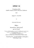
nanocrystalline powders. The structure model of the inorganic decomposition product yZ112P2O7, whose structure was determined ab initio from powder diffraction data [8] [S. G. Pbcm, a = 4.9504(5) A, b = 13.335(2) A, c = 16.482(3) A, V= 1088.1(2) A3], was used to generate calculated patterns [15], Samples were assumed to be strain-free, with isotropic and monodisperse crystallites. Various crystallite diameters D were selected in the range 80-1200 A. The apparent sizes £p (= 3D/4) were transformed into integral breadths |3* (= Ep"1) and, then, expressed in 20 units Pie (= X|3*/cos0) for selected diffraction lines within the angular range 9-80° (20). To avoid unnecessary truncation errors with Lorentzian profiles [16], a Gaussian function was preferred to describe individual reflections. The angular dependence of FWHM was modelled with the usual quadratic function in tan0 from the FWHM values of the Gaussian reflections calculated from the relation FWHM = 0.93 94(32e [17]. Using the simulated patterns, the crystal structure of Y-Z112P2O7 was solved back using the programs EXPO and FOX. Hie efficiency of the direct methods was estimated by the number of correct electron peak positions found. Using the parallel tempering algorithm, it was pointed out that both the calculation time per trial and the number of trials increased when crystallite diameter decreased. A penalty factor E [= (NJD)"], combining the number of trials N, required to find the structure model and the crystallite diameter D (in A), was then calculated. This result deserves the following comments: 49 % 53 % 59 %
1—1—L-1—1—M-J—L-1—1—L-1—1—L-1—1—I—i—i—I—i—i—I—i—i—1-4 0
0 80
160
200
280
320
400
600
800
1000
1200
D(A)
Figure 3. Comparison of the efficiency of direct methods and parallel tempering algorithm applied to powder patterns simulated for various spherical crystallite diameters D (A) from the crystal structure of y-ZnjPjO-. The number of atoms (on 14 independent atoms) found by EXPO is shown by the histogram, together with the percentage of statistically independent reflections (°o)[10 and refs therein], the penalty factor E related to the direct space method is represented by the solid line.
- D > 400 A: the crystal structure could be solved easily by the two approaches. The direct methods required a few minutes only, while the parallel tempering needed a few hours. - 160 A < D < 320 A: the structure was also solved by the two approaches. While the parallel tempering algorithm was moderately affected by line broadening, the direct methods provided only half atoms. Subsequent different Fourier calculations were thus time-consuming. - D = 80 A: the extraction of integrated intensities was not possible, due to the excessive diffraction line broadening. The structure model was found by FOX after longer time.
14
European Powder Diffraction Conference, EPDIC 9
Concluding remarks This study demonstrates the efficiency of a direct space approach to solve the crystal structure of inorganic compounds when the direct methods fail, especially in the case of noticeable diffraction line overlap. However, it is also shown, through the structure determination of y-Zn2P207, that some atoms are usually found using the direct methods. Then, it is useful to consider this partial model for structure completion using a global optimisation approach.
References 1.
Newsam, J. M., Deem, M. W. & Freeman, C. M., 1992, Accuracy in Powder Diffraction II. NIST Special Pubi. No. 846, 80.
2.
Kariuki, B. M., Serrano-Gonzalez, H., Johnston, R. L. & Harris, K. D. M., 1997, Chem. Phys. Lett., 280, 189.
3.
Shankland, K„ David, W. I. F. & Csoka, T„ 1997, Z. Kristallogr., 212, 550.
4.
Altomare, A., Cuocci, C., Giacovazzo, C., Grazia, A., Moliterni, G. & Rizzi, R., 2004, Mater. Sci. Forum, 443-444, 23.
5.
Putz, H., Schòn, J. C. & Jansen, M., 1999, J. Appi. Crystallogr., 32, 864.
6.
Falcioni, M. & Deem, M. W„ 1999, J. Chem. Phys., 110, 1754.
7.
Favre-Nicolin, V. & Cerny, R., 2002, J. Appi. Crystallogr., 35, 734.
8.
Bataille, T., Bénard-Rocherullé, P. & Louér, D., 1998, J. Solid State Chem., 140,
9.
Boultif, A. & Louér, D., 2004, J. Appi. Crystallogr., 37, 724.
62.
10. Altomare, A., Burla, M. C., Camalli, M., Carrozzini, B., Cascarano, G. L., Giacovazzo, C., Guagliardi, A., Moliterni, A. G. G., Polidori, G. & Rizzi, R., 1999, J. Appi. Crystallogr., 32, 339. 11. Le Bail, A., Duroy, H. & Fourquet, J. L., 1988, Mater. Res. Bull., 23, 447. 12. Rodriguez-Carvajal, J., 1990, in Abstracts of the Powder Diffraction Meeting, edited by J. Galy & D. Louér (Toulouse, France), p. 127. 13. Roisnel, T. & Rodriguez-Carvajal, J., 2001, Mater. Sci. Forum, 378-381, 118. 14. Mahé, N. & Bataille, T., 2004, Inorg. Chem., 43, 8379. 15. Bataille, T. Audebrand, N., Boultif, A. & Louér, D., 2004, Z Kristallogr., 219, 881. 16. Toraya, H., 1985, J. Appi. Crystallogr., 18, 351. 17. Langford, J. I., 1978, J. Appi. Crystallogr., 11, 10. Acknowledgements. The authors are grateful to Dr N. Audebrand and J.-F. Colin for helpful discussions. G. Marsolier and S. Lanoé are indebted for their technical assistance in X-ray powder data collection.
Z. Kristallogr. Suppl. 23 (2006) 15-20 © by Oldenbourg Wissenschaftsverlag, Miinchen
15
Molecular crystal structures from powder X-ray diffraction techniques Eugene Y. Cheung and Kenneth D.M. Harris* School of Chemistry, Cardiff University, Main Building, Park Place, Cardiff CF10 3 AT, United Kingdom Contact author; e-mail: [email protected] Keywords: powder diffraction, structure solution, genetic algorithms Abstract. This paper presents an overview of the genetic algorithm technique for structure solution from powder diffraction data, and gives a number of examples that illustrate the application of this technique to solve crystal structures of different types of molecular solids, including oligopeptides and multi-component co-crystals.
Introduction In recent years, most reported crystal structure determination of organic molecular solids from powder diffraction data [ 1 ^ ] has used the direct-space strategy for structure solution, although a number of successful structure determinations of such materials using traditional approaches for structure solution have also been reported [5], The direct-space strategy [1] follows a close analogy to global optimization procedures, which find applications in many areas of science. Trial structures are generated in direct space, independently of the experimental powder diffraction data, and the suitability of each trial structure is assessed by directly comparing the powder diffraction pattern calculated for the trial structure and the experimental powder diffraction pattern. This comparison is quantified using an appropriate figure-of-merit. Our implementations of the direct-space strategy have used the weighted powder profile R-factor Rwp (i.e. the R-factor normally used in Rietveld refinement), which considers the entire digitized intensity profile point-by-point, rather than the integrated intensities of individual diffraction maxima. Each trial structure is defined by a set (T) of structural variables, which represent the position, orientation and intramolecular geometry of each molecule in the asymmetric unit. The position is defined by the coordinates {x, y, z} of the centre of mass or a selected atom, and the orientation is defined by the rotation angles {6, , 2H; BA) and pentafluorobenzoic acid (C6F5CC>2H; PFBA) has been determined [13] from powder X-ray diffraction data using our parallel GA technique for structure solution. High-resolution solid state 13C NMR of the co-crystal indicates that the asymmetric unit contains two molecules of BA and two molecules of PFBA. In space group Cc, the values of x and z for one molecule can be fixed arbitrarily, and thus a total of 26 structural variables are required in the GA calculation (each of the four independent molecules in the asymmetric unit has one variable torsion angle). The structure was solved within 170 generations for four sub-populations each containing 50 trial structures. The structure (figure la) comprises stacks of alternating BA and PFBA molecules, with two crystallographically independent types of stack. There is hydrogen bonding between the carboxylic acid groups of molecules in the two types of stack. All interactions of this type involve a BA molecule in one stack and a PFBA molecule in the other stack. The two independent BA molecules and the two independent PFBA molecules differ appreciably in conformation (i.e. the torsion angle between the carboxylic acid and aryl units). The fact that this torsion angle is larger for one molecule of each type can be explained by the need to avoid repulsive F * " 0 interactions in the structure.
18
European Powder Diffraction Conference, EPDIC 9
bonding involving the carboxylic acid groups is clearly evident, (b) Crystal structure of the BN/BQ/AN co-crystal. Dotted lines indicate the E-stacking interactions and hydrogen bonded chains.
Many molecular co-crystals cannot be prepared by conventional solution phase crystallization, but can instead be prepared by grinding together the "pure" solid phases of the constituent molecules. Materials prepared in this manner are virtually always microcrystalline powders, and are therefore not amenable to structural characterization by single crystal X-ray diffraction. We have demonstrated [14] the use of powder X-ray diffraction to determine the structure of a co-crystal material prepared by the solid state grinding route. The material contains three molecular components - racemic bis-|3-naphthol (BN), benzoquinone (BQ) and anthracene (AN). Structure solution was carried out using our parallel GA method in space group C2/c. Hie contents of the asymmetric unit (confirmed on the basis of highresolution solid state 13C NMR data) comprise one BN molecule, one BQ molecule and half of an AN molecule (which resides on a two-fold axis), and the structure solution calculation involved a total of 17 structural variables. The structure was solved within 50 generations for two sub-populations each containing 100 trial structures. The structure (Figure lb) is based on three different interaction motifs: edge-to-face interactions between BQ (edge) and AN (face) molecules, face-to-face interactions between BQ and BN molecules, and chains of O—H*"0 hydrogen bonds involving BN and BQ molecules. Structural characterization of co-crystal materials prepared by grinding procedures has previously been limited by the fact that the preparation procedure intrinsically produces polycrystalline powders, but it is clear that structure determination from powder diffraction data has a crucial role to play in the structural characterization of new co-crystal phases produced in this way. Flexible Molecules: Structural Rationalization of Oligopeptides An understanding of the molecular conformations and intermolecular interactions in crystalline oligopeptides can provide important insights into the structural properties of polypeptide sequences in proteins [15], However, these materials often cannot be prepared as single crystals appropriate for single crystal X-ray diffraction studies, and the use of powder diffraction data may represent the only viable route for structure determination [16], We discuss two examples of oligopeptide structures (Piv- L Pro-Gly-NHMe and Piv- L Pro-y-Abu-NHMe)
Z. Kristallogr. Suppl. 23 (2006)
19
determined from powder X-ray diffraction data using our GA technique for structure solution. The interest in both materials concerns their potential to form (3-tuni conformations [17]. In the GA structure solution calculation [18] for Piv- L Pro-Gly-NHMe, the genetic code comprised 9 variables (in space group PI, the position {.T. V. zj of the molecule is fixed arbitrarily). Hie structure was solved within 50 generations for a population size of 100 trial structures. Figure 2a shows the final refined structure of Piv- L Pro-Gly-NHMe, in which the molecule adopts a Type II (3-tuni conformation stabilized by an intramolecular 4—>1 hydrogen bond between the C = 0 group of the Piv residue and the methylamide N-H group (N—O, 2.99 A; N—O-C, 140.6°). The second material Piv- L Pro-y-Abu-NHMe differs from Piv- L Pro-Gly-NHMe by the introduction of two additional CH2 units within the peptide chain. In the GA structure solution calculation [19], the genetic code comprised 13 variables. The structure was solved within 20 generations for a population size of 100 trial structures. In the crystal structure, Piv- L Proy-Abu-NHMe adopts a folded conformation (Figure 2b), with a short C - H " * 0 interaction [H*"0, 2.51 A; C'"0, 3.59 A; C - H " * 0 , 172.4°; hydrogen atom position normalized according to standard geometries from neutron diffraction] between one of the a-methylene hydrogen atoms of the y-Abu residue and the C = 0 group of the Piv residue. This C-H***0 interaction defines an intramolecular cyclic 10-atom motif, similar to that observed in the classical (3-tuni (which involves an intramolecular N - H * " 0 hydrogen bond).
(a)
•
1
(b)«
x
Figure 2. (a) Conformation of Piv- Pro—Gly—NHMe in the crystal structure, showing the formation of a type II[¡-turn, (b) Conformation of Piv-1 Pro-y-Abu—NHMe in the crystal structure, showing the formation of an intramolecular C-H,,,0=C interaction.
References 1.
Harris, K.D.M., Tremayne, M., Lightfoot, P., Bruce, P.G., 1994, J. Am. Chem. Soc., 116, 3543.
2.
Harris, K.D.M., Tremayne, M., 1996, Chem. Mater., 8, 2554.
3.
Harris, K.D.M., Tremayne, M., Kariuki, B.M., 2001 ,Angew. Chemie,Int. Ed., 113, 1674.
20
European Powder Diffraction Conference, EPDIC 9
4.
David, W.I.F., Shankland, K., McCusker, L.B., Baerlocher, C. (Editors), 2002, Structure Determination from Powder Diffraction Data (Oxford University Press/International Union of Crystallography).
5.
Bruneiii, M., Wright, J.P., Vaughan, G.R.M., Mora, A.J., Fitch, A.N., 2003, Angew. Chemie, Int. Ed., 42, 2029.
6.
Kariuki, B.M., Serrano-González, H., Johnston, R.L., Harris, K.D.M., 1997, Chem. Phys. Lett., 280, 189.
7.
Harris, K.D.M., Johnston, R.L., Kariuki, B.M., 1998, Acta Crystallogr., A54, 632.
8.
Turner, G.W., Tedesco, E., Harris, K.D.M., Johnston, R.L., Kariuki, B.M., 2000, Chem. Phys. Lett., 321, 183.
9.
Habershon, S., Turner, G.W., Kariuki, B.M., Cheung, E.Y., Hanson, A.J., Tedesco, E., Albesa-Jové, D., Chao, M.H., Lanning, O.J., Johnston, R.L., Harris, K.D.M., 2003, EAGER, Computer Program for Structure Solution from Powder Diffraction Data (Cardiff University and University of Birmingham).
10. Seaton, C.C., Tremayne, M., 2002, Chem. Commun. 880. 11. Habershon, S., Harris, K.D.M., Johnston, R.L., 2003, J. Comp. Chem., 24, 1766. 12. Habershon, S., Cheung, E.Y., Harris, K.D.M., Johnston, R.L., 2004, Chem. Phys. Lett., 390, 394. 13. Albesa-Jové, D., Kariuki, B.M., Kitchin, S.J., Grice, L., Cheung, E.Y., Harris, K.D.M., 2004, ChemPhysChem, 5, 414. 14. Cheung, E.Y., Kitchin, S.J., Harris, K.D.M., Imai, Y., Tajima, N., Kuroda, R., 2003, J. Am. Chem. Soc., 125, 14658. 15. Fischer, G„ 2000, Chem. Soc. Rev., 29, 119. 16. Seebach, D., Matthews, J.L., Meden, A., Baerlocher, C., McCusker, L.B., 1997, Helv. Chim. Acta, 80, 173. 17. Seebach, D., Brenner, M., Rueping, M., Schweizer, B., Jaun, B., 2001, Chem. Commun., 207. 18. Tedesco, E., Harris, K.D.M., Johnston, R.L., Turner, G.W., Raja, K.M.P., Balaram, P., 2001, Chem. Commun., 1460. 19. Cheung, E.Y., McCabe, E.E., Harris, K.D.M., Johnston, R.L., Tedesco, E., Raja, K.M.P., Balaram, P., 2002, Angew. Chemie, Int. Ed., 41, 494. Acknowledgements. We are grateful to EPSRC, Cardiff University, University of Birmingham, Purdue Pharma, Ciba Specialty Chemicals, Wyeth, Proctor and Gamble, Accelrys and Astra-Zeneca for supporting our research in the field covered by this article. The contributions of other research group members and collaborators mentioned in the references are also gratefully acknowledged.
Z. Kristallogr. Suppl. 23 (2006) 21-26 © by Oldenbourg Wissenschaftsverlag, Miinchen
21
On the uncertainty of lattice parameters refined from neutron diffraction data B. Peplinski1'*, D. M. Tobbens2, W. Kockelmann3, R. M. Ibberson3 'Federal Institute for Materials Research and Testing (BAM), Richard-Willstatter-Str. 11, 12489 Berlin, Federal Republic of Germany 2 Hahn-Meitner-Institute (HMI), SF2, Glienicker Str. 100, 14109 Berlin, Germany 3 ISIS Facility, Rutherford Appleton Laboratory (RAL), Chilton, Didcot, OX11 0QX, UK *Contact author; e-mail: [email protected] Keywords: uncertainty, lattice parameter refinement, neutron diffraction, Rietveld method Abstract. Factors crucial to the reliable estimation and minimization of the uncertainty of measurement of lattice parameters refined from neutron diffraction data are analysed. Estimates of the uncertainty of measurement are derived from diffraction data of a reference material collected at one constant wavelength and two time-of-flight neutron diffractometers.
Introduction Lattice parameter refinements from neutron diffraction (ND) data may be preferred to those from diffraction data collected with conventional X-rays or synchrotron radiation, for example if investigations on coarse-grained powders, with volumes of up to several cubic centimetres, are meant to be representative for the whole specimen. For these kind of analyses ND takes advantage of the high penetration of neutrons for most elements (isotopes), allowing them to simultaneously probe the whole volume of a thick specimen with nearly no attenuation. Furthermore, with ND the observed intensities of Bragg reflections are averaged over a larger number of crystallite orientations than the high-resolution modes of the conventional X-ray or synchrotron radiation techniques usually allow. This is a consequence of the large equatorial and axial divergence normally employed in constant wavelength ND beam optics and of the wide wavelength spectrum and the large acceptance angles of the detector banks used in time-of-flight (ToF) ND. If a lattice parameter refinement is carried out by the Rietveld method or a related technique, then usually the only available measures of the uncertainty of the refined lattice parameters are the estimated standard deviations (e.s.d.s) calculated by the full pattern fitting program. However, e.s.d.s are measures of precision rather than of accuracy and these two terms must not be confused. For a sound estimation of the measurement uncertainty of lattice parameters that are refined from ND diffraction data more information is needed than just the e.s.d.s that are provided by the Rietveld refinement of the diffraction pattern of the sample.
22
European Powder Diffraction Conference, EPDIC 9
General considerations The uncertainty of measurement of refined lattice parameters has three aspects common to all types of ND instruments. These are 'wavelength calibration', 'neutron-optical aberrations & line profile modelling' and 'serial correlation' which will be discussed individually. Wavelength calibration Bragg's law involves a 100% correlation between the d-values (and therefore the lattice parameters) of a crystallographic phase and the wavelength of the diffracted radiation. Thus for any assumed value of the wavelength X another set of lattice parameters results, whereas the agreement indices and the e.s.d.s remain the same. Since for all neutron diffractometers the wavelength is not known per se, as would be the case with characteristic X-ray radiation, even under the idealising assumption of the complete absence of any systematic errors from the model and from the observed data, the lattice parameters are completely indeterminate. Therefore, a determination of accurate lattice parameters by constant wavelength ND or ToF ND necessarily includes calibration procedures with reference materials (CRM) that have lattice parameters certified for a given temperature by independent analytical techniques. The propagation of the error which is associated with the calibration has to be accounted for in the estimation of the combined standard uncertainty (for definitions of metrological terms see [1] or derived publications; see also equations (2) and (3)) of the lattice parameters of the sample! If the ND analysis of the actual sample as well as the calibration procedure are carried out by the Rietveld method, then the estimation of the combined standard uncertainty of the refined lattice parameters can be based on the e.s.d.s of these two Rietveld analyses. Neutron optical aberrations & line profiles Bragg's law implies that the position of the diffraction lines and the wavelength are numbers, whereas in practise they are single-numbered characteristics of intensity distributions. Extracting from the observed diffraction pattern an unbiased, i.e. hypothetical diffraction pattern, which has been corrected for all aberrations is the precondition for the determination of accurate line positions 20corrected from which unbiased d-values and lattice parameters can be calculated. Within the framework of the 'fundamental parameter approach' (FPA) accurate mathematical modelling of the observed diffraction line profiles is achieved by convoluting the individual contributions. In constant wavelength ND the diffraction pattern I(2Q)0bserved is described by a convolution of the wavelength distribution A, the instrument function, and sample-related contributions. In ToF-ND I(20)observed and A are replaced by I(d)0bserved and by the time structure of the neutrons leaving the moderator, respectively. Serial correlation e.s.d.s calculated by many Rietveld programs are not reliable measures of the 'probable errors', because, in many cases, they are systematically too small due to 'serial correlation' (for Cooper's criticism on the reliability of e.s.d.s in the Rietveld method see [2]). Therefore, uncorrected e.s.d.s are not only no measure of accuracy, but can also be an unreliable measure of the precision of refined lattice parameters. E.s.d.s are calculated under the assumption that the values in the difference curve are independent observations. However, adjacent individual points in the difference curve are not independent but correlated by the profile function used. The extent of this correlation depends on the size of the 20 sampling steps used for
Z. Kristallogr. Suppl. 23 (2006)
23
data collection and evaluation. A formula for estimating the corrections that should be applied to the e.s.d.s has been given by Berar and Lennan [3] who established that e.s.d.s calculated by Rietveld programs without consideration of serial correlations are often too small by a factor of approximately two. Multiplying the uncorrected e.s.d. by a correction factor gives the e.s.d.corrected, i.e. an estimate of the standard deviation that also includes serial correlation.
Types of ND instruments Further aspects of the uncertainty of measurement have to be analysed separately for each of the following three types of ND instruments: A. High-resolution scanning powder diffractometers for monochromatic neutrons, equipped with parallel collimators placed in front of the detectors and restricting the equatorial divergence of the diffracted beam (prototype: D1A and D2B at the ILL) B. High-resolution powder ToF-ND instruments C. Powder diffractometers for monochromatic neutrons in Debye-Scherrer-geometry with one (or several) position-sensitive detector(s) providing resolution in the equatorial plane
Design characteristics of ND instruments of type A Diffractometers of type A often use large monochromators of quite complex design which produce an incident beam with a large axial convergence. Soller collimators are usually not used with this type of diffractometer. The intensity distribution in the incident monochromized neutron beam is not necessarily homogeneous along the goniometer axis. The existence of a wavelength spread in the incident beam as shown by [4] means in practise that in Bragg's law the wavelength X is to be replaced by its effective value Xng.. The large axial convergence/divergence in the incident beam of this type of diffractometer causes distortions of the diffraction line profiles and line shifts ([5] and literature given therein). Within the framework of the 'fundamental parameter approach' these axial effects can be accounted for by the 'axial divergence correction function' faxM divergence^ approx_ (20) which depends on axial divergence, geometry and dimensions of the instrument and the sample as well as on the width and shape of the reflections which are influenced by the real structure of the specimen. In general this function can be determined only approximately due to correlations between some of the parameters. The contributions to the 'axial divergence correction function' are convoluted with each other. This means that they are not additive. For a diffractometer of type A the equatorial cross section of the incident beam can be chosen so large that it exceeds the diameter of even large sample containers by far. The most essential design element of this type of diffractometer are tight Soller-like collimators in front of each detector, that limit the equatorial divergence typically to about 5' or 10', making the detector angular-sensitive, rather than position-sensitive. The sampling volume of each detector again exceeds the diameter of even large sample containers by far. This very large sampling volume provided by the intersection of the wide incident beam with the wide detected diffracted beams make this type of instrument insensitive to small and even moderate sample displacements in the equatorial plane. This means that any eccentricity of the sample in the order of a few tens of a mm or even a mm will cause no line shifts in the dif-
24
European Powder Diffraction Conference, EPDIC 9
fraction patterns. This is an inherent advantage of this type of instruments as it eliminates one of the main factors detrimental to high accuracy of lattice parameter refinements. The only remaining systematic error of 20-position is the residual shift of the zero point, which has to be refined together with the wavelength.
Contribution of the calibration procedure to the uncertainty of refined lattice parameters (ND instruments of type A) The following three, yet contradicting optimization criteria for carrying out an accurate 'wavelength calibration' with a calibrant are a dilemma for which constant wavelength neutron diffractometers do not provide a solution: a) It would be necessary to use for the Rietveld refinement a range of the diffraction angle as wide as possible, including explicitly the low angle region, as only this allows for the highest accuracy in the 'wavelength calibration'. The largest possible range of diffraction angles results in the largest changes of the sine-function which - according to Bragg 's law - is a necessary pre-condition for the simultaneous refinement of the two strongly correlated parameters 'wavelength' and 'zero point'. b) It would be necessary to use for the Rietveld refinement only the angular region around 180° (20), as there the conversion error reaches its minimum. c) It would be necessary to use for the Rietveld refinement only the angular region about 90°, as there the influence of the axial aberrations (and of the residual errors in the mathematical model describing the axial divergence error correction) on the angular positions of the diffraction lines reaches its minimum. This dilemma also contributes to the situation (see preceding section), that the 'wavelength calibration' not necessarily yields the true physical values of these two strongly correlated parameters, but just their effective values. These effective values might depend on the choice of the low- (LAL) and high angle limit (HAL) used for the refinements. The effects mentioned so far lead to the following rewriting of Bragg's law: d
m
= 2. s.i nt { .d
o b s
___
+ E P S
X e
f f ^ V+ faxial
divergence,approx.
7T7TT
(1)
with Xeff = effective value of X; 0obs. = observed halved diffraction angle; EPSijeff. = effective value of the zero point correction for the detector bank. Estimating the combined standard uncertainty u of the wavelength calibration three uncorrelated contributions have to be taken into account: (u(Ae/r )/Aeff ) *
v
'
'combined
« (u(Aeff \
+
\
)/Aeff
JJ /
(U(aCRM
)
)
JJ / calibrant, Rietveld, corrected for serial correlation
1 a
CRM
)certtflcate
+ (U(aCRM
)
1 Ü
CRM
i^)
)T_caUbration
The first term on the right-hand side of equation (2) contains the contribution from the Rietveld refinement for the calibrant. Data for this term can be extracted from Table 2. They
Z. Kristallogr. Suppl. 23 (2006)
25
show that this term is the moderately dominating one if silicon powder is used as a calibrant. The second term on the right-hand side is the uncertainty of the lattice parameter of the CRM (^CRM) that is stated in the certificate. The third term accounts for the uncertainty of the lattice parameter that is associated with the temperature of the calibration (Tcaiibration).
Uncertainty of refined lattice parameters (instruments of type A) It is good practice not to release the zero point during the refinement of the data of the sample, but to keep it fixed at the value from the 'wavelength calibration'. Thus inaccuracies of the refined lattice parameter(s) are avoided and their e.s.d.s are minimized. The combined relative standard uncertainty u of a lattice parameter aj of a phase refined from a diffraction pattern collected at the temperature Tsampie can be estimated by equation (3). ( u ( a . ) / a .1 ) ' / combined
~
( u ( a . ) / a 1. ) ' ' sample,
+ (V u (\ a .?)/ / a . i) /"sample,
+ («(«)/«• X
Rietveld,
corrected
axial divergence
tple
+ (" ( \
V
for serial
error
correlation
(3)
correction
A f ' f . ) '1' calibration, )
combined
with n = 2 or 1 if there is no correlation at all or maximum correlation, respectively. In those cases where the second and third term on the right-hand side are negligible the use of n=l will be especially appropriate, as the remaining two contributions are strongly correlated (same instrument, Rietveld program, line profile model). The symbols in equation (3) have the same meanings as in equation (2). The metrological connection between equations (2) and (3) is maintained o n l y if the following conditions are fulfilled: a) identical apparative conditions (settings of the monochromator and detector; factually analysed sample height) are used for the sample and for the calibrant (CRM); b) identical mathematical models of axial divergence are applied in the Rietveld evaluation of the diffraction patterns of the sample and of the calibrant (CRM).
ND instruments of type B (ToF) ToF diffraction patterns are displayed as 1(d) and Bragg's law reads in this representation as h d
m
1
=
1 m
l { L
o
+
L
i
(4)
) s m O
with h = Planck constant, m = neutron mass, L0 = the moderator-to-sample distance, = the sample-to-detector distance, L = L0+L! the total flight path and t = the total time-of-flight. For each channel of the position-sensitive detector bank the product L-sinQ has to be calibrated. Sample displacement leads to changes in the flight path, A(L0+L!), and to angular shifts AO, which can result in considerable shifts of the diffraction lines in a ToF diffraction pattern [6], But from the differentiated version of equation (4) it is clear that using very large flight paths and only the backscattering region, the influence of moderate sample displacement on the observed d-values can be diminished to practically zero.
26
European Powder Diffraction Conference, EPDIC 9
Results from three ND instruments Diffraction data of NIST SRM640c (silicon powder) were collected at a constant wavelength and at two ToF neutron diffractometers. Details of data collection and evaluation are given in Table 1. The uncorrected estimated standard deviations and the corrected estimated standard deviations, multiplied by the coverage factor k=2, resulting from these lattice parameter refinements are summarized in Table 2. The latter are not yet the expanded uncertainties \J95% of the complete lattice parameter refinements as they do not include the additional uncertainty associated with the calibration. Table 1. Conditions applied for the collection and evaluation of the diffraction data. instrument E9 (HMI) ROTAX (ISIS) HRPD (ISIS) detector bank(s) range of d-values / A data accumulation / h profile function, program
64 3 He multidetector 0.9 - 6.0 7 FPA [5], Fullprof.2K
backscattering (#3) backscattering (#1) 0.4-3.0 0.5-2.1 8 10 double-exponential-pseudo-Voigt, GSAS
Table 2. Estimated relative measurement uncertainties u(a0)/a0 of the lattice parameter of silicon as derived from Rietveld refinements. instrument E9 (HMI) ROTAX (ISIS) HRPD (ISIS) ^'S'd-imcorrected (*0 2-e.s.d.com,cKd (a0) /a0
5.5-10"6 1.8-10"5
6.8-10"6 2.7-10"5
1.4-10"6 5.6-10"6
Conclusion To accurately refine lattice parameters from neutron diffraction data the conditions used for data collection and data evaluation should be carefully chosen. The lattice parameter of a well-crystallized cubic material can be refined with an expanded relative uncertainty of U95o/o(a)/a = 5-10"5 using a high-resolution multi-collimator/multi-detector diffractometer for monochromatic neutrons or a high-resolution ToF-ND instrument. With a dedicated ToF-ND instrument a value of U95o/0(a)i/ai = 1 -lO"5 is feasible.
References 1.
BIPM, IEC, IFCC, ISO, IUPAC, IUPAP, OIML, 1993, Guide to the expression uncertainty in measurement (GUM).
2.
Sakata M., Cooper M.J., 1979, J. Appl. Cryst., 12, 554.
3.
Berar J.F., Lennan P., 1991, J. Appl. Cryst., 24, 1.
4.
Holden T.M., 1996, Mater. Res. Soc. Symp. Proc., 376, 385.
5.
Tobbens D.M., Z. Krist., this Proceedings
of EPDIC-9,
of
Prague, 2004, accepted.
6. Wang X.-L., Wang Y.D., Richardson J.W., 2002, J. Appl. Cryst., 35, 533. Acknowledgements. One of the authors (B.P.) would like to thank Prof. G. Will, University of Bonn, as well as Dr. S. Noack and Dr. W. Hasselbarth, both BAM, for helpful discussions.
1.2
Qualitative and Quantitative Phase Analysis
Z. Kristallogr. Suppl. 23 (2006) 29-34 © by Oldenbourg Wissenschaftsverlag, Miinchen
29
Application of the Rietveld method to the severely superimposed diffraction patterns of technical products containing a large number of solid solution phases *
B. Peplinski , P. Kocher, G. Kley Federal Institute for Materials Research and Testing (BAM) Richard-Willstatter-Str. 11, D-12489 Berlin, Federal Republic of Germany Contact author; e-mail: [email protected] Keywords: phosphorus recovery, Rietveld method, sewage sludge ash Abstract. The Rietveld method was used to reliably interpret severely superimposed diffraction patterns of a sewage sludge ash before and after being turned into an ecologically desirable, high-quality fertilizer by a newly-developed method of thermochemical treatment. The thermochemical treatment causes severe changes in the phase composition of the ash, which can be described as a mixture of a large number of solid solution phases.
Introduction Many countries require a) the removal of phosphorus from wastewater before discharging it into the ambient environment and b) the development of technologies for the reuse of the recovered phosphorus [1]. By doing so, local rivers and lakes are protected from eutrophication (phosphorus pollution), and the world's limited resources of mineral phosphorus can be used more economically. Although phosphorus recovery from sewage sludge is already practised on a larger scale, the economically and ecologically sensible reuse of millions of tons p.a. of recovered phosphates is still being debated. The principal obstacle for the reuse of sewage sludge ashes as phosphate fertilizer in agriculture is their high content of ecologically harmful heavy metals, such as Zn, Cu, Pb, Cd, Hg, which exceeds the legal limits considerably. A new technology [2] is being developed at the BAM that reduces the content of ecologically harmful heavy metals in sewage sludge ashes to a value, orders of magnitude below today's legal limits. Sewage sludge ashes treated with this technology can be used as ecologically desirable, high-quality fertilizers. In addition, this treatment increases the bioavailability of the phosphorus content from a low original value of 65 to 100%; cost effectiveness is an essential advantage of the technology. The new technology has been developed on chemical intuition and decades of experience. The chemical and phase composition of sewage sludge ashes are different from those of ashes from coal burning power stations. The temperature-time regime of sewage sludge incineration typically includes only 5 s in the
30
European Powder Diffraction Conference, EPDIC 9
hottest zone at a temperature of about 850 °C. Therefore, sewage sludge ashes are nonequilibrium mixtures of a large number of interdispersed crystallographic phases. The element distribution and phase composition of five untreated sewage sludge ashes were recently investigated thoroughly by a variety of analytical techniques including X-ray powder diffraction (XRD), although the latter was not used to its full potential [3], The phase transformation processes accompanying the thermochemical treatment [2] of sewage sludge ashes have not been investigated until now.
Samples The phase composition of four products (P0-P3) was analysed. P0 is a sewage sludge ash from a large municipal wastewater treatment plant where FeCl 2 was the precipitation agent. P1-P3 were obtained after small-scale thermochemical treatment was carried out on P0 according to [2], For each of these three products slightly different process parameters were employed. For each thermochemical treatment, 100 g of the ash, P0, were hand-pressed into a brick-shaped moulding 1 0 x 5 x 2 cm 3 in size, put on a sintered corundum tray, soaked with 100 g of a MgCl 2 solution (400 g MgCl 2 /kg) and inserted into a large muffle furnace. The latter was heated up to 1050°C, kept at this temperature for 2 h, and then cooled to room temperature. The product, PI, is a homogenized blend of five batches produced under identical conditions, yielding a final product of about 600 g. The effectiveness of this treatment in reducing the content of ecologically harmful heavy metals is demonstrated by the results of element analyses carried out using X-ray fluorescence spectrometry (XRF) (table 1). The mass fractions of the main elements in these ashes are given in table 2. The thermochemical treatment of the ash leads to an increase in its MgO content and also causes an increase in the bioavailability of the total phosphate content of this ash. This is documented by the data in table 3. These analyses were carried out by [4] using the 'standard citric acid test for the bioavailability of phosphate fertilizers' [5] in Table 1. Mass fractions of ecologically harmful heavy metals in a sewage sludge ash before (PO) and after (P1-P3) thermochemical treatments according to XRF analyses.
ash P0 PI P2 P3
Zn 2680 27 28 420
Cu 1560 165 42 220
mass fraction / (mg/kg) Sn Pb Cd 88 3 191