Morris' human anatomy - a complete systematic treatise [7 ed.]
381 57 218MB
English Pages [1534] Year 1923
Cover Page
Title Page
Table of Contents
Section1
Section2
Section3
Section4
Section5
Section6
Section7
Section8
Section9
Section10
Section11
Section12
Section13
Section14
Section15
Index
Recommend Papers


![Human anatomy [1]](https://ebin.pub/img/200x200/human-anatomy-1.jpg)


![Human Anatomy [4 ed.]](https://ebin.pub/img/200x200/human-anatomy-4nbsped.jpg)
![Human Anatomy, Global Edition: Marieb Human Anatomy 9 [9 ed.]
1292314478, 9781292314471](https://ebin.pub/img/200x200/human-anatomy-global-edition-marieb-human-anatomy-9-9nbsped-1292314478-9781292314471.jpg)
![Atlas Of Human Anatomy [7 ed.]](https://ebin.pub/img/200x200/atlas-of-human-anatomy-7nbsped.jpg)
![Morris' human anatomy - a complete systematic treatise [7 ed.]](https://ebin.pub/img/200x200/morris-human-anatomy-a-complete-systematic-treatise-7nbsped.jpg)
- Author / Uploaded
- C. M. Jackson
File loading please wait...
Citation preview
MORRIS’
HUMAN ANATOMY SEVENTH EDITION
PUBLISHERS’ NOTE TO SEVENTH EDITION This edition of Morris’ Anatomy carries to a further point the thorough revision that was made of the text for the sixth edition, which was published but sixteen months ago. In the interim, the text has been subjected to class use and the improvements found to be desirable both in the interest of teachers and students have been made. In this connection our grateful thanks are tendered to those teachers who were kind enough to offer sug-
gestions. A number of the pictures have been reengraved and improved, and the colored illustrations refined so as to conform to higher pedagogical and artistic standards.
MORRIS’
HUMAN ANATOMY A COMPLETE SYSTEMATIC
TREATISE
EDITED BY
C. M. JACKSON, M. S., M. 1). PROFESSOR AND
DIRECTOR
OF
UNIVERSITY
THE DEPARTMENT OF
OF
ANATOMY,
MINNESOTA
THE CONTRIBUTORS CHARLES R. BARDEEN, University of Wisconsin. ELIOT R. CLARK, University of Missouri. ALBERT C. EYCLESHYMER, University of Illinois. J. F. GUDERNATSCH, Formerly Cornell University Medical College. IRVING HARDESTY, Tulane University of Louisiana. C. M. JACKSON, University of Minnesota. DEAN LEWIS, Rush Medical College.
RICHARD E. SCAMMON, University of Minnesota. J. PARSONS SCHAEFFER, Jefferson Medical College.
H. D. SENIOR, University and Bellevue Hospital Medical College, N. Y. G. ELLIOT SMITH, University of London. CHARLES R. STOCKARD, Cornell University Medical College. R. J. TERRY, Washington University, St. Louis.
SEVENTH EDITION
ELEVEN HUNDRED AND SIXTY-FOUR ILLUSTRATIONS FIVE HUNDRED AND FIFTEEN PRINTED IN COLORS
PHILADELPHIA
P. BLAKISTON’S SON
&
1012 WALNUT STREET
CO.
Copyright.
1923,
by
P. Blakiston’s
PRINTED BY THE
IN
Son
U. S. A.
MAPLE PRESS YORK PA
&
Co.
CONTRIBUTORS TO SEVENTH EDITION CHARLES R. BARDEEN, University of Wisconsin. ELIOT R. CLARK, University of
Missouri. ALBERT C. EYCLESHYMER, University of Illinois. J. F. GUDERNATSCH, Formerly Cornell University Medical College.
IRVING HARDESTY, Tulane University of Louisiana. C. M. JACKSON, University of Minnesota. DEAN LEWIS, Rush Medical College.
For arrangement
RICHARD E. SCAMMON, University of Minnesota. J. PARSONS~SCHAEFFER, Jefferson Medical College. H. D. SENIOR, University and Bellevue Hospital Medical College, N. Y. G. ELLIOT SMITH, University of London. CHARLES R. STOCKARD, Cornell University Medical College. R. J. TERRY, Washington University, St. Louis.
of subjects and authors
see page ix
EDITOR’S PREFACE TO THE SIXTH EDITION One criticism upon most of the current text-books of human anatomy is that they are too extensive for the beginner. Much precious time is wasted by him in floundering through a mass of details which obscure the fundamental facts. And yet it is important to have these details conveniently accessible for both present and future reference. To meet this difficulty, the attempt is made in this edition to discriminate systematically in the use of sizes of type. The larger type is used for the more fundamental facts, which should be mastered first, and the smaller type for details. While it has been found difficult to apply this principle uniformly through the various sections, it is hoped that the plan, even though but imperfectly realized, will prove useful to the beginner. In the illustrations of the bones, as heretofore, the origins of muscles are indicated by red lines, the insertions by blue lines, and the attachments of ligaments by dotted black lines. Each of the various sections has been throughly revised, and some of them entirely rewritten. The previous section on Morphogenesis is now entitled Developmental Anatomy. It has been rewritten by Prof. R. E. Scammon, and its scope extended so as to include both prenatal and postnatal changes, thus bridging the gap between embryology and adult gross anatomy. The section on Skin and Mammary Gland has been separated from the Glands of Internal Secretion, the former being rewritten by Prof. Charles R. Stockard and the latter by Prof. J. F. Gudernatsch. The spleen, formerly included with the ductless glands, was also revised for the present edition by Dr. Gudernatsch, but has been transferred to its more appropriate position under the Lymphatic System. Each author is alone responsible for the subject-matter of the article following his name. Care has been exercised on the part of the editor, however, to make the whole uniform, complete and systematic. As to nomenclature, the Anglicised form of the BNA has been continued, excepting those cases where the Latin form is adopted into English (e.g., most of the muscles), and rare cases where the BN A term seems undesirable. As a rule, the Anglicised form where first used is followed by the BNA Latin term in brackets, except where the two are practically identical. For convenience of reference, some of the commoner synonyms of the old nomenclature are also added in parenthesis. The fourth edition of Morris’s Anatomy was the first general text-book of anatomy in English to adopt the BNA. During the past few years the merit of this system of nomenclature has become so widely recognized that it is now very generally accepted among the English-speaking nations. Lack of space forbids the enumeration of the many advantages of this system, not the least of which is the reduction of some 30,000 anatomical terms (including synonyms) to 5000. The comparatively few defects of the BNA will doubtless be remedied by revision. In addition to the bibliographical references scattered throughout the text, a brief list is given at the close of each section. These brief lists of carefully selected references are intended merely as a guide to put the student ‘on track’ of the original literature.
EDITOR’S PREFACE TO THE SIXTH EDITION Due credit has been given throughout the book wherever illustrations have been taken, or modified, from other works. Special acknowledgement should be made of our indebtedness to the works of Toldt, Rauber-Kopsch, Poirier and Charpy, Henle and Spalteholz. In the present edition many new figures have been added, and in addition a large number of the older figures have been improved or replaced. For the generosity of the publishers in this connection, and for the hearty cooperation of the contributors in the revision of the various sections, the editor desires to express his deep indebtedness. C. M. Jackson. Minneapolis.
CONTENTS Page
Introduction
1
C. M. Jackson, M.S., M.D.
SECTION I DEVELOPMENTAL ANATOMY By
Divisions of Developmental Period.
R. E. Scammon, Ph.D. Page ...
Early Development External Body-Form Growth of Body as a Whole
The Skeleton The Vascular System The Nervous System
-
5 6 14 20 25 33 36
Page
The Digestive Tract The Respiratory System The Urogenital System The Celomic Cavity The Ductless Glands The Skin and Appendages
30 45 53 56 57 57 58
References
SECTION II SKIN AND MAMMARY GLANDS Charles R. Stockard, Ph.D., Sc.D. The Skin
59 66 66
Appendages of the Skin Hairs
Nails
69 71 73
Glands Mammary Glands Cutaneous
SECTION III OSTEOLOGY By
Robert J. Terry, A.B., M.D.
The Skeleton I. The Axial Skeleton A. The Vertebral Column
The Cervical Vertebrae The Thoracic Vertebrae The Lumbar Vertebrae The Sacrum The Coccygeal Vertebrae
The Vertebral Column as a
Whole B. Bones of the Skull The Skull as a Whole
The Orbits The Nasal Skeleton The Interior of the Cranium The Occipital
.
81 84 84 86 90 91 93 96 97 104 105 113 115 117 122
The Parietal The Frontal The Sphenoid
The Sphenoidal Conchae The Temporal Bone
The Tympanum The The The The The The The The The The
Osseous Labyrinth
Ethmoid
Inferior Nasal Concha..
.
Lacrimal Bone Vomer Nasal Bones
Maxilla
Palate Bone
Zygomatic or Malar Bone Mandible
127 129 132 137 138 146 149 150 153 154 154 155 155 160 162 163
CONTENTS Page
168 168 167 172 172 178 182 184 184 185 187 191 198 201 205
The Morphology of the Skull.
The Skull at Birth The Hyoid Bone C. The Thorax
The Ribs
The Sternum The Thorax as a Whole II. The Appendicular Skeleton A. Bones of the Upper Extremity The Clavicle The Scapula The Humerus The Radius The Ulna The Carpus
Page
The Metacarpals
The Phalanges B. Bones of the Lower Extremity. The Coxal Bone The Pelvis The Femur The Patella The Tibia The Fibula The Tarsus The Metatarsus The Phalanges The Bones of the Foot Homologies of the Extremities References
210 21-3 215 215 222 223 230 231 235 237 245 248 251 251 253
SECTION IV THE ARTICULATIONS Robert J. Terry, A.B., M.D.
Classification of Articulations Movements of Joints Articulations of the Skull Mandibular Articulation
256 257 258 258
Skull and Vertebral Column Articulations of Atlas with Occiput
261 261
tropheus Ligaments uniting the Occiput and Epistropheus
263
Ligaments and Joints between the
Articulations between Atlas and Epis-
Articulations of the Trunk 1. The Articulations of the Vertebral Column
266 267 267
a. The Bodies of the Verte-
brae
b. The Articular Processes. .. c. The Laminae d. The Spinous Processes. ... e. The Transverse Processes.
2. Sacrovertebral Articulations. 3. Articulations of the Pelvis.. . 4. Articulations of the Ribs with the Vertebrae 5. Articulations at the Front of the Thorax Movements of the Thorax The Articulations of the Upper Extremity
268 270 271 272 273 274 276 282 285 288 290
•
1. Sternocostoclavicular Articu-
lation Scapuloclavicular Union Shoulder-joint '.. Elbow-joint Union of Radius wr ith Ulna. . Radiocarpal Articulation Carpal Joints Carpometacarpal Joints Intermetacarpal Articulations Metacarpophalangeal Joints.. Interphalangeal Articulations. The Articulations of the Lower Limb. . 1. Hip-joint 2. Knee-joint 3. Tibiofibular Union 4. Ankle-joint 5. Tarsal Joints a. The Talocalcaneal Union. . b. Articulations of Anterior Part of Tarsus c. Mediotarsal or Transverse Tarsal Joints 6. Tarsometatarsal Articulations 7. Intermetatarsal Articulations 8. Metatarsophalangeal Articu-
2. 3. 4. 5. 6. 7. 8. 9. 10. 11.
.
lations
9. Interphalangeal Joints References
290 292 295 300 303 307 310 313 315 315 317 317 318 325 336 338 341 342 343 345 348 349 350 351 351
SECTION V THE MUSCULATURE By
C. R. Bardeen, A.B., M.D.
General Remarks on Muscles 353 I. Musculature of the Head and Neck 363 and Shoulder Girdle 1. Facialis Musculature Muscula2. Cranio mandibular ture 3. Suprahyoid Musculature 4. Muscles of the Tongue 5. Superficial Shoulder-Girdle
Musculature
6. Infrahyoid Muscles
364 373 377 380 382 384
7. Scalene Musculature 8. Prevertebral Musculature 9. Anterior and Lateral Intertransverse Muscles 10. Deep Musculature of the
Shoulder-Girdle II. Musculature of the Upper Limb... A. Musculature of the Shoulder. . B. Pectoral Muscles and Axillary Fascia C. Musculature of the Arm
388 389 390 391 394 397 403 408
CONTENTS Page
1. Dorsal or Extensor Group.. 2. Ventral or Flexor Group....
D. Musculature of the Forearm...
and Hand
1. Radiodorsal Division a. Superficial Layer b. Deep Layer 2. Ulnovolar Division a. First Layer b. Second Layer c. Third Layer d. Fourth Layer 3. Musculature of the Hand... III. Spinal Musculature A. Superficial Lateral Dorsal System B. Deep Lateral Dorsal Muscles.. C. Superficial Medial Dorsal System D. Deep Medial Dorsal System... E. Suboccipital Muscles IV. Thoracic-abdominal Musculature. A. Ventral Division B. Lateral Division 1. Serratus Group 2. External Oblique Group...; 3. Internal Oblique Group. ... 4. Transverse Group C.
Lumbar Muscle
D. Diaphragm V. Musculature of the Pelvic Outlet.
.
411 413 416 421 421 425 429 429 432 433 436 437 444
447 449 450 450 452 455 463 464 464 465 466 467 469 469 472
Page
A. Muscles of the Pelvic Diaphragm, Coccyx and Anus.. B. Muscles of the Urogenital Diaphragm C. External Genital Muscles VI. Musculature of the Lower Limb... A. Musculature of the Hip 1. Iliofemoral Musculature.... a. Anterior Group b. Posterior Group 2. Ischiopubofemoral Musculalature of the Hip B. Musculature of the Thigh 1. Anterior Group 2. Medial (Adductor) Group.. (Hamstring) 3. Posterior Group C. Musculature of the Leg 1. Muscles of the Front of the Leg 2. Lateral Musculature of the Leg 3. Musculature of the Back of the Leg D. Muscles of the Foot 1. Muscle of the Dorsum of the Foot 2. Muscles of the Sole Muscles Grouped According to Function References
481 482 483 485
487
487 487 489 495 497 499 503 506 508 512 515
516
523 524 525 532 547
SECTION VI BLOOD-VASCULAR SYSTEM By
Harold D.
A. The Heart and Pericardium 1. The Heart Exterior of the Heart Atrial Portion Atrioventricular Valves Ventricular Portion
Semilunar Valves Architecture of the Heart Vessels and Nerves
2. The Pericardium 3. Surface Relations 4. Morphogenesis
B. The Arteries and Veins 1. Pulmonary Arteries and Veins... 2. The Systemic Arteries The Aorta Innominate Artery Common Carotid Arteries External Carotid Artery Internal Carotid Artery Subclavian Artery Axillary Artery Brachial Artery Ulnar Artery Superficial Volar Arch Radial Artery Deep Volar Arch
Thoracic Aorta... Visceral Branches Parietal Branches Abdominal Aorta Parietal Branches Visceral Branches Terminal Branches Middle Sacral Artery Descending or
Senior, M.D., F.R.C.S. 550 550 551 552 556 557 558 559 561 563 565 565 569 570
571 571
573 574
577
590 596 609 612 615 619 620 622 624 624 625 627 629 630 640 640
640 642 650 652
Common Iliac Arteries
Hypogastric Artery External Iliac Artery Femoral Artery Popliteal Artery
Posterior Tibial Artery Lateral Plantar Artery Medial Plantar Artery Anterior Tibial Artery Dorsalis Pedis Artery Morphogenesis and Variations of the Arteries a. Arteries of the Head and Trunk b. Arteries of the Extremities 3. The Systemic Veins Veins Emptying into the Vena Cava Superior Veins of the Head and Neck. . . . Superficial Veins
Deep Veins Veins of the Thorax Superficial Veins Deep Veins Veins of the Upper Extremity...
Superficial Veins Deep Veins Veins Emptying into the Vena
Cava Inferior
Portal Vein and its Tributaries
Common Iliac Veins
Hypogastric Vein
External Iliac Vein Veins of the Lower Extremity. Superficial Veins Deep
Veins
.,
657
660 662 664 664 666 668 668 674 676 677 678 678
683
696 696
697
701 702 704 706 709 713 714 716 717 717 720
CONTENTS Morphogenesis and Variations of the Veins a. Vena Cava Superior Tributaries
and
Page
724
724
Page
b. Vena
Cava Inferior and Tributaries Portal System References
727 727 730
SECTION VII THE LYMPHATIC SYSTEM By
Eliot R. Clark, A.B., M.D.
I. General Anatomy of the Lymphatic System 1. Lymphatic Capillaries 2. Lymphatic Vessels 3. Lymphoid Organs 4. Development of the Lymphatic System II. Special Anatomy of the Lymphatic System A. Lymphatics of the Head and Neck 1. Superficial Nodes of Head and Neck 2. Lymphatic Vessels of the Face 3. Deep Lymphatic Nodes of the Head and Neck 4. Deep Lymphatic Vessels of the Head and Neck B. Lymphatics of the Upper Extremity 1. Lymphatic Nodes 2. Lymphatic Vessels C. Lymphatics of the Thorax 1. Superficial Lymphatic Vessels 2. Lymphatic Nodes
731 731 736 736 739
741 741 742 744 746
3. Deep Lymphatics of the Thorax
Thoracic Duct
Right Collecting Ducts
Deep Lymphatic Vessels. D. Lymphatics of Abdomen and Pelvis 1. Lymphatic Nodes of the Abdomen and Pelvis 2. Lymphatic Vessels of the Abdominal Walls 3. Visceral Lymphatic Vessels of the Abdomen and Pelvis. Lymphatics of Alimentary Tract Lymphatics of Excretory Ori
...
.
gans
747 753 753 755 755 755 756
Lymphatics of Reproductive Organs E. Lymphatics of the Lower Extremity
1. Lymphatic Nodes 2. Lymphatic Vessels. F. The Spleen References
758 758 760 761 763 763 767 767 767
772
777 779 779 779 783 786
SECTION VIII THE NERVOUS SYSTEM By Irving
General Considerations Central Nervous System I. Spinal Cord
External Morphology
Internal Structure II. Brain or Encephalon General Topography Rhombencephalon 1. Medulla Oblongata 2. Pons Varoli 3. Cerebellum
Cerebrum
1. Mesencephalon (Midbrain). . 2. Prosencephalon (Forebrain).. A. Diencephalon (Interbrain) B. Telencephalon (Endbrain) III. General Summary of Principal
Conduction Paths of Nervous System IV. Meninges The Peripheral Nervous System I. Cranial Nerves Olfactory Nerves Optic Nerves Oculomotor Nerves Trochlear Nerves Abducens Nerves
Hardesty, A.B., Ph.D. 787 807 807 807 811 830 830 836 836 840 841 871 871 881 881 884 931 943 958 959 962 963 964 966 967
Trigeminal Nerves
Masticator Nerves Facial Nerves Glossopalatine Nerves Vestibular Nerves Cochlear Nerves Glossopharyngeal Nerves Hypoglossal Nerves Vagus Nerves Spinal Accessory Nerves Gangliated Cephalic Plexus II. Spinal Nerves A. Posterior Primary Divisions. . 1. Cervical Nerves 2. Thoracic Nerves 3. Lumbar Nerves 4. Sacral Nerves B. Anterior Primary Divisions.... 1. Cervical Nerves .
Cervical Plexus Brachial Plexus 2. Thoracic Nerves 3. Lumbar Nerves Lumbosacral Plexus
Lumbar Plexus Lumbosacral Trunk 4. Sacral Nerves
967 974 976 979 982 983 983 985 987 992 992 997 1004 1004 1005 1005 1007 1007 1007 1007 1013 1027 1031 1031 1031 1039
1036
CONTENTS Page
Sacral Plexus Pudendal Plexus Coccygeal Plexus III. Distribution of the Cutaneous Branches Cutaneous Areas of Scalp Cutaneous Areas of Face Cutaneous Areas of Neck Cutaneous Areas of Trunk Cutaneous Areas of Limbs The Sympathetic System
1039 1050 1051 1051 1051 1052 1053 1053 1055 1059
Page
Sympathetic Trunks Cephalic and Cervical Portions of the Sympathetic Trunk Thoracic Portion of Sympathetic Trunk Lumbar Portion of Sympathetic Trunk Sacral Portion of Sympathetic Trunk
Great Prevertebral Plexuses References
1064 1065 1069
1071
1071 1072 1078
SECTION IX SPECIAL SENSE-ORGANS By
G. Elliot Smith, M.A., M.D., F.R.C.P., F.R.S.
1081 1086 1086 11. Organ of Taste 1086 III. The Eye General Surface View 1087 Examination of Eyeball 1090 1102 Cavity of Orbit 1102 General Arrangement 1107 Optic Nerve Bloodvessels and Nerves of Orbit 1109
Eyelids
General Considerations I. Olfactory Organ
Lacrimal Apparatus
V.
Development of the Eye The Ear External Ear Middle Ear Internal Ear Development of the Ear References
1111 1113 1114 1116 1116 1119 1126 1129 1131
SECTION X THE DIGESTIVE SYSTEM By
C. M. Jackson, M.S., M.D.
The Mouth The Lips and Cheeks The Palate The Tongue ■ The Salivary Glands The Teeth The Pharynx
1134 1136 1138 1139 1144 1149 1158 1167 1171 1174
The Esophagus
The Abdomen The Peritoneum
The Stomach The Small Intestine The Duodenum The Jejunum and Ileum The Large Intestine The Liver The Bile-Passages The Pancreas References
1181 1188 1188 1191 1195 1206 1212 1216 1221
SECTION XI THE RESPIRATORY SYSTEM J. Parsons Schaeffer, Ph.D., M.D.
The External Nose The Internal Nose The Paranasal Sinuses
The Larynx Cartilages of Larynx Joints and Membranes of Larynx Muscles of Larynx
. . .
1224 1228 1233 1238 1239 1243 1248
Cavity of Larynx and Mucosa
The Trachea and Bronchi The Thoracic Cavity The Pleurae Thoracic Mediastinum The Lungs
References
1251 1254 1257 1257 1261 1262 1269
CONTENTS
SECTION XII
UROGENITAL SYSTEM By
Albert C. Eycleshymer, Ph.D., M.D. Page
1271 1271 1278 The Ureters 1280 The Urinary Bladder 1283 The Male Reproductive Organs The Testes and Their Appendages... 1283 The Scrotum 1283 1285 The Testes and Epididymis
The Urinary Apparatus The Kidneys
The Ductus Deferentes and Seminal
Vesicles The Spermatic Cord The Penis The Male Urethra
1287 1290 1290 1292
The Prostate The Bulbourethral Glands The Female Reproductive Organs
The Ovaries The Tubae Uterinse The Uterus The Vagina
Female External Genitalia and Urethra
Development of the Reproductive Organs
References
Page
1294 1295 1296 1298 1299 1300 1304 1306 1308 1310
SECTION XIII
GLANDS OF INTERNAL SECRETION J. F. Gttdernatsch, Ph.D. Thyroid Gland Parathyroid Glands Thymus Chromaffin System Suprarenal Glands
1312
Aortic Paraganglia
1318 1321 1322 1325
References
1317
Carotid Body
Hypophysis Pineal Body Coccygeal Body
1325 1326 1327 1329 1329
SECTION XIV
CLINICAL AND TOPOGRAPHICAL ANATOMY By
The Head The Cranium The Bony Sinuses
Craniocerebral Topography
The Hypophysis Cerebri The Face The Orbit and Eye
The Mouth The Nose and Pharynx The Neck The Thorax The Abdomen
The Pelvis Male Pelvis Female Pelvis Hernia Inguinal Hernia Index
Dean Lewis, M.D. 1331 1333 1335 1338 1342 1342 1346 1349 1352 1354 1363 1370 1382 1382 1391 1394 1394
Femoral Hernia Umbilical Hernia The Back The Upper Extremity The Shoulder and Arm The Elbow The Forearm The Wrist and Hand The Lower Extremity The Hip and Thigh The Knee Popliteal Space The Leg The Ankle The Foot Arches of the Foot
1398 1402 1403 1410 1410 1417 1419 1452 1433 1433 1442 1448 1449 1456 1464 1460
1467
INTRODUCTION C. M. JACKSON, M.S., M.D. PROFESSOR OF ANATOMY, UNIVERSITY
OF MINNESOTA
ANATOMY,
as the term is usually employed, denotes the study of the structure of the human body. Properly, however, it has a much wider significance, including within its scope not man alone, but all animal forms, and, indeed, plant forms as well; so that, when its application is limited to man, it should be termed human anatomy. Human anatomy, then, is the study of the structure of the human body, and stands in contrast to, or rather in correlation with, human physiology, which treats of the functions of the human body, the two sciences, anatomy and physiology, including the complete study of man’s organization and functional activities. In the early history of the sciences these terms sufficed for all practical needs, but as knowdedge grew, specialization of necessity resulted and new terms were from time to time introduced to designate special lines of anatomical inquiry. With the improvement of the microscope a new field of anatomy was opened up and the science of histology came into existence, including the portion of anatomy which deals with the minuter details of structure. So, too, the study of the development of the body gradually assumed the dignity of a more or less independent study known as embryology, and the study of the structural changes due to disease was included in the science of pathology; so that the term anatomy is sometimes limited to the study of the macroscopic structure of normal adult
organisms.
It is clear, however, that the lines of separation between anatomy, histology, embryology, and pathology are largely arbitrary. Microscopic anatomy necessarily grades off into macroscopic anatomy; the development of an organism is a progressive process and the later embryonic or fetal stages shade gradually into the adult; and structural anomalies lead insensibly from the normal to the pathological domains. Furthermore it is found that in its individual development the organism passes through stages corresponding to those of its ancestry in evolution; in other words, ontogeny repeats phylogeny. A comprehensive study of anatomy must therefore include more or less of the other sciences. Since an appreciation of the significance of structural details can be obtained only by combining the studies of anatomy (including histology) and embryology, and since, further, much light may be thrown on the significance of embryological stages by comparative studies, anatomy, embryology, and comparative anatomy form a combination of sciences by which the structure of an organism, the significance of that structure, and the laws which determine it are elucidated. For this combination it is convenient to have a single term, morphology, a word meaning literally the science of form. In morphological comparisons the term homology denotes similarity of structure, due to a common origin in the evolution of organs or parts; while analogy denotes merely physiological correspondence in function. Thus the arm of man and the wing of a bird are homologous, but not analogous, structures; on the other hand, the wing of a bird and the wing of an insect are analogous, but not homologous. Serial homology refers to corresponding parts in successive segments of the body. Nomenclature.—Formerly there was much confusion in the anatomical nomenclature, due to the multiplicity of names and the lack of uniformity in using them. Various names were applied to the same organs and great diversity of usage prevailed, not only between various countries, but also even among authors of the same country. Recently, however, a great improvement has been made by the general adoption of an international system of anatomical nomenclature. This system was first adopted by the German Anatomical Society at a meeting in Basel, in 1895, and is hence called the Basel Nomina Anatomica, or briefly, the BNA. The BNA provides each term in Latin form, which is especially desirable for international usage. Each nation, however, is expected to translate the terms into its own language, wherever it is deemed preferable for 1
2
INTRODUCTION
everyday usage. Thus in the present work the Anglicised form of the BNA is generally used. Where not identical, however, the Latin form is added once for each term in a place convenient for reference, and is designated by enclosure in brackets [ ]. Where necessary the older terms have also been added as synonyms. the Commission by whom the BNA was prepared included eminent anatomists representing various .European nations, the work of the Commission was very thorough and careful, and extended through a period of six years. Among the guiding principles in the difficult task of selecting the most suitable terms were the following: (1) Each part should have one name only. (2) The names should be as short and simple as possible. (3) Related structures should have similar names. (4) Adjectives should be in opposing pairs. A few exceptions were found necessary, however. On account of its obvious merits, the BNA system has been generally adopted throughout the civilized world, and the results are very satisfactory. Comparatively few new terms have been thereby introduced, over 4000 of the 4500 names in the BNA corresponding almost exactly to older terms already in use by the English-speaking nations. Certain minor defects have been criticized; but these are outweighed by the advantages of this uniform system. Abbreviations. Certain frequently used words in the BNA are abbreviated as follows: a., arteria (plural, aa., arterise); b., bursa; g., ganglion; gl., glandula; lig., ligamentum (plural, hgg-, hgamenta); m., musculus (plural, mm., musculi); n., nervus (plural, nn., nervi); oss., ossis (or ossium); proc., processus; r., ramus (plural, rr., rami); v., vena (plural, vv.> venae).
Terms of position and direction.—The exact meaning of certain fundamental terms used in anatomical description must be clearly understood and kept in mind. In defining these terms, it is supposed that the human body is in an upright position, with arms at the sides and palms to the front. The three fundamental planes of the body are the sagittal, the transverse and the frontal. The vertical plane through the longitudinal axis of the trunk, dividing the body into right and left halves, is the median or midsagittal plane; and any plane parallel to this is a sagittal plane. Any vertical plane at right angles to a sagittal plane, and dividing the body into front and rear portions is a frontal (or coronal) plane. A plane across the body at right angles to sagittal and coronal planes is a transverse or horizontal plane. Terms pertaining to the front of the body are anterior or ventral; to the rear, posterior or dorsal; upper is designated as superior or cranial; and lower as inferior or caudal.
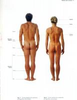
The term medial means nearer the midsagittal plane, and lateral, further from that plane. These terms should be carefully distinguished from internal (inner) and external (outer), which were formerly synonymous with them. Internal, as now used (BNA), means deeper, i. e., nearer the central axis of the body or part; while external refers to structures more superficial in position. Proximal, in describing a limb, refers to position nearer the trunk; while distal refers to a more peripheral position. Adverbial forms are also employed, e. g., anteriorly or ventrally (forward, before); posteriorly or dorsally (backward, behind); superiorly or cranially (upward, above); and interiorly or caudally (downward, below). It should also be noted that the terms ventral, dorsal, cranial and caudal are independent of the body posture, and therefore apply equally well to corresponding surfaces of vertebrates in general with horizontal body axis. On this account these terms are preferable, and will doubtless ultimately supplant the terms anterior, posterior, superior and inferior. The discrimination in the use of several similar terms of the BNA should also receive attention. Thus medianus (median) refers to the median plane. Medialis (medial) means nearer the median plane and is opposed to lateral, as above stated. Medius (middle) is used to designate a position between anterior and posterior, or between internal and external. Between medialis and lateralis, however, the term intermedius is used. Finally, transversalis means transverse to the body axis; transversus, transverse to an organ or part; and transversarius, pertaining to some other structure which is transverse. Parts of the body. —The primary divisions of the human body (fig. 1) are the head, neck, trunk and extremities. The head [caput] includes cranium and face [facies]. The neck [collum] connects head and trunk. The trunk [truneus] includes thorax, abdomen, and pelvis. The upper extremity [extremitas superior] includes arm [brachium], forearm [antibrachium], and hand [manus]. The lower extremity [extremitas inferior] includes thigh [femur], leg [crus], and foot [pes]. Each of the parts mentioned has further subdivisions, as indicated in fig. 1. The cranium includes: crown [vertex]; back of the head [occiput]; frontal region [sinciput], including forehead [frons]; temples [tempora]; ears [aures], including auricles [auriculae].
INTRODUCTION
3
The face includes the regions of the eye [oculus], nose [nasus], and mouth [os], the subdivisions of which will be given later under the appropriate sections. The thorax includes: breast [pectus]; mammary gland [mamma]; and thoracic cavity [cavum thoracis]. The back [dorsum] includes the vertebral column [columna vertebralis]. The abdomen includes: navel [umbilicus]; flank [latus]; groin [inguen]; loin [lumbus]; and the abdominal cavity [cavum abdominis]. The Fig.
1.—Parts
of the
Human
Body.
A, Posterior view.
B, Anterior view.
pelvis includes: pelvic cavity [cavum pelvis]; genital organs [organa genitalia], buttocks [nates], separated by a cleft [crena ani] at the anus. The hip [coxa] connects the pelvis with lower extremity. In the lower extremity, the thigh is joined to the leg by the knee [genu]. The foot includes: heel [calx]; sole [planta]; instep [tarsus]; metatarsus’, and five toes [digiti I—V], including the great toe [hallux] and little toe [digitus minimus].
4
INTRODUCTION
The upper extremity is joined to the thorax by the shoulder. The arm is joined to the forearm at the elbow [cubitus]. The hand includes: wrist [carpus]; metacarpus, with palm [vola or palma] and back [dorsum manus]. The five fingers [digiti I-V] include: thumb [pollex], index finger [index]; middle finger [digitus medius] ring finger [digitus annularis] and little finger [digitus minimus]. Organ-systems.—Each of the various parts of the body above outlined is composed of various organs, and the groups of related organs make up organ-
systems. The various organ-systems are treated as special branches of descriptive anatomy. The study of the bones is called osteology; of the ligaments and joints, syndesmology (or arthrology); of the vessels, angiology; of the muscles, myology; of the nervous system, neurology; and of the viscera, splanchnology. Further subdivisions are also made. The viscera, for example, include the digestive tract, respiratory tract, urogenital tract, etc.
Tissues and cells. —The body, as above stated, has various parts, each of which may be subdivided into its component systems and organs. A further analysis reveals a continued series of structural units of gradually decreasing complexity. Thus each organ is found to consist of a number of tissues (epithelial, connective, muscular or nervous). Finally, each tissue is composed of a group of similar units called cells which are the ultimate structural units of the body. The body may therefore be regarded as composed of myriads of cell units, organized into units of gradually increasing complexity, very much as a social community is composed of individuals organized into trades, municipalities, etc. Most of the individual tissues can be recognized by their gross appearance. In fact, the principal tissues were first demonstrated by Bichat through skilful dissection, maceration, etc., and without the aid of the microscope. The cellular structure of the tissues was later discovered by Schwann in 1839. Each cell is composed of a material called 'protoplasm, a viscid substance variable in appearance and exceedingly complex in chemical composition. It readily breaks down into simpler chemical compounds, whereby energy (chiefly in the form of heat and mechanical energy) is liberated. It has also the power of absorbing nutritive material to build up and replace what was lost. Its decomposition results from stimuli of various kinds, and hence it is said to be irritable. The mechanical energy which it liberates is manifested by its contractility, especially in the muscle cells. It excretes the waste products produced by its decomposition. Each cell has the power, under favorable conditions, of reproducing itself by division. Protoplasm presents, in short, all the forms of activity manifested by the body as a whole; and indeed, the activities of the body are the sum of the activities of its constituent cells. In the protoplasm of each cell is a specially differentiated portion, the nucleus. The nucleus plays an important part in regulating the activities of the cytoplasm, the general protoplasm of the cell body. The nucleus differs from the cytoplasm both structurally and chemically, and contains a very important substance, chromatin, which during cell division is aggregated into a definite number of masses called chromosomes. Furth'er details concerning the cells and tissues may be found in the text-books of cytology and histology. In earlier days human anatomy was almost entirely a descriptive science, but little attention being paid to the significance of structure, except in so far as it could be correlated with physiological phenomena as they were at the time understood. In recent years attention has been largely paid to the morphology of the human body and much valuable information as to the meaning of the structure and relations of the various organs has resulted. Since the form and structure of the body are the final result of a series of complicated developmental changes, the science of embryology has greatly contributed to our present knowledge of human morphology, ana, accordingly, an account of some of the more important phases of morphogenesis and developmental anatomy will form a fitting introduction to the study of the adult. References .—General: For looking up the literature upon any anatomical topic, the best guide in general is the Jahresbericht liber die Fortschritte der Anatomie und Entwicklungsgeschichte, which contains classified titles and brief abstracts of the more important papers in gross anatomy, histology and embryology. Other useful aids are the Index Medicus and the catalogue of the Surgeon General’s Library of the War Dep’t. (Washington, D. C.). The latter two contain titles only, but cover the whole field of medicine. The Concilium Bibliographicum also provides a convenient card-index system of references for the biological sciences, including Anatomy. For nomenclature: His, Archiv f. Anat., 1895 (BNA system); Barker, Anatomical Nomenclature; Eycleshymer, Anatomical Names. Cells and tissues: Wilson, The Cell; Ilertwig, Zelle und Gewebe (also English transl.); Schaefer, Microscopic Anatomy (in Quain’s Anatomy, 11th ed.); Heidenhain, Plasma und Zelle; Kolliker, Gewebelehre; Prenant, Bouin et Maillard, Traitc d’Histologie.
SECTION I
DEVELOPMENTAL ANATOMY By
RICHARD E. SCAMMON, Ph.D.
PROFESSOR
OF
ANATOMY, UNIVERSITY OF MINNESOTA
rnHE
life history of man, in common with most higher organisms, is characterized by continuous change and presents a cycle in which may be recognized J- the succeeding phases of growth and differentiation, maturity, and old age or senescence. In man nearly one-third of the traditional span of life is required for the body to reach its full size and differentiation. This portion of the human life cycle may be called the developmental period, and the study of the structure of the body and its changes in this time may be termed developmental anatomy. Divisions of the developmental period.—The developmental period is divided by the incident of birth into prenatal and postnatal epochs and in these a number of more or less arbitrarily defined subdivisions may be recognized. The divisions of the developmental period are shown on the following table. In this scheme puberty is regarded only as a transition point between later childhood and adolescence. The length of the developmental period and of its several subdivisions varies greatly with sex, race, environment, and physical constitution. A distinction is often drawn between the anatomic or physiologic age of the individual, as indicated by the degree of physical development of the body, and the calendar or chronologic age. As females pass through most of the transitions of the developmental period a little earlier than do males the physiologic age of girls is usually somewhat greater than that of boys of the same calendar age. DIVISIONS OF THE DEVELOPMENTAL PERIOD IN MAN
Period of the ovum. From fertilization to the close of the second week of prenatal life. Prenatal life
Postnatal life
Period of the embryo. From the close of the second week to the close of the second (lunar) month. Period of the fetus. From the close of the second (lunar) month to birth at 10 lunar months. Birth Period of the newborn (Neonatal period). From birth to the close of the second (postnatal) week. Infancy. From 2 weeks to the close of the first year or until the habitual assumption of the erect posture (usually in the thirteenth or fourteenth month). Early childhood (Milk-tooth period). From 1 to 6 years. Middle childhood. From 6 to 9 or 10 years. Childhood Later childhood (Prepuberal period. From 9 or 10 years to 12-15 years in females and 13-16 years in males.
Puberty
Sixteenth year in males. (According to American data.) Adolescence. From puberty to the last years of the second decade in females and to the first years of the third decade in males. Fourteenth year in females.
Growth and differentiation.—The changes which characterize the developmental period do not take place at the same time or at equal rates in all regions of the body, for each organ and part has its own peculiar life cycle. In a few 5
6
DEVELOPMENTAL ANATOMY
organs, such as the mesonephros of the embryo, this cycle is very short. Other organs persist during childhood and then decline, while the great majority continue, with varying degrees of change, throughout postnatal life. The characteristic life cycle of the various organs depends upon the changes in the structural units which compose them and, in the last analysis, upon the growth and differentiation of their constituent cells. Each cell has a definite life cycle, an early period of rapid and vigorous changes, later periods of differentiation and maturity, followed by stages of degeneration and death. This cycle of cell changes is termed cytomorphosis. The length of life cycle of the various types of cells in the body differs greatly, some of the blood cells living probably a month or less while certain brain cells may survive throughout postnatal life. The growth of cells may take place either by the enlargement (hypertrophy) of individual cells or by the multiplication (hyperplasia) of cells by mitosis. Cell division is necessary for continued cell growth for otherwise the cell would soon reach a size where its surface would be inadequate (for nutritive, respiratory and excretory purposes) to its mass. In general, however, cell division is most active in the early embryonic periods, during which the cells remain small. Later, cell division diminishes or ceases, and growth is due chiefly to the enlargement of cells already present. The growth of the structural units of organs also follows this general rule, the production of new units being confined mainly to fetal and early postnatal life. While the functional and structural differentiation of cells and structural units may take place during the period of their rapid multiplication these processes are usually partially disassociated and the phase of active differentiation comes some time after the period of most active growth.
THE EARLY DEVELOPMENT OF THE EMBRYO The germ-cells and fertilization. —The period of development in man, as in the great majority of multicellular animals, is inaugurated by the process of fertilization which consists of the union of the male germ-cell or spermatozoon with the female germ-cell or ovum. The ovum and spermatozoon are differentiated, by the process of maturation, from certain more primitive germ-cells set aside from the general body or somatic cells at an early period in development. In maturation both the ovum and spermatozoon undergo profound nuclear changes and each becomes highly specialized in form and structure for its part in the fertilization process. The ripe spermatozoon or sperm is a slender lance-like structure 0.05 or 0.06 mm. long (fig. 3). The ripe human ovum or egg-cell is a spheroidal body whose greatest diameter is approximately 0.1 mm. (fig. 2). It contains a nucleus about 0.02 mm. in diameter which is generally slightly eccentric in position. Suspended in the protoplasm of the cell-body are numerous droplets and granules which are presumably reserve food substances. The ovum is bounded by a delicate vitelline
membrane.
Fertilization has not been observed in man but it has been studied in detail in several mammals and it is most probable that the process is essentially the same in all higher forms. After escaping from the ovary through the rupture of the Graafian follicle the ovum enters the ostium of the uterine tube and passes down the lumen. The union of the ovum with the spermatozoon probably takes place in most cases during this process. Segmentation of the ovum. —The fertilized ovum is converted into a solid ball of much smaller cells by a series of cell-divisions. This process is known as segmentation and the mass of cells resulting from it is called the morula. Like fertilization, segmentation has not been observed in man and our concepts of the process in the human species are based upon observations on the ova of lower animals. It is probable that the first segmentation divisions are equal but that the later ones are quite irregular. The morula which results from them is a solid body at first but an eccentrically placed cavity soon appears within it and the structure is differentiated into an outer shell, the trophoblast, and a cluster of cells termed the inner cell-mass. The inner cell-mass is broadly attached to the inner surface of the trophoblast and the cavity between the two is filled with fluid and bridged by delicate cellular strands, the magma reticulare. It is probable that after fertilization approximately ten days are required for the ovum to reach this stage of development. During this period the ovum has left the uterine tube and has come to rest on the inner surface of the uterus. The uterine epithelium in contact with the ovum is destroyed, presumably
EMBRYONIC DISK
7
through the activity of the cells of the trophoblast, and the ovum sinks into the uterine mucosa and is inclosed by it. Until this implantation takes place the ovum is an independent organism dependent upon its own scanty reserve food supply for nourishment. Consequently it grows little if any during this period. With implantation, however, the ovum becomes, in a fashion, a parasite upon the maternal organism from which it derives its nourishment throughout Fig.
2.—Mature Ovum,
with Follicular
Cells,
Thompson.)
of
a Woman 36 Years
Old.
X500.
(After
the remainder of the fetal period. With the establishment of this relation the ovum enters on a period of extremely rapid growth. Formation of the embryonic disk. —Two spaces now appear in the inner cell-mass, an upper one, the amniotic cavity and a lower one, the yolk-sac cavity. These are separated by a plate of cells, the embryonic disk. At the same time a distinct layer of cells is differentiated on the outer surface of the cell-mass. This Fig.
3. —Mature Spermatozoa.
A, frontal view showing broad surface of the head: B, anterior portion in side view. Broman.)
X2500.
(After
layer is the extraembryonic mesoderm. It is probably formed in part from the cells of the inner cell-mass and the trophoblast and in part from the magma reticulare. The extraembryonic mesoderm forms a complete lining about the original cavity of the morula and this space is now termed the extraembryonic celom. As the extraembryonic celom is established the magma reticulare disappears and the connection between the inner cell-mass and the trophoblast is reduced to a short bridge of extraembryonic mesoderm, the connecting stalk (fig. 4).
DE VELOPMENTAL ANA TOM Y
8
thearch- segthe S.C.,
Embryo. celom. of
formatin formatin extrambyonicfolds. of
the of
Stre and Ex.,nN.euraFl.,
Formatin (hypoteical). Miler .)
the and
C,
and
F, E
disk.
can l.
etrophblast mbryonic embryonicthe E.d., neur t ic disk.
OvumLewis, in
Eternod, and thestalk.N.C.,yolk-sac.
Changes Dandy, cel-mas conectingel-mas. Y.s., of
formatin
Later Brodel, itnheerD,C.st.,in er vili. the Broman, celom.cavity.I.CM., Tr.v., h i n d g u t . figures of
Ilustraing difernxtao mbyiacmniotc H.G.,
trophblastic
of
Diagrms
the A.c., on and
trophblast.
morula
mentaio-cvy.
B,
(hypoteical).
(Based yolk-saccan l. hH.,eart. Tr., Seri s cavity,andneural foregut. op
A
4.—
Fig.
amniotc entroF.G.,
A,
EMBRYONIC DISK
9
Our interest is centered in the embryonic disk, for the embryo is entirely a product of this structure: the remainder of the ovum gives rise to the supporting or nourishing structures for the developing embryo or else disappears comparatively early in prenatal life. The embryonic disk in embryos of the third week is an oval plate having a maximum diameter of about 0.2 mm. It consists of three sheets of cells called the germ layers. The upper layer or ectoderm forms the floor of the amniotic cavity and becomes continuous with the walls of the amnion at the margins of the embryonic disk. The lower layer or entoderm forms the roof of the yolk-sac and is continued as the walls of this structure at the periphery of the embryonic disk. The middle layer or mesoderm forms an incomplete plate between the ectoderm and entoderm. At the margins of the embryonic disk it becomes continuous with the extraembryonic mesoderm which covers the outer surface of the inner
cell-mass.
The subsequent history of the embryo is essentially that of the differentiation and the disposition of the germ-layers. Their contributions to the adult body are as follows: Fig.
5.—Diagram of a Longitudinal Section through the Long Axis of the Embryonic Time of Formation of the Primitive Streak and Neurenteric, Canal. (Based in part on the figures of Ingalls and Streeter.) Disk
at the
From the ectoderm are formed: The central and peripheral nervous system, the epithelial internal ear, the lens, iris and retina of the eye. The epithelial portion of the skin and its appendages. The lining of the buccal, nasal, and a part of the pharyngeal cavities; the enamel of the teeth; the salivary glands. The lining of the anal canal; the lining of the vestibule and a portion of the urethra in the male, with associated glands. The anterior lobe of the hypophysis cerebri; the pharyngeal hypophysis. The paraganglia. From the entoderm are formed: The lining epithelium of the digestive tract, with the exception of the mouth, a part of the pharynx, and the anal canal; the parenchyma of the digestive glands, pancreas and liver. The lining of the larynx, trachea, bronchi and lungs. The lining of a portion of the bladder; the lining of the female urethra and a part of the male
urethra, with associated glands. The parenchyma of the thyroid and parathyroid glands; the reticulum and the thymic cor-
puscles of the thymus.
From the mesoderm are formed:
The skeletal and muscular structures and the connective tissues of the body. The vascular system; the lymphoid and sanguifactive organs. The serous membranes. The genital glands and their ducts and accessory structures. The kidneys, ureters and the greater part of the bladder. The dentine and cementum of the teeth. The cortex of the suprarenal glands. Early changes in the embryonic disk.—The first indications of the establishment of the embryo on the germinal disk appear early in the third week. At this
10
DEVELOPMENTAL ANATOMY
time the ectoderm and entoderm in the posterior part of the longitudinal axis of the disk fuse forming a band of cells known as the primitive streak. The primitive streak is indented by a dorsal primitive groove (fig. 6). It terminates anteriorly in an enlargement, the primitive node, and from the node a mass of cells, the head-process, extends forward in the midline, fusing below with the entoderm of the yolk-sac in this region. A narrow channel, the neurenteric canal, pierces the primitive node and connects the amniotic cavity with the yolksac cavity. The neurenteric canal is continuous with a cleft in the head-process termied the head-process canal (fig. 5). As these changes take place on the germinal disk a small tubular outgrowth, the allantois, arises from the posterior end of the roof of the yolk-sac and grows upward, behind the amnion, into the connecting-stalk. The primitive groove, the primitive streak, the primitive node, neurenteric canal, and headprocess are ephemeral structures which may be regarded as representing a highly modified process of gastrulation in the human embryo. The primitive streak and node and the headjoin the mesoderm laterally and presumably contribute cells to this germ-layer. The ead-process also gives rise to a longitudinal rod of cells, the notochord, which forms the median
Erocess
Fig. 6.—Dorsal View
of the Embryonic Disk and of the Third
Part
Week.
Yolk-sac
of
an Embryo
(After Streeter.)
longitudinal axis of the embryo and is subsequently associated ventral part is incorporated in the entoderm of the yolk-sac. material the head-process disappears as a separate structure. relatively shorter with the growth of the embryo anterior to it backward of the primitive node. After the third week it is neurenteric canal is normally obliterated in the third week*
of the Early
with the skeleton; possibly its With this distribution of its
The primitive streak becomes and the consequent migration no longer recognizable. The
The topography of the embryonic disk. —Although only slight signs of differ-
entiation are visible on the surface of the embryonic disk in the third week it is possible to map out upon it more or less definite areas corresponding to all of the various regions of the future body, as shown in fig. 7. Beginning anteriorly, the head region is relatively enormous in size, occupying the entire area in front of the primitive node and forming about half of the entire disk. The cervical, thoracic, lumbar,, and sacrococcygeal regions appear successively smaller, approaching the posterior end (Hail-bud’) of the primitive streak. It is also a striking fact that the future dorsal region of the body wall, corresponding to the central portion of the disk, along each side of the mid line, is now larger than the ventrolateral regions, which occupy a relatively narrow zone around the periphery of the disk. Early changes in the germ layers.—The definitive embryo is formed by the rapid growth of the dorsal region of the embryonic disk and by a series of folds and cleavages of the germ-layers of this area. The ectoderm plays a most active part
THE ENTODERM
11
in these early transformations. Shortly after the primitive streak is established, the ectoderm along the midline of the embryonic disk is thickened into a neural plate which extends from the primitive node to the anterior end of the disk. The lateral margins of the plate grow rapidly and rise from the surface of the disk as a pair of longitudinal neural folds or ridges which bound a shallow neural groove (figs. 8 and 12A). The neural plate is converted into the neural tube by the further growth of the neural ridges which fold over the neural groove and fuse in the midline. This process begins in the future cervical region and extends forward and backward from this level (fig. 12A). The extreme anterior and Fig.
7.
Topography
—
of the Embryonic
ng, neural groove, lower limb.
1 mm.
Disk.
Diagram of relations at the length of about pp, primitive pit. U, upper limb. L,
pn, primitive node,
posterior ends of the tube remain open for a time as the anterior and posterior neuropores. With their subsequent closure the walls of the tube are completed and its cavity is entirely separated from the amniotic cavity. The neural tube gives rise to the brain, spinal cord, and retinae and optic nerves. Its further history is considered in connection with the nervous system. The ectoderm which covers the periphery of the embryonic disk is carried over the dorsal surface of the neural tube with the infolding of the neural ridges. It forms the external covering of the embryo. The entoderm.—As the neural plate is formed from the ectoderm on the upper surface of the embryonic disk, the entoderm lying below this region is folded into the primitive digestive tube or archenteron. In embryos of the latter part of the third week three divisions, th q foregut, the hindgut, and the midgut may
12
DEVELOPMENTAL ANATOMY
be recognized in this structure (figs. 4E, 4F). The midgut is a shallow groove still broadly connected with the yolk-sac, the foregut is a pocket-like projection from the midgut extending forward under the anterior part of the neural plate, and the hindgut is a similar but shorter projection which extends into the caudal
region of the developing embryo. With the further growth of the archenteron the foregut and hindgut become considerably elongated and the connection of the midgut with the yolk-sac is reduced to a short, wide yolk-stalk. In this process the upper part of the posterior wall of the yolk-sac is incorporated in the floor of the hindgut, and the allantois now takes origin from this part of the archenteron instead of the yolksac. The later history of the entoderm will be considered in connection with the development of the digestive and respiratory tracts. Fig.
8.
Embryo 1.54 mm. Long. cavity having been removed.
Human
—
Viewed from above, the roof of the amniotic
(Minot, after Graf Spee.)
The mesoderm. —The mesoderm of the human embryo appears to have a dual origin being formed primarily from the extraembryonic mesoderm of the inner cell-mass and secondarily from the primitive streak, primitive node, and head-process. After the formation of the notochord the mesoderm takes the form of a pair of plates which lie on either side of the longitudinal embryonic axis and which are continuous laterally with the extraembryonic mesoderm covering the amnion and yolk-sac. Behind the primitive node these plates fuse with the primitive streak across the midline of the embryonic disk but anterior to the node they are separated by a medial space which contains the notochord (figs. 9, 10.) Some of the later changes in the mesoderm are shown in fig. 10. Each plate of mesoderm is divided by a longitudinal groove into three parts. These are (1) a narrow medial strip, the medial or paraxial mesoderm, (2) the intermediate mesoderm which forms a slender cord lying beneath the longitudinal groove, and (3) a broad band of lateral mesoderm. The medial mesoderm is subdivided by a series of transverse clefts into a row of blocks or segments known as the mesodermic somites. At the same time the lateral mesoderm splits into an upper (outer) or somatic layer and a lower (inner) or splanchnic layer. The space between these two layers is the embryonic body cavity or celom. It becomes continuous with the extraembryonic celom at the lateral margins of the embryonic disk.
THE MESODERM
13
The appearance of the mesodermic somites marks the beginning of metamerism, the arrangement of the body in successive segments or metameres. The somites form first in the occipital region and rapidly differentiate in the craniocaudal direction. In embryos 7 or 8 mm. in length about 40 pairs of somites can be distinguished. The anterior end of the medial mesoderm, which is continued into the head region, does not undergo segmentation in the human embryo. From it are formed the cranial bones, certain of the muscles of the head and connective tissue. 9. —Ckoss-sections of a Series of Young Human Embryos. All drawn at the same magnification. (Slightly modified from Graf Spee.) A, embryo of the middle of the third week. B, embryo of the end of the third week. C, embryo of the early part of the fourth week. D, embryo of the latter part of the fourth week. E, embryo of the fifth week. A.c., amniotic cavity. C.st., connecting stalk. Y.s., yolk-sac. The mesoderm is indicated in stipple. Fig.
A small cavity, the myocele appears in the center of each somite and the wall separates into an upper lateral part, the dermomyotome, and a lower medial part, the sclerotome. From the dermomyotomes are formed the voluntary muscles of the trunk, neck, and a part of the head; while the sclerotomes take part in the development of the axial skeleton. Probably both parts of the somite contribute cells to the mesenchyma which forms the connective tissue of the body wall, and the supporting structures and voluntary muscles of the limbs. The greater part of the intermediate mesoderm is also divided into segments or nephrotomes (corresponding to the somites), portions of which form the transitory uropoietic organs, the pronephros and mesonephros. The posterior part of the intermediate mesoderm remains unsegmented as the nephrogenic cord which is later involved in the development of the permanent kidney.
14
DEVELOPMENTAL ANATOMY
The lateral mesoderm shows no evidences of segmentation. The lateral cavities which are formed between its upper and lower layers soon lose their connection with the extraembryonic body cavity and fuse in the midline, forming the general celom to be described later. Fig. 10.—Stereograms Illustrating
the
Early Changes in the
Mesoderm.
Intm.mes., intermediate mesoderm. Lat.mes., lateral mesoderm. Nch., notochord. Sm.mes., somatic layer of lateral mesoderm. Sp.mes., splanchnic layer of lateral mesoderm. Ectoderm, yellow; mesoderm, green; entoderm, red.
THE DEVELOPMENT OF THE EXTERNAL BODY-FORM The early transformations of the germ-layers convert the embryonic disk into a cylindrical structure which is only partially connected with yolk-sac and connecting stalk (fig. 11). The cylindrical body wall now encloses two tubes (neural and enteric) with a longitudinal axis (notochord) between them, and is Membranes and Umbilical Cord. (After Lewis.) chorion, coe., celom. Y.s., al., allantois, am., amnion, am.c., amniotic cavity, cho., yolk-sac.
Fig.
11.—Diagrams the
Illustrating the Development op the Embryonic
Formation
of
the
covered by an outer layer of skin-ectoderm which is continuous along the sides of the embryo with the ectoderm of the amnion. The head is relatively large and is separated from the disk below it by a deep head fold. The caudal end
DEVELOPMENT OF HEAD AND NECK
15
of the embryo is prolonged into a short tail-bud which is also marked off from'the disk by a shallow tail-fold. The middle portion or trunk is still widely connected with the disk, but its boundaries are indicated by distinct lateral folds. The embryo becomes further separated from the other structures derived from the inner cell-mass by the deepening of the head, tail, and lateral folds; and its connection with these structures is reduced to a slender umbilical cord. This cord contains the allantois and yolk-stalk (with their surrounding mesoderm)
and is covered by the ectoderm which is reflected from the amnion upon the external surface of the embryo. Fig.
12. A, Human Embryo 2.11 mm. Long. (From a Model by Eternod.) B Human Embryo 4.2 mm. Long, Showing Three Branchial Grooves. (After His.)’ —
Coincident with these changes, the longitudinal axis of the embryo is modified by the formation of a series of flexures or bends. The head is flexed on the trunk first by an anterior cephalic flexure, and soon after by a more posterior cervical flexure, and the caudal part of the trunk and the tail are bent downward in a semicircular curve (fig. 12B). Later the rapid growth of the dorsal region throws the entire body into a partial spiral (coiled either to the right or left) so that its outline, when seen in lateral view, may be almost circular (fig. 13). The ventral part of the body increases in size very rapidly in the second fetel month through the great growth of the contained viscera. W'th this growth the axis of the trunk is straightened and the cervical flexure partially eliminated The cephalic and caudal flexures are never completely obliterated, although the latter is obscured by the.growth of the lower limbs; and the external evidences of the former are masked by the subsequent changes in the proportions of the head and face. A well marked tail appears which in embryos 7 to 8 mm. long may be nearly one-sixth as long as the body. Regression of the tail structure begins in the sixth week and by the ninth week it has usually entirely disappeared. Deveploment of the head and neck.—The head is divisible from an early stage, into a neural portion including the brain, eyes and internal ears with their
16
DEVELOPMENTAL ANA TOM Y
supporting structures, and a facial or visceral part which contains the anterior termination of the digestive-respiratory tract. The growth and differentiation of these two portions are quite dissimilar. The neural portion is by far the larger in the young embryo and this predominance is never completely lost although it is greatly reduced during both fetal and postnatal life by the growth of the accessory structures of the mouth, nose and pharynx. Fig.
13.—Human
Embryo 4.02 mm. Long.
(After Hochstetter.)
In the fourth and fifth weeks the visceral portion of the head undergoes marked external changes. A median oral sinus or embryonic mouth is formed on the ventral surface, and anterior to the sinus a pair of small nasal pits. The nasal pits are bounded laterally by lateral nasal processes; and a broad medial process separates them and extends downward forming the middle part of the upper boundary of the oral sinus. The remainder of the margin of the oral sinus is formed from the mandibular and maxillary processes of the first branchial Fig.
14.—Human
Embryo
11.5 mm. Long.
(After Minot.)
arch, the maxillary processes forming the lateral thirds of the upper boundary and the mandibular processes the entire lower margin. The maxillary and medial nasal processes are separated for the time by shallow lacrimal grooves (figs. 13, 14 and 17). The margins of the sinus are completed by the coalescence of the mandibular processes below and the fusion of the maxillary and medial nasal processes above. The definitive nose is formed by the fusion of the lateral and’ medial nasal proc-
DEVELOPMENT OF TRUNK
17
esses at the lower margins of the nasal pits and the subsequent growth of the medial process, particularly in the midline above the nares. The later development of the external features of the face is illustrated by the series of outlines in fig. 18. Fig.
15. A, Outlines of Average Human Ova from 3 to 8 Weeks Old, One-half Natural B, Outlines of Human Embryos from the Third to the Eighth Week, Enlarged 2.5 Times. (After Evans.) —
Size.
As these changes take place in the facial region the lateral surfaces of the neck are indented by a series of four (paired) branchial (visceral) grooves which are separated by the branchial arches. The upper part of the first of these grooves is deepened to form the external auditory meatus, the margins being elevated to form the auricle. The region corresponding to the second, third, and fourth grooves becomes depressed, forming the cervical sinus which soon closes over and nor-
mally disappears.
Fig.
16.—Figures
Illustrating the
Changes
Postnatal Growth.
Proportions (After Stratz.)
in
during
Prenatal
and
Development of the trunk.—In the young embryo the trunk appears as a cylindrical body flattened from side to side and exhibiting externally the modeling of the viscera contained within it. In fetal life, with the development of the skeleton and trunk musculature and the rounding of the visceral mass, it takes on an ovoid form largest at the level of the umbilicus and almost circular in cross section. In spite of the changes in the form and relative proportions of the
18
DEVELOPMENTAL ANATOMY
contained viscera, the relative proportions of the trunk remain almost unchanged from the close of the third fetal month until birth. In the early part of infancy, also, there is little change in the form of the trunk, but after the assumption of the erect posture there is a reduction of the relative anteroposterior diameter of both the thoracic and abdominal regions accompanied by a decrease in the relative size Development of the Face in the Second Fetal Month. (From a series of models made in the Department of Embryology of the Carnegie Institution.) L.N., lateral nasal process. Md., mandibular process. M.N., medial nasal process. N.P., nasal pit. Mx., maxillary process.
Fig.
17.
—
of the umbilical region and a relative increase in the lumbar region. These changes continue throughout childhood and early adolescence. Development of the extremities. —The limbs appear about the third week of fetal life as short ridges which project from the lateral surfaces of the cranial and caudal ends of the trunk. Each ridge is differentiated into a limb-bud in which may be recognized a flattened distal segment representing the hand or Fxa. 18.—A Series
of Profiles Illustrating the Changes of the Face in the Developmental Period.
in the Form and Proportions (After Peter.)
foot, and a rounded proximal segment representing the remainder of the limb. The latter is again divided by a slight constriction into a distal part, corresponding to the forearm or leg, and a proximal part corresponding to the arm or thigh. The digits are formed as radiating ridges on the lateral surfaces of the hand and foot segments. As these ridges grow more rapidly than the bodies of these segments they soon project beyond their margins as definitive fingers or toes. The axes of the limbs undergo three main changes in position in their early development. At first the limb-buds project outward at right angles to the
DEVELOPMENT OF EXTREMITIES Fig.
19.
—
Development op the Upper Extremity.
19
(After Retzius.)
A, anterior limb-bud of an embryo 12 mm. long. B, anterior limb-bud of an embryo 15 m. long. C, anterior limb-bud of an embryo 16 mm. long. D, forearm and hand of an embryo 25 mm. long. E, hand of a fetus 52 mm. long. All X6.
Fig. 20.— Development op the Lower'Extremity.
(After Retzius.)
A, Posterior limb-bud of an embryo 17 mm. long. B, posterior limb-bud of an embryo 19 mm. long. C, leg and foot of an embryo 25 mm. long. D, foot of a fetus 52 mm. long. All X6.
DEVELOPMENTAL ANATOMY
20
lateral surface of the body. Later they are bent caudally and ventrally so that their former ventral surfaces face medially. And finally each limb is rotated about its long axis through an angle of approximately 90 degrees. This rotation takes place in opposite directions in the arm and leg. The arm is turned outward so that the thumb comes to lie on the lateral (outer) margin of the limb and the palm faces ventrally (in supination), while the leg rotates inward and the great toe comes to lie on the medial margin of the limb, and the plantar surface of the foot faces dorsally. In embryonic life the development of the arm precedes that of the leg and it is not until after birth that the lower extremity exceeds the upper one in length. In postnatal life the lower limb increases in length more rapidly than the upper; at about two years their length is equal and in the adult the lower limb is about one-sixth longer than the upper. The adult relations of the different segments of the limbs (arm, forearm and hand, and thigh, leg and foot) are practically established early in prenatal life although there is some reduction in the relative length of the hand and height of the foot in the postnatal period.
THE GROWTH OF THE BODY AND ITS PARTS Growth of the body in weight. —The diameter of the ripe human ovum is approximately 0.1 mm. Consequently, if the egg-cell is considered as a perfect sphere, its volume is about 0.0000005 cc. and its weight, assuming the specific gravity to be 1.0, is about 0.0000005 gm. If the average weight of the body in the third decade be considered as 65 kilos (about 143 pounds) we may estimate the total increment in the body-weight during the developmental period at about 130 billion-fold. Considered from this point of view almost all of the weight increment takes place in prenatal life, for in this period the body increases in mass about 6.5 billion times while from birth to maturity the gain is but twenty-fold. From the standpoint of absolute growth, on the other hand, the body acquires about 5 per cent, of its adult weight before birth and about 95 per cent, thereafter. The growth of the body in weight is indicated in the following tables. Growth in length. —Growth in length has certain characters in common with growth in weight although the relative lineal increase of the body in the developmental period is obviously much smaller than the relative growth in mass. At the end of the first fetal month the length of the embryo is approximately 0.25 cm. This is increased 10-fold in the second fetal month but thereafter the relative rate of growth becomes progressively slower. The period of most rapid absolute growth in length is in the fourth fetal month, during which there is a gain of about 8 cm. (from 10 cm. in the twelfth week to 18 cm. in the sixteenth). After this there is a gradual decline in the absolute as well as the relative rate of lineal increase. Prenatal Growth Age in lunar
months 0
Crown-rump or sitting height (Mall), cm.
(diameter of ovum 0.1 mm.) 0.25 2.5 6.8 12.1 16.7 21.0 24.5 28.4 31.6 33.6
in Length
Crown-heel or
standing height (Mall), cm.
and Weight
Weight at end of
month, grams
(Ovum
=
Ratio of increase
to weight at beginning of month
0.0000005 g.)
=
I II III IV V VI VII VIII IX *x *
270 days (Mall).
0.25 3.0 9.8 18.0 25.0 31.5 37.1 42.5 47.0 50.0
0.004 2.0 24.0 120.0 330.0 600.0 1000.0 1600.0 2400.0 3200.0
7999.99 499.0 11.0 4.0 1.75 0.82 0.67 0.60 0.50 0.33
GROWTH OF BODY Average Physical Measurements
Postnatal Life.
Age
Birth
6 months
12 months
18 months
2 years
3 years
4 years
of
American Children
Weight, pounds
inches
Chest girth, inches
Head girth, inches
20.5
13.0
14.0
Girls
7.1
20.0
12.9
13.8
Boys
18.0
26.5
17.4
17.4
Girls
16.7
25.9
17.1
17.1
Boys
21.9
29.4
18.6
18.5
Girls
20.7
28.9
18.1
18.0
Boys
24.6
31.7
19.1
19.1
Girls
23.4
31.1
18.6
18.5
Boys
27.1
33.7
19.5
19.4
Girls
26.4
33.4
19.4
19.0
Boys
32.2
37.1
20.6
19.9
Girls
30.5
36.7
20.4
19.4
Boys
35.9
39.5
21.1
20.1
Girls
33.7
39.0
20.4
19.7
Sixth fetal month
Relative Volume of the Parts of the In per cent, of the total body volume.
in
45 37 27 22 15 7
of
Height,
7.3
Second fetal month
Maturity
First Four Years
Boys
Head and neck
Two years Six years
in the
(Based in part on the figures of Crum and Taylor.)
Sex
Growth
Birth
21
Trunk
50 40 49 50.5 51 53
Body.
Arms
3 8 9 9 9 10
Legs 3 15 15 17.5 25 30
The growth in length in fetal life is indicated in a preceding table (p. 20) and by the upper curve in fig. 21. The age of the fetus may be estimated from its standing height by ‘Hasse’s rule'; namely: that before the fifth month the age in fetal months is equal to the square root of the total (standing or crownheel) height, while after the fifth month the age equals one-fifth of the standing height in cm. This give approximate results except for the first 2 months. The length of the body at birth usually falls between 48 and 52 cm. (approximately 19 to 21 inches). The birth-length, like the birth-weight, is influenced by sex, race, and a number of other factors. In the neonatal period there is often a slight decrease in length due to changes in bodily proportions in the recovery from the molding effects of birth. The curve of postfetal growth in length is a sinuous one similar to the curve of postnatal weight increase and the same phases may be recognized in it. Length increases about 30 per cent. (15 cm. or 6 inches) in the first six months and about 50 per cent. (25 cm. or 10 inches) in the first year (fig. 22). During early and middle childhood the lineal increase is very slow, averaging only about 6 or 7 cm. per year. The prepuberal length increase, like the weight increase, begins earlier in girls than in boys and is completed sooner. The body increases approximately 3.3 times in length during the postnatal developmental period. Growth in length usually ceases at about 18 years in females and soon after 20 in males.
22
DEVELOPMENTAL ANATOMY
Fig. 21. —Chart of
Fig
22. —Chart
of
the Average
Growth
on the data of
the Average
in Length and
Weight in
Mall, A. W. Meyer and Jackson.)
Fetal Life.
Growth in Height and Weight in
(Bedvel.)
the
(Based
First Year.
GROWTH OF PARTS
23
The relation between the length and the weight of the body changes greatly in the developmental period. In later fetal life and early infancy the mass of the body is much greater in proportion to its length than at any subsequent time. The decline in relative weight begins about the middle of the first year and continues until after puberty. Thereafter there is a period of relative mass increase which may continue throughout maturity. During infancy and childhood females are relatively lighter than males but after puberty they are relatively heavier. The surface area of the body in the developmental period.—The metabolism of the body is greatly influenced by the relation of its surface or cutaneous area to its mass or volume, and this relation is greatly altered in the course of postnatal Fig. 23. —Chart Showing Average Postnatal Growth
in Height and
Weight.
(After
Stratz.)
development. The surface area of the average newborn child is about 2500 square cm. (400 square inches). This is doubled in the first year and is tripled in the middle of childhood. There is a period of rapid increase in surface area before puberty and the total gain between birth and maturity is about 7-fold. But the weight of the body increases approximately 20-fold in this time and there is consequently a great reduction in the ratio of surface area to mass or volume (from over 800 square cm. of surface area per kilogram of body weight in the newborn to less than 300 square cm. per kilogram in the adult.) The relative growth of the parts of the body.—Growth and differentiation do not take place at the same time or rate in the various parts of the body and the changes in proportions which occur in the developmental period are dependent on this lack of uniformity. While each part passes through its own cycle of changes these changes as a whole tend to follow what is known as the law of developmental direction; for it is generally found that development (including
24
DEVELOPMENTAL ANA TOM Y
growth and differentiation), in the long axis of the body, appears first in the head region of the body and progresses toward the tail region and similarly development in the transverse plane begins in the mid-dorsal region and progresses lateroventrally (in the limbs proximodistally). Some of the changes in the proportions of the body and the relative size of its several parts are indicated in figs. 15 and 16 and in a preceding table (p. 21). The head is the largest part of the body in earlier stages, forming about one-half of the body in the second fetal month, about one-quarter at birth, and from 6 to 8 per cent, in maturity. The trunk as a whole remains of about the same relative size throughout the developmental period (45 to 50 per cent.) although the thoracic portion reaches its maximum in the earlier stages and the pelvic portion not until adolescence. The lower limbs, like the pelvis, develop slowly, forming about 3 per cent, of the body at the end of the period of embryo, about 15 per cent, at birth and reaching about 30 per cent, in the adult. The upper limbs also form about 3 per cent, of the body weight at the close of the embryonic period. They increase to 8 or 9 per cent, at birth and maintain thereafter about the same relative size. These changes cause a great increase in the relative weight and volume of the caudal or lower part of the body; and with them the midpoint of the body (between crown and sole) is gradually shifted from the upper margin of the thorax at the end of the embryonic period to a level slightly above the umbilicus at birth, and to the level of the crest of the pubis in the adult. The center of gravity is also shifted caudally from the cervical region in the embryo to the point where the inferior vena cava pierces the diaphragm at birth, and to a point just in front of the sacral promontory in the adult. The relative growth of systems.—There is a marked difference in the growth of the various systems of the body. The skeleton grows comparatively slowly during the greater part of prenatal life but increases much more rapidly in the last two fetal months. At birth it forms from 15 to 20 per cent, of the body. Its postnatal growth apparently proceeds with that of the body as a whole, the total increase in weight between birth and maturity being about twenty-fold. The musculature also grows rather slowly in the young embryo but increases to about 25 per cent, of the body at birth and to 40 or 45 per cent, in the adult. The blood-vessels apparently also increase in relative weight after birth. The central nervous system, on the other hand, is relatively enormous in the young embryo, decreasing from about 25 per cent, of the body in the second fetal month to about 15 per cent, at birth and to between 2 and 2.5 per cent, in the adult. Data for the peripheral nervous system and the skin are somewhat scanty and rather unsatisfactory, but it is evident that both undergo a considerable reduction in relative weight in the postnatal developmental period. The visceral group (as a whole) shows a slow but steady decrease in relative weight after the embryonic period, forming about 15 per cent, of the body weight in the second month, about 9 per cent, in the newborn, and from 5 to 7 per cent, in the adult. Growth of organs. —While in general the individual organs follow the course of growth of the visceral group, each organ has its own characteristic scheme of relative growth. As a rule, after its appearance in embryo, each organ increases more or less rapidly to a maximum relative size, after which, although increasing in absolute size, it decreases in relative size through subsequent prenatal and postnatal life to maturity. Curves of the absolute growth of the various organs in the period of the fetus all appear much alike, showing an initial period of slow increase followed after the fifth month by a terminal phase of rapid growth. This uniformity is lost at birth and most of the major organs, on the basis of the course of their postnatal growth, can be classified in 4 main groups—splanchnic, nervous, genital and lymphoid (fig. 24). The splanchnic group includes the digestive, respiratory, urinary organs, the thyroid gland, the heart, and the spleen. These organs increase rapidly in weight in infancy and the first part of early childhood. In the latter part of early childhood and in middle childhood they grow quite slowly. They enter on a second phase of rapid growth in the prepuberal period, and this is followed by a terminal phase of slow increase in adolescence. In general the growth of the organs of this group is similar to the growth of the body as a whole.
25
THE SKELETON
The nervous group includes the brain, spinal cord, and eyeballs. These structures grow very rapidly in infancy and have completed over 90 per cent, of their postnatal increase by the close of early childhood. The organs of the genital group (all genital organs with the exception of the ovaries and uterus) grow very slowly until the prepuberal period, when they enter on a phase of rapid increase which extends into or through adolescence. The structures of the lymphoid group (excluding the spleen but including the thymus) are large at birth, grow rapidly until puberty, and then decline in absolute weight.
Fig.
24. —Chart
Illustrating the
Course
of
Growth
of the
Various
Types of Organs.
The growth of the various organs is calculated in per cent, of their adult weight.
T The organs which do not fall under any of the preceding categories are the suprarenal glands, the ovaries, the uterus and the hypophysis. Their growth is considered in connection w ith their develo.pment in the following sections.
r
THE SKELETON The skeleton, including the bones, cartilages, ligaments, and joints, is derived from the mesoderm. The process is inaugurated by the formation, in the future skeletal regions, of masses of thickened mesenchyma known as scleroblastema. The hard parts of the skeleton, the bones and cartilages, arise in the more condensed parts of the scleroblastema while the joints are formed from the intervening portions. The majority of the thickened scleroblastemal masses are differentiated into cartilage. Certain of these cartilages persist throughout life while a few may be converted at a later time into fibrous tissue. But by far the greater number are replaced by bone during the later development of the skeleton. Most of the bones of the body arise through the replacement of previously formed bodies of cartilage by bony material, while a smaller number are formed directly in the membranous scleroblastema. The former are known as cartilaginous or replacement bones and the latter
DEVELOPMENTAL ANATOMY
26
investment bones. All the bones of the extremities, the bones of the spinal column and thorax, the auditory ossicles, the hyoid bone, and the greater part of the bones of the base of the skull are cartilaginous bones. The bones of the face and the greater part of the vault of the skull are membranous bones. Certain of the skull bones are formed partly in membrane and partly in cartilage. The process of ossification begins in discrete foci which are known as centers of ossification. These centers are formed from time to time throughout the developmental period, the first appearing in the clavicle in embryos of the sixth week and the last, in the epiphyses of the vertebral column, in the third decade of postnatal life. Over eight hundred centers are formed in the body and of these slightly more than half appear after birth. Almost all bones of the adult are formed from one or more centers of ossification; the relation of the total number of bones in the mature skeleton to the total number of centers being approximately as 1 to 3. The ossification centers of all bones of the body with the exception are termed membranous or
Fig. 25.
—
Median
Sagittal
Sections
trating the Development
of
vertebrae indicated in black. Cunningham.)
the Vertebral Column at Various Ages IllusNormal Spinal Curvatures. Cervical and lumbar (Based in part on the figures of Bardeen, Williams and
of
the
of those of the carpus, tarsus, skull, and sternum, may be divided into two general classes, the primary and secondary. The primary centers which form the greater part of the bone almost always appear before birth. Such centers, when located in long bones, are known as diaphyses. The secondary centers or epiphyses are, with one or two exceptions, formed during postnatal life. A further consideration of the nature of diaphyses and epiphyses will be found in the section on Osteology (p. 82). As the formation of new centers and the fusion of older ones proceeds at unequal rates during the first two decades, the number of separate bone masses in the body varies from year to year during this period. The number of bones in the average newborn child is 270. This number is reduced in the first 2 or 3 years of life through the fusion of primary centers which are present before birth. From this time until puberty, however, the number increases steadily through the formation of epiphyses and the ossification of the carpus and tarsus. In the fourteenth year there are about 350 separate bony masses in the body. After puberty the number again increases rapidly until nearly the middle of the third decade. Often it is not until middle life that the number of bones is reduced to the usual quota of 206, excluding the smaller
sesamoids.
The spinal column. —In the latter part of the first month the notochord is surrounded by a sheath of mesenchyme in which may be recognized segmentally arranged masses which represent the vertebra and are formed from the sclerotomes of the mesodermic somites. Dorsal prolongations from these masses grow up on either side of the neural tube forming the arches
27
THE SKELETON
of the vertebrae, and at the same time lateral outgrowths appear which represent the various lateral projections of the vertebrae including the costal processes. In the second and third fetal months the cervical and sacral regions form the greater part of the vertebral column (fig. 25). At birth the cervical part forms approximately one-fourth, the thoracic part one-half, and the lumbar part one-fourth of the entire movable spine. In the adult the thoracic portion continues to form approximately one-half of the free vertebral column, but the lumbar portion is increased to about one-third, and the cervical portion is reduced to one-fifth or one-sixth of the whole. It is probable that in most cases these proportional postnatal changes take place in the first 3 years. During the first fetal month the vertebral column shows a pronounced ventral flexion. From this time until birth the free portion gradually becomes straighter while the sacral portion first becomes straighter and later acquires a second ventral curve. In the newborn child the free column forms a single gentle curve with an anterior (ventral) concavity extending from Fig. 26. —Diagram Showing
the
Categories to which
the
Bones
op the
Skull Belong.
(After McMurrich.) The unshaded bones are membrane bones, the heavily shaded represent the chondrocranium, while the black represents the branchial arch elements. AS, alisphenoid. ExO, exoccipital. F, frontal. Hy., hyoid. IP, interparietal. Z, zygomatic. Mn, mandible. Mx, maxilla. NA, nasal. P, parietal. Pe, periotic. SO, supraoccipital; Sq, squamosal; St, styloid process; Th, thyroid cartilage; Ty, tympanic.
the first cervical to the last lumbar vertebra, while the sacrum is directed somewhat posteriorly. The cervical curve appears when the infant begins to lift its head but neither at this time nor later does it become a fixed flexure. The lumbar curve appears as the child assumes the upright posture. It forms very slowly and throughout childhood and adolescence it may be effaced by stretching the spine. Later the lumbar curve is fixed, in a measure, by the anterior thickening of the lower lumbar vertebrae and the intervertebral disks between them. This process begins in later childhood and continues slowly until maturity. For further details on the curvatures, see p. 97. The cartilaginous vertebrae are formed by the appearance of centers of chondrification of the blastemal vertebrae. There are four of these centers for each vertebra, two lateral ones which soon fuse in the body, and one for each side of the vertebral arch. The ossification of the vertebrae is considered in the section on Osteology. The material between the vertebral masses is later converted into the outer portions of the intervertebral disks; and the segments of the notochord which occupy these regions form the nuclei pulposi. The vertebral portions of the notochord degenerate. The length of the movable or free vertebral column (cervical, thoracic, and lumbar vertebrae) at the end of the second fetal month is about 2 cm. This is more than doubled in the third month, nearly quadrupled in the fourth, and increased almost 10-fold by birth when the average length is almost 20 cm. (8 in.). Between birth and maturity the movable vertebral column increases in length between 2 and 2.5 times, about two-thirds of this growth being accomplished
28
DE VELOPMEN TAL ANA TOM Y
before puberty.
After the second fetal month the vertebral column forms from 40 to 45 per of the total length of the skeleton. Development of the skull. —-The bones of the skeleton of the head may be divided into three main categories (fig. 26): (1) the group of cartilage bones developed mainly in the base of the skull around and anterior to the cephalic portion of the notochord and about certain of the organs of sense; (2) the membrane bones which form the vault of the skull and the framework of the upper part of the face; and (3) the cartilage bones which have their origin from the cores of the branchial arches. The distinction between these three parts of the head skeleton is often imperfect, for their parts overlap and fuse during development and the relations between them are constantly shifted and modified. In the skull, as in the other parts of the skeleton, three stages, the blastemal or membranous, the chondrogenous, and the osseogenous, may be recognized although they overlap considerably and do not proceed at the same rate in all parts of the head skeleton. cent,
Fig.
27.
—
Model
of the
Chondrocranium
lage in blue.
of
a
Human Fetus 8
Viewed from above.
cm.
in length.
(After O. Hertwig.)
Carti-
Toward the end of the first month the brain is enclosed in a membranous sac formed by the condensation of the surrounding mesenchyma. The basal portion of this sheath forms a thickened plate which surrounds the cephalic portion of the notochord and projects forward
beyond its anterior termination. The occipital portion of this plate is greatly expanded and is connected with the fibrous capsules around the developing internal ears. The anterior part of the plate extends forward into the nasal region. The formation of the chondrocranium in the basal portion of the cranial blastema begins early in the second month and is practically complete by the close of the third (fig. 27). The caudal or occipital portion of the chondrocranium forms almost a complete ring around the foramen magnum and extends laterally about the base of the posterior part of the brain. Lying anterior to the occipital portion of the skull on either side are the cartilaginous auditory capsules. The contiguous borders of the occipital portion of the skull and the auditory capsules, are partly fused, but lacunae in this region indicate the position of the future hypoglossal and jugular foramina and transmit the structures which pass through these openings in the adult skull. A median bar of cartilage, which represents the basilar portion of the occipital bone and the body of the sphenoid, passes forward from the anterior margin of the foramen magnum and terminates in an expanded frontonasal plate. Two pairs of processes arise from this median mass. The posterior (alisphenoid) represents a part of the greater wing of the sphenoid and the anterior clinoid processes. The latter fuse with the frontonasal plate enclosing the optic foramina. The medial portion of the frontonasal plate extends forward into the nasal region forming the fundament of the ethmoid bone and a part of the nasal capsule. The lateral portions (orbitosphenoid) spread over the eyes and represent the lesser wings of the sphenoid.
GROWTH OF SKULL
29
The visceral elements of the skull are derived from cartilages formed in the branchial arches. That of the first arch is known as Meckel’s cartilage. It extends from the outer surface of the
auditory capsule through the mandibular process to the ventral median line. The upper portion of Meckel’s cartilage is differentiated into two parts which form two of the auditory ossicles, the malleus and the incus. The lower part is enclosed in a sheath of membrane bone which forms the mandible (fig. 28). All of this part of the cartilage disappears with the exception of its medial tip which is probably involved in the formation of the mental protuberance. The upper portion of the cartilage of the second or hyoid arch forms a portion of the stapes and a part of the styloid process of the temporal bone while its lower segment gives rise to the lesser cornu and a part of the body of the hyoid bone. The upper portions of the third, fourth Fig.
28. —Mandible Showing Relations of Meckel’s Cartilage in a Human Fetus 8 Cm. Crown-rump Length. (After Kollmann.)
of
and fifth arches form no permanent structures. The lower part of the third arch gives rise to the greater cornu and a part of the body of the hyoid bone, and the lower parts of the fourth and fifth arches are involved in the formation of the thyroid cartilage. The further history of the ossification of the bones of the head will be found in the section on Osteology.
Growth of the skull. —The most striking characteristic of the fetal skull is the great predominance of the neural over the visceral portion. During the period of the chondrocranium the base of the cranium is large as compared with the roof or vault but with the growth of the cerebral hemispheres the vault becomes increasingly prominent. In early stages of development the occipital region forms by far the largest part of the cranium. Later the parietal portion enters on a period of rapid growth and becomes the predominant region, and finally in the last fetal months the frontal region becomes the center of most vigorous expansion. .
Fig.
29. —Skulls of the Newborn and Adult. Drawn to the same face height to illustrate the relative proportions of the facial and neural skeleton at birth and in maturity. (After Holl.)
At birth the skull is large as compared with the rest of the skeleton. The neural portion is still much larger than the visceral or facial, the relation between the two being as 8 to 1 as compared with the ratio of 2.5 to 1 in the adult (fig. 29). The vault in comparison with the base of the cranium is also much larger than in later life. The bones of the cranial vault are quite thin and are separated by narrow strips of membrane which are expanded at the angles of the parietal bones into areas of some size which are known as fontanelles. Theoretically fontanelles may be formed at any point on the calvarium which is equidistant from three or more contiguous centers of ossification.- There are some 26 such points on the vault of the skull and constant or occasional fontanelles have been found in most of these locations. Two median and single fontanelles (the frontal and occipital), and two lateral and paired fontanelles (the sphenoidal and mastoid), are commonly present at birth. Their positions are shown in figs. 30 and 188-190. Besides these constant fontanelles, numerous accessory ones may be present. The more important ones, the parietal, cerebellar and metopic fontanelles, are all located along the sagittal suture.
30
DEVELOPMENTAL ANATOMY
The involution of the frontal fontanelle usually begins some months after birth and is generally completed early in the second year. The occipital fontanelle is generally closed by the end of the first trimester. The sphenoidal fontanelle commonly closes in the first 6 months and the mastoid fontanelle between 12 and 18 months after birth. The obliteration of the fontanelles takes place by the progressive ingrowth of the edges of the bone which bounds them, but occasionally separate ossification centers may arise within them and form independent bones which occupy all or part of the original space. The skull as a whole grows less in postnatal life than the other divisions of the skeleton, the neural portion increasing in volume about 5 times and the facial portion about 12-fold. Between birth and maturity the cranial capacity rises from 400 to 1500 c.c. and the horizontal circumference of the skull from about 32 cm. to 48 or 50 cm. Most of this growth takes place in the first two or three years after birth (fig. 31). The postnatal growth of the skull is closely associated with the growth of other structures of the head and particularly with those of the brain and eyeballs, the teeth and paranasal sinuses, and certain of the larger muscles. These Fig.
30.
—
and
Diagram op the Calvarium at Birth, Showing the Positions of the more Important Accessory Constant fontanelles
the
Fontanelles.
light stipple, accessory fontanelles in heavy stipple.
Constant shaded in
structures influence the growth of the skull at different periods: the brain and eyeballs mainly in infancy and early childhood, the teeth and paranasal sinuses mainly in middle and later childhood, and the muscles mainly in adolescence. During infancy all parts of the skull grow rapidly, the cranial capacity increasing from 400 cc. at birth to 700 cc. at 6 months and over 1000 cc. at 18 months. The face grows even more rapidly than the cranium in this period, the ratio between the two being reduced from 1 to 8 at birth to 1 to 6 in the second year. Most of the early growth of the face is due to the expansion of the orbits, which accomplish over half of their postnatal growth in the first 2 years, but there is also a marked growth of the maxillae and mandible in connection with the development of the deciduous dentition. From 2 to 7 years the growth of the skull is continued, although less rapidly than in infancy. The cranium expands slowly, the vault growing more than the base. The facial skeleton grows more rapidly than the neural portion and by 5 years the ratio between them is 1 to 5. Most of the growth is in the lower part of the face and is due to the expansion of the dental arches and the development of the maxillary sinuses. In middle and later childhood the skull grows little, aside from the lengthening of the face which accompanies the establishment of the permanent dentition. In adolescence the growth of the skull again increases. The cranium enlarges slightly in all diameters, mainly through the increase in the thickness of the bones of the vault, and the face completes its growth with
GROWTH OF THORAX
31
the full development of the paranasal sinuses and the upper and lower dental arches. As a rule these later changes are more extensive in the male than in the female skull. The thorax.—The formation of the thorax is first indicated in the blastemal period by the development of the costal processes of the thoracic vertebrae. These structures represent the future ribs. They assume a rod-like form and rapidly grow ventralward in the thoracic body Fig.
31. —Tracings
Illustrating
and Welcker.)
Median Sagittal Sections of the Skull at Different Ages, of Growth of the Cranium. (Based on the figures of Corrado
of the Rate
wall. Their distal ends unite craniocaudally, forming longitudinally directed sternal bands either side of the ventral midline. The fundament of the unpaired sternum is formed by the side-to-side union of these bands, together with a small mass (the episternum) which is derived from a band of thickened mesenchyma connecting the sternal ends of the developing clavicles. on
Fig. 32. —Anterior Views of the Skeletal Thorax. A, of an embryo 15 mm. long. (After Muller.) B, of a newborn child. to All drawn the same absolute size.
C, of
an
adult.
Some of the later changes in the form of the thorax are shown in figs. 32, A, B and C. In early stages the thorax is conical and is nearly circular in cross-section. At the time of their origin the ribs lie parallel with one another and are almost horizontal, but they soon incline downward (figs. 44, 45). After birth and particularly after the erect posture is assumed there is a progressive reduction of the relative anteroposterior diameter of the thorax and its base is relatively contracted. These changes are possibly due in part to the effect of gravity on the viscera and to the reduction in relative size of the organs of the upper abdominal region.
32
DEVELOPMENTAL ANATOMY
The postnatal growth of the thorax, as determined by external measurements, particularly of the horizontal chest circumference, seems to follow the course of the growth of the body in height and weight. There is a period of rapid increase in infancy and a part of early childhood, a period of slow growth in middle childhood, followed by a phase of rapid growth in prepuberty and perhaps early adolescence, and finally a terminal period of slow increase to maturity. The pelvis.—The pelvis is formed in part from the fixed vertebrae of the sacrum and coccyx, which are developed around the lower portion of the notochord, and in part from the blastema! ossa coxae which are developed from the proximal portions of the mesenchymal cores of the lower limb-buds. These elements are not closely associated at the time of their differentiation and the complete pelvic girdle with definite fundaments of the pubic symphysis and the sacroiliac articulations is not established until about the end of the second month. In early fetal life the pelvis is relatively small. After the third month all of the pelvic diameters grow at approximately the same rate and there are no great changes in the shape of the pelvis from this time to birth. The pelvis of the newborn differs from that of the adult in a number of particulars (fig. 33). It has a distinctly conical form and its length is greater in proportion to the transverse and conjugate diameters. The pelvic cavity is relatively as well as absolutely much smaller than in the adult. The superior aperture approaches a circle more closely than in later life, although Fig.
33. Drawing of Anterior Views of the Pelves of an Adult Male and a Male Newborn Drawn to the Same Absolute Size and Superimposed. Adult pelvis in solid line, newborn pelvis in broken line. (After Merkel.) —
birth the transverse diameter is greater than the conjugate. The dimensions of both the lesser pelvic cavity and the inferior aperture are smaller in proportion to the superior aperture than at maturity. The sacrum forms a greater portion of the pelvic circumference and is less depressed between the ilia. The sacral promontory is less marked, but a second prominence may be indicated at the level of the linea terminalis. There is little indication of the sacral concavity. The acetabula are very large and shallow at birth while the obturator foramina are relatively small. The pelvis is more vertical in position than later, the plane of the superior aperture forming an angle of 80° in the horizontal as compared with 50° to 60° with the adult, while the long axis of the symphysis pubis is more nearly perpendicular. During the first 2 years of life the pelvis grows rapidly in all dimensions. This growth is continued at a slower rate from 2 to 5 years, but there is comparatively little increase between 5 and 10 years. There is a second period of more rapid growth in later childhood and adolescence, and by the end of the second decade the pelvis has almost obtained its adult dimensions, although many of the pelvic epiphyses do not unite with the main bones until the twenty-fifth year. Until the infant assumes an erect position, the pelvis changes but little in form. As this position becomes habitual, the sacrum descends between the ilia and the promontory is definitely established. With this there is a relative expansion of the ala of the ilia, an increase in the pubic angle and a bending of the sacrum backward. The increase in the transverse diameter of the pelvis is brought about mainly by the growth of the sacrum and the posterior parts of the ilium. Growth also takes place along the line of apposition of the three divisions of the os coxae in the acetabulum, but there is probably comparatively little growth, at least in males, at the symphysis pubis. The pelvic growth which occurs after puberty is practically all even at
epiphyseal.
Sexual differences in the pelvis appear about the sixth fetal month, mainly in the form of differences in the pubic angle and the shape of the ilia. The pelvis of the newborn male is larger than that of the female, the sacrum is relatively wider, the ilia less vertical, the pubic angle more acute and the acetabula shallower and more horizontal. The sexual differences of the newborn pelvis, aside from those of size, are usually masked in the early period of rapid
DEVELOPMENT OF VASCULAR SYSTEM
33
growth and do not reappear until the prepuberal period. During the first decade the pelvic measurements of boys are usually larger than those of girls, but after this time the dimensions of the female pelvis are generally the greater. THE VASCULAR SYSTEM In this section the consideration of the vascular system will be limited to a description of the establishment and course of the early embryonic circulation, an account of the fully established circulation of the late fetus, and the changes in the circulatory system at birth; including an outline of the general growth of the vascular structures. The morphologic changes by which the various segments of the embryonic circulatory system are transformed to the adult structures are described in connection with the Vascular System (pp. 668 and 724). In man and the higher mammals the development of the vascular system is extremely precocious, due to the small amount of food substances stored in the ova of these forms and the consequent necessity of a system of vessels which can draw nourishment and oxygen from the maternal circulation and distribute them to the tissues of the embryo.
1
Fig.
34.
Diagram of the
—
Circulation
of a Young Human Embryo. the figures of Watt and Brodel.)
(Based in part on
Fundaments of the first vessels in man appear in the form of cords, cellular strands, and cysts in the extraembryonic mesoderm at a stage preceding the establishment of the embryo on the germinal disk. These structures are organized into two sets of anastomosing cords; one, the umbilical or allantoic, which is associated with the connecting stalk, allantois, and the trophoblast; and the other, the vitelline, which spreads out in the mesoderm of the yolk-sac. Trunks formed from these networks pass, in the mesoderm, to the margin of the germinal disk. As the tubular embryo is formed from this structure, they enter the developing body either as definite trunks or as capillary plexuses, and form the framework of the embryonic vascular system. The course of the embryonic circulation.—The form of the circulatory system in the young embryo is illustrated by fig. 34. The blood from the capillaries of the chorion (the modified trophoblast) passes to the embryo through the paired umbilical or allantoic veins. Before entering the embryo these vessels are joined by the vitelline veins which collect the blood from the yolk-sac. These vessels form the vitelline-umbilical trunks (V.TJ.Tr.) which enter the body, on either side, in the splanchnic mesoderm below the foregut and join in the tubular heart, which is formed from them. From the heart the ventral aorta? pass below and then around the anterior part of the foregut and gaining its dorsal surface run backward as the paired dorsal aortae. The dorsal aortae give off the vitelline arteries (Vit. art.) which return blood to the capillaries of the yolk-sac and finally terminate in the umbilical or allantoic arteries which run to the chorion through the connecting stalk and end in the chorionic capillaries. The dorsal aortae also give rise to a number of segmentally placed arterial sprigs which supply the tissues of the embryo. The blood from these vessels is collected by venules which empty into longitudinal venous trunks, the anterior and posterior cardinal veins. The cardinal veins on either side drain into a short common cardinal vein (duct of Cuvier) which opens into the posterior part of the heart-tube in common with the vitelline-umbilical trunks. The umbilical veins are the nourishing vessels of the embryo, since they carry blood from the chorionic capillaries where it has received oxygen and food-stuffs (by osmosis) from the maternal circulation. These substances are absent from the vitelline veins, since the yolk-sac of the human embryo contains no reserve food material. Theoretically, at least, there is a mixture of arterial blood from the umbilical vessels and venous blood from the common cardinal veins in the posterior end of the heart; but the actual difference between the arterial and venous blood in the young embryo is probably very slight since the volume of the blood-stream is relatively large and the rate of circulation presumably very rapid.
34
DEVELOPMENTAL ANATOMY
The fetal circulation.—The course of the blood in the late fetus is shown in diagrammatic fashion in figs. 35 and 586. The pure or arterial blood from the placental capillaries enters the body by the single umbilical vein, and passes through this vessel to the liver where it is joined by a branch of the portal vein carrying venous blood. The blood from the sinus formed by the union of these two vessels passes through the ductus venosus which joins with the inferior vena cava (either directly or through the left hepatic vein). As the vena cava is carrying blood from the capillaries of the lower part of the body there is a further mixture of venous and arterial blood at this point. The stream of mixed blood now passes through the terminal porFig. 35. —Diagram of the Fetal Circulation. Ab.Ao., abdominal aorta. Ao., ascending aorta. D.A., ductus arteriosus. D.V., ductus venosus. F., foramen ovale. Inf. V.C., inferior vena cava. L.A., left atrium. L.V., left ventricle. P.a., pulmonary arteries. Pt.v., portal vein. P.v., pulmonary veins. R.A., right atrium. Sup.V.C., superior vena cava: Umb.a., umbilical arteries. Umb.v., umbilical vein.
tion of the vena cava and enters the caudal portion of the right atrium. The superior vena cava also enters the right atrium, bringing back venous blood from the head and superior ex-
tremities. The opening of the superior vena cava with its valves is so placed that the stream from this vessel is directed toward the foramen ovale between the right and left atria. Despite this anatomical arrangement, however, experimental evidence indicates that the blood of the inferior and superior venae cavae is completely mixed in the right atrium. The blood from the right atrium flows in part into the left atrium, where it is joined by a small stream of venous blood, returning from the lungs through the pulmonary vein. It then passes into the left ventricle and thence to the systemic circulation through the arch of the aorta. A portion of the blood from the right atrium passes into the right ventricle and through it to the pulmonary artery. A small part of this stream is diverted to the right and left pulmonary arteries to supply the lungs, but the main current passes through the ductus arteriosus (Botalli) to pass into the descending arch of the aorta, joining the stream from the left ventricle which has come through the aortic arch. The blood passing through the aorta continues downward through this vessel, and such as-remains after supplying the aortic branches flows into thp umbilical arteries and thence back to the capillaries of the placenta.
GROWTH OF HEART
35
Several peculiarities of the fetal circulation which are particularly striking may be enumerated. But one vessel in the body of the fetus, the umbilical vein, carries strictly arterial blood, and this vessel supplies no part of the body directly, except a small portion of the liver. The blood entering the right atrium from the inferior vena cava has already received venous mixtures from three sources (the portal vein, the inferior vena cava, and the vena azygos major). There is a complete mixture of blood from the superior and the inferior vena cava in the right atrium and a further addition of venous blood from the pulmonary vein in the left atrium. Thus the circulating blood of the fetal body is throughout mixed, venous and arterial. Its efficiency under these conditions probably depends: (1) on its very large quantity compared with the volume of the fetus; (2) on the rapidity of its circulation; and (3), on the relatively slow rate of catabolism (and hence slight amount of waste products) in the fetal organism. The establishment of the adult circulation. —The change from the fetal to adult type of circulation is brought about by the closure of the fetal blood-passages, the umbilical vessels, the ductus venosus, the ductus arteriosus, and the foramen ovale. In this closure two processes can be recognized; the physiologic occlusion, which takes place immediately after birth, and the anatomical obliteration which occur days, months, or even years later. With the establishment of respiration the ductus arteriosus collapses, and all the blood from the pulmonary artery is forced through the branches of these vessels into the capillary bed of the lungs. This leads to an increased flow from the pulmonary veins into the left atrium and pressure in this chamber rises so that the valve of the foramen ovale is forced against the margin of the interatrial septum and the communication between the two chambers is interrupted. At the same time, with the tying of the umbilical cord, or in most cases even with the simple interruption of this structure, the fetal ends of the umbilical arteries and veins are contracted and their lumina obliterated. With this change, blood no longer flows through the umbilical vein and the current of blood through the ductus venosus ceases. The actual obliteration of the fetal blood-passages rarely begins in the first fortnight of postnatal life. The obliteration of the ductus venosus is brought about through the interruption of its epithelial lining and invasion of its lumen by the connective tissue of the tunica media and tunica interna. The vessel is generally completely closed by the end of the first month. Obliteration of the ductus arteriosus takes place in much the same way, generally in the first half year of postnatal life, but sometimes not before the close of the first year. The occlusion of the foramen ovale is apparently brought about by the proliferation of connective tissues in the valve and at the margin of the foramen, through the mechanical irritation of these structures by friction in the movements of the heart. At the close of one year the foramen is obliterated in about one-half of all cases, but the opening remains patent to the probe in nearly onethird of adult hearts. The obliteration of the umbilical arteries and vein begin shortly after birth with the organization of the thrombus at the cut end of the vessel; but the process is not complete throughout the intra-abdominal portion of these vessels until several weeks after birth. For changes in the umbilicus, see p. 1403. The growth of blood-vessels. —In the young embryo the blood-vessels have very thin walls and their caliber is relatively enormous; that of the dorsal aorta, for example, is nearly one-fifth the transverse diameter of the body in the third week. In fetal and postnatal life the bloodvessels continue to increase in absolute diameter even to very old age, but their relative diameter is steadily reduced, at least until maturity. In the developmental period, the different vascular trunks constantly adjust to the changing volumes of the areas which they supply and to modifications in the caliber of vessels which drain from them. Thus the arterial trunks to the head increase in size during the period of rapid growth of this part while those of the lower limbs grow slowly in early life but increase rapidly when these members enter a period of active growth. And the abdominal aorta undergoes an actual decrease in diameter for a period after birth when the umbilical arteries which drain it are obliterated. The walls of arteries in the fetal period are relatively thicker, compared with the diameters of their lumina, than in postnatal life; but their absolute thickness increases slowly to an advanced age. In fetal life the growth of the walls of arteries takes place almost entirely in the tunica media and tunica externa, the tunica interna remaining almost unchanged from the fourth fetal month until birth. After birth, however, the relative growth of the interna is much more rapid than that of the other coats. Apparently there is little growth of the tunica media after puberty although the interna increases throughout postnatal life. There are but few observations on the growth of vessels in length, but these seem to indicate that the lineal growth of blood-vessels is closely correlated with the lineal growth of adjacent structures.
Growth of the heart. —In the second fetal month the heart forms about 3.6 per cent, of the The relative weight decreases to about 0.7 per cent, at the close of fetal life. With the great increase in the weight of the body in the first year the relative heart-weight drops to about 0.5 per cent., and from this time there is very slow decrease until the middle years of adult life when the average relative weight of the heart is 0.4 to 0.45 per cent. It should be remembered that during fetal life the heart not only sends the blood through the capillaries of the body but also through those of the fetal portion of the placenta. The relation of the weight of the heart to the combined weight of the body and placenta in the latter fetal months is not far from the adult ratio of heart-weight to body-weight (0.47 to 0.45 per cent.). The growth in absolute weight of the heart in postnatal life follows the course of the visceral group of organs. The birth-weight of the organ (20 to 25 grams) is doubled in the first year, tripled in 4 years and increased 6 to 8-fold by puberty. The total postnatal gain in absolute heart-weight is usually about 12-fold. The mass of the walls of the various chambers of the heart is distinctly modified in the developmental period. The atria form a considerably larger proportion of the heart-weight in the fetus than in the adult, the reduction in their relative weight taking place mainly in early infancy. In the fetal period the weight of the right ventricle is equal to or greater than that of the left. But the additional work which falls to the left ventricle after the separation of the
body.
36
DE VELOPMEN TAL ANA TOM Y
circulations at birth causes this portion of the heart to grow so rapidly that by the close of the first year it is nearly twice the weight of the right ventricle. The lymphoid and sanguifactive organs. —The first blood-cells of the human embryo are differentiated in connection with the developing blood-vessels of the yolk-sac; but the liver soon becomes a seat of blood-formation and remains a sanguifactive organ until birth. The bone-marrow appears as the primary marrow cavities are formed in the course of the ossification of the different bones, and since the first ossification centers do not appear until the sixth week there is little differentiation of bone-marrow until after the period of the embryo. The spleen appears in the fifth week as a thickening of the layers of splanchnic mesothelium of the dorsal inesogastrium, the splenic pulp being differentiated from a mass of dense mesenchyma formed by the proliferation of cells from the splanchnic layer of the region. The fundaments of lymphglands appear in the third month as collections of lymphoid cells about plexuses of lymphatic capillaries. The formation of lymph-glands continues through fetal life and probably for an indefinite time after birth. For development of the lymphatics in general, see Lymphatic System, p. 739. The growth of lymphoid tissue has been considered in connection with the growth of organs. It is characterized by a relatively great amount of lymphoid tissue at birth, an increase in absolute amount until about puberty and a subsequent decline in amount both absolute and relative (fig. 24). The spleen, however, does not follow this course. It is relatively small in early fetal life but increases in the latter part of the period, forming about 0.3 per cent, of the body at birth. During postnatal life it declines in relative weight, forming less than 0.2 per cent, of the body in the adult. The increase in the absolute weight of the spleen in postnatal life follows the course of the splanchnic group of organs. systemic and pulmonary
THE NERVOUS SYSTEM The early development of the brain. —Even before the neural plate is folded into the neural tube it is differentiated into an anterior expanded portion which represents the brain and a narrower posterior part which represents the spinal cord. As the anterior part of the plate becomes tubular it is further divided by grooves into three swellings or vesicles, the forebrain, midbrain, and hindbrain (figs. 36A and B). As these chambers differentiate, the axis of the
(From Lewis, after Bremer.) B Lewis, after His.) Except the isthmus, is., the principal subdivisions of the brain are indicated by prefixes of the term encephalon, sp.c., spinal cord, h., hemisphere, o.v., optic vesicle, r., rhinencephalon. v„ roof of the fourth ventricle. Fig. 36. —A, The
Brain
The Brain
of a 4.0 mm. Human Embryo. (From of a 10.2 mm. Embryo.
cephalic portion of the tube is bent first at the level of the midbrain (the cephalic flexure) and soon after at the juncture of the hindbrain at the cord (the cervical flexure). The floor of the middle portion of the hindbrain is curved ventrally at a much later time, forming the third or pontine flexure. For further details and figures of the early development of the brain, see pp. 11, 790. Before the forebrain is completely closed it is expanded transversely forming lateral outpouchings, the optic vesicles. Each optic vesicle forms a distal optic cup which produces the retina of the eye, while its proximal optic stalk remains attached to the forebrain as the optic
GROWTH OF NERVOUS SYSTEM
37
nerve. The point of attachment of the optic stalks marks the line of division of the forebrain into two secondary vesicles, an anterior telencephalon and a posterior diencephalon. Somewhat later a second pair of lateral outpouchings are formed from the walls of the telencephalon dorsal to the optic evaginations. These are the cerebral vesicles. They form the cerebral hemispheres and (secondarily) the olfactory lobes and tracts. The diencephalon forms a smaller part of the forebrain. Its floor is depressed posterior to the optic stalks, forming the embryonic infundibulum from which are differentiated the posterior lobe of the hypophysis, the mammillary bodies, the tuber cinereum, and the infundibulum of the adult. The lateral walls of the diencephalon are thickened forming the thalamus and geniculate bodies. The roof becomes membranous anteriorly to form the epithelial layer of the tela chorioidea; posteriorly it forms the pineal body. The walls of the mesencephalon become greatly thickened, their dorsal portion forming the corpora quadrigemina and their ventral portions the greater part of the cerebral peduncles (crura cerebri). Three secondary divisions can be recognized in the hindbrain or rhombencephalon: (1) a narrow isthmus, which is continuous with the mesencephalon anteriorly; (2) a short middle segment, the metacephalon; and (3) a large posterior portion, the myelencephalon. The isthmus changes little in form in later development. It is represented in the adult by a portion of the brain-stem which includes the anterior medullary velum, and parts of the brachium conjunctivum (superior cerebellar peduncles) and of the cerebral peduncles. The metencephalon forms an expanded and thickened ring of the brain-tube immediately behind the isthmus. Its dorsal part forms the cerebellum and its ventral portion the pons. In early stages the myelencephalon forms a widely expanded chamber with a membranous roof and a thick floor which is continuous with the cord without any sharp line of demarcation. The myelencephalon becomes the medulla oblongata, a part of the roof forming the posterior medullary velum. The spinal cord. —In the young embryo the spinal cord extends to the extreme caudal end of the body; but with the regression of the tail in the latter part of the second fetal month there may be recognized an anterior or trunk portion which will form the definitive spinal cord, and a posterior or caudal segment which undergoes partial involution, forming the filum terminale. The distinction between these two portions is definitely established before the middle of the third month. The cervical and lumbar enlargements of the cord can be recognized by the end of the third month. For relations at various stages, see figs. 43-48. The cerebrospinal cavities. —The central lumen of the neural canal is never lost but exists throughout life in a modified form as the central canal of the spinal cord and the ventricles of the brain. The lumen in the lower end of the trunk portion of the spinal cord remains as a chamber of some size, the ventriculus terminalis, but in the remainder of the cord it is reduced in relative size through the fusion of the dorsal part of the lateral walls to the minute central canal. In the myelencephalon and the metencephalon the lumen is expanded, forming the fourth ventricle; while in the isthmus and mesencephalon it is reduced to a narrow channel, the Sylvian aqueduct. The third ventricle represents the expanded anterior end of the canal in the forebrain and the lateral ventricles its lateral extensions which are produced with the evagination of the cerebral vesicles, the points of origin of these extensions being represented by the interventricular foramina. The epithelial layers of the choroid plexuses which project into the third and fourth ventricles are formed by the invaginations of the membranous wall of the brain in these regions and the morphologic continuity of the walls of the canal is not interrupted, at least during the embryonic and the early fetal periods. The so-called fifth ventricle (cavum septi pellucidi) has no developmental relation to the cerebrospinal cavities, being formed much later between the apposed medial walls of the hemispheres. Growth of the central nervous system.—The relative weight of the central nervous system in the developmental period has been considered in connection with the general growth of systems. The absolute weight of the brain at the end of the third fetal month is about 3.5 gm. This is increased about 10-fold by the middle of the fetal period and about 100-fold by birth. The weight of the brain is more than doubled in infancy and is increased about 3-fold by the close of the first period of childhood. Thereafter the rate of absolute growth is very slow, the adult weight, which is about 3.6 times that of the newborn, being commonly attained by the close of the fifteenth year. The spinal cord weighs about 0.15 gm. at the close of the third fetal month, increasing about 5-fold by the middle of the fetal period and 20-fold by birth. The cord increases about 8-fold in postnatal life, most of this growth taking place in infancy and early childhood. The spinal cord forms about 15 per cent, of the central nervous system in the second fetal month but thereafter it forms a decreasing proportion until birth when it is less than 1 per cent. In postnatal life, on the other hand, this ratio gradually rises to about 2 per cent. The parts of the brain also show changing relations in relative size during the developmental period. The brain-stem follows the course of the cord, forming a larger proportion of the brain in fetal life, a gradually decreasing proportion in the later part of prenatal life, and a small relative increase after birth. The cerebellum, on the other hand, grows very slowly in early fetal life but in the later fetal months enters a period of rapid relative growth which continues through infancy and early childhood. It forms about 3 per cent, of the brain in the third fetal month, about 6 per cent at birth, and about 10 per cent, in maturity. For topography of the developing brain, see figs. 43-48, also fig. 746. The development of the peripheral nerves.—When the neural tube separates from the surface ectoderm there is left between these two structures a narrow plate of ectodermal cells known as the neural crest. These cells give rise to all of the sensory nerves of the cerebrospinal system, with the exception of the optic and olfactory nerves whose development is considered in connection with the sense organs. The motor nerves and the motor portions of the mixed nerves, on the other hand, are all formed as processes from cells located in the ventral or ventrolateral portion of the neural tube. The course of development of the spinal and cranial nerves is described in the section on the Nervous System.
38
DEVELOPMENTAL ANATOMY
>
The eye.—Four elements enter into the formation of the main structures of the eye. These vesicle, derived from the lateral wall of the forebrain; (2) the lentic or optic placode, formed by a thickening of the surface ectoderm over the optic vesicle; (3) a zone of surface ectoderm immediately surrounding the lentic placode but which does not share in its thickening; and (4) the head mesenchyma which surrounds the optic vesicle and placode. The optic vesicle gives rise to the retina (both nervous and pigmented layers), the epithelium covering the posterior surface of the iris and ciliary body and the optic nerve. The lentic placode is converted into a lentic vesicle which later forms the lens. The surface ectoderm immediately surrounding the lentic placode produces the anterior epithelial layer of the cornea, the epithelium of the conjunctiva and the parenchyma of the lacrimal gland. The surrounding mesenchyma forms the sclerotic and choroid coats of the eye and their derivatives. The vitreous body is probably derived in part from the ectodermal and in part from the mesenchymal elements of the eye. The further history of the eye is described in connection with the section on the Special Sense Organs (p. 1114). The ear.—As has been previously described (pp. 17, 29), the outer ear is formed from the first branchial groove or cleft, the middle ear is a derivative of the first branchial pouch, and the auditory ossicles are formed from the upper ends of the cartilages of the first and second branchial arches. The inner ear is formed from the otocyst, a closed sac formed from the otic or auditory placode which appears as a thickening of surface ectoderm above the first branchial arch. The development of the ear is considered in more detail in the section on the Special Sense Organs (p. 1129). The ear in childhood.—The ear of the newborn and infant differs from that of the adult in several important details. The external auditory meatus is relatively short and straight and there is no true bony meatus. The tympanic membrane is slightly smaller than in the adult, but it acquires its full size in infancy. It is slightly more horizontal in early life. The tympanic cavity and ossicles have reached their full size at birth and the epitympanic recess and antrum are quite as large as in maturity if not larger. The antrum lies entirely above the tympanic cavity and its lateral wall is only about 1 mm. thick. The mastoid process does not develop until after the first year and the mastoid cells usually appear in later childhood. The auditory (Eustachian) tube has about one-half of its adult length at birth but its diameter is quite as great as in maturity. The tube is almost horizontal in the infant, the oblique course of the adult tube being acquired with the growth of the nasopharynx in middle and later childhood. The internal ear has practically its adult dimensions at birth. The olfactory organ.—The organ of smell is developed from the epithelium of the upper part of the nasal fossae, whose history is described later in connection with the development of the digestive tract. The olfactory nerve is formed by the processes of specialized cells which remain in the olfactory mucosa. A rudimentary olfactory organ, the organ of Jacobson, is represented in the embryo by a pair of small pouches in the nasal septum. These usually disappear in the later part of fetal life. are: (1) the optic
THE DEVELOPMENT OF THE DIGESTIVE TRACT Early development.—In the early embryo four main structures may be recognized which play a part in the later development of the digestive and respiratory tracts. These are: (1) the nasal pits and (2) the oral sinus, which are lined with ectoderm derived from the covering of the inferior surface of the head; (3) the archenteron, formed from the entoderm of the roof of the yolk-sac; and (4) the cloacal membrane. These elements form the epithelial linings of the digestive and respiratory tracts and the parenchyma of the glands connected wuth them. At a later time there is associated with them a considerable amount of mesenchyma which gives rise to the muscular wall and the connective tissue investments of the tubes and to the supporting framework of the associated glands. Some of the general changes in the development of the digestive tract are shown in figs. 37, 43, 44, 45 and 46. The oral sinus is deepened and its roof gives rise to a median pocket (Rathke’s pouch), which later separates and forms the anterior lobe of the hypophysis. At the same time the buccopharyngeal membrane separating the sinus from the cephalic end of the pharynx disappears and the ectoderm and entoderm becomes continuous in this region. The foregut is differentiated into an upper expanded pharynx and a lower tubular segment which later forms the esophagus, stomach, and a portion of the duodenum. The midgut elongates and its connection with the yolk-sac is reduced to a slender yolk-stalk. The hindgut is differ-
entiated into an upper tubular portion and a lower chamber, the cloaca. The latter gives origin to the allantois and its floor is formed in part by the cloacal membrane. The cloaca is later divided, in the frontal plane, into a dorsal rectum continuous with the midgut and a ventral urogenital sinus which receives the allantois. The mouth. —The floor of the embryonic mouth or oral sinus is formed by the inner surfaces of the mandibular processes. The margins of its roof are formed laterally by the maxillary processes and anteriorly by the medial nasal process; and its central portion includes two membranous plates which separate the oral cavity from the nasal pits above. These plates soon disappear and the oral sinus communicates with the nasal chambers by paired primiiive choance (fig. 38). The definitive palate is formed by the growth of the paired palatine shelves which arise from the medial borders of the maxillary processes and grow toward each other, fusing in the midline. In this manner the upper part of the original oral cavity is left above the palatine shelves, forming the inferior portions of the permanent nasal fossae. The formation of the boundaries of the mouth has been considered in connection with the development of the face (p. 16). After their establishment they are invaded by ridges of oral epithelium which form the oral vestibule separating the lips and cheeks from the alveolar processes. The teeth. —The teeth are formed in part from the oral ectoderm and in part from the mesoderm of the cores of the maxillary and mandibular arches. The ectodermal portion arises as
DEVELOPMENT OF TEETH
39
vertical outgrowths from oral epithelium known as dental shelves, which extend into the alveolar processes and lie parallel with and medial to the labial grooves. A series of twenty cupshaped expansions, the enamel organs, form on the free edges of the dental shelves. Each of these covers, in part, a small mass of condensed mesenchyma, the dental papilla (figs. 39 and 40). Fig. 37.—Reconstructions of the Digestive Tract. A, of an embryo 2.5 mm. long. (After Thompson and Lewis.) B, of an embryo 10 mm. long.
(After Phisalix.)
The enamel organs enlarge rapidly and their connections with the dental shelves are reduced to slender necks. Each organ is differentiated into three parts: (1) a thin outer membrane attached by the neck to the dental shelf; (2) an inner membrane composed of a single layer of Fig.
38.
—
Reconstruction
of
the Oral Region
of
an
Embryo of the
Second Month.
(From Schaeffer after Kollman and Keith.) VI., alveolar processes. Max.pr., maxillary process. Med.n.pr., medial nasal process P.s., palatine shelves. U.I., upper lip.
high columnar cells or ameloblasts; and (3) an intervening spongy mass, the enamel-pulpCoincident with these changes the dental papilla is differentiated into a peripheral layer of columnar cells or odontoblasts, which immediately underlies the inner layer of enamel organ, and a dense central core which is highly vascularized. The portion of the dental shelves which is not involved in the formation of the deciduous teeth gives rise to a second set of enamel-organs for the permanent dentition.
40
DE VELOPMENTAL ANA TOM Y
The calcification of the teeth begins in the eighteenth fetal week after the crowns are well outlined (fig. 40). The process starts at the crown of the tooth and extends toward the root. Simple teeth, such as the incisors, have single centers of calcification while teeth with two or more cusps have separate centers for each cusp, which soon fuse in a single mass. Calcification Fig. 39. —Diagrams Showing
the Early Development of
vertical section.
Three Teeth.
(After Lewis and Stohr.)
One is shown
in
takes’place in both the ectodermal and the mesodermal parts of the tooth germ. The cells of the inner layer of the enamel organ (ameloblasts) become greatly elongated and a deposit of enamel, in the form of fine globules, appears at their outer margins and gradually fills the cells, converting them into the enamel-prisms. The peripheral cells of the dental papilla (odontoblasts)
Fig.
likewise
40.
assume a columnar form and a deposit of dentine is laid down
Section
—
Showing Later Stages
of
Tooth
Development.
between them
(After Szymonowicz.)
and the ameloblasts. As this material increases the odontoblasts retreat toward the center of the dental papilla, but processes which extend from them remain imbedded in the dentine as the dentinal fibers. The cementum which covers the dentine of the root is produced in connection with the surrounding mesenchyma in a manner similar to the formation of membrane-bone. The center of the dental papilla with its blood-vessels, lymphatics and nerves remains as the pulp of the tooth.
DEVELOPMENT OF PHARYNX
41
At the time of birth the germs of all the deciduous teeth and of all the permanent teeth, except the second and third molars, are present, and those of the deciduous teeth and the first permanent molars are partially calcified. The germs of the second permanent molars are formed in the second postnatal month but those of the third molars do not appear until about the fifth year. The later history of the teeth, including the chronology of their eruption, is considered in connection with the digestive tract (p. 1154). The tongue. —The anterior part of the tongue is formed from a pair of lateral lingual swellings which appear in the floor of the mouth, at the level of the first branchial arch, early in the second .month. These fuse together medially, replacing an earlier median swelling in this region, the tuberculurtj, impar. The posterior part of the tongue is formed from the medial portion of the second, and possibly the third, branchial arch. The musculature of the tongue arises in situ from the thickened mesenchyma of the lingual region but its innervation (by the hypoglossal nerve) as well as its comparative development indicate that it was originally derived from certain of the occipital somites. The epithelium of the tongue is probably partly of ectodermal and partly of entodermal origin, the terminal sulcus indicating the boundary between the two layers. Fig. 41. —Diagram to Show the Derivatives of the Branchial Pouches. Ie, He, Ille, IVe, Ve, external branchial grooves. Ii, Ili, IHi, IVi, internal branchial pouches. Tons., palatine tonsil. EpIII, EpIV, parathyroid glands. Ub, ultimobranchial body. Th., thyroid gland. D.th.gl., ductus thyroglossus. (Modified from Keibel and Mall.)
The salivary glands.—The parotid gland is formed from a shelf-like outgrowth of the epithelium at the angle of the mouth between the maxillary and mandibular processes; and the submaxillary and sublingual glands are formed from similar outgrowths from the medial and lateral angles of the grooves between the tongue and lower alveolar processes. The general scheme of the later development of submaxillary and parotid glands and the major portion of the sublingual gland is much the same. Each grows backward as a keel-like flange which becomes detached from the oral epithelium except for a small anterior connection which represents the future ostium of the duct of the gland. The proximal portion of the outgrowth forms the main duct of the gland, the distal expanded portion giving rise, by repeated divisions, to the smaller ducts and alveoli. The minor sublingual mass is developed from a series of separate outgrowths. The pharynx.—The pharynx forms a considerable portion of the digestive tube in the young embryo. Its relative size undergoes a marked decrease in fetal life, followed by a slower reduction after birth. During infancy and childhood the length of the pharynx increases rapidly while the anteroposterior diameter grows slowly and the width remains almost unchanged. All diameters increase in middle and later childhood but the growth in length is still the most rapid. In adolescence there is a limited growth of all diameters. During the fourth week four pairs of branchial ( pharyngeal) pouches are formed from the lateral walls of the pharynx and a single median outgrowth (the thyroid diverticulum) appears in its floor. The pharyngeal pouches correspond in position to the branchial grooves, which are formed on the external surface of the neck, and are separated by the branchial arches. They give rise to a number of structures which are quite dissociated in later life (figs. 41 and 56). The first pouch is directed dorsally and laterally. Its outer extremity is expanded forming the tympanic cavity of the middle ear, while its proximal part is converted into the auditory (Eustachian) tube. A dorsal recess from the second pouch forms the tonsillar sinus and the epithelial portion of the palatine tonsil. The histories of the third and fourth pouches are similar. Both form dorsal and ventral diverticula, the former giving rise to the parenchym a of the parathyroid glands and the latter to the reticulum and thymic (Hassal’s) corpuscles
42
DEVELOPMENTAL ANATOMY
of the thymus. The stalks of these pouches merge in the piriform recess. A pair of small ultimobranchial bodies, which possibly represent a fifth pair of pouches, arise from the lateral walls of the pharynx behind the fourth pouches. They form epithelial masses which migrate into the cervical region and are intimately associated with the thyroid. It is improbable that they form any part of this structure but they may give rise to small epithelial masses known as the glandula insularis cervicaiis. The palatine tonsils. —About the fourth fetal month solid epithelial buds from the floor of the second pouch invade the surrounding connective tissue. These are later converted and glands of the palatine tonsil. Lymphoid cells are found in the mesenchymal into the tissue under the tonsillar epithelium in the sixth fetal month, but definite lymphoid nodules are not present until nearly the time of birth. The postnatal growth of the palatine tonsil is extremely variable. In many cases they reach their highest development in the fifth or sixth year and involution takes place in later childhood. Apparently, in other instances, their growth may continue to puberty or early adolescence. Early in the third fetal month the plica triangularis arises from the floor of the pharynx opposite the second pouch and grows upward over a portion of the tonsillar sinus. At the time of birth the plica triangularis forms a fold which surrounds the tonsil, except for a small portion of its posterior border, and covers a part of its free surface. The fusion of the plica triangularis with the walls of the tonsillar sinus is already under way at this time and many specimens show the characteristic subdivisions of the cavity which are found in the adult. In later fetal life and at birth the tonsil lies somewhat higher in the sinus than in later life and its axis is horizontal. descends during infancy and its longer axis becomes vertical. Fig.
42. —Development
of the
Stomach,
A, embryo 5 mm. long. B, embryo 8 mm. long (after Broman).
D, embryo 19 mm. long (after Lewis).
The tonsil
C, embryo 10 mm. long
The pharyngeal tonsil and bursa. —Early in the second fetal month there appears in the roof of the pharynx a small pouch, the pharyngeal bursa, from which there develop a series of radiating folds which extend anteriorly nearly to the nasal openings. In the later fetal months these are invaded by lymphoid tissue and are converted into the pharyngeal tonsil. The structure reaches its maximum development in early childhood. Its involution generally begins in the prepuberal period and is completed in early adolescence. The bursa pharyngea is commonly converted into a small closed cyst which remains throughout adult life. The esophagus.—The esophagus in young embryos is a short tube of relatively large caliber, flattened from side to side. During the early part of the second month it becomes considerably elongated but is reduced in relative diameter and assumes a cylindrical form. At birth the tube is 8 to 10 cm. (3 to 4 in.) in length from the cricoid cartilage to the cardia. Its length is doubled in the first three years and tripled by puberty, but there seems to be little longitudinal growth thereafter. The stomach.—The stomach appears in the fourth week as a spindle-shaped enlargement of the lower part of the foregut. This is soon subdivided by the incisura angularis (or gastric angle) into an upper expanded cardiac and a lower tubular pyloric portion (fig. 42). The fundus develops as an outpouching of the cardiac portion, in the latter part of the second month and the gastric canal (‘Magenstrasse’) is established about the same time. A little later the pyloric portion is subdivided into pyloric canal and antrum. As these changes take place the stomach is bent to the right at the level of the incisura angularis, and the entire organ is rotated from left to right through about 90 degrees. After this time the stomach changes little in form until distended by its contents, either before or immediately after birth, when there is a considerable expansion of the fundus, body and greater curvature. In early stages, the long axis of the stomach is vertical. With the establishment of the incisura angularis it often becomes transverse or oblique but it commonly returns to the vertical position in late fetal life. With the distension which occurs at birth the long axis again becomes obliquely transverse and this position is usual until the erect posture is habitually assumed,
DEVELOPMENT OF ALIMENTARY CANAL
43
Figs. 43, 44, 45, 46, 47 and 48. —A Series ing the
of Reconstructions and Dissections Illustratand Topography of the Viscera in the Developmental Period. drawn to the same crown-rump length.
Growth
All specimens Fig. 43, embryo 4.2 mm. long. (Modified from His.) Fig. 44, embryo 11 mm. long. (Modified from Jackson.) Fig. 45, embryo 17 mm. long. (Modified from Jackson.) Fig. 46, fetus 65 mm. long. (Modified from Jackson.) Fig. 47, newborn 51 cm. long. Fig. 48, adolescent. (Based on drawings and casts of His and Symington.) Nervous system, yellow; gastrointestinal tract, red; spleen, blue; Wolffian body, green. AID allantois. Ao., aorta. Bl., bladder. CL, cloaca. C.V., cardinal veins. E., esophagus. G., genital gland. Ht., heart. 1., intestine. K., kidney. L., liver. Lg., lung. Ph., pharynx. Sr., suprarenal gland. St., stomach. U., uterus. W. b., Wolffian body.
Fig. Fig.
44.
43.
Fig.
45.
Fig.
46.
44
DEVELOPMENTAL ANATOMY
when the vertical position again becomes the more common. At the time of its differentiation the stomach lies in the upper part of the thorax but in early fetal life it descends to the upper abdominal region. There is little later change in the positions of the orifices of the stomach but with its subsequent growth in size and changes in form the lower margin gradually descends in the abdomen during the greater part of childhood. The stomach grows with great rapidity in the latter part of fetal life and this rate is continued in the first trimester of postnatal life. The stomach increases in weight about 24 times between birth and maturity, its postnatal growth following the course of the splanchnic group of organs. The capacity of the stomach at birth is about 30 cc. It rises rapidly to about 90 or 100 cc. at the end of the first month and thereafter increases more slowly to 250 to 300 cc. at the end of the first year. The development of the intestines.—With the separation of the archenteron from the yolk-sac the intestinal tract is established as a short and relatively straight tube extending from the stomach swelling to the cloaca. This tube in its rapid elongation is thrown into three main or primary loops and a number of secondary ones* The primary loops are: (1) the gastroduodenal loop which forms the upper portion of the duodenum; (2) the enterocolic
Fig.
47.
Fig.
48.
or umbilical loop which gives rise to the distal portion of the duodeum, the jejunoileum, and the cecum, ascending colon and a part of the transverse colon; and (3) the left colic loop wjiich forms the remainder of the large intestine. The gastroduodenal loop is short and simple needing no further description. The umbilical loop first lies in the median sagittal plane of the body. It consists of a cranial and a caudal limb and a yolk-stalk arises from its summit which projects into an extension of the body-cavity, the umbilical celom. A slight swelling on the caudal limb marks the position of the cecum and the division between small and large intestines. In its further growth the umbilical loop turns on its axis and its caudal limb is carried first to the left, then anteriorly and later to the right of the proximal one, carrying the cecum with it. The development of the coils of the small intestine (see page 1194) takes place partly in the abdominal and partly in the umbilical celom. During the middle of the fourth month they return rather suddenly to the abdominal cavity and following this the first part of the colon and the cecum pass ventrally over the small intestines to lie in the right hypochondriac region below the liver. Later the cecum descends into the right iliac fossa to a level a little below that occupied in the adult. Its final position is acquired in early childhood after a slight ascent which is probably associated with the postnatal growth of the lumbar
region.
The left colic loop is formed by the bending of the lower part of the intestinal tube to the left side of the abdominal cavity some time after the formation of the other primary intestinal loops. Secondary curves in this loop form the left colic (splenic) and sigmoid flexures. For further details on the development of the intestines, see p. 1204.
The cecum and vermiform process (appendix).—The formation of the swelling which represents the cecum was mentioned above. After the rotation of the umbilical loop the cecal swelling is extended as finger-like projection from the ventral surface of the gut. A small diverticulum which represents the vermiform process arises from the apex of this projection in the third fetal month (fig. 50). The vermiform process grows rapidly, reaching its full rela-
45
DEVELOPMENT OF RECTUM
tive length (as compared to the length of the intestine) by the middle of fetal life. In the fourth month the cecal projection is bent at right angles to the main axis of the segment of the intestine from which it arises and the ileocecal valve is formed by invagination of the terminal portion of the walls of the ileum at this point. At birth the cecum is relatively small. Its Fig.
49.
Dissection
Abdominal Viscera of a Fetus of the Fifth Month. C., cecum. Cl., colon. Ht., heart. K., kidney. L., lungs. Sp., spleen. Sr., suprarenal gland. St., stomach. U., umbilicus. Ov., ovary. —
of the
Thoracic
and
lower end becomes directly continuous with the vermiform process without
a sharp line of demarcation and the cecal sacculations are generally absent. The appendix usually lies directly below the cecum at this time, or is bent upward at an acute angle along its medial margin. The development of the cecal sacculations and the formation of a distinct cecal fundus usually takes place in early childhood (generally in the third or fourth year).
Fig. 50. —Four Stages
in
the
Development of the
Cecum
(After Kollmann and Paterson.)
and
Vermiform Process.
A, embryo at the end of the second month. B, fetus of the third month. D, child of 5 years.
after birth.
C, child ten weeks
The rectum and anal canal.—The rectum is formed from the dorsal portion of the cloaca which is separated from the urogenital sinus by the formation of the urorectal septum. At the same time the posterior part of the cloacal membrane is separated from the anterior portion and forms the anal plate which is later invaginated forming the anal canal. The rectum and anal canal are separated until comparatively late in the development by the anal membrane which lies at the level of the future anal valves. A want of rupture of the anal membrane is known as congenital atresia of the anus.
DEVELOPMENTAL ANATOMY
46
Growth of the intestines. —The growth of the intestines in the early part of prenatal life is very rapid. At birth they form about 1.5 per cent, of the body as compared with 0.75 per cent, in maturity. The absolute length of the intestines at birth is usually between 350 and 400 cm. (12 to 14 feet). The intestinal tract grows about 50 per cent, in length in the first
51.—Diagrams of the Development of the Liver. (After Lewis.) a 4.0 mm. human embryo. B, a 12 mm. pig. C, the arrangement of ducts in the human adult, c.d., cystic duct, c.p., cavity of the peritoneum, d., duodenum, d.c., ductus choledochus. dia., diaphragm, div., diverticulum, f.l., falciform ligament, g.b., gall-bladder, g.o., greater omentum, h.d., hepatic duct, ht., heart, int., intestine, li., liver, l.o., lesser omentum, m., mediastinum, oe., esophagus, p.c., pericardial cavity, p.d., pancreatic duct, ph., pharynx, p.v., portal vein, st., stomach, tr., trabecula, v.c.i., vena cava inferior, v.v., vitelline vein, y.s., yolk-sac. Fig.
A, the condition in
year. Thereafter the increase is much slower, the length in the adult being about 2.5 times that of the newborn. In the early part of the embryonic period the large intestine forms nearly one-half of the length of the intestinal tract. But by the fourth fetal month it is reduced to about one-fifth of the total length of the intestines and this ratio is maintained throughout the remainder of life, except for a period of increase in the length of the colon in the latter fetal months due to its distension by meconium. The liver.—The liver arises in the third week as a thickened area in the floor of the posterior part of the foregut (figs. 37A, 43, 51, 62). This area forms a small, thick-walled pouch in which Fig.
52. —A, B, C, Diagrams Illustrating the Development of the Pancreas. (After Laguesse.) D, Diagram Illustrating the Embryonic Constituents of the Adult Pancreas. (After Charpy.)
may be recognized an anterior hepatic and a posterior cystic portion. The cystic part forms the common bile-duct, the gall-bladder and cystic duct, and probably the main hepatic ducts. The hepatic portion gives rise to a large number of cellular cords which anastomose freely interdigitate with the neighboring veins, and form the parenchyma of the liver. Certain of these cords, which lie close to the developing portal veins, form the minor hepatic ducts. For further details on the development of the liver, see p. 1215 and figs. 918, 963. The liver grows with great rapidity in the embryonic period forming over 7 per cent, of the body in the second and third fetal months. After this time the relative weight declines to about 4 per cent, at birth, 3 per cent, in childhood and 2.5 per cent, in maturity. The postnatal growth of the liver in absolute weight follows the general course of the splanchnic viscera
DEVELOPMENT OF RESPIRATORY SYSTEM
47
and the total increase between birth and maturity is about 13-fold. In fetal life the gallbladder is small in proportion to the liver, being more or less buried in its substance, but it grows rapidly in infancy and the adult relation between these structures is established by the end of the second year. The pancreas. —The pancreas (fig. 52) arises in the fourth week from two thickenings of the wall of the posterior part of the foregut. The first of these to appear is the dorsal pancreas which is formed from the roof of the primitive duodenum caudal to the fundament of the stomach. The second or ventral pancreas, which is sometimes a paired structure, arises from the floor of the gut at the cephalic end of the liver-pouch. Both diverticula differentiate into proximal stalks and distal expanded portions, the former forming the main ducts of the gland and the latter giving rise to the minor ducts and alveoli. Both pancreatic diverticula turn to the right in their growth and, passing around the right side of the intestine, come in contact near the point of origin of the ventral pancreas. Later, in connection with the rotation of the intestinal tube, the pancreas is shifted to the left of the duodenum. A fusion of the two elements takes place through the anastomosis of their main ducts. In later life the ventral pancreas is represented by the proximal portion of the main pancreatic duct and the lower part of the head of the pancreas. The remainder of the parenchyma of the gland, the accessory pancreatic duct (when present) and the distal portion of the main duct are derived from the dorsal pancreas. The relation of the pancreas to the primitive mesentery is shown in figs. 918 and 966. The pancreas grows relatively slowly until the sixth fetal month when it enters on a period of more rapid growth which extends well into the first year of postnatal life. The organ increases in weight nearly 30 times in postnatal life. Its general course of growth is that of the splanchnic group of viscera.
THE RESPIRATORY SYSTEM
Early development of the respiratory system.—The entodermal fundament of the respiratory system arises in the third week as a median groove in the hind part of the floor of the pharynx. The cranial portion of this groove, which represents the larynx, remains attached to the pharynx while the caudal portion grows downward as a respiratory bud which represents the trachea and the lungs (figs. 37B, 43). The free end of the respiratory bud is expanded into a rounded vesicle which immediately bifurcates into right and left branches or lung-buds. Lung Outgrowths of Embroyos of (A) 4.3, (B ) 8.5., and (C) 10.5 MM. Ap, Pulmonary artery; Ep, eparterial bronchus; Vp, pulmonary vein; I, second lateral bronches; II, main bronchi. —(His.) Fig.
53.
—
Reconstruction
op
the
The larynx.—The cranial end of the respiratory groove forms a T-shaped cleft which is bounded anteriorly by a transverse ridge, the fundament of the epiglottis, and laterally by paired arytenoid swellings which represent the cuneiform and corniculate tubercles and the aryepiglottic folds. The epithelial surfaces of the arytenoid swellings soon fuse, forming a plate which obliterates the upper part of the respiratory groove. The cavity of the larynx is reestablished in the second month by the disintegration of the central cells of this plate. The ventricle and ventricular and vocal folds are formed by lateral extensions of the laryngeal cavity in this region. The laryngeal cartilages are differentiated in the mesenchyma surrounding the epithelial tube of the larynx. Their chondrification begins in the second fetal month but is not completed until shortly before birth. The derivation of the thyroid cartilage is considered in connection with the development of the branchial skeleton (p. 29). The larynx is relatively large in the fetus and newborn. At birth the absolute vertical dimensions are a little less and the transverse dimensions a little more than one-third those of the adult (fig. 55). Three phases can be recognized in the postnatal growth of the larynx: (1) a period of general rapid growth from birth to 2 or 3 years: (2) a succeeding period to prepuberty in which growth proceeds rather slowly although there are some alterations in form: and (3) a second period of rapid growth (most noticeable in males) in later childhood and adolescence. Sexual differences are said to appear in the larynx in the third year and after this time the male larynx is larger than the female. The larynx gradually descends in the neck during the
fetal period and childhood.
DEVELOPMENTAL ANA TOM Y
48
The trachea. —The trachea is formed from the portion of the respiratory bud lying between the laryngeal groove and the lung-buds. The tracheal cartilages and musculature develop in the second month from the mesenchyma surrounding the tube, and the tracheal glands first appear towards the close of the third month. The length of the trachea is nearly tripled between birth and maturity. The diameter of the trachea increases 4 or 5-fold after birth. Fig.
54. —Reconstruction of the Opening into the Larynx in an Embryo of TwentySeen from behind and above, the dorsal wall of the pharynx being cut away. (From McMurrich after Kallius.) co, Cornicular, and cu, cuneiform tubercle. Ep, epiglottis. T. unpaired portion of the tongue. eight Days.
The lungs.—The lining of the bronchial tree and the alveoli (air-cells) of the lungs is formed by the repeated division of the lung-buds and their branches (fig. 53). While the terminal branches of the bronchial tree arise from the larger stems by typical dichotomous division the main trunks are formed by monopodial division, straight or but slightly curved while the smaller arise as side branches from them. From an early stage the lung-buds are asymmetrical, the right being larger than the left and directed more caudally. The early branching of the Fig.
55. —Outline Drawings of the Larynges of an Adult and a Newborn. Reduced to the same size and superimposed, showing the differences in proportions at birth and in maturity. Adult in dotted line; newborn in solid line.
buds is also dissimilar, the right bud forming two side buds (representing the bronchial ram or the upper and middle lobesj, the left bud forming but one (representing the bronchus of the upper lobe). The formation of alveoli from the tips of theterminal branches of the bronchial tree begins in the sixth fetal month. Their number grows rapidly but they increase little in size before birth. At birth, with the establishment of respiration the alveoli expand vigorously their lining epithelium being reduced from a columnar to a squamous type. Aside from the possible growth of a few air-cells from the walls of the terminal bronchioles there are no new alveoli formed after birth. The size of the alveoli, however, continues to increase throughout life, even to extreme old age.
DEVELOPMENT OF NOSE
49
The visceral pleura is formed from the splanchnic mesoderm of the parietal part of the celom which carries the lung-buds as they push their way into this cavity, and the stroma of the lung is probably derived from cells of this layer. The connective tissue of the lung is extremely abundant in the early part of the fetal period but the relative amount is greatly reduced in the last fetal months by the increase in the number of alveoli and still more so by the expansion of the alveoli at birth. The lungs are formed in the upper cervical region but in their development they descend at first rapidly and then more slowly into the thoracic cavity. There is little change in the position of the lungs in infancy but a slow descent in childhood. The lungs reach their highest relative weight in the fourth fetal month when they form about 3.3 per cent, of the body. In the newborn they form about 1.7 to 2.0 per cent, and in the adult about 1 per cent, of the body-weight. The growth of the lungs in absolute weight during postnatal life follows the scheme of the splanchnic group of organs. The nose and paranasal sinuses.—The formation of the nasal pits, development of the external nose, and establishment of the hard palate have already been considered (p. 38). In the early part of the second month the nasal fossae are represented by the two nasal pits which open to the surface of the face through the embryonic nares (fig. 17) and are separated medially by the primitive nasal septum. They communicate with the oral sinus through the primitive choanae (fig. 38).
56. —Lateral View
of the Nose and Pharynx of an Embryo of 6 Week. (After Sudler.) Hyp., anterior lobe of the hypophysis. N., nasal cavity. Submx.gl., submaxillary glands Thym., thymus. Thyr., thyroid. T.P 1 inferior concha. T.P 2 middle concha. V.P., first branchial (pharyngeal) pouch.
Fig.
of
a Model
,.
,.
The floor of the definitive nasal fossse is formed by the fusion of the palatine shelves of the During this change the primitive choanae are merged in the nasal fossae and the definitive choanae are formed, eventually assuming their final position between the posterior ends of the nasal fossae and the nasopharynx. At the same time the lower paits of the nasal fossae are completely separated by the union of the lower margin of the nasal septum with the upper surface of the fused palatine shelves (fig. 38). The agger nasi and inferior concha arise as processes from the lateral walls of the nasal fossa and tbe conchae above them from both the medial and lateral walls of the upper parts of the fossa;. The conchae are developed in part by the actual outgrowth of shelves from the wall and in part by the formation of grooves which limit these processes. The formation of the conchae begins in the seventh week. For further details on the development of the nose, see p. 1238. When the definitive nasal fossae are first established they are quite short anteroposteriorly but their height and breadth are relatively great. During fetal life the length of the chambers grows more rapidly than the height and at birth the fossae are relatively long, broad and low. The height of the fossae increases nearly one-half in infancy but grows much more slowly thereafter. At 7 years it is about twice and in the adult 2.5 to 3 times as great as in the newborn. Apparently the length of the nasal fossae is about doubled in the first decade and increases little thereafter. The breadth of the nasal cavity, on the other hand, increases very slowly in
maxillary processes.
early childhood.
The -paranasal sinuses arise as evaginations of the nasal mucous membrane in the latter part of the third and in the fourth fetal months. Their subsequent history is considered in connection with their adult anatomy (p. 1238).
50
DE VELOPMEN TAL ANA TOM Y THE UROGENITAL SYSTEM
5 All three germ-layers are involved in the formation of the urogenital system. The mesodermic contribution is derived from the intermediate mesoderm, the entodermal from the caudal ends of the cloaca and allantois, and the ectodermal mainly from the cloacal membrane. 1 he excretory portion of the urogenital system is derived entirely from the intermediate mesoderm. In man, as well as in other mammals, and in reptiles and birds, there are formed successively in the embryo three sets of excretory organs or kidneys. The first of these, the pronephros or head-kidney, is a rudimentary structure even in the embryo and disappear Fig. 57. —A,
Dissection
of
an Embryo
of
the Fifth
(After Coste.)
Week, Showing the Wolffian Body.
anterior limb-bud. All., allantois. IB.S., buccal sinus. Ht., heart. I.a., cranial A. limb of umbilical loop of the intestine. I.p., caudal limb of umbilical loop of the intestine. P.L., posterior limb-bud. W.B., Wolffian body.
B, Transverse Section
Wolffian Body
of a Human Embryo 10 mm. Long. (After Lewis.) ao., aorta, c., posterior cardinal vein, gl., glomerulus of Wolffian tubule, g.r., genital ridge, mes., mesentery, s.c.v., subcardinal vein, si., sinusoid, sy., sympathetic nerves.
W.d., Wolffian duct.
of the
W.t., Wolffian tubule.
completely except for its duct. The second, the mesonephros or Wolffian body, becomes a functional excretory organ in the embryo, but later degenerates with the exception of certain portions which are retained as parts of tne male genital system and others which remain as vestigial structures. The third, the metanephros, remains as the permanent kidney. i The pronephros. —The pronephros arises as a series of outgrowths from the intermediate cell-mass of the cervical region. These sprouts develop into tubules which connect medially with the body-cavity and end blindly laterally.' The blind ends of the tubules are directed caudally and growing backward fuse with one another forming a solid cord. The pronephros is a transitory structure and by the fifth week all of its tubules have degenerated; but the longitudinal cord formed from them persists, acquires a lumen, and, growing backward, connects with the lateral wall of the cloaca. It is now called the Wolffian duct. The mesonephros and the Mullerian ducts.—The mesonephros arises as a series of tubules from the intermediate mesoderm of the lower cervical, thoracic, and the greater part of the lumbar region. These tubules become disconnected from the intermediate cell-mass, their lateral ends joining the Wolffian duct while their medial extremities are expanded into cup-shaped vesicles which enclose a capillary tuft derived from arterial sprigs which pass to them from the
THE UROGENITAL SINUS
51
aorta. When fully developed the Wolffian or mesonephric tubules form a pair of large masses projecting from the dorsal wall of the abdominal cavity on either side of the mesentery (figs. 43-45, 57A). These masses, the Wolffian bodies, reach their greatest development in the sixth or seventh week and thereafter undergo a rapid involution, except for certain parts which are retained in connection with the genital glands. At the time when the Wolffian body has almost reached its highest development the peritoneum near its cranial end on the medial surface is invaginated forming a second longitudinal duct, the Mullerian duct, which lies parallel with the Wolffian duct. The Mullerian ducts grow posteriorly and join the urogenital sinus. In the lower part of their course the Mullerian ducts lie side by side. They subsequently fuse into a single tube in this region and open into the urogenital sinus through a single ostium. Their upper parts, which are associated with the Wolffian bodies, remain paired and separate. The metanephros.—The metanephros or permanent kidney is formed from the metanephric bud (primitive ureter), which is an outgrowth from the Wolffian duct, and from a thickened mass of mesenchyma, the metanephric blastema, derived from the lower part of the intermediate mesoderm. The metanephric bud arises from the lower part of the Wolffian duct in the fourth week. It grows backward and upward behind theWolffian body where its distal end encounters and becomes imbedded in the metanephric blastema. The distal end forms the renal pelvis while the proximal portion remains as the ureter. The renal tubules are formed in part from the renal pelvis and in part from the metanephric blastema, the former giving rise to the straight and arched collecting segments and the latter to the remainder of the tubule. The metanephric blastema also forms the stroma of the kidney. The renal tubules are formed in a series of 14 to 18 generations, the last formed tubules occupying the periphery of the kidney. All of the renal tubules have been formed at birth and the subsequent increase in the renal parenchyma (about 90 per cent, of the total growth) takes place entirely through tubule hypertrophy. Fig. 58. Development of the Urogenital Sinus. (From Lewis after Keibel.) to 9 weeks), a, A, embryo 11.5 mm. long (4 >2 weeks). B, embryo 25 mm. long anus, al.d., allantoic'duct, bl., bladder, cl., cloaca. M.d., Mullerian duct, p., pelvis of kidney, r., rectum, ur., ureter, u.s., urogenital sinus. W.d., Wolffian duct. —
The position of the kidneys changes greatly during development. Starting in the sacral region they gradually pass into the abdominal region in the second month. In later fetal life they are shifted downward so that their lower poles are usually in the pelvis at the time of birth, but in infancy there is commonly a second upward shifting of the kidneys. The later changes in the position of the kidneys are probably passive, dependent on the growth of the posterior wall of the trunk. The kidneys form about 0.7 per cent, of the weight of the body at birth. They decrease to about 0.46 per cent, in maturity, their total postnatal increase being about 14-fold. Their growth in absolute weight follows the general course of the splanchnic group of organs. The urogenital sinus.—In young embryos the hindgut and the allantois unite in a common cloaca. This chamber is joined on either side by the Wolffian ducts and its ventral wall is formed, in part, by the cloacal membrane. The cloaca becomes divided, in the frontal plane, by the rectourethral septum into a dorsal (posterior) rectum and a ventral (anterior) urogenital sinus. This partition extends to the cloacal membrane which is differentiated into a ventral portion, associated with the later development of the urogenital sinus and a dorsal (posterior)
part which forms the anal canal (see p. 38). The Wolffian ducts remain connected with the urogenital sinus when the cloaca is divided, and the ureters which spring from the Wolffian ducts separate from them and acquire independent openings in the urogenital sinus cranial to the ostia of the Wolffian ducts. At this time also the Mullerian ducts form a connection with the sinus medial to the openings of the Wolffian ducts. The urogenital sinus is later differentiated into three segments. The upper portion (pars vesicalis) is an expanded chamber receiving the ureters and continuous with the allantois cranially. The middle portion (pars urethralis) is a short tube into which the Mullerian and Wolffian ducts open. The lower segment (pars phallica) is widely expanded and is floored by the ventral portion of the cloacal membrane. The pars vesicalis forms the bladder. Its lining epithelium is of entodermal origin except in the region of the future trigone which is derived from the mesoderm of the proximal ends of the ureters. The connection of the allantois with the bladder is lost in the second fetal month and the cranial third of the bladder is obliterated in fetal life, remaining as a fibrous band, the
DE VELOPMENTAL ANA TOM Y
52
urachus. At birth the bladder is mainly an abdominal organ, its base lying behind the symphysis pubis and its apex extending halfway to the umbilicus, the long axis of the contracted organ being almost vertical at this time. During postnatal life the bladder shifts backward and downward in the pelvis. Three stages can be recognized in this process; a period of rapid descent in infancy and early childhood, a stationary phase in middle and later childhood, and a final period of slow descent in adolescence. The pars urethralis forms the entire urethra in the female and the proximal portion of the urethra in the male. In the third fetal month the pars urethralis gives rise to a series of longitudinal folds from which are formed the prostatic tubules of the male and the corresponding but rudimentary paraurethral (Skene’s) glands of the female. All of the prostatic glands are formed by the middle of fetal life. The pars phallica enlarges in the second fetal month, encroaching on the pars urethralis and forming a shallow vestibule into which the urethra and the Mullerian ducts open independently. In its further development (which is considered in connection with external genitalia) the pars phallica is converted into the distal part of the urethra in the male and the vestibule in the female. A pair of evaginations from the lower part of the pars phallica in the fourth fetal month gives rise to the bulbovestibular glands in the female and to the bulbourethral glands in the male. The external genitalia.—The formation of the external genitalia takes place through the development and transformation of a series of external elevations at the margins of the cloacal membrane. Each of these structures consists of a central core of mesenchyma covered by an outer layer of the ectoderm of the perineal region. A median elevation, the cloacal tubercle, is formed at the anterior end of the cloacal membrane in the fifth or sixth week. This is differentiated into an apical phallus, the genital eminence, and a basal portion, the genital tubercle, which surrounds the root of the phallus and extends caudally on either side of the cloacal membrane as the paired genital swellings. At the same time, the cloacal membrane forms a deep urethral groove the lips of which are converted into a second pair of longitudinal ridges termed the genital folds, which lie medial to the genital swellings. The urethral groove between them is converted into a longitudinal slit which connects the vestibule of the urogenital sinus with the exterior.
'
(A) of a male embryo. (B) of a 59. female embryo. (After Lewis.) a., anus, g., glans clitoridis and glans penis, g.f., genital folds, g.g.f., genital swellings, r., raphe, u.s., urogenital sinus. Fig.
—
Diagrams of the
External Genital
Organs.
In the female the phallic part of the cloaca! tubercle forms the clitoris while the basal portion forms the mons veneris cranially and the labia majora caudally. The median slit becomes the rima pudendi and the genital folds at its margins the labia minora. In the male the phallic portion of the cloacal tubercle forms the greater part of the penis. The genital folds are not so fully developed in the male but the margins of the urethral groove bend medially over this depression and fuse along the median line converting it into the proximal urethra. The anterior extremity remains open as the external urethral orifice. The genital swellings disappear in the male, being replaced by an unpaired scrotal swelling. Origin of the testis and ovary.—In their earlier stages no differences can be recognized between the ovary and testis. The undifferentiated sex-gland appears as a ridge on the medial side of the Wolffian body extending from the middle thoracic through the abdominal region. This ridge consists of a covering epithelium and an inner solid core formed by the ingrowth of this covering. Two types of cells may be recognized in the gland, those having their origin from the epithelium of the body cavity, and larger and less numerous germ-cells whose origin in man is uncertain. Development of the testis and its ducts.—In the transformation of the indifferent genital gland into the testis the inner part of its epithelial core is converted into a network of solid cords, and the outer part forms a layer of dense mesenchyma which underlies the covering epithelium and represents the tunica albuginea testis. The network of solid cords is converted into the tubuli contorti (seminiferous tubules), the tubuli recti, and probably a portion of the rete testis. With this differentiation of the genital gland the Wolffian body is also greatly modified. A number of the upper mesonephric tubules become connected with the rete testis and form the efferent ducts of the testis. The tubules above and below this group lose their connection with the Wolffian duct and remain as vestigial structures, certain of the upper ones forming the appendix testis and possibly the appendix epididymidis, and the lower ones the paradidymis. The Wolffian duct remains in its entirety as the ductus epididymidis, the ductus deferens, and the ejaculatory duct. The seminal vesicles arise as outgrowths of the ejaculatory duct. The Mullerian duct degenerates in the male, except for its lower extremity which remains as the prostatic utricle (the homolog of the vagina) and for its upper or cranial end which may give rise to the appendix epididymidis (fig. 1051).
DEVELOPMENT OF CELOMIC CAVITY
53
Development of the female genital tract. —In the female the core of the genital ridge forms the stroma of the ovary, the cells of the general covering epithelium give rise to the follicular cells, and the germ-cells which lie in the epithelium develop into the primitive ova (see fig. 1040). The early changes in the Wolffian body resemble those of the male. The upper tubules degenerate with the exception of one or two which remain as the cystic appendices vesiculosi of the adult. The middle group of tubules form the epoophoron (fig. 1042), a structure homologous with the efferent ducts of the testis but without function, which persists and increases in size until maturity. The lower group of tubules undergo a more complete involution although remnants of them persist in postnatal life as the paroophoron (the homolog of the paradidymis). The Wolffian duct in the female loses its connection with the urogenital sinus but portions of it may be retained as the longitudinal duct of the epoophoron or the duct of Gartner (homolog of the ductus deferens and ductus epididymidis). The Mullerian duct is retained in its entirety in the female, the unpaired portion forming the uterovaginal canal and the paired portions the uterine tubes (fig. 1051). For the development of the broad ligament, see fig. 1040, p. 1297. Descent of the testis and ovary.—In the latter part of the second fetal month the testes extend along the posterior wall of the trunk from the thoracic to the sacral region. In the third month they are found in the iliac fossae, from the fourth to the seventh month at the level of the future internal abdominal ring, and in the eighth month they usually pass into the scrotum (fig. 60). The causes of the descent of the testes are obscure. Much of the early change in position is due not to the actual shifting of the organs but to the involution of their cranial parts. The later changes may be due in part to the contraction of the gubernaculum testis, an associated ligament of the fetal testis wr hich contains smooth muscle. 60. Diagrams of the Descent of the Testis. (From Lewis after Eberth.) ep., ’'epididymis. p.c., peritoneal cavity, p.v. processus vaginalis, t., testis, p.l., parietal layer of the tunica vaginalis, v.l., visceral layer of the tunica vaginalis.
Fig.
—
The passage of the testes through the inguinal canal is preceded by the invasion of the solid
strotum by a pocket of peritoneum, the saccus vaginalis, which later partially surrounds the cestis as the tunica vaginalis. The connection between the saccus vaginalis and the abdomina
peritoneal cavity is usually patent in the newborn, being commonly closed in the first 6 months after birth. For further details, see p. 1287. The ovaries, like the testes, shift from an abdominal to a pelvic position in the early part of fetal life (fig. 49), although their final position is usually acquired in childhood. In their passage the axes of the ovaries are shifted first from the longitudinal to the transverse plane of the body and finally into the sagittal plane. The canal of Nuck, the homolog of the saccus vaginalis, is found in the labia majora in the female fetus. It is generally open at birth but is obliterated in early infancy. The growth of the male genital organs.—The male organs of generation follow without exception the scheme of growth of the genital group of organs. They are characterized by rather rapid increase in the later fetal months and this phase may extend into the first months of postnatal life. Thereafter there is little change in their absolute weights until the prepuberal period, when a stage of rapid growth begins which may extend through adolescence into early maturity. Most of the male generative organs increase over thirty-fold in absolute weight in the postnatal period, being relatively heavier in the adult than in the newborn. However, the relative size of the testes is probably greater at the close of the embryonic period than at any subsequent time. The growth of the female genital organs. —All of the female genital organs grow rapidly in fetal life and are relatively large at birth. In postnatal life the growth of the vagina, uterine tubes, and epoophoron seem to follow the usual course of the genital organs. The postnatal growth of the ovaries is extremely irregular, the weight being influenced by the development of the ovarian follicles, a process which is active in childhood as w ell as in maturity. The uterus in the neonatal period undergoes a marked reduction in weight—a change which has been attributed to the withdrawal of a placental hormone at the time of birth. After this initial decrease there is little change in the size of the organ until the prepuberal period when it again enters on a phase of active growth. The adult dimensions are probably attained by puberty in the majority of cases. The paroophoron does not increase in size after birth.
r
THE CELOMIC CAVITY The general plan of the development of the celom as a cavity formed between the splanchnic and somatic layers of the lateral mesoderm has been outlined in connection with the development of the mesoderm (p. 12). In the higher mammals, including man, the celom first makes its appearance in the region of the heart as irregular clefts in the mesoderm on either side of
DE VELOPMENTAL ANA TOM Y
54
the body. These spaces unite with one another by the formation of a communication which crosses the midline of the body below the heart. The common cavity formed by this fusion is known as the pericardial celom (fig. 61). The pleuroperitoneal portion of the celom is Fig. 61. —Dorsal View
op a Reconstruction op an Embryo about 2 mm. Long, Showing Extent and Divisions of the Embryonic Celom. (After Dandy.) Ht., heart. P.c., pericardium. P.C., parietal (pleural) canal. Pr., peritoneal cavity.
the
formed by the caudal extension of the celom in the lateral plates of the mesoderm on either side of the body. The peritoneal celom communicates freely with the extraembryonic celom at the margins of the embryonic disk. Fig.
62. —Sagittal Section
cation, Septum
Showing the
Primitive Pericardial and Celomic. Communi(After in a Human Embryo of 3 mm.
Transversum, Liver, etc.,
Kollmann, from a model by His.)
A single peritoneal cavity is formed from the two lateral ones as the embryo separates from the embryonic disk, and the ventral abdominal wall is formed. During this process the abdominal portion of the archenteron is enclosed between the right and left layers of splanchnic mesoderm which are reflected upon it from the dorsal and ventral abdominal walls. These layers remain dorsal to the archenteron as the dorsal mesentery. They also remain ventral to the archenteron from the end of the cavity to the umbilical region as the ventral
DEVELOPMENT OF DUCTLESS GLANDS
55
mesentery. Caudal to the umbilicus, however, the ventral mesentery disappears and the right and left peritoneal cavities become confluent. The formation of the ventral body-walls also separates the peritoneal cavity from the extraembryonic celom, although an extraembryonic extension of the peritoneal cavity, the umbilical celom, remains in the root of the umbilical cord through the embryonic period. The separation of the peritoneal, pericardial and pleural cavities. —The final divisions of the celom are separated by the formation of the diaphragm and the lateral walls of the middle mediastinum. As will be seen from figs. 61 and 62, the pericardial celom communicates with the peritoneal portion of the celom only by a pair of lateral passages, the parietal canals. These channels are separated in the midline by the anterior part of the yolk-stalk and by the vitelline-umbilical trunks which pass along this structure to reach the heart. The median partition formed by these structures with their covering of splanchnic mesoderm is called the septum transversum and is the fundament of the greater part of the diaphragm. In the later shifting of the septum transversum the cranial parts of the parietal canals become funnelshaped spaces which are invaded by the lung-buds and which form the fundaments of the pleural cavities. The pleural cavities become separated from the pericardial cavity by the pleuropericardial membranes which arise from the dorsal and lateral walls of the parietal cavity enclosing the phrenic nerve. The caudal openings of the pleural cavities into the peritoneal‘cavity become closed by the pleuroperitoneal membranes which arise from the dorsal margin of the septum transversum and extend dorsolaterally to unite with the dorsal abdominal wall (fig. 63).
63. —Lateral View of an Embryo 11 mm. Long, Showing the Pleuroperitoneal (P. pr.) and the Pleuropericardial Membranes. (P. pc.). (After Mall.) Ht., heart. L., lung. N.ph., phrenic nerve. S., stomach. S.tr., septum transversum. W, Wolffian body.
Fig.
The fundament of the diaphragm is formed in the upper cervical region and rapidly shifts caudally in the embryonic period. The musculature of the diaphragm is derived from premuscle masses which are formed in the cervical region. The further history of the peritoneum is considered wuth its adult anatomy (p. 1172). The development of the tunica vaginalis is outlined in connection with the descent of the testes (p. 53). For relations to hernia, see p. 1397.
THE DUCTLESS GLANDS The several varieties of ductless glands have little in common in their germ-layer origin, in the method of their early development, or in the course of their subsequent growth. The thyroid gland.—The thyroid gland appears in embryos of the third -week as a shallow median depression of the floor of the pharynx at the level of the first and second branchial pouches (fig. 37A). This outgrowth is converted into a solid mass which for a variable period remains connected with the pharynx by a solid stalk, but which eventually becomes detached and migrates into the region of the neck occupied by the definitive thyroid gland (cf. p. 1316). The solid mass is broken into a number of fenestrated epithelial plates from which the thyroid follicles are developed. Colloid appears in the thyroid follicles about the third fetal month. The disappearance of the colloid at birth and also the desquamation and partial destruction of the follicular epithelium have been described, but are of doubtful significance. The thyroid assumes its bilobed form at an early stage (fig. 41). Its stalk may persist in part as the pyramidal lobe of the gland and portions of it occasionally remain as isolated thyroid masses in the upper cervical region or in the base of the tongue. The foramen cecum of the tongue presumably marks the point of its pharyngeal attachment. At birth the thyroid weighs from 2 to 3 grams and its weight increases 10 to 15-fold in postnatal life. The postnatal changes in weight follow the course of the visceral group of organs. The parathyoid glands.—The parenchyma of the parathyroid glands is formed from the thickened lateral walls of the dorsal extremities of the third and fourth pharyngeal pouches (fig. 41, Ep. Ill and Ep. IV). These masses become detached from the pouches in the second month and then migrate to their adult position. For further details, see p. 41. The pineal body.—The pineal body (epiphysis cerebri) arises in the fifth week as a diverticulum from the caudal extremity of the roof of the diencephalon (fig. 641). The distal
56
DE VELOPMENTAL ANA TOM Y
portion of this pouch becomes the body of the epiphysis. The proximal portion remains as the stalk. Ingrowths of connective tissue from the pia mater later divide the body of the organ into lobules. At birth the structure is relatively large and by 12 years it has obtained its full size. Structural involution of the pineal body begins about the sixth or seventh year and is
practically complete by puberty.
The hypophysis cerebri. —The anterior or glandular lobe of the hypophysis cerebri (pituitary body) is formed from the extremity of Rathke’s pouch from the oral sinus, while the posterior or neural lobe is formed from a part of the infundibular depression in the floor of the forebrain. These two structures come in contact in the fourth week. The extremity of Rathke’s pouch soon becomes detached and partially incloses the neural portion. In the second month the walls of Rathke’s pouch are differentiated into cords and tubules which form the trabeculae and acini of the hypophyseal parenchyma and obliterate the greater part of its central cavity. The infundibular portion undergoes less extensive changes. The greater part of the stalk of Rathke’s pouch usually disappears but a portion of its lower part probably forms the ;pharyngeal hypophysis, a constant glandular mass resembling the anterior lobe of the hypophysis and located in the region of the pharyngeal tonsil (see fig. 1069). The hypophysis weighs about 0.12 grams in the newborn and its mass increases 5 or 6 times between birth and maturity. The curve of the postnatal increase in the absolute weight of the hypophysis is shown in fig. 24. It is characterized by a rapid rise in infancy and early childhood and a slow but steady growth thereafter to maturity. The thymus.—While a part of the thymus is of branchial origin and the organ is commonly classed with the ductless glands, both its finer structure and the course of its growth indicate a close relationship with the lymphoid organs. In man the thymus first appears as outgrowths of the walls of the third and (inconstantly) of the fourth pair of branchial pouches (fig. 41). Usually the lower portion of the third pouch is converted into a long epithelial tube the lumen of which is soon obliterated. Its walls are reduced to a reticular network whose meshes are invaded by numerous lymphocytes. The thymic (Hassal’s) corpuscles are formed by the secondary aggregation of reticular cells of entodermal origin. For further details, seep. 1321. In its growth the thymus follows the typical course of a lymphoid organ (fig. 24). At birth it forms about 0.42 per cent, of the body. This relative weight drops to 0.12 per cent, in later childhood, 0.09 per cent, in adolescence and 0.05 to 0.02 per cent, in early maturity. The absolute weight rises from about 13 grams at birth to about 38 grams at puberty and then declines. The weight of the thymus is at all periods subject to great individual variation. After birth the parenchyma forms a constantly decreasing proportion of the thymus. In the fetus the thymus occupies the anterior part of the superior mediastinum and often a little of the lower cervical region. Its thoracic portion is usually widely expanded coming in contact with the anterior chest wall over a considerable area. With the establishment of respiration at birth the gland is pressed between the expanding medial borders of the lungs and moulded into the more elongate form which is characteristic of infancy and childhood (figs. 1058, 1059). The chromaffin bodies. —The cells of the various masses of chromaffin tissue (aortic bodies, carotid bodies, cardiac bodies, etc.) have their primary origin in the neural crest (see p. 1321) and form a part of the stream of cells which migrate to the ventral side of the vertebral column. These walls give rise both to cells of the sympathetic ganglia and to the chromaffin cells, the distinction between the two becoming evident in the latter part of the second month. The chromaffin bodies form prominent structures in the fetus and newborn, the largest being the aortic bodies which are located on either side of the abdominal aorta (fig. 1066). After infancy they undergo partial involution. The suprarenal glands.—The suprarenal glands have a dual origin, the medulla being formed of chromaffin tissue (vide supra) and the cortex from the lining of the celom. The cortex appears in the fourth week as buds of celomic epithelium which project from the root of the mesentery into the loose mesenchyma. These form a compact isolated mass of epithelial cords lying on either side of the aorta. The medulla is formed by the migration of chromaffin cells into the center of the mass of cortex. This process begins in the second month and continues through the greater part of the fetal period. For further details, see p. 1324. The suprarenals follow a peculiar growth-cycle (fig. 24). Growing rapidly in fetal life they acquire an average weight of about 7 grams at birth. During the period of the newborn they undergo a rapid decrease to about one-third of their natal weight. There is little increase in weight in infancy or early childhood but apparently a rapid phase of growth in middle or later childhood and a slower gain thereafter. The relative weight of the suprarenals is approximately 0.46 per cent, of the body-weight from the fourth fetal month until birth. Following the postnatal decrease it drops to about 0.15 per cent., rising again to about 0.2 per cent, in the adult. The neonatal decrease of the suprarenals is caused by the involution of the middle and inner cortical zones, which are not regenerated from the outer zone until after the middle of the first period of childhood.
THE SKIN AND APPENDAGES The skin. —-The epidermal portion of the skin is formed from the surface ectoderm of the embryo while the dermis is derived from the underlying mesenchyma. In an early stage the epidermis consists of two layers, a surface layer of flattened cells, the periderm, and a basal layer of columnar germinative cells. The layers of the epidermis recognizable in the adult skin do not appear until about the middle of the of fetal life. The dermis becomes distinguishable from the underlying tela subcutanea in the third month but its division into reticular and papillary layers does not occur until late in fetal life. The hair-follicles are formed from solid downgrowths of the germinal layer of the epidermis. The first appear at the close of the second month but the general hair-coat does not form before the fourth month. New hair-
REFERENCES ON DEVELOPMENTAL ANATOMY
57
follicles are formed until birth and probably for some time thereafter. The first hairs or fetal lanugo are soon shed, the process beginning before birth, and a second shedding of the infantile hair, including the hair of the eyelashes and crown, takes place about the end of the first year. After this time there is a constant hair-change but no definite periods of shedding can be recognized. The sebaceous glands arise as lateral outgrowths of the developing hairfollicles. The sudoriferous glands (sweat glands) are formed as solid downgrowth of the epithelium in the fourth or fifth months. All of the sudoriferous glands are formed in fetal life. For further details on the development of the skin, see pp. 66, 69. The areas occupied by the nails are marked out on the dorsal surface of the digits in the
early part of the third month. The epithelium at the proximal margin of the nail-area is invaginated forming the proximal or posterior nail-fold which projects into dermal mesenchyma, and smaller folds are formed at the lateral margins of the nails. The middle cells of the invagination form the horny layer of the nail and the lower cells form the germinative layer. The upper cells or periderm form a superficial covering, the eponychium, which is later thrown off except for a narrow proximal margin which persists through life. The dermal mesenchyma underlying the epithelial nail forms the nail-bed. For the growth of the nails, see p. 70. The mammary glands.—In the fourth week a thickening of the surface ectoderm, the mammary line, is formed on either side of the trunk extending from the anterior to the posterior limb-bud. The portion of the mammary line in the region of the future mammary gland forms a solid mass of epithelium, the mammary hillock. The lactiferous ducts develop as outgrowths from the basal portion of the mammary hillock and the minor ducts and alveoli of the gland are formed through the further growth and subdivision of the lactiferous ducts. For further details, see p. 79. Soon after birth a slight secretion (witch’s milk) is formed in the lactiferous ducts of the mammary glands of both male and female infants. Ordinarily this secretion ceases by the close of the third postnatal week. There is little change in the structure of the mammary gland in childhood but in the latter part of the prepubertal period in the female there is a rapid growth of the gland parenchyma together with an increase in the adipose and elastic tissue. References on developmental anatomy. Embryology: Keibel and Mall, Human Embryology (2 vols.); McMurrich, Development of the Human Body; Bryce, Quain’s Anatomy, 11th ed., vol 1; Keith, Human Embryology and Morphology; Broman, Normale und abnorme Entwicklung des Menschen. Growth: Minot, Age, Growth and Death; Jackson, Amer. Jour. Anat., vol. 9; Anat. Record, vol. 3.; Retzius, Biol. Untersuch., vol. 11; Dufestel La Croissance. Postnatal Development: Bardeen, Carnegie Contributions to Embryology, No. 49: Ballantyne, Introduction to the Diseases of Infancy; Symington, The Topographical Anatomy of the Child; Scammon, Outline of the Anatomy of the Infant and Child, Abt’s System of Pediatrics, vol. 1. —
SECTION II
THE SKIN
ANI)
MAMMARY
GLANDS By
CHARLES R. STOCKARD, M.S., Ph.D., Sc.D.
PROFESSOR
OF ANATOMY
IN
THE CORNELL UNIVERSITY MEDICAL COLLEGE
THE SKIN bodies of all animals present a modified surface layer inclosing and protecting their more delicate inner parts. The existence of the individual -JL largely depends upon the integrity of this limiting envelope and through it exchanges between the environment and the individual must take place. In the lowest animals such a modified surface layer is known as the ectoplasm, perisarc, theca, coat, etc. while in man and higher animals it is the skin. When an area of skin is destroyed the fluids of the body flow out freely and the elements of the environment invade the exposed parts. If the destruction of skin be too extensive, the individual is unable to maintain itself and actually disintegrates into the environment. On the basis of such a conception, the skin becomes one of the most important and complicated organs of the body, both as to structure and functions. The human skin, or common integument [integumentum commune], covering the entire surface of the body and blending with the epithelial lining of the inner tubes at their orifices is so constructed as to maintain wide physical and chemical differences between the internal structures on the one side and the external environment on the other. At the same time the skin permits exchanges of fluids and, through special modifications, supplies all sensory communication and appreciation of the surrounding world. The 'primary function of the skin is protective, but in addition and in connection with this function it supplies the mechanism for regulating or maintaining the body-temperature, the sensory apparatus for receiving impressions, widely distributed glands for the secretion of sweat and sebum, local glands secreting The waxes and the milk-glands on which the existence of the race has depended. skin also possesses slight powers of excretion, respiration and absorption. Its outer layer further gives rise to the hair and nails which are protective in nature. The receptor portions of the organs of special sense are developmental modifications of the embryonic skin. Thus all means of acquaintance with the world about must depend primarily upon skin-organs. And finally the stimuli received by the organs of special sense are conveyed to. the central nervous system, the brain and spinal cord, which in evolution and embryonic development represent a modified portion of the embryonic skin or ectoderm. The skin of animals, broadly speaking, is the protective and sensory sheath enclosing the body. Layers. —The skin consists of two principal layers. The outer layer, epidermis or scarf-skin, contains no vessels and is derived from the ectoderm. This is truly the protecting layer and from modifications of it the hair, nails and skin-glands as protective organs are derived, although these may later extend deep into the underlying tissues (fig. 64). Immediately below this outer epithelial epidermis lies the corium (cutis, derma) or connective tissue skin. This is richly supplied with blood and lymph-vessels and from these the epidermis is nourished. Sensory
I
59
THE SKIN AND MAMMARY GLANDS
60
end-organs and nerves are also very abundant in the corium. Further details of structure of the corium and its relation to the surface patterns of the epidermis are described later. The corium passes imperceptibly into a deeper, looser connective tissue layer, the tela subcutanea or superficial fascia, which serves to connect more or less loosely the corium or skin proper to the deep fascia or underlying tissues. Thickness. —In general the skin on the more exposed or extensor surfaces of the body and extremities is thicker and less sensitive than on the ventral surfaces yet on the palms and soles it is thicker than in any dorsal region except the neck and interscapular back region. At the same time the palm and sole skin is highly sensitive. The average thickness is from 1 to 2 mm.; but over the tympanic membrane and eyelids it may be less than 0.5 mm., while on the back it may reach almost 5 mm. in thickness. Fig.
64.
Vertical Section
—
of the
Sole
of the
Stohr.)
Foot of an Adult.
X25. (Lewis and
The color of the dorsal skin and also of the dorsal hair, which includes the head-hair, is darker than the ventral skin and ventral hair, such as the beard. An individual may have black crown hair and a lighter or red beard but rarely, if ever, the reverse arrangement. The color of the skin has considerable general significance and the races of mankind are roughly separated on such a basis into Caucasian or white, Mongolian or yellow, Malay or brown, Indian or red, Ethiopian or black. The hair and eye-color are very dark brown or black in all races except the white. This race of mankind alone shows golden or flaxen hair and blue eyes. The color of the skin varies with the amount of melanin pigment present in the deepest layers of the epidermis, being black in the negro where it is most abundant and decreasing in the different races to the scantest amount in the blonde Caucasian. The blood in the cutaneous vessels also affects the color of the skin, giving the pinkish complexion to the albino and blonde; in the brunette often producing a dark color below the eyes and about the lips. The influence of the blood on skin-color is readily appreciated by noting the blueness of the lips and fingers when very cold, the scarlet flush of anger, and the pallidness of fear. The skin of blondes on exposure to strong sunlight or cutting winds becomes red, and usually shows later irregular pigmented spots or freckles. Darker individuals become uniformly pigmented or tanned on exposure. Both tan and freckles are more or less transient and generally disappear when the body is no longer exposed. Complete absence of pigment from the skin gives the condition known as albinism. Partial
61
SURFACE OF SKIN
absence of pigment or white spots at times occur; if congenital the condition is known as leukoderma, if acquired it is called vitiligo. Young children of darker races, e. g., Japanese and Chinese, occasionally present a bluish pigmentation known as ‘blue Mongolian spots’ of the skin over the sacral, coccygeal and ischial regions. This also occurs rarely in w'hite children. The appearance is due to the presence in the corium of spindle-shaped or stellate pigment cells, chromatophores, resembling the pigment cells of the choroid layer of the eyeball. Similar cells are found distributed generally in the corium of monkeys’ skin and their occurrence in man has been thought to be of possible phylogenetic significance. Fig.
65.—Finger Print (Natural Size)
Showing
Crists And
Sulci.
The elasticity and strength of the skin are due to the corium, and this layer when tanned or cured gives leather. The skin is more elastic or stretchable over certain regions than others and this property varies in different individuals. The skin is more lightly attached to underlying parts in certain regions, and is here very movable. Its elasticity is well shown under these conditions. If an arm be firmly grasped with the hand it will be found that the skin may move up and down over the underlying muscles as if it were a sleeve. The degree of motility of the skin is appreciated in surgical operations. Fig. 66. —Diagram Showing the'Arrangement
of the Principal
Crists
of the
Thumb
The infinite folds and irregularities on the surface of the skin along with its loose under attachment and elastic nature render it distensible to a considerable extent. Roughly speaking, the skin of an individual is sufficiently extensive to cover a body of much larger size. Under certain conditions a leg, for example, may swell to double the usual size but the skin stretches to cover it. The skin over such a swmllen part is smoother and more glistening than usual, since the minute folds and patterns are obliterated or smoothed out by the expansion. When increase in size is gradual such as normally occurs during pregnancy the skin area may be stretched to four or five times its previous extent. In these cases the skin is often injured and short parallel, slightly reddish streaks occur which after reduction in size become the silvery white lines, or striae seen in the abdominal skin of a woman who has borne children. The surface-area of the skin corresponds approximately to the surface of the body and
naturally varies with the size of the individual. It has been variously estimated at from 10,500 to 18,700 sq. cm. for a medium-sized adult male. For the area in children, see p. 23.
THE SKIN AND MAMMARY GLANDS
62
Folds and furrows.—The skin presents elevations and depressions due to the fact that it follows more or less closely the contour of the underlying structures, but in addition to this it possesses certain elevations and depressions peculiarly its own. They are found on the skin in various parts of the body. Some are permanent, others only temporary. Large permanent folds which include all the layers of the skin are seen, as the prepuce of the penis and the pudendal labia. The most marked depression is the umbilicalfovea. Other conspicuous folds and furrows are seen in the neighborhood of the lips and eyelids. Certain other less permanent folds and furrows are produced by the action of the joints, jointfurrows, and of the muscles of expression of the skin, ‘wrinkles.’ Fig.
67.—From a
Photograph of the Superficial
(XI.)
Furrows on
the
Back
of the
Hand
Other minute folds and furrows which affect only the epidermis and the superficial layer of the corium are seen in various places. These are represented by the numerous fine superficial creases, unassociated with elevations, forming rhomboidal and triangular figures over almost the whole of the surface of the skin (figs. 65, 66). They are especially numerous on the dorsal surface of the hands (fig. 67). The fine curvilinear ridges [cristae cutis] with intervening furrows [sulci cutis] arranged in parallel lines in groups on the flexor surface of the hands and feet are also of this type. They form patterns characteristic for each individual and permanent throughout life. Among the projections are the large permanent folds of skin such as the labia pudendi, the preputium penis, the frenula preputii, clitoridis, and labiorum pudendi, and less marked ridges as the median raphe of the perineum, scrotum and penis, and the tuberculum labii superioris. Of a somewhat different sort are the touch pads [toruli tactiles] of the hands and feet. Among Fig.
68.—From a Photograph of the Skin Ridges Hand. Epithelium Completely Removed Above;
and Papillae of the Palm of the Partly Removed Below'. (X 5.)
the larger depressions in addition to the umbilical fovea, is the coccygeal foveola, and a considerof well-marked permanent furrows found in various places, such as the nasolabial and mentolabial sulci, the philtrum labii superioris, the infraorbital sulcus, and the infra- and supraorbital palpebral sulci. There are numerous articular furrows on both the flexor and extensor surfaces produced by the action of the joints, and associated with intervening folds of skin, particularly on the dorsal surface. They are especially noticeable on the hands. Variations of the palmar joint-sulci are due to variations in opposition of the thumb and the use of the fingers and the relative arrangement of the thumb and fingers and joints. They are of importance as indicating topographically the position of the joints, their relation to which has been made clearer by means of the X-ray. The folds and furrows brought about through the action of the skin muscles run at right angles to the muscle fibers and are more or less transitory at first but become more permanent through repeated or long-continued action. They are represented by the wrinkles of the forehead, the lines of expression of the face, the transverse wrinkles of the scrotum and the radiating folds around the anus. The more superficial cristae cutis and sulci cutis are arranged in groups
STRUCTURE OF CORIUM
63
within and around the touch pads,
on the volar surface of the hands and the plantar surface of the feet (figs. 65, 66). The cristse of each group are parallel. They correspond to the rows of papillae of the corium. Since the patterns of the cristae and sulci are characteristic for the individual, and permanent from youth to old age, they have been classified in a number of types and are important as a means of identification. There are also a great number of minute depressions which mark the points where the hairs pierce the surface andr where the glands open. These are popularly known as pores. Under the influence of cold and emotion the hair muscles contract and cause a slight elevation of the skin at the point where the hair emerges. This roughened appearance of the skin is generally known as ‘goose-flesh.’ A complex wrinkling of the skin appears in old age, or in the course of exhausting diseases, as a result of loss of elasticity and from absorption of the cutaneous and subcutaneous fat. Rounded depressions called dimples are produced by the attachment of muscle-fibers to the deep surface of the skin, as on the chin and cheek, and are made more evident by the contraction of these fibers. Others are produced by the attachment of the skin by fibrous bands to bony eminences, as the elbow, shoulder, vertebrae, and posterior iliac spines. They are best, seen when the subcutaneous adipose tissue is well developed. Fig.
69. —Papill.e
of the
Corium
Epithelium
after
Removed
Maceration. From Retouched Maceration. (X 25.)
Photograph
by
Structure of the corium.—The superficial layer of the corium is of fine, close texture, free from fat, and forms a multitude of eminences called papillae corii (figs. 68, 69) which project into corresponding depressions on the deep surface of the epidermis. For this reason this part of the corium although but indistinctly separated from the deeper layer is called the corpus papillare. Some of the papillse contain vessels, other nerves, hence they are known as vascular or tactile papillae. They are very closely set, varying considerably in number in different parts of the body from 36 to 136 to a square millimeter and it has been estimated that there are about 150 million papillae on the whole surface.
The deeper layer of the corium, the tunica propria (stratum reticulare), is composed of coarse loose bands of fibrous tissue intermingled with small fat lobules. The fibrous and elastic tissue is arranged for the most part in intercrossing bundles nearly parallel to the surface of the skin. The bundles running in some directions are usually more strongly developed and more numerous than those in others, but the direction of the strongly developed bundles varies in different parts of the body. In general those are best developed which have a direction parallel with the usual lines of tension of the skin, hence it results that wounds of the skin tend to gape most at right angles to these lines. The bundles take a direction nearly at right angles to the long axis of the limbs, and on the trunk run obliquely, caudally, and laterally from the spine (figs. 70, 71). On the scalp, forehead, chin, and epigastrium, equally strong bundles cross in all directions, and a round wound, instead of being linear as elsewhere, appears as a ragged or triangular hole. The arrangement of the connective tissue bundles influences the arrangement of the blood-vessels of the skin.
THE SKIN AND MAMMARY GLANDS
64
The quantity of subcutaneous fat varies considerably in different parts of the body. It is, for instance, entirely absent in the penis, scrotum, and eyelids. When it is abundant, the subcutaneous layer is known as the panniculus adiposus. In some situations, as in the caudal portion of the abdomen and in the perineum, the connective tissue is so arranged that the panniculus may be divided into layers, so that a superficial and a deep layer of the superficial fascia may be recognized. The fat is well developed over the nates, volar surface of the hands and plantar surface of the feet, where it serves as pads or cushions; in the scalp it appears as a single uniform lobulated layer between the corium and the aponeurosis of the epicranial muscle; and on other parts of the surface it is somewhat unequally distributed and shows a tendency to accumulate in apparent disproportion in some localities, as on the abdomen, over the symphysis pubis, about the mammae in females, etc. Everywhere except on the scalp it may undergo rapid and visible increase or decrease under the influence of change of nutrition. Figs. 70
and
Bundles
the Arrangement of the Connective Tissue Skin on the Anterior and Posterior Surfaces of the Body.
71.—Diagrams Showing of
the
(After Langer.)
Skin-muscles.—In the subcutaneous tela and the corium muscle fibers are found in larger or smaller groups. These are of two kinds, striated and unstriated (smooth) fibers. Subcutaneous planes of striated muscle are relatively scanty in man when compared with
the great panniculus carnosus of the lower mammalia. This is mainly represented by the platysma in the neck which has both its origin and part of its insertion in the skin. Closely
associated with this are the muscles of expression of the face and the palmaris brevis muscle which have one end terminating in the deep surface of the skin. The epicranial muscle is also considered by some to belong to this group. Smooth muscle fibers are scattered through the corium collected into bundles in the neighborhood of the sebaceous glands and the hairs. They are described in connection with these latter (p. 68). In addition to these muscles are found in the scrotum as the dartos, in the perineum, around the anus, and beneath the papilla and areola of the mammary gland.
Bursae mucosae subcutaneae.—In some situations where the integument is exposed to repeated friction over subjacent bones or other hard structures its movements are facilitated by the development of sac-like interspaces in the subcutaneous tissue, the subcutaneous mucous bursae. They are similar to the more deeply placed bursse which are found in relation with muscle tendons. Their occurrence is quite variable. In some individuals they are numerous, in others very few. They have a considerable practical importance from the fact that they may become greatly swollen.
LYMPHATICS OF SKIN
65
The most constant subcutaneous mucous bursae are the following: Bursa anguli mandibulae; B. subcutanea prementalis, between the periosteum and soft parts over the tip of the chin; B. subcutanea prominentiae laryngeae over the ventral prominence of the thyroid cartilage of the larynx (often found in the male); B. subcutanea acromialis, between the acromion and the skin; B. subcutanea olecrani, beneath the skin on the dorsal surface of the olecranon; B. subcutanea epicondyli humeri lateralis, found beneath the skin over the lateral epicondyle of the humerus (occasional); B. subcutanea epicondyli humeri medialis, between the skin and the medial epicondyle of the humerus (more frequent); B. subcutanea metacarpophalangea dorsalis, between the skin and the dorsal side of the metacarpophalangeal joints (occasional especially the fifth); B. subcutanea digitorum dorsalis, beneath the skin over the proximal finger-joints; and rarely over the distal finger-joints; B. subcutanea trochanterica, between the skin and the great trochanter of the femur; B. subcutanea praepatellaris, beneath the skin covering the caudal half of the patella; B. subcutanea infrapatellar is, between the skin and the cephalic end of the ligamentum patellae; B. subcutanea tuberositatis tibiae ventral to the tibial tuberosity, covered by skin or by skin and crural fascia; B. subcutanea malleoli lateralis, between the skin and the point of the lateral malleolus; B. subcutanea malleoli medialis, between the skin and medial malleolus; B. subcutanea calcanea, in the sole of the foot between the skin and the plantar surface of the calcaneum; B. subcutanea sacralis, beneath the skin which covers the lumbodorsal fascia and the region between the sacrum and coccyx. Fig. 72. —Cutaneous
Nerves
op the
puscles.
Middle Finger and Lamellous (Pacinian) Cor(From Toldt’s Atlas.)
Blood-vessels of the skin.—The corium and subcutaneous tela are richly supplied with blood-vessels. The cutaneous arteries are as a rule perforating branches from the deeper arteries supplying the muscles and underlying tissue of the region. There are, however, a number of arteries directly supplying the skin, though all of these are small except some of the arteries of the scalp. The skin of the trunk is supplied by branches from the intercostal arteries in a metameric fashion. The areas supplied by certain groups of vessels and the directions which the arteries follow in the skin show much regularity. The arteries enter the corium from the underlying fascia and there break up into a network of minute vessels supplying the hair-follicles, glands and all cutaneous tissues. The veins of the skin usually accompany the arteries and lead back to the larger underlying vessels. Other veins of considerable size, particularly noticeable on the extremities run in the fascia immediately beneath the skin and independent of the arteries. These large vessels are described in the general section on the veins (p. 701). Lymphatics of the skin.—The cutaneous lymphatic vessels are found in the skin of all parts of the body but are more abundant in certain places. The lymph-vessels of the skin are developmentally among the first lymph-vessels to appear. The larger vessels and glands of the subcutaneous tela will be found described in connection with the general lymphatic system Section VII. In the corium the lymphatics from the papillse form a subpapillary network which opens into a subcutaneous plexus connected with the larger lymph-vessels of the subcutaneous tela. There are no lymph-vessels in the epidermis, but this is supposed to be nourished
66
THE SKIN AND MAMMARY GLANDS
by the lymph in the tissue spaces between the cells and these spaces connect indirectly with the lymph-vessels. The nerves.—The skin has one of the richest nerve supplies of the body. The nerves are in greater proportion in those parts which are most sensitive. The various skin-areas are supplied by specific (segmental) nerves with much greater regularity than in the case of the arteries. The nerves supplying adjoining areas overlap so that there is an intermediate space supplied by both. The variations consist in an extension of one area and a corresponding contraction of an adjoining area. The distribution of the nerves in the skin shows, especially on the trunk and neck, a marked metameric arrangement (see fig. 805). With the exception of the nerves to the sudoriferous and sebaceous glands, the skin-muscles and blood-vessels, all the cutaneous nerves are sensory. They have diverse modes of termination. Some end in the subcutaneous tela; others, the greater number, terminate in the corium; still others extend to the epidermis. Some of the sensory organs of the skin are shown in fig. 648. Development of the skin.—The ectoderm of the embryo is at first a single layer of cells but it soon becomes two-layered, the outer layer being very different in form from the uniformly regular underlying cells. This outer layer, epitrichium or periderm, is present only in the embryo and fetus and is cast off. The cells of the deeper layer of ectoderm multiply and form a many-celled stratified epithelium. The ectodermal cells also give rise to the hairs, nails, various types of skin-glands, and enamel-organs of the teeth. The stratified epithelium becomes differentiated into several more or less clearly marked layers due to changes taking place in the cells as they near its outer surface. The lower'cells continue to multiply throughout life as the stratum germinativum. Cells above this stratum deposit granules in their protoplasm and form the stratum granulosum. These cells on reaching a more superficial position become cornified and constitute the stratum corneum. This cornified layer is continuously desquamated or thrown off and replaced from the deeper cells. Such is the continuous wear and tear of the outer surface of the body and its means of regeneration. The corium or connective tissue skin is mesodermal in origin and differentiates from the cells of the dermo-muscular plate of mesoderm immediately underlying the embryonic ectoderm. It forms the matrix which received the down-growths from the epidermis, hair-follicles, glands, etc. and serves by means of its rich vascular supply to nourish all the skin organs and parts. Old age changes.—With advanced age the skin becomes thinner, less elastic and in certain regions the papillae of the corium almost completely disappear. The cutaneous and subcutaneous fat becomes absorbed and the thin inelastic skin wrinkles over the wasted parts. The epidermis becomes smoother, with finer markings less pronounced, and takes on a sleek, shiny, often scar-like appearance. The hair becomes rough and fails to maintain its general directions. The function of the skin-glands is impaired and scaling often occurs. These changes give to the skin of the aged an entirely different feel and texture from that of the vigorous adult.
THE APPENDAGES OF THE SKIN The appendages of the skin include: (A) the hairs; (B) the nails; (C) the cutaneous glands; and (D) the mammary glands.
A. THE HAIRS The hairs [pili] are less developed in man than in any other primate. Where well developed they in themselves serve as a protective organ and moreover through their connection with the nervous system they become in a measure organs of special sense. They are strong, flexible, somewhat elastic, and poor conductors of heat. They cover the entire surface of the body with the following exceptions: The flexor surfaces of the hands and feet; the dorsal bends and sides of the fingers and toes; the dorsal surfaces of the distal phalanges of the fingers and toes; the red borders of the lips; the glands and inner surface of the prepuce of the penis and clitoris; the inner surface of the labia majora; the labia minora and the papilla mammae. The size and length of hairs varies greatly not only in different parts of the body but also in different individuals and races. In certain situations the hairs are especially long and large and are designated by special names (such as the capilli, barba, hirci, and pubes). Strong, well-developed short hairs are found in connection with the organs of sense forming the eyebrows, supercilia, the eyelashes, cilia, at the entrance to the external acoustic meatus, tragi, and at the nares, vibrissae. Upon the extensor surfaces of the extermities, upon the chest, and in other situations in some individuals, especially in adult males, the hairs are also longer and stronger than upon the rest of the body, where they are, as a rule, short, fine and downy. The first hairs appearing in the fetus are very fine, and are called lanugo. Excess of long hairs, hypertrichosis, may involve the whole hairy surface of the body as seen in the exagerated cases of hairy men and bearded women exhibited as freaks. This condi-
67
COLOR OF SKIN
tion may be inherited and affect several individuals in the same family. Local areas of long hairs also occur as over nsevi and upon the sacrum. Local congestion due to inflammation, irritation, or pressure may cause hypertrichosis. In women, hair upon the upper lip or other parts of the face may be an inherited peculiarity and due to some abnormality of the ovaries. It is also not uncommon after the menopause.
In color the hairs may be either blonde, brown, black, red, or some gradation of these colors. The color varies with the race, and also with the individual, and according to age. It is due to pigment in the cells of the hair but is also influenced by the amount of air between the cells. Greying and whitening of the hair is due not only to a decrease of pigment but also to an increase in the amount of air between the cells. Sudden blanching of the hair is thought to be due almost entirely to an increase in the quantity of this contained air. Whitening of the hair is Fig.
73.—Longitudinal Section
of a Growing
Hair
Toldt’s Atlas.)
of the
Head.
(X30.)
(From
physiological in old age and not infrequent in younger persons. This may be an inherited peculiarity or may follow mental overwork, nervous shock, or prolonged disease. Local blanching is
also seen as the result of disease. The hair may be straight, waved, curled, or frizzled in varying degree. Here also there is not only an individual but also a racial variation, as instanced in the curled or crinkled hair of the African negro and the straight hair of the American Indian. The curliness is caused by the form and manner of implantation in the skin. Straight hairs are round or oval in transection and curled hairs are more flattened. The root of curled hair has been observed in certain instances, as fri the negro, to have a curved course in the skin which may account in a measure for its curliness. The hairs are arranged singly or in groups of from two to five and, except those of the eyelashes, are implanted at oblique angles to the surface of the skin. The directions in which the hairs point are constant throughout life for the same individual. They are arranged in tracts in which the hairs diverge from a center in whorls, the vortices pilorum. These vortices are found constantly in certain definite regions and apportion the whole hairy surface. The centers of vortices are found at the vertex (sometimes double) upon the face, around the external auditory meatus, inlthe axilla, in the inguinal region, and sometimes on the
THE SKIN AND MAMMARY GLANDS
68
lateral surface of the body. These are all paired except as a rule the first. Where adjoining vortices come together the hairs are arranged in lines along which they all point in nearly the same direction, only slightly diverging, forming the hair streams, flumina pilorum. In other lines and places the hairs point in coverging directions such as at the umbilicus and over the tip of the coccyx.
The structure of the hair.—Each hair consists of a shaft [scapus pili] (fig. 73) projecting from the free surface of the skin to end (unless broken or cut) in a conical tip [apex pili], and of a root [radix pili], imbedded in the case of the lanugo hair in the corium and of the larger hairs at various depths in the subcutaneous tela. Surrounding the root is a downgrowth of the skin known as the follicle
[folliculus pili]. Fig.
74.
Longitudinal
—
Section
of
a Hair Ready
Hair. (X30.)
to Fall
out,
(From Toldt’s Atlas.)
with Follicle
for New
The root of the hair at its deepest parts swells to from one and one-half to three times the diameter of the shaft forming thus the bulb [bulbus pili] (fig. 73). The bulb is hollow and a vascular connective tissue process, the hair papilla [papilla pili] (figs. 73, 74) extends from the deepest part of the follicle into the cavity in its base. The follicle consists of an external connective tissue portion, the theca folliculi, formed by the corium, and an internal epithelial portion belonging to the epidermis and divided into two portions, the inner and outer rootsheaths (fig. 73).
At the junction of the outer and middle thirds of the follicle of most of the hairs, the ducts of usually two or more sebaceous glands connect with the space between the hair and its follicle (figs. 73, 75). Immediately beneath this is the narrowest part of the follicle, the neck [collum folliculi pili], especially important as the position of the nerve ending of the hair.
Many of the hairs havo in connection with their follicle round or flat bundles of unstriped muscle fibers, the arrectores pilorum (figs. 73, 75). These are situ-
THE NAILS
69
ated on the side toward which the hairs point, their deep ends being attached to the hair-follicle beneath the sebaceous glands, which they more or less embrace; and their superficial ends connect with the papillary layer of the skin. Contraction of the arrectores not only causes the hairs to become more erect and the skin around them to project somewhat causing ‘goose flesh,’ but also compresses the sebaceous glands which are situated between the follicle and muscle and helps to empty the glands of their secretion. The blood supply of the hairs.—The hair-follicles are surrounded by a capillary network of arteries connected with those of the corium and the papillae are also supplied with loops of arteries. The nerves of the corium supply branches to the hairs. Some of these branches enter the papillae, others surround the follicle at its neck and are distributed among the cells of the outer root sheath. Development. —The hairs are developed from the epidermis by thickenings land downgrowths into the corium of plugs of epithelium. The deepest parts of these plugs become swollen to form bulbs and from these the hairs are produced. The central cells of the epithelial downgrowths disintegrate producing the lumen of the follicle. The hairs continue to grow from the deeper cells and protrude from their follicles between the fifth and seventh fetal months Abnormally they may be scanty at birth and rarely entirely absent, alopecia. The lanugo hairs which cover all the hairy parts of the body at birth are soon shed and replaced by new hairs in the old follicles. Throughout life also the hairs are being constantly shed and replaced bv ,
Fig.
75. —Vertical Section of the Skin
from Scalp.
(X20.)
This is accompanied by cornification of the bulb and fibrillation of the deep end.of Thinning of the hair and baldness occur when the shed hairs cease to be replaced. This is common in old age and a premature baldness appears to run in certain families. The rate of growth is normally from 1 to 1.5 cm. per month, but is subject to variation. new ones.
the hair (fig. 74).
B.
THE NAILS
The nails [ungues] are thin, translucent, horny epidermic plates upon this dorsal surfaces of the distal phalanges of the fingers and toes. Through there hardness they serve as protective organs not only by covering the nerve-endings and other delicate structures of the skin; but also by acting as natural weapons. On the fingers they form useful tools. They are four-sided plates presenting a distal free border [margo liber], which overhangs the tips of the fingers, an irregular, sharp proximal edge [margo occultus}, and on each side a somewhat thinned
border [margo lateralis] (fig. 76). Each nail is composed of an exposed distal part, the body [corpus unguis], and a proximal covered part, the root [radix ungius], (figs. 76-78), which ends in the margo occultus. The nail is at a slightly deeper level than the surrounding skin which overhangs the root and the lateral margins in a fold, the nail-wall [vallum unguis] (figs. 77, 78). The epidermis of the free edge of the nail-wall, especially proximally, is thickened and often appears as a ragged edge. Atfa deeper level than the above and extending somewhat more clistally is a variably developed thin parchment-like membrane, the eponychium, closely attached to the superficial surface of the nail. The groove which is formed between the
70
THE SKIN AND MAMMARY GLANDS
vallum and the underlying nail bed is known as the sulcus matricis unguis. This lodges the root and lateral margins ol the nail and is deepest in the center of the root, becomes shallower toward the lateral margins, and finally disappears entirely toward the free border of the nail (fig. 78). The stratum corneum unguis (fig. 78) which forms the principal thickness of the nail, presents fine longitudinal lines on the free dorsal surface The deeper surface of the nail, the stratum germinativum unguis, is a soft epithelial layer. Both these layers are transparent,
76. —Dorsal Surface Of IsoFinger-Nail. ( X 1.) (From Toldt’s Atlas.)
Fig.
lated
Fig.
77.
Finger-Nail and
—
Nail Bed.
excepting a semilunar area near the root, the lunula (figs. 76, 77), which is opaque whitish in Beneath the stratum germinativum is the fibrous nail bed [matrix unguis], corresponding to the corium and presenting well-marked longitudinal ridges, the cristse matricis unguis (fig- 77). Blood-supply of the nails.—The arteries are numerous in the matrix beneath the body of the nail but fewer beneath the root. They pass from the deep parts of the nail bed toward the surface, running in the main longitudinally and sending anastomosing branches to the papillae. The nerves beneath the nail are abundant and terminate in free sensory endings and in special end organs of several sorts.
color.
Fig.
78. —Longitudinal Section
Through the Tip of the
(From Toldt’s Atlas.)
Middle
Finger.
(X2.)
Development of the nails.—For an account of the development of the nails, see pp. 57, 70. Growth of the nails.—The nail grows in length and thickness by multiplication of those cells of the stratum germinativum which are situated between the margo occultus of the root
and the distal border of the lunula. The older cells are pushed distally and toward the surface As a result the nail becomes gradually thicker from the occult border as far as the distal margin of the lunula. Over the rest of the nail bed no thickening appears to take place. The rate of growth is faster on the fingers than on the toes and varies with age, season, and the individual. When the nail is torn off, or detached through inflammation, it may be regenerated if the cells of the stratum germinativum have not been destroyed. Congenital hypertrophy of the nails sometimes occurs, but absence or imperfect development is rarely seen. The white spots so frequently seen in the nail are caused by air between the cell layers due usually to injury or impaired development.
by the deeper cells.
THE CUTANEOUS GLANDS
71
C. THE CUTANEOUS GLANDS The glands of the skin [glandulae cutis] are of two kinds: glomiform glands and sebaceous glands. The glomiform (‘skein-like’) glands [glandulae glomiformes] are of four types: sudoriferous, ciliary, ceruminous and circumanal glands.
The sudoriferous glands [glandulae sudoriferae] or sweat-glands are modified simple tubular glands which secrete the sweat [sudor]. They are found in the skin of all parts of the body except that part of the terminal phalanges covered lay the nails, the concave surface of the concha of the ear, the labia minora, and the inferior part of the labia majora in the female and the surface of the prepuce and the glans penis in the male. Fig.
79. —Vertical Section
of the Palmar Skin Showing an Isolated Sudoriferous Gland. (Testut.) 1, Stratum corneum; 2, Malpighian layer; 3, corium; 4, papilla; 5, body of sudoriferous gland; and 6, 7, its excretory duct; 8, orifice of duct on surface; 9, subcutaneous fat.
The number of sweat glands found in different parts of the body varies greatly. There are very few on the convex surface of the concha and on the eyelid. They are also rather scanty on the dorsal surface of the trunk and neck, more numerous on the ventral surface of these parts and on the extensor surfaces of the extremities, still more numerous on the flexor surfaces and most numerous on the volar surface of the hands and plantar surface of the feet. They vary from less than 57 to more than 370 to the square centimeter. Each gland (figs. 64, 79) consists of a secretory portion or body [corpus gl. sudoriferse], and an excretory duct [ductus sudoriferus], which opens on the surface of the skin by a mouth visible to the unaided eye, the so-called ‘pore’ [porus sudoriferus]. Occasionally the duct opens into a hair-follicle. The bodies of the glands are irregular or flattened spherical masses, yellowish or yellowish red in color and somewhat transparent. They vary in size from .06 to 4 mm. or more with a mean diameter of .2 to .4 mm., the largest being found in the axilla. They are formed of the irregularly many times coiled terminal part of the gland tube. The bodies of the glands are situated in the deeper part of the corium or in the subcutaneous tela. The ducts, beginning as several coils bound up with those of the bodies, extend often in a straight or slightly wavy course nearly at right angles to the surface as far as the epidermis. This they pierce as spiral canals of from two to sixteen turns, more marked where the epidermis is thickest (fig. 64), and opened on the surface by somewhat widened funnel-shaped mouths. The ducts pass between the papilke of the corium and open on the summits of the cutaneous cristse where these are present. The diameter of the ducts is distinctly smaller than that of the secreting part of the glands, and this is true of the lumen also.
72
THE SKIN AND MAMMARY GLANDS
The degree of development of the sweat-glands varies with the situation, the individual, and also racially, as instanced by their great development in the negro. In rare cases sweat glands are completely absent from the human skin. The general body-skin of a number of mammals contains no sweat-glands. The glands are smaller in the aged than in the young. The sudoriferous glands in the axillary region seem to be in some way connected with the sexual function for although a large number persist as small glands, others undergo further development beginning about the ninth year in the female and at puberty in themale. These glands in places form almost a continuous layer and are formed of large partly branched tubules with high secreting cells. The reddish color of the sweat in the axillary and some other regions, especially in certain individuals, is probably derived from the pigment-granules which are found in the glands here. The oil in the secretion lubricates the skin and keeps it soft and
supple.
Vessels and nerves.—The sudoriferous glands are supplied from the deep cutaneous plexus by an abundant network of arteries which surround and penetrate between the coils of the glandtubules. There is an enclosing network of nerve-fibers some of which have been traced to the gland cells. Development.—The sudoriferous glands are seen first in the fourth or fifth fetal month. The anlages resemble closely those of the hair, but the cells are not so loosely packed. They project down as solid plugs which become long, slender, and tortuous rods. In the seventh fetal month the rods begin to develop a lumen in the deeper parts, which also now begin to coil. A lumen soon develops also in the superficial parts and joins that in the deeper part of the gland. The outer of the two layers of epithelium in the ducts becomes transformed at its transition into the gland proper into the myoepithelial layer.
The ciliary glands [gl. ciliares; Molli] are modified sudoriferous glands of the branched tuboalveolar type. They have simpler coils but are larger than ordinary sweat glands. They are situated in the eyelids near their free borders and open into the follicles of the cilia or close to them (see Section IX). The circumanal glands [gl. circumanales]. are found in a circular area about 1.5 cm. wide which surrounds the anus, a short distance from it. These glands are several times the size of the ordinary sweat glands and resemble the glands found in the axilla, their secretion likewise having a strong odor. They are branching tubular glands. The other kinds of glands which are found in this same area are ordinary sweat glands, glands with straight ducts, with saccules and secondary alveoli, and tuboalevolar glands.
Ceruminous gland [gl. ceruminosae] are glomiform glands somewhat modified from the sudoriferous type. They are branched tuboalveolar glands with relatively large lumina in the coils and narrow short ducts, and occur only in the external acoustic (auditory) meatus. They are very abundant on the dorsal and superior part of the acoustic meatus in the region of the cartilaginous part, where in the adult most of them open on the surface of the skin close to hairs. Others open into the hair-follicles as they all do in the fetus and child. Their secretion, the cerumen, is, when freshly secreted, a fluid or semifluid oily material of a yellowish-brown color, which on exposure to the air becomes solid like wax.
The sebaceous glands [gl. sebacese] are simple branched or unbranched alveolar glands distributed over nearly the whole surface of the body. Ninetenths of them are closely associated with the hairs, into the follicles of which they empty (figs. 73, 74), and are therefore absent from certain of the nonhairy parts of the body, as the flexor surfaces of the hands and feet, the dorsal surfaces of the distal phalanges of the fingers and toes. On the other hand, a few are found, usually much modified, opening independent of the hair-follicles, as at the angles of the red margins of the lips, around the nares, around the anus, and the tarsal (Meibomian) glands in the eyelids. Modified sebaceous glands are also found upon the mammary papilla and areola in the female, and in some cases upon the superficial surface of the glans and the surface of the prepuce of the penis, here known as preputial glands; also a few very small ones may be found upon the labia minora, the glans and prepuce of the clitoris. The sebaceous glands vary in size in different situations and also in individuals and races. They range from .2 to 2.2 mm. long and nearly as broad. Among the smallest are those of the scalp. The largest are found on the alse of the nose and on the cheeks where their ducts are visible to the unaided eye. They are also large on the mons pubis, labia majora, scrotum, about the anus and on the mammary areola. Smaller glands are also found associated with these large ones. The size of the glands is independent of the size of the hairs with which they are associated but the number of glands depends upon the size of the hair. On small hairs one or more glands are always found and on large hairs there may be a whole wreath of from four to six separate glands opening into the hair follicle. The number of sebaceous glands has never been exactly estimated, although, it is known that they are less numerous than the su oriferous glands. This is very evident ontheextrem-
MAMMARY GLANDS
73
ities, trunk, and neck, where they bear a relation of 1 to 6 or 8. On the scalp, concha of the ear, and skin of the face they are about equal in number while on the forehead, alse of the nose, free borders of the eyelids and external genital organs in the female the number of sebaceous glands is greater than the number of sudoriferous glands.
Each sebaceous gland consists of a secretory portion, the body, connected with the hair-follicle or the surface of the skin by a wide short duct. In the small glands, the body of the gland may consist of a single alveolus but in the larger glands there are from four to twenty of these connected by irregular ducts to a single excretory duct. The ducts open into the hair-follicles near their necks between the inner root-sheath and the hair or upon the surface of the skin. They are always very short, cylindrical, or infundibuliform, and their epithelium is directly connected with that of the outer root-sheath of the hairfollicle or with the epidermis where the hair is wanting. The glands lie in the superficial layers of the corium and where one or a few are connected to a single hair, they usually open into the hair-follicles on the side toward which the hairs point. Where there are several glands for one hair they may completely surround the hairs like a rosette. The active secretion of the sebaceous glands does not begin before the fifth or sixth year of life. It attains its maximum in the adult and decreases in the aged. The relation of the arrectores pilorum to the sebaceous glands has been described in connection with the relation of these muscles to the hairs. Vessels and nerves. —The sebaceous glands are surrounded by a fine capillary plexus of blood-vessels closely associated with those of the hairs and skin. Concerning their lymphvessels little is known. The nerves of the sebaceous glands are connected with those of the skin and hair but the exact manner of distribution is uncertain. Development.—The sebaceous glands appear first in the fifth fetal month as single, rarely double, buds on the anlages of the hair-follicles. The distal ends of these enlarged buds become lobulated. In these solid masses of cells lumina for the alveoli and the ducts later are formed, through the fatty degeneration of the central cells. The oily contents of these cells together with the debris and the cast-off surface cells of the epidermis form the vernix caseosa on the surface of the fetus.
D. THE MAMMARY GLANDS
The mammary glands [mammse] or breasts are modified cutaneous glands. In the male they remain rudimentary and functionless throughout life, but in the female they are functionally closely associated with the reproductive organs since they secrete the milk for the nourishment of the newborn and are subjected to marked changes at puberty, throughout pregnancy, during and after lactation, and after the menopause. The two mammse (fig. 80) are situated on the ventral surface of the thorax one on each side of the sternum. As examined from the surface in a well-developed nulliparous female they appear to extend from the second or third rib to the sixth or seventh costal cartilage and from the lateral border of the sternum to beyond the ventral folds of the axillae. (For further details on topography and clinical relations, see p. 1366.) Separating the two mammse there is a median area of variable size, the sinus mammarum. In shape they are conical or hemispherical, and in consistency somewhat firm and elastic. The two breasts are seldom equal in size, the left, as a rule being slightly the larger. Each measures from 10 to 13 cm. in diameter being slightly longer in the direction parallel to the lateral border of the pectoralis major muscle. The weight of each gland varies from 140 to 200 grams, or more. Each mamma presents a ventral surface and a dorsal surface. The ventral surface is free, covered by skin, smooth and convex. It is continuous cephalically, without sharp demarcation, with the ventral surface of the thorax but laterally and caudally it is usually sharply defined (figs. 80, 82). It is most prominent slightly mesocaudal to the center and at this point there is a marked pigmented projection, the nipple [papilla mammae] surrounded by a slightly raised area, also pigmented, the areola mammae. These two structures will be described separately later. The dorsal surface of the mammary gland (figs. 82, 83) is attached and concave. It is in relation in its cephalomedial two-thirds with the fascia over the pectoralis major muscle. In its caudolateral third it extends over the base of the axillary fossa, where it is in relation with lymphatic glands and with the serratus anterior muscle, and at its most caudal part, sometimes with the external abdominal oblique muscle. The usual number of breasts in the human species is two; rarely is the number reduced, much more often do we find an increase in this number. Each of these conditions is found in both sexes and may be complete or partial. Complete suppression of both breasts, amastia, is
THE SKIN AND MAMMARY GLANDS
74
one of the rarest anomalies and is usually associated with other defects. Complete absence of one is less rare. A more frequent condition is arrest of development; micromastia, leading to rudimentary but functionless organs. Absence of the nipple, athelia, is much commoner and generally affects both breasts. All grades of the imperfection from complete absence to slightly imperfect nipple may be found. When there is an increase this may include the whole breast, polymastia, or just the nipple, polythelia. The supernumerary structures [mammae accessorise] ipay be represented only by a pigmented area indicating an areola; or by a nipple with or without an areola: by a gland with a more or less perfect nipple and areola; or with ducts opening without a nipple; or there may be no opening on the surface. The extra mamma is very rarely perfectly developed and functional. Various observers have found the supernumerary breasts or nipples occurring in from 1 to 7 per cent, of the cases examined and somewhat oftener in males than in females. The extra organs are found more frequently on the left side, usually along a line extending from the axilla toward the genitalia. This corresponds to the position in which the mammsc occur in some other mammals and also to the milk-line of the embryo. Although they are occasionally found in other situations, over 90 per cent, of them
f
Fig.
80.
The
—
Right
Mamma
of
a
Girl 18 Years Old. (Modified from Spalteholz.)
upon the ventral surface of the thorax along the above-mentioned line caudal and medial to the normal pair of breasts. They are frequently hereditary. It is doubtful whether their possessors are either more fertile or more liable to bear twins. The shape of the breasts varies with the development and functional activity and with the amount of fat. The smooth, somewhat conical breast of the nullipara becomes hemispherical with increase in the amount of fat, while in emaciation it may be reduced to a flattened disk with an irregular surface. After lactation the breasts tend to become more pendulous with marked sulci between them and the thoracic walls, and after repeated pregnancies they may become elongated so as to be almost conical or even have pedunculated bases.
are encountered
The size of the mammary gland in girls remains relatively the same as in the infant up to puberty, when it suddenly increases considerably and continues for a time to enlarge slightly at each menstrual period. There is also a temporary enlargement and soreness at each menstrual period, due perhaps to the increased blood supply. Until the age of puberty the glands measure 8 to 10 mm. in diameter but when they have attained their complete adult development they have in-
MAMMARY GLANDS
75
creased to 100 to 110 mm. in the cephalomedial, 120 to 130 mm. in the cephalolateral (obliquely from above downward) direction, and 50 to 60 mm. in thickness. Fig.
81.
—
The Female Mamma During Lactation.
Fig. 82.—Sagittal Section
of the Right
Mamma
(Testut.)
of
(After Luschka.)
a Woman Twenty-two Years
Old.
During pregnancy the breasts again increase in size, more especially after the birth of the When their full functional activity is established, their volume may be two or three times as great as before pregnancy. After lactation they return again nearly to their former
child.
76
THE SKIN AND MAMMARY GLANDS
size, which they retain until another pregnancy. After the menopause the useless glands in some cases atrophy and are reduced to small discoidal masses. In others, especially in fat individuals, although the secreting tissue disappears, it is replaced by fat so that there is little or no reduction in size. In addition to the above-mentioned variations in size, the breasts are subject to great individual differences, the cause of which is little understood. Large robust women are sometimes seen with small mammary glands, and small women with large glands. The position of the gland is considered more fully in Section XIV. The level of the mammae varies with the stature; as a rule, in tall women it is more caudal and in short and broadchested women it is more cephalic. The tightness of the attachment to the sheath of the pectoralis major muscle is quite variable, but even when quite loose there is some movement of the breast when the arm is raised. The glandular tissue of that part of the breast which overhangs the axilla may be in direct contact with the lymphatic glands. Structure.—The mammary glands are composed of the essential epithelial glandular tissue, the parenchyma, the supporting and enclosing connective tissue of the subcutaneous tela, the stroma, and the covering cutaneous layer. Parenchyma.—The essential part of each mamma is a flattened, circular mass of glandular tissue of a whitish or reddish-white color, the corpus mammae. This is thickest opposite the nipple and thinner toward the periphery. The ventral surface of this mass is convex and made uneven by numerous irregular pyramidal processes (figs. 82, 83) which project toward the skin. The dorsal sufrace, or base, is flat or slightly concave and much less irregular than the ventral surface. Fig. 83. —Horizontal Section
of the Right
(Testut.)
Mamma
of
a Woman 22 Years Old,
Minute processes of glandular tissue extend from the corpus mamma' into the retromammary tissue, some of them accompanying the septa of the pectoral fascia between the bundles of muscle fibers of the pectoralis major muscle. The circumference of the mamma is thick and well defined, more marked caudally than cephalically, but it presents numerous irregular processes which extend beyond the limits apparent from the surface. One of these especially large and well marked extends cephalolaterally into the axillary fossa, and there are frequently other large but less-marked projections. The glandular tissue in section appears grayish or pinkish in color, and is firm and resistant in consistency. It is thus readily distinguished from the adipose tissue.
The corpus mammae is not a single structure but is composed of from fifteen to twenty separate lobes [lobi mammae] (fig. 81). these are larger and smaller irregular flattened pyramidal groups of glandular tissue, with their apices toward the nipple and their bases radiating toward the periphery of the gland. Each lobe has a single excretory duct [ductus lactiferus] (figs. 81, 82, 83), which opens by a contracted orifice (porus lactiferus) in a depression upon the tip of the nipple. When traced from the pore toward the circumference of the gland, the ducts are seen to run first directly dorsally through the nipple, parallel and close to one another. From the base of the nipple they Each duct is here visible to the unaided eye and measures from 1.5 to 2.5 mm. in diameter. Beneath the areola its diameter increases for a short distance to from 4 to 9 mm., forming thus a reservoir, the ampulla or sinus lactiferus, in which the secretion may accumulate for a time. Beyond this dilation the duct continues, gradually decreasing in size as it breaks up into smaller and smaller branches.
MAMMARY GLANDS
77
There is no anastomosis between the ducts during their course, although at or beneath the pore two or more ducts may join to have a common opening. They possess no valves but when empty their inner surface is thrown into longitudinal plicse. The ducts have an external coat of white fibrous connective tissue mixed with circular and longitudinal elastic fibers, and an epithelial lining.
Each of the terminalbranches of a duct ends in a tubulosaccular, spherical or pyriform alveoA number of these alveoli which open into a common branch of the duct, when grouped together and bound up with connective tissue, constitute a lobule of the gland (lobulus mamma;). A lobe is made up of all the lobules whose ducts join one common excretory duct. Stroma.—The lobes, lobules, and alveoli are completely covered by a connective tissue sheath too delicate to constitute a distinct capsule. Outside of this the whole gland is embedded in the subcutaneous tela which forms for it a sheath, capsula adiposa mammas. This is particularly well developed on the ventral surface where the fat fills in between the irregularities caused by the lobes and lobules and gives to the surface of the gland its smooth appearance. Within the corpus mammae there is little fat between the lobules in nulliparae but much more fat is found here in the stroma in multiparae. When the fat is absorbed, as it is during lactation and in emaciation, the lobules stand out much more distinctly. There is however, no fat immediately beneath the areola and nipple. The connective tissue is here loosely arranged and allows free motility of the nipple and also permits the more easy distention of the ducts and sinuses during lactation. The connective tissue strands, retinacula mammae, which extend from the apices of the glandular processes on the ventral surface of the mamma are connected to the corium and correspond to the retinacula cutis found in other situations. These are sometimes particularly well developed over the cephalic part of the mamma and have been called the suspensory ligament oj Cooper. The dorsal surface of the mamma is bound to the pectoral fascia by loose connective tissue containing, as a rule, only a small amount of retromammary fat (figs. 82, 83). The attachment to the sheath of the pectoralis major muscle is at times so loose that the spaces between the connective tissue appear to form serous sinuses, the retromammary bursae. In addition to the axillary process or ‘tail’ of the gland, a projection is sometimes seen extending toward the sternum and another caudolaterally; also processes extending toward the clavicle and caudomedially have been described. Besides these large projections there are numerous branched interlacing processes which combine into larger and smaller masses on the ventral surface and exist as minute extensions on the dorsal surface. In thin women, the parenchyma at the apex of these triangular processes reaches nearly to the surface. A mammary gland may be made up of a larger amount of stroma and a smaller amount of glandular tissue, or the reverse, and therefore a small breast may furnish more milk than a large one. There is also a variation in different parts of the same breast, one lobe or section may have well-developed lobules while in another they remain almost as at puberty, merely branching ducts. lus.
The skin covering the ventral surface of the mamma is covered with lanugo hairs associated with sebaceous glands, and contains many sweat glands of the ordinary type. It is so thin that the subjacent veins are readily seen through it. It is closely adherent to the subjacent fatty layer but its flexibility, elasticity, and motility over the deeper glandular tissue permit much stretching during the enlargement which occurs at the time of lactation. In spite of this, lineae albicantes are often produced especially when the breasts have been unusually large. Aside from the above-mentioned particulars it does not differ from the skin of the adjacent part of the thorax, except over the center of the breast where it forms the areola and nipple. The areola mammae (figs. 80 to 83) is covered by a thin, delicate, pigmented skin. The color in young nulliparae is reddish, the shade varying with the complexion. During pregnancy the color darkens, slightly in blondes, but so as to become almost black in marked brunettes. This pigmentation serves as one of the signs of gestation. After lactation the color fades, but little pigmentation remaining in blondes, considerable in brunettes. During pregnancy there is sometimes seen extending more or less beyond the areola a less deeply and less uniformly pigmented ring, the secondary areola. In size, the areola is subject to considerable individual variation and is increased in pregnancy. The surface of the areola is roughened by a number of slight elevations irregularly arranged. These are due to underlying large sebaceous and rudimentary milk-glands [gl. areolares: Montgomery], tubercles or glands of Montgomery. Projections caused by sebaceous glands are also found in the secondary areola. All of these tubercles enlarge greatly during pregnancy and the glands produce a slight secretion which is discharged through ducts that open on their summits. The sweat glands are few but large, and in addition to the lanugo hairs there are usually several well-developed hairs. The corium of the areola is devoid of fat but contains a well-developed layer of smooth musclefibers, the fascicles of which intercross in various directions but may be seen to be mainly of two orders, circular and radial. They are continuous with those of the nipple. The circular fibers are most numerous adjacent to the nipple, where they may form a layer nearly 2 mm. in
thickness.
The areola varies greatly in size, measuring from 15 to 60 mm. in diameter. to the areolar glands and the tubercles of Montgomery.
confusion in regard
There is some
Some conside
78
THE SKIN AND MAMMARY GLANDS
the tubercles to be caused by the areolar glands, others consider them caused by the sebaceous glands. Sebaceous glands undoubtedly cause the projections in the secondary areola. The sudoriferous glands of the areola are large and compound tubular glands ■with a complicated glomerulus and are considered as transitions between sweat and mammary glands. The sebaceous glands are even more numerous than the sudoriferous and are composed of several lobes. They glso have been considered by some as intermediate stages in the formation of mammary glands, but this is improbable. There are ten to fifteen very small areolar glands (though Pinard found an average of but four to each breast), whose structure is essentially identical with that of the principal mammary glands. They have dilations on their ducts and they open on the areola at times in common with a sebaceous gland.
The nipple [papilla mammae] (figs. 80 to 83) in well-developed nulliparae is situated slightly mesocaudal to the center of the breast and on a level with the fourth rib or fourth intercostal space about 12 cm. from the median line. But its position in reference to the thoracic wall varies greatly with age, individual, and the present and past activity of the gland. The nipple is usually somewhat conical or cylindrical with a rounded fissured tip marked by fifteen to twenty minute depressions into which the lactiferous ducts empty. The average length of the nipple is 10 mm. to 12 mm. The skin is thin, wrinkled, and pigmented like the areola, except over the tip of the nipple where there is no pigment. The corium of the nipple has many large vascular and nervous papillae and there is no fat in it. Hairs and sudoriferous glands are absent, but sebaceous glands are present in great numbers. Their secretion here and over the areola serves to keep the skin soft and to protect it from the saliva of the nursing infant. In the deeper layers of the corium smooth muscle-fibers form a loose stratum continuous with that of the areola. This is made up principally of an external circular layer and to a slight extent by an internal layer whose bundles of fibers are parallel with the milk-ducts. Numerous interlacing muscle-fibers connected with these layers and mixed with loose connective tissue, and elastic fibers, but no fat, surround the lactiferous ducts as they pass through the axis of the nipple. The nipple usually does not project from the surface until the third year. It soon becomes conical but does not attain its full size until shortly after puberty. The size of the nipple is variable, ordinarily in proportion to the size of the gland, but large nipples are sometimes found During pregnancy the nipple increases in on small breasts and small nipples on large breasts. size and becomes more sensitive and more easily erectile. The shape of the nipple in addition to conical or cylindrical may be hemispherical, flattened, discoidal, or slightly pedunculated. Its end may be invaginated or the entire nipple retracted beneath the surface of the gland and projecting only in response to stimuli. The circular muscle-fibers of the nipple act like those at its base in the areola. By intermittent, rhythmic contractions they tend to empty the lactiferous ducts; by continuous and tight contraction they act as a sphincter. When contracted they also narrow the nipple, make it harder, erect, and more projecting. When the vertical fibers contract they depress the tip of the nipple or they may retract the whole nipple beneath the surface. The muscle of the areola when stimulated puckers the skin toward the nipple causing circular, concentric folds in the skin of the areola.
The male mammary gland [mamma virilis]. This develops exactly as in the female. From birth to puberty the glands in the two sexes have a parallel growth and development, but from this time on the glands in the male grow but slightly and reach their full development about the twentieth year. The corpus mamma; in the adult male measures from 1.5 to 2.5 cm. in diameter and .3 to .5 cm. in thickness. It is whitish in color, tough, and stringy. It is composed of the same number of lobes as in the female but these consist of little more than short ducts with no true acini and may be reduced to mere epithelial or connective tissue strands. The areola and nipple are present and pigmented, but the nipple averages only 2 to 5 mm. in height. The areola has a diameter of 2 to 3 cm. and is covered with hairs. The areolar tubercles may be recognized and the areolar muscle is present. The position of the nipple in relation to the chest-wall is more constant than in the female as the breast is less movable. It is seldom beyond the limits of the fourth intercostal space or the two adjacent ribs, and averages 12 cm. from the median line. Occasionally the male breast may hypertrophy on one or both sides (gynecomastia). Blood-supply.—The main arterial supply to the mammary gland is from mammary rami of perforating branches of the internal mammaiy artery (p. 607). Usually that from the second or third intercostal space is especially large. Small branches, external mammary rami, are also supplied to the caudal and lateral segments of the breast by the lateral thoracic artery (p. 611). Some rami from the thoracoacromial or supreme thoracic arteries may reach the cephalo-lateral segment of the breast and small twigs, lateral mammary rami, from the anterior branches of the lateral cutaneous rami of the aortic intercostal arteries (p. 627) supply its deep surface. The veins from a superficial plexus communicating with deeper veins corresponding to the arteries. The lymphatics.—The lymphatics of the mammae are extremely numerous, forming rich plexuses and free anastomoses. There is a rich plexus in the skin of the areola and nipple which empties mainly into a subareolar plexus. Deep lymphatics arise in the spaces around the alveoli in all parts of the gland, and most of these converge toward the nipple where they join the subareolar plexuses. They anastomose freely with the cutaneous lymphatics and many of
REFERENCES FOR SKIN AND MAMMARY GLAND
79
them empty into the subareolar plexus through large lymph-vessels which run parallel with the lacteal ducts. From the subareolar plexus usually two large lymph-vessels arise and pass toward the axilla to empty into the axillary lymph-glands. Other lymphatic vessels of the mammary gland follow the course of the various blood-vessels. For further details on the lymphatics, see p. 756. The nerves.—The gland proper receives its nerves laterally from the lateral mammary rami of the anterior rami of the lateral cutaneous branches of the fourth to sixth intercostal nerves and medially from the medial mammary rami of the anterior cutaneous branches of the second to the fourth intercostal nerves. The skin over the breast receives in addition to branches from the above nerves, branches from the supraclavicular nerves of the cervical plexus. Sympathetic fibers reach the gland but by what course is not yet clear. The nerves are distributed in part to the skin, in part to the plain muscle of the areola and nipple, some to the blood-vessels, and others to the glandular tissue. The secretion is, however, not entirely controlled by nerves as it is influenced also by hormones from other organs brought to it by the blood. Development.—In very early embryos the epithelium over an area on the side of the body extending from the forelimb to the hindlimb (or beyond these limits) is seen to be deeper and more cubical, the mammary streak. In this area there is produced by multiplication of cells a ridge, the mammary line or ridge. In spots along this line, corresponding to the relative position of the mammary glands in some mammals and the supernumerary mammae in man, the epithelium thickens. The intervening parts of the line disappear as the spots enlarge to form transient mammary hillocks. In man development ordinarily proceeds in but one of these hillocks on each side. The deep surface of the hillock projects into the corium as the superficial surface flattens out and the mesodermic cells of the corium condense around the ingrowth producing the nipple zone. Rapid proliferation of the deeper cells produces a club-shaped stage from the deeper surface of which small bud-like masses of epithelial cells sprout and extend as solid plugs into the corium. These are the anlages of the true secreting part of the gland and the number of buds corresponds to the number of lobes of the future gland. The sprouts extend beyond and beneath the nipple zone and are supported by closely packed connective tissue cells forming the stroma zone. The epithelial buds continue to grow and branch and a lumen is finally produced in the originally solid plugs. The primary epithelial ingrowth degenerates and ultimately disappears. A cavity is produced in it which later connects with the lumina of the gland ducts. The depressed nipple zone becomes elevated above the surface soon after
birth.
References for the skin and mammary gland.— General and topographic:
Quain’s Anatomy,
11th ed., vol. ii, pt. 1: Testut, Traite d’Anatomie Humaine, 4th ed.; Poirier-Charpy, Trait4 d’Anatomie, vol. v; Rauber-Kopsch, Lehrbuch der Anatomie, 9th ed.; Bardeleben, Handbuch der Anatomie, vol. v, pt. 1; Merkel, Topographische Anatomie; Coming, Lehrbuch der topographischen Anatomie. Development: Keibel and Mall, Human Embryology. Skin: Heidenhain, Anat. Hefte., vol. xxx; Kean (finger-prints), Jour. Amer. Med. Assoc., vol. xlvii; Unna (blood and lymph), Arch. f. mikr. Anat., vol. lxxii; Botezat (nerves) Anat. Anz., vol. xxxiii. Nails: Branca, Annales de Dermat. et Syphilis, 1910; Mammary glands: Kerr, Ref. Hand. Med. Sci. (Breast).
SECTION 111 () By
STE OL O GY ROBERT J. TERRY, A.B., M.D.
PROFESSOR OF ANATOMY
IN WASHINGTON UNIVERSITY
TIIE SKELETON skeleton forms the solid framework of the body, and is composed of bones, and in certain parts, of pieces of cartilage. The various bones and A cartilages are united by means of ligaments, and are so arranged as to give the body definite shape, protect from injury the more important delicate organs, and afford attachment to the muscles by which the various movements f
are accomplished.
In its widest acceptance, the term skeleton includes all parts of the framework, whether internal or external, and as in many of the lower animals there are, in addition to the deeper osseous parts, hardened structures associated with the integument, it is convenient to refer to the two groups as endoskeleton and exoskeleton or dermal skeleton, respectively. All vertebrate animals possess an endoskeleton, and many of them a well-developed exoskeleton also; but in mammals, the external skeleton, when it exists, plays a relatively subordinate part. In most of the invertebrates the endoskeleton is absent and the dermal skeleton alone is found. The exoskeleton is phylogenetically the older and the endoskeleton the more recent form; transition between the two is presented by the ganoid fishes. Bones are divisible in regard to form into four classes—long, short, flat, and irregular. The long bones, found in the limbs, sustain the weight of the trunk and form a system of levers which, with the muscles attached to them, provide the means of prehension and locomotion. The short bones, illustrated by those of the carpus and tarsus, are found mainly where compactness, elasticity, and limited motion are the principal requirements. Flat bones confer protection or provide broad surfaces for muscular attachment, as in the case of the cranial bones and the shoulder-blade. Lastly, the irregular or mixed bones constitute a group of peculiar form, often very complex, which cannot be included under either of the preceding heads. These are the vertebrse, sacrum, coccyx, and many of the bones of the skull. The surface contour of a bone presents inequalities in the shape of eminences and depressions. Ridges, spines, tubercles and grooves are directly related to the origin and insertion of muscles; other grooves of crooked and branched form, canals and foramina are adapted to blood-vessels; smooth surfaces, cartilage-covered and of various forms (heads, condyles, trochlear surfaces) enter into the formation of joints. The surfaces of a bone with the exception of cartilage-covered articular surfaces are closely invested with a tough, fibrous vascular periosteum intimately connected with the tendons and ligaments which find attachment on the bone. All bones present a superficial layer of compact osseous substance which varies in thickness. Within the denser covering there exists a sponge-structure of bony plates and tubes known as cancellous bone which is most abundant in the short and irregular forms: in long bones it is limited almost entirely to the enlarged extremities. The cancellous bone contains the red marrow. The plate of cancellous bone correspond to the lines of pressure and tension to which most bones are subjected. The shaft of a long bone contains a spacious medullary cavity occupied by the fatty marrow; into this space a constant artery (the medullary or nutrient) enters by the so-called nutrient foramen of the shaft. The flat bones of the cranial vault have very dense outer and inner tables of compact substance; the intermediate layer of cancellous bone, channeled for large veins, is known as the diploe. In certain parts of the skull the spongy substance is replaced by the air-filled paranasal sinuses. Vessels and nerves.—It is a characteristic of the vascular system of the skeleton that the bones are poor in capillary nets but are furnished with an abundance of fine arterioles. Arteries enter the flat bones at various points. Veins run at first with the arteries, then enter separate bony-walled canals. In the long bones, arteries supply (1) the spongy structure of the extremities, by twigs coming from the articular arteries which branch in the periosteum; (2) the compact bone of the shaft, by vessels of the periosteum; (3) the walls of the medullary cavity and the medulla by the nutrient artery. The latter divides on entering the medullary cavity into proximal and distal branches which at the extremities anastomose with the articular arteries supplying the red marrow and cancellous bone. The nutrient artery is accompained 81
82
THE SKELETON
by two veins; most of the venous blood of the medulla and spongy bone is returned by large and numerous veins which leave the bone by foramina at the extremities. Lymph vessels are abundant in the periosteum and perivascular lymphatics are present in the Haversian canals. Very little is known concerning the nerves which accompany the arteries into bone. Among the physical properties of bone of special interest is that of elasticity, which declines in old age, the bones becoming brittle and liable to fracture. The strength of bone has been determined in various ways with pressure in the direction of its long axis. The femur broke at 263-400 kg.; the humerus at 174-276 kg., (Messerer.) In regard to hardness, there is not a great deal of variation generally throughout the skeleton;
the petrous portion of the temporal bone is exceptionally hard. The specific gravity of bone ranges from 1.87 to 1.97. The color of bones in the living is white tinged very slightly with pink and yellow. Burning a bone in the air reduces its weight one-third and the residue consists of earthy salts chiefly phosphate of lime. The mineral matter may be removed from a bone by treating it with an acid; there remains a tough, elastic substance, retaining the form of the original bone and which on boiling yields gelatine. Like other systems, the osseous framework is subject to variation. The bones are among the last organs to assume their definitive shapes and are directly or indirectly under the environmental influences of all the soft structures. Skeletal variations may or may not represent heritable characters. In the following discussion of the bones, only a few of the more typical and important variations are noted.
The number of bones in the skeleton varies at different ages (see p. 26), some, which are originally quite independent, becoming united as age advances. They are arranged in an axial set, which includes the vertebral column, the skull, the ribs, and the sternum, and an appendicular set, belonging to the limbs. The following table shows the number of bones usually distinct in middle life, excluding the auditory ossicles: —■
Bones
Axial Skeleton Appendicular
Skeleton
26 23 25 64 62
r The vertebral column
1 The ribs and sternum r The upper limbs 1 The lower limbs Total
'
200
Several of the skull-bones are compound, i. e., in the immature skeleton they consist of separate elements which ultimately unite to form a single bone. In order to comprehend the nature of such bones it is advantageous to study them in the various stages through which they pass in the process of development in the fetus and the child. It follows, therefore, that to appreciate the morphology of the skeleton the osteogenesis or mode of development of the bones must be studied, as well as their topography or position. Some bones arise by ossification in membrane, others in cartilage. In the embryo, many portions of the skeleton are represented by cartilage which may become infiltrated by limesalts—calcification. This earthy material is taken up and redeposited in a regular manner ossification. Portions of the original cartilage persist at the articular ends of bones, and, in young bones, at the epiphysial lines, i. e., the lines of junction of the main part of a bone with the extremities or epiphyses. Long bones increase in length at the epiphysial cartilages, and increase in thickness by ossification of the deeper layers of the investing membrane or periosteum. These processes —intracartilaginous and intramembranous ossification —proceed concurrently in the limb-bones of a young and growing mammal. There is no bone in the human skeleton which, though preformed in cartilage, is perfected in this tissue. The ossification is completed in membrane. On the other hand, there are numerous instances in the skull, of bones the ossification of which begins in, and is perfected by, the intramembranous method. Ossification in a few instances commences in membrane, but later invades tracts of cartilage; occasionally the process begins in the perichondrium and remains restricted to it, never invading the underlying cartilage, which gradually disappears (vomer and nasal bones are examples). Further details of development and ossification are included in the description of each bone. (For early development, see p. 25.) The limb-bones differ in several important particulars from those of the skull. Some of the long bones have many centers of ossification, but these have not the same significance as those of the skull. It is convenient to group the centers into two sets, primary and secondary. The primary nucleus of a long bone appears quite early in fetal life, and the main part (shaft) thus formed is called the diaphysis. In only three instances does a secondary center appear before birth, e. g., the lower end of the femur, the head of the tibia, and occasionally the head of the humerus. Many primary ossific nuclei appear after birth. When a bone possesses one or more secondary centers, the primary nucleus, as a rule, appears early. Secondary centers which remain for a time distinct from the main portion of a bone are termed epiphyses (fig. 84). An epiphysis may arise from a single nucleus, as is the case at the lower end of the femur, or from several, as at the upper end of the humerus. Prominences about the ends of long bones may be capped by separate epiphyses. According to Parsons, there are at least three kinds of epiphyses:—(1) Those which appear at the articular ends of long bones, which, since they transmit the weight of the body from bone —
EPIPHYSES OF SKELETON
83
to bone, may be termed pressure epiphyses. (2) Those which appear as knob-like processes, where important muscles are attached to bones; and as these are concerned with the pull of muscles, they may be described as traction epiphyses. (3) The third kind includes those epiphyses which represent parts of the skeleton at one time of functional importance but which, having lost their function, have now become fused with neighboring bones and only appear as separate ossifications in early life. These may be termed atavistic epiphyses and include such epiphyses as the tuberosity of the ischium, the representative of the hypoischium of reptiles. The epiphyses of bones seem to follow certain rules, thus: 1. Those epiphyses whose centers of ossification appear last are the first to unite with the shaft. There is one exception, however, to this statement, viz., the upper end of the fibula which is the last to unite with the shaft, although its center appears two years after that for the lower end. This may perhaps be accounted for by the rudimentary nature of the proximal end of the fibula in man and many other mammals. —
,
Fig. 84.
The*Tibia
—
and
Fibula
in
Section
to
Show
the
Epiphyses.
2. The epiphysis toward which the nutrient artery is directed is the first to be united with the shaft. It is also found that while the increase in length of the long bones takes place at the epiphysial cartilages, the growth takes place more rapidly and is continued for a longer period at the end where the epiphysis is the last to unite. The shifting of the investing periosteum, which apparently results from these two factors, leads to obliquity of the vascular canal by drawing the proximal portion of the nutrient artery toward the more rapidly growing end. Moreover, when a bone has only one epiphysis, the nutrient artery will be directed toward the extremity which has no epiphysis. 3. The centers of ossification appear earliest in those epiphyses which bear the largest relative proportion to the shafts of the bones to which they belong. 4. When an epiphysis ossifies from more than one center, the various nuclei coalesce before the shaft and epiphysis consolidate, e. g., the upper end of the humerus.
84
THE SKELETON /.
THE AXIAL SKELETON
A. THE VERTEBRAL COLUMN The vertebral column [columna vertebralis] (fig. 100) functions as a pillar for the support of the trunk and a case for the protection of the spinal cord and nerve roots. It consists of a series of bones called vertebrae, closely connected by means of fibrous and elastic structures, which allow of a small amount of motion between any two adjacent members of the series, but which give to the column as a whole a high degree of flexibility. Stability of the vertebral column is secured partly through the form of the individual vertebrae and the form of the entire pillar, but also to a large extent by means of muscular control. In the young subject the vertebrae are thirty-three in number. Of these, the upper twenty-four remain separate throughout life, and are distinguished as movable or true vertebrae. The succeeding five vertebrae become consolidated in the adult to form one mass, called the sacrum, and at the terminal part of the column are four rudimentary vertebrae, which also tend to become united as age advances, to form the coccyx. The lower nine vertebrae thus lose their mobility as individual bones, and are accordingly known as the fixed or false Ftg.
85. —A Thoracic Vertebra.
(Side View.)
vertebrae. Of the true vertebrae, the first seven are called cervical [cervicales], the succeeding twelve thoracic [thoracales] or dorsal, and the remaining five lumbar [lumbalesj. Although the vertebrae of the different regions of the column differ markedly in many each vertebra is constructed on a common plan, which is more or less modified in regions to meet special requirements. The essential characters are well seen in the near the middle of the thoracic region, and it will be advantageous to commence the the vertebral structures with one selected from this region.
respects, different vertebrae study of
Description of a thoracic vertebra (figs. 85, 86). —The vertebra consists of two essential parts—a body in front and an arch behind. The body [corpus vertebrae] or centrum functions in supporting the weight of the trunk. It is a solid disk of bone, somewhat heart-shaped, deeper behind than in front, slightly concave on its superior and inferior surfaces, and wider transversely than anteroposteriorly. The upper and lower surfaces are rough for the intervertebral disks which are interposed between the bodies of the vertebrae, and the margins are slightly lipped. The circumference of the body is concave from above downward in front, convex from side to side, and perforated by numerous vascular foramina. Posteriorly it is concave from side to side and presents one or two large foramina for the exit of veins from the cancellous tissue. On each side of the body, at the place where it joins the arch, are two costal pits (superior and inferior) [fovea costalis superior; inferior] placed at the upper and lower borders, and when two vertebrae are superimposed, the adjacent
THORACIC VERTEBRA
85
costal pits form a complete articular pit for the head of a rib. The superior and inferior costal pits were formerly designated as ‘demifacets.’ The arch [arcus vertebrae] serves to protect the spinal cord and the roots of the spinal nerves. It is formed by two pedicles and two laminae, and supports seven processes —one spinous, two transverse, and four articular. The pedicles or roots of the vertebral arch [radices arcus vertebrae] are two short, constricted columns of bone, projecting horizontally backward from the posterior surface of the body. The concavities on the upper and lower borders of each pedicle, of which the lower is much the deeper, are named vertebral notches [incisurae], and when two vertbrae are in position, the notches are converted into intervertebral foramina for the transmission of the spinal nerves and blood-vessels. The laminae are two broad plates of bone which connect the spinous process with the roots (pedicles) and complete the arch posteriorly. The superior border and the lower part of the anterior surface of each lamina are rough for the attachment of the ligamenta flava which bind together the adjacent vertebrae.The upper part of the anterior surface is smooth, where it forms the posterior boundary of the vertebral canal. When articulated, the laminae in the thoracic region are imbricated or sloped, one pair over the other, somewhat like tiles on a roof. The spinous process [processus spinosus], serves mainly for muscular attachment. It is long and three-sided, projects backward and downward from the Fig.
86. —A Thoracic Vertebra. (From Above.)
center of the arch and terminates in a slight tubercle. It gives attachment by its prominent borders to the interspinous ligaments and by its free extremity to the supraspinous ligament. The transverse processes [processus transversus] are two in number and extend laterally from the arch at the junction of the pedicles and laminse. They are long, thick, backwardly directed columns of bone terminating in clubbed extremities, on each of which is a costal pit [fovea costalis transversalis] for articulation with the tubercle of a rib. The transverse processes, in addition to supporting the ribs, afford attachment to and powerful leverage for muscles. The articular processes, tw o superior and two inferior, project upward and downward opposite the attachments of the transverse processes and form joints between successive vertebrae. The superior are flat and bear facets or surfaces [facies articulares superiores] which are directed upward, backward, and laterally, and are situated a little in advance of the inferior, the facets of which [facies articulares inferiores] are oval, concave, and directed downward, forward, and
r
medially.
The vertebral foramen is bounded anteriorly by the body, posteriorly and on each side by the arch. It is nearly circular, and is smaller than in the cervical or the lumbar region. When the vertebrae are articulated, the series of rings constitute the spinal or vertebral canal [canalis vertebralis], in which is lodged the spinal cord and its membranes .
86
THE SKELETON THE CERVICAL VERTEBRAE
The segment of the vertebral column which forms the axial skeleton of the neck is posessed of a high degree of flexibility, resulting from the peculiar conformation of its constituent vertebrae and from the special characteristics of the articulations between the individual bones. For muscular attachments, see figs. 90 and 91; also section on Musculature. A typical cervical vertebra (from the third to the sixth inclusive) presents the following characteristics (fig. 87):—The body is smaller than in other regions of the column and is of oval shape with the long axis transverse. The lateral margins of the upper surface are raised into prominent lips, so that the surface is concave from side to side; it is also sloped downward in front. The inferior surface, on the contrary, projects downward in front and is rounded off at the sides to receive the corresponding lips of the adjacent vertebra. It is concave antero-posteriorly and convex in an opposite direction. The partial interlocking of the adjacent bodies increases the stability of the inter-
vertebral articulations. The roots (pedicles) are directed laterally and backward and spring from the body about midway between the upper and lower borders. The superior and inferior notches are nearly equal in depth, but the inferior are usually somewhat deeper. The laminae are long, narrow, and slender. The spinous process is short and bifid at the free extremity. Articular processes.—Both the superior and inferior articular processes are situated at the junction of the root with the lamina and they form the upper and Fig. 87. —A
Cervical Vertebra.
lower extremities of a small column of bone. The articular surfaces are oblique and nearly flat, the superior looking backward and upward, and the inferior forward and downward. The transverse process presents near its base a round costotransverse foramen [foramen transversarium] for the transmission of the vertebral artery, vein, and a plexus of sympathetic nerves. Moreover, each process is deeply grooved above for a spinal nerve, and is bifid at its free extremity, terminating in two tubercles —
anterior and posterior.
The costotransverse foramen is very characteristic of a cervical vertebra.
It is bounded
medially by the pedicle, posteriorly by the transverse process (which corresponds to the transverse process of a thoracic vertebra), anteriorly by the costal process (which corresponds to the rib in the thoracic region), and 1; terally by the costotransverse lamella. The latter is a bar of bone joining the two processes and directed obliquely upward and forward in the upper vertebrae and horizontally in the lower.
The vertebral foramen is triangular with rounded angles; it is larger than in the thoracic or lumbar vertebrae, in adaption to the cervical enlargement of the spinal cord and the greater mobility of the cervical region of the column. Peculiar cervical vertebrae. —The various cervical vertebrae possess distinguishing features, though, with the exception of the first, second, and seventh, which are so different as to necessitate separate descriptions, these are largely confined to the direction of the costotransverse lamella, and the size and level of the anterior and posterior tubercles. In the third the anterior tubercle is higher than the posterior and the costotransverse lamella is oblique; in the fourth the anterior tubercle is elongated vertically, so that its lower end is nearly on a level with the posterior, though the lamella still remains oblique. In the fifth and sixth they are nearly on the same level, but in the latter the anterior tubercle is markedly developed to form the carotid tubercle.
ATLAS OR FIRST CERVICAL VERTEBRA
87
The Atlas or First Cervical Vertebra
The atlas (fig. 88) is remarkable in that it has neither body nor spinous process. It has the form of an irregular ring, and consists of two thick portions, the lateral masses, united in front and behind by bony arches. The anterior arch joins the lateral masses in front and constitutes about one-fifth of the entire circumference of the ring. On its anterior surface it presents a tubercle for the attachment of the longus colli muscle and the anterior longitudinal ligament, and its posterior surface a circular facet [fovea dentis] for articulation with the odontoid process [dens] of the epistropheus. The upper and lower borders serve for the attachment of ligaments uniting the atlas to the occipital bone and epistropheus respectively. Fig. 88.—The First
Cervical Vertebra or Atlas.
The lateral masses are thick and strong, supporting the articular processes above and below and extending laterally into the transverse processes. The superior articular surfaces are elongated, deeply concave, and converge in front. Directed upward and medially they receive the condyles of the occipital bone, and occasionally each presents two oval facets united by an isthmus. The inferior articular surfaces are circular and almost flat; they are directed downward and medially and articulate with the epistropheus. The articular processes, like the superior articular processes of the epistropheus, differ from those of other vertebra; in being situated in front of the places of exit of the spinal nerves. Between the upper and lower articular surfaces on the inside of the ring are two smooth rounded tubercles, one on each side, to which the transverse ligament is attached. This ligaFig.
89. —The
Epistropheus or
Axis.
ment divides the interior of the ring into a smaller anterior part for the dens of the epistropheus, and a larger posterior part, corresponding to the foramina of other vertebrae, for the spinal cord and its membranes. The transverse processes are large and extend farther outward than those of the vertebrae immediately below. They are flattened from above downward and each is perforated by a large costotransverse foramen; the extremity is not bifid, but, on the contrary, is broad and rough for the attachment of numerous muscles. The posterior arch unites the lateral masses behind
and forms about two-fifths of the entire circumference. It presents in the middle line a rough elevation or tubercle representing a rudimentary spinous process. At its junction with the lateral mass on the superior surface is a deep groove, the sulcus arteriae vertebralis, which lodges the vertebral artery and the suboccipital (first spinal) nerve. The groove corresponds to the superior notches of other vertebrae and occasionally it is converted into a foramen by a bony arch —the ossified oblique ligament of the atlas. A similar but much shallower notch is present on the inferior surface of the posterior arch, and, with a corresponding notch on the
THE SKELETON
88
an intervertebral foramen for the exit of the second spinal nerve. The upper and lower surfaces of the arch afford attachment to ligaments uniting the atlas to the occipital bone and the axis. For muscular attachments, see figs. 90 and 91.
axis, forms
The
Epistropheus
(Axis)
The epistropheus (axis) (fig. 89) is the thickest and strongest of the cervical vertebrae, and is so named from forming a pivot on which the atlas rotates, carrying the head. It is easily recognized by the rounded dens (odontoid process) which surmounts the upper surface of the body. This process, which represents the displaced body of the atlas, is large, blunt, and tooth-like, and bears on its anterior surface an oval facet for articulation with the anterior arch of the atlas; Fig.
90. —The Cervical Vertebrae.
(Anterior*.View.)
posteriorly it presents a smooth groove which receives the transverse ligament. To the apex a thin narrow fibrous band (the apical dental ligament) is attached, and on each side of the apex is a rough surface for the attachment of the alar ligaments which connect it with the occipital bone. The enlarged part of the process is sometimes termed the head, and the constricted basal part the neck. The inferior surface of the body resembles that of the succeeding vertebrae and is concave from front to back and slightly convex from side to side. Its anterior surface is marked by a median ridge separating two lateral depressions for the insertion of the longus colli. The roots (pedicles), are stout and broad; the laminae are thick and prismatic: the spinous process is large and strong, deeply concave on its under surface, and markedly bifid; the transverse processes are small, not bifurcated and not grooved. The costotransverse foramen is directed very obliquely upward and laterally and the costal process is larger than the transverse. The superior articular surfaces are oval, and directed upward and laterally for articulation with the atlas. They are remarkable in being supported partly by the body, and partly by the pedicles, and in being situated in front of the superior notches. The inferior articular surfaces are similar in form and position to those of the succeeding vertebrae. For muscular attachments see figs. 90 and 91.
SEVENTH CERVICAL VERTEBRA
89
The Seventh Cervical Vertebra
Situated at the junction of the cervical and thoracic regions of the vertebral column, the seventh cervical vertebra (figs. 90, 91) may be described as a transitional vertebra —i. e., possessing certain features characteristic of both regions. The spinous process is longer than that of any of the other cervical vertebrie. It is not bifurcated, but ends in a broad tubercle projecting beneath the skin, whence'the name vertebra prominens has been applied to this bone. The transFig.
91. —The Cervical Vertebrae. Rectus capitis posterior
(Posterior View.)
minor
verse process is massive; the costal element of the process is very small, but, on the other hand, the posterior or vertebral part of the process is large and becoming more like the transverse process of a thoracic vertebra.
The costotransverse foramen is the smallest of the series and may be absent. It does not, rule, transmit the vertebral artery, but frequently gives passage to a vein. Occasionally the costal process is segmented off and constitutes a cervical rib. The body sometimes bears on each side near the lower border a costal pit for the head of the first rib. When this is present, there is usually a well-developed cervical rib. For muscular attachments, see figs. 90 and 91. Variations.—The cervical vertebrae exhibit great variation in regard to the extremities of their spinous processes. As a rule among Europeans, the second, third, fourth, and fifth vertebrae possess bifid spines. The sixth and seventh exhibit a tendency to bifurcate, their tips presenting two small lateral tubercles; sometimes the sixth has a bifid spine, and more as a
90
THE SKELETON
rarely the seventh presents the same condition. Occasionally all the cervical spines, with the exception of the second, are non-bifid, and even in the axis the bifurcation is not extensive. • In the lower races of men the cervical spines are relatively shorter and more stunted than in Europeans generally a,nd, as a rule, are simple. The only cervical vertebra which presents a bifid spine in all races is the epistropheus; even this may be non-bifid in the Negro, and occasionally in the European. (Owen Turner, Cunningham.) The laminae of the lower cervical vertebrae frequently present posteriorly distinct tubercles from which fasciculi of the multifidus muscle ;
Fig.
92.—Peculiar Thoracic Yerterr^e.
arise. They are usually confined to the sixth and seventh vertebrse, but are fairly frequent on the fifth, and are occasionally seen on the fourth. The occurrence of cervical ribs was mentioned above in connection with the seventh cervical vertebra. They occur normally in birds and reptiles. For their clinical significance in man, see p. 1365. The dens epistrophei may form a separate os odontoideum, the apex of which may correspond to a vestigial body of the atlas. In the atlas, ossification of the anterior or posterior arch may be incomplete The groove for the vertebral artery is not rarely converted into a foramen (typical for mammals). Fusion, partial or complete, of the atlas with the occipital frequently
occurs.
THE THORACIC VERTEBRAE
The general characters of the thoracic (or dorsal) vertebrse have already been considered (p. 84). Their most distinguishing features are the pits on
L UMBA11 VER TEBRM
91
the transverse processes and sides of the bodies articulating with the tubercles and heads of the ribs respectively. Peculiar thoracic vertebrae.—Several vertebrae in this series differ from the typical example. The exceptional ones are—the first, ninth, tenth, eleventh, and twelfth (fig. 92).
The first thoracic vertebra is a transitional vertebra. The body in its general conformation approaches very closely the seventh cervical, in that the greatest diameter is transverse, and its upper surface is concave from side to side. On each side is an entire pit, close to the upper border, for the head of the first rib, and a very small pit (inferior costal pit) below for the head of the second rib. The spinous process is thick, sVong, almost horizontal and usually more prominent than that of the seventh cervical, an important point to remember when counting the spines in the living subject. The ninth has superior costal pits, and usually no inferior; when the inferior pits are present, this vertebra is not exceptional. The tenth usually has an entire costal pit at its upper margin, on each side, but occasionally only a superior costal pit. It has no lower pits and the pits on the transverse processes are usually small. The eleventh has a large body resembling that of a lumbar vertebra. The pits are on the pedicles and they are complete and of large size. The transverse processes are short, show evidence of subdivision into three parts, and have no pits for the tubercles of the eleventh pair of ribs. In many mammals, the spines of the anterior vertebrae are directed backward, and those of the posterior directed forward, whilst in the center of the column there is usually one spine vertical. The latter is called the anticlinal vertebra, and indicates the point at which the thoracic begin to assume the characters of lumbar vertebrae. In man the eleventh thoracic is the anticlinal vertebra. The twelfth resembles in general characters the eleventh, but may be distinguished from it by the articular surfaces on the inferior articular processes being convex and turned laterally
The transverse process is rudimentary and tripartite, presenting for examination three tubercles, superior, inferior, and lateral, which correspond respectively to the mammillary, accessory, and transverse processes of a lumbar vertebra. There is one complete pit on the root (pedicle) for the head of the twelfth rib. Variations. —The twelfth thoracic, in the absence of the twelfth pair of ribs, commonly conforms to the type of a lumbar vertebra. The transverse process of the tenth occasionally lacks the facet for costal articulation. The lumbar form of transverse process may be present in the eleventh thoracic vertebra. A peculiarity, more frequent in the thoracic and lumbar than in the cervical and sacral regions of the column, is the existence of a half-vertebra (fig. 101;. Such specimens have a wedge-shaped half-centrum, to which are attached a lamina, a transverse, superior, and inferior articular, and half a spinous process. As a rule, a half-vertebra is ankylosed to the vertebra above and below. as in the lumbar vertebra.
THE LUMBAR VERTEBRAE The lumbar segment of the vertebral column is massive to support the weight of the head, thorax and upper limbs, yet sufficiently flexible to permit of a conFig.
93. —A Lumbar Vertebra.
(Side view.)
siderable range of movement . The lumbar vertebrae (figs. 93, 94) are'- distinguished from the cervical and thoracic vertebrae by their large size and by the absence of costal articular surfaces. The body is somewhat reniform, with the greatest diameter transverse, flat above and below, and generally slightly deeper in front than behind. The roots (pedicles) are strong and directed straight backward, and the lower vertebral
92
THE SKELETON
notches are deep and large. The laminae are shorter and thicker than those of the thoracic or cervical vertebrse, and the vertebral foramen is triangular, wider than in the thoracic, but smaller than in the cervical region. The vertebral canal of the lumbar region contains the termination of the spinal cord and its membranes, and the cauda equina. The spinous process, thick, broad, and somewhat quadrilateral, projects horizontally backward. The articular processes are thick and strong. The superior articular surface is concave and directed backward and medially; the inferior is convex and looks forward and laterally. The superior pair are more widely separated than the inferior pair and embrace the inferior articular processes of the vertebra above. The posterior margin of each superior articular process is surmounted by the mammillary process or tubercle (metapophysis) which corresponds to the superior tubercle of the transverse process of the last thoracic vertebra. In man the mammillary tubercles are rudimentary, but in some animals they attain large proportions, as in the kangaroo and armadillo. The transverse processes are long, slender, somewhat spatula-shaped, compressed from before backward, and directed laterally and a little backward. They are longest in the third vertebra Fig. 94. —A Lumbar Vertebra. (Showing the compound nature of the transverse process.
Upper view.)
end diminish in the fourth, second, and fifth, in this order, to the first, in which they are shortTheir extremities are in series with the lateral tubercles of the transverse processes of the twelfth thoracic vertebra and also with the ribs. With the latter the so-called transverse processes in the lumbar region are homologous, and hence they are sometimes called the costal processes. Occasionally the costal element differentiates and becomes a well-developed
ts of all.
lumbar rib.
Behind the base of each transverse or costal process is a small eminence, directed downward, which corresponds with the inferior tubercle of the lower thoracic transverse process, and with the transverse processes of the thoracic vertebrse above, and is named the accessory process (anapophysis). It is well developed in some of the lower animals, as in the dog and
cat.
The fifth lumbar vertebra deviates in some of its features widely from the other members of the series. It is massive, and the body is much thicker in front than behind; it forms with the sacrum the sacrovertebral angle. The transverse processes are short, thick, conical, and spring from the body as well as from the roots of the arch. They afford the attachment to the iliolumbar ligaments. The spinous process is smaller than that of any of the other lumbar vertebrse; the laminae project into the vertebral foramen on each side; and the roots are stout and flattened from above downward. The inferior articular
THE SACRUM
93
processes are separated to such a degree as to be wider apart than the superior, and they articulate with the first sacral vertebra. Variations. —The roots of the arch in this vertebra are liable to a remarkable deviation from the conditions found in other parts of the spine. The peculiarity consists of a complete solution in the continuity of the arch immediately behind the superior articular processes. In such specimens the anterior part consists of the body carrying the roots, transverse and superior articular processes; while the posterior segment is composed of the laminae, spine, and inferior articular processes. The posterior segment of the ring of this vertebra may even consist of two pieces. There is reason to believe that this abnormality of the fifth lumbar vertebra occurs in five per cent, of all subjects examined. Sir William Turner found seven examples among thirty skeletons examined. The skeletons in which this occured were;—a Malay, an Andamanese, a Chinese, two Bushmen, an Eskimo, and a Negro. A similar condition is occasionally met with either unilaterally or bilaterally in the thoracic vertebra. The fifth lumbar may show a tendency to conform to the type of upper sacral vertebra, with which it may even become fused.
THE SACRUM
The five sacral vertebrae (figs. 95, 96) are united in the adult to form the os sacrum, a large, curved, triangular bone, forming the base of the vertebral column and lying between and firmly connected with the hip bones. Together with the coccyx, it completes the posterior boundary of the minor pelvis. Of Fig. 95.
The Sacrum and Coccyx.
—
(Anterior view.)
the five vertebrae which compose the sacrum the uppermost is the largest, the succeeding ones become rapidly smaller, and the fifth is quite rudimentary. In the erect posture the sacrum lies obliquely, being directed from above downward and backward, and forms with the last lumbar vertebra an anterior projection known as the sacrovertebral angle or promontory. Surfaces. —The pelvic surface, [facies pelvina] directed downward and forward, is smooth, concave from above downward and slightly from side to side. It is crossed in the middle by four transverse ridges [lineae transversae] which mark the positions of the intervertebral disks and separate the bodies of the five sacral vertebrae. Of the bodies, the first and second are nearly equal in size and are larger than the third, fourth, and fifth, which, in vertical depth, are also nearly equal to each other. At the extremities of the transverse ridges on each side
94
THE SKELETON
are four openings, called the anterior sacral foramina, which transmit the anterior divisions of the first four sacral nerves; they are also traversed by branches of the lateral sacral arteries. The foramina are separated by wide processes, representing the costal processes of the vertebrae, which unite laterally to form the lateral portion (or mass) [pars lateralis]. The latter presents grooves occupied by the sacral nerves, and is rough opposite the second, third, and fourth sacral vertebrae, where the piriformis muscle takes origin. The lateral part of the fifth sacral vertebra gives insertion to the coccygeus. The dorsal surface [facies dorsalis] is strongly convex and rough giving origin to the powerful sacrospinalis muscle. The midline is occupied by four eminences representing the somewhat suppressed spinous processes. Of these the first is the largest, the second and third may be confluent, and the fourth is often absent. The processes are united to form an irregular ridge or crest [crista sacralis media]. The bone on each side of the spines is slightly hollowed and is formed by the united laminae. In the fourth sometimes, but always in the fifth, the laminae fail to meet in the middle line, leaving a gap, the hiatus sacralis, at the termination of the spinal canal, the lateral margins of which are prolonged downward as the sacral cornua. Fig. 96.
—
The Sacrum.
(Posterior View.)
The cornua represent the lower articular processes of the fifth sacral vertebra and give attachment to the posterior sacrococcygeal ligaments. Lateral to the laminae is a second series of small eminences which represent the articular and mammillary processes of the vertebrae above. The first pair are large for the last lumbar vertebra, the second and third are small, and the fourth and fifth are inconspicuous. Together they form a pair of irregular ridges [cristae sacrales articulares].
Immediately lateral to the articular processes are the posterior sacral foramina, four on each side; they are smaller than the anterior, and give exit to the posterior primary divisions of the first four sacral nerves. Lateral to the foramina on each side are five elevations representing the transverse processes. The first pair, situated at the junction of the posterior surface with the base, are large and conspicuous; all serve for the attachment of ligaments and muscles. Together they form on each side of the sacrum an irregular ridge, crista sacralis lateralis space between the spinous and transverse processes presents a shallow concavity known as the sacral groove, continuous above with the vertebral groove of the upper part of the column, and, like it, lodging the multifidus muscle. Bridging across the groove and attached to the sacral spines medially, and to the lower and back part of the sacrum laterally, is the flat tendon of origin of the sacrospinalis (erector spince). The gluteus maximus takes origin from the back of the lower two pieces of the sacrum.
The
THE SACRUM
95
The base [basis ossis sacri] or upper surface of the sacrum (fig. 98) bears considerable resemblance to the upper surface of the fifth lumbar vertebra. It presents in the middle the body, of a reniform shape, posterior to which is the upper end of the sacral canal bounded by two laminae. On each side of the canal are two articular processes bearing well-marked mammillary tubercles. The conjoined transverse and costal processes form on each side a broad surface, the wing or ala of the sacrum, continuous wT ith the iliac fossa of the hip-bone, and giving attachment to a few fibers of the iliacus. The lateral margins (fig. 97). —It has already been noted that the lateral portion of the sacrum is the part lateral to the foramina. It is broad and thick above, where it forms the ala, but narrowed below. The lateral aspect of the upper part presents in front a broad irregular surface, covered in the recent state with fibrocartilage, which articulates with the ilium and is known as the auricular Fig.
97. —Left Lateral View
of
Sacrum and Coccyx.
surface [facies auricularis]. The position is somewhat variable, but in most instances corresponds to the sides of the first, second and third sacral bones. The general direction of the auricular surface approaches an anteroposterior plane. It is bounded posteriorly by some rough depressions for the attachment of the
posterior sacroiliac ligaments.
Below the auricular surface, the lateral margin is rough for the sacrotuberous and sacrospinous (greater and lesser sacrosciatic) ligaments, and terminates in the projection known as the inferior lateral angle. Immediately below the angle is a notch, converted into a foramen by the transverse process of the first coccygeal vertebra, and a ligament connecting this with the inferior lateral angle of the sacrum. Through this foramen passes the anterior branch of the fifth sacral nerve.
The apex [apex ossis sacri] is directed downward and forward and is formed by the inferior aspect of the body of the fifth sacral vertebra. It is transversely oval and articulates by means of an intervertebral disk with the coccyx. In advanced life the apex of the sacrum becomes united to the coccyx by bone.
96
THE SKELETON
The sacral canal is the continuation of the spinal canal through the sacrum. Like the bone, it is curved, triangular in section at the base and flattened toward the apex. It terminates at the hiatus sacralis between the sacral cornua, where the lamina) of the fourth and fifth sacral vertebra) are incomplete. The canal opens on the surface by the anterior and posterior sacral foramina and lodges the lower branches of the cauda equina, the filum terminale, and the lower portion of the dura and arachnoid. The subdural and subarachnoid spaces extend downward within the canal as far as the body of the second sacral vertebra. Differences in the two sexes. —The sacrum of the female is usually broader in proportion to its length, much less curved, and directed more obliquely backward than that of the male. The curvature of the female sacrum belongs chiefly to the lower part of the bone, whereas in the Fig.
98. —Base
of
Sacuum.
male it is equally distributed over its whole length; but the curvature is subject to considerable variation in different skeletons. Racial differences.—The human sacrum is characterized by its great breadth in comparison with its length, though in the lower races it is relatively longer than in the higher. The proportion is expressed by the sacral index The average sacral index in the British male is 112, in the female 116. Sacra in which the index is above 100 are platyhieric, as in Europeans; those under 100 are dolichohieric, as in most of the black races (Sir W. Turner). =
Fig.
99.
The Coccyx.
—
•
A. Posterior view; B. Anterior view.
THE COCCYGEAL VERTEBRAE The four coccygeal vertebrae are united in the adult to form the coccyx [os coccygis] (fig. 99). While four is the usual number of these rudimentary vertebrae, occasionally there are five, and rarely three. In middle life the first piece is usually separate, and the original division of the remaining portion of the coccyx into three parts is indicated by transverse grooves. In advanced life the pieces of the coccyx, having previously united to form one bone, may also become
joined to the sacrum.
The first piece of the coccyx is much broader than the others. It consists of a body, transverse processes, and rudiments of a neural arch. The body presents on its upper surface an oval facet for articulation with the apex of the sacrum. On each side of the body a transverse
THE VERTEBRAL COLUMN
97
process projects laterally and is joined either by ligament or bone to the inferior lateral angle of the sacrum, forming a foramen for the anterior division of the fifth sacral nerve. From the posterior surface of the body two long coccygeal cornua project upward and are connected to the sacral cornua by the posterior sacrococcygeal ligaments, enclosing on each side an aperture —the last intervertebral foramen—for the exit of the fifth sacral nerve. The coccygeal cornua represent the roots and superior articular processes of the first coccygeal vertebra. The second piece of the coccyx is much smaller than the first, and consists of a body, traces of transverse processes, and a neural arch, in the form of slight tubercles at the sides and on the posterior aspect of the body. The third and fourth pieces of the coccyx, smaller than the second piece, are mere nodules of bone, corresponding solely to vertebral bodies. The anterior surface of the coccyx gives attachment to the anterior sacrococcygeal ligament and near the tip to the levator ani; it is in relation with the posterior surface of the rectum. The posterior surface of the coccyx is convex, and the upper three pieces afford attachment to the gluteus maximus on each side, and the last piece to the coccygeal portion of the sphincter ani externus. The lateral margins are thin, and receive parts of the sacrosciatic ligaments, of the coccygei muscles, and of the levatores ani. . Variations of the sacrum and coccyx.—The number of sacral vertebrae may be increased to six, resulting from the fusion of the first coccygeal or, less often, of the last lumbar. The number may be reduced to four. The junction between the lumbar and sacral parts of the column is occasionally made by an element presenting the characteristics of a lumbar vertebra on one side and of a sacral on the opposite. The auricular surface may extend beyond the general level of the base of the sacrum or be displaced downward to reach the fourth sacral vertebra. The sacral canal may be open posteriorly to a greater degree than is normally the case. Coalescence of the coccyx and sacrum takes place less often and later in life in the female than in the male. '
THE VERTEBRAL COLUMN AS A WHOLE The vertebral column (fig. 100) is the central axis of the skeleton and is situated in the median line at the posterior aspect of the trunk. Superiorly it supports the skull; laterally it gives attachment to the ribs, through which (in part) it receives the weight of the upper limbs, and inferiorlv it is supported by the hip-bones, by which the weight of the trunk is transmitted to the lower limbs. Its length varies in different skeletons, but on an average it measures about 70 cm. (28 in.) in the male and about 2.5 cm. (1 in.) less in the female. To the entire length the cervical region contributes about one-sixth, the thoracic about one-third, the lumbar about one-fourth, and the sacrococcygeal portion the remaining about one-sixth. About onefourth of the length of the presacral spine is made up of intervertebral disks. Curvatures.—The vertebral column presents a series of curvatures, four when viewed in profile and one when viewed from the front or back. The former are directed alternately forward and backward, and are named, from the regions of the column in which they occur, cervical, thoracic, lumbar, and sacral. The fifth curve is lateral, being in most cases directed toward the right side. The cervical, thoracic and lumbar curvatures pass imperceptibly into one another, but at the junction of the last lumbar vertebra with the sacrum a well-marked angle occurs, known as the sacrovertebral or lumbosacral angle, with the result that the promontory of the sacrum overhangs the cavity of the minor pelvis and forms a portion of its superior
aperture. The thoracic and sacral curves have their concavities directed forward and are developed during intrauterine life (fig. 25). They are in obvious relation to two great cavities of the trunk, thoracic and pelvic, and may be regarded as primary or accommodation curves, for the thoracic and pelvic viscera. The thoracic curve extends from the second to the twelfth thoracic vertebra and the sacral curve coincides with the sacrum and coccyx. The cervical and lumbar curves have their convexities directed forward, and are developed during the first year after birth (see p. 27). They are essentially curves of compensation, necessary for the maintenance of the upright posture, and are brought about mainly by modifications in the shape of the intervertebral disks. The cervical curve is formed about the third month, or at the time when the infant can sit upright. The great peculiarity of the curve is that it is never consolidated, being present when the body is placed in the erect position and obliterated by bending the head down upon the chest. The lumbar curve is developed about the end of the first year or when the child begins to walk, but is not consolidated until adult life. (Symington.) The cervical curve extends from the atlas to the second thoracic vertebra, and the lumbar curve from the twelfth thoracic to the promontory of the sacrum. The lateral curve is situated in the upper thoracic region, usually presenting the convexity to the right, is probably associated with the greater use made of the right hand. This curve, however, is particularly liable to modification in different occupations and in different races. The aortic impression normally present consists of a variable flattening of the left side of the thoracic vertebra) in the middle of the series. Viewed from the front, the vertebral column presents a series of pyramids due to the successive increase and decrease in size of the bodies. These become broader from the axis to the first thoracic vertebra and then decrease to the fourth thoracic. The first pyramid therefore includes all the cervical vertebrae except the atlas, and has the apex directed upward and its base downward, whilst the second is inverted and formed by the first four thoracic vertebrae. The third pyramid, much the longest, is the result of the increase in size from the fourth thoracic to the fifth lumbar vertebra, and the fourth, which is inverted, is produced by the rapid contraction of the sacral and coccygeal vertebrae. Viewed from behind, the spinous processes project in the midline, and the transverse processes as two lateral rows. Of the spines, those of the epistropheus, seventh cervical, first thoracic, and the lumbar vertebrae appear most prominent. On each side is the vertebral groove, the floor of which is formed in the cervical and lumbar regions by the laminae and articu-
THE SKELETON
98
Fig.
100.—Vertebral Column.
(Lateral view.)
99
OSSIFICATION OF VERTEBRAE
lar processes, and in the thoracic region, by the laminae and transverse processes. The transverse processes project laterally for a considerable distance in the atlas, first thoracic, and the middle of the lumbar series; they are shortest in the third cervical and the twelfth thoracic. In the lateral view, the intervertebral foramina appear oval in shape, and are small in the cervical, larger in the thoracic, and largest in the lumbar region. Numerical variation. —Addition to the total number of vertebrae by intercalation of an element probably does not occur. Addition to a group is rather frequently observed and has been accounted for by reduction in number of the elements in an adjacent group, the total number of vertebrae in the column remaining unaltered. In this fluctuation, the vertebra added is intermediate in form between the types of the two groups concerned. Structure of a vertebra. —The bodies of the vertebrae are largely composed of cancellous tissue, with a thin outer covering of compact tissue. In a vertical section through the centrum the fibers of the cancellous tissue are seen to be arranged vertically and horizontally, the vertical Fig. 101.—A Divided Thoracic Vertebra.
(After Turner.)
fibers being curved with their concavities directed toward the center of the bone (fig. 102). The horizontal fibers are slightly curved parallel with the upper and lower surfaces, and have theirconvexities toward the center of the bone. They are not so well define dj_as the vertical set. (Wagstaffe.) Ossification. —The vertebrae in general.—The ossification of each vertebra (see p. 27) takes place in cartilage from three primary and five secondary centers. The three primary centers appear, one in the body and two in the arch, about the seventh week of intrauterine life. In the thoracic region the nucleus for the body appears first, but in the cervical region it is preceded by the centers for the arch. The nucleus for the body soon becomes bilobed, and this condition is sometimes so pronounced as to give rise to the appearance of two distinct nuclei. Indeed, the nucleus is very rarely double and the two parts of the body may remain separate throughout life (fig. 101). The bilateral character of the nucleus is further emphasized by the occasional formation of half-vertebrae. The lateral centers are deposited near the bases of the superior articular processes and give rise to the roots, laminae, articular, and the greater parts of the transverse and spinous processes. Fig.
102.—A Vertebral Centrum in Secthe Pressure Curves.
tion to Show
Fig.
103.—A Vertebra
at
Birth.
At birth a typical vertebra (fig. 103) consists of three osseous pieces—a body and two lateral masses, which constitute the arch, the parts being joined together by hyaline cartilage. The line of union of the lateral portion with the body is known as the neurocentral suture, and is not actually obliterated for several years after birth. In the thoracic region the central ossification does not pass beyond the point with which the head of the rib articulates, and leaves a
portion of the body on each side formed from the lateral ossification. A thoracic vertebra at the fifth year shows that the pits for the heads of the ribs are situated behind the neurocentral
suture, which is directed obliquely backward and medially. The laminae unite during the first year after birth; and by the gradual extension of ossification into the various processes, the vertebrae have attained almost their full size by the time of puberty. Subsequently the secondary centers appear in the cartilaginous extremities of the spinous and transverse processes (fig. 104), and in the cartilage on the upper and lower surfaces of the bodies, forming in each vertebra two annular plates, thickest at the circumference and gradually thinning toward the central deficiency (figs. 104, 105). The epiphyses appear from the fifteenth to the twentieth year and join with the vertebra by the twenty-fifth year.
THE SKELETON
100
In several vertebra? the mode of ossification differs from the account given above —in some cases considerably—and necessitates separate consideration. Atlas (fig. 106).—The lateral portions and posterior arch are formed from two centers of ossification, which correspond to the lateral centers of other vertebrae and appear about the seventh week. The anterior arch is ossified from one center, which, however ; does not appear until a few months after birth. Union of the lateral parts occurs posteriorly in the third year, Fig.
104. Lumbar Vertebra —
at the Eighteenth
Year
with Secondary
Centers.
being sometimes preceded by the appearance of a secondary center of ossification in the intervening cartilage, and the union of the lateral parts with the anterior arch occurs about the sixth year.
Epistropheus (figs. 107-109). —The arch, and the processes associated with it, are formed from two lateral centers which appear, like those in the other vertebra?, about the seventh Fig. 105.
Upper Thoracic Vertebra with
—
an Epiphysial Plate Removed and Drawn Side. The plate shows the characteristic deficiency in the center. XI.
at
the
The common piece of cartilage which precedes the body and dens is ossified from four (or five) centers, one (or two) for the body of the epistropheus, in the fourth month, two, laterally disposed, for the dens, a few weeks later, and one, for the apex of the dens, in the second year. The two collateral centers for the main part of the dens soon coalesce, so that at birth the epistropheus consists of four osseous pieces—two lateral portions which constitute the arch, the body, and the dens, surmounted by a piece of cartilage. During third or week.
t
Fig.
106.
Immature Atlas. (Third Year.)
—
fourth year the dens joins with the body, the line of union being indicated even in advanced life by a small disk of cartilage, and the arch unites in front and behind about the same time or a little later. The apical nucleus of the dens, which represents an epiphysis, joins the main part about the twelfth year and in the seventeenth year an epiphysial plate appears for the lower surface of the body. There are also rudiments, adjoining the cartilaginous disk, of the upper
plate of the body.
OSSIFICATION OF VERTEBRA
101
Cervical vertebrae (fig. 110) . —In the cervical vertebrae the lateral centers form a larger share of the body than in the vertebra) of other regions, and the neurocentral suture runs almost in a sagittal direction. The sixth, seventh, and even the fifth have additional centers which apFig.
107. —Development
of
the
Epistropheus
(Axis).
pear before birth for the anterior or costal divisions of the transverse processes. In the other cervical vertebrae the costal processes are ossified by extension of the lateral nuclei. The costal processes of the seventh cervical sometimes remain separate, constituting! cervical ribs. Fig.
108.—The
Epistropheus at
Four Years of Age, showing Dens. (Natural size.)
the
Size and Extent of
the
Lumbar vertebrae (fig. 111).—In the lumbar vertebrae the neurocentral suture is almost transverse, and to the usual number of centers of ossification, two other epiphyses for the mammillary tubercles are added, the centers appearing about puberty. The transverse process of the first lumbar vertebra is occasionally developed from an independent center. Fig.
109.—The
Epistropheus (from an
Adult)
in
Sagittal
Section.
The fifth lumbar exhibits in some cases a special mode of ossification in the arch. Instead of two centers, there are four—one on each side for the root, transverse process, and supeFig.
110.—-An Immature Cervical
Vertebra.
Fig. 111.—Ossification
of
Lumbar Vertebra.
the Fifth
rior articular process, and another on each side for the lamina, inferior articular process, and the lateral half of the spinous process (fig. 111). There may be failure of union of roots with the laminae or of the laminae with one another.
THE SKELETON
102
Sacral vertebrae (figs. 112-114). —The sacrum ossifies from thirty-five centers, which may as follows:—fin each of the five vertebrae there are three primary nuclei —one for the body and two for the arch; in each of the first three the costal element of the lateral mass on each side is formed from a separate nucleus; associated with each body are two epiphysial plates; and on each lateral margin are two irregular epiphyses, one for the auricular surface and another
be classified
Fig.
112.
Sacrum at Birth
—
to show
Centers
of
Ossification.
(Enlarged one-third.)
for the rough edge below. The centers for the bodies appear about the eighth or ninth week and for the vertebral arches from the third to the fifth month. The arches join the bodies at different times in the different vertebrae, ranging from the second year below, to the fifth or sixth year above, and union of the laminae takes place behind some years later, from about the ninth to the fifteenth year. Fig.
113.—The Sacrum at Four Years of Age (B). The Figure the Base Drawn from Above. (X%.)
at the Top
(A) Shows
The centers for the costal elements appear outside the anterior sacral foramina, from the fifth to the seventh month, and these unite with the bodies somewhat later than the arches. The centers for the epiphysial plates appear about the fifteenth year, and for the auricular epiphyses and the edges below, from the eighteenth to the twentieth year.
MORPHOLOGY OF VERTEBRAE
103
Consolidation begins soon after puberty by fusion of the costal processes, and this is followed byjossification from below upward in the intervertebral disks, resulting in the union of the adjacent bodies and the epiphysial plates, the ossific union of the first and second being completed by the twenty-fifth year or a little later. The marginal epiphyses are also united to the sacrum by the twenty-fifth year. Even in advanced life intervertebral disks persist in the more central parts of the bone and can be well seen in sections. Coccygeal vertebrae.—The coccygeal vertebrae are cartilaginous at birth and each is usually ossified from a single center, though there may be two for the first piece. Ossification begins soon after birth in the first segment, and in the second from the fifth to the tenth year. The centers for the third and fourth segments appear just before, and after, puberty respectively. As age advances the various pieces become united with each other, the three lower uniting before middle life and the upper somewhat later. In advanced life the coccyx may join with the sacrum, th&lunion earlier and more frequently in the male than in the female. Fig.
114.—Sacrum at
about Twexty-two
The Serial Morphology
of
Years.
(X%>)
the Vertebrce
Although at first sight many of the vertebrae exhibit peculiarities, nevertheless a study of the mode by which they develop, and their variations, indicates the serial homology of the constituent parts of the vertebrae in each region of the column. The body (centrum) of the vertebra is that part which immediately surrounds the notochord. This part is present in all the vertebrae of man, but the centrum of the atlas is dissociated from its arch, and ankylosed (as the dens) to the body of the epistropheus. The arches and spinous processes are easily recognized throughout the various parts of the column in which complete vertebrae are present. The articular processes or zygapophyses are of
no morphological interest.
The transverse processes (fig. 115) offer more difficulty. They occur in the simplest form in Here they articulate with the tubercles of the ribs, whence the term tubercular processes or diapophyses has been given them (the place of articulation of the head of the rib with the vertebra is the capitular process or par apophysis), and the transverse process and the neck of the rib enclose an arterial foramen named the costotransverse foramen. In the cervical region the costal element ( pleurapophysis ) and the transverse process are fused together, and the conjoint process thus formed is pierced by the costotransverse foramen. The compound nature of the process is indicated by the fact that the anterior or costal processes in the lower cervical vertebrae arise from additional centers and occasionally retain their independence as cervical ribs, and in Sauropsida (birds and reptiles) these processes are represented by free ribs. In the lumbar region, the compound nature of the transverse process is further marked. The true transverse process is greatly suppressed, and its extremity is indicated by the accessory tubercle. Anterior to this in the adult vertebrae a group of holes represents the costotransverse foramen, and the portion in front of this is the costal element. Occasionally it persists as an
the thoracic series.
independent ossicle, the lumbar rib.
In the sacral series the costal elements are coalesced in the first three vertebrae to form the greater part of the lateral portion for articulation with the ilium, the costotransverse foramina being completely In rare instances the first sacral vertebra will articulate with the ilium on one side, but remain free on the other, and under such conditions the free process exactly resembles the elongated transverse process of a lumbar vertebra. The first three sacral vertebrae which develop costal processes for articulation with the ilium are termed true sacral vertebrae, while the fourth and fifth are termed pseudosacral. A glance at fig. 115 will show the homology of the various parts of a vertebra from the cervical, thoracic, lumbar, and sacral
obscured.
regions.
104
THE SKELETON
B. THE CRANIUM OR SKULL The bony framework of the head, termed the cranium or skull, presents the most complex structure of the skeleton. This condition is the result mainly of the presence and close association in the head of the brain and group of >sense organs on the one hand and, on the other, the highly specialized entrance'to'' the Fig. 115.—Morphology of
the
Transverse
and
Articular Processes.
digestive and respiratory systems, the mouth and nasal cavities. In adaptation to the huge, rounded brain of man a bony capsule, the cerebral cranium, has been formed, consisting mostly of flat bones rigidly united by special joints called sutures. (For structure of cranial wall, see p. 81.) The cerebral cranium is directly supported by the vertebral column, and where the two come together a certain degree of transition of form of adjacent parts can be distinguished. The
SKULL AS A WHOLE
105
cerebral cranium passes without sharp line of demarcation into the visceral cranium, or facial skeleton, which includes the jaws and the bony support of the tongue, the hyoid bone. Both subdivisions of the cranium in this zone of contact are concerned in providing the skeletal support for the nose, the eyes and ears. Many individual bones, some singly, but most of them in pairs, go to make up the skull; and whereas some of those entering into the cerebral division are confined entirely to that division and some of the bones constituting the visceral skeleton are purely visceral in relation and function, other elements are both cerebral and visceral in position and use. It follows that a sharp distinction of cerebral and visceral limits can hardly be made and that attempts to range all cranial bones in one of two categories must lead in some instances to arbitrary choice. The term cranium is frequently restricted to the cerebral cranium, the visceral cranium being then designated as the facial skeleton.
THE SKULL AS A WHOLE
The skull, consisting of the cerebral and visceral crania, may first be considered Taking a general view, it is spheroidal in shape, smooth above, compressed from side to side, flattened and uneven below, and divisible into six as a whole.
regions: a superior region or vertex, a posterior or occipital region, an anterior or frontal region, an inferior region or base, and two lateral regions. (1) The Superior
Region
Viewed from above (norma verticalis ) the skull presents an oval outline with the broader end behind, and includes the frontal, the two parietal, and the interparietal portion of the occipital bones. The lateral limits of the superior region may be set at the temporal lines which pass through the parietal eminences. The surface is smooth and rounded and covered by the scalp. In a skull of average wddth the zygomatic arches are visible, but in very broad skulls they are obscured. The sutures of the vertex are the following: The metopic, which is, in most skulls, merely a median fissure in the frontal bone just above the glabella; occasionally it involves the whole length of the bone. It is due to the persistence of the fissure normally separating the two halves of the bone in the infant. The sagittal is situated between the two parietals, and extends from the bregma to the lambda. The single or paired parietal foramen lies close to the sagittal suture a short distance
anterior to the lambda. The coronal lies between the frontal and parietals, and extends from pterion to pterion. The lambdoid is formed by the parietals and interparietal portion of the occipital. It extends from asterion to asterion. The occipital suture is only present when the interparietal bone exists as a separate element
(fig- 135). The more important anthropometric points The bregma, which indicates the situation of the frontal (greater) fontanelle, and marks the confluence of the coronal, the sagittal, and, when present, the metopic sutures. The lambda, where the sagittal enters the lambdoid suture; it marks the situation of the occipital (lesser) fontanelle. The obelion, a little anterior to the lambda, is usually indicated by a flat area between the paired parietal foramina.
are:
(2) The Posterior
—
Region
Viewed from behind (norma occipitalis ) the skull is somewhat pentagonal in form. Of the five angles, the superior or median is situated in the line of the sagittal suture; the two upper lateral angles coincide with the parietal eminences and the two lower with the mastoid processes of the temporal bones. Of the sides, four are somewhat rounded, and one, forming the basal line, running between the mastoid processes, is flattened. The center is occupied by the lambda, and radiating from this point are three sutures, the sagittal, and the two parts of the lambdoid. Each half of the lambdoid suture bifurcates at the mastoid portion of the temporal bone, the two divisions constituting the parietomastoid and occipitomastoid sutures; the point of bifurcation is known as the asterion. In the lower part of the view is seen the external occipital protuberance (inion), the occipital crest, and the three pairs of nuchal lines, which give it a rough and uneven appearance. The occipital point is the point of the occiput furthest from the glabella in the median plane. It is situated above the external occipital protuberance.
THE SKELETON
106
(3) The Lateral
Region
The lateral region (norma lateralis) (fig. 116) may be divided into a cerebral and a visceral (facial) portion by a line extended between the root of the nose and the tip of the mastoid process. Th e cerebral portion presents two regions: that of the temporal fossa and that of the external auditory meatus. The temporal fossa is occupied in the recent state by the body of the temporal muscle to which it is adapted. It is somewhat semilunar in shape, is bounded above and behind by the temporal lines, in front by the frontal and zygomatic bones, and great wing of sphenoid, and laterally by the zygomatic arch, by which it is separated superficially from the infratemporal fossa; more deeply the infratemporal ridge of the sphenoid separates the two fossae. The fossa is formed by parts of five bones, the zygomatic, temporal, parietal, frontal, great wing of sphenoid, and is traversed by six sutures, coronal, sphenozygomatic, sphenosquamosal, sphenoparietal, squamosal, and sphenofrontal. The temporal lines, two in number, run a somewhat parallel course, separated by a narrow smooth tract of bone. The lower line Fig.
116.—The Skull. Norma lateralis.
begins at the temporal crest of the frontal bone, passes onto the parietal and terminates by joining the supramastoid crest of the temporal bone; it marks the limit of the temporal muscle above and behind. The point where the lower temporal line is crossed by the coronal suture is the stephanion, and the region where the frontal, sphenoid, temporal, and parietal bones meet is the pterion. The latter is sometimes occupied in the adult by the epipteric bone. The external auditory meatus [meatus acusticus externus] is a short canal in the lateral region of the skull confined to the temporal bone and leading to the tympanic cavity. Its walls are formed for the most part by the tympanic portion of the temporal bone, above to some extent by the squamous portion. The entrance to the canal [porus acusticus externus] is bounded by the roughened, free margin of the tympanic, known as the external auditory process, to which is fixed the cartilaginous auricle. At the bottom of the meatus, a slight groove [sulcus tympanicus] can be seen which receives the inferior part of the circumference of the tympanic membrane. Anterior to the external auditory meatus and separated from it by the tympanic is a deep concavity, the mandibular fossa, the anterior portion of which, made by the squamous portion of the temporal, is adapted to the articulation of the lower jaw; the posterior part, contributed by the tympanic, lodges an extension of the parotid gland. These two portions are separated by the petrotympanic ( Glaserian) fissure, a narrow gap separating the squamous from the tympanic. Regarding the latter, it is further to be observed that it is quite thin in its middle, sometimes perforated; and that it presents medially a free irregular margin [vagina processus styloidei] in relation to the base of the styloid process.
107
LATERAL REGION OF SKULL
Behind the external auditory meatus is the mastoid portion of the temporal bone, projecting downward in the conical mastoid process. Its surface is rough, affording attachment to muscles. A mastoid foramen , at or near the posterior margin of this portion, gives passage to a vein from the transverse sinus. The mastoid portion is demarcated from the temporal fossa by the supramastoid crest which continues forward over the entrance to the external auditory meatus and into the posterior root of the zygoma. The visceral (facial) portion of the lateral region is concerned with mastication and includes the articulation of the mandible and the surfaces and processes to which are attached the masticatory muscles. It is directly continuous above with the temporal fossa, which, as has been pointed out, gives origin to the temporal muscle, and presents the form of a deep hollow the infratemporal fossa. The infratemporal fossa (zygomatic fossa), irregular in shape, is situated below and to the medial side of the zygoma, covered in part by the ramus of the mandible. It is bounded in front by the lower part of the medial surface of the zygomatic, and by the infratemporal surface of the maxilla, on which are seen the orifices of the posterior superior alveolar canals; behind by the posterior border of the lateral pterygoid plate, the spine of the sphenoid, and the articular tubercle; above by the infratemporal ridge, a small part of the squamous portion of the temporal, the great wing of the sphenoid perforated by the foramen ovale and foramen spinosum; below by the alveolar border of the maxilla; laterally by the ramus of the mandible and the zygoma formed by zygomatic and temporal; medially by the lateral pterygoid plate, a line from which to the spine of the sphenoid separates the infratemporal fossa from the base of Fig.
117.—A Section
Wall
of the
of the
Skull
showing the
Antrum,
and
the
Medial Wall
Orbit, Fossa.
of the
Pterygopalatine
the
Medial
It contains the lower part of the temporal muscle and the coronoid process of the mandible, the external and internal pterygoid muscles, the internal maxillary vessels, and the mandibular division of the trigeminal nerve with numerous branches. At the upper and medial part of the infratemporal fossa are seen the inferior orbital and pterygopalatine fissures. The zygomatic arch (zygoma) functions chiefly as the origin of the masseter muscle and in the articulation of the mandible. It is formed by the broad, zygomatic process of the zygomatic bone articulating with the slender zygomatic process of the temporal bone. Beneath the zygomatic arch, between it and the wall of the cranium, is a wide opening leading from the temporal into the infratemporal fossa which accommodates the lower portion of the temporal muscle and the coronoid process of the mandible into which it is inserted. The ramus of the mandible is adapted in form for articulation with the base of the cranium and for the insertion of the masticatory muscles. This stout, flat plate stands up from the body of the mandible, and ends above in two processes separated by the broad mandibular notch. The posterior process, condyloid, is articular; the anterior, coronoid, slender and pointed. For detailed description of the mandible see p. 163. The inferior orbital (or sphenomaxillary) fissure is horizontal in position, and lies between the maxilla and the great wing of the sphenoid; laterally it is usually completed by the zygomatic, though in some cases the sphenoid joins the maxilla, and in this way excludes the zygomatic bone from the fissure; medially it is terminated by the infratemporal surface of the orbital process of the palate bone. Through this fissure the orbit communicates with the pterygopalatine (sphenomaxillary), infratemporal, and temporal fossse. It transmits the infraorbital nerve and vessels, the zygomatic nerve, ascending branches from the sphenopalatine ganglion to the orbit, and a communicating vein from the ophthalmic to the pterygoid plexus. The pterygopalatine (pterygomaxillary) fissure forms a right angle with the inferior orbital fissure and is situated between the maxilla and the anterior border of the pterygoid process of the skull.
108
THE SKELETON
the sphenoid. At its lower angle, where the two lips of the fissure approximate, the lateral pterygoid plate occasionally articulates with the maxilla, but they are usually separated by a thin portion of the pyramidal process of the palate. The pterygopalatine fissure, which serves to connect the infratemporal fossa with the pterygopalatine fossa, is bounded medially by the perpendicular part of the palate; it transmits branches of the internal maxillary artery, and the corresponding veins, to and from the pterygopalatine fossa. The pterygopalatine (sphenomaxillary) fossa (fig. 117) is a small space, of the form of an inverted pyramid, situated at the angle of junction of the inferior orbital (sphenomaxillary) with the pterygopalatine (pterygomaxillary) fissure, below the apex of the orbit. It is bounded in front by the infratemporal surface of the maxilla; behind, by the base of the pterygoid process and the lower part of the anterior surface of the great wing of the sphenoid; medially by the perpendicular part of the palate with its orbital and sphenoidal processes; above by the iower surface of the body of the sphenoid. Three fissures terminate in it —viz., the superior orbital, pterygopalatine, and inferior orbital; through the superior orbital fissure it communicates with the cranium, through the pterygopalatine fissure with the infratemporal fossa, through the inferior orbital fissure with the orbit, and through the sphenopalatine foramen on the medial wall it communicates with the upper and back part of the nasal fossa. Including the sphenopalatine foramen six foramina open into the fossa. Of these, three are on the posterior wall: enumerated from without inward, and from above downward, they are the foramen rotundum, the pterygoid (Vidian) canal, and the pharyngeal (pterygopalatine) canal. The apex of the pyramid leads below into the pterygopalatine canal and the accessory palatine canals which branch from it; and anteriorly is the orifice of the infraorbital canal. The fossa contains the sphenopalatine ganglion, the maxillary nerve, and the terminal part of the internal maxillary artery, and the various foramina and canals in relation with the fossa serve for the transmission of the numerous branches of these vessels and nerves. Fig.
118
—
(4) Inferior
Hard Palate
Region
of
a Child Five Years Old.
or External Base
of
Skull
The external base of the skull (norma basilaris ) (figs. 119, 120) extends from the incisor teeth to the occipital protuberance, and is bounded on each side by the alveolar arch, zygomatic bone, zygoma, temporal bone, and the superior nuchal line of the occipital bone. It may be divided into three portions: (a) anterior or visceral, (b) middle or subcerebral, and (c) posterior or suboccipital. (a) The anterior portion includes portions of the mandible, the maxilla and the hyoid bone. (For description of the hyoid, see p. 167.) Mandible.—The inferior margin and medial surface of the body of the mandible can be examination of the inferior aspect of the cranium. The two halves of the body
seen in an
extending from the ramus forward in a parabolic curve meet at the symphysis of the chin. Their are subcutaneous. At the back of the symphysis, the mental tubercles give attachment to muscles of the tongue and hyoid bone. Lateral to the midplane, a shallow depression, digastric fossa, marks the origin of the anterior belly of the digastric muscle. The oblique mylohyoid line indicates the origin of the mylohyoid muscle, which enters into the formation of the floor of the mouth. The region of the medial surface of the jaw above this line is related to the mouth cavity: the sublingual salivary gland lies in this region, its place near the symphysis being marked by a shallow fossa. The region below the mylohyoid line is in relation to structures of the neck: a fossa posteriorly indicates the position of the submaxillary salivary gland and opposite it the inferior margin of the body of the bone is often slightly grooved where the external maxillary artery passes from the neck onto the face. The posterior portion of the mylohyoid line stands at the level of the transition between the mouth and pharynx, giving attachment to the superior constrictor muscle of the pharynx and the pterygomandibular ligament.
rounded, thick, inferior edges
The teeth and alveolar processes of the mandible are best studied from the anterior aspect of the skull. The alveolar processes of the maxillae together form an elliptic curve. They pre-
BASE OF SKULL
109
sent the dental alveoli adapted to the form
of the roots of the eight pairs of permanent teeth of the adult. Between the processes of the two sides extends the hard palate [palatum durum] which separates the mouth and nasal cavity. It is composed of the palatine processes of the maxilla and palate bones meeting their fellows in a median suture. A transverse suture connects the palate process of the maxilla with that of the palate bone behind it. The roughened surface of the hard palate denotes the presence of many glands in the mucosa which covers it, and the grooves and foramina are adapted to the passage of palatine vessels and nerves. A large median foramen anteriorly (incisive foramen) communicates with the nasal fossae on each side by the incisive canals. The large opening of the pterygopalatine canal between the palate and maxilla and the lower openings of the canals in the palate bone itself (greater and lesser palatine foramina) give passage to nerves and vessels. The soft palate is fixed to the posterior sharp margin of the hard palate, which is extended backward in the midline to form the 'posterior nasal spine. The middle or subcerebral portion of the external base of the skull presents a central region adapted largely to the nasopharynx and lateral areas traversed by the cerebral vessels and nerves entering and leaving the cranial cavity. When viewed in profile the subcerebral portion appears as a deep fossa sunk between the more elevated anterior and posterior portions. The central region presents the stout basilar part of the occipital bone, continuous anteriorly with the body of the sphenoid, the pterygoid processes of the latter, and the apices of the petrosal parts of the temporal bones. This region communicates with the nasal fossae by the paired openings (choanae), limited by palate processes of the palate bones below, by the vaginal processes of the pterygoids and alse of the vomer above, laterally by the medial pterygoid laminae and medially by the vomer (see fig. 126). With the exception of the vomer, these also give support to the upper part of the pharynx. A notch in the upper part of the free margin of the medial pterygoid plate is adapted to the cartilaginous portion of the auditory tube which opens nearby in the lateral wall of the nasopharynx and leads to the adjacent groove [semicanalis tubae auditivse] in the angle between the petrosal part of the temporal and the great wing of the sphenoid. The pharyngeal aponeurosis (see p. 1159) is attached to the external cranial base in this region. The pharyngeal tubercle of the pars basilaris, the free edge of the medial pterygoid plate and its hamulus give origin to the superior constrictor muscle of the pharynx. Whereas the lateral plate of the pterygoid process is adapted to the origin of masticatory muscles, the medial lamina is related to pharyngeal structures. From its base in the scaphoid fossa arises the tensor veli palatini muscle whose tendon is deflected by the hamulus. Branches of the sphenopalatine artery and sphenopalatine ganglion reach the roof of the nasopharynx by the pharyngeal canal , running beneath the vaginal process of the medial pterygoid plate. Less intimately connected with the pharynx is the pterygoid (Vidian) nerve which occupies the pterygoid canal directed forward through the base of the pterygoid process. This canal opens anteriorly into the pterygopalatine fossa and posteriorly into the foramen lacerum, the name given to the space (occupied by the basal fibrocartilage) with jagged margins between the pars basilaris of the occipital and the great wing of the sphenoid on the one hand and the extremity of the pars petrosa of the temporal on the other. The central region of the subcerebral division, close to the occipital foramen, gives insertion to muscles of the vertebral column and, under cover of the occipital condyles, passage by way of the hypoglossal canals to the hypoglossal nerves. The paired lateral areas of the subcerebral region intervene between the infratemporal fossae and mandibular fossae laterally and the nasopharyngeal region medially. These areas present a number of foramina for the passage of vessels and nerves. Most anteriorly the foramen ovale pierces the great wing of the sphenoid behind the base of the pterygoid process. It transmits the mandibular division of the trigeminal nerve. The middle meningeal branch of the internal maxillary artery enters the cranium by the foramen spinosum, which perforates the posterior angle of the sphenoid between the foramen ovale and the spina angularis (for the sphenomandibular ligament). The orifice of the carotid canal, occupied by the internal carotid artery appears on the inferior surface of the pars petrosa of the temporal bone. Behind this orifice is the large jugular foramen in which the internal jugular vein is formed and through which pass the glossopharyngeal, vagus and accessory nerves. The anterior wall of the foramen, made by the petrous portion of the temporal bone, presents two fossae; a larger oval one, the jugular fossa occupied by the superior bulb of the internal jugular vein, and a smaller notch-like depression, canaliculus cochleae, medial to the jugular fossa, transmitting a vein from the cochlea to the internal jugular. Upon the lateral wall of the jugular fossa, a minute foramen leads to the mastoid canaliculus, transmitting the auricular branch of the vagus. On the ridge between the jugular fossa and orifice of the carotid canal is the fossula petrosa in the bottom of which lies the opening of the tympanic canaliculus carrying the tympanic branch of the glossopharyngeal nerve. At the side of the jugular foramen and behind the base of the styloid process is the stylomastoid foramen the inferior opening of the facial canal, the passageway through the cranial wall for the facial nerve. The slender styloid process, ensheathed at its base by the vaginal process, springs from the temporal bone, beneath the tympanic cavity. ,
}
The posterior portion of the external cranial base presents the joint for articulation with the neck and the great areas for the attachment of the muscles which move the head and neck. The foramen magnum lies within this region. The paired occipital condyles, oval in form and elevated above the general level lie upon either side of the foramen magnum chiefly in the lateral parts of the occipital bone. They articulate with the atlas; a tubercle on the margin gives
110
THE SKELETON
Fig.
119.
The Skull.
—
Norma basilaris.
(To
show muscular attachments.)
BASE OF SKULL
Fig.
120.—The Skull.
Norma basilaris.
Bones colored
111
THE SKELETON
112
attachment to the alar ligament. Lateral to the condyle, the rough surface of the jugular process gives insertion to the rectus capitis lateralis muscle. This process articulates laterally with the pars mastoidea of the temporal; in front it enters into the boundary of the jugular foramen. Behind the jugular process and condyle is the condylar fossa, adapted to receive the edge of the superior articular process of the atlas. Regarding the areas for the attachment of muscles: the posterior belly of the digastric arises from the digastric fossa of the mastoid portion medial to the mastoid process. The squamous part (squama occipitalis) behind the foramen magnum and lateral parts, convex and rough, is mapped out by ridges indicating the limits of muscular insertion and sites of ligament?
Fig.
121.—The Skull.
Norma facialis. To show origin of muscles.
ous attachment.
The external occipital crest, in the midline from the external occipital protuberance to the foramen magnum, gives attachment to the ligamentum nuchse. From the external occipital protuberance, the superior nuchal line reaches to the lateral angle of the occipital bone; from the middle of the crest, the inferior nuchal line arches toward the jugular process. The names of the muscles inserted here will be found in the description of the occipital bone
(p. 122).
Within this region of the external base, openings for the passage of so-called emissary veins are present: the mastoid foramen near the posterior margin of the pars mastoidea, communicating with the transverse sinus; a condylar canal often found in the bottom of the condylar fossa
opening into the terminal part of the same sinus. The occipital artery occupies the occipital groove of the pars mastoidea medial to the digastric fossa. For description of the foramen occipitale magnum, see p. 125.
THE ORBITS (5) The Anterior
113
Region
The anterior region (norma facialis) (figs. 121, 122) comprises the anterior end of the cerebral cranium, or forehead, and the skeleton of the face. The configuration of this region is determined by the presence of the nose, eyes and mouth, the nasal skeleton, orbits and jaws giving support to these organs. The upper part or forehead, narrowest between the temporal crests about half an inch above the zygomatic processes of the frontal, presents at this level Fig.
122.
The Skull.
—
Norma facialis.
the two transverse sulci, above which are the frontal eminences corresponding to the anterior poles of the frontal lobes of the brain. The bones entering into formation of the norma facialis are: —the frontal, nasals, lacrimals,
orbital surfaces of the small and the great wings, and a portion of the body of the sphenoid, the laminae papyraceae of the ethmoids, the orbital processes of the palate bones, the zygomatics, maxillae, inferior nasal conchae, and the mandible.
THE ORBITS
Below the forehead are the openings of the orbits [orbitse] (figs. 122, 123), two cavities of pyramidal shape, with their bases directed forward and laterally and their apices backward and medially. Each cavity forms primarily a socket
114
THE SKELETON
for the eyeball and the muscles, nerves, and vessels associated with it, but also contains vessels and nerves not directly related to the eye, but which pass through to other regions. Seven bones enter into formation of its walls, viz., the frontal, zygomatic, sphenoid, ethmoid, lacrimal, palate, and maxilla; but as three of these—the frontal, sphenoid, and ethmoid—are single median bones which form parts of each cavity, there are only eleven bones represented in the two orbits. Each orbit presents four walls, a circumference or base, and an apex. The apex of each orbit corresponds to the optic foramen, a circular orifice between the two roots of the lesser wing of the sphenoid bone, which transmits the optic nerve and ophthalmic artery. From the circumference of the optic foramen the straight muscles {recti) of the eye-
ball arise. The superior wall or roof, vaulted and smooth, is formed mainly by the orbital plate of the frontal and is completed posteriorly by the small wing of the sphenoid. At the lateral angle it presents the fossa of the lacrimal gland, and at the medial angle a depression [fovea trochlearis] or a spine for the pulley of the superior oblique muscle of the eye. The inferior wall or floor is directed upward and laterally and is not so large as the roof. It is formed by the orbital plate of the maxilla, the orbital process of the zygomatic, and the orbital process of the palate bone. At its medial angle it presents the nasolacrimal canal, occupied by the nasolacrimal duct between the orbit and nasal fossa; and near this, a depression for the origin of the inferior oblique muscle. It is marked near the middle by a furrow for the Fig.
123.—The Medial Wall
of the
Orbit.
infraorbital artery and the maxillary nerve, terminating anteriorly in the infraorbital canal, through which the nerve and artery emerge on the face. Near the commencement of the canal a narrow passage, the anterior alveolar canal, runs forward and downward in the anterior wall of the antrum, transmitting nerves and vessels to the incisor and canine teeth. The lateral wall, directed forward and medially, is formed by the orbital surface of the great wing of the sphenoid, and the zygomatic. Between it and the roof, near the apex, is the superior orbital (sphenoidal) fissure, a narrow gap intervening between the greater and lesser wings of the sphenoid, and connecting the orbit and cranial cavity. It transmits several structures, including the ophthalmic, oculomotor, trochlear and abducent nerves and the ophthalmic vein or veins; it also transmits some filaments from the cavernous plexus of the sympathetic, the orbital branch of the middle meningeal artery and recurrent branches of the lacrimal artery. The lower margin of the fissure presents near the middle a small tubercle, from which the inferior head of the lateral rectus muscle arises. Between the lateral wall and the floor, near the apex, is the inferior orbital (sphenomaxillary) fissure, through which pass the maxillary nerve and its zygomatic branch, and the infraorbital vessels from the pterygopalatine fossa. A connection is established through the fissure between the orbital veins and the pterygoid
plexus in the infratemporal fossa.
The great wing of the sphenoid forms the posterior margin of the fissure, the orbital plate of the maxilla its anterior edge, and the zygomatic its lateral boundary. On the latter bone are seen the zygomatico-orbital orifices of the zygomaticotemporal and zygomaticofacial canals, which traverse the zygomatic bone, transmitting the nerves of the same name to the temporal fossa and cheek. The commencement of the zygomaticotemporal canal is sometimes seen in the sphenozygomatic suture. The medial wall, narrow and nearly vertical, is formed from before backward by the frontal process of the maxilla, the lacrimal, the lamina papyracea of the ethmoid, and the body of the sphenoid. It is traversed by three vertical sutures:—one between the frontal process of the maxilla and the lacrimal, a second between lacrimal and lamina papyracea, and a third between
115
NASAL SKELETON
the lamina papyracea and the sphenoid. Occasionally the sphenoidal concha appears in the orbit between the ethmoid and the body of the sphenoid. Anteriorly is the fossa of the lacrimal sac, hollowed out of the lacrimal bone and frontal process of the maxilla; behind this the posterior lacrimal crest, from which the lacrimal part of the orbicularis oculi muscle arises. At the junction of the medial wall with the roof, and in the suture between the ethmoid and frontal, are seen the orifices of the anterior and posterior ethmoidal canals, the anterior, transmitting the anterior ethmoidal vessels and nerve; and the posterior, the posterior vessels and nerve. The base or circumference is quadrilateral in form and corresponds to the aditus orbitse. It is bounded by the frontal bone above, presenting the sharp supraorbital margin, broken by the supraorbital notch (sometimes a foramen) giving passage to the supraorbital branch of the ophthalmic nerve and the supraorbital artery. Above the margin the superciliary arch extends medially into the prominence of the glabella. The frontal process of the maxilla and the medial angular process of the frontal are on the medial side, the zygomatic bone and the zygomatic process of the frontal on the lateral side of the aditus orbitse. The zygomatic and the body ol the maxilla form the inferior boundary which is in the shape of a sharp raised edge, infraorbital margin, between the orbital and facial surfaces of these bones. The infraorbital margin is continued medially into the anterior lacrimal crest, in front of the fossa of the lacrimal sac, which gives origin to the orbicularis oculi, pars orbitalis. Below the infraorbital margin is to be seen the facial opening of the infraorbital canals, the infraorbital foramen for the nerve and artery of the same name. The orbit communicates with the cranial cavity by the optic foramen and superior orbital fissure; with the nasal fossa, by means of the nasolacrimal canal; with the infratemporal and pterygopalatine fossae, by the inferior orbital fissure. In addition to these large openings, the orbit has five other foramina—the infraorbital, zygomatico-orbital, and the anterior and posterior ethmoidal canals —opening into it or leading from it. The following points may also be noted: —The suture between the zygomatic process of the frontal bone and the zygomatic; the suture between ttie frontal bone and the frontal process of the maxilla; and in the lower seg-
ment, the zygomaticomaxillary suture.
NASAL SKELETON The nasal skeleton includes the bony and cartilaginous support of the external nose, the nasal fossae and the paranasal sinuses. Fig.
124.—Section
through
the Nasal
Fossa
to
show
Septum looking toward Right
the
Septum.
Nasal Fossa.
Left Half, with
Skeleton of the external nose. —The cartilaginous framework is described in the section on the Respiratory System, p. 1225. The'bony skeleton forms the bridge of the nose which is composed medially of the nasal bones articulating with each other in the internasal suture,' and laterally of the frontal processes of the maxillae articulating with the nasal bones. These elements unite above with the frontal bone in the nasofrontal and frontomaxillary sutures. Below the bridge of the nose the cartilaginous portion projects from the margins of the pirilorm aperture. This large opening leads into the bony-walled nasal fossae, is higher than wide and is bounded by the nasal bones above and the maxillae laterally and below. In the midline below is the anterior nasal spine which supports the extremity of the cartilaginous nasal septum. For muscular attachments in this region, see fig. 121. The nasal fossae (figs. 124, 125, 975, 1086) are two irregular cavities situated
THE SKELETON
116
on each side of a median vertical septum. They open in front by the piriform aperture and communicate behind with the pharynx by the choanae. They are somewhat oblong in transverse section, and extend vertically from the anterior part of the base of the cranium above to the superior surface of the hard palate below. Their transverse extent is very limited, especially in the upper part. Each fossa presents a roof, floor, medial and lateral walls, and communicates with the paranasal sinuses of the frontal, sphenoid, maxilla, and ethmoid bones. The roof is horizontal in the middle, but sloped downward in front and behind. The anterior slope is formed by the posterior surface of the nasal bone and the nasal process of the frontal; the horizontal portion corresponds to the cribriform plate of the ethmoid and the sphenoidal concha; the posterior slope is formed by the inferior surface of the body of the sphenoid, the ala of the vomer, and a small portion of the sphenoidal process of the palate. The sphenoidal sinus opens at the upper and back part of the roof into the sphenoethmoidal recess, above the superior meatus. Fig.
125.
Section
—
through
the Nasal Fossa to the
Meatuses.
show
the
Lateral Wall
with
The floor is concave from side to side, and in the transverse diameter wider than the roof. It is formed mainly by the palatine process of the maxilla and completed posteriorly by the horizontal part of the palate bone. Near its anterior extremity, close to the septum is the incisive canal. The septum or medial wall is formed by the perpendicular plate of the ethmoid, the Vomer, the rostrum of the sphenoid, the crest of the nasal bones, the frontal spine, and the median crest formed by the apposition of the palatine processes of the maxillae and the horizontal parts of the palate bones. The anterior border has a triangular outline limited above by the perpendicular plate of the ethmoid and below by the vomer, and in the recent state the deficiency is filled up by the septal cartilage of the nose. The posterior border is formed by the pharyngeal edge of the vomer, which separates the two choanse. The septum, which is usually deflected from the middle line to one side or the other, is occasionally perforated, and in some cases a strip of cartilage, continuous wi h he triangular cartilage, extends backward between the vomer and perpendicular plate of the ethmoid (posterior or sphenoidal process). The lateral wall is the most extensive and the most complicated on account of the formation of the meatuses of the nose. It is formed by the frontal process and the medial surface of the maxilla, the lacrimal, the superior and inferior conchse of the ethmoid, the inferior nasal concha, the vertical part of the palate bone, and the medial surface of the medial pterygoid plate. The three concha; (frequently four, see p. 1233), which project medially, overhang the three recesses known as the meatuses of the nose. The superior meatus, the shortest of the three, js situated between the superior and middle nasal conchse, and into it open the posterior ethmoidal cells. The middle meatus lies between the middle and inferior conchse. It presents a prominent groove, the-hiatus semilunaris, bounded by the ethmoidal bulla above and the uncinate process of the ethmoid below, and leading into a cleft-like space, the ethmoidal
INTERIOR OF CRANIUM
117
infundibulum. Into the middle meatus open the anterior ethmoidal cells (including the middle or bullar group), the frontal sinus and the maxillary sinus (frequently by two apertures). (For further details concerning the paranasal sinuses, see descriptions under the individual bones; also under Respiratory System, p. 1233.) Anterior to the middle meatus is a slight elevation, the agger nasi, on the frontal process of the maxilla. The inferior meatus, longer than the others, is between the inferior nasal concha and the floor of the nasal fossa. Anteriorly and laterally it presents the lower orifice of the nasolacrimal canal. The common meatus of the nose is the narrow space between the conchae and the nasal septum; the name nasopharyngeal meatus is given to the region of the nasal fossa located on each side behind the level of the conchse. The lateral wall is formed by the perpendicular plate of the palate; here the sphenopalatine foramen, standing just behind the posterior end of the middle conchse, puts the nasal fossa into communication with the pterygopalatine fossa and gives passage to the sphenopalatine artery and nasal branches of the sphenopalatine
ganglion.
The nasal fossae open on the face by means of the apertura piriformis and into the pharynx by the choanae. The paired choanae (fig. 126) are bounded superiorly by the alae of the vomer, the sphenoidal processes of the palate, and the inferior surface of the body of the sphenoid; laterally by the medial pterygoid plates; and interiorly by the posterior edge of the horizontal plates of the palate bones. They are separated from each other by the posterior border of the vomer.
The nasal fossae communicate with all the more important fossae and the air-sinuses of the skull. By means of the foramina in the roof they are in connection with the cranial cavity; by the middle meatus each fossa is in communication with the maxillary and frontal sinuses Fig.
126.
—
The Choanae.
(Viewed from behind.)
and anterior ethmoidal cells; the posterior ethmoidal cells open into the superior meatuses and the sphenoidal sinuses into the recesses above; the sphenopalatine foramina connect them with the pterygopalatine fossae. The nasolacrimal canals connect with the orbits, and the incisive canals with the oral cavity.
SUTURES OF THE ANTERIOR REGION The sutures of the anterior region are numerous and for the most part unimportant:— The transverse suture (fig. 122) extends from one zygomatic process of the frontal to the other. The upper part of the suture is formed by the frontal bone; below are the zygomatic, great and small wings of the sphenoid, lamina papyracea, lacrimal, maxillary, and nasal bones. A portion of this complex suture, lying between the sphenoidal and frontal bones, appears in the anterior cranial fossa. The following anthropometric points may also be noticed: The alveolar point, the lowest point in the center of the anterior margin of the upper alveolar arch. The glabella, a smooth spa$e between the converging superciliary arches. The nasion, the middle of the nasofrontal suture. The subnasal point, the lowest point on each side, of the piriform aperture. —
THE INTERIOR OF THE CRANIUM The size of the cranial cavity is in relation to that of the contained brain and its envelopes. The cavity conforms rather closely to the shape of the brain, the larger divisions of which all leave their marks upon the base and walls.
THE SKELETON
118
Median section (fig. 127).—In this section conditions are presented for an advantageous review of the structure of the cranial wall, and of the size and form of the cranial cavity. The walls of the brain-case are built up mainly from the parietal, frontal and occipital bones; to a lesser extent by the temporals, the
sphenoid and ethmoid.
The following anthropometric points and measurements are best understood in the median (fig. 127) drawn from the basion (anterior margin of the foramen magnum) to the the anterior extremity of the sphenoid represents the basicranial axis; whilst the line drawn from this extremity to the subnasal point lies in the basifacial axis. These two axes form an angle termed the craniofacial, which has been used in making comparative measurements of crania. A line prolonged vertically upward from the basion will strike the bregma. This is the basibregmatic axis, and represents the greatest height of the cranium. A line drawn from the glabella to the occipital point indicates the greatest length of the cranium. The structure of the bony capsule in the midplane shows considerable variation. It is thinnest at the cribriform plate of the ethmoid, and thickest through the basilar portion of the occipital and body of the sphenoid. In the cranial walls may be seen the diploe, between the outer and inner tables [laminae]; also the frontal and sphenoidal air sinuses. For structure of the cranial wall, see also p. 81.
section. The black line
Fig.
127.—Certain
Important Measures Obtained from the Median Craniogram. (From Wilder’s “Laboratory Manual of Anthropometry”)
The inner surface of the cranium presents slight depressions corresponding to the convolu-
tions [gyri] of the cerebrum. A series of branching grooves are occupied
by meningeal
vessels;
the largest, for the middle meningeal vessels, may be traced to the foramen spinosum in the base. The shallow groove for the superior sagittal sinus [sulcus sagittalis] occupies the midline of the roof ; and on each side are small pits [foveolse granulares] for the Pacchionian bodies of the arachnoid. Posteroinferiorly on each side the groove for the transverse sinus crosses the occipital, angle of the parietal, and mastoid portion of the temporal bones.
The floor [basis cranii interna] of the cranial cavity presents three irregular subdivisions termed the anterior, middle, and posterior fossse (figs. 128 and 129) in adaption to the contour of the base of the brain. The Anterior Cranial Fossa.—The floor of this fossa is on a higher level than the rest of the cranial floor. It is formed by the horizontal plate of the frontal bone, the cribriform plate of the ethmoid, the lesser wings of the sphenoid and the fore part of the body of the sphenoid. It suppcfrts the frontal lobes of the cerebrum. The sutures traversing the floor of the fossa are the frontoethmoidal, forming three sides of a rectangle, that portion of the transverse facial suture which traverses the roof of the orbit, and the ethmosphenoidal suture, the center of which corresponds to the prosphenion. In the midline of the floor is the crista galli, its alse articulating with the frontal and so completing the boundaries of the foramen cecum (lodging an emissary vein); beyond, is the
CRANIAL FOSSAE
119
frontal crest to which as well as to the crista galli the falx cerebri is attached. On either side of the crista galli, the cribriform plate presents numerous foramina for filaments of the olfactory nerve. The lateral parts of the floor are constituted by the horizontal parts of the frontal bone, which at the same time form the roofs of the orbits. This region is convex here and shows marked grooves and ridges for the cerebral gyri and sulci. In the frontoethmoidal suture are the cerebral openings of the anterior and posterior ethmoidal canals transmitting ethmoidal arteries, the anterior carrying besides, the anterior ethmoidal nerve. The posterior margin of the lesser wing of the sphenoid corresponds to the anterior part of the Sylvian fissure of the cerebrum, separating the frontal lobe occupying the anterior cranial fossa, from the temporal lobe which projects downward and forward into the middle fossa.
The Middle Cranial Fossa, situated on a lower level than the anterior, consists of a central and two lateral portions. In front it is limited by the posterior borders of the lesser wings of the sphenoid and the anterior margin of the optic groove, behind by the dorsum sell® and the upper margin of the petrous
portion of both temporal bones. Laterally it is bounded on each side by the squamous portion of the temporal, great wing of the sphenoid, and the par ietal bone, whilst the floor is formed by the body and great wings of the sphenoid and the anterior surface of the petrous portion of the temporals. It contains the following sutures: —sphenoparietal, petrosphenoidal, squamosphenoidal, squamous, and a part of the transverse suture.
In general the form of the lateral portion corresponds to that of the temporal lobe of the brain. A conspicuous groove lodges the middle meningeal vessels in their course from the foramen spinosum. The semilunar (Gasserian) ganglion of the trigeminus occupies a slight depression on the part of the apex of the petrous bone; its mandibular branch passes to the infratemporal fossa by the foramen ovale; its maxillary branch passes into the pterygopalatine fossa via the foramen rotundum; its ophthalmic branch goes through the superior orbital fissure into the orbit. Upon the anterior face of the petrous portion is a small opening, hiatus canalis facialis, which gives passage to the great superficial petrosal nerve; this runs in a groove to the irregular lacerated foramen in the base of the cranium. The internal carotid artery enters the cranial cavity from the carotid canal at the apex of the petrous bone, and runs in the carotid groove upon the side of the body of the sphenoid, together with the cavernous sinus. Lateral to the carotid aperture is a slender bony process, the lingula sphenoidalis. Lateral to the hiatus canalis facialis is the tegmen tympani, the thin roof of the middle ear cavity; behind which is the eminentia arcuata, made by the superior semicircular canal. The middle portion of the middle cranial fossa is occupied mainly by the hypophyseal fossa (sella turcica) lodging the hypophysis or pituitary gland. The fossa is limited in front by a rounded eminence, the tuberculum sellae, and posteriorly by a quadrilateral plate, the dorsum sellae. The lateral angles of the dorsum sellse form the posterior clinoid processes. The anterior clinoid processes project backward from the lesser wings of the sphenoid. The carotid groove terminates opposite the last-named process. Here the ophthalmic artery is given off which accompanies the optic nerve through the optic foramen between the two roots of the lesser wing, into the orbit. A transverse groove [sulcus chiasmatis] joins the optic foramina.
The Posterior Cranial Fossa is the deepest and largest of the series. It is bounded in front by the dorsum sellse of the sphenoid and on each side by the superior border of the petrosal, and the mastoid portion of the temporal bone, the posterior inferior angle of the parietal, and the groove on the occipital bone for the transverse sinus. These bones take part in the formation of its floor, including the petro-occipital, occipitomastoid and parietomastoid sutures. In the recent state the fossa lodges the cerebellum, pons, and medulla, and is roofed in by the tentorium cerebelli, a tent-likeprocess of the dura mater attached to the superior boundaries of the fossa. It communicates with the general cranial cavity by means of the foramen ovale of Pacchionius, a large opening bounded anteriorly by the clivus (basilar groove) and posterolaterally by the free edge of the tentorium; and interiorly it communicates wdth the vertebral canal by the foramen magnum. Between the posterior cranial fossa and the middle cranial fossa on each side is the pyramid, formed by the petrous portion of the temporal bone and enclosing the inner ear. The features on its anterior surface were mentioned above. On the posterior surface, the most conspicuous mark is the internal auditory meatus, which transmits the acoustic, facial and glossopalatine nerves. Posterolateral to this are the fossa subarcuata and the aquaeductus veslibuli (see fig. 156). The superior margin of the pyramid is grooved for the superior petrosal sinus; the posterior margin for the inferior petrosal sinus. Through the foramen magnum the spinal cord passes into the brain-stem, resting upon the basilar portion of the occipital between two paired eminences, the jugular tubercles; higher up this surface is called the clivus and continues upon the dorsum sella;. The cerebellum occupies the greater part of the remaining space of the posterior fossa. A low ridge in the midline of the squama extends from the internal occipital protuberance to the foramen magnum and gives attachment to the falx cerebelli. On either side of the foramen magnum is the internal orifice
120
THE SKELETON
Fig.
128. —Floor of
the
Cranium.
(Bones colored.)
CRANIAL FLOOR
Fig.
129. —Floor of
the
Cranium.
121
122
THE SKELETON
of the hypoglossal canal, transmitting the hypoglossal nerve. Still more laterally is the jugular foramen, a large opening between the occipital and petrous bones, which transmits the vagus, glossopharyngeal and accessory nerves and the transverse sinus. The groove for this sinus is shallow at its beginning (near the internal occipital protuberance) but deeper in its terminal portion [sulcus sigmoideus], where it presents a small foramen for the mastoid emissary vein.
BONES OF THE SKULL The skull or cranium includes the cerebral cranium and the visceral cranium. The bones of the cerebral cranium are eight in number—viz., occipital, two parietal, frontal, two temporal, sphenoid, ethmoid. Those of the visceral cranium (facial bones), surrounding the mouth and nose, and forming with the cranium the orbital cavity for the reception of the eye, are fourteen in number —viz., two maxillae, two zygomatic (malar), two nasal, two lacrimal, two palate, two inferior conchas (turbinates ), the mandible, and the vomer. A group of movable bones, comprising the hyoid, suspended from the basilar surface of the cranium, and three small bones, the incus, malleus, and stapes, situated in the middle ear or tympanic cavity, are also included in the enumeration of the bones of the skull. Fig.
130.
The
—
Occipital.
(External view.)
THE OCCIPITAL
The occipital bone [os occipitale] (fig. 130) is.situated at the posterior and inferior part of the cranium. In general, it is flattened and trapezoid in shape, curved upon itself so that one surface is convex and directed backward and somewhat downward, while the other is concave and looks in the opposite direction. It is pierced in its lower and front part by a large aperture, the foramen magnum, by which the vertebral canal communicates with the cranial cavity.
OCCIPITAL BONE
123
The occipital bone is divisible into four parts, basilar, squamous, and two lateral (or condylar), so arranged around the foramen magnum that the basilar part lies in front, the lateral parts on either side, and the squamous part above and behind. Speaking generally, this division corresponds to the four separate parts of which the bone consists at the time of birth (fig. 134), known as the basioccipital, supraoccipital, and exoccipital s. In early life these parts fuse together, the lines of junction of the supraoccipital and exoccipitals extending lateralward from the posterior margin of the foramen magnum, and those of the exoccipitals and basioccipital passing through the condyles near their anterior extremities. It must be noted, however, that the upper portion of the squamous part represents
additional bone, the interparietal. The squamous part [squama occipitalis] (supraoccipital and interparietal) presents on its convex posterior surface, and midway between the superior angle and the posterior margin of the foramen magnum, a prominent tubercle known as the external occipital protuberance (or inion), from which a vertical ridge —the external occipital crest—runs downward and forward as far as the foramen. The protuberance and crest give attachment to the ligamentum nuchse.
an
Fig.
131.
Occipital
—
Bone, Internal
or
Cerebral Surface,
Arching lateralward on each side from the external occipital protuberance toward the lateral angle of the bone is a semicircular ridge, the superior nuchal line [linea nuchse superior], which divides the surface into two parts—an upper [planum occipitale] and a lower [planum nuchale]. Above this line is a second less distinctly marked ridge—the highest nuchal line [linea nuchse suprema]. It is the most curved of the three lines on this surface and gives attachment to the epicranial aponeurosis and to a few fibers of the occipitalis muscle. Between the superior and highest curved lines is a narrow crescentic area in which the bone is smoother and denser than the rest of the surface, whilst the part of the bone above the linea suprema is convex and covered by the scalp.
The lower part of the surface is very uneven and subdivided into an upper and a lower area by the inferior nuchal line, which runs laterally from the middle of the crest to the jugular prqcess. The curved lines and the areas thus mapped out between and below them give attachment to several muscles. 'To the superior nuchal line are attached, medially the trapezius, and laterally the occipitalis and sternocleidomastoid; the area between the superior and inferior
124
THE SKELETON
curved lines receives the semispinalis capitis (complexus) medially, and splenius capitis and obliquus capitis superior laterally; the inferior nuchal line and the area below it afford insertion to the rectus capitis posterior minor and major.
The internal or cerebral surface is deeply concave and marked by two grooved ridges which cross one another and divide the surface into four fossae of which the two upper, triangular in form, lodge the occipital lobes of the cerebrum, and the two lower, more quadrilateral in outline, the lobes of the cerebellum. The vertical ridge extends from the superior angle to the foramen magnum and the transverse ridge from one lateral angle to the other. Their intersection forms the eminentia cruciata, the midpoint forming the internal occipital protuberance. The upper part of the vertical ridge is grooved [sulcus sagittalis] for the superior sagittal (longitudinal) sinus and gives attachment, by its margins, to the falx cerebri; the lower part is sharp and known as the internal occipital crest, and affords attachment to the falx cerebelli. Approaching the foramen magnum the ridge divides, and the two parts become lost upon its margin. The angle of divergence sometimes presents a,shallow fossa for the extremity of the vermis of the cerebellum, and is called the vermiform fossa. The two parts of the transverse ridge are deeply grooved [sulcus transversus] for the transverse (lateral) sinuses, and the Fig. 132.—Cerebral
Surface
of the
Occipital, Showing an Occasional Disposition! the
Channels.
of
margins of the groove give attachment to the tentorium cerebelli. To one side of the internal occipital protuberance is a wide space, where the vertical groove is continued into one of the lateral grooves (more frequently the right), for the confluens sinuum or torcular Herophili; it is sometimes exactly in the middle line (fig. 132). ,
The squamous portion has three angles and four borders. The superior angle forming the summit of the bone is received into the space formed by the union of the two parietals. The lateral angles are very obtuse and correspond in situation with the lateral ends of the transverse ridges. Above the lateral angle on each side the margin is deeply serrated, forming the lambdoid or superior border which extends to the superior angle and articulates with the posterior border of the parietal in the lambdoid suture. The mastoid or inferior border extends from the lateral angle to the jugular process and articulates With the mastoid portion of the temporal. The lateral portions [partes laterales] (exoccipitals) form the lateral boundaries of the foramen magnum and bear the condyles on their inferior surfaces. The condyles are two convex oval processes of bone with smooth articular surfaces, covered with cartilage in the recent state, for the superior articular processes of the atlas. They converge in front, and are somewhat everted. Their margins give attachment to the capsular ligaments of the occipitoatlantal joints and on the medial side of each is a prominent tubercle for the alar (lateral odontoid)
OCCIPITAL BONE
125
ligament. The anterior extremities of the condyles extend beyond the exoccipitals on the basioccipital portion of the bone. The hypoglossal (anterior condyloid) foramen or canal [canalis hypoglossi] perforates the bone at the base of the condyle, and is directed from the interior of the cranium, just above the foramen magnum, forward and laterally; it transmits the hypoglossal nerve and a twig of the ascending pharyngeal artery. The hypoglossal foramen is sometimes double, being divided by a delicate spicule of bone. Above the canal is a smooth convexity known as the tuberculum jugulare sometimes marked by an oblique groove for the ninth, tenth and eleventh cranial nerves. Posterior to each condyle is a pit, the condylar fossa, which receives the hinder edge of the superior articular process of the atlas when the head is extended. The floor of the depression is occasionally perforated by the condylar (posterior condyloid) canal or foramen [canalis condyloideus], which transmits a vein from the transverse sinus. Projecting laterally opposite the condyle is a quadrilateral portion of bone known as the jugular process, the extremity of which is rough for articulation with the jugular facet on the petrous portion of the temporal bone. Up to twenty-five years the bones are united here by means of cartilage; about this age ossification of the cartilage takes place, and the jugular process thus becomes fused with the petrosal. Its anterior border is deeply notched to form the posterior boundary of the jugular foramen, and the notch is directly continuous with a groove on the upper surface which lodges the termination of the transverse sinus. In or near the groove is seen the inner opening of the condylar foramen. The lower surface of the process gives attachment to the rectus capitis lateralis and the oblique occipitoatlantal ligament. Occasionally the mastoid air-cells extend into this process and rarely a process of bone, representing the paramastoid process of many mammals, projects downward from its under aspect and may be so long as to join or articulate with the transverse process of the atlas. Fig.
133.—The Foramen Magnum
at the
Sixth
Year.
The basilar portion (basioccipital) is a quadrilateral plate of bone projecting forward and upward in front of the foramen magnum. Its superior surface presents a deep groove—the basilar groove [clivus]; it supports the medulla oblongata and gives attachment to the tectorial membrane (occipitoaxial ligament). The lower surface presents in the middle line a small elevation known as the pharyngeal tubercle for the attachment of the fibrous raphe of the pharynx.
On each side of the middle line are impressions for the insertions of the longus capitis (rectus capitis anterior major) and rectus capitis anterior {minor), the impression for the latter being nearer to the condyle, and near the foramen magnum this surface gives attachment to the
anterior occipitoatlantal ligament. Anteriorly the basilar process articulates by synchrondrosis with the body of the sphenoid up to twenty years of age, after which there is complete bony union. Posteriorly it presents a smooth rounded border forming the anterior boundary of the foramen magnum. It gives attachment to the apical odontoid ligament, and above this to the ascending portion of the crucial ligament. In the occipital bone at the sixth year the lateral extremities of this border are enlarged to form the basilar portion of the condyles. The lateral borders are rough below for articulation with the petrous portion of the temporal bones, but above, on either side of the basilar groove, is a half-groove, which, with a similar half-groove on the petrous portion of the temporal bone, lodges the inferior petrosal sinus.
The foramen magnum is oval in shape, with its long axis in a sagittal direction. It transmits the medulla oblongata and its membranes, the accessory nerves (spinal portions), the vertebral arteries, the anterior and posterior spinal arteries, and the tectorial membrane (occipitoaxial ligament). It is widest
THE SKELETON
126
behind, where it transmits the medulla, and is narrower in front, where it is encroached upon by the condyles. Occasionally a facet is present on the anterior margin, forming a third occipital condyle for articulation with the dens. Between the condyles and behind the margin of the foramen magnum the posterior occipitoatlantal ligament obtains attachment. Fig.
134.—The
Occipital at
Birth.
(Internal view.)
Blood-supply.—The occipital bone receives its blood-supply from the occipital, posterior auricular, middle meningeal, vertebral and the ascending pharyngeal arteries. Articulations. —The occipital bone is connected by suture with the two parietals, the two temporals and the sphenoid; the condyles articulate with the atlas, and exceptionally the occipital articulates with the dens of the epistropheus by means of the third occipital condyle. Fig. 135.—The Occipital with a Separate Interparietal.
Ossification. —The occipital bone develops in four pieces. The squamous portion is ossified from four centers, arranged in two pairs, which appear about the eighth week. The upper pair are deposited in membrane, and this part of the squamous portion represents the interparietal bone of many animals. The lower pair, deposited in cartilage, form the true supraoccipital element, and the four parts quickly coalesce near the situation of the future occipital protuberance. For many t weeks two deep lateral fissures separate the interparietal and supraoccipital portions, and a membranous space extending from the center of the squamous
PARIETAL BONE
127
portion to the foramen magnum partially separates the lateral portions of the supraoccipital. This space is occupied later by a spicule of bone, and is of interest as being the opening through which the form of hernia of the brain and its meninges, known as occipital meningocele or encephalocele, occurs. The basioccipital and the two exoccipitals are ossified each from a single
nucleus which appears in cartilage from the eighth to the tenth week. At birth the bone consists of four parts united by strips of cartilage, and in the squamous
portion fissures running in from the upper and lateral angles are still noticeable (fig. 134). The osseous union of the squamous and exoccipital is completed in the fifth year, and that of the exoccipitals with the basioccipital before the seventh year. Up to the twentieth year the basioccipital is united to the body of the sphenoid by an intervening piece of cartilage, but about that date ossific union begins and is completed in the course of two or three years. Variations. —A shallow fossa occasionally found on the ventral surface of the basilar portion in front of the pharyngeal tubercle has been interpreted as a vestige of the canal of the notochord. The cerebral fossae may be of unequal size and dissimilar in form. Various structures comparable to parts of an atlas have been observed about the foramen magnum. The occipital bears many resemblances in its development to an atlas vertebra and some of its varieties become intelligible when seen from this standpoint (Terry, Jour. Morph., 1917, 29: 281). Elevation of the area between the highest two curved lines gives rise to a torus occipitalis. Occasionally the interparietal portion remains separate throughout life (fig. 135), forming what has been termed the inca bone , or it may be represented by numerous detached ossicles or Wormian bones. In some cases a large Wormian bone, named the preinterparietal, is found, partly replacing the interparietal bone. A preinterparietal bone is found in some mammals, and it has occasionally been observed in the human fetal skull, rarely in the adult.
THE PARIETAL
The two parietal bones (figs. 136, 137), interposed between the frontal before and the occipital behind, form a large portion of the roof and sides of the cranium. Each parietal bone [os parietale] is quadrilateral in form, convex externally, concave internally, with two surfaces, four borders, and four angles. The parietal surface is smooth and is crossed, just below the middle, by two curved lines known as the temporal lines. The superior line gives attachment to the temporal fascia; the lower, frequently the better marked, limits the origin of the temporal muscle; the narrow part of the surface enclosed between them is smoother than the rest. Immediately above the ridges is the most convex part of the bone, termed the parietal eminence [tuber parietale], best marked in young bones, and indicating the point -where ossification began. Of the two divisions on the parietal surface marked off by the temporal lines, the upper is covered by the scalp, and the lower, somewhat striated, affords origin to the temporal muscle. Close to the upper border and near to the occipital angle is a small opening—the parietal foramen— which transmits a vein to the superior sagittal (longitudinal) sinus. The cerebral surface is marked with slight depressions corresponding to the cerebral convolutions and by numerous deep, narrow furrows, running upward and backward from the sphenoidal angle and the lower border, for the middle meningeal vessels. A shallow depression running close to the superior border forms, with the one of the opposite side, a channel for the superior sagittal sinus, at the side of which are small irregular pits for the Pacchionian bodies [foveolse granulares]. The pits are usually present in adult skulls, but are best marked in those of old persons. The margins of the groove for the superior sagittal sinus give attachment to the falx cerebri. Borders.—The sagittal or superior border, the longest and thickest, is deeply serrated to articulate with the opposite parietal, with which it forms the sagittal suture. The frontal or anterior border articulates with the frontal to form the coronal suture. It is deeply serrated and bevelled, so that it is overlapped by the frontal above, but overlaps the edge of that bone below. The occipital or posterior border articulates with the occipital to form the lambdoid suture, and resembles the superior and anterior in being markedly serrated. The squamosal or inferior border is divided into three portions:—the anterior, thin and bevelled, is overlapped by the tip of the great wing of the sphenoid; the middle portion, arched and also bevelled, is overlapped by the squamous part of the temporal; and the posterior portion, thick and serrated, articulates with the mastoid portion of the temporal bone. Angles.—The frontal or anterior superior, almost a right angle, occupies that part of the bone which at birth is membranous and forms part of the anterior fontanelle. The sphenoidal or anterior inferior angle is thin and prolonged
THE SKELETON
128
downward to articulate with the tip of the great wing of the sphenoid. Its inner surface is marked by a deep groove, sometimes converted into a canal’for a short distance for the middle meningeal vessels (chiefly for the sinus). The occipital or posterior superior angle is obtuse and occupies that part which during fetal life 136.—The Left Parietal.
(Outer surface.)
Fig. 137.—The Left Parietal.
(Inner surface.)
Fig.
enters into the formation of the posterior fontanelle. The mastoid or posterior inferior angle is thick and articulates with the mastoid portion of the temporal bone. Its inner surface presents a shallow groove which lodges a part of the transverse (lateral) sinus.
FRONTAL BONE
129
Blood-supply.—The parietal bone receives its blood-supply from the middle meningeal, occipital and supraorbital arteries. Articulations. —The parietal articulates with the occipital, frontal, sphenoid, temporal, its fellow of the opposite side, and the epipteric bone when present. Occasionally the temporal and epipteric bones exclude the parietal from articulation with the great wing of the sphenoid. Ossification. —The parietal ossifies from two nuclei, one above the other, which appear in the outer layer of the membranous wall of the skull about the seventh week and fuse to form a single center in the third month. The ossification radiates in such a way as to leave a cleft at the upper part of the bone in front of the occipital angle, the cleft of the two sides forming a lozenge-shaped space across the sagittal suture known as the sagittal fontanelle. This is usually closed about the fifth month of intrauterine life, but traces may sometimes be recognized at the time of birth, and the parietal foramina are to be regarded as remains of the cleft. According to Dr. A. W. W. Lea, a well-developed sagittal fontanelle is present in 4.4 per cent, of infants at birth. In such cases it closes within the first two months of life, but at times it may remain open for at least eight months after birth and possibly longer (see p. 29). Variations. —In addition to those referred to above, the following variations are of interest: conversion of the meningeal sulcus at the anteroinferior angle into a bony-walled canal; the occurrence of an os bregmaticum; symmetrical thinning over a large area of both parietals in the aged. Rarely the parietal bone is composed of two pieces, one above the other, and separated by an anteroposterior suture (subsagittal suture), more or less parallel with the sagittal suture. In such cases the two primary centers of ossification fail to fuse.
THE FRONTAL The frontal bone [os frontale] closes the cranium in front and is situated above the skeleton of the face. It consists of two portions—a frontal ( vertical) portion Fig.
138.
The Frontal.
—
(Anterior view.)
[squama frontalis], forming the convexity of the forehead, and an orbital (horizontal) portion, which enters into formation of the roof of each orbit (figs. 138, 139). i Frontal^ {vertical) portion.—The frontal surface is smooth and convex, and usually presents in the middle line above the root of the nose some traces of the suture which in young subjects traverses the bone from the upper to the lower part. This suture, known as the frontal or metopic suture, indicates the line of junction of the two lateral halves of which the bone consists at the time of birth; in the adult the suture is usually obliterated except at its lowest part. On each side is a rounded elevation, the frontal eminence [tuber frontale], very prominent in young bones, below which is a shallow groove, the sulcus transversus, separating the frontal eminence from the superciliary arch. The latter forms an arched projection above the margin of the orbit and corresponds to an air-cavity within
THE SKELETON
130
the bone known as the frontal sinus. The ridges of the two sides converge toward the median line, but are separated by a smooth surface called the glabella (nasal eminence). Below the arch the bone presents a sharp curved margin, the supraorbital border, forming the upper boundary of the circumference of the orbit and separating the frontal from the orbital portion of the bone. At the junction of its medial and intermediate third is a notch, sometimes converted into a foramen, and known as the supraorbital notch or foramen; it transmits the supraorbital nerve, artery, and vein, and presents a small opening for a vein of the diploe. Sometimes, a second less marked notch is present, medial to the supraorbital, and known as the frontal notch; it transmits one of the divisions of the supraorbital nerve. The extremities of the supraorbital border are directed downward and form th6 medial and zygomatic (lateral angular) processes. The prominent zygomatic process articulates with the zygomatic bone and receives superiorly two well-marked lines which converge somewhat as they curve downward and forward across the bone. These are the superior and inferior temporal lines, continuous with the temporal lines on the partietal bone, the upper giving attachment to the temporal fascia and the lower to the temporal muscle. Behind the lines is a slight concavity which forms part of the floor of the temporal fossa (temporal surface) and gives origin to the temporal muscle. The medial processes articulate with the lacrimals and form the lateral limits of the nasal notch, bounded in front by a rough, semilunar surface which articulates with the upper ends of the nasal bones and the frontal (nasal) processes of the maxillae. Fig.
139.
The Frontal Bone.
—
(Inferior view.)
In the concavity of the notch lies the nasal portion of the frontal, which projects somewhat beneath the nasal bones and the nasal processes of the maxillae. It is divisible into three parts; —a median frontal (nasal) spine, which descends in the nasal septum between the crest of the nasal bones in front and the vertical plate of the ethmoid behind, and, on the posterior aspect of the process, two grooved surfaces, one on either side of the median ridge from which the frontal (nasal) spine is continued. Each surface enters into the formation of the roof of the
nasal fossa.
The cerebral surface presents in the middle line a vertical groove—the sagittal sulcus—which descends from the middle of the upper margin and lodges the superior sagittal (longitudinal) sinus. Below, the groove is succeeded by the frontal crest, which terminates near the lower margin at a small notch, converted into a foramen by articulation with the ethmoid. The foramen is called the foramen cecum, and is generally closed below, but sometimes transmits a vein from the nasal fossae to the superior sagittal (longitudinal) sinus. The frontal crest serves for the attachment of the anterior part of the falx cerebri. On each side of the middle line the bone is deeply concave, presenting depressions for the cerebral convolutions and numerous small furrows which, running medially from the lateral margin, lodge branches of the middle meningeal vessels. At the upper part of the surface, on either side of the frontal sulcus, are some depressions for Pacchionian bodies. The horizontal portion consists of two somewhat triangular plates of bone called the orbital plates, which, separated from one another by the ethmoidal notch [incisura ethmoidalis], form the greater part of the roof of each orbit. When the bones are articulated, the notch is filled up by the cribriform plate of the ethmoid, and the half-cells on the upper surface of the lateral mass of the ethmoid are completed by the depressions or half-cells which occupy the irregular margins of the notch. Traversing these edges transversely are two grooves which com-
131
FRNOTAL BONE
plete, with the ethmoid, the anterior and posterior ethmoidal canals. The anterior transmits the anterior ethmoidal nerve and vessels; the posterior transmits the posterior ethmoidal nerve and vessels, and both canals open on the medial wall of the orbit. Farther forward, on either side of the nasal spine, are the openings of the frontal sinuses, two irregular air-spaces (fig. 141) which extend within the bone for a variable distance and give rise to the superciliary arches. Each is lined by mucous membrane and communicates with the nasal fossa. (Cf. pp. 1235 and 1335.) Fig.
140.—The Frontal Bone
at Birth,
The inferior surface of each orbital plate, smooth and concave,, presents immediately behind the lateral angular process the lacrimal fossa, for the lacrimal gland. Close to the medial angular process is a depression called the trochlear fossa [fovea trochlearis], which gives attachment to the cartilaginous pulley for the superior oblique muscle. The superior surface of each plate is convex and strongly marked by eminences and depressions for the convolutions on the orbital surface of the cerebrum. Fig.
141.—Unusually Large
Frontal Sinuses.
Borders. —The articular border of the frontal portion (parietal margin) forms a little -more than a semicircle. It is thick, strongly serrated, and bevelled so as to overlap the parietal above and to be overlapped by the edge of that bone below. The border is continued interiorly into a triangular rough surface on either side, which articulates with the great wing of the sphenoid. The posterior border of the orbital portion is thin and articulated with the lesser wing of the sphenoid. Blood-supply.—The blood-vessels for the supply of the vertical portion are derived from the frontal and supraorbital arteries, which enter on the outer surface, and from the middle and small meningeal, which enter on the cerebral surface. The horizontal portion receives branches from the ethmoidal, and other branches of the ophthalmic, as well as from the meningeal.
132
THE SKELETON
Articulations. —The frontal articulates with the parietal, sphenoid, ethmoid, lacrimal,
zygomatic (malar), maxilla, and nasal bones. Also, with the epipteric bones when present, and occasionally with the squamous portion of the temporal, and with the sphenoidal concha
when it reaches the orbit. Ossification.—The frontal is ossified from two nuclei deposited in the outer layer of the membranous wall of the cranium, in the situations of the future frontal eminences. These nuclei appear about the eighth week, and ossification spreads quickly through the membrane. At birth the bones are quite distinct, but subsequently they articulate with each other in the median line to form the metopic suture. In the majority of cases the suture is obliterated by osseous union, which commences about the second year, though in a few cases the bones remain distinct throughout life. After the two halves of the bone have united, osseous material is deposited at the lower end of the metopic suture to form the frontal spine, which is one of the distinguishing features of the human frontal bone. The spine appears about the twelfth year, and soon consolidates with the frontal bone above. Accessory nuclei are sometimes seen between this bone and the lacrimal and may persist as Wormian ossicles. The frontal sinuses appear in the fetus but do not become conspicuous until the seventh year as prolongations upward from the middle nasal meatus (see p. 1238). They increase in size up to old age. As they grow they extend in three directions, viz., upward, laterally, and backward along the orbital roof. A bony septum, usually complete, separates the sinuses of the two sides, and they are larger in the male than in the female. The superciliary arches are not altogether reliable guides as to the size of the sinuses, since examples are seen in which the arches are low and the sinuses large. Variations.—The frontal sinuses may be extraordinarily large (fig. 141), extending through the orbital plates, into the zygomatic processes and high up into the squama: or they may be quite small or absent entirely. Metopism occurs most frequently in the white races.
THE SPHENOID
The sphenoid [os sphenoidale] (figs. 142-145) is situated in the base of the skull and takes part in the formation of the floor of the anterior, middle, and Fig.
142.—The Sphenoid,
from
Above.
posterior cranial fossae, of the temporal and nasal fossae, and of the cavity of the orbit. It is very irregular in shape and is described as consisting of a central part or body, two pairs of lateral expansions called the great and small wings, and a pair of processes which project downward, called the pterygoid processes. The body, irregularly cuboidal in shape, is hollowed out into two large cavities known as the sphenoidal sinuses, separated by a thin sphenoidal septum and opening in front by two large apertures into the nasal fossae. The superior surface presents the following points of interest: In front is seen a prominent spine, the ethmoidal spine, which articulates with the hinder edge of the cribriform plate of the ethmoid. The surface behind this is smooth and frequently presents two longitudinal grooves, one on either side of the median line, for the olfactory tracts; it is limited posteriorly by a ridge, the limbus sphenoidalis, which forms the anterior border of the narrow transverse optic groove [sulcus chiasmatis], above and behind which lies the optic commissure. The groove terminates on
133
SPHENOID BONE
each side in the optic foramen, which perforates the root of the small wing and transmits the optic nerve and the ophthalmic artery. Behind the optic groove is the tuberculum sellae, indicating the line of junction of the two parts of which the body is formed (pre- and postsphenoid) and presenting laterally the inconstant middle clinoid processes; still further back is a deep depression, the hypophyseal fossa [sella turcica], which lodges the hypophysis cerebri. The floor of the fossa presents numerous foramina for blood-vessels, and in the fetus the superior orifice of a narrow passage called the craniopharyngeal canal. The posterior boundary of the fossa is formed by a quadrilateral plate of bone, the dorsum Fig.
143.—The Left Half
of the
Sphenoid.
(Posterior view.)
sellae (dorsum ephippii), the posterior surface of which is sloped in continuation with the basilar groove of the occipital bone. The superior angles of the plate are surmounted by the posterior clinoid processes, which give attachment to the tentorium cerebelli and the interclinoid ligaments. Below the clinoid process, on each side of the dorsum sellae (sometimes at the suture between the sphenoid and apex of petrosal), a notch is seen, converted into a foramen by the dura mater, for the passage of the abducens nerve; at the inferior angle is the posterior petrosal process, which articulates with the apex of the petrous portion of the temporal bone, forming the medial boundary of the foramen lacerum. The dorsum sellae Fig.
144.—The
Sphenoid.
(Anterior view.)
is slightly concave posteriorly (the clivus) and supports the pons Varoli and the basilar artery. The inferior surface presents in the middle line a prominent ridge known as the rostrum, which is received into a deep depression between the alse of the vomer. On each side is the vaginal process of the medial pterygoid plate, directed horizontally and medially, which, with the ala? of the vomer, covers the greater part of this surface. The remainder is rough and clothed by the mucous membrane of the roof of the pharynx. The anterior surface is divided into two lateral halves by the sphenoidal
134
THE SKELETON
crest, a vertical ridge of bone continuous above with the ethmoidal spine, below
with the rostrum, and articulating in front with the perpendicular plate of the ethmoid. The surface on each side presents a rough lateral margin for articulation with the lateral mass of the ethmoid and the orbital process of the palate bone. Elsewhere it is smooth, and enters into the formation of the roof of the nasal fossae, presenting superiorly the irregular apertures of the sphenoidal sinuses.
The body is not much hollowed until after the sixth year, but from that time the sinuses increase in size as age advances. Except for the apertures jtist mentioned, they are closed below and in front by the two sphenoidal conch ae (turbinate bones) originally distinct, but in the adult usually incorporated with the sphenoid.
The posterior surface is united to the basioccipital, up to the twentieth year, by a disk of hyaline cartilage forming a synchondrosis, but afterward this becomes ossified and the two bones then form one piece. The lateral surface of the body gives attachment to the two wings, and its fore part is free where it forms the medial boundary of the superior orbital fissure and the posterior part of the medial wall of the orbit. Above the line of attachment of the great wing is a broad groove which lodges the internal carotid artery and the cavernous sinus, called the carotid groove. It is deepest where it curves behind the root of the process, and this part is bounded along its lateral margin by a slender ridge of bone named the lingula (fig. 142), which projects backward in the angle between the body and the great wing. Fig.
145.
Right
—
Half of
Sphenoid.
(Anterior view.)
The small or orbital wings [alse parvse] are two thin, triangular plates of bone extending nearly horizontally and laterally on a level with the front part of the upper surface of the body. Each- arises medially by two processes or roots, the upper thin and flat, the lower thick and rounded. Near the junction of the lower root with the body is a small tubercle fpr the attachment of the common tendon of three ocular muscles—viz., the superior medial, and upper head of lateral rectus —and between the two roots is the optic foramen. The lateral extremity, slender and pointed, approaches the great wing, but, as a rule, does not actually touch it. The superior surface, smooth and slightly concave, forms the posterior part of the anterior fossa of the cranium. The inferior surface constitutes a portion of the roof of each orbit and overhangs the superior orbital (or sphenoidal) fissure, the elongated opening between the small and great wings. The anterior border is serrated for articulation with the orbital plate of the frontal, and the posterior border, smooth and rounded, is received into the Sylvian fissure of the cerebrum. Moreover, the posterior border forms the boundary between the anterior and middle cranial fossae and is prolonged at its medial extremity to form the anterior clinoid process. Between the tuberculum sellae and the anterior clinoid process is a semicircular notch which represents the termination of the carotid groove. It is sometimes converted into a foramen, the caroticoclinoid foramen, by a spicule of bone which bridges across from the anterior clinoid to the middle clinoid process; the latter is a small tubercle frequently seen on each side, in front of the hypophyseal fossa, and lateral to the tuberculum sellae; the foramen transmits the internal carotid artery, and the spicule of bone which may complete the foramen is formed by ossification of the caroticoclinoid ligament. ,
The great or temporal wings [alse magnse], arising from the lateral surface of the body, extend laterally and then upward and forward. The posterior part is placed horizontally and projects backward into the angle between the squamous and petrous portions of the temporal bone. From the under aspect of its pointed
SPHENOID BONE
135
extremity the spine, which is grooved medially by the chorda tympani nerve (Lucas), projects downward. The spine serves for the attachment of the sphenomandibular ligament and a few fibers of the tensor veil palatini. Each wing presents four surfaces and four borders. The cerebral or superior surface is smooth and concave. It enters into the formation of the middle cranial fossa, supports the temporal lobe of the cerebrum, and presents several foramina. At the anterior and medial part is the foramen rotundum for the second division of the fifth nerve, and behind and lateral to it, near the posterior margin of the great wing, is the large foramen ovale, transmitting the third division of the trigeminal nerve, the small meningeal artery, and an emissary vein from the cavernous sinus. Behind and lateral to the foramen ovale is the small circular foramen spinosum, sometimes incomplete, for the passage of the middle meningeal vessels, and the recurrent branch of the third division of the trigeminal. Between the foramen ovale and the foramen rotundum is the inconstant foramen Vesalii, which transmits a small emissary vein from the cavernous sinus; and on the plate of bone, behind and medial to the foramen ovale (sphenopetrosal lamina), a minute canal is occasionally seen —'the canaliculus innominatus—through which the small superficial petrosal nerve escapes from the skull. When the canaliculus is absent, the nerve passes through the foramen ovale.
The anterior surface looks medially and forward and consists ofN two divisions—a quadrilateral or orbital surface, which forms the chief part of the lateral wall of the orbit, and a smaller, inferior or sphenomaxillary surface, situated above the pterygoid process and perforated by the foramen rotundum; this inferior part forms the posterior w all of the pterygopalatine fossa. The lateral or squamozygomatic surface is divided by a prominent infratemporal ridge into a superior portion, which forms part of the temporal fossa and affords attachment to the temporal muscle, and an inferior part, which looks downward into the zygomatic fossa and gives attachment to the external pterygoid muscle; the inferior part joins the lateral surface of the lateral pterygoid plate, and presents the inferior orifices of the foramen ovale, foramen spinosum, and foramen of Vesalius. r
Borders of the great wing.—The posterior border extends from the body to the spine. By its lateral third it articulates with the petrous portion of the temporal bone, whilst the medial two-thirds form the anterior boundary of the foramen lacerum. The squamosal border is serrated behind and bevelled in front for articulation with the squamous portion of the temporal bone, whilst its upper extremity, or summit, is bevelled on its inner aspect, for the anterior inferior angle of the parietal. Immediately in front of the upper extremity is a rough, triangular, sutural area for the frontal, the sides of which are formed by the upper margins of the superior, anterior, and lateral surfaces respectively. The zygomatic or anterior border separates the orbital and temporal surfaces and articulates with the zygomatic, and by its lower angle, in many skulls, also with the maxilla. Below the anterior border is a short horizontal ridge, non-articular, which separates the sphenomaxillary and zygomatic surfaces. Above and medially, where the orbital and cerebral surfaces meet, is the sharp medial border, which forms the lower boundary of the superior orbital fissure, serving for the passage of the third, fourth, three branches of the first division of the trigeminal, and the abducens cranial nerves, the orbital branch of the middle meningeal artery, a recurrent branch from the lacrimal artery, some twigs from the cavernous plexus of the sympathetic, and one or two ophthalmic veins. Near the middle of the border is a small tubercle for the origin of the lower head of the lateral rectus muscle.
The pterygoid processes project downward from the junction of the body and the great wings. Each consists of two plates, one shorter and broader, the lateral pterygoid plate [lamina lateralis], the other longer and narrower, the medial pterygoid plate [lamina medialis]. They are united in front, but diverge behind so as to enclose between them the pterygoid fossa in which lie the internal pterygoid and tensor veli palatini muscles. The lateral pterygoid plate is turned a little laterally and by its lateral surface, which looks into the infratemporal fossa, affords origin to the external pterygoid muscle, and from its medial surface the internal pterygoid takes origin.
In front, the two plates are joined above, but diverge below, leaving a gap —the pterygoid notch—occupied, in the articulated skull, by the pyramidal process of the palate. Superiorly, they form a triangular surface which looks into the pterygopalatine fossa and presents the anterior orifice of the pterygoid canal. The anterior border of the medial pterygoid plate articulates with the posterior border of the vertical plate of the palate. The medial pterygoid plate is prolonged below into a slender, hook-like or hamular process [hamulus pterygoideus], smooth on the under aspect for the tendon of the tensor veli palatini which plays round it. Superiorly, the medial plate extends medially on the under surface of the body, forming the ,
THE SKELETON
136
which articulates with the ala of the vomer and the sphenoidal process'of The vaginal process presents, on the under surface, a small groove which, with the sphenoidal process of the palate, forms the pharyngeal canal for the transmission of branches of the sphenopalatine vessels and ganglion. The medial surface of the medial pterygoid plate forms part of the lateral boundary of the nasal fossa, and the lateral surface, the medial boundary of the pterygoid fossa. The posterior border presents superiorly a well-marked prominence, the pterygoid tubercle, above and to the lateral side of which is the posterior orifice of the pterygoid canal. The latter pierces the bone in the sagittal direction at the root of the medial vaginal process,
the palate.
Fig.
146.—The Sphenoid
at
Birth.
pterygoid plate and transmits the Vidian vessels and nerve. Some distance below the tubercle is a projection, called the processus tubarius, which supports the cartilage of the tuba auditiva (Eustachian tube). From the lower third of the posterior border and from the hamular process, the superior constrictor of the pharynx takes origin, and from the depression known as the scaphoid fossa, situated in the upper part of the recess between the two pterygoid plates, the tensor veli palatini arises. Blood-supply.—The sphenoid is supplied by branches of the middle and small meningeal arteries, the deep temporal and other branches pf the internal maxillary artery —viz., the Vidian and sphenopalatine. The body of the bone also receives twigs from the internal carotid. Fig.
147.—The
Jugum Sphenoidale.
Articulations. —The sphenoid articulates with all the bones of the cranium—viz., occipital, paroietal, frontal, ethmoid, temporal, and sphenoidal conch*; also with the palate, vomer, zyogmatic, epipteric bone when present, and occasionally with the maxilla. Ossification. —The sphenoid is divided, up to the seventh or eighth month of intrauterine life, into an anterior or presphenoid portion, including the part of the body in front of the tuberculum sell* and the small wings, and a postsphenoid portion, the part behind the tuberculum Fig.
148. —The Inferior Surface
of Presphenoid at the
Sixth
Year.
sell* including the hypophyseal fossa and the great wings. The two portions of the body join together before birth, but in many animals the division is persistent throughout life.
The presphenoid portion ossifies in cartilage from four centers, one of which gives rise to each lesser wing (orbitosphenoid) and a pair to the body of the presphenoid. In the formation of the postsphenoidal portion both cartilage and membrane bone participate, the pterygoid plates being formed in membrane, while the rest of the portion, together with the hamular process, ossifies from cartilage. (Fawcett.). At about the eighth week a center appears at the base of each greater wing (alisphenoid), and at about the same time a pair of centers appear in the body (basisphenoid) and later one in each lingula (sphenotic). The medial pterygoid
SPHENOIDAL CONCHJE
137
are formed from membrane investing the cartilage, in which a center appears for the hamulus. The lateral plate is formed in membrane and a considerable part of the greater
plates
wing is also membranous in origin (see epipteric bone). At birth (fig. 146) the bone consists of three pieces. The median piece includes the basisphenoid and lingul*, conjoined wdth the presphenoid, carrying the orbitosphenoids. The two lateral pieces are the alisphenoids, carrying the medial pterygoid plates. The dorsum sell* is cartilaginous. A canal, known as the craniopharyngeal canal, extends into the body from the sella turcica and sometimes reaches its under surface. It contains a process of dura mater, and represents the remains of the canal in the base of the cranium, through which the hypophyseal stalk extended upward to the hypophysis. The great wings are joined to the lingul* by cartilage, but in the course of the first year bony union takes place. About the same time the orbitosphenoids meet and fuse in the midline to form the jugum sphenoidale (fig. 147), which thus excludes the anterior part of the presphenoid from the cranial cavity. For some years the body of the-presphenoid is broad and rounded interiorly (fig. 148). The posterior clinoid processes chondrify separately, a fact w hich throws some light on the occasional absence of these processes. THE SPHENOIDAL CONCHA The sphenoidal conch* (or turbinate bones; bones of Bertin) (figs. 149, 150) may be obtained as distinct ossicles about the fifth year, and resemble in shape two hollow cones flattened in three planes. At this date each is wedged in between the under surface of the presphenoid and the orbital and sphenoidal processes of the palate bone, with the apex of the cone directed backward a,s far as the vaginal process of the medial pterygoid plate. Of its three surfaces, the lateral is in relation with the pterygopalatine fossa, and occasionally extends upward between the sphenoid and the lamina papyracea of the ethmoid, to appear on the medial wall of the orbit (fig. 168). The inferior surface forms the upper boundary of the sphenopalatine foramen and enters into formation of the posterior part of the roof of the nasal fossa. The superior surface lies flattened against the under surface of the presphenoid, while the base of the cone is in contact with the lateral mass of the ethmoid. Fig.
149.
The Sphenoidal Concha Sixth Year.
—
at the
Fig.
150.
The
—
Sphenoidal
Skull.
Concha
from
an Old
The deposits of earthy matter from which the sphenoidal conch* are formed appear at the fifth month. At birth each forms a small triangular lamina in the perichondrium of the ethmovomerine plate near its junction with the presphenoid, and partially encloses a small recess from the mucous membrane of the nose, which becomes the sphenoidal sinus. By the third year the bone has surrounded the sinus, forming an osseous capsule, conical in shape, the circular orifice which represents the base becoming the sphenoidal foramen. As the cavity enlarges the medial wall is absorbed, and the medial wall of the sinus is then formed by the presphenoid. The bones are subsequently ankylosed in many skulls with the ethmoid, whence they are often regarded as parts of that bone. More frequently they fuse with the presphenoid, and less frequently with the palate bones. After the twelfth year they can rarely be separated from the skull without damage. In many disarticulated skulls they are so broken up that a portion is found on the sphenoid, fragments on the palate bones, and the remainder attached to the ethmoid. Sometimes, even in old skulls, they are represented by a very thin triangular plate on each side of the rostrum of the sphenoid (fig. 150). Variations.—The variability of the middle clinoid process has been mentioned. The superior part of the dorsum sell* may consist of a separate bar of bone, or may be connected with the apex of the petrous bone. Foramina brought about by bridges of bone between the posterior margin of the greater wing and the spine (pterygospinous, transmitting the nerve of the internal pterygoid) and between the process and the great wing (crotaphitico-buccinatorius, for the lesser division of the mandibular nerve) are rarely observed. Persistence of the craniopharyngeal canal is sometimes seen.
THE EPIPTERIC AND WORMIAN BONES The epipterics are scale-like bones which occupy anterolateral fontanelles. Each epipteric bone is wedged between the squamozygomatic portion of the temporal, frontal, great wing of sphenoid, and the parietal, and is present in most skulls between the second and fifteenth year. After that date it may persist as a separate ossicle, or unite with the sphenoid, the frontal, or ; the squamozygomatic. The epipteric. bone is preformed in membrane, and appears as a series of bony granules in the course of the first year.
138
THE SKELETON
The Wormian or sutural bones (ossa suturarum) are small, irregularly shaped ossicles, often found in the sutures of the cranium, especially those in relation with the parietal bones. They sometimes occur in great numbers; as many as a hundred have been counted in one skull. They are rarely present in the sutures of the face.
THE TEMPORAL BONE
The temporal bone [os temporale], situated at the side and the base of the cranium, contains the organ of hearing and articulates with the lower jaw. It is Fig. 151.—The Temporal Bone
(Constituent parts.)
at
Birth.
Fig.
152.—Temporal Bone (Inner view.)
at
Birth.
usually divided into three parts —viz., the squamous portion, forming the anterior and superior part of the bone, thin and expanded and prolonged externally into the zygomatic process; the mastoid portion, the thick conical posterior part, behind the external aperture of the ear; and a pyramidal projection named the petrous portion, situated in a plane below and to the medial side of the two parts already mentioned, and forming part of the base of the skull. When it is considered in reference to its mode of development, the temporal bone is found to be built up of three parts (figs. 151-153), which, however, do not altogether correspond to the arbitrary divisions of the adult bone. The three parts are named squamosal, petrosal, and tympanic, and a knowledge of their arrangement in the early stages of growth greatly facilitates the study of the fully formed bone. Fig. 153.—The Temporal Bone At
Birth. (Outer view,)
The more important division of the temporal bone is the petrous portion. It is pyramidal in shape, and contains the essential part of the organ of hearing, around which it is developed as a cartilaginous capsule. This is known as the periotic capsule or petrosal element, and its base abuts on the outer aspect of the cranium, where it forms a large part of the so-called mastoid portion of the temporal bone. Besides containing the internal ear, it bears on its cranial side a foramen (internal auditory meatus) for the facial and auditory nerves, and on its outer side two openings—the fenestra vestibuli and fenestra cochleae (fig. 154). The squamosal is a superadded element and is formed as a membrane bone in the lateral wall of the cranium. It is especially developed in man in consequence of
139
TEMPORAL BONE
the large size of the brain, and forms the squamous division of the adult bone, and by a triangular shaped process which is prolonged behind the aperture of the ear it also contributes to the formation of the mastoid portion. It is obvious, therefore, that the mastoid is not an independent element, but belongs in part to the petrous, Fig. 154.—Right Temporal Bone at about Srx Years. The tympanic plate has been separated and drawn below. A portion of the postauditory process of the squamosal has been removed to show the mastoid antrum.
and in part to the squamous. The tympanic portion, also superadded, is a ring of bone developed in connection with the external auditory meatus, and eventually forms a plate constituting part of the bony wall of this passage. These three parts are easily separable at birth, but eventually become firmly united to form a Fig.
155.—The Left
Temporal
Bone.
(Outer view.)
single bone which affords little trace of its complex origin.
Lastly a process of
bone, developed in the second visceral arch, coalesces with the under surface of the temporal bone and forms the styloid process. The squamous portion [squama temporalis] is flat, scale-like, thin, and translucent. It is attached almost at right angles to the petrous portion, forms part
140
THE SKELETON
of the side wall of the skull and is limited above by an uneven border which describes about two-thirds of a circle. The outer surface is smooth, slightly convex near the middle, and forms part of the temporal fossa. Above the external auditory meatus-it presents a nearly vertical groove for the middle temporal artery. Connected with its lower part is a narrow projecting bar of bone known as the zygomatic process. At its base the process is broad, directed laterally, and flattened from above downward. It soon, however, becomes twisted on itself and runs forward, almost parallel with the squamous portion. This part is much narrower and compressed laterally so as to present medial and lateral surfaces w ith upper and lower margins. The lateral surface is subcutaneous; the medial looks toward the temporal fossa and gives origin to the masseter muscle. The lower border is concave and rough for fibers of the same muscle, whilst the upper border, thin and prolonged further forward than the lower, receives the temporal fascia. The extremity of the process is serrated for articulation with the zygomatic bone. At its base the zygomatic process presents three roots —anterior, middle, and posterior.
r
The anterior root continuous with the lower border, is short, broad, convex, and directed medially to terminate in the articular tubercle, which is covered with cartilage in the recent state, for articulation with the condyle of the lower jaw. The middle root, sometimes very prominent, forms the postglenoid process. It separates the articular portion of the mandibular fossa from the external auditory meatus and is situated immediately in front of the petrotympanic (Glaserian) fissure. The posterior root, prolonged from the upper border, is strongly marked and extends backward as a ridge above the external auditory meatus. It is called the temporal ridge (supramastoid crest), and marks the arbitrary line of division between the squamous and mastoid portions of the adult bone. It forms part of the posterior boundary of the temporal fossa, from which, as well as from the ridge, fibers of the temporal muscle arise. Where the anterior root joins the zygomatic process is a slight tubercle—the preglenoid tubercle—for the attachment of the temporomandibular ligament, and between the anterior and middle roots is a deep oval depression, forming the part of the mandibular fossa for the condyle of the lower jaw. The mandibular fossa is a considerable hollow, bounded in front by the articular tubercle and behind by the tympanic plate which separates it from the external auditory meatus. It is divided into two parts by a narrow slit —the petrotympanic (Glaserian) fissure. The anterior part [facies articularis], which belongs to the squamous portion, is articular, and, like the articular tubercle, is coated with cartilage. The posterior part, formed by the tympanic plate, is non-articular and lodges a lobe of the parotid gland. Immediately in front of the articular tubercle is a small triangular surface which enters into the formation of the roof of the zygomatic fossa.
The inner or cerebral surface of the squamous portion is marked by furrows for the convolutions of the brain and grooves for the middle meningeal vessels. At the upper part of the surface the inner table is deficient and the outer table is prolonged scale, with the bevelled surface looking inward to overlap the corresponding edge of the parietal. Anteriorly the border is thicker, serrated, and slightly bevelled on the outer side for articulation with the posterior border of the great wing of the sphenoid. Posteriorly it joins the rough serrated margin of the mastoid portion to form the parietal notch. The line separating the squamous from the petrous portion is indicated at the lower part of the inner surface by a narrow cleft, the internal petrosquamous suture, the appearance of which varies in different bones according to the degree of persistence of the original line of division. some distance upward, forming a thin
The mastoid portion [pars mastoidea] is rough and convex. It is bounded above by the temporal ridge and the parietomastoid suture; in front, by the external auditory meatus and the tympanomastoid fissure; and behind, by the suture between the mastoid and occipital. As already pointed out, it is formed by the squamous portion in front and by the base of the petrosal behind, the line of junction of the two component parts being indicated on the outer surface by the external petrosquamous suture (squamomastoid). The appearance of the suture varies, being in some bones scarcely distinguishable, in others, a series of irregular depressions, whilst occasionally it is present as a well-marked fissure (fig. 155) directed obliquely downward and forward. The mastoid portion is prolonged downward behind the external acoustic meatus into a nipple-shaped projection, the mastoid process, the tip of which points forward as well as downward. The process is marked, on its medial surface, by a deep groove, the mastoid notch (diagast.ric fossa), for the origin of the digastric muscle, and again medially by the occipital groove for the occipital artery. The outer surface is perforated by numerous foramina, one, of large size, being usually near the posterior border and called the mastoid foramen. It transmits a vein to the transverse (lateral) sinus and the mastoid branch of the occipital artery. The mastoid portion
situated
TEMPORAL BONE
141
gives attachment externally to the auricularis 'posterior (retrahens aurern) and occipitalis, and, with the mastoid process, to the sternomastoid, splenius capitis and longissimus capitis (trachelomastoid). Projecting from the posterosuperior margin of the external auditory meatus there is frequently a small tubercle —the suprameatal spine—behind which the surface is depressed to form the mastoid (suprameatal) fossa.
along
,
The inner surface of the mastoid portion presents a deep curved sigmoid groove, in which is lodged a part of the transverse sinus; the mastoid foramen is seen opening into the groove. The interior of the mastoid portion, in the adult, is usually occupied by cavities, of which some are lined by mucous membrane and known as the mastoid air-cells (fig. 160). These open into a small chamber the mastoid antrum —which communicates with the upper part of the tympanic cavity. The mastoid cells are arranged in three groups: (1) anterosuperior, (2) middle, and (3) apical. The apical cells, situated at the apex of the mastoid process, are small and usually contain marrow. —
Fig.
156.—The Left
Temporal
Bone.
(Seen from the
inner side and above.)
Borders. —The superior border is broad and rough for articulation with the hinder part of the inferior border of the parietal bone. The posterior border, very uneven and serrated, articulates with the inferior border of the occipital bone, extending from the lateral angle to the jugular process.
The petrous portion [pars petrosa; pyramis] is a pyramid of very dense bone presenting a base, an apex, three (or four) surfaces, and three (or four) borders or angles. Two sides of the pyramid look into the cranial cavity, the posterior into the posterior cranial fossa, and the anterior into the middle cranial fossa. The inferior surface appears on the under surface of the cranium. The medial and posterior walls of the tympanic cavity in the temporal bone are sometimes described as a fourth side of the pyramid. The base forms a part of the lateral surface of the cranium; the apex is placed medially. The posterior surface of the pyramid is triangular in form, bounded above by the superior angle and below by the posterior angle. Near the middle is an obliquely directed foramen [porus acusticus internus] leading into a short canal the internal auditory meatus —at the bottom of which is a plate of bone, pierced by numerous foramina, and known as the lamina cribrosa. The canal transmits the facial, auditory and glossopalatine (pars intermedia) nerves, and the internal auditory artery. The bottom of the internal auditory meatus can be most advantageously studied in a temporal bone of the newborn, when the canal is shallow —
and the openings relatively wide.
142
THE SKELETON
The fundus of the meatus (fig. 157) is divided by a transverse ridge of bone, the transverse crest, into a superior and inferior fossa. Of these, the superior is the smaller, and presents anteriorly the beginning of the facial canal (aqueduct of Fallopius), which transmits the facial nerve. The rest of the surface above the crest is dotted with small foramina (the superior vestibular area) which transmit nerve-twigs to the recessus ellipticus (fovea hemielliptica) and the ampulla} of the superior and lateral semicircular canals (vestibular division of the auditory nerve). Below the crest there are two depressions and an opening. Of these, an anterior curled tract (the spiral cribriform tract) with a central foramen (foramen centrale cochleare) marks the base of the cochlea; the central foramen indicates the orifice of the canal of the modiolus, and the smaller foramina transmit the cochlear twigs of the auditory nerve. The posterior opening (foramen singulare) is for the nerve to the ampulla of the posterior semicircular canal. The middle depression (inferior vestibular area) is dotted with minute foramina for the nervetwigs to the saccule, which is lodged in the recessus sphaericus (fovea hemisphaerica). The inferior fossa is subdivided by a low vertical crest. The fossa in front of the crest is the fossula cochlearis, and the recess behind it is the fossula vestibularis. Behind and lateral to the meatus is a narrow fissure, the aqueductus vestibuli, covered by a scale of bone. In the fissure lie the ductus endolymphaticus, a small arteriole and venule,
and a process of connective tissue which unites the dura mater to the sheath of the internal ear. Occasionally a bristle can be passed through it into the vestibule. Near the upper margin, and opposite a point about midway between the meatus and the aqueduct of the vestiule, is an irregular opening, the fossa subarcuata, the remains of the floccular fossa, a conspicuous depression in the fetal bone. In the adult the depression usually lodges a process of dura mater and transmits a small vein, though in some bones it is almost obliterated.
The anterior surface of the pyramid, sloping downward and forward, forms the back part of the floor of the middle fossa of the cranium. Upon the anterior surface of the pyramid will be found the following points of interest, proceeding from the apex toward the base of the pyramid:—(1) a shallow trigeminal impression for the semilunar (Gasserian) ganglion of the trigeminal nerve; (2) two small grooves Fig.
157.—The Foramina
in
the Fundus
Child
at
of the
Birth (X4).
Left Internal
(Diagrammatic.)
Auditory
Meatus
of
a
running backward and laterally toward two small foramina overhung by a thin osseous lip, the larger and medial of which, known as the hiatus canalis facialis, transmits the great superficial petrosal nerve and the petrosal branch of the middle meningeal artery, whilst the smaller and lateral foramen is for the small superficial petrosal nerve; (3) behind and lateral to these is a,n eminence—the eminentia arcuata—best seen in young bones, corresponding to the superior semicircular canal in the interior; (4) still more laterally is a thin transulcent plate of bone, roofing in the tympanic cavity, and named the tegmen tympani.
The inferior or basilar surface of the pyramid is very irregular and presents numerous important structures, including the carotid canal, jugular fossa, stylomastoid process and foramen (fig. 158). At the apex the basilar surface is rough, quadrilateral, and gives attachment to the tensor Behind this is seen the large circular orifice of the carotid canal for the transmission of the carotid artery and a plexus of sympathetic nerves. On the same level, near the posterior border, is a small three-sided depression, the canaliculus cochleae, which transmits a small vein from the cochlea to the internal jugular. Behind these two openings is the large elliptical jugular fossa which forms the anterior and lateral part of the bony wall of the jugular foramen, in which is contained a dilation on the commencement of the internal jugular vein; on the lateral wall of the jugular fossa is a minute foramen, the mastoid canaliculus, for the entrance of the auricular branch of the vagus (Arnold’s nerve) into the interior of the bone. Between the inferior aperture of the carotid canal and the jugular fossa is the sharp carotid ridge, on which is a small depression, the fossula petrosa, and at the bottom of this a minute opening, the tympanic canaliculus, for Jacobson’s nerve (tympanic branch of the glossopharyngeal) and the small tympanic branch from the ascending pharyngeal artery. Behind the jugular fossa is the rough jugular surface for articulation with the jugular process of the occipital bone, on the lateral side of which is the prominent cylindrical spur known as the styloid process with the stylomastoid foramen at its base. This foramen, which is the external orifice of the facial canal, transmits the facial nerve, the stylomastoid artery and sometimes the auricular branch of the vagus. Running backward from the foramen are the mastoid and occipital grooves already described. tympani, levator veli palatini and the pharyngeal aponeurosis. ,
143
TEMPORAL BONE
The tympanic surface of the pyramid, forming the medial and posterior walls [paries labyrinthica] of the tympanic cavity, is shown by removing the
tympanic plate (fig. 154).
The tympanic surface presents near the base an excavation, known as the tympanic or mastoid antrum, covered by the triangular part of the squamous below and behind the temporal line. The opening of the antrum into the tympanic cavity is situated immediately above the fenestra vestibuli, an oval-shaped opening which receives the base of the stapes; below the fenestra vestibuli is a convex projection or promontory, marked by grooves for the tympanic plexus of nerves and containing the commencement of the first turn of the cochlea. In the lower and posterior part of the promontory is the fenestra cochleae, closed in the recent state by the secondary membrane of the tympanum. Running downward and forward from the front of the fenestra vestibuli is a thin curved plate of bone [septum canalis musculotubarii], separating two grooves converted into canals by the overlying tympanic plate. The lower is the groove for the Eustachian tube [semicanalis tubae auditivsel, the communicating passage between the tympanum and the pharynx; the upper is the semicanalis m. tensoris tympani, and the lateral apertures of both canals are visible in the retiring angle, between the petrous and squamous portions of the bone. Fig.
158.
The Left
—
Temporal
Bone.
(Inferior view.)
The apex of the pyramid is truncated and presents the medial opening of the carotid canal. The latter commences on the inferior surface, and, after ascending for a short distance, turns forward and medially, tunnelling the bone as far as the apex, and finally opens into the upper part of the foramen lacerum formed between the temporal and sphenoid bones. One or two minute openings in the wall of the carotid canal, known as the caroticotympanic canaliculi, transmit communicating twigs between the carotid and tympanic plexuses. The upper part of the apex is joined by cartilage to the posterior petrosal process of the sphenoid.
The base is the part of the pyramid which appears laterally at the side of the cranium and takes part in the formation of the mastoid portion. It is described above with that division of the bone. Angles.—The superior angle (border) of the pyramid is the longest and separates the posterior from the anterior surface. It is grooved for the superior petrosal sinus, gives attachment to the tentorium cerebelli, and presents near the apex a semilunar notch upon which the trigeminal nerve lies. Near its medial end there is often a small projection, for the attachment
144
THE SKELETON
of the pet.rosphenoidal ligament, which arches over the inferior petrosal sinus and the abducens The posterior angle separates the posterior from the inferior surface, and when articulated with the occipital, forms the groove for the inferior petrosal sinus, and completes the jugular foramen formed by the temporal in front and on the lateral side, and by the occipital behind and on the medial side. The jugular foramen is divisible into three compartments: an anterior for the inferior petrosal sinus, a middle for the glossopharyngeal, vagus and accessory nerves, and a posterior for the internal jugular vein and some meningeal branches from the occipital and ascending pharyngeal arteries. The anterior angle is the shortest and consists of two parts, one joined to the squamous in the petrosquamous suture and a small free part internally which articulates with the sphenoid. A fourth or inferior border may be distinguished which runs along the line of junction with the tympanic plate and is continued on to the rough area below the apex. nerve.
The tympanic portion [pars tympanica] is quadrilateral in form, hollowed out above and behind, and nearly fiat, or somewhat concave, in front and below. It forms the whole of the anterior and inferior walls, and part of the posterior wall, of the external auditory meatus, and is separated behind from the mastoid process by the tympanomastoid (auricular) fissure through which the auricular branch of the vagus in some cases leaves the bone. In front it is separated by the petrotympanic fissure from the squamous portion. Through the petrotympanic fissure the tympanic branch of the internal maxillary artery passes. The processus gracilis of the malleus is lodged within it, and a narrow subdivision at its inner end, known as the canal of Huguier, transmits the chorda tympani nerve. The tympanic part presents for examination two surfaces and four borders. The anteroinferior surface, directed downward and forward, lodges part of the parotid gland. Near the middle it is usually very thin, and sometimes presents a small foramen (the foramen of Huschke), which represents a non-ossified portion of the plate. The posterosuperior surface looks into the external auditory meatus and tympanic cavity, and at its medial end is a narrow groove, the sulcus tympanicus, deficient above, which receives the membrana
tympani.
The lateral border is rough and everted, forming the external auditory process for the attachment of the cartilage of the pinna; the superior border enters into the formation of the petrotympanic fissure; the inferior border is uneven and prolonged into the vaginal process [vagina processus styloidei] which surrounds the lateral aspect of the base of the styloid process and gives attachment to the front part of the fascial sheath of the carotid vessels; the medial
border, short and irregular, lies immediately below and to the lateral side of the opening of the Eustachian tube, and becomes continuous with the rough quadrilateral area on the inferior aspect of the apex.
The external auditory meatus is formed partly by the tympanic and partly by the squamous portion. It is an elliptical bony tube leading into the tympanum, the extrance of which is bounded throughout the greater part of its circumference by the external auditory process of the tympanic plate. Above, the entrance is limited by the temporal ridge or posterior root of the zygomatic process. The styloid process is a slender, cylindrical spur of bone fused with the inferior aspect of the temporal immediately in front of the stylomastoid foramen. It consists of two parts, basal (tympanohyal), which in the adult lies under cover of the tympanic plate, and a projecting portion (stylohyal), which varies in length from five to fifty millimeters. When short, it is hidden by the vaginal process, but, on the other hand, it may reach to the hyoid bone. The projecting portion gives attachment to three muscles and two ligaments. The stylopharyngeus arises near the base from the medial and slightly'from the posterior aspect; the stylohyoid from the posterior and lateral aspect near the middle; and the styloglossus from the front near the tip. The tip is continuous with the stylohyoid ligament, which runs down to the lesser cornu of the hyoid bone. A band of fibrous tissue—the stylomandibular ligament—passes from the process below the origin of the styloglossus to the angle of the lower
jaw.
Blood-supply.—The arteries supplying the temporal bone are derived from various sources. The chief are: Stylomastoid from posterior auricular: it enters the stylomastoid foramen. Anterior tympanic from internal maxillary: it passes through the petrotympanic fissure. Superficial petrosal from middle meningeal: transmitted by the hiatus canalis facialis. Caroticotympanic from internal carotid whilst in the carotid canal. Internal auditory from the basilar: it enters the internal auditory meatus, and is distributed to the cochlea and vestibule. Other less important twigs are furnished by the middle meningeal, the meningeal branches of the occipital, and by the ascending pharyngeal artery. The squamous portion is supplied, on its internal surface, by the middle meningeal, and externally by the branches of the deep temporal from the internal maxillary. Articulations. —The temporal bone articulates with the occipital, parietal, sphenoid, zygomatic, and, by a movable joint, with the mandible. Occasionally the squamous portion pre.
DEVELOPMENT OF TEMPORAL BONE
145
articulates with the frontal. A frontosquamosal suture is common in the skulls of the lower races of men, and is normal in the skulls of the chimpanzee, gorilla, and
sents a process which
gibbon.
Ossification. —Of the three parts which constitute the temporal bone at birth, the squamosal and tympanic develop in membrane and the petrosal in cartilage. The squamosal is formed from one center, which appears as early as the eighth week, and ossification extends into the zygomatic process, which grows concurrently with the squamosal. At first the tympanic border is nearly straight, but soon assumes its characteristic horseshoe shape. At birth the postglenoid tubercle is conspicuous, and at the hinder end of the squamosal there is a process which comes into relation with the mastoid antrum. The center for the tympanic element appears about the twelfth week. At birth it forms an incomplete ring, open above, and slightly ankylosed to the lower border of the squamosal. The anterior extremity terminates in a small irregular process, and the medial aspect presents, in the lower half of its circumference, a groove for the reception of the tympanic membrane. Up to the middle of the fifth month the periotic capsule is cartilaginous; it then ossifies so rapidly that by the end of the sixth month its chief portion is converted into porous bone. The ossific material is deposited in four centers, or groups of centers, named according to their relation to the ear-capsule in its embryonic position. The nuclei are deposited in the following order: 1. The opisthotic appears at the end of the fifth month. The osseous material is seen first on the promontory, and it quickly surrounds the fenestra cochlea; from above downward, and forms the floor of the vestibule, the lower part of the fenestra vestibuli, and the internal auditory meatus; it also invests the cochlea. Subsequently a plate of bone arises from it to surround the internal carotid artery and form the floor of the tympanum. Fig.
159.
—
Temporal
Bone
at the
Sixth Year.
2. The pro-otic nucleus is deposited behind the internal auditory meatus near the medial limb of the superior semicircular canal. It covers in a part of the cochlea, the vestibule, and the internal auditory meatus, completes the fenestra vestibuli, and invests the superior semicircular canal. 3. The pterotic nucleus ossifies the tegmen tympani and covers in the lateral semicircular canal; the ossific matter is first deposited over the lateral limb of this canal. 4. The epiotic, often double, is the last to appear, and is first seen at the most posterior part of the posterior semicircular canal. At birth (figs. 151-153) the bone is of loose and open texture, thus offering a striking contrast to the dense and ivory-like petrosal of the adult. It also differs from the adult bone in several other particulars. The floccular fossa is widely open and conspicuous. Voltolini has pointed out that a small canal leads from the floor of the floccular fossa and opens posteriorly on the mastoid surface of the bone; it may open in the mastoid antrum. The hiatus canalis facialis is unclosed and the tympanum is filled with gelatinous connective tissue. The mastoid process is not developed, and the jugular fossa is a shallow depression. After birth the parts grow rapidly. The tympanum becomes permeated with air, the various elements fuse, and the tympanic annulus grows rapidly and forms the tympanic plate. Development of the tympanic plate takes place by an outgrowth of bone from the lateral aspect of the tympanic annulus. This outgrowth preceeds most rapidly from the tubercles or spines at its upper extremities, and in consequence of the slow growth of the lower segment a deep notch is formed; gradually the tubercles coalesce, lateral to the notch, so as to enclose a foramen which persists until puberty, and sometimes even in the adult. In most skulls a cleft (tympanomastoid fissure) remains between the tympanic element and the mastoid process. The anterior portion of the tympanic plate forms with the inferior border of the squamosal a cleft known as the petrotympanic fissure, which is subsequently encroached upon by the growth of the petrosal. As the tympanic plate increases in size it joins the lateral wall of the carotid canal and presents a prominent lower edge, known as the vaginal process (sheath of the styloid).
146
THE SKELETON
The mastoid process becomes distinct about the first year, coincident with the obliteration of the petrosquamous suture, and increases in thickness by deposit from the periosteum. According to most writers, the process becomes pneumatic about the time of puberty, but it has been shown- by Young and Milligan that the mastoid air-cells develop at a much earler period than is usually supposed. Air-cells were present, as small pit-like diverticula from the mastoid antrum, in a nine months’ fetus and in an infant one year old. In old skulls the air-cells may extend into the jugular process of the occipital bone. At birth the mastoid antrum is relatively large and bounded laterally by a thin plate of bone belonging to the squamosal (postauditory process). As the mastoid increases in thickness the antrum comes to lie at a greater depth from the surface and becomes relatively smaller. The styloid process is ossified in cartilage from two centers, one of which appears at the base in the tympanohyal before birth. This soon joins with the temporal bone, and in the second year a center appears for the stylohyal, which, however, remains very small until puberty. In the adult it usually becomes firmly united with the tympanohyal, but it may remain permanently separate. Variations.—A bar of bone in the dura over the trigeminal nerve has been interpreted as a vestige of the primitive cranial wall as presented in the reptilia. The petrosquamous suture may persist. In the heritable defect, known as cleidocranial dysostosis , the temporal squama is rudimentary and the zygomatic arch is incomplete. The mastoid cells vary greatly in extent and may even invade the squamous part.
THE TYMPANUM
The tympanum (middle ear) includes a cavity [cavum tympani] of irregular form in the temporal bone, situated over the jugular fossa, between the petrous portion medially and the tympanic and squamous portions laterally. When fully developed, it is completely surrounded by bone except where it communicates Fig.
160.
The Medial Wall
—
of the Tympanum.
with the external auditory meatus, and presents six walls—lateral, medial, posterior, anterior, superior (roof), and inferior (floor). The lateral and medial walls are flat, but the remainder are curved, so that they fun into adjoining surfaces, without their limits being sharply indicated. The roof or tegmen tympani [paries tegmentalis] is a translucent plate of bone, forming part of the superior surface of the petrous portion and separating the tympanum from the middle fossa of the skull. The floor [paries jugularis] is the plate of bone which forms the roof of the jugular fossa. The medial wall [paries labyrinthica] (figs. 160, 1075) is formed by the tympanic surface of the petrous portion. In the angle between it and the roof is a horizontal ridge which extends backward as far as the posterior wall and then turns downward in the angle between the medial and posterior walls. This is the facial (Fallopian) canal, and is occupied by the facial nerve. The other features of this surface—viz., the fenestra vestibuli, the fenestra cochleae, and the promontory—have been previously described with the tympanic surface of the petrous portion of the temporal bone. The posterior wall [paries mastoidea] of the tympanum is also formed by the anterior surface of the petrous portion. At the superior and lateral angle of this wall an opening leads into the mastoid antrum. Immediately below this opening there is a small hollow cone, the pyramidal eminence, the cavity of which is continuous with the descending limb of the facial canal. The cavity is occupied by the stapedius and the tendon of the muscle emerges at the apex. One or more bony spicules often connect the apex of the pyramid with the promontory. The roof and floor converge toward the anterior extremity (wall) of the tympanum, w hich is, in consequence, very low; it is occupied by two semicanals, the lower for the Eustachian tube, the upper for the tensor tympani muscle. These channels are sometimes described together as the canalis musculotubarius. In carefully prepared bones the upper semicanal is a small horizontal hollow cone (anterior pyramid), 12 mm. in length; the apex is just in front of the fenestra vestibuli, and is perforated to permit the passage of the tendon of the muscle. As a rule, the thin walls of the canal are damaged, and represented merely by a thin ridge of bone. The posterior portion of this ridge projects into the tympanum, and is know n as the processus
r
147
THE TYMPANUM
cochleariformis. The thin septum between the semicanal for the tensor tympani and the tube is pierced by a minute opening which transmits the small deep petrosal nerve. The lateral wall [paries membranacea] is occupied mainly by the external auditory meatus. This opening is closed in the recent state by the tympanic membrane. The rim of bone to which the membrane is attached is incomplete above, and the defect is known as the tympanic notch (notch of Rivinus). Anterior to this notch, in the angle between the squamous portion and the tympanic plate, is the petrotympanic (Glaserian) fissure, and the small passage which transmits the chorda tympani nerve, known as the canal of Huguier. Up to this point the description of the middle ear conforms to that in general usage. But Young and Milligan have laid stress on the fact that the middle ear is really a cleft, named by them the 1 middle-ear cleft,’ which intervenes between the periotic capsule, on the one hand, and the squamozygomatic and tympanic elements of the temporal bone on the other. This cleft, as development proceeds, gives rise to three cavities:—(1) the mastoid antrum; (2) tympanum; and (3) the Eustachian tube. They point out that ‘the cleft is primarily continuous, and however much it may be altered in shape and modified in parts to form these three cavities, that continuity is never lost.’ It will be clear that the mastoid antrum, according to this view, is not an outgrowth from the tympanum, but is simply the lateral end of the middleear cleft.
The tympanic cavity may be divided into three parts. The part below the level of the superior margin of the external auditory meatus is the tympanum proper; the portion above this level is the epitympanic recess or attic; it receives the head of the malleus, the body of the incus, and leads posteriorly into the Fig.
at Birth Dissected from above Semicircular Canals and the Mastoid Antrum.
161.—Temporal Bone
and
behind to show
(Enlarged
the
%.)
recess known as the mastoid antrum. The third part is the downward extension known as the hypotympanic recess. (For additional details and figures of the tympanic cavity, see p. 1121.) The tympanic or mastoid antrum.—The air-cells which in the adult are found in the interior of the mastoid portion of the temporal bone open into a small cavity termed the mastoid antrum (figs. 160, 161, 1075). This is an air-chamber, communicating with the attic of the tympanum, and separated from the middle cranial fossa by the posterior portion of the tegmen tympani. The floor is formed by the mastoid portion of the petrosal, and the lateral wall by the squamosal, below the temporal ridge. In children the outer wall is exceedingly thin, but in the adult it is of considerable thickness. The lateral semicircular canal projects into the antrum on its medial wall, and is very conspicuous in the fetus. Immediately below and in front of the canal is the facial nerve, contained in the facial canal. The mastoid antrum has somewhat the form of the bulb of a retort (Thane and Godlee) compressed laterally, and opening by its narrowed neck into the attic or epitympanic recess. Its dimensions vary at different periods of life. It is well developed at birth, attains its maximum size about the third year, and diminishes somewhat up to adult life. In the adult the plate of bone which forms the lateral wall of the antrum is 12 to 18 mm. (to M in.) in thickness, whereas at birth it is about 1.8 mm. or less. The deposition of bone laterally occurs, therefore, at average rate of nearly 1 mm. a year in thickness. In the adult the antrum is about 12 mm. from front to back, 9 mm. from above downward, and 4.5 mm. from side to side. A canal occasionally leads from the mastoid antrum through the petrous bone to open in the recess which indicates the position of the floccular fossa; it is termed the petromastoid canal. (Gruber.) The facial (Fallopian) canal.—This canal begins at the anterior angle of the superior fossa of the internal auditory meatus, and passes forward and laterally above the vestibular portion
THE SKELETON
148
of the internal ear for a distance of 1.5-2.0 mm.
At the lateral end of this portion of its course it becomes dilated to accommodate the geniculate ganglion, and then turns abruptly backward and runs in a horizontal ridge on the medial wall of the tympanum, lying in the angle between it and the tegmen tympani, immediately above the fenestra vestibuli, and extending as far backward as the entrance to the mastoid antrum. Here it comes into contact with the inferior aspect of the projection formed by the lateral semicircular canal, and then turns vertically downward, running in the angle between the medial and posterior walls of the tympanum to terminate at the stylomastoid foramen. The canal is traversed by the facial nerve. Numerous openings exist in the walls of this passage. At its abrupt bend, or genu, the greater and smaller superficial petrosal nerves escape from, and a branch from the middle meningeal artery enters, the canal, and in the vertical part of its course the cavity of the pyramid opens into it. There is also a small orifice by which the auricular branch of the vagus joins the facial, and near its termination the iter chordae posterius or the chorda tympani nerve leads from it into the tympanum. Fig.
162.
The Bones
—
of the
Middle Ear.
(Modified from Henle.)
The small bones of the tympanum. —These bones [ossicula auditus], the malleus, incus and stapes, are contained in the upper part of the tympanic cavity. Together they form a jointed column of bone connecting the membrana tympani with the fenestra vestibuli (fig. 162). The malleus.—This is the most external of the auditory ossicles, and lies in relation with the tympanic membrane. Its upper portion, or head, is lodged in the epitympanic recess. It is of rounded shape, and presents posteriorly an elliptical depression for articulation with the incus. Below the head is a constricted portion or neck, from which three processes diverge. The largest is the handle or manubrium, which is slightly twisted and flattened. It forms an obtuse angle with the head of the bone, and lies between the membrana tympani and the mucous membrane covering its inner surface. The tensor tympani tendon is inserted into the manubrium near its junction with the neck on the medial side. The anterior process (processus gracilis or Folii) is a long, slender, delicate spiculum of bone (rarely seen of full length except in the fetus), projecting nearly at right angles to the anterior
aspect of the neck,
and extending obliquely downward. It lies in
the petrotympanic fissure,
149
OSSEOUS LABYRINTH
and in the adult usually becomes converted into connective tissue, except a small basal stump. The lateral process is a conical projection from the lateral aspect of the base of the manubrium. Its apex is connected to the upper part of the tympanic membrane, and its base receives the lateral ligament of the malleus. The malleus also gives attachment to a superior and an anterior ligament, the latter of which was formerly described as the laxator tympani muscle. The incus.—This bone is situated between the malleus externally and the stapes internally. It presents for examination a body and two processes. The body is deeply excavated anteriorly for the reception of the head of the malleus. The short process projects backward, and is connected by means of ligamentous fibers to the posterior wall of the tympanum, near the entrance to the mastoid antrum. The long process is slender, and directed downward and inward, and lies parallel with the manubrium of the malleus. On the medial aspect of the distal extremity of this process is the lenticular process (orbicular tubercle), separate in early life, but subsequently joined to the process by a narrow neck. Its free surface articulates with the head of the stapes. The stapes is the innermost ossicle. It has a head directed horizontally outward, capped at its outer extremity by a disk resembling the head of the radius. The cup-shaped depression receives the lenticular process of the incus. The base occupies the fenestra vestibuli, and like this opening, the inferior border is straight, and the superior curved. The base is connected with the head by means of two crura, and a narrow piece of bone called the neck. Of the two crura, the anterior is the shorter and straighter. The crura with the base form a stirrup-shaped arch, of which the inner margin presents a groove for the reception of the membrane stretched across the hollow of the stapes. In the early embryo this hollow is traversed by the stapedial artery. The neck is very short, and receives on its posterior border the tendon of the stapedius. Development.—For the early development of the auditory tube and tympanic cavity from the first branchial pouch, see p. 17. The auditory ossicles are formed from the upper extremities of the axial skeletons of the first and second branchial arches, the malleus and incus belonging to the first arch and the stapes to the second (Reichert). The ossicles consequently lie originally in the walls of the cavity, but they are surrounded by a loose spongy tissue, which, on the entrance of air into the cavity, becomes compressed, allowing the cavity to enfold the ossicles. These therefore are enclosed within an epithelium which is continuous with that lining the tympanic cavity. The mastoid cells are outgrowths of the cavity into the adjacent bone, and are therefore lined with an epithelium continuous with that of the cavity.
THE OSSEOUS LABYRINTH The osseous labyrinth [labyrinthus osseus] (figs. 163, 865-867) is a complex cavity hollowed out of the petrous portion of the temporal bone and containing Fig.
163.
—
The Left Osseous
Labyrinth.
(After Henle.
From a cast.)
the membranous labyrinth, the essential part of the organ of hearing. The osseous labyrinth is incompletely divided into three parts, named the vestibule, the semicircular canals, and the cochlea. The vestibule.—This is an oval chamber situated between the base of the internal auditory and the medial wall of the tympanum, with which it communicates by way of the fenestra vestibuli. Anteriorly, the vestibule leads into the cochlea, and posteriorly it receives the extremities of the semicircular canals. It measures about 3 mm. transversely, and is somewhat longer anteroposteriorly. Its medial wall presents at the anterior part a circular depression, the spherical recess (fovea hemispherica), which is perforated for the passage of nerve-twigs. This recess is separated by a vertical ridge (the crista vestibuli) from the vestibular orifice of the aqueductus vestibuli, which passes obliquely backward to open on the posterior surface of the petrosal. The roof contains an oval depression—the elliptical recess (fovea hemielliptica). The semicircular canals are three in number. Arranged in different planes, each forms about two-thirds of a circle. One extremity of each canal is dilated to form an ampulla. The superior canal lies transversely to the long axis of the petrosal, and is nearly vertical; its highest limb makes a projection on the superior surface of the bone. The ampulla is at the lateral end; the medial end opens into the vestibule conjointly with the superior limb of the posterior canal. The posterior canal is nearly vertical and lies in a plane nearly parallel to the posterior surface of the petrosal. It is the longest of the three; its upper extremity joins the medial limb of the superior canal, and opens in common with it into the vestibule. The lower is the ampullated end. The lateral canal is placed horizontally and arches laterally; its lateral limb forms a prominence in the mastoid antrum. This canal is the shortest; its ampulla is at the lateral end near the fenestra vestibuli. meatus
THE SKELETON
150
The cochlea. —This is a cone-shaped cavity lying with its base upon the internal auditory meatus, and the apex directed forward and laterally. It measures about five mm. in length, and the diameter of its base is about the same. The center of this cavity is occupied by a column of bone—the modiolus —around which a canal is wound in a spiral manner, making about two and a half turns. This is the spiral canal of the cochlea; its first turn is the largest and forms a bulging, the promontory, on the medial wall of the tympanum. Projecting into the canal throughout its entire length there is a horizontal, shelf-like lamella, the lamina spiralis, which terminates at the apex of the cochlea in a hook-like process, the hamulus. The free edge of the lamina spiralis gives attachment to the membranous cochlea, a canal having in section the form of a triangle whose base is attached to the lateral wall of the spiral canal. By this the spiral canal is divided into a portion above the lamina spiralis, termed the scala vestibuli, which communicates at its lower end with the osseous vestibule, and a portion below, termed the scala tympani, which abuts at its lower end upon the fenestra cochleae. The two scalae communicate at the apex of the cochlea by the helicotrema. Near the commencement of the scala tympani, and close to the fenestra rotunda, is the cochlear orifice of the canaliculus cochleae (ductus perilymphaticus). In the adult this opens below, near the middle of the posterior border of the petrous bone, and transmits a smali vein from the cochlea to the jugular fossa. Measurements of the principal parts connected with the auditory organs:— Internal auditory meatus: Length of anterior wall, 13-14 mm.; of posterior wall, 6.7 mm. External auditory meatus: 14-16 mm. (Gruber.) Tympanum: Length, 13 mm.; height in center of cavity, 15 mm.; width opposite the membrana tympani, 2 mm.; width opposite the tubal orifice, 3-4 mm. (Von Troltsch.) The capsule of the osseous labyrinth is in length 22 mm. (Schwalbe.) The superior semicircular canal measures along its convexity 20 mm. The posterior semicircular canal measures along its convexity 22 mm. The lateral semicircular canal measures along its convexity 15 mm. The canal is in diameter 1.5 mm. (Huschke.) The ampulla of the canal, 2.5 mm. Fig.
164.
The Cochlea in
—
Sagittal
Section.
(After Henle.)
The Development. —For the origin of the otocyst from the surface ectoderm, see p. 38. mesodermal tissue which surrounds the otocyst becomes later the petrous portion of the temporal bone, the perilymph and the internal periosteal layer; the osseous labyrinth is therefore merely the portions of the petrous which enclose the cavity occupied by the membranous internal ear.
THE ETHMOID The ethmoid [os ethmoidale] is a bone of delicate texture, situated at the anterior part of the base of the cranium (figs. 165-167). Projecting downward from between the orbital plates of the frontal, it enters into the formation of the orbital and nasal fossae. It is cubical in form, and its extreme lightness and delicacy are due to an arrangement of very thin plates of bone surrounding irregular spaces known as air-cells. The ethmoid consists of four parts: the horizontal or cribriform plate, the ethmoidal labyrinth on each side, and a perpendicular plate. The cribriform plate [lamina cribrosa] forms part of the anterior cranial fossa, and is received into the ethmoidal notch of the frontal bone. It presents on its upper surface, in the median line, the intracranial portion of the perpendicular plate, known as the crista galli, a thick, vertical, triangular process with the highest point in front, and a sloping border behind which gives attachment to the falx cerebri. The anterior border is short and in its lower part broadens out to form two alar processes which articulate with the frontal bone and complete the foramen cecum. The crista galli is continuous behind with a median ridge, and on each side of the midline is a groove which lodges the olfactory bulb. The cribriform plate is pierced, on each side, by numerous foramina, arranged in two or three rows, which transmit the filaments of the olfactory nerves ascending to the bulb. Those in the middle of the groove are few and are simple perforations, through which pass the nerves from the roof of the nose; the medial and lateral series are more numerous and constitute the
151
ETHMOID BONE
upper ends of small canals, which subdivide as they course downward to the upper parts of the septum and the lateral wall of the nasal fossa. At the front part of the cribriform plate is a narrow longitudinal slit, on each side of the crista galli, which transmits the anterior ethmoidal (nasal) branch of the ophthalmic division of the trigeminal nerve. The posterior border articulates with the ethmoidal spine of the sphenoid.
The perpendicular plate [lamina perpendicularis] (mesethmoid) is directly continuous with the crista galli on the under aspect of the cribiform plate, so Fig.
165.—Section through
the Nasal Fossa to show (Lamina Perpendicularis).
the
Mesethmoid.
that the two plates cross each other at right angles. The larger part of the perpendicular plate is below the point of intersection and forms the upper third of the septum of the nose. It is quadrangular in form with unequal sides. The anterior border articulates with the spine of the frontal and the crest of the nasal bones. The inferior border articulates in front with the septal cartilage of the nose and behind with the anterior margin of the vomer. The posterior border is very thin and articulates with the Fig.
166.—The Ethmoid.
(Lateral view.)
crest of the sphenoid. This plate, which is generally deflected a little to one side, presents above a number of grooves and minute canals which lead from the inner set of foramina in the cribriform plate and transmit the olfactory nerves from the septum.
The ethmoidal labyrinth (lateral mass) is oblong in shape and suspended from the under aspect of the lateral part of the cribriform plate. It includes two scrolllike pieces of bone, the superior and middle nasal conchae (turbinate bones), and encloses numerous irregularly shaped air-spaces, known as the ethmoidal cells. These are arranged in two main sets —anterior and posterior ethmoidal cells
THE SKELETON
152
—and, in the recent state, are lined with prolongations of the nasal mucous membrane. Laterally the labyrinth presents a thin, smooth, quadrilateral plate of bone —the lamina papyracea (os planum) —which closes in the ethmoidal cells and forms a large part of the medial wall of the orbit (figs. 123, 975). By its anterior border the lamina articulates with the lacrimal, and by its posterior border with the sphenoid; the inferior border articulates with the medial margin of the orbital plate of the maxilla and the orbital process of the palate bone, whilst the superior border articulates with the horizontal plate of the frontal. Two notches in the superior border lead into grooves running horizontally across the lateral mass to the cribriform plate, which complete, with the frontal bone, the ethmoidal canals. The anterior canal transmits the anterior ethmoidal vessels and nerve; the posterior transmits the posterior ethmoidal vessels and nerve. At the lower part of the lateral surface is a deep groove, which belongs to the middle meatus of the nose, and is bounded below by the thick curved margin of the inferior nasal concha. Anteriorly the middle meatus forms the infundibulum, a sinuous passage often communicating with the frontal sinus through the anterior part of the labyrinth. The anterior ethmoidal cells in part open into the lower portion of the infundibulum, and in this way communicate with the nose; some (the middle ethmoidal cells) open directly into the meatus. In front of Fig.
167.—Section
Showing the
Ethmoid,
and
Lateral Wall op
the
Nasal Fossa.
the lamina papyracea are seen a few broken cells, which extend under, and are completed by, the lacrimal bone and the frontal process of the maxilla; from this part of the labyrinth an irregular lamina, known as the uncinate process, projects downward and backward. The uncinate process articulates with the ethmoidal process of the inferior nasal concha and forms a small part of the medial wall of the maxillary sinus.
Medially the labyrinth takes part in the formation of the lateral wall of the nasal fossa, and presents the superior and middle nasal conchae (turbinate processes), continuous anteriorly, but separated behind by a space directed forward from the posterior margin (fig. 167). This channel is the superior meatus of the nose and communicates with the posterior ethmoidal cells. The conch* are covered in the recent state with mucous membrane and present numerous foramina for blood-vessels and, above, grooves for twigs of the olfactory nerves. Each concha has an attached upper border and a free, slightly convoluted, lower border, and in the case of the middle concha, the lower margin has already been noticed on the outer aspect, where it overhangs the middle meatus of the nose. The posterior extremity of the labyrinth articulates with the anterior surface of the body of the sphenoid and is commonly united with the sphenoidal concha.
A rounded prominence on the lateral wall of the middle meatus, inclosing an air cell, is known as the bulla ethmoidalis. Anteroinferior to the bulla is a large semilunar depression hiatus semilunaris] which communicates with the infundibulum.
INFERIOR NASAL CONCHA
153
Many of the ethmoidal cells are imperfect and are completed by adjacent bones. Those along the superior edge of the lateral mass are the frontoethmoidal; those at the anterior border, usually two in number, are known as lacrimoethmoidal. Those along the lower edge of the lamina papyracea are the maxilloethmoidal; and posteriorly, are the sphenoethmoidal, completed by the sphenoidal concha, and a palatoethmoidal cell. The anterior extremity presents one or two incomplete cells closed by the nasal process of the maxilla. For further details concerning the ethmoidal cells, see p. 1236. Blood-supply.—The ethmoid receives its blood-supply from the anterior and posterior ethmoidal arteries and from the sphenopalatine branch of the internal maxillary. Articulations. With the frontal, sphenoid, two palate bones, two nasals, vomer, two inferior nasal concha), two sphenoidal conch®, two maxillae, and two lacrimal bones. The posterior surface of each labyrinth is in relation with the sphenoid on each side of the crest and rostrum, and helps to close in the sphenoidal sinus. Ossification. The ethmoid has three centers of ossification. Of these, a nucleus appears in the fourth month of intrauterine life in each labyrinth, first in the lamina papyracea and afterward extending into the middle concha. At birth each lateral portion is represented by two scroll-like bones, very delicate and covered with irregular depressions, which give it a worm-eaten appearance. Six months after birth a nucleus appears in the ethmovomerine cartilage for the vertical plate which gradually extends into the crista galli, and the cribriform plate is formed by ossification extending laterally from this center, and medially from the labyrinth. The three parts coalesce to form one piece in the fifth or sixth year. .
The ethmoidal cells arise before birth and become more prominent about the third year
gradually invading the labyrinths. In many places there is so much absorption of bone that the cells perforate the ethmoid. Along the lower border, near its articulation with the maxilla the absorption leads to the partial detachment of a narrow strip known as the uncinate process. Sometimes a second but smaller hook-like process is formed, above and anterior to this, so fragile that it is difficult to preserve it in disarticulated bones. The relations of the uncinate process are best studied by removing the lateral wall of the maxillary sinus. Variations.—Secondary foramina of the lamina papyracea are not infrequent in the aged. Reduction of this plate is met with where either the maxilla or the frontal or both send processes to participate in forming the medial wall of the orbit. Increase in the number of conch® is
common.
THE INFERIOR NASAL CONCHA
The inferior nasal concha (inferior turbinate) (fig. 168) is a slender, scroll-like
lamina, attached by its upper margin to the lateral wall of the nasal fossa, and
hanging into the cavity in such a way as to separate the middle from the inferior Fig. 168.—The
Inferior Concha, Adult
Sphenoidal Turbinate,
and
Lacrimal Bones
meatus of the nose. It may be regarded as a dismemberment of the ethmoidal labyrinth, with which it is closely related. It presents two surfaces, two borders,
and two extremities.
The lateral surface is concave, looks toward the lateral wall of the nasal fossa, and is overhung by the maxillary process. The medial surface is convex, pitted with depressions, and grooved for vessels, which, for the most part, run longitudinally. The superior or attached border articulates in front with the conchal crest of the maxilla, then ascends to form the
lacrimal process, which articulates with the lacrimal bone and forms part of the wall of the lacrimal canal. Behind this, it is turned downward to form the maxillary process, alreadv mentioned, which overhangs the orifice of the maxillary sinus and serves to fix the bone firmly to the lateral wall. of the nasal fossa. The projection behind the maxillary process is the ethmoidal process, joined in the articulated skull with the uncinate process of the ethmoid across the opening of the maxillary sinus. Posteriorly the upper border articulates with the concha
154
THE SKELETON
crest of the palate.
The inferior border is free, rounded, and somewhat thickened. The anterior extremity is blunt and flattened, and broader than the posterior extremity, which is elongated, narrow, and pointed. Articulations. —With the maxilla, lacrimal, palate, and ethmoid. Ossification. —The inferior nasal concha is ossified in cartilage from a single nucleus which appears in the fifth month of intrauterine life. At birth it is a relatively large bone and fills up the lower part of the nasal fossa. Variation.—A more or less prominent line, running the length of the medial surface, may be elevated to form a ridge or give rise to an additional scroll. It is probable that this is a persistence of the maxilloturbinal of the chondrocranium, from the base of which the inferior concha is derived.
THE LACRIMAL
The lacrimal bone [os lacrimale] (figs. 123, 168) is extremely thin and delicate, quadrilateral in shape, and situated at the anterior part of the medial wall of the orbit. It is the smallest of the facial bones. The orbital surface is divided by a vertical ridge, the posterior lacrimal crest, into two unequal portions. The anterior, smaller portion is deeply grooved to form the lacrimal groove, which lodges the lacrimal sac and forms the commencement of the canal for the nasolacrimal duct. The portion behind the ridge is smooth, and forms part of the medial wall of the orbit. The ridge gives origin to the orbicularis oculi (pars lacrimalis) muscle and ends below in a hook-like process, the lacrimal hamulus, which curves forward to articulate with the lacrimal tubercle of the maxilla and completes the superior orifice of the nasolacrimal canal. The medial surface is in relation with the two anterior cells of the ethmoid (lacrimoethmoidal), forms part of the infundibulum, and inferiorly looks into the middle meatus of the nose. The superior border is short, and articulates with the medial angular process of the frontal. The inferior border posterior to the crest joins the medial edge of the orbital plate of the maxilla. The narrow piece, anterior to the ridge, is prolonged downward as the descending process to join the lacrimal process of the inferior nasal concha. The anterior border articulates with the posterior border of the frontal process of the maxilla and the posterior border with the lamina papyracea of the ethmoid. The vessels of the lacrimal bone are derived from the infraorbital, dorsal nasal branch of the ophthalmic, and anterior ethmoidal arteries. Articulations. —The lacrimal articulates with the ethmoid, maxilla, frontal, and inferior nasal concha. Ossification. —This bone arises in the membrane overlying the cartilage of the frontonasal plate, and in its mode of ossification is very variable. As a rule, it is formed from a single nucleus which appears in the third or fourth month of intrauterine life. Variations. —Division into two or more parts; fusion with neighboring bones; absence and extensive development of the hamulus to project out of the orbit are the chief variations of the lacrimal. The hamular process is regarded as representing the remains of the facial part of the lacrimal seen in lower animals.
THE VOMER The vomer (figs. 165, 169) (ploughshare bone) is an unpaired flat bone, which lies in the median plane and forms the lower part of the nasal septum. It is thin and irregularly quadrilateral in form, and is usually bent somewhat to one side, though the deflection rarely involves the posterior margin. Each lateral surface is covered in the recent state by the nasal mucous membrane, and is traversed by a narrow but well-marked groove, which lodges the nasopalatine nerve. The superior border, by far the thickest part of the bone, is expanded laterally into two alae. The groove between them receives the rostrum of the sphenoid, and the margin of each ala comes into contact with the sphenoidal process of the palate and the vaginal process of the medial pterygoid plate. The inferior border is uneven and lies in the groove formed by the crests of the maxillary and palate bones of the two sides. The anterior border slopes downward and forward and is grooved below for the septal cartilage of the nose; above it is united with the perpendicular plate of the ethmoid. The posterior border, smooth, rounded, and covered by mucus membrane, separates the choanae (posterior nares). The anterior and inferior borders meet at the anterior extremity of the bone which forms a short vertical ridge behind the incisor crest of the maxillae. From near the anterior extremity, a small projection passes downward between the incisive foramina. Blood-supply. —The arterial supply of the vomer is derived from the anterior and posterior ethmoidal and the sphenopalatine arteries. Branches are also derived from the posterior palatine through the foramen incisivum. Ossification. —The vomer is ossified from two centers which appear about the eighth week in the membrane investing the ethmovomerine cartilage. The two lamellae unite below during
NASAL BONES
155
the third month and form a shallow bony trough in which the cartilage lies. In the process of growth the lamellae extend upward and gradully fuse to form a rectangular plate of bone, the cartilage enclosed between them undergoing absorption at the same time. Ihe alae on the superior margin and the groove in front are evidence of the original bilaminar condition. However, a bilateral origin of the vomer is not the general rule among mammals. Variation.—The inferior margin of the vomer has been observed in the intermaxillary suture participating with the palatal processes in the formation of the hard palate. Fig.
169.—’The Vomer.
(Side view.)
THE NASAL BONES
Ihe nasal (figs. 170, 171) are two small oblong bones situated at the upper part of the face and forming the bridge of the nose. Each bone is thicker and narrower above, thinner and broader below, and presents two surfaces and four
borders. The facial surface is concave from above downward, convex from side to side, and near the center is perforated by a small foramen, which transmits a small tributary to the anterior facial vein. The posterior or nasal surface, covered in the recent state by mucous membrane, is concave laterally, and traversed by a longitudinal groove [sulcus ethmoidalis] for the anterior ethmoidal branch of the ophthalmic division of the trigeminal nerve. Fig. 170.—The Left
Nasal Bone, Facial Surface.
Fig.
171.—The Left Nasal Bone,
Nasal Surface.
The short superior border is thick and serrated for articulation with the medial part of the nasal notch of the frontal. The inferior border is thin, and serves for the attachment of the lateral nasal cartilage. It is notched for the external nasal branch of the anterior ethmoidal nerve. The nasal bones of the two sides are united by their medial borders, forming the internasal suture. The contiguous borders are prolonged backward to form a crest which rests on the frontal spine and the anterior border of the perpendicular, plate of the ethmoid. The lateral border articulates with the frontal process of the maxilla. Blood-supply.—Arteries are supplied to this bone by the nasal branch of the ophthalmic the frontal, the angular, and the anterior ethmoidal arteries. Articulations.—With the frontal, maxilla, ethmoid, and its fellow of the opposite side. Ossification. —Each nasal bone is developed from a single center which appears about the eighth week in the membrane overlying the frontonasal cartilage. The cartilage, which is continuous with the ethmoid cartilage above and the lateral cartilage of the nose below, subsequently undergoes absorption. At birth the nasal bones are nearly as wide as they are long, whereas in the adult the length is three times greater than the width. Variations.—Reduction of the nasal bones with concavity of the lateral margins and accomby expansion of the frontal process of the maxilla is not uncommon. Rarely the nasal ones are absent.
Eanied
THE MAXILLA
The maxilla or upper jaw-bone (figs. 172-174) is one of the largest and most important of the bones of the face. It supports the maxillary teeth and takes part in the formation of the orbit, the hard palate, and the nasal fossa. It is divisible into a body and four processes, of which two—the frontal and zygomatic —belong to the upper part, and the palatine and alveolar to the lower part of the bone.
156
THE SKELETON
The body is somewhat pyramidal in shape and hollowed by a large cavity known as the sinus maxillaris (antrum of Highmore), lined by mucous membrane in the recent state, and opening at the base of the pyramid into the nasal cavity, the zygomatic process forming the apex. The anterior (or facial) surface looks forward and outward and is marked at its lower part by a series of eminences which indicate the positions of the roots of the teeth. The eminence produced by the fang of the canine tooth is very prominent and separates two fossae. That on the medial side is the incisive fossa, and gives origin to the alar and transverse portions of the nasalis, and just above the socket of the lateral incisor tooth, to a Fig.
172.—The Left Maxilla.
view.)
slip of the orbicularis oris; on the lateral side is the canine fossa, from which the caninus (levator anguli oris ) arises. Above the canine fossa, and close to the margin of the orbit, is the infraorbital foramen, through which the terminal branches of the infraorbital nerve and vessels emerge, and from the ridge immediately above the foramen the quadratus labii superioris takes origin. The medial margin of the anterior surface is deeply concave, forming the nasal notch, the lateral boundary of the piriform aperture (fig. 122), and is prolonged below into the anterior nasal spine. Fig.
173.—The Left Maxilla.
(Medial view.)
A ridge of bone extending upward from the socket of the first molar tooth separates the anterior from the infratemporal (zygomatic) surface. This latter surface is convex and presents near the middle the orifices of the posterior alveolar canals, transmitting the posterior alveolar vessels and nerves. The posterior inferior angle, known as the tuberosity [tuber maxillare], is rough and is most prominent after eruption of the wisdom tooth. It gives origin to a few fibers of the internal 'pterygoid muscle and articulates with the tuberosity of the palate. The orbital surface [planum orbitale] is smooth, irregularly triangular, and forms the greater part of the floor of the orbit.
THE MAXILLA
157
Anteriorly, it is rounded and reaches the orbital circumference for a short distance at the root of the nasal process; laterally is the rough surface for the zygomatic bone. The posterior border, smooth and rounded, forms the inferior boundary of the inferior orbital fissure. The medial border is nearly straight and presents, behind the frontal process, a smooth rounded angle forming part of the circumference of the orbital orifice of the nasolacrimal canal, and a notch which receives the lacrimal bone. The rest of the medial border is rough for articulation with the lamina papyracea of the ethmoid and orbital process of the palate bone. Fig.
174.—Section
of
Maxillae
to show
the
(Reduced
Floor
of
the
Maxillary
Antrum
The orbital surface is traversed by the infraorbital groove, which, commencing at the posterior border, deepens as it passes forward and finally becomes closed in to form the infraorbital canal. It transmits the second division of the trigeminal nerve and the infraorbital vessels and terminates on the anterior surface immediately below the margin of the orbit. From the infraorbital, other canals—the anterior and middle alveolar—run downward in the wall of the Fig.
175.—Maxilla
and
Zygomatic
Bone,
to show
Muscular Attachments.
(Poirier.)
Inferior oblique
antrum and transmit the anterior and middle alveolar vessels and nerves. Lateral to the commencement of the lacrimal canal is a shallow depression for the origin of the inferior oblique muscle of the eye. The nasal surface takes part in the formation of the lateral wall of the nasal fossa. It presents a large irregular aperture which leads into the antrum and, immediately in front of this, the lacrimal groove, directed downward, backward, and laterally into the inferior meatus of the nose. The groove is converted into a canal by the lacrimal and inferior nasal concha and transmits the nasolacrimal duct.
158
THE SKELETON
In front of the groove is a smooth surface crossed obliquely by a ridge, the conchal crest, for articulation with the inferior nasal concha. The surface below the crest is smooth, concave, and belongs to the inferior meatus; the surface above the crest extends on to the lower part of the frontal process and forms the wall of the atrium of the middle meatus. Behind the opening of the antrum the surface is rough for articulation with the vertical plate of the palate bone and crossing it obliquely is a smooth groove converted by the palate into the pterygopalatine canal for the passage of the (descending) palatine nerves and the descending palatine artery.
The frontal process, somewhat triangular in shape, rises vertically from the angle of the maxilla. Its lateral surface is continuous with the anterior surface of the body, and gives attachment to the orbicularis oculi, the medial palpebral ligament and the quadratus labii superioris (caput angulare). The medial surface forms part of the lateral boundary of the nasal fossa and is crossed obliquely by a low ridge, known as the agger nasi, limiting the atrium of the middle meatus. The hinder part of this surface rests on the anterior extremity of the labyrinth of the ethmoid and completes the maxilloethmoidal cells. The superior border articulates with the frontal; the anterior border articulates with the nasal bone; the posterior border is thick and vertically grooved, in continuation with the lacrimal groove, and lodges the lacrimal sac. The medial margin of the groove articulates with the lacrimal bone, and the junction of its lateral margin with the orbital surface is indicated by the lacrimal tubercle.
The zygomatic process, rough and triangular, forms the summit of the prominent ridge of bone separating the anterior and infratemporal surfaces. It articulates above with the zygomatic, and from its inferior angle a few fibers of the masseter take origin. The anterior and posterior surfaces are continuous with the anterior and infratemporal surfaces of the body. Fig.
176.—The Maxilla at Birth.
The palatine process projects horizontally from the medial surface and with the corresponding process of the opposite side, forms about three-fourths of the hard palate. The superior surface is smooth, concave from side to side, and constitutes the larger part of the floor of the nasal fossa. The inferior surface is vaulted, rough, and perforated with foramina for nutrient vessels. Near its lateral margin is a longitudinal groove for the transmission of the vessels and nerves which issue at the posterior palatine canal and course along the lower aspect of the palate. When the bones of the two sides are placed in apposition, a large orifice may be seen in the middle line immediately behind the incisor teeth. This is the incisive foramen, at the bottom of which are four foramina. Two are small and arranged one behind the other exactly in the mesopalatine suture. These are the foramina of Scarpa and transmit the nasopalatine nerves, the left passing through the anterior and the right through the posterior aperture. The lateral and larger orifices are the foramina of Stenson, representing the lower apertures of the incisive canals by which the nose communicates with the mouth; they transmit some terminal branches of the descending palatine artery to the nasal fossae, and may contain recesses or remnants of the nasal mucous membrane. Running laterally from the incisive foramen to the space between the second incisor and canine tooth, an indistinct suture may sometimes be seen, indicating the line of junction of the maxillary and premaxillary portions of the bone. The premaxilla or incisive bone is the part which bears the incisor teeth and in some animals exists throughout life as an independent element. The posterior border of the palate process is rough and serrated for articulation with the horizontal plate of the palate bone which completes the hard palate. The medial border joins with its fellow to form the nasal crest upon which the vomer is received. The more elevated anterior portion of this border is known as the incisor crest, and is continued forward into the anterior nasal spine. The septal cartilage of the nose rests on its summit and the an-
THE MAXILLA
159
terior extremity of the vomer lies immediately behind it. At the side of the incisor crest is seen the upper aperture of a canal leading from the nose to the mouth, which in its course downward becomes a groove by a deficiency of its medial wall. Thus when the two bones are articulated the incisive canal is formed, communicating above with the nasal fossa on either side. The alveolar process is crescentic in shape, spongy in texture, and presents cavities [alveoli dentales] in which the upper teeth are lodged. When complete there are eight sockets (alveoli), with wide mouths, gradually narrowing as they pass into the substance of the bone, and forming exact impressions of the corresponding fangs of the teeth. The pit for the canine tooth is the deepest; those for the molars are the widest and subdivided. Along the lateral aspect of the alveolar process the buccinator arises as far forward as the first molar tooth. The maxillary sinus or antrum of Highmore (figs. 128, 174), the air-chamber occupying the body of the bone, is somewhat pyramidal in shape, the base being represented by the nasal or medial surface, and the apex corresponding to the zygomatic process. In addition it has four walls: the superior is formed by the orbital plate, and the inferior by the alveolar ridge. The anterior wall corresponds to the anterior surface of the maxilla, and the posterior is formed by the infratemporal surface. The medial boundary or base presents a very irregular orifice at its posterior part; this is partially filled in by the vertical plate of the Fig.
177.—Maxillae
First
Sutures
Dentition in both of which the at the end of the and Premaxilla, and between the two Parts of the Premaxilla,
between Maxilla ARE SEEN.
palate bone, the uncinate process of the ethmoid, the maxillary process of the inferior nasal concha, and a small portion of the lacrimal bone. Even when these bones are in their relative positions, the orifice is very irregular in shape, and requires the mucous membrane to form the definite rounded aperture (or apertures, for they are often multiple) known as the opening of the sinus through which the cavity communicates with the middle meatus of the nose. The cavity of the sinus varies considerably in size and shape. In the young, it is small and the walls are thick: as life advances it enlarges at the expense of its walls, and in old age they are often extremely thin, so that occasionally the cavity extends even into the substance of the zygomatic bone. The floor of the sinus is usually very uneven, due to prominences corresponding to the roots of the molar teeth. In most cases the bone separating the teeth from the sinus is very thin, and occasionally the roots project into it. The teeth which come into closest relationship with the sinus are the first and second molars, but the sockets of any of the teeth lodged in the maxilla may, under diseased conditions, communicate with it. As a rule, the cavity of the sinus is single, but occasionally specimens are seen in which it is divided by
bony septa into chambers, and it is not uncommon to find recesses separated by bony processes. The roof of the sinus presents near its anterior aspect what appears to be a thick rib of bone; this is hollow and corresponds to the infraorbital canal. For further details, see p. 1235. Blood-supply. —The maxilla is a very vascular bone and its arteries are numerous and large. They are derived from the infraorbital, alveolar, descending palatine, sphenopalatine, ethmoidal, frontal, nasal, and external maxillary vessels. Articulations. —With the frontal, nasal, lacrimal, ethmoid, palate, vomer, zygomatic, inferior nasal concha and its fellow of the opposite side. Occasionally it articulates with the great wing, and the pterygoid process, of the sphenoid.
160
THE SKELETON
Ossification. —The maxilla is developed from several centers which are deposited in membrane during the second month of intrauterine life. Several pieces are formed which speedily fuse, so that at birth, with the exception of the incisor fissure separating the maxilla from the premaxilla, there is no trace of the composite character of the bone. The centers of ossification comprise—(1) the malar, which gives rise to the portion of bone outside the infraorbital canal; (2) the maxillary, from which the greater part of the body and the frontal process are developed; (3) the palatine, forming the hinder three-fourths of the palatal process and ad joinmi? part of the nasal wall; (4) the premaxillary, giving rise to the independent premaxillary bone (os incisivum), which lodges the incisor teeth and completes the anterior fourth of the hard palate. In the early stages of growth the premaxilla may consist of two pieces arising from two centers of ossification which Albrecht has named as follows:—the endognathion, or medial division for the central incisor, and the mesognathion, or lateral division for the lateral incisor; the rest of the maxilla is named the exognathion; (5) the prepalatine, corresponding to the infravomerine center of Rambaud and Renault, forms a portion of bone interposed between the premaxillary in front and the palatine process behind. It gives rise to a part of the nasal surface and completes the medial wall of the incisive canal. At birth the maxillary sinus is narrow from side to side and does not extend laterally to any appreciable extent between the orbit and the alveoli of the teeth. During the early years of life it gradually enlarges, but does not attain its full growth until after the period of the*second For further description of the maxillary sinus, see p. 1235. Variations.—The walls of the infraorbital canal may be incomplete toward the maxillary sinus, putting the nerve into direct contact with the lining mucosa. The infraorbital foramen may be double. Cleft palate is apparently due to non-union of the embryonic palatine shelves (p. 38). The cleft which occurs to one side of the midline, falls through the incisive bone and germ of the lateral incisor tooth and not as a rule in the plane of the suture between the incisive and maxillary bones.
dentition..
THE PALATE The palate bone [os palatinum] (figs. 178, 179) forms the posterior part of the hard palate, the lateral wall of the nasal fossa between the maxilla and the Fig.
178. Left Palate Bone. —
(Medial view.)
medial pterygoid plate, and, by its orbital process,.the hinder part of the floor of the orbit. It is somewhat L-shaped and presents for examination a horizontal part and a perpendicular part; at their point of junction is the pyramidal process, and surmounting the top of the vertical plate are the orbital and sphenoidal processes, separated by the sphenopalatine notch. The horizontal part resembles the palatine process of the maxilla, but is much shorter. The superior surface is smooth, concave from side to side, and forms the back part of the floor of the nasal fossa; the inferior surface completes the hard palate behind and presents near its posterior border a transverse ridge which gives attachment to the tensor veil 'palatini muscle. The anterior border is rough for articulation with the palatine process of the maxilla; the posterior is free, curved, and sharp, giving attachment to the soft palate. Medially it is thick and broad for articulation with its fellow of the opposite side, forming a continuation of the crest of the palatal processes of the maxilla* and supporting the vomer. The posterior extremity of the crest is the posterior nasal spine, from which the azygos uvulae arises. Laterally, at its junction with the perpendicular part, it is grooved by the pterygopalatine canal.
The perpendicular part is longer and thinner than the horizontal plate. The lateral surface is in relation with the maxilla and is divided into two parts by a vertical groove which forms with the maxilla the pterygopalatine canal for the transmission of the anterior palatine nerve and the descending palatine
PALATE BONE
161
artery. The part of the surface in front of the groove articulates with the nasal surface of the maxilla and overlaps the orifice of the antrum by the maxillary process, a variable projection on the anterior border. Behind the groove the surface is rough for articulation with the maxilla below and the medial pterygoid plate above. The medial or nasal surface presents two nearly horizontal ridges separating three shallow depressions. Of the depressions, the lower forms part of the inferior meatus of the nose, and the limiting ridge or conchal (inferior turbinate) crest articulates with the inferior nasal concha. Above this is the depression forming part of the middle meatus, and the ridge or ethmoidal (superior turbinate) crest, constituting its upper boundary, articulates with the middle nasal concha. The upper groove is narrower and deeper than the other two and forms a large part of the superior meatus of the nose. The anterior border of the vertical plate is thin and bears the maxillary process, a tongue-like piece of bone, which extends over the opening of the maxillary sinus from behind. This border is continuous above with the orbital process. The posterior border is rough and articulates with the anterior border of the medial pterygoid plate. It is continuous superiorly with the sphenoidal process. The pyramidal process or tuberosity fits into the notch between the lower extremities of the pterygoid plates and presents posteriorly three grooves. The middle, smooth and concave, completes the pterygoid fossa, and gives origin to a few fibers of the internal pterygoid; the medial and lateral grooves are rough for articulation with the anterior border of the corresponding pterygoid plate. Interiorly, close to its junction with the horizontal plate, are the openings of the greater palatine and smaller palatine canals, of which the latter are less constant; they Fig.
179.
Palate Bone.
—
(Posterior view.)
transmit the palatine nerves. Medially the pyramidal process gives origin to a few fibers of the superior constrictor of the pharynx, and laterally a small part appears in the infratemporal
fossa between the tuberosity of the maxilla and the pterygoid process of the sphenoid. The sphenoidal process, the smaller of the two processes surmounting the vertical part, curves upward and medially and presents three surfaces and two borders. The superior surface is in contact with the body of the sphenoid, and the top of the medial pterygoid plate, where it completes the pharyngeal canal. The medial or inferior surface forms part of the lateral wall and roof of the nasal fossa, and at its medial end touches the ala of the vomer. The lateral surface looks forward and laterally into the pterygopalatine (sphenomaxillary) fossa. Of the two borders, the posterior is thin and articulates with the medial pterygoid plate; the anterior border forms the posterior boundary of the sphenopalatine foramen. The orbital process is somewhat pyramidal in shape, and presents for examination five surfaces, three of which—the posterior, anterior, and medial—are articular and the rest nonarticular. The posterior or sphenoidal surface is small and joins the anterior surface of the body of the sphenoid; the medial or ethmoidal articulates with the labyrinth of the ethmoid; and the anterior or maxillary, which is continuous with the lateral surface of the perpendicular part, is joined with the maxilla. Of the two non-articular surfaces, the superior or orbital, directed upward and laterally, is slightly concave, and forms the posterior angle of the floor of the orbit; the lateral or zygomatic, smooth and directed laterally looks into the pterygopalatine (sphenomaxillary) and infratemporal fossae, and forms the anterior boundary of the sphenopalatine foramen. The process is usually hollow and the cavity completes one of the posterior ethmoidal cells or communicates with the sphenoidal sinus.' Between the orbital and sphenoidal processes is the sphenopalatine notch, converted by the body of the sphenoid, into a complete foramen. It leads from the pterygopalatine fossa into the back part of the nasal cavity close to its roof, and transmits the medial branches from the sphenopalatine ganglion and the sphenopalatine vessels. Blood-supply.—The palate bone receives branches from the descending palatine and the sphenopalatine arteries. Articulations. —With the sphenoid, maxilla, vomer, inferior nasal concha, ethmoid, and its fellow of the opposite side.
162
THE SKELETON
Ossification.—The palate is ossified in membrane from a single center which appears about the eighth week at the angle between the horizontal and perpendicular parts. At birth the two parts are nearly equal in length, but as the nasal fossse increase in vertical depth, the perpendicular part is lengthened until it becomes about twice as long as the horizontal part. Variations.—Conversion of the sphenopalatine notch into a foramen (fig. 179) is rather frequent. Variation in the size of the orbital process is often observed; by enlargement it may reach the frontal bone in the medial wall of the orbit. The air-cell of this process may communicate with one of the posterior ethmoidal cells. The horizontal plate may be invaded by the maxillary antrum.
THE ZYGOMATIC
The zygomatic [os zygomaticum] or malar bone (fig. 180) forms the prominence of the cheek and joins the zygomatic process of the temporal with the maxilla. It is quadrangular in form with the angles directed vertically and horizontally. The malar (or external) surface is convex and presents one or two small orifices for the transmission of the zygomaticofacial nerves and vessels. It is largely covered by the orbicularis oculi and near the middle is slightly elevated to form the malar tuberosity, which gives origin to the zygomaticus and zygomatic head of the quadratus labii superioris muscle. The temporal (or internal) surface is concave and looks into the temporal and infratemporal fossse; it is excluded from the orbit by a prominent curved plate Fig.
180.
The Left
—
Zygomatic
Bone.
A, the malar surface. B, the temporal and orbital surfaces,
of bone, the orbital process, which forms the anterior boundary of the temporal fossa. The upper part gives origin, to a few fibers of the temporal muscle, while at the lower part is a large rough area for articulation with the zygomatic process
of the maxilla. The orbital process is placed at right angles to the remaining part of the bone and forms the anterior portion of the lateral wall of the orbit. On the orbital surface of the process are seen the foramina of two zygomatico-orbital canals, which transmit the zygomaticofacial and zygomaticotemporal branches of the zygomatic branch of the fifth, together with two small arteries from the lacrimal. In some cases, however, the canal is single at its commencement on the orbital plate and bifurcates as it traverses the bone. The rough free edge of the process articulates above with the zygomatic border of the great wing of the sphenoid, and below with the maxilla. When the orbital process is large, it excludes the great wing of the sphenoid from articulation with the maxilla, and the border then presents near the middle a short, non-serrated portion which closes the anterior extremity of the inferior orbital (sphenomaxillary) fissure.
All the four angles of the zygomatic bone have distinguishing features. The superior, forming the frontosphenoidal process, is the most prominent, and is serrate for articulation with the zygomatic process of the frontal; the anterior or infraorbital process, sharp and pointed, articulates with the maxilla and occasionally forms the superior boundary of the infra-
THE MANDIBLE
163
or temporal processes blunt and serrated mainly on its mediat aspect for articulation with the zygomatic process of the temporal; the inferior angle, blunl and rounded, is known as the malar tubercle. Of the four borders, the orbital is the longest and extends from the frontosphenoidal to the infraorbital process. It is thick, rounded, and forms more than one-third of the circumference of the orbit; the temporal border, extending from the frontosphenoidal to the temporal process, is sinuously curved and gives attachment to the temporal fascia. Near the frontal angle is usually seen a slight elevation, the processus marginalis, to which a strong slip of the fascia is attached; the masseteric border, thick and rough, completes the lower edge of the zygomatic arch and gives origin to the anterior fibers of the masseter; the maxillary border, rough and concave, is connected by suture with the maxilla, and near the margin of the orbit gives origin to the infraorbital head of the quadratus labii superioris. Blood-supply.—The arteries of the zygomatic are derived from the infraorbital, lacrimal, transverse facial, and deep temporal arteries. Articulations.—With the maxilla, frontal, temporal, and sphenoid. Ossification. —The zygomatic is ossified in membrane from three centers which appear in the eighth week of intrauterine life. The three pieces, which have received the names of premalar, postmalar, and hypomalar, unite about the fifth month. Variations.—The canals and foramina, which transmit branches of the zygomatic ramus of the maxillary nerve, are subject to frequent variation in number and position. Occasionally the primary nuclei fail to coalesce, and the bone is then represented in the adult by two or three portions separated by sutures. The bipartite zygomatic has been observed in skulls obtained from at least a dozen different races of mankind, but because of its greater frequency in the crania of the Japanese (seven per cent.), the name of os Japonicum has been given to it.
orbital foramen; the posterior
THE MANDIBLE
The mandible [mandibula] or lower jaw-bone (figs. 181, 182) is the largest and strongest bone of the face. It supports the lower teeth, and by means of a pair of condyles, moves on the skull at the mandibular fossse of the temporal Fig.
181.—The Mandible.
(Lateral view.)
bones. It consists of a horizontal portion—the body—strongly curved, so as to somewhat resemble in shape a horseshoe, from the ends of which two branches or rami ascend almost at right angles. The body is marked in the middle line in front by a faint groove which indicates the symphysis or place of union of the two originally separate halves of the bone. This ends below in the elevation of the chin known as the mental protuberance, the lowest part of which is slightly depressed in the center and raised on each side to form the mental tubercle. Each half of the mandible presents two surfaces and two borders. On the lateral surface, at the side of the symphysis, and below the incisor teeth, is a shallow depression, the incisor
THE SKELETON
164
fossa, from which the mentalis and the incisivus labii inferioris muscle arise; and more laterally, opposite the second bicuspid tooth, and midway between the upper and lower margins, is the mental foramen, which transmits the mental nerve and vessels. Below the foramen is the oblique line, extending backward and upward from the mental tubercle to the anterior border of the ramus; it divides the body into an upper or alveolar part and a lower or basilar part and
affords origin to the quadratus labii inferioris and the triangularis muscles. The medial surface presents at the back of the symphysis four small projections, forming the mental spine (genial tubercles). These are usually arranged in two pairs, one above the other; the upper, comprising a pair of prominent spines, gives origin to the genioglossi, and the lower, represented in some bones by a median ridge or only a slight roughness, gives origin to the geniohyoid muscles. At the side of the symphysis near the inferior margin is an oval depression, the digastric fossa, for the attachment of the anterior belly of the digastric muscle. Commencing below the mental spine, and extending upward and backward to the ramus, is the mylohyoid line, which becomes more prominent as it approaches the alveolar border; it gives attachment along its wdiole length to the mylohyoid muscle, at its posterior fifth to the superior constrictor of the pharynx, Fig.
182.
The Mandible.
—
(Medial view.)
and at the posterior extremity to the pterygomandibular raphe. Above this line at the side of the symphysis is a smooth depression [fovea sublingualis] for the sublingual gland, and below it, farther back, is another for the submaxillary gland. The alveolar part or superior border is hollowed out into eight sockets or alveoli. These are conical in shape and form an exact counterpart of the roots of the teeth which they contain. From the lateral aspect of the alveolar process, as far forward as the first molar tooth, the buccinator muscle takes origin. The base or inferior border is thick and rounded. In the anterior part of its extent it gives attachment to the platysma, and near its junction with the ramus is a groove for the external maxillary artery which here turns upward into the face. The ramus is thinner than the body and quadrilateral in shape. The lateral surface is flat, gives insertion to the masseter, and at the lower part is marked by several oblique ridges for the attachment of tendinous bundles in the substance of the muscle. The medial surface presents near the middle the mandibular (inferior dental) foramen, leading into the mandibular (inferior dental) canal. The canal traverses the bone and terminates at the mental foramen on the lateral surface of the body. From the canal, which in its posterior two-thirds is nearer to the medial, and in its anterior third nearer to the lateral, surface of the mandible, a series of small channels pass
THE MANDIBLE upward to the sockets of the posterior
165
teeth and transmit branches of the inferior alveolar
(dental) vessels and nerve; in front of the mental foramen a continuation of the canal extends forward and conveys the vessels and nerves to the canine and incisor teeth. The mandibular foramen is bounded medially by a sharp margin forming the lingula (mandibular spine), which gives attachment to the sphenomandibular ligament. The posterior margin of the lingula is notched. This notch forms the commencement of a groove, the mylohyoid groove [sulcus mylohyoideus], which runs obliquely downward and forward and lodges the mylohyoid nerve and artery, and, in the embryo, Meckel’s cartilage. Behind the spine is a rough area for the insertion of the internal 'pterygoid muscle.
The posterior border of the ramus is thick and rounded, and in meeting the inferior border of the ramus forms the angle of the jaw, which is rough, obtuse, usually everted, and about 122° in the adult; the angle gives attachment to the Fig.
183.—Mandible showing Relations of Meckel’s Cartilage in Human Fetus of 8 cm. Crown-rump Length. (After Kollmann.)
stylomandibular ligament. The inferior border is thick, rounded, and continuous with the base. The anterior border is continuous with the oblique line whilst the upper border presents two processes separated by a deep concavity, the mandibular (sigmoid) notch. Of the processes, the anterior is the coronoid ; the posterior, the condylar. The condylar process consists of the condyle [capitulum mandibulse] and the narrowed portion by which it is supported, the neck. The condyle is oval in shape, with its long axis transverse to the upper border of the ramus, but oblique with regard to the median axis of the skull, so that the lateral extremity, which presents the condylar tubercle for the temporomandibular ligament of the Fig.
184.—The Mandible
at
Birth.
mandibular articulation, is a little more forward than the medial extremity. The convex surface of the condyle is covered with cartilage in the recent state, and rests in the mandibular fossa; the neck is flattened from before backward, and presents, in front, a depression [fovea pterygoidea] for the insertion of the external pterygoid muscle. The coronoid process, flattened and triangular, is continued upward from the anterior part of the ramus. The lateral surface is smooth and gives insertion to the temporal and masseter muscles; the medial surface is marked by a ridge which descends from the tip and becomes continuous with the posterior part of the mylohyoid line. On the medial surface, as well as on the tip of the coronoid process, the temporal muscle is inserted. The mandibular notch, the deep semi-
THE SKELETON
166
lunar excavation separating the coronoid from the condylar process, is crossed by the masseteric nerve and vessels. Blood-supply.—Compared with other bones, the superficial parts of the mandible are not with blood. The chief artery is the inferior alveolar which runs in the mandibular canal, and hence, as the bone is exposed to injury and sometimes actually laid bare in its alveolar portion, it often necroses, especially if the artery is involved at the same time. Ossification. —The mandible is mainly formed by ossification in the fibrous tissue investing the cartilage of the first branchial arch or Meckel’s cartilage, although a small portion of the cartilage itself is directly converted into bone. It is now generally admitted that the lower jaw is developed in membrane as a single skeletal element. The center of ossification appears in the sixth week of intrauterine life in the outer aspect of Meckel’s cartilage and gives rise to the bony plate comparable to the dentale in lower animals. This plate extends forward right up to the midline in front, and from it a shelf grows upward for the support of the tooth-germs. Meckel’s cartilage lies below and medial to the dentary plate, and the inferior alveolar nerve passes forward between the two structures. Meckel’s cartilage itself takes some small part in the formation of the lower jaw. Ossification from the primary nucleus invades the cartilage at a point opposite the interval between the first and second tooth-germs, and the resulting bone contributes to the formation of the alveolar margin opposite these two teeth. Behind this point the cartilage atrophies except in so far as it helps to form the sphenomandibular ligament and the malleus and incus. Behind the symso freely supplied
Fig.
185. The Skull —
of
a Woman Eighty-three Years the
Mandible
and
Old, to
show
the
Changes
in
Maxilla.
physis the anterior extremity of the cartilage does not enter into the formation of the jaw, but it usually persists throughout fetal life as one or two small, rounded, cartilaginous masses. Occasionally they become ossified and give rise to accessory ossicles in this situation. The lamella of bone situated on the medial side of Meckel’s cartilage, corresponding to the distinct splenial element in some animals, arises in man as an extension from the dentary element. In connection with the condylar and coronoid processes, cartilaginous masses are developed. These do not, however, indicate separate elements, but are adaptations to the growth of the lower jaw. They are ossified by an extension from the surrounding membrane bone. The process of ossification of the lower jaw commences as early as the thirty-ninth day (Mall), and proceeds rapidly, so that by the fourth month the various parts are formed. Age-changes. —At birth the mandible is represented by two nearly horizontal troughs of bone, lodging unerupted teeth, and joined at the symphysis by fibrous tissue. The body is mainly alveolar, the basal part being but little developed; the condyle and the upper edge of the symphysis are nearly on a level; the mental foramen is nearer the lower than the upper margin, and the angle is about 175°. The inferior alveolar nerve lies in a shallow groove between the splenial and dentary plates. During the first year osseous union of the two halves takes place from below upward, but is not complete until the second year. After the first dentition, the ramus forms with the body of the mandible an angle of about 140°, and the mental foramen is situated midway between the upper an lower borders of the bone opposite the second milk-molar. In the adult, the angle formed by the ramus and body is nearer to a right angle, and the mental foramen is opposite the second bicuspid, so that its relative position remains unaltered after the first dentition.
HYOID BONE
167
In old age, after the fall of the teeth, the alveolar margin is absorbed, the angle formed by the ramus and body is again increased, and the mental foramen approaches the alveolar margin. In a young and vigorous adult the mandible is, with the exception of the petrous portion of
the temporal, the densest bone in the skeleton; in old age it becomes exceedingly porous, and often so soft that it may easily be broken. Variations.—Besides the extensive changes in weight and form to which the mandible is normally subjected in the life cycle many sorts of variation may occur. The chin may be protruding or receding. There may be but one eminence instead of a pair of mental tubercles. A median foramen is rarely present at the symphysis, comparable with an arterial canal nor-
mally present in certain apes. The coronoid and condylar processes vary considerably in form and size. They are rarely united with the rest of the ramus by sutures. A process of the inferior margin near the angle is often observed and has been compared with a similar spur in the jaws of carnivora.
THE HYOID BONE
The hyoid bone [os hyoideum] or os linguae (fig. 186), situated in the anterior part of the neck between the chin and the thyroid cartilage, supports the tongue and gives attachment to numerous muscles. It is suspended from the lower extremities of the styloid processes of the temporal bones by two slender bands known as the stylohyoid ligaments, and is divisible into a body and two pairs of processes, the greater and lesser cornua. The body, constituting the central portion of the bone, is transversely placed and quadrilateral in form. It is compressed from before backward and lies obliquely so that the anterior surface looks upward and forward and the posterior surface in the opposite direction. Fig.
186.—The
Hyoid
Bone.
A, Male; B, Female.
(Natural Size.)
The anterior surface is convex and divided by a horizontal ridge into a superior and an Frequently it also presents a vertical ridge crossing the former at right angles, and just above the point of intersection is the glossohyal process, the vestige of a well-developed process is to be found in this situation in the hyoid bone of some of the lower animals. In this way four spaces or depressions for muscular attachments are marked off, two on either side of the middle line. The posterior surface is deeply concave and separated from the epiglottis by the thyrohyoid membrane, and by some loose areolar tissue. The membrane passes upward from the thyroid cartilage to be attached to the superior border, and interposed between it and the concavity on the back of the body is a small synovial bursa. The inferior border, thicker than the upper, gives insertion to muscles. The iateral borders are partly in relation with the greater cornua, with which they are connected, up to middle life, by synchrondrosis, but after this period, usually by bone.
inferior portion.
The greater cornua projects upward and backward from the sides of the body. They are flattened from above downward, thicker near tneir origin, and terminate posteriorly in a rounded tubercle to which the thyrohyoid ligament is attached. The lesser cornua are small conical processes projecting upward and backward opposite the lines of junction between the body and the greater cornua, and by their apices give attachment to the stylohyoid ligaments; they are connected to the body by fibrous tissue. Professor Parsons has shown that a joint with a synovial cavity is common between the smaller and greater cornua. The lesser cornua are sometimes partly or completely cartilaginous in the adult. The muscles attached to each half of the hyoid bone may be enumerated as follows:— Body
Greater cornu Lesser cornu
Geniohyoid, genioglossus, mylohyoid, sternohyoid, omohyoid, stylohyoid, thyrohyoid and hyoglossus. Thyrohyoid, middle constrictor, hyoglossus, and digastric. Chondroglossus, and middle constrictor.
168
THE SKELETON
Ossification.—In the early months of intrauterine life the hyoid bone is composed of hyaline cartilage and is directly continuous with the styloid processes of the temporal bones. Ossification takes place from six centers, of which two appear in the central piece of cartilage, one on either side of the middle line, either just before or just after birth; soon after their appearance, however, they coalesce to form the body of the bone (basihyal). The center for each of the greater cornua (thyreohyals) appears just about the time of birth, and for each of the lesser cornua (ceratohyals) some years after birth, even as late as puberty. (F. G. Parsons.) The greater cornua and the body unite in middle life; the lesser cornua rarely ankylose with the body and only in advanced age. Parsons has shown, however, that the lesser cornua more frequently unite with the greater cornua. Fig.
187. —Hyoid Bone Enlarged to show Muscular Attachments. (After F. G. Parsons.)
THE MORPHOLOGY OF THE SKULL In man the skull during development passes through three stages. At first the brain vesicles are enclosed in a sac of indifferent tissue which ultimately becomes tough and fibrous to form the membranous cranium. This, in turn, is partly converted into the membrane or roof bones of the cranium, whilst the remainder is represented in the adult by the dura mater. At
the sides and base of the membranous cranium, however, cartilage is deposited. chondrocranium, in which as well as in the membranous tracts, osseous tissue appears in due course. Eventually, an osseous box is formed, consisting of membrane bones and cartilage bones intricately interwoven. For early stages and figure of the chondrocranium, see p. 28. A study of the skull in the chondral stage is very instructive. It consists of two parts: (1) The skull proper and (2) the appendicular elements. (1) The skull proper consists of three regions:— (a) The notochordal region, which ultimately gives rise to the chief parts of the occipital bone and a part of the sphenoid. It is named notochordal because the notochord runs in it as far as the anterior extremity, i.e the level of the fossa hypophvseos (sella turcica). ( h ) Anterior to the notochordal is the trabecular region, from which the remainder of the sphenoid is developed. (c) The most anterior part of the prechordal portion of the base is the ethmovomerine region, from which the nasal septum and its cartilages arise. These three parts continue forward the line of the vertebral axis, and constitute a craniofacial axis terminating, in front, in the premaxillae. Finally, wedged in on each side, between the notochordal and trabecular regions, is the complicated periotic (auditory) capsule. (2) The appendicular elements of the skull are a number of cartilaginous rods surrounding the visceral cavity—i. e., nose, mouth, and pharynx—which undergo a remarkable metamorphosis, and are represented ifl the adult by the ear-bones, the styloid process, and the hyoid bone. These rods of cartilage are named, from before backward, the mandibular, hyoid, and thyroid bars. They may with care be easily dissected in the fetus between the third and fourth months. For description of their metamorphosis, see p. 29. In addition to these structures ossifications occur in the connective tissue of the maxillary process, a structure which may be regarded as forming the anterior part of the first branchial arch, and in the medial nasal process. The ossifications in the maxillary process give rise to the pterygoid (medial pterygoid process of the sphenoid), the palate, the maxilla, and the zygomatic, while that in the medial nasal process forms the premaxilla. ,
The Skull at Birth The skull at birth presents, when compared with the adult skull, several important and interesting features. Its peculiarities may be considered under three headings:—The peculiarities of the fetal skull as a whole; the construction of the individual bones; the remnants of the chondral skull.
SKULL AT BIRTH
169
(1) The General Characters of the Fetal Skull (Figs. 188-190) The most striking features of the skull at birth are, its relatively large size in comparison with the body, and the predominance of the cranial over the facial portion of the skull (8 to 1). The frontal and parietal eminences are large and conspicuous; the sutures are absent; the adjacent margins of the bones of the vault are separated by septa of fibrous tissue continuous Fig.
188.
The Cranium
—
at
Birth.
(Viewed from above.)
with the dura mater internally and the pericranium externally; hence it is difficult to separate the roof bones from the underlying dura ma,ter, each being lodged, as it were, in a dense membranous sac. The bones of the vault consist of a single layer without any diploe, and their cranial surfaces present no digital impressions. Six membranous spaces exist, named fontaFig.
189.—The Cranium
at
Birth.
(Lateral view.)
nelles: two are median, the frontal [fonticulus frontalis; major] being anterior and the occipital [fonticulus occipitalis; minor] posterior. Two exist on each side, termed anterior [fonticulus sphenoidalis] and posterior [fonticulus mastoideus] lateral fontanelles. Each angle of the parietal bones is in relation with a fonfcanelle. The anterior fontanelle is lozenge-shaped, the poste-
170
THE SKELETON
rior triangular. The lateral fontanelles
are irregular in outline.
The lateral fontanelles close the second
closes in the first year, and the frontal during softer birth; the occipital fontanelle the fontanelles, see p. year. For further details soon
on 29. Turning to the base of the skull, the most striking points are the absence of the mastoid processes, and the large angle which the pterygoid plates form with the skull-base, whereas in the adult it is almost a right angle. The base of the skull is relatively short, and the lower border of the mental symphysis is on a level with the occipital condyles. The facial skeleton is relatively small in consequence of the small size of the nasal fossae, the small size of the maxillary sinus, and the rudimentary condition of the alveolar borders of the maxillae and mandible; the nasal fossae are as wide as they are high, and are almost filled with the conchae. Growth takes place rapidly in the first seven years after birth. There is a second period of rapid growth at puberty, when the air-sinuses develop, and this affects especially the face and frontal portion of the cranium. For further details on the growth of the skull, see p. 29.
(2) The Peculiarity of Individual Bones at Birth The occipital bone (fig. 134) consists of four distinct parts, which have already been described. Compared with the adult bone, the following are the most important points of distinction:—There is no pharyngeal tubercle or jugular process; the squamous portion presents
two deep fissures separating the interparietal from the supraoccipital portion and extending medially as far as the occipital protuberance. The grooves for the transverse (lateral) sinuses are absent.
Fig. 190.
The Cranium
—
at
Birth
in
Sagittal a *.)
indicated by
Section.
(Sphenoidal concha
The sphenoid (see fig. 146) in a macerated fetal skull falls into three pieces: (1) united preand post-sphenoids, orbitosphenoids, and lingulae, and (2 and 3) the alisphenoids. The presphenoid is quite solid and connected with the ethmovomerine cartilage, and presents but traces of the air-sinus which occupy this part in the adult skull. The presphenoid by its upper surface forms part of the anterior cranial fossa, from which it is subsequently excluded by the growth of the orbitosphenoids. The optic foramina are large and triangular in shape. The lingulae stand out from the basisphenoid as two lateral buttresses, and at the tuberculum sella) is the basipharyngeal canal, which in the recent bone is occupied by fibrous tissue. The dorsum sellae is still cartilaginous. The alisphenoids with the pterygoid processes are separated from the rest of the bone by cartilage. The foramen rotundum is complete, but the future foramen ovale is merely a deep notch in the posterior border of the great wing, and there is no foramen spinosum. The pterygoid processes are short, and each medial pterygoid plate presents a broad surface for articulation with the lingula. The pterygoid canal is a groove between the medial pterygoid plate, the lingula, and great wing. The temporal bone at birth (figs. 151-153, 161) consists of three elements, the petrosal, squamosal, and tympanic. The petrosal presents a large and conspicuous floccular (subarcuate) fossa; the hiatus canalis facialis (Fallopii) is a shallow bay lodging the geniculate ganglion of the facial nerve. There is a relatively large mastoid antrum, but no mastoid process. The styloid process is unossified, but the tympanohyal may be detected as a minute rounded nodule of bone near the stylomastoid foramen. The squamosal has a very shallow mandibular fossa and a relatively large postglenoid prbercle. The posterior part of the inferior border is prolonged downward into an uncinate tuocess {postauditory process ) which closes the mastoid antrum laterally. The tympanic bone or annulus is a delicate, horseshoe-shaped ossicle, attached by its anterior and posterior extremities to the inferior border of the squamosal.
SKULL AT BIRTH
171
The ear-bones are chiefly of interest from their size, for they are as large at birth as in the adult. The anterior (Folian) process of the malleus may be 2 cm. in length. The frontal (fig. 140) consists of two bones separated by a median vertical (metopic) suture. The frontal eminence is very pronounced, but the superciliary arches and frontal sinuses are wanting. The frontal spine, which later becomes one of the most conspicuous features of this bone, is absent. There is no temporal line. The parietal (figs. 188-189) is simply a quadrilateral lamina of bone, concave on its inner and convex on the outer surface. The parietal eminence, which indicates the point at which the ossification of the bone commenced, is large and prominent. The grooves for blood-sinuses, as in other cranial bones, are absent. Each angle of the parietal is in relation with a fontanelle. As in the adult, the anterior inferior angle of the bone is prolonged downward toward the alisphenoid. The ethmoid consists of two lateral portions separated by the still cartilaginous ethmovomerine plate. The ethmoid cells are represented by shallow depressions, and the uncinate process is undeveloped. The sphenoidal conchae are two small triangular pieces of bone lying in the perichondrium on each side of the ethmovomerine plate near its junction with the presphenoid. (Indicated by the in fig. 190.) The maxilla (fig. 176) presents the following characters:—The incisive suture is visible on the palatine aspect of the bone. The alveolar border presents five sockets for teeth. The infraorbital foramen communicates with the floor of the orbit by a deep fissure; this fissure sometimes persists in the adult. The sinus is a shallow depression. The mandible at birth (figs. 184, 189, 190) consists of two halves united by fibrous tissue in the line of the future symphysis. Each half is a bony trough lodging teeth. The trough is divided by thin osseous partitions into five compartments: of these, the fifth is the largest, and is often subdivided by a ridge of bone. The floor is traversed by a furrow as far forward as the fourth socket (that for the first milk molar), where it turns outward at the mental foramen. This furrow lodges the inferior alveolar nerve and artery, which enter by the large mandibular foramen. The condyle is on a level with the upper border of the anterior extremity of the bone. The palate bones differ mainly from those in the adult in that the vertical and horizontal plates are of the same length; thus the nasal fossae in the fetus are as wide as they are high, whereas in the adult the height of each nasal fossa greatly exceeds the width. Concerning the remaining bones little need be said. The vomer (fig. 190) is a delicate trough of bone for the reception of the inferior border of the ethmovomerine plate; its inferior border, which rests upon the hard palate, is broad, and the bone presents quite a different appearance from that in the adult. The nasal bones are short and broad; the zygomatics and inferior conchae are relatively very large; and the lacrimals are thin, frail, and delicate lamellae. The hyoid consists of five parts. There is a median nucleus for the basihyal, and one on each side for the greater cornua (thyrohyals). The lesser cornua are cartilaginous. *
(3) Remnants of the Cartilaginous Cranium It has already been pointed out that at an early date the base of the skull and the face are represented by hyaline cartilage, which for the most part is replaced by bone before birth. Even at birth remnants of this primitive chondral skull are abundant. In the cranium, cartilaginous tracts exist between the various portions of the occipital bone, as well as at the line of junction of the occipital with the petrosal and sphenoid. The dorsum sell® is entirely cartilaginous at birth, and the last portion of this cartilage disappears with the ankylosis of the basioccipital and basisphenoid about the twentieth year. A strip of cartilage unites the alisphenoids with the lingul®, and for at least a year after birth this cartilage is continuous with that which throughout life occupies the foramen lacerum. A strip of cartilage exists along the posterior border of the orbitosphenoid, and not infrequently extends lateralward to the pterion. In the adult skull it is replaced by ligamentous tissue. The ethmovomerine plate (fig. 190 is entirely car ilaginous, and near the end of the nose supports the lateral nasal cartilages, remnants of the frontonasal plate. The fate of the ethmovomerine plate is instructive. The upper part is ossified to form the mesethmoid; the lower part atrophies from the pressure exerted by the vomer; the anterior end remains as the septal cartilage. The lateral snout-like extremities of the frontonasal plate persist as the lateral cartilages of the nose. Among the appendicular elements of the skull, the styloid process and a large portion of the hyoid are cartilaginous at birth. The Nerve-foramina of the Skull The various foramina and canals in the skull which give passage to nerves may be arranged in two groups, primary and secondary. Primary foramina indicate the places where the nerves leave the general cavity of the dura mater, and as this membrane indicates the limit of the primitive cranium, a cranial nerve, in a morphological sense, becomes extracranial at the point where it pierces this membrane. The primary foramina are the formen magnum, the hypoglossal, jugular, auditory, rigeminal, petrosphenoidal and optic foramina. In consequence of the complicated and extraordinary modifications the vertebrate skull has undergone many nerves traverse, in the adult skull, bony tunnels and canals which are not represented in the less complex skulls of low vertebrates, such as sharks and rays. To such foramina and canals the terms secondary or adventitious may be applied. These inslude the superio orbital fissure, foramen rotundum, foramen ovale, ethmoidal canals, infraorbital canal, zygomaticotemporal foramen, zygomaticofacial canals, sphenopalatine foramen, Scarpa’s foramen, pharyngeal fora:
men, pterygoid (Vidian) canal, posterior palatine canal, mandibular (inferior dental) canal, stylomastoid foramen, iter chord® posterius, iter chord® ant.erius, petrotympanic fissure, and
inferior orbital fissure.
172
THE SKELETON Craniometry
Among normal human indiv duals of all races the capacity of the cranial cavity, which is used in calculating the size of the brain, is between 1000 and 1800 c.c. with an average of 1400 c.c. Crania having a capacity below 1350 are microcephalic; those exceeding 1450, megacephalic; ranging between these figures, mesocephalic. The capacity is found by carefully filling the cranial cavity with shot and then measuring the latter. The greatest length of the cranium is measured between the glabella and the point in the midline of the occiput furthest removed —the occipital point. The breadth of the cranium is the greatest transverse diameter above the level of the supramastoid ridges. The cephalic index is obtained by finding the proportion of the maximum breadth to the length:
Skulls having a cephalic index between 75 and 80 are mesaticephalic; above 80, brachy cephalic; below 75, dolichocephalic. The horizontal circumference of the cranium is measured passing around the skull through the glabella and occipital point. The basibregmatic height (from basion to bregma) is used in determinations of the index of * height: index of height: 70-75, metriocephalic; below 70, tapeinocephalic; above '
length
=
75, akrocephalic.
The degree of facial prominence is indicated by the facial angle, made with the horizontal by frontal prominence to the anterior nasal spine. Since the prominence of the face is almost purely prominence of the jaws, the modern method of measuring is by a gnathic (jaw) index; that proposed by Flower is a comparison of the basialveolar (basion to alveolar point) with the basinasal length (basion to nasion). When the gnathic index is below 98, the skull is orthognathous; above 103. prognathous; between 98 and 103, mesognathous. The form of the face is determined by comparing the height and breadth (nasio-mental height and bizygomatic breadth); the face is leptoprosopic when the index is 90.1 or above; chameprosopic , below 90.1. The orbital index is obtained by comparing the vertical height of the aditus orbitse to its transverse breadth: index between 89—84 is mesoseme; below 84, microseme; above 89, megaseme. The nasal index is derived by comparing the height of the nose with the maximum width of the pyriform aperture (nasion to subnasal point). A skull having a nasal index under 48 is leptorhine; above 53, platyrhine; between 48 and 53, mesorhine. Size of the teeth varies considerably among the races, with tendency to be larger in savage peoples. The dental index of Flower is; dental length (distance between anterior surface of first premolar and posterior surface of third molar), compared with the basinasal length. Teeth are mesodont when the index is between 42 and 44; microdont, below 42; megodont, above a line from the
44.
C. THE THORAX The thorax is a bony cage (figs. 203, 204) formed by the thoracic vertebrae already described, the ribs with their costal cartilages, and the sternum. It serves to protect and support the thoracic viscera and in connection with the musculature of the ribs performs important respiratory functions. The red marrow of the cancellous tissue of the ribs and sternum is one of the chief seats of the red bloocl-corpuscle formation.
THE RIBS The ribs [costae] (figs. 191, 192) twelve in number on each side, constitute a series of narrow, flattened bones, extending from the sides of the thoracic vertebrae toward the median line on the anterior aspect of the trunk. The anterior ends of the first seven pairs are connected, by means of their costal cartilages, with the sides of the sternum, and on this account the first seven ribs on each side are termed true or sternal ribs. The remaining five pairs, known as false or asternal ribs, may be arranged in two sets: —one, including the eighth, ninth, and tenth ribs, in which the cartilages of the anterior extremities are connected together, and the other, including the eleventh and twelfth, in which the anterior extremities, tipped with cartilage, are free. The eleventh and twelfth are known, in consequence, as the floating ribs. Thus, the first seven are vertebrosternal; the eighth, ninth, and tenth, vertebrochondral; the eleventh and twelfth, vertebral ribs. The ribs increase in length from the first to the seventh, and decrease from the seventh to the twelfth. They also vary in their direction, the upper ones being less oblique than the lower. The obliquity is greatest at the ninth rib and gradually decreases from the ninth to the twelfth.
THE RIBS
173
Typical characters of a rib (fig. 191).—The seventh is regarded as the most typical rib. It presents a vertebral extremity or head; a narrow portion or neck; a sternal extremity; and an intermediate portion, the body or shaft. The head [capitulum costae] presents an articular surface made up of two articular facets separated by a horizontal crest [crista capituli]. The crest is connected by an interarticular ligament with an intervertebral disk, and the facets articulate with the costal pits on the sides of the bodies of two vertebra (sixth and Fig.
191.—The Seventh Rib
of the
Left Side.
(Seen from below.)
As a rule, r the lower facet is the larger, and articulates with the thoracic vertebra, which the rib corresponds in number. This is the primary facet, and is the one represented in those ribs which possess only a single facet on the rib-head. The anterior margin is lipped for the attachment of the radiate ligament. The neck [collum costae] is that portion of the rib extending from the head to the tubercle. It is flattened from before backward and is in relation posteriorly with the transverse process of the lower of the two vertebrae with which the head articulates; it forms the anterior boundary of the costotransverse foramen. seventh). to
174
THE SKELETON
It is rough where it is attached to the neck ligament [lig. colli costa;]. The anterior surface is flat and smooth. The superior border of the neck, continuous with the corresponding border of the shaft, presents a rough crest [crista colli] for the anterior costotransverse ligament. The inferior border of the neck is rounded and continuous with the ridge of the costal groove.
The tubercle, situated behind at the junction of the neck with the shaft, consists of an upper and lateral part, rough for the attachment of the posterior costotransverse ligament, and a lower and medial part, bearing a facet for articulation with a pit near the tip of the transverse process. The tubercle projects below the lower edge of the rib to form a crest, marking the beginning of the costal groove. The body is strongly curved and presents two surfaces and two borders. At first the curve is in the same plane as the neck, but it quickly turns forward at a point on the posterior surface of the shaft known as the angle, where it gives attachment to the iliocostalis muscle and some of its subdivisions. The rib has also a second or upward curve, beginning at the angle. These curves are expressed by describing the main curve as disposed around a vertical, and the second or upward curve around a second transverse axis. Fig.
192.
First and Second Ribs.
—
(Viewed from above.)
Besides the; two curves 'now described, the rib is slightly twisted on itself, so that the surfaces which look medially and laterally behind are placed obliquely in front and look downward as well as medially, and upward as well as laterally. The external surface of the rib is convex, and gives attachment to muscles. Near its anterior extremity it forms a somewhat abrupt curve, indicated by a ridge on the bone, which gives attachment to the serratus anterior (magnus ) muscle, and is sometimes called the anterior
angle.
The internal surface is concave and presents near its inferior border the costal groove [sulcus costae]. The groove is best marked near the angle, and gradually becomes shallower toward the anterior extremity of the rib, where it is finally lost; it lodges the intercostal vessels and nerve. The ridge limiting the groove above is continuous with the inferior border of the neck of the rib, and gives attachment to the internal intercostal muscle. The superior border is rounded, and affords attachment to the internal and external intercostal muscles. The inferior border commences abruptly near the angle, and gives attachment to the external intercostal muscle.
THE RIBS
175
The sternal end of the shaft is cupped for the reception of the costal cartilage. Blood-supply.—The ribs are very vascular and derive numerous branches from the intercostal arteries. The branches in the shaft run toward the vertebral end, whilst those in the head and neck run, as a rule, toward the shaft. In the neighborhood of the tuberosity the vessels do not seem to have any constant arrangement. Peculiar ribs (figs. 192, 193).—Several ribs present certain peculiarities and differ in many particulars from the general description given above. These are the first, second, tenth, eleventh and twelfth. The first rib (fig. 192) is the broadest, shortest, and most curved of all the series. It is not twisted, and is so placed that its superior surface looks forward as well as upward, and its inferior surface backward as well as downward. The head is small, and as a rule is furnished with only one articular facet. The neck, longer than that of most of the ribs, is slender and rounded. The tubercle is large and prominent.- The shaft lies for its whole extent nearly in one plane, has no angle, and is curved in one direction only, i. e., around a vertical axis. The superior surface presents two shallow grooves, separated near the inner border by a rough surface (scalene tubercle or tubercle of Lisfranc) for the scalenus anterior muscle. The groove in front of this surface is for the subclavian vein and the groove behind it is for the subclavian artery and a nerve trunk passing to the brachial plexus. Between the groove for the artery and the tubercle is a rough surface for the insertion of the scalenus medius, and between the groove and the outer margin is an area for the origin of the serratus anterior {magnus). The inferior surface is uniformly flat and lacks a subcostal groove. By the lateral portion, which is rough, Fig.
193.—The Vertebral Ends
of
Tenth, Eleventh,
and Twelfth Ribs
Angle
it gives attachment to the internal intercostal muscle; the remainder of the inferior surface is in relation to pleura and lung. The lateral border is thick and rounded, and gives attachment to the external intercostal muscle, whilst the medial border, thin, sharp, and concave, receives the attachment of the fascia (Sibson’s) covering the dome of the pleura. The anterior extremity is thick and broad, and its upper margin, as well as the cartilage to which it is joined, afford attachment to the costoclavicular ligament and the subclavius muscle. The costal cartilage of this rib is directly united to the manubrium sterni, and occasionally the cartilage and the adjoining part of the anterior extremity of the rib are replaced by fibrous tissue. The rib derives its nutrition mainly from the superior intercostal branch of the subclavian artery. The second rib (fig. 192) is much longer than the first, and although like it in being strongly curved round a vertical axis, in its form and general character there is a closer resemblance to the ribs lower down in the series. The head is round and preseilt two facets, the costal groove is present, though faintly marked, and an angle is situated near the tubercle. The specially distinguishing feature of the rib, however, is a well-marked tuberosity on its outer surface somewhat near the middle, for the origin of a part of the first digitation, and the whole of the second digitation of the serratus anterior (magnus ). Between the tuberosity and the tubercle the outer surface is smooth and rounded and gives attach ent to the scalenus posterior the serratus posterior superior, the iliocostalis cervicis (cervicalis ascendens ), and the liocostalis dorsi (accessorius). The internal surface is smooth and in relation to the pleura. The borders give attachment to the intercostal muscles, the upper, to those of the first space, the lower, to those of the second. The shaft of the second rib is not twisted on its own axis, so that both ends can lie flat on the table. The second rib receives vessels from the superior intercostal branch of the subclavian artery and the first aortic intercostal. The tenth rib (fig. 193) is distinguished by a single facet on the head for articulation with ,
THE SKELETON
176
the body of the tenth thoracic vertebra. Occasionally there are two facets, in which case the rib articulates also with the ninth thoracic vertebra. The tenth rib, like the ribs immediately above, is long, curved, presents a deep costal groove, a well-marked tuberosity and an angle. It may be noted, however, that the distance between the tubercle and the angle in this rib is greater than in the ribs above. Speaking generally, the distance between these points increases from above downward. The eleventh rib is peculiar in that it has a single facet on the head, a feebly marked angle some distance from the head, a shallow costal groove, no tubercle, and no neck. The tubercle is sometimes represented by a slight elevation or roughness without any articular facet. The anterior extremity is pointed. The twelfth rib has a large head furnished with one facet for articulation with the root (pedicle) of the twelfth thoracic vertebra. The shaft is narrow and extremely variable in length (3 to 20 cm.). It is usually somewhat longer than the first rib, but it may be shorter. There is no tubercle, no angle, no neck, no costal groove. The anterior extremity is pointed. Posteriorly, the upper border is smooth and rounded; the lower border is sharp and rough. The costal cartilages are bars of hyaline cartilage attached to the anterior extermities of the ribs. Like the shaft of a rib, each cartilage has an outer and inner surface. The outer surfaces give origin and insertion to large muscles, and the inner surfaces, from the second to the Fig.
194.
Epiphysis for the head. fifteen; fuses
Rib
—
Appears at at twenty-three
at Puberty.
for tubercle. Appears fifteen; fuses at twenty-three
Epiphysis
at
The
cartilaginous shaft commences to ossify at the eighth week of intrauterine life
sixth inclusive, are in relation with the transversus thoracis (triangularis sterni). The upper and lower borders serve for the attachment of the internal intercostal muscles. The upper seven cartilages, and occasionally the eighth, are connected with the sternum. Of these, the first fuses with the manubrium sterni and the remaining six are received into small articular concavities, and retained by means of ligaments. The cartilages of the vertebrochondral ribs are united to one another and tb the seventh costal cartilage by ligaments (sometimes by short vertical bars of cartilage), while those of the vertebral ribs form no such attachment, but lie between the abdominal muscles. The inner surfaces of the lower six costal cartilages afford attachment to the diaphragm and the transversalis muscle. Each of the second, third, fourth, and fifth costal cartilages articulates with the side of the sternum, at a point corresponding to the junction of two sternebrse. The sixth and seventh (and eighth when this reaches the-sternum) are arranged irregularly. As a rule, the sixth lies in a recess at the side of the fifth sternebra; the seventh corresponds to the line of junction of the meso- and metasternum; and the eighth articulates with the metasternum (fig. 195). Blood-supply.—The costal cartilages derive their blood-supply from the terminal twigs of the aortic intercostals and from the internal mammary arteries. They are distributed to the perichondrium. (Blood supply of ribs noted above.) Ossification. —At the eighth week of intrauterine life the ribs are cartilaginous. About this time a nucleus appears near the angle of each rib, and spreads with great rapidity along the
THE RIBS
177
shaft, and by the fourth month reaches as far as the costal cartilage. At this date the length of rib-shaft bears the same proportion to that of the costal cartilage as in adult life. Whilst the ribs are in a cartilaginous condition, the first eight reach to the side of the sternum, and even after ossification has taken place, the costal cartilage of the eighth rib, in many instances, retains its articulation with the sternum up to as late as the eighth month (fig. 195). This relationship may persist through life, but usually the cartilage retrogresses, and is replaced bv ligamentous tissue. About the fifteenth year a secondary center appears for the head of each rib, and a little later one makes its appearance for the tubercle, except in the eleventh and twelfth ribs. Frequently epiphyses are developed on both parts of the tubercle (see figs. 196 and 197). The epiphyses fuse with the ribs about the twenty-third year. The rib-shaft increases in length mainly at its line of junction with the costal cartilage. Variations. —The ribs may be increased in number by addition either at the cervical or lumbar end of the series, but it is extremely rare to find an additional rib or pair of ribs in both the cervical and lumbar regions in the same subject. Fig.
195.—The Thorax at the Eighth Fetal Month (On the left side eight cartilages reach the sternum.)
Cervical ribs are fairly common; as a rule, they are of small size and rarely extend more than a few mm. beyond the extremity of the transverse process. (For their morphology, see also p. 1365.) Occasionally they exceed such insignificant proportions and reach as far as the sternum; between these two extremes many varieties occur. In one case Turner was able to make a thorough dissection of a specimen in which a complete cervical rib existed. Its head articulated with the body of the seventh cervical vertebra and had a radiate ligament. The tubercle was well developed, and articulated with the transverse process. The costal cartilage blended with that of the first thoracic rib, and gave attachment to the costoclavicular ligament. Between it and the first thoracic rib there was a well-marked intercostal space occupied by intercostal muscles. It received the attachment of the scalenus anterior and medius muscles, and it was crossed by the subclavian artery and vein. The nerves of the intercostal space were Fig.
196.—Posterior Portion op
the Sixth Rib (After Toldt.)
in the
Fifteenth Year.
supplied by the eighth cervical and first thoracic. The artery of the space was derived from the deep cervical, which, with the superior intercostal, arose from the root of the vertebral. The head of the first thoracic rib in this specimen articulated with the seventh cervical, as well as with the first thoracic vertebra. In this specimen, there was no movable twelfth thoracic rib on the same side as this well-developed cervical rib; the twelfth thoracic vertebra had mammillary and accessory processes, and a strong elongated costal process, and w as in linear
r
series with the lumbar transverse processes. Gruber and Turner, from a careful and elaborate study of this question, summarise the variations in the cervical rib thus: —It may be very short and possess only a head, neck, and tubercle. When it extends beyond the transverse process, its shaft may end freely or join the first thoracic rib: this union may be effected by bone, cartilage, or ligament. In very rare instances it may have a costal cartilage and join the manubrium of the sternum. Not unfrequently a process, or eminence, exists on the first thoracic rib at the spot where it articulates
178
THE SKELETON
with a cervical rib. For a recent discussion of the subject of cervical ribs see Todd, Jour. Anat. & Phys., vols. 46 and 47. Lumbar ribs are of less significance than cervical ribs and rarely attain a great length. Their presence is easily accounted for, as they are the differentiated costal elements of the transverse processes. They are never so complete as the cervical ribs, and articulate only with the transverse processes; the head never reaches as far as the body of the vertebra, and there is no neck or tubercle. An extra levator costee muscle is associated with a lumbar rib. An interesting variation is that known as the bicipital rib. This condition is seen exclusively in connection with the first thoracic rib. The vertebral end consists of two limbs which lie different in transverse planes. These bicipital ribs have been especially studied in whales and man. This abnormality is due to the fusion of two ribs, either of a cervical rib with the shaft of the first thoracic; or the more common form, the fusion of the first and second true ribs. Among unusual variations of ribs should be mentioned the replacement of the costal cartilage and a portion of the rib-shaft by fibrous tissue, a process which occurs normally in the case of the eighth rib during its development. Sometimes the shafts of two or more ribs may become united by small quadrilateral plates of bone extending across the intercostal spaces. Fig.
197.—Posterior Portion
of the
Sixth Rib
(After Toldt.)
in
the
Eighteenth
Year.
THE STERNUM The sternum (figs. 198, 199) is a flat, oblong plate of bone, situated in the anterior wall of the thorax, and divisible into three parts—(1) the manubrium sterni (presternum), (2) the corpus sterni (mesosternum), constituting the body of the bone, arid (3) the xiphoid (or ensiform) process (metasternum). In the young subject it consists of six segments (sternebrae). Of these, the first remains separate throughout life and forms the manubrium; the succeeding four segments fuse together, forming the body; while the lowest segment, also distinct until middle life, is represented by the xiphoid process. In its natural position the sternum is inclined obliquely from above downward and forward, and corresponds in length to the vertebral column from the third to the ninth thoracic vertebra. It is not of equal width throughout, being broader above at the manubrium and narrow at the junction of this piece with the body.
Toward the lower part of the body the sternum again
widens, and then suddenly contracts at its junction with the xiphoid process which constitutes the narrowest part.
The manubrium or first piece of the sternum forms the broadest and thickest part of the bone, and is of a somewhat triangular form with the base directed upward and the apex downward. It represents two surfaces and four borders. The anterior surface [planum sternale] is largely subcutaneous. It is slightly convex and directed obliquely upward and forward, is smooth and gives origin on each side to the sternal head of the sternomastoid and the pectoralis major. The posterior surface, almost flat, and directed downward and backward, affords origin near the lateral margins on each side, to the sternohyoid muscle above and the sternothyroid muscle below. Of the four borders, the superior is the longest and much the thickest. In the middle is a curved, non-articular depression, called the jugular (interclavicular) notch, to which the fibers of the interclavicular ligament are attached, and at either end is an oval articular surface [incisura clavicularis], somewhat saddle-shaped and directed upward,
backward, and laterally for the reception of the medial end of the clavicle. The circumference of the articular surface gives attachment to the sternoclavicular ligaments.
The lateral borders of the manubrium slope from above downward and medially and each presents an irregular surface, the costal notch [incisura costalis], above the first costal cartilage and a small facet below, which, with an adjoining facet on the body, forms a notch for the second costal cartilage. The two articular surfaces are separated by a narrow curved edge in relation with the internal intercostal muscle of the first space. The lower border is thick
THE STERNUM
179
and short and presents an oval rough surface which articulates with the upper border of the body, forming the sternal synchondrosis. The two opposed surfaces are separated by a fibrocartilaginous disk, which may, however, become partially ossified in advanced age, and at the position of the joint there is usually an angle—the sternal angle (angulus Ludovici)—which can be felt as a transverse ridge beneath the skin. This is useful in locating the second rib in the living subject.
The body [corpus sterni] (gladiolus) or second piece of the sternum is longer, narrower, and thinner than the manubrium. It is widest opposite the notches
for the fifth costal cartilages and becomes narrower above and below. The anterior surface is flat, directed upward and forward, and marked by three transverse elevations which indicate the lines of junction of its four component parts. It gives attachment on each side to fibers of the pectoralis major and is subcutaneous in the midline; it occasionally presents a foramen—the sternal foramen situated at the junction of the third and fourth pieces of the bone. The posterior surface is slightly concave, marked by lines corresponding to those on the anterior surface, and below gives attachment on each side to fibers of the transversus thoracis (triangularis sterni). —
The lateral borders of the body present four whole costal notches and two half-notches on each side, which articulate with the costal cartilages of the second to the seventh ribs inclusive; the two half-notches are completed by corresponding notches on the manubrium and the xiphoid process. Between the articular depressions the lateral border is curved and in relation to the internal intercostal muscles. In order to appreciate the nature of these articular notches, it is advantageous to study the sternum in a young subject (fig. 202). Each typical sternebra presents - four angles at each of which is a deminotch. Between every two sternebr® there is an intersternebral disk so that when in position, each notch for a costal cartilage is formed by a sternebra above and below and an intersternebral disk in the middle, thus repeating the relation of the rib-head to the vertebral centrum. Later in life these fuse more or less together, except in the case of the first and second sternebrae, which usually remain separate to the end of life. The first (presternum) is the most modified of all the sternebrse, and differs from them in the fact that the costal cartilage of the first rib is continuous with it, and in the fact that it supports the clavicles.
The superior border of the sternal body presents an oval facet for articulation (synchondrosis) with the manubrium. The inferior border is short and articulated with the xiphoid process, forming the mesometasternal joint, the two opposed surfaces being separated by a layer of cartilage so long as they are not
united by bone. The xiphoid (ensiform) process is the thin, elongated process projecting downward between the cartilages of the seventh ribs. It is the least developed part of the sternum and is subject to many variations in form, being sometimes pointed, broad and thin, occasionally bifid or perforated by a foramen, and sometimes bent forward, backward, or deflected to one side. In structure it is cartilaginous in early life, partially ossified in the adult, but in old age it tends to become ossified throughout and to fuse with the body. The anterior surface of the xiphoid process gives attachment to a few fibers of the rectus abdominis muscle and the chondroxiphoid ligament; the posterior surface to the sternal fibers of the diaphragm, and the lowest fibers of the transversus thoracis (triangularis sterni), while the lateral margins receive the aponeuroses of the abdominal muscles. Its tip is directly continuous with the linea alba. Differences according to sex. —The sternum differs somewhat in the tw o sexes. The female sternum is relatively shorter, the diminution being confined almost to the body. In the male the body is more than twice as long as the manubrium, whereas in the female it is usually less than twice the length of the first piece. Structurally the sternum is composed of cancellous tissue covered with an outer layer of compact tissue. Its arterial supply is derived mainly from the sternal and perforating branches of the internal mammary. Development of the sternum (figs. 201, 202). —The osseous sternum is preceded by a continuous or non-segmented central sternal cartilage formed in the following way. When the cartilaginous ribs first appear in the embryo, their anterior or ventral ends are united with a tract of mesenchyma, one of a pair of sternal bands which are connected with the medial ends of the clavicles and with the mass of tissue between them which has been regarded as the representative of the episternum. For some time a median fissure is present, bordered by the two sagittally directed bands which have proceeded to the cartilaginous stage with each of which at first nine ribs are joined. As development proceeds the two bands come into contact in the midline and fuse from before backward to form a median sternal cartilage. The cephalic extremity, presternum, is at first connected with the medial ends of the clavicles by mesenchyma in which the sternoclavicular joint and interarticular disk are formed. The latter may be a derivative of the episternum, the remaining part of which is included in the presternum. The eighth costal cartilage generally loses its sternal attachment, although in some cases it remains permanently articulated with the side of the xiphoid process. The ninth costal cartilage be-
r
THE SKELETON
180
one part remaining attached to the sternum and forming the xiphoid process, while the end still continuous with the rib acquires a new attachment to the eighth cartilage. The ends adherent to the sternum may remain separate and give rise to a bifid xiphoid process, though much more frequently they unite, leaving a small foramen. At first, therefore, the sternum and costal cartilages are continuous. A joint soon forms between the presternum and mesosternum, and others between the costal cartilages and the
comes subdivided,
Fig.
198.
The Sternum.
—
(Anterior view.)
jugular notch
sternum (except in the case of the first) quickly follow. The division of the mesosternum into segments is a still later formation and arises during the process of ossification. On the development of the sternum see Paterson, Jour. Anat. & Phys., vol. 25; Hanson, Am. Jour. Anat., 1919, 26: 41. Ossification. —The ossification of the sternum is slow and irregular. The process begins in the presternum (manubrium) by a single center about the sixth month of intrauterine life, though occasionally other accessory centers are superadded. The mesosternum (body) usually ossifies from seven centers. The upper segment ossifies from a single median nucleus about the eighth month, and below this, three pairs of ossific nuclei appear, which may remain for a long time separate. Of these, two pairs for the second and third segments are visible at birth, and those for the lower segment make their appearance
THE STERNUM
181
toward the end of the first year. The various lateral centers unite in pairs, so that at the sixth year the sternum consists of six sternebrse, the lowest (metasternum) being cartilaginous. Very often, however, there are only four centers of ossification in the gladiolus, as shown in fig. 202. Gradually the four pieces representing the mesosternum fuse with one another, and at twenty-five they form a single piece, but exhibit, even in advanced life, traces of their original separation. A sternal foramen is usually- the result of non-union acrossThe middle line or a defect of ossification. ~
Fig.
199.—The Sternum.
(Posterior view.)
The metasternum is always imperfectly ossified, and does not join with the mesosternum till after middle life. The presternum and mesosternum rarely fuse. The dates given above for the various nuclei, and for the union of the various segments, are merely approximate, hence
the sternum affords very uncertain data as to age. Variations. —The mode of development of the sternum as described above will explain some deviations to which it is occasionally subject. In rare instances the two lateral halves fail to unite, giving rise to the anomaly of a completely cleft sternum (fissura sterni). The union of the two halves may occur in the region of the manubrium and fail below, while in other cases the upper and lower parts have fused but remain separate in the middle. The clefts are in many instances so small as not to be of any moment, and are not even recognized until the skele-
182
THE SKELETON
ton is prepared. In a few individuals, however, they have been so extensive as to allow the pulsation of the heart to be perceptible to the hand, and even to the eye, through the skin covering the defect in the bone. A common variation in the sternum is asymmetry of the costal cartilages. Instead of corresponding, the cartilages may articulate with the sternum in an alternating manner. Rarely a pair of cartilaginous nodules or ossicles [ossa suprasternalia] occur at the superior margin of the manubrium; these have been interpreted as vestiges of an episterjuum. Fig.
200. —-Posterior Surface
of the Manubrium, with Sternal Ends the First Costal Cartilages.
of
Clavicles and
THE THORAX AS A WHOLE The bony thorax (figs. 203, 204) is somewhat conical in shape, deeper behind than in front and compressed anteroposteriorly, so that in the adult it measures less in the sagittal than in the transverse axis. The posterior wall, formed by the thoracic vertebrae and the ribs as far lateralward as their angles, is convex from above downward, and the backward curve of the ribs produces on each side of the vertebrae a deep furrow, the costovertebral groove, in which the sacrospinalis (erector spinas) muscle and its subdivisions are lodged. The backward curve of the ribs produces also a deep, broad groove or hollow on each side of the vertebral column Fig.
201. —Two
Stages in the
Formation
of the
Cartilaginous Sternum.
(After Ruge.)
2
within the thorax in which the posterior bulky margin of the lung is contained. The anterior wall is formed by the sternum and costal cartilages. It is slightly convex and inclined forward in its lower part, forming an angle of about 20° with the vertical plane. The lateral walls are formed by the ribs from the angles to the costal cartilages. The top of the thorax presents an elliptical aperture, the superior thoracic aperture, which measures on an average 12.5 cm. (5 inches) transversely and 0.2 cm. {2V inches) in its sagittal axis. It is bounded by the first thoracic vertebra behind, the upper margin of the manubrium sterni in front, and the first rib on each side. As the upper margin of the manubrium sterni is oftenest on a level with the disk between the second and third thoracic vertebrae, it follows that the plane of the opening is directed obliquely upward and forward. The sternal angle is usually opposite the body of
183
THE THORAX
Fig. 202. —Ossification of the Sternum. A, common arrangement of the ossific centers. B, showing accessory center in the manubrium sterni, and bilateral centers in the second, third, and fourth pieces of the body.
Fig.
203.
The Thorax.
—
(Front view.)
Superior thoracic aperture
184
THE SKELETON
the fifth thoracic vertebra and the xiphisternal junction corresponds to the disk between the ninth and tenth thoracic vertebra;. The lower aperture of the thorax is very irregular, and is formed by the twelfth thoracic vertebra behind, the twelfth ribs laterally, and in front by two curved lines, ascending one on either side from the last rib, along the costal margin to the lower border of the body. The two borders form the costal arch, which in the median line below the sternum forms the infrasternal angle. From this angle the xiphoid process projects downward. The intervals between the ribs are the intercostal spaces, and are eleven in number on each side. The ratio of the sagittal and the transverse diameter of the thorax forms the thoracic index, which is higher in the female and in children, in whom the thorax is more rounded. In the embryo (p. 17), the index is very much higher, the sagittal diameter being greater than the transverse. In the early embryo, the index is nearly 200; at birth it is about 90. In the adults Fig.
204. —The Thorax. (Posterior view.) The scapulae are drawn from an A"-ray photograph of a man 33 years old.
it varies from 70 to 75, averaging 2 or 3 per cent, lower in the male than in the female. It is also lower in the negro than in the white race. (Rodes, Zeitschr. f. Morph, u. AnthropdBd. 9.) The low thoracic index in man (as compared with quadrupedal mammals) is partly an effect of gravity upon the anterior thoracic wall in the upright posture. Jackson, however, concludes that other factors are predominant.
II. THE APPENDICULAR SKELETON A. BONES OF THE UPPER EXTREMITY The skeleton of the upper limb is adapted chiefly to the function of prehension which in man is perfected to a high degree. The bones of the upper extremity may be arranged in four groups corresponding to the division of the limb into four segments. In the shoulder are the clavicle and the scapula, which together constitute the pectoral or shoulder girdle; in the arm is the humerus; in the forearm are the radius and ulna; and in the hand the carpus, the metacarpus, and the phalanges.
THE CLAVICLE
185
THE CLAVICLE The clavicle [clavicula] or collar bone (figs. 205, 206) is situated immediately above the first rib and extends from the upper border of the manubrium sterni, laterally and backward to the acromion of the scapula. It connects the upper limb with the trunk, and is so situated that while the medial end is securely but flexibly united with the sternum and first costal cartilage, the lateral end is joined with the scapula, supporting it firmly in its various positions and associated with it in all its movements. The clavicle functions chiefly as a prop to the shoulder, putting it away from the side of the body and so establishing conditions for free action of the arm. The clavicle is a long bone, and when Fig.
205.—The Left Clavicle.
(Superior surface.)
viewed from above presents a double curvature, so that it somewhat resembles in shape the italic letter /, with a medial prismatic portion, convex forward, and a lateral flattened portion, concave forward. Prismatic portion. —The medial two-thirds of the bone, extending from the sternal extremity to a point opposite the coracoid process of the scapula, has the form of a triangular prism. This portion, however, is subject to considerable variations of form, being more cylindrical in ill-developed specimens and becoming almost quadrangular when associated with great muscular development. In a typical specimen it is marked by three borders separating three surfaces. Of these, the anterior surface is convex and divided near the sternal end by a prominent ridge into two parts, a lower, giving origin to the clavicular portion of Fig.
206.—The Left Clavicle.
(Inferior surface.)
the pectoralis major; an upper, for the clavicular portion of the sternocleidomastoid. Near the middle of the shaft the ridge disappears, the surface is smooth, and is covered only by the integument, and, the platysma. The posterior surface is concave, forming an arch over the brachial plexus and the subclavian artery, broadest medially and smooth in its whole extent. It gives origin near the sternal extremity to a part of the sternohyoid and occasionally to a few fibers of the sternothyroid. Somewhere near the middle of this surface is a small foramen, directed laterally, for the chief nutrient artery of the bone, derived from the transverse scapular (suprascapular) artery. Sometimes the foramen is situated on the inferior surface of the bone, in the subclavian groove. On the inferior surface near the sternal end is a rough area, the costal tuberosity, about three-quarters of an inch in length, for the attachment of the costoclavic-
THE SKELETON
186
ular ligament by which the clavicle is fixed to the cartilage of the first rib. More laterally is a longitudinal groove for the subclavius, bordered by two lips, to which the sheath of the muscle is attached. To the posterior of the two lips the layer of deep cervical fascia which binds down the posterior belly of the omohyoid to the clavicle is also attached. Of the three borders, the superior separates the anterior and posterior surfaces. Beginning at the sternal end, it is well-marked, becomes rounded and indistinct in the middle, whilst laterally it is continuous with the posterior border of the outer third. The posterior border separates the inferior and posterior surfaces and forms the posterior lip of the subclavian groove. It begins at the costal tuberosity and can be traced laterally as far as the coracoid tubercle, an eminence on the under aspect of the bone near the junction of prismatic and flattened portions. The anterior border is continuous with the anterior border of the flattened portion and separates the anterior and inferior surface. Medially, it forms the lower boundary of the elliptical area for the origin of the pectoralis major, and approaches the posterior border. Near the middle of the bone it coincides with the anterior lip of the subclavian groove.
Flattened portion. —The lateral third of the bone, extending from a point opposite the coracoid process of the scapula to the acromial extremity, is flattened from above downward and presents two surfaces and two borders. The superior surface is rough and looks directly upward and gives attachment to the trapezius behind and the deltoid in front; between the two areas the surface is subcutaneous. On the inferior surface, near the posterior border, is a rough elevation, the coracoid (conoid) tubercle; it overhangs the coracoid process and gives attachment to the conoid ligament. From the coracoid tubercle, a prominent ridge, the trapezoid or oblique line, runs laterally and forward to near the lateral end of the bone. To it the trapezoid ligament is attached. The conoid and trapezoid ligaments are the two parts of the coracoclavicular ligament which binds the clavicle down to the coracoid process. Fig. 207. The Sternal Ends of Two Clavicles with Epiphyses. A, right clavicle from below and behind. B, left clavicle from below and behind. —
(From Royal College of Surgeons Museum.) Sternal epiphyses
The anterior border is sharp, gives origin to the deltoid muscle, and frequently presents near the junction of the flattened and prismatic portions a projection known as the deltoid tubercle. The posterior border is thick and rounded, and receives the insertion of the upper fibers of the trapezius.
The sternal extremity of the clavicle presents a triangular articular surface, directed medially, downward, and a little forward, slightly concave from before backward and convex from above downward, which articulates with a facet on the upper border of the manubrium sterni through an interposed interarticular disk.
Of the three angles, one is above and two below. The posteroinferior angle is prolonged backward, and so renders this surface considerably larger than that with which it articulates; the superior angle receives the attachment of the upper part of the disk. The lower part of the surface is continuous with a facet on the under aspect of the bone, medial to the costal tuberosity, for the first costal cartilage. The circumference of the extremity is rough, and gives attachment to the interclavicular ligament above and the anterior and posterior sternoclavicular ligaments in front and behind.
The acromial extremity presents a smooth, oval, articular facet, flattened or convex, directed slightly downward for the acromion; its border is rough, for the attachment of the capsule of the acromioclavicular joint. Structure. —The clavicle consists externally of a compact layer of bone, much looker in the middle and thinning out gradually toward the two extremities. There is no true raeduUary cavity, for the interior is occupied from end to end by cancellous tissue, the amoun in the various parts of the bone being in inverse proportion to the thickness of the outer compact shell., Ossification —From observations made by F. P. Mall, D. C. L. Fitzwilliams, and E. Fawcett it seems almost certain that there are two centers of ossification of the shaft of the clavicle,
THE SCAPULA
187
at the juncture of the middle and lateral thirds. They appear very early (first ossific center in the body), about the fifth week of embryonic life, and rapidly fuse. The ossific process extends medially and laterally along the shaft toward the medial and lateral extremities, respectively. About the eighteenth year a secondary center appears at the sternal end and forms a small epiphysis which joins the shaft about the twenty-fifth year. Variations. —Not infrequently the shaft of the clavicle is perforated by a small canal transmitting one of the cutaneous nerves of the cervical plexus. The degree of curvature in the lateral and medial portions of the bone is subject to fluctuation. The most important deviation from the type is a true variation, with a hereditary tendency, namely partial or complete absence of one or both
clavicles. This
rare condition is the more remarkable because of its
constant association with certain defects of the cranium. It has been named dysostosis cleidocranialis. For further details, see Hultkranz, Zeitschr. f. Morph, u. Anthropol., 1908, 11:385.
THE SCAPULA The scapula (figs. 204, 208, 209) is a large flat bone, triangular in shape, situated on the dorsal aspect of the thorax, between the levels of the second and seventh ribs. It is attached to the trunk iby means of the clavicle, with which it is articulated, and by various muscles through which a variety of movements are permitted; it articulates with the humerus at the shoulder-joint. The greater part of the scapula consists of a triangular plate known as the body, from which two processes are prolonged: one anterior in position, is the coracoid; the other, posterior in position, is the spine, which is continued laterally into the acromion. The body presents two surfaces, three borders, and three angles. The costal (anterior) surface, or venter, looks considerably medialward, is deeply concave, forming the subscapular fossa. The fossa is marked by several oblique lines which commence at the posterior border and pass obliquely upward and laterally; these lines or ridges divide the surface into several shallow grooves, from which the subscapularis takes origin, while the ridges give attachment to the tendinous intersections of that muscle. The lateral third of the surface is smooth and overlapped by the subscapularis, while medially are two small flat areas in front of the upper and lower angles respectively, but excluded from the subscapular fossa by fairly definite lines and joined by a ridge which runs close to the vertebral border. The ridge and its terminal areas
serve for the insertion of the serratus anterior (magnus ). The dorsal (posterior) surface is generally convex and divided by a prominent plate of bone—the spine —into two unequal parts. The hollow above the spine is the supraspinous fossa and lodges the supraspinatus muscle. The part below the spine is the infraspinous fossa ; it is three times as large as the supraspinous fossa, is alternately concave and convex, and gives origin to the infraspinatus. The muscle is attached to its medial three-fourths and covers the lateral fourth, without taking origin from it.
The infraspinous fossa does not extend as far as the axillary border, but is limited laterally by a ridge —the oblique line—which runs from the glenoid cavity—the large articular surface for the head of the humerus—downward and backward to join the posterior border a short distance above the inferior angle. This line, which gives attachment to a stout aponeurosis, cuts off an elongated surface, narrow above for the origin of the teres minor, and crossed near its middle by a groove for the circumflex (dorsal) artery of the scapula; below, the surface is broader for the origin of the teres major and occasionally a few fibers of the latissimus dorsi. The two areas are separated by a line which gives attachment to an aponeurotic septum situated between the two teres muscles.
The supra- and infraspinous fossae communicate through the great scapular notch at the lateral border of the spine, and through the notch the suprascapular nerve and transverse scapular artery are transmitted from one fossa to the other. Borders.—The three borders of the scapula are named superior, vertebral, and axillary. The superior is short and thin and extends from the upper angle to the coracoid process. Laterally it presents a deep depression, the scapular notch, to the extremities of which the superior transverse ligament is attached. The notch or foramen transmits the suprascapular nerve, while the transverse scapular artery usually passes over the ligament. From the adjacent margins of the notch and from the ligament the posterior belly of the omohyoid takes origin.
The vertebral border (sometimes called the base) is the longest, and extends from the upper or medial to the lower angle of the bone. It is divisible into three parts, to each of which a muscle is attached: an upper portion extending from the medial (superior) angle to the spine, for the insertion of the levator scapula;
THE SKELETON
188
middle portion, opposite the smooth triangular area at the commencement of the spine, for the rhomboideus minor; and the lowest and longest portion, extending below this as far as the inferior angle, for the rhomboideus major, the attachment of which takes place through the medium of a fibrous arch. a
The axillary border is the thickest, and extends from the lower margin of the glenoid cavity to the inferior angle of the bone. Near its junction with the glenoid cavity there is a rough surface, about 2.5 cm. (1 in.) in length the infraglenoid tubercle, from which the long head of the triceps arises, and_below the tubercle is the groove for the circumflex (dorsal) artery of the scapula. The upper two-thirds of the border is deeply grooved on the ventral aspect and gives origin to a considerable part of the subscapularis. Angles. —The three angles are named medial, inferior, and lateral. Fig.
208. —The Left
Scapula.
(Dorsal surface.)
The medial (or superior) angle, forming the highest part of the body, is thin, smooth, and or approximating a right-angle. It is formed by the junction of the superior and vertebral borders and gives insertion to a few fibers of the levator scapula?. The inferior angle, constituting the lowest part of the body, is thick, rounded, and rough. It is formed by the junction of axillary and vertebral borders, gives origin to the teres major, and is crossed horizontally by the upper part of the latissimus dorsi, the latter occasionally receiving from it a small slip of fleshy fibers.
either rounded
The lateral angle forms the expanded portion of the bone known as the head, bearing the glenoid cavity, and supported by a somewhat constricted neck. The glenoid cavity is a wide, shallow, pyriform, articular surface for the head of the humerus, directed forward and laterally, with the apex above and the broad end below. Its margin is raised, and affords attachment to the glenoid ligament, which deepens its concavity. The margin is not, however, of equal prominence
THE SCAPULA
189
throughout, being somewhat defective where it is overarched by the acromion, notched anteriorly, and emphasized above to form a small eminence, the supraglenoid tubercle, for the origin of the long head of the biceps. The circumference and adjoining part of the neck give attachment to the articular capsule of the shoulder-joint, and the anterior border to the three accessory ligaments of the capsule, known as the superior, middle, and inferior glenohumeral folds. The superior fold (Flood’s ligament) is attached above the notch near the upper end; of the two remaining folds, which together constitute Schlemm’s ligament, the middle is attached immediately above the notch and the inferior below the notch. In the recent state the glenoid cavity is covered with hyaline cartilage. The neck is more prominent behind than before and below than above, where it supports the coracoid process. It is not separated by any definite boundary from the body. Fig.
209.
The Left Scapula.
—
(Ventral surface.)
Processes. —The spine is a strong,Triangular plate of bone attached obliquely to the dorsum of the scapula and directed backward and upward. Its apex is situated at the vertebral border; the base, corresponding to the middle of the neck, is free, concave, and gives attachment to the inferior transverse ligament, which arches over the transverse scapular (suprascapular) vessels and suprascapular nerve. Of the two borders, the anterior is joined to the body, while the posterior is free, forming a prominent subcutaneous crest. The latter commences at the vertebral border, in a smooth triangular area, over which the tendon of the trapezius glides, usually without the intervention of a bursa, as it passes to its insertion into a small tubercle on the crest beyond. Further laterally, this border is rough, and presents two lips—a superior for the insertion of the trapezius and an inferior for the origin of the deltoid. Laterally the crest is continued into the acromion. The spine has two surfaces, the superior, which also looks medialward and
190
THE SKELETON
forward, is concave, contributes to the formation of the supraspinous fossa, and gives origin to the supraspinatus muscle; the inferior surface, also slightly concave, is directed lateralward and backward, forms part of the infraspinous fossa, and affords origin to the infraspinatus muscle. On both surfaces are one or more prominent vascular foramina. The acromion, a process overhanging the glenoid cavity, springs from the angle formed by the junction of the crest with the base of the spine. Somewhat
crescentic in shape, it forms the summit of the shoulder and is compressed from above downward so as to present two surfaces, two borders, and two extremities.
The posterior part sometimes terminates laterally in a prominent acromial angle (metacromion) and the process then assumes a more or less triangular form. Of the two extremities, the posterior is continuous with the spine, while the anterior forms the free tip. The upper surface, directed upward, backward, and slightly lateralward, is rough and convex, and affords origin at its lateral part to a portion of the deltoid; the remaining part of this surface is subcutaneous. The lower surface, directed downward, forward, and slightly medialward, is concave and smooth. The medial border, continuous with the upper lip of the crest, presents, from behind forward, an area for the insertion of the trapezius; a small, oval, concave articular facet for the lateral end of the clavicle, the edges of which are rough for the acromioclavicular ligaments; and, beyond this, the anterior extremity or tip, to which is attached the apex of the coracoacromial ligament. The lateral border, continuous with the inferior lip of the crest, is thick, convex, and presents three or four tubercles with intervening depressions; from the tubercles the tendinous septa in the acromial part of the deltoid arise, and from the depressions, some fleshy fibers of the same muscle.
Projecting upward from the neck of the scapula is the coracoid process, bent finger-like, pointing forward and laterally. It consists of two parts, ascending and horizontal, placed at almost a right angle to each other. The ascending part arises by a wide root, extends upward and medially for a short distance, and is compressed from before backward; it is continuous above with the horizontal part and
below with the neck of the scapula; the lateral border lies above the glenoid cavity and gives attachment to the coracohumeral ligament; the medial border, which forms the lateral boundary of the scapular notch, gives attachment to the conoid ligament above and the transverse ligament below. Its anterior and posterior surfaces are in relation with the subscapularis and supraspinatus respectively. The horizontal part of the process runs forward and lateralward; it is compressed from above downward so as to present two borders, two surfaces, and a free extremity. The medial border gives insertion along its anterior half to the pectoralis minor and nearer the base to the costocoracoid membrane; the lateral border is rough for the coracoacromial and coracohumeral ligaments; the upper surface is irregular and gives insertion in front to the pectoralis minor, and behind to the trapezoid ligament; the inferior surface is smooth and directed toward the glenoid cavity, which it overhangs; the free extremity or apex gives origin to the conjoined coracobrachialis and short head of the biceps. Structure and vessels. —The greater part of the body of the scapula and the central parts of the spinous process are thin and transparent. The coracoid and acromion processes, the crest of the spine and inferior angle, the head, neck, and axillary border, are thick and opaque. The young bone consists of two layers of compact tissue with an intervening cancellous layer, but in the transparent parts of the adult bone the middle layer has disappeared. The vascular foramina on the costal surface transmit twigs from the subscapular and transverse scapular (suprascapular) arteries; those in the infraspinous fossa, twigs from the circumflex (dorsal) and transverse scapular (suprascapular) arteries, the latter also giving off vessels which enter the foramina in the supraspinous fossa. The acromion is supplied by branches from the thoracoacromial (acromiothoracic) artery. The line of attachment of the spinous process to the dorsum of the scapula is known as the morphological axis, and the obtuse angle in the subscapular fossa opposite the spine as the subscapular angle. From the axis three plates of bone radiate as from a center, the prescapula forward, the mesoscapula laterally, and the postscapula backward, being named in accordance with the long axis of the body in the horizontal position. In the human subject the postscapula is greatly developed, and this is associated with the freedom and versatility of movement possessed by the upper limb. 100 X brBftdth The scapular index, s rat i° between the breadth and length of the bone.
length
’
*
The index is higher in negroes than in Europeans; it is also higher in infants than in adults. Ossification. —The scapula is ossified from nine centers. Of these, two (for the body of the scapula and the coracoid) may be considered as primary, and the remainder as secondary. The center for the body appears in a plate of cartilage near the neck of the scapula about the eighth week of intrauterine life, and quickly forms a triangular plate of bone, from which the spine appears as a slight ridge about the middle of the third month. At birth the glenoid fossa and part of the scapular neck, the acromion and coracoid processes, the vertebral border and inferior angle, are cartilaginous. During the first year a nucleus appears for the coracoid, and at the tenth year a second center appears for the base of the coracoid and the upper part of the glenoid cavity (subcoracoid, fig. 210). During the fifteenth year the coracoid unites with the scapula, and about this time the other secondary centers appear. Two nuclei are deposited in the acromial cartilage, and fuse to form the acromion, which joins the spine at the twentieth year. The union of spine and acromion
THE HUMERUS
191
may be fibrous, hence the latter is sometimes found separate in macerated specimens. The cartilage along the vertebral border ossifies from two centers, one in the middle, and another at the inferior angle. A thin lamina is added along the upper surface of the coracoid process and occasionally another at the margin of the glenoid cavity. These epiphyses join by the twenty-
fifth year. The occurrence of
a special primary center for the coracoid process is of morphological importance in that the process is the representative of what in the lower vertebrates is a distinct coracoid bone. This primarily takes part in the formation of the glenoid cavity and extends medially to articulate with the sternum. In man and all the higher mammals only the lateral portion of the bone persists. Variations.—The scapular notch is sometimes found bridged over by bone converting it into a foramen (normal in some animals). The whole acromion process may fail to unite or a part of it, representing one of the two or three component centers may be separate. Scapulae with concave vertebral margins (scaphoid type) have been described by Dr. W. W. Graves as of frequent occurrence, generally poorly ossified and with low scapular index. Fig.
210. —Ossification of the Scapula.
THE HUMERUS The humerus (figs. 211-213) is the longest and largest bone of the upper limb, and extends from the shoulder above, where it articulates with the scapula, to the elbow [cubitus] below, where it articulates with the two bones of the forearm [antibrachium]. It is divisible into a shaft and two extremities; the upper extremity includes the head [caput], neck [collum], and two tuberosities —great and small; the lower extremity includes the articular surface with the surmounting fossae in front and behind, and the two epicondyles. Upper extremity.—The head forms a nearly hemispherical articular surface, cartilage-clad in the recent state and directed upward, medially, and backward toward the glenoid cavity. Below the head the bone is rough and somewhat constricted, constituting the anatomical neck, best marked superiorly, where it forms a groove separating the articular surface from the two tuberosities. The circumference of the neck gives attachment to the capsule of the shoulder-joint and the glenohumeral ligaments, the upper of which is received into a depression near the top of the intertubercular (bicipital) groove. The lowest part of the
192
THE SKELETON
capsule descends upon the humerus some distance from the articular margin. Laterally and in front of the head are the two tuberosities, separated by a deep furrow. The greater tuberosity [tuberculum majus], lateral in position and reaching higher than the lesser tuberosity [tuberculum minus], is marked by three facets for the insertion of muscles: an upper one for the supraspinous, a middle for the infraspinatus, and a lower for the teres minor. The lesser tuberosity is situated in front of the head and is the more prominent of the two; it receives the insertion Fig.
211. —Thf Left Humerus.
(Anterior view.)
of the subscapularis. The furrow between the tuberosities lodges the long tendon of the biceps and forms the commencement of the intertubercular (bicipital) groove, which extends downward along the shaft of the humerus. Between the tuberosities the transverse humeral ligament converts the upper end of the groove into a canal. In addition to the long tendon of the biceps and its tube of synovial membrane, the groove transmits a branch of the anterior circumflex artery. Immediately below the two tuberosities the bone becomes contracted and forms the surgical neck.
THE HUMERUS
193
The shaft or body [corpus humeri] is somewhat cylindrical above, flattened and prismatic below. Three borders and three surfaces may be recognised. Borders.—The anterior border commences above at the greater tuberosity, and its upper part, forming the crest of this tuberosity [crista tuberculi majoris] receives the pectoralis major. In the middle of the shaft it is rough and prominent and gives insertion to the deltoid; below it is smooth and rounded, giving origin Fig.
212. —The Left Humerus.
(Posterior view.)
to the brachialis, and finally it passes along lateral to the coronoid fossa to become continuous with the ridge separating the capitulurn and trochlea. It separates the anteromedialfrom the anterolateral surface. The lateral margin extends from the lower and posterior part of the greater tuberosity to the lateral epicondyle. Smooth and indistinct above, it gives attachment to the teres minor and the lateral head of the triceps; it is interrupted in the middle by the groove for the radial nerve (musculospiral groove), and the lower third becomes prominent and curved
194
THE SKELETON
laterally to form the lateral supracondylar ridge, which affords origin in front to the brachioradialis and the extensor carpi radialis longus; behind to the medial head of the triceps, and between these muscles in front and behind to the lateral intermuscular septum. It separates the anterolateral from the posterior surface. The medial border commences at the lesser tuberosity, forming its crest which receives the] the teres major, and continuing downward to the medial epicondyle. Near the middle of the shaft it forms a ridge for the insertion of the Fig.
213.—The Left Humerus with a Supracondyloid Process Muscle Attachments. (Anterior view.)
and
some
Irregular
coracobrachialis and presents a foramen for the nutrient ratery, directed downward toward the elbow-joint. Below it forms a distinct medial supracondylar ridge, curved medially, which gives origin to the brachialis in front, the medial head of the triceps behind, and the medial intermuscular septum in the interval between the muscles. This border separates the anteromedial from the posterior surface. Surfaces. —The anterolateral surface is smooth above, rough in the middle, forming a large impression for the insertion of the deltoid, below which is the
THE HUMERUS
195
termination of the groove for the radial nerve. The lower part of the surface gives origin to the lateral part of the brachialis. The anteromedial surface is narrow above, where it forms the floor of the intertubercular (bicipital) groove, and receives the insertion of the latissimus dorsi. Near the junction of the upper and middle thirds of the bone the groove, gradually becoming shallower, widens out and, with the exception of a rough impression near the middle of the shaft for the coracobrachialis, the remaining part of the anteromedial surface is flat and smooth, and gives origin to the brachialis. Occasionally, a bony spine of variable size, the supracondylar process (fig. 213), projects downward from the medial border about 5 cm. (2 in.) above the medial epicondyle, to which it is joined by a band of fibrous tissue. It occurs in from 1 per cent. (Testut) to 2.7 per cent. (Gruber) of cases; and was found in 7 of 1000 living subjects (Terry). Through the ring thus formed, which corresponds to the supracondylar foramen in many of the lower animals, the median nerve and brachial artery are transmitted, though in some cases it is occupied by the nerve alone. The process gives origin to the pronator teres, and may afford insertion to a persistent lower part of the coracobrachialis.
The posterior surface is obliquely divided by a broad shallow groove, which runs in a spiral direction from behind downward and forward and transmits the radial (musculospiral) nerve and the profunda artery. The lateral part of the surface above the groove gives attachment to the lateral head, and the part below the groove, to the medial head of the triceps. Fig.
214. —A Diagram
Pressure and Tension Curves in Humerus. (After Wagstaffe.)
showing
of the
the
Head
The lower extremity of the humerus is flattened from before backward, and terminates below in a sloping articular surface, subdivided by a low ridge into the trochlea and the capitulum. The trochlea is the pulley-like surface which extends over the end of the bone for articulation with the semilunar notch (great sigmoid cavity) of the ulna. It is constricted in the center and expanded laterally to form two prominent edges, the medial of which is thicker, descends lower, and forms a marked projection; the lateral edge is narrow and corresponds to the interval between the ulna and radius. Above the trochlea are two fossae: on the anterior surface is the coronoid fossa, an oval pit which receives the coronoid process of the ulna when the forearm is flexed; on the posterior aspect is the olecranon fossa, a deep hollow for the reception of the anterior extremity of the olecranon in extension of the forearm. These fossae are usually separated by a thin, translucent plate of bone, sometimes merely by fibrous tissue, so that in macerated specimens a perforation, the supratrochlear foramen, exists. The capitulum, or radial head, is much smaller than the trochlea, somewhat globular in shape, and limited to the anterior and inferior surfaces of the extremity. It articulates with the concavity on the summit of the radius. The radial fossa is a slight depression on the front of the bone, immediately above the capitulum, which receives the anterior edge of the head of the radius in complete flexion of the forearm, whilst between the capitulum and the trochlea is a shallow groove occupied by the medial margin of the head of the radius. In the recent state the inferior articular surface is covered with cartilage, the fossae are lined by synovial membrane, and their margins give attachment to the capsule of the elbow-joint. Projecting on either side from the lower end of the humerus are the two epicondyles. Both epicondyles project beneath the skin and are easily felt in palpating the elbow. The medial
196
THE SKELETON
one is large and by far the more prominent of the two, rough in front and below, smooth behind, where there is a shallow groove for the ulnar nerve. The rough area serves for origin of the pronator teres above, the common tendon of origin of the flexor carpi radialis, palmaris longus, flexor digitorum sublimis and flexor carpi ulnaris in the middle, and the ulnar collateral ligament below. The lateral epicondyle is flat and irregular. Above, it gives attachment to a common tendon of origin of the extensor carpi radialis brevis, extensor digitorum communis, extensor quinti digiti proprius, extensor carpi ulnaris, and supinator; to a depression near the outer margin of the capitulum, the radial collateral ligament is attached, and from an area below and behind, the anconeus takes origin. Fig.
215.
Ossification
—
of the
Humerus;
the Figure also shows the
Epiphysial and
Capsular
Lines.
Relations of
the
The length of the humerus is somewhat less than one-fifth the stature of the individual. axes of the upper and lower extremities of the bone lie in planes that cross each other at an angle that varies in size between 12° and 20° in Europeans and is considerably greater in negroes. The term neck is applied to three parts of the humerus. The anatomical neck is the constriction to which the articular capsule is mainly attached. The upper extremity of the humeral shaft, before its union with the epiphysis, terminates in a low three-sided pyramid, the surfaces of which are separated from one another by ridges. The medial of these three surfaces underlies the head of the bone, and the two lateral surfaces underlie the tuberosities. The part sup-
The principal
197
OSSIFICATION OF HUMERUS
porting the head constitutes the morphological neck of the humerus. The surgical neck is the indefinite region below the tuberosities where the bone is liable to fracture. Architecture. —The interior of the shaft of the humerus is hollowed out by a large medullary canal, whereas the extremities are composed of cancellated tissue invested by a thin compact layer. The arrangement of the cancellous tissue at the upper end of the humerus is shown in fig. 214. The lamellae converge to the axis of the bone and form a series of superimposed arches which reach upward as far as the epiphysial line. In the epiphyses the spongy tissue forms a fine network, the lamellae resulting from pressure being directed at right angles to the articular surface of the head and to the great tuberosity. Blood-supply.—The foramina which cluster round the circumference of the head and tuberosities transmit branches from the transverse scapular (suprascapular) and anterior and posterior circumflex arteries. At the top of the intertubercular groove is a large nutrient foramen for a branch of the anterior circumflex artery which supplies the head. The nutrient artery of the shaft is derived from the brachial, and in many cases, an additional branch, derived from the profunda artery, enters the foramen in the groove for the radial nerve (musculospiral Fig.
216.
Shoulder op a Boy op Sixteen Years. From an A’-ray plate by Dr. Sherwood Moore, Washington University. (Cf. adult, fig. 1116.)
—
groove). The lower extremity is nourished by branches derived from the profunda (superior profunda), the superior and inferior ulnar collateral (inferior profunda and anastomotic), and the recurrent branches of the radial, ulnar, and interosseous arteries. Ossification. —The humerus is ossified from one primary center (diaphysial) and six secondary centers (epiphysial). The center for the shaft appears about the seventh week (forty-second day, according to Mall) of intrauterine life and grows very rapidly. At birth only the two extremities are cartilaginous, and these ossify in the following manner: Single centers appear for the head in the first year, for the greater tuberosity in the third year, and for the lesser tuberosity in the fifth year, though sometimes the latter ossifies by an extension from the greater tuberosity. These three nuclei coalesce at six years to form a single epiphysis, which joins the shaft about the twentieth year. The inferior extremity ossifies from four centers: one for the capitulum appears m the third year, a second for the medial epicondyle in the fifth year, a third for the trochlea in the tenth year, and a fourth for the lateral epicondyle in the fourteenth year. The nuclei for the capitulum, trochlea, and lateral epicondyle coalesce to form a single epiphysis which joins the shaft in the seventeenth year (figs. 223, 1121). The nucleus of the medial epicondyle joins the shaft independently at the age of eighteen years. ,
.
198
THE SKELETON THE RADIUS
The radius (figs. 217—222) is the lateral and shorter of the two bones of the Above, it articulates with the humerus; below, with the carpus; and on the medial side with the ulna. It presents a shaft and two extremities. The upper extremity, smaller than the lower, includes the head, neck, and tuberosity. The head [capitulum], covered with cartilage in the recent state, is a circular disk forming the expanded, articular end of the bone. Superiorly it presents the capitular depression [fovea capitulij for the reception of the capitulum forearm;
Fig.
217.—The Left Ulna
of the humerus; its
and
Radius.
(Anteromedial view.)
circumference [circumferentia articularis], deeper
on the
medial aspect, articulates with the radial notch (lesser sigmoid cavity) of the ulna, and is narrow elsewhere for the annular ligament by which it is embraced. Below the head is a short cylindrical portion of bone, somewhat constricted, and known as the neck. The upper part is surrounded by the ligament which embraces the head, and below this it gives insertion anterolaterally to the supinator. Below the neck, at the anteromedial aspect of the bone, is an oval eminence, the radial tuberosity, divisible into two parts: a rough posterior portion for the insertion of the tendon of the biceps, and a smooth anterior surface in relation with a bursa which is situated between the tendon and the tuberosity.
THE RADIUS
199
The body [corpus radii] or shaft is somewhat prismatic in form, gradually increasing in size from the upper to the lower end, and slightly curved so as to be concave toward the ulna. Three borders and three surfaces may be recognized. Of the borders, the medial or interosseous crest [crista interossea] is best marked. Commencing at the posterior edge of the tuberosity, its first part is round and indistinct, and receives the attachment of the oblique cord of the radius; it is continued as a sharp ridge which divides near the lower extremity to become continuous with the anterior and posterior margins of the ulnar notch (sigmoid cavity). Fig.
218.
The Left Ulna and Radius.
—
(Posterolateral view.)
The' prominent ridge and the posterior of the two lower lines give attachment to the interosseous membrane, whilst the triangular surface above the ulnar notch receives a part of the pronator quadratus. The interosseous crest separates the volar from the dorsal surface. The volar border [margo volaris] runs from the tuberosity obliquely downward to the lateral side of the bone and then descends vertically to the anterior border of the styloid process. The upper third, constituting the oblique line of the radius, gives origin to the radial head of the flexor digitorum sublimis, limits the insertion of the supinator above, and the origin of the flexor pollicis longus below. The volar border separates the volar from the lateral surface. The dorsal border extends from the back of the tuberosity to the prominent middle tubercle on the
THE SKELETON
200
posterior aspect of the lower extremity. Separating the lateral from the dorsal surface, it is well marked in the middle third, but becomes indistinct above and below.
Surfaces.—The volar (or anterior) surface is narrow and concave above; broad, flat, and smooth below. The upper two-thirds is occupied chiefly by the flexor pollicis longus and a little less than the lower third by the pronator quadratics. Near the junction of the upper and middle thirds of the volar surface is the nutrient foramen, directed upward toward the proximal end of the bone. It transmits a branch of the volar interosseous artery. The lateral surface is convex, rounded above and affords insertion to the supinator ; marked near the middle by a rough, low, vertical ridge for the pronator teres; smooth below, where the tendons of the Fig.
219. —Articular Facets on
the
Lower End of Left Radius and Ulna.
extensor carpi radialis longus and brevis lie upon it, and where it is crossed by the abductor pollicis longus and extensor pollicis brevis. The dorsal (or posterior) surface, smooth and rounded above, is covered by the supinator; grooved longi-
tudinally in the middle third for the abductor pollicis longus and the extensor pollicis brevis; the lower third is broad, rounded, and covered by tendons. The line which forms the upper limit of the impression for the abductor pollicis longus is known as the posterior oblique line. The lower extremity of the radius is quadrilateral; its carpal surface [facies articularis carpea] is articular and divided by a ridge into a medial quadrilateral portion, concave for articulation with the lunate bone; and a lateral triangular Fig.
220.—Dorsal View
of the
Lower End
of the
Radius
and
Ulna.
portion, extending onto the styloid process for articulation with the navicular (scaphoid) bone. The medial surface, also articular, presents the ulnar notch (sigmoid cavity) for the reception of the rounded margin of the head of the ulna. To the border separating the ulnar and carpal articular surfaces the base of the articular disk is attached, and to the anterior and posterior borders, the anterior and posterior radioulnar ligaments respectively. The anterior surface is raised into a prominent area for the anterior ligament of the wrist-joint. The lateral surface is represented by the styloid process, a blunt pyramidal eminence, easily palpated beneath the skin; to its base the brachioradialis is inserted, whilst the tip serves for the attachment of the radial (external) collateral ligament of the wrist.
THE ULNA
201
Its lateral surface is marked by two shallow furrows for the tendons of the abductor, pollids longus and extensor pollicis brevis. The posterior surface is convex, and marked by three prominent ridges separating three furrows. The posterior annular ligament is attached to these ridges, thus forming with the bone a series of tunnels for the passage of tendons (fig. 220). The most lateral groove is broad, shallow, and frequently subdivided by a low ridge. The lateral subdivision is for the extensor carpi radialis longus, the medial for the extensor carpi radialis brevis. The middle groove is narrow and deep for the tendon of the extensor pollicis longus. The sharp tubercle which limits this groove laterally can be distinguished by palpation. The most medial is shallow and transmits the extensor indicis proprius, the extensor digitorum communis, the dorsal branch of the interosseous artery, and the dorsal interosseous nerve. When the radius and ulna are articulated, an additional groove is formed for the tendon of the extensor digiti quinti proprius.
Ossification. —The radius is ossified from a center which appears in the middle of the shaft in the eighth week ol intrauterine life and from two epiphyseal centers which appear after birth. The nucleus for the lower end appears in the second year, and that for the upper end, which forms simply the disk-shaped head, from the fifth year to the tenth year. According to Pryor the distal epiphysis appears in female children at the eighth month; in males at fifteen months. The head unites with the shaft at the seventeenth year whilst the inferior epiphysis and the shaft join about the twentieth year. Variations. —Congenital absence of the radius has frequently been observed; absence of the thumb has been noted in association with this variation in a number of cases. A sesamoid develops rarely in the tendon of the biceps over the tuberosity of the radius.
THE ULNA The ulna (figs. 217, 218, 221, 224) is a long, prismatic bone, thicker above than below, on the medial side of the forearm and parallel with the radius, which it exceeds in length by the extent of the olecranon process. It articulates at the upper end with the humerus, at the lower end indirectly with the carpus, and on the lateral side with the radius. It is divisible into a shaft and two extremities. The upper extremity is of irregular shape and forms the thickest and strongest part of the bone. The superior articular surface is concave from above downward, convex from side to side, and transversely constricted near the middle. It belongs partly to the olecranon, the thick upward projection from the shaft, and partly to the coronoid process, which projects horizontally forward from the front of the ulna. This semilunar excavation forms the semilunar notch (greater sinmoid cavity) and articulates with the trochlear surface of the humerus. The