Innovative Bioceramics in Translational Medicine II: Surgical Applications (Springer Series in Biomaterials Science and Engineering, 18) 9811674388, 9789811674389
This book is part of a two-part volume book that highlights the latest advances in innovative bioceramics applied in the
134 11 7MB
English Pages 285 [279] Year 2021
Preface
Contents
Editors and Contributors
1 Past and Future of Wound Dressing in Soft and Hard Tissue Surgery
1.1 Introduction
1.2 Wound Healing
1.2.1 Phases of Acute Wound Healing
1.3 Wound Dressings
1.4 Post-operative Wound Management
1.4.1 Post-operative Wound Complications
1.4.2 Dressing Selection on Surgical Wounds
1.5 Future Directions
1.6 Conclusion Remarks
References
2 3D Printing and Bioprinting of Biomaterials and Bioceramic Scaffolds: Clinical Outcomes and Implications in Bone Tissue Engineering and Maxillofacial Reconstructive Surgery
2.1 Introduction
2.2 Bone Tissue Engineering
2.3 Biomaterials in Bone Tissue Engineering
2.3.1 Demineralized Bone Matrix
2.3.2 Bioceramics and Bioglasses
2.4 Cells in Bone Tissue Engineering
2.4.1 Mesenchymal Stem Cells (MSCs)
2.4.2 Induced Pluripotent Stem Cells (iPSCs)
2.4.3 Exosome
2.5 3D Bioprinting Approaches
2.5.1 Biomimicry
2.5.2 Self-assembly Approach
2.5.3 3D Bioprinting in Bone Tissue Engineering and Craniofacial Reconstruction
2.6 Concluding Remarks
References
3 Bioresorbable Bone Fixation Devices for Oral and Maxillofacial Surgery
3.1 Introduction
3.2 Polymer-Based Osteosynthesis Materials
3.2.1 Polyglycolic Acid
3.2.2 Poly(Lactic Acid): Poly-L-Lactic Acid and Poly-D-Lactic Acid
3.2.3 Co-polymers of Polyglycolic Acid, Poly-L-Lactic Acid, and Poly-D-Lactic Acid
3.2.4 Unsintered Hydroxyapatite and PLLA Bioactive/Bioresorbable Material
3.2.5 Unsintered Hydroxyapatite/Poly-L-Lactic Acid/Polyglycolic Acid Bioactive/Resorbable Material
3.3 Magnesium-Based Bioresorbable Material
3.4 Clinical Significance
3.4.1 Clinical Applications in Orthognathic Surgery
3.4.2 Clinical Application to Maxillofacial Trauma Surgery
3.5 Conclusions and Future Perspectives
References
4 Tissue Engineering Strategies for Craniomaxillofacial Surgery: Current Trends in 3D-Printed Bioactive Ceramic Scaffolds
4.1 Introduction
4.2 Direct Ink Writing for Craniomaxillofacial Applications
4.3 Personalized Fabrication of Scaffolds
4.4 Regenerative Pharmaceuticals
4.5 Translational Evidence: Preclinical Models
4.6 Conclusions and Future Directions
References
5 Clinical Application of Monoclonal Antibodies: Key Technological Advances and Treatment of Osteoporosis
5.1 Introduction
5.1.1 Antibody—What is It?
5.1.2 B Cells—The Immune Response and Formation of Immunoglobulins
5.1.3 Monoclonal Antibodies: What Are They?
5.2 Production of Monoclonal Antibody and Key Technological Advances
5.2.1 Overview
5.2.2 Hybridoma
5.2.3 Humanization of the Murine Antibody
5.2.4 Modulation of Antibody Effector Functions
5.3 The Naming of Monoclonal Antibodies
5.4 Monoclonal Antibodies in Therapeutics: General Characteristics and Pharmacokinetics
5.5 Osteoporosis: Regulation of Bone Metabolism
5.6 Assessment of Bone Modeling and Remodeling: Osteoporosis Medication Efficacy
5.7 Denosumab
5.7.1 RANKL/RANK/OPG System in Skeletal Health
5.7.2 Development of a Monoclonal Antibody Targeting the RANKL Pathway to Treat Osteoporosis
5.7.3 Denosumab: Efficacy in Treatment of Osteoporosis
5.7.4 Denosumab: Safety
5.7.5 Denosumab: Long-Term Effects on Skeletal Health
5.7.6 Denosumab in Patients with Chronic Kidney Disease
5.8 Romosozumab
5.8.1 The Wnt Signaling Pathway and Sclerostin in Skeletal Health
5.8.2 Development of a Monoclonal Antibody Targeting Sclerostin to Treat Osteoporosis
5.8.3 Romosozumab: A Monoclonal Antibody Against Sclerostin Leading to Significant Bone Growth
5.8.4 Romosozumab: Efficacy in Treatment of Osteoporosis
5.8.5 Romosozumab: Safety
5.9 Concluding Remarks
References
6 Antibody Treatment and Osteoporosis: Clinical Perspective
6.1 Introduction
6.2 RANKL
6.3 Denosumab
6.3.1 Bone Turnover Rebound and Post-discontinuation Effects
6.4 Sclerostin
6.5 Romosozumab
6.5.1 Phase II Studies
6.5.2 Phase III Studies
6.5.3 Hip Fractures
6.5.4 Indication, Dosing and Administration
6.5.5 Safety and Tolerability
6.6 Concluding Remarks
References
7 Fabrication of Fully Artificial Carbonate Apatite Bone Substitutes
7.1 Introduction
7.2 Fabrication of Bone Substitutes Through Chemical Reaction Without Sintering
7.3 Precursor Block Utilized for Fabrication of CO3Ap Bone Substitutes Through Chemical Reaction
7.4 Fabrication of Calcite Precursor Blocks
7.4.1 Calcite Precursor Blocks Derived from Ca(OH)2 Compact
7.4.2 Calcite Precursor Blocks Derived from Gypsum Hardened Blocks
7.5 Fabrication of Precursor Blocks Consisting Chemical Composition Other Than Calcite
7.6 Fabrication of CO3Ap Bone Substitutes Through Compositional Transformation of Precursors
7.6.1 Phosphatization of Calcite Precursor Block
7.6.2 Carbonation of α-TCP Precursor Block or DCPD Precursor Block
7.6.3 Phosphatization and Carbonation of Gypsum Precursor Blocks
7.7 Fabrication of Porous CO3Ap Bone Substitutes and Its Efficacy on New Bone Formation
7.7.1 Fabrication of Porous Calcite Precursor Block Using Microfiber as a Porogen
7.7.2 Fabrication of Interconnected Porous CO3Ap Foam Similar to Cancellous Bone
7.7.3 Fabrication of Interconnected Porous CO3Ap Bone Substitutes by Granular Bridging Method
7.7.4 Fabrication of CO3Ap Honeycomb Scaffolds by Injection Molding
7.7.5 Construction of 3-D CO3Ap with Arbitrary Shaped Structure Using 3-D Printer
7.8 In Vitro and in Vivo Evaluations of CO3Ap Bone Substitutes
7.9 Basic Researches on CO3Ap Bone Substitutes
References
8 Smart Bioceramics for Orthopedic Applications
8.1 Introduction
8.2 Implant-Cell Interactions
8.3 Bioceramics
8.3.1 Calcium Phosphates
8.3.2 Bioglass and Glass-Ceramics
8.3.3 Biocomposites
8.4 The Criteria of Smart Bioceramics
8.5 Smart Bioceramics
8.5.1 Porosity
8.5.2 Trace Elements with Bioceramics
8.5.3 Nanoscale Bioceramics
8.6 Smart Bioceramics for Orthopaedic Applications
8.6.1 Bioceramic Surface Coatings
8.6.2 Bone Graft
8.6.3 Scaffolds
8.6.4 Injectable Bioceramics
8.7 Conclusion
References
9 Bone Morphogenic Proteins and Bioceramic Scaffolds in Orthopedics
9.1 Introduction
9.2 Bone Morphogenetic Proteins and Their Classification
9.3 Receptors of BMPs
9.4 Signaling Cascades of BMPs
9.5 Carriers for BMPs
9.6 Calcium Phosphate as a Carrier of BMP
9.7 Role of BMP in Bone Regeneration and Repair
9.8 Role of BMP in Cartilage Repair
9.9 Conclusion
References
10 Spine Surgery—Part I: Biomechanics, Materials, and 3-D Printing Technology: Surgical Perspective and Clinical Impact
10.1 Biomechanics of the Spine
10.1.1 Biomechanics of Normal Spine
10.1.2 Biomechanics of Abnormal Spine: Spinal Instability
10.1.3 Clinical Interventions
10.2 Biomaterials
10.2.1 Biomaterials: Structural Properties
10.2.2 Mechanical Properties
10.2.3 Material Descriptions
10.2.4 Metal Characteristics
10.2.5 Non-metal Characteristics
10.2.6 Surgical Implications
10.3 Three-Dimensional (3D)-Printing
10.3.1 Printing Techniques and Tissue Engineering Applications
10.3.2 Accuracy of 3D Printing
10.3.3 Preoperative Planning Applications
10.3.4 Trainee and Patient Education Applications
10.3.5 Intraoperative Applications: Guidance Systems
10.3.6 Intraoperative Applications: Implants
References
11 Spine Surgery—Part II: Ceramic and Non-ceramic Bone Substitutes: A Surgical Perspective
11.1 Introduction
11.2 Ceramic-Based Bone Graft Substitutes
11.2.1 Hydroxyapatite
11.2.2 Tricalcium Phosphate (TCP)
11.2.3 Calcium Sulfate
11.2.4 Bioactive Glass
11.3 Non-ceramic-Based Bone Graft Substitutes
11.3.1 Autograft (Iliac Crest Bone Graft, Local Autograft)
11.3.2 Allograft (Demineralized Bone Matrix, Corticocancellous Allograft)
11.3.3 Bone Marrow (Bone Marrow Aspirates, Bone Marrow Concentrate)
11.3.4 Growth Factors (Bone Morphogenetic Proteins, Autologous Platelet Concentrate)
11.3.5 Collagen (Absorbable Collagen Sponge)
11.4 Future of the Bone Substitutes
References
12 Orthopedic Application of Collagen-Hydroxyapatite Bone Substitutes: A Clinical Perspective
12.1 Introduction
12.2 Bone Defects in Orthopedic Surgery
12.3 Scaffolds in Orthopedic Surgery
12.4 Hydroxyapatite
12.5 Collagen
12.6 Collagen-Hydroxyapatite Bone Substitutes
12.7 Biotechnology
12.8 Pearls and Pitalls
12.8.1 Spine Surgery
12.8.2 Septic Bone Disease
12.9 Summary
References
Index
Recommend Papers


- Author / Uploaded
- Andy H. Choi (editor)
- Besim Ben-Nissan (editor)
File loading please wait...
Citation preview
Springer Series in Biomaterials Science and Engineering 18
Andy H. Choi Besim Ben-Nissan Editors
Innovative Bioceramics in Translational Medicine II Surgical Applications
Springer Series in Biomaterials Science and Engineering Volume 18
Series Editor Min Wang, Department of Mechanical Engineering, The University of Hong Kong, Pokfulam Road, Hong Kong
The Springer Series in Biomaterials Science and Engineering addresses the manufacture, structure and properties, and applications of materials that are in contact with biological systems, temporarily or permanently. It deals with many aspects of modern biomaterials, from basic science to clinical applications, as well as host responses. It covers the whole spectrum of biomaterials–polymers, metals, glasses and ceramics, and composites/hybrids–and includes both biological materials (collagen, polysaccharides, biological apatites, etc.) and synthetic materials. The materials can be in different forms: single crystals, polycrystalline materials, particles, fibers/wires, coatings, non-porous materials, porous scaffolds, etc. New and developing areas of biomaterials, such as nano-biomaterials and diagnostic and therapeutic nanodevices, are also focuses in this series. Advanced analytical techniques that are applicable in R&D and theoretical methods and analyses for biomaterials are also important topics. Frontiers in nanomedicine, regenerative medicine and other rapidly advancing areas calling for great explorations are highly relevant. The Springer Series in Biomaterials Science and Engineering aims to provide critical reviews of important subjects in the field, publish new discoveries and significant progresses that have been made in both biomaterials development and the advancement of principles, theories and designs, and report cutting-edge research and relevant technologies. The individual volumes in the series are thematic. The goal of each volume is to give readers a comprehensive overview of an area where new knowledge has been gained and insights made. Significant topics in the area are dealt with in good depth and future directions are predicted on the basis of current developments. As a collection, the series provides authoritative works to a wide audience in academia, the research community, and industry. This book series is indexed by the EI Compendex and Scopus databases. If you are interested in publishing your book in the series, please contact Dr. Mengchu Huang (Email: [email protected]).
More information about this series at https://link.springer.com/bookseries/10955
Andy H. Choi · Besim Ben-Nissan Editors
Innovative Bioceramics in Translational Medicine II Surgical Applications
Editors Andy H. Choi Faculty of Science University of Technology Sydney Ultimo, NSW, Australia
Besim Ben-Nissan Faculty of Science University of Technology Sydney Ultimo, NSW, Australia
ISSN 2195-0644 ISSN 2195-0652 (electronic) Springer Series in Biomaterials Science and Engineering ISBN 978-981-16-7438-9 ISBN 978-981-16-7439-6 (eBook) https://doi.org/10.1007/978-981-16-7439-6 © The Editor(s) (if applicable) and The Author(s), under exclusive license to Springer Nature Singapore Pte Ltd. 2022 This work is subject to copyright. All rights are solely and exclusively licensed by the Publisher, whether the whole or part of the material is concerned, specifically the rights of translation, reprinting, reuse of illustrations, recitation, broadcasting, reproduction on microfilms or in any other physical way, and transmission or information storage and retrieval, electronic adaptation, computer software, or by similar or dissimilar methodology now known or hereafter developed. The use of general descriptive names, registered names, trademarks, service marks, etc. in this publication does not imply, even in the absence of a specific statement, that such names are exempt from the relevant protective laws and regulations and therefore free for general use. The publisher, the authors and the editors are safe to assume that the advice and information in this book are believed to be true and accurate at the date of publication. Neither the publisher nor the authors or the editors give a warranty, expressed or implied, with respect to the material contained herein or for any errors or omissions that may have been made. The publisher remains neutral with regard to jurisdictional claims in published maps and institutional affiliations. This Springer imprint is published by the registered company Springer Nature Singapore Pte Ltd. The registered company address is: 152 Beach Road, #21-01/04 Gateway East, Singapore 189721, Singapore
Preface
In order to meet the demands of modern-day medical needs, it is of paramount importance that biomaterials and bioceramics transcend their current limitations of simply augmenting or replacing bodily components to a more innovative role where they can interact with cells and tissues. Although the presence of material requirements is common during the design and development of medical devices, relevant clinical prerequisites should also be incorporated so that appropriate prosthetics and implantable components are produced. This resulted in the creation of a highly interdisciplinary field known as translational medicine. The definition of translational medicine generally agreed upon in the scientific community is that it is the constructive translation of new and innovative technique and information through advancements in basic research performed by interdisciplinary research teams into novel methodologies for preventing, diagnosing, and treating diseases for the benefit of patients and the public at large. Translational medicine has three main pillars: benchside (in the laboratory), bedside (clinical trials), and the community. Bioceramics employed in medicine and surgery plays a crucial role in expanding the performance and function of medical devices. The science and technology of bioceramics is truly interdisciplinary, and consequently, improved or innovative bioceramics can only be achieved through advancements in physical and biological sciences, engineering, and medicine. There have been increasing demands on medical devices that they not only extend life but also improve its quality. Of even greater importance are the exciting and potential opportunities associated with the production of patient-matched ceramic components containing complex shapes with three-dimensional (3D) printing technology. Gaining a deeper understanding into the correlations between material properties and biological performance will be useful in the design of innovative bioceramics and in addressing issues of implant failure and related infection. The challenge remains in providing safe and efficacious bioceramics with the required properties and an acceptable biocompatibility level. As the field of innovative biomaterials finds increasing applications in cellular and tissue engineering, it will continue to be used in new ways as part of the most innovative therapeutic strategies. v
vi
Preface
Divided into 2 volumes, the books comprise of 23 chapters written by top-notch international surgeons and experts in the fields of orthopedics, maxillofacial surgery, orthodontics, spinal surgery, and biomaterials. It is envisaged that each chapter will provide an in-depth examination of the latest research and clinical advances in hard tissue reconstruction and regenerations and in the treatment of bone diseases such as osteoporosis. The first volume, Fundamental Research, covers the basic principles and techniques used in the manufacture of bioceramics and biocomposites for various biomedical applications including drug delivery, implantable bionics and the development of the cardiac pacemaker, and bone tissue engineering. Furthermore, self-healing materials have been attracting increasing interest in both engineering and medical applications during the past two decades. Self-healing hydrogels are particularly interesting because of their ability to repair structural damages and recover their original functions, specifically in tissue engineering. The current emphasis of tissue engineering has changed by seizing the advantage of combining the utilization of living cells with 3D scaffolds to transport vital cells and other biological materials such as stem cells and peptides to the damaged site of the patient with the intention of promoting tissue healing and regeneration. Clinical applications of bioactive composite scaffolds containing bioceramics and biodegradable polymers have attracted much attention during the past three decades. These composite grafts can also provide antibacterial properties when combined with therapeutic metal ions such as silver and copper. Similarly, functionalizing metallic surfaces and bioceramics with antimicrobial peptides would enable the creation of scaffolds and implants that can provide a mechanism against bacterial infection, while at the same time, stimulate bone formation. The second volume, Surgical Applications, covers the translation of innovative techniques and novel applications of bioceramics and bioceramics-based composite from the laboratory to a clinical environment in areas such as wound management following orthopedic surgical incisions and the application of bioresorbable bone fixation devices and ceramic–polymer biocomposite bone grafts for the repair of damaged tissues in dentistry and orthopedics. The advancement in personalized surgery and the manufacture of patient-specific 3D-printed bioceramic scaffolds for bone regeneration in craniomaxillofacial and spinal surgery are also thoroughly examined in this volume. Furthermore, the incorporation of biogenic materials such as bone morphogenetic proteins as well as regenerative pharmacologic agents like dipyridamole will allow for the development of a new generation of smart bioceramics-based scaffolds that promotes osteoconductivity and more importantly osteoinductivity. It has been a well-established fact that bone undergoes a continuous process of remodeling or regeneration in which the activities of osteoclasts and osteoblasts are combined. Osteoporosis arises if this relationship became unbalanced and the quantity of bone resorbed exceeds the amount of new bone formed resulting in a reduction in bone strength and an increase in fracture risk. Reducing the fracture risk thus became the primary focus in the treatment of osteoporosis. Monoclonal antibodies have been applied in recent years in the treatment of osteoporosis. In this
Preface
vii
volume, we also intended to give our readers a fundamental insight into the basic properties, the technology used in their development, and their clinical application in the treatment of osteoporosis. Finally, I would like to express my deepest gratitude to all my contributing authors from Australia, France, India, Italy, Japan, Portugal, South Korea, Tanzania, Turkey, the United Kingdom, the United States, and Vietnam for their time and valuable contributions to this informative book during the challenging time of the COVID-19 pandemic. I would also like to thank my great family for their support throughout this endeavor. Also, I would like to give very special thanks to my mentor and co-editor Prof. Besim Ben-Nissan for his friendship, support, and advice for over two decades. Finally, I would like to acknowledge the people at Springer Publishing, especially Mano Priya Saravanan, Ramesh Premnath, and Dharaneeswaran Sundaramurthy, and Prof. Min Wang for their help and for making these two books possible. Sydney, Australia
Andy H. Choi
Contents
1
2
3
4
5
Past and Future of Wound Dressing in Soft and Hard Tissue Surgery . . . . . . . . . . . . . . . . . . . . . . . . . . . . . . . . . . . . . . . . . . . . . . . . . . . . . . Innocent J. Macha and Besim Ben-Nissan 3D Printing and Bioprinting of Biomaterials and Bioceramic Scaffolds: Clinical Outcomes and Implications in Bone Tissue Engineering and Maxillofacial Reconstructive Surgery . . . . . . . . . . . Muhja Salah, Farhad B. Naini, and Lobat Tayebi Bioresorbable Bone Fixation Devices for Oral and Maxillofacial Surgery . . . . . . . . . . . . . . . . . . . . . . . . . . . . . . . . . . . . . . Quang Ngoc Dong and Takahiro Kanno Tissue Engineering Strategies for Craniomaxillofacial Surgery: Current Trends in 3D-Printed Bioactive Ceramic Scaffolds . . . . . . . . . . . . . . . . . . . . . . . . . . . . . . . . . . . . . . . . . . . . . . . . . . . . . Lukasz Witek, Vasudev Vivekanand Nayak, Christopher M. Runyan, Nick Tovar, Sharbel Elhage, James C. Melville, Simon Young, David H. Kim, Bruce N. Cronstein, Roberto L. Flores, and Paulo G. Coelho Clinical Application of Monoclonal Antibodies: Key Technological Advances and Treatment of Osteoporosis . . . . . . . . . . . Sian Yik Lim
1
15
35
55
75
6
Antibody Treatment and Osteoporosis: Clinical Perspective . . . . . . . 111 Giacomina Brunetti, Sara Todisco, and Maria Grano
7
Fabrication of Fully Artificial Carbonate Apatite Bone Substitutes . . . . . . . . . . . . . . . . . . . . . . . . . . . . . . . . . . . . . . . . . . . . . . . . . . . 127 Kanji Tsuru, Michito Maruta, and Kunio Ishikawa
ix
x
Contents
8
Smart Bioceramics for Orthopedic Applications . . . . . . . . . . . . . . . . . . 157 Fatma Nur Depboylu, Petek Korkusuz, Evren Yasa, and Feza Korkusuz
9
Bone Morphogenic Proteins and Bioceramic Scaffolds in Orthopedics . . . . . . . . . . . . . . . . . . . . . . . . . . . . . . . . . . . . . . . . . . . . . . . . 187 Howa Begam, Subhasis Roy, Prasenjit Mukherjee, Abhijit Chanda, Biswanath Kundu, and Samit Kumar Nandi
10 Spine Surgery—Part I: Biomechanics, Materials, and 3-D Printing Technology: Surgical Perspective and Clinical Impact . . . . 209 Samuel H. Brill, Jee Ho Chong, Dongyoung Kim, and Woojin Cho 11 Spine Surgery—Part II: Ceramic and Non-ceramic Bone Substitutes: A Surgical Perspective . . . . . . . . . . . . . . . . . . . . . . . . . . . . . 231 Sanghyo Lee, Matthew T. Morris, David A. Essig, and Woojin Cho 12 Orthopedic Application of Collagen-Hydroxyapatite Bone Substitutes: A Clinical Perspective . . . . . . . . . . . . . . . . . . . . . . . . . . . . . . 247 Pietro Domenico Giorgi, Giuseppe Rosario Schirò, Simona Legrenzi, and Francesco Puglia Index . . . . . . . . . . . . . . . . . . . . . . . . . . . . . . . . . . . . . . . . . . . . . . . . . . . . . . . . . . . . . 265
Editors and Contributors
About the Editors Dr. Andy H. Choi is an early career researcher who received his Ph.D. from the University of Technology Sydney (UTS) in Australia in 2004 on the use of computer modelling and simulation known as finite element analysis (FEA) to examine the biomechanical behavior of implants installed into a human mandible. After completing his Ph.D., he expanded his research focus from FEA to sol-gel synthesis of multifunctional calcium phosphate nano coatings and nano composite coatings for dental and biomedical applications. In late 2010, Dr. Choi was successfully awarded the internationally competitive Endeavour Australia Cheung Kong Research Fellowship Award and undertook post-doctoral training at the Faculty of Dentistry of the University of Hong Kong focusing on the application of FEA in dentistry and the development of calcium phosphate nano-bioceramics. He is currently serving as an associate editor for the Journal of the Australian Ceramic Society and as an editor for a number of dentistry-related journals. In addition, he is also serving as an editorial board member for several dentistry, nanotechnology, and orthopedics journals. To date, Dr. Choi has published over 50 publications including 5 books and 30 book chapters on calcium phosphate, nano-biomaterial coatings, sol-gel technology, marine structures, drug delivery, tissue engineering, and finite element analysis in nanomedicine and dentistry.
xi
xii
Editors and Contributors
Prof. Besim Ben-Nissan has higher degrees in Metallurgical Engineering (ITU), Ceranic Engineering (University of New South Wales) and a Ph.D. in Mechanical and Industrial Engineering with Biomedical Engineering (University of New South Wales). Over the last four decades together with a large numbers of PhD students and post-doctoral fellows he has worked on production and analysis of various biomedical materials, implants, calcium phosphate ceramics, advanced ceramics (alumina, zirconia, silicon nitrides), sol-gel developed nanocoatings for enhanced bioactivity, corrosion and abrasion protections, optical and electronic ceramics and thermally insulating new generation composites. He also has contributed to the areas of mechanical properties of sol-gel developed nanocoatings. In the biomedical field, he has involved with the development of materials for slow drug delivery, natural and marine material conversion, implant technology (bioactive materials including conversion of Australian corals to hydroxyapatite bone grafts), biomimetics (learning from nature and its application to regenerative medicine), bio-composites, investigative research on biomechanics and Finite Element Analysis (mandible, knee, hip joints, hip resurfacing), reliability and implant design (modular ceramic knee prosthesis, femoral head stresses). He was part of a research team which initiated the world’s first reliable ceramic knee and hydroxyapatite sol gel derived nanocoatings. Since 1990 he has published over 260 papers in journals, five books and over 50 book chapters. He is one of the editors of the Journal of the Australian Ceramic Society and Editorial Board member of three international biomaterials journals. He was awarded “The Australasian Ceramic Society Award” for his contribution to “Ceramic education and research and development in Australia.” He also received “Future Materials Award” for his contribution to the “Advanced nanocoated materials field”. He has collaborated with a number of international groups in Japan, USA, Thailand, Finland, Israel, France, UK, Germany and Turkey and held grants from the Australian Academy of Science and the Japan Society for Promotion of Science for collaborative work in the biomedical field in Europe, USA and Japan respectively. After serving as an academic for over 33 years he has
Editors and Contributors
xiii
retired, however still active and contributes to science by research in the biomedical field and supervising higher degree students.
Contributors Howa Begam Centre for Healthcare Science and Technology, Indian Institute of Engineering Science and Technology, Shibpur, India Besim Ben-Nissan Faculty of Science, School of Life Sciences, Biomaterials and Translational Medicine Group, University of Technology Sydney (UTS), Ultimo, Australia Samuel H. Brill Tufts University, Medford, MA, USA Giacomina Brunetti Department of Biosciences, Biotechnologies and Biopharmaceutics, University of Bari, Bari, Italy Abhijit Chanda Department of Mechanical Engineering, Jadavpur University, Kolkata, India Woojin Cho Department of Orthopaedic Surgery, Albert Einstein College of Medicine/Montefiore Medical Center, Bronx, NY, USA Jee Ho Chong Tufts University, Medford, MA, USA Paulo G. Coelho Department of Biomaterials, New York University College of Dentistry, New York, NY, USA; Department of Mechanical and Aerospace Engineering, New York University Tandon School of Engineering, Brooklyn, NY, USA; Hansjörg Wyss Department of Plastic Surgery, New York University Grossman School of Medicine, New York, NY, USA Bruce N. Cronstein Department of Medicine, New York University Grossman School of Medicine, New York, NY, USA Fatma Nur Depboylu Department of Bioengineering, Hacettepe University Institute of Science and Technology, Beytepe, Ankara, Turkey Quang Ngoc Dong Department of Oral and Maxillofacial Surgery, Shimane University Faculty of Medicine, Izumo, Shimane, Japan; National Hospital of Odonto-Stomatology, Hanoi, Vietnam Sharbel Elhage Department of Plastic Surgery, Wake Forest University School of Medicine, Winston-Salem, NC, USA David A. Essig Northwell Health at Long Island Jewish Medical Center, New Hyde Park, NY, USA;
xiv
Editors and Contributors
Donald and Barbara Zucker School of Medicine at Hofstra/Northwell, Hempstead, NY, USA Roberto L. Flores Hansjörg Wyss Department of Plastic Surgery, New York University Grossman School of Medicine, New York, NY, USA Pietro Domenico Giorgi Orthopedics and Traumatology Unit, Emergency and Urgency Department, A.S.S.T. Grande Ospedale Metropolitano Niguarda, Milan, Italy Maria Grano Department of Emergency and Organ Transplantation, Section of Human Anatomy and Histology, University of Bari, Bari, Italy Kunio Ishikawa Faculty of Dental Science, Department of Biomaterials, Kyushu University, Fukuoka, Japan Takahiro Kanno Department of Oral and Maxillofacial Surgery, Shimane University Faculty of Medicine, Izumo, Shimane, Japan David H. Kim Department of Surgery, Division of Plastic and Reconstructive Surgery, Lewis Katz School of Medicine, Temple University, Philadelphia, PA, USA Dongyoung Kim Rutgers New Jersey Medical School, Newark, NJ, USA Feza Korkusuz Faculty of Medicine, Department of Sports Medicine, Hacettepe University, Sihhiye, Ankara, Turkey Petek Korkusuz Faculty of Medicine, Department of Histology and Embryology, Hacettepe University, Sihhiye, Ankara, Turkey Biswanath Kundu Bioceramic and Coating Division, CSIR-Central Glass & Ceramic Research Institute, Kolkata, India Sanghyo Lee Department of Orthopaedic Surgery, Albert Einstein College of Medicine/Montefiore Medical Center, Bronx, NY, USA Simona Legrenzi Orthopedics and Traumatology Unit, Emergency and Urgency Department, A.S.S.T. Grande Ospedale Metropolitano Niguarda, Milan, Italy Sian Yik Lim Straub Clinic, Hawaii Pacific Health, Honolulu, HI, USA; Pali Momi Medical Center, Hawaii Pacific Health, Honolulu, HI, USA Innocent J. Macha Department of Mechanical and Industrial Engineering, University of Dar es Salaam, Dar es Salaam, Tanzania Michito Maruta Department of Dental Engineering, Fukuoka Dental College, Fukuoka, Japan James C. Melville Department of Oral & Maxillofacial Surgery, The University of Texas Health Science Center at Houston School of Dentistry, Houston, TX, USA Matthew T. Morris Northwell Health at Long Island Jewish Medical Center, New Hyde Park, NY, USA
Editors and Contributors
xv
Prasenjit Mukherjee Department of Veterinary Clinical Complex, West Bengal University of Animal and Fishery Sciences, Mohanpur, Nadia, India Samit Kumar Nandi Department of Veterinary Surgery and Radiology, West Bengal University of Animal and Fishery Sciences, Kolkata, India Farhad B. Naini Kingston Hospital, Kingston upon Thames, UK; St George’s Hospital and Medical School, Tooting, London, UK Vasudev Vivekanand Nayak Department of Biomaterials, New York University College of Dentistry, New York, NY, USA; Department of Mechanical and Aerospace Engineering, New York University Tandon School of Engineering, Brooklyn, NY, USA Francesco Puglia Orthopedics and Traumatology Unit, Emergency and Urgency Department, A.S.S.T. Grande Ospedale Metropolitano Niguarda, Milan, Italy; University of Milan, Milan, Italy Subhasis Roy Department of Veterinary Clinical Complex, West Bengal University of Animal and Fishery Sciences, Mohanpur, Nadia, India Christopher M. Runyan Department of Plastic Surgery, Wake Forest University School of Medicine, Winston-Salem, NC, USA Muhja Salah St George’s Hospital, Tooting, London, UK Giuseppe Rosario Schirò Orthopedics and Traumatology Unit, Emergency and Urgency Department, A.S.S.T. Grande Ospedale Metropolitano Niguarda, Milan, Italy Lobat Tayebi Marquette University School of Dentistry, Milwaukee, WI, USA Sara Todisco Department of Biosciences, Biotechnologies and Biopharmaceutics, University of Bari, Bari, Italy Nick Tovar Department of Biomaterials, New York University College of Dentistry, New York, NY, USA; Department of Oral and Maxillofacial Surgery, NYU Langone Medical Center and Bellevue Hospital Center, New York, NY, USA Kanji Tsuru Department of Dental Engineering, Fukuoka Dental College, Fukuoka, Japan Lukasz Witek Department of Biomaterials, New York University College of Dentistry, New York, NY, USA; Department of Biomedical Engineering, New York University Tandon School of Engineering, Brooklyn, NY, USA Evren Yasa Department of Mechanical Engineering, Eski¸sehir Osmangazi University, Eskisehir, Turkey
xvi
Editors and Contributors
Simon Young Department of Oral & Maxillofacial Surgery, The University of Texas Health Science Center at Houston School of Dentistry, Houston, TX, USA
Chapter 1
Past and Future of Wound Dressing in Soft and Hard Tissue Surgery Innocent J. Macha and Besim Ben-Nissan
Abstract Wound management following surgery is challenging because of high rates of wound infections due to the rise of antibiotic-resistant bacteria and increased risk of allergic reactions. On the other hand, several factors play significant roles in wound healing such as surgical techniques used, aging, oxygenation and preexisting medical conditions such as diabetes. The application of wound dressings plays critical roles in wound healing and infectious prevention. The choice of proper wound dressing depends on the type of a wound. This chapter will comprehensively discuss advances in surgical wound dressings for soft and hard tissues with a focus on proper preparations techniques and characterizations. Different post-operative wound healing monitoring procedures will also be covered. Keywords Wound healing · Wound dressing · Infection control · Dressing materials · Alginate · Hydrogel · Hydrocolloid
1.1 Introduction Among the key elements of surgical wound care, appropriate dressings play critical roles in wound healing as well as prevention of potential risk of surgical site infection and associated complications such as wound dehiscence. It has been reported that about 27 million surgical procedures are performed in the US each year with up to 5% resulting in surgical site infection [1]. The burden of surgical site infection in EU/EEA is estimated at 543,149 cases annually [2]. In developing countries, few data are available on the healthcare associated infection and there is a need to improve surveillance and surgical site infection control practices [3]. I. J. Macha (B) Department of Mechanical and Industrial Engineering, University of Dar es Salaam, P.O BOX 35131, Dar es Salaam, Tanzania e-mail: [email protected] B. Ben-Nissan Faculty of Science, School of Life Sciences, Biomaterials and Translational Medicine Group, University of Technology Sydney (UTS), Ultimo, Australia © The Author(s), under exclusive license to Springer Nature Singapore Pte Ltd. 2022 A. H. Choi and B. Ben-Nissan (eds.), Innovative Bioceramics in Translational Medicine II, Springer Series in Biomaterials Science and Engineering 18, https://doi.org/10.1007/978-981-16-7439-6_1
1
2
I. J. Macha and B. Ben-Nissan
Dressing selection and dressing change protocol in surgical wounds is not always straightforward but necessitate taking into account patient’s circumstances and personal preferences and education in psychosocial terms as well as wound’s healing capacity without disturbing the healing process. It has been suggested that the ideal dressing does not cause trauma or disturb the wound bed, enable easy and painfree removal and should create an undisturbed healing environment. “Undisturbed wound healing”, a relatively new concept is gaining much attention in clinical setup for acute and chronic wound management [4]. Undisturbed wound healing is connected to the choice of wound dressing materials which decrease frequency of dressing changes, provide moist wound healing and prevention of wound adherence. Principally, wound healing can be improved if the dressing remains in-situ especially during the initial stages of healing such as epithelialization while preventing periwound skin damage, bacterial infections and avoiding frequent dress removal which is associated with an alleviated pain and delay healing. Dressing change is advisable to base on clinical need rather routine. This chapter will comprehensively discuss advances in surgical wound dressings for soft and hard tissues with a focus on proper preparations techniques and characterizations. Different post-operative wound healing monitoring procedures will also be covered.
1.2 Wound Healing The American College of Surgeons and Surgical Infection Society suggest that surgical wounds heal follow one of the three mechanisms: primary, secondary or tertiary intention [5] with four phases namely hemostasis, inflammation, proliferation, and remodeling regardless of the aetiology of the wound. These four phases are separated but often occur concurrently. However, the mode of recovery of bone and soft tissues wounds differ. Primary intention healing refers to the healing or wound closure where by tissues are replaced in their original anatomic position similarly to their structure they had before injury and without any tissue loss. This healing is quick and if happens within eight hours after initial incision will prevent wound exposure to substantial contaminants and minimal scarring. Secondary intention healing occurs in wounds that are not linear and may take longer time to heal. These are the wounds with high risk for dehiscence due to poor patient’s conditions, exposed to substantial contamination, or subjected to extensive tension as in articulating joints [6]. Wounds that are not covered with epithelialized tissue due to intentional surgical procedures such as extraction sockets or accidental wounds where there is loss of tissues, fall in this category. The term tertiary intention referred to delayed wound healing from either primary or secondary intention due to bacterial infections or excessive contamination. In that case, wounds would require debridement followed by monitoring to ensure tissue regeneration and wound closure. Also, traumatic injuries such as crush injuries which
1 Past and Future of Wound Dressing in Soft and Hard Tissue Surgery
3
causes vascular damage and alteration in tissue perfusion undergo tertiary intention wound healing.
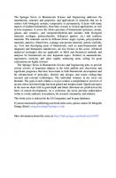
1.2.1 Phases of Acute Wound Healing The four phases of wound healing mentioned above are continuous and overlapping phases. Naturally, the body will activate healing processes by providing various components of extracellular matrix (ECM) in response to different microenvironment conditions. These phases comprising hemostasis phase, inflammation phase, proliferation phase and remodeling phase and Fig. 1.1 shows different components of ECM at each healing phase. Each phase must occur in the appropriate time for normal wound healing but multitude of factors associated with patient’s conditions may impair the wound healing in the post-operative period. It is therefore, important for surgeons and other health workers involved in wound management to have in-depth understanding of important factors affecting wound healing for successful wound management [8]. Aging has been reported to affect different phases of wound
Fig. 1.1 Wound heling phases showing ECM and the main cell types (Adapted from [7] with permission)
4
I. J. Macha and B. Ben-Nissan
healing and the course of chronic ulcer formation. Lifestyle and systemic diseases such as diabetes, cancer, kidney failure and high blood pressure are reported to significantly delay the healing process. Apart from patient’s lifestyle which could be changed, controlling underlined diseases in support of wound healing could be challenging. In a healing process, as previously indicated, wound dressings play critical roles in wound healing and choosing the right type of dressings and application techniques for a particular wound need skilled and experienced personnel with a deep understanding of wound healing dynamics.
1.3 Wound Dressings The continuing advancement of science and technology has changed the way wounds are being dressed. Since the first use of wound dressing developed from natural materials in 18th Century, significant modifications have been done in the last 50 years with the first modern wound dressing development in middle 1981 [9]. The modern wound dressings were more active with the ability to maintain a moist environment at the wound bed and absorbing exudate. Nowadays there are smart dressings with electronic sensors integrated to detect and give a wireless communication of the data in real time on the healing progress [10]. These smart dressings offer a major benefit of facilitation of monitoring a healing process remotely resulting in reduction of hospital cost. The challenges associated with smart dressing include biocompatibility, moisture and biofouling resistance, availability of miniaturized components, calibration and disposability. The ideal wound dressing has characteristics discussed comprehensively in these reviews [11, 12]. However, there are no a single material that possesses all ideal criteria. Different strategies such as combinatory approach have been suggested in the design of wound dressing materials. This approach combines two or more different materials with different properties needed for supporting wound healing. It should be iterated that the choice of dressing materials depends on wound characteristics, patients’ preferences and many other factors. However, for practical purposes only three factors such as wound characteristics, clinical effectiveness and economic factors are considered. Currently, there are different synthetic dressing materials in clinical use that can be categorized based on their ability to adsorb exudate, deliver drugs such as antibiotic, anti-inflammatory or growth factors [13, 14], ability to promote granulation and the activeness. Table 1.1 presents the summary of different wound dressing categories and Table 1.2 highlights the wound dressing materials that were previously used in the past and those currently in use.
1 Past and Future of Wound Dressing in Soft and Hard Tissue Surgery
5
Table 1.1 Wound dressing materials and their categories Category/main function
Dressing materials
Suitability
Change frequency
Exudate absorbent
Hydrophobic polyurethane or silicone foam, alginate [15–17]
Moderate to highly exudating wounds
1–4 days
Delivery vehicle
Anticeptic (Silver, Iodine, Prevent and treat Chlorhexidine, wound infection Polyhexamethyl-biguanide (PHMB), Honey, Acetic acid and Potassium permanganate) [18–20]
Promoting granulation
Hydrocolloid [21]
Granulated wounds 3–5 days with mild to moderate exudate
Maintain moist environment
Polyurethane films and semipermeable membrane, hydrogels (propylene glycol) [17, 22]
Superficial wounds, wounds with light exudate, wounds on elbows, heels or flat surface
Activeness
Poly (hydroxyethyl Delivery and methacrylate) (pHEMA) and extraction of solutes silicone [23] via externally controlled convective mass transfer
2–3 days
Pressure
Compression bandages, stockings [21]
3–6 months
Venous laceration
3 days
1–3 times per week
1.4 Post-operative Wound Management Proper management of post-operative wounds provides a supportive and protective environment for wound healing and surgical site infection respectively. Dressing being one of the major components of wound management that promote healing by providing a moist environment and protecting the wound from potential danger. In surgical wounds, the specific function of the dressing is to absorb blood or exudate fluid in the immediate postoperative phase that might lead to maceration of the wound. Each wound needs a specific dressing regime to accommodate wound characteristics and patient’s preferences. Dressings applied on a surgical wound should ideally be left in place undisturbed unless to have the wound reviewed or when wound become stained by discharge or clinical signs of infection. It has been suggested that it is not necessary to dress a closed surgical wound after the removal of the initial. Typically, initial surgical dressings are to remain undisturbed for 48–72 h and some stay in place for up to seven days. However, some patients may prefer to have their wounds dressed.
6
I. J. Macha and B. Ben-Nissan
Table 1.2 Past and present wound dressing materials, properties and usage Materials
Properties and function
Usage
Animal-based (e.g.: collagen, jellyfish collagen, sponge)
Hydrophilic, can be porous, high absorption ability
Acute wounds, skin regeneration
Herbal origin (e.g.: cotton, Cellulose, and/or viscose, bamboo viscose) double layered, and sorption ability
Burn wounds, chronic wounds and ulcerations
Synthetic origin (e.g.: Polyurethane, PU, PLA, N-isopropylacrylamide)
Inexpensive well distributed porous structures and high flexibility and strength
Easily produced thin films, they are suitable for relatively shallow wounds, and with low to moderate drainage, and high absorbance
Alginate
Highly absorbent, might need a secondary dressing
Infected and noninfected wounds, but not for dried ones, but can be made to aid blood coagulation
Hydrogel
Many different compositions, transparent, maintains a moist wound environment, facilitate autolytic debridement
For wounds with low to moderate exudate, burns, ulcers, surgical wounds, and skin repairs
Hydrocolloid
Ability to inhibit bacteria growth, appropriate for wounds with low to moderate drainage, encourage autolytic debridement, and excellent adhesion property
Traumatic injuries, leg ulcers, acute and chronic wounds, pressure sores, minor burns, inappropriate for infected wounds and diabetic foot ulceration
Medicated
Mainly for prevention of further Mainly for infected wounds infections, facilitate removal of necrotic tissues, and most importantly promotes tissue regeneration
Most of the open surgical wounds that are not linear on involve surgical procedures such as extraction of sockets heal by secondary intention and they need appropriate dressings [24]. The risk of opting gauze-based dressing causes excessive pain on removal because of dress adherence on the wound and covered by the healing tissue. Studies have revealed that the use of hydrofibre dressing in surgical wounds tend to decrease pain during dress change, reduce length of stay, enhance healing and improve patient confidence. The recommended timeframe for staple or suture removal is one to two weeks unless for cosmetic concerns in the area such as face or eye they may require earlier removal to prevent scarring (three to five days). For wounds located at palms or soles normally take 2–3 weeks for dress change. Other wound closure supporting materials such as steri-strips or tissue/skin adhesives should be left in place until the fall off by themselves, usually five to ten days.
1 Past and Future of Wound Dressing in Soft and Hard Tissue Surgery
7
1.4.1 Post-operative Wound Complications Surgical wounds suffer from two common complications such as surgical site infection and wound dehiscence. These normally occur when a healing process is stalled at the inflammatory healing phase. The severity of these complications ranges from mild cases needing wound care management and administration of antibiotics to serious cases with secondary surgical procedures and a high mortality rate [25]. The main contributing factors such as surgical technique and degree of contamination have been regarded as strong indicators for surgical site infection and wound dehiscence. Complication risk factors can be grouped based on patient, surgical procedures and post-operative factors. However, some scholars have refuted surgical technique contribution to pathogenesis of the complications due to mainly the advancement of surgical tools and appropriate procedures and training. Despite the advances in medical supplies and devices for surgery, postoperative wound complications still remain a serious challenge. Surgical site infection alone comprising of about 15% of all health care associated infections, reportedly with more than 500,000 yearly [26]. Preventive measures for wound complications begin before surgical procedures. This includes thorough assessment of the risk factors associated with the patient (pre-existing underlined medical conditions that may limit healing by preventing the delivery of oxygen and necessary nutrients to the healing tissues), intra-operative, and post-operation, using of good surgical techniques and meticulous hemostasis, proper technology choice to enhance healing and close monitoring strategies in the post-operative period. Pre- and intra-operative risk factors and mitigations discussion is beyond the scope of this chapter and only post-operative issues will be presented. Different factors contributing to post-operative surgical site infection include saturated or leaking wound dressing, physical disruptions of staple, suture or glue that holds the tissue together before re-epithelialized which allow migration of bacterial to the wound. The Centre for Disease Control and Prevention’s (CDC) guideline for prevention of surgical site infection describes the specific criteria required for an infection to be considered a surgical site infection [27]. It is indicated that surgical site infection can occur within 30 days postoperatively or a year later for an implanted device. An infection is considered surgical site infection if the tissues are affected and show histological signs including purulent drainage or abscess.
1.4.2 Dressing Selection on Surgical Wounds Post-surgical incision care being an important part of wound healing, must consider all aspects of wound care in order to reduce the risk of infection and associated wound complications. Dressing selection, dressing protocol and dressing change time constitute to the ideal care of surgical wounds. It has been suggested that the concept of undisturbed wound healing should be given considerable attention in
8
I. J. Macha and B. Ben-Nissan
surgical wound care. While there are specific indicators which should trigger immediate dressing change such as potential dehiscence, excessive bleeding, dressing saturation and suspected local infection, a longer wear time should be considered more suitable for the healing. Apart from considering dressing and wound characteristics in wound dressing selection, different requirements for individual patients should also be considered. Table 1 shows suitability of different wound dressing materials based on the wound characteristics. The ideal dressing, on the other hand should possess specific properties required in managing post-surgical wounds [28]. Enough evidence should be needed to support the use of any specific type of dressing post-operatively for wounds healing by different mechanisms; primary, secondary or tertiary intentions. Acute surgical wounds sometimes are left open to heal by secondary intention but require moist environment in which the wound dressings should be able to keep moist environment and prevent bacteria from entering wound bed.
1.4.2.1
Dressing Characteristics
Ability to handle exudate: Exudate is produced as a normal part of a healing process to keep the wound bed wet. The wound fluid acts a medium for cells, nutrients and growth factors to migrate to the wound and help the healing process. For the normal wound healing, exudate production decreases over time but in chronic wounds exudate is produced over the prolonged period of time in higher amount than normal. Exudate contains high level of substance that are detrimental to cell-supporting extracellular matrix which also lead to wound pain, delayed healing, enlargement of the wound, skin maceration and local wound infection. Dressing that can minimize the effects of excessive exudate to the wound healing would be an important parameter to consider in choosing the dressing materials for surgical wounds. Ability to stretch: This is a key important parameter for dressing materials for skin protection and patient comfort and mobility. Flexible and conforming dressing materials increases the levels of physical activity while recovery. With flexible material pain management is improved through wound contact layer and reduce the risk of blistering or irritation. The advancement in technology provides the possibility to tailor the properties of flexible materials for specialized wound management applications with an improved reliability and efficiency. Most of the dressing materials such as films, foams, adhesives, mesh and textile could be modified to have an improved flexibility and conforming traits. Waterproof: Waterproof dressing offer protective barrier to the damaged tissues against contaminants. Patients wearing waterproof dressing can feel more secure and protected even in challenging environment. It increases the level of physical activities such as swimming without using an addition product on the wound. Most of waterproof dressing have transparent layers that can allow monitoring of wound without removing the dressing [29].
1 Past and Future of Wound Dressing in Soft and Hard Tissue Surgery
9
Transparency: Transparent dressing which are referred to as transparent films are impermeable to liquid, water and bacteria allowing one-way passage of moisture and gases. They are normally ultra-thin, flexible and waterproof, offering optimal protection for the wound. Transparency allows visualization of the wound bed make it easy to follow the wound healing progress without dressing removal. These dressings are suitable for the treatment of frictional wounds, closed surgical incision sites, skin graft and donor sites, catheter sites and areas of friction. They cannot be used for wounds with excessive exudate and usually changed three times a week [15].
1.4.2.2
Surgical Wounds
In this sub-section discussion is limited to selected common surgical wounds seen in clinical and community settings but we are aware that there are many different types of surgical wounds that may be encountered.
Total Joint Arthroplasty It has been reported elsewhere that the ideal dressing for orthopedic surgical incisions should be absorbent, able to act as complete barrier, transparent, non-adherent, able keep the mound moist and require minimum changes. However, there is no single dressing material that encompasses all of the above parameters [15]. It envisaged that the combinatory approach in the development of dressing materials will result into a product with all important parameters. Hydrofiber and hydrocolloid dressings can handle excess exudate but keep the wound moist, have high absorptive capacity and permeability. They have low blistering rates and require minimum change thus reduce the risk of surgical site infection.
Caesarian (C) Section C-section surgery is a surgical birthing method attributed with more risks of postsurgery complications such as premature rupture, wound infections, pelvic peritonitis than traditional vaginal births. Similar abdominal surgery to C-section include hysterectomy, appendectomy, hernia surgery and laparotomy. Proper dressing material should be able to handle exudate from the wound, provide optimal healing conditions, protect the surgical area and prevent stitches catching clothes. The skin edges normally seal within one to two days after the operation but varies from a person to another. After two days wound can usually be left open, however, some people prefer their wounds be covered.
10
I. J. Macha and B. Ben-Nissan
Skin Graft and Donor Site Skin graft is surgical procedures undertaken to remove a section of a skin of varied thickness from one part of the body (donor site) such as upper thigh and placed on a site of the injury (recipient site). Donor site wound care would require application of wound dressing capable of supporting moist environment [30]. Since the healing at donor site is by epithelialization, a transparent dressing or fine mesh gauze are preferred. Skin grafts are normally sutured, stapled or glued at the recipient site, treated like surgical wound and covered with an appropriate dressing. The choice of dressing remains with the surgeon responsible. Post-surgical risks are bacterial infection, graft contracture and hematoma. The healing takes up to six weeks and up to two years for scars to mature and inflammation to cease [31].
1.5 Future Directions It has been becoming more clearer in the translational medicine that by incorporation of the therapeutic agents into the wound dressings, it is possible to repair the wounds more quickly and more efficiently. Among the therapeutic agents, antibiotics can be used to prevent wound infections, growth factors to revitalize damaged tissues and supplements, such as vitamins and minerals. In the past, plain gauze and thin paraffin-impregnated gauze were utilized as drug carriers. Nowadays, hydrocolloids, hydrogels, alginates, polylactic acid and polyurethane films/foams are the materials used to deliver therapeutic agents. One factor that limits the wound healing process is an infection and early detection. Knowing the currently encountered problems, wound dressing researchers in addition to new material development are directing their efforts in next-generation wound dressings with the abilities of diagnosis during early stages, real-time monitoring, and on-demand therapy. Number of investigators, combined bioelectronics, and a smart flexible electronics-integrated wound dressing with a double-layer structure, the upper layer of which is polydimethylsiloxane-encapsulated flexible electronics integrated with a temperature sensor and ultraviolet (UV) light-emitting diodes, and the lower layer of which is a UV-responsive antibacterial hydrogel [32]. It was reported that this dressing is expected to provide early infection diagnosis via realtime wound-temperature monitoring by the integrated sensor and on-demand infection treatment by the release of antibiotics from the hydrogel by in situ UV irradiation. Their animal trials showed that this integrated system possesses a good flexibility, excellent compatibility, and high monitoring sensitivity and durability. It is envisaged that this technology can be extended to patches using current nanotechnology.
1 Past and Future of Wound Dressing in Soft and Hard Tissue Surgery
11
1.6 Conclusion Remarks The need for proper wound dressing materials to appropriately manage surgical wounds is gradually increasing as a major aspect to prevent post-operative complications, a growing area of concern for patients and surgeons. This is an opportunity for the scientific community and biomedical industries to put more effort in development of advanced wound dressing materials with ideal characteristics. A single material, even with chemical and surface modifications cannot possess all necessary traits of an ideal dressing, to support healing of surgical wounds. With the advances in technology, we believe the combinatory approach in dressing development could be employed to address the problem. With the large number of wound dressing products available in clinical settings, the appropriate choice remains largely to the surgeons and health-care professionals. The best practice for surgical wound dressing choice recommended in this chapter when combined with the well evidence-based interventions will equip healthcare professionals with ability to make the needed decisions for the proper wound management.
References 1. Infectious Disease Advisor (2017) Surguical site infections.Hospital infection control. https:// www.infectiousdiseaseadvisor.com/home/decision-support-in-medicine/hospital-infectioncontrol/surgical-site-infections/. Accessed 16 May 2021 2. European Centre for Disease Prevention and Control (ECDC) (2021) Facts about surgical site infections. https://www.ecdc.europa.eu/en/surgical-site-infections/facts. Accessed 16 May 2021 3. Allegranzi B, Bagheri Nejad S, Combescure C et al (2011) Burden of endemic health-careassociated infection in developing countries: systematic review and meta-analysis. Lancet 377:228–241 4. Stephen-Haynes J (2015) The benefits of undisturbed healing using ALLEVYN LifeTM . Available via Wounds International. https://www.woundsinternational.com/resources/details/thebenefits-of-undisturbed-healing-using-allevyn-lifetm. Accessed 16 May 2021 5. Ban KA, Minei JP, Laronga C et al (2017) American college of surgeons and surgical infection society: surgical site infection guidelines, 2016 update. J Am Coll Surg 224:59–74 6. Young A, McNaught CE (2011) The physiology of wound healing. Surgery 29:475–479 7. Kordestani SS (2019) Wound healing process. Atlas of wound healing. Elsevier, Amsterdam, pp 11–22 8. Tahir M, Chaudhry EA, Zimri FK et al (2020) Negative pressure wound therapy versus conventional dressing for open fractures in lower extremity trauma. Bone Joint J 102-B:912–917 9. Shah JB (2011) The history of wound care. J Am Col Certif Wound Spec 3:65–66 10. O’Callaghan S, Galvin P, O’Mahony C et al (2020) “Smart” wound dressings for advanced wound care: a review. J Wound Care 29:394–406 11. Boateng JS, Matthews KH, Stevens HN et al (2008) Wound healing dressings and drug delivery systems: a review. J Pharm Sci 97:2892–2923 12. Borda LJ, Macquhae FE, Kirsner RS (2016) Wound dressings: a comprehensive review. Curr Derm Rep 5:287–297 13. Macha IJ, Sufi S (2020) Novel slow drug release bioceramic composite materials for wound dressing applications: potential of natural materials. SN Appl Sci. https://doi.org/10.1007/s42 452-020-1977-z
12
I. J. Macha and B. Ben-Nissan
14. Macha IJ, Muna MM, Magere JL (2018) In vitro study and characterization of cotton fabric PLA composite as a slow antibiotic delivery device for biomedical applications. J Drug Deliv Sci Technol 43:172–177 15. Criscitelli T (2012) Fast facts for wound care nursing: practical wound management in a nutshell. AORN J 96:563 16. Jones V, Grey JE, Harding KG (2006) Wound dressings. BMJ 332:777–780 17. Lionelli GT, Lawrence WT (2003) Wound dressings. Surg Clin North Am 83:617–638 18. Wounds International (2008) Wound infection in clinical practice: a WUWHS international consensus. https://www.woundsinternational.com/resources/details/wound-infection-clinicalpractice-wuwhs-international-consensus. Accessed 16 May 2021 19. McDonnell G, Russell AD (1999). Antiseptics and disinfectants: activity, action, and resistance. Clin Microbiol Rev 12:147–179. Erratum in: Clin Microbiol Rev 2001 Jan; 14(1):227 20. Brennan SS, Leaper DJ (1985) The effect of antiseptics on the healing wound: a study using the rabbit ear chamber. Br J Surg 72:780–782 21. Dabiri G, Damstetter E, Phillips T (2016) Choosing a wound dressing based on common wound characteristics. Adv Wound Care 5:32–41 22. Eisenbud D, Hunter H, Kessler L et al (2003) Hydrogel wound dressings: where do we stand in 2003? Ostomy Wound Manage 49:52–57 23. Cabodi M, Cross VL, Qu Z et al (2007) An active wound dressing for controlled convective mass transfer with the wound bed. J Biomed Mater Res B Appl Biomater 82:210–222 24. Andersen BM (2019) Prevention of postoperative wound infections. Prevention and control of infections in hospitals. Springer, Cham, pp 377–437 25. Dhaigude BD, Shree S, Shah P et al (2018) Post-operative wound complications following emergency and elective abdominal surgeries. Int Surg J. https://doi.org/10.18203/2349-2902. isj20175901 26. National Center for Health Statistics (US) (2016) Health, United States, 2016: With chartbook on long-term trends in health. Report No.: 2017-1232 27. Centers for Disease Control and Prevention (2017) Guideline for prevention of surgical site infection. https://www.cdc.gov/infectioncontrol/guidelines/ssi/index.html. Accessed 16 May 2021 28. Bennett-Marsden M (2010) How to select a wound dressing. Pharm J 2:363–366 29. Wounds UK (2016) Medical adhesive-related skin injuries (MARSI): made easy. https://www. wounds-uk.com/resources/details/medical-adhesive-related-skin-injuries-marsi-made-easy. Accessed 16 May 2021 30. Chowdhry M, Chen AF (2015) Wound dressings for primary and revision total joint arthroplasty. Ann Transl Med 3:268 31. Beldon P (2007) What you need to know about skin grafts and donor site wounds. Wound Essentials 2:149–155 32. Pang Q, Lou D, Li S et al (2020) Smart flexible electronics-integrated wound dressing for real-time monitoring and on-demand treatment of infected wounds. Adv Sci 7:1902673
1 Past and Future of Wound Dressing in Soft and Hard Tissue Surgery
13
Innocent J. Macha has extensive experience in research and teaching for more than nine (9) years in the area of biomaterials synthesis and characterization, drug delivery devices, cell culture and bacteria biofilm. Dr. Macha is also one of the Associate Editors of the Journal of The Australian Ceramic Society and has published more than 26 articles and 10 book chapters.
Besim Ben-Nissan has higher degrees in Metallurgical Engineering (ITU), Ceranic Engineering (University of New South Wales) and a Ph.D. in Mechanical and Industrial Engineering with Biomedical Engineering (University of New South Wales). Over the last four decades together with a large numbers of Ph.D. students and post-doctoral fellows he has worked on production and analysis of various biomedical materials, implants, calcium phosphate ceramics, advanced ceramics (alumina, zirconia, silicon nitrides), sol-gel developed nanocoatings for enhanced bioactivity, corrosion and abrasion protections, optical and electronic ceramics and thermally insulating new generation composites. He also has contributed to the areas of mechanical properties of sol-gel developed nanocoatings. In the biomedical field, he has involved with the development of materials for slow drug delivery, natural and marine material conversion, implant technology (bioactive materials including conversion of Australian corals to hydroxyapatite bone grafts), biomimetics (learning from nature and its application to regenerative medicine), biocomposites, investigative research on biomechanics and Finite Element Analysis (mandible, knee, hip joints, hip resurfacing), reliability and implant design (modular ceramic knee prosthesis, femoral head stresses). He was part of a research team which initiated the world’s first reliable ceramic knee and hydroxyapatite sol–gel derived nanocoatings. Since 1990 he has published over 260 papers in journals, five books and over 50 book chapters. He is one of the editors of the Journal of the Australian Ceramic Society and Editorial Board member of three international biomaterials journals. He was awarded “The Australasian Ceramic Society Award” for his contribution to “Ceramic education and research and development in Australia.” He also received “Future Materials Award” for his contribution to the “Advanced nanocoated materials field”. He has collaborated with a number of international groups in Japan, USA, Thailand, Finland, Israel, France, UK, Germany and Turkey and held grants from the Australian Academy of Science and the Japan Society for Promotion of Science for collaborative work in the biomedical field in Europe, USA and
14
I. J. Macha and B. Ben-Nissan Japan respectively. After serving as an academic for over 33 years he has retired, however still active and contributes to science by research in the biomedical field and supervising higher degree students.
Chapter 2
3D Printing and Bioprinting of Biomaterials and Bioceramic Scaffolds: Clinical Outcomes and Implications in Bone Tissue Engineering and Maxillofacial Reconstructive Surgery Muhja Salah, Farhad B. Naini, and Lobat Tayebi Abstract Three-dimensional (3D) printing is a type of additive manufacturing that works by the application of material inks layer by layer using data from computeraided design (CAD) to help to place the ink in a predefined place, thus producing a highly accurate product even with complex geometry. The goal in using 3D bioprinting is to develop a biological scaffold that resembles the desired tissue to be replaced, including the cells and the growth factors, in a specific spatial relationship. The developments in bone tissue engineering (BTE) and 3D bioprinting are revolutionizing osseous craniofacial reconstructive surgery. This chapter aims to describe 3D bioprinting of biomaterial and bioceramic scaffolds for bone tissue engineering and maxillofacial reconstructive surgery. Keywords Additive manufacturing · Layer by layer · Bioprinting · Biological scaffold · Bioactive glass · Calcium phosphate · Hydroxyapatite · Mesenchymal stem cells · Induced pluripotent stem cells · Exosome · Biomimetics · Self-assembly
M. Salah (B) St George’s Hospital, Tooting, London, UK F. B. Naini Kingston Hospital, Kingston upon Thames, UK St George’s Hospital and Medical School, Tooting, London, UK L. Tayebi Marquette University School of Dentistry, Milwaukee, WI, USA © The Author(s), under exclusive license to Springer Nature Singapore Pte Ltd. 2022 A. H. Choi and B. Ben-Nissan (eds.), Innovative Bioceramics in Translational Medicine II, Springer Series in Biomaterials Science and Engineering 18, https://doi.org/10.1007/978-981-16-7439-6_2
15
16
M. Salah et al.
2.1 Introduction Three-dimensional (3D) printing is a type of additive manufacturing that was first invented in 1984 for engineering and industrial purposes. It works by the application of material inks layer by layer using data from computer-aided design (CAD) to help to place the ink in a predefined place, thus producing a highly accurate product even with complex geometry [1, 2]. The technology found its way to the health sector through dentistry when additive manufacturing was used to print a solid block of dental implants, crowns, and bridges from a biocompatible and bioinert material that does not elicit an immune reaction [3]. Scientists were overly ambitious realizing the precision of the end-product when 3D printing was used. They decided to unleash the power of 3D printing and use it for medicinal purposes to bioprint tissues. The first bioprinting attempt was undertaken early in 1988, using an inkjet printer depositing cell drops on-demand approach. The goal in using 3D bioprinting is to develop a biological scaffold that resembles the desired tissue to be replaced, including the cells and the growth factors, in a specific spatial relationship. It is a customizable, patient-specific solution meeting the patient’s need at a macro level (i.e., shape and size), and on a micro level resembling patients’ tissue structure and architecture [4, 5]. The development in bone tissue engineering and 3D bioprinting also aims to solve the crisis in the shortage in organs needed for transplantation [6]. Tissue loss in the craniofacial region can occur due to a craniofacial genetic deformity, trauma, or surgical excision as a treatment of tissue malignancy [7]. Facial disfigurement has a severe negative impact on individuals, both socially and psychologically, and requires rapid, precise, and aesthetic rebuilding producing a functional, harmonious, and symmetrical face [8]. Osseous craniofacial reconstruction traditionally employs a graft harvested from the iliac crest or the ribs, which serve as the bridge needed to direct the 3D bone growth (osseoconduction), as well as inducing the differentiation and the recruiting of osteoblasts (osseoinduction) into the injured area to promote bone healing [9]. However, placing a graft is not without risk; autogenous bone grafting carries the risk of morbidity (pain in the donor site, neuralgia, blood loss, or infection), while the allogenic bone graft is associated with the possibility of transmitting infection or eliciting an immune reaction [10]. Moreover, facial reconstruction using a bone graft does not always provide aesthetic results due to the anatomical complexity of bone, soft tissue, and the hollow cavities in the face. 3D bioprinting, on the other hand, may provide a more precise alternative that fits the defects, reducing the need to count on the surgeon’s ability to harvest or carve the graft to fit the surface.
2 3D Printing and Bioprinting of Biomaterials and Bioceramic …
17
2.2 Bone Tissue Engineering Bone tissue engineering has received much attention in the last few decades, and it showed tremendous progress due to the improved understanding of bone biology, along with the advances in the biomaterials. It focuses on: (a) (b)
(c) (d)
Developing biomaterials that can provide the same physical and biological properties as natural bone [11]. Producing scaffolds from these biomaterials, having the same architecture and topography that ensure nutrient and oxygen passage, micro-vessels, and nerve ingrowth, as well as regulating the stem cell differentiation down the osteogenic fate [12, 13]. Incorporating mesenchymal stem cells (MSC) that are directed toward differentiating into osteogenic cell lineage [14]. Incorporating bone growth factors; bone morphogenic proteins (BMP), insulinlike growth factor-2 (IGF-1), vascular endothelial growth factors (VEGF), and others that enhance osteogenesis [15].
2.3 Biomaterials in Bone Tissue Engineering Bone is composed of 60–70% inorganic phase in the form of hydroxyapatite (Ca10 (PO4 )6 (OH)2 ), while the organic phase is mostly formed of collagen type I with some other proteins and growth factors. The simplicity of the natural bone composition enabled the progress in bone tissue engineering. Biomaterials used to fabricate scaffolds should be biocompatible, biodegradable to be replaced by the newly generated bone, and bioactive to enhance bone regeneration, having physical strength and mechanical properties, which enable it to support the load the natural bone is supporting [16]. Examples of biomaterials used in bone tissue engineering include demineralized bone matrix as well as a number of bioceramics and bioglasses.
2.3.1 Demineralized Bone Matrix These are allografts treated with chemical acid to demineralize as well as removing the inorganic component of the graft, leaving the matrix proteins, mainly collagen I and bone growth factor BMP, and are then treated with radiation to decrease the possibility of eliciting an immune reaction [17]. Demineralized bone matrix has been used for decades in clinical applications, and has shown tremendous success being osteoconductive and osseoinductive, but because the end-product is in a powder form, making it is difficult to handle during surgery, which consequently has limited its use [18]. Solutions implemented to ease the manipulation of the powder were based on using the powder mixed with a viscous carrier to enable it to condense and pack into bony defects [17].
18
M. Salah et al.
Wagner et al. reported using demineralized bone matrix for mandibular reconstruction by wrapping it in an acellular dermal matrix to confine the demineralized bone matrix paste and placing it over a bent plate [19]. The patients were followed up for five years and showed evidence of bone healing. In a recent study, Driscoll et al. used demineralized bone matrix mixed with hydroxyapatite crystals in different ratios in a 3D printer to print scaffolds for spinal repair, and it was tested in rat models [20]. The preclinical studies showed successful fusion, with the developed biomaterial being a hybrid encompassing the osseoinductive properties of the demineralized bone matrix carrying the bone growth factor along with the osteoconductive properties of the hydroxyapatite.
2.3.2 Bioceramics and Bioglasses These are inorganic oxides including hydroxyapatite, calcium phosphate, tricalcium phosphate (TCP), and calcium silicate. They are considered bioactive as they bond to bone and elicit osteogenesis [21].
2.3.2.1
Hydroxyapatite
This makes the bulk of the bone composition, thus, it has been studied extensively as a bone substitute because its composition resembles natural bone. Both calcium and phosphate ions present in hydroxyapatite promote bone regeneration. Calcium ions stimulate osteoblasts by activating ERK1/2 pathways, which protect them from apoptosis, as well as having a central role in bone maturation by deposition in immature bone [22]. Phosphate ions activate the IGF-1 pathway in osteoblasts, which is implicated in cell survival, growth, and protein synthesis [23]. Besides, it is osseoinductive and osseoconductive, making it ideal to be a synthetic bone substitute. But, it is organized in a highly arranged crystalline microstructure that hinders it from degrading, and it also inherits a low compressive and tensile strength making it brittle when loaded [24]. To reduce the brittleness of hydroxyapatite, Mukherjee et al. investigated the effect of adding carbon nanotubes (CNT) to hydroxyapatite and found that it increased the fracture toughness of the scaffold. They tested the scaffold on animal models and found that the addition of CNT was biologically safe with no toxicity shown in either the liver or kidney, but with enhanced bone regeneration on the implanted site [25]. However, the data was stated to be preliminary and incomplete to proceed onto clinical studies. In relations to the 3D printing of hydroxyapatite, Seitz et al. were able to use hydroxyapatite powder sprayed with a polymeric binder dissolved in water to ensure ink flow to produce a porous scaffold with fully interconnected channels sixteen years ago, which are further compacted after printing in a 1250 °C furnace to remove and achieve binder pyrolysis [26]. Six years ago, Shao et al. proposed the use of 3D-gel
2 3D Printing and Bioprinting of Biomaterials and Bioceramic …
19
printing (3DGP) instead of regular 3D printing with the use of hydroxyethylmethacrylate (HEMA) gelation system to produce a flowable slurry. Their new system has the advantage of being appropriate to a wide range of materials from metal to ceramics, keeping the cost low while achieving high printing efficiency, producing complex shapes due to the flow of the slurry. They used it with stainless steel, zirconia, and hydroxyapatite, and tested the fabricated scaffolds and their mechanical properties [27–29]. Most studies on the fabrication of hydroxyapatite scaffolds were carried out using biomaterials only, without embedding cells within the scaffold and the seeding of cells occurred after fabrication, thus it should not be confused with bioinks, which incorporate both biomaterials and cells [30].
2.3.2.2
Tricalcium Phosphate
In 1920, Albee was the first to report that rhombohedral β-form, β-tricalcium phosphate (β-TCP), enhances osteogenesis [31]. Tricalcium phosphate is composed of calcium and phosphate ions just like hydroxyapatite, which renders it to have the same effect on osteoblasts resulting in bone regeneration. Gao et al. showed the in vivo osteogenic potential of the tricalcium phosphate granules placed in a titanium porous scaffold and implanted in a femur defect on animal models [32]. In contrast to hydroxyapatite, tricalcium phosphate has a crystalline structure that is not highly organized, which makes it more susceptible to resorption and degradation, which is an ideal property for a scaffold material [33]. Ishikawa et al. compared the mechanical properties and recorded the histological findings of the newly generated bone when tricalcium phosphate and hydroxyapatite were used [34]. The study confirmed tricalcium phosphate has higher solubility than hydroxyapatite, which explains why more bone is found around the implanted tricalcium phosphate than the hydroxyapatite. Degradation of the tricalcium phosphate is a desired property when constructing a scaffold, but degradation of the material should be coordinated with the speed of the osteogenesis process. To adjust tricalcium phosphate degradation, it has been doped with mineral oxides like magnesium oxide (MgO) and strontium oxide (SrO), which affect the crystalline orientation of the tricalcium phosphate and make it less soluble and alter both the mechanical and biological properties of the tricalcium phosphate [35]. Banerjee et al. confirmed slower degradation of the implanted MgO/SrO-doped β-TCP than pure β-TCP on animal models [36]. They also showed that the doped implant had more cell attachment, which increases cell differentiation and proliferation. Analysis of osteocalcin and type I collagen inside the implants indicated faster osteogenesis and remodeling. Recently, Gu et al. used Mg-doped tricalcium phosphate and 3D-printed an interconnected-pores scaffold with mechanical properties close to bone [37]. They further seeded the scaffold with MSCs derived from bone marrow and umbilical cord and showed that both osteogenesis and angiogenesis were enhanced. In an animal model, Kim et al. transplanted 3D-printed scaffolds from a composite of tricalcium phosphate and polycaprolactone polymer and used it to repair the maxilla in a dog after resecting a tumor with success [38].
20
M. Salah et al.
In 1986, another strategy was proposed to adjust the solubility of tricalcium phosphate in physiological conditions by combining hydroxyapatite with tricalcium phosphate in different ratios to achieve the best physical and mechanical properties for the desired load-bearing application referred to as biphasic calcium phosphate [39]. Increasing the hydroxyapatite content in the biphasic calcium phosphate leads to a more stable material, while increasing the tricalcium phosphate results in a material that is more soluble, thus, it can be easily tailored. Liu et al. 3D-printed scaffolds using biphasic calcium phosphate and examined the in vivo behavior using rabbit calvarial defects which showed an increase in osteogenesis and high bone density [40].
2.3.2.3
Calcium Phosphate Cement
This cement was accidentally invented in the 1980s by the American Dental Association Health Foundation Paffenbarger Research Centre (ADAHF-PRC) who were trying to develop a cement to treat and remineralize early dental caries. A mixture of tetra calcium phosphate, dicalcium phosphate anhydrous, and dicalcium phosphate dihydrate with water was found to rapidly produce hydroxyapatite. A decade after that, the FDA approved calcium phosphate cement for clinical use, and since then a tremendous number of studies have been conducted [41]. The cement was found to promote osteogenesis, being osteoconductive, and most importantly it is injectable, which makes it easier for clinical use. Injecting the material into the site of surgery will allow it to mould into the shape of the deformity without the need for further drilling at the surgical site to match the size and shape of the scaffold. Yu et al. reported the success of calcium phosphate cement in bone regeneration when they performed an in vivo study in which injectable calcium phosphate cement was implanted into a femoral condyle defects of rabbits [42]. Lin et al. cultured three types of cells: induced pluripotent stem cells (iPSC), human umbilical vein endothelial cells (HUVECs), and pericytes; into scaffolds made from calcium phosphate cement and implanted them into cranial defects created on rats [43]. They found that the tri-culture group had elevated angiogenic and osteogenic markers, and mineralization.
2.3.2.4
Bioactive Glass
These are silicate-based ceramics composed of silicon dioxide, calcium oxide, phosphorus oxide, potassium oxide, magnesium oxide, and boric oxide. The composition and percentage of these oxides vary, but the key component, silicate, always constitutes 45–52% of its weight [44]. Bioglasses possess the capability to form a strong chemical bond with the bone tissue that is created through the polycondensation of a silicone-rich layer on the surface of the bioactive glass due to ion exchange between ions in the physiological fluid and leaching of ions from the surface of the bioglass [44]. Moreover, the electronegative silicone-rich layer on the surface is considered osseoinductive as it adsorbs protein that in turn attracts macrophages and MSCs [44].
2 3D Printing and Bioprinting of Biomaterials and Bioceramic …
21
As a bone substitute material, bioactive glass proved its worthiness in an in vivo animal study carried out by Moimas et al. where bioactive glass implanted into tibial defects were found to be completely resorbed in six months and be replaced by bone tissue [45]. It was shown that the composition of the bioactive glass affects the union chemical reaction and stimulation of cells to promote osteogenesis. Bioactive glass went through optimization of its formula, and 45S4 was invented composed of 45% SiO2 , 24.5% Na2 O, 24.5% CaO, 6% P2 O5 (wt%), characterized by a high amount of Na2 O and CaO, which make the surface of the material very bioactive [46]. Scaffolds made from 45S5 were found by Detsch et al. to drive umbilical cord-derived MSCs down the osteogenic differentiation pathway [47]. Recently, the development of the sol–gel method, adding ammonia to the sol phase to transform it into a gel and then freeze-dry it, produced 58S bioactive glass composing of 60 mol.% SiO2 , 36 mol.% CaO and 4 mol.% P2 O5 . 58S bioactive glass has the benefit of achieving a homogeneous biomaterial compared to the melting method used originally where phosphate becomes volatile at high temperature [48]. Wheeler et al. compared in vivo bone regeneration capacity between 45S4 and 58S scaffolds after implantation within critical-sized distal femoral cancellous bone defects in a rabbit model and the results showed that the 58S degraded much quicker but was able to form bone earlier than 45S4 at 4 weeks, which is normalized at 12 weeks [49]. The 3D printing of scaffolds composed of bioactive glass have been investigated in a number of in vitro and in vivo studies El-Rashidy et al. comprehensively reviewed the in vivo studies undertaken on the regeneration of bone with 3D-printed bioactive glass scaffolds [50]. Recently, Kolan et al. compared the osteogenic potential of bioactive glass scaffolds made by 3D printing with and without the use of BMP after implantation into cranial defects in rats [51]. Their study concluded that the addition of BMP to the scaffold greatly enhanced bone regeneration.
2.4 Cells in Bone Tissue Engineering 3D bioprinting includes both biomaterial and cells in the bioink to fabricate a scaffold. Ideal biomaterials for bone substitutes should stimulate the seeded stem cells to differentiate into osteoblasts responsible for the bone regeneration. Gao et al. proposed different molecular mechanisms by which biomaterials interact with stem cells to promote osteogenesis [14]. The exact process is not known, but they postulated that phosphorus, magnesium, and strontium ions released from the biomaterial activate the BMP pathway and increase the concentration of the calcitonin generelated peptide. The following section describes the various types of cells used in bone tissue engineering.
22
M. Salah et al.
2.4.1 Mesenchymal Stem Cells (MSCs) These are used for their pluripotency, ability to differentiate into osteoblasts, and immune modulative effect [52]. They can also be derived from a variety of sources ranging from bone marrow, umbilical cord, placenta, dental pulp, adipose tissue, and other sources [52]. Injecting MSCs derived from adipose tissue along mandibular fracture lines were found to enhance osseointegration and bone quality, as well as promoted bone healing as observed in the study by Castillo-Cardiel et al. [53]. In 2016, a study by Chamieh et al. discovered that implanting collagen scaffolds seeded with dental pulp-derived MSCs into calvarial defects in rats resulted in accelerated bone regeneration compared to rats having a collagen scaffold with no seeded cells, demonstrated by evaluating the variations in bone density and through histological examination [54]. Fahimipour et al. reported in a recent article the utilization of the bioprinter to bioprint collagen matrix to mimic the extracellular matrix of natural bone with the MSC and BMP [55]. The matrix has the benefit of confining BMP as well as preventing it from escaping the scaffold, which is known to cause ectopic bone formation or osteomas [56]. The 3D printing was used again to 3D print a scaffold that represents the mineralized part of the bone, which is then used to support the MSC-BMP collagen matrix. It was found that using this method enhanced MSCs seeding, and proliferation while the availability of BMP enhanced the osteogenic potential of the MSCs [55]. A recent report by Dong et al. showed that the presence of osteoclasts is crucial for bone regeneration as well as osteoblasts [57]. In their study, a proteomic analysis was performed, and mass spectrometry was used to identify proteins secreted in extracellular matrix. The analysis showed the presence of more than 608 protein presents, among which two proteins are known to be secreted by pre-osteoclasts, CXCL12 and IGFBP5 proteins, both are responsible for MSC cells’ migration and osteogenic differentiation, respectively [57]. They confirmed their hypothesis by implanting scaffolds made from decalcified bone matrix seeded with co-cultured MSCs and pre-osteoclasts into femur defects in rats showing significant enhancements in bone regeneration compared to implanting scaffolds seeded with MSCs only.
2.4.2 Induced Pluripotent Stem Cells (iPSCs) These are another exciting source of cells that can differentiate into any cell type, mimicking embryonic stem cells. However, with the ethical dilemma that has risen by extracting embryonic stem cells, which results in the destruction of human embryos, motivated scientists to look for other sources of cells that have the same pluripotency [58]. To circumvent this issue, iPSCs were produced by Takahashi et al. in 2007 by transducing four factors: Oct3/4, Sox2, Klf4, and c-Myc, present in embryonic stem cells, in fibroblast turning them into cells mimicking pluripotency [59]. Xie et al. investigated the osteogenic differentiation of iPSCs seeded on a scaffold made
2 3D Printing and Bioprinting of Biomaterials and Bioceramic …
23
from a composite of hydroxyapatite-chitosan-collagen and found the proliferation of iPSCs into osteoblasts and an increase in bone protein secretion [60]. Moreover, they implanted these scaffolds into cranial defects of animal models, and compared the density of bone with the scaffold seeded with iPSC and other seeded with MSCs and found that the iPSCs scaffold has nearly double the bone density than when MSCs were used alone. The osteogenic differentiation of iPSCs was studied by a number of research groups [61]. A study by Kao et al. discovered that resveratrol has a supporting effect on the osteogenic differentiation of iPSCs [62]. Later, a study by Ji et al. examined the osteogenic differentiation of human iPSCs regulated by nanohydroxyapatite/chitosan/gelatine 3D scaffolds with nano-hydroxyapatite in different ratios [63]. Investigation was also carried out to reprogram iPSCs to functional osteoblasts using only the small molecule exogenous adenosine [64]. However, iPSCs still carry the potential of tumorigenicity and teratoma formation, which still limits its use clinically, and further investigation should be conducted to optimize its use and safety [65].
2.4.3 Exosome Recently, increasing interest was diverted into cell-free therapies after the discovery that MSCs cause tissue regeneration due to its paracrine effect. This approach carries the benefit of avoiding tumorigenicity, resistance to apoptosis, triggering an immune response, and genetic instability, which are all present in MSCs utilization [66]. It will also permit the repeated injections or administration of the therapy without the fear of accumulation of cells in non-targeted tissue, especially the lungs [67]. The cell-free approach uses the exosomes, which are membrane-bound vesicles, produced by endosomes in the cell containing a specific cargo either: micro-RNA, messenger-RNA, proteins, or other biomolecules, and get excreted outside the cell to be communicated into another cell [68]. Exosomes are produced by most cell types as a way of communication and crosstalk between cells. Exosomes from MSCs regulate the paracrine effect that enhances the regeneration of tissues [69]. Several studies have been conducted and showed the potential of using exosomes for bone regeneration. Lu et al. extracted exosomes from adipose-derived MSCs and used a TNF-α preconditioned medium, which was found to positively promote osteogenesis and bone repair [70]. Zhao et al. proposed that exosomes extracted from bone marrow-derived MSCs and co-cultured with osteoblasts, result in the activation of the MAPK pathway on the osteoblasts, which is important for the cell cycle and growth, and results in their proliferation, thus promoting bone regeneration [71]. More importantly, Diomede et al. demonstrated the ability of an implanted 3Dprinted scaffold to heal calvarial defect in rats that is composed of a polymer polylactic acid (PLA), seeded with exosomes and gingiva-derived MSCs [72]. Furthermore, Zhang et al. also worked extensively on exosomes and in one of their study, they showed that a scaffold made with tricalcium phosphate combined with exosomes
24
M. Salah et al.
derived from iPSCs healed calvarial defects on rats via activating the PI3K/Akt signalling pathway [73]. In a later study, they used exosomes derived from umbilical cord-derived MSCs combined in hydrogel and transplanted at the femoral fracture site in the animal model. They found that implanted exosomes promoted angiogenesis, which in turn enhanced fracture healing [74]. Although the results of these studies are promising, still, a consensus on exosome extraction and purification has not been achieved which is important in translational medicine.
2.5 3D Bioprinting Approaches In the process of bioprinting, deposition of both the biomaterial and the cells occur simultaneously. 3D bioprinting is achieved by one or a combination of the following strategies.
2.5.1 Biomimicry This is a straightforward approach using the bioprinter to replicate the original architecture of the tissue, thereby providing the right environmental factors that guide cells to differentiate into the right type of cells. This approach of bioprinting is extremely dependent on the material ink used to construct the scaffold. A scaffold is the parallel of the extracellular matrix, that should be able to provide the chemical and physiological cues important for cell viability, differentiation, and expansion [75]. Scaffold biomaterials should be biocompatible, permeable to nutrients, having adequate stiffness to withstand loading and deformation while at the same time, able to undergo degradation at the same pace that allows the growth of new bone tissue and eventually replaces the scaffold [76]. All these requirements are crucial in choosing the most ideal scaffold bioink and they are also the primary factors that determines the success of the printed scaffold. After bioprinting, a bioreactor is used to regulate environmental factors such as the oxygen, temperature, nutrient diffusion, and the gravitational force needed for cell infiltration to the depth of the printed scaffold [77].
2.5.2 Self-assembly Approach This is a scaffold-free approach that eliminates the need for scaffold biomaterials and mitigating the difficulties faced using the scaffold. The approach adopts the same embryological development process which utilizes interaction and signals between adjacent cells and their extracellular matrix to self-organize into the tissue intended for engineering [78]. High-density initial cell seeding ensures cell–cell interaction, resulting in cell producing their own extracellular matrix and forming cell aggregates
2 3D Printing and Bioprinting of Biomaterials and Bioceramic …
25
in the form of spheres or sheets, and carries the advantage of efficiency to produce tissues faster than using scaffold bioink. Various methods are used to form these cell aggregates from magnetic levitation, hanging drop, hydrogel microwell, and others, each with its pros and cons [79]. Spheroids and sheets are then used in a 3D printer to form the engineered tissue. The advantage of using this approach is the ability to use different types of cells as well as regulating their ratios. This allowed for the co-culturing endothelial cells with MSCs, which promotes angiogenesis in the final construct, while the MSCs differentiate into the desired cell type [80]. Yamasaki et al. created a scaffold-free construct from adipose tissue-derived MSCs by using the needle array 3D printing method and implanted them into femoral defects of pigs which showed enhanced osteochondral regeneration [81]. Recently, Heo et al. described a method to 3D print spheroid aggregates, made from human umbilical vein endothelial cells (HUVECs) and MSCs and called it the aspiration-assisted bioprinting (AAB) technique, in which they showed that it allowed for better and more precise positioning of the spheroids to produce scaffold-free bone tissue [82].
2.5.3 3D Bioprinting in Bone Tissue Engineering and Craniofacial Reconstruction 3D bioprinting is offering an exciting future for bone tissue engineering and craniofacial reconstruction, but the technology is still in its early stages. Few studies were carried out or are currently in progress that shows promising results. In 2014, Goh et al. implanted a polycaprolactone scaffold fabricated by 3D printing in sockets of newly extracted teeth to preserve the height of maxillary and mandibular ridges [83]. In the same year, a Chinese team, led by Zhang who worked extensively in BTE, published the results of their clinical trial on 23 female patients reconstructing the mandibular angle after ostectomy [84]. They demonstrated that using 3D bioprinting titanium scaffolds, led to greater bone regeneration, shorter operation time, and better aesthetic results. In 2015, Sumida et al. published the results of their clinical trial of implanting 3D printed scaffolds for maxillary and mandibular ridges in 13 patients without randomization and reported favorable outcomes [85]. 3D printing is also used by neurosurgeons for the correction of calvarial defects after resecting brain tumours. Kilstrom et al. reported in 2019 the results of using 3D printing to fabricate calcium phosphate-titanium reinforced scaffolds implanted on the skull of 52 patients with the intention to promote bone regeneration and osteointegration [86]. A search in the clinical trial government website (www.clinicaltrial.gov) in March 2021 revealed the presence of 342 clinical trials with different statuses, when searching MSCs and bone regeneration, of which 6 trials are concerned with bone regeneration in the craniomaxillofacial region, listed in Table 2.1. At the same time, only 4 studies are concerned with using 3D printing for the correction of bone defects in the craniomaxillofacial region, listed in Table 2.2.
Drug: BCP with autologous mesenchymal stem cells (MSC)
(continued)
Institute of Clinical Dentistry, University of Bergen, Bergen, Hordaland, Norway
Completed No results available Bone atrophy
• Combination product: Mesenchymal stem cells associated with biomaterials • Combination Product: Iliac crest autogenous bone graft
Reconstruction of jaw bone using mesenchymal stem cells (NCT02751125)
Locations
Hospital Sírio-Libanes, São Paulo, Brazil
Interventions
Completed No results available Cleft lip and palate
Conditions
Bone tissue engineering with dental pulp stem cells for alveolar cleft repair (NCT03766217)
Study results
• Syddansk Universitet SDU (University Hospital of Southern Denmark), Odense, Denmark • Assistance Publique—Hôpitaux De Paris, Créteil, France • CHU Nantes, Centre de Soins Dentaires, Nantes, France • University of Bergen, Institute of Clonical Dentistry, Bergen, Hordaland, Norway • Universidad Complutense De Madrid, Madrid, Calle Fernando De Castro Rodriguez, Spain • Universitat Internacional De Catalunya, Barcelona, Spain
Status
Efficacy in alveolar bone Recruiting No results available Alveolar bone atrophy • Combination product: regeneration with autologous advanced medicinal therapy MSCs and biomaterial in (MSC combined with comparison to autologous bone biomaterial) • Procedure: autologous bone grafting (NCT04297813) graft
Title
Table 2.1 Applications of MSCs and bone regeneration in the craniomaxillofacial region
26 M. Salah et al.
Completed No results available Maxillary cyst • Biological: MSV treatment Bone loss of substance
Treatment Of maxillary bone cysts with autologous bone mesenchymal stem cells (MSV-H) (NCT01389661)
Cleft lip and palate
Completed Has results
Interventions
• Procedure: maxillary alveolar graft by tissue engineering • Procedure: Bone tissue engineering using mesenchymal stem cells
• Biological: BM-MSCs/fibrin glue/collagen fleece • Other: fibrin glue/collagen fleece • Procedure: open flap debridement
Use of mesenchymal stem cells for alveolar bone tissue engineering for Cleft Lip and Palate Patients (NCT01932164)
Conditions
Completed No results available Chronic periodontitis
Autologous alveolar bone marrow mesenchymal stem cells for the reconstruction of infrabony periodontal defects (NCT02449005)
Study results
Status
Title
Table 2.1 (continued)
• Río Hortega University Hospital, Valladolid, Valladid, Spain • Bionand, Parque Tecnológico de Andalucía, Universidad de Málaga, Malaga, Spain • Instituto de Biologia y Genetica Molecular, Valladolid, Spain
Hospital Sírio Libanês, São Paulo, Brazil
Dental School, Aristotle University, Thessaloniki, Greece
Locations
2 3D Printing and Bioprinting of Biomaterials and Bioceramic … 27
28
M. Salah et al.
Table 2.2 Applications of 3D printing for the correction of bone defects in craniomaxillofacial region Title
Status
Study results
Conditions
Interventions
Efficiency of 3D-printed Not yet implant versus autograft recruiting for orbital reconstruction (TOR-3D) (NCT03608280)
No Significant • Procedure: bone results bone defect in autograft • Procedure: orbital available the orbit reconstruction by 3D-printed porous titanium implant
Craniofacial applications Unknown of 3D printing status (NCT03292679)
No Facial results fracture available
Locations
Procedure: 3D template
Three-dimensional printing of patient-specific titanium Plates in Jaw Surgery: A Pilot Study (3DJP16) (NCT03057223)
Recruiting No Mandibular results neoplasms available Maxillary neoplasms Dentofacial deformities Maxillofacial injuries
Device: 3D-printed patient-specific titanium plates
Personalized titanium Plates vs CAD/CAM surgical splints in maxillary repositioning of orthognathic Surgery (NCT02914431)
Completed No Malocclusion Device: 3D printing results abnormalities, Personalized available jaw Titanium Plate
The Prince Philip Dental Hospital, Hong Kong, Hong Kong Ninth People’s Hospital, Shanghai JiaoTong University School of Medicine, Shanghai, Shanghai, China
2.6 Concluding Remarks Developing bone tissue engineering is important, as the need for bone implants increases due to increasing population, increasing facial injuries, orthognathic surgeries, tumors, and craniofacial deformities. Translation of this technology would be the only solution to treat large defects and non-union fractures and when technology is combined with 3D printing, it allows potentially more aesthetic facial reconstruction and reduced surgery time. However, the technology needs further investigation to optimize the biomaterial to ensure both optimal osteogenesis and angiogenesis to enable vascularization of the scaffolds. Biomaterials used should also provide the mechanical properties needed for the implanted site, as bone engineered to be implanted in a load-bearing bone should be different from scaffolds created for nonload bearing bone. Enhancing 3D printing technology enables it to provide scaffolds
2 3D Printing and Bioprinting of Biomaterials and Bioceramic …
29
exactly mimicking the natural bone with the highest resolution. Also, the ease and availability of the biomaterial, 3D printer, and expertise in hospital settings should be discussed to allow its translation directly to patients.
References 1. Khorram Niaki M, Nonino F (2017) Additive manufacturing management: a review and future research agenda. Int J Prod Res 55:1419–1439 2. Wohlers T, Gornet T (2014) History of additive manufacturing. http://www.wohlersassociates. com/history2016.pdf. Accessed 12 Apr 2021 3. Javaid M, Haleem A (2019) Current status and applications of additive manufacturing in dentistry: a literature-based review. J Oral Biol Craniofac Res 9:179–185 4. Murphy SV, De Coppi P, Atala A (2020) Opportunities and challenges of translational 3D bioprinting. Nat Biomed Eng 4:370–380 5. Mandrycky C, Wang Z, Kim K et al (2016) 3D bioprinting for engineering complex tissues. Biotechnol Adv 34:422–434 6. Abouna GM (2008) Organ shortage crisis: problems and possible solutions. Transplant Proc 40:34–38 7. Zhang W, Yelick PC (2018) Craniofacial tissue engineering. Cold Spring Harb Perspect Med 8:a025775 8. Singh VP, Moss TP (2015) Psychological impact of visible differences in patients with congenital craniofacial anomalies. Prog Orthod 16:5 9. Elsalanty ME, Genecov DG (2009) Bone grafts in craniofacial surgery. Craniomaxillofac Trauma Reconstr 2:125–134 10. Sohn HS, Oh JK (2019) Review of bone graft and bone substitutes with an emphasis on fracture surgeries. Biomater Res 23:9 11. Wang W, Yeung KWK (2017) Bone grafts and biomaterials substitutes for bone defect repair: a review. Bioact Mater 2:224–247 12. Cun X, Hosta-Rigau L (2020) Topography: a biophysical approach to direct the fate of mesenchymal stem cells in tissue engineering applications. Nanomaterials 10:2070 13. Chen X, Fan H, Deng X et al (2018) Scaffold structural microenvironmental cues to guide tissue regeneration in bone tissue applications. Nanomaterials 8:960 14. Gao C, Peng S, Feng P et al (2017) Bone biomaterials and interactions with stem cells. Bone Res 5:17059 15. De Witte TM, Fratila-Apachitei LE, Zadpoor AA et al (2018) Bone tissue engineering via growth factor delivery: from scaffolds to complex matrices. Regen Biomater 5:197–211 16. Hutmacher DW (2000) Scaffolds in tissue engineering bone and cartilage. Biomaterials 21:2529–2543 17. Zhang H, Yang L, Yang XG et al (2019) Demineralized bone matrix carriers and their clinical applications: an overview. Orthop Surg 11:725–737 18. Gruskin E, Doll BA, Futrell FW et al (2012) Demineralized bone matrix in bone repair: history and use. Adv Drug Deliv Rev 64:1063–1077 19. Wagner JD, Wagner JP, Wagner SA (2017) Mandibular defect reconstruction utilizing preoperative 3D modeling, plate pre-bending and a novel triad graft: a case report. Stomatological Dis Sci 1:97–102 20. Driscoll JA, Lubbe R, Jakus AE et al (2020) 3D-printed ceramic-demineralized bone matrix hyperelastic bone composite scaffolds for spinal fusion. Tissue Eng Part A 26:157–166 21. Ribas RG, Schatkoski VM, do Amaral Montanheiro TL et al (2019) Current advances in bone tissue engineering concerning ceramic and bioglass scaffolds: a review. Ceram Int 45:21051– 21061
30
M. Salah et al.
22. Danciu TE, Adam RM, Naruse K et al (2003) Calcium regulates the PI3K-Akt pathway in stretched osteoblasts. FEBS Lett 536:193–197 23. Guntur AR, Rosen CJ (2013) IGF-1 regulation of key signaling pathways in bone. Bonekey Rep 2:437 24. Mondal S, Pal U (2019) 3D hydroxyapatite scaffold for bone regeneration and local drug delivery applications. J Drug Deliv Sci Technol 53:101131 25. Mukherjee S, Nandi SK, Kundu B et al (2016) Enhanced bone regeneration with carbon nanotube reinforced hydroxyapatite in animal model. J Mech Behav Biomed Mater 60:243–255 26. Seitz H, Rieder W, Irsen S et al (2005) Three-dimensional printing of porous ceramic scaffolds for bone tissue engineering. J Biomed Mater Res B Appl Biomater 74:782–788 27. Shao H, He J, Lin T et al (2019) 3D gel-printing of hydroxyapatite scaffold for bone tissue engineering. Ceram Int 45:1163–1170 28. Shao H, Zhao D, Lin T et al (2017) 3D gel-printing of zirconia ceramic parts. Ceram Int 43:13938–13942 29. Ren X, Shao H, Lin T et al (2016) 3D gel-printing—an additive manufacturing method for producing complex shape parts. Mater Des 101:80–87 30. Groll J, Burdick JA, Cho DW et al (2018) A definition of bioinks and their distinction from biomaterial inks. Biofabrication 11:013001 31. Albee FH (1920) Studies in bone growth: triple calcium phosphate as a stimulus to osteogenesis. Ann Surg 71:32–39 32. Gao P, Zhang H, Liu Y et al (2016) Beta-tricalcium phosphate granules improve osteogenesis in vitro and establish innovative osteo-regenerators for bone tissue engineering in vivo. Sci Rep 6:23367 33. Bohner M, Santoni BLG, Döbelin N (2020) β-tricalcium phosphate for bone substitution: synthesis and properties. Acta Biomater 113:23–41 34. Ishikawa K, Miyamoto Y, Tsuchiya A et al (2018) Physical and histological comparison of hydroxyapatite, carbonate apatite, and β-tricalcium phosphate bone substitutes. Materials 11:1993 35. Gallo M, Le Gars SB, Douillard T et al (2019) Effect of grain orientation and magnesium doping on β-tricalcium phosphate resorption behavior. Acta Biomater 89:391–402 36. Banerjee SS, Tarafder S, Davies NM et al (2010) Understanding the influence of MgO and SrO binary doping on the mechanical and biological properties of beta-TCP ceramics. Acta Biomater 6:4167–4174 37. Gu Y, Zhang J, Zhang X et al (2019) Three-dimensional printed Mg-doped β-TCP bone tissue engineering scaffolds: effects of magnesium ion concentration on osteogenesis and angiogenesis in vitro. Tissue Eng Regen Med 16:415–429 38. Kim SE, Shim KM, Jang K et al (2018) Three-dimensional printing-based reconstruction of a maxillary bone defect in a dog following tumor removal. In Vivo 32:63–70 39. Daculsi G (1998) Biphasic calcium phosphate concept applied to artificial bone, implant coating and injectable bone substitute. Biomaterials 19:1473–1478 40. Liu F, Liu Y, Li X et al (2019) Osteogenesis of 3D printed macro-pore size biphasic calcium phosphate scaffold in rabbit calvaria. J Biomater Appl 33:1168–1177 41. Chow LC, Takagi S (2001) A natural bone cement-a laboratory novelty led to the development of revolutionary new biomaterials. J Res Natl Inst Stand Technol 106:1029–1033 42. Yu L, Li Y, Zhao K et al (2013) A novel injectable calcium phosphate cement-bioactive glass composite for bone regeneration. PLoS One 8:e62570 43. Zhang C, Hu K, Liu X et al (2017) Novel hiPSC-based tri-culture for pre-vascularization of calcium phosphate scaffold to enhance bone and vessel formation. Mater Sci Eng C Mater Biol Appl 79:296–304 44. Välimäki VV, Aro HT (2006) Molecular basis for action of bioactive glasses as bone graft substitute. Scand J Surg 95:95–102 45. Moimas L, Biasotto M, Di Lenarda R et al (2006) Rabbit pilot study on the resorbability of three-dimensional bioactive glass fibre scaffolds. Acta Biomater 2:191–199
2 3D Printing and Bioprinting of Biomaterials and Bioceramic …
31
46. Fiume E, Barberi J, Verné E et al (2018) Bioactive glasses: from parent 45S5 composition to scaffold-assisted tissue-healing therapies. J Funct Biomater 9:24 47. Detsch R, Alles S, Hum J et al (2015) Osteogenic differentiation of umbilical cord and adipose derived stem cells onto highly porous 45S5 Bioglass® -based scaffolds. J Biomed Mater Res A 103:1029–1037 48. Vuong BX, Hiep DT (2019) Bioactive glass 58S prepared using an innovation sol-gel process. Process Appl Ceram 13:98–103 49. Wheeler DL, Eschbach EJ, Hoellrich RG et al (2000) Assessment of resorbable bioactive material for grafting of critical-size cancellous defects. J Orthop Res 18:140–148 50. El-Rashidy AA, Roether JA, Harhaus L et al (2017) Regenerating bone with bioactive glass scaffolds: a review of in vivo studies in bone defect models. Acta Biomater 62:1–28 51. Kolan KCR, Huang YW, Semon JA et al (2020) 3D-printed biomimetic bioactive glass scaffolds for bone regeneration in rat calvarial defects. Int J Bioprint 6:274 52. Liu C, Zhang H, Tang X et al (2018) Mesenchymal stem cells promote the osteogenesis in collagen-induced arthritic mice through the inhibition of TNF-α. Stem Cells Int 2018:4069032 53. Castillo-Cardiel G, López-Echaury AC, Saucedo-Ortiz JA et al (2017) Bone regeneration in mandibular fractures after the application of autologous mesenchymal stem cells, a randomized clinical trial. Dent Traumatol 33:38–44 54. Chamieh F, Collignon AM, Coyac BR et al (2016) Accelerated craniofacial bone regeneration through dense collagen gel scaffolds seeded with dental pulp stem cells. Sci Rep 6:38814 55. Fahimipour F, Dashtimoghadam E, Mahdi Hasani-Sadrabadi M (2019) Enhancing cell seeding and osteogenesis of MSCs on 3D printed scaffolds through injectable BMP2 immobilized ECM-Mimetic gel. Dent Mater 35:990–1006 56. Blackwood KA, Bock N, Dargaville TR et al (2012) Scaffolds for growth factor delivery as applied to bone tissue engineering. Int J Polym Sci. https://doi.org/10.1155/2012/174942 57. Dong R, Bai Y, Dai J et al (2020) Engineered scaffolds based on mesenchymal stem cells/preosteoclasts extracellular matrix promote bone regeneration. J Tissue Eng 11:2041731420926918 58. Lo B, Parham L (2009) Ethical issues in stem cell research. Endocr Rev 30:204–213 59. Takahashi K, Tanabe K, Ohnuki M et al (2007) Induction of pluripotent stem cells from adult human fibroblasts by defined factors. Cell 131:861–872 60. Xie J, Peng C, Zhao Q et al (2016) Osteogenic differentiation and bone regeneration of iPSCMSCs supported by a biomimetic nanofibrous scaffold. Acta Biomater 29:365–379 61. Csobonyeiova M, Polak S, Zamborsky R et al (2017) iPS cell technologies and their prospect for bone regeneration and disease modeling: a mini review. J Adv Res 8:321–327 62. Kao CL, Tai LK, Chiou SH et al (2010) Resveratrol promotes osteogenic differentiation and protects against dexamethasone damage in murine induced pluripotent stem cells. Stem Cells Dev 19:247–258 63. Ji J, Tong X, Huang X et al (2016) Patient-derived human induced pluripotent stem cells from gingival fibroblasts composited with defined nanohydroxyapatite/chitosan/gelatin porous scaffolds as potential bone graft substitutes. Stem Cells Transl Med 5:95–105 64. Kang H, Shih YR, Nakasaki M et al (2016) Small molecule-driven direct conversion of human pluripotent stem cells into functional osteoblasts. Sci Adv 2:e1600691 65. Gorecka J, Kostiuk V, Fereydooni A et al (2019) The potential and limitations of induced pluripotent stem cells to achieve wound healing. Stem Cell Res Ther 10:87 66. Barkholt L, Flory E, Jekerle V et al (2013) Risk of tumorigenicity in mesenchymal stromal cell-based therapies–bridging scientific observations and regulatory viewpoints. Cytotherapy 15:753–759 67. Boltze J, Arnold A, Walczak P et al (2015) The dark side of the force—constraints and complications of cell therapies for stroke. Front Neurol 6:155 68. de la Torre GC, Goreham RV, Bech Serra JJ et al (2018) “Exosomics”—a review of biophysics, biology and biochemistry of exosomes with a focus on human breast milk. Front Genet 9:92 69. Nikfarjam S, Rezaie J, Zolbanin NM et al (2020) Mesenchymal stem cell derived-exosomes: a modern approach in translational medicine. J Transl Med 18:449
32
M. Salah et al.
70. Lu Z, Chen Y, Dunstan C et al (2017) Priming adipose stem cells with tumor necrosis factoralpha preconditioning potentiates their exosome efficacy for bone regeneration. Tissue Eng Part A 23:1212–1220 71. Zhao P, Xiao L, Peng J et al (2018) Exosomes derived from bone marrow mesenchymal stem cells improve osteoporosis through promoting osteoblast proliferation via MAPK pathway. Eur Rev Med Pharmacol Sci 22:3962–3970 72. Diomede F, Gugliandolo A, Cardelli P et al (2018) Three-dimensional printed PLA scaffold and human gingival stem cell-derived extracellular vesicles: a new tool for bone defect repair. Stem Cell Res Ther 9:104 73. Zhang J, Liu X, Li H et al (2016) Exosomes/tricalcium phosphate combination scaffolds can enhance bone regeneration by activating the PI3K/Akt signaling pathway. Stem Cell Res Ther 7:136 74. Zhang Y, Hao Z, Wang P et al (2019) Exosomes from human umbilical cord mesenchymal stem cells enhance fracture healing through HIF-1α-mediated promotion of angiogenesis in a rat model of stabilized fracture. Cell Prolif 52:e12570. 75. Kim TG, Shin H, Lim DW (2012) Biomimetic scaffolds for tissue engineering. Adv Funct Mater 22:2446–2468 76. Velasco MA, Narváez-Tovar CA, Garzón-Alvarado DA (2015) Design, materials, and mechanobiology of biodegradable scaffolds for bone tissue engineering. Biomed Res Int 2015:729076. 77. Nikolova MP, Chavali MS (2019) Recent advances in biomaterials for 3D scaffolds: a review. Bioact Mater 4:271–292 78. Bishop ES, Mostafa S, Pakvasa M et al (2017) 3-D bioprinting technologies in tissue engineering and regenerative medicine: current and future trends. Genes Dis 4:185–195 79. Khoshnood N, Zamanian A (2020) A comprehensive review on scaffold-free bioinks for bioprinting. Bioprinting 19:e00088 80. Ovsianikov A, Khademhosseini A, Mironov V (2018) The synergy of scaffold-based and scaffold-free tissue engineering strategies. Trends Biotechnol 36:348–357 81. Yamasaki A, Kunitomi Y, Murata D et al (2919) Osteochondral regeneration using constructs of mesenchymal stem cells made by bio three-dimensional printing in mini-pigs. J Orthop Res 37:1398–1408 82. Heo DN, Ayan B, Dey M et al (2020) 3D bioprinting of co-cultured osteogenic spheroids for bone tissue fabrication. bioRxiv. https://doi.org/10.1101/2020.06.16.155143 83. Goh BT, Teh LY, Tan DB et al (2015) Novel 3D polycaprolactone scaffold for ridge preservation—a pilot randomised controlled clinical trial. Clin Oral Implants Res 26:271–277 84. Shen C, Zhang Y, Li Q et al (2014) Application of three-dimensional printing technique in artificial bone fabrication for bone defect after mandibular angle ostectomy. Zhongguo Xiu Fu Chong Jian Wai Ke Za Zhi 28:300–303 85. Sumida T, Otawa N, Kamata YU et al (2015) Custom-made titanium devices as membranes for bone augmentation in implant treatment: clinical application and the comparison with conventional titanium mesh. J Craniomaxillofac Surg 43:2183–2188 86. Kihlström Burenstam Linder L, Birgersson U, Lundgren K et al (2019) Patient-specific titanium-reinforced calcium phosphate implant for the repair and healing of complex cranial defects. World Neurosurg 122:e399–e407
2 3D Printing and Bioprinting of Biomaterials and Bioceramic …
33
Muhja Salah is a specialist orthodontist graduated from University of Khartoum in the Republic of the Sudan. Dr. Salah was also awarded a Master of Science degree with merit from Aston University in the UK on stem cell and regenerative medicine.
Farhad B. Naini is Consultant Orthodontist at Kingston and St George’s University Hospitals in London, UK. He is Clinical Lead for Maxillofacial Surgery and Dentistry at Kingston Hospital NHS Foundation Trust, and Director of Research in dentistry at St George’s Hospital. Dr. Naini is also a Fellow and examiner for the Royal College of Surgeons of England, Fellow of the Royal College of Surgeons of Edinburgh, Fellow and Odontology Section Council member of the Royal Society of Medicine and a Fellow of the Higher Education Academy. He has over 120 publications in peer-reviewed journals, and over 45 book chapters. He is co-editor of the textbook ‘Orthodontics: Principles and Practice’, co-editor of the textbook ‘Preadjusted Edgewise Fixed Orthodontic Appliances: Principles and Practice’, co-editor of the 61 chapter, 95-author tome ‘Orthognathic Surgery: Principles, Planning and Practice’, and author of the reference textbook, ‘Facial Aesthetics: Concepts and Clinical Diagnosis’. Lobat Tayebi Dr. Tayebi is currently an Associate Professor and Director of Research at Marquette University School of Dentistry. After her graduation from University of CaliforniaDavis in 2011, Dr. Tayebi joined Oklahoma State University as an Assistant Professor of Materials Science and Engineering. She moved to Marquette University School of Dentistry in 2014. She has published over 280 peer-reviewed articles and book chapters and her research interests include biomaterials, 3D printing, regenerative medicine and tissue engineering and personalized medicine.
Chapter 3
Bioresorbable Bone Fixation Devices for Oral and Maxillofacial Surgery Quang Ngoc Dong and Takahiro Kanno
Abstract Osteosynthetic devices play a critical role in the field of maxillofacial surgery and can be applied in various operative procedures from facial reconstruction and trauma to corrective dentofacial jaw surgery. Titanium material has been utilized to make bone fixation hardware for a very long time and has survived the test of time with reliable characteristics. However, as a permanent foreign body, titanium devices also bring some morbidities which lead to secondary operation for removal. As a result, bioresorbable alternatives are developed to surpass the weakness of non-resorbable materials. Several types of bioresorbable material have been made commercially available and are widely used in the world. Recently, innovative bioactive/osteoconductive composites of polymers and bioceramics as well as bioresorbable metal with favorable features have attracted more attention from the science community and may potentially become the new standard of osteosynthetic materials in Oral and Maxillofacial surgery. Keywords Osteosynthesis materials · Hydroxyapatite · Fracture fixation · Magnesium · Polyglycolic acid · Poly(lactic acid) · Resorbable material · Maxillofacial surgery
3.1 Introduction Facial fractures are commonly encountered by oral and maxillofacial surgeons. These fractures are usually treated with open reduction and internal fixation to facilitate bone healing. Bone fixation is also required to immobilize bone fragments after surgeries involving osteotomy, such as orthognathic surgeries and neoplasm ablation. Therefore, bone fixation devices are very important for oral and maxillofacial surgeries. Q. N. Dong · T. Kanno (B) Department of Oral and Maxillofacial Surgery, Shimane University Faculty of Medicine, Izumo, Shimane, Japan e-mail: [email protected] Q. N. Dong National Hospital of Odonto-Stomatology, Hanoi, Vietnam © The Author(s), under exclusive license to Springer Nature Singapore Pte Ltd. 2022 A. H. Choi and B. Ben-Nissan (eds.), Innovative Bioceramics in Translational Medicine II, Springer Series in Biomaterials Science and Engineering 18, https://doi.org/10.1007/978-981-16-7439-6_3
35
36
Q. N. Dong and T. Kanno
There are three stages of bone healing: inflammation, proliferation, and remodeling. Bone and soft tissue healing have similar stages, except that calcification is only seen in bone healing. After bleeding from an injury stops, a blood clot forms between the bone fragments. Inflammatory and pluripotent mesenchymal cells migrate to the interfragmentary blood clot, remove necrotic tissue, and produce a callus. The callus, consisting of fibrous tissue, cartilage, and immature bone, connects the bone fragments. If the fractured bone is immobilized, vascularized, and free of infection, the callus will gradually ossify during the remodeling stage to complete bone healing. This healing process is termed indirect bone healing, but surgeons prefer direct bone healing. In direct bone healing, bone fragments are surgically reduced into either their original positions or any other suitable position and stabilized with fixation devices. Lamellar bone, without callus, forms between bone fragments. This healing process is termed healing by primary intention and has the best outcomes. If the bone fragments are inadequately immobilized, the interfragmentary gap may increase and delayed union or non-union may occur [1]. Therefore, the importance of bone fixation devices cannot be overemphasized. Titanium is the standard material used for bone plates and screws in multiple surgical specialties, including oral and maxillofacial surgery. Compared to other conventional materials, titanium has many favorable properties such as biocompatibility, osseointegration, high mechanical strength, ease of handling, radiopacity, and minimal scatter on computed tomography scans that makes it the ideal choice in stabilizing bone fragments [2–4]. However, titanium is non-bioresorbable and remains in the body permanently. A second surgery may sometimes be required to remove the titanium hardware because of complications, such as stress-shielding, migration, thermal irritability, and infection [5]. Surgical removal of titanium increases the cost and complications associated with general anesthesia and surgery. Because temporary bone fixation is required in most cases, bioresorbable materials may be used for bone fixation in maxillofacial surgeries. The structure and function of the facial bones are unique: the hollow paranasal sinuses and orbits communicate with the external environment. Defects in the facial bones are not uncommon, and mastication places a relatively large load on the bones. Therefore, the ideal bioresorbable material for bone fixation should have adequate strength, minimal foreign body reactions, and the ability to immobilize, support, and regenerate bones. Various bioresorbable bone plates and screws with different characteristics have been developed. Earlier generations of bioresorbable devices had limited clinical utility because of insufficient stiffness, rapid loss of strength, long resorption time, and adverse reactions [6–8]. However, later generations of bioresorbable bone plates overcame many of these limitations. These later generations possess greater mechanical strength and demonstrate equivalent outcomes to titanium [9]. Recently, biomaterials consisting of bioceramics and polymers/co-polymers have proven to be appropriate alternatives for maxillofacial surgeries because of their favorable physical
3 Bioresorbable Bone Fixation Devices for Oral …
37
features, bioactivity, and shorter resorption times [10, 11]. Additionally, bioresorbable metals, such as magnesium (Mg) alloys, have been evaluated as alternatives to titanium and polymer-based materials, but their use in oral and maxillofacial surgeries remains limited. In this chapter, we will describe bioresorbable bone fixation devices including later-generation devices with innovative features, used in maxillofacial surgeries.
3.2 Polymer-Based Osteosynthesis Materials Polymer-based fixation devices have been extensively studied, and many devices are commercially available. Table 3.1 summarizes the bioresorbable implants approved by the Japanese government for use in maxillofacial surgeries. The most commonly used commercially available bioresorbable osteosynthesis materials contain polyhydroxy acids, which are homopolymers or co-polymers of poly-L-lactic acid (PLLA), poly-D-lactic acid (PDLA), polyglycolic acid (PGA), or polydioxanone sulfate [12]. Homopolymers such as PLLA, with high crystallinity and hydrophobicity, have long resorption times. Conversely, the use of co-polymers accelerates degradation and reduces the resorption time. Bioceramics, such as unsintered and uncalcined hydroxyapatite (u-HAp), with bioactive and osteoconductive properties have recently been used as osteosynthesis materials. For the reason that the above-mentioned materials stimulate bone regeneration, surgeons have many options during maxillofacial reconstruction, especially of the mid-face and orbits.
3.2.1 Polyglycolic Acid Polyglycolic Acid (PGA) was the first bioresorbable polymer to be used in clinical practice. However, PGA has limited utility for osteosynthesis because of its rapid degradation and loss of strength. The material loses its mechanical strength within 4– 7 weeks after implantation into the human body, which is insufficient for bone healing [13]. Furthermore, rapid PGA degradation releases a large quantity of glycolic acid, which lowers the pH and leads to complications such as sterile sinus formation, osteolysis, and intraosseous fluid accumulation [14]. Therefore, PGA is used to manufacture biodegradable sutures instead of bone fixation devices.
0.8/1.0 (0.3/0.6) 0.9/1.4 (0.5) 0.8/1.2 (0.5)
PDLLA (100%)
PLLA (82%) + PGA (15%)
PLLA (85%) + PGA (15%)
PLLA (100%)
Plate: PLLA (60 wt.%) + u-HAp (40 wt.%) Screw: PLLA (70 wt.%) + u-HAp (30 wt.%)
PLLA/PGA (90 wt.%) + u-HAp (10 wt.%)
LactoSorb®
RapidSorb®
FIXORB-MX®
SuperFIXORB-MX® (OSTEOTRANS MX)®
SuperFIXORB-EX®
SonicWeld
Rx®
1.0/1.5
1.0/1.4 90.3/0.5)
1.5
0.95
PLLA (100%)
PLLA (100%)
GRAND FIX® (flat type)
Plate thickness (mm)
GRAND FIX®
Plate/screw composition
Plate system
2.0
2.0
1.5/2.0
1.5/2.0
1.6/2.1
2.2
2.2
Screw diameter (mm)
Mid-face/mandible
Mid-face/mandible
Mid-face/mandible
Mid-face
Mid-face
Mid-face
Mid-face
Mid-face/mandible
Indication
12–18 months
12–30 months
>3 years
>3 years
Biodegradation period
TEIJIN Medical Corp., Osaka, Japan
TEIJIN Medical Corp., Osaka, Japan
TEIJIN Medical Corp., Osaka, Japan
5.5 years
5.5 years
DePuy Synthes CMF, 12 months West Chester, PA, USA
Biomet Inc.,m Jacksonville, FL, USA
KLS Martin GmbH & Co., Tuttlingen, Germany
GUNZE, Kyoto, Japan
GUNZE, Kyoto, Japan
Manufacturer
Table 3.1 Commercially available resorbable and bioresorbable plate systems for maxillofacial osteosynthesis approved by the Japanese government (PLLA: poly-L-lactic acid; PDLLA: poly-D-L-lactic acid; PGA: polyglycolic acid; u-HAp: unsintered hydroxyapatite; wt.: weight)
38 Q. N. Dong and T. Kanno
3 Bioresorbable Bone Fixation Devices for Oral …
39
3.2.2 Poly(Lactic Acid): Poly-L-Lactic Acid and Poly-D-Lactic Acid Poly(lactic acid) (PLA), a common bioresorbable polymer, is used as an osteosynthesis material, and has a high molecular weight and high degree of crystallinity. PLA has two stereoisomeric forms: poly-l-lactic acid (PLLA) and poly-d-lactic acid (PDLA), differentiated by the basis of the optically active carbon atom in the lactic acid [15]. PLLA has been used since 1990, and is considered the “first generation” of bioresorbable bone materials [13, 16, 17] (Fig. 3.1). As a result of its crystallinity and hydrophobicity, PLLA is resistant to hydrolysis and bioresorption in vitro; complete loss of strength takes more than 2 years [13, 17]. The resorption time in vivo for PLLA is more than 3.5 years [18, 19]. Two PLLA devices: FixsorbMX® (TEIJIN Medical Corp., Osaka, Japan) and GRAND FIX® (GUNZE, Kyoto, Japan), have been utilized in maxillofacial surgery [6, 9]. Problems associated with these devices include insufficient strength of materials, foreign body reactions, and a late degradation tissue response [9, 18, 19]. Conversely, PDLA has a lower degree of crystallinity and is less resistant to hydrolysis. PDLA also shows slower degradation, which makes it highly biocompatible and useful for extensive facial osteosynthetic surgeries, especially of the mid-face and mandible. Although crystalline particles offer a resistance to degradation, they may elicit an inflammatory response [6, 15, 18]. Despite advances in the polymer technology of maxillofacial osteosynthesis systems, there has been no significant improvement in the strength of materials.
Fig. 3.1 Maxillofacial osteosynthesis systems using first-generation bioresorbable materials [12]. a GRAND FIX® system; b FIXORB-MX system; c GRAND FIX® flat-type system; d threedimensional and single type bioresorbable plates used during bilateral sagittal split ramus osteotomy for mandibular setback using the GRAND FIX® system in orthognathic surgery; e thin/flat-type bioresorbable plate (GRAND FIX® , flat type) used for internal fixation of the Le Fort I maxillary osteotomy in orthognathic surgery and f used for left infraorbital rim and frontozygomatic suture in a complex zygomatico-maxillary fracture
40
Q. N. Dong and T. Kanno
Increased thickness of bioresorbable plate systems, which is required to maintain strength, leads to complications such as palpability [9]. Several years are needed for the complete resorption of PLLA plate systems; therefore, the thickness of osteosynthesis plates is an important consideration. For this reason, the GRAND FIX flat-type plate (Gunze) was modified to obtain a novel, commercially available thin and flat bioresorbable plate. This system is ideal for mid-facial osteosynthesis because of its durability and strength, although it has limited approval for use in Japan. The plate is stiff and, because of its width, has mechanical strength similar to that of other PLLA plates. This novel system reduces palpability at easily palpable facial sites, such as the periorbital rim and frontozygomatic sutures [9].
3.2.3 Co-polymers of Polyglycolic Acid, Poly-L-Lactic Acid, and Poly-D-Lactic Acid Co-polymers of PGA, PLLA, and PDLA are categorized as rapidly bioresorbable second-generation osteosynthetic materials, and are preferred over PGA and PLLA alone [6, 13] (Fig. 3.2). Their properties can be controlled by modifying the ratio of glycolide to lactide. The rates of hydration and hydrolysis increase when crystalline PGA is co-polymerized with PLLA [6]. Therefore, different mechanical properties and degradation rates can be achieved by co-polymerization of various derivatives of α-hydroxy acids. The degradation time of the co-polymers depends on the ratio of
Fig. 3.2 Maxillofacial osteosynthesis systems using second-generation rapidly bioresorbable materials [12]. a LactoSorb® system; b RapidSorb® system; c Three-dimensional bioresorbable plate for Le Fort II/III mid-face fractures using the LactoSorb® system; d Bioresorbable plate (RapidSorb® ) for internal fixation of a complex left zygomatico-maxillary fracture; e double-L-shape bioresorbable plate (LactoSorb® ) for internal fixation of Le Fort I advancement and anterior maxillary osteotomy in orthognathic surgery
3 Bioresorbable Bone Fixation Devices for Oral …
41
constituent monomers [13]. In general, co-polymers with higher glycolide content degrade more rapidly. For example, the degradation time for an 85:15 PDLA:PGA co-polymer is 5 months. As an exception to this rule, 50:50 PGA:PLLA co-polymers degrade most rapidly [6, 20]. Co-polymers of L- and D-lactides, such as SR-P(L/DL)LA 70/30 (composed of 70% PLLA and 30% PDLA), lose strength in vitro within 48 weeks of implantation. Co-polymers of L-lactide and glycolide (PLGA) are used extensively because of their favorable physicochemical properties. LactoSorb® (Biomet Inc., Jacksonville, Florida, USA) is a copolymer of PLLA (82%) and PGA (18%) [20–23]. RapidSorb® manufactured by DePuy Synthes CMF (West Chester, PA, USA) is composed of the same polymers and almost in the same ratio, as LactoSorb (85:15). RapidSorb and LactoSorb are ideal for mid-face and maxillary osteosynthesis, but have limited approval for use in Japan [24]. These co-polymers are designed to provide adequate strength for 6–8 weeks and have a resorption time of 12–18 months, which is ideal for mid-facial osteosynthesis [13, 20, 22, 25]. A retrospective study by Sukegawa et al. found the PLLA/PGA co-polymer plate system was appropriate for maxillofacial osteosynthesis, with only minor postoperative complications. These co-polymers are metabolized via the citric acid cycle, and excreted by the lungs as carbon dioxide and water [23, 24, 26]. Due to their amorphous structure, copolymers do not release crystalline particles, and their slow degradation makes them highly biocompatible [6, 13].
3.2.4 Unsintered Hydroxyapatite and PLLA Bioactive/Bioresorbable Material Recently, hydroxyapatite has been added to PLLA because of its osteoconductive properties [27, 28]. SuperFIXORB-MX® (TEIJIN Medical Corp.), also known as OSTEOTRANS MX® outside of Japan, is composed of fine particles of unsintered hydroxyapatite, carbonate ions, and PLLA [3, 29]. Composites of unsintered hydroxyapatite/PLLA, as third-generation bioactive/bioresorbable osteosynthetic materials, are processed by machining and milling to create miniscrews and miniplates containing 30 and 40% weight fractions of unsintered hydroxyapatite particles (raw hydroxyapatite; neither calcined nor sintered), respectively [3, 27–30] (Fig. 3.3). Due to the fact that they are osteoconductive and biodegradable, the unsintered hydroxyapatite/PLLA nano-composites are completely replaced by bone [27, 28, 30, 31]. Bioactive/bioresorbable osteosynthetic devices maintain a bending strength equal to that of human cortical bone for 25 weeks in vivo [27, 28, 30, 31]. After PLLA is implanted, hydrolysis by body fluids and biodegradation begin. The molecular weight of PLLA decreases, and the unsintered hydroxyapatite fraction increases for almost 2 years [27, 28]. The composite is free from PLLA after 4 years, and the majority of unsintered hydroxyapatite particles will be replaced by bone after
42
Q. N. Dong and T. Kanno
Fig. 3.3 Maxillofacial osteosynthesis systems using third-generation bioactive/osteoconductive and bioresorbable materials [12]. a SuperFIXORB-MX® (OSTEOTRANS MS® ) system; b bioresorbable sheet and tack fixation for right naso-orbito-ethmoidal (midfacial) fracture reconstruction using the SuperFIXORB-MX® (OSTEOTRANS MS® ) system and infraorbital rim fixation using the RapidSorb® system; c three-dimensional and pre-formed bioresorbable plate applied during bilateral sagittal split ramus osteotomy, intraoral vertical ramus osteotomy, Le Fort I osteotomy and genioplasty using the SuperFIXORB-MX® (OSTEOTRANS MS® ) system for orthognathic surgery. d Use of the SuperFIXORB-MX® (OSTEOTRANS MS® ) plate system for zygoma and zygomatic arch replacement and fixation with modified Crockett’s method following total maxillectomy of a maxillary squamous cell carcinoma
5.5 years [27, 30]. The prolonged duration of resorption may cause complications, such as palpable discomfort over the plate beneath thin facial skin (e.g., periorbital region), and the discomfort may worsen over time because of an increase in plate volume due to the deposition of fibrous tissue [27, 30]. In comparison with other older bioresorbable polymers, unsintered hydroxyapatite/PLLA osteoconductive composites provide greater stability and are approved for use in osteosynthetic surgeries, especially those involving the mid-face and mandible [27, 30, 31]. The unsintered hydroxyapatite/PLLA composite has higher mechanical strength compared to PLLA devices, including bending strength, bending modulus, shear strength, and impact strength. A recent in vitro study involving biomechanical loading of a three-dimensional mandibular model of bilateral sagittal split ramus osteotomy (BSSRO) demonstrated high efficacy and stability of unsintered hydroxyapatite/PLLA plates [32]. Unsintered hydroxyapatite/PLLA plates possess osteoconductivity and bond directly to bone. Recent studies have evaluated the effects of this plate system on bioactive osteoconductive bone healing through scanning capacitance microscopy of incompletely exposed plates removed from maxillofacial regions [5, 29–34]. These studies found deposits of bone-like regenerative tissue on the surface of removed plates and screws that were in contact with the bone [3, 33], suggesting that the plates bonded with the bone and accelerated its healing. To study the bioactive and osteoconductive effects of unsintered hydroxyapatite/PLLA on facial bones, Dong et al. [11] and Ngo et al. [10] covered a critical-sized
3 Bioresorbable Bone Fixation Devices for Oral …
43
defect in rat mandible with a thin sheet of unsintered hydroxyapatite/PLLA, and observed a large quantity of new bone inside the defect using histopathology and micro-computed tomography (Fig. 3.4). The new bone was attached firmly to the parent bone and unsintered hydroxyapatite/PLLA material, which may be a result of the osteoconductive properties of unsintered hydroxyapatite. Shikinami and Okuno [35] observed a calcium phosphate layer surrounding the unsintered hydroxyapatite/PLLA material after 3 to 6 days, which completely covered the entire surface within 7 days of immersion in simulated body fluid at 37 °C. Kokubo [36] suggested that the interaction between unsintered hydroxyapatite and the biological environment results in the formation of a calcium phosphate surface layer and serum protein adsorption. This surface layer modifies the structure of the adsorbed serum proteins (i.e., fibronectin). The bonds within the molecules are then exposed to osteoblasts, and their progenitors attach to the material surface. Cellular differentiation, bone matrix formation, and mineralization are regulated by intracellular signaling mechanisms, which stimulate the formation and bonding of bone. This phenomenon is responsible for the osteoconductive properties of unsintered hydroxyapatite/PLLA. Osteoconductive bioactivity is advantageous for maxillofacial osteosynthesis and early functional improvements after fracture or osteotomy (Figs. 3.5 and 3.6).
Fig. 3.4 Three-dimensional views of the unsintered hydroxyapatite/poly-L-lactic acid (PLLA) group a [10]; b three-dimensional views of the unsintered hydroxyapatite/PLLA group, showing an increase in newly formed bone over time; c three-dimensional views of the sham control group without new bone. For an adequate view, images at week 16 are shown at a lower magnification than those at other weeks. The unsintered hydroxyapatite/PLLA/polyglycolic acid material was not visible due to its low radiopacity. Scale bars: 2000 μm (white), 5000 μm (yellow)
44
Q. N. Dong and T. Kanno
Fig. 3.5 Midfacial osteosynthesis systems using fourth-generation bioactive/osteoconductive and rapid bioresorbable materials. a SuperFIXORB-EX® system; b fourth-generation bioactive/osteoconductive and rapid bioresorbable for Le Fort III/II/I severe midfacial fracture open reduction and internal osteosynthesis, and the nasal cantilever technique using a calvaria bone grafting
Fig. 3.6 A patient with a naso-orbito-ethmoidal fracture accompanied by defects in the orbital floor and medial wall [12, 31]. a Preoperative computed tomography (CT) image; b intraoperative view of orbital reconstruction using a bioresorbable sheet and tack fixation [SuperFIXORB-MX® (OSTEOTRANS MS® ) system]; c CT image obtained immediately postoperatively showing adequate orbital reconstruction; d CT image obtained at 6 months postoperatively showing optimal mechanical, bioactive, osteoconductive, and bioresorbable characteristics; e CT image obtained at 12 months postoperatively showing healed bone with good function of the bioactive and osteoconductive material in the orbit
3 Bioresorbable Bone Fixation Devices for Oral …
45
The unsintered hydroxyapatite/PLLA composite material has significant advantages because of its bioactive, osteoconductive, and biodegradable properties, and may have broad indications in maxillofacial surgery [5, 27, 28, 30–34].
3.2.5 Unsintered Hydroxyapatite/Poly-L-Lactic Acid/Polyglycolic Acid Bioactive/Resorbable Material The third generation bioresorbable polymer, unsintered hydroxyapatite/PLLA, has excellent bone bonding and osteoconductive properties, but the degradation time is very long. In some studies, unsintered hydroxyapatite/PLLA fragments were still detected 5 years after insertion [30]. The long resorption time of unsintered hydroxyapatite/PLLA may be associated with adverse effects; therefore, there is a need to create materials with the favorable characteristics of unsintered hydroxyapatite/PLLA, but with shorter resorption times, to improve the outcomes of maxillofacial reconstruction surgery. The unsintered hydroxyapatite/PLLA/PGA polymers are fourth generation bioresorbable bone fixation devices (Fig. 3.5). This new material consists of unsintered hydroxyapatite (10% of the weight), and the co-polymer of 88:12 PLLA:PGA. The addition of PGA reduces the degradation time of the material. Two recent animal studies compared unsintered hydroxyapatite/PLLA/PGA with unsintered hydroxyapatite/PLLA. Ngo et al. found equal bone regeneration ability between unsintered hydroxyapatite/PLLA/PGA and unsintered hydroxyapatite/PLLA applied to critical-sized rat mandible defects in vivo [10]. The new bone formed in the unsintered hydroxyapatite/PLLA/PGA group was larger in quantity, and matured earlier, than that in the unsintered hydroxyapatite/PLLA group, but the differences did not reach statistical significance. Ishizuka et al. [37] evaluated the interaction of bioactive materials (unsintered hydroxyapatite/PLLA and unsintered hydroxyapatite/PLLA/PGA) with mandibular periosteum and measured the molecular weights of both materials after immersion in the animal’s body [37]. The interaction of periosteum with the osteoconductive materials (unsintered hydroxyapatite/PLLA and unsintered hydroxyapatite/PLLA/PGA), and bone regeneration, were demonstrated using immunohistochemistry with Runx2 and periostin antibody. In addition, the molecular weight in the unsintered hydroxyapatite/PLLA/PGA group after 16 weeks was only half of that in the unsintered hydroxyapatite/PLLA group. Therefore, both unsintered hydroxyapatite/PLLA/PGA and unsintered hydroxyapatite/PLLA are appropriate choices of reconstructive material for the maxillofacial region, where many hollow anatomical structures exist. Furthermore, unsintered hydroxyapatite/PLLA/PGA is likely to have a shorter degradation time than unsintered hydroxyapatite/PLLA. Further in vivo studies and clinical trials should be performed to explore the use of this promising material.
46
Q. N. Dong and T. Kanno
3.3 Magnesium-Based Bioresorbable Material Magnesium is one of the most common elements found on earth, as well as in the human body. This metal has been used in orthopedic surgery for over a century. Magnesium has high compatibility but rapidly degrades, which results in excessive gas formation and accumulation in the body [38]. Therefore, magnesium has limited clinical utility as a bone fixation device. With advances in technology, magnesium alloys have been developed to reduce impurities, improve compatibility, and increase resistance. These magnesium alloys have been used as materials for cardiac stents and orthopedic screws for more than a decade [39]. Magnesium-based materials are processed by alloying, surface coating and treatment. Aluminum, rare earth elements, and other metals have been mixed with magnesium to create alloys. The addition of these elements to the alloy not only improves the strength and ductility, but also the corrosion resistance [40–42]. Surface modification and coating provide a protective layer for the metal, which prevents corrosion. In some studies, a significant reduction in corrosion was seen with coated compared to uncoated magnesium [39, 43]. Magnesium alloys have tensile strength ranging from 140 to 550 MPa [44] and mimic the mechanical properties of cortical bone [45]. Since magnesium alloys are weaker than titanium, maxillofacial bone fixation devices made from magnesium alloys need to be thicker to compensate for the difference in strength [44]. Multiple animal and human studies have found that magnesium alloys have high biocompatibility and rarely cause complications [46–49]. Current magnesium alloys are designed to degrade more slowly to facilitate bone healing. Naujokat et al. discovered that only 9% of the total volume of the WE43 magnesium alloy osteosynthesis material appeared to have lower density on a computed tomography (CT) scan [44]. Surprisingly, new bone was formed on the surface of the magnesium device. Although the mechanism of bone regeneration is unclear, it has also been observed in other studies [50, 51]. Future studies should compare the bone regeneration ability of magnesium alloys, unsintered hydroxyapatite/PLLA, and unsintered hydroxyapatite/PLLA/PGA. Based on animal and human studies, magnesium alloys have potential as maxillofacial bioresorbable materials [38, 47, 51–54]. To the best of our knowledge, only two studies have evaluated the use of magnesium alloys as materials for bone screws to fix the head of mandibular condyle [55, 56]. The results of these studies were promising but more clinical research is still required.
3.4 Clinical Significance Bioresorbable osteosynthetic implants are used to stabilize fractures, osteotomies, and bone grafts of facial bones [3, 4, 13, 16, 25, 34, 57]. The use of bioresorbable osteosynthetic implants is appropriate for the mid-face due to the fact that the fracture
3 Bioresorbable Bone Fixation Devices for Oral …
47
is easily accessible, and the implant has low biomechanical stress. Furthermore, based on recent advances in implant technology, bioresorbable osteosynthetic systems can also be used for the mandible.
3.4.1 Clinical Applications in Orthognathic Surgery In orthognathic surgery, bioresorbable implants have the advantage over titanium of not needing a second operation for removal of the implant [6, 57–60]. Le Fort I osteotomies are stabilized with four L-shaped bioabsorbable plates and secured bilaterally in the pyriform aperture and zygomatic buttress [6, 13, 16]. Good outcomes have also been reported after the use of a biodegradable mesh [6, 13]. Segmental Le Fort I osteotomy is stabilized via standard bioresorbable fixation [34]. A systematic review of the effectiveness of bioresorbable fixation in orthognathic surgery by Fedorowicz et al. [61] reported adverse effects, mainly in the posterior maxillary region [21, 61]. Infection was associated with loose screws and wound dehiscence. A recent meta-analysis reported similar complication rates between bioresorbable and conventional titanium fixation in two trials of Le Fort I orthognathic surgery and three trials of the bimaxillary Le Fort I osteotomy plus BSSRO [25]. The use of bioresorbable plates in BSSRO has been well-described [57, 58]. Standard techniques to stimulate osteosynthesis during BSSRO include triangular placement of bicortical screws or the use of monocortical screws and two PLLA and unsintered hydroxyapatite/PLLA mini-plates [6, 25]. The two plates are applied above and below the inferior alveolar canal. Regardless of the fixation method, mandibular setback causes more instability than mandibular advancement [6, 61– 65]. BSSRO with fixation for mandibular setback using a mini-resorbable plate leads to segment mobility during the early postoperative period [63, 65]. A 0.7-mmthick unsintered hydroxyapatite/PLLA mesh can also be applied after BSSRO, especially when major segmental movements have been performed [63, 65]. Osteosynthesis using unsintered hydroxyapatite/PLLA devices during orthognathic surgery is advantageous because of the rigidity, osteoconductivity, and bone-bonding capacity of these devices [5, 32, 33, 62]. Furthermore, a recent meta-analysis showed no significant difference between bioresorbable and titanium plate fixation groups after BSSRO [25], and there was also a similar rate of complications, including infections, temporomandibular disorders, paresthesia, palpability, dehiscence, material-related complications, exposure, and relapse [25]. The safety profile of bioresorbable and titanium fixation devices is similar after bimaxillary operations, BSSRO, and Le Fort I operations, except for lower palpability and the lack of requirement for second surgery with the bioresorbable devices [6, 16, 25, 64, 66].
48
Q. N. Dong and T. Kanno
3.4.2 Clinical Application to Maxillofacial Trauma Surgery The application of bioresorbable osteosynthesis plates during maxillofacial trauma surgery has been well-documented [3, 9, 15, 22, 29]. Similar stability of mid-facial fractures can be achieved using unsintered hydroxyapatite/PLLA and PLLA plates with titanium plates [3, 9, 13, 15, 25, 29, 66]. Bos et al. was the first to study the use of biodegradable plates and screws during maxillofacial surgery for fixation of zygomatic fractures [67]. The use of biodegradable plates and screws has also been extended to craniomaxillofacial fracture surgeries of the mandible or mid-face, and to three-dimensional orbital trauma reconstruction [7, 9, 15, 22, 29, 68]. The resorbable system can be used for rigid internal fixation under conditions where muscular and stress forces are not a determining factor in fragment displacement, including midface and whole mandibular fractures [7–9, 15, 22, 29, 68]. In children, effective use of resorbable PGA and PLLA plates combined with brief postoperative intermaxillary fixation for mandibular fractures was reported by Stanton et al. [69]. However, the stability of fixation, duration of degradation, and rate of complications (e.g., foreignbody reactions) need to be explored further. Park et al. suggested that resorbable plates and screws should be selected carefully on an individual patient basis depending on the fracture site and presence of infection [70]. The use of biodegradable plates is recommended for maxillofacial fractures with minimal load [9, 16, 31, 33]. In addition, degradation of these devices, such as PLLA into carbon dioxide and water, may take up to 3 years [17–19]. Therefore, resorbable plates and screws may be used for uncomplicated fractures of the mid-face and mandible after considering the load of mastication at surgical sites [25, 70, 71]. A recent meta-analysis of five trials of maxillofacial mid-face and mandibular fracture fixation revealed a significantly lower complication rate in the bioresorbable than titanium group [13, 25, 72–75]. Subgroup analyses according to complications revealed that palpability was more common in the titanium than bioresorbable group. The rates of infection, paresthesia, foreign-body reactions, dehiscence, and malocclusion, and of complications related to materials, exposure, and mobility, were not higher in the bioresorbable group compared to the titanium group [25, 74]. However, metal ions were detected near the implant site, suggesting leakage of the metal into body fluids [5, 13, 25, 30, 72–75]. The bioresorbable fixation system has a favorable safety profile, similar to that of titanium fixation. Next generation unsintered hydroxyapatite/PLLA bioactive/bioresorbable plates are suitable for internal bone fixation of maxillofacial fractures because of their durability, bioactivity, and osteoconductive potential [5, 27, 28, 30–33]. The bone regeneration ability of unsintered hydroxyapatite/PLLA and unsintered hydroxyapatite/PLLA/PGA is particularly useful for reconstructing the hollow structures of the face (i.e., orbits and paranasal sinuses) (Fig. 3.6). Traumatic injuries of the orbital floor and walls are usually reconstructed using biomaterials. The traditional materials used for reconstruction are autogenous bone from any other site, non-resorbable materials like titanium, polymers such as porous polyethylene, and
3 Bioresorbable Bone Fixation Devices for Oral …
49
resorbable materials. Although autogenous bone is the ideal choice for orbital reconstruction, a donor site is required, there is a risk of associated complications, and the resorption of autogenous bone is unpredictable [76]. Titanium mesh is very difficult to remove [76]. Moreover, if the orbital contents are not reduced carefully, the sharp edge of the material can cause injury, and there is a risk of orbital adherence syndrome [26, 77]. Porous polyethylene is not visible on radiologic imaging and is also expensive [78]. In addition, non-resorbable implants are permanent and may cause delayed complications. Non-osteoconductive resorbable reconstruction materials, on the other hand, should only be used for small orbital defects; otherwise, the defect will re-appear when the material is resorbed. Therefore, bioresorbable osteoconductive materials, such as unsintered hydroxyapatite/PLLA and unsintered hydroxyapatite/PLLA/PGA, are the ideal choice for reconstruction after orbital injury or a defect-type lesion. The newly formed bone fuses with the native bone and replaces the material that is resorbed [10, 11, 37]. Various studies have demonstrated the effectiveness of these materials for treating orbital defects [29, 31, 79, 80]. Therefore, bioresorbable fixation systems appear to be a reasonable option for maxillofacial fracture fixation, but large-scale randomized, prospective trials are required to confirm the safety of this approach [25, 57, 81].
3.5 Conclusions and Future Perspectives There have been significant technological advances in the bioresorbable osteosynthesis plates used in oral and maxillofacial surgery. The development of new systems, and improvement of old ones, has led to several advantages over titanium metal plate systems for both patients and surgeons. We presented an overview of the currently available bioresorbable osteosynthesis materials and osteofixation systems. The use of bioresorbable osteosynthetic materials is associated with many complications, but these materials have some advantages over titanium fixation, including the lack of requirement for a second surgery, the possibility of radiological assessment, and improved bone healing and remodeling. Furthermore, based on recent studies, bioabsorbable osteosynthesis systems are reliable for osteofixation in various maxillofacial surgeries, including fragment fixation in orthognathic and trauma surgeries. The use of bioresorbable devices is associated with stable fixation, uneventful bone healing, optimal remodeling, and skeletal stability similar to that of titanium devices. The third- and fourth-generation bioresorbable materials may be used in maxillofacial surgeries because of their optimal mechanical, bioactive, osteoconductive, and bioresorbable characteristics. Although magnesium alloys have several favorable characteristics, more trials are needed to confirm their feasibility for use as a reconstruction material. Future studies of materials for maxillofacial osteosynthetic plates should focus on reducing foreign body reactions and enhancing biocompatibility, mechanical strength, bioactivity, the bioresorption rate, and customizability.
50
Q. N. Dong and T. Kanno
Acknowledgements This work was supported in part by a Grant-in-Aid for Scientific Research from JSPS KAKENHI to T.K. (#21K10042).
References 1. Miloro M, Ghali GE, Larsen PE et al (eds) (2012) Peterson’s principles of oral and maxillofacial surgery. People’s Medical Publishing House (PMPH), Connecticut 2. Luo M, Yang X, Wang Q et al (2018) Skeletal stability following bioresorbable versus titanium fixation in orthognathic surgery: a systematic review and meta-analysis. Int J Oral Maxillofac Surg 47:141–151 3. Sukegawa S, Kanno T, Katase N et al (2016) Clinical evaluation of an unsintered hydroxyapatite/poly-l-lactide osteoconductive composite device for the internal fixation of maxillofacial fractures. J Craniofac Surg 27:1391–1397 4. Al-Moraissi EA, Ellis E 3rd (2015) Biodegradable and titanium osteosynthesis provide similar stability for orthognathic surgery. J Oral Maxillofac Surg 73:1795–1808 5. Landes C, Ballon A, Ghanaati S et al (2014) Treatment of malar and midfacial fractures with osteoconductive forged unsintered hydroxyapatite and poly-L-lactide composite internal fixation devices. J Oral Maxillofac Surg 72:1328–1338 6. Park YW (2015) Bioabsorbable osteofixation for orthognathic surgery. Maxillofac Plast Reconstr Surg 37:6 7. Yolcu Ü, Alan H, Malkoç S et al (2015) Cytotoxicity evaluation of bioresorbable fixation screws on human gingival fibroblasts and mouse osteoblasts by real-time cell analysis. J Oral Maxillofac Surg 73:1562.e1-10 8. Bali RK, Sharma P, Jindal S et al (2013) To evaluate the efficacy of biodegradable plating system for fixation of maxillofacial fractures: a prospective study. Natl J Maxillofac Surg 4:167–172 9. Sukegawa S, Kanno T, Nagano D et al (2016) The clinical feasibility of newly developed thin flat-type bioresorbable osteosynthesis devices for the internal fixation of zygomatic fractures: is there a difference in healing between bioresorbable materials and titanium osteosynthesis? J Craniofac Surg 27:2124–2129 10. Ngo HX, Dong QN, Bai Y et al (2020) Bone regeneration capacity of newly developed uncalcined/unsintered hydroxyapatite and poly-l-lactide-co-glycolide sheet in maxillofacial surgery: an in vivo study. Nanomaterials 11:22 11. Dong QN, Kanno T, Bai Y et al (2019) Bone regeneration potential of uncalcined and unsintered hydroxyapatite/poly l-lactide bioactive/osteoconductive sheet used for maxillofacial reconstructive surgery: an in vivo study. Materials 12:2931 12. Kanno T, Sukegawa S, Furuki Y et al (2018) Overview of innovative advances in bioresorbable plate systems for oral and maxillofacial surgery. Jpn Dent Sci Rev 54:127–138 13. Schumann P, Lindhorst D, Wagner ME et al (2013) Perspectives on resorbable osteosynthesis materials in craniomaxillofacial surgery. Pathobiology 80:211–217 14. Kumar CR, Sood S, Ham S (2005) Complications of bioresorbable fixation systems in pediatric neurosurgery. Childs Nerv Syst 21:205–210 15. Pina S, Ferreira JMF (2012) Bioresorbable plates and screws for clinical applications: a review. J Healthcare Eng. https://doi.org/10.1260/2040-2295.3.2.243 16. van Bakelen NB, Buijs GJ, Jansma J et al (2014) Decision-making considerations in application of biodegradable fixation systems in maxillofacial surgery—a retrospective cohort study. J Craniomaxillofac Surg 42:417–422 17. Pihlajamäki H, Böstman O, Hirvensalo E et al (1992) Absorbable pins of self-reinforced polyL-lactic acid for fixation of fractures and osteotomies. J Bone Joint Surg Br 74:853–857 18. Bergsma JE, de Bruijn WC, Rozema FR et al (1995) Late degradation tissue response to poly(L-lactide) bone plates and screws. Biomaterials 16:25–31
3 Bioresorbable Bone Fixation Devices for Oral …
51
19. Bergsma EJ, Rozema FR, Bos RR et al (1993) Foreign body reactions to resorbable poly(Llactide) bone plates and screws used for the fixation of unstable zygomatic fractures. J Oral Maxillofac Surg 51:666–670 20. Suuronen R, Haers PE, Lindqvist C et al (1999) Update on bioresorbable plates in maxillofacial surgery. Facial Plast Surg 15:61–72 21. Sukegawa S, Kanno T, Matsumoto K et al (2018) Complications of a poly-L-lactic acid and polyglycolic acid osteosynthesis device for internal fixation in maxillofacial surgery. Odontology 106:360–368 22. Koike T, Kanno T, Sekine J (2015) A case of naso-orbital-ethmoid fracture following unexpected airbag deployment. J Oral Maxillofac Surg Med Pathol 27:522–524 23. Wiltfang J, Merten HA, Schultze-Mosgau S et al (2000) Biodegradable miniplates (LactoSorb): long-term results in infant minipigs and clinical results. J Craniofac Surg 11:239–243; discussion 244–245 24. Edwards RC, Kiely KD, Eppley BL (2001) The fate of resorbable poly-L-lactic/polyglycolic acid (LactoSorb) bone fixation devices in orthognathic surgery. J Oral Maxillofac Surg 59:19– 25 25. Yang L, Xu M, Jin X et al (2013) Complications of absorbable fixation in maxillofacial surgery: a meta-analysis. PLoS One 8:e67449 26. Young SM, Sundar G, Lim TC et al (2017) Use of bioresorbable implants for orbital fracture reconstruction. Br J Ophthalmol 101:1080–1085 27. Shikinami Y, Matsusue Y, Nakamura T (2005) The complete process of bioresorption and bone replacement using devices made of forged composites of raw hydroxyapatite particles/poly l-lactide (F-u-HA/PLLA). Biomaterials 26:5542–5551 28. Shikinami Y, Hata K, Okuno M (1997) Ultra-high strength resorbable implants for oral and maxillofacial surgery made from composites of bioactive ceramic particles/polylactide. Int J Oral Maxillofac Surg 26:37 29. Kanno T, Karino M, Yoshino A et al (2017) Feasibility of single folded unsintered hydroxyapatite particles/poly-l-lactide composite sheet in combined orbital floor and medial wall fracture reconstruction. J Hard Tissue Biol 26:237–244 30. Sukegawa S, Kanno T, Kawai H et al (2015) Long-term bioresorption of bone fixation devices made from composites of unsintered hydroxyapatite particles and poly-l-lactide. J Hard Tissue Biol 24:219–224 31. Kanno T, Tatsumi H, Karino M et al (2016) Applicability of an unsintered hydroxyapatite particles/poly-l-lactide composite sheet with tack fixation for orbital fracture reconstruction. J Hard Tissue Biol 25:329–334 32. Sukegawa S, Kanno T, Manabe Y et al (2017) Biomechanical loading evaluation of unsintered hydroxyapatite/poly-l-lactide plate system in bilateral sagittal split ramus osteotomy. Materials 10:764 33. Sukegawa S, Kanno T, Koyama Y et al (2017) Precision of post-traumatic orbital reconstruction using unsintered hydroxyapatite particles/poly-l-lactide composite bioactive/resorbable mesh plate with and without navigation: a retrospective study. J Hard Tissue Biol 26:274–280 34. van Bakelen NB, Buijs GJ, Jansma J et al (2013) Comparison of biodegradable and titanium fixation systems in maxillofacial surgery: a two-year multi-center randomized controlled trial. J Dent Res 92:1100–1105 35. Shikinami Y, Okuno M (1999) Bioresorbable devices made of forged composites of hydroxyapatite (HA) particles and poly-L-lactide (PLLA): Part I. Basic characteristics. Biomaterials 20:859–877 36. Kokubo T (ed) (2008) Bioceramics and their clinical applications. Woodhead Publishing, England 37. Ishizuka S, Dong QN, Ngo HX et al (2021) Bioactive regeneration potential of the newly developed uncalcined/unsintered hydroxyapatite and poly-l-lactide-co-glycolide biomaterial in maxillofacial reconstructive surgery: an in vivo preliminary study. Materials 14:2461 38. Chaya A, Yoshizawa S, Verdelis K et al (2015) In vivo study of magnesium plate and screw degradation and bone fracture healing. Acta Biomater 18:262–269
52
Q. N. Dong and T. Kanno
39. Schaller B, Saulacic N, Imwinkelried T et al (2016) In vivo degradation of magnesium plate/screw osteosynthesis implant systems: soft and hard tissue response in a calvarial model in miniature pigs. J Craniomaxillofac Surg 44:309–317 40. Xin Y, Hu T, Chu PK (2011) In vitro studies of biomedical magnesium alloys in a simulated physiological environment: a review. Acta Biomater 7:1452–1459 41. Peng Q, Huang Y, Zhou L et al (2010) Preparation and properties of high purity Mg-Y biomaterials. Biomaterials 31:398–403 42. Pardo A, Merino MC, Coy AE et al (2008) Corrosion behaviour of magnesium/aluminium alloys in 3.5wt.% NaCl. Corros Sci 50:823–834 43. Zhuang J, Jing Y, Wang Y et al (2016) Degraded and osteogenic properties of coated magnesium alloy AZ31; An experimental study. J Orthop Surg Res 11:30 44. Naujokat H, Seitz JM, Açil Y et al (2017) Osteosynthesis of a cranio-osteoplasty with a biodegradable magnesium plate system in miniature pigs. Acta Biomater 62:434–445 45. Torroni A, Xiang C, Witek L et al (2017) Biocompatibility and degradation properties of WE43 Mg alloys with and without heat treatment: in vivo evaluation and comparison in a cranial bone sheep model. J Craniomaxillofac Surg 45:2075–2083 46. Atkinson HD, Khan S, Lashgari Y et al (2019) Hallux valgus correction utilising a modified short scarf osteotomy with a magnesium biodegradable or titanium compression screws—a comparative study of clinical outcomes. BMC Musculoskelet Disord 20:334 47. Gigante A, Setaro N, Rotini M et al (2018) Intercondylar eminence fracture treated by resorbable magnesium screws osteosynthesis: a case series. Injury 49(Suppl 3):S48–S53 48. Grün NG, Holweg P, Tangl S et al (2018) Comparison of a resorbable magnesium implant in small and large growing-animal models. Acta Biomater 78:378–386 49. Jähn K, Saito H, Taipaleenmäki H et al (2016) Intramedullary Mg2 Ag nails augment callus formation during fracture healing in mice. Acta Biomater 36:350–360 50. Oshibe N, Marukawa E, Yoda T et al (2019) Degradation and interaction with bone of magnesium alloy WE43 implants: a long-term follow-up in vivo rat tibia study. J Biomater Appl 33:1157–1167 51. Schaller B, Saulacic N, Beck S et al (2017) Osteosynthesis of partial rib osteotomy in a miniature pig model using human standard-sized magnesium plate/screw systems: effect of cyclic deformation on implant integrity and bone healing. J Craniomaxillofac Surg 45:862–871 52. Kong X, Wang L, Li G et al (2018) Mg-based bone implants show promising osteoinductivity and controllable degradation: a long-term study in a goat femoral condyle fracture model. Mater Sci Eng C Mater Biol Appl 86:42–47 53. Kose O, Turan A, Unal M et al (2018) Fixation of medial malleolar fractures with magnesium bioabsorbable headless compression screws: short-term clinical and radiological outcomes in eleven patients. Arch Orthop Trauma Surg 138:1069–1075 54. Plaass C, von Falck C, Ettinger S et al (2018) Bioabsorbable magnesium versus standard titanium compression screws for fixation of distal metatarsal osteotomies—3 year results of a randomized clinical trial. J Orthop Sci 23:321–327 55. Leonhardt H, Ziegler A, Lauer G et al (2021) Osteosynthesis of the mandibular condyle with magnesium-based biodegradable headless compression screws show good clinical results during a 1-year follow-up period. J Oral Maxillofac Surg 79:637–643 56. Leonhardt H, Franke A, McLeod NMH et al (2017) Fixation of fractures of the condylar head of the mandible with a new magnesium-alloy biodegradable cannulated headless bone screw. Br J Oral Maxillofac Surg 55:623–625 57. Gareb B, van Bakelen NB, Buijs GJ et al (2017) Comparison of the long-term clinical performance of a biodegradable and a titanium fixation system in maxillofacial surgery: a multicenter randomized controlled trial. PLoS One 12:e0177152 58. van Bakelen NB, Boermans BD, Buijs GJ et al (2014) Comparison of the long-term skeletal stability between a biodegradable and a titanium fixation system following BSSO advancement—a cohort study based on a multicenter randomised controlled trial. Br J Oral Maxillofac Surg 52:721–728
3 Bioresorbable Bone Fixation Devices for Oral …
53
59. Turvey TA, Bell RB, Tejera TJ et al (2002) The use of self-reinforced biodegradable bone plates and screws in orthognathic surgery. J Oral Maxillofac Surg 60:59–65 60. Kulkarni RK, Moore EG, Hegyeli AF et al (1971) Biodegradable poly(lactic acid) polymers. J Biomed Mater Res 5:169–181 61. Fedorowicz Z, Nasser M, Newton JT et al (2007) Resorbable versus titanium plates for orthognathic surgery. Cochrane Datab Syst Rev. https://doi.org/10.1002/14651858.CD006204. pub2 62. Landes CA, Ballon A, Tran A et al (2014) Segmental stability in orthognathic surgery: hydroxyapatite/Poly-l-lactide osteoconductive composite versus titanium miniplate osteosyntheses. J Craniomaxillofac Surg 42:930–942 63. Ko EW, Huang CS, Lo LJ et al (2013) Alteration of masticatory electromyographic activity and stability of orthognathic surgery in patients with skeletal class III malocclusion. J Oral Maxillofac Surg 71:1249–1260 64. Paeng JY, Hong J, Kim CS et al (2012) Comparative study of skeletal stability between bicortical resorbable and titanium screw fixation after sagittal split ramus osteotomy for mandibular prognathism. J Craniomaxillofac Surg 40:660–664 65. Ueki K, Marukawa K, Shimada M et al (2006) Maxillary stability following Le Fort I osteotomy in combination with sagittal split ramus osteotomy and intraoral vertical ramus osteotomy: a comparative study between titanium miniplate and poly-L-lactic acid plate. J Oral Maxillofac Surg 64:74–80 66. Sukegawa S, Kanno T, Shibata A et al (2015) Use of templates and self-tapping metal screws for temporary fixation of a resorbable plate system. Ann Maxillofac Surg 5:231–233 67. Bos RR, Boering G, Rozema FR et al (1987) Resorbable poly(L-lactide) plates and screws for the fixation of zygomatic fractures. J Oral Maxillofac Surg 45:751–753 68. Singh M, Singh RK, Passi D et al (2016) Management of pediatric mandibular fractures using bioresorbable plating system—efficacy, stability, and clinical outcomes: our experiences and literature review. J Oral Biol Craniofac Res 6:101–106 69. Stanton DC, Liu F, Yu JW et al (2014) Use of bioresorbable plating systems in paediatric mandible fractures. J Craniomaxillofac Surg 42:1305–1309 70. Park CH, Kim HS, Lee JH et al (2011) Resorbable skeletal fixation systems for treating maxillofacial bone fractures. Arch Otolaryngol Head Neck Surg 137:125–129 71. Buijs GJ, van der Houwen EB, Stegenga B et al (2007) Mechanical strength and stiffness of biodegradable and titanium osteofixation systems. J Oral Maxillofac Surg 65:2148–2158 72. Bhatt K, Roychoudhury A, Bhutia O et al (2010) Equivalence randomized controlled trial of bioresorbable versus titanium miniplates in treatment of mandibular fracture: a pilot study. J Oral Maxillofac Surg 68:1842–1848 73. Lee HB, Oh JS, Kim SG et al (2010) Comparison of titanium and biodegradable miniplates for fixation of mandibular fractures. J Oral Maxillofac Surg 68:2065–2069 74. Dorri M, Nasser M, Oliver R (2009) Resorbable versus titanium plates for facial fractures. Cochrane Datab Syst Rev. https://doi.org/10.1002/14651858.CD007158.pub2 75. Meningaud JP, Poupon J, Bertrand JC et al (2001) Dynamic study about metal release from titanium miniplates in maxillofacial surgery. Int J Oral Maxillofac Surg 30:185–188 76. Kim HK, Baek WI, Bae TH et al (2015) Usefulness of subciliary approach by using lacrimal sac stripping for large isolated medial orbital fracture reconstruction. Ann Plast Surg 75:170–173 77. Chou C, Kuo YR, Chen CC et al (2017) Medial orbital wall reconstruction with porous polyethylene by using a transconjunctival approach with a caruncular extension. Ann Plast Surg 78:S89–S94 78. Park H, Kim HS, Lee BI (2015) Medial wall orbital reconstruction using unsintered hydroxyapatite particles/poly l-lactide composite implants. Arch Craniofac Surg 16:125–130 79. Dong QN, Karino M, Koike T et al (2020) Navigation-assisted isolated medial orbital wall fracture reconstruction using an u-ha/plla sheet via a transcaruncular approach. J Invest Surg 33:644–652 80. Kohyama K, Morishima Y, Arisawa K et al (2018) Immediate and long-term results of unsintered hydroxyapatite and poly L-lactide composite sheets for orbital wall fracture reconstruction. J Plast Reconstr Aesthet Surg 71:1069–1075
54
Q. N. Dong and T. Kanno
81. van Bakelen NB, Vermeulen KM, Buijs GJ et al (2015) Cost-effectiveness of a biodegradable compared to a titanium fixation system in maxillofacial surgery: a multicenter randomized controlled trial. PLoS One 10:e0130330.
Quang Ngoc Dong Dr. Quang graduated with an undergraduate degree in Dentistry in 2011 from Hanoi Medical University. After that, he started working as an Oral and Maxillofacial surgeon in the National Hospital of Odonto-Stomatology in Hanoi, Vietnam from September 2011. From 2014 to 2015, Dr. Quang studied in The University of Manchester, United Kingdom and graduated with the master’s degree in Oral and Maxillofacial Surgery. Since September 2016 to March 2021, Dr. Quang attended the Ph.D. program and Clinical Fellowship in the Department of Oral and Maxillofacial Surgery, Shimane University Faculty of Medicine in Japan. His research interest includes the clinical application of bioactive and osteoconductive biomaterial in Oral and Maxillofacial surgery and the use of computer-assisted surgical planning and computer-assisted fabrication of surgical guide in maxillofacial reconstruction and orthognathic surgery. Takahiro Kanno Professor Kanno graduated from School of Dentistry and Graduate School of Dentistry, Kyushu Dental College, Kitakyushu, Fukuoka, Japan in 2001 (DDS) and be awarded PhD degree (Maxillofacial Surgery; Director, Chief & Mentor: Prof. Tetsu Takahashi) from Graduate School of Kyushu Dental College in 2005. He worked at The Department of Oral and Maxillofacial Surgery of Kyushu Dental College (2001–2005) and at The Division of Oral-Maxillofacial Surgery of Kagawa Prefectural Central Hospital (2005–2012). He studied abroad for clinical fellowships (AO fellow and IBRA fellow) at The Department of Oral and Maxillofacial Surgery, Ludwig-Maximilians-Universität Munich, Germany (2006) and at The Department of CranioMaxillofacial Surgery, University of Bern, Switzerland (2007). He is now employed by Shimane University Faculty of Medicine as a fulltime medical faculty (2012-present). He is the Chair Professor and the Director of The Department of Oral and Maxillofacial Surgery, the Chief Surgeon of Maxillofacial Trauma Center. He has more than 200 scientific papers published, among them 130 published in international journals. Specialty: Surgical treatment of oral, head and neck cancer and reconstruction, Maxillofacial trauma, Orthognathic surgery and Oral and maxillofacial implant surgery.
Chapter 4
Tissue Engineering Strategies for Craniomaxillofacial Surgery: Current Trends in 3D-Printed Bioactive Ceramic Scaffolds Lukasz Witek, Vasudev Vivekanand Nayak, Christopher M. Runyan, Nick Tovar, Sharbel Elhage, James C. Melville, Simon Young, David H. Kim, Bruce N. Cronstein, Roberto L. Flores, and Paulo G. Coelho Abstract The advancements in craniomaxillofacial surgical reconstruction have led to improvements in patient-specific treatment care. One of the primary goals is to restore form and function by balancing aesthetic, functional and physiological demands while minimizing morbidity to the patient. Although the promise of bone tissue engineering has yet to be fully achieved, clinically, translational developments have been evaluated with favorable results. This chapter reviews the recent L. Witek (B) · V. V. Nayak · N. Tovar · P. G. Coelho Department of Biomaterials, New York University College of Dentistry, New York, NY, USA e-mail: [email protected] L. Witek Department of Biomedical Engineering, New York University Tandon School of Engineering, Brooklyn, NY, USA V. V. Nayak · P. G. Coelho Department of Mechanical and Aerospace Engineering, New York University Tandon School of Engineering, Brooklyn, NY, USA C. M. Runyan · S. Elhage Department of Plastic Surgery, Wake Forest University School of Medicine, Winston-Salem, NC, USA N. Tovar Department of Oral and Maxillofacial Surgery, NYU Langone Medical Center and Bellevue Hospital Center, New York, NY, USA J. C. Melville · S. Young Department of Oral & Maxillofacial Surgery, The University of Texas Health Science Center at Houston School of Dentistry, Houston, TX, USA D. H. Kim Department of Surgery, Division of Plastic and Reconstructive Surgery, Lewis Katz School of Medicine, Temple University, Philadelphia, PA, USA B. N. Cronstein Department of Medicine, New York University Grossman School of Medicine, New York, NY, USA © The Author(s), under exclusive license to Springer Nature Singapore Pte Ltd. 2022 A. H. Choi and B. Ben-Nissan (eds.), Innovative Bioceramics in Translational Medicine II, Springer Series in Biomaterials Science and Engineering 18, https://doi.org/10.1007/978-981-16-7439-6_4
55
56
L. Witek et al.
and current advances in biomaterials science combined with 3D printing combined with bioactive molecule osteogenic stimulation, and their integration toward the development of devices capable of facilitating craniomaxillofacial bony restoration. Keywords Craniomaxillofacial Surgery · 3D Printing · Additive Manufacturing · Adenosine · Dipyridamole (DIPY) · Bone Tissue Engineering · Recombinant Human Bone Morphogenic Protein (rhBMP-2)
4.1 Introduction The principles of craniomaxillofacial surgery were established with the goals of restoring form and function by balancing aesthetic, functional and physiological demands while minimizing morbidity to the patient. Surgeons, specifically plastic and reconstructive surgeons, routinely operate on a breadth of conditions, without being defined by an anatomical area, to address and treat the specific needs of the patient from head to toe through a variety of reconstructive techniques. Plastic surgeons are oftentimes a part of a team of specialists, including dentists, oral and maxillofacial surgeons, neurosurgeons, orthopedic surgeons, and otolaryngologists. While each anatomical realm has its own set of considerations, the common surgical principle is the replacement of “like with like”. Therefore, the use of autogenous tissue with analogous histologic and mechanical properties is often preferable over prosthetic replacements. In craniomaxillofacial reconstruction, for example, autogenous tissue is preferred for the restoration of congenital, oncologic, traumatic, and iatrogenic deformities. To date, limitations to the gold standard of autogenous tissue reconstruction remain such as size, shape, and stock of donor tissue, coupled with extended operative time and donor site morbidity. However, these challenges present an opportunity for modern and innovative approaches to craniomaxillofacial reconstruction, such as the application for additive manufacturing, specifically three-dimensional (3D) printing.
R. L. Flores · P. G. Coelho Hansjörg Wyss Department of Plastic Surgery, New York University Grossman School of Medicine, New York, NY, USA
4 Tissue Engineering Strategies for Craniomaxillofacial Surgery: Current Trends …
57
4.2 Direct Ink Writing for Craniomaxillofacial Applications Three-dimensional printing (3DP) technology can be leveraged to reconstruct craniomaxillofacial bone defects by designing and manufacturing customized patient models, surgical templates and cutting guides, as well as patient-specific implants and tissue engineering devices. Bony defects or deformities in the craniomaxillofacial skeleton can be congenital, infectious, neoplastic, traumatic, or iatrogenic in etiology. Approximately one-third of all birth defects affect the craniomaxillofacial region. Examples include bony defects of the mid-face such as alveolar clefts characterized by an anterior alveolar defect of the maxilla, as well as cranial deformities such as craniosynostosis or premature fusion of one or more of the cranial sutures. Additionally, edentulism, a condition that more commonly affects the aging and elderly population, is a leading contributor of alveolar bone deficiency. Etiologies for edentulism may be linked to genetic factors, periodontal disease, or trauma. Ensuing tooth loss and the lack of mechanical stress on the alveolar ridge result in physiological alveolar bone resorption and ridge atrophy. Bone loss in the alveolar region typically leads to one of the three primary types of ridge deficiencies: horizontal, vertical, or a complex configuration, where both vertical and horizontal component dimensions should be reestablished for adequate restoration of masticatory function through dental implants. There are a variety of different biomaterials which can be employed for bone tissue engineering. Bioactive calcium phosphates are most commonly selected due to their well-documented safety profile and biocompatibility [1, 2]. Specifically, calciumphosphate (CaPO4 ) bioactive ceramics, i.e., hydroxyapatite and β-tri-calcium phosphate (β-TCP), have been identified as suitable alternatives to autogenous bone grafts [3] due to their similarities to bone. Hydroxyapatite, which is the predominant inorganic component of bone, has been most commonly utilized ceramic. Although hydroxyapatite is both biocompatible and osseoconductive, its degradation kinetics (~2% per year in vivo [4]) render it an unfavorable material for replacement of scaffold material by regenerated bone. As a result, β-TCP ceramic was developed [5] and similar to hydroxyapatite, β-TCP is both biocompatible and osseoconductive. Furthermore, β-TCP has the capacity to regenerate bone while being resorbed over a 6-to-18-month period depending on overall macro, micro, and nanoporosity [4, 5]. Due to inferior bulk form configurations and degradation kinetics of bioactive ceramics, an impetus was necessary for altering and improving construct designs in an effort to improve performance and facilitate bone regeneration. While most bioactive ceramics exist in powder form or in prefabricated bulk shapes, fabrication of patient-specific bone grafting scaffolds with fit and fill designs has remained a challenge. However, with the advent of 3D printing technology and its subsequent evolution, equipment capable of printing viscous colloidal gels were developed [6, 7], ultimately allowing the fabrication of personalized devices that are capable of fitting and filling bony defects of different size and complex tri-dimensional shape. Through the utilization of a viscous colloidal ink system in conjunction with 3D
58
L. Witek et al.
printing, β-TCP can be printed into patient specific devices matching volumetric configuration of the bone defect. Such capabilities allow for maximal interaction with the interface of the bone defect for a prompt and complete osteointegration. Most commonly, latticed-based structures with tailored surface texture facilitate the osseoconduction, vascular in-growth, and scaffold degradation/absorption. With additive manufacturing or 3D printing technology, such customized modifications to scaffold design can be systematically incorporated to tailor the device at various scales (i.e., macro-, meso-, micro- and nanometer) levels [8–11].
4.3 Personalized Fabrication of Scaffolds The utilization of 3D printing technologies coupled with medical imaging (i.e., computed tomography (CT), magnetic resonance imaging (MRI), ultrasound or, in some cases, high-resolution 3D photographs) to acquire volumetric data of the patient and area of interest/anatomy may be utilized for the fabrication of not only surgical guides/templates but also patient specific, fit and fill scaffolds for bone regeneration [12–15]. After the volumetric data is acquired, it is subsequently exported to a data-editing platform for processing, refinement, and isolation of the defect site. Digital dataediting and surface refinement are finalized, the digitally reconstructed fit and fill solid is then exported into a stereolithography (.STL) format that is digitally sliced and meshed into a multilayered three-dimensional (3D) object, yielding a final scaffold composed of ~5–7 layers per millimeter (mm) (Fig. 4.1). During the digital processing steps, the engineer or end user tailor the scaffold’s attributes, such as strut/rod and pore size. Once the final digital model is completed, a tool path is saved in a code specified to the printer that is exported to the Direct Ink Writing printer (Fig. 4.2). The Direct Ink Writing (DIW) printing process, commonly referred to as robocasting, is the process of assembling a bioactive ceramic colloidal gel in a layerby-layer fashion [16]. The DIW technology utilizes a 3D printer assembled with a stationary platform and a moveable gantry. The DIW equipment follows a tool path
Fig. 4.1 Digital images of different scaffold configurations used for regeneration of boney defects of a radial; b calvaria; and c full-thickness mandibular defect
4 Tissue Engineering Strategies for Craniomaxillofacial Surgery: Current Trends …
59
Fig. 4.2 Schematic of Direct-Ink Write 3D printer (3D Inks LLC, Tulsa, OK) used to assemble scaffolds. The machine is composed of 3 syringe pumps, along with a x-, y-, z-axis gantry. The printer deposits the primary material and secondary support structure in a reservoir located directly below the printing stage
from the saved programming code file and ‘extrudes’ successive layers (z-axis), in the x- and y-axis, of colloidal gel to fabricate the tailored lattice scaffold. Notably, the DIW process coupled with the bioactive ceramic colloidal inks does require a post-processing step of sintering, which involves heating the construct to 1100 °C to densify the structure in order to achieve sufficient mechanical properties for surgical handling (Fig. 4.3). The DIW method presents itself as one of the most favorable to the biomedical field, due to its ability to not only tailor the colloidal ink suspensions to closely mimic properties of native bone [7, 17], but also simultaneously design patientspecific fit and fill scaffolds. A recent literature search revealed many publications Fig. 4.3 Lateral view of 3D-printed β-TCP scaffold being held with surgical forceps for alveolar ridge reconstruction in a pre-clinal rabbit model
60
L. Witek et al.
demonstrating successful printing of volumetrically complex lattice structures for various defects, but there is a paucity of relevant in vivo testing of such constructs. Of note, there has been more research and publications made available about these bioactive ceramic scaffolds and their clinically relevant applications [14, 15, 18].
4.4 Regenerative Pharmaceuticals On a molecular level, bone regeneration is a multifaceted process, which is continuously remodeling, and in addition, there is osseoinduction with the commitment of osteogenic progenitor cells to differentiate [3, 19]. Combining patient-specific 3Dprinted scaffolds, with organized pores augmented with osseoinductive pharmaceuticals has the potential to increase osteoblast activity and decrease osteoclast activity, resulting in increased formation of new bone [19, 20]. Therefore, the implementation of in vivo studies provides important insight into the role of agonists, (e.g., adenosine) with respect to bone regeneration. Adenosine’s effects are systemic because its receptors can be found in nearly all tissues. Unfortunately, due to its limited extracellular presence and a restricted regulatory pathway ensuring a reduced half-life, non-stressed cellular environments do not accumulate adequate extracellular adenosine concentrations to activate adenosine receptors. Therefore, the ideal approach to adenosine-induced bone regeneration would target the local delivery pathway that can facilitate extracellular concentrations to activate adenosine receptors, ultimately inducing bone formation. The application of targeting pathways at specific sites has become feasible with application of 3D-printed bioactive ceramic scaffold carriers enabling the delivery of osseoinductive pharmaceuticals. The commercially available and FDA-approved osteogenic agent recombinant human bone morphogenic protein (rhBMP-2), which is commonly delivered locally, results in increased osteogenesis when compared to scaffold alone [20–22]. Unfortunately, rhBMP-2 has been associated with significant disadvantages such as ectopic and exuberant bone formation and in pediatric patients, with premature suture closure in the craniomaxillofacial skeleton. Alternatively, activators of purinergic receptors have exhibited favorable bone regenerative potential without the adverse side effects associated with rhBMP-2 (Fig. 4.4) [23–27]. Adenosine is now garnering attention due to its osteogenic properties. Adenosine serves as a marker of metabolic status at the cellular level and is known to attenuate activity for a wide array of cell types as a protective mechanism. Adenosine release is stimulated in sepsis [28], is implicated in neutrophil suppression [29] and induces a protective vasodilatory and negative inotropic effect on stressed cardiac vessels and tissue. This mechanism forms the basis of anti-platelet agents and those used in cardiac stress testing. Alternative approaches to adenosine receptor activation, such as pharmacological manipulation, have been previously explored and targeting this pathway has the capacity to achieve the necessary adenosine concentrations to affect its receptors without the need to induce a stressful cellular environment. The most notable pharmacologic agent is Dipyridamole (DIPY), an adenosine A2A
4 Tissue Engineering Strategies for Craniomaxillofacial Surgery: Current Trends …
61
Fig. 4.4 Volumetric analysis of calvarial bone volume fraction regenerated by scaffolds treated with rhBMP-2 and 1000 μM DIPY at 8 weeks. Native bone porosity is denoted by dashed line; black dashed lines are 95% confidence intervals
receptor indirect agonist. DIPY functions via the Type 1 equilibrative nucleoside transporter (ENT1) to block adenosine reuptake into the cell and thereby results in extracellular accumulation (Fig. 4.5) [30–32]. Dipyridamole has recently been shown to further stimulate osteoblast function while limiting osteoclast formation; these principles have recently been reproduced in highly translational preclinical models. For Dipyridamole (DIPY), the dosages needed to induce bone regeneration are much lower when delivered via scaffold compared to those via oral administration. Furthermore, the dose needed to achieve bone regeneration is lower than established systemic doses for pediatric patients [18]. Our hypothesis was validated that adenosine receptor activation augments the bone regenerative capacity of bioactive ceramic 3D-printed scaffolds; our group utilized the same scaffold material and design on mice critical sized calvarial defects (Fig. 4.6). The results of the study showed significantly increased amounts of bone formation for scaffolds immersed in dipyridamole prior to implantation, relative to the plain scaffold group. These strategies were further explored and confirmed in large translational sheep calvarial model (Fig. 4.7) [18]. This result among others has led to studies assessing the regenerative potential of these principles in various shaped defects in skeletally immature and mature translational models.
62
L. Witek et al.
Fig. 4.5 Mechanism by which facilitates osteogenesis. Dipyridamole inhibits equilibrative nucleoside transporter 1 (ENT-1) leading to increased osteoblast activity and decreased osteoclast activity via increased A2A R signaling in an adenosine-dependent fashion. (Reprint with permission from [33])
Fig. 4.6 Volumetric 3-D analysis of bone formation in a collagen-coated scaffold. a MicroCT images indicating new bone formation (blue) and the scaffold (gray). b Volume of new bone formation in the scaffolds at the site of trephination are presented as the means ± SEM. (Reprint with permission from [23])
4.5 Translational Evidence: Preclinical Models Our group successfully utilized 3D-printed bioactive ceramic scaffolds in translational models for regeneration of critical-sized, full-thickness defects in both skeletally immature and mature rabbit and pig models [14, 34]. The results unequivocally showed that scaffolds seamlessly fit and filled critical-sized defects and bridged the defects with newly formed bone as early as 8 weeks in vivo [14, 35].
4 Tissue Engineering Strategies for Craniomaxillofacial Surgery: Current Trends …
63
Fig. 4.7 Representative histologic micrographs from animals in the a CTRL and b DIPY group after 6-weeks of healing. (a.1 and b.1) Close-up depicting significant bony infill within the space between the defect margin and wall of the scaffold, with several osteons highlighted (Yellow Arrows) that provide evidence of lamellar reorganization. Osteon development is evident and more prevalent in the (b.1) DIPY group compared to (a.1) control. (Adapted and reprint with permission from [18])
The challenges for regenerating the skeletally immature bone are different from that of a skeletally mature bone. For example, alveolar cleft defects of the primary palate are associated with structural instability of the maxillary arch, an inability to support tooth eruption, and facial asymmetry. The current bone tissue engineeringbased treatment modalities must take into consideration the developing craniomaxillofacial skeleton and thus cannot utilize treatment approach that may potentially induce asymmetrical growth. Based on the promising preclinical results seen in experiments reconstructing calvaria and mandibular defects, the application of dipyridamole-loaded bioceramic scaffolds has also been investigated in critical-sized alveolar defects in translational skeletally immature rabbit and pig models, where scaffolds were coated with cross-linked bovine collagen to function as the carrier for localized DIPY delivery (Fig. 4.8). The DIPY augmented scaffolds were implanted and evaluated over 2-to-18-month periods. These preclinical experiments demonstrated the regenerative potential of 3D-printed scaffolds with dipyridamole to induce bone growth in the alveolar and calvarial bone of rabbits, with bone formation spanning the length of the established critical-sized defect. The long-term experiments, at 6- and 18-months, revealed regenerated alveolar bone is denser than unoperated bone [36], but a decrease in regenerated bone volume percentage from 6 to 18 months likely due to physiologic remodeling, culminating with similar bone density between regenerated and native contralateral alveolar bone. In addition, regenerated alveolar bone was functionally analogous to native bone in terms of mechanical properties (Fig. 4.9). This morphologic difference yet mechanical similarity has been noted in mandibular defect models [37]. In a similar study published by our group, no significant difference was noted in bone volume between the calvaria and alveolar defects repaired with 3DP-DIPY bioceramic scaffolds and autologous bone graft at 6 months (Fig. 4.10 a anb b)
64
L. Witek et al.
Fig. 4.8 a 2D microCT cross-section and b histological micrograph showing the remaining scaffold (blue arrow) and regenerated bone (red bone). Native bone (pink arrows) surrounding the scaffold from the periphery in a pre-clinical rabbit model
Fig. 4.9 a Elastic modulus (E) and b Hardness (H) of alveolar scaffold-regenerated bone yielded no statistical differences when compared to that of native control. (Mean ± 95% CI) [36]
[36]. Furthermore, regenerated bone density was significantly increased compared to native contralateral unoperated bone density yet with similar mechanical properties. Therefore, these results imply that regenerated alveolar bone tissue eventually remodels to native bone density without detriment to function when given enough time. Degradation rate and bone tissue properties were different between calvarial
4 Tissue Engineering Strategies for Craniomaxillofacial Surgery: Current Trends …
65
Fig. 4.10 Volumetric analysis of a calvaria and b alveolus bone yielding comparable bone volume fraction regenerated by scaffold compared to native bone (red dashed line; black dashed lines are 95% confidence intervals) at 18 months in both (p = 0.064 and p = 0.337, respectively). Error bars are 95% confidence intervals
bone and alveolar bone, highlighting the metabolic and mechanical heterogeneity in the craniomaxillofacial skeleton that is important to consider when applying tissue engineering principles to craniomaxillofacial reconstruction. This most recent long-term animal study of immature rabbits through the time of facial maturity reported a degradation kinetic profile [38]. The study demonstrated an acceleration of β-TCP degradation to 55–90%/year through 3D printing augmented with DIPY (Fig. 4.11). The qualitative assessment indicated that the absorbed βTCP was replaced by vascularized, organized bone, with histologic and mechanical
Fig. 4.11 Degradation kinetics analysis of scaffold over 18 months used to calculate annual degradation rate of β-TCP in calvarium and alveolus (54.6% and 90.5%, respectively). Change in Scaffold occupancy can be calculated using the following equation [38]
66
L. Witek et al.
Fig. 4.12 Histological micrograph of 1000 μMDIPY scaffold after 24 weeks of healing in a rabbit calvaria. The yellow arrow points to the scaffold material, black, remaining. The green arrows, far right, point to the suture, with the dashed yellow line illustrating that it remained patent
properties similar to native bone and without damage noted to the growing suture (Fig. 4.12). Most importantly, craniomaxillofacial growth appeared to be comparable between 3D-printed β-TCP scaffold and autologous bone graft control - with no readily evident increase in asymmetry, ectopic bone formation, or morbidity or mortality associated with DIPY-coated 3D-printed β-TCP scaffold-treated defects. Scaffold Occupancy =
%Scaffold at time, t %Scaffold at time minus 1, (t − 1)
4.6 Conclusions and Future Directions 3D printing has the ability to create patient specific devices/scaffolds based on clinical imaging offering a valuable approach to personalized fit-and-fill bony defects (Fig. 4.13) [39]. Traditionally scaffolds were composed of bulk material such as hydroxyapatite [40], but 3D printing allows for the design of porous scaffolds that act as temporary void fillers similar to bulk scaffolds with the added benefit of pores that behave as healing chambers, guiding osteoconduction from the walls of the defect into pores via an intramembranous-like healing pathway and filling the scaffold lattice structure with woven bone. With the rapidly evolving clinical setting, new biomaterials and pharmacologic advances are likely to continue to be introduced to clinician and surgeons. Therefore, a concrete understanding of the array of clinically available regenerative treatment modalities, with translational potential, will facilitate further innovation. Clinicians, engineers, and surgeons have all witnessed striking clinical and scientific innovations for craniofacial reconstruction. This has been in large part
4 Tissue Engineering Strategies for Craniomaxillofacial Surgery: Current Trends …
67
Fig. 4.13 A CT rendering of a patient’s isolated mandible with the defect (region of interest) denoted in red dashed box, b the isolated volumetric section from the CT data, highlighted in red box in part a of figure, and c final converted multilayered scaffold rendition; d Volumetric rendering of a patient’s scan with the custom 3D printed scaffold virtually placed at the site of defect. (Adapted and reprint with permission from [39])
due to the introduction of reconstructive microvascular surgery, autogenous nonvascularized bone grafting techniques, distraction osteogenesis, osteoconductive biomaterials, osteoinductive growth factors, computer-aided design and manufacturing (CAD/CAM), virtual surgical planning (VSP) coupled with three-dimensional (3D) printing technology [41–44]. This expansion of the reconstructive surgical armamentarium has allowed surgeons to have a myriad of techniques and technologies that address the functional and aesthetic needs of patients regardless of anatomic and/or medical challenges at their disposal. Such advances in the field have enabled craniofacial reconstruction to be categorized into four categories: (1) soft tissue pedicled flaps; (2) non-vascularized soft and hard tissue grafts; (3) soft and hard tissue vascularized grafts; and (4) alloplastic reconstructions with prosthetic appliances. On occasion, composite techniques can be used, such as the staged reconstruction of a defect where soft tissue is first added to a defect site followed by bone at a later time. A newer, more modern, technique using a hybrid of vascularized soft and hard tissue with nonvascular tissue engineered allogenic grafts, while reported in a limited number of cases has shown favorable outcomes [45]. With the advances in bone tissue engineering, there has been a paradigm shift, which has occurred over the past 10 years regarding the role and contribution of mesenchymal stem or progenitor cells (MSCs) toward bone tissue engineering. Previous preclinical large animal models using bone tissue engineering devices have demonstrated increased bone formation with the addition of both adipose stem cells (ASCs) and bone marrow-derived stem cells (BMSCs) [46, 47]. In 2010, Muschler et al. identified four strategies for the use of osteogenic cells with bone tissue engineering devices, including: (1) targeting local MSCs to a local site using osteoconductive scaffolds, (2) transplantation of MSCs with or without processing, (3) homing of MSCs into a wound site from regional tissues or systemic circulation using bioactive factors, targeting with selective binding or magnetic fields, and (4) ex vivo modification of MSCs to enhance their performance followed by introduction using the other listed strategies [48]. However these strategies presume
68
L. Witek et al.
that the contribution of MSCs towards bone formation is largely direct, meaning that the MSCs themselves differentiate into the heterogenous cell types which comprise trabecular bone and bone marrow. After all, the initial promising aspect of MSCs was their multipotency and ability to differentiate into various mesenchymal cell lineages [49]. However, studies examining the cell fate of MSCs in animal models demonstrate very little engraftment, suggesting instead that the contribution of MSCs to tissue formation may be indirect [50–53]. We propose that it may be adequate to provide constructs with osteogenic factors to recruit local MSCs rather than to directly include progenitor cells in bone tissue engineering constructs. Ultimately, for the reconstructive craniomaxillofacial surgeon omitting MSCs greatly reduces regulatory hurdles encountered when attempting clinical translation. Tissue engineering for the craniomaxillofacial bone region is and has been the vision of surgeons for a long time. While the clinical potential of tissue engineering in craniomaxillofacial regeneration is yet to be clinically deployed in a large scale, promising patient specific constructs are under actively investigation in large preclinical translational models. As described, 3D-printed bioactive ceramic scaffolds with deliberate geometries for osseoconduction and osseoinduction provide regenerative cells with physical pathways to communicate with one another and fill defects. Additionally, for the first time, significant bone healing responses to bones such as the calvaria and mandible are being reported, where A2A R activation appears to stimulate local durable tissue which contributes to bone regeneration [15]. Acknowledgements The work presented in this chapter was supported by NIH/NIAMS (MPI- Cronstein, Coelho) 5R01AR068593-02, 3R01AR068593- 02S1, 5R01AR068593-03, & 3R01AR068593- 03S1, NIH/NICHD (MPI- Coelho, Flroes, Crosntein) R21HD090664-01, and DoD (MPI- Rodriguez, Coelho) W81XWH-16-1-0772. Dr. Coelho is a co-inventor of the 3D printing technology presented in this chapter (Patent #: US20150150681A1).
References 1. Inzana JA, Olvera D, Fuller SM et al (2014) 3D printing of composite calcium phosphate and collagen scaffolds for bone regeneration. Biomaterials 35:4026–4034 2. Simon JL, Michna S, Lewis JA et al (2007) In vivo bone response to 3D periodic hydroxyapatite scaffolds assembled by direct ink writing. J Biomed Mater Res A 83:747–758 3. Roberts TT, Rosenbaum AJ (2012) Bone grafts, bone substitutes and orthobiologics: the bridge between basic science and clinical advancements in fracture healing. Organogenesis 8:114–124 4. Moore WR, Graves SE, Bain GI (2001) Synthetic bone graft substitutes. ANZ J Surg 71:354– 361 5. Kivrak N, Ta¸s AC (1998) Synthesis of Calcium hydroxyapatite-tricalcium phosphate (ha-tcp) composite bioceramic powders and their sintering behavior. J Am Ceram Soc 81:2245–2252 6. Witek L, Shi Y, Smay J (2017) Controlling calcium and phosphate ion release of 3D printed bioactive ceramic scaffolds: An in vitro study. J Adv Ceram 6:157–164 7. Witek L, Smay J, Silva NRFA et al (2013) Sintering effects on chemical and physical properties of bioactive ceramics. J Adv Ceram 2:274–284 8. Coelho PG, Jimbo R (2014) Osseointegration of metallic devices: current trends based on implant hardware design. Arch Biochem Biophys 561:99–108
4 Tissue Engineering Strategies for Craniomaxillofacial Surgery: Current Trends …
69
9. Jimbo R, Anchieta R, Baldassarri M et al (2013) Histomorphometry and bone mechanical property evolution around different implant systems at early healing stages: an experimental study in dogs. Implant Dent 22:596–603 10. Wilson CE, de Bruijn JD, van Blitterswijk CA et al (2004) Design and fabrication of standardized hydroxyapatite scaffolds with a defined macro-architecture by rapid prototyping for bone-tissue-engineering research. J Biomed Mater Res A 68:123–132 11. Steigenga JT, Al-Shammari KF, Nociti FH et al (2003) Dental implant design and its relationship to long-term implant success. Implant Dent 12:306–317 12. Bekisz JM, Liss HA, Maliha SG et al (2019) In-house manufacture of sterilizable, scaled, patient-specific 3d-printed models for rhinoplasty. Aesthet Surg J 39:254–263 13. Flores RL, Liss H, Raffaelli S et al (2017) The technique for 3D printing patient-specific models for auricular reconstruction. J Craniomaxillofac Surg 45:937–943 14. Lopez CD, Diaz-Siso JR, Witek L et al (2018) Three dimensionally printed bioactive ceramic scaffold osseoconduction across critical-sized mandibular defects. J Surg Res 223:115–122 15. Lopez CD, Witek L, Torroni A et al (2018) The role of 3D printing in treating craniomaxillofacial congenital anomalies. Birth Defects Res 110:1055–1064 16. Cesarano J III, Baer TA, Calvert P (1997) Recent developments in freeform fabrication of dense ceramics from slurry deposition. https://doi.org/10.2172/554831. Accessed 29 Jun 2021 17. Zhang B, Song J (2018) 3D-printed biomaterials for guided tissue regeneration. Small Methods 2:1700306 18. Bekisz JM, Flores RL, Witek L et al (2018) Dipyridamole enhances osteogenesis of threedimensionally printed bioactive ceramic scaffolds in calvarial defects. J Craniomaxillofac Surg 46:237–244 19. Herford AS, Nguyen K (2015) Complex bone augmentation in alveolar ridge defects. Oral Maxillofac Surg Clin North Am 27:227–244 20. Teven CM, Fisher S, Ameer GA et al (2015) Biomimetic approaches to complex craniofacial defects. Ann Maxillofac Surg 5:4–13 21. Hidalgo DA, Rekow A (1995) A review of 60 consecutive fibula free flap mandible reconstructions. Plast Reconstr Surg 96:585–596 22. Hidalgo DA (1994) Condyle transplantation in free flap mandible reconstruction. Plast Reconstr Surg 93:770–781. (discussion 782–3) 23. Mediero A, Wilder T, Reddy VS et al (2016) Ticagrelor regulates osteoblast and osteoclast function and promotes bone formation in vivo via an adenosine-dependent mechanism. FASEB J 30:3887–3900 24. Mediero A, Wilder T, Perez-Aso M et al (2015) Direct or indirect stimulation of adenosine A2A receptors enhances bone regeneration as well as bone morphogenetic protein-2. FASEB J 29:1577–1590 25. Mediero A, Cronstein BN (2013) Adenosine and bone metabolism. Trends Endocrinol Metab 24:290–300 26. Mediero A, Frenkel SR, Wilder T et al (2012) Adenosine A2A receptor activation prevents wear particle-induced osteolysis. Sci Transl Med 4:135ra65. 27. Costa MA, Barbosa A, Neto E et al (2011) On the role of subtype selective adenosine receptor agonists during proliferation and osteogenic differentiation of human primary bone marrow stromal cells. J Cell Physiol 226:1353–1366 28. Martin C, Leone M, Viviand X et al (2000) High adenosine plasma concentration as a prognostic index for outcome in patients with septic shock. Crit Care Med 28:3198–3202 29. Cronstein BN, Kramer SB, Weissmann G et al (1983) A new physiological function for adenosine: regulation of superoxide anion production. Trans Assoc Am Phys 96:384–391 30. Monagle P, Chan AKC, Goldenberg NA et al (2012) Antithrombotic therapy in neonates and children: antithrombotic therapy and prevention of thrombosis, 9th ed: American College of Chest Physicians Evidence-Based Clinical Practice Guidelines. Chest 141(2 Suppl):e737Se801S 31. Patrono C, Coller B, Dalen JE et al (1998) Platelet-active drugs: the relationships among dose, effectiveness, and side effects. Chest 114(5 Suppl):470S-488S
70
L. Witek et al.
32. FitzGerald GA (1987) Dipyridamole. N Engl J Med 316:1247–1257 33. Lopez CD, Coelho PG, Witek L et al (2019) Regeneration of a pediatric alveolar cleft model using three-dimensionally printed bioceramic scaffolds and osteogenic agents: comparison of dipyridamole and rhBMP-2. Plast Reconstr Surg 144:358–370 34. Tovar N, Witek L, Atria P et al (2018) Form and functional repair of long bone using 3D-printed bioactive scaffolds. J Tissue Eng Regen Med 12:1986–1999 35. Maliha SG, Lopez CD, Coelho PG et al (2020) Bone tissue engineering in the growing calvaria using dipyridamole-coated, three-dimensionally-printed bioceramic scaffolds: construct optimization and effects on cranial suture patency. Plast Reconstr Surg 145:337e–347e 36. Wang MM, Flores RL, Witek L et al (2019) Dipyridamole-loaded 3D-printed bioceramic scaffolds stimulate pediatric bone regeneration in vivo without disruption of craniofacial growth through facial maturity. Sci Rep 9:18439 37. Yuan J, Cui L, Zhang WJ et al (2007) Repair of canine mandibular bone defects with bone marrow stromal cells and porous beta-tricalcium phosphate. Biomaterials 28:1005–1013 38. Shen C et al (2021) Transforming the degradation rate of β-tricalcium phosphate bone replacement using 3-dimensional printing. Ann Plastic Surg. https://doi.org/10.1097/sap.000000000 0002965. PMID: 34611100 39. Witek L, Colon RR, Wang MM et al (2019) Tissue-engineered alloplastic scaffolds for reconstruction of alveolar defects. In: Mozafari M, Sefat F, Atala A (eds) Handbook of tissue engineering scaffolds, vol 1. Woodhead Publishing, United Kingdom, pp 505–520 40. Sheikh Z, Najeeb S, Khurshid Z et al (2015) Biodegradable materials for bone repair and tissue engineering applications. Materials 8:5744–5794 41. Marx RE, Tursun R (2013) A qualitative and quantitative analysis of autologous human multipotent adult stem cells derived from three anatomic areas by marrow aspiration: tibia, anterior ilium, and posterior ilium. Int J Oral Maxillofac Implants 28:e290–e294 42. Herford AS (2009) rhBMP-2 as an option for reconstructing mandibular continuity defects. J Oral Maxillofac Surg 67:2679–2684 43. Hirsch DL, Garfein ES, Christensen AM et al (2009) Use of computer-aided design and computer-aided manufacturing to produce orthognathically ideal surgical outcomes: a paradigm shift in head and neck reconstruction. J Oral Maxillofac Surg 67:2115–2122 44. Sacco AG, Chepeha DB (2007) Current status of transport-disc-distraction osteogenesis for mandibular reconstruction. Lancet Oncol 8:323–330 45. Kim BB, Zaid WY, Park EP et al (2014) Hybrid mandibular reconstruction technique: Preliminary case series of prosthetically-driven vascularized fibula free flap combined with tissue engineering and virtual surgical planning. J Oral Maxillofac Surg 72:e6–e7 46. Bajuri MY, Selvanathan N, Dzeidee Schaff FN et al (2021) Tissue-engineered hydroxyapatite bone scaffold impregnated with osteoprogenitor cells promotes bone regeneration in sheep model. Tissue Eng Regen Med 18:377–385 47. Liu G, Zhang Y, Liu B et al (2013) Bone regeneration in a canine cranial model using allogeneic adipose derived stem cells and coral scaffold. Biomaterials 34:2655–2664 48. Muschler GF, Raut VP, Patterson TE et al (2010) The design and use of animal models for translational research in bone tissue engineering and regenerative medicine. Tissue Eng Part B Rev 16:123–145 49. Pittenger MF, Mackay AM, Beck SC et al (1999) Multilineage potential of adult human mesenchymal stem cells. Science 284:143–147 50. Toma C, Wagner WR, Bowry S et al (2009) Fate of culture-expanded mesenchymal stem cells in the microvasculature: in vivo observations of cell kinetics. Circ Res 104:398–402 51. Caplan AI, Dennis JE (2006) Mesenchymal stem cells as trophic mediators. J Cell Biochem 98:1076–1084 52. François S, Bensidhoum M, Mouiseddine M et al (2006) Local irradiation not only induces homing of human mesenchymal stem cells at exposed sites but promotes their widespread engraftment to multiple organs: a study of their quantitative distribution after irradiation damage. Stem Cells 24:1020–1029
4 Tissue Engineering Strategies for Craniomaxillofacial Surgery: Current Trends …
71
53. McBride C, Gaupp D, Phinney DG (2003) Quantifying levels of transplanted murine and human mesenchymal stem cells in vivo by real-time PCR. Cytotherapy 5:7–18
Lukasz Witek Dr. Witek is currently an Assistant Professor at the Department of Biomaterials and holds an affiliated position at the Department of Biomedical Engineering at NYU Tandon School of Engineering. Dr. Witek is an NYU alumnus; he earned his M.S. in Biomaterials Science under the mentorship of Dr. Paulo G. Coelho DDS, Ph.D. in 2011. Dr. Witek earned his Ph.D. in Chemical Engineering from Oklahoma State University in 2015, completing his thesis under the guidance of James E. Smay, Ph.D. Dr. Witek’s research interests include utilizing 3D printing technologies for biomedical/biomaterial applications. His work focuses on extrusion-based, three-dimensional printing of bioactive ceramic scaffolds for bone regeneration.
Vasudev Vivekanand Nayak Mr. Nayak is a mechanical engineer by training with a specialization in additive manufacturing and biomaterials for soft and hard tissue regeneration. His recent work focused on determining the bactericidal efficacy of silver ions augmented into β-Tricalcium Phosphate. Mr. Nayak’s doctoral research is dedicated towards engineering novel printing techniques and biocompatible shape memory polymers to cater towards the emerging need of minimally invasive surgery in orthopedics. Mr. Nayak, who is a Ph.D. candidate and currently serves as a research assistant at the Craniomaxillofacial Orthopedic Biomaterials Regenerative Applications Lab under the guidance and leadership of Drs. Lukasz Witek and Paulo G. Coelho. Christopher M. Runyan Dr. Runyan is an Assistant Professor at the Department of Plastic and Reconstructive Surgery at Wake Forest Baptist Medical Center. Dr. Runyan is a member of the American Cleft Palate-Craniofacial Association and the American Society of Craniofacial Surgeons. His specialty areas include pediatric plastic surgery and craniofacial reconstructive surgery. His research areas include bone regeneration, cleft lip and palate, craniosynostosis, Pierre Robin Sequence, and distraction osteogenesis.
72
L. Witek et al. Nick Tovar Dr. Tovar is a PGY-2 Oral Maxillofacial Surgery Resident at NYU’s Langone-Bellevue Hospital. Dr. Tovar previously was an Adjunct Scientist in the Department of Biomaterials at New York University College of Dentistry in New York. His primary research focuses are on the rapid repair and regeneration of damaged tissue due to trauma or disease, in an effort to develop bone graft materials and metal-based implants for the dental and orthopedic applications.
Sharbel Elhage Dr. Elhage is currently a surgery resident at the Carolinas Medical Center in Charlotte, North Carolina. He attended Clemson University and the Medical University of South Carolina.
James C. Melville Dr. Melville is an associate professor in the Bernard and Gloria Pepper Katz Department of Oral and Maxillofacial Surgery at UTHealth School of Dentistry. He is a diplomate of the American Board of Oral and Maxillofacial Surgery and a fellow of the American Association of Oral and Maxillofacial Surgeons, the American Academy of Craniomaxillofacial Surgeons, and American College of Surgeons. He has written a Springer textbook on maxillofacial reconstruction and contributed to 18 chapters in textbooks concerning oral and maxillofacial surgery, oncology, maxillofacial reconstruction, microvascular reconstruction, and tissue engineering. In addition, he is the author of more than 30 articles in peer-reviewed journals. He is a reviewer for the Journal of Oral and Maxillofacial Surgery (JOMS), Triple OOOO Journal, Journal of CranioMaxillofacial Surgery, Journal of Tissue Engineering, Journal of Biomedical Materials Research, Experimental Dermatology and of Journal of American Dental Association (JADA).
4 Tissue Engineering Strategies for Craniomaxillofacial Surgery: Current Trends …
73
Simon Young Dr. Young is an Associate Professor in the Department of Oral & Maxillofacial Surgery at The University of Texas Health Science Center at Houston School of Dentistry and a board-certified oral and maxillofacial surgeon with significant experience in the treatment and reconstruction of craniomaxillofacial trauma and pathology. Dr. Young’s research efforts include the synthesis and characterization of implantable biomaterials designed to elicit in situ cell recruitment and programming. His work includes the use of materials able to simultaneously deliver multiple bioactive factors with distinct release profiles. These constructs have been used in a diverse set of applications such as the promotion of craniofacial bone regeneration and cancer immunotherapy. He has broad experience in the fields of polymer synthesis and characterization, growth factor delivery, in vivo models, characterization of bone and neovascularization, cancer immunotherapy, and implantable therapeutic cancer vaccines. The focus of his current research is to develop novel material-based immunotherapies for the treatment of head and neck cancer and explore mechanisms of how this approach may synergize with other modalities of treatment such as chemo/radiation therapy. The paradigm of in situ cell programming using biomaterials is also being explored using layer-bylayer technology to deliver bioactive factors for the enhancement of craniofacial bone regeneration and multidomain peptide hydrogels for nerve regeneration. David Kim Dr. Kim is currently a Plastic Surgery Fellow in the Division of Plastic and Reconstructive Surgery at the Lewis Katz School of Medicine, Temple University. He graduated from Cornell University with a Bachelor of Arts degree and earned his Doctor of Medicine at the University of Maryland School of Medicine. He completed his surgical internship and residency at Brown University, where he served as chief resident in surgery. His clinical and academic interests include craniofacial surgery and general and microsurgical reconstruction.
74
L. Witek et al. Bruce N. Cronstein Dr. Cronstein is a rheumatologist and the Dr. Paul R. Esserman Professor of Medicine in the Department of Medicine at NYU Grossman School of Medicine. Dr. Cronstein also serves as director of the Clinical and Translational Science Institute and chief of the Division of Translational Medicine at NYU Langone. His research focuses on the antiinflammatory effects of adenosine and its role in wound healing and bone metabolism.
Roberto L. Flores Dr. Flores currently serves as the Joseph McCarthy Associate Professor of Reconstructive Plastic Surgery in the Hansjörg Wyss Department of Plastic Surgery at NYU Langone. He is board certified in plastic and reconstructive surgery. Dr. Flores is a pioneer in the construction of digital animation and simulation tools to teach surgeons complex reconstructive procedures of the face. His top areas of expertise include micrognathia, isolated pierre robin sequence, pierre robin sequence, and crouzon syndrome.
Paulo G. Coelho Prof. Coelho has a Doctor of Dental Surgery degree from the Pontificia Universidade Catolica do Parana in Brazil as well as a Ph.D. in Materials Engineering at the University of Alabama at Birmingham. Currently, he is a Leonard I. Linkow Professor in in the Department of Biomaterials at the NYU College of Dentistry, Mechanical and Aerospace Engineering at NYU Tandon School of Engineering, and at the Hansjörg Wyss Department of Plastic Surgery at NYU School of Medicine. Dr. Coelho’s research interests include hard and soft tissue engineering for reconstructive surgery, systemic effects on tissue healing, preclinical evaluation of implantable devices, cell and tissue responses to biomaterials, and designing and testing of biomedical devices.
Chapter 5
Clinical Application of Monoclonal Antibodies: Key Technological Advances and Treatment of Osteoporosis Sian Yik Lim
Abstract Osteoporosis is a skeletal disorder in which bone strength is decreased, leading to increased risk of fracture. In recent years, monoclonal antibodies have increasingly been used to treat human diseases, including osteoporosis. In this article, we discuss two monoclonal antibodies currently approved to treat osteoporosisdenosumab, and romosozumab. We aim to give the reader a basic understanding of the basic properties of monoclonal antibodies, the technology used to develop them, and their clinical application in the treatment of osteoporosis. Keywords Osteoporosis · Monoclonal antibodies · Denosumab · Romosozumab · Skeletal disorder · RANKL · RANK · OPG · Sclerostin
5.1 Introduction In recent years, monoclonal antibodies have increasingly been used to treat human diseases, including cancer, autoimmune disease, asthma, and osteoporosis. The total number of monoclonal antibodies approved by the United States Federal Drug Administration was 64 in 2018 with 11 new therapeutic monoclonal antibodies being approved in 2017 alone. The sales of antibody-based therapeutic agents is expected to reach approximately 170 billion in 2022, consisting of 20% of global pharmaceutical market [1]. Monoclonal antibodies now represent one of the most critical advances in pharmacological development in the past several decades. Osteoporosis is a skeletal disorder in which bone strength is decreased, leading to increased risk of fracture [2]. Osteoporosis, and related fragility fractures are associated with significant cost, morbidity and mortality. The treatment of osteoporosis focuses on reducing fracture risk. In this article, we discuss two monoclonal antibodies currently approved to treat osteoporosis: denosumab, and romosozumab. We aim to give the reader a basic understanding of the basic properties of monoclonal S. Y. Lim (B) Straub Clinic, Hawaii Pacific Health, Honolulu, HI, USA Pali Momi Medical Center, Hawaii Pacific Health, Honolulu, HI, USA © The Author(s), under exclusive license to Springer Nature Singapore Pte Ltd. 2022 A. H. Choi and B. Ben-Nissan (eds.), Innovative Bioceramics in Translational Medicine II, Springer Series in Biomaterials Science and Engineering 18, https://doi.org/10.1007/978-981-16-7439-6_5
75
76
S. Y. Lim
antibodies, the technology used to develop them, and their clinical application in the treatment of osteoporosis.
5.1.1 Antibody—What is It? An antibody is a protein molecule synthesized by the immune system in response to an antigen. Figure 5.1 shows the structure of an antibody. Antibodies consist of four polypeptides. Two heavy chains and light chains join to form a Y-shaped molecule. Antibodies circulate in the blood, recognize foreign antigens such as bacteria and viruses, triggering an immune response, leading to the neutralization of bacteria and viruses [3]. An antigen is any substance that elicits the production of an antibody by the immune system. Antigens are large molecules, usually consisting of proteins or polysaccharides. An epitope is a part of an antigen molecule to which an antibody attaches itself. A single antigen is a large molecule and may contain several distinct epitopes. Antibodies consist of a variable region, and a constant region (Fig. 5.1). The variable region contributes to the antigen binding site and determines an antibody’s affinity to a specific antigen. When antibodies bind to antibodies through the variable region, they exert direct effects (inhibition of a toxin/enzyme of a pathogen). The constant region determines the antibody subtype. The constant region of the antibody interacts with accessory molecules to mediate indirect effector functions including antibody-dependent activation cellular cytotoxicity or complement cytotoxicity.
Fig. 5.1 Structure of an antibody
5 Clinical Application of Monoclonal Antibodies: Key Technological Advances … Table 5.1 Difference between monoclonal antibodies and polyclonal antibodies
77
Monoclonal antibodies
Polyclonal antibodies
B cell source
Single B cell clone
Different B cell lineages
Antibodies
Homogeneous antibodies
Heterogeneous antibodies
Target
Single epitope of antigen
Different epitopes of a single antigen
5.1.2 B Cells—The Immune Response and Formation of Immunoglobulins Each individual human possesses numerous clonally derived lymphocytes. The clonal selection theory states that each B cell clone arises from a single precursor and can recognize and respond to a distinct antigenic determinant. An antigen selects a specific preexisting clone and activates it, leading to the proliferation of a single cell line determined by the antigen. Memory cells remain in the circulation and proliferate when the body is exposed again to the antigen [4, 5]. The polyclonal B cell response is the typical way the human immune system responds to antigens. This response ensures that a single antigen is attacked at different overlapping parts of the antigen (epitopes). In this response, various B cell clones develop antibodies against distinct epitopes on a single antigen, leading to polyclonal antibodies. Polyclonal antibodies, to be differentiated from monoclonal antibodies, are a collection of antibodies that react towards a particular antigen, each identifying a different epitope (Table 5.1).
5.1.3 Monoclonal Antibodies: What Are They? Monoclonal antibodies are identical antibodies produced from a single clone of genetically identical B cells. Monoclonal antibodies have high specificity to an epitope. In treating human disease, monoclonal antibodies are desirable because of their unique ability to target specific disease-related molecules. Several advantages of monoclonal antibodies for the treatment of human disease as compared to sera-derived polyclonal antibodies include: (1) lower lot-to-lot variability, (2) low risk of pathogen transmission, (3) more significant activity per mass of protein due to specificity against the desired target, (4) lower rates of immunological complications such as serum sickness and immediate hypersensitivity [6].
78
S. Y. Lim
Fig. 5.2 Steps in developing monoclonal antibodies for treatment of human disease
5.2 Production of Monoclonal Antibody and Key Technological Advances 5.2.1 Overview The production of monoclonal antibodies is a complex process. This section describes key concepts involved in producing monoclonal antibodies that are essential in understanding antibody treatment in osteoporosis, summarized in Fig. 5.2. Developing a monoclonal antibody requires prior research into the disease process’s molecular pathways to identify targets for monoclonal antibodies. After identifying the target, the first step is antibody selection. This step involves creating/finding an antibody that has high affinity to the target antigen. One method to achieve this includes immunizing an animal with the target antigen. Other more advanced techniques include the screening of a library of antibodies developed through phage display technology. Once an antibody has been chosen, the second step involves modifications made to the antibody to generate a monoclonal antibody with the desired specificity, pharmacokinetics, and properties. Modification includes processes such as humanization and the development of antibody fragments to enhance pharmacokinetic properties and efficiency. In the third step, monoclonal antibodies are mass-produced in large quantities to treat disease. The initial technology used to achieve this was to create a hybridoma (see subsequent section). Other methods of producing immortal cell culture lines include the usage of Chinese hamster ovary cells.
5.2.2 Hybridoma Plasma cell lines could not be directly used to produce monoclonal antibodies in vitro because plasma cell lines could only replicate a limited number of times before dying. The advancement of hybridoma technology was a significant step in developing monoclonal antibodies to treat human disease. In 1975, Georges Kohler and Cesar Milstein published their work titled “Continuous cultures of fused cells secreting antibody of predefined specificity” describing the hybridoma technique of production of monoclonal antibodies [7].
5 Clinical Application of Monoclonal Antibodies: Key Technological Advances …
79
In the hybridoma technique, monoclonal antibodies are developed in a laboratory by first vaccinating mice with a target antigen. The target antigen stimulates B cells in the spleen of mice to produce specific antibodies. Subsequently, the B cells in the spleen are isolated. B cells that produce specific antibodies are fused with tumor cells with a strong ability to replicate, forming a hybridoma cell. Hybridoma cells, produce antibodies and are “immortal”. Cultures to be cloned are subsequently selected for production of monoclonal antibodies. Development of hybridoma cells made unlimited production of monoclonal antibodies with predetermined specificity possible. Georges Kohler, and Cesar Milstein were awarded the 1984 Nobel Prize in Physiology for their work developing hybridoma cells.
5.2.3 Humanization of the Murine Antibody However, murine antibodies are limited by (a) increased immunogenic potential; and (b) decreased efficacy. Therefore, subsequent efforts were made to transform murine antibodies to antibodies that were more similar to human antibodies. In the 1990s, chimeric antibodies were developed by combining sequences of the murine variable domain with the human constant region. In chimeric antibodies, 66% of the antibody structure is of human immunoglobulin, while 33% is of mouse immunoglobulin. The first chimeric monoclonal antibody, Abciximab, was approved in 1994 was approved to treat heart disease. Subsequently, humanized antibodies were developed by the complementary-determining region grafting technique. Mice with an unmodified genome are used to create humanized monoclonal antibodies. After immunization, target DNA is extracted, complementary-determining region loops responsible for antigen binding are transplanted into the human variable domain framework sequence. In humanized monoclonal antibodies, only the complementary determining region (CDR) is of mouse sequence origin. Humanized antibodies only contain 6–10% mouse proteins, while the 90–94% is human proteins [8]. Romosozumab, one of the monoclonal antibodies described extensively in this chapter, is a humanized monoclonal antibody. The development of fully human antibodies was made possible by the discovery of phage display by Sir Gregory P Winter and transgenic mice engineered to produce human antibodies. Phage display allows the development of large antibody libraries derived from the human B-cell repertoire. Phage display facilitates screening for target binding antibody fragments within an extensive library without having to screen each antibody individually. Transgenic mice are engineered to produce human antibodies by replacing mouse immunoglobulin G loci with human immunoglobulin loci in the germlines of mice deficient in mouse antibody production. When transgenic mice are injected with a human therapeutic target, they produce fully human monoclonal antibodies. Target DNA is then extracted, with antibody genes cloned. Monoclonal antibodies are then expressed from the Chinese hamster ovary cells. Adalimumab was the first fully human antibody approved to treat rheumatoid arthritis in 2002. Denosumab, the other monoclonal antibody described in this chapter, is a
80
S. Y. Lim
Table 5.2 Comparison of development of humanized monoclonal antibody versus fully human monoclonal antibody
Animal used
Humanized monoclonal antibody (i.e., romosozumab)
Fully human monoclonal antibody (i.e., denosumab)
Mouse with unmodified genome
Mouse IgG locus is replaced with human DNA locus
Antibody selection Mouse injected with human therapeutic target
Mouse injected with human therapeutic target
Modification
Target specific DNA extracted (CDR Target specific DNA extracted regions from mouse antibody grafted onto human antibody framework)
Mass production
Expression of monoclonal antibody using Chinese Hamster Ovary Cells
Expression of monoclonal antibody using Chinese Hamster Ovary Cells
Table 5.3 Classification of monoclonal antibodies based on the composition of mouse/human proteins Composition
Substem B (International Suffix Nonproprietary Names Working Group nomenclature system before 2017)
Murine antibodies
Mouse proteins
O
Mab
Chimeric antibodies
Approximately 2/3 human Xi proteins /approximately 1/3 mouse proteins
Mab
Humanized antibodies
Approximately 90–94% human proteins /approximately 6–10% mouse proteins
Zu
Mab
Fully human antibodies
Fully human proteins
U
Mab
fully-humanized monoclonal antibody that was developed using Xenomouse transgenic mouse technology [9]. Table 5.2 contrasts the development of humanized monoclonal antibodies as compared to the development of fully human monoclonal antibodies. Table 5.3 summarizes the classification of monoclonal antibodies based on the composition of mouse/human proteins, and the nomenclature used in their naming.
5.2.4 Modulation of Antibody Effector Functions As previously described, the Fc portion of an antibody interacts with accessory molecules, leading to indirect effector functions. Manipulating the sequences and
5 Clinical Application of Monoclonal Antibodies: Key Technological Advances …
81
glycosylation of the hinge and constant domains can modify antibody effector functions and circulating half-life [10, 11]. Currently, every clinically used monoclonal antibodies are immunoglobulin G monoclonal antibodies. The IgG class consists of IgG1, IgG2, IgG3, and IgG4 subtypes. IgG1 and IgG3 are potent triggers of effector mechanisms, while IgG2 and IgG4 have little indirect effects. In the development of monoclonal antibodies, one of the factors affecting antibody isotype choice is the effector functions required to mediate a biological process that confers a desired disease outcome [11]. The monoclonal antibodies used for osteoporosis treatment are of IgG2 isotype because antibodies with IgG2 isotype have little indirect effects [12].
5.3 The Naming of Monoclonal Antibodies The International Nonproprietary Names Working Group developed a specific structure to name monoclonal antibodies. Denosumab and Romosozumab were named based on the older nomenclature system before 2017, where monoclonal antibody names consist of a prefix, Substem A, Substem B, and suffix. The prefix has no specific criteria, is the unique name of each of the monoclonal antibodies. The antibody’s target determines Substem A (i.e., tu for tumor; os/so for bone). In contrast, the sequence from which the monoclonal antibody was derived determines Substem B (i.e., o for mouse; xi for chimeric; zu for humanized; u for fully human antibodies) (Table 5.3). The suffix mab is a common stem for monoclonal antibodies. Denosumab (den) is a fully humanized monoclonal antibody that targets bone used to treat osteoporosis (prefix: den, Substem A: os (target bone); Substem B: u (fully humanized); mab: monoclonal antibody). Romosozumab (romo) is a humanized monoclonal antibody that targets bone used to treat osteoporosis (prefix: romo; Substem A: os (target bone), Substem B: zu (humanized), mab: monoclonal antibody). In 2017, the International Nonproprietary Names Working Group issued new guidance to discontinue the use of Substem B.
5.4 Monoclonal Antibodies in Therapeutics: General Characteristics and Pharmacokinetics The specificity of monoclonal antibodies towards the target antigen allows precise targeting of pathways involved in disease processes. Monoclonal antibodies modulate cells directly through binding to cell surface receptors, membrane-bound proteins, growth factors, and circulating proteins [13]. Monoclonal antibodies can also have indirect effects, mediated by the Fc region of the antibody. Indirect effects of monoclonal antibodies include: (1) recruitment of natural killer cells, monocytes, and
82
S. Y. Lim
macrophages, leading to antibody-dependent cellular cytotoxicity, and (2) activation of the complement system leading to complement cytotoxicity. Monoclonal antibodies are large complex proteins purified from living cells. Due to their large size, monoclonal antibodies cannot cross cellular membranes. Therefore, monoclonal antibodies are generally directed against extracellular targets. Monoclonal antibodies, in general, have long half-lives; hence, dosing can range from monthly to every several months. Monoclonal antibodies are usually administered intravenously and subcutaneously because the gastrointestinal tract denatures complex proteins. After subcutaneous administration, lymphatic channels take up monoclonal antibodies, which then enter the circulation [14]. Monoclonal antibodies leave the vasculature primarily by convective transport. Convective transport depends on the blood-tissue hydrostatic gradient and the sieving effect of the paracellular pores in the vascular epithelium. Another mechanism of monoclonal antibodies leaving the vasculature is by endocytosis/pinocytosis via endothelial cells [10, 14]. Monoclonal antibodies are large-sized molecules and are too large to be cleared by the liver and kidney. The elimination of monoclonal antibodies occur through: (1) nonspecific elimination through proteolysis by reticuloendothelial system, and (2) more specific antigen mediated clearance by internalizing the monoclonal antibody-target complex into the target cell, followed by intracellular degradation [10]. Many monoclonal antibodies exhibit non-linear pharmacokinetics: at low concentrations, the drug is cleared by first-order kinetics (a constant proportion of the drug is eliminated per unit time); at high concentrations, a drug is cleared by zero-order kinetics (a constant amount of drug is eliminated per unit of time). Non-linear pharmacokinetics observed because target mediated elimination has a small capacity and hence is susceptible to saturation [14]. At low serum concentrations, rapid saturable target mediated elimination regulates the elimination rate of the antibody. However, when higher serum concentrations saturate target-mediated elimination, eliminating antibody-protein occurs more slowly via nonspecific endocytosis and other processes [14, 15]. Therefore, this leads to an increase in exposure to drug that is more than dose-proportional at higher serum concentrations. Table 5.4 summarizes the common characteristics of monoclonal antibodies.
5.5 Osteoporosis: Regulation of Bone Metabolism Our skeleton consists of bones, which are living tissues. During human development, bones are formed through intramembranous or endochondral ossification. After formation, bone grows and changes shape by process of modeling. Bone modeling occurs where osteoclasts and osteoblasts work independently to reshape or shape bone. Bone modeling occurs primarily in childhood but continues throughout life. Modeling leads to a net increase in bone mass. Genetic factors, combined with environmental factors such as physical strain and hormonal factors, regulate
5 Clinical Application of Monoclonal Antibodies: Key Technological Advances …
83
Table 5.4 Characteristics of monoclonal antibodies Characteristics
Monoclonal antibody
Size/Structure
High molecular weight protein
Target
High specificity, targeting extracellular targets
Half-life
Long half-life–from days to weeks
Production
Culture derived
Administration
Intravenously or subcutaneously
Absorption
Lymphatic system (after subcutaneous administration)
Distribution
Limited due to large size-convective transport, endocytosis/pinocytosis via endothelial cells
Excretion
Reticuloendothelial system, antigen mediated clearance. Not through the liver or kidneys
bone modeling. Parathyroid hormone and sclerostin inhibition also stimulate bone modeling [16]. Bone remodeling renews the skeleton. The critical cells involved in bone remodeling are osteoblasts and osteoclasts within a bone remodeling unit. In the remodeling process, osteoclasts and osteoblasts work sequentially within the same bone remodeling unit. The first step of remodeling is bone resorption. During bone resorption, osteoclasts remove mature bone from the skeleton. Bone resorption occurs when osteoclasts break down an old bone area, forming a pit or a small hole (resorption pit). Ossification (laying down of new bone) then follows resorption. During ossification, osteoblasts move into the resorption pit and start filling it with bone matrix. The bone matrix is then mineralized, forming mature bone. The bone remodeling process takes place over several months: resorption is a more rapid process (2–4 weeks), while bone formation takes up to 4–6 months to complete [17, 18]. During the process of remodeling, our skeleton breaks down small areas and rebuilds them. About 10% of the skeleton is remodeled annually [18, 19]. At that rate, the entire skeleton turns over about every ten years. Remodeling adjusts bone architecture to meet changing mechanical needs, also repairing microdamage in the bone matrix [18, 20]. This process occurs throughout life, maintaining or leading to a slight decrease in bone mass. Both systemic hormones and local factors regulate the process of bone remodeling. Systemic hormones, such as parathyroid hormone, calcitonin, vitamin D, and estrogen, affect bone remodeling. Many bone elements, known as cytokines (including RANKL), growth factors, local regulator proteins (including sclerostin), are also involved in regulating bone remodeling [21]. Understanding the bone remodeling and modeling processes as well as how it is regulated has allowed targeting specific disease-related molecules, leading to osteoporosis treatment advances. The following sections describe two monoclonal antibodies used to treat osteoporosis developed based on this understanding: (1) Denosumab, a monoclonal antibody that binds to RANKL, leading to inhibition of the RANKL/RANK/OPG system; (2) Romosozumab, a monoclonal antibody that binds to sclerostin, leading to activation of the Wnt signaling pathway.
84 Table 5.5 Bone turnover markers
S. Y. Lim Bone formation markers
Bone resorption markers
Bone-specific alkaline phosphatase (BSAP)
N-terminal cross-linking telopeptide (NTX) of type I collagen
Procollagen type I N propeptide (P1NP)
C-terminal cross-linking telopeptide (CTX) of type I collagen
5.6 Assessment of Bone Modeling and Remodeling: Osteoporosis Medication Efficacy Histologic examination of bone biopsies is the gold standard to assess bone formation and resorption in bone modeling and remodeling. However, the collections of bone biopsies are invasive and are not widely available. Assessment of serum/urine biomarkers is an inexpensive and easier way to understand the whole-body bone formation and resorption at a given moment in time. When osteoclasts resorb bone, they release mature collagen fragments such as C-telopeptide (CTX) and N-telopeptide (NTX). CTX and NTX can be measured in the serum and urine. Measuring the bone mature collagen fragments allows assessment of bone resorption. Osteoblasts produce bone-specific alkaline phosphatase (BSAP) when they secret active bone matrix. When osteoblasts cleave components of collagen during collagen synthesis, they release procollagen N-terminal extension peptide (P1NP). Measuring of bone alkaline phosphatase and P1NP provides information about bone formation. Table 5.5 summarizes various bone turnover markers discussed in this chapter. The efficacy of osteoporosis medications is assessed by their ability to reduce fragility fractures. However, using fracture as an end point is associated with significant cost and requirements of large population size. Therefore, bone mineral density (BMD) is measured as a surrogate for bone strength to gain additional information about osteoporosis medication efficacy [12].
5.7 Denosumab 5.7.1 RANKL/RANK/OPG System in Skeletal Health The RANKL/RANK/OPG system was discovered in the mid-1990s. NF-kappa-β ligand (RANKL), expressed on osteoblasts and stromal cells, regulates osteoclast formation, function, and survival. RANK Ligand is a glycoprotein that binds to RANK on the osteoclasts, leading to maturation of CFU-M to mature osteoclasts, leading to increased bone resorption. Osteoblasts and stromal stem cells secrete osteoprotegerin. Osteoprotegerin prevents the binding of RANKL from binding to
5 Clinical Application of Monoclonal Antibodies: Key Technological Advances …
85
RANK, reducing osteoclast formation. Osteoprotegerin, therefore, exerts control over the effects of RANKL on osteoclast function [22]. This understanding led to efforts targeting the RANKL/RANK/OPG system to treat osteoporosis. It was postulated that by inhibiting RANKL, osteoclast formation could be inhibited, therefore allowing the reduction of bone loss in osteoporosis.
5.7.2 Development of a Monoclonal Antibody Targeting the RANKL Pathway to Treat Osteoporosis Although osteoprotegerin was effective in vitro in inhibiting osteoclast differentiation, very high subcutaneous doses of recombinant osteoprotegerin had to be administered to suppress bone resorption in mice. The full-length molecule with the C-terminal heparin-binding domain also exhibited poor pharmacokinetic and pharmacodynamic properties [12]. This led to the development of Fc-OPG fusion molecules, which comprised human OPG fused to the human immunoglobulin Fc region, which demonstrated higher potency, were more stable in vivo, and exhibited longer half-life [23]. A Phase I clinical study evaluated RANKL inhibition’s efficacy on bone resorption using recombinant Fc-OPG. The primary outcome measures being bone turnover markers of urinary N-telopeptide (NTX) [24]. At the highest dose, an 80% reduction in NTX at four days after dosing, with bone turnover suppression lasting 45 days, was noted, suggesting targeting the RANKL was a viable candidate for the treatment for osteoporosis. However, with Fc-OPG fusion molecules, there were concerns about developing anti-OPG antibodies in humans, which might cross-react with endogenous OPG, neutralizing its activity. One patient in the previously mentioned study did develop anti-OPG antibodies after one dose of Fc-OPG fusion molecule [24]. Although clinically non-significant, this raised concern because osteoporosis is a chronic condition requiring repeated administration of the Fc-OPG fusion molecules for continuous treatment [23]. Another potential problem was the binding of OPG to TNF-related apoptosis-inducing ligand (TRAIL), which was a natural defense mechanism of the body against cancer. Although osteoprotegerin had only a low affinity for TRAIL, this was a concern because chronic treatment was needed for osteoporosis [23]. Therefore, focus turned to the development of a specific anti-RANKL antibody. AMG 162, subsequently known as denosumab, was later developed. Denosumab is a fully human IgG2 monoclonal antibody that binds to both soluble and membranebound RANKL with high affinity [12]. As compared to the Fc-OPG fusion molecule, denosumab does not resemble OPG in molecule structure. Consequently, this avoids the issue of the development of anti-OPG antibodies [23]. Furthermore, denosumab does not bind to other TNF family members such as TRAIL. Thus, this alleviated concerns about interference with the defense mechanism against tumorigenesis [23]. A single dose of denosumab was more potent than the Fc-OPG fusion molecule. It
86
S. Y. Lim
showed more significant decreases in bone turnover markers at lower doses and more prolonged anti-resorptive effect at equivalent doses [24].
5.7.3 Denosumab: Efficacy in Treatment of Osteoporosis Denosumab is a fully human IgG2 monoclonal antibody that binds to both soluble and membrane-bound RANKL with high affinity [12]. By binding to RANKL, denosumab prevents binding of RANKL to RANK. Preventing oligomerizing of RANK leads to decreasing osteoclast maturation, function, and survival. Denosumab primarily exerts its effect by reducing bone remodeling, leading to reduced bone breakdown and increased bone mass. Denosumab is administered as a subcutaneous injection, 60 mg once every six months. Denosumab was initially approved for the treatment of osteoporosis in postmenopausal women in 2010 [25]. In subsequent paragraphs we described the clinical development of denosumab. The key findings from Phases I, II, and III trials are summarized in Table 5.6 [23, 25–31]. In a Phase I trial of healthy postmenopausal women, a single dose of denosumab resulted in dose-dependent, rapid (within 12 h), profound (up to 84%), and sustained (up to 9 months) decrease in urinary NTX [23]. The efficacy and safety of subcutaneously administered denosumab were evaluated in 412 postmenopausal women with low bone density in a Phase II trial [26]. The primary endpoint was percentage change from baseline in bone mineral density at the lumbar spine at 12 months. A total of seven dosages and two dosing intervals (every three months, every six months) were evaluated. Twelve months of denosumab treatment led to increased bone density in the lumbar spine of 3.0–6.7%. In the hip and distal third of the radius, there was a gain in bone density of 1.9–3.6% and 0.4–1.3%, respectively. After denosumab treatment, a decrease in serum C-telopeptide from baseline was noted three days after denosumab administration. The duration of bone turnover suppression was dose-dependent [26]. Bone specific alkaline phosphatase levels were decreased with a one-month delay. These findings are consistent with denosumab’s anti-remodeling activity, where both bone resorption of osteoclasts and bone formation of osteoblasts are inhibited [26]. Denosumab demonstrated non-linear pharmacokinetics [26]. Based on the data from Phase II trial, dosing of 60 mg every six months was chosen because this was the lowest dose with the least frequent administration interval, which led to the most gain in bone mineral density in all skeletal sites. Furthermore, with 60 mg every 6-month dosing, bone turnover markers started to increase toward levels observed with open comparator alendronate at the end of the dosing interval. 60 mg every 6-month dosing was considered potentially more ideal than continuous suppression at higher doses of denosumab. The primary trial for denosumab was the Phase III randomized FREEDOM trial [25]. The FREEDOM trial had 7868 women, with mean baseline T scores LS–2.8, TH–1.9, and FN–2.2. 22% had prevalent vertebral compression fracture; 82–84% of patients completed the study. The patients received every six months denosumab
Trial design/study context
Study population
Comparator groups/denosumab dose
49 Postmenopausal women
Single Dose Denosumab for 6–9 months or placebo Denosumab Doses: Subcutaneous 0.01, 0.03, 0.1, 0.3, 1.0 and 3.0 mg/kg
(1) Safety/tolerability. (2) Pharmacokinetics/pharmacodynamics
Primary endpoint
Denosumab in postmenopausal women with low bone mineral density [26]
Randomized control trial
412 Postmenopausal women with low bone mineral density
Denosumab for 12 months or Open-label alendronate, or placebo Denosumab Doses: Subcutaneous – 6, 14, or 30 mg every 3 months −14, 60, 100, or 210 mg every 6 months
(1) Percentage change from baseline in bone mineral density at the lumbar spine at 12 months
Phase II Trials: Dose/Schedule Finding, Preliminary Efficacy, and Safety Studies in Postmenopausal Women with Osteoporosis or Low Bone Mass
A Single-Dose Randomized control trial Placebo-Controlled Study of AMG 162, a Fully Human Monoclonal Antibody to RANKL, in Postmenopausal women [23]
Phase I Trials: Pharmacokinetics, Pharmacodynamics, Safety, and Tolerability Studies
Study ID/study name
Table 5.6 Clinical development of denosumab
(continued)
(1) Bone mineral density gain in the lumbar spine 3.0–6.7% at various doses (compared with an increase of 4.6% with alendronate and -0.8% with placebo) (2) Bone mineral density gain in the total hip of 1.9–3.6%, distal third of the radius of 0.4–1.3% at various doses (3) Rapid reduction of serum levels of c-telopeptide within three days (more pronounced than alendronate)
(1) Dose-dependent reduction of urinary NTX, within 12 h, was profound and sustained (2) No serious adverse events
Conclusions
5 Clinical Application of Monoclonal Antibodies: Key Technological Advances … 87
Randomized control trial Study Context: Prevention trial of treatment native women with a lower risk for fracture
Effects of Denosumab on Bone Mineral Density and Bone Turnover in Postmenopausal Women [31]
332 postmenopausal women with lumbar spine BMD T score between −1.0 and −2.5
Study population Denosumab for 24 months or placebo Denosumab Doses: Subcutaneous 60 mg every six months
Comparator groups/denosumab dose
Study ID: NCT00089791 Fracture Reduction Evaluation of Denosumab in Osteoporosis Every 6 Months (FREEDOM) [25]
Randomized control trial Study Context: Pivotal trial for denosumab: Fracture prevention efficacy and safety study
7868 women with osteoporosis with bone mineral density of less than −2.5 but not less than −4.0 at the lumbar spine or total hip
Denosumab for three years or placebo Denosumab Doses:Subcutaneous 60 mg every six months
Phase III Trials: Clinical Efficacy and Safety in Osteoporosis Patients Compared to Placebo, or Standard of Care
Trial design/study context
Study ID/study name
Table 5.6 (continued)
(1) New vertebral fracture (X-ray) over study period
(1) Percentage change from baseline in bone mineral density at the lumbar spine at 24 months
Primary endpoint
(continued)
(1) New vertebral fracture reduced by 68% (2) Non-vertebral fracture reduced by 20% (3) Hip fracture reduced by 40% (4) No increased risk of cancer, infection, cardiovascular disease, delayed fracture healing, or hypocalcemia
(1) Bone mineral density gain in the lumbar spine 6.5% at 12 months with denosumab (−0.6% in placebo) (2) Statistically significant greater BMD gains with denosumab than alendronate at the total hip, 1/3 distal radius, and total body (3) Significantly suppressed serum CTX, tartrate-resistant acid phosphatase 5b, P1NP when compared to placebo
Conclusions
88 S. Y. Lim
Trial design/study context
Randomized control trial Study Context: Head-to-head study of denosumab versus alendronate in postmenopausal women with low bone mass
Randomized control trial Study Context: Head-to-head study of denosumab versus alendronate in postmenopausal women with low bone mass with prior alendronate treatment
Study ID/study name
Study ID: NCT00330460 A Randomized, Double Blind Study to Compare the Efficacy of Treatment with Denosumab versus Alendronate Sodium in Postmenopausal Women with Low Bone Mineral Density (DECIDE) [27]
Study ID: NCT00377819 A randomized study to evaluate safety and efficacy of transitioning therapy from alendronate to denosumab in postmenopausal women with low bone mineral density (STAND) [28]
Table 5.6 (continued) Comparator groups/denosumab dose
504 postmenopausal women (1) Age 55 or older. BMD T-score of −2.0 or less and −4.0 or more (2) Prior alendronate treatment for at least six months
Denosumab for 12 months or alendronate 70 mg weekly Denosumab Doses: Subcutaneous 60 mg every six months
1189 early Denosumab for 12 months postmenopausal women or alendronate 70 mg with low bone mass weekly Denosumab Doses: Subcutaneous 60 mg every six months
Study population
(1) Percentage change from baseline in bone mineral density in the total hip at month 12
(1) Percentage change from baseline in bone mineral density in the total hip at month 12
Primary endpoint
(continued)
(1) Total hip BMD increased by 1.9% at month 12, as compared to 1.05% in patients on alendronate (Statistically Significant) (2) Statistically significant greater BMD gains with denosumab than alendronate at the lumbar spine, femoral neck, and 1/3 distal radius
(1) Total hip BMD increased by 3.5% at month 12, as compared to 2.6% in patients on alendronate (Statistically Significant) (2) Statistically significant greater BMD gains with denosumab than alendronate at the lumbar spine, femoral neck, and 1/3 distal radius
Conclusions
5 Clinical Application of Monoclonal Antibodies: Key Technological Advances … 89
Study ID: NCT00523341 Extension Study to Evaluate the Long-Term Safety and Efficacy of Denosumab in the Treatment of Osteoporosis [30] FREEDOM trial Extension Study
Extension Trials 4550 women with osteoporosis who completed the FREEDOM Study
Randomized control trial 242 male patients with Study Context: osteoporosis Placebo-controlled efficacy and safety study in men with osteoporosis
Study ID: NCT00980174 A Multicenter, Randomized, Double-Blind, Placebo Controlled Study to Compare the Efficacy and Safety of Denosumab Versus Placebo in Males with Low Bone Mineral Density (ADAMO) [29]
Study population
Trial design/study context
Study ID/study name
Table 5.6 (continued)
All patients received denosumab for additional seven years Long term group-total of 10 years of denosumab Cross over groupThree years of placebo followed by three years of denosumab Denosumab dose Subcutaneous 60 mg every six months
Denosumab for 12 months or placebo, followed by one year of denosumab Denosumab Doses: Subcutaneous 60 mg every six months
Comparator groups/denosumab dose
(1) Safety Monitoring
1) Percentage change from baseline in bone mineral density at the lumbar spine at 12 months
Primary endpoint
(continued)
(1) Yearly exposure adjusted participant incidence of adverse events decreased from 165.3 to 95.9 per 100 participant years over ten years (2) Serious adverse events were stable over time-11.5 and 14.4 per 100 participant years (3) One atypical femoral fracture in each group during extension (4) Seven cases of osteonecrosis of the jaw in the long-term group, six cases of osteonecrosis of the jaw in the crossover group
(1) Bone mineral density gain in the lumbar spine 3.0–5.7% at 12 months (2) Bone mineral density gain in the total hip of 2.4%, femoral neck 2.1%, and 0.6% at the one-third distal radius
Conclusions
90 S. Y. Lim
Study ID: NCT00091793 Study to evaluate AMG 162 in the Prevention of Postmenopausal Osteoporosis DEFEND Extension Study
Study ID/study name
Table 5.6 (continued)
Trial design/study context 256 women with low bone density who completed the DEFEND trial
Study population After two years in the study, all patients followed for 24 months off treatment
Comparator groups/denosumab dose
Conclusions
(1) Safety Monitoring (1) Bone mineral density (2) Change in bone mineral density and bone declined after treatment turnover markers was discontinued in the group the received denosumab for two years (2) The previously treated denosumab group maintained higher bone mineral density than the previously treated placebo group at all sites 3) Bone turnover markers increased above baseline. CTX increased within three months, NTX increased within six months. Both turnover markers returned to baseline by month 48
Primary endpoint
5 Clinical Application of Monoclonal Antibodies: Key Technological Advances … 91
92
S. Y. Lim
subcutaneously versus placebo for 36 months. The primary endpoint in the study was vertebral compression fracture at 36 months. The secondary endpoints were non-vertebral compression fractures and hip fractures. At three years, patients who received denosumab had 68% less new vertebral fractures, 20% less non-vertebral fractures, and 40% fewer hip fractures as compared to placebo. Two Phase III head-to-head studies compared denosumab and alendronate, with the primary endpoint being gain in hipbone mineral density. The DECIDE trial included postmenopausal women with low bone mass who have never received alendronate (n = 1189 patients) [27]. In contrast, the STAND trial included postmenopausal women with low bone mass who had received at least six months of bisphosphonates [28]. The primary endpoint for both studies was the change in total hip BMD from baseline to month twelve. Other endpoints included change in bone turnover markers and change in BMD at the femoral neck, trochanter, lumbar spine, and one-third radius at 12 months. In both studies, denosumab was superior to alendronate in increasing hip BMD at the total hip. Denosumab increased bone density in all skeletal sites, but particularly cortical bone [27, 28]. Denosumab has been studied in men, as compared to placebo, denosumab treatment leads to gain in bone density at 12 months in the spine, hips and 33% distal radius in men with low bone mineral density [29].
5.7.4 Denosumab: Safety Overall, in the FREEDOM study, there was no significant difference in the risk of infection, except for cellulitis between placebo and treatment groups. More than half of the cellulitis was erysipelas from a site in Mexico City. There were numerically more infection-related serious adverse events in the denosumab arm (4.1%) versus the placebo group (3.5%). However, the difference was not statistically significant [25]. The concern of the possibility of increased risk from infection is because certain lymphocytes express RANK ligand. However, the role of RANKL in the immune system is unclear. Eczema occurred more commonly in the denosumab group (3.0%, vs. 1.7%). The FREEDOM trial was continued further for ten years (Fig. 5.3). Patients who received placebo for three years received Prolia subsequently for seven years, and patients who received denosumab in the first three years continued to receive denosumab (FREEDOM extension) [30]. The main reason for FREEDOM extension was mainly to monitor for the safety of denosumab. The FREEDOM Extension results were encouraging, where the yearly rates of adverse events for patients receiving denosumab being stable or declining throughout the study (165.3 per 100 patientyears to 95.9 per 100 patient-years) over ten years. Serious adverse events, including cellulitis, were stable over time between 11.5 and 14.4 per 100 patient-years [30]. Osteonecrosis of the jaw and atypical femoral fracture were uncommon. One case of atypical femoral fracture in the long-term group (year 7 of denosumab treatment) and one case in the cross over group (year 3 of denosumab treatment). The incidence
5 Clinical Application of Monoclonal Antibodies: Key Technological Advances …
93
Fig. 5.3 Design of FREEDOM extension study and DEFEND extension study
of atypical femoral fractures was 0.8 per 10,000 patient-years. There were 13 cases of osteonecrosis of the jaw, 7 cases in the long-term group, and six in the crossover group. The incidence of osteonecrosis of the jaw was 5.2 per 10,000 patient-years. At ten years in the FREEDOM Extension trial, cellulitis rates were persistently low (less than 0.1–0.2 per 100 patient-years). This finding was reassuring given the higher rates of cellulitis in patients who received denosumab as compared to patients who received placebo in the first three years of the FREEDOM study [30]. Infections declined over ten years, from 38.6 per 100 patient-years to 23.0 per 100 patient-years. Infection-related serious adverse events were stable at 1.1–2.6 per 100 patient-years. There was no increased risk of infection in the long-term treatment group and the crossover group. A separate analysis has been done to compare adverse events of interest including malignancy, eczema, dermatitis, pancreatitis, endocarditis, and delayed fracture healing and serious adverse events of interest including infections, opportunistic infections, and cellulitis, or erysipelas between the first three years of the extension in the crossover and long-term denosumab groups relative to placebo and denosumab groups in FREEDOM. Notably, rates of adverse events incidence rates and serious adverse events incidence rates were similar or lower in the first three years of the
94
S. Y. Lim
extension in the crossover and long-term denosumab groups compared to the placebo and denosumab groups in the FREEDOM trial [32].
5.7.5 Denosumab: Long-Term Effects on Skeletal Health With ten years of denosumab, there was an increase of bone mineral density in the spine of approximately 20%. Hip bone mineral density gained approximately 9–10% [30]. The bone density continues to increase and does not plateau as compared to teriparatide, where the BMD gain plateaus. Although denosumab mainly suppresses bone remodeling, denosumab may permit a modeling effect on the bone to proceed even though bone resorption is marked suppressed [33]. The yearly incidence of new vertebral fracture and non-vertebral fractures remained low during the FREEDOM extension, similar to rates observed in the denosumab group during the first three years of the FREEDOM study. Although there was no placebo in the FREEDOM extension, a simulation method was used to estimate expected fracture rates had the denosumab participants who enrolled in the extension study received placebo [30]. As compared to the simulated placebo group, the relative risk for new vertebral fractures and non-vertebral fractures were lower (new vertebral fractures 0.62, 95% Confidence Interval 0.47–0.80; non-vertebral fractures 0.54, 95% Confidence Interval 0.43–0.68). In the FREEDOM extension, patients who continued to receive denosumab after receiving denosumab in the first three years had numerically lower non-vertebral fractures. The lower number of fractures suggests the possibility that non-vertebral fractures may decrease with prolonged denosumab use. On denosumab, a relationship of higher the BMD and lower the non-vertebral fracture incidence was noted [34]. This relationship plateaued at a T score of between −1.5 and −2.0. These data support the notion that treating osteoporosis to a specific T score threshold may be a reasonable target for osteoporosis treatment. Treating to a target may be feasible with denosumab because most osteoporotic women could achieve non-osteoporotic T scores after ten years of denosumab treatment. After ten years of treatment, 95% of patients had a total hip T score of more than −2.5, 81% had a total hip T score of −2.0, and 61% had a T-score of more than −1.5 [34]. Although denosumab effectively and rapidly suppresses bone remodeling, this effect is reversible after stopping of treatment [35–37]. This effect is consistent with the pathophysiology of osteoporosis and the pharmacokinetics and pharmacodynamics of monoclonal antibodies. In the DEFEND trial extension study, bone mineral density declined after denosumab treatment was discontinued in the group the received denosumab for two years (Fig. 5.3). After discontinuation of denosumab, bone turnover markers increased above baseline. CTX increased within three months, NTX increased within six months. Both turnover markers returned to baseline by month 48. Similar findings were noted in a study by Miller et al., which included a group of patients who received denosumab for 24 months. After 24 months patients either continued denosumab for an additional 24 months, discontinued therapy or
5 Clinical Application of Monoclonal Antibodies: Key Technological Advances …
95
discontinued treatment for 12 months then reinitiated denosumab for 12 months. Twenty-four months of denosumab treatment suppressed bone turnover markers. With stopping of denosumab at 24 months, bone turnover markers overshoot that of placebo and take two years to decrease back to baseline [35]. This correlated with a loss of BMD in the first 12 months of discontinuation of treatment. However, retreatment with denosumab led to regaining in BMD. The gain with retreatment suggests bone remained responsive with resumed denosumab treatment. Levels of bone turnover markers increased on discontinuation and decreased with retreatment [35]. This reversible effect is of consequence because of the concept of a drug holiday for osteoporosis. During a drug holiday, treatment is stopped temporarily for low-risk osteoporosis patients who have been on bisphosphonate treatment for five years to reduce the risk of side effects [38]. With the reversible effects of denosumab on bone, a drug holiday without transitioning to another agent may not be recommended. There have been reports of increased risk of fracture has after abrupt discontinuation of denosumab [39, 40]. Based on limited data, treatment with oral bisphosphonate after denosumab treatment has been suggested as a prudent strategy when discontinuing denosumab [36].
5.7.6 Denosumab in Patients with Chronic Kidney Disease As previously described, monoclonal antibodies, including denosumab, are eliminated by reticuloendothelial macrophages. Because denosumab is not eliminated in the kidneys, no renal dosing is needed. Examining patients in the FREEDOM study stratified by renal function, Jamal et al. noted that fracture risk reduction and bone mineral density changes at all sites favored denosumab [41]. Treatment efficacy did not differ with kidney function. Notably, changes in calcium, creatinine level, and incidence of adverse events did not defer with kidney function [39]. It is essential to be aware of the increased risk of hypocalcemia in patients with chronic kidney disease. Therefore, ensuring normal calcium and vitamin D levels before administering denosumab is vital in chronic kidney disease patients.
5.8 Romosozumab 5.8.1 The Wnt Signaling Pathway and Sclerostin in Skeletal Health The canonical Wnt-β-catenin pathway plays an essential role in skeletal health and bone remodeling [42]. When the Wnt ligand binds to a specific Frizzled family receptor and an LDL-receptor-related protein (LRP) co-receptor (LRP-5 or LRP-6
96
S. Y. Lim
co-receptor), this activates wnt-responsive genes. This activation leads to a cascade of changes, ultimately leading to increased bone formation and decreased bone resorption. When the Wnt-β-catenin pathway is activated, β-catenin accumulates within the cell. The accumulated β-catenin enters the nucleus, binds to the T-cell factor transcription factor leading to activation of Wnt-responsive genes. One of the effects is inducing osteoblast precursors to differentiate. Furthermore, activation of the Wntβ-catenin pathway also leads to increased expression of osteoprotegerin. As previously described, osteoprotegerin inhibits the binding of RANK to RANKL, leading to inhibition of osteoclast differentiation [42]. The net effect of activation of the Wntβ-catenin pathway is increased bone mass from increased osteoblasts and inhibition of osteoclasts. When the Wnt-β-catenin pathway is inhibited, a scaffolding protein called axin builds a destruction complex that phosphorylates β-catenin. Phosphorylated β-catenin is ubiquitinated and degraded by a proteasome. When β-catenin does not enter the nucleus, Wnt-responsive genes are not activated, leading to decreased bone formation and increased bone resorption [43–45]. Sclerostin is a glycoprotein encoded by the SOST gene and is produced by osteocytes. Sclerostin is an inhibitor of the Wnt-β-catenin and bone morphogenic protein signaling pathways, stimulating osteoblasts’ proliferation. By binding to LRP 5/6, Sclerostin prevents Wnt from binding to LRP 5/6 and Frizzled family receptor, inhibiting the canonical Wnt signaling pathway [46]. The complex interaction between osteocytes, sclerostin, and the Wnt pathway mediates the coupling of mechanical stress on bone to the anabolic response. Sclerostin may be a crucial link between osteocyte mechanosensing and osteoblastic bone formation [47, 48].
5.8.2 Development of a Monoclonal Antibody Targeting Sclerostin to Treat Osteoporosis Two rare genetic diseases provided insight into the possibility of pursuing sclerostin inhibition in osteoporosis treatment. Sclerosteosis is a condition where patients have homozygous mutations in the SOST gene [49]. Van Bunchem’s disease is a condition where patients have a deleted region in distant regulatory elements involved in transcribing the SOST gene [50]. Both diseases have genetic abnormalities in the SOST gene leading to biologically less active sclerostin. In both conditions, the Wnt-β-catenin pathway is uninhibited, therefore leading to osteoblast hyperactivity. Osteoblast hyperactivity leads to bone overgrowth in the skull, mandible, ribs, clavicles, and long bones [49, 51]. These patients have a low risk of fracture due to their increased bone mass [49, 51].
5 Clinical Application of Monoclonal Antibodies: Key Technological Advances …
97
For sclerosteosis, heterozygote carriers of the SOST mutation had low but detectable levels of sclerostin. They had high bone mass compared to healthy sameaged patients but lower than homozygous patients and were also less prone to fractures. This observation led to the initial hypothesis of circulating sclerostin’s genedose effect, leading to different clinical phenotypes [52]. These observations were further supported by SOST knockout mice’s findings having a high bone mass phenotype [53], while transgenic mice overexpressing the SOST gene having a low bone mass phenotype [54]. Several animal models have shown the effect of anti-sclerostin antibodies on bone formation. When ovariectomized rats, which develop estrogen deficiency-induced bone loss, were given anti-sclerostin antibody treatment reversed estrogen deficiencyinduced bone loss. There was a significant bone mass increase with the anti-sclerostin antibody. When cynomolgus monkeys were treated with anti-sclerostin antibodies, BMD, and bone strength improved [55].
5.8.3 Romosozumab: A Monoclonal Antibody Against Sclerostin Leading to Significant Bone Growth Romosozumab is a humanized IgG2 monoclonal antibody that targets the glycoprotein sclerostin. By inhibiting the wnt signaling pathway’s inhibitor, the wnt signaling pathway is activated, leading to bone gain. With romosozumab, in contrast to anti-remodeling agents (i.e., denosumab): there is a rapid initial increase in bone formation, followed by a slighter more prolonged decrease of bone resorption. This combined bone formation and bone resorption reduction lead to significant bone gain with romosozumab treatment, with romosozumab being one of the most potent bone anabolic agents to date [56]. Inhibition of sclerostin by romosozumab predominantly seems to stimulate modeling-based bone formation at cancellous and endocortical surfaces [57]. Romosozumab is administered as a subcutaneous injection 210 mg once monthly. Romosozumab was approved in 2019 to treat osteoporosis in postmenopausal women at high risk of fracture. In the following paragraphs, we described the clinical development of romosozumab. The key findings from Phases I, II, and III trials are summarized in Table 5.7 [58–63]. Two Phase I trials of romosozumab assessed the safety of romosozumab. Both studies were placebo-controlled, randomized trials. In the first study, 72 healthy subjects were randomized to receive romosozumab subcutaneously (various doses), intravenously (various doses), or placebo [58]. Depending on the dose of romosozumab given, patients were followed for 29–85 days. The second was study investigated multiple doses of romosozumab in 48 healthy men and postmenopausal women [59]. The study involved three months of treatment followed by three months of follow-up after treatment. Both studies showed that romosozumab was well tolerated, with no deaths or significant safety findings.
Trial Design / Study Context
Study Participants
Comparator groups / Romosozumab Dose
Study ID: NCT01059435 A first-in-human study evaluating AMG 785 in healthy men and postmenopausal women [58]
Randomized control trial
72 healthy males or female (Age: 45–59)
One dose of romosozumab or placebo Romosozumab Doses: Subcutaneous 0.1, 0.3, 1, 3, 5 or 10 mg/kg Intravenous dose 1 or 5 mg/kg
Phase I trials: pharmacokinetics, pharmacodynamics, safety, and tolerability studies
Study ID / Study Name
Table 5.7 Clinical development of romosozumab Conclusions
(continued)
(1) Safety/tolerability (1) Dose related increases (2) in bone formation markers, Pharmacokinetics/pharmacodynamics with a dose related decrease in bone resorption markers leading to a large anabolic window (2) AMG 785 well tolerated, with no deaths or study discontinuations (3) One treatment-related serious adverse event of non-specific hepatitis but resolved after cessation of the drug
Primary endpoint
98 S. Y. Lim
Trial Design / Study Context 16 healthy males and 32 postmenopausal females (Age: 45–80 with low bone mass)
Study Participants Romosozumab or placebo Romosozumab Doses: Subcutaneous Postmenopausal women – 6 doses of 1 mg/kg or 2 mg/kg (every 2 weeks) – 3 doses of 2 or 3 mg/kg (every 4 weeks) Healthy men – 6 doses of 1 mg/kg (every 2 weeks) – 3 doses of 3 mg/kg (every 4 weeks)
Comparator groups / Romosozumab Dose
Conclusions
(continued)
(1) Safety/tolerability (1) Increased bone (2) formation marker P1NP by Pharmacokinetics/pharmacodynamics 66–147%, decreased bone resorption markers serum C-telopeptide by 15–50%. Bone mineral density gain in the lumbar spine by 4–7% (2) AMG 785 well tolerated, with no deaths (3) Adverse event rates balanced between groups. No significant safety findings
Primary endpoint
Phase II trials: dose/schedule finding, preliminary efficacy and safety studies in postmenopausal women with osteoporosis or low bone mass
Study ID: NCT01825785 Randomized control A multiple dose study to trial evaluate the safety, tolerability, pharmacokinetics and pharmacodynamics of AMG 785 [59]
Study ID / Study Name
Table 5.7 (continued)
5 Clinical Application of Monoclonal Antibodies: Key Technological Advances … 99
Randomized control trial
Study ID: NCT00896532 Phase 2 Study of AMG 785 in Postmenopausal Women with Low Bone Mineral Density [60]
419 postmenopausal women (Age: 55–85 with low bone mineral density)
Study Participants Romosozumab for 12 months at various doses Romosozumab Doses: Subcutaneous – Monthly: 70 mg, 140 mg, or 210 mg – Every 3 months: 140 mg, 210 mg
Comparator groups / Romosozumab Dose (1) Percentage change from baseline at month 12 in lumbar spine bone mineral density
Primary endpoint
Phase III trials: clinical efficacy, and safety in osteoporosis patients compared to placebo, or standard of care
Trial Design / Study Context
Study ID / Study Name
Table 5.7 (continued)
(continued)
(1) There were significant increases in lumbar spine bone mineral density at month 12 with all doses of romosozumab (2) Of all doses studied, romosozumab 210 mg monthly dosing had the largest bone mineral density gain of 11.3% (3) The lumbar spine bone density gain with monthly romosozumab 210 mg was more significant as compared to 0.1% decrease in patients on placebo, 4.1% increase in patients on alendronate, 7.1% in patients on teriparatide (4) Bone mineral density gain in the lumbar spine, total hip at various doses of romosozumab (5) Transitory increase in bone formation markers, with sustained decreases in bone resorption markers
Conclusions
100 S. Y. Lim
Trial Design / Study Context
Randomized control trial Study Context: Head-to-head study of romosozumab versus teriparatide in postmenopausal women with osteoporosis previously on bisphosphonate
Randomized control trial Study Context: Fracture prevention efficacy and safety study
Study ID / Study Name
Study ID: NCT01796301 An Open-label, Randomized, Teriparatide-controlled Study to Evaluate the Effect of Treatment with Romosozumab in Postmenopausal Women With Osteoporosis Previously Treated with Bisphosphonate Therapy (STRUCTURE) [61]
Study ID: NCT01575834 Registrational Study with AMG 785 to Treat Postmenopausal Osteoporosis (FRAME) [62]
Table 5.7 (continued)
7180 postmenopausal women, with osteoporosis (T-score of -2.5 to -3.5 at the total hip or femoral neck)
436 with postmenopausal osteoporosis Prior oral bisphosphonate for at least 3 years Alendronate the year before screening
Study Participants (1) Percentage change from baseline in areal BMD by dual-energy x-ray absorptiometry at the total hip through month 12
Primary endpoint
Romosozumab or (1) Incidence of vertebral fracture at denosumab for 12 months and 24 months 12 months, followed by denosumab for 12 months Romosozumab Doses: Subcutaneous 210 mg monthly
Romosozumab or Teriparatide Romosozumab Doses: Subcutaneous 210 mg monthly
Comparator groups / Romosozumab Dose
(continued)
(1) Decreased risk of vertebral fracture by 73% at 12 months, and 75%at 24 months in the romosozumab group as compared to the placebo group
(1) Mean percentage change from baseline in the total hip areal bone mineral density was 2.6% in the romosozumab group and -0.6% in the teriparatide group
Conclusions
5 Clinical Application of Monoclonal Antibodies: Key Technological Advances … 101
Trial Design / Study Context
Randomized control trial Study Context: Fracture prevention efficacy and safety study in a population with higher risk of fracture than FRAME
Study ID / Study Name
Study ID: NCT01631214 Study to Determine the Efficacy and Safety of Romosozumab in the Treatment of Postmenopausal Women with Osteoporosis (ARCH) [63]
Table 5.7 (continued)
4093 postmenopausal women with osteoporosis and a fragility fracture
Study Participants Romosozumab or alendronate for 12 months, followed by open label alendronate Romosozumab Doses: Subcutaneous 210 mg monthly
Comparator groups / Romosozumab Dose (1) Cumulative incidence of new vertebral fracture at 24 months (2) Cumulative incidence of clinical fracture (non-vertebral and symptomatic vertebral fracture) at the time of the primary analysis
Primary endpoint
(1) 48% lower risk of new vertebral fracture in the romosozumab to alendronate group (6.2%) as compared to alendronate-to-alendronate group (11.9%) (2) 27% lower risk of clinical fracture in the romosozumab to alendronate group (9.7%) as compared to alendronate-to-alendronate group (13.0%)
Conclusions
102 S. Y. Lim
5 Clinical Application of Monoclonal Antibodies: Key Technological Advances …
103
A subsequent Phase II trial evaluated the effectiveness and safety of romosozumab in postmenopausal women with low bone density [60]. In this study, 419 postmenopausal women 55–85 yearsold, with BMD T-score < −2.0 and > −3.5. Three hundred and eighty three (91%) patients were randomized to receive romosozumab monthly (doses 70, 140, 210 mg) or every 3-months (doses 140, 210 mg), placebo, or open-label comparator group (oral alendronate 70 mg weekly, or subcutaneous teriparatide 20 μg daily). In this study, the primary endpoint was percentage change from baseline of lumbar spine BMD at 12 months in patients who received romosozumab as compared to the pooled placebo group. At 12 months, the study showed that pooled romosozumab group participants achieved a statistically significant increase in BMD at the lumbar spine, total hip, and femoral neck compared to the pooled placebo group participants at all doses and frequencies evaluated in the study. Among the assessed doses, romosozumab 210 mg administered subcutaneously monthly was associated with the highest BMD gain at 12 months with no increased incidence of adverse events. Romosozumab led to bone mineral density increases of 11.3% in the lumbar spine, 4.1% in the total hip, and 3.7% in the femoral neck. There were no significant differences in the percentage of serious adverse events between all groups. Injection site reactions (pain, hematoma, erythema reaction, discomfort, hemorrhage, or rash at the injection site) were more common with romosozumab treatment. There was a transient rise in bone formation markers that peaked one month after romosozumab administration. After peaking at one month, bone formation markers decreased to baseline or dropped below baseline at months 2–9 depending on the romosozumab dose. Bone resorption markers decreased the most in the first week but remained below baseline up to month 12 of treatment. This suggests an anabolic window with romosozumab treatment that allows a rapid initial increase in bone formation, followed by a slighter more prolonged decrease of bone resorption as previously described. Romosozumab demonstrated non-linear pharmacokinetics. The STRUCTURE study (STudy evaluating effect of RomosozUmab Compared with Teriparatide in postmenopaUsal women with osteoporosis at high risk for fracture pReviously treated with bisphosphonatE therapy) which was a randomized, open-label, international multicenter trial assessed the effect of a 12-month treatment course with either romosozumab or teriparatide on BMD after bisphosphonate treatment [61]. The study included 436 postmenopausal women 55–85 years old with osteoporosis (T-score