Hepatitis Delta Virus (Medical Intelligence Unit) 0387322302, 9780387322308
Hepatitis Delta Virus is an up-to-date guide to hepatitis D virus (HDV), a human virus with a number of distinctive feat
131 62 9MB
English Pages 102 Year 2006
Untitled
Recommend Papers


![Hepatitis E Virus [2 ed.]
9819913039, 9789819913039](https://ebin.pub/img/200x200/hepatitis-e-virus-2nbsped-9819913039-9789819913039.jpg)

![Cell Adhesion Molecules in Human Transplantation (Medical Intelligence Unit) [2nd ed.]
1570595178, 9781570595172, 9780585408569](https://ebin.pub/img/200x200/cell-adhesion-molecules-in-human-transplantation-medical-intelligence-unit-2ndnbsped-1570595178-9781570595172-9780585408569.jpg)


![Hematopoietic Stem Cell Development (Medical Intelligence Unit) [1 ed.]
0306478722, 9780306478727](https://ebin.pub/img/200x200/hematopoietic-stem-cell-development-medical-intelligence-unit-1nbsped-0306478722-9780306478727.jpg)

- Author / Uploaded
- Hiroshi Handa
- Yuki Yamaguchi
File loading please wait...
Citation preview
MEDICAL INTELUGENCE UNIT
Hepatitis Delta Virus Hiroshi Handa, M.D., Ph.D. Graduate School of Bioscience and Biotechnology Tokyo Institute of Technology Nagatsuta, Yokohama, Japan
Yuki Yamaguchi, Ph.D. Graduate School of Bioscience and Biotechnology Tokyo Institute of Technology Nagatsuta, Yokohama, Japan
LANDES BIOSCIENCE / EUREKAH.COM GEORGETOWN, TEXAS
U.SA
SPRINGER SCIENCE+BUSINESS MEDIA NEW YORK, NEW YORK
U.SA
HEPATITIS DELTA VIRUS Medical Intelligence Unit Landes Bioscience / Eurekah.com Springer Science+Business Media, Inc. ISBN: 0-387-32230-2
Printed on acid-free paper.
Copyright ©2006 Landes Bioscience and Springer Science+Business Media, Inc. All rights reserved. This work may not be translated or copied in whole or in part without the written permission of the publisher, except for brief excerpts in connection with reviews or scholarly analysis. Use in connection with any form of information storage and retrieval, electronic adaptation, computer software, or by similar or dissimilar methodology now known or hereafter developed is forbidden. T h e use in the publication of trade names, trademarks, service marks and similar terms even if they are not identified as such, is not to be taken as an expression of opinion as to whether or not they are subject to proprietary rights. While the authors, editors and publisher believe that drug selection and dosage and the specifications and usage of equipment and devices, as set forth in this book, are in accord with current recommendations and practice at the time of publication, they make no warranty, expressed or implied, with respect to material described in this book. In view of the ongoing research, equipment development, changes in governmental regulations and the rapid accumulation of information relating to the biomedical sciences, the reader is urged to carefiilly review and evaluate the information provided herein. Springer Science+Business Media, Inc., 233 Spring Street, New York, New York 10013, U.S.A. http://www.springer.com Please address all inquiries to the Publishers: Landes Bioscience / Eurekah.com, 810 South Church Street, Georgetown, Texas 78626, U.S.A. Phone: 512/ 863 7762; FAX: 512/ 863 0081 http://www.eurekah.com http://www.landesbioscience.com Printed in the United States of America. 9 8 7 6 5 4 3 2 1
Library of Congress Cataloging-in-Publication Data Hepatitis delta virus / [edited by] Hiroshi Handa, Yuki Yamaguchi. p. ; cm. ~ (Medical intelligence unit) Includes bibliographical references and index. ISBN 0-387-32230-2 (alk. paper) 1. Delta-associated agent. 2. Delta infection. I. Handa, H. (Hiroshi), 1946- II. Yamaguchi, Yuki. III. Series: Medical intelligence unit (Unnumbered : 2003) [DNLM: 1. Hepatitis Delta Virus—genetics. 2. Hepatitis D. 3. Hepatitis Delta Virus—physiology. 4. Virus Replication-physiology. Q W 170 H5298 2006] Q R 2 0 1 . H 4 6 H 4 5 3 2006 616.3'623019-dc22
2006005520
CONTENTS Preface 1. Genotype of Hepatitis Delta Virus Nohuyuki Enomoto, Hideki Watanabe, Kazuyoshi Nagayama, Tsuyoshi Yamashiro andMamoru Watanabe Classification of H D V Genotype Geographical Distribution of H D V Genotype Clinical Significance of H D V Genotype Virological Significance of H D V Genotype 2. Hepatitis Delta Virus: HDV-HBV Interactions Camille Sureau The Structure of the H D V Particle Are HBV Helper Functions Limited to Supplying Envelope Proteins to HDV? Why Is HBV Best Suited for Assisting HDV? How Do S-HBsAg and the RNP Interact with Each Other for H D V Assembly? The Infectivity of the H D V Virions What Are the Effects of H D V Infection on die HBV Life Cycle? 3. Structiu*e and Replication of Hepatitis Delta Virus RNA JohnM. Taylor TheRNAs RNA Structure Experimental Systems for the Initiation of H D V Replication RNA-Directed Transcription Template-Switching and Recombination Post-Transcriptional Processing RNA Assembly Models of Genome Replication 4. Hepatitis Delta Antigen: Biochemical Properties and Functional Roles in H D V Replication Michael M.C. Lai Structural and Functional Domains of HDAg RNA-Binding Domains Nuclear Localization Signal (NLS) Coiled-Coil Sequence The C-Terminal 19-Amino Acid Extension of L-HDAg Phosphorylation Subcellular Localization of HDAg Functions of HDAg in H D V Replication RNA Chaperone and Enhancement of Ribozyme Activity Other Activities Regulation of the Synthesis of S- and L-HDAg
. vii
1
1 1 1 4 10 11 12 12 15 18 18 20 20 22 23 24 26 27 31 31
38 38 38 39 40 40 40 42 42 45 45 45
The Roles of HDAg in H D V RNA Synthesis The Cytotoxic Effects of HDAg Perspectives
46 47 \1
5. Hepatitis Delta Virus RNA Editing John L. Casey What Is RNA Editing? Mechanism of H D V RNA Editing Regulation of H D V RNA Editing Assays for Editing Future Directions
52
6. Hepatitis Delta Antigen and RNA Polymerase II Yuki Yamaguchi and Hiroshi Handa Variation on a Theme: Initiation, Elongation, and Termination of H D V RNA Transcription HDAg as a Viral Transcription Elongation Factor Molecular Analysis of Elongation Control by HDAg Targeting Transcription Elongation: A General Strategy for Viruses?
66
52 53 56 62 63
G7 70 73 73
7. Clinical Features of Hepatitis Delta Virus Dimitrios Vassilopoulos and Stephanos J. Hadziyannis Acute Hepatitis Delta
7G
8. Diagnosis of Hepatitis D Virus Infection Jaw-Ching Wu Serological Diagnosis Based on Antibodies to HDAg (Anti-HDV) Molecular Diagnosis Based on HDAg and H D V RNA Immuno-Pathologic Diagnosis Based on Hepatic HDAg Genotypic Diagnosis Replication Markers of HBV
81
Index
77
82 84 87 89 89 93
EDITORS Hiroshi Handa Graduate School of Bioscience and Biotechnology Tokyo Institute of Technology Nagatsuta, Yokohama, Japan Chapter 6
YukiYamaguchi Graduate School of Bioscience and Biotechnology Tokyo Institute of Technology Nagatsuta, Yokohama, Japan Chapter 6
CONTRIBUTORS John L. Casey Department of Microbiology and Immunology Georgetown University Medical Center Washington, District of Columbia, U.S.A. Email: [email protected] Chapter 5 Nobuyuki Enomoto First Department of Internal Medicine University of Yamanashi Yamanashi, Japan Chapter 1 Stephanos J. Hadziyannis Department of Medicine and Hepatology Henry Dunant Hospital Athens, Greece Email: [email protected] Chapter 7
Michael M.C. Lai Department of Molecular Microbiology and Immunology University of Southern California Keck School of Medicine Los Angeles, California, U.S.A. and Institute of Molecular Biology Academia Sinica Taipei, Taiwan Email: [email protected] Chapter 4 Kazuyoshi Nagayama Department of Gastroenterology and Hepatology Tokyo Medical and Dental University Tokyo, Japan Chapter 1 Camille Sureau CNRS Laboratoire de Virologie Mol^culaire INSERM U76 Institut National de la Transfusion Sanguine Paris, France Email: [email protected] Chapter 2
John M. Taylor Fox Chase Cancer Center Philadelphia, Pennsylvania, U.S.A. Email: [email protected] Chapter 3 Dimitrios Vassilopoulos Athens University School of Medicine Hippokration General Hospital Academic Department of Medicine Athens, Greece Email: [email protected] Chapter 7 Hideki Watanabe Department of Gastroenterology and Hepatology lokyo Medical and Dental University Tokyo, Japan Chapter 1
Mamoru Watanabe Department of Gastroenterology and Hepatology Tokyo Medical and Dental University Tokyo, Japan Chapter 1 Jaw-Ching Wu Division of Gastroenterology Taipei Veterans General Hospital Institute of Clinical Medicine National Yang-Ming University Taipei, Taiwan Email: [email protected] Chapter 8 Tsuyoshi Yamashiro First Department of Internal Medicine University of Ryukyus Okinawa, Japan Chapter 1
PREFACE Since its discovery in 1979, H D V has occupied a unique position in virus taxonomy. It does not belong to any of the estabhshed viral family but constitutes its own genus, deltavirus, whereas it does have significant similarity to viroids, subviral agents of higher plants. H D V RNA genome is smaller than any known animal virus genome, so small that it encodes only a single protein. Therefore, its propagation is largely dependent on factors supplied by host and another virus, hepatitis B virus (HBV). For example, H D V makes use of HBV s surface antigens for envelope proteins. H D V replicates through RNA-dependent RNA synthesis by cellular DNA-dependent RNA polymerase(s). RNA editing by cellular enzyme(s) and RNA cleavage by viral ribozymes are also involved in the viral life cycle. From a medical point of view, patients infected with both HBV and H D V tend to develop more severe clinical symptoms than those infected with HBV alone. All these features make H D V unique and attractive, and its research over the last two decades has resulted in a number of findings that have wide implications beyond the immediate subject. This book concisely describes various aspects of HDV, from basics to cutting-edge research, from medicine to molecular virology and biology. Chapters were written by internationally renowned scientists. We want to take this opportunity to thank all the authors who generously contributed. We hope their conscientious efforts will have made this book useful to broad readers for many years to come. We would also like to acknowledge the expert assistance of Cynthia Conomos and Sara Lord at Landes Bioscience. Hiroshi Handa. M.D.. Ph.D. Yuki Yamaguchi, Ph.D.
CHAPTER 1
Genotype of Hepatitis Delta Virus Nobuyuki Enomoto,* Hideki Watanabe, Kazuyoshi Nagayama, Tsuyoshi Yamashiro and Mamoru Watanabe Classification of HDV Genotype
H
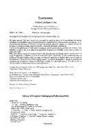
epatitis delta virus (HDV) is a defective virus that requires hepatitis B virus (HBV) surface antigen for virion assembly and infection,^ and contains a negative single stranded circular RNA genome of 1.7 kilobases.^'^ HDV is classified into three genotypes (genotype I, II and III) based on genetic sequence analysis (Fig. 1). Genotype II shows approximately 75% homology to genotype I, and genotype III shares about 60 to 65% homology with genotype I and II. There are many variants within each genotype. Especially, HDV genotype II is further divided into two types (i.e., Ila and lib), with 11^/Q nucleotide homology between the complete sequences of genotype Ila and lib. The nucleotide homology between genotype lib and Ilb-M, the newly identified lib variant, is 88-90%.^ Similarly, Ila variant was recently found in Siberia (Ila-Yakutia), which in comparison with Ila shows a similar degree of genetic differences. These genotypes show different geographical distributions and clinical pictures, which is thought to be caused by functional differences of genotype-specific sequences of HDV-RNA as well as HDAg protein.
Geographical Distribution of HDV Genotype Genotype I has been identified in most areas of the world and represented by many different isolates (Fig. 1). Genotype II is confined to East Asia (mainly Siberia, Japan, and Taiwan), in contrast to the ubiquitous global distribution of genotype I. Genotype lib was first identified in Taiwan,^ and was subsequently reported among patients from the Miyako Islands, ^^ one of the nearest Japanese islands to Taiwan. Recently, a new genetic variant of HDV genotype lib (Ilb-M) was identified. Genotype III is isolated to the northern part of South America, and is closely associated with fulminant hepatitis.
Clinical Significance of HDV Genotype HDV genotypes are known to affect the pathogenesis and diverse clinical pictures of HDV infection. Genotype I causes hepatic diseases ranging from mild to severe, often with the aggressive hepatitis and frequently associated with liver cirrhosis (LC) and hepatocellular carcinoma (HCC). On the other hand, genotype II is generally associated with a more favorable outcome than genotype I.^AIIa variant recently reported in Yakutia, Siberia, Russia also causes •Corresponding Author: Nobuyuki Enomoto—First Department of Internal Medicine, University of Yamanashi, Shimokato, Tamaho, Yamanashi 409-3898, Japan. Email: [email protected]
Hepatitis Delta Virus, edited by Hiroshi Handa and Yuki Yamaguchi. ©2006 Landes Bioscience and Springer Science+Business Media.
Hepatitis Delta Virus
-JA-N1 (Japan) -China - Taiwan-1 (Taiwan) -USA
^
Italy-1 Central African RepuiDlic - Lebanon Somalia
- JA-M1 (Japan, Miyako)
lib
L215 (Japan, Miyako)
Ilb-M
JA-M31 (Japan, Miyako) JA-T (Japan)
II
TWD62 (Taiwan)
ITa
-Yakut-26 (Russia) Yakut-62 (Russia) Japan 1 (Japan) Taiwan-3 (Taiwan)
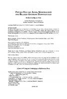
III Figure 1. Phylogenetic tree analysis of HDV isolates. Sources of isolates are as follows: TWD62 (AFO18077), Taiwan-3 (U19598), Taiwan-1 (M92448), Yakut-26 (AJ309879), Yakut-62 (AJ309880) Japan-1 (X60193), Lebanon (M84917), Somalia (U81988), China (X77627), USA (M28267), France (D01075), Italy-1 (X04451), Canada (AF098261), Central African Republic (AJ000558), Peru-1 (L22063), Venezuela (AB037948),JA-M1(AF309420),JA-M31(AB118841), JA-T (AB118847) were sequenced in this study. (GenBank accession number). a severe hepatitis comparable to genotype I in this c o h o r t / Genotype III is associated with fulminant hepatitis. These findings strongly suggest that the genetic structure of H D V can profoundly influence the pathogenesis of liver injury in H D V infection. In Japan, chronic H D V infection is endemic in the Miyako Islands where genotype l i b and Ilb-M are found, and their clinical pictures differed despite relatively uniform clinical backgrounds including virologicai factors of HBV. '^ Most of the patients with chronic H D V genotype lib infection were asymptomatic carrier (ASC) or chronic hepatitis (CH) and none were at the liver cirrhosis (LC) or hepatocellular carcinoma ( H C C ) stage. In contrast, about half of patients with genotype Ilb-M were in the C H and LC stages, respectively, and none of them were ASC. These findings indicate that patients with genotype I l b - M are more likely to progress to LC and H C C than those with genotype lib, and that differences in H D V genotype could cause the different clinical pictures observed in this popidation. In general, the genetic structure responsible for clinical features could not be readily determined because the genetic differences between the different genotypes are too diverse as seen in Figure 2. In contrast, despite the different clinical pictures between l i b and Ilb-M, the genetic differences are small enough to enable the definition of the genetic features of H D V pathogenesis
Genotype of Hepatitis Delta Virus
ilb-M{jA-Ml) Ilb-M {L215) lib (TWD62) Ifa a a p a n - l ) I (ltaly-1) HI (Peru-1)
GATGGa':CACAGTG-CCGACCy^AGAGGCCGCAGGTGGGAGGATCAG-CCA--CCGGAGAGGGACGCGA?GC:T--AGAGTGGAGGAAAG~TCGGAAGGG T -• G...T A -. . C-A G. .AA. G TC G . . .C -•.. , - CCAA G TC. .-CG A. A . . . G . . - . . C G . G . .GGAG. . A . . A G . T C . . . G . A . G G G . . A , . — . . . A G . . . - . . 7 C T C C A . C . AA TC A.CTT G. . - . A C . G A G GATA.CCXl^TAA. . . G . . . G G . .GAA.T-. . . . .AG . A . ~~ . TCGCX-CAC. AA A TTC A.CTC -A G . ~ ~ . T . . G - G . - . . . G . .GTC.GAAATC CGG . G. .A3AA. TCCC. . . A . T T . G. AGA-AGAA. . . . G . C G A A . -TTC C . .ATC
llb-M(J A-MI) !fb-M (L215) lib rrWI>62) l l a (Japan-!) 1 (1lai.Y-i) III (Peru-i)
T--CCCAAGAGGATCACTCGAG^TTCAAGAGGTGAGGAGGGATCCCCGAGACGCTGGAGGAACGCCGX^AAAAAGA-AAAGAA-QGCAAGAGATTGGTAG .-C C C C T. ..C.C A G ATA . G . .GAGAA- . T C CTAC G C A C G . C . G . . A . C.AGG. . .T.G-AA G T . -C CCA . , A . TGGC^'XC . C. CTC. AGAAG •A.AT.T -.A A C T . . . G. - A . TTTCATTC. .GAAG. . .GT..AC. . • , . CTA . C A A . - - G A G A G G . . TCAG . A A - G C , GAC. .G.AT. . C C - . . A A C . C C . . .GAAT . T C T - G G . . .GGG. . , G . G, A.AGGTG, . A - — G . A C-CTCCCA. .AGAG. . AGAG. ACAAGATA AGC AC A . C A A . C . . T . . C . C . C T G . G A G . G . . G T . .GCG. . A G A - — . . ,
irb-M(JA-Ml)
186 AAAAGAGCG-AGCCTCCCGATACGAGTTTGCCA-GGACCTATCAAGXTTGGAGTCATCCG-GCCCGTAGGGGAGAATAGAACACCGGG--GGGTGATCCA
Hb-M (L215) l i b (TWD62) Ha (Japan-l) I (Italy-l) HI (Peru-l)
C --.G -G . . .G.G. . . G . - . C . GAGACCC. GGT
Hb-MUArMI) llb-M {L2I5) H b (TW'D63) Ha aapan-l> I (l«aly-l) m r« - i )
CCAGGAGAAGTAG-CGGAGAACCCACCTCCAGAGGACCCCTTCT3CGAACAGAAAAGCTCTTCCCCCTCGGGAGT-AGGGCCGTAGCGATGQGAGGQGAT G. . , A G C...T.TC ~ G. . A . . . ~TG..AA..AT A G, .ACTCC. . T . G A T A C - . A . A . .A G. . A . , . GGTCGA— GA A G CCT. . T . C G . A . . . A G . A . A . . A A..G..A... .ACTCGG.GATG.G.A AT A . A G . G - . , G , , . . GAG. C^GTA. GAGT. A . A . . A A A. , , . - C . . . . T - T G . A - . , . . G . .G A G G. C . CTiGlGTA. . G
AutocatalyriccUnage rfgion
TCGT-CCAT'n'C-Tl'T^rn"AC'^rri'~TCCCCGCCATG.JTi:CCAaCCrCC'rCCrrGGCGCCGGCCGGaCAACA^nrCGAAG^
—
Uh »tt \ in
.-. .• C , , , TC , TA , C CT..T , . . - , . • C G- , . T • . •• QA T CT CG
l,a>-M2> {Ja|>«n-U (H«»v-n (Pcrtt-1>
-AG. . . GC-
Hypei-variahJe region
, ,' lU>-M-M2) li« 1 HI
"-""-"
I
^
J
.^™
^ ^ j , „ ™ „ ^ , ^ „
i V
(JaiMin-n Olnlyt) (Peru I )
llbM(J\-M1) llh-M {\l\>) llh
IIIHM(.IV-MI)
j
""
I
!
,
^ 1 J
i
!
i
r
~
J
''—^
T
T
1
1
?
lli>-M (1215) lUi :) lU (Ja|>an-I) 1 III
(lialy 1) (Peru 1)
" ^
Hli.-M(J\-\n) IIUVI (1215) lit) \U 1 III
(fVMXii) (Ja{Mii-t) (t««K 1) (I'm.-1)
i
in>-M f-
'
(12I5> (i\vi)(i2) (Japan!) (tialv 1) (IVru-1)
H1.M(JA-\H) lltf^M (1.215) lit) (IA\IXi2) liH iJaiMii-U 1 (itai> 1) III (VtruV)
*
"
•
"
'
'
_
^^ _
^
„
„
~
/
r-"
"
T^^
- - -^T~ —
~
,j-
~,~
-
" "
^^ ~
1 M l ) Vj> •
III! 1 HI
"
T
1
^
-. ^ - . _.. ^^^ ^ ^ ^^ r^
"
f~
1
ti\ pir\ariahk
^
.
i
^
rt{»ion
^^
— , ^ -^
J I t
"T 1
I
^
f I •T
;
, 1
/
C) occurs at nucleotide 1015 of the genomic sense RNA, changing the termination codon for small HDAg (on the antigenomic strand) to a tryptophan codon and extending the ORF for an additional 19 amino acids. This mutation results in the synthesis of the L-HDAg.^'^^ This RNA editing appears to be carried out by a double-stranded RNA-adenosine deaminase (ADAR).^^ More recent studies have further identified ADAR I, but not ADARII, as the enzyme responsible for HDV RNA editing.^^'^ The efficiency of RNA editing in vitro appears to be very high; however, the extent of RNA editing in the cells is regulated, so that L-HDAg is not over-produced. It was previously suggested that L-HDAg itself can suppress RNA synthesis, thus limiting the amount of RNA synthesized at the later stages of viral replication cycle. However, recent studies suggest that the feedback inhibition is probably a result of the enhanced deleterious mutations in the genome triggered by the edited RNA sequence^^ or mediated by L-HDAg per se. These mechanisms can explain how the L-HDAg-encoding mRNA is specifically suppressed. In every delta hepatitis patient examined, both of the RNA species containing a large and a small HDAg ORF are present.^^
The Roles of HDAg in HDV RNA Synthesis As discussed above, HDAg is necessary for HDV RNA synthesis. Although the mechanism of its participation in HDV RNA synthesis is still not clear, it is known that the synthesis of genomic and antigenomic RNA strands have differential requirements for HDAg. For example, antigenomic RNA can not initiate RNA replication when it is transfected together with the recombinant HDAg derived from E. coli, whereas genomic RNA can. Phosphorylation (at serine-177) and methylation (at arginine-13) of HDAg are required for the replication of the antigenomic but not the genomic RNA strand. ^^' ^ Genomic, but not antigenomic, RNA synthesis is inhibited by L-HDAg, when the latter is expressed early in viral replication.^^ These findings are also consistent with the studies showing differential sensitivity of the genomic and antigenomic RNA synthesis to a-amanitin, i.e., the genomic RNA synthesis (from the antigenomic RNA template) is sensitive to the low concentration of a-amanitin, whereas antigenomic RNA synthesis (from the genomic RNA template) is resistant (up to 100 ug/ml of a-amanitin). ' The mRNA transcription is also sensitive to the low concentration of a-amanitin. Furthermore, Pol II can elongate RNA-dependent RNA synthesis in vitro using HDV RNA as template, although the RNA product is different from the natural RNA species."^^ These findings suggest that polymerase II and an additional polymerase, likely pol I, are both involved in HDV RNA replication. HDAg has been shown to complex with pol II. These studies suggest that HDAg is directly involved in HDV RNA replication, rather than affects the general transcription machinery indirectly. Finally, HDAg also has been reported to complex with a nucleolar protein B23, and HDV RNA synthesis is enhanced when B23 is over-expressed,'^^ suggesting that HDAg may be located in the nucleolus at certain stages of the viral life cycle and that this localization is associated with HDV RNA replication. The question remains as to how RNA polymerase II or other cellular polymerases can utilize an RNA template rather than their normal DNA template. Possibly, HDAg may complex with cellular transcription factors and then interact directly or indirecdy with pol II, thereby altering its template specificity. Since HDAg binds to HDV RNA,^' a transcription complex consisting of HDAg, pol II and cellular transcription factors could conceivably complex with HDV RNA and carry out RNA-dependent RNA replication. However, HDV RNA synthesis did occur in the absence of HDAg in cell-free lysates, and the addition of HDAg did not have significant effects on HDV RNA synthesis in vitro.^^'^^'^^ A more recent study showed that HDAg can promote the elongation, but not initiation, of HDV RNA-dependent,
Hepatitis Delta Antigen
47
pol Il-mediated RNA synthesis in vitro.^^ It is noteworthy that the rod-shaped structure of HDV RNA resembles double-stranded DNA; therefore, it is conceivable that cellular RNA polymerases and transcription factors may recognize double-stranded RNA. Indeed, the double-stranded DNA counterpart of the region encompassing the replication origin for the antigenomic-strand HDV RNA has a promoter activity for transcription.^^' How the posttranslational modifications of HDAg can affect HDV RNA synthesis is a very interesting question. These questions remain the most critical issues in the study of HDV replication cycle.
The Cytotoxic Effects of HDAg HDAg, particularly the S-HDAg, has been shown to be cytotoxic when it was expressed at high levels in the cells. ' ' ' However, no significant pathology has been noted in the HDAg-expressing transgenic mice ' and HDAg-expressing cell lines, or associated with HDV infection of primary hepatocyte cultures. ^^'^ '^^ In contrast, both the small and large HDAg have been shown to affect cellular pol Il-mediated transcription in vitro and in vivo; in one study, both S- and L-HDAg inhibited pol Il-mediated transcription in vitro and in vivo. However, in another study, L-HDAg, but not S-HDAg, stimulated pol Il-mediated transcription. L-HDAg has been shown to activate serum response factor-dependent transcription, but not other signal transduction pathways. In contrast, S-HDAg did not have such an effect. "^^ The biological significance of these findings is not known. In any case, these effects will likely cause cytotoxicity. HDV replication has also been shown to induce apoptosis in avian cells. However,,in a different study, no apoptosis or cell cycle arrest of HDV-replicating cells were noted. Nevertheless, the cells harboring HDV RNA were gradually lost, suggesting a growth disadvantage associated with HDV replication. The precise effects of HDV replication on the host cells will require further studies.
Perspectives HDAg plays a very important role in the replication cycle of HDV. It serves as the structural component of the virion and also plays various roles in different steps of the viral replication processes, including RNA replication and virus assembly. The most intriguing aspect of the HDAg biology is its role in converting the cellular DNA-dependent RNA polymerases to an enzyme capable of replicating RNA. This capability opens up a possibility that certain cellular RNA may be replicated through an RNA-dependent process via a protein very similar to HDAg. Indeed, several cellular proteins bear limited sequence similarity with HDAg, including the negative elongation factor (NELF-A) and another uncharacterized protein DIPA (Delta antigen-interacting protein). These proteins may have potential to perform similar functions to that of HDAg. The understanding of these processes will reveal novel features of HDV replication and the molecular biology of mammalian cells.
References L Beard MR, Macnaughton T B , Gowans EJ. Identification and characterization of a hepatitis delta virus RNA transcriptional promoter. J Virol 1996; 70:4986-4995. 2. Bonino F, Hoyer B, Shih J et al. Delta hepatitis agent: Structural and antigenic properties of the delta-associated particles. Infect Immunity 1984; 43:1000-1005. 3. Bordier BB, Marion PL, Ohashi K et al. A prenylation inhibitor prevents production of infectious hepatitis delta virus particles. J Virol 2002; 76:10465-72. 4. Brazas R, Ganem D . A cellular homolog of hepatitis delta antigen: Implications for viral replication and evolution. Science 1996; 274:90-94. 5. Casey JL, Brown TL, Golan EJ et al. A genotype of hepatitis D virus that occurs in northern South America. Proc Natl Acad Sci USA 1993; 90:9016-9020.
48
Hepatitis Delta
Virus
6. Casey JL, Gerin JL. Genotype-specific complementation of hepatitis delta virus RNA replication by hepatitis delta antigen. J Virol 1998; 72:2806-14. 7. Chang F-L, Chen P-J, T u S-J et al. T h e large form of hepatitis A antigen is crucial for assembly of hepatitis A virus. Proc Natl Acad Sci USA 1991; 88:8490-8494. 8. Chang J, Morabda G, Taylor J. Limitations to replication of hepatitis delta virus in avian cells. J Virol 2000; 74:8861-8866. 9. Chang M-F, Baker SC, Soe LH et al. H u m a n hepatitis delta antigen is a nuclear phosphoprotein with RNA-binding activity. J Virol 1988; 62:2403-2410. 10. Chang MF, Chen C H , Lin SL et al. Functional domains of delta antigens and viral RNA required for RNA packaging of hepatitis delta virus. J Virol 1995; 69:2508-2514. 11. Chang M-F, Chen C-J, Chang S-C. Mutational analysis of delta antigen: Effect on assembly and replication of hepatitis delta virus. J Virol 1994; 68:646-653. 12. Chao M, Hsieh S-Y, Taylor J. The antigen of hepatitis delta virus: Examination of in vitro RNA binding specificity. J Virol 1991; 65:4057-4062. 13. Chao M, Hsieh S-Y, Taylor J. Role of two forms of hepatitis delta virus antigen: Evidence for a mechanism of self-limiting genome repHcation. J Virol 1990; 64:5066-5069. 14. Chen C W , Tsay YG, W u H L et al. The double-stranded RNA-activated kinase, PKR, can phosphorylate hepatitis D virus small delta antigen at functional serine and threonine residues. J Biol Chem 2002; 277:33058-33067. 15. Chen P-J, Chang F-L, W a n g C-J et al. Functional studies of hepatitis delta virus large antigen in packaging and replication inhibition: Role of the amino-terminal leucine zipper. J Virol 1992; 66:2853-2859. 16. Chen P-J, Kalpana G, Goldberg J et al. Structure and replication of the genome of hepatitis delta virus. Proc N a d Acad Sci USA 1986; 83:8774-8778. 17. Cheng Q , Jayan G C , Casey JL. Differential inhibition of RNA editing in hepatitis delta virus genotype III by the short and long forms of hepatitis delta antigen. J Virol 2003; 77:7786-95. 18. Choi S-S, Rasshofer R, Roggendorf M. Propagation of woodchuck hepatitis delta virus in primary woodchuck hepatocytes. Virology 1988; 167:451-457. 19. Chou H - C , Hsieh T-Y, Sheu G-T et al. Hepatitis delta antigen mediates the nuclear import of hepatitis delta virus RNA. J Virol 1998; 72:3684-3690. 20. Cole SM, Gowans EJ, Macnaughton TB et al. Direct evidence for cytotoxicity associated with expression of hepatitis delta virus antigen. Hepatology 1991; 13:845-851. 2 1 . De Bruin W, Leenders W, Kos T et al. In vitro binding properties of the hepatitis delta antigens to the hepatitis B virus envelope proteins: Potential significance for the formation of delta particles. Virus Res 1994; 31:27-37. 22. Filipovska J, Konarska M M . Specific H D V RNA-templated transcription by Pol II in vitro. RNA 2000; 6:41-54. 23. Fu T-B, Taylor J. The RNAs of hepatitis delta virus are copied by RNA polymerase II in nuclear homogenates. J Virol 1993; 67:6965-6972. 24. Glenn JS, Watson JA, Havel C M et al. Identification of a prenylation site in delta virus large antigen. Science 1992; 256:1331-1333. 25. Goto T, Kato N , O n o - N i t a SK et al. Large isoform of hepatitis delta antigen activates serum response factor-associated transcription. J Biol Chem 2000; 275:37311-6. 26. Guilhot S, Huang S-N, Xia Y-P et al. Expression of the hepatitis delta virus large and small antigens in transgenic mice. J Virol 1994; 68:1052-1058. 27. Hsieh S-Y, Chao M, Coates L et al. Hepatitis delta virus genome replication: A polyadenylated mRNA for delta antigen. J Virol 1990; 64:3192-3198. 28. Hsieh S-Y, Yang P-Y, O u J T et al. Polyadenylation of the m R N A of hepatitis delta virus is dependent upon the structure of the nascent RNA and regulated by the small or large delta antigen. Nucleic Acids Res 1994; 22:391-396. 29. Huang W H , Yung BY, Syu WJ et al. T h e nucleolar phosphoprotein B23 interacts with hepatitis delta antigens and modulates the hepatitis delta virus RNA replication. J Biol Chem 2 0 0 1 ; 276:25166-75. 30. Huang YH, W u JC, Hsu SC et al. Varied immunity generated in mice by D N A vaccines with large and small hepatitis delta antigens. J Virol 2003; 77:12980-5.
Hepatitis Delta Antigen
49
3 1 . H u a n g ZS, W u H N . Identification and characterization of the RNA chaperone activity of hepatitis delta antigen peptides. J Biol Chem 1998; 273:26455-26461. 32. Hwang SB, Lai M M C . Isoprenylation masks a conformational epitope and enhances trans-dominant function of the large hepatitis delta antigen. J Virol 1994; 68:2958-2964. 3 3 . Hwang SB, Lai M M C . Isoprenylation mediates direct protein-protein interactions between hepatitis large delta antigen and hepatitis B virus surface antigen. J Virol 1993; 67:7659-7662. 34. Hwang SB, Lee C Z , Lai M M C . Hepatitis delta antigen expressed by recombinant baculoviruses: Comparison of biochemical properties and post-translational modifications between the large and small forms. Virology 1992; 190:413-422. 34a. Hwang SB, Lai M M C . A unique conformation at the carboxyl terminus of the small hepatitis delta antigen revealed by a specific monoclonal antibody. Virology 1993; 193:924-931. 35. Jayan G C , Casey JL. Inhibition of hepatitis delta virus R N A editing by short inhibitory RNA-mediated knockdown of A D A R l but not ADAR2 expression. J Virol 2002; 76:12399-404. 36. Jeng KS, Su PY, Lai M M C . Hepatitis delta antigens enhance the ribozyme activities of hepatitis delta virus RNA in vivo. J Virol 1996; 70:4205-4209. 37. Kuo MY-P, Chao M , Taylor J. Initiation of replication of the human hepatitis delta virus genome from cloned D N A : Role of Delta Antigen. J Virol 1989; 63:1945-1950. 38. Lazinski D , Grzadzielska E, Das A. Sequence-specific recognition of RNA hairpins by bacteriophage antiterminators requires a conserved arginine-rich motif. Cell 1989; 59:207-218. 39. Lazinski D W , Taylor JM. Expression of hepatitis delta virus RNA deletions: Cis and trans requirements for self-cleavage, Ligation and RNA packaging. J Virol 1994; 68:2879-2888. 40. Lazinski D W , Taylor JM. Relating structure to function in the hepatitis delta virus antigen. J Virol 1993; 67:2672-2680. 4 1 . Lee C H , Chang SC, Chen CJ et al. T h e nucleolin binding activity of hepatitis delta antigen is associated with nucleolus targeting. J Biol Chem 1998; 273:7650-7656. 42. Lee C H , Chang SC, W u C H et al. A Novel chromosome region maintenance 1-independent nuclear export signal of the large form of hepatitis delta antigen that is rerquired for the viral assembly. J Biol Chem 2 0 0 1 ; 276:8142-8. 4 3 . Lee C-Z, Chen PJ, Chen D S . Large hepatitis delta antigen in packaging and replication inhibition: Role of the carboxyl-terminal 19 amino acids and aminoterminal sequences. J Virol 1995; 69:5332-5336. 44. Lee C-Z, Chen P-J, Lai M M C et al. Isoprenylation of large hepatitis delta antigen is necessary but not sufficient for hepatitis delta virus assembly. Virology 1994; 199:169-175. 45. Lee C-Z, Lin J-H, Mcknight K et al. RNA-binding activity of hepatitis delta antigen involves two arginine-rich motifs and is required for hepatitis delta virus RNA replication. J Virol 1993; 67:2221-2229. 45a. Li YJ, Stallcup MR, Lai M M C . Hepatitis delta virus antigen is methylated at arginine residues, a n d m e t h y l a t i o n regulates subcellular localization a n d R N A r e p l i c a t i o n . J Virol 2 0 0 4 ; 78(23):13325-13334. 46. Lin J-H, Chang M-F, Baker SC et al. Characterization of hepatitis delta antigen: Specific binding to hepatitis delta virus RNA. J Virol 1990; 64:4051-4058. 47. Lo K, Hwang SB, Duncan R et al. Characterization of m R N A for hepatitis delta antigen: Exclusion of the full-length antigenomic RNA as an mRNA. Virology 1998a; 250:94-105. 48. Lo K, Sheu G-W, Lai M M C . Inhibition of cellular RNA polymerase II transcription by delta antigen of hepatitis delta virus. Virology 1998b; 247:178-188. 49. Luo G, Chao M , Hsieh SY et al. A specific base transition occurs on replicating hepatitis delta virus RNA. J Virol 1990; 64:1021-1027. 50. Macnaughton T B , Beard MR, Chao M et al. Endogenous promoters can direct the transcription of hepatitis delta virus RNA from a recircularized c D N A template. Virology 1993; 196:629-636. 5 1 . Macnaughton T B , Cowans EJ, Jilbert AR et al. Hepatitis delta virus RNA, protein synthesis and associated cytotoxicity in a stably transfected cell line. Virology 1990a; 177:692-698. 52. Macnaughton T B , Cowans EJ, Mcnamara SP et al. Hepatitis 6 antigen is necessary for access of hepatitis 6 virus R N A to the cell transcriptional machinery but is not part of the transcriptional complex. Virology 1991; 184:387-390.
50
Hepatitis Delta
Virus
53. Macnaughton T B , Gowans EJ, Reinboth B et al. Stable expression of hepatitis delta virus antigen in a eukaryotic cell line. J Gen Virol 1990b; 71:1339-1345. 54. Macnaughton T B , Lai M M C . Genomic but not antigenomic hepatitis delta virus RNA is preferentially exported from the nucleus immediately after synthesis and processing. J Virol 2002a; 76:3928-3935. 55. Macnaughton T B , Lai M M C . Large hepatitis delta antigen is not a suppressor of hepatitis delta virus RNA synthesis once RNA replication is estabUshed. J Virol 2002b; 76:9910-9919. 56. Macnaughton T B , Li YI, Doughty AL et al. Hepatitis delta virus RNA encoding the large delta antigen cannot sustain replication due to rapid accumulation of mutations associated with RNA editing. J Virol 2003; 77:12048-12056. 57. Macnaughton T B , Shi ST, Modahl LE et al. Rolling circle replication of hepatitis delta virus RNA is carried out by two different cellular RNA polymerases. J Virol 2002; 76:3920-3927. 58. Modahl LE, Lai M M . T h e large delta antigen of hepatitis delta virus potently inhibits genomic but not antigenomic RNA synthesis: A mechanism enabling initiation of viral replication. J Virol 2000; 74:7375-7380. 59. Modahl LE, Lai M M C . Transcription of hepatitis delta antigen mRNA continues throughout hepatitis delta virus (HDV) replication: A new model of H D V RNA transcription and replication. J Virol 1998; 72:5449-5456. 60. Modahl LE, Macnaughton T B , Zhu N et al. RNA-dependent replication and transcription of hepatitis delta virus RNA involve distinct cellular RNA polymerases. Mol Cell Biol 2000; 20:6030-6039. 6 1 . Mu J-J, Chen DS, Chen P-J. The conserved serine 177 in the delta antigen of hepatitis delta virus is one putative phosphorylation site and is required for efficient viral RNA replication. J Virol 2001; 75:9087-9095. 62. Mu J-J, Tsay YG, Juan LJ et al. T h e small delta antigen of hepatitis delta virus is an acetylated protein and acetylation of lysine 72 may influence its cellular localization and viral RNA synthesis. Virology 2004; 319:60-70. 63. Mu J-J, W u H-L, Chiang B-L et al. Characterization of the phosphorylated forms and the phosphorylated residues of hepatitis delta virus delta antigens. J Virol 1999; 73:10540-10545. 64. O t t o JC, Casey PJ. T h e hepatitis delta virus large antigen is farnesylated both in vitro and in animal cells. J Biol Chem 1996; 271:4569-4572. 65. Poisson F, Roingeard P, Baillou A et al. Characterization of RNA-binding domains of hepatitis delta antigen. J Gen Virol 1993; 74:2473-2477. 66. Polo JM, Jeng KS, Lim B et al. Transgenic mice support replication of hepatitis delta virus RNA in multiple tissues, particularly in skeletal muscle. J Virol 1995; 69:4880-4887. 67. Poison AG, Bass BL, Casey JL. RNA editing of hepatitis delta virus antigenome by dsRNA- adenosine deaminase. Nature (London) 1996; 380:454-456. 68. Rizzetto M, Canese M G , Arico S et al. Immunofluorescence detection of A new antigen-antibody system (Delta/Anti-Delta) associated with hepatitis B virus in liver and serum of HBsAg carrier. Gut 1977; 18:997-1003. 69. Rizzetto M , Hoyer B, Canese M G et al. Delta agent: Association of A antigen with hepatitis B surface antigen and RNA in serum of A-infected chimpanzees. Proc Natl Acad Sci USA 1980; 77:6124-6128. 70. Rozzelle JE, W a n g JG, Wagner DS et al. Self-association of a synthetic peptide from the N terminus of the hepatitis delta virus protein into an immunoreactive alpha helical multimer. Proc Natl Acad Sci USA 1995; 92:382-386. 7 1 . Ryu W-S, Bayer M, Taylor J. Assembly of hepatitis delta virus particles. J Virol 1992; 66:2310-2315. 72. Ryu W-S, Netter H J, Bayer M et al. Ribonucleoprotein complexes of hepatitis delta virus. J Virol 1993; 67:3281-3287. 73. Sheu G-T, Lai M M C . Recombinant hepatitis delta antigen from E. Coli promotes hepatitis delta virus RNA replication only from the genomic strand but the antigenomic strand. Virology 2000; 278:578-586. 74. Sureau C, Jacob JR, Eichberg J W et al. Tissue culture system for infection with human hepatitis delta virus. J Virol 1991; 65:3443-3450.
Hepatitis Delta Antigen
51
75. Sureau C, Moriarty AM, Thornton GB et al. Production of infectious hepatitis delta virus in vitro and neutralization with antibodies directed against hepatitis B virus preS antigens. J Virol 1992; 66:1241-1245. 76. Tai F-P, Chen P-J, Chang F-L et al. Hepatitis delta virus c D N A can be used in transfection experiments to initiate Viral RNA replication. Virology 1993; 197:137-142. 77. Tavanez JP, Cunha C, Silva M C A et al. Hepatitis delta virus ribonucleoproteins shuttle between the nucleus and the cytoplasm. RNA 2002; 8:637-646. 78. Taylor J, Mason W , Summers J et al. Replication of human hepatitis delta virus in primary cultures of woodchuck hepatocytes. J Virol 1987; 61:2891-2895. 79. Taylor JM. H u m a n hepatitis delta virus: An agent with similarities to certain satellite RNAs of plants. Curr T o p Microbiol Immunol 1999; 239:107-122. 80. Wang C C , Chang T C , Lin C W et al. Nucleic acid binding properties of the nucleic acid chaperone domain of hepatitis delta antigen. Nucleic Acids Res 2003; 31:6481-6492. 8 1 . Wang D , Pearlberg J, Liu YT et al. Deleterious effects of hepatitis delta virus replication on host cell proHferation. J Virol 2 0 0 1 ; 75:3600-4. 82. Wang H - W , Chen P-J, Lee C-Z et al. Packaging of hepatitis delta virus RNA via the RNA-binding domain of hepatitis delta antigens: Different roles for the small and large delta antigens. J Virol 1994; 68:6363-6371. 83. Wang J-G, Lemon SM. Hepatitis delta virus antigen forms dimers and multimeric complexes in vivo. J Virol 1993; 67:446-454. 84. Wei Y, Ganem D . Activation of heterologous gene expression by the large isoform of hepatitis delta antigen. J Virol 1998; 72:2089-2096. 85. Weiner AJ, Choo Q-L, Wang K-S et al. A single antigenomic open reading frame of the hepatitis delta virus encodes the epitope(s) of both hepatitis delta antigen polypeptides P24 and P27 . J Virol 1988; 62:594-599. 86. W o n g SK, Lazinski D W . Replicating hepatitis delta virus RNA is edited in the nucleus by the small form of A D A R l . Proc N a d Acad Sci USA 2002; 99:15118-23. 87. Xia Y-P, C h a n g M-F, Wei D et al. Heterogeneity of hepatitis delta antigen. Virology 1990; 178:331-336. 88. Xia Y-P, Lai M M C . Oligomerization of hepatitis delta antigen is required for b o t h the trans-activating and trans-dominant inhibitory activities of the delta antigen. J Virol 1992; 66:6641-6648. 89. Xia Y-P, Yeh C-T, O u J-H et al. Characterization of nuclear targeting signal of hepatitis delta antigen: Nuclear transport as a protein complex. J Virol 1992; 66:914-921. 90. Yamaguchi Y, Delehouzee S, Handa H . H I V and Hepatitis delta virus: Evolution takes different paths to relieve blocks in transcriptional elongation. Microbes Infect 2002; 4:1169-1175. 9 1 . Yamaguchi Y, Filipovska J, Yano K et al. Stimulation of RNA polymerase II elongation by hepatitis delta antigen. Science 2 0 0 1 ; 293:124-127. 92. Yeh TS, Lo SJ, Chen PJ et al. Casein kinase II and protein kinase C modulate hepatitis delta virus RNA repHcation but not empty viral particle assembly. J Virol 1996; 70:6190-6198. 93. Zuccola HJ, Rozzelle JE, Lemon SM et al. Structural basis of the oligomerization of hepatitis delta antigen. Structure 1998; 6:821-830.
CHAPTER 5
Hepatitis Delta Virus RNA Editing John L. Casey* Summary
T
he genome of hepatitis delta virus (HDV) is the smallest known to infect man. Encoding just one protein, hepatitis delta antigen (HDAg), HDV relies heavily on host functions and on structural features of the viral RNA. A good example of this reliance is found in the process known as HDV RNA editing, which requires particular structural features in the HDV antigenome, and a host RNA editing enzyme, ADARl. During replication, the adenosine in the amber stop codon in the viral gene for the short form of HDAg (HDAg-S) is edited to inosine. As a result, the amber stop codon in the HDAg-S open reading frame is changed to a tryptophan codon; the reading frame is thus extended by 19 or 20 codons and the longer form of HDAg, HDAg-L, is produced. This change serves a critical purpose in the HDV replication cycle because HDAg-S supports viral RNA replication, while HDAg-L is required for virion packaging but inhibits viral RNA replication. This review will cover the mechanisms of RNA editing in the HDV replication cycle and the regulatory mechanisms by which HDV controls editing.
What Is RNA Editing? RNA editing can be loosely defined as the site-specific modification of an RNA sequence from that of its template by mechanisms other than splicing. The term was first used in the late 1980s to describe an unusual process in which multiple Us are inserted and deleted in trypanosome mitochondrial mRNAs. As a result of the insertions/deletions, the coding capacity of the affected mRNAs is dramatically altered. The usage of the term was subsequendy expanded as it was applied to other examples of nucleotide changes in mRNA that changed the coding capacity, including deamination of C to U in apoB 100 mRNA in small intestine, deamination of A to I in glutamate receptor subunit B (gluRB) premRNA in brain, and insertion of nontemplated G's in the P gene of paramyxoviruses. While collectively referred to as RNA editing, these sequence revisions involve a wide range of mechanisms. In the two types of editing used by mammalian cells, C to U and A to I, the modified base within the RNA molecule is deaminated and there is no evidence that phosphate backbone is broken during the editing process. The type of RNA editing used by HDV is adenosine deamination. In this process, the amino group of adenosine is removed and replaced with a keto oxygen. Because this position of *John L. Casey—Department of Microbiology and Immunology, Georgetown University Medical Center, 3900 Reservoir Rd., N W , Washington, District of Columbia, 20007 U.S.A. Emai hcaseyj ©georgetown.edu
Hepatitis Delta Virus, edited by Hiroshi Handa and Yuki Yamaguchi. ©2006 Landes Bioscience and Springer Science+Business Media.
Hepatitis Delta Virus RNA Editing
N
53
H,0
50 base-pairs) double-stranded RNAs, in which up to 5 0 % of adenosines may be deaminated. T h e role of this activity in cells is not clear, but the fact that dsRNA is a target and that one form of A D A R l is induced by interferon^^ has led to the suggestion that editing of dsRNA may be part of the cellular response to virus infection. Clearly promiscuous editing such as occurs on dsRNA could be deleterious to virus replication. Indeed, spurious editing on H D V R N A by overexpressed A D A R l and ADAR2 led to the production of protein variants that inhibited replication.^"^ W h e t h e r interferon treatment increases editing at the a m b e r / W site, or elsewhere on the H D V RNA, remains to be determined. Even though H D V R N A exhibits significant base-pairing in the unbranched rod structure, promiscuous editing does not typically occur during H D V infection; the a m b e r / W site is edited 600-fold more efficiently than the other 337 adenosines in die RNA.5^ It is likely that the primary and secondary structure of the H D V R N A have evolved to avoid undesirable (for the virus) editing at sites other than amber/W. Guanosine is by far the most common 5' neighbor for adenosine in both the H D V genome and antigenome, and the ratios of observed to expected occurrences for the dinucleotides G A and U C (which would be GA in the complementary strand) are higher than for any other dinucleotides (Table 1). This bias may be due, in part,
60
Hepatitis Delta Virus
Table 1. Dinucleotide frequencies in HDV RNA^ Dinucleotide
No. of Occurences
GA TC AG CT CC GG AA TT AT CG TG AC GT CA GC TA
Observed/ Ex
161 160 134 140 204 177 79 79 56 112 66 65 62 62 89 32
1.65 1.54 1.38 1.35 1.28 1.24 1.19 1.17 0.83 0.74 0.67 0.63 0.63 0.6 0.59 0.48
' based on H D V genotype I prototype, accession no. X04451
to selection for sequences that place non-amber/W adenosines in contexts that are less likely to be edited: analysis of editing on dsRNAs has indicated that adenosines flanked by a 5' guanosine are much less likely to be deaminated than other adenosines.^^ As for secondary structure, base-pairing in the HDV RNA unbranched rod structure is interrupted by frequent bulges, internal loops and mismatches, which have been shown to restrict editing on artificial dsRNA substrates.^^'^^'52
Regulation of Editing HDV must regulate both the rate and the extent of editing at the amber/W site because HDAg-L, which is produced as a result of editing, is necessary for virion production but inhibits viral RNA replication. Varying the efficiency of editing at the amber/W site, either by altering levels of ADAR expression or by the introduction of mutations near the amber/W site, can affect HDV replication, virus production, or both.^"^' ^ Premature editing at the amber/W site results in reduced levels of RNA replication and reduced production of viable virions because edited antigenomes encode HDAg-L, which is a trans-dominant inhibitor of HDV RNA replication.^^' '^^ Insufficient editing can lead to increased intracellular HDV RNA replication, but inhibits virion production. ^ In addition to the rate, the extent of editing must also be controlled because the mechanism of editing in the HDV replication scheme (Fig. 2) produces genomes encoding HDAg-L. These genomes are packaged but are not likely to be infectious because HDAg-L does not support HDV RNA replication. Thus, the kinetics and extent of editing are likely regulated during HDV replication to maximize the rate and amount of infectious virus produced. Control mechanisms for editing rely on several viral components and functions, including: RNA structure, HDAg, and viral RNA replication. HDV does not appear to regulate editing by affecting ADARl expression because ADARl levels are unaffected by HDV replication. Some of the control mechanisms may be described as passive, in that they are not affected by (or responsive to) the level of editing. This category includes the secondary structure
61
Hepatitis Delta Virus RNA Editing
ADAR1
ADAR1
ADAR1
ADAR1
c
Genotype I
Genotype III
Figure 5. Schematic of the regulation of editing in HDV genotype I and III by HDAg. Left) genotype I. Both HDAg-S and HDAg-L efiFectively inhibit editing, most Hkely by binding the RNA near the editing site and Hmiting access ofADARl. Right) genotype III. HDAg-L, but not HDAg-S can effectively inhibit editing. HDAg-L is drawn with the C-terminal extension indicated by the small segment with diagonal hashing of the RNA around the amber/W site. As mentioned above, the disruptions in base-pairing 3' of the amber/W site in HDV genotype I create a sub-optimal substrate for editing. Mutations that increase base-pairing in this region increase editing, but severely reduce replication and virion production (Sato and Lazinski, personal communication; Jayan and Casey, unpublished). It is not yet known whether the structures in the vicinity of the amberA?^ sites of genotypes II and III are also sub-optimal. One potential dilemma for the virus that is posed by using a sub-optimal structure to limit editing efficiency is that the specificity of editing is likely to be compromised because the specificity is determined by the ratio of the efficiency of editing at the amber/W site to the efficiency of editing at other "nonspecific*' sites. The danger for the virus of nonspecific editing is the production of additional genomes defective for replication, or even the creation of dominant negative HDAg-S mutants. ^'^ Thus, there may be limits as to how much amber/W editing can be restricted by using sub-optimal structures. HDV does appear to have a mechanism for minimizing the effects of editing at nonamber/W sites: in one study of HDV replicating HDV in transfected cells, all nonamber/W changes that occurred during replication were found on genomes that were also edited at the amber/W site.^^ HDV genotype I uses an additional mechanism to slow down editing early in the replication cycle. For this genotype, HDAg-S is a strong inhibitor of editing (Fig. 5). While editing on replicating RNA 2-3 days post-transfection is nearly undetectable, up to 40% of nonreplicating RNAs produced in transfected cells in the absence of HDAg are edited. However, cotransfection of an HDAg-S expression construct leads to markedly reduced levels of editing on nonreplicating RNAs, most likely by binding to HDV RNA and preventing access ofADARl.51 The levels of HDAg-S required for this inhibition are similar to those seen in cells replicating HDV RNA. Thus, it appears that HDAg-S prevents the rapid accumulation of editing early in the HDV genotype I replication cycle.
62
Hepatitis Delta Virus
On the other hand, for HDV genotype III, HDAg-S is not an effective inhibitor of editing and Ukely does not play a direct role in limiting editing levels.^ Rather, HDV genotype III uses the distribution of the RNA between at least two conformations to restrict editing. '^ Only RNA molecules that adopt the double hairpin structure can be edited (Fig. 4). However, the majority of the genotype III RNA appears to assume the unbranched rod conformation, which is not a substrate for editing. ^'^ Thus, while the amber/W site itself in genotype III RNA can be edited with efficiency similar to the genotype I site, editing levels in nonreplicating genotype III RNAs are much lower because most of the RNA assumes the unbranched rod conformation, which is not a substrate for editing. The introduction of mutations in the genotype III RNA that shift the distribution of the RNA to the double hairpin structure increases editing to levels comparable with those seen with nonreplicating genotype I RNA."^^ It is not clear whether the inhibition of editing in genotype I or the conformational control of editing in genotype III is influenced by levels of editing and/or replication. Possibly, the HDAgiRNA ratio may vary during replication and thereby affect HDV genotype I editing rates. Likewise, for genotype III, if the RNA transcription rate varies during HDV genotype III RNA replication, such variations could affect editing by altering the distribution of the RNA between the double hairpin and unbranched rod structures. Other mechanisms to control editing are by their nature responsive to editing levels. In HDV genotype III, HDAg-L is a much better inhibitor of editing than is HDAg-S, and is likely involved in a negative feedback loop to limit editing levels (Fig. 5).^^ This regulatory behavior requires the double hairpin structure peculiar to HDV genotype III amber/W editing. The reason for the differential effects of HDAg-S and HDAg-L is not yet clear; the hairpin on the 3' side of the amber/W site plays an essential role, and could interfere with HDAg-S binding near the amber/W site. Genotype I does not use the same mechanism to control editing levels because genotype I HDAg-S and HDAg-L do not exhibit differential effects on editing (Cheng and Casey, unpublished data). Rather, HDV genotype I may limit the extent of editing via the inhibitory effect of HDAg-L on RNA replication, which is required for editing to occur on the antigenome and for the synthesis of edited genomic copies (Sato and Lazinski, personal communication).
Assays for Editing Analysis of RNA editing at the HDV amber/W site has relied principally on two methods: comparison of HDAg-S and HDAg-L levels, and restriction digestion of PCR-amplified cDNA derived from HDV RNA. The former method has the advantage of being quick and simple and can be readily applied to analysis of editing on HDV RNA in cultured cells. However, this method is limited to cell-based experiments and by the requirements for a translated mRNA. The latter method works because editing at the amber/W site fortuitously creates a restriction digestion site that is not present in unedited RNA; enzymes used have included Not\, Sty I, Dsa I, Btg\. This method has the advantage of being more direct and can be applied to experiments performed in cells and in vitro. However, it is important to note that analysis of PCR products is susceptible to a potential artifact that could lead to an underestimate of editing levels. Because editing levels are frequently 30% or less, PCR products will be heterogeneous. If reannealing of these heterogeneous PCR products competes with primer annealing, then some PCR products will contain heteroduplexes in which one strand is derived from unedited RNA and the other from edited RNA. Such heteroduplexes will not be digested by the restriction enzyme and will result in underestimates of editing. To avoid this pitfall it is necessary to exclude heteroduplexes from the analysis."^^'^^ One approach is to radioactively label PCR products only during the final extension step; in this way heteroduplexes are not labeled and do not contribute to the quantitation of products that are digested by the restriction enzyme. The accuracy of this approach has been verified by sequence analysis of cloned PCR products. ' '
Hepatitis Delta Virus RNA Editing
63
Future Directions Analysis of editing in HDV has led to valuable contributions to the field of RNA adenosine deamination. Thus far, it is the only example of specific editing that occurs in an organ other than the brain in mammals, but it is highly likely that more examples will be identified. While the general structures required for editing in genotypes I and III have been identified, the contributions of many elements - such as the numerous bulges on the 3' side of the amber/ W site - in these structures to editing levels and specificity have yet to be fiiUy explored. It seems likely that the editing site in genotype II will use the unbranched rod structure, as in genotype I, but this remains to be demonstrated. There is also much to learn about the compatibility of structures required for editing with those required for replication. Finally, given that both the structures required for editing and the C-terminal region of HDAg-L are defining features of HDV genotypes I, II, and III, it will be interesting to explore the relationship between editing and HDAg-L function.
Acknowledgements I would like to thank Dr. Qiufang Cheng for comments on the manuscript. The work in the authors laboratory is supported by NIH grant AI42324.
References 1. Benne R, Van den Burg J, BrakenhofF JP et al. Major transcript of the frameshifted coxll gene from trypanosome mitochondria contains four nucleotides that are not encoded in the D N A . Ceil 1986; 46(6):819-826. 2. Scott J. Messenger RNA editing and modification. Curr Opin Cell Biol Dec 1989; 1(6):1141-1147. 3. Higuchi M , Single FN, Kohler M et al. RNA editing of AMPA receptor subunit GluR-B: A base-paired intron-exon structure determines position and efficiency. Cell 1993; 75(7):1361-1370. 4. Curran J, Kolakofsky D . Sendai virus P gene produces multiple proteins from overlapping open reading frames. Enzyme 1990; 44(l-4):244-249. 5. Bonino F, Hoyer B, Ford E et al. T h e delta agent: HBsAg particles with delta antigen and RNA in the serum of an H B V carrier. Hepatology 1981; 1(2):127-131. 6. Bonino F, Hoyer B, Shih J W et al. Delta hepatitis agent: Structural and antigenic properties of the delta- associated particle. Infect Immun 1984; 43(3): 1000-1005. 7. Bergmann KF, Gerin JL. Antigens of hepatitis delta virus in the hver and serum of humans and animals. J Infect Dis 1986; 154(4):702-706. 8. Bonino F, Heermann KH, Rizzetto M et al. Hepatitis delta virus: Protein composition of delta antigen and its hepatitis B virus-derived envelope. J Virol 1986; 58(3):945-950. 9. W a n g KS, Choo QL, Weiner AJ et al. Structure, sequence and expression of the hepatitis delta viral genome. Nature 1986; 323(6088):508-5l4. 10. Makino S, Chang M F , Shieh CK et al. Molecular cloning and sequencing of a human hepatitis delta virus RNA. Nature 1987; 329(6137):343-346. 11. Kuo MY, Chao M, Taylor J. Initiation of replication of the human hepatitis delta virus genome from cloned DNA: Role of delta antigen. J Virol 1989; 63(5):1945--1950. 12. Chao M, Hsieh SY, Taylor J. Role of two forms of hepatitis delta virus antigen: Evidence for a mechanism of self-limiting genome replication. J Virol 1990; 64(10):5066-5069. 13. Chang FL, Chen PJ, T u SJ et al. T h e large form of hepatitis delta antigen is crucial for assembly of hepatitis delta virus. Proc Natl Acad Sci USA 1991; 88(19):8490-8494. 14. Hwang SB, Lee C Z , Lai M M . Hepatitis delta antigen expressed by recombinant baculoviruses: Comparison of biochemical properties and post-translational modifications between the large and small forms. Virology 1992; 1 9 0 ( l ) : 4 l 3 - 4 2 2 . 15. Glenn JS, Watson JA, Havel C M et al. Identification of a prenylation site in delta virus large antigen. Science 1992; 256(506l):1331-1333. 16. Xia YP, C h a n g M F , Wei D et al. Heterogeneity of hepatitis delta antigen. Virology 1990; 178(l):331-336.
64
Hepatitis Delta
Virus
17. Weiner AJ, Choo QL, W a n g KS et al. A single antigenomic open reading frame of the hepatitis delta virus encodes the epitope(s) of both hepatitis delta antigen polypeptides p24 delta and p27 delta. J Virol 1988; 62(2):594-599. 18. Sureau C, Taylor J, Chao M et al. Cloned hepatitis delta virus c D N A is infectious in the chimpanzee. J Virol 1989; 63(10):4292-4297. 19. Luo GX, Chao M, Hsieh SY et al. A specific base transition occurs on replicating hepatitis delta virus RNA. J Virol 1990; 64(3):1021-1027. 20. Casey JL, Bergmann KF, Brown T L et al. Structural requirements for RNA editing in hepatitis delta virus: Evidence for a uridine-to-cytidine editing mechanism. Proc Natl Acad Sci USA 1992; 89(15):7149-7153. 2 1 . Zheng H, Fu T B , Lazinski D et al. Editing on the genomic RNA of human hepatitis delta virus. J Virol 1992; 66(8):4693-4697. 22. Casey JL, Gerin JL. Hepatitis D virus RNA editing: Specific modification of adenosine in the antigenomic RNA. J Virol 1995; 69(12):7593-7600. 23. Poison AG, Bass BL, Casey JL. RNA editing of hepatitis delta virus antigenome by dsRNA-adenosine deaminase. Nature 1996; 380(6573):454-456. 24. Yang J H , Sklar P, Axel R et al. Purification and characterization of a human RNA adenosine deaminase for glutamate receptor B p r e m R N A editing. Proc N a t l Acad Sci USA 1997; 94(9):4354-4359. 25. Melcher T , Maas S, H e r b A et al. A m a m m a l i a n R N A e d i t i n g enzyme. N a t u r e 1996; 379(6564):460-464. 26. O ' C o n n e l l MA, Krause S, H i g u c h i M et al. C l o n i n g of c D N A s e n c o d i n g m a m m a l i a n double-stranded RNA-specific adenosine deaminase. Mol Cell Biol 1995; 15(3):1389-1397. 27. Patterson JB, Samuel CE. Expression and regulation by interferon of a double-stranded-RNA- specific adenosine deaminase from human cells: Evidence for two forms of the deaminase. Mol Cell Biol 1995; 15(10):5376-5388. 28. Brusa R, Zimmermann F, Koh DS et al. Early-onset epilepsy and postnatal lethality associated with an editing- deficient GluR-B allele in mice. Science 1995; 270(5242):1677-1680. 29. Wang Q , Khillan J, Gadue P et al. Requirement of the RNA editing deaminase A D A R l gene for embryonic erythropoiesis. Science 2000; 290(5497):1765-1768. 30. Seeburg P H . A-to-I editing: New and old sites, functions and speculations. Neuron 2002; 35(l):17-20. 3 1 . Bass BL. RNA editing by adenosine deaminases that act on RNA. Annu Rev Biochem 2002; 71:817-846. 32. Jayan G C , Casey JL. Increased RNA editing and inhibition of hepatitis delta virus replication by high-level expression of A D A R l and ADAR2. J Virol 2002; 76(8):3819-3827. 33. W o n g SK, Sato S, Lazinski D W . Substrate recognition by A D A R l and ADAR2. Rna 2001; 7(6):846-858. 34. Sato S, W o n g SK, Lazinski D W . Hepatitis delta virus minimal substrates competent for editing by A D A R l and ADAR2. J Virol 2001; 75(18):8547-8555. 35. W o n g SK, Lazinski D W . Replicating hepatitis delta virus RNA is edited in the nucleus by the small form of A D A R l . Proc Natl Acad Sci USA 2002; 99(23):15118-15123. 36. Jayan G C , Casey JL. Inhibition of hepatitis delta virus R N A editing by short inhibitory RNA-mediated knockdown of Adarl but not Adar2 expression. J Virol 2002; 76(23): 12399-404. 37. Lehmann KA, Bass BL. T h e importance of internal loops within RNA substrates of A D A R l . J Mol Biol 1999; 291(1):1-13. 38. Poison AG, Bass BL. Preferential selection of adenosines for modification by double- stranded RNA adenosine deaminase. Embo J 1994; 13(23):5701-5711. 39. O h m a n M, Kallman AM, Bass BL. In vitro analysis of the binding of ADAR2 to the premRNA encoding the GluR-B R/G site. Rna 2000; 6(5):687-697. 40. Herbert A, Rich A. T h e role of binding domains for dsRNA and Z - D N A in the in vivo editing of minimal substrates by A D A R l . Proc Natl Acad Sci USA 2 0 0 1 ; 98(21):12132-12137. 4 1 . Lomeli H , Mosbacher J, Melcher T et al. Control of kinetic properties of AMPA receptor channels by nuclear RNA editing. Science 1994; 266(5191):1709-1713.
Hepatitis Delta Virus RNA Editing
65
42. Herb A, Higuchi M, Sprengel R et al. Q / R site editing in kainate receptor GluR5 and GluR6 premRNAs requires distant intronic sequences. Proc Natl Acad Sci USA 1996; 93(5):1875-1880. 43. Shakil A O , Hadziyannis S, Hoofnagle J H et al. Geographic distribution and genetic variability of hepatitis delta virus genotype I. Virology 1997; 234(1): 160-167. 44. Niro GA, Smedile A, Andriulli A et al. T h e predominance of hepatitis delta virus genotype I among chronically infected Italian patients. Hepatology 1997; 25(3):728-734. 45. Casey JL. RNA editing in hepatitis delta virus genotype III requires a branched double-hairpin RNA structure. J Virol 2002; 76(l$):7385-7397. AG. Hsu SC, Syu WJ, Sheen IJ et al. Varied assembly and RNA editing efficiencies between genotypes I and II hepatitis D virus and their implications. Hepatology 2002; 35(3):665-672. 47. Casey JL, Brown TL, Golan EJ et al. A genotype of hepatitis D virus that occurs in northern South America. Proc N a d Acad Sci USA 1993; 90(19):9016-9020. 48. Ivaniushina V, Radjef N , Alexeeva M et al. Hepatitis delta virus genotypes I and II cocirculate in an endemic area of Yakutia, Russia. J Gen Virol 2 0 0 1 ; 82(Pt 11):2709-2718. 49. Yang A, Papaioannou C, Hadzyannis S et al. Base changes at positions 1014 and 578 of delta virus RNA in Greek isolates maintain base pair in rod conformation with efficient RNA editing. J Med Virol 1995; 47(2):113-119. 50. Jayan G C , Casey JL. UnpubHshed. 51. Poison A G , Ley 3rd HL, Bass BL et al. Hepatitis delta virus RNA editing is highly specific for the amber/W site and is suppressed by hepatitis delta antigen. Mol Cell Biol 1998; 18(4): 1919-1926. 52. Aruscavage PJ, Bass BL. A phylogenetic analysis reveals an unusual sequence conservation within introns involved in RNA editing. RNA 2000; 6(2):257-269. 53. Glenn JS, White J M . trans-dominant inhibition of human hepatitis delta virus genome replication. J Virol 1991; 65(5):2357-236l. 54. Cheng Q , Jayan G C , Casey JL. Differential inhibition of RNA editing in hepatitis delta virus genotype III by the short and long forms of hepatitis delta antigen. J Virol Jul 2 0 0 3 ; 77(l4):7786-7795. 55. W u T T , Bichko W , Ryu WS et al. Hepatitis delta virus mutant: Effect on RNA editing. J Virol 1995; 69(11):7226-7231. 56. Zuker M , Mathews D H , Turner D H . Algorithms and thermodynamics for RNA secondary structure prediction: A practical guide. In: Barciszewski J, Clark BFC, eds. RNA Biochemistry and Biotechnology. Kluwer Academic Publishers, 1999:11-43. 57. Seeburg P H , Higuchi M, Sprengel R. RNA editing of brain glutamate receptor channels: Mechanism and physiology. Brain Res Brain Res Rev 1998; 26(2-3):217-229.
CHAPTER 6
Hepatitis Delta Antigen and RNA Polymerase II Yuki Yamaguchi and Hiroshi Handa* Abstract
R
eplication and transcription of HDV proceed via RNA-dependent RNA synthesis. These reactions are thought to be catalyzed at least in part by host RNA polymerase II (RNAPII). Hepatitis delta antigen (HDAg), which is critical for these processes, was recently proposed to function as a transcription elongation factor for RNAPII. The involvement of a DNA-dependent RNA polymerase in RNA-dependent RNA synthesis is itself intriguing and poses fundamental questions as to how RNA synthesis initiates, elongates, and terminates on an unusual HDV RNA template. In addition, the presence of a Viral' transcription elongation factor is unprecedented in eukaryotes, whereas a few are known to exist in prokaryotes. Thus, the study of HDV replication and transcription should provide tremendous insight into the basic mechanism underlying RNAPII transcription.
Introduction Three types of RNA-dependent RNA synthesis occur during the HDV life cycle: (i) antigenomic RNA synthesis from genomic RNA, (ii) genomic RNA synthesis from antigenomic RNA, and (iii) HDAg mRNA synthesis from genomic RNA (see Chapter 3 for details). The first and second types of reactions are steps in replication that are thought to proceed by a 'rolling cycle' mechanism. This mechanism is analogous to DNA replication of many plasmids and filamentous bacteriophages. As for the third type of reaction, based on the analysis of the mRNA's 5' end, it is assumed that the transcription is initiated from a position that is very close to an end of the rod-like structure of the HDV genome. By extension, the first type of reaction, which also utilizes genomic RNA as a template, may be initiated from the same position of the HDV genome. In this chapter, we refer to the three types of reactions simply as 'transcription. Several lines of evidence suggest that RNAPII is involved in HDV RNA transcription. First, viroid RNAs, infectious agents in plants that show structural similarity to HDV RNA, are reportedly transcribed by RNAPII in cell-free extracts.^ Second, as reported by a few laboratories, HDV RNA can also be transcribed by RNAPII in vitro."^ It should be noted, however, that the studies completed thus far have been unable to synthesize full-length complementary RNAs. In one report, for example, RNAPII in the nuclear extract of human HeLa cells directed a genomic strand synthesis of up to --40 nt using an antigenomic fragment of HDV RNA as a *Corresponding Author: Hiroshi Handa—Graduate School of Bioscience and Biotechnology, Tokyo Institute of Technology, 4259 Nagatsuta, Yokohama 226-8501, Japan. Email: [email protected]
Hepatitis Delta Virus, edited by Hiroshi Handa and Yuki Yamaguchi. ©2006 Landes Bioscience and Springer Science+Business Media.
Hepatitis Delta Antigen and RNA Polymerase II
67
template.^ Third, HDAg, the sole HDV protein, directly binds to RNAPII and remarkably stimulates DNA-directed and HDV RNA-directed transcription in vitro. ^'^ The second half of this chapter deals with this topic. Fourth, HDAg mRNA is capped and polyadenylated at its 5' and 3' ends, respectively. These processing events are tighdy coupled to RNAPII transcription and occur in all the known mRNA species synthesized by RNAPII, with the exception of histone mRNA. Conversely, essentially no RNA species synthesized by other RNA polymerases are capped or polyadenylated. Fifth, in intact cells and in isolated nuclei, transcription of the HDV genome is reportedly sensitive to the mushroom toxin a-amanitin at concentrations low enough to selectively inhibit RNAPII. '^ One may need to view this with caution, however, because opposing residts have been presented by another laboratory^ ^' (see Chapter 3 for more discussion). With these findings taken together, it should be reasonable to conclude that RNAPII is responsible at least in part for HDV RNA transcription.
\ ^ i a t i o n on a Theme: Initiation, Elongation, and Termination of HDV RNA Transcription The idea that RNAPII, a DNA-dependent RNA polymerase, directs RNA-dependent RNA synthesis poses several interesting questions as to how RNA synthesis initiates, elongates, and terminates on an unusual HDV RNA template. From a mechanistic point of view, such an RNA-directed transcription seems quite a challenge to RNAPII, as discussed below. Elucidation of this mechanism may lead to the identification of new molecular targets to prevent the pathogenic virus. Furthermore, such knowledge should add insightfiil information on the basic mechanism of RNAPII transcription. Before moving on to the central issue, we first overview the process of DNA-directed transcription by RNAPII. The transcription process comprises several distinct steps, including: (i) preinitiation complex assembly, (ii) promoter opening, (iii) transcription initiation, (iv) promoter escape, (v) transcription elongation, and (vi) transcription termination (Fig. lA). The first four steps occur around transcription initiation sites. RNAPII alone is unable to initiate transcription. Instead it forms a preinitiation complex together with general transcription factors, including transcription factor (TF) IIA, TFIIB, TFIID, TFIIE, TFIIF, andTFIIH, on a promoter. Core promoter elements, such as TATA boxes and Inr elements, are important for the assembly. TFIIH then facilitates the conversion of a closed-to-open complex by its DNA helicase activity in an ATP-dependent manner (promoter opening).^^ Next, RNAPII starts to synthesize nascent RNA but immediately encounters a transcriptional block when it reaches 9'-12 bp downstream. TFIIH helicase suppresses the block and facilitates the transition to the elongation phase (promoter escape). This step is equated with the dissociation of RNAPII from promoter-bound transcription factors. During transcription elongation, RNAPII forms a ternary complex together with template DNA and nascent RNA. Within the ^transcription elongation complex', 12-15 bp of DNA are unwound to form a 'transcription bubble'. In addition, 8-9 nt of RNA in the 3'-end are contained by forming a hybrid with the template stand of DNA, with the growing 3'-end usually maintained at the active site of RNAPII.^ Termination of premRNA synthesis is tighdy but not entirely coupled to 3-end processing. ^^ The processing event, composed of transcript cleavage and polyadenylation, takes place 23 or 24 nt downstream of the AAUAAA sequence. Transcription termination seems to occur rather randomly between 200-2000 bp downstream of the poly(A) signal, triggered by preceding transcript cleavage and polyadenylation. How is transcription initiated on the HDV RNA template? A few laboratories investigated the requirement for cis-acting elements in HDV transcription in vitro. According to these studies, small bulges close to the ends of the rod-like genome, where transcription is considered to be initiated, are important for efficient transcription. ' These studies, however, were unable to establish the necessary and sufficient conditions for transcription initiation in
68
Hepatitis Delta Virus
B (>yp0il^^
i
4 PIC assembly GTFs
RNAPII
factors?
+1
I
if promoter opening
iiiiiw
inQniii
initiation
OnSDO^^
+1 initiation
iiiiiiiniiii
I
PIC assembly
+9-12 promoter escape & elongation
i
elongation (1st round)
elongation (multi-round) nuniiiiiiiiiiii
iiiiiiiiiiiiiiiiiiiit
^liitiiiiiiiv
ii -f40
Taiwan, Greece and Italy) ' revealed that the actual spectrum of delta hepatitis is very wide and heterogeneous and that similarly to the infection with the other known hepatitis viruses it can range from a very mild, clinically latent disease to florid active hepatitis and decompensated cirrhosis. With time going on and with the accumulation of new data from long-term follow-up studies ' it also became obvious that the natural course of acute and chronic HDV infection is extremely variable and includes all possibilities from complete cure and burning out to slow progression, rapid progression, development of cirrhosis and liver failure and development of hepatocellular carcinoma (HCC). Furthermore with the application of refined serological, virological and other laboratory techniques, the clinical aspects of HDV infection could be associated and linked meaningfully with numerous viral, host and other variables. In this article an attempt is made to describe the evolution over the years of our concepts on the clinical correlates and syndromes developing in acute and chronic HDV infection and on their natural course. In this context it is important to stress that hepatitis delta represents infection not with one but with two viruses (the HDV and the HBV) that are transmitted to the host either concomitantly (coinfection) or in the case of HDV superinfection in a host with preexisting chronic HBV infection. Moreover, due to epidemiological reasons and common risk factors coinfection or superinfection with the hepatitis C virus (HCV) and with the human immunodeficiency virus (HIV) is also encountered in clinical practice. Complex clinical features may thus arise while interactions between the coinfecting viruses may have significant impact on their replicative activity, on immune and pathogenetic mechanisms and consequently on the severity, course and outcome of the nosological syndromes resulting from each of them.
•Corresponding Author: Dimitrios Vassilopoulos—Athens University School of Medicine, Hippokration General Hospital, Academic Department of Medicine, 114 Vass. Sophias Ave., 115 27 Athens, Greece. Email: [email protected]
Hepatitis Delta Virus, edited by Hiroshi Handa and Yuki Yamaguchi. ©2006 Landes Bioscience and Springer Science+Business Media.
Clinical Features ofHepatitis Delta Vims
Acute Hepatitis Delta Infection with the hepatitis D virus of susceptible individuals results in acute hepatitis provided that the HDV gets the necessary helper function from the surface proteins of the HBV particularly the preSl in order to achieve entry into hepatocytes and replicate. This condition is met either (a) by concomitant transmission of the two viruses to susceptible individuals lacking anti-HBV immunity (coinfection) or (b) by transmission of HDV to individuals with preexisting chronic HBV infection (HDV superinfection). Individuals with natural or vaccination-acquired immunity against HBV, harboring antibodies to the hepatitis B surface protein (anti-HBs), are protected from HDV infection. Depending on the type of HDV infection (superinfection or coinfection) and on several host and viral factors like age, sex, immune status of the host, size of the viral inoculum, HBeAg/ anti-HBe status of the infecting and infected individuals (in case of superinfection) and probably on the HDV and HBV genotypes, the acute HBV infection may attain mild, severe or even fulminant course, may resolve or may progress to chronicity.
Acute Hepatitis Due to Coinfection with HDV and HBV The clinical features of acute hepatitis D due to coinfection are not much different from those of HDV superinfection. However in both conditions severe and even fulminant forms of hepatitis appear to occur more frequendy compared with ordinary acute hepatitis B. ' On the other hand, acute hepatitis D is indistinguishable from ordinary hepatitis B on clinical and histological grounds. The differential diagnosis is possible only on the basis of serological assays showing the presence of markers of HDV infection together with markers of primary HBV infection (high titers of IgM anti-HBc). The clinical expression may be biphasic with two episodes of acute hepatitis occurring a few weeks apart. The first is linked to peak HBV replication and the second to HDV replication. The HDV may suppress the replication of HBV and milder forms of acute hepatitis may, thus, occur in coinfection. Early suppression of HBV has been observed in Italy to inhibit the synthesis of HBsAg resulting in non detectability of this marker in serum. In a recent study, a transient decrease in the levels of HBV DNA was observed in HBV/HDV acutely coinfected patients compared to patients with acute hepatitis B.^ Coinfections with HDV and HBV contrary to HDV superinfections of chronic HBsAg carriers also are known to be self limited in their vast majority.^ This difference is attributed to the duration of infection with the helper hepatitis B virus which, being transient in coinfection, does not permit the HDV to oudive the episode of acute hepatitis. Why HBV infection is transient in coinfection can be explained better now by the fact that most coinfections have been identified among adults in whom, unlike the pattern observed in children, acute hepatitis B is self limited in more than 95% of its cases. Moreover in most cases of coinfection the transmitted HBV strain is HBeAg-negative (usually precore mutant) and, as known, acute infection with HBeAg-negative HBV strains may cause severe acute and even fulminant hepatitis but rarely if ever chronic hepatitis B, irrespective of the age of patients.^ In communities with endemic HBV infection self-limited coinfections with HBV and HDV appear to be quite frequent. A significant number of individuals who are positive for serological markers of past HBV infection (anti-HBc and anti-HBs positive) are also positive for serum anti-HDV of IgG class, a marker of past HDV infection. ^^ HDV RNA can be detected in serum in 93% to 100% of coinfected individuals.^^ At the same time, HBV is expressed in a lower percentage of patients and becomes undetectable with time. With resolution of HBV/HDV coinfection HDV RNA becomes undetectable, serum HBsAg is cleared and markers of past HBV and HDV infection develop.
77
78
Hepatitis Delta Virus
Acute Hepatitis Due to HDVSuperinfection In this setting chronic HBV infection already preexists with variable effects on liver function and structure ranging from the inactive HBsAg carrier state to chronic hepatitis B with necroinflammation and advanced fibrosis including cirrhosis. The underlying liver condition is assumed to determine to a large extent the severity and outcome of HDV superinfection. In general superinfection usually results in overt acute hepatitis and if the preexisting HBV-induced liver damage is already advanced it may lead rapidly to liver failure. Cases of fulminant hepatitis D have been reported in the tropical Amazon Area of South America, due to superinfection with HDV genotype III of HBV carriers with genotype F.^^ Similar cases of fulminant hepatitis D superinfection (genotype I) have been recendy reported in Russian patients. HDV superinfection usually becomes chronic (> 90%) because the underlying HBV infection is long-lived and continues to offer to HDV helper function for ongoing replication. The concept that transition of superinfection to chronicity is mainly a function of HBV fmds further support from the data on HBV DNA and HDV RNA in studies of the natural course of HDV superinfection. Chronic Hepatitis D and Its Natural Course Chronic hepatitis D is considered a more severe form of chronic liver disease with rapid advancement to cirrhosis. ^'^' Recent epidemiological data from population based studies in HDV endemic areas have indicated a more benign course in a significant proportion of chronically infected individuals. Wu et al have proposed that patients with chronic HDV infection progress from an initial phase of active hepatitis to a chronic phase with moderate disease activity. From that point, patients either progress to cirrhosis or remain in a chronic remission state characterized by decreased HDV and HBV replication.^^ Development of cirrhosis has been reported in 70 to 80% of chronically infected patients (15% occurring during the first 2 years of infection). The significant impact of HDV infection in the development of cirrhosis is best illustrated in HBsAg positive cirrhotic children where -- 40% of them have evidence of HDV infection. Following this rapid phase of cirrhosis development, it appears that the disease activity subsides with long periods of inactivity prior to the emergence of clinical signs of decompensated cirrhosis. Nevertheless, during this period the risk of HCC development and death is increased by threefold and twofold, respectively compared to HDV negative cirrhotic patients with chronic HBV infection. Once the stage of clinical cirrhosis has been reached, replication of HDV declines and HDV RNA is detected in approximately one third of patients. The clinical impact of chronic HDV infection in areas endemic for HDV infection is also apparent by the proportion of HBV infected individuals that undergo liver transplantation. In recent data from Italy, 36% of the HBV transplanted patients had a concomitant HDV infection.'« The ominous prognosis of chronic HDV/HBV infection has been recently reexamined in the light of recent epidemiological data showing a significant overall decrease in the prevalence of HDV infection in endemic areas such as Italy and Greece ' as well as by the realization that this chronic infection does not always follow a progressively deteriorating course.^ In a recent multicenter Italian study the prevalence of HDV infection among patients with chronic HBV infection has declined from 23% in 1987 to 8% in 1997.^^ Similarly among HBV cirrhotic patients the proportion of HDV infected individuals has dropped from 40% to 12% during the same period. ^^ This decrease is attributed mainly to a significant decline in the number of new HBV/HDV infections while at the same time most cases of newly diagnosed chronic HDV infections presented at an advanced stage of liver disease (70%
Clinical Features ofHepatitis Delta Virus
79
cirrhotic). Similar observations have been made in other endemic areas of chronic hepatitis D like Greece. On the other hand, studies in closed populations of highly endemic areas in Greece and the American Samoa, have shown that a significant proportion of patients with chronic HDV infection follow a benign course with minimal liver necroinflammatory activity and low rates of progression to cirrhosis.^ Furthermore, increased rates of spontaneous clearance of HBsAg (10-20%) have been reported in patients with chronic HBV/HDV infection.^'^^ These clinical observations emphasize the significant heterogeneity in the clinical outcome of chronic HBV/ HDV infection. The exact determinants and adverse prognostic factors for progressive liver disease in patients with chronic HDV infection have not been clarified. Among the factors that are presumed to play a role is the HDV genotype, the level of HBV and HDV replication as well as the coinfection with other viruses (HCV, HIV). So far, three genotypes of HDV (I, II, III) have been reported. Although certain epidemiological observations regarding the natural course and frequency of fulminant hepatitis in the three different genotypes have been made, their exact role in the clinical outcome of chronic HDV infection has not been determined. Genotype I is the most common genotype worldwide including United States, Middle East and Europe. Recendy, cases of fulminant hepatitis in patients infected with HDV genotype I have been identified in Russia (city of Samara). Genotype II is more common in Asia (Japan, Taiwan) and is associated with milder forms of the disease. Genotype III has been isolated only in South America (Columbia, Peru, Venezuela), usually in association with genotype F HBV infection. In these areas, clusters of fulminant hepatitis D have been described.^^ Coinfection with HCV and HIV Triple infections with HBV, HDV and HCV can be identified in several populations at risk for multiple parenterally transmitted viral infections as is the case of intravenous drug users (IVDUs), multiply transfixsed individuals and HIV infected persons. The same is true for a Taiwanese community of Tzukuan where in addition to HBV and HCV endemicity, HDV infection was also found to be endemic probably because of sharing of nondisposable needles and syringes for daily medical practices."^^ As mentioned above, in patients coinfected with HBV, HDV exhibits an inhibitory role on the replicative efficacy of HBV during the chronic phase of the disease.^^'^ In triple infections, in most studies HDV is the predominant virus suppressing the replication of HBV and HCV. This was not the case in studies from Far East which demonstrated a predominant role for HCV in triple infections."^^'"^^ Regardless, of the predominating virus in triple infection a more severe course of liver disease has been reported.^ '^^ Although the data on chronic HDV/HIV coinfection are limited, it does not appear that there is an adverse effect of HIV infection in the course of liver disease in this group of patients.^^'^^
References 1. Farci P. Delta hepatitis: An update. J Hepatol 2003; 39(Suppl 1):S212-S219. 2. Rosina F, Conoscitore P, Cuppone R et al. Changing pattern of chronic hepatitis D in Southern Europe. Gastroenterology 1999; 117(1):161-166. 3. Hadziyannis SJ. Hepatitis D . Clin Liver Dis 1999; 3(2):309-325. 4. Rizzetto M , Smedile A. Hepatitis D . In: SchifF E, Sorrel M , Maddrey W , eds. ScifFs Diseases of the Liver. Lippincott Williams and Wilkins, 2002:863-875. 5. Sagnelli E, StrofFolini T, Ascione A et al. Decrease in H D V endemicity in Italy. J Hepatol 1997; 26(l):20-24.
80
Hepatitis Delta
Virus
6. Hadziyannis SJ. Viral Hepatitis: Clinical features. In: Bacon BR, Di Bisceglie A M , eds. Liver diseases: Diagnosis and Management. New York: Churchill Livingstone, 2000:79-107. 7. Caredda F, Antinori S, Pastecchia C et al. Incidence of hepatitis delta virus infection in acute HBsAg-negative hepatitis. J Infect Dis 1989; 159(5):977-979. 8. Chulanov VP, Shipulin GA, Schaefer S et al. Kinetics of HBV D N A and HBsAg in acute hepatitis B patients with and without coinfection by other hepatitis viruses. J Med Virol 2003; 69(3):313-323. 9. Buti M, Esteban R, Jardi R et al. Clinical and serological outcome of acute delta infection. J Hepatol 1987; 5(l):59-64. 10. Hadziyannis SJ, Dourakis SP, Papaioannou C et al. Changing epidemiology and spreading modalities of hepatitis delta virus infection in Greece. Prog Clin Biol Res 1993; 382:259-266. 11. Tang JR, Cova L, Lamelin JP et al. Clinical relevance of the detection of hepatitis delta virus RNA in serum by RNA hybridization and polymerase chain reaction. J Hepatol 1994; 21(6):953-960. 12. Casey JL, Brown T L , Golan EJ et al. A genotype of hepatitis D virus that occurs in northern South America. Proc Natl Acad Sci USA 1993; 90(19):9016-9020. 13. Flodgren E, Bengtsson S, Knutsson M et al. Recent high incidence of fulminant hepatitis in Samara, Russia: Molecular analysis of prevailing hepatitis B and D virus strains. J Clin Microbiol 2000; 38(9):3311-3316. 14. W u JC, Chen T Z , H u a n g YS et al. Natural history of hepatitis D viral superinfection: Significance of viremia detected by polymerase chain reaction. Gastroenterology 1995; 108(3):796-802. 15. Saracco G, Rosina F, Brunetto MR et al. Rapidly progressive HBsAg-positive hepatitis in Italy. The role of hepatitis delta virus infection. J Hepatol 1987; 5(3):274-281. 16. Rizzetto M, Verme G, Recchia S et al. Chronic hepatitis in carriers of hepatitis B surface antigen, with intrahepatic expression of the delta antigen. An active and progressive disease unresponsive to immunosuppressive treatment. Ann Intern Med 1983; 98(4):437-441. 17. Fattovich G, Giustina G, Christensen E et al. Influence of hepatitis delta virus infection on morbidity and mortality in compensated cirrhosis type B. T h e European Concerted Action on Viral Hepatitis (Eurohep). Gut 2000; 46(3):420-426. 18. Fagiuah S, Mirante VG, Pompili M et al. Liver transplantation: T h e Italian experience. Dig Liver Dis 2002; 34(9):640-648. 19. Gaeta GB, StrofFoHni T , Chiaramonte M et al. Chronic hepatitis D: A vanishing Disease? An Italian multicenter study. Hepatology 2000; 32(4 Pt l):824-827. 20. Niro GA, Gravinese E, Martini E et al. Clearance of hepatitis B surface antigen in chronic carriers of hepatitis delta antibodies. Liver 2 0 0 1 ; 21(4):254-259. 2 1 . Casey JL, Niro GA, Engle RE et al. Hepatitis B virus (HBV)/hepatitis D virus (HDV) coinfection in outbreaks of acute hepatitis in the Peruvian Amazon basin: T h e roles of H D V genotype III and HBV genotype F. J Infect Dis 1996; 174(5):920-926. 22. Lu SN, Chen T M , Lee C M et al. Molecular epidemiological and clinical aspects of hepatitis D virus in a unique triple hepatitis viruses (B, C, D) endemic community in Taiwan. J Med Virol 2003; 70(l):74-80. 2 3 . Jardi R, Rodriguez F, Buti M et al. Role of hepatitis B, C, and D viruses in dual and triple infection: Influence of viral genotypes and hepatitis B precore and basal core promoter mutations on viral replicative interference. Hepatology 2001; 34(2):404-4l0. 24. Sagnelli E, Coppola N , Scolastico C et al. Virologic and clinical expressions of reciprocal inhibitory effect of hepatitis B, C, and delta viruses in patients with chronic hepatitis. Hepatology 2000; 32(5):1106-1110. 25. Mathurin P, Thibault V, Kadidja K et al. Replication status and histological features of patients with triple (B, C, D) and dual (B, C) hepatic infections. J Viral Hepat 2000; 7(l):15-22. 26. Liaw YF, Tsai SL, Sheen IS et al. Clinical and virological course of chronic hepatitis B virus infection with hepatitis C and D virus markers. Am J Gastroenterol 1998; 93(3):354-359. 27. M o n n o L, Angarano G, Santantonio T et al. Lack of H B V and H D V replicative activity in HBsAg-positive intravenous drug addicts with immune deficiency due to HIV. J Med Virol 1991; 34(3): 199-205. 28. Pol S, Wesenfelder L, Dubois F et al. Influence of human immunodeficiency virus infection on hepatitis delta virus superinfection in chronic HBsAg carriers. J Viral Hepat 1994; 1(2): 131-137.
CHAPTER 8
Diagnosis of Hepatitis D Virus Infection Jaw-Ching Wu* Introduction
H
epatitis D virus (HDV) is a small defective virus with a single stranded circular RNA of 1.7 kb in size. Its hepatitis B surface antigen (HBsAg) envelope is provided by the helper hepatitis B virus (HBV) for successfiil package and transmission of HDV. The antigenomic strand of HDV encodes a single protein, hepatitis delta antigen (HDAg) of two molecular weight forms. The large form HDAg with a 19-amino acid extension at the C-terminus plays a key role in the package of HDV and suppresses viral replication in a trans-dominant negative manner, while the small form plays an essential role in trans-activating the replication of HDV RNA.^'^ There are two kinds of HDV infection. Coinfection results from acute simidtaneous infection of both HBV and HDV. Few patients with coinfection progress to chronicity because of self-limited nature of acute HBV infection in adults. Superinfection indicates the occurrence of HDV infection in patients with underlying chronic hepatitis B. The great majority of patients with HDV superinfection progress to chronicity. The disease spectrum of HDV infection varies greatly from an important etiology of fulminant hepatitis and rapidly progressive hepatitis to a subclinical course. '^^ Persistent replication of HDV associated with relapsing acute exacerbations and elevated ALT levels is a characteristic of chronic active hepatitis D. HDV is classified into three genotypes. The most common isolate, the genotype I, has been cloned from patients with fulminant or chronic active hepatitis in Italy, America, Taiwan, Nauru, France and Lebanon. Genotype II has only been isolated from Japan and Taiwan, and it is less often associated with fulminant hepatitis or rapid progression to cirrhosis or HCC as compared to genotype I. Genotype III has been isolated from patients with severe acute hepatitis in Peru and Colombia.^ Several steps are needed for an accurate diagnosis of HDV infection. The first step is to differentiate HBV from HDV infection. Differential diagnosis is essential for understanding and proper management of the disease, because HDV infection is an important etiology of fulminant hepatitis and an aggravating factor for the progression of chronic hepatitis B to cirrhosis. ' In the clinical setting of antiviral therapy, nucleoside analogues are effective in the treatment of chronic hepatitis B, but not effective for chronic hepatitis D. In the setting of liver transplantation, recurrent HBV infection is associated with inflammation and tissue damage of the graft and the mortality of recipients, whereas HDV infection in the host liver seems to *Jaw-Ching Wu—Division of Gastroenterology, Taipei Veterans General Hospital, Insitute of Clinical Medicine, National Yang-ming University, 201 Shih-Pai Road, Sec. 2, Taipei 112, Taiwan. Email: [email protected]
Hepatitis Delta Virus, edited by Hiroshi Handa and Yuki Yamaguchi. ©2006 Landes Bioscience and Springer Science+Business Media.
Hepatitis Delta Virus
82
Table 1. Serological and histological markers for the diagnosis ofHDV infection Markers
Methods
Comments
Serum Total anti-HDV
Immunoassay
IgM anti-HDV
Immunoassay
H D V RNA
Northern blot RT-PCR Immunoassay Western blot Immunoassay Immunoassay
Positive in coinfection and superinfection High titer in active infection Positive in acute and chronic infection High titer in active infection Active H D V replication Most sensitive, 10-100 copies Masked in immune complex in chronic infection Active H D V replication, research use Usually positive Positive in coinfection
Northern blot In situ hybridization Western blot Immuno-staining
Active Active Active Active
HDAg HBsAg IgMantl-HBc Liver H D V RNA HDAg
replication, replication, replication, replication,
research research research classical
use use use standard
reduce the risk of significant HBV-induced liver damage in the allograft J ^ The survival rate of liver transplant recipients is better in patients with both HBV and HDV infections than that of patients with HBV infection alone. ^^ The different outcomes may be due to suppressed HBV replication in patients with chronic hepatitis D. The second step is to distinguish between HDV coinfection and superinfection, because the former seldom progresses to chronicity and the great majority of the latter becomes chronic. The third step is the differentiation between acute and chronic hepatitis D. Finally, the determination of HDV genotypes is also of clinical and epidemiological importance.^ ' The diagnosis of HDV infection is based on the detection of the components (HDAg or HDV RNA) of HDV or antibodies to HDAg that will be discussed in the following sections (Table 1).
Serological Diagnosis Based on Antibodies to HDAg (Anti-HDV) Radio- or enzyme-immunoassays of serum anti-HDV are commercially available, and are most convenient for the first-line screening of HDV infection in a large number of patients in daily clinical practice or epidemiological surveys. Acute HDV infection is diagnosed by seroconversion or rising titers of anti-HDV. ^^'^^ At acute stage, some patients with acute HDV coinfection or superinfection are diagnosed by seroconversion of anti-HDV ^^' ^ Therefore, follow-up assays of anti-HDV at one month interval are needed to determine if HDV infection is the etiology responsible for acute hepatitis attacks in patients, particularly in those with risk behaviors (intravenous drug abuse or prostitute contact). However, patients with fulminant hepatitis may expire before seroconversion of anti-HDV. In such cases, detection of HDV RNA by a sensitive reverse transcription polymerase chain reaction (RT-PCR) is of great value to determine the etiology of fulminant hepatitis.^ Within 2 months of HDV infection, more than 90% of patients become serum anti-HDV positive. Of the patients whose initial serum samples are already positive for anti-HDV, the diagnosis of acute HDV infection may be supported by rising titers of anti-HDV. If only one serum sample is tested, titration of anti-HDV is of value in the differential diagnosis between acute and chronic hepatitis D. The highest dilution that gives positive result is defined as anti-HDV titer. A low anti-HDV titer less than
Diagnosis of Hepatitis D Virus Infection
83
Table 2, Differential diagnosis of HDV
infection
Acute Serum Serum HBsAg IgManti-HBc IgG anti-HDV* IgManti-HDV HDV RNA ALT Liver HDAg HDV RNA
Chronic
Coinfection
Superinfection
Active
Remission
Transient Positive Transient Transient Transient High+
Persistent Negative Initial 1000 Often positive Often positive Moderate
Persistent Negative Often 10 X normal.
Usually
100 in patients with acute hepatitis attack is a characteristic finding of acute HDV superinfection. ^^'"^^ The sensitivity is 98% and the specificity is 93%.^^ The differentiation between coinfection and superinfection is important. Less than 5% of HDV coinfections progress to chronicity due to self-Umited nature of acute hepatitis B in adults. On the contrary, more than 90% of HDV superinfections become chronic. '"^^'^^ Both coinfection and superinfection of HDV may induce fulminant hepatic failure. ' The mortality rate of HDV coinfection ranges from 1-10%, while that of HDV superinfection may be up to 5-20%.^^ Coinfection is characterized by coexistence of high titer IgM anti-HBc and IgM anti-HDV (Table 2). Ajiti-HDV appears at 1 week after infection, and IgM anti-HDV usually disappears within 6 weeks. IgM anti-HDV may occasionally persist up to 12 weeks. Persistence of IgM anti-HDV predicts the progression to chronic HDV infection, while clearance of IgM anti-HDV often indicates resolution of infection. In the cases of HDV superinfection, serum IgM anti-HBc is usually negative or occasionally in low titer. In contrast to IgM anti-HBc for acute hepatitis B, high titer IgM anti-HDV often persists in chronic hepatitis D and is associated with active disease activity. Jardi et al reported that high molecular weight IgM form (19 S) was predominandy detected in patients with acute HDV infection, whereas the low molecular weight (7 S) form was found in chronic hepatitis D cases. However, there are some overlaps of the results. And the procedures for the separation and the determination of the 17-S and 19-S forms of IgM anti-HDV are not convenient for routine clinical use. The role of serum anti-HDV in representing active HDV infection and liver disease activity is controversial.'^^'^^ Early studies defined a high serum titer (>1000 or >5000) of total anti-HDV to represent chronic hepatitis D (CHD).^'^^ Buti et al reported that IgM anti-HDV was associated with active HDV replication and disease activity.'^^ In contrast, a lack of correlation of IgM anti-HDV with HDV replication and histological activity was reported in later studies.^^'2^-2^ In our study,^^ a high serum titer (>1000 or >5000) of total anti-HDV has a high positive predictive rate (84-86%) for HDV viremia, but the negative predictive rate (36-46%) is low (Table 3). Serum IgM anti-HDV also has a high positive predictive rate of 89% for HDV viremia, but the negative predictive rate (54%) is also low (Table 2). In another study using hepatic HDAg staining as a reference of HDV replication by Jardi et al'^^ serum HDV RNA had the highest concordance rate, followed by serum HDAg, and serum IgM
84
Hepatitis Delta Virus
Table 3. Correlation of serological and histological markers with HDV viremia based on RT-PCR in chronic anti-HDV positive patients Indices
Sensitivity
Specificity
PP/NP
K value
Serum anti-HDV titer ^5000 ^1000 ^100 Serum IgM Anti-HDV HDV-RNA by northern blot Hepatic H D A g staining
0.15 0.52 0.94 0.62 0.71 0.82
0.95 0.80 0.43 0.85 1.00 0.94
0.86/0.36 0.84 / 0.46 0.76/0.77 0.89/0.54 1.00/0.78 0.97/0.71
0.07 0.27 0.41 0.41 0.72 0.70
PP: Positive predictive rate, NP: Negative predi ctive rate. (Repriiited with permission from ref. 19, Huang et al, J Gastroenterol Hepatol 1998; 1 3:57-61. ©1998 Blackwell Publishing.)
determination had the least degree of concordance. Discrepant results may be also observed in immuno-compromised patients who have HDV viremia in the absence of serum anti-HDV. On the other hand, some patients w^ith HDV coinfection or superinfection may still have detectable anti-HDV in sera after clearance of serum HDV or even HBsAg. The anti-HDV titers of these patients are usually low.
Molecular Diagnosis Based on HDAg and HDV RNA Detection of HDAg or HDV RNA in serum or liver indicates active HDV replication and infectivity. ' ' ' The following sections will describe the applications and limitations of various methods in the detection of HDAg or HDV RNA.
Serum HDAg Detection of HDAg in serum by enzyme immunoassay or western blot represents active HDV replication."^^ Serum HDAg and anti-HDV IgM are sensitive for diagnosis of acute HDV infection during the first two weeks after onset of symptoms.^^ However, HDAg is undetectable by immunoassay 2 weeks after infection, probably because the anti-HDV in serum has formed immunocomplex with HDAg and competes for anti-HDV in the immunoassays. The detection rates of HDAg by immunoassays in patients with acute HDV infection vary from 26% to 100%.^ '^^ Timing of blood sampling and different sensitivity in the assays may explain the discrepant results. The detection of serum HDAg by enzyme immunoassay has not been widely applied in clinical practice. In contrast to enzyme immunoassay, immunoblotting is sensitive in the detection of HD Ag either in the sera of patients with acute or chronic HDV infection. Before Western blot analysis of HDAg, serum samples are firsdy ultracentrifuged and viral particles can be pelleted through sucrose cushion. Then viral proteins are extracted and subjected to polyacrylamide gel electrophoresis and transferred to nitrocellulose membrane. The blotted HDAg can react with primary antibody of anti-HDV, followed by secondary antibody labeled with radioactive isotope, immuno-peroxidase or chemiluminescence agent. Finally, HDAg will be shown by autoradiography, chloromogens or chemiluminescence. The HDAgs in viral particles are shown as the small form of 24 kDA and the large form of 27 kDA, approximately in equal amounts, in contrast to larger amount of small HDAg in liver tissue. Using this method, serum HDAg was detected in 7 1 % of chronic hepatitis D patients with intrahepatic HDAg.^^ However, this method is cumbersome for routine clinical purpose and less sensitive compared to RT-PCR or
Diagnosis ofHepatitis D Virus Infection
85
^^wr^
Wi 2 3 4 S § •—9.5 -7.5-
-2.4 — 1.7 -1.4 0.3
Figure 1. Northern blot hybridization using strand-specific riboprobe for flill-genomic sequence. Lanes 1-4 are anti-HDV positive patients with acute hepatitis. Lane 5 is a patient with chronic hepatitis D. Lanes 1, 2, and 4 show strong positive signals at 1.7 kb of HDV genomic sequence. Lane 5 shows a weaker signal at 1.7 kb. Lane 3 shows negative result. Lane 6 is a negative control. Northern blot hybridization using riboprobe. Smedile et al reported that the detection rate of serum HDV RNA using riboprobe or cDNA probe, or serum HDAg by immunoblotting in chronic hepatitis D patients with intrahepatic HDAg w^ere 83%, 63% and 73%, respectively.
Serum HDV RNA Serum HDV RNA can be detected either by dot-blot hybridization. Northern blot hybridization or RT-PCR. Viral particles are ultracentrifuged through sucrose cushion to form pellets at the bottom of tubes. Then viral RNA is extracted and analyzed in agarose gel electrophoresis. Extracted HDV RNA can be directly spotted or transferred after electrophoresis onto nitrocellulose membrane, and hybridized with a cDNA or riboprobes. The Northern blot hybridization not only shows positive signal but also demonstrate the correct molecular weight of HDV genome at 1.7 kb (Fig. 1). Therefore, Northern blot hybridization provides a specific signal that can be more easily read. Incomplete denaturation of HDV RNA before Northern blot hybridization may yield additional bands besides the correct signal at 1.7 kb. Well-prepared and -preserved HDV RNA from the degradation of ribonuclease, freshly-prepared acid phenol for RNA extraction and a riboprobe with high specific activity are key points for the assurance of high sensitivity of Northern blot hybridization. The sensitivity of Northern blot detection of HDV RNA using RNA probe is superior to that using cDNA probe. The use of fiiU-length riboprobes may further increase the sensitivity compared to the use of cDNA or RNA probes of partial genomic sequences. '^^'^^ Moreover, strand-specific riboprobes can detect either genomic and anti-genomic HDV RNA in infected or transfected hepatocytes and thus confirm the replication of HDV^ Because HDV genome has high GC contents and intra-molecular complementarity, stringent washing conditions and temperatures are required to assure a clean background. There are no commercially available kits for the quantification of HDV RNA. Comparison of the signals of slot-hybridization or Northern blot hybridization with
86
Hepatitis Delta Virus
Figure 2. Detection of serum HBV DNA by PCR and HDV RNA by RT-PCR in patients with HDV infection at various stages of infection. The PCR or RT-PCR products of the same samples were loaded into the same wells, electrophoresed in a 2% agarose gel, and stained with ethidium bromide. The 190-base pair bands represented amplified cDNA products of the core gene of HBV, and the 446-base pair bands represented the amplified cDNA products of the HDAg-coding region of HDV. The lowest bands represented the remnant of the primers used in PCR. M: indicates molecular size markers shown by a base pair (bp). Lanes 1-8 represent the amplified HDV products of acute nonfulminant hepatitis, chronic hepatitis, cirrhosis, and HCC with or without accompanying HBV products. Lane 9 indicates a remission case with positive HBV DNA and HDV RNA, and lane 10 indicates the other remission case negative for both. N indicates negative samples from HBsAg-negative and anti-HDV-negative healthy control. (Reprinted with permission from Wu et al, Gastroenterology 1995; 108:796-802, ©1995 American Gastroenterological Association.) hybridized signals of serial dilutions of in vitro transcribed H D V R N A or c D N A of H D V of know concentrations can provide relative amounts of serum H D V R N A of patients. Cariani et al developed a nonradioisotopic assay for the detection of H D V R N A in serum by combining RT-PCR of the R N A and enzyme linked immunoassay detection of the P C R products using a monoclonal antibody specific for double-stranded D N A . T h e sensitivity of this method was comparable to that of standard P C R followed by Southern-blot hybridization (about 10 copies/ml) and was 1000 to 10000 times more sensitive than direct dot-blot hybridization. T h e determination of serum H D V R N A levels of patients can be used to study the changes of H D V replication during the natural history of H D V infection. T h e replication of H D V is more active at the acute stage of infection, then gradually decreasing afterwards. T h e persistence of H D V replication is usually associated with elevated ALT levels, and the clearance of H D V R N A is often accompanied by normalization of ALT levels unless H B V reactivates. However, some asymptomatic risk groups may have H D V viremia in the absence of elevated ALT levels. T h e measurement of serum H D V R N A levels has been also used to evaluate the therapeutic response of patients with chronic hepatitis D . ' RT-PCR (Fig. 2) has been documented as the most sensitive assay for H D V - R N A detection (Table 3). It is 10,000 times more sensitive than northern blot hybridization. ' ' Micro-heterogeneity in nucleotide sequences of different H D V clones from a single subject has been reported. ^ H D V is classified into three genotypes including a novel subtype of genotype II. ' ' T h e divergence in nucleotide sequences ranges from 5 % to 1 4 % among different isolates of the same genotype and from 2 2 % to 3 8 % among different genotypes. ' ^' ' It has
Diagnosis of Hepatitis D Virus Infection
87
Figure 3. Immuno-peroxidase staining of HDAg in liver biopsy of a patient with acute HDV superinfection. Diffuse distribution of nuclear staining (predominantly both nucleoplasmic and nucleolar staining and some nucleoplasmic staining only) in more than 50% of hepatocytes (original X 100). been suggested that faster rate of evolution in the RNA viral genome may be due to the low proof-reading activity of RNA polymerase. ' Recombination may further increase the diversity of HDV genomes. The heterogeneity of HDV genomic sequences among different isolates and genotypes and the high intramolecular complementarity with secondary or tertiary structures of HDV genomes may reduce the efficiency of RT-PCR unless experiments are carried out in optimal conditions. ' ' ' ' Proper extraction and preservation of HDV RNA genomes to be used as templates in reverse transcription is the first step to assure a high sensitivity. Second, primers for PCR should be svnthesized based on conserved regions of the prevalent HDV genotypes of that area. '^ '^^' ^' Primers based on the HDAg-coding region, particularly the sequence encoding the carboxyl half of the HDAg, can give a high yield of PCR.^'^"^'"^ ^^^ Using RT-PCR based on these primers, HDV RNA can be detected in the sera of 97% patients with acute HDV superinfection, 75% of chronic hepatitis patients with positive anti-HDV, 74% of anti-HDV positive cirrhotic patients, 63% of anti-HDV positive patients with hepatocellular carcinoma, and 34% of anti-HDV positive subjects with biochemical remission. Of the patients negative for serum HDV RNA by dot-blot or Northern- blot hybridization, a significant portion (>54%) still had detectable HDV RNA^'^^'^^ Quantitative PCR can be used for the determination of HDV RNA levels in the sera of patients.
Immuno-Pathologic Diagnosis Based on Hepatic HDAg The demonstration of nuclear staining of a novel antigen in liver by immunofluoresence technique led to the first discovery of HDAg by Dr. Rizetto et al Positive detection of HDAg in liver biopsies by immuno-peroxidase (Fig. 3) or immuno-fluorescence staining still remain to be important diagnostic methods for HDV infection. ^' ^ The immuno-peroxidase staining can be applied to paraffin-embedded liver specimens without the requirement of fresh-frozen tissues, and therefore is more convenient for routine use. Moreover, immuno-peroxidase staining in paraffin-embedded liver specimens provides a better general outline of hepatocytes and inflammatory cells for histological inspection. Patient s sera with high titers of anti-HDV are common sources of primary antibody for HDAg staining. However, antibodies obtained from animals immunized with recombinant HDAg become more stable sources and provide
88
Hepatitis Delta Virus
B size: bp
Xhol bp
M u 1
1
446
U
f
1
387 •'
303
|81*
IH 1 59!
'•
183^
362
Figure 4. HDV genotyping by RFLP and the discovery of a novel HDV genotype. A) TheX^oI-cleaved PCR products of HDV genomes were electrophoresed in a 3 % agarose gel and stained with ethidium bromide. Lanes: M: molecular size markers (bp); U: undigested amplified products of 446 bp; 1: digested genotype I PCR products; 2: digested genotype II PCR; and 3, a novel RFLP pattern. B) restriction sites of Xhol in the amplified sequence (nt 889-1334) of different HDV genotypes. Vertical arrows indicate X/>d?I cutting sites. Horizontal bar: U: undigested amplified sequence which varied from 443 to 446 bp in size among different genotypes (genotype III HDV does not have 2iX\y Xhol cutting site in this region); 1: genotype I PCR products have a single Xhol cutting site and are cleaved into fragments of 387 and 59 bp; 2: genotype II PCR products have two Xhol cutting sites and are cleaved into fragments of 81, 303 and 59 bp (*, the genotype II PCR products have three single-base deletions at 5' end of the amplified sequence); 3: PCR products of the novel genotype HDV isolate have a single X/;