Handbook of Neurological Sports Medicine: Concussion and Other Nervous System Injuries in the Athlete [1 ed.] 1450441815, 9781450441810
Handbook of Neurological Sports Medicine: Concussion and Other Nervous System Injuries in the Athlete presents technique
137 17 11MB
English Pages 416 Year 2014
Title Page
Copyright Page
Contributors
Preface
Acknowledgments
Part I: General Concepts
Chapter 1: Athletes and Neurological Injuries
Chapter 2: Medicolegal Considerations in Neurological Sports Medicine
Chapter 3: Having a Game Plan
Part II: Spors-Related Head Injuries
Chapter 4: Biomechanics, Pathophysiology, and Classification of Concussion
Chapter 5: In the Trenches
Chapter 6: Neuroimaging and Neurophysiological Studies in the Head-Injured Athlete
Chapter 7: Neuropsychological Assessment in Concussion
Chapter 8: Role of Balance Testing and Other Adjunct Measures in Concussion
Chapter 9: Postconcussion Syndrome
Chapter 10 : Neuropathology of Chronic Traumatic Encephalopathy
Chapter 11: The Emerging Role of Subconcussion
Chapter 12: Severe Head Injury and Second Impact Syndrome
Chapter 13: Neurological Considerations in Return to Sport Participation
Chapter 14: The Role of Pharmacologic Therapy and Rehabilitation in Concussion
Chapter 15: The Research Behind Natural Neuroprotective Approaches to Concussion
Part III: Sport-Related Injuries of the SpIne and PerIpheral Nervous System
Chapter 16: Cervical, Thoracic, and Lumbar Spine Injuries
Chapter 17: Management of Spine Injuries, Including Rehabilitation, Surgical Considerations, and Return to Play
Chapter 18: Peripheral Nerve Injuries in Athletes
Part IV: Other Sports-Related Neurological Issues
Chapter 19: Headaches in Athletics
Chapter 20: Heat Illness in Sport
Appendix A
Appendix B
Appendix C.1
Appendix C.2
Appendix D
Index
About the Authors
Ad
Recommend Papers


![Sports Injuries of the Shoulder [1st ed. 2020]
3030230287, 9783030230289](https://ebin.pub/img/200x200/sports-injuries-of-the-shoulder-1st-ed-2020-3030230287-9783030230289.jpg)
![Mental Health in the Athlete: Modern Perspectives and Novel Challenges for the Sports Medicine Provider [1st ed.]
9783030447533, 9783030447540](https://ebin.pub/img/200x200/mental-health-in-the-athlete-modern-perspectives-and-novel-challenges-for-the-sports-medicine-provider-1st-ed-9783030447533-9783030447540.jpg)


![Sports Injuries of the Elbow [1st ed.]
9783030523787, 9783030523794](https://ebin.pub/img/200x200/sports-injuries-of-the-elbow-1st-ed-9783030523787-9783030523794.jpg)
![Handbook of Neurological Sports Medicine: Concussion and Other Nervous System Injuries in the Athlete [1 ed.]
1450441815, 9781450441810](https://ebin.pub/img/200x200/handbook-of-neurological-sports-medicine-concussion-and-other-nervous-system-injuries-in-the-athlete-1nbsped-1450441815-9781450441810.jpg)
- Author / Uploaded
- Anthony L. Petraglia
- Julian E. Bailes
- Arthur L. Day
File loading please wait...
Citation preview
H a n d b o o k
o f
Neurological Sports Medicine Concussion and Other Nervous System Injuries in the Athlete
Anthony L. Petraglia, MD Julian E. Bailes, MD Arthur L. Day, MD
Human Kinetics
Library of Congress Cataloging-in-Publication Data Petraglia, Anthony L., 1980- author. Handbook of neurological sports medicine: concussion and other nervous system injuries in the athlete / Anthony L. Petraglia, Julian E. Bailes, Arthur L. Day. p. ; cm. Includes bibliographical references and index. I. Bailes, Julian E., author. II. Day, Arthur L., author. III. Title. [DNLM: 1. Athletic Injuries. 2. Brain Injuries. 3. Trauma, Nervous System. QT 261] RD97.P4816 2015 617.1'027--dc23 2014009602 ISBN: 978-1-4504-4181-0 (print) Copyright © 2015 by Anthony L. Petraglia, Julian E. Bailes, and Arthur L. Day All rights reserved. Except for use in a review, the reproduction or utilization of this work in any form or by any electronic, mechanical, or other means, now known or hereafter invented, including xerography, photocopying, and recording, and in any information storage and retrieval system, is forbidden without the written permission of the publisher. The web addresses cited in this text were current as of May 2014, unless otherwise noted. Acquisitions Editors: Karalyn Thompson and Joshua J. Stone; Developmental Editor: Kevin Matz; Associate Managing Editor: Anne E. Mrozek; Copyeditor: Joyce Sexton; Indexer: Susan Danzi Hernandez; Permissions Manager: Dalene Reeder; Senior Graphic Designer: Fred Starbird; Graphic Designer: Dawn Sills; Cover Designer: Sangwon Yeo; Photographs (interior): © Human Kinetics, unless otherwise noted; Photo Asset Manager: Laura Fitch; Visual Production Assistant: Joyce Brumfield; Photo Production Manager: Jason Allen; Art Manager: Kelly Hendren; Associate Art Manager: Alan L. Wilborn; Illustrations: © Human Kinetics, unless otherwise noted; Printer: Courier Companies, Inc. Printed in the United States of America 10 9 8 7 6 5 4 3 2 1 The paper in this book was manufactured using responsible forestry methods. Human Kinetics Website: www.HumanKinetics.com United States: Human Kinetics P.O. Box 5076 Champaign, IL 61825-5076 800-747-4457 e-mail: [email protected] Canada: Human Kinetics 475 Devonshire Road Unit 100 Windsor, ON N8Y 2L5 800-465-7301 (in Canada only) e-mail: [email protected] Europe: Human Kinetics 107 Bradford Road Stanningley Leeds LS28 6AT, United Kingdom +44 (0) 113 255 5665 e-mail: [email protected]
Australia: Human Kinetics 57A Price Avenue Lower Mitcham, South Australia 5062 08 8372 0999 e-mail: [email protected] New Zealand: Human Kinetics P.O. Box 80 Torrens Park, South Australia 5062 0800 222 062 e-mail: [email protected]
E5835
Contents Contributors ix Preface xi Acknowledgments xiii
Part I
General Concepts . . . . . . . . . . . . . . . . . . . . . . . . . . . . . . . . 1
Chapter 1 Athletes and Neurological Injuries: A View From 10,000 Feet . . . . . . 3 The Present 4 Spectrum of Neurological Injury in Sport 4 Concluding Thoughts 32 References 32
Chapter 2 Medicolegal Considerations in Neurological Sports Medicine . . . . 43 With Increased Awareness Comes Increased Scrutiny 43 The King of Concussions 44 Negligence 44 Duty and Breach 45 Violation of a Statutory Duty 45 Standard of Care Defined by Experts 46 Standard of Care Established Through Literature, Rules, Protocols, and Textbooks 47 Good Samaritan Laws 48 Proximate Cause 48 Assumption of the Risk 48 Theories of Negligence 49 Cases of Interest 49 NFL and NCAA Concussion Litigation 52 Concluding Thoughts 54 References 55
•
iii
•
iv
• • •
Contents
Chapter 3 Having a Game Plan . . . . . . . . . . . . . . . . . . . . . . . . . . . . . . . . . . . . . . 59 Developing an Emergency Action Plan 59 Caring for Athletic Injuries 64 Responsibilities of Host and Visiting Medical Staff 71 Concluding Thoughts 73 References 73
Part II
Sports-Related Head Injuries . . . . . . . . . . . . . . . . . . . . 75
Chapter 4 Biomechanics, Pathophysiology,
and Classification of Concussion . . . . . . . . . . . . . . . . . . . . . . . . . . . 77 Biomechanics and Basic Concepts 77 Lessons Learned From Football 80 Lessons Learned From Other Sports 84 Pathophysiology of Concussion 89 Classification of Concussion and Grading Systems 94 Concluding Thoughts 96 References 96
Chapter 5 In the Trenches: Acute Evaluation and Management of Concussion . . 103 Presentation 105 Acute Evaluation 110 Concluding Thoughts 114 References 115
Chapter 6 Neuroimaging and Neurophysiological Studies
in the Head-Injured Athlete . . . . . . . . . . . . . . . . . . . . . . . . . . . . . . . 121 Standard Neuroimaging 121 Advanced Structural Techniques 125 Advanced Functional Techniques 129 Neurophysiological Techniques 133 Concluding Thoughts 135 References 135
Chapter 7 Neuropsychological Assessment in Concussion . . . . . . . . . . . . . . 141 Use of Symptom Checklists 142 Value of Neuropsychological Assessment of Concussion 143
Contents
• • •
v
Issues With Computerized Assessments 147 Other Considerations 150 Other Issues Addressed by Neuropsychologists in Assessing Concussed Patients 151 Concluding Thoughts 155 References 155
Chapter 8 Role of Balance Testing
and Other Adjunct Measures in Concussion . . . . . . . . . . . . . . . . . . 163 Balance Assessment in Concussion 163 Emerging Technology and Future Directions for Adjunct Measures of Assessment in Concussion 169 Concluding Thoughts 173 References 173
Chapter 9 Postconcussion Syndrome . . . . . . . . . . . . . . . . . . . . . . . . . . . . . . . 179 What’s In a Definition 179 Scope of the Problem 181 A Neuroanatomical Substrate for Prolonged Symptoms 181 Psychogenesis of PCS and PPCS 182 A Modern Conceptual Framework for PCS and PPCS 183 Concluding Thoughts 184 References 184
Chapter 10 Neuropathology of Chronic Traumatic Encephalopathy . . . . . . . . . 189 Definition of Chronic Traumatic Encephalopathy 189 Posttraumatic Encephalopathy Versus Chronic Traumatic Encephalopathy 192 Gross Morphology and Histomorphology of Chronic Traumatic Encephalopathy 194 Concluding Thoughts 202 References 202
Chapter 11 The Emerging Role of Subconcussion . . . . . . . . . . . . . . . . . . . . . . 209 A Working Definition 209 Laboratory Evidence of Subconcussive Effects 210 Clinical Evidence of Subconcussion 211 Concluding Thoughts 214 References 216
vi
• • •
Contents
Chapter 12 Severe Head Injury and Second Impact Syndrome . . . . . . . . . . . . 219 Cerebral Contusions and Intraparenchymal Hemorrhage 219 Traumatic Subarachnoid Hemorrhage 220 Subdural Hematoma 221 Skull Fractures 222 Epidural Hematoma 223 Diffuse Axonal Injury 224 Arterial Dissection and Stroke 225 Fatalities 227 Other Posttraumatic Sequelae 228 Second Impact Syndrome 229 Concluding Thoughts 231 References 231
Chapter 13 Neurological Considerations in Return to Sport Participation . . . . 235 History of Return to Play 235 Symptom Complex and Identification 239 Return to Play and Brain Abnormalities 240 Addressing and Resolving Return-to-Play Issues 244 Concluding Thoughts 249 References 249
Chapter 14 The Role of Pharmacologic Therapy and Rehabilitation in Concussion . 251 The Decision to Treat Pharmacologically 251 Somatic Symptoms 252 Sleep Disturbance Symptoms 257 Emotional Symptoms 258 Cognitive Symptoms 260 The Role of Rehabilitation in Concussion Management 262 Concluding Thoughts 264 References 264
Chapter 15 The Research Behind Natural Neuroprotective
Approaches to Concussion . . . . . . . . . . . . . . . . . . . . . . . . . . . . . . . 271 Eicosapentaenoic Acid and Docosahexaenoic Acid 271 Curcumin 272 Resveratrol 275 Creatine 276 Green Tea 278
Contents
• • •
vii
Caffeine 278 Vitamins E and C 280 Vitamin D 281 Scutellaria baicalensis 282 Examples of Other Neuroprotective Nutraceuticals 283 Another Natural Approach: Hyperbaric Oxygen Therapy 283 Concluding Thoughts 284 Acknowledgment 285 References 285
Part III Sport-Related Injuries of the Spine
and Peripheral Nervous System . . . . . . . . . . . . . . . . . 297
Chapter 16 Cervical, Thoracic, and Lumbar Spine Injuries:
Types, Causal Mechanisms, and Clinical Features . . . . . . . . . . . . 299 Background and Epidemiology 299 Normal Anatomy 300 Types of Tissue Injuries and Neurologic Syndromes 300 Common Cervical Injuries and Conditions 307 Common Thoracic Injuries 313 Common Lumbar Injuries 313 Concluding Thoughts 318 References 318
Chapter 17 Management of Spine Injuries, Including Rehabilitation,
Surgical Considerations, and Return to Play . . . . . . . . . . . . . . . . . 321 On-the-Field Assessment 321 Radiological Assessment 324 Treatment and Rehabilitation 325 Surgical Considerations 330 Cervical Spine Injuries and Their Management and Treatment 331 Thoracic and Lumbar Spine Injuries and Their Management 334 Concluding Thoughts 336 References 337
Chapter 18 Peripheral Nerve Injuries in Athletes . . . . . . . . . . . . . . . . . . . . . . . 341 Epidemiology 341 Pathogenesis 341 Clinical Evaluation 345
viii
• • •
Contents
Additional Testing 346 Management Rationale 347 Surgical Options: Primary Nerve Surgery 349 Surgical Options: Secondary Surgery (Soft Tissue or Bony Reconstruction) 350 Postoperative Management and Return to Play 351 Legal Implications 351 Concluding Thoughts 351 References 352
Part IV Other Sports-Related Neurological Issues . . . . . . 353 Chapter 19 Headaches in Athletics . . . . . . . . . . . . . . . . . . . . . . . . . . . . . . . . . . 355 Clinical Approach and Assessment 355 Commonly Recognized Headache Syndromes Coincidental to Sporting Activity 357 Prolonged Sporting Activity as a Trigger for Commonly Recognized Headache Syndromes 359 Primary Exertional Headache 360 Headaches Attributed to Head or Neck Trauma 361 Headaches Attributed to Sport-Specific Mechanisms 362 Concluding Thoughts 363 References 363
Chapter 20 Heat Illness in Sport . . . . . . . . . . . . . . . . . . . . . . . . . . . . . . . . . . . . . 365 Background 365 Contributory Factors in Heat Illness 365 Prevention 367 The Spectrum of Heat Illness and Management 368 Return to Play 370 Concluding Thoughts 370 References 370 Appendix A
American Spinal Injury Association (ASIA) Standard Neurological Classification of Spinal Cord Injury 373
Appendix B
Sample Concussion Symptom Checklist 375
Appendix C.1 Sport Concussion Assessment Tool (SCAT3) 377 Appendix C.2 Sport Concussion Assessment Tool for Children 383 Appendix D
Concussion in Sports Palm Card 389
Index 391 About the Authors 400
Contributors Clayton J. Fitzsimmons, Esq. Fitzsimmons Law Firm Wheeling, West Virginia Robert P. Fitzsimmons, Esq. Fitzsimmons Law Firm Wheeling, West Virginia Jennifer Hammers, DO Department of Forensic Medicine New York University New York, New York Wesley H. Jones, MD Department of Neurosurgery University of Texas at Houston Houston, Texas Saint-Aaron L. Morris, MD Department of Neurosurgery University of Texas at Houston Houston, Texas Bennet I. Omalu, MD, MBA, MPH Department of Medical Pathology and Laboratory Medicine University of California Davis Medical Center Sacramento, California
Elizabeth M. Pieroth, PsyD, ABPP Department of Psychiatry NorthShore University HealthSystem Evanston, Illinois Fabio V. C. Sparapani, MD, PhD Department of Neurological Surgery Federal University of São Paulo São Paulo, Brasil Robert J. Spinner, MD Department of Neurosurgery Mayo Clinic Rochester, Minnesota Corey T. Walker, MD Department of Neurosurgery Barrow Neurological Institute Phoenix, Arizona Ethan A. Winkler, MD, PhD Department of Neurosurgery University of California, San Francisco San Francisco, California
•
ix
•
This page intentionally left blank.
Preface
S
ports medicine is an exciting specialty concerned with the care of injury and illness in athletes, and it is a specialty that crosses various medical disciplines. Sport-related neurological injuries are among the most complex and dreaded injuries that an athlete can sustain. Without a doubt, recent years have been filled with major cultural and scientific shifts in the way athletes, coaches, parents, and physicians view sport-related neurological injuries, particularly concussion. The enormous public health impact is in part due to the large scope of athletes susceptible to such injuries, from the world-class athlete to the weekend warrior and those participating in youth sports. There is clearly an increased need for further neurological expertise and training for those practitioners caring for athletes with sport-related neurological injuries. As neurosurgeons, we are typically involved with the treatment of the most serious and catastrophic of athletic injuries. Traditionally, though, we have been trained clinically to deal with the entire spectrum of trauma and illness in both the central and peripheral nervous system. We have now come to realize that injuries once considered mild or minor can have serious acute and long-term effects requiring proper attention. Skyrocketing levels of public awareness have allowed our society to gain a better understanding of these neurological injuries; but we, as a medical community, still have a long way to go. There is still a great need for continued prevention programs and improved systematic and evidence-based approaches to the athlete with neurological injuries. An explosion in research initiatives has led to significant advances in the field of neurological sports medicine; there is a greater understanding of the causation, diagnosis, and treatment of sports-related neurological injuries than ever before. We have come to appreciate the wide spectrum of spinal injuries in the athlete and their
implications for return-to-play decisions. Sophisticated nonoperative and operative techniques are being refined and used in the management of athletic spinal injuries. Often overshadowed by other types of injuries, peripheral nerve injuries affect athletes of all ages and can be equally devastating. A better understanding of these injuries has led to more accurate diagnoses and timely, appropriate interventions. The on-field management of acute traumatic brain injury in the athlete, specifically concussion, has evolved. Our ability to diagnose concussion has improved with the use of ancillary measures including computerized neuropsychological assessment, balance testing, and advanced neuroimaging techniques. There has been an increased emphasis on developing appropriate return-to-play criteria, and these decisions continue to mature. Although still in its infancy, we also have a better understanding of the longterm sequelae of repetitive head injury and are developing ways to better diagnose and treat these individuals. For centuries, teams of people with multiple skill sets, experience levels, and technical competencies have consistently outperformed individuals acting alone in trying to solve a problem or complete a task. Similarly, the effective assessment and management of sport-related neurological injuries require a coordinated, interdisciplinary approach, with all team members seeking to collaboratively achieve common objectives. Paramount to efficient and effective care of the athlete is a sound understanding, on the part of all practitioners, of the issues pertinent to neurological injury and illness. This can be challenging in an expansive and constantly evolving field like neurological sports medicine; however, this book aims to facilitate just that. As a concise and complete guide to the recognition, evaluation, and care of athletes with neurological injuries, this book serves as the
•
xi
•
xii
• • •
Preface
definitive handbook of neurological sports medicine and provides the foundation for the clinical decisions that all sports medicine practitioners must make. The text begins by highlighting the scope of neurological injury in sport and provides a thorough overview of some general key concepts in neurological sports medicine, including a unique review of the medicolegal considerations encountered in this area. The book then guides the practitioner through a complete yet practical review of how far we’ve come, where we are, and where we are going regarding sport-related head injuries. A review of the biomechanics and pathophysiology underlying concussion sets the stage for a better understanding of the clinical presentation. The acute evaluation of the concussed athlete is covered, including the role of neuroimaging and other adjunct measures of assessment such as neuropsychological evaluation. Emphasis is then placed on bringing the practitioner up to speed on current return-to-play recommendations and the role of education in the management of concussion. A review of participation recommendations for patients with preexisting neurological conditions or structural lesions is also provided. The long-term effects of concussion are also stressed, and the most up-to-date evidence for pharmacotherapy in concussion is reviewed. More catastrophic sport-related head injuries
are considered as well, and attention is given to the emerging concept of subconcussion. Closing out the section on sport-related head injuries is a unique review of cutting-edge, translational research that has investigated the potential acute and chronic neuroprotective benefits of many naturally occurring compounds and herbs as well as other natural treatment approaches. Attention then turns toward covering the breadth and depth of athletic injuries to the cervical, thoracic, and lumbar spine, as well as the peripheral nervous system. Special consideration is given to other sport-related neurological issues including headache and heatstroke. Several appendixes at the end of the book provide the practitioner with essential resources to aid in the care of any athlete. Encompassing the full range of neurological sports-related issues, this text provides athletic trainers, physical therapists, emergency medical technicians, students, and physicians of all specialties with an authoritative, comprehensive review of current literature and bridges the gap between principles and practice. We hope that it serves as a practical reference for practitioners caring for athletes and acts as a stimulus for continued advancements in the field of neurological sports medicine.
Acknowledgments
T
he authors would like to thank their families; without their endless support this book would not have been written. Deepest gratitude is also due to the other contributors in the book, whose knowledge and assistance have greatly strengthened its content.
This effort was also supported, in part, by a grant from the Houston Texans and the McNair Foundation, organizations committed to research into and protection from lasting effects of athletic-related injuries.
•
xiii
•
This page intentionally left blank.
part
I
General Concepts
S
ports medicine is a branch of medicine that deals with physical fitness and the treatment and prevention of injuries related to sport and exercise. Neurological injury has always been a potential risk of participation in sport. Care of those with neurological injury has evolved significantly over the years. Neurological injuries can occur in just about any sport, and a sound understanding of the breadth of these injuries allows the sports medicine practitioner to provide comprehensive, efficient care. In this section we present an overview of the spectrum of neurological injury in sport,
covering the pertinent epidemiology and types of injuries observed. We then review some of the general medicolegal concepts that any sports medicine practitioner should be familiar with. Topics such as negligence, duty and breach, standard of care, and proximate cause are discussed, as are cases of interest. Additionally, anyone involved in the care of athletes should be aware of the importance of emergency planning. We review the salient aspects of developing an effective and practical action plan for the coverage of athletic events.
•
1
•
This page intentionally left blank.
chapter
1
Athletes and Neurological Injuries A View From 10,000 Feet
S
ports and athletics have been around as long as humankind has existed. Their origins can likely be traced to the practice of hunting and the training of combat skills necessary to feed and protect one’s family and tribe. As leisure time developed, such activities evolved into athletic contests for their own sake, with games that involved wrestling and the throwing of spears, stakes, and rocks. Early reference to such practice can be found in the book of Genesis: “Jacob was left there alone. Then a man wrestled with him until the break of dawn. When the man saw that he could not prevail over him, he struck Jacob’s hip at its socket, so that Jacob’s socket was dislocated as he wrestled with him. . . . At sunrise, as he left Penuel, Jacob limped along because of his hip” (New American Bible, Gen. 32:25-26, 32:32). This is some of the earliest documentation of sports-related injury as well. The recognition of injury and illness with physical activity led to the earliest forms of sports medicine in ancient Greece and Rome. In an effort to improve athletic training and overall supervision, physical education was implemented. Just as physical education became a necessary part of a Greek youth’s training, athletic contests became a standard part of Greek life. While these games were originally associated with religious observances, they became increas-
ingly popular and ultimately grew into events in themselves, with the first Olympic Games held in 776 BC. Those who excelled at such sporting events quickly gained eminence similar to that of today’s elite athletes. In many cases, athletic ability enabled people to improve their social status by becoming a coach or trainer. At that time, athletic trainers were expected to be experts on massage, diet, physical therapy, and hygiene, as well as proficient in coaching athletes in the techniques of boxing, wrestling, jumping, and the other sports.[305] By the 5th century BC, the trainer-coach had become a significant force in the development of athletics. This influence continued throughout the Roman Empire. Around 444 BC, Iccus of Tarentum, a former pentathlon champion, wrote the first textbook on athletic training and paved the way for others to document their experiences in a similar fashion. [95] One of the most famous trainers was Milo of Croton, who was a heroic athletic figure in his own right. One of his documented training methods for gaining strength was to lift a bull daily, beginning on the day of its birth. He felt that in doing so, one would be able to lift the animal when it was full grown—probably the earliest record of progressive resistance exercise.[305] Professional conflict between doctors and trainers at the time led to the physician having less involvement in preventive training and care
•
3
•
4
• • •
Handbook of Neurological Sports Medicine
and only being used if an injury occurred. It wasn’t until Claudius Galen of Pergamum was appointed physician and surgeon to the gladiators in Pergamum in the 2nd century AD that the physician became increasingly involved in the care of the athlete. Galen used his experiences in the care of athletes to gain considerable skill and knowledge in anatomy and surgery; and once settled in Rome, he became the personal physician to the emperor Marcus Aurelius. Widely considered one of the greatest physicians of ancient Rome, Galen engaged in teaching, publishing, systematic observations, and aggressive pursuit of improved treatment methods that paved the way for practitioners of sports medicine today.
The Present Just as sport and athletic competition have evolved over the years in response to economic, social, and political change, so too has the practice of sports medicine. While the basic tenets of athletic care have persisted, sports medicine has evolved from its ancient roots to a more multidisciplinary team effort that includes parents, coaches, athletic trainers, therapists, and physicians. New sports, superior performance, and increased levels of competition have all led to changes in the way athletes are cared for. Never before have the physiological and psychological effects of sport on the human body been as carefully scientifically examined and researched as they are today. Neurological sports medicine has witnessed growth in research initiatives and public awareness unlike that of any other aspect of sports medicine. Sport-related neurological injuries are among the most complex and dreaded injuries that an athlete can sustain. While the rates and types of neurological injury vary and are dependent on the sporting activity, age of the participants, and level of competition,[335, 336] the risk of neurological injury derives primarily from the nature of the sport (contact vs. collision vs. noncontact) and the specific activities associated with participation. For most sports, there seems to be a higher risk of injury during competition than during practice sessions; however, greater reporting of injury within sport in recent years has highlighted the need for increased attention at all times.
Spectrum of Neurological Injury in Sport Increased awareness and medical advances have certainly led to a greater understanding of the causation, diagnosis, and treatment of sportsrelated neurological injuries, although it is clear that there is still much to learn. To grasp the enormous public health impact these injuries have, it is paramount to appreciate that neurological injury can occur in just about any type of sport. It is important to keep in mind that a wealth of data on neurological injuries exist for some sports whereas there is a paucity of literature for others. Additionally, the epidemiological data for some injuries, such as concussion, are widely variable, in part due to the significant change in awareness and diagnosis of athletic neurological injuries over the past few decades. The following sections provide a broad overview of neurological injuries across a spectrum of sports ranging from recreational activities to organized athletic competition.
American Football The history of American football can be traced to early versions of rugby football and soccer. The first game of intercollegiate football was played on November 6, 1869, between Rutgers University and Princeton University. The popularity of collegiate football grew as it became the dominant version of the sport for the first half of the 20th century. The origin of professional football, though, can be traced back to 1892 when William “Pudge” Heffelfinger accepted $500 to play in a game for the Allegheny Athletic Association against the Pittsburgh Athletic Club, marking the first known time a player was paid for participating in the sport. The sport has undergone much growth and change over the past century, becoming one of the most popular in the United States. Prior reports cited 1,800,000 participants in all levels of football.[72] However, new participation numbers gathered by the National Operating Committee for Standards in Athletic Equipment (NOCSAE), the National Federation of State High School Associations (NFHS), and USA Football are higher. The NFHS has estimated that there are approximately 1.1 million high school players
Athletes and Neurological Injuries: A View From 10,000 Feet
(grades 9 through 12).[227] Reports also indicate that there are approximately 100,000 post–high school players in organizations including the National Football League (NFL), National Collegiate Athletic Association (NCAA), National Association of Intercollegiate Athletics (NAIA), National Junior College Athletic Association (NJCAA), Arena Football, and semiprofessional football.[227] Additionally, USA Football estimates that there are 3 million youth football players in the United States.[227] Thus, according to these figures, the 2011 football season saw an estimated 4.2 million participants in the United States. Catastrophic injuries constitute an uncommon but nonetheless devastating occurrence in football.[10] There were four fatalities directly related to football during the 2011 football season, with two in high school football, one in college football, and one in sandlot football.[227] Both of the high school fatalities resulted from injuries to the brain; the youth injury was a cervical vertebra fracture, and the collegiate death was due to brain trauma. Thus, for the approximately 4.2 million participants in 2011, the rate of direct fatalities was 0.10 per 100,000 participants. Work in the mid-1960s focusing on footballrelated head and neck injuries resulted in a significant reduction in the incidence of these accidents owing to improvements in equipment, education in proper techniques, offseason conditioning, and rule changes. The rate of injuries with incomplete neurological recovery in high school and junior high school football was 0.33 per 100,000 players, and the rate at the college level was 2.66 per 100,000 players.[226] Cervical cord neurapraxia and the various types of fractures seen in football are covered in greater detail later in the book. Concussions are a frequent injury in football. The rate of concussion in football participants reported in the literature is widely variable, in large part due to the changes in concussion awareness and diagnosis in the past few decades. One study evaluated concussions in high school football players that were reported to medical professionals over a three-season time span.[254] In this study the concussion rate in high school football players was found to be 3.66 concussions per 100 player-seasons, meaning that there were 3.66 concussions every season for every 100 players. That being said, another study surveyed 233 high school football players after one season and
• • •
5
found that 110 players (47.2%) had experienced at least one concussion and 81 players (34.9%) reported having experienced multiple concussions during the season, a much higher rate than in the former study.[178] A more recent report found the concussion rate in high school football players to be 0.21 per 1,000 athletic exposures in practice and 1.55 per 1,000 athletic exposures in games.[114] Collectively, the concussion rate was 0.47 per 1,000 athletic exposures. An athletic exposure is defined as participation in a single practice, competition, or event. The concussion rate in collegiate football players studied over a 16-year study period was found to be 0.37 per 1,000 athletic exposures.[135] As with high school football players, though, other studies have suggested a higher rate of concussions in collegiate football players. Another study found a concussion injury rate of 0.39 per 1,000 athletic exposures in practice and 3.02 per 1,000 athletic exposures in games (an overall rate of 0.61 concussions per 1,000 athletic exposures). [114] It is important to note that although it may seem that the rate of concussion is significantly greater in college compared to high school football, this may be due to the greater access to medical care, reporting, and oversight provided at the college level and not necessarily a reflection of a difference in the actual number of concussions. Thus a continued emphasis on concussion awareness at the youth and high school levels is paramount, considering that younger athletes may experience more symptoms and recover more slowly than collegiate and professional athletes.[72, 201] There is now a greater awareness of the potential for chronic brain injury with repetitive head injuries, in part due to the increased numbers of football players diagnosed with chronic traumatic encephalopathy.[206, 241, 313] Chronic neurodegenerative diseases, including dementia pugilistica, chronic traumatic encephalopathy, and mild cognitive impairment, are covered further in later chapters. Brachial plexus injury is one of the most common peripheral nerve injuries in football. Initially called “pinched nerve syndrome,” this phenomenon is colloquially referred to as a “burner” or “stinger.”[52, 91, 310, 335] It has been reported to account for approximately 36% of all neurological upper extremity injuries in football. [171] The incidence of transient brachial plexus injury is significant over the course of a high
6
• • •
Handbook of Neurological Sports Medicine
school, college, or professional football player's career.[157] Peripheral nerve injuries in football may also occur as a result of blocking or tackling techniques. One study reported that football was the sport that most commonly caused injury necessitating referral for electrodiagnostic testing.[172] Mononeuropathies in football have been reported to involve the axillary, suprascapular, ulnar, median, long thoracic, and radial nerves. [172] Peroneal neuropathy has been reported to occur in 24% of football players in whom complete knee dislocation and ligamentous injury have taken place.[172] Even in the absence of serious musculoskeletal injury, the superficial course of the peroneal nerve lends itself to injury or even neurapraxia with transient deficits in situations of contact to the lower extremity.
Archery and Bow Hunting Archaeological evidence dates archery back over 25,000 years, with the sport first appearing as an organized event in the Olympic Games in Paris in 1900. Similarly, hunting with bow and arrow has always been a popular recreational sport for outdoor enthusiasts. Undoubtedly the most common cause of neurological injury associated with the sport is accidental falls from hunting tree stands. Hunting tree stands are typically small platforms elevated approximately 15 to 30 feet (4.6-9 m) above the ground, providing hunters with a greater field of view and decreasing the odds of their scent being detected by game at the ground level. Falls from this height can result in speeds in excess of 30 miles per hour (48 km/h) and a broad spectrum of neurological injury. Despite several studies[64, 65, 88, 111, 211, 255, 262, 346] demonstrating that falls represent a significant proportion of hunting-related injuries, tree stands are still not widely appreciated as one of the most dangerous pieces of equipment a hunter owns. Crites and colleagues [64] retrospectively reviewed the types of spinal injuries that resulted from falls from hunting tree stands. Of the 27 patients included in the study, 44% sustained significant neurological deficits. In total there were 17 burst fractures, eight wedge compression fractures, four fractures involving the posterior elements, and one coronal fracture of the sacral body. A significant percentage of patients had associated injuries. Thirty-three percent of
patients required surgical intervention for their spinal injuries. In 1994, Price and Mallonee studied the Oklahoma State Department of Health spinal cord injury (SCI) surveillance data in an attempt to describe the incidence and circumstances surrounding hunting-related spinal cord injuries.[255] They found that all of the huntingrelated injuries in the SCI database resulted from falls from trees or tree stands. The incidence rate of injury in the study was less than 1 per 100,000 licensed hunters. Urquhart and colleagues also analyzed patients with injuries related to falls from hunting tree stands.[346] Of the 19 patients in the cohort, there was one death, and eight of the 18 survivors were either paralyzed or permanently disabled. More recent studies[65, 88, 211] have reminded us that hunting tree stands are a persistent cause of neurological sport injury, despite many years of awareness. Additionally, Metz and colleagues highlighted in their study of 51 patients that brain injuries can also occur in falls from tree stands. [211] The most common injuries were spinal fractures (51% of patients in the study); however, closed head injuries were identified in 24% of patients and included concussions and intracranial hemorrhages, in addition to skull and facial fractures. All three of the patients in the study who died had intracranial hemorrhages. It is clear that tree stand falls are associated with high morbidity and mortality, and their treatment is associated with a significant utilization of patient care resources. Increased attention to hunter education regarding the safe and proper use of tree stands is critical to decreasing the incidence of hunting-related injuries. Falls aside, the other type of neurological injury for which the archer is at risk is peripheral nerve injury in the upper extremity.[259] It is possible for the archer to lacerate a digital nerve and artery with the razor-sharp broad head used for bow hunting. Rayan also has described patients with compression neuropathies of the digital nerves from the bowstring and median nerve compression at the elbow as well as the wrist. [259] A case of isolated long thoracic nerve palsy has been described.[296] The patient presented with classic winging of the scapula and atrophy of the serratus anterior muscle, presumably due to recurrent compression and overstretching of the long thoracic nerve with repetitive practice.
Athletes and Neurological Injuries: A View From 10,000 Feet
Another archer presented with atrophy of the infraspinatus muscle secondary to suprascapular nerve palsy.[130] Another interesting risk for injury in archery is bow hunter’s stroke. The condition results from vertebrobasilar insufficiency caused by vertebral artery spasm or mechanical injury secondary to the repeated cervical rotations associated with the sport.[308] Although it is an unusual condition usually caused by structural abnormalities at the craniocervical junction, cases have been reported secondary to lateral intervertebral disc herniations as well.[348]
Australian Rules Football and Rugby Rugby originated in England in 1823 and became a professional sport in 1895. It is an international sport in which protective gear is at a minimum and aggressive tackling is an integral part of the game.[336] Likewise, Australian rules football is an aggressive sport, with similarities to both rugby and American football, that began in Melbourne, Australia, in 1858. Competitions seem to result in more injuries per exposure, while practice accounts for a greater percentage of the total number of injuries. The majority of injuries occur during the scrum and tackles. Forwards, who are more physically involved during the game, seem to be at the greatest risk.[205] A significant number of injuries are to the head and neck. Overall, the rate of head, neck, and orofacial injuries in Australian rules football is 2.6 injuries per 1,000 participation-hours.[36] McIntosh and colleagues studied the incidence of injury in youth rugby over two seasons and found the rate to be 19.2 injuries per 1,000 hours of player–game exposure.[205] Thirty percent of the injuries were to the head, face, and neck; and of the 234 head injuries, 85% were concussions. A recent systematic review found that the highest incidence of concussion for adolescent rugby was 3.3 per 1,000 playing hours.[27] Adams reviewed 1,000 injuries due to rugby and found a 14.0% incidence of head injuries.[3] Another study retrospectively examined Australian rules football–related fatalities over 9 years.[202] The authors identified 25 mortalities associated with the sport; and of these, nine were secondary to brain injury. They identified intracranial hemorrhage in eight of those nine athletes, as well as
• • •
7
traumatic subarachnoid hemorrhage secondary to vertebral artery injury in three players. Injuries to the spine are not infrequent in rugby and Australian rules football.[18, 61, 82, 106, 257, 278] The average annual incidence of acute spinal cord injuries in these sports has been reported to be between 1.5 and 3.2 per 100,000 players.[18] The incidence of spinal injuries in professional rugby players is 10.9 per 1,000 player matchhours; it seems to be lower during practice, with an incidence of 0.37 per 1,000 player training hours.[106] The most common mechanism of injury seems to be cervical spine hyperflexion, producing fracture dislocations.[257] Transient quadriparesis has also been reported in rugby.[278]
Automobile Racing Automobile racing boasts some of the largest attendance figures in all of sport. The types of auto racing can be classified in a variety of ways (e.g., open- vs. closed-wheel) and can range from go-kart to stock car racing. The course style and speeds can vary as well. In Formula 1 racing, cars often reach speeds in excess of 240 miles per hour (386 km/h) on tortuous circuits, whereas in drag racing the vehicles can exceed speeds of 300 miles per hour (483 km/h) on a straight racing strip. Evolution of the sport and the use of helmets, safety restraints, and safety cells and cages have resulted in a significant reduction in the severity of injury. More than 90% of the neurological injuries that occur in the sport are to the head. The full spectrum of brain injury has been observed, from concussion to diffuse axonal injury and intracranial hemorrhage. One study retrospectively reviewed open-wheel racing accidents at Indianapolis Raceway Park during six seasons.[312] During 61 open-wheel racing events, 57 drivers were evaluated at Indianapolis Raceway Park after crashes, and only two required an ICU admission due to head injury. In Indy car events from 1985 to 1989, 367 crashes occurred, involving 413 drivers, with 38 of these drivers sustaining 48 injuries.[338] According to this report, 29.2% of the injuries were closed head injuries despite the use of helmets and other safety equipment. Trammell and colleagues also reported that open head injuries occurred in only 5% of these cases.[338] Weaver and coauthors analyzed data regarding Indy Racing League
8
• • •
Handbook of Neurological Sports Medicine
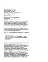
car crashes from 1996 to 2003, comparing the likelihood of head injury in drivers in a vehicle that sustained an impact greater than or equal to 50 g versus those sustaining a lesser impact. [359] They found that drivers in a crash with an impact greater than or equal to 50 g developed a head injury 16% of the time versus 1.6% for those involved in crashes with a lesser impact. Peripheral nerve injuries can occasionally occur in racing. Some drivers have reported symptoms consistent with brachial plexus injury. They describe transient upper extremity paresthesias secondary to the safety straps looped tightly around their arms. These straps are also connected to their helmets to combat the high forces the racers experience. Ulnar, peroneal, and sciatic nerve injuries can result from the constant pressure against the seat or other objects in the car during long races. Heatstroke has even been reported to occur, although rarely.[147] Spinal cord injury, spinal fractures, and cervical sprains and strains have also been described in automobile racing.[335] One study investigated racing injuries in either single-seat or formula cars or saloon cars between 1996 and 2000 at Fuji Speedway in Japan.[222] While extremity bruising accounted for the majority of injuries in single-seat car racing, 53.2% of the injuries in saloon car racing were neck sprains. Another report found that spinal injuries composed 20% of injuries experienced by professional automobile drivers.[338] In this review, injuries most commonly occurred during a vehicular rollover and usually led to cervical spine or spinal cord injury. In general, thoracolumbar injuries are uncommon, mainly due to the driver’s being so well restrained. The HANS (head and neck support) device is a safety item in many car racing sports (figure 1.1). It reduces the likelihood of head and neck injuries, such as a basilar skull fracture, in the event of a crash. The device is primarily made of carbon-fiber and is U-shaped. The back of the U sits behind the nape of the neck, and the two arms lie flat along the top of the chest over the pectoral muscles. The device is attached only to the helmet, by two anchors on either side, and not to the belts, driver's body, or seat. Therefore it is secured with the body of the driver only. The purpose is to stop the head from whipping forward in a crash without otherwise restricting movement of the neck. In a crash, the device maintains the relative position of the head to the
Figure 1.1 The head and neck support (HANS) device reduces the likelihood of head and neck injuries in the event of a crash. Picture Alliance/Photoshot
body, transferring energy to the much stronger chest, torso, shoulder, seat belts, and seat as the head is decelerated.
Ballet and Dance Dance has been an important part of life since ancient civilization. An extraordinary range of styles exists, from classical ballet and ballroom to modern dance and breakdancing. Although typically thought of as an art form, many forms of dance are physically taxing and can be considered sport. Additionally, dance is incorporated into a variety of sports including gymnastics, figure skating, synchronized swimming, and even martial arts kata. Regardless of style, all types of dance have something in common—they involve not only flexibility, athleticism, and body movement, but also physics. If the proper physics are not taken into consideration, injuries can occur. Many dance movements require extreme positions that can place the body at risk for acute, subacute, or chronic injury. In general, though, neurological injuries seen in many forms of dance,
Athletes and Neurological Injuries: A View From 10,000 Feet
such as ballet, result from chronic microtrauma or overuse, as opposed to the acute injury seen in other sports. It is important to remember that as with other athletes, dancers are often highly motivated to suppress pain and ignore injury until it affects their performance; this creates a need for increased awareness. Dancers are vulnerable to various stressrelated injuries, and muscle strains represent more than a third of all injuries. Injuries particularly affect the lumbar spine and the peripheral nerves and, to a lesser extent, the cervical spine. In one study, the National Organization of Dance and Mime surveyed 141 dancers from seven professional ballet and modern dance companies regarding their injuries.[35] Forty-eight percent had experienced a chronic injury, and 42% reported a more recent injury within the previous 6 months that had affected their performance. Garrick and Requa[109] reported 2.97 injuries per injured dancer in a large professional ballet company, with the lumbar spine the second most frequently involved region. Back problems are fairly prevalent in dance, with 10% to 17% of injuries occurring in the vertebral column.[218] Spondylolysis is a form of overuse injury secondary to chronic hyperextension and hyperlordosis of the lumbar spine. Microfractures of the vertebral bodies can occur, especially with repetitive flexion. This can result in wedging and Schmorl’s nodes at the thoracolumbar junction, a condition known as atypical Scheuermann’s disease. This condition can also result from lumbar extension contracture with excessive flexion demands transferred to the thoracic spine and resultant anterior end plate fractures and secondary bony formation.[30, 182] Fractures of the pars interarticularis and pedicles, arthritic degeneration, premature arthrosis, scoliosis, and discogenic as well as mechanical back pain are also common in dancers. Peripheral nerve injuries including nerve entrapment, neuropathy, and nerve dysfunction in the legs, ankles, and feet frequently occur in dancers.[219, 270-273] It should also be noted that neurologic injuries can occur in more recreational forms of dance. Breakdancing, for instance, involves athletic moves and spinning on various parts of the body, including the head and hands. One study described a breakdancer who presented with headaches and papilledema and was found to have multiple subdural hematomas.[208] Head
• • •
9
banging, a popular dance form accompanying heavy metal music, involves extreme flexion, extension, and rotation of the head and cervical spine. The motions can be performed so violently as to cause mild traumatic brain injury, whiplash injury to the cervical spine, or even subdural hematomas.[75, 158, 246] In 2008, Patton and McIntosh performed an observational study and biomechanical analysis of head banging.[246] They determined that an average head banging song has a tempo of about 146 beats per minute, which is predicted to cause mild head injury when the range of motion is greater than 75 degrees. Similarly, at higher tempos and greater ranges of motion, they found a greater risk of neck injury. Another study reported on a group of thirty-seven 8th graders participating in a dance marathon in which head banging occurred; 82% of the girls and 17% of the boys had resultant cervical spine pain that lasted 1 to 3 days.[158]
Baseball and Softball Baseball and softball are extremely popular sports. Between the fall of 1982 and the spring of 2008, approximately 10.9 million high school men and 23,517 high school women competed in baseball.[47, 72, 225] Over that time frame, an additional 616,947 men played baseball at the collegiate level.[225] Approximately 419,000 males and 900 females participate in baseball at the high school level annually.[72, 225] In the United States it is estimated that 23 million organized softball games are played each year. In 2008, Mueller and Cantu reported that between the fall of 1982 and the spring of 2008, approximately 30,000 men and 8.1 million women played high school softball.[225] An additional 323,000 collegiate women played softball over that time frame. Around 1,100 men and 313,000 women compete in softball at the high school level each year.[225] Injuries resulting from playing recreational baseball and softball are among the most frequent causes of sport-related emergency room visits, accounting for an estimated 286,708 injuries in 2009.[345] Minor injuries are fairly common in both sports, but catastrophic injuries can occur as well. Acute neurologic injuries are typically more common than chronic ones. Pasternack and colleagues studied patterns of injury in 2,861 Little League baseball players aged 7 to 18 years and reported 81 total injuries.[245] Eighty-one percent
10
• • •
Handbook of Neurological Sports Medicine
were found to be acute, and 19% were reported to be secondary to overuse. The authors found that 62% of the acute injuries were due to being struck by the ball. Interestingly, in softball, base sliding was found to be responsible for 71% of the injuries in one study.[146] These studies are consistent with other studies that have found the main mechanisms of injury to be related to being struck by a ball, repetitive motions or overuse such as throwing or swinging, collisions, sliding, and, much more rarely, being struck by a bat. As mentioned previously, severe neurologic injuries such as epidural hematomas or intracranial hemorrhages and catastrophic fatalities can occur but are rarer than mild brain injury. One study reported catastrophic injury rates in baseball of 0.37 per 100,000 high school player-games and 1.7 per 100,000 college playergames.[32] This study also found the fatality rates in baseball to be 0.067 per 100,000 high school baseball players and 0.86 per 100,000 college baseball players. Powell and Barber-Foss studied 10 different high school sports over the course of 3 years, identifying mild traumatic brain injuries, and found that softball and baseball accounted for only 2.1% and 1.2% of these injuries, respectively.[254] Two hundred forty-six certified athletic trainers reported a rate of 0.23 concussions per 100 player-seasons in high school baseball players, meaning that 0.23 concussions occurred every season for every 100 athletes. The same study found that the rate of concussion in high school softball was 0.46 per 100 player-seasons. Covassin and colleagues[63] studied NCAA athletic injuries over a 3-year time span and reported that concussions accounted for 2.9% of all injuries that occurred in practice and 4.2% of all injuries that occurred in games. In softball, concussions that occurred in practice accounted for 4.1% of all softball injuries, whereas in games, concussions accounted for 6.4% of all injuries. A more recent study investigated injuries in NCAA athletes over 16 years and found the rate of concussion in baseball to be 0.07 per 1,000 athletic exposures. [135] This survey also revealed a concussion rate in softball of 0.14 per 1,000 athletic exposures. These rates of concussion in collegiate softball are higher than those reported in a recent 1-year study of high school softball athletes, in which the overall concussion rate was 0.07 per 1,000 athletic exposures.[114]
Injuries to the spine are rare but can occur, usually secondary to collisions or headfirst sliding. While the use of breakaway bases and rules against headfirst sliding (at the youth level) have helped to substantially reduce the occurrence of sliding-related injuries, the risk for catastrophic spinal cord injury still exists. With headfirst slides, the top of the runner’s head can collide with the body or leg of the defensive player, creating a significant amount of axial load to the vertebral column. Baseball and softball players also are at risk for injury to their peripheral nervous system in addition to injuries of the brain and spine. A pitcher’s arms in baseball and softball withstand tremendous repetitive stress throughout a season. Long and colleagues[189] reported that every major league baseball pitcher, most minor league pitchers, and a few amateur pitchers they had studied had had reduced sensory nerve action potentials in the throwing arm. This is probably due to overuse and is a manifestation of brachial plexus injury. Although they throw underhand, fast-pitch softball pitchers withstand maximum compressive forces at the elbow and shoulder equivalent to 70% to 98% of their total body weight.[15] The more common peripheral nerve injuries in baseball and softball include suprascapular, axillary, and ulnar nerve injuries, although more infrequent injuries to the radial, musculocutaneous, and median nerves have been described.[140, 174, 301, 337]
Basketball Originally developed in 1891 by Dr. James Naismith at the International YMCA Training School in Springfield, Massachusetts, basketball has grown into a tremendously popular sport enjoyed worldwide and equally by men and women. Approximately 13.8 million high school men, 11 million high school women, 375,000 college men, and 328,000 college women participated in basketball between 1982 and 2008.[225] Most basketball injuries are musculoskeletal, affecting primarily the lower extremity; however, neurologic injuries can occur to the head, spine, and peripheral nerves. Head injuries in basketball can be caused by the sudden deceleration of the head when the player strikes an immobile object, such as the floor, another player, or a basketball pole or rim. These forces can cause direct contusions to
Athletes and Neurological Injuries: A View From 10,000 Feet
the brain or even result in tears of arteries and bridging veins, subsequently causing epidural and subdural hematomas. Several reports of acute subdural and epidural hematoma related to playing basketball are in the literature.[73, 160, 341] In their 16-year study, Hootman and colleagues found in college basketball that men experienced a rate of 0.16 concussions per 1,000 athletic exposures compared to a rate of 0.22 concussions per 1,000 athletic exposures in women. [135] Powell and Barber-Foss demonstrated that in high school basketball, the rate of concussion was 0.75 and 1.04 concussions per 100 playerseasons in men and women, respectively.[254] Additionally, in men’s high school basketball, concussions accounted for 4.1% and 5.0% of all the injuries sustained during practices and games, respectively.[114] While this was not significantly different for men, they did note that in women’s high school basketball, concussions accounted for 4.7% and 8.5% of the injuries sustained during practice and games, respectively; and this was significantly different. This subtle significant difference was also confirmed in a separate study.[261] Injuries to the spine are more common in basketball than other forms of neurologic injury. Basketball involves rapid, repetitive changes in direction and explosive movements that put significant stresses on the spine, resulting in a spectrum of spinal disease including lumbar sprains, contusions, facet hypertrophy, pars interarticularis fractures, spinal stenosis, spondylolisthesis, and disc herniations or degenerative disc disease.[4, 66, 141, 209, 213, 234, 311, 321] Cases of cervical cord neurapraxia have been reported in basketball players as well.[332] Although typically thought of as a football injury, burners or stingers have been reported in basketball secondary to acute head, neck, or shoulder trauma.[91, 92] Suprascapular, musculocutaneous, ulnar, median, peroneal, and sciatic nerves are all susceptible to entrapment neuropathies.[56, 68, 92, 152, 326, 340] Compression neuropathies of the arms are common injuries in a unique subgroup of basketball players—those who participate in wheelchair basketball.[43]
Bowling Bowling can be traced back more than 5,000 years ago to Egypt. In the 1930s, a British anthropologist named Sir Flinders Petrie discovered a
• • •
11
collection of objects in a child's tomb in Egypt that appeared to have been used for a primitive form of bowling. A crude version of a bowling ball and primitive pins were all sized for a child. A similar game evolved during the Roman Empire that entailed tossing stone objects as close as possible to other stone objects. This game became popular with soldiers and eventually evolved into Italian bocce (considered a form of outdoor bowling). The game has continued to evolve and today is a sport enjoyed by more than 100 million people in more than 90 countries each year and is considered a timeless sport.[97] Although not typically thought of as a sport with a high risk of injury, bowling can be both physically and psychologically demanding. Tremendous force is applied to the body throughout a bowler’s stance, approach, pivot step, arm swing, release, and follow-through. Repetitive stress is applied to the entire upper extremity including the fingers, wrist, and elbow. Injuries may vary by age as well. A recent study examined bowling-related injuries presenting to U.S. emergency departments between 1990 and 2008. [162] The authors analyzed data from the U.S. Consumer Product Safety Commission's National Electronic Injury Surveillance System and found that children younger than 7 years had a higher proportion of finger injuries and injuries from dropping the ball than individuals older than 7 years. On the other hand, bowlers more than 65 years old sustained a greater proportion of injuries related to falling, slipping, or tripping. While the annual incidence of injury is extremely low, the sport can cause a spectrum of neurologic hand and upper extremity injuries, either acute or due to overuse. Injuries to the fingers and digital nerves can occur. One report described a bowler with a rare traumatic dislocation of the four long fingers.[197] More commonly, though, the repetitive nature of bowling can lead to injuries to the digital nerve of the thumb, which most bowlers place inside the ball holes; this is referred to as “cherry pitter’s thumb”[337] (figure 1.2). Perineural fibrosis of the digital nerve of the thumb[297, 351] and even cases of thumb neuromas have been described. [164, 165] Dobyns and colleagues reported on one of the largest series of these patients.[78] Patients may present with a positive Tinel’s sign and skin atrophy or callusing over the neuroma. The nerve may ultimately become atrophied with fibrous
12
• • •
Handbook of Neurological Sports Medicine
in most animals. The earliest form of boxing can be traced back to 1500 BC in what is known today as Ethiopia; it then spread to ancient Egypt and throughout the Mediterranean. Grecian boxers would fight to the death, and Onomastos of Smyrna has been credited with first defining the rules of the sport:
Onomastos’ Rules of Boxing • No time limits or pauses unless agreed upon by both fighters • No ring • Matches open to all comers • Fighters not matched by weight • Won by disabling the opponent
Figure 1.2 Bowler’s thumb. This patient was taken to surgery because he did not respond to conservative therapy after a clinical diagnosis of bowler’s thumb. At surgery, the digital nerve was markedly enlarged secondary to perineural fibrosis. (a) The enlarged ulnar digital nerve (arrows) is surrounded by perineural fibrosis producing an irregular rather than a normal smooth contour. Definitive surgical therapy consisted of neurolysis with careful removal of perineural fibrotic tissue. (b) The nerve is smaller and has a smoother contour following neurolysis. The patient had an uneventful postoperative course and was able to eventually return to bowling. Reprinted from M.F. Showalter, D.H. Flemming, and S.A. Bernard, 2010, “MRI manifestations of bowler’s thumb,” Radiology Case Reports 6: 458. By permission of D.H. Flemming.
tissue proliferation at the site of injury. Not to be forgotten, neck and back pain can also occur in bowling and is often the result of discogenic injury.[228]
Boxing Unorganized hand-to-hand combat can be traced back to prehistoric man, and sparring activity of one sort or another, in general, can be observed
Eventually the sport was introduced to the Olympics in 688 BC. Great Britain is given credit, though, for turning boxing into the sport we know today, with its first formal set of boxing rules introduced in 1743 by Jack Broughton, who has since been referred to as the father of English boxing. Today it is a popular sport enjoyed by spectators and athletes of all levels. One must take a couple of important considerations into account to understand the types of neurological injuries that occur in boxing. For starters is the level of competition, which can be broken down into two simple subgroups: amateur and professional boxing. Amateur boxing differs from professional boxing in many regards. To begin with, amateur boxing contests feature a shorter length and fewer rounds. In amateur boxing the primary objective is to score points. The force of a blow or its effect on the opponent does not count as it receives the same credit as any regular blow. Hence, the knockout is a byproduct in amateur boxing. In contrast, in professional boxing, added weight is given to a blow based on its impact or effect on one's opponent, and thus the knockdown and knockout are objectives in the pros. In amateur boxing, in contrast to professional, headgear is worn, and boxers use 10-ounce (0.28 kg) gloves for all weight classes. Professional boxers use 8-ounce gloves (0.22 kg) and less padding. These differences have a large effect on the spectrum of injury seen in boxing as a sport. Another consideration is that injuries in boxing can be classified as acute or chronic. While acute injuries can typically be recognized and appreciated at the time the sport is played,
Athletes and Neurological Injuries: A View From 10,000 Feet
the chronic manifestation of nervous system injury secondary to repetitive trauma may not be realized until years after one has stopped boxing. The most commonly injured sites in boxing are the head, neck, face, and hands. Because the head is one of two primary targets, neurological injury, both acute and chronic, is the greatest potential risk that a participant accepts. Head injuries generally occur secondary to contact between the fist and head, head and head, or head and some part of the boxing ring. Concussions are by far the most common acute neurological injury in boxing. However, well-designed studies to assess the incidence of concussion in boxing have not been published. Some studies have estimated the rate of concussion to be 0.58 and 1.5 to 3 per 100 athletic exposures in amateur and professional boxing, respectively.[29, 151] Zazryn and colleagues retrospectively sought to investigate the risk of injury to professional boxers in a 16-year study in the state of Victoria, Australia.[375] A total of 107 injuries were recorded from 427 fight participations; concussions were common and accounted for approximately 15.9% of the injuries. To further explore the epidemiology of injury to both amateur and professional boxers in Victoria, the same group performed a prospective cohort study with 1-year follow-up from 2004 to 2005.[374] Thirty-three amateur and 14 active professional boxers sustained 21 injuries during the study period; most were to the head region, with concussion being the most common (33%). A recent study investigated the epidemiology of boxing injuries presenting to U.S. emergency departments between 1990 and 2008.[253] The study demonstrated that an estimated 165,602 individuals sustained boxing injuries resulting in a visit to a U.S. hospital emergency department during this time period, which amounted to an average of 8,716 injuries occurring annually. When a subgroup analysis was done according to age, the percentage of injuries that were concussions or closed head injuries in the group aged 12 to 17 years (8.9%) was similar to that in the group aged 18 to 24 years (8.1%) and the group aged 25 to 34 years (8.5%). While a knockout in boxing is synonymous with concussion, the majority of concussions in boxing are not associated with a loss of consciousness and are more commonly associated with transient cognitive impairment, loss of motor tone, or both; future studies are certainly needed
• • •
13
to better define the true rate of concussion in this population of athletes. The true incidence of acute intracranial hemorrhage is also largely unknown. The full spectrum of acute neurological injury can be seen: brain contusion or intraparenchymal hemorrhage, traumatic subarachnoid hemorrhage, acute subdural hematoma (the most common form of serious and lethal boxing brain injuries), epidural hematoma, diffuse axonal injury, carotid or vertebral artery dissection, and second impact syndrome.[148, 150, 216, 217] As noted earlier, chronic neurological injuries from boxing tend to have an insidious onset and often present and progress once a boxer’s career is over. Chronic neurodegenerative disease including mild cognitive impairment (MCI), chronic traumatic encephalopathy (CTE), and dementia pugilistica (DP) have all been described in boxers, although it is still unknown why certain boxers go on to develop these conditions and others do not.[200, 201, 206, 241, 313] These individual acute and chronic injuries are more thoroughly discussed in later chapters. There is a paucity of validated epidemiological data on which to accurately base boxing fatality rates. Instead, many of the reported fatality data have been obtained from a combination of media sources, industry reports, and individual case reports. A recent review of fatalities in boxing showed that based on the data analyzed between the control and fatal-bout groups, a computerized method of counting landed blows at ringside could provide sufficient data to stop matches that might result in fatalities.[215] However, such a process would become less effective as matches become more competitive, and implementing such a change would significantly decrease the competitive nature of the sport. From what can be ascertained based on the available reviews of boxing deaths, it appears that the rate of boxing fatalities has declined over the last few decades and that this can in part be attributed to rule changes as well as medical advances improving both the diagnosis and treatment of the acutely injured fighter.[11] A couple of final general concepts regarding the risk for sustaining acute neurological injury in boxing are worth noting. The literature contains studies reporting conflicting results regarding age, sex, experience, and various measures of exposures as risk factors for acute neurological injuries in combat sports. Several groups of
14
• • •
Handbook of Neurological Sports Medicine
boxers, though, have a theoretically higher risk of sustaining neurological injuries—including those who are fatigued or dehydrated. A large number of brain injuries in boxing occur as a result of fatigue. As participants fatigue, they tend to rely more on instinct and become increasingly oblivious to the amount of trauma they are receiving. Also as they fatigue, they have a decreased ability to maintain good balance and block or avoid punches. They are also less able to move with a punch when struck. This loss of defensive ability makes the fatigued boxer more vulnerable to acute neurological injuries. Dehydration in boxers can occur as a result of perspiration as a match progresses or intentional weight loss before a fight in order to meet a weight requirement, and it can increase a boxer’s vulnerability to injury. Dehydration can contribute to and accentuate fatigue, affecting a boxer in the ways already mentioned. Additionally, dehydration can decrease the amount of cerebrospinal fluid (CSF) surrounding the brain, which normally provides the brain with a buffer from trauma. Also, this decrease in CSF enlarges the potential space around the brain, increasing the likelihood of developing hemorrhages such as subdural hematomas.
Bungee Jumping Bungee jumping has quite an old origin. This way of jumping comes from the ancient ritual known as Gkol, performed on the Pentecost Island in the Pacific archipelago of Vanuatu, where young men would jump from tall wooden platforms with vines tied to their ankles as a test of their courage and passage into manhood. The first modern bungee jumps were made on April 1, 1979, from the 250-foot (76 m) Clifton Suspension Bridge in Bristol, England, by members of the Oxford University Dangerous Sports Club. The jumpers were arrested shortly afterward, but they continued with jumps in the United States, and the concept spread worldwide soon thereafter. Over 1 million jumps have been made since that time. Bungee jumping injuries can be grouped into those that occur secondary to equipment failure or technical misjudgment and those that occur regardless of safety measures. In the first instance, catastrophic injury can occur if the safety harness fails, the cord elasticity is miscalculated, or the cord is not properly connected to the jump plat-
form. There are several reports of quadriplegia and death secondary to these errors.[127, 129, 134, 191] In one case report of a bungee cord attachment apparatus malfunctioning, a jumper experienced a free fall of approximately 240 feet (73 m) and avoided a catastrophic outcome, sustaining only minor injury, because of the presence of an air cushion on the ground below.[288] Catastrophic injury can also occur if the jump height is miscalculated or if it changes. In 1997, a member of a 16-person professional bungee jumping team died of significant traumatic brain injury when she jumped from the top level of the Louisiana Superdome and collided headfirst into the concrete-based playing field while practicing for a performance that was to take place during the halftime show of Super Bowl XXXI. Hot air balloon–based launching sites are vulnerable to undetected changes in altitude, which can cause the cord length to be greater than the distance to the ground. However, jumps from fixed sites, such as bridges, have resulted in catastrophic injury as well, secondary to striking the platform on rebound. There have also been reports of jumpers whose cord becomes tangled around their neck after a jump. Even when appropriate safety measures are taken, there can be risk for neurological injury. A case of peroneal nerve palsy was diagnosed in a bungee jumper presenting with foot drop and paresthesias, presumably secondary to repetitive compression from the safety harness around the ankles.[334] More recently, a bungee jumper suffered a traumatic carotid artery dissection due to the force created by the free fall and rebound motion associated with the jump.[377] Fortunately for thrill seekers, despite all of the risks involved, bungee jumping is still considered one of the safest of the “extreme sports.”
Canoeing and Kayaking Canoeing originated to meet the simple needs of transportation across and along waterways and was the primary mode of long-distance transportation at one time. As a method of water transportation, canoes have generally been replaced by motorized boats and sailboats, although they remain popular as recreational or sporting watercraft. Canoeing as recreation and sport is often attributed to Scottish explorer John MacGregor, who was introduced to canoes on a camping trip
Athletes and Neurological Injuries: A View From 10,000 Feet
in Canada and the United States in 1858. Upon returning to the United Kingdom, he constructed his own canoes and used them on waterways in various parts of Britain, Europe, and the Middle East; he subsequently founded the Royal Canoe Club, the world’s oldest canoe club, in 1866.[260] The first canoeing competition was held by the club in 1874, and by 1936 canoeing had become an Olympic sport. The International Canoe Federation (ICF) is the umbrella organization for all canoe organizations worldwide and oversees the various disciplines, including canoe marathon, canoe slalom, canoe sprint, whitewater racing, canoe polo, canoe sailing, freestyle kayaking, surfski, and dragon boat racing. Lower back pain and injury are the most common complaints of athletes who are injured during canoeing or kayaking.[355] Those who train harder are obviously at a greater risk in these sports. Kameyama and colleagues surveyed 821 active canoeists and performed a medical check of 63 top competitive canoeists, including physical and laboratory tests and radiographic examinations of the chest, spine, shoulder, elbow, and wrist joints.[153] Completed questionnaires were returned by 417 canoeists, whose reported racing styles were kayak, 324; Canadian canoe, 71; slalom, 13; and not specified, 9. Of the 417 respondents, 94 canoeists (22. 5%) reported that they experienced low back pain. On medical examinations, the lower back pain was found to be mainly of myofascial origin or due to spondylolysis.[153] Catastrophic cervical spine injury or head injury is also possible and can occur particularly in kayakers who flip their vessel in shallow water.[289] Participants are also prone to median nerve entrapment secondary to the significant torque that wrists are subjected to with paddling.[355]
• • •
15
high school and collegiate levels have created a new dynamic, including increased risk for injury. The increase in cheerleading-related injuries in recent years can be attributed to the increase in the number of athletes engaged in the sport. The rise in catastrophic injuries, though, appears to be related to the increasing difficulty of the acrobatic routines that cheerleaders perform and the daring skills of prospective cheerleaders trying to make a cheering squad. Modern cheerleading involves high team throws and daring aerial drills that frequently lead to accidents, particularly during pyramid building (figure 1.3).[31, 291-293] Mortalities in cheerleading have been reported. [31] Severe neurological injuries have included skull fractures, intracranial hemorrhages, and diffuse cerebral edema. Additionally, major spine injuries have ranged from cervical fractures to spinal cord contusion and paralysis.[31, 336]
Cheerleading Cheerleading originated as squads aimed at getting the crowd at a sporting event to cheer louder. Today, however, it has evolved into an athletic activity that incorporates elements of dance and gymnastics along with stunts and pyramid formations. An estimated 3.5 million people participate as cheerleaders each year, from 6-year-olds to adults who cheerlead for professional athletic teams. While cheerleading is meant to support an athletic team, its intense competitions at the
Figure 1.3 Pyramid building in cheerleading is particularly dangerous. © Ric Tapia/Icon SMI
16
• • •
Handbook of Neurological Sports Medicine
A recent study described the epidemiology of cheerleading-related strain and sprain injuries. [294] Athletic exposure and injury data were collected from 412 U.S. cheerleading teams via the Cheerleading Reporting Information Online surveillance tool; spotting or basing other cheerleaders was the most common mechanism of injury and was more likely to result in a lower back strain or sprain than other mechanisms. Another study investigated a cohort of 9,022 cheerleaders on U.S. cheerleading teams.[292] Of the 567 cheerleading injuries reported during the 1-year study, 83% occurred during practice as opposed to competition; specifically, 52% occurred while the cheerleader was attempting a stunt, and 24% occurred while the cheerleader was basing or spotting one or more team members. The authors also noted that collegiate cheerleaders were more likely to sustain a concussion or closed head injury than were cheerleaders at other levels. One prospective cohort study of high school cheerleaders found that 6.3% of all injuries were concussions.[283] Cheerleading can be associated with median palmar digital neuropathy as a result of chronic trauma to the palm during cheerleading activities, although this is rare.[295]
cervical spine, and lumbar spine accounted for 8%, 4%, and 20% of the total, respectively.[195] Another recent study reviewed injuries in cricket players of all levels over a 6-year period and indentified 498 cases.[354] Head injuries accounted for 20% of injuries in this study, and of those, 77% were the result of being struck by a ball or bat. The head injuries involved fractures (35%), contusions (18%), and concussion (10%). As with other sports, injury mechanisms seem to vary with age. In one study, players less than 10 years of age were most commonly injured by being struck by the bat; for those aged 10 to 50 years, being struck by the ball or the bat was most common; and for those >50 years of age, the most common mechanisms were overexertion, strenuous or repetitive movements, or falls.[354] The injury pattern also changed with age. Fifty percent of all injuries to those younger than 10 years occurred to the head; players aged 10 to 19 years sustained mostly injuries to the head, as well as upper and lower extremities; and those older than 20 years mainly sustained extremity injury. These figures are important when one considers how to tailor protective equipment and injury prevention education to these athletes.
Cricket
Cycling and BMX
Although cricket has been long heralded as a “gentleman’s game,” the game’s evolution has resulted in shorter and more competitive matches involving greater aggression, more stressful training programs, and a workload on par with that of other professional athletes.[13] Thus it is no surprise that an increase in the number of cricketing injuries has been observed recently, with players exposed to risk of impact (ball or bat), collision (other players, fences), slips and falls, and repetitive and overuse injuries. While well-designed studies critically investigating the incidence of neurological injury are lacking, neurological injuries can and do occur. Epidemiological studies undertaken in Australia, South Africa, England, and the West Indies have repeatedly demonstrated that fast bowlers have the highest risk of injury in cricket, with the lower back being most susceptible to both traumatic and overuse injuries.[116, 181, 195, 242, 315] A 1-year study prospectively surveyed all injuries occurring in all major matches of the West Indies Cricket Board and found that injuries to the head,
Participation in the sport of cycling has grown significantly in the past several years. The first competitive bicycle race was held in France in 1869. Since that time the sport has expanded to include recreational riding, road and off-road cycling, track racing, BMX, and cyclocross competition. The sport is associated with a wide range of neurological injuries affecting both the central and peripheral nervous systems. While at first glance one might expect the incidence of injury to be greater in BMX biking versus other forms of cycling, the reported incidences appear to be similar.[370] A comparison of various groups of cyclists showed that a large number of head injuries needed admission to a hospital, but interestingly, BMX riders had fewer head injuries than the other groups. Concussions account for about 7% of the injuries in BMX racing.[145] Off-road racing carries a high risk for musculoskeletal injury; however, in several reports the incidence of concussion was quite small, less than 1%.[173, 265] Regardless of the type of cycling, injuries to the head are common in
Athletes and Neurological Injuries: A View From 10,000 Feet
accidents. Each year, more than 500,000 people in the United States are treated in emergency departments, and more than 700 people die as a result of bicycle-related injuries.[51] The rate of fatalities in children from bicycle accidents exceeds that from causes such as falls, poisoning, suffocation, and firearms—problems that typically receive much more attention.[360] The majority of fatal accidents are due to intracranial hemorrhages and subdural hematomas. Helmet use has significantly helped to reduce the incidence of catastrophic injury. Neck and back pain are extremely common complaints in cyclists, occurring in up to 60% of participants.[361] In one recent descriptive epidemiological study, 109 elite cyclists in the training camps of seven professional teams were interviewed regarding overuse injuries they had experienced in the previous 12 months.[58] Injuries that required attention from medical personnel or involved time loss from cycling were registered. Of the 94 injuries registered, 45% were in the lower back. Additionally, 58% of all cyclists had experienced lower back pain at some point in the prior 12 months, and interestingly, only 41% had sought medical attention for it. Another study of overuse injuries in cyclists found that the most common anatomical site was the neck (49%) and that the lower back accounted for 30% of complaints.[362] It is thought that one mechanistic etiology might be the hyperextension of the neck and forward-bent riding position. Also, if the bicycle is not fitted appropriately for the participant, the cyclist may need to reach farther forward for the handlebars, exacerbating the hyperextension of the neck and extreme flexion of the back needed to ride. Spinal cord injuries and disc herniations have also been associated with cycling accidents.[79, 169, 350] Injuries to the peripheral nerves can occur with cycling as well. The most common peripheral nerve deficits associated with the sport include injuries to the pudendal, genitofemoral, and ulnar nerves. Neuropathies with symptoms in the distribution of the pudendal and genitofemoral nerves are common and can occur in as many as 50% of male cyclists competing in long-distance rides. [33] Goldberg, Peylan, and Amit described a cyclist with injury to the pudendal nerves who presented with numbness of the buttocks and genitalia, associated with difficulty in achieving an erection.[118]
• • •
17
Cessation of cycling for a few weeks resulted in a spontaneous resolution of the patient’s symptoms, fortunately without residual deficit. The injury is thought to occur from overuse or improper or poor positioning of the seat. Less frequently, the shape of the seat is the root of the problem. Ulnar nerve injury secondary to chronic compression at the handlebar interface has been recognized for over 100 years.[105] One study reported 117 cases over a 4-year period. [139] This condition, often referred to as cyclist’s or handlebar palsy, can result in both motor and sensory symptoms and most commonly occurs during a long ride, often over rough terrain. Although much less common than ulnar neuropathy, median nerve involvement has been described in cyclists as well.[37]
Darts and Lawn Darts The sharp point on the dart used in recreational indoor dart competition as well as outdoor lawn darts can be a cause for neurological injury. Lawn darts are typically 12 inches (30 cm) long with a weighted metal or plastic tip on one end and three plastic fins on a rod at the other end, basically an oversized version of the traditional indoor dart. The darts are tossed underhand toward a horizontal ground target, where the weighted end hits first and sticks into the ground, similarly to a horseshoe. While injuries are typically rare with the use of indoor darts, several reports have documented the neurological injury risk with lawn darts. One of the largest series reported 76 patients who presented to the hospital with lawn dart injuries.[309] The patients ranged from 1 to 18 years of age and were predominantly male. The most common sites of injury were the head (54%), eye (17%), and face (11%), and hospitalization was required for nearly 54% of these patients. Two other reports corroborated the risk for penetrating brain injury with these darts.[128, 325] The U.S. Consumer Product Safety Commission banned the sale of lawn darts in December 1988,[344] and Canada followed in similar suit shortly thereafter.[132] Despite the ban on the sale of lawn darts, there remain millions of sets in circulation. These dangerous products may still be in garages, basements, or secondhand stores, and the U.S. Consumer Product Safety Commission has urged consumers to discard or destroy all lawn darts immediately.[344]
18
• • •
Handbook of Neurological Sports Medicine
Diving and Swimming In the 1800s, diving evolved from a sport called plunging. It became a part of the Olympic Games in St. Louis, Missouri, in 1904. As a sport, it has always carried a risk of catastrophic cervical spine injury. It was recognized as early as 1948 that unsafe diving is the leading cause of spinal cord injuries associated with aquatic activities.[20, 71] Diving is the fourth leading cause of spinal cord injury in the United States[239] and is also responsible for significant proportions of spinal cord injury worldwide.[20, 26, 71, 221] These devastating injuries can occur when divers strike the springboard or platform with their head during a dive or when the head contacts the bottom surface of the pool. Most injuries do not happen in elite divers but rather occur in recreational athletes who dive into shallow water (in swimming pools, ponds, or lakes), often with disastrous results.[76] Spinal cord injuries are most commonly associated with either fracture dislocations or compression fractures of the spinal column forced into hyperflexion. One study investigated 220 patients with spinal cord injury secondary to diving accidents who were admitted to the Midwest Regional Spinal Injury Center in Chicago.[8] Of the total patients, 70% had neurological injury; and among these, 47% had complete spinal injuries with loss of all motor and sensory function below the level of injury. The rest of the patients sustained incomplete injuries: 62% anterior cord syndrome, 17% central cord syndrome, and 20% Brown-Sequard syndrome.[8] The most common levels of injury were at the C-5 and C-6 levels. In another large retrospective study, all spinal cord injuries associated with diving were at the cervical level.[280] The incidence of spinal cord injury in diving is heavily dependent on patient factors, agent factors (i.e., that which imparts force on the diver), physical environmental factors (e.g., warning signs, depth indicators, lighting), and social factors (e.g., absence of lifeguards or presence of alcohol). [71] Given the burden and rates of these injuries, as well as the fact that they appear to be (for the most part) preventable, efforts at multifaceted prevention programs are urgently needed.[71] The sport of swimming per se is generally felt to be one of the safest with regard to ner-
vous system injury, with a low incidence of head, spine, and peripheral nerve injuries. Most catastrophic injuries in swimming are related to diving into the water. That being said, in swimmers, neurogenic thoracic outlet syndrome may develop in association with hypertrophied pectoralis minor muscles.[155, 316]
Dodgeball Dodgeball is a well-known form of team sport that is best known as a game played in physical education classes. While the game is typically played among children in elementary school, the sport has emerged internationally as a popular middle school, high school, and college sport. It is also popular in informal settings and can be played on a playground, in a gym, or in organized recreational leagues. There are no standards for the measurements or materials of the balls used; however, most dodgeballs are roughly the size of a volleyball and are composed of foam with a thin plastic or rubber shell. Although dodgeball is typically thought of as a benign activity, the risk for concussion or other severe head injury exists. A recent report described a 9-year-old child who was found to have a chronic subdural hematoma due to repeated minor dodgeball head impacts. [356] No altered mental status or focal neurological deficits were observed; however, the child presented with intermittent severe headache associated with nausea and vomiting.
Equestrian Sports Horses were first domesticated in 4500 BC by tribesmen in Central Asia. The horse has played an important role throughout human history, all over the world, both in warfare and in peaceful pursuits such as transportation, trade, and agriculture. While horses were initially trained and ridden for practical working purposes, equestrianism has since evolved to include a wide range of sports including, but not limited to, endurance riding, show jumping, harness racing, vaulting, polo, thoroughbred horse racing, steeplechase racing, rodeo, and recreational riding. Equestrian-related injuries are a serious occurrence, and horseback riding is recognized as more dangerous than skiing and motorcycle or automobile racing.[14] In the United States,
Athletes and Neurological Injuries: A View From 10,000 Feet
an estimated 30 million people ride horses each year, resulting in 50,000 emergency room visits (1 visit per 600 riders per year).[49, 170] Children participating in equestrian activities are at risk for substantial injury, and pediatric care providers must maintain a high index of suspicion when evaluating these children.[25, 70, 235] Females tend to have a higher rate of injury than males, but this is partly due to the female predominance in the sport.[22, 23, 235] Riding carries with it an implicit risk of injury associated with the unpredictability of the animals, the weight of the horse, the positioning of the rider's head as high as 10 feet (3 m) off the ground, and travel at speeds up to 40 miles per hour (64 km/h).[131] The most common mechanism of injury is falling or being thrown from the horse, followed by being kicked, trampled, and bitten.[83, 131, 170, 186] One study reviewed a national database of equestrian injuries over a 2-year period to identify predictors of significant injury.[186] The authors found that the injuries occurred at home (36%), in a recreation or sporting facility (30%), on a farm (19%), and on other public property (12%). The injuries were due to a fall (59%) or to being thrown from or bucked off the horse (22%), or they occurred while on the horse (9%). Neurological injury in particular is a common consequence of these activities; it ranges in severity from concussions and degenerative spine disease to spinal cord injury and debilitating closed head injuries, and can even result in death.[14, 24, 131, 185, 266, 339] Chronic neurodegenerative disease, in the form of dementia pugilistica, has been reported in steeplechase jockeys. For professional jockeys, head trauma is one of the most common types of career-ending injuries.[12] Helmets have helped to decrease the incidence of catastrophic injury; however, helmet use (particularly during recreational riding) remains low.[34] One possible obstacle to helmet use in equestrian sports may be the cost. However, while helmets traditionally have been quite expensive, many approved helmets can now be found for less than $50.
Golf Although golf may trace its origins to ancient Rome, it has been fairly well established that the modern game was actually devised in Scotland in the late 14th or 15th century. Today, more than
• • •
19
26 million people in the United States alone play golf. Golf is considered a safe sport, and although they are uncommon, injuries incurred during golfing are an increasing problem. A number of studies have examined the occurrence of injuries in amateur as well as elite or professional golfers. In professional male golfers, the most common site of injury is the lower back.[204] For professional female golfers, the lower back is the second most common site of injury.[198] The most common mechanism of injury for professional golfers is the highly repetitive practice, followed by hitting an object other than the ball with the club.[198, 204] The amount of practice places this group of golfers at risk for overuse injuries; and often they continue to play, making them more likely to aggravate the injury compared to amateur golfers. In amateur golfers, the lower back is an extremely common site of injury. The literature is conflicting regarding the most common site of injury, as some studies report that the wrist is more commonly injured than the lower back.[16, 199] In amateur golfers, the most common mechanisms of injury are overuse, poor biomechanics of the swing, and hitting the ground or an object other than the ball during the swing.[199, 204] One study reviewed golf-related injuries that occurred in 300 patients attending emergency departments over a 6.5-year period.[363] Most of these injuries involved the head. The main mechanisms of injury were being hit by a club (37%), being hit by a ball (28%), sprains or strains (9.67%), and slips or falls (7%). When the data were broken down by age, the main source of injury in adults was being hit by a ball, whereas for children and adolescents, being struck by a golf club accounted for 77% of hospital presentations. Catastrophic injury can occur as a result of being struck by either a club or a ball, although each of these occurs rarely.[204] Standing behind someone who unexpectedly takes a practice swing or standing too close to another player who is swinging is the most common source of this injury. Freak accidents have also been described—for example, the golf club shaft breaks, resulting in a penetrating head injury. Some golf-related neurological injuries are due to golf cart accidents or falls out of a cart, often involving inebriated passengers and drivers.[193, 204, 335, 342]
20
• • •
Handbook of Neurological Sports Medicine
Gymnastics and Trampoline Although gymnastics began more than 2,000 years ago, it was seen originally as an activity, not a sport. It has evolved into a widely popular sport today with significant increases in participation in the last two decades. Approximately 3,800 men and 24,500 women participate in gymnastics annually.[225] The increase in numbers has exposed a larger number of athletes to acute and chronic injuries. Many studies place the overall injury rate as high as in football, wrestling, and softball.[335] One study sought to describe the epidemiology of gymnastics-related injuries among children ages 6 to 17 years in the United States over a 15-year period.[300] This retrospective review revealed an estimated 425,900 children treated in U.S. hospital emergency departments for gymnastics-related injuries during the 16-year period, with the number of injuries averaging 26,600 annually. The places where injuries occurred included schools (40.0%), places of recreation or sport (39.7%), homes (14.5%), and other public property (5.8%). The required maneuvers and postures are difficult and place the body at risk for neurological injury, particularly spine injury (figure 1.4). Degenerative disc disease and spondylolysis are common injuries in gymnasts. Most spinal cord injuries occur at the midcervical levels.[7, 9, 237] With most gymnastics activities, the incidence of catastrophic brain injury is relatively low; most of these injuries occur during a dismount in which an athlete lands on the head. Milder head injuries such as concussion occur more frequently. In their 15-year study of children ages 6 to 17 years, Singh and colleagues found that head and neck injuries accounted for 12.9% of all injuries.[300] Strain and sprain injuries were most common (44.5%), and concussion or closed head injury occurred in only 1.7% of the cases.[300] The concussions in that study were more likely to occur while participants were performing headstands versus other skills. In a 16-year survey of collegiate gymnasts, the concussion rate was found to be 0.16 per 1,000 athletic exposures. [135] Although extremely rare, peripheral nerve injuries involving the femoral, lateral femoral cutaneous, and distal posterior interosseus nerves have all been reported.[5, 41, 115, 119]
Figure 1.4 The maneuvers and postures involved in gymnastics can place the body at risk for neurological injury.
Trampolines have evolved only over the last 50 years, with an unprecedented surge in popularity recently. They are used both as a part of gymnastics events and for recreational activity. Trampolines have been identified as a common cause of injury−spinal cord injury in particular− with many studies documenting the risk.[144, 240, 274, 317, 369] Participants can be projected many feet in the air and fall back to the trampoline or the ground, landing on their head or neck, putting them at risk for catastrophic injury. One study demonstrated that in the United States, trampolines accounted for greater than 6,500 pediatric cervical spine injuries in 1998.[40, 107] This was a fivefold increase in trampoline-related injuries compared with the previous 10 years. While fractures are the most common injury, spinal injuries reportedly account for 12% of trampoline-related injuries.[107] These occur frequently in children;
Athletes and Neurological Injuries: A View From 10,000 Feet
however, teenagers and young adults have sustained spinal cord injuries as well.[183, 298]
Hang Gliding Thousands in the United States participate in hang gliding despite the risks inherent to the sport. Hang gliders are kite-like crafts to which the flier is attached via a harness and supported on a swing-like frame. By shifting their body weight, fliers can steer the craft in various directions. The sport has continued to grow, with competitions held at both the national and international levels. The potential for neurological injury, particularly catastrophic injury, is fairly obvious and has been noted in the literature.[17, 367] Usual causes of injury include misjudgment of landing speed and altitude, resulting in injurious landing forces. An earlier study noted that most of the hang gliding injuries examined occurred as a result of in-flight errors in judgment versus equipment failure.[331] Another study showed that the majority of all injuries (60%) occurred during landing.[180] In that same study, spinal injuries were the most common type, accounting for 36% of the total.[180] The majority of hang gliding injuries reportedly occur in young adults age 20 to 40 years.[100] Another study noted that of nonfatal injuries, 16% were head injuries and 17% involved the spine.[331]
Hockey (Field and Ice) Native North Americans played variations of stickball such as tabé or shinny, which are similar in some ways to modern field hockey. For well over 1,000 years, though, the Daur people of Inner Mongolia have been playing a game called beikou, which entails whacking around a ball-like knob of apricot root with long wooden branches.[203] For night games, they use an ignitable ball covered in felt.[203] The modern game of field hockey grew from English public schools in the early 19th century, and the game was codified for use in the Olympics shortly thereafter. One study showed that between 1982 and 2008, approximately 3,000 men and 1.43 million women competed in high school field hockey, with an additional 145,000 women competing at the college level.[225] Most believe that ice hockey evolved from outdoor stick-and-ball games as
• • •
21
well. It is thought that these games were adapted to the icy conditions of Canada in the 19th century and later evolved into a game played on ice skates, often with a puck. Ice hockey is a sport that is widely popular today, and an average of approximately 27,800 men and 2,800 women play it each year.[72, 225] Both sports use a hard object (ball or puck) and sticks and are played in an aggressive manner. Neurological injuries in both forms of hockey most commonly consist of concussions and spinal cord injuries, with peripheral nerve injuries much less common. Possible mechanisms include falls during ball or puck handling, collisions with other players or inanimate objects (bench, goal, boards), and checking.[72, 335] Although there are many similarities between ice and field hockey, the proportions of concussions differ between the sports. Considering the lower number of participants compared to other sports, both forms of hockey are associated with relatively higher rates of concussion. In a 13-year study comparing injuries in male and female pediatric (ages 2 through 18 years) ice and field hockey participants, the proportion of concussion was higher in ice hockey players (3.9%) than in field hockey players (1.4%).[373] Concussions in high school field hockey have been reported at a rate of 0.46 per 100 playerseasons.[254] Collegiate field hockey athletes had a rate of 0.18 concussions per 1,000 athletic exposures in one large 16-year study.[135] In that same study, the rate of concussions in male and female collegiate ice hockey participants was 0.41 and 0.91 per 1,000 athletic exposures, respectively. [135] The relationship between age and concussion in ice hockey in uncertain; however, studies in youth hockey players seem to suggest that Bantam and Pee-Wee ice hockey players (ages 11-14 years) have a greater rate of concussion than Atom players (age 9-10 years).[84, 85] Nonetheless, with increased awareness and reporting in recent years, concussion appears much more common than we once thought across all levels of ice hockey competition. Hockey-related spinal injuries have seen an increased occurrence, although this may be in part due to the larger number of players, better diagnostic skills, or increased reporting.[323, 335] An earlier study looked at the SportSmart Registry for the years 1966 to 1996.[324] The authors
22
• • •
Handbook of Neurological Sports Medicine
examined the nature and incidence of major spinal injuries in ice hockey players and identified 300 players worldwide who had sustained spinal injuries, of which approximately 250 occurred in Canada, 35 in the United States, and 15 in Europe. Complete motor injuries were suffered in approximately 25% of players, and 75% of the injuries occurred during organized games as opposed to practice.[324] Burst fractures and fracture dislocations are the most common vertebral injuries and carry with them an increased risk for spinal cord injury. The most frequently reported mechanism of injury is a push or a check from behind, sending the unsuspecting player hurling into the boards headfirst. Spinal injury can also occur when a player is hit while looking down at the puck, delivering an axial loading force to the head and spine. Rule changes, education, neck strengthening exercises, and conditioning have all been important aspects of injury prevention in these athletes. As mentioned previously, peripheral nerve injury in both forms of hockey is rare but can occur.[337] Although more commonly reported in football players, burners or stingers have been described in hockey players.[91] Direct trauma or orthopedic injuries always carry a risk for peripheral neuropathy. Axillary nerve injury secondary to direct trauma associated with shoulder dislocation has been reported in two hockey players.[247, 248] Similarly, peroneal neuropathy has occurred in hockey players, though rarely, secondary to laceration of the nerve with a skate blade or as a result of direct blunt nerve trauma.[192, 290]
Hurling Hurling is one of the three national games of Ireland and has prehistoric origins, having been played for at least 3,000 years. It is a fast-paced game that combines elements of baseball, field hockey, and lacrosse. A similar game for women is called camogie. Players hurl a small, hard, 100- to 130-gram leather ball (sliotar) with a long tapered stick (hurley). They score points by propelling the ball over or under the crossbar of the goal. This is a contact sport that differs from other “stick” games in that the majority of play is above the participants’ heads in the air. As a result, injuries to the head are common and are usually due to contact with the ball, the stick, or another player.[229] Two studies found that hurling
injuries occurred to the head or face 36% to 40% of the time.[67, 69, 229] As of 2010, all players must wear helmets with face guards, and a resultant decrease in injuries is apparent. Before this time, helmet use during game play was optional, and resistance to helmet use certainly contributed to the high injury rates. An earlier prospective study investigated hurling injuries in 74 players over the 8 months of one season.[358] The most common types of injuries were muscle strains. Back and head injuries accounted for 11% and 9% of the total, respectively. Concussions accounted for 3% of the total. Interestingly, 41% of the injuries were attributed to foul play, making a case for the need for better rule enforcement. Even though it is one of the all-around fastest field sports, catastrophic injury in hurling is quite rare.
In-Line Skating, Roller Skating, and Skateboarding The first recorded use of roller skates was in 1743 during a London stage performance, although the inventor of this skate is largely unknown. John Joseph Merlin is credited as the inventor of the skate in 1760, which was actually a primitive in-line skate with metal wheels. Roller skating saw rapid growth throughout the early 1900s and immediately became a popular pastime for both men and women. In-line skating evolved from roller skating; some believe it was developed to occupy ice hockey players during the months when there was no ice. The first in-line skate, featuring three in-line wheels attached to a wooden plate, was patented in Paris in 1819. Several renditions appeared over the next century. In-line skating has since become one of the fastest-growing recreational sports for children and teenagers in the United States. The low cost and various health benefits have allowed the sport to thrive beyond the limits of a fad, as evidenced by the existence of professional leagues and international competitions (roller hockey, roller derby, aggressive in-line). Millions of participants have gravitated toward skating as a means of recreation, competition, fitness, training, or transportation. Skateboarding is also a popular recreational activity and part of a lifestyle among many young people.[103, 263] The most common injuries among all of these skate
Athletes and Neurological Injuries: A View From 10,000 Feet
sports are musculoskeletal, with ankle strains and sprains and extremity fractures.[1, 90, 101, 103, 159, 175, 263, 277, 279] The incidence of injury with roller skating and in-line skating is less than that in ice skating. For skateboarding, the incidence of injuries is estimated to be 10 injuries per year, per skateboarder.[263] A total of 65% of injured adolescent skateboarders sustain injuries on public roads, on footpaths, and in parking lots.[103, 159] Furthermore, injuries seem to occur more frequently while people are performing a trick.[263] Helmet use has helped to reduce the incidence of severe head injury, but head injuries still do occur.[117, 138, 167, 187] "Truck surfing" or "skitching" refers to skating behind or alongside a vehicle while holding on to the vehicle. This results in a skater’s traveling at the same speed as the vehicle. Such risky behavior can be very dangerous, particularly if the skater cannot slow down fast enough to prevent colliding with the vehicle or if the skater is thrown into oncoming traffic or the roadbed should the vehicle suddenly slow down, stop, or turn. The enhanced momentum results in a greater force of impact and consequently a more severe injury. Death is not uncommon with these severe injuries.
Lacrosse The native North Americans used to play a game called baggataway in order to train for battle. [299] Many tribes played games with carefully shaped sticks made of hickory and other spiritually important woods that had nets on the end, generally made of elm bark, leather, or deer hide. The National Lacrosse Association came into being in 1867, and the game evolved over time into the modern sport we know today. It is a fast-paced contact sport, played in close quarters, using sticks to propel a hard ball at high speeds. Neurological injuries can occur when a player falls or is struck by another player, the ball, or a stick, or when colliding with fixed objects such as the goal or a bench. Men’s lacrosse is considered a contact sport and women’s lacrosse is considered noncontact, which may be a reason why protective gear, including helmets, is used only occasionally or not at all in women’s lacrosse. High school lacrosse has approximately 33,000 male and 22,000 female participants each year. [72, 225] An additional 5,800 collegiate men and
• • •
23
4,000 collegiate women play lacrosse annually, as well.[72, 225] In one study of scholastic women’s lacrosse, the reported rate of head and face injury was 1.1 per 1,000 athletic exposures.[121] Head injuries are frequent in lacrosse players. In one 10-year study, closed head injuries composed 6% of all lacrosse-related injuries.[77] An earlier study of college athletes found that of all major women’s collegiate sports, lacrosse appeared to have the highest percentage of concussive injuries (14%) and that this was higher than the proportion of concussion seen in male lacrosse players (10%). [63] Hootman and colleagues, in their 16-year study, reported the rate of concussion in collegiate athletes to be 0.26 and 0.25 per 1,000 athletic exposures for men and women lacrosse players, respectively. Injuries are more common in games than in practice, likely due to the more aggressive play during competition.[121] In one study, concussions accounted for 8.6% of all injuries in lacrosse competitions, with athletes nine times more likely to experience a concussion in a game compared with practice.[135] Catastrophic injury can occur, as well. There is one report of an epidural hematoma resulting from a strike to the head from a lacrosse stick.[264] At the institution of one of the authors, a teen lacrosse player presented to the trauma bay after being struck in the back of the neck with a lacrosse ball. The computed tomography (CT) scan of his head demonstrated diffuse subarachnoid and intraventricular hemorrhage secondary to a significant vertebral artery dissection. The patient died shortly after admission to the hospital.
Martial Arts and Mixed Martial Arts Martial arts are ancient forms of combat modified for modern sport and exercise. Participation in martial arts is increasing, with millions of people practicing each year. In general, martial arts provide health-promoting and meaningful exercise and have been shown to improve participants' overall cardiovascular endurance, strength, balance, flexibility, body fat composition, stress and relaxation, confidence, and socialization. [258] Mixed martial arts (MMA) competition, also referred to as no-holds-barred (NHB) fighting, ultimate fighting, and cage fighting, has its roots in ancient Greece. In 648 BC, it was referred to as pankration and was featured at the 33rd
24
• • •
Handbook of Neurological Sports Medicine
ancient Olympics.[44, 252, 258] Pankration, which is Greek for “all powerful,” was the hybridization of boxing and wrestling and was spawned from unarmed combat on the battlefield.[252] It became an extremely popular freestyle fighting sport and served as the climactic event of the Olympics for centuries. Despite the attempts by legislators and the medical community to ban it, MMA grew in the early 1990s from an underground spectacle into an internationally sanctioned sport with many of the same health benefits as traditional martial arts. In addition to these health benefits, however, all forms of martial arts activities carry an obvious risk of injury. The incidence of injuries among the various individual disciplines seems to be roughly similar, at least between karate, taekwondo, and Muay Thai kickboxing.[110, 335] Martial arts styles that involve striking, such as kickboxing, karate, and taekwondo, have been shown to have a higher incidence of injury than styles that involve grappling alone, such as judo, sumo, and Brazilian jiu-jitsu. One large study sought to determine the rate and types of injuries occurring to registered professional kickboxers in Victoria, Australia, over a 16-year period.[376] Of the 382 injuries recorded from 3,481 fight participations, the body region most commonly injured was the head-neck-face (52.5%), with intracranial injury specifically occurring 17.2% of the time. Another study assessed the conditions under which concussions occurred in full-contact taekwondo competition.[250] The incidence of concussion in this study was high and was greater in men (7.04 per 1,000 athletic exposures) than in women (2.42 per 1,000 athletic exposures). Initially promoted as a violent and brutal sport, MMA has dramatically changed, with revised rules and improved regulations to minimize the risk of injury. With regard to safety, MMA has been compared to other combat sports, such as boxing; however, MMA may actually have a safer track record with respect to serious injury and death. This difference may be due largely to the competitive structure of MMA events. Mixed martial arts competitions consist of three 5-minute rounds (for nonchampionship bouts) or five 5-minute rounds (for championship bouts), followed by 1-minute rest periods between rounds.[258] Competitors are matched according to designated weight classes and experience levels and wear protective equipment consisting of a
mouth guard, groin protector, and 4- to 6-ounce (0.11-0.17 kg) MMA gloves. Limited studies have investigated injury incidence in MMA.[28, 236, 258, 285, 286] Based on the data available, closed head injuries, lacerations, and orthopedic injuries are commonly experienced by competitors.[286] Knockout rates are lower in MMA competitions than in boxing, suggesting a reduced risk of brain injury in MMA competitions compared to other events involving striking. Since the inception of MMA in the modern era, only four deaths have been documented, with three of the four occurring outside of the United States in unsanctioned fights.[258] As a comparison, one study documented 71 deaths in boxing from 1993 to 2007, with a total of 1,355 deaths from 1890 to 2007, averaging 11.6 deaths per year during the modern history of the sport.[320] The incidence of concussion in MMA matches has ranged from 1% to 3%, with almost 25% of the matches stopped secondary to impact to the head; however, the incidence of concussion may be underreported. Injuries to the peripheral nerves are possible, with both acute and chronic symptoms reported.[161] Neuropathic symptoms can occur in individuals as a result of strikes on pressure points or exposed peripheral nerves. Although the majority of symptoms resolve within 1 year, individuals with repetitive exposure strikes may be more likely to have chronic symptoms.[161] Injuries to the spine can occur, although they are rare.[21, 251, 343] Studies using mathematical models of the biomechanics of maneuvers in MMA have shown that the forces involved are of the same order as those involved in whiplash injuries and of the same magnitude as compression injuries of the cervical spine.[168]
Motorcycle Racing The International Motorcycling Federation (FIM) is the governing body of motorcycle racing. It represents 103 national motorcycle federations, divided into six regional continental unions, and oversees the various motorcycle racing disciplines including road racing, motocross, endure and cross-country rallies, and track racing. Other forms of motorcycle racing include drag racing, hill climb, and land speed racing. Although extremity injuries are the most common type of injury, motorcycle racers frequently sustain significant spine and head trauma, as well.[53, 123, 124,
Athletes and Neurological Injuries: A View From 10,000 Feet
Studies have reported that head injuries account for 10% to 30% of injuries and that 25% of these are severe, with associated intracranial hemorrhage or mortality.[136, 347] In one study, the mortality rate in motorcycle racing was estimated at 9% of all injuries.[347] Spinal injuries are uncommon but can occur.[136, 179] 136, 330, 347]
Mountain and Rock Climbing and Hiking High-altitude travel for mountain climbing, trekking, or sightseeing has become very popular. Mountaineering is a sport that requires experience, athletic ability, and technical knowledge to maintain safety. In general, injury rates among mountain climbers and hikers are low, estimated at two cases per 1,000 climbers.[335] Falls during climbing represent one of the more common causes of serious injury, although acute and chronic musculoskeletal injuries of the hands and extremities are also frequent.[303] While catastrophic neurological injury can occur as with most sports, a unique form of sport-related neurological injury is associated with the condition referred to as acute mountain sickness (AMS). Acute mountain sickness is an illness that can affect mountain climbers, hikers, travelers, or even skiers at high altitudes (typically above 8,000 feet or 2,400 m). It is thought to be due to a combination of reduced air pressure and lower oxygen levels at high altitudes. The faster one climbs to a high altitude, the more likely acute mountain sickness becomes. The symptoms depend on the speed of the climb and the level of exertion. Headache is one of the cardinal symptoms of AMS and is presumed to be due to the development of cerebral edema.[42, 99, 102, 142] It may also be related to vascular dilation secondary to hypercapnea before the development of hypoxia-induced hyperventilation.[102] Dyspnea, weakness, asthenia, and nausea are also commonly associated with AMS. Acute mountain sickness can progress to high-altitude pulmonary edema (HAPE) or high-altitude cerebral edema (HACE), which is potentially fatal. Acute mountain sickness has been reported without headache, making it important for mountaineers to maintain awareness that the rapid onset of HAPE with subsequent severe desaturation may lead to the development of HACE even in the absence of headache.[328]
• • •
25
While traveling too high too fast is one factor relating to the development of AMS, individual susceptibility to high altitude–related illness is a further risk factor that can be recognized only in persons who have traveled to high altitudes in the past. One study found that in an unselected group of mountain climbers, 50% had AMS at 4,500 meters, while 0.5% to 1% had HACE and 6% had HAPE at the same altitude.[282] Magnetic resonance imaging (MRI) changes have been noted in several studies.[87, 126] One study recruited 35 climbers (12 were professional and 23 were amateur) in four expeditions without supplementary oxygen.[87] Twelve professionals and one amateur went to Mount Everest (8,848 meters), eight amateurs to Mount Aconcagua (6,959 meters), seven amateurs to Mont Blanc (4,810 meters), and seven amateurs to Mount Kilimanjaro (5,895 meters). Interestingly, only one of the 13 Everest climbers had a normal MRI; the amateur showed frontal subcortical lesions, and the remainder had cortical atrophy and enlargement of Virchow-Robin spaces but no lesions.[87] Among the remaining amateurs, 13 showed symptoms of high-altitude illness, five had irreversible subcortical lesions, and 10 had innumerable widened Virchow-Robin spaces.[87] No changes were noted on the MRI of a control group. Mountain climbers and hikers can be subject to certain sport-specific peripheral nerve injuries. Tarsal tunnel syndrome can occur in mountaineers and is attributable to repetitive dorsiflexion of the ankle, causing injury to the tibial nerve. [190] Rucksack paralysis refers to a syndrome of brachial plexus injury at the upper and middle trunk that occurs with the use of a hiking backpack.[19, 62, 122, 149, 166] Injury of the suprascapular, axillary, and long thoracic nerves can also occur with rucksack use.[62, 220] Brachial plexus traction is thought to be the underlying mechanism, and the use of a backpack without waist support may exacerbate such traction.
Racket Sports Racket sports are those sports in which players use rackets to hit a ball or other object; tennis, badminton, paddleball, and squash are a few examples. These sports are played by millions of people annually. Neurological injuries sustained in racket sports primarily involve the peripheral
26
• • •
Handbook of Neurological Sports Medicine
nervous system. All racket sports involve repetitive arm swinging, which can lead to several musculoskeletal overuse injuries, although the symptoms may mimic a nerve entrapment syndrome.[337] Specific nerve entrapments are possible, however. Posterior interosseous nerve entrapment is relatively common in tennis players secondary to compression at the arcade of Frohse.[190] Suprascapular injury can also occur in tennis players, likely secondary to the repetitive overhead swinging during serving, with compression at the suprascapular or supraglenoid notches.[74, 267] Long thoracic nerve injury can occur via the same repetitive serving mechanism. [337] Radial nerve palsy has been reported in tennis players, most usually as a result of compression from the fibrous bands at the lateral head of the triceps.[256, 314] Compression of the lateral cutaneous nerve of the forearm has also been described in a tennis player thought to have used the forehand swing excessively.[93]
Rodeo Rodeo originated in the mid-19th century as informal events in the western United States and northern Mexico, with cowboys and cattle ranchers testing their work skills against one another.[125] Although Deer Trail, Colorado, lays claim to the first rodeo in 1869, the first true formal rodeo was held in Cheyenne, Wyoming, in 1872.[125] Many rodeo events are based on the tasks required in cattle ranching. Today, events include saddle bronco riding, steer wrestling, team roping, bareback bronco riding, calf roping, and bull riding. The rates of injury vary by rodeo event but are highest in bull riding and bareback and saddle bronco riding.[45] The sport involves contact, collision, and repetitive forces, not to mention the risk secondary to the size, strength, and unpredictability of the animals. Bull riders appear to be most at risk. In bull riding, the incidence of injury is reported at 32.2 injuries per 1,000 athletic exposures.[80] While a number of different injuries can occur during bull riding, concussions are often the most alarming. Concussions have been reported to account for 9% to 14% of all reported rodeorelated injuries.[45, 212] More serious head injuries can occur, and cervical as well as lumbar injuries and sprains have been reported. Injuries can occur secondary to violent dismounts, contact
with the animal, or equipment failure.[45] That being said, experience may also account for the incidence of injury at various levels of competition, although further studies are needed to delineate this.[46]
Rowing Competitive rowing is a taxing sport that dates back to ancient Rome and Egypt. Even since the earliest recorded references to rowing, the sporting element has been present. The first known "modern" rowing races began from competition among the professional watermen who provided ferry and taxi service on the River Thames in London. Today the sport has evolved to include races on rivers, on lakes, or on the ocean, depending on the type of race and the discipline. The sport requires both strength and aerobic conditioning. There is a significant injury rate among competitive participants. The extensive training and repetitive technique place the rower at risk for overuse injury.[207, 249] The most frequently injured region is the low back, mainly due to excessive hyperflexion and twisting, and can include specific injuries such as spondylolysis, sacroiliac joint dysfunction, and disc herniation.[268] One study found a 17% incidence of spondylolysis in high-level rowers.[307] Smoljanovic and colleagues investigated injuries in 398 international elite-level junior rowers.[304] Overall, 290 (73.8%) injuries involved overuse, and 103 (26.2%) were related to a single traumatic event. Female rowers were injured more frequently than male rowers; and in both sexes, the most common injury site was the low back, followed by the knee and the forearm-wrist. Interestingly, the rowers with traumatic injuries had less rowing experience than the uninjured rowers, and the incidence of traumatic injuries was significantly lower in rowers who regularly performed more than 10 minutes of posttraining stretching. Another recent study prospectively followed 20 international rowers who were competing as part of the Irish Amateur Rowing Union squad system over the course of 12 months. [366] The mean number of injuries sustained per athlete was 2.2 over the 12-month period. The most frequent injury involved the lumbar spine (31.82% of total injuries), and cervical spine injuries accounted for 11.36% of the total number of injuries.[366]
Athletes and Neurological Injuries: A View From 10,000 Feet
Injuries to the peripheral nerves can occur and are usually the result of improper technique or poorly fitting equipment. The repetitive rowing technique predisposes rowers to carpal tunnel syndrome secondary to entrapment of the median nerve. Additionally, pressure from an improperly fitted seat can cause sciatic nerve injury.[156]
Shooting Sports The sport of shooting was introduced to the Olympics in 1896. The sport is competitive and involves tests of accuracy and speed using various types of guns including rifle, handgun, shotgun, action gun, 3-gun, submachine gun, and crossbow gun. Musculoskeletal complaints are common in competitive shooters, with lower back pain the most frequent complaint. An older screening study found that nearly 80% of shooters experience back pain during a competition and 63% suffer back pain after a competition.[352] It is thought that lower back pain in shooters may be secondary to the prolonged hyperextension and rotation of the spine during aiming. Peripheral nerves are also at risk for injury in shooting sports. Injury to the long thoracic nerve with subsequent weakness of the serratus anterior muscle has been reported in shooters.[368] Hearing loss is a common complaint among shooters and can be addressed with the use of protective earpieces. The right ear tends to be more affected than the left simply because of the positioning of guns in right-handed individuals. The longer an athlete is exposed to shooting, particularly without protective earpieces, the greater the risk of developing hearing deficits.[54] Finally, lead absorption and intoxication with resultant symptomatic neuropathy may be of concern for shooters, particularly in users of indoor smallbore rifle or pistol ranges.[98, 113, 177, 238, 319] This can occur at outdoor ranges as well.[120]
Skiing and Snowboarding Some of the earliest people to ski may have been prehistoric Nordic people. The Norwegians transformed skiing from a mode of transportation to a competitive sport that is enjoyed today by millions. The various types of skiing including freestyle, alpine (slalom, giant slalom, super-G, and downhill), and Nordic or cross-
• • •
27
country skiing. Skiing attracts more than 200 million participants worldwide each year,[143] with an estimated 11.5 million participating in the United States alone.[163] In the United States, another 8.2 million people annually participate in snowboarding, a winter sport that was pioneered in the 1960s and 1970s as an alternative to alpine skiing.[163] Snowboarding is one of the fastest-growing winter sports in the world and in 1998 became an official part of the Winter Olympic Games in Nagano, Japan. Although most skiing- and snowboarding-related injuries are orthopedic, both are high-speed sports with an associated high risk of injury to the nervous system. In general, older skiers tend to have the highest skiing-related injury rates.[371] In contrast, snowboarding-related injuries are more associated with younger participants, which is likely a reflection of the greater number of young snowboarders.[371] Intermediate or expert snowboarders tend to be more likely to sustain injury than beginners, usually because of the jumps attempted at this level of experience.[333, 353, 372] In one study, intentional jumping was the cause of injury in 77% of snowboarders compared with 20% of skiers.[322] Numerous studies have examined differences in injuries between snowboarding and alpine skiing.[2, 133, 163, 184, 371] While injury rates have fluctuated over time, in general, snowboarding is associated with higher injury rates than skiing.[163] Most spinal injuries in skiing and snowboarding result from a simple fall or from striking a tree.[230, 372] In contrast to the situation with other sports, most skiing-related spinal injury occurs in the thoracolumbar region, followed by the cervical region.[89] Snowboarding-related spinal injuries occur most commonly in the cervical spine.[112, 372] While they may not receive as much publicity as spinal cord injuries, traumatic brain injuries are also common in skiing and snowboarding.[231, 335] It has been estimated that 15% to 20% of the annually reported skiing and snowboarding injuries are head injuries.[224] Most of these occur early in the season; and in one study, mild injuries were more common than severe brain injuries.[184] Concussions have been reported to represent 9.6% of all injuries in skiers and 14.7% of all injuries in snowboarders.[39, 72] One study examined 5 years of trauma registry data to compare skiers and snowboarders and showed that the rate of head injury for the two
28
• • •
Handbook of Neurological Sports Medicine
sports was similar.[133] A concussion was present in 60% of the skiers and 21% of the snowboarders.[133] Additionally, snowboarders suffered an intracranial hemorrhage in 71% of the cases compared to 28% of the skiers.[133] In summary, this study seemed to show that skiers sustain a greater proportion of concussions whereas snowboarders may experience a greater number of severe brain injuries.
Snowmobiling and All-Terrain Vehicles All-terrain vehicles (ATVs) are popular recreational and utility vehicles. Snowmobiles are a type of all-terrain vehicle intended as a winter utility vehicle to be used where other vehicles cannot go. The evolution of the snowmobile is not the work of any one inventor but more a process of advances in engines for the propulsion of vehicles and supporting devices over snow. Snowmobiles appealed to hunters and workers transporting personnel and materials across snow-covered land, frozen lakes, and rivers. In the later part of the 20th century, though, they were put to use for recreational purposes as well. Contemporary types of recreational riding include snowcross and racing, trail riding, freestyle, and mountain climbing. Summertime activities for snowmobile enthusiasts include drag racing on grass, on asphalt strips, or even across water. Operating both ATVs and snowmobiles requires skill and physical strength, given their inherent maneuverability, acceleration, and top speed capabilities. These same characteristics increase the risk of injury with operation of these vehicles. Many studies have documented the inherent risks of operating these fun recreational vehicles. [48, 96, 188, 196, 275, 276, 302, 327, 349] One study reviewed experience at a children's hospital over 10 years. [194] The authors retrospectively analyzed 185 pediatric cases involving ATV-related accidents. Sixty-two patients (33.5%) suffered neurological injuries, and the most common injuries included skull fracture (37 cases) and closed head injury (30 cases). There were 39 cases of intracranial hemorrhage and 11 patients with spinal fractures. A recent study identified different radiologically diagnosed injuries in 512
consecutive children suffering from ATV-related injuries who were treated at a tertiary care pediatric hospital.[287] Head injuries occurred in 244 children (48%) and accounted for five of six deaths. Calvarial skull fractures occurred in 104 children and were associated with intraparenchymal, subdural, and epidural hematomas. [287] Extremity injuries were common as well. In another study, four statewide trauma and hospital databases were queried to obtain data on hospital visits by patients with ATV-related neurological injuries in Utah from 2001 to 2005. [96] Seven hundred forty-one patients with ATVrelated head and spinal injuries were identified. Five hundred fifty-nine patients experienced head trauma, and 328 patients sustained spinal trauma. Vehicle rollover was the most common mechanism of injury (28.6%), followed by loss of control and separation of rider and vehicle (20.1%) and collisions with stationary objects (6.1%) or other vehicles (4.1%). [96] Other common mechanisms of injury include being thrown to the ground, striking a tree, and flipping backward.[269] In most studies, helmet use is inconsistently documented or just not known; however, as would be expected, patients without helmets are more likely to have a head injury.[96, 269, 349] While people of all ages use snowmobiles and ATVs, adolescent riders tend to have more severe injuries and more head injuries than other age groups.[302] Another study profiled ATV crash victims with neurological injuries who were treated at a level I trauma center over 10 years; only 22% of all patients had been wearing helmets. [48] Interestingly, alcohol or drugs or both were involved in almost one-half of all incidents in that same study. Fifty-five of 238 patients sustained spinal axis injuries.[48] Other studies have focused on describing some of the spinal injuries that can occur. In one report, spinal injury occurred in 7.4% of patients. [276] The most common level of fracture was thoracic (39%), followed by lumbar (29%) and cervical (16%).[276] Snowmobile- and ATV-related trauma is associated with a higher incidence of axial compression and burst-type fracture morphologies than in the general spinal cord injury population.[275] Peripheral nerve injuries attributable to snowmobiling and ATV use are common. Brachial
Athletes and Neurological Injuries: A View From 10,000 Feet
• • •
29
plexus injuries occur in 4.8% of snowmobile accident victims.[214] Severe injury with complete brachial plexopathy occurs in 67% of patients with brachial plexus injuries who have had snowmobile accidents.[38]
Soccer Soccer originated more than 2,000 years ago in China, where the sport started with kicking a basic ball around. Modern football, otherwise called soccer, started in 1863 when the English Football Association was founded, and today the sport remains one of the most popular worldwide with over 200 million participants. While most soccer-related injuries are musculoskeletal, neurological injuries can occur, particularly concussion. A 2-year study found the concussion rates in soccer to be 0.92 and 1.14 injuries per 100 player-seasons in high school men and women, respectively.[254] A separate study found that high school men and women playing soccer experienced a rate of 0.04 and 0.09 concussions per 1,000 athletic exposures, respectively, in practice, and 0.59 and 0.97 concussions per 1,000 athletic exposures, respectively, in games.[114] The rate of concussion in collegiate male and female soccer players has been reported as 0.28 and 0.41 per 1,000 athletic exposures.[135] In another study, college male soccer players experienced 0.24 and 1.38 concussions per 1,000 athletic exposures in practice and games, respectively.[114] In that same study, collegiate female soccer players experienced 0.25 and 2.8 concussions per 1,000 athletic exposures in practices and games, respectively.[114] Both men and women appear to be at a greater risk of suffering a concussion in games as opposed to practice.[261] Concussions in both male and female soccer players typically occur as a result of head-to-head collisions sustained in the act of heading the ball (figure 1.5). In male soccer players, another common cause of concussions is contact with another person. Male goalies seem to be significantly more likely to experience a concussion than other players.[114] Women experience fewer concussions as a result of contact with another player; however, they sustain more concussions than men as a result of contact with the ground and contact with the ball.[114]
Figure 1.5 Concussion in soccer usually occurs during the act of heading the ball.
Surfing and Skimboarding Compared with other sports, surfing is relatively safe. Contact with the surfboard, rocks, coral, or sand causes most injuries. Environmental factors such as sun exposure and marine animal bites and stings are other causes of injury.[232, 318] Various types of surfing injuries have been described in the medical literature, with lacerations and contusions occurring most commonly. Head injuries that can result from striking the surfboard represent the full spectrum of injury, from skull fractures and concussions to catastrophic brain injury and fatality. Spinal fractures and spinal cord injury are also possible. One study examined the relative frequency, pattern, and mechanisms of surfing injuries.[233] Fifty-five percent of injuries resulted from contact with one’s own board, 12% with another surfer's board, and 17%
30
• • •
Handbook of Neurological Sports Medicine
with the sea floor. Thirty-seven percent of acute injuries were to the head and neck. The authors identified seven cases of cervical spine fractures due to the surfer’s head striking the ocean floor, three of which resulted in permanent neurological deficits and four of which resulted in thoracic spine fractures. Surfer’s myelopathy is a unique form of spinal cord injury associated with surfing.[6, 57, 154, 329] This relatively new condition is a nontraumatic myelopathy that was first described in 2004. [329] Fewer than 20 cases have been discussed in the literature since that time. The proposed mechanism of injury is an ischemic event to the watershed zones of the spinal cord and is thought to occur in surfers who, while paddling their board in a hyperextended position, perform a Valsalva maneuver while attempting to stand up on the boards. This is postulated to increase intraspinal pressure and bring about an ischemic myelopathy. Peripheral nerve injuries can occur as well. Prolonged surfing can result in repetitive microtrauma that can cause common peroneal or saphenous neuropathy in young surfers.[86, 357] Long-duration riding can lead to compression on the inner leg and stretching of the peroneal nerve due to prolonged leg abduction. Skimboarding is another sport that has grown in popularity. As the maneuvers have become more “extreme” and technically difficult, the injury rates have been growing. There are two distinct styles of skimboarding. Both begin with the skimboarder’s running and jumping on the moving board as it hydroplanes in the surf.[60] The skimboarder can either ride along the beach in the receding water or ride away from the beach into the oncoming waves. With the latter style, they can make a 180-degree turn and ride the wave back to shore or choose to incorporate aerial tricks such as jumps, flips, and twists once they hit the wave.[60] Injuries can occur with the sudden deceleration of the board as it transitions from water to land or with falls into shallow water. Studies have demonstrated that upper and lower extremity fractures are most common.[210, 284, 365] Certainly, though, the combination of high speed and height (especially with tricky maneuvers or jumps) increases the risk of significant injury from landing headfirst in shallow water with subsequent head or spinal cord injury.[60]
Volleyball Volleyball originated in 1895 in Holyoke, Massachusetts, as a game termed Mintonette. It was developed by William G. Morgan, a YMCA physical education director, and later came to be known as volleyball. Overall, the sport is one of the safest competitive and recreational activities played by high school and collegiate athletes.[335] The rate of concussion in high school volleyball players has been reported as 0.14 per 100 playerseasons.[254] A 16-year study of collegiate volleyball players found a concussion rate of 0.09 per 1,000 athletic exposures.[135] An additional study reported that concussions accounted for 1.3% of all volleyball injuries reported during practices and 4.1% of injuries reported during games.[63] Peripheral nerve injuries tend to occur frequently in volleyball players. [337] The most common form of peripheral nerve entrapment is an isolated entrapment of the suprascapular nerve related to the overhand motion of serving[81, 94, 176, 223, 243] (figure 1.6). This neuropathy typically occurs in the serving, or dominant, arm of the volleyball player. Isolated mononeuropathies of the axillary nerve have been reported in younger volleyball players as well.[244]
Axillary artery Axillary nerve Humeral shaft Teres minor Subscapularis Teres major
Figure 1.6 Peripheral nerve injuries are common in volleyball. Suprascapular nerve entrapment may be due to repetitive trauma from the repeated overhand loading with serving. E5835/Petraglia/fig 1.11/467555/JG/R1
Athletes and Neurological Injuries: A View From 10,000 Feet
Wakeboarding and Water Skiing Wakeboarding is a popular water sport but one that has significant potential for trauma due to the high velocities, rotational maneuvers, acceleratory forces, water obstacles, and general lack of protective equipment used. This surface water sport, involving riding a wakeboard over the surface of a body of water, was developed from a combination of water skiing, snowboarding, and surfing techniques. Riders are usually towed behind a motorboat, typically at speeds of 17 to 24 miles per hour (27-38 km/h); however, they can also be towed by other means, including closed-course cable systems, which have increased significantly in popularity worldwide as of late.[281] The most common injuries reported are anterior cruciate ligament (ACL) tears, shoulder dislocations, contusions, fractures, and ankle sprains. [50] Head or neck trauma is a concern and may be even more common in wakeboarding than in other water sports, but the true trauma incidence is unknown and probably underreported. One study analyzed data on water skiing– and wakeboarding-related injuries presenting to 98 hospital emergency departments in the United States between 2001 and 2003.[137] Head injuries represented the largest percentage of injuries for wakeboarders (28.8% of all injuries) and the smallest percentage for water skiers (4.3%). Traumatic brain injury with intracranial hemorrhage has been reported to occur during wakeboarding as well.[55] Additionally, an unusual case of central nervous system injury occurred in a wakeboarder who developed a carotid artery dissection and stroke shortly following a wakeboarding accident. [104] Similarly, a carotid dissection was recently reported in an athlete who sustained a whiplashtype injury while water skiing.[364]
Wrestling Wrestling is one of the oldest forms of recreational combat. The wide range of styles includes both traditional and more modern styles, with varying rules. Annually, there are approximately 239,000 male and 1,700 female high school wrestlers, as well as 6,700 male collegiate wrestlers.[225] This is an aggressive sport with a fairly high injury rate
• • •
31
considering the lower number of participants, particularly at the high school level. As seen in most other sports, the injury rate is higher during competition than during practice. Concussions occur frequently in wrestling. The concussion rate in high school wrestling has been reported as 1.58 per 100 player-seasons. [254] A more recent survey of athletic trainers from the 2005 to 2006 season found that concussions occurred at a rate of 0.13 per 1,000 athletic exposures and 0.32 per 1,000 athletic exposures in practices and games, respectively. [114] That same study found that takedowns were the most common cause of concussion and were more likely than other wrestling maneuvers to lead to a concussion. Approximately 60% of the concussions occurred as a result of contact with another wrestler; the remainder occurred as a result of contact with the mat.[114] The rate of concussion in collegiate wrestlers has been reported at around 0.25 to 0.35 concussions per 1,000 athletic exposures.[114, 135] Another study found that concussions accounted for 6.6% of all injuries occurring during matches and 4.5% of those occurring during practice.[63] An older survey of wrestling injuries showed that severe injuries can include cervical spinal fractures, spinal cord injury, cervical cord neurapraxia, severe closed head injury, and acute disc herniation with quadriplegia and that they rarely lead to death.[306] Severe injuries can result from head-to-head collisions during takedown attempts or slams to the mat. In a study of 63 patients who sustained cervical spinal injuries while participating in organized sporting events, wrestling accounted for the second highest number of injuries, preceded only by football.[7] Additionally, cases of chronic traumatic encephalopathy have been described in professional wrestlers, raising awareness about the potential for chronic neurodegenerative disease with repetitive head trauma in this sport.[108] Brachial plexus injury occurs relatively commonly in wrestling compared with other sports; such injuries occur typically as a result of the grappling holds. Some reports have shown that burners or stingers account for 37% of all head and neck injuries in competitive wrestlers.[91, 337] Other forms of peripheral nerve injuries have been reported, albeit less frequently, including
32
• • •
Handbook of Neurological Sports Medicine
axillary neuropathy; ulnar neuropathy; and long thoracic, median, suprascapular, and spinal accessory nerve injuries.[59, 172]
Concluding Thoughts Injuries are obviously a common occurrence in sport and can result from acute trauma or from overuse of a particular body part. It is easy to appreciate that neurological injury can occur in just about any type of sport. Injuries to the brain, spine, and peripheral nerves can be devastating. Sports medicine practitioners should familiarize themselves with the potential neurological injuries characteristic of each individual sport. While we have a good sense of the epidemiology underlying neurological injury in some sports, we are lacking data in others. Such data would highlight areas requiring increased focus. An understanding of the risks inherent in any sport would also allow for the development of injury prevention strategies and serve as a starting point for effective educational programs.
References 1. AAP. Committee on injury and poison prevention: skateboard and scooter injuries. Pediatrics 2002;109(3):542-543. 2. Ackery A, Hagel BE, Provvidenza C, Tator CH. An international review of head and spinal cord injuries in alpine skiing and snowboarding. Inj Prev 2007;13(6):368-375. 3. Adams ID. Rugby football injuries. Br J Sports Med 1977;11(1):4-6. 4. Anakwenze OA, Namdari S, Auerbach JD, Baldwin K, Weidner ZD, Lonner BS, et al. Athletic performance outcomes following lumbar discectomy in professional basketball players. Spine (Phila Pa 1976) 2010;35(7):825-828. 5. Aulicino PL. Neurovascular injuries in the hands of athletes. Hand Clin 1990;6(3):455-466. 6. Aviles-Hernandez I, Garcia-Zozaya I, DeVillasante JM. Nontraumatic myelopathy associated with surfing. J Spinal Cord Med 2007;30(3):288-293. 7. Bailes JE, Hadley MN, Quigley MR, Sonntag VK, Cerullo LJ. Management of athletic injuries of the cervical spine and spinal cord. Neurosurgery 1991;29(4):491-497. 8. Bailes JE, Herman JM, Quigley MR, Cerullo LJ, Meyer PR Jr. Diving injuries of the cervical spine. Surg Neurol 1990;34(3):155-158. 9. Bailes JE, Maroon JC. Spinal cord injuries in athletes. N Y State J Med 1991;91(2):44-45.
10. Bailes JE, Petschauer M, Guskiewicz KM, Marano G. Management of cervical spine injuries in athletes. J Athl Train 2007;42(1):126-134. 11. Baird LC, Newman CB, Volk H, Svinth JR, Conklin J, Levy ML. Mortality resulting from head injury in professional boxing. Neurosurgery 2010;67(5):1444-1450; discussion 1450. 12. Balendra G, Turner M, McCrory P. Careerending injuries to professional jockeys in British horse racing (1991-2005). Br J Sports Med 2008;42(1):22-24. 13. Bali K, Kumar V, Krishnan V, Meena D, Rawall S. Multiple lumbar transverse process stress fractures as a cause of chronic low back ache in a young fast bowler - a case report. Sports Med Arthrosc Rehabil Ther Technol 2011;3(1):8. 14. Ball CG, Ball JE, Kirkpatrick AW, Mulloy RH. Equestrian injuries: incidence, injury patterns, and risk factors for 10 years of major traumatic injuries. Am J Surg 2007;193(5):636-640. 15. Barrentine SW, Fleisig GS, Whiteside JA, Escamilla RF, Andrews JR. Biomechanics of windmill softball pitching with implications about injury mechanisms at the shoulder and elbow. J Orthop Sports Phys Ther 1998;28(6):405-415. 16. Batt ME. A survey of golf injuries in amateur golfers. Br J Sports Med 1992;26(1):63-65. 17. Bentley TA, Page SJ, Macky KA. Adventure tourism and adventure sports injury: the New Zealand experience. Appl Ergon 2007;38(6):791-796. 18. Berry JG, Harrison JE, Yeo JD, Cripps RA, Stephenson SC. Cervical spinal cord injury in rugby union and rugby league: are incidence rates declining in NSW? Aust N Z J Public Health 2006;30(3):268-274. 19. Bessen RJ, Belcher VW, Franklin RJ. Rucksack paralysis with and without rucksack frames. Mil Med 1987;152(7):372-375. 20. Bhide VM, Edmonds VE, Tator CH. Prevention of spinal cord injuries caused by diving: evaluation of the distribution and usage of a diving safety video in high schools. Inj Prev 2000;6(2):154-156. 21. Birrer RB. Trauma epidemiology in the martial arts. The results of an eighteen-year international survey. Am J Sports Med 1996;24(6 Suppl):S72-79. 22. Bixby-Hammett D, Brooks WH. Common injuries in horseback riding. A review. Sports Med 1990;9(1):36-47. 23. Bixby-Hammett DM. Accidents in equestrian sports. Am Fam Physician 1987;36(3):209-214. 24. Bixby-Hammett DM. Horse-related injuries and deaths in North Carolina, 1995-1999. N C Med J 2006;67(2):161-162. 25. Bixby-Hammett DM. Pediatric equestrian injuries. Pediatrics 1992;89(6 Pt 2):1173-1176. 26. Blanksby BA, Wearne FK, Elliott BC, Blitvich JD. Aetiology and occurrence of diving inju-
Athletes and Neurological Injuries: A View From 10,000 Feet
ries. A review of diving safety. Sports Med 1997;23(4):228-246. 27. Bleakley C, Tully M, O'Connor S. Epidemiology of adolescent rugby injuries: a systematic review. J Athl Train 2011;46(5):555-565. 28. Bledsoe GH, Hsu EB, Grabowski JG, Brill JD, Li G. Incidence of injury in professional mixed martial arts competitions. J Sports Sci Med 2006;5:136142. 29. Blonstein JL, Clarke E. Further observations on the medical aspects of amateur boxing. Br Med J 1957;1(5015):362-364. 30. Blumenthal SL, Roach J, Herring JA. Lumbar Scheuermann's. A clinical series and classification. Spine (Phila Pa 1976) 1987;12(9):929-932. 31. Boden BP, Tacchetti R, Mueller FO. Catastrophic cheerleading injuries. Am J Sports Med 2003;31(6):881-888. 32. Boden BP, Tacchetti R, Mueller FO. Catastrophic injuries in high school and college baseball players. Am J Sports Med 2004;32(5):1189-1196. 33. Bohlmann T. Injuries in competitive cycling. Phys Sportmed 1981;9:118. 34. Bond GR, Christoph RA, Rodgers BM. Pediatric equestrian injuries: assessing the impact of helmet use. Pediatrics 1995;95(4):487-489. 35. Bowling A. Injuries to dancers: prevalence, treatment, and perceptions of causes. BMJ 1989;298(6675):731-734. 36. Braham R, Finch CF, McCrory P. The incidence of head/neck/orofacial injuries in non-elite Australian football. J Sci Med Sport 2004;7(4):451-453. 37. Braithwaite IJ. Bilateral median nerve palsy in a cyclist. Br J Sports Med 1992;26(1):27-28. 38. Braun BL, Meyers B, Dulebohn SC, Eyer SD. Severe brachial plexus injury as a result of snowmobiling: a case series. J Trauma 1998;44(4):726-730. 39. Bridges EJ, Rouah F, Johnston KM. Snowblading injuries in Eastern Canada. Br J Sports Med 2003;37(6):511-515. 40. Brown PG, Lee M. Trampoline injuries of the cervical spine. Pediatr Neurosurg 2000;32(4):170175. 41. Brozin IH, Martfel J, Goldberg I, Kuritzky A. Traumatic closed femoral nerve neuropathy. J Trauma 1982;22(2):158-160. 42. Brundrett G. Sickness at high altitude: a literature review. J R Soc Promot Health 2002;122(1):14-20. 43. Burnham RS, Steadward RD. Upper extremity peripheral nerve entrapments among wheelchair athletes: prevalence, location, and risk factors. Arch Phys Med Rehabil 1994;75(5):519-524. 44. Buse GJ. No holds barred sport fighting: a 10 year review of mixed martial arts competition. Br J Sports Med 2006;40(2):169-172. 45. Butterwick DJ, Hagel B, Nelson DS, LeFave MR, Meeuwisse WH. Epidemiologic analysis of injury
• • •
33
in five years of Canadian professional rodeo. Am J Sports Med 2002;30(2):193-198. 46. Butterwick DJ, Meeuwisse WH. Effect of experience on rodeo injury. Clin J Sport Med 2002;12(1):30-35. 47. Cantu RC, Mueller FO. The prevention of catastrophic head and spine injuries in high school and college sports. Br J Sports Med 2009;43(13):981986. 48. Carr AM, Bailes JE, Helmkamp JC, Rosen CL, Miele VJ. Neurological injury and death in all-terrain vehicle crashes in West Virginia: a 10-year retrospective review. Neurosurgery 2004;54(4):861-866; discussion 866-867. 49. Carrillo EH, Varnagy D, Bragg SM, Levy J, Riordan K. Traumatic injuries associated with horseback riding. Scand J Surg 2007;96(1):79-82. 50. Carson WG Jr. Wakeboarding injuries. Am J Sports Med 2004;32(1):164-173. 51. CDC. Centers for Disease Control and Prevention: Bicycle Related Injuries. 2009. Available from: http://www.cdc.gov/HomeandRecreationalSafety/Bicycle/index.html. 52. Chao S, Pacella MJ, Torg JS. The pathomechanics, pathophysiology and prevention of cervical spinal cord and brachial plexus injuries in athletics. Sports Med 2010;40(1):59-75. 53. Chapman MA, Oni J. Motor racing accidents at Brands Hatch, 1988/9. Br J Sports Med 1991;25(3):121-123. 54. Charakorn C, Amatyakul P. Hearing impairment in Thais due to sport shooting: a preliminary report. J Med Assoc Thai 1998;81(5):344-351. 55. Chia JK, Goh KY, Chan C. An unusual case of traumatic intracranial hemorrhage caused by wakeboarding. Pediatr Neurosurg 2000;32(6):291-294. 56. Cho D, Saetia K, Lee S, Kline DG, Kim DH. Peroneal nerve injury associated with sports-related knee injury. Neurosurg Focus 2011;31(5):E11. 57. Chung HY, Sun SF, Wang JL, Lai PH, Hwang CW. Non-traumatic anterior spinal cord infarction in a novice surfer: a case report. J Neurol Sci 2011;302(1-2):118-120. 58. Clarsen B, Krosshaug T, Bahr R. Overuse injuries in professional road cyclists. Am J Sports Med 2010;38(12):2494-2501. 59. Cohn BT, Brahms MA, Cohn M. Injury to the eleventh cranial nerve in a high school wrestler. Orthop Rev 1986;15(9):590-595. 60. Collier TR, Jones ML, Murray HH. Skimboarding: a new cause of water sport spinal cord injury. Spinal Cord 2010;48(4):349-351. 61. Collins CL, Micheli LJ, Yard EE, Comstock RD. Injuries sustained by high school rugby players in the United States, 2005-2006. Arch Pediatr Adolesc Med 2008;162(1):49-54. 62. Corkill G, Lieberman JS, Taylor RG. Pack palsy in backpackers. West J Med 1980;132(6):569-572.
34
• • •
Handbook of Neurological Sports Medicine
63. Covassin T, Swanik CB, Sachs ML. Epidemiological considerations of concussions among intercollegiate athletes. Appl Neuropsychol 2003;10(1):12-22. 64. Crites BM, Moorman CT III, Hardaker WT Jr. Spine injuries associated with falls from hunting tree stands. J South Orthop Assoc 1998;7(4):241245. 65. Crockett A, Stawicki SP, Thomas YM, Jarvis AM, Wang CF, Beery PR, et al. Tree stands, not guns, are the midwestern hunter's most dangerous weapon. Am Surg 2010;76(9):1006-1010. 66. Crockett HC, Wright JM, Madsen MW, Bates JE, Potter HG, Warren RF. Sacral stress fracture in an elite college basketball player after the use of a jumping machine. Am J Sports Med 1999;27(4):526-528. 67. Crowley PJ, Condon KC. Analysis of hurling and camogie injuries. Br J Sports Med 1989;23(3):183185. 68. Cruz Martinez A, Arpa J. Carpal tunnel syndrome in childhood: study of 6 cases. Electroencephalogr Clin Neurophysiol 1998;109(4):304-308. 69. Cuddihy B, Hurley M. Contact sports and injury. Ir Med J 1990;83(3):98-100. 70. Cuenca AG, Wiggins A, Chen MK, Kays DW, Islam S, Beierle EA. Equestrian injuries in children. J Pediatr Surg 2009;44(1):148-150. 71. Cusimano MD, Mascarenhas AM, Manoranjan B. Spinal cord injuries due to diving: a framework and call for prevention. J Trauma 2008;65(5):1180-1185. 72. Daneshvar DH, Nowinski CJ, McKee AC, Cantu RC. The epidemiology of sport-related concussion. Clin Sports Med 2011;30(1):1-17, vii. 73. Datti R, Gentile SL, Pisani R. Acute intracranial epidural haematoma in a basketball player: a case report. Br J Sports Med 1995;29(2):95-96. 74. Daubinet G, Rodineau J. Paralysis of the suprascapular nerve and tennis. Apropos of 3 groups of professional players. Schweiz Z Sportmed 1991;39(3):113-118. 75. De Cauwer H, Van Giel R, Mortelmans L, van den Hauwe L. An uncommon cause of headache after headbanging at a party. Eur J Emerg Med 2009;16(4):212-213. 76. DeVivo MJ, Sekar P. Prevention of spinal cord injuries that occur in swimming pools. Spinal Cord 1997;35(8):509-515. 77. Diamond PT, Gale SD. Head injuries in men's and women's lacrosse: a 10 year analysis of the NEISS database. National Electronic Injury Surveillance System. Brain Inj 2001;15(6):537-544. 78. Dobyns JH, O'Brien ET, Linscheid RL, Farrow GM. Bowler's thumb: diagnosis and treatment. A review of seventeen cases. J Bone Joint Surg Am 1972;54(4):751-755. 79. Dodwell ER, Kwon BK, Hughes B, Koo D, Townson A, Aludino A, et al. Spinal column and spinal
cord injuries in mountain bikers: a 13-year review. Am J Sports Med 2010;38(8):1647-1652. 80. Downey DJ. Rodeo injuries and prevention. Curr Sports Med Rep 2007;6(5):328-332. 81. Dramis A, Pimpalnerkar A. Suprascapular neuropathy in volleyball players. Acta Orthop Belg 2005;71(3):269-272. 82. Dunn RN, van der Spuy D. Rugby and cervical spine injuries - has anything changed? A 5-year review in the Western Cape. S Afr Med J 2010;100(4):235-238. 83. Eckert V, Lockemann U, Puschel K, Meenen NM, Hessler C. Equestrian injuries caused by horse kicks: first results of a prospective multicenter study. Clin J Sport Med 2011;21(4):353-355. 84. Emery CA, Hagel B, Decloe M, Carly M. Risk factors for injury and severe injury in youth ice hockey: a systematic review of the literature. Inj Prev 2010;16(2):113-118. 85. Emery CA, Meeuwisse WH. Injury rates, risk factors, and mechanisms of injury in minor hockey. Am J Sports Med 2006;34(12):1960-1969. 86. Fabian RH, Norcross KA, Hancock MB. Surfer's neuropathy. N Engl J Med 1987;316(9):555. 87. Fayed N, Modrego PJ, Morales H. Evidence of brain damage after high-altitude climbing by means of magnetic resonance imaging. Am J Med 2006;119(2):168 e161-166. 88. Fayssoux RS, Tally W, Sanfilippo JA, Stock G, Ratliff JK, Anderson G, et al. Spinal injuries after falls from hunting tree stands. Spine J 2008;8(3):522-528. 89. Federiuk CS, Schlueter JL, Adams AL. Skiing, snowboarding, and sledding injuries in a northwestern state. Wilderness Environ Med 2002;13(4):245-249. 90. Feiler S, Frank M. Pattern of injuries and risk of injury in skateboarding. Sportverletz Sportschaden 2000;14(2):59-64. 91. Feinberg JH. Burners and stingers. Phys Med Rehabil Clin N Am 2000;11(4):771-784. 92. Feinberg JH, Nadler SF, Krivickas LS. Peripheral nerve injuries in the athlete. Sports Med 1997;24(6):385-408. 93. Felsenthal G, Mondell DL, Reischer MA, Mack RH. Forearm pain secondary to compression syndrome of the lateral cutaneous nerve of the forearm. Arch Phys Med Rehabil 1984;65(3):139-141. 94. Ferretti A, Cerullo G, Russo G. Suprascapular neuropathy in volleyball players. J Bone Joint Surg Am 1987;69(2):260-263. 95. Finley MI, Pleket HW. The Olympic Games: The First Thousand Years. London: Chatto and Windus; 1976. 96. Finn MA, MacDonald JD. A population-based study of all-terrain vehicle-related head and spinal injuries. Neurosurgery 2010;67(4):993-997; discussion 997.
Athletes and Neurological Injuries: A View From 10,000 Feet
97. FIQ. Fédération Internationale des Quilleurs. Available from: www.fiq.org/index.php. 98. Fischbein A, Rice C, Sarkozi L, Kon SH, Petrocci M, Selikoff IJ. Exposure to lead in firing ranges. JAMA 1979;241(11):1141-1144. 99. Fischer R. Hazards of mountain climbing and hiking. MMW Fortschr Med 2005;147(38):2830, 32. 100. Foray J, Abrassart S, Femmy T, Aldilli M. Hanggliding accidents in high mountains. Apropos of 200 cases. Chirurgie 1991;117(8):613-617. 101. Forsmann L, Eriksson A. Skateboard injuries of today. Br J Sports Med 2001;35:325-328. 102. Foulke GE. Altitude-related illness. Am J Emerg Med 1985;3(3):217-226. 103. Fountain JL, Meyers MC. Skateboarding injuries. Sports Med 1996;22(6):360-366. 104. Fridley J, Mackey J, Hampton C, Duckworth E, Bershad E. Internal carotid artery dissection and stroke associated with wakeboarding. J Clin Neurosci 2011;18(9):1258-1260. 105. Frontera WR. Cyclist's palsy: clinical and electrodiagnostic findings. Br J Sports Med 1983;17(2):9193. 106. Fuller CW, Brooks JH, Kemp SP. Spinal injuries in professional rugby union: a prospective cohort study. Clin J Sport Med 2007;17(1):10-16. 107. Furnival RA, Street KA, Schunk JE. Too many pediatric trampoline injuries. Pediatrics 1999;103(5):e57. 108. Garber G. Doctors: Wrestler had brain damage. ESPN. 2009. Available from: http://sports.espn. go.com/espn/otl/news/story?id=4724912. 109. Garrick JG, Requa RK. Ballet injuries. An analysis of epidemiology and financial outcome. Am J Sports Med 1993;21(4):586-590. 110. Gartland S, Malik MH, Lovell ME. Injury and injury rates in Muay Thai kick boxing. Br J Sports Med 2001;35(5):308-313. 111. Gates RL, Helmkamp JC, Wilson SL, Denning DA, Beaver BL. Deer stand-related trauma in West Virginia: 1994 through 1999. J Trauma 2002;53(4):705-708. 112. Genelin A, Kathrein A, Daniaux A, Lang T, Seykora P. Current status of spinal injuries in winter sports. Schweiz Z Med Traumatol 1994(1):17-20. 113. George PM, Walmsley TA, Currie D, Wells JE. Lead exposure during recreational use of small bore rifle ranges. N Z Med J 1993;106(965):422-424. 114. Gessel LM, Fields SK, Collins CL, Dick RW, Comstock RD. Concussions among United States high school and collegiate athletes. J Athl Train 2007;42(4):495-503. 115. Giuliani G, Poppi M, Acciarri N, Forti A. CT scan and surgical treatment of traumatic iliacus hematoma with femoral neuropathy: case report. J Trauma 1990;30(2):229-231.
• • •
35
116. Glazier PS. Is the “crunch factor” an important consideration in the aetiology of lumbar spine pathology in cricket fast bowlers? Sports Med 2010;40(10):809-815. 117. Goh SH, Tan HK, Yong WS, Low BY. Spectrum of roller-blading injuries. Ann Acad Med Singapore 1996;25(4):547-549. 118. Goldberg I, Peylan J, Amit S. Nerve injuries in bicycle riders. Harefuah 1991;121(5-6):159-161. 119. Goldberg MJ. Gymnastic injuries. Orthop Clin North Am 1980;11(4):717-726. 120. Goldberg RL, Hicks AM, O'Leary LM, London S. Lead exposure at uncovered outdoor firing ranges. J Occup Med 1991;33(6):718-719. 121. Goldenberg MS, Hossler PH. Head and facial injuries in interscholastic women's lacrosse. J Athl Train 1995;30(1):37-41. 122. Goodson JD. Brachial plexus injury from light tight backpack straps. N Engl J Med 1981;305(9):524525. 123. Grange JT, Bodnar JA, Corbett SW. Motocross medicine. Curr Sports Med Rep 2009;8(3):125130. 124. Grant T, Whipp JA. Injuries resulting from motorcycle desert racing. Am J Sports Med 1976;4(4):170-172. 125. Groves M. Ropes, Reins, and Rawhide: All About Rodeo. Albuquerque, NM. University of New Mexico Press; 2006:3-4. 126. Hackett PH, Yarnell PR, Hill R, Reynard K, Heit J, McCormick J. High-altitude cerebral edema evaluated with magnetic resonance imaging: clinical correlation and pathophysiology. JAMA 1998;280(22):1920-1925. 127. Hanbury PH. Bungy jumping. Aust N Z J Ophthalmol 1990;18(2):229. 128. Hanigan WC, Olivero WC, Duffy JJ, Peterson R. Lawn dart injury in children: report of two cases. Pediatr Emerg Care 1986;2(4):247-249. 129. Harries M. The ups and down of bungee jumping. BMJ 1992;305(6868):1520. 130. Hashimoto K, Oda K, Kuroda Y, Shibasaki H. Case of suprascapular nerve palsy manifesting as selective atrophy of the infraspinatus muscle in an archery player. Rinsho Shinkeigaku 1983;23(11):970-973. 131. Havlik HS. Equestrian sport-related injuries: a review of current literature. Curr Sports Med Rep 2010;9(5):299-302. 132. Health-Canada. Requirements of the Hazardous Products Act and Controlled Products Regulations: SOR/2001-270. Canada. 2001. Available from: www.hc-sc.gc.ca/ewh-semt/alt_formats/ hecs-sesc/pdf/pubs/occup-travail/ref_man/hpa_ schedule-annexes_lpd-eng.pdf. 133. Hentschel S, Hader W, Boyd M. Head injuries in skiers and snowboarders in British Columbia. Can J Neurol Sci 2001;28(1):42-46.
36
• • •
Handbook of Neurological Sports Medicine
134. Hite PR, Greene KA, Levy DI, Jackimczyk K. Injuries resulting from bungee-cord jumping. Ann Emerg Med 1993;22(6):1060-1063. 135. Hootman JM, Dick R, Agel J. Epidemiology of collegiate injuries for 15 sports: summary and recommendations for injury prevention initiatives. J Athl Train 2007;42(2):311-319. 136. Horner CH, O'Brien AA. Motorcycle racing injuries on track and road circuits in Ireland. Br J Sports Med 1986;20(4):157-158. 137. Hostetler SG, Hostetler TL, Smith GA, Xiang H. Characteristics of water skiing-related and wakeboarding-related injuries treated in emergency departments in the United States, 2001-2003. Am J Sports Med 2005;33(7):1065-1070. 138. Houshian S, Herold N, Rock ND. Roller skating injuries. Ugeskr Laeger 1997;159(23):3580-3582. 139. Hoyt CS. Letter: Ulnar neuropathy in bicycle riders. Arch Neurol 1976;33(5):372. 140. Hsu JC, Paletta GA Jr, Gambardella RA, Jobe FW. Musculocutaneous nerve injury in major league baseball pitchers: a report of 2 cases. Am J Sports Med 2007;35(6):1003-1006. 141. Hsu WK, McCarthy KJ, Savage JW, Roberts DW, Roc GC, Micev AJ, et al. The Professional Athlete Spine Initiative: outcomes after lumbar disc herniation in 342 elite professional athletes. Spine J 2011;11(3):180-186. 142. Hultgren HN. High altitude medical problems. West J Med 1979;131(1):8-23. 143. Hunter RE. Skiing injuries. Am J Sports Med 1999;27(3):381-389. 144. Hurson C, Browne K, Callender O, O'Donnell T, O'Neill A, Moore DP, et al. Pediatric trampoline injuries. J Pediatr Orthop 2007;27(7):729-732. 145. Illingworth CM. BMX compared with ordinary bicycle accidents. Arch Dis Child 1985;60(5):461464. 146. Janda DH, Hankin FM, Wojtys EM. Softball injuries: cost, cause and prevention. Am Fam Physician 1986;33(6):143-144. 147. Jareno A, de la Serna JL, Cercas A, Lobato A, Uya A. Heat stroke in motor car racing drivers. Br J Sports Med 1987;21(1):48. 148. Jayarao M, Chin LS, Cantu RC. Boxing-related head injuries. Phys Sportsmed 2010;38(3):18-26. 149. Johnson RJ. Anatomy of backpack-strap injury. N Engl J Med 1981;305(26):1594. 150. Jordan BD. Brain injury in boxing. Clin Sports Med 2009;28(4):561-578, vi. 151. Jordan BD, Campbell EH. Acute boxing injuries among professional boxers in New York State: a two year survey. Phys Sportmed 1988;12:53-67. 152. Juel VC, Kiely JM, Leone KV, Morgan RF, Smith T, Phillips LH II. Isolated musculocutaneous neuropathy caused by a proximal humeral exostosis. Neurology 2000;54(2):494-496.
153. Kameyama O, Shibano K, Kawakita H, Ogawa R, Kumamoto M. Medical check of competitive canoeists. J Orthop Sci 1999;4(4):243-249. 154. Karabegovic A, Strachan-Jackman S, Carr D. Surfer's myelopathy: case report and review. CJEM 2011;13(5):357-360. 155. Karas SE. Thoracic outlet syndrome. Clin Sports Med 1990;9(2):297-310. 156. Karlson K. Rowing injuries. Phys Sportmed 2000;28:40-50. 157. Kasow DB, Curl WW. "Stingers" in adolescent athletes. Instr Course Lect 2006;55:711-716. 158. Kassirer MR, Manon N. Head banger's whiplash. Clin J Pain 1993;9(2):138-141. 159. Keilani M, Krall C, Lipowec L, Posch M, Komanadj TS, Crevenna R. Skateboarding injuries in Vienna: location, frequency, and severity. PM R 2010;2(7):619-624. 160. Keller TM, Holland MC. Chronic subdural haematoma, an unusual injury from playing basketball. Br J Sports Med 1998;32(4):338-339. 161. Kelly MD. Traumatic neuralgia from pressurepoint strikes in the martial arts: results from a retrospective online survey. J Am Osteopath Assoc 2008;108(6):284-287. 162. Kerr ZY, Collins CL, Comstock RD. Epidemiology of bowling-related injuries presenting to US emergency departments, 1990-2008. Clin Pediatr (Phila) 2011;50(8):738-746. 163. Kim S, Endres NK, Johnson RJ, Ettlinger CF, Shealy JE. Snowboarding injuries: trends over time and comparisons with alpine skiing injuries. Am J Sports Med 2012. 164. Kisner WH. Thumb neuroma: a hazard of ten pin bowling. Br J Plast Surg 1976;29(3):225-226. 165. Kitagawa T, Kimura S, Ogata H. Fatigue fracture. Seikei Geka 1966;17(7):564-566. 166. Knapik JJ, Reynolds KL, Harman E. Soldier load carriage: historical, physiological, biomechanical, and medical aspects. Mil Med 2004;169(1):45-56. 167. Knox CL, Comstock RD, McGeehan J, Smith GA. Differences in the risk associated with head injury for pediatric ice skaters, roller skaters, and in-line skaters. Pediatrics 2006;118(2):549-554. 168. Kochhar T, Back DL, Mann B, Skinner J. Risk of cervical injuries in mixed martial arts. Br J Sports Med 2005;39(7):444-447. 169. Konkin DE, Garraway N, Hameed SM, Brown DR, Granger R, Wheeler S, et al. Population-based analysis of severe injuries from nonmotorized wheeled vehicles. Am J Surg 2006;191(5):615618. 170. Kriss TC, Kriss VM. Equine-related neurosurgical trauma: a prospective series of 30 patients. J Trauma 1997;43(1):97-99. 171. Krivickas LS, Wilbourn AJ. Peripheral nerve injuries in athletes: a case series of over 200 injuries. Semin Neurol 2000;20(2):225-232.
Athletes and Neurological Injuries: A View From 10,000 Feet
172. Krivickas LS, Wilbourn AJ. Sports and peripheral nerve injuries: report of 190 injuries evaluated in a single electromyography laboratory. Muscle Nerve 1998;21(8):1092-1094. 173. Kronisch RL, Chow TK, Simon LM, Wong PF. Acute injuries in off-road bicycle racing. Am J Sports Med 1996;24(1):88-93. 174. Kuschner SH, Lane CS. Recurrent fracture of the humerus in a softball player. Am J Orthop (Belle Mead NJ) 1999;28(11):654-656. 175. Kyle SB, Nance ML, Rutherford GW Jr, Winston FK. Skateboard-associated injuries: participationbased estimates and injury characteristics. J Trauma 2002;53(4):686-690. 176. Lajtai G, Pfirrmann CW, Aitzetmuller G, Pirkl C, Gerber C, Jost B. The shoulders of professional beach volleyball players: high prevalence of infraspinatus muscle atrophy. Am J Sports Med 2009;37(7):1375-1383. 177. Landrigan PJ, McKinney AS, Hopkins LC, Rhodes WW Jr, Price WA, Cox DH. Chronic lead absorption. Result of poor ventilation in an indoor pistol range. JAMA 1975;234(4):394-397. 178. Langburt W, Cohen B, Akhthar N, O'Neill K, Lee JC. Incidence of concussion in high school football players of Ohio and Pennsylvania. J Child Neurol 2001;16(2):83-85. 179. Larson AN, Stans AA, Shaughnessy WJ, Dekutoski MB, Quinn MJ, McIntosh AL. Motocross morbidity: economic cost and injury distribution in children. J Pediatr Orthop 2009;29(8):847-850. 180. Lautenschlager S, Karli U, Matter P. Paragliding accidents—a prospective analysis in Swiss mountain regions. Z Unfallchir Versicherungsmed 1993;Suppl 1:55-65. 181. Leary T, White JA. Acute injury incidence in professional county club cricket players (1985-1995). Br J Sports Med 2000;34(2):145-147. 182. Lemire JJ, Mierau DR, Crawford CM, Dzus AK. Scheuermann's juvenile kyphosis. J Manipulative Physiol Ther 1996;19(3):195-201. 183. Leonard H, Joffe AR. Children presenting to a Canadian hospital with trampoline-related cervical spine injuries. Paediatr Child Health 2009;14(2):84-88. 184. Levy AS, Hawkes AP, Hemminger LM, Knight S. An analysis of head injuries among skiers and snowboarders. J Trauma 2002;53(4):695-704. 185. Lin CY, Wright J, Bushnik T, Shem K. Traumatic spinal cord injuries in horseback riding: a 35-year review. Am J Sports Med 2011;39(11):2441-2446.
• • •
37
188. Long G, Thompson TM, Storm B, Graham J. Cranial impalement in a child driving an all-terrain vehicle. Pediatr Emerg Care 2011;27(5):409-410. 189. Long RR, Sargent JC, Pappas AM, Hammer K. Pitcher's arm: an electrodiagnostic enigma. Muscle Nerve 1996;19(10):1276-1281. 190. Lorei MP, Hershman EB. Peripheral nerve injuries in athletes. Treatment and prevention. Sports Med 1993;16(2):130-147. 191. Louw D, Reddy KK, Lauryssen C, Louw G. Pitfalls of bungee jumping. Case report and review of the literature. J Neurosurg 1998;89(6):1040-1042. 192. MacDonald PB, Strange G, Hodgkinson R, Dyck M. Injuries to the peroneal nerve in professional hockey. Clin J Sport Med 2002;12(1):39-40. 193. Macgregor DM. Golf related head injuries in children. Emerg Med J 2002;19(6):576-577. 194. Mangano FT, Menendez JA, Smyth MD, Leonard JR, Narayan P, Park TS. Pediatric neurosurgical injuries associated with all-terrain vehicle accidents: a 10-year experience at St. Louis Children's Hospital. J Neurosurg 2006;105(1 Suppl):2-5. 195. Mansingh A, Harper L, Headley S, King-Mowatt J, Mansingh G. Injuries in West Indies cricket 2003-2004. Br J Sports Med 2006;40(2):119-123; discussion 119-123. 196. Matejka J, Zeman J, Belatka J, Nepras P, Houcek P, Linhart M. Seat-belt and chance fractures of the thoracolumbar spine. Zentralbl Chir 2010;135(2):149-153. 197. Mauch F, Bauer G. Traumatic dislocation of long fingers at the metacarpophalangeal joint while bowling—case report of an extremely rare injury. Sportverletz Sportschaden 1999;13(3):76-78. 198. McCarroll JR, Gioe TJ. Professional golfers and the price they pay. Phys Sportsmed 1982;10(7):64-70. 199. McCarroll JR, Retting AC, Shelbourne KD. Injuries in the amateur golfer. Phys Sportsmed 1990;18(3):122-126. 200. McCrory P. Sports concussion and the risk of chronic neurological impairment. Clin J Sport Med 2011;21(1):6-12. 201. McCrory P, Meeuwisse W, Johnston K, Dvorak J, Aubry M, Molloy M, et al. Consensus statement on concussion in sport: the 3rd International Conference on Concussion in Sport held in Zurich, November 2008. J Athl Train 2009;44(4):434-448. 202. McCrory PR, Berkovic SF, Cordner SM. Deaths due to brain injury among footballers in Victoria, 1968-1999. Med J Aust 2000;172(5):217-219. 203. Mcgrath C. A Chinese hinterland, fertile with field hockey. New York Times. August 23, 2008;Sect. D1.
186. Loder RT. The demographics of equestrian-related injuries in the United States: injury patterns, orthopedic specific injuries, and avenues for injury prevention. J Trauma 2008;65(2):447-460.
204. McHardy A, Pollard H, Luo K. Golf injuries: a review of the literature. Sports Med 2006;36(2):171-187.
187. Lohmann M, Petersen AO, Pedersen OD. Skateboard and rollerskate accidents. Ugeskr Laeger 1990;152(22):1591-1593.
205. McIntosh AS, McCrory P, Finch CF, Wolfe R. Head, face and neck injury in youth rugby: incidence and risk factors. Br J Sports Med 2010;44(3):188-193.
38
• • •
Handbook of Neurological Sports Medicine
206. McKee AC, Cantu RC, Nowinski CJ, HedleyWhyte ET, Gavett BE, Budson AE, et al. Chronic traumatic encephalopathy in athletes: progressive tauopathy after repetitive head injury. J Neuropathol Exp Neurol 2009;68(7):709-735. 207. McNally E, Wilson D, Seiler S. Rowing injuries. Semin Musculoskelet Radiol 2005;9(4):379-396. 208. McNeil SL, Spruill WA, Langley RL, Shuping JR, Leonard JR III. Multiple subdural hematomas associated with breakdancing. Ann Emerg Med 1987;16(1):114-116. 209. Meeuwisse WH, Sellmer R, Hagel BE. Rates and risks of injury during intercollegiate basketball. Am J Sports Med 2003;31(3):379-385. 210. Merriman D, Carmichael K, Battle SC. Skimboard injuries. J Trauma 2008;65(2):487-490. 211. Metz M, Kross M, Abt P, Bankey P, Koniaris LG. Tree stand falls: a persistent cause of sports injury. South Med J 2004;97(8):715-719. 212. Meyers MC, Elledge JR, Sterling JC, Tolson H. Injuries in intercollegiate rodeo athletes. Am J Sports Med 1990;18(1):87-91. 213. Micheli LJ, Hall JE, Miller ME. Use of modified Boston brace for back injuries in athletes. Am J Sports Med 1980;8(5):351-356. 214. Midha R. Epidemiology of brachial plexus injuries in a multitrauma population. Neurosurgery 1997;40(6):1182-1188; discussion 1188-1189. 215. Miele VJ, Bailes JE. Objectifying when to halt a boxing match: a video analysis of fatalities. Neurosurgery 2007;60(2):307-315; discussion 315-306. 216. Miele VJ, Bailes JE, Cantu RC, Rabb CH. Subdural hematomas in boxing: the spectrum of consequences. Neurosurg Focus 2006;21(4):E10. 217. Miele VJ, Bailes JE, Voelker JL. Boxing and the neurosurgeon. Clin Neurosurg 2002;49:396-406. 218. Milan KR. Injury in ballet: a review of relevant topics for the physical therapist. J Orthop Sports Phys Ther 1994;19(2):121-129. 219. Miller EH, Benedict FE. Stretch of the femoral nerve in a dancer. A case report. J Bone Joint Surg Am 1985;67(2):315-317. 220. Millesi-Eberhard D, Konig B, Millesi H. Compression syndromes of the axillary nerve and the suprascapular nerve. Handchir Mikrochir Plast Chir 1999;31(5):311-316. 221. Minaire P, Demolin P, Bourret J, Girard R, Berard E, Deidier C, et al. Life expectancy following spinal cord injury: a ten-years survey in the RhoneAlpes region, France, 1969-1980. Paraplegia 1983;21(1):11-15. 222. Minoyama O, Tsuchida H. Injuries in professional motor car racing drivers at a racing circuit between 1996 and 2000. Br J Sports Med 2004;38(5):613616. 223. Montagna P, Colonna S. Suprascapular neuropathy restricted to the infraspinatus muscle in volleyball players. Acta Neurol Scand 1993;87(3):248-250.
224. Mueller BA, Cummings P, Rivara FP, Brooks MA, Terasaki RD. Injuries of the head, face, and neck in relation to ski helmet use. Epidemiology 2008;19(2):270-276. 225. Mueller FO, Cantu RC. Catastrophic Sport Injury Research Annual Report (2008). Chapel Hill, NC: National Center for Catastrophic Injury Research; 2008. Available from: www.unc.edu/depts/nccsi/ AllSport.pdf. 226. Mueller FO, Cantu RC. Catastrophic Sport Injury Research Annual Report (2010). Chapel Hill, NC: National Center for Catastrophic Injury Research; 2010. 227. Mueller FO, Colgate B. Annual Survey of Football Injury Research (2011 Report). Chapel Hill, NC: National Center for Catastrophic Injury Research; 2012. Available from: www.unc.edu/depts/ nccsi/2011FBAnnual.pdf. 228. Mundt DJ, Kelsey JL, Golden AL, Panjabi MM, Pastides H, Berg AT, et al. An epidemiologic study of sports and weight lifting as possible risk factors for herniated lumbar and cervical discs. The Northeast Collaborative Group on Low Back Pain. Am J Sports Med 1993;21(6):854-860. 229. Murphy JC, Gissane C, Blake C. Injury in elite county-level hurling: a prospective study. Br J Sports Med 2012;46(2):138-142. 230. Myles ST, Mohtadi NG, Schnittker J. Injuries to the nervous system and spine in downhill skiing. Can J Surg 1992;35(6):643-648. 231. Nakaguchi H, Fujimaki T, Ueki K, Takahashi M, Yoshida H, Kirino T. Snowboard head injury: prospective study in Chino, Nagano, for two seasons from 1995 to 1997. J Trauma 1999;46(6):10661069. 232. Nathanson A, Bird S, Dao L, Tam-Sing K. Competitive surfing injuries: a prospective study of surfing-related injuries among contest surfers. Am J Sports Med 2007;35(1):113-117. 233. Nathanson A, Haynes P, Galanis D. Surfing injuries. Am J Emerg Med 2002;20(3):155-160. 234. NBTA. National Basketball Trainers’ Association: Injury Reporting System. New York: National Basketball Association; 1991. 235. Nelson DE, Bixby-Hammett D. Equestrian injuries in children and young adults. Am J Dis Child 1992;146(5):611-614. 236. Ngai KM, Levy F, Hsu EB. Injury trends in sanctioned mixed martial arts competition: a 5-year review from 2002 to 2007. Br J Sports Med 2008;42(8):686-689. 237. Noguchi T. A survey of spinal cord injuries resulting from sport. Paraplegia 1994;32(3):170-173. 238. Novotny T, Cook M, Hughes J, Lee SA. Lead exposure in a firing range. Am J Public Health 1987;77(9):1225-1226. 239. NSCISC. 2010 NSCISC Annual Statistical Report. Birmingham, AL: NSCISC; 2010.
Athletes and Neurological Injuries: A View From 10,000 Feet
240. Nysted M, Drogset JO. Trampoline injuries. Br J Sports Med 2006;40(12):984-987. 241. Omalu B, Bailes J, Hamilton RL, Kamboh MI, Hammers J, Case M, et al. Emerging histomorphologic phenotypes of chronic traumatic encephalopathy in American athletes. Neurosurgery 2011;69(1):173-183; discussion 183. 242. Orchard JW, James T, Portus MR. Injuries to elite male cricketers in Australia over a 10-year period. J Sci Med Sport 2006;9(6):459-467. 243. Ozer D, Baltaci G, Leblebicioglu G. Rehabilitation and shoulder function after suprascapular nerve entrapment operation in a volleyball player. Arch Orthop Trauma Surg 2007;127(9):759-761. 244. Paladini D, Dellantonio R, Cinti A, Angeleri F. Axillary neuropathy in volleyball players: report of two cases and literature review. J Neurol Neurosurg Psychiatry 1996;60(3):345-347. 245. Pasternack JS, Veenema KR, Callahan CM. Baseball injuries: a Little League survey. Pediatrics 1996;98(3 Pt 1):445-448. 246. Patton D, McIntosh A. Head and neck injury risks in heavy metal: head bangers stuck between rock and a hard bass. BMJ 2008;337:a2825. 247. Perlmutter GS, Apruzzese W. Axillary nerve injuries in contact sports: recommendations for treatment and rehabilitation. Sports Med 1998;26(5):351-361. 248. Perlmutter GS, Leffert RD, Zarins B. Direct injury to the axillary nerve in athletes playing contact sports. Am J Sports Med 1997;25(1):65-68. 249. P e r r i n A E . R o w i n g i n j u r i e s . C o n n M e d 2010;74(8):481-484. 250. Pieter W, Zemper ED. Incidence of reported cerebral concussion in adult taekwondo athletes. J R Soc Promot Health 1998;118(5):272-279. 251. Pieter W, Zemper ED. Injury rates in children participating in taekwondo competition. J Trauma 1997;43(1):89-95; discussion 95-86. 252. Poliakoff M. Combat Sports in the Ancient World: Competition, Violence, and Culture. New Haven, CT: Yale University Press; 1995. 253. Potter MR, Snyder AJ, Smith GA. Boxing injuries presenting to U.S. emergency departments, 19902008. Am J Prev Med 2011;40(4):462-467. 254. Powell JW, Barber-Foss KD. Traumatic brain injury in high school athletes. JAMA 1999;282(10):958963. 255. Price C, Mallonee S. Hunting-related spinal cord injuries among Oklahoma residents. J Okla State Med Assoc 1994;87(6):270-273. 256. Prochaska V, Crosby LA, Murphy RP. High radial nerve palsy in a tennis player. Orthop Rev 1993;22(1):90-92. 257. Quarrie KL, Cantu RC, Chalmers DJ. Rugby union injuries to the cervical spine and spinal cord. Sports Med 2002;32(10):633-653.
• • •
39
258. Rainey CE. Determining the prevalence and assessing the severity of injuries in mixed martial arts athletes. N Am J Sports Phys Ther 2009;4(4):190-199. 259. Rayan GM. Archery-related injuries of the hand, forearm, and elbow. South Med J 1992;85(10):961964. 260. RCC. Royal Canoe Club. Available from: www. royalcanoeclub.com/. 261. Rechel JA, Yard EE, Comstock RD. An epidemiologic comparison of high school sports injuries sustained in practice and competition. J Athl Train 2008;43(2):197-204. 262. Reishus AD. Injuries and illnesses of big game hunters in western Colorado: a 9-year analysis. Wilderness Environ Med 2007;18(1):20-25. 263. Rethnam U, Yesupalan RS, Sinha A. Skateboards: are they really perilous? a retrospective study from a district hospital. BMC Res Notes 2008;1:59. 264. Rimel R, Nelson W, Persing J. Epidural hematoma in lacrosse. Phys Sportmed 1983;11:140-144. 265. Rivara FP, Thompson DC, Thompson RS, Rebolledo V. Injuries involving off-road cycling. J Fam Pract 1997;44(5):481-485. 266. Roe JP, Taylor TK, Edmunds IA, Cumming RG, Ruff SJ, Plunkett-Cole MD, et al. Spinal and spinal cord injuries in horse riding: the New South Wales experience 1976-1996. ANZ J Surg 2003;73(5):331-334. 267. Romeo AA, Rotenberg DD, Bach BR Jr. Suprascapular neuropathy. J Am Acad Orthop Surg 1999;7(6):358-367. 268. Rumball JS, Lebrun CM, Di Ciacca SR, Orlando K. Rowing injuries. Sports Med 2005;35(6):537-555. 269. Russell A, Boop FA, Cherny WB, Ligon BL. Neurologic injuries associated with all-terrain vehicles and recommendations for protective measures for the pediatric population. Pediatr Emerg Care 1998;14(1):31-35. 270. Sammarco GJ. Dance medicine. Foot Ankle 1982;3(2):63-64. 271. Sammarco GJ, Miller EH. Forefoot conditions in dancers: part I. Foot Ankle 1982;3(2):85-92. 272. Sammarco GJ, Miller EH. Forefoot conditions in dancers: part II. Foot Ankle 1982;3(2):93-98. 273. Sammarco GJ, Stephens MM. Neurapraxia of the femoral nerve in a modern dancer. Am J Sports Med 1991;19(4):413-414. 274. Sandler G, Nguyen L, Lam L, Manglick MP, Soundappan SS, Holland AJ. Trampoline trauma in children: is it preventable? Pediatr Emerg Care 2011;27(11):1052-1056. 275. Sanfilippo JA, Winegar CD, Harrop JS, Albert TJ, Vaccaro AR. All-terrain vehicles and associated spinal injuries. Spine (Phila Pa 1976) 2008;33(18):1982-1985. 276. Sawyer JR, Bernard MS, Schroeder RJ, Kelly DM, Warner WC Jr. Trends in all-terrain vehicle-related
40
• • •
Handbook of Neurological Sports Medicine
spinal injuries in children and adolescents. J Pediatr Orthop 2011;31(6):623-627. 277. Schalamon J, Sarkola T, Nietosvaara Y. Injuries in children associated with the use of nonmotorized scooters. J Pediatr Surg 2003;38(11):1612-1615. 278. Scher AT. Rugby injuries to the cervical spine and spinal cord: a 10-year review. Clin Sports Med 1998;17(1):195-206. 279. Schieber RA, Branche-Dorsey CM, Ryan GW, Rutherford GW Jr, Stevens JA, O'Neil J. Risk factors for injuries from in-line skating and the effectiveness of safety gear. N Engl J Med 1996;335(22):1630-1635. 280. Schmitt H, Gerner HJ. Paralysis from sport and diving accidents. Clin J Sport Med 2001;11(1):17-22. 281. Schofer MD, Hrabal SA, Timmesfeld N, FuchsWinkelmann S, Patzer T. Cable wakeboarding, a new trendy sport: analysis of injuries with regard to injury prevention. Scand J Med Sci Sports 2010. 282. Schommer K, Bartsch P. Basic medical advice for travelers to high altitudes. Dtsch Arztebl Int 2011;108(49):839-848. 283. Schulz MR, Marshall SW, Yang J, Mueller FO, Weaver NL, Bowling JM. A prospective cohort study of injury incidence and risk factors in North Carolina high school competitive cheerleaders. Am J Sports Med 2004;32(2):396-405. 284. Sciarretta KH, McKenna MJ, Riccio AI. Orthopaedic injuries associated with skimboarding. Am J Sports Med 2009;37(7):1425-1428. 285. Scoggin JF III, Brusovanik G, Pi M, Izuka B, Pang P, Tokumura S, et al. Assessment of injuries sustained in mixed martial arts competition. Am J Orthop (Belle Mead NJ) 2010;39(5):247-251. 286. Seidenberg PH. Mixed martial arts: injury patterns and issues for the ringside physician. Curr Sports Med Rep 2011;10(3):147-150. 287. Shah CC, Ramakrishnaiah RH, Bhutta ST, ParnellBeasley DN, Greenberg BS. Imaging findings in 512 children following all-terrain vehicle injuries. Pediatr Radiol 2009;39(7):677-684. 288. Shapiro MJ, Marts B, Berni A, Keegan MJ. The perils of bungee jumping. J Emerg Med 1995;13(5):629-631. 289. Shephard RJ. Science and medicine of canoeing and kayaking. Sports Med 1987;4(1):19-33. 290. Shevell MI, Stewart JD. Laceration of the common peroneal nerve by a skate blade. CMAJ 1988;139(4):311-312. 291. Shields BJ, Fernandez SA, Smith GA. Epidemiology of cheerleading stunt-related injuries in the United States. J Athl Train 2009;44(6):586-594. 292. Shields BJ, Smith GA. Cheerleading-related injuries in the United States: a prospective surveillance study. J Athl Train 2009;44(6):567-577. 293. Shields BJ, Smith GA. Epidemiology of cheerleading fall-related injuries in the United States. J Athl Train 2009;44(6):578-585.
294. Shields BJ, Smith GA. Epidemiology of strain/ sprain injuries among cheerleaders in the United States. Am J Emerg Med 2011;29(9):1003-1012. 295. Shields RW Jr, Jacobs IB. Median palmar digital neuropathy in a cheerleader. Arch Phys Med Rehabil 1986;67(11):824-826. 296. Shimizu J, Nishiyama K, Takeda K, Ichiba T, Sakuta M. A case of long thoracic nerve palsy, with winged scapula, as a result of prolonged exertion on practicing archery. Rinsho Shinkeigaku 1990;30(8):873-876. 297. Showalter MF, Flemming DH, Bernard SA. MRI manifestations of bowler's thumb. Radiol Case Rep 2011;6(1). 298. Silver JR, Silver DD, Godfrey JJ. Trampolining injuries of the spine. Injury 1986;17(2):117-124. 299. SimplyLacrosse. Who Invented Lacrosse. 2011. Available from: www.simplylacrosse.com/whoinvented-lacrosse.html. 300. Singh S, Smith GA, Fields SK, McKenzie LB. Gymnastics-related injuries to children treated in emergency departments in the United States, 1990-2005. Pediatrics 2008;121(4):e954-960. 301. Sinson G, Zager EL, Kline DG. Windmill pitcher's radial neuropathy. Neurosurgery 1994;34(6):1087-1089; discussion 1089-1090. 302. Smith LM, Pittman MA, Marr AB, Swan K, Singh S, Akin SJ, et al. Unsafe at any age: a retrospective review of all-terrain vehicle injuries in two level I trauma centers from 1995 to 2003. J Trauma 2005;58(4):783-788. 303. Smith LO. Alpine climbing: injuries and illness. Phys Med Rehabil Clin N Am 2006;17(3):633-644. 304. Smoljanovic T, Bojanic I, Hannafin JA, Hren D, Delimar D, Pecina M. Traumatic and overuse injuries among international elite junior rowers. Am J Sports Med 2009;37(6):1193-1199. 305. Snook GA. The history of sports medicine. Part I. Am J Sports Med 1984;12(4):252-254. 306. Snook GA. A survey of wrestling injuries. Am J Sports Med 1980;8(6):450-453. 307. Soler T, Calderon C. The prevalence of spondylolysis in the Spanish elite athlete. Am J Sports Med 2000;28(1):57-62. 308. Sorensen BF. Bow hunter's stroke. Neurosurgery 1978;2(3):259-261. 309. Sotiropoulos SV, Jackson MA, Tremblay GF, Burry VF, Olson LC. Childhood lawn dart injuries. Summary of 75 patients and patient report. Am J Dis Child 1990;144(9):980-982. 310. Standaert CJ, Schofferman JA, Herring SA. Expert opinion and controversies in musculoskeletal and sports medicine: conflict of interest. Arch Phys Med Rehabil 2009;90(10):1647-1651. 311. Starkey C. Injuries and illnesses in the national basketball association: a 10-year perspective. J Athl Train 2000;35(2):161-167.
Athletes and Neurological Injuries: A View From 10,000 Feet
312. Steele AG. Emergency medical care for open wheel racing events at Indianapolis Raceway Park. Ann Emerg Med 1994;24(2):264-268. 313. Stern RA, Riley DO, Daneshvar DH, Nowinski CJ, Cantu RC, McKee AC. Long-term consequences of repetitive brain trauma: chronic traumatic encephalopathy. PM R 2011;3(10 Suppl 2):S460467. 314. Streib E. Upper arm radial nerve palsy after muscular effort: report of three cases. Neurology 1992;42(8):1632-1634. 315. Stretch RA. Cricket injuries: a longitudinal study of the nature of injuries to South African cricketers. Br J Sports Med 2003;37(3):250-253; discussion 253. 316. Strukel RJ, Garrick JG. Thoracic outlet compression in athletes: a report of four cases. Am J Sports Med 1978;6(2):35-39. 317. Sukeik M, Haddad FS. Trampolining injuries. Br J Hosp Med (Lond) 2011;72(1):23-25. 318. Sunshine S. Surfing injuries. Curr Sports Med Rep 2003;2(3):136-141. 319. Svensson BG, Schutz A, Nilsson A, Skerfving S. Lead exposure in indoor firing ranges. Int Arch Occup Environ Health 1992;64(4):219-221. 320. Svinth JR. Death under the stoplight: The Manuel Velazquez boxing fatality collection. Journal of Combative Sport [serial on the Internet]. 2007. Available from: http://ejmas.com/jcs/jcsart_ svinth_a_0700.htm. 321. Sward L. The thoracolumbar spine in young elite athletes. Current concepts on the effects of physical training. Sports Med 1992;13(5):357-364. 322. Tarazi F, Dvorak MF, Wing PC. Spinal injuries in skiers and snowboarders. Am J Sports Med 1999;27(2):177-180. 323. Tator CH, Carson JD, Cushman R. Hockey injuries of the spine in Canada, 1966-1996. CMAJ 2000;162(6):787-788. 324. Tator CH, Carson JD, Edmonds VE. New spinal injuries in hockey. Clin J Sport Med 1997;7(1):1721. 325. Tay JS, Garland JS. Serious head injuries from lawn darts. Pediatrics 1987;79(2):261-263. 326. Tennent TD, Chambler AF, Rossouw DJ. Posterior dislocation of the hip while playing basketball. Br J Sports Med 1998;32(4):342-343. 327. Thepyasuwan N, Wan XT, Davis VJ. All-terrain vehicle injuries at Arrowhead Regional Medical Center (Level II): epidemiology, risks, and outcome. Am Surg 2009;75(10):1004-1008. 328. Thomassen O, Skaiaa SC. High-altitude cerebral edema with absence of headache. Wilderness Environ Med 2007;18(1):45-47. 329. Thompson TP, Pearce J, Chang G, Madamba J. Surfer's myelopathy. Spine (Phila Pa 1976) 2004;29(16):E353-356.
• • •
41
330. Tomida Y, Hirata H, Fukuda A, Tsujii M, Kato K, Fujisawa K, et al. Injuries in elite motorcycle racing in Japan. Br J Sports Med 2005;39(8):508511. 331. Tongue JR. Hang gliding injuries in California. J Trauma 1977;17(12):898-902. 332. Torg JS, Corcoran TA, Thibault LE, Pavlov H, Sennett BJ, Naranja RJ Jr, et al. Cervical cord neurapraxia: classification, pathomechanics, morbidity, and management guidelines. J Neurosurg 1997;87(6):843-850. 333. Torjussen J, Bahr R. Injuries among competitive snowboarders at the national elite level. Am J Sports Med 2005;33(3):370-377. 334. Torre PR, Williams GG, Blackwell T, Davis CP. Bungee jumper's foot drop peroneal nerve palsy caused by bungee cord jumping. Ann Emerg Med 1993;22(11):1766-1767. 335. Toth C. The epidemiology of injuries to the nervous system resulting from sport and recreation. Neurol Clin 2008;26(1):1-31, vii. 336. Toth C, McNeil S, Feasby T. Central nervous system injuries in sport and recreation: a systematic review. Sports Med 2005;35(8):685-715. 337. Toth C, McNeil S, Feasby T. Peripheral nervous system injuries in sport and recreation: a systematic review. Sports Med 2005;35(8):717-738. 338. Trammell TR, Olvey SE, Reed DB. Championship car racing accidents and injuries. Phys Sportsmed 1986;14:114-120. 339. Tsirikos A, Papagelopoulos PJ, Giannakopoulos PN, Boscainos PJ, Zoubos AB, Kasseta M, et al. Degenerative spondyloarthropathy of the cervical and lumbar spine in jockeys. Orthopedics 2001;24(6):561-564. 340. Tsur A, Shahin R. Suprascapular nerve entrapment in a basketball player. Harefuah 1997;133(56):190-192, 247. 341. Tudor RB. Acute subdural hematoma following a blow from a basketball. Am J Sports Med 1979;7(2):136. 342. Tung MY, Hong A, Chan C. Golf buggy related head injuries. Singapore Med J 2000;41(10):504505. 343. Tuominen R. Injuries in national karate competitions in Finland. Scand J Med Sci Sports 1995;5(1):44-48. 344. U.S. CPSC. Following Recent Injury CPSC Reissues Warning: Lawn Darts Are Banned and Should Be Destroyed. Washington, DC: U.S. Consumer Product Safety Commission; 1997. Available from: www.cpsc.gov/CPSCPUB/PREREL/ PRHTML97/97122.html. 345. U.S. CPSC. National Electronic Injury Surveillance System (NEISS) 2009 Data Highlights. 2009. Available from: www.cpsc.gov/neiss/2009highlights.pdf. 346. Urquhart CK, Hawkins ML, Howdieshell TR, Mansberger AR Jr. Deer stands: a significant
42
• • •
Handbook of Neurological Sports Medicine
cause of injury and mortality. South Med J 1991;84(6):686-688. 347. Varley GW, Spencer-Jones R, Thomas P, Andrews D, Green AD, Stevens DB. Injury patterns in motorcycle road racers: experience on the Isle of Man 1989-1991. Injury 1993;24(7):443-446. 348. Vates GE, Wang KC, Bonovich D, Dowd CF, Lawton MT. Bow hunter stroke caused by cervical disc herniation. Case report. J Neurosurg 2002;96(1 Suppl):90-93. 349. Vegeler RC, Young WF. All-terrain vehicle accidents at a level II trauma center in Indiana: an 8-year retrospective review. Int Surg 2009;94(1):84-87. 350. Veisten K, Saelensminde K, Alvaer K, Bjornskau T, Elvik R, Schistad T, et al. Total costs of bicycle injuries in Norway: correcting injury figures and indicating data needs. Accid Anal Prev 2007;39(6):1162-1169. 351. Viegas SF, Torres FG. Cherry pitter's thumb. Case report and review of the literature. Orthop Rev 1989;18(3):336-338. 352. Volski R, Bourguignon G, Rodriguez H. Lower spine screening in the shooting sports. Phys Sportmed 1986;14:101-106. 353. Wakahara K, Matsumoto K, Sumi H, Sumi Y, Shimizu K. Traumatic spinal cord injuries from snowboarding. Am J Sports Med 2006;34(10):16701674. 354. Walker HL, Carr DJ, Chalmers DJ, Wilson CA. Injury to recreational and professional cricket players: circumstances, type and potential for intervention. Accid Anal Prev 2010;42(6):20942098. 355. Walsh M. Preventing injury in competitive canoeists. Phys Sportmed 1985;13:120-128. 356. Wang HK, Chen HJ, Lu K, Liliang PC, Liang CL, Tsai YD, et al. A pediatric chronic subdural hematoma after dodgeball head injury. Pediatr Emerg Care 2010;26(9):667-668. 357. Watemberg N, Amsel S, Sadeh M, Lerman-Sagie T. Common peroneal neuropathy due to surfing. J Child Neurol 2000;15(6):420-421. 358. Watson AW. Sports injuries in the game of hurling. A one-year prospective study. Am J Sports Med 1996;24(3):323-328. 359. Weaver CS, Sloan BK, Brizendine EJ, Bock H. An analysis of maximum vehicle G forces and brain injury in motorsports crashes. Med Sci Sports Exerc 2006;38(2):246-249. 360. Weiss BD. Bicycle-related head injuries. Clin Sports Med 1994;13(1):99-112. 361. Weiss BD. Nontraumatic injuries in amateur long distance bicyclists. Am J Sports Med 1985;13(3):187-192. 362. Wilber CA, Holland GJ, Madison RE, Loy SF. An epidemiological analysis of overuse injuries
among recreational cyclists. Int J Sports Med 1995;16(3):201-206. 363. Wilks J, Jones D. Golf-related injuries seen at hospital emergency departments. Aust J Sci Med Sport 1996;28(2):43-45. 364. Willett GM, Wachholtz NA. A patient with internal carotid artery dissection. Phys Ther 2011;91(8):1266-1274. 365. Williams MR, Poulter RJ, Fern ED. Skimboarding: a new danger in the surf? Emerg Med J 2006;23(2):137. 366. Wilson F, Gissane C, Gormley J, Simms C. A 12-month prospective cohort study of injury in international rowers. Br J Sports Med 2010;44(3):207-214. 367. Windsor JS, Firth PG, Grocott MP, Rodway GW, Montgomery HE. Mountain mortality: a review of deaths that occur during recreational activities in the mountains. Postgrad Med J 2009;85(1004):316-321. 368. Woodhead AB III. Paralysis of the serratus anterior in a world class marksman. A case study. Am J Sports Med 1985;13(5):359-362. 369. Wootton M, Harris D. Trampolining injuries presenting to a children's emergency department. Emerg Med J 2009;26(10):728-731. 370. Worrell J. BMX bicycles: accident comparison with other models. Arch Emerg Med 1985;2(4):209213. 371. Xiang H, Kelleher K, Shields BJ, Brown KJ, Smith GA. Skiing- and snowboarding-related injuries treated in U.S. emergency departments, 2002. J Trauma 2005;58(1):112-118. 372. Yamakawa H, Murase S, Sakai H, Iwama T, Katada M, Niikawa S, et al. Spinal injuries in snowboarders: risk of jumping as an integral part of snowboarding. J Trauma 2001;50(6):1101-1105. 373. Yard EE, Comstock RD. Injuries sustained by pediatric ice hockey, lacrosse, and field hockey athletes presenting to United States emergency departments, 1990-2003. J Athl Train 2006;41(4):441449. 374. Zazryn T, Cameron P, McCrory P. A prospective cohort study of injury in amateur and professional boxing. Br J Sports Med 2006;40(8):670-674. 375. Zazryn TR, Finch CF, McCrory P. A 16 year study of injuries to professional boxers in the state of Victoria, Australia. Br J Sports Med 2003;37(4):321324. 376. Zazryn TR, Finch CF, McCrory P. A 16 year study of injuries to professional kickboxers in the state of Victoria, Australia. Br J Sports Med 2003;37(5):448-451. 377. Zhou W, Huynh TT, Kougias P, El Sayed HF, Lin PH. Traumatic carotid artery dissection caused by bungee jumping. J Vasc Surg 2007;46(5):10441046.
chapter
2
Medicolegal Considerations in Neurological Sports Medicine In collaboration with
Robert P. Fitzsimmons, Esq. • Clayton J. Fitzsimmons, Esq.
I
n 1999, Pittsburgh Steelers Hall of Famer Mike Webster filed a disability claim with the National Football League (NFL) alleging that he was totally and permanently disabled as a result of repetitive brain trauma, concussions, and subconcussive blows he received over the course of his 17-year NFL playing career. A PhD psychologist and four doctors, including a hand-picked neurologist retained by the NFL, unanimously found that Mike Webster was totally and permanently disabled from football-related head trauma. The NFL’s pension board granted total and permanent disability for football-related head trauma but denied that it had existed at the time of Webster’s retirement. Webster appealed the pension board’s decision to the U.S. District Court of Maryland (Baltimore), which reversed the NFL’s ruling. That ruling was subsequently affirmed on appeal in 2006 by the U.S. Court of Appeals for the 4th Circuit.1 Sadly, “Iron” Mike Webster died in 2002 and was never able to fully enjoy his victory over the NFL. Mike Webster’s battle with the NFL and his life story after football garnered significant publicity and propelled the issue of concussions and closed head injuries to the forefront of the sporting and medical worlds. After Webster’s death, a brilliant forensic pathologist and neuropathologist, Bennet Omalu, MD, examined and autopsied Mike Webster’s brain and discovered the condi-
tion of chronic traumatic encephalopathy (CTE). Chronic traumatic encephalopathy is a progressive degenerative disease, diagnosed postmortem,2 in individuals with a history of concussive and subconcussive impacts. Subsequent deaths (many of which were suicides) of star athletes, such as Chris Henry and Chris Benoit, together with numerous other NFL players, National Hockey League players, boxers, and high school and college athletes, further heightened the attention surrounding concussions in contact sports. Because of this increased awareness, the public is more informed today about the dangers of concussions, which for many years were significantly underappreciated and undertreated.
With Increased Awareness Comes Increased Scrutiny The risk of traumatic brain injury and concussions is inherent in all contact sports. This is especially true for children and adolescents, who have an increased risk of neurological injury with greater morbidity and mortality. According to the National Center for Sports Safety, 62% of sport injuries occur during practice, and 3.5 million children under the age of 14 receive medical treatment each year as a result of injuries sustained during sporting or recreational activities.3
•
43
•
44
• • •
Handbook of Neurological Sports Medicine
Additionally, the Centers for Disease Control and Prevention estimates that as many as 3,900,000 sport-related and recreation-related concussions occur in the United States every year.4 Undoubtedly, football is the most visible and publicized contact sport with the highest rate of concussions, but concussions also frequently occur in basketball, soccer, hockey, lacrosse, rugby, wrestling, and bull riding. In fact, any sport or activity in which an individual can contact an opposing player or a hard surface, such as a floor, goalpost, or backboard, is considered a contact sport. Concussions and head injuries have always existed in contact sports, but society has not given proper deference to their serious consequences until recently. As a result of this increased attention, individuals and entities overseeing and supervising contact sports will be subjected to closer scrutiny and will be expected to use and implement standards of care that are more expansive and stringent. Team doctors, trainers, and subsequent treatment personnel need to be fully acquainted with preparticipation testing, eligibility, on-field evaluations, treatment, and return-to-play standards. From a neurological standpoint, this chapter attempts to provide a legal overview of the laws and principles that govern the conduct of individuals who are responsible for athletes exposed to potential neurological injuries from sporting activities. Because of recent and emerging research on concussions and their dire consequences, concussions are used as the basic injury in this chapter for purposes of illustration and explanation. The principles in this chapter are equally applicable to all neurological injuries, including all forms of trauma to the brain or spinal cord.
The King of Concussions Mike Webster’s lawsuit and Dr. Omalu’s discovery of CTE are credited with energizing the present movement to strengthen the diagnosis of concussions, require proper treatment for those affected by head injuries, and fully inform participants about the risks of permanent brain damage associated with concussions and closed head injuries. Although Mike Webster was the first person actually diagnosed with CTE (also referred to as Mike Webster’s disease), he was
by no means the first person to suffer from the effects of multiple concussions or head trauma. King Henry VIII is one of the most well-known figures in world history. In addition to being considered an intellectual, Henry VIII was an accomplished musician, author, and poet who also enjoyed one of the most extreme contact sports of all time—jousting. For those unfamiliar with it, two riders charge horses directly at each other; the horsemen are armed with long wooden lances (9-14 feet long [2.7-4.3 meters]) and equipped with metal chest armor and a helmet called a helm. Contact is so severe that even the horses wear head shields. The object of the match is to drive the lance into the torso or head of the opponent to unhorse him. Such contact often results in serious injuries, including concussions, and death. During the course of his jousting career, King Henry VIII sustained at least two suspected severe concussions.5 In 1524, Henry was struck with an opponent’s lance above his right eye, after which he constantly and regularly complained of serious headaches. On January 24, 1536, while engaged in a jousting tournament at Greenwich Palace, King Henry VIII was knocked off his horse, which then fell on him, thereby rendering him unconscious for a reported 2 hours. According to some accounts, King Henry VIII suffered from personality changes, obesity (to the extent that he had to be moved by mechanical means), and anger problems. He also reportedly experienced psychological difficulties. The king had six wives, most of whom he had executed and one of whom he declared was the product of witchcraft. King Henry’s abnormal behavior is perhaps best captured by John Denham’s poem “Cooper’s Hill,” which asks, “Tell me (my muse) what monstrous dire offence, what crime could any Christian King [Henry] incense, to such a rage?” There is a strong probability that these psychological issues were caused, at least in part, by the multiple head traumas sustained in the jousting arenas.6
Negligence The essence of negligence is conduct that creates unreasonable danger to others.7 Negligence claims arise from one’s conduct, that is, an act or a failure to act. In order to prove liability for
Medicolegal Considerations in Neurological Sports Medicine
negligence, the law requires a claimant to prove all four of the following elements by a preponderance of evidence, that is, “more probably than not”8 or 51% vs. 49%: 1. A duty, or obligation, recognized by the law, requiring the person to conform to a certain standard of conduct, for the protection of others against unreasonable risk 2. A breach of the duty by failing to conform to the standard of care 3. Proximate cause, that is, the breach of duty must have caused or contributed to the damage or injury 4. Actual loss or damage9 The entire concept of negligence is based on a uniform standard of behavior. It is impossible to affix a specific standard that fits all scenarios because of the multitude of situations encountered in life. The law, therefore, uses an objective standard to judge all conduct that is equally applicable to all persons. This standard is called the reasonable person standard and is the standard under which all negligence claims are evaluated.10 According to the reasonable person standard, each person has a duty to behave as a reasonable person would under the same or similar circumstances.11 Thus, this standard is flexible enough to cover any individual or class of individuals. This fictional “reasonable person” is aptly described by the following quote: “This excellent but odious character stands like a monument in our courts of justice, vainly appealing to his fellow citizens to order their lives after his own example.”12
Duty and Breach The initial inquiry in all negligence cases is whether the defendant owed a legal duty of care to protect the plaintiff.13 A legal duty is “an obligation, to which the law will give recognition and effect, to conform to a particular standard of conduct toward another.”14 The duty is defined by the nature of the relationship itself (e.g., doctor–patient; coach–player; trainer–player; team doctor–player–patients) together with the conduct of the individual(s). Generally, a legal duty is created in one of two ways. First, as a basic principle of tort law,
• • •
45
every person has a legal duty to exercise reasonable care to avoid physical harm to others.15 The scope of this duty is limited by the foreseeability of the resulting harm. “The ultimate test of the existence of a duty to use care is found in the foreseeability that harm may result if it is not exercised. The test is, would the ordinary man in the defendant’s position, knowing what he knew or should have known, anticipate that the harm of the general nature of that suffered was likely to result.”16 No person is expected to protect against harm from events that one cannot reasonably anticipate or foresee or that are so unlikely to occur that the risk, although recognizable, would commonly be disregarded.17 Additionally, a legal duty may be created by a statute or case law, which may exist separate and apart from, or in place of, the duty to exercise reasonable care. Once it is determined that a duty of care exists, a defendant may be liable only if she breached this duty by an act or omission that exposed others to an unreasonable risk of harm.18 This requires a claimant to demonstrate that the defendant acted unreasonably under the circumstances.19 What is unreasonable is a very fact-specific inquiry and requires a balancing of the risks and benefits of the conduct.20 Typically an analysis is undertaken to compare the “magnitude of the risk” versus the “utility of the conduct.”21 Hypothetically, you are the team doctor of King Henry’s jousting team, the Canterbury Knights. You are asked to determine the king’s medical eligibility for an upcoming match against the Tudor Dragons, in which Henry will be pitted against Richard the Lionheart. Whether paid or not, you would clearly owe a duty of care to the members of the team, which would include, at a minimum, conducting a full physical, together with a detailed medical history with specific questions relating to concussions. That would be the duty owed by you to Henry. If you failed to perform this duty, you would likely be executed or, in today’s legal system, be found to have breached your duty and therefore be found negligent.
Violation of Statutory Duty Anyone associated with sporting activities should be familiar with all state laws that regulate the activity, if any, as well as all laws regulating the
46
• • •
Handbook of Neurological Sports Medicine
profession, job, or skill being performed. These laws may establish, in whole or in part, a standard of care applicable to your conduct.22 If it can be shown that the state law was meant to protect the safety of individuals, like athletes in contact sports, and that the law was intended to regulate the specific conduct under scrutiny, a violation of this law will result in a finding that you were “per se” negligent (presumptively negligent or negligent as a matter of law).23 Approximately 49 states have now passed concussion legislation following the lead of the State of Washington.24 Generally these laws require athletes to be removed from play if they are suspected of having suffered a concussion; require athletes to obtain written medical authorization before returning to physical activity; and require some form of concussion education, typically for coaches, parents, and athletes. Assume that the City of Canterbury has enacted a concussion law similar to the ones discussed here. The coach of the Canterbury Knights is told that King Henry got his “bell rung” during the last joust. Upon inquiry, Henry admits to being dazed but claims that he is fine now and wants to ride again in order to win the last jousting match for the team. The coach has no discretion and must follow Canterbury’s concussion law. If he does not, he will be found “per se” negligent.
Standard of Care Defined by Experts The reasonable person standard is the minimum standard by which a person will be judged. However, if the potential defendant has knowledge or skill superior to that of the ordinary person, the law adjusts the standard and requires that the reasonable person be one within the same profession or with the same skill set as the defendant.25 In litigation, an expert of the same or a similar profession or skill set will often be permitted to render an opinion as to what the applicable standard of care is in the same or similar circumstances and whether the person on trial breached that standard. An expert does not testify as to what he would have done, but rather as to what a reasonable and competent person (coach, trainer, team doctor, and so on) would have done under the same or similar circumstances. Thus,
experienced or trained team doctors, subsequent caretakers, athletic directors, coaches, or trainers will be judged by the reasonable person standard of a person in the same or a similar profession or skill set that is revealed through the testimony of an expert witness.26 Before being allowed to render any such opinions, however, the expert must first be determined to be truly qualified, which will be based on her knowledge, education, training, and experience. In order for an expert’s testimony to be admissible at trial, it must further satisfy certain rules of evidence, case authority, or both, which generally require the following: • The expert is qualified by knowledge, skill, experience, training, or education. • The expert’s scientific, technical, or other specialized knowledge will help the jury or finder of fact to understand the evidence. • The testimony is based on sufficient facts or data. • The testimony is the product of reliable principles and methods. • The expert has reliably applied principles and methods to the facts.27 The requirement that an expert’s principles and methods be reliable was designed to disqualify opinions based on junk science. This standard is commonly referred to as the Daubert rule (named after the case that established the law).28 If an expert’s testimony is based on an assertion or inference derived from scientific methodology, there must first be an assessment as to (a) whether the scientific theory or conclusion has been tested, (b) whether it has been subjected to peer review and publication, (c) whether its rate of error is known, and (d) whether it is generally accepted within the scientific community.28 For example, the Daubert rule may be used to disqualify a proposed expert who claims he conducted personal noncontrolled impact studies in a courtyard where he had lanced the heads of 10 knights riding horses and none sustained a concussion. Standing alone, such a study would probably fail every criteria of Daubert and therefore be inadmissible at trial. The strength or weakness of an expert generally hinges on the effectiveness or ineffectiveness of cross-examination by opposing counsel and the established expertise of the expert. This is
Medicolegal Considerations in Neurological Sports Medicine
critical because the success or failure of a professional or medical negligence claim is largely affected by the testimony of each side’s expert witness.
Standard of Care Established Through Literature, Rules, Protocols, and Textbooks As previously discussed, experts are frequently called upon to render opinions on duty (standard of care), breach, causation, and past and future damages in cases involving serious injuries from a sport-related accident. During the course of an expert’s direct examination or on crossexamination, the questioner (or interrogator as this person is referred to by some experts) may reference a statement, passage, or chart contained in a treatise, periodical, journal, or pamphlet. If permitted by the court, the statement may be used to impeach an opposing expert (or defendant). Before this occurs, however, the treatise, journal, or pamphlet must first be shown to be reliably authoritative. This can be established by the witness’s acknowledgement of reliability or by another expert’s testimony that the document is reliably authoritative. If the statement or passage is admitted into evidence, it can be used as substantive proof of the standard of care.29 The benefit of this type of evidence is that the opinions expressed by the author in the treatise, journal, or pamphlet are allowed into evidence without being subjected to cross-examination, and the party using the written statement gains the benefit of an additional expert opinion. You are the team physician for the Canterbury Knights, accused of negligently allowing King Henry to return to jousting following a concussion. The facts reveal that 7 days ago, Henry sustained a concussion, which caused severe headaches for 5 days. On the seventh day postconcussion, Henry tells you that he feels much better and that the headaches have been only mild over the last 2 days. He wants to joust today and, despite no testing, you give Henry the green light. At the match, Henry is struck in the head and receives a second major concussion within 7 days.
• • •
47
At trial, your expert, Dr. Butts, a graduate of King’s College of Medicine, comes to save the day and testifies that your decision to let Henry joust was within the applicable standard of care because the headaches had improved and there was no need for neuropsych testing. Henry’s attorney, Cromwell, confronts your expert with a 5-year study of jousting concussions published in the peer-reviewed Journal of Oxford Brain Surgeries (JOBS). The authors of the article studied 300 jousting concussions and concluded that “following a concussion, a knight should never be permitted to return to jousting if any symptoms remain, including mild headaches, and until proper neuropsych testing has been performed.” This type of impeachment can be devastating to your expert’s credibility, and it aligns the entire study and its authors against you and your expert. What do you do besides pray? Instruct your attorney to settle the case. In addition to learned treatises and articles, numerous organizations publish policies, procedures, and protocols that can also come into play in negligence claims. For example, the American Academy of Family Physicians collaborated with multiple organizations and put together several team physician consensus statements. These statements are an obvious source of quality information and guidance and provide information on such topics as concussion and the team physician,30 sideline preparedness,31 and returnto-play issues.32 Each consensus statement advises the reader on the first page that “[it] is not intended as a standard of care and should not be interpreted as such.” However, few lawyers or juries will buy in to such a disclaimer. Not to pick on the AAFP, but aren’t these statements ones that the AAFP feels are representative of what a qualified, competent team physician should do in these circumstances? Of course they are. Furthermore, the existence of the multiple disclaimers probably has the exact opposite effect of their intended purpose by bringing attention to the standards and highlighting them as the applicable standards of care. Juries are smart and would see such disclaimers as a blatant attempt to avoid obvious responsibility. After all, the word “consensus” means “the judgment arrived at by most of those concerned,” that is, reasonable team physicians! A good cross-examiner would feast upon such statements.
48
• • •
Handbook of Neurological Sports Medicine
Other publications by professional or trade organizations that are relevant to the subject matter may help establish the standard of care governing your conduct and the way you practice your profession. A must-read for any team physician is the article “Heads Up: Concussions in High School Sports.”33 Athletic trainers should also familiarize themselves with the position statement of the National Athletic Trainers’ Association, “Preventing Sudden Death in Sports.”34
Good Samaritan Laws When there is no duty to go to the assistance of a person in peril or danger, there is at least a duty to avoid any affirmative act that would make the person’s situation worse.35 This general principle of law discourages persons from voluntarily rendering aid in times of need. Over the years, medical groups have lobbied to change this harsh rule and have been successful. In most states, there are now laws that absolve a doctor who gratuitously renders aid in an emergency from all liability for negligence. These types of laws are commonly referred to as the “Good Samaritan Law.” However, these laws do not immunize a health care provider if her conduct was beyond negligent and was willful, wanton, or reckless.36 Many state laws have extended the immunity provided under the Good Samaritan Law to other professions rendering emergency aid. All providers of care should check the law in their individual state to determine whether rendering emergency aid could potentially subject them to liability.
Proximate Cause In addition to proving that the defendant breached a duty of care owed to the plaintiff, the claimant must also demonstrate that his injuries and damages were a proximate cause of the negligent act. “Proximate cause” is a legal term that means there must be some reasonable connection between the act of negligence and the damages that the plaintiff claims.37 Many courts today define proximate cause in terms of whether the negligent conduct “caused or contributed to” the injury and damages. There may be more than one proximate cause for an injury, which means that two or more negligent acts may each
be a proximate cause of an injury. A limitation to proximate cause is that the injury resulting from the negligent conduct must be foreseeable.38 With regard to King Henry, it was certainly foreseeable that he could sustain a second major concussion within a few days that would cause him to suffer permanent neurological damage. Thus, if you were the team doctor for the Canterbury Knights who permitted Henry to joust on the seventh day postconcussion and he sustained permanent neurological damage, your negligent act would be a proximate cause of Henry’s injuries and damages.
Assumption of the Risk Because contact sports involve some risk of injury, a discussion about the defense of assumption of the risk is appropriate. “Assumption of the risk” generally means that a potential claimant has given her express or implied consent to the risk of injuries inherent in a particular activity before engaging in said activity.39 In such circumstances, the plaintiff is held to have assumed the risk and is therefore prohibited from any recovery for negligence. For example, in Higgins v. Pfeiffer, the plaintiff was an amateur baseball player who was struck in the eye by a baseball thrown by a teammate during warm-ups.40 The Higgins court held that the injury sustained by the player was within the scope of his consent implicit in the game and attendant circumstances and therefore that the teammate, catcher, and coach were not liable.41 In reaching its decision, the Higgins court stated, “Participants in sporting activity are assumed to be aware of hazards inherent in the playing of the game and have consented to the risk of injury inherent in the contest, other than breaches of contest rules designed to protect safety of the players as opposed to integrity of the contest.”42 Importantly, the doctrine or defense is limited to known, inherent risks and would typically not serve as a bar to a claim for injuries from unsportsmanlike conduct, which is not within the scope of the game’s rules with the possible exception of hockey.43 Additionally, it must be shown that the injured individual recognized and understood the particular risk or danger involved and voluntarily chose to encounter it.44 In the context of concussions, the dangers and risks
Medicolegal Considerations in Neurological Sports Medicine
associated with multiple concussions were not fully appreciated until recently, which makes it an issue whether a player could knowingly and voluntarily assume the risk of concussions. This defense will likely play an instrumental part in any future NFL or National Collegiate Athletic Association (NCAA) player’s lawsuits, as discussed later in this chapter.
Theories of Negligence Many different theories of negligence have arisen over the years in litigation concerning injuries sustained during contact sports. These theories are limited only by the innovative thought processes of skilled attorneys. Nonetheless, final judgment is always decided by our judiciary, which is charged with ensuring compliance with the law. Although the system is not perfect (none are), our civil justice system allows theories regulated by rules of procedure and evidence to be subjected to a judgment by our peers with constant judicial oversight. An additional safeguard is provided through appellate review. The following negligence claims frequently appear in sports-related injury cases: • Failure to properly train • Failure to be properly credentialed • Inadequate supervision45 • Failure to properly observe, refer, or stabilize the injured player • Unequal matching of opponents (boxing) • Improper return to play46, 47 • Improper equipment or fitting • Improper screening or physicals • Failure to warn of risks • Failure to enact proper rules for concussions or return to play • Failure to stop or curtail risky or violent conduct • Medical malpractice48 • Negligent hiring or retention of personnel • Improper design or maintenance of playing field or premises • Failure to have an emergency medical plan49 • Improper medical clearance50
• • •
49
This list of claims is not meant to be all-inclusive, but rather sets forth various examples of claims that have been made in recent years. Irrespective of the type of claim, there must always be evidence sufficient to support a finding of each of the four elements of negligence, that is, duty, breach, causation, and damages. Product liability cases stand alone in a separate category. These claims are typically filed against the manufacturer or distributor of the equipment, alleging that the product was defective in design or manufacture or that the manufacturer failed to warn of known dangers with the use of the product. Some product cases may also include an allegation that the product was unsafe for its intended purpose.51 An example of a products liability claim is Daniels v. Rawlings Sporting Goods Company, Inc.,52 wherein a high school football player sustained permanent brain damage when his helmet “caved in” during a collision with another player. The injured player brought a products liability and negligence claim against the helmet manufacturer. The jury found that the helmet was defectively manufactured and that the manufacturer had a duty to warn that the helmet would not protect a player against head and brain injuries. A judgment was rendered against the manufacturer for $750,000 in compensatory damages and $750,000 in punitive damages.
Cases of Interest It is impossible to predict all of the factual scenarios people will encounter that could subject them to potential liability arising from a sportrelated contact or neurological injury. Examining prior legal cases and their results, however, can provide guidance as to what is and is not acceptable conduct when one is confronted with a sport-related injury. The doctrine of stare decisis requires courts of law “to follow earlier judicial decisions when the same points arise again in litigation.”53, 54 Courts of law adhere to stare decisis because it provides continuity and predictability in our legal system and further provides notice to society as to what one’s rights, duties, and obligations are.55, 56 As noted previously, the public has only recently begun to learn about the serious consequences of concussions and head trauma in
50
• • •
Handbook of Neurological Sports Medicine
contact sports. Because of this, there are limited published legal opinions addressing these issues as compared to other traditional areas of tort law. It can be expected that the law on this subject will continue to develop rapidly to keep pace with advancing research and science. The following cases are a sample of judicial opinions from across the country that demonstrate how courts have addressed various issues relating to neurological sports injuries. These cases are not intended to cover the full litany of factual patterns that may lead to allegations of liability, and they do not cover all the legal issues implicated in sport injuries. Rather, these cases have been selected to allow the reader to gain insight into how the law is applied to varying factual scenarios.
Harvey v. Ouchita Parish School Board During his sophomore and junior years, Michael Harvey had established himself as a star player on the West Monroe High School football team in Ouachita Parish, Louisiana. Before the start of his senior year, he sustained two minor neck injuries during football.57 Harvey’s father, a chiropractor, treated his son for these injuries and told Michael’s coach that Michael had to wear a neck roll in all practices and games for an indefinite period of time to protect his neck from further injury.58 During the second game of his senior season, Michael’s neck roll was torn off his shoulder pads and was damaged to the extent it could not be reattached. During halftime of the game, Michael inquired about an extra neck roll with the student trainer, who indicated that there were none. Michael did not ask any of the coaches for a neck roll and returned to play in the third quarter without a neck roll. After making an interception, Michael was tackled by the face mask during the return and sustained a ruptured disc at C4-5. Michael was treated with a discectomy and fusion. Michael filed suit against his high school football coach and the school board as a result of the injuries he sustained. At trial, the court found the coach and staff negligent for failing to require a “player to wear available protective equipment to minimize the risk of a player being injured when
tackled, even by actions that violate game rules, such as the ‘face mask’ and ‘late hit’ infractions for which penalty flags are thrown.”59 The judgment totaled $215,000 including $35,000 for “loss of opportunity to play college football.” The total judgment was reduced by 20% for Michael’s portion of his comparative fault.60
Maldonado v. Gateway Hotel Holdings, L.L.C. A 23-year-old professional boxer, Fernando Maldonado, was knocked out in a fight at the Gateway Hotel in St. Louis in 1999. After being revived, Maldonado walked to his dressing room, where he lost consciousness. There was no ambulance on-site or on standby, nor was medical monitoring provided. Maldonado alleged that the hotel, as the landowner, failed to have an ambulance and medical monitoring on-site, which delayed his treatment, thereby causing significant brain injury and numerous motor and cognitive deficits. The jury found the hotel negligent and awarded $13.7 million in compensatory damages. Although a request for punitive damages was not made, the jury, on its own, assessed punitive damages in the amount of $27.4 million to the verdict, which was later struck by the judge.61
Cerny v. Cedar Bluffs Junior/Senior Public School In September 1995, Brent Cerny struck his head against the ground while attempting to make a tackle in a football game. Reports indicated that Cerny was dizzy and disoriented but remained in the game for a couple of plays before taking himself out. Cerny returned to the game in the third quarter and played to its conclusion. He participated in practice the following week and was injured again when his helmet struck another player during practice drills. Cerny’s doctor testified that he suffered a closed head injury with second concussion syndrome. In his lawsuit, Cerny advanced several theories of negligence against his coach, including failing to adequately examine, failing to obtain qualified medical attention, and improperly allowing him to return to play. Critical testimony during the
Medicolegal Considerations in Neurological Sports Medicine
trial was conflicting. The judge found that the coach’s conduct in evaluating Cerny and permitting him to reenter the game and participate in subsequent practices was consistent with what a reasonable coach would do under like or similar circumstances. The judge’s verdict found that the coach was not negligent.62
Pinson v. State of Tennessee In 1984, Michael Pinson received a blow to his head in a football practice. Shortly afterward, he collapsed and remained unconscious for 10 minutes. The school’s athletic trainer examined Pinson and found facial palsy; no control on the left side of the body; unequal pupils; and no response to pain, sound, or movement. Pinson was thereafter immediately rushed to the hospital. The team trainer did not accompany Pinson to the hospital and instead sent a student trainer. Hospital records revealed that the student trainer informed hospital personnel that Pinson had been unconscious for 2 minutes. The school’s trainer later appeared at the hospital but never conveyed to hospital personnel the significant neurological findings he had made on the field. Pinson’s subsequent symptoms of headache, known by the trainer, together with the trainer’s original findings, were never relayed to Pinson’s treating doctor, who ultimately allowed Pinson to return to play. Three weeks after the concussion, Pinson was “kicked in the head” and collapsed unconscious at practice. Surgery revealed a chronic subdural hematoma that had been present likely for 3 to 4 weeks. Pinson remained in a coma for several weeks following his brain surgery and became hemiparetic. At a commissioner’s trial, the school’s trainer was found negligent for failing to communicate Pinson’s neurological signs and symptoms to the emergency room and treating physician. Damages of $300,000 were assessed against the school trainer and the school.63
Rosada v. State of New York John Rosado, a state detainee, filed suit against the state as a result of a fractured skull he sustained when he fell while playing basketball at the detention center. Rosado alleged that the state
• • •
51
was negligent for using concrete floors instead of hardwood. The court found that no duty existed to use wooden basketball floors, and judgment was entered against Rosado.64
Regan v. State of New York In Regan v. State of New York, a young college rugby player suffered a broken neck while practicing as a member of the rugby club and was rendered quadriplegic. The player filed a lawsuit against the state university alleging inter alia, negligent supervision of the practice. The court dismissed the claim, finding that the player had assumed the risk of “those injury-causing events which are known, apparent, or reasonably foreseeable consequences of their own participation.”65
Fox v. Board of Supervisors of Louisiana State University and Agricultural and Mechanical College In another rugby club lawsuit, a visiting club member sued the host school as a result of injuries he sustained during a match which rendered him quadriplegic. Interestingly, the player alleged that his injuries resulted from fatigue caused by the host school’s actions. The claimant asserted that the host team had provided a cocktail party the evening before (in which he had participated); scheduled two matches on the next day; and had failed to ascertain whether the invitees were properly trained, coached, and supervised. In denying the claim against the host university, the court found that the “fact that a Rugby Club at a State University invited the [visiting team’s] player who was injured during a game at the University did not make the University guardian of all participants’ safety.”66
Pahulu v. University of Kansas Alani Pahulu, a University of Kansas scholarship football player, suffered a hit to his head during a tackle, which briefly dazed him and caused numbness and tingling in his arms and legs. Team doctors described this episode as transient quadriplegia, and Pahulu was not allowed to return to play. A team physician subsequently diagnosed Pahulu with a congenitally narrow cervical
52
• • •
Handbook of Neurological Sports Medicine
canal. In consultation with a neurosurgeon, the team physician concluded that based on the “one previous episode of transient quadriplegia and markedly stenotic cervical canal, [Pahulu was] at extremely high risk for subsequent and potentially permanent severe neurological injury including permanent quadriplegia.” The team physician then disqualified Pahulu from participation in intercollegiate football. Pahulu objected to the school’s decision and consulted three specialists who concluded that he could participate in intercollegiate football with no more risk of permanent paralysis than any other player.67 Pahulu, who was entering his last year of eligibility to play intercollegiate football, sought an injunction under Section 504 of the Rehabilitation Act of 197368 and asserted that he was an “individual with a disability” (previously referred to as a handicap). Under this act, a person must show that he has a physical or mental impairment that substantially limits one or more major life activities.69 Pahulu’s case demonstrates the extreme difficulty in determining return-to-play eligibility. Here, the player had a perceived disability but still sought eligibility under a federal act that was designed to give equal protection to persons with disabilities.70 Despite a brilliant argument by Pahulu’s attorneys, the court ultimately affirmed the university’s decision of disqualification.
Kyriazis v. University of West Virginia West Virginia University, a rugby coach, and a faculty advisor of a rugby club were all sued when a rugby player claimed to have been injured during a match.71 The player had been required to sign a release before being allowed to participate as a club member. The release indicated that the player accepted “any and all risks of . . . personal injury or death” and released the university and others “from any present and future claims including negligence, for . . . personal injury, or wrongful death, arising from my participation in rugby club activities.”72 The court held that this release was void because the university possessed a decisive bargaining advantage over the student and the release was not freely and fairly made between the parties.
Lessons to Be Learned As demonstrated by the previously discussed cases, the application of the law is not a mechanical approach. The outcome of each case is dependent on its own unique facts. As such, there is no bright-line rule or specific course of conduct that the law prescribes to avoid liability completely. As in all negligence claims, in a claim against a medical provider or responsible person involving a sport-related injury, the defendant will be evaluated under the “reasonable person” standard. Thus, in order to avoid liability, she must act as a reasonable medical provider, trainer, coach, or other professional would under the same or similar circumstances.
NFL and NCAA Concussion Litigation The highly publicized class action lawsuit filed by former players against the NFL was settled in September 2013. The lawsuit sought damages for football-related brain injuries as a result of concussions and subconcussive traumas. The class action complaint generally alleged two types of wrongful conduct by the NFL, namely, negligence and fraud. The theories underlying the negligence claims were failing to disclose the harmful effects of concussions, failing to properly treat concussions, and failing to establish rules governing tackling techniques and return to play. The more serious allegations against the NFL, however, were that it fraudulently concealed knowledge of the harmful effects of concussions and misrepresented material facts relating to the dangers of concussions. Specifically, the lawsuit alleged that the NFL knew as early as the 1920s of the harmful effects of concussions but concealed this knowledge from coaches, trainers, players, and the public until October 2009. The NFL filed a motion to dismiss the lawsuit, arguing that the plaintiffs’ claims were preempted under Section 301 of the Labor Management Relations Act because resolution of the plaintiffs' claims was substantially dependent on or inextricably intertwined with the terms of the collective bargaining agreement (CBA), or arose under the CBA, or both.73 As such, the NFL argued that the parties must resolve their disputes by the dispute
Medicolegal Considerations in Neurological Sports Medicine
resolution process prescribed by the CBA—which in this case prohibited civil claims and required arbitration. Additionally, the NFL argued that under the terms of the CBA it was not responsible for player safety and that this responsibility fell to each team, the union, the individual players, or some combination of these.74 While this motion was pending, the parties reached a settlement fully resolving all claims against the NFL. Under the terms of the settlement, it has been reported that the NFL will pay $765 million, of which approximately $65 million will go toward testing and $10 million will be used to fund education and research. Former players are eligible for compensation only if they have been diagnosed with one of five categories of diseases: (1) ALS or amyotropic lateral sclerosis (Lou Gehrig’s disease), (2) Alzheimer disease, (3) Parkinson disease, (4) dementia, or (5) severe cognitive impairment. Additionally, a sixth category of eligibility for compensation is “death from 2006 to the present (September 2013) with a medically diagnosed condition of 1 through 5 while alive, or suicide after 2006 with a diagnosis of CTE by autopsy.” Pursuant to these terms of the settlement, some of the most deserving players and their families, such as the families of Mike Webster, Terry Long, and Justin Strzelczyk, will not be compensated. At first blush, the amount of the settlement may seem to be a large sum. However, considering the damages incurred, that is, brain damage, this amount is potentially inadequate. This is especially true considering the number of claimants (approximately 4,500 players) and the payment conditions, which allow the NFL to pay the money over a 20-year period (reducing the settlement to an actual “present value” of approximately $600 million). Furthermore, the settlement amount is paltry compared to the NFL's anticipated revenue of approximately $180 billion (currently $9 billion per year) during the 20-year payout period. In addition to the monetary components, two other aspects of the settlement warrant particular attention. First, one of the principal terms of the settlement is that the settlement cannot be considered an admission by the NFL of liability or an admission by the NFL that the plaintiffs’ injuries were caused by football. Second, the lawsuit was settled before the plaintiffs were
• • •
53
able to conduct discovery. Discovery is the pretrial phase of a lawsuit during which the parties gather information primarily through depositions, interrogatories, requests for documents, and requests for admissions. Because no discovery was undertaken, the plaintiffs never gained access to the NFL’s internal documents and never were allowed to cross-examine NFL personnel, executives, or members of the NFL Brain Injury Committee under oath. Thus, the fundamental question underlying this lawsuit remains unanswered: What did the NFL know about concussions and when?75 Although the NFL concussion lawsuit appears to have been settled,76 this is by no means the end of concussion litigation. Because the NFL concussion lawsuit was filed as a class action, members of the class have the opportunity to object to the terms and conditions of the settlement and further may opt out of the class and pursue individual claims. It is impossible to determine whether a significant number of players will opt out. Several factors will certainly influence this decision: (1) the reasonableness of the amount of money actually received, (2) the requirement that the NFL be allowed to maintain that it did nothing wrong, and (3) the fact that no answers have been provided as to what the NFL knew about concussions and when. Additionally, concussion-related lawsuits have been filed against the NCAA. The NCAA lawsuits assert claims for negligence and fraudulent concealment, as well as claims seeking to establish a trust fund to provide for medical monitoring for all past, present, and future student-athletes who have suffered or will suffer concussion-related symptoms. The complaints against the NCAA specifically allege that the NCAA failed to correct the coaching and teaching of tackling techniques that cause head injuries, failed to educate personnel and players as to what a concussion is, had improper or no return-to-play rules, failed to screen and test for head injuries, and failed to determine that players with multiple concussions were ineligible. Individual players who opt out of the National Football League Players' Concussion Injury Litigation class action settlement and pursue individual claims and plaintiffs in all future concussion-related litigation, including the NCAA concussion litigation, will need to overcome
54
• • •
Handbook of Neurological Sports Medicine
several barriers in order to prevail on their claims. Besides encountering a general denial of the allegations, one can expect that the NFL and NCAA defendants will assert the Statute of Limitations as a defense. A Statute of Limitations is a law that limits the amount of time a person has to file a claim. There are numerous exceptions to the Statute of Limitations, including time periods when a person was incompetent. The allegation of fraudulent concealment, if proven, would also potentially extend the time periods for filing these claims. As indicated earlier in the chapter, one of the claimants’ biggest hurdles will be to overcome the defense of assumption of the risk, that is, the concept that a “person who participates in sports assumes the risks inherent in that sport.”38 Stated another way, “when parties voluntarily enter a relationship where the plaintiff will assume certain well-known risks, the defendant has no duty to protect the plaintiff from those risks.”38 The defendants are also likely to use a defense borrowed from a skiing accident case, which provides that there is “[i]nherent risk which a [football player] is deemed to have knowledge of and assume is one that cannot be removed through the exercise of due care if the sport is to be enjoyed.”77 Additionally, it is anticipated that the defendants will claim that many, if not all, of the players intentionally disregarded the known risks of concussions. Troy Polamalu, a four-time All-Pro safety for the Pittsburgh Steelers, is on record as stating that he has “chosen to continue playing injured in games when the team’s medical staff would tell him not to.”78 According to ESPN, Polamalu and Chicago Bears Pro-Bowl linebacker Brian Urlacher have admitted that they would hide a head injury to stay on the field.78 Another potential defense the NFL and NCAA may raise can be found in the responses and warranties made by the individual players during their yearly qualifying medical examination. These exams typically include a section on past medical history that each player is required to complete. One question regularly asked in this section is whether the player has ever experienced dizziness, headaches, visual disturbances, and so on. These types of questions may be construed as questions relating to the symptoms of concussions and head trauma. Any failure on the part of the player to fully and accurately report
any prior symptoms could potentially hamper his ability to successfully pursue a claim. As part of these yearly medical exams, the player may have also been required to make certain warranties about his health. For example, the questionnaires may have included language like the following: “I hereby warrant that the information provided herein is true and accurate and I recognize and understand that the team/ school/league is relying on the information provided herein as stated.” If the information provided by the player was not true, inaccuracies or false answers like these will be aggressively pursued by the defendants as a total bar to the claims. Another issue and defense that may be raised in the NFL lawsuits is whether the individual players’ claims are barred by states’ workers’ compensation laws. Each state generally has laws that grant employers immunity from civil lawsuits by their employees for work-related injuries. Some states provide an exception to this immunity if it can be shown that the employer acted knowingly, willfully, or both.79 This issue will also be affected by the status of the player, that is, whether the player qualified as an employee or was an independent contractor. It is too early to tell what impact these cases will have on the sport we all know and love— football—as well as other contact sports. But one thing is for certain: These lawsuits, together with other claims in the future, will help to provide necessary compensation to persons who have been severely injured. These lawsuits have also had beneficial secondary effects. They have raised awareness about the dangers of concussions, which has enabled others, especially our youth and their parents, to make informed decisions about whether to play contact sports; they have encouraged research aimed at providing a better understanding of the disease and creating improved diagnostics, treatment protocols, and a cure for CTE; and they have been the catalyst behind rules enacted to lessen the incidence and severity of the disease.
Concluding Thoughts Mike Webster's successful claims against the NFL, combined with the brain research done over the last decade and Dr. Omalu's discovery of CTE,
Medicolegal Considerations in Neurological Sports Medicine
has heightened awareness of the disabling effects of concussions. Our understanding of CTE is changing daily. This has resulted in a nationwide response to educate and enact standards and duties to lessen the incidence of concussions and provide proper treatment. As such, the duties, responsibilities, and obligations of those in charge of supervising contact sports and treating injuries sustained in such activities are correspondingly evolving. Future lawsuits, legislatively enacted laws, and our growing experiences with sportrelated injuries will define the standards of care that will regulate the conduct and standards for medical providers, trainers, coaches, and others involved in caring for participants in contact sports. As a team doctor, trainer, or subsequent caretaker, for example, it is imperative that you become fully acquainted and stay up to date with all preparticipation testing protocols, eligibility criteria, on-field evaluations, return-to-play standards, and all other standards involved with contact sports.
References 1. Jani (Estate of Mike Webster) v. Bell, 209 F. App’x 305 (4th Cir. 2006). 2. Studies are presently under way that show promise in diagnosing CTE through PET scans in living individuals. The study is known as a TauMark scan. 3. National Center for Sports Safety and Safe Kids, U.S.A., 2011. 4. Langlois JA, Rutland-Brown W, Wald M. The epidemiology and impact of traumatic brain injury: a brief overview. Journal of Head Trauma Rehabilitation 2006; 21(5): 375-378. 5. The jousting accident that turned Henry VIII into a tyrant. The Independent, 18 April, 2009. 6. Ibid. 7. Prosser W. Handbook of the Law of Torts §31 (5th ed. 1984). 8. Jackson v. State Farm Mut. Auto. Ins. Co., 600 S.E.2d 346 (2004). 9. Prosser W. Handbook of the Law of Torts, §30 (5th ed. 1984). 10. The “reasonable person” standard traces its roots back to 1837 in the English case of Vaughn v. Menlove, 132 Eng. Rep. 490 (C.P. 1837), which is the leading case articulating support for the reasonable person standard. Vaughn v. Menlove addressed the issue of whether the conduct of someone who built a haystack that caused a fire should be judged against that person’s best possible farm
• • •
55
work or the farm work that a reasonable person would have done. 11. Brown v. Kendall, 60 Mass. 292 (1850); Restatement (Second) of Torts §283. 12. Herbert AP. Misleading Cases in the Common Law; 1930, 12-16. 13. Epps v. United States, 862 F. Supp. 1460, 1463 (D.S.C. 1994). 14. Prosser W. Handbook of the Law of Torts, p. 356 (5th ed. 1984). 15. Restatement (Second) of Torts §302, comment a (1965). 16. Syl. pt. 3, Sewell v. Gregory, 371 S.E.2d 82 (W.Va. 1988). 17. Prosser W. Handbook of the Law of Torts §31, p. 170 (5th ed. 1984). 18. Osborne v. Montgomery, 234 N.W. 372, 379 (Wis. 1931). 19. Ibid. 20. Restatement (Second) of Torts §291 (1965). 21. United States v. Carroll Towing, 159 F.2d 169 (2nd Cir. 1947). 22. Restatement (Second) of Torts, §285-286 (1965). 23. Martin v. Herzog, 126 N.E. 814 (N.Y. 1920). The court found that the defendant was negligent per se as a result of driving his wagon without lights at night, which was in violation of a traffic statute. 24. See Wash. Rev. Code Ann. §28A.600.194 (also known as the Zackery Lystedt Law), which states: “(1)(a) Concussions are one of the most commonly reported injuries in children and adolescents who participate in sports and recreational activities. The centers for disease control and prevention estimates that as many as three million nine hundred thousand sports-related and recreation-related concussions occur in the United States each year. A concussion is caused by a blow or motion to the head or body that causes the brain to move rapidly inside the skull. The risk of catastrophic injuries or death are significant when a concussion or head injury is not properly evaluated and managed. (b) Concussions are a type of brain injury that can range from mild to severe and can disrupt the way the brain normally works. Concussions can occur in any organized or unorganized sport or recreational activity and can result from a fall or from players colliding with each other, the ground, or with obstacles. Concussions occur with or without loss of consciousness, but the vast majority occurs without loss of consciousness.(c) Continuing to play with a concussion or symptoms of head injury leaves the young athlete especially vulnerable to greater injury and even death. The legislature recognizes that, despite having generally recognized return to play standards for concussion and head injury, some affected youth athletes are prematurely returned to play resulting in actual or potential physical injury or death
56
• • •
Handbook of Neurological Sports Medicine
to youth athletes in the state of Washington. (2) Each school district's board of directors shall work in concert with the Washington interscholastic activities association to develop the guidelines and other pertinent information and forms to inform and educate coaches, youth athletes, and their parents and/or guardians of the nature and risk of concussion and head injury including continuing to play after concussion or head injury. On a yearly basis, a concussion and head injury information sheet shall be signed and returned by the youth athlete and the athlete's parent and/or guardian prior to the youth athlete's initiating practice or competition. (3) A youth athlete who is suspected of sustaining a concussion or head injury in a practice or game shall be removed from competition at that time. (4) A youth athlete who has been removed from play may not return to play until the athlete is evaluated by a licensed health care provider trained in the evaluation and management of concussion and receives written clearance to return to play from that health care provider. The health care provider may be a volunteer. A volunteer who authorizes a youth athlete to return to play is not liable for civil damages resulting from any act or omission in the rendering of such care, other than acts or omissions constituting gross negligence or willful or wanton misconduct. (5) This section may be known and cited as the Zackery Lystedt law.” 25. Everett v. Bucky Warren Inc., 380 N.E.2d 653, 659 (Mass. 1978) (stating that the hockey coach, “as a person with substantial experience in the game of hockey, may be held to a higher standard of care and knowledge than would an average person”); McCoid AH, The Care Requirement of Medical Practitioners, 12 Vand. L. Rev. 549 (1959). 26. Prosser W. Handbook of the Law of Torts, p. 185-189 (5th ed. 1984). 27. Fed. R. Evid. 702; Hardin v. Ski Venture, Inc., 50 F.3d 1291 (4th Cir. 1995). 28. Daubert v. Merrell Dow Pharmaceuticals, Inc., 509 U.S. 579 (1993). 29. Fed. R. Evid. 803(18), which provides that the following is not excluded by the rule against hearsay: “To the extent called to the attention of an expert witness upon cross-examination or relied upon by the expert witness in direct examination, statements contained in published treatises, periodicals or pamphlets on a subject of history, medicine, or other science or art, established as a reliable authority by the testimony or admission of the witness or by other expert testimony or by judicial notice. If admitted, the statements may be read into evidence but may not be received as exhibits.” 30. American Academy of Family Practitioners. Concussion (mild traumatic brain injury) and the team physician: a consensus statement, 2005.
31. American Academy of Family Practitioners. Sideline preparedness for the team physician: a consensus statement, 2001. 32. American Academy of Family Practitioners. The team physician and return-to-play issues: a consensus statement, 2002. 33. Heads up: concussions in high school sports. In: Guide for Coaches, U.S. Department of Health and Human Services Centers for Disease Control and Prevention. June 2010. 34. Casa D, Guskiewicz K, Anderson S, et al. National Athletic Trainers’ Association position statement: preventing sudden death in sports. Journal of Athletic Training, 2012: 47(1): 96-118. 35. Prosser W. Handbook of the Law of Torts, §56, pp. 343-344 (4th ed. 1978). 36. See Minn. Stat. §604A.01 subd.2(a); West’s F.S.A. §768.13. 37. Prosser W. Handbook of the Law of Torts, p. 378 (5th ed. 1984) 38. Prosser W. Handbook of the Law of Torts, p. 480, 481 (5th ed. 1984). 39. Prosser W. Handbook of the Law of Torts §68 (5th ed. 1984); see also Weistart J, Lowell C, The Law of Sports, 1979, ch. 8; Annots., 1977, 77 A.L.R. 3d 1300 for overview of the doctrine of assumption of risk in sports and, in particular, contact sports. 40. Higgins v. Pfeiffer, 546 N.W.2d 645 (Mich. Ct. App. 1996). 41. Ibid. 42. Ibid. 43. See Nabozny v. Barnhill, 334 N.E.2d 258 (Ill. App. Ct. 1975) (holding “that when athletes are engaged in an athletic competition; all teams involved are trained and coached by knowledgeable personnel; a recognized set of rules governs the conduct of the competition; and a safety rule is contained therein which is primarily designed to protect players from serious injury, a player is then charged with a legal duty to every other player on the field to refrain from conduct proscribed by a safety rule”); Hackbart v. Cincinnati Bengals, Inc., 435 F. Supp. 352 (Dist. Colo. 1977), reversed, 601 F.2d 516 (10th Cir., 1979) (a football player struck another player on the head and neck in anger and frustration following an interception); Karas v. Strevell, 884 N.E.2d 122 (Ill. 2008). 44. Ventura v. Winegardner, 357 S.E.2d 764 (W.Va. 1987). 45. Wahrer v. San Bernadino City Unified School District, E036671, 2006 WL 350461 (Cal. Ct. App. Feb. 16, 2006) (unpublished). 46. Serrell v. Connetquot Central School District of Islip, 798 N.Y.S.2d 493 (N.Y. App. Div. 2005). 47. Cerney v. Cedar Bluffs Public School, 679 N.W.2d 198 (Neb. 2004).
Medicolegal Considerations in Neurological Sports Medicine
48. Coonradt v. Averill Park Central School Dist., 414 N.Y.S. 2d 242 (N.Y. App. Div. 1979). 49. Mogabgab v. Orleans Parish School Board, 239 So.2d 456 (La. Ct. App. 1970). 50. Knapp v. Northwestern University, 101 F.3d 473 (7th Cir. 1996); see also Paterick TE, Paterick TJ, Fletcher GF, Maron BJ, Medical and legal issues in the cardiovascular evaluation of competitive athletes, JAMA 2005; 294: 3011-3018. 51. Austria v. Bike Athletic Co., 810 P.2d 1312 (Or. Ct. App. 1991) (parents of football player severely injured by blow to the head during football practice brought suit against designer and manufacturer of football helmet alleging defective design). 52. Rawlings Sporting Goods Company, Inc. v. Daniels, 619 S.W.2d 435 (Tex. Civ. App. 1981). 53. Black's Law Dictionary (8th ed. 2004) 1443. 54. Stare decisis is an abridged version of the Latin phrase stare decisis et non quieta movera, which means "[t]o stand by things decided, and not to disturb settled points." City of Rocky River v. State Employment Relations Bd., 539 N.E.2d 103 (Ohio 1989) (citing Black's Law Dictionary [5th ed. rev. 1979] 1261). 55. Westfield Ins. Co. v. Galatis, 797 N.E.2d 1256, 1268 (Ohio 2003). 56. Stare decisis is a judicial policy and is not an absolute rule of law. Courts of law will part ways with precedent that is not legally sound and when the decisions were based on reasoning that has become outdated or fallen into disfavor. Cherrington v. Erie Ins. Property and Cas. Co., 745 S.E.2d 508, 519 (W.Va. 2013). 57. Harvey v. Ouachita Parish Sch. Bd., 674 So. 2d 372, 375(La. Ct. App. 1996), writ denied, 681 So. 2d 1260 (1996). 58. Ibid. 59. Harvey, 674 So.2d at 376. 60. Harvey, 674 So.2d at 375, 376. 61. Maldonado v. Gateway Hotel Holdings, L.L.C., 154 S.W.3d 303 (Mo. Ct. App. 2003). 62. Cerny v. Cedar Bluffs Junior/Senior Public School, 628 N.W.2d 697 (Neb. 2001). 63. Pinson v. State of Tennessee, C.A. No. 02A019409-BC-00210, 1995 WL 739820 (Tenn. Ct. App. 1995). 64. Rosado v. State of New York, 527 N.Y.S.2d 314 (N.Y. App. Div. 1988). 65. Regan v. State of New York, 654 N.Y.S.2d 488, (N.Y. App. Div. 1997).
• • •
57
66. Fox v. Board of Sup’rs of Louisiana State University and Agr. and Mechanical College, 576 So.2d 978 (La. 1991). 67. Pahulu v. University of Kansas, 897 F. Supp. 1387 (D.C. Kan. 1995). 68. 29 U.S.C. §706 et seq. 69. Welsh v. City of Tulsa, Okla., 977 F.2d 1415 (10th Cir. 1992). 70. Mitten MJ. Amateur athletes with handicaps or physical abnormalities: who makes the participation decision, 71 Neb. L. Rev. 987 (1992). 71. The plaintiff became dizzy, lost his balance, and was later diagnosed with a basilar-artery thrombosis. The case does not describe how, if at all, this condition was caused by any act of negligence. 72. Kyriazis v. University of West Virginia, 450 S.E.2d 649 (W.Va. 1994). 73. Mem. of Law of Def. Mot. to Dismiss, In re: National Football League Players' Concussion Injury Litigation, No. 2:12-md-02323, MDL No. 2323 (Aug. 30, 2012). 74. Ibid. 75. For additional information on this subject, see Fainaru-Wade M, Fainaru S, League of Denial: The NFL, Concussions, and the Battle for Truth, Crown: 2013. 76. Another component of the NFL class action lawsuit is product liability claims asserted against helmet manufacturer Riddell Inc./All American Sports Corporation. These claims include negligent design, manufacturing defects, failure to warn, and negligence. Whether these claims will be pursued post-NFL settlement is yet to be seen, but they still exist within the NFL concussion litigation. 77. Brett v. Great American Recreation, Inc., 677 A.2d 705 (N.J. 1996). 78. Polamalu T. I’ve hid concussions, ESPN.com News Services, July 18, 2012. 79. For example, in West Virginia an employer is stripped of its immunity if it is proven that the employer acted with “deliberate intention.” W.Va. Code §23-4-2(d)(2). There are two ways to prove that an employer acted with “deliberate intention”: (1) by proving a “consciously formed” deliberate intent to bring about the specific result of injury or (2) by proving the existence of the five statutory elements set forth in W.Va. Code §23-4-2(d)(2)(ii), which establish a prima facie case of “deliberate intent” essentially requiring an employee to demonstrate that the employer intentionally disregarded a known safety violation.
This page intentionally left blank.
chapter
3
Having a Game Plan
O
rganizations or institutions that sponsor athletic activities or events must establish emergency response plans. Written emergency plans should be coordinated in consultation with local emergency medical services. Everyone should know his respective responsibilities. Emphasis should be on education and training of school athletic trainers, coaches, and school staff. In addition, the maintenance of emergency equipment and supplies at schools for prevention and treatment of sports-related emergencies is of ultimate importance. This chapter reviews the formation and implementation of a comprehensive, practical, and flexible game plan.
Developing an Emergency Action Plan While most injuries sustained during athletics or other physical activity are relatively mild, it is evident that emergencies can and do occur. The relatively low incidence rate of catastrophic injury may create a false sense of security among athletic program personnel, and the consequences of a lack of preparedness can be disastrous.[3, 14, 23] Injuries of this nature are often unpredictable and occur without warning. One of the more demanding medical decisions in athletics today occurs immediately following a neurological injury in the athlete, particularly to the head or spine. Proper care of an athlete with a neurological injury (or any injury for that matter)
requires preparation for response to emergencies, including education and training, appropriate use of personnel, and the formation and implementation of an emergency action plan. The “Sideline Preparedness Statement” was developed by a collaboration of six major professional associations concerned about clinical sports medicine issues (American Academy of Family Physicians, American Academy of Orthopaedic Surgeons, American College of Sports Medicine, American Medical Society for Sports Medicine, American Orthopaedic Society for Sports Medicine, and the American Osteopathic Academy of Sports Medicine). The goal was to provide physicians who are responsible for making decisions regarding the medical care of athletes with guidelines for identifying and planning for medical care and services at the site of practice or competition.[1] The document was not intended as a standard of care but rather only a guide, and it can serve as a very valuable resource. The National Athletic Trainers’ Association (NATA) issued an excellent position statement regarding emergency planning in athletics that should be read by anyone involved in the care of an athlete.[3]
Need for a Plan The concept may seem rather intuitive, but it is important to consider the need for emergency planning in athletics. As with other accidents or natural disasters, emergencies in sport are usually not expected; however, when they do occur, a controlled response makes all the difference.
•
59
•
60
• • •
Handbook of Neurological Sports Medicine
This is not unique to athletic events, as such plans provide the foundation for law enforcement, fire and rescue, pilots, and other military and government agencies to effectively respond to emergencies. Emergency planning should be a dynamic rather than a static process, because many lessons that are gleaned from major events can be used to help improve or refine an existing emergency plan. Several points of consideration are unique to emergency management in athletics, though. Typically, at any one time, only a single athletic participant is requiring response for a possible internal injury, bleeding, cardiac event, or traumatic head or spine injury. That being said, multiple injuries can happen all at once, requiring simultaneous triage and management. Additionally, the possibility for injury to a game official, a fan, or other sideline participant should be accounted for. In addition to providing responders with an organized approach to reacting to an untoward event, the need for emergency plans in athletics can be thought of from two major vantage points—professional and legal.[3]
Professional Need Any practitioners caring and providing oversight for athletes have an organizational and professional responsibility to provide an emergency plan. Governing bodies associated with all levels of athletic competition have stated that access to emergency medical care should be provided in the event of an emergency. The National Collegiate Athletic Association (NCAA) and the National Federation of State High School Associations (NFHS) both recommend that all member institutions develop an emergency plan for their athletic programs.[24, 25] Both organizations reinforce that coverage should be provided for any aspect of athletic activity, including practices and contests during both in-season and off-season activities. Similar plans have been recommended at the youth sport level as well.[28] Interestingly, despite the aforementioned oversight, several studies have demonstrated less than 100% adherence to these recommendations. One survey of NCAA member institutions revealed that at least 10% of the institutions do not maintain any form of emergency plan.[5] A more recent study surveyed 1,000 randomly selected members of NATA and included questions on the clinical background of the athletic trainer, the demographic features of the school,
the preparedness of the school to manage lifethreatening athletic emergencies, the presence of preventive measures to avoid potential sportrelated emergencies, and the immediate availability of emergency equipment.[26] Ultimately 521 of the returned questionnaires were eligible for analysis. The results showed that 70% of schools have a written emergency plan (WEP), although 36% of schools with a WEP did not practice the plan. Only 34% of schools had an athletic trainer present during all athletic events. Interestingly, the sports typically with higher rates of fatalities or injuries based on published literature, such as ice hockey and gymnastics, had less coverage by athletic trainers compared with other sports with lower rates of fatalities or injuries.[26] So, again, emergency plans are not used 100% of the time across the board. Additionally, even though some schools are in compliance with many of the recommendations for school-based athletic emergency preparedness, there are still many specific areas for improvement, including practicing the emergency plan, linking all areas of the school directly with emergency medical services, and increasing the presence of athletic trainers at athletic events. Several national organizations, such as NATA and the American College of Sports Medicine, recognize the professional need for emergency preparedness and have incorporated such competencies into their educational curriculum. Such competencies justify the need for any health care practitioner involved in the care of athletes to be involved with the development and application of emergency plans as a fulfillment of her professional obligations. Ultimately, the burden of planning for such untoward events rests on those most intimately involved with the athletes, as it should.
Legal Need The other major point of consideration is the legal need for the development and application of an emergency plan. All organizational medical personnel, from trainers to physicians, have a legal duty as reasonable and prudent professionals to ensure high-quality care of the participating athlete. From a legal standpoint, even more important is the precedence for the standard of care by which health professionals are measured.[27, 29] It is this standard of care that sets the umbrella of accountability for both the prac-
Having a Game Plan
titioners and the governing bodies that oversee these practitioners. The emergency plan has been categorized as a written document that defines the standard of care required during an emergency situation.[19] In addition to the essentially legal requirement that sports medicine programs have a well-formulated, adequately written, and periodically rehearsed emergency action plan, it has been stated that the lack of an emergency plan is frequently the basis for claims and suits based on negligence.[19] One factor that comes into play from the legal standpoint is the concept of foreseeability. Administrators, organizations, and members of the sports medicine team must question whether a particular emergency situation has a reasonable possibility of occurring during the sport in question; if the answer is yes, then a previously prepared emergency plan must be in place.[3] Several legal claims and suits have set precedence as to the need for emergency plans, alluding to a lack thereof as resulting in a delayed response and subsequent catastrophic consequences.[32] Therefore, there clearly exists both a professional and legal obligation to provide an emergency action plan in order to protect the interests of all parties involved. A failure to provide an emergency plan results at a minimum in inefficient care for the athlete and at worst allows for potential negligence and subsequent catastrophic ramifications for the injured athlete and all personnel involved.
Team Approach to Neurological Sports Medicine The overarching goal of the sports medicine team is the delivery of the highest possible quality health care to the athlete; paramount to achieving this goal is solid teamwork. Just as with an athletic team, athletic trainers, students, emergency medical personnel, coaches, and physicians need to work together as an efficient unit in emergency situations, because time is crucial.[22] Sometimes a few seconds or a single move here or there can mean the difference between life and death or permanent disability. Whether in a stadium with 60,000 fans or at a local high school game with stands of concerned parents watching your every move, it is important to remember that the environment can often become chaotic
• • •
61
very quickly and that emotions can run high when an athletic emergency occurs. Herein lies the importance of having an emergency plan to provide an organized structure to the sports medicine team response. In an athletic environment, the first person to respond in an emergency situation may vary widely.[22] In most settings, athletic trainers represent the workforce in the trenches, able to provide immediate care for most injuries sustained; and they are often the first responders to most injuries during a game. This may even be the case in settings where team physicians are available on the sidelines. In the case of certain neurological injuries, such as concussion, this may prove to be extremely valuable, as athletic trainers have the advantage of knowing the personalities and habits of their athletes because of the amount of time spent with them on a regular basis.[17] Such intimate knowledge of the athletes allows for the sensitive detection of subtle signs that someone may have experienced a concussion. However, once a concussion is identified, a physician needs to be involved in the overall management and return-to-play decisions; and the same goes for more severe neurological injuries to the head, spine, or peripheral nervous system. It is important to remember, though, that the first responder may also be a coach, a game official, or an emergency medical technician. It is this variation that makes it imperative for an emergency plan to be in place and well rehearsed. [3] With a plan in place, people with different training levels and skills sets will be able to work effectively together when responding to an emergency situation. All personnel involved with athletes should also have basic cardiopulmonary response and first aid training, as well as familiarity with the use of automatic external defibrillators.[3] With regard to neurological injuries specifically, it would be ideal for every sports medicine team member, as well as the athletes, parents, school officials, and coaches, to be educated about concussion and spine injury. The emergency plan should outline and clarify an understanding regarding team member responsibilities. For example, who will be responsible for on-field responses? Who will handle the emergency assessment and handle communication if more advanced help is needed? Who will observe and care for the athlete on the sideline following an injury should the player not need
62
• • •
Handbook of Neurological Sports Medicine
to be transferred? Who will communicate with the parents and coaches? In an ideal situation, all members of the team would be involved in some aspect of the care, although a team leader should be identified, particularly for emergency situations.
Implementation Implementing an emergency plan involves writing, education, and rehearsal. First, committing a protocol to writing provides a clear response mechanism and allows for continuity among all team members.[19] Flow sheets or algorithmic charts can be helpful in projecting a clear protocol for emergency events. It is important to have separate plans or modified versions for different athletic venues and for practices versus games. Additionally, the different versions and protocols should be flexible and should account for the personnel available at different times. For instance, the team physician may be present for games but not necessarily for practices, so plans may vary dependent on these changes. Also the location and type of equipment as well as communication devices may differ among sports, venues, and level of athletic participation.[3] The take-home point is that one size does not fit all; and to be thorough, all possible versions should be put into writing. Once the protocol is set in writing, the next step to implementing an emergency plan is educating all members of the team. Any and all personnel should be very familiar with the current system in place for responding to an athletic catastrophe. The coordinated response is only as good as the weakest link, so each team member should have a written copy of the entire emergency plan, as well as documentation with that individual’s specific roles and responsibilities in emergency situations. The same rule of thumb applies for institutional and organizational administrators. It is also helpful if a venue-specific plan is posted in clear sight at each venue, as well.[3] Finally, once all members of the sports medicine team are educated, the plan needs to be rehearsed. Championship teams at any level do not just “wing” things—they practice. By rehearsing the emergency plan, team members are able to maintain their skills at a high level of competency and gain comfort within a specific system. Before a game, the preparation should
include mock episodes of catastrophic neurological injuries such as spinal injuries. All team members should have an understanding of all responsibilities of each member of the staff. Cross-training staff members allows for proper care in the event of an absent staff member. Dry runs also allow for working through potential pitfalls and challenges that could be encountered in a real emergency situation, in the comfort of a practice session. Rehearsals can also be accomplished through annual or biannual in-service meetings and through preseasonal debriefings, with reviews performed as needed throughout any given sport season, to compensate for potential personnel turnover.
Equipment All specialized equipment for emergency care should be checked on a regular basis for wear, safety, and loss and should be in good operating condition.[16] The necessary equipment should be at the site and quickly accessible.[31, 38] All staff should be well versed in the application of all equipment associated with emergency injury management and should be trained in advance to use it properly. The equipment should be comprehensive but also appropriate for the level of training of the emergency medical providers available and the venue being considered. For spine injury, typical equipment includes a spine board, scoop stretcher, rigid collar, and support padding for immobilization. Improvements in technology and emergency training, however, require personnel to also become familiar with the use of automatic external defibrillators, oxygen, and advanced airways, especially for use in the setting of catastrophic head or spine injury.
Communication An important part of ensuring a thorough emergency plan is securing access to a working telephone or other telecommunications device, whether fixed or mobile.[3, 16, 22, 32] The designated communications system should be checked before each practice or competition to ensure that everything is in working condition, and a backup plan should be in place should a failure occur with the primary communication device.[16] A list of emergency contact numbers, in addition to a copy of the overall emergency plan, should
Having a Game Plan
be posted near the communication system. Clear details regarding the venue location, directions, and any other pertinent information can help facilitate the effective communication of coordinated and accurate information in the event of an emergency and should also be readily available.
Transportation A clear plan for transportation of the injured athlete is essential to the overall emergency protocol. In an ideal situation, an ambulance should be available on-site for athletic events; however, this is not always realistic given multiple events at the same time and overall logistics. Thus, at a minimum, emphasis should definitely be placed on ensuring ambulance availability on-site for high-risk events. The type of transportation service, equipment available, and training level of the personnel staffing the ambulance or other transportation means should be identified and taken into consideration when the emergency plan is put together. If an ambulance will be available on-site, a location should be designated with rapid access to the site and a cleared route for exiting and entering the venue.[3] A plan should also be put into place to account for and allow athletic medical coverage at the venue, should the emergency care provider and ambulance need to leave the site to transport an injured athlete.
Venue Location As mentioned previously, the emergency plan should be not only activity specific but venue specific as well. The plan for the venue should take into account and encompass all aspects of the emergency plan, including accessibility to emergency medical transportation, equipment, communication, and proximity to medical care facilities. The host and visiting medical staff should communicate about the venue, in particular regarding the location of equipment and the communications system in place at the site.
Medical Care Facilities An emergency medical facility should be identified for all athletic events. In selecting an appropriate facility, consideration should be given to the location of the facility with respect to the
• • •
63
athletic venue and the level of service available to deliver appropriate care to an injured athlete. [3, 16] There should be clear communication with the designated medical care facility in advance of athletic events. Additionally, it is recommended that the emergency plan be reviewed with the medical staff present at the designated facility and that any issues pertinent to athletic medical care discussed, such as the proper removal of athletic equipment.[3, 22]
Documentation and Establishing Policy The protocol is different from the policy in that it specifically outlines the clinical tests and steps that will be used for assessments and overall management. The policy outlines the steps to be put in place, including roles and responsibilities of the team, athlete, and organization. It is important to involve institutional and organizational administration and coaching staff in the planning and documentation process, as this creates a sense of individual ownership and “buy-in” of the policy, in addition to fostering a sense of teamwork across the board. The policy should, at the very least, consider four components: 1. Preseason planning 2. On-field and sideline evaluation 3. Removal from play 4. Graduated return-to-play progression[17] Sample concussion policies can be found readily online, and most policies include information regarding the implementation of educational programs for all players, parents, and team staff. Other important documentation should include the following:[3, 19] • Delineation of the person or group (or both) responsible for documenting the emergency situation • Follow-up documentation on evaluation of response to the emergency situation • Documentation of regular rehearsal of the emergency plan • Documentation and records of personnel training • Documentation of maintenance of emergency equipment
64
• • •
Handbook of Neurological Sports Medicine
Caring for Athletic Injuries To ensure adequate and timely care of athletic injuries, proper triage is important. While on the sideline, when able, the team physician and athletic trainers can watch the activity intently and have a unique opportunity to see the injury occur. In contrast to receiving a patient in the hospital setting, having the chance to see the mechanics of the injury as it occurs provides the opportunity to respond to the athlete immediately without need for extensive subjective assessment. The proper steps in evaluating an injured athlete include the primary survey, resuscitation, the secondary survey, and then sideline assessment to include any other necessary first aid.
Primary Survey Immediately following an injury, a primary survey is taken, beginning with an assessment of the athlete’s level of consciousness. A team approach is used; however, there should always be one person in charge while performing any on-field maneuvers, particularly those involving a potential spine-injured athlete. The person in charge varies from situation to situation but is often the team physician. That being said, in most situations, especially at the youth and high school levels, the athletic trainers not only are the first responders to an injury but also assume the role of team leader. The conscious athlete is obviously much easier to assess, as the unconscious athlete poses a variety of additional challenges for the health care provider. In the event that an athlete is rendered unconscious, the clinician should also suspect a concomitant cervical spine injury when approaching him on the field. In the conscious patient, a spinal fracture or cord injury may be accompanied by severe muscle spasm and pain, which may indicate the nature of the injury. However, if the mechanism of injury is not known or if a cervical spine injury cannot be ruled out, one still needs to assume initially that the neck is injured. Guidelines stress that any athlete suspected of having a spinal injury should not be moved,[34] and in a short period of time the primary survey can be conducted. Responders should immediately attempt to expose the airway. One can rapidly assess the airway by just talking to the patient. If the athlete is able to communicate, then the airway is
patent; however, care must be taken to ensure that the airway is kept open and patent. Foreign debris should be removed, and a jaw thrust (preferred over a chin lift due to the potential for neck movement) can be performed as needed. This assessment should be performed with as little motion as possible. Should the athlete be unconscious, one should obtain access to the airway by removing any existing barriers, such as a protective face mask if one is present (figure 3.1). Otherwise, in a conscious athlete with a stable airway, face mask removal can be initiated once the decision to immobilize and transport has been made. Responders should be aware of and well trained in established face mask removal techniques. Many tools are available for this purpose, and the goal should be to perform this quickly and with as minimal movement and difficulty as possible (figure 3.2). A powered (cordless) screwdriver is generally faster, produces less head movement, and is easier to use than cutting tools; it should be the first tool used in an attempt to remove a face mask attached with loop straps that are secured with screws.[10, 33, 34, 36] A backup cutting tool, specifically matched to the sport equipment used, should be available in case screw removal fails. Examples of such cutting tools include the Trainers Angel (Riverside, CA) and the FM Extractor (Sports Medicine Concepts, Inc., Livonia, NY). This is referred to as a combined-tool approach.[6, 15] Having helmets undergo regular maintenance, reconditioning, and recertification throughout a season can increase the likelihood that face mask screws can be removed successfully.[6, 35] Once the airway has been quickly inspected, the athlete’s breathing is assessed with a “look, listen, and feel” method and circulation assessed by palpating for peripheral pulses. If rescue breathing becomes necessary, the individual with the most training and experience should establish an airway and commence rescue breathing using the safest technique. All aspects of assessment and management should be performed in accordance with the American Heart Association guidelines for cardiopulmonary resuscitation (CPR) and basic life support (BLS),[2, 37] which all team members should be familiar with and certified in. Often the athlete with a head or spine injury is found lying prone or on the side. It cannot be stressed enough that the initial assessment of airway, breathing, and circulation should be performed with the athlete in the position she
Having a Game Plan
• • •
65
Figure 3.1 If the athlete is unconscious, the face mask should be removed while maintaining in-line immobilization. In a conscious athlete with a stable airway, face mask removal can be initiated once the decision to immobilize and transport has been made.
was found in. In the conscious prone athlete, if vital signs are present and stable, there is no significant rush to move the athlete to the supine position. Following the primary survey, any other necessary resuscitative measures must be implemented or continued, including CPR, fluid replacement for dehydration (with intravenous line if needed), immediate cooling of the athlete (in the case of heatstroke or exhaustion), splinting of fractures, or stopping any bleeding with direct pressure.
Spine Boarding and Stabilization
Figure 3.2 A variety of face mask removal tools are available.
If the athlete must be moved from a prone to supine position, or if the patient is requiring transfer and spine boarding, care has to be taken to keep the spine immobilized during this process. The head, neck, and trunk are moved as one unit, with avoidance of any movement of the cervical spine or any rotation, flexion, extension, or lateral flexion of the rest of the spine. The sports medicine team should be prepared to immobilize athletes with suspected spine injuries on a long spine board or other full-body immobilization
66
• • •
Handbook of Neurological Sports Medicine
device[18, 21, 34] (figure 3.3). The traditional spine board represents the most common device used for full-body immobilization; however, devices such as the full-body vacuum splint are more comfortable for athletes, reduce superficial irritation and sores over bony prominences, and may be used in appropriate situations.[18, 21] One study comparing these two types of spine board immobilization concluded that although the vacuum splint was applied more easily and quickly, the efficacy of cervical immobilization required additional padding to support the spine.[21] So these points should be taken into consideration when the type of spine board is chosen. There are also improved models that include a winged-type apparatus to compensate for larger athletes or athletes with bulky equipment. Selection of the appropriate transfer technique is also critical. The log roll technique requires four or five rescuers: one to control the head and cervical spine, two or three to roll the patient on command, and one to position the spine board. [34, 39] The lift-and-slide technique is a lift requiring six or more people that involves lifting the athlete to allow for spine board placement.[34] This
technique is effective in minimizing structural interference that could result in unwanted spinal column movements. The straddle lift-and-slide technique requires only four responders to lift the body and shift the athlete onto a spine board.[34] For the prone athlete, the log roll technique is the only option. For the supine athlete, any of these techniques may be used; however, the lift-andslide technique has been reported to produce less motion at the head and in the cervical spine than the log roll technique and thus should be used when possible.[4, 11-13] Interestingly, the same has been reported in studies looking at the removal of patients off of a spine board, despite the fact that most patients in hospitals are still log-rolled.[20] Manual stabilization of the head and cervical spine should be converted to immobilization using external devices such as rigid cervical collars before transfer to a spine board. In equipment-laden sports, this may be difficult or impossible, although a cervical vacuum immobilization device has been shown to limit cervical spine range of motion in the fully equipped football player.[30, 34] Tape, foam blocks, and towels can complement the basic items and improve
Figure 3.3 Athletes with a suspected spine injury should be immobilized on a long spine board or other full-body immobilization device.
Having a Game Plan
stability.[7] Stabilization of the cervical spine should be continued, until a destabilizing injury has been ruled out, using appropriate diagnostic testing or exam or both; and whenever possible, manual stabilization should be resumed after the application of external devices.[9] Studies have also compared the effectiveness of different manual cervical spine stabilization techniques. One study compared head motions that occurred when trained professionals performed the head squeeze (HS) and trap squeeze (TS) cervical spine stabilization techniques.[4] The authors compared peak head and neck motion with both HS and TS during lift-and-slide and log roll placement of a simulated cooperative patient on a spinal board and did the same in a simulated agitated patient trying to sit up or rotate his head. They concluded that there was little overall difference between HS and TS techniques in a cooperative patient; however, when a patient was confused and agitated, the HS was much worse than the TS at minimizing cervical spine motion.[4] Regarding positioning of the cervical spine, a nice neutral alignment should be sought. Studies have demonstrated that in healthy individuals, a slight degree of flexion equivalent to 2 cm of occiput elevation produces a favorable increase in the spinal canal/spinal cord ratio, opening the angle within the spinal canal.[8, 9] In unhelmeted athletes, some posterior padding may be required to maintain neutral spinal positioning; however, this alignment is usually achieved by the typical football, hockey, or lacrosse helmet and thus is another reason to not remove an athlete’s helmet right away. Because of the unwanted movement that could occur in the cervical spine with the removal of athletic equipment such as a helmet and shoulder pads, removal should be deferred until the athlete has been transported to an emergency medical facility except under extenuating circumstances such as the following[34]: • If a helmet is not securely fitted and is affecting immobilization of the head and neck • If the face mask cannot be removed safely in a reasonable amount of time to gain access to an athlete’s airway • If the helmet impedes the ability to provide ventilation, if required, even after removal of the face mask
• • •
67
• If the equipment prevents neutral alignment of the cervical spine or forces the head and neck to rotate, extend, or flex unsafely It is also important to remember and educate those caring for athletes that shoulder pads and helmets are best removed simultaneously in order to preserve anatomical alignment of the neck. Again, teamwork is essential!
Secondary Survey Once the primary survey is completed and the athlete is stabilized, the secondary survey begins. The secondary survey involves assessing the whole body from head to toe to check for injuries and simultaneously measuring vital signs including blood pressure and heart rate. Testing should sequentially evaluate the head and neck, pupil size and reactivity, chest, abdomen, pelvis, and extremities. Specific to neurological injuries, a thorough pertinent neurological exam in the secondary survey involves examining the cranial nerves (table 3.1) and performing a formal motor strength assessment (table 3.2) and sensory testing (figures 3.4-3.6) to detect any signs of a head injury, as well as to address and rule out any motor or sensory dysfunction. The secondary survey may not always be feasible at the site of the event and may need to be done once the patient is at the designated emergency medical facility or even in the ambulance while on the way to the hospital. For most athletes, though, the secondary exam can continue on the sideline, or the patient can be taken to the locker room (if available) for further assessment.
Sideline Assessment Athletes who have not lost consciousness and have been deemed stable via a thorough onfield evaluation may frequently be moved to the sidelines where management can continue and should focus on the injuries sustained. Many personnel have adopted the routine of taking the athlete to the locker room (rather than the sidelines) to perform the postconcussion assessment in order to get away from the crowd, teammates, and commotion of the game. This is preferable, when possible, in order to perform a thorough assessment free of distraction. Once
Table 3.1 Cranial Nerves and Examination Cranial nerve (CN) CN I CN II
•
Name Olfactory Optic
Function Special sensory: smell Special sensory: sight
CN III
Oculomotor Somatic motor: superior and me dial inferior rectus, inferior oblique Visceral motor: sphincter pupillae
CN IV
Trochlear
Somatic motor: superior oblique
CN V
Trigeminal
Somatic sensory: face and ante rior 2/3 of tongue Somatic motor: muscles of mas tication, tensor tympani, tensor palatini
CN VI
Abducens
Somatic motor: lateral rectus
CN VII
Facial
Somatic sensory: posterior exter nal ear canal Special sensory: taste, anterior 2/3 of tongue Somatic motor: muscles of facial expression Visceral motor: salivary glands, lacrimal glands
CN VIII
Vestibulo cochlear
Special sensory: auditory, balance
CN IX
Glossopha Somatic sensory: posterior 1/3 of ryngeal tongue, middle ear Special sensory: taste, posterior 1/3 of tongue Visceral sensory: carotid body, sinus Somatic motor: stylopharyngeus Visceral motor: parotid gland
CN X
Vagus
Somatic sensory: external ear Special sensory: taste over epi glottis Visceral sensory: aortic arch, body Somatic motor: soft palate, phar ynx, larynx Visceral motor: bronchoconstric tion, peristalsis, bradycardia, vomiting
CN XI
Spinal accessory
Somatic motor: trapezius, sterno cleidomastoid
68
•
Method of testing • Test sense of smell (oranges, coffee) • Visual acuity (Snellen chart) • Visual fields (covering an eye, bring finger toward midline from above, below, sides) • Pupillary response to light and accom modation • Ask patient to look medially • Ask patient to look medially and then downward • Test patient’s sensation to touch over all areas of face • Afferent arm of corneal reflex • Ask patient to clench teeth, feel muscles bilaterally • Jaw jerk reflex • Ask patient to look laterally • Ask patient to identify tastes • Ask patient to smile, raise eyebrows, and squeeze eyes tight while observing facial symmetry
• Weber’s test, Rinne’s test • Balance examination • CN IX and X usually tested together by asking patient to say “Ah” and observing uvula and soft palate • Uvula and soft palate should demon strate symmetric elevation • Note: Uvula will deviate away from the side of a lesion or injury (toward intact side)
• Ask patient to shrug shoulders against resistance • Ask patient to turn head from side to side against resistance
Cranial nerve (CN) CN XII
Name Hypo glossal
Function Somatic motor: tongue
Method of testing • Inspect tongue for size and any abnormal movements (i.e., fasciculations) • Ask patient to protrude tongue in midline • Note: Tongue will deviate toward the side of a lesion or injury
Table 3.2 Spinal Nerve Motor Root Distribution and Examination Segment C1-C4 C3, C4, C5
Muscle Neck muscles Diaphragm
Reflex – –
Upper abdominal muscles
Action to test – Inspiration, forced expiratory volume (FEV1) Abduct arm >90 degrees Elbow flexion Wrist extension Elbow extension Finger extension Grasp Abduct little finger – Beevor’s sign
C5, C6
Deltoid Biceps
C6, C7
Extensor carpi radialis
C7, C8
Triceps Extensor digitorum
C8, T1
Flexor digitorum profundus Hand intrinsics
T2-T9
Intercostals
T9, T10 T11, T12
Lower abdominal muscles
Beevor’s sign
Abdominal cutaneous reflex
Biceps reflex Supinator reflex Triceps reflex – – Abdominal cutaneous reflex
L2, L3
Iliopsoas
Hip flexion
Cremasteric reflex
L3, L4
Quadriceps
Knee extension
Quadriceps reflex (knee jerk)
L4, L5
Medial hamstrings Tibialis anterior
Ankle dorsiflexion
L5, S1
Lateral hamstrings Posterior tibialis Extensor hallucis longus
Knee flexion Foot inversion Great toe extension
–
S1, S2
Gastrocnemius
Ankle plantar flexion
Achilles reflex (ankle jerk)
S2, S3
Flexor digitorum
–
–
S2-S4
Bladder, lower bowel, anal sphincter
Clamp down during rectal exam
Anal cutaneous reflex (anal wink), bulbocaver nosus reflex
–
•
69
•
70
• • •
Handbook of Neurological Sports Medicine
C2 C2 C3 C4 C5
C2 C3 C4 C5 T1 T2 T3 T4 T5 T6 T7 T8 T9 T10
C6 C7 C8 T1 T2 T3 T4 T5 T6 T7 T8 T9 T10 T11 T12 L1 L2 L3 L4 L5
C5
T11 T12 L1 C6
S2 S3
C8
L2
C6 C7
C6
S3 S4 S5
S2 C7
C8
S1
L1
L3
L2
L4 L3
L5
L5
S1
S1 L5 L4
Figure 3.4 Illustration of sensory dermatomes for sensory examination.
the athlete is on the sidelines or in the locker variable nature of concussion requires an indiroom, if a spinal injury is no longer a concern, vidualized approach to injury management, and the helmet should be taken off and given to this is described in further detail in a subsequent E5835/Petraglia/fig 3.4/467562/JG/R1 the equipment personnel. In this way, one can chapter. The same close attention should be paid prevent the athlete from being inadvertently to patients who experience burners or stingers returned to the game without any clearance or other peripheral nerve injuries. If the athlete from the medical team. Symptoms in headis found to have a normal neurological assessinjured athletes may not be obvious during the ment and is cleared to return to play, the medical first few minutes postinjury. Thus, any form of staff should still maintain direct contact with the head trauma should be monitored continuously athlete during the remainder of the contest and on the sidelines with serial exams. The highly reassess afterward, as well.
Having a Game Plan
• • •
71
Upper lateral brachial cutaneous nerve
Posterior brachial cutaneous and lower lateral brachial cutaneous nerve
Medial brachial cutaneous and intercostobrachial nerves
Medial brachial cutaneous and intercostobrachial nerves
Posterior antebrachial cutaneous nerve
Lateral antebrachial cutaneous nerve
Medial antebrachial cutaneous nerve
Medial antebrachial cutaneous nerve Radial nerve
Ulnar nerve
Ulnar nerve Median nerve
Figure 3.5 Cutaneous nerve distribution of the upper limb.
Responsibilities of Host and Visiting Medical Staff
nity in one’s area. As mentioned previously, the designated emergency medical facility should be E5835/Petraglia/fig 3.5/467563/JG/R1 provided with a list of dates, times, and locations of annual athletic events. This direct communicaThe responsibilities of the host medical and athtion should occur before the start of each season. letic training staff are usually completed before Often the team physician facilitates this process the athletic season. Many of these responsibilities because of familiarity with hospital personnel are folded into the formation of the emergency and policy. action plan. In general, effective medical coverThe host medical staff is responsible for arrangage for athletic events requires creating a sound ing the transportation of an injured athlete to an working relationship with the medical commuemergency medical facility, usually securing an
72
• • •
Handbook of Neurological Sports Medicine
Spinal nerves (ventral rami) Iliohypogastric nerve Ilioinguinal nerve
Obturator nerve
Genitofemoral nerve
Lateral femoral cutaneous nerve
Femoral nerve (anterior cutaneous branches)
Posterior femoral cutaneous nerve
Obturator nerve
Lateral sural cutaneous nerves Saphenous nerve
Femoral nerve (anterior cutaneous branches)
Saphenous nerve Superficial peroneal nerve
Medial sural cutaneous and sural nerves Deep peroneal nerve
Medial plantar nerve
Tibial nerve (lateral calcaneal branches) Lateral plantar nerve
Tibial nerve (medial calcaneal branches) Medial plantar nerve
Figure 3.6 Cutaneous nerve distribution of the lower limb.
usually less of a concern at the collegiate and ambulance on-site or at least having one readily professional levels than it is at the high school available. Therefore relationships and contractual E5835/Petraglia/fig and youth levels. International venues can add arrangements need to be made with emergency3.6/467564/JG/R2 additional complexity in that there may be techmedical services specialists and ambulance comnology gaps that need to be accounted for. panies before the season starts, as well. Finally, The visiting medical staff is responsible for the host medical staff should contact the visiting communicating with the host upon arrival at the staff to address any needs that they may have venue. Athletic trainers, physicians, and other before or during the contest. This may allow medical personnel should meet to discuss the the visiting team to make adjustments to the emergency plan and any other pertinent details. type and amount of equipment and staff needed The visiting medical staff should also take the during travel. In many cases there is a limit to the time to familiarize themselves with the facility equipment the traveling medical staff can bring, and the venue in its entirety. At the end of an so communication in advance is helpful. This is
Having a Game Plan
athletic event, the medical staff and trainers from the host and visiting teams typically shake hands and “close the loop” on any outstanding issues or injuries that occurred during the game.
Concluding Thoughts An emergency action plan needs to be developed for any athletic event. All involved with the care and oversight of athletes should be familiarized with the plan so that it can be implemented immediately when necessary and in order to provide appropriate standards of emergency care to all sport participants. The plan should coordinate communication, proper coverage of events, maintenance of appropriate emergency equipment and supplies, use of appropriate emergency medical personnel, and continuing education in the area of emergency medicine and planning. Similarly, once an injury does occur, the approach to the athlete should be a focused, coordinated effort that runs through the primary and secondary surveys in a standard fashion. Hopefully, through careful preparticipation planning, adequate medical coverage, safe practice and training techniques, and other safety measures, potential emergencies will be handled appropriately or even averted.
References 1. Sideline preparedness for the team physician: consensus statement. Med Sci Sports Exerc 2001;33(5):846-849. 2. AHA. American Heart Association: Basic Life Support. 2010. Available from: www.heart.org/ HEARTORG/CPRAndECC/HealthcareTraining/ BasicLifeSupportBLS/Basic-Life-Support-BLS_ UCM_001281_SubHomePage.jsp. 3. Andersen J, Courson RW, Kleiner DM, McLoda TA. National Athletic Trainers' Association position statement: emergency planning in athletics. J Athl Train 2002;37(1):99-104. 4. Boissy P, Shrier I, Briere S, Mellete J, Fecteau L, Matheson GO, et al. Effectiveness of cervical spine stabilization techniques. Clin J Sport Med 2011;21(2):80-88. 5. Brown GT. NCAA group raising awareness on medical coverage. NCAA News 1999;15:6-7. 6. Copeland AJ, Decoster LC, Swartz EE, Gattie ER, Gale SD. Combined tool approach is 100% successful for emergency football face mask removal. Clin J Sport Med 2007;17(6):452-457.
• • •
73
7. De Lorenzo RA. A review of spinal immobilization techniques. J Emerg Med 1996;14(5):603-613. 8. De Lorenzo RA, Augustine JJ. Patient positioning. Laying out the options. JEMS 1996;21(2):72-74, 77-82; quiz 88-79. 9. De Lorenzo RA, Olson JE, Boska M, Johnston R, Hamilton GC, Augustine J, et al. Optimal positioning for cervical immobilization. Ann Emerg Med 1996;28(3):301-308. 10. Decoster LC, Shirley CP, Swartz EE. Football face-mask removal with a cordless screwdriver on helmets used for at least one season of play. J Athl Train 2005;40(3):169-173. 11. Del Rossi G, Horodyski M, Conrad BP, Dipaola CP, Dipaola MJ, Rechtine GR. Transferring patients with thoracolumbar spinal instability: are there alternatives to the log roll maneuver? Spine (Phila Pa 1976) 2008;33(14):1611-1615. 12. Del Rossi G, Horodyski M, Heffernan TP, Powers ME, Siders R, Brunt D, et al. Spine-board transfer techniques and the unstable cervical spine. Spine (Phila Pa 1976) 2004;29(7):E134-138. 13. Del Rossi G, Horodyski MH, Conrad BP, Di Paola CP, Di Paola MJ, Rechtine GR. The 6-plus-person lift transfer technique compared with other methods of spine boarding. J Athl Train 2008;43(1):613. 14. Dolan MG. Emergency care: planning for the worst. Athl Ther Today 1998;3(1):12-13. 15. Gale SD, Decoster LC, Swartz EE. The combined tool approach for face mask removal during onfield conditions. J Athl Train 2008;43(1):14-20. 16. Griffin LY. Emergency preparedness: things to consider before the game starts. Instr Course Lect 2006;55:677-686. 17. Guskiewicz KM, Broglio SP. Sport-related concussion: on-field and sideline assessment. Phys Med Rehabil Clin N Am 2011;22(4):603-617, vii. 18. Hamilton RS, Pons PT. The efficacy and comfort of full-body vacuum splints for cervical-spine immobilization. J Emerg Med 1996;14(5):553-559. 19. Herbert DL. Legal Aspects of Sports Medicine. Canton, OH: Professional Reports; 1990. 20. Horodyski M, Conrad BP, Del Rossi G, DiPaola CP, Rechtine GR II. Removing a patient from the spine board: is the lift and slide safer than the log roll? J Trauma 2011;70(5):1282-1285; discussion 1285. 21. Johnson DR, Hauswald M, Stockhoff C. Comparison of a vacuum splint device to a rigid backboard for spinal immobilization. Am J Emerg Med 1996;14(4):369-372. 22. Kleiner DM. Emergency management of athletic trauma: roles and responsibilities. Emerg Med Serv 1998;27(10):33-36. 23. Kleiner DM, Glickman SE. Medical considerations and planning for short distance road races. J Athl Train 1994;29(2):145-151.
74
• • •
Handbook of Neurological Sports Medicine
24. NCAA. 2011-2012 NCAA Sports Medicine Handbook. 22nd ed. Indianapolis: National Collegiate Athletic Association; 2011. 25. NFHS. NFHS Sports Medicine Handbook. 4th ed. Indianapolis: National Federation of State High School Associations; 2011. 26. Olympia RP, Dixon T, Brady J, Avner JR. Emergency planning in school-based athletics: a national survey of athletic trainers. Pediatr Emerg Care 2007;23(10):703-708. 27. Osborne B. Principles of liability for athletic trainers: managing sport-related concussion. J Athl Train 2001;36(3):316-321. 28. Pop Warner. 2012 Coaches Risk Management Handbook. Pop Warner Little Scholars (updated March 19, 2012). Available from: www.popwarner.com/forms.htm. 29. Quandt EF, Mitten MJ, Black JS. Legal liability in covering athletic events. Sports Health 2009;1(1):84-90. 30. Ransone J, Kersey R, Walsh K. The efficacy of the rapid form cervical vacuum immobilizer in cervical spine immobilization of the equipped football player. J Athl Train 2000;35(1):65-69. 31. Rubin A. Emergency equipment: what to keep on the sidelines. Phys Sportsmed 1993;21(9):47-54. 32. Shea JF. Duties of care owed to university athletes in light of Kleinecht. J Coll Univ Law 1995;21:591614.
33. Swartz EE, Armstrong CW, Rankin JM, Rogers B. A 3-dimensional analysis of face-mask removal tools in inducing helmet movement. J Athl Train 2002;37(2):178-184. 34. Swartz EE, Boden BP, Courson RW, Decoster LC, Horodyski M, Norkus SA, et al. National Athletic Trainers' Association position statement: acute management of the cervical spine-injured athlete. J Athl Train 2009;44(3):306-331. 35. Swartz EE, Decoster LC, Norkus SA, Cappaert TA. The influence of various factors on high school football helmet face mask removal: a retrospective, cross-sectional analysis. J Athl Train 2007;42(1):11-19; discussion 20. 36. Swartz EE, Norkus SA, Cappaert T, Decoster LC. Football equipment design affects face mask removal efficiency. Am J Sports Med 2005;33(8):1210-1219. 37. Travers AH, Rea TD, Bobrow BJ, Edelson DP, Berg RA, Sayre MR, et al. Part 4: CPR overview: 2010 American Heart Association guidelines for cardiopulmonary resuscitation and emergency cardiovascular care. Circulation 2010;122(18 Suppl 3):S676-684. 38. Waeckerle JF. Planning for emergencies. Phys Sportsmed 1991;19(2):35-38. 39. Watkins RG. Neck injuries in football players. Clin Sports Med 1986;5(2):215-246.
part
II Sports-Related Head Injuries
I
n recent years, societal attention to the occurrence of head injuries in athletes has been increasing. Catastrophic brain injuries at all levels of play, as well as concern about the association of chronic neurodegenerative disease with repetitive mild traumatic brain injury, have highlighted some of the well-publicized coverage of this issue. Head injuries can be considered in two broad categories. The first comprises severe traumatic brain injuries, which occur rarely but nevertheless constitute a reproducible number of cases of severe neurologic impairment and death on an annual basis. The second broad category includes mild traumatic brain injury or concussion. These injuries have made the most profound impact on neurologic sports medicine lately. Such injuries are a significant public health issue, in part due to the sheer number of players who potentially sustain a concussion each year, as well as the cumulative effects and potential for long-term sequelae. Our understanding of the pathophysiology underlying traumatic brain injury, particularly concussion, has improved. We start this
section of the book by reviewing the most up-to-date literature regarding the biomechanics and pathophysiology of concussion. Similarly, the evaluation and management of sport-related head injuries have matured and improved at an unprecedented rate. We first discuss the acute assessment of the headinjured athlete. The on-field evaluation, “in the trenches,” is the most critical moment for the athlete, as efficient care begins with appropriate identification that a head injury has even occurred. We then provide an overview of the various neuroimaging sequences used to assess the head-injured patient. Additionally, we review the role of neuropsychological testing, balance testing, and other adjunct measures of assessment. In addition to talking about subconcussion and severe brain injury, we discuss the subacute and chronic sequelae of sport-related brain injury. Finally, we review the literature regarding the management of these injuries, including the pharmacologic management of concussion, natural treatment approaches, the role of rehabilitation, and return-to-play guidelines.
•
75
•
This page intentionally left blank.
chapter
4
Biomechanics, Pathophysiology, and Classification of Concussion In collaboration with
Corey T. Walker, MD
F
or centuries, neurological injuries have occurred in sport. Often, impacts to the head are considered merely part of the game, and in many instances, a player’s ability to withstand or “shake off” such blows has been a mark of toughness. It was not until the past few decades that minor hits were recognized as potentially damaging to the brain. A large proportion of head injuries that would be diagnosed as concussions today would have been dismissed 10 years ago as inappreciable impacts of no consequence. Hence, we are still in an upswing of mindfulness and attentiveness among athletes, coaches, trainers, and caregivers regarding concussion in sport. We are also amid an explosion in research and a scientific understanding of how concussions occur, as well as the neurologic alterations that result from them. In this chapter, we first describe the biomechanics of concussion. That is, how does something colliding with the head change the head’s physical properties in space? After this macroscopic study of forces and accelerations, we will be in a position to see how such forces affect single neurons and other cells of the central nervous system on a microscopic level. In turn, this will allow for an exploration of current pathophysiological concepts and studies to date. Lastly, we will take a step back and examine classification systems that have been in place to
grade concussion. This will not only demonstrate some of the clinical aspects of concussion but also provide a historical reference for how diagnostic changes have occurred in the past couple of decades.
Biomechanics and Basic Concepts One of the most complex aspects of concussion as a disease is the uniqueness of each incident. Every concussive insult is slightly different from the last; no two head impacts occur at exactly the same speed or with the same direction, or contain the same mass or occur at exactly the same anatomical location. It is no wonder, then, that the field of concussion research has focused much of its attention on the biomechanical parameters that contribute to head injury. Augmenting the body of knowledge in this area may allow for improved clinical injury identification, as well as advanced prevention and more accurate animal models for research.
Newton’s Second Law of Motion In order to understand the biomechanics of concussion, it is vital that one have sufficient comprehension of the fundamental physics underlying collisions. Specifically, laws of energy
•
77
•
78
• • •
Handbook of Neurological Sports Medicine
transfer, force, acceleration, and momentum play governing roles in injury mechanisms. At the most basic level, Newton’s Second Law of Motion enables the visualization of how the forces of head impacts result in head and consequently brain acceleration. Force = Mass × Acceleration Given this rudimentary equation, and assuming a purely linear impact (no rotation), it can be seen that as the impacting force (e.g., striking player, ball, punch) increases, so will the acceleration of the head, since its mass is constant. Similarly, in instances of rotational force (figure 4.1), or torque, we have Torque = Moment of Inertia × Angular Acceleration. Therefore, just as with linear forces, increased rotational forces result in a greater amount of rotational head acceleration, as the moment of inertia is also constant. Armed only with these two simple physics concepts, we can appreciate a great deal about concussion biomechanics, as many experts believe linear and angular brain accelerations to be the most predictive variables of head injury.[27] It can be assumed in most cases of head injury that there are components of both linear and angular forces at work. In order to understand
how these two variables come together, the laws relating to conservation of energy need to be examined. That is, the total energy entering a closed system (like that of an insulting blow to the head) is equal to that leaving the system. Therefore, the sum of the kinetic energies of the striking object and of the struck player is equal to the total linear and angular energy imparted to the head plus that energy dissipated to the head. This also includes small amounts of energy emitted as heat and vibration. Determining the degree to which the output energy is linear relative to angular relies on the directionality of the vector forces and the point of impact in relation to the fulcrum point around which the head is rotating (neck). Hence, the speed of the object hitting the head, the velocity of the struck player, and the point of impact on the head are critical values that one must consider when investigating hits to the head.
Law of Momentum Conservation Likewise, we can institute the Law of Momentum Conservation to witness how muscle contraction and body alignment affect brain velocities. Here we define this law using the following equation, where i refers to initial conditions, or those before impact, and f refers to final conditions, or those after impact:
Center of gravity
Fulcrum (fixed by foramen magnum)
Linear acceleration
Rotational acceleration
Figure 4.1 The difference between linear and rotational acceleration with respect to the human brain.
Biomechanics, Pathophysiology, and Classification of Concussion
vobject,i mobject + vplayer,i (mbody + mhead) = vobject,f mobject + vbody,f mbody + vhead,f mhead From this equation, it can be seen that the larger the mass (m) of the striking object (or person) and the faster the velocity (v) it strikes with, the more momentum that will be transferred to the player’s head and body. One instance of this that is practically demonstrable in contact sports relates to the relative effective mass of a striking player. When a player strikes with his head, neck, and body in alignment, the mass behind the strike is 1.67 times larger than if the hit was made from an upright position. As one can imagine, this imparts much more momentum to the struck player’s head and greatly augments the risk for injury.[153] Conversely, if one looks at increasing the mass of the struck player by coupling his body weight to that of the head (mbody + mhead) during collisions, the effective weight through which the striking momentum can be dispersed is increased. Therefore, by precontracting the neck muscles (to make the head and neck one unit) before a head impact, one can dramatically reduce final head velocities. This implies that if an athlete is struck on the head before he has had a chance to do this, the value of mbody in the equation approaches zero, and consequently, all of the momentum of the inciting force is transferred directly to the head. Such theoretical suggestions have been reproduced in the laboratory setting using test dummies: Viano and colleagues showed that increased neck stiffness reduced peak head accelerations and a composite concussion risk identification score, which they termed Head Injury Criterion (HIC).[151] Similarly, data from hockey biomechanical studies demonstrate the heightened head impact severity in unanticipated hits, with players clearly unable to contract their neck muscles before contact.[93] This analysis lends support to the notion that neck strengthening and elevated impact awareness in sports with high incidences of head injury could provide preventive protection against brain injury, an example of biomechanical research having direct neurological sports medicine application. Although much insight can be derived from this simplified version of the law of momentum conservation, one must also consider the anatomical location of impact in order to visualize how it applies spatially. For example, it can be
• • •
79
realized that impacts delivered to a player’s torso do not impart the same amount of momentum as those of the same magnitude imparted directly to the head. This is why significant focus has been placed on protecting players in the National Football League (NFL) against helmet-to-helmet contact, as this type of tackling technique grossly increases the risk of concussion in football players.[157] In fact, one study analyzed 174 severe impacts from 1996 to 2001 in the NFL and showed that 107 (62%) of them were the result of helmet-to-helmet impacts.[112]
Acceleration–Deceleration Injury Concussions result largely from the acceleration– deceleration forces placed on the brain during head impacts. As discussed later in this chapter, this places shearing forces on neurons and other cells in the brain, ultimately leading to most of the pathophysiological changes seen in concussion. In models of head injury in which head acceleration is not allowed and the head is fixed (not allowed to move in either a linear or rotational fashion), considerably more force is required to cause a concussion.[29, 107] In experimental animal models, Ommaya and Gennerelli demonstrated the particular importance of rotational acceleration specifically, as animals were placed under high accelerations with their heads either free to rotate or to translate only in a linear plane.[108] The former group experienced concussions across a range of accelerations, while the latter did not. Likewise, the authors showed that wearing a neck collar protected the brain against concussion, presumably by preventing head rotation. More recent studies have corroborated these findings and have demonstrated the importance of both linear and rotational accelerations in the incidence of concussion.[110, 113]
Brain Slosh Given the importance of acceleration–deceleration injury, certain theories have been proposed to explain how the kinematic dynamics of the head result in harm to the brain. In clinical pathologic studies of brain contusions, the phenomenon of coup–contrecoup brain injury (figure 4.2) has been shown in which there is an initial injury to the brain below the site of blunt force (coup) and a resulting injury on the opposite side of the
80
• • •
Handbook of Neurological Sports Medicine
brain. As a result, recent research efforts have focused on reducing this effect by decreasing the volume compliance of the brain. In a study using animal models, Smith and colleagues attempted to do this by increasing intracranial blood volume (blood is much more amenable to changes in volume and pressure) through means of slight compression of the internal jugular vein, thereby reducing this residual volume and increasing intracranial pressure.[137] The results of this methodology showed a significantly reduced extent of axonal injury. The authors posited that decreasing sloshing allows elastic collisions between molecules in the brain rather than imparting frictional, acoustic, and kinetic energies on the cells as a result of the relative motion (shear and collision) between compartments. Though more studies are required to further elucidate the exact mechanisms, such biomechanical interactions likely play a noteworthy role in the mechanism of concussion injury. Figure 4.2 This figure demonstrates the mechanism of
E5835/Petragila/fig4.2/467567/alw/r1-pulled a coup–contrecoup injury of the brain during impact. As shown, the injury results from the damage produced at the location of direct injury (coup), as well as the damage produced when the momentum of the brain causes it to slam into the opposite side of the bony cranium (contrecoup).
Illustration Copyright © 2014 Nucleus Medical Media, All rights reserved. Www.nucleusinc.com.
brain (contrecoup) due to what is believed to be the brain’s slamming into the other side of the cranial cavity.[48, 98] This contusion pathogenesis helps to depict an important concept, which is that the brain is essentially an object floating in a fluid within a rigid container and has a considerable amount of freedom to move within this compartment. In essence, there is a reserve volume within the cranium filled with cerebrospinal fluid (CSF) through which the brain can accelerate and collide with the skull. This movement has been referred to as brain “slosh” because of the semisolid properties of neural tissue and its surrounding stromal cytoarchitecture. This notion provides context that enables understanding of the limitations of helmets in preventing concussion. Despite preventing fracture of the skull and limiting the incidence of brain contusion and catastrophic head injury, helmets are unable to mitigate sloshing of the
Lessons Learned From Football As described in the opening chapter of this book, sport-related neurological injuries occur in many sports. However, American football has one of the higher incidences of concussion, largely due to the high rate of impacts and the expanse of participation in this sport.[57] Therefore, the breadth of concussive injuries in football has provided an arena for immense amounts of research. Moreover, due to the mandatory use of helmets, football has an advantage for studying injury biomechanics in that it has a medium by which real-time measurements of impacts can be measured.[27] Hence, it is from this field that we have gained an extensive amount of our knowledge about the forces, velocities, accelerations, and frequencies at which head injuries take place.
Methods of Analysis In 1994, the NFL formed the Committee on Mild Traumatic Brain Injury. One of the committee’s goals was to examine and improve safety equipment. Despite widespread innovation in the helmet industry since the early 1970s, few scientific data existed to predict the amount of protection provided by helmet use. At that point,
Biomechanics, Pathophysiology, and Classification of Concussion
the committee pursued its own independent research on the biomechanics and causes of mild traumatic brain injury (mTBI) and shared its results with the National Operating Committee on Standards for Athletic Equipment (NOCSAE), helmet manufacturers, researchers, and clinicians in this field.[111] The first report from this group examined 182 concussive and subconcussive (but severe) hits that occurred in the NFL between 1996 and 2001.[113] Only incidents that were caught on two or more game-time cameras were included. Using this video footage, the group was able to very accurately reconstruct each of the impacts on Hybrid III anthropometric test dummies (Humanetics Innovative Solutions, Plymouth, MI), the same models used previously for safety investigations in the automotive industry. Fitted with football helmets, the anthropometric test devices (ATDs) enabled measurements of translational (linear) acceleration through installation of a standard accelerometer in the center of the head and rotational acceleration with nine linear accelerometers strategically placed in a 3–2–2–2 configuration around the head. Kinematic analysis of these reconstructions was then performed by calculations based on two high-speed cameras to ensure accuracy of the models. Based on their
• • •
81
scrutiny of the methodology used, the group calculated a maximum theoretical error of 15% of peak value, with most measurements falling much below this level.[103] Despite the relative accuracy of this approach simulating real impacts, certain limitations exist. Certainly, the use of ATDs rather than real players in studies limits some of the conclusions that can be drawn. Furthermore, this dataset dealt with only one specific inciting hit and did not take into account the hits that the players took before getting a concussion. Not only would the latter allow researchers to get quantification of all the hits given and received, it would also allow for analysis of cumulative effects and whether previous impact “dosages” cause a predilection for concussive injury. Subsequent studies built off of some of these limitations and furthered the field. Duma and coauthors first reported the use of novel acceleration-measuring helmets that could be used by players during practice and games. [30] This technology, termed the Head Impact Telemetry System (HITS; Simbex LLC, Lebanon, OH), uses the Sideline Response System (Ridell Corp., Elyria, OH) in order to relay measurements of six spring-loaded single-axis accelerometers loaded in the helmets to a computer on the sidelines (figure 4.3). In addition to
Figure 4.3 X-ray view of the Head Impact Telemetry System fitted into an American football helmet (figure courtesy of Simbex, LLC). The head figure, generated by the HIT System software, indicates impact location and magnitude by arrow placement and length. Reprinted, by permission, from S.P. Broglio et al., 2012, “High school and collegiate football athlete concussions: A biomechanical review” Annals of Biomedical Engineering 40(1): 37-46.
82
• • •
Handbook of Neurological Sports Medicine
reports of accelerations, the HIT System localizes the point of contact on the head. Such data allow researchers to fully compile all the impacts that players incur during a season and to describe variables that may contribute to increased risk for neurological injury. To date, this has been the most widely implemented system to study human impact biomechanics in vivo within sport. Recent research has looked at modifications of this system in order to allow for direct measurements of angular kinematics given the important role that angular rotation plays in concussion pathogenesis.[126, 127] Though rotational accelerations can be calculated using the HIT System, rotational velocities cannot, making it difficult to measure the temporal component of the impact pulses. Such information has been shown to be important, as rotational head accelerations of great magnitudes can be tolerated over very short durations, but this tolerance decreases as the duration goes up. Therefore, a newly adapted six degrees of freedom (6DOF) system, which uses 12 custom-oriented accelerometers (distinct from the arrangement of the six in the HIT System), has been devised to directly measure both of these factors. Initial studies using these devices have corroborated values seen with the HIT System, thereby demonstrating the overall reliability of these methods.[126]
Impact Frequency One of the most interesting epidemiological components of impact biomechanics in American football is the sheer number of hits (concussive and subconcussive) a player takes to the head in a given season. This is revisited in chapter 11 on subconcussion. There have been various reports on the number of collisions that the average collegiate player sustains, and there is some amount of variation in the results between studies. This may be due to relative differences in the number of games and contact practices that various teams participate in or opt to study for a given season. Additionally, the threshold point that each study used in order for the minimum force to be considered a hit may be different, thereby creating a slight incongruity in methodologies. In any case, even in the more conservative estimates, college players are experiencing on average 900 to 1,000 hits per season.[23, 30, 92, 130] Two major studies of high school football players found
similar individual player hit frequencies of 520 and 652 per season.[16, 130] This is quite alarming given that in high school players the brain is not fully developed. Emerging evidence is indicating that the minimal amount of damage that occurs with even subconcussive hits could feasibly carry a cumulative injurious effect.
Impact Magnitude While impact frequency constitutes an important portion of the picture of head injury in football, it is vital to also consider the magnitude of impacts. It is evident that a player can receive a thousand hits in a season and never sustain a concussion, just as easily as a player can get struck only one time in a season and have this be concussive. Therefore, many recent studies have aimed to use the HIT System technology to scrutinize acceleration vectors and compare them for concussive and subconcussive hits. Interestingly, current results have shown only few differences in average accelerations (linear and angular) over the course of a season between high school and collegiate athletes despite the physical differences in strength and speed between these two levels of play.[16] It is not until one examines the distribution of the magnitudes, and specifically the subset of hardest impacts, that differences in intensity and number emerge, with collegiate athletes experiencing high-acceleration impacts more often.[32, 130] This may create suspicion that collegiate players are at higher risk for concussion than high school athletes. However, that does not appear to be the case based on epidemiological studies, as discussed previously.[51, 116] As expected, though, a recent study examining impact exposure in even younger players (7- to 8-year-olds) demonstrated that overall accelerations were lower in this population.[25] That being said, linear accelerations ranged from 10 g (threshold to be counted by HIT System) to 100 g and angular accelerations from 52 rad/ s2 to 7,694 rad/s2, indicating that these players are still at risk of concussive injury. Of note, the authors reported that of the 38 impacts measured in the 95% of linear accelerations (>40 g), 76% of them occurred during practices. Also worth noting is that of those impacts greater than 80 g, all eight took place in practices, as well. Hence, impact exposure in young football players with
Biomechanics, Pathophysiology, and Classification of Concussion
developing brains can be greatly reduced through evaluation and restructuring of practices so that drills entailing full contact are minimized and emphasis is placed instead on correct tackling techniques and learning fundamentals. Another key finding from these seasonal impact exposure studies pertains to positional differences in hit frequency and magnitude. One might expect that players on the line consistently experience different impact biomechanics from players in the open field. In a study of 72 collegiate players, Mihalik and colleagues showed that offensive and defensive linemen experienced the brunt of all impacts recorded over two seasons (35.52% and 21.99%, respectively), with offensive backs, defensive backs, linebackers, and wide receivers sustaining many fewer (12.39%, 15.37%, 10.33%, and 4.39%, respectively). [92] Furthermore, a strong association between high-magnitude impacts and position was seen. In particular, offensive backs had a 1.52, 1.41, 1.24, 1.17, and 1.03 times greater probability of receiving an impact of 80 g acceleration than defensive linemen, defensive backs, offensive linemen, linebackers, and wide receivers, respectively. Therefore, it can be seen that while line players have a greater frequency of contact, running backs and receivers are more likely to encounter harder blows to the head. In 20 concussions reported by Eckner and colleagues, only 5 occurred in offensive or defensive linemen compared to 7 among offensive backs and wide receivers or tight ends, a clearly disproportionate amount considering the total number of hits sustained in a given season and the relative number of players at these two positions at any one time.[32] Additionally, when examining impact locations, one sees variations in frequencies at which different portions of the helmet get hit. As would be expected, hits to the face mask are the most common, followed by those to the back and the top of the head, with the sides being struck the least. More importantly, the average magnitude of impacts differs based on impact location, with the top of the head resulting in the highest linear accelerations (29.22 g). This is compared to the hits to all other portions of the helmet having average accelerations less than 21.71 g.[92] These data correspond to reports of impact locations in concussion. Guskiewicz and coauthors reported on 13 demonstrated concussions in collegiate
• • •
83
players; nearly half (6/13) occurred due to hits to the top of the helmet.[50] Of the other injuries, four were to the face mask, two were to the right side, and only one was to the back of the helmet. While this research is mostly descriptive, it provides important details that may someday help protect American football players from undue injury. For example, coaches can use such data to aid in teaching their players to lead blocks with their arms rather than their heads in order to minimize the frequency of impacts. All players in general should attempt to avoid leading with their heads down to prevent hits to the top of their helmets. Perhaps this information also will guide safety equipment manufacturers in their design of the next generation of helmets.
Concussion Threshold As mentioned earlier, many attempts have been made to compare the biomechanics of concussive and subconcussive hits. Ultimately, the goal of such pursuits is to find a threshold of one or more parameters above which a concussion will occur and below which one will not. Given the subtlety or lack of clinical symptoms exhibited by many concussed athletes, this would be tremendously helpful in the screening of injured players on the sidelines, especially if an experienced caregiver is not on-site. However, despite repeated attempts to find this threshold, such cutoffs have proven of little diagnostic value due to their limited sensitivity, specificity, or both. Again, the emerging evidence is indicating that such a distinction may not necessarily be as important as identifying the role that the total impact burden may play in both concussion occurrence and more longterm sequelae. In animal studies, researchers have tried to recreate head accelerations in order to find a value at which they could consistently generate a mTBI. Hodgson and colleagues used a shortduration impact model in monkeys and found that concussions were caused by linear accelerations of 2,000 to 5,000 g.[55] Along the same lines, Unterharnscheidt reported that he could produce a concussion with rotational accelerations of 200 rad/s2 in most of his subjects.[146] However, in humans, the range over which concussions and subconcussive injuries occurs varies greatly. In 1964, in an effort to better understand acceleration tolerance in automobile crashes, Gurdjian
84
• • •
Handbook of Neurological Sports Medicine
and coauthors proposed the Wayne State University Concussion Tolerance Curve, which was a functional probability equation based on impact duration and magnitude.[49] It suggested that an impact below 80 g would be “noninjurious,” but that impacts greater than 90 g could likely cause a mTBI. Likewise, after performing the previously described studies, Pellman and colleagues claimed that a threshold of 70 to 75 g would likely cause a mTBI based on their recreation of NFL impacts.[113] However, in the study described by Guskiewicz and colleagues,[50] concussions were reported at accelerations as low as 60.5 g and 163.4 rad/s2 (range: 60.5-168.7 g, 163.4-8,994.4 rad/s2; mean: 102.8 g, 5,311.6 rad/s2), and only seven of 1,858 (90%) occur without a LOC and that many patients can suffer severe symptoms following a concussion without LOC.[19, 78] It is evident that these scales are antiquated and that they applied a very simplistic approach to a very unpredictable and complicated disease. From a historical standpoint, it is also interesting to note that in Torg’s grading system[145] (table 4.4), the term “bell rung” was used to define Grade 1 concussions with only short-term confusion, unsteady gait, dazed appearance, and no amnesia. Even among medical experts of the time (1991), this term was applied widely to concussions. Terms such as “ding” and “bell ringer” have been completely abandoned. Certainly, this type of
Table 4.1 Cantu Grading System for Concussion Grade level Grade 1 Grade 2 Grade 3
Concussion symptoms No loss of consciousness; posttramautic amnesia less than 30 minutes Loss of consciousness less than 5 minutes in duration or posttraumatic amnesia lasting longer than 30 minutes but less than 24 hours in duration Loss of consciousness for more than 5 minutes or posttraumatic amnesia for more than 24 hours
Reproduced from “Guidelines for return to contact sports after a cerebral concussion,” Physician Sportsmedicine, R. C. Cantu, 14(10):75-83, ©1986.
Table 4.2 Colorado Medical Society Grading System for Concussion Grade level Grade 1 Grade 2 Grade 3
Concussion symptoms Confusion without amnesia; no loss of consciousness Confusion with amnesia; no loss of consciousness Loss of consciousness
Reprinted from Colorado Medical Society, 1990, Report of the Sports Medicine Committee: Guidelines for the management of concussion in sports (Denver, CO: Colorado Medical Society).
Table 4.3 American Academy of Neurology Practice Parameter Grading System for Concussion Grade level Grade 1 Grade 2 Grade 3
Concussion symptoms Transient confusion; no loss of consciousness; concussion symptoms or mental status abnormalities on examination resolve in less than 15 minutes Transient confusion; no loss of consciousness; concussion symptoms or mental status abnormalities on examination last more than 15 minutes Any loss of consciousness, either brief (seconds) or prolonged (minutes)
Reprinted from J.P. Kelly and J.H. Rosenberg, 1997, “Diagnosis and management of concussion in sports,” Neurology 48: 575-80.
96
• • •
Handbook of Neurological Sports Medicine
Table 4.4 Torg Grading System for Concussion Grade level Grade 1 Grade 2 Grade 3 Grade 4 Grade 5 Grade 6
Concussion symptoms “Bell rung”; short-term concussion; unsteady gait; dazed appearance; no amnesia Posttraumatic amnesia only; vertigo; no loss of consciousness Posttraumatic retrograde amnesia; vertigo; no loss of consciousness Immediate transient loss of consciousness Paralytic coma; cardiorespiratory arrest Death
Reprinted from J.S. Torg (ed), 1991, Athletic injuries to the head, neck and face (St. Louis, MO: Mosby-Year Book).
language contributed to the dissolution of such grading systems. In 2004 at the 2nd International Conference on Concussion in Sport in Prague, a new classification system for concussion was attempted. [86] It suggested that simple concussions were those in which symptoms resolved without complication over 7 to 10 days and that could be managed by primary care physicians or certified athletic trainers. Complex concussions, on the other hand, were those in which symptoms persisted, specific sequelae existed (such as concussive convulsions), or prolonged LOC or cognitive impairment occurred. It was thought that these players required more consideration than those on standard return-to-play regimens and should be managed in a multidisciplinary manner by specialists with experience in the field. Even though this “simple versus complex” differentiation has a certain degree of validity, in the majority of cases it does not allow for prediction of injury severity at the time of contact. Therefore, it was suggested that this system also be dropped until further work is performed to elucidate prognostic factors for more severe injuries, and that all forms of concussion be considered a single entity.[79] Likewise, any athlete sustaining a concussion should be evaluated and cared for by a practitioner with specific and up-to-date training in the assessment and management of concussion, regardless of discipline. The most recent International Conference on Concussion in Sport consensus statements echo this belief as well.[88, 89] More attention has focused on modifying factors that might influence outcomes. These modifiers may affect return-to-play advice and prognosis for some players and are discussed in further detail in subsequent chapters.
Concluding Thoughts The science behind concussion continues to evolve at a rapid pace. Concussion grading scales have come and gone—a concussion . . . is a concussion . . . is a concussion. We have come to learn the importance of the various forces at play with head impacts from sports such as American football, boxing, soccer, and hockey. Much of our knowledge regarding molecular processes that occur following a concussion has been derived from models of moderate and severe TBI; however, basic science research in the arena of mild TBI has expanded. As we learn more about the pathophysiology and biophysics underlying concussion, further questions will undoubtedly arise. As this arena of TBI research continues to evolve, it will be imperative that models of concussion replicate injuries in humans as closely as possible. Further elucidation of these pathophysiological mechanisms will allow for the development of translational treatment approaches and answers to questions about how mTBI injury results in the observed spectrum of neurological sequelae.
References 1. Colorado Medical Society. Report of the Sports Medicine Committee. Guidelines for the Management of Concussion in Sports. Denver: Colorado Medical Society; 1990. 2. Council on Scientific Affairs. Brain injury in boxing. JAMA 1983;249:254-257. 3. Agel J, Dick R, Nelson B, Marshall SW, Dompier TP. Descriptive epidemiology of collegiate women’s ice hockey injuries: National Collegiate Athletic Association Injury Surveillance System, 2000–2001 through 2003–2004. J Athl Train 2007;42:249-254.
Biomechanics, Pathophysiology, and Classification of Concussion
4. Andersen TE, Arnason A, Engebretsen L, Bahr R. Mechanisms of head injuries in elite football. Br J Sports Med 2004;38:690-696. 5. Andrew RD. Seizure and acute osmotic change: clinical and neurophysiological aspects. J Neurol Sci 1991;101:7-18. 6. Appelberg KS, Hovda DA, Prins ML. The effects of a ketogenic diet on behavioral outcome after controlled cortical impact injury in the juvenile and adult rat. J Neurotrauma 2009;26:497-506. 7. Atha J, Yeadon MR, Sandover J, Parsons KC. The damaging punch. Br Med J (Clin Res Ed) 1985;291:1756-1757. 8. Barkhoudarian G, Hovda DA, Giza CC. The molecular pathophysiology of concussive brain injury. Clin Sports Med 2011;30:33-48, vii-iii. 9. Bazarian JJ, Zhong J, Blyth B, Zhu T, Kavcic V, Peterson D. Diffusion tensor imaging detects clinically important axonal damage after mild traumatic brain injury: a pilot study. J Neurotrauma 2007;24:1447-1459. 10. Bergsneider M, Hovda DA, Shalmon E, Kelly DF, Vespa PM, Martin NA, et al. Cerebral hyperglycolysis following severe traumatic brain injury in humans: a positron emission tomography study. J Neurosurg 1997;86:241-251. 11. Blaylock RL, Maroon J. Immunoexcitotoxicity as a central mechanism in chronic traumatic encephalopathy—a unifying hypothesis. Surg Neurol Int 2011;2:107. 12. Block ML, Zecca L, Hong J-S. Microglia-mediated neurotoxicity: uncovering the molecular mechanisms. Nat Rev Neurosci 2007;8:57-69. 13. Blumbergs PC, Scott G, Manavis J, Wainwright H, Simpson DA, McLean AJ. Staining of amyloid precursor protein to study axonal damage in mild head injury. Lancet 1994;344:1055-1056. 14. Boden BP, Kirkendall DT, Garrett WE. Concussion incidence in elite college soccer players. Am J Sports Med 1998;26:238-241. 15. Brainard LL, Beckwith JG, Chu JJ, Crisco JJ, McAllister TW, Duhaime A-C, et al. Gender differences in head impacts sustained by collegiate ice hockey players. Med Sci Sports Exerc 2011:297-304. 16. Broglio SP, Eckner JT, Martini D, Sosnoff JJ, Kutcher JS, Randolph C. Cumulative head impact burden in high school football. J Neurotrauma 2011;28:2069-2078. 17. Brust JD, Leonard BJ, Pheley A, Roberts WO. Children's ice hockey injuries. Am J Dis Child 1992;146:741-747. 18. Cantu RC. Guidelines for return to contact sports after a cerebral concussion. Phys Sportsmed 1986;14(10):75-83. 19. Cantu RC. Return to play guidelines after a head injury. Clin Sports Med 1998;17:46-60.
• • •
97
20. Cervós-Navarro J, Lafuente JV. Traumatic brain injuries: structural changes. J Neurol Sci 1991;103 Suppl:S3-14. 21. Chu Z, Wilde EA, Hunter JV, McCauley SR, Bigler ED, Troyanskaya M, et al. Voxel-based analysis of diffusion tensor imaging in mild traumatic brain injury in adolescents. AJNR 2010;31:340-346. 22. Coles JP, Fryer TD, Smielewski P, Chatfield DA, Steiner LA, Johnston AJ, et al. Incidence and mechanisms of cerebral ischemia in early clinical head injury. J Cereb Blood Flow Metab 2004;24:202-211. 23. Crisco JJ, Fiore R, Beckwith JG, Chu JJ, Brolinson PG, Duma S, et al. Frequency and location of head impact exposures in individual collegiate football players. J Athl Train 2010;45:549-559. 24. Cull-Candy S, Brickley S, Farrant M. NMDA receptor subunits: diversity, development and disease. Curr Opin Neurobiol 2001;11:327-335. 25. Daniel RW, Rowson S, Duma SM. Head impact exposure in youth football. Ann Biomed Eng 2012; 40(4): 976-981. 26. Dantzer R, O'Connor JC, Freund GG, Johnson RW, Kelley KW. From inflammation to sickness and depression: when the immune system subjugates the brain. Nat Rev Neurosci 2008;9:46-56. 27. Dashnaw ML, Petraglia AL, Bailes JE. An overview of the basic science of concussion and subconcussion: where we are and where we are going. Neurosurg Focus 2012;33(6):E5: 1-9. 28. Denny-Brown D, Russell WR. Experimental cerebral concussion. Brain 1941;64:93-163. 29. Denny-Brown D, Russell WR. Experimental cerebral concussion. J Physiol 1940;99:153. 30. Duma SM, Manoogian SJ, Bussone WR, Brolinson PG, Goforth MW, Donnenwerth JJ, et al. Analysis of real-time head accelerations in collegiate football players. Clin J Sport Med 2005;15:3-8. 31. Echlin PS, Johnson AM, Riverin S, Tator CH, Cantu RC, Cusimano MD, et al. A prospective study of concussion education in 2 junior ice hockey teams: implications for sports concussion education. Neurosurg Focus 2010;29:E6. 32. Eckner JT, Sabin M, Kutcher JS, Broglio SP. No evidence for a cumulative impact effect on concussion injury threshold. J Neurotrauma 2011;28:2079-2090. 33. Engel S, Wehner HD, Meyermann R. Expression of microglial markers in the human CNS after closed head injury. Acta Neurochir Suppl 1996;66:89-95. 34. Fabricius M, Fuhr S, Willumsen L, Dreier JP, Bhatia R, Boutelle MG, et al. Association of seizures with cortical spreading depression and peri-infarct depolarisations in the acutely injured human brain. Clin Neurophysiol 2008;119:19731984.
98
• • •
Handbook of Neurological Sports Medicine
35. Faden A, Demediuk P, Panter SS, Vink R. The role of excitatory amino acids and NMDA receptors in traumatic brain injury. Science 1989;244:798-800. 36. Farkas O, Lifshitz J, Povlishock JT. Mechanoporation induced by diffuse traumatic brain injury: an irreversible or reversible response to injury? J Neurosci 2006;26(12):3130-3140. 37. Farooqui AA, Horrocks LA. Excitatory amino acid receptors, neural membrane phospholipid metabolism and neurological disorders. Brain Res Rev 1991;16:171-191. 38. Feuerstein GZ, Wang X, Barone FC. The role of cytokines in the neuropathology of stroke and neurotrauma. Neuroimmunomodulation 1998;5:143-159. 39. Fineman I, Hovda DA, Smith M, Yoshino A, Becker DP. Concussive brain injury is associated with a prolonged accumulation of calcium: a 45Ca autoradiographic study. Brain Res 1993;624:94102. 40. Fuller CW, Junge A, Dvorak J. A six year prospective study of the incidence and causes of head and neck injuries in international football. Br J Sports Med 2005;39 Suppl 1:i3-9. 41. Gerberich SG, Finke R, Madden M, Priest JD, Aamoth G, Murray K. An epidemiological study of high school ice hockey injuries. Childs Nerv Syst 1987;3:59-64. 42. Gessel LM, Fields SK, Collins CL, Dick RW, Comstock RD. Concussions among United States high school and collegiate athletes. J Athl Train 2007;42:495-503. 43. Ghirnikar RS, Lee YL, Eng LF. Inflammation in traumatic brain injury: role of cytokines and chemokines. Neurochem Res 1998;23:329-340. 44. Ginsberg MD, Zhao W, Alonso OF, Loor-Estades JY, Dietrich WD, Busto R. Uncoupling of local cerebral glucose metabolism and blood flow after acute fluid-percussion injury in rats. Am J Physiol 1997;272:H2859-2868. 45. Giza CC, Hovda DA. The neurometabolic cascade of concussion. J Athl Train 2001;36:228-235. 46. Giza CC, Maria NSS, Hovda DA. N-methyl-Daspartate receptor subunit changes after traumatic injury to the developing brain. J Neurotrauma 2006;23:950-961. 47. Greenwald RM, Gwin JT, Chu JJ, Crisco JJ. Head impact severity measures for evaluating mild traumatic brain injury risk exposure. Neurosurgery 2008;62:789-798.
50. Guskiewicz KM, Mihalik JP, Shankar V, Marshall SW, Crowell DH, Oliaro SM, et al. Measurement of head impacts in collegiate football players: relationship between head impact biomechanics and acute clinical outcome after concussion. Neurosurgery 2007;61:1244-1253. 51. Guskiewicz KM, Weaver NL, Padua DA, Garrett WE, Hill C, Carolina N. Epidemiology of concussion in collegiate and high school football players. Sports Med 2000;28:643-650. 52. Gwin JT, Chu JJ, McAllister TW, Greenwald RM. In situ measures of head impact acceleration in NCAA Division I men’s ice hockey: implications for ASTM F1045 and other ice hockey helmet standards. J ASTM Int 2009;6:1-10. 53. Hartings JA, Strong AJ, Fabricius M, Manning A, Bhatia R, Dreier JP, et al. Spreading depolarizations and late secondary insults after traumatic brain injury. J Neurotrauma 2009;26:1857-1866. 54. Herman ST. Epilepsy after brain insult: targeting epileptogenesis. Neurology 2002;59:S21-26. 55. Hodgson V, Thomas T, Khalil T, eds. The role of impact location in reversible cerebral concussion. Proceedings of the 27th Stapp Car Crash Conference. Warrendale, PA: Society of Automotive Engineers, Inc.; 1983. 56. Homsi S, Piaggio T, Croci N, Noble F, Plotkine M, Marchand-Leroux C, et al. Blockade of acute microglial activation by minocycline promotes neuroprotection and reduces locomotor hyperactivity after closed head injury in mice: a twelve-week follow-up study. J Neurotrauma 2010;27:911-921. 57. Hootman JM, Dick R, Agel J. Epidemiology of collegiate injuries for 15 sports: summary and recommendations for injury prevention initiatives. J Athl Train 2007;42:311-319. 58. Hovda DA, Yoshino A, Kawamata T, Katayama Y, Becker DP. Diffuse prolonged depression of cerebral oxidative metabolism following concussive brain injury in the rat: a cytochrome oxidase histochemistry study. Brain Res 1991;567:1-10. 59. Iwasaki Y, Yamamoto H, Iizuka H, Yamamoto T, Konno H. Suppression of neurofilament degradation by protease inhibitors in experimental spinal cord injury. Brain Res 1987;406:99-104. 60. Johnson GV, Greenwood JA, Costello AC, Troncoso JC. The regulatory role of calmodulin in the proteolysis of individual neurofilament proteins by calpain. Neurochem Res 1991;16:869-873.
48. Gurdjian ES. Cerebral contusions: re-evaluation of the mechanism of their development. J Trauma 1976;16:35-51.
61. Johnston KM, McCrory P, Mohtadi NG, Meeuwisse W. Evidence-based review of sport-related concussion: clinical science. Clin J Sport Med 2001;11:150-159.
49. Gurdjian ES, Lissner HR, Hodgson VR, Patrick LM. Mechanism of head injury. Clin Neurosurg 1964;12:112-128.
62. Kalimo H, Rehncrona S, Söderfeldt B. The role of lactic acidosis in the ischemic nerve cell injury. Acta Neuropathol Suppl 1981;7:20-22.
Biomechanics, Pathophysiology, and Classification of Concussion
63. Kampfl A, Posmantur RM, Zhao X, Schmutzhard E, Clifton GL, Hayes RL. Mechanisms of calpain proteolysis following traumatic brain injury: implications for pathology and therapy: a review and update. J Neurotrauma 1997;14:121-134. 64. Katayama Y, Becker DP, Tamura T, Hovda DA. Massive increases in extracellular potassium and the indiscriminate release of glutamate following concussive brain injury. J Neurosurg 1990;73:889900. 65. Kawamata T, Katayama Y, Hovda DA, Yoshino A, Becker DP. Administration of excitatory amino acid antagonists via microdialysis attenuates the increase in glucose utilization seen following concussive brain injury. J Cereb Blood Flow Metab 1992;12:12-24. 66. Kelly JP, Rosenberg JH. Diagnosis and management of concussion in sports. Neurology 1997;48(3):575-580. 67. Korn A, Golan H, Melamed I, Pascual-Marqui R, Friedman A. Focal cortical dysfunction and blood-brain barrier disruption in patients with postconcussion syndrome. J Clin Neurophysiol 2005;22:1-9. 68. Kubota M, Nakamura T, Sunami K, Ozawa Y, Namba H, Yamaura A, et al. Changes of local cerebral glucose utilization, DC potential and extracellular potassium concentration in experimental head injury of varying severity. Neurosurg Rev 1989;12 Suppl 1:393-399. 69. Kumar R, Husain M, Gupta RK, Hasan KM, Haris M, Agarwal AK, et al. Serial changes in the white matter diffusion tensor imaging metrics in moderate traumatic brain injury and correlation with neuro-cognitive function. J Neurotrauma 2009;26:481-495. 70. Lampert PW, Hardman JM. Morphological changes in brains of boxers. JAMA 1984;251:2676-2679. 71. Laurer HL, Bareyre FM, Lee VM, Trojanowski JQ, Longhi L, Hoover R, et al. Mild head injury increasing the brain’s vulnerability to a second concussive impact. J Neurosurg 2001;95:859-870. 72. Leao AAP. Further observations on the spreading depression of activity in the cerebral cortex. J Neurophysiol 1947;10:409-414. 73. Lees A, Nolan L. The biomechanics of soccer: a review. J Sports Sci 1998;16:211-234. 74. Lifshitz J, Sullivan PG, Hovda DA, Wieloch T, McIntosh TK. Mitochondrial damage and dysfunction in traumatic brain injury. Mitochondrion 2004;4:705-713. 75. Lipton ML, Gulko E, Zimmerman ME, Friedman BW, Kim M, Gellella E, et al. Diffusiontensor imaging implicates prefrontal axonal injury in executive function impairment following very mild traumatic brain injury. Radiology 2009;252:816-824.
• • •
99
76. Liu L, Wong TP, Pozza MF, Lingenhoehl K, Wang Y, Sheng M, et al. Role of NMDA receptor subtypes in governing the direction of hippocampal synaptic plasticity. Science 2004;304:1021-1024. 77. Loane DJ, Byrnes KR. Role of microglia in neurotrauma. Neurotherapeutics 2010;7:366-377. 78. Maddocks DL, Dicker GD, Saling MM. The assessment of orientation following concussion in athletes. Clin J Sport Med 1995;5:32-35. 79. Makdissi M. Is the simple versus complex classification of concussion a valid and useful differentiation? Br J Sports Med 2009;43 Suppl 1:i23-27. 80. Martin NA, Patwardhan RV, Alexander MJ, Africk CZ, Lee JH, Shalmon E, et al. Characterization of cerebral hemodynamic phases following severe head trauma: hypoperfusion, hyperemia, and vasospasm. J Neurosurg 1997;87:9-19. 81. Mata M, Staple J, Fink DJ. Changes in intraaxonal calcium distribution following nerve crush. J Neurobiol 1986;17:449-467. 82. Maugans TA, Farley C, Altaye M, Leach J, Cecil KM. Pediatric sports-related concussion produces cerebral blood flow alterations. Pediatrics 2012;129:28-37. 83. Maxwell WL, Graham DI. Loss of axonal microtubules and neurofilaments after stretch-injury to guinea pig optic nerve fibers. J Neurotrauma 1997;14:603-614. 84. Maxwell WL, McCreath BJ, Graham DI, Gennarelli TA. Cytochemical evidence for redistribution of membrane pump calcium-ATPase and ecto-Ca-ATPase activity, and calcium influx in myelinated nerve fibres of the optic nerve after stretch injury. J Neurocytol 1995;24:925-942. 85. Mccaffrey MA, Mihalik JP, Crowell DH, Shields EW, Guskiewicz KM. Measurement of head impacts in collegiate football players: clinical measures of concussion after high- and low-magnitude impacts. Neurosurgery 2007;61:1236-1243. 86. McCrory P, Johnston K, Meeuwisse W, Aubry M, Cantu R, Dvorak J, et al. Summary and agreement statement of the 2nd International Conference on Concussion in Sport, Prague 2004. Br J Sports Med 2005;39:196-204. 87. McCrory P, Johnston KM, Mohtadi NG, Meeuwisse W. Evidence-based review of sport-related concussion: basic science. Clin J Sport Med 2001;11(3):160-165. 88. McCrory P, Meeuwisse W, Johnston K, Dvorak J, Aubry M, Molloy M, et al. Consensus statement on concussion in sport: the 3rd International Conference on Concussion in Sport held in Zurich, November 2008. Br J Sports Med 2009;43 Suppl 1:i76-90. 89. McCrory P, Meeuwisse WH, Aubry M, Cantu B, Dvorak J, Echemendia RJ, et al. Consensus statement on concussion in sport: the 4th I nternational
100
• • •
Handbook of Neurological Sports Medicine
Conference on Concussion in Sport held in Zurich, November 2012. Br J Sports Med 2013;47(5):250258. 90. McCrory PR, Berkovic SF. Video analysis of acute motor and convulsive manifestations in sportrelated concussion. Neurology 2000;54:14881491. 91. Meehan WP, Bachur RG. Sport-related concussion. Pediatrics 2009;123:114-123. 92. Mihalik JP, Bell DR, Ed M, Marshall SW. Measurement of head impacts in collegiate football players: an investigation on positional and event-type differences. Neurosurgery 2007;61:1229-1235. 93. Mihalik JP, Blackburn JT, Greenwald RM, Cantu RC, Marshall SW, Guskiewicz KM. Collision type and player anticipation affect head impact severity among youth ice hockey players. Pediatrics 2010;125:e1394-1401. 94. Mihalik JP, Greenwald RM, Blackburn JT, Cantu RC, Marshall SW, Guskiewicz KM. Effect of infraction type on head impact severity in youth ice hockey. Med Sci Sports Exerc 2010;42:1431-1438. 95. Mihalik JP, Guskiewicz KM, Marshall SW, Blackburn JT, Cantu RC, Greenwald RM. Head impact biomechanics in youth hockey: comparisons across playing position, event types, and impact locations. Ann Biomed Eng 2011;40:141-149. 96. Mihalik JP, Guskiewicz KM, Marshall SW, Greenwald RM, Blackburn JT, Cantu RC. Does cervical muscle strength in youth ice hockey players affect head impact biomechanics? Clin J Sport Med 2011;21:416-421. 97. Morgan JI, Curran T. Role of ion flux in the control of c-fos expression. Nature 1986;322:552-555. 98. Morrison AL, King TM, Korell MA, Smialek JE, Troncoso JC. Acceleration-deceleration injuries to the brain in blunt force trauma. Am J Forensic Med Pathol 1998;19:109-112. 99. Muir JK, Boerschel M, Ellis EF. Continuous monitoring of posttraumatic cerebral blood flow using laser-Doppler flowmetry. J Neurotrauma 1992;9:355-362. 100. Nakamura Y, Takeda M, Angelides KJ, Tanaka T, Tada K, Nishimura T. Effect of phosphorylation on 68 KDa neurofilament subunit protein assembly by the cyclic AMP dependent protein kinase in vitro. Biochem Biophys Res Commun 1990;169:744-750. 101. Naunheim RS, Bayly PV, Standeven J, Neubauer JS, Lewis LM, Genin GM. Linear and angular head accelerations during heading of a soccer ball. Med Sci Sports Exerc 2003;35:1406-1412. 102. Naunheim RS, Standeven J, Richter C, Lewis LM. Comparison of impact data in hockey, football, and soccer. J Trauma 2000;48:938-941. 103. Newman JA, Beusenberg MC, Shewchenko N, Withnall C, Fournier E. Verification of biome-
chanical methods employed in a comprehensive study of mild traumatic brain injury and the effectiveness of American football helmets. J Biomech 2005;38:1469-1481. 104. Nilsson P, Ronne-Engström E, Flink R, Ungerstedt U, Carlson H, Hillered L. Epileptic seizure activity in the acute phase following cortical impact trauma in rat. Brain Res 1994;637:227-232. 105. Niogi SN, Mukherjee P, Ghajar J, Johnson CE, Kolster R, Lee H, et al. Structural dissociation of attentional control and memory in adults with and without mild traumatic brain injury. Brain 2008;131:3209-3221. 106. Nixon RA. The regulation of neurofilament protein dynamics by phosphorylation: clues to neurofibrillary pathobiology. Brain Pathol 1993;3:29-38. 107. Ommaya AK. Head injury mechanisms and the concept of preventive management: a review and critical synthesis. J Neurotrauma 1995;12:527546. 108. Ommaya AK, Gennarelli TA. Cerebral concussion and traumatic unconsciousness. Correlation of experimental and clinical observations of blunt head injuries. Brain 1974;97:633-654. 109. Osteen CL, Moore AH, Prins ML, Hovda DA. Agedependency of 45calcium accumulation following lateral fluid percussion: acute and delayed patterns. J Neurotrauma 2001;18:141-162. 110. Parkinson D, Jell RM. Concussion. Acceleration limits causing concussion. Surg Neurol 1988;30:102-107. 111. Pellman EJ. Background on the National Football League's research on concussion in professional football. Neurosurgery 2003;53:797-798. 112. Pellman EJ, Viano DC, Tucker AM, Casson IR. Concussion in professional football: location and direction of helmet impacts: part 2. Neurosurgery 2003;53:1328-1341. 113. Pellman EJ, Viano DC, Tucker AM, Casson IR, Waeckerle JF. Concussion in professional football: reconstruction of game impacts and injuries. Neurosurgery 2003;53:799-812. 114. Pettus EH, Povlishock JT. Characterization of a distinct set of intra-axonal ultrastructural changes associated with traumatically induced alteration in axolemmal permeability. Brain Res 1996;722:1-11. 115. Povlishock JT, Christman CW. The pathobiology of traumatically induced axonal injury in animals and humans: a review of current thoughts. J Neurotrauma 1995;12:555-564. 116. Powell JW, Barber-Foss KD. Traumatic brain injury in high school athletes. JAMA 1999;282:958-963. 117. Prins ML, Fujima LS, Hovda DA. Age-dependent reduction of cortical contusion volume by ketones after traumatic brain injury. J Neurosci Res 2005;82:413-420.
Biomechanics, Pathophysiology, and Classification of Concussion
118. Rancan M, Otto VI, Hans VH, Gerlach I, Jork R, Trentz O, et al. Upregulation of ICAM-1 and MCP-1 but not of MIP-2 and sensorimotor deficit in response to traumatic axonal injury in rats. J Neurosci Res 2001;63:438-446. 119. Ransohoff RM, Perry VH. Microglial physiology: unique stimuli, specialized responses. Annu Rev Immunol 2009;27:119-145. 120. Rhodes JKJ, Sharkey J, Andrews PJD. The temporal expression, cellular localization, and inhibition of the chemokines MIP-2 and MCP-1 after traumatic brain injury in the rat. J Neurotrauma 2009;26:507-525. 121. Roberts-Lewis JM, Siman R. Spectrin proteolysis in the hippocampus: a biochemical marker for neuronal injury and neuroprotection. Ann N Y Acad Sci 1993;679:78-86. 122. Robertson CL, Saraswati M, Fiskum G. Mitochondrial dysfunction early after traumatic brain injury in immature rats. J Neurochem 2007;101:12481257. 123. Rodriguez-Paez AC, Brunschwig JP, Bramlett HM. Light and electron microscopic assessment of progressive atrophy following moderate traumatic brain injury in the rat. Acta Neuropathol 2005;109:603-616. 124. Rosen AS, Andrew RD. Osmotic effects upon excitability in rat neocortical slices. Neuroscience 1990;38:579-590. 125. Rousseau P, Post A, Hoshizaki B. A comparison of peak linear and angular headform accelerations using ice hockey helmets. J ASTM Int 2009;6:112. 126. Rowson S, Beckwith JG, Chu JJ, Leonard DS, Greenwald RM, Duma SM. A six degree of freedom head acceleration measurement device for use in football. J Appl Biomech 2011;27:8-14. 127. Rowson S, Brolinson G, Goforth M, Dietter D, Duma S. Linear and angular head acceleration measurements in collegiate football. J Biomech Eng 2009;131:061016. 128. Saatman KE, Abai B, Grosvenor A, Vorwerk CK, Smith DH, Meaney DF. Traumatic axonal injury results in biphasic calpain activation and retrograde transport impairment in mice. J Cereb Blood Flow Metab 2003;23:34-42. 129. Saly V, Andrew RD. CA3 neuron excitation and epileptiform discharge are sensitive to osmolality. J Neurophysiol 1993;69:2200-2208. 130. Schnebel B, Gwin JT, Anderson S, Gatlin R. In vivo study of head impacts in football: a comparison of National Collegiate Athletic Association Division I versus high school impacts. Neurosurgery 2007;60:490-495; discussion 495-496. 131. Schwartzkroin PA, Baraban SC, Hochman DW. Osmolarity, ionic flux, and changes in brain excitability. Epilepsy Res 1998;32:275-285.
• • •
101
132. Shapira Y, Artru A, Shohami E. Blood-brain barrier permeability, cerebral edema, and neurologic function after closed head injury in rats. Anesth Analg 1993:141-148. 133. Shewchenko N, Withnall C, Keown M, Gittens R, Dvorak J. Heading in football. Part 2: biomechanics of ball heading and head response. Br J Sports Med 2005;39 Suppl 1:i26-32. 134. Shlosberg D, Benifla M, Kaufer D, Friedman A. Blood-brain barrier breakdown as a therapeutic target in traumatic brain injury. Nat Rev Neurol 2010;6:393-403. 135. Siesjö BK. Pathophysiology and treatment of focal cerebral ischemia. Part II: mechanisms of damage and treatment. J Neurosurg 1992;77:337-354. 136. Smith DH, Chen XH, Pierce JE, Wolf JA, Trojanowski JQ, Graham DI, et al. Progressive atrophy and neuron death for one year following brain trauma in the rat. J Neurotrauma 1997;14:715-727. 137. Smith DW, Bailes JE, Fisher JA, Robles J, Turner RC, Mills JD. Internal jugular vein compression mitigates traumatic axonal injury in rat model by reducing intracranial slosh effect. Neurosurgery 2011; 70(3):740-746. 138. Spain A, Daumas S, Lifshitz J, Rhodes J, Andrews PJD, Horsburgh K, et al. Mild fluid percussion injury in mice produces evolving selective axonal pathology and cognitive deficits relevant to human brain injury. J Neurotrauma 2010;27:1429-1438. 139. Spiotta AM, Bartsch AJ, Benzel EC. Heading in soccer: dangerous play? Neurosurgery 2012;70:1-11. 140. Sternberger LA, Sternberger NH. Monoclonal antibodies distinguish phosphorylated and nonphosphorylated forms of neurofilaments in situ. Proc Natl Acad Sci U S A 1983;80:6126-6130. 141. Stojsih S, Boitano M, Wilhelm M, Bir C. A prospective study of punch biomechanics and cognitive function for amateur boxers. Br J Sports Med 2010;44:725-730. 142. Strong AJ, Fabricius M, Boutelle MG, Hibbins SJ, Hopwood SE, Jones R, et al. Spreading and synchronous depressions of cortical activity in acutely injured human brain. Stroke 2002;33:2738-2743. 143. Suwanwela C, Suwanwela N. Intracranial arterial narrowing and spasm in acute head injury. J Neurosurg 1972;36:314-323. 144. Tang YP, Wang H, Feng R, Kyin M, Tsien JZ. Differential effects of enrichment on learning and memory function in NR2B transgenic mice. Neuropharmacology 2001;41:779-790. 145. Torg J. Grading system for concussion. In: Torg J, ed. Athletic Injuries to the Head, Neck and Face. St. Louis: Mosby-Year; 1991. 146. Unterharnscheidt F. About boxing: review of historical and medical aspects. Texas Rep Biol Med 1970;28:421-495.
102
• • •
Handbook of Neurological Sports Medicine
147. van Landeghem FK, Stover JF, Bechmann I, Brück W, Unterberg A, Bührer C, et al. Early expression of glutamate transporter proteins in ramified microglia after controlled cortical impact injury in the rat. Glia 2001;35:167-179. 148. Verity MA. Ca(2+)-dependent processes as mediators of neurotoxicity. Neurotoxicology 1992;13:139-147. 149. Verweij BH, Muizelaar JP, Vinas FC, Peterson PL, Xiong Y, Lee CP. Mitochondrial dysfunction after experimental and human brain injury and its possible reversal with a selective N-type calcium channel antagonist (SNX-111). Neurol Res 1997;19:334-339. 150. Vespa P, Bergsneider M, Hattori N, Wu H-M, Huang S-C, Martin NA, et al. Metabolic crisis without brain ischemia is common after traumatic brain injury: a combined microdialysis and positron emission tomography study. J Cereb Blood Flow Metab 2005;25:763-774. 151. Viano DC, Casson IR, Pellman EJ. Concussion in professional football: biomechanics of the struck player: part 14. Neurosurgery 2007;61:313-327; discussion 327-318. 152. Viano DC, Casson IR, Pellman EJ, Bir CA, Zhang L, Sherman DC, et al. Concussion in professional football: comparison with boxing head impacts: part 10. Neurosurgery 2005;57:1154-1172. 153. Viano DC, Pellman EJ. Concussion in professional football: biomechanics of the striking player: part 8. Neurosurgery 2005;56:266-280. 154. Walilko TJ, Viano DC, Bir CA. Biomechanics of the head for Olympic boxer punches to the face. Br J Sports Med 2005;39:710-719. 155. Wilde EA, McCauley SR, Hunter JV, Bigler ED, Chu Z, Wang ZJ, et al. Diffusion tensor imaging of acute mild traumatic brain injury in adolescents. Neurology 2008;70:948-955.
156. Williamson IJS, Goodman D. Converging evidence for the under-reporting of concussions in youth ice hockey. Br J Sports Med 2006;40:128-132; discussion 128-132. 157. Withnall C, Shewchenko N, Gittens R, Dvorak J. Biomechanical investigation of head impacts in football. Br J Sports Med 2005;39 Suppl 1:i49-57. 158. Wozniak JR, Krach L, Ward E, Mueller BA, Muetzel R, Schnoebelen S, et al. Neurocognitive and neuroimaging correlates of pediatric traumatic brain injury: a diffusion tensor imaging (DTI) study. Arch Clin Neuropsychol 2007;22:555-568. 159. Xiong Y, Gu Q, Peterson PL, Muizelaar JP, Lee CP. Mitochondrial dysfunction and calcium perturbation induced by traumatic brain injury. J Neurotrauma 1997;14:23-34. 160. Yamakami I, McIntosh TK. Effects of traumatic brain injury on regional cerebral blood flow in rats as measured with radiolabeled microspheres. J Cereb Blood Flow Metab 1989;9:117-124. 161. Yi J-H, Hazell AS. Excitotoxic mechanisms and the role of astrocytic glutamate transporters in traumatic brain injury. Neurochem Int 2006;48:394403. 162. Yoshino A, Hovda DA, Kawamata T, Katayama Y, Becker DP. Dynamic changes in local cerebral glucose utilization following cerebral conclusion in rats: evidence of a hyper- and subsequent hypometabolic state. Brain Res 1991;561:106-119. 163. Zazryn T, Cameron P, McCrory P. A prospective cohort study of injury in amateur and professional boxing. Br J Sports Med 2006;40:670-674. 164. Zhang L, Yang KH, King AI. A proposed injury threshold for mild traumatic brain injury. J Biomech Eng 2004;126:226. 165. Zlokovic BV. The blood-brain barrier in health and chronic neurodegenerative disorders. Neuron 2008;57:178-201.
chapter
5
In the Trenches Acute Evaluation and Management of Concussion
A
concussion is a type of traumatic brain injury (TBI). On the continuum of TBI, it is toward the mild end of the spectrum; however, this certainly does not mean that it is any less important or that the consequences are of less concern. Unfortunately, there is no universally agreed upon definition of concussion. That one statement alone has been reiterated repeatedly over the last decade and should give a sense of how complicated this process is. Concussion is not a new term. The word is derived from the Latin word concutere (“to shake violently”). There is mention of concussion, later translated to commotio cerebri, in the Hippocratic corpus, a collection of ancient Greek medical works.[77] The text discusses the loss of speech, hearing, and sight that can result from “commotion of the brain.”[77] It wasn’t until the 16th century, though, that the term concussion really came into use in connection with patients who presented with symptoms of confusion, lethargy, and memory problems following a closed head injury.[77] This concept of disruption of mental function, by the shaking of the brain remained the widely accepted understanding of concussion until the 19th and early 20th centuries. While no single definition of concussion, minor head injury, or mild traumatic brain injury (mTBI) is universally accepted, a variety of definitions have been offered over the years. One of the first attempts to improve the definition of this condition was made in 1966 by the Con-
gress of Neurological Surgeons.[2] Their proposed consensus definition of concussion, which was subsequently endorsed by the American Medical Association and the International Neurotraumatology Association, stated that concussion is “a clinical syndrome characterized by the immediate and transient posttraumatic impairment of neural function, such as alteration of consciousness, disturbance of vision or equilibrium, etc., due to brain stem involvement.”[2, 7] In the past, one of the more commonly used definitions was the one put together in 1993 by the American Congress of Rehabilitation Medicine. [52] The organization defined mild traumatic brain injury as involving an alteration in consciousness (amnesia or confusion), less than 30 minutes of loss of consciousness, or less than 24 hours of posttraumatic amnesia, with focal neurological deficits that “may or may not be transient.”[52] Subsequent definitions were presented in 1997 by the American Academy of Neurology[4] and then in 2004 by the World Health Organization Collaborating Task Force on mTBI.[18] Even the Concussion in Sport (CIS) Group has refined its definition over the last decade.[8, 73, 74] Clearly, the sports medicine definition of concussion is not precisely equivalent to mTBI. [108] Traumatic brain injury is typically graded, from mild to severe, using the Glasgow Coma Scale (GCS). Patients sustaining a head injury can be graded on this neurological scale based on the effects of the injury on their level of •
103
•
104
• • •
Handbook of Neurological Sports Medicine
consciousness. The GCS score is based on an individual’s eye opening, verbal performance, and motor response; and a composite score is given ranging from 3 to 15 (table 5.1). Although the scale demonstrates good validity and is effective for making an objective assessment, it is a crude scale, limited in its applicability in certain cases. A mild TBI based on this scale applies to any patient with a GCS of 13 to 15. This, by definition, may include patients with a focal neurological deficit or even a structural lesion. Part of the confusion may arise from the fact that concussion is a mild form of traumatic brain injury, hence the inclination to lump it together with mTBI. Indeed, even to this day, many use the terms interchangeably, as noted in the Centers for Disease Control and Prevention (CDC) definition.[1] It has been more recently acknowledged, however, that the terms concussion and mTBI refer to different injury constructs and should not be used interchangeably.[74] The definition of concussion generated from the 3rd International Conference on Concussion in Sport, in Zurich, represents probably the most recent up-to-date consensus definition: Concussion is defined as a complex pathophysiological process affecting the brain, induced by traumatic biomechanical forces. . . . 1) Concussion may be caused either
by a direct blow to the head, face or neck, or a blow elsewhere on the body with an impulsive force transmitted to the head. 2) Concussion typically results in the rapid onset of short-lived impairment of neurological function that resolves spontaneously. 3) Concussion may result in neuropathological changes, but the acute clinical symptoms largely reflect a functional disturbance rather than a structural injury. 4) Concussion results in a graded set of clinical symptoms that may or may not involve loss of consciousness. Resolution of the clinical and cognitive symptoms typically follows a sequential course. In a small percentage of cases, however, postconcussive symptoms may be prolonged. 5) No abnormality on standard structural neuroimaging studies is seen in concussion. [74] (p. i76-i77)
The consensus statement from the 4th International Conference on Concussion in Sport was congruent with this definition.[75] An improved definition of concussion in the years to come will help health care professionals identify, diagnose, and manage concussion better, as well as help researchers to develop improved diagnostic and therapeutic approaches. This is the focus of many research efforts currently under way.[15]
Table 5.1 Glasgow Coma Scale (GCS) Category Eye opening
Verbal response
Motor response
Response Spontaneous To speech To pain None Oriented Confused Inappropriate words Incomprehensible sounds None Follows commands Localizes to pain Withdraws to pain Flexor (decorticate) posturing Extensor (decerebrate) posturing None
Score 4 3 2 1 5 4 3 2 1 6 5 4 3 2 1
In the Trenches: Acute Evaluation and Management of Concussion
Presentation Concussion may go undiagnosed or mismanaged because of the variability in type, number, and severity of injury signs and symptoms. The vast majority of patients with a concussion do not exhibit a focal neurological deficit on physical examination.[16] The recognition of signs or symptoms (e.g., decreasing level of consciousness; deterioration in neurological function; decrease or irregularity in heart rate or respirations; signs of a skull fracture; lateralized weakness; sensory deficits; severe headache; vomiting; seizure; or unequal, dilated, or nonreactive pupils) may indicate a more severe form of TBI. Intracranial hemorrhage can occur with even mild forms of TBI and can include subdural or epidural hematomas and cerebral contusions. Thus, it is important that those caring for athletes understand these signs of more severe head injury and transport patients immediately to the nearest emergency medical facility for further workup and management.[42] The diagnosis of acute concussion involves the assessment of a range of domains including clinical symptoms, physical signs, behavior, cognition, and balance.[74]
Signs and Symptoms of Concussion The signs and symptoms of concussion (table 5.2) can vary from patient to patient and from concussion to concussion, thus highlighting the need for an individualized approach for each athlete suspected of having a concussion. Of the possible signs that one may observe following a concussion, mental status changes and subtle
105
neurocognitive deficits are the most common. Confusion has long been considered a hallmark of concussion.[53, 66] While most people have a general sense of what the term implies, confusion more objectively includes a disturbance of vigilance with heightened distractibility, the inability to maintain a coherent stream of thought, or the inability to carry out a sequence of goal-directed movements.[87] Disorientation may be present during a concussion; however, subtle mental status changes are more common.[54, 62, 71] This is one of the underlying reasons for incorporating more sensitive questions into the mental status examination of an athlete with a suspected concussion,[62] which is discussed in more detail later. A loss of consciousness (LOC) can also occur with a concussion, although the majority of concussions occur without a LOC, even though an athlete may seem dazed.[17] Loss of consciousness has been reported to occur in 8% to 19% of sport-related concussions.[31, 35, 47, 84, 90, 105] Prolonged LOCs lasting longer than 1 to 2 minutes are much less frequent, with most LOCs lasting less than a minute.[23] Many human concussion studies have documented an array of postconcussive symptoms and neuropsychological deficits following concussion in the absence of a LOC. [23, 107] While the duration of LOC is felt to correlate with outcome in patients with moderate and severe TBI,[50, 74] it has not been noted to be a measure of injury severity in concussion studies.[57, 59] Posttraumatic amnesia can occur following a concussion, but it is not as common. Several classic studies have documented the presence of amnesia either instantaneously following a
Table 5.2 Example of Signs and Symptoms of a Concussion Signs observed by staff Appears dazed Staring, vacant facial expression Confusion Disorientation to game, score, and so on Inappropriate or labile emotions Coordination issues Slowness to answer questions Loss of consciousness Change in behavior
• • •
Symptoms reported by the athlete Headache Nausea, vomiting Balance problems or dizziness Problems with vision (blurry, “double”) Photo- or phonophobia Feeling “foggy” or “hazy” or “out of it” Sleep disturbances Difficulty with concentration or memory Irritability, emotionality, sadness
106
• • •
Handbook of Neurological Sports Medicine
concussion or delayed by several minutes. [34, 113, 114] Amnesia may be experienced as a loss of memory or an inability to recall the events immediately preceding brain injury (retrograde amnesia) or as a deficit in memory or learning new information following brain injury (anterograde amnesia). Both can occur, or one type can be present in the absence of the other.[70, 71] The duration of amnesia is documented as the time between the occurrence of the injury and the point at which the individual regains normal, continuous memory function. Amnesia may be present as a solid memory gap or as spotty deficits, and usually the duration decreases over time. As with LOC, there is uncertainty as to whether the presence of amnesia carries any prognostic value.[74] Typically, retrograde amnesia varies depending on the time of measurement and thus is felt to be poorly reflective of injury severity[112, 114]; however, the presence of amnesia, in general, does appear predictive of symptoms and neurocognitive deficits following concussion in athletes.[23] The overall clinical postconcussive symptom burden and duration are felt to probably be more important with regard to outcome than the presence of amnesia alone.[57, 58, 76] While documenting the occurrence and duration of LOC and amnesia may be helpful in characterizing an athlete’s head injury, it is critical to remember that the possibility of concussion should not be dismissed if neither of these phenomena are observed or reported. [66] Additionally, it is important to note that a variety of immediate motor phenomena such as tonic posturing or convulsive movements may accompany a concussion.[74] These clinical features are generally benign and require no specific management.[16, 17, 74, 78, 79] Concussion symptoms can generally be grouped into four different domains: somatic, sleep disturbance, emotional, and cognitive.[3, 74, 83, 92, 94, 101] If any one or more of these components are present, a concussion should be suspected and the appropriate management strategy instituted.[74] That said, there are many conditions in athletes that may cause similar symptoms, such as heat illness, dehydration (in up to 2.5%), exertional migraines, or sleep disorders.[31, 84, 91, 92, 105] Thus when diagnosing a concussion, it is important to try to establish a relationship between an appropriate mechanism of injury and onset or worsening of symptoms.
Although symptoms typically begin immediately following the trauma, some patients may not experience or notice symptoms for several minutes, hours, or sometimes even days after the injury. For example, one study reported a small subset of concussed athletes who did not experience symptom onset until about 15 minutes after injury.[69] There seems to be a link between a greater number of postconcussion symptoms in the first 72 hours and the occurrence of LOC or amnesia. [23] Common symptoms include headaches, fatigue, dizziness, memory issues, irritability, anxiety, poor concentration, or photoor phonophobia. Postconcussion symptoms can occur alone or in combination; and the presentation, with regard to severity and duration, varies widely among concussed athletes, as well as from concussion to concussion in the same individual. Some symptoms, though, are reported more often than others. Headache has been reported in 83% to 86% of concussions, whereas dizziness and confusion appear in 65% and 57% of concussed patients, respectively.[44, 46, 76, 90] Even subtle symptoms like “feeling mentally foggy” are important to note, though, as studies have highlighted that concussed athletes exhibiting fogginess had worse performance on memory, reaction time, and processing speed measures as well as an overall higher total symptom score. [49] It cannot be stressed enough how critical it is to pay attention to all symptoms following a potential concussion. [92]
Natural History The factors that play into an individual’s recovery or postconcussive course are poorly defined. Most (80-90%) concussions resolve spontaneously within 7 to 10 days, although this recovery period varies from athlete to athlete.[74] Children and adolescents in particular may have a longer recovery period.[33, 47, 74, 84, 85] Those with a prior history of concussion may also have a more protracted, albeit normal, recovery course.[44] A small percentage of patients have symptoms that persist beyond a normal recovery period, consistent with a postconcussion syndrome picture.[94] Cognitive function likely recovers independently from concussion symptoms; however, a complex dynamic certainly exists between the two. Several studies have investigated the relationship between concussion symptoms and
In the Trenches: Acute Evaluation and Management of Concussion
the severity of postinjury neuropsychological performance and symptom presentation in an athlete-specific population.[22, 23, 49, 55, 56] In one multicenter case–control study, a total of 78 high school athletes sustaining sport-related concussion were selected for study.[23] On-field presence of disorientation, posttraumatic amnesia, retrograde amnesia, and LOC were reviewed. ImPACT, a computerized neuropsychological test battery, was administered preseason and, on average, 2 days postinjury. In this study, a good postinjury presentation was defined as no measurable change relative to baseline in terms of both ImPACT memory and symptom composite scores. A poor presentation was defined as a 10-point increase in symptom reporting and a 10-point decrease in memory functioning (exceeding the 80% confidence interval for measurement error on ImPACT).[23] Odds ratios revealed that athletes demonstrating poor presentation at 2 days postinjury were over 10 times more likely (p value < 0.001) to have exhibited retrograde amnesia following concussive injury than athletes exhibiting good presentation. Similarly, athletes with poor presentation were over four times more likely (p value < 0.013) to have exhibited posttraumatic amnesia and at least 5 minutes of mental status change. Conversely, there were no differences between good and poor presentation groups in terms of on-field LOC. Thus, the authors concluded that the presence of amnesia, not LOC, appears predictive of symptom and neurocognitive deficits following concussion in athletes.[23] Iverson and colleagues examined the relationship between the subjective report of feeling foggy at 1 week postconcussion and acute neuropsychological outcome. [49] Participants were 110 high school students who sustained a sport-related concussion and were evaluated on average 5 to 10 days postinjury. The majority of athletes were football players (63.6%); however, other represented sports included basketball (12.7%), soccer (11.8%), hockey (3.6%), lacrosse (2.7%), softball (1.8%), track (0.9%), volleyball (0.9%), and wrestling (0.9%). Nearly half of the subjects (50.9%) reported at least one prior concussion, and 33.6% reported that this was their first concussion. Athletes were divided into two groups on the basis of self-reported fogginess. The first group reported no fogginess (n = 91), whereas the second group reported experiencing some degree of fogginess (n = 19)
• • •
107
on a 6-point symptom scale. The authors found that the athletes with persistent fogginess experienced a large number of other postconcussion symptoms compared to those with no reported fogginess. In addition, the athletes with persistent fogginess had significantly slower reaction times, reduced memory performance, and slower processing speed on computerized neuropsychological testing.[49] Recent findings suggest that a delayed return to sport could be associated with an initially greater symptom load, prolonged headache, or subjective concentration deficits.[63] The authors of this study found that a headache lasting more than 60 hours, fatigue, tiredness, fogginess, or more than three symptoms at presentation correlated with a prolonged recovery. While prior studies have shown that neurocognitive testing and symptom clusters postinjury may predict protracted recovery in concussed athletes, only recently have on-field signs and symptoms been examined empirically as possible predictors of protracted recovery. A recent study was undertaken to determine which on-field signs and symptoms were predictive of a protracted (greater than or equal to 21 days) versus rapid (less than or equal to 7 days) recovery after a sport-related concussion.[56] On-field signs and symptoms included confusion, LOC, posttraumatic amnesia, retrograde amnesia, imbalance, dizziness, visual problems, personality changes, fatigue, sensitivity to light or noise, numbness, and vomiting. This was a cohort study that included 107 male high school football athletes who completed computerized neurocognitive testing within an average 2.4 days after injury, and who were followed until return to play. Athletes were grouped into rapid (n = 62) or protracted (n = 36) recovery time groups. The researchers found that dizziness at the time of injury was associated with a 6.34 odds ratio of a protracted recovery from concussion.[56] Surprisingly, the remaining on-field signs and symptoms were not associated with an increased risk of protracted recovery in this study. There were several limitations to this study (as with any study), and even though the findings qualified only as level 2 evidence, they did demonstrate that the presence of on-field dizziness was a robust predictor of protracted recovery from sport-related concussions. The results also indicate that perhaps we should consider
108
• • •
Handbook of Neurological Sports Medicine
using separate and specific tests for dizziness (e.g., questionnaires) in addition to traditional postural–balance tests (e.g., Balance Error Scoring System [BESS]) rather than relying on postural–balance tests as indicators of dizziness, as this specific symptom may potentially help identify high school athletes at risk for a protracted recovery. Such on-field identification of dizziness could lead to earlier implementation of vestibular rehabilitation and other modalities to treat dizziness, which could potentially expedite recovery from concussion.
Effect of Age on Concussion The age of the athlete is an important consideration in concussion. For starters, there are differences between adolescent, preadolescent, and adult athletes with regard to the role that sport participation plays in their lives, the amount of knowledge they are expected to acquire on a daily basis, and the frequency with which their cognitive function is assessed or tested.[86] There is also basic science and clinical evidence to suggest that TBI in the youth and adolescent population differs from that in adults. From a pathophysiological standpoint, many studies in animal models of TBI have provided a foundation for our understanding of age-related effects in concussion and have suggested a more pronounced response to brain injury in younger animals with regard to motor and cognitive deficits as well as pathophysiological markers of injury.[21, 37, 41, 51] Juvenile animals also have been found to be more vulnerable to repetitive brain injury than aged animals.[98] Studies in moderate to severe TBI have suggested that developmental physiological differences may underlie age effects. The most significant maturation of the brain occurs between birth and age 5; however, the brain continues to mature well into adolescence. Overall differences in brain water content, cerebral blood volume, myelination, skull geometry, and suture elasticity may account for a differential response to brain injury. Throughout childhood and adolescence, the brain undergoes a significant structural remodeling process, which includes a substantial increase in white matter, as well as an overall decrease in gray matter attributable to the activity-dependent process of synaptic pruning. Synaptic pruning is a neuronal regulatory process necessary for normal brain development.
It results in the elimination of approximately half of the neurons originally present at birth. It is through this mechanism that excess neurons and synapses in the immature central nervous system are eliminated throughout development, leaving more efficient synaptic circuitry in the adult brain. Injury can affect this process. These changes directly influence the levels and distributions of several neurotransmitters in the brain. These widespread changes involve areas known to be involved in emotion and high-order cognitive processes, including planning, executive control, and decision making. These dynamic regions of the brain may be particularly vulnerable to injury. After injury, more widespread and prolonged cerebral swelling may occur in children than in adults.[5, 13, 95] Although the exact reasons for this severity of swelling are not known, differences in the expression of glutamate receptors or of aquaporin-4 by microglia may play a role.[11] The developing brain is also thought to be 60 times more sensitive to glutamate, N-methyl-d-aspartate (NMDA), and excitotoxic brain injury.[80] From a biomechanics standpoint, the importance of rotational acceleration in concussion has been reviewed. It has been postulated that increasing both the cervical muscle strength and tone at the time of impact can reduce the risk of concussion by increasing the effective mass of the head, which becomes more of a unit with the rest of the body as the neck muscles strengthen. [86] This would presumably then buffer the blow for any given force. This idea has been supported at the clinical level.[88] Young children and adolescents have been shown to have weaker neck strength and decreased head segment mass.[89] Therefore, younger athletes might be at increased risk for concussion when hit with the same magnitude of force due to their relatively weak cervical muscle strength compared with their older counterparts. This makes sense when one takes into consideration that a greater force is required to cause similar concussive injury in smaller brains than in larger brains with greater mass.[38, 72, 86, 89] So, while it may seem inconceivable that a youth athlete could generate such large forces, it may be that a lesser force is required to generate the same effect because of the decreased support from weaker neck musculature. Clinical studies in concussion have suggested that younger age may portend a greater risk
In the Trenches: Acute Evaluation and Management of Concussion
for slow recovery or poor outcome following concussion. When time to recover was analyzed based on symptom resolution and memory performance, collegiate athletes were found to recover more rapidly than high school athletes. During neurocognitive testing of high school students, the average times to normalization back to preconcussion baselines were reported as 10 to 14 days compared with 5 to 7 days in collegiate athletes and 2 to 5 days in professionals.[33, 39, 93] In high school athletes, neurocognitive deficits may persist well after self-reported symptoms of concussion have resolved.[58, 65, 111] One study detected ongoing verbal memory deficits at 14 days after the injury despite resolution of clinical symptoms.[65] Another study reported that nearly 14% of children aged 6 to 18 years who experienced concussion remained symptomatic 3 months after the date of injury.[9] Approximately 2.3% of children aged 0 to 18 years were symptomatic at 1 year in that same study.[9] Altogether, the current literature seems to suggest a longer recovery time from concussion for youth athletes and to indicate that this may be due to the differences in anatomy, physiology, and biomechanics of injury. Future studies are needed in this population of athletes to help elucidate some of the underlying reasons for these observations.
Effect of Sex on Concussion While many studies have documented the frequent occurrence of concussion in males,[19] a growing body of evidence suggests that female athletes may be at greater risk of concussion.[28] In a 3-year study of collegiate athletes across several sports, women were at an increased risk for sustaining a concussion during games (9.5%) when compared to men (6.5%).[28] Female athletes had a greater incidence of concussion per 1,000 athletic exposures than their male counterparts across most sports, with women’s soccer carrying one of the highest concussion rates. A similar trend was observed in a more recent study carried out over a 16-year period in collegiate athletes.[48] Such sex differences are also observed in recovery from concussion. Several researchers have suggested that female athletes present with more acute symptoms and take a longer period to recover from concussions.[25, 27] A meta-analysis exploring the effect of sex differences on outcome following TBI found that women performed worse than men on 85% of measured variables.
• • •
109
Primarily these variables included symptoms such as headache, dizziness, anxiety, fatigue, and poor memory and concentration. These findings have been corroborated by similar reports of women tending to present with a significantly greater number of acute symptoms postinjury. [60, 82, 96, 97] Another study found that a sample of high school and collegiate women demonstrated significantly worse reaction times, processing speed, and symptom scores than their male counterparts.[12] In that same study, when controlling for sports that require the use of a helmet (e.g., football), the authors found that women were twice as likely as men to exhibit cognitive impairments.[12] Covassin and colleagues used a prospective dependent-sample cohort design to compare baseline and postconcussion neuropsychological test scores and endorsed symptoms as functions of serial postconcussion assessment with respect to time and sex.[27] Female concussed athletes had significantly lower scores on visual memory than male concussed athletes. Along the same lines, a recent study of collegiate soccer players found that concussed females showed significantly slower reaction times and higher total symptom scores than concussed males.[25] Some possible explanations have been offered to explain these sex differences. As has been suggested in youth athletes, women tend to have decreased head–neck segment mass when compared to male athletes. In one study, sex differences existed in head–neck segment dynamic stabilization during head angular acceleration. [109] Females exhibited significantly greater head– neck segment peak angular acceleration (50%) and displacement (39%) than males despite initiating muscle activity significantly earlier. [109] Female athletes exhibited significantly less isometric strength (49%), neck girth (30%), and head mass (43%), resulting in lower levels of head–neck segment stiffness (29%).[109] It is felt that this could result in greater angular acceleration to the head after a head impact, making the female athlete more susceptible to sustaining a concussion. In addition to neck musculature, differences in the reporting of postconcussion symptoms between male and female athletes have also been attributed to other neuroanatomical factors such as variability in cerebral organization and hormonal differences.[12, 26] The role that estrogen, the primary female sex hormone, plays in these differences is uncertain. The evidence is [32]
110
• • •
Handbook of Neurological Sports Medicine
equivocal, as some studies have demonstrated a neuroprotective effect and others have found the hormone to be detrimental in the setting of brain injury. Most of these studies have been in animal models. Sport environment and social differences between the sexes may help explain the different response to concussion. It is the nature of the male sport environment, especially in contact and collision sports, to play through pain in efforts to demonstrate toughness and masculinity. Male athletes are often even praised for their courage and rewarded when playing through pain and injury. Some studies have suggested that female athletes may be more concerned about their future health than male athletes. Women tend to report more concern about the impact of injury on their health.[40]
Other Influencing Factors There are modifying factors that may predict the potential for prolonged or persistent symptoms[74] (table 5.3). The presence of any one of these factors may influence the overall assessment and management strategies; thus these are important to tease out in a detailed history as best as possible. Patients who score lower on IQ testing have been found to be more likely to experience per-
sistent postconcussive symptoms and to fall into the postconcussion syndrome category following brain injury.[61, 82] Athletes with a self-reported diagnosed learning disability were shown in one study to perform more poorly on baseline testing than did those with a history of multiple concussions without a diagnosis of a learning disability. [24] Some studies have suggested that specific premorbid emotional and personality characteristics potentially place some people at an increased risk for a poor outcome following concussion.[64, 103] Postconcussive symptoms may be mediated by an interaction of neurological and psychological factors after a TBI. Psychiatric disease preceding or following brain injury, including acute stress disorder, posttraumatic stress disorder (PTSD), depression, and anxiety, has been linked to an increased risk of poor outcome following concussion.[14, 61, 82]
Acute Evaluation Identification of concussions has always been a challenge for the sports medicine practitioner. Kevin Guskiewicz stated it best when he said, “Perhaps the most challenging aspect of managing sports related concussion is recognizing the injury, especially in athletes with no obvious signs
Table 5.3 Modifying Factors for Concussion Factors Symptoms
Signs Sequelae Temporal
Threshold Age Co- and premorbidities Medication Behavior Sport
Modifier Number Duration (>10 days) Severity Prolonged loss of consciousness (>1 min), amnesia Concussive convulsions Frequency—repeated concussions over time Timing—injuries close together in time “Recency”—recent concussion or traumatic brain injury Repeated concussions occurring with progressively less impact force or slower recovery after each successive concussion Child and adolescent (0.9 m) or five stairs. Adapted from Stiell et al. 2001.
Each of these clinical decision rules has been validated prospectively, and both include seven criteria, although the only ones they have in common are older age and vomiting. The efficacy of relying on the presence of any of the clinical features in deciding whether to order a CT was
Neuroimaging and Neurophysiological Studies in the Head-Injured Athlete
studied in two large prospective validation trials. [114, 123] The sensitivity and specificity of the New Orleans Criteria were 99% and 5%, respectively, for detecting any lesion or clinically important CT abnormality.[45, 114, 124] The sensitivity and specificity for detecting lesions requiring neurosurgery were 100% and 5%, respectively. The studies defined clinically important lesions not requiring immediate surgery as contusions, subarachnoid blood, small subdural hematomas, and skull fractures as well as parenchymal and intraventricular hemorrhages. The sensitivity and specificity of the Canadian CT Head Rule criteria for detecting clinically important CT lesions was found to be 87% and 39%, respectively; the sensitivity and specificity for detecting lesions requiring neurosurgery was 100% and 38%, respectively.[114, 123, 124] Patients 15 years and younger were excluded from one of the original studies[124] and from both validation studies[114, 123]; therefore, the applicability of these rules to that age group is uncertain. The CHALICE criteria and rule was derived from a prospective multicenter cohort study in an attempt to provide a rule for selection of highrisk children with head injury for CT scanning. [29] The study included children with head injury of any severity. The criteria were found to have a sensitivity of 98% and a specificity of 87% for the prediction of clinically significant head injury and resulted in a CT scan rate of 14%.[29]
The CHALICE Rule: The Children's Head Injury Algorithm for the Prediction of Important Clinical Events Rule A CT scan is required if any of the following criteria are present.
History Witnessed loss of consciousness of >5 minutes duration History of amnesia (either anterograde or retrograde) of >5 minutes duration Abnormal drowsiness (defined as drowsiness in excess of that expected by the examining doctor) Three or more vomits after head injury (a vomit is defined as a single discrete episode of vomiting) Suspicion of nonaccidental injury (NAI) (defined as any suspicion of NAI by the examining doctor)
• • •
123
Seizure after head injury in a patient who has no history of epilepsy
Examination Glasgow Coma Scale 22 years
n/a
>1200*
n/a, not available. Note: The number of impacts accrued each season varies by position. *Estimate based on practice patterns and style of play § Head impacts averaged from mean data available from accelerometer studies at each level of competition. Adapted from: J.E. Bailes et al., 2013, “Role of subconcussion in repetitive mild traumatic brain injury,” Journal of Neurosurgery 119(5): 1235-1245.
216
• • •
Handbook of Neurological Sports Medicine
References
mance in mice. Acad Emerg Med 2004;11(8):809819.
1. Allen GV, Gerami D, Esser MJ. Conditioning effects of repetitive mild neurotrauma on motor function in an animal model of focal brain injury. Neuroscience 2000;99(1):93-105.
14. Crisco JJ, Fiore R, Beckwith JG, Chu JJ, Brolinson PG, Duma S, et al. Frequency and location of head impact exposures in individual collegiate football players. J Athl Train 2010;45(6):549-559.
2. Bailes JE, Mills JD. Docosahexaenoic acid reduces traumatic axonal injury in a rodent head injury model. J Neurotrauma 2010;27(9):1617-1624.
15. Crisco JJ, Wilcox BJ, Beckwith JG, Chu JJ, Duhaime AC, Rowson S, et al. Head impact exposure in collegiate football players. J Biomech 2011;44(15):2673-2678.
3. Bailes JE, Petraglia AL, Omalu BI, Nauman E, Talavage T. Role of subconcussion in repetitive mild traumatic brain injury. J Neurosurg 2013; 119(5):1235-45. 4. Barkhoudarian G, Hovda DA, Giza CC. The molecular pathophysiology of concussive brain injury. Clin Sports Med 2011;30(1):33-48, vii-iii. 5. Baugh CM, Stamm JM, Riley DO, Gavett BE, Shenton ME, Lin A, et al. Chronic traumatic encephalopathy: neurodegeneration following repetitive concussive and subconcussive brain trauma. Brain Imaging Behav. 2012, 6(2):244254. 6. Bazarian JJ, Zhu T, Blyth B, Borrino A, Zhong J. Subject-specific changes in brain white matter on diffusion tensor imaging after sports-related concussion. Magn Reson Imaging 2012;30(2):171180. 7. Bennett RE, Mac Donald CL, Brody DL. Diffusion tensor imaging detects axonal injury in a mouse model of repetitive closed-skull traumatic brain injury. Neurosci Lett 2012;513(2):160-165. 8. Blaylock RL, Maroon J. Immunoexcitotoxicity as a central mechanism in chronic traumatic encephalopathy—a unifying hypothesis. Surg Neurol Int 2011;2:107. 9. Breedlove EL, Robinson M, Talavage TM, Morigaki KE, Yoruk U, O'Keefe K, et al. Biomechanical correlates of symptomatic and asymptomatic neurophysiological impairment in high school football. J Biomech 2012;45(7):1265-1272. 10. Broglio SP, Eckner JT, Martini D, Sosnoff JJ, Kutcher JS, Randolph C. Cumulative head impact burden in high school football. J Neurotrauma 2011;28(10):2069-2078. 11. Conte V, Uryu K, Fujimoto S, Yao Y, Rokach J, Longhi L, et al. Vitamin E reduces amyloidosis and improves cognitive function in Tg2576 mice following repetitive concussive brain injury. J Neurochem 2004;90(3):758-764. 12. Creed JA, DiLeonardi AM, Fox DP, Tessler AR, Raghupathi R. Concussive brain trauma in the mouse results in acute cognitive deficits and sustained impairment of axonal function. J Neurotrauma 2011;28(4):547-563. 13. Creeley CE, Wozniak DF, Bayly PV, Olney JW, Lewis LM. Multiple episodes of mild traumatic brain injury result in impaired cognitive perfor-
16. Crisco JJ, Wilcox BJ, Machan JT, McAllister TW, Duhaime AC, Duma SM, et al. Magnitude of head impact exposures in individual collegiate football players. J Appl Biomech 2011; 28(2):174-183. 17. Daniel RW, Rowson S, Duma SM. Head impact exposure in youth football. Ann Biomed Eng 2012;40(4):976-981. 18. Dashnaw ML, Petraglia AL, Bailes JE. An overview of the basic science of concussion and subconcussion: where we are and where we are going. Neurosurg Focus 2012;33(6):E5. 19. DeFord SM, Wilson MS, Rice AC, Clausen T, Rice LK, Barabnova A, et al. Repeated mild brain injuries result in cognitive impairment in B6C3F1 mice. J Neurotrauma 2002;19(4):427-438. 20. Eckner JT, Sabin M, Kutcher JS, Broglio SP. No evidence for a cumulative impact effect on concussion injury threshold. J Neurotrauma 2011;28(10):2079-2090. 21. Friess SH, Ichord RN, Ralston J, Ryall K, Helfaer MA, Smith C, et al. Repeated traumatic brain injury affects composite cognitive function in piglets. J Neurotrauma 2009;26(7):1111-1121. 22. Guskiewicz KM, Mihalik JP. Biomechanics of sport concussion: quest for the elusive injury threshold. Exerc Sport Sci Rev 2011;39(1):4-11. 23. Guskiewicz KM, Mihalik JP, Shankar V, Marshall SW, Crowell DH, Oliaro SM, et al. Measurement of head impacts in collegiate football players: relationship between head impact biomechanics and acute clinical outcome after concussion. Neurosurgery 2007;61(6):1244-1252; discussion 1252-1253. 24. Gysland SM, Mihalik JP, Register-Mihalik JK, Trulock SC, Shields EW, Guskiewicz KM. The relationship between subconcussive impacts and concussion history on clinical measures of neurologic function in collegiate football players. Ann Biomed Eng 2012;40(1):14-22. 25. Hootman JM, Dick R, Agel J. Epidemiology of collegiate injuries for 15 sports: summary and recommendations for injury prevention initiatives. J Athl Train 2007;42:311-319. 26. Johnson GV, Greenwood JA, Costello AC, Troncoso JC. The regulatory role of calmodulin in the proteolysis of individual neurofilament proteins by calpain. Neurochem Res 1991;16(8):869-873.
The Emerging Role of Subconcussion
27. Kanayama G, Takeda M, Niigawa H, Ikura Y, Tamii H, Taniguchi N, et al. The effects of repetitive mild brain injury on cytoskeletal protein and behavior. Methods Find Exp Clin Pharmacol 1996;18(2):105-115. 28. Kane MJ, Angoa-Perez M, Briggs DI, Viano DC, Kreipke CW, Kuhn DM. A mouse model of human repetitive mild traumatic brain injury. J Neurosci Methods 2012;203(1):41-49. 29. Killam C, Cautin RL, Santucci AC. Assessing the enduring residual neuropsychological effects of head trauma in college athletes who participate in contact sports. Arch Clin Neuropsychol 2005;20(5):599-611. 30. Lado WE, Persinger MA. Mechanical impacts to the skulls of rats produce specific deficits in maze performance and weight loss: evidence for apoptosis of cortical neurons and implications for clinical neuropsychology. Percept Mot Skills 2003;97(3 Pt 2):1115-1127. 31. Laurer HL, Bareyre FM, Lee VM, Trojanowski JQ, Longhi L, Hoover R, et al. Mild head injury increasing the brain's vulnerability to a second concussive impact. J Neurosurg 2001;95(5):859870. 32. Lifshitz J, Lisembee AM. Neurodegeneration in the somatosensory cortex after experimental diffuse brain injury. Brain Struct Funct 2012;217(1):4961. 33. Longhi L, Saatman KE, Fujimoto S, Raghupathi R, Meaney DF, Davis J, et al. Temporal window of vulnerability to repetitive experimental concussive brain injury. Neurosurgery 2005;56(2):364374; discussion 364-374. 34. Mata M, Staple J, Fink DJ. Changes in intraaxonal calcium distribution following nerve crush. J Neurobiol 1986;17(5):449-467. 35. Maxwell WL, McCreath BJ, Graham DI, Gennarelli TA. Cytochemical evidence for redistribution of membrane pump calcium-ATPase and ecto-Ca-ATPase activity, and calcium influx in myelinated nerve fibres of the optic nerve after stretch injury. J Neurocytol 1995;24(12):925-942. 36. Maxwell WL, Povlishock JT, Graham DL. A mechanistic analysis of nondisruptive axonal injury: a review. J Neurotrauma 1997;14(7):419-440. 37. McAllister TW, Flashman LA, Maerlender A, Greenwald RM, Beckwith JG, Tosteson TD, et al. Cognitive effects of one season of head impacts in a cohort of collegiate contact sport athletes. Neurology 2012;78(22):1777-1784. 38. McKee AC, Cantu RC, Nowinski CJ, HedleyWhyte ET, Gavett BE, Budson AE, et al. Chronic traumatic encephalopathy in athletes: progressive tauopathy after repetitive head injury. J Neuropathol Exp Neurol 2009;68(7):709-735. 39. Miller JR, Adamson GJ, Pink MM, Sweet JC. Comparison of preseason, midseason, and postseason
• • •
217
neurocognitive scores in uninjured collegiate football players. Am J Sports Med 2007;35(8):12841288. 40. Mills JD, Bailes JE, Sedney CL, Hutchins H, Sears B. Omega-3 fatty acid supplementation and reduction of traumatic axonal injury in a rodent head injury model. J Neurosurg 2011;114(1):77-84. 41. Mills JD, Bailes JE, Turner RC, Dodson SC, Sakai J, Maroon JC. Anabolic steroids and head injury. Neurosurgery 2012;70(1):205-209; discussion 209-210. 42. Mills JD, Hadley K, Bailes JE. Dietary supplementation with the omega-3 fatty acid docosahexaenoic acid in traumatic brain injury. Neurosurgery 2011;68(2):474-481; discussion 481. 43. Olsson Y, Rinder L, Lindgren S, Stalhammar D. Studies on vascular permeability changes in experimental brain concussion. A comparison between the effects of single and repeated sudden mechanical loading of the brain. Acta Neuropathologica 1971;19(3):225-233. 44. Omalu B, Bailes J, Hamilton RL, Kamboh MI, Hammers J, Case M, et al. Emerging histomorphologic phenotypes of chronic traumatic encephalopathy in American athletes. Neurosurgery 2011;69(1):173-183; discussion 183. 45. Omalu BI, Bailes J, Hammers JL, Fitzsimmons RP. Chronic traumatic encephalopathy, suicides and parasuicides in professional American athletes: the role of the forensic pathologist. Am J Forens Med Pathol 2010;31(2):130-132. 46. Omalu BI, DeKosky ST, Hamilton RL, Minster RL, Kamboh MI, Shakir AM, et al. Chronic traumatic encephalopathy in a national football league player: part II. Neurosurgery 2006;59(5):10861092; discussion 1092-1093. 47. Omalu BI, DeKosky ST, Minster RL, Kamboh MI, Hamilton RL, Wecht CH. Chronic traumatic encephalopathy in a National Football League player. Neurosurgery 2005;57(1):128-134; discussion 128-134. 48. Omalu BI, Fitzsimmons RP, Hammers J, Bailes J. Chronic traumatic encephalopathy in a professional American wrestler. J Forens Nurs 2010;6(3):130-136. 49. Omalu BI, Hamilton RL, Kamboh MI, DeKosky ST, Bailes J. Chronic traumatic encephalopathy (CTE) in a National Football League player: case report and emerging medicolegal practice questions. J Forens Nurs 2010;6(1):40-46. 50. Pellman EJ, Viano DC, Tucker AM, Casson IR, Waeckerle JF. Concussion in professional football: reconstruction of game impacts and injuries. Neurosurgery 2003;53:799-812. 51. Pettus EH, Povlishock JT. Characterization of a distinct set of intra-axonal ultrastructural changes associated with traumatically induced alteration in
218
• • •
Handbook of Neurological Sports Medicine
axolemmal permeability. Brain Res 1996;722(12):1-11. 52. Povlishock JT, Pettus EH. Traumatically induced axonal damage: evidence for enduring changes in axolemmal permeability with associated cytoskeletal change. Acta Neurochir Suppl 1996;66:81-86. 53. Prabhu SP. The role of neuroimaging in sport-related concussion. Clin Sports Med 2011;30(1):103-114, ix. 54. Raghupathi R, Mehr MF, Helfaer MA, Margulies SS. Traumatic axonal injury is exacerbated following repetitive closed head injury in the neonatal pig. J Neurotrauma 2004;21(3):307-316. 55. Rowson S, Duma SM, Beckwith JG, Chu JJ, Greenwald RM, Crisco JJ, et al. Rotational head kinematics in football impacts: an injury risk function for concussion. Ann Biomed Eng 2012;40(1):1-13. 56. Saatman KE, Abai B, Grosvenor A, Vorwerk CK, Smith DH, Meaney DF. Traumatic axonal injury results in biphasic calpain activation and retrograde transport impairment in mice. J Cereb Blood Flow Metab 2003;23(1):34-42. 57. Schnebel B, Gwin JT, Anderson S, Gatlin R. In vivo study of head impacts in football: a comparison of National Collegiate Athletic Association Division I versus high school impacts. Neurosurgery 2007;60(3):490-495; discussion 495-496. 58. Shitaka Y, Tran HT, Bennett RE, Sanchez L, Levy MA, Dikranian K, et al. Repetitive closed-skull traumatic brain injury in mice causes persistent multifocal axonal injury and microglial reactivity. J Neuropathol Exp Neurol 2011;70(7):551-567.
59. Shultz SR, MacFabe DF, Foley KA, Taylor R, Cain DP. Sub-concussive brain injury in the LongEvans rat induces acute neuroinflammation in the absence of behavioral impairments. Behav Brain Res 2012;229(1):145-152. 60. Spain A, Daumas S, Lifshitz J, Rhodes J, Andrews PJ, Horsburgh K, et al. Mild fluid percussion injury in mice produces evolving selective axonal pathology and cognitive deficits relevant to human brain injury. J Neurotrauma 2010;27(8):1429-1438. 61. Talavage TM, Nauman E, Breedlove EL, Yoruk U, Dye AE, Morigaki K, et al. Functionally-detected cognitive impairment in high school football players without clinically-diagnosed concussion. J Neurotrauma 2010; 31(4):327-338. 62. Uryu K, Laurer H, McIntosh T, Pratico D, Martinez D, Leight S, et al. Repetitive mild brain trauma accelerates Abeta deposition, lipid peroxidation, and cognitive impairment in a transgenic mouse model of Alzheimer amyloidosis. J Neurosci 2002;22(2):446-454. 63. Weber JT. Experimental models of repetitive brain injuries. Prog Brain Res 2007;161:253-261. 64. Weitbrecht WU, Noetzel H. [Autoradiographic investigations in repeated experimental brain concussion (author's transl)]. Arch Psychiatr Nervenk 1976;223(1):59-68. 65. Yoshiyama Y, Uryu K, Higuchi M, Longhi L, Hoover R, Fujimoto S, et al. Enhanced neurofibrillary tangle formation, cerebral atrophy, and cognitive deficits induced by repetitive mild brain injury in a transgenic tauopathy mouse model. J Neurotrauma 2005;22(10):1134-1141.
chapter
12 Severe Head Injury and Second Impact Syndrome
T
he last 30 to 40 years have seen a dramatic decrease in the incidence of severe head injury in athletics. Rule changes, better equipment standards and design, increased awareness, and improved medical care have all accounted for fewer injuries. While severe closed head injuries are relatively rare in organized sporting events, the injuries can have devastating consequences. Understanding the fundamentals of severe and catastrophic injuries allows the sports medicine practitioner to be prepared in the event of these occurrences.
Cerebral Contusions and Intraparenchymal Hemorrhage Hemorrhagic brain contusions and intraparenchymal hemorrhages (also known as traumatic intracerebral hemorrhage) represent regions of primary neuronal and vascular injury. Contusions are frequent sequelae of head injury and most commonly occur following acceleration– deceleration mechanisms. A contusion represents a heterogeneous area of brain injury that consists of hemorrhage, cerebral infarction, and necrosis. These regions of the brain are usually edematous with areas of punctuate hemorrhages that can extend deep into the white matter or even the subdural and subarachnoid spaces. Contusions
commonly occur in coup or contrecoup fashion. In coup injuries, the brain is injured directly under the area of impact. The degree of injury to the underlying brain depends on the energy transmitted, the area of contact, and the region of the brain involved, as well as other factors. Contrecoup injuries occur on the side opposite the impact as the brain glides and strikes the skull surface. This results in a hemorrhagic lesion diametrically opposed to the impact site. After impact, the brain may also become contused if it collides with bony protuberances on the inside surface of the skull. The frontal and temporal lobes are particularly susceptible to this type of injury; however, contusions can be observed in the midbrain and cerebellum, as well. Contusions vary in size from small, localized areas to larger areas of injury (figure 12.1). The important aspect to remember about these types of brain injuries is that they can demonstrate progression over time with respect to size and number of contusions. This progression typically occurs over the first 24 to 48 hours, with a proportion of cases demonstrating delayed hemorrhage occurring in areas that were previously free of blood on imaging. Multiple smaller areas of contusion can coalesce into a largerappearing lesion, more commonly referred to as an intraparenchymal hemorrhage. Contusions can be associated with other intra- or extra-axial hemorrhages, and skull fractures can be present quite frequently. •
219
•
220
• • •
Handbook of Neurological Sports Medicine
Figure 12.1 Representative axial computed tomography images demonstrating bifrontal hemorrhagic contusions.
Figure 12.2 Representative axial CT images demon-
The clinical course of these patients varies greatly depending on the location of the hemorrhagic lesion, as well as the number and extent of the hemorrhagic contusions. A patient can present with a neurological exam ranging from essentially normal to focal neurological deficits or even a coma. Involvement of the frontal and temporal lobes often results in behavioral or mental status changes. Some athletes have never suffered initial unconsciousness or focal neurological deficit but may have a headache or period of confusion after their head injury. The apparent failure to rapidly clear their mental status is usually what leads to the diagnosis. Diagnosis is usually made with a computed tomography (CT) scan, which is also frequently used for radiographic surveillance when following patients through their clinical course. Management is typically conservative with close observation, but depends on many factors including the size of the contusion, location, and the patient’s clinical exam. A traumatic intracerebral hemorrhage is a parenchymal lesion. It is very similar in radiographic appearance and pathophysiology to a contusion. It represents a localized collection of blood within the brain and is recognized as a confluent area of homogenous hemorrhage, which is what distinguishes it from a contusion (figure 12.2). As with cerebral contusions, diagnosis is readily established by CT scan. Patients usually present with focal neurological deficits but may progress to further neurological deterioration. Intraparenchymal hemorrhages are among the most common causes of lethal sport-related brain injuries.
Some patients present with a delayed intracerebral hemorrhage. This entity is typically seen in older patient populations but should be kept in mind during evaluation of any patient who has sustained a significant head impact and has delayed symptoms. The reported incidence varies with the resolution of the CT scanner, timing of the scan, and definition.[103] In those patients with a Glasgow Coma Scale (GCS) less than or equal to 8, the reported incidence is approximately 10%.[27, 32, 42, 48, 77, 96] The hemorrhage forms in the hours to days after the initial trauma, although most occur within 72 hours after the trauma.[26, 42] The athlete is generally at risk because these hematomas are seen more commonly when rotational head trauma has occurred. Factors believed to contribute to delayed traumatic intracerebral hemorrhage include local or systemic coagulopathy, hemorrhage into an already contused region of the brain or an area of necrotic brain softening, vascular injury, or coalescence of extravasated microhematomas.[26] The outcome reported in the literature has generally been poor for these patients.
strating an intraparenchymal hemorrhage in the right frontal, parietal, and temporal lobes.
Traumatic Subarachnoid Hemorrhage Another acute neurological injury observed in athletics is traumatic subarachnoid hemorrhage (SAH). As its name implies, traumatic SAH is bleeding into the fluid-filled space around the
Severe Head Injury and Second Impact Syndrome
Figure 12.3 Representative axial CT image demonstrating traumatic subarachnoid hemorrhage along the right sylvian fissure.
brain called the subarachnoid space (figure 12.3). A large percentage of serious traumatic brain injuries involve some component of this type of bleeding. While the hemorrhage can cause meningeal irritation, the condition is usually not life threatening, and no immediate treatment is required for a good outcome. Larger amounts of SAH may lead to vasospasm, although this is more typically observed with spontaneous aneurysmal SAH. Communicating hydrocephalus can occur in a delayed fashion as a result of SAH and may clinically present with late clinical deterioration.
Subdural Hematoma Subdural hematomas (SDHs) are the most common form of serious and lethal brain injuries in athletics. A SDH is a collection of blood that occurs beneath the dura (which is the membrane overlying the brain). Subdural hematomas in younger athletes do not behave in the same manner as those usually seen in the elderly population. The younger athlete does not possess a large potential subdural space as elderly people do. As a result, mass effect, increases in intracranial pressure, and clinical deterioration occur much more rapidly. These hematomas can occur both acutely and chronically. Acute SDHs usually present within 48 to 72 hours after a head injury. According to reports from the National Center for Catastrophic Sports Injury Research, an acute SDH is the most
• • •
221
common cause of death due to head injury in sport.[73] With their research on American football players, Boden and colleagues demonstrated that 38% of athletes receiving such an injury were playing while still symptomatic from a prior head injury that season.[6] Acute SDHs can occur at any location in the brain and generally occur by two main mechanisms. These hemorrhages can result from a tearing of surface or bridging veins secondary to rotational acceleration-deceleration during violent head motion. With this etiology, primary brain damage may be less severe. The other common cause is a parenchymal laceration leading to a surrounding subdural accumulation of blood. In this case there is usually severe primary brain injury. Frequently, the athlete with a SDH has a small blood collection with underlying brain contusion and hemispheric swelling. In either case, significant associated underlying contusions or edema can further compound brain injury. Chronic SDHs occur in a later time frame with more variable clinical manifestations. A chronic SDH is defined as a hematoma present at 3 weeks or more after a traumatic injury. The initial hemorrhage that occurs into the subdural space may be a small amount that fails to generate any significant brain compression and thus may not be identified early on. The bleeding or oozing of blood may continue, and by 4 to 7 days, a chronic SDH begins to involve the infiltration of fibroblasts to organize an outer membrane around the clot.[74] Subsequently, an inner membrane can form and turn the hematoma into an encapsulated osmotic membrane that interacts with the production and absorption of cerebrospinal fluid (CSF), creating an active dynamic process within the membrane layers. Subdural hematomas can result in a wide variety of sequelae, ranging from mild symptoms such as headaches to focal neurological deficits and even death. Athletes may become unconscious or experience focal neurological deficits (or both) immediately, or symptoms may develop more insidiously over time. Typically athletes with any sizable acute SDH have a significant neurological deficit. Chronic SDHs have more protean clinical manifestations and may become symptomatic in a more insidious manner. Although not common in athletes, a chronic SDH must always be in the differential diagnosis,
222
• • •
Handbook of Neurological Sports Medicine
especially in those presenting with a remote history of head impact. Emergent CT diagnosis is mandatory for the expeditious and successful treatment of these patients. Acute SDHs appear as a crescent-shaped mass of increased attenuation (hyperdense), usually overlying the convexity of the brain, adjacent to the inner table of the skull[39, 40] (figure 12.4). However, acute SDHs can also be interhemispheric, along the tentorium, or in the posterior fossa. Chronic SDHs have a similar appearance, although they appear hypodense (approaching the appearance of CSF) on the CT scan (figure 12.5). Subdural hematomas in general differ from epidural hematomas in that they are more diffuse, less uniform in appearance, and usually concave over the surface of the brain.
Figure 12.4 Representative axial CT image demonstrating a left frontoparietal, hyperdense, concave collection that is consistent with an acute subdural hematoma. Also note the midline shift to the right.
Figure 12.5 Representative axial CT image demonstrating a right frontoparietal, hypodense, concave collection that is consistent with a chronic subdural hematoma. Also note the midline shift to the left.
Patients with a suspected SDH should be immediately transported to a facility with neurosurgical services, where an emergent CT can be obtained and appropriate treatment carried out. Rapid surgical evacuation of the hematoma should be considered for symptomatic acute SDHs that are greater than about 1 cm at the thickest point (or greater than 5 mm in pediatric patients).[39, 40] In patients with an underlying brain contusion, surgical decompression and evacuation of the hematoma may not improve the symptoms due to the primary parenchymal injury.[3]
Skull Fractures Head injury resulting in the fracture of the skull is a common occurrence in sports, especially those in which helmets are not regularly employed. Additionally, any recreational or sporting activity in which inadvertent head impacts occur can predispose to skull fracture. Baseball, for example, is a sport in which an athlete on the field is unhelmeted and if hit in the head by a line drive could sustain a skull fracture. Not uncommonly, spectators are also at risk if struck in the head with a ball or puck. Diagnosis can be made with either plain skull radiographs or a CT scan; the latter can identify any underlying associated injuries. Fractures can be linear or comminuted, and they can also be depressed or nondepressed. Linear skull fractures are common and can involve the frontal, parietal, temporal, or occipital bones (figure 12.6). They usually are the result of a direct blow to the skull. Linear skull fractures are not typically depressed, although they can be. They may occur with a concomitant overlying scalp laceration, in which case they are considered a compound fracture. More often than not, there is no misalignment of the bone edges, and the fractures are not generally considered serious. They are more important as markers of potential underlying cerebral injury given the large magnitude of blunt force necessary to create the fracture. Injury to blood vessels in close proximity can also occur. Most linear, nondepressed skull fractures do not require specific treatment other than conservative observation for any neurological dysfunction or deterioration. These fractures can heal within several months to years and, in the absence of any other issues, often do not
Severe Head Injury and Second Impact Syndrome
prevent the athlete from resuming participation, even in contact sports. Fractures can also be comminuted and depressed. Depressed, comminuted skull fractures, like linear fractures, can occur to any of the surface bones of the skull (figure 12.7). They usually occur when a relatively small object makes impact with the skull, resulting in the depression of the underlying bone. Impacts with large objects (stationary or moving) can also result in these complex fractures. Bone fragments can separate and be driven deep, potentially lacerating the underlying dura or even invading the brain surface itself. Many patients with depressed skull fractures do not have significant brain injury; however, hematomas, CSF leak, or infection may occur. In contrast to linear skull fractures, comminuted or depressed skull frac-
• • •
223
tures often require treatment based on the location, contamination, potential regarding cosmetic appearance, and degree of skull depression.
Epidural Hematoma Skull fractures that cross the bony grooves harboring blood vessels may cause bony fragments to lacerate the meningeal vessels, resulting in an epidural hematoma (EDH). Epidural hematomas are not an uncommon occurrence secondary to traumatic brain injury in athletes, especially in sports in which the participants are not helmeted such as baseball or golf, although they occur less commonly than acute SDH. The overall incidence of EDH is 1% of head trauma admissions, which is approximately 50% of the incidence of acute SDH.[39, 40] An EDH is a collection of blood that occurs between the dura and the skull (figure 12.8). Blood accumulates between the skull and outside the dura, with the dura dissecting until the point of dural attachment to the overlying cranium. The bleeding is frequently arterial and fails to tamponade quickly because of the high arterial pressure. Approximately 85% of EDHs are due to arterial bleeding; the middle meningeal artery is the most common source of middle fossa EDHs. [39, 40] The remainder of cases are mainly due to bleeding from the middle meningeal vein or dural sinuses. It is important to note that fractured bone
Figure 12.6 Representative axial CT image demonstrating a right-sided linear skull fracture (arrows).
Figure 12.8 Representative axial CT images demon-
Figure 12.7 Representative axial CT image demonstrating a depressed occipital skull fracture (arrows). Courtesy of University of Rochester Medical Center.
strating (on the left) a large right frontotemporal, hyperdense, biconvex collection that is consistent with an acute epidural hematoma. The darker (hypodense) areas within the hematoma represent hyperacute hemorrhage. Also note the significant midline shift to the left. On the right is a left frontal epidural hematoma with a typical “lentiform” appearance.
224
• • •
Handbook of Neurological Sports Medicine
edges or bleeding from a diploic space can also result in significant epidural hemorrhage, especially in the pediatric population. A skull fracture is present in approximately 75% of patients with an EDH.[53, 76] The majority (70%) of cases occur laterally over the hemisphere with their epicenter at the pterion.[39, 40] Epidural hematomas can also occur in the frontal, occipital, and posterior fossa. The classic clinical picture of a patient with an EDH involves a brief posttraumatic loss of consciousness (LOC), secondary to the force of impact. This is usually followed by arousal to an essentially normal level of consciousness. This is often referred to as a “lucid interval,” which can last a variable period of time. A short time thereafter, the athlete may experience a sudden, excruciating headache followed by a progressive neurological deterioration. The patient will progress to obtundation, contralateral hemiparesis, and ipsilateral (same side as clot) pupillary dilation. If this remains untreated, the patient can go on to exhibit decerebrate posturing-rigidity, hypertension, respiratory distress, and death (secondary to brain herniation).[39, 40] While classically clinically characterized this way, a true “lucid interval” and “textbook” presentation occurs in less than 10% to 27% of patients with an EDH,[3, 39, 40] although it has been characterized in some studies as occurring in as many as 47% of patients.[1] The clinical manifestations of EDH depend on the type of head injury, forces imparted, and time course of the hematoma formation. Any patient or athlete who has sustained a significant head impact such that a significant LOC or neurological deficit is present should undergo a more immediate and thorough medical evaluation including a CT scan. Epidural hematomas generally (85% of the time) have a classic appearance of a hyperdense, biconvex (lenticular) shape adjacent to the skull on head CT scans[39] (figure 12.8). Mass effect is also frequently associated with EDH. Management can vary from observation to surgical evacuation of the EDH and depends on the presence of symptoms, size of the EDH, and age of the patient. It is essential to recognize this injury early on in order to commence appropriate management. If it is treated early, complete neurological recovery can typically be expected, as EDHs are usually not associated with other underlying brain injuries.
Diffuse Axonal Injury Diffuse axonal injury (DAI) is a less localized but more severe type of acute neurological injury that can occur in sport. It is a type of injury seen most commonly in victims of motor vehicle accidents due to significant acceleration–deceleration forces but is occasionally seen in severe athleticrelated head trauma as well. Diffuse axonal injury occurs in half of patients with severe TBI and is responsible for one-third of all head injuryrelated deaths.[34] It is the most common cause of persistent vegetative state and significant disability following traumatic brain injury. Diffuse axonal injury is the result of shearing of multiple axons secondary to rotational forces (acceleration) on the brain. Parts of the brain such as the cortex (gray matter) and white matter have various densities and different physical properties that accelerate at different speeds upon impact, resulting in shearing. There is usually a lack of a mass lesion with severe DAI. Additionally, the rotational acceleration of the head results in a swirling motion of the brain around pedicles of blood vessels. A consequence of such an injury is punctuate hemorrhages from small vessel tears, in addition to the diffuse tearing of white matter fiber tracts (figure 12.9). Management varies based on the clinical manifestations and the severity of the pathophysiology, which can occur along a spectrum from mild to severe.
Figure 12.9 Diffuse axonal injury in a comatose patient 2 days after a motor vehicle accident. Computed tomography scan displays minimal shear and often can appear quite normal (left). The corresponding T2-weighted MRI image (right) reveals extensive bilateral foci of microhemorrhage. Reprinted from Seminars in Pediatric Surgery, 19(4), S.E. Morrow and M. Pearson, “Management strategies for severe closed head injuries in children,” pgs. 279-285, copyright 2010, with permission from Elsevier.
Severe Head Injury and Second Impact Syndrome
Arterial Dissection and Stroke
225
golf, volleyball, and softball.[4, 24, 31, 36, 37, 43, 51, 56, 62, 70, 72, 75, 78, 80, 84-86, 88, 90, 92, 94, 100]
Injuries to the head that cause sudden flexion or extremely rapid rotation of the neck can tear the intima of the carotid or vertebral arteries. Injuries to these vessels must not be overlooked as potential acute neurological injuries. Such tears or dissections can extend near the skull base, resulting in vessel occlusion and possible cerebral ischemia or infarction. Stroke is the most significant complication of craniocervical arterial dissection. Dissection occurs more commonly in the extracranial carotid and vertebral arteries as compared to the intracranial portions of these vessels. Cervical internal carotid artery dissections occur typically 2 cm distal to the bifurcation and may extend distally for a variable distance (figure 12.10). Extracranial vertebral artery dissection commonly involves the V3 segment at the C1-C2 levels, where it is most susceptible to mechanical trauma (figure 12.11). Athletes with craniocervical arterial dissections can present with nonspecific complaints and in all settings. Maintaining a high index of suspicion for carotid or vertebral artery dissection is critical whenever a patient presents with
Athletic trainers, team physicians, pediatricians, or emergency room physicians are the first providers to see athletes with sport-related stroke. Thus, it is particularly important that these professionals be aware of the possibility of ischemic stroke occurring after any form of head or neck athletic injury. Any athlete with recent head or neck trauma who presents with acute stroke-like symptoms should be immediately evaluated for possible acute ischemic stroke.[29, 82] Craniocervical arterial dissection is a condition in which the layers of blood vessel separate from each other, either spontaneously or secondary to trauma. Most often this separation occurs between the intima and media, and it is often associated with a tear in the luminal lining of the intima. Craniocervical arterial dissection and stroke has been reported in a wide spectrum of athletic activities; among these are soccer, boxing, wakeboarding, mixed martial arts, scuba diving, treadmill running, triathlon, springboard diving, taekwondo, rugby, winter activities, baseball,
Carotid dissection
Cross section
Normal carotid artery
Lining of artery compressed due to blood dissecting up from a tear
Longitudinal section
Blood clot
Common carotid artery
• • •
Torn artery wall Normal blood flow
Figure 12.10 Illustration of a carotid dissection.
E5835/Petraglia/fig 12.10/467674/JG/R1
Restricted blood flow
226
• • •
Handbook of Neurological Sports Medicine
Figure 12.11 Vertebral artery dissection. Illustration Copyright © 2014 Nucleus Medical Media, All rights reserved. www.nucleusinc.com.
E5835/Petragila/fig12.11/467675/alw/r1-pulled
unusual focal neurological complaints, particularly if the cranial nerves are involved and if the patient has a suspicious mechanism of trauma. A history of cervical hyperextension, flexion, or rotation should alert the physician to the possibility of dissection. That being said, a direct impact on the neck can also result in a dissection or arterial injury. One example is that of a lacrosse player who was struck in the back of the neck with a ball and sustained a vertebral artery dissection with diffuse subarachnoid hemorrhage and subsequently progressed to death (figure 12.12). Injury to the craniocervical vessels can occur in sports typically felt to be benign, such as golf. “Golfer’s stroke” has been described by Maroon and colleagues,[62] as well as others,[24, 46, 93] and can result in injury or dissection to either the vertebral or carotid arteries with the minor repetitive neck motion used in golf. The diagnosis of an arterial dissection or a significant hemodynamic process or injury may
require multiple imaging modalities. While catheter angiography is still considered the gold standard, recent evidence in the literature suggests that magnetic resonance imaging (MRI), computed tomography angiogram (CTa), or both provide highly sensitive and specific diagnostic information.[23] Thus, catheter angiography may be needed only in cases in which noninvasive imaging is negative or inconclusive.[98] It should be noted that vertebral artery dissections represent a greater diagnostic challenge than carotid dissections for both MRI (e.g., flow artifacts and periarterial venous enhancement simulating a mural hematoma) and CTa (bone artifact, particularly at the skull base).[82] Expeditious identification and management are essential for good outcome. There are no clear recommendations to guide return to participation. Most agree that patients can be encouraged to participate in noncontact and low-contact sports. Some have suggested waiting at least 6
Severe Head Injury and Second Impact Syndrome
• • •
227
Figure 12.12 Representative axial CT images in an athlete who was struck in the back of the neck with a ball and who presented to the hospital emergently for rapid neurological deterioration. The patient was found to have significant subarachnoid and intraventricular hemorrhage throughout the basal cisterns and ventricular system, secondary to a traumatic vertebral artery dissection.
months before resumption of contact sports, while others have reported that they would never recommend participation in high-contact sports after arterial dissection and acute ischemic stroke. [5] There seems to be more hesitation about letting a patient with an arterial dissection return to athletics in comparison to a patient with idiopathic stroke.[5] Though most studies find that the recurrence risk for arterial dissection is approximately 1% per year,[23] the recurrence risk in athletes (particularly children) with sport-related stroke may be higher, with rates of up to 30% having been cited.[5, 55, 82] Other less common mechanisms of injury to the craniocervical arteries have been described. Bow hunter’s stroke is a symptomatic vertebrabasilar insufficiency caused by stenosis or occlusion of the vertebral artery with physiologic head rotation.[87] In 1978, Sorensen coined the term bow hunter’s stroke to refer to the sudden onset of right-sided hemiparesis, contralateral hemisensory changes, and a dilated right pupil in a 39-year-old man after he turned his head during archery practice.[87] Since that time, additional series have described patients with bow hunter’s stroke who presented with either a completed stroke or transient ischemic attack (TIA) referable to changes in head position.[35, 38, 44, 57, 64, 83, 101] It is important to note that although the term stroke is used throughout the literature to refer to the condition, the condition encompasses a wide spectrum of rotational hemodynamic insufficiency ranging from a TIA to an acute ischemic stroke.[54] Various other pathologic conditions have been reported as causes of bow hunter's stroke, including far lateral cervical disc herniation reported by Vates and coauthors[97] and
C1-C2 facet hypertrophy reported by Chough and colleagues.[25] Bow hunter’s stroke most commonly occurs at the junction of C1 and C2 and less commonly as the vertebral artery enters the C6 transverse foramen.[69] The predominance of this site for occlusion is accounted for by the immobilization of the vertebral artery at the transverse foramina of C1 and C2 and along the sulcus arteriosus to where it inserts into the dura. [54] Management has varied from conservative treatment with anticoagulation to surgical fixation and fusion at C1-C2. An alternative treatment is surgical decompression of the vertebral artery at the site of compression. The long-term outcome of patients with this disease is not clearly understood given the overall rarity of the condition; however, any intervention that alleviates the compression and restores blood flow should significantly improve outcomes.[54]
Fatalities While participation in sport is usually regarded as healthy and safe, athletes are nevertheless subject to an unpredictable risk of sudden death during participation.[58, 95] Although most data regarding these tragic events have had to do with cardiovascular causes,[59-61] neurological injury can also lead to fatalities.[21, 22] An excellent recent study by Thomas and colleagues sought to define the clinical profile, epidemiology, and frequency of trauma-related deaths in young U.S. athletes by analyzing the 30-year U.S. National Registry of Sudden Death in Young Athletes (1980-2009) using systematic identification and tracking strategies.[95] Of the
228
• • •
Handbook of Neurological Sports Medicine
1,827 deaths of athletes aged 21 years or younger, 261 (14%) were caused by trauma-related injuries, usually involving the head or neck.[95] This was noted in 22 different sports. The majority (90%) of deaths occurred in male athletes. The highest number of events in a single year was 16, with an average of 9 athletic trauma-related deaths per year throughout 30 years.[95] The mortality rate in this retrospective study was 0.11 per 100,000 participations. Sports in which traumarelated deaths occurred included track and field (predominantly pole vaulting), baseball, soccer, horseback riding, skiing, gymnastics, softball, basketball, cheerleading, hockey, wrestling, cycling, lacrosse, triathlon or cross country running, rugby, surfing, weightlifting, American football, and boxing. The largest number of deaths was in American football, accounting for 148 fatalities (57%), including 17 deaths in which there were documented concussions shortly before a fatal head injury (“second impact syndrome”).[95] Trauma-related deaths, in this review, occurred either in competitive events (62%) or during practice (38%).[95] The majority of the traumarelated deaths were due to injury of the head, neck, or both (89% of trauma-related deaths), with other deaths resulting from abdominal, thoracic, or multiple organ damage. These data corroborate findings from other similar studies. [6-14, 21, 22]
Boxing is another sport that has received substantial attention in the media for sport-related fatalities in recent years. Unfortunately, there is a paucity of validated epidemiological data on which to accurately base boxing fatality rates. [66-68] Instead, many of the reported fatality data have been obtained from a combination of media sources, industry reports, and individual case reports. From what can be ascertained based on the available reviews of boxing deaths, it appears that the rate of boxing fatalities has declined over the last few decades and that this can in part be attributed to rule changes, as well as medical advances that have improved both the diagnosis and treatment of the acutely injured fighter. The majority of boxing fatalities result from SDHs, and typically these are associated with an immediate LOC during the fight. While death as a result of participation in boxing and other combat sports has received plenty of media attention, the fatality rate in boxing actually compares favorably to that in many other sports receiving
less attention with respect to participant safety, such as horse racing, sky diving, hang gliding, and mountaineering. Miele and Bailes analyzed the number and types of punches landed in a typical professional match, in bouts considered to be competitive and in those that ended in fatalities, to determine whether or not this would be a practical method of differentiating between these groups. [66] Several statistically significant differences were discovered between matches that resulted in fatalities and the control group; these included the number of punches landed per round, the number of power punches landed per round, and the number of power punches thrown per round by losing boxers.[66] When the fatal bouts were compared with the most competitive bouts, though, these differences were no longer evident. Thus, based on their findings, the authors concluded that a computerized method of counting landed blows at ringside could provide sufficient data to stop matches that might result in fatalities; however, such a process would become less effective as matches became more competitive.[66]
Other Posttraumatic Sequelae Posttraumatic seizures are thought to occur in approximately 5% of all patients with cranial cerebral trauma and approximately 15% of those with severe head injuries, although these may be underestimates. Certain factors in TBI predispose the athlete to developing posttraumatic epilepsy. Patients with lesions such as a contusion or hematoma (particularly in the temporal lobe), those with a depressed skull fracture impinging on the cortical surface, and those experiencing delayed seizures (later than 1 week following trauma) are believed to have a higher incidence of posttraumatic epilepsy. In the setting of TBI, seizures can occur at a variety of time points. “Impact seizures” occur immediately at the time of trauma and are believed to occur secondary to altered electromechanical conductance due to the impact. “Immediate seizures” occur within the first 24 hours after trauma, while “early seizures” occur within the first week after TBI. “Later seizures” occur at a time remote from the initial injury
Severe Head Injury and Second Impact Syndrome
and are most typically considered to be congruent with posttraumatic epilepsy. While the use of prophylactic anticonvulsants (such as phenytoin) in patients believed to be at greater risk for developing posttraumatic seizures has helped reduce the incidence of seizures within the first week, there is no effect on the development of seizures in a delayed fashion. The management of patients with posttraumatic epilepsy follows the guidelines for the treatment of patients with epilepsy of nontraumatic origin. Another sequela of TBI includes posttraumatic hydrocephalus. Hydrocephalus is the enlargement of the ventricular system (figure 12.13). This typically occurs only with more severe forms of TBI. The incidence of posttraumatic ventriculomegaly has been reported to range from 30% to 86%.[49] The enlargement can represent ventriculomegaly alone or symptomatic hydrocephalus.[41] Particularly following severe TBI, it is not uncommon to see loss of cerebral tissue. Imaging with CT or MRI demonstrates areas of porencephaly, venous–arterial infarction, or atrophy.[41] The loss of tissue allows for passive dilation of the ventricular system as the ventricles expand to fill a void. Diagnosis is typically made clinically with the aid of imaging. Sometimes the presentation is as subtle as a leveling off of neurological improvement in the rehabilitation of TBI. Some of these patients can improve with procedures such as a ventriculoperitoneal shunt, which is commonly used to divert cerebrospinal fluid away from the point of obstruction. Another rare complication of traumatic brain injury is cerebral venous sinus thrombosis (CVST). Cerebral venous sinus thrombosis is rare,
Figure 12.13 Representative axial CT image demonstrating significantly enlarged lateral ventricles.
• • •
229
with an incidence of around three or four cases per million people.[30] Overall, in adults, 75% of CVST cases occur in females.[89] Clinically, CVST can present with a variety of symptoms. More commonly patients present with a headache that develops over several days; much less commonly, with an acute decrease in consciousness. In some cases, CVST can result in death. While most cases of CVST are idiopathic or associated with thrombophilia, pregnancy, or chronic inflammatory conditions, a rare cause that is often overlooked is traumatic closed head injury. There have been several reports of CVST associated with TBI.[2, 15, 45, 52, 63, 102] Cases are sometimes associated with an overlying skull fracture, and many may even be small and go unrecognized.[102] The actual occurrence in traumatic injuries may be higher than realized if overlooked in the acute TBI setting. One retrospective review study of 195 patients with acute traumatic head injuries identified 15.8% of patients as having an occlusive CVST on imaging.[28] Imaging studies should be carried out to enable prompt and exacting management. Treatment with thrombolytics or other potential interventional procedures may be required to prevent serious neurological consequences or even death.
Second Impact Syndrome The term second impact syndrome (SIS) was coined in 1984 by Saunders and Harbaugh[79]; however, the phenomenon was actually first described in 1973 by renowned neurosurgeon Richard C. Schneider.[20, 81] Second impact syndrome is defined as a fatal, malignant, and uncontrollable increase in intracranial pressure caused by diffuse cerebral edema that occurs after a head impact has been sustained before complete recovery from a previous head trauma. The syndrome occurs when an athlete experiences a head injury, possibly a concussion or even worse, and then sustains a second injury before the symptoms associated with the first injury have cleared.[17-20] The second blow may be remarkably minor and involve only a blow to the chest or torso that may impart forces to the brain.[20] The affected athlete does not necessarily lose consciousness and in most cases even remains on the playing field or walks off under his own power. Then the stunned but conscious athlete, in a matter of
230
• • •
Handbook of Neurological Sports Medicine
subfalcine herniation, or herniation of the cerebellar tonsils through the foramen magnum with resultant brain stem compression (figure 12.14). The deterioration is extremely rapid and often faster than that seen with EDHs. While MRI can more precisely delineate and characterize the injury, CT scanning is the initial imaging modality of choice because it can rapidly identify a potential lesion or brain shift that may require neurosurgical intervention.[99] On imaging, the brain appears edematous with a hemispheric asymmetry; and there is often a small SDH that can be associated with the injury (figure 12.15). The notable finding is that the shift is out of proportion to the amount of SDH present. The basal cisterns may be effaced due to temporal lobe or diencephalic herniation.[20] If the patient survives the initial episode of intracranial hypertension, multifocal bilateral nonhemorrhagic infarction ensues. This highlights the importance of early recognition because there is usually little primary brain injury, and serious or fatal neurological outcomes are due to secondary brain injury from raised intracranial pressure and resultant brain herniation. Prompt treatment with intubation, hyperventilation, osmotic agents, and surgical intervention (if ultimately needed) has helped to reduce the mortality associated with SIS.[16, 19]
seconds to minutes, may precipitously collapse to the ground, with rapidly dilating pupils, loss of eye movement, and evidence of respiratory failure.[17-20] Significant controversy exists about the validity of this condition, and its precise frequency in sport is unknown; however, numerous cases have been reported in the literature, and most involve adolescent males or young adults.[18, 20, 33, 50, 65, 71, 99] The precise incidence per 100,000 participants is not known because the population at risk has not been clearly defined. Second impact syndrome is associated with a high mortality rate (approaching 50%) and a nearly 100% morbidity rate.[19] It is an important acute neurological injury to keep in mind when one is making return-to-play decisions about an athlete who has suffered a TBI, as clearly prevention is of utmost importance. The pathophysiological mechanism is thought to involve a dysfunction or loss of autoregulation of the cerebral vasculature, leading to vascular engorgement and diffuse cerebral edema.[20, 47, 91] This hyperemic brain swelling markedly increases intracranial pressure and can lead to brain herniation syndromes. The increased pressure can cause subsequent inferomedial herniation of the temporal lobes (transtentorial herniation),
Falx cerebri
Mass
Lateral ventricle
Subfalcine herniation 1
Tentorium cerebelli
2
Central herniation 3
4
Uncal transtentorial herniation Tonsillar herniation
Figure 12.14 A mass lesion (like a large hematoma) or significant cerebral edema can cause the brain to herniate in many different ways, as illustrated, with resultant brain compression. Reprinted, by permission, from B. Blumenfeld, 2002, Neuroanatomy through clinical cases (Sunderland, MA: Sinauer Associates, Inc.).
E5835/Petraglia/fig 12.14/467681/JG/R2
Severe Head Injury and Second Impact Syndrome
• • •
231
Figure 12.15 Typical imaging findings of dysautoregulation/second impact syndrome (DSIS). (a, b) Admission noncontrast axial CT images and (c) artist's rendition demonstrate a small heterogeneous left frontal subdural hematoma (SDH; white arrows) that causes complete effacement of the basal cisterns and brain stem distortion. Note the subtle linear increased density in the region of the circle of Willis (black arrow), consistent with “pseudosubarachnoid hemorrhage,” resulting from the marked elevation in intracranial pressure. Although preservation of the gray–white matter differentiation is seen, there is asymmetric enlargement of the left hemisphere, consistent with hyperemic cerebral swelling (dysautoregulation). Note that (a) is smaller than (b) even though the left hemisphere is mildly compressed by the overlying SDH. The extent of mass effect and midline shift is disproportional to the volume of the SDH (compare with figures 12.3 and 12.4). This 3-day-postoperative Fluid attenuated inversion recovery (FLAIR) magnetic resonance image (d) and artist's rendition (e) demonstrate bilateral multifocal ischemic lesions involving several vascular territories, including the left posterior cerebral artery, thalamus, insular cortex, basal ganglia, and orbitofrontal cortex. Reprinted, by permission, from R. Cantu and A.D. Gean, 2010, “Second-impact syndrome and a small subdural hematoma: An uncommon catastrophic result of repetitive head injury with a characteristic imaging appearance,” Journal of Neurotrauma 27(9): 1557-1564. The publisher for this copyrighted material is Mary Ann Liebert, Inc. publishers.
Concluding Thoughts While the incidence of severe head injury has decreased, it is important for caregivers at athletic contests to be cognizant of the spectrum of injuries that occur in response to brain trauma. Two important goals in evaluating the potentially head-injured athlete include recognizing that a head injury may have occurred and that athletes requiring transport to a medical facility for further workup and treatment must be accurately identified. While mild traumatic brain injury or concussion occurs more frequently and has received a lot of attention lately, other injuries include intracranial hemorrhage, subdural hematomas, epidural hematomas, skull fractures, neurovascular injury, and diffuse axonal injury and can even result in death. Although extremely rare, second impact syndrome is deadly, and concern regarding its occurrence has shaped the conservative, modern-day management of mild traumatic brain injury.
References 1. Araujo JL, Aguiar Udo P, Todeschini AB, Saade N, Veiga JC. Epidemiological analysis of 210 cases of surgically treated traumatic extradural hematoma. Rev Col Bras Cir 2012;39(4):268-271. 2. Barbati G, Dalla Monta G, Coletta R, Blasetti AG. Post-traumatic superior sagittal sinus thrombosis.
Case report and analysis of the international literature. Minerva Anestesiol 2003;69(12):919-925. 3. Begso JJ, Lehman RC. Field evaluation and management of head and neck injuries. Clin Sports Med 1987;6:1. 4. Benedict WJ, Prabhu V, Viola M, Biller J. Carotid artery pseudoaneurysm resulting from an injury to the neck by a fouled baseball. J Neurol Sci 2007;256(1-2):94-99. 5. Bernard TJ, deVeber GA, Benke TA. Athletic participation after acute ischemic childhood stroke: a survey of pediatric stroke experts. J Child Neurol 2007;22(8):1050-1053. 6. Boden B, Tacchetti R, Cantu R, Knowles S, Mueller F. Catastrophic head injuries in high school and college football players. Am J Sports Med 2000;35:1-7. 7. Boden BP. Direct catastrophic injury in sports. J Am Acad Orthop Surg 2005;13(7):445-454. 8. Boden BP, Boden MG, Peter RG, Mueller FO, Johnson JE. Catastrophic injuries in pole vaulters: a prospective 9-year follow-up study. Am J Sports Med 2012;40(7):1488-1494. 9. Boden BP, Lin W, Young M, Mueller FO. Catastrophic injuries in wrestlers. Am J Sports Med 2002;30(6):791-795. 10. Boden BP, Pasquina P, Johnson J, Mueller FO. Catastrophic injuries in pole-vaulters. Am J Sports Med 2001;29(1):50-54. 11. Boden BP, Tacchetti R, Mueller FO. Catastrophic cheerleading injuries. Am J Sports Med 2003;31(6):881-888. 12. Boden BP, Tacchetti R, Mueller FO. Catastrophic injuries in high school and college baseball players. Am J Sports Med 2004;32(5):1189-1196.
232
• • •
Handbook of Neurological Sports Medicine
13. Boden BP, Tacchetti RL, Cantu RC, Knowles SB, Mueller FO. Catastrophic cervical spine injuries in high school and college football players. Am J Sports Med 2006;34(8):1223-1232. 14. Boden BP, Tacchetti RL, Cantu RC, Knowles SB, Mueller FO. Catastrophic head injuries in high school and college football players. Am J Sports Med 2007;35(7):1075-1081. 15. Brors D, Schafers M, Schick B, Dazert S, Draf W, Kahle G. Sigmoid and transverse sinus thrombosis after closed head injury presenting with unilateral hearing loss. Neuroradiology 2001;43(2):144-146. 16. Bruce DA, Alavi A, Bilaniuk L, Dolinskas C, Obrist W, Uzzell B. Diffuse cerebral swelling following head injuries in children: the syndrome of “malignant brain edema.” J Neurosurg 1981;54(2):170178. 17. Cantu RC. Second impact syndrome a risk in any contact sport. Phys Sportsmed 1995;23:27-34. 18. Cantu RC. Second impact syndrome: immediate management. Phys Sportsmed 1992;20:55-58. 19. Cantu RC. Second-impact syndrome. Clin Sports Med 1998;17(1):37-44. 20. Cantu RC, Gean AD. Second-impact syndrome and a small subdural hematoma: an uncommon catastrophic result of repetitive head injury with a characteristic imaging appearance. J Neurotrauma 2010;27(9):1557-1564. 21. Cantu RC, Mueller FO. Brain injury-related fatalities in American football, 1945-1999. Neurosurgery 2003;52(4):846-852; discussion 852-843. 22. Cantu RC, Mueller FO. Catastrophic football injuries: 1977-1998. Neurosurgery 2000;47(3):673675; discussion 675-677. 23. Caplan LR. Dissections of brain-supplying arteries. Nat Clin Pract Neurol 2008;4(1):34-42. 24. Choi KD, Oh SJ, Yang TI, Lee TH. Golfer's stroke from internal carotid artery dissection. Arch Neurol 2008;65(8):1122-1123. 25. Chough CK, Cheng BC, Welch WC, Park CK. Bow hunter's stroke caused by a severe facet hypertrophy of C1-2. J Korean Neurosurg Soc 2010;47(2):134-136. 26. Cooper PR. Delayed traumatic intracerebral hemorrhage. Neurosurg Clin N Am 1992;3(3):659665. 27. Cooper PR, Maravilla K, Moody S, Clark WK. Serial computerized tomographic scanning and the prognosis of severe head injury. Neurosurgery 1979;5(5):566-569. 28. Delgado Almandoz JE, Kelly HR, Schaefer PW, Lev MH, Gonzalez RG, Romero JM. Prevalence of traumatic dural venous sinus thrombosis in highrisk acute blunt head trauma patients evaluated with multidetector CT venography. Radiology 2010;255(2):570-577.
29. Dharmasaroja P, Dharmasaroja P. Sports-related internal carotid artery dissection: pathogenesis and therapeutic point of view. Neurologist 2008;14(5):307-311. 30. Dobbs TD, Barber ZE, Squier WL, Green AL. Cerebral venous sinus thrombosis complicating traumatic head injury. J Clin Neurosci 2012;19(7):1058-1059. 31. Echaniz-Laguna A, Fleury MC, Petrow P, Arnould G, Beaujeux R, Warter JM. Internal carotid artery dissection caused by a kick during French boxing. Presse Med 2001;30(14):683. 32. Erol FS, Kaplan M, Topsakal C, Ozveren MF, Tiftikci MT. Coexistence of rapidly resolving acute subdural hematoma and delayed traumatic intracerebral hemorrhage. Pediatr Neurosurg 2004;40(5):238-240. 33. Fekete JF. Severe brain injury and death following minor hockey accidents: the effectiveness of the "safety helmets" of amateur hockey players. Can Med Assoc J 1968;99(25):1234-1239. 34. Fick DS. Management of concussion in collision sports. Guidelines for the sidelines. Postgrad Med 1995;97(2):53-56, 59-60. 35. Fox MW, Piepgras DG, Bartleson JD. Anterolateral decompression of the atlantoaxial vertebral artery for symptomatic positional occlusion of the vertebral artery. Case report. J Neurosurg 1995;83(4):737-740. 36. Fridley J, Mackey J, Hampton C, Duckworth E, Bershad E. Internal carotid artery dissection and stroke associated with wakeboarding. J Clin Neurosci 2011;18(9):1258-1260. 37. Furtner M, Werner P, Felber S, Schmidauer C. Bilateral carotid artery dissection caused by springboard diving. Clin J Sport Med 2006;16(1):76-78. 38. George B, Laurian C. Impairment of vertebral artery flow caused by extrinsic lesions. Neurosurgery 1989;24(2):206-214. 39. Greenberg MS, Arredondo N. Handbook of Neurosurgery. 6th ed. Lakeland, FL/New York: Greenberg Graphics/Thieme Medical; 2006. 40. Greenberg MS. Handbook of Neurosurgery. 7th ed. Tampa, FL: Greenberg Graphics; 2010. 41. Gudeman SK, Kishore PR, Becker DP, Lipper MH, Girevendulis AK, Jeffries BF, et al. Computed tomography in the evaluation of incidence and significance of post-traumatic hydrocephalus. Radiology 1981;141(2):397-402. 42. Gudeman SK, Kishore PR, Miller JD, Girevendulis AK, Lipper MH, Becker DP. The genesis and significance of delayed traumatic intracerebral hematoma. Neurosurgery 1979;5(3):309-313. 43. Hafner F, Gary T, Harald F, Pilger E, Groell R, Brodmann M. Dissection of the internal carotid artery after SCUBA-diving: a case report and review of the literature. Neurologist 2011;17(2):79-82.
Severe Head Injury and Second Impact Syndrome
44. Hanakita J, Miyake H, Nagayasu S, Nishi S, Suzuki T. Angiographic examination and surgical treatment of bow hunter's stroke. Neurosurgery 1988;23(2):228-232. 45. Hesselbrock R, Sawaya R, Tomsick T, Wadhwa S. Superior sagittal sinus thrombosis after closed head injury. Neurosurgery 1985;16(6):825-828. 46. Hong JM, Kim TJ, Lee JS, Lee JS. Neurological picture. Repetitive internal carotid artery compression of the hyoid: a new mechanism of golfer's stroke? J Neurology Neurosurg Psychiatry 2011;82(2):233-234. 47. Junger EC, Newell DW, Grant GA, Avellino AM, Ghatan S, Douville CM, et al. Cerebral autoregulation following minor head injury. J Neurosurg 1997;86(3):425-432. 48. Kaplan M, Ozveren MF, Topsakal C, Erol FS, Akdemir I. Asymptomatic interval in delayed traumatic intracerebral hemorrhage: report of two cases. Clin Neurol Neurosurg 2003;105(3):153155. 49. Katz RT, Brander V, Sahgal V. Updates on the diagnosis and management of posttraumatic hydrocephalus. Am J Phys Med Rehabil 1989;68(2):9196. 50. Kelly JP, Nichols JS, Filley CM, Lillehei KO, Rubinstein D, Kleinschmidt-DeMasters BK. Concussion in sports. Guidelines for the prevention of catastrophic outcome. JAMA 1991;266(20):28672869. 51. Kocyigit A, Cinar C, Kitis O, Calli C, Oran I. Isolated PICA dissection: an unusual complication of scuba diving: case report and review of the literature. Clin Neuroradiol 2010;20(3):171-173. 52. Kuether TA, O'Neill O, Nesbit GM, Barnwell SL. Endovascular treatment of traumatic dural sinus thrombosis: case report. Neurosurgery 1998;42(5):1163-1166; discussion 1166-1167. 53. Kvarnes TL, Trumpy JH. Extradural haematoma. Report of 132 cases. Acta Neurochir 1978;41(13):223-231. 54. Lemole GM Jr, Henn JS, Spetzler RF, Zabramski JM. Bow hunter's stroke. BNI Q2001;17:4-10.. 55. Lynch JK, Pavlakis S, Deveber G. Treatment and prevention of cerebrovascular disorders in children. Curr Treat Options Neurol 2005;7(6):469480. 56. Macdonald DJ, McKillop EC. Carotid artery dissection after treadmill running. Br J Sports Med 2006;40(4):e10; discussion e10. 57. Mapstone T, Spetzler RF. Vertebrobasilar insufficiency secondary to vertebral artery occlusion froma fibrous band. Case report. J Neurosurg 1982;56(4):581-583. 58. Maron BJ. Sudden death in young athletes. New Engl J Med 2003;349(11):1064-1075.
• • •
233
59. Maron BJ, Doerer JJ, Haas TS, Tierney DM, Mueller FO. Sudden deaths in young competitive athletes: analysis of 1866 deaths in the United States, 1980-2006. Circulation 2009;119(8):1085-1092. 60. Maron BJ, Haas TS, Doerer JJ, Thompson PD, Hodges JS. Comparison of U.S. and Italian experiences with sudden cardiac deaths in young competitive athletes and implications for preparticipation screening strategies. Am J Cardiol 2009;104(2):276-280. 61. Maron BJ, Shirani J, Poliac LC, Mathenge R, Roberts WC, Mueller FO. Sudden death in young competitive athletes. Clinical, demographic, and pathological profiles. JAMA 1996;276(3):199-204. 62. Maroon JC, Gardner P, Abla AA, El-Kadi H, Bost J. "Golfer's stroke": golf-induced stroke from vertebral artery dissection. Surg Neurol 2007;67(2):163-168; discussion 168. 63. Matsushige T, Nakaoka M, Kiya K, Takeda T, Kurisu K. Cerebral sinovenous thrombosis after closed head injury. J Trauma 2009;66(6):15991604. 64. Matsuyama T, Morimoto T, Sakaki T. Comparison of C1-2 posterior fusion and decompression of the vertebral artery in the treatment of bow hunter's stroke. J Neurosurg 1997;86(4):619-623. 65. McQuillen JB, McQuillen EN, Morrow P. Trauma, sport, and malignant cerebral edema. Am J Forens Med Pathol 1988;9(1):12-15. 66. Miele VJ, Bailes JE. Objectifying when to halt a boxing match: a video analysis of fatalities. Neurosurgery 2007;60(2):307-315; discussion 315-316. 67. Miele VJ, Bailes JE, Cantu RC, Rabb CH. Subdural hematomas in boxing: the spectrum of consequences. Neurosurg Focus 2006;21(4):E10. 68. Miele VJ, Bailes JE, Voelker JL. Boxing and the neurosurgeon. Clin Neurosurg 2002;49:396-406. 69. Miele VJ, France JC, Rosen CL. Subaxial positional vertebral artery occlusion corrected by decompression and fusion. Spine 2008;33(11):E366-370. 70. Miyata M, Yamasaki S, Hirayama A, Tamaki N. Traumatic middle cerebral artery occlusion. No Shinkei Geka 1994;22(3):253-257. 71. Mori T, Katayama Y, Kawamata T. Acute hemispheric swelling associated with thin subdural hematomas: pathophysiology of repetitive head injury in sports. Acta Neurochir Suppl 2006;96:4043. 72. Motohashi O, Kameyama M, Kon H, Fujimura M, Onuma T. A case of vertebral artery occlusion following heading play in soccer. No Shinkei Geka 2003;31(4):431-434. 73. Mueller FO, Cantu RC. National Center for Catastrophic Sports Injury Research 27th annual report. Fall 1982 to spring 2009. Chapel Hill, NC National Center for Catastrophic Sport Injury Research: 2009.
234
• • •
Handbook of Neurological Sports Medicine
74. Munro D, Merritt HH. Surgical pathology of subdural hematoma: based on a study of one hundred and five cases. Arch Neurol Psychiatry 1936;35:64-78. 75. Pary LF, Rodnitzky RL. Traumatic internal carotid artery dissection associated with taekwondo. Neurology 2003;60(8):1392-1393. 76. Phonprasert C, Suwanwela C, Hongsaprabhas C, Prichayudh P, O'Charoen S. Extradural hematoma: analysis of 138 cases. J Trauma 1980;20(8):679-683. 77. Ramina R, Kruger J, Marcu H. [Delayed traumatic intracerebral hemorrhage. Report of 5 cases]. Arquivos de Neuro-Psiquiatria 1980;38(3):252260. 78. Reess J, Pfandl S, Pfeifer T, Kornhuber HH. Traumatic occlusion of the internal carotid artery as an injury sequela of soccer. Sportverletz Sportschaden 1993;7(2):88-89. 79. Saunders RL, Harbaugh RE. The second impact in catastrophic contact-sports head trauma. JAMA 1984;252(4):538-539. 80. Schievink WI, Atkinson JL, Bartleson JD, Whisnant JP. Traumatic internal carotid artery dissections caused by blunt softball injuries. Am J Emerg Med 1998;16(2):179-182. 81. Schneider RL. Head and Neck Injuries in Football. Baltimore: Williams & Wilkins; 1973. 82. Sepelyak K, Gailloud P, Jordan LC. Athletics, minor trauma, and pediatric arterial ischemic stroke. Eur J Pediatr 2010;169(5):557-562. 83. Shimizu T, Waga S, Kojima T, Niwa S. Decompression of the vertebral artery for bow-hunter's stroke. Case report. J Neurosurg 1988;69(1):127131. 84. Skurnik YD, Sthoeger Z. Carotid artery dissection after scuba diving. Isr Med Assoc J 2005;7(6):406407. 85. Slankamenac P, Jesic A, Avramov P, Zivanovic Z, Covic S, Till V. Multiple cervical artery dissection in a volleyball player. Arch Neurol 2010;67(8):1024. 86. Slowey M, Maw G, Furyk J. Case report on vertebral artery dissection in mixed martial arts. Emerg Med Australas 2012;24(2):203-206. 87. Sorensen BF. Bow hunter's stroke. Neurosurgery 1978;2(3):259-261. 88. Sparing R, Hesse MD, Schiefer J. Traumatic internal carotid artery dissection associated with triathlon: a rare differential diagnosis. Sportverletz Sportschaden 2005;19(4):211-213. 89. Stam J. Thrombosis of the cerebral veins and sinuses. New Engl J Med 2005;352(17):17911798.
90. Stampfel G. Winter sport injuries to the carotid artery. Radiologe 1983;23(9):426-430. 91. Strebel S, Lam AM, Matta BF, Newell DW. Impaired cerebral autoregulation after mild brain injury. Surg Neurol 1997;47(2):128-131. 92. Suzuki R, Osaki M, Endo K, Amano T, Minematsu K, Toyoda K. Common carotid artery dissection caused by a frontal thrust in Kendo (Japanese swordsmanship). Circulation 2012;125(17):e617-619. 93. Taniguchi A, Wako K, Naito Y, Kuzuhara S. Wallenberg syndrome and vertebral artery dissection probably due to trivial trauma during golf exercise. Rinsho Shinkeigaku 1993;33(3):338-340. 94. Tascilar N, Ozen B, Acikgoz M, Ekem S, Aciman E, Gul S. Traumatic internal carotid artery dissection associated with playing soccer: a case report. Turk J Trauma Emerg Surg 2011;17(4):371-373. 95. Thomas M, Haas TS, Doerer JJ, Hodges JS, Aicher BO, Garberich RF, et al. Epidemiology of sudden death in young, competitive athletes due to blunt trauma. Pediatrics 2011;128(1):e1-8. 96. Tseng SH. Delayed traumatic intracerebral hemorrhage: a study of prognostic factors. J Formos Med Assoc 1992;91(6):585-589. 97. Vates GE, Wang KC, Bonovich D, Dowd CF, Lawton MT. Bow hunter stroke caused by cervical disc herniation. Case report. J Neurosurg 2002;96(1 Suppl):90-93. 98. Vertinsky AT, Schwartz NE, Fischbein NJ, Rosenberg J, Albers GW, Zaharchuk G. Comparison of multidetector CT angiography and MR imaging of cervical artery dissection. AJNR 2008;29(9):17531760. 99. Weinstein E, Turner M, Kuzma BB, Feuer H. Second impact syndrome in football: new imaging and insights into a rare and devastating condition. J Neurosurg Pediatr 2013;11(3):331-334. 100. Weinstein SM, Cantu RC. Cerebral stroke in a semi-pro football player: a case report. Med Sci Sports Exerc 1991;23(10):1119-1121. 101. Yang PJ, Latack JT, Gabrielsen TO, Knake JE, Gebarski SS, Chandler WF. Rotational vertebral artery occlusion at C1-C2. AJNR 1985;6(1):96-100. 102. Yokota H, Eguchi T, Nobayashi M, Nishioka T, Nishimura F, Nikaido Y. Persistent intracranial hypertension caused by superior sagittal sinus stenosis following depressed skull fracture. Case report and review of the literature. J Neurosurg 2006;104(5):849-852. 103. Young HA, Gleave JR, Schmidek HH, Gregory S. Delayed traumatic intracerebral hematoma: report of 15 cases operatively treated. Neurosurgery 1984;14(1):22-25.
chapter
13 Neurological Considerations in Return to Sport Participation
I
n the last few years, it has become apparent that allowing a brain-injured athlete to reappear within the same game or practice is not beneficial for the athlete in the present and may be deleterious for the future. The last decade has seen many advances in our knowledge of the pathophysiology of concussion, how concussions occur on a cellular and ultrastructural basis, and the potential for ongoing metabolic disturbances. Generally speaking, symptom onset, severity, type, and duration have been the identifying factors through which concussion is managed. In addition, several important concussion tools, such as the Sideline Assessment of Concussion (SAC) and the Sports Concussion Assessment Tool (SCAT3), have come to be routinely employed to improve player management. It is imperative that those in attendance of athletes in training, practice, and game settings have a considered, robust, and qualified concussion management strategy. It is generally now accepted, at all levels of play from professional to youth sports, that athletes do not resume sport participation on the same day a concussion injury is suspected or diagnosed. The focused neurological assessment, initial action plan, and determination of disposition are important and should be in place for all those involved in the care of athletes. In addition, it is invaluable for those in attendance to determine the mechanism
of injury, which will often come into play later when decisions are made about the athlete’s future. This chapter focuses on the assessment of the extent of injury, its potential for continuous brain dysfunction, and judgment processes regarding resumption of athletic activities.
History of Return to Play Return-to-play (RTP) decisions in sport are most often difficult and involve many and complex factors. The practice of neurological sports medicine is fraught with innumerable potential facts and circumstances, which means that each decision is individualized. The circumstances involved in the various sports, those that entail regular contact as well as those that have the potential for frequent collisions, are all considered before a final course of action is formulated. In sports that involve regular, often purposeful blows to the head (such as boxing, mixed martial arts, and American gridiron football) and others that have the potential for such collisions (including soccer, ice hockey, basketball, and lacrosse), never before has the issue of RTP been so important. Due to our ever-increasing fund of knowledge of the pathophysiology of concussion and a better appreciation of the long-term implications of repetitive concussions, we have greater concern regarding the outcomes. In addition, caretakers are under greater scrutiny than
•
235
•
236
• • •
Handbook of Neurological Sports Medicine
ever before, and having knowledge of the many features and nuances of concussion is helpful. Historically, the acute clinical symptoms of concussion are believed to primarily reflect a functional disturbance; we know that the mechanical trauma of a concussion may result in neuropathological changes at the ultrastructural level (particularly in patients with subacute or chronic symptoms), which ultimately initiates a complex cascade of neurochemical and neurometabolic events.[12, 16, 25, 34, 35] At the cellular level, it is appreciated that neuronal membrane disruption, or mechanoporation, leads to ionic shifts and an increase in intracellular glutamate and calcium.[12, 16, 34] Additionally, mitochondrial dysfunction leads to a failure in adenosine triphosphate and an increase in reactive oxygen species (see chapter 4). Concussion may also compromise or alter the control of cerebral blood flow, cerebrovascular reactivity, and cerebral oxygenation. There is also the possibility after concussion for repetitive subconcussive injury. The spectrum of postconcussive disease includes acute symptoms, postconcussion syndrome (PCS), persistent or prolonged PCS (PPCS), mild cognitive impairment, chronic traumatic encephalopathy (CTE), and dementia pugilistica (figure 13.1). We also know that prior brain injury is the leading environmental cause of Parkinson’s disease (PD) later in life. The role of ongoing neuroinflammation and immunoexcitotoxicity mechanisms in the genesis of these
postconcussive processes has been appreciated recently. The acute and chronic timing of some of these cascades may have important implications in the treatment of concussed individuals[5, 12, 15, 20, 34] (table 13.1). Traditionally, most patients who sustain a concussion have a spontaneous, sequential resolution of their symptoms within a period of 7 to 10 days. Some patients have a prolonged recovery and display signs and symptoms of concussion past the usual period. As noted in the earlier chapter on PCS, different time points have been suggested in the literature as to when a patient can be considered to be exhibiting a PCS. For some, a diagnosis of PCS may be made when symptoms resulting from concussion last
Figure 13.1 The impact of repetitive mild traumatic brain injury. Reprinted, by permission, from R.C. Schnieder, 1973, Head and neck injuries in football (Philadelphia, PA: Lippincott, Williams, and Wilkins), 192.
Table 13.1 Stages of Concussive Injury Stage Acute concussion
Postconcussion syndrome Prolonged postconcussion syndrome Chronic traumatic encephalopathy
Symptoms Physical symptoms Headaches, dizziness, hearing loss, balance difficulty, nausea and vomiting, sensitivity to light or noise, diminished athletic performance Cognitive deficits Loss of short-term memory, difficulty with focus or concentration, decreased attention, diminished work or school performance Emotional disturbances Irritability, anger, fear, mood swings, decreased libido Persistent concussion symptoms, usually lasting 6 weeks to 3 months after mild traumatic brain injury; self-limiting Symptoms lasting over 3 months Lowered concussion threshold, diminished athletic performance, diminished work or school performance Latency period (usually 6-10 years), personality disturbances, emotional disturbances, emotional lability, marriage and personal problems, relationship failures, depression, alcohol or substance abuse, suicide attempt or completion
Neurological Considerations in Return to Sport Participation
for months after the injury. Postconsussion syndrome has also been characterized in the literature as symptoms lasting 10 days. A small minority of patients have symptoms lasting for several months or longer; this is referred to as PPCS.[29, 34] Still, discrepancy exists in the literature regarding the timing of the phenomenon because PPCS has been defined by other standards as symptoms lasting 3 months. It is now considered that the persistence of symptoms between 6 weeks and 3 months is consistent with PCS and that any symptoms lasting longer than 3 months constitute a PPCS.[34] When deciding who should be treated pharmacologically, one should first consider whether the patient’s symptoms have exceeded the typical recovery period, and secondly whether the symptoms are sufficient that the possible benefit of treatment outweighs the potential adverse effects of the given medication. The clinical care of the athlete with mild TBI has always centered on recognition of the concussion symptoms and the diagnosis of a concussive event. During the past two decades, concussion manifestations and their cataloging into a tiered grading scale enabled the identification and ultimately the management of those injured to allow for their proper recovery and ultimate safe return to participation in their sport. Among the guiding principles in sport concussion have been that loss of consciousness occurs in the vast minority, about 10%, and that other symptoms including confusion, amnesia, postural abnormalities, visual disturbances, and headache are characteristic. In addition, a symptom-free waiting period of physical and mental rest is appropriate before the athlete is allowed to return to play. A period of time off from school or classroom participation and other academic accommodations, such as allowing a longer time to complete school assignments, are often necessary and beneficial. Recent findings indicate that impacts during contact sports may occur that are not identified at the clinical level because no outward or visible sign of neurological dysfunction or symptomatology develops. As discussed in the earlier chapter on subconcussion, this emerging evidence includes extensive tauopathy changes seen through autopsy analysis of brains of former professional football players who did not have a diagnosis of concussion during their playing career; accelerometer data gathered in football players not known to have had a concussion or
• • •
237
concussion-like symptoms during play; and data on laboratory animals subjected to mild TBI forces and later proven to have injury. Subconcussion is cranial impact that does not result in known or diagnosed concussion on clinical grounds. It may also occur with rapid acceleration-deceleration to the body or torso, particularly when the brain is free to move within the cranium, creating a “slosh” phenomenon. Subconcussion has its greatest effect via repetitive occurrences whereby cumulative exposure becomes deleterious. The concept of minimal or “subconcussive” injury requires further exploration for its potential role in accruing sufficient anatomical damage so as to have a clinical expression later in life.[12] Subconcussion may need to be considered in RTP or retirement decisions due to the extent of exposure to repetitive head blows. The care of the athlete involved in contact or collision sports has traditionally been based on clinical factors meshed with concussion grading scales. In recent years, the grading of concussions has fallen into disfavor for several reasons. First, there is an inherent assumption that all ages, level of competition, sex, sport, concussion and medical history, and other important factors can all be lumped into the same category. We understand now, more than ever, that the heterogeneity of sport concussion requires taking these numerous factors into account. In addition, the full spectrum of postconcussion symptomatology should be considered in every case in order to arrive at an optimal plan for continuation in the given sport or for retirement in some instances. In the past, it was customary that for each grade of concussion one could take a predetermined and standardized approach regarding the length of time off and the safe return strategy. However, with further clinical experience, we now know that RTP must be approached within a flexible, graded, and individualized system of care.
Current Recommendations for Return to Play 1. No activity—complete physical and cognitive rest until asymptomatic; objective is rest and recovery 2. Light aerobic exercise—walking, stationary bike at >70% intensity 3. Sport-specific exercise—skating drills, running, soccer drills, and so on
238
• • •
Handbook of Neurological Sports Medicine
4. Noncontact training drills—more advanced drills, for example, movement drills, coordination, passing drills; may add resistance training 5. Full contact practice—participation in normal training activities 6. Return to game play The concepts of RTP have now been codified at many levels in sport and in our society. Following the lead of the State of Washington, more than 40 states have passed laws that govern allowing an athlete with a brain injury to return to play and the providers who are able to participate in the decision-making process. In a pioneering effort, Washington passed the Lystedt Law in 2009. This law states that athletes under the age of 18 who are suspected of having sustained a concussion are to be removed from the practice or game and are not allowed to return until cleared; they must obtain a written RTP authorization from a medical professional trained in the diagnosis and management of concussions. The law also requires that athletes, parents, and coaches be educated annually about concussions and their dangers and implications, and that they read and sign a head injury information sheet annually. School districts are required to work with their various state interscholastic governing bodies to develop guidelines for safe play, and private nonprofit youth leagues must comply as well. In addition, various sport and sports medicine organizations have stressed that under no circumstances may an athlete with a suspected concussion return to a practice or game until the head injury issues have been addressed and resolved. This implies that even if the symptoms that day seem to resolve, the athlete is disqualified pending further evaluation and clearance. Athletes will no longer be under inflexible guidelines, timelines, or the demands of a particular sport for RTP decisions. The National Federation of State High School Associations (NFHS), the governing body for high school sports in the United States, has enacted several initiatives aimed at improved concussion management. Effective starting from the 2010 high school football season, any player showing signs, symptoms, or behavior associated with a concussion must be removed from the game or practice and shall not return to play until cleared by an appropriate health
care professional. While the earlier rule directed officials to remove an athlete from play if he was “unconscious or apparently unconscious,” officials are now mandated to remove any player who shows features of concussion such as loss of consciousness, headache, dizziness, confusion, or balance problems. That same year, the National Collegiate Athletic Association (NCAA) enacted similar rules to govern the management of college athletes at all three divisions of play. The rule requires that those in attendance at athletic events, including games and practices, focus first on the identification of any player who possibly exhibits concussion signs and symptoms. Once identified, athletes are to be pulled from the game or practice; and if diagnosed with or suspected of concussion, they are not allowed to return that day. Once concussion has been confirmed, they must be cleared by an appropriate health care provider. The successful RTP must follow a written, codified plan that involves a graded exercise and sport-specific regimen without a relapse into concussion symptoms. At the youth level, several organizations, including Pop Warner Football (PWF) and USA Football, have continued the concussion education and management policy that applies at higher levels of play. Pop Warner Football follows the rules promulgated by the NFHS concerning concussion and its management, and has also passed rule changes effective as of the 2012 football season to eliminate head contact in practice sessions, including reducing all contact drills to only one-third of practice time. Numerous (more than 20) concussion grading and management scales have been developed in the past two decades (see chapter 4). These were not, however, developed as a result of prospective, randomized, clinical studies. They also were not sport, age, or concussion history specific. Previous studies have demonstrated that migraine headache, cognitive symptoms, visual memory, and processing speed are neuropsychological parameters that predict a prolonged recovery from concussion in high school athletes. It has been noted that these findings of prior concussion history do not necessarily discriminate between simple and complex concussion classifications. Therefore, we now rely heavily on adjunctive measures, including neuropsychological testing, in athletes who do not recover as expected.
Neurological Considerations in Return to Sport Participation
Symptom Complex and Identification Our primary marker for the resolution of concussion at both the cellular and ultrastructural levels has been the symptom checklist. The most common symptoms of concussion are well known; these include headache, dizziness, visual or balance difficulties, sleep disturbance, and diminished school or job performance.[37] However, ancillary testing, most commonly neuropsychological assessment, can show ongoing abnormalities. Makdissi and colleagues found cognitive deficits on neuropsychological evaluation, even after concussion symptoms had resolved.[25] Among other factors, age, as well as concussion history, should weigh prominently in the evaluation for RTP, as it is believed that concussion resolution takes longer in high school players than in their older counterparts.[10, 14, 33] The role of neuroimaging in concussion had been a progressive one. Magnetic resonance imaging (MRI) is becoming more widely used in determining ongoing brain dysfunction. Most standard MRI sequences have been designed to evaluate for structural damage at the macroscopic level; however, advanced sequences have recently been developed that have the potential to increase the sensitivity of MRI to detect both structural and functional abnormalities associated with concussion, in the acute setting as well as later in the subacute and chronic phases of recovery. The use of these new techniques, such as diffusion tensor imaging (DTI), is especially relevant in cases in which conventional CT and MRI sequences are unable to detect macroscopic structural abnormalities. Talavage and colleagues, in a group of high school football players, performed MRI, functional MRI (fMRI), and neurocognitive assessments at three distinct times: (1) before the start of contact practices, (2) during the season, and (3) 2 to 5 months after the season ended. Also, helmet-mounted accelerometers were used to record head collisions during all contact practices and games. The authors demonstrated quantifiable neurophysiological changes in both fMRI and ImPACT testing in the absence of outwardly observable symptoms of concussion. Players with functionally observed impairments (FOI+) showed differences in fMRI activation for a
• • •
239
working memory task that were at least as large as those in players in whom a diagnosis of concussion (i.e., a clinically observed impairment, COI+) had been made by the team physician. Of particular interest, the FOI+ group comprised primarily linemen, individuals who experience helmet-to-helmet contact on nearly every play from scrimmage.[41] This finding of diminished neurological performance in the absence of classical symptoms of concussion is similar to previous observations of tauopathy in eight former National Football League (NFL) players, seven of whom were offensive linemen.[31] Breedlove and colleagues corroborated this finding by showing that fMRI changes in many regions of the brain were statistically correlated to the number and spatial distribution of head hits received after the beginning of contact practices. Regression models constructed to relate the hits experienced to observed fMRI changes were found to explain an even greater proportion of the variance for a concussed group (COI+) than for an asymptomatic group (COI−). The COI− group exhibited substantial impact-correlated involvement of the visual processing systems in the upper parietal and occipital lobes. In contrast, the COI+ group demonstrated significant relationships between the number and locations of hits and those regions involved in verbal working memory.[7] This last observation implies that clinical diagnosis of neurological system deficits may be dependent on which brain functional units have been compromised, and that the entire sport history of blows to the head plays a causal role in overall neurological changes. As noted in the chapters on subconcussion and neuroimaging, DTI highlights the emerging clinical evidence we are finding for subconcussive brain injury. A prospective study by Bazarian and coauthors was performed in high school athletes engaged in hockey or football compared with controls.[3] The authors investigated the ability to detect subject-specific changes in brain white matter (WM) before and after sport-related concussion. Subjects underwent DTI pre- and postseason within a 3-month interval. Only one athlete was diagnosed with a concussion (scanned within 72 hours), and eight suffered between 26 and 399 subconcussive head blows. Fractional anisotropy (FA) and mean diffusivity (MD) were measured, and the percentage of WM voxels with significant (p < .05) pre–post FA
240
• • •
Handbook of Neurological Sports Medicine
and MD changes was highest for the concussion subject, intermediary for those with subconcussive head blows, and lowest for controls. While analysis detected significantly changed WM in a single concussed athlete as expected, the most striking findings were in those athletes who had not sustained a concussion. Asymptomatic athletes with multiple subconcussive head blows had significant changes in a percentage of their WM, over three times higher than in controls. The significance of these WM changes and their relationship to head impact forces are currently uncertain and require further study. Overall, findings in these fMRI and DTI studies point to the potential future role of functional neuroimaging in making RTP decisions. The athlete’s risk of experiencing longstanding effects of repetitive blows is likely to be measured as a cumulative dose over the lifetime, and could include factors such as age at exposure, type and magnitude of exposure, recovery periods, differential rates of recovery, and genotype.[12] There are likely other important factors, such as the number of cranial hits within the same game or practice, the interval between the hits, the severity and effects of each impact, and the weekly cranial impact burden. The ultimate consequence likely is mediated through the effect on brain cells’ metabolic disturbance and recovery therefrom. The role of protective equipment and variability in equipment also are factors that may come into play, but their contribution is unknown. The athlete’s concussion history is of utmost importance with respect to considering RTP. Although a few studies have questioned the contribution of previous concussions to the issue of recurrence and length of symptoms, most have confirmed that prior events increase subsequent risk. On the horizon is the potential for emerging biomarkers to elucidate concussion diagnosis.[3] At the high school, collegiate, and professional levels, there appears to be greater risk of concussion, prolonged symptomatology, and ongoing or chronic effects in athletes who have experienced prior events. In general, it seems that having sustained three or more concussions at any level of play increases one’s risk of having either season- or career-ending symptoms. Excellent studies at both the high school and NCAA levels have shown that prior concussions increase both the susceptibility to and severity of subsequent concussions. Studies at the Center for Study of Retired Athletes, at the University of North
Carolina, Chapel Hill, have shown that having had three or more concussions necessitating loss of playing time in professional football markedly increased the chances of being diagnosed with and treated for mild cognitive impairment and depression during the retirement years.[18, 19] A 2009 study at the University of Michigan School of Social Research found that former NFL players, who were not yet 50 years of age, were at a 19 times greater risk of being diagnosed with dementia or Alzheimer’s disease compared to other men their age.[38]
Return to Play and Brain Abnormalities The participation of athletes with prior diagnosed brain lesions or abnormalities is a complex issue and has long been a source of confusion for the athletes and their caretakers, as well as physicians. Several relatively common diagnoses occur in those who have already played their specific sport or desire to become involved at either a recreational or organized sporting level.[28] Returning to participation in contact sports following the diagnosis of a structural brain lesion has been one of the most complicated decisions the sports medicine practitioner must make. The advisability of allowing athletes with a previous craniotomy to reinstitute participation in contact and collision sports, including American football, ice hockey, soccer, boxing, and several other high-velocity sports, remains unclear.[28] The published literature concerning safety in these situations is minimal and there are no randomized, controlled studies, so our current state of knowledge is primarily anecdotal. In several instances following craniotomy, athletes have successfully returned to contact sports, including American football, ice hockey, and boxing. In these sports, an advantage is that the use of helmets may provide some protection. The criterion that has generally been used to allow return to contact sports is a radiographically demonstrated healing or healed craniotomy bone flap. In general, this occurs satisfactorily within 1 year in nonsmokers. Often a frontal sinus anterior or posterior wall fracture has occurred, most commonly in soccer or ice hockey players. Many athletes, after definitive repair, have returned to play without adverse effects.
Neurological Considerations in Return to Sport Participation
Contraindications to Returning to Sport* Persistent postconcussion symptoms Increasing symptoms in the setting of decreased number or severity of impacts Space-occupying brain mass Craniotomy Permanent central nervous system sequelae Hydrocephalus (untreated) Intracranial hemorrhage from any cause Second impact syndrome *General recommendations, decision individualized.
Chiari Malformation The Chiari I malformation (CM-I) is a congenital disorder of unknown incidence, characterized by the caudal herniation of cerebellar tonsils through the foramen magnum (figure 13.2). This is an increasingly recognized finding on MRI, with a mean age at onset of symptoms and diagnosis of 15 years, which overlaps with the most common years of participation in contact or collision sports.[4] The variable clinical symptoms result from brain stem compression by the herniating tonsils and from disorders of cerebrospinal fluid (CSF) circulation. Classic symptoms are severe throbbing headache and neck pain starting shortly after coughing, sneezing, straining, changing posture, or physical exertion. Because symptoms may be brought out or aggravated
• • •
241
by increased intracranial pressure, some have questioned whether the presence of a CM-I can alter the normal CSF capacity for buffering of the brain in instances of high-velocity impacts. Although of no apparent consequence during normal activities, this abnormality may prevent the normal buoyancy of the CSF from protecting the brain from the strong forces that can be generated during impact in contact sports. Numerous football players at the high school and college levels who sustained concussions were later found to have CM-I as their only abnormality on radiographic evaluations. Whether these findings were incidental or whether the CM-I was a contributing factor to their injury is a matter of debate. Also of concern is the rare fatality associated with the condition; several cases of sudden cardiorespiratory arrest in children with no prior neurological abnormalities have been reported. Cardiac arrest has also been described following a brisk head movement in an adult with a CM-I, as have deaths following minor head trauma in two adults (it is likely that these fatalities were the result of respiratory arrest). This could be the result of medullary compression from the cerebellar tonsillar herniation, which may have depressed the function of the respiratory center, producing hypoxia.[2, 4] There are several football players at the high school and college levels who sustained concussions and whose radiographic evaluations showed CM-I as their only abnormality. A CM-I malformation is currently not considered an absolute, only a relative, contraindication to further participation in contact sports in asymptomatic patients. When this abnormality is discovered during a diagnostic evaluation for concussion, the authors of this book have generally reacted conservatively and recommended against returning to contact sports, especially for players with repetitive symptoms.
Arachnoid Cysts
Figure 13.2 A sagittal T1-weighted MRI demonstrating cerebellar tonsillar ectopia consistent with a diagnosis of Chiari I malformation.
Arachnoid cysts may occur in 1% of the general population (figure 13.3). They are frequently associated with symptoms that result from focal compression, mass effect, or hydrocephalus. The decision to surgically treat cysts usually involves a strategy to reduce the mass effect. Athletes who are asymptomatic have typically been allowed to continue to participate in their sport, and no significant associated trends or adverse events have
242
• • •
Handbook of Neurological Sports Medicine
Figure 13.3 Computed tomography scan of the head of a patient with a right-sided (temporal) arachnoid cyst.
been reported.[28] The potential for an arachnoid cyst to hemorrhage is another consideration. Hemorrhage occurs from fragile vessels within the cyst wall or leptomeninges or from bridging veins.[44] This can result in intracystic, subdural, or epidural hemorrhage. With regards to the risk of hemorrhage, there is no direct correlation with cyst location, size, or symptomatic state. Despite the relative frequency of arachnoid cysts in the general population, sparse information exists in the medical literature concerning the likelihood of hemorrhage into the cyst in the setting of athletic participation. A rare but well-known propensity exists for arachnoid cysts to present with hemorrhage (either spontaneous or traumatic), thought to occur in approximately 0.1% of people with these cysts.[23, 28, 44] Bleeding into the subdural space from an arachnoid cyst has been reported in soccer and football players.[2] An arachnoid cyst may present increased risk for a traumatically induced hemorrhage, and although it is not an absolute contraindication to participation in contact sports, patients and their family members should be carefully counseled that the risk is present but uncertain.[32] We reported a case of a 16-year-old female soccer player who presented with subdural hemorrhage 4 weeks after heading a soccer ball without loss of consciousness.[36] Her symptoms of hemisensory loss and seizures heralded the large hematoma, which required craniotomy for evacuation. Although she made a complete recovery, she chose not to participate any further in the sport. This case points out that merely striking the soccer ball with the head, without losing consciousness, is sufficient to lead to intracranial hemorrhage, especially if there is an underlying structural lesion. Since that publication, other cases of similar hemorrhage in soccer and basketball have been reported.
It is felt that disturbances in the CSF pathways and normal CSF circulation by anatomic abnormalities are perhaps the major implication for CM-I malformations as well as arachnoid cysts. Some have questioned whether the presence of a CM-I malformation can alter the normal CSF capacity for buffering the brain in instances of high-velocity impacts.[2] While this is of no apparent consequence during normal activities, the high-gravity forces that can be generated during impact in contact sports may prevent the normal buoyancy of the CSF from protecting the brain.
Ventriculoperitoneal Shunts The outlook for children in whom shunts have been placed to treat hydrocephalus has become positive during the past decade, and a large percentage of these children attain high levels of neurological functioning. They are often able to participate in organized sports. The estimated prevalence of ventriculoperitoneal (VP) shunts is about 125,000 in the United States alone. The medical literature contains few articles that specifically address the issue of sport-related shunt complications.[6] Additionally, a recent review of the legal literature failed to locate any cases of sport-related shunt complications. For several reasons, this population is thought to be at a higher risk for neurological sequelae during participation in sport. Some of these patients have persistent ventriculomegaly despite shunt placement. These athletes may be at risk for cortical collapse over their enlarged ventricles, with secondary tearing of bridging veins and development of subdural hematomas. Patients who have had longstanding hydrocephalus sometimes have a thinner cranium. This could increase the risk for brain injury from impacts to the head. The physiological reserve of the central nervous system to respond to injury may be significantly reduced in this population due to the original insult that caused the hydrocephalus. The CSF acts in part as a shock absorber for the brain. Patients who have undergone shunt placement for hydrocephalus have a change in the dynamics of the CSF flow that could have a negative impact on this buffering effect.[6] The Joint Section on Pediatric Neurosurgery of the American Association of Neurological Surgeons and the Congress of Neurological Surgeons conducted a study on VP shunts and sports. [6] The study revealed a very low incidence of sport-related complications with VP shunts; the
Neurological Considerations in Return to Sport Participation
incidence was significantly less than 1%. There was no reported instance of a neurological morbidity or a fatality. The most commonly discussed issues were shunt fractures and shunt dysfunction occurring close in time to participation in an active sport. The catheters of the currently used systems can become calcified along their tracks over time and may become adherent to adjacent tissue, which can increase the risk of fracture or disconnection of the catheter during participation in sport. Dysfunction of the shunt caused by a sport-related fall directly onto the shunt valve has also been reported. The sporting events most frequently implicated by providers were wrestling and soccer. Three providers specifically indicated that the observed adverse event could be attributed to supervised wrestling. All such events involved catheter disconnections or fractures. Accumulation of a clot or other subdural fluid collection was reported by four providers. Three of these involved subacute subdural fluid accumulations in patients with enlarged ventricles, and one acute clot was reported that occurred in an athlete with normal-sized ventricles who directly headed a fast-traveling soccer ball. Football, turning cartwheels, rapid somersaulting, and sledding or tobogganing were also linked to adverse effects. Overall, this report established that the incidence of observed problems attributable to sport participation in shunt-treated children seems very low. There are no specific guidelines for suggested activities or contact sport restrictions in this
• • •
243
population.[28] The majority (89%) of surveyed neurosurgeons do not restrict participation in noncontact sports, and one-third of responding neurosurgeons do not restrict participation in contact sports.[6] Another third prohibit or strongly advise against participation in all contact sports.[6] Football was the most commonly prohibited sport, yet data from this survey did not implicate football-related problems in children treated with shunts. This could be the result of a low incidence of football participation in these children or of improved protective equipment. Boxing and wrestling were also specifically prohibited.[6]
Prior Craniotomy In general, most experts have not allowed patients who have had a craniotomy for any parenchymal lesion to return to contact sports. It has been felt that obliteration by scar tissue of the normal CSF pathways and buoyancy may alter the brain's ability to withstand repetitive concussive effects. However, recently, rigid cranial fixation in a patient with a normal neurological examination has not been considered an absolute contraindication to later return to contact sport participation. The newer cranial reconstruction plates are thought to confer a major advantage in terms of rigid fixation and promotion of bridging of the kerf line in the craniotomy flap by osteoblastic activity[28] (figure 13.4).
Figure 13.4 Newer cranial reconstruction plates are thought to confer a major advantage in terms of rigid fixation.
244
• • •
Handbook of Neurological Sports Medicine
Epilepsy Epilepsy is seen in about 2% of the general population and has led to mixed emotions but a generally conservative attitude on the part of patients, parents, and physicians regarding participation in contact or collision sports. Thus, the question of participation in athletic activities by individuals with epilepsy has been controversial. However, no convincing evidence indicates that epilepsy is an absolute contraindication to participation in most contact sports.[1, 11] The American Medical Association's Committee on Medical Aspects of Sports stated in 1974, “There is ample evidence to show that patients with epilepsy will not be adversely affected by indulging in any sport, including football, provided the normal safeguards for sports participation are followed, including adequate head protection.”[11] In 1983, the American Academy of Pediatrics agreed that epilepsy should not exclude a child from participation in contact sports.[1] In addition, it seems that repetitive minor impacts in contact sports do not cause athletes with epilepsy to experience any deterioration in their condition. [2, 28, 43] According to most experts, the psychological, emotional, social, and physical benefits of exercise seem to generally outweigh concerns regarding sport activities in people with seizure disorders.[30] The authors of this book recommend an individualized approach because many factors must be considered, such as the inherent danger in a sport (e.g., parachuting and hang gliding), the degree of seizure control, and seizure precipitants during exercise (e.g., excessive fatigue, electrolyte disturbances, and hyperthermia).[9, 26] For some, boxing and full-contact karate have been absolutely contraindicated.
Addressing and Resolving Return-to-Play Issues The basis for retirement from contact sports is complex, and athletes may need to consider many factors at any stage of their career, from high school to professional levels. Retirement after concussion generally reflects the underlying brain injury and falls into one of the four categories (discussed previously) depending on the timing of concussion.
Factors for Consideration in Retirementa Season Ending • Prolonged postconcussion syndrome • Three or more concussions in single season • Two or more major concussionsb in single season • Diminished academic performance • Diminished athletic performance • CT or MRI brain scan abnormality
Career Ending • Chiari malformation • Intracranial hemorrhage • Diminished academic performance or cognitive abilities • Persistent prolonged postconcussion syndrome • Lowering of threshold for concussion (as judged by physicians, athletes, coaches, certified athletic trainers) • Three or more major career concussions • CT or MRI brain scan documentation of structural brain injury • Nonresolving functional MRI scan deficits • CTE syndrome All return-to-play and retirement decisions are individualized; some features are relative contraindications for return to play.
a
Major concussion: symptomatic for more than 1 week.
b
Some athletes choose to retire due to intractable acute symptoms after concussion. Others may choose to retire due to PCS, which usually consists of self-limited sequelae of concussion such as dizziness, headaches, and declining academic or athletic performance. Thirdly, more prolonged postconcussive effects may result in a decision to retire due to changes in performance, motivation, or personality, which again may manifest in declining athletic or academic abilities.[8] Finally, in unique subsets of patients, displaying signs or symptoms of CTE may prompt a decision to retire.
Neurological Considerations in Return to Sport Participation
Concussion and Traumatic Brain Injury History Exposure to mild TBI is the primary historical information used in a decision to retire an athlete. As stated earlier, recent research has shed light on the issue of subconcussive impacts and the potential for their cumulative number to be significant. Now more than ever, the collective burden of these impacts—either the quantity of hits, or more likely, playing years as a surrogate— can greatly influence the estimation of potential for brain injury. The number, symptoms, and time to recovery after concussion are all important factors, as is a history of returning to play while still symptomatic (table 13.2). Although they are rare, a history of postconcussive seizures should be sought. The mechanism of concussion, if known, and the type of protective equipment worn are also considered. Information regarding current postconcussive symptomatology should be sought; this is best gleaned from the patient, family, and athletic trainers, among others.[21] The frequency and severity of headaches should be recorded. Mood swings, irritability, insomnia, lack of concentration, or impaired memory are pertinent. Personality changes may occur. Any persistent postconcussive symptoms or permanent neurological symptoms from a concussion, such as organic dementia, hemiplegia, and homonymous hemianopsia, should prompt investigation into the possibility of retiring an athlete. A history of declining school or athletic performance may be a sensitive indicator of both early and chronic changes after concussion, and this is particularly important to elicit if present.
• • •
245
For a subset of patients with a long history of exposure, symptoms suggestive of CTE should be sought, such as explosive behavior, alcohol or substance abuse, excessive jealousy, mood disorders, and paranoia. These symptoms often demonstrate latency from initial exposure and are progressive. Omalu and colleagues performed an extensive postmortem psychological history in their series of patients and unearthed a variety of common historical clues pointing to chronic cognitive and neuropsychological decline. These included drug and alcohol abuse, increasing religiosity, suicidal ideations or attempts, insomnia, hyperactivity, breakdown of intimate or family relationships, exaggerated responses to stressors, poor business or financial management, bankruptcy, and others.[31] Although the latency period of symptoms in CTE is not known, these should be investigated when an athlete and family are interviewed for possible retirement after concussion.[27, 31]
Past Traumatic Brain Injury Specific potentially catastrophic events after concussion, such as second impact syndrome and surgically treated brain injuries (e.g., subdural hematoma), would ordinarily disqualify an athlete from further participation in contact sports. Any athlete surviving a second impact syndrome should retire from contact sports. In addition, any athlete requiring surgery for evacuation of an intracranial hemorrhage should be considered for retirement due to multiple factors, including changes in CSF dynamics and the decrease in structural integrity of the skull, although some
Table 13.2 Risk Factors That May Extend or Complicate Recovery From Concussion Factors Concussion history Symptoms Signs Susceptibility Age Preexisting conditions
Modifiers Total number, proximity, severity (duration) Total number, severity (intensity and especially duration) Prolonged loss of consciousness (>1 min) Concussions occurring with lower impact magnitude, requiring longer recovery, or both Youth and adolescent athletes may recover more slowly; older athletes may have cumulative effects Migraine, depression, anxiety or panic attacks, attention deficit hyperactivity disorder, learning disabilities, other medical illnesses
246
• • •
Handbook of Neurological Sports Medicine
investigators have reported that, rarely, an athlete may be considered for RTP after craniotomy following healing of bony defect (discussed earlier in this chapter). Concussions that occur earlier during the athlete’s life and are not sport related, including childhood injuries, may also contribute to the magnitude of injury burden and should be documented and considered in RTP decisions. These principles assist in making a decision to retire an athlete. The physical examination should center on any abnormal neurological findings. A complete and detailed neurological examination should be performed to search for any focal abnormalities. Visual field testing should be completed. Tests of balance and coordination should be performed to assess for ataxia. Examination of reflexes for hyperreflexia, as well as other long tract findings, should be documented. Patients should be observed for signs of parkinsonism. Mental status examination is an important clinical prelude to further neuropsychological testing. Even with an extensive neurological examination, findings may be normal in a large percentage of patients with recurrent concussion. Neuropsychological testing has undergone an evolution in its sport applications, and the neurocognitive and neuropsychological features may be measured with current methods.[10, 14, 31, 33, 37] Neuropsychological impairment has been related to concussive and subconcussive injury in boxers and football and soccer players, and neuropsychological testing is crucial in determining the consequences of concussive injury. The number of concussive events has been shown to be significantly related to lifetime risk of depression as well as late-life cognitive impairment, with a suggestion that increasing number of concussions leads to higher prevalence. Several recent studies show the subtle cognitive effects of concussive injury and suggest that specialized testing of attention and information processing be used for assessment of postconcussive effects. These discoveries led to a consensus statement from the participants in the 3rd International Conference on Concussion in Sport held in Zurich, which emphasized the importance of neuropsychological testing. It has been recommended by Echemendia and colleagues that clinical neuropsychologists, rather than athletic trainers or others, administer the complex psychometric tests to evaluate for these
subtle abnormalities.[13] Furthermore, it has been suggested that computer-based tests be used, such as ImPACT, CogSport, and Automated Neuropsychological Assessment Metrics (ANAM). The advent of these computerized platforms, with access to the Internet and qualified neuropsychological opinion, make neuropsychological testing all the more practical and available for a larger population.43 For accurate assessment of the effects of concussive injury on athletes before a decision is made to end a career, it is imperative that these tests be administered. Many sources have shown that simple tests of intellectual and mental function do not reveal the true extent of cognitive decline. Furthermore, baseline neuropsychological examinations are important and preferred for comparison and more sensitive detection of decline. Although many nuances are involved in correctly applying neuropsychological testing and interpreting the results, both traditional and computer-based instruments have proven invaluable in athletic concussion management. Therefore, neuropsychological testing, either computer-based or in consultation with a neuropsychologist, provides objective data that greatly aid sports medicine clinicians when deciding if retirement from sport should be pursued. Imaging studies are integral to a full evaluation and should be performed. Although a CT scan may be performed to rule out any obvious abnormalities (e.g., intracranial hemorrhage), the standard imaging method is MRI, including DTI sequencing of the brain for further anatomic signs of trauma. Diffuse cerebral atrophy or ventriculomegaly may be encountered as well as other signs of chronic injury. Imaging studies may reveal further, nontraumatic anatomic abnormalities; and although these aspects may not be directly related to concussion, they too may prompt a decision to retire an athlete. Discovery of any symptomatic abnormalities of the foramen magnum, such as a Chiari I malformation, should prompt consideration of retirement, especially when combined with syringomyelia, obliteration of subarachnoid space, or indentation of the anterior medulla. This has also been suggested for discovery of hydrocephalus or the incidence of spontaneous subarachnoid hemorrhage from any cause, although no guidelines are currently in place. More recently, MRI diffusion tensor imaging has demonstrated a correlation
Neurological Considerations in Return to Sport Participation
of increased fractional anisotropy and decreased radial diffusivity with increasing severity of postconcussive symptoms in adolescents after concussion.[3] This is currently increasing the ability to visualize and characterize postconcussive effects.
Potential for Future Genetic Testing Early research in CTE and other neurodegenerative diseases has shown a trend toward developing these diseases among people with certain genetic traits. Specifically, the ApoE4 and the ApoE3 alleles, in both homozygous and heterozygous forms, have been implicated in the development of Alzheimer’s disease and CTE after brain trauma. This was first discovered in a population of 30 career boxers and has since been confirmed in other populations with neurotrauma. These proteins are various alleles of the apolipoprotein E, which is important for lipid transport and is widely produced in the brain. Although this clinical science is still in its infancy, it may be possible someday to predict who will develop long-term problems from repeated head trauma in sport. Genetic testing, although raising ethical questions, may be a valuable tool for athletes who wish to know their risk for sustaining repeated concussive injury and may play a role in the decision to retire athletes in the future. Furthermore, although the presence of hetero- or homozygous ApoE4 or ApoE3 confers a three- to nine-fold increased risk of developing various forms of dementia after traumatic brain injury, its implications with regard to modern sport are uncertain.[22, 24] The pathophysiological mechanism for this association may be that beta-amyloid is deposited in the brain to a greater extent after head trauma in individuals with the ApoE4 allele. The allele may also affect the efficiency of neuronal repair, which is suggested by the poorer outgrowth of neurites observed in cell cultures containing ApoE4 after traumatic injury; this could lead to the accumulation of residual tissue damage after repeated episodes of trauma. ApoE4related alterations in the neuronal cytoskeleton, increased susceptibility to reactive oxygen species in association with ApoE4, and altered intracerebral cholesterol trafficking are other mechanisms proposed to be the cause of increased susceptibility to chronic TBI in athletes with the ApoE4 gene.[22]
• • •
247
However, the link between ApoE4 and chronic TBI is not universally accepted. Most studies relating to mild TBI in sport have included a relatively small population of patients, and this has been a major criticism. Also, investigators have relied on brief cognitive assessments or coarse measures of global functioning, thereby limiting their conclusions. In other research on the role of the ApoE4 allele in mild to moderate brain injury in which a more detailed evaluation of neuropsychiatric outcome was performed, no link was found between the presence of the ApoE4 allele and poor outcome across all measures. Many, if not most, questions remain unanswered. The extent of the risk is not known, nor is the relationship between this gene and an increased risk of chronic TBI; but the implications are major. Should all athletes in contact or collision sports be tested for ApoE4 as a part of the preparticipation physical, and should those with positive results be banned from participation? Issues such as the genetic profile of an athlete could be useful in determining if the participant is predisposed to a particular injury. If this information is known before participation, the athlete can be properly counseled concerning risks, given special techniques and equipment to minimize risk, and offered alternative sporting activities. In addition, several societal concerns exist with regard to DNA-based testing. When testing is performed, who should be allowed access to the acquired information? Other questions arise, for example whether sport regulatory agencies should have access to information about an individual’s ApoE4 status before granting a boxing license or taking other actions.
Social and Legal Implications in the Decision to Retire Concerning retirement based on concussive injury, it should be recognized that athletes generally desire to continue play and thus continue their exposure to potential concussions. While the decision to retire an athlete is difficult in view of the factors already discussed, it is made even more challenging because of the many social factors involved. In addition to the athlete, the coach, team members, agents, and athlete’s family have considerable input and stake in the decision to retire. Athletes may be under
248
• • •
Handbook of Neurological Sports Medicine
significant financial stress to continue in their sport. This can extend to all levels of competition, from high school athletes desiring a college scholarship to professional athletes who may have limited alternative vocational skills and are supporting a family or have other responsibilities. Athletes are often reluctant to describe their full symptomatology after concussive injury. This may have less of an impact on the decision to retire an athlete than on a RTP decision because these debates often take place only after evidence of postconcussive injury has become apparent to everyone.[39] Despite the many complexities of the retirement decision-making process, the athlete must be involved in this milestone event. The decision should be made with the athlete and family in discourse and engagement. At times, however, the very cognitive and neuropsychological decline that may prompt a decision to retire interferes with the executive functioning and judgment of the athlete. Neuropsychological testing should be performed and integrated into decision making regarding the extent of a patient’s ability to participate in the retirement decision. The decision to retire an athlete after concussion may be complicated by other factors. According to Goldberg, team-employed physicians have frequently been cited as downplaying injury and encouraging RTP. Goldberg makes the case that sport-related health decisions should be made by independent physicians as in workers’ compensation claims.[17] To avoid this complicating factor, some have advised that retirement assessments be performed by an independent physician or that a second opinion by an independent physician be encouraged. The NFL and other organizations are implementing this suggestion to provide local neurosurgeons and neurologists for second-opinion consultations on concussion. The decision to allow or disallow RTP when a question of retirement arises has several legal implications for treating physicians. The decision to retire is usually symptom driven. Until biomarkers become reliable and are implemented, their value is uncertain.[42] Furthermore, much is still to be learned about the potential for chronic brain injury and CTE. A recent preliminary study suggests that positron emission tomography scanning may be useful for determining the distribution and extent of tau protein deposition[40] (figure 13.5).
Figure 13.5 Figure legend: FDDNP-PET scan results for NFL players and a control. Coronal and transaxial FDDNP-PET scans of the retired NFL players include: NFL1: 59-year-old linebacker with MCI, who experienced momentary loss of consciousness after each of two concussions; NFL2: 64-year-old quarterback with age-consistent memory impairment, who experienced momentary loss of consciousness and 24-hour amnesia following one concussion; NFL3: 73-year-old guard with dementia and depression, who suffered brief loss of consciousness after 20 concussions, and a 12-hour coma following 1 concussion; NFL4: 50-year-old defensive lineman with MCI and depression, who suffered two concussions and loss consciousness for 10 minutes following one of them; NFL5: 45-year-old center with MCI, who suffered 10 concussions and complained of light sensitivity, irritability, and decreased concentration after the last two. The players' scans show consistently high signals in the amygdala and subcortical regions and a range of cortical binding from extensive to limited, whereas the control subject shows limited binding in these regions. Red and yellow areas indicate high FDDNP binding signals. Reprinted from American Journal of Geriatric Psychiatry Vol. 21(2), G.W. Small et al., “PET scanning of brain tau in retired National Football League players: Preliminary findings,” pgs. 138-44, copyright 2013, with permission from Elsevier.
In spite of the uncertainty about the impact of concussive injury on long-term functioning, however, physicians must carefully weigh the benefits and risks of RTP and counsel athletes accordingly; some have advocated a conservative approach. Physicians also have a responsibility to provide athletes with full information about
Neurological Considerations in Return to Sport Participation
• • •
249
their medical condition and the possible consequences of return to play so that their decision is informed. Throughout this process, the obligation of a physician is to protect the health, safety, and future welfare of the athlete to the extent possible, regardless of whether the individual wants to return to play.
4. Beuls EA, Vandersteen MA, Vanormelingen LM, Adriaensens PJ, Freling G, Herpers MJ, et al. Deformation of the cervicomedullary junction and spinal cord in a surgically treated adult Chiari I hindbrain hernia associated with syringomyelia: a magnetic resonance microscopic and neuropathological study. Case report. J Neurosurg 1996;85(4):701-708.
Concluding Thoughts
5. Blaylock RL, Maroon J. Immunoexcitotoxicity as a central mechanism in chronic traumatic encephalopathy—a unifying hypothesis. Surg Neurol Int 2011;2:107.
Among the main concerns of the injured athlete is always “How soon can I return to sports?” The answer to this question is not always easy because each athlete and each injury is unique. Return-to-play decisions are fundamental to the practice of sports medicine but vary greatly for a given medical condition and circumstance, and often from one provider to another. Returning too soon can increase one’s risk of reinjury or of developing a chronic problem that will lead to a longer recovery. On the other hand, keeping the athlete out too long can lead to unnecessary deconditioning. Attempts have been made to develop consensus regarding such decisions. Grading systems for concussion are a thing of the past; and appropriately, a conservative approach has been taken regarding RTP for these athletes. The available evidence and literature on RTP in contact sports following the diagnosis of a structural brain lesion are sparse, and these decisions are among most complicated the sports medicine practitioner needs to make. We do have some literature to help guide the decisions for patients with a prior craniotomy, epilepsy, Chiari malformation, and ventriculoperitoneal shunts. Adding case series to the literature will provide increased support for the decisions that sports medicine practitioners need to make regarding return to play.
References 1. Sports and the child with epilepsy. Pediatrics 1983;72(6):884-885. 2. Bailes JE, Cantu RC. Head injury in athletes. Neurosurgery 2001;48(1):26-45; discussion 45-46. 3. Bazarian JJ, Zhu T, Blyth B, Borrino A, Zhong J. Subject-specific changes in brain white matter on diffusion tensor imaging after sports-related concussion. Magn Reson Imaging 2012;30(2):171180.
6. Blount JP, Severson M, Atkins V, Tubbs RS, Smyth MD, Wellons JC, et al. Sports and pediatric cerebrospinal fluid shunts: who can play? Neurosurgery 2004;54(5):1190-1196; discussion 1196-1198. 7. Breedlove EL, Robinson M, Talavage TM, Morigaki KE, Yoruk U, O'Keefe K, et al. Biomechanical correlates of symptomatic and asymptomatic neurophysiological impairment in high school football. J Biomech 2012;45(7):1265-1272. 8. Cantu RC. Return to play guidelines after a head injury. Clin Sports Med 1998;17(1):45-60. 9. Cantu RC. Epilepsy and athletics. Clin Sports Med 1998;17(1):61-69. 10. Collins MW, Lovell MR, Iverson GL, Cantu RC, Maroon JC, Field M. Cumulative effects of concussion in high school athletes. Neurosurgery 2002;51(5):1175-1179; discussion 1180-1171. 11. Corbitt RW, Cooper DL, Erickson DJ, Kriss FC, Thornton ML, Craig TT. Editorial: Epileptics and contact sports. JAMA 1974;229(7):820-821. 12. Dashnaw ML, Petraglia AL, Bailes JE. An overview of the basic science of concussion and subconcussion: where we are and where we are going. Neurosurg Focus 2012;33(6):E5:1-9. 13. Echemendia RJ, Herring S, Bailes J. Who should conduct and interpret the neuropsychological assessment in sports-related concussion? Br J Sports Med 2009;43 Suppl 1:i32-35. 14. Field M, Collins MW, Lovell MR, Maroon J. Does age play a role in recovery from sports-related concussion? A comparison of high school and collegiate athletes. J Pediatr 2003;142(5):546-553. 15. Gagnon I, Galli C, Friedman D, Grilli L, Iverson GL. Active rehabilitation for children who are slow to recover following sport-related concussion. Brain Inj 2009;23(12):956-964. 16. Giza CC, Hovda DA. The neurometabolic cascade of concussion. J Athl Train 2001;36(3):228-235. 17. Goldberg DS. Concussions, professional sports, and conflicts of interest: why the national football league's current policies are bad for its (players') health. HEC Forum 2008;20(4):337-355. 18. Guskiewicz KM, Marshall SW, Bailes J, McCrea M, Cantu RC, Randolph C, et al. Association
250
• • •
Handbook of Neurological Sports Medicine
between recurrent concussion and late-life cognitive impairment in retired professional football players. Neurosurgery 2005;57(4):719-726; discussion 719-726. 19. Guskiewicz KM, Marshall SW, Bailes J, McCrea M, Harding HP Jr, Matthews A, et al. Recurrent concussion and risk of depression in retired professional football players. Med Sci Sports Exerc 2007;39(6):903-909. 20. Iverson G. Predicting slow recovery from sportrelated concussion: the new simple-complex distinction. Clin J Sport Med 2007;17(1):31-37. 21. Johnson LS. Return to play guidelines cannot solve the football-related concussion problem. J School Health 2012;82(4):180-185. 22. Katzman R, Galasko DR, Saitoh T, Chen X, Pay MM, Booth A, et al. Apolipoprotein-epsilon4 and head trauma: synergistic or additive risks? Neurology 1996;46(3):889-891. 23. Kawanishi A, Nakayama M, Kadota K. Heading injury precipitating subdural hematoma associated with arachnoid cysts—two case reports. Neurol Med Chir (Tokyo) 1999;39(3):231-233. 24. Kutner KC, Erlanger DM, Tsai J, Jordan B, Relkin NR. Lower cognitive performance of older football players possessing apolipoprotein E epsilon4. Neurosurgery 2000;47(3):651-657; discussion 657-658. 25. Makdissi M, Darby D, Maruff P, Ugoni A, Brukner P, McCrory PR. Natural history of concussion in sport: markers of severity and implications for management. Am J Sports Med 2010;38(3):464-471. 26. McCrory PR, Bladin PF, Berkovic SF. Retrospective study of concussive convulsions in elite Australian rules and rugby league footballers: phenomenology, aetiology, and outcome. Br Med J 1997;314(7075):171-174. 27. McKee AC, Cantu RC, Nowinski CJ, HedleyWhyte ET, Gavett BE, Budson AE, et al. Chronic traumatic encephalopathy in athletes: progressive tauopathy after repetitive head injury. J Neuropathol Exp Neurol 2009;68(7):709-735. 28. Miele VJ, Bailes JE, Martin NA. Participation in contact or collision sports in athletes with epilepsy, genetic risk factors, structural brain lesions, or history of craniotomy. Neurosurg Focus 2006;21(4):E9. 29. Moser RS, Schatz P, Jordan BD. Prolonged effects of concussion in high school athletes. Neurosurgery 2005;57(2):300-306; discussion 300-306. 30. Nakken KO, Bjorholt PG, Johannessen SI, Loyning T, Lind E. Effect of physical training on aerobic capacity, seizure occurrence, and serum level of antiepileptic drugs in adults with epilepsy. Epilepsia 1990;31(1):88-94. 31. Omalu B, Bailes J, Hamilton RL, Kamboh MI, Hammers J, Case M, et al. Emerging histomorpho-
logic phenotypes of chronic traumatic encephalopathy in American athletes. Neurosurgery 2011;69(1):173-183; discussion 183. 32. Parsch CS, Krauss J, Hofmann E, Meixensberger J, Roosen K. Arachnoid cysts associated with subdural hematomas and hygromas: analysis of 16 cases, long-term follow-up, and review of the literature. Neurosurgery 1997;40(3):483-490. 33. Pellman EJ, Lovell MR, Viano DC, Casson IR. Concussion in professional football: recovery of NFL and high school athletes assessed by computerized neuropsychological testing—part 12. Neurosurgery 2006;58(2):263-274; discussion 263-274. 34. Petraglia AL, Maroon JC, Bailes JE. From the field of play to the field of combat: a review of the pharmacological management of concussion. Neurosurgery 2012;70(6):1520-1533; discussion 1533. 35. Petraglia AL, Winkler EA, Bailes JE. Stuck at the bench: potential natural neuroprotective compounds for concussion. Surg Neurol Int 2011;2:146. 36. Prabhu VC, Bailes JE. Chronic subdural hematoma complicating arachnoid cyst secondary to soccerrelated head injury: case report. Neurosurgery 2002;50(1):195-197; discussion 197-198. 37. Sabini RC, Nutini DN. Return-to-play guidelines in concussion: a closer look at the literature. Phys Sportsmed 2011;39(3):23-30. 38. Schwarz A. Dementia risk seen in NFL players. New York Times. September 29, 2009. 39. Sedney CL, Orphanos J, Bailes JE. When to consider retiring an athlete after sports-related concussion. Clin Sports Med 2011;30(1):189-200, xi. 40. Small GW, Kepe V, Siddarth P, Ercoli LM, Merrill DA, Donoghue N, et al. PET scanning of brain tau in retired National Football League players: preliminary findings. Am J Geriatr Psychiatry 2013;21(2):138-144. 41. Talavage TM, Nauman E, Breedlove EL, Yoruk U, Dye AE, Morigaki K, et al. Functionally-detected cognitive impairment in high school football players without clinically-diagnosed concussion. J Neurotrauma 2010;31(4):327-338. 42. Turner RC, Lucke-Wold BP, Robson MJ, Omalu BI, Petraglia AL, Bailes JE. Repetitive traumatic brain injury and development of chronic traumatic encephalopathy: a potential role for biomarkers in diagnosis, prognosis, and treatment? Front Neurol 2012;3:186. 43. van Linschoten R, Backx FJ, Mulder OG, Meinardi H. Epilepsy and sports. Sports Med 1990;10(1):919. 44. Vigil DV, DiFiori JP, Puffer JC, Peacock WJ. Arachnoid cyst and subdural hygroma in a high school football player. Clin J Sport Med 1998;8(3):234237.
chapter
14 The Role of Pharmacologic Therapy and Rehabilitation in Concussion
T
raditionally, the medical management of concussion has involved close observation as well as physical and cognitive rest. As we have learned more about concussion over the years, it has become clear that the use of a multifaceted assessment and serial testing can provide a better framework for determining effective individualized patient management strategies. Treatment for patients with concussion centers on symptom management and education of the patient, family, and other significant contacts (athletic trainers, teachers, coaches, employers, and so on). While the role of education in the management of these patients has been investigated at length,[20, 41, 71, 76, 84, 85, 90, 91, 105, 126, 127] there have been no randomized, controlled trials regarding the pharmacologic treatment of concussion symptoms. As has been demonstrated in several reviews, the evidence is equivocal for many of the medications used to manage the postconcussive patient.[14, 20, 73, 86] In most cases, the studies involve patients with moderate and severe traumatic brain injury (TBI) and fail to look at the efficacy of these medications in a concussion cohort alone. The symptomatic treatment of concussion can be challenging, in part due to patient heterogeneity. While no standardized approach exists, there are a number of effective adjunctive medical therapies for symptoms that
when used appropriately, in an individualized manner, can improve outcomes. Additionally, rehabilitation strategies may be effective for some patients with persistent postconcussive symptoms.
The Decision to Treat Pharmacologically As mentioned in previous chapters, most patients who sustain a concussion have a spontaneous, sequential resolution of their symptoms within a period of 7 to 10 days.[30, 36, 45, 72, 135] That being said, it must be emphasized that some patients do have a prolonged recovery and display signs and symptoms of concussion past the “usual period.” Different time points have been suggested in the literature as to when a patient can be considered to exhibit a postconcussion syndrome (PCS) or prolonged postconcussion syndrome (PPCS). The persistence of symptoms between 6 weeks and 3 months is consistent with PCS, and any symptoms lasting longer than 3 months can be considered PPCS.[86, 110] When deciding who should be treated pharmacologically, one should consider first whether or not the patient’s symptoms have exceeded the typical recovery period and second, whether or not the symptoms are negatively affecting the patient’s life to such a degree that
•
251
•
252
• • •
Handbook of Neurological Sports Medicine
the possible benefit of treatment outweighs the potential adverse effects of the medication under consideration.[73, 86] An appreciation of the basic pathophysiology of concussion provides a basis for understanding the potential treatments. Following a concussion is a complex cascade of neurochemical and neurometabolic events.[8, 21, 47, 67-69, 86, 88, 93, 104, 116] Concussion may also compromise or alter mitochondrial function, the control of cerebral blood flow (CBF), cerebrovascular reactivity, and cerebral oxygenation.[21, 59, 86] There is also accumulating evidence that neuroinflammatory cascades and excitotoxicity play a significant role in the pathogenesis of disease following concussion and possibly repetitive subconcussive injury.[10, 21, 86] Overall, the acute and chronic timing of some of these cascades may have important implications in the treatment of concussed individuals. The use of naturally occurring supplements and compounds as an initial therapeutic approach to concussion patients has been described.[86, 87] An alternative approach with natural neuroprotective compounds such as eicosapentaenoic acid (EPA)–docosahexaenoic acid (DHA) or fish oil, curcumin, resveratrol, and various vitamins is discussed further in the next chapter. In the authors’ opinion, such compounds address the underlying pathophysiological processes (particularly neuroinflammation) and thus aid in patient and disease management. When patients have persistent symptoms despite conservative efforts and a natural therapeutic approach, then pharmacologic symptom treatment options can be considered.[86] While no clinically validated pharmacologic treatment has been shown to speed recovery or ameliorate the deficits attributed to TBI,[9] patients with PCS or PPCS may benefit from symptomatic medical treatment while they are healing.[9, 75, 76, 98] One should note several general points when considering using medication for the treatment of postconcussion symptoms. It is best to avoid medications that lower the seizure threshold, cause confusion, or contribute to cognitive slowing, fatigue, or daytime drowsiness—all of which could potentially confound the clinical exam. In general, therapies should be initiated at the lowest effective dose and be titrated slowly based on tolerability, side effects, and clinical response. All patient medications and over-the-counter supplements should be reviewed to try to prevent
unwanted interactions. These basic principles have been echoed in most reviews on concussion management.[20, 72, 73, 86] Treatment of concussion should be symptom specific, and these symptoms are generally grouped into four categories[72, 73, 98]: somatic complaints, sleep disturbance, and emotional and cognitive symptoms. It is important to keep in mind that a complex relationship exists between the various concussion symptoms and also that alleviating one symptom may improve others.
Somatic Symptoms The initial treatment of somatic complaints in a concussion patient starts with a thorough clinical evaluation and is based on individual symptom presentation. Posttraumatic headaches are the most common symptom reported after a concussion; these occur acutely in upward of 90% of patients[6, 26, 37, 83] (table 14.1). There may be an increased risk of hemorrhage within the first 24 to 48 hours following concussion.[35] In general, aspirin and other nonsteroidal anti-inflammatory drugs (NSAIDs) are avoided in the acute period so as not to induce or exacerbate intracranial hemorrhage. No controlled trials have demonstrated this theoretical small risk, though.[135] Rebound headaches are also common with prolonged NSAID and narcotic use and can potentially complicate the recovery process, particularly in athletes.[53, 60, 80] For this reason, acetaminophen is a logical choice for the treatment of postconcussion headache in the acute period. Most patients have a spontaneous resolution of their headache; however, those patients that go on to develop persistent headaches as a part of a PCS may require further treatment. Management of headache in brain injury is difficult and complex because there are many possible underlying factors. Medication selection is guided by the character of the headaches. Most of the evidence is weak (level III), in part due to poor study design, heterogeneous patient populations, and a lack of adequate controls.[20, 73, 86] Additionally, the variation in medication regimens (i.e., dose, duration, start of treatment) across studies makes it difficult to synthesize the data. Most patients who develop persistent headaches after concussion have tension-type or migraine-like headaches.[38, 60, 63, 80] Patients
Table 14.1 Pharmacotherapy for Somatic Symptoms—Headachea Class Analgesics
NSAIDs
Medication Brand names Acetamin- Tylenol ophen Bayer, Bufferin, Ecotrin
Ibuprofen
Advil, Motrin
Naproxen
Aleve, Naprosyn 550-850 mg BID
Diclofenac
Voltaren, Cataflam
50-100 mg daily (divided doses)
Elavil, Endep, Vanatrip
10-25 mg QHS; titrate up for effect (usually doses of 150 mg or less employed)
Nor triptyline
Aventyl, Pamelor
10-25 mg QHS; titrate as above
Valproic acid
Depakene, Depakote
250 mg BID; can titrate up in increments of 250 mg for effect (max daily dose 1,500 mg)
Drowsiness, dizziness, headache, diarrhea, constipation, heartburn, appetite changes, weight changes, back pain, agitation, mood swings
15-25 mg QHS and slowly raised to as high as 100 mg BID
Lack of coordination, impaired memory and concentration, irritability, headache, weakness, motor tics, GI upset, hair loss, appetite changes
Topiramate Topamax, Topiragen
Beta- adrenergic antagonists
Side effects Usually rare; at high doses or overdoses, vomiting, liver and kidney failure GI upset, ulcers, bleeding, nausea, headache GI upset, GI ulcers, GI bleeding, dizziness, nausea, vomiting, loss of appetite, arrhythmia, confusion; prolonged use in concussion patients can lead to rebound headaches Nausea, GI upset, weakness, blurred vision, changes in appetite, drowsiness, dizziness, arrhythmia, motor tics, seizures, hallucination, unusual bleeding
Aspirin
Anti Ami depressants triptyline
Anti convulsants
Dosing 500-1,000 mg TIDQID (max daily dose 4,000 mg) 81 or 325 mg daily 600-800 mg TID
Gaba pentin
Neurontin, Gab- 300 mg TID and may arone, Fanatrex, be slowly raised as Horizant high as 1,200 mg TID
Propran olol
Inderal, InnoPran
Metoprolol
Lopressor, Toprol
Dizziness, headache, blurred vision, anxiety, memory problems, motor tics, increased appetite
40-320 mg daily (divided doses)
Abdominal cramps, fatigue, insomnia, nausea, depression, impotence, light-headedness, slow heart rate, 25 mg BID; can increase dose up to 100 low blood pressure, cold extremities, shortness of breath or wheezmg BID if needed ing; not to be used in patients with asthma
Ergot DHE45, Dihydro preparations ergotamine Migranal (DHE) (abortive)
Intranasal versus IM/ SQ versus IV (0.5-1 mg, max 2 mg/day)
Abnormal skin sensations, anxiety, diarrhea, dizziness, flushing, sweating, nausea, vomiting
Triptans (abortive)
Unusual taste (nasal formulation), paresthesias, hyperesthesia, dizziness, chest tightness, vertigo, tingling, hypertension, injection site reactions, flushing, chest pressure, heaviness, jaw or neck pain
Suma triptan
Imitrex, Alsuma
Oral: 25-100 mg prn Intranasal: 10-20 mg BID prn SQ: 6 mg
Zolmi triptan
Zomig
Oral: 5-10 mg Intranasal: 5 mg
Rizatriptan
Maxalt
5-10 mg; can repeat dose 2 h from first dose (max 30 mg/day)
mg, milligrams; TID, three times a day; QID, four times a day; max, maximum; GI, gastrointestinal; NSAIDs, nonsteroidal anti-inflammatory drugs; BID, twice a day; QHS, at bedtime; IM, intramuscular; SQ, subcutaneous; IV, intravenous; prn, as needed.
a
•
253
•
254
• • •
Handbook of Neurological Sports Medicine
with cervicogenic pain, myofascial injury, temporomandibular joint injury, or muscle spasms in the superior trapezius and semispinalis capitis muscles in the suboccipital region can also present with headaches.[115, 135] These patients may benefit from treatment with a muscle relaxant. Headaches frequently noted in the athletic population are covered further in a later chapter. Antidepressants are commonly used to treat postconcussion headaches despite the equivocal evidence in the literature.[20, 76, 86] The antidepressant amitriptyline has shown efficacy in the treatment of postconcussion headaches in some studies.[20, 123, 130] In one retrospective review that looked at 23 mild TBI patients treated with amitriptyline for posttraumatic headaches, 56% and 34% of patients had an “excellent” and “good” recovery, respectively.[123] This study also seemed to show more favorable outcomes for patients who were younger, female, and started on medication shortly after injury.[123] It is important to recognize that such a study is limited by its design and very small sample size, making it difficult to draw any definitive conclusions from the findings. Another study looked at amitriptyline treatment in 12 depressed control patients with headaches and compared them to 10 depressed concussion patients with headaches. [107] The authors concluded that postconcussive headaches were not successfully managed with amitriptyline. Interestingly, all of the patients in the depressed control group exhibited headache improvement following 4 weeks of amitriptyline. None of the patients in the depressed mild TBI group exhibited improvement with their headaches.[107] Amitriptyline is used in the treatment of tension-type and migraine-like headache not associated with trauma and thus still remains a good option for postconcussive headaches.[38, 60, 63, 73, 86] Amitriptyline can also be sedating, which may provide relief for those also suffering from sleep disturbances following a concussion.[53, 60, 63, 73]
McBeath and Nanda reviewed repetitive administration of intravenous dihydroergotamine (DHE) and metoclopramide for postconcussion headache.[70] Patients varied in the period of time they had headaches following trauma, ranging from 1 day to more than 3 years. All patients displayed at least three other PCS symptoms including memory problems, impaired concentration, sleep problems, dizziness, and
anxiety. A “good to excellent” overall headache response to DHE therapy was achieved in 28 (85%) of the patients.[70] Patients also obtained “good to excellent” relief of memory problems (91% of patients), sleep problems (94%), and dizziness (88%). Dihydroergotamine seemed to be well tolerated, and no serious or unexpected adverse reactions were reported.[70] Valproic acid is a medication believed to enhance the neurotransmission of gamma-aminobutyric acid (GABA), via inhibition of GABA transaminase; however, several other mechanisms of action have been proposed in recent years.[102] Valproic acid also blocks voltage-gated sodium channels and T-type calcium channels. While valproic acid has been traditionally used as a broad-spectrum anticonvulsant drug, it also has been found useful for the treatment of migraine and chronic daily headaches. Packard sought to determine the effectiveness of divalproex sodium in the treatment of chronic daily posttraumatic headaches.[81] They performed a retrospective review of 100 patients treated with divalproex sodium for posttraumatic headaches of 2 months or longer duration. All headaches in the study resulted from mild head injuries, defined in the study as having a period of unconsciousness less than 20 minutes, a Glasgow Coma Scale of 13 or more, and posttraumatic amnesia lasting less than 48 hours. For inclusion in the study, charts had to show patients with at least 1 month (or more) of treatment with divalproex. Dosing had been individualized for optimum therapeutic effect. Starting dose was generally 250 mg daily (sometimes as 125 mg taken twice a day). This was increased by 250 mg per week depending on patient response. Maximum doses were 500 mg three times a day. No other prophylactic medications were used. Sixty percent of patients in the study had mild to moderate improvement in their headaches after at least 1 month of treatment; 26% showed no response, and another 14% discontinued treatment because of side effects that included nausea, weight gain, hair loss, and tremor. Fifty-eight percent of the patients who did show improvement had a change in their headache pattern from daily to episodic. The authors concluded that divalproex sodium appears to be safe and effective for the treatment of persistent postconcussive headaches.[81] Although the subjects were not a cohort of athletes, Erickson and colleagues performed a
The Role of Pharmacologic Therapy and Rehabilitation in Concussion
retrospective study in 100 U.S. Army soldiers with chronic posttraumatic headaches after mild head trauma.[23] Response rates to various headache abortive medications were measured, with the goal of determining outcomes with acute and prophylactic medical therapies. Treatment outcomes were also compared between subjects with blast-induced injury and non-blast posttraumatic headache. Headache frequency decreased by 41% among nonblast patients compared to the 9% decrease seen among blast injury patients. A significant decline in headache frequency occurred in the 29 patients treated with topiramate (100 mg divided, daily) but not in the 48 patients treated with a low-dose tricyclic antidepressant. Additionally, a significantly greater proportion of patients (70%) who used a triptan-class medication experienced reliable headache relief within 2 hours compared to the 42% of subjects using other headache abortive medications. The authors concluded that triptanclass medications appeared to be effective for aborting posttraumatic headaches attributed to a concussion from a blast injury or nonblast injury in military troops, and that topiramate appeared to be an effective headache prophylactic therapy in those same patients.[23] Other options exist for persistent headaches following a concussion; however, as with the previously mentioned treatments, there is scant literature on which to base treatment selection. [86] Other pharmacologic treatments discussed in the literature include the use of beta-blockers, calcium channel blockers, triptans, and gabapentin as potential medical therapies for persistent postconcussive headaches, mostly based on their use in other headache disorders.[53, 60, 63, 73] Some small reports and studies have addressed the use of some of these other treatments in patients with posttraumatic headaches.[3, 17, 23, 29, 111, 130] For example, triptans are a family of tryptaminebased drugs traditionally used as abortive medication in the treatment of migraines and cluster headaches.[86] Their action is attributed to their binding to serotonin 5-HT1B and 5-HT1D receptors in cranial blood vessels, causing their constriction and subsequent inhibition of proinflammatory neuropeptide release. These drugs may be effective because they act on serotonin receptors in nerve endings as well as the blood vessels.[95, 120, 121] One case series reported on four patients with posttraumatic headaches after mild TBI who
• • •
255
were treated with subcutaneous sumatriptan. Adequate headache relief occurred in 95% of 32 treated attacks, with an average time to relief of 51 minutes. Another small case series discussed the treatment of seven patients with posttraumatic headaches following mild TBI with subcutaneous sumatriptan and reported headache relief by 20 minutes.[29] Another example is the use of beta-blockers for the treatment of migraine-like headaches. Beta-blockers may work by decreasing prostaglandin production, though they may also prevent headaches through their effect on serotonin or a direct effect on arteries. One study reported the use of propranolol alone or in combination with amitriptyline in 30 patients with headache following minor head trauma.[130] Seventy percent of patients reported a dramatic reduction in the frequency and severity of their headaches. Thus, in certain circumstances in which trials with other medications have proven ineffective, such medications may be reasonable choices, albeit anecdotally.[86] Dizziness and disequilibrium are also common somatic symptoms experienced by the postconcussive patient and may occur in upward of 30% of people sustaining a concussion[16] (table 14.2). The differential diagnosis for these symptoms is broad; thus the patient interview, medication review, and clinical exam often guide the clinician in determining the plan of care. Although vestibular suppressants have been shown to be effective acutely for vestibular disorders, the same cannot be said for chronic dizziness after a concussion.[137] No studies have investigated the role of pharmacotherapy for vestibular symptoms in concussion; however, if chronic postconcussive dizziness is severe enough to significantly limit functional activities of daily living, then a brief trial of a vestibular suppressant may be warranted. Careful consideration should be given to the use of these medications in PCS patients in view of the effects on arousal and memory, as well as their addictive qualities.[6] Potential medical therapies for persistent postconcussive dizziness include meclizine, scopolamine, and dimenhydrinate.[86] In general, treatment with benzodiazepines (although possibly effective) should be avoided if possible given their sedating and addictive qualities. Fatigue is another common symptom reported in postconcussion patients (table 14.2). [111]
Table 14.2 Pharmacotherapy for Somatic Symptoms—Dizziness, Fatigue, Nauseaa Class
Medication
Brand names
Vestibular suppressants
Meclizine
Antivert, Bonine, Medivert
Scopolamine
Scopace, TransdermScop, Maldemar
0.5 mg patch every 3 days prn
Dry mouth, topical allergy, tachyarrythmia, drowsiness, dizziness, restlessness, blurred vision, dry or itchy eyes, flushing, nausea, vomiting, headache
Dime hydrinate
Dramamine, Driminate
50 mg every 4-6 h prn
Dizziness, drowsiness, dry mouth or throat
Lorazepam
Ativan
0.5 mg BID
Clonazepam
Ceberclon, Klonopin, Valpax
0.25-0.5 mg BID
Diazepam
Valium, Valrelease
2-10 mg daily
Sedation, dizziness, weakness, unsteadiness, depression, loss of orientation, headache, respiratory depression; caution should be used as these medications can cause physical dependence
Methyl phenidate
Ritalin, Concerta, Metadate
Benzo diazepines
Neuro stimulants
Dextro Adderall, amphetamine Dexedrine, ProCentra
Antiemetics
Modafanil
Provigil
Amantadine
Symadine, Symmetrel
Atomoxetine
Strattera
Ondansetron
Zofran
Phenergan
Phenergan, Pentazine, Promethagan
Dosing Dizziness 12.5-50 mg every 4-6 h prn
Fatigue 5 mg BID; can titrate up total daily dose by 5 mg every 2 weeks to a max of 20 mg BID 5 mg daily; can titrate up for effect (max daily dose 40 mg) 100 mg every morning; can increase by 100 mg amounts, using divided doses (max daily dose 400 mg) 100-400 mg daily 40 mg daily (single or divided doses); can titrate up for effect (max daily dose 100 mg)
Nausea 4 mg every QID prn 12.5-25 mg QID prn
mg, milligrams; prn, as needed; BID, twice a day; max, maximum; QID, four times a day.
a
•
256
•
Side effects Hallucinations, blurred vision, dry mouth, constipation, dizziness, drowsiness
Insomnia, decreased appetite, GI upset, headaches, dizziness, motor tics, irritability, anxiousness, tearfulness Anxiety, GI upset, insomnia, irritability, euphoria, staring episodes Headache, dizziness, feeling nervous or agitated, nausea, diarrhea, insomnia, dry mouth, hallucinations, depression
Dizziness, blurred vision, anxiety, insomnia Dry mouth, irritability, nausea, decreased appetite, constipation, dizziness, sweating, dysuria, sexual problems, weight changes, palpitations, tachycardia, hypertension Dizziness, drowsiness, anxiety, diarrhea, blurred vision, dry mouth, stuffy nose, tinnitus, weight gain, swelling, impotence, constipation, light-headedness
The Role of Pharmacologic Therapy and Rehabilitation in Concussion
edications, substance use, and lifestyle can also M contribute to fatigue; thus all medical and psychological issues and modifiable factors should be addressed first.[86] All conservative measures should be taken before pharmacotherapy for fatigue is started. Although neurostimulants are widely used in TBI (particularly severe TBI),[28, 46, 48, 73, 131] there is no research evidence to support the use of these medications for fatigue in concussion. The pharmacologic agents that have been trialed in mild to severe TBI, with variable success, include methylphenidate, modafinil, and amantadine.[46, 48, 73] In general, posttraumatic nausea occurs frequently in the acute period after concussion and less commonly as part of PCS (table 14.2). Persistent nausea in postconcussion patients most commonly occurs in association with persistent dizziness or secondary to medication effects.[2] Before treating patients with antiemetics such as ondansetron, the clinician should be sure to thoroughly review the patient’s history and attempt to define any triggers or patterns. [86] Other somatic symptoms that typically follow a concussion acutely, such as vision difficulties (i.e., diplopia, blurred vision, photophobia), hearing difficulties (i.e., altered acuity, phonophobia), and changes in appetite, tend to resolve spontaneously with conservative measures and rarely require further treatment.[2]
Sleep Disturbance Symptoms Difficulties with sleep often occur acutely following a concussion and are a common source of significant morbidity, especially for studentathletes, as they tend to markedly affect school performance.[73, 83] Patients may have difficulty falling asleep or staying asleep, or may have insomnia. Sleep hygiene is one of the first issues that should be addressed and discussed with patients.[6, 73, 94] Patients should be advised to eliminate distractions from the bedroom (i.e., television, stereo, video games, phones, computers). Bedtime should be spent sleeping. Reducing sources of stimulation can help the patient fall asleep and stay asleep. Having patients return to and engage in daytime physical and mental activities, within each individual’s functional limits, will help to establish a regular, normalized sleep–wake pattern.[86] Additionally, people
• • •
257
with sleep disturbances should avoid caffeine, nicotine, and alcohol use, and minimize daytime naps.[73] If conservative measures do not suffice, sleep agents may assist patients with PCS (table 14.3). Trazodone is an antidepressant with 5-HT2A receptor antagonist and some serotonin reuptake inhibitor properties.[27, 73] It has anxiolytic and hypnotic effects, with less anticholinergic and sexual side effects than other antidepressants, and thus is a commonly used agent to treat sleep disturbances following concussion.[27, 73] Some clinicians prefer other commonly used agents such as zolpidem or tricyclic antidepressants.[94] Prazosin is a selective alpha-1 receptor antagonist, best known for its use in the treatment of hypertension, anxiety, and posttraumatic stress disorder. Ruff and colleagues examined whether treatment with sleep hygiene counseling and oral prazosin would improve sleep, headaches, and cognitive performance.[103] The cohort of patients included 126 veteran soldiers with blast-induced mild TBI during deployment in Operation Iraqi Freedom or Operation Enduring Freedom (OIF/OEF). Seventy-four of the 126 veterans had comorbidities including frequent severe headaches and residual deficits on neurological examination, neuropsychological testing, or both. Of these veterans, only five had restful sleep. After 9 weeks of treatment, 65 veterans reported restful sleep; a significant number of patients had improved headaches and cognitive assessment scores. Other clinicians prefer to use melatonin as a sleep agent. Melatonin is an endogenous hormone produced primarily by the pineal gland and converted from serotonin. Melatonin’s high efficacy, high safety profile, and virtual lack of toxicity make it of interest in clinical medicine. [99] Studies have shown it to be effective and safe for use in TBI patients, with most studies using 5 mg dosing.[13, 27, 50, 64, 79, 106] Melatonin is an overthe-counter supplement, and the recommended dosage is 0.3 mg to 5 mg.[1, 12] This is considered a safe and effective dosage.[1, 12] Generally, it is a good idea to start at lower melatonin doses and then gradually increase the dosage until the most effective one is found. The basic mechanism by which melatonin produces sleepiness in humans is unclear, although three main hypotheses have been proposed. Melatonin’s mechanism of action may involve a phase shift of the endogenous
258
• • •
Handbook of Neurological Sports Medicine
Table 14.3 Pharmacotherapy for Sleep Disturbancesa Class Sedative hypnotics
Medication Zolpidem
Brand names Ambien, Edluar, Zolpimist
Dosing 5 mg QHS; can increase to 10 mg QHS if poor results
Serotonin Trazodone modulators
Desyrel, Oleptro
25-50 mg QHS
Alphaadrenergic antago nists
Prazosin
Minipress
1 mg QHS; may slowly increase/ titrate dose for effect (max daily dose is 10 mg QHS)
Supple ment
Melatonin
Health Aid Melatonin, VesPro Melatonin, Melaxen
0.3 mg to 5 mg QHS
Side effects Drowsiness, headache, dizziness, light-headedness, unsteady walking, difficulty with coordination, constipation, diarrhea, heartburn, stomach pain, changes in appetite, paresthesias, unusual dreams Headache or heaviness in head, nausea, vomiting, bad taste in mouth, stomach pain, diarrhea, constipation, changes in appetite or weight, weakness, nervousness, decreased concentration, confusion, nightmares, tinnitus Dizziness, drowsiness, dry mouth, frequent urination, headache, lack of energy, nausea, light-headedness, nasal congestion, weakness
Daytime sleepiness, sleepwalking, confusion, headache, dizziness, abdominal discomfort
mg, milligrams; QHS, at bedtime; max, maximum.
a
circadian pacemaker, a reduction in core body temperature, a direct action on somnogenic structures of the brain, or more than one of these.[100]
Emotional Symptoms Emotional symptoms are common in postconcussive patients.[72, 83] Patients may report irritability, depression, apathy, anxiety, posttraumatic stress disorder, personality changes, disinhibited behavior, or emotional lability. Restrictions on activity, in addition to the removal of athletes from their teammates, can also lead to depression. Many of the acute emotional symptoms are short-lived and can be managed conservatively with coping strategies, professional counseling, and support of family and friends.[73, 86] However, patients with persistent postconcussive emotional or behavioral symptoms may benefit from pharmacologic treatment in addition to nonpharmacologic therapeutic measures (table 14.4).
In general, antidepressants have been globally used for most emotional symptoms.[73, 86] Tricyclic antidepressants and selective serotonin reuptake inhibitors (SSRIs) have both been investigated as therapeutic options in the treatment of TBIrelated depression.[114, 129] Some studies suggest that while amitriptyline has been useful in treating primary depression, it may not be as effective for treating post-TBI depression.[20, 22, 108, 129] As with the medical treatments for other concussion symptoms, most of the evidence for SSRI use in concussion comes from small, uncontrolled or open studies and case reports rather than large randomized trials. That being said, SSRIs have become the primary treatment for TBI-associated depression because of their perceived clinical efficacy and relatively few side effects. Once a response or remission is achieved, it is uncertain as to how long therapy should be continued in this patient population.[97] Fann and coauthors performed an 8-week nonrandomized, single-blind, placebo run-in trial of sertraline in 15 patients diagnosed with
Table 14.4 Pharmacotherapy for Emotional Symptomsa Class
Medication
Brand names
Tricyclic Amitriptyline Elavil, anti Endep, depressants Vanatrip
Nortriptyline Aventyl, Pamelor Selective serotonin reuptake inhibitors
Side effects Nausea, GI upset, weakness, blurred vision, changes in appetite, drowsiness, dizziness, arrhythmia, motor tics, seizures, hallucination, unusual bleeding
Aggressiveness, strange changes in behavior, suicidal thoughts/behavior, extreme changes in mood, insomnia, nausea, dry mouth, decreased libido, dizziness, diarrhea
Sertraline
Zoloft
Citalopram
Celexa
10 mg daily; can titrate dose up for effect (max daily dose 80 mg)
Constipation, decreased sexual desire or ability, diarrhea, dizziness, drowsiness, dry mouth, increased sweating, light-headedness
Escitalopram
Lexapro
10-20 mg daily
Nausea, dizziness, GI upset, increased appetite, hallucination, arrhythmia
Paroxetine
Paxil
20 mg daily; can titrate dose up for effect (max daily dose 50 mg)
Anxiety, blurred vision, constipation, decreased sexual desire or ability, diarrhea, dizziness, drowsiness, dry mouth, loss of appetite, nausea, nervousness, stomach upset
Fluoxetine
Prozac
20 mg daily; can increase to maintenance dose up to 80 mg daily
Anxiety, nausea, motor tics, decreased appetite, weakness
Wellbutrin, Zyban
Dose depends on if immediate versus sustained versus extended release (max daily dose 450 mg)
Seizures, delirium, hallucinations
Other anti Bupropion depressants
Benzo diazepines
Dosing Depression 10-25 mg QHS; titrate up for effect (usually doses of 150 mg or less employed) 10-25 mg QHS; titrate as above 25 mg daily; can increase weekly in 25 mg increments (max daily dose 200 mg)
Lorazepam
Ativan
Anxiety 0.5 mg BID
Clonazepam Ceberclon, Klonopin, Valpax
0.25-0.5 mg BID
Diazepam
2-10 mg daily
Valium, Valrelease
Sedation, dizziness, weakness, unsteadiness, depression, loss of orientation, headache, respiratory depression; caution should be used as these medications can cause physical dependence
mg, milligrams; QHS, at bedtime; GI, gastrointestinal; max, maximum; BID, twice a day.
a
•
259
•
260
• • •
Handbook of Neurological Sports Medicine
major depression between 3 and 24 months after a mild TBI.[25] On the Hamilton Depression Rating Scale (HAMD), at least 13 patients (87%) had a decrease in their score greater than or equal to 50% (“response”), and 10 patients (67%) achieved “remission” of their depression by week 8 of sertraline treatment.[25] There was also a statistically significant improvement in other postconcussive symptoms, including psychological distress, anger, aggression, and cognitive variables of psychomotor speed and recent verbal/visual memory. Thus, as previously mentioned, treating depression following a concussion may also improve other cognitive symptoms or deficits in patients.[24] Rapoport and colleagues examined the rates of response and remission associated with citalopram treatment for major depression following TBI.[96] Patients with major depression following mild to moderate TBI were treated with citalopram for either 6 weeks (n = 54) or 10 weeks (n = 26). The HAMD was used to assess depression severity and treatment effect. After 6 weeks of treatment, 54 subjects were assessed; 27.7% responded, with 24.1% in remission. Following 10 weeks of treatment, 26 subjects were assessed; 46.2% responded, with 26.9% in remission. Other SSRIs have also been investigated for postTBI depression.[14, 44, 77] Paroxetine was found to be just as effective as citalopram in improving emotional symptoms in one study,[77] and another study found significant improvement in mood with fluoxetine treatment in post-TBI patients,[44] although the TBI populations were heterogeneous in these studies.
Cognitive Symptoms Cognitive complaints and symptoms are extremely common in the first hours and days following a concussion.[18, 19, 72, 73, 83] Patients frequently report difficulty with memory and concentration. Advances in computerized neuropsychological testing have revealed qualitative deficits in memory, complex attention or working memory, and speed of mental and motor performance.[18, 19, 45, 72, 124] A subgroup of individuals continue to have both subjective symptoms and persistent deficits on formal neuropsychological testing, and these patients may benefit from a trial of medications if symptoms are interfering with activities of daily living (table 14.5).
Though methylphenidate has been studied more than most other cognitive agents in TBI, no studies have investigated the role of methylphenidate strictly in the setting of concussion. [89, 118, 133, 134, 136] There is evidence to support the use of methylphenidate in treating deficits in attention, processing speed, and general cognitive functioning following brain injury.[73, 129] Whyte and colleagues investigated 34 adults with moderate to severe TBI and attention complaints in the postacute phase of recovery in a 6-week randomized, double-blind, placebo-controlled, repeated crossover study of methylphenidate administration.[134] They found that methylphenidate had a clinically significant effect on processing speed, attention, and some aspects of on-task behavior in naturalistic tasks. The effect of subacute administration of methylphenidate on recovery from moderate to moderately severe closed head injury was also explored in a double-blind, placebo-controlled trial.[89] The study included 23 patients ranging in age from 16 to 64 years. Head injury severity ranged from moderately severe to “complicated mild” (defined as Glasgow Coma Scale from 13 to 15 with evidence of cerebral contusion on computed tomography scan). Thirty-day follow-up was based on 12 patients, whereas 90-day evaluation was based on 9 patients; complicated mild head injuries were excluded from the analyses. This study was clearly limited by a small and very heterogeneous patient group; however, subacute administration of methylphenidate after moderately severe head injury appeared to enhance the rate but not the ultimate degree of recovery.[89] Not all studies have shown such a benefit from methylphenidate, though, and caution is in order when one is prescribing methylphenidate due to its potential to lower seizure thresholds.[14, 73, 136] Amantadine is another potential medication used to manage postconcussive neurocognitive recovery.[61, 109] Amantadine appears to act through several pharmacologic mechanisms, but no dominant mechanism of action has been identified.[86] It is a dopaminergic and noradrenergic agent and may also be a weak N-methyl-Daspartate (NMDA) receptor antagonist. The role of amantadine in TBI has been investigated, with equivocal evidence in the literature regarding its efficacy.[86] Several studies have suggested that amantadine is safe and may improve cognitive functioning.[52, 74] In one study of 22 patients with
Table 14.5 Pharmacotherapy for Cognitive Symptomsa Class Neuro stimulants
Selective serotonin reuptake inhibitors
Acetylcho linesterase inhibitors
Others
Medication Methylphenidate
Brand names Ritalin, Concerta, Metadate
Dosing 5 mg BID; can titrate up total daily dose by 5 mg every 2 weeks to a max of 20 mg BID 5 mg daily; can titrate up for effect (max daily dose 40 mg) 100 mg every morning; can increase by 100 mg amounts, using divided doses (max daily dose 400 mg) 100-400 mg daily
Side effects Insomnia, decreased appetite, GI upset, headaches, dizziness, motor tics, irritability, anxiousness, tearfulness
Dextroamphetamine
Adderall, Dexadrine, ProCentra
Modafanil
Provigil
Amantadine
Symadine, Symmetrel
Atomoxetine Strattera
40 mg daily (single or divided dose); can titrate up for effect (max daily dose 100 mg)
Dry mouth, irritability, nausea, decreased appetite, constipation, dizziness, sweating, dysuria, sexual problems, weight changes, palpitations, tachycardia
Sertraline
Zoloft
25 mg daily; can increase weekly in 25 mg increments (max daily dose 200 mg)
Aggressiveness, strange changes in behavior, extreme changes in mood, insomnia, nausea, dry mouth, decreased libido, dizziness, diarrhea
Fluoxetine
Prozac
20 mg daily; can increase to maintenance dose up to 80 mg daily
Anxiety, nausea, motor tics, decreased appetite, weakness
Donepezil
Aricept
5-10 mg daily
Severe diarrhea, severe nausea or vomiting, weight loss, stomach pain, fainting spells, bradycardia, difficulty passing urine, worsening of asthma, stomach ulcers
Rivastigmine
Exelon
1.5 mg BID; can be Diarrhea, dizziness, drowsiness, titrated for effect (max headache, loss of appetite, nausea, daily dose 200 mg) stomach upset, vomiting
Galantamine
Razadyne
4 mg BID initially, then increased to goal 8-12 mg BID (also available in extended release form)
Diarrhea, dizziness, headache, loss of appetite, nausea, stomach upset, drowsiness, weight loss
Cytidine diphosphate-choline (CDPcholine)
Citicoline
250-500 mg daily
Increased body temperature, sweating, nausea, loss of appetite
Anxiety, GI upset, insomnia, irritability, euphoria, staring episodes Headache, dizziness, feeling nervous or agitated, nausea, diarrhea, insomnia, dry mouth, hallucinations, depression
Dizziness, blurred vision, anxiety, insomnia
mg, milligrams; BID, twice a day; max, maximum; GI, gastrointestinal.
a
•
261
•
262
• • •
Handbook of Neurological Sports Medicine
mild, moderate, and severe TBI, amantadine significantly improved executive function testing. [52] Additionally, positron emission tomography scans demonstrated a significant increase in left prefrontal cortex glucose metabolism that correlated with improved cognitive testing. Cholinergic dysfunction is thought to underlie the memory impairment in patients with Alzheimer’s disease (AD), and postconcussive patients with cognitive symptoms share some similarity with AD patients in relation to memory and attention deficits.[135] Donepezil is a longacting acetyl-cholinesterase inhibitor that has been shown to improve cognition in AD patients. There is accumulating evidence that donepezil administration can improve overall function, as well as short- and long-term memory.[49, 66, 128, 132] Donepezil also reduced anxiety, depression, and apathy in some patients.[66] Newergeneration acetylcholinesterase inhibitors, such as rivastigmine and galantamine, have also been investigated, with similar promising results.[78, 112, 113, 117, 119, 138]
Cytidine diphosphate-choline (CDP-choline), an intermediate precursor in the synthesis of phosphatidylcholine, is thought to lead to increased brain acetylcholine levels. Levin and colleagues explored CDP-choline treatment of postconcussional symptoms for 1 month after mild to moderate TBI.[62] This small, doubleblinded, placebo-controlled study of 14 patients found that CDP-choline produced a significantly greater reduction of postconcussion symptoms than placebo. The patients also had a significant improvement in recognition memory for designs. [62] There is still a need for further studies to determine the efficacy of cholinergic agents in treating the neurocognitive symptoms in postconcussion patients. Several other pharmacologic agents, such as fluoxetine, sertraline, atomoxetine, bromocriptine, and pramiracetam, have been investigated as treatment for postconcussive cognitive symptoms; however, the evidence is limited.[14, 44, 73, 86, 129]
The Role of Rehabilitation in Concussion Management Each athlete must be assessed on an individualized basis. Athletes with persistent symptoms
may not respond or may find only little relief from pharmacologic treatments. Certainly more conservative measures such as reassurance, discussions of expected recovery time, and compensatory strategies can improve symptoms of PCS. [20, 51, 76, 125, 127] Other studies have found that an information booklet can reduce PCS symptoms at 3 to 6 months after injury in adults.[91] One reported that particularly in children, an information booklet on strategies for dealing with posttraumatic symptoms resulted in fewer symptoms and less behavioral changes at 3 months after injury.[90] That being said, Al Sayegh and colleagues identified six randomized controlled trials that concluded no benefit versus three that demonstrated an improvement in symptoms.[4] According to these authors, one can argue that elements of this intervention may be justified as cost-effective in preventing the development of PCS, perhaps in selected patients such as those whose head injury warranted admission, although they felt that support for its usefulness has been perhaps overstated.[4] As with the pharmacologic interventions, a recent systematic review of psychological interventions for PCS revealed equivocal evidence in the literature.[4] Cognitive behavioral therapy (CBT) is a form of psychological intervention that focuses on identifying and changing patterns of maladaptive thinking and behavior that can exacerbate (or in some cases even cause) affective symptoms often associated with persistent effects of direct brain injury, including depression and anxiety.[58] Studies using either singlecase designs or trials with limited controls have demonstrated that psychosocial treatment with cognitive–behavioral components may be effective in addressing particular persistent symptoms after mixed-severity TBI.[92] Such studies have shown improvement in symptoms such as dizziness,[34] headache,[33, 65] and depression.[11] There is also some evidence for broader improvements in activities of daily living in uncontrolled studies of CBT for mild TBI.[42] In their systematic review on investigations of the potential efficacy of CBT for PCS, Al Sayegh and colleagues found 10 studies, three of which used a randomized controlled design, and all 10 concluded a benefit.[4] Robust conclusions about the efficacy of CBT were difficult to make due to relatively small numbers and short durations of follow-up. There is also limited evidence that multifaceted rehabilita-
The Role of Pharmacologic Therapy and Rehabilitation in Concussion
tion programs that include a psychotherapeutic element are of benefit in the management of persisting symptoms. Neurocognitive rehabilitation uses cognitive tasks to improve cognitive processes, or it may involve developing compensatory strategies to address difficulties with aspects of cognition such as attention, memory, and executive functioning.[58] Studies have demonstrated that neurocognitive rehabilitation improves performance on selected neuropsychological test scores and cognitive function in patients with mild or mildto-moderate TBI.[15, 39, 40] The evidence supporting the efficacy of neurocognitive rehabilitation following TBI varies for different cognitive processes. Several studies have demonstrated evidence supporting neurocognitive rehabilitation of attention processes in head-injured patients. [15, 39, 82, 101] One randomized controlled trial of an 11-week program of combined neurocognitive rehabilitation and cognitive behavioral therapy, in symptomatic mild-to-moderate TBI patients, showed improved divided auditory attention, anxiety, and depression.[122] While there have been no studies demonstrating the effectiveness of vestibular suppressants following a concussion, investigations using objective balance assessments have shown that nonpharmacologic interventions, such as vestibular rehabilitation, may be a useful alternative for treating postconcussive dizziness and disequilibrium.[5, 31, 32, 43] Vestibular rehabilitation may reduce dizziness and improve gait and balance in children and adults.[5, 31] Customized exercise programs of gaze stabilization, dynamic visual acuity, static postural stability, dynamic postural stability, desensitization of head motion, and aerobic conditioning yield the best results in symptom resolution.[31] Rehabilitation options and length of treatment will differ depending on the symptoms and other associated variables in these patients. Most mild TBI patients respond to vestibular physical therapy intervention over 8 weeks, although some may continue to improve with an additional 4 to 8 weeks of gaze stabilization and dynamic gait training.[31, 32] Using vestibular therapy in coordination with a structured multidisciplinary program can dramatically improve symptom control and realization of functional self-goals.[31] Recent developments have supported the use of standardized exercise testing and controlled
• • •
263
aerobic exercise treatment as an approach to treating PCS.[7, 54-56, 58] Drs. John Leddy, Barry Willer, and colleagues at the University of Buffalo in New York have pioneered much of this work. This rehabilitative treatment approach proposes that one fundamental cause of refractory PCS is persistent physiologic dysfunction, which may include altered autonomic function and impaired autoregulation as well as distribution of CBF.[55, 57] Previous studies by Leddy and coauthors have demonstrated the efficacy, safety, and reliability of a progressive subsymptom threshold exercise rehabilitation program in ameliorating PCS.[54, 56] The authors of these studies postulate that exercise assessment and aerobic exercise training could possibly reduce concussion-related physiological dysfunction by restoring autonomic balance and by improving autoregulation of CBF. A similar rehabilitation program has been established for children with PCS after sport-related concussion that combines gradual, closely monitored physical conditioning, general coordination exercises, visualization, education, and motivation activities. A recent pilot study by Leddy and colleagues compared functional magnetic resonance imaging (fMRI) activation patterns during a cognitive task, exercise capacity, and symptoms in PCS patients who received exercise treatment with (a) a PCS placebo stretching group and (b) a healthy control group.[55] The number of study participants was limited, with only four patients per group. Patients completed a math processing task during fMRI and an exercise treadmill test before (time 1) and after approximately 12 weeks (time 2) of the exercise treatment.[55] Those in the exercise treatment group performed aerobic exercise at 80% of the heart rate (HR) attained on the treadmill test for 20 minutes per day with an HR monitor at home, 6 days per week.[55] At time 1, there was no difference in fMRI activation between the two PCS groups; however, the healthy controls had significantly greater activation in the posterior cingulate gyrus, lingual gyrus, and cerebellum versus all PCS subjects. At time 2, the PCS patients in the exercise group did not differ from healthy controls, whereas PCS patients in the placebo stretching group had significantly less activity in the cerebellum, anterior cingulate gyrus, and thalamus than healthy controls.[55] After 12 weeks of the exercise program, PCS patients achieved a significantly
264
• • •
Handbook of Neurological Sports Medicine
greater exercise HR and had fewer symptoms than the placebo stretching group. Interestingly, cognitive performance did not differ by group or time. The authors concluded that controlled aerobic exercise rehabilitation might help restore normal CBF regulation, as indicated by fMRI activation, in PCS patients.[55] These studies need to be further explored with a larger group of patients; however, these preliminary results are extremely encouraging.
Concluding Thoughts Treating patients with postconcussive symptoms is not an easy task. It is paramount to have a consistent and cohesive multidisciplinary approach when treating concussion patients with somatic, sleep, cognitive, or behavioral impairments.[86] Fortunately, most postconcussive symptoms resolve spontaneously and require only conservative treatment. Subgroups of patients do have prolonged recoveries, however, and may benefit from treatment with medications. While we have a variety of medications at our disposal, at this time there exists a clear inability to precisely treat concussion symptoms pharmacologically in this difficult population. The choice of which medication to use for a patient depends on the characteristic nature of the symptoms, and each decision should be made on an individual case basis.[86] Additionally, paradigms such as vestibular therapy, CBT, or controlled aerobic exercise rehabilitation should be considered as adjunct treatment measures for postconcussive patients with persistent symptoms. Future research should be geared toward developing well-designed studies to explore the effectiveness of these various treatment modalities.
References 1. Medline Plus: Melatonin. 2010. Available from: www.nlm.nih.gov/medlineplus/druginfo/natural/940.html. 2. VA/DoD clinical practice guideline for management of concussion/mild traumatic brain injury. J Rehabil Res Dev 2009;46(6):CP1-68. 3. Abend NS, Nance ML, Bonnemann C. Subcutaneous sumatriptan in an adolescent with acute posttraumatic headache. J Child Neurol 2008;23(4):438-440.
4. Al Sayegh A, Sandford D, Carson AJ. Psychological approaches to treatment of postconcussion syndrome: a systematic review. J Neurol Neurosurg Psychiatry 2010;81(10):1128-1134. 5. Alsalaheen BA, Mucha A, Morris LO, Whitney SL, Furman JM, Camiolo-Reddy CE, et al. Vestibular rehabilitation for dizziness and balance disorders after concussion. J Neurol Phys Ther 2010;34(2):87-93. 6. Arciniegas DB, Anderson CA, Topkoff J, McAllister TW. Mild traumatic brain injury: a neuropsychiatric approach to diagnosis, evaluation, and treatment. Neuropsychiatr Dis Treat 2005;1(4):311327. 7. Baker JG, Freitas MS, Leddy JJ, Kozlowski KF, Willer BS. Return to full functioning after graded exercise assessment and progressive exercise treatment of postconcussion syndrome. Rehabil Res Pract 2012;2012:705309. 8. Barkhoudarian G, Hovda DA, Giza CC. The molecular pathophysiology of concussive brain injury. Clin Sports Med 2011;30(1):33-48, vii-viii. 9. Beauchamp K, Mutlak H, Smith WR, Shohami E, Stahel PF. Pharmacology of traumatic brain injury: where is the "golden bullet"? Mol Med 2008;14(11-12):731-740. 10. Blaylock RL, Maroon J. Immunoexcitotoxicity as a central mechanism in chronic traumatic encephalopathy—a unifying hypothesis. Surg Neurol Int 2011;2:107. 11. Bradbury CL, Christensen BK, Lau MA, Ruttan LA, Arundine AL, Green RE. The efficacy of cognitive behavior therapy in the treatment of emotional distress after acquired brain injury. Arch Phys Med Rehabil 2008;89(12 Suppl):S61-68. 12. Buscemi N, Vandermeer B, Hooton N, Pandya R, Tjosvold L, Hartling L, et al. The efficacy and safety of exogenous melatonin for primary sleep disorders. A meta-analysis. J Gen Intern Med 2005;20(12):1151-1158. 13. Buscemi N, Vandermeer B, Pandya R, Hooton N, Tjosvold L, Hartling L, et al. Melatonin for treatment of sleep disorders. Evid Rep Technol Assess (Summ) 2004(108):1-7. 14. Chew E, Zafonte RD. Pharmacological management of neurobehavioral disorders following traumatic brain injury—a state-of-the-art review. J Rehabil Res Dev 2009;46(6):851-879. 15. Cicerone KD. Remediation of "working attention" in mild traumatic brain injury. Brain Inj 2002;16(3):185-195. 16. Cicerone KD, Kalmar K. Persistent post concussive syndrome: the structure of subjective complaints after mild traumatic brain injury. J Head Trauma Rehabil 1995;10(3):1-17. 17. Cohen SP, Plunkett AR, Wilkinson I, Nguyen C, Kurihara C, Flagg A II, et al. Headaches during war: analysis of presentation, treatment, and
The Role of Pharmacologic Therapy and Rehabilitation in Concussion
factors associated with outcome. Cephalalgia 2012;32(2):94-108. 18. Collins MW, Grindel SH, Lovell MR, Dede DE, Moser DJ, Phalin BR, et al. Relationship between concussion and neuropsychological performance in college football players. JAMA 1999;282(10):964-970. 19. Collins MW, Lovell MR, McKeag DB. Current issues in managing sports-related concussion. JAMA 1999;282(24):2283-2285. 20. Comper P, Bisschop SM, Carnide N, Tricco A. A systematic review of treatments for mild traumatic brain injury. Brain Inj 2005;19(11):863-880. 21. Dashnaw ML, Petraglia AL, Bailes JE. An overview of the basic science of concussion and subconcussion: where we are and where we are going. Neurosurg Focus 2012;33(6):E5:1-9.
• • •
265
induced head trauma using vestibular-visualcognitive interaction tests. J Neurol Phys Ther 2010;34(2):94-97. 33. Gurr B, Coetzer BR. The effectiveness of cognitivebehavioural therapy for post-traumatic headaches. Brain Inj 2005;19(7):481-491. 34. Gurr B, Moffat N. Psychological consequences of vertigo and the effectiveness of vestibular rehabilitation for brain injury patients. Brain Inj 2001;15(5):387-400. 35. Guskiewicz KM, Bruce SL, Cantu RC, Ferrara MS, Kelly JP, McCrea M, et al. Recommendations on management of sport-related concussion: summary of the National Athletic Trainers' Association position statement. Neurosurgery 2004;55(4):891-895; discussion 896.
22. Dinan TG, Mobayed M. Treatment resistance of depression after head injury: a preliminary study of amitriptyline response. Acta Psychiatr Scand 1992;85(4):292-294.
36. Guskiewicz KM, McCrea M, Marshall SW, Cantu RC, Randolph C, Barr W, et al. Cumulative effects associated with recurrent concussion in collegiate football players: the NCAA Concussion Study. JAMA 2003;290(19):2549-2555.
23. Erickson JC. Treatment outcomes of chronic posttraumatic headaches after mild head trauma in US soldiers: an observational study. Headache 2011;51(6):932-944.
37. Guskiewicz KM, Weaver NL, Padua DA, Garrett WE Jr. Epidemiology of concussion in collegiate and high school football players. Am J Sports Med 2000;28(5):643-650.
24. Fann JR, Uomoto JM, Katon WJ. Cognitive improvement with treatment of depression following mild traumatic brain injury. Psychosomatics 2001;42(1):48-54.
38. Haas DC. Chronic post-traumatic headaches classified and compared with natural headaches. Cephalalgia 1996;16(7):486-493.
25. Fann JR, Uomoto JM, Katon WJ. Sertraline in the treatment of major depression following mild traumatic brain injury. J Neuropsychiatry Clin Neurosci 2000;12(2):226-232. 26. Faux S, Sheedy J. A prospective controlled study in the prevalence of posttraumatic headache following mild traumatic brain injury. Pain Med 2008;9(8):1001-1011. 27. Flanagan SR, Greenwald B, Wieber S. Pharmacological treatment of insomnia for individuals with brain injury. J Head Trauma Rehabil 2007;22(1):67-70. 28. Frenette AJ, Kanji S, Rees L, Williamson DR, Perreault MM, Turgeon AF, et al. Efficacy and safety of dopamine agonists in traumatic brain injury: a systematic review of randomized controlled trials. J Neurotrauma 2012;29(1):1-18. 29. Gawel MJ, Rothbart P, Jacobs H. Subcutaneous sumatriptan in the treatment of acute episodes of posttraumatic headache. Headache 1993;33(2):96-97. 30. Giza CC, Hovda DA. The neurometabolic cascade of concussion. J Athl Train 2001;36(3):228-235. 31. Gottshall K. Vestibular rehabilitation after mild traumatic brain injury with vestibular pathology. NeuroRehabilitation 2011;29(2):167-171. 32. Gottshall KR, Hoffer ME. Tracking recovery of vestibular function in individuals with blast-
39. Helmick K. Cognitive rehabilitation for military personnel with mild traumatic brain injury and chronic post-concussional disorder: results of April 2009 consensus conference. NeuroRehabilitation 2010;26(3):239-255. 40. Hicks RR, Smith DH, Lowenstein DH, Saint Marie R, McIntosh TK. Mild experimental brain injury in the rat induces cognitive deficits associated with regional neuronal loss in the hippocampus. J Neurotrauma 1993;10(4):405-414. 41. Hinkle JL, Alves WM, Rimell RW, Jane JA. Restoring social competence in minor head-injury patients. J Neurosci Nurs 1986;18(5):268-271. 42. Ho MR, Bennett TL. Efficacy of neuropsychological rehabilitation for mild-moderate traumatic brain injury. Arch Clin Neuropsychol 1997;12(1):1-11. 43. Hoffer ME, Gottshall KR, Moore R, Balough BJ, Wester D. Characterizing and treating dizziness after mild head trauma. Otol Neurotol 2004;25(2):135-138. 44. Horsfield SA, Rosse RB, Tomasino V, Schwartz BL, Mastropaolo J, Deutsch SI. Fluoxetine's effects on cognitive performance in patients with traumatic brain injury. Int J Psychiatry Med 2002;32(4):337344. 45. Iverson GL, Brooks BL, Collins MW, Lovell MR. Tracking neuropsychological recovery following concussion in sport. Brain Inj 2006;20(3):245252.
266
• • •
Handbook of Neurological Sports Medicine
46. Jha A, Weintraub A, Allshouse A, Morey C, Cusick C, Kittelson J, et al. A randomized trial of modafinil for the treatment of fatigue and excessive daytime sleepiness in individuals with chronic traumatic brain injury. J Head Trauma Rehabil 2008;23(1):52-63.
58. Leddy JJ, Sandhu H, Sodhi V, Baker JG, Willer B. Rehabilitation of concussion and post-concussion syndrome. Sports Health 2012;4(2):147-154.
47. Johnson GV, Greenwood JA, Costello AC, Troncoso JC. The regulatory role of calmodulin in the proteolysis of individual neurofilament proteins by calpain. Neurochem Res 1991;16(8):869-873.
60. Lenaerts ME, Couch JR. Posttraumatic headache. Curr Treat Options Neurol 2004;6(6):507-517.
48. Kaiser PR, Valko PO, Werth E, Thomann J, Meier J, Stocker R, et al. Modafinil ameliorates excessive daytime sleepiness after traumatic brain injury. Neurology 2010;75(20):1780-1785.
59. Len TK, Neary JP. Cerebrovascular pathophysiology following mild traumatic brain injury. Clin Physiol Funct Imaging 2011;31(2):85-93.
61. Leone H, Polsonetti BW. Amantadine for traumatic brain injury: does it improve cognition and reduce agitation? J Clin Pharm Ther 2005;30(2):101-104. 62. Levin HS. Treatment of postconcussional symptoms with CDP-choline. J Neurol Sci 1991;103 Suppl:S39-42.
49. Kaye NS, Townsend JB III, Ivins R. An open-label trial of donepezil (aricept) in the treatment of persons with mild traumatic brain injury. J Neuropsychiatry Clin Neurosci 2003;15(3):383-384; author reply 384-385.
63. Lew HL, Lin PH, Fuh JL, Wang SJ, Clark DJ, Walker WC. Characteristics and treatment of headache after traumatic brain injury: a focused review. Am J Phys Med Rehabil 2006;85(7):619627.
50. Kemp S, Biswas R, Neumann V, Coughlan A. The value of melatonin for sleep disorders occurring post-head injury: a pilot RCT. Brain Inj 2004;18(9):911-919.
64. Maldonado MD, Murillo-Cabezas F, Terron MP, Flores LJ, Tan DX, Manchester LC, et al. The potential of melatonin in reducing morbiditymortality after craniocerebral trauma. J Pineal Res 2007;42(1):1-11.
51. King NS. Post-concussion syndrome: clarity amid the controversy? Br J Psychiatry 2003;183:276278. 52. Kraus MF, Smith GS, Butters M, Donnell AJ, Dixon E, Yilong C, et al. Effects of the dopaminergic agent and NMDA receptor antagonist amantadine on cognitive function, cerebral glucose metabolism and D2 receptor availability in chronic traumatic brain injury: a study using positron emission tomography (PET). Brain Inj 2005;19(7):471-479. 53. Lane JC, Arciniegas DB. Post-traumatic headache. Curr Treat Options Neurol 2002;4(1):89104. 54. Leddy JJ, Baker JG, Kozlowski K, Bisson L, Willer B. Reliability of a graded exercise test for assessing recovery from concussion. Clin J Sport Med 2011;21(2):89-94.
65. Martelli MF, Grayson RL, Zasler ND. Posttraumatic headache: neuropsychological and psychological effects and treatment implications. J Head Trauma Rehabil 1999;14(1):49-69. 66. Masanic CA, Bayley MT, VanReekum R, Simard M. Open-label study of donepezil in traumatic brain injury. Arch Phys Med Rehabil 2001;82(7):896901. 67. Mata M, Staple J, Fink DJ. Changes in intraaxonal calcium distribution following nerve crush. J Neurobiol 1986;17(5):449-467. 68. Maxwell WL, McCreath BJ, Graham DI, Gennarelli TA. Cytochemical evidence for redistribution of membrane pump calcium-ATPase and ecto-Ca-ATPase activity, and calcium influx in myelinated nerve fibres of the optic nerve after stretch injury. J Neurocytol 1995;24(12):925-942.
55. Leddy JJ, Cox JL, Baker JG, Wack DS, Pendergast DR, Zivadinov R, et al. Exercise treatment for postconcussion syndrome: a pilot study of changes in functional magnetic resonance imaging activation, physiology, and symptoms. J Head Trauma Rehabil 2013;28(4):241-249.
69. Maxwell WL, Povlishock JT, Graham DL. A mechanistic analysis of nondisruptive axonal injury: a review. J Neurotrauma 1997;14(7):419-440.
56. Leddy JJ, Kozlowski K, Donnelly JP, Pendergast DR, Epstein LH, Willer B. A preliminary study of subsymptom threshold exercise training for refractory post-concussion syndrome. Clin J Sport Med 2010;20(1):21-27.
71. McCrea M, Hammeke T, Olsen G, Leo P, Guskiewicz K. Unreported concussion in high school football players: implications for prevention. Clin J Sport Med 2004;14(1):13-17.
57. Leddy JJ, Kozlowski K, Fung M, Pendergast DR, Willer B. Regulatory and autoregulatory physiological dysfunction as a primary characteristic of post concussion syndrome: implications for treatment. NeuroRehabilitation 2007;22(3):199-205.
70. McBeath JG, Nanda A. Use of dihydroergotamine in patients with postconcussion syndrome. Headache 1994;34(3):148-151.
72. McCrory P, Meeuwisse W, Johnston K, Dvorak J, Aubry M, Molloy M, et al. Consensus statement on concussion in sport: the 3rd International Conference on Concussion in Sport held in Zurich, November 2008. Br J Sports Med 2009;43 Suppl 1:i76-90.
The Role of Pharmacologic Therapy and Rehabilitation in Concussion
73. Meehan WP III. Medical therapies for concussion. Clin Sports Med 2011;30(1):115-124, ix. 74. Meythaler JM, Brunner RC, Johnson A, Novack TA. Amantadine to improve neurorecovery in traumatic brain injury-associated diffuse axonal injury: a pilot double-blind randomized trial. J Head Trauma Rehabil 2002;17(4):300-313. 75. Mittenberg W, Burton DB. A survey of treatments for post-concussion syndrome. Brain Inj 1994;8(5):429-437. 76. Mittenberg W, Canyock EM, Condit D, Patton C. Treatment of post-concussion syndrome following mild head injury. J Clin Exp Neuropsychol 2001;23(6):829-836. 77. Muller U, Murai T, Bauer-Wittmund T, von Cramon DY. Paroxetine versus citalopram treatment of pathological crying after brain injury. Brain Inj 1999;13(10):805-811. 78. Noble JM, Hauser WA. Effects of rivastigmine on cognitive function in patients with traumatic brain injury. Neurology 2007;68(20):1749; author reply 1750. 79. Orff HJ, Ayalon L, Drummond SP. Traumatic brain injury and sleep disturbance: a review of current research. J Head Trauma Rehabil 2009;24(3):155165. 80. Packard RC. Epidemiology and pathogenesis of posttraumatic headache. J Head Trauma Rehabil 1999;14(1):9-21. 81. Packard RC. Treatment of chronic daily posttraumatic headache with divalproex sodium. Headache 2000;40(9):736-739. 82. Palmese CA, Raskin SA. The rehabilitation of attention in individuals with mild traumatic brain injury, using the APT-II programme. Brain Inj 2000;14(6):535-548. 83. Paniak C, Reynolds S, Phillips K, Toller-Lobe G, Melnyk A, Nagy J. Patient complaints within 1 month of mild traumatic brain injury: a controlled study. Arch Clin Neuropsychol 2002;17(4):319334. 84. Paniak C, Toller-Lobe G, Durand A, Nagy J. A randomized trial of two treatments for mild traumatic brain injury. Brain Inj 1998;12(12):1011-1023. 85. Paniak C, Toller-Lobe G, Reynolds S, Melnyk A, Nagy J. A randomized trial of two treatments for mild traumatic brain injury: 1 year follow-up. Brain Inj 2000;14(3):219-226. 86. Petraglia AL, Maroon JC, Bailes JE. From the field of play to the field of combat: a review of the pharmacological management of concussion. Neurosurgery 2012;70(6):1520-1533; discussion 1533. 87. Petraglia AL, Winkler EA, Bailes JE. Stuck at the bench: potential natural neuroprotective compounds for concussion. Surg Neurol Int 2011;2:146.
• • •
267
88. Pettus EH, Povlishock JT. Characterization of a distinct set of intra-axonal ultrastructural changes associated with traumatically induced alteration in axolemmal permeability. Brain Res 1996;722(12):1-11. 89. Plenger PM, Dixon CE, Castillo RM, Frankowski RF, Yablon SA, Levin HS. Subacute methylphenidate treatment for moderate to moderately severe traumatic brain injury: a preliminary double-blind placebo-controlled study. Arch Phys Med Rehabil 1996;77(6):536-540. 90. Ponsford J, Willmott C, Rothwell A, Cameron P, Ayton G, Nelms R, et al. Impact of early intervention on outcome after mild traumatic brain injury in children. Pediatrics 2001;108(6):1297-1303. 91. Ponsford J, Willmott C, Rothwell A, Cameron P, Kelly AM, Nelms R, et al. Impact of early intervention on outcome following mild head injury in adults. J Neurol Neurosurg Psychiatry 2002;73(3):330-332. 92. Potter S, Brown RG. Cognitive behavioural therapy and persistent post-concussional symptoms: integrating conceptual issues and practical aspects in treatment. Neuropsychol Rehabil 2012;22(1):1-25. 93. Povlishock JT, Pettus EH. Traumatically induced axonal damage: evidence for enduring changes in axolemmal permeability with associated cytoskeletal change. Acta Neurochir Suppl 1996;66:81-86. 94. Rao V, Rollings P. Sleep disturbances following traumatic brain injury. Curr Treat Options Neurol 2002;4(1):77-87. 95. Rapoport AM, Tepper SJ. Triptans are all different. Arch Neurol 2001;58(9):1479-1480. 96. Rapoport MJ, Chan F, Lanctot K, Herrmann N, McCullagh S, Feinstein A. An open-label study of citalopram for major depression following traumatic brain injury. J Psychopharmacol 2008;22(8):860-864. 97. Rapoport MJ, Mitchell RA, McCullagh S, Herrmann N, Chan F, Kiss A, et al. A randomized controlled trial of antidepressant continuation for major depression following traumatic brain injury. J Clin Psychiatry 2010;71(9):1125-1130. 98. Reddy C. A treatment paradigm for sports concussion. Brain Injury Professional 2004;4:24-25. 99. Reiter RJ, Korkmaz A. Clinical aspects of melatonin. Saudi Med J 2008;29(11):1537-1547. 100. Rogers NL, Dinges DF, Kennaway DJ, Dawson D. Potential action of melatonin in insomnia. Sleep 2003;26(8):1058-1059. 101. Rohling ML, Faust ME, Beverly B, Demakis G. Effectiveness of cognitive rehabilitation following acquired brain injury: a meta-analytic re-examination of Cicerone et al's (2000, 2005) systematic reviews. Neuropsychology 2009;23(1):20-39. 102. Rosenberg G. The mechanisms of action of valproate in neuropsychiatric disorders: can we
268
• • •
Handbook of Neurological Sports Medicine
see the forest for the trees? Cell Mol Life Sci 2007;64(16):2090-2103. 103. Ruff RL, Ruff SS, Wang XF. Improving sleep: initial headache treatment in OIF/OEF veterans with blast-induced mild traumatic brain injury. J Rehabil Res Dev 2009;46(9):1071-1084. 104. Saatman KE, Abai B, Grosvenor A, Vorwerk CK, Smith DH, Meaney DF. Traumatic axonal injury results in biphasic calpain activation and retrograde transport impairment in mice. J Cereb Blood Flow Metab 2003;23(1):34-42. 105. Salazar AM. Impact of early intervention on outcome following mild head injury in adults. J Neurol Neurosurg Psychiatry 2002;73(3):239. 106. Samantaray S, Das A, Thakore NP, Matzelle DD, Reiter RJ, Ray SK, et al. Therapeutic potential of melatonin in traumatic central nervous system injury. J Pineal Res 2009;47(2):134-142. 107. Saran A. Antidepressants not effective in headache associated with minor closed head injury. Int J Psychiatry Med 1988;18(1):75-83. 108. Saran AS. Depression after minor closed head injury: role of dexamethasone suppression test and antidepressants. J Clin Psychiatry 1985;46(8):335-338. 109. Sawyer E, Mauro LS, Ohlinger MJ. Amantadine enhancement of arousal and cognition after traumatic brain injury. Ann Pharmacother 2008;42(2):247-252. 110. Sedney CL, Orphanos J, Bailes JE. When to consider retiring an athlete after sports-related concussion. Clin Sports Med 2011;30(1):189-200, xi. 111. Sheftell FD, Weeks RE, Rapoport AM, Siegel S, Baskin S, Arrowsmith F. Subcutaneous sumatriptan in a clinical setting: the first 100 consecutive patients with acute migraine in a tertiary care center. Headache 1994;34(2):67-72. 112. Silver JM, Koumaras B, Chen M, Mirski D, Potkin SG, Reyes P, et al. Effects of rivastigmine on cognitive function in patients with traumatic brain injury. Neurology 2006;67(5):748-755. 113. Silver JM, Koumaras B, Meng X, Potkin SG, Reyes PF, Harvey PD, et al. Long-term effects of rivastigmine capsules in patients with traumatic brain injury. Brain Inj 2009;23(2):123-132. 114. Silver JM, McAllister TW, Arciniegas DB. Depression and cognitive complaints following mild traumatic brain injury. Am J Psychiatry 2009;166(6):653-661. 115. Solomon S. Posttraumatic headache. Med Clin North Am 2001;85(4):987-996, vii-viii. 116. Spain A, Daumas S, Lifshitz J, Rhodes J, Andrews PJ, Horsburgh K, et al. Mild fluid percussion injury in mice produces evolving selective axonal pathology and cognitive deficits relevant to human brain injury. J Neurotrauma 2010;27(8):1429-1438.
117. Tenovuo O. Central acetylcholinesterase inhibitors in the treatment of chronic traumatic brain injuryclinical experience in 111 patients. Prog Neuropsychopharmacol Biol Psychiatry 2005;29(1):61-67. 118. Tenovuo O. Pharmacological enhancement of cognitive and behavioral deficits after traumatic brain injury. Curr Opin Neurol 2006;19(6):528-533. 119. Tenovuo O, Alin J, Helenius H. A randomized controlled trial of rivastigmine for chronic sequels of traumatic brain injury-what it showed and taught? Brain Inj 2009;23(6):548-558. 120. Tepper SJ. Safety and rational use of the triptans. Med Clin North Am 2001;85(4):959-970. 121. Tepper SJ, Millson D. Safety profile of the triptans. Expert Opin Drug Saf 2003;2(2):123-132. 122. Tiersky LA, Anselmi V, Johnston MV, Kurtyka J, Roosen E, Schwartz T, et al. A trial of neuropsychologic rehabilitation in mild-spectrum traumatic brain injury. Arch Phys Med Rehabil 2005;86(8):1565-1574. 123. Tyler GS, McNeely HE, Dick ML. Treatment of post-traumatic headache with amitriptyline. Headache 1980;20(4):213-216. 124. Van Kampen DA, Lovell MR, Pardini JE, Collins MW, Fu FH. The "value added" of neurocognitive testing after sports-related concussion. Am J Sports Med 2006;34(10):1630-1635. 125. Vidal PG, Goodman AM, Colin A, Leddy JJ, Grady MF. Rehabilitation strategies for prolonged recovery in pediatric and adolescent concussion. Pediatric Ann 2012;41(9):1-7. 126. Wade DT, Crawford S, Wenden FJ, King NS, Moss NE. Does routine follow up after head injury help? A randomised controlled trial. J Neurol Neurosurg Psychiatry 1997;62(5):478-484. 127. Wade DT, King NS, Wenden FJ, Crawford S, Caldwell FE. Routine follow up after head injury: a second randomised controlled trial. J Neurol Neurosurg Psychiatry 1998;65(2):177-183. 128. Walker W, Seel R, Gibellato M, Lew H, Cornis-Pop M, Jena T, et al. The effects of Donepezil on traumatic brain injury acute rehabilitation outcomes. Brain Inj 2004;18(8):739-750. 129. Warden DL, Gordon B, McAllister TW, Silver JM, Barth JT, Bruns J, et al. Guidelines for the pharmacologic treatment of neurobehavioral sequelae of traumatic brain injury. J Neurotrauma 2006;23(10):1468-1501. 130. Weiss HD, Stern BJ, Goldberg J. Post-traumatic migraine: chronic migraine precipitated by minor head or neck trauma. Headache 1991;31(7):451456. 131. Wheaton P, Mathias JL, Vink R. Impact of pharmacological treatments on cognitive and behavioral outcome in the postacute stages of adult traumatic brain injury: a meta-analysis. J Clin Psychopharmacol 2011;31(6):745-757.
The Role of Pharmacologic Therapy and Rehabilitation in Concussion
132. Whelan FJ, Walker MS, Schultz SK. Donepezil in the treatment of cognitive dysfunction associated with traumatic brain injury. Ann Clin Psychiatry 2000;12(3):131-135. 133. Whyte J, Hart T, Schuster K, Fleming M, Polansky M, Coslett HB. Effects of methylphenidate on attentional function after traumatic brain injury. A randomized, placebo-controlled trial. Am J Phys Med Rehabil 1997;76(6):440-450. 134. Whyte J, Hart T, Vaccaro M, Grieb-Neff P, Risser A, Polansky M, et al. Effects of methylphenidate on attention deficits after traumatic brain injury: a multidimensional, randomized, controlled trial. Am J Phys Med Rehabil 2004;83(6):401-420.
• • •
269
135. Willer B, Leddy JJ. Management of concussion and post-concussion syndrome. Curr Treat Options Neurol 2006;8(5):415-426. 136. Williams SE, Ris MD, Ayyangar R, Schefft BK, Berch D. Recovery in pediatric brain injury: is psychostimulant medication beneficial? J Head Trauma Rehabil 1998;13(3):73-81. 137. Zee DS. Perspectives on the pharmacotherapy of vertigo. Arch Otolaryngol 1985;111(9):609-612. 138. Zhang L, Plotkin RC, Wang G, Sandel ME, Lee S. Cholinergic augmentation with donepezil enhances recovery in short-term memory and sustained attention after traumatic brain injury. Arch Phys Med Rehabil 2004;85(7):1050-1055.
This page intentionally left blank.
chapter
15 The Research Behind Natural Neuroprotective Approaches to Concussion In collaboration with
Ethan A. Winkler, MD, PhD
S
ignificant efforts have been made in recent years to discover substances that can provide neuroprotection against diseases of the central nervous system (CNS). Regarding traumatic brain injury (TBI), no therapies generated in the laboratory have successfully translated from the bench to the bedside. Concussion is a unique form of TBI in that the current mainstay of treatment focuses on both physical and cognitive rest. There has been immense interest in natural compounds, nutraceuticals (i.e., food derivatives or dietary supplements and herbal remedies that provide health benefits), and other natural alternative treatment measures such as hyperbaric oxygen. [164] Some of these preparations and compounds have been used for centuries to treat illness and have become more popular in society lately, particularly because of their relatively few side effects.[164] With so many ineffective treatments, it is important to continue to explore potential neuroprotective translational therapies for TBI. Encouraging results from laboratory studies demonstrate the multimechanistic neuroprotective properties of many naturally occurring compounds and treatments. Similarly, some intriguing clinical observational studies potentially suggest both acute and chronic
neuroprotective effects. Here we review some of the research behind a few of these natural treatment approaches.
Eicosapentaenoic Acid and Docosahexaenoic Acid Omega-3 polyunsaturated fatty acids are important structural components of all cell membranes modulating membrane fluidity, thickness, cell signaling, and mitochondrial function. [63, 185] Long-chain polyunsaturated fatty acids, including eicosapentaenoic acid (EPA) and docosahexaenoic acid (DHA), are highly enriched in neuronal synaptosomal plasma membranes and vesicles. [63] The predominant CNS polyunsaturated fatty acid is DHA, which is readily retained in neuronal plasma membranes.[139] Neuronal DHA, in turn, influences the phospholipid content of the plasma membrane, increasing phosphatidylserine and phosphatidylethanolamine production and promoting neurite outgrowth during both development and adulthood.[34, 176] Despite DHA’s importance for CNS function, the predominant dietary polyunsaturated fatty is linolenic acid obtained through ingestion of certain nuts and vegetable oils, which is inefficiently converted to
•
271
•
272
• • •
Handbook of Neurological Sports Medicine
EPA or DHA.[96] Therefore, effective supplementation or increased ingestion of dietary sources rich in EPA and DHA, such as cold-water fish species and fish oil, may help improve a multitude of neuronal functions, including long-term potentiation and cognition[114, 141, 146]. With respect to neuroprotection in the context of improving outcomes following TBI, multiple preclinical studies have suggested that supplementation with DHA, EPA, or both may have potential benefit through a multitude of diverse but complementary mechanisms.[13, 145, 232, 233] Studies using rodent models of experimental injury have shown that preinjury dietary supplementation with fish oil effectively reduces posttraumatic elevations in protein oxidation, resulting in stabilization of multiple molecular mediators of learning, memory, cellular energy homeostasis, and mitochondrial calcium homeostasis, as well as improving cognitive performance[232, 233] (table 15.1). Not only have the benefits of pretraumatic DHA supplementation been independently confirmed,[145] but DHA supplementation has been shown to significantly reduce the number of swollen, disconnected, and injured axons when administered following TBI. [13, 145] Of note, DHA has provided neuroprotection in experimental models of both focal and diffuse TBI.[13, 145, 232, 233] Studies in other models of neurological injury have revealed a variety of potential mechanisms of neuroprotection in addition to DHA’s and EPA’s well-established antioxidant and anti-inflammatory properties[32, 34, 35, 66, 87, 131, 132, 140, 144, 166, 176, 188, 203, 223, 232, 233] (table 15.1). Despite abundant laboratory evidence supporting its neuroprotective effects in experimental models, the role of dietary DHA or EPA supplementation in human neurological diseases remains uncertain. To date, no clinical trials have investigated the effects of DHA or EPA dietary supplementation on the treatment or prevention of TBI. Several population-based observational studies have suggested that increased consumption of dietary fish, omega-3 polyunsaturated fatty acid, or both may reduce risk for ischemic stroke in several populations[83, 93, 148]; however, such benefit has not been observed in all populations studied.[31] Randomized controlled trials have also demonstrated significant reductions in ischemic stroke recurrence,[212] relative risk for ischemic stroke,[2] and reduced incidence of both symptomatic vasospasm and mortality
following subarachnoid hemorrhage.[248] Multiple studies, on the other hand, have found no statistically significant reduction in neurological impairment following ischemic stroke[73, 168] or reductions in epileptic seizure frequency.[28, 58, 169, 250] Clinical trials in Alzheimer’s disease have also been largely ineffective.[170] The clinical evidence thus far appears equivocal; however, the overall difficulty in controlling for basal dietary intake of polyunsaturated fatty acids between experimental groups, lack of good study design, and the significant heterogeneity of the studied patient populations make these studies difficult to interpret collectively. Nonetheless, the multimechanistic neuroprotective properties and the positive preclinical findings associated with omega-3 polyunsaturated fatty acid supplementation warrant well-designed clinical trials in the future to determine whether supplementation may improve outcomes following mild TBI.
Curcumin Curcumin is a flavonoid compound that is the principal curcuminoid of the Indian spice turmeric. It is also a member of the ginger family. While this natural phenol is most commonly known for providing the yellow pigment seen in many curries, curcumin has long been a staple of traditional remedies offered by practitioners of Oriental and Aryurvedic medicine.[74] More recently, curcumin has gained much attention from Western researchers for its potential therapeutic benefits in large part due to its potent antioxidant[127, 189, 231] and anti-inflammatory properties.[3, 115, 138] Curcumin is highly lipophilic and crosses the blood–brain barrier, enabling it to exert a multitude of established neuroprotective effects (table 15.2). Multiple experimental animal models have suggested that curcumin supplementation may offer benefit in the treatment of chronic neurodegenerative processes such as Alzheimer’s disease,[127, 242] as well as of acute neurological insults including ischemic stroke[62, 95, 198, 216, 241, 254] and subarachnoid hemorrhage.[222] Specifically in the context of TBI, a series of preclinical studies suggested that pretraumatic and posttraumatic curcumin supplementation may bolster the brain’s resilience to injury and serve as a valuable therapeutic option.[115, 196, 197, 231, 234] Curcumin may confer significant neuro-
Table 15.1 Eicosapentaenoic Acid– and Docosahexaenoic Acid–Mediated Multimechanstic Neuroprotection Mechanism Antioxidant
Model Cell culture
Rat corpus callosum
Agent Docosahexaenoic acid (DHA) Fish oil
Summary Increased activity of glutathione peroxidase and glutathione reductase
References 223
Increased activity of superoxide dismutase Reduced activity of xanthine oxidase activity and nitric oxide levels
188
Rat hypothalamus
Fish oil
Reduced activity of superoxide dismutase Decreased nitric oxide and tissue malondialdehyde (lipid peroxidation) levels
203
Gerbil ischemia
DHA
Increased activity of glutathione peroxidase and catalase levels
35
Rat TBI
Fish oil
Reduced protein oxidation
232, 233
Isolated monocytes Isolated monocytes
Fish oil
66 Decreased synthesis of interleukin-1β (IL-1β) 140 and tumor necrosis factor-α (TNF-α) Decreased synthesis of TNF-α, IL-1, IL-6, and IL-8 Reduced monocyte–endothelium adhesion and transendothelial migration
Cell culture
EicosaInhibition of downstream mediators of c-Jun Npentaenoic terminal kinases (JNK) pathway acid (EPA)
131
Cell culture
DHA
Reduced expression of TNF-α, IL-6, nitric oxide synthase, and Cyclooxygenase-2 (COX-2) in microglia
132
Cell culture
DHA
Inhibition of glutamate-induced neuronal toxicity
223
Rat nucleus basalis
Fish oil
Increased neuronal survival following N-Methyl- 87 D-aspartate (NMDA) receptor activation
Organotypic hippocampal slices
DHA
Reduced neuronal toxicity following α-Amino3-hydroxy-5-methyl-4-isoxazolepropionic acid (AMPA) receptor activation
144
Mitochondrial Rat traumatic Fish oil protection brain injury (TBI)
Counteracted posttraumatic reductions in ubiquitous mitochondrial creatinine kinase (uMtCK)
233
Protection of brain metabolism
Anti- inflammatory
Reduction in excitoxicity
Cell culture
EPA/DHA
Increased blood–brain barrier glucose transport 166
Rat TBI
Fish oil
Counteracted posttraumatic reductions in the silent information regulator 2 (Sir2) and uMtCK
233
DHA
Increased neuron population and increased length of neurites
32
DHA
Increased neuronal viability and increased length of neurites
34
Cell culture
EPA/DHA
Increased neurite outgrowth
176
Rat TBI
Fish oil
Counteracted posttraumatic reductions in brain- 232 derived neurotrophic factor (BDNF), synapsin I, and cyclic adenosine monophosphate (cAMP) responsive element binding protein (CREB)
Neurite Cell culture growth and neurogenesis Cell culture
Protection of synaptic plasticity
•
273
•
Table 15.2 Mechanisms of Curcumin-Mediated Neuroprotection Mechanism Antioxidant
Model Alzheimer’s mice Rat TBI Rat TBI
Agent Curcumin
Summary Reduced protein oxidation
References 131
Curcumin
Reduced protein oxidation
231
CNB-001
Reduced lipid peroxidation
196
CNB-001
Normalized posttraumatic superoxide dismutase levels
234
Rat ischemia Curcumin
Counteracted reductions in glutathione peroxidase Decreased levels of reactive oxygen species, peroxynitrite, and nitric oxide
62, 95, 216
Rat ischemia Curcumin
Increased activity of superoxide dismutase and reduced lipid peroxidation
198
Curcumin
Reduced levels of IL-1β and perineuronal microgliosis
127
Curcumin
Reduced expression of IL-1β and inhibited nuclear factor kappa B (NFκB) Reduced reactive astrogliosis
115
Rat TBI
Anti- Alzheimer’s inflammatory mice Mouse TBI
Rat ischemia Curcumin
Counteracted post-ischemic neutrophil infiltration 62
Anti- apoptotic
Rat ischemia Curcumin
Decreased levels of cytochrome c and cleaved caspase 3 expression Increased B-cell lymphoma-2 (Bcl-2) expression
254
Protects blood–brain barrier
Rat ischemia Curcumin
Reduced blood–brain barrier disruption
95
Decreases edema
Mouse TBI
Counteracted posttraumatic upregulation of astrocyte aquaporin-4
115
Rat ischemia Curcumin
Reduced edema following ischemic injury
62, 95
Rat TBI
Counteracted posttraumatic reductions in uMtCK, uncoupling protein 2 (UCP-2), and Cyclooxygenase-2 (COX-2)
197
Mitochondrial protection
Curcumin
Rat ischemia Curcumin
Decreased levels of cytochrome c and increased 254 Bcl-2 expression
Rat TBI
Curcumin
Counteracted posttraumatic reductions in the Sir2, adenosine monophosphate (AMP)-activated protein kinase (AMPK), uMtCK, UCP-2
231
Rat TBI
CNB-001
Counteracted posttraumatic reductions in Sir2
234
Plasma membrane turnover
Rat TBI
CNB-001
Counteracted posttraumatic reductions in phospholipase A2 protein levels
196
Protects synaptic plasticity
Rat TBI
Curcumin
Counteracted posttraumatic reductions in BDNF, synapsin I, and CREB
231
Rat TBI
CNB-001
Counteracted posttraumatic reductions in NMDA receptor NR2B subunit and syntaxin 3
196
Rat TBI
CNB-001
Counteracted posttraumatic reductions in BDNF, synapsin I, CREB, and calcium/calmodulindependent protein kinase (CaMKII)
234
Protection of brain metabolism
•
Curcumin
274
•
The Research Behind Natural Neuroprotective Approaches to Concussion
protection because of its ability to act on multiple deleterious posttraumatic molecular cascades. For example, pretraumatic curcumin supplementation improved posttraumatic cognitive deficits and stabilized levels of certain proteins implicated in the molecular mechanisms underlying learning, memory, and cellular energy homeostasis[115, 197, 231] (table 15.2). Additionally, these studies demonstrated that both pre- and posttraumatic curcumin administration resulted in a significant reduction of neuroinflammation via inhibition of the proinflammatory molecules interleukin-1β (IL-1β) and nuclear factor kappa B (NFκB). More importantly, the reduced neuroinflammatory response mitigated posttraumatic reactive astrogliosis and prevented upregulation of the water channel aquaporin 4, thus reducing the magnitude of cellular edema. It was determined, though, that prophylactic administration of curcumin exerted greater neuroprotective effects than posttraumatic treatment and that the therapeutic window for significant neuroprotection in these studies was less than 1 hour post-TBI.[115] Other studies have further evaluated the benefits of posttraumatic administration of a curcumin derivative, CNB-001, with enhanced neuroprotective properties.[130, 196, 234] These studies demonstrate that this compound is capable of significantly reducing posttraumatic elevations in lipid peroxidation and protein oxidation, as well as disturbances in plasma membrane turnover and phospholipid metabolism. Additionally, this curcumin derivative prevented reductions in proteins important for learning, memory, and synaptic transmission and promoted cellular energy homeostasis (table 15.2). Posttraumatic administration of CNB-001 also improved injuryassociated behavioral impairment,[196, 234] thereby suggesting that curcumin-induced normalization of multiple molecular systems may help preserve neuronal structure and function during the postinjury period. Therapeutic administration of curcumin in human patients has been shown to be well tolerated.[15, 60] However, despite a tremendous amount of laboratory evidence demonstrating the neuroprotective effects of curcumin, to date no human studies have been conducted with respect to the effects of curcumin administration on the treatment of TBI, subarachnoid or intracranial hemorrhage, epilepsy, or stroke. Preliminary clinical evidence in support of curcumin’s
• • •
275
neuroprotective properties has come from several epidemiologic studies. One study suggested that curcumin, a spice highly consumed in the Indian culture, may be partially responsible for the significant reductions in Alzheimer’s disease prevalence observed in India when compared to the United States.[39] Another study suggested that increased curry consumption in an elderly population is associated with higher Mini Mental Status Examination scores.[154] In spite of these initial favorable findings, the results of more recent clinical trials in several Alzheimer’s disease populations remain equivocal.[80] Whether curcumin intake or administration can afford significant neuroprotection in human TBI remains largely unknown and underexplored.
Resveratrol Resveratrol is a naturally occurring phytoalexin and stilbenoid compound found in multiple dietary sources including red wine, grapes, and peanuts[17]. Since its discovery in 1940, resveratrol has gained media attention as the cardioprotective agent in red wine[26] and for its capability to extend vertebrate life span.[16] Resveratrol has been demonstrated to effectively cross the blood–brain barrier and improve outcomes in animal models following multiple acute neurologic insults including stroke,[71, 90, 122, 171, 201] global cerebral ischemia,[59] spinal cord injury,[100, 111, 243] and TBI.[11, 200, 204] Resveratrol has also been demonstrated to slow the development of chronic neurodegenerative disease in animal models.[102, 174] Although many of resveratrol’s therapeutic benefits are classically attributed to its potent antioxidant effects,[11, 17] numerous studies have identified additional mechanisms of neuroprotection (table 15.3). Preclinical studies have also explored resveratrol’s therapeutic effect on experimental TBI. Studies have demonstrated that the posttraumatic administration of resveratrol reduces neuropathologic and behavioral sequelae in both immature and adult rodents.[11, 200, 204] Resveratrol treatment in immature rodents reduced posttraumatic neuronal loss and improved behavioral measures of locomotion, anxiety, and novel object recognition memory.[204] In adult rodents, administration of resveratrol resulted in reduced levels of oxidative stress and lipid peroxidation
276
• • •
Handbook of Neurological Sports Medicine
Table 15.3 Mechanisms of Resveratrol Neuroprotection Mechanism Antioxidant
Model Rat ischemia
Agent Resveratrol
Rabbit spinal Resveratrol cord ischemia Rabbit spinal Resveratrol cord ischemia Rat TBI
Resveratrol
Anti- Rabbit spinal Resveratrol inflammatory cord ischemia
Summary Counteracted post-ischemia elevations in tissue malondialdehyde (lipid peroxidation) and reductions in brain glutathione Reduced spinal cord malondialdehyde (lipid peroxidation) levels Reduced spinal cord malondialdehyde (lipid peroxidation) and nitric oxide levels Counteracted posttraumatic elevations in malondialdehyde, xanthine oxidase, and nitric oxide levels Increased posttraumatic glutathione levels Reduced spinal cord neutrophil infiltration
References 201
100 111 11
100
Cell culture
Resveratrol
Inhibited NFκB
101
Reduces excitoxicity
Rat ischemia
Resveratrol
Reduced glutamate release and lessened excitotoxic index
122
Alters intraneuronal mediators
Organotypic hippocampal slices
Resveratrol
Activated nicotinamide adenine dinucleotidedependent deacetylase sirtuin 1 (SIRT1)
171
Rat ischemia
Resveratrol
Activated nicotinamide adenine dinucleotidedependent deacetylase SIRT1
59
Maintains extracellular matrix
Mouse ischemia
Resveratrol
Counteracted post-ischemic upregulation of matrix metalloproteinase-9 (MMP-9)
71
Decreases edema
Rat spinal cord injury
Resveratrol
Reduced posttraumatic edema and improved 243 Na+/K+-adenosine triphosphatase (ATPase) activity
Rat TBI
Resveratrol
Reduced posttraumatic edema
and stabilized endogenous antioxidants following TBI.[11] Furthermore, studies have demonstrated that resveratrol treatment reduces brain edema and lesion volume, as well as improving neurobehavioral functional performance following TBI.[11, 200] The molecular mechanisms underlying the neuroprotection remain largely unknown. To date, no human trials have investigated the effects of resveratrol on the prevention or treatment of TBI. Administration of resveratrol in some clinical studies has shown that it is capable of increasing cerebral blood flow[105] and reducing inflammation via inhibition of the proinflammatory molecule NFκB.[101] Epidemiological studies have also suggested that increased red wine consumption is associated with reductions in stroke risk.[150] However, it remains to be
11, 200
determined whether such protection is a result of improvements in other vascular and neuronal parameters or if it is even dependent on the presence of resveratrol. Therefore, further studies are needed to fully elucidate resveratrol’s potential neuroprotective benefit, particularly in TBI.
Creatine Creatine is an amino acid-like compound favored as a dietary supplement by many athletes for its promotion of muscle mass production. It also plays an integral role in the endogenous maintenance of cellular energy reserves in tissues with high and fluctuating energy demands, such as the brain and skeletal muscle. Central nervous system creatine is derived both from its
The Research Behind Natural Neuroprotective Approaches to Concussion
local biosynthesis from the essential amino acids methionine, glycine, and arginine and through the transport of circulating peripherally derived or dietary creatine across the blood–brain barrier.[18, 156] Dietary creatine is predominantly found in protein-rich foods such as meat, fish, and poultry. Biochemically, creatine is readily phosphorylated by creatine kinase to yield the high-energy analogue phosphocreatine. Phosphocreatine may then transfer its N-phosphoryl group to adenosine diphosphate (ADP), creating one molecule of adenosine triphosphate (ATP) and thereby replenishing cellular energy stores. [18] In the CNS, maintenance of cellular ATP levels is necessary for proper development and provides the cellular energy required to maintain the various cellular processes necessary for proper neuronal structure and function; these include the maintenance of neuronal membrane potential, ion gradients underlying signal propagation, intracellular calcium homeostasis, neurotransmission, intracellular and intercellular signal transduction, and neuritic transport.[18, 236] More recent evidence also suggests that creatine may serve as a neuronal cotransmitter augmenting postsynaptic gamma-aminobutyric acid (GABA) signal transduction.[5, 54, 153, 163] Studies of patients with CNS creatine deficiency and murine models with genetic ablation of creatine kinase have consistently demonstrated significant neurologic impairment in the absence of proper creatine, phosphocreatine, or creatine kinase function, thus highlighting its functional importance.[18, 152] Preclinical studies in a variety of experimental models have suggested that dietary creatine may provide neuroprotection in animal models of chronic neurodegenerative disease, including Alzheimer’s disease, Parkinson’s disease, Huntington’s disease, and amyotrophic lateral sclerosis.[18, 78] The neuroprotective effects may also be conferred in acute neurologic injuries, such as TBI.[190, 209] It is important to note that mild TBI reduces brain creatine and phosphocreatine levels in rodent models, suggesting that resulting impairments in the maintenance of cellular energy may play a role in the evolution of secondary brain injury.[199] In rodents, pretraumatic dietary supplementation with creatine monohydrate significantly reduced the magnitude of cortical tissue damage and the concentration of two biomarkers of cellular injury, free fatty acids and lactic acid, following experimental injury.[190,
• • •
277
It was further elucidated that creatine-mediated neuroprotection is in part mediated by the maintenance of cellular ATP levels and improvements in mitochondrial bioenergetics, including increased mitochondrial membrane potential and reductions in mitochondrial permeability, reactive oxygen species, and calcium levels.[209] Additional mechanisms of neuroprotection in the context of TBI remain to be determined. In humans, studies using nuclear magnetic spectroscopy have demonstrated that creatine supplementation indeed increases cerebral creatine and phosphocreatine stores. Additionally, chronic dosing may partially reverse neurologic impairments in human CNS creatine deficiency syndromes. [78, 152] Acute supplementation of creatine may also improve cognition in elderly patients and in adults following sleep deprivation.[142, 143] Several studies have suggested that creatine supplementation may also reduce oxidative DNA damage and brain glutamate levels in Huntington’s disease patients.[20, 86] Another study highlighted that creatine supplementation marginally improved indices of mood and reduced the need for increased dopaminergic therapy in patients with Parkinson’s disease.[21] Together, these data suggest that dietary creatine supplementation may effectively increase CNS creatine–phosphocreatine stores and may modulate human neurologic disease. No human studies have addressed the effects of prophylactic creatine supplementation on increasing brain resilience to TBI. However, preliminary results in a pediatric population have suggested that posttraumatic oral creatine administration (0.4 g/kg), given within 4 hours of TBI and then daily thereafter, may improve both acute and long-term outcomes.[183, 184] Acutely, posttraumatic creatine administration seemed to reduce duration of posttraumatic amnesia, length of time spent in the intensive care unit, and duration of intubation.[183] At 3 and 6 months postinjury, subjects in the creatine treatment group demonstrated improvement on indices of self-care, communication abilities, locomotion, sociability, personality or behavior, and cognitive function when compared to untreated controls. [183] Further analysis of the same population revealed that patients in the creatine treatment group were less likely to experience headaches, dizziness, and fatigue over 6 months of followup.[184] Most importantly, creatine treatment 209]
278
• • •
Handbook of Neurological Sports Medicine
appeared to be well tolerated and no significant side effects were reported,[183, 184] which was consistent with other human studies using higher dosages.[10, 21, 86] While initial studies have also provided encouraging preliminary evidence supporting the use of creatine supplementation in the treatment of primary depression,[178] whether creatine may serve as an effective treatment for posttraumatic or postconcussive depression remains to be determined.
Green Tea Although enjoyed by many for simply its taste and ability to bolster alertness during a time of fatigue, green tea is composed of a trio of protective compounds that have independently drawn the attention of researchers from diverse disciplines, including cardiology, oncology, rheumatology, and neurology. At the core of green tea’s neuroprotective properties are the flavonoid epigallocatechin-3-gallate (EGCG), the amino acid theanine, and the methylxanthine caffeine (discussed later). All three of these compounds have been shown to exert multiple in vitro and in vivo neuroprotective effects[14, 79, 98, 106, 108, 137, 173] (table 15.4). One of the most abundant compounds in green tea extract is the potent antioxidant EGCG, which is capable of crossing the blood–nerve and blood–brain barrier[151, 207] and exerting neuroprotective benefits in animal models of peripheral nerve injury,[228] spinal cord trauma,[106] and ischemic stroke.[49, 120, 159] Epigallocatechin-3-gallate also displays neuroprotective properties in animal models of chronic neurodegenerative diseases including amyotrophic lateral sclerosis,[113, 249] Parkinson’s disease,[48, 121, 136] and Alzheimer’s disease.[108, 173] Epigallocatechin-3-gallate’s neuroprotection has largely been attributed to its potent antioxidant[14, 108, 173, 228] and anti-inflammatory properties[106, 137] ; however, a number of studies have identified additional neuroprotective mechanisms (table 15.4). Other animal studies have also demonstrated that theanine, another important component of green tea extract, exerts a multitude of neuroprotective benefits in experimental models of ischemic stroke,[64, 98] Alzheimer’s disease[109] and Parkinson’s disease.[43] Theanine, like EGCG, contains multiple mechanisms of neuroprotec-
tive action, including protection from excitotoxic injury[98] and inhibition of inflammation (table 15.4).[109] As with most other natural compounds, no human trials have been conducted to investigate the effects of EGCG or theanine on reducing or treating brain injury following TBI; however, preliminary evidence has suggested that green tea–derived compounds may indeed modulate neuronal function in human subjects. For example, a randomized, placebo-controlled trial demonstrated that administration of green tea extract and L-theanine, over 16 weeks of treatment, improved indices of memory and brain theta wave activity on electroencephalography, suggesting greater cognitive alertness.[161] Additional studies have suggested that green tea extract may decrease cognitive decline in the elderly[155] and that L-theanine- and theogallin-enriched green tea or caffeine may increase brain theta wave activity and performance on tasks requiring attention, respectively.[61, 103] Collectively, these studies suggest that green tea consumption or supplementation with its derivatives may bolster cognitive function acutely and may slow cognitive decline. However, sound evidence demonstrating green tea’s neuroprotection in chronic neurodegenerative disease is lacking. At least one population-based study, though, did demonstrate that increased green tea consumption was associated with a reduced risk for Parkinson’s disease independent of total caffeine intake.[40] Future clinical and preclinical studies are needed to definitively address whether green tea may provide significant neuroprotection in TBI.
Caffeine Caffeine has assumed a unique position in Western popular culture as a readily available psychoactive agent in tea, carbonated soft drinks, and coffee to combat periods of fatigue and increase mental alertness. Much less appreciated are the potential neuroprotective benefits from chronic caffeine consumption. Caffeine is a nonselective adenosine receptor antagonist that may also influence CNS adenosine receptor levels following chronic, but not acute, treatment.[124] Caffeine-mediated neuroprotection arises through adenosine-dependent effects such as modulation of glutaminergic synaptic
The Research Behind Natural Neuroprotective Approaches to Concussion
• • •
279
Table 15.4 Mechanisms of Neuroprotection Mediated by the Green Tea Ingredients Epigallocatechin-3-Gallate (EGCG) and Theanine Mechanism Antioxidant
Model Diabetic rats
Agent EGCG
Rat spinal cord injury
EGCG
Rat peripheral nerve injury
Summary Reduced levels of malondialdehyde (lipid peroxidation) and nitrites Reduced inducible nitric oxide synthase and nitrotyrosine levels
References 14 106
Reduced neuronal nicotinamide adenine di- 228 nucleotide phosphate-diaphorase (NADPHd) and neuronal nitric oxide synthase (nNOS)
Rat ischemia
EGCG
Reduced malondialdehyde (lipid peroxidation) and the level of oxidized:total glutathione ratio
49
Parkinson’s mouse
EGCG
Reduced neuronal nitric oxide synthase
48
Parkinson’s mouse
EGCG
Prevented 1-methyl-4-phenyl-1,2,3,6-tetrahydropyridine (MPTP)-induced elevations in superoxide dismutase and catalase
121
Mouse brain
Theanine
Increased brain glutathione levels and decreased protein oxidation and lipid damage
109
EGCG
Reduced myeloperoxidase activity Attenuated postinjury elevations in TNF-α, IL-1β, COX-2
106
Mouse ischemia
Theanine
Reduced post-ischemic microgliosis
64
Mouse brain
Theanine
Inhibited activation of NFκB
109
Reduction in Organotypic excitotoxicity spinal cord slices
EGCG
Reduced glutamate level
249
Extracellular matrix
Mouse ischemia
EGCG
Counteracted post-ischemic upregulation of MMP-9
159
Neurite growth and neurogenesis
Cell culture
EGCG
Potentiated nerve growth factor-induced neurite outgrowth
79
Cell culture
Theanine
Prevented downregulation of BDNF and glial- 43 derived neurotrophic factor
Synaptic Mouse ischtransmission emia
Theanine
Modulated postsynaptic gamma-aminobutyric acid (GABA)A receptors
64
Tau and Alzheimer’s beta-amyloid mice
EGCG
Reduced tau phosphorylation and beta-amyloid deposition
173
Anti- Rat spinal inflammatory cord injury
transmission, cell survival signal transduction, and inhibition of neuroinflammation, as well as through adenosine-independent effects such as protection of the blood–brain barrier.[41] Significant caffeine-mediated neuroprotection has been demonstrated in animal models of chronic neu-
rodegenerative disease like Alzheimer’s disease[7, and Parkinson’s disease.[99, 192, 239] The preclinical evidence regarding the neuroprotective effects of caffeine administration in TBI is equivocal.[4, 24, 41, 124, 227] It seems that chronic but not acute caffeine treatment leads to significant 8, 33, 55]
280
• • •
Handbook of Neurological Sports Medicine
reductions in neurological deficits, cerebral edema, cellular apoptosis, and inflammatory cell infiltrate following experimental injury. These improvements were attributed to upregulation of adenosine A1 receptors, which in turn suppressed synthesis of proinflammatory cytokines and reduced glutamate release and subsequent excitotoxic injury.[124] Consistent with this report, studies have also suggested that chronic caffeine treatment is associated with diminished hippocampal neuronal cell death following TBI.[227] Other studies have demonstrated that selective adenosine A1 agonists improve neuropathology following experimental injury,[220] whereas adenosine A1 receptor genetic ablation worsens posttraumatic neuroinflammation and seizure activity.[82, 112] Together, these studies suggest that chronic caffeine consumption may exert neuroprotective effects by modulating adenosine signaling in the brain. Yet another study demonstrated that posttraumatic caffeine treatment was associated with reductions in intracranial pressure.[24] Furthermore, the protective effects of posttraumatic caffeine administration were shown to be potentiated by its coadministration with alcohol.[56] In stark contrast, an independent study has reported that acute administration of high concentrations of caffeine just before traumatic injury worsened mortality, inflammatory cell infiltrate, edema, and blood–brain barrier disruption.[4] The reasons underlying these apparent contradictions remain unclear but would necessitate careful consideration and caution regarding dosing, timing, and duration of treatment if caffeine administration were to be translated into the clinical arena. Population-based studies have also yielded conflicting results regarding the utility of dietary caffeine intake in the prevention of human neurologic disease. Several studies have identified that ingestion of coffee, tea, or both is associated with reduced risk for cognitive decline,[187] Alzheimer’s disease,[67, 135, 186] and Parkinson’s disease,[9, 53, 179, 181] suggesting that caffeine intake may provide protection from chronic neurodegenerative processes. One large population-based observational study found that consumption of three cups of coffee or tea per day for 10 years led to a 22% and a 28% reduction in risk for the development of Parkinson’s disease.[211] The benefits are less clear for acute neurologic
diseases such as stroke. In certain populations, such as women, chronic coffee consumption was associated with a lower risk of ischemic stroke and subarachnoid hemorrhage.[117] However, in other populations, chronic coffee intake did not change stroke risk[77] and in one study even transiently increased stroke risk immediately following ingestion.[147] Caffeine use has also been identified as a risk factor for subarachnoid hemorrhage[27] and intracerebral hemorrhage[70] in young patients. To date no formal clinical trials have been conducted to establish the effects of caffeine on reducing or treating TBI. Adenosine levels have been shown to be elevated in brain interstitial and cerebrospinal fluid (CSF) following TBI,[19, 50, 175] although the significance of these changes is unclear. Elevated CSF caffeine levels were associated with more favorable outcomes 6 months following TBI in one study.[182] While this was only a preliminary finding, it suggests that pretraumatic caffeine ingestion may afford some degree of neuroprotection and improve outcomes following TBI.[182] It is still uncertain whether this benefit is associated with acute or chronic caffeine consumption and if so, what amount.
Vitamins E and C Vitamins generally have a positive connotation with respect to perceived health benefits. Preclinical and more recently clinical studies have begun to support the use of vitamins E and C in reducing neuropathology and cognitive deficits following brain trauma.[51, 89, 172, 235] Of the numerous vitamins commercially available, vitamin E has been at the forefront of many studies investigating the potential neuroprotective benefits of vitamin supplementation. Vitamin E is a collective term for eight naturally occurring compounds, four tocopherols (alpha-, beta-, gamma-, and delta-) and four tocotrienols (alpha-, beta-, gamma-, and delta-).[219] Vitamin E is a potent, lipid-soluble antioxidant that is present in high concentrations in the mammalian brain.[205] In several animal models of brain injury such as ischemic stroke,[107, 158] subarachnoid hemorrhage,[104, 218] and Alzheimer’s disease,[210] administration of alpha-tocopherol or its potent derivative alphatocotrienol has been shown to lessen oxidative
The Research Behind Natural Neuroprotective Approaches to Concussion
stress and neuropathology. Other laboratory studies have demonstrated that pretraumatic alpha-tocopherol supplementation reduces TBIinduced increases in lipid peroxidation and oxidative injury and impairments in spatial memory. [89, 235] Other studies, in transgenic mouse models of Alzheimer’s disease, have further demonstrated that pre- and posttraumatic vitamin E supplementation reduces lipid peroxidation and amyloidosis and improves cognitive performance following repetitive concussive brain injury.[51] Despite its considerable promise in many animal models, studies of the effectiveness of vitamin E supplementation in preventing or treating neurologic disease in human patients have yielded conflicting results. Several populationbased studies have found vitamin E-associated reductions in ischemic stroke risk,[52, 191] whereas others have failed to find such an association. [23, 30, 65, 193] . Additionally, several of these studies have noted an increased risk for hemorrhagic stroke, warranting caution regarding widespread usage.[191, 193] While recent studies have reported that vitamin E supplementation in mild cognitive impairment (MCI) and Alzheimer’s disease patients proved largely ineffective,[91] such claims have been controversial, and one should consider several factors when critically reviewing the results of these studies. For starters, the doses used in these trials were quite low. Additionally, these clinical trials explored the effectiveness of alpha-tocopherol, the least active form of vitamin E. Even though it is the major form of vitamin E in U.S. diets, gamma-tocopherol has received little attention compared to alpha-tocopherol, which is generally found in supplements.[219] Gamma-tocopherol is the main anti-inflammatory component and has been found to be more effective in scavenging free radicals and nitrogen oxygen species that cause inflammation. Interestingly, the use of alpha-tocopherol supplements also significantly reduces serum gamma-tocopherol; therefore, any potential health benefits of alpha-tocopherol supplements may be offset by deleterious changes in the bioavailability of other forms of potent tocopherols and tocotrienols.[219] One other point of consideration is that in neurodegenerative disease states like Alzheimer’s disease and Parkinson’s disease, where there are high levels of reactive oxygen species generation, vitamin E can tend to become
• • •
281
oxidized itself. For maximal effectiveness and to maintain its antioxidant capacity, vitamin E must be given in conjunction with other antioxidants like vitamin C or flavonoids. These various factors might account for the null effects of alphatocopherol supplementation in patients with MCI and Alzheimer's disease.[219] In contrast, emerging evidence has suggested that daily intravenous administration of vitamin E following TBI significantly decreases mortality and improves patient outcomes when assessed at discharge and at 2- and 6-month follow-up time points.[172] Importantly, no increase in adverse events was detected. This study also identified that high-dose vitamin C administration following injury stabilized or reduced perilesional edema and infarction in the majority of patients receiving postinjury treatment.[172] Like vitamin E, vitamin C, also known as ascorbic acid, is a potent antioxidant present in high concentrations in the CNS. Given these similarities in action, it has been speculated that combined vitamin C and E therapy may potentiate CNS antioxidation and act synergistically with regard to neuroprotection. Few studies have investigated combination therapy; however, one prospective human study has found that combined intake of vitamin C and E displays significant treatment interaction and reduces the risk of stroke.[52] Future studies are needed to confirm whether vitamin C or E monotherapy improves outcomes following TBI and whether combined therapy may further potentiate any protective benefit.
Vitamin D Vitamin D is structurally similar to many sterol hormones and is obtained through both dietary intake and endogenous biosynthesis from cholesterol in the skin. Following intake or biosynthesis, vitamin D undergoes enzyme-catalyzed sequential hydroxylation to yield its active form 1,25-dihydroxyvitamin D or calcitriol. Even though it has been established that vitamin D plays a vital role in calcium homeostasis peripherally, its functional role in the CNS has remained elusive. Recent research has suggested not only that the cells in the brain possess the hydroxlase responsible for vitamin D activation, but also that multiple regions in the brain abundantly express
282
• • •
Handbook of Neurological Sports Medicine
the nuclear vitamin D receptor.[69] Binding of vitamin D to its nuclear receptor, in turn, leads to its association with other transcription factors such as retinoic acid receptor. Subsequently this complex binds to vitamin D response elements in genomic DNA, thus augmenting gene transcription. Vitamin D-induced alterations in gene transcription are now believed to modulate myriad neuronal properties including proliferation, differentiation, and maintenance of calcium homeostasis.[69] Vitamin D deficiency is endemic in the adolescent, adult, and elderly populations in the United States[38, 76, 215] and has been associated with inflammatory, autoimmune, cardiovascular, neuromuscular, and neurodegenerative diseases as well as cancer.[38] Population-based studies have suggested that vitamin D deficiency in the elderly is indeed associated with an increased prevalence of Parkinson’s disease,[68] dementia, and Alzheimer’s disease; an increased stroke risk; and a higher prevalence of magnetic resonance imaging findings suggestive of primary cerebrovascular lesions.[29] The association between vitamin D deficiency and elevated stroke risk or other cardiovascular disease has been confirmed in other studies.[6, 167] Furthermore, a randomized controlled trial has suggested that post-ischemic administration of vitamin D may improve endothelial cell function.[229] These studies, albeit anecdotal, suggest that vitamin D may indeed possess both neuroprotective and vasculoprotective properties. More recent research has indicated that vitamin D supplementation and the prevention of vitamin D deficiency may have valuable roles in the treatment of TBI and may represent an important and necessary neuroprotective adjuvant for post-TBI progesterone therapy.[12, 37, 38] Progesterone is one of the few agents to demonstrate significant reductions in mortality following TBI in human patients in preliminary trials[230, 237] and phase III multicenter trials currently in progress. Similarly, in vitro and in vivo studies have suggested that vitamin D supplementation with progesterone administration may significantly enhance neuroprotection.[12] Vitamin D deficiency may increase inflammatory damage and behavioral impairment following experimental injury and attenuate the protective effects of posttraumatic progesterone treatment.[37]
Scutellaria baicalensis The root Scutellaria baicalensis is one of the most widely used traditional Oriental herbal remedies. It has been used in a number of conditions including bacterial infections, inflammatory conditions, and more recently neurologic disease. At the core of its potent protective bioactivity is a trio of flavonoids, including baicalein, baicalin, and wogonin; each of these has been independently demonstrated to possess neuroprotective properties both in vitro and in vivo. Studies have demonstrated that baicalein and baicalin possess potent antioxidant abilities,[72, 84] whereas wogonin potently attenuates microglial activation and resultant neuroinflammation.[118, 165] Independent studies have further demonstrated that baicalein and baicalin may also have anti-inflammatory properties through the inhibition of the proinflammatory molecule NFκB[208, 240] and microglial activation.[123] Comparison studies have also demonstrated that baicalein may protect neurons from both excitoxic and glucose deprivation injury, while baicalin proved to be protective in excitotoxic injury and wogonin exerted no direct neuroprotective benefit in either in vitro model.[119]. Other models have suggested that wogonin does promote neurite outgrowth[128] and indeed protects neurons from oxygen–glucose deprivation,[202] in addition to excitotoxic and oxidative injury.[44] Baicalein has been shown to protect neurons from endoplasmic stress-induced apoptosis as well.[47] It remains unclear which flavonoid, if any, predominates with respect to Scutellaria-derived neuroprotection. These potent antioxidant, anti-inflammatory, and anti-apoptotic mechanisms have been explored in a multitude of other experimental models including cerebral ischemia,[45, 88, 110, 194, 195] cerebral reperfusion injury,[240] spinal cord injury,[36, 251] Alzheimer’s disease,[133] and Parkinson’s disease.[133, 149] Importantly, in one model of experimental TBI, posttraumatic administration of baicalein decreased protein levels of proinflammatory cytokines, reduced cortical contusion volume, and improved neurologic outcome.[42] Whether baicalin and wogonin also possess protective effects with respect to TBI remains to be determined. Despite these positive preliminary findings in models of CNS disease, including TBI, no human studies have been conducted to
The Research Behind Natural Neuroprotective Approaches to Concussion
evaluate whether the administration of Scutellaria provides neuroprotective benefits.
Examples of Other Neuroprotective Nutraceuticals Numerous other herbal remedies, while previously used strictly within the realm of traditional Oriental medicine, are becoming more appreciated for their multimechanistic neuroprotective benefits. One such compound, known as danshen, composed of the root Salvia miltiorrhiza, has been used for centuries as a traditional Chinese remedy for coronary artery and cerebrovascular disease. At least one study in human subjects confirmed that chronic ingestion may indeed decrease the risk of stroke as well as stroke recurrence.[1, 238] More recently, it has been shown that the bioactive compounds of Salvia miltiorrhiza extract providing much of the neuroprotective benefit are salvianic acid and lipid-soluble tanshinones.[116, 225, 226] These compounds have been shown to reduce lipid peroxidation and mitochondrial permeability,[224, 225] stabilize intracellular calcium,[85] reduce neuroinflammation,[214] and protect the blood–brain barrier.[213, 252] The neuroprotective effects have been investigated in experimental models of stroke,[116, 213] Parkinson’s disease,[225] and Alzheimer’s disease.[129] It is unknown whether significant benefit is associated with administration of Salvia miltiorrhiza derivatives in either experimental models of TBI or human TBI. Another traditional remedy that has shown significant neuroprotective potential is the potent anti-inflammatory[46, 138] and antioxidant[92, 162] compound derived from the bark of the maritime pine tree - Pycnogenol. In experimental models, Pycnogenol has demonstrated the ability to slow or reduce the pathologic processes associated with Alzheimer’s disease.[92, 162] Similarly, Pycnogenol administration, in a clinical study of elderly patients, led to improved cognition and reductions in markers of lipid peroxidase.[180] Yet another example of a potential natural neuroprotective agent that has been widely studied is ginseng. Ginseng is composed of multiple neuroactive compounds, for example
• • •
283
ginsenosides and saponins, that possess multiple mechanisms of neuroprotection including the ability to reduce brain oxidation[253] and neuroinflammation,[97, 226] protect mitochondrial function,[246] promote neurogenesis,[255] and promote expression of several neurotrophic factors.[126] In experimental models, ginsenosides or saponins have been demonstrated to exert protection in cerebral ischemia,[160] stroke,[244-246, 255] subarachnoid hemorrhage,[125] Parkinson’s disease,[134] and Alzheimer’s disease.[226] Ginseng is also thought to protect cognitive function during aging.[253] Preclinical studies in at least one experimental model of TBI have suggested potential benefit in improving neuropathologic and behavioral outcomes.[94] In humans, though, it remains unclear whether chronic administration of ginseng may improve cognition or slow progression of dementia.[75]
Another Natural Approach: Hyperbaric Oxygen Therapy Hyperbaric oxygen (HBO) therapy began in 1943 as a treatment for decompression sickness, also known as the bends, in U.S. naval diving personnel.[57] Since that time, HBO therapy has been demonstrated to reverse the effects of carbon monoxide poisoning, as well as to improve tissue quality and the results of surgery in previously irradiated tissue and severe diabetic foot infections involving hypoxic tissue (figure 15.1). Recent laboratory studies suggest that HBO therapy reduces the extent of secondary brain damage following TBI and ischemic brain injury in animal models.[157, 177, 217, 247] In rodent models, posttraumatic HBO therapy significantly decreases neutrophil inflammatory infiltration and metalloproteinase (MMP)-9 levels in the injured brain.[221] Hyperbaric oxygen therapy was also shown to increase contused hippocampal vascular density and improve cognitive function following 80 low-pressure treatments in rats. [81] In other tissues of the body, HBO therapy has been found to decrease regional nitric oxide production and inhibit stimulus-induced proinflammatory cytokine production of IL-1β and tumor necrosis factor-α (TNF-α).[22] Dosing regimens for the treatment of concussion are still under investigation. In a recent small
284
• • •
Handbook of Neurological Sports Medicine
Figure 15.1 Hyperbaric oxygen therapy is an intermittent treatment of 100% oxygen at increased atmospheric pressure. Patients receive treatment while they comfortably relax in a transparent cylindrical chamber. The therapy quickly delivers high concentrations of oxygen into the bloodstream, which assists in healing. Courtesy of the Hyperbaric Therapy of the Lowcountry.
case series of two patients with postconcussion syndrome, the researcher used HBO therapy of 100% oxygen at 1.5 atmospheric absolute (ATA) for 60 minutes once a day for 40 days.[206] Both patients found relief of their postconcussion syndrome symptoms, and normal cognitive function was restored (as measured on ImPACT neurocognitive concussion testing). Although several studies have reported beneficial outcomes, the clinical value of HBO in treating concussion and postconcusson syndrome–prolonged postconcussion syndrome is indeterminate because the evidence is insufficient to prove effectiveness or ineffectiveness. At the time of this writing, the U.S. military was conducting a large multicenter study investigating the short-term and long-term effectiveness of HBO therapy in the treatment of TBI and posttraumatic stress disorder. The interest to understand the role HBO therapy has in postconcussion syndrome is generated from several studies that have yielded significant symptom, neuropsychological testing, and affective improvements. There are currently no set standards for the use of HBO for postconcussion syndrome, but most clinicians treating postconcussion syndrome with HBO use 1.5 ATA and daily sessions between 1 to 2 hours. Duration of the treatment is usually associated with symptom improvement. Risks
associated with HBO are mostly related to the increased pressure but are comparable to those with airplane pressurization. Ear and sinus pain can occur and, in rare cases, vision blurring due to swelling of the lens of the eye.
Concluding Thoughts The use of dietary supplements and herbal remedies has become more commonplace today than in the past. Several issues arise when one is considering the use of these compounds as adjuvant therapy for CNS disease. For instance, not all preparations available over the counter are standardized across the board with respect to the quantity or concentrations of compound, so quality control becomes an issue. Also, little is known about the most optimal dose or amount of consumption necessary to yield a clinical effect. Potential interactions with other nutraceuticals or prescription medications are common, and it is paramount to have a sound understanding of their biological mechanisms of action. Nonetheless, abundant preclinical studies demonstrate the neuroprotective properties of a variety of these compounds. Multiple mechanisms lead to secondary damage after TBI, including ischemia, activation
The Research Behind Natural Neuroprotective Approaches to Concussion
of neuronal death cascades, cerebral swelling, and inflammation. The development of neurochemical, histopathologic, and molecular techniques to study TBI has enabled researchers to gain new insights into the mechanisms underlying posttraumatic tissue damage and associated neurological dysfunction. Despite the technological advances made during the last several decades, no effective neuroprotective therapy is as yet available for mild, let alone severe, TBI. The mainstay of treatment for patients with concussions is rest; and while the majority of patients have a spontaneous resolution of their symptoms over a short period of time, a proportion of patients have persistent symptoms and develop postconcussion syndrome or prolonged postconcussion syndrome. We are also learning more about the long-term neurodegenerative processes that may result from repetitive concussive and subconcussive brain injury, including MCI, chronic traumatic encephalopathy, and dementia pugilistica. Neuroinflammation appears to be a common thread with all of these disease processes.[25] Numerous pharmacologic agents have been explored as potential therapeutic interventions aimed at ameliorating secondary damage after TBI, but without much success. It is likely that successful therapy for severe TBI may require favorable effects on multiple deleterious cascades rather than on a single pathophysiologic mechanism. One of the intriguing aspects of many of these natural approaches is that they possess multiple mechanisms of neuroprotection, particularly anti-inflammatory properties. While an increasing number of well-designed studies are investigating the neuroprotective potential of nutraceutical preparations and natural treatment approaches, the clinical evidence is still fairly thin. There is a need for future trials exploring the potential therapeutic benefits of these compounds and hyperbaric oxygen in the treatment of TBI, particularly concussion.
Acknowledgment A significant portion of this chapter is adapted from a paper published in the journal Surgical Neurology International (SNI): Petraglia AL, Winkler EA, Bailes JE Stuck at the bench: Potential natural neuroprotective compounds for concussion. Surg Neurol Int 2011; 2:146.
• • •
285
References 1. Danshen in ischemic stroke. Chin Med J (Engl) 1977;3(4):224-226. 2. Dietary supplementation with n-3 polyunsaturated fatty acids and vitamin E after myocardial infarction: results of the GISSI-Prevenzione trial. Gruppo Italiano per lo Studio della Sopravvivenza nell'Infarto miocardico. Lancet 1999;354(9177):447-455. 3. Aggarwal BB, Shishodia S. Suppression of the nuclear factor-kappaB activation pathway by spice-derived phytochemicals: reasoning for seasoning. Ann N Y Acad Sci 2004;1030:434-441. 4. Al Moutaery K, Al Deeb S, Ahmad Khan H, Tariq M. Caffeine impairs short-term neurological outcome after concussive head injury in rats. Neurosurgery 2003;53(3):704-711; discussion 711-702. 5. Almeida LS, Salomons GS, Hogenboom F, Jakobs C, Schoffelmeer AN. Exocytotic release of creatine in rat brain. Synapse 2006;60(2):118-123. 6. Anderson JL, May HT, Horne BD, Bair TL, Hall NL, Carlquist JF, et al. Relation of vitamin D deficiency to cardiovascular risk factors, disease status, and incident events in a general healthcare population. Am J Cardiol 2010;106(7):963-968. 7. Arendash GW, Mori T, Cao C, Mamcarz M, Runfeldt M, Dickson A, et al. Caffeine reverses cognitive impairment and decreases brain amyloidbeta levels in aged Alzheimer's disease mice. J Alzheimers Dis 2009;17(3):661-680. 8. Arendash GW, Schleif W, Rezai-Zadeh K, Jackson EK, Zacharia LC, Cracchiolo JR, et al. Caffeine protects Alzheimer's mice against cognitive impairment and reduces brain beta-amyloid production. Neuroscience 2006;142(4):941-952. 9. Ascherio A, Zhang SM, Hernan MA, Kawachi I, Colditz GA, Speizer FE, et al. Prospective study of caffeine consumption and risk of Parkinson's disease in men and women. Ann Neurol 2001;50(1):56-63. 10. Atassi N, Ratai EM, Greenblatt DJ, Pulley D, Zhao Y, Bombardier J, et al. A phase I, pharmacokinetic, dosage escalation study of creatine monohydrate in subjects with amyotrophic lateral sclerosis. Amyotroph Lateral Scler 2010;11(6):508-513. 11. Ates O, Cayli S, Altinoz E, Gurses I, Yucel N, Sener M, et al. Neuroprotection by resveratrol against traumatic brain injury in rats. Mol Cell Biochem 2007;294(1-2):137-144. 12. Atif F, Sayeed I, Ishrat T, Stein DG. Progesterone with vitamin D affords better neuroprotection against excitotoxicity in cultured cortical neurons than progesterone alone. Mol Med 2009;15(910):328-336. 13. Bailes JE, Mills JD. Docosahexaenoic acid reduces traumatic axonal injury in a rodent head injury model. J Neurotrauma 2010;27(9):1617-1624.
286
• • •
Handbook of Neurological Sports Medicine
14. Baluchnejadmojarad T, Roghani M. Chronic epigallocatechin-3-gallate ameliorates learning and memory deficits in diabetic rats via modulation of nitric oxide and oxidative stress. Behav Brain Res 2011;224(2):305-310. 15. Baum L, Cheung SK, Mok VC, Lam LC, Leung VP, Hui E, et al. Curcumin effects on blood lipid profile in a 6-month human study. Pharmacol Res 2007;56(6):509-514. 16. Baur JA, Pearson KJ, Price NL, Jamieson HA, Lerin C, Kalra A, et al. Resveratrol improves health and survival of mice on a high-calorie diet. Nature 2006;444(7117):337-342. 17. Baur JA, Sinclair DA. Therapeutic potential of resveratrol: the in vivo evidence. Nat Rev Drug Discov 2006;5(6):493-506. 18. Beard E, Braissant O. Synthesis and transport of creatine in the CNS: importance for cerebral functions. J Neurochem 2010;115(2):297-313. 19. Bell MJ, Robertson CS, Kochanek PM, Goodman JC, Gopinath SP, Carcillo JA, et al. Interstitial brain adenosine and xanthine increase during jugular venous oxygen desaturations in humans after traumatic brain injury. Crit Care Med 2001;29(2):399-404.
young are modifiable. Stroke 2003;34(6):13751381. 28. Bromfield E, Dworetzky B, Hurwitz S, Eluri Z, Lane L, Replansky S, et al. A randomized trial of polyunsaturated fatty acids for refractory epilepsy. Epilepsy Behav 2008;12(1):187-190. 29. Buell JS, Dawson-Hughes B, Scott TM, Weiner DE, Dallal GE, Qui WQ, et al. 25-Hydroxyvitamin D, dementia, and cerebrovascular pathology in elders receiving home services. Neurology 2010;74(1):18-26. 30. Buring JE. Aspirin prevents stroke but not MI in women; vitamin E has no effect on CV disease or cancer. Cleve Clin J Med 2006;73(9):863-870. 31. Caicoya M. Fish consumption and stroke: a community case-control study in Asturias, Spain. Neuroepidemiology 2002;21(3):107-114. 32. Calderon F, Kim HY. Docosahexaenoic acid promotes neurite growth in hippocampal neurons. J Neurochem 2004;90(4):979-988. 33. Cao C, Cirrito JR, Lin X, Wang L, Verges DK, Dickson A, et al. Caffeine suppresses amyloid-beta levels in plasma and brain of Alzheimer's disease transgenic mice. J Alzheimers Dis 2009;17(3):681-697.
20. Bender A, Auer DP, Merl T, Reilmann R, Saemann P, Yassouridis A, et al. Creatine supplementation lowers brain glutamate levels in Huntington's disease. J Neurol 2005;252(1):36-41.
34. Cao D, Xue R, Xu J, Liu Z. Effects of docosahexaenoic acid on the survival and neurite outgrowth of rat cortical neurons in primary cultures. J Nutr Biochem 2005;16(9):538-546.
21. Bender A, Koch W, Elstner M, Schombacher Y, Bender J, Moeschl M, et al. Creatine supplementation in Parkinson disease: a placebocontrolled randomized pilot trial. Neurology 2006;67(7):1262-1264.
35. Cao DH, Xu JF, Xue RH, Zheng WF, Liu ZL. Protective effect of chronic ethyl docosahexaenoate administration on brain injury in ischemic gerbils. Pharmacol Biochem Behav 2004;79(4):651-659.
22. Benson RM, Minter LM, Osborne BA, Granowitz EV. Hyperbaric oxygen inhibits stimulus-induced proinflammatory cytokine synthesis by human blood-derived monocyte-macrophages. Clin Exp Immunol 2003;134(1):57-62. 23. Bin Q, Hu X, Cao Y, Gao F. The role of vitamin E (tocopherol) supplementation in the prevention of stroke. A meta-analysis of 13 randomised controlled trials. Thromb Haemost 2011;105(4):579585. 24. Blaha M, Vajnerova O, Bednar M, Vajner L, Tichy M. [Traumatic brain injuries—effects of alcohol and caffeine on intracranial pressure and cerebral blood flow]. Rozhl Chir 2009;88(11):682-686. 25. Blaylock RL, Maroon J. Immunoexcitotoxicity as a central mechanism in chronic traumatic encephalopathy—a unifying hypothesis. Surg Neurol Int 2011;2:107. 26. Bradamante S, Barenghi L, Villa A. Cardiovascular protective effects of resveratrol. Cardiovasc Drug Rev 2004;22(3):169-188. 27. Broderick JP, Viscoli CM, Brott T, Kernan WN, Brass LM, Feldmann E, et al. Major risk factors for aneurysmal subarachnoid hemorrhage in the
36. Cao Y, Li G, Wang YF, Fan ZK, Yu DS, Wang ZD, et al. Neuroprotective effect of baicalin on compression spinal cord injury in rats. Brain Res 2010;1357:115-123. 37. Cekic M, Cutler SM, VanLandingham JW, Stein DG. Vitamin D deficiency reduces the benefits of progesterone treatment after brain injury in aged rats. Neurobiol Aging 2011;32(5):864-874. 38. Cekic M, Stein DG. Traumatic brain injury and aging: is a combination of progesterone and vitamin D hormone a simple solution to a complex problem? Neurotherapeutics 2010;7(1):81-90. 39. Chandra V, Pandav R, Dodge HH, Johnston JM, Belle SH, DeKosky ST, et al. Incidence of Alzheimer's disease in a rural community in India: the Indo-US study. Neurology 2001;57(6):985-989. 40. Checkoway H, Powers K, Smith-Weller T, Franklin GM, Longstreth WT Jr, Swanson PD. Parkinson's disease risks associated with cigarette smoking, alcohol consumption, and caffeine intake. Am J Epidemiol 2002;155(8):732-738. 41. Chen JF, Chern Y. Impacts of methylxanthines and adenosine receptors on neurodegeneration: human and experimental studies. Handb Exp Pharmacol 2011(200):267-310.
The Research Behind Natural Neuroprotective Approaches to Concussion
• • •
287
42. Chen SF, Hsu CW, Huang WH, Wang JY. Postinjury baicalein improves histological and functional outcomes and reduces inflammatory cytokines after experimental traumatic brain injury. Br J Pharmacol 2008;155(8):1279-1296.
53. Costa J, Lunet N, Santos C, Santos J, Vaz-Carneiro A. Caffeine exposure and the risk of Parkinson's disease: a systematic review and meta-analysis of observational studies. J Alzheimers Dis 2010;20 Suppl 1:S221-238.
43. Cho HS, Kim S, Lee SY, Park JA, Kim SJ, Chun HS. Protective effect of the green tea component, L-theanine on environmental toxinsinduced neuronal cell death. Neurotoxicology 2008;29(4):656-662.
54. Cupello A, Balestrino M, Gatta E, Pellistri F, Siano S, Robello M. Activation of cerebellar granule cells GABA(A) receptors by guanidinoacetate. Neuroscience 2008;152(1):65-69.
44. Cho J, Lee HK. Wogonin inhibits excitotoxic and oxidative neuronal damage in primary cultured rat cortical cells. Eur J Pharmacol 2004;485(13):105-110. 45. Cho J, Lee HK. Wogonin inhibits ischemic brain injury in a rat model of permanent middle cerebral artery occlusion. Biol Pharm Bull 2004;27(10):1561-1564. 46. Cho KJ, Yun CH, Yoon DY, Cho YS, Rimbach G, Packer L, et al. Effect of bioflavonoids extracted from the bark of Pinus maritima on proinflammatory cytokine interleukin-1 production in lipopolysaccharide-stimulated RAW 264.7. Toxicol Appl Pharmacol 2000;168(1):64-71. 47. Choi JH, Choi AY, Yoon H, Choe W, Yoon KS, Ha J, et al. Baicalein protects HT22 murine hippocampal neuronal cells against endoplasmic reticulum stress-induced apoptosis through inhibition of reactive oxygen species production and CHOP induction. Exp Mol Med 2010;42(12):811-822. 48. Choi JY, Park CS, Kim DJ, Cho MH, Jin BK, Pie JE, et al. Prevention of nitric oxide-mediated 1-methyl-4-phenyl-1,2,3,6-tetrahydropyridineinduced Parkinson's disease in mice by tea phenolic epigallocatechin 3-gallate. Neurotoxicology 2002;23(3):367-374. 49. Choi YB, Kim YI, Lee KS, Kim BS, Kim DJ. Protective effect of epigallocatechin gallate on brain damage after transient middle cerebral artery occlusion in rats. Brain Res 2004;1019(1-2):47-54. 50. Clark RS, Carcillo JA, Kochanek PM, Obrist WD, Jackson EK, Mi Z, et al. Cerebrospinal fluid adenosine concentration and uncoupling of cerebral blood flow and oxidative metabolism after severe head injury in humans. Neurosurgery 1997;41(6):1284-1292; discussion 1292-1283. 51. Conte V, Uryu K, Fujimoto S, Yao Y, Rokach J, Longhi L, et al. Vitamin E reduces amyloidosis and improves cognitive function in Tg2576 mice following repetitive concussive brain injury. J Neurochem 2004;90(3):758-764. 52. Cook NR, Albert CM, Gaziano JM, Zaharris E, MacFadyen J, Danielson E, et al. A randomized factorial trial of vitamins C and E and beta carotene in the secondary prevention of cardiovascular events in women: results from the Women's Antioxidant Cardiovascular Study. Arch Intern Med 2007;167(15):1610-1618.
55. Dall'Igna OP, Porciuncula LO, Souza DO, Cunha RA, Lara DR. Neuroprotection by caffeine and adenosine A2A receptor blockade of beta-amyloid neurotoxicity. Br J Pharmacol 2003;138(7):12071209. 56. Dash PK, Moore AN, Moody MR, Treadwell R, Felix JL, Clifton GL. Post-trauma administration of caffeine plus ethanol reduces contusion volume and improves working memory in rats. J Neurotrauma 2004;21(11):1573-1583. 57. Davis JC. Hyperbaric oxygen treatment. J Intensive Care Med 1989;4:55-57. 58. DeGiorgio CM, Miller P, Meymandi S, Gornbein JA. n-3 fatty acids (fish oil) for epilepsy, cardiac risk factors, and risk of SUDEP: clues from a pilot, double-blind, exploratory study. Epilepsy Behav 2008;13(4):681-684. 59. Della-Morte D, Dave KR, DeFazio RA, Bao YC, Raval AP, Perez-Pinzon MA. Resveratrol pretreatment protects rat brain from cerebral ischemic damage via a sirtuin 1-uncoupling protein 2 pathway. Neuroscience 2009;159(3):993-1002. 60. Dhillon N, Aggarwal BB, Newman RA, Wolff RA, Kunnumakkara AB, Abbruzzese JL, et al. Phase II trial of curcumin in patients with advanced pancreatic cancer. Clin Cancer Res 2008;14(14):44914499. 61. Dimpfel W, Kler A, Kriesl E, Lehnfeld R, KeplingerDimpfel IK. Source density analysis of the human EEG after ingestion of a drink containing decaffeinated extract of green tea enriched with L-theanine and theogallin. Nutr Neurosci 2007;10(3-4):169-180. 62. Dohare P, Garg P, Jain V, Nath C, Ray M. Dose dependence and therapeutic window for the neuroprotective effects of curcumin in thromboembolic model of rat. Behav Brain Res 2008;193(2):289-297. 63. Dyall SC, Michael-Titus AT. Neurological benefits of omega-3 fatty acids. Neuromolecular Med 2008;10(4):219-235. 64. Egashira N, Hayakawa K, Osajima M, Mishima K, Iwasaki K, Oishi R, et al. Involvement of GABA(A) receptors in the neuroprotective effect of theanine on focal cerebral ischemia in mice. J Pharmacol Sci 2007;105(2):211-214. 65. Eidelman RS, Hollar D, Hebert PR, Lamas GA, Hennekens CH. Randomized trials of vitamin E in the treatment and prevention of cardiovascular
288
• • •
Handbook of Neurological Sports Medicine
disease. Arch Intern Med 2004;164(14):15521556.
of creatine supplementation. Amino Acids 2010;38(1):31-44.
66. Endres S, Ghorbani R, Kelley VE, Georgilis K, Lonnemann G, van der Meer JW, et al. The effect of dietary supplementation with n-3 polyunsaturated fatty acids on the synthesis of interleukin-1 and tumor necrosis factor by mononuclear cells. N Engl J Med 1989;320(5):265-271.
79. Gundimeda U, McNeill TH, Schiffman JE, Hinton DR, Gopalakrishna R. Green tea polyphenols potentiate the action of nerve growth factor to induce neuritogenesis: possible role of reactive oxygen species. J Neurosci Res 2010;88(16):36443655.
67. Eskelinen MH, Ngandu T, Tuomilehto J, Soininen H, Kivipelto M. Midlife coffee and tea drinking and the risk of late-life dementia: a population-based CAIDE study. J Alzheimers Dis 2009;16(1):85-91.
80. Hamaguchi T, Ono K, Yamada M. REVIEW: Curcumin and Alzheimer's disease. CNS Neurosci Ther 2010;16(5):285-297.
68. Evatt ML, Delong MR, Khazai N, Rosen A, Triche S, Tangpricha V. Prevalence of vitamin d insufficiency in patients with Parkinson disease and Alzheimer disease. Arch Neurol 2008;65(10):1348-1352. 69. Eyles D, Burne T, McGrath J. Vitamin D in fetal brain development. Semin Cell Dev Biol 2011;22(6):629-636. 70. Feldmann E, Broderick JP, Kernan WN, Viscoli CM, Brass LM, Brott T, et al. Major risk factors for intracerebral hemorrhage in the young are modifiable. Stroke 2005;36(9):1881-1885. 71. Gao D, Zhang X, Jiang X, Peng Y, Huang W, Cheng G, et al. Resveratrol reduces the elevated level of MMP-9 induced by cerebral ischemia-reperfusion in mice. Life Sci 2006;78(22):2564-2570. 72. Gao Z, Huang K, Yang X, Xu H. Free radical scavenging and antioxidant activities of flavonoids extracted from the radix of Scutellaria baicalensis Georgi. Biochim Biophys Acta 1999;1472(3):643650. 73. Garbagnati F, Cairella G, De Martino A, Multari M, Scognamiglio U, Venturiero V, et al. Is antioxidant and n-3 supplementation able to improve functional status in poststroke patients? Results from the Nutristroke Trial. Cerebrovasc Dis 2009;27(4):375-383. 74. Garodia P, Ichikawa H, Malani N, Sethi G, Aggarwal BB. From ancient medicine to modern medicine: ayurvedic concepts of health and their role in inflammation and cancer. J Soc Integr Oncol 2007;5(1):25-37. 75. Geng J, Dong J, Ni H, Lee MS, Wu T, Jiang K, et al. Ginseng for cognition. Cochrane Database Syst Rev 2010(12):CD007769. 76. Gordon CM, DePeter KC, Feldman HA, Grace E, Emans SJ. Prevalence of vitamin D deficiency among healthy adolescents. Arch Pediatr Adolesc Med 2004;158(6):531-537. 77. Grobbee DE, Rimm EB, Giovannucci E, Colditz G, Stampfer M, Willett W. Coffee, caffeine, and cardiovascular disease in men. N Engl J Med 1990;323(15):1026-1032. 78. Gualano B, Artioli GG, Poortmans JR, Lancha Junior AH. Exploring the therapeutic role
81. Harch PG, Kriedt C, Van Meter KW, Sutherland RJ. Hyperbaric oxygen therapy improves spatial learning and memory in a rat model of chronic traumatic brain injury. Brain Res 2007;1174:120129. 82. Haselkorn ML, Shellington DK, Jackson EK, Vagni VA, Janesko-Feldman K, Dubey RK, et al. Adenosine A1 receptor activation as a brake on the microglial response after experimental traumatic brain injury in mice. J Neurotrauma 2010;27(5):901-910. 83. He K, Rimm EB, Merchant A, Rosner BA, Stampfer MJ, Willett WC, et al. Fish consumption and risk of stroke in men. JAMA 2002;288(24):31303136. 84. He XL, Wang YH, Gao M, Li XX, Zhang TT, Du GH. Baicalein protects rat brain mitochondria against chronic cerebral hypoperfusion-induced oxidative damage. Brain Res 2009;1249:212-221. 85. Hei M, Luo Y, Zhang X, Liu F. Tanshinone IIa alleviates the biochemical changes associated with hypoxic ischemic brain damage in a rat model. Phytother Res 2011;25(12):1865-1869. 86. Hersch SM, Gevorkian S, Marder K, Moskowitz C, Feigin A, Cox M, et al. Creatine in Huntington disease is safe, tolerable, bioavailable in brain and reduces serum 8OH2'dG. Neurology 2006;66(2):250-252. 87. Hogyes E, Nyakas C, Kiliaan A, Farkas T, Penke B, Luiten PG. Neuroprotective effect of developmental docosahexaenoic acid supplement against excitotoxic brain damage in infant rats. Neuroscience 2003;119(4):999-1012. 88. Hwang YK, Jinhua M, Choi BR, Cui CA, Jeon WK, Kim H, et al. Effects of Scutellaria baicalensis on chronic cerebral hypoperfusion-induced memory impairments and chronic lipopolysaccharide infusion-induced memory impairments. J Ethnopharmacol 2011;137(1):681-689. 89. Inci S, Ozcan OE, Kilinc K. Time-level relationship for lipid peroxidation and the protective effect of alpha-tocopherol in experimental mild and severe brain injury. Neurosurgery 1998;43(2):330-335; discussion 335-336. 90. Inoue H, Jiang XF, Katayama T, Osada S, Umesono K, Namura S. Brain protection by resveratrol and fenofibrate against stroke requires peroxisome
The Research Behind Natural Neuroprotective Approaches to Concussion
proliferator-activated receptor alpha in mice. Neurosci Lett 2003;352(3):203-206. 91. Isaac MG, Quinn R, Tabet N. Vitamin E for Alzheimer's disease and mild cognitive impairment. Cochrane Database Syst Rev 2008(3):CD002854. 92. Ishrat T, Parveen K, Hoda MN, Khan MB, Yousuf S, Ansari MA, et al. Effects of Pycnogenol and vitamin E on cognitive deficits and oxidative damage induced by intracerebroventricular streptozotocin in rats. Behav Pharmacol 2009;20(7):567-575. 93. Iso H, Rexrode KM, Stampfer MJ, Manson JE, Colditz GA, Speizer FE, et al. Intake of fish and omega-3 fatty acids and risk of stroke in women. JAMA 2001;285(3):304-312. 94. Ji YC, Kim YB, Park SW, Hwang SN, Min BK, Hong HJ, et al. Neuroprotective effect of ginseng total saponins in experimental traumatic brain injury. J Korean Med Sci 2005;20(2):291-296. 95. Jiang J, Wang W, Sun YJ, Hu M, Li F, Zhu DY. Neuroprotective effect of curcumin on focal cerebral ischemic rats by preventing blood-brain barrier damage. Eur J Pharmacol 2007;561(1-3):54-62. 96. Jones PJH, Kubow S. Lipids, sterols, and their metabolites. In: Shils ME, Olson JA, Shike M, Ross AC, eds. Modern Nutrition in Health and Disease. 2006; Baltimore, MD: Lippincott, Williams and Wilkins. 97. Jung JS, Shin JA, Park EM, Lee JE, Kang YS, Min SW, et al. Anti-inflammatory mechanism of ginsenoside Rh1 in lipopolysaccharide-stimulated microglia: critical role of the protein kinase A pathway and hemeoxygenase-1 expression. J Neurochem 2010;115(6):1668-1680. 98. Kakuda T, Yanase H, Utsunomiya K, Nozawa A, Unno T, Kataoka K. Protective effect of gammaglutamylethylamide (theanine) on ischemic delayed neuronal death in gerbils. Neurosci Lett 2000;289(3):189-192. 99. Kalda A, Yu L, Oztas E, Chen JF. Novel neuroprotection by caffeine and adenosine A(2A) receptor antagonists in animal models of Parkinson's disease. J Neurol Sci 2006;248(1-2):9-15. 100. Kaplan S, Bisleri G, Morgan JA, Cheema FH, Oz MC. Resveratrol, a natural red wine polyphenol, reduces ischemia-reperfusion-induced spinal cord injury. Ann Thorac Surg 2005;80(6):2242-2249. 101. Karlsen A, Paur I, Bohn SK, Sakhi AK, Borge GI, Serafini M, et al. Bilberry juice modulates plasma concentration of NF-kappaB related inflammatory markers in subjects at increased risk of CVD. Eur J Nutr 2010;49(6):345-355. 102. Karuppagounder SS, Pinto JT, Xu H, Chen HL, Beal MF, Gibson GE. Dietary supplementation with resveratrol reduces plaque pathology in a transgenic model of Alzheimer's disease. Neurochem Int 2009;54(2):111-118. 103. Kelly SP, Gomez-Ramirez M, Montesi JL, Foxe JJ. L-theanine and caffeine in combination affect
• • •
289
human cognition as evidenced by oscillatory alpha-band activity and attention task performance. J Nutr 2008;138(8):1572S-1577S. 104. Kemaloglu S, Ozkan U, Yilmaz F, Ak E, Acemoglu H, Olmez G, et al. Preventive effects of intracisternal alphatochopherol on cerebral vasospasm in experimental subarachnoid haemorrhage. Yonsei Med J 2003;44(6):955-960. 105. Kennedy DO, Wightman EL, Reay JL, Lietz G, Okello EJ, Wilde A, et al. Effects of resveratrol on cerebral blood flow variables and cognitive performance in humans: a double-blind, placebocontrolled, crossover investigation. Am J Clin Nutr 2010;91(6):1590-1597. 106. Khalatbary AR, Ahmadvand H. Anti-inflammatory effect of the epigallocatechin gallate following spinal cord trauma in rat. Iran Biomed J 2011;15(1-2):31-37. 107. Khanna S, Roy S, Slivka A, Craft TK, Chaki S, Rink C, et al. Neuroprotective properties of the natural vitamin E alpha-tocotrienol. Stroke 2005;36(10):2258-2264. 108. Kim J, Lee HJ, Lee KW. Naturally occurring phytochemicals for the prevention of Alzheimer's disease. J Neurochem 2010;112(6):1415-1430. 109. Kim TI, Lee YK, Park SG, Choi IS, Ban JO, Park HK, et al. l-Theanine, an amino acid in green tea, attenuates beta-amyloid-induced cognitive dysfunction and neurotoxicity: reduction in oxidative damage and inactivation of ERK/p38 kinase and NF-kappaB pathways. Free Radic Biol Med 2009;47(11):1601-1610. 110. Kim YO, Leem K, Park J, Lee P, Ahn DK, Lee BC, et al. Cytoprotective effect of Scutellaria baicalensis in CA1 hippocampal neurons of rats after global cerebral ischemia. J Ethnopharmacol 2001;77(23):183-188. 111. Kiziltepe U, Turan NN, Han U, Ulus AT, Akar F. Resveratrol, a red wine polyphenol, protects spinal cord from ischemia-reperfusion injury. J Vasc Surg 2004;40(1):138-145. 112. Kochanek PM, Vagni VA, Janesko KL, Washington CB, Crumrine PK, Garman RH, et al. Adenosine A1 receptor knockout mice develop lethal status epilepticus after experimental traumatic brain injury. J Cereb Blood Flow Metab 2006;26(4):565575. 113. Koh SH, Lee SM, Kim HY, Lee KY, Lee YJ, Kim HT, et al. The effect of epigallocatechin gallate on suppressing disease progression of ALS model mice. Neurosci Lett 2006;395(2):103-107. 114. Kotani S, Sakaguchi E, Warashina S, Matsukawa N, Ishikura Y, Kiso Y, et al. Dietary supplementation of arachidonic and docosahexaenoic acids improves cognitive dysfunction. Neurosci Res 2006;56(2):159-164. 115. Laird MD, Sukumari-Ramesh S, Swift AE, Meiler SE, Vender JR, Dhandapani KM. Curcumin
290
• • •
Handbook of Neurological Sports Medicine
attenuatescerebral edema following traumatic brain injury in mice: a possible role for aquaporin-4? J Neurochem 2010;113(3):637-648. 116. Lam BY, Lo AC, Sun X, Luo HW, Chung SK, Sucher NJ. Neuroprotective effects of tanshinones in transient focal cerebral ischemia in mice. Phytomedicine 2003;10(4):286-291. 117. Larsson SC, Virtamo J, Wolk A. Coffee consumption and risk of stroke in women. Stroke 2011;42(4):908-912. 118. Lee H, Kim YO, Kim H, Kim SY, Noh HS, Kang SS, et al. Flavonoid wogonin from medicinal herb is neuroprotective by inhibiting inflammatory activation of microglia. FASEB J 2003;17(13):19431944. 119. Lee HH, Yang LL, Wang CC, Hu SY, Chang SF, Lee YH. Differential effects of natural polyphenols on neuronal survival in primary cultured central neurons against glutamate- and glucose deprivation-induced neuronal death. Brain Res 2003;986(1-2):103-113. 120. Lee SY, Kim CY, Lee JJ, Jung JG, Lee SR. Effects of delayed administration of (-)-epigallocatechin gallate, a green tea polyphenol on the changes in polyamine levels and neuronal damage after transient forebrain ischemia in gerbils. Brain Res Bull 2003;61(4):399-406. 121. Levites Y, Weinreb O, Maor G, Youdim MB, Mandel S. Green tea polyphenol (-)-epigallocatechin-3-gallate prevents N-methyl-4-phenyl-1,2,3,6-tetrahydropyridine-induced dopaminergic neurodegeneration. J Neurochem 2001;78(5):1073-1082. 122. Li C, Yan Z, Yang J, Chen H, Li H, Jiang Y, et al. Neuroprotective effects of resveratrol on ischemic injury mediated by modulating the release of neurotransmitter and neuromodulator in rats. Neurochem Int 2010;56(3):495-500. 123. Li FQ, Wang T, Pei Z, Liu B, Hong JS. Inhibition of microglial activation by the herbal flavonoid baicalein attenuates inflammation-mediated degeneration of dopaminergic neurons. J Neural Transm 2005;112(3):331-347. 124. Li W, Dai S, An J, Li P, Chen X, Xiong R, et al. Chronic but not acute treatment with caffeine attenuates traumatic brain injury in the mouse cortical impact model. Neuroscience 2008;151(4):1198-1207. 125. Li Y, Tang J, Khatibi NH, Zhu M, Chen D, Tu L, et al. Treatment with ginsenoside rb1, a component of panax ginseng, provides neuroprotection in rats subjected to subarachnoid hemorrhage-induced brain injury. Acta Neurochir Suppl 2011;110(Pt 2):75-79. 126. Liang W, Ge S, Yang L, Yang M, Ye Z, Yan M, et al. Ginsenosides Rb1 and Rg1 promote proliferation and expression of neurotrophic factors in primary
Schwann cell cultures. Brain Res 2010;1357:1925. 127. Lim GP, Chu T, Yang F, Beech W, Frautschy SA, Cole GM. The curry spice curcumin reduces oxidative damage and amyloid pathology in an Alzheimer transgenic mouse. J Neurosci 2001;21(21):8370-8377. 128. Lim JS, Yoo M, Kwon HJ, Kim H, Kwon YK. Wogonin induces differentiation and neurite outgrowth of neural precursor cells. Biochem Biophys Res Commun 2010;402(1):42-47. 129. Liu T, Jin H, Sun QR, Xu JH, Hu HT. The neuroprotective effects of tanshinone IIA on betaamyloid-induced toxicity in rat cortical neurons. Neuropharmacology 2010;59(7-8):595-604. 130. Liu Y, Dargusch R, Maher P, Schubert D. A broadly neuroprotective derivative of curcumin. J Neurochem 2008;105(4):1336-1345. 131. Lonergan PE, Martin DS, Horrobin DF, Lynch MA. Neuroprotective actions of eicosapentaenoic acid on lipopolysaccharide-induced dysfunction in rat hippocampus. J Neurochem 2004;91(1):20-29. 132. Lu DY, Tsao YY, Leung YM, Su KP. Docosahexaenoic acid suppresses neuroinflammatory responses and induces heme oxygenase-1 expression in BV-2 microglia: implications of antidepressant effects for omega-3 fatty acids. Neuropsychopharmacology 2010;35(11):2238-2248. 133. Lu JH, Ardah MT, Durairajan SS, Liu LF, Xie LX, Fong WF, et al. Baicalein inhibits formation of alpha-synuclein oligomers within living cells and prevents Abeta peptide fibrillation and oligomerisation. ChemBioChem 2011;12(4):615-624. 134. Luo FC, Wang SD, Qi L, Song JY, Lv T, Bai J. Protective effect of panaxatriol saponins extracted from Panax notoginseng against MPTPinduced neurotoxicity in vivo. J Ethnopharmacol 2011;133(2):448-453. 135. Maia L, de Mendonca A. Does caffeine intake protect from Alzheimer's disease? Eur J Neurol 2002;9(4):377-382. 136. Mandel S, Maor G, Youdim MB. Iron and alpha-synuclein in the substantia nigra of MPTP-treated mice: effect of neuroprotective drugs R-apomorphine and green tea polyphenol (-)-epigallocatechin-3-gallate. J Mol Neurosci 2004;24(3):401-416. 137. Mandel S, Weinreb O, Amit T, Youdim MB. Cell signaling pathways in the neuroprotective actions of the green tea polyphenol (-)-epigallocatechin3-gallate: implications for neurodegenerative diseases. J Neurochem 2004;88(6):1555-1569. 138. Maroon JC, Bost JW, Maroon A. Natural antiinflammatory agents for pain relief. Surg Neurol Int 2010;1:80. 139. Martinez M. Tissue levels of polyunsaturated fatty acids during early human development. J Pediatr 1992;120(4 Pt 2):S129-138.
The Research Behind Natural Neuroprotective Approaches to Concussion
• • •
291
140. Mayer K, Meyer S, Reinholz-Muhly M, Maus U, Merfels M, Lohmeyer J, et al. Short-time infusion of fish oil-based lipid emulsions, approved for parenteral nutrition, reduces monocyte proinflammatory cytokine generation and adhesive interaction with endothelium in humans. J Immunol 2003;171(9):4837-4843.
153. Neu A, Neuhoff H, Trube G, Fehr S, Ullrich K, Roeper J, et al. Activation of GABA(A) receptors by guanidinoacetate: a novel pathophysiological mechanism. Neurobiol Dis 2002;11(2):298-307.
141. McGahon BM, Martin DS, Horrobin DF, Lynch MA. Age-related changes in synaptic function: analysis of the effect of dietary supplementation with omega-3 fatty acids. Neuroscience 1999;94(1):305-314.
155. Ng TP, Feng L, Niti M, Kua EH, Yap KB. Tea consumption and cognitive impairment and decline in older Chinese adults. Am J Clin Nutr 2008;88(1):224-231.
154. Ng TP, Chiam PC, Lee T, Chua HC, Lim L, Kua EH. Curry consumption and cognitive function in the elderly. Am J Epidemiol 2006;164(9):898-906.
142. McMorris T, Harris RC, Howard AN, Langridge G, Hall B, Corbett J, et al. Creatine supplementation, sleep deprivation, cortisol, melatonin and behavior. Physiol Behav 2007;90(1):21-28.
156. Ohtsuki S, Tachikawa M, Takanaga H, Shimizu H, Watanabe M, Hosoya K, et al. The blood-brain barrier creatine transporter is a major pathway for supplying creatine to the brain. J Cereb Blood Flow Metab 2002;22(11):1327-1335.
143. McMorris T, Mielcarz G, Harris RC, Swain JP, Howard A. Creatine supplementation and cognitive performance in elderly individuals. Neuropsychol Dev Cogn B Aging Neuropsychol Cogn 2007;14(5):517-528.
157. Palzur E, Vlodavsky E, Mulla H, Arieli R, Feinsod M, Soustiel JF. Hyperbaric oxygen therapy for reduction of secondary brain damage in head injury: an animal model of brain contusion. J Neurotrauma 2004;21(1):41-48.
144. Menard C, Patenaude C, Gagne AM, Massicotte G. AMPA receptor-mediated cell death is reduced by docosahexaenoic acid but not by eicosapentaenoic acid in area CA1 of hippocampal slice cultures. J Neurosci Res 2009;87(4):876-886.
158. Park HA, Kubicki N, Gnyawali S, Chan YC, Roy S, Khanna S, et al. Natural vitamin E {alpha}tocotrienol protects against ischemic stroke by induction of multidrug resistance-associated protein 1. Stroke 2011;42(8):2308-2314.
145. Mills JD, Bailes JE, Sedney CL, Hutchins H, Sears B. Omega-3 fatty acid supplementation and reduction of traumatic axonal injury in a rodent head injury model. J Neurosurg 2011;114(1):77-84.
159. Park JW, Hong JS, Lee KS, Kim HY, Lee JJ, Lee SR. Green tea polyphenol (-)-epigallocatechin gallate reduces matrix metalloproteinase-9 activity following transient focal cerebral ischemia. J Nutr Biochem 2010;21(11):1038-1044.
146. Morley JE, Banks WA. Lipids and cognition. J Alzheimers Dis 2010;20(3):737-747. 147. Mostofsky E, Schlaug G, Mukamal KJ, Rosamond WD, Mittleman MA. Coffee and acute ischemic stroke onset: the Stroke Onset Study. Neurology 2010;75(18):1583-1588. 148. Mozaffarian D, Longstreth WT Jr, Lemaitre RN, Manolio TA, Kuller LH, Burke GL, et al. Fish consumption and stroke risk in elderly individuals: the cardiovascular health study. Arch Intern Med 2005;165(2):200-206. 149. Mu X, He G, Cheng Y, Li X, Xu B, Du G. Baicalein exerts neuroprotective effects in 6-hydroxydopamine-induced experimental parkinsonism in vivo and in vitro. Pharmacol Biochem Behav 2009;92(4):642-648. 150. Mukamal KJ, Ascherio A, Mittleman MA, Conigrave KM, Camargo CA Jr, Kawachi I, et al. Alcohol and risk for ischemic stroke in men: the role of drinking patterns and usual beverage. Ann Intern Med 2005;142(1):11-19. 151. Nakagawa K, Miyazawa T. Absorption and distribution of tea catechin, (-)-epigallocatechin3-gallate, in the rat. J Nutr Sci Vitaminol (Tokyo) 1997;43(6):679-684. 152. Nasrallah F, Feki M, Kaabachi N. Creatine and creatine deficiency syndromes: biochemical and clinical aspects. Pediatr Neurol 2010;42(3):163-171.
160. Park SI, Jang DK, Han YM, Sunwoo YY, Park MS, Chung YA, et al. Effect of combination therapy with sodium ozagrel and panax ginseng on transient cerebral ischemia model in rats. J Biomed Biotechnol 2010;2010:893401. 161. Park SK, Jung IC, Lee WK, Lee YS, Park HK, Go HJ, et al. A combination of green tea extract and l-theanine improves memory and attention in subjects with mild cognitive impairment: a double-blind placebo-controlled study. J Med Food 2011;14(4):334-343. 162. Peng QL, Buz'Zard AR, Lau BH. Pycnogenol protects neurons from amyloid-beta peptideinduced apoptosis. Brain Res Mol Brain Res 2002;104(1):55-65. 163. Peral MJ, Vazquez-Carretero MD, Ilundain AA. Na(+)/Cl(-)/creatine transporter activity and expression in rat brain synaptosomes. Neuroscience 2010;165(1):53-60. 164. Petraglia AL, Winkler EA, Bailes JE. Stuck at the bench: potential natural neuroprotective compounds for concussion. Surg Neurol Int 2011;2:146. 165. Piao HZ, Jin SA, Chun HS, Lee JC, Kim WK. Neuroprotective effect of wogonin: potential roles of inflammatory cytokines. Arch Pharm Res 2004;27(9):930-936.
292
• • •
Handbook of Neurological Sports Medicine
166. Pifferi F, Jouin M, Alessandri JM, Haedke U, Roux F, Perriere N, et al. n-3 Fatty acids modulate brain glucose transport in endothelial cells of the bloodbrain barrier. Prostaglandins Leukot Essent Fatty Acids 2007;77(5-6):279-286.
177. Rockswold SB, Rockswold GL, Vargo JM, Erickson CA, Sutton RL, Bergman TA, et al. Effects of hyperbaric oxygenation therapy on cerebral metabolism and intracranial pressure in severely brain injured patients. J Neurosurg 2001;94(3):403-411.
167. Pilz S, Dobnig H, Fischer JE, Wellnitz B, Seelhorst U, Boehm BO, et al. Low vitamin d levels predict stroke in patients referred to coronary angiography. Stroke 2008;39(9):2611-2613.
178. Roitman S, Green T, Osher Y, Karni N, Levine J. Creatine monohydrate in resistant depression: a preliminary study. Bipolar Disord 2007;9(7):754758.
168. Poppitt SD, Howe CA, Lithander FE, Silvers KM, Lin RB, Croft J, et al. Effects of moderate-dose omega-3 fish oil on cardiovascular risk factors and mood after ischemic stroke: a randomized, controlled trial. Stroke 2009;40(11):3485-3492.
179. Ross GW, Petrovitch H. Current evidence for neuroprotective effects of nicotine and caffeine against Parkinson's disease. Drugs Aging 2001;18(11):797-806.
169. Puri BK, Koepp MJ, Holmes J, Hamilton G, Yuen AW. A 31-phosphorus neurospectroscopy study of omega-3 long-chain polyunsaturated fatty acid intervention with eicosapentaenoic acid and docosahexaenoic acid in patients with chronic refractory epilepsy. Prostaglandins Leukot Essent Fatty Acids 2007;77(2):105-107. 170. Quinn JF, Raman R, Thomas RG, Yurko-Mauro K, Nelson EB, Van Dyck C, et al. Docosahexaenoic acid supplementation and cognitive decline in Alzheimer disease: a randomized trial. JAMA 2010;304(17):1903-1911. 171. Raval AP, Dave KR, Perez-Pinzon MA. Resveratrol mimics ischemic preconditioning in the brain. J Cereb Blood Flow Metab 2006;26(9):1141-1147. 172. Razmkon A, Sadidi A, Sherafat-Kazemzadeh E, Mehrafshan A, Bakhtazad A. Beneficial effects of vitamin c and vitamin e administration in severe head injury: a randomized double-blind control trial. Paper presented at the Congress of Neurological Surgeons annual meeting, San Francisco, 2010. 173. Rezai-Zadeh K, Arendash GW, Hou H, Fernandez F, Jensen M, Runfeldt M, et al. Green tea epigallocatechin-3-gallate (EGCG) reduces beta-amyloid mediated cognitive impairment and modulates tau pathology in Alzheimer transgenic mice. Brain Res 2008;1214:177-187. 174. Richard T, Pawlus AD, Iglesias ML, Pedrot E, Waffo-Teguo P, Merillon JM, et al. Neuroprotective properties of resveratrol and derivatives. Ann N Y Acad Sci 2011;1215:103-108. 175. Robertson CL, Bell MJ, Kochanek PM, Adelson PD, Ruppel RA, Carcillo JA, et al. Increased adenosine in cerebrospinal fluid after severe traumatic brain injury in infants and children: association with severity of injury and excitotoxicity. Crit Care Med 2001;29(12):2287-2293. 176. Robson LG, Dyall S, Sidloff D, Michael-Titus AT. Omega-3 polyunsaturated fatty acids increase the neurite outgrowth of rat sensory neurones throughout development and in aged animals. Neurobiol Aging 2010;31(4):678-687.
180. Ryan J, Croft K, Mori T, Wesnes K, Spong J, Downey L, et al. An examination of the effects of the antioxidant Pycnogenol on cognitive performance, serum lipid profile, endocrinological and oxidative stress biomarkers in an elderly population. J Psychopharmacol 2008;22(5):553-562. 181. Saaksjarvi K, Knekt P, Rissanen H, Laaksonen MA, Reunanen A, Mannisto S. Prospective study of coffee consumption and risk of Parkinson's disease. Eur J Clin Nutr 2008;62(7):908-915. 182. Sachse KT, Jackson EK, Wisniewski SR, Gillespie DG, Puccio AM, Clark RS, et al. Increases in cerebrospinal fluid caffeine concentration are associated with favorable outcome after severe traumatic brain injury in humans. J Cereb Blood Flow Metab 2008;28(2):395-401. 183. Sakellaris G, Kotsiou M, Tamiolaki M, Kalostos G, Tsapaki E, Spanaki M, et al. Prevention of complications related to traumatic brain injury in children and adolescents with creatine administration: an open label randomized pilot study. J Trauma 2006;61(2):322-329. 184. Sakellaris G, Nasis G, Kotsiou M, Tamiolaki M, Charissis G, Evangeliou A. Prevention of traumatic headache, dizziness and fatigue with creatine administration. A pilot study. Acta Paediatr 2008;97(1):31-34. 185. Salem N Jr, Litman B, Kim HY, Gawrisch K. Mechanisms of action of docosahexaenoic acid in the nervous system. Lipids 2001;36(9):945-959. 186. Santos C, Costa J, Santos J, Vaz-Carneiro A, Lunet N. Caffeine intake and dementia: systematic review and meta-analysis. J Alzheimers Dis 2010;20 Suppl 1:S187-204. 187. Santos C, Lunet N, Azevedo A, de Mendonca A, Ritchie K, Barros H. Caffeine intake is associated with a lower risk of cognitive decline: a cohort study from Portugal. J Alzheimers Dis 2010;20 Suppl 1:S175-185. 188. Sarsilmaz M, Songur A, Ozyurt H, Kus I, Ozen OA, Ozyurt B, et al. Potential role of dietary omega-3 essential fatty acids on some oxidant/antioxidant parameters in rats' corpus striatum. Prostaglandins Leukot Essent Fatty Acids 2003;69(4):253-259.
The Research Behind Natural Neuroprotective Approaches to Concussion
189. Scapagnini G, Caruso C, Calabrese V. Therapeutic potential of dietary polyphenols against brain ageing and neurodegenerative disorders. Adv Exp Med Biol 2010;698:27-35. 190. Scheff SW, Dhillon HS. Creatine-enhanced diet alters levels of lactate and free fatty acids after experimental brain injury. Neurochem Res 2004;29(2):469-479. 191. Schurks M, Glynn RJ, Rist PM, Tzourio C, Kurth T. Effects of vitamin E on stroke subtypes: metaanalysis of randomised controlled trials. BMJ 2010;341:c5702. 192. Schwarzschild MA, Xu K, Oztas E, Petzer JP, Castagnoli K, Castagnoli N Jr, et al. Neuroprotection by caffeine and more specific A2A receptor antagonists in animal models of Parkinson's disease. Neurology 2003;61(11 Suppl 6):S55-61. 193. Sesso HD, Buring JE, Christen WG, Kurth T, Belanger C, MacFadyen J, et al. Vitamins E and C in the prevention of cardiovascular disease in men: the Physicians' Health Study II randomized controlled trial. JAMA 2008;300(18):2123-2133. 194. Shang Y, Cheng J, Qi J, Miao H. Scutellaria flavonoid reduced memory dysfunction and neuronal injury caused by permanent global ischemia in rats. Pharmacol Biochem Behav 2005;82(1):6773. 195. Shang YZ, Miao H, Cheng JJ, Qi JM. Effects of amelioration of total flavonoids from stems and leaves of Scutellaria baicalensis Georgi on cognitive deficits, neuronal damage and free radicals disorder induced by cerebral ischemia in rats. Biol Pharm Bull 2006;29(4):805-810. 196. Sharma S, Ying Z, Gomez-Pinilla F. A pyrazole curcumin derivative restores membrane homeostasis disrupted after brain trauma. Exp Neurol 2010;226(1):191-199. 197. Sharma S, Zhuang Y, Ying Z, Wu A, Gomez-Pinilla F. Dietary curcumin supplementation counteracts reduction in levels of molecules involved in energy homeostasis after brain trauma. Neuroscience 2009;161(4):1037-1044. 198. Shukla PK, Khanna VK, Ali MM, Khan MY, Srimal RC. Anti-ischemic effect of curcumin in rat brain. Neurochem Res 2008;33(6):1036-1043. 199. Signoretti S, Di Pietro V, Vagnozzi R, Lazzarino G, Amorini AM, Belli A, et al. Transient alterations of creatine, creatine phosphate, N-acetylaspartate and high-energy phosphates after mild traumatic brain injury in the rat. Mol Cell Biochem 2010;333(1-2):269-277. 200. Singleton RH, Yan HQ, Fellows-Mayle W, Dixon CE. Resveratrol attenuates behavioral impairments and reduces cortical and hippocampal loss in a rat controlled cortical impact model of traumatic brain injury. J Neurotrauma 2010;27(6):1091-1099.
• • •
293
201. Sinha K, Chaudhary G, Gupta YK. Protective effect of resveratrol against oxidative stress in middle cerebral artery occlusion model of stroke in rats. Life Sci 2002;71(6):655-665. 202. Son D, Lee P, Lee J, Kim H, Kim SY. Neuroprotective effect of wogonin in hippocampal slice culture exposed to oxygen and glucose deprivation. Eur J Pharmacol 2004;493(1-3):99-102. 203. Songur A, Sarsilmaz M, Sogut S, Ozyurt B, Ozyurt H, Zararsiz I, et al. Hypothalamic superoxide dismutase, xanthine oxidase, nitric oxide, and malondialdehyde in rats fed with fish omega-3 fatty acids. Prog Neuropsychopharmacol Biol Psychiatry 2004;28(4):693-698. 204. Sonmez U, Sonmez A, Erbil G, Tekmen I, Baykara B. Neuroprotective effects of resveratrol against traumatic brain injury in immature rats. Neurosci Lett 2007;420(2):133-137. 205. Spector R, Johanson CE. Vitamin transport and homeostasis in mammalian brain: focus on vitamins B and E. J Neurochem 2007;103(2):425-438. 206. Stoller KP. Hyperbaric oxygen therapy (1.5 ATA) in treating sports related TBI/CTE: two case reports. Med Gas Res 2011;1(1):17. 207. Suganuma M, Okabe S, Oniyama M, Tada Y, Ito H, Fujiki H. Wide distribution of [3H](-)-epigallocatechin gallate, a cancer preventive tea polyphenol, in mouse tissue. Carcinogenesis 1998;19(10):17711776. 208. Suk K, Lee H, Kang SS, Cho GJ, Choi WS. Flavonoid baicalein attenuates activation-induced cell death of brain microglia. J Pharmacol Exp Ther 2003;305(2):638-645. 209. Sullivan PG, Geiger JD, Mattson MP, Scheff SW. Dietary supplement creatine protects against traumatic brain injury. Ann Neurol 2000;48(5):723729. 210. Sung S, Yao Y, Uryu K, Yang H, Lee VM, Trojanowski JQ, et al. Early vitamin E supplementation in young but not aged mice reduces Abeta levels and amyloid deposition in a transgenic model of Alzheimer's disease. FASEB J 2004;18(2):323-325. 211. Tan EK, Tan C, Fook-Chong SM, Lum SY, Chai A, Chung H, et al. Dose-dependent protective effect of coffee, tea, and smoking in Parkinson's disease: a study in ethnic Chinese. J Neurol Sci 2003;216(1):163-167. 212. Tanaka K, Ishikawa Y, Yokoyama M, Origasa H, Matsuzaki M, Saito Y, et al. Reduction in the recurrence of stroke by eicosapentaenoic acid for hypercholesterolemic patients: subanalysis of the JELIS trial. Stroke 2008;39(7):2052-2058. 213. Tang C, Xue H, Bai C, Fu R, Wu A. The effects of Tanshinone IIA on blood-brain barrier and brain edema after transient middle cerebral artery occlusion in rats. Phytomedicine 2010;17(14):11451149.
294
• • •
Handbook of Neurological Sports Medicine
214. Tang C, Xue HL, Bai CL, Fu R. Regulation of adhesion molecules expression in TNF-alphastimulated brain microvascular endothelial cells by tanshinone IIA: involvement of NF-kappaB and ROS generation. Phytother Res 2011;25(3):376380. 215. Tangpricha V, Pearce EN, Chen TC, Holick MF. Vitamin D insufficiency among free-living healthy young adults. Am J Med 2002;112(8):659-662. 216. Thiyagarajan M, Sharma SS. Neuroprotective effect of curcumin in middle cerebral artery occlusion induced focal cerebral ischemia in rats. Life Sci 2004;74(8):969-985. 217. Tinianow CL, Tinianow TK, Wilcox M. Effects of hyperbaric oxygen on focal brain contusions. Biomed Sci Instrum 2000;36:275-281. 218. Travis MA, Hall ED. The effects of chronic two-fold dietary vitamin E supplementation on subarachnoid hemorrhage-induced brain hypoperfusion. Brain Res 1987;418(2):366-370. 219. Usoro OB, Mousa SA. Vitamin E forms in Alzheimer's disease: a review of controversial and clinical experiences. Crit Rev Food Sci Nutr 2010;50(5):414-419. 220. Varma MR, Dixon CE, Jackson EK, Peters GW, Melick JA, Griffith RP, et al. Administration of adenosine receptor agonists or antagonists after controlled cortical impact in mice: effects on function and histopathology. Brain Res 2002;951(2):191-201. 221. Vlodavsky E, Palzur E, Soustiel JF. Hyperbaric oxygen therapy reduces neuroinflammation and expression of matrix metalloproteinase-9 in the rat model of traumatic brain injury. Neuropathol Appl Neurobiol 2006;32(1):40-50. 222. Wakade C, King MD, Laird MD, Alleyne CH Jr, Dhandapani KM. Curcumin attenuates vascular inflammation and cerebral vasospasm after subarachnoid hemorrhage in mice. Antioxid Redox Signal 2009;11(1):35-45. 223. Wang X, Zhao X, Mao ZY, Wang XM, Liu ZL. Neuroprotective effect of docosahexaenoic acid on glutamate-induced cytotoxicity in rat hippocampal cultures. Neuroreport 2003;14(18):2457-2461. 224. Wang XJ, Wang ZB, Xu JX. Effect of salvianic acid A on lipid peroxidation and membrane permeability in mitochondria. J Ethnopharmacol 2005;97(3):441-445. 225. Wang XJ, Xu JX. Salvianic acid A protects human neuroblastoma SH-SY5Y cells against MPP+-induced cytotoxicity. Neurosci Res 2005;51(2):129-138. 226. Wang Y, Liu J, Zhang Z, Bi P, Qi Z, Zhang C. Antineuroinflammation effect of ginsenoside Rbl in a rat model of Alzheimer disease. Neurosci Lett 2011;487(1):70-72. 227. Washington CB, Jackson EK, Janesko KL, Vagni VA, Lefferis Z, Jenkins LW, et al. Chronic caffeine
administration reduces hippocampal neuronal cell death after experimental traumatic brain injury in mice. J Neurotrauma 2005;22:366-370. 228. Wei IH, Tu HC, Huang CC, Tsai MH, Tseng CY, Shieh JY. (-)-Epigallocatechin gallate attenuates NADPH-d/nNOS expression in motor neurons of rats following peripheral nerve injury. BMC Neurosci 2011;12:52. 229. Witham MD, Dove FJ, Sugden JA, Doney AS, Struthers AD. The effect of vitamin D replacement on markers of vascular health in stroke patients - a randomised controlled trial. Nutr Metab Cardiovasc Dis 2012;22(10):864-870. 230. Wright DW, Kellermann AL, Hertzberg VS, Clark PL, Frankel M, Goldstein FC, et al. ProTECT: a randomized clinical trial of progesterone for acute traumatic brain injury. Ann Emerg Med 2007;49(4):391-402, 402 e1-2. 231. Wu A, Ying Z, Gomez-Pinilla F. Dietary curcumin counteracts the outcome of traumatic brain injury on oxidative stress, synaptic plasticity, and cognition. Exp Neurol 2006;197(2):309-317. 232. Wu A, Ying Z, Gomez-Pinilla F. Dietary omega-3 fatty acids normalize BDNF levels, reduce oxidative damage, and counteract learning disability after traumatic brain injury in rats. J Neurotrauma 2004;21(10):1457-1467. 233. Wu A, Ying Z, Gomez-Pinilla F. Omega-3 fatty acids supplementation restores mechanisms that maintain brain homeostasis in traumatic brain injury. J Neurotrauma 2007;24(10):1587-1595. 234. Wu A, Ying Z, Schubert D, Gomez-Pinilla F. Brain and spinal cord interaction: a dietary curcumin derivative counteracts locomotor and cognitive deficits after brain trauma. Neurorehabil Neural Repair 2011;25(4):332-342. 235. Wu A, Zhe Y, Gomez-Pinilla F. Vitamin E protects against oxidative damage and learning disability after mild traumatic brain injury in rats. Neurorehabil Neural Repair 2010;24(3):290-298. 236. Wyss M, Kaddurah-Daouk R. Creatine and creatinine metabolism. Physiol Rev 2000;80(3):11071113. 237. Xiao G, Wei J, Yan W, Wang W, Lu Z. Improved outcomes from the administration of progesterone for patients with acute severe traumatic brain injury: a randomized controlled trial. Crit Care 2008;12(2):R61. 238. Xu G, Zhao W, Zhou Z, Zhang R, Zhu W, Liu X. Danshen extracts decrease blood C reactive protein and prevent ischemic stroke recurrence: a controlled pilot study. Phytother Res 2009;23(12):1721-1725. 239. Xu K, Xu YH, Chen JF, Schwarzschild MA. Caffeine's neuroprotection against 1-methyl-4-phenyl-1,2,3,6-tetrahydropyridine toxicity shows no tolerance to chronic caffeine administration in mice. Neurosci Lett 2002;322(1):13-16.
The Research Behind Natural Neuroprotective Approaches to Concussion
240. Xue X, Qu XJ, Yang Y, Sheng XH, Cheng F, Jiang EN, et al. Baicalin attenuates focal cerebral ischemic reperfusion injury through inhibition of nuclear factor kappaB p65 activation. Biochem Biophys Res Commun 2010;403(3-4):398-404. 241. Yang C, Zhang X, Fan H, Liu Y. Curcumin upregulates transcription factor Nrf2, HO-1 expression and protects rat brains against focal ischemia. Brain Res 2009;1282:133-141. 242. Yang F, Lim GP, Begum AN, Ubeda OJ, Simmons MR, Ambegaokar SS, et al. Curcumin inhibits formation of amyloid beta oligomers and fibrils, binds plaques, and reduces amyloid in vivo. J Biol Chem 2005;280(7):5892-5901. 243. Yang YB, Piao YJ. Effects of resveratrol on secondary damages after acute spinal cord injury in rats. Acta Pharmacol Sin 2003;24(7):703-710. 244. Ye R, Kong X, Yang Q, Zhang Y, Han J, Li P, et al. Ginsenoside Rd in experimental stroke: superior neuroprotective efficacy with a wide therapeutic window. Neurotherapeutics 2011;8(3):515-25. 245. Ye R, Kong X, Yang Q, Zhang Y, Han J, Zhao G. Ginsenoside Rd attenuates redox imbalance and improves stroke outcome after focal cerebral ischemia in aged mice. Neuropharmacology 2011;61(4):815-824. 246. Ye R, Zhang X, Kong X, Han J, Yang Q, Zhang Y, et al. Ginsenoside Rd attenuates mitochondrial dysfunction and sequential apoptosis after transient focal ischemia. Neuroscience 2011;178:169-180. 247. Yin D, Zhou C, Kusaka I, Calvert JW, Parent AD, Nanda A, et al. Inhibition of apoptosis by hyperbaric oxygen in a rat focal cerebral ischemic model. J Cereb Blood Flow Metab 2003;23(7):855-864. 248. Yoneda H, Shirao S, Kurokawa T, Fujisawa H, Kato S, Suzuki M. Does eicosapentaenoic acid (EPA)
• • •
295
inhibit cerebral vasospasm in patients after aneurysmal subarachnoid hemorrhage? Acta Neurol Scand 2008;118(1):54-59. 249. Yu J, Jia Y, Guo Y, Chang G, Duan W, Sun M, et al. Epigallocatechin-3-gallate protects motor neurons and regulates glutamate level. FEBS Lett 2010;584(13):2921-2925. 250. Yuen AW, Sander JW, Fluegel D, Patsalos PN, Bell GS, Johnson T, et al. Omega-3 fatty acid supplementation in patients with chronic epilepsy: a randomized trial. Epilepsy Behav 2005;7(2):253-258. 251. Yune TY, Lee JY, Cui CM, Kim HC, Oh TH. Neuroprotective effect of Scutellaria baicalensis on spinal cord injury in rats. J Neurochem 2009;110(4):1276-1287. 252. Zhang WJ, Feng J, Zhou R, Ye LY, Liu HL, Peng L, et al. Tanshinone IIA protects the human bloodbrain barrier model from leukocyte-associated hypoxia-reoxygenation injury. Eur J Pharmacol 2010;648(1-3):146-152. 253. Zhao HF, Li Q, Li Y. Long-term ginsenoside administration prevents memory loss in aged female C57BL/6J mice by modulating the redox status and up-regulating the plasticity-related proteins in hippocampus. Neuroscience 2011;183:189-202. 254. Zhao J, Yu S, Zheng W, Feng G, Luo G, Wang L, et al. Curcumin improves outcomes and attenuates focal cerebral ischemic injury via antiapoptotic mechanisms in rats. Neurochem Res 2010;35(3):374-379. 255. Zheng GQ, Cheng W, Wang Y, Wang XM, Zhao SZ, Zhou Y, et al. Ginseng total saponins enhance neurogenesis after focal cerebral ischemia. J Ethnopharmacol 2011;133(2):724-728.
This page intentionally left blank.
part
III Sport-Related Injuries of the Spine and Peripheral Nervous System
D
uring participation in any sport, injuries to any part of the spine are possible, as well as injuries to the soft tissue and fascia that make up the spinal unit. Though injuries that result in back pain are not the most common injury in the young athlete, they can lead to frustration, and more severe forms of injury can be a devastating source of morbidity. Most athletic injuries to the back are sprains of the ligaments or strains of the muscles. Many injuries occur after repetitive overuse of the structures of the spine. Most of the injuries affect the lumbar spine; however, cervical spine injuries are perhaps among the most feared complications of athletic activities. In this part of the book we review the important facets of an optimal
response to the athlete with suspected or proven spinal injury. Peripheral nerve injuries occur in athletes as well. These injuries can affect any peripheral nerve and can affect the athlete at any age. Certain athletes and certain nerves appear to be at higher risk for injury. Athletes may develop peripheral nerve injuries from direct trauma or chronically following repetitive trauma. Injuries can develop from compression, contusion, stretch, friction, or microtrauma. One must have a broad knowledge of peripheral nerve injuries, as at times they are serious and potentially career ending. Thus in this section we provide an overview to prepare the sports medicine practitioner to deal with these types of athletic injuries.
•
297
•
This page intentionally left blank.
chapter
16 Cervical, Thoracic, and Lumbar Spine Injuries Types, Causal Mechanisms, and Clinical Features In collaboration with
Saint-Aaron L. Morris, M.D. • Wesley H. Jones, M.D.
S
pinal injury is perhaps one of the most feared complications in athletic activities. Injuries can involve the soft tissues, bony structural elements, the vertebral column, the spinal cord itself, or any combination thereof. Spinal cord injuries can result in significant disability and time lost from competition and can become a source of chronic pain and functional limitation. This chapter reviews the various cervical, thoracic, and lumbar spine injuries as well as their clinical features.
Background and Epidemiology An estimated 11% of traumatic spinal injury admissions are due to sport-related activity. Males account for three-fourths of these injuries, and individuals in their mid-20s are most frequently affected. In a national study in Ireland, the most common causes of sport-related spinal injury were equestrian sports (41.8%), followed by rugby (16.3%), diving (15.3%), football (soccer) and hurling (9.6%), and cycling (4.2%).[5] In the United States, football, wrestling, and ice hockey
account for the majority of reported athleticsrelated spine trauma. These injuries have the potential to be devastating, as the majority occur at the cervical level (60%).[5] Approximately one-third of all spinal injuries are associated with a neurologic deficit, and of these, at least one-third are permanent. The risks are greatest during the actual athletic contest, secondary to the use of more violent forces; the frequency trends upward in direct relationship with the level of competition. A National Football League epidemiologic study demonstrated that spinal injuries accounted for 7% of all football-related injuries.[15] A large proportion affect the lumbar region, with soft tissue injuries accounting for the majority. An estimated 30% of athletes have sport-related low back pain, and 80% of these complaints arise during practice or training. Ligamentous sprains are common in the pediatric population, likely due to the skeletally immature spine, while discogenic disease and strains are more likely in adults. Although the majority of such injuries are not catastrophic, they generate a significant cost, with affected players missing an average of 21.4 practices and 4.4 games per injury—a significant loss to the player and team, particularly at professional levels.[10]
•
299
•
300
• • •
Handbook of Neurological Sports Medicine
Normal Anatomy The spine is a complex osseous and ligamentous structure that, together with the supporting musculature, is responsible for protecting the spinal cord and nerve roots within. The exact anatomic relationships of these structures to each other are very important in clarifying the nature, extent, and potential risks of neurologic injury, as well as the urgency and extent of intervention indicated. Plain X-rays, although valuable in some instances to define stability in flexion and extension, have largely been supplanted by more revealing imaging methods. Computed tomography (CT) scans are generally best for evaluating the osseous components of the spine and to delineate any
Spinous process Lamina Spinal cord and exiting roots
Facet joint
fractures or subluxation of bone, while magnetic resonance imaging (MRI) offers much better visualization of the neural elements and ligaments[8, 9, 31] (figures 16.1 and 16.2).
Types of Tissue Injuries and Neurologic Syndromes Table 16.1 presents an outline of the various tissues that might be disrupted during a spinal injury. All would be expected to produce some degree of axial pain in the region of the spine affected. Nervous system damage, to either the spinal cord, nerve roots, or brachial plexus, represents the most severe consequence of spinal
Transverse process Anterior longitudinal ligament Cross section of vertebral body Cross section of spinous process
Ligamentum flavum
a
Vertebral body
Foramen transversarium
Interspinous ligament
Posterior longitudinal ligament
Supraspinous ligament
E5835/Petraglia/fig 16.1a1/467711/JG/R2
E5835/Petraglia/fig 16.1a2/467712/JG/R1
b Figure 16.1 Normal spinal anatomy, cervical. (a) Schematic (axial and midsagittal views). Note the ligaments that allow flexibility while providing stability for protection of the spinal canal contents during movement (midsagittal view) and the relationship of the spinal cord and nerve roots within the spinal canal. Note also the foramen transversarium, which contains the vertebral arteries as they ascend toward the brain. (b) Neuroimaging of the cervical spine: plain X-rays, CT, and MRI. Note the integrity, alignment, and curvature of the bone anteriorly and posteriorly. The bony details are much clearer on CT scanning, while the soft tissue, discs, spinal cord, and nerve roots require MRI.
Cervical, Thoracic, and Lumbar Spine Injuries
• • •
301
Spinal cord
Conus medullaris Meninges: Dura mater Arachnoid mater Pia mater Cauda equina Internal filum terminale Interspinous ligament Supraspinous ligament Ligamentum flavum
Distal dural sac
External filum terminale Sacrum
a
b
Figure 16.2 Normal spinal anatomy, lumbar (midsagittal). (a) Schematic (midsagittal view). Note the increased size of the vertebral bodies to provide support and a fixation point for the pelvis. (b) Neuroimaging of the lumbar spine: plain X-rays, CT, and MRI. Note the integrity, alignment, and curvature of the bone anteriorly and posteriorly. The bony details are much clearer on CT scanning, while the discs and other soft tissues are best seen on MRI. Note the absence of the spinal cord within the spinal fluid (CSF) in the mid and lower lumbar region as seen on MRI. E5835/Petraglia/fig 16.2a/467716/JG/R1
injury; having knowledge of the relationship of the various types of tissue injuries to the likelihood of nervous system compromise is imperative. Neurologic deficits vary according to the presence or absence of spinal cord injury or the specific distribution of the nerve(s) arising and exiting the spine at that level (table 16.2, figure 16.3). Because the spinal cord generally ends at L1, lumbar injuries are not associated with spinal cord distribution deficits. The degree of any motor or cord deficits can be quantified according to the scale outlined in tables 16.3 and 16.4. Table 16.5 outlines the nomenclature and types of disc
injuries, while table 16.6 organizes the terminology and grading for fractures. When one is faced with a potential spine injury, the initial focus is to maintain the spine in a protected unstressed position until clinical and neuroimaging studies have validated stability by regimented assessment of each portion of the affected spine region. Whenever the spinal cord might be involved, it is imperative that the athlete not return to play until there is complete resolution of any radicular pain, sensory abnormality, weakness, and limited range of motion. Players with persistent symptoms should undergo spine immobilization
Table 16.1 Tissues Affected by Spinal Injuries Tissue Soft tissue (skin, muscle)
Type of injury Sprain Strain
Ligaments Disc
Rupture (herniation) Spondylosis
Bone
• Congenital • Stenosis • Chiari malformation • Bone resorption or fusion • Fractures • Pars interarticularis (stress) • Facet • Spinous process • Transverse process • Vertebral body
Nervous system
Spinal cord Nerve root Brachial plexus
Nearby organ systems
• Vascular • Carotid artery • Vertebral artery • Renal • Lung
Table 16.2 Patterns of Common Nerve Root Injuries
•
Level
Major muscle
C5
Deltoid
C6
Biceps
C7
Triceps
C8
Intrinsic hand muscles
T1
Intrinsic hand muscles
T4-10
Intercostals
L1-3
Iliopsoas
L4
Quadriceps
L5
Tibialis anterior, extensor hallucis longus
S1
Gastrocnemius and soleus
Foot plantar flexion
Achilles
Lateral foot, small toe
S2-4
External anal sphincter
Rectal tone
Anal wink and bulbocavernosus
Perianal region
302
•
Major action Reflex Cervical Abduction of arms >90 from midline Forearm flexion and Biceps supination Forearm extension Triceps Handgrip Finger abduction Thoracic Expanding chest during inhalation Lumbosacral Hip flexion Cremasteric Knee extension Patellar Foot dorsiflexion
Sensation Anterior shoulder Thumb ± index finger Middle ± index finger Small finger Small finger Radiation paralleling rib
Groin, lateral thigh Anterior thigh Dorsum of foot, great toe
Cervical, Thoracic, and Lumbar Spine Injuries
and CT–MR spinal imaging to assess for fractures, dislocations, or disc herniations as a source of their complaints.
Table 16.3 Muscle Strength Grading Grade 0 1 2 3 4 5
Description No movement Palpable or visible contraction or both Antigravity movement with assistance Antigravity movement without assistance Movement against some resistance Movement against full resistance
• • •
303
Spinal Cord: Cervical Neurapraxia (Transient Quadriparesis or Quadriplegia) Also referred to as “spinal cord concussion,” this phenomenon affects as many as 1.3 per 10,000 collegiate football players. The condition is associated with temporary impairment in sensory or motor modalities (or both) of two or more extremities following spinal trauma. Burning paresthesias and weakness/paralysis of the involved extremities, often distally in the hands, generally lasts 10 to 15 minutes (74%); however, athletes may present with deficits for as long as
Table 16.4 American Spinal Injury Association (ASIA) Impairment Scale for Spinal Cord Injury Class A B C D E
Description No motor or sensory function below the level of injury Sensory but no motor function below the level of injury >50% of the muscles innervated below the level of injury have strength grade 1 year from injury) or in those in whom primary nerve surgery has failed, other reconstructive techniques may be useful to augment or provide some degree of function. These include tendon– muscle or free-functioning muscle transfers, as well as joint fusions or osteotomies that can be performed by orthopedic or plastic surgeons.
c
Figure 18.5 This college-level football player sustained a “prolonged” stinger after a blow from behind to his neck– shoulder region. He regained function within days but had persistent loss of shoulder strength. He continued to play despite subsequent stingers that resolved within minutes. He presented to the authors after not recovering deltoid function at 4 months. At exploration, a 6 cm lesion of the axillary nerve was noted (a). Stimulating and recording across it did not produce a nerve action potential. The neuroma was resected (b). Interpositional grafting was done using the sural nerve. Three cables were placed (c).
Peripheral Nerve Injuries in Athletes
Postoperative Management and Return to Play After nerve reconstruction, patients are typically immobilized for several weeks. Following this period of time, they should resume physical therapy as previously described. Nerve regeneration is a slow and laborious process. Athletes are not usually resigned to this. Their motivation and keen inclination to return to activity must be tempered by the necessity to heal. The patient should understand the time needed for recovery and should have realistic expectations of achievable goals, including the likelihood of returning not only to sport but also to the preinjury status. The treating physician must keep in mind that is what is acceptable or even a good result to the physician or a more sedentary patient may be a deep disappointment to the athlete, team members, and fans. Athletes need to be treated on an individual basis, as there are only recommendations for these injuries and no specific evidence-based guidelines. Because the range of injuries and the demands based on level of participation are so diverse, return to sport is a very complicated issue.[5, 9, 16, 22] The simple notion of return to sport does not address return to all sports, all positions, or all levels of participation, let alone the temporal aspect—return during or after that game, that season, or a career? A single nerve injury cannot be compared to brachial plexus injury. For example, an athlete with an isolated axillary nerve lesion may have full active motion, compensating with a (hypertrophied) supraspinatus muscle. On the other extreme, a severe brachial plexus injury may result in significant global impairment of the limb. An amateur is not the same as a professional athlete. A recreational bicyclist is not exposed to the same demands as a football lineman. Predisposed risks are impossible to quantify. There are no easy answers. In general, athletes in whom symptoms have resolved and examination and range of motion have normalized may return to sport. Normalization of EMG is not a requirement, as there may be persistent changes even when full strength is present. When it has been determined that players are able to return to play, they should be reexamined after the game and in the next few weeks. In general, athletes with neurologic deficits, persistent
• • •
351
pain, or restricted range of motion should not play and should be rehabilitated before return. Athletes should be instructed in proper techniques (e.g., for blocking or tackling), and their equipment can be modified. They should undergo strengthening and range of motion exercises. Persistent symptoms should signal further workup, serial exams, and adjuvant studies. All this aside, there are highlevel athletes with neurologic deficits (e.g., even axillary nerve palsies) who compete. In athletes who have experienced stingers, there are no contraindications for those with fewer than three episodes lasting 24 hours). There are no absolute contraindications, and common sense must rule. The athlete’s confidence should be reinforced with learning how to prevent further recurrence. Psychosocial support should be provided to encourage self-determination. The rehabilitation program should be closely supervised, hopefully facilitating a smooth and gradual progression to return to competitive activity.
Legal Implications Suffice it to say that there are legal implications for the athlete and those treating the athlete with peripheral nerve injuries. A nerve injury in a young adult can curtail a promising and possibly lucrative career and glamorous lifestyle. Coaches, trainers, and physicians are potentially involved in important decisions related to returning to sport or ending careers that have serious ramifications. Neurologic injuries can also predispose to other neurologic or musculoskeletal injuries that must be considered. Frank discussions must be held. Good documentation is critical, and second opinions may be beneficial and supportive.
Concluding Thoughts Sport-related peripheral nerve injuries are common, occurring in a large number of sports and with a broad spectrum of presentations. They can occur as a result of micro- or macrotrauma, acutely or chronically. Acute nerve injuries are usually secondary to direct impact with great kinetic power involved. The diagnosis is primarily
352
• • •
Handbook of Neurological Sports Medicine
clinical but is often supplemented by additional electrophysiological or radiological tests. Initial management depends of the type of injury and the presence of associated lesions. Athletes with peripheral nerve injuries may benefit from mind and body rehabilitation, pain relief, and education to prevent reinjury. Surgery may be indicated for those with symptoms or findings that fail to improve or who have untreatable pain. Return to play depends on the results achieved with rehabilitation and the requirements of each sport.
References 1. National Electronic Injury Surveillance System (NEISS) [https://www.cpsc.gov/cgibin/NEISSQuery/home.aspx]. 2010. Accessed October 25, 2012. 2. Neurological Sports Medicine: A Guide for Physicians and Athletic Trainers. Rolling Meadows, IL: American Association of Neurological Surgeons; 2001. 3. Bosnjak R, Mofardin S, Derham C. Delayed sciatic neuropathy after distal semimembranosus muscle rupture associated with tethering of the sciatic nerve by a rare distal muscular branch. Injury 2009;40(2):226-229. 4. Busche K. Neurologic disorders associated with weight lifting and bodybuilding. Neurol Clin 2008;26(1):309-324, xii. 5. Cantu RC. Stingers, transient quadriplegia, and cervical spinal stenosis: return to play criteria. Med Sci Sports Exerc 1997;29(7 Suppl):S233-235. 6. Capitani D, Beer S. Handlebar palsy—a compression syndrome of the deep terminal (motor) branch of the ulnar nerve in biking. J Neurol 2002;249(10):1441-1445. 7. Cokluk C, Aydin K. Ultrasound examination in the surgical treatment for upper extremity peripheral nerve injuries: part I. Turk Neurosurg 2007;17(4):277-282. 8. Colak T, Bamac B, Alemdar M, Macit Selekler H, Ozbek A, Colak S, et al. Nerve conduction studies of the axillary, musculocutaneous and radial nerves in elite ice hockey players. J Sports Med Phys Fitness 2009;49(2):224-231. 9. Cummins CA, Schneider DS. Peripheral nerve injuries in baseball players. Phys Med Rehabil Clin North Am 2009;20(1):175-193, x. 10. Du R, Auguste KI, Chin CT, Engstrom JW, Weinstein PR. Magnetic resonance neurography for the evaluation of peripheral nerve, brachial plexus, and nerve root disorders. J Neurosurg 2010;112(2):362-371.
11. Farkkila M, Pyykko I. Blood flow in the contralateral hand during vibration and hand grip contractions of lumberjacks. Scand J Work Environ Health 1979;5(4):368-374. 12. Finch C, Cassell E. The public health impact of injury during sport and active recreation. J Sci Med Sport 2006;9(6):490-497. 13. Finch C, Valuri G, Ozanne-Smith J. Sport and active recreation injuries in Australia: evidence from emergency department presentations. Br J Sports Med 1998;32(3):220-225. 14. Hirasawa Y, Sakakida K. Sports and peripheral nerve injury. Am J Sports Med 1983;11(6):420426. 15. Irshad K, Feldman LS, Lavoie C, Lacroix VJ, Mulder DS, Brown RA. Operative management of “hockey groin syndrome”: 12 years of experience in National Hockey League players. Surgery 2001;130(4):759-764; discussion 764-756. 16. Jeyamohan S, Harrop JS, Vaccaro A, Sharan AD. Athletes returning to play after cervical spine or neurobrachial injury. Curr Rev Musculoskelet Med 2008;1(3-4):175-179. 17. Kelly MD. Traumatic neuralgia from pressurepoint strikes in the martial arts: results from a retrospective online survey. J Am Osteopath Assoc 2008;108(6):284-287. 18. Krivickas LS, Wilbourn AJ. Peripheral nerve injuries in athletes: a case series of over 200 injuries. Semin Neurol 2000;20(2):225-232. 19. Maceroli M, Uglialoro AD, Beebe KS, Benevenia J. Recurrent knee pain in an athletic adult: multiple schwannomas secondary to schwannomatosis: a case report. Am J Orthop (Belle Mead NJ) 2010;39(11):E119-122. 20. Paladini D, Dellantonio R, Cinti A, Angeleri F. Axillary neuropathy in volleyball players: report of two cases and literature review. J Neurol Neurosurg Psychiatry 1996;60(3):345-347. 21. Pawlak M, Kaczmarek D. Field hockey players have different values of ulnar and tibial motor nerve conduction velocity than soccer and tennis players. Arch Ital Biol 2010;148(4):365-376. 22. Podlog L, Eklund RC. Returning to competition after a serious injury: the role of self-determination. J Sports Sci 2010;28(8):819-831. 23. Spinks AB, McClure RJ. Quantifying the risk of sports injury: a systematic review of activity-specific rates for children under 16 years of age. Br J Sports Med 2007;41(9):548-557; discussion 557. 24. Spinner RJ, Atkinson JL, Wenger DE, Stuart MJ. Tardy sciatic nerve palsy following apophyseal avulsion fracture of the ischial tuberosity. Case report. J Neurosurg 1998;89(5):819-821. 25. Toth C, McNeil S, Feasby T. Peripheral nervous system injuries in sport and recreation: a systematic review. Sports Med 2005;35(8):717-738.
part
IV Other Sports-Related Neurological Issues
H
eadaches are common in the general population. The athlete, though, may present with one of three types of headache: (1) headaches unrelated to athletic activity; (2) headaches that are exertion or activity related; and (3) those that are posttraumatic. An understanding of the various types of headaches allows for accurate diagnosis and subsequently effective treatment. The workup of an athlete presenting with headaches is reviewed in this part of the book. Another neurologic issue that affects athletes is heat illness, including heat-
stroke. Heatstroke is the most serious form of heat injury and is a medical emergency. Heatstroke often occurs as a progression from milder heat-related illnesses such as heat cramps, heat syncope (fainting), and heat exhaustion. It results from prolonged exposure to high temperatures, usually in combination with dehydration, which leads to failure of the body's temperature control system. In this section we also review the evaluation and management of the athlete with heat illness or heatstroke.
•
353
•
This page intentionally left blank.
chapter
19 Headaches in Athletics
H
eadaches are extremely common, affecting millions of people each year. They vary in pain intensity, pattern, and location from individual to individual. In men, the lifetime prevalence is 93% for headache of any kind, 8% for migraine, and 69% for tensiontype headache. [46] For women, the lifetime prevalence is 99% for headache of any kind, 25% for migraine, and 88% for tension-type headache.[46] In children, headache prevalence increases from 39% at the age of 6 to 70% at the age of 15 years.[46] Clearly widespread, headache is a costly public health problem. Despite the substantial impact, the needs of many people with headache are unmet.[14, 20] Although irritating, the majority of headaches do not require medical intervention. Athletes are a particular subset of patients who can experience headaches specific to their sports. [35] Despite the high prevalence of headache in community populations, the epidemiology of sport-related headache is unclear. One study surveyed 178 medical students and 190 physical education students and demonstrated that 35% of both groups suffered from sport- and exercise-related headaches.[51, 52] In any case, headache can have an impact on sport at both the amateur and elite levels, and an understanding of the relationship between sport and headache, as well as options for management, is important for health care practitioners at all levels.
Clinical Approach and Assessment Headache is a nonspecific symptom, which means that it may have many possible causes. The brain does not have pain receptors on its surface, so headaches are usually due to disturbance of pain-sensitive structures around the brain, which can include the dural pain fibers, periosteum of the skull, muscles, nerves, arteries and veins, subcutaneous tissue, mucous membranes, and sinuses. The key to correct diagnosis and effective treatment of an athlete with headaches lies in a detailed history.[35] A thorough assessment will aid the practitioner in trying to narrow down what type of headache is being experienced. The evaluation should include eliciting a detailed history of symptoms—namely, the location, severity, and frequency of the headache as well as the characteristic nature of the pain (e.g., throbbing, pressure-like, sharp, stabbing). Questioning about medication history and alleviating or aggravating factors also provides useful information. Preceding or other associated symptoms should also be considered, such as nausea, vomiting, diarrhea, vertigo, nasal congestion, lacrimation, weakness, sensory symptoms, dysarthria, confusion, or visual disturbances. Other factors that must be considered include history of non–sport-related
•
355
•
356
• • •
Handbook of Neurological Sports Medicine
headaches, family history of headaches, prior and current illnesses, excessive caffeine intake, psychosocial factors, and substance abuse.[35] In addition to a thorough neurological evaluation, a percentage of athletes with headaches will need to have a magnetic resonance imaging (MRI) scan of the brain.[35] One study found that in 103 patients with exertional headaches, 10 had an organic lesion that was identified on imaging as accounting for the headaches.[44] There are numerous classification systems for headaches that have strict diagnostic criteria; however, these are undoubtedly more relaxed in practical clinical practice. While these classification systems have been instrumental in helping to establish uniform terminology and consistent diagnostic criteria for a range of headache disorders, they are sometimes inadequate to distinguish between different subtypes of sport headaches. The most well-recognized system is that of the International Headache Society (IHS).[1] The IHS classification has been endorsed by the World Health Organization (WHO) and incorporated into the International Classification of Diseases (ICD). Still, others think about sport headache in terms of a framework similar to that proposed by Williams and Nukada.[52] Kernick and Goadsby agreed that from a practical perspective, the classification can be somewhat cumbersome in specific settings such as sport; in their paper they defined four categories of sport-related headache, providing a pragmatic organization for practitioners.[23] The following overview of the most common types of headache and those associated with sporting activities is an attempt to bridge these different classification schemes.
International Headache Society (IHS) Classification of Headaches Primary Headaches • Migraine • Tension-type headache • Cluster headache and other trigeminal autonomic cephalgias • Other primary headaches
Secondary Headaches • Headache attributed to head or neck trauma or both • Headache attributed to cranial or cervical vascular disorder
• Headache attributed to nonvascular intracranial disorder • Headache attributed to a substance or its withdrawal • Headache attributed to infection • Headache attributed to disorder of homeostasis • Headache or facial pain attributed to disorder of cranium, neck, eyes, ears, nose, sinuses, teeth, mouth, or other facial or cranial structures • Headache attributed to psychiatric disorder
Cranial Neuralgias, Central and Primary Facial Pain, and Other Headaches • Cranial neuralgias and central causes of facial pain • Other headache, cranial neuralgia, central or primary facial pain Note: Each of these categories has subcategories within that are beyond the scope of this discussion. Further detailed information and classification can be found at http://ihsclassification.org/en/.
Headache Framework Proposed by Williams and Nukada • Effort–exertional headache • Effort migraine • Trauma-triggered migraine • Posttraumatic headache Adapted from Williams and Nukada 1994.
Sport-Related Headache Classification as Proposed by Kernick and Goadsby • A recognized headache syndrome (migraine, tension-type headache, cluster headache) coincidental to sporting activity • A recognized headache syndrome (migraine, tension-type headache) induced by sporting activity • Headache arising from mechanisms that occur during exertion (these can be primary where the exact mechanism is not understood, or secondary where a direct causal factor can be demonstrated): • Headache related to changes in cardiovascular parameters (increase in cardiac output and raised venous pressure)
Headaches in Athetics
• Headache related to trauma • Headache arising from structures in the neck • Headache arising from mechanisms that are specific to an individual sport Reprinted, by permission, from D.P. Kernick and P.J. Goadsby, 2011, “Guidance for the management of headache in sport on behalf of The Royal College of General Practitioners and The British Association for the Study of Headache,” Cephalalgia 31(1): 106-111.
Commonly Recognized Headache Syndromes Coincidental to Sporting Activity The most common types of headache are the “primary headache disorders”; these include headaches such as migraine, tension-type headache, and cluster headaches. They all have typical features and are relatively common. These headaches may occur coincidental to athletic participation, and it is important to recognize their characteristic features to help guide management.
Migraine Migraine is a common primary headache disorder that affects 15% of women and 6% of men. [29] The lifetime prevalence of migraine is 16%; however, the prevalence can vary with age and is at its highest between the ages of 25 to 55 years for both males and females.[42, 43] After the age of 55, prevalence falls for both sexes. Before the age of 12, migraine is more common in boys; but postpuberty, migraine is substantially more common in females.[42, 43] At 20 years of age, the ratio approaches 2 to 1 (females to males).[29] This ratio peaks at ages 40 to 44, approximating a ratio of 3 to 1 (females to males), and then drops to 2.5 to 1 (females to males) by the age of 70.[29] Although regular exercise can help reduce the frequency, intensity, and duration of migraine attacks,[12, 13] migraine is likely to be the most common coincidental primary headache during sport.[23] Migraine headaches are characterized by an intense, throbbing, or pounding pain that is felt periodically in the forehead, temple, ear, or jaw or around the eye. It is often unilateral. Migraines
• • •
357
are also frequently called vascular headaches because of the vascular system abnormalities thought to possibly give the headache its throbbing or pulsatile characteristic. A migraine headache can last anywhere from 3 hours to 3 days (and in some cases even longer). Often there is a prodrome or early headache. Sometimes, before the pain, patients experience an aura that may consist of seeing flashing lights, stars, or other white objects. If it occurs, an aura can last about 15 to 20 minutes. This type is often referred to as a classic migraine. The symptoms that occur before the migraine can vary, though. They may involve the autonomic nervous system (symptoms such as flushing), the neurological system (i.e., numbness, tingling, problems with speech and even temporary paralysis on one side of the body), or the muscular system (spasm and muscle pain typically in the neck). Nausea and vomiting may also occur during or after the migraine attack. Factors that commonly predispose people to migraine headaches can include changes in altitude, metabolic changes, changes in medications, changes in blood pressure, hormonal changes, changes in sleeping patterns, and even variations in diet. For the acute attack, triptans, serotonin 5-HT1B/1D receptor agonists, are the mainstay of treatment.[23] The experience of administration during sport is limited. McCrory and colleagues studied the use of intranasal sumatriptan for 38 migraine attacks in Australian rules football players.[37] There was a good response with no major side effects, but minor side effects were reported in over 70% of cases. Apart from the potential impact of triptans on cognitive performance, there are theoretical concerns regarding the potential for coronary vasoconstriction.[23] For the amateur player, a nonsteroidal antiinflammatory, with or without an antiemetic, would be a simple and generally safe first choice. [23] If triptans were to be needed during sport in amateurs or in elite players, underlying cardiac pathology such as ischemic heart disease, conduction abnormalities, and cardiomyopathy should be excluded with an exercise electrocardiogram (ECG) and echocardiogram. If cleared for use, any of the first-line triptans, such as sumatriptan, almotriptan, eletriptan, rizatriptan, or zolmitriptan, represent reasonable first choices.[23] Nasal sprays have the theoretical advantage of faster action and to some extent are able to bypass
358
• • •
Handbook of Neurological Sports Medicine
gastrointestinal absorption limitations. A poor response or lack thereof to a triptan may not represent a class effect, and an alternative from the class should be tried. Abortive treatment of a migraine also can begin with nonsteroidal antiinflammatory drugs (NSAIDs), although these are often ineffective. Combination drugs that contain butalbital and caffeine (Fiorinal, Fioricet, Esgic) or ergotamine and caffeine (Wigraine tablets, Cafergot ) can alternatively be used. [35] The amount of these medications should be limited, as excessive NSAID use and caffeine intake, including dietary caffeine and caffeine in over-the-counter drugs, can cause refractory rebound headaches. In routine practice, the first-line medications to use for the preventive treatment of migraine are beta-blockers. Propranolol is commonly used, but atenolol is convenient and cheap and probably works just as well. The use of beta-blockers in many sports, though, has obvious implications, including limitation of performance (by limiting the heart rate increase necessary for strenuous activities) and the fact that that they are banned in many professional sports. For that reason, calcium channel blockers such as verapamil are sometimes preferred for migraine headache prophylaxis. Although the evidence is limited across the board, topiramate, sodium valproate, and gabapentin are reasonable alternative choices so long as appropriate monitoring for potential side effects is undertaken.
Tension-Type Headaches Tension-type headaches are the most common type of primary headache disorder. As with migraine, this is a disorder of middle life, with a peak prevalence between the ages of 20 and 50 years, followed by a decline. These headaches tend to occur more frequently in women, with a ratio of 1.4:1 (females to males).[46] Studies have shown that episodic tension-type headaches account for approximately 90% of all headaches.[1, 39, 46] These headaches can be classified as either episodic or chronic. Individuals with episodic tension-type headaches may also be at an increased risk of developing chronic tensiontype headaches. This condition, although less common, affects 2.6% of females and 1.6% of males.[46]
Tension-type headaches are typically dull, occipital, and bilateral. The pain is often described as a constant pressure, as if the head were being squeezed in a vice, and it can radiate from the lower back of the head, the neck, the eyes, or other muscle groups in the body. Tension-type headache pain is typically mild to moderate but may be severe. The headaches can interfere with daily living, and many patients report having to discontinue or significantly limit their normal activity. Episodic tension-type headaches are defined as tension-type headaches occurring fewer than 15 days a month, whereas chronic tension headaches occur 15 days or more a month for at least 6 months.[1] The headaches can last from minutes to days, months, or even years, though a typical tension headache lasts 4 to 6 hours. The exact cause or causes of tension headache are unknown. Prior popular belief maintained that the pain of tension headache stemmed from muscle contraction in the face, neck and scalp, perhaps as a result of heightened emotions, tension, or stress; however, current research suggests that there doesn't appear to be a significant increase in muscle tension in people diagnosed with tension headache. Other common theories support interference or “mixed signals” involving nerve pathways to the brain, which is demonstrated by a heightened sensitivity to pain in people who have tension headaches. Thus increased muscle tenderness, a common symptom of tension headache, may actually be the result of overactive pain receptors. Potential triggers can include stress, depression and anxiety, poor posture, working in awkward positions or holding one position for a long time, jaw clenching, eyestrain, or sleep deprivation. Episodic tension-type headaches generally respond well to over-the-counter analgesia and are even usually improved by exercise.[23] Amitriptyline is usually a first-line pharmacologic treatment option for the management of chronic tension-type headaches. Tricyclic antidepressants are effective, although they can produce somnolence and dry mouth, which can interfere with athletic performance.[35] Newer selective serotonin reuptake inhibitor (SSRI)-type antidepressants can be effective with fewer side effects, although controlled studies in headache patients have not been done.[35] In 2009, a Cochrane
Headaches in Athetics
atabase review concluded that acupuncture D could serve as a valuable nonpharmacologic option in patients with frequent episodic or chronic tension-type headaches.[28] In general, these types of headaches are unlikely to be a problem in athletics.[23]
Cluster Headaches Cluster headache is a rare condition that involves, as its most prominent feature, an immense degree of pain that is almost always on only one side of the head. The primary headache disorder is uncommon, affecting approximately 0.1% to 0.4% of the general population. The headache occurs predominantly in males, with a 9:1 ratio (males to females).[46, 49] Some have described the pain resulting from cluster headaches as the most intense pain a human can endure—worse than burns, broken bones, or childbirth. Many cluster headache sufferers have committed suicide, leading to the nickname “suicide headache” for this type of headache. Cluster headaches are excruciating unilateral headaches of extreme intensity.[4, 9] The pain is usually periorbital and occurs in short bursts over a “cluster period.” The duration of the common cluster headache ranges from as little as 15 minutes to 3 hours or more. The onset of an attack is rapid and most often without the preliminary signs that are characteristic of a migraine. The majority of attacks occur daily for 6 to 8 weeks, typically once or twice a year, with spontaneous resolution. Cluster headaches often occur periodically with spontaneous remissions interrupting active periods of pain, although there are 20% of sufferers whose cluster headaches never remit. The cause of the condition is currently unknown. These headaches are frequently accompanied by associated autonomic symptoms. Usually at least one of the following autonomic symptoms occurs: ptosis (drooping eyelid), miosis (pupil constriction), conjunctival injection (redness of the conjunctiva), lacrimation (tearing), rhinorrhea (runny nose), and, less commonly, facial blushing, swelling, or sweating, all appearing on the same side of the head as the pain.[4, 9] The attack can also be associated with restlessness and, less frequently, photo- or phonophobia. Nausea rarely accompanies a cluster headache, though it has been reported.
• • •
359
Sporting activity is unlikely during the cluster period.[23] Although frequently used as a symptomatic agent in the general population, short-term oral steroids are contraindicated for the elite athlete. During the onset of a cluster headache, many people respond to inhalation of 100% oxygen, and in studies this has been found to be quite an effective treatment.[11] Triptans, either intranasal sumatriptan or zolmitriptan or subcutaneous sumatriptan, are effective at improving symptoms during an attack or even in aborting attacks.[17, 26] Cardiovascular concerns are the same as those outlined for the treatment of migraine. For prolonged bouts of episodic or for chronic cluster headache, verapamil is the preventive agent of choice but may cause cardiac conduction delays, and monthly ECGs should be undertaken.[23] It is best avoided in elite athletes, though, where lithium or topirimate can be used. Greater occipital nerve injection may be a useful intervention in episodic cluster headache, but again, steroids are contraindicated for the elite athlete.[2, 23]
Prolonged Sporting Activity as a Trigger for Commonly Recognized Headache Syndromes It is not uncommon for people to experience these primary headache disorders as a result of athletic participation. There is no evidence that cluster headache is induced by sporting activity, and most tension-type headaches are self-limiting and do not restrict athletic participation. Athletes with predisposition to migraine, however, may have prolonged exertion as one trigger for a typical migraine. It has been noted that over 20% of people with a migraine headache disorder experience migraines precipitated by physical activity.[22] In one report, exercise- or effort-induced migraine was seen in 9% of 128 patients studied.[51] The authors reported that in the study participants, such headaches often began in childhood or adolescence, with average age at onset of 15. Aura was noticed by all and nausea by the majority, and vomiting and neck stiffness were frequent. The headache was
360
• • •
Handbook of Neurological Sports Medicine
typicalof migraine headaches, with a characteristic moderate to severe throbbing, and lasted for hours. The authors suggested that low oxygen tension could possibly trigger effort-induced migraine in these patients by an as yet unknown mechanism.[51] The exercise- or effort-induced migraine headache does not typically resolve when the activity is discontinued. The headache may occur minutes or hours into the activity or after cessation of activity. For a headache to occur with prolonged exertion, additional triggers may be required, such as heat, altitude, bright light, dehydration, low blood sugar, or some combination of these. [25] The evaluation of the athlete with exertion as one trigger for migraine is the same as that for any patient presenting with headache.[25] Special emphasis on triggers related to the sporting activity should be reviewed; these may include the use of equipment, environmental factors (e.g., sunlight or altitude), or diet.[25] Although the evidence is fairly thin, it has been reported that exercise-induced migraine can be prevented by aerobic warm-up before activity. [24] Another reasonable approach would be to treat with indomethacin 1 hour before onset of exertion. If this is not successful, titrating the dose up and treating over the 24-hour period before exertion may be considered. Alternatively, one could consider a triptan dosed appropriately before exertion.[23] Similarly, if the elite athlete has consistent problems, a triptan administered 30 to 60 minutes before activity, with the aforementioned caveats, could be tried; however, the experience with triptans used in this way is not positive. Therefore, if an athlete has consistent problems with sport-induced migraine, it may be best to start preventive pharmacotherapy as outlined earlier.
Primary Exertional Headache The IHS criteria classify a primary exertional headache as a pulsating headache lasting from 5 minutes to 48 hours and brought on by and occurring only during or after physical exertion, for which no underlying cause can be identified. [1] Exertional- and effort-induced headaches are grouped together in the IHS classification. While they have many features in common, there are some clear differences between the two.
Exertional headache appears to be a response to lifting, pushing, pulling, or a similar activity. Included, as precipitating causes, are also sexual activity, coughing, sneezing, or straining at stool. The Valsalva maneuver is a common denominator among all of these activities. Exertional headaches are seen in a variety of sports. They are not necessarily in response to maximal physical effort; very often a headache occurs at submaximal activity and even at the beginning of exercise.[35] Effort-induced headache is a type of headache described by several authors as a response to aerobic activities such as running or swimming.[32, 35] The majority of these patients have a headache of a migraine type. The cardiovascular physiological processes that occur during sport can induce primary exertional headaches and can be separated most simply into those that occur with increased cardiac output and increased venous pressure.
Headache Associated With Increased Cardiac Output Headache associated with exercise when cardiac output is raised is the most common type of sporting headache.[23] The true underlying mechanism is unknown. The headache is often described as migraine-like in character, exacerbated in hot weather and at altitude and typically occurring during the period of maximum effort, although it can be experienced during warm-up or after exertion.[23] A possible mechanism of these headaches is hyperventilation leading to vasoconstriction that triggers a reactive vasodilation with a resultant migraine-type headache.[35] To investigate possible structural abnormalities and hence a secondary headache disorder, all effort-induced headaches should be investigated with an MRI of the brain; blood pressure and ECG; and screening labs for renal and liver function, thyroid disease, and diabetes. Sending urine to be evaluated for catecholamines may also be warranted. Arnold-Chiari malformations, a structural abnormality that involves cerebellar ectopia through the foramen magnum and neoplasms, are the most common secondary pathologies. When a secondary cause has been excluded, evidence for the treatment of primary exerciseinduced headache is anecdotal.[23] As mentioned earlier, gradual warm-up exercise routines
Headaches in Athetics
have been advocated.[24] As also noted previously, indomethacin is the treatment of choice, but if the headaches occur more frequently, a beta-blocker can be used as a preventive pharmacotherapy providing there is no contraindication for the athlete. The evidence supporting pharmacotherapy specifically for sport-induced migraine headaches is limited; however, most medications used for the nonathlete are also alternative options.
Headache Associated With Increased Venous Pressure This headache is more common in sports such as weightlifting and presumably caused by distension of the cerebral venous system.[23] The prevalence is unknown in sport, as most combine this type of headache with exercise- or effort-induced headache. Prior IHS criteria have typically classified this type of headache as a subform of primary exertional headache; but because of its sudden onset and presumed mechanism, it may have more similarities to primary cough headache. An important secondary cause is an Arnold-Chiari malformation, which must be excluded with a MRI of the head. The diagnostic criteria for this type of headache comprise sudden onset lasting from 1 second to 30 minutes, brought on and occurring only in association with coughing, straining, or Valsalva maneuver.[1] As with exercise-induced headaches, indomethacin is claimed to be effective in some instances.[23]
Headaches Attributed to Head or Neck Trauma The IHS views headaches related to trauma as secondary headache disorders. Acute posttraumatic headaches can be due to mild, moderate, or severe head injury. Headaches related to mild traumatic brain injury carry considerable overlap with those headaches seen in postconcussion syndrome. According to the IHS criteria, acute posttraumatic headache attributed to moderate or severe head injury involves moderate or severe head trauma with at least one of the following: loss of consciousness for >30 minutes, Glasgow Coma Scale (GCS) 48 hours, or imaging demonstration of a traumatic brain lesion (contusion, intracerebral or subarachnoid hemorrhage or both, skull fracture).[1] The headache typically develops within 7 days after head trauma or after the regaining of consciousness following head trauma, and resolves within 3 months after head trauma. Chronic posttraumatic headache is a headache that persists for 3 months after head trauma and may be due to maladaptive central sensitization. The mechanisms that generate pain in these posttraumatic headaches are poorly understood. [48] The headaches may be due to direct stress acting on dural structures or secondary mechanisms due to bleeding or axonal damage. A variety of pain patterns may develop, most of which resemble those of primary headache disorders such as tension-type headache, migraine headache (footballer’s migraine), or rarely a clusterlike syndrome.[16, 18, 30, 33, 34, 45, 50] Alternatively, a preexisting primary headache can be made worse in close temporal relationship to trauma. The degree of injury does not always correlate with headache symptoms. Management of prolonged posttraumatic headaches is similar to that in nontraumatic cases. Headache can arise from trauma to structures in the neck as well. Trauma to the neck can induce or exacerbate a cervical lesion with subsequent referred pain to the head via the upper cervical nerves. Cervicogenic headache is a syndrome characterized by chronic hemicranial pain that is referred to the head from either bony structures or soft tissues of the neck. The condition's pathophysiology and source of pain have been debated, but the pain is likely referred from one or more muscular, neurogenic, osseous, articular, or vascular structures in the neck.[5-8, 15, 27, 41] The trigeminocervical nucleus is a region of the upper cervical spinal cord where sensory nerve fibers in the descending tract of the trigeminal nerve (trigeminal nucleus caudalis) are believed to interact with sensory fibers from the upper cervical roots. This functional convergence of upper cervical and trigeminal sensory pathways allows the bidirectional referral of painful sensations between the neck and trigeminal sensory receptive fields of the face and head. [7, 8] A functional convergence of sensorimotor fibers in the spinal accessory nerve (CN XI) and upper cervical nerve roots ultimately converge with the descending tract of the trigeminal nerve
362
• • •
Handbook of Neurological Sports Medicine
and might also be responsible for the referral of cervical pain to the head.[7, 8] Diagnostic criteria have been established for cervicogenic headache, but its presenting characteristics occasionally may be difficult to distinguish from primary headache disorders such as migraine or tension-type headache. A comprehensive history, review of systems, and physical examination including a complete neurological assessment is necessary. Patients with cervicogenic headache often have altered neck posture or restricted cervical range of motion.[19] The head pain can be triggered or reproduced by active neck movement, passive neck positioning especially in extension or extension with rotation toward the side of pain, or application of digital pressure to the involved facet regions or over the ipsilateral greater occipital nerve.[5] Additionally, muscular trigger points are usually found in the suboccipital, cervical, and shoulder musculature. These trigger points can also refer pain to the head when manually or physically stimulated. There are no neurological findings of cervical radiculopathy, though the patient might report scalp paresthesia or dysesthesia.[5] Imaging and laboratory studies may also aid in the diagnosis of cervicogenic headache. Zygapophyseal joint, cervical nerve, or medial branch blockade is used to confirm the diagnosis of cervicogenic headache and predict the treatment modalities that will most likely provide the greatest efficacy.[5] The successful treatment of cervicogenic headache usually requires a multifaceted approach using pharmacologic, nonpharmacologic, manipulative, anesthetic, and occasionally surgical interventions.[31] Medications alone are often ineffective or provide only modest benefit for this condition.[5]
Headaches Attributed to Sport-Specific Mechanisms A number of headaches unique to a sport have been described that have specific mechanistic etiologies. For example, any type of constrictive headwear can cause an external compression headache. This includes headwear that places pressure on the head—including tight hats, helmets, headbands, and goggles. It is not known why some people are more sensitive than others
to this type of pressure. The headaches seem to be more common among people who have migraines. This supraorbital neuralgia is also referred to as “swimmer’s headache” or "swimgoggle headache," as it has been described frequently in swimmers due to pressure from their goggles[38, 40] (figure 19.1). It has also been described in hockey players due to the pressure of their helmets.[35] This type of headache is usually dull and located in the temporal and occipital areas, but it can have features of a migraine headache with all the typical manifestations, including an aura.[35] Loosening or avoiding the constrictive equipment (i.e., goggles or helmet) can prevent or stop a headache. Another example of sport-specific headache is in spinning figure skaters. The headache is thought to be due to a centrifugal effect that causes microscopic intracranial ischemia.[47] “Diver’s headache” is another sport-specific headache that occurs as a result of carbon dioxide intoxication during diving.[10, 36] Headaches in divers can also be induced by a cold stimulus or can be a symptom associated with decompression illness. Another common form of headache in sport is the muscle contraction type. It is described as an achy or tight sensation, pressure, or constriction, especially in the back of the neck, that often causes headaches in the back of the head and the temple region. Muscle contraction headache has been described that is triggered by the gripping of a mouthpiece, causing strain of facial and cervical muscles.[35] The neck and head pain is increased with tension and stress and often occurs daily. A high association of temporomandibular joint syndrome has been noted with this type of headache. Massage therapy or physical therapy is often helpful for this condition. High-altitude headache is recognized as an accompaniment of acute mountain sickness and is thought to be due to a vascular phenomenon. [3] A study by Jokl and colleagues described a migraine-like headache in runners at the 1968 Olympics in Mexico City (7,000 feet [2,134 m] above sea level).[21] The authors went on to note an absence of headaches in these well-trained athletes at sea level. Altitude-triggered headache typically begins at approximately 3,000 meters above sea level.[35] Above 5,000 meters, patients can develop acute cerebral edema and may need prompt medical intervention with immediate,
Headaches in Athetics
rapid descent, oxygen, and steroids.[35] The mildest mountain sickness syndrome also includes nausea, vomiting, and fatigue. The headache is throbbing in character, increases with any physical activity, and occurs after 6 to 96 hours of exposure. These headaches can resolve with acclimatization but may require treatment.
Concluding Thoughts The burden of illness associated with headache disorders is substantial and includes an individual as well as a societal burden. The individual burden is determined by the symptoms during the attack, by anticipation of symptoms between attacks, and by decreased quality of life in individuals who suffer from headache. The societal burden includes both direct (i.e., cost of medical care) and indirect (i.e., impact on work, school) economic costs. In the case of headaches and sport, clearly these painful disorders can affect recreational activities. The pathogenesis of the majority of these headaches is poorly understood, and different types of activity may lead to different pathophysiological mechanisms. Further research is needed to define more accurately the scope of the problem. A better understanding of the pathophysiological mechanisms involved in sport-related headaches will lead to improved options for management. An awareness of the role of headaches in athletics on the part of the general practitioner, the sports physician, and those involved in sport at all levels will allow for more efficient and effective care.
References 1. The International Classification of Headache Disorders: 2nd edition. Cephalalgia 2004;24 Suppl 1:9-160. 2. Ambrosini A, Vandenheede M, Rossi P, Aloj F, Sauli E, Pierelli F, et al. Suboccipital injection with a mixture of rapid- and long-acting steroids in cluster headache: a double-blind placebocontrolled study. Pain 2005;118(1-2):92-96. 3. Appenzeller O. Altitude headache. Headache 1972;12(3):126-129. 4. Beck E, Sieber WJ, Trejo R. Management of cluster headache. Am Fam Physician 2005;71(4):717724.
• • •
363
5. Biondi DM. Cervicogenic headache: a review of diagnostic and treatment strategies. J Am Osteopath Assoc 2005;105(4 Suppl 2):16S-22S. 6. Biondi DM. Cervicogenic headache: diagnostic evaluation and treatment strategies. Curr Pain Headache Rep 2001;5(4):361-368. 7. Biondi DM. Cervicogenic headache: mechanisms, evaluation, and treatment strategies. J Am Osteopath Assoc 2000;100(9 Suppl):S7-14. 8. Bogduk N. The anatomical basis for cervicogenic headache. J Manipulative Physiol Ther 1992;15(1):67-70. 9. Capobianco DJ, Dodick DW. Diagnosis and treatment of cluster headache. Semin Neurol 2006;26(2):242-259. 10. Cheshire WP Jr, Ott MC. Headache in divers. Headache 2001;41(3):235-247. 11. Cohen AS, Burns B, Goadsby PJ. High-flow oxygen for treatment of cluster headache: a randomized trial. JAMA 2009;302(22):2451-2457. 12. Darling M. Exercise and migraine. A critical review. J Sports Med Phys Fitness 1991;31(2):294-302. 13. Darling M. The use of exercise as a method of aborting migraine. Headache 1991;31(9):616-618. 14. Dowson A, Jagger S. The UK migraine patient survey: quality of life and treatment. Curr Med Res Opin 1999;15(4):241-253. 15. Edmeads J. The cervical spine and headache. Neurology 1988;38(12):1874-1878. 16. Espir ML, Hodge IL, Matthews PH. Footballer's migraine. BMJ 1972;3(5822):352. 17. Goadsby PJ, Cittadini E, Burns B, Cohen AS. Trigeminal autonomic cephalalgias: diagnostic and therapeutic developments. Curr Opin Neurol 2008;21(3):323-330. 18. Haas DC, Lourie H. Trauma-triggered migraine: an explanation for common neurological attacks after mild head injury. Review of the literature. J Neurosurg 1988;68(2):181-188. 19. Hall T, Robinson K. The flexion-rotation test and active cervical mobility—a comparative measurement study in cervicogenic headache. Man Ther 2004;9(4):197-202. 20. Harpole LH, Samsa GP, Matchar DB, Silberstein SD, Blumenfeld A, Jurgelski AE. Burden of illness and satisfaction with care among patients with headache seen in a primary care setting. Headache 2005;45(8):1048-1055. 21. Jokl E, Jokl P, Seaton DC. Effect of altitude upon 1968 Olympic Games running performances. Int J Biometeorol 1969;13(3):309-311. 22. Kelman L. The triggers or precipitants of the acute migraine attack. Cephalalgia 2007;27(5):394-402. 23. Kernick DP, Goadsby PJ. Guidance for the management of headache in sport on behalf of the
364
• • •
Handbook of Neurological Sports Medicine
Royal College of General Practitioners and the British Association for the Study of Headache. Cephalalgia 2011;31(1):106-111. 24. Lambert RW Jr, Burnet DL. Prevention of exercise induced migraine by quantitative warm-up. Headache 1985;25(6):317-319. 25. Lane JC. Migraine in the athlete. Semin Neurol 2000;20(2):195-200. 26. Law S, Derry S, Moore RA. Triptans for acute cluster headache. Cochrane Database Syst Rev 2010(4):CD008042. 27. Leone M, D'Amico D, Grazzi L, Attanasio A, Bussone G. Cervicogenic headache: a critical review of the current diagnostic criteria. Pain 1998;78(1):1-5. 28. Linde K, Allais G, Brinkhaus B, Manheimer E, Vickers A, White AR. Acupuncture for tensiontype headache. Cochrane Database Syst Rev 2009(1):CD007587. 29. Lipton RB, Stewart WF. Migraine in the United States: a review of epidemiology and health care use. Neurology 1993;43(6 Suppl 3):S6-10. 30. L u c a s R N . F o o t b a l l e r ' s m i g r a i n e . B M J 1972;2(5812):526. 31. Martelletti P, van Suijlekom H. Cervicogenic headache: practical approaches to therapy. CNS Drugs 2004;18(12):793-805. 32. Massey EW. Effort headache in runners. Headache 1982;22(3):99-100. 33. Matthews WB. Footballer's migraine. BMJ 1972;2(5809):326-327. 34. Matthews WB. Footballer's migraine. Am Heart J 1973;85(2):279-280. 35. Mauskop A, Leybel B. Headache in sports. In: Jordan B, ed. Sports Neurology. Philadelphia: Lippincott; 1998. 36. McCrory P. Recognizing exercise-related headache. Phys Sportsmed 1997;25(2):33-43. 37. McCrory P, Heywood J, Ugoni A. Open label study of intranasal sumatriptan (Imigran) for footballer's headache. Br J Sports Med 2005;39(8):552-554.
38. O'Brien JC Jr. Swimmer's headache, or supraorbital neuralgia. Proc (Bayl Univ Med Cent) 2004;17(4):418-419. 39. Olesen J, Tfelt-Hansen P, Ramadan N, Goadsby PJ, Welch KMA. The Headaches. Philadelphia: Lippincott, Williams & Wilkins; 2005. 40. Pestronk A, Pestronk S. Goggle migraine. New Engl J Med 1983;308(4):226-227. 41. Pollmann W, Keidel M, Pfaffenrath V. Headache and the cervical spine: a critical review. Cephalalgia 1997;17(8):801-816. 42. Rasmussen BK. Epidemiology of headache. Cephalalgia 1995;15(1):45-68. 43. Rasmussen BK. Epidemiology of migraine. Biomed Pharmacother 1995;49(10):452-455. 44. Rooke ED. Benign exertional headache. Med Clin North Am 1968;52(4):801-808. 45. Sandyk R. Footballer's migraine—a report of 2 cases. S Afr Med J 1983;63(12):434. 46. Saper JR, Silberstein SD, Gordon CD, Hamel RL, Swidan S. Handbook of Headache Management. 2nd ed. Philadelphia: Lippincott Williams & Wilkins; 1999. 47. Schmidt C. Cerebral blood flow and migraine incidence in spinning figure skaters. Headache Q-Curr Trea 1998;9(3):249-254. 48. Seifert TD, Evans RW. Posttraumatic headache: a review. Curr Pain Headache Rep 2010;14(4):292298. 49. Sjaastad O, Bakketeig LS. Cluster headache prevalence. Vaga study of headache epidemiology. Cephalalgia 2003;23(7):528-533. 50. Turkewitz LJ, Wirth O, Dawson GA, Casaly JS. Cluster headache following head injury: a case report and review of the literature. Headache 1992;32(10):504-506. 51. Williams SJ, Nukada H. Sport and exercise headache: part 1. Prevalence among university students. Br J Sports Med 1994;28(2):90-95. 52. Williams SJ, Nukada H. Sport and exercise headache: part 2. Diagnosis and classification. Br J Sports Med 1994;28(2):96-100.
chapter
20 Heat Illness in Sport
H
eat injury is a common occurrence in athletes participating in warm weather activities. There are many types of injury, ranging from mild heat cramps and heat illness to life-threatening heatstroke. Heat illness can advance quickly in athletes, and early diagnosis and proper therapy can save lives. Proper precautions must be taken for athletes at risk for hyperthermia illness; and if one is prepared, conditions such as exertional heatstroke (EHS) should be preventable. All athletes, coaches, athletic trainers, parents and guardians, and practitioners should be aware of the risk factors and causes for heat illness, follow recommended strategies, and be prepared to respond quickly to symptoms of illness.[54]
Background Heat illness encompasses a spectrum of illness that includes muscle and heat cramps, heat syncope, heat exhaustion, EHS, and exertional hyponatremia.[8, 54] The spectrum of illness and clinical manifestations depend on many factors, including degree of heat and especially the extent of heat exposure; the humidity level; and the athlete’s level of conditioning and underlying health status, caloric expenditure, and amount of total body water loss (among other things).[5] While the causes of heat illness and ultimately EHS are
multifactorial, the basic underlying mechanism is an increase in cellular metabolism that exceeds the ability of the cardiovascular system to provide sufficient circulation to the major organs and keep internal body temperature at a normal level—an overwhelming of the thermoregulatory system.[2, 5, 8, 20] Depending on the amount of environmental heat gain and endogenous heat production, there is a progressive increase in internal body temperature, with an estimated 13% increase in cellular metabolism for every 1° C increase in body temperature.[5] It has been reported that at a core body temperature of 40.5° C (105 °F), cellular metabolism is at 50% above normal level.[5] As normal heat dissipation mechanisms begin to fail, heat-related conditions can arise.
Contributory Factors in Heat Illness During exercise, our body’s main mechanism of cooling off is sweating; during exercise up to 90% of the energy produced is released by heat. Many factors can hinder heat release and perspiration. Environmental factors play a large role in how we are able to release heat during exercise. Air temperature, combined with humidity, wind speed, and sun, affects how well our bodies cool
•
365
•
366
• • •
Handbook of Neurological Sports Medicine
themselves. Direct exposure to the sun with no available shade can increase an athlete’s core body temperature. As air temperature increases, thermal strain increases; but if relative humidity increases as well, the body loses its ability to use evaporation as a cooling method.[17, 20, 23, 41, 42] Humidity influences how easily sweat can evaporate, with higher levels of humidity (greater than 60%) making evaporation more difficult. Kulka and Kenney reported that heatstroke is possible at any combination of ambient temperature above 26.7 °C (80 °F) and relative humidity above 40%.[34] Clothing can also play a role in athletes’ developing heat illness. For starters, any dark clothing can increase heat absorption, which can significantly increase the chance of heat stress. Additionally, full body clothing, heavy or extensive protective equipment, and helmets can increase the potential risk, not only because of the extra weight but also as a barrier to evaporation and cooling. An individual’s level of fitness is an important consideration when one weighs potential causes of heat illness. Before exercising in the heat, athletes must be in good physical condition. Athletes with a higher percentage of body fat have greater difficulty cooling off. Extra body fat increases exertional heat production. Particularly at the elite level, it is not uncommon for athletes to weigh 300 pounds (136 kg) or more. Even athletes who weigh less but carry more muscle bulk may be at risk, as they can still generate more heat production and have an increased load to work with but may not have the increased body surface area to shed the extra heat. Also, any concomitant fevers or illness (current or recent) may increase an athlete’s risk. Age should also be taken into consideration; younger athletes, in general, adjust to heat more slowly than adults. Their bodies are less effective overall at regulating body heat and need longer periods of time to adjust to warmer temperatures. Athletes of all ages should be allowed to acclimatize to heat before a graded increase in exercise intensity. Acclimatization is a physiologic response to repeated heat exposure during exercise over the course of 10 to 14 days.[3, 15, 19-21, 55] When allowed to acclimatize, the body is able to cope better with thermal stressors, including increases in stroke volume, sweat output,
sweat rate, and evaporation of sweat, as well as decreases in heart rate, core body temperature, skin temperature, and sweat salt losses.[3, 21, 55] Hydration status plays a large contributory role in heat illness. As athletes perspire, they lose necessary body fluids that, if not replaced, can lead to dehydration. Dehydration makes it difficult to sweat and subsequently cool down, which can result in a heat illness. Even mild levels of dehydration can impair athletic performance and mechanisms of temperature regulation. It has been reported that dehydration of as little as 2% of body weight has a negative effect on performance and thermoregulation.[41] Athletes who start activities in an already dehydrated state are at greater risk for heat injury; thus preactivity hydration status is important. Factors that can affect preactivity hydration status include inadequate rehydration after a previous exercise session; alcohol consumption; and fever, vomiting, or diarrhea. The use of diuretics, stimulants, anabolic steroids, and performance-enhancing substances has become a temptation and a practiced behavior among many amateur, school, and professional athletes and may also increase the risk of heat illness.[1, 5] Football, wrestling, and hockey are the sports most often associated with the use of dietary supplements.[39] Ephedrine alkaloids are amphetamine-like compounds with potentially significant stimulatory properties that act on the heart and central nervous system.[5] These alkaloids are derived from various species of herbs of the genus Ephedra, also referred to as Ma-huang (Ephedra equisetina), and are often found in products advertised to promote weight loss and increased energy. These products may also contain amphetamine-like compounds such as phenylpropanolamine, pseudoephedrine, and phentermine. The most severe adverse side effects related to amphetamine and amphetamine-like compounds (including cocaine) center on adverse cardiovascular events including lethal arrhythmias, acute myocardial infarction, and severe hypertension. [5, 31] Rhabdomyolysis and acute neurological events such as seizures and hemorrhagic–ischemic stroke have also been reported with the use of these compounds.[11, 12, 31, 33, 52] These side effects can be aggravated by the use of caffeine. Ephedra and related alkaloid compounds have
Heat Illness in Sports
been linked to thermoregulatory dysfunction. These compounds can result in increased metabolism, heat production, and core body temperature elevation.[29, 49] Such substances can cause cutaneous vasoconstriction, diminishing one’s ability to conduct heat outside of the body. The compounds can also mask fatigue, which could result in athletes’ pushing themselves beyond a recognizable danger point.[5] Another common performance-enhancing supplement used by athletes is creatine monohydrate. Most athletes use creatine to promote muscle mass development and enhance energy use. While the overall safety profile of creatine is favorable, several reports have implicated its use in the development of heat illness. Creatine may cause shifts of water into skeletal muscle that could deplete intravascular volume.[27] There have been numerous reports of diarrhea, muscular cramps, heat intolerance, and dehydration associated with creatine use in athletes.[6, 30, 37, 38, 45, 46, 51]
Prevention There are many ways to reduce the risk for heat injury. Dr. Douglas J. Casa has done excellent and extensive work in the realm of heat illness in athletes, and the National Athletic Trainers' Association (NATA) position statement on preventing sudden death in sports nicely outlines recommendations for preventing EHS (see the following list). Exercise intensity can increase core body temperature faster and to a higher extent than any other factor.[43] Thus, it is essential to be flexible with regard to exercise intensity and to be conscious of rest periods when environmental or other factors may present a potential hazard. Extreme or new environmental conditions should be approached with caution, and practices should be altered and events canceled as appropriate.[21] Athletes should be given time to gradually adapt to a new activity or climate. The intensity and duration of exercise can be increased in a graded fashion. It may be beneficial for athletes to avoid wearing heavy protective equipment until they have acclimatized. Consideration should be given to scheduling outdoor exercise at the coolest time of day, either early morning or after sunset.
• • •
367
National Athletic Trainers' Association Recommendations on Preventing Exertional Heatstroke • In conjunction with preseason screening, athletes should be questioned about risk factors for heat illness or a history of heat illness. Evidence Category: C • Special considerations and modifications are needed for those wearing protective equipment during periods of high environmental stress. Evidence Category: B • Athletes should be acclimatized to the heat gradually over a period of 7 to 14 days. Evidence Category: B • Athletes should maintain a consistent level of euhydration and replace fluids lost through sweat during games and practices. Athletes should have free access to readily available fluids at all times, not only during designated breaks. Evidence Category: B • The sports medicine staff must educate relevant personnel (eg, coaches, administrators, security guards, EMS staff, athletes) about preventing exertional heatstroke (EHS) and the policies and procedures that are to be followed in the event of an incident. Signs and symptoms of a medical emergency should also be reviewed. Evidence Category: C Reprinted, by permission, from D.J. Casa et al., 2012, “National athletic trainers’ association position statement: Preventing sudden death in sports,” Journal of Athletic Training 47(1): 96-118.
In general, one can consult the athletic trainer and physician before starting any new exercise activity to discuss any preexisting medical conditions (especially cardiac or pulmonary), recent illnesses, or medications that can contribute to dehydration or heat illness. Athletes can wear light-colored clothing and use plenty of sunscreen or sunblock to protect against sun exposure. Fluid replacement is essential to preventing heat injury. Hydration can help reduce heart rate, fatigue, and core body temperature while improving performance and cognitive functioning.[17, 19, 23, 42, 50] Athletes should make sure they remain well hydrated before, during, and after exercise. Fluids that are lost during exercise or practice should be replaced whether the athlete feels thirsty or not. Monitoring the color of one’s
368
• • •
Handbook of Neurological Sports Medicine
urine is an easy and quick way to assess hydration status; darker urine is a sign of less hydration.
The Spectrum of Heat Illness and Management When normal thermoregulatory mechanisms start to fail, several minor and moderate heatrelated conditions can arise as an individual progresses to more severe heat injury.
Heat Cramps One mild heat-related condition commonly seen in athletes is heat cramps. Heat cramps are painful involuntary spasms of the major muscle groups used in exercise (i.e., stomach, arm, and leg muscles) that are thought to occur when sodium and water are not replaced during intense, prolonged exercise in the heat. This depletion results in an alkalosis and increased osmolality and lactate levels, which are thought to cause the spasms. Treatment generally involves cessation of the exercise activity and administration of oral or intravenous fluid replacement. In addition to rest, gentle stretching of the affected muscles can sometimes also provide symptomatic relief. When recognized and treated promptly, the symptoms of heat cramps are ordinarily adequately controlled and not serious.[3, 5]
Heat Syncope and Heat Exhaustion More moderate forms of heat illness include heat syncope and heat exhaustion. Heat syncope is typically characterized by symptoms of weakness, fatigue, and fainting. These symptoms are typically brought on by significant exercise in the heat. Heat syncope often occurs during the first 5 days of adjusting to a new activity, especially when sodium and water losses are not replaced. Those taking diuretics and young athletes returning to play after time off for injury are at greater risk for heat syncope. Another condition that may be considered in the differential diagnosis is exercise-associated collapse (EAC); however, the underlying cause may be more distinct. Exerciseassociated collapse occurs in a conscious athlete after completion of an exertional event or after stopping exercise; the athlete is unable to stand or walk unaided as light-headedness, faintness
and dizziness, or syncope causes the collapse.[47] The mechanism of EAC is multifactorial and has previously been attributed to hyperthermia or dehydration.[13, 48, 53] Currently, however, EAC is believed to be principally the result of transient postural hypotension caused by lower extremity pooling of blood once the athlete stops exercising or running and the resultant impairment of cardiac baroreflexes.[4, 28, 32] Heat exhaustion is an intermediate step in heat illness wherein heat overload produces symptoms such as nausea, vomiting, significant fatigue, hyperventilation, excessive weakness, headache, excessive thirst, heavy sweating, and a fast, weak pulse. While temperature elevation does occur, signs of dehydration may not appear prominent, and central nervous system (CNS) function remains normal.[5] The condition is a reversible one. The mainstay of treatment for heat exhaustion includes recognizing the symptoms, stopping the activity, moving the individual to a cooler environment, administration of fluids, and observation. If nausea or vomiting prevents the affected individual from drinking enough fluids, intravenous administration may be required.
Heatstroke Exertional heatstroke is the most severe form of heat injury and is a medical emergency that is often irreversible. Exertional heatstroke is classically characterized as a core body temperature greater than 40.0 °C (104 °F) with associated CNS dysfunction.[2, 5, 16, 21, 41, 43] The CNS dysfunction may present as headache, disorientation, confusion, dizziness, vomiting, diarrhea, loss of balance, staggering, irritability, irrational or unusual behavior, apathy, aggressiveness, hysteria, delirium, incoherent speech, seizure, collapse, loss of consciousness, decerebrate posturing, and coma. In addition to elevated temperature and CNS dysfunction, athletes can show other signs and symptoms that may raise the suspicion of EHS, including hypotension, a widened pulse pressure, tachycardia, hyperventilation, and hot, dry skin. It has been reported, though, that most athletes with EHS have hot, sweaty skin as opposed to the dry skin classically described as accompanying EHS.[2, 7, 43] Although body temperature is classically elevated above 40.0 °C (104 °F), heatstroke is not necessarily related to the extent of fever.
Heat Illness in Sports
An acute-phase response results in cells liberating heat shock proteins and other inflammatory mediators; but as the condition progresses, there is usually rapid multisystem organ failure (hepatic, renal, coagulation cascade) and final cardiovascular collapse.[5] Heatstroke may occur as a progression from heat syncope and heat exhaustion; however, it can also occur without prodromal or warning signs or symptoms of more minor and moderate heat injury. Hence recognition and assessment of EHS are of paramount importance. The two main criteria for diagnosis of EHS are (1) a core body temperature greater than 40.0 °C to 40.5 °C (104 °F to 105 °F), taken via a rectal thermometer soon after collapse, and (2) CNS dysfunction.[5, 10, 15, 19, 21, 40] In some cases of EHS, patients have a lucid interval during which they are cognitively normal, followed by rapidly deteriorating symptoms.[16, 21] Rectal temperature and gastrointestinal temperature (via ingestible thermistors) [5]
• • •
369
are the only methods proven valid for accurate temperature measurement in a patient with EHS. [18, 21] Other temperature assessment devices, such as oral, axillary, aural canal, and temporal artery thermometers, are inferior and inaccurate in an exercising person and should not be relied on in the absence of a valid device.[16, 21] In some settings, obtaining a rectal temperature may not be feasible; however, because of the importance of immediate action, the practitioner should rely on other key diagnostic indicators. Ultimately, if EHS is suspected, treatment should be started at once. The mainstay of treatment for this condition is removal of the patient from the heat source and rapid cooling and hydration. The goal for any EHS victim is to lower the body temperature to 38.9 °C (102 °F) or less within 30 minutes of collapse.[21] The length of time body temperature is above the critical core temperature of 40.0 °C to 40.5 °C (104 °F to 105 °F) dictates the morbidity and risk of death from EHS.[21, 26] Cold-water
Figure 20.1 A cold-water immersion tub should be the primary piece of equipment used for the treatment of EHS, to cool the athlete rapidly to a temperature of 38.9 °C (102 °F) or less within 30 minutes of collapse. Courtesy of the Korey Stringer Institute/University of Connecticut.
370
• • •
Handbook of Neurological Sports Medicine
immersion is the most effective cooling modality for EHS[14, 22] (figure 20.1). The water should be approximately 1.7 °C to 15.0 °C (35 °F to 59 °F) and continuously stirred to maximize cooling; and the athlete should be removed when core body temperature reaches 38.9 °C (102 °F) to prevent overcooling.[21] If cold-water immersion is not available, cold-water dousing or wet ice towel rotation placed over the entire body may be used to assist with cooling, although these methods have not been shown to be as effective as cold-water immersion. Policies and procedures for cooling athletes before transport to the hospital must be explicitly clear and must be shared with potential emergency medical services responders so that treatment by all the involved medical professionals is coordinated.[21] In general, though, athletes should be cooled first and then transported to a hospital unless cooling and proper medical care are unavailable on-site.
Return to Play When treated with oral or intravenous fluid replacement and rest, most minor and moderate heat illnesses are not serious, and most athletes can return to play once medically stable. Structured guidelines for return to play after EHS are lacking. The main considerations are treating any associated sequelae and, if possible, identifying the cause of the EHS so that future episodes can be prevented.[21] Many patients with EHS are cooled effectively and, once sent home, may be able to resume modified activity within 1 to 3 weeks.[21] When treatment is delayed, patients may experience residual complications for months or years after the event. Most guidelines suggest that after a period of no activity, the athlete should be asymptomatic with normal blood enzyme levels before a gradual return to activity is initiated, although no tools are available to truly assess whether the thermoregulatory system is fully recovered.[44] The most recent NATA guidelines state that in all cases of EHS, after the athlete has completed a 7-day rest period and obtained normal blood work and physician clearance, an athlete may begin a progression of physical activity, supervised by the athletic trainer, from low intensity to high intensity and increasing duration in a temperate environment, followed by the same progression in a warm to
hot environment.[21] In some cases, full recovery may not be possible; and if the athlete experiences any side effects or negative symptoms with training, the progression should be slowed or delayed indefinitely.
Concluding Thoughts The exact science and pathophysiology of dehydration and heatstroke in athletes are incompletely understood; however, our understanding of the thermoregulatory mechanisms involved with intense exercise in warm climates has improved vastly. However, great advances have been made in the past few decades in the management of hydration, electrolytes, and practice session strategies, and this has contributed to progressively diminishing fatality rates due to heatstroke in athletes.[5] Ultimately, increased education at all levels, early recognition, and proper treatment can prevent the serious consequences of heat-related illness from occurring. In the near future, further work should emerge on a new generation of cooling devices and techniques for patients with neurological injury, particularly postcardiac arrest. These innovative cooling techniques and devices, which comprise cooling catheters, helmets, and sophisticated cooling blankets, should prove to be of some benefit to patients with heatstroke. Moving forward, their efficacy will have to be rigorously tested in hyperthermic patients. In addition to improving the cooling techniques, it will be necessary to develop therapies based on modulation of the inflammatory and coagulation responses as well.[9] Immunomodulators such as interleukin-1 receptor antagonists, glucocorticoids, and recombinant activated protein C improve survival in animal models of heatstroke but have yet to be studied in humans.[9, 24, 25, 35, 36]
References 1. Applegate EA, Grivetti LE. Search for the competitive edge: a history of dietary fads and supplements. J Nutr 1997;127(5 Suppl):869S-873S. 2. Armstrong LE, Casa DJ, Millard-Stafford M, Moran DS, Pyne SW, Roberts WO. American College of Sports Medicine position stand. Exertional heat illness during training and competition. Med Sci Sports Exerc 2007;39(3):556-572.
Heat Illness in Sports
3. Armstrong LE, Maresh CM. The induction and decay of heat acclimatisation in trained athletes. Sports Med 1991;12(5):302-312. 4. Asplund CA, O'Connor FG, Noakes TD. Exerciseassociated collapse: an evidence-based review and primer for clinicians. Br J Sports Med 2011;45(14):1157-1162. 5. Bailes JE, Cantu RC, Day AL. The neurosurgeon in sport: awareness of the risks of heatstroke and dietary supplements. Neurosurgery 2002;51(2):283-286; discussion 286-288. 6. Barrette EP. Creatine supplementation for enhancement of athletic performance. Alternative Med Alert 1998;1:73-84. 7. Bergeron MF, McKeag DB, Casa DJ, Clarkson PM, Dick RW, Eichner ER, et al. Youth football: heat stress and injury risk. Med Sci Sports Exerc 2005;37(8):1421-1430. 8. Binkley HM, Beckett J, Casa DJ, Kleiner DM, Plummer PE. National Athletic Trainers' Association position statement: exertional heat illnesses. J Athl Train 2002;37(3):329-343. 9. Bouchama A, Dehbi M, Chaves-Carballo E. Cooling and hemodynamic management in heatstroke: practical recommendations. Crit Care 2007;11(3):R54. 10. Bouchama A, Knochel JP. Heat stroke. New Engl J Med 2002;346(25):1978-1988. 11. Brody SL, Wrenn KD, Wilber MM, Slovis CM. Predicting the severity of cocaine-associated rhabdomyolysis. Ann Emerg Med 1990;19(10):11371143. 12. Bruno A, Nolte KB, Chapin J. Stroke associated with ephedrine use. Neurology 1993;43(7):13131316. 13. Cade JR, Free HJ, De Quesada AM, Shires DL, Roby L. Changes in body fluid composition and volume during vigorous exercise by athletes. J Sports Med Phys Fitness 1971;11(3):172-178. 14. Casa DJ, Anderson JM, Armstrong LE, Maresh CM. Survival strategy: acute treatment of exertional heat stroke. J Strength Cond Res 2006;20(3):462. 15. Casa DJ, Anderson SA, Baker L, Bennett S, Bergeron MF, Connolly D, et al. The inter-association task force for preventing sudden death in collegiate conditioning sessions: best practices recommendations. J Athl Train 2012;47(4):477-480. 16. Casa DJ, Armstrong LE, Ganio MS, Yeargin SW. Exertional heat stroke in competitive athletes. Curr Sports Med Rep 2005;4(6):309-317. 17. Casa DJ, Armstrong LE, Hillman SK, Montain SJ, Reiff RV, Rich BS, et al. National athletic trainers' association position statement: fluid replacement for athletes. J Athl Train 2000;35(2):212-224. 18. Casa DJ, Becker SM, Ganio MS, Brown CM, Yeargin SW, Roti MW, et al. Validity of devices that
• • •
371
assess body temperature during outdoor exercise in the heat. J Athl Train 2007;42(3):333-342. 19. Casa DJ, Clarkson PM, Roberts WO. American College of Sports Medicine roundtable on hydration and physical activity: consensus statements. Curr Sports Med Rep 2005;4(3):115-127. 20. Casa DJ, Csillan D, Armstrong LE, Baker LB, Bergeron MF, Buchanan VM, et al. Preseason heat-acclimatization guidelines for secondary school athletics. J Athl Train 2009;44(3):332-333. 21. Casa DJ, Guskiewicz KM, Anderson SA, Courson RW, Heck JF, Jimenez CC, et al. National athletic trainers' association position statement: preventing sudden death in sports. J Athl Train 2012;47(1):96-118. 22. Casa DJ, McDermott BP, Lee EC, Yeargin SW, Armstrong LE, Maresh CM. Cold water immersion: the gold standard for exertional heatstroke treatment. Exerc Sport Sci Rev 2007;35(3):141-149. 23. Casa DJ, Stearns RL, Lopez RM, Ganio MS, McDermott BP, Walker Yeargin S, et al. Influence of hydration on physiological function and performance during trail running in the heat. J Athl Train 2010;45(2):147-156. 24. Chen CM, Hou CC, Cheng KC, Tian RL, Chang CP, Lin MT. Activated protein C therapy in a rat heat stroke model. Crit Care Med 2006;34(7):19601966. 25. Chiu WT, Kao TY, Lin MT. Interleukin-1 receptor antagonist increases survival in rat heatstroke by reducing hypothalamic serotonin release. Neurosci Lett 1995;202(1-2):33-36. 26. Costrini A. Emergency treatment of exertional heatstroke and comparison of whole body cooling techniques. Med Sci Sports Exerc 1990;22(1):1518. 27. Demant TW, Rhodes EC. Effects of creatine supplementation on exercise performance. Sports Med 1999;28(1):49-60. 28. Eichna LW, Horvath SM, Bean WB. Post-exertional orthostatic hypotension. Am J Med Sci 1947;213(6):641-654. 29. Gill ND, Shield A, Blazevich AJ, Zhou S, Weatherby RP. Muscular and cardiorespiratory effects of pseudoephedrine in human athletes. Br J Clin Pharmacol 2000;50(3):205-213. 30. Greenwood M, Farris J, Kreider R, Greenwood L, Byars A. Creatine supplementation patterns and perceived effects in select division I collegiate athletes. Clin J Sports Med 2000;10(3):191-194. 31. Haller CA, Benowitz NL. Adverse cardiovascular and central nervous system events associated with dietary supplements containing ephedra alkaloids. New Engl J Med 2000;343(25):1833-1838. 32. H o l t z h a u s e n L M , N o a k e s T D . C o l l a p s e d ultraendurance athlete: proposed mechanisms and an approach to management. Clin J Sports Med 1997;7(4):292-301.
372
• • •
Handbook of Neurological Sports Medicine
33. Kernan WN, Viscoli CM, Brass LM, Broderick JP, Brott T, Feldmann E, et al. Phenylpropanolamine and the risk of hemorrhagic stroke. New Engl J Med 2000;343(25):1826-1832. 34. Kulka TJ, Kenney WL. Heat balance limits in football uniforms how different uniform ensembles alter the equation. Phys Sportsmed 2002;30(7):29-39. 35. Lin MT, Liu HH, Yang YL. Involvement of interleukin-1 receptor mechanisms in development of arterial hypotension in rat heatstroke. Am J Physiol 1997;273(4 Pt 2):H2072-2077. 36. Liu CC, Chien CH, Lin MT. Glucocorticoids reduce interleukin-1 concentration and result in neuroprotective effects in rat heatstroke. J Physiol 2000;527 Pt 2:333-343. 37. Lopez RM, Casa DJ. The influence of nutritional ergogenic aids on exercise heat tolerance and hydration status. Curr Sports Med Rep 2009;8(4):192-199. 38. Lopez RM, Casa DJ, McDermott BP, Ganio MS, Armstrong LE, Maresh CM. Does creatine supplementation hinder exercise heat tolerance or hydration status? A systematic review with meta-analyses. J Athl Train 2009;44(2):215-223. 39. Massad SJ, Shier NW, Koceja DM, Ellis NT. High school athletes and nutritional supplements: a study of knowledge and use. Int J Sport Nutr 1995;5(3):232-245. 40. McDermott BP, Casa DJ, Yeargin SW, Ganio MS, Armstrong LE, Maresh CM. Recovery and return to activity following exertional heat stroke: considerations for the sports medicine staff. J Sport Rehabil 2007;16(3):163-181. 41. Montain SJ, Coyle EF. Influence of graded dehydration on hyperthermia and cardiovascular drift during exercise. J Appl Physiol 1992;73(4):13401350. 42. Montain SJ, Sawka MN, Latzka WA, Valeri CR. Thermal and cardiovascular strain from hypohydration: influence of exercise intensity. Int J Sports Med 1998;19(2):87-91. 43. Mora-Rodriguez R, Del Coso J, Estevez E. Thermoregulatory responses to constant versus variableintensity exercise in the heat. Med Sci Sports Exerc 2008;40(11):1945-1952.
44. O'Connor FG, Casa DJ, Bergeron MF, Carter R III, Deuster P, Heled Y, et al. American College of Sports Medicine Roundtable on exertional heat stroke—return to duty/return to play: conference proceedings. Curr Sports Med Rep 2010;9(5):314321. 45. Pecci MA, Lombardo JA. Performance-enhancing supplements. Phys Med Rehabil Clin North Am 2000;11(4):949-960. 46. Poortmans JR, Francaux M. Adverse effects of creatine supplementation: fact or fiction? Sports Med 2000;30(3):155-170. 47. Roberts WO. Exercise-associated collapse care matrix in the marathon. Sports Med 2007;37(45):431-433. 48. Rowell LB, Marx HJ, Bruce RA, Conn RD, Kusumi F. Reductions in cardiac output, central blood volume, and stroke volume with thermal stress in normal men during exercise. J Clin Invest 1966;45(11):1801-1816. 49. Savdie E, Prevedoros H, Irish A, Vickers C, Concannon A, Darveniza P, et al. Heat stroke following Rugby League football. Med J Aust 1991;155(9):636-639. 50. Sawka MN, Latzka WA, Matott RP, Montain SJ. Hydration effects on temperature regulation. Int J Sports Med 1998;19 Suppl 2:S108-110. 51. Silver MD. Use of ergogenic aids by athletes. J Am Acad Orthop Surg 2001;9(1):61-70. 52. Welch RD, Todd K, Krause GS. Incidence of cocaine-associated rhabdomyolysis. Ann Emerg Med 1991;20(2):154-157. 53. Wyndham CH, Strydom NB. The danger of an inadequate water intake during marathon running. S Afr Med J 1969;43(29):893-896. 54. Yard EE, Gilchrist J, Haileyesus T, Murphy M, Collins C, McIlvain N, et al. Heat illness among high school athletes—United States, 2005-2009. J Safety Res 2010;41(6):471-474. 55. Yeargin SW, Casa DJ, Judelson DA, McDermott BP, Ganio MS, Lee EC, et al. Thermoregulatory responses and hydration practices in heat-acclimatized adolescents during preseason high school football. J Athl Train 2010;45(2):136-146.
append i x
A
American Spinal Injury Association (ASIA) Standard Neurological Classification of Spinal Cord Injury Patient Name_____________________________________ Date/Time of Exam _____________________________
INTERNATIONAL STANDARDS FOR NEUROLOGICAL CLASSIFICATION OF SPINAL CORD INJURY (ISNCSCI)
RIGHT
Examiner Name ___________________________________ Signature _____________________________________
SENSORY
SENSORY
MOTOR
KEY MUSCLES
KEY SENSORY POINTS Light Touch (LTR) Pin Prick (PPR)
C2 C3 C4
KEY MUSCLES
C2 C3 C4
C2
C5 UER Wrist extensors C6 (Upper Extremity Right) Elbow extensors C7 C8 0 = absent altered Finger abductors T1 21 == normal NT = not testable T2 Comments (Non-key Muscle? Reason for NT? Pain?): T3 T4 T5 T6 0 = absent 1 = altered T7 2 = normal NT = not testable T8 T9 T10 T11 T12 L1 L2 Knee extensors L3 LER L4 (Lower Extremity Right) Long toe extensors L5 S1 S2 S3 (VAC) Voluntary anal contraction S4-5 (Yes/No)
C5 C6 Wrist extensors C7 Elbow extensors C8 T1 Finger abductors
C3 C4
T2
T3
C2
C5
T4 T5 T6 T7
C3
T8
T2 T3 T4 T5 T6 T7 T8 T9 T10 T11 T12 L1
T1
T9 0 = absent 1 = altered C4 T10 2 = normal NT = not testable T11
C6
T12 L1
S3
L2
S4-5
Palm
• Key Sensory Points
L3
S2
C6
C8 6 C8 C 7
C7
C
MOTOR
(SCORING ON REVERSE SIDE)
SENSORY
(SCORING ON REVERSE SIDE)
UER
+ UEL
MAX (25)
(25)
L2 L3 Knee extensors L4 L5 Long toe extensors S1
L4 L5 S1
S2 S3 S4-5
NEUROLOGICAL LEVELS
LEFT TOTALS
as on reverse
(MAXIMUM)
1. SENSORY 2. MOTOR
LER
+ LEL
MAX (25)
(50)
R
LEL
(Lower Extremity Left)
(DAP) Deep anal pressure (Yes/No)
(MAXIMUM)
= UEMS TOTAL
2 = normal NT = not testable
0 = absent 1= altered
RIGHT TOTALS MOTOR SUBSCORES
UEL
(Upper Extremity Left)
0 = total paralysis 1 = palpable or visible contraction 2 = active movement, gravity eliminated 3 = active movement, against gravity 4 = active movement, against some resistance 5 = active movement, against full resistance 5* = normal corrected for pain/disuse NT = not testable
Dorsum Dorsum
L5
LEFT
MOTOR
KEY SENSORY POINTS Light Touch (LTL) Pin Prick (PPL)
L
(25)
3. NEUROLOGICAL LEVEL OF INJURY (NLI)
= LEMS TOTAL
SENSORY SUBSCORES (50)
LTR MAX (56)
= LT TOTAL
+ LTL (56)
4. COMPLETE OR INCOMPLETE?
Incomplete = Any sensory or motor function in S4-5
5. ASIA IMPAIRMENT SCALE (AIS)
PPR (112)
= PP TOTAL
+ PPL
MAX (56)
(56)
(In complete injuries only)
ZONE OF PARTIAL PRESERVATION
Most caudal level with any innervation
SENSORY MOTOR
(112)
R
This form may be copied freely but should not be altered without permission from the American Spinal Injury Association.
L
REV 02/13
American Spinal Injury Association: International Standards for Neurological Classification of Spinal Cord Injury, revised 2013; Atlanta, GA. Reprinted 2013. E5835/Petraglia/fig A.1/467818/JG/R1
•
373
•
374
• • •
Appendix A
A = Complete. No sensory or motor function is preserved in the sacral segments S4-5.
2 = active movement, full range of motion (ROM) with gravity eliminated 3 = active movement, full ROM against gravity
B = Sensory Incomplete. Sensory but not motor function
4 = active movement, full ROM against gravity and moderate resistance in a muscle specific position.
5 = (normal) active movement, full ROM against gravity and full resistance in a functional muscle position expected from an otherwise unimpaired person.
5* = (normal) active movement, full ROM against gravity and sufficient resistance to be considered normal if identified inhibiting factors (i.e. pain, disuse) were not present.
NT = not testable (i.e. due to immobilization, severe pain such that the patient cannot
be graded, amputation of limb, or contracture of > 50% of the normal range of motion).
Sensory Grading
is preserved below the neurological level and includes the sacral segments S4-5 (light touch or pin prick at S4-5 or deep anal pressure) AND no motor function is preserved more than three levels below the motor level on either side of the body.
C = Motor Incomplete. Motor function is preserved below the neurological level**, and more than half of key muscle functions below the neurological level of injury (NLI) have a muscle grade less than 3 (Grades 0-2).
D = Motor Incomplete. Motor function is preserved below the neurological level**, and at least half (half or more) of key muscle functions below the NLI have a muscle grade > 3.
0 = Absent 1 = Altered, either decreased/impaired sensation or hypersensitivity 2 = Normal NT = Not testable
E = Normal. If sensation and motor function as tested with the ISNCSCI are graded as normal in all segments, and the patient had prior deficits, then the AIS grade is E. Someone without an initial SCI does not receive an AIS grade.
Non Key Muscle Functions (optional) May be used to assign a motor level to differentiate AIS B vs. C
Movement
Steps in Classification
ASIA Impairment Scale (AIS)
Muscle Function Grading 0 = total paralysis 1 = palpable or visible contraction
Root level
Shoulder: Flexion, extension, abduction, adduction, internal and external rotation Elbow: Supination
C5
Elbow: Pronation Wrist: Flexion
C6
Finger: Flexion at proximal joint, extension. Thumb: Flexion, extension and abduction in plane of thumb
C7
Finger: Flexion at MCP joint Thumb: Opposition, adduction and abduction perpendicular to palm
C8
Finger: Abduction of the index finger
T1
Hip: Adduction
L2
Hip: External rotation
L3
Hip: Extension, abduction, internal rotation Knee: Flexion Ankle: Inversion and eversion Toe: MP and IP extension
L4
Hallux and Toe: DIP and PIP flexion and abduction
L5
Hallux: Adduction
S1
** For an individual to receive a grade of C or D, i.e. motor incomplete status, they must have either (1) voluntary anal sphincter contraction or (2) sacral sensory sparing with sparing of motor funtion more than three levels below the motor level for that side of the body. The International Standards at this time allows even non-key muscle function more than 3 levels below the motor level to be used in determining motor incomplete status (AIS B versus C). NOTE: When assessing the extent of motor sparing below the level for distingusihing between AIS B and C, the motor level on each side is used; whereas to differentiate between AIS C and D (based on proportion of key muscle functions with strength grade 3 or greater) the neurological level of injury is used.
The following order is recommended for determining the classification of individuals with SCI. 1. Determine sensory levels for right and left sides. The sensory level is the most caudal, intact dermatome for both pin prick and light touch sensation. 2. Determine motor levels for right and left sides. Defined by the lowest key muscle function that has a grade of at least 3 (on supine testing), providing the key muscle functions represented by segments above that level are judged to be intact (graded as a 5). Note: in regions where there is no myotome to test, the motor level is presumed to be the same as the sensory level, if testable motor function above that level is also normal. 3. Determine the neurological level of injury (NLI) This refers to the most caudal segment of the cord with intact sensation and antigravity (3 or more) muscle function strength, provided that there is normal (intact) sensory and motor function rostrally respectively. The NLI is the most cephalad of the sensory and motor levels determined in steps 1 and 2. 4. Determine whether the injury is Complete or Incomplete. (i.e. absence or presence of sacral sparing) If voluntary anal contraction = No AND all S4-5 sensory scores = 0 AND deep anal pressure = No, then injury is Complete. Otherwise, injury is Incomplete. 5. Determine ASIA Impairment Scale (AIS) Grade: Is injury Complete? If YES, AIS=A and can record ZPP (lowest dermatome or myotome NO on each side with some preservation) Is injury Motor Complete? If YES, AIS=B
NO
(No=voluntary anal contraction OR motor function more than three levels below the motor level on a given side, if the patient has sensory incomplete classification)
Are at least half (half or more) of the key muscles below the neurological level of injury graded 3 or better?
NO INTERNATIONAL STANDARDS FOR NEUROLOGICAL CLASSIFICATION OF SPINAL CORD INJURY
AIS=C
YES AIS=D
If sensation and motor function is normal in all segments, AIS=E Note: AIS E is used in follow-up testing when an individual with a documented SCI has recovered normal function. If at initial testing no deficits are found, the individual is neurologically intact; the ASIA Impairment Scale does not apply.
American Spinal Injury Association: International Standards for Neurological Classification of Spinal Cord Injury, revised 2013; Atlanta, GA. Reprinted 2013.
E5835/Petraglia/fig A.2/497224/JG/R1
append i x
B
Sample Concussion Symptom Checklist
•
375
•
376
• • •
Appendix B
Graded Symptom Checklist (GSC) Symptom Time of injury Blurred vision Dizziness Drowsiness Excess sleep Easily distracted Fatigue Feel “in a fog” Feel “slowed down” Headache Inappropriate emotions Irritability Loss of consciousness Loss of orientation Memory problems Nausea Nervousness Personality change Poor balancecoordination Poor concentration Ringing in ears Sadness Seeing stars Sensitivity to light Sensitivity to noise Sleep disturbance Vacant stare– glassy eyed Vomiting
2 to 3 h postinjury
24 h postinjury
48 h postinjury
72 h postinjury
Note: The GSC should be used not only for the initial evaluation but also for each subsequent follow-up assessment until all signs and symptoms have cleared at rest and during physical exertion. In lieu of simply checking each symptom present, the certified athletic trainer can ask the athlete to grade or score the severity of the symptoms on a scale of 0 to 6, where 0 = not present, 1 = mild, 3 = moderate, and 6 = most severe. Reprinted, by permission, from K.M. Guskiewicz et al., 2004, “National Athletic Trainers’ Association position statement: Management of sport-related concussion,” Journal of Athletic Training 29(3): 280-297.
append i x
C.1 Sport Concussion Assessment Tool (SCAT3)
•
377
•
378
• • •
Appendix C.1
E5835/Petragila/App.C.1/467820/alw/r1-pulled
Appendix C.1
E5835/Petragila/App.C.1pg2/490844/alw/r1-pulled
• • •
379
380
• • •
Appendix C.1
E5835/Petragila/App.C.1pg3/490845/alw/r1-pulled
Appendix C.1
• • •
381
McCrory P, Meeuwisse W, Aubry M, Cantu R.C., Dvorak J, Echemendia R, Engebretsen L, Johnston K, Ktcher J, Raftery M, Sills, A, Benson B, Davis G, Ellenbogen R, Guskeiwicz K, Herring SA, Iverson G, Jordan B, Kissick J, McCrea M, McIntosh A, Maddocks D, Makdissi M, Purcell L, Putukian M, Schneider K, Tator C, Turner M. Consensus statement on concussion in sport: The 4th International Conference on Concussion in Sport held in Zurich, November 2012. Br J Sports Med 2013; 47: 250-258
E5835/Petragila/App.C.1pg4/490846/alw/r1-pulled
This page intentionally left blank.
append i x
C.2 Sport Concussion Assessment Tool for Children
•
383
•
384
• • •
Appendix C.2
E5835/Petragila/C.2pg1/467821/alw/r1-pulled
Appendix C.2
E5835/Petragila/C.2pg2/490847/alw/r1-pulled
• • •
385
386
• • •
Appendix C.2
E5835/Petragila/C.2pg3/490848/alw/r1-pulled
Appendix C.2
• • •
387
McCrory P, Meeuwisse W, Aubry M, Cantu R.C., Dvorak J, Echemendia R, Engebretsen L, Johnston K, Ktcher J, Raftery M, Sills, A, Benson B, Davis G, Ellenbogen R, Guskeiwicz K, Herring SA, Iverson G, Jordan B, Kissick J, McCrea M, McIntosh A, Maddocks D, Makdissi M, Purcell L, Putukian M, Schneider K, Tator C, Turner M. Consensus statement on concussion in sport: The 4th International Conference on Concussion in Sport held in Zurich, November 2012. Br J Sports Med 2013; 47: 250-258
E5835/Petragila/C.2pg4/490849/alw/r1-pulled
This page intentionally left blank.
append i x
D
Concussion in Sports Palm Card
•
389
•
390
• • •
Appendix D
From CDC, Heads up: Brain injury in your practice toolkit.
E5835/Petragila/appD.1/467822/alw/r1-pulled
Index Page numbers followed by an f or a t indicate a figure or table, respectively. A acceleration-deceleration injury 79 acclimatization to weather 366 acute evaluation of concussions amnesia and 105-106, 144 balance testing 114 challenges of 110-111 concluding thoughts 114-115 definitions of concussions 103, 104 domains of symptoms 106 effect of age on concussion 108-109 effect of sex on concussion 109-110 factors predicting the duration of symptoms 110f, 110 functional testing 114 grading system 94-96, 95t, 96t, 103104, 104t, 376 history of the event importance 111 mTBI and 104 neurological evaluation 112 postural stability 114 range of motion and strength testing 114 recovery period 106-107 sideline assessment tools 112-114 signs and symptoms of 105t, 105-107 symptom scales 111-112 acute mountain sickness (AMS) 25 Acute Stress Disorder scores 183 ADC (apparent diffusion coefficient) 126 adenosine diphosphate (ADP) 277 adenosine triphosphate (ATP) 89, 90f, 199, 236, 277 alpha-tocopherol 280-281 altitude-induced injury 25, 362-363 Alzheimer’s disease (AD) CTE versus 194 natural neuroprotective approaches to treatment 275, 278, 279-280, 282 pharmacologic therapy for 262 possible genetic predictors of 247 PTE tauopathy versus 192 AMA (American Medical Association) 103 amantadine 260, 262 American Academy of Clinical Neuropsychology 147, 153 American Academy of Family Physicians 47
American Academy of Neurology 95t, 103 American Academy of Pediatrics 143 American Association of Neurological Surgeons 242 American College of Sports Medicine 60 American Congress of Rehabilitation Medicine 103 American football. See football, American American Heart Association 64 American Medical Association (AMA) 103 American Spinal Injury Association (ASIA) 373 amitriptyline 254, 258, 358 amnesia and concussions 105-106, 144 AMS (acute mountain sickness) 25 amyotrophic lateral sclerosis 278 ANAM (Automated Neuropsychological Assessment Metrics) 146, 212 angiography 347 anterior spinal artery syndrome 305 anthropometric test devices (ATDs) 81 antidepressants for headaches 254 antioxidants and TBI treatments 275, 278, 282 ApoE4 and ApoE3 alleles 247 apparent diffusion coefficient (ADC) 126 arachnoid cysts 241-242, 242f archery 6-7 areflexia 345 Arnold-Chiari malformations 360, 361 arterial dissection 225f, 225-227, 226f, 227f ASIA (American Spinal Injury Association) 373 ATDs (anthropometric test devices) 81 atenolol 358 athletic exposure 5 athletic trainers 3, 4, 48, 60, 61, 64, 96, 246 atomoxetine 262 ATVs 28-29 Australian rules football 7 Automated Neuropsychological Assessment Metrics (ANAM) 146, 212 automobile racing 7-8 axonal injury 91f, 91
B back pain. See also lumbar injuries bowling 12 canoeing and kayaking 15 cycling 17 dance 9 shooting sports 27 treatment for 326, 327-328 BAEPs (brain stem auditory evoked potentials) 134 baicalein 282 baicalin 282 balance assessment BESS 165-169, 168f biomarkers of injury 172-173 concluding thoughts 173 during concussion evaluation 114 dual task 170 helmet accelerometers 169 postural equilibrium and TBI 164 postural equilibrium and the CNS 163-164 reaction time 172 saccadic reaction time 170-171 sensory organization test 165, 167f virtual reality 169-170, 170f visual tracking 171f, 171-172 Balance Error Scoring System (BESS) 114, 165-169, 168f, 212 ballet 8-9 baseball 9-10 baseline testing 147-148 basic life support (BLS) 64 basketball 10-11, 50, 107 BBB (blood-brain barrier) 92-93 Benoit, Chris 43 BESS (Balance Error Scoring System) 114, 165-169, 168f, 212 beta-blockers 255, 358, 361 biomarkers in concussion assessment 172-173 biomechanics of head injuries acceleration-deceleration injury 79 brain slosh 79-80 Law of Momentum Conservation 78-79 Newton’s Second Law of Motion and 77-78 blood-brain barrier (BBB) 92-93 blood oxygen level dependent (BOLD) 130 •
391
•
392
• • •
Index
BLS (basic life support) 64 BMX 16-17 BOLD (blood oxygen level dependent) 130 bone cervical injuries 309f, 309-311, 310f fractures. See fractures lumbar injuries 314-316, 316f, 316t spine injuries. See spinal injury/spinal cord injury Bow hunter’s stroke 7, 227 bowler’s thumb 11, 12f bowling 11-12, 12f boxing acute neurological injury 13 chronic neurodegenerative disease 13 commonly injured sites 13 concussions in 13, 84-86, 85f court findings regarding negligence in 50 CTE and DP in 13, 192, 199, 200, 201, 247 dehydration and 14 fatalities due to sport participation 13, 228 fatigue and 14 history of 12 return to play decision 240, 243, 244 types of neurological injuries in 12-13 brachial plexus injury automobile racing 8 baseball 10 brachial plexitis 344 cervical injuries and conditions 312 football 5-6 mountain climbing and hiking 25 snowmobiling and ATVs 28-29 wrestling 31 brain abnormalities affecting return to play arachnoid cysts 241-242, 242f Chiari malformation and 241, 241f contraindications 241 criterion currently used 240-241 epilepsy 244 prior craniotomy 243f, 243 ventriculoperitoneal shunts 242-243 brain injury. See head injury; neurological injuries in sports brain segmentation 126 brain slosh 79-80 brain stem auditory evoked potentials (BAEPs) 134 breakdancing 9 bromocriptine 262 Brown-Sequard syndrome 304-305 bungee jumping 14 burners 305 C caffeine 278-280, 366 calcium channel blockers 358 camogie 22
Canadian CT Head Rule 122-123 canoeing 14-15 Cantu grading system for concussions 95t cardiopulmonary resuscitation (CPR) 64 carotid artery dissection 31 carpal tunnel syndrome 345 CBT (cognitive behavioral therapy) 262-263 CDP-choline (cytidine diphosphatecholine) 262 Centers for Disease Control and Prevention (CDC) 44, 104 central cord syndrome 304 central nervous system (CNS) 31, 92, 93, 94, 108, 163-164, 210, 242, 366, 368 cerebral blood flow dynamics (CSF) 92, 124, 130, 132, 182, 236, 252 cerebral contusions 219-220, 220f cerebral perfusion 89 cerebral venous sinus thrombosis (CVST) 229 cerebrospinal fluid (CSF) 14, 80, 124, 221, 241 Cerny v. Cedar Bluffs Junior/Senior Public School 50-51 cervical injuries and conditions bone fractures 309f, 309-311, 310f brachial plexus 312 Chiari malformation 312, 312f disc herniations 308-309, 309f Klippel-Feil syndrome 312 management of 67, 331-334 Os odontoideum 313 soft tissue 307f, 307-308 vascular 311f, 311-312 cervical injury management disc disease 331, 333 return to play 334 soft tissue 331 stabilization techniques 67 stenosis and transient spinal cord injury 333-334 stingers 334 surgical considerations, fractures 333 cervical neurapraxia 303-304, 305f cervicogenic headaches 361-362 cheerleading 15-16 cherry pitter’s thumb 11 Chiari I malformation (CM-I) 241f, 241, 312f, 312 children and sports injury. See younger players Children’s Head Injury Algorithm for the Prediction of Important Clinical Events Rule (CHALICE) 123 Child-SCAT3 384-387 cholinergic agents 262 chronic neurodegenerative disease 13 chronic traumatic encephalomyelopathy (CTEM) 198 chronic traumatic encephalopathy (CTE) boxing and 13, 192, 200, 214, 247
classification system 197-198 clinical presentation 189, 198-199 compared to DP 198 concluding thoughts 202 concussion hemorrhages and 200201 constellation of symptomatology 190, 190t diagnoses basis 189-190 diffuse microscopic neuropathologic changes in 200 football and 5, 43, 214 genetic testing and return to play decision 247 gross morphology and histomorphology 194-196, 195t, 196f, 197f immunohistochemical tissue findings 201-202 link with PTSD in war veterans 198 neuropathology of 199-201 pathophysiological cascades linking TBI to 199 posttraumatic encephalopathy versus 192, 193f terms associated with 190-191 wrestling and 31 CIS (Concussion in Sport) Group 103 citalopram 260 classification and grading systems for concussions 94-96, 95t, 96t, 103104, 104t clay shoveler’s fracture 309 Clinical Test of Sensory Interaction and Balance (CTSIB) 165 cluster headaches in athletics 359 CNADs (computerized neuropsychological assessment devices) 147 CNB-001 274t, 275 CNS. See central nervous system coaches ability to recognize cognitive impairment 142-143 data on concussions and 83, 87 laws regarding concussion education and 238 responsibilities regarding player injuries 46, 61, 326, 351 cognitive behavioral therapy (CBT) 262-263 cognitive symptoms athletes’ awareness of 142 computerized tests and. See computerized testing batteries pharmacologic therapy 260, 261t, 262 recovery time prediction and 238 when to test after an injury 150 CogSport 146-147 Collaborating Task Force on mTBI, WHO 103 Colorado Medical Society grading system for concussions 95t Committee on Mild Traumatic Brain Injury 80
Index
computed tomography (CT) about 121-122 criteria for determining the need for 122-124 criteria for evaluating children 123 general criteria 123-124 peripheral nerve injury 346 computerized neuropsychological assessment devices (CNADs) 147 computerized testing batteries advantages over paper-pencil measures 145 ANAM 146, 212 baseline versus normative comparisons 147-148 CogSport 146-147 Concussion Vital Signs 147 HeadMinder 147 ImPACT 145-146 individualized versus group administration 149 issues and sources of errors 147 sandbagging of baseline testing 149 concept of foreseeability 61 Concussion in Sport (CIS) Group 103 concussions acute evaluation and management of. See acute evaluation of concussions alternative treatments. See natural neuroprotective approaches to concussion assessment. See neuropsychological assessments baseball 10 biomechanics of head injuries 77-80, 80f boxing 13, 84-86, 85f categories of head injuries 75, 297 cheerleading 16 classification and grading systems 94-96, 95t, 96t, 103-104, 104t, 376, 384-387 concluding thoughts 96 definitions of 103, 104 dodgeball 18 football 5, 80-84, 81f grading tool for children 384-387 gymnastics 20 hockey 21, 86-88 hurling 22 increase in awareness of head injuries and 75 lacrosse 23 laws concerning 46. See also medicolegal considerations litigation against the NFL and NCAA 52-54 martial arts and mixed martial arts 24 medical management of. See pharmacologic therapy neuroimaging’s role in diagnosing. See neuroimaging
palm card 378-381 pathophysiology of 89-94, 90f, 91f, 94f risk factors complicating recovery from 245t rodeo 26 skiing and snowboarding 27-28 soccer 29, 88-89 volleyball 30 wrestling 31 Concussion Symptom Inventory (CSI) 112 Concussion Vital Signs 147 Congress of Neurological Surgeons 103, 242 contusions, cerebral 219-220, 220f core stability and conditioning 330 cortical spreading depression 89 corticosteroids 328 coup-contrecoup brain injury 79-80, 80f CPR (cardiopulmonary resuscitation) 64 cramps, heat 368 cranial nerves and emergency examination 68-69t craniocervical arterial dissection 224 craniotomy and return to sport 242243, 243f creatine 131-132, 276-278 creatine monohydrates 367 cricket 16 CSF. See cerebral blood flow dynamics CSF (cerebrospinal fluid) 14, 80, 124, 221, 241 CSI (Concussion Symptom Inventory) 112 CT. See computed tomography CTE. See chronic traumatic encephalopathy CTEM (chronic traumatic encephalomyelopathy) 198 CTSIB (Clinical Test of Sensory Interaction and Balance) 165 curcumin 272, 274t, 275 cutaneous nerve distribution emergency examination 71f, 72f CVST (cerebral venous sinus thrombosis) 229 cycling 16-17 cytidine diphosphate-choline (CDPcholine) 262 cytochrome oxidase 90 D DAI (diffuse axonal injury) spectrum 121, 224f, 224 dance 8-9 Daniels v. Rawlings Sporting Goods Company, Inc. 49 darts 17 Daubert rule 46 degenerative disc disease 11, 20, 331333 dehydration 14, 366 dementia pugilistica (DP) 13, 19, 189, 198, 201, 214
• • •
393
depression and concussion recovery 151, 152 DHE (dihydroergotamine) 254 Diagnostic and Statistical Manual of Mental Diseases, 4th edition (DSM-IV) 179, 180, 181 diffuse axonal injury (DAI) 121, 224, 224f diffusion tensor imaging (DTI) 91, 127f, 127-129, 213, 239 diffusion-weighted imaging (DWI) 126, 126f dihydroergotamine (DHE) 254 dipolar slow wave activity (DSWA) 135 disc herniations. See also spinal injury/ spinal cord injury basketball 11 cervical injuries 308-309, 309f, 310f cycling 17 lumbar injuries 314, 315f thoracic injuries 313, 335 discogenic injury 12 divalproex sodium 254 diver’s headache 362 diving 18 dizziness and disequilibrium treatments 255, 256t, 259t docosahexaenoic acid (DHA) 271-272, 273t doctrine of stare decisis 49 dodgeball 18 donepezil 262 DP (dementia pugilistica) 13, 19, 189, 198, 201, 214 DSM-IV (Diagnostic and Statistical Manual of Mental Diseases, 4th edition) 179, 180, 181 DSWA (dipolar slow wave activity) 135 DTI (diffusion tensor imaging) 91, 127129, 213, 239 dual task assessments 170 duty and breach 45 DWI (diffusion-weighted imaging) 126 dynamic stretching 330 E EAAs (excitatory amino acids) 89 EAC (exercise-associated collapse) 368-370 EDH (epidural hematoma) 223f, 223224 education and patient management 154, 251 effort-induced headaches 360 EGCG (epigallocatechin-3-gallate) 278 EHS (exertional heatstroke). See heat illness in sports eicosapentaenoic acid (EPA) 271-272, 273t electrophysiology 134-135, 346 emergency response plans communication plan 62-63 concluding thoughts 73 documentation and establishing policy 63
394
• • •
Index
emergency response plans (continued) equipment 62, 65-67, 66f implementation 62 medical facilities 63 minimum training requirements for all personnel 61 need for a plan 59-61 responsibilities of host and visiting medical staff 71-73 steps in evaluating an injured athlete. See acute evaluation of concussions; evaluating an injured athlete team approach to 61-62 transportation 63 venue location 63 emotional symptoms treatments 258, 259t, 260 EPA (eicosapentaenoic acid) 271-272, 273t ephedra 366 ephedrine alkaloids 366 epidural hematoma (EDH) 223f, 223224 epigallocatechin-3-gallate (EGCG) 278 epilepsy and return to sport 244 EPs (evoked potentials) 134-135 equestrian sports 18-19 equipment for emergency response plans 62, 65-67, 66f ERPs (event-related potentials) 134-135 evaluating an injured athlete concussion evaluation. See acute evaluation of concussions cranial nerves and examination 68-69t cutaneous nerve distribution of the lower limb 72f cutaneous nerve distribution of the upper limb 71f primary survey 64-65, 65f secondary survey 67 sensory dermatomes for sensory examination 70f sideline assessment 67, 70 spinal nerve motor root distribution 69t spine board and stabilization 65-67, 66f evoked potentials (EPs) 134-135 excitatory amino acids (EAAs) 89 exercise-associated collapse (EAC) 368-370 exercise treatment in rehabilitation 263-264 exertional compartment syndrome 344 exertional headaches 360. See also headaches in athletics exertional heatstroke (EHS). See heat illness in sports exhaustion, heat 368 F FA (fractional anisotropy) 91 fatalities due to sport participation 13-14, 227-228
fatigue treatments 255, 256t, 257 Fédération Internationale de Football Association (FIFA) 88 fluid attenuation inversion recovery (FLAIR) 124 fluoxetine 260, 262 FM Extractor 64 fMRI (functional magnetic resonance imaging) 130-131 “fogginess” with concussion 106, 107 football, American brachial plexus injury and 5-6 concussion litigation 52-54 concussions in 5, 80-84, 107 court findings regarding negligence in 50-52 CTE and 5, 43, 53, 54-55, 189, 192 fatalities due to sport participation 227-228 history of 4 neuroimaging studies 239-240 on-the-field helmet removal protocol 322-324 participation levels 4-5 rate of direct fatalities 5 rate of head and neck injuries 5 return to play decision 240-241 spinal injuries and 299 subconcussions and 209-215, 211f, 214t, 215t football, Australian 7 foreseeability concept 61 Fox v. Board of Supervisors of Louisiana State University 51 fractional anisotropy (FA) 91 fractures cervical injuries 309f, 309-311, 310f lumbar injuries 316-318 process 317 skull 222-223, 223f surgery decision 333 thoracic and lumbar spine injuries 335-336 functional magnetic resonance imaging (fMRI) 130-131 functional spinal stenosis 311 G gabapentin 358 Gadd Severity Index 84 galantamine 262 Galen of Pergamum 4 gamma-tocopherol 281 genetic testing 247 ginseng 283 Glasgow Coma Scale (GCS) 103-104, 104t, 220 glucose hypermetabolism 89 glutamate 89 golf 19 good samaritan laws 48 Graded Symptom Checklist (GSC) 212, 376 gradient-echo (GRE) sequence 124
grading system for concussions acute evaluation 94-96, 95t, 96t, 103104, 104t, 376 for children 384-387 Greece 3 green tea 278, 279t gymnastics 20-21 H HACE (high-altitude cerebral edema) 25 Hamilton Depression Rating Scale (HAMD) 260 hang gliding 21 HANS device 8 HAPE (high-altitude pulmonary edema) 25 Harvey v. Ouchita Parish School Board 50 HBOT (hyperbaric oxygen therapy) 283-284 HDFT (high-density fiber tracking) 129 headaches in athletics associated with increased cardiac output 360-361 associated with increased venous pressure 361 attributed to head or neck trauma 361-362 attributed to sport-specific mechanisms 362 classification systems 356-357 clinical approach and assessment 355-356 cluster 359 concluding thoughts 363 due to prolonged exertion 359-360 effort-induced 360 exertional 360 high-altitude 362-363 migraines 357-358, 359 prevalence of 355 tension-type 358-359 treatments 252, 253t, 254-255 head banging 9 Head Impact Telemetry System (HITS) 81f, 81, 169, 212 heading in soccer 88-89 head injury biomechanics of 77-80, 80f categories of 75 concussions. See concussions severe. See severe head injury; traumatic brain injuries in specific sports. See neurological injuries in sports Head Injury Criterion (HIC) 79, 84 HeadMinder 147 head squeeze (HS) 67 “Heads Up: Concussions in High School Sports” 48 heat cramps 368 heat exhaustion 368 heat illness in sports background 365 cold water immersion 369f, 369-370 concluding thoughts 370
Index
contributory factors 365-367 heat cramps 368 heat exhaustion 368 heatstroke 368-370 heat syncope 368 prevention 367 return to play 370 heatstroke 368-370 heat syncope 368 helmets accelerometer data 86-87, 169, 170f, 212, 213, 215, 239 effectiveness studies 80-83, 85, 211212 headaches due to 362 heat illness and 366 helmet-to-helmet contact dangers 49, 50, 79, 239 limits to injury prevention 80 magnitude of impacts on 83 maintenance of 64 on-the-field removal protocol 67, 70, 322-324 sports requiring 5, 7, 17, 19, 22, 23, 28, 86 Henry, Chris 43 Henry VIII 44 HIC (Head Injury Criterion) 79, 84 Higgins v. Pfeiffer 48-49 high-altitude cerebral edema (HACE) 25 high-altitude headache 362-363 high-altitude pulmonary edema (HAPE) 25 high-density fiber tracking (HDFT) 129 hiking 25 history of athletics and sports medicine 3-4 HITS (Head Impact Telemetry System) 81, 81f, 169, 212 HITsp 84 hockey biomechanics of head injuries 79 concussions in 44, 86-88, 107 CTE in 214 fatalities in 60 helmet accelerometers use 169, 213 helmet removal 67 neurological injuries and 21-22 peripheral nerve injury 22, 341, 345 return to play decision 240-241 spinal injury 299 hockey groin syndrome 345 Hospital Anxiety and Depression Scale 182 HS (head squeeze) 67 hunting tree stands 6 hurling 22 Hybrid III anthropometric test dummies 81 hydrocephalus 228 hyperbaric oxygen therapy (HBOT) 283-284 I IBH (Impact Boxing Headgear) 86 Iccus of Tarentum 3
ICD (International Classification of Diseases) 179, 180, 181, 356 ICF (International Canoe Federation) 15 IHS (International Headache Society) 356 Immediate Post-Concussion Assessment and Cognitive Testing (ImPACT) 107, 142, 145-146 immunoexcitotoxicity 93-94, 94f Impact Boxing Headgear (IBH) 86 indomethacin 360, 361 injury recovery process academic and employment accommodations 153-154 age and 150 depression 151 education for patients and family members 154 effort and malingering 152-153 ethnic and cultural differences 151 multiple concussions 151 psychological factors 152 rehabilitation in concussion management 262-264 sex and 150-151 somatoform disorder 152 Inter-Association Task Force for Appropriate Care of the Spine-Injured Athlete 322 International Canoe Federation (ICF) 15 International Classification of Diseases (ICD) 179, 180, 181, 356 International Conference on Concussion in Sport 96, 113, 143 International Headache Society (IHS) 356 International Motorcycling Federation (FIM) 24 International Neurotraumatology Association 103 intraparenchymal hemorrhage 219220, 220f Irish Amateur Rowing Union 26 J Joplin’s neuroma 344 K kayaking 14-15 Kernick and Goadsby headache classification 356-357 ketones 90 Klippel-Feil syndrome 312 Kyriazis v. University of West Virginia 52 L lacrosse 23, 107 lactate 131-132 lawn darts 17 Law of Momentum Conservation and 78-79 lift-and-slide technique 66 ligamentous injuries 308 log roll technique 66 long thoracic nerve injury 26 loss of consciousness (LOC) 95t, 105
• • •
395
Lovell, Mark 141 lumbar injuries 313t. See also back pain; thoracic injuries bone 314-316, 316t, 316f disc herniations 314, 315f fractures 316-318 management of 334-336 soft tissue 314 Lystedt Law 238 M magnetic resonance imaging (MRI) 124f, 124-125, 126, 239, 346 magnetic resonance spectroscopy (MRS) 131f, 131-132 magnetoencephalography (MET) 134f, 135 Ma-huang 366 Major League Baseball (MLB) 141 Maldonado v. Gateway Hotel Holdings, L.L.C. 50 malingering in concussion recovery 152-153 Marmarou weight drop 210 Maroon, Joseph 141 martial arts and mixed martial arts 23-24 Martland, Harrison 189 MCI (mild cognitive impairment) 13 medical management of concussion. See pharmacologic therapy medications for spine injuries 327-328 medicolegal considerations assumption of the risk 48-49 concluding thoughts 54-55 concussion litigation involving the NFL and NCAA 52-54 court findings regarding negligence 50-52 doctrine of stare decisis 49 duty and breach 45 good samaritan laws 48 head traumas sustained by Henry VIII 44 Mike Webster’s case against the NFL 43, 44 negligence 44-45 proximate cause 48 rate of sports injury in children 43-44 standard of care defined by experts 46-47 standard of care established through documents 47-48 theories of negligence 49 violation of statutory duty 45-46 melatonin 257-258 MET (magnetoencephalography) 134f, 135 methylphenidate 260 metoclopramide 254 microglial activation 93-94 migraines 357-358, 359 Mike Webster’s disease (MWD) 44, 189 mild cognitive impairment (MCI) 13 mild traumatic brain injury (mTBI) 81, 104
396
• • •
Index
Milo of Croton 3 Mini Mental Status Examination 275 MLB (Major League Baseball) 141 mononeuropathies 6 Morton’s metatarsalgia 344 motorcycle racing 24-25 mountain climbing 25 MRI (magnetic resonance imaging) 124125, 239, 346 MRS (magnetic resonance spectroscopy) 131f, 131-132 mTBI (mild traumatic brain injury) 81, 104 muscle contraction headache 362 MWD (Mike Webster’s disease) 44, 189 myelopathy 333 myoinositol 131-132 N N-acetyl aspartate (NAA) 131-132 NAPs (nerve action potentials) 349-350 National Academy of Neuropsychology 143, 147, 153 National Association of Intercollegiate Athletics (NAIA) 5 National Athletic Trainers’ Association (NATA) 48, 59, 322, 367 National Basketball Association (NBA) 141 National Center for Catastrophic Sports Injury Research 221 National Center for Sports Safety 43 National Collegiate Athletic Association (NCAA) 5, 53-54, 60, 147, 238 National Electronic Injury Surveillance System (NEISS) 341 National Federation of State High School Associations (NFHS) 4, 60, 238 National Football League (NFL) 5, 43 concussion litigation 52-53 focus on protecting players 79 Neuropsychology Program 141 National Hockey League (NHL) 141 National Junior College Athletic Association (NJCAA) 5 National Lacrosse Association 23 National Operating Committee for Standards in Athletic Equipment (NOCSAE) 4, 81 National Organization of Dance and Mime 9 natural neuroprotective approaches to concussion caffeine 278-280 concluding thoughts 284-285 creatine 276-278 curcumin 272, 274t, 275 docosahexaenoic acid 271-272, 273t eicosapentaenoic acid 271-272, 273t ginseng 283 green tea 278, 279t hyperbaric oxygen therapy 283-284, 284f interest in natural alternative treatments 271
Pycnogenol 283 resveratrol 275-276, 276t Salvia miltiorrhiza 283 Scutellaria baicalensis 282-283 vitamin D 281-282 vitamins E and C 280-281 NBA (National Basketball Association) 141 NCAA (National Collegiate Athletic Association) 5, 53-54, 60, 147, 238 negligence 44-45 NEISS (National Electronic Injury Surveillance System) 341 nerve action potentials (NAPs) 349-350 nerve root or plexus spinal injury 305 neurocognitive rehabilitation 263 NeuroCom Smart Balance Master System 114, 165, 166f neurofilament compaction 91 neurogenic thoracic outlet syndrome 18 neuroimaging advanced functional techniques 129133 advanced structural techniques 125129 apparent diffusion coefficient 126 computed tomography 121-124, 346 concluding thoughts 135 diffuse axonal injury spectrum and 121, 224, 224f diffusion tensor imaging 91, 127f, 127-129, 213, 239 diffusion-weighted imaging 126f, 126 electrophysiology 134-135, 345-346 functional MRI 130-131 high-density fiber tracking 129f, 129 magnetic resonance imaging 124f, 124-125, 126, 239, 346 magnetoencephalography 134f, 135 MRS 131f, 131-132 PET 133, 133f quantitative MRI techniques 126 role in diagnosing concussions 121 SPECT 132f, 132-133 susceptibility-weighted imaging 125126 neurological injuries in sports archery and 6-7 Australian rules football and 7 automobile racing and 7-8 ballet and dance and 8-9 baseball and softball and 9-10 basketball and 10-11 bow hunting and 6-7 bowling and 11-12 boxing and 12-14 bungee jumping and 14 canoeing and kayaking and 14-15 cheerleading and 15-16 concluding thoughts 32 cricket and 16 cycling and BMX and 16-17 darts and lawn darts and 17
diving and 18 dodgeball and 18 equestrian sports and 18-19 football and 4-6 golf and 19 gymnastics and 20 hang gliding and 21 hiking and 25 history of athletics and sports medicine 3-4 hockey and 21-22 hurling and 22 lacrosse and 23 martial arts and mixed martial arts and 23-24 motorcycle racing and 24-25 mountain climbing and 25 racket sports and 25-26 risks of injury 4 rodeo and 26 rowing and 26-27 rugby and 7 shooting sports and 27 skating and skateboarding and 22-23 skiing and snowboarding and 27-28 skimboarding and 30 snowmobiling and ATVs and 28-29 soccer and 29 surfing and 29-30 swimming and 18 trampoline and 20-21 volleyball and 30 wakeboarding and water skiing and 31 wrestling and 31-32 neurometabolic cascade 89-91, 90f neurophysiological techniques electrophysiology 134-135, 345-346 magnetoencephalography 134f, 135 neuropsychological assessments comprehensive testing, post-concussion 154-155 computerized methods. See computerized testing batteries concluding thoughts 155 paper-pencil testing 144-145 prediction of recovery after concussion 144 recovery process 150-154 return-to-play assessments development 141-142 symptom checklists use 142-143 tool for 378-381 utility with concussed patients 143 when to test after an injury 149-150 neuroticism 184 New Orleans Criteria (NOC) 122 Newton’s Second Law of Motion and 77-78 NFHS (National Federation of State High School Associations) 4, 60, 238 NFL. See National Football League NHL (National Hockey League) 141
Index
NJCAA (National Junior College Athletic Association) 5 N-methyl-D-aspartate (NMDA) 89, 90-91 NOC (New Orleans Criteria) 122 nocebo effect 184 NOCSAE (National Operating Committee for Standards in Athletic Equipment) 4, 81 nonsteroidal anti-inflammatory drugs (NSAIDs) 327, 358 O Olympic Games 3 Omalu, Bennet 43, 189 Omalu-Bailes histomorphology subtypes of CTE 195t Os odontoideum 313 P Pahulu v. University of Kansas 51-52 palm card 378-381 paper-pencil testing in concussion assessment 144-145 Parkinson’s disease (PD) in boxers 199 CTE versus 194, 198 natural neuroprotective approaches to treatment 278, 279-280, 282 prior brain injury as a cause 236 paroxetine 260 pars interarticularis 9, 11, 302f, 314, 316f, 316, 336. See also spondylolysis Parsonage-Turner syndrome 344 pathophysiology of concussions axonal injury 91f, 91 blood-brain barrier breakdown 92-93 cerebral blood flow dynamics 92 immunoexcitotoxicity 93-94, 94f neurometabolic cascade 89-91, 90f PCS. See postconcussion syndrome PD. See Parkinson’s disease perineural fibrosis 11 peripheral nerve injury acute injuries 342-344 archery 6 association with particular sports 342-343t bony reconstruction 350 chronic injuries 344-345 clinical evaluation 345f, 345-346, 346f concluding thoughts 351-352 dance 9 electrophysiology testing 345-346 epidemiology 341 hockey 22, 341, 345 legal implications 351 martial arts and mixed martial arts 24 mountain climbing and hiking 25 pathogenesis 341-342 postoperative management and return to play 351
primary nerve surgery 349f, 349-351, 350f radiological assessment 346 rowing 27 shooting sports 27 snowmobiling and ATVs 28 soft tissue surgery 350 subacute injuries 344 surgery decision 347-349, 348f volleyball 30 peroneal neuropathy 6 PET (positron emission tomography) 133 Phalen’s sign 345 pharmacologic therapy cognitive symptoms 260, 261t, 262 concluding thoughts 264 decision to treat 251-252 emotional symptoms 258, 259t, 260 natural alternatives to. See natural neuroprotective approaches to concussion role of education in patient management 251 sleep disturbance symptoms 257258, 258t for somatic symptoms 253t, 253-257, 256t, 259t physical education 3 pinched nerve syndrome 5-6 Pinson v. State of Tennessee 51 PNF (proprioceptive neuromuscular facilitation) 330 Polamalu, Troy 54 Pop Warner Football (PWF) 238 positron emission tomography (PET) 133 postconcussion neurosis-traumatic encephalitis 190t, 190 postconcussion syndrome (PCS) biopsychosocial conceptualization of 183-184 clinical care 237 concluding thoughts 184 decision to treat pharmacologically 251-252 definitions of 179-180 neuroanatomical substrate for symptoms 181-182 psychogenesis of 182 scope of the problem 181 posterior interosseous nerve entrapment 26 posttraumatic encephalopathy (PTE) 192, 193f posttraumatic myelopathy (PTM) 192 posttraumatic stress disorder (PTSD) 152, 198 postural equilibrium 163-164 pramiracetam 262 prazosin 257 “Preventing Sudden Death in Sports” 48 process fractures 317 progesterone 282
• • •
397
prolonged postconcussion syndrome (PPCS) biopsychosocial conceptualization of 183-184 decision to treat pharmacologically 251-252 defined 237 psychogenesis of 182-183 propranolol 255, 358 proprioceptive neuromuscular facilitation (PNF) 330 proximate cause 48 PTM (posttraumatic myelopathy) 192 PTSD (posttraumatic stress disorder) 152, 198 pudendal nerve injury 17 PWF (Pop Warner Football) 238 Pycnogenol 283 Q quadriplegia 303-304 R racket sports 25-26 radial nerve palsy 26 radiculopathy (sciatica) 314, 333 radiological assessment peripheral nerve injury 346-347 spine injuries management 324-325, 325f subconcussions 213-214 RBANS (Repeatable Battery for the Assessment of Neuropsychological Status) 213 RCI (reliable change indices) 148 reaction time assessments 172 reasonable person standard 45 recovery from injury. See injury recovery process Regan v. State of New York 51 rehabilitation in concussion management 262-264. See also injury recovery process reliable change indices (RCI) 148 Repeatable Battery for the Assessment of Neuropsychological Status (RBANS) 213 resveratrol 275-276, 276t return to sport participation after heat illness 370 with brain abnormalities 240-244, 241f, 242f, 243f cervical spine injury management and 334 checklist use 239 clinical presentation and 239-240 concluding thoughts 249 concussion and TBI history 245-247 contraindications after spine injury 329t cumulative dose considerations 240 development of return-to-play assessments 141-142 history of return to play 235-237 neuroimaging use 239
398
• • •
Index
return to sport participation (continued) postoperative management of peripheral nerve injury 351 potential for genetic testing to play a decision role 247 recommendations for 237-238 retirement decision factors 244, 247249 risk factors complicating recovery from concussion 245t risk of ongoing chronic effects 240 social and legal implications of the decision 247-249, 248f spectrum of postconcussive disease 236-237 stages of concussive injury 236t state laws regarding 238 thoracic and lumbar spine injuries and 336 rivastigmine 262 rodeo 26 Rome 3 Rosada v. State of New York 51 rowing 26-27 RT(clin) 172 Rucksack paralysis 25 rugby court findings regarding negligence in 51, 52 fatalities in 228 neurological injuries and 7, 44 peripheral nerve injury 342 spinal injury 299, 316 S SAC (Standardized Assessment of Concussion) 113, 142, 167, 169, 212 saccadic reaction time 170-171 sacroiliac joint dysfunction 26 SAH (traumatic subarachnoid hemorrhage) 220-221, 221f Salvia miltiorrhiza 283 sandbagging of baseline testing 149 SCAT (Sport Concussion Assessment Tool) 113 SCAT2 (Sport Concussion Assessment Tool-2) 113, 142, 169 SCAT3 (Sport Concussion Assessment Tool-3) 378-381 Scheuermann’s disease 9 sciatica (radiculopathy) 314, 333 SCIWORA (spinal cord injury without radiographic abnormality) 308 Scutellaria baicalensis 282-283 SDHs (subdural hematoma) 221-222, 222f second impact syndrome 229-230, 231f selective serotonin reuptake inhibitors (SSRIs) 258, 259t, 261t, 358 sensory dermatomes for sensory examination 70f sensory organization 164 Sensory Organization Test (SOT) 114, 165, 167f, 212 sertraline 258, 260, 262
severe head injury arterial dissection 225f, 225-227, 226f, 227f cerebral contusions 219-220, 220f concluding thoughts 231 diffuse axonal injury 121, 224f, 224 epidural hematoma 223f, 223-224 fatalities due to sport participation 227-228 intraparenchymal hemorrhage 219220, 220f posttraumatic sequelae 228-229 second impact syndrome 229-230, 231f skull fractures 222-223, 223f stroke 225f, 225-227, 226f subdural hematoma 221-222, 222f traumatic subarachnoid hemorrhage 220-221, 221f shooting sports 27 sideline assessment tools 112-114 Sideline Response System 81 single-photon emission computed tomography (SPECT) 132f, 132133 skating and skateboarding 22-23 skiing and snowboarding 27-28 skimboarding 30 skull fractures 222-223, 223f SLAM (Sports as a Laboratory Assessment Model) 141 sleep disturbance treatments 257-258, 258t snowmobiling 28-29 soccer 29, 88-89, 107 sodium valproate 358 softball 9-10, 107 soft tissue cervical injuries and conditions 307f, 307-308 cervical injuries management 331 lumbar injuries 314 thoracic and lumbar spine injuries 334-335 somatic symptoms treatment dizziness and disequilibrium 255, 256t, 259t fatigue 255, 256t, 257 headache treatments 252, 253t, 254255 somatoform disorder 152 SOT (Sensory Organization Test) 114, 165, 167f, 212 spear tackler’s spine 309 SPECT (single-photon emission computed tomography) 132f, 132-133 spinal cord injury without radiographic abnormality (SCIWORA) 308 spinal injury/spinal cord injury anterior spinal artery syndrome 305 background and epidemiology 299 baseball 10 basketball 11 Brown-Sequard syndrome 304-305
central cord syndrome 304 cervical injuries and conditions 307f, 308-313, 309f, 310f, 311f, 312f cervical neurapraxia 303-304, 305f classification chart 373-374 concluding thoughts 318 diving 18 gymnastics 20 hang gliding 21 hockey 21-22 hunting related 6 lumbar injuries 313t, 314-316, 315f, 316t, 316f, 317f management of. See spine injury management nerve root or plexus 305-306, 306f normal anatomy of the spine 300f, 300, 301f skiing and snowboarding 27 snowmobiling and ATVs 28 surfing 30 thoracic injuries 313 treatment and rehabilitation. See spine injury treatment and rehabilitation types of tissue injuries and neurologic syndromes 300-301, 302-303t vascular 306-307 wrestling 31 spinal nerve motor root distribution and examination 69t spine board and stabilization 65-67, 66f spine injury management cervical spine injuries. See cervical injury management concluding thoughts 336-337 contraindications of return to play 329t helmet removal 324 on-the-field assessment 321-324, 322f, 323t, 324t radiological assessment 324-325, 325f spondylosis 336 surgical considerations 330-331, 332f thoracic and lumbar 334-336 spine injury treatment and rehabilitation challenges of elite athletes 326 core stability and conditioning 330 correcting posture 328 failure of initial phase and 328, 329t flexibility and strengthening 329 immobilization and activity modification 328 initial evaluation 326-328 integrative and maintenance phase 330 medications 327-328 physical fitness maintenance 328329 rehabilitation program design 326, 327t restoration of range of motion and flexibility 329-330
Index
return to competition 330 strengthening 330 subacute phase 328-330 spondylolisthesis 336 spondylolysis about 308-309, 309f dance 9 gymnastics 20 lumbar 314 pars defects 314-316, 316f rodeo 26 stress fractures and 336 spondylosis 331, 333, 335 Sport Concussion Assessment Tool (SCAT) 113 Sport Concussion Assessment Tool-2 (SCAT2) 113, 142, 169 Sport Concussion Assessment Tool-3 (SCAT3) 378-381 Sports as a Laboratory Assessment Model (SLAM) 141 sprains automobile racing 8 basketball 11 cervical injuries 308, 331 defined 307 golf 19 lumbar injuries 314, 334 peripheral nerve injury 342 rodeo 26 skating 23 spinal injury 307-308 thoracic injuries 313, 334 wakeboarding and water skiing 31 younger players and 209 Spurling’s test 309f SSRIs (selective serotonin reuptake inhibitors) 260 SRIs (serotonin reuptake inhibitors) 358 Standardized Assessment of Concussion (SAC) 113, 142, 167, 169, 212 standard of care defined by experts 46-47, 60-61 established through documents 47-48 stare decisis doctrine 49 State of Washington 238 static stretching 330 Statute of Limitations 54 statutory duty 45-46 stenosis, cervical 310f, 310 stingers 305, 334, 343-344 straddle lift-and-slide technique 66 strains automobile racing 8 cervical injuries 308 dance 9 defined 307 golf 19 hurling 22 lumbar injuries 314, 334 skating 23 spinal injury 307-308 thoracic injuries 313, 334
stress-related injuries in dance 9 stroke 225f, 225-227, 226f subarachnoid hemorrhage (SAH) 220221, 221f subconcussions biophysics data 211f, 211-212 clinical evidence of 211-214 concluding thoughts 214-215, 215t definition of 209 laboratory evidence of 210-211 necropsy tissue analysis 214t, 214 neuropsychological evaluation 212213 neuroradiological findings 213-214 subdural hematoma (SDHs) 221-222, 222f sumatriptan 255, 357 surfer’s myelopathy 30 surfing 29-30 susceptibility-weighted imaging (SWI) 125-126 swimmer’s headache 362 swimming 18 symptom scales for concussions 111-112 T Tarsal tunnel syndrome 25 tauopathy in CTE 192 tension-type headaches 358-359 theanine 278 theories of negligence 49 thoracic injuries. See also lumbar injuries disc herniations 335 fractures 335-336 pars defects 336 return to play 336 soft tissue 334-335 spondylosis 335 thumb neuromas 11 Tinel’s sign 345 topiramate 358 Torg grading system for concussions 96t Trainers Angel 64 trampoline 20-21 transient quadriparesis 303-304 transient spinal cord injury (TSCI) 333-334 trap squeeze (TS) 67 traumatic axonal injury 91f, 91 traumatic brain injuries pathophysiological cascades linking TBI to CTE 199 postural equilibrium and 164 skiing and snowboarding 27 subconcussions and 210-211 traumatic intracerebral hemorrhage 220 traumatic subarachnoid hemorrhage (SAH) 220-221, 221f trazodone 257 tricyclic antidepressants 258, 358 triptan-class medications 255, 357, 359, 360 TSCI (transient spinal cord injury) 333-334 tumors 344
• • •
399
U ulnar neuropathy 17 ultrasound 346 Urlacher, Brian 54 USA Football 4, 238 V vacuum splint 66 valproic acid 254 vascular injury cervical 311f, 311-312 spinal 306-307 VBM (voxel-based morphometry) 126 ventriculomegaly 228 ventriculoperitoneal shunts 242-243 verapamil 358, 359 vestibular rehabilitation 263 vestibular systems 163-164, 165, 255 violation of statutory duty 45-46 virtual reality assessments 169-170, 170f visual tracking in mild TBI 171f, 171-172 vitamin D 281-282 vitamins E and C 280-281 volleyball 30, 107 voxel-based morphometry (VBM) 126 W wakeboarding and water skiing 31 war veterans 198 Webster, Mike 43, 44, 189 WEP (written emergency plan) 60 Williams and Nukada headache framework 356 wogonin 282, 283 women concussion risk in hockey 87 concussion risk in soccer 88-89 effect of sex on concussion 109-110 factors affecting the recovery process 150-151 World Health Organization (WHO) 103, 356 wrestling 31-32, 107 written emergency plan (WEP) 60 Y younger players concussion grading tool 384-387 concussion risks in football 82-83 concussion risks in hockey 87 concussion risks in soccer 88-89 concussion symptom scales for pediatric athletes 112 criteria for a CT scan 123 effect of age on concussion 108-109 fatalities due to sport participation 227 lumbar disc herniations in 314 rate of sports injury in children 43-44 SCIWORA 308 spinal injuries in football 299 sprains and 209 subconcussions and 212 vulnerability to CTE 199
About the Authors Anthony L. Petraglia, MD, graduated from the University of Chicago in 2002 with a BA in neuroscience and earned his medical degree from the University of Rochester School of Medicine and Dentistry in 2007. He completed his residency in neurological surgery at the University of Rochester Medical Center in 2014. Petraglia was the first neurosurgery resident to complete a neurological sports medicine fellowship, and is currently an attending neurosurgeon at Unity Health System in Rochester, New York, where he is also the director of the concussion program. Petraglia has presented nationally and internationally on neurological sports medicine, has published numerous manuscripts and book chapters on various aspects of neurological surgery, and performs editorial duties for several medical journals. His membership in professional organizations includes the Congress of Neurological Surgeons (CNS) and the American Association of Neurological Surgeons (AANS), and he has served as an assistant to the Sports Medicine Section of the AANS/CNS. He has worked as a physician with several collegiate and high school football teams, as a neurosurgical consultant for the Webster Youth Sports Council, and as a medical director for cyclocross racing. Julian E. Bailes, Jr., MD, earned a BS from Louisiana State University in 1978, and his MD from Louisiana State University School of Medicine in New Orleans in 1982. He completed a general surgery internship at Northwestern Memorial Hospital in 1983 and a neurological surgery residency at Northwestern University in Chicago in 1987, as well as a fellowship in cerebrovascular surgery at the Barrow Neurological Institute in Phoenix. Bailes was director of cerebrovascular surgery at Allegheny General Hospital in Pittsburgh from 1988 until 1997 and later at Celebration Health Hospital in Orlando, where he also was the director of emergency medical services at
•
400
•
both the city and county levels. In 2000, Bailes assumed the position of professor and chair in the department of neurosurgery at West Virginia University School of Medicine in Morgantown. He most recently assumed the position of chair of the department of neurosurgery at NorthShore University Health System in Chicago and is codirector of the Neurological Institute. Bailes is a past chair of the Sports Medicine Section for the American Association of Neurological Surgeons. He has more than 100 publications concerning various aspects of neurological surgery, including three books on neurological sports medicine, and performs editorial duties for numerous medical journals. He is an internationally recognized expert on neurological athletic injuries and has been a team physician at either the National Football League (NFL) or collegiate level for more than 20 years. Since 1992, he has been the neurological consultant to the NFL Players’ Association (NFLPA), which has sponsored his research on the effects of head injuries on professional athletes. He is the director of the NFLPA’s Second Opinion Network. He is the medical director of the Center for Study of Retired Athletes, which is affiliated with the NFLPA and the University of North Carolina, and is the medical director of Pop Warner Football, the nation’s largest youth football association. Arthur L. Day, MD, graduated from Louisiana State University Medical School in 1972. He completed his surgical internship in Birmingham, Alabama, and subsequently completed his residency in neurological surgery and fellowship in brain tumor immunology at the University of Florida College of Medicine in Gainesville, Florida. Day practiced at the University of Florida for 25 years, ultimately rising to the positions of professor, co-chair, and program director of the department of neurological surgery at the University of Florida. In 2002, he moved to Boston
About the Authors
to assume a position as a professor of surgery at Harvard Medical School with a clinical practice at Brigham and Women’s Hospital. While there, he served as the associate chair and residency program director of the department of neurological surgery at Brigham and Women’s and Children’s Hospital in Boston. Subsequently, he was the chair of the department and also the director of the Cerebrovascular Center and the Neurologic Sports Injury Center at Brigham and Women’s Hospital. He co-founded and directed an annual meeting at Fenway Park addressing the latest knowledge and treatments of athletic-related neurological injuries. He currently is professor,
• • •
401
vice chair, residency program director, and director of clinical education in the department of neurosurgery at the University of Texas Medical School at Houston. Day has held leadership positions in many medical professional societies and has received numerous awards and honors. He has published almost 170 journal articles and book chapters and has co-edited a book about neurological sports injuries. He is an internationally recognized expert in neurological sports medicine. For the past 30 years, he has served as a consulting physician for multiple NCAA and National Football League (NFL) teams.