Fundamentals of Tissue Engineering and Regenerative Medicine [1 ed.] 3540777547, 9783540777540, 9783540777557, 3540777555
"Fundamentals of Tissue Engineering and Regenerative Medicine" provides a complete overview of the state of th
712 82 18MB
English Pages 1016 Year 2009
cover......Page 1
front-matter......Page 2
Part A.I - General Aspects- General and Ethical Aspects......Page 26
1. The History of Tissue Engineering and Regenerative Medicine in Perspective.pdf......Page 28
2. Economic Modeling and Decision Making in the Development and Clinical Application of Tissue Engineering.pdf......Page 36
3. Ethical Issues in Tissue Engineering.pdf......Page 46
4. Sourcing Human Embryonic Tissue- The Ethical Issues.pdf......Page 59
5. Tissue Engineering and Regenerative Medicine. Their Goals, Their Methods and Their Consequences from an Ethical Viewpoint.pdf......Page 69
Part B.II - Biological Considerations- Tissue and Organ Differentiation......Page 78
6. Control of Organogenesis- Towards Effective Tissue Engineering.pdf......Page 80
7. Cytokine Signaling in Tissue Engineering.pdf......Page 90
8. Influence of Mechanical Effects on Cells. Biophysical Basis.pdf......Page 102
Part C.III - Engineering Strategies- Engineering at the Genetic and Molecular Level......Page 108
9. Towards Genetically Designed Tissues for Regenerative Medicine.pdf......Page 110
10. Posttranscriptional Gene Silencing.pdf......Page 126
11. Biomolecule Use in Tissue Engineering.pdf......Page 138
Part C.IV - Engineering Strategies- Engineering at the Cellular Level......Page 153
12. Fetal Tissue Engineering- Regenerative Capacity of Fetal Stem Cells.pdf......Page 154
13. Embryonic Stem Cell Use.pdf......Page 173
14. The Unrestricted Somatic Stem Cell (USSC) From Cord Blood For Regenerative Medicine.pdf......Page 181
15. Mesenchymal Stem Cells- New Insights Into Tissue Engineering and Regenerative Medicine.pdf......Page 191
16. Stem Cell Plasticity- Validation Versus Valedictory.pdf......Page 210
Part C.V - Engineering Strategies- Engineering at the Tissue Level......Page 222
17. Bone Tissue Engineering.pdf......Page 223
18. Cartilage Engineering.pdf......Page 245
19. Muscle Tissue Engineering.pdf......Page 255
20. Tendon and Ligament Tissue Engineering- Restoring Tendon-Ligament and Its Interfaces.pdf......Page 266
21. Neural Tissue Engineering and Regenerative Medicine.pdf......Page 281
22. Adipose Tissue Engineering.pdf......Page 299
23. Intervertebral Disc Regeneration.pdf......Page 316
24. Tissue Engineering of Ligaments and Tendons.pdf......Page 325
25. Tissue Engineering of Cultured Skin Substitutes.pdf......Page 336
26. Dental Hard Tissue Engineering.pdf......Page 351
27. Mucosa Tissue Engineering.pdf......Page 374
28. Tissue Engineering of Heart Valves.pdf......Page 386
Part C.VI - Engineering Strategies- Engineering at the Organ Level......Page 391
29. Breast Tissue Engineering.pdf......Page 392
30. Bioartificial Liver.pdf......Page 400
31. Pancreas Engineering.pdf......Page 414
32. Tissue-Engineered Urinary Bladder.pdf......Page 431
33. Cell-Based Regenerative Medicine for Heart Disease.pdf......Page 443
Part D.VII - Technical Aspects- Biomaterial Related Aspects......Page 455
34. Biomaterials.pdf......Page 457
35. Biomaterial-Related Approaches- Surface Structuring.pdf......Page 468
36. Mineralised Collagen as Biomaterial and Matrix for Bone Tissue Engineering.pdf......Page 484
37. Hydrogels for Tissue Engineering.pdf......Page 493
Part D.VIII - Technical Aspects- Scaffold Related Aspects......Page 516
38. Defining Design Targets for Tissue Engineering Scaffolds.pdf......Page 517
39. Scaffold Structure and Fabrication.pdf......Page 534
40. Prospects of Micromass Culture Technology in Tissue Engineering.pdf......Page 545
Part D.IX - Technical Aspects- Laboratory Aspects and Bioreactor Use......Page 550
41. Laboratory Procedures – Culture of Cells and Tissues.pdf......Page 551
42. Bioreactors in Tissue Engineering- From Basic Research to Automated Product Manufacturing.pdf......Page 586
43. The Evolution of Cell Printing.pdf......Page 603
44. Biophysical Stimulation of Cells and Tissues in Bioreactors.pdf......Page 622
45. Microenvironmental Determinants of Stem Cell Fate.pdf......Page 636
Part E.X - Transplantation Issues- Functional Aspects in Biological Engineering......Page 653
46. Perfusion Effects and Hydrodynamics.pdf......Page 655
47. Ex Vivo Formation of Blood Vessels.pdf......Page 671
48. Biomechanical Function in Regenerative Medicine.pdf......Page 679
49. Influence of Biomechanical Loads.pdf......Page 690
Part E.XI - Transplantation Issues- Immune System Issues......Page 703
50. Innate and Adaptive Immune Responses in Tissue Engineering.pdf......Page 704
51. Toll-Like Receptors- Potential Targets for Therapeutic Interventions.pdf......Page 731
Part E.XII - Transplantation Issues- Study Design Principles......Page 738
52. Tissue Engineered Models for In Vitro Studies.pdf......Page 739
53. In Vivo Animal Models in Tissue Engineering.pdf......Page 753
54. Assessment of Tissue Responses to Tissue-Engineered Devices.pdf......Page 760
Part F.XIII - Clinical Use- Clinical Application......Page 776
55. Evidence-based Application in Tissue Engineering and Regenerative Medicine.pdf......Page 778
56. Tissue Engineering Applications in Neurology.pdf......Page 791
57. Tissue Engineering in Maxillofacial Surgery.pdf......Page 802
58. Tissue Engineering Strategies in Dental Implantology.pdf......Page 813
59. Tissue Engineering Application in General Surgery.pdf......Page 829
60. Regeneration of Renal Tissues.pdf......Page 842
61. Tissue Engineering Applications in Plastic Surgery.pdf......Page 849
62. Tissue Engineering Applications for Cardiovascular Substitutes.pdf......Page 859
63. Tissue Engineering Applications in Orthopedic Surgery.pdf......Page 884
64. Tissue Engineering and Its Applications in Dentistry.pdf......Page 891
65. Tissue Engineering Applications in Endocrinology.pdf......Page 909
66. Regenerative Medicine Applications in Hematology.pdf......Page 921
67. The Reconstructed Human Epidermis Models in Fundamental Research.pdf......Page 936
Part F.XIV - Clinical Use- Clinical Handling and Regulatory Issues......Page 946
68. Regulatory Issues.pdf......Page 947
back-matter......Page 950
Recommend Papers
![Biomimicked Biomaterials: Advances in Tissue Engineering and Regenerative Medicine [1st ed.]
9789811532610, 9789811532627](https://ebin.pub/img/200x200/biomimicked-biomaterials-advances-in-tissue-engineering-and-regenerative-medicine-1st-ed-9789811532610-9789811532627.jpg)
![Bioinspired Biomaterials: Advances in Tissue Engineering and Regenerative Medicine [1st ed.]
9789811532573, 9789811532580](https://ebin.pub/img/200x200/bioinspired-biomaterials-advances-in-tissue-engineering-and-regenerative-medicine-1st-ed-9789811532573-9789811532580.jpg)
![Vascularization for Tissue Engineering and Regenerative Medicine [1 ed.]
3319545841, 9783319545844](https://ebin.pub/img/200x200/vascularization-for-tissue-engineering-and-regenerative-medicine-1nbsped-3319545841-9783319545844.jpg)

![Tissue Engineering II: Basics of Tissue Engineering and Tissue Applications [1 ed.]
3540361855, 9783540361855](https://ebin.pub/img/200x200/tissue-engineering-ii-basics-of-tissue-engineering-and-tissue-applications-1nbsped-3540361855-9783540361855.jpg)


![Fundamentals of Tissue Engineering and Regenerative Medicine [1 ed.]
3540777547, 9783540777540, 9783540777557, 3540777555](https://ebin.pub/img/200x200/fundamentals-of-tissue-engineering-and-regenerative-medicine-1nbsped-3540777547-9783540777540-9783540777557-3540777555.jpg)
- Author / Uploaded
- Ulrich Meyer
- Thomas Meyer
- Jörg Handschel
- Hans Peter Wiesmann
- Similar Topics
- Biology
- Microbiology
File loading please wait...
Citation preview
Fundamentals of Tissue Engineering and Regenerative Medicine
Ulrich Meyer · Thomas Meyer Jörg Handschel · Hans Peter Wiesmann (Eds.)
Fundamentals of Tissue Engineering and Regenerative Medicine
123
Ulrich Meyer, Prof. Dr. med. dent. Dr. med. Clinic for Maxillofacial and Plastic Facial Surgery Heinrich Heine University Düsseldorf Moorenstraße 5 40225 Düsseldorf Germany E-mail: [email protected]
Jörg Handschel, Priv.-Doz., Dr. med. dent. Dr. med. Clinic for Maxillofacial and Plastic Facial Surgery Heinrich Heine University Düsseldorf Moorenstraße 5 40225 Düsseldorf Germany E-mail: [email protected]
Thomas Meyer, Prof. Dr. med. Dr. phil. Dr. rer. nat. Department of Internal Medicine – Cardiology University Hospital Marburg Baldingerstraße 1 35033 Marburg Germany E-mail: [email protected]
Hans Peter Wiesmann, Priv.-Doz., Dr. rer. medic. Biomineralisation and Tissue Engineering Group Department of Experimental Maxillofacial Surgery University of Münster Waldeyerstraße 30 48149 Münster Germany E-mail: [email protected]
ISBN: 978-3-540-77754-0
e-ISBN: 978-3-540-77755-7
DOI: 10.1007/ 978-3-540-77755-7 Library of Congress Control Number: 2008931995 © 2009 Springer-Verlag Berlin Heidelberg This work is subject to copyright. All rights are reserved, whether the whole or part of the material is concerned, speciically the rights of translation, reprinting, reuse of illustrations, recitation, broadcasting, reproduction on microilm or any other way, and storage in data banks. Duplication of this publication or parts thereof is permitted only under the provisions of the German Copyright Law of September 9, 1965, in it current version, and permission for use must always be obtained from Springer. Violations are liable to prosecution under the German Copyright Law. The use of general descriptive names, registed names, trademarks etc. in this publication does not imply, even in the absence of a speciic statement, that such names are exempt from the relevant protective laws and regulations and therefore free for general use. Product liability: the publishers cannot guarantee the accuracy of any information about dosage and application contained in this book. In every individual case the user must check such information by consulting the relevant literature. Cover design: Frido Steinen-Broo, eStudio Calamar, Spain Reproduction, typesetting and production: le-tex publishing services oHG, Leipzig, Germany Printed on acid-free paper 987654321 springer.com
Preface
The man-made creation of tissues, organs, or even larger organisms was for a long time a matter of myth and dream throughout the history of medicine. It now comes into clinical reality. Tissue engineering and regenerative medicine are the terms that are nowadays used to describe the approach to generate complex tissues and organs from simpler pieces. Both are multidisciplinary, young and emerging ields in biotechnology and medicine, which are expected to change patient treatment profoundly, generating and regenerating tissues and organs instead of just repairing them. There is much promise and expectation connected to this biomedical discipline regarding improved treatment possibilities, enhanced quality of the patient’s life, and the ability to overcome in a future perspective the need for major grafting procedures. It is anticipated that this biotechnology has also a high economical impact on clinical medicine. To fulil these expectations several challenges concerning scientiic, technological, clinical, ethical, and also social issues need to be met. Basic research still requires the evaluation and elaboration of fundamental processes and procedures in multiple research ields. However, irst bioengineered products have already been introduced in the markets, and much more are in the preclinical stage, and many companies are involved in this area. In addition to having a therapeutic application, where the tissue is either grown in a patient or outside the patient and transplanted, tissue engineering can have diagnostic applications where the tissue is made in vitro and used for testing drug metabolism and uptake, toxicity, and pathogenicity. The foundation of tissue engineering/regenerative medicine for either therapeutic or diagnostic applications is the ability to exploit living cells in a variety of ways. Whereas tissue engineering is a more technical concept of tissue and organ reconstruction by the use of cells, scaffolds, and biomolecules, the term regenerative medicine is more focused on the support of self healing capabilities and the use of stem cells. Medicine-oriented stem cell research includes research that involves stem cells, whether from human, non-human, embryonic, fetal, or adult sources. It includes all aspects in which stem cells are isolated, derived, or cultured for purposes such as developing cell or tissue therapies, studying cellular differentiation, research to understand the factors necessary to direct cell specialization to speciic pathways, and other developmental studies. In this sense it does not include transgenic studies, gene knock-out studies, nor the generation of chimeric animals. Both concepts (tissue engineering and regenerative medicine) of cell, tissue, or organ regeneration and reconstruction are based on an multidisciplinary approach bringing together various scientiic ields such as biochemistry, pharmacology,
VI
material science, cell biology, and engineering and clinical disciplines. The promising biotechnology, now introduced as a new clinical tool in the restoration of lost tissues or the healing of diseases, is assumed to change treatment regimes and to contribute signiicantly to clinical medicine in future decades. A lot of current limitations seem most likely be overcome in the near future, suggesting that tissue engineering as well as regenerative medicine strategies will replace other therapies in routine clinical practice. The fast growth of the tissue engineering and regenerative medicine discipline is mirrored by the high number of excellent research papers covering all aspects of these ields. Additionally, numerous high quality books are available describing in detail different aspects of tissue engineering or regenerative medicine. Despite the fact that such literature is already available, we decided to edit a book on tissue engineering and regenerative medicine. There were three reasons for this decision: during our experimental and clinical work on tissue regeneration and reconstruction, with our main focus on bone and cartilage engineering, which we have done for more than a decade in our clinics as well as in our interdisciplinary biomineralization and tissue engineering research group, we observed that many specialists of the different ields, involved in approaching this area, had dificulties in overviewing the complexity of the ield. We therefore intended to edit a comprehensive book covering all major aspects of this ield. Secondly, during the last decade a shift and, at the same time, interdentation was seen between the tissue engineering ield and the ield of regenerative medicine (with a main focus on stem cell research). In recent years stem cell research and use was applied with tissue engineering techniques and the border between both areas therefore blurred. This fusion is mirrored also by the emergence of new societies (for regenerative medicine) or the renaming of the most inluential society (Tissue Engineering and Regenerative Medicine Society, formerly the Tissue Engineering Society International). Therefore, there was a need to integrate both aspects in one book. Thirdly, as tissue engineering brings together basic researchers, mainly having a biological, biophysical, or material science-oriented background, with clinically oriented physicians, we found that they differed in the used “language.” In this text book the contributors tried to use an uniform terminology as a common platform for discussions across the borders of medical subspecialities. Fundamentals of Tissue Engineering and Regenerative Medicine is intended not only as a text for biomedical engineering students and students in all ields of tissue engineering and cell biology, and medical courses at basic and advanced levels, but also as a reference for research and clinical laboratories. In addition, a special aim of this book was to deine the current state of tissue engineering and regenerative medicine approaches which are applied in the various clinical particualar specialities. We have therefore conceptualised the book according to a methodological approach (social, economical, and ethical considerations; basic biological aspects of regenerative medicine; classical methods of tissue engineering (cell, tissue, organ culture, scaffolds, bioreactors); and a medical disciplineoriented approach (application of these techniques in the various medical disciplines). Since during the last years these therapeutic options have been introduced in clinical treatment decisions, this book gives profound basic tissue engineering information (as how to generate and regenerate tissues and organs) and at the same time the medical specialist will ind detailed information on the state of regenerative medicine in his/her discipline. The text of this book is supported by numerous
Preface
Preface
VII
tables, schematic illustrations, and photos in order to provide a better understanding of the information offered in this book. As the recent detailed knowledge in tissue engineering and regenerative medicine far exceeds the content of a book, we have tried to ind a compromise between a comprehensive depiction of this new biomedical ield and one that is manageable for the reader. The expertise required to generate this book far exceeded that of it editors. No single expert, to date, is able to have detailed insight into all aspects of this fast growing and complex biomedical ield. The content of the book represents the combined intellect and experience of more than one hundred researchers and clinicians, all of them outstanding specialists in their ield. Their fundamental work has not only set the basis for the tremendous advances in this biotechnology ield but has also given patients new and fascinating treatment options in clinical medicine. Finally, we believe that, especially today, it is important to understand and relect the current limitations of the ield. The expectations must be aligned with scientiic and, perhaps more importantly, ethical considerations and relections. Given that stem cell use is a mainstay in regenerative medicine, a special focus is given to ethical as well as theological considerations. In addition to the impressive speed with which the advances in tissue engineering and regenerative medicine during the last decade have made a clinical impact on the treatment of many diseases, a fascinating aspect of this area of biotechnology is that it is a model of how basic biology is closely connected with and directly transferred to clinical medicine. We hope this book will add further stimulus for all basic researchers and clinicians who are involved in investigating and applying tissue engineering and regenerative medicine techniques and will contribute to make this an attractive and reliable alternative treatment option in medicine. Ulrich Meyer Thomas Meyer Jörg Handschel Hans Peter Wiesmann
Contents
Part A General Aspects . . . . . . . . . . . . . . . . . . . . . . . . . . . . . . . . . . . . . .
1
I
3
General and Ethical Aspects . . . . . . . . . . . . . . . . . . . . . . . . . . . . . . . . 1
The History of Tissue Engineering and Regenerative Medicine in Perspective . . . . . . . . . . . . . . . . . . . . . . . . . . . . . . . . . U. Meyer
5
2
Economic Modeling and Decision Making in the Development and Clinical Application of Tissue Engineering . . . . . . . . . . . . . . 13 P. Vavken, R. Dorotka, M. Gruber
3
Ethical Issues in Tissue Engineering . . . . . . . . . . . . . . . . . . . . . . . 23 P. Gelhaus
4
Sourcing Human Embryonic Tissue: The Ethical Issues . . . . . . . 37 C. Rehmann-Sutter, R. Porz, J. L. Scully
5
Tissue Engineering and Regenerative Medicine. Their Goals, Their Methods and Their Consequences from an Ethical Viewpoint . . . . . . . . . . . . . . . . . . . . . . . . . . . . . . . . . . . . . . . . . . . . 47 E. Schockenhoff
Part B Biological Considerations . . . . . . . . . . . . . . . . . . . . . . . . . . . . . . 57 II
Tissue and Organ Diferentiation . . . . . . . . . . . . . . . . . . . . . . . . . . . . 59 6
Control of Organogenesis: Towards Effective Tissue Engineering . . . . . . . . . . . . . . . . . . . . . . . . . . . . . . . . . . . . . . . . . . . 61 M. Unbekandt, J. Davies
7
Cytokine Signaling in Tissue Engineering . . . . . . . . . . . . . . . . . . 71 T. Meyer, V. Ruppert, B. Maisch
8 Inluence of Mechanical Effects on Cells. Biophysical Basis . . . . 83 D. Jones
X
Contents
Part C Engineering Strategies . . . . . . . . . . . . . . . . . . . . . . . . . . . . . . .
89
III
91
Engineering at the Genetic and Molecular Level . . . . . . . . . . . . . . 9
Towards Genetically Designed Tissues for Regenerative Medicine . . . . . . . . . . . . . . . . . . . . . . . . . . . . . . . . . . . . . . . . . . . . W. Weber, M. Fussenegger
93
10 Posttranscriptional Gene Silencing . . . . . . . . . . . . . . . . . . . . . . . 109 V. Ruppert, S. Pankuweit, B. Maisch, T. Meyer 11 Biomolecule Use in Tissue Engineering . . . . . . . . . . . . . . . . . . . 121 R. A. Depprich
IV
Engineering at the Cellular Level . . . . . . . . . . . . . . . . . . . . . . . . . . . 137 12 Fetal Tissue Engineering: Regenerative Capacity of Fetal Stem Cells . . . . . . . . . . . . . . . . . . . . . . . . . . . . . . . . . . . . . . . . . . . 139 P. Wu, D. Moschidou, N. M. Fisk 13 Embryonic Stem Cell Use . . . . . . . . . . . . . . . . . . . . . . . . . . . . . . 159 J. Handschel, U. Meyer, H. P. Wiesmann 14 The Unrestricted Somatic Stem Cell (USSC) From Cord Blood For Regenerative Medicine . . . . . . . . . . . . . . . . . . . . . . . . 167 G. Kögler 15 Mesenchymal Stem Cells: New Insights Into Tissue Engineering and Regenerative Medicine . . . . . . . . . . . . . . . . . . 177 F. Djouad, R. S. Tuan 16 Stem Cell Plasticity: Validation Versus Valedictory . . . . . . . . . . 197 N. D. Theise
V
Engineering at the Tissue Level . . . . . . . . . . . . . . . . . . . . . . . . . . . . 209 17 Bone Tissue Engineering . . . . . . . . . . . . . . . . . . . . . . . . . . . . . . . 211 U. Meyer, H. P. Wiesmann, J. Handschel, N. R. Kübler 18 Cartilage Engineering . . . . . . . . . . . . . . . . . . . . . . . . . . . . . . . . . 233 J. Libera, K. Ruhnau, P. Baum, U. Lüthi, T. Schreyer, U. Meyer, H. P. Wiesmann, A. Herrmann, T. Korte, O. Pullig, V. Siodla 19 Muscle Tissue Engineering . . . . . . . . . . . . . . . . . . . . . . . . . . . . . 243 M. P. Lewis, V. Mudera, U. Cheema, R. Shah
Contents
XI
20 Tendon and Ligament Tissue Engineering: Restoring Tendon/ Ligament and Its Interfaces . . . . . . . . . . . . . . . . . . . . . . . . . . . . . 255 J. J. Lim, J. S. Temenoff 21 Neural Tissue Engineering and Regenerative Medicine . . . . . . 271 N. Zhang, X. Wen 22 Adipose Tissue Engineering . . . . . . . . . . . . . . . . . . . . . . . . . . . . . 289 T. O. Acartürk 23 Intervertebral Disc Regeneration . . . . . . . . . . . . . . . . . . . . . . . . 307 J. Libera, Th. Hoell, H.-J. Holzhausen, T. Ganey, B. E. Gerber, E. M. Tetzlaff, R. Bertagnoli, H.-J. Meisel, V. Siodla 24 Tissue Engineering of Ligaments and Tendons . . . . . . . . . . . . . 317 P. Vavken 25 Tissue Engineering of Cultured Skin Substitutes . . . . . . . . . . . . 329 R. E. Horch 26 Dental Hard Tissue Engineering . . . . . . . . . . . . . . . . . . . . . . . . . 345 J. M. Mason, P. C. Edwards 27 Mucosa Tissue Engineering . . . . . . . . . . . . . . . . . . . . . . . . . . . . . 369 G. Lauer 28 Tissue Engineering of Heart Valves C. Lüders, C. Stamm, R. Hetzer
VI
. . . . . . . . . . . . . . . . . . . . . . 381
Engineering at the Organ Level . . . . . . . . . . . . . . . . . . . . . . . . . . . . 387 29 Breast Tissue Engineering . . . . . . . . . . . . . . . . . . . . . . . . . . . . . . 389 E. Geddes, X. Wu, C. W. Patrick Jr. 30 Bioartiicial Liver . . . . . . . . . . . . . . . . . . . . . . . . . . . . . . . . . . . . . 397 J.-K. Park, S.-K. Lee, D.-H. Lee, Y.-J. Kim 31 Pancreas Engineering . . . . . . . . . . . . . . . . . . . . . . . . . . . . . . . . . . 411 R. Cortesini, R. Calaiore 32 Tissue-Engineered Urinary Bladder . . . . . . . . . . . . . . . . . . . . . . 429 A. M. Turner, J. Southgate 33 Cell-Based Regenerative Medicine for Heart Disease . . . . . . . . 441 C. Stamm, C. Lüders, B. Nasseri, R. Hetzer
XII
Contents
Part D Technical Aspects . . . . . . . . . . . . . . . . . . . . . . . . . . . . . . . . . . . . 453 VII Biomaterial Related Aspects
. . . . . . . . . . . . . . . . . . . . . . . . . . . . . . 455
34 Biomaterials . . . . . . . . . . . . . . . . . . . . . . . . . . . . . . . . . . . . . . . . . 457 H. P. Wiesmann, U. Meyer 35 Biomaterial-Related Approaches: Surface Structuring . . . . . . . 469 G. Jell, C. Minelli, M. M. Stevens 36 Mineralised Collagen as Biomaterial and Matrix for Bone Tissue Engineering . . . . . . . . . . . . . . . . . . . . . . . . . . . . 485 M. Gelinsky 37 Hydrogels for Tissue Engineering . . . . . . . . . . . . . . . . . . . . . . . . 495 J. Teßmar, F. Brandl, A. Göpferich
VIII Scafold Related Aspects . . . . . . . . . . . . . . . . . . . . . . . . . . . . . . . . . . 519 38 Deining Design Targets for Tissue Engineering Scaffolds . . . . 521 S. J. Hollister, E. E. Liao, E. N. Mofitt, C. G. Jeong, J. M. Kemppainen 39 Scaffold Structure and Fabrication . . . . . . . . . . . . . . . . . . . . . . . 539 H. P. Wiesmann, L. Lammers 40 Prospects of Micromass Culture Technology in Tissue Engineering . . . . . . . . . . . . . . . . . . . . . . . . . . . . . . . . . . . . . . . . . . 551 J. Handschel, H. P. Wiesmann, U. Meyer
IX
Laboratory Aspects and Bioreactor Use . . . . . . . . . . . . . . . . . . . . . 557 41 Laboratory Procedures – Culture of Cells and Tissues . . . . . . . 559 C. Naujoks, K. Berr, U. Meyer 42 Bioreactors in Tissue Engineering: From Basic Research to Automated Product Manufacturing . . . . . . . . . . . . . . . . . . . . 595 D. Wendt, S. A. Riboldi 43 The Evolution of Cell Printing . . . . . . . . . . . . . . . . . . . . . . . . . . 613 B. R. Ringeisen, C. M. Othon, J. A. Barron, P. K. Wu, B. J. Spargo 44 Biophysical Stimulation of Cells and Tissues in Bioreactors . . . 633 H. P. Wiesmann, J. Neunzehn, B. Kruse-Lösler, U. Meyer 45 Microenvironmental Determinants of Stem Cell Fate . . . . . . . . 647 R. L. Mauck, W-J. Li, R. S. Tuan
Contents
XIII
Part E Transplantation Issues . . . . . . . . . . . . . . . . . . . . . . . . . . . . . . . 665 X
Functional Aspects in Biological Engineering . . . . . . . . . . . . . . . . 667 46 Perfusion Effects and Hydrodynamics . . . . . . . . . . . . . . . . . . . . 669 R. A. Peattie, R. J. Fisher 47 Ex Vivo Formation of Blood Vessels . . . . . . . . . . . . . . . . . . . . . . 685 R. Y. Kannan, A. M. Seifalian 48 Biomechanical Function in Regenerative Medicine . . . . . . . . . . 693 B. David, J. Pierre, C. Oddou 49 Inluence of Biomechanical Loads . . . . . . . . . . . . . . . . . . . . . . . . 705 U. Meyer, J. Handschel
XI
Immune System Issues . . . . . . . . . . . . . . . . . . . . . . . . . . . . . . . . . . . 719 50 Innate and Adaptive Immune Responses in Tissue Engineering . . . . . . . . . . . . . . . . . . . . . . . . . . . . . . . . . . . . . . . . . . 721 L. W. Norton, J. E. Babensee 51 Toll-Like Receptors: Potential Targets for Therapeutic Interventions . . . . . . . . . . . . . . . . . . . . . . . . . . . . . . . . . . . . . . . . . 749 S. Pankuweit, V. Ruppert, B. Maisch, T. Meyer
XII Study Design Principles . . . . . . . . . . . . . . . . . . . . . . . . . . . . . . . . . . 757 52 Tissue Engineered Models for In Vitro Studies . . . . . . . . . . . . . 759 C. R. McLaughlin, R. Osborne, A. Hyatt, M. A. Watsky, E. V. Dare, B. B. Jarrold, L. A. Mullins, M. Grifith 53 In Vivo Animal Models in Tissue Engineering . . . . . . . . . . . . . . 773 J. Haier, F. Schmidt 54 Assessment of Tissue Responses to Tissue-Engineered Devices K. Burugapalli, J. C. Y. Chan, A. Pandit
781
Part F Clinical Use . . . . . . . . . . . . . . . . . . . . . . . . . . . . . . . . . . . . . . . . . 797 XIII Clinical Application . . . . . . . . . . . . . . . . . . . . . . . . . . . . . . . . . . . . . . 799 55 Evidence-based Application in Tissue Engineering and Regenerative Medicine . . . . . . . . . . . . . . . . . . . . . . . . . . . . . 801 U. Meyer, J. Handschel
XIV
Contents
56 Tissue Engineering Applications in Neurology . . . . . . . . . . . . . . 815 E. L. K. Goh, H. Song, G.-Li Ming 57 Tissue Engineering in Maxillofacial Surgery . . . . . . . . . . . . . . . 827 H. Schliephake 58 Tissue Engineering Strategies in Dental Implantology . . . . . . . 839 U. Joos 59 Tissue Engineering Application in General Surgery . . . . . . . . . 855 Y. Nahmias, M. L. Yarmush 60 Regeneration of Renal Tissues . . . . . . . . . . . . . . . . . . . . . . . . . . . 869 T. Aboushwareb, J. J. Yoo, A. Atala 61 Tissue Engineering Applications in Plastic Surgery . . . . . . . . . 877 M. D. Kwan, B. J. Slater, E. I. Chang, M. T. Longaker, G. C. Gurtner 62 Tissue Engineering Applications for Cardiovascular Substitutes . . . . . . . . . . . . . . . . . . . . . . . . . . . . . . . . . . . . . . . . . . . 887 M. Cimini, G. Tang, S. Fazel, R. Weisel, R.-K. Li 63 Tissue Engineering Applications in Orthopedic Surgery . . . . . 913 A. C. Bean, J. Huard 64 Tissue Engineering and Its Applications in Dentistry . . . . . . . . 921 M. A. Ommerborn, K. Schneider, W. H.-M. Raab 65 Tissue Engineering Applications in Endocrinology . . . . . . . . . . 939 M. R. Hammerman 66 Regenerative Medicine Applications in Hematology . . . . . . . . . 951 A. Wiesmann 67 The Reconstructed Human Epidermis Models in Fundamental Research . . . . . . . . . . . . . . . . . . . . . . . . . . . . . . 967 A. Coquette, Y. Poumay
XIV Clinical Handling and Regulatory Issues
. . . . . . . . . . . . . . . . . . . . 977
68 Regulatory Issues . . . . . . . . . . . . . . . . . . . . . . . . . . . . . . . . . . . . . 979 B. Lüttenberg
Subject Index . . . . . . . . . . . . . . . . . . . . . . . . . . . . . . . . . . . . . . . . . . . . . . 983
Contributors
Tamer Aboushwareb Wake Forest Institute for Regenerative Medicine, Wake Forest University Health Sciences, Medical Center Boulevard, Winston-Salem, NC 27157, USA T. Oğuz Acartürk Department of Plastic, Reconstructive and Aesthetic Surgery, Çukurova University School of Medicine, Adana 01330, Turkey, E-mail: [email protected] Anthony Atala Wake Forest Institute for Regenerative Medicine, Wake Forest University Health Sciences, Medical Center Boulevard, Winston-Salem, NC 27157, USA, E-mail: [email protected] Julia E. Babensee Wallace H. Coulter Department of Biomedical Engineering, Georgia Institute of Technology and Emory University, 313 Ferst Drive, Atlanta, GA 30332, USA, E-mail: [email protected] Jason A. Barron Stern, Kessler, Goldstein and Fox, LLC, Washington, DC 20005, USA, E-mail: [email protected] Peter Baum Gelenkklinik Gundelingen, Alte Bundesstraße 29, 79194 Gundelingen, Germany Allison C. Bean Stem Cell Research Center, Children’s Hospital of Pittsburgh, 3460 Fifth Avenue, 4100 Rangos Research Center, Pittsburgh, PA 15213, USA Karin Berr Clinic for Maxillofacial and Plastic Facial Surgery, Heinrich Heine University, Moorenstraße 5, 40225 Düsseldorf, Germany, E-mail: [email protected] Rudolf Bertagnoli Pro Spine Center, St. Elisabeth Hospital, St. Elisabeth-Straße 23, 94315, Straubing, Germany Ferdinand Brandl Department of Pharmaceutical Technology, University of Regensburg, Universitaetsstraße 31, 93040 Regensburg, Germany Krishna Burugapalli Heinz Wolff Building, Kingston Lane, Brunel Institute of Bioengineering, Brunel University, Uxbridge, Middlesex, UB8 3PH, UK
XVI
Riccardo Calaiore Department of Internal Medicine, Perugia University of Perugia, Via E. Dal Pozzo, 06126 Perugia, Italy, E-mail: [email protected] Jeffrey C. Y. Chan National Centre for Biomedical Engineering Science, National University of Ireland, Galway, Galway, Ireland, E-mail: [email protected] Edward I. Chang Division of Plastic and Reconstructive Surgery, Department of Surgery, Stanford University School of Medicine, 257 Campus Drive, GK201, Stanford, CA 94305-5148, USA Umber Cheema Tissue Repair and Engineering Center, UCL Institute of Orthopaedics and Musculoskeletal Science, Royal National Orthopaedic Hospital, Brockley Hill, Stanmore, Middlesex HA7 4LP, UK, E-mail: [email protected] Massimo Cimini Division of Cardiovascular Surgery, Department of Surgery, Toronto General Research Institute, University of Toronto, Toronto, ON M5G 1L7, Canada Alain Coquette Division of Applied Biology, SGS Life Science Services, Vieux Chemin du Poète 10, 1301 Wavre, Belgium, E-mail: [email protected] Raffaello Cortesini Department of Pathology, Columbia University, 630 West 168 Street, P&S 14-401, New York, NY 10032, USA, E-mail: [email protected] Emma V. Dare University of Ottawa, Department of Cellular and Molecular Medicine, The Ottawa Hospital, General Campus, 501 Smyth Road, Ottawa, ON K1H 8L6, Canada Bertrand David Laboratoire Mécanique des Sols, Structures et Matériaux, UMR CNRS 8579, École centrale, Paris, France, E-mail: [email protected] Jamie Davies Centre for Integrative Physiology, University of Edinburgh, George Square, Edinburgh EH8 9XB, UK, E-mail: [email protected] Rita A. Depprich Clinic for Maxillofacial and Plastic Facial Surgery, University of Düsseldorf, Moorenstraße 5, 40225 Düsseldorf, Germany, E-mail: [email protected] Farida Djouad Cartilage Biology and Orthopaedics Branch, National Institute of Arthritis and Musculoskeletal and Skin Diseases, 50 South Drive, Room 1523, BMSC 8022, Bethesda, MD 20892-8022, USA Ronald Dorotka Department of Orthopedic Surgery, Medical University of Vienna, Waehringer Guertel 18–20, 1090 Vienna, Austria, E-mail: [email protected]
Contributors
Contributors
XVII
Paul C. Edwards Division of Oral Pathology, Medicine and Radiology, Department of Periodontics and Oral Medicine, University of Michigan School of Dentistry, 1011 N. University Ave., Ofice 2029E, Ann Arbor, MI 48109-1078, USA, E-mail: [email protected] Shaie Fazel Division of Cardiovascular Surgery, Department of Surgery, Toronto General Research Institute, University of Toronto, Toronto, ON M5G 1L7, Canada Nicholas M. Fisk Experimental Fetal Medicine Group, Institute of Reproductive and Developmental Biology, Imperial College, Hammersmith Campus, Du Cane Road, London W12 0NN, UK and University of Queensland Centre for Clinical Research, Brisbane, QLD 4029, Australia, E-mail: [email protected] Robert J. Fisher Department of Chemical Engineering, Building 66, Room 446, Massachusetts Institute of Technology, Cambridge, MA 02139, USA, E-mail: [email protected] Martin Fussenegger Institute for Chemical and Bioengineering, HCI F115, ETH Zurich, Wolfgang-Pauli-Straße 10, 8093 Zurich, Switzerland, E-mail: [email protected] Tim Ganey Atlanta Medical Center, 303 Parkway Drive NE, Box 227, Atlanta, GA 30329, USA Elizabeth Geddes The University of Texas, Houston, 1515 Holcombe Boulevard, TX 77030 Petra Gelhaus Institut für Ethik, Geschichte und Theorie der Medizin, Universitätsklinik Münster, Von-Esmarch-Straße 62, 48149 Münster, Germany, E-mail: [email protected] Michael Gelinsky Research Group Tissue Engineering and Biomineralisation, The Max Bergmann Center of Biomaterials Dresden, Technische Universität Dresden, Institute of Materials Science, Budapester Str. 27, 01069 Dresden, Germany, E-mail: [email protected] Bruno E. Gerber Biological Repair, University Hospital Lewisham, Lewisham High Street, London, SE 13 GLH, UK Eyleen L. K. Goh Institute for Cell Engineering, Department of Neurology, Johns Hopkins University School of Medicine, 733 N. Broadway, Broadway Research Building 706, Baltimore, MD 21205, USA, E-mail: [email protected] and Duke-NUS Graduate Medical School Singapore, 2 Jalan Bukit Merah, Singapore 169547, Singapore
XVIII
Achim Göpferich Department of Pharmaceutical Technology, University of Regensburg, Universitaetsstraße 31, 93040 Regensburg, Germany, E-mail: [email protected] May Grifith University of Ottawa Eye Institute, The Ottawa Hospital, General Campus, 501 Smyth Road, Ottawa, ON K1H 8L6, Canada, E-mail: [email protected] Martin Gruber Department of Orthopedic Surgery, Medical University of Vienna, Waehringer Guertel 18–20, 1090 Vienna, Austria, E-mail: [email protected] Geoffrey C. Gurtner Children’s Surgical Research Program, Division of Plastic and Reconstructive Surgery, Department of Surgery, Stanford University School of Medicine, 257 Campus Drive, GK201, Stanford, CA 94305-5148, USA, E-mail: [email protected] Jörg Haier Clinic for Surgery, University of Münster, Waldeyerstraße 16, 48149 Münster, Germany, E-mail: [email protected] Marc R. Hammerman Renal Division, Department of Medicine, Washington University School of Medicine, 660 S. Euclid Avenue, St. Louis, MO 63110, USA, E-mail: [email protected] Jörg Handschel Clinic for Maxillofacial and Plastic Facial Surgery, University of Düsseldorf, Moorenstraße 5, 40225 Düsseldorf, Germany, E-mail: [email protected] Andreas Herrmann Department of Molecular Biophysics, Humboldt University of Berlin, Invalidenstraße 43, 10115 Berlin, Germany Roland Hetzer Laboratory for Tissue Engineering, Department of Cardiothoracic and Vascular Surgery, Deutsches Herzzentrum Berlin, Campus Benjamin Franklin, Augustenburger Platz 1, 13353 Berlin, Germany, E-mail: [email protected] Thomas Hoell Spine Center Baden, Mittelbaden Hospital, Robert-Koch-Str. 70, 77815 Bühl, Germany Scott J. Hollister Departments of Biomedical Engineering, Surgery and Mechanical Engineering, University of Michigan, 2200 Bonisteel Boulevard, Ann Arbor, MI 41809, USA, E-mail: [email protected] Hans-Jürgen Holzhausen Institute for Pathology, University of Halle, Magdeburger Str. 14, 06112, Halle, Germany Raymund E. Horch Department of Plastic and Hand Surgery, University of Erlangen-Nürnberg, Krankenhausstraße 12, 91054 Erlangen, Germany, E-mail: [email protected]
Contributors
Contributors
XIX
Johnny Huard Stem Cell Research Center, Children’s Hospital of Pittsburgh, 3460 Fifth Avenue, 4100 Rangos Research Center, Pittsburgh, PA 15213, USA, E-mail: [email protected] A. Hyatt University of Ottawa Eye Institute, The Ottawa Hospital, General Campus, 501 Smyth Road, Ottawa, ON K1H 8L6, Canada Bradley B. Jarrold The Procter & Gamble Company, Beauty Technology Division, Miami Valley Innovation Center, Cincinnati, OH 45253, USA Gavin Jell Department of Materials and Institute of Biomedical Engineering, Imperial College London, London SW7 2AZ, UK, E-mail: [email protected] Claire G. Jeong Scaffold Tissue Engineering Group and Department of Biomedical Engineering, The University of Michigan, 2208 Lurie Biomedical Engineering Building, 1101 Beal Avenue, Ann Arbor, MI 48109-2099, USA David Jones Institute of Experimental Orthopaedics and Biomechanics, Philipps University of Marburg, Baldingerstraße, 35033 Marburg, Germany, E-mail: [email protected] Ulrich Joos Clinic for Cranio-Maxillofacial Surgery, University of Münster, Münster, Germany, E-mail: [email protected] Ruben Y. Kannan Biomaterials and Tissue Engineering Centre, Academic Division of Surgical and Interventional Sciences, University College London, Rowland Hill Street, London NW3 2PF, UK, E-mail: [email protected] Jessica M. Kemppainen Scaffold Tissue Engineering Group and Department of Biomedical Engineering, The University of Michigan, 2208 Lurie Biomedical Engineering Building, 1101 Beal Avenue, Ann Arbor, MI 48109-2099, USA Young-Jin Kim Biomedical Research Institute, Lifecord Inc, Yeoksam-dong, 708-33 Kangnam-gu, Seoul 139-919, South Korea, E-mail: [email protected] Gesine Kögler José Carreras Cord Blood Bank, Institute for Transplantation Diagnostics and Cell Therapeutics, Heinrich Heine University Medical Center, Moorenstraße 5, Bldg. 14.88, 40225 Düsseldorf, Germany, E-mail: [email protected] Thomas Korte Department of Molecular Biophysics, Humboldt University of Berlin, Invalidenstraße 43, 10115 Berlin, Germany Birgit Kruse-Lösler Clinic for Cranio-, Maxillofacial Surgery, University of Münster, Waldeyerstr. 30, 48149 Münster Norbert R. Kübler Clinic for Maxillofacial and Plastic Facial Surgery, University of Düsseldorf, Moorenstraße 5, 40225 Düsseldorf, Germany
XX
Matthew D. Kwan Division of Plastic and Reconstructive Surgery, Department of Surgery, Stanford University School of Medicine, 257 Campus Drive, GK201, Stanford, CA 94305-5148, USA, E-mail: [email protected] Lydia Lammers Clinic for Cranio-, Maxillofacial Surgery, University of Münster, Waldeyerstr. 30, 48149 Münster, Germany Günter Lauer Department of Oral and Maxillofacial Surgery, University Hospital Carl Gustav Carus Dresden, Fetscherstraße 74, 01307 Dresden, Germany, E-mail: [email protected] Doo-Hoon Lee Biomedical Research Institute, Lifecord Inc, Yeoksam-dong, 70833 Kangnam-gu, Seoul 139-919, South Korea, E-mail: [email protected] Suk-Koo Lee Department of Surgery, Samsung Medical Center, Sungkyunkwan University, Seoul 135-710, South Korea, E-mail: [email protected] Mark P. Lewis Division of Biomaterials and Tissue Engineering, UCL Eastman Dental Institute, 256 Gray’s Inn Road, London WC1X 8LD, UK, E-mail: [email protected] Ren-Ke Li Division of Cardiovascular Surgery, Department of Surgery, Toronto General Research Institute, University of Toronto, Toronto, ON M5G 1L7, Canada, E-mail: [email protected] Wan-Ju Li Department of Orthopedics and Rehabilitation, Department of Biomedical Engineering, University of Wisconsin, Madison, 600 Highland Avenue, K4/769 Clinical Science Center, Madison, WI 53792-7375, USA, E-mail: [email protected] Elly E. Liao Scaffold Tissue Engineering Group and Department of Biomedical Engineering, The University of Michigan, 2208 Lurie Biomedical Engineering Building, 1101 Beal Avenue, Ann Arbor, MI 48109-2099, USA Jeanette Libera co.don AG, Warthestraße 21, 14513 Teltow, Germany, E-mail: [email protected] Jeremy J. Lim Department of Biomedical Engineering, Georgia Institute of Technology and Emory University, 313 Ferst Drive, Atlanta, GA 30332, USA, E-mail: [email protected] Michael T. Longaker Division of Plastic and Reconstructive Surgery, Department of Surgery, Stanford University School of Medicine, 257 Campus Drive, GK201, Stanford, CA 94305-5148, USA Cora Lüders Cardiothoracic Surgery, Deutsches Herzzentrum Berlin, Augustenburger Platz 1, 13353 Berlin, Germany, E-mail: [email protected] Ursus Lüthi Sports Clinic Zurich, Tödistraße 49, 8002 Zürich, Switzerland
Contributors
Contributors
XXI
Beate Lüttenberg Tissue Engineering Laboratory, Clinic for Maxillofacial and Plastic Facial Surgery, Waldeyerstr. 30, 48149 Münster, Germany, E-Mail: [email protected] and Centrum for Bioethics, University Münster, Von-Esmarch-Str. 62, 48149 Münster, Germany Bernhard Maisch Department of Internal Medicine – Cardiology, University Hospital Marburg, Baldingerstraße 1, 35033 Marburg, Germany, E-mail: [email protected] James M. Mason Molecular & Cellular Therapeutics and Gene Therapy Vector Laboratories, NS-LIJ Feinstein Institute for Medical Research, 350 Community Drive, Manhasset, NY 11030, USA, E-mail: [email protected] Robert L. Mauck McKay Orthopaedic Research Laboratory, Department of Orthopaedic Surgery, University of Pennsylvania, 424 Stemmler Hall, MC6081, 36th Street and Hamilton Walk, Philadelphia, PA 19104, USA, E-mail: [email protected] Cristopher R. McLaughlin University of Ottawa Eye Institute, The Ottawa Hospital, General Campus, 501 Smyth Road, Ottawa, ON K1H 8L6, Canada Hans-Jörg Meisel Neurosurgery, Bergmannstrost Hospital, Merseburger Straße 165, 06112 Halle, Germany Thomas Meyer Department of Internal Medicine – Cardiology, University Hospital Marburg, Baldingerstraße 1, 35033 Marburg, Germany, E-mail: [email protected] Ulrich Meyer Clinic for Maxillofacial and Plastic Facial Surgery, Heinrich Heine University, Moorenstraße 5, 40225 Düsseldorf, Germany, E-mail: [email protected] Caterina Minelli Department of Materials and Institute of Biomedical Engineering, Imperial College London, London SW7 2AZ, UK, E-mail: [email protected] Guo-Li Ming Institute for Cell Engineering, Department of Neurology, Johns Hopkins University School of Medicine, 733 N. Broadway, Broadway Research Building 706, Baltimore, MD 21205, USA Erin N. Mofitt Scaffold Tissue Engineering Group and Department of Biomedical Engineering The University of Michigan, 2208 Lurie Biomedical Engineering Building, 1101 Beal Avenue, Ann Arbor, MI 48109-2099, USA Dafni Moschidou Experimental Fetal Medicine Group, Institute of Reproductive and Developmental Biology, Imperial College, Hammersmith Campus, Du Cane Road, London W12 0NN, UK, E-mail: [email protected]
XXII
Vivek Mudera Tissue Repair and Engineering Center, UCL Institute of Orthopaedics and Musculoskeletal Science, Royal National Orthopaedic Hospital, Brockley Hill, Stanmore, Middlesex HA7 4LP, UK, E-mail: [email protected] Lisa A. Mullins The Procter & Gamble Company, Beauty Technology Division, Miami Valley Innovation Center, Cincinnati, OH 45253, USA Yaakov Nahmias Center for Engineering in Medicine, Department of Surgery, Massachusetts General Hospital, Shriners Burns Hospital, Harvard Medical School, 51 Blossom Street, Boston, MA 02114, USA, E-mail: [email protected] Boris Nasseri Cardiothoracic Surgery, Deutsches Herzzentrum Berlin, Augustenburger Platz 1, 13353 Berlin, Germany, E-mail: [email protected] Christian Naujoks Department of Maxillofacial and Plastic Facial Surgery, University Hospital Düsseldorf, Moorenstraße 5, 40225 Düsseldorf, Germany, E-mail: [email protected] Jörg Neunzehn Klinik und Poliklinik für spezielle Mund-Kiefer-Gesichts-Chirurgie mit Institut für Experimentelle Zahnheilkunde, Waldeyerstrasse 30, 48149 Münster, Germany, E-mail: [email protected] Lori W. Norton Wallace H. Coulter Department of Biomedical Engineering, Georgia Institute of Technology and Emory University, 313 Ferst Drive, Atlanta, GA 30332, USA, E-mail: [email protected] Christian Oddou Faculté des Sciences et Technologie, B2OA, CNRS 7052 et Universités Paris 7, 12 & 13, 61 Avenue du General de Gaulle, 94010 Creteil Cedex, France, E-mail: [email protected] Michelle Alicia Ommerborn Department of Operative and Preventive Dentistry and Endodontics, Heinrich-Heine-University, Moorenstraße 5, 40225 Düsseldorf, Germany, E-mail: [email protected] Rosemarie Osborne The Procter & Gamble Company, Beauty Technology Division, Miami Valley Innovation Center, Cincinnati, OH 45253, USA Christina M. Othon Chemistry Division, US Naval Research Laboratory, Washington, DC 20375, USA, E-mail: [email protected] Abhay Pandit National Centre for Biomedical Engineering Science, National University of Ireland, Galway, Ireland, E-mail: [email protected] Sabine Pankuweit Department of Internal Medicine – Cardiology, University Hospital Marburg, Baldingerstraße, 35043 Marburg, Germany, E-mail: [email protected]
Contributors
Contributors
XXIII
Jung-Keug Park Department of Chemical and Biochemical Engineering, Dongguk University, Center for Advanced Colloidal Materials (CACOM), E208, Wonheungkwan, 3-26, Pil-dong, Choong-gu, Seoul 100-715, South Korea, E-mail: [email protected] Charles W. Patrick Jr. Ofice of Institutional Advancement, Southwestern Baptist Theological Seminary, Fort Worth, Tx 76122, USA, E-mail: [email protected] Robert A. Peattie Department of Biomedical Engineering, Tufts University, 4 Colby Street, Medford, MA 02155, USA, E-mail: [email protected] Julien Pierre Faculté des Sciences et Technologie, B2OA, CNRS 7052 et Universités Paris 7-12-13, 61 Avenue du Général de Gaulle, 94010 Creteil Cedex, France, E-mail: [email protected] Rouven Porz Ethikstelle, Inselspital, University Bern, 3010 Bern, Switzerland Yves Poumay Cell and Tissue Laboratory, URPHYM, University of Namur (FUNDP), Rue de Bruxelles 61, 5000 Namur, Belgium Oliver Pullig Clinic for Orthopaedics and Rheumatology, University Hospital Erlangen, Rathsbergerstraße 57, 91054 Erlangen, Germany Wolfgang Hans-Michael Raab Department of Operative and Preventive Dentistry and Endodontics, Heinrich Heine University, Moorenstraße 5, 40225 Düsseldorf, Germany, E-mail: [email protected] Christoph Rehmann-Sutter Ethics in Bioscience, University of Basel, Schönbeinstraße 20, 4056 Basel, Switzerland, E-mail: [email protected] Stefania Adele Riboldi Institute for Surgical Research and Hospital Management, University Hospital Basel, Hebelstraße 20, 4031 Basel, Switzerland, E-mail: [email protected] Bradley R. Ringeisen Alternative Energy Section, Code 6113, US Naval Research Laboratory, 4555 Overlook Ave. SW, Washington, DC 20375, USA, E-mail: [email protected] Klaus Ruhnau St. Marien-Hospital Buer, Mühlenstraße 5–9, 45894 Gelsenkirchen, Germany Volker Ruppert Department of Internal Medicine – Cardiology, University Hospital Marburg, Baldingerstraße 1, 35033 Marburg, Germany, E-mail: [email protected] Henning Schliephake Department of Oral and Maxillofacial Surgery, George Augusta University, Robert-Koch-Straße 40, 37075 Göttingen, Germany, E-mail: [email protected]
XXIV
Fabian Schmidt Clinic for Surgery, University of Münster, Waldeyerstraße 16, 48149 Münster, Germany Kurt Schneider Department of Operative and Preventive Dentistry and Endodontics, Heinrich Heine University, Moorenstraße 5, 40225 Düsseldorf, Germany, E-mail: Kurt.Schneider@uni-duesseldorf. de Eberhard Schockenhoff AB Moraltheologie, Universität Freiburg, 79085 Freiburg, Germany, E-mail: [email protected] Thomas Schreyer Evangelisches Krankenhaus Elisabethenstift GmbH, LandgrafGeorg-Straße 100, 64287 Darmstadt, Germany Jackie Leach Scully School of Geography, Politics and Sociology, Newcastle University, 5th loor, Claremont Bridge, Claremont Road, Newcastle upon Type NE1 7RU, UK Alexander M. Seifalian Biomaterials and Tissue Engineering Centre, Academic Division of Surgical and Interventional Sciences, University College London, Rowland Hill Street, London NW3 2PF, UK, E-mail: [email protected] Rishma Shah Division of Biomaterials and Tissue Engineering, UCL Eastman Dental Institute, 256 Gray’s Inn Road, London WC1X 8LD, UK, E-mail: [email protected] Vilma Siodla Department of Neurosurgery, BG Clinic Bergmannstrost, Merseburgerstraße 165, 06112 Halle, Germany Bethany J. Slater Division of Plastic and Reconstructive Surgery, Department of Surgery, Stanford University School of Medicine, 257 Campus Drive, GK201, Stanford, CA 94305-5148, USA Hongjun Song Departments of Neurology and Neuroscience, Institute for Cell Engineering, Johns Hopkins University, 733 N. Broadway, Baltimore, MD 21205, USA, E-mail: [email protected] Jennifer Southgate The Jack Birch Unit of Molecular Carcinogenesis, Department of Biology, University of York,York YO10 5YW, UK, E-mail: [email protected] Barry J. Spargo Chemistry Division, US Naval Research Laboratory, Washington, DC 20375, USA, E-mail: [email protected] Christof Stamm Cardiothoracic Surgery, Deutsches Herzzentrum Berlin, Augustenburger Platz 1, 13353 Berlin, Germany, E-mail: [email protected] Molly M. Stevens Department of Materials and Institute of Biomedical Engineering, Imperial College of Science, Prince Consort Road, London SW7 2BP, UK, E-mail: [email protected]
Contributors
Contributors
XXV
Gilbert Tang Division of Cardiovascular Surgery, Department of Surgery, Toronto General Research Institute, University of Toronto, Toronto, ON M5G 1L7, Canada, E-mail: [email protected] Jörg Teßmar Department of Pharmaceutical Technology, University of Regensburg, Universitaetsstraße 31, 93040 Regensburg, Germany Johnna S. Temenoff Department of Biomedical Engineering, Georgia Institute of Technology and Emory University, 313 Ferst Drive, Atlanta, GA 30332, USA E-mail: [email protected] Ernst M. Tetzlaff Orthopedic Clinic, Praxis für Orthopädie, Am Alten Markt, 22926 Ahrensburg, Germany Neil David Theise Division of Digestive Diseases, Beth Israel Medical Center, First Avenue at 16th Street, New York, NY 10003, USA, E-mail: [email protected] Rocky S. Tuan Cartilage Biology and Orthopaedics Branch, National Institute of Arthritis and Musculoskeletal and Skin Disorders, National Institutes of Health, Department of Health and Human Services, 50 South Drive, MSC 8022, Building 50, Room 1503, Bethesda, MD 20892-8022, USA, E-mail: [email protected] Alexander M. Turner The Jack Birch Unit of Molecular Carcinogenesis, Department of Biology, University of York, York YO10 5YW, UK, E-mail: [email protected] Mathieu Unbekandt Centre for Integrative Physiology, University of Edinburgh, George Square, Edinburgh EH8 9XB, UK, E-mail: [email protected] Patrick Vavken Department of Orthopedic Surgery, Children’s Hospital Boston, Harvard Medical School, 300 Longwood Ave, Enders 1016, Boston, MA 02115, USA, E-mail: [email protected] Mitchell A. Watsky Department of Physiology, University of Tennessee Health Science Center, 894 Union Avenue, Memphis, TN 38163, USA Wilfried Weber Institute for Chemical and Bioengineering, HCI F115, ETH Zurich, Wolfgang-Pauli-Straße 10, 8093 Zurich, Switzerland, E-mail: [email protected] Richard Weisel Division of Cardiovascular Surgery, Department of Surgery, Toronto General Research Institute, University of Toronto, Toronto, ON M5G 1L7, Canada Xuejun Wen Department of Cell Biology and Anatomy and Department of Orthopaedic Surgery, Medical University of South Carolina, Charleston, SC 29425, USA, E-mail: [email protected]
XXVI
David Wendt Institute for Surgical Research and Hospital Management, University Hospital Basel, Hebelstraße 20, 4031 Basel, Switzerland, E-mail: [email protected] Anne Wiesmann Hämato-Onkologisches Zentrum Hamburg-Ost, Hamburger Str. 41, 21465 Reinbek, Germany, E-mail: [email protected] Hans Peter Wiesmann Biomineralisation and Tissue Engineering Group, Department of Experimental Maxillofacial Surgery, University of Münster, Waldeyerstr. 30, 48149 Münster, Germany, E-mail: [email protected] Pensée Wu Experimental Fetal Medicine Group, Institute of Reproductive and Developmental Biology, Imperial College, Hammersmith Campus, Du Cane Road, London W12 0NN, UK, E-mail: [email protected] Peter K. Wu Southern Oregon University, Ashland, OR 97520, USA, E-mail: [email protected] Xuemei Wu Department of Biomedical Engineering, The University of Texas M. D. Anderson Cancer Center, 1515 Holcombe Blvd., Unit 193, Houston, TX 77030, USA, E-mail: [email protected] Martin L. Yarmush Center for Engineering in Medicine, Department of Surgery, Massachusetts General Hospital, Shriners Burns Hospital, Harvard Medical School, 51 Blossom Street, Boston, MA 02114, USA, E-mail: [email protected] James J. Yoo Wake Forest Institute for Regenerative Medicine, Wake Forest University Health Sciences, Medical Center Boulevard, Winston-Salem, NC 27157, USA, E-mail: [email protected] Ning Zhang Clemson-MUSC Bioengineering Program, Department of Bioengineering, Clemson University, Charleston, SC 29425, USA and Department of Cell Biology and Anatomy, Medical University of South Carolina, Charleston, SC 29425, USA
Contributors
Part A General Aspects
I
General and Ethical Aspects
The History of Tissue Engineering and Regenerative Medicine in Perspective
1
U. Meyer
Contents 1.1
History . . . . . . . . . . . . . . . . . . . . . . . . . . . . . .
5
1.2
Future Prospects . . . . . . . . . . . . . . . . . . . . . . 11 References . . . . . . . . . . . . . . . . . . . . . . . . . . . 12
1.1
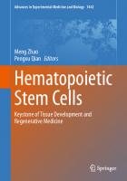
History The artiicial generation of tissues, organs, or even more complex living organisms was throughout the history of mankind a matter of myth and dream. During the last decades this vision became feasible and has been recently introduced in clinical medicine. Tissue engineering and regenerative medicine are terms for the ield in biomedicine that deal with the transformation of these fundamental ideas to practical approaches. Several aspects of generating new tissues and organs out of small pieces of living specimens are now scientiically solved, but at this point it is unknown how much impact these new approaches will have on clinical medicine in the future. In this respect it seems important to recapitulate from where the visions and the work came, in order to speculate or predict where tissue engineering and regenerative medicine will head. The concept of tissue engineering and regenerative medicine as measures to create more complex organisms from simpler pieces is deeply embedded in the people’s imaginary world. A change in the vision, hope, and believe of how to create or regenerate
complex organs or organisms can be observed during history as a mirror of the cultural history of mankind. Even the early history of men is related to the idea that independent life can be created without sexual reproduction. Stories from Greek mythology [the creation of persons without sexual reproduction, e.g., the generation of Prometheus (Fig. 1.1)] may be considered as early reports representing the idea of creating living creatures from living or nonliving specimens. The Biblical tale of Eve created from Adam’s rib is a further and perhaps the most well-known
Fig. 1.1 The generation of Prometheus
6
U. Meyer
Fig. 1.2 Healing of Justinian
Fig. 1.3 Theophrastus von Hohenheinm
example of this concept [1] (in a modern view a kind of hybrid cloning). A multitude of examples in literature and the arts mirrors the desire of humans to be able to create by themselves living individuals or at least parts of individuals. The envisioned measures to create life are inluenced by the social, cultural, and scientiic background of individual persons at that time. The famous painting “Healing of Justinian” (Fig. 1.2) a visualization of the legend of St. Cosmas and St. Damien (278 AD) depicting the transplantation of a homograft limb onto an injured soldier, is one early instance of the vision of regenerative medicine. As humans progressed in the understanding of nature and as they developed more advanced culture techniques they envisioned the generation of living creatures by applying physicochemical or biological techniques. During the transformation from the Middle Ages to the Renaissance in Europe, there was the hope and belief by a number of scientists that through alchemy living organisms could be generated. Theophrastus von Hohenheim, better known as Paracelsus (Fig. 1.3), tried (and failed) to ind a recipe to create
human life by a mixture of chemical substances in a deined environment. Johann Wolfgang von Goethe (1749–1832) deals in his fundamental work of literature Faust [2] with the relation of an individual (Faust) to knowledge, power, morality, and theology. One central theme in the struggle of Faust to be powerful is the deeply embedded wish to create life. The creation of the artiicial being Homunculus in Goethe’s Faust is a central part of the drama, by which Goethe reveals various transformational processes working in the human soul. In the famous laboratory scene of Faust (Part II) he describes the vision of men being able to create life by alchemy (Fig. 1.4), representing the irrepressible human dream of “engineering” life: Look there’s a gleam! – Now hope may be fulilled, That hundreds of ingredients, mixed, distilled – And mixing is the secret – give us power The stuff of human nature to compound If in a limbeck we now seal it round And cohobate with inal care profound, The inished work may crown this silent hour
Chapter 1 The History of Tissue Engineering and Regenerative Medicine in Perspective
7
many contemporary “Faustian” technologies, such as cloning, genetic, or stem cell techniques in modern tissue engineering and regenerative medicine. With respect to an historical view of tissue engineering, Faust is a representative of Northern European humanity striving for evolution from the scientiic and ethical limitations and strictures of the 16th century Reformations to the new aspirations of humanity that Goethe saw developing during the 18th century Enlightenment era. He was attracted to the idea of creating life by adding substances to nonliving specimens, similar to visions of how God created Adam, visualized by the famous painting of Michelangelo (Fig. 1.5). Goethe struggles to weave the personal inner journey of Faust towards some enlightenment (described in the prologue):
Fig. 1.4 Depiction of Dr. Faustus and his Homunculus
It works! The substance stirs, is turning clearer! The truth of my conviction passes nearer The thing in Nature as high mystery prized, This has our science probed beyond a doubt What Nature by slow process organized, That have we grasped, and crystallized it out. The description of the creation of Homunculus is also of special concern today, since it is suggestive of
I’ve studied now Philosophy, And Jurisprudence, Medicine, And even alas! Theology All through and through with ardour keen! Here now I stand, poor fool, and see I’m just as wise as formerly. Am called a Master, even Doctor too, And now I’ve nearly ten years through Pulled my students by their noses to and fro And up and down, across, about, And see there’s nothing we can know! thereby being in the context of the collective social forces that are undergoing transformation through the historical processes of that time. As Faust deals with nearly all aspects and questions that arise in tissue engineering and regenerative medicine (and that are discussed in the irst chapter of this book), it can
Fig. 1.5 Michelangelo’s painting The Creation of Adam
8
be considered to be a timeless and always relevant consideration on the ield of biomedicine. Later on, as science and medicine progressed, a multitude of stories, reports, paintings, and ilms dealt with the idea that humans could create life by modern “scientiic” measures. A prominent newer example in literature and ilm is the story of Frankenstein, written by Mary Shelley in 1818 (Fig. 1.6), describing the vitalization of a creature, reassembled from different body parts. Parallel to the mythological, biblical, and ictional reports, various persons performed pioneering practical work to generate, heal, or regenerate body parts. The emergence of tissue engineering is, through their work, closely connected with the development of clinical medicine (prosthetics, reconstructive surgery, transplantation medicine, microsurgery) and biology (cell biology, biochemistry, molecular biology, genetics). The mechanical substitution of body parts by nonvital prosthetic devices (metallic and ivory dentures, wooden legs) can be considered as early efforts to use biomaterials in reconstructive medicine. The irst
Fig. 1.6 Book cover of Frankenstein (Edition 1831)
U. Meyer
attempts to replace teeth in the sense of modern dental implantology seems to go back as early as in the Galileo-Roman period. The anthroposophic inding of a human skull, containing a metallic implant in the jaw [3], is indicative of early attempts of humans to regain lost function by tissue substitution. Leading areas of reconstructive medicine in clinical use were evident in the age before modern dentistry and orthopedics. Ambroise Pare` (1510–1590) described in his work Dix livres de la chirurgie [4] measures to reconstruct teeth, noses, and other parts of the body. A common method in the 18th century to replace teeth was the homologous transplantation of teeth in humans. John Hunter (1728–1793) investigated in his pioneering work the effect of transplantation not only at a clinical level (he claimed, that homologous transplanted teeth lasted for years in the host) but also performed animal experimental work on the fate of transplants, thereby setting the basis for a scientiic approach on transplantation medicine [5]. A milestone in the modern view of tissue engineering was the use of skin grafts. The use of skin grafts is closely related to the work of the famous surgeon Johann Friedrich Dieffenbach (1792–1847). As he performed animal experimental and clinical work on skin transplantation (described in Nonnulla de Regeneratione et Transplantatione [6]), and as he also established ways to use pedicled skin laps (since most of the clinical skin transplantation treatments failed), Dieffenbach is one of the modern founders of plastic and reconstructive surgery and can also be considered to be an early practitioner in transplantation medicine. Breakthroughs in the clinical use of skin grafts were made by Heinrich Christian Bünger, irst successful autologous skin transplantation [7]; Jaques Reverdin (1842–1929), use of small graft islets; and Karl Thiersch (1827–1895), split thickness grafts [8, 9]. The high number of failures were overcome by the observation of Esser (1877–1964) that immobilization of transplants through the use of dental impression materials improves the fate of transplants in facial wound reconstruction. The clinical efforts reached through the combined use of surgical and dental techniques in reconstructive surgery and transplantation medicine led to the evolution of the dental- and medical-based Maxillofacial and Plastic Facial Surgery discipline. The foundation and establishment of this new specialty at the Westdeutsche Kieferklinik in Düsseldorf and the extensive experience in this center with injured soldiers during the
Chapter 1 The History of Tissue Engineering and Regenerative Medicine in Perspective
First and Second World War led to signiicant improvements in tissue regeneration and reconstruction in the plastic and reconstructive surgery ield. The underlying biological reason for the success of clinical skin transplantation by reining the transplantation approach (the shift from enlarged grafts to small cellcontaining particles, the invention of ixation protocols) was in the beginning to a great extent unknown. Enlightenment into the biological mechanisms that accounted for the fate of transplants was provided by the fundamental biological work of Rudolf Virchow (1821–1902). He described in his Cellularpathologie [10] that tissue regeneration is dependent on cell proliferation. His work led not only to the investigation of tissue healing through cellular effects, but also to the cultivation of cells outside the body (in vitro, irst suggested by Leo Loeb [11]). C.A. Ljunggren and J. Jolly were the irst researchers to attempt to cultivate cells outside the body [12]. The milestone breakthrough in in vitro cell cultivation was reached by R.G. Harrison (1870–1959), demonstrating active growth of cells in culture [13]. Since that time, cell biology and especially in vitro cell culture became the mainstay of what can be considered classical tissue engineering [14]. Underlying in vitro cell culture with subsequent cell transplantation, modern tissue engineering and regenerative medicine is directly connected to microsurgery. Alexis Carrel (1873–1944) can be considered the founder of modern organ transplantation due to his work elaborating the methods of vascular anastomosis [15, 16]. The use of microvascular surgery was primarily performed in organ transplantation and plastic surgery. Whole organ transplantation or transplantation of body parts were made possible by this technique. E. Ullman (irst kidney transplantation in animals [17]) and J.P. Merrill (irst successful clinical kidney transplantation in identical twins) are directly related to the advances in transplantation medicine [18]. One of the most well-known milestones in organ transplantation medicine was the irst heart transplantation 1967 by the South African surgeon Christiaan Barnard. His life—saving transplantation not only had extensive coverage in the newspapers at that time, but also raised an intensive and controversial debate on ethical issues in transplantation medicine [19]. Whereas the indications for microvascular tissue transplantation were extended towards plastic and reconstructive surgery (use of free myocutaneous, osteomyocutaneus, and other vessel containing
9
laps), and measures to perform microsurgery were reined and standardized, failures in clinical transplantation medicine were mainly based on immune incompatibilities (except for the use of autografts). The success of transplantation medicine, whether cells, tissues, or organs are in use was, and still is, to a great extent dependant on the immune state of graft and host. The science of immunomodulation and immunosupression is therefore still a critical aspect in all tissue engineering and regenerative medicine applications. The term “tissue engineering” was up to the mid 1980s loosely applied in the literature in cases of surgical manipulation of tissues and organs or in a broader sense when prosthetic devices or biomaterials were used [20]. The term “tissue engineering” as it is nowadays used was introduced in medicine in 1987. The deinition that was agreed on was: “Tissue Engineering is the application of the principles and methods of engineering and life sciences toward the fundamental understanding of structure-function relationships in normal and pathologic mammalian tissue and the development of biological substitutes to restore, maintain, or improve function.” The early years of tissue engineering were based on cell and tissue culture approaches. W.T. Green undertook a number of experiments in the early 1970s to generate cartilage using a chondrocyte culture technique in combination with a “bone scaffold.” Despite of his inability to generate new cartilage, he set the theoretical and practical concept to connect and coax cells with scaffolds. Innovations in this approach were made by Burke and Yannas through a laboratory and clinical collaboration between Massachusetts General Hospital and M.I.T. in Boston, aimed at generating skin by a culture of dermal ibroblasts or keratinocytes on protein scaffolds, and using it for the regeneration of burn wounds. A key point in tissue engineering was given by the close cooperation between Dr. Joseph Vacanti from Boston Children’s Hospital and Dr. Robert Langer from M.I.T. Their article in Science [21], describing the new technology, may be referenced as the beginning of this new biomedical discipline. Later on, a high number of centers all over the world focused their research efforts towards this ield. Tissue engineering was catapulted to the forefront of the public awareness with a BBC broadcast exploring the potential of tissue-engineered cartilage which included images of the now infamous “mouse with the human ear” on its back from the
10
laboratory of Dr. Charles Vacanti at the University of Massachusetts Medical Center. The visual power of the photograph of the “auriculosaurus” helped to transfer the idea and vision of generating new tissues or organs from the imaginary world of human beings to the real world. Since that time, tissue engineering has been considered one of the most promising biomedical technologies of the century [22]. Regenerative medicine seems to be more dificult to deine, as this term was used earlier but was less deined in the literature then the term “tissue engineering.” It is now viewed by most biologists and physicists as a ield where stem cells drive embryonic formation, or where inductive organizers induce a blastema to regenerate a tissue, aimed at reforming damaged tissues and organs in humans. It seems that a rigid deinition of regenerative medicine is not constructive while the principle approaches that deine the ield are still being delineated. Stem cells, being at the center of expectations, hold great promise for the future of regenerative medicine. Stem cell plasticity and cloning, with nuclear transfer, transdifferentiation, and cell fusion as measures to modulate the stem cell differentiation pathway, is now a central issue in regenerative medicine [23]. During cloning, an adult nucleus is transplanted into an egg, which must erase the adult genome’s epigenetic marks, so it can re-express every gene necessary to build a new animal. Robert Briggs and Thomas King were the irst to demonstrate (based on a similar experiment proposed by Hans Spemann at the University of Freiburg as early as in 1938) how to clone frogs by replacing the nuclei of eggs with cells from tadpoles and adult intestinal epithelium. The further major step in cloning research was the cloning of two lambs (Megan and Morag) from embryonic cells in 1996. Cloning of the sheep Dolly [24] was, from a public awareness point of view, the key event in stem cell research. Ian Wilmut at the Roslin Institute near Edinburgh and his colleagues at PPL Therapeutics in East Lohian reported on February 27th, 1997 in Nature that they had produced a lamb named Dolly (Fig. 1.7), born the previous July, that was the irst mammalian clone created using the genetic material from an adult cell. As soon as the story hit the front pages (the news was broke by the British Sunday newspaper The Observer four days ahead of Nature’s publication) a public and media maelstrom ensued. Editors and writers of other newspaper went so far as to speculate
U. Meyer
Fig. 1.7 Sheep Dolly and her irst-born lamb Bonnie
that “It is the prospect of cloning people, creating armies of dictators, that will attract most attention.” The creation of Dolly, cloned from an adult udder cell, overturned the idea that in mammals, developed cells could not reverse their fate. The news had a tremendous impact on society, mirrored by the fact that within days the President of the United States, the head of the European Commission, the Vatican, and many others were calling for a review of regulations on cloning research, if not an outright ban. It became obvious that the world was simply not prepared for the debate. The use of modern techniques like cloning or stem cell modulation was considered by a large part of the public to be a modern version of alchemy [25]. Japanese researchers reported in 1998 on the successful birth of eight cloned calves using adult cells from slaughterhouse entrails, raising the possibility that animals could be cloned for the quality of their meat (a situation close to the vision of the creation of Eve). During the last decade, scientists have shown that they are able to clone a variety of mammals including mice, rats, calves, cows, pigs, cats, and dogs. Some envisaged an industry of cloning applications, from the production of medicines in live bioreactors—cloned, genetically modiied livestock—to the creation of herds of cloned animals that might one day be used as organ donors. But the low
Chapter 1 The History of Tissue Engineering and Regenerative Medicine in Perspective
eficiency of the cloning process, relecting the problems related to reprogramming a cell’s DNA by the content of eggs, has stymied industrial development. New directions focus on the addition of chemicals or proteins to adult cell nuclei, in order to bypass the need for eggs altogether. Egg-free approaches may also enable what many see as the most promising potential application of cloning: the creation of human embryonic stem cells, or cells made from them, that could be used to treat human disease [26]. Ten years later, the ethical debate launched by Dolly and encouraged by science iction stories has changed. After a decade, mammalian cloning is moving forward with central societal issues remaining unresolved. The recent situation has now been supplanted by a bioethical discussion that is more complex and more focused on the real situation in stem cell biology. One outcome of this discussion is the risk assessment, currently open for public consultation, by the US Food and Drug Administration (see: www.fda.gov/cvm/Clone RiskAssessment.htm). Researchers and physicists speculate that unless there is some unknown fundamental biological obstacle, and given wholly positive motivations, human reproductive cloning is an eventual certainty [27].
1.2
Future Prospects The future of tissue engineering and regenerative medicine holds the promise of custom-made medical solutions for injured or diseased patients, with genetic (re-)engineering in the zygote or even earlier as one of the most promising and also most debatable aspects of this ield. As the ield of tissue engineering and regenerative medicine is established as a central discipline in biomedicine nowadays, it seems interesting to speculate where the ield will head. As both areas have matured to the point that its research and clinical aspects can be conceptually categorized, various commercial or noncommercial organizations have tried to assess the future of the ield. These assessments seem to be important for researchers, policy makers, regulators, funders, technology developers, and the biomedical industry. Among the different assessments, published in open-access or
11
limited access documents (nearly all of them thinking along similar lines), a study performed by the Tissue Engineering Journal can be considered to be the most scientiic approach [28]. The Journal undertook such an assessment through the evaluation of a questionnaire (using a modiied Hoshin process) given to the editorial board of Tissue Engineering. The aim of the study was to identify a list of strategically important concepts to achieve clinically relevant solutions for medical problems (up to the year of 2021). The evaluation of the questionnaire revealed some important aspects for the future of tissue engineering and regenerative medicine. One important inding was that highly strategic issues are not at the forefront of the daily work. The most striking example was angiogenic control. The dominance of this issue over all other issues and its low level of present progress propelled it to the top of strategic concepts. The second most important area was assumed to be stem cell science. As the editors assumed that understanding and control of stem cell research may give way for a short conduit in a number of tissue engineering approaches, molecular biology and system biology research seemed to be similarly important in the strategic development of the ield. In addition to angiogenic control and stem cell biology, cell sourcing, cell/tissue interaction, immunologic understanding and control, manufacturing and scale up, as well some other issues were considered to be important (in a decreasing manner). With respect to the near and immediate future, the dominant concept that was supported by the largest number of specialists was clinical understanding and interaction. It seemed important that, as the ield is oriented towards clinical application, close collaboration between researchers and clinically working physicians is of utmost importance. A close cooperation, based ideally on a deep understanding of the “other’s” ield, is not only valuable for the establishment of engineered tissue design criteria but also to enhance the potential for the inal introduction of such therapies into clinical practice at large. The present book, intended to give researchers and clinicians a common platform, is hopefully one tool to reach this important aim. The future will show when and how which of the multiple approaches in tissue engineering and regenerative medicine will withstand the proof of clinical usage of such therapies over time.
12
References 1.
Fox E (1983) The Schoken Bible, vol 1. Schoken, New York 2. Goethe JW (1831/1959) Faust (part two) Act I: laboratory. Penguin, Harmondsworth 3. Crubézy E, Murail P, Girard L, Bernadou JP (1998) False teeth of the roman world. Nature 391(6662):29 4. Pare A (1634) The works of that famous Chirurgion Ambrose Parey. Cotes and Young, London 5. Hunter J (1771) The natural history of the human teeth. Johnson, London 6. Dieffenbach JF (1822) Nonnulla de regeneratione et transplantatione. Richter, Würzburg 7. Bünger HC (1823) Gelungener Versuch einer Nasenbildung aus einem völligen getrennten Hautstück aus dem Beine. J Chir Augenhkd 4:569 8. Thiersch K (1874) Ueber die feineren anatomischen Veränderungen beim Aufheilen von Haut auf Granulationen. Arch Klin Chir 37:318–324 9. Mangoldt F (1895) Die Ueberhäutung von Wundlächen und Wundhöhlen durch Epithelaussaat, eine neue Methode der Transplantation. Dtsch Med Wochenschr 21:798–799 10. Virchow R (1858) Die Cellularpathologie in ihrer Begründung auf physiologische und pathologische Gewebelehre. Hirschwald, Berlin 11. Loeb L (1897) Ueber die Entstehung von Bindegewebe, Leukocyten und roten Blutkörperchen aus Epithel und über eine Methode, isolierte Gewebsteile zu züchten. Stern, Chicago 12. Ljunggren CA (1898) Von der Fähigkeit des Hautepithels, ausserhalb des Organismus sein Leben zu behalten, mit Berücksichtigung der Transplantation. Dtsch Z Chir 47:608–615
U. Meyer 13. Harrison RG (1910) The outgrowth of the nerve iber as a mode of protoplasmic extension. J Exp Zool 9:787–846 14. Fell HB (1972) Tissue culture and it’s contribution to biology and medicine. J Exp Biol 57:1–13 15. Carrel A, Burrows MT (1911) Cultivation of tissues in vitro and its technique. J Exp Med 13:387–398 16. Witkowski JA (1979) Alexis Carrel and the mysticism of tissue culture. J Med Hist 23:279–296 17. Ullmann E (1902) Experimentelle Nierentransplantation. Wien Klin Wochenschr 15:281–282 18. Merrill JP, Murray JE, Harrison JH (1956) Successful homotransplantation of human kidney between identical twins. J Am Med Assoc 160:277–282 19. Barnard C, Pepper CB (1969) Christiaan Barnard. One life. Timmins, Cape Town 20. Skalak R, Fox CF (1988) Tissue engineering. Liss, New York 21. Langer R, Vacanti JP (1993) Tissue Engineering. Science 260:920–926 22. Nerem RM (1991) Cellular engineering. Ann Biomed Eng 19:529–545 23. Wadman M (2007) Dolly: a decade on. Nature 445:800–801 24. Wilmut I, Schnieke AE, McWhir J, Kind AJ, Campbell KHS (1997) Viable offspring derived from fetal and adult mammalian cells. Nature 385:810–813 25. Rutenberg MS, Hamazaki T, Singh AM, Terada N (2004) Stem cell plasticity: beyond alchemy. Int J Hematol 79:15–21 26. Theise D, Wilmut I (2003) Cell plasticity: lexible arrangements. Nature 425:21 27. Nature editorial (2007) Dolly’s legacy. Nature 445:795 28. Johnson PC, Mikos AG, Fisher JP, Jansen JA (2007) Strategic directions in tissue engineering. Tissue Eng 13:2827–2837
Economic Modeling and Decision Making in the Development and Clinical Application of Tissue Engineering
2
P. Vavken, R. Dorotka, M. Gruber
Contents 2.1
Evaluating Tissue Engineering Between Public Health and Microeconomics . . . . . . . 13
2.2
Evaluation of a Treatment . . . . . . . . . . . . . . . 13
2.3
Allocation of Tissue Engineering as a Health Care Resource . . . . . . . . . . . . . . . . . . . . . . . . 14
2.4
Economic Modeling of Cost-effectiveness .
2.5
Decision Analysis Models . . . . . . . . . . . . . . . 16
2.6
Cartilage Tissue Engineering . . . . . . . . . . . . 16
2.7
Genitourinary Tissue Engineering . . . . . . . . . 17
2.8
Conclusion . . . . . . . . . . . . . . . . . . . . . . . . . . . 20
15
References . . . . . . . . . . . . . . . . . . . . . . . . . . . 21
2.1
Evaluating Tissue Engineering Between Public Health and Microeconomics Tissue engineering in regenerative medicine has become one of the most promising subjects in current medical research. A number of procedures based on or involving tissue engineering have already been approved for human application and are clinically available, and numerous others are on the edge of becoming so. During the development of a new procedure and the process of its approval, repeated evaluations, focusing on different aspects, are necessary. Initial studies focus on the effect of
a treatment; subsequently its eficacy and effectiveness as well as safety proiles are established. Before a procedure will be directed towards clinical application, economic evaluations are done assessing whether further investment of scarce resources is justiied. In public health such justiication is consistent with showing a highly effective treatment and ruling out that the same investment could result in more productive research or a more effective treatment elsewhere, while from a microeconomic perspective potential revenue is addressed. Such argumentation is a source of much debate, but can hardly be avoided given a true scarcity of resources and increasing costs of health care. Due to the increasing importance of such issues, especially in a high end, multidisciplinary ield such as tissue engineering, knowledge on the principles of economic evaluation has become a valuable addition to a researcher’s expertise. It is the objective of this chapter to give a concise introduction to economic modeling and resulting decision-making strategies in health care in the evaluation of tissue engineering.
2.2
Evaluation of a Treatment The evaluation of a new intervention comprises the assessment of four distinct parameters: Eficacy, Effectiveness, Eficiency, and Availability and Allocation. Eficacy describes the maximal effect under
14
P. Vavken, R. Dorotka, M. Gruber
optimized conditions, for example, in an in vitro study or a meticulously monitored clinical trial using a per-protocol analysis. Per-protocol means that patients are analyzed depending on the treatment they eventually received and not per initial allocation. Effectiveness describes the effect of an intervention in a “real life” situation including noncompliance, losses to follow-up, wrong dosages, et cetera. Effectiveness can be assessed in clinical trials using intentionto-treat, i.e., data analysis retains all patients in the group of their initial allocation, regardless of crossing-over between study groups, losses to follow-up, and the like. While eficacy is of meaning for treatment development, effectiveness has the higher impact on clinical life and is considered more relevant for the purpose of subsequent economic evaluation.
Effect controlled study environment
Efficacy Real life circumstances
Effectiveness Costs (financial, personal) Utility (QALY/DALY)
Efficiency
DECISION ANALYSIS
Fig. 2.1 Framework of the evaluation for a treatment. The general effect of a procedure is known and put into action in a speciic stetting to establish the eficacy of this procedure for a given question. Taking “real life” circumstances, such as patient compliance, wrong dosage, losses to follow-up and so forth, into account, the effectiveness can be estimated. This value, in turn, is translated into utilities and/or put into relation with costs to establish the eficiency. All the latter parameters are inally used in decision analysis
In parallel, effectiveness is an important parameter in decision-making processes. Figure 2.1 shows the framework of evaluation and decision making for a treatment.
2.3
Allocation of Tissue Engineering as a Health Care Resource As a resource in health care, tissue engineering should be evaluated in the light of four parameters: eficiency, equity, need, and appropriateness to guarantee balanced service provisions. Eficiency describes the effect of a procedure in relation to its costs. Eficiency will be described in detail later in this chapter. Equity stands for a fair distribution of allocations and can be divided into horizontal equity, i.e., treating the same those who have the same need, and vertical equity, i.e., treating differently those who have different levels of need, or in other words to prioritize urgent cases over nonurgent cases. Deining equity, in turn, begs the question of need. Need summarizes three subgroups, corporate need, the need for health, and the need for health care. While corporate need is determined in adjustment to pressure from interest groups affected, the need for health is a more universal parameter. It is established on population data and mirrors the need for health as a function of the prevalence of disease. The need for health care, however, exists when a patient is suffering from a disease that can be successfully addressed by an existing treatment. As such, tissue engineering applications may increase the need for health care by introducing new treatment options to existing diseases. Need assessment and market analysis for supply and demand are not necessarily the same. While their descriptions sometimes sound rather similar, need assessment is a public health instrument aiming towards equitable and effective use of (inancial) resources, while a market analysis is the microeconomic way of estimating the potential of value generation, and does not always produce the same result. Appropriateness inally means that a patient receives the treatment he or she needs. As for any new therapy with not yet fully understood outcomes the appropriateness
Chapter 2 Economic Modeling in Tissue Engineering
of various tissue engineering treatments has been a matter of debate.
2.4
Economic Modeling of Cost-efectiveness Economic models are needed to evaluate the relation between costs and effectiveness, usually in so-called cost-effectiveness analyses (CEA), which is both a generic term and the name of one speciic method. There is a considerable variety of validated models for different purposes such as cost estimation and the characterization of cost-outcome relationships, which are clearly beyond the scope and intention of this book [8]. We want to focus on cost-effectiveness and cost-utility analyses for the purpose of this chapter. However, before any analysis can be done, the relevant parameters need to be identiied. The determination of the costs of a tissue engineering application is fairly dificult. Generally, total cost can be split into direct healthcare costs (e.g., procedure, hospital stay, medication, personnel, supplies), indirect healthcare costs (transportation from and to the hospital/ofice, parking, childcare while admitted to the hospital), and indirect costs such as productivity cost due to lost economic productivity because of impaired ability to work. Obviously cost estimates can differ considerably, depending on perspective and treatment at hand. Translating the cost of an intervention into current currency will produce different results for different markets, depending on the value and price of goods and work force. Finally, time has to be included as a factor. It is a commonly accepted fact that one unit of currency, e.g., one Euro, will lose in value over time. Hence a discount of costs over time, usually 3–5%, is used. This discount, however, does not account for inlation! Given the considerable paucity of data on clinical tissue engineering applications as far as inancial burdens are concerned, most of these parameters have to be estimated. An important issue in such predictions is, again, need, since an urgently needed, and thus probably frequently used procedure or treatment will beneit from the economy of scale.
15
Effectiveness has already been mentioned above. Briely, effectiveness describes the effect of a treatment in a clinical setting. To identify this parameter, suficiently sized trials of lawless design and— equally important—studying clearly deined questions are required. It is important to scrutinize published estimations of effectiveness for their validity. Does the evidence at hand really focus on the relevant questions and are appropriate measures used? The brunt of tissue engineering applications proposes beneicial long-term effects, suggesting superiority over current treatment options, but have patients been suficiently followed to prove this? In summary, effectiveness, although often considered a straight-forward and very objective measure compared to costs or utilities, offers ample opportunities for misconceptions and has to be assessed with much caution. Utility tries to describe the value of an intervention to a person. While for example a 34% reduction of mortality is an excellent result for an intervention, this has little meaning to the individual patient. One of the most important aspects of utilities as a healthcare outcome is quality of life. Quality of life can be measured using validated scores, and utilities account for this factor by describing quality-adjusted life years (QALY) or disability-adjusted life years (DALY). Another approach to utilities that is used in decision analysis is directly assessing the value of an outcome by interrogating patients. Briely, a patient is offered two options, e.g., suffering from disease or the status post a procedure, which are usually represented as doors. Each door has a distinct likelihood of achieving an outcome and the patient is asked to choose accordingly. Likelihoods are altered until the patient changes his mind, and this value is then used as the utility of the outcome. This is referred to as a reference lottery and, needless to say, it is very subjective, depending on personal, as well as societal preferences [8, 22]. It is a common option to do a reference lottery with medical specialists, especially for interventions with long-term effects or arcane complication potential, to account for bias arising from patient ignorance. Finally it is important to consider that utilities just like costs are a time-dependent variable and decline over time, not only because patients prefer achieving a speciic state of health rather now than in one year, but also because nonrelated health
16
events might interfere with or even stop this process. The worst-case scenario would be a patient dying from another disease or in an accident before eventually receiving a highly effective treatment. Hence a discount of utilities has to be included in all estimates. Once the parameters’ cost, effectiveness, and utility have been established, analyses are done by calculating the amount of money spent per unit of effectiveness or utility, respectively. Two important parameters can be deducted from these analyses. One is the incremental cost-effectiveness ratio (ICER) deined as the ratio of change in costs of a treatment to the change in effect of the treatment, or in simpler terms the additional effect of a treatment bought for an extra Euro/Dollar/Pound. Previous studies have estimated the upper border of ICER a society is willing to pay at €45,000 ($60,000 or £30,000) [8]. The other parameter is headroom, or the amount of extra cost for which a treatment is still cost-effective given its effectiveness. Considering these analyses it is important to note that these processes are not only dependent on the market. Regulatory issues differ signiicantly on both international and national levels and can affect costs heavily.
2.5
Decision Analysis Models Information acquired in these studies can and should be used together with decision analysis. The classic model of decision analysis is the decision tree, representing a unidirectional decision support structure leading from a question at the left hand side to branches of possible outcomes on the right hand side. On the very end, the utilities of the individual outcomes are stated, so that the utility of every event and decision on the tree can be calculated by going backwards and multiplying utilities with probabilities. To account for the variability of the included parameters sensitivity analyses must be done. During such an analysis, the values of one or more variables are altered, and change in the inal outcome is monitored. The major limitation of the decision tree, however, is its unidirectional nature. In various healthcare situations patients will rather move back
P. Vavken, R. Dorotka, M. Gruber
and forth between states than travel down a line of distinct events. Such behavior is modeled in Markov Models representing cycles of events during which a patient may move to another health state, regress into an earlier situation, or remain in his current position. Sensitivity analyses for Markov models are done using Monte Carlo simulation, a method that repeatedly changes all variables at a time using random values form a predeined range to construct 95% conidence intervals for the outcome.
2.6
Cartilage Tissue Engineering Osteoarthritis is a major burden in the population of developed countries, causing pain and suffering on the individual level and loss of productivity and extensive healthcare costs on the societal level. Total joint replacement is an outstandingly successful treatment for osteoarthritis of most joints, but is associated with complications such as component wear and loosening, especially in long use. Hence its use is considerably limited in younger and active patients. One of the earliest tissue engineering applications in clinical use was autologous chondrocyte implantation (ACI) in the management of cartilage defects, and a considerable amount of data is available to evaluate this method [18]. Brittberg published his groundbreaking paper in the New England Journal of Medicine in 1994 [5]. Originally, the method is described as harvesting chondrocytes from nonweight bearing areas of the cartilage, culturing these cells in vitro, and implanting them into a bioactive chamber created by covering the debrided cartilage defect with a periosteum lap. This original method has been further developed and currently biomaterials are in broad use in Europe, and awaiting FDA approval for use in the USA. Tissue engineering in cartilage repair is thus a good example of evaluating a clinically employed treatment. Evaluating ACI as a healthcare resource, need assessment is fairly straightforward. Considering the high incidences of cartilage damage in our highly active present-day population, the need for health is obvious. Considering the painful and chronic nature of osteoarthritis and the resulting impairment and
Chapter 2 Economic Modeling in Tissue Engineering
disability, the need for health care on the individual as well as societal level is imminent. Appropriateness of ACI should be determined in the face of its alternatives. Briely, ACI should have a convincing immediate effect on pain and joint function, but this effect should also be long lasting. A number of trials and reviews studied the effect of ACI compared to alternative treatment options. Data on eficacy and effectiveness are available from seven randomized controlled studies with a maximum follow-up of 5 years presenting clinical as well as histological indings [3, 4, 7, 12–14, 20, 21]. Long-term results are available from cohort studies for 9–11 years [18, 19]. Other studies have described costs, quality of life, and cost-utility of ACI. Lindahl, Wildner, and Minas provide costs for ACI in SEK, DM, and USD, respectively [15, 17, 24]. Cost-effectiveness and cost-utility analyses were presented by Clar, Minas, and Vavken [6, 17, 23]. The health technology assessment by Clar, et al. is the most extensive study and used short and long-term models comparing ACI with microfracture and mosaicplasty using QALY and time to total knee replacement as outcome [6]. For their short-term model they found that ACI would have to be 70–100% more effective than microfracture to be cost-effective. Over 10 years, however, only 10–20% would be required. Clar performed extensive sensitivity analyses to test the robustness of their result and found them to be dependent on quality of life assessments only. From a decision analysis perspective these data are challenging. The considerable heterogeneity between individual studies and the lack of long-term data make predictive modeling dificult. A representative decision model was generated from the existing data. Briely, data produced by Clar, Knutsen, Wood, and Saris were used to estimate outcome probabilities [6, 14, 20, 25]. Utilities were estimated by surgeons focusing on cartilage repair and based on feed-back from patients. Surgeons were primarily targeted in this reference lottery, because from our experience, there is insuficient knowledge on ACI among patients leading to an inability to grasp the meaning of the potential complications, and utility estimates are deviated by prejudice and spurious assumptions. The decision tree for this model is presented in Fig. 2.2. To test the robustness of these estimates, one-way sensitivity analyses of the effect of complication rates, the severity of complications,
17
and the loss of utility due to complications can be done (Fig. 2.3a–c). This model shows a higher utility for ACI compared to microfracture. Sensitivity analyses support this inding over a wide area of variance for included parameters. It should be noted that this model is an example of decision making in tissue engineering, and while it uses real evidence it is still incomplete and estimates might be subject to considerable change after addition of further parameters. For example, the model could be expanded taking revisions and second-line operations due to development of OA into account.
2.7
Genitourinary Tissue Engineering Tissue engineering of urogenital tissue has been proposed by Atala as early as 1993, but has not yet drawn much attention [1, 2]. A number of conditions requiring urethroplasty or cystoplasty may proit from tissue engineering approaches. The need for cystoplasty as determined by a literature review is based prevailingly on cases of bladder carcinoma with approximately 10,000 to 50,000 case per year in the USA and UK [9, 10]. The current gold standard is radical cystectomy, followed by ileocystoplasty. Other indications of cystoplasty such as detrusor instability or postinlammatory scarring are unlikely to be promising niches for tissue engineering applications. Urethroplasty currently is performed in cases of strictures using buccal mucosa. In summary, urogenital tissue engineering is a good example for evaluating a tissue engineering application on the edge of clinical implementation. The recent study by McAteer reviewed costs and utility of urogenital tissue engineering using headroom analyses [16]. The main problem for this study was to deine cost and utilities since there is almost no pre-existing literature. McAteer showed a minimal inancial headroom for engineering urethral tissue of £186 (€275, $375) based on a difference in QALY of 0.0062. For an engineered bladder, in turn, this paper found a headroom of £15,000 (€22,000, $30,000), mostly because of a rather high increase of quality of life with a difference in QALY of 0.5.
18
P. Vavken, R. Dorotka, M. Gruber Decision tree for autologous chondrocyte implantation (ACI) verus microfracture (MFX) in cartilage repair Procedure
Utility Postoperative
pa
perioda
Utility Long term clinical outcome / histology Good / hyaline cartilage
uneventful
ACI
0.70
complications
95%
4%
1%
Utility
50%
1
40%
0.5
Failure / fibrocartilage
10%
0.1
Good / hyaline cartilage
50%
0.9
40%
0.4
10%
0
0.71 Good / fibrocartilage
0.61 Good / fibrocartilage Failure / fibrocartilage
primary failure
pb
0
0
Good / hyaline cartilage
20%
1
60%
0.5
Failure / fibrocartilage
20%
0.1
Good / hyaline cartilage
20%
0.9
60%
0.4
20%
0
Cartilage damage uneventful
MFX
0.51
complications
95%
4%
0.52 Good / fibrocartilage
0.42 Good / fibrocartilage Failure / fibrocartilage
primary failure
1%
0
0
a Rates of complications from Wood et al. JBJS 2006; 88:503-507 b Probabiliites obtained from Clar et al. Health Technol Assess 2005;9(47)
Fig. 2.2 Possible decision tree for using autologous chondrocyte implantation (ACI) instead of microfracture (MFX). At the beginning there is the problem of “cartilage damage” leading to a decision node (represented as a square). This decision leads to chance nodes (circles) of possible outcomes, given
with the probabilities p. The blue igures show the utilities of an outcome. The initial utilities on the right side were obtained from a reference lottery among orthopedic surgeons specializing in cartilage repair; the other utilities were calculated by multiplication with the corresponding probabilities
Chapter 2 Economic Modeling in Tissue Engineering
19
Sensitivity analysis of the effect of complication rates on utilities ACI
MFX
1.00 0.90 0.80
Utility
0.70 0.60 0.50 0.40 0.30 0.20 0.10 0.00 0%
1%
1%
2%
3%
4%
a
5%
6%
7%
8%
9%
10% 15% 20% 25%
complication rate
Sensitivity analysis of the effect of the severity of complications ACI
MFX
0.80 0.70 0.60
Utility
0.50 0.40 0.30 0.20 0.10 0.00 0.01
b
0.05
0.1
0.2
0.3
0.4
0.5
severity of complications
Fig. 2.3a–c The results from one-way sensitivity analyses testing the robustness of the indings from the decision tree. a and b show that autologous chondrocyte implantation (ACI) retains higher utilities regardless of complication rate and se-
verity. A two-way sensitivity analysis including rates and severity into one model supports this inding (not shown). c see next page
20
P. Vavken, R. Dorotka, M. Gruber Sensitivity analysis of the effect of loss of utility due to a fibrocartilaginous scar ACI
MFX
1.00 0.90 0.80 0.70
Utility
0.60 0.50 0.40 0.30 0.20 0.10 0.00 0.1
c
0.2
0.3
0.4
0.5
0.6
0.7
0.8
loss of utility due to complications
Fig. 2.3a–c (continued) c tests the assumption of lost utility due to ibrocartilage production instead of hyaline cartilage. This is probably the most important point in the sensitivity analyses since ibrocartilage formation has been associated
with subsequent OA development. Given the lesser rates of such scarring in ACI, there is an obvious beneit for ACI that is multiplied by the loss of utility due to ibrocartilage, acting as a surrogate for OA in this case
This high difference derives from a rather long-lasting utility of 10 years and the fairly high number of comorbidities associated with ileocystoplasty. In this case, decision analysis is based on inancial headroom. Tissue engineering in urethroplasty seems to be a poor choice, but one must consider that the estimates for headroom are based on a number of assumptions, one among them is a maximum amount of money that will be made available for an additional effect. From this point of view, a beneitcost analysis based on a person’s willingness-to-pay (WTP) could show quite different results [11].
source allocation. Public health uses models of economic and decision analysis to do so. However, it has to be remembered that these mechanisms are based on populations and the good of the many, whereas the interest of the physician during an actual decision focuses on the good of his or her patient at hand. This gradient of interests has to be compensated. Fortunately, health care is an economy of large averages and wide variance, and in considerably wide context high costs in one individual may be compensated by affordable management in others. We wanted to stress the fact that economic modeling, given its necessity, should not be more than one part of a decision. Microeconomic approaches focusing on revenue should not outweigh equity and need, and quality, unlike costs and utility, must not be discounted. Such tendency might not develop from harmful intent, but health care is not an industry plant, and methods developed on well-understood and tightly controlled processes are bound to fail. Just as tissue engineering is an outstanding example of a multidisciplinary approach to a common problem, economic modeling in health care is not. This chapter
2.8
Conclusion Economic evaluation has become an essential part of modern medicine. Given the limited resources and increasing demand of treatments at increasing costs, it is justiied to strive for eficiency and balanced re-
Chapter 2 Economic Modeling in Tissue Engineering
thus aimed at succinctly presenting important principles of common models and processes to researchers and clinicians to stimulate an environment of equally informed discussion on a mutual level. All models are simpliied representations of reality and their predictions and estimates are bound on approximations. The quality of these approximations is crucial, thus extensive considerations of the effect of errors in these assumptions, i.e., sensitivity analyses, are pivotal. On the other hand, assumptions with proven suficient robustness are readily available for model building, whereas the accumulation of real data might be prohibitively expensive inancially or logistically. Using autologous chondrocyte implantation in cartilage repair as an example, we could successfully establish a model that shows a high eficiency of this procedure compared to alternative options. Including rates and severity of complication, a decision analysis showed that this procedure is superior to microfracture based on a beneicial long-term result because of a higher production of hyaline cartilage, and its use in a clinical setting is recommend by these indings. This model, however, is based on a number of assumptions, such as proportion of patients with hyaline repair tissue, and its external validity might very well be affected. However, this is a general problem in all model-based analyses. Tissue engineering in the genitourinary system is a beautiful example for an application on the verge of in vivo testing. Headroom analyses suggest a strong potential for a tissue-engineered bladder, especially because of ample complications in current techniques of ileocystoplasty. Urethroplasty, in turn, failed to achieve such a result. Notably, these results are based on predicted estimates of effectiveness and utility that might change due to future developments. Tissue engineering holds a position at the intersection of high costs for R&D and clinical application on the one hand and highly effective treatments that might satisfy urgent health needs on the other. What stands out from the past is that negligence of sound economic models has led to the bankruptcy of two leading tissue engineering companies, Organogenesis and Advanced Tissue Sciences, both focusing on skin grafts, in 2002. During both the development and clinical use of tissue engineering, meticulous economic modeling considering the speciics of the
21
tissue engineering market—slow product and reimbursement approval, high costs in research and development, and a surprisingly slow market—will be a key issue to successful implementation of a treatment, parallel to its proven effectiveness.
References 1.
2. 3.
4.
5.
6.
7.
8. 9.
10.
11. 12.
13.
Atala A, Freeman MR, Vacanti JP, et al. (1993) Implantation in vivo and retrieval of artiicial structures consisting of rabbit and human urothelium and human bladder muscle. J Urol 150:608–612 Atala A, Bauer SB, Soker S, et al. (2006) Tissue-engineered autologous bladders for patients needing cystoplasty. Lancet 367:1241–1246 Basad E, Stürz H, Steinmeyer J (2004) Die Behandlung chondraler Defekte mit MACI oder microfracture—erste Ergebnisse einer vergleichenden klinischen Studie. Orthop Prax 40:6–10 Bentley G, Biant LC, Carrington RWJ, et al. (2003) A prospective, randomized comparison of autologous chondrocyte implantation versus mosaicplasty for osteochondral defects in the knee. J Bone Joint Surg Br 85B:223–230 Brittberg M (1994) Treatment of deep cartilage defects in the knee with autologous chondrocyte transplantation. N Eng J Med 331:889–895 Clar C, Cummins E, McIntyre L, et al. (2005) Clinical and cost-effectiveness of autologous chondrocyte implantation for cartilage defects in knee joints: a systematic review and economic evaluation. Health Technol Assess 9:1–82 Dozin B, Malpeli M, Cancedda R, et al. (2005) Comparative evaluation of autologous chondrocyte implantation and mosaicplasty: a multicentered randomized clinical trial. Clin J Sport Med 15:220–226 Edejer Tt, Baltussen R, Adam T, et al. (2003) WHO guide to cost-effectiveness analysis. WHO, Geneva Frimberger D, Lin HK, Kropp BP (2006) The use of tissue engineering and stem cells in bladder regeneration. Regen Med 1:425–435 Guan Y, Ou L, Hu G, et al. (2008) Tissue engineering of urethra using human vascular endothelial growth factor gene-modiied bladder urothelial cells. Artif Organs 32:91–99 Hammit JK (2002) QALYs versus WTP. Risk Anal 22:985–1001 Horas U, Pelinkovic D, Herr G, et al. (2003) Autologous chondrocyte implantation and osteochondral cylinder transplantation in cartilage repair of the knee joint: a prospective, comparative trial. J Bone Joint Surg Am 85:185–192 Knutsen G, Engebretsen L, Ludvigsen TC, et al. (2004) Autologous chondrocyte implantation compared with microfracture in the knee. A randomized trial. J Bone Joint Surg Am 86:455–464
22 14. Knutsen G, Drogset JO, Engebretsen L, et al. (2007) A randomized trial comparing autologous chondrocyte implantation with microfracture. Findings at ive years. J Bone Joint Surg Am 89:2105–2112 15. Lindahl A, Brittberg M, Peterson L (2001) Health economics beneits following autologous chondrocyte transplantation for patients with focal chondral lesions of the knee. Knee Surg Sports Traumatol Arthrosc 9:358–363 16. McAteer H, Cosh E, Freeman G, et al. (2007) Cost-effectiveness analysis at the development phase of a potential health technology: examples based on tissue engineering of bladder and urethra. J Tissue Eng Regen Med 1:343–349 17. Minas T (1999) Chondral lesions of the knee: comparisons of treatments and treatment costs. Am J Orthop 28:374 18. Peterson L, Minas T, Brittberg M, et al. (2000) Two- to 9-year outcome after autologous chondrocyte transplantation of the knee. Clin Orthop Relat Res 212–234 19. Peterson L, Minas T, Brittberg M, et al. (2003) Treatment of osteochondritis dissecans of the knee with autologous chondrocyte transplantation: results at two to ten years. J Bone Joint Surg Am 85:17–24
P. Vavken, R. Dorotka, M. Gruber 20. Saris D (2007) Prospective multi-center randomized controlled trial of ChondroCelect (in an autologous chondrocyte transplantation procedure) versus microfracture (as procedure) in the repair of symptomatic cartilaginous defects of the femoral condyles of the knee. J Am Acad Orthop Surg, February 14–18, San Diego 21. Saris DB, Vanlauwe J, Victor J, et al. (2008) Characterized chondrocyte implantation results in better structural repair when treating symptomatic cartilage defects of the knee in a randomized controlled trial versus microfracture. Am J Sports Med 36:235–246 22. Thornton JG, Lilford RJ, Johnson N (1992) Decision analysis in medicine. BMJ 304:1099–1103 23. Vavken P, Gruber M, Dorotka R (2008) Tissue Engineering in orthopedic surgery—clinical effectiveness and cost effectiveness of autologous chondrocyte transplantation. Z Orthop Unfall 146:26–30 24. Wildner M, Shangha O, Behrend C (2000) Wirtschaftlichkeitsuntersuchung zur autologen Chondrozytentransplantation. Arthroskopie 13:123–131 25. Wood JJ, Malek MA, Frassica FJ, et al. (2006) Autologous cultured chondrocytes: adverse events reported to the United States Food and Drug Administration. J Bone Joint Surg Am 88:503–507
Ethical Issues in Tissue Engineering P. Gelhaus
Contents 3.1
Introduction . . . . . . . . . . . . . . . . . . . . . . . . . . 23
3.2
Risk Assessment . . . . . . . . . . . . . . . . . . . . . . 24
3.2.1
Invasiveness . . . . . . . . . . . . . . . . . . . . . . . . . 24
3.2.2
Importance of the Substituted Function . . . . 25
3.2.3
Methodological Risks . . . . . . . . . . . . . . . . . . 25
3.2.4
Alternatives . . . . . . . . . . . . . . . . . . . . . . . . . . 25
3.2.5
Unexpected Risks . . . . . . . . . . . . . . . . . . . . . 26
3.3
Informed Consent . . . . . . . . . . . . . . . . . . . . . 26
3.4
Status of the Embryo . . . . . . . . . . . . . . . . . . . 27
3.5
Gene Therapy . . . . . . . . . . . . . . . . . . . . . . . . 28
3.6
Animal Research . . . . . . . . . . . . . . . . . . . . . . 29
3.7
Tissue and Organ Transplantation . . . . . . . . . 30
3.7.1
Organ Supply . . . . . . . . . . . . . . . . . . . . . . . . . 30
3.7.2
Further Social Implications . . . . . . . . . . . . . . 31
3.7.3
Histocompatibility . . . . . . . . . . . . . . . . . . . . . 31
3.7.4
Remaining Problems . . . . . . . . . . . . . . . . . . . 32
3.8
Enhancement . . . . . . . . . . . . . . . . . . . . . . . . . 32
3.9
Conclusion . . . . . . . . . . . . . . . . . . . . . . . . . . . 32 References . . . . . . . . . . . . . . . . . . . . . . . . . . . 34
3
3.1
Introduction Tissue Engineering (or regenerative medicine) is a rapidly developing ield of research pursued by very different branches of medicine. The most results are published by orthopedists [28] and cardiologists, but meanwhile, there is nearly no special subject that does not investigate in this area. Furthermore, there is a multitude of methods and techniques that are involved in tissue engineering. Therefore, it is dificult to deine or to describe the scope of “tissue engineering” in one sentence. According to Langer and Vacanti, tissue engineering is “an interdisciplinary ield that applies the principles of engineering and life sciences toward the development of biological substitutes that restore, maintain or improve tissue function or a whole organ [27].” It includes up to three elements: (1) isolated cells or cell substitutes, (2) tissue-inducing substances and (3) cells placed on or within matrices (scaffolds). This includes a multitude of scientiic and therapeutic purposes in regenerative medicine, most spectacular perhaps the construction of whole autologous organs that would not only be a solution for the lack of donor organs, but would also answer the problem of adverse host response and minimize the risk of infection. In summary, there is nearly no method or technique that could not be part of tissue engineering. Analogically, the ethical assessment of tissue engineering is complex, depending on the applied techniques, the kind (or in the case of embryos also
24
the origin) of subjects and the intended goals as well as the social context. Tissue engineering involves a couple of highly controversially discussed issues like embryonic or stem cell research, gene therapy and enhancement, but it also includes unsuspicious, lowrisk research like basic cell culture experiments. This article presents the most important topics to be considered in a careful overall evaluation of tissue engineering. Nevertheless, it is also important to remember that ethics is not just a corset to restrict and retard the scientiic progress, but a method to identify the good and the worthwhile. The function of ethics is to canalize the progress in directions that are accepted by the society and to emphasize the most acceptable means on this way. One of the essential tasks of an ethicist apart from the evaluation of the chances and promises as well as the risks of a new technique is to check the alternatives. If ethics is merely understood as a killjoy and hindrance for real research, this is an indicator for a very one-sided perspective (either of the ethicist in question or of the investigator).
3.2
Risk Assessment Any kind of clinical research has to consider the relation of risk and chance for the subjects. In the older versions of the Declaration of Helsinki, there was a strict segregation between research on patients and research on healthy subjects [52]. For patients, the unavoidable risk of the new treatment had to be weighed out by an acceptable chance of individual beneit. This made the invention of placebo-controlled branches of clinical trials dificult; only if no effective therapy was available, a placebo control was judged as morally acceptable. Healthy subjects, on the other hand, nearly never proit (concerning their health) from clinical trials. Though any reasonable precaution is also necessary in this case, including laboratory, toxicological and animal research as well as careful evaluation of the related literature, healthy subjects are considered as autonomous persons who can decide for themselves. If they are willing to take the risk of this clinical trial, the only essential premise is their informed consent. The older Declaration has protected patients more carefully, considering that they are more vulnerable and prone
P. Gelhaus
to persuasion because of their dependency of their physician. The new version of the Helsinki Declaration, on the contrary, has skipped the differentiation between therapeutic and nontherapeutic research and explicitly allows placebo-controlled research, though an adequate relation between risk and beneit still is required [52]. According to the new regulation, methodological requirements are better considered; the value of a higher validity of scientiic evidence is appreciated. The special protection and regard of the patient, on the other hand, has accordingly to be diminished [19]. Actually, the equipoise of risk-beneit-relation in the different branches of the trial is no longer restricted to the individual, but a large scientiic or therapeutic beneit may equate an advanced individual risk—informed consent presupposed. Ethically, these changes are controversial: the concessions to medical progress (and therefore to public welfare) are purchased by a minor level of care for the individual patient. On the other hand, the new charter on clinical ethics emphasizes more than previous regulations the responsibility of the physician to every individual patient. So the traditional conlict between duties of the physician and the investigator—usually in one person—are actualized and pointed [1]. On behalf of tissue engineering, a simple, general risk assessment is not possible. The purposes, techniques, and applications are too variable. Several aspects inluence the evaluation.
3.2.1
Invasiveness The risk of tissue engineering depends on the degree of invasiveness of the applied method. A mere in vitro experiment in order to gain knowledge of, e.g., signal transduction of a cell cluster is nearly riskless for the patient—general laboratory caution assumed. The ex vivo cultivation of skin cells is also safe, but the reimplantation implies risks of contamination, errors, incompatibilities, etc. Roughly speaking, the more steps of modiication of the autologous cells are involved, the higher is the risk of adverse events. If more surgery is needed for the substitution, the risk of invasive surgery adds on to the experimental risk. The interactivity of the applied sub-
Chapter 3 Ethical Issues in Tissue Engineering
stances also inluences the degree of invasiveness. Very bioactive ingredients also mean a deeper risk of invasiveness. Very small particles may also imply a special risk as in the case of nanotechnology, which is a promising branch especially in the construction of scaffolds [3]. In addition, the purposes of the new technique are important. Often, it is useful if a material is inert and minimizes interaction between the prosthesis and the surrounding body. This also minimizes the risk of adverse events [14, 37]. The rationale of tissue engineering however, is to get nearer to physiological processes and thus to optimize the interactivity. However, the more interactivity between materials and the body, the higher the risk of side effects. Then again, the nearer the interaction meets physiological mechanisms, the better are the chances of successful replacement of the defective function. An example for the search of an optimized risk-beneit relation is the development of degradable but otherwise not too bioactive scaffolds that are impregnated with growth factors in order to induce autologous cell growth [46]. Though very interactive at irst—being prone to degradation and activating regeneration—the scaffolds reach the ideal of inactivity in the long run, leaving only physiological structures. But on the way to deliberately using physiological mechanisms, the risks are the higher the deeper the methods address the body’s own physiological processes.
3.2.2
Importance of the Substituted Function Another factor for the assessment of a new technique is the intended function of the engineered tissue. If the intended function is essential for the body, the supposed beneit of the therapy will be very impressive. On the other hand, a failure could be disastrous. The autologous replacement of defective heart valves, for example, might just as easily risk as save the life of a patient [45]. The replacement of a lacking tooth contrariwise, can be an aesthetic and functional advantage, but never justiies a similar amount of risk [4, 33]. But a life-threatening disease does not necessarily justify any kind of dangerous therapy. In fact, here a mere lack of success sufices for a very severe adverse event, while in a more modest case, a
25
failure may only mean that there is no improvement, but—apart from the intervention itself—also no aggravation. In addition, the precariousness of life can diminish the capacity of the patient to give a valid, autonomous informed consent, because he would accept every kind of experiment even if it only offers him a shade of hope. For a careful risk assessment, the impression “anything is better than this state” is not suficient. Even in a hopeless situation, a reasonable expectation of amelioration is required.
3.2.3
Methodological Risks Each method is associated with speciic risks and dangers. Often it is possible to minimize the risk and use precautionary measures, but it is seldom possible to rule out completely the speciic risks of the method. With regard to the different types of tissue engineering, there are some important sources of risk to be considered. One area of concern is the infection risk, either by contamination of the source material or by disease transmission from allogeneic cells, especially if a few, very successful cell lines are broadly distributed [7]. Every step of processing in the production can also be associated with microbial contamination, modiication of the cells, mistaken identity of the cells, and general delivery of unwanted cells. With regard to scaffolds, there is the possibility of latent reaction to degradation products, toxicity of process additives or residues and, of course, a product performance that does not work in the expected way [45]. Stem cell therapy as well as gene therapy generally have the risk of inducing cancer because of uncontrolled cell growth due to activating oncogenes or by deactivating protective genes.
3.2.4
Alternatives An often neglected ield of risk-beneit evaluation is the open and careful analysis of possible alternative therapies. If there is a satisfactory “gold standard” for therapy, the situation is not too dificult.
26
The promises of the new therapy have to compensate for the higher risk of a not-so-well-tested intervention. But if there are many (or no) options of indicated therapies, a more individual evaluation of medical and personal advantages and disadvantages of each option is necessary, and this complex situation has to be measured with the new therapy approach. The ethical importance of this individual assessment contrasts the objective needs of statistical valid research. A gradual course of action from single patient use to elected patients and small controlled trials up to statistically valid trials is not only legally but also ethically claimed.
3.2.5
Unexpected Risks Unexpected risks do not refer to the naivety of the investigator but to the remaining risks after all careful and reasonable evaluation of current knowledge, e.g., cancer and deadly infections due to viral gene transfer which were not unexpected risks of gene therapy before they occurred, but theoretically wellknown dangers of the methodological approach. The life-shortening effect of some antiarrhythmics, on the contrary, was a real conceptual surprise and could count as an unexpected effect. The problem with this kind of risk is that they exist in addition to all calculable dangers and, per deinition, are not expected and therefore not assessable. The more important are precautions not to ignore unforeseen effects and correlations, even if theoretical relations to the experiment seem to be improbable [51, 39]. That is the reason why usual protocols of clinical trials require that every adverse event has to be reported, even if common sense indicates that there is no causal connection to the tested intervention. In fact, unexpected risks are the main reason why an experimental therapy is always riskier than an established therapy; and though it is dificult to disclose about unforeseen risks, this part is one of the most important aspects of a valid informed consent. These general considerations concerning risk assessment of new therapy approaches fully apply to nearly all types of tissue engineering, though they are, of course, not speciic to it.
P. Gelhaus
3.3
Informed Consent Every medical intervention requires the informed consent of the concerned patient. Exceptions can only be made in emergency cases when there is no opportunity to get an informed consent. In these cases, the assumed will of the patient is that his life is saved no matter what medical means are needed. Legislation strictly insists on a formal informed consent before elective surgery and more so before experimental interventions on human beings. Not only the cruelties of Nazi experiments but also ongoing unethical experiments, e.g., on prostitutes, prisoners, children, or uninformed patients also before and after World War II, have led to a strong emphasis on informed consent in order to prevent the worst excesses. The underlying moral principle is the respect for the autonomy of the patient. The mere legal, formal consent refers to certain ethical requirements. A valid informed consent usually contains the following elements: 1. Disclosure 2. Understanding 3. Voluntariness 4. Capacity 5. Consent According to Faden and Beauchamp any kind of manipulation or compulsion by the physician is not acceptable. But a certain kind of pressure in a therapeutic context may be adequate in order to oppose inner compulsions due to the disease [15]. This does not apply in a research context. The voluntariness rests on the independence from the wishes, values, and interests of the investigator [30]. The problem is that an objective, uninterested informing of a subject by the investigator is nearly impossible [50]. If the investigator is not convinced by the promises of the new approach, it would be unethical to start the trial at all. But how can an investigator give unbiased information and a selection of all relevant facts if he has to be necessarily partial? This contradiction underlines the importance of an ethical awareness of the investigator. If the informed consent shall mirror the respect for the autonomy of the subject, the disclosing investigator has to be highly aware of the ideal of objectivity on the one hand and his own personal tendency to support the trial on the other hand.
Chapter 3 Ethical Issues in Tissue Engineering
The respect for autonomy alone cannot prevent all unethical research; irresponsible experiments may use extraordinarily courageous or careless subjects. By that, the investigators do not violate the autonomy of the subjects, but nevertheless other valid moral principles like beneicence, nonmaleicence, and justice are neglected [5]. Apart from that, the adequate regard for an informed consent is not always the only or a suficient manner to respect the autonomy of a patient. It is, in any case, the condition sine qua non an ethical research on human subjects usually is impossible. I don’t want to discuss the dificulties of assumed consent or consent by proxy in this chapter, but if the patient himself is capable of consent, this condition is not replaceable by any other means [8].
3.4
Status of the Embryo Some of the most promising tissue engineering projects require embryonic stem cells. Embryonic stem cell research is ethically controversial because it presupposes the use (and destruction) of human embryos for basic research or for the beneit of other persons [40]. The ethical evaluation fundamentally depends on the status of the embryo and the concept of the beginning of human life. Genetically speaking, it is uncontestable that human life usually begins with the fusion of an oocyte and a spermatozoon. The moral status of the fertilized egg cell, on the other hand, is highly controversial. The extremes go from a moral status comparable to the status of adult, autonomous persons (or even better because of the innocence and vulnerability of the embryo) to a mere cell cluster without claim on any kind of moral care [44]. No scientiic evidence can solve this problem, because it is not a matter of evidence but of normative attribution. The known facts of embryology are not the source of dissent. Neither side holds that human life does not start with the fertilization of an egg cell, but on the other hand, an egg cell is not equivalent to a full-grown adult and needs a lot of luck and development to become that. The fundamental question in this context is a moral one: what amount of consideration must be attributed to which kind of development of a possible future person? This formulation
27
seems to imply a gradual answer, but that is not necessarily true. It is not inconsistent to attribute the full moral concern to the fertilized egg cell. Four types of arguments are often used to support this comprehensive position: the species membership, the continuity of the developing person, the commencement of individuality, and the potentiality of the zygote (the so-called SCIP-arguments). For each of these arguments there are, of course, counter-arguments [38]. The species argument, for example, is prone to the naturalistic fallacy by directly deriving normative judgments from biological facts. The continuity can only be seen a posteriori, because the majority of fertilized egg cells never succeed in becoming a born human, let alone an adult person. The deinition of individuality is discussable, e.g., because of the possibility of twinning or fusion in the earliest stages of the embryo, but also because of the exclusive focus on the genetic aspect of individuality. The potentiality-argument tends to be vague, because it is dificult to specify the kind and statistical probability to take into account, because, e.g., “in an era of cloning, some degree of potentiality attaches to all bodily cells” [22]. If already the early embryo has got claims on full moral acceptance then it has not only a strong right to live (not merely in a research setting, but also in therapeutic reproductive circumstances and concerning abortion), but it also must not be only a means for the welfare of others without even consenting to the sacriice. The prognosis for a societal agreement on these matters in a secular, pluralistic society is therefore poor [53]. Of course, there are other important arguments against embryonic stem cell research, e.g., the slippery slope from therapeutic cloning to reproductive cloning and designer babies according to the taste of the parents. Also the destabilization of family structures as a basis for our society is a concern. Further arguments are doubts about justice in gaining egg cells from poor women or dependant female researchers and in disregarding women in general (and therefore persons with full moral claims). Finally, social side effects in many colors and shades are a widespread source of concern. However, all these arguments are relative to certain circumstances and can, in principle, be ruled out by safety standards, laws, and supervision or be outweighed by higher values. This does not apply to the categorical moral status of the embryo. If embryos are fully moral persons, any
28
kind of exploitative use is undoubtedly unethical. A compromise is hardly imaginable. On the other hand, there does not seem to be a stable majority in western society that is convinced of this high moral status of the early embryo regarding the blastocyst in question. Therefore, it may be useful to consider some differentiations and alternatives. Incidentally, the term embryo does not it very well, because no fertilized egg cell in stem cell research grows inside a maternal womb (ancient Greek “embryo”: to grow inside). This terminological remark does not imply any certain moral judgment [12, 21]. In the worst case scenario, they would be aborted only for reasons of research (a rather theoretical setting). In the second worst case scenario, they are produced only for reasons of research. The ranking might not be undisputed: possibly the creation of a human being only for exploitation and murder is worse than the simple murder of it. On the other hand, the intentional creation may diminish the moral status of the cell; there has never been a realistic potential to grow to a full person. In the third worst case scenario, the embryo would be destroyed anyway, because it is an excessive embryo from reproductive therapy. The use for stem cell research would only add some possible good to the general situation. On the other hand, it could encourage immoral methods like reproductive medicine, because supernumerary embryos would not any longer only count as waste. A minor case of wrong-doing would be research on already existing cell-lines. True, it still presupposes the killing of human embryos, but it does not commit new crimes and, though it may be generally false to proit from past wrong [22], it offers the possibility to compromise with other positions without neglecting the fundamental status of the embryo. If even a deadline in the past is included, there should be no encouragement of further wrongdoing (such is the current but not undisputed legal situation in Germany) [31]. Still, the alternatives should not be neglected. If the hoped-for progress could be achieved by adult stem cell research, it would be the much better way, because no human would have to be killed for the results and informed consent could be gotten in advance [25]. But if there still is a need of embryonic stem cells, what about cloning using an unfertilized egg cell? If it would be possible, on the irst view no embryo had to die. Looking closer, still a potential human being would be created, this time not by the fusion of egg cell and sperm, but by the hybridization
P. Gelhaus
of an egg and a body cell. The moral evaluation even from a conservative point of view is dificult. Another step in the direction of a dificult moral status is the current attempt to fuse animal egg cells with human DNA. The resulting chimeric stem cells take the categorical concerns seriously but nevertheless open doors that are possibly even more forbidden from a strictly conservative perspective. On the other hand, a delay of scientiic progress by alternative and less controversial methods is not available without a price. Many lives might be saved during the time between the possible success of embryonic and adult stem cell therapy. The problem is that all these promises necessarily lie in the future, so a reliable evaluation is not really available. Nevertheless, it is an open question how to weigh categorical concerns of only a part of the society against relative but also consistent and important opposite moral concerns.
3.5
Gene Therapy Human gene therapy is still in the experimental phase nearly twenty years after the irst oficially accepted trials in 1989 and 1990. There have been some minor successes, e.g., in treating Severe Combined Immune Deiciency (SCID) and in thousands of treated patients (mainly cancer patients), but despite the high expectations of a causal and therefore low-risk and inally healing approach to eliminate genetic diseases, results have been disappointing. Currently, gene therapy usually is rather used pharmacologically and takes into account that the corrected genes seldom persist for a long time in the organism. Unfortunately, the theoretical risks of gene transfer such as implementation of cancer or serious adverse effects of viral vectors really have occurred. Until now, only somatic gene therapy ex vivo or in vivo has been tried in humans. One problem in addition to the deactivation of therapeutic genes is that for a therapeutic effect all or at least very many of the target cells have to be manipulated. Very effective vector systems are needed that therefore also carry a relatively high risk of adverse events. A germ-line therapy, on the other hand, could possibly be more effective as the inserted gene would be transferred
Chapter 3 Ethical Issues in Tissue Engineering
to all developing somatic cells. According to the current state of the art, this approach as a therapeutic intention would be obsolete, because the amount of needed germ cells would usually make a selection of in vitro fertilized egg cells more effective. In addition, the risks of creating a genetically modiied human being are at the moment unacceptably high considering that these experiments would produce humans on probation. An interesting step in between is the gene transfer in cell cultures, in progenitor cells or in adult stem cells. Of course, also gene transfer in embryonic stem cells is imaginable, but one of the advantages of embryonic stem cells is their potential to be easily grown in culture and differentiated to a variety of cells, so the usefulness of genetic manipulation without endangering the speciic advantages of embryonic stem cells must be judged critically. Even therapeutic cloning could count as a comprehensive form of gene therapy. All these experiments would also meet the deinition of tissue engineering. With regard to risk assessment, the mentioned techniques tend to involve a lower risk than in vivo gene therapy, though of course the usual end of an ex vivo trial is the retransfer of the manipulated cell culture to the patient and thus, though some risks can be eliminated by puriication and selection methods, the intrinsic gene transfer risk remains. However, risk assessment alone is not a suficient ethical analysis. As we have seen before, the origin of the manipulated cells also is important, and in this regard somatic cells like adult stem cells are much less problematic than embryonic stem cells. Also the amount of manipulation can be a factor; a neat addition or edition of one single disease-causing gene may be easier to tolerate than a large-scale transfer of DNA. On the other side, the somatic cell nuclear transfer (SCNT) or therapeutic cloning is hard to classify in this respect. On the one hand, the whole nuclear genome is transferred, on the other hand, just cellular parts of the initial cell remain; so is the transfer nearly complete or only rudimental from a genetic point of view? [11] Apart from the moral status of potentially consumed embryos, namely the status of the human genome itself is in question with regard to gene therapy. Again, the question of whether the human DNA is just one biochemical macromolecule among others or the deepest essence of human identity is highly controversial. In this respect, the scientiic campaign to geneticism has been more
29
successful, than current investigators can enjoy. The beauty and simplicity of the deciphered genetic code even temporarily has tempted researchers to (very unscientiically) claiming a “biogenetic dogma.” Human omnipotence seemed to be near. Unfortunately, this exaggerated outlook has at the same time awoken many fears about using forbidden knowledge and playing god without the necessary carefulness and foresight [16]. Still, the public and even biology students learn genetic principles in a very simpliied (and in tendency, geneticistic) way. No wonder, that a nearly religious or mythic awe results, with all consequences including moral concerns of an overly pragmatic use. Nevertheless, even in a pluralistic society like ours, a general agreement with regard to the high moral value of helping the ill still holds. The arguments for limiting hopeful therapeutic research must be very convincing in order to overrule the prima facie ethical claim on medical improvement [16]. It would be helpful if the enthusiasm of the investigators for the potential of their research would not lead them to exaggerated prognoses that might not only gain more public attention and funding, but also backire in waking justiied fears in those who do not share the same utopias (e.g., intentional replacement of the evolution, or the total abolition of mortality and disease).
3.6
Animal Research Animal research is another ield of bioethical debate that is touched by tissue engineering. In this respect (like in certain aspects of gene therapy), tissue engineering tends to rather diminish moral concerns regarding existing methods and conventions. Animal research is a necessity in clinical research to lessen the risks of new therapies for human beings. On the other hand, animals are sentient living creatures and their use in painful and deadly experiments is ethically disputed. After all, moral status does not fall from the sky but is subject to reasonable grounds. According to some ethical accounts, ethical regard depends on reciprocity and well-understood self interest. In this case, animals have no moral rights and animal research is not problematic, though still
30
there is no need to be crueler than necessary. But in interest- or need-oriented approaches the moral claim is equivalent to the possible good or evil an organism can experience and accordingly, animal research especially in higher developed animals is not tolerable [41, 42]. Tissue engineering offers the opportunity of designing speciic human cell-lines for testing the effects of new pharmaceuticals and therefore could replace (at least in parts) the urge of animal research with their sometimes questionable evidence concerning human beings. In fact, animal research inds itself in a conlict between epistemology and ethics. On the one hand, the guinea-pigs should be as close to humans as possible in order to get valid indings, on the other hand, their resemblance to humans makes their moral negligence objectionable. Tissues in culture could be an answer to this problem, at least in some cases, as they are not sentient and only can have interests and needs in a very metaphorical way, but can produce indings that are often even more transferable to whole human beings than animal experiment results. Therefore, irms that offer designed cell cultures advertise with the moral advantage [10] and animal protectors appeal to use these alternatives whenever possible [32]. On the other hand, as for every experimental therapy approach, new tissue engineering projects require accessory animal experiments at every step of development from the testing of used materials, matrices, cells, and signaling substances over the proof of the therapy principle itself up to clinically relevant animal models [26]. Also the ethical considerations of the European Group on Ethics (EGE) recommend the legal ensuring of suficient animal experiments [13]. So all things considered, the saving of animal lives by tissue engineering methods might be a marginal effect.
3.7
Tissue and Organ Transplantation One of the greatest promises of tissue engineering is the replacement of the function of whole defective organs [23]. Up until now we have usually been dependent on donor organs of living or dead donors [2].
P. Gelhaus
Although organ transplantation often is a life-saving, effective, and inevitable form of therapy, it still implies many practical and ethical problems such as lack of organs and therefore questions of justice and allocation, of pressure on possible donors, of altruism and duty, organ sale, xenotransplantation, and so on. Furthermore, there are problems of compatibility, graft failure, graft rejection, side effects of the immunosuppressive therapy, the mental and economic costs, and related dificulties. Also the near connection to the controversial brain death criterion, the implied mechanical understanding of the human body, the possible mixture of identities, and social implications often provoke uneasiness. Different types of tissue engineering could give answers to or evade many of these dificulties. Therefore, the mere availability of alternatives to organs from cadaver donors could rule out many important problems.
3.7.1
Organ Supply The gap between organ supply and organ demand and the search for alternatives is one of the engines of tissue engineering research. If enough organs could be bred without the need of donor organs, the considerations about ethically acceptable ways of enhancing the willingness to donation would be superluous. The conlict about consent or dissent regulations, club models, living donors, and organ sale could completely be skipped. Currently, the discussion continues as heated as ever. Accepting the principle of organ transplantation as a good in general, the many possible methods to increase their number are still controversial. The advance consent of a dying person makes the transplantation relatively unproblematic, but what about the many cases where the wish of the potential donor is not known? Can relatives or friends make a valid surrogate decision? Is it morally acceptable, if a state presupposes the consent and requires a formal denial? Is the risk for living donors acceptable? Isn’t it irresponsible for a physician if he mutilates and risks the life of a healthy person without possible beneit for the do-
Chapter 3 Ethical Issues in Tissue Engineering
nor himself, only for the welfare of another patient? Even if the donor consents, what about his motives? Is the consent really voluntary, or has there been any kind of pressure by the family, the patient, the social surrounding? Is there an exaggerated hope for gratefulness? The setting where a woman donates a kidney for her husband who later divorces her for another woman is not illusionary. What is so unethical about organ sale? Why not let the donors proit from their sacriice? And does not the common use of organ transplantation imply complicity even with criminal organ robbery? Or what about diminishing the demand and only allow transplantations to those who have been willing to donate their own organs in advance? Or prefer those who are useful and of merit for the society? Many questions of justice are posed by the general lack of organs, and the seemingly simple answer to solve the problem in a way that does result in the best possible distribution in the society with a special look on the worst-off (the Rawls’ model) does not really offer a practical solution. All these and more sophisticated considerations—so is the hope regarding artiicial organs—could perhaps be placed back in the theoretical sphere.
3.7.2
31
bodily equivalent of these traits. This action would be obscure if not for substantial pragmatic reasons. Both reasons are connected with high tech medicine. Firstly, by the advanced possibilities of technical replacement of many organ functions, death often is a measure of decision. Heart or respiration failure, e.g., are no longer practically equivalent with death, so reasonable grounds have to be found for the decision to stop all further therapy attempts and let the patient die. The irreversible destruction of the whole brain is not the only, but a very plausible reason to stop every form of therapy, so the death of the rest of the body will follow soon. The second reason for the need of the brain death criterion is the organ transplantation. If the death of the human organism and its single organs would not be dissociated, the concept of cadaver organ transplantation could not be veriied. Especially the second practical connection of brain death and organ transplantation again and again provokes distrust in both [6]. For those who are not convinced by the brain death criterion (e.g., because of the segregation from human and other animals’ death), the explantation of a brain-dead donor actually kills the donor. If tissue engineering would succeed in creating enough artiicial organs to render the classical organ transplantation superluous, the skepticism concerning the brain death criterion also would be strongly diminished.
Further Social Implications The common use of cadaver organs also threatens in a certain sense the dignity of the dead. On the one hand, the organs are no longer useful for the donor, on the other hand, the dying human body could easily be seen as a spare parts store. Piety to the dead could be diminished if from the moment of the brain death diagnosis all attention is no longer on the dying person but on the successful transfer of as many organs as possible. At least, a thorough explantation is not everybody’s idea of a peaceful death. The brain death criterion as such is a very controversial conception with two important distinct functions. It deines the human death as the death of the whole brain, which makes the death of humans fundamentally different to the death of all other living organisms. It speciies some important and typically human traits and locates humanity in the
3.7.3
Histocompatibility A very promising branch of tissue engineering works with autologous cells of the patient. Whether the initial materials are somatic cells or adult stem cells of the patient, most practical complications of heterologous transplantations, especially the problem of immunosuppressive treatment, are avoided. The main resting risk is a possible contamination of the tissue und a resulting infection or rejection. Therapeutic cloning with DNA of the patient also shares these advantages. If in the farer future whole autologous organs could be bred, the concerns about a possible mixture of identities by implantation of organs of another individual would also be dissipated.
32
3.7.4
Remaining Problems All these exciting perspectives of tissue engineering will not occur at one moment. But as long as only parts of these goals are realized, many of the advantages cannot supervene. It would not be sound to reckon all these hopes as if they were already or with absolute certainty in the near future true. Until the realization, the risk assessment should content itself with the directly expected consequences of the trial. But even if it is possible to replace all classical transplantations by better tissue or organ engineering methods, the concern about a mechanical body model behind the whole regenerative medicine could not be refuted. But I doubt that this disadvantage alone would lead to a refusal of the technique in general. Rather, the complementary focus on the patient as a whole person should be encouraged. If we doubtlessly respect our patients as persons, we evidently are not prone to a mechanical body model although we have technical means to replace certain body functions.
3.8
Enhancement While the visions of ameliorating transplantation medicine as part of therapeutic progress are nearly everywhere accepted, tissue engineering also can promise developments and progress that are not so uniformly approved. In principle, the improvement of cell or organ function does not necessarily stop with normal or average function [18]. The same techniques could also apply to better or optimal functioning. Sometimes, there is a short step from therapy to cosmetics, life style or doping. If medical knowledge and techniques are no longer used for the ight against diseases but for diverse wishes of the client, we usually speak of enhancement. The difference does not lie in the means but in the ends. This does not imply that enhancement generally is immoral, but it does not any longer possess the moral backing like medicine in the true sense. Also the risk assessment has to be even more careful, because the expected beneit is not as clear as in a medical context. What counts as
P. Gelhaus
improvement? Which size for female breasts is optimal? How deep may wrinkles be? Aesthetic and life style questions cannot hope for the same degree of agreement than the goals of medicine. The European group on Ethics (EGE) therefore recommends with regard to tissue engineering to carefully distinguish between medical and cosmetic use [13]. But also regarding anti-aging research, the border between medicine and life-style is not easily drawn. While the single degenerative symptoms of aging, e.g., arthrosis, are usually seen as diseases and therefore as candidates for therapy, the aging as such is part of the normal human lifespan. Prolonging life is a central end in medicine, but abolishing death at all (if that would be possible) is no medical goal. It would change the human condition in a basic way, and that is no part of medicine in a narrower sense. Also, in sports, the classiication of replacement or enhancement of function is ambiguous, e.g., according to news reports, in 2008 a successful Paralympics athlete was denied the approval for the Olympics in China, because his leg prosthesis would be too big an advantage compared to healthy sportsmen. And substitution therapy or doping with many drugs is a matter of dose and patient rather than of application of principle. In sports, doping is not only disapproved because of the side effects for the athletes, but above all because it is unfair to the honest competitors. If a similar conception of fairness also applies to enhancement in other social sectors is controversial. The liberal position holds that there is nothing wrong with improving human functions if the intervention is voluntary, while a conservative perspective is very skeptical about the motives behind and the worthiness of the aim. If the dificult bordering between disease and enhancement [17] leads to an uncritical medicalization of the social environment, it is wise to proceed with caution [20]. Otherwise, the consensus about the primacy of medical goals in many contexts is also endangered.
3.9
Conclusion As a result from viewing the various different ields that are touched by tissue engineering research, we can give no simple sweeping statement. With regard
Chapter 3 Ethical Issues in Tissue Engineering
to the variability of methods, goals, perspectives, and scientiic branches, there can be no general yes or no to all types of regenerative medicine. It is even dificult to give a list of points to consider, because the range of involved techniques is so wide. For many basic cell culture experiments, the problems of justice in organ transplantation are totally irrelevant, whereas the construction of artiicial organs may still be in such an early phase of animal experiments that a sophisticated analysis of risk and potential beneit in a clinical setting may seem absolutely premature. But every single experiment has also to be seen in a broader scientiic and social context [9]. In a trial to give an order of the main lines of ethical analysis, it seems to be useful to distinguish between the origin of the material, the applied methods, the intended aims, and the social implications. Classical philosophy of science often neglects the subject of research because of its development from mathematical logic and physics. In medical research, the origin of many chemical or biochemical substances is also of minor interest. In experiments on humans however, the origin of the research materials (or even subjects!) is much more important. Even the use of surgical waste requires at least the consent of the patient; the use of research animals presupposes a sound reasoning of its necessity; the use of embryos for research is highly controversial and the creation of human-animal-chimeric cell lines requires a careful awareness of implied ethical concerns. The applied methods determine the kind and measure of risks and accordingly the carefulness that is necessary. One of the most important points to consider in the assessment is the availability of alternatives. There is a certain danger in the observation that for a hammer everything looks like a nail, i.e., the often applied techniques in a research unit seem to be more accessible than perhaps more appropriate ones that are not yet in use there. For a valid informed consent as well as for a good risk assessment, a thorough overview of relevant research approaches and of possible methods is inevitable. The goals, on the other hand, determine the possible beneit of the research. It is important to distinguish between direct goals of the experiment at hand and fairer perspectives that envision the research and open the discussion for supporters of this progress and skeptics on its possible or probable implications. These larger goals are not totally irrelevant for the speciic risk assessment, but because
33
of their putative status they should be assessed with caution. The social implications often refer to these larger goals and the evaluation of their worthiness. As long as the larger purpose is a therapeutic one (e.g., gene therapy, organ transplantation, etc.) it is generally nearly uncontested. Social visions (independence of sexual reproduction, e.g.) and the perspective on individual nontherapeutic improvement on the contrary, often don’t ind general approval [35]. The slippery slope argument is another kind of social relection that does not refer to the intended aims but to necessary or possible pragmatic implications of the invention of the new technique. A very narrowly connected social implication of tissue engineering is the commercial use of human body parts, for example, whereas questions of ownership and patenting are unclear [7, 13, 29, 48]. These implications are at hand, but we can easily imagine further social developments that may not necessarily follow but still have a certain probability. The fear of a general moral decay and of the impossibility to stop at the further steps of development (e.g., concerning embryo research and reproductive cloning up to a generally manipulated and designed reproduction) can lead to a rejection also of techniques that are not controversial per se (e.g., somatic gene therapy). A third type of social relection that has to be considered is the categorical form of objections. If these objections are valid, an ethical refusal is irrefutable. The social aspect therein is that usually—as in the case of a full moral status of early human embryos—not the whole society agrees in the categorical refusal. Unfortunately, categorical arguments are not debatable. If the ethical background thereof is important for society, a renunciation of the research in this point can be necessary. In this case, the fantasy of the politicians is challenged in order to ind a reasonable compromise between the justiied positions. An example of such an attempt is the German legislation on stem cell research. It forbids the creation of embryonic stem cells but allows stem cell research on existing cell lines that were created before the deadline of May 1, 2007. This takes the moral status of the embryo seriously but does not totally exclude embryonic stem cell research. Unfortunately, the compromise has the taste of double moral that transfers unethical research in foreign countries and does not really pay the price for a good conscience [31, 47].
34
Not each of these points applies to any kind of tissue engineering, of course, but working in therapeutic and therefore generally approved circumstances alone does not justify any kind of research either, so a careful individual ethical assessment is recommended. The reliance on research freedom can be misleading: though it is also a societal approved value [53], it is important to note that it is neither the only nor the highest good. In a general ethical assessment, freedom of research is one aspect among others. Tissue engineering and its multiple perspectives is also an example for the power of research to resolve many medical, but also some social problems. It is always prudent to be cautious if social problems are approached by scientiic means (e.g., concerning eugenics and the problem of how to deal appropriately with handicapped people), but since some social and conceptual problems are caused by scientiic progress [36], it can be only advantageous if they are overcome by further progress, or if bad compromises are rendered superluous.
References 1.
ABIM, ACP-ASIM, European Federation of Internal Medicine (2002) Medical professionalism in the new millennium. A physician charter. Ann Internal Med 136(3):243–246 2. Abouna GM (2003) Ethical issues in organ and tissue transplantation. Med Princ Pract 12(1):54–69 3. Ach JS, Jömann N (2006) Ethical implications of nanobiotechnology. State-of-the-art survey of ethical issues related to nanobiotechnology. In: Siep L (eds) Nano-bioethics: ethical dimensions of nanobiotechnology. Münster, Germany: Lit pp 13–62 4. Baum BJ, Mooney DJ (2000) The impact of tissue engineering on dentistry. J Am Dental Assoc 131:309–318 5. Beauchamp TL, Childress JF (1994) Principles of biomedical ethics, 4th ed. Oxford University Press, New York 6. Birnbacher D (2007) Der Hirntod—eine pragmatische Verteidigung. Jahrbuch für Recht und Ethik 15:459–477 7. Black J (1997) Thinking twice about “tissue engineering”. IEEE Eng Med Biol Mag 16(4):102–104 8. Buchanan AE, Brock DW (1990) Deciding for others: the ethics of surrogate decision making. University Press, Cambridge 9. Daar AS, Bhatt A, Court E, Singer A (2004) Stem cell research and transplantation: science leading ethics. Transplant Proc 36:2504–2506 10. de Brugerolle A (2007) SkinEthic Laboratories, a company devoted to develop and produce In vitro alternative methods to anima use. ALTEX 24(3):167–171
P. Gelhaus 11. Denker HW (1999) Embryonic stem cells: an exciting ield for basic research and tissue engineering, but also an ethical dilemma? Cells Tissues Organs 165:246–249 12. Dickens BM, Cook RJ (2007) Acquiring human embryos for stem cell research. Int J Gynaecol Obstet 96:67–71 13. European Group on Ethics (2004) Report of the European Group on Ethics on the Ethical Aspects of Human Tissue Engineered Products 14. Eyrich D, Brandl F, Appel B, Wiese H, Maier G, Wenzel M, Staudenmaier R, Goepferich A, Blunk T (2007) Long-term stable ibrin gels for cartilage engineering. Biomaterials 28:55–65 15. Faden RR, Beauchamp TL (1986) A history and theory of informed consent. Oxford, New York 16. Gelhaus P (2006) Gentherapie und Weltanschauung. Ein Überblick über die genethische Discussion. Wissenschaftlicher Buchverlag, Darmstadt 17. Gelhaus P (2007) Was bedeutet eigentlich “gesund” im medizinischen Umfeld? In: Jansen G, Schwarzischer K (eds) Gesundheit-wozu? Jansen-Verlag, Lüneburg, pp 83–102 18. Gelhaus P (2008) Wie groß ist zu groß? Zur Funktionalität des Normalen. In: Groß D, Müller S, Steinmetzer J (eds) Normal—anders—krank? Akzeptanz, Stigmatisierung und Pathologisierung im Kontext der Medizin. Medizinisch-Wissenschaftliche Verlagsgesellschaft Berlin, pp 33–49 19. Gelhaus P, Middel CD (2002) “Individuelle Gesundheit versus public health?!” ein Streitgespräch zur Forschungsentwicklung am Beispiel der revidierten Deklaration von Helsinki (Edinburgh 2000). In: Brand A, Engelhardt DV, Simon A, Wehkamp KH, (eds) Individuelle Gesundheit versus public health? Münster, London, pp 96–112 20. Gordijn B (2004) Medizinische Utopien. Eine ethische Betrachtung. Vandenhoeck & Ruprecht Göttingen, pp 65–110 21. Green RM (2002) Beneiting from “evil”: an incipient moral problem in human stem cell research. Bioethics 16(6):544–556 22. Green RM (2004) Ethical considerations. In: Lanza R, et al. (eds) Handbook of stem cells. Elsevier, Amsterdam, pp 759–764 23. Hammerman MR, Cortesini R (2004) Organogenesis and tissue engineering.Transpl Immunol 12:191–192 24. Heinonen M, Oila O, Nordström K (2005) Current issues in the regulation of human tissue-engineering products in the European Union. Tissue Eng 11(11/12):1905–1911 25. Henon PR:(2003) Human embryonic or adult stem cells: an overview on ethics and perspectives for tissue engineering. Adv Exp Med Biol 534:27–45 26. Hunziker EB (2003) Tissue engineering of bone and cartilage. From the preclinical model to the patient. Novartis Found Symp 249:70–78 27. Langer R, Vacanti JP (1993) Tissue engineering. Science 260:920–926 28. Lohmander LS (2003) Tissue engineering of cartilage: do we need it, can we do it, is it good and can we prove it? Novartis Found Symp 249:2–10 29. Longley D, Lawford P (2001) Engineering human tissue and regulation: confronting biology and law to bridge the gaps. Med Law Int 5:101–115
Chapter 3 Ethical Issues in Tissue Engineering 30. Meyer T (2005) Ethical considerations in bone and cartilage tissue engineering. Meyer U, Wiesmann HP (eds) Bone and cartilage engineering. Springer, Berlin, pp 203–206 31. Nationaler Ethikrat (Germany) (2007) Zur Frage einer Änderung des Stammzellgesetzes. Stellungnahme, www. ethikrat.org/stellungnahmen/pdf/stn-stammzellgesetz. pdf (access: 03–06–2008) 32. Nordgren A (2004) Moral imagination in tissue engineering research on animal models. Biomaterials 25:1723–1734 33. Reed JA, Patarca R (2006) Regenerative dental medicine: stem cells and tissue engineering in dentistry. J Environ Pathol Toxicol Oncol 25(3):537–569 34. San Martin A, Borlongan CV (2006) Cell transplantation: toward cell therapy. Cell Transplant 15:665–673 35. Satava RM (2002) Disruptive visions. Moral and ethical challenges from advanced technologies and issues for the new generation of surgeons. Surg Endosc 16:1403–1408 36. Satava RM (2003) Biomedical, ethical, and moral issues being forced by advanced medical technologies. Proc Am Philos Soc 147(3):246–258 37. Schilling AF, Linhart W, Filke S, Gebauer M, Schinke T, Rueger JM, Amling M (2004) Resorbability of bone substitute biomaterials by human osteoclasts. Biomaterials 25:3963–3972 38. Schöne-Seifert B, Ach JS, Siep L (2006) Totipotenz und Potentialität. Zum moralischen Status von Embryonen bei unterschiedlichen Varianten der Gewinnung humaner embryonaler Stammzellen. Gutachten für das Kompetenznetzwerk Stammzellforschung NRW. Jahrbuch für Wissenschaft und Ethik 11:261–321 39. Schwab IR, Johnson NT, Harkin DG (2006) Inherent risks associated with manufacture of bioengineered ocular surface tissue. Arch Ophtalmol 124:1734–1740 40. Siep L (2006) Ethical problems of stem cell research and stem cell transplantation, In: Deltas C, Kalokairinou EM, Rogge S (eds) Progress in science and the danger of hybris. Waxmann, Münster, pp 91–99
35 41. Singer P (1975) Animal liberation: a new ethics for our treatment of animals. Random House, New York 42. Singer P (1993) Practical Ethics 2nd ed. Cambridge, New York 43. Stanworth SJ, Newland AC (2001) Stem cells: progress in research and edging towards the clinical setting. Clin Med 1(5):378–382 44. Steinbock B (2007) Moral status, moral value and human embryos: implications for stem cell research. In: Steinbock B (ed) The Oxford handbook of bioethics. Oxford, New York, pp 416–440 45. Sutherland FWH, Mayer JE Jr (2003) Ethical and regulatory issues concerning engineered tissues for congenital heart repair. Semin Thorac Cardiovasc Surg Pediatr Card Surg Annu 6:152–163 46. Sylvester KG, Longaker MT (2004) Stem cells. Arch Surg 139:93–99 47. Takala T, Häyry M (2007) Beneiting from past wrongdoing, human embryonic stem cells and the fragility of the German legal position. Bioethics 21:150–159 48. Trommelmans L, Selling J, Dierickx K (2007) A critical assessment of the directive on tissue engineering of the European Union. Tissue Eng 13(4):667–672 49. Vats A, Tolley NS, Polak JM, Buttery LDK (2002) Stem cells: sources and applications. Clin Otolaryngol Allied Sci 27:227–232 50. Weyr S (1993) Informed consent: patient autonomy and physician beneicence within clinical medicine. Kluwer, Dordrecht 51. Williams D (2006) A registry for tissue engineering clinical trials, In: Med Device Technol 5:8–10 52. World Medical Association (1964, 1975, 1983, 1989, 1996, 2000, 2004) Declaration of Helsinki 53. Zoloth L (2004) Immortal cells, moral selves. In: Lanza R, et al. (eds.) Handbook of stem cells, vol 1. Elsevier, Amsterdam, pp 747–757
Sourcing Human Embryonic Tissue: The Ethical Issues
4
C. Rehmann-Sutter, R. Porz, J. L. Scully
Contents 4.1
Introduction . . . . . . . . . . . . . . . . . . . . . . . . . . 37
4.2
Actor Perspectives . . . . . . . . . . . . . . . . . . . . . 38
4.3
The Political Community . . . . . . . . . . . . . . . 38
4.4
The (Future) Patients . . . . . . . . . . . . . . . . . . . 39
4.5
Embryo Donors . . . . . . . . . . . . . . . . . . . . . . . 40
4.6
Egg/Sperm Donors . . . . . . . . . . . . . . . . . . . . 42
4.7
Embryo . . . . . . . . . . . . . . . . . . . . . . . . . . . . . 43
4.8
Scientists and Medical Professionals . . . . . . 44 References . . . . . . . . . . . . . . . . . . . . . . . . . . . 45
4.1
Introduction For some tissue engineering projects human embryonic cells are used. The feature of pluripotency makes them particularly interesting to study and desirable to use. The human embryo, however, is an entity that is in many ways special and precious. It is a being that is in a process of ongoing development—if transferred to the uterus and the conditions are favorable, it will develop into a fetus and be born as a child. Experiments with human embryos have been necessary and have stimulated ethical controversies in the context of reproductive technologies and genetic diagnostics. But stem cell research demands extensive and systematic sourcing of embryonic cells as a resource for new biotechnological developments. From this point
of view it is not surprising that this research is surrounded by ethical questions. Since the late 1990s, stem cells have become one of the key symbols in the public discourse of bioethics and biopolitics. More or less detailed regulations have been developed by political bodies, deining more or less restricted conditions under which stem cells may be retrieved from human embryos. The embryo is a prenatal—under in vitro conditions, a pregestational—stage in the life of the human organism. It is a marginal existence: life on the way to becoming. Underlying the debate about regulations is the problem of inding appropriate moral categories to describe life at its margins: is an embryo a thing, i.e., something without intrinsic moral rights, or an entity with a certain moral dignity, to which we have obligations, or even a person, not much different from a newborn, as some participants in the debate claim? The controversial ontology and thus the ambivalent moral status of this liminal entity called the embryo makes it dificult to craft justiications for when we are allowed to destroy an embryo for use as a source of the raw materials for regenerative medicine and tissue engineering. Tissue engineering generates a complicated network of relationships and responsibilities between concrete people who adopt certain roles, like the “stem cell researcher” or the “embryo donor.” In shaping these roles and these sometimes conlicting responsibilities, it also generates interfaces between different domains of action, such as the therapy–research interface in IVF clinics that have established close connections with stem cell laboratories. Let us take a closer look at those different actor perspectives.
38
4.2
Actor Perspectives We can start with the following scenario: A woman donates an oocyte that is used to generate an embryo for an infertile couple. For some reason, the embryo is not used for in vitro fertilization (IVF) but becomes spare, and is donated by the couple to stem cell research. The embryo is disintegrated into cells and gives rise to a stem cell culture that is characterized in the laboratory, expanded, and offered to other laboratories through an international stem cell research network. In one of the laboratories that purchases this stem cell line, a treatment for a cell degenerative disease may be developed and offered to a patient. If we look at this scenario, however simpliied it may be compared to real cases, a series of actor perspectives can be identiied which build up a complex network of relationships: (i) a germ cell donor, (ii) the team in the donation clinic, (iii) the couple undergoing IVF who are asked to donate the spare embryo, (iv) the staff in the reproductive medicine center, (v) the team of stem cell researchers, (vi) the stem cell research network, (vii) the laboratory that purchases the stem cell line, (viii) a patient and his or her medical care team, and (ix) state and private funding bodies, research and policy advising ethics committees, and the wider publics involved in an ethical discourse and biopolitics. The cells that are transplanted into the patient’s body are therefore not simply biological entities with certain properties, but also social entities that carry different meanings and provoke ethical concerns embedded in the concrete relationships. Cells are more than biology because they relate all these people within a network of mutually responsible practices. In the practices of regenerative medicine, new social roles ind representatives, and the question arises of what it would mean to perform these roles excellently: not merely from a technical, but also from an ethical point of view. The picture becomes even more complex when we acknowledge that many of the people involved are not acting as isolated individuals but in the context of institutions, some of them public (clinics, universities), others private (companies). And many of the concepts which they use to understand and solve the practical questions that arise, originate not in iso-
C. Rehmann-Sutter, R. Porz, J. L. Scully
lation but in different discourses, where language is developed, metaphors are used, and meaning is generated, sometimes in close relation with inancial and other interests [1, 3, 20, 25]. It is not simply a question of rules that scientists and medical professionals must follow. We organize our brief discussion of the ethical issues according to six different actor perspectives: the political community, the (future) patient, the embryo donors, the gamete donor, the embryo, and the scientists or medics in their different functions. These are obviously not the only relevant perspectives but they cover at least those of the most directly involved parties.
4.3
The Political Community The public is involved in tissue donation not through direct acts, but because, ideally, the whole community is in agreement with whatever regulations/guidelines are produced. These regulations and procedures relect the prevailing ethos and political culture of the community. Furthermore, the “private” attitudes of other, directly involved individuals such donors, patients, or researchers are inevitably formed by the “public” culture they inhabit. From the legal perspective the irst question to be answered is: should the state be involved at all? Should a legal regulation of any sort be put into force, or can the state leave this practice unregulated? In most countries where stem cell research developed the answer was preordained, because older regulations were already in place that either allowed or banned the utilization of embryos, including use for stem cell research. But as a question of principle, we think that one reason that speaks strongly in favor of regulation is that the “entry conditions” into the legal community for human life during development need to be clear. A system of norms cannot leave the question of membership unconsidered without loss of consistency. The next decision is between three basic options: (1) forbidding the use of embryonic cells for research and development, (2) allowing use under certain conditions, and (3) allowing unrestricted use. All countries in which stem cell research developed chose
Chapter 4 Sourcing Human Embryonic Tissue: The Ethical Issues
some variant of option (2). The reasons may differ. One argument against a complete ban is that it is dificult to argue that an embryonic cell brought into tissue culture needs to be protected legally from being used for research or therapeutic purposes. Within option (2) allowing the use of embryonic cells under certain restrictions, there are at least three further basic options. Roughly, they cover the variety of legal solutions that may be found in different countries. The lawgiver can specify conditions in terms of (a) the time during embryonic development, (b) the conditions of access to the embryo, or (c) a calendar deadline of generation of the stem cell line, or any combinations of these. The USA (under President George W Bush) and Germany both chose the calendar deadline solution, setting the deadline before putting the rule into force and arguing that no embryo should be killed anywhere in the world because of research funded or allowed under the authority of this law. The UK (following the recommendations of the Warnock committee) and China have both chosen to set a maximal delay after fertilization. Research is allowed only up to 14 days post fertilization, and it is subject to strict oversight. The production of embryos for research is permitted in the UK, Sweden, Denmark, Finland, or Spain. France, Switzerland, and the US National Bioethics Advisory Committee under President Clinton opted for a variant of the middle option, and deined the conditions under which an embryo from IVF may become available for stem cell research [7]: these conditions include that it must be a spare embryo that was developed for the purpose of pregnancy but cannot be used for this purpose any more, and that the couple must have consented to donate the embryo to research.
4.4
The (Future) Patients Some future patients might ind the potential source of their treatment (embryonic tissue) ethically problematic. They may face a moral dilemma, if there is only one cure for a severe or life-threatening condition. Even those patients who can live with the fact that the cure involves embryonic tissue might still wish that their lives should not depend on cells that are
39
produced under immoral or dubious circumstances. There is, therefore, a moral obligation towards the future patients to care about the ethics of stem cell science and to ind open and transparent regulations that deine conditions that can be accepted ethically by as many potential patients as possible [18]. This can be seen in parallel to transplantation medicine. An obligation not to procure organs under objectionable conditions derives from the therapeutic responsibility for the patient. In stem cell medicine, in contrast to transplantation medicine, there is no prevailing international consensus in sight, about exactly what are the acceptable circumstances. But even if solutions are inluenced by cultural and religious factors, this therapeutic relationship to the future patients leads to the recognition that the community of stem cell scientists has a moral responsibility towards the public, that is those who are also potential patients. The responsibility includes discussing the conditions of embryo use openly and transparently, in order to enable public participation in the debate and clear public agreement or disagreement with the solution found. There is also an interesting question here about the limits to knowledge and informed consent for medical treatments. Should we argue that all patients must always be (made) aware of the ethical dilemmas “hidden” in the treatment they are being given? Collective responsibility might imply this. Or, given that some of these patients may be in extremis, should we argue that beneicence demands that they be spared such remote issues? Apart from the moral issues of embryo use there is also an obvious need to adhere to accepted ethical standards in clinical trials to ensure they are safe and fair for the study participants. This means that the source of the stem cells has to be ethically acceptable to the future patient(s). If a patient asks for information about the sources of the cells with which he or she is treated the question may arise of whether the patient should be given the names of the couple who produced the embryo. In transplantation medicine, the established practice is to keep organ donation anonymous. Knowing the identity of the person who once lived with the organ might be problematic for the patient learning to identify with her or his “new” body [8, 14]. A similar argument can be made in stem cell medicine. From the patient’s perspective justice issues may also arise. There are ethical concerns about who
40
beneits from this technology, and what are the likely costs of developing and implementing it. The patient may question the fairness of distribution of risks and beneits: why should she or he accept higher risks than others while participating in a study with unclear beneit? Or why was he or she lucky enough to receive therapy where others do not? Or, if the patient is not among those who can proit from the new treatment, he or she may feel discriminated. Even in the developed world it is likely that at least at irst, regenerative technologies will be available only to the more privileged sections of society. As always with highly sophisticated and expensive innovative healthcare, there is a debate to be had about global disparities in health standards and healthcare provision.
4.5
Embryo Donors When a couple is undergoing an IVF treatment it may happen that one (or more) of the embryos they produce become “spare.” This “supernumerary embryo” may arise when the team in the IVF clinic considers an embryo to be unsuitable for use on morphological grounds, or from some other circumstance that unexpectedly prevents the transfer of the embryo to the woman’s uterus. In many countries (including the UK and Switzerland) there is a legal requirement to keep the number of “spare” embryos as low as possible, and corresponding clinical practices are in place. However, doctors and patients generally tend to want to create enough embryos to give a good chance that at least one of them will be suitable for transfer. If some embryos become “spare” the question arises of what to do with them. In some countries, for example the UK and the USA, it is legally permissible to freeze them. However, this only postpones the question of what to do with them to a later point in time: a couple in this situation will inevitably have to make some kind of decision about them (even if the decision is to refuse to make a decision, and leave it up to the clinic, which may mean the embryos are simply disposed of). Sooner or later the couple can become “embryo donors,” even by default.
C. Rehmann-Sutter, R. Porz, J. L. Scully
How people act in this new social role of potential embryo donors [21] depends among other things on how they view their embryos. Couples who perceive their embryo as purely “biological tissue” may have fewer problems in deciding what to do. If they consider it to be a “living entity” or a “potential child” to some degree, they may be confronted with additional hard questions. If they see the supernumerary embryo as having value as a “symbolic relic” of their IVF treatment, their line of thinking is likely to be quite different, and to be determined by whether or not the IVF treatment was ultimately successful or whether the experiences they had in course of their treatment were good ones. They may want to treat their spare embryo in a symbolic way itself, perhaps to give the story of their IVF treatment a “proper” ending, and to come to terms with the often strenuous process of the physically and emotionally demanding period of IVF cycles. Interview studies with couples undergoing IVF have shown that attitudes to the embryo are not pregiven and ixed, but rather are deined and acted upon in relation to their social context at different times and places [9, 17, 24]. This includes both personal time (the stage interviewees are at in their treatment process and in their parenting project) and space (whether the embryos are embodied, are posttransfer, or are in the clinic as fresh or frozen embryos), and socio-cultural time and space (in relation to legislation and to developments in science and medicine). Hence, their vision of the spare embryo and the context of their previous experiences with reproductive medicine shape the rationale of how to deal with their new role as potential embryo donors. But acting in accordance with one’s own vision might be inadequate, especially when this vision has to be adjusted to the different options of what to do with the spare embryos. Donation of embryos is only one possibility, and even “donation” might have different implications, depending on the purpose of the donation. Currently, at least ive different options can be identiied, illustrating the complexity of the situation a couple might be faced with after having undergone IVF: 1. Faced with a spare embryo, couples can decide to attempt another pregnancy. This is only an option if the spare embryo has been or can be frozen and is still viable. This decision is also dependent on
Chapter 4 Sourcing Human Embryonic Tissue: The Ethical Issues
life circumstances, the goals of the couple, and the number of children they feel emotionally, psychologically, or inancially capable of raising. 2. They can maintain the surplus ones frozen. But in some countries like the USA, freezing costs an annual fee, which might be out of some parents’ reach, while in other countries couples are only legally allowed to freeze for a restricted number of years. At least in this scenario, couples in principle have the time to think about their frozen embryos and what they want to have done with them. Alternatively some couples may prefer not to think about them, until a decision is forced on them for legal or clinical reasons. Freezing policies differ. Under current Swiss law, for example, it is generally not allowed to freeze a zygote more than 24 h after insemination. Within the irst 24 h however it is considered in law as a “impregnated egg:” a Swiss couple can therefore freeze “impregnated eggs,” but not an embryo produced in an ongoing IVF treatment. These parameters may change when the law does. From the perspective of the couple, such arbitrariness can be puzzling. 3. If the decision was taken against having further children, and against further freezing, the couple may decide to thaw the surplus embryos and then dispose of them. This again sounds more straightforward in outline than the reality. For example, one couple might ind the disposal to be loaded with an indeinite feeling of guilt, while for other infertile couples it might look like squandering a potential child. The couple may also be aware, or be made aware, that to researchers who are interested in stem cells, disposal would be a waste of a valuable good. 4. In countries such as the USA or Sweden, the couple can donate their surplus embryo to another couple. This option provides another couple with a chance to fulill their wish for a child. Donation of an embryo to another couple creates a new kind of relationship between the two couples that needs to be carefully considered because conlicts can arise. If donation is anonymous the child, after having grown up, may still have a legal right to ind out who her or his genetic parents were. Some couples opt for openness from the beginning and maintain a relationship with the biosocial parents and with the child. Others might not wish to do
41
that. Or they may wish to but ind that the recipient couple are against such openness. 5. In some countries, the couple can donate embryos to stem cell research. Couples have to undergo a process of informed consent, the concrete steps of which depend on cultural factors, medical history in the country, the current legal situation, the communication skills of the doctor or the research team involved, and the experience this particular team of doctors/researchers has acquired in this area of research. Questions have been raised about the appropriate level of information that is possible to provide in this context. The International Society for Stem Cell Research [13] emphasizes that both giving information and obtaining consent should be performed by the research team, not by the doctors involved in IVF. A clear separation of the clinical IVF world from the world of research is important to make sure that donation of surplus embryos is in no way connected with the couple’s IVF treatment (ISSCR Guidelines, para 11.4). This seems important in order to minimize the risk that patients donate out of personal gratitude to the doctor or are motivated by a sense of obligation to the IVF clinic. The ISSCR further states that “the informed consent process should take into account language barriers and the educational level of the subject themselves” (ISSCR Guidelines, para 11.3). At irst sight this makes perfect sense, but one has to remember the number of additional features that couples need to be informed about: the derivation process, the way the surplus embryo is “destroyed” in the stem cell lab, the fact that derived stem cell lines will be kept for years, that new lines can branch out of the original ones, that some cells might be transformed into engineered tissues, that the lines can be internationally exchanged, what kind of research the research team is working on, whether there is any potential conlict of interest or inancial interests that must be disclosed, that no immediate beneit might arise out of these lines—to mention only some of the essential aspects. This is a lot of highly technical information for a couple who wanted to get a child by IVF. Suddenly they ind themselves forced to become, at least to some extent, specialists in the background, goals, and conlicts of stem cell research and tissue
42
engineering. The embryo donor has not become a donor for the sake of it, but out of a completely different life project: the desire for a child (by IVF) and a family. From the embryo donors’ perspective, therefore, their original goal has no common ground with stem cell research. Nevertheless, in the request to consider donating, embryo donors now ind themselves having to bridge the gap between their previous goal (getting a child) and research in regenerative medicine. Arriving at one’s own vision of the embryo, making one’s way through different decisive situations, keeping clear the separation between IVF treatment and stem cell research, trying to understand the full implications of a dense and interwoven procedure of information and informed consent: becoming an ethically responsible embryo donor is not easy. Every step along the clinical procedures (which the patients may perceive as an inexorable conveyor belt) can produce vulnerabilities for the couple, particularly when they are going through this for the irst time and are likely to have been completely unaware of this potential chain of events in their initial plans to have a child.
4.6
Egg/Sperm Donors Research embryos can be generated in vitro with oocytes and sperm cells that have been donated for this purpose. The procedure may however not be legal everywhere. For example, it is forbidden in Germany and Switzerland, but permitted in the UK as long as the embryo is not grown beyond 14 days after fertilization. Some possible scenarios for the production of embryonic stem cells work even without sperm. The oocyte can be stimulated to divide by itself long enough to allow the derivation of embryonic stem cell lines (parthenogenesis). There may be technical limitations with this scenario, but they are beyond the focus of this chapter. Many of the same issues apply to donors of eggs or sperm as apply to embryo donors, e.g., the transparency of communication and information, and informed consent. However, from the perspective of
C. Rehmann-Sutter, R. Porz, J. L. Scully
prospective donors, there are some signiicant differences [10]. Chief among these is the context in which “spare” eggs and sperm, and embryos, become available. In most countries where IVF is regulated, spare embryos are an unintended byproduct of IVF— they constitute a failure in some sense of IVF. Donation means giving away an embryo that already exists and might otherwise be destroyed. Egg and sperm donation is rather different: they are explicitly to be used for the production of an embryo for research purposes. The decision includes the question “Should an embryo be produced with my germ cells that will be used for the production of stem cells?” The eggs might be donated in or beyond the context of the donor’s own IVF (depending on the regulatory framework). This makes a signiicant difference especially for the woman who has to undergo hormone stimulation and an operation in order to retrieve the oocytes. If however spare oocytes from a completed IVF treatment are donated, the woman has no extra physical burden. Sperm can be retrieved much easier (by masturbation), and sperm has been donated for many years for use in donor insemination procedures. Hence, egg/sperm donation points up the highly gendered nature of the different processes—ease of sperm donation, vs. egg donation involving potential risk to woman through hormonal (over)stimulation or egg extraction. This gender difference is also relevant to the question of whether sperm or eggs may be sold. A inancial incentive may inluence the woman to accept a health risk that she would otherwise reject. Alternatively, payment might be seen as no more than reasonable recompense for the burdens undertaken. Other ethical considerations concern trade and trafic. In some countries there are agencies that do business in eggs and sperm. This is problematic for several reasons. One is the increased distance between donor and user and the lack of transparency that may result. Another is a fundamental question of whether trade in human body parts should be permitted at all. Trading transforms the traded object into a commodity and can be seen as inconsistent with the dignity of the bodies of women and men. There are unresolved ethical debates about the acceptability of payment for donation. Traditionally it was acceptable to pay sperm donors (generations of British medical students are widely believed to have
Chapter 4 Sourcing Human Embryonic Tissue: The Ethical Issues
supported themselves through medical school this way), but payment to egg donors has been more contentious. The social meaning of the two functions also differs along gender lines. In part this is because of a longer history of sperm donation. There is something of a social role for a sperm donor, but not yet for an egg donor. In part this is because of social expectations about maternal and paternal roles: it may be seen as more acceptable for a man to donate in a way that may produce many offspring entirely unknown to him, but seen as “unmaternal” for women to do the same. And/or it can be seen as “natural” for women to donate “altruistically.” Further issues must be included here if the perspective is widened to a global one. Feminist bioethicists [11] have been among those pointing out the potential for exploitation of women as donors of oocytes and embryos. This exploitation may be emotional/psychological (e.g., when an expectation is placed on a woman that she will be altruistic in providing an egg). Equally signiicantly, women—especially economically disadvantaged women—may be tempted to become involved in an unregulated, even global trade in oocytes [5, 6]. Among the unresolved ethical issues here are (1) whether from this perspective there is an in principle objection to payment for provision of eggs or sperm (or embryos), and (2) whether women would best be prevented from exploitation by prohibiting the sale of gametes, or by permitting and properly regulating it.
4.7
Embryo The embryo is not an agent like the future parents, or the scientiic and medical professionals. Nor is it a subject with a moral perspective, as the child would be. But it is very dificult to deine exactly what the embryo is. It is the focus of moral debates whose intransigence is rooted in differences in the description of the embryo. Some will say it is just an organized conglomerate of biologically very active cells and nothing more. They endorse a model of the embryo as essentially a “thing.” Others will say that the em-
43
bryo has a full set of genes, making it an early stage of human life, fully capable (other conditions given) to develop into a fetus and a child (and an adult). They therefore want the embryo to be treated as a subject morally and legally, and they want society to recognize the embryo’s right to life. They endorse a model of the embryo as essentially a “person.” A third group of people take an intermediary position and say, the embryo is unique: a very early stage in human development, highly dependent on the female organism in a variety of ways, perhaps building an organic unity within it over long period of time; it is an entity that will, in a later stage, after many transformations and only under favorable conditions, grow into a fetus and be born as a child. This, they argue, gives reason to treat the embryo with at least some dignity, perhaps a changing level of dignity, not as a person but not as just an assembly of materials either. They endorse a model of the embryo as an “object of respect.” This dificulty of descriptions, together with the different scripts that are implied with the descriptions, and not the presupposed “moral perspective” of the embryo as an agent, is the most important issue to be discussed in this section. The descriptions we chose (we have tried to group them into three basic “models” [16]) are always extrapolations and projections by other agents such as scientists, philosophers, politicians, and of course “ordinary people,” who all have ideas about the beginning of human life, based somewhere in their world views. The embryo is an entity whose identity depends heavily on the categories we use to describe it. What, then, is an embryo in moral terms? We cannot suggest a general and objective answer [2, 12, 15]. But without having complete agreement among ourselves, we tend to the third of the models described above [19] and we could provide reasons for the plausibility of our variants of this position and implausibility of the others, which might convince some but certainly not all of those endorsing the other models. What is more objective however is the differentiation of the practical situations that are generated in the context of IVF and stem cell technologies that may have an impact on our thinking about the moral position of the embryo. Reproductive technologies introduce the complexity of different kinds of embryo (frozen, spare,
44
in vitro, in utero, cloned, parthenogenetic, chimeric, etc.), made from natural or artiicial (stem cellderived) germ cells, which on being placed in a different practical context pose different moral questions. Some ethicists would even argue that they must be granted alternative moral status or can legitimately be treated in different ways, because they are different kinds of entities. Other ethicists would argue moral status must remain the same. Drawing on empirical data from interviews [9], we can hypothesize that parents/potential donors often seem to consider their embryos differently depending on the context—whether in a dish in front of them, frozen, before or after nidation, developing into a child or not. An important ethical issue that must be discussed in all moral “models” of the embryo is this: Who has the obligation (and the right) to make decisions about its fate? How far can couples decide autonomously about the embryo that grows from germ cells taken from their bodies? What needs to be legally regulated by the state and what should better be left to soft-law policy regulations such as ethical guidelines or recommendations—or personal judgment? This question raises new issues in the debate about public and private arenas in modern life—a fundamental question in liberal societies about what is considered open to regulation by the state and what should be left to the individual. Traditionally, the law started from the assumption that its regulations are needed as soon as there are relationships between independent human beings—persons—under concern. Therefore, legal personhood—the capacity to have legal rights—in most legal systems begins at birth, or immediately before birth in order to protect the unborn infant from maltreatment during birth and in prenatal pediatric care. Reproductive technologies produce entities—embryos outside the body of a woman—and procedures—an arsenal of interventional laboratory technologies—that straddle the traditional boundary between family life (private) and healthcare provision (public). Other issues are related more exclusively to only one of the “models.” Scholars endorsing the strongest protective model who see the embryo as a person in its own right ask that the life of the embryo be protected, and oppose their use for stem cell research. But even in this position it is not entirely clear whether an embryo that has no chance of survival (e.g., embryos that have been positively diagnosed as
C. Rehmann-Sutter, R. Porz, J. L. Scully
carrying a genetic disease in preimplantation genetic diagnosis) and which would be left to die anyway may be used in stem cell research. The issue here is whether instrumentalization of the embryo in a situation where life cannot be protected is morally objectionable or not. Those who believe that embryonic life must be protected because the embryo is an entity with intrinsic dignity, could still endorse a view that instrumentalization alone (without sacriicing a life that could be saved) is ethically tolerable.
4.8
Scientists and Medical Professionals Scientists and medical professionals are large and diverse groups of people. The goals, priorities and ethics of science are very different from the goals, priorities, and ethics of therapy. There needs to be clarity over this in order for medical professionals and scientists to understand the responsibilities, values, and the ethical questions in the constitution of their roles and also for patients to be treated appropriately. Both medical professionals and scientists must adhere to conventional standards of ethical behavior in the ield. There are a growing number of regulations and guidelines in the different countries that must be observed. In addition, the experience of research with human subjects has led to international standards of good clinical practice [4, 22, 26] that also apply to the interface between stem cell research and reproductive medicine. For stem cell researchers, there are major forces that can operate against them remaining within appropriate ethical boundaries (more so than with medical professionals, where in principle the interests of the patient provide a strong counterbalance to many potentially unethical acts). For example, researchers face the drives of personal, institutional, even national ambition, scientiic curiosity, a demand for rapid progress to fulill public promises that have been made in this ield, and also the increasing need to satisfy funding bodies (public or private) and secure further funding for research in expanding centers. There are therefore ethical issues about commercial or political interests involved in funding research. Commercial funding is not automatically unethical
Chapter 4 Sourcing Human Embryonic Tissue: The Ethical Issues
but demands transparency in terms of involvement, and adequate governance. Further ethical issues arise in the interaction between researchers and other groups, especially public and funding bodies. For example, there is a requirement for accuracy and integrity in the presentation of work [23] and the avoidance of hype. The latter is an obligation to the general public and is justiied both through the legitimate interest of the general public in being adequately informed and through the particular responsibility of scientists to seek knowledge (not illusion) and pursue the path of truth. With regard to economy and fairness we would like to conclude by referring to the issues raised for potential patients and by adding the following question: are researchers morally (co)responsible for the just allocation of the economic and medical results of what they do? Should it be of concern to researchers that patients locally and globally have unequal access to therapies, and/or will be unequally burdened by producing tissue for research? These wider perspective issues have traditionally been seen as beyond the remit of scientiic or medical ethics, but there are increasing claims from within bioethics that it is legitimate to expect those involved to address such questions—especially those who in some way beneit from the process, like researchers. In this chapter we have traced the ethical issues and the moral responsibilities in this extended ield of practice. We have speciically not defended one of the substantial points of view on the status of the embryo, that is either the liberal or the conservative standpoint, but have tried to make the ethical discussion more accessible to those working in the ields of stem cell research and in tissue engineering. Ethical analysis can help to untangle some of the moral knots; seeing the problems from the perspectives of different involved actors, not just from an abstract theoretical point of view, will help as well.
References 1.
2. 3. 4.
5.
6. 7.
8. 9.
10.
11.
12.
13.
14.
Acknowledgment We are grateful for support from the Swiss National Science Foundation and the Käthe Zingg-Schwichtenberg Fonds that made our research possible. For English revision of the chapter we thank Monica Buckland.
45
15. 16.
17. 18.
Andrews L, Nelkin D (2001) Body bazaar: the market for human tissue in the biotechnology age. Crown, New York Anselm R, Körtner UHJ (eds) (2003) Streitfall Biomedizin. Urteilsindung in christlicher Verantwortung. Vandenhoeck & Ruprecht, Göttingen Bock V, Wülingen B (2007) Genetisierung der Zeugung. Eine Diskurs- und Metaphernanalsyse reproduktionsgenetischer Zukünfte. Bielefeld: Transkript CIOMS (2002) Council of International Organizations of Medical Sciences, International Ethical Guidelines for Biomedical Research Involving Human Subjects. CIOMS, Geneva. Available via www.cioms.ch/frame_ guidelines_nov_2002.htm Dickenson D (2002) Commodiication of human tissue: implications for feminist and development ethics. Dev World Bioeth 2:55–63 Dickenson D (2007) Property in the body: feminist perspectives. Cambridge University Press, Cambridge European Group on Ethics in Sciences and New Technologies to the European Commission (2007) Recommendations on the ethical review of hESC FP7 research projects. Ofice of Oficial Publications of the EC, Luxembourg Franklin P (2003) The recipient’s perspective. In: Morris P (ed) Ethical eye: transplants. Council of Europe Publishing, Strasbourg, pp 51–62 Haimes E, Porz R, Scully JL, Rehmann-Sutter C (2008) ‘So, what is an embryo?’ A comparative study of the views of those asked to donate embryos for hESC research in the UK and Switzerland. New Genet Soc 27(2):113–126 HFEA (2006) SEED Report. A report on the Human Fertilization and Embryology Authority’s review of sperm, egg and embryo donation in the United Kingdom. Available via: www.hfea.gov.uk/en/492.html Holland S (2001) Beyond the embryo: a feminist appraisal of the embryonic stem cell debate. In: Holland S, et al (eds) The human embryonic stem cell debate: science, ethics, and public policy. MIT, Cambridge, pp 73–86 Holland S, Lebacqz K, Zoloth L (eds) (2001) The human embryonic stem cell debate. science, ethics, and public policy. MIT Press, Cambridge, Mass ISSCR (2006) International Society for Stem Cell Research, Guidelines for the Conduct of Human Embryonic Stem Cell Research. Available via: www.isscr.org/guidelines/ISSCRhESCguidelines2006.pdf Le Breton D (2003) Identity problems and transplantations. In: Morris P (ed.) Ethical eye: transplants. Council of Europe Publishing, Strasbourg, pp 41–49 Lenzen W (ed) (2004) Wie bestimmt man den “moralischen Status” von Embryonen? Mentis, Paderborn Maio G (2002) Welchen Respekt schulden wir dem Embryo? Die embryonale Stammzellforschung in medizinethischer Perspektive. Deutsche Medizinische Wochenschrift 127:160–163 Parry S (2006) (Re)constructing embryos in stem cell research. Soc Sci Med 62:2349–2359 Rehmann-Sutter C (2002) Why care about the ethics of therapeutic cloning? Differentiation 69:179–181
46 19. Rehmann-Sutter C (2006) Altered nuclear transfer, Genom-Metaphysik und das Argument der Potentialität. Die ethische Schutzwürdigkeit menschlicher Embryonen in vitro. Jahrbuch für Wissenschaft und Ethik 11:351–374 20. Rose N (2007) The politics of life itself. Biomedicine, and subjectivity in the twenty-irst century. Princeton University Press, Princeton 21. Scully JL, Rehmann-Sutter C (2006) Creating donors: the 2005 Swiss law on donation of ‘spare’ embryos to hESC research. J Bioeth Inq 3:81–93 22. Smith T (1999) Ethics in medical research. A handbook of good practice. Cambridge University Press, Cambridge
C. Rehmann-Sutter, R. Porz, J. L. Scully 23. Shamoo AE, Resnik DB (2003) Responsible conduct of research. Oxford University Press, Oxford 24. Svendsen MN, Koch L (2008) Unpacking the ‘spare embryo’: facilitating stem cell research in a moral landscape. Soc Stud Sci 38(1):93–110 25. Waldby, C, Mitchell R (2006) Tissue Economics. Blood, Orgns, and Cell Lines in Late Capitalism. Duke University Press, Durham 26. WMA (2004) The World Medical Association, The Declaration of Helsinki. Available via: www.wma.net/e/ethicsunit/helsinki.htm
Tissue Engineering and Regenerative Medicine. Their Goals, Their Methods and Their Consequences from an Ethical Viewpoint
5
E. Schockenhoff
Contents 5.1
Introduction . . . . . . . . . . . . . . . . . . . . . . . . . . 47
5.2
Criteria for Ethical Value Judgments . . . . . . . 48
5.3
Application to the Procedures of Tissue Engineering and Regenerative Medicine . . . 49
5.3.1
Justiication of the Goals . . . . . . . . . . . . . . . . 50
5.3.2
Scrutinizing the Means and Methods . . . . . . 51
5.4
Responsibility for the Consequences . . . . . . 54
5.1
Introduction An ethical analysis of scientiic research projects is not a retrospective corollary that for reasons of completeness must not be omitted from a manual on the basics of tissue engineering and regenerative medicine. It is, rather necessitated by man’s self-image as a moral protagonist, obligated to justify and take responsibility for his/her actions. The issue here is not solely the personal responsibility of the individual scientist or individual moral intuitions. Insofar as they promulgate the moral standards of the society concerned and its culturally accepted ethos, moral intuitions have prima facie an important function for moral judgments. Considerations of social acceptability, which particular research procedures may or may not ind in a given historical situation, cannot, however, replace an ethical analysis of research practice.
This has to be subject to rational validation of arguments and practical conclusions; it does not ask about the chance of social acceptability or the actual practicability of research interests, but about normative validity, about what should be the case because it can be justiied in a rational discourse with intersubjectively veriiable, generalizable arguments. An ethical analysis accordingly targets not only the offering of advice to academic organizations or political decision-making bodies, but also the individual’s capacity for moral judgment. It comprehends itself as a guide to better moral judgment, by encouraging critical relection on one’s own moral intuitions, checking their validity in a relective process of argumentation, and ultimately revising them where appropriate (or inding them conirmed on a relective meta-level). Science and ethics were for a long time regarded as two consecutive processes: the thesis involved was that ethical relection always relates retrospectively to what has irst been researched and discovered, and already established itself in broad areas of scientiic practice. The model of a successive sequence of research and ethics, however, proves inadequate for several different reasons. As in the fairytale of the hare and the tortoise, ethical relection always comes too late here; it inds itself in the dilemma of either issuing ineffective prohibitions on action or justifying retrospectively what is a barely tolerable borderline case long since being practiced in numerous research laboratories. Moreover, in the paradigm of temporally successive research and ethical assessment, it is overlooked that in the execution of scientiic practice itself both of them have to intermesh as soon as the
48
researchers involved relect on what they are doing and their clients consider their expectations. Splitting the research process into two successive subfunctions, which due to professionalization, furthermore, fall under different career categories (scientists and doctors on the one hand, philosophers, theologians, and jurists on the other), remains unsatisfactory for all the parties involved. This is why more interdisciplinary intermeshing of science and ethics is desirable. A model of this kind of research-concurrent ethical expertise aims at ensuring the integration of scientiic research and ethical relection at as early a stage as possible, so that this can already become prospectively effective in the choice of research objectives and research methods.
5.2
Criteria for Ethical Value Judgments Human action differs from biological processes or physical phenomena in nature by reason of its intentional structure. It does not originate from causes, but is determined by reasons that guide the actor; it is teleological. Humans have to account to themselves and to others for the goals pursued in their actions. This constitutes a crucial differentiating feature between actions and natural processes. While a human being can and must be responsible for his/ her actions, there is in the case of natural processes no court of appeal in a position to do this. This is why moral judgments that draw conclusions on the permissibility or inadmissibility of corresponding human actions directly from biological facts or the circumstance that something does not occur in nature are based on a category error. This is known as the naturalistic, or in the converse case as the normativistic fallacy. From the circumstance that nature deals rather carelessly with the early embryonic phases of human life, it is accordingly erroneous to conclude that humans can follow nature’s example in this regard; nor does the biological fact that nature among the mammals knows clone formation through embryo splitting entail a moral right of humans to imitate it. A description of biological processes can always lead
E. Schockenhoff
only to descriptive insights, whereas moral judgments make prescriptive statements on what should or should not happen. Biological facts are, admittedly, morally relevant in that a knowledge of them is presupposed in moral judgments. Only thus can an ethical analysis verify the facts it is adducing. But biology does not provide any information about the criteria and evaluative standards on which the ethical judgment should be based. For this, a consensus is needed on man’s self-image and the signiicance of his anthropological implications (physicality, imperfection, initude, sociality) and his normative ideas of justice, which require that the perspectives of all parties involved be incorporated in one’s own value judgment. Every action is characterized by two aspects, each of which is signiicant for its moral assessment: an action can be teleologically regarded in terms of its externality, in what the actor does, or its internality, in why he/she is doing it. The intention of an action must not be confused with a mere wish or an emotional predilection for the contents of our wishes. When a person intends a particular action, he/she performs an act of inner self-determination, in which he/she makes a selection from among his/her wishes, attitudes and emotions, and decides the goals on which his/her actions are to be focused. In contrast to a mere wish, the conscious intent of an action presupposes a rational scrutiny of goals and their rational justiication. Although the intent of an action and the justiication of the objectives being pursued in it play a paramount role among the criteria for moral judgments, it is not suficient simply to ask for what purpose something is being done. An intended action has not yet been completely described if we merely state the goals we aim to achieve thereby. Rather, the moral assessment of actions must also include a judgment of the means by which we wish to attain our purposes. Within the double-poled structure of human actions, good motivation and the intent to achieve high-ranking goals relate only to one aspect, which needs to be supplemented by another. The moral assessment of an action’s externality, of what we do in pursuing our goals, is indispensable, because others may be affected by our actions, whose rights are not nulliied by the high-ranking desirability of the goals we are pursuing. Besides justifying our goals, then
Chapter 5 Tissue Engineering and Regenerative Medicine
49
we are also called upon to legitimate the means selected or a particular method of research. When the choice of means is examined, the permissible category will exclude those methods that imply a violation of human dignity or fundamental human rights. Due to the multifaceted meanings of the term “human dignity,” an ethical judgment requires that the normative core content of human dignity be tightly deined as a minimalistic term, enabling a practical consensus to be reached. It accordingly violates the dignity of a human being to utilize him/ her as a mere object of someone else’s will (“prohibition on objectal relationships”) and to use him/her solely for achieving extraneous purposes irrelative to his/her existence (“instrumentalization prohibition”). In relation to the research procedures of regenerative medicine, the question must accordingly be raised of whether the various forms of obtaining human stem cells make an embryo into a mere object at any phase of the production process, and reduce its existence to the achievement of extraneous purposes. A judgment on the moral permissibility of research with embryonic human stem cells accordingly presupposes an answer to the question of whether we regard the human embryo outside the mother’s body as a human being, to whom we must, irrespective of the early stage of its development and its as yet unformed human shape, grant the fundamental rights to which every person is inherently entitled, i.e. without any further preconditions and the presence of additional characteristics. Besides justiication of the goals and a scrutiny of the means, the third criterion of ethical judgment requires responsibility to be taken for the foreseeable consequences of an action. Usually, human actions bring about not only the consequence intended as the goal, but also other consequences, perhaps also harmful ones, whose weighting has to be incorporated in the overall judgment on the action concerned. In medical ethics, responsibility for the consequences of your own actions is entailed by the maxims of consideration for the patient’s well being and the avoidance of harm. In these classical constellations, the task is to weigh up therapeutically desirable consequences against unwanted side effects, both of which affect one and the same person, namely the patient, to whom the doctor is primarily obligated. Questions of responsibility for the conse-
quences of science can, however, above and beyond the acceptance of therapeutic risks, also arise where one’s own actions lead to consequential problems of morality, whose solution is not foreseeable at the juncture of decision-making, as is, for example, the case with knowledge of the high number of superluous embryos in certain forms of in vitro fertilization or the foreseeable expansion of the indication spectrum following an initially restrictive approval policy for preimplantation diagnostics. But it is also possible that current ethical reservations will become groundless, because research is developing in a different direction or the research results can be clinically utilized in an ethically unobjectionable manner. Thus the ethical objections to the use of EHS cells could prove nugatory, if these are no longer required and the adult stem cells were to sufice for treating diseases.
5.3 Application to the Procedures of Tissue Engineering and Regenerative Medicine The three-stage model of comprehensive ethical judgment applies for both individual actions in the interpersonal category and for the collective actions of social institutions and for the research activities of the scientiic community. These are subject to the same conditions for justiication as are applied to every category of action: neither should the scientiic community have more stringent stipulations imposed upon it because it is researching into cellular biology, which opens up hitherto undreamed-of options for manipulating human life, nor should it receive dispensation from compliance with ethical principles and fundamental norms of human coexistence (e.g. the prohibition on killing), because its successes promise to make some of humankind’s ancient dreams come true. Like all other people in their private and professional lives, scientists are tasked with justifying the goals of their actions, with scrutinizing the appropriate means for achieving them, and deliberating on the consequences of their actions. Application of this tripartite approach to research projects and therapeutic models being discussed in
50
the ield of tissue engineering and regenerative medicine leads to a differentiated result in terms of ethical analysis.
5.3.1
Justiication of the Goals On the teleological level, no plausible ethical objections can be discerned from an attempt to use stem cell research in order to create replacement tissue that performs the function of the diseased organism at the defective point. Even if these options for therapeutic utilization are still a long way off, they do denote high-ranking research objectives that in many areas of medicine may lead to new therapeutic approaches. Should it one day actually be possible to selectively control the reprogramming of human tissue cells, so that they develop into skin cells, liver cells, myocardial tissue, or cells of the brain and the central nervous system, advances of this kind in regenerative medicine would be unequivocally welcomed in the interests of patients. It would be tantamount to a paradigm shift in medical thinking if functional organ failures due to disease or accidents were no longer to be remedied by artiicial prosthetics or transplantation of donors’ organs but by stimulating the organism concerned to form the relevant types of tissue anew “on the spot.” In some areas (e.g. in the case of coronary diseases of the heart or of liver, kidney and pulmonary diseases), this elegant therapeutic approach could replace the present-day standard therapy, while in others it could open the way to substantially better or the irst actually signiicant treatment results. The greatest hopes at present are that researchers will succeed in reprogramming the body’s own tissue stem cells and using them for healing strokes, paraplegia or degenerative nerve diseases like Parkinson’s, Alzheimer’s, or multiple sclerosis. It is also conceivable that comparative studies will provide insights into the growth factors for tumor cells, enabling more eficient and patient-friendlier therapeutic approaches to be adopted in all areas of oncology. Finally, the prospects for regenerative medicine in the ield of plastic surgery merit a mention: if, as is
E. Schockenhoff
already the case in part (“skin from the tube”), tissue can be successfully grown in order to stimulate the body into forming new skin cells, patients could be liberated from mutilations or other bodily disigurement after illness, accident, or burns. The gains in quality of life thus achieved for these patients can be properly appreciated only within the framework of a holistic concept of health, also incorporating psychological factors of health like emotional equilibrium, an intact ego, or self-esteem in terms of their importance for self-perceived human health. Precisely because humans, as physical–mental creatures, ind their personal identities only in the interaction of body and soul, so that the body is simultaneously the self-expression of the person involved and the medium of his/her self-depiction in the social environment, health must not be reduced to a symptomless functioning of the organism in its somatic context. One possible objection to the goals of generative medicine points to the utopian character of an expanded deinition of the term “health.” If they were one day to become reality, could not the options for growing new organs lead to a redeinition of the medical profession’s self-image, focusing no longer on healing illnesses, but on the optimization of human nature in the sense of enhancement and antiageing medicine? Does not regenerative medicine also nourish the hope that with the aid of grown organs we shall ultimately be able to conquer death or at least repeatedly postpone it? The horror scenarios in which everyone stocks up with organic spares for his/her own body, and parents are held responsible for genetic optimization of their children even in the act of procreation, underline the relevant fears. But these are utopian hopes for the future, without any foundations in the anthropological condition of the human as a inite, lawed creature. Conquering the ageing process and death forever, and thus transcending the barriers of inite lifetimes in a new drive for potential immortality—this is a dream that people of every era have dreamed. But, as the shift in the disease spectrum and the emergence of new afluence-related diseases, in conjunction with the path-breaking successes of modern medicine since the 19th century, go to show, it is a vain dream, aiming to eradicate the conditio humana. The advances in medicine have at all times also aroused unrealistic expectations of healing, to be followed by
Chapter 5 Tissue Engineering and Regenerative Medicine
51
subsequent disillusionment. When the prospects of regenerative medicine inspire people’s imagination, and—for better or for worse—encourage utopian dream worlds or horror scenarios, philosophers and theologians are called upon to elucidate the meaning of life’s initude, limitations and imperfections for the comprehension of human existence. But this does not produce a normative argument against particular medical research concepts. The anthropological problems entailed by an ambitious medicine geared to improving human nature and eradicating its constitutive limitations could be adduced at any stage of scientiic progress, and accordingly do not constitute a speciic objection to the goals of regenerative medicine, but rather formulate a disquiet that places the ongoing innovations in medical thinking under a general suspicion.
can neither itself decide controversial scientiic issues nor may it simply assume as realistic the variant more favorable for it. Ethical research, however, can from the available results of stem cell research draw conclusions on how far the original intrascientiic estimations have been conirmed during the further course of the research process. One important argument for the alleged lack of alternatives to research with EHS cells was that the mechanism of reprogramming for differentiated body cells can be comprehended only in this way; another disadvantage of adult stem cells was perceived in the fact that their potential for reproducing and their ability to form cells of different tissue types were said to be very limited. Both arguments can be regarded as refuted since mouse cells have been successfully reprogrammed so as to be able to form tissue of all three blastodermic layers in a comparable way to EHS cells. Proof of principle has thus been achieved for such reprogramming of adult stem cells, usually regarded as a breakthrough. The successes announced in 2007 in obtaining induced pluripotent stem cells (= IPS cells), procured from connective-tissue cells, demonstrate that this is also a realistic research scenario applicable to humans. IPS cells created artiicially possess the potential to develop into specialized types of tissue like nerve or liver cells, but are not totipotent, i.e. they cannot develop into a new individual as an adult human being. The advantage of such IPS cells over embryonic stem cells is that there are no ethical reservations about the method of obtaining them and the nature of their use. What weight is attached to the ethical reservations against research with embryonic human stem cells? Since obtaining them with present-day production processes necessitates the destruction of human embryos, the answer to this question will depend on how the moral and legal status of human embryos is assessed. The decision on the status issue has to be taken independently of whether the result meets the interests of the research and scientiic communities or not. But it is inadmissible to opt for a “balancing” process in which we assign to the embryo, in dependence on extraneous utilization aspirations, a moral and legal status that ignores the embryo its own perspective. Rather, the irreversible asymmetry of the judgmental level—we, who have already been
5.3.2
Scrutinizing the Means and Methods The insight that not everything that is desirable or technically feasible is also ethically acceptable denotes a potentially consensual starting point for ethical debates on biomedical issues. This consensus, however, gives way to controversial assessments as soon as the need arises to speciically deine where the borderline runs between the morally permissible, the still-acceptable and the ethically counter-indicated. In most cases of bio-ethical conlict, the issue is not whether the goals of biomedical research are justiied, but rather the ways in which they may be permissibly achieved. In the ield of regenerative medicine, the paramount question is whether research into cellular biology and tissue-growing is to be performed only with the body’s own adult stem cells, or whether it may also have recourse to embryonic stem cells. It exceeds the genuine competence of philosophical or theological ethics to make a value judgment on how far the development potential and differentiability of adult and embryonic stem cells are actually comparable; nor can the normative action sciences use their criteria to assess whether the indings obtained in an animal experiment on the reprogramming of a mouse’s stem cells can be transferred to human beings. Ethics
52
born, are deciding upon the viewpoint under which we shall regard an embryo’s early phases of life— obligates us to use particular caution and to provide advocatory representation of the embryo’s concerns within the context of our own judgment. Only when this is done from an impartial standpoint, incorporating the interests of the research community in having a human life’s entitlement to protection beginning at the latest possible juncture, can it be regarded as morally sound. Advocatory representation of the embryo’s position against the interests of the scientiic community or patient groupings is a prescript of impartiality and thus of justice; it cannot be relativised by the statement that particularly high-ranking goods are at stake on the part of the scientiic community, or that patients suffering from potentially fatal diseases are seeing their livelihood at risk. The moral right to kill in selfdefense presupposes that the attacker poses a danger to the life of the person under threat, one that can be averted only by killing the aggressor. However, the embryo does not threaten anyone; on the contrary, it itself is an innocent creature requiring protection; one, moreover, that has been brought by speciically human action into the precarious situation of its current existential context. If, in specifying the temporal beginning of its entitlement to protection, there should be some latitude in terms of the human-biological basics (e.g. between the completion of the fertilization cascade and the beginning of nidation), this must not be tacitly utilized to the embryo’s disadvantage. Ethical rationality rather dictates a search for the least arbitrary juncture from an impartial point of view. The indings of modern-day genetics, particularly the discovery of DNA and the process for its recombination in the process of fertilization, favor the conclusion that this least arbitrary juncture coincides with the fusion of the ovum and sperm cells. Creation of a new, unique genome constitutes a qualitative leap in which, compared with the separate existences of the ovum and sperm cells interacting in the process of procreation, something radically new and underivable comes into being. It accordingly seems reasonable to give preference to the completion of fertilization over later junctures that denote other maturation processes or the overcoming of critical danger zones. Compared to later deinitions, it accordingly appears appropriate to regard the completion of fertilization as the juncture that marks the beginning of an individual human’s life.
E. Schockenhoff
On the basis of current indings in developmental biology, it can be assumed that the embryo exists from the very beginning both as a speciic species (as a human) and as a speciic individual (as this human). From the moment of fertilization, it possesses the unique hereditary dispositions that it can unfold in a continuous process without any relevant disjunctions, provided it is offered the requisite environmental conditions. This also applies as a basic principle for the extracorporeal embryo. Its alleged lack of a chance to develop is not to be construed as an innate defect, as a missing quality, or as an intrinsic ontological deicit. Rather, a superluous embryo is cut off from its chance to develop only because this chance has been withheld by its progenitors. The argumentation that a superluous embryo is no longer destined for life and accordingly loses its moral and legal entitlement to protection, stands the logic of an admissible moral approach on its head: if the options provided by reproductive medicine enable human life to be created outside the mother’s body, this must not be allowed to lead to our giving the in vitro embryo we have created a lesser degree of respect than is merited by an in utero embryo created in a natural act of procreation. It follows that parents and doctors must not treat the extracorporeal embryo as the end result of a production process, that they can dispose of as they please to suit their own ideas and interests. Since they have brought the embryo into its current position by their own actions and by using the new options of biotechnology, they are, on the contrary, obligated to ensure that it receives a chance of development comparable to the natural process of procreation. The relationship of the progenitor to the embryo created should in moral terms not be one of unilateral exploitation, but one of recognition, anticipating the embryo’s future opportunities in life. Given an approach of this nature, the artiiciality of the embryo’s creation does not change the fact that the biological process initiated by the progenitors can conceal the beginning of a story narrating the personal freedom of a subject whom they may subsequently encounter at a later stage of his/her development as a fully equal partner for interaction. When the empirical indings concerning the beginning of human embryos’ development are interpreted in the light of normative premises such as human dignity, the principle of equality or the prohibition on killing, it emerges that human life, right from the start, i.e. from completion
Chapter 5 Tissue Engineering and Regenerative Medicine
53
of fertilization, falls under the protection of human dignity, something to which every human being is entitled from the origination of his/her existence and which demands that his/her existence be respected for its own sake. Insofar as life is an ineluctable precondition for moral self-determination, and has to be regarded as the existential foundation for a person’s origination and development, the dignity guaranteed by legal codes in democracies demands the assurance of effective protection for human life. Protection of human dignity and protection of human life are accordingly inseparable. For the life of human embryos, it follows that even in the early phase of their existence and in their extracorporeal location they must remain outside the applicability of a balancing of goods. Since as far as the embryo is concerned, the issue is not one involving a greater or lesser degree of acceptable restrictions, but the totality of its existence, the concept of a graduated protection of life gives it no protection in cases of doubt. A graduated, slowly evolving entitlement to protection, by its very nature, cannot take effect in a possible balancing of goods against highranking goals, as basic scientiic research or the possible saving of seriously ill patients indubitably are; it offers the embryo no protection, in view of the extreme danger to its existence. Permitting a balancing of possible goods in favor of high-ranking research goals would be tantamount to arbitrary unequal treatment of the embryo compared to born humans, violating the former’s fundamental rights. The phrase occasionally used by scientists as well, describing the embryo as a mere conglomeration of cells, which cannot as yet be a human being “like you and me,” overlooks the circumstance that human dignity and the right to life are not tied to phenotypical characteristics (like the size of the bodily integument) or a particular phase of a human’s life. If each and every individual is entitled to the rights owed to human beings by nature without their effective observance requiring further characteristics, abilities, or evidence of capability, the conclusion is irrefutable: neither the age (whether at an earlier or later juncture of ontogenesis) nor the location of an embryo (whether in vitro or in vivo) supplies a cogent criterion for differentiation that could justify its exploitational use for purposes of research. For recognition of its dignity and its right to life, it is immaterial whether a new human being exists as a zygote, as an embryo, as an infant, as an adolescent, as an adult
at the zenith of his/her life, or as an elderly person. Entitlement to some civil freedoms like the right to vote or the right to testify comes only from a certain age, while others can be legally withdrawn again by reason of illness and accident (like the right to conduct your own affairs). But the graduation of civil rights status does not affect his/her inherent identity as a human being, which forms the basis for recognition of those fundamental rights that protect each human individual irrespective of all further differentiating factors. These considerations lead to the result that for ethical reasons regenerative medicine should refrain from utilizing embryonic human stem cells. The trialling of promising research options may become a moral impossibility if it entails violating elementary rights and entitlements of others. We sympathize with the bandit Robin Hood, who steals not out of self-interest, but for the beneit of the poor: he is not a common thief, since thanks to his virtuous motivation his deeds take on a degree of dignity, nobility, and magnanimity. Nonetheless, we know that robbery is not a morally and legally permissible method for helping the poor. The same principle also applies in the moral conlicts that may arise in the ield of regenerative medicine from the use of embryonic human stem cells. It is true that doctors and researchers are here pursuing high-ranking goals; they are conducting basic or therapeutic research in order to comprehend the origination of diseases and to develop new forms of treatment. An ethical assessment of particular research approaches, however, cannot be based solely on the researchers’ intentions. A moral point of view rather is arrived at only when the concerns of all parties involved are impartially factored in. In this context, the rule of preference applies, that the preservation of elementary rights must take priority over possible assistance for others in the event of a conlict. The protection of fundamental rights— particularly the right to life, unequivocally owed to every innocent human being—outweighs the putative beneicial effects for others. In the event of a collision between positive duties of assistance (virtuous duties) and negative duties of omission (legal duties) that cannot be resolved by adherence to a temporal sequence, in line with the motto “irst what’s urgent, then what’s postponable,” the no-harm principle must accordingly take priority over providing assistance. That one’s own life may not be preserved at any price, i.e. in the event of a
54
conlict also by the violation of another’s rights, also applies for a seriously ill human being. The right to healing, which subsumes research and experimental utilization of new therapeutic procedures, comes up against its limits where its implementation would necessitate the destruction of another’s life. A moral assessment of research approaches demands an impartial stance, regarding all parties affected as equal participants in a shared life experience, and none of them as a mere object to be exploited by others. For those who may be the beneiciaries of biomedical research, the capacity for ethical judgments demands the ability to assess one’s own expectations from the perspective of others. Speciically, this means: an ill person may wish to be healed and utilize every conceivable chance for healing. It is, however, a moral impossibility to wish, i.e. to intentionally, above and beyond merely wishing for the outcome, assent as an actual means to another person’s suffering harm, and the harm he/she suffers being the price of one’s own advantage, the obverse of one’s own beneit, the necessary loss in one’s own gain. Acquiring the capacity to make your own moral judgments begins with the perception that there are other people who stand in the way of asserting your interests and achieving your wishes, and who nonetheless possess a justiied perspective of their own, which you as a morally acting person must not ignore even if you ind yourself in an existential emergency.
5.4
Responsibility for the Consequences For an ethical assessment of the question as to whether a course of action or a research project is permitted or prohibited, the presumptive consequences are also relevant. It is inadmissible to refuse their observance with a generalized reference to the speculative character of slippery-slope arguments. Rather, wherever the protection of high-ranking goods like human dignity and life is at stake, preventive responsibility ethics shall be given preference when assessing the probability of the feared consequences actually occurring. The rule that risk prognoses shall proceed
E. Schockenhoff
from the less favorable prognosis (Hans Jonas) wherever high-ranking goods are irreversibly at risk does not, however, mean that this may simply be assumed without any empirical evidence. Rather, the predictions must be continually reviewed against actual developments, and appropriately corrected if they prove to be groundless. In contrast to the case of cloning for biomedical research purposes, consequence-driven arguments meanwhile play only a minor role in evaluating research with adult or embryonic stem cells. While many people reject research cloning for the reason that they see it as entailing the risk of far-reaching instrumentalization of women as egg donors, or because they fear that the further development of cloning technology may encourage attempts to grow humans, developments in the ield of stem cell research are currently being assessed less controversially. Now that IPS cells have been successfully obtained using a process that manages without the destruction of human embryos and does not create any totipotent development potential, leading researchers have announced that in future they will be more frequently opting for a cell programming approach (instead of cell nucleus transfer within the framework of cloning research). Those research groups, too, who despite all ethical reservations regard research with classical EHS cells as (still) indispensable are announcing that this is to be the case only for a transitional period until the requisite comparative experiments between humans and animals have been completed. Since in the case of a possible therapeutic use on humans, the utilization of adult stem cells anyway merits preference, due to their lack of an immunological rejection reaction and their lower tendency to tumor formation, the risk of an uncontrollably expanding production of embryonic human stem cells can at present be classiied as improbable. In view of the advances being made by research in cellular biology and its successes in reprogramming mature tissue cells, the specter of industrialized mass production of embryonic stem cells in embryo factories lacks plausibility. The most recent advances in reprogramming technology for adult body cells, however, also render obsolete the thinking with regard to the creation of what are known as research embryos. Consumptive research on embryos created speciically for this purpose is unequivocally a violation of human dignity,
Chapter 5 Tissue Engineering and Regenerative Medicine
55
in that not only are human beings in the early stage of their development being used for extraneous purposes but are also being created solely for purposes of their instrumentalization. When even the decision to create a person is governed not by the wish for a pregnancy but from a preconceived intention to destroy the person concerned, his/her entire existence is reduced to the attainment of extraneous purposes. The reiication of human life, which the prohibitions on objectal relationships and instrumentalization are designed to preclude, would here be exempliied in the highest conceivable degree of lagrancy, since a human being would be created solely for a purpose foreign to its own existence and reduced completely to its suitability for serving this purpose. Uncompromising rejection of the creation of research embryos merits special mention, because in the past there have repeatedly been voices raised that regarded even consumptive research on such embryos, either now or at a later juncture, as scientiically advisable and ethically acceptable. For the ethical reasons already outlined, propositions of this kind must be unequivocally rejected. The creation of human embryos for research purposes is expressly prohibited in Article 18.2 of the European Convention on Human Rights and Biomedicine. An ethical assessment of alternative research concepts cannot make any statements of its own on their scientiic suitability for achieving particular indings, but must in this regard rely on the professional
competence of the scientists concerned and their (frequently divergent) evaluations. The most recent development in the ield of reprogramming research, however, demonstrates that the prospect of being able to obtain the hoped-for indings in an ethically acceptable manner is greater than many people have hitherto assumed. This should strengthen the readiness among everyone involved to grant genuinely ethical considerations a greater weight in the development of research concepts. Ethically harmless research alternatives must from a moral perspective be given fundamental priority over those subject to severe ethical reservations. Scientiic research groups are facing the question of what weighting they wish to give to considerations of morality when planning their research strategies. Their alternatives here are to opt right from the start for ethically harmless research concepts or to adopt a pragmatic approach and trust that the breaching of moral barriers will ultimately pay off, because the hoped-for success will retrospectively justify the means. The successes of regenerative medicine, however, show that researchers do not have to choose between morality and success, but that successful research is also possible in a morally acceptable manner, and has already produced impressive results. The more ethically acceptable research approaches compete with each other, the more probable appears the prospect that the hoped-for gains from advances in regenerative medicine will actually materialize.
Part B Biological Considerations
II
Tissue and Organ Differentiation
Control of Organogenesis: Towards Effective Tissue Engineering
6
M. Unbekandt, J. Davies
Contents 6.1
Introduction . . . . . . . . . . . . . . . . . . . . . . . . . . 61
6.2
Features of Natural Organogenesis . . . . . . . . 61
6.3
Engineered Organogenesis . . . . . . . . . . . . . . 63
6.3.1
Extracorporeal Tissue Engineering . . . . . . . . 63
6.3.2
Intracorporeal Tissue Engineering . . . . . . . . 64
6.4
A Longer-Term Vision for Tissue Engineering: Synthetic Morphology . . . . . . . 66 References . . . . . . . . . . . . . . . . . . . . . . . . . . . 67
6.1
Introduction The word “Organogenesis” is deined as “the production and development of the organs of an animal or plant” [1]. In the context of medical research, it has traditionally been applied to the natural processes of fetal development but it is now beginning to be applied also to the creation of living organs, or organ substitutes, by artiicial means. It is this latter meaning that is most relevant to this book and most of this chapter will therefore focus on artiicial organogenesis. It will be helpful, though, to review the basic features of natural organogenesis irst, because the most successful methods of artiicial organogenesis tend to build on them.
6.2
Features of Natural Organogenesis Each organ of the human body forms in its own way but decades of research, mainly in mice, have revealed some constant themes. One is that most organs develop relatively autonomously; rudiments of lungs, salivary glands, kidneys, prostates, etc., can be removed from embryos and placed, in isolation, in organ culture where they will grow organotypically in relatively simple media with no inluence from other embryonic tissues [2]. In the context of a real embryo there is, of course, some communication between different organs of the body, which is responsible for keeping their development in step, amongst other things, but the ability of the organs to develop to a large extent in culture demonstrates the extent to which the information required for controlling organogenesis resides within an organ itself. This point is critical to the whole enterprise of tissue engineering. Fully-formed organs contain many cell types and have complicated anatomies. Visceral organs, with which this review is mainly concerned, typically contain epithelial tubes, which may or may not be branched, smooth muscles, vascular endothelia cells, neurons, and stroma. Each of these broad categories may include several different cell types. For example, epithelial tubes may contain simple cells involved simply with building “plumbing” and also specialized cells that undertake various types of excretion or solute exchange. Their rudiments, though, are usually simple and consist of very few
62
tissue types. The metanephric (“permanent”) kidney, for example, consists initially of two tissues, an epithelial tubule called the ureteric bud and a surrounding mesenchyme. The epithelial tubule apparently consists of two cell types (“tip” and “stalk”) and the mesenchyme may consist of just a single cell type. Over the course of development, these give rise to the approximately 100 distinct renal tissues listed in the recently-published ontology of the kidney [3]. The progressive differentiation of cell types during organogenesis, and their action in creating tissue shapes, is coordinated by a number of paracrine and autocrine signaling molecules. Many of these mediate communication between mesenchymal and epithelial components, mesenchyme-derived signals being required for epithelial development and vice versa. In many organs, epithelial development depends on mesenchymal secretion of members of the ibroblast growth factor (FGF) family, particularly FGFs 7 and 10 each of which signals mainly via FGFR2IIIb. FGF7 is used mainly in the seminal vesicle, salivary gland, and ventral prostate [4–7]. FGF10 is important in lung, thyroid, pituitary, prostate, salivary gland, pancreas, and lachrymal gland [6, 8–10]. FGFs are also of some importance to renal development [11] but here the most important pathway for
M. Unbekandt, J. Davies
mesenchymal-epithelial signaling uses the ligand GDNF and its receptor Ret [12–14]. Wnt molecules also control epithelial development in many organs, such as lung, kidney, and lachrymal gland [15, 16]. Members of the TGFβ superfamily, including TGFs themselves and BMPs, are also involved, sometimes acting positively and sometimes negatively on epithelial growth and branching [17–19]. Signals also pass from the epithelial components to the mesenchymal components. These signals include sonic hedgehog (shh) [20–22], Wnts [23] and FGFs [24]. The extracellular matrix is also very important to survival, differentiation, and morphogenesis of cells within developing organs [25]. Fibronectin is required for branching morphogenesis of epithelia in kidneys, lungs, and salivary glands [26–28] and interstitial collagens can also play an important role in deining the clefts that separate new branches [29]. Other widespread matrix components such as laminins are also required [30], as are more organ-speciic molecules such as nephronectin [31]. Speciic matrix receptors, such as integrins, are also important and organ development fails in their absence [30, 32]. The correct balance of matrix deposition and matrix destruction is also critical, and is controlled by secretion of proteases and protease inhibitors (Fig. 6.1) [33].
Fig. 6.1 Molecules that typically mediate communication between cell types during the development of typical organs
Chapter 6 Control of Organogenesis: Towards Effective Tissue Engineering
On the face of it, then, controlling organogenesis artiicially would require an ability to manipulate signaling molecules, matrices, and matrix receptors to a degree far beyond the limitations of our current technology. It turns out, though, that developing organs make extensive use of adaptive self-organization; the genome does not specify their precise architecture, but rather it speciies the machinery that allows cells to organize themselves into organotypic (but not exactly identical) arrangements [34]. It is therefore possible to provide cells with some of their requirements, for example critical survival factors and a minimal matrix, and allow them to organize a full matrix and a full conversation of signaling molecules and differentiated states themselves. This is particularly true when one starts with uncommitted stem cells, but it to a large extent true even with primary cultures of cells from adult organs. Epithelial cells from tubules in organs, for example, tend to associate with one another as an epithelial sheet even in two-dimensional cell culture and will, when provided with an appropriate threedimensional matrix and a few signaling molecules, spontaneously organize themselves into cysts or tubules (Fig. 6.2a) [35–37]. Mixtures of epithelial and mesenchymal cells will, in the right culture condi-
63
tions, tend to sort spontaneously to form epithelial cysts or tubes surrounded by mesenchymal tissue (Fig. 6.2b) [38]. Furthermore, they secrete the signaling molecules that they would secrete in normal development and this is potentially important if the cells are transplanted into a host animal. Production of growth factors such as VEGF, for example, can recruit and organize endothelial cells of a host to serve the needs of the transplant [39–42]. Each of the techniques for engineering organogenesis for clinical purposes that will be described below makes extensive use of cells’ ability to organize themselves and/ or cells of their hosts.
6.3
Engineered Organogenesis The ultimate purpose of engineering organogenesis is to provide a new functional organ or tissue to substitute for a missing or damaged organ in a human patient. Although the science is young there is already a great variety of approaches being taken. They can be divided, for convenience, several different ways; one of the clearest is that between production of extracorporeal organ substitutes and intracorporeal ones.
6.3.1
Extracorporeal Tissue Engineering
Fig 6.2a,b Spontaneous self-organization of cells from embryonic organs when placed in organ culture. a Epithelial cells from kidney, salivary gland, etc., will organize themselves to produce polarized cysts when placed in an appropriate threedimensional matrices. These cysts will produce branching tubules in the presence of ramogens such as hepatocyte growth factor (HGF). b Mixtures of fetal lung cells placed in threedimensional matrices organize themselves spontaneously into epithelial and mesenchymal compartments, with mesenchyme organized organotypically around the basal side of epithelial cysts
Extracorporeal devices support the life of a body from outside that body. The most primitive examples predate the advent of tissue engineering by many decades and are based on established techniques of mechanics, hydraulics, and electronics. Examples are the negative pressure ventilator (“iron lung,” invented in 1928) and the renal dialysis machine (invented in 1943). Abiotic renal dialysis machines can perform the iltration function of the kidneys with respectable eficiency but they cannot easily perform the feedback-controlled biosynthetic functions of an intact kidney that are responsible for the carefully-controlled production of renin, erythropoietin, and the deiodination of thyroid hormone [43, 44].
64
Addition of a bioreactor containing renal cells to the dialysis circuit helps to substitute for these functions and improves the clinical performance of dialysis machines [45–47]. Renal cells require a proper substrate and, for this purpose, standard hemoiltration capillary tubes can be used, the renal cells being grown on the luminal surface. This problem is even more acute for liver, the metabolic functions of which cannot be substituted by simple iltration. Some purely mechanochemical techniques such as toxin adsorption on to activated charcoal [48] can be helpful in acute conditions but regulative and biosynthetic functions of the liver can be provided only by living cells. The extracorporeal approach to this requires low of substances between a patient’s blood or plasma and hepatic cells maintained in a bioreactor. Liver cells will generally not function in suspension so need to be provided with a suitable substrate, and this needs to allow very high densities of cells to be maintained since a high mass of hepatocytes is needed to substitute for liver function in an animal as large as a human. In most current approaches, these needs are met by the use of microporous hollow ibers, the outsides of which are covered with human liver cells in primary culture, these cells generally coming from livers that are unsuitable for use in transplantation [49]. When mixtures of human liver cells are used, they generally associate spontaneously into aggregates that include parenchymal and nonparenchymal cells and that form sinusoid-like structures [50]. Bioreactors like these (which are used in combination with dialysis) can support patients for at least 144 h [51]. Similar bioreactors have also been produced with porcine hepatocytes and have been used to support human life [52]. An alternative to the use of capillary ibers is the use of micro-encapsulated aggregates of hepatocytes [53], although this is less common.
6.3.2
Intracorporeal Tissue Engineering There are three main approaches to intracorporeal tissue engineering: (i) the use of individual cells, (ii) the use of cells already attached to a scaffold, and (iii) the use of developing organs (Fig. 6.3).
M. Unbekandt, J. Davies
Fig. 6.3a–c The three main approaches currently under study for intracorporeal tissue engineering devices. a Cells can be injected intracorporeally to try to regenerate or repair an organ. b Cells can be seeded on an “organ-shaped” scaffold in order to give them a shape before intracorporeal transplantation. c Another approach consists in transplanting developing embryonic organs into adults
The irst method generally works by injection of cell suspensions into the body, either topically or systemically. It relies on the assumption that, if the cells are placed in the right chemical and physical context, they will be able to undergo organogenesis and/ or to integrate in organs during the natural healing process. For this purpose, stem cells are particularly promising candidates by virtue of their pluripotency and potential for proliferation. When, for example, a single mammary stem cell is injected into a mammary fat pad, it is able to reconstruct a functional mammary gland [54]. In culture, embryonic stem cells can form embryoid bodies that include cells that normally derive from each of the three embryonic germ layers [55], and they can also give rise to a rich variety of cell types in tissue engineering. When they are injected into embryonic kidneys growing in culture, they contribute to mesenchyme and integrate to epithelial tubules [56]. Observations like this have given rise to the idea of “preculturing” ES cells in environments that cause them to differentiate into the cell types characteristic of the organ to be regenerated or repaired, improving the eficiency of the regeneration process and possibly restricting the potential of the cells to make trouble in the body by differentiating into unwanted structures.
Chapter 6 Control of Organogenesis: Towards Effective Tissue Engineering
Preculture of ES cells need not be done in the complex environment of a cultured fetal organ, and at least for some cell types a cocktail of growth factors can be used (generally those produced in the normal developing organ). Again working in kidney, experimenters have used combinations of retinoic acid, BMP4, BMP7, or activin A to induce ES cells to differentiate into cell types that form tubules when injected into embryonic metanephros [57, 58]. Direct transfection of cells with transgenes can also be used to program them. This has been demonstrated by transfection of ES cells with Wnt4, a gene encoding for an autocrine signaling molecule involved in normal kidney development. This induces them to form a cell type that expresses the renal channel aquaporin 2. After injection under the cortex of adult mouse kidneys, these cells form teratomas containing tubular structures [59]. Clearly, the teratoma-forming tendency of the cells would not be clinically desirable, and if programming by transfection is to be used, genetic systems must be designed carefully so that they do not remain active after they are needed. The history of gene therapy is plagued with problems such as this. For example, the treatment of patients with severe X-linked immunodeiciency with bone marrow stem cells transfected with a retroviral vector led to a long-term signiicant improvement of their conditions for nine out of ten patients but also to leukemia for two of them [60]. The bone marrow is a source of circulating stem cells in the body that can repopulate the whole hematopoietic lineages of an irradiated host—indeed bone marrow transplantation is by far the most effective method of stem cell therapy in current clinical practice. Bone marrow cells can also contribute to the regeneration of injured liver and heart [61, 62] The potential of bone marrowderived stem cells (BMSC) to contribute to repair of other organs has been extensively studied but the experiments have led to conlicting results. Whole bone marrow transplantation or by cell injection has suggested on one hand that these cells can contribute to kidney repair after ischemia/perfusion injury and to basal turnover [63, 64] and on the other hand that they do not contribute signiicantly to kidney repair [65–67]. All of the groups involved in these conlicting results are known and respected for careful experimental work, and the divergent results suggest that the behavior of bone marrow
65
cells may depend critically on precise experimental conditions. It is now generally believed that even adult organs contain niches of adult pluripotent stem cells that can be used for healing after an injury. In uninjured organs, stem cells cycle very slowly and they can therefore be identiied, at least tentatively, by labeling all nuclei with a base analogue such as BrdU and then searching for cells that are still labeled after many weeks. In the kidney, a population of such slow-cycling, label-retaining cells exists in the renal papilla [68]. These cells proliferate after ischemia/ reperfusion injury and are thought to contribute to the repair of the organ. Isolation of these cells, and their production in large numbers, could have an important impact for future kidney cell therapy and much work is currently being done to develop eficient means to do this. The second broad approach to intracorporeal tissue engineering consists of the seeding and culture of cells on a scaffold (sometimes biodegradable) before the cell-and-scaffold assembly is introduced to the body. The scaffold is used to give cells the right shape and/or to isolate them from the other body parts. The engineered “tissues” thus produced are unlikely to have the full range of actions of their normal counterparts, but they can perform speciic functions well. An example of their use is to deal with a problem suffered by patients on hemodialysis. Extracorporeal dialysis, as has been mentioned, is eficient at removing small molecules from the circulation but it cannot remove larger molecules, such as β2-microglobulin, that are normally removed by living kidney cells. In an attempt to deal with this problem, [69] a collagen sponge, impregnated with basic Fibroblast growth factor to promote vascularization, was implanted subcutaneously in nude mice and was injected with cells that expressed megalin, which promotes cellular uptake of β2-microglobulin. The implant led to the signiicantly better control of β2-microglobulin in anephric rats. This “cell on a scaffold” technique can be used to reconstruct complete organs and has been successful for the reconstruction of bladders, at least in animals. In order to obtain cells for bladder reconstruction, a biopsy of an adult dog bladder was performed and the collected cells were cultured to increase their numbers. They were then seeded on a bladder-shaped scaffold and were transplanted into the body of a host
66
dog, with effective reconstruction of a functional bladder in at least some cases [70]. For the kidney, a similar process has been used in animals, in conjunction with therapeutic cloning. Dermal cells were collected from a notch cut in the ear of a cow, and their nuclei from bovine dermal cells were transfected into ovocytes. The ovocytes were allowed to develop after insemination and the resulting fetuses were harvested after 56 days. The developing metanephric kidneys were removed from these fetuses and their cells isolated and seeded on cylindrical polycarbonate membranes. These were then transplanted back into the cow that was the original donor of the dermal cells (and would therefore not produce immunological rejection). On the subcutaneous, transplanted scaffold, tubular kidney structures and glomeruli formed and they secreted a luid with properties similar to urine, in terms of as concentrations of urea and creatinine [71]. This technique although very promising, could not be of course be applied directly to humans because of the obvious ethical issues surrounding the fetuses that were brought into existence only to have their organs harvested. The third broad approach to intracorporeal organ reconstruction consists in the transplantation of developing embryonic organs, or parts of them, into adult hosts. This differs from the techniques already described in that it retains the tissue relationships of the donor cells, rather than reducing them to a cell suspension. The technique has been extensively studied for the kidney (for an review, see [72]). Due to the absence of a vasculature during the irst stages of kidney development, antigen presenting cells are almost absent of the developing metanephric kidney and transplanted organs are remarkably free from rejection. Parts of embryonic kidneys have been successfully implanted in the renal parenchyma [40] and integrated with the host kidney. Whole organs have also been transplanted under the renal capsule or intraperitoneally [73], and the survival time of anephric rats has been increased by these grafts [74]. The survival time is proportional to the number of transplanted embryonic metanephros, illustrating their eficient renal function [75]. From a clinical point of view, the supply of embryonic organ rudiments presents a problem similar to that of adult donor organs, even more so if they have to be at a speciic developmental stage. There are also serious ethical, as well as practical, problems
M. Unbekandt, J. Davies
in using material from aborted human fetuses. There is therefore a great interest in producing “embryonic organs” in culture, either directly from stem cells or from subcultured tissues from an original fetal donor. An example of this subculture has been demonstrated for the progenitor of the urine collecting duct system of the kidney, the ureteric bud. Isolated rodent ureteric buds can be grown intact in threedimensional gels, where their tubules branch as they would in life. Parts of these branches can be cut off and placed in new gels, where they generate new branching systems of their own [76]. This technique could be an eficient way to generate in vitro multiple renal structures from one metanephros by recombination with mesenchymal cells, which could be cultured independently from the epithelium [77], before transplantation. One fetal donor could therefore yield many “fetal organs.” At the moment, intracorporeal tissue engineering remains an immature ield. Much good work has been done, and several eficient techniques have been demonstrated but these are each like well-formed pieces of a jig-saw puzzle that have yet to be assembled into the full clinical picture. Some pieces have yet to be made. One of the most serious problems faced by the ield is that the cell types that seem to be easiest to work with and that yield the most impressive results are those that involve harvesting from fetuses or involve cloning, neither of which is desirable in a human context. Those that involve simpler cell lines offer less lexibility, although remarkable things have still been achieved and these may well be the irst systems in regular clinical use.
6.4
A Longer-Term Vision for Tissue Engineering: Synthetic Morphology All cells described so far have been essentially normal, or have been altered only by the introduction of a single gene (as in the Wnt4 example). Tissue engineering using them is therefore limited to using properties already present in body cells. This may change. Recent years have brought very rapid advances in our ability to engineer complex genetic control systems, a ield now called “synthetic biology” [78],
Chapter 6 Control of Organogenesis: Towards Effective Tissue Engineering
and they have also brought a rapid increase in our understanding of basic morphogenetic mechanisms [34]. Placing cellular morphogenetic modules under the control of engineered control systems would allow the programming of cells to produce designed morphological responses to signals and environments that are outside the repertoire of normal cells. Very primitive steps in this endeavor, called “synthetic morphology” [79] have already been made and progress is likely to be rapid over the next decade or so. Synthetic morphology offers the possibility of creating interfaces between the body and “lumps” of conventional tissue engineering (imagine cells programmed to make tubules that connect luid drains of tissue engineered kidneys with the natural bladder). It also offers the possibility of interfacing the body to electromechanical devices (imagine neurons programmed to connect synthetic limbs to the motor and sensory roots of the CNS). Synthetic morphology makes a clearer break between tissue engineering and conventional surgery, in that it is re-engineering the developmental program itself rather than using it, as surgeons use the wound-healing program, as it is. It takes only a little imagination to conceive applications of advanced tissue engineering that could create entirely novel tissues and organs. Many humans already alter their body for decorative purposes, for example by piercing or tattooing. Could tissue engineering be used for decorative or vain purposes, either in a mild way, producing an endogenous tattoo for example, or enlarging certain organs. Should it? Could it also be applied in a much more radical way, giving decorative “antlers” to a human who wants them, for example? Should it? Mice have already been engineered with additional photoreceptors from other animals, so that they gain the “full” color vision that (most of) we humans take for granted. Could mice, and then humans, be made sensitive to other wavelengths that no mammal can see, perhaps using ultraviolet sensors from insects, if not in eyes than at least as a diffuse sensation in organs in the skin? Should they? Experience suggests that when a technology developed for serious and noble purposes becomes available, the artistic side of the human spirit tends to apply it in quite unexpected ways (the relationship between medical pharmacology and recreational drug use, or between the World Wide Web developed for scientiic purposes in CERN
67
and cybersex are examples). There is no reason to suppose that a mature and effective technology of tissue engineering will be any different. It may therefore be sensible for pioneers of tissue engineering to maintain a dialogue with ethicists so that, for once, they and law-makers will not be reacting to advances in biology too late, and with suspicion.
References 1. 2.
3.
4.
5.
6. 7.
8.
9.
10.
11.
12.
13.
The New Oxford Dictionary of English (1998) Oxford University Press, Oxford Grobstein C (1953) Inductive epitheliomesenchymal interaction in cultured organ rudiments of the mouse. Science 118(3054):52–55 Little MH, Brennan J, Georgas K, Davies JA, Davidson DR, Baldock RA, et al. (2007) A high-resolution anatomical ontology of the developing murine genitourinary tract. Gene Expr Patterns 7(6):680–699 Alarid ET, Rubin JS, Young P, Chedid M, Ron D, Aaronson SA, et al. (1994) Keratinocyte growth factor functions in epithelial induction during seminal vesicle development. Proc Natl Acad Sci U S A 91(3):1074–1078 Morita K, Nogawa H (1999) EGF-dependent lobule formation and FGF7-dependent stalk elongation in branching morphogenesis of mouse salivary epithelium in vitro. Dev Dyn 215(2):148–154 Thomson AA (2001) Role of androgens and ibroblast growth factors in prostatic development. Reproduction 121(2):187–195 Sugimura Y, Foster BA, Hom YK, Lipschutz JH, Rubin JS, Finch PW, et al. (1996) Keratinocyte growth factor (KGF) can replace testosterone in the ductal branching morphogenesis of the rat ventral prostate. Int J Dev Biol 40(5):941–951 Bellusci S, Grindley J, Emoto H, Itoh N, Hogan BL (1997) Fibroblast growth factor 10 (FGF10) and branching morphogenesis in the embryonic mouse lung. Development 124(23):4867–4878 Ohuchi H, Hori Y, Yamasaki M, Harada H, Sekine K, Kato S, et al. (2000) FGF10 acts as a major ligand for FGF receptor 2 IIIb in mouse multi-organ development. Biochem Biophys Res Commun 277(3):643–649 Makarenkova HP, Ito M, Govindarajan V, Faber SC, Sun L, McMahon G, et al. (2000) FGF10 is an inducer and Pax6 a competence factor for lacrimal gland development. Development 127(12):2563–2572 Bates CM (2007) Role of ibroblast growth factor receptor signaling in kidney development. Pediatr Nephrol 22(3):343–349 Sainio K, Suvanto P, Davies J, Wartiovaara J, Wartiovaara K, Saarma M, et al. (1997) Glial-cell-line-derived neurotrophic factor is required for bud initiation from ureteric epithelium. Development 124(20):4077–4087 Schuchardt A, D’Agati V, Larsson-Blomberg L, Costantini F, Pachnis V (1994) Defects in the kidney and enteric
68
14.
15.
16. 17.
18.
19.
20.
21.
22.
23.
24.
25.
26.
27.
M. Unbekandt, J. Davies nervous system of mice lacking the tyrosine kinase receptor Ret. Nature 367(6461):380–383 Schuchardt A, D’Agati V, Pachnis V, Costantini F (1996) Renal agenesis and hypodysplasia in ret-k- mutant mice result from defects in ureteric bud development. Development 122(6):1919–1929 Dean CH, Miller LA, Smith AN, Dufort D, Lang RA, Niswander LA (2005) Canonical Wnt signaling negatively regulates branching morphogenesis of the lung and lacrimal gland. Dev Biol 286(1):270–286 Merkel CE, Karner CM, Carroll TJ (2007) Molecular regulation of kidney development: is the answer blowing in the Wnt? Pediatr Nephrol 22(11):1825–1838 Michos O, Goncalves A, Lopez-Rios J, Tiecke E, Naillat F, Beier K, et al. (2007) Reduction of BMP4 activity by gremlin 1 enables ureteric bud outgrowth and GDNF/ WNT11 feedback signalling during kidney branching morphogenesis. Development 134(13):2397–2405 Ritvos O, Tuuri T, Eramaa M, Sainio K, Hilden K, Saxen L, et al. (1995) Activin disrupts epithelial branching morphogenesis in developing glandular organs of the mouse. Mech Dev 50(2–3):229–245 Dean C, Ito M, Makarenkova HP, Faber SC, Lang RA (2004) Bmp7 regulates branching morphogenesis of the lacrimal gland by promoting mesenchymal proliferation and condensation. Development 131(17):4155–4165 Bellusci S, Furuta Y, Rush MG, Henderson R, Winnier G, Hogan BL (1997) Involvement of sonic hedgehog (shh) in mouse embryonic lung growth and morphogenesis. Development 124(1):53–63 Yu J, Carroll TJ, McMahon AP (2002) Sonic hedgehog regulates proliferation and differentiation of mesenchymal cells in the mouse metanephric kidney. Development 129(22):5301–5312 Pepicelli CV, Lewis PM, McMahon AP (1998) Sonic hedgehog regulates branching morphogenesis in the mammalian lung. Curr Biol 8(19):1083–1086 Carroll TJ, Park JS, Hayashi S, Majumdar A, McMahon AP (2005) Wnt9b plays a central role in the regulation of mesenchymal to epithelial transitions underlying organogenesis of the mammalian urogenital system. Dev Cell 9(2):283–292 Karavanova ID, Dove LF, Resau JH, Perantoni AO (1996) Conditioned medium from a rat ureteric bud cell line in combination with bFGF induces complete differentiation of isolated metanephric mesenchyme. Development 122(12):4159–4167 Stabellini G, Calvitti M, Becchetti E, Carinci P, Calastrini C, Lilli C, et al. (2007) Lung regions differently modulate bronchial branching development and extracellular matrix plays a role in regulating the development of chick embryo whole lung. Eur J Histochem 51(1):33–42 Sakai T, Larsen M, Yamada KM (2003) Fibronectin requirement in branching morphogenesis. Nature 423(6942):876–881 Ye P, Habib SL, Ricono JM, Kim NH, Choudhury GG, Barnes JL, et al. (2004) Fibronectin induces ureteric bud cells branching and cellular cord and tubule formation. Kidney Int 66(4):1356–1364
28. Larsen M, Wei C, Yamada KM (2006) Cell and ibronectin dynamics during branching morphogenesis. J Cell Sci 119(Pt 16):3376–3384 29. Nakanishi Y, Ishii T (1989) Epithelial shape change in mouse embryonic submandibular gland: modulation by extracellular matrix components. Bioessays 11(6):163–167 30. Zent R, Bush KT, Pohl ML, Quaranta V, Koshikawa N, Wang Z, et al. (2001) Involvement of laminin binding integrins and laminin-5 in branching morphogenesis of the ureteric bud during kidney development. Dev Biol 238(2):289–302 31. Brandenberger R, Schmidt A, Linton J, Wang D, Backus C, Denda S, et al. (2001) Identiication and characterization of a novel extracellular matrix protein nephronectin that is associated with integrin alpha8beta1 in the embryonic kidney. J Cell Biol 154(2):447–458 32. Wang R, Li J, Lyte K, Yashpal NK, Fellows F, Goodyer CG (2005) Role for beta1 integrin and its associated alpha3, alpha5, and alpha6 subunits in development of the human fetal pancreas. Diabetes 54(7):2080–2089 33. Gill SE, Pape MC, Leco KJ (2006) Tissue inhibitor of metalloproteinases 3 regulates extracellular matrix—cell signaling during bronchiole branching morphogenesis. Dev Biol 298(2):540–554 34. Davies J (2005) Mechanisms of morphogenesis. Academic Press, Oxford 35. Sakurai H, Barros EJ, Tsukamoto T, Barasch J, Nigam SK (1997) An in vitro tubulogenesis system using cell lines derived from the embryonic kidney shows dependence on multiple soluble growth factors. Proc Natl Acad Sci U S A 94(12):6279–6284 36. Wei C, Larsen M, Hoffman MP, Yamada KM (2007) Self-organization and branching morphogenesis of primary salivary epithelial cells. Tissue Eng 13(4):721–735 37. Soriano JV, Pepper MS, Nakamura T, Orci L, Montesano R (1995) Hepatocyte growth factor stimulates extensive development of branching duct-like structures by cloned mammary gland epithelial cells. J Cell Sci 108 (Pt 2):413–430 38. Schuger L, O’Shea KS, Nelson BB, Varani J (1990) Organotypic arrangement of mouse embryonic lung cells on a basement membrane extract: involvement of laminin. Development 110(4):1091–1099 39. Preminger GM, Koch WE, Fried FA, Mandell J (1980) Utilization of the chick chorioallantoic membrane for in vitro growth of the embryonic murine kidney. Am J Anat 159(1):17–24 40. Woolf AS, Palmer SJ, Snow ML, Fine LG (1990) Creation of a functioning chimeric mammalian kidney. Kidney Int 38(5):991–997 41. Tufro A (2000) VEGF spatially directs angiogenesis during metanephric development in vitro. Dev Biol 227(2):558–566 42. Sariola H, Ekblom P, Lehtonen E, Saxen L (1983) Differentiation and vascularization of the metanephric kidney grafted on the chorioallantoic membrane. Dev Biol 96(2):427–435
Chapter 6 Control of Organogenesis: Towards Effective Tissue Engineering 43. Eckardt KU (1996) Erythropoietin production in liver and kidneys. Curr Opin Nephrol Hypertens 5(1):28–34 44. Della BR, Kurtz A, Schricker K (1996) Regulation of renin synthesis in the juxtaglomerular cells. Curr Opin Nephrol Hypertens 5(1):16–19 45. Humes HD, Fissell WH, Weitzel WF, Bufington DA, Westover AJ, MacKay SM, et al. (2002) Metabolic replacement of kidney function in uremic animals with a bioartiicial kidney containing human cells. Am J Kidney Dis 39(5):1078–1087 46. Tiranathanagul K, Brodie J, Humes HD (2006) Bioartiicial kidney in the treatment of acute renal failure associated with sepsis. Nephrology (Carlton ) 11(4):285–291 47. Humes HD, Bufington DA, Lou L, Abrishami S, Wang M, Xia J, et al. (2003) Cell therapy with a tissue-engineered kidney reduces the multiple-organ consequences of septic shock. Crit Care Med 31(10):2421–2428 48. Kramer L, Gendo A, Madl C, Ferrara I, Funk G, Schenk P, et al. (2000) Biocompatibility of a cuprophane charcoal-based detoxiication device in cirrhotic patients with hepatic encephalopathy. Am J Kidney Dis 36(6):1193–1200 49. Sauer IM, Neuhaus P, Gerlach JC (2002) Concept for modular extracorporeal liver support for the treatment of acute hepatic failure. Metab Brain Dis 17(4):477–484 50. Zeilinger K, Sauer IM, Pless G, Strobel C, Rudzitis J, Wang A, et al. (2002) Three-dimensional co-culture of primary human liver cells in bioreactors for in vitro drug studies: effects of the initial cell quality on the long-term maintenance of hepatocyte-speciic functions. Altern Lab Anim 30(5):525–538 51. Sauer IM, Zeilinger K, Obermayer N, Pless G, Grunwald A, Pascher A, et al. (2002) Primary human liver cells as source for modular extracorporeal liver support—a preliminary report. Int J Artif Organs 25(10):1001–1005 52. Irgang M, Sauer IM, Karlas A, Zeilinger K, Gerlach JC, Kurth R, et al. (2003) Porcine endogenous retroviruses: no infection in patients treated with a bioreactor based on porcine liver cells. J Clin Virol 28(2):141–154 53. Dixit V, Gitnick G (1998) The bioartiicial liver: state-ofthe-art. Eur J Surg Suppl 164(582):71–76 54. Shackleton M, Vaillant F, Simpson KJ, Stingl J, Smyth GK, Asselin-Labat ML, et al. (2006) Generation of a functional mammary gland from a single stem cell. Nature 439(7072):84–88 55. Desbaillets I, Ziegler U, Groscurth P, Gassmann M (2000) Embryoid bodies: an in vitro model of mouse embryogenesis. Exp Physiol 85(6):645–651 56. Steenhard BM, Isom KS, Cazcarro P, Dunmore JH, Godwin AR, St John PL, et al. (2005) Integration of embryonic stem cells in metanephric kidney organ culture. J Am Soc Nephrol 16(6):1623–1631 57. Bruce SJ, Rea RW, Steptoe AL, Busslinger M, Bertram JF, Perkins AC (2007) In vitro differentiation of murine embryonic stem cells toward a renal lineage. Differentiation 75(5):337–349 58. Kim D, Dressler GR (2005) Nephrogenic factors promote differentiation of mouse embryonic stem cells into renal epithelia. J Am Soc Nephrol 16(12):3527–3534
69
59. Kobayashi T, Tanaka H, Kuwana H, Inoshita S, Teraoka H, Sasaki S, Terada Y (2005) Wnt4-transformed mouse embryonic stem cells differentiate into renal tubular cells. Biochem Biophys Res Commun. 336(2):585-595. 60. Hacein-Bey-Abina S, Von Kalle C, Schmidt M, McCormack MP, Wulffraat N, Leboulch P, et al. (2003) LMO2associated clonal T cell proliferation in two patients after gene therapy for SCID-X1. Science 302(5644):415–419 61. Lagasse E, Connors H, Al Dhalimy M, Reitsma M, Dohse M, Osborne L, et al. (2000) Puriied hematopoietic stem cells can differentiate into hepatocytes in vivo. Nat Med 6(11):1229–1234 62. Orlic D, Kajstura J, Chimenti S, Bodine DM, Leri A, Anversa P (2003) Bone marrow stem cells regenerate infarcted myocardium. Pediatr Transplant 7(3):86–88 63. Kale S, Karihaloo A, Clark PR, Kashgarian M, Krause DS, Cantley LG (2003) Bone marrow stem cells contribute to repair of the ischemically injured renal tubule. J Clin Invest 112(1):42–49 64. Lin F, Cordes K, Li L, Hood L, Couser WG, Shankland SJ, et al. (2003) Hematopoietic stem cells contribute to the regeneration of renal tubules after renal ischemia-reperfusion injury in mice. J Am Soc Nephrol 14(5):1188–1199 65. Dufield JS, Park KM, Hsiao LL, Kelley VR, Scadden DT, Ichimura T, et al. (2005) Restoration of tubular epithelial cells during repair of the postischemic kidney occurs independently of bone marrow-derived stem cells. J Clin Invest 115(7):1743–1755 66. Krause DS, Theise ND, Collector MI, Henegariu O, Hwang S, Gardner R, et al. (2001) Multi-organ, multilineage engraftment by a single bone marrow-derived stem cell. Cell 105(3):369–377 67. Lin F, Moran A, Igarashi P (2005) Intrarenal cells, not bone marrow-derived cells, are the major source for regeneration in postischemic kidney. J Clin Invest 115(7):1756–1764 68. Oliver JA, Maarouf O, Cheema FH, Martens TP, al Awqati Q (2004) The renal papilla is a niche for adult kidney stem cells. J Clin Invest 114(6):795–804 69. Saito A, Kazama JJ, Iino N, Cho K, Sato N, Yamazaki H, et al. (2003) Bioengineered implantation of megalinexpressing cells: a potential intracorporeal therapeutic model for uremic toxin protein clearance in renal failure. J Am Soc Nephrol 14(8):2025–2032 70. Oberpenning F, Meng J, Yoo JJ, Atala A (1999) De novo reconstitution of a functional mammalian urinary bladder by tissue engineering. Nat Biotechnol 17(2):149–155 71. Lanza RP, Chung HY, Yoo JJ, Wettstein PJ, Blackwell C, Borson N, et al. (2002) Generation of histocompatible tissues using nuclear transplantation. Nat Biotechnol 20(7):689–696 72. Hammerman MR (2003) Tissue engineering the kidney. Kidney Int 63(4):1195–1204 73. Rogers S, Hammerman M (2004) Prolongation of life in anephric rats following de novo renal organogenesis. Organogenesis 1(1):22–25
70 74. Rogers SA, Lowell JA, Hammerman NA, Hammerman MR (1998) Transplantation of developing metanephroi into adult rats. Kidney Int 54(1):27–37 75. Marshall D, Dilworth MR, Clancy M, Bravery CA, Ashton N (2007) Increasing renal mass improves survival in anephric rats following metanephros transplantation. Exp Physiol 92(1):263–271 76. Steer DL, Bush KT, Meyer TN, Schwesinger C, Nigam SK (2002) A strategy for in vitro propagation of rat nephrons. Kidney Int 62(6):1958–1965
M. Unbekandt, J. Davies 77. Barasch J, Yang J, Ware CB, Taga T, Yoshida K, Erdjument-Bromage H, et al. (1999) Mesenchymal to epithelial conversion in rat metanephros is induced by LIF. Cell 99(4):377–386 78. Heinemann M, Panke S (2006) Synthetic biology— putting engineering into biology. Bioinformatics 22(22):2790–2799 79. Davies JA (2008) Synthetic morphology: prospects for engineered, self-constructing anatomies. J Anat 212(6):707-719.
Cytokine Signaling in Tissue Engineering
7
T. Meyer, V. Ruppert, B. Maisch
Contents 7.1
Summary . . . . . . . . . . . . . . . . . . . . . . . . . . . . 71
7.2
Pleiotropic Effects of Cytokines . . . . . . . . . . 71
7.3
Components in Cytokine-Induced Signal Transduction . . . . . . . . . . . . . . . . . . . . . . . . . 72
7.4
Molecular Events in JAK-STAT Signaling . . 74
7.5
Dynamic Regulation of STAT Transcriptional Activity . . . . . . . . . . . . . . . . . . . . . . . . . . . . . 75
7.6
Transcriptional Activity of STAT Proteins . . 77
7.7
Blocking of the JAK-STAT Pathway Within the Context of Tissue Engineering . . 78 References . . . . . . . . . . . . . . . . . . . . . . . . . . . 78
7.1
Summary Signal transduction is based on the need of different cells to communicate with each other in order to coordinate their growth and differentiation. The mechanisms for such complex regulation include secretion of soluble signaling molecules, as well as direct contacts between cells. Signal transduction pathways usually converge in the nucleus where transcription factors execute distinct gene expression programs. Many, though not all transcription factors bind to a speciic base sequence in the promoter region of genes. Signal transducer and activator of transcription (STAT) proteins constitute a family of cytokineinducible transcription factors that modulate broadly diverse biological processes, including cell growth,
differentiation, apoptosis, immune regulation, fetal development, and transformation. They were originally discovered as DNA-binding proteins mediating interferon signal transduction. STAT proteins transmit cytokine signals directly from the plasma membrane to the nucleus without the interplay of a second messenger. In response to extracellular ligands receptor-associated Janus kinases (JAKs) activate STATs by phosphorylation on a single tyrosine at the carboxy terminus of the molecule. Activated STAT molecules form dimers through reciprocal interactions between the SH2 domain of one monomer and the phosphorylated tyrosine residue of the other. In the nucleus they bind to sequence-speciic DNA elements and modulate the expression of a broad range of target genes. Recently, it was shown that STAT proteins shuttle between the cytoplasm and nucleus both in the presence and absence of cytokine stimulation. In this review we summarize the principles of the JAK-STAT signaling circuit and briely discuss putative pharmacological interventions thereof used in tissue engineering.
7.2
Pleiotropic Efects of Cytokines Cytokines comprise a large number of secreted factors regulating cell growth, development, and immune responses. Stimulation with different cytokines results in overlapping effects in many cell types, and this redundancy is probably due to the fact that they share common receptor subunits and signal components.
72
Cytokines can be divided into two types: Type I cytokines include interleukins, colony-stimulating factors, neutrophic factors, and hormones, while type II cytokines include interferons and interleukin-10 [1]. Interferons mediate antiviral responses, inhibit proliferation and participate in immune surveillance and tumor suppression [2]. Due to their substantial pleiotropy, cytokines elicit a broad range of diverse effects that may either be injurious or protective, depending on the particular cytokine, its concentration in the local tissue microenvironment, and the duration of its action.
7.3
Components in CytokineInduced Signal Transduction The intracellular effects of cytokines are mediated by the Janus kinase (JAK)-signal transducer and activator of transcription (STAT) pathway. The JAK-STAT pathway is involved in a wide range of distinct cellular processes, including inlammation, apoptosis, cell-cycle regulation, and development, suggesting that it has a major impact on the control of cell fate in normal and pathophysiological states. This pathway is regarded as a paradigmatic model for direct signal transduction, because it transmits information received from extracellular polypeptide signals directly to target genes in the nucleus without the interplay of second messengers [3–5]. JAK proteins, named after the Roman two-headed mythical god Janus, function as cytosolic tyrosine kinases and induce a cascade of phosphorylation steps that inally result in the activation of STAT proteins. In human cells four different JAK proteins (termed JAK1, JAK2, JAK3, and TYK2) are expressed, each consisting of approximately 1,200 amino acid residues [6]. The JAK kinases exhibit different receptor afinities, but all transmit their signals through the recruitment of STAT transcription factors. Cell lines lacking either JAK1 or JAK2 expression are unable to mediate a response to interferon-γ, whereas those deicient in TYK2 fail to respond to interferon-α/β [7–9]. JAK1-deicient mice exhibited perinatal lethality and defective lymphoid development [10]. JAK2 knockout mice exhibited an embryonic lethal phenotype caused by defective erythropoiesis but intact
T. Meyer, V. Ruppert, B. Maisch
lymphoid development [11, 12]. Targeted disruption of the jak3 gene resulted in a severe combined immunodeiciency with reduced numbers of functional T and B lymphocytes, and dysregulated myelopoiesis [13–15]. JAK proteins share a characteristic feature of seven conserved domains, also referred to as JAK homology (JH) domains [6]. The carboxy-terminal JH1 domain appears to convey full catalytic activity, while the neighboring JH2 domain appears to be a pseudokinase domain with no enzymatic activity. As their name implies, the STAT proteins have the dual function of transducing signals from the plasma membrane to the nucleus, where they modulate transcription of target genes. The STAT proteins, originally discovered as DNA-binding proteins engaged in interferon signaling, constitute a family of evolutionarily conserved transcription factors that in humans consists of seven members: STAT1, STAT2, STAT3, STAT4, STAT5a, STAT5b, and STAT6 with numerous splice variants The different members are activated by speciic sets of cytokines, growth factors, or hormones. STAT1 has been implicated as key molecule in interferon-induced antiviral defense and apoptotic cell death [5]. Stimulation with interferon-γ results in the formation of STAT1 homodimers that bind to GAS (gamma-activated site) elements in the promoter region of interferon-γ-activated genes. Knockout-mice lacking STAT1 expression are viable, but cannot respond to viral or bacterial infections, presumably due to a defect in interferon signaling. In these animals the expression of major histocompatibility complex (MHC) class II protein, interferon-regulatory factor-1 (IRF-1), guanylate-binding protein-1 (GBP-1), and the MHC class II transactivating protein (CIITA) is critically diminished. In response to interferon-α, STAT1 primarily forms the complex transcription factor interferonstimulated gene factor 3 (ISGF3) which also includes STAT2 and interferon-regulatory factor-9 (IRF-9, also termed p48). ISGF3 binds to interferon-stimulated response elements and modulates the expression of genes engaged in antiviral response. Knockout mice lacking STAT2 expression develop normally, but are susceptible to viral infections due to an unresponsive interferon-α/β signaling [16]. STAT3 is involved in promoting cell-cycle progression, cellular transformation, and proliferation [1]. STAT3 becomes tyrosine phosphorylated by a variety of cytokines, such as IL-6, IL-11, leukemia
Chapter 7 Cytokine Signaling in Tissue Engineering
inhibitory factor (LIF), oncostatin M (OSM), ciliary neurotrophic factor, and cardiotrophin-1. Propagation of these cytokine signals requires a common receptor subunit, gp130, and various ligand-binding subunits. STAT3 is considered as an oncogene, because it is persistently activated in numerous tumor entities. In squamous cell carcinomas, constitutive STAT3 activation is due to aberrant epidermal growth factor signaling, and in multiple myeloma IL-6 signaling is abnormally regulated [17]. It induces the expression of cyclin D1, c-Myc, and Bcl-xl, thus accounting for its transforming potential. Embryos lacking STAT3 expression die early in embryogenesis prior to gastrulation [18]. Tissue-speciic knockout models with STAT3-deicient T cells showed an impaired proliferation of T cells in response to IL-6, thus conirming the central role of IL-6 in STAT3 activation [19]. STAT4 shows remarkable speciicity for IL-12 receptor family signaling (IL-12, IL-23, IL-27). It was shown that STAT4 knockout mice were deicient in TH1 cells, but otherwise normal. The phenotype of STAT4 knockout mice closely resembles that of mice deicient in IL-12 expression [20, 21]. STAT5 was initially identiied as a prolactininduced transcription factor in mammary gland tissue and termed mammary gland factor (MGF) [22]. Later, it was shown that STAT5 consists of two highly related genes, which share 96% homology at the amino acid level [23]. In addition to activation by prolactin, STAT5 is also activated by IL-2, IL-3, IL-5, IL-7, IL-9, IL-15, GM-CSF, thrombopoietin, erythropoietin, and growth hormone. In knockout experiments, STAT5a was demonstrated to be required for mammary gland development and lactogenesis [24, 25]. Targeted disruption of the stat5b gene resulted in mice that lack sexual dimorphism of body growth rates and liver gene expression, probably resulting from altered response to growth hormone [25, 26]. The STAT5a/STAT5b double knockout mice exhibited defects in mammopoiesis and sexual dimorphism and had a profound deicit in peripheral T cells, which probably resulted from unresponsiveness to circulating IL-2 [27, 28]. Additionally these animals showed defective fetal liver erythropoiesis. Tyrosine phosphorylation of STAT6 is induced in response to stimulation with IL-4 as well as IL13 [29, 30]. As expected, mice lacking STAT6 expression show defects in IL-4-mediated functions including induction of CD23 and MHC class II,
73
immunoglobulin switching to IgE, B-cell and T-cell proliferation, and TH2 cell development [30–32]. Despite their functional diversity, all members of the STAT family share a conserved domain structure with an amino-terminal domain separated by a protease-sensitive linker peptide from the core domain and a carboxy-terminal transactivating domain [3, 4, 33] (Fig. 7.1). The amino-terminal domain of about 130 residues folds into a unique hook-shaped domain that promotes tetramer formation and resultant cooperative binding to DNA [34–36]. The large core domain encompasses several structurally distinct domains beginning at the amino-terminal end with a four-helix bundle, which is engaged in protein– protein interactions [37]. The DNA-binding domain displays an immunoglobulin fold and is required for sequence-speciic DNA binding of phosphorylated STAT dimers [38]. The consecutive linker region consists of a unique all-alpha helical architecture that appears to participate in DNA binding [39]. The Src homology 2 (SH2) domain mediates binding to phosphotyrosine residues in cytokine receptors and is required for the formation of phosphorylated STAT dimers. The latter is achieved via reciprocal interaction between the phosphotyrosine of one monomer and the SH2 domain of the corresponding monomer [40]. The carboxy terminus is most divergent in size and sequence between the different STAT family members. In numerous splice variants this transactivating domain is deleted [41].
p-Y
ND
CC
DBD
LD
SH2
p-S
TAD
Fig. 7.1 Domain structure of STATs. Members of the family of STAT transcription factors share a common modular organization into functional domains. The N-domain (ND) is engaged in tetramerization on DNA, formation of dimers between unphosphorylated STAT monomers, tyrosine dephosphorylation, and nuclear import. The coiled–coil domain facilitates protein–protein interactions. The DNA-binding domain (DBD, missing in STAT2) contains a dimer-speciic nuclear import signal and together with the adjacent linker domain (LD) interacts with genomic DNA. The SH2 domain (SH2) is involved in receptor recruitment and STAT dimerization through binding to the phosphotyrosine residue (p-Y) of the other monomer. The transactivation domain (TAD) contains a conserved serine residue (p-S) which is phosphorylated upon cytokine stimulation and is important for maximal transcriptional activity and rapid nuclear export of STAT1
74
T. Meyer, V. Ruppert, B. Maisch
receptor-associated JAKs catalyze the phosphorylation of a single tyrosine residue within their carboxyterminal domain [3, 40, 41]. STAT proteins are then released from the receptor complex as phosphorylated homodimers or heterodimers (STAT1:STAT2, STAT1:STAT3) and subsequently migrate into the nucleus [41, 42]. Within the nucleus, they bind to speciic DNA elements of the consensus sequence 5´-TTCN3GAA-´3, termed gamma activated sites (GAS), in the promoters of target genes and modulate gene transcription [38]. Speciic tyrosine phosphatases then dephosphorylate STAT proteins, for example, the phosphatase Tc45 inactivates STAT1 [43, 44].
7.4
Molecular Events in JAK-STAT Signaling Ligand binding to the extracellular domain of cytokine receptors induces dimerization of the receptor molecules followed by auto-phosphorylation of the noncovalently attached JAK kinases (for an overview see Fig. 7.2). The activated JAKs then trans-phosphorylate tyrosine residues within the cytoplasmic domain of the cytokine receptor, thereby creating docking sites for the SH2 domain of STAT proteins [2, 4, 6]. Once STAT proteins are recruited to the intracellular receptor chains,
1
Cytokine
Cytokine receptor
2P
JAK
JAK
P
STAT
4
STAT
3
STAT1
P
P
Plasma membrane
STAT1
Cytosol
STAT
STAT1
5
PP
STAT
STAT1
18
RanGAP
STAT1
6
PP
Imp
STAT
CRM1 Ran GTP
Ran GDP
17
STAT
STAT
STAT1
STAT1
7
19
16
Ran Impα GDP
NPC
8
14 STAT
STAT
STAT1
STAT1
Imp
9
CRM1 Ran GDP
Ran GTP
15
STAT
STAT1
Imp Impα
PP
STAT
STAT1
STAT
STAT1
RCC1
13
Tc45
STAT1
12
10
STAT
STAT1
Nucleus
PP
11
STAT
STAT1
STAT
STAT1
DNA (non-GAS)
PP
STAT
STAT1
DNA (GAS)
Fig. 7.2 Diagram depicting the signal transduction of the transcription factor STAT1. Ligand binding to cytokine receptors (1) leads to the auto-phosphorylation of JAK kinases (2) and the JAK-catalysed tyrosine phosphorylation of intracellular receptor chains (3). STAT1 is recruited to the receptor complex and subsequently phosphorylated on a single tyrosine residue (4). STAT1 dimers (5) form a complex with importin α, importin β, and RanGDP (6), which facilitates nuclear import through the nuclear pore complex (NPC) (7). Tyrosine-phosphorylated STAT1 is barred from nuclear exit (8). In the nucleus the import complex disassembles and liberates transcriptionally active STAT1 (9). STAT1 dimers can either recognize GAS sites in the promoter region of target genes (10) and modulate their expression (11) or alternatively bind to non-GAS sites (12), where they are rapidly inactivated by Tc45 phosphatase activity (13). The dephosphorylated proteins exit the nucleus either via energyindependent transport through the nuclear pore (14) or in association with the export receptor CRM1 (15) in a RanGTP-dependent manner (16). Nuclear import of phospho-STAT1 and export via CRM1 are driven by the asymmetric RanGDP/ RanGTP distribution across the nuclear envelope that results from the differential localization of RanGTPase-activating protein (RanGAP) and the guanine nucleotide exchange factor RCC1. In the cytosol, the CRM1-containing export complex disassembles and liberates unphosphorylated STAT1 (17). The STAT1 molecules can then either be rephosphorylated at the receptor (18) or participate in constitutive, energy-independent nucleocytoplasmic shuttling through the NPC (19). Not included in the diagram is the extensive spatial reorganization within the STAT1 dimer from a DNA-bound “parallel” to an “anti-parallel” form
Chapter 7 Cytokine Signaling in Tissue Engineering
However, STAT activation is not mediated exclusively by cytokine receptors that lack intrinsic tyrosine kinase domains, but is also observed in cells stimulated with epidermal growth factor (EGF) or platelet-derived growth factor (PDGF) [17]. Both EGF and PDGF receptors are capable of directly phosphorylating STAT proteins in the absence of JAK activation [45]. STAT proteins may also be activated by nonreceptor tyrosine kinases such as Src and Abl. Mammalian cells transformed by oncogenic Src show constitutively tyrosine-phosphorylated STAT3 and negative-dominant forms of STAT3 block the transforming ability of Src, demonstrating the close correlation between STAT3 activation and oncogenic transformation [46].
7.5
Dynamic Regulation of STAT Transcriptional Activity In resting cells, STAT proteins are localized predominantly in the cytosol [47]. Upon cytokine stimulation, STAT proteins accumulate in the nucleus for a few hours depending on the cytokine concentration and the cell type used, until the resting distribution is restored (Fig. 7.3). It has been revealed, however, that despite persisting nuclear accumulation the STATs are constantly shuttling between the cytosol and nucleus with high translocation rates [48–51]. Import into the nucleus occurs along two different translocation pathways: a direct association of unphosphorylated STAT molecules with components of the nuclear pore complex (nucleoporins) as well as binding of phospho-STAT1 to import factors, called importins (Fig. 7.2). Binding to importin requires the “parallel” orientation of both monomers within the STAT dimer [33, 52]. Nuclear import is independent of cytokine stimulation, since both phosphorylated and unphosphorylated STAT molecules enter the nucleus, albeit via different pathways. Translocation of the unphosphorylated protein requires neither metabolic energy nor transport factors, but follows a concentration gradient, indicating that it functions as facilitated diffusion. This carrier-free nucleocytoplasmic translocation is mediated through direct contacts between STAT proteins and nucleoporins located in the nuclear core complex [50].
75
In contrast, nuclear import of phosphorylated STAT1 is energy-dependent and based on the asymmetric distribution of the small GTPase Ran at the two sites of the nuclear membrane [53]. The directionality of active transport is given by the differential nucleotide binding of the small GTPase Ran at both sites of the nuclear membrane, which relects the asymmetric localization of Ran nucleotide exchange factors across the nuclear envelope [54]. The intracellular RanGDP/RanGTP gradient across the nuclear envelope constitutes the driven force behind this active transport and enables the entry of STATs into the nucleus against a concentration gradient. Due to nucleotide hydrolysis by cytoplasmically localized RanGTPase-activating protein (RanGAP), the concentration of the guanosine triphosphate (GTP) form of Ran in the cytosol is low. In the nucleus, however, high levels of RanGTP are maintained by the guanine nucleotide exchange factor RCC1 which catalyses the conversion of RanGDP to RanGTP. Both the nuclear import of activated STAT dimers and export of the unphosphorylated STAT via the export receptor chromosomal region maintenance 1 (CRM1) rely on this asymmetric distribution of RanGTP. STAT1 homodimers and STAT1-STAT2 heterodimers interact with importin α5 (NPI-1/hSrp1) [55, 56]. Tyrosine-phosphorylated STAT1 is imported into the nucleus in a complex with importin α5 and importin β, also known as karyopherin β [56, 57]. However, the stoichiometry of this import complex is not known. Binding to importin α5 occurs via an unusual dimer-speciic nuclear localization signal (dsNLS) within the DNA-binding domain of STAT1 [48, 56–59]. The high RanGTP level in the nucleus promotes the disassembly of the import complex and facilitates the binding of the export receptor CRM1 to unphosphorylated STAT1 [60]. Also for IL-4 signaling, a continuous cycling of STAT6 is required [61]. Tyrosine-phosphorylated STAT1 is unable to exit the nucleus before it has been dephosphorylated [62]. Recently, it has been revealed that dephosphorylation of phosphotyrosine on STAT1 dimers requires extensive spatial reorientation of the monomers facilitated by the amino-terminal domain [52]. Additionally, we found that enzymatic dephosphorylation by Tc45 phosphatase is proportional to the sequence-speciic dissociation rate of STAT1 from DNA [62]. STAT1 dimers bound to GAS sites on DNA are effectively protected from dephosphorylation and thus kept
76
Fig. 7.3a–d Tyrosine phosphorylation and nuclear accumulation of STAT1 upon stimulation with interferon γ. a Time course of STAT1 tyrosine dephosphorylation. A Western blot from U3A cells expressing recombinant STAT1 carboxy-terminally fused to green luorescent protein (STAT1-GFP) is shown. Cells were either unstimulated (-IFNγ) or stimulated for 45 min. with 5 ng/ml human interferon γ (+IFNγ). The cytokine-containing medium was then replaced for the indicated times with fresh medium containing no staurosporine (-Stauro) or 500 nM staurosporine (+Stauro). Whole cell extracts were analyzed by Western blotting with an anti-phospho-STAT1 antibody and reprobed with anti-STAT1 antibody. Note that the kinase inhibitor staurosporine signiicantly reduced the phosphorylation level of STAT1, demonstrating that persistent kinase activity is required for maintaining activated STAT1. b Time course of interferon-γ-induced nuclear accumulation of STAT1. HeLa cells expressing recombinant STAT1-GFP were either unstimulated (w/o IFNγ) or stimulated for 1 h with 5 ng/ml interferon γ (1h IFNγ). The cells were incubated in the
T. Meyer, V. Ruppert, B. Maisch
absence of interferon γ for the indicated times. Fluorescence micrographs for the localization of STAT1-GFP and the corresponding Hoechst-stained nuclei are shown. c The blocking of kinase activity results in the breakdown of STAT1 nuclear accumulation. HeLa cells expressing STAT1-GFP were stimulated for 1 h with interferon γ and subsequently incubated in the absence (w/o stauro) or presence (+stauro) of 500 nM staurosporine for additional 2 h. The cells were then ixed and stained with Hoechst dye for the detection of nuclei. d Intracellular localization of STAT1 in an interferon-γ-stimulated cell. The spatial distribution of STAT1 tagged with green luorescent protein (GFP) in a HeLa cell stimulated for 45 min. with interferon γ is shown. The luorescence intensity plotted over the cell as measured by laser scanning microscopy is depicted. Note that STAT1-GFP accumulates adjacent to the plasma membrane and in the nucleus. Light blue indicates low concentration, green moderate concentration, and yellow and red high concentrations of STAT1-GFP
Chapter 7 Cytokine Signaling in Tissue Engineering
longer in a transcriptionally active state (Fig. 7.2). Thus, nucleocytoplasmic shuttling enables the coupling between receptor processes at the cell membrane and transcriptional regulation in the nucleus. Taken together, STAT signaling requires a constant cycle of cytosolic activation and subsequent deactivation in the nucleus that is linked to extensive spatial reorientation within the dimer [48, 49–52, 61].
7.6
Transcriptional Activity of STAT Proteins For transcriptional activity, STAT proteins cooperate with numerous other transcription factors, such as Sp1, c-Jun, Fos, nuclear factor-κB (NF-κB), glucocorticoid receptor (GR), and interferon-regulatory factors (IRFs). For example, STAT1 and Sp1 interact synergistically on the ICAM promoter, STAT3, c-Jun and GR bind to the α2-macroglobulin promoter, and STAT5 and GR interact on the β-casein promoter [63–65]. STATs also interact with coactivators which function in chromatin remodeling, such
Fig. 7.4a,b Transcriptional responses of STAT1. a Interferonγ-induced expression of a STAT1-reponsive reporter as revealed by a luciferase reporter gene assay. U3A cells were transfected with three different plasmids coding for wild-type STAT1, a luciferase gene with three tandem STAT-binding sites in its promoter, and a constitutively expressed GAPDH gene used for normalization. The relative light units of luciferase activity in extracts from cells incubated for 6 h in
77
as p300/CREB-binding protein (CBP), N-Myc interactor (Nmi), MCM-5, BRG1, and histone deacetylases (HDACs) [66–71]. In mammalian cells, CBP has been shown to play a key role in mediating transcriptional control via a number of other DNA-binding transcription factors, including CREB (cAMPresponsive element-binding protein), p53, NF-κB, steroid/thyroid hormone receptors, and MyoD. CBP has histone acetyltransferase activity, indicating that it can stimulate gene expression by acetylating histones and, therefore, opening up the chromatin structure. The coactivator CBP also directly interacts with various components of the basal transcription complex, such as TBP (TATA-binding protein), TFIIB, and the RNA polymerase holoenzyme, suggesting that it can bridge the gap between the STATs and the basal complex. For optimal transcriptional activity, STAT proteins require phosphorylation of a single serine residue in the carboxy-terminal region of the molecule. Figure 7.4 illustrates the transcriptional activity of STAT1 as demonstrated by a reporter gene assay and a viral plaque assay. Among the target genes of tyrosine-phosphorylated STAT transcription factors are the suppressor of cytokine signaling (SOCS) proteins that form a negative feedback loop by inhibiting the activity of
the absence (–IFNγ) or presence of interferon γ (+IFNγ) are shown. b Expression of STAT1 protects against the lytic activity of vesiculo stomatitis virus (VSV). STAT1-deicient U3A cells and U3A cells reconstituted with STAT1 were infected with increasing titers of VSV ranging from 10–6 to 10–3. Twelve hours later the cells were ixed and the living cells were stained with crystal violet
78
JAKs [72–74]. Other important negative regulators of the JAK-STAT pathway are cytosolic tyrosine phosphatases, which inactivate JAK kinases, such as SH2-containing phosphatase-1 (SHP-1), SHP2, protein-tyrosine-phosphatase-1B (PTP1B), CD45, and Tc45 [75–77]. Protein inhibitors of activated STAT (PIASs) localized in the nucleus have been described as negative regulators that block the DNA-binding activity of STAT dimers [78, 79].
7.7
Blocking of the JAK-STAT Pathway Within the Context of Tissue Engineering The crucial role of the distinct STAT family members in balancing proliferative and apoptotic processes suggests that pharmacological interventions in the JAK-STAT pathway may be exploitable for tissue engineering. Recently, speciic compounds have been identiied that function as potent blockers of this important signaling pathway. Particularly, immunosuppressive JAK3 inhibitors have been introduced as a new class of anti-inlammatory drugs, which promise to prolong the survival rate of heterologous tissue-engineered transplants [80–85]. A potent JAK3 inhibitor, termed CP-690550, was shown to effectively prevent organ allograft rejection in mouse and cynomolgus models [82, 86]. Another promising approach to directly target STAT proteins is the development of speciic phosphopeptides that mimic the tyrosine-phosphorylated SH2 domainbinding sequence of STATs. Peptidomimetics have been successfully tested for their eficacy as selective inhibitors of STAT activation in experimental animal studies. These compounds are derivates of the tyrosine-phosphorylated STAT3 SH2 domain binding sequence that contain the STAT3 sequence PYLKTK, where Y represents the phosphotyrosine [87–90]. Phosphorylation of the tyrosine residue is essentially required for the eficacy of this approach, as nonphosphorylated derivates consistently failed to inhibit STAT signaling. It was proposed that these synthetic peptide inhibitors exert their effects by competitive binding to the STAT SH2 domain, thereby blocking either dimerization or receptor re-
T. Meyer, V. Ruppert, B. Maisch
cruitment. According to this assumption, peptidomimetics disrupt transcriptionally active STAT dimers, probably by causing their dissociation into functionally incompetent monomers, which do not bind to the promoters of target genes [91]. Furthermore, decoy oligonucleotides carrying a consensus STAT binding site have been successfully used for the neutralizing of STAT proteins [92–96]. This strategy allows direct reduction in STAT transcriptional activity on target promoters through competitive binding to high-afinity DNA elements. For example, administration of synthetic STAT1 decoy oligonucleotides results in a delayed acute rejection and a prolonged survival of cardiac allografts in transplanted rats [94]. Other small synthetic or naturally occurring compounds have been demonstrated to speciically reduce the transcriptional activity of STAT proteins [97–102]. In summary, blocking components of the JAKSTAT pathway may be exploitable to improve the outcome of tissue engineering. The success of such a pharmacological approach probably depends on the STAT family member that is neutralized and the role the STAT protein plays in the tissue-engineered organ. Future work will show whether and to what extent the JAK-STAT pathway and, in particular, the nucleocytoplasmic shuttling thereof, might be a prime target for therapeutic intervention. Thus, studies on blocking cytokine-driven transcription factors in the context of tissue engineering will surely be exciting and rewarding.
References 1.
2.
3. 4.
5. 6.
Calò V, Migliavacca M, Bazan V, Macaluso M, Buscemi M, Gebbia N, Russo A. STAT proteins: from normal control of cellular events to tumorigenesis. J Cell Physiol 2003;197:157-68 Stark GR, Kerr IM, Williams BR, Silverman RH, Schreiber RD. How cells respond to interferons. Annu Rev Biochem 1998;67:227-64 Ihle JN. The Stat family in cytokine signaling. Curr Opin Cell Biol 2001;13:211-17 Levy DE, Darnell JE Jr. Stats: transcriptional control and biological impact. Nat Rev Mol Cell Biol 2002;3:65162 Shuai K, Liu B. Regulation of JAK-STAT signalling in the immune system. Nat Rev Immunol 2003;3:900-11 Rane SG, Reddy EP. Janus kinases: components of multiple signaling pathways. Oncogene 2000;19:5662-79
Chapter 7 Cytokine Signaling in Tissue Engineering 7. 8.
9.
10.
11.
12.
13.
14.
15.
16. 17. 18.
19.
20.
21.
Velazquez L, Fellous M, Stark GR, Pellegrini S. A protein tyrosine kinase in the interferon α/β signalling pathway. Cell 1992;70:313-22 Müller M, Briscoe J, Laxton C, Guschin D, Ziemiecki A, Silvennoinen O, Harpur AG, Barbieri G, Witthuhn BA, Schindler C, Pellegrini S, Wilks AF, Ihle JN, Stark GR, Kerr IM. The protein tyrosine kinase JAK1 complements defects in interferon-α/β and – γ signal transduction. Nature 1993;366:129-35 Watling D, Gushin D, Müller M, Silvennoinen O, Witthuhn BA, Quelle FW, Rogers NC, Schindler C, Stark GR, Ihle JN, Kerr IM. Complementation by the protein tyrosine kinase JAK2 of a mutant cell line defective in interferon-gamma signal transduction. Nature 1993;366:166-70 Rodig SJ, Meraz MA, White JM, Lampe PA, Riley JK, Arthur CD, King KL, Sheehan KC, Yin L, Pennica D, Johnson EM Jr, Schreiber RD. Disruption of the jak1 gene demonstrates obligatory and nonredundant roles of the Jaks in cytokine-induced biologic responses. Cell 1998;93:373-83 Neubauer H, Cumano A, Müller M, Wu H, Huffstadt U, Pfeffer K. Jak2 deiciency deines an essential developmental checkpoint in deinitive hematopoiesis. Cell 1998;93:397-409 Parganas E, Wang D, Stravopodis D, Topham DJ, Marine JC, Teglund S, Vanin EF, Bodner S, Colamonici OR, van Deursen JM, Grosveld G, Ihle JN. Jak2 is essential for signaling through a variety of cytokine receptors. Cell 1998;93:385-95 Nosaka T, van Deursen JM, Tripp RA, Thierfelder WE, Witthuhn BA, McMickle AP, Doherty PC, Grosveld GC, Ihle JN. Defective lymphoid development in mice lacking Jak3. Science 1995;270:800-2 Park SY, Saijo K, Takahashi T, Osawa M, Arase H, Hirayama N, Miyake K, Nakauchi H, Shirasawa T, Saito T. Developmental defects of lymphoid cells in Jak3 kinasedeicient mice. Immunity 1995;3:771-82 Grossman WJ, Verbsky JW, Yang L, Berg LJ, Fields LE, Chaplin DD, Ratner L. Dysregulated myelopoiesis in mice lacking Jak3. Blood 1999;94:932-9 Park C, Li S, Cha E, Schindler C. Immune response in Stat2 knockout mice. Immunity 2000;13:795-804 Bromberg JF. Activation of STAT proteins and growth control. BioEssays 2001;23:161-9 Takeda K, Noguchi K, Shi W, Tanaka T, Matsumoto M, Yoshida N, Kishimoto T, Akira S. Targeted disruption of the mouse Stat3 gene leads to early embryonic lethality. Proc Natl Acad Sci USA 1997;94:3801-4 Takeda K, Kaisho T, Yoshida N, Takeda J, Kishimoto T, Akira S. Stat3 activation is responsible for IL-6-dependent T cell proliferation through preventing apoptosis: generation and characterization of T cell-speciic Stat3deicient mice. J Immunol 1998;161:4652-60 Kaplan MH, Sun YL, Hoey T, Grusby MJ. Impaired IL12 responses and enhanced development of Th2 cells in Stat4-deicient mice. Nature 1996;382:174-7 Thierfelder WE, van Deursen JM, Yamamoto K, Tripp RA, Sarawar SR, Carson RT, Sangster MY, Vignali DA, Doherty PC, Grosveld GC, Ihle JN. Requirement for
79
22.
23.
24.
25.
26.
27. 28.
29.
30.
31.
32.
33.
34.
35.
Stat4 in interleukin-12-mediated responses of natural killer and T cells. Nature 1996;382:171-4 Wakao H, Gouilleux F, Groner B. Mammary gland factor (MGF) is a novel member of the cytokine regulated transcription factor gene family and confers the prolactin response. EMBO J 1994;13:2182-91 Liu X, Robinson GW, Gouilleux F, Groner B, Hennighausen L. Cloning and expression of Stat5 and an additional homologue (Stat5b) involved in prolactin signal transduction in mouse mammary tissue. Proc Natl Acad Sci USA 1995;92:8831-5 Liu X, Robinson GW, Wagner KU, Garrett L, WynshawBoris A, Henninghausen L. Stat5a is mandatory for adult mammary gland development and lactogenesis. Genes Dev 1997;11:179-86 Teglund S, McKay C, Schuetz E, van Deursen JM, Stravopodis D, Wang D, Brown M, Bodner S, Grosveld G, Ihle JN. Stat5a and Stat5b proteins have essential and nonessential, or redundant, roles in cytokine responses. Cell 1998;93:841-50, Udy GB, Towers RP, Snell RG, Wilkins RJ, Park SH, Ram PA, Waxman DJ, Davey HW. Requirement of STAT5b for sexual dimorphism of body rates and liver gene expression. Proc Natl Acad Sci USA 1997;94:723944 Moriggl R, Sexl V, Piekorz R, Topham D, Ihle JN. Stat5 activation is uniquely associated with cytokine signaling in peripheral T cells. Immunity 1999;11:225-30 Moriggl R, Topham DJ, Teglund S, Sexl V, McKay C, Wang D, Hoffmeyer A, van Deursen J, Sangster MY, Bunting KD, Grosveld GC, Ihle JN. Stat5 is required for IL-2-induced cell cycle progression of peripheral T cells. Immunity 1999;10:249-59 Hou J, Schindler U, Henzel WJ, Ho TC, Brasseur M, McKnight SL. An interleukin-4-induced transcription factor: IL-4 Stat. Science 1994;265:1701-6 Takeda K, Kamanaka M, Tanaka T, Kishimoto T, Akira S. Impaired IL-13-mediated functions of macrophages in STAT6-deicient mice. J Immunol 1996;157:3320-2 Kaplan MH, Schindler U, Smiley ST, Grusby MJ. Stat6 is required for mediating responses to IL-4 and for development of Th2 cells. Immunity 1996;4:313-9 Shimoda K, van Deursen J, Sangster MY, Sarawar SR, Carson RT, Tripp RA, Chu C, Quelle FW, Nosaka T, Vignali DA, Doherty PC, Grosveld G, Paul WE, Ihle JN. Lack of IL-4-induced Th2 response and IgE class switching in mice with disrupted Stat6 gene. Nature 1996;380:630-3 Mao X, Ren Z, Parker GN, Sondermann H, Pastorello MA, Wang W, McMurray JS, Demeler B, Darnell JE Jr, Chen X. Structural bases of unphosphorylated STAT1 association and receptor binding. Mol Cell 2005;17:76171 Shuai K, Liao J, Song MM. Enhancement of antiproliferative activity of gamma interferon by the speciic inhibition of tyrosine dephosphorylation of Stat1. Mol Cell Biol 1996;16:4932-41 Vinkemeier U, Cohen SL, Moarei I, Chait BT, Kuriyan J, Darnell JE Jr. DNA binding of in vitro activated Stat1-alpha, Stat1-beta and truncated Stat1: interaction between
80
36.
37.
38.
39.
40. 41.
42.
43.
44.
45. 46.
47.
48.
49.
50.
T. Meyer, V. Ruppert, B. Maisch NH2-terminal domains stabilizes binding of two dimers to tandem DNA sites. EMBO J 1996;15:5616-26 Meyer T, Hendry L, Begitt A, John S, Vinkemeier U. A single residue modulates tyrosine dephosphorylation, oligomerization, and nuclear accumulation of Stat transcription factors. J Biol Chem 2004;279:18998-9007 Horvath CM, Stark GR, Kerr IM, Darnell JE Jr. Interactions between STAT and non-STAT proteins in the interferon-stimulated gene factor 3 transcription complex. Mol Cell Biol 1996;16:6957-64 Horvath CM, Wen Z, Darnell JE Jr. A STAT protein domain that determines DNA sequence recognition suggests a novel DNA-binding domain. Genes Dev 1995;15:98494 Yang E, Henriksen MA, Schaefer O, Zakharova N, Darnell JE Jr. Dissociation time from DNA determines transcriptional function in a STAT1 linker mutant. J Biol Chem 2002;277:13455-62 Shuai K, Stark GR, Kerr IM, Darnell JE Jr. A single phosphotyrosine residue of Stat91 required for gene activation by interferon-γ. Science 1993;261:1744-6 Schindler C, Shuai K, Prezioso VR, Darnell JE Jr. Interferon-dependent tyrosine phosphorylation of a latent cytoplasmic transcription factor. Science 1992;257:809-13 Shuai K, Horvath CM, Huang LH, Qureshi SA, Cowburn D, Darnell JE Jr. Interferon activation of the transcription factor Stat91 involves dimerization through SH2-phosphotyrosyl peptide interactions. Cell 1994;76:821-8 Haspel RL, Salditt-Georgieff M, Darnell JE Jr. The rapid inactivation of nuclear tyrosine phosphorylated Stat1 depends upon a protein tyrosine phosphatase. EMBO J 1996;15:6262-8 ten Hoeve J, de Jesus Ibarra-Sanchez M, Fu Y, Zhu W, Tremblay M, David M, Shuai K. Identiication of a nuclear Stat1 protein tyrosine phosphatase. Mol Cell Biol 2002;22:5662-8 Akira S. Functional roles of STAT family proteins: lessons from knockout mice. Stem Cells 1999;17:138-46 Turkson J, Bowman T, Garcia R, Caldenhoven E, De Groot RP, Jove R. Stat3 activation by Src induces speciic gene regulation and is required for cell transformation. Mol Cell Biol 1998;18:2545-52 Meyer T, Gavenis K, Vinkemeier U. Cell-type speciic and tyrosine phosphorylation-independent nuclear presence of STAT1 and STAT3. Exp Cell Res 2002;272:4555 Meyer T, Begitt A, Lödige I, van Rossum M, Vinkemeier U. Constitutive and IFNγ-induced nuclear import of STAT1 proceed through independent pathways. EMBO J 2002;21:344-54 Zeng R, Aoki Y, Yoshida M, Arai K, Watanabe S. Stat5B shuttles between cytoplasm and nucleus in a cytokine-dependent and -independent manner. J Immunol 2002;168:4567-75 Marg A, Shan Y, Meyer T, Meissner T, Brandenburg M, Vinkemeier U. Nucleocytoplasmic shuttling by nucleoporins Nup153 and Nup214 and CRM1-dependent nuclear export control the subcellular distribution of latent Stat1. J Cell Biol 2004;165:823-33
51. Pranada AL, Metz S, Herrmann A, Heinrich PC, MüllerNewen G. Real time analysis of STAT3 nucleocytoplasmic shuttling. J Biol Chem 2004;279:15114-23 52. Mertens C, Zhong M, Krishnaraj R, Zou W, Chen X, Darnell JE Jr. Dephosphorylation of phosphotyrosine on STAT1 dimers requires extensive spatial reorientation of the monomers facilitated by the N-terminal domain. Genes Dev 2006;20:3372-81 53. Sekimoto T, Nakajima K, Tachibana T, Hirano T, Yoneda Y. Interferon-γ-dependent nuclear import of Stat1 is mediated by the GTPase activity of Ran/TC4. J Biol Chem 1996;271:31017-20 54. Bischoff FR, Scheffzek K, Ponstingl H. How Ran is regulated. Results Probl Cell Differ 2002;35:49-66 55. Sekimoto T, Imamoto N, Nakajima K, Hirano T, Yoneda Y. Extracellular signal-dependent nuclear import of Stat1 is mediated by nuclear pore-targeting complex formation with NPI-1, but not Rch1. EMBO J 1997;16:7067-77 56. Fagerlund R, Melén K, Kinnunen L, Julkunen I. Arginine/lysine-rich nuclear localization signals mediate interactions between dimeric STATs and importin α5. J Biol Chem 2002;277;30072-8 57. McBride KM, Banninger G, McDonald C, Reich NC. Regulated nuclear import of the STAT1 transcription factor by direct binding of importin-α. EMBO J 2002;21:1754-63 58. Melén K, Kinnunen L, Julkunen I. Arginine/lysine-rich structural element is involved in interferon-induced nuclear import of STATs. J Biol Chem 2001;276:16447-55 59. Melén K, Fagerlund R, Franke J, Köhler M, Kinnunen L, Julkunen I. Importin α nuclear localization signal binding sites for STAT1, STAT2, and inluenza A virus nucleoprotein. J Biol Chem 2003;278:28193-200 60. McBride KM, McDonald C, Reich NC. Nuclear export signal located within the DNA-binding domain of the STAT1 transcription factor. EMBO J 2000;19:6196-206 61. Andrews RP, Ericksen MB, Cunningham CM, Daines MO, Hershey GK. Analysis of the life cycle of stat6. Continuous cycling of STAT6 is required for IL-4 signaling. J Biol Chem 2002;277:36563-9 62. Meyer T, Marg A, Lemke P, Wiesner B, Vinkemeier U. DNA binding controls inactivation and nuclear accumulation of the transcription factor Stat1. Genes Dev 2003;17:1992-2005 63. Look DC, Pelletier MR, Tidwell RM, Roswit WT, Holtzman MJ. Stat1 depends on transcriptional synergy with Sp1. J Biol Chem. 1995;270:30264-7 64. Stöcklin E, Wissler M, Gouilleux F, Groner B. Functional interactions between Stat5 and the glucocorticoid receptor. Nature 1996;383:726-8 65. Zhang X, Wrzeszczynska MH, Horvath CM, Darnell JE Jr. Interacting regions in Stat3 and c-Jun that participate in cooperative transcriptional activation. Mol Cell Biol 1999;19:7138-46 66. Zhang JJ, Vinkemeier U, Gu W, Chakravarti D, Horvath CM, Darnell JE Jr. Two contact regions between Stat1 and CBP/p300 in interferon γ signaling. Proc Natl Acad Sci USA 1996;93:15092-6 67. Zhang JJ, Zhao Y, Chait BT, Lathem WW, Ritzi M, Knippers R, Darnell JE Jr. Ser727-dependent recruitment of MCM5 by Stat1α in IFN-γ-induced transcriptional activation. EMBO J 1998;17:6963-71
Chapter 7 Cytokine Signaling in Tissue Engineering 68. Zhu M, John S, Berg M, Leonard WJ. Functional association of Nmi with Stat5 and Stat1 in IL-2- and IFNγmediated signaling. Cell 1999;96:121-30 69. Huang M, Qian F, Hu Y, Ang C, Li Z, Wen Z. Chromatinremodelling factor BRG1 selectively activates a subset of interferon-α-inducible genes. Nat Cell Biol 2002;4:77481 70. Nusinzon I, Horvath CM. Interferon-stimulated transcription and innate antiviral immunity require deacetylase activity and histone deacetylase 1. Proc Natl Acad Sci USA 2003;100:14742-7 71. Ni Z, Karaskov E, Yu T, Callaghan SM, Der S, Park DS, Xu Z, Pattenden SG, Bremner R. Apical role for BRG1 in cytokine-induced promoter assembly. Proc Natl Acad Sci USA 2005;102:14611-6 72. Endo TA, Masuhara M, Yokouchi M, Suzuki R, Sakamoto H, Mitsui K, Matsumoto A, Tanimura S, Ohtsubo M, Misawa H, Miyazaki T, Leonor N, Taniguchi T, Fujita T, Kanakura Y, Komiya S, Yoshimura A. A new protein containing an SH2 domain that inhibits JAK kinases. Nature 1997;387:921-4 73. Starr R, Willson TA, Viney EM, Murray LJ, Rayner JR, Jenkins BJ, Gonda TJ, Alexander WS, Metcalf D, Nicola NA, Hilton DJ. A family of cytokine-inducible inhibitors of signalling. Nature 1997;387:917-21 74. Naka T, Fujimoto M, Kishimoto T. Negative regulation of cytokine signaling: STAT-induced STAT inhibitor. Trends Biochem Sci 1999;24:394-8 75. You M, Yu DH, Feng GS. Shp-2 tyrosine phosphatase functions as a negative regulator of the interferon-stimulated JAK-STAT pathway. Mol Cell Biol 1999;19:241624 76. Aoki N, Matsuda T. A nuclear protein tyrosine phosphatase TC-PTP is a potential negative regulator of the PRL-mediated signaling pathway: dephosphorylation and deactivation of signal transducer and activator of transcription 5a and 5b by TC-PTP in nucleus. Mol Endocrionol 2002;16:58-69 77. Simoncic PD, Lee-Loy A, Barber DL, Tremblay ML, McGlade CJ. The T cell protein tyrosine phosphatase is a negative regulator of Janus family kinases 1 and 3. Curr Biol 2002;12:446-53 78. Chung CD, Liao J, Liu B, Rao X, Jay P, Berta P, Shuai K. Speciic inhibition of Stat3 signal transduction by PIAS3. Science 1997;278:1803-5 79. Shuai K. Modulation of STAT signaling by STAT-interacting proteins. Oncogene 2000;19:2638-44 80. O´Shea JJ, Pesu M, Borie DC, Changelian PS. A new modality for immunosuppression: targeting the JAK/ STAT pathway. Nat Rev Drug Discov 2004;3:555-564 81. Rakesh K, Agrawal DK. Controlling cytokine signaling by constitutive inhibitors. Biochem Pharmacol 2005;70:649-57 82. Changelian PS, Flanagan ME, Ball DJ, Kent CR, Magnuson KS, Martin WH et al. Prevention of organ allograft rejection by a speciic Janus kinase 3 inhibitor. Science 2003;302:875-8 83. Conklyn M, Andresen C, Changelian P, Kudlacz E. The JAK3 inhibitor CP-690550 selectively reduces NK and CD8+ cell numbers in cynomolgus monkey blood following chronic oral dosing. J Leukoc Biol 2004;76:124855
81 84. Desrivières S, Kunz C, Barash I, Vafaizadeh V, Borghouts C, Groner B. The biological functions of the versatile transcription factors STAT3 and STAT5 and new strategies for their targeted inhibition. J Mammary Gland Biol Neoplasia 2006;11:75-87 85. Ferrajoli A, Faderl S, Ravandi F, Estrov Z. The JAKSTAT pathway: a therapeutic target in hematological malignancies. Curr Cancer Drug Targets 2006;6:671-9 86. Kudlacz E, Perry B, Sawyer P, Conklyn M, McCurdy S, Brissette W, Flanagan M, Changelian P. The novel JAK-3 inhibitor CP-690550 is a potent immunosuppressive agent in various murine models. Am J Transplant 2004;4:51-7 87. Turkson J, Ryan D, Kim JS, Zhang Y, Chen Z, Haura E, Laudano A, Sebti S, Hamilton AD, Jove R. Phosphotyrosyl peptides block Stat3-mediated DNA binding activity, gene regulation, and cell transformation. J Biol Chem 2001;276:45443-55 88. Ren Z, Cabell LA, Schaefer TS, McMurray JS. Identiication of a high-afinity phosphopeptide inhibitor of Stat3. Bioorg Med Chem Lett 2003;13:633-6 89. Lui VW, Boehm AL, Koppikar P, Leeman RJ, Johnson D, Ogagan M, Childs E, Freilino M, Grandis JR. Antiproliferative mechanisms of a transcription factor decoy targeting signal transducer and activator of transcription (STAT) 3: the role of STAT1. Mol Phamacol 2007;71:1435-43 90. Catalano RD, Johnson MH, Campbell EA, CharnockJones DS, Smith SK, Sharkey AM. Inhibition of Stat3 activation in the endometrium prevents implantation: a nonsteroidal approach to contraception. Proc Natl Acad Sci USA 2005;102:8585-90 91. Turkson J, Kim JS, Zhang S, Yuan J, Huang M, Glenn M, Haura E, Sebti S, Hamilton AD, Jove R. Novel peptidomimetic inhibitors of signal transducer and activator of transcription 3 dimerization and biological activity. Mol Cancer Ther 2004;3:261-9 92. Quarcoo D, Weixler S, Groneberg D, Joachim R, Ahrens B, Wagner AH, Hecker M, Hamelmann E. Inhibition of signal transducer and activator of transcription 1 attenuates allergen-induced airway inlammation and hyperreactivity. J Allergy Clin Immunol 2004;114:288-95 93. Xi S, Gooding WE, Grandis JR. In vivo antitumor eficacy of STAT3 blockade using a transcription factor decoy approch: implication for cancer therapy. Oncogene 2005;24:970-9 94. Hölschermann H, Stadlbauer TH, Wagner AH, Fingerhuth H, Muth H, Rong S, Güler F, Tillmanns H, Hecker M. STAT-1 and AP-1 decoy oligonucleotide therapy delays acute rejection and prolongs cardiac allograft survival. Cardiovasc Res 2006;71:527-36 95. Hückel M, Schurigt U, Wagner AH, Stöckigt R, Petrow PK, Thoss K, Gajda M, Henzgen S, Hecker M, Bräuer R. Attenuation of murine antigen-induced arthritis by treatment with a decoy oligonucleotide inhibiting signal transducer and activator of transcription-1 (STAT-1). Arthritis Res Ther 2006;8:R17 96. Stojanovic T, Scheele L, Wagner AH, Middel P, Bedke J, Lautenschläger I, Leister I, Panzner S, Hecker M. STAT-1 decoy oligonucleotide improves microcirculation and reduces acute rejection in allogeneic rat small bowel transplants. Gene Ther 2007;14:883-90
82 97. Song H, Wang R, Wang S, Lin J. A low-molecular-weight compound discovered through virtual database screening inhibits Stat3 function in breast cancer cells. Proc Natl Acad Sci USA 2005;102:4700-5 98. Siddiquee K, Zhang S, Guida WC, Blaskovich MA, Greedy B, Lawrence HR, Yip ML, Jove R, McLaughlin MM, Lawrence NJ, Sebti SM, Turkson J. Selective chemical probe inhibitor of Stat3, identiied through structure-based virtual screening, induces antitumor activity. Proc Natl Acad Sci USA 2007;104:7391-9 99. Natarajan C, Bright JJ. Curcumin inhibits experimental allergic encephalomyelitis by blocking IL-12 signaling through Janus kinase-STAT pathway in T lymphocytes. J Immunol 2002;169:6506-13
T. Meyer, V. Ruppert, B. Maisch 100. Lee YK, Isham CR, Kaufman SH, Bible KC. Flavopiridol disrupts STAT3/DNA interactions, attenuates STAT3directed transcription, and combines with the Jak kinase inhibitor AG490 to achieve cytotoxic synergy. Mol Cancer Ther 2006;5:138-48 101. Aggarwal BB, Sethi G, Ahn KS, Sandur SK, Pandey MK, Kunnumakkara AB, Sung B, Ichikawa H. Targeting signal-transducer-and-activator-of-transcription-3 for prevention and therapy of cancer: modern target but ancient solution. Ann NY Acad Sci 2006;1091:151-69 102. Lynch RA, Etchin J, Battle TE, Frank DA. A smallmolecule enhancer of signal transducer and activator of transcription 1 transcriptional activity accentuates the antiproliferative effects of IFN-γ in human cancer cells. Cancer Res 2007;67:1254-61
Influence of Mechanical Effects on Cells. Biophysical Basis
8
D. Jones
Contents 8.1
Introduction . . . . . . . . . . . . . . . . . . . . . . . . . . 83
8.2
Differences in Response . . . . . . . . . . . . . . . . 83
8.2.1
What Are the Downstream Transduction Mechanisms? . . . . . . . . . . . . . . . . . . . . . . . . . 84
8.2.2
Why Is There a Lag in Response? . . . . . . . . . 84
8.3
Stretch Sensors and Fluid Shear Flow Sensors . . . . . . . . . . . . . . . . . . . . . . . . . . . . . 85
8.3.1
Other Mechanisms . . . . . . . . . . . . . . . . . . . . . 85
8.4
Which Cells? . . . . . . . . . . . . . . . . . . . . . . . . . 85
8.4.1
What Properties Must a Bone Mechanosensor Have to Fit the Known Data? . . . . . . . . . . . . 86 References . . . . . . . . . . . . . . . . . . . . . . . . . . . 87
biophysical mechanisms are the subject of this chapter speciically. There are a number of outstanding issues that still need resolving: 1. How many cells and cell types contribute to mechanosensing in the tissue? 2. What is the biophysical nature of the mechanosensor or mechanosensors? 3. How many biochemical transduction mechanisms are there? 4. How does mechanosensing result in the overall tissue response? 5. Is there a differentiation between physiological load and hyper-physiological load? Here we briely review mechanosensing and then speciically consider the present state of the art in bone cells.
8.1
Introduction 8.2 Mechanosensing is a phenomenon where a deformation is transduced by a biophysical mechanism into a biochemical response that can elicit movement and/ or changes in gene expression. Many systems are well understood in various organisms and organs, for example hearing (vibration) and touch, which involve sensing nerve cells. However for tissues in many multicellular organisms especially those involved in feedback coupling for itness, such as muscle, lungs, and blood circulation the phenomenon is not well understood at the biochemical level and especially not for the biophysical level. The possible
Diferences in Response The irst thing to point out in bone mechanosensing is that there are signiicant differences between individuals in both response to loading and response to unloading and that these differences are genetic. Most data comes from animal studies, but studies in humans show the same complex pattern. For instance, different strains of mice have different breaking strengths of bone [1]. Some of the strains in this study were also studied by Judex et al. [2]
84
inding that different strains of mice react differently to use and disuse, BALB/cByJ and C57BL/6J responding to low level stimulation and C3H/HeJ not at all while BALB/cByJ lost signiicantly more than the other two when in disuse, C3H/HeJ being a weak bone and BALB/cByJ being amongst the strongest in the Wergedal study. This was shown subsequently to be due to inheritance of several different loci on several chromosomes, indicating multiple factors causing these effects some of which have been identiied [3]. The underlying biological mechanisms of use are constant within a genetic group but vary strongly in the population. Either this means that there are two mechanosensors (one for use the other for disuse) or one sensor that is regulated downstream differently by a complex group of factors, hormones, and cytokines. The complex pattern of inheritance of multiple gene products above indicate that each function (bone formation or resorption) has a separate downstream regulation. Partially relecting this, human studies by Rubin et al. [3] have shown differences in response related to the body mass (the more adipose tissue the less the increase, but thinner women lose more bone mass in general). What is not known is are these differences due to variation in the mechanosensor per se or the downstream transduction?
8.2.1
What Are the Downstream Transduction Mechanisms? In most vertebrate cells studies the biochemical signaling pathways seems to be more or less the same in all cells from most tissues so far studied. However although we can see often an immediate (within a second) cytoskeletal movement response to applied stretches, the irst biochemical transduction mechanism (release of intracellular free calcium) is irst detected 90–120 sec after the stimulus and often only after repeated stimulation. (The other second messengers are dependent on this but happen at the same time.) A phospholipase C (β2) is activated after a pause of 90–120 sec of a 1 or 10 Hz stretch [4]. Immediately
D. Jones
many second messenger systems are activated. The IP3 released by the PLC releases intracellular free calcium activating cGMP and subsequently iNOS; PKC activated by PLC stimulates ERK I and II, PLA2, COX thus prostaglandin synthesis. These in turn lead to a plethora of further downstream signaling and gene activation. What is not known and for which there is no consensus, is the nature and location of the mechanosensor(s). Some very puzzling properties that are known do not it any of the present hypotheses. One consideration to be taken into account by the ion channel theorists is that downstream biochemical pathway signaling from ion channel activation involving any the above second messengers are unknown, after 40 years of investigation. In fact the only cells in which ion channel activity is known to release biochemical signaling molecules (and those involved in bone signaling) are nerve cells. This will be discussed further below.
8.2.2
Why Is There a Lag in Response? The irst and perhaps most basic mechanosensing system possessed by all cells to the same degree is that for osmotic pressure. Osmotic control of cells, including bacteria, is sensed mechanically directly in the membrane which transfers the energy (Joules) into ion channels. In this speciic case one can talk perhaps correctly about membrane strain (at least at the membrane protein interface) and interactions with speciic ion channels but nowhere else. These mechanosensors are well investigated and described in the literature at the biophysical and biochemical level [5]. Confusion often exists, however, in the minds of people investigating mechanosensing as to the difference between the osmotic sensing system and other forms of mechanosensing. A distinction exists between them and the biophysics of the loading mechanosensor, the biological transduction is also quite distinct. Direct stimulation of the membrane for instance, or interference with ion channels may directly affect this osmo-regulating mechanism and lead to confusion in the results. It is in fact very dificult to make a direct link between an ion channel
Chapter 8 Influence of Mechanical Effects on Cells. Biophysical Basis
regulation and downstream biochemical signal such as those mentioned above. However as we shall seem it is dificult in any case to make a direct link to mechanical stimulation and downstream biochemical signaling. Ion channels are well known however to be involved in release of neurotransmitters in nerve cells. Nerve cells are also mechanosensors in many organs, so might be a possible candidate for the main target of mechanotransduction as opposed to other cell types.
85
Cells use mechanosensing as part of a mechanism to apply force to the substrate. Most ibroblast-like cells will sense over very tiny areas of their membrane the substrate stiffness and react to this by moving, generally onto the stiffer surface [10]. In this case the sensing is through the actin cytoskeleton (primary cilia above are tubulin structures). That this mechanosensor could also be involved is an attractive possibility.
8.4 8.3
Stretch Sensors and Fluid Shear Flow Sensors In vertebrates kidneys possess luid shear low sensors derived from the primary cilium. Other tissue types such as the oviduct and lungs possess cilia, which are active organs of movement. Cilia also possess a sensing function and feedback control [6]. The so-called primary cilia also exists in all cell types, being, during cell division, one half of the apparatus for chromosome separation and is located at the cell surface. It has been suggested that, as in the cilia of the kidney, primary cilia act as shear low sensors in bone cells [7, 8]. Their studies preclude any involvement of ion channels and calcium inlux, which also contradicts many other studies. However, any luid force acting on a cell must also deform it. It is therefore dificult to distinguish between the two biophysical mechanisms, detection at the luid shear surface or detection or at the cytoplasm shear surface (internal).
8.3.1
Other Mechanisms Other mechanisms that have been suggested are through caveolae, for example [9]. However these structures might be involved as part of any other mechanosensing structures, including the primary cilium and ion channel modiication.
Which Cells? All cells are mechanosensitive. It is a question as to what type of deformation, amplitude, and frequency. It is not true to say that all deformation parameters are equal! The question can be reformulated: what cells are most important in the physiological regulation of bone? The fact that all cells are sensitive makes distinguishing one from the other dificult. It could also be that all cell types play a role! It has become fashionable to believe that osteocytes are the mechanosensing system in bone. Skerry irst noticed that osteocytes seemed to respond extremely quickly to mechanical loads in bone [11] when he noticed a rapid increase in glucose 6-phosphate dehydrogenase. Whether this particular enzyme was the one actually measured is not so important as to the speed in which the osteocytes responded. Osteocytes produce many signaling molecules some of which are speciic to the osteocyte including sclerostin, which inhibits Wnt signaling and bone formation. Animals that lack this protein have much increased bone formation. We could thus speculate that single point mutation polymorphisms can be at least partially responsible for the genetic difference found in animals mentioned above [12]. Noticing that osteocytes are in an extensive network and believing that gap junctions [13] formed a syncytiallike pathway through the bone, many investigators also believe that communication is through the gap junctions between osteocytes. However gap junctions do not mediate communication. Shirmmacher et al. [14] investigated properties of bone gap junc-
86
tions and like all other gap junctions they are not strongly coupled. Firstly, only relatively small molecules pass through (circa MW 300); they are only electrochemically 50% coupled; there is no ampliication mechanism so any signal passes only 3 cells before dying away and many signals such as NO and Ca²+ are very short lived. As we see below very small but high frequency signals also stimulate bone so the sensor is most likely not low at all! In any case lows over the osteocyte are not measured at this time. One type of cell present extensively in bone with known long transmission and ampliication systems and known to have specialized forms for mechanosensing, which also have an extensive nerve-like network in bone and also produce bone stimulating factors as a result of ion channel activity is—nerve cells themselves! There is no obvious logical reason to exclude any cell from the process of tissue regulation by mechanical loading. Most cells as pointed out above are mechanosensitive. The role of gap junctions is to smooth signaling; thus, more cells that sense the more regulated the response will be. However, many aspects of bone mechanosensing are contradictory. If we assume that luid shear low is the main sensing mechanism, i.e., the same as in endothelial cells, then how is it that bone responds also to very small amplitudes? A very curious inding is that the smaller the amplitude the more repetitions are needed and that these repetitions can have a high frequency such that 30 × 3,000 µstr cycles at 1 Hz can be replaced by 3,000 × 30 µstr cycles at 100 Hz. Using the Zetos system [15], which applies deined compression cycles to bone, we applied a typical walking pattern of deformation. Using a fast Fourier transform (FFT) the waveform was analyzed as to the frequency and energy spectrum. Applying a software ilter to split the waveform into high and low frequencies (in this case above and below 30 Hz) we found that each half of the waveform was equally potent in stimulating stiffness increases. With very tiny amplitudes of 30 µε and less there can be no low inside the bone. The fact that many repetitions are needed—even at high amplitudes (around 36 at 3,000 µε) means that the cells have a memory of the previous deformation. Separating the repetitive stimulus period (in stretching or bending experiments) with short pauses increases the response. This also emphasizes that there is a mem-
D. Jones
ory effect [16]. Over a wide range of amplitudes and frequencies this relationship is linear, which indicates that it is the stored energy (Joules J) that activates; above a certain threshold formation is promoted over breakdown. This is true over a very large frequency range—from 1 Hz into ultrasound. (The biochemical transduction mechanism of ultrasound is also through PLC-PKC [17].) The cell thus integrates the amount of energy applied over a short time (several minutes per day). The cells have a memory which is short. These indings imply a relaxation period whereby the cells become insensitive and then sensitive again after a short period, however, storing the memory of previous stimulations. By what mechanism can the energy of tiny vibrations over an enormously large frequency range be stored? What is the mechanism of energy accumulation? As far as can be said from the limited amount of work done on the amplitude/cycle number/frequency relationship it appears to be linear. In other words, 300 cycles (at 1 Hz) (i.e., 5 min) and 3,000 µstr can be equated to 30,000 cycles at 30 µstr over 5 min) and 300,000 cycles at 3 µstr (also over 5 min) and 3,000,000 cycles—i.e., 1, 10, and 100 and 1 kHz and beyond, at least to 45 kHz. At 120 Hz Rubin and colleagues are stimulating bone on vibrating platforms. Thus small vibrations in the tissue (thus the cells) of 30 µstr or 0.003% across a cell is roughly 0.5 nm. This is within the thermal motion of the molecules (1–4 nm with a force of 1–4 pN). So far there are good reasons to believe that the critical energy accumulation to stimulate must be within a period of 15–30 min, not spread out over a longer period. Thus the frequency is also a critical factor and this implies that it is power (energy in Joules per unit of time) that is critical and not the energy per se.
8.4.1
What Properties Must a Bone Mechanosensor Have to Fit the Known Data? 1. Sensitive to a very wide frequency range from less than 1 Hz to 45 kHz. 2. Amplitudes from 3,000 µstr at 1 Hz to 0.3 µstr at 10 kHz.
Chapter 8 Influence of Mechanical Effects on Cells. Biophysical Basis
3. The number of cycles range from 300 at 1 Hz and 3,000 µstr to several hundred thousand at the higher frequency/lower amplitude (this appears to be a linear relationship). 4. The stimulation can take place in just 5 min per day. 5. One stimulation is not enough. What are the possibilities for a mechanical accumulator? Well, a wind-up watch or the winding mechanism of a crossbow are mechanisms to store energy. If the spring has a relaxation time (made of a slowly recycling material for instance) the energy must be stored in a short period of time within this relaxation time—a ratchet is a possibility. Feynman proposed the idea of a molecular ratchet some years ago [18]. In this case his idea was to show that the idea of converting Brownian motion into useful work would not be feasible. If we consider the power and not just energy, then small vibrations which are very fast contain much more power than the local Brownian motion and thus is above the thermal background. In other words, we are adding energy into a system at a rate faster than the relaxation time. One place where such a mechanism could be placed is in the actin myosin contraction network at the base of the cell. An alternative mechanism which also its the data is based on biochemical storage. Many second messengers have a limited life span, they are broken down or in the case of calcium ions pushed out of the cell or returned to internal stores. One could suppose that the mechanosensitive cell is affected by a small disturbance and a small number of signaling molecules are produced; these will have a half-life of around 40 min. Repeated stimulation results in an increasing number of signaling molecules until a threshold is reached and the mechanosignaling process is triggered. This occurs against a background of the signaling molecules being reduced in the cell by the normal processes. Thus larger disturbances don’t have to occur so often and smaller ones do. Presently many groups seem to be busy in gathering data in order to prove one hypothesis or the other rather than actually testing the various hypotheses, so it might be a long time before there is any signiicant progress in this area.
87
References 1.
2.
3.
4.
5.
6. 7.
8.
9.
10.
11.
12.
13. 14.
15.
16.
Wergedal JE, Sheng MH, Ackert-Bicknell CL, Beamer WG, Baylink DJ. Genetic variation in femur extrinsic strength in 29 different inbred strains of mice is dependent on variations in femur cross-sectional geometry and bone density. Bone. 36(1):111-22, 2005 Judex S, SquireRG-M, DonahueL-R, Rubin C. Genetically Based Inluences on the Site-Speciic Regulation of Trabecular and Cortical Bone Morphology J. of Bone and Mineral Research,:19:600-606, 2004 Rubin C, Recker, R, Diane Cullen D, John Ryaby J, McCabe J, McLeod K Prevention of Postmenopausal Bone Loss by a Low-Magnitude, High-Frequency Mechanical Stimuli: A Clinical Trial Assessing Compliance, Eficacy, and Safety. Journal of Bone and Mineral Research 19:343-350, 2004 Hoberg M, Gratz HH, Noll M, Jones DB Mechanosensitivity of human osteosarcoma cells and phospholipase C β2 expression BBRC 333 142-149, 2005 Sukharev, S I, Blount, P, Martinac, B, Blattner FR, Kung, C. A large-conductance mechanosensitive channel in E. coli encoded by mscL alone. Nature 368, 265-268, 1994 Satir P. The Physiology Of Cilia and Mucociliary Interactions Annu. Rev Physiol 52:137-155, 1990 Jones D Leivseth G and Tenbosch J. Mechano-Reception in Osteoblast-like Cells. Biochemistry and Cell Biology 73:525-534, 1995 Malone A, Anderson T, Tummala P, Kwon RY. Johnston TR, Stearns T R. Jacobs CR Primary cilia mediate mechanosensing in bone cells by a calcium-independent mechanism PNAS 104:13325-13330, 2007 Spisni E. Mechanosensing role of caveolae and caveolar constituents in human endothelial cells. J Cell Physiol 197:198-204, 2003 Lo, C-M Wang H-B Dembo M and Yu-li Wang. Cell Movement Is Guided by the Rigidity of the Substrate Biophys J,79:144-152, 2000 Skerry TM, Bitensky L, Chayen J, Lanyon LEJ. Early strain-related changes in enzyme activity in osteocytes following bone loading in vivo. Bone Miner Res (5):783788 1989 ten Dijke P, Krause C, de Gorter DJ, Löwik CW, van Bezooijen RL. Osteocyte-derived sclerostin inhibits bone formation:its role in bone morphogenetic protein and Wnt signaling. J Bone Joint Surg Am 1:31-5, 2008 Doty SB. Morphological evidence of gap junctions between bone cells. Calcif Tissue Int. 33(5):509-12, 1981 Schirrmacher K, Schmitz I, Winterhager E, Traub O Brummer F, Jones D & Bingmann D Characterization of gap junctions between osteoblast-like cells in culture. Calcif.Tissue Int. 51:285-290, 1992 David V et al. Ex Vivo Bone Formation in Bovine Trabecular Bone Cultured in a Dynamic 3D Bioreactor Is Enhanced by Compressive Mechanical Strain Tissue Engineering 14:117-126, 2008 Skerry TM One mechanostat or many. modiications of the site-speciic response of bone to mechqnical loading in nature and nurture. J Musculoskeletal Neuronal Interactions 6:122-127, 2006
88 17. Jones DB, Fischler H Ultrasound stimulates mitosis and the PI-PLC-PKC pathway in a dose dependant manner in osteoblast-like cells. Calcif Tiss Int 44 (supp):S-97 P5, 1989
D. Jones 18. Feynman RP, Leighton RB, Sands M. The Feynman Lectures on Physics Addison-Wesley, Reading, Massachusetts, 1966, Vol. I, Chapter 46
Part C Engineering Strategies
III
Engineering at the Genetic and Molecular Level
Towards Genetically Designed Tissues for Regenerative Medicine
9
W. Weber, M. Fussenegger
Contents 9.1
Introduction . . . . . . . . . . . . . . . . . . . . . . . . .
93
9.2
Inducible Expression Control in Mammalian Cells . . . . . . . . . . . . . . . . . . . . . . . . . . . . . . .
93
Regulation of Gene Expression at the Transcriptional Level . . . . . . . . . . . . .
94
9.2.1 9.2.2
Regulation of Gene Expression at the Translational Level . . . . . . . . . . . . . .
98
9.2.3
Multiregulated Multigene Engineering . . . .
99
9.2.4
Synthetic Gene Networks—Emulating Nature’s Design Principles . . . . . . . . . . . . . 102
9.2.5
Cell-to-Cell Communication Systems
9.3
Delivery of Genetic Switches . . . . . . . . . . . 103
9.4
Gene-based Tissue Engineering . . . . . . . . . 103
9.5
. . . . 103
Outlook . . . . . . . . . . . . . . . . . . . . . . . . . . . . 103 References . . . . . . . . . . . . . . . . . . . . . . . . . . 106
9.1
Introduction Integration of fundamental research, elucidating cell differentiation of synthetically designed trigger-inducible promoters and of viral transduction technology, is the basis of tissue engineering for regenerative medicine [29]. Fundamental research has revealed numerous factors that play a role in targeted cell differentiation, such as bone morphogenetic proteins, BMPs [37], angiogenesis-related factors (VEGF, angiopoietin [71]) or cell-cycle regulators (cyclin-
dependent kinase inhibitors [10]). These proteins can be introduced eficiently and safely into various cell types through the development of viral vectors, which are most commonly derived from adeno- and retroviruses [74]. However, as in small-moleculebased medicine, the dose of the therapeutic agent (i.e., the transgene) differentiates a poison from a remedy (Paracelsus, 1493–1541), thereby necessitating genetic tools for adjustment of the transgene expression levels into the therapeutic window. To achieve this, a variety of adjustable expression systems have been designed and have proven essential in almost all ields of mammalian cell technology including functional genomics [23], gene therapy [74], tissue engineering [73], drug discovery [2, 88], synthetic biology [32, 36] as well as the manufacturing of protein therapeutics [6, 13]. This chapter provides an overview of current inducible expression systems with a special focus on systems suitable for tissue engineering applications. Together with an overview of currently used viral vectors this chapter explains how the discovered proteins can be applied in gene-based tissue engineering for regenerative medicine.
9.2
Inducible Expression Control in Mammalian Cells Trigger-inducible switches to control the expression of (sets of) target genes in tissue engineering applications must meet the following criteria:
94
• The inducer molecule, used to trigger gene expression, must be licensed for use in humans, must be devoid of pleiotropic side effects, and must have pharmacokinetic parameters to enable rapid penetration into the target tissue as well as rapid clearance from the tissue. • The system should enable adjustable gene expression ranging from no or only negligible levels of expression in the OFF state to maximal expression, comparable to that of strong constitutive promoters in the ON state. Furthermore, the system should enable all intermediate expression levels by adjusting the concentration of the inducer molecule accordingly. • Expression control should be reversible so that transgene expression can be timed for optimum control of cell growth, differentiation, and death. • The molecular setup of the system should be compact (optimally one or two vectors) and should be compatible with different types of viral vectors for eficient transfer of the target gene. • The regulating components of the system should not interfere with host cell physiology and should exhibit low immunogenicity. Regulated target protein production can be achieved by inducible interference at any level of the protein biosynthesis chain, at the genome level through inducible excision of expression units [8, 28], at the transcriptional level by controlling the activity of synthetic or artiicial promoters [9, 74], and at the translational level by interfering with translation or mRNA stability [9, 74], by inducibly blocking secretion [57] or inally by modulating the target protein through small molecules [58]. While the control of protein expression at the genome, secretion, or protein levels has been implemented successfully for speciic applications, the highest compliance of the inducible expression systems with the above-mentioned selection criteria is found in regulation at the transcriptional and translational levels and in genetic networks based on these designs, which are discussed below.
W. Weber, M. Fussenegger
9.2.1
Regulation of Gene Expression at the Transcriptional Level Gene expression at the transcriptional level can be modulated either by reversibly blocking constitutive promoters or by conditionally activating minimal promoters devoid of enhancer elements according to the approaches described below (Fig. 9.1, Table 9.1). • Small molecule-responsive DNA-binding proteins (Fig. 9.1a,b). Small molecule-regulated binding between allosteric bacterial repressor proteins and cognate DNA operator sequences can be used to regulate the activity of constitutive or minimal promoters. Constitutive promoters can be repressed by placing the operator sequence within or downstream of the promoter sequence so that binding of the repressor protein interferes with RNA polymerase binding or progression. Small-moleculetriggered release of the repressor protein from the operator sequence clears the DNA for subsequent transcription. The transcription-repressing effect of DNA-binding proteins can further be enhanced by fusion to trans-silencing domains like the Krüppel-associated box protein (KRAB, [3]) or a deacetylase [5] (Fig. 9.1a). On the other hand, minimal promoters (comprising a TATA box but devoid of enhancer sequences) can be activated by small-molecule-inducible binding of bacterial repressor proteins fused to transcription-activating domains (e.g., VP16 of Herpes simplex, [64]) to cognate operator sequences located 5´ of the promoter sequence (Fig. 9.1b). Inducible release of the DNA-binding protein-transactivator fusion deactivates promoter function, thereby resulting in transcriptional silence. • Small-molecule-regulated protein dimerization (Fig. 9.1c). Protein pairs that can be dimerized by small drugs (e.g., FKBP and FRB dimerized by rapamycin derivatives or streptavidin and AviTag dimerized by biotin [56, 80]) can be used to inducibly reconstitute functional transcription factors by fusing the irst dimerization partner to a DNA-binding protein (e.g., yeast Gal4 or artiicial zinc inger proteins) and the second to a transcriptional activation domain (e.g., VP16).
Chapter 9 Towards Genetically Designed Tissues for Regenerative Medicine
The target promoter consists of a minimal promoter fused in 3´ of an operator sequence speciic to the DNA-binding protein (Fig. 9.1c). • Small-molecule-triggered protein sequestration (Fig. 9.1c). Steroid hormone receptors are sequestrated by the cytoplasmatic heat shock protein 90 (Hsp90) and are released there from upon contact with their cognate steroid hormone, migrate into the nucleus and trigger activation of their cognate
Protein Production ON
target promoters. In order to avoid interference with the endogenous hormone network, the hormone receptors were (i) mutated to be exclusively responsive to synthetic hormone analogs like tamoxifen or mifepristone, (ii) fused to DNAbinding domains recognizing speciic operator sequences fused upstream of minimal promoters and (iii) fused to strong transactivation domains like VP16 or human p65 of NF-κB (Fig. 9.1d).
Protein Production OFF
R R
Pconst
O
goi
pA
Pconst
O
goi
pA
goi
pA
goi
pA
+ a
A A O
+ Pmin
goi
pA
O
Pmin
b
A A D O
Pmin
goi
pA
D O
Pmin
+ c
Hsp90 A A O
Pmin
goi
pA
O +
d
regulating small molecule
Pmin
95
goi
pA
Fig. 9.1a–d Expression control strategies at the transcriptional level. Implementation of the different regulation strategies is given in Table 9.1. a Repressor-based expression control. A constitutive promoter (Pconst) is blocked by binding a repressor protein (R) to an operator sequence (O) between the promoter and the gene of interest (goi). Addition of a regulating small molecule triggers dissociation of R and O and de-represses Pconst. b Activator-based expression control. A minimal promoter (Pmin, devoid of enhancer sequences) is activated by a transactivator (A) bound to its operator sequence (O) upstream of Pmin resulting in transcription of the gene of interest (goi). Addition of a regulating small molecule dissociates A from O and deactivates Pmin. The reverse coniguration where A binds O in the presence of the inducing molecule has also been realized (e.g., rtTA, rCymR, AlcR, Table 9.1). c Dimerization-based expression control. Addition of a regulating small molecule dimerizes a DNA-binding protein (D) with an activation domain (A) resulting in activation of the minimal promoter (Pmin) fused to an operator site (O) speciic to the DNA-binding protein. In the absence of the dimerizing molecule, A cannot bind D and Pmin remains silent. d Sequestration-based expression control. In the absence of a regulating small molecule the transactivator (A) is sequestrated in the cytoplasm by the heat shock protein 90 (Hsp90). Addition of the inducer triggers dissociation of A from Hsp90, translocation to the nucleus, binding to the speciic operator sequence O, activation of the minimal promoter Pmin and expression of the gene of interest (goi)
96
W. Weber, M. Fussenegger
Table 9.1 Concepts for inducible expression control in mammalian cells. The regulation principle of the different systems (Figs. 9.1–9.3) is indicated in the irst column Principle
System
Mechanism
Ref.
9.1a
E-ON
The repressor E(-KRAB) binds the operator ETR8 in the absence of macrolide antibiotics and blocks transcription from the constitutive simian virus 40 promoter. In the presence of macrolide antibiotics the repressor is relieved and transcription resumes.
[81]
9.1a
PIP-ON
The repressor PIP(-KRAB) binds the operator PIR3 in the absence of streptogramin antibiotics and blocks transcription from the constitutive simian virus 40 promoter. In the presence of streptogramin antibiotics the repressor is relieved and transcription resumes.
[14]
9.1a
Q-mate repressorbased
The repressor CymR binds the operator CuO in the absence of cumate and blocks transcription from the constitutive cytomegalovirus promoter. In the presence of cumate the repressor is relieved and transcription resumes.
[47]
9.1a
Q-ON
The repressor ScbR-KRAB binds the operator OscbR8 in the absence of the butyrolactone SCB1 and blocks transcription from the constitutive simian virus 40 promoter. In the presence of SCB1 the repressor is relieved and transcription resumes.
[82]
9.1a
TET-ON, repressorbased
The repressor tTS-H4 (TetR-HDAC4) binds the operator tetO in the absence of tetracycline and blocks transcription from the constitutive HPRT promoter. In the presence of tetracycline the repressor is relieved and transcription resumes.
[5]
9.1a
T-REX
The repressor TetR binds the operator tetO2 in the absence of tetracycline and blocks transcription from the constitutive cytomegalovirus promoter. In the presence of tetracycline the repressor is relieved and transcription resumes.
[84]
9.1b
AIR
In the presence of acetaldehyde the transactivator AlcR binds its operator OAlcA and activates the minimal human cytomegalovirus promoter.
[79]
9.1b
E-OFF
In the absence of macrolide antibiotics the transactivator E-VP16 binds its operator ETR and activates the minimal human cytomegalovirus promoter.
[81]
9.1b
GyrB
The presence of coumermycin dimerizes the bacterial gyrase B subunits fused to the p65 transactivation domain and a phage λ-derived DNA-binding domain. The dimerized proteins bind their operator and activate the minimal cytomegalovirus promoter.
[87]
9.1b
PIP-OFF
In the absence of streptogramin antibiotics the transactivator PIP-VP16 binds its operator PIR and activates the minimal Drosophila heat shock protein 90 promoter.
[14]
9.1b
Q-mate activationbased
In the presence of cumate the transactivator rCymR binds its operator CuO6 and activates the minimal human cytomegalovirus promoter.
[47]
9.1b
QuoRex
In the absence of the butyrolactone SCB1 the transactivator ScbR-VP16 binds its operator OscbR and activates the minimal human cytomegalovirus promoter.
[83]
9.1b
TET-OFF
In the absence of tetracycline antibiotics the transactivator TetR-VP16 (tTA) binds its operator tetO7 and activates the minimal human cytomegalovirus promoter.
[22]
9.1b
TET-ON
In the presence of doxycycline the transactivator rTetR-VP16 (rtTA) binds its operator tetO7 and activates the minimal human cytomegalovirus promoter. The rtTA mutant rtTA2S-M2 shows superior regulation proiles.
[24,66]
9.1c
Biotin
[77,80] In the presence of biotin, the avitagged VP16 transactivation domain is biotinylated by E.coli biotin ligase BirA. Biotinylated VP16 binds to streptavidin (SA) fused to the tetracycline repressor TetR. The dimerized protein complex (TetR-SA-Biotin-avitag-VP16) binds to the tetracycline operator tetO7 and activates the minimal cytomegalovirus promoter.
9.1c
Rapamycin In the presence of rapamycin analogs FKBP fused to a DNA-binding zinc inger domain dimerizes with FRB fused to the human p65 transactivator. The dimerized protein complex (zinc inger-FKBP-Rapamycin-FRB-p65) is bound to its cognate operator element and activates the minimal cytomegalovirus promoter.
[56]
Chapter 9 Towards Genetically Designed Tissues for Regenerative Medicine
97
Table 9.1 (continued) Concepts for inducible expression control in mammalian cells. The regulation principle of the different systems (Figs. 9.1–9.3) is indicated in the irst column Principle
System
Mechanism
Ref.
9.1d
Estrogen
The estrogen receptor ligand-binding domain (ER-LBD), fused to the Gal4 DNAbinding domain and the VP16 transactivation domain, is sequestrated by the heat shock protein 90 (Hsp90) in the cytoplasm. In the presence of estrogen (or its analog tamoxifene), the tripartite fusion protein is dissociated from Hsp90, migrates to the nucleus, binds its Gal4 operator and activates a minimal adenovirus-derived promoter.
[7]
9.1d
Mifepristone (RU486)
The mutated progesterone receptor ligand-binding domain (PR-LBD), fused to the Gal4 DNA-binding domain and the VP16 transactivation domain is sequestrated by the heat shock protein 90 (Hsp90) in the cytoplasm. In the presence of mifepristone, the tripartite fusion protein is dissociated from Hsp90, migrates to the nucleus, binds its Gal4 operator and activates a minimal thymidine kinase promoter.
[70]
9.2a
E.REX, TET, QuoRex
The minimal cytomegalovirus promoters and polyadenylation sites in the E.REX, TET, and QuoRex systems were engineered for expression of short hairpin RNAs (shRNA), thereby enabling inducible shRNA under the control of the macrolide-, tetracycline- and butyrolactone-responsive promoters.
[38,61]
9.2b
Ribozyme
A self-cleaving hammerhead ribozyme (rz) in the 5´ untranslated region of the messenger RNA triggers mRNA destruction. In the presence of toyocamycin the ribozyme in inhibited, thereby preserving mRNA and enabling translation of the protein of interest.
[86]
9.2c
Neomycin
A stop codon downstream from the start codon triggers premature termination of protein translation. In the presence of neomycin the stop signal is overridden and the full-length protein of interest is translated.
[48]
9.3a
E.REX
One vector-based inducible shRNA expression. An intron-encoded shRNA was included in the macrolide-responsive transactivator coding sequence. In the absence of macrolide antibiotics, E-VP16 is expressed, binds its operator ETR and activates the minimal cytomegalovirus promoter in a positive feedback loop. At the same time the intron is eliminated by splicing, resulting in a functional shRNA to knock down the target gene mRNA. Addition of macrolide antibiotics interrupts the positive feedback loop by dissociating E-VP16 from its operator ETR.
.
AlcR, Aspergillus nidulans-derived acetaldehyde-responsive transactivator; Avitag, synthetic biotinylation signal; BirA, E.coli biotin ligase; CuO, CymR-speciic operator element; CymR, Pseudomonas putida-derived cumate-responsive repressor; E, E.coliderived macrolide-responsive repressor; ER-LBD, estrogen receptor ligand-binding domain; ETR, E-speciic operator (optionally multimerized, e.g., 8x); FKBP, FK-binding protein; FRB, FKBP-rapamycin-binding domain; GyrB, bacterial gyrase B subunit; HDAC4, human deacetylase 4; HPRT, hypoxanthine phosphoribosyl transferase; Hsp90, heat shock protein 90; KRAB, Krüppel-associated box protein-derived transcriptional repressor; OAlcA, AlcR-speciic operator element; OScbR, ScbR-speciic operator element (optionally multimerized, e.g., 8x); p65, transactivation domain of human NF-κB; PIP, Streptomyces pristinaespiralisderived streptogramin-responsive repressor; PIR, PIP-speciic promoter element; PR-LBD, progesterone receptor ligand-binding domain; rCymR, Pseudomonas putida-derived cumate-responsive repressor engineered to bind its operator CuO in the presence of cumate; rTetR, Tn10-derived tetracycline-responsive repressor engineered to bind its operator tetO in the presence of doxycycline; rtTA, rTetR-VP16 fusion; rz, self-cleaving hammerhead ribozyme; SA, streptavidin; SCB1, butyrolactone inducer speciic to ScbR; ScbR, Streptomyces coelicolor-derived butyrolactone-responsive repressor; shRNA, short hairpin RNA; tetO, TetR-speciic operator element (optionally multimerized, e.g., 7x); TetR, Tn10-derived tetracycline-responsive repressor; tTA, TetR-VP16 fusion protein; VP16, Herpes simplex viral protein 16-derived transactivation domain
98
W. Weber, M. Fussenegger
9.2.2
useful when endogenous genes are to be knocked down, e.g., in differentiation studies. • Small-molecule-responsive ribozymes (Fig. 9.2b). Regulation at the mRNA level can be achieved by the integration of self-cleaving hammerhead ribozymes into the untranslated regions, resulting in autocatalytic mRNA destruction, which can be prevented by the addition of small molecule ribozyme inhibitors, thereby preserving the integrity of translation-competent mRNA [85, 86]. • Modulation of translation termination (Fig. 9.2c). Placing a stop codon within the open reading frame results in premature termination of translation and synthesis of nonfunctional protein products. Addition of aminoglycoside antibiotics (e.g., neomycin) overrides the stop codon and restores translation of the full-length protein [48].
Regulation of Gene Expression at the Translational Level Inducible expression approaches targeting mRNA are suitable for speciic applications but commonly fail to show regulation comparable to that achieved by the transcription control systems (Fig. 9.2, Table 9.1). • Inducible knockdown of the target gene mRNA by inducible expression of short hairpin RNAs (shRNA) (Fig. 9.2a). The conventional transcription control systems can be modiied for the expression of shRNA, which speciically targets and primes complementary mRNA sequences for destruction [38, 61]. This approach is especially
Protein Production ON
Protein Production OFF
A O
Pmin
shRNA pA
A O
Pmin
shRNA pA
+ POI Degradation 5’cap
AAA
Pendo
goi
pA
goi
pA
5’cap
AAA
Pendo
goi
pA
goi
pA
a
Pconst
rz
Pconst -
5’cap
rz
Self-cleavage 5’cap
AAA
AAA
+ POI
b
Pconst
ATG-N6-TGA-goi
pA
Pconst
ATG-N6-TGA-goi
pA
5’cap AUG UGA
UAA
5’cap AUG UGA
AAA
+ POI c
regulating small molecule
UAA
AAA
Fig. 9.2a–c Expression control strategies at the mRNA level. Implementation of the different regulation strategies is given in Table 9.1. a Coupled transcription and translation control. In the absence of the regulating small molecule, the transactivator (A) binds to its operator (O) and activates transcription from the minimal promoter (Pmin) resulting in transcription of short hairpin RNA (shRNA). shRNA triggers degradation of complementary mRNA sequences of endogenous genes of interest (goi) transcribed under the control of an endogenous promoter (Pendo). In the presence of the regulating small molecule A is dissociated from O and shRNA expression is silent thereby preserving the integrity of goi mRNA and subsequent production of the protein of interest (POI). b Ribozyme-controlled gene expression. A hammerhead ribozyme (rz) placed in the 5´ untranslated region triggers self-cleavage and destruction of mRNA, thus shutting down translation of the gene of interest (goi). Inhibition of the ribozyme by adding regulating small molecules preserves mRNA integrity and translation of the goi to the protein of interest (POI). c Regulation by modulation of translational termination. A UGA stop codon, six nucleotides (N6) downstream of the start codon (AUG), triggers premature termination of translation. Addition of aminoglycosides, such as neomycin, induces the overriding of the stop codon by the ribosomes, resulting in translation of full-length protein of interest (POI)
Chapter 9 Towards Genetically Designed Tissues for Regenerative Medicine
9.2.3
Multiregulated Multigene Engineering Cellular differentiation and growth are rarely controlled by a single gene product, therefore necessitating the availability of genetic tools for the expression
PSV40
tTA
99
of multiple genes, either at the same time or for dual regulation, using mutually compatible expression control systems (Fig. 9.3). • Coordinated expression of several genes (Fig. 9.3a). For the expression of several genes under the control of one inducible promoter, multicistronic expression vectors (pTRIDENT vectors [11, 78]) were developed, similar to those
pA TET
tetO7
Pmin
goi
IRES
p27kip1
IRES
Growth arrest
a
bcl-xL
pA
Survival
TET tetO7
Pmin
tTA
IRES
p21
IRES
Growth arrest
GFP
pA
Marker
Activation Repression b
Fig. 9.3a–f Multigene-multiregulated expression control and synthetic gene networks. a Coordinated expression of several genes under the control of one inducible promoter. The simian virus 40 promoter (PSV40) drives expression of the tetracyclineresponsive transactivator (tTA, TetR-VP16), which, in the absence of tetracycline, binds to its operator tetO7 and triggers activation of the minimal promoter Pmin driving transcription of a multicistronic mRNA. The gene in the irst cistron is translated in a classical cap-dependent manner, whereas initiation of translation of the genes in the second and third cistrons is mediated by internal ribosome entry sites (IRES), which trigger binding of the ribosome, independent of a 5´ cap structure. Such multicistronic vectors can be applied for multigene engineering strategies like the concomitant expression of a gene
of interest (e.g., differentiation factor), a growth-arrest gene (p27Kip¹) to limit cell expansion and a survival factor (bcl-xL) to reduce apoptosis [13]. b One vector-based autoregulated inducible expression. Multicistronic vectors can be applied for autoregulated designs, where the inducible promoter triggers expression of its own transactivator. Leaky mRNA transcripts, driven by Pmin, are translated into the transactivator tTA, which binds its operator tetO7 and further activates Pmin in a positive feedback cycle. The positive feedback can be interrupted by addition of tetracycline (TET). Inducible expression of tTA is linked to the genes of interest by IRES elements (see Fig. 9.3a) on the same mRNA and can, for example, be applied for inducible growth arrest (mediated by p21) and for expression of a transfection marker (green luorescent protein, GFP) [12]
100
W. Weber, M. Fussenegger
in bacteria, where several proteins are translated from one mRNA. In these multicistronic expression vectors, the gene in the irst cistron is translated in a classical cap-dependent manner, while translation of the downstream genes relies on internal ribosome entry sites (IRES), which promote ribosome assembly and translation (Fig. 9.3a). • Autoregulated expression concepts (Fig. 9.3b). The above-described inducible expression systems commonly rely on two expression units, one containing the inducible promoter and one the
Protein Production ON (+ EM) ETR Pmin
E
transcriptional modulator (transrepressor or transactivator). In order to reduce genetic complexity and to facilitate implementation of inducible expression concepts, autoregulated designs have been developed, where the inducible promoter expresses its own transactivator in a positive feedback loop [12, 40] (Fig. 9.3b). Leaky transcripts, arising from the residual activity of the minimal promoter, trigger formation of a few transactivator molecules, which bind to and activate the inducible promoter leading to the production of more transactivator molecules. Addition of the
Protein Production OFF (-EM)
VP16 pA
ETR Pmin
E
VP16 pA
E-VP16
Translation
5’cap
5’cap
AAA
Splicing
AAA
POI Degradation 5’cap
Pendo
5’cap
AAA
goi
pA
Pendo
AAA
goi
pA
Activation Repression c Fig. 9.3a–f (continued) Multigene-multiregulated expression control and synthetic gene networks c One vector-based expression of short hairpin mRNAs (shRNA) for inducible knockdown of target genes. Leaky transcripts from the minimal promoter (Pmin) trigger translation of E-VP16, which binds the operator ETR and further activates Pmin in a positive feedback loop. E-VP16 contains an intron-encoded shRNA,
which is eliminated by splicing yielding translation-competent mRNA as well as free shRNA, triggering the degradation of complementary mRNA molecules transcribed by endogenous promoters (Pendo). Addition of erythromycin (+EM) disrupts the positive feedback loop and neither E-VP16 nor shRNA is produced thereby preserving endogenous mRNA and restoring translation of the protein of interest (POI) (see also Table 9.1)
Chapter 9 Towards Genetically Designed Tissues for Regenerative Medicine
sion systems. The inducible expression systems responsive to macrolide, streptogramin, and tetracycline antibiotics are compatible and enable independently regulated expression of up to three transgenes within one cell [81]. Accordingly, a palette of expression vectors has been constructed for the easy installation of dual-regulated gene expression in mammalian cells (pDuoRex vectors, [15, 18, 46, 78]), which has been successfully validated in the controlled differentiation of C2C12 myoblasts into osteoblasts or adipocytes [16, 17] (Fig. 9.3d).
regulating component, however, disrupts the positive feedback loop and results in transcriptional silence (Fig. 9.3b). This approach was recently used to construct an inducible one-promoter-based short hairpin RNA expression unit (Fig. 9.3c), a signiicant simpliication, comparable to hitherto established two-vector-based inducible knockdown strategies [38]. • Differentially regulated gene expression (Fig. 9.3d). Independent control of several genes, such as growth or differentiation factors, requires mutually compatible inducible expres-
5’LTR
Ψ+
PIT
IRES
tTA
PPGK
PI
PIR
Pmin
hBMP w/o
101
neoR
3’LTR
TET
pA
tetO7
+PI
Pmin +TET
C/EBPα pA +PI +TET
Adipocyte differentiation
Osteoblast differentiation
Activation Repression d Fig. 9.3a–f (continued) Multigene-multiregulated expression control and synthetic gene networks d Dual-regulated expression for independent control of two transgenes. Mutually compatible inducible expression systems (e.g., TET and PIP systems, Table 9.1) can be used to independently regulate expression of two genes within one cell. A dicistronic (Fig. 9.3a) retroviral vector is used for expression of the transactivators PIT and tTA, which bind to their respective operator sequences PIR and tetO7 and trigger activation of the minimal promoters
(Pmin) driving expression of human bone morphogenetic protein 2 (BMP-2) or of the CAAT enhancer binding protein α (C/ EBPα). Addition of the streptogramin antibiotic pristinamycin I (PI) or of tetracycline (TET) disrupts PIT and tTA binding to their operators and deactivates transcription. Differential expression control of BMP-2 and C/EBPα enabled lineage control of C2C12 myoblast into adipocytes (triglyceride droplet staining) or osteoblasts (alkaline phosphatase staining) [17]
102
W. Weber, M. Fussenegger
9.2.4
time. Such regulatory networks display fundamental regulation motifs such as positive and negative feedback, signal transduction and ampliication, hysteretic switching as well as epigenetic bistability [72]. The advent of synthetic biology has enabled the reconstruction of such genetic circuitries [25, 27, 30] by using well-deined modular building blocks such as transcriptional modulators and responsive promoters.
Synthetic Gene Networks— Emulating Nature’s Design Principles Expression of growth- and differentiation-controlling factors in mammalian cells is regulated by endogenous gene networks governing cell fate in space and
PSV40-1
ETR PIP-KRAB IRES seap pA PI EM
SEAP Production (U/L)
PSV40-2
Activation
PIR E-KRAB pA
Repression
3
+EM
2
-EM
1
-PI +PI 0 0
5
10
15
20
25
Time (d)
e
EtOH
AcAl
AlcR PCMV
adh
pA
PAIR
SEAP Production (mU/L)
Sender cell
Receiver cell
40
30
20
10
0 0
f
seap
10
20
30
40
Distance between cell populations (mm)
45
pA
Fig. 9.3a–f (continued) Multigene-multiregulated expression control and synthetic gene networks e Bi-stable toggle switch for longterm sustained gene expression even in the absence of a regulating molecule. The irst simian virus 40 promoter (PSV40-1) triggers expression of the repressor PIP-KRAB and the reporter gene (SEAP, human placental secreted alkaline phosphatase) in a dicistronic coniguration (Fig. 9.3a). PIP-KRAB binds to its operator PIR and represses PSV40-2 on the second plasmid, thereby preventing expression of E-KRAB, which in turn would bind to ETR and repress PSV40-1. Therefore, PSV40-1 remains derepressed while PSV40-2 does not turn on unless a regulating compound is added. Addition of pristinamycin I (PI) for a short period of time inactivates PIP-KRAB and therefore derepresses PSV40-2, which now triggers production of E-KRAB and represses PSV40-1, leading to the sustained activation of PSV40-2, even after the removal of regulating PI. The activity of the promoters can be switched back by adding erythromycin (EM) for transient inactivation of E-KRAB. f Cellto-cell communication system. A sender cell, metabolically engineered for alcohol dehydrogenase (adh) production under the control of a cytomegalovirus immediate early promoter, converts ethanol in the medium into acetaldehyde, which diffuses via gas or the liquid phase to receiver cells, where it triggers activation of the promoter PAIR by its transactivator AlcR (Table 9.1), resulting in the production of a reporter gene (SEAP, secreted alkaline phosphatase). It was shown [75] that this cell-to-cell communication can be used to translate the distance between the sender and receiver cell population into a graded expression readout, a mechanism which emulates the gradient-controlled differentiation of the developing embryo
Chapter 9 Towards Genetically Designed Tissues for Regenerative Medicine
In pioneering studies, antibiotic-responsive promoters were interconnected to perform Boolean logic operations [34] or to function as signal transduction and ampliication cascades [35, 33]. Mathematical modeling of transcription control elements further enabled the rational design of higher-order networks, resulting in hysteretic switches [32] as well as gene circuitries showing programmable time-delay expression kinetics [76, 80]. A recent study describes an epigenetic bi-stable gene switch, which enables sustained gene expression even after removal of the inducing molecule, which initially triggered its expression state [36]. This scenario resembles a light switch, where short impulses reverse the output signal and might prove especially useful in applications where the transgene is expressed and silenced for prolonged periods. This design was realized by the interconnection of two repressible promoters in such a way that one promoter triggers the expression of a repressor speciic to the second promoter [36] (Fig. 9.3e). Short-term inactivation of one or the other repressor by administering of the cognate antibiotic relieves promoter repression and reverses the gene expression status [36].
9.2.5
Cell-to-Cell Communication Systems Cell-to-cell communication by means of hormones, cytokines, or gradient-forming signal molecules governs synchronization of the different organs in the body as well as growth and differentiation during embryogenesis (Fig. 9.3f). A recent study described the irst synthetic communication system between mammalian cells based on sender cells metabolically engineered to produce acetaldehyde and receiver/ effector cells harboring acetaldehyde-responsive promoters for perceiving the acetaldehyde signal and responding by expression of the appropriate transgene [75]. It was demonstrated that sender cells produce an acetaldehyde gradient around the sender cell colony, which is subsequently converted into a gene-expression gradient, a mechanism which mimics signal molecule production, diffusion, and translation into gene expression in the developing embryo (Fig. 9.1f) [59, 75].
103
9.3
Delivery of Genetic Switches For the eficient and safe transfer of genetic information into mammalian cells, tissues, and whole organisms, viral transduction systems have prevailed, mainly lentiviral and adenoviral systems displaying highly eficient gene transfer, long-term stability, and acceptable safety proiles. Table 9.2 gives the criteria for selecting the viral vector, which might best it a given application.
9.4
Gene-based Tissue Engineering The functional characterization of differentiation- and growth-regulating factors in fundamental research together with the design of synthetic promoters for trigger-inducible gene activation control in space and time were driving forces in the development of genebased tissue engineering studies in recent years. Attractive targets in those studies were the vascular system, where angiogenesis can be eficiently modulated by well-deined growth factors (VEGF, endostatin, [1, 52, 62]) or bone regeneration of critically sized calvarial effects, where de novo bone formation can be triggered by inducible expression of bone morphogenetic protein 2 [19]. In both cases, transgene expression must be controlled precisely in order to prevent severe side effects like uncontrolled vascularization or excessive ossiication of the surrounding tissues. Table 9.3 presents an overview of gene-based tissue engineering studies for regenerative medicine.
9.5
Outlook Fundamental research on cellular differentiation and growth, the design of synthetic inducible promoters and gene networks as well as the advent of clinically validated viral gene transfer vectors have progressed signiicantly within the last few years.
104
W. Weber, M. Fussenegger
The three disciplines can now be combined to design new approaches in tissue engineering for regenerative medicine in vivo or ex vivo. The irst approaches to be implemented in the clinics will most probably be based on the regulated expression of a single differentiation factor, for example in the ields of angiogenesis or bone formation, where research and development are most advanced. Depending on the results of these irst studies, next-generation molecular interventions will enable the regeneration of
tissues by means of multiple differentiation factors under control the of (semi-) synthetic gene networks for an optimum therapeutic outcome.
Acknowledgements We thank Marcia Schoenberg for critical comments on the manuscript.
Table 9.2 Viral gene delivery tools for therapeutic applications. Frequency in clinical trials was retrieved from http://www.abedia.com/wiley/vectors.php Property
Adeno-associated virus
Adenovirus
Lentivirus
Onco-retrovirus
Compatibility with inducible expression system
E.REX; Rapamycin, [69]; TET, [26]; Ribozyme, [86]
Mifepristone, [4]; PIP, [21]; Q-mate, [47]; Rapamycin, [68]; Tamoxifen, [62]; TET, [21]
AIR, [79]; Biotin, (Weber et al., submitted); E.REX, [42]; Mifepristone, [60]; Neomycin, [48]; PIP, [43]; Rapamycin, [31]; TET, [39]
E.REX, [78]; Mifepristone, [51]; PIP, [46]; Rapamycin, [53]; TET, [65]
Capacity
4.5 kb
5–8 kb (30–35, gutless, high capacity)
8 kb
8.5 kb
Episomal, transient in dividing cells
Stable integration
Stable integration
Insertional mutagenesis
Insertional mutagenesis
Long-term stability Episomal, occasional stable integration Safety concerns
Insertional mutagenesis Inlammation
Side effects
Low, pre-primed patients possible
Low Pre-primed patients, possible inlammatory/ immune response
Low
Tropism
Broad, not suitable for hematopoetic cells
Broad
Broad
Dividing cells only
Frequency used in gene therapy trials
3.7%
26%
23%
Table 9.3 Inducible gene expression technology for tissue engineering and regenerative medicine System
Vector
Transgene
Application
Ref.
E-OFF
Lentiviral
VEGF
Inducible expression of vascular endothelial growth factor on the chicken chorioallantoic membrane conditionally triggered angiogenesis.
[42]
Hypoxia
Naked DNA Heme Transfection of a hypoxia-inducible expression vector for heme oxygeoxygenase-1 nase-1 into the left anterior ventricular wall and subsequent surgically induced myocardial infarction resulted in infarct-speciic expression of heme oxygenase-1 and signiicantly reduced apoptosis in the infarct region.
PIP-OFF
Lentiviral
VEGF
Inducible expression of vascular endothelial growth factor on the chicken chorioallantoic membrane conditionally triggered angiogenesis.
[63]
[43]
Chapter 9 Towards Genetically Designed Tissues for Regenerative Medicine
105
Table 9.3 (continued) Inducible gene expression technology for tissue engineering and regenerative medicine System
Vector
Transgene
Retroviral/ myoD and PIPOFF/TET- Transfection msx-1 OFF PIP-OFF/ TET-OFF
Retroviral/ C/EBP-α Transfection and BMP-2
Rapamycin
Retroviral
Application
Ref.
Differential expression of myoD and msx-1 by means of mutually compatible PIP-OFF and TET-OFF expression technologies enabled the inducible differentiation of C2C12 myoblasts into the myogenic, osteogenic, or adipogenic lineage.
[16]
Dual-regulated expression of BMP-2 and C/EBP-α using the PIP-OFF and TET-OFF systems enabled differential differentiation of C2C12 myoblasts into adipocytes or osteoblasts.
[17]
Engineered Isolated canine CD34+ cells were transduced with a thrombopoietin rethrombopoi- ceptor engineered for multimerization and activation in the presence etin receptor of the rapalog dimerizer. Following reimplantation, expansion of the transduced cells could be triggered by administration of the rapalog.
[49]
Tamoxifen Adenoviral
Endostatin
Inhibition of VEGF-mediated retinal neovascularization and detachment of the retina in mice.
[62]
TET-OFF
Adenoviral
BMP-2
C9 mesenchymal stem cells were engineered for doxycycline-regulated expression of BMP-2. Implantation of these cells either into the tibialis anterior muscles or into the joints of CB17-severe combined immunedeicient bg mice induced cartilage and bone illed with bone marrow after 10 days.
[50]
TET-OFF
Electroporation
pdx-1
Production of insulin-secreting cells from embryonic stem cells. Embryonic stem cells were transfected with tetracycline-regulated expression vectors for pdx-1. Induction of pdx-1 resulted in the expression of insulin 2, glucokinase, neurogenin 3, and somatostatin in the resulting differentiated cells.
[44]
TET-OFF
Lentiviral
Human CNTF
Ciliary neurotrophic factor triggers neuroprotection in the quinolinic acid model of rats with Huntington’s disease.
[54]
TET-OFF
Lentiviral
Mutated Huntingtin
Reversible expression of mutated huntingtin in the rat striatum reversibly induced Huntington’s disease phenotypes.
[55]
TET-OFF
Retroviral
Runx2
Primary skeletal myoblasts were engineered for tetracycline-regulated Runx2 expression. In vitro induction of Runx2 triggered osteoblastic differentiation as monitored by alkaline phosphatase activity and matrix mineralization. In vivo inducible mineralization was achieved by intramuscular implantation of engineered myoblasts and application of tetracycline in the drinking water.
[20]
TET-ON
AAV
BMP-2
Bone regeneration by inducible expression of bone morphogenetic protein 2 in mice with critically sized calvarial defects.
[19]
TET-ON
Cells from transgenic mice
SV40 T antigen
Correction of hyperglycemia in diabetic mice transplanted with beta cells re[41] versibly immortalized by inducible expression of the simian virus 40 T antigen.
TET-ON
Lentiviral
Tyrosine hydroxylase
Prototype treatment of Parkinson’s disease by inducible expression of tyrosine hydroxylase in the rat striatum.
[67]
TET-ON
Retroviral
HPV16-E6/E7
Primary human corneal epithelial (HCE) cells were transduced with retroviral vectors for doxycycline-inducible expression of human papilloma virus 16-E6-E7 genes for tight regulation of proliferation and normal differentiation as a model for scarcely available HCEs.
[45]
T-REX
Cell transfer
EGF
Regulated expression of the human epidermal growth factor in a porcine wound model.
[84]
AAV, adeno-associated virus; BMP-2, bone morphogenetic protein 2; CNTF, ciliary neurotropic factor; EGF, epidermal growth factor; HCE, human corneal epithelial cells; HPV, human papilloma virus; SV40, simian virus 40; Rapalog, molecule analog to rapamycin; VEGF, vascular endothelial growth factor
106
W. Weber, M. Fussenegger
References 1.
2.
3.
4.
5.
6.
7.
8.
9.
10.
11. 12.
13.
14. 15.
16.
Abruzzese RV, Godin D, Mehta V et al. (2000) Liganddependent regulation of vascular endothelial growth factor and erythropoietin expression by a plasmid-based autoinducible GeneSwitch system. Mol Ther 2:276–287 Aubel D, Morris R, Lennon B et al. (2001) Design of a novel mammalian screening system for the detection of bioavailable, non-cytotoxic streptogramin antibiotics. J Antibiot (Tokyo) 54:44–55 Bellefroid EJ, Poncelet DA, Lecocq PJ et al. (1991) The evolutionarily conserved Kruppel-associated box domain deines a subfamily of eukaryotic multiingered proteins. Proc Natl Acad Sci U S A 88:3608–3612 Bhat RA, Stauffer B, Komm BS et al. (2004) Regulated expression of sFRP-1 protein by the GeneSwitch system. Protein Expr Purif 37:327–335 Bockamp E, Christel C, Hameyer D et al. (2007) Generation and characterization of tTS-H4:a novel transcriptional repressor that is compatible with the reverse tetracycline-controlled TET-ON system. J Gene Med 9:308–318 Boorsma M, Nieba L, Koller D et al. (2000) A temperature-regulated replicon-based DNA expression system. Nat Biotechnol 18:429–432 Braselmann S, Graninger P, Busslinger M (1993) A selective transcriptional induction system for mammalian cells based on Gal4-estrogen receptor fusion proteins. Proc Natl Acad Sci U S A 90:1657–1661 Brocard J, Feil R, Chambon P et al. (1998) A chimeric Cre recombinase inducible by synthetic,but not by natural ligands of the glucocorticoid receptor. Nucleic Acids Res 26:4086–4090 Fussenegger M (2001) The impact of mammalian gene regulation concepts on functional genomic research, metabolic engineering, and advanced gene therapies. Biotechnol Prog 17:1–51 Fussenegger M, Bailey JE (1998) Molecular regulation of cell-cycle progression and apoptosis in mammalian cells:implications for biotechnology. Biotechnol Prog 14:807–833 Fussenegger M, Mazur X, Bailey JE (1998) pTRIDENT, a novel vector family for tricistronic gene expression in mammalian cells. Biotechnol Bioeng 57:1–10 Fussenegger M, Moser S, Mazur X et al. (1997) Autoregulated multicistronic expression vectors provide one-step cloning of regulated product gene expression in mammalian cells. Biotechnol Prog 13:733–740 Fussenegger M, Schlatter S, Datwyler D et al. (1998) Controlled proliferation by multigene metabolic engineering enhances the productivity of Chinese hamster ovary cells. Nat Biotechnol 16:468–472 Fussenegger M, Morris RP, Fux C et al. (2000) Streptogramin-based gene regulation systems for mammalian cells. Nat Biotechnol 18:1203–1208 Fux C, Fussenegger M (2003) Toward higher order control modalities in mammalian cells-independent adjustment of two different gene activities. Biotechnol Prog 19:109–120 Fux C, Langer D, Fussenegger M (2004) Dual-regulated myoD- and msx1-based interventions in C2C12-derived
17.
18.
19.
20. 21.
22.
23.
24.
25.
26.
27. 28. 29.
30.
31.
32. 33.
34.
cells enable precise myogenic/osteogenic/adipogenic lineage control. J Gene Med 6:1159–1169 Fux C, Mitta B, Kramer BP et al. (2004) Dual-regulated expression of C/EBP-alpha and BMP-2 enables differential differentiation of C2C12 cells into adipocytes and osteoblasts. Nucleic Acids Res 32:e1 Fux C, Moser S, Schlatter S et al. (2001) Streptograminand tetracycline-responsive dual regulated expression of p27(Kip1) sense and antisense enables positive and negative growth control of Chinese hamster ovary cells. Nucleic Acids Res 29:E19 Gafni Y, Pelled G, Zilberman Y et al. (2004) Gene therapy platform for bone regeneration using an exogenously regulated, AAV-2-based gene expression system. Mol Ther 9:587–595 Gersbach CA, Le Doux JM, Guldberg RE et al. (2006) Inducible regulation of Runx2-stimulated osteogenesis. Gene Ther 13:873–882 Gonzalez-Nicolini V, Fussenegger M (2005) A novel binary adenovirus-based dual-regulated expression system for independent transcription control of two different transgenes. J Gene Med 7:1573–1585 Gossen M, Bujard H (1992) Tight control of gene expression in mammalian cells by tetracycline-responsive promoters. Proc Natl Acad Sci U S A 89:5547–5551 Gossen M, Bujard H (2002) Studying gene function in eukaryotes by conditional gene inactivation. Annu Rev Genet 36:153–173 Gossen M, Freundlieb S, Bender G et al. (1995) Transcriptional activation by tetracyclines in mammalian cells. Science 268:1766–1769 Guido NJ, Wang X, Adalsteinsson D et al. (2006) A bottom-up approach to gene regulation. Nature 439:856–860 Haberman RP, McCown TJ (2002) Regulation of gene expression in adeno-associated virus vectors in the brain. Methods 28:219–226 Hasty J, McMillen D, Collins JJ (2002) Engineered gene circuits. Nature 420:224–230 Hunter NL, Awatramani RB, Farley FW et al. (2005) Ligand-activated Flpe for temporally regulated gene modiications. Genesis 41:99–109 Kelm JM, Kramer BP, Gonzalez-Nicolini V et al. (2004) Synergies of microtissue design, viral transduction and adjustable transgene expression for regenerative medicine. Biotechnol Appl Biochem 39:3–16 Kobayashi H, Kaern M, Araki M et al. (2004) Programmable cells:interfacing natural and engineered gene networks. Proc Natl Acad Sci U S A 101:8414–8419 Kobinger GP, Deng S, Louboutin JP et al. (2005) Pharmacologically regulated regeneration of functional human pancreatic islets. Mol Ther 11:105–111 Kramer BP, Fussenegger M (2005) Hysteresis in a synthetic mammalian gene network. Proc Natl Acad Sci U S A 102:9517–9522 Kramer BP, Weber W, Fussenegger M (2003) Artiicial regulatory networks and cascades for discrete multilevel transgene control in mammalian cells. Biotechnol Bioeng 83:810–820 Kramer BP, Fischer C, Fussenegger M (2004) BioLogic gates enable logical transcription control in mammalian cells. Biotechnol Bioeng 87:478–484
Chapter 9 Towards Genetically Designed Tissues for Regenerative Medicine 35. Kramer BP, Fischer M, Fussenegger M (2005) Semi-synthetic mammalian gene regulatory networks. Metab Eng 7:241–250 36. Kramer BP, Viretta AU, Daoud-El-Baba M et al. (2004) An engineered epigenetic transgene switch in mammalian cells. Nat Biotechnol 22:867–870 37. Leboy PS (2006) Regulating bone growth and development with bone morphogenetic proteins. Ann N Y Acad Sci 1068:14–18 38. Malphettes L, Fussenegger M (2004) Macrolide- and tetracycline-adjustable siRNA-mediated gene silencing in mammalian cells using polymerase II-dependent promoter derivatives. Biotechnol Bioeng 88:417–425 39. Markusic D, Oude-Elferink R, Das AT et al. (2005) Comparison of single regulated lentiviral vectors with rtTA expression driven by an autoregulatory loop or a constitutive promoter. Nucleic Acids Res 33:e63 40. Mazur X, Eppenberger HM, Bailey JE et al. (1999) A novel autoregulated proliferation-controlled production process using recombinant CHO cells. Biotechnol Bioeng 65:144–150 41. Milo-Landesman D, Surana M, Berkovich I et al. (2001) Correction of hyperglycemia in diabetic mice transplanted with reversibly immortalized pancreatic beta cells controlled by the tet-on regulatory system. Cell Transplant 10:645–650 42. Mitta B, Weber CC, Fussenegger M (2005) In vivo transduction of HIV-1-derived lentiviral particles engineered for macrolide-adjustable transgene expression. J Gene Med 7:1400–1408 43. Mitta B, Weber CC, Rimann M et al. (2004) Design and in vivo characterization of self-inactivating human and non-human lentiviral expression vectors engineered for streptogramin-adjustable transgene expression. Nucleic Acids Res 32:e106 44. Miyazaki S, Yamato E, Miyazaki J (2004) Regulated expression of pdx-1 promotes in vitro differentiation of insulin-producing cells from embryonic stem cells. Diabetes 53:1030–1037 45. Mohan RR, Possin DE, Mohan RR et al. (2003) Development of genetically engineered tet HPV16-E6/E7 transduced human corneal epithelial clones having tight regulation of proliferation and normal differentiation. Exp Eye Res 77:395–407 46. Moser S, Rimann M, Fux C et al. (2001) Dual-regulated expression technology:a new era in the adjustment of heterologous gene expression in mammalian cells. J Gene Med 3:529–549 47. Mullick A, Xu Y, Warren R et al. (2006) The cumate gene-switch:a system for regulated expression in mammalian cells. BMC Biotechnol 6:43 48. Murphy GJ, Mostoslavsky G, Kotton DN et al. (2006) Exogenous control of mammalian gene expression via modulation of translational termination. Nat Med 12:1093–1099 49. Neff T, Horn PA, Valli VE et al. (2002) Pharmacologically regulated in vivo selection in a large animal. Blood 100:2026–2031 50. Noel D, Gazit D, Bouquet C et al. (2004) Short-term BMP-2 expression is suficient for in vivo osteochondral differentiation of mesenchymal stem cells. Stem Cells 22:74–85
107
51. Pollett JB, Zhu YX, Gandhi S et al. (2003) RU486-inducible retrovirus-mediated caspase-3 overexpression is cytotoxic to bcl-xL-expressing myeloma cells in vitro and in vivo. Mol Ther 8:230–237 52. Pollock R, Giel M, Linher K et al. (2002) Regulation of endogenous gene expression with a small-molecule dimerizer. Nat Biotechnol 20:729–733 53. Pollock R, Issner R, Zoller K et al. (2000) Delivery of a stringent dimerizer-regulated gene expression system in a single retroviral vector. Proc Natl Acad Sci U S A 97:13221–13226 54. Regulier E, Pereira de Almeida L, Sommer B et al. (2002) Dose-dependent neuroprotective effect of ciliary neurotrophic factor delivered via tetracycline-regulated lentiviral vectors in the quinolinic acid rat model of Huntington’s disease. Hum Gene Ther 13:1981–1990 55. Regulier E, Trottier Y, Perrin V et al. (2003) Early and reversible neuropathology induced by tetracycline-regulated lentiviral overexpression of mutant huntingtin in rat striatum. Hum Mol Genet 12:2827–2836 56. Rivera VM, Clackson T, Natesan S et al. (1996) A humanized system for pharmacologic control of gene expression. Nat Med 2:1028–1032 57. Rivera VM, Wang X, Wardwell S et al. (2000) Regulation of protein secretion through controlled aggregation in the endoplasmic reticulum. Science 287:826–830 58. Rossi F, Charlton CA, Blau HM (1997) Monitoring protein-protein interactions in intact eukaryotic cells by beta-galactosidase complementation. Proc Natl Acad Sci U S A 94:8405–8410 59. Roth S, Stein D, Nusslein-Volhard C (1989) A gradient of nuclear localization of the dorsal protein determines dorsoventral pattern in the Drosophila embryo. Cell 59:1189–1202 60. Sirin O, Park F (2003) Regulating gene expression using self-inactivating lentiviral vectors containing the mifepristone-inducible system. Gene 323:67–77 61. Szulc J, Wiznerowicz M, Sauvain MO et al. (2006) A versatile tool for conditional gene expression and knockdown. Nat Methods 3:109–116 62. Takahashi K, Saishin Y, Saishin Y et al. (2003) Intraocular expression of endostatin reduces VEGF-induced retinal vascular permeability, neovascularization, and retinal detachment. Faseb J 17:896–898 63. Tang YL, Tang Y, Zhang YC et al. (2005) A hypoxia-inducible vigilant vector system for activating therapeutic genes in ischemia. Gene Ther 12:1163–1170 64. Triezenberg SJ, Kingsbury RC, McKnight SL (1988) Functional dissection of VP16, the trans-activator of herpes simplex virus immediate early gene expression. Genes Dev 2:718–729 65. Unsinger J, Kroger A, Hauser H et al. (2001) Retroviral vectors for the transduction of autoregulated, bidirectional expression cassettes. Mol Ther 4:484–489 66. Urlinger S, Baron U, Thellmann M et al. (2000) Exploring the sequence space for tetracycline-dependent transcriptional activators:novel mutations yield expanded range and sensitivity. Proc Natl Acad Sci U S A 97:7963–7968 67. Vogel R, Amar L, Thi AD et al. (2004) A single lentivirus vector mediates doxycycline-regulated expression of transgenes in the brain. Hum Gene Ther 15:157–165
108 68. Wang J, Voutetakis A, Zheng C et al. (2004) Rapamycin control of exocrine protein levels in saliva after adenoviral vector-mediated gene transfer. Gene Ther 11:729–733 69. Wang J, Voutetakis A, Papa M et al. (2006) Rapamycin control of transgene expression from a single AAV vector in mouse salivary glands. Gene Ther 13:187–190 70. Wang Y, O’Malley BW, Jr., Tsai SY et al. (1994) A regulatory system for use in gene transfer. Proc Natl Acad Sci U S A 91:8180–8184 71. Weber CC, Cai H, Ehrbar M et al. (2005) Effects of protein and gene transfer of the angiopoietin-1 ibrinogenlike receptor-binding domain on endothelial and vessel organization. J Biol Chem 280:22445–22453 72. Weber W, Fussenegger M (2002) Artiicial mammalian gene regulation networks-novel approaches for gene therapy and bioengineering. J Biotechnol 98:161–187 73. Weber W, Fussenegger M (2004) Approaches for triggerinducible viral transgene regulation in gene-based tissue engineering. Curr Opin Biotechnol 15:383–391 74. Weber W, Fussenegger M (2006) Pharmacologic transgene control systems for gene therapy. J Gene Med 8:535–556 75. Weber W, Daoud El-Baba M, Fussenegger M (2007) Synthetic ecosystems based on airborne inter- and intrakingdom communication. Proc Natl Acad Sci U S A:In press 76. Weber W, Kramer BP, Fussenegger M (2007) A genetic time delay circuitry in mammalian cells. Biotechnol Bioeng: in press. 77. Weber W, Bacchus W, Gruber F et al. (2007) A novel vector platform for vitamin H-inducible transgene expression in mammalian cells. Submitted for publication 78. Weber W, Marty RR, Keller B et al. (2002) Versatile macrolide-responsive mammalian expression vectors for multiregulated multigene metabolic engineering. Biotechnol Bioeng 80:691–705
W. Weber, M. Fussenegger 79. Weber W, Rimann M, Spielmann M et al. (2004) Gasinducible transgene expression in mammalian cells and mice. Nat Biotechnol 22:1440–1444 80. Weber W, Stelling J, Rimann M et al. (2007) A synthetic time-delay circuit in mammalian cells and mice. Proc Natl Acad Sci U S A 104:2643–2648 81. Weber W, Fux C, Daoud-el Baba M et al. (2002) Macrolide-based transgene control in mammalian cells and mice. Nat Biotechnol 20:901–907 82. Weber W, Malphettes L, de Jesus M et al. (2005) Engineered Streptomyces quorum-sensing components enable inducible siRNA-mediated translation control in mammalian cells and adjustable transcription control in mice. J Gene Med 7:518–525 83. Weber W, Schoenmakers R, Spielmann M et al. (2003) Streptomyces-derived quorum-sensing systems engineered for adjustable transgene expression in mammalian cells and mice. Nucleic Acids Res 31:e71 84. Yao F, Svensjo T, Winkler T et al. (1998) Tetracycline repressor, tetR, rather than the tetR-mammalian cell transcription factor fusion derivatives, regulates inducible gene expression in mammalian cells. Hum Gene Ther 9:1939–1950 85. Yen L, Magnier M, Weissleder R et al. (2006) Identiication of inhibitors of ribozyme self-cleavage in mammalian cells via high-throughput screening of chemical libraries. Rna 12:797–806 86. Yen L, Svendsen J, Lee JS et al. (2004) Exogenous control of mammalian gene expression through modulation of RNA self-cleavage. Nature 431:471–476 87. Zhao HF, Boyd J, Jolicoeur N et al. (2003) A coumermycin/novobiocin-regulated gene expression system. Hum Gene Ther 14:1619–1629 88. Zhao HF, Kiyota T, Chowdhury S et al. (2004) A mammalian genetic system to screen for small molecules capable of disrupting protein-protein interactions. Anal Chem 76:2922–2927
Posttranscriptional Gene Silencing V. Ruppert, S. Pankuweit, B. Maisch, T. Meyer
Contents 10.1
Introduction . . . . . . . . . . . . . . . . . . . . . . . . . 109
10.2
The RNA Interference Process . . . . . . . . . . 109
10.3
Cellular Functions of CAR . . . . . . . . . . . . . 110
10.4
Small Interfering RNAs for Silencing the CAR Gene . . . . . . . . . . . . . . . . . . . . . . . 112
10.5
Gene Silencing of CAR Protects from CVB3-induced Cell Death . . . . . . . . . 116
10.6
Potential Role of siRNA Technology in Regenerative Medicine . . . . . . . . . . . . . . 117 References . . . . . . . . . . . . . . . . . . . . . . . . . . 118
10
a therapeutic modality against a broad spectrum of human diseases. RNAi-based techniques promise to reine the clinical outcome of tissue-engineered products designed for clinical application. However, real hurdles need to be overcome to translate the therapeutic potential of the RNAi approach into clinical practice. In the following we will irst give a very short overview of the mechanistic understanding of RNAi and then present our own data on the RNAiguided knockdown of the CAR gene, which encodes the cellular receptor for coxsackie- and adenoviruses. Not included in this review are the actions of ribozymes and antisense oligonucleotides, which are also used to pharmacologically suppress the expression of a targeted gene product.
10.1
Introduction Regenerative medicine is an emerging ield that has been extensively studied and includes modern approaches for gene targeting. Recent indings have highlighted the effectiveness of gene silencing in therapeutic settings [1–7]. The key therapeutic advantage of using RNA interference (RNAi) lies in an ability to downregulate the expression of an undesired gene product of known sequence speciically and potently [8, 9]. RNA interference was irst discovered in the nematode Caenorhabditis elegans a decade ago, and was later shown to occur also in mammalian cells in response to double-stranded small interfering RNAs (siRNA) of approximately 21 nucleotides in length that serve as effector molecules for sequencespeciic gene silencing [10, 11]. In recent years, rapid progress has been made towards the use of RNAi as
10.2
The RNA Interference Process RNA interference is deined as a cellular process by which double-stranded RNA directs mRNA for destruction in a sequence-dependent manner. Exogenous siRNAs target complementary mRNA for transcript cleavage and degradation in a process known as posttranscriptional gene silencing (PTGS). The irst event in RNAi involves the enzymatic fragmentation of cytoplasmic double-stranded RNA into 21- to 23-bp nucleotide duplexes that hybridize with the target mRNA [12]. Cleavage is carried out by an RNase-III endonuclease called Dicer, which is complexed with TAR-RNA binding protein (TRBP) and protein activator of protein kinase PKR (PACT). The Dicer-TRBP-PACT complex directs siRNAs to
110
the RNA-induced silencing complex, termed RISC [13]. The core components of RISC are the Argonaute (AGO) family members, and in humans only Ago2 possesses an active catalytic domain for cleavage activity [14]. The guide strand is bound within the catalytic, RNase H-like PIWI domain of Ago2 at the 5′ end and a PIWI-Argonaute-Zwille (PAZ) domain that recognizes the 3´ end of the siRNA [15, 16]. Argonaute2 cleaves the target mRNA molecules between bases 10 and 11 relative to the 5´ end of the antisense siRNA strand. Effective posttranscriptional gene silencing requires perfect or near-perfect Watson-Crick base pairing between the mRNA transcript and the antisense or guide strand of the siRNA [13]. Partial complementarity between siRNA and target mRNA may repress translation or destabilize the transcript if the binding mimics microRNA (miRNA) interactions with target sites. MicroRNAs are the endogenous substrates for the RNAi machinery, and RNA interference functions as a regulatory mechanism in eukaryotic cells for homology-dependent control of gene activity. Endogenously encoded primary microRNAs (pri-miRNAs) are transcribed by RNA polymerase II and initially processed in the nucleus by Drosha-DGCR8 (DiGeorge syndrome critical region gene 8) to generate precursor miRNA (pre-miRNA) [17, 18]. These premiRNA molecules consist of approximately 70 nucleotide-long stem-loop structures and are exported to the cytoplasm by exportin-5 in a Ran-dependent manner [19, 20]. In the cytosol, the pre-miRNAs bind to a complex containing Dicer, TRBP, and PACT. After cleavage, the approximately 22-nucleotide-long, mature miRNAs are then loaded into the AGO2 and RISC and recognize target sequences in the 3´ untranslated region of mRNA to direct translational repression. In the case of complete sequence complementarity with the target mRNA, the miRNA appears to direct cleavage of the mRNA transcript through RISC activity. Much progress has been made in the last few years in the employment of RNAi-based techniques for interfering with gene expression in vivo and, indeed, the potential to speciically knock-out target genes offers new alternatives for clinical applications. Cellular proteins that are involved in viral attachment or replication appear to be interesting targets for an in vivo RNAi approach, since this promises to directly
V. Ruppert et al.
reduce the burden of a viral challenge. Considerable attention has been paid to the cellular receptor for adenoviruses, since these viruses have gained widespread interest as vectors for therapeutic gene delivery [21]. Thus, in the following we focus on the silencing of the CAR gene, that encodes the cellular receptor for coxsackie B- and adenoviruses in human cells.
10.3
Cellular Functions of CAR The cellular receptor for coxsackie B viruses and adenoviruses is a 346-amino acid protein with a single membrane-spanning domain which belongs to the immunoglobulin superfamily [22–25]. The extracellular domain is composed of two immunoglobulinlike domains (D1 and D2), and the amino-terminal D1 domain contains the primary attachment site for these viruses. The membrane-spanning domain separates the extracellular domain of 216 residues from a 107-residue intracellular domain [23]. The cytoplasmic domain contains a site for palmitylation, potential phosphorylation sites, and a carboxy-terminal hydrophobic peptide motif that interacts with PDZ-domain proteins. The presence of the cytoplasmic domain is required for CAR-dependent inhibition of cellular growth [26]. In several human cancers including bladder and prostate carcinoma, and glioblastoma multiforme, CAR expression is downregulated, and reconstitution of CAR has resulted in decreased cell proliferation and tumorigenicity in animals [27–29]. In malignant glioma cells, CAR acts as a tumor suppressor [30]. The coxsackie virus and adenovirus receptor (CAR) was irst identiied as a cellular surface protein involved in attachment and uptake of a group B coxsackie virus, and was later recognized as a cell adhesion molecule that mediates the formation of homotypic cell–cell contacts [22, 31, 32]. Cohen and colleagues have shown that CAR plays a role as a transmembrane component of the epithelial cell tight junction [32]. Adenoviruses interact with CAR through an elongated iber protein which projects from the virus capsid. Binding to adenoviruses oc-
Chapter 10 Posttranscriptional Gene Silencing
curs with high afinity (1 nM) via a globular iber knob at a site that has been involved in CAR dimerization, suggesting that iber interactions disrupt CAR-mediated intercellular adhesion [23, 33]. The expression of CAR appears to be developmentally regulated. Kashimura and coauthors found an age-dependent expression of CAR in rats [34]. During the late embryonic and early postnatal period there is a high expression particularly in the brain, heart, and skeletal muscle, while in the later postnatal period the expression of CAR gradually decreases [34, 35]. Despite this gradual decrease during postnatal development, CAR is still detectable in adults [32]. Immunohistochemistry showed that CAR is localized on the cell surface of cardiomyocytes in immature rat hearts. In contrast, in adult hearts CAR is detected predominantly in intercalated discs (Fig. 10.1). The tissue distribution of human CAR with higher expression levels in the heart, brain, and pancreas is consistent with the tropism of coxsackie virus B3, which causes myocarditis, meningoencephalitis, and pancreatitis. From knock-out mice we have learned that CAR plays an essential role in normal heart development. Mice lacking CAR expression die in utero between day E11.5 and E13.5 possibly due to insuficient heart function [36, 37]. Chen and colleagues found
Fig. 10.1 Immunohistochemical detection of coxsackie Band adenovirus receptor (CAR) in murine heart tissue. The section was counterstained with haematoxylin-eosin. Note the predominant localization of CAR in intercalated discs
111
that cardiomyocyte-speciic CAR deletion results in hyperplasia of the embryonic left ventricle and abnormalities of sinuatrial valves [38]. It was reported that the knock-out mice had a reduced density and disturbed intracellular arrangement of myoibrils as well as mitochondrial abnormalities in their hearts, as determined by ultrastructural analysis [37, 38]. Expression of CAR in the heart can be reinduced in rats by immunization of puriied pig cardiac myosin [39]. In experimental autoimmune myocarditis, the expression of CAR is increased on damaged myocardium during the recovery phase, suggesting a possible role for CAR in tissue remodeling [35, 40]. Myocardial infarction in rats induced by permanent ligation of the left anterior descending coronary artery resulted in a locally conined CAR upregulation in the infarct zone [35]. While CAR expression is low in normal adult human heart, strong upregulation of CAR was observed in the majority of heart biopsies obtained from patients with dilated cardiomyopathy, where it is mainly restricted to intercalated discs and the sarcolemma of cardiomyocytes [41]. Expression of CAR in diseased hearts suggests a functional role for CAR in mediating cell–cell contacts between cardiomyocytes and tissue remodeling [41, 42]. However, redistribution of viral attachment factor CAR in these diverse pathological circumstances may create a predisposition for recurrent cardiac infections with coxsackieviruses and adenoviruses, and thus be a major determinant of susceptibility to viral myocarditis. Coxsackie viruses are positive-sense singlestranded members of the enteroviruses group of the family Picornaviridae which cause a broad spectrum of clinically relevant diseases including acute and chronic myocarditis [43–45]. It is well known that acute myocarditis can be induced in mice following inoculation with coxsackie virus B3 (CVB3) and that CVB infection leads to cardiac myocyte cell death in vitro [46, 47]. However, not all mouse strains are susceptible to CVB3-induced myocarditis [48]. For example, DBA/2 and A/J (H-2a) mice usually develop a severe course of acute viral myocarditis, whereas C57Bl/6 mice are resistant to CVB3-induced heart damage. It has been reported that infants are more susceptible to CVB3 infection than young children or adults, and that the mortality in human infants due to CVB infection is comparably high [49].
112
V. Ruppert et al.
10.4
Small Interfering RNAs for Silencing the CAR Gene In this article we examined the effect of CAR gene silencing using RNA interference (RNAi) on coxsackie virus infection. HeLa cells were transfected with double-stranded siRNA against the human CAR gene followed by infection with CVB3. Coxsackie virus B3 stocks were prepared by one passage in HeLa cells. After threefold freezing and thawing, the virus was clariied by centrifugation. At the beginning of each experiment the virus titer was determined by plaque assays. HeLa cells cultured in Quantum 101 medium were transfected with the corresponding siRNAs. The siRNA sequences were selected from a BLAST search of the National Center for Biotechnology Information’s expressed sequence tag library in order to ensure that they targeted only the desired genes (Fig. 10.2). A CAR-speciic doublestranded siRNA and two controls (21-mer) were synthesized by QIAGEN. The inal concentration of all siRNA was 20 µM in provided buffer. For complex formation, RNAiFect transfection reagent (QIAGEN) was added to the diluted siRNAs and mixed by pipetting up and down. The samples were incubated for 15 min at room temperature to allow the formation of transfection complexes. After complex formation, the transfection solution was added drop-wise onto the cells and the cells were incubated at 37°C under normal conditions. On the
next day the cells were infected with CVB3 at a titer of approximately 1 PFU/cell. Twenty-four hours and two days (48 h) after infection, cell lysates from the 6 well-plates were collected and stored in a –80°C freezer. Cell viability assays were carried out in 96 well-plates. For real-time RT-PCR analysis total RNA was extracted from HeLa cells using the QIAGEN RNeasy Kit according to the manufacture’s instructions, including a DNase digestion (Serva). The mRNA of the CAR gene from HeLa cells was ampliied with speciic primer pairs by one-step real-time PCR using an iCycler (BioRad). Polymerase chain reaction was carried out according to the manufacturer’s recommendations in a total volume of 25 µl, containing 2.5 µl total RNA, 2.5 µl SYBR green and either 7.5 µM CAR or GAPDH (house keeping gene) speciic forward and reverse primers (Table 10.1). The protocol used included reverse transcription at 50°C for 30 min, denaturation at 95°C for 15 min, and ampliication steps repeated 40 times (95°C for 45 sec, annealing at 60°C for 45 sec, and extension at 72°C for 45 sec). A melting curve analysis was run after inal ampliication via a temperature gradient from 55 to 94°C in 0.5°C increment steps measuring luorescence at each temperature for a period of 10 sec. All reactions were carried out in at least duplicate for each sample. The relative expression of CAR and lamin A/C transcripts were normalized to the level of GAPDH. Using the BioRad iQ iCycler system software, the threshold (Ct) at which the cycle numbers were measured was adjusted to areas of exponential
AA
5´ UTR
AAAAUTR
75-100 bases GTTAAGCCTTCAGGTGCGAGATGTTTTAC
siRNA duplex r(GCCUUCAGGUGCGAGAUGU)d(TT) – 3´ 3´-(TT)d(CGGAAGUCCACGCUCUACA)r Fig. 10.2 Sequence of CAR-speciic siRNA
Sense Antisense
Chapter 10 Posttranscriptional Gene Silencing
113
Table 10.1 Primers used for the real-time PCR Name
Forward primer
Reverse primer
TAnn.
CAR
5´-gCCCACTTCATggTTAgCAg-3´
5´-TACggCTCTTTggAggTggC-3´
60°C
Lamin A/C
5´-AgCACTgCTCTCAgTgAgAA-3´
5´-CTCTCAAACTCACgCTgCTT-3´
59°C
GAPDH
5´-gAAggTgAAggTCggAgT C-3´
5´-gAAgATggTgATgggATTTC-3´
60°C
TAnn, annealing temperature
ampliication of the traces. The ΔΔ-method was used to determine comparative expression level by applying the formula 2-(ΔCt target - ΔCt reference sample), as described [50]. HeLa cells treated with CAR siRNA or nonsilencing siRNA were lysed in 9M UREA buffer containing 20 mM deoxycholate, 10 mM EDTA, 3% NP40, 1% Trasylol and 1 mM phenylmethylsulfonyl luoride (PMSF), pH 7.4. After measuring the concentrations of each sample using a DC protein assay kit from Bio-Rad, 2 µg of protein were separated by SDSpolyacrylamide gel electrophoresis (PAGE) and analyzed by immunoblotting with a primary anti-CAR antibody (Santa Cruz) and an appropriate secondary antibody. GAPDH expression was detected using an anti-GAPDH antibody. Bound immunoreactivity was detected by chemiluminescence (Pierce) and densitometry was performed using computer software (Quantity One, PDI). Cell viability was measured by using 3-(4,5-dimethylthiazol-2-yl)-2,5-diphenyl-tetrazolium bromide (MTT). The cells were incubated with 10 µl MTT solution for 2 h. The plates were then centrifuged for 10 min at 500 g, the supernatant was discarded and the formazan salts formed were extracted with a solution containing 12.5% w/v SDS and 45% di-methylformamide, pH 4.7. After extraction, the optical density at 570 nm was measured using an enzyme-linked immunosorbent assay (ELISA) reader. The absorbance of sham-infected cells was deined as the value of 100% survival, and measured absorbance for either siRNA-treated, nontreated, or control cells was normalized to the ratio of the shaminfected sample. Data were analyzed using the SigmaStat Advisory Statistics for scientists (SYSTAT). For comparison of CAR levels in siRNA-treated and
untreated samples, an unpaired, two-tailed Student’s t-test was used. A p-value of less than or equal to 0.05 was considered signiicant. To demonstrate that CAR expression plays a crucial role in the susceptibility to CVB3 infection, CAR-speciic siRNA was transfected in HeLa cells that are highly permissive for this viral strain. For assessing the eficacy of gene silencing, the technique of one-step SYBR green real-time PCR was applied. A typical result from real-time PCR and the corresponding melting curves are shown in Fig. 10.3. The parameter CT (threshold cycle) was deined as the cycle number at which the luorescence emission exceeds the ixed threshold. SYBR green is a luorogenic minor groove-binding dye that exhibits little luorescence when in solution but emits a strong luorescent signal upon binding to double-stranded DNA [51]. Thus, incorporated SYBR green functions as a nonsequence-speciic luorescent intercalating agent which directly measures amplicon production (including nonspeciic ampliication and formation of primer dimers). The melting point of the CAR amplicon was set at 83.5°C and GAPDH at 83°C. Figures 10.4 and 10.5 demonstrate the gene silencing effects of different siRNAs in HeLa cells as determined by real-time PCR. Clearly, the results showed a considerable downregulation of either lamin A/C or CAR mRNA, depending on the siRNA used. The mRNA expression of the lamin A/C gene was downregulated to less than 20% (17.6±6.4%) in cells transfected with lamin A/C-speciic siRNA. However, there was no such effect in either untransfected cells or cells transfected with control siRNA and CAR-speciic siRNA, respectively (Fig. 10.4). Transfection of HeLa cells with CAR-speciic siRNA reduced the expression level of CAR mRNA to less
114
V. Ruppert et al.
than one third (32.5±4.3%) as compared to controls (Fig. 10.5). Forty-eight hours after transfection, CAR mRNA expression dropped to 7% and then increased slightly to 10% one day later (Fig. 10.6). No CAR downregulation was found in cells transfected with nonspeciic or lamin A/C-speciic siRNA (Figs. 10.5, 10.6). Western blot analysis conirmed the real-time RTPCR results. One day after CAR siRNA transfec-
tion, protein expression of CAR decreased to 48% as compared to samples transfected with nonsilencing siRNA. Forty-eight hours after transfection the expression levels of mRNA and protein were similarly reduced. CAR protein expression then dropped to 8% and was below the detection threshold 72 h after transfection. The time course of gene silencing as determined by immunoblotting is shown in Fig. 10.6.
Fig. 10.3 Results of a SYBR green real-time RT-PCR experiment with the corresponding melting curves. a The calculated threshold using the maximum correlation coeficient approach was set at 32.9. Baseline cycles were determined automati-
cally. Weighted mean digital iltering was applied, and global iltering was off. b Melting curves of the PCR fragments of the house keeping gene GAPDH (melting temperature 83°C) and CAR (83.5°C) are shown
Chapter 10 Posttranscriptional Gene Silencing
115
140
140
Lamin A/C mRNA expression [%]
* p < 0.001 vs. controls 120
120
100
100
80
80
60
60
40
40
* 20
20
0
0 HeLa contr.
ns siRNA
siRNA lamin A/C siRNA CAR
Fig. 10.4 Lamin A/C mRNA expression in HeLa cells 24 h after lamin A/C siRNA transfection (ns siRNA = nonsilencing siRNA). Lamin A/C expression decreased signiicantly
in cells transfected with lamin A/C-speciic siRNA. In HeLa cells treated with either nonsilencing siRNA or CAR siRNA there was no such reduction in lamin A/C mRNA expression
120
120
CAR mRNA expression [%]
* p < 0.001 vs. controls 100
100
80
80
60
60
*
40
20
40
20
0
0 HeLa contr.
ns siRNA
siRNA lamin A/C siRNA CAR
Fig. 10.5 CAR mRNA expression in HeLa cells 24 h after siRNA transfection (ns siRNA = nonsilencing siRNA). Cells treated with CAR-speciic siRNA showed a signiicantly re-
duced expression of CAR. In contrast, CAR expression was not silenced in cells treated with either nonsilencing siRNA or laminA/C siRNA
Ratio CAR mRNA expression
116
a
V. Ruppert et al.
7
7
6
6
5
5
4
4
3
3
2
2
1
1
0
contr 24h transf 24h contr 48h transf 48h contr 72h transf 72h
0
60 kd 50 kd
CAR
40 kd GAPDH
b
contr. transf. contr. transf. contr. transf. 48h 24h 24h 48h 72h 72h
Fig. 10.6 Time course of CAR gene silencing following transfection of HeLa cells with CAR-speciic siRNA as determined by real-time RT-PCR. a Note that CAR mRNA expression was reduced over at least 72 h following transfection.
Western blot analysis conirmed the reduced CAR expression. b Seventy-two hours posttransfection, CAR expression was below the detection threshold
10.5
was a slight dose-dependent decrease in the percentage of viable cells. The presence of siRNAs in the transfection solution did not further impair cell viability as compared to cells incubated with the transfection solution alone (data not shown). Finally we tested the effect of CAR downregulation on the susceptibility to viral infection. Therefore, we irst silenced CAR expression in HeLa cells using siRNA technology, and then infected the cells with CVB3 at a dose of 1 PFU/cell. Coxsackie virus B3 is known to induce a cytopathic effect in cultured human cells, as demonstrated by a decrease in cell
Gene Silencing of CAR Protects from CVB3-induced Cell Death The inluence of the transfection solution RNAiFect on the viability of HeLa cells is demonstrated in Fig. 10.7. Two concentrations of the transfection solution were tested: total and half of the concentration recommended by the manufacturer. The viability of HeLa cells without any treatment was set at 100%. After incubation with the transfection solution there
Chapter 10 Posttranscriptional Gene Silencing
117 120
100
100
80
80
60
60
40
40
20
20
Viability [%]
120
0
0
w/o Transf.
1/2 x Transf.
1 x Transf.
Fig. 10.7 Viability of HeLa cells after incubation with different concentrations of the transfection solution. Cells were either untransfected or transfected with half and the total concentration of the transfection solution, respectively, as recommended by the manufacture (QIAGEN)
viability [45]. Twenty-four hours after CVB3 infection we did not observe a signiicant reduction in the viability of CAR siRNA-treated cells as compared to control cells (data not shown). However, 48 h after CVB3 infection we detected signiicant differences in the viability of the differentially treated cells (Fig. 10.8). Upon infection with CVB3 we observed signiicantly impaired cell viability in cells with unaltered CAR expression. Interestingly, silencing of the CAR gene resulted in a partial resistance towards the cytopathic effects of CVB3 (Fig. 10.8). Neither untransfected cells nor cells transfected with nonspeciic or lamin A/C-speciic siRNA resisted CVB3 infection, suggesting that knocking down CAR gene expression eficiently protected cells from virus-induced cell lysis.
10.6
Potential Role of siRNA Technology in Regenerative Medicine In the presented study, we employed the method of RNA interference to speciically knock down the
receptor for coxsackieviruses and adenoviruses in a human cell line. Double-stranded small interfering RNAs were successfully exploited to inhibit the expression of CAR in a sequence-speciic manner [52, 53]. The potential of RNA interference to inhibit virus propagation has been well established [53, 54]. Transfection of HeLa cells with CVB3-speciic siRNA prior to infection blocked viral replication and resulted in a decreased cytopathic effect. We targeted the human CAR receptor, which is the key molecule regulating coxsackieviral attachment and cellular uptake. Our data support the hypothesis that knocking down the expression of a target gene may be a successful strategy to reine the properties of a cell line that is used for implantation in clinical settings. Although CAR may not be a good candidate for such an RNAi-based gene approach, our results suggest that this in vitro technology offers some potential for future clinical application in regenerative medicine. Inhibiting gene expression in a target-speciic manner appears to be a promising approach to ameliorate human cells in vitro before they are incorporated into cell-based devices for therapeutic purposes.
118
V. Ruppert et al.
120
120
Viability [%]
* p < 0.001 vs. Controls
100
100
80
80
*
60
60
*
*
40
40
20
20
0
0 Controls
CVB3 inf.
CVB3 inf. ns siRNA
CVB3 inf. siRNA lamin A/C
Fig. 10.8 Downregulation of CAR using siRNA transfection protects HeLa cells from infection with coxsackie virus. Only cells transfected with CAR-speciic siRNA were rescued from
Acknowledgement The authors thank Verena Koch and Marlies Crombach for expert technical assistance. The research on this subject was in part funded by the BMBF German competence net on heart failure. V. Ruppert was supported by the Verein zur Förderung der Kardiologie Marburg and T. Meyer by a grant from the Deutsche Forschungsgemeinschaft and Deutsche Krebshilfe.
References 1.
2.
3.
Leung RK, Whittaker PA. RNA interference: from gene silencing to gene-speciic therapeutics. Pharmacol Therapeutics 2005;107:222–239 Aagaard L, Rossi JJ. RNAi therapeutics: Principles, prospects and challenges. Adv Drug Deliv Rev 2007;59:75–86 de Fougerolles A, Vornlocher HP, Maraganore J, Lieberman J. Interfering with disease: a progress report on siRNA-based therapeutics. Nat Rev Drug Discov 2007;6,443–453
CVB3 inf. siRNA CAR
infection with coxsackie virus B3. The mean value of untransfected HeLa cells was set at 100% (ns siRNA = nonsilencing siRNA)
4.
Gewirtz AM. On future´s doorstep: RNA interference and the pharmacopeia of tomorrow. J Clin Invest 2007;117:3612–3614 5. Kim DH, Rossi JJ. Strategies for silencing human disease using RNA interference. Nat Rev Genet 2007;8:173–184 6. Kumar LD, Clarke AR. Gene manipulation through the use of small interfering RNA (siRNA): from in vitro to in vivo applications. Adv Drug Deliv Rev 2007;59:87–100 7. Mao CP, Lin YY, Hung CF, Wu TC. Immunological research using RNA interference technology. Immunol 2007;121:295–307 8. Elbashir SM, Harborth J, Lendeckel W, Yalcin A, Weber K, Tuschl T. Duplexes of 21-nucleotide RNAs mediate RNA interference in cultured mammalian cells. Nature 2001;411:494–498 9. Tuschl T. RNA interference and small interfering RNAs. Chembiochem 2001;2:239–245 10. Fire A, Xu S, Montgomery MK, Kostas SA, Driver SE, Melo CC. Potent and speciic genetic interference by double-stranded RNA in Caenorhabditis elegans. Nature 1998;391:806–811 11. Sen GL, Blau HM. A brief history of RNAi: the silence of the genes. FASEB J 2006;20:1293–1299 12. Zamore PD, Tuschl T, Sharp PA, Bartel DP. RNAi: double-stranded RNA directs the ATP-dependent cleavage of mRNA at 21 to 23 nucleotide intervals. Cell 2000;101:25–33
Chapter 10 Posttranscriptional Gene Silencing 13. Martinez J, Patkaniowska A, Urlaub H, Lührmann R, Tuschl T. Single-stranded antisense siRNA guide target RNA cleavage in RNAi. Cell 2002;110:563–574 14. Liu J, Carmell MA, Rivas FV, Marsden CG, Thomson JM, Song JJ, Hammond SM, Joshua-Tor L, Hannon GJ. Argonaute2 is the catalytic engine of mammalian RNAi. Science 2004; 305:1437–1441 15. Matranga C, Tomari Y, Shin C, Bartel DP, Zamore PD. Passenger-strand cleavage facilitates assembly of siRNA into Ago2-containing RNAi enzyme complexes. Cell 2005;123:607–620 16. Rand TA, Petersen S, Du F, Wang X. Argonaute2 cleaves the anti-guide strand of siRNA during RISC activation. Cell 2005;123:621–629 17. Lee Y, Ahn C, Han J, Choi H, Kim J, Yim J, Lee J, Provost P, Rådmark O, Kim S, Kim VN. The nuclear RNase III Drosha initiates microRNA processing. Nature 2003;425:415–419 18. Han J, Lee Y, Yeom KH, Kim YK, Jin H, Kim VN. The Drosha-DGCR8 complex in primary microRNA processing. Genes Dev 2004;18:3016–3027 19. Yi R, Qin Y, Macara IG, Cullen BR. Exportin-5 mediates the nuclear export of pre-microRNAs and short hairpin RNAs. Genes Dev 2003;17:3011–3016 20. Lund E, Güttinger S, Calado A, Dahlberg JE, Kutay U. Nuclear export of microRNA precursors. Science 2004;303:95–98 21. Hutchin ME, Pickles RJ, Yarbrough WG. Eficiency of adenovirus-mediated gene transfer to oropharyngeal epithelial cells correlates with cellular differentiation and human coxsackie and adenovirus receptor expression. Hum Gene Ther 2000;11:2365–2375 22. Bergelson JM, Cunningham JA, Droguett G, Kurt-Jones EA, Krithivas A, Hong JS, Horwitz MS, Crowell RL, Finberg RW. Isolation of a common receptor for Coxsackie B viruses and adenoviruses 2 and 5. Science 1997;275:1320–1323 23. Coyne CB, Bergelson JM. CAR: A virus receptor within the tight junction. Adv Drug Deliv Rev 2005;57:869–882 24. Hauwel M, Furon E, Gasque P. Molecular and cellular insights into the coxsackie-adenovirus receptor: role in cellular interactions in the stem cell niche. Brain Res Rev 2005;48:265–272 25. Raschperger E, Thyberg J, Pettersson S, Philipson L, Fuxe J, Pettersson RF. The coxsackie- and adenovirus receptor (CAR) is an in vivo marker for epithelial tight junctions, with a potential role in regulating permeability and tissue homeostasis. Exp Cell Res 2006;312:1566–1580 26. Okegawa T, Pong RC, Li Y, Bergelson JM, Sagalowsky AI, Hsieh JT. The mechanism of the growth-inhibitory effect of coxsackievirus and adenovirus receptor (CAR) on human bladder cancer: a functional analysis of CAR protein structure. Cancer Res 2001;61:6592–6600 27. Okegawa T, Li Y, Pong RC, Bergelson JM, Zhou J, Hsieh JT. The dual impact of coxsackie and adenovirus receptor expression on human prostate cancer gene therapy. Cancer Res 2000;60:5031–5036 28. Sachs MD, Rauen KA, Ramamurthy M, Dodson JL, De Marzo AM, Putzi MJ, Schoenberg MP, Rodriguez R. In-
119
29.
30.
31.
32.
33.
34.
35.
36.
37.
38.
39.
40.
tegrin αv and coxsackie adenovirus receptor expression in clinical bladder cancer. Urol 2002;60:531–536 Fuxe J, Liu L, Malin S, Philipson L, Collins VP, Pettersson RF. Expression of the coxsackie and adenovirus receptor in human astrocytic tumors and xenografts. Int J Cancer 2003;103:723–729 Kim M, Sumerel LA, Belousova N, Lyons GR, Carey DE, Krasnykh V, Douglas JT. The coxsackievirus and adenovirus receptor acts as a tumour suppressor in malignant glioma cells. Br J Cancer 2003;88,1411–1416 Honda T, Saitoh H, Masuko M, Katagiri-Abe T, Tominaga K, Kozakai I, Kobayashi K, Kumanishi T, Watanabe YG, Odani S, Kuwano R. The coxsackie virusadenovirus receptor protein as a cell adhesion molecule in the developing mouse brain. Brain Res Mol Brain Res 2000;77:19–28 Cohen CJ, Shieh JT, Pickles RJ, Okegawa T, Hsieh JT, Bergelson JM. The coxsackievirus and adenovirus receptor is a transmembrane component of the tight junction. Proc Natl Acad Sci USA 2001;98:15191–15196 Walters RW, Freimuth P, Moninger TO, Ganske I, Zabner J, Welsh MJ. Adenovirus iber disrupts CAR-mediated intercellular adhesion allowing virus escape. Cell 2002;110:789–799 Kashimura T, Kodama M, Hotta Y, Hosoya J, Yoshida K, Ozawa T, Watanabe R, Okura Y, Kato K, Hanawa H, Kuwano R, Aizawa Y. Spatiotemporal changes of coxsackievirus and adenovirus receptor in rat hearts during postnatal development and in cultured cardiomyocytes of neonatal rat. Virchows Arch 2004;444:283–292 Fechner H, Noutsias M, Tschoepe C, Hinze K, Wang X, Escher F, Pauschinger M, Dekkers D, Vetter R, Paul M, Lamers J, Schultheiss HP, Poller W. Induction of coxsackievirus-adenovirus-receptor expression during myocardial tissue formation and remodeling. Identiication of a cell-to-cell contact-dependent regulatory mechanism. Circulation 2003;107:876–882 Asher DR, Cerny AM, Weiler SR, Horner JW, Keeler ML, Neptune MA, Jones SN, Bronson RT, DePinho RA, Finberg RW. Coxsackievirus and adenovirus receptor is essential for cardiomyocyte development. Genesis 2005;42:77–85 Dorner AA, Wegmann F, Butz S, Wolburg-Buchholz K, Wolburg H, Mack A, Nasdala I, August B, Westermann J, Rathjen FG, Vestweber D. Coxsackievirus-adenovirus receptor (CAR) is essential for early embryonic cardiac development. J Cell Sci 2005;118:3509–3521 Chen JW, Zhou B, Yu QC, Shin SJ, Jiao K, Schneider MD, Baldwin HS, Bergelson JM. Cardiomyocyte-speciic deletion of the coxsackievirus and adenovirus receptor results in hyperplasia of the embryonic left ventricle and abnormalities of sinuatrial valves. Circ Res 2006;98:923–930 Ito M, Kodama M, Masuko M, Yamaura M, Fuse K, Uesugi Y, Hirono S, Okura Y, Kato K, Hotta Y, Honda T, Kuwano R, Aizawa Y. Expression of coxsackievirus and adenovirus receptor in hearts of rats with experimental autoimmune myocarditis. Circ Res 2000;86:275–280 Bowles NE, Javier Fuentes-Garcia F, Makar KA, Li H, Gibson J, Soto F, Schwimmbeck PL, Schultheiss HP, Pauschinger M. Analysis of the coxsackievirus B-adeno-
120
41.
42.
43.
44.
45.
46.
V. Ruppert et al. virus receptor gene in patients with myocarditis or dilated cardiomyopathy. Mol Genet Metab 2002;77:257–259 Noutsias M, Fechner H, de Jonge H, Wang X, Dekkers D, Houtsmuller AB, Pauschinger M, Bergelson J, Warraich R, Yacoub M, Hetzer R, Lamers J, Schultheiss HP, Poller W. Human coxsackie-adenovirus receptor is colocalized with integrins αv β3 and αv β5 on the cardiomyocyte sarcolemma and upregulated in dilated cardiomyopathy. Implications for cardiotropic viral infections. Circulation 2001;104:275–280 Zen K, Liu Y, McCall IC, Wu T, Lee W, Babbin BA, Nusrat A, Parkos CA. Neutrophil migration across tight junctions is mediated by adhesive interactions between epithelial coxsackie and adenovirus receptor and a junctional adhesion molecule-like protein on neutrophils. Mol Biol Cell 2005;16:2694–2703 Bowles NE, Olsen EG, Richardson PJ, Archard LC. Detection of Coxsackie-B-virus-speciic RNAsequences in myocardial biopsy samples from patients with myocarditis and dilated cardiomyopathy. Lancet 1986;327:1120–1123 Andréoletti L, Hober D, Becquart P, Belaich S, Copin MC, Lambert V, Wattré P. Experimental CVB3-induced chronic myocarditis in two murine strains: evidence of interrelationships between virus replication and myocarditis damage in persistent cardiac infection. J Med Virol 1997;52:206–214 Ahn J, Joo CH, Seo I, Kim D, Kim YK, Lee H. All CVB serotypes and clinical isolates induce irreversible cytopathic effects in primary cardiomyocytes. J Med Virol 2005a;75:290–294 Herzum M, Ruppert V, Küytz B, Jomaa H, Nakamura I, Maisch B. Coxsackievirus B3 infection leads to cell death of cardiac myocytes. J Mol Cell Cardiol 1994;26:907–913
47. Tracy S, Höling K, Pirruccello S, Lane PH, Reyna SM, Gauntt CJ. Group B coxsackie virus myocarditis and pancreatitis: connection between viral virulence phenotypes in mice. J Med Virol 2000;62:70–-81 48. Opavsky MA, Martino T, Rabinovitch M, Penninger J, Richardson C, Petric M, Trinidad C, Butcher L, Chan J, Liu PP. Enhanced ERK-1/2 activation in mice susceptible to coxsackie virus-induced myocarditis. J Clin Invest 2002;109:1561–1569 49. Kaplan MH. Coxsackie virus infection in children under 3 months of age. In: M. Bendinelli H. Friedman (Eds) Coxsackie virus: A General Update. New York, NY, Plenum Press, 1988; 241–252 50. Pfafl MW. A new mathematical model for relative quantiication in real-time RT-PCR. Nucleic Acids Res 2001;29:e45 51. Morrison TB, Weis JJ, Wittwer CT. Quantiication of lowcopy transcripts by continuous SYBR Green I monitoring during ampliication. Biotechniques 1998;24:954–958 52. Ahn J, Jun ES, Lee HS, Yoon SY, Kim D, Joo CH, Kim YK, Lee H. A small interfering RNA targeting coxsackievirus B3 protects permissive HeLa cells from viral challenge. J Virol 2005b;79:8620–8624 53. Werk D, Schubert S, Lindig V, Grunert HP, Zeichhardt H, Erdmann VA, Kurreck J. Developing an effective RNA interference strategy against a plus-strand RNA virus: silencing of coxsackievirus B3 and its cognate coxsackievirus-adenovirus receptor. Biol Chem 2005;386:857–863 54. Merl S, Michaelis C, Jaschke B, Vorpahl M, Seidl S, Wessely R. Targeting 2A protease by RNA interference attenuates coxsackieviral cytopathogenicity and promotes survival in highly susceptible mice. Circulation 2005;111:1583–1592
Biomolecule Use in Tissue Engineering R. A. Depprich
Contents 11.1
Introduction . . . . . . . . . . . . . . . . . . . . . . . . . 121
11.2
Epidermal Growth Factors (EGFs) . . . . . . . 123
11.3
Fibroblast Growth Factors (FGFs) . . . . . . . 124
11.4
Platelet-derived Growth Factors (PDGFs) . 125
11.5
Insulin-like Growth Factors (IGFs) . . . . . . . 126
11.6
Transforming Growth Factor Beta (TGF-ß)
11.7
Bone Morphogenetic Proteins (BMPs) . . . . 127
126
References . . . . . . . . . . . . . . . . . . . . . . . . . . 130
11.1
Introduction Tissue engineering is an interdisciplinary ield in biomedical engineering that aims to regenerate new biological material to replace diseased or damaged tissues or organs. Tissue engineering is based on principles of cellular and molecular developmental biology and morphogenesis guided by bioengineering and biomechanics. The three key ingredients for both morphogenesis and tissue engineering are inductive signals (regulatory biomolecules, morphogens), responding cells and extracellular matrix (scaffolds) [108]. Morphogens regulate the proliferation and differentiation of cells, whereas scaffolds serve as a carrier and delivery system for the morphogens and simultaneously produce and inluence the microenvironment [113]. Regulatory biomolecules, such as differentiation or growth factors and cytokines, are released by many different sorts of cells in a diverse
11
manner (endocrine, autocrine, paracrine, juxtacrine or intracrine), targeted at a particular cell or cells to carry out a speciic reaction (Tables 11.1, 11.2) [75]. The term cytokine is generally reserved to describe factors associated with cells involved in the immune system, but in many instances the term growth factor is used as a synonym for cytokines [101]. When a growth factor binds to a target cell receptor an intracellular signal transduction system is activated that inally reaches the nucleus and produces a biological response [79]. These ligand-receptor interactions are very speciic and vary from the simple binding of one ligand to one particular cellular receptor to complex interactions with one or more ligands binding to one or more receptors. Additionally, the ligand-receptor interactions are even more complicated as different variants of the same growth factor may bind to a single receptor, or different growth factor receptors may be activated by a single ligand [53]. Fibroblast growth factors (FGFs) comprise a family of related mitogens, which, in vertebrates, includes 22 members [102]. Their biological functions are mediated through four high-afinity transmembrane receptor tyrosine kinases, known as FGF receptors (FGFRs). The FGF receptors consist of four known members (FGFR1–FGFR4) with overlapping afinities for the various members of the FGF family [61]. Recently a ifth receptor has been identiied [124]. The various FGFs can bind to these receptors with different afinities. Binding of FGF-2 to the high afinity receptors FGF receptor-1 (the lg gene product) and FGF receptor-2 (the bek gene product) results in autophosphorylation of the receptor and signaling to the cell [30]. Alternative splicing in the extracellular domain of FGFR1, FGFR2 and FGFR3
122
R. A. Depprich
Table 11.1 Biomolecules in tissue engineering Growth factor
Source
Receptor
Function
Epidermal growth factors (EGFs)
Saliva, plasma, urine and most other body luids
Tyrosine kinase
Mitogen for ectodermal, mesodermal and endodermal cells, promotes proliferation and differentiation of epidermal and epithelial cells
Fibroblast growth factors (FGFs)
Macrophages, mesenchymal cells, chondrocytes, osteoblasts
Tyrosine kinase
Proliferation of mesenchymal cells, chondrocytes and osteoblasts
Platelet-derived growth factors (PDGFs)
Platelets, macrophages, endothelial cells, ibroblasts, glial cells, astrocytes, myoblasts, smooth muscle cells
Tyrosine kinase
Proliferation of mesenchymal cells, osteoblasts and ibroblasts, macrophage chemotaxis
Insulin-like growth factors (IGFs)
Liver, bone matrix, osteoblasts, chondrocytes, myocytes
Tyrosine kinase
Proliferation and differentiation of osteoprogenitor cells
Transforming growth factor beta (TGF-ß)
Platelets, bone, extracellular matrix
Serine threonine sulfate
Stimulates proliferation of undifferentiated mesenchymal cells
Bone morphogenetic proteins (BMPs)
Bone extracellular matrix, osteoblasts, osteoprogenitor cells
Serine threonine sulfate
Differentiation of -mesenchymal cells into chondrocytes and osteoblasts -osteoprogenitor cells into osteoblasts inluences embryonic development
Table 11.2 Effects of growth factors on different cells Growth factor
Fibroblast proliferation
Osteoblast proliferation
Mesenchymal cell differentiation
Vascularization
Extracellular matrix synthesis
EGFs
++
–
++
+
–
FGFs
++
++
–
++
–
PDGFs
++
++
–
+ (indirect effect)
–
IGFs
+
++
–
–
++
TGF-ß
+ or –
+ or –
–
+ (indirect effect)
++
BMPs
–
±
++
++ (indirect effect)
±
++ greatly increased, + increased, – no or negative effect
further enhances the complexity of the FGFR system, since this splicing action generates receptor variants, which differ in their ligand-binding speciicities [61, 64]. The FGF receptor-1 and FGF receptor-2 can exist in forms containing either two or three immunoglobulin-like domains in the extracellular portion of the molecule [111]. Members of the transforming growth factor beta (TGF-ß) superfamily, which include bone morphogenetic proteins (BMPs), bind to a heteromeric receptor complex with intrinsic serine/threonine kinase activity. This receptor complex consists of two type I and
type II receptors, which are structurally similar, with glycosylated cysteine-rich extracellular regions, short transmembrane parts and intracellular serine/threonine kinase domains [87, 107]. At present, 12 type I and seven type II receptors with different afinity for the members of the TGF-ß family have been identiied [116]. Type I receptors, but not type II receptors, have a region rich in glycine and serine residues (GS domain) in the juxtamembrane domain [126]. The type II receptor is capable of binding to the ligand, and has a constitutively active kinase, however, it cannot propagate a signal without the type I recep-
Chapter 11 Biomolecule Use in Tissue Engineering
tor. Each member of the TGF-ß superfamily binds to a characteristic combination of type I and type II receptors, both of which are needed for signaling as the type I receptor can only form a complex with a ligand that is already bound to the type II receptor [141]. The intracellular signaling pathways that are induced by the activated receptor ligand complexes involve a family of signaling molecules called Smad proteins which are divided into three subclasses: R-Smads (receptor-activated Smads), Co-Smads (common mediator Smads), and I-Smads (inhibitory Smads). Whereas binding of BMPs to the receptor complex initiates interaction with the R-Smads Smad1, Smad5 and Smad8, Smad2 and Smad3 are phosphorylated after stimulation by TGF-ß or activin [53, 87, 99]. Activated R-Smads form heteromeric (preferentially trimeric) complexes with Co-Smads (Smad4) and translocate into the nucleus where they bind to the speciic sequences in the promoters of target genes and activate transcription of those genes. Smad6 and Smad7 function as general inhibitors of TGF-ß, activin and BMP signaling, whereas Smad6 speciically inhibits BMP signaling [53, 94, 95, 107, 142].
11.2
Epidermal Growth Factors (EGFs) The aim of tissue engineering is the regeneration of defective or lost tissues or organs in which the main focus was initially concentrated on material based approaches and now focuses on cell-based devices. These constructs are likely to encompass additional families of growth factors, evolving biological scaffolds and incorporation of mesenchymal stem cells [105]. The EGF family contains several peptide growth factors, among these are the epidermal growth factor (EGF), transforming growth factor-α (TGFα), heparin-binding EGF-like growth factor (HB-EGF), amphiregulin (AR), betacellulin (BTC), epiregulin (EPR), epigen and the four neuregulins. NRG-1 is also known as Neu differentiation factor (NDF), heregulin (HRG), acetylcholine receptorinducing activity (ARIA) and glial growth factor (GGF) [31]. The biological activities of the different members of the EGF family are similar. All the
123
members have been shown to induce cell migration, differentiation and gene expression, and are involved in processes such as angiogenesis, wound healing, bone reabsorption, atherosclerosis, blastocyst implantation and tumor growth [22, 73]. Cras-Meneur et al. [26] investigated the effect of epidermal growth factor (EGF) on proliferation and differentiation of undifferentiated pancreatic embryonic cells in vitro. It was demonstrated that EGF expanded the pool of embryonic pancreatic epithelial precursor cells but at the same time repressed the differentiation into endocrine tissue. Once expanded and after removal of EGF, these precursor cells differentiated and formed endocrine cells by a default pathway. Similar effects were found when EGF was added to cultures of embryonic or adult precursor cells derived from the nervous system. It was shown that EGF acts as a mitogen for precursor cells inducing their proliferation while repressing their differentiation. When EGF was removed, cell differentiation into mature cells (neurons, glial cells) occurred [47, 109, 127]. The presence of EGF and hepatocyte growth factor (HGF) led to the differentiation of putative hepatic stem cells derived from adult rats into mature hepatocytes [52]. A combination of EGF and HGF showed synergistic effects on the proliferation of monolayers of hepatocytes [11]. Mixed cultures of hepatocytes and nonparenchymal cells growing as clusters of cells. Collagen coated plastic beads in roller bottles, subsequently placed in Matrigel formed three-dimensional ducts and sheets of mature hepatocytes surrounded by nonparenchymal cells. Under the inluence of EGF and HGF neither growth factor added separately was suficient to maintain prolonged viability of the epithelial cells [91]. For the purpose of tissue engineering of oropharyngeal mucosa Blaimauer et al. [10] investigated the effects of EGF and keratinocyte growth factor (KGF) on the growth of oropharyngeal keratinocytes in a coculture with autologous ibroblasts in a three-dimensional matrix (Matrigel). The investigators revealed that a physiologic EGF concentration was best for promoting cell growth and that EGF did not change any of the growth characteristics of oropharyngeal keratinocytes, with the exception of the speed of growth. To develop a decellularized bone-anterior cruciate ligament (ACL)-bone allograft for treatment of ACL disruption the impact of EGF on cellular ingrowth of primary ACL ibroblasts in decellularized ligaments
124
was examined by Harrison and Gratzer [50]. No increase of cellular ingrowth by the addition of EGF to the culture medium of seeded cells could be revealed. In contrast EGF photo-immobilized onto polystyrene culture plates using UV irradiation increased ACL cell proliferation in proportion to the amount of added EGF. The investigators concluded that photoimmobilized EGF induced rapid proliferation of ACL ibroblast cells by artiicial juxtacrine stimulation and speculated that similar EGF immobilization onto bioabsorbable materials (e.g., polyglycolic acid or polylactic acid) might contribute to a new therapy for the treatment of ACL injuries [138].
11.3
Fibroblast Growth Factors (FGFs) Acidic FGF (aFGF or FGF-1) and basic FGF (bFGF or FGF-2) the prototype members of the FGF family are ubiquitous cytokines found in many tissues [137]. FGFs show a very high afinity to heparin and are therefore also sometimes referred to as heparin binding growth factors [68]. FGFs have effects on multiple cell types derived from mesoderm and neuroectoderm, including endothelial cells [45, 123]. Because FGF-1 binds to the same receptor as FGF-2 FGF-1 displays more or less the same spectrum of activities. It is, however, generally approximately 50-100-fold less active than FGF-2 in similar assays [33]. FGFs are key regulators of the various stages of embryonic development, as revealed by their expression pattern and the phenotypes observed in FGF and FGFR knockout animals [102]. As FGF-1 and FGF-2 stimulate angiogenesis and wound healing, numerous investigations were made to induce vascularization and ibrous tissue formation [6, 39, 48, 96, 144]. Ribatti and coworkers investigated the role of endogenous and exogenous ibroblast growth factor-2 (FGF-2) in the wound healing reparative processes, utilizing the chick embryo chorioallantoic membrane (CAM) as an in-vivo model of wound healing. The results showed that the application of exogenous recombinant FGF-2 greatly accelerated the wound repair occurring approximately 24 h earlier than in untreated CAMs, stimulating angiogenesis, ibroblast proliferation, and macrophage iniltration [110]. To improve
R. A. Depprich
wound healing, full-thickness skin defects were enhanced via the use of FGF-1 using a collagen scaffold in an animal experiment. FGF-1 delivered from a collagen scaffold showed promising results [103]. With the purpose of developing functional tissueengineered cardiovascular structures eventually designed for implantation, Fu and coworkers studied the effects of FGF-2 and TGF-ß on maturation of human pediatric aortic cell culture. The indings demonstrated the best results with the addition of FGF-2 enhancing cell proliferation and collagen synthesis on the biodegradable polymer, leading to the formation of more mature, well organized tissue engineered structures [38]. To replace diseased or damaged cartilage by in vitro engineered, viable cells or graft tissues is a novel therapeutic approach. Cartilage tissue engineering by sequential exposure of chondrocytes to FGF-2 during 2D expansion and BMP-2 during three-dimensional (3D) cultivation demonstrated the positive effects of FGF-2 enhancing the responsiveness to chondrogenic factors (e.g., BMP-2), thus improving the size and composition of engineered cartilage generated by subsequent 3D cultivation on PGA scaffolds [85]. An experimental study using chondrocytes either unstimulated or treated with several growth factors to regenerate full thickness defects in rabbit joint cartilage showed that treatment of chondrocyte cultures with FGF-2 had a stabilizing effect on the differentiated state of the cells in implanted grafts compared with BMPs and TGF-ß. Only FGF had a clear beneicial effect to the graft tissues after 1 month [134]. Igai and coworkers investigated whether implantation of a gelatin sponge, releasing FGF-2 slowly into a tracheal cartilage defect, would induce regeneration of autologous tracheal cartilage in an animal study. The authors observed in the FGF-2 group regenerated cartilage in all dogs [56, 57]. FGFs also enhance the intrinsic osteogenic potential but the nature of FGF action is complex and the biological effect of FGFs may depend on the differentiation stage of osteoblasts, interaction with other cytokines, or the length and mode of exposure to factors [34]. In an animal experimental study no clear beneit of using knitted polylactide scaffolds combined with FGF-1 on the healing of calvarial critical size defects in rats could be revealed [44]. A recent ield of research is the combination of FGFs with stem cells to regenerate new tissues. It was demonstrated that FGF-2 stimulates
Chapter 11 Biomolecule Use in Tissue Engineering
adipogenic differentiation of human adipose-derived stem cells [66], chondrogenic phenotypic differentiation on bone marrow-derived mesenchymal cells [24] and that mesenchymal stem cells cultured with FGF-2 and rhBMP-2 could act as a substitute for autograft in lumbar arthrodesis [92].
11.4
Platelet-derived Growth Factors (PDGFs) Platelet-derived growth factor (PDGF), a dimeric molecule that was originally identiied in platelets, exists in several subtypes consisting of homodimers or heterodimers of the PDGF-A and PDGF -B gene products [51]. PDGF-B chains are intrinsically more active than PDGF-A subunits [19]. Up to the present, many cell types, including macrophages, endothelial cells, ibroblasts, glial cells, astrocytes, myoblasts, and smooth muscle cells, have subsequently been determined to synthesize PDGF. Many different cell types, particularly those of mesenchymal origin including periodontal ligament cells and osteoblasts, respond to PDGF [90]. The primary effect of PDGF is that of a mitogen, initiating cell division. PDGF plays an important role during mammalian organogenesis and its overexpression has been linked to different types of ibrotic disorders and malignancies [1, 49, 54, 135]. In contrast to many other cytokines PDGF is not released into the circulation and the biological half-life is less than 2 min after intravenous administration [13]. With the aim of optimizing the in-vitro culture conditions for ligament tissue engineering, FawziGrancher and coworkers investigated the effects of PDGF-AB on ibroblast proliferation in different substrates. Application of PDGF-AB resulted in signiicant increase in cell proliferation on all the substrates, the highest increase was found on gelatin coated silicon sheet culture [35]. Regeneration of periodontal structures lost during periodontal diseases constitutes a complex biological process regulated among others by interactions between cells and growth factors. The observation that PDGF is chemotactic for periodontal ligament cells promoting collagen and total protein synthesis [18] and reduces the inhibitory effects of
125
lipopolysaccharide on gingival ibroblast proliferation [5] led to intense research for periodontal regeneration involving the application of PDGF [14, 41, 84, 104]. An in vitro evaluation of the mitogenic effect of PDGF-BB on human periodontal ligament cells cultured with various bone allografts was performed by Papadopoulos et al. [104]. The combination of human demineralized freeze-dried allografts of cortical (DFDBA) or cancellous (DFBA) bone with PDGFBB provoked a statistically signiicant increase in periodontal ligament cell proliferation compared with cell/control cultures with PDGF-BB, while non-demineralized freeze-dried allografts (FBA) from cancellous bone had no similar effect. [104]. The investigation of experimental periodontal grafting materials consisting of beta-tricalcium phosphate (ß-TCP) or CaSO4 scaffolds enriched with PDGF-BB revealed that PDGF-BB enhances the bone graft materials osteogenic properties by promoting cellular ingrowth into the osseous defect and bone matrix [7, 76, 100]. Recently a growth-factor enhanced matrix (GEM) has become available for clinical use. A combination of β-TCP with rhPDGF-BB (GEM21S) was approved by the FDA in November 2005 for the treatment of intrabony and furcation periodontal defects and gum tissue recession associated with periodontal defects [83]. As one of the major problems of the topical administration of PDGF is the maintaining of therapeutic protein levels at the defect site, research was focused on PDGF gene transfer to a greater extend [3, 16, 63, 149]. Anusaksathien and coworkers evaluated the effect of PDGF-A and PDGF-B gene transfer to human gingival ibroblasts on ex-vivo repair in threedimensional collagen lattices. PDGF-A and PDGF-B gene expression was maintained for at least 10 days. PDGF-B gene transfer stimulated potent increases in cell repopulation and defect ill above that of PDGF-A and corresponding controls [2]. A recent experimental study demonstrated the preparation of porous chitosan/coral composites combined with plasmid PDGFB gene for periodontal tissue engineering. These gene-activated scaffolds were colonized by human periodontal ligament cells and implanted subcutaneously into athymic mice. The results demonstrated an increased proliferation of human periodontal ligament cells and increased expression of PDGF-B compared with cells on the pure coral scaffolds [148]. A novel approach to fabricate tissue engineering scaffolds capable of controlled PDGF delivery was demonstrated
126
by Wei et al. [133]. Microspheres containing PDGFBB were incorporated into 3D nano-ibrous scaffolds. The incorporation of the microspheres into the scaffolds signiicantly reduced the initial burst release. The biological activity of released PDGF-BB was evidenced by stimulation of human gingival ibroblast DNA synthesis in vitro.
11.5
Insulin-like Growth Factors (IGFs) IGF-I and IGF-II sharing approximately 50% structural homology to proinsulin were isolated initially as serum factors with insulin-like activities that could not be neutralized by antibodies directed against insulin [36, 115]. The majority of circulating IGF-I and IGF-II is produced by the liver, although various tissues have the capability to synthesize these peptides locally, including kidney, heart, lung, fat tissues, and various glandular tissues. IGF-I is produced also by chondroblasts, ibroblasts, and osteoblasts. The hepatic synthesis of IGF-I is largely growth hormone dependent, whereas the synthesis of IGF-II is relatively independent of growth hormone. Both forms of IGF are mitogenic in vitro for a number of mesodermal cell types. IGFs are essential for normal embryonic and postnatal growth, and play an important role in the function of a healthy immune system, lymphopoiesis, myogenesis and bone growth among other physiological functions [27, 37, 121, 125]. IGF-I is a potent mitogen for ibroblasts and chondrocytes, its insulin-like molecular structure causes it to mimic the effects of insulin on adipocytes and muscle cells [9, 128]. IGF-I is also important for bone remodeling and maintenance of skeletal mass and plays a signiicant role in age-related osteoporosis. IGF-I is also reported to be expressed during fracture healing and to stimulate it, suggesting a role as an autocrine/paracrine factor potentiating bone regeneration. [150]. IGF-II is functionally similar to IGF-I, but is thought to be relatively more important in fetal growth [46]. Several investigations evaluated the effects of IGF-I alone or in combination with other growth factors on cartilage tissue engineering [25, 59, 88]. Three-dimensional scaffolds and IGFI in combination with the synergistic growth factor TGF-ß have been proven to be effective stimulants
R. A. Depprich
for chondrogenesis in articular cartilage tissue regeneration [29]. Polymeric microspheres were investigated as a delivery system that releases IGF-I in a controlled manner by Elisseeff and coworkers for cartilage tissue engineering. Statistically signiicant changes in glycosaminoglycan production and increased cell content compared with the control groups were observed after a 14 day incubation period with IGF-I and IGF-I/TGF-ß microspheres [32]. In contrast, Veilleux and Spector did not ind any beneicial effects of IGF-I compared with FGF-2 or the combination of both growth factors on chondrocyte differentiation in type II collagen-glycosaminoglycan scaffolds in vitro [130]. In a recent publication, the potential of a novel 3D hydrogel scaffold made of a thermoreversible gelation polymer was investigated as a delivery system for chondrocytes, IGF-1 and/ or TGF-ß. TGP was demonstrated a potential scaffold material in the generation of tissue-engineered cartilage in vitro [146]. Westreich and coworkers tested the subcutaneous site as a recipient bed for the engineering of cartilage in vivo. After implantation of ibrin glue and autologous chondrocytes cartilage formation occurred. The addition of FGF and IGF-I had no beneicial inluence on cartilage yield [136]. Recently, Capito and Spector used a collagen glycosaminoglycan scaffold as a nonviral gene delivery vehicle for facilitating gene transfer to seeded adult articular chondrocytes to produce a local overexpression of IGF-I for enhancing cartilage regeneration. The results showed noticeably elevated IGF-I expression levels and enhanced cartilage formation compared with the controls [20]. Several investigators evaluated the effects of IGF-I in the chondrogenesis of different sorts of stem cells. Not only IGF-I but also TGF-ß and FGF-2 have been proven to be potent chondroinductive growth factors on chondrogenesis of stem cells [40, 58, 69, 81].
11.6
Transforming Growth Factor Beta (TGF-ß) The transforming growth factor-beta (TGF-ß) family includes a large number of proteins of related homology in addition to the TGF-ßs themselves. The TGFßs exist in ive isoforms, three of which are expressed
Chapter 11 Biomolecule Use in Tissue Engineering
in mammals and referred to as TGF-ß1, TGF-ß2, and TGF-ß3. The TGF-ßs and their receptors are ubiquitously expressed in normal tissues and most cell lines and play critical roles in growth regulation and development [67, 86]. The three major activities of TGF-ßs include inhibition of cell proliferation, enhancement of extracellular matrix deposition and the exhibition of complex immunoregulatory properties. TGF-ßs act primarily on epithelial and lymphoid cells, and directly counteract the effects of many different mitogenic growth factors. Each cell type that responds to TGF-ß has speciic TGF-ß receptors. The cellular responses to the TGF-ßs vary with the cell type, growth factor dose, presence of other growth factors, and various other conditions [72]. TGF-ß1 is known to be produced locally during bone development and regeneration. It is one of the most important factors in the bone environment, helping to retain the balance between the dynamic processes of bone resorption and bone formation [60]. It is secreted by both bone marrow stromal cells and osteoblasts and is stored in the bone matrix, from which it is released and activated on bone resorption [77]. TGF-ß1 has been studied as a potential induction factor for bone tissue engineering by several investigators [12, 43, 78, 80, 82]. However, the in-vitro action of TGF-ß1 seems to depend strongly on culture conditions, dosages, the type of osteoblastic cells employed, and their stage of maturation, as several in-vitro studies on the effects of TGF-ß1 provided widely divergent results. Recently, Lee and coworkers published the use of collagen/chitosan composite microgranules loaded with TGF-ß1 for in-vivo bone regeneration in rabbit calvarial defects. After 4 weeks, the TGF-ß1loaded microgranules showed a higher bone-regenerative capacity compared with the TGF-ß1-unloaded microgranules [74]. In a similar study, chitosan scaffolds containing microspheres were investigated as carriers for controlled TGF-ß1 release for cartilage tissue engineering. After 3 weeks, a signiicant increase was observed when the chondrocytes were cultured on scaffolds containing microspheres loaded with TGF-ß1 in contrast to the chondrocytes grown on scaffolds without microspheres [17]. As TGF-ß1 appears to have positive effects on cartilage differentiation and repair this growth factor was employed for cartilage tissue engineering at which the tissue responses depend on the dose of TGF-ß and length of exposure [55, 93]. Mainly, two types of scaffolds, solid types (e.g., porous body, mesh, sponge,
127
and unwoven fabric) and hydrogels in combination with TGF-ß, were investigated for cartilage tissue engineering. Hydrogel materials were demonstrated to be more effective in retaining chondrocyte functions or promoting matrix synthesis, when compared with monolayer cultures. The hydrogel types of scaffolds have further been shown to reconstruct the 3D environment for the chondrocytes in cartilage tissue engineering [145]. Park et al. [106] evaluated injectable hydrogel composites in combination with TGFß1 for cartilage tissue engineering. Bovine chondrocytes embedded in hydrogels co-encapsulating gelatin microparticles loaded with TGF-β1 showed a statistically signiicant increase in cellular proliferation compared with controls. With the aim to generate cardiovascular replacement tissues, Sales and coworkers investigated the ability of noninvasively isolated blood-derived endothelial progenitor cells to secrete extracellular matrix on scaffolds in response to TGF-ß1. The authors demonstrated signiicant extracellular matrix production and phenotypic change of endothelial progenitor cells to a cell type with characteristics of valvular interstitial cells [117]. The effect of TGF-ß1 alone or in combination with other growth factors for tissue engineering of ligament was also investigated by several authors [35, 89, 97]. Jenner and coworkers showed that rhTGFß1 and to a lesser extent GDF-5, can stimulate human bone marrow stromal cells cultured onto woven, bioabsorbable, 3D scaffolds to form collagenous soft tissues [62]. A irst clinical case was published by Becerra et al. [8] employing a recombinant human TGF-ß1 containing an auxiliary collagen binding domain (rhTGF-ß1-F2) dexamethasone (DEX) and beta-glycerophosphate (beta-GP). Autologous bone marrow-derived cells were exposed to rhTGF-ß1-F2 and expanded in vitro. After loading into porous ceramic scaffolds and transplantation into the bone defect of a 69-year-old man the culture-expanded cells differentiated into bone tissue.
11.7
Bone Morphogenetic Proteins (BMPs) Current research on bone tissue regeneration has emerged from a pioneering study on bone growth more than 4 decades ago. In 1964, Marshall Urist
128
R. A. Depprich
discovered that implantation of dried demineralized bone powder into the muscle of a rabbit could stimulate the growth of new bone. Urist and collaborators named the proteinaceous substance bone morphogenetic protein (BMP) [129]. In 1981, A.H. Reddi and his workgroup were able to extract a soluble protein component of the inductive matrix. Neither the soluble component nor the residual collagen matrix could induce new bone growth on their own, but bone inductivity recurred after recombination of the extract and the matrix. This inding proved that a soluble osteogenic fraction and a solid carrier were necessary factors for bone regeneration [118]. In 1987, Reddi et al. [119] published the successful clearance and characterization of an inductive factor they named osteogenin (this factor was later referred
to as BMP-3). At about the same time, Wozney and coworkers successfully isolated and characterized more inductive factors (BMP-2, BMP-4, BMP-5, BMP-6, BMP-7) and succeeded in expression of the recombinant human proteins [23, 139]. To date, mainly through their sequence homology with other BMPs, approximately 20 BMP family members have been identiied. Most of these play important roles in embryogenesis and morphogenesis of various tissues and organs. After birth, the BMPs play roles in tissue repair and regeneration. With the exception of BMP-1, which is a cysteine-rich zinc-peptidase, the BMP family members are the largest subgroup of the TGF-ß superfamily of growth and differentiation factors (Table 11.3). Although BMPs were originally isolated and identiied from bone, they are expressed
Table 11.3 The BMP family Name
Synonym
Potential function
BMP-2
BMP-2A
Bone and cartilage morphogenesis
60
BMP-3
Osteogenin
Bone induction
n.a.
BMP-3B
GDF-10
Bone induction
n.a.
BMP-4
BMP-2B
Bone and cartilage morphogenesis
58
BMP-5
-
Bone morphogenesis
88
BMP-6
Vgr-1
Cartilage hypertrophy
87
BMP-7
OP-1
Bone differentiation
100
BMP-8
BMP-8A, OP-2
Bone induction
74
BMP-8B
OP-3
Bone and cartilage development
67
BMP-9
GDF-2
Bone induction
51
BMP-10
-
Tabeculation of the heart
47
BMP-11
GDF-11
Neurogenesis
36
BMP-12
GDF-7, CDMP-3
Ligament and tendon development
n.a.
BMP-13
GDF-6, CDMP-2
Cartilage development and hypertrophy
53
BMP-14
GDF-5, CDMP-1, CDMP-2
Mesenchyme aggregation and chondrogenesis 51
BMP-15
CDMP-1, GDF 9B
Oocyte and follicular development
n.a.
BMP-16
-
Bone and cartilage development
n.a.
DMP cartilage derived morphogenetic protein OP osteogenic protein TGF-ß transforming growth factor ß Vgr vegetal (protein) related n.a. not applicable
Amino acid homology in %
Chapter 11 Biomolecule Use in Tissue Engineering
in most other tissues. As the original characteristic of the BMP family is its unique ability to induce new bone formation, most studies evaluated the eficacy of BMPs (most commonly utilized OP-1/BMP-7 or BMP-2) for bone tissue regeneration [70, 116]. The BMPs induce the differentiation of resident mesenchymal cells into chondroblasts and osteoblasts. The responding cells can be provided by the surrounding soft tissue (e.g., muscle and vessels), the periosteum, bone marrow and the associated stroma, for tissue engineering applications in bone [140]. A variety of matrix systems are utilized in conjunction with BMPs for bone tissue engineering. The four major categories of investigated BMP matrix materials include natural polymers (e.g., collagen, alginate, hyaluronic acid), inorganic materials (e.g., calcium phosphate cements, hydroxyapatite, tricalcium phosphate), synthetic polymers {PDLLA [poly(D,L-lactide)], PEG [poly(ethylene glycol)], PLLA [poly(L-lactic acid)]} and composites of these materials. Autograft or allograft carriers have also been used. Carrier conigurations range from simple depot delivery systems to more complex systems mimicking the extracellular matrix structure and function. Alternative BMP delivery systems are composed of viral vectors, genetically altered cells, conjugated factors and small molecules [114, 122]. BMP-2 is the most widely used growth factor to stimulate osteoblastic differentiation [147]. In 2004, the genetically engineered bone graft product, recombinant human bone morphogenetic protein-2/ absorbable collagen sponge (rhBMP-2/ACS), was approved by the U.S. Food and Drug Administration (FDA) for the treatment of acute, open tibia shaft fractures in adults [65]. In the same year, OP-1 (BMP-7), in combination with bovine collagen, was also approved by the FDA. It is intended for use in making a new posterolateral spinal fusion in patients who have had a failed spinal fusion surgery and are not able to provide their own bone or bone marrow for grafting because of a condition such as osteoporosis, diabetes, or smoking [21]. Collagen is a well-established and most commonly used natural material for controlled delivery of BMPs. Several investigations demonstrated bone regeneration using rhBMP-2 and collagenous scaffolds in critical size defects. Würzler and coworkers showed mandibular reconstruction with an osteoinductive implant following an extensive continuity resection of the lower jaw in Göttinger mini-pigs
129
[143]. Boyne and coworkers used rhBMP-2 in a collagenous carrier to regenerate hemimandibulectomy defects in elderly sub-human primates. The authors emphasized that aged non-human primates, chronologically comparable with 80-year-old humans, respond as favorably to rhBMP-2 as do the middleaged animals. Extrapolating the results to the clinical level, the authors would expect that rhBMP-2 would produce a comparable result in the regeneration of large hemimandibulectomy-type defects in clinical human patients [15]. With the aim to repair an extended mandibular discontinuity defect, Warnke and coworkers grew a custom vascularized bone graft in a man. An ideal shaped titanium mesh cage illed with bone mineral blocks, the patient’s bone marrow and rhBMP-7 was initially implanted in the latissimus dorsi muscle of an adult male patient and 7 weeks later transplanted as a free bone-muscle lap to repair the mandibular defect. The scintigraphic and radiological results showed new bone formation and bone remodeling and mineralization inside the mandibular transplant [132]. Recently, Swedish researchers published the reconstruction of a frontal bone defect by creating a bone graft in the latissimus dorsi muscle using BMP-2 and a polyamide template. A combination of heparin with bovine type I collagen, hyaluronic acid, and ibrin was used as carrier for BMP-2 [4]. As a result of reinements in genetic engineering, genetically modiied BMPs were developed. Kübler and coworkers succeeded in production of a non-natural BMP-variant (EHBMP-2) with osteoinductive properties by expression in E. coli through speciic mutation of the amino acid sequence [71]. Depprich and coworkers demonstrated that modiication of the heparin-binding site by alteration of the amino acid sequence of BMP-2 increased osteoinductive activity. The authors postulated a longer retention period of BMP-2 in the tissue and thus a better bioavailability of the osteoinductive protein [28]. To investigate the effects of BMP on the development of stem cells is the main topic in tissue engineering research. As BMP signals play key roles throughout embryology, stem and progenitor cells show reactions upon exposure to BMP ligands, but the inal outcome of a BMP signal is basically unpredictable and differs enormously between previously studied cell types [131]. In addition BMP, Wnt, and Notch pathways have been implicated to play key roles in self-renewal and differentiation of hematopoietic, intestinal, and
130
R. A. Depprich
epidermal stem cells [120]. BMP-2 has been shown to rapidly initiate the commitment of pluripotential mesenchymal stem cells to osteoprogenitor cells (preosteoblasts) and promote their maturation into osteoblasts [112]. Zur Nieden and coworkers demonstrated that BMP-2 can induce chondro-, osteoand adipogenesis in embryonic stem cells, but this is depending on supplementary co-factors and on the concentration of BMP [151]. Recently, osteogenic differentiation of rabbit mesenchymal stem cells in a 3D hybrid scaffold containing hydroxyapatite and BMP-2 was published. The homogeneous bone formation observed throughout the hybrid scaffolds was greatly inluenced by the addition of BMP-2 [98]. A new approach in tissue engineering is the use of distinct gene-based strategies. Gersbach and coworkers used primary skeletal myoblasts overexpressing either BMP-2 growth factor or Runx2 transcription factor. The authors’ indings revealed that retroviral delivery of BMP-2 or Runx2 stimulated differentiation into an osteoblastic phenotype in vitro and in vivo, but BMP-2 stimulated osteoblastic markers faster and to a greater extent than Runx2. The authors emphasize the complexity of gene therapybased bone substitutes as an integrated relationship of differentiation state, construct maturation, and paracrine signaling of osteogenic cells [42].
7.
8.
9.
10.
11.
12.
13.
14.
References 1.
2.
3.
4.
5.
6.
Alvarez RH, Kantarjian HM, Cortes JE (2006) Biology of platelet-derived growth factor and its involvement in disease. Mayo Clin Proc 81:1241-1257 Anusaksathien O, Webb SA, Jin QM, Giannobile WV (2003) Platelet-derived growth factor gene delivery stimulates ex vivo gingival repair. Tissue Eng 9:745-756 Anusaksathien O, Jin Q, Zhao M, Somerman MJ, Giannobile WV (2004) Effect of sustained gene delivery of platelet-derived growth factor or its antagonist (PDGF1308) on tissue-engineered cementum. J Periodontol 75:429-440 Arnander C, Westermark A, Veltheim R, Docherty-Skogh AC, Hilborn J, Engstrand T (2006) Three-dimensional technology and bone morphogenetic protein in frontal bone reconstruction. J Craniofac Surg 17:275-279 Bartold PM, Narayanan AS, Page RC (1992) Plateletderived growth factor reduces the inhibitory effects of lipopolysaccharide on gingival ibroblast proliferation. J Periodontal Res 27:499-505 Bastaki M, Nelli EE, Dell’Era P, Rusnati M, MolinariTosatti MP, Parolini S, Auerbach R, Ruco LP, Possati L,
15.
16.
17.
18.
19.
Presta M (1997) Basic ibroblast growth factor-induced angiogenic phenotype in mouse endothelium. A study of aortic and microvascular endothelial cell lines. Arterioscler Thromb Vasc Biol 17:454-464 Bateman J, Intini G, Margarone J, Goodloe S, Bush P, Lynch SE, Dziak R (2005) Platelet-derived growth factor enhancement of two alloplastic bone matrices. J Periodontol 76:1833-1841 Becerra J, Guerado E, Claros S, Alonso M, Bertrand ML, Gonzalez C, Andrades JA (2006) Autologous human-derived bone marrow cells exposed to a novel TGF-beta1 fusion protein for the treatment of critically sized tibial defect. Regen Med 1:267-278 Binz K, Schmid C, Bouillon R, Froesch ER, Jurgensen K, Hunziker EB (1994) Interactions of insulin-like growth factor I with dexamethasone on trabecular bone density and mineral metabolism in rats. Eur J Endocrinol 130:387-393 Blaimauer K, Watzinger E, Erovic BM, Martinek H, Jagersberger T, Thurnher D (2006) Effects of epidermal growth factor and keratinocyte growth factor on the growth of oropharyngeal keratinocytes in coculture with autologous ibroblasts in a three-dimensional matrix. Cells Tissues Organs 182:98-105 Block GD, Locker J, Bowen WC, Petersen BE, Katyal S, Strom SC, Riley T, Howard TA, Michalopoulos GK (1996) Population expansion, clonal growth, and speciic differentiation patterns in primary cultures of hepatocytes induced by HGF/SF, EGF and TGF alpha in a chemically deined (HGM) medium. J Cell Biol 132:1133-1149 Bonewald L (2002) Transforming growth factor beta. In: Bilezikian JP, Raisz LG, Rodan GA (eds) 2nd edn. Academic Press, San Diego, pp 903-918 Bowen-Pope DF, Malpass TW, Foster DM, Ross R (1984) Platelet-derived growth factor in vivo: levels, activity, and rate of clearance. Blood 64:458-469 Boyan LA, Bhargava G, Nishimura F, Orman R, Price R, Terranova VP (1994) Mitogenic and chemotactic responses of human periodontal ligament cells to the different isoforms of platelet-derived growth factor. J Dent Res 73:1593-1600 Boyne PJ, Salina S, Nakamura A, Audia F, Shabahang S (2006) Bone regeneration using rhBMP-2 induction in hemimandibulectomy type defects of elderly sub-human primates. Cell Tissue Bank 7:1-10 Breitbart AS, Mason JM, Urmacher C, Barcia M, Grant RT, Pergolizzi RG, Grande DA (1999) Gene-enhanced tissue engineering: applications for wound healing using cultured dermal ibroblasts transduced retrovirally with the PDGF-B gene. Ann Plast Surg 43:632-639 Cai DZ, Zeng C, Quan DP, Bu LS, Wang K, Lu HD, Li XF (2007) Biodegradable chitosan scaffolds containing microspheres as carriers for controlled transforming growth factor-beta1 delivery for cartilage tissue engineering. Chin Med J (Engl) 120:197-203 Canalis E, McCarthy TL, Centrella M (1989) Effects of platelet-derived growth factor on bone formation in vitro. J Cell Physiol 140:530-537 Canalis E, Varghese S, McCarthy TL, Centrella M (1992) Role of platelet derived growth factor in bone cell function. Growth Regul 2:151-155
Chapter 11 Biomolecule Use in Tissue Engineering 20. Capito RM, Spector M (2007) Collagen scaffolds for nonviral IGF-1 gene delivery in articular cartilage tissue engineering. Gene Ther 14:721-732 21. Carlisle E, Fischgrund JS (2005) Bone morphogenetic proteins for spinal fusion. Spine J 5:240S-249S 22. Carpenter G, Cohen S (1979) Epidermal growth factor. Annu Rev Biochem 48:193-216 23. Celeste AJ, Iannazzi JA, Taylor RC, Hewick RM, Rosen V, Wang EA, Wozney JM (1990) Identiication of transforming growth factor beta family members present in bone-inductive protein puriied from bovine bone. Proc Natl Acad Sci U S A 87:9843-9847 24. Chiou M, Xu Y, Longaker MT (2006) Mitogenic and chondrogenic effects of ibroblast growth factor-2 in adipose-derived mesenchymal cells. Biochem Biophys Res Commun 343:644-652 25. Chua KH, Aminuddin BS, Fuzina NH, Ruszymah BH (2004) Interaction between insulin-like growth factor-1 with other growth factors in serum depleted culture medium for human cartilage engineering. Med J Malaysia 59 Suppl B:7-8 26. Cras-Meneur C, Elghazi L, Czernichow P, Scharfmann R (2001) Epidermal growth factor increases undifferentiated pancreatic embryonic cells in vitro: a balance between proliferation and differentiation. Diabetes 50:1571-1579 27. Denley A, Cosgrove LJ, Booker GW, Wallace JC, Forbes BE (2005) Molecular interactions of the IGF system. Cytokine Growth Factor Rev 16:421-439 28. Depprich R, Handschel J, Sebald W, Kubler NR, Wurzler KK (2005) Comparison of the osteogenic activity of bone morphogenetic protein (BMP) mutants. Mund Kiefer Gesichtschir 9:363-368 29. Detamore MS, Athanasiou KA (2005) Evaluation of three growth factors for TMJ disc tissue engineering. Ann Biomed Eng 33:383-390 30. Dionne CA, Crumley G, Bellot F, Kaplow JM, Searfoss G, Ruta M, Burgess WH, Jaye M, Schlessinger J (1990) Cloning and expression of two distinct high-afinity receptors cross-reacting with acidic and basic ibroblast growth factors. Embo J 9:2685-2692 31. Dreux AC, Lamb DJ, Modjtahedi H, Ferns GA (2006) The epidermal growth factor receptors and their family of ligands: their putative role in atherogenesis. Atherosclerosis 186:38-53 32. Elisseeff J, McIntosh W, Fu K, Blunk BT, Langer R (2001) Controlled-release of IGF-I and TGF-beta1 in a photopolymerizing hydrogel for cartilage tissue engineering. J Orthop Res 19:1098-1104 33. Engele J, Bohn MC (1992) Effects of acidic and basic ibroblast growth factors (aFGF, bFGF) on glial precursor cell proliferation: age dependency and brain region speciicity. Dev Biol 152:363-372 34. Fakhry A, Ratisoontorn C, Vedhachalam C, Salhab I, Koyama E, Leboy P, Paciici M, Kirschner RE, Nah HD (2005) Effects of FGF-2/-9 in calvarial bone cell cultures: differentiation stage-dependent mitogenic effect, inverse regulation of BMP-2 and noggin, and enhancement of osteogenic potential. Bone 36:254-266 35. Fawzi-Grancher S, De Isla N, Faure G, Stoltz JF, Muller S (2006) Optimisation of biochemical condition and sub-
131
36.
37. 38.
39.
40.
41.
42.
43.
44.
45.
46. 47.
48.
strates in vitro for tissue engineering of ligament. Ann Biomed Eng 34:1767-1777 Froesch ER, Buergi H, Ramseier EB, Bally P, Labhart A (1963) Antibody-suppressible and nonsuppressible insulin-like activities in human serum and their physiologic signiicance. an insulin assay with adipose tissue of increased precision and speciicity. J Clin Invest 42:18161834 Froesch ER (1993) IGFs: function and clinical importance. J Intern Med 234:533-534 Fu P, Sodian R, Luders C, Lemke T, Kraemer L, Hubler M, Weng Y, Hoerstrup SP, Meyer R, Hetzer R (2004) Effects of basic ibroblast growth factor and transforming growth factor-beta on maturation of human pediatric aortic cell culture for tissue engineering of cardiovascular structures. Asaio J 50:9-14 Fujita M, Ishihara M, Shimizu M, Obara K, Nakamura S, Kanatani Y, Morimoto Y, Takase B, Matsui T, Kikuchi M, Maehara T (2007) Therapeutic angiogenesis induced by controlled release of ibroblast growth factor-2 from injectable chitosan/non-anticoagulant heparin hydrogel in a rat hindlimb ischemia model. Wound Repair Regen 15:58-65 Fukumoto T, Sperling JW, Sanyal A, Fitzsimmons JS, Reinholz GG, Conover CA, O’Driscoll SW (2003) Combined effects of insulin-like growth factor-1 and transforming growth factor-beta1 on periosteal mesenchymal cells during chondrogenesis in vitro. Osteoarthritis Cartilage 11:55-64 Gamal AY, Mailhot JM (2000) The effect of local delivery of PDGF-BB on attachment of human periodontal ligament ibroblasts to periodontitis-affected root surfaces--in vitro. J Clin Periodontol 27:347-353 Gersbach CA, Guldberg RE, Garcia AJ (2007) In vitro and in vivo osteoblastic differentiation of BMP-2-, Runx2-engineered skeletal myoblasts. J Cell Biochem 100:1324-1336 Gombotz WR, Pankey SC, Bouchard LS, Ranchalis J, Puolakkainen P (1993) Controlled release of TGF-beta 1 from a biodegradable matrix for bone regeneration. J Biomater Sci Polym Ed 5:49-63 Gomez G, Korkiakoski S, Gonzalez MM, Lansman S, Ella V, Salo T, Kellomaki M, Ashammakhi N, Arnaud E (2006) Effect of FGF and polylactide scaffolds on calvarial bone healing with growth factor on biodegradable polymer scaffolds. J Craniofac Surg 17:935-942 Gospodarowicz D, Mescher AL, Birdwell CR (1978) Control of cellular proliferation by the ibroblast and epidermal growth factors. Natl Cancer Inst Monogr 109130 Graves DT, Cochran DL (1990) Mesenchymal cell growth factors. Crit Rev Oral Biol Med 1:17-36 Gritti A, Frolichsthal-Schoeller P, Galli R, Parati EA, Cova L, Pagano SF, Bjornson CR, Vescovi AL (1999) Epidermal and ibroblast growth factors behave as mitogenic regulators for a single multipotent stem cell-like population from the subventricular region of the adult mouse forebrain. J Neurosci 19:3287-3297 Gualandris A, Rusnati M, Belleri M, Nelli EE, Bastaki M, Molinari-Tosatti MP, Bonardi F, Parolini S, Albini A, Morbidelli L, Ziche M, Corallini A, Possati L, Vacca A,
132
49.
50.
51.
52.
53.
54.
55.
56.
57.
58.
59.
60.
61.
62.
R. A. Depprich Ribatti D, Presta M (1996) Basic ibroblast growth factor overexpression in endothelial cells: an autocrine mechanism for angiogenesis and angioproliferative diseases. Cell Growth Differ 7:147-160 Hammacher A, Hellman U, Johnsson A, Ostman A, Gunnarsson K, Westermark B, Wasteson A, Heldin CH (1988) A major part of platelet-derived growth factor puriied from human platelets is a heterodimer of one A and one B chain. J Biol Chem 263:16493-16498 Harrison RD, Gratzer PF (2005) Effect of extraction protocols and epidermal growth factor on the cellular repopulation of decellularized anterior cruciate ligament allografts. J Biomed Mater Res A 75:841-854 Hart CE, Bowen-Pope DF (1990) Platelet-derived growth factor receptor: current views of the two-subunit model. J Invest Dermatol 94:53S-57S He ZP, Tan WQ, Tang YF, Feng MF (2003) Differentiation of putative hepatic stem cells derived from adult rats into mature hepatocytes in the presence of epidermal growth factor and hepatocyte growth factor. Differentiation 71:281-290 Heldin CH, Miyazono K, ten Dijke P (1997) TGF-beta signalling from cell membrane to nucleus through SMAD proteins. Nature 390:465-471 Hirst SJ, Barnes PJ, Twort CH (1996) PDGF isoforminduced proliferation and receptor expression in human cultured airway smooth muscle cells. Am J Physiol 270:L415-428 Hunziker EB, Driesang IM, Morris EA (2001) Chondrogenesis in cartilage repair is induced by members of the transforming growth factor-beta superfamily. Clin Orthop Relat Res S171-181 Igai H, Chang SS, Gotoh M, Yamamoto Y, Misaki N, Okamoto T, Yamamoto M, Tabata Y, Yokomise H (2006) Regeneration of canine tracheal cartilage by slow release of basic ibroblast growth factor from gelatin sponge. Asaio J 52:86-91 Igai H, Yamamoto Y, Chang SS, Yamamoto M, Tabata Y, Yokomise H (2007) Tracheal cartilage regeneration by slow release of basic ibroblast growth factor from a gelatin sponge. J Thorac Cardiovasc Surg 134:170-175 Im GI, Jung NH, Tae SK (2006) Chondrogenic differentiation of mesenchymal stem cells isolated from patients in late adulthood: the optimal conditions of growth factors. Tissue Eng 12:527-536 Indrawattana N, Chen G, Tadokoro M, Shann LH, Ohgushi H, Tateishi T, Tanaka J, Bunyaratvej A (2004) Growth factor combination for chondrogenic induction from human mesenchymal stem cell. Biochem Biophys Res Commun 320:914-919 Janssens K, ten Dijke P, Janssens S, Van Hul W (2005) Transforming growth factor-beta1 to the bone. Endocr Rev 26:743-774 Jaye M, Schlessinger J, Dionne CA (1992) Fibroblast growth factor receptor tyrosine kinases: molecular analysis and signal transduction. Biochim Biophys Acta 1135:185-199 Jenner JM, van Eijk F, Saris DB, Willems WJ, Dhert WJ, Creemers LB (2007) Effect of transforming growth factor-beta and growth differentiation factor-5 on proliferation and matrix production by human bone marrow
63.
64. 65.
66.
67. 68.
69.
70.
71.
72. 73.
74.
75. 76.
77.
stromal cells cultured on braided poly lactic-co-glycolic Acid scaffolds for ligament tissue engineering. Tissue Eng 13:1573-1582 Jin Q, Anusaksathien O, Webb SA, Printz MA, Giannobile WV (2004) Engineering of tooth-supporting structures by delivery of PDGF gene therapy vectors. Mol Ther 9:519-526 Johnson DE, Williams LT (1993) Structural and functional diversity in the FGF receptor multigene family. Adv Cancer Res 60:1-41 Jones AL, Bucholz RW, Bosse MJ, Mirza SK, Lyon TR, Webb LX, Pollak AN, Golden JD, Valentin-Opran A (2006) Recombinant human BMP-2 and allograft compared with autogenous bone graft for reconstruction of diaphyseal tibial fractures with cortical defects. A randomized, controlled trial. J Bone Joint Surg Am 88:14311441 Kakudo N, Shimotsuma A, Kusumoto K (2007) Fibroblast growth factor-2 stimulates adipogenic differentiation of human adipose-derived stem cells. Biochem Biophys Res Commun 359:239-244 Khalil N (1999) TGF-beta: from latent to active. Microbes Infect 1:1255-1263 Klagsbrun M (1990) The afinity of ibroblast growth factors (FGFs) for heparin; FGF-heparan sulfate interactions in cells and extracellular matrix. Curr Opin Cell Biol 2:857-863 Koay EJ, Hoben GM, Athanasiou KA (2007) Tissue Engineering with Chondrogenically-differentiated Human Embryonic Stem Cells. Stem Cells Korchynsky O, ten Dijke P (2002) Bone morphogenetic protein receptors and their nuclear effectors in bone formation. In: Vukicevic S, Sampath KT (eds) Bone morphogenetic proteins: from laboratory to clinical practice. Birkhäuser, Basel, pp 31-60 Kubler NR, Wurzler K, Reuther JF, Faller G, Sieber E, Kirchner T, Sebald W (1999) EHBMP-2. Initial BMP analog with osteoinductive properties. Mund Kiefer Gesichtschir 3 Suppl 1:S134-S139 Lawrence DA (2001) Latent-TGF-beta: an overview. Mol Cell Biochem 219:163-170 Lee DC, Fenton SE, Berkowitz EA, Hissong MA (1995) Transforming growth factor alpha: expression, regulation, and biological activities. Pharmacol Rev 47:51-85 Lee JY, Kim KH, Shin SY, Rhyu IC, Lee YM, Park YJ, Chung CP, Lee SJ (2006) Enhanced bone formation by transforming growth factor-beta1-releasing collagen/chitosan microgranules. J Biomed Mater Res A 76:530-539 Lee SJ (2000) Cytokine delivery and tissue engineering. Yonsei Med J 41:704-719 Lee YM, Park YJ, Lee SJ, Ku Y, Han SB, Klokkevold PR, Chung CP (2000) The bone regenerative effect of platelet-derived growth factor-BB delivered with a chitosan/tricalcium phosphate sponge carrier. J Periodontol 71:418-424 Lieb E, Milz S, Vogel T, Hacker M, Dauner M, Schulz MB (2004) Effects of transforming growth factor beta1 on bonelike tissue formation in three-dimensional cell culture. I. Culture conditions and tissue formation. Tissue Eng 10:1399-1413
Chapter 11 Biomolecule Use in Tissue Engineering 78. Lieb E, Vogel T, Milz S, Dauner M, Schulz MB (2004) Effects of transforming growth factor beta1 on bonelike tissue formation in three-dimensional cell culture. II: Osteoblastic differentiation. Tissue Eng 10:1414-1425 79. Lieberman JR, Daluiski A, Einhorn TA (2002) The role of growth factors in the repair of bone. Biology and clinical applications. J Bone Joint Surg Am 84-A:1032-1044 80. Linkhart TA, Mohan S, Baylink DJ (1996) Growth factors for bone growth and repair: IGF, TGF beta and BMP. Bone 19:1S-12S 81. Longobardi L, O’Rear L, Aakula S, Johnstone B, Shimer K, Chytil A, Horton WA, Moses HL, Spagnoli A (2006) Effect of IGF-I in the chondrogenesis of bone marrow mesenchymal stem cells in the presence or absence of TGF-beta signaling. J Bone Miner Res 21:626636 82. Lu L, Yaszemski MJ, Mikos AG (2001) TGF-beta1 release from biodegradable polymer microparticles: its effects on marrow stromal osteoblast function. J Bone Joint Surg Am 83-A Suppl 1:S82-91 83. Lynch SE, Wisner-Lynch L, Nevins M, Nevins ML (2006) A new era in periodontal and periimplant regeneration: use of growth-factor enhanced matrices incorporating rhPDGF. Compend Contin Educ Dent 27:672-678; quiz 679-680 84. Marcopoulou CE, Vavouraki HN, Dereka XE, Vrotsos IA (2003) Proliferative effect of growth factors TGF-beta1, PDGF-BB and rhBMP-2 on human gingival ibroblasts and periodontal ligament cells. J Int Acad Periodontol 5:63-70 85. Martin I, Suetterlin R, Baschong W, Heberer M, VunjakNovakovic G, Freed LE (2001) Enhanced cartilage tissue engineering by sequential exposure of chondrocytes to FGF-2 during 2D expansion and BMP-2 during 3D cultivation. J Cell Biochem 83:121-128 86. Massague J (1990) The transforming growth factor-beta family. Annu Rev Cell Biol 6:597-641 87. Massague J (1998) TGF-beta signal transduction. Annu Rev Biochem 67:753-791 88. Mauck RL, Nicoll SB, Seyhan SL, Ateshian GA, Hung CT (2003) Synergistic action of growth factors and dynamic loading for articular cartilage tissue engineering. Tissue Eng 9:597-611 89. Meaney Murray M, Rice K, Wright RJ, Spector M (2003) The effect of selected growth factors on human anterior cruciate ligament cell interactions with a three-dimensional collagen-GAG scaffold. J Orthop Res 21:238-244 90. Meyer-Ingold W, Eichner W (1995) Platelet-derived growth factor. Cell Biol Int 19:389-398 91. Michalopoulos GK, Bowen WC, Zajac VF, Beer-Stolz D, Watkins S, Kostrubsky V, Strom SC (1999) Morphogenetic events in mixed cultures of rat hepatocytes and nonparenchymal cells maintained in biological matrices in the presence of hepatocyte growth factor and epidermal growth factor. Hepatology 29:90-100 92. Minamide A, Yoshida M, Kawakami M, Okada M, Enyo Y, Hashizume H, Boden SD (2007) The effects of bone morphogenetic protein and basic ibroblast growth factor on cultured mesenchymal stem cells for spine fusion. Spine 32:1067-1071
133 93. Miura Y, Parvizi J, Fitzsimmons JS, O’Driscoll SW (2002) Brief exposure to high-dose transforming growth factor-beta1 enhances periosteal chondrogenesis in vitro: a preliminary report. J Bone Joint Surg Am 84-A:793799 94. Miyazono K (2000) TGF-beta signaling by Smad proteins. Cytokine Growth Factor Rev 11:15-22 95. Miyazono K, Kusanagi K, Inoue H (2001) Divergence and convergence of TGF-beta/BMP signaling. J Cell Physiol 187:265-276 96. Montesano R, Vassalli JD, Baird A, Guillemin R, Orci L (1986) Basic ibroblast growth factor induces angiogenesis in vitro. Proc Natl Acad Sci U S A 83:7297-7301 97. Moreau J, Chen J, Kaplan D, Altman G (2006) Sequential growth factor stimulation of bone marrow stromal cells in extended culture. Tissue Eng 12:2905-2912 98. Na K, Kim SW, Sun BK, Woo DG, Yang HN, Chung HM, Park KH (2007) Osteogenic differentiation of rabbit mesenchymal stem cells in thermo-reversible hydrogel constructs containing hydroxyapatite and bone morphogenic protein-2 (BMP-2). Biomaterials 28:2631-2637 99. Nakao A, Imamura T, Souchelnytskyi S, Kawabata M, Ishisaki A, Oeda E, Tamaki K, Hanai J, Heldin CH, Miyazono K, ten Dijke P (1997) TGF-beta receptor-mediated signalling through Smad2, Smad3 and Smad4. Embo J 16:5353-5362 100. Nevins M, Giannobile WV, McGuire MK, Kao RT, Mellonig JT, Hinrichs JE, McAllister BS, Murphy KS, McClain PK, Nevins ML, Paquette DW, Han TJ, Reddy MS, Lavin PT, Genco RJ, Lynch SE (2005) Platelet-derived growth factor stimulates bone ill and rate of attachment level gain: results of a large multicenter randomized controlled trial. J Periodontol 76:2205-2215 101. Nimni ME (1997) Polypeptide growth factors: targeted delivery systems. Biomaterials 18:1201-1225 102. Ornitz DM, Itoh N (2001) Fibroblast growth factors. Genome Biol 2:REVIEWS3005 103. Pandit A, Ashar R, Feldman D, Thompson A (1998) Investigation of acidic ibroblast growth factor delivered through a collagen scaffold for the treatment of fullthickness skin defects in a rabbit model. Plast Reconstr Surg 101:766-775 104. Papadopoulos CE, Dereka XE, Vavouraki EN, Vrotsos IA (2003) In vitro evaluation of the mitogenic effect of platelet-derived growth factor-BB on human periodontal ligament cells cultured with various bone allografts. J Periodontol 74:451-457 105. Parikh SN (2002) Bone graft substitutes: past, present, future. J Postgrad Med 48:142-148 106. Park H, Temenoff JS, Holland TA, Tabata Y, Mikos AG (2005) Delivery of TGF-beta1 and chondrocytes via injectable, biodegradable hydrogels for cartilage tissue engineering applications. Biomaterials 26:7095-7103 107. Piek E, Heldin CH, Ten Dijke P (1999) Speciicity, diversity, and regulation in TGF-beta superfamily signaling. Faseb J 13:2105-2124 108. Reddi AH (2000) Morphogenesis and tissue engineering of bone and cartilage: inductive signals, stem cells, and biomimetic biomaterials. Tissue Eng 6:351-359
134 109
110.
111.
112.
113.
114. 115.
116.
117.
118.
119.
120.
121. 122.
123. 124.
R. A. Depprich Reynolds BA, Weiss S (1996) Clonal and population analyses demonstrate that an EGF-responsive mammalian embryonic CNS precursor is a stem cell. Dev Biol 175:1-13 Ribatti D, Nico B, Vacca A, Roncali L, Presta M (1999) Endogenous and exogenous ibroblast growth factor-2 modulate wound healing in the chick embryo chorioallantoic membrane. Angiogenesis 3:89-95 Richard C, Liuzzo JP, Moscatelli D (1995) Fibroblast growth factor-2 can mediate cell attachment by linking receptors and heparan sulfate proteoglycans on neighboring cells. J Biol Chem 270:24188-24196 Rickard DJ, Sullivan TA, Shenker BJ, Leboy PS, Kazhdan I (1994) Induction of rapid osteoblast differentiation in rat bone marrow stromal cell cultures by dexamethasone and BMP-2. Dev Biol 161:218-228 Ripamonti U, Crooks J, Rueger DC (2001) Induction of bone formation by recombinant human osteogenic protein-1 and sintered porous hydroxyapatite in adult primates. Plast Reconstr Surg 107:977-988 Rose FR, Hou Q, Oreffo RO (2004) Delivery systems for bone growth factors - the new players in skeletal regeneration. J Pharm Pharmacol 56:415-427 Rotwein P (1991) Structure, evolution, expression and regulation of insulin-like growth factors I and II. Growth Factors 5:3-18 Rueger DC (2002) Biochemistry of bone morphogenetic proteins. In: Vukicevic S, Sampath KT (eds) Bone morphogenetic proteins: from laboratory to clinical practice. Birkhäuser, Basel, pp 1-18 Sales VL, Engelmayr GC Jr, Mettler BA, Johnson JA Jr, Sacks MS, Mayer JE, Jr. (2006) Transforming growth factor-beta1 modulates extracellular matrix production, proliferation, and apoptosis of endothelial progenitor cells in tissue-engineering scaffolds. Circulation 114:I193-199 Sampath TK, Reddi AH (1981) Dissociative extraction and reconstitution of extracellular matrix components involved in local bone differentiation. Proc Natl Acad Sci U S A 78:7599-7603 Sampath TK, Muthukumaran N, Reddi AH (1987) Isolation of osteogenin, an extracellular matrix-associated, bone-inductive protein, by heparin afinity chromatography. Proc Natl Acad Sci U S A 84:7109-7113 Satija NK, Gurudutta GU, Sharma S, Afrin F, Gupta P, Verma YK, Singh VK, Tripathi RP (2007) Mesenchymal stem cells: molecular targets for tissue engineering. Stem Cells Dev 16:7-23 Schmid C (1995) Insulin-like growth factors. Cell Biol Int 19:445-457 Seeherman H, Wozney JM (2005) Delivery of bone morphogenetic proteins for orthopedic tissue regeneration. Cytokine Growth Factor Rev 16:329-345 Slavin J (1995) Fibroblast growth factors: at the heart of angiogenesis. Cell Biol Int 19:431-444 Sleeman M, Fraser J, McDonald M, Yuan S, White D, Grandison P, Kumble K, Watson JD, Murison JG (2001) Identiication of a new ibroblast growth factor receptor, FGFR5. Gene 271:171-182
125. Stewart CE, Rotwein P (1996) Growth, differentiation, and survival: multiple physiological functions for insulin-like growth factors. Physiol Rev 76:1005-1026 126. ten Dijke P, Yamashita H, Ichijo H, Franzen P, Laiho M, Miyazono K, Heldin CH (1994) Characterization of type I receptors for transforming growth factor-beta and activin. Science 264:101-104 127. Tropepe V, Sibilia M, Ciruna BG, Rossant J, Wagner EF, van der Kooy D (1999) Distinct neural stem cells proliferate in response to EGF and FGF in the developing mouse telencephalon. Dev Biol 208:166-188 128. Ullrich A, Berman CH, Dull TJ, Gray A, Lee JM (1984) Isolation of the human insulin-like growth factor I gene using a single synthetic DNA probe. Embo J 3:361-364 129. Urist MR (1965) Bone: formation by autoinduction. Science 150:893-899 130. Veilleux N, Spector M (2005) Effects of FGF-2 and IGF-1 on adult canine articular chondrocytes in type II collagen-glycosaminoglycan scaffolds in vitro. Osteoarthritis Cartilage 13:278-286 131. Wagner TU (2007) Bone morphogenetic protein signaling in stem cells–one signal, many consequences. Febs J 274:2968-2976 132. Warnke PH, Springer IN, Wiltfang J, Acil Y, Euinger H, Wehmoller M, Russo PA, Bolte H, Sherry E, Behrens E, Terheyden H (2004) Growth and transplantation of a custom vascularised bone graft in a man. Lancet 364:766770 133. Wei G, Jin Q, Giannobile WV, Ma PX (2006) Nanoibrous scaffold for controlled delivery of recombinant human PDGF-BB. J Control Release 112:103-110 134. Weisser J, Rahfoth B, Timmermann A, Aigner T, Brauer R, von der Mark K (2001) Role of growth factors in rabbit articular cartilage repair by chondrocytes in agarose. Osteoarthritis Cartilage 9 Suppl A:S48-S54 135. Westermark B, Heldin CH (1993) Platelet-derived growth factor. Structure, function and implications in normal and malignant cell growth. Acta Oncol 32:101-105 136. Westreich R, Kaufman M, Gannon P, Lawson W (2004) Validating the subcutaneous model of injectable autologous cartilage using a ibrin glue scaffold. Laryngoscope 114:2154-2160 137. Wiedlocha A, Sorensen V (2004) Signaling, internalization, and intracellular activity of ibroblast growth factor. Curr Top Microbiol Immunol 286:45-79 138. Woo YK, Kwon SY, Lee HS, Park YS (2007) Proliferation of anterior cruciate ligament cells in vitro by photoimmobilized epidermal growth factor. J Orthop Res 25:73-80 139. Wozney JM, Rosen V, Celeste AJ, Mitsock LM, Whitters MJ, Kriz RW, Hewick RM, Wang EA (1988) Novel regulators of bone formation: molecular clones and activities. Science 242:1528-1534 140. Wozney JM, Seeherman HJ (2004) Protein-based tissue engineering in bone and cartilage repair. Curr Opin Biotechnol 15:392-398 141. Wrana JL, Attisano L, Wieser R, Ventura F, Massague J (1994) Mechanism of activation of the TGF-beta receptor. Nature 370:341-347 142. Wrana JL, Attisano L (2000) The Smad pathway. Cytokine Growth Factor Rev 11:5-13
Chapter 11 Biomolecule Use in Tissue Engineering 143. Würzler KK, Heisterkamp M, Böhm H, Kübler NR, Sebald W, Reuther JF (2004) Mandibular reconstruction with autologous bone and osseoinductive implant in the Gottingen minipig. Mund Kiefer Gesichtschir 8:75-82 144. Xue L, Tassiopoulos AK, Woloson SK, Stanton DL, Jr., Ms CS, Hampton B, Burgess WH, Greisler HP (2001) Construction and biological characterization of an HBGAM/FGF-1 chimera for vascular tissue engineering. J Vasc Surg 33:554-560 145. Yamaoka H, Asato H, Ogasawara T, Nishizawa S, Takahashi T, Nakatsuka T, Koshima I, Nakamura K, Kawaguchi H, Chung UI, Takato T, Hoshi K (2006) Cartilage tissue engineering using human auricular chondrocytes embedded in different hydrogel materials. J Biomed Mater Res A 78:1-11 146. Yasuda A, Kojima K, Tinsley KW, Yoshioka H, Mori Y, Vacanti CA (2006) In vitro culture of chondrocytes in a novel thermoreversible gelation polymer scaffold containing growth factors. Tissue Eng 12:1237-1245
135 147. Yoon ST, Boden SD (2002) Osteoinductive molecules in orthopaedics: basic science and preclinical studies. Clin Orthop Relat Res 33-43 148. Zhang Y, Wang Y, Shi B, Cheng X (2007) A plateletderived growth factor releasing chitosan/coral composite scaffold for periodontal tissue engineering. Biomaterials 28:1515-1522 149. Zhu Z, Lee CS, Tejeda KM, Giannobile WV (2001) Gene transfer and expression of platelet-derived growth factors modulate periodontal cellular activity. J Dent Res 80:892-897 150. Zofkova I (2003) Pathophysiological and clinical importance of insulin-like growth factor-I with respect to bone metabolism. Physiol Res 52:657-679 151. zur Nieden NI, Kempka G, Rancourt DE, Ahr HJ (2005) Induction of chondro-, osteo- and adipogenesis in embryonic stem cells by bone morphogenetic protein-2: effect of cofactors on differentiating lineages. BMC Dev Biol 5:1
IV
Engineering at the Cellular Level
Fetal Tissue Engineering: Regenerative Capacity of Fetal Stem Cells
12
P. Wu, D. Moschidou, N. M. Fisk
Contents 12.1
Introduction . . . . . . . . . . . . . . . . . . . . . . . . . 139
12.2
Advantages of Fetal Stem Cells . . . . . . . . . 139
12.3
Fetal Stem Cell Sources . . . . . . . . . . . . . . . 140
12.3.1
Bone Marrow, Liver, Blood . . . . . . . . . . . . . 140
12.3.2
Amniotic Fluid, Placenta . . . . . . . . . . . . . . . 141
12.3.3
Umbilical Cord Blood . . . . . . . . . . . . . . . . . 141
12.3.4
Non-Haemopoietic Solid Organs . . . . . . . . 143
12.3.5
Harvesting Fetal Stem Cells and Their Characterisation . . . . . . . . . . . . . 143
12.4
Potential for Tissue Repair . . . . . . . . . . . . . 147
12.4.1
Microchimerism . . . . . . . . . . . . . . . . . . . . . 147
12.4.2
Intrauterine Transplantation . . . . . . . . . . . . 149
12.4.3
Adult Tissue Repair . . . . . . . . . . . . . . . . . . . 151
12.5
Conclusions . . . . . . . . . . . . . . . . . . . . . . . . . 153 References . . . . . . . . . . . . . . . . . . . . . . . . . . 153
of their tissue of origin (multipotent), whereas embryonic stem cells (ESCs) have the advantageous capacity to develop into all tissue types (pluripotent), including trophoblasts (totipotent). However, ESC research has been hampered by both safety concerns and ethical reservations due to requisite destruction of the blastocyst during harvesting. Transplantation of ESCs is almost invariably followed by the development of embryonal teratomas, precluding cell transplantation. On the other hand, adult stem cells have the advantage of greater accessibility, but the disadvantage of more limited proliferative capacity and restricted plasticity. Nevertheless, adult mesenchymal stem cells (MSCs) can differentiate into a range of mesoderm-derived tissues such as bone, fat and cartilage, while adult haemopoietic stem cells (HSCs) can reconstitute the haemopoietic system. The plasticity of ESC might seem to give them a therapeutic advantage over adult stem cells, whereas, in effect, this limits potential therapeutic application as a result of oncogenicity. So it is adult stem cells that hold promise for cell transplantation, while the eventual clinical use for ESCs is likely to be in tissue engineering applications.
12.1
Introduction 12.2 Considerable debate has focused on the contrasting merits of embryonic and adult stem cells. Fetal stem cells represent an intermediate cell type in this controversy. Adult stem cells have limited capacity to differentiate into fully functioning mature cell types
Advantages of Fetal Stem Cells Fetal stem cells can be found at various stages of human development with a declining gradient
140
of potency as gestation advances, and teleologically lie betwixt and between ESCs and adult stem cells [41]. Compared with adult cells, they have greater differentiation potential and a competitive advantage in terms of engraftment [46, 95, 109], while, unlike ESCs, they do not form teratomas in vivo [1]. Early fetal MSCs, for instance, self renew faster in culture than adult MSC and senesce later, whilst retaining a stable phenotype. Therefore they are more readily suited for use on a therapeutic scale for either ex-vivo gene or cell therapy, fetal tissue engineering or intrauterine transplantation. First trimester human fetal MSCs are more primitive, have longer telomeres and greater telomerase activity compared to adult MSCs, and additionally express the allegedly embryonic-speciic pluripotency markers [43]. Their multipotentiality is greater and they differentiate readily into muscle [22], oligodendrocytes [61] and haemopoietic cells [76]. They express a shared α2, α4, and α5β1 integrin proile with irst trimester HSC, implicating them in homing and engraftment, and consistent with this, they have signiicantly greater binding to their respective extracellular matrix ligands than adult MSCs [30]. There seems no difference in transducibility with eficiencies of > 95% using lentiviral vectors that have stable gene expression at both short and long time points, without affecting self renewal or multipotentiality [20]. Like adult MSCs, they express low levels of HLA I, but unlike them, they lack intracellular HLA II and take a longer time to express this on stimulation [40]. Umbilical cord blood (UCB) is a valuable source of HSCs, which are increasingly used as an alternative to bone marrow (BM) for transplantation as they are easily accessible and more readily available. On the other hand, although MSCs from cord blood have similar properties to irst trimester fetal MSCs, they are dificult to isolate and are present in very low concentrations in term cord blood compared with UCB from preterm infants [32, 52]. Furthermore, Kogler et al. [65] demonstrated that only 35% of term UCB samples contain differentiating MSCs. Promisingly, MSCs have been obtained from term umbilical cord tissue (i.e. Wharton’s jelly) at much higher eficiencies than term UCB [100]. Comparative studies between early and late fetal stem cells are awaited.
P. Wu, D. Moschidou, N. M. Fisk
12.3
Fetal Stem Cell Sources Fetal stem cells can be isolated from a variety of sources and have different properties depending on progenitor cell type. HSCs are found in abundance during embryonic and fetal life and originate from haemangioblasts in the yolk sac and the aorto-gonadmesonephros (AGM) region [78]. More recently, Rhodes et al. [95] have shown that HSCs are not only present in murine placenta, but also that the placenta is an HSC generation site. These CD34+ cells lack expression of lineage markers such as CD38 and HLADR. They have a slow doubling time, differentiate down the myeloid and lymphoid lineages in vitro, and can repopulate the entire haemopoietic system when transplanted into xenogeneic recipients [108]. Fetal MSCs can be isolated from fetal blood and haemopoietic organs in early pregnancy [14], but also from a variety of somatic organs including the liver, spleen, BM, femur, pancreas, and lung, as well as amniotic luid and placenta across gestation and UCB after delivery.
12.3.1
Bone Marrow, Liver, Blood HSC can easily be isolated from irst trimester fetal blood and the major haemopoietic organs: the liver and BM. The relative abundance of HSCs in blood peaks in the mid second trimester, probably due to cells migrating from the liver to establish haemopoiesis in the fetal BM [25]. Fetal blood HSCs proliferate faster than those present in UCB or adult BM, and can give rise to all the haemopoietic lineages [13]. Fetal liver HSCs generate more progenitors than their adult BM counterparts and have a higher cloning eficiency [84, 97] as well as a tenfold competitive engraftment advantage over adult marrow cells when infused into immunodeicient fetal recipients [108]. In contrast, MSCs present in the fetal circulation from early gestation, and progressively decline in frequency in fetal blood during the late irst trimester [14]. Their presence before initiation of BM hae-
Chapter 12 Fetal Tissue Engineering: Regenerative Capacity of Fetal Stem Cells
mopoiesis, their ability to support haemopoiesis in co-culture, their decline in frequency as haemopoiesis is established, along with comparable populations of MSCs being found in irst trimester BM and liver is consistent with the hypothesis that fetal MSCs migrate to future deinitive sites of haemopoiesis where they adhere to facilitate HSC engraftment and initiation of haemopoiesis [29]. In the presence of serum, fetal MSCs from blood, liver and bone marrow adhere to plastic and grow as spindle-shaped ibroblastic cells similar to adult BM MSCs. They are non-haemopoietic and non-endothelial, as they do not express CD45, CD34, CD14, CD31 or vWF, but express a number of adhesion molecules including HCAM-1 (CD44), VCAM-1 (CD106) and β1-integrin (CD29). In their undifferentiated state, MSCs show positive expression of stroma-associated markers CD73 (SH3 and SH4) and CD105 (SH2), the early bone marrow progenitor cell marker CD90 (thy-1), and the extracellular matrix proteins vimentin, laminin and ibronectin [42]. They demonstrate tremendous expansive capacity, and cycle faster than comparable adult BM-derived MSCs, having a doubling time of 24-30 h over 20 passages (50 population doublings) without differentiation [14, 39]. Like their adult counterparts, human fetal MSCs can differentiate into at least three different mesenchymal tissues: fat, bone and cartilage. More recent work shows that they may be able to differentiate into skeletal muscle [19], neurons and oligodendrocytes [60] as well as hepatocytes and blood [75]. Additionally, irst trimester MSCs express the pluripotency stem cell markers Oct-4, Nanog, Rex1, SSEA-3, SSEA-4, Tra-1-61 and Tra-1-81. MSCs also express more hTERT, are more readily expandable and senesce later in culture when compared with adult MSCs (Fig. 12.1) [43].
141
from AF but their actual origin yet to be determined due to the heterogeneous nature of AF cell content. A subpopulation of Oct-4 positive cells has been identiied in second trimester AF, together with cells that express markers for neuronal stem cells. This contributes to the idea that pluripotent cells are present in AF [92]. Recently, De Coppi et al. [28] isolated cells from second trimester AF using c-kit, a tyrosine kinase receptor for the stem cell factor (SCF), as a selection marker. These stem cells have similar characteristics to fetal MSCs, can be clonally expanded and differentiated into cell types representative of all three embryonic germ layers both in vitro and in vivo. The stem cell properties of MSCs in second trimester AF have recently been conirmed and they appear similar to MSCs from other fetal sources [51, 59, 112]. Another accessible source of cells for autologous use is the placenta. Chorionic villus sampling (CVS) is used clinically to isolate placental cells with suficiently minor invasion to allow diagnostic use. MSCs have been successfully isolated from second and third trimester placental samples, and represent of cells present in the placenta [5, 121]. Limited studies also suggest that MSCs are present in irst trimester placenta [90]. Like AF MSCs, placental MSCs express typical markers such as CD166, CD105, CD73, and CD90, while they are negative for CD14, CD34 and CD45. Also, MSCs from the amnion membrane have recently been suggested to have some characteristics of cardiomyoblasts, and can differentiate into cells similar to cardiomyocytes and successfully integrate into cardiac tissues [122]. Placental MSCs can differentiate into cells of the adipogenic, chondrogenic, osteogenic, myogenic, endothelial and neurogenic lineages, as conirmed by several groups [5, 91, 121]. Additionally, placenta-derived MSCs express primitive markers such as FZD-9, SSEA-4, Oct-4, Nanog-3 and nestin when grown in medium used for ESC expansion [8].
12.3.2
Amniotic Fluid, Placenta 12.3.3 The amniotic luid (AF) has been regarded with great hope as a minimally-invasive source for fetal stem cells in utero, since isolation of cells from autologous AF would involve few, if any, ethical concerns. Various stem cells types have been isolated
Umbilical Cord Blood Umbilical cord blood is a rich source of HSCs and progenitor cells, and can be used extensively as an
142
P. Wu, D. Moschidou, N. M. Fisk f Blood
f Liver
f BM
Ad BM
SAOS
Oct-4
Nanog
Population Doublings
SSEA4
30
Adult BM MSC Fetal MSC
25 20 15 10 5 0 0
SSEA3
200
b
400
600
800
1,000
4
T/S ratios
3
Tra-1-61
1,200
Time in Culture (Hours) 3.4 100% 2.5 74.6%
2.4 71.7%
2.5 75%
2
1.15 34%
1.1 34%
1
Tra-1-81
0 Blood
a
c
Liver
Fetal
BM
BM
Endom
hMSCTERT
Ad
Fig. 12.1a–c Human irst trimester fetal mesenchymal stem cell characteristics. a Expression of pluripotency markers Oct4, Nanog, Rex-1, SSEA-3, SSEA-4, Tra-1-61 and Tra-1-81 by immunoluoerescence. b Growth kinetics of MSC from human irst trimester fetal and adult bone marrow MSC seeded
at 10,000 cells/cm2 estimated by the cumulative population doublings over 50 days. c Telomerase activity showed greater activity in fetal samples compared with adult BM MSC and endometrial cells. Adapted from Guillot et al. [43]
alternative to BM and peripheral blood mobilised HSCs for transplantation in situations of marrow failure, malignancy and genetic disease [96]. The frequency of MSCs in UCB is far lower than that found in irst trimester fetal blood or in BM [14]. However, even with sensitive culture techniques,
UCB MSCs are isolated in only around a third of samples [12, 70]. This has considerable implications for private commercial blood banks offering directed autologous storage of UCB [33]. Wharton’s jelly, the connective tissue of the human umbilical cord, is a rich source of MSCs, as shown
Chapter 12 Fetal Tissue Engineering: Regenerative Capacity of Fetal Stem Cells
by various studies [77, 83, 94]. One study has shown that these cells can be induced to differentiate into cells with neural characteristics, expressing neuronspeciic enolase and other neural markers [80]. Kadner and co-workers derived autologous cells of myoibroblast origin by mincing cord blood vessels or whole cord [55, 54], while Takechi and co-workers showed that Wharton’s jelly cells express vimentin, desmin, myosin and α-actin, as also demonstrated by Kadner et al. [54, 107]. Perhaps the richest postnatal source of mesenchymal progenitors is human umbilical cord perivascular cells. These cells have a doubling time of 20 hours at passage two, and produce over 1010 cells in one month in culture. They express SH2, SH3, Thy-1 and CD44, but are negative for CD34 and class I and II major histocompatibility antigens, which could be an advantage for allogeneic applications [89].
12.3.4
Non-Haemopoietic Solid Organs MSCs have also been isolated from second trimester lung tissue, with similar properties to MSCs from other fetal tissues [50]. One difference is that fetal lung MSCs initially express CD34 at a high proportion, though they lose this expression later in culture [51]. Fetal pancreas and kidney are also a source of CD34– MSCs with a classic multipotent phenotype similar to that of other fetal MSCs [4, 48]. When fetal kidney MSCs were transplanted in a fetal lamb model, they showed site-speciic engraftment and differentiation [3]. Neural stem cells (NSCs) have been found in many areas of the fetal brain [16] as well as the hippocampus and subventricular zone in the adult central nervous system (CNS) [53]. Fetal NSCs, like their adult counterparts, have the ability to self renew and differentiate into the three predominant cell types of the CNS: neurons, astrocytes and oligodendrocytes. When transplanted into immunodeicient newborn mice, they show engraftment, migration and site-speciic neuronal differentiation up to 7 months later [113].
143
12.3.5
Harvesting Fetal Stem Cells and Their Characterisation Collection of stem cells from cord blood as well as from fetuses in ongoing pregnancies for autologous use obviates the ethical concerns that have hampered ESC research. With the exception of cord blood and term placental tissues, fetal stem cells are more usually, however, collected from surplus fetal tissue after irst or second trimester termination of pregnancy, subject to informed consent, institutional ethical approval, and compliance with national guidelines covering fetal tissue research. MSC are isolated by removal of non-nucleated cells and standard selection for plastic adherence. Our group has applied ultrasound-guided cardiocentesis in consenting women undergoing surgical termination of pregnancy under general anaesthesia to characterise both HSCs and MSCs in irst trimester blood [13, 14]. The harvest of fetal blood in continuing pregnancies is feasible using endoscopic and/ or ultrasound guided cord puncture, but it is technically challenging. Second trimester liver has been suggested as an alternative, although work in sheep suggests its collection carries a substantial fetal loss rate [105]. A more readily accessible source is AF or chorion villi, collected at clinically indicated diagnostic procedures. Standard assays to identify human fetal MSCs (hfMSC) rely on their spindle-shaped morphology, selective adherence to a solid surface, proliferative potential, capacity to differentiate into lineages from the three germ layers and ability to repair tissues. Table 12.1 summarises the immunophenotype of various fetal stem cells. All fetal stem cells are CD45– and CD34– (except initially for fetal lung and spleen) and BM cells from irst and second trimester exhibit similar characteristics. The majority of fetal stem cells are positive for SH2, CD29, CD44 and CD90; however, they are HLA-DR negative. Many fetal cells express pluripotency markers SSEA-4 and Oct-4, whereas other markers such as CD113 and CD133 are inconsistently expressed across different cell sources. Unfortunately, there is a lack of comparative studies between varying fetal cell sources and gestation, as most research have focused on
–
–
CD14
Macrosialin CD68
CD34
CD31
CD58
CD50
CD29
CD44
LPS-R
gp 105-120
PECAM
LFA-3
ICAM-3
ß1-integrin
HCAM-1
CD105
CD73
SH2
SH3
+
+
+
+ +
+ +
+
+
CD166
CD86
+
CD80
B7.1
B7.2
–
CD106
VCAM-1
+
–/+
–
–
–
CD40
+
+
+
–
–
–
Bp50 +
+
+
–
–
–
–
–
+
+
–
+
–
–
–
–/+
+
–
–/+
–
+
–
–
–
+
+
–
–
–
[48]
Lung/ PanSpleen creas
[9, 38] [51]
CD71
+
+
+
–
–
–
–
–
[90]
BM Amnion BM
Second trimester
T9
+
+
+
–
–
CD38
–
T10
CD51
FL
[14, 30, 43]
FB
CD45
References
CD No.
LCA
Antigen
First trimester
+
+
–
–
+
+
–
–
–
–
–
[4]
+
+
–
–
–
[26]
Kidney Cartilage
Muscle
+
+
–
–
+
+
–
–
–
–
–
–
–
–
[28, 59, [120] 111]
AF
+
+
–
+
–
–
+
+
+
–
+
–
–
–
–
[65, 92]
+
+
+
–
–
–
–
[58, 80, 115]
+
+
–
+
–
–
[7, 99]
Amnion
+
+
+
–
+
+
–
–
–
–
+
+
+
+
+
–
–
–
[5, 9, [79, 91, 49, 121] 102]
UCB Wharton’s HUCPV PlaJelly centa
Unknown Postnatal
Table 12.1 Immunophenotype of the different types of fetal stem cells (+ positive, –/+ weakly positive or low expression, – negative). Adapted from Guillot et al. [41]
144 P. Wu, D. Moschidou, N. M. Fisk
CD13
Aminopeptidase N
+
+
+
+
– +
–
+
–
Fibronectin
vWF
CD54
+
Laminin
ICAM-1
+
Vimentin
CD133
+
–
+
+
–
+
+
–
–
–
CD11a
LFA-1
–
–
–/+
–
+
–
–
AC133
CD120a,b
TNF-R1,2
+
–
+
–
CD127
IL-7R
–
+
[48]
–
CD123
IL-3R
–
+
[9, 38] [51]
Lung/ PanSpleen creas
SCFR, c-kit CD117
CD25
IL-2R
–
+
[90]
BM Amnion BM
+
CD49e
VLA-5
FL
[14, 30, 43]
FB
Second trimester
Neurothelin CD147
CD49d
CD73
References
CD No.
VLA-4
HLA-DR
SH4
Antigen
First trimester
+
–
–
–
–
–
–/+
–
+
–
–
+
[4]
+
[26]
Kidney Cartilage
Muscle
+
–
–/+
–
–
–
–/+
–
+
–
–
+
–
–
[28, 59, [120] 111]
AF
+
+
+
–/+
–
+
–/+
–
+
–
+
[65, 92]
+
–
+
+
+
–
[58, 80, 115]
–/+
–
[7, 99]
Amnion
+
–
+
+
–/+
+
–
[5, 9, [79, 91, 49, 121] 102]
UCB Wharton’s HUCPV PlaJelly centa
Unknown Postnatal
Table 12.1 (continued) Immunophenotype of the different types of fetal stem cells (+ positive, –/+ weakly positive or low expression, – negative). Adapted from Guillot et al. [41]
Chapter 12 Fetal Tissue Engineering: Regenerative Capacity of Fetal Stem Cells 145
FL
+
+
+
+
+
+
–
+
+
+
Nanog
+
+
+
+
+
+
Oct-4
+
+
+
+
+
–
+
+
+
+
+
+
+
+
[7, 99]
SSEA-4
+
+
+
[58, 80, 115]
–
+
CK18
+
+
[65, 92]
Stro-1
+
Nestin
+
[28, 59, [120] 111]
Amnion
+
+
+
+
–/+
[5, 9, [79, 91, 49, 121] 102]
UCB Wharton’s HUCPV PlaJelly centa
+
+
[26]
Muscle
CD146
+
[4]
AF
–
+
[48]
Kidney Cartilage
CD235a
+
[9, 38] [51]
Lung/ PanSpleen creas
Glycophorin A
+
[90]
BM Amnion BM
[14, 30, 43]
FB
Unknown Postnatal
CD90
References
CD No.
Second trimester
Thy-1
Antigen
First trimester
Table 12.1 (continued) Immunophenotype of the different types of fetal stem cells (+ positive, –/+ weakly positive or low expression, – negative). Adapted from Guillot et al. [41]
146 P. Wu, D. Moschidou, N. M. Fisk
Chapter 12 Fetal Tissue Engineering: Regenerative Capacity of Fetal Stem Cells
comparing fetal stem cells with adult stem cells, particularly BM cells. Only one study compared MSC from irst and third trimester placenta and showed little difference in lineage differentiation, despite the early gestation amnion epithelial cells expressing more markers (CD13, CD14, CD49e and CD105) than those from late gestation [90].
12.4
Potential for Tissue Repair Pregnancy provides a naturally occurring endogenous paradigm, which attests to the ability of fetal stem cells to engraft tissues, and in some cases differentiate.
147
be recognised by the maternal immune system, no reaction or rejection is apparent [72]. This may be due to the HLA status of hfMSC and their lack of ability to elicit alloreactive T-cell proliferative responses [40, 39]. Thus these immunomodulatory properties of fetal MSCs make them good candidates for longterm microchimerism. Additionally, in animal models infused allogeneic MSCs migrate to and engraft in host tissues [31]. Also, HSCs have been found in maternal blood up to 27 years after pregnancy. Finally, the presence of differentiated male cells in diseased post-reproductive female tissues implies that fetal stem cells entering the maternal blood can engraft, expand and differentiate within host tissues [86, 103].
12.4.1.1
Animals 12.4.1
Microchimerism Fetal stem cells traficking into maternal blood during pregnancy sequester in maternal tissues, where they persist for decades. This phenomenon has been observed both in humans and in animals, but quantiication has initially not been easy, as PCR used to identify Y-microchimerism did not allow for cell type identiication [11]. In contrast, luorescence-insitu-hybridization (FISH) for the Y chromosome to indicate male and thus deinitively fetal cells, allows for parallel cell immunophenotyping [87]. While persistent fetal cells were initially implicated in maternal autoimmune disease, more thorough studies with controls suggest that microchimeric fetal cells are found in controls without autoimmune disease, and in a wide range of organs, while animal studies have recently suggested that fetal microchimerism is both ubiquitous and may play a role in response to tissue injury [62]. The fetal cell type responsible for microchimerism is unknown and candidates include all cells in fetal blood or trophoblast with the potential to persist long-term, but evidence supports the involvement of stem cells such as MSCs or HSCs. Although the fetus and its cells are semi-allogeneic and should therefore
Murine models of microchimerism have been developed in an attempt to identify the type and role of microchimeric cells, rather than their potential use in therapy [63, 64]. An early study by Liegeois et al. [73] demonstrated the presence of fetal cells from an earlier pregnancy in skin and lymphoid tissue. A more recent study showed that although frequency declined with increasing time postpartum, microchimeric fetal cells were still present in retired breeders [63]. Similarly, transgenic fetal cells were present in wild type murine maternal brains, with a peak frequency of 1.8% four weeks postpartum [107]. These animal studies have not elucidated the biological role of microchimeric fetal cells but it is increasingly suggested that microchimeric cells are involved in tissue repair rather than disease pathogenesis, since fetal cells are broadly recruited to sites of non-speciic tissue injury [10, 64]. Intraperitoneal injection of vinyl-chloride in retired breeders showed a 48fold increase in Y-genome equivalents in peripheral blood which was not seen in virgin controls, suggesting that fetal cells migrated to sites of injury in response to tissue damage [23]. Cell migration and tissue-speciic differentiation was also observed in rats after chemical hepato-renal injury [114], while in a further model, chemical liver injury resulted in an increase in fetal microchimeric cells in the injured
148
tissue [64]. In another model, fetal cells in postpartum maternal brain doubled after neural injury [107], while a more recent report demonstrated that GFPlabelled fetal cells recruited to sites of maternal skin inlammation during pregnancy expressed endothelial cell markers, implicating them in the tissue repair process [83].
12.4.1.2
Humans It has long been known that fetal cells circulate in the blood of most pregnant women and can persist for decades postpartum [11]. In tissues, male cells have been identiied in women with cervical cancer, thyroid tumours and skin lesions of polymorphic eruption of pregnancy (PEP). Although six out of eight women with cervical cancer had male cells,
Fig. 12.2a–e Mesenchymal stem cells from adult female marrow are XX on FISH (a), controls and a male cell bearing a Y chromosome labelled with SpectrumGreen are shown in MSC cultures from women with sons (b). The Y chromosome in male MSC (arrowhead) was identiied with alternative
P. Wu, D. Moschidou, N. M. Fisk
which in contrast were absent from the cervical tissue of healthy women and may thus suggest a role in the pathogenesis of cancer, the location of microchimeric cells within the cancerous tissues was not investigated [17]. Similarly, while male microchimeric cells have been found in a variety of benign and malignant thyroid tumours, their location has not been compared with adjacent healthy tissues [102]. In another study by Aractingi et al. [6], dermis and epidermis biopsies from pregnant women with PEP were examined for male DNA presence rather than cell phenotype. Male cells have also been identiied in marrow-derived MSCs and rib sections from women with a history of at least one male pregnancy, but not in women without, which suggests that fetal stem cells circulating into the maternal blood engraft in marrow, where they remain for years (Fig. 12.2) [87]. Male cells have also been found clustered around tumour cells in lung or thymus tumour tissue and they were more likely to be located in diseased than healthy tissues [86].
Y FISH probes, DYZ1 (c) and SRY (d), labelled with SpectrumOrange. Vimentin-FITC+ cells are shown in (e), where SRY (arrowhead) is also labelled with SpectrumOrange. Adapted from O’Donoghue et al. [87]
Chapter 12 Fetal Tissue Engineering: Regenerative Capacity of Fetal Stem Cells
149
12.4.2
12.4.2.1
Intrauterine Transplantation
Allogeneic Versus Autologous
Fetal stem cells can be transplanted in utero to a fetal environment and this is a possible option in the treatment of hereditary haematological, metabolic and immunological diseases [34]. There are several advantages of intrauterine transplantation (IUT). As well as allowing pre-emptive treatment before the occurrence of irreversible end-organ damage in the fetus, there is an natural exponential expansion of cellular compartments and endogenous migration of stem cells in fetal life [68]. The stoichiometric beneit of the fetus being only about 30 g in size at 13 weeks is such that a much larger proportion of cells can be delivered prenatally compared with postnatally. Finally, it obviates the need for myeloablation, because of tolerance to foreign antigens in the irst trimester fetus [39].
Although the immunological naiveté of the early gestation fetus should allow use of allogeneic stem cells for IUT, in utero HSC transplantation has so far only been successful in fetuses with immunodeiciency disorders. Indeed, allogeneic murine HSCs appear to induce an immune response compared to congenic donor cells [89]. The use of an autologous source should largely overcome the immunological barrier. This allows an additional cell therapy strategy, ex vivo gene therapy, which uses autologous stem cells transduced with an integrating vector before re-transplantation back into the patient (Fig. 12.3). While the harvest of fetal BM or brain is unlikely ever to be feasible in continuing pregnancies, the harvest of fetal liver has been reported in fetal sheep, albeit with signiicant
Fig. 12.3 Ex-vivo gene therapy strategy of fetal stem cells
150
morbidity and mortality [104]. Relatively safe techniques to access the fetal circulation have been developed over the last two decades for fetal diagnosis and therapy after 16 weeks, with a fetal loss rate of approximately one percent. For collection of hfMSC from earlier fetal blood, prior to 16 weeks gestation, procedures are limited by the small size of fetal vessels; the umbilical vein, for instance, measures only 2 mm in diameter at 12 weeks gestation. Nevertheless, one group has applied ultrasoundguided fetal blood samplings in ongoing pregnancies at risk of haemoglobinopathies with a loss rate of only 5% at gestations as early as 12 weeks [88]. Thin gauge embryo-fetoscopes also allow early fetal blood sampling from the umbilical vessels under direct visualisation (Fig. 12.4) [103]. Ultrasoundguided fetal blood sampling may be more appealing compared with the fetoscopic approach, as it is simpler, less equipment-intensive and experience of this technique for use at a later gestational age already exists in many specialist fetomaternal centres. We found that success rates of fetal blood sampling
Fig. 12.4 Fetoscopic view showing needle (arrow) for fetal blood sampling via umbilical vein. Adapted from Chan et al. [18]
P. Wu, D. Moschidou, N. M. Fisk
were comparable in fetoscopic (4/6) and ultrasoundguided (8/12) procedures; however procedural time was shorter with ultrasound-guidance [18]. To utilise hfMSCs as a target cell type for ex-vivo gene therapy, a suficient volume of fetal blood is needed to isolate and expand hfMSCs. On the other hand, the risk of inducing hypovolaemic circulatory embarrassment in the fetus has to be considered. We showed that at least 50 µL are needed for hfMSC isolation, and when higher volumes of fetal blood are sampled in the irst trimester, fetal bradycardia can occur post fetal blood sampling (1/3 using fetoscopy and 1/4 under ultrasound-guidance) [18].
12.4.2.2
Ex-Vivo Gene Therapy Stem cells have considerable utility as targets for gene therapy because they can self renew, thus precluding the need for repeated administration of the gene vector. While this approach has already been validated in post-natal clinical trials with HSCs [2, 37, 45], the occurrence of leukaemia in three of 11 children treated in the French trial [67] and in one of ten children from the London trial [109] due to the insertion of the transgene near a proto-oncogene has raised serious concerns about the risk of insertional oncogenesis. Several lines of investigation are currently being pursued to reduce this risk, such as the use of insertional site screening prior to re-infusion, the use of safer and/or site speciic vectors and the use of regulable vectors which could be switched off should an adverse event arise [67]. HfMSCs are readily transduced with integrating vectors without affecting their stem cell properties of self renewal and multilineage differentiation [20]. Transduced hfMSCs can be clonally expanded and work is now underway to select clones where integration occurred at “safe” regions of the human genome, in order to minimise the risk of any oncogenic event. The use of hfMSCs as vehicles for gene delivery would be applicable to a number of diseases such as osteogenesis imperfecta, the muscular dystrophies and various enzyme deiciency syndromes such as the mucopolysaccharidoses and lysosomal storage diseases.
Chapter 12 Fetal Tissue Engineering: Regenerative Capacity of Fetal Stem Cells
12.4.2.3
151
Osteogenesis Imperfecta
Animal Models Intrauterine transplantation using HSCs has been well covered by a recent review [111], and in humans successful outcomes have been limited to immunodeicient fetuses, which can instead be transplanted more safely postnatally [35, 117,118]. In contrast, MSCs have immunomodulatory activities, and they engraft widely after intrauterine transplantation in animal models regardless of gestational age or immune competence at transplantation [71]. Taylor et al. [109] showed a tenfold competitive engraftment advantage of fetal liver relative to adult BM cells in fetal recipients. Another group also found long-term persistence of human cells with site-speciic differentiation after transplanting human fetal kidney MSCs in a fetal lamb model [4]. Similarly, hfMSC engraft into multiple organ compartments after IUT in fetal sheep with evidence of site-speciic differentiation [75, 100]. Our own experience is that irst trimester human fetal blood MSCs transplanted in utero into immunocompetent MF1 wild type fetal mice can be detected postnatally by staining for human speciic markers. When transplanted into immunodeicient damaged murine muscle, MSCs participated in regeneration of muscle by forming spectrin-positive muscle ibres, albeit at low levels (< 1% ibres per muscle section) [22].
Muscular Dystrophy Duchenne muscular dystrophy (DMD) is an X-linked recessive muscular dystrophy arising from a dystrophin gene mutation. Patients present with progressive myopathy, and often die by the third decade due to respiratory or cardiac failure. Previous work in our group on IUT in a DMD mouse model, the mdx mouse, achieved a low level of hfMSC engraftment (0.5–1.0%) in multiple organs, although the level was signiicantly higher in muscle than in non-muscle tissues [21]. This is presumably due to the lack of overt pathology in mdx model at the time of transplantation. Others have found that fetal liver cell transplants in mdx mice resulted in multicompartment engraftment and myogenic differentiation [74].
Osteogenesis imperfecta presents with in utero pathology, osteopenia and bone fragility and is due to abnormal collagen production by osteoblasts as a result of mutations in the α chains of collagen type I. The intermediate severity type III is progressively deforming with recurrent fractures from or before birth, short stature, and kyphoscoliosis predisposing to premature death. Using a mouse model of type III, oim, our group recently reported in utero transplantation of hfMSCs ameliorated the disease phenotype producing a clinically-relevant two-thirds reduction in fracture incidence, along with increased bone strength, length and thickness (Fig. 12.5) [44]. The marked improvement in skeletal phenotype from hfMSC transplantation was associated with engraftment levels in bone of only around 5%. This is in keeping with modest engraftment levels seen in both a paediatric BM transplantation trial and a single clinical case of hfMSC transplantation in a human fetus with osteogenesis imperfecta, resulting in longterm chimerism in the bone and BM, and a lack of alloreactivity to donor MSCs [47,69].
Osteopetrosis Autosomal recessive osteopetrosis is a genetic disease where mineralised cartilage and bone cannot be degraded thus blocking BM formation. In its mouse model, IUT of unselected adult murine BM cells lead to improvements in 35% of mice transplanted, by rendering them either clinically asymptomatic or having a normal phenotype in terms of growth, ability to breed, and the presence of donor cells during the normal lifespan [36].
12.4.3
Adult Tissue Repair The potential of fetal cells in postnatal tissue repair has been investigated using a number of animal models.
152
Fig. 12.5a,b IUT improves disease phenotype in oim mouse model of type III osteogenesis imperfecta. a IUT reduces long bone fractures. Incidence of fractured bones in humerus, ulna, femur and tibia of 4-, 8-, and 12-week-old OIM+IUT and OIM
P. Wu, D. Moschidou, N. M. Fisk
mice. P 30%
[37, 47]
Lipofectamine 2000
> 30%
[37]
65%
[60]
> 20%
[2]
Nucleofector ExGen 500 Effectene
> 20%
[2]
TransIT
> 40%
[47]
a The transfection eficiencies is described in the literature are for various cell types, not necessarily hESCs
164
J. Handschel, U. Meyer, H. P. Wiesmann
13.9
2.
Ethical Aspects 3.
Opposition to the use of ESCs because of moral and religious concerns presents an obstacle to progress in cell-derived replacement treatment [15, 23]. Objections are mainly raised because experimentation with ESCs derived from human blastocysts requires the destruction of human embryos. The current debate on the ethical status of the human embryo has focused on its fragility and defencelessness, and the question as to what degree of respect a human embryo should merit in the light of recent medical progress. It is generally accepted that the blastocyst contains a complete set of genetic instructions and the capacity for the epigenetic determinations needed to develop into a human being. However, there is a dispute about the ethical and legal implications of using human primordial stem cells for tissue engineering, which includes a controversy as to whether absolute respect for individual human life already begins at conception [15, 23]. All in all, ESCs have been well-recognised as cells having some remarkable features. They have the potential to differentiate into all types of mature cells in the body and can be expanded in principle without limit. However, before these cells can be used in clinics many issues remain to be addressed. In addition, legal and ethical concerns might advocate the use of other pluripotent cells (e.g. umbilical cord blood cells) or the creation of ESCs by dedifferentiation. Whereas up to now determined cells have been commonly used in clinical cell-based engineering strategies, further investigations might demonstrate the clinical feasibility and applicability of pluripotential cell lines such as ESCs in tissue engineering. It is predicted that the use of pluripotential cells will play a major role in the future of tissue engineering and regenerative medicine.
4.
5.
6.
7.
8.
9.
10.
11.
12.
13.
14.
15.
16. 17.
References 1.
Amit M, Carpenter MK, Inokuma MS et al (2000) Clonally derived human embryonic stem cell lines maintain pluripotency and proliferative potential for prolonged periods of culture. Dev Biol 227:271-278
18.
19.
Arnold AS, Laporte V, Dumont S et al (2006) Comparing reagents for eficient transfection of human primary myoblasts: FuGENE 6, Effectene and ExGen 500. Fundam Clin Pharmacol 20:81-89 Atala A (2007) Engineering tissues, organs and cells. J Tissue Eng Regen Med 1:83-96 Avilion AA, Nicolis SK, Pevny LH et al (2003) Multipotent cell lineages in early mouse development depend on SOX2 function. Genes Dev 17:126-140 Bielby RC, Boccaccini AR, Polak JM et al (2004) In vitro differentiation and in vivo mineralization of osteogenic cells derived from human embryonic stem cells. Tissue Eng 10:1518-1525 Binderman I, Fin N (1990) Bone substitutesorganic, inorganic, and polymeric: Cell material interactions. In: Yamamuro T, Hench L and Wilson J eds) CRC Handbook of Bioactive Ceramics. CRC Press, Boca Raton, pp 45-51 Boiani M, Scholer HR (2005) Regulatory networks in embryo-derived pluripotent stem cells. Nat Rev Mol Cell Biol 6:872-884 Brimble SN, Zeng X, Weiler DA et al (2004) Karyotypic stability, genotyping, differentiation, feeder-free maintenance, and gene expression sampling in three human embryonic stem cell lines derived prior to August 9, 2001. Stem Cells Dev 13:585-597 Brimble SN, Sherrer ES, Uhl EW et al (2007) The cell surface glycosphingolipids SSEA-3 and SSEA-4 are not essential for human ESC pluripotency. Stem Cells 25:5462 Burt RK, Verda L, Kim DA et al (2004) Embryonic stem cells as an alternate marrow donor source: engraftment without graft-versus-host disease. J Exp Med 199:895904 Chambers I, Colby D, Robertson M et al (2003) Functional expression cloning of Nanog, a pluripotency sustaining factor in embryonic stem cells. Cell 113:643-655 Chambers I (2004) The molecular basis of pluripotency in mouse embryonic stem cells. Cloning Stem Cells 6:386-391 Chaudhry GR, Yao D, Smith A et al (2004) Osteogenic cells derived from embryonic stem cells produced bone nodules in three-dimensional scaffolds. J Biomed Biotechnol 2004:203-210 Cibelli JB, Campbell KH, Seidel GE et al (2002) The health proile of cloned animals. Nat Biotechnol 20:1314 Cogle CR, Guthrie SM, Sanders RC et al (2003) An overview of stem cell research and regulatory issues. Mayo Clin Proc 78:993-1003 Collas P (2007) Dedifferentiation of cells: new approaches. Cytotherapy 9:236-244 D’Ippolito G, Schiller PC, Ricordi C et al (1999) Age-related osteogenic potential of mesenchymal stromal stem cells from human vertebral bone marrow. J Bone Miner Res 14:1115-1122 Daheron L, Opitz SL, Zaehres H et al (2004) LIF/STAT3 signaling fails to maintain self-renewal of human embryonic stem cells. Stem Cells 22:770-778 Damien CJ, Parsons JR (1991) Bone graft and bone graft substitutes: a review of current technology and applications. J Appl Biomater 2:187-208
Chapter 13 Embryonic Stem Cell Use 20. Drab M, Haller H, Bychkov R et al (1997) From totipotent embryonic stem cells to spontaneously contracting smooth muscle cells: a retinoic acid and db-cAMP in vitro differentiation model. Faseb J 11:905-915 21. Dvash T, Mayshar Y, Darr H et al (2004) Temporal gene expression during differentiation of human embryonic stem cells and embryoid bodies. Hum Reprod 19:28752883 22. Evans MJ, Kaufman MH (1981) Establishment in culture of pluripotential cells from mouse embryos. Nature 292:154-156 23. Gilbert DM (2004) The future of human embryonic stem cell research: addressing ethical conlict with responsible scientiic research. Med Sci Monit 10:RA99-103 24. Gurdon JB (1962) Adult frogs derived from the nuclei of single somatic cells. Dev Biol 4:256-273 25. Halleux C, Sottile V, Gasser JA et al (2001) Multi-lineage potential of human mesenchymal stem cells following clonal expansion. J Musculoskelet Neuronal Interact 2:71-76 26. Handschel J, Wiesmann HP, Depprich R et al (2006) Cell-based bone reconstruction therapies--cell sources. Int J Oral Maxillofac Implants 21:890-898 27. Handschel J, Berr K, Depprich R et al (2008) Osteogenic differentiation of embryonic stem cells. Head Face Med 4:10 28. Heng BC, Cao T, Stanton LW et al (2004) Strategies for directing the differentiation of stem cells into the osteogenic lineage in vitro. J Bone Miner Res 19:1379-1394 29. Hochedlinger K, Jaenisch R (2003) Nuclear transplantation, embryonic stem cells, and the potential for cell therapy. N Engl J Med 349:275-286 30. Itskovitz-Eldor J, Schuldiner M, Karsenti D et al (2000) Differentiation of human embryonic stem cells into embryoid bodies compromising the three embryonic germ layers. Mol Med 6:88-95 31. Kitsberg D (2007) Human embryonic stem cells for tissue engineering. Methods Mol Med 140:33-65 32. Klimanskaya I, Chung Y, Meisner L et al (2005) Human embryonic stem cells derived without feeder cells. Lancet 365:1636-1641 33. Klug MG, Soonpaa MH, Koh GY et al (1996) Genetically selected cardiomyocytes from differentiating embronic stem cells form stable intracardiac grafts. J Clin Invest 98:216-224 34. Kogler G, Sensken S, Airey JA et al (2004) A new human somatic stem cell from placental cord blood with intrinsic pluripotent differentiation potential. J Exp Med 200:123-135 35. Langer R, Vacanti JP (1993) Tissue engineering. Science 260:920-926 36. Lavon N, Yanuka O, Benvenisty N (2004) Differentiation and isolation of hepatic-like cells from human embryonic stem cells. Differentiation 72:230-238 37. Lee SH, Lumelsky N, Studer L et al (2000) Eficient generation of midbrain and hindbrain neurons from mouse embryonic stem cells. Nat Biotechnol 18:675-679 38. Lysaght MJ, Reyes J (2001) The growth of tissue engineering. Tissue Eng 7:485-493 39. Maitra A, Arking DE, Shivapurkar N et al (2005) Genomic alterations in cultured human embryonic stem cells. Nat Genet 37:1099-1103
165 40. Martin GR (1981) Isolation of a pluripotent cell line from early mouse embryos cultured in medium conditioned by teratocarcinoma stem cells. Proc Natl Acad Sci U S A 78:7634-7638 41. Matin MM, Walsh JR, Gokhale PJ et al (2004) Speciic knockdown of Oct4 and beta2-microglobulin expression by RNA interference in human embryonic stem cells and embryonic carcinoma cells. Stem Cells 22:659-668 42. McDonald JW, Liu XZ, Qu Y et al (1999) Transplanted embryonic stem cells survive, differentiate and promote recovery in injured rat spinal cord. Nat Med 5:14101412 43. Menendez P, Wang L, Bhatia M (2005) Genetic manipulation of human embryonic stem cells: a system to study early human development and potential therapeutic applications. Curr Gene Ther 5:375-385 44. Mitsui K, Tokuzawa Y, Itoh H et al (2003) The homeoprotein Nanog is required for maintenance of pluripotency in mouse epiblast and ES cells. Cell 113:631-642 45. Moosmann S, Hutter J, Moser C et al (2005) Milieu-adopted in vitro and in vivo differentiation of mesenchymal tissues derived from different adult human CD34-negative progenitor cell clones. Cells Tissues Organs 179:91101 46. Nash R, Neves L, Faast R et al (2007) The lectin Dolichos bilorus agglutinin recognizes glycan epitopes on the surface of murine embryonic stem cells: a new tool for characterizing pluripotent cells and early differentiation. Stem Cells 25:974-982 47. Nguyen TH, Murakami A, Fujiki K et al (2002) Transferrin-polyethylenimine conjugate, FuGENE6 and TransITLT as nonviral vectors for gene transfer to the corneal endothelium. Jpn J Ophthalmol 46:140-146 48. Nichols J, Zevnik B, Anastassiadis K et al (1998) Formation of pluripotent stem cells in the mammalian embryo depends on the POU transcription factor Oct4. Cell 95:379-391 49. Niwa H, Miyazaki J, Smith AG (2000) Quantitative expression of Oct-3/4 deines differentiation, dedifferentiation or self-renewal of ES cells. Nat Genet 24:372-376 50. Niwa H (2007) How is pluripotency determined and maintained? Development 134:635-646 51. Ohtsuka S, Dalton S (2008) Molecular and biological properties of pluripotent embryonic stem cells. Gene Ther 15:74-81 52. Oliveri RS (2007) Epigenetic dedifferentiation of somatic cells into pluripotency: cellular alchemy in the age of regenerative medicine? Regen Med 2:795-816 53. Pankratz MT, Li XJ, Lavaute TM et al (2007) Directed neural differentiation of human embryonic stem cells via an obligated primitive anterior stage. Stem Cells 25:1511-1520 54. Pittenger MF, Mackay AM, Beck SC et al (1999) Multilineage potential of adult human mesenchymal stem cells. Science 284:143-147 55. Quarto R, Thomas D, Liang CT (1995) Bone progenitor cell deicits and the age-associated decline in bone repair capacity. Calcif Tissue Int 56:123-129 56. Reubinoff BE, Pera MF, Fong CY et al (2000) Embryonic stem cell lines from human blastocysts: somatic differentiation in vitro. Nat Biotechnol 18:399-404
166 57. Salero E, Hatten ME (2007) Differentiation of ES cells into cerebellar neurons. Proc Natl Acad Sci U S A 104:2997-3002 58. Schuldiner M, Yanuka O, Itskovitz-Eldor J et al (2000) Effects of eight growth factors on the differentiation of cells derived from human embryonic stem cells. Proc Natl Acad Sci U S A 97:11307-11312 59. Schulz LC, Ezashi T, Das P et al (2007) Human embryonic stem cells as models for trophoblast differentiation. Placenta 29(Suppl A):S10-S16 60. Siemen H, Nix M, Endl E et al (2005) Nucleofection of human embryonic stem cells. Stem Cells Dev 14:378383 61. Smith AG, Heath JK, Donaldson DD et al (1988) Inhibition of pluripotential embryonic stem cell differentiation by puriied polypeptides. Nature 336:688-690 62. Solter D (2000) Mammalian cloning: advances and limitations. Nat Rev Genet 1:199-207 63. Thomson JA, Itskovitz-Eldor J, Shapiro SS et al (1998) Embryonic stem cell lines derived from human blastocysts. Science 282:1145-1147 64. Trounson A (2002) Human embryonic stem cells: mother of all cell and tissue types. Reprod Biomed Online 4(Suppl 1):58-63
J. Handschel, U. Meyer, H. P. Wiesmann 65. Urbach A, Schuldiner M, Benvenisty N (2004) Modeling for Lesch-Nyhan disease by gene targeting in human embryonic stem cells. Stem Cells 22:635-641 66. Wang G, Zhang H, Zhao Y et al (2005) Noggin and bFGF cooperate to maintain the pluripotency of human embryonic stem cells in the absence of feeder layers. Biochem Biophys Res Commun 330:934-942 67. Wilmut I, Young L, Campbell KH (1998) Embryonic and somatic cell cloning. Reprod Fertil Dev 10:639-643 68. Yamaguchi M, Hirayama F, Murahashi H et al (2002) Ex vivo expansion of human UC blood primitive hematopoietic progenitors and transplantable stem cells using human primary BM stromal cells and human AB serum. Cytotherapy 4:109-118 69. Young LE, Sinclair KD, Wilmut I (1998) Large offspring syndrome in cattle and sheep. Rev Reprod 3:155-163 70. Zavazava N (2003) Embryonic stem cells and potency to induce transplantation tolerance. Expert Opin Biol Ther 3:5-13 71. zur Nieden NI, Kempka G, Rancourt DE et al (2005) Induction of chondro-, osteo- and adipogenesis in embryonic stem cells by bone morphogenetic protein-2: effect of cofactors on differentiating lineages. BMC Dev Biol 5:1 72. Zwaka TP, Thomson JA (2003) Homologous recombination in human embryonic stem cells. Nat Biotechnol 21:319-321
The Unrestricted Somatic Stem Cell (USSC) From Cord Blood For Regenerative Medicine
14
G. Kögler
Contents 14.1
Biological Advantages of Cord Blood (CB) as a Stem Cell Resource . . . . . . . . . . . . . . . 167
14.2
Unrestricted Somatic Stem Cells (USSCs) From CB . . . . . . . . . . . . . . . . . . . . . . . . . . . 168
14.2.1
Isolation, Expansion and Characterization of USSCs from Fresh CB . . . . . . . . . . . . . . 168
14.2.2
Isolation, Expansion and Characterization of USSCs from Cryopreserved CB . . . . . . . . . . . . . . . 171
14.2.3
Differentiation of USSCs Towards the Mesenchymal Lineage . . . . . . 171
14.2.4
Neural Differentiation of USSCs . . . . . . . . 172
14.2.5
Endodermal Differentiation of USSCs . . . . 172
14.3
Hematopoiesis Supporting Activity of USSCs . . . . . . . . . . . . . . . . . . . . . . . . . . . 174
14.4
Conclusion: Future Efforts Towards the Regenerative Capacity of CB Non-hematopoietic Cells . . . . . . . . . . . . . . . 174 References . . . . . . . . . . . . . . . . . . . . . . . . . . 175
14.1
Biological Advantages of Cord Blood (CB) as a Stem Cell Resource In the 19 years since the irst CB transplantation was reported in the New England Journal of Medicine [9], the ields of CB research, banking and transplantation have lourished [2]. Umbilical CB has made allogeneic hematopoietic stem cell transplantation available to patients who do not have an HLA-identical sibling
or an unrelated donor [20]. CB transplantation has been used to successfully treat leukemia, lymphoma, myelodysplasia, aplastic anemia, hemoglobinopathies, metabolic storage diseases, and immunodeiciencies. There are more than 300,000 unrelated CB units stored in 40 banks, and more than 10,000 CB transplants have been performed [20]. Standardization of unrelated CB banking was initiated in Düsseldorf in 1992/1993; however, only due to the inancial support of the German José Carreras Foundation in 1996 were we able to develop a program for high quality CB banking on an international basis [14]. As of April 2008, a total of 13,554 allogeneic CB products have been cryopreserved and 490 CB transplants were provided worldwide. At present the CB bank is the largest single standing CB bank in Europe (Fig. 14.1, www.netcord.org). Compared with its allogeneic adult bone marrow counterpart, CB has substantial logistic and clinical advantages, such as: faster availability of banked units (if necessary within three days); extension of the donor pool because of the tolerance of one or two in six HLA-mismatches; lower severity and incidence of acute graft versus host disease, lower risk of transmission of infections by latent viruses, and absence of risks to donors [20]. In addition, the stem cell compartment in CB is less mature. This has been documented extensively for the hematopoietic system, including a higher proliferative potential in vitro as well as in vivo, which is associated with an extended life span of the stem cells and longer telomeres [23]. In addition, the frequency of these stem cells is also higher in CB than in adult bone marrow, probably due to the very rapid growth and
168
G. Kögler
Fig. 14.1 Development of the José Carreras Cord Blood Bank
remodeling of tissue that is required during the fetal period. Hematopoietic stem cells, for example, proliferate and differentiate in a number of distinct sites such as in yolk sac, fetal liver, thymus, spleen and bone marrow, indicating migration through the circulation. The immaturity and increased frequency of hematopoietic stem cells suggest that CB might also be an attractive source for non-hematopoietic stem cells.
14.2
Unrestricted Somatic Stem Cells (USSCs) From CB In 1999 our group described for the irst time osteoblast precursor cells derived from human umbilical CB [25]. In 2000, adherently growing, ibroblastlike cells were identiied in CB, which revealed an immunophenotype (CD45–, CD13+, CD29+, CD73+, CD105+) similar to bone marrow (BM)-derived mesenchymal stroma cells (MSCs) [8]. Soon thereafter, Campagnoli et al. [6] conirmed the MSC nature of such cells from fetal blood by showing that they
have the potential to differentiate to osteocytes, adipocytes and chondrocytes. Goodwin et al. [10] described a cell population, which can give rise to cells with features of adipocytes, osteocytes or neural cells, thus, a mesodermal/ectodermal differentiation. Non-hematopoietic stem cells in CB appear not to be simply MSCs. We were able to identify such a multipotent adherent cell (Fig. 14.2), which we have termed USSC [15, 16]. USSCs have the potential to differentiate into mesodermal osteoblasts, chondroblasts, adipocytes, hematopoietic cells and cardiomyocytes, into ectodermal cells of all three neural lineages as well as into endodermal hepatic cells [18, 21].
14.2.1
Isolation, Expansion and Characterization of USSCs from Fresh CB Collection, processing and initial characterization of CB units obtained by the José Carreras CB bank in Düsseldorf from its 86 participating collection
Chapter 14 The Unrestricted Somatic Stem Cell (USSC) From Cord Blood For Regenerative Medicine
Fig. 14.2 Characteristics of USSC. Spindle-shaped USSC plated at high density
sites/maternity hospitals (8,000 units per year) is performed routinely. Only about 30% of these units are suitable for hematopoietic banking, mainly due to cell number limitations [17]. Therefore, suficient CB units are available for research purposes, and the donor mothers consent to the use for research if donations are not suitable for banking. In USSC cultures
169
initiated, only in 43% were USSC colonies observed. On average there were four USSC colonies per CB unit. They grew into monolayers of ibroblastoid, spindle-shaped cells within 2–3 weeks (Fig. 14.2). Once established, when colonies were observed 32% reached passage seven or eight and beyond nine, respectively. If cells reached passage nine, frequently they could be expanded for more than 20 passages, yielding theoretically up to 1015 cells [15]. While long-time cultivation USSCs could be kept up to very high passage, the replicative lifespan of MSCs was shorter already ending up in P11 after 16 PD (Fig. 14.3). The better growth kinetic of USSCs was also relected in shorter PD time (one doubling every 3.32 ± 0.88 days) if compared with MSCs (one doubling every 18.95 ± 10.73 days). Culture conditions inluenced the generation frequency and expansion potential. In the presence of Myelocult/10–7 M dexamethasone, USSC cultures were initiated from 57.1% of CB samples (52 out of 91), compared with 43% in DMEM/10–7M dexamethasone/30% FCS (Fig. 14.4a). Although the generation frequency was higher in Myelocult medium, Myelocult grown cells showed a reduced expansion potential and a tendency to spontaneous osteogenic differentiation (Fig. 14.4b), associated with loss of
Fig. 14.3 Comparison of the growth kinetics of USSC and bone marrow (BM) MSC
170
G. Kögler
multipotency (cells no longer differentiated along neural and endodermal pathways). Serum was identiied as the most critical factor for generation and expansion, and FCS needs to be carefully pre-selected to allow for extensive ampliication of multipotent USSCs. Currently, low glucose DMEM/30% FCS with (generation) or without (expansion) 10–7 M dexamethasone is the present standard in our group (Fig. 14.4a). Whether these effects of different culture conditions relect a preferential outgrowth of distinct cell populations in the two media—multipotent USSCs in DMEM/10–7 M dexamethasone/30% FCS and more committed MSC-like cells triggered into differentiation by Myelocult/10–7 M dexamethasone—and, thus, a stem cell hierarchy, is currently unknown. Nevertheless, these results clearly indicate the importance of standardized conditions for generation and expansion of USSCs (Fig. 14.4c) and that multipotency of
each USSC line has to be conirmed towards the mesodermal, ectodermal and endodermal lineage (Fig. 14.4d–f). USSC lack expression of CD4, CD8, CD11a, CD11b, CD11c, CD14, CD15, CD16, CD18, CD25, CD27, CD31, CD33, CD34, CD40, CD45, CD49d, CD50, CD56, CD62E, CD62L, CD62P, CD80, CD86, CD106, CD117, cadherin V, glycophorin A and HLA-class II, but are positive for CD10, CD13, CD29, CD44, CD49e, CD58, CD71, CD73, CD90, CD105, CD146, CD166, vimentin, cytokeratin 8 and 18, von-Willebrand-factor as well as HLA-class I and show only limited expression of CD49b and CD123 [15, 16]. Due to their proliferative and differentiation capacity, USSCs are an interesting candidate for the future development of cellular therapies as well as support of hematopoietic reconstitution. Since generation and expansion under GMP conditions is
Fig. 14.4a–f Quality control of USSC. a Standard morphology in the presence of DMEM, 30% pretested FCS. b Nodule formation in the presence of corticoids (wrong FCS). c Growth
kinetic for good USSC lines; d–f represent the differentiation potential towards the osteoblastic, neural and endodermal lineage
Chapter 14 The Unrestricted Somatic Stem Cell (USSC) From Cord Blood For Regenerative Medicine
mandatory for use in clinical application, the automated cell processing system Sepax (BIOSAFE) with the CS900 separation kit can be used for mononuclear cell separation from CB in a similar way as described for bone marrow mononuclear cells (MNCs) [1]. The combination of this Sepax procedure together with the cell stack system (one, two, ive and ten layers), results in cell numbers of 1 × 10⁹ USSCs within four passages [19]. These USSC products could be cryopreserved, thawed and expanded further in clinical grade quality.
171
as expected, since thawed CB contains many erythrocytes and erythroblasts as well as dead granulocytes, which might aggregate and the frequency of USSCs per MNC is extremely low [16]. Due to these dificulties to generate USSC from cryopreserved material, therapeutic application of USSCs probably will rely on the establishment of USSC cell banks, with the USSCs generated from fresh CB.
14.2.3
14.2.2
Isolation, Expansion and Characterization of USSCs from Cryopreserved CB The generation of USSCs from cryopreserved and thawed CB samples was associated with dificulties
Fig. 14.5a–c For osteoblast differentiation, cells were cultured at 8,000 cells/cm2 in 24-well plates in DMEM, 30% FCS, 10-7 M dexamethasone, 50 µM ascorbic acid-2 phosphate and 10 mM ß-glycerol phosphate (DAG). a Differentiation to
Diferentiation of USSCs Towards the Mesenchymal Lineage Differentiation of USSCs to osteoblasts can always (100% of all cultures) be induced by culturing the cells in the presence of dexamethasone, ascorbic acid and β-glycerol phosphate (Fig. 14.5a). Cells formed nodules, which stained with Alizarin red, an indication for osteoblast-typical calciication and functional
osteoblasts is shown by Alizarin red staining to determine calcium deposition (×10 magniication). Osteoblastic nature was conirmed by RT-PCR (b) and Western blotting (c)
172
competency of the differentiated cells. Bone-speciic alkaline phosphatase (ALP) activity was detected and continuous increase in Ca2+ release was documented. Osteogenic differentiation was further conirmed by increased expression of ALP, osteonectin, osteopontin, Runx1, bone sialo-protein and osteocalcin as well as Notch 1 and 4 detected by RT-PCR [15] (Fig. 14.5b) and conirmed on protein level (Fig. 14.5c). A pellet culture technique in the presence of dexamethasone, prolin, sodium pyruvat, ITS+ Premix and TGF-β1 was employed to trigger USSCs towards the chondrogenic lineage [15]. The chondrogenic nature of differentiated cells was assessed by Alcian blue staining and by expression analysis of the cartilage extracellular protein collagen type II. For induction of adipogenic differentiation, USSCs were cultured with dexamethasone, insulin, IBMX and indomethacin. For adipogenic differentiation we were able to identify two subsets of USSC lines: cells which could be differentiated towards adipocytes (which are more likely MSC) and USSC cell lines that could not be differentiated towards adipocytes but had otherwise a much better differentiation potential. Lack of adipogenic differentiation capacity of CB-derived MSC-like cells has also been reported by others [4, 13]. In contrast, bone marrow mesenchymal cells (BMMSCs) always showed adipogenic differentiation. Therefore, we postulate two functional subsets and the hypothesis that USSCs and MSCs can be easily distinguished in CB by simple adipogenic differentiation.
14.2.4
Neural Diferentiation of USSCs USSCs can be differentiated in the presence of NGF, bFGF, dibutyryl cAMP, IBMX and retinoic acid into neural cells—this is, at the moment, the primary quality control for multipotency (Fig. 14.6). These analyses have been performed in co-operation with Prof. H.W. Müller (Molecular Neurobiology, Department of Neurology, University of Düsseldorf Medical School, Düsseldorf) [15]. Neuroilamentpositive (NF) cells were detected (70% of cells) and double immunostaining revealed co-localization of neuroilament and sodium-channel protein in a small proportion of cells. Expression of synapto-
G. Kögler
physin was detected after 4 weeks. USSC-derived neurons were identiied that stained positive for TH (approximately 30% of the cells), the key enzyme of the dopaminergic pathway [11]. Choline acetyltransferase was detected in about 50% of the cells. Recent data demonstrated functional neural properties applying patch-clamp analysis and high performance liquid chromatography (HPLC) [11]. Although a rare event, voltage gated sodium channels could clearly be identiied and the HPLC analysis conirmed synthesis and release of dopamine and serotonin by differentiated USSCs. The in-vitro data of USSC/ MSC differentiation towards the neural lineage has in the mean time been conirmed by other groups [24]. Sun et al. [22] described voltage-sensitive and ligand-gated channels in differentiating fetal neural cells isolated from the non-hematopoietic fraction of human CB.
14.2.5
Endodermal Diferentiation of USSCs Previous experimental data showed that transplantation of USSCs in a non-injury model, the preimmune fetal sheep [15], and revealed more than 20% albumin-producing human parenchymal hepatic cells. Meanwhile, we were able to establish endodermal differentiation of USSCs also in vitro [18, 21], applying protocols described for both embryonic and adult stem cells. USSCs were negative for the majority of endodermal markers tested by RT-PCR. Only expression of HGF and cytokeratins 8, 18 and 19 was observed on RT-PCR level. After induction of differentiation, USSCs never expressed NeuroD, HNF1, HNF3b, PDX-1, PAX4, insulin and α-fetoprotein, but did express, depending on the culture conditions, the common endodermal precursor markers GSC, Sox-17, HNF4α, GATA4 (but not HNF1 or HNF3ß) as well as albumin, Cyp2B6, Cyp3A4, Gys2 (liver development) and Nkx6.1 and ISL-1 (pancreatic development). Furthermore, glycogen storage (PAS staining) and albumin secretion were detected (Fig. 14.7). These results clearly indicate endodermal differentiation of USSCs in vitro [18, 21]. Based on RT-PCR data as well as functional data, an endodermal differentiation pathway could be delineated for USSCs [21].
Chapter 14 The Unrestricted Somatic Stem Cell (USSC) From Cord Blood For Regenerative Medicine
173
Fig. 14.6 In-vitro differentiation of USSC into neural cells. Differentiated cells showed positive immunoreactivity for the neuron-speciic markers neuroilament and ß-3 tubulin
Fig. 14.7 Left panel: a–d PAS-staining of spindle-shaped undifferentiated USSC in DMEM A+B and angular hepatocytelike cells in differentiation medium with HGF and OSM on Matrigel. Right panel: Secretion of human albumin. Quantita-
tive determination of human albumin secretion in media during 28 days of differentiation measured by ELISA. Carcinoma cell line HepG2 was used as positive and negative control
174
14.3
Hematopoiesis Supporting Activity of USSCs Recent experience shows that it is possible to also reconstitute adults after myeloablative or non-myeloablative conditioning by applying one large CB unit or by combining two CB units [3, 5]. Many adult patients among the provided transplants from the Düsseldorf CB bank received a double CB transplantation to increase hematopoietic progenitor and granulocyte numbers and to reduce the duration of posttransplant neutropenia. Recently, data presented by the EUROCORD registry in Paris showed excellent engraftment kinetics of the double CB transplants and high survival rates. Other clinical approaches to improve reconstitution used the co-transplantation of a single CB unit together with highly puriied CD34+ mobilized peripheral blood stem cells or bone marrow from a haploidentical related donor. The specialized BM stroma environment consisting of extracellular matrix and stroma cells has been shown to be crucial for hematopoietic regeneration after any stem cell transplantation. Therefore, co-transplantation of CBUSSCs may also improve engraftment. Our group has studied cytokine production and in vitro hematopoiesis-supporting stromal activity of USSC in vitro and in vivo in comparison with BM-derived MSCs [16]. USSC constitutively produced many cytokines, including SCF, LIF, TGF-1ß, M-CSF, GM-CSF, VEGF, IL-1ß, IL-6, IL-8, IL-11, IL-12, IL-15, SDF-1α and HGF. When USSCs were stimulated with IL-1ß, G-CSF was released. Production of SCF and LIF was signiicantly higher in USSCs compared with BMMSCs [16]. In order to determine the hematopoiesis supporting stromal activity of USSCs compared with BMMSCs, CB CD34+ cells were expanded in co-cultures. At 1, 2, 3 and 4 weeks, co-cultivation of CD34+ cells on the USSC layer resulted in a 14-fold, 110-fold, 151-fold and 183.6-fold ampliication of total cells and in a 30-fold, 101-fold, 64-fold and 29fold ampliication of CFC, respectively. LTC-IC expansion at 1 and 2 weeks was with twofold and 2.5fold signiicantly higher for USSCs than BMMSCs (single-fold and single-fold, respectively), but declined after day 21. In summary, USSCs produce functionally signiicant amounts of hematopoiesis supporting cytokines and are superior to BMMSCs
G. Kögler
in supporting the expansion of CD34+ cells from CB. USSCs are, therefore, suitable candidates for stromadriven ex-vivo expansion of hematopoietic CB cells for short-term reconstitution (Fig. 14.8). Recent in-vivo data of Chan et al. [7] have shown that USSCs induced a signiicant enhancement of CD34+ cell homing to both bone marrow and spleen. In the publication of Huang et al. [12], CB MSCs were used for the ex-vivo expansion of CB progenitors and resulted in a fast recovery after transplantation in the NOD-SCID mice.
14.4
Conclusion: Future Eforts Towards the Regenerative Capacity of CB Non-hematopoietic Cells Ultimately, it would be of great scientiic value to know whether the cells we call USSCs are multipotent or even pluripotent (germline differentiation?). Furthermore, it would be helpful to deine antigens, which allow the enrichment of this cell population from each CB collection, and to deine more precisely the distinct differentiation pathways with a read-out in selected animal models. In addition, the functionality of the USSC must be proven in comparison with the adult or fetal or even embryonic counterpart with regard to each differentiation pathway. On the basis of their multipotency and expansion under GMP conditions into large quantities, these USSCs or other non-hematopoietic cells from CB, when pretested for infectious agents and matched for the major transplantation antigens, may serve as a universal allogeneic stem cell source for the future development of cellular therapy for tissue repair and tissue regeneration.
Acknowledgments The author would like to thank all the co-authors, and mainly Prof. P. Wernet, of our jointly published papers that were cited here and the Deutsche Forschungsgemeinschaft (DFG) for funding the research group FOR 717 including the project Ko2119/6-1. Studies reported from the author’s laboratory con-
Chapter 14 The Unrestricted Somatic Stem Cell (USSC) From Cord Blood For Regenerative Medicine
Fig. 14.8 Expansion of USSC can be performed in a closed system applying cell stacks (Costar Corning). In a ten-layer cell stack system, cell numbers of 1.5 × 10⁹ USSC were obtained within 4 passages. These USSC can be used as a feeder layer to support hematopoietic cells in the presence of throm-
cerning the hematopoiesis supporting activity, which were reviewed here, have been supported by the German José Carreras Leukemia Foundation grant DJCLS-R03/06; DJCLS-R07/05v and the EUROCORD III grant QLRT-2001-01918. Thanks to Teja F. Radke, Anja Buchheiser, PhD, Aurelie Lefort, and Simon Waclawczyk for their excellent technical support.
bopoietin (TPO), G-CSF, Flt-3 ligand and SCF. Readout after a subsequent culture of non adherent cells are the FACS analysis for the presence of immature CD34+ cells as well as the colony forming assay for BFU-E, CFU-GM and CFU-GEMM
2.
3.
4.
5.
References 1.
Aktas M, Radke TF, Strauer BE et al (2008) Separation of adult bone marrow mononuclear cells using the au-
175
6.
tomated closed separation system Sepax. Cytotherapy 10:203–211 Ballen KK, Barker JN, Stewart SK et al (2008) Collection and preservation of cord blood for personal use. Biol Blood Marrow Transplant 14:356–363 Barker JN, Weisdorf DJ, DeFor TE et al (2005) Transplantation of 2 partially HLA-matched umbilical cord blood units to enhance engraftment in adults with hematologic malignancy. Blood 105:1343–1347 Bieback K, Kern S, Kluter H et al (2004) Critical parameters for the isolation of mesenchymal stem cells from umbilical cord blood. Stem Cells 22:625–634 Brunstein CG, Barker JN, Weisdorf DJ et al (2007) Umbilical cord blood transplantation after nonmyeloablative conditioning: impact on transplantation outcomes in 110 adults with hematologic disease. Blood 110:3064–3070 Campagnoli C, Roberts IA, Kumar S et al (2001) Identiication of mesenchymal stem/progenitor cells in human irst-trimester fetal blood, liver, and bone marrow. Blood 98:2396–2402
176 7.
8. 9.
10.
11.
12.
13.
14.
15.
16.
G. Kögler Chan SL, Choi M, Wnendt S et al (2007) Enhanced in vivo homing of uncultured and selectively ampliied cord blood CD34+ cells by cotransplantation with cord bloodderived unrestricted somatic stem cells. Stem Cells 25:529–536 Erices A, Conget P, Minguell JJ (2000) Mesenchymal progenitor cells in human umbilical cord blood. Br J Haematol 109:235–242 Gluckman E, Broxmeyer HA, Auerbach AD et al (1989) Hematopoietic reconstitution in a patient with Fanconi’s anemia by means of umbilical-cord blood from an HLAidentical sibling. N Engl J Med 321:1174–1178 Goodwin HS, Bicknese AR, Chien SN et al (2001) Multilineage differentiation activity by cells isolated from umbilical cord blood: expression of bone, fat, and neural markers. Biol Blood Marrow Transplant 7:581–588 Greschat S, Schira J, Küry P et al (2008) Unrestricted somatic stem cells from human umbilical cord blood can be differentiated into neurons with a dopamineric phenotype. Stem Cells and Development (in press) Huang GP, Pan ZJ, Jia BB et al (2007) Ex vivo expansion and transplantation of hematopoietic stem/progenitor cells supported by mesenchymal stem cells from human umbilical cord blood. Cell Transplant 16:579–585 Kern S, Eichler H, Stoeve J et al (2006) Comparative analysis of mesenchymal stem cells from bone marrow, umbilical cord blood, or adipose tissue. Stem Cells 24:1294–1301 Kogler G, Callejas J, Hakenberg P et al (1996) Hematopoietic transplant potential of unrelated cord blood: critical issues. J Hematother 5:105–116 Kogler G, Sensken S, Airey JA et al (2004) A new human somatic stem cell from placental cord blood with intrinsic pluripotent differentiation potential. J Exp Med 200:123–135 Kogler G, Radke TF, Lefort A et al (2005) Cytokine production and hematopoiesis supporting activity of cord
17.
18.
19.
20. 21.
22.
23.
24.
25.
blood-derived unrestricted somatic stem cells. Exp Hematol 33:573–583 Kogler G, Tutschek B, Koerschgen L et al (2005) Die José Carreras Stammzellbank Düsseldorf im NETCORD/ EUROCORD-Verbund: Entwicklung, klinische Ergebnisse, Perspektiven und Aufklärung werdender Eltern. Gynäkologe 38:836–846 Kogler G, Sensken S, Wernet P (2006) Comparative generation and characterization of pluripotent unrestricted somatic stem cells with mesenchymal stem cells from human cord blood. Exp Hematol 34:1589–1595 Radke TF, Buchheiser A, Lefort A et al (2007) GMPconform generation and cultivation of USSC from cord blood using the SEPAX-separation method and a closed culture system applying cell stacks. Blood 110(11):367 Rocha V, Gluckman E (2007) Outcomes of transplantation in children with acute leukaemia. Lancet 369:1906–1908 Sensken S, Waclawczyk S, Knaupp AS et al (2007) In vitro differentiation of human cord blood-derived unrestricted somatic stem cells towards an endodermal pathway. Cytotherapy 9:362–378 Sun W, Buzanska L, Domanska-Janik K et al (2005) Voltage-sensitive and ligand-gated channels in differentiating neural stem-like cells derived from the nonhematopoietic fraction of human umbilical cord blood. Stem Cells 23:931–945 Vaziri H, Dragowska W, Allsopp RC et al (1994) Evidence for a mitotic clock in human hematopoietic stem cells: loss of telomeric DNA with age. Proc Natl Acad Sci U S A 91:9857–9860 Wang TT, Tio M, Lee W et al (2007) Neural differentiation of mesenchymal-like stem cells from cord blood is mediated by PKA. Biochem Biophys Res Commun 357:1021–1027 Wernet P, Callejas J, Enczmann J et al (1999) Osteoblast precursor cells derived from human umbilcal cord blood. Exp Hematol 27(Suppl 1):117
Mesenchymal Stem Cells: New Insights Into Tissue Engineering and Regenerative Medicine
15
F. Djouad, R. S. Tuan
Contents 15.1
Phenotype of Mesenchymal Stem Cells: Role Within the Bone Marrow Niche . . . . . 177
15.2
Differentiation Potential of MSCs . . . . . . . . 179
15.3
Cartilage Tissue Engineering . . . . . . . . . . . 181
15.3.1
Cells . . . . . . . . . . . . . . . . . . . . . . . . . . . . . . . 181
15.3.2
Scaffold . . . . . . . . . . . . . . . . . . . . . . . . . . . . 182
15.3.3
Factors . . . . . . . . . . . . . . . . . . . . . . . . . . . . . 184
15.4
Immunomodulatory Properties of MSCs . . 185
15.4.1
Immunophenotype of MSCs and Allogeneic Recognition . . . . . . . . . . . . . . . . . . . . . . . . . 185
15.4.2
Immunosuppressive Properties of MSCs In Vitro . . . . . . . . . . . . . . . . . . . . . . . . . . . . 186
15.4.3
Immunosuppressive Properties of MSCs In Vivo . . . . . . . . . . . . . . . . . . . . . . . . . . . . . 187
15.5
Conclusions
. . . . . . . . . . . . . . . . . . . . . . . . 188
References . . . . . . . . . . . . . . . . . . . . . . . . . . 188
15.1
Phenotype of Mesenchymal Stem Cells: Role Within the Bone Marrow Niche Mesenchymal stem cells (MSCs), a resident population of stem cells in many adult tissues, were irst identiied in the bone marrow and have been isolated from marrow aspirates on the basis of the strong capacity of these cells to adhere to plastic
culture substrata [1]. In vitro, these cells are readily characterized, among the adherent hematopoietic cells (HSCs), by their ability to generate single-cellderived colonies or ibroblast colony-forming units (CFU-Fs) [2–4]. Unlike HSCs, to date, MSCs do not have a proile of speciic cell-surface markers, i.e., none of the markers known to be expressed by MSCs has been shown to enable isolation of pure MSCs [5]. However, combination of epitopes, in particular surface proteins, represents a promising strategy for distinct characterization of MSCs [6]. Due to the low frequency of MSCs (0.0001–0.01%) in nucleated cells derived from adult bone marrow [7, 8] and the lack of knowledge on speciic cell surface markers and MSC location and origin, our current knowledge of this population of cells is largely based on analysis of the properties of culture-expanded cells, not on the primary colony-initiating cells. Although a body of studies argues for the existence of differences in antigen expression between unmanipulated MSCs and in-vitro-expanded MSCs, MSCs are commonly positive for homing-associated cell adhesion molecule (HCAM/CD44), a hyaluronic acid (HA) receptor that mediates cell attachment to HA [9], very late antigen-α1 (VLA-α1/CD49a), src homology 3 and 4 domains (SH3, SH4/CD73), Thy-1 (CD90), src homology 2 domain (SH2/endoglin/CD105), and activated leukocyte cell adhesion molecule (ALCAM/ CD166). Contrary to HSCs, MSCs are negative for hematopoietic progenitor cell antigen (HPCA/CD34), leukocyte common antigen (LCA/CD45), or monocyte differentiation antigen (CD14). The antigen-labeling proiles are generally homogeneous from passage two. Interestingly, the surface markers reported
178
to be highly expressed on culture-expanded MSCs are not equivalently discriminative with regard to MSCs. For example, almost all CD45–CD14–/CD73+ and CD45–CD14–/CD49a+ phenotype-identiied subsets have been reported to have the ability to generate CFU-Fs, although only some cells express CD105 or CD90 [10]. However, comparisons of the various combinations by different laboratories show that the majority of subsets include either integrin-β1 (CD29), CD105 or both [11]. These contradictory observations may be explained by the alteration of surface antigen expression in vitro. Indeed, the mean of luorescence, determined by low cytometry, increased after 1–3 days of cell-substrate adherence, which favors an increase of some antigen sites on the cells. Furthermore, some of these discrepancies may be due to variations of sample origin, culture techniques, and media used among the different laboratories. Another surface marker characteristic for MSCs is the low-afinity nerve growth factor receptor (LNGFR/CD271). The recognized CD271+ populations were fractionated by luorescence-activated cell sorting, and the clonogenic capacity of the sorted cells was analyzed for their ability to give rise to CFU-Fs. The results showed that only the CD271(bright) contained CFU-F cells. In this study, the authors proposed a novel panel of surface markers, including CD140b, CD340, and CD349, within the CD271(bright) population [12]. One of the irst antibodies shown to enrich for CFU-Fs in fresh aspirates of human BM is STRO-1 [13]. STRO-1 is non-reactive with hematopoietic progenitors and a selection of STRO-1+ cells from human bone marrow results in a ten- to 20-fold enrichment of CFU-F relative to their incidence in unfractionated bone marrow [13, 14]. The biological activities and fate of MSCs are controlled by both intrinsic and regulatory mechanisms. The microenvironment of stem cells, also called “niche”, is critical in regulating the balance between quiescence, self-renewal and the commitment of stem cells. Despite the complexity of the adult stem cell niche, some molecules have been identiied as important niche components. Cells that make up the stem cell niche produce factors, which inhibit stem cell differentiation and maintain stemness potential. Among these factors, cell adhesion molecules play important roles in ixing the cells in speciic niche within tissues, thereby regulating cell movement and
F. Djouad, R. S. Tuan
coordinating the communication of cells with each other and with the extracellular matrix (ECM). Integrins are a large family of cell surface receptors, which mediate cell-matrix and cell-cell adhesion. An integrin receptor consists of two non-covalently bound subunits: α and β. These versatile receptors are crucial in mediating functional dialogue between the exterior and the interior of the cells to transmit inside-out and outside-in signals [15]. This bi-directional signaling mediates many functions, such as cell motility, proliferation, differentiation and death. MSCs display a positive proile for several members of the integrin family such as VLA-4 (integrin α4/ β1), and integrins α6/β1, α8/β1 and α9/β1 [16]. Integrins CD49e and CD29 in HSC have been reported to participate in stem cell retention in the niche by mediating the adhesion of cells to the ECM molecules [17]. Elevated levels of integrins are often characteristic for stem cells. Recently, marrow-derived MSCs have been reported to represent a surrogate model for the cellular microenvironment of the hematopoietic niche, and afinity to the MSCs is signiicantly higher among hematopoietic progenitor cell (HPC) subsets with self-renewal capacity [18]. Indeed, the highly expressed adhesion molecules, such as ibronectin-1, cadherin-11, vascular cell adhesion molecule (VCAM-1), connexin 43, as well as integrins are involved in the speciic interaction of HPC and MSCs. The primitive subsets of HPC associated with higher self-renewal capacity possess a higher afinity to MSCs than more committed progenitors [18]. Endoglin, an auxiliary component of the transforming growth factor β (TGF-β) receptor system, is also expressed on HSCs and has been shown to decrease cell migration by increasing cell-cell adhesion [19]. Endoglin is likely to be a good candidate to mediate stem cell homing. Indeed, stem cell proliferation requires mobilization from the niche, and restoration of quiescence is accompanied by a return to the niche. Similar to endoglin, activated leukocyte cell adhesion molecule, also involved in MSC-niche interactions, is expressed by undifferentiated MSCs and disappears once the cells enter the osteogenic pathway [20–22]. N-Cadherin is a member of the classical cadherins [23] which, like non-classical cadherins, contains several extracellular Ca2+ binding domains that participate in Ca2+-mediated cell-cell adhesion. Cell-cell adhesion mediated by cadherins is also essential for the biology of MSCs. Changes in
Chapter 15 Mesenchymal Stem Cells: New Insights Into Tissue Engineering and Regenerative Medicine
N-cadherin expression in MSCs are suggested to be associated with parallel changes in cell-cell adhesion and differentiation into adipocytes, chondrocytes and osteoblasts. Different cadherin repertoires are required to provide cues for cell speciication and commitment to a speciic lineage [24–26]. Indeed, functional N-cadherin is required in a temporally and quantitatively speciic manner for normal mesenchymal condensation and chondrogenesis during embryonic development. Interestingly, both overexpression of normal N-cadherin or a defective form of N-cadherin result in alterations in chondrogenic gene regulation [27, 28]. N-Cadherin is required for mesenchymal speciication and condensation in the early developmental stages of chondrogenesis. However, persistence of N-cadherin expression prevents further progression from precartilage condensation to chondrocyte differentiation [28]. Interestingly, while constant high N-cadherin expression is seen during osteogenesis, low levels are required for adipocyte differentiation [29].
15.2
Diferentiation Potential of MSCs A key characteristic that deines MSCs is their multilineage differentiation potential. The osteogenic potential of ibroblast-like cells isolated from bone marrow was irst described by Friedenstein et al. [1]. Subsequently, a body of studies showed that the differentiation potential of these ibroblastic-like cells was not limited to the osteogenic lineage but could involve multiple cell types of mesenchymal lineage (reviewed in [30]). Currently, MSCs are operationally deined as adherent ibroblastic-like cells capable of forming osteoblasts, adipocytes and, chondrocytes in vitro and in vivo [31, 32]. Under certain conditions, MSCs exhibit the ability to differentiate into ectoderm- and endoderm-like cells, but their differentiation into non-mesoderm lineages is still controversial. MSCs injected into the central nervous systems of newborn mice migrate throughout the brain, where they acquire morphological and phenotypic characteristics of astrocytes and neurons [33]. However, several studies have shown that MSC-derived neurons express an unbalanced number of neuronal
179
proteins and a lack of the functional characteristics of neurons [34, 35]. Therefore, deinitive evidence for the neural differentiation potential of MSCs is still lacking. The epithelial differentiation potential of MSCs was shown in a body of both in vitro and in vivo studies. In an animal model of lung injury induced by bleomycin exposure, MSC engraftment was enhanced and a small percentage of MSCs localized to areas of lung injury [36]. MSCs engrafted in lung differentiated into type I pneumocytes or acquired phenotypic characteristics of the main cell types in lung, such as ibroblasts, type I and II epithelial cells, and myoibroblasts [37]. In vitro, co-culture with heat-shocked small airway epithelial cells induced human MSCs to differentiate into epithelial-like cells [38]. In contrast, studies showed that MSCs do not contribute signiicantly to the structural regeneration of epithelial cells in the post-ischemic kidney or injured cornea [39, 40]. In contrast, the ability of MSCs to differentiate into multiple mesoderm-type cells, including osteoblasts, chondrocytes, and adipocytes, is clearly evident and, to date, is a key parameter for MSC characterization. However, deinitive parameters to modulate biological pathways and to direct and maintain a speciic differentiated state of MSCs is crucial for tissue engineering and regeneration approaches, and are still actively being investigated. For clinical applications, differentiation cascades that effect MSC commitment have to be understood in detail, and protocols must be established to guarantee controlled induction and guidance of cell differentiation toward the desired phenotype. In order to achieve these goals and to establish valid, sequential criteria to assess differentiation, one approach is to compare the molecular events accompanying MSC differentiation induction to those that occur during mesenchymal differentiation in vivo. The characteristics of these events are summarized below. Endochondral ossiication, the developmental process that leads to the formation of the long bones of the skeleton, begins with a chain of events that include mesenchymal condensation, chondrogenic differentiation, chondrocyte hypertrophy, and, matrix mineralization [41]. Proliferating chondroprogenitor mesenchymal cells are characterized by expression of collagen type I (Col I), whereas condensing pre-cartilage cells downregulate Col I and begin to express collagen type II (Col II), which is the
180
predominant collagen type produced by the differentiated chondrocytes. Mature chondrocytes express Col II, Col IX, Col XI, and cartilage proteoglycan core protein (aggrecan) and link protein. Synthesis of Col X and a parallel decrease in the expression levels of Col II are characteristic of hypertrophic maturation of chondrocytes. Because of its restricted expression in hypertrophic cartilage and its capacity to bind calcium [42], Col X has been suggested to play a role in the transformation of cartilage into bone [43]. Molecular analysis of differentiation stages adopted by MSCs in the course of chondrogenesis has yielded variable results, and the patterns of gene expression observed seem to depend on the speciic induction conditions [44–46]. In terms of the signaling pathways involved, TGF-β and BMP signals are mediated by Smad transcription factors that bind to TGF-β type I receptors that are phosphorylated following ligand binding to TGF-β type II receptors [47, 48]. Smads 1, 5, and 8 associate with BMP receptor and Smads 2 and 3 associate with TGF-β receptor are released into the cytoplasm upon phosphorylation, complex with Smad 4, and translocate into the nucleus where they regulate gene expression. Because TGF-β receptor- and BMP receptor-associated Smads compete for Smad 4 and other downstream signaling molecules, these pathways may antagonize each other, such that when one pathway is activated, the other is suppressed [49]. Overall, while it is clear that BMP and TGF-β mediated Smad signaling events are critical for induction of chondrogenesis, these pathways are only a part of the multiple signaling events that contribute to the regulation of chondrogenic commitment [50–52]. Osteogenesis of MSCs is critically dependent on BMPs, factors present in the demineralized bone matrix capable of inducing bone formation [53]. Various members, such as BMP-2, -4, -6, -7, and -9, are positive inducer of osteoblast differentiation of MSCs. Acting via the Smad pathway, BMPs stimulate the expression of the Id proteins (inhibitor of DNA binding/differentiation helix-loop-helix proteins), Msx2, and Dlx5 (for review see [54]). With Runx2 and Osterix, Msx2 and Dlx5 are the major transcription factors involved in osteogenesis. Msx2 acts on early committed progenitors and promotes their proliferation, whereas Dlx5 promotes osteoblast differentiation by inducing the expression of Runx2 and Osterix [55, 56]. Runx2 induces the
F. Djouad, R. S. Tuan
early commitment of MSCs to osteochondrogenic progenitor, while the terminal differentiation event is affected by the action of Osterix. Among the osteogenic BMPs, BMP-6 is the most potent inducer of hMSC differentiation in vitro. Only BMP-6 expression is upregulated by dexamethasone treatment, while addition of exogenous BMP-6 consistently yields an osteoblastic phenotype via regulation of expression of the osteoblast lineage master regulatory transcription factors. Similarly, when BMPs are used in combination, only combinations containing BMP-6 promote robust mineralization. At the transcriptional level, the regulatory control mechanisms governing BMP-6-induced hMSC osteoblast differentiation appear to be similar to those involved in early skeletal development [57]. Adipogenesis, the process of differentiation of mesenchymal cells to mature adipocytes, is regulated by two important transcription factors: CCAAT/enhancer binding proteins (C/EBPs) and PPARγ. This differentiation process involves two major stages. The early stage involves the recruitment and proliferation of pre-adipocytes. The late stage is the differentiation of pre-adipocytes into adipocytes, including lipid accumulation [58]. Interestingly, in murine MSCs, it was shown that hyperglycemia accelerates the adipogenic induction of lipid accumulation, through an ERK1/2-mediated PI3K/Akt pathway, that results in an increase of PPARγ expression [59]. A number of studies strongly suggest that osteogenic and adipogenic differentiation of MSCs are reciprocally regulated. For example, it was shown that the increased differentiation of MSCs towards the adipocytic lineage observed in the bone marrow of osteoporotic patients occurs in detriment of the differentiation towards the osteogenic lineage [60, 61]. In agreement with these reports, Rickard et al. [62] have shown that intermittent exposure of MSCs to either parathyroid hormone (PTH) as well as a nonpeptide small molecule agonist of the PTH1 receptor, inhibited the expression of adipocyte phenotypic markers and adipocyte formation, while simultaneously stimulating the osteoblast-associated marker alkaline phosphatase. In this study, the authors suggest that the prevention of osteoprogenitor differentiation to the adipocyte lineage would be expected to contribute to the overall stimulatory effect of daily treatment with PTH on trabecular bone formation, mass and strength.
Chapter 15 Mesenchymal Stem Cells: New Insights Into Tissue Engineering and Regenerative Medicine
15.3
Cartilage Tissue Engineering Since articular cartilage is structurally a relatively homogeneous tissue, consisting of sparsely distributed chondrocytes surrounded by abundant ECM with absence of vascularity and innervation, cartilage repair has been actively targeted for tissue engineering-based approaches. However, an immediate consideration is that, in vivo, articular cartilage, the load-bearing tissue of the joint, possesses a specialized architecture with poor spontaneous regeneration potential. Currently, despite various therapeutic options for the treatment of chondral and osteochondral defects, no approach offers the formation of a cartilage de novo with the structure, biochemical composition, mechanical properties and capacity of self-maintenance similar to healthy native articular cartilage. Subchondral bone penetration procedures based on the recruitment of stem cells from the marrow have been widely used to treat localized cartilage damages. These techniques include subchondral drilling, microfracture, and spongialization and offer good short- to intermediate-term results since they generate a ibrocartilage tissue characterized by its poor biomechanical properties compared with normal cartilage [63–65]. More recently, transplantation of cultured autologous chondrocytes has been proposed to repair deep cartilage defects in the femorotibial articular surface of the knee joint [66, 67], but in some patients has resulted in a poorly differentiated ibrocartilage [68]. Therefore, tissue engineering strategies utilizing combination of cells, biodegradable scaffold, and bioactive molecules to recapitulate natural processes of tissue regeneration and development appear as an attractive new approach.
15.3.1
Cells The ideal source of cells for cartilage tissue engineering is still being determined. Currently, chondrocytes and MSCs are the most explored cells for their potential to repair cartilage. Since endogenous chondrocytes mediate the balance between synthesis and
181
degradation of matrix components, i.e., cartilage homeostasis, chondrocytes are the most obvious choice. However, an important challenge is the production of chondrocytes in suficient numbers to form tissue constructs of an appropriate size to ill a clinically relevant defect. Differentiated chondrocytes are round cells responsible for the production of ECM molecules such as Col II and glycosaminoglycans. Due to the limited supply of chondrocytes, their expansion is necessary, but when carried out in monolayer culture conditions, the cells become ibroblastic and lose their phenotype and characteristic pattern of matrix protein expression [69–71]. To prevent chondrocyte dedifferentiation in monolayer cultures, a variety of growth factors such as ibroblast growth factor-2 (FGF-2) has been used [72]. Monolayer expansion of articular chondrocytes on speciic substrates such as aggrecan and Col I, or ceramic material (with osteogenic potential) modulates the dedifferentiated cell phenotype and stimulates their differentiation into either the chondrogenic or osteogenic lineage respectively [73, 74]. Another concern with the use of chondrocytes for cartilage repair deals with the need to surgically harvest cartilage from patients for cell isolation. Donor site morbidity as well as the quality of the isolated chondrocytes, inluenced by trauma, age or disease processes, are major concerns [75, 76]. Recent years have witnessed an increased interest in using MSCs as an attractive alternative to chondrocytes for cartilage tissue engineering. MSCs are easy to isolate, expandable in culture and can be induced to undergo chondrogenesis under a variety of culture conditions, which usually include treatment with growth factors from the TGF-β superfamily and a three-dimensional, high-density culture environment, such as pellet and micromass cultures [77, 78]. Translating the micromass or pellet high cell density culture conditions to tissue engineering applications, MSCs have been seeded into biomaterial scaffolds such as ibrin, alginate hydrogels, agarose, hyaluronan, photopolymerizing gels, polyester foams and nanoibrous meshes, as well as in gel-iber amalgams [79–84]. For in-vitro culture conditions, the eficiency of chondrogenesis was shown to be scaffold-dependent. Indeed, while alginate and agarose have similar macroscopic properties and are sometimes used interchangeably, recent evidence suggests that cell-scaffold interactions result in different cell behavior in response to biochemical and mechanical stimulation [85]. For ex-
182
ample, the chondrogenic differentiation of rat MSCs in three-dimensional hydrogel systems induced by TGF-β1 is cell passage dependent, and MSC expansion in the presence of FGF-2, an enhancer of mesenchymal differentiation of stromal cells at late passage numbers, promotes cartilaginous matrix accumulation within alginate over time, whereas FGF-2 pretreatment decreases matrix accumulation by MSCs in agarose [86]. Interestingly, in a comparative study, we showed that while chondrogenic differentiation does occur in MSC-laden agarose hydrogels, the amount of the forming matrix and measures of its mechanical properties are lower than those produced by chondrocytes from the same group of donors. Furthermore, the fact that sulfated glycosaminoglycan content and mechanical properties plateau in MSC-laden constructs suggests that the results are not simply due to a delay while differentiation occurs. Therefore, while MSCladen constructs can develop cartilage-like mechanical properties, further optimization must be done to achieve levels similar to those produced by differentiated chondrocytes [87]. Human liposuction aspirates contain multipotent cells also called adipose-derived stem cells (ADSCs) with at least a tri-lineage potential to differentiate into bone, cartilage and fat [88–90]. The chondrogenic differentiation potential of ADSCs has been determined in high density micromass cultures [91], in alginate [92], agarose [82] and collagen-based scaffolds [82, 93]. ADSCs supported in a ibrin glue matrix and implanted into a full-thickness chondral defects developed the early functional characteristics of native hyaline cartilage [94]. Currently, adipose tissue appears to be an attractive source because of its abundance, ease of harvesting, and limited donorsite morbidity. However, a lower ECM production and a lower Col II gene expression over other cell types suggest that the chondrogenic potential of ADSCs has to be optimized [46, 95]. Neocartilage tissue formation from synovium-derived stem cells cultured in micromasses, in alginate and collagen gels has also been achieved [96, 97]. When transferred to alginate gel cultures, synoviumderived stem cells assumed a rounded form. In a dosedependent manner, BMP-2 stimulated the expression of Sox9, Col II and aggrecan. Interestingly, under optimal conditions, the expression levels of cartilagespeciic genes were comparable with those of cultured articular cartilage-derived chondrocytes [97].
F. Djouad, R. S. Tuan
15.3.2
Scafold Scaffolds provide a three-dimensional environment that is desirable for the production of cartilaginous matrix. A variety of scaffolds have been assessed for MSC adhesion, proliferation, migration and differentiation. Scaffolds provide mechanical support while MSCs multiply and eventually differentiate into functional tissue-speciic cells. It is thus desirable for the scaffold (1) to have a direct and controlled degradation proile that is coordinated with the production of ECM by the differentiating MSCs, (2) to promote cell viability, differentiation and proliferation, (3) to permit the diffusion of nutrients and waste products, (4) to adhere and integrate with the surrounding native tissue, (5) to ill the defect, and (6) to provide mechanical integrity congruent with the defect site. The residence time of the scaffolds in the defect determined by its degradation rate is a key element in the design of tissue engineering scaffolds and in the application of particular scaffolds to speciic repair models [98]. For example, in a model of osteochondral defects, the quick degradation of cross-linked polysaccharide polymer and the poly(DL-lactic-co-glycolic acid) scaffolds leads to rapid bone formation, while the slow degradation of HYAFF-11 and poly(L-lactic acid) prolongs the presence of cartilaginous tissue and delays endochondral bone formation [99]. In scaffold applications, since appropriate cellularity is critical to ensure adequate cell-cell interactions, cell seeding density has to be carefully considered. Enhanced cellcell contact obtained at high cell-seeding density tends to improve ECM production and deposition [81]. Cell distribution and iniltration into the scaffold is dictated by the seeding method used and varies among different type of scaffolds, such as hydrogels, sponges, and ibrous meshes. Dynamic seeding in sponge and mesh scaffolds supports cell distribution and iniltration, whereas uniform cell distribution is obtained in hydrogels. Natural polymers, e.g., alginate, agarose, ibrin, chitosan, collagen, chondroitin sulfate, gelatin, hyaluronic acid and cellulose, usually display inferior mechanical properties and are often more readily susceptible to a variety of enzymatic degradations within the host tissues, compared with synthetic polymers such as poly(α-hydroxy esters), poly(ethylene glycol/ oxide), poly(NiPAAn), poly(propylene fumarates),
Chapter 15 Mesenchymal Stem Cells: New Insights Into Tissue Engineering and Regenerative Medicine
and polyurethanes, which are characterized by controllable and predictable chemical and physical properties [97, 100–108]. Hydrated cross-linked biomaterials, that mimic the natural structure of ECM with high water content in vivo and eficient exchange of nutrients and gases, offer an attractive alternative to high density cell pellet or synthetic polymer scaffold [109, 110]. Cell interactions with biomaterials play an important role in regulating cell function and ECM accumulation within tissue engineered constructs. For both mature and differentiating cell types, agarose and alginate hydrogels have been shown to support the chondrocytic phenotype, characterized by round cell morphologies and expression of cartilage ECM molecules [111–114]. Hydrogels are also capable of transducing mechanical loads to exert controlled forces on encapsulated cells, similar to physiological conditions. However, these chondrogenic hydrogels present limitations for cartilage engineering including slow degradation rates. Hyaluronan or hyaluronic acid (HA), an important molecular component of articular cartilage, contributing to joint hydration, cell-matrix interactions, and mechanical integrity [115], is another polymer that has been used for cartilage repair. By varying properties of this scaffold, e.g., molecular weight and cross-link density, the mechanical, diffusional, and degradation properties of the HA hydrogel can be tailored to achieve a formulation optimized for cartilage repair. For example, Liu et al. [116] have shown that a co-cross-linked synthetic matrix scaffold composed of chemically modiied HA and gelatin can be used as a cell delivery vehicle for osteochondral defect repair in a rabbit model, resulting in better delivery and retention of MSCs to support osteochondral differentiation during tissue repair than scaffold or MSCs alone. Fibrous biomaterial meshes used for cell delivery inluence cell behavior as a function of iber diameter, alignment, and porosity/volume. Our laboratory has developed an electrospinning method to produce three-dimensional nanoibrous scaffolds using α-hydroxy polyesters [117]. The nanoibers structurally mimic the nanoscale ibrous components of the native ECM, and present advantages including high surface area to volume ratios and fully interconnected pores, as well as the possibility of fabrication of aligned ibers [117]. The level of chondrogenesis
183
observed in MSCs seeded within the nanoibrous scaffolds was comparable with that observed for MSCs maintained as high-density cell aggregates or pellets [118]. The most commonly used synthetic polymers are the α-hydroxy polyesters, including polylactic acid (PLA), polyglycolic acid (PGA), or copolymers [poly(lactide-co-glycolide), PLGA], often in the form of ibers and sponges [119, 120]. PGA is the most hydrophilic and degrades into a natural metabolite. PLA is more hydrophobic with a slower degradation rate [121]. Interestingly, the molecular and cellular characterization of chondrocytes seeded in PGA and PLGA constructs implanted in a heterotopic animal model revealed that the faster bioresorbable PGA scaffold showed longer term cartilage phenotype [122]. The authors suggested that the cartilage-like tissue produced in PGA in vivo might be applicable for cartilage tissue engineering applications. The copolymer, PLGA, is frequently used as a cell carrier, but has unfavorable hydrophobicity characteristics [123, 124]. The problem of cell leakage from the scaffold during cell seeding in vitro can be overcome using alginate gel to seed cells in the scaffold to form an amalgam [125, 126]. While the polymer provided appropriate mechanical support and stability to the composite culture, the alginate hydrogel allows cell penetration as well as favoring cell rounding to promote mesenchymal chondrogenesis. These studies showed that stem cells seeded in the amalgam can undergo induction into chondrogenic phenotype both in vitro [125] and in vivo after being transduced with a replication deicient adenovirus carrying hTGF-β2 (Ad5-hTGF-β2) [126]. Such an approach may be a viable method to treat injuries of hyaline cartilage [126]. Finally, most efforts to tailor these scaffolds have focused on the chemical and mechanical properties of the biomaterial itself. Recently, Choi et al. [127] reported a strategy to micro-control the distribution of soluble materials within the scaffold via microluidic networks embedded directly within the cell-seeded biomaterial. The authors described a functional microluidic structure within a three-dimensional scaffold of calcium alginate seeded with bovine chondrocytes. These microluidic channels support eficient exchange of solutes within the bulk of the scaffold with spatial and temporal control of the soluble environment experienced by the cells in their
184
three-dimensional environment. Such an approach is promising in directing cellular activities towards the formation of spatially differentiated tissues.
15.3.3
Factors Given the limited supply of differentiated cells with speciic functions, current tissue engineering strategies principally involve inducing differentiation of MSCs or minimizing de-differentiation of chondrocytes. Growth factors have emerged as important tools in attempts to accomplish these goals. Transforming growth factor-β (TGF-βs) superfamily members, bone morphogenetic proteins (BMPs), and growth differentiation factors (GDFs) as well as insulin-like growth factors (IGFs), Wnt proteins and ibroblasts growth factors (FGFs), have all been functionally implicated in mesenchymal chondrogenesis [128]. BMPs were originally identiied as proteins able to stimulate ectopic endochondral bone formation and are the most widely utilized inductive factor in cartilage and bone tissue engineering applications [129, 130]. Of all BMPs (BMP-2, -4, -5, -6, or -7) transfected in chondrocytes reassembled in three-dimensionally in alginate beads, only BMP-2 and BMP-7 showed regulation of the cartilage ECM genes, aggrecan and Col II. BMP-7 was able to promote and maintain chondrocyte phenotype expressing aggrecan and Col II and revert collagen expression proile from Col I to Col II during long-term culture. Moreover, BMP-7 stabilizes the artiicial cartilage construct in vivo by protecting it from nonspeciic iniltration and destruction. In contrast, BMP-2-expressing cells revealed no alteration in collagen type gene expression compared with control-transfected chondrocytes still displaying a de-differentiated phenotype [131]. In addition, BMP-2, -5, and -6 were shown to maintain and promote later stages of chondrocyte differentiation rather than initiation of maturation [132, 133], while BMP-7 promoted chondrocyte proliferation and inhibited terminal differentiation [134]. Similarly, in combination with IGF-1, BMP-7 was shown to increase proteoglycan production in both normal and OA chondrocyte cultures [135]. With regard to
F. Djouad, R. S. Tuan
cartilage tissue engineering, only a subset of BMPs or sequentially administered amounts of particular BMPs may contribute to the achievement of a differentiated chondrocyte phenotype. BMPs were delivered into areas of partial or full-thickness defects by different vehicles, including Col I sponge, chitosan, autologous chondrocytes stimulated with BMPs, chondrocytes cultured in agarose in the presence of growth factors, BMPs mixed with perichondrium, as well as BMPs seeded on a ibrin matrix or encapsulated in the liposomes. Both BMP-2 and BMP-7 have been shown to accelerate and improve tissue repair by illing the defect areas with hyaline-like cartilage [136-140]. For example, Sellers et al. [138] have shown that implantation of BMP-2 impregnated collagen sponge into full-thickness defects in articular cartilage improved the histological appearance of the repair cartilage compared to that of empty defects and defects illed with a collagen sponge alone. This improvement was still evident as long as one year after implantation. Implantation of constructs obtained using mesenchymal cells infected with adenoviral constructs encoding BMP-2 or IGF-1 and suspended in ibrin glue into mechanically induced partialthickness cartilage lesions in the patellar groove of the rat femur results in successful repair of the experimentally generated lesions [141]. TGF-β1, TGF-β2, and TGF-β3 have all been shown to induce chondrogenic differentiation of MSCs in vitro [45]. Although TGF-β1 was the factor initially reported to induce chondrogenesis of MSCs, stimulating Col II and proteoglycan expression in MSCs, TGF-β2 and TGF-β3 have been found to be superior to TGF-β1 in promoting chondrogenesis of hMSCs [45, 142]. It was reported that TGF-β1 was unable to induce the expression of the chondrocytic markers in monolayer cultures but had a chondrogenic effect on the chondrocytes in threedimensional culture systems [143–145]. Therefore, TGF-β1 effects are dependent on the cells acquiring a spherical morphology, similar to cells in the lacunae of cartilage during development. Na et al. [144] have shown that chondrocytes encapsulated in hydrogels containing dexamethasone and TGF-β1 for differentiation accumulated abundant ECM rich in proteoglycans and polysaccharides, suggesting that the chondrocyte cells were encased in the hydrogel and then formed hyaline cartilage. In combination with
Chapter 15 Mesenchymal Stem Cells: New Insights Into Tissue Engineering and Regenerative Medicine
IGF-1 and FGF, TGF-β1 promotes chondrogenesis. For example, Worster et al. [146] demonstrated that MSCs cultured with IGF-I were signiicantly more chondrogenic when pre-treated with TGF-β1. The authors pointed out the importance of sequential release of IGF-I and TGF-β1 from modular designed PLGA scaffolds. In vivo, MSCs transduced with TGF-β1 seeded into chitosan completely repair the full-thickness defect with the formation of hyalinelike cartilage tissue [147]. Interestingly, dynamic deformational loading of chondrocyte laden agarose constructs applied concurrently with TGF-β3 supplementation resulted in signiicantly lower mechanical properties of the constructs, compared with constructs undergoing the same loading protocol applied after the discontinuation of the growth factor [148]. This study suggests that the optimal strategy using well-characterized conditions for the functional tissue engineering of articular cartilage, particularly to accelerate construct development, may incorporate sequential application of different growth factors and applied deformational loading [148, 149].
15.4
Immunomodulatory Properties of MSCs The multilineage differentiation and transdifferentiation abilities of MSCs [4, 150–154], combined with their extensive proliferative capacity in vitro, make these adult stem cells excellent candidates for application in repair medicine such as in cardiac repair, bone disorders and metabolic diseases. Recent indings accentuate the superiority of these stem cells, revealing that they can escape immune recognition and modulate the immune function of major cell populations involved in alloantigen recognition and elimination, including antigen presenting cells, T cells and natural killer cells. This immunophenotype of MSCs, the low expression of human leukocyte antigen (HLA) major histocompatibility complex (MHC) class I, and the absence of co-stimulatory molecules, together with the observation that MSCs do not elicit a proliferative response from allogeneic lymphocytes, might open attractive possibilities to use universal
185
donor MSCs for different therapeutic applications [155–158]. Despite current efforts focused on the mechanisms responsible for this immunoprivileged status, this phenomenon remains an intrigue.
15.4.1
Immunophenotype of MSCs and Allogeneic Recognition As stated earlier, MSCs can be isolated from an increasing number of adult tissues, such as bone marrow, adipose tissue, fetal liver, cord blood or synovial membrane, although bone marrow is most often used [159–161], and can be expanded in vitro as monolayers of plastic-adherent cells typically with a ibroblast-like morphology. Identiied and isolated as ibroblast colony-forming unit-ibroblasts (CFUFs), MSCs almost homogeneously lack hematopoietic surface markers including CD34 or CD45 [3, 4]. Currently, no speciic marker for MSCs is available; however, their characterization relies on the expression of a speciic pattern of adhesion molecules such as Thy-1 (CD90), endoglin (CD105), vascular cell adhesion molecule-1 (VCAM-1/CD106), SH2 and SH3 (for review, see [162]). In addition, MSCs express human leukocyte antigen (HLA) major histocompatibility complex (MHC) class I, negligible levels of both MHC class II and Fas ligand. MSCs do not express B7-1, B7-2, CD40, or CD40L costimulatory molecules [163]. Although MSCs have MHC molecules on the surface, their unique immunological properties have been suggested by the fact that engraftment of human MSCs occurred after intra-uterine transplantation into sheep, even when the transplant was performed after the fetuses became immunocompetent [164]. This inding has been supported by an emerging group of studies suggesting that MSCs escape recognition of alloreactive cells, or at least possess a hypoimmunogenic character [163, 165, 166]. Indeed, Potian et al. [167] showed that human MSCs express both HLA class I and II, display a lack of immunogenic potential, and are able to inhibit immune response, indicating that HLA expression on MSCs is not the major reason for immune escape and immune suppression. Similarly, MSCs failed to elicit
186
a proliferative response when co-cultured with allogeneic lymphocytes, despite provision of a co-stimulatory signal delivered by an anti-CD28 antibody and pretreatment with IFN-γ to induce cell surface HLA class II expression in MSCs [167–170].
15.4.2
Immunosuppressive Properties of MSCs In Vitro The presence of adult murine, baboon, and human MSCs in a mixed lymphocyte reaction (MLR) suppresses T-cell proliferation [156, 157, 163, 165, 169]. MSCs inhibit the proliferative activity of T lymphocytes when triggered by mitogens, such as concanavalin, protein A (SpA) and phytohemagglutinin, allogeneic lymphocytes, or professional antigen-presenting cells in a dose-dependent manner [156]. Indeed, the immunosuppressive properties of MSCs are most potent when they are present in excess (MSC to lymphocyte ratio >1:10), and are decreased or even reversed at low MSC concentration [156, 157]. This suppression induced by MSCs occurs when the cells are autologous to the stimulatory or the responder lymphocytes, or are derived from a third-party donor [156, 157, 165]. Although the reduction of lymphocyte proliferation can still be observed even when MSCs are added as late as day 5 in the MLR, the inhibitory effect is most potent when the cells are added at the beginning of the 6-day lymphocyte culture. Currently, although there is an emerging body of studies demonstrating immunosuppressive properties of MSCs in vitro, the mechanisms underlying the inhibitory activity remain poorly understood and, in fact, there are a number of controversies. Several studies have shown that the inhibition elicited by MSCs is mediated by soluble factors [156, 158]. However, supernatants from MSC cultures do not display a suppressive effect unless the MSCs have been co-cultured with lymphocytes. In this case, the conditioned supernatant obtained from a 4-day co-culture can suppress the proliferation of T cells, suggesting that activation of MSCs by lymphocytes is required for expression of any immunosuppressive factors [156]. TGF-β and hepatocyte growth factor (HGF) represent potential candidates
F. Djouad, R. S. Tuan
that have been most studied, but it is now clear that the immunosuppressive activity of MSCs is not associated with the secretion of these factors [158, 169]. On the other hand, IL-10, a well-characterized anti-inlammatory cytokine, secreted by lymphocyte-activated MSCs, has been shown to play a role in the inhibitory effect of MSCs. Indeed, blocking IL-10 signaling partially restored T-cell proliferation and pro-inlammatory cytokine release inhibited by MSCs [171]. Another mechanism, based on the activity of indoleamine 2,3-dioxygenase (IDO), an enzyme which acts to impair protein synthesis by depletion of the essential amino acid tryptophan, has also been postulated [172]. IFN-γ produced by T lymphocytes or NK cells may promote the immune suppressive activity of MSCs through its ability to stimulate their IDO activity, which in turn suppresses T- or NK-cell proliferation [172, 173]. In agreement with these studies, using MSCs from placental tissue, immunosuppression was reversed when IDO activity was blocked, suggesting that the anti-proliferative effect of MSCs is speciically dependent on IDO production [174]. Interestingly, the emerging data on the mechanisms contributing to immune privilege in the pregnant uterus show striking similarity to the immunosuppressive effects of MSCs, including the production of IDO [172]. Therefore, the potential involvement of IDO activity in the anti-proliferative properties mediated by MSCs associated with the presence of MSCs in the placenta and in umbilical cord strongly supports the hypothesis that MSCs are involved in fetal tolerance [175, 176]. However, Tse et al. [169] have shown that the immunosuppressive properties of MSCs were not affected in the presence of tryptophan or an IDO inhibitor. Since IDO has been reported to induce apoptosis of thymocytes and TH1 cells, these results excluding a role for IDO are consistent with other studies demonstrating that MSCs do not induce apoptosis in the suppressed cultures [158, 169, 177, 178]. On the other hand, Plumas et al. [179] have recently shown that MSC inhibit Tcell proliferation by inducing apoptosis of activated T cells through the conversion of tryptophan into kynurenine. Another potential mechanism for the observed MSC-induced immunosuppression is that they function through suppressive T cells. Currently, three subsets of regulatory T cells (Tregs) have been characterized which negatively regulate immune responses:
Chapter 15 Mesenchymal Stem Cells: New Insights Into Tissue Engineering and Regenerative Medicine
(1) naturally occurring CD4+CD25highfoxP3+ Tregs that act in an antigen-non-speciic manner; (2) the induced CD4+CD25+/- Tregs that act in an antigen-speciic manner; and (3) CD8+CD28- T suppressor subset recently identiied in humans [180–182]. A possible involvement of Tregs has been suggested by several studies. Short-term co-cultures of MSCs and autologous or allogeneic PBMCs generate both powerful regulatory CD4+ or CD8+ T cells [183]. In addition, Batten et al. [184] showed that a cytokine proile lacking in pro-inlammatory cytokines is induced when T cells are cultured with MSCs and results in the generation of CD4+CD25loCD69loFoxP3+ Tregs. Aggarwal and Pittenger [185] also demonstrated an increase in the proportion of CD4+CD25+ in IL-2 stimulated lymphocytes co-cultured with MSCs, while Beyth et al. [171] showed that the suppressive properties of MSCs were not affected when CD25+ cells were removed from the CD4+ subpopulation. These studies suggest that MSCs do not generate new regulatory cells from naive T cells, but could stimulate the proliferation of Tregs. Other explanations include the inhibition of the dendritic cell (DC) differentiation. Several studies have shown that MSCs also impaired the generation of DCs from monocytes, and induced a tolerogenic phenotype associated with IL-10 production, responsible for a failure in the antigen-presenting cell compartment [171, 185]. Indeed, during DC maturation, MSCs inhibit upregulation of antigen-presenting cell (APC)-related molecules, such as CD80 (B7-1) and CD86 (B7-2), and HLA-DR [186, 187]. More recently, Ramasamy et al. [188] have shown that monocytes, although they do not require the initiation of DNA synthesis or replication, nevertheless have to enter the cell cycle to become functional DCs. MSCs arrest monocytes in the Go phase of cell cycle and make them unable to stimulate allogeneic T cells. However, these convergent data on MSC effects on DC differentiation are insuficient to elucidate the mechanisms responsible for the suppressive properties of MSCs, since MSCs inhibit T-cell proliferation by mechanisms that do not require APCs, using direct stimulation with CD3 and CD28 antibodies, enriched T-cell populations, or T-cell clones [157, 163, 169, 189]. Clonal expansion or clonal anergy of T cells is determined by the presence or absence of a co-stimulatory signal, such as the interaction of CD28 on
187
T cells with B7 on APCs. As MSCs lack surface expression of the co-stimulatory molecules, B7-1, B7-2 and CD40, it was assumed that MSCs can render T cells anergic. However, this possibility is not consistent with the study showing that retroviral transduction of MSCs with B7 did not result in increased T-cell proliferation [170].
15.4.3
Immunosuppressive Properties of MSCs In Vivo The immunomodulatory effect of MSCs was irst assessed in vivo in a preclinical baboon model of skin graft acceptance. In that study, Bartholomew et al. [165] observed a signiicant prolongation of skin graft survival with a single dose of intravenously administrated MSCs, comparable with that obtained with the potent immunosuppressive drugs currently used in clinic. Using genetically engineered MSCs, Djouad et al. [156] have shown in vivo that MSCs from various origins failed to elicit allogeneic T-cell proliferation in the host. Indeed, when implanted into allogeneic recipient, BMP-2-expressing murine MSCs were not rejected, but instead maintained their potential to differentiate toward cartilage and bone. Another important inding was the capacity of MSCs to allow the proliferation of allogeneic tumor cells that would otherwise be rejected by immunocompetent recipients when injected alone. When injected subcutaneously, the MSCs were seen localized in the stroma surrounding the tumor, whereas systemically infused MSCs could not be detected [156]. This potential side effect has to be considered in further clinical trials, and assessed and compared with the administration of various immunosuppressive drugs currently used in therapeutic protocols, such as total bone marrow transplantation. Severe acute graft-versus-host disease (GVHD) is one of the major complications after hematopoietic stem-cell transplantation (HSCT). Treatment of severe GVHD is dificult and the condition is often associated with a high mortality. Le Blanc et al. [190] showed promising results with the intravenous administration of haplo-identical MSCs in a patient
188
with severe treatment resistant grade IV acute GVHD of the gut and the liver. Administration of MSCs successfully treated the 9-year-old leukemia patient, who rapidly recovered with normalized bilirubin, a normal colon, and with no minimal residual disease in the blood or bone marrow one year after transplantation. Currently, this study offers the most striking effect of the use of MSCs to combat acute GVHD. However, 1.5 years after the transplantation, an attempt was made to discontinue immunosuppression, and the GVHD recurred. Immunosuppression was again initiated, but in the meantime the patient developed repeated episodes of pneumonia and died 19 months after the transplantation. In this case, although a profound immunomodulatory effect was seen, it is clear that tolerance was not induced by MSCs and that immunosuppression was needed [191]. Currently, there is clear evidence that MSCs modulate immune reactions in vitro and escape from immune surveillance in vivo. However, the mechanisms involved to permit better control of the immunosuppressive properties of MSCs in vivo, for both physiological and pathological conditions, and to develop optimal use of MSCs in future applications, such as the treatment of organ transplant rejection and autoimmune inlammatory disorders and tissue regeneration, still remain to be understood.
F. Djouad, R. S. Tuan
and phenotypic characteristics of MSCs. The future of cartilage tissue engineering depends on our knowledge of fundamental stem cell biology, our understanding of the molecular events during the chondrogenic process, and the development of technologies, e.g., applications of biomaterials and growth factors, for the long-term maintenance of the articular cartilage phenotype.
Acknowledgment Supported by the NIH NIAMS Intramural Research Program (ZO1 AR41131).
References
1.
2.
3.
15.5
Conclusions 4.
Tissue engineering is a promising approach in regenerative medicine, and in the case of cartilage repair, the goal is to restore function to the articular joint that has undergone pathological degeneration. A central issue in all tissue engineering-based therapies is inding an appropriate cell source. Currently, for cartilage repair, MSCs appear to be the best alternative to chondrocytes, which are limited in number in the native tissue and exhibit phenotype instability in culture. MSCs are in principle a potent cell source for regenerative medicine because, in addition to their multipotentiality, they are also immunosuppressive. However, the recent controversy concerning MSC function(s) underscores the critical need for standardization and thus for elucidation of the identity
5.
6.
7.
8.
9.
Friedenstein AJ, Chailakhyan RK, Gerasimov UV (1987) Bone marrow osteogenic stem cells: in vitro cultivation and transplantation in diffusion chambers. Cell Tissue Kinet 20:263–272 Castro-Malaspina H, Gay RE, Resnick G, Kapoor N, Meyers P, Chiarieri D, McKenzie S, Broxmeyer HE, Moore MA (1980) Characterization of human bone marrow ibroblast colony-forming cells (CFU-F) and their progeny. Blood 56:289–301 Bruder SP, Jaiswal N, Haynesworth SE (1997) Growth kinetics, self-renewal, and the osteogenic potential of puriied human mesenchymal stem cells during extensive subcultivation and following cryopreservation. J Cell Biochem 64:278–294 Pittenger MF, Mackay AM, Beck SC, Jaiswal RK, Douglas R, Mosca JD, Moorman MA, Simonetti DW, Craig S, Marshak DR (1999) Multilineage potential of adult human mesenchymal stem cells. Science 284:143–147 Kolf CM, Cho E, Tuan RS (2007) Mesenchymal stromal cells. Biology of adult mesenchymal stem cells: regulation of niche, self-renewal and differentiation. Arthritis Res Ther 9:204 Kemp KC, Hows J, Donaldson C (2005) Bone marrowderived mesenchymal stem cells. Leuk Lymphoma 46:1531–1544 Sakaguchi Y, Sekiya I, Yagishita K, Muneta T (2005) Comparison of human stem cells derived from various mesenchymal tissues: superiority of synovium as a cell source. Arthritis Rheum 52:2521–2529 Dazzi F, Ramasamy R, Glennie S, Jones SP, Roberts I (2006) The role of mesenchymal stem cells in haemopoiesis. Blood Rev 20:161–171 Zhu H, Mitsuhashi N, Klein A, Barsky LW, Weinberg K, Barr M L, Demetriou A, Wu GD (2006) The role of
Chapter 15 Mesenchymal Stem Cells: New Insights Into Tissue Engineering and Regenerative Medicine
10.
11. 12. 13.
14.
15.
16.
17. 18.
19.
20.
21.
22.
23.
24.
the hyaluronan receptor CD44 in mesenchymal stem cell migration in the extracellular matrix. Stem Cells 24:928–935 Boiret N, Rapatel C, Veyrat-Masson R, Guillouard L, Guerin JJ, Pigeon P, Descamps S, Boisgard S, Berger MG (2005) Characterization of nonexpanded mesenchymal progenitor cells from normal adult human bone marrow. Exp Hematol 33:219–225 Jackson L, Jones DR, Scotting P, Sottile V (2007) Adult mesenchymal stem cells: differentiation potential and therapeutic applications. J Postgrad Med 53:121–127 Buhring HJ, Battula VL, Treml S, Schewe B, Kanz L, Vogel W (2007) Novel markers for the prospective isolation of human MSC. Ann N Y Acad Sci 1106:262–271 Simmons PJ, Torok-Storb B (1991) Identiication of stromal cell precursors in human bone marrow by a novel monoclonal antibody, STRO-1. Blood 78:55–62 Dennis JE, Carbillet JP, Caplan AI, Charbord P (2002) The STRO-1+ marrow cell population is multipotential. Cells Tissues Organs 170:73–82 Docheva D, Popov C, Mutschler W, Schieker M (2007) Human mesenchymal stem cells in contact with their environment: surface characteristics and the integrin system. J Cell Mol Med 11:21–38 Ip JE, Wu Y, Huang J, Zhang L, Pratt RE, Dzau VJ (2007) Mesenchymal stem cells utilize integrin beta1 not CX chemokine receptor 4 for myocardial migration and engraftment. Mol Biol Cell 18:2873–2882 Whetton AD, Graham GJ (1999) Homing and mobilization in the stem cell niche. Trends Cell Biol 9:233–238 Wagner W, Wein F, Roderburg C, Saffrich R, Faber A, Krause U, Schubert M, Benes V, Eckstein V, Maul H, Ho AD (2007) Adhesion of hematopoietic progenitor cells to human mesenchymal stem cells as a model for cell-cell interaction. Exp Hematol 35:314–325 Liu Y, Jovanovic B, Pins M, Lee C, Bergan RC (2002) Over expression of endoglin in human prostate cancer suppresses cell detachment, migration and invasion. Oncogene 21:8272–8281 Arai F, Ohneda O, Miyamoto T, Zhang XQ, Suda T (2002) Mesenchymal stem cells in perichondrium express activated leukocyte cell adhesion molecule and participate in bone marrow formation. J Exp Med 195:1549–1563 Bruder SP, Ricalton NS, Boynton RE, Connolly TJ, Jaiswal N, Zaia J, Barry FP (1998) Mesenchymal stem cell surface antigen SB-10 corresponds to activated leukocyte cell adhesion molecule and is involved in osteogenic differentiation. J Bone Miner Res 13:655–663 Schieker M, Pautke C, Haasters F, Schieker J, Docheva D, Bocker W, Guelkan H, Neth P, Jochum M, Mutschler W (2007) Human mesenchymal stem cells at the single-cell level: simultaneous seven-colour immunoluorescence. J Anat 210:592–599 Takeichi M, Inuzuka H, Shimamura K, Fujimori T, Nagafuchi A (1990) Cadherin subclasses: differential expression and their roles in neural morphogenesis. Cold Spring Harb Symp Quant Biol 55:319–325 Gumbiner BM (1996) Cell adhesion: the molecular basis of tissue architecture and morphogenesis. Cell 84:345–357
189
25. Vleminckx K, Kemler R (1999) Cadherins and tissue formation: integrating adhesion and signaling. Bioessays 21:211–220 26. Stains JP, Civitelli R (2005) Cell-cell interactions in regulating osteogenesis and osteoblast function. Birth Defects Res C Embryo Today 75:72–80 27. Modarresi R, Lafond T, Roman-Blas JA, Danielson KG, Tuan RS, Seghatoleslami MR (2005) N-cadherin mediated distribution of beta-catenin alters MAP kinase and BMP-2 signaling on chondrogenesis-related gene expression. J Cell Biochem 95:53–63 28. DeLise AM, Tuan RS (2002) Alterations in the spatiotemporal expression pattern and function of N-cadherin inhibit cellular condensation and chondrogenesis of limb mesenchymal cells in vitro. J Cell Biochem 87:342–359 29. Marie PJ (2002) Role of N-cadherin in bone formation. J Cell Physiol 190:297–305 30. Phinney DG, Prockop DJ (2007) Concise review: mesenchymal stem/multipotent stromal cells: the state of transdifferentiation and modes of tissue repair current views. Stem Cells 25:2896–2902 31. Aslan H, Zilberman Y, Kandel L, Liebergall M, Oskouian RJ, Gazit D, Gazit Z (2006) Osteogenic differentiation of noncultured immunoisolated bone marrow-derived CD105+ cells. Stem Cells 24:1728–1737 32. Muraglia A, Cancedda R, Quarto R (2000) Clonal mesenchymal progenitors from human bone marrow differentiate in vitro according to a hierarchical model. J Cell Sci 113:1161–1166 33. Kopen GC, Prockop DJ, Phinney DG (1999) Marrow stromal cells migrate throughout forebrain and cerebellum, and they differentiate into astrocytes after injection into neonatal mouse brains. Proc Natl Acad Sci U S A 96:10711–10716 34. Wislet-Gendebien S, Hans G, Leprince P, Rigo JM, Moonen G, Rogister B (2005) Plasticity of cultured mesenchymal stem cells: switch from nestin-positive to excitable neuron-like phenotype. Stem Cells 23:392–402 35. Cho KJ, Trzaska KA, Greco SJ, McArdle J, Wang FS, Ye JH, Rameshwar P (2005) Neurons derived from human mesenchymal stem cells show synaptic transmission and can be induced to produce the neurotransmitter substance P by interleukin-1 alpha. Stem Cells 23:383–391 36. Ortiz LA, Gambelli F, McBride C, Gaupp D, Baddoo M, Kaminski N, Phinney DG (2003) Mesenchymal stem cell engraftment in lung is enhanced in response to bleomycin exposure and ameliorates its ibrotic effects. Proc Natl Acad Sci U S A 100:8407–8411 37. Kotton DN, Ma BY, Cardoso WV, Sanderson EA, Summer RS, Williams MC, Fine A (2001) Bone marrowderived cells as progenitors of lung alveolar epithelium. Development 128:5181–5188 38. Spees JL, Olson SD, Ylostalo J, Lynch PJ, Smith J, Perry A, Peister A, Wang MY, Prockop DJ (2003) Differentiation, cell fusion, and nuclear fusion during ex vivo repair of epithelium by human adult stem cells from bone marrow stroma. Proc Natl Acad Sci U S A 100:2397–2402 39. Dufield JS, Park KM, Hsiao LL, Kelley VR, Scadden DT, Ichimura T, Bonventre JV (2005) Restoration of tubular epithelial cells during repair of the postischemic
190
40.
41. 42. 43.
44.
45.
46.
47.
48. 49.
50.
51.
52. 53.
54.
F. Djouad, R. S. Tuan kidney occurs independently of bone marrow-derived stem cells. J Clin Invest 115:1743–1755 Ma Y, Xu Y, Xiao Z, Yang W, Zhang C, Song E, Du Y, Li L (2006) Reconstruction of chemically burned rat corneal surface by bone marrow-derived human mesenchymal stem cells. Stem Cells 24:315–321 DeLise AM, Fischer L, Tuan RS (2000) Cellular interactions and signaling in cartilage development. Osteoarthritis Cartilage 8:309–334 Kirsch T, von der Mark K (1991) Ca2+ binding properties of type X collagen. FEBS Lett 294:149–152 Kirsch T, Ishikawa Y, Mwale F, Wuthier RE (1994) Roles of the nucleational core complex and collagens (types II and X) in calciication of growth plate cartilage matrix vesicles. J Biol Chem 269:20103–20109 Sekiya I, Vuoristo JT, Larson BL, Prockop DJ (2002) In vitro cartilage formation by human adult stem cells from bone marrow stroma deines the sequence of cellular and molecular events during chondrogenesis. Proc Natl Acad Sci U S A 99:4397–4402 Barry F, Boynton R E, Liu B, Murphy JM (2001) Chondrogenic differentiation of mesenchymal stem cells from bone marrow: differentiation-dependent gene expression of matrix components. Exp Cell Res 268:189–200 Winter A, Breit S, Parsch D, Benz K, Steck E, Hauner H, Weber RM, Ewerbeck V, Richter W (2003) Cartilagelike gene expression in differentiated human stem cell spheroids: a comparison of bone marrow-derived and adipose tissue-derived stromal cells. Arthritis Rheum 48:418–429 Zou H, Choe KM, Lu Y, Massague J, Niswander L (1997) BMP signaling and vertebrate limb development. Cold Spring Harb Symp Quant Biol 62:269–272 Mehra A, Wrana JL (2002) TGF-beta and the Smad signal transduction pathway. Biochem Cell Biol 80:605–622 Candia AF, Watabe T, Hawley SH, Onichtchouk D, Zhang Y, Derynck R, Niehrs C, Cho KW (1997) Cellular interpretation of multiple TGF-beta signals: intracellular antagonism between activin/BVg1 and BMP-2/4 signaling mediated by Smads. Development 124:4467–4480 Xu D, Gechtman Z, Hughes A, Collins A, Dodds R, Cui X, Jolliffe L, Higgins L, Murphy A, Farrell F (2006) Potential involvement of BMP receptor type IB activation in a synergistic effect of chondrogenic promotion between rhTGFbeta3 and rhGDF5 or rhBMP7 in human mesenchymal stem cells. Growth Factors 24:268–278 Zhang W, Ge W, Li C, You S, Liao L, Han Q, Deng W, Zhao RC (2004) Effects of mesenchymal stem cells on differentiation, maturation, and function of human monocyte-derived dendritic cells. Stem Cells Dev 13:263–271 Goldring MB, Tsuchimochi K, Ijiri K (2006) The control of chondrogenesis. J Cell Biochem 97:33–44 Musgrave DS, Pruchnic R, Bosch P, Ziran BH, Whalen J, Huard J (2002) Human skeletal muscle cells in ex vivo gene therapy to deliver bone morphogenetic protein-2. J Bone Joint Surg Br 84:120–127 Satija NK, Gurudutta GU, Sharma S, Afrin F, Gupta P, Verma YK, Singh VK, Tripathi RP (2007) Mesenchymal stem cells: molecular targets for tissue engineering. Stem Cells Dev 16:7–23
55. Ryoo HM, Lee MH, Kim YJ (2006) Critical molecular switches involved in BMP-2-induced osteogenic differentiation of mesenchymal cells. Gene 366:51–57 56. Hu G, Lee H, Price SM, Shen MM, Abate-Shen C (2001) Msx homeobox genes inhibit differentiation through upregulation of cyclin D1. Development 128:2373–2384 57. Friedman MS, Long MW, Hankenson KD (2006) Osteogenic differentiation of human mesenchymal stem cells is regulated by bone morphogenetic protein-6. J Cell Biochem 98:538–554 58. Avram MM, Avram AS, James WD (2007) Subcutaneous fat in normal and diseased states 3. Adipogenesis: from stem cell to fat cell. J Am Acad Dermatol 56:472–942 59. Chuang CC, Yang RS, Tsai KS, Ho FM, Liu SH (2007) Hyperglycemia enhances adipogenic induction of lipid accumulation: involvement of extracellular signal-regulated protein kinase 1/2, phosphoinositide 3-kinase/Akt, and peroxisome proliferator-activated receptor gamma signaling. Endocrinology 148:4267–4275 60. Rodriguez JP, Montecinos L, Rios S, Reyes P, Martinez J (2000) Mesenchymal stem cells from osteoporotic patients produce a type I collagen-deicient extracellular matrix favoring adipogenic differentiation. J Cell Biochem 79:557–565 61. Nuttall ME, Gimble JM (2000) Is there a therapeutic opportunity to either prevent or treat osteopenic disorders by inhibiting marrow adipogenesis? Bone 27:177–184 62. Rickard DJ, Wang FL, Rodriguez-Rojas AM, WuZ, Trice WJ, Hoffman SJ, Votta B, Stroup GB, Kumar S, Nuttall ME (2006) Intermittent treatment with parathyroid hormone (PTH) as well as a non-peptide small molecule agonist of the PTH1 receptor inhibits adipocyte differentiation in human bone marrow stromal cells. Bone 39:1361–1372 63. Knutsen G, Engebretsen L, Ludvigsen TC, Drogset JO, Grontvedt T, Solheim E, Strand T, Roberts S, Isaksen V, Johansen O (2004) Autologous chondrocyte implantation compared with microfracture in the knee. A randomized trial. J Bone Joint Surg Am 86-A:455–464 64. Blevins FT, Steadman JR, Rodrigo JJ, Silliman J (1998) Treatment of articular cartilage defects in athletes: an analysis of functional outcome and lesion appearance. Orthopedics 21:761–767; discussion 767–768 65. Buckwalter JA, Mankin HJ (1998) Articular cartilage: degeneration and osteoarthritis, repair, regeneration, and transplantation. Instr Course Lect 47:487–504 66. Knutsen G, Drogset JO, Engebretsen L, Grontvedt T, Isaksen V, Ludvigsen TC, Roberts S, Solheim E, Strand T, Johansen O (2007) A randomized trial comparing autologous chondrocyte implantation with microfracture. Findings at ive years. J Bone Joint Surg Am 89:2105–2112 67. Brittberg M, Lindahl A, Nilsson A, Ohlsson C, Isaksson O, Peterson L (1994) Treatment of deep cartilage defects in the knee with autologous chondrocyte transplantation. N Engl J Med 331:889–895 68. Roberts S, Hollander AP, Caterson B, Menage J, Richardson JB (2001) Matrix turnover in human cartilage repair tissue in autologous chondrocyte implantation. Arthritis Rheum 44:2586–2598
Chapter 15 Mesenchymal Stem Cells: New Insights Into Tissue Engineering and Regenerative Medicine 69. Thirion S, Berenbaum F (2004) Culture and phenotyping of chondrocytes in primary culture. Methods Mol Med 100:1–14 70. Stokes DG, Liu G, Coimbra IB, Piera-Velazquez S, Crowl RM, Jimenez SA (2002) Assessment of the gene expression proile of differentiated and dedifferentiated human fetal chondrocytes by microarray analysis. Arthritis Rheum 46:404–419 71. Darling EM, Athanasiou KA (2005) Rapid phenotypic changes in passaged articular chondrocyte subpopulations. J Orthop Res 23:425–432 72. Martin I, Vunjak-Novakovic G, Yang J, Langer R, Freed LE (1999) Mammalian chondrocytes expanded in the presence of ibroblast growth factor 2 maintain the ability to differentiate and regenerate three-dimensional cartilaginous tissue. Exp Cell Res 253:681–688 73. Darling EM, Athanasiou KA (2005) Retaining zonal chondrocyte phenotype by means of novel growth environments. Tissue Eng 11:395–403 74. Barbero A, Grogan SP, Mainil-Varlet P, Martin I (2006) Expansion on speciic substrates regulates the phenotype and differentiation capacity of human articular chondrocytes. J Cell Biochem 98:1140–1149 75. Carver SE, Heath CA (1999) Inluence of intermittent pressure, luid low, and mixing on the regenerative properties of articular chondrocytes. Biotechnol Bioeng 65:274–281 76. Lee CR, Grodzinsky AJ, Hsu HP, Martin SD, Spector M (2000) Effects of harvest and selected cartilage repair procedures on the physical and biochemical properties of articular cartilage in the canine knee. J Orthop Res 18:790–799 77. Majumdar MK, Wang E, Morris EA (2001) BMP-2 and BMP-9 promotes chondrogenic differentiation of human multipotential mesenchymal cells and overcomes the inhibitory effect of IL-1. J Cell Physiol 189:275–284 78. Awad HA, Halvorsen YD, Gimble JM, Guilak F (2003) Effects of transforming growth factor beta1 and dexamethasone on the growth and chondrogenic differentiation of adipose-derived stromal cells. Tissue Eng 9:1301–1312 79. Ma HL, Hung SC, Lin SY, Chen YL, Lo WH (2003) Chondrogenesis of human mesenchymal stem cells encapsulated in alginate beads. J Biomed Mater Res A 64:273–281 80. Erickson GR, Gimble JM, Franklin DM, Rice HE, Awad H, Guilak F (2002) Chondrogenic potential of adipose tissue-derived stromal cells in vitro and in vivo. Biochem Biophys Res Commun 290:763–769 81. Huang CY, Reuben PM, D’Ippolito G, Schiller PC, Cheung HS (2004) Chondrogenesis of human bone marrowderived mesenchymal stem cells in agarose culture. Anat Rec A Discov Mol Cell Evol Biol 278:428–436 82. Awad HA, Wickham MQ, Leddy HA, Gimble JM, Guilak F (2004) Chondrogenic differentiation of adiposederived adult stem cells in agarose, alginate, and gelatin scaffolds. Biomaterials 25:3211–3222 83. Caterson EJ, Li WJ, Nesti LJ, Albert T, Danielson K, Tuan RS (2002) Polymer/alginate amalgam for cartilagetissue engineering. Ann N Y Acad Sci 961:134–138
191
84. Li WJ, Laurencin CT, Caterson EJ, Tuan RS, Ko FK (2002) Electrospun nanoibrous structure: a novel scaffold for tissue engineering. J Biomed Mater Res 60:613–621 85. Xu XL, Lou J, Tang T, Ng KW, Zhang J, Yu C, Dai K (2005) Evaluation of different scaffolds for BMP-2 genetic orthopedic tissue engineering. J Biomed Mater Res B Appl Biomater 75:289–303 86. Coleman RM, Case ND, Guldberg RE (2007) Hydrogel effects on bone marrow stromal cell response to chondrogenic growth factors. Biomaterials 28:2077–2086 87. Mauck RL, Yuan X, Tuan RS (2006) Chondrogenic differentiation and functional maturation of bovine mesenchymal stem cells in long-term agarose culture. Osteoarthritis Cartilage 14:179–189 88. Zuk PA, Zhu M, Ashjian P, De Ugarte DA, Huang JI, Mizuno H, Alfonso ZC, Fraser JK, Benhaim P, Hedrick MH (2002) Human adipose tissue is a source of multipotent stem cells. Mol Biol Cell 13:4279–4295 89. Zuk PA, Zhu M, Mizuno H, Huang J, Futrell JW, Katz AJ, Benhaim P, Lorenz HP, Hedrick MH (2001) Multilineage cells from human adipose tissue: implications for cell-based therapies. Tissue Eng 7:211–228 90. Dragoo JL, Samimi B, Zhu M, Hame SL, Thomas BJ, Lieberman JR, Hedrick MH, Benhaim P (2003) Tissueengineered cartilage and bone using stem cells from human infrapatellar fat pads. J Bone Joint Surg Br 85:740–747 91. Chiou M, Xu Y, Longaker MT (2006) Mitogenic and chondrogenic effects of ibroblast growth factor-2 in adipose-derived mesenchymal cells. Biochem Biophys Res Commun 343:644–652 92. Lin Y, Luo E, Chen X, Liu L, Qiao J, Yan Z, Li Z, Tang W, Zheng X, Tian W (2005) Molecular and cellular characterization during chondrogenic differentiation of adipose tissue-derived stromal cells in vitro and cartilage formation in vivo. J Cell Mol Med 9:929–939 93. Masuoka K, Asazuma T, Hattori H, Yoshihara Y, Sato M, Matsumura K, Matsui T, Takase B, Nemoto K, Ishihara M (2006) Tissue engineering of articular cartilage with autologous cultured adipose tissue-derived stromal cells using atelocollagen honeycomb-shaped scaffold with a membrane sealing in rabbits. J Biomed Mater Res B Appl Biomater 79:25–34 94. Dragoo JL, Carlson G, McCormick F, Khan-Farooqi H, Zhu M, Zuk PA, Benhaim P (2007) Healing full-thickness cartilage defects using adipose-derived stem cells. Tissue Eng 13:1615–1621 95. Huang JI, Kazmi N, Durbhakula MM, Hering TM, Yoo JU, Johnstone B (2005) Chondrogenic potential of progenitor cells derived from human bone marrow and adipose tissue: a patient-matched comparison. J Orthop Res 23:1383–1389 96. Yokoyama A, Sekiya I, Miyazaki K, Ichinose S, Hata Y, Muneta T (2005) In vitro cartilage formation of composites of synovium-derived mesenchymal stem cells with collagen gel. Cell Tissue Res 322:289–298 97. Park Y, Sugimoto M, Watrin A, Chiquet M, Hunziker E. B (2005) BMP-2 induces the expression of chondrocytespeciic genes in bovine synovium-derived progenitor
192
98.
99.
100.
101.
102.
103.
104.
105.
106.
107.
108.
109.
110.
111.
F. Djouad, R. S. Tuan cells cultured in three-dimensional alginate hydrogel. Osteoarthritis Cartilage 13:527–536 Solchaga LA, Yoo JU, Lundberg M, Dennis JE, Huibregtse BA, Goldberg VM, Caplan AI (2000) Hyaluronan-based polymers in the treatment of osteochondral defects. J Orthop Res 18:773–780 Solchaga LA, Temenoff JS, Gao J, Mikos AG, Caplan AI, Goldberg VM (2005) Repair of osteochondral defects with hyaluronan- and polyester-based scaffolds. Osteoarthritis Cartilage 13:297–309 Na K, Kim S, Woo DG, Sun BK, Yang HN, Chung HM, Park KH (2007) Synergistic effect of TGFbeta-3 on chondrogenic differentiation of rabbit chondrocytes in thermo-reversible hydrogel constructs blended with hyaluronic acid by in vivo test. J Biotechnol 128:412–22 Williams CG, Kim TK, Taboas A, Malik A, Manson P, Elisseeff J (2003) In vitro chondrogenesis of bone marrow-derived mesenchymal stem cells in a photopolymerizing hydrogel. Tissue Eng 9:679–88 Li WJ, Danielson KG, Alexander PG, Tuan RS (2003) Biological response of chondrocytes cultured in threedimensional nanoibrous poly(epsilon-caprolactone) scaffolds. J Biomed Mater Res A 67:1105–1114 Aigner J, Tegeler J, Hutzler P, Campoccia D, Pavesio A, Hammer C, Kastenbauer E, Naumann A (1998) Cartilage tissue engineering with novel nonwoven structured biomaterial based on hyaluronic acid benzyl ester. J Biomed Mater Res 42:172–181 Hoshikawa A, Nakayama Y, Matsuda T, Oda H, Nakamura K, Mabuchi K (2006) Encapsulation of chondrocytes in photopolymerizable styrenated gelatin for cartilage tissue engineering. Tissue Eng 12:2333–2341 Kuo YC, Lin CY (2006) Effect of genipin-crosslinked chitin-chitosan scaffolds with hydroxyapatite modiications on the cultivation of bovine knee chondrocytes. Biotechnol Bioeng 95:132–144 De Franceschi L, Grigolo B, Roseti L, Facchini A, Fini M, Giavaresi G, Tschon M, Giardino R (2005) Transplantation of chondrocytes seeded on collagen-based scaffold in cartilage defects in rabbits. J Biomed Mater Res A 75:612–622 Vinatier C, Magne D, Moreau A, Gauthier O, Malard O, Vignes-Colombeix C, Daculsi G, Weiss P, Guicheux J (2007) Engineering cartilage with human nasal chondrocytes and a silanized hydroxypropyl methylcellulose hydrogel. J Biomed Mater Res A 80:66–74 Buschmann MD, Gluzband YA, Grodzinsky AJ, Kimura JH, Hunziker EB (1992) Chondrocytes in agarose culture synthesize a mechanically functional extracellular matrix. J Orthop Res 10:745–758 Balakrishnan B, Jayakrishnan A (2005) Self-cross-linking biopolymers as injectable in situ forming biodegradable scaffolds. Biomaterials 26:3941–3951 Shin H, Quinten Ruhe P, Mikos AG, Jansen JA (2003) In vivo bone and soft tissue response to injectable, biodegradable oligo(poly(ethylene glycol) fumarate) hydrogels. Biomaterials 24:3201–3211 Galois L, Hutasse S, Cortial D, Rousseau CF, Grossin L, Ronziere MC, Herbage D, Freyria AM (2006) Bovine chondrocyte behaviour in three-dimensional type I col-
112.
113. 114.
115. 116.
117.
118.
119.
120.
121. 122.
123.
124.
125.
126.
lagen gel in terms of gel contraction, proliferation and gene expression. Biomaterials 27:79–90 Giannoni P, Siegrist M, Hunziker EB, Wong M (2003) The mechanosensitivity of cartilage oligomeric matrix protein (COMP). Biorheology 40:101–109 Wong M, Siegrist M, Gaschen V, Park Y, Graber W, Studer D (2002) Collagen ibrillogenesis by chondrocytes in alginate. Tissue Eng 8 979–87. Hung CT, Lima EG, Mauck RL, Takai E, LeRoux MA, Lu HH, Stark RG, Guo XE, Ateshian GA (2003) Anatomically shaped osteochondral constructs for articular cartilage repair. J Biomech 36:1853–1864 Knudson CB, Knudson W (2001) Cartilage proteoglycans. Semin Cell Dev Biol 12:69–78 Liu Y, Shu XZ, Prestwich GD (2006) Osteochondral defect repair with autologous bone marrow-derived mesenchymal stem cells in an injectable, in situ, cross-linked synthetic extracellular matrix. Tissue Eng 12:3405–3416 Li WJ, Cooper JA Jr, Mauck RL, Tuan RS (2006) Fabrication and characterization of six electrospun poly(alphahydroxy ester)-based ibrous scaffolds for tissue engineering applications. Acta Biomater 2:377–385 Li WJ, Tuli R, Okafor C, Derfoul A, Danielson KG, Hall DJ, Tuan RS (2005) A three-dimensional nanoibrous scaffold for cartilage tissue engineering using human mesenchymal stem cells. Biomaterials 26:599–609 Mooney DJ, Baldwin DF, Suh NP, Vacanti JP, Langer R (1996) Novel approach to fabricate porous sponges of poly(D,L-lactic-co-glycolic acid) without the use of organic solvents. Biomaterials 17:1417–1422 Freed LE, Marquis JC, Nohria A, Emmanual J, Mikos AG, Langer R (1993) Neocartilage formation in vitro and in vivo using cells cultured on synthetic biodegradable polymers. J Biomed Mater Res 27:11–23 Chung C, Burdick JA (2008) Engineering cartilage tissue. Adv Drug Deliv Rev 60:243–262 Zwingmann J, Mehlhorn, AT, Sudkamp N, Stark B, Dauner M, Schmal H (2007) Chondrogenic differentiation of human articular chondrocytes differs in biodegradable PGA/PLA scaffolds. Tissue Eng 13:2335–2343 Sittinger M, Reitzel D, Dauner M, Hierlemann H, Hammer C, Kastenbauer E, Planck H, Burmester GR, Bujia J (1996) Resorbable polyesters in cartilage engineering: afinity and biocompatibility of polymer iber structures to chondrocytes. J Biomed Mater Res 33:57–63 Cao Y, Vacanti JP, Paige KT, Upton J, Vacanti CA (1997) Transplantation of chondrocytes utilizing a polymer-cell construct to produce tissue-engineered cartilage in the shape of a human ear. Plast Reconstr Surg 100:297–302; discussion 303–304 Caterson EJ, Nesti LJ, Li WJ, Danielson KG, Albert TJ, Vaccaro AR, Tuan RS (2001) Three-dimensional cartilage formation by bone marrow-derived cells seeded in polylactide/alginate amalgam. J Biomed Mater Res 57:394–403 Jin XB, Sun YS, Zhang K, Wang J, Shi TP, Ju XD, Lou SQ (2007) Tissue engineered cartilage from hTGF beta2 transduced human adipose derived stem cells seeded in PLGA/alginate compound in vitro and in vivo. J Biomed Mater Res A (in press)
Chapter 15 Mesenchymal Stem Cells: New Insights Into Tissue Engineering and Regenerative Medicine 127. Choi NW, Cabodi M, Held B, Gleghorn JP, Bonassar LJ, Stroock AD (2007) Microluidic scaffolds for tissue engineering. Nat Mater 6 908–15. 128. Heng BC, Cao T, Lee EH (2004) Directing stem cell differentiation into the chondrogenic lineage in vitro. Stem Cells 22:1152–1167 129. Sampath TK, Reddi AH (1981) Dissociative extraction and reconstitution of extracellular matrix components involved in local bone differentiation. Proc Natl Acad Sci U S A 78:7599–7603 130. Urist MR, Mikulski A, Lietze A (1979) Solubilized and insolubilized bone morphogenetic protein. Proc Natl Acad Sci U S A 76:1828–1832 131. Kaps C, Bramlage C, Smolian H, Haisch A, Ungethum U, Burmester GR, Sittinger M, Gross G, Haupl T (2002) Bone morphogenetic proteins promote cartilage differentiation and protect engineered artiicial cartilage from ibroblast invasion and destruction. Arthritis Rheum 46:149–162 132. Bailon-Plaza A, Lee AO, Veson EC, Farnum CE, van der Meulen MC (1999) BMP-5 deiciency alters chondrocytic activity in the mouse proximal tibial growth plate. Bone 24:211–216 133. Kameda T, Koike C, Saitoh K, Kuroiwa A, Iba H (2000) Analysis of cartilage maturation using micromass cultures of primary chondrocytes. Dev Growth Differ 42:229–236 134. Haaijman A, Burger EH, Goei SW, Nelles L, ten Dijke P, Huylebroeck D, Bronckers AL (2000) Correlation between ALK-6 (BMPR-IB) distribution and responsiveness to osteogenic protein-1 (BMP-7) in embryonic mouse bone rudiments. Growth Factors 17:177–192 135. Loeser RF, Chubinskaya S, Pacione C, Im HJ (2005) Basic ibroblast growth factor inhibits the anabolic activity of insulin-like growth factor 1 and osteogenic protein 1 in adult human articular chondrocytes. Arthritis Rheum 52:3910–3917 136. Frenkel SR, Saadeh PB, Mehrara BJ, Chin GS, Steinbrech DS, Brent B, Gittes GK, Longaker MT (2000) Transforming growth factor beta superfamily members: role in cartilage modeling. Plast Reconstr Surg 105:980–990 137. Sellers RS, Peluso D, Morris EA (1997) The effect of recombinant human bone morphogenetic protein-2 (rhBMP-2) on the healing of full-thickness defects of articular cartilage. J Bone Joint Surg Am 79:1452–1463 138. Sellers RS, Zhang R, Glasson SS, Kim HD, Peluso D, D’Augusta DA, Beckwith K, Morris EA (2000) Repair of articular cartilage defects one year after treatment with recombinant human bone morphogenetic protein-2 (rhBMP-2). J Bone Joint Surg Am 82:151–160 139. Grgic M, Jelic M, Basic V, Basic N, Pecina M, Vukicevic S (1997) Regeneration of articular cartilage defects in rabbits by osteogenic protein-1 (bone morphogenetic protein-7). Acta Med Croatica 51:23–27 140. Cook SD, Patron LP, Salkeld SL, Rueger DC (2003) Repair of articular cartilage defects with osteogenic protein-1 (BMP-7) in dogs. J Bone Joint Surg Am 85-A Suppl 3:116–123 141. Gelse K, von der Mark K, Aigner T, Park J, Schneider H (2003) Articular cartilage repair by gene therapy using
142.
143.
144.
145.
146.
147.
148.
149.
150.
151. 152.
153.
154.
193
growth factor-producing mesenchymal cells. Arthritis Rheum 48:430–441 Yamaguchi A (1995) Regulation of differentiation pathway of skeletal mesenchymal cells in cell lines by transforming growth factor-beta superfamily. Semin Cell Biol 6:165–173 Galera P, Redini F, Vivien D, Bonaventure J, Penfornis H, Loyau G, Pujol JP (1992) Effect of transforming growth factor-beta 1 (TGF-beta 1) on matrix synthesis by monolayer cultures of rabbit articular chondrocytes during the dedifferentiation process. Exp Cell Res 200:379–392 Na K, Kim S, Sun BK, Woo DG, Yang HN, Chung HM, Park KH (2008) Bioimaging of dexamethasone and TGF beta-1 and its biological activities of chondrogenic differentiation in hydrogel constructs. J Biomed Mater Res A (in press) Yaeger PC, Masi TL, de Ortiz JL, Binette F, Tubo R, McPherson JM (1997) Synergistic action of transforming growth factor-beta and insulin-like growth factor-I induces expression of type II collagen and aggrecan genes in adult human articular chondrocytes. Exp Cell Res 237:318–325 Worster AA, Brower-Toland BD, Fortier LA, Bent SJ, Williams J, Nixon AJ (2001) Chondrocytic differentiation of mesenchymal stem cells sequentially exposed to transforming growth factor-beta1 in monolayer and insulin-like growth factor-I in a three-dimensional matrix. J Orthop Res 19:738–749 Guo CA, Liu XG, Huo JZ, Jiang C, Wen XJ, Chen ZR (2007) Novel gene-modiied-tissue engineering of cartilage using stable transforming growth factor-beta1transfected mesenchymal stem cells grown on chitosan scaffolds. J Biosci Bioeng 103:547–556 Lima EG, Bian L, Ng KW, Mauck RL, Byers BA, Tuan RS, Ateshian GA, Hung CT (2007) The beneicial effect of delayed compressive loading on tissue-engineered cartilage constructs cultured with TGF-beta3. Osteoarthritis Cartilage 15:1025–1033 Benjamin A, Byers RLM, Chiang IE, Tuan RS (2008) Transient exposure to TGF-β3 under serum-free conditions enhances the biomechanical and biochemical maturation of tissue-engineered cartilage. Tissue Engineering (in press) Bianco P, Riminucci M, Kuznetsov S, Robey PG (1999) Multipotential cells in the bone marrow stroma: regulation in the context of organ physiology. Crit Rev Eukaryot Gene Expr 9:159–173 Bianco P, Gehron Robey P (2000) Marrow stromal stem cells. J Clin Invest 105:1663–1668 Friedenstein AJ, Latzinik NW, Grosheva AG, Gorskaya UF (1982) Marrow microenvironment transfer by heterotopic transplantation of freshly isolated and cultured cells in porous sponges. Exp Hematol 10:217–227 Song L, Webb NE, Song Y, Tuan RS (2006) Identiication and functional analysis of candidate genes regulating mesenchymal stem cell self-renewal and multipotency. Stem Cells 24:1707–1718 Song L, Tuan RS (2004) Transdifferentiation potential of human mesenchymal stem cells derived from bone marrow. FASEB J 18:980–982
194 155. Glennie S, Soeiro I, Dyson PJ, Lam EW, Dazzi F (2005) Bone marrow mesenchymal stem cells induce division arrest anergy of activated T cells. Blood 105:2821–2827 156. Djouad F, Plence P, Bony C, Tropel P, Apparailly F, Sany J, Noel D, Jorgensen C (2003) Immunosuppressive effect of mesenchymal stem cells favors tumor growth in allogeneic animals. Blood 102:3837–3844 157. Krampera M, Glennie S, Dyson J, Scott D, Laylor R, Simpson E, Dazzi F (2003) Bone marrow mesenchymal stem cells inhibit the response of naive and memory antigen-speciic T cells to their cognate peptide. Blood 101:3722–3729 158. Di Nicola M, Carlo-Stella C, Magni M, Milanesi M, Longoni PD, Matteucci P, Grisanti S, Gianni AM (2002) Human bone marrow stromal cells suppress T-lymphocyte proliferation induced by cellular or nonspeciic mitogenic stimuli. Blood 99:3838–3843 159. Djouad F, Bony C, Haupl T, Uze G, Lahlou N, Louis-Plence P, Apparailly F, Canovas F, Reme T, Sany J, Jorgensen C, Noel D (2005) Transcriptional proiles discriminate bone marrow-derived and synoviumderived mesenchymal stem cells. Arthritis Res Ther 7:R1304–R1315 160. Jones EA, Kinsey SE, English A, Jones RA, Straszynski L, Meredith DM, Markham AF, Jack A, Emery P, McGonagle D (2002) Isolation and characterization of bone marrow multipotential mesenchymal progenitor cells. Arthritis Rheum 46:3349–3360 161. Nakahara H, Goldberg VM, Caplan AI (1991) Cultureexpanded human periosteal-derived cells exhibit osteochondral potential in vivo. J Orthop Res 9 465–76. 162. Noel D, Djouad F, Jorgense C (2002) Regenerative medicine through mesenchymal stem cells for bone and cartilage repair. Curr Opin Investig Drugs 3:1000–1004 163. Le Blanc K, Tammik C, Rosendahl K, Zetterberg E, Ringden O (2003) HLA expression and immunologic properties of differentiated and undifferentiated mesenchymal stem cells. Exp Hematol 31:890–896 164. Liechty KW, MacKenzie TC, Shaaban AF, Radu A, Moseley AM, Deans R, Marshak DR, Flake AW (2000) Human mesenchymal stem cells engraft and demonstrate site-speciic differentiation after in utero transplantation in sheep. Nat Med 6:1282–1286 165. Bartholomew A, Sturgeon C, Siatskas M, Ferrer K, McIntosh K, Patil S, Hardy W, Devine S, Ucker D, Deans R, Moseley A, Hoffman R (2002) Mesenchymal stem cells suppress lymphocyte proliferation in vitro and prolong skin graft survival in vivo. Exp Hematol 30:42–48 166. Maitra B, Szekely E, Gjini K, Laughlin MJ, Dennis J, Haynesworth SE, Koc ON (2004) Human mesenchymal stem cells support unrelated donor hematopoietic stem cells and suppress T-cell activation. Bone Marrow Transplant 33:597–604 167. Potian JA, Aviv H, Ponzio NM, Harrison JS, Rameshwar P (2003) Veto-like activity of mesenchymal stem cells: functional discrimination between cellular responses to alloantigens and recall antigens. J Immunol 171:3426–3434 168. Gotherstrom C, Ringden O, Westgren M, Tammik C, Le Blanc K (2003) Immunomodulatory effects of human
F. Djouad, R. S. Tuan
169.
170.
171.
172.
173.
174.
175.
176.
177. 178.
179.
180.
181.
182.
foetal liver-derived mesenchymal stem cells. Bone Marrow Transplant 32:265–272 Tse WT, Pendleton JD, Beyer WM, Egalka MC, Guinan EC (2003) Suppression of allogeneic T-cell proliferation by human marrow stromal cells: implications in transplantation. Transplantation 75:389–397 Klyushnenkova E, Mosca JD, Zernetkina V, Majumdar MK, Beggs KJ, Simonetti DW, Deans RJ, McIntosh KR (2005) T cell responses to allogeneic human mesenchymal stem cells: immunogenicity, tolerance, and suppression. J Biomed Sci 12:47–57 Beyth S, Borovsky Z, Mevorach D, Liebergall M, Gazit Z, Aslan H, Galun E, Rachmilewitz J (2005) Human mesenchymal stem cells alter antigen-presenting cell maturation and induce T-cell unresponsiveness. Blood 105:2214–2219 Meisel R, Zibert A, Laryea M, Gobel U, Daubener W, Dilloo D (2004) Human bone marrow stromal cells inhibit allogeneic T-cell responses by indoleamine 2,3-dioxygenase-mediated tryptophan degradation. Blood 103:4619–4621 Krampera M, Cosmi L, Angeli R, Pasini A, Liotta F, Andreini A, Santarlasci V, Mazzinghi B, Pizzolo G, Vinante F, Romagnani P, Maggi E, Romagnani S, Annunziato F (2006) Role for interferon-gamma in the immunomodulatory activity of human bone marrow mesenchymal stem cells. Stem Cells 24:386-398 Jones BJ, Brooke G, Atkinson K, McTaggart SJ (2007) Immunosuppression by placental indoleamine 2,3-dioxygenase: a role for mesenchymal stem cells. Placenta 28:1174–1181 Baksh D, Yao R, Tuan RS (2007) Comparison of proliferative and multilineage differentiation potential of human mesenchymal stem cells derived from umbilical cord and bone marrow. Stem Cells 25:1384–1392 Nauta AJ, Fibbe WE (2007) Immunomodulatory properties of mesenchymal stromal cells. Blood 110:3499–3506 Fallarino F, Grohmann U, Vacca C, Orabona C, Spreca A, Fioretti MC, Puccetti P (2003) T cell apoptosis by kynurenines. Adv Exp Med Biol 527:183–190 Zappia E, Casazza S, Pedemonte E, Benvenuto F, Bonanni I, Gerdoni E, Giunti D, Ceravolo A, Cazzanti F, Frassoni F, Mancardi G, Uccelli A (2005) Mesenchymal stem cells ameliorate experimental autoimmune encephalomyelitis inducing T-cell anergy. Blood 106:1755–1761 Plumas J, Chaperot L, Richard MJ, Molens JP, Bensa JC, Favrot MC (2005) Mesenchymal stem cells induce apoptosis of activated T cells. Leukemia 19:1597–1604 Stephens LA, Barclay AN, Mason D (2004) Phenotypic characterization of regulatory CD4+CD25+ T cells in rats. Int Immunol 16:365–375 Filaci G, Fravega M, Negrini S, Procopio F, Fenoglio D, Rizzi M, Brenci S, Contini P, Olive D, Ghio M, Setti M, Accolla RS, Puppo F, Indiveri F (2004) Nonantigen speciic CD8+ T suppressor lymphocytes originate from CD8+CD28– T cells and inhibit both T-cell proliferation and CTL function. Hum Immunol 65:142–156 Filaci G, Fravega M, Fenoglio D, Rizzi M, Negrini S, Viggiani R, Indiveri F (2004) Non-antigen speciic CD8+ T suppressor lymphocytes. Clin Exp Med 4:86–92
Chapter 15 Mesenchymal Stem Cells: New Insights Into Tissue Engineering and Regenerative Medicine 183. Prevosto C, Zancolli M, Canevali P, Zocchi MR, Poggi A (2007) Generation of CD4+ or CD8+ regulatory T cells upon mesenchymal stem cell-lymphocyte interaction. Haematologica 92:881–888 184. Batten P, Sarathchandra P, Antoniw JW, Tay SS, Lowdell MW, Taylor PM, Yacoub MH (2006) Human mesenchymal stem cells induce T cell anergy and downregulate T cell allo-responses via the TH2 pathway: relevance to tissue engineering human heart valves. Tissue Eng 12:2263–2273 185. Aggarwal S, Pittenger MF (2005) Human mesenchymal stem cells modulate allogeneic immune cell responses. Blood 105:1815–1822 186. Djouad F, Charbonnier LM, Boufi C, Louis-Plence P, Bony C, Apparailly F, Cantos C, Jorgensen C, Noel D (2007) Mesenchymal stem cells inhibit the differentiation of dendritic cells through an interleukin-6-dependent mechanism. Stem Cells 25:2025–2032 187. Maccario R, Podesta M, Moretta A, Cometa A, Comoli P, Montagna D, Daudt L, Ibatici A, Piaggio G, Pozzi S, Frassoni F, Locatelli F (2005) Interaction of human mesenchymal stem cells with cells involved in alloantigen-
188.
189.
190.
191.
195
speciic immune response favors the differentiation of CD4 + T-cell subsets expressing a regulatory/suppressive phenotype. Haematologica 90:516–525 Ramasamy R, Fazekasova H, Lam EW, Soeiro I, Lombardi G, Dazzi F (2007) Mesenchymal stem cells inhibit dendritic cell differentiation and function by preventing entry into the cell cycle. Transplantation 83:71–76 Le Blanc K, Tammik L, Sundberg B, Haynesworth SE, Ringden O (2003) Mesenchymal stem cells inhibit and stimulate mixed lymphocyte cultures and mitogenic responses independently of the major histocompatibility complex. Scand J Immunol 57:11–20 Le Blanc K, Rasmusson I, Sundberg B, Gotherstrom C, Hassan M, Uzunel M, Ringden O (2004) Treatment of severe acute graft-versus-host disease with third party haploidentical mesenchymal stem cells. Lancet 363:1439–1441 Le Blanc K, Ringden O (2005) Immunobiology of human mesenchymal stem cells and future use in hematopoietic stem cell transplantation. Biol Blood Marrow Transplant 11:321-334
Stem Cell Plasticity: Validation Versus Valedictory
16
N. D. Theise
Contents 16.1
Introduction . . . . . . . . . . . . . . . . . . . . . . . . . 197
16.2
Cell Plasticity . . . . . . . . . . . . . . . . . . . . . . . 198
16.3
Principles of Plasticity . . . . . . . . . . . . . . . . . 198
16.3.1
Principle 1: Genomic Completeness . . . . . . 200
16.3.2
Principle 2: Cellular Uncertainty . . . . . . . . . 201
16.3.3
Principle 3: Stochasticity of Cell Lineages
16.4
Issues of Experimental Design and Discourse . . . . . . . . . . . . . . . . . . . . . . . . . . . 202
16.4.1
Nature and Severity of Injury . . . . . . . . . . . 202
16.4.2
Methods of Detection . . . . . . . . . . . . . . . . . 203
16.5
The Single-Cell Test . . . . . . . . . . . . . . . . . . 203
16.5.1
Some Cases for Plasticity . . . . . . . . . . . . . . 204
16.5.2
The Case Against… . . . . . . . . . . . . . . . . . . . 204
16.6
Summary . . . . . . . . . . . . . . . . . . . . . . . . . . . 206
201
References . . . . . . . . . . . . . . . . . . . . . . . . . . 207
16.1
Introduction The ield of stem cell plasticity, by which most commentators and investigators mean plasticity of adult stem cells, was opened to great fanfare with the publication of three papers in Science in the last 2 years of the prior millennium. This trio, taken together, was trumpeted by Science as the “Breakthrough of the Year” for 1999 and showed that stem cells deriving
from one organ were not only not restricted to producing cells of that organ (so, e.g., marrow-derived cells could become skeletal muscle) but were also not restricted to their embryonic germ layer of origin (neural stem cells produced blood and marrowderived stem cells produced liver) [1–3]. These discoveries would simply have been exciting, with abundant scientiic or therapeutic potential, but their entanglement in the extrascientiic issues of the related ield of embryonic stem cell plasticity led to extrascientiic pressures and assessments which have often distorted the scientiic indings themselves [4]. Thus, in the United States and Europe, where primarily some religious orthodoxies opposed the use of embryonic stem cells on religion-based, nonscientiic grounds, the scientiic establishment found it necessary to counter the “adult, so not embryonic” position of these polemicists with an overly simpliied “not adult, so embryonic” response. This reply then formalized an investment on the part of scientists and scientiic entities such as journals and granting bodies to downplay the positive indings regarding adult cell plasticity, as these were perceived as harming the cause of embryonic stem cell research. The result is widespread confusion for scientists, clinicians, and lay people alike. In this review, the current consensus regarding adult stem cell plasticity will be reviewed along with scientiic aspects of experimental design and how these may bias experimental results and interpretations. Hopefully, with this introduction to and summary of the ield, the reader will understand some of the limitations of past experiments, some criteria with which to evaluate past and future reports, and join me
198
in a degree of expectant optimism regarding the possibilities for future efforts. While many in the ield are wearied by the political aspects of the ight to demonstrate the reality of adult stem cell plasticity, it is clear that the ield continues apace, heading for vindication, rather than a valedictory that will mark its end.
16.2
Cell Plasticity While the phrase most often encountered is “stem cell plasticity,” this speciicity for stem cells is actually not quite to the point. Cell plasticity in general is the theme, which may or may not involve a beginning with, or passage through, a functional stem cell phase of differentiation. Four basic pathways of cell plasticity have now been demonstrated [5, 6]. The irst of these is the standard paradigm of both embryonic and fetal development and postnatal/adult tissue maintenance and reconstitution in response to injury. This pathway begins with a stem cell which undergoes asymmetric cell division that results in self-renewal as well as production of rapidly proliferative daughter cells giving rise to at least one, but usually several, differentiated cell types (Fig. 16.1a). Local microenvironmental effects including states of injury may inluence which maturational lineages are followed in speciic tissue locations. This pathway is commonly thought of as hierarchical and unidirectional relecting progressive repression of unrelated genes, resulting in increasing lineage restriction until a “terminally differentiated” cell is produced. The second pathway of cell plasticity is recognized primarily in nonmammalian vertebrates and in mammalian neoplasia. In this pathway, more fully differentiated cells “de-differentiate” toward a stem cell-like state and then re-differentiate along a different developmental lineage (Fig. 16.1b). The formation of a blastema from mature cells following amputation of a limb in amphibians is an example of this process [7]. Such de-differentiation is a commonly observed occurrence in clinical specimens from human malignancies [8]. It also seems to be
N. D. Theise
maintained in mammalian fetuses: severing of a limb of a developing limb leads to regeneration through an amphibian-like process mediated in part by homeobox protein MSX1 [9]. That this pathway is at least potentially present in adult mammals, rather than having been lost during their evolutionary development, is demonstrated by recent indings from Ellen Heber-Katz (Wistar Institute) in the MRL mouse in which there is healing without scar and regeneration of organs (heart, brain, liver, and dermis/epidermis) similar to that seen in amphibians [10, 11]. At wound sites, a blastema is produced from differentiated cell populations that then re-differentiated into the cells of the regenerated limb or organ. Some of the mechanisms involved in this process include increased metalloproteinase production by circulating mononuclear cells that home to the site, thereby preventing both reorganization of subepithelial basement membrane and creation of dense scar. The third and fourth pathways are those that are currently most typically referred to by the phrase “stem cell plasticity” and represent nuclear reprogramming either by microenvironmental inluences to directly change a cell’s differentiation state or by cytoplasmic inluences when a cell of one type fuses with a cell of another type (Fig. 16.1c, d). While fusion was irst brought forth as a critique of the earliest plasticity reports—i.e., that engraftment from transplanted cells in diverse organs arose not by relocation into the new site and then direct differentiation of the engrafted cell, but instead by fusion with a preexistent differentiated cell—we now know that both mechanisms occur. Which one predominates likely depends on the nature and severity of injury [12–16]. So, while fusion was irst promoted as a critique of the previously published plasticity data, it turned out to be an important plasticity pathway in its own right.
16.3
Principles of Plasticity In the irst years of this research, my collaborator Diane Krause (Yale University) and I contemplated
Chapter 16 Stem Cell Plasticity: Validation Versus Valedictory
199
Fig. 16.1a–d Pathways of cell plasticity. a Cell lineages derive from “in tissue” stem/progenitor cells (in embryologic and fetal development and in maintenance and repair of adult tissues) which can shift to emphasize one lineage or another in response to stage of development or injury. b De-differentiation, in which differentiated cells of one lineage can revert to a blastema-like state and then re-differentiate along different lineages. This pathway is prominent in amphibian limb regeneration, but is present in mammals during fetal life and in neoplasia, at least. c Direct differentiation, in which cells trans-
planted directly into tissue or arriving through the circulation can engraft in a new site and directly take on site-appropriate differentiation, in response to microenvironmental cues. d Fusion, in which cells transplanted directly into tissue or arriving through the circulation fuse with pre-existing cells. The resulting cell can be binucleated or the nuclei can then fuse leading to a single, but tetraploid nucleus. This fusion event can be stable or the fused cell can undergo reduction division into two diploid cells
the reported indings of our own group and of others and formulated a “new paradigm” for cell plasticity comprising three principles (Table 16.1) [8, 17, 18]. These formulations were an attempt to organize the indings into a conceptual whole, offering guidelines
for experimental design, evaluation of data, and thereby clarifying the nature, we hoped, of the longstanding dogmas that were being overturned.
200
N. D. Theise
Table 16.1 Principles of cell plasticity 1. Genomic Completeness
Any cell with the entire genome intact (i.e. without deletions, translocations, duplications, mutations) can potentially become any other cell type.
2. Cellular Uncertainty
Any attempt to analyze a cell necessarily alters the nature of the cell at the time of isolation, thereby altering outcomes of subsequent differentiation events.
3. Stochasticity of cell lineages
Descriptions of progenitors and progeny of any given cell must be expressed stochastically.
16.3.1
Principle 1: Genomic Completeness This irst principle states that any cell that contains the entire genome intact—without deletions, mutations, translocations, or multiplications—can potentially become any other cell type of the organism from which it has been isolated. This perhaps extreme statement of plasticity potential of all cells recognizes two concepts. The irst is that the original experiments upon which the dogma of restricted differentiation was founded were not designed to demonstrate the full range of differentiative potential. These historic experiments involved the transfer of cells from one part of a developing embryo or fetus to another location. The well-known result is that up to a certain point in development, the transplanted cell took on the differentiation characteristics of the new location, but after a certain point such transfer did not result in reprogramming of the cell. So the dogma developed that earlier in development there is a larger range of differentiation potential that is closed down systematically as development proceeds. Thus, the standard model of cell lineage development was one of increasing levels of irreversible gene restriction until a terminally differentiated cell was achieved. The irst plasticity pathway described above was hence enshrined as the only possible normative cell lineage scheme. The postulated molecular explanation for this, as noted, was progressive and irreversible gene repression. For several decades such mechanisms were indeed identiied, generally grouped as epigenetic modiications and including methylation of the genome, particularly at CpG islands which would then, most often (though not always) block binding of transcription factors and prevent gene expression;
methylation and deacetylation of histone proteins promoting the formation of heterochromatin and thereby making genes unavailable for transcription; and X inactivation, the random deletion of one X chromosome in all the cells of female organisms to prevent overexpression of the genes located on the X chromosome. With these mechanisms described, it seemed clear that irreversible gene restriction was indeed present, explaining the unidirectional, restricted nature of cell lineages. However, in the same recent period of time in which the “new” plasticity was being identiied in newly designed transplantation experiments, new data regarding mechanisms of gene repression began to accumulate, gradually demonstrating that gene restrictions were in fact not irreversible; rather they could be reversed, but circumstances had to trigger the necessary pathways which would allow that process. The original experiments in transplanting embryonic and fetal cells at different points in development were simply not providing the right signals to make such molecular reversals take place. When we irst published this concept of “genomic completeness” we had one kind of experiment which could unequivocally serve as proof of principle: the cloning of Dolly and, subsequently, many other animals [19]. Clearly, there were cytoplasmic factors in the ovum receiving the transplanted adult nucleus that could undo most if not all of the gene restrictions that had been laid down in that “terminally” differentiated donor cell. Even so, it was still something of a surprise when it was reported that even X inactivation in cloned animals had been reversed by the cloning process: all the cells of the cloned female did not have the same X inactivation as the original donor cell, but instead, the inactivated X chromosome had been reactivated, and random X inactivation was restored through development [20]. The mechanisms
Chapter 16 Stem Cell Plasticity: Validation Versus Valedictory
201
of gene de-repression are beyond the scope of this essay, but have been summarized elsewhere [21, 22]. Every few months, however, new such mechanisms are being identiied, which are often tissue- and cell type-speciic, meaning that these are coordinated and physiologically important processes. The most recent entrants into this approach to exploring and developing the plasticity of adult cells have come through the synergy of parallel, not competing lines of embryonic and adult stem cell research. With the identiication of the genes that underlie the pluripotentiality of embryonic stem cells, such as nanog, oct4, and sox2, several laboratories have recently published the effects of transfection of adult cells with these and other genes (e.g., c-myc, klf4) [23–25]. With these genes up-regulated, the adult cells, both murine and human, developed embryonic-like stem cell potential. Moreover, recent research with smaller molecules such as reversine suggests that exposure of cells to factors not requiring virally mediated gene transfers might be capable of similar transformation [26]. Thus, while the somewhat crude cloning process works, we discover ever-more-subtle mechanisms of inducing and manipulating the underlying molecular events. As we learn to exploit them we must admit that such plasticity is not the unique domain of cells from early in development.
cell meet. Formulating this principle, we recognized that while cell biologists often referred to separate steps of cell isolation and then cell conditioning, in fact cell isolation itself was at least potentially a conditioning step, in that isolation necessarily involves altering the microenvironment. We included the word “potentially” in this principle, however, because we were not sure (indeed, we doubted) that it was an inherent aspect of cell behavior, believing that one day it might be possible, say with a perfected magnetic resonance imaging machine, to instantaneously identify all molecular and atomic components of a cell and thereby know everything about its state and how it will subsequently behave. This was a similar caveat to one issued by Heisenberg when he irst pronounced the uncertainty of elementary particles: namely that this dificulty was perhaps merely a technological limitation. Ultimately, we now know that it is not based in our limited technology but is accepted as an inherent aspect of all material in the known universe. Dr. Krause and I were not (yet) making such claims. This topic, too, is beyond the range of this review, though it may have implications for how plasticity research is evaluated; detailed discussion suggesting that uncertainty is indeed “inherent” is available elsewhere [21, 22].
16.3.2
Principle 3: Stochasticity of Cell Lineages
16.3.3
Principle 2: Cellular Uncertainty This principle, echoing Heisenberg’s uncertainty principle, states that “any attempt to observe a cell alters the state of that cell at the time of characterization and potentially alters the likelihood of subsequent differentiation events” [8, 17, 18, 27]. This principle relects a dogma that still seems reasonable to hold, though cell biologists often somewhat blithely ignore it for reasons we will discuss below. As author Richard Lewontin has stated, “the internal and the external co-determine the cell” [28]. That is, the properties of a cell not only arise from its gene expression proile, but also from inluences in the microenvironment. The nature of the cell arises dynamically where the inside and the outside of the
The logical extension of the irst two principles is that once a cell is in hand to be studied, one can never be absolutely certain about the cell lineage that produced that cell or about where it might go, in terms of differentiation, once it is manipulated. Instead, one must consider that cells are inherently stochastic and that the origin and fate of cells must be expressed stochastically [8, 17, 18]. While determinism versus stochasticity has been a long-standing argument in cell biology, the bulk of the evidence supports the less conservative, stochastic impression [29, 30]. Not only is stochasticity implied by the irst two principles above, but can be demonstrated ex vivo in the varying progeny deriving from single, isolated hematopoietic progenitors for example, each plated
202
cell producing a different assortment of hematopoietic progeny. On the molecular level, stochasticity of gene expression is also quite clearly demonstrated, so that, again, this characteristic is not due to limitations in our technology but in the behavior of cells. An important example is the demonstration that euchromatin is highly mobile within the nucleoplasm, while the heterochromatin is quite ixed [31]. Fluorescent tagging of these two regions of the genome and observation in real time by luorescence microscopy makes this clear. Moreover, the euchromatin’s motion in the nucleoplasm, sometimes facing out of the chromatin domain, where it can freely interact with transcription factors in the nucleoplasm, and sometimes facing inward, where exposure to transcription factors is minimized, can best be described mathematically as a “random walk.” Thus, gene expression itself has a stochastic element based, at least, on the free movement of the expressed portions of the genome.
16.4
Issues of Experimental Design and Discourse It should be no surprise to any scientist reading this essay that experimental design affects the outcome of the experiment. This concept is oddly and dismayingly ignored by many of the investigators and commentators who critique adult stem cell research [32]. To some extent the ability to simultaneously pretend to scientiic rigor while ignoring the implications of design choice as a conditioning factor stems from an increasingly outdated belief in cells as unitary objects with inherent properties independent of microenvironment. Thus, repeatedly, critics will quote negative results derived from one kind of injury to comment on the reliability of data from a different kind of injury, as though the body was not far more subtle in its properties than are the political inluences on editorializing about stem cell research. We will consider examples of these kinds of comparative experiments and the deiciencies in the use of their data. Many of these examples will involve marrow to liver plasticity, as these are closest to my own area of expertise.
N. D. Theise
16.4.1
Nature and Severity of Injury To explore how the nature of an injury can condition outcomes we may consider the derivation of intrahepatic stem/progenitor cells (“oval cells” in rodents, “ductular reactions” in humans) from bone marrow, a possibility with diverse indings in the papers addressing the topic. The irst report by Petersen et al. of marrow to liver plasticity showed that some oval cells were marrow derived [3]. We conirmed that inding in humans, in the irst marrow to liver plasticity paper [33]. Papers showing positive and negative results followed, with those of the Grompe and Dabeva laboratories coming down squarely on the side of the absence of such an effect [34, 35]. These negative-results papers have then been taken by most commentators as being the inal word, even though the experimental models employed were not precise replications of the experimental methods of the earlier papers. The recent paper of Oh et al., from Bryon Petersen’s laboratory, clariies the entire question by far more carefully exploring variations in methods [36]. Methena et al. had used three models for examining oval cell derivation from marrow cells [34]. One of these used the application of a typical and quite eficient oval cell generating protocol: the hepatotoxin retrorsine followed by partial hepatectomy. For the purposes of this experiment, experimental animals were irst transplanted with bone marrow and oval cells generated after marrow engraftment was established. Petersen, recognizing that pyrrolizidine alkaloids such as retrorsine and monocrotaline affect rapidly proliferating cells, questioned whether the toxin itself might also inhibit the newly transplanted marrow, thus suppressing the marrow to liver pathway. Indeed, his intuition was correct: by reversing the order of injuries, irst administering the hepatotoxin to injure the liver and then, during a time period in which the toxin has been eliminated, giving the marrow transplant, the partial hepatectomy produces equivalent numbers of oval cells—but now 20% of them are marrow derived. Thus, a widely understood “deinitive” negative-results paper is seen to have conditioned the negative outcome. The physiology is far more subtle than the discourse. What subtleties might be at play? We now
Chapter 16 Stem Cell Plasticity: Validation Versus Valedictory
know that some secreted factors are important for homing of marrow cells to sites of injury, including stromal derived factor-1 (SDF-1), matrix metalloproteinases 2 and 9, stem cell factor (SCF), and hepatocyte growth factor [37–40]. We have recently explored expression of SDF-1 and SCF in three models commonly used in marrow to liver plasticity research, the FAH-null mouse model of hereditary tyrosinemia type I, biliary injury due to DDC administration, and radiation injury at levels comparable to those used to achieve marrow transplant [41]. Unsurprisingly, the production of both serum factors is signiicantly different between all three models. Thus, to assume that plasticity results from one model provide a clear commentary on the indings of a completely different model is inappropriate if one is interested in scientiic rigor. Not only is the nature of the injury important, but its severity may play an important role. Despite this seemingly obvious concept, few publications have explored it. Herzog et al. did so with regard to radiation pneumonitis and found that extent of injury, assessed by measurements of cellular debris and protein in bronchoalveolar lavage luid and apoptosis of pneumocytes in tissue sections, had a profound impact on marrow derivation of pneumocytes [42]. At radiation levels of 400 cGy and 600 cGy, despite greater than 80% and 90% marrow engraftment of donor cells, respectively, and signiicant lung injury, there was no marrow-to-lung engraftment. At 1,000 cGy, administered either as one dose or in split doses, there was a small increase of donor marrow engraftment to approximately 95%, but marked increases in lung injury by the above-described measurements. Only at these higher levels of pulmonary injury does pneumocyte engraftment appear.
203
differentiation in the lung), some, though not all, seemingly Y+/TTF-1+ cells were found to actually be overlaps of circulating CD45+ hematopoietic cells and pneumocytes even in 3-μm-thin tissue sections. In our original investigations we did not include this as a control, and thus we may well have overestimated the levels of engraftment [43]. Similarly, the choice of markers to indicate end organ cells of donor origin bears on detection. Two predominant methods have been used, sometimes in combination. One is the already mentioned male donor into female recipient with demonstration of the Y chromosome by in situ hybridization indicating donor derivation. The careful analysis of markers of hematopoietic origin and end organ differentiation are clearly important, as just described, because the Y chromosome may appear in one cell, but actually be in another that is overlapping. The other common method of donor detection is expression of a protein in the donor that is not seen in the recipient, either in the case of a transgene in the donor, most commonly, or absence of a normative gene in the recipient. That transgene expression is variable is no surprise, though, again, it is not typically considered as an issue in plasticity experiments. However, while most green luorescent protein (GFP) transgenic mice display GFP in all cell types, the expression is not truly ubiquitous as it is not present in every cell of every type. Thus, there may be high variabiliity of expression between strains and within individual tissues and cell types within each mouse. Swenson et al. have recently explored differences between three mice strains expressing green luorescent protein and found profound and important differences among them [44].
16.5 16.4.2
The Single-Cell Test
Methods of Detection Herzog et al. are also, in the same paper, among the irst to very clearly delineate the ease with which engraftment can be overestimated without important controls [42]. By including CD45 staining in addition to Y-chromosome staining (the marker of cells of donor origin) and TTF-1 (marker of pneumocyte
From the earliest days of the plasticity ield, it was clear that simple transplantation of large cell populations could not adequately address the question of whether true plasticity was observed. Could it not be possible, for example, that transplanted marrowderived cells were not displaying plasticity, but that there were previously unidentiied tissue-speciic
204
cells in the marrow: one cell type circulating to regenerate liver, another for brain, another for blood, another for muscle, etc, rather than a single cell whose progeny could do everything? To answer this question, plasticity had to be demonstrated at the single-cell level. The irst paper to suggest such plasticity was that published in 2001 by my own research group [43]. A few others have followed in subsequent years, the relatively small number in part related to the dificulty of performing such experiments [4, 45– 57]. The 15 relevant papers which have focused on single-cell plasticity of marrow-derived stem mesenchymal, hematopoietic or other stem cells that I have been able to identify are summarized in Table 16.2.
N. D. Theise
Thus, the latter is not only achieved from possible pressure effects pushing two cells into direct contact and forcing coalescence, but by some active, receptor/ligand-mediated process. It would seem that, while the number of reports is relatively small, there is clearly solid evidence that some degree of adult stem cell plasticity takes place under the right circumstances. Why is there such confusion and controversy, then, in the ield? The single “negative” result paper in this collection of studies is revealing and deserves a close look.
16.5.2
The Case Against… 16.5.1
Some Cases for Plasticity All but one of these papers demonstrate signiicant pluripentiality on a clonal basis, sometimes within the mesodermal lineage compartment (e.g., endothelium, cardiac, smooth and skeletal muscle, adipocytes, chondrocytes, glial cells, and osteoblasts), but also to endodermal (epithelia of lung, liver, gastrointestinal tract) and ectodermal (neuroectoderm, neurons, skin, and adnexa) lineages. Various techniques were used to induce and observe plasticity events: bone marrow transplantation, direct transplantation into sites of tissue injury, injection of retrovirally marked cells into blastocysts, coculture with cells of injured organs. Not all of these studies investigated whether differentiation occurred directly or by a stable cell fusion event, though when this was investigated, most experiments reported direct differentiation, though some identify fusion as well. Regarding this last point, perhaps the most interesting and conceptually illustrative experiment is that reported by Spees et al, in which epithelial cells from injured lung tissue were cocultured with mesenchymal stem cells (MSCs) [55]. By observing and recording the behaviors of cells in real time, this team was able to observe not only the direct differentiation of MSCs into epithelial cells, but also the apparently active, pseudopod-mediated contact between MSCs and epithelial cells leading to fusion.
This most famous negative-result paper is that of Wagers et al., from the laboratory of Irving Weissman, at Stanford University, titled “Little evidence for developmental plasticity of adult hematopoietic stem cells” [58]. The authors, purporting a desire to “rigorously test” the possibility that marrow-derived stem cells, performed the only other (to my knowledge) attempt to recreate our original single-cell, bone marrow transplant experiment, with investigation of multiorgan engraftment. They found seven hepatocytes and one Purkinje cell that were derived from a single c-kit+thy1.1lolin–Sca1+ (KTLS) hematopoietic stem cell. The lack of rigor in this paper, however, was pointed out by two Comment letters subsequently published in Science, one from the group of Helen Blau and one from our group [59, 60]. Points raised included 1. Wagers et al. used 6- to 12-week-old donor and 10- to 14-week-old recipient mice. Our donor and recipient mice were age-matched at 4 to 6 weeks old. In stem cell functioning, age matters, with younger stem cells out performing older cells in a variety of ways. 2. We analyzed engraftment 11 months following stem cell transplantation, while Wagers et al. analyzed mice from 4 to 9 months after transplantation. It remains unclear whether timing of sampling is important for such experiments.
Chapter 16 Stem Cell Plasticity: Validation Versus Valedictory
205
Table 16.2 Single cell plasticity experiments with marrow-derived stem cells
Citation
Cell type
Experiment
2001
Krause et al29
Two day homing, lin– HSC
BM Tx
Trilineage
Not investigated
2001
Halleux et al
hMSC
In vitro
Osteocytes, adipocytes, chrondrocytes
Not investigated
2001
Jiang et al24
MAPC
Blastocyst Tx & BM Tx
Trilineage
Not investigated
2002
Grant et al13
Sca1+ ckit+ lin– HSC
BM Tx
Retinal endothelium
Not investigated
2002
Wagers et al
ckit thy1.1 lin Sca1 + HSC
BM Tx
Very rare hepatocytes, Purkinje cells
Not investigated
2003
Keene et al27
Murine MAPC
Blastocyst Tx
Neurons, glial cells
Not investigated
2003
Spees et al
hMSC
In vitro
Bronchial epithelium
2004
Jang et al
Two day homing, lin- HSC
In vitro
Hepatocytes
Direct differentiation
2004
Kawada et al
mMSC (neg results for HSC)
In tissue Tx
Cardiac myocytes
Not investigated
2005
Yoon et al
hBMSC
In vivo/In vitro
Cardiac myocytes, smooth muscle, endothelium
2006
De Bari et al10
hMSC
BM Tx and in tissue Tx
chondrocytes, osteoblasts, adipocytes, skeletal myocytes
Not investigated
2006
Lange et al32
rMSC
In vitro
Hepatocytes
Direct differentiation
2006
Tropel et al57
mMSC
In vitro
Neurons
Direct differentiation
2007
Cogle et al
ckit thy1 lin Sca1 HSC
BMTx
Colonic adenomas, Lung squamous cell cancer
Direct differentiation
2007
Chamberlain et al7
hMSC
In utero (sheep) BMTx
Hepatocytes
Direct differentiation
14
48
43
23
26
63
9
+
+
lo
+
–
–
+
Differentiation
Fusion vs. Direct Differentiation
Year
Both
Both
HSC hematopoietic stem cell, BM bone marrow, Tx transplant, Trilineage differentiation along several endodermal, mesodermal, and ectodermal lineages, hMSC, mMSC, rMSC human, mouse, and rat mesenchymal stem cells, MAPC multipotent adult progenitor cells
3. Wagers et al. reported that their mice had 0.03% to 71.6% peripheral blood engraftment, but engraftment levels in the four mice analyzed were not speciied, nor was the level of blood engraftment stated for the time that the solid tissues were examined. Our levels of engraftment ranged from 30% to 91% in ive mice, the engraftments in all of which were reported. 4. We isolated and puriied our stem cells through functional isolation following negative selection for mature hematopoietic lineage markers (“lin
negative selection”); Wagers et al. used purely phenotypic sorting to select for KTLS cells. Of possible import is that our lin negative selection included AA4.1 antibody, a step that removed hematopoietic progenitors and possibly allowed selection for an “earlier” stem cell with more plasticity potential, while that of Wagers et al. did not. 5. The parabiotic experiments performed by Wagers et al. would of course include the bone marrow cell population we tested, but the extensive acute
206
and continual chronic wound healing at the site of skin, subcutis, and vascular grafting, with expected production of extensive granulation tissue which we now know is extensively marrow-derived, may have acted as a signiicant sink for any circulating progenitors that might have engrafted elsewhere. 6. We have already discussed the dificulties inherent in using a transgene marker of donor cell origin, such as GFP. GFP was used by Wagers et al., but not only do they not demonstrate controls for native expression in the donor animals, this particular GFP+ donor animal was developed by that laboratory and its native GFP expression is never, to my knowledge, described in any publication, let alone in this paper. Thus, the absence of engraftment could simply relect poor GFP expression in the donor and is impossible to evaluate with certainty. Why spend this much time critiquing this single negative result paper? Because a careful reader of the plasticity literature would be hard pressed to ind a single discussion of plasticity that did not cite “Little evidence…” as a signiicant critique of the whole ield, despite the comment from the Weissman research group, in a response to our Comment letter, that “our data are not directly comparable to those of Krause et al. and do not implicitly refute their observations” [61]. The power of the journal and prestige of the senior author guaranteed this paper a degree of polemical power that would haunt the ield ever since its publication, despite this demurral published over 5 months later. A few months later, this exchange about the publishing of this paper between two noted stem cell researchers was recorded by journalist Cynthia Fox [62]: Stem Cell Researcher 1: “Very poor editorial judgement.” Stem Cell Researcher 2: “That paper has done damage, but I think it will all wash out.” We wait for it to “wash out,” still. I am conident, however, that we have begun to turn that corner. Nonetheless, one scientiic fact about the relative capacity for
N. D. Theise
plasticity between marrow-derived stem cells from Wagers et al. may be of prime signiicance. Perhaps its negative inding relects that the KTLS cell is not indeed the “ultimate” hematopoietic cell the authors insist. Review of the cited single-cell plasticity studies certainly suggests that mesenchymal stem cells are quite readily, and perhaps much more readily, contributory to plasticity events and that the success of the 2-day homed cell of our experiments, developed by the Sharkis laboratory, relects that it may be a more fundamental precursor of both hematopoietic and mesenchymal stem cells in the marrow. Direct comparison of these two cells to each other has not to my knowledge yet been published.
16.6
Summary While there are many scientists and commentators with opinions regarding stem cell plasticity, and while many of these have decidedly negative opinions, it appears that this is not a result of balanced and unbiased review of the evidence. That adult cells have the potential under some circumstances to become pluripotent or even totipotent cannot be doubted given the proof of principle experiments of cloning in many species and by development of embryonic stem cell-like totipotency of mouse and adult somatic cells by retroviral gene transfer. To what extent plasticity happens physiologically probably depends on whether or not there is tissue injury and on the nature and severity of injury, if present. The importance of these processes physiologically is still open to question, though even if it is of little physiologic importance, it certainly represents something that can be exploited for therapeutic or industrial purposes. While to date, the politics and other nonscientiic conditioning factors that limit the potential of plasticity research drive many to say it is time for a valedictory for the ield, the published data suggest that, rather, early claims of adult stem cell plasticity are vindicated and that ultimately only the limits of our experimental ingenuity will impede progress.
Chapter 16 Stem Cell Plasticity: Validation Versus Valedictory
References 1.
2.
3.
4. 5. 6. 7.
8. 9. 10. 11.
12.
13.
14.
15.
16.
17.
18.
19.
Bjornson CR, Rietze RL, Reynolds BA, et al (1999) Turning brain into blood: a hematopoietic fate adopted by adult neural stem cells in vivo. Science 283: 534–537 Ferrari G, Cusella-De Angelis G, Coletta M, et al. Muscle regeneration by bone marrow-derived myogenic progenitors. Science 279: 1528–1530 (1998) Petersen BE, Bowen WC, Patrene KD, et al (1999) Bone marrow as a potential source of hepatic oval cells. Science 284: 1168–1170 Theise ND (2003) Stem cell research: elephants in the room. Mayo Clinic Proc 78: 1004. Herzog EL, Chai L, Krause DS (2003) Plasticity of marrow-derived stem cells. Blood 102: 3483–93 Theise ND, Wilmut I (2003) Cell plasticity: lexible arrangement. Nature 425: 21 Brockes JP, Kumar A (2005) Appendage regeneration in adult vertebrates and implications for regenerative medicine. Science 310: 1919–23 Theise ND, Krause DS (2002) Toward a new paradigm of cell differentation capacity. Leukemia 16: 542–548 Han M, Yang X, Farrington JE, et al (2003) Digit regeneration is regulated by Msx1 and BMP4 in fetal mice. Development 130: 5123–32 Hampton DW, Seitz A, Chen P, et al (2004) CNS response to injury in the MRL/MpJ mouse. Neuroscience 127: 821–32 Heber-Katz E, Chen P, Clark L, et al (2004) Regeneration in MRL mice: further genetic loci controlling the ear hole closure trait using MRL and M.m. Castaneus mice. Wound Repair Regen 12: 384–92 Alvarez-Dolado M, Pardal R, Garcia-Verdugo JM, et al (2003) Fusion of bone-marrow-derived cells with Purkinje neurons, cardiomyocytes and hepatocytes. Nature 425: 968–73 Harris RG, Herzog EL, Bruscia EM, et al (2004) Lack of a fusion requirement for development of bone marrowderived epithelia. Science 305: 90–3 Newsome PN, Johannessen I, Boyle S, et al (2003) Human cord blood-derived cells can differentiate into hepatocytes in the mouse liver with no evidence of cellular fusion. Gastroenterology 124: 1891–900 Quintana-Bustamane O, Albarez-Barrientos A, Kofman AV, et al (2006) Hematopoietic mobilization in mice increases the presence of bone marrow-derived hepatocytes via in vivo cell fusion. Hepatology 43: 108–116 Willenbring H, Grompe M (2003) Embryonic versus adult stem cell pluripotency: in liver only fusion matters. J Assist Reprod Genet 20: 393–4 Theise ND (2003) New principles of cell plasticity. Comptes Rendus Biologies (Academie des Sciences, Paris) 325: 1039–43 Theise ND, Krause DS (2001) Suggestions for a new paradigm of cell differentiative potential. Blood Cells, Molecules, and Diseases 27: 625–631 Campbell KH, McWhir J, Ritchie WA, et al (1996) Sheep cloned by nuclear transfer from a cultured cell line. Nature 380: 64–66
207
20. Eggan K, Akutsu H, Hochedlinger K, Rideout W 3rd, Yanagimachi R, Jaenisch R X-chromosome inactivation in cloned mouse embryos. Science 290: 1578–1581 (2000) 21. Theise ND (2006) Implications of “post-modern biology” for pathology: the cell doctrine. Lab Invest 86: 335–44 22. Theise ND, Harris R (2006) Postmodern biology: (adult) (stem) cells are plastic, stochastic, complex, and uncertain. Handb Exp Pharmacol 174: 389–408 23. Okita K, Ichisaka T, Yamanaka S (2007) Generation of germline-competent induced pluripotent stem cells. Nature 448: 313–7 24. Takahashi K, Yamanaka S (2006) Induction of pluripotent stem cells from mouse embryonic and adult ibroblast cultures by deined factors. Cell 126: 663–76 25. Wernig M, Meissner A, Foreman R, et al (2007) In vitro reprogramming of ibroblasts into a pluripotent ES-celllike state. Nature 448: 318–24 26. Chen S, Takanashi S, Zhang Q, et al (2007) Reversine increases the plasticity of lineage-committed mammalian cells. Proc Natl Acad Sci USA 104: 10482–7 27. Potten CS, Loefler M (1990) Stem cells: attributes, cycles, spirals, pitfalls and uncertainties. Lessons for and from the crypt. Development 110: 1001–1020 28. Lewontin R (2000) It ain’t necessarily so: The dream of the human genome and other illusions. New York Review of Books, New York 29. Kaern M, Elston TC, Blake WJ, et al (2005) Stochasticity in gene expression: from theories to phenotypes. Nat Rev Genet 6: 451–464 31. Bornleth H, Edelmann P, Zink D, et al (1999) Quantitative motion analysis of subchromosomal foci in living cells using four-dimensional microscopy. Biophys J 77, 2871–2886 (1999) 30. Kurakin A (2005) Self-organization vs Watchmaker: stochastic gene expression and cell differentiation. Dev Genes Evol 215: 46–52 32. Theise ND (2005) Experimental design and discourse in adult stem cell plasticity. Stem Cell Reviews 1: 9–14 33. Theise ND, Nimmakayalu M, Gardner R, et al (2000) Liver from bone marrow in humans. Hepatology 32: 11–16 34. Menthena A, Deb N, Oertel M, et al (2004) Bone marrow progenitors are not the source of expanding oval cells in injured liver. Stem Cells 22: 1049–61 35. Wang X, Foster M, Al-Dhalimy M, et al (2003) The origin and liver repopulating capacity of murine oval cells. Proc Natl Acad Sci U S A. 100 Suppl 1: 11881–8 36. Oh SH, Witek RP, Bae SH, et al (2007) Bone marrowderived hepatic oval cells differentiate into hepatocytes in 2-acetylaminoluorene/partial hepatectomy-induced liver regeneration. Gastroenterology 132: 1077–87 37. Hatch HM, Zheng D, Jorgensen ML, et al (2002) SDF1alpha/CXCR4: a mechanism for hepatic oval cell activation and bone marrow stem cell recruitment to the injured liver of rats. Cloning Stem Cells 4: 339–51 38. Kollet O, Shivtiel S, Chen YQ, et al (2003) HGF, SDF1, and MMP-9 are involved in stress-induced human CD34 + stem cell recruitment to the liver. J Clin Invest 112: 160–9
208 39. Kucia M, Wojakowski W, Reca R, et al (2006) The migration of bone marrow-derived non-hematopoietic tissue-committed stem cells is regulated in an SDF-1-, HGF-, and LIF-dependent manner. Arch Immunol Ther Exp (Warsz) 54: 121–35 40. Shyu WC, Lin SZ, Yen PS, et al (2007) Stromal cellderived factor 1α promotes neuroprotection, angiogenesis and mobilization/homing of bone marrow-derived cells in stroke rats. J Pharmacol Exp Ther [Epub ahead of print] 41. Swenson ES, Kuwahara R, Krause DS, et al (2008) Physiological variations of stem cell factor and stromalderived factor-1 in murine models of liver injury and regeneration. Liver International [In press] 42. Herzog EL, Van Arnam J, Hu B, et al (2006) Threshold of lung injury required for the appearance of marrowderived lung epithelia. Stem Cells 24: 1986–92. 43. Krause DS, Theise ND, Collector MI, et al (2001) Multiorgan, multi-lineage engraftment by a single bone marrow-derived stem cell. Cell 105: 369–377. 44. Swenson ES, Price JG, Brazelton T, et al (2007) Limitations of green luorescent protein as a cell lineage marker. Stem Cells 25: 2593–600 45. Chamberlain J, Yamagami T, Colletti E, et al (2007) Eficient generation of human hepatocytes by the intrahepatic delivery of clonal human mesenchymal stem cells in fetal sheep. Hepatology 46: 1935–45 46. Cogle CR, Theise ND, Fu DT, et al (2007) Bone marrow contributes to epithelial cancers in mice and humans as developmental mimicry. Stem Cells 25: 1881–1887 47. De Bari C, Dell’Accio F, Vanlauwe J, Eyckmans J, Khan IM, Archer CW, Jones EA, McGonagle D, Mitsiadis TA, Pitzalis C, Luyten FP. Mesenchymal multipotency of adult human periosteal cells demonstrated by single-cell lineage analysis. Arthritis Rheum. 2006 Apr;54(4):1209–21 48. Grant MB, May WS, Caballero S, et al (2002) Adult hematopoietic stem cells provide functional hemangioblast activity during retinal neovascularization. Nat Med 8: 607–12 49. Halleux C, Sottile V, Gasser JA, et al (2001) Multi-lineage potential of human mesenchymal stem cells following clonal expansion. J Musculoskelet Neuronal Interact 2: 71–6
N. D. Theise 50. Jang YY, Collector MI, Baylin SB, et al (2004) Hematopoietic stem cells convert into liver cells within days without fusion. Nat Cell Biol 6: 532–9 51. Jiang Y, Jahagirdar BN, Reinhardt RL, et al (2002) Pluripotency of mesenchymal stem cells derived from adult marrow. Nature 418: 41–9 52. Kawada H, Fujita J, Kinjo K, et al (2004) Nonhematopoietic mesenchymal stem cells can be mobilized and differentiate into cardiomyocytes after myocardial infarction. Blood 104: 3581–7 53. Keene CD, Ortiz-Gonzalez XR, Jiang Y, et al (2003) Neural differentiation and incorporation of bone marrowderived multipotent adult progenitor cells after single cell transplantation into blastocyst stage mouse embryos. Cell Transplant 12: 201–13 54. Lange C, Bassler P, Lioznov MV (2005) Liver-speciic gene expression in mesenchymal stem cells is induced by liver cells. World J Gastroenterol 11: 4497–504 55. Spees JL, Olson SD, Ylostalo J, et al (2003) Differentiation, cell fusion, and nuclear fusion during ex vivo repair of epithelium by human adult stem cells from bone marrow stroma. Proc Natl Acad Sci U S A 100: 2397–402 56. Tropel P, Platet N, Platel JC, et al (2006) Functional neuronal differentiation of bone marrow-derived mesenchymal stem cells. Stem Cells 24: 2868–76 57. Yoon YS, Wecker A, Heyd L, et al (2005) Clonally expanded novel multipotent stem cells from human bone marrow regenerate myocardium after myocardial infarction. J Clin Invest 115: 326–38 58. Wagers AJ, Sherwood RI, Christensen JL, et al (2002) Little evidence for developmental plasticity of adult hematopoietic stem cells. Science 297: 2256–9 59. Helen Blau, Tim Brazelton, Gilmor Keshet, et al (2002) Something in the Eye of the Beholder. Science 298: 361–363 60. Theise ND, Krause DS, Sharkis S (2003) Comments on: “Little evidence for stem cell plasticity.” Science 299: 1317 61. Amy J. Wagers, Richard I. Sherwood, et al (2003) Response to Comment on “Little Evidence for Developmental Plasticity of Adult Hematopoietic Stem Cells” Science 299: 1318 62. Fox, C (2007) Cell of cells: the global race to capture and control the stem cell. W. W. Norton and Company, New York, p 31
V
Engineering at the Tissue Level
Bone Tissue Engineering U. Meyer, H. P. Wiesmann, J. Handschel, N. R. Kübler
Contents 17.1
Introduction
. . . . . . . . . . . . . . . . . . . . . . . . 211
17.2
Bone Repair Strategies . . . . . . . . . . . . . . . . 212
17.3
Membrane Techniques . . . . . . . . . . . . . . . . 212
17.4
Biophysical Effects . . . . . . . . . . . . . . . . . . . 213
17.5
Distraction Osteogenesis . . . . . . . . . . . . . . . 214
17.6
Biomolecules . . . . . . . . . . . . . . . . . . . . . . . . 215
17.7
Cell Transplantation Approaches . . . . . . . . 216
17.8
Flap Prefabrication . . . . . . . . . . . . . . . . . . . 217
17.9
Extracorporal Strategies . . . . . . . . . . . . . . . 218
17.10
Determined Bone Cells . . . . . . . . . . . . . . . . 220
17.11
Evaluation of Engineering Success . . . . . . . 222 References . . . . . . . . . . . . . . . . . . . . . . . . . . 229
17.1
Introduction Bone repair is a subject of intensive investigation in reconstructive surgery (for review, see [1]). Current approaches in skeletal reconstructive surgery use biomaterials, autografts or allografts, although restrictions on all these techniques exist. These restrictions include donor site morbidity and donor shortage for autografts [2], immunological barriers for allografts, and the risk of transmitting infectious diseases. Numerous artiicial tissue substitutes
17
containing metals, ceramics, and polymers were introduced to maintain skeletal function [3]. However, each material has speciic disadvantages, and none of these can perfectly substitute for autografts in current clinical practice. The use of biomaterials is a common treatment option in clinical practice. One important reason for the priority of tissue grafts over nonliving biomaterials is that they contain living cells and tissue-inducing substances, thereby possessing biological plasticity. Research is currently in progress to develop cell-containing hybrid materials and to create replacement tissues that remain interactive after implantation, imparting physiological functions as well as structure to the tissue or organ damaged by disease or trauma [4]. Bone tissue engineering, as in most other tissue engineering areas, exploits living cells in a variety of ways to restore, maintain, or enhance tissue functions [5, 6]. There are three principal therapeutic strategies for treating diseased or lost tissue in patients: (1) in situ tissue regeneration, (2) implantation of freshly isolated or cultured cells, and (3) implantation of a bone-like tissue assembled in vitro from cells and scaffolds. For in situ regeneration, new tissue formation is induced by speciic scaffolds or external stimuli that are used to stimulate the body’s own cells and promote local tissue repair. Cellular implantation means that individual cells or small cellular aggregates from the patient or a donor are injected directly into the damaged or lost region with or without a degradable scaffold. For tissue implantation, a complete three-dimensional tissue is grown in vitro using autologous or donor cells within a scaffold, which has to be implanted once it has reached
212
U. Meyer et al.
“maturity“ [7–9]. In this chapter we give some details of all of the above-mentioned strategies and present alternatives to extracorporal approaches that are important in clinical decision-making. Furthermore, it is mentioned that some of the techniques described here are clinically combined with extracorporal tissue-engineering methods.
tion and tissue necrosis or irritation, however, cellular augmentation of the local bone cell population will probably fail.
17.3
Membrane Techniques 17.2
Bone Repair Strategies The use of autologous bone in bone defect reconstruction can be considered to be the gold standard. There are two classical ways to repair bone defects by use of autologous cells: one utilizes mechanisms of defect repair by enhancement of the local host cell population, while the other is based on transplantation of grafted bone (Fig. 17.1). Augmentation of the host cell population can effectively be used to improve the healing of bone lesions. The ability to repair or reconstruct lost bone structure by this technique critically depends on the condition of the defect site. If the soft and hard tissues of the defect site are healthy, expansion of host cells is often successfully applied. In circumstances of impaired defect conditions such as wound infec-
Membrane techniques can be used to improve defect healing by ingrowth of local host cells in the defect site (Fig. 17.2). This kind of defect repair procedure (guided bone regeneration) is mainly used in maxillofacial surgery to repair bony defects in the maxilla and mandible. The principle of guided bone regeneration (GBR) is to effectively separate bone tissue from the soft tissue ingrowth by a barrier [10]. The application of the principle of GBR has proven to be successful in a number of controlled animal studies and clinical trials [11–13]. The healing pattern has been shown to involve all steps of de novo bone formation including blood clot formation, invasion by osteoprogenitor cells, and their terminal differentiation into osteoblasts. Under these circumstances the produced extracellular matrix inally mineralizes to form woven bone and later is remodeled into lamellar bone [14]. The success of GBR critically depends on the defect size and geometry.
Bone repair by autologous cells
Augmentation of local host cells
Membranes biophysical Stimuli
GBR
Distraction
biological Stimuli
Transplantation of cells
non- vascularised
Cytokines
Ultrasound Electro-magnetical fields
cells cancellous bone cortical bone bulk bone
Fig. 17.1 Bone repair by autologous cells
vascularised
pedicled
free
Chapter 17 Bone Tissue Engineering
213
Fig. 17.2 Principles of guided bone regeneration (GBR)
Bone defects can be assumed to be repaired to a larger extent in cases where the defect is neighbored by more than two bone walls. The main limitation of GBR is therefore the inability to reconstruct geometrically complex or larger defect sites.
17.4
Biophysical Efects Biophysical effects, in particular mechanical loading and electromagnetic signals, are clearly important regulators of bone formation. Indeed, the regenerative
capacity of bone tissue is largely due to the bone’s capacity to recognize the functional environment required for the emergence and maintenance of a structurally intact bone tissue. Biophysical stimulation methods have therefore been introduced and have proved successful in clinical practice. Ultrasound application and electromagnetic ield exposure are, besides distraction osteogenesis, considered to be a special form of mechanical stimulation, classical examples of biophysically driven approaches to inluencing bone formation. Ample evidence from various prospective double-blinded placebo-controlled clinical studies now documents the eficacy of mechanical and electrical stimulation (direct current, inductive coupling, capacitive coupling, and composite ields)
214
and of mechanical stimulation (distraction osteogenesis, ultrasound stimulation, and fracture activation) in enhancing bone repair [15]. Because of its clinical eficacy, biophysical stimulation is now one of several methods to enhance bone formation in patients. When applied to a patient, biophysical stimuli exert their effects at distinct skeletal sites. The response to ultrasound stimuli is altered by the anatomy of the application site. Ultrasound-related deformations through adjacent structures (joints, ligaments, and muscles) have a major impact on the resultant biophysical signal at the desired effector site. Both patient-speciic and technique-speciic factors play an important role. When biophysical treatment strategies are considered, the speciic forms of biophysical stimulation and their related dose effect and application timing must be carefully determined and validated. Despite limited knowledge concerning the biological effects of biophysical stimulation, it has long been the goal of many scientists and clinicians to ind applicable clinical tools for using biophysical stimuli in the context of bone healing. In recent decades, different treatment strategies have been reported to be successful in animal experiments as well as in clinical trials. This has led researchers and clinicians to conduct a multitude of deined experiments to assess the effects of different biophysical stimuli in facilitating new bone formation (for review, see [16]). Numerous investigations have been performed on electrical ield exposure. When electrical stimuli are applied iatrogenically to a patient in order to enhance bone formation, one has to be aware that electrical stimuli exert their effects within the various hierarchical structures of the skeleton. The inluence of external stimuli in skeletal regeneration can generally be addressed at organ, tissue, cellular, and molecular levels. Understanding the overall process of biophysical signaling requires an appreciation of these various levels of study and a knowledge of how one level relates to and inluences the next. Electrical stimuli can be analyzed as they act on whole bones. Additionally, the contributions of electrical effects made on multicellular systems can be evaluated by examining the tissue reaction toward the biophysical microenvironment. Thirdly, cellular and molecular reactions critically sharpen the tissue response. The key requirement for using the effects of such biophysical forces is therefore to deine the
U. Meyer et al.
precise cellular response to the stimulation signal in an in vitro environment and to use well-established animal and clinical models to quantify and optimize the therapeutic stimulation approaches. Whereas this seems to be achievable through research collaboration among different disciplines using complementary approaches, electrical stimulation is not a standardized or common treatment option to regenerate skeletal defects.
17.5
Distraction Osteogenesis Distraction osteogenesis frequently used to augment local bone in clinical settings [17] is nowadays an established bone reconstructive treatment option (Fig. 17.3). Histologically, distraction osteogenesis shares many of the features of fetal tissue growth and neonatal limb development, as well as normal fracture gap healing [18, 19]. Although the biomechanical, histological, and ultrastructural changes associated with distraction osteogenesis have been widely described, the molecular mechanisms governing the formation of new bone in the interfragmental gap of gradually distracted bone segments remain largely unclear. A growing body of evidence has emphasized the contribution of both local bone or bone precursor cells and neovascularity to bone formation during distraction [20]. Recent studies have implicated a growing number of cytokines that are intimately involved in the regulation of bone synthesis and turnover during distraction procedures [21]. The gene regulation of numerous cytokines (transforming growth factor-beta1 [TGF-β1], TGF-β2, TGF-β3, bone morphogenetic proteins, insulin-like growth factor-1, and ibroblast growth factor-2) and extracellular matrix proteins (osteonectin, osteopontin) during distraction osteogenesis have been best characterized and reviewed by Bouletreau et al. [22, 23]. Reinements in the surgical procedure of bone distraction have signiicantly improved clinical outcome [17]. Distraction osteogenesis has therefore become a mainstay in bone tissue engineering and has signiicantly improved our armamentarium for bone reconstructive procedures.
Chapter 17 Bone Tissue Engineering
215
Fig. 17.3a–f Distraction osteogenesis. a intraoperative view, b,d radiographs at beginning and end of distraction, and c,e ultrasounds at the beginning and the end of distraction. f Histological appearance of the regenerated tissue
17.6
Biomolecules Biomolecules with or without a carrier have been suggested to augment the local bone cell population (Table 17.1) [24]. It was suggested that bone reconstruction may be accomplished using various bioactive factors with varying potency and eficacy [25]. Members of the TGF-ß superfamily and other growth factors—notably, bone morphogenetic protein (BMP), insulin-like growth factor (IGF), platelet-derived growth factor (PDGF), and GDF—are currently being tested for their experimental use to
engineer new bone. Evidence from a variety of studies indicate that exposure to osteoinductive growth factors can promote new bone formation [26]. Different healing environments require different types of bone tissue to be engineered. For example, a segmental long bone defect requires primarily cortical bone to be formed, whereas a maxillary alveolar crest augmentation requires primarily membranous (cancellous) bone. It is possible that different cascades of signaling molecules orchestrate these two healing processes. Alternatively, the same factors may be involved but have different outcomes because of the different mechanical environments or different host cell populations. It should be considered that
216 Table 17.1 Biomolecules used to augment bone cell reaction Transforming growth factor-beta (TGF-ß) Bone morphogenetic proteins (BMPs) Insulin-like growth factors (IGFs) Platelet-derived growth factors (PDGFs) Fibroblast growth factors (FGFs)
different osteoinductive growth factors have a range of biological potency and with increased eficiency comes increased cost and often increased risk. There are currently three strategies for delivery of osteoinductive growth factors which are at various stages of development. The irst strategy involves extracting and partially purifying a mixture of proteins from animal or human cortical bone, as described by Urist [27] and Urist and Strates for BMPs [28]. The second strategy involves the use of recombinant proteins (e.g., rhBMPs), which was enabled by the cloning and sequencing of many of these genes [29, 30]. The third and least-developed strategy is that of gene therapy, which involves delivery of the DNA encoding a growth factor rather than delivery of the protein itself [31–33]. It is also likely that there will not be one best strategy or one best growth factor and that success in the end may be determined by such factors as manufacturing cost and ease of use by the treating physicians or the lexibility to adapt to different bone tissue engineering environmental requirements. Regardless of the speciic growth factor chosen, there are several common issues which when unsolved may present potential clinical limitations to all of the above strategies. These factors include dose, carrier, patient variability, and confounding clinical factors.
17.7
Cell Transplantation Approaches Various donor sites allow the grafting of different types and amounts of bone cell-containing specimens. The use of autologous bone is accompanied by a grafting procedure and can be accompanied by donor site morbidity. Different surgical methods
U. Meyer et al.
(Fig. 17.4) allow us to graft bone-forming tissue in the form of: • periosteal laps • cancellous bone chips • cortical bone chips • bulk corticospongiosal grafts and • vascularized bone. Small bone defects can be treated with small bone grafts (periosteal laps, cancellous, or cortical bone chips) provided local conditions are appropriate for bone healing. Endochondral bone from the ilium, tibia, or rib can be harvested, as well as membranous bone from the facial skeleton. Animal studies indicate that membranous bone is less prone to resorption compared with endochondral bone [34]. However, when the bone defect is large, bulk corticospongiosal grafts are necessary. Effective rigid ixation of the graft facilitates graft survival, and hematomas should be avoided. Problems with conventional grafts include their susceptibility to infection, the unknown survival of transplanted cells, and the unpredictable degree of resorption. The extent of resorption is dependent on the graft volume, the delay time before loading, and the condition of the transplant bed. A major advance in bone reconstruction strategies was the use of vascularized bone tissue in combination with or without adjacent soft tissue (muscle, subdermal tissue, or skin). The soft tissue of the lap can be used to obliterate the defect space to a desired extent, to prevent infection, and to allow a better barrier against direct surface loads. Bone reconstruction by vascularized bone can be achieved through pedicled laps or microvascular laps. Several pedicled osteocutaneous or osteomuscular laps have been established in clinical routine [35]: sternocleidomastoid muscle combined with the clavicle [36], trapezius muscle with parts of the scapula [37–39], pectoralis muscle with part of a rib or sternum [40–42], latissimus dorsi osteomyocutaneous lap [43], and the temporalis muscle with calvarian bone [44–46]. Free vascularized bone grafts provide recently the best possibility for reconstruction of large bone defects. Suficient bone material with good quality corticocancellous bone can be harvested for use in reconstruction of large bone defects. A soft tissue paddle can be harvested together with the vascularized bone as for example an osteocutaneous lap. The iliac crest [47, 48], the ibula [49–52], and the scapula [53–56]
Chapter 17 Bone Tissue Engineering
217 Fig. 17.4a–c Different methods for bone reconstruction. a cancellous bone, b cortical bone, and c microvascular ibular lap
are all well-established donor sites from which a large amount of bone can be grafted [57]. This type of bone reconstruction combines the advantage of enlarging bone transplants while maintaining the nutritional needs of cells. In current clinical practice, this technique overcomes limitations of other approaches (free nonvascularized bone graft transfer, tissue-engineered constructs) when larger defects need to be reconstructed. The main disadvantage of free vascularized grafts is donor site morbidity, especially when larger grafts are necessary.
17.8
Flap Prefabrication Flap prefabrication is one of the most exciting areas in reconstructive surgery because of its bridging role between conventional reconstructive surgery and tissue engineering [58]. This new and exciting approach has gained major public interest since Vacanti and colleagues fabricated a human ear on a mouse back [59]. Using the prefabrication technique,
218
tissues such as bone or cartilage can be preassembled to form composite tissues that can it even major defect sites. Prefabrication can expand the versatility of a graft. Existing organs can be transformed into a transplantable state, or completely new organs can be created. In its simple form—e.g., pre-lamination—a new tissue can be created by burying cartilage, bone, skin, or mucosa underneath a fascia or in a muscle pouch, and it can later be harvested as a composite tissue free lap to replace lost tissue [58, 60–67]. Vascular induction is another approach in which new blood supply is generated in a present tissue to improve the survival of the transplanted construct. It was presented that bone chips wrapped in a vascular carrier such as muscular tissue became vascularized grafts [68]. The future of lap prefabrication can be envisioned to generate complex vascularized tissues in desired external shapes that mimic the defect to be replaced. The basis of such approaches may be the use of suitable vascular carriers, resorbable matrix components, attached cytokines, and incorporated cells. It was shown by various investigators that prefabrication can be started ex vivo and then be transferred to the in vivo situation [69], but can also be used solely in vivo. Warnke et al. [70] demonstrated, for example, the growth and transplantation of a custom vascularized bone graft in a man. The use of such complex tissue-engineering strategies demonstrates the general feasibility of applying these therapies in humans, but details of these therapy options have to be solved before they can be used as a routine treatment option.
17.9
Extracorporal Strategies Extracorporal tissue engineering in the narrower (EU) deinition of bone and cartilage engineering requires not only living cells, but additionally the interaction of three biological components: bone cells, the extracellular scaffolds, and in some instances growth factors. For engineering living tissues in vitro, cultured cells are commonly grown on two-dimensional or three-dimensional bioactive degradable biomaterials that provide the physical and chemical
U. Meyer et al.
basis to guide their proliferation and differentiation. In bioreactors outside the body, the biomaterial is assembled to a complex three-dimensional construct. New approaches in extracorporal tissue-engineering strategies are aimed toward fabricating scaffold-free three-dimensional microtissue constructs. The technique of microtissue formation is described also in detail in this book. Additionally, the use of biomolecules seems not to be dispensable since the cells themselves may be stimulated to synthesize some of the needed biomolecules in a autocrine fashion. The assembly of cells into tissue substitutes is a highly orchestrated set of events that requires time scales ranging from seconds to weeks and dimensions ranging from 0.0001 to 10 cm. At the moment the techniques are moving from an experimental stage to the level of clinical application. Cell/scaffold-based approaches for tissue engineering of bone offer perspectives on future treatment concepts. The skeletal system contains a variety of different cell types: vascular cells, bone marrow cells, preosteoblasts, osteocytes, chondroblasts, chondrocytes, and osteoclasts, all executing distinct cellular functions to allow the skeleton to work as a highly dynamic, load-bearing organ. Whereas all these cells are necessary to build up a “real” skeleton, limited cell sources are regarded to be suficient for engineering a “bone”—or “cartilage”-like construct in vitro. Principal sources of cells for tissue engineering include those under xenogenic cells, autologous, and allogenic cells (Table 17.2). Each category can be subdivided according to whether the cells are stem cells (embryonic or adult) or whether the cells are in a more differentiated stage. Various mature cell lines as well as multipotent mesenchymal progenitors have been successfully established [71] in bone tissue engineering approaches. Additionally, other bone cell lines such as genetically altered cell lines (sarcoma cells, immortalized cells, and nontransformed clonal cell lines) were developed and used to evaluate basic aspects of in vitro cell behavior in nonhuman settings. The stem cell of skeletal tissue is a hypothetical concept with only circumstantial evidence for its existence, and indeed, there seems to be a hierarchy of stem cells each with variable self-renewal potentials (for review, see [72]). Embryonic and adult stem cells can be distinguished. Embryonic stem cells reside in
Chapter 17 Bone Tissue Engineering
219
Table 17.2 Principal cell sources used in tissue-engineering studies Experimental studies
Preclinical studies
Clinical studies
Autologous
Autologous
Autologous
Allogenic
Allogenic
Xenogenic
Xenogenic
Immortalized Nontransformed clonal Sarcoma
blastocysts. They were irstly isolated and grown in culture more than 20 years ago [73]. The primitive stem cells renewing skeletal structures have been given a variety of names including connective tissue stem cells, osteogenic cells [74], stromal stem cells [75], stromal ibroblastic cells [76], and mesenchymal stem cells [77]. Until today no nomenclature is entirely accurate based upon the developmental origins or differentiation capacities of these cells, but the last term, although defective, appears to be in favor at the moment. Stem cells have the capacity for extensive replication without differentiation, and they possess, as mentioned, a multilineage developmental potential allowing them to give rise to not only bone and cartilage, but tendon, muscle, fat, and marrow stroma. These expansion properties make stem cells, whether derived from the hematopoietic system (HSCs) or marrow (MSCs), a very interesting source of cells for tissue engineering of bone. MSCs are present in fetal tissue, postnatal bone marrow, and also in the bone marrow of adults [77]. A number of markers are expressed on MSCs. Some of these have been used not only to characterize these cells but also to enrich MSCs from populations of adherent bone marrow stromal cells. However, none of these markers seem to be speciic for MSCs. Although the bone marrow serves as the primary reservoir for MSCs, their presence has also been reported in a variety of other tissues (periosteum and muscle connective tissue [78, 79] and fetal bone marrow, liver, and blood [80]). Transfer of these tissues (e.g., coverage of cartilage defects by periosteum) may act also through stem cell-induced repair mechanisms. Whereas MSCs have been identiied in fetal blood
[80] and infrequently observed in umbilical cord blood [81–83], it is not deinitively resolved whether MSCs are present in steady-state peripheral blood of adults or not, but the number of stem cells in peripheral blood is probably extremely low [84]. A major problem in using adult stem cells in a given clinical situation is the dificulty in obtaining a signiicant number of stem cells to generate cell constructs of bigger size. Limitations of adult stem cell harvesting gave therefore rise to the use of fetal stem cells, particularly on fetal bone marrow (FBM). This cell source seems to be a promising source for tissue-engineering approaches, since they have a signiicantly reduced immunogenicity, compared with adult stem cells. In contrast to adult tissues, fetal tissues produce more abundant trophic substances and growth factors, which promote cell growth and differentiation to a greater extent. Research on fetal tissue transfer in animal models and in human experimental treatments has conirmed distinct advantages over adult tissues including lowered immunogenicity and higher percentage of primitive cells. Due to the lowered immunogenicity, fetus-derived stem cells seem to remove, to some extent, the problems of tissue typing. As such cells also have a high capacity to differentiate into the complete repertoire of mesenchymal cell lineages, combined with the ability for rapid cellular growth, differentiation, and reassembly, fetal cells may become a more important subject in tissue-engineering strategies. Fetal stem cells have, beneath their enormous potential for biomedical and tissue-engineering applications, some serious limitations. There is a lack of methods that enable tissue engineers to direct the differentiation of embryonic
220
stem cells and to induce speciic functions of the embryonic cell-generated tissue after transplantation [85]. Two other issues are of main concern [85]: (1) It must be assured that the cultivation and transplantation of such stem cells are not accompanied by a tumorigenic differentiation. As it was shown that undifferentiated ES cells give rise to teratomas and teratocarcinomas after implantation in animals, this potential misdevelopment constitutes a major problem for the clinical use of such cells. (2) It must be assured that the in vitro generation of cells does not lead to an immunological incompatibility toward the host tissue. Immunological incompatibilities can be avoided by using the somatic cell nuclear transfer methodology (SCNT) [86]. However, nuclear cloning methods are often criticized for this potential risk. The main problem in employing fetal cells for use in tissue engineering is not only to obtain such cells but perhaps more important the clinical, legal, and ethical issues involved in such treatment strategies, which are probably the most dificult barrier to overcome. Many attempts have been undertaken to reine procedures for the propagation and differentiation of stem cells. Despite the various advantages of using tissue-derived adult stem cells over other sources of cells, there is some debate as to whether large enough populations of differentiated cells can be grown in vitro rapidly enough when needed clinically. At present, stem cells are, for example, not able to differentiate deinitively into osteocyte-like cells which are competent to mineralize the pericellular region in a bone-like manner under in vitro conditions [87, 88]. This must be considered as one limitation for the use of stem cells in extracorporal skeletal tissue engineering. Much more, basic research is therefore necessary to assess the full potential of stem cell therapy to reconstitute skeletal mass. It is expected that many future studies will be directed toward the development of gene therapy protocols employing gene insertion strategies [89]. The concept that members of the BMP and TGF-β superfamily will be particularly useful in this regard has already been tested by many investigators [33, 90]. Genetic engineering to shape gene expression proiles may be, therefore, a future route for the use of stem cells in human tissue engineering, but this approach is at the moment far away from clinical applications.
U. Meyer et al.
17.10
Determined Bone Cells Differentiated osteoblast-like cells serve as common cell lines in evaluating preclinical and clinical aspects of bone tissue engineering. Osteoblasts, derived from multiple sites of the skeleton, can be harvested in the form of precursor cells, lining cells, mature osteoblasts, or osteocytes. Cell separation is then performed by various techniques to multiplicate distinct cell lines in culture. A complementary approach in experimental settings can be distinguished from the use of primary cells: speciically modiied cells. Whereas primary cells are commonly used in clinical cell-based engineering strategies, some of the genetic alterations of cells for use in bone reconstruction have until now been restricted to laboratory studies. Experiments using in vitro assay systems have yielded not only much basic information concerning the cultivation of these cell lines, but can also be used for tissue-engineering studies in basic and preclinical investigations. Each cell source has its inherent advantages and limitations. As the use of autologous determined osteoblastlike cells does not raise, in contrast to the use of stem cells, either legal issues surrounding genetically altered cells or problems of immune rejection, autologous determined bone cells can be considered to be recently the most important cell source in bone tissue engineering [91]. Therefore, in current clinical practice, differentiated autologous osteoblast-like cells are the most desirable cell source. However, it is also true that these cells may be insuficient to rebuild damaged bone tissue in a reasonable time. A considerable number of cell divisions are needed to bulk the tissue to its correct size. Former studies have regarded the propagation of adult mature cells in culture as a serious problem, because it was thought that most adult tissues contained only a minority of cells capable of effective expansion. However, in numerous recent investigations it has been shown that bone cells proliferate in culture without losing their viability. Various sources of determined bone cells can be used for cultivation. Committed osteoprogenitors—i.e., progenitor cells restricted to osteoblast development and bone formation—can be identiied by functional assays of their differentiation
Chapter 17 Bone Tissue Engineering
capacity in vitro, such as the colony-forming units assay (CFU-O assay). A wide variety of systemic, local, and positional factors regulate proliferation and differentiation of determined cells also, a sequence that is characterized by a series of cellular and molecular events distinguished by differential expression of osteoblast-associated genes including those for speciic transcription factors, cell cycle-related proteins, adhesion molecules, and matrix proteins (for review, see [92]). The mature osteoblast phenotype is in itself heterogeneous with subpopulations of osteoblasts expressing only subsets of the known osteoblast markers, including those for cytokine, hormones, and growth factor receptors, raising the intriguing possibility that only certain osteoblasts are competent to respond to regulatory agents at particular points in time. Cultures containing determined “osteoblastic” or “osteoblast-like” cells have been established from different cell populations in the lineage of osteogenic cells (osteoprogenitor cells, lining cells, osteoblasts, and osteocytes) (for review, see [93]). They can be derived from several anatomical sites, using different explant procedures. Bone cell populations may be derived from the cortical or cancellous bone, bone marrow, periosteum, and in some instances from other tissues. Isolation of cells can be performed by a variety of techniques, including mechanical disruption, explant outgrowth, and enzyme digestion [94]. Commonly used procedures to gain cells are digestive or outgrowth measures. Outgrowth of bone-like cells can be achieved through culturing of periosteum pieces or bone explants. Cells located within the periosteum and bone can differentiate into ibroblastic, osteogenic, or reticular cells [95–99]. Periosteal-derived mesenchymal precursor cells generate progenitor cells committed to one or more cell lines with an apparent degree of plasticity and interconversion [100–104]. In culture-expanded periosteum, cells were shown to retain the ability to heal a segmental bone defect after being reimplanted and induce osteogenic tissue when seeded into diffusion chambers [91, 105–107]. Outgrowth cultures of periosteum pieces favor the coculture of different cell types [108]. It remains controversial whether cortical or spongy bone, gained by different postsurgical processing techniques, is a better material of choice
221
[109–111] to outgrow cells in culture. It has been suggested that particulate culturing is superior to bone chip culturing [112, 113], based on the assumption that when particle size decreases, the absolute square measure of the surface of the tissue specimens in the culture dish increases. An increased absolute square measure of the transplant surface would increase the amount of living cells released. Springer et al. [114] demonstrated in an experimental study, that bone chips obtained from trabecular bone provided a higher cell number than those raised from cortical bone. Surprisingly, they found that processing of spongy bone graft in the bone mill (leading to bone particulates) results in lower absolute amounts of osteoblast-like cells, whereas the use of the bone mill in cortical bone has much less impact on the number of cells counted. The authors speculated that the treatment of transplants with the bone mill or raising of transplants by rotating instruments should reduce the amount of bone cells supplied, suggesting that a decreased particle size is disadvantageous when cell outgrow methods are in use. Ecarot-Charrier and colleagues [115] were the irst to present a method for isolating osteoblasts from newborn mouse calvaria using digestive enzymes in solution. As it was demonstrated that isolated osteoblasts gained through this method retained their unique properties in culture, tissue digestion became a common method of cell harvesting for in vitro purposes. Once such matured cells have been isolated from the tissue, there are also in such culture strategies a number of parameters that inluence the expression of the osteoblastic phenotype in cell culture, most important the culture medium, culture time, number of passages, and the presence of compounds. The presence of ascorbic acid, ß-glycerophosphate, and dexamethasone inluence the expression of the osteoblastic phenotype in a differentiated manner. ß-Glycerophosphate for example induces phenotypic matrix maturation by enabling mineral formation in osteoblast-like cell cultures. Dexamethasone, as an additional factor, is described as inducing cell differentiation but imposes a negative effect on cell proliferation, indicative for a reciprocal and functionally coupled relationship between proliferation and differentiation. Therefore, it is convenient to select suitable experimental conditions for cultivation. As with stem cell cultivation,
222
the culture conditions should be well deined in order to standardize the ex vivo grown product. Because endochondral bone formation and frequently fracture repair proceed through a cartilaginous intermediate, some investigators have suggested that the transplantation of committed chondrocytes would also ameliorate bone regeneration [102]. Vacanti and colleagues [94] compared the ability of periosteal progenitors and articular chondrocytes to effect bone repair. They showed that periosteal cells from newborn calves seeded on a scaffold and implanted in critical-sized calvarial defects generated new bone. Specimens examined at early times contained material that grossly and histologically appeared to be cartilage. The scaffold seeded with chondrocytes also formed cartilage. However, no endochondral ossiication was observed, since the transplanted specimens remained in a cartilaginous state. Therefore, chondrocytes proved ineffective as a cell-based therapy for tissue engineering of bone. Because mature cartilage is thought to produce factors that inhibit angiogenesis, implants seeded with committed chondrocytes may prevent the endochondral cascade by preventing vascular invasion. Cells derived from cartilage seem to be committed to retain their phenotype and, therefore, are unable to differentiate toward hypertrophic chondrocytes under the experimental conditions tested so far. In contrast, when precursor cells from the periosteum are provided, their primitive developmental state allows them to proceed through the entire chondrogenic lineage, ultimately becoming hypertrophic chondrocytes. The molecular
U. Meyer et al.
basis for the difference in the phenotypic potential of these different cell types remains a mystery and is an area of intensive investigation.
17.11
Evaluation of Engineering Success For the purpose of testing ex vivo-generated bone tissue substitutes, the experimental model should allow manipulation of the mode of fracture healing, the size and location of segmental bone defects, and the various forms of regeneration disturbances. A careful experimental design and an extended evaluation including the biomechanical characteristics of the newly formed tissue will improve our understanding of the biological basis and the clinical implications of bone tissue engineering (Table 17.3). A test of the conditions under which bone tissue engineering is advantageous over other techniques greatly relies on the experimental model selected [116]. Bone tissue engineering approaches are aimed at restoring large segments of the skeletal bone lost for various reasons, improving the healing of complicated fractures, and hopefully in the near future fully regenerating complex skeletal defects [117–119]. Animal models should effectively mimic the clinical situation in human bone healing. However, it is often desired that bone defects in animal models should fail to heal unless they are treated with a cell-based tissue
Table 17.3 Animal experimental and clinical evaluation methods Method (in vivo)
Determination
Clinic
Form, function
X-ray
Hard tissue structure/overview
Microradiography
Hard tissue structure/details
Computed tomography
Hard and soft tissue structure/overview
µCT
Hard and soft tissue structure/details
NMR
Hard and soft tissue structure
Densitometry
Mineral content
Serum analysis
Bone formation and resorption marker, hormones, cytokines
Urine analysis
Bone formation and resorption marker, hormones, cytokines
Method (in vitro) Histology
Tissue structure
Chapter 17 Bone Tissue Engineering
engineering strategy. Clinicians should be aware that bone tissue engineering approaches have unique features, as they differ in different ields of surgery. For example, bony regeneration is often observed after a sinus lift procedure in the maxillary sinus region, even in the absence of implanted biomaterial. In this case, elevation of the sinus mucosa seems to be the crucial procedure that induces bone growth. Bone regeneration in the maxillofacial area appears to take place in part by mechanisms distinct from those in the orthopedic ield. In particular, the response to scaffold materials may be different depending on the nature of the surrounding intact bone and soft tissue. As an initial approach to determining the osteogenic potential of tissue-engineered bone constructs in vivo, heterotopic animal models are of special interest. They allow the evaluation of the biological performance of the hybrid material commonly implanted in ectopic sites. Implantation is performed for example in muscle pouches, fascial pouches, or subcutaneously. The investigation can be performed in immunocompetent as well as in immunocompromised animals. The next step of preclinical evaluation is the in vivo defect model. Schmitz and Hollinger [120] were the irst to postulate a rationale for testing bone tissue engineering by using critical size defects in a hierarchy of animal models. It is known that the rate of bone repair varies inversely with order along the phylogenetic scale [121, 122]. Moreover, bone regenerative capacities differ signiicantly among animals of the same species [123, 124] and among loaded and nonloaded skeletal sites. Bone de-
223
fects regenerate more actively in immature animals of a given species than in older ones. Therefore, from a clinical point of view it is advisable to test cellular hybrid materials for their ability to repair an osseous defect in a mature, adult animal [123]. Mature dogs, goats, pigs, and nonhuman primates have been successfully used to examine the regeneration of mandibular discontinuity defects [125]. These animal models allow us to follow the fate of the implanted material in a functional environment which is comparable to the human situation. This is particularly important in preparation for future US Food and Drug Administration trials [126], to establish these treatment options in daily clinical practice. Cell based transplantation approaches are nowadays measures to improve tissue healing, since the legal and regulatory situation allows the transplantation of autologous cells. This kind of cell-based therapy enables us to accelerate bone formation. Figures 17.5–17.9 demonstrate the fundamental steps of an extracorporal tissue engineering strategy. Cells are harvested in a irst step (Part I; Fig. 17.5). Autologous serum is gained from the patient to allow cells to multiplicate in a bioreactor system provided by a commercial tissue engineering company (co.don AG, Berlin; Part II; Fig. 17.6). Multiplicated cells are then coaxed with a scaffold (Part III; Fig. 17.7) and implanted in a critical size mandibular cyst (Part IV; Fig. 17.8). The radiological control (before cystectomy, directly afterwards, and 1 and 6 weeks after transplantation; Part V; Fig. 17.9) demonstrates a fast healing of the bony defect.
224
Fig. 17.5 Clinical proceeding of bone tissue engineering for bone reconstruction: part I: explantation
U. Meyer et al.
Chapter 17 Bone Tissue Engineering
Fig. 17.6 Clinical proceeding of bone tissue engineering for bone reconstruction: part II: laboratory preparation
225
226
Fig. 17.7 Clinical proceeding of bone tissue engineering for bone reconstruction: part III: scaffold seeding
U. Meyer et al.
Chapter 17 Bone Tissue Engineering
Fig. 17.8 Clinical proceeding of bone tissue engineering for bone reconstruction: part IV: implantation
227
228
Fig. 17.9 Clinical proceeding of bone tissue engineering for bone reconstruction: part V: radiological control
U. Meyer et al.
Chapter 17 Bone Tissue Engineering
References 1.
2.
3.
4. 5. 6. 7.
8.
9.
10.
11.
12.
13.
14.
15.
16. 17.
Schultz O, Sittinger M, Haeupl T, et al (2000) Emerging strategies of bone and joint repair. Arthritis Res 2:433–436 Damien CJ, Parsons JR (1991) Bone graft and bone graft substitutes: a review of current technology and applications. J Appl Biomater 2:187–208 Binderman I, Fin N (1990) Bone substitutes organic, inorganic, and polymeric: Cell material interactions. CRC press Boca Raton Alsberg E, Hill EE, Mooney DJ (2001) Craniofacial tissue engineering. Crit Rev Oral Biol Med 12:64–75 Langer R, Vacanti JP (1993) Tissue engineering. Science 260:920–926 Lysaght MJ, Reyes J (2001) The growth of tissue engineering. Tissue Eng 7:485–493 Loty C, Sautier JM, Boulekbache H, et al (2000) In vitro bone formation on a bone-like apatite layer prepared by a biomimetic process on a bioactive glass-ceramic. J Biomed Mater Res 49:423–434 Meyer U, Joos U, Wiesmann HP (2004a) Biological and biophysical principles in extracorporal bone tissue engineering. Part I. Int J Oral Maxillofac Surg 33:325–332 Schliephake H, Knebel JW, Aufderheide M, et al (2001) Use of cultivated osteoprogenitor cells to increase bone formation in segmental mandibular defects: an experimental pilot study in sheep. Int J Oral Maxillofac Surg 30:531–537 Lang NP, Hammerle CH, Bragger U, et al (1994) Guided tissue regeneration in jawbone defects prior to implant placement. Clin Oral Implants Res 5:92–97 Buser D, Dula K, Hirt HP, et al (1996) Lateral ridge augmentation using autografts and barrier membranes: a clinical study with 40 partially edentulous patients. J Oral Maxillofac Surg 54:420–432 Berglundh T, Lindhe J (1997) Healing around implants placed in bone defects treated with Bio-Oss. An experimental study in the dog. Clin Oral Implants Res 8:117–124 Fiorellini JP, Engebretson SP, Donath K, et al (1998) Guided bone regeneration utilizing expanded polytetraluoroethylene membranes in combination with submerged and nonsubmerged dental implants in beagle dogs. J Periodontol 69:528–535 Hämmerle CH, Chiantella GC, Karring T, et al (1998) The effect of a deproteinized bovine bone mineral on bone regeneration around titanium dental implants. Clin Oral Implants Res 9:151–162 Brighton CT (1998) Breakout session. 4: Biophysical enhancement. Clin Orthop Relat Res (355 Suppl):S357–358 Chao EY, Inoue N (2003) Biophysical stimulation of bone fracture repair, regeneration and remodelling. Eur Cell Mater 6:72–84 Meyer U, Kleinheinz J, Joos U (2004b) Biomechanical and clinical implications of distraction osteogenesis in craniofacial surgery. J Craniomaxillofac Surg 32:140–149
229 18. Ilizarov GA (1992) The transosseous osteosynthesis. Theoretical and clinical aspects of the regeneration and growth of tissue. Springer New York 19. Sato M, Ochi T, Nakase T, et al (1999) Mechanical tension-stress induces expression of bone morphogenetic protein (BMP)-2 and BMP-4, but not BMP-6, BMP-7, and GDF-5 mRNA, during distraction osteogenesis. J Bone Miner Res 14:1084–1095 20. Choi IH, Ahn JH, Chung CY, et al (2000) Vascular proliferation and blood supply during distraction osteogenesis: a scanning electron microscopic observation. J Orthop Res 18:698–705 21. Meyer U, Meyer T, Schlegel W, et al (2001) Tissue differentiation and cytokine synthesis during strain-related bone formation in distraction osteogenesis. Br J Oral Maxillofac Surg 39:22–29 22. Bouletreau PJ, Warren SM, Longaker MT (2002a) The molecular biology of distraction osteogenesis. J Craniomaxillofac Surg 30:1–11 23. Bouletreau PJ, Warren SM, Spector JA, et al (2002b) Hypoxia and VEGF up-regulate BMP-2 mRNA and protein expression in microvascular endothelial cells: implications for fracture healing. Plast Reconstr Surg 109:2384–2397 24. Schliephake H (2002) Bone growth factors in maxillofacial skeletal reconstruction. Int J Oral Maxillofac Surg 31:469–469–484 25. Stevenson S, Horowitz M (1992) The response to bone allografts. J Bone Joint Surg Am 74:939–950 26. Hogan BL (1996) Bone morphogenetic proteins: multifunctional regulators of vertebrate development. Genes Dev 10:1580–1594 27. Urist MR (1965) Bone: formation by autoinduction. Science 150:893–899 28. Urist MR, Strates BS (1971) Bone morphogenetic protein. J Dent Res 50:1392–1406 29. Wozney JM, Rosen V, Celeste AJ, et al (1988) Novel regulators of bone formation: molecular clones and activities. Science 242:1528–1534 30. Ozkaynak E, Rueger DC, Drier EA, et al (1990) OP-1 cDNA encodes an osteogenic protein in the TGF-beta family. EMBO J 9:2085–2093 31. Fang J, Zhu YY, Smiley E, et al (1996) Stimulation of new bone formation by direct transfer of osteogenic plasmid genes. Proc Natl Acad Sci U S A 93:5753–5758 32. Boden SD, Titus L, Hair G, et al (1998) Lumbar spine fusion by local gene therapy with a cDNA encoding a novel osteoinductive protein (LMP-1). Spine 23:2486–2492 33. Lieberman JR, Le LQ, Wu L, et al (1998) Regional gene therapy with a BMP-2-producing murine stromal cell line induces heterotopic and orthotopic bone formation in rodents. J Orthop Res 16:330–339 34. Zins JE, Whitaker LA (1983) Membranous versus endochondral bone: implications for craniofacial reconstruction. Plast Reconstr Surg 72:778–785 35. Conley J (1972) Use of composite laps containing bone for major repairs in the head and neck. Plast Reconstr Surg 49:522–526 36. Siemssen SO, Kirkby B, O’Connor TP (1978) Immediate reconstruction of a resected segment of the lower jaw,
230
37. 38.
39.
40.
41.
42.
43.
44.
45. 46.
47. 48.
49.
50.
51. 52. 53. 54.
55. 56.
57.
U. Meyer et al. using a compound lap of clavicle and sternomastoid muscle. Plast Reconstr Surg 61:724–735 Demergasso F, Piazza MV (1979) Trapezius myocutaneous lap in reconstructive surgery for head and neck cancer: an original technique. Am J Surg 138:533–536 Panje W, Cutting C (1980) Trapezius osteomyocutaneous island lap for reconstruction of the anterior loor of the mouth and the mandible. Head Neck Surg 3:66–71 Guillamondegui OM, Larson DL (1981) The lateral trapezius musculocutaneous lap: its use in head and neck reconstruction. Plast Reconstr Surg 67:143–150 Cuono CB, Ariyan S (1980) Immediate reconstruction of a composite mandibular defect with a regional osteomusculocutaneous lap. Plast Reconstr Surg 65:477–484 Green MF, Gibson JR, Bryson JR, et al (1981) A onestage correction of mandibular defects using a split sternum pectoralis major osteo-musculocutaneous transfer. Br J Plast Surg 34:11–16 Lam KH, Wei WI, Siu KF (1984) The pectoralis major costomyocutaneous lap for mandibular reconstruction. Plast Reconstr Surg 73:904–910 Maruyama Y, Urita Y, Ohnishi K (1985) Rib-latissimus dorsi osteomyocutaneous lap in reconstruction of a mandibular defect. Br J Plast Surg 38:234–237 McCarthy JG, Zide BM (1984) The spectrum of calvarial bone grafting: introduction of the vascularized calvarial bone lap. Plast Reconstr Surg 74:10–18 McCarthy JG, Cutting CB, Shaw WW (1987) Vascularized calvarial laps. Clin Plast Surg 14:37–47 Rose EH, Norris MS (1990) The versatile temporoparietal fascial lap: adaptability to a variety of composite defects. Plast Reconstr Surg 85:224–232 Taylor GI (1982) Reconstruction of the mandible with free composite iliac bone grafts. Ann Plast Surg 9:361–376 Shenaq SM (1988) Reconstruction of complex cranial and craniofacial defects utilizing iliac crest-internal oblique microsurgical free lap. Microsurgery 9:154–158 Taylor GI, Miller GD, Ham FJ (1975) The free vascularized bone graft. A clinical extension of microvascular techniques. Plast Reconstr Surg 55:533–544 Wei FC, Chen HC, Chuang CC, et al (1986) Fibular osteoseptocutaneous lap: anatomic study and clinical application. Plast Reconstr Surg 78:191–200 Wei FC, Seah CS, Tsai YC, et al (1994) Fibula osteoseptocutaneous lap for reconstruction of composite mandibular defects. Plast Reconstr Surg 93:294–304 Hidalgo DA (1989) Fibula free lap: a new method of mandible reconstruction. Plast Reconstr Surg 84:71–79 dos Santos LF (1984) The vascular anatomy and dissection of the free scapular lap. Plast Reconstr Surg 73:599–604 Swartz WM, Banis JC, Newton ED, et al (1986) The osteocutaneous scapular lap for mandibular and maxillary reconstruction. Plast Reconstr Surg 77:530–545 Baker SR (1989) Reconstruction of the Head and Neck. Churchill Livingston New York Granick MS, Ramasastry SS, Newton ED, et al (1990) Reconstruction of complex maxillectomy defects with the scapular-free lap. Head Neck 12:377–385 Frodel JL Jr, Funk GF, Capper DT, et al (1993) Osseointegrated implants: a comparative study of bone
58.
59.
60.
61.
62.
63.
64. 65.
66.
67. 68.
69.
70.
71.
72.
73.
thickness in four vascularized bone laps. Plast Reconstr Surg 92:449–55 Tan BK, Chen HC, He TM, et al (2004) Flap prefabrication—the bridge between conventional laps and tissueengineered laps. Ann Acad Med Singapore 33:662–666 Cao Y, Vacanti JP, Paige KT, et al (1997) Transplantation of chondrocytes utilizing a polymer-cell construct to produce tissue-engineered cartilage in the shape of a human ear. Plast Reconstr Surg 100:297–302 Alam MI, Asahina I, Seto I, et al (2003) Prefabrication of vascularized bone lap induced by recombinant human bone morphogenetic protein 2 (rhBMP-2). Int J Oral Maxillofac Surg 32:508–514 Jaquiery C, Rohner D, Kunz C, et al (2004) Reconstruction of maxillary and mandibular defects using prefabricated microvascular ibular grafts and osseointegrated dental implants—a prospective study. Clin Oral Implants Res 15:598–606 Keser A, Bozkurt M, Taner OF, et al (2004) Prefabrication of bone by vascular induction: an experimental study in rabbits. Scand J Plast Reconstr Surg Hand Surg 38:257–260 Schultze-Mosgau S, Lee BK, Ries J, et al (2004) In vitro cultured autologous pre-conluent oral keratinocytes for experimental prefabrication of oral mucosa. Int J Oral Maxillofac Surg 33:476–485 Staudenmaier R, Hoang TN, Kleinsasser N, et al (2004) Flap prefabrication and prelamination with tissue-engineered cartilage. J Reconstr Microsurg 20:555–564 Terheyden H, Menzel C, Wang H, et al (2004) Prefabrication of vascularized bone grafts using recombinant human osteogenic protein-1–part 3: dosage of rhOP-1, the use of external and internal scaffolds. Int J Oral Maxillofac Surg 33:164–172 The Hoang N, Kloeppel M, Staudenmaier R, et al (2005) Neovascularization in prefabricated laps using a tissue expander and an implanted arteriovenous pedicle. Microsurgery 25:213–219 Top H, Aygit C, Sarikaya A, et al (2005) Bone lap prefabrication: an experimental study in rabbits. Ann Plast Surg 54:428–434 Fisher J, Wood MB (1987) Experimental comparison of bone revascularization by musculocutaneous and cutaneous laps. Plast Reconstr Surg 79:81–90 Findlay M, Dolderer J, Cooper-White J, et al (2003) Creating large amounts of tissue for reconstructive surgery— a porcine model. Aust N Z J Surg 73:240 Warnke PH, Springer IN, Wiltfang J, et al (2004) Growth and transplantation of a custom vascularised bone graft in a man. Lancet 364:766–770 Yamaguchi M, Hirayama F, Murahashi H, et al (2002) Ex vivo expansion of human UC blood primitive hematopoietic progenitors and transplantable stem cells using human primary BM stromal cells and human AB serum. Cytotherapy 4:109–118 Trifitt JT (2002) Osteogenic stem cells and orthopedic engineering: summary and update. J Biomed Mater Res 63:384–389 Martin GR (1981) Isolation of a pluripotent cell line from early mouse embryos cultured in medium conditioned by
Chapter 17 Bone Tissue Engineering
74. 75. 76.
77. 78.
79.
80.
81. 82.
83. 84.
85. 86. 87.
88.
89.
90.
91.
92.
93.
teratocarcinoma stem cells. Proc Natl Acad Sci U S A 78:7634–7638 Ham AW (1969) Histology. Lippincott Co. Philadelphia Owen M (1988) Marrow stromal stem cells. J Cell Sci Suppl 10:63–76 Weinberg CB, Bell E (1986) A blood vessel model constructed from collagen and cultured vascular cells. Science 231:397–400 Caplan AI (1991) Mesenchymal stem cells. J Orthop Res 9:641–650 Nathanson MA (1985) Bone matrix-directed chondrogenesis of muscle in vitro. Clin Orthop Relat Res (200):142–158 Nakahara H, Dennis JE, Bruder SP, et al (1991) In vitro differentiation of bone and hypertrophic cartilage from periosteal-derived cells. Exp Cell Res 195:492–503 Campagnoli C, Roberts IA, Kumar S, et al (2001) Identiication of mesenchymal stem/progenitor cells in human irst-trimester fetal blood, liver, and bone marrow. Blood 98:2396–2402 Erices A, Conget P, Minguell JJ (2000) Mesenchymal progenitor cells in human umbilical cord blood. Br J Haematol 109:235–242 Gutierrez-Rodriguez M, Reyes-Maldonado E, Mayani H (2000) Characterization of the adherent cells developed in Dexter-type long-term cultures from human umbilical cord blood. Stem Cells 18:46–52 Mareschi K, Biasin E, Piacibello W, et al (2001) Isolation of human mesenchymal stem cells: bone marrow versus umbilical cord blood. Haematologica 86:1099–1100 Zvailer NJ, Marinova-Mutafchieva L, Adams G, et al (2000) Mesenchymal precursor cells in the blood of normal individuals. Arthritis Res 2:477–488 Wobus AM (2001) Potential of embryonic stem cells. Mol Aspects Med 22:149–164 Alison MR, Poulsom R, Forbes S, et al (2002) An introduction to stem cells. J Pathol 197:419–423 Jaiswal N, Haynesworth SE, Caplan AI, et al (1997) Osteogenic differentiation of puriied, culture-expanded human mesenchymal stem cells in vitro. J Cell Biochem 64:295–312 Plate U, Arnold S, Stratmann U, et al (1998) General principle of ordered apatitic crystal formation in enamel and collagen rich hard tissues. Connect Tissue Res 38:149–57 Evans CH, Robbins PD (1995) Possible orthopaedic applications of gene therapy. J Bone Joint Surg Am 77:1103–1114 Oakes DA, Lieberman JR (2000) Osteoinductive applications of regional gene therapy: ex vivo gene transfer. Clin Orthop Relat Res (379 Suppl): 101–112 Ashton BA, Allen TD, Howlett CR, et al (1980) Formation of bone and cartilage by marrow stromal cells in diffusion chambers in vivo. Clin Orthop Relat Res (151):294–307 Yamaguchi A, Komori T, Suda T (2000) Regulation of osteoblast differentiation mediated by bone morphogenetic proteins, hedgehogs, and Cbfa1. Endocr Rev 21:393–411 Hutmacher DW, Sittinger M (2003) Periosteal cells in bone tissue engineering. Tissue Eng 9:S45–64
231 94. Vacanti CA, Kim W, Upton J, et al (1995) The eficacy of periosteal cells compared to chondrocytes in the tissue engineered repair of bone defects. Tissue Eng 1:301–301–308 95. Friedenstein AJ (1976) Precursor cells of mechanocytes. Int Rev Cytol 47:327–359 96. Nuttall ME, Patton AJ, Olivera DL, et al (1998) Human trabecular bone cells are able to express both osteoblastic and adipocytic phenotype: implications for osteopenic disorders. J Bone Miner Res 13:371–382 97. Trifitt JT, Oreffo ROC (1998) Osteoblast lineage. JAI Press, Inc. Connecticut 98. Dahir GA, Cui Q, Anderson P, et al (2000) Pluripotential mesenchymal cells repopulate bone marrow and retain osteogenic properties. Clin Orthop Relat Res (379 Suppl): 134–145 99. Bianco P, Riminucci M, Gronthos S, et al (2001) Bone marrow stromal stem cells: nature, biology, and potential applications. Stem Cells 19:180–192 100. Nakahara H, Goldberg VM, Caplan AI (1992) Cultureexpanded periosteal-derived cells exhibit osteochondrogenic potential in porous calcium phosphate ceramics in vivo. Clin Orthop Relat Res (276):291–298 101. Park SR, Oreffo RO, Trifitt JT (1999) Interconversion potential of cloned human marrow adipocytes in vitro. Bone 24:549–554 102. Bahrami S, Stratmann U, Wiesmann HP, et al (2000) Periosteally derived osteoblast-like cells differentiate into chondrocytes in suspension culture in agarose. Anat Rec 259:124–130 103. Schantz JT, Hutmacher DW, Chim H, et al (2002) Induction of ectopic bone formation by using human periosteal cells in combination with a novel scaffold technology. Cell Transplant 11:125–138 104. Schantz JT, Hutmacher DW, Ng KW, et al (2002) Evaluation of a tissue-engineered membrane-cell construct for guided bone regeneration. Int J Oral Maxillofac Implants 17:161–174 105. Ohgushi H, Goldberg VM, Caplan AI (1989a) Repair of bone defects with marrow cells and porous ceramic. Experiments in rats. Acta Orthop Scand 60:334–339 106. Ohgushi H, Goldberg VM, Caplan AI (1989b) Heterotopic osteogenesis in porous ceramics induced by marrow cells. J Orthop Res 7:568–578 107. Nakahara H, Bruder SP, Goldberg VM, et al (1990) In vivo osteochondrogenic potential of cultured cells derived from the periosteum. Clin Orthop Relat Res (259):223–232 108. Meyer U, Szulczewski HD, Moller K, et al (1993) Attachment kinetics and differentiation of osteoblasts on different biomaterials. Cells Mater 3:129–129–140 109. Girdler NM, Hosseini M (1992) Orbital loor reconstruction with autogenous bone harvested from the mandibular lingual cortex. Br J Oral Maxillofac Surg 30:36–38 110. Chen NT, Glowacki J, Bucky LP, et al (1994) The roles of revascularization and resorption on endurance of craniofacial onlay bone grafts in the rabbit. Plast Reconstr Surg 93:714–22 111. Schwipper V, von Wild K, Tilkorn H (1997) Reconstruction of frontal bone, periorbital and calvarial defects with
232
112.
113.
114. 115.
116.
117.
118.
U. Meyer et al. autogenic bone. Mund Kiefer Gesichtschir 1 Suppl 1: 71–4 Marx RE, Miller RI, Ehler WJ, et al (1984) A comparison of particulate allogeneic and particulate autogenous bone grafts into maxillary alveolar clefts in dogs. J Oral Maxillofac Surg 42:3–9 Shirota T, Ohno K, Motohashi M, et al (1996) Histologic and microradiologic comparison of block and particulate cancellous bone and marrow grafts in reconstructed mandibles being considered for dental implant placement. J Oral Maxillofac Surg 54:15–20 Springer IN, Terheyden H, Geiss S, et al (2004) Particulated bone grafts—effectiveness of bone cell supply. Clin Oral Implants Res 15:205–212 Ecarot-Charrier B, Glorieux FH, van der Rest M, et al (1983) Osteoblasts isolated from mouse calvaria initiate matrix mineralization in culture. J Cell Biol 96:639–643 Einhorn TA (1999) Clinically applied models of bone regeneration in tissue engineering research. Clin Orthop Relat Res (367 Suppl):S59–67 Puelacher WC, Vacanti JP, Ferraro NF, et al (1996) Femoral shaft reconstruction using tissue-engineered growth of bone. Int J Oral Maxillofac Surg 25:223–228 Bruder SP, Kraus KH, Goldberg VM, et al (1998a) The effect of implants loaded with autologous mesenchymal stem cells on the healing of canine segmental bone defects. J Bone Joint Surg Am 80:985–996
119. Bruder SP, Kurth AA, Shea M, et al (1998b) Bone regeneration by implantation of puriied, culture-expanded human mesenchymal stem cells. J Orthop Res 16:155-162 120. Schmitz JP, Hollinger JO (1986) The critical size defect as an experimental model for craniomandibulofacial nonunions. Clin Orthop Relat Res 205:299–308 121. Enneking WF, Morris JL (1972) Human autologous cortical bone transplants. Clin Orthop Relat Res 87:28–35 122. Prolo DJ, Pedrotti PW, Burres KP, et al (1982) Superior osteogenesis in transplanted allogeneic canine skull following chemical sterilization. Clin Orthop Relat Res 168:230–242 123. Harris WH, Lavorgna J, Hamblen DL, et al (1968) The inhibition of ossiication in vivo. Clin Orthop Relat Res 61:52–60 124. Enneking WF, Burchardt H, Puhl JJ, et al (1975) Physical and biological aspects of repair in dog cortical-bone transplants. J Bone Joint Surg Am 57:237–252 125. Fennis JP, Stoelinga PJ, Jansen JA (2002) Mandibular reconstruction: a clinical and radiographic animal study on the use of autogenous scaffolds and platelet-rich plasma. Int J Oral Maxillofac Surg 31:281–286 126. Slavkin H (2000) Thoughts on the future of dental and craniofacial research. Compend Contin Educ Dent 21:927–930
Cartilage Engineering J. Libera, K. Ruhnau, P. Baum, U. Lüthi, T. Schreyer, U. Meyer, H. P. Wiesmann, A. Herrmann, T. Korte, O. Pullig, V. Siodla
Contents 18.1
Introduction . . . . . . . . . . . . . . . . . . . . . . . . . 233
18.2
Manufacturing and Clinical Application
18.3
Clinical Results . . . . . . . . . . . . . . . . . . . . . . 234
18.4
Preclinical Studies
18.5
In Vivo Integration and Differentiation Studies . . . . . . . . . . . . . 238
18.6
In Vitro Differentiation and Maturation . . . 239
. . 234
. . . . . . . . . . . . . . . . . . . 237
References . . . . . . . . . . . . . . . . . . . . . . . . . . 241
18.1
Introduction Adult articular cartilage has a limited capacity for repair and cartilage defects often progress to osteoarthritis [7, 8]. The potential to repair hyaline cartilage is limited due to poor vascularisation, reduced migration and mitogenic characteristics of chondrocytes. In 1987 autologous chondrocytes transplantation was pioneered by Peterson and Brittberg to treat cartilage defects mainly in the knee joint. Peterson und Brittberg (1994) introduced a method where in vitro-propagated chondrocytes in suspension were transplanted under a periosteal lap to treat cartilage defects of human (ACT) [20, 21]. During the following years numerous in vitro and in vivo studies were performed to obtain insight into characteristics, function and behaviour of chondrocytes as well as to improve operational techniques, standardize clinical as-
18
sessment and rehabilitation procedure. The irst step for the operational improvement of the mentioned irst generation of ACT was done by replacing the periosteal lap by a collagen membrane to cover the cartilage defect [23]. A second generation of cell-based transplants for cartilage repair was developed soon, combining propagated chondrocytes with scaffold materials. Chondrocytes were seeded on hyaluronic acid scaffolds, on collagen membranes or collagen gels and cultured, termed matrix-associated chondrocyte implantation (MACI) [2–4, 6, 19]. The cell-loaded membrane or scaffold is placed in the defect ground and ixed by ibrin glue. This technique avoids a close suturing of a periosteal lap. As a third generation, chondrocytes were cultured three dimensionally without the use of any scaffold materials, offering a complete autologous threedimensional (3D) autologous transplant that can be applied arthroscopically [1]. For this the cells were cultured in high density, where chondrocytes form so-called spheroids that only consist of the patient’s own chondrocytes and a matrix that is synthesized by the chondrocytes themselves. As for these techniques of the irst-, second- and third-generation, cartilage biopsies were taken, chondrocytes isolated and transplanted back during a second operation, these techniques belong to the two-step procedures. In parallel with these chondrocyte transplants, cartilage repair techniques were developed using cell free-scaffold materials, allowing a one-step procedure. This technique bases on microfracture of the subchondral bone, the in-bleeding, and migration of blood mesenchymal stem cells into a scaffold
234
material that is placed into the cartilage defect [5, 13]. This technique is called autologous matrix-induced chondrogenesis (AMIC). Currently the improvement of this xenogenous one-step procedure is under development. For this, during the operation, chondrocytes are isolated out of a cartilage biopsy, mixed with bone marrow cells and seeded on a scaffold material [11]. A similar technique was induced by Lu et al. [15], seeding minced hyaline cartilage on a scaffold material. These cell- or tissue-loaded scaffolds are placed into the defect ground and ixed during the same operation. This wide spectrum of techniques is indicative of the public and scientiic interest to offer techniques that eficiently repair cartilage defects. The eficiency and safety of all these techniques need to be evaluated by clinical trials that will be base for the future market authorization under the new European legislation. The following sections are focused on the clinical and preclinical results of the above-mentioned autologous chondrocyte spheroid technology.
J. Libera et al.
Under the suspension culture conditions, chondrocytes aggregate and form spheroids (so-called chondrospheres®) initiated by cell-cell contact. Chondrocytes within spheroids are spherical in shape, allowing the expression of the hyaline-speciic phenotype [1]. The synthesized hyaline-speciic matrix components stay within the lumen of the cell aggregates, resulting in the formation of solid cell-matrix constructs. The size of chondrospheres® depends on the used cell number. Clinically, spheroids with a size of 600 to 800 µm are used (Fig. 18.1). This size allows an arthroscopic transplantation. As the application and ixation of co.don chondrosphere® does not include any additional surgical procedure or additives such as a membrane, matrix or sealant, co.don chondrosphere® can be simply placed arthroscopically into the defects to treat (Fig. 18.1). The size of chondrospheres® of 500 up to 800 µm allows the use of 1-mm working channel to place the spheroids into the defect. After debriding the defect and removing the synovial luid, spheroids were dropped into the defect e.g. using lexible cannulas. By using sterile arthroscopical instruments, spheroids can be distributed within the defect ground.
18.2
Manufacturing and Clinical Application
18.3
Clinical Results Human chondrocytes are isolated out of a cartilage biopsy and after propagation transferred into a suspension culture system. Manufacturing is performed according to the German drug law and under the guidelines of GMP (good manufacturing practice).
Since 2004 co.don chondrosphere® has been clinically used to treat cartilage defects of the knee joint, talus, shoulder and recently of the hip. Pilot trials and
Fig. 18.1 a co.don® AG® chondrosphere® within a syringe. b Arthroscopic view after transplantation into an articular cartilage defect. (Fig. 1b courtesy of Dr. Schreyer, ev. Elisabethenstift Hospital, Darmstadt, Germany)
Chapter 18 Cartilage Engineering
histological/immunohistochemical evaluations of the transplants as well as of regenerate biopsies indicated the eficiency of the autologous 3D chondrocyte transplants. Since 2004 more that 500 patients were treated. Pilot trials were performed in three German clinical centres including 26 patients (Dr. Ruhnau, St. Marien-Hospital Buer), 10 patients (Dr. Baum, Gelenkklinik Gundelingen) and 6 patients (Dr. Schreyer, ev. Hospital Elisabethenstift, Darmstadt). Patients included into the pilot trials at hospitals in the Buer and Gundelingen were indicated for the treatment of full chondral defects, graded as Outerbridge III to IV, aged between 15 and 51 years. Defects had a size between 1.5 to 10.1 cm2 and were located at the medial and lateral condyles, and patella trochleae. Patients included in the pilot trial in Darmstadt suffered full chondral, graded as Outerbridgew III to IV, with an average defect size of 3.9 cm2, and average age of 36 years. Three patients were treated arthroscopically. Cartilage biopsies were taken from a non-loaded area and sent together with an autologous blood sample to co.don® AG. Autologous chondrocytes were isolated and propagated in monolayer. After obtaining a suficient cell number, chondrocytes were transferred into a chondrospheroid culture system. After additional 2 weeks, chondrosphere® processing was inished and the transplant shipped to the clinical site for immediate transplantation. The German society for cartilage and bone cell transplantation recommends a dose of 1 million chondrocytes per squared centimetre. Chondrosphere® is used in a concentration of about 3 million chondrocytes per squared centimetre. The surgical treatment has been performed via mini-arthroscopy. Defect margins were sharply debrided back to the subchondral bone lamella without disturbing its integrity to avoid bleeding. Chondrospheres were dropped into the defect using a syringe. NaCl solution was removed and after a 20-min adhesion time the arthrotomies and incisions were closed in a standard fashion. Postoperatively, the rehabilitation included 24-h leg resting, following a controlled passive- and later active-motion therapy. The loading depended on the defect location and size of the defects. Weight-bearing progression for a total of 8 weeks postoperatively was performed.
235
During the follow up of up to 24 months, at the centres in Buer and Gundelingen, the Lysholm score, the IKDC (Ingvar Kamprad Design Centre; subjective knee evaluation form), and the WOMAC (Western Ontario and McMaster Universities osteoarthritis) index were evaluated. The statistical analysis was performed by Schicke (unafiliated statistician in trauma surgery research, Helios hospital, BerlinBuch). Second-look arthroscopies were performed 3 and 12 months after transplantation. During the follow-up at the centres in Buer and Gundelingen, all patients (n = 36) reported an improved range of motion and loading as well as a pain decrease. The Lysholm score increased to 77.9 ± 2.9. For the IKDC a score of 61.1 ± 2.5 and for the WOMAC an index of 1.99 ± 0.3 was assessed. These results are comparable to other cartilage repair studies, where the Lysholm scores increased to 69 up to 86 [9, 18, 19], and the IKDC increased to 66 up to 81 [10, 22]. Nine patients of the group at the Gelenkklinik underwent a second-look arthroscopy (9 of 10). Already after 3 months defects were illed and regenerated cartilage was excellent integrated into the surrounding cartilage (Fig. 18.2). The surfaces of regenerates were smooth. Cartilage regenerates appeared white in colour, showing some round to oval, clear white areas, indicating crystallization points of cartilage differentiation originated from transplanted chondrospheres®. These spots disappeared over time and were not visible at second look arthroscopies after 11 months. During the follow-up of 1 year, the patients at the ev. Elisabethenstift Hospital in Darmstadt were evaluated using the DGKKT, HSS, Lysholm, and Tegner scores (Fig. 18.3). All scores improved during the follow-up with an increase of the DGKKT score to about 77, the HS score to about 93 and the Lysholm score to about 83. The average Tegner score was 3.5. After treatment for all patients a full load bearing the treated joints was allowed. co.don chondrosphere® offer advantages in two ields. The 3D chondrocyte transplant consists of strictly autologous in vitro-, de novo-synthesized cartilage matrix with cultured, low-differentiated and highly concentrated viable chondrocytes embedded in their in vitro-engineered autologous matrix. The surgical application is minimally invasive. The cell transplant adheres spontaneously on the base of the cartilage defect within a short time. There is no
236
need for a shaping or for a ixation with a membrane. The cell migration of the surfacing chondrocytes of chondrosphere® explains the full integration between cartilage regenerate and surrounding articular carti-
J. Libera et al.
lage tissue. A multicenter clinical trial needs to show the therapeutic beneit of co.don chondrosphere® for the treatment of focal cartilage defects within the knee joint.
Fig. 18.2a,b Second-look arthroscopy, knee joint 3 months after co.don chondrosphere® treatment. Image on the right indicates original defect localization. (Images courtesy of Dr. Baum, Gelenkklinik, Gundelingen, Germany)
Fig. 18.3 Evaluation of patient outcome preoperatively, Prä-OP and 6 and 12 months after co.don chondrosphere® treatment
Chapter 18 Cartilage Engineering
18.4
237
To obtain irst in vivo data on safety and eficacy of chondrosphere® a minipig pilot trial was performed (in cooperation with Dr. H. P. Wiesmann, University Hospital of Münster, and Dr. U. Meyer, University Hospital, Düsseldorf). It was tested whether autologous processed chondrospheres® can regenerate hyaline cartilage after transplantation into full-thickness chondral cartilage defects using the Göttinger mature minipig model (2 years old, around 35 kg; Ellegaard, Dalmose, Denmark). Prior designing the trial, in vitro studies were performed revealing species-dependent proliferation, migration characteristics as well as a different capability to form in vitro 3D-tissue. Chondrocytes isolated from articular joints of human, sheep, horse, dog, cow and mini-pig were studied. Only for minipig and cow chondrocytes the formation of spheroids comparable to human chondrocytes were found. Based on this observation, the minipig model was used to analyze capacity of chondrocyte spheroids to heal cartilage defects. Additionally, the mature minipig model was chosen to minimize self-healing processes and to more approximate joint size, loading and cartilage time scale of healing to those of humans. This model had been successfully used in previous studies for skeletal reconstruction and cartilage repair [12, 16, 17]. The study was approved by the Animal Ethics Committee of the University of Münster.
For this purpose, 4-mm chondral cartilage defects were surgically created into the tibia-femoral joints of ive minipigs. The created articular defects were then treated by transplanting autologous spheroid tissue constructs. chondrospheres® were allowed to adhere for 20 min. The untreated defects served as a control. After 1 week all animals were ambulating normally. No signs of infection were present during the experimental period. During preparation of the treated joints, there were no visible abnormalities of soft tissue or joint capsule in all animals, with no signs of synoviitis. After 2 months in control, an overspreading of repairing scar tissue or an incomplete illing of the defect (Fig. 18.4c) and a deformation of the joint anatomy was observed (Fig. 18.4a). Within control defects, generated tissue revealed no expression of hyaline-speciic proteoglycans and revealed a strong expression of collagen type I (Fig. 18.5). In one of the two regenerated control defects a moderate expression of collagen type II was found. Hyaline cartilage generally was generated in chondrosphere® treated defects with chondrone appearance and a frequently columnar organization of chondrocytes in the near of subchondral bone. A positive safranin O-staining and collagen type II expression was found. Collagen type I expression was suppressed. Between the regenerated cartilage within chondrosphere® treated defects and the adjacent cartilage as well as the defect ground, no gaps or disturbances were visible either macroscopically or microscopically.
Fig. 18.4a–c Histological and immunohistochemical staining of repair tissue within nontreated cartilage defects using a minipig model. a Safranin O staining, b collagen type I staining, c collagen type II staining. Orange stain indicates expression of hyaline-speciic proteoglycans (a); red stain indicates expression of collagen type I (b) and type II (c). For the treated
defects, a defect illing height comparable to the adjacent cartilage was found (Fig. 18.5). Borders between the previously created defect and the surrounding cartilage were dificult to distinguish. All regenerated tissues were found to be smooth, homogenous and well integrated and no hypertrophy was observed
Preclinical Studies
238
J. Libera et al.
Fig. 18.5a–c Histological and immunohistochemical staining of repair tissue within co.don chondrosphere®-treated cartilage defects using a minipig model. a Safranin O staining, b col-
lagen type I staining, c collagen type II staining. Orange stain indicates expression of hyaline-speciic proteoglycans (a); red stain indicates expression of collagen type I (b) and type II (c)
18.5
The structural and functional integration is essential for long-term functionality of implants. The spheroid adhesion and subsequent remodelling are likely the crucial steps in structural and functional integration of engineered constructs into native cartilage. To assess the integrative capacity of chondrosphere®, studies were performed on co-cultures. chondrospheres were placed into created defects within human cartilage explants and were cultured in vitro or were placed subcutaneously into the severe combined immunodeiciency (SCID) mouse model (in cooperation with University Hospital Regensburg, Dr. T. Schubert, Dr. J. Schedel).
The results of the in vitro studies revealed that the adhesion and integration process of in vitro formed autologous chondrospheres consists of three phases: (1) initial ixation by cell mediated adhesion on host tissue; (2) widening and completion of adhesion area by migration of chondrosphere® surface chondrocytes at the irregular surface (e.g. issures) of the host tissue; (3) shape adaptation of chondrosphere® transplant to the defect cavity by the synthesis and secretion of cartilage-speciic structural and regulatory components, followed by the transplant remodelling, the illing of gaps between transplant and host tissue, as well as the biochemical integration into host tissue. An excellent integration of chondrosphere® was found using the SCID mouse model, too. After 12 weeks, remodelled spheroids are characterized by a higher cell density than the native cartilage (Fig. 18.6).
Fig. 18.6a,b Integration and maturation of chondrospheres in co-culture with native articular cartilage, transplanted subcutaneously in the SCID-mouse. a Four weeks, elastica van
Giesson staining ( × 20), b collagen type II staining. NT native tissue, CS chondrospheres®. (In cooperation with Drs. Schubert and Schedel, University Hospital, Regensburg, Germany)
In Vivo Integration and Diferentiation Studies
Chapter 18 Cartilage Engineering
239
Between remodelled spheroids and native cartilage tissue, no gaps were visible. All adjacent surfaces between chondrospheres and native human tissue have shown no gaps. The de novo-secreted matrix by chondrosphere® chondrocytes is integrated into the native cartilage matrix.
18.6
In Vitro Diferentiation and Maturation Autologous chondrospheres were formed by 3D aggregation of patients own chondrocytes. Aggregation is followed by the synthesis and secretion of autologous matrix components [1]. For these aggregation and matrix formation processes no growth factors or other stimuli are used; the only supplement is the patient own serum. Chondrospheres are solid and elastic aggregates with a bright white surface. In dependence of the initial chondrocyte number per chondrosphere aggregate, the size of a single chondrospheres differs. For the treatment of focal knee cartilage defects within the knee joint, chondrospheres of a diameter of about 500–800 µm were used (Fig. 18.1). As it was already described, during the ongoing spheroid culture, chondrocytes within chondrospheres are viable and produce high amounts of structural cartilage matrix components [1, 14]. The ongoing matrix secretion and maturation within the interior of chondrospheres is accompanied by a separation of chondrocytes. During maturation of chondrospheres few apoptotic and necrotic chondrocytes were observed, mainly found within areas of high matrix formation activity (in cooperation with Prof. A. Herrmann, Dr. T. Korte, Humboldt University of Berlin). This again conirms the regulated decrease in cell number during hyaline-matrix maturation processes. Raster electron microscopic studies revealed at the surface of chondrospheres a layer of elongated chondrocytes that are surrounded only by thin layer of matrix components. This layer is comparable to the surface of native hyaline cartilage. Additionally, a dense network of structural cartilage matrix components and matrix secretion processes were visible under the chondrocyte surface layer (Fig. 18.7) (in cooperation with Prof. A. Herrmann and Dr. Bleiss, Humboldt University of Berlin, Germany).
Fig. 18.7 Electron microscopy of co.don® AG chondrosphere®. Three weeks autologous 3D culture. (In cooperation with Prof. Herrmann and Dr. Bleiss, Humboldt University, Berlin, Germany)
Clinical success of autologous chondrosphere transplantation depends on the capability of chondrocytes to form hyaline cartilage after their transplantation into cartilage defects. Differentiation of monolayer propagated chondrocytes within chondrospheres is accompanied by the expression of collagen type II and the expression of further differentiation and hyaline-speciic markers such as S100, aggrecan, hyaline-speciic proteoglycans, SOX-9 and matrillin (part of analysis performed in cooperation with Dr. O. Pullig and Prof. Swoboda, University Hospital Erlangen). During the time course of the 3D-culture of chondrosphere®, the collagen type I expression decreases and is only found in the surface cell layers, comparable to surface chondrocytes within native hyaline cartilage. Additionally, chondrocyte differentiation and matrix maturation within chondrosphere® is regulated by the expression of endogenous chondrogenic growth factors. A differentiation dependent expression of the growth factors transforming growth factor beta (TGF-β), ibroblast growth factor 2 (FGF-2) and bone morphogenetic protein 2/4 (BMP2/4) (no immunohistochemical antibody for BMP2 available) was observed (Fig. 18.8, not shown for FGF-2). This underscores again the high intrinsic potential of isolated and cultured human chondrocytes to regulate their chondrogenesis. In parallel, the expression of interleukin 1 beta (IL-1β) and tumor necrosis factor (TNF-α), was not
240
found (not shown). These factors are known to inhibit the chondrogenic differentiation. Additionally, for chondrospheres a fusion could be demonstrated when several single spheroids were brought into close contact [1]. After transplantation of chondrospheres into focal cartilage defects, single chondrospheres need to merge with each other to form an integrated cartilage regenerate. The fusion process
J. Libera et al.
is accompanied by a reshaping and remodelling of the single spheroids. The gaps between single spheroids were illed with de novo-synthesized matrix and a compact construct is formed. Interestingly, during fusion a strong stimulation of hyaline-speciic marker as well as chondrogenic growth factor expression as hyaline-speciic proteoglycans, collagen type II, S100 antigen and aggrecan can be observed (Fig.
Fig. 18.8a,b Expression of growth factors by human chondrocytes cultured autologous as chondrospheres. Immunohistochemical staining for (a) TGF-β and (b) BMP2/4 after 8 weeks of culturing. Red staining indicates positive staining ( × 500)
Fig. 18.9a–d Fused chondrosphere®. Histological and immunohistochemical staining. a Safranin O, b collagen type II, c aggrecan, d collagen type I. a–c 6 weeks ( × 250), d 2 weeks ( × 100) of culturing. Orange (safranin O) and red stains indicate positive staining
Chapter 18 Cartilage Engineering
18.9). The density of chondrocytes within the interior of chondrospheres further decreased towards the density of native hyaline cartilage compared with the cell density within single chondrospheres. The increased expression of chondrogenic growth factors might indicate their inluence of matrix formation and maturation within co.don chondrosphere®. Our study indicates that the transplantation of in vitro-generated autologous cartilage microtissue by in vitro-expanded chondrocytes is a promising technique for the repair of full-thickness cartilage defects. The sole supplement to the culture medium was autologous serum and no foreign scaffold or matrix was used. The chondrosphere® technology seems to overcome an artiicial scaffold use in cartilage tissue engineering strategies. Simpliication of surgical technique and optimal integration of tissue engineered transplants are most advantages of co.don chondrosphere®. Clearly, additional studies are necessary to evaluate repair mechanism at different time points and to perform statistical analyses with regard to macroscopic and histological grading.
241 5. 6.
7.
8.
9.
10.
11. 12.
13.
Acknowledgment Parts of the in vitro studies were supported by Bundesministerium für Bildung und Forschung (Germany).
References 1. 2.
3.
4.
Anderer U, Libera J: In vitro engineering of human autogenous cartilage. J Bone Miner Res 17:1420–29, 2002 Andereya S, Maus U, Gavenis K, Gravius S, Stanzel S, Müller-Rath R, Miltner O, Mumme T, Schneider U: Treatment of patellofemoral cartilage defects utilizing a 3D collagen gel: two-year clinical results. Z Orthop Unfall 45(2):139–45, 2007 Bachmann G, Basad E, Lommel D, Steinmeyer J: MRI in the follow-up of matrix-supported autologous chondrocyte transplantation (MACI) and microfracture. Radiologe 44(8):773–82, 2004 Behrens P, Ehlers EM, Köchermann KU, Rohwedel J, Russlies M, Plötz W: New therapy procedure for localized cartilage defects. Encouraging results with autologous chondrocyte implantation. MMW Fortschr Med 141(45):49–51, 1999
14.
15.
16.
17.
18.
19.
20.
Behrens P: Matrixgekoppelte Mikrofrakturierung. Ein neues Konzept zur Knorpeldefektbehandlung. Arthroskopie 8:193–197, 2005 Behrens P, Bitter T, Kurz B, Russlies M: Matrix-associated autologous chondrocyte Transplantation/implantation (MACT/MACI)—5-year follow-up. Knee 13(3):194–2 Epub, 2006 Davies-Tuck ML, Wluka AE, Wang Y, Teichtahl AJ, Jones G, Ding C, Cicuttini FM: The natural history of cartilage defects in people with knee osteoarthritis. Osteoarthritis Cartilage, 2007 Davis MA, Ettinger WH, Neuhaus JM, Cho SA, Hauck WW: The association of knee injury and obesity with unilateral and bilateral osteoarthritis of the knee. Am J Epidemiol 130:278–288, 1989 Dorotka R, Kotz R, Tratting S, Nehrer S: Mid-term results of autologous chondrocyte transplantation in knee and ankle. A one- to six-year follow-up study. Z Rheumatol 63(5):385–92, 2004 Flohe S, Schulz M: Prospektiver Vergleich zweier Matrix-gekoppelter Chondrozyten. Transplantationsverfahren zur Behandlung von Knorpelschäden im Kniegelenk. Ger Med Sci GM07dkou002, 2007 Hendriks J, de Bruijn E, Schotel R, van Blitterswijk CA, Riesle J: Cellular synergy for 1 step cartilage repair. Oral presentation at the ICRS, Warsaw, 2007 Hunziker EB: Biologic repair of articular cartilage. Defect models in experimental animals and matrix requirements. Clin Orthop 367:S135–S46, 1999 Kramer J, Böhrnsen F, Lindner U, Behrens P, Schlenke P, Rohwedel J: In vivo matrix-guided human mesenchymal stem cells. Cell Mol Life Sci 63(5):616–26, 2006 Libera J, Luethi U, Alasevic OJ: Autologous matrix-induced engineered cartilage transplantation. In: Zanasi S, Brittberg M, Marcacci M; Basic Science, clinical repair and reconstruction of articular cartilage defects: current status and prospects, Volume 1, p.591–600, Italy, 2006 Lu Y, Dhanaraj S, Wang Z, Bradley DM, Bowman SM, Cole BJ, Binette F: Minced cartilage without cell culture serves as an effective intraoperative cell source for cartilage repair. J Orthop Res 24(6):1261–70, 2006 Mainil-Varlet P, Riese F, Grogan S, Mueller W, Saager C, Jakob RP: Articular cartilage repair using a tissue engineered cartilage-like implant: an animal study. Osteoarthritis Cartilage 9:6–15, 2001 Meyer U, Runte C, Dirksen D, Stamm T, Fillies T, Joos U, Wiesmann HP: Image based biomimetric approach to design and fabrication of tissue engineered bone. Comp Assisted Radiol Surg 123:726–32, 2003 Nehrer S, Dorotka R, Schatz K, Bindreiter U, Kotz R: Klinische Ergebnisse nach matrixassitierter Knorpelzelltransplantation mit Hyaluronat Vlies. Ger Med Sci GM04dgu0558, 2004 Ossendorf C, Kaps C, Kreuz PC, Burmester GR, Sittinger M, Erggelet C. Treatment of posttraumatic and focal osteoarthritic cartilage defects of the knee with autologous polymer-based three-dimensional chondrocyte grafts: 2-year clinical results. 9(2):R41, 2007 Peterson L, Minas T, Brittberg M, Nilsson A, SjögrenJansson E, Lindahl A: Two- to 9-year outcome after autologous chondrocyte transplantation of the knee. Clin Orthop Relat Res 374:212–34, 2000
242 21. Saris DB, Vanlauwe J, Victor J, Haspl M, Bohnsack M, Fortems Y, Vandekerckhove B, Almqvist KF, Claes T, Handelberg F, Lagae K, van der Bauwhede J, Vandenneucker H, Yang KG, Jelic M, Verdonk R, Veulemans N, Bellemans J, Luyten FP: Characterized chondrocyte implantation results in better structural repair when treating symptomatic cartilage defects of the knee in a randomized controlled trial versus microfracture. Am J Sports Med 36(2):235–46, 2008
J. Libera et al. 22. Schmidt A, Johann K, Kunz M: Erste klinische Ergebnisse nach matrixgekoppelter autologer Chondrozytentransplantation am Kniegelenk. 35th congress of the DGRh and 21st annual conference of the ARO, Hamburg, 2007 23. Steinwachs M, Kreuz PC: Autologous chondrocyte implantation in chondral defects of the knee with a type I/ III collagen membrane: a prospective study with a 3-year follow-up. Arthroscopy 23(4):381–7, 2007
Muscle Tissue Engineering M. P. Lewis, V. Mudera, U. Cheema, R. Shah
Contents 19.1
Structure and Function . . . . . . . . . . . . . . . . 243
19.2
Myogenesis . . . . . . . . . . . . . . . . . . . . . . . . . 244
19.3
Regeneration . . . . . . . . . . . . . . . . . . . . . . . . 245
19.4
Skeletal Muscle Engineering . . . . . . . . . . . . 245
19.5
Topographical Approaches . . . . . . . . . . . . . 247
19.6
Material Scaffolds . . . . . . . . . . . . . . . . . . . . 247
19.7
Self-Organisation . . . . . . . . . . . . . . . . . . . . . 249
19.8
Injectibles . . . . . . . . . . . . . . . . . . . . . . . . . . 249
19.9
Mechanical Approaches . . . . . . . . . . . . . . . 249
19.10
Beyond the Basics: Vascularisation and Connection with Other Tissues
19.11
. . . . . . 250
Conclusion . . . . . . . . . . . . . . . . . . . . . . . . . . 251 References . . . . . . . . . . . . . . . . . . . . . . . . . . 251
19.1
Structure and Function Skeletal muscle is a classical example of “structure determining function”. Successful strategies for clinical applications of tissue engineered skeletal muscle must recapitulate the processes that muscle undergoes during either embryonic development or adult regeneration. Current approaches to tissue engineering of skeletal muscle broadly utilize different aspects of these processes. To understand these approaches we must irst begin with a consideration of these biological events.
19
Skeletal muscle is attached to the skeleton and is the most abundant type of muscle, accounting for 48% of body mass [1]. It is responsible for the voluntary control and active movement of the body and, in addition, protects the abdominal viscera and functions as an accessory to aid respiration. In the craniofacial region, skeletal muscle is responsible for facial expression and tongue, eye and jaw mobility [2]. Skeletal muscle consists of elongated, multinuclear muscle ibres encapsulated within connective tissue sheaths [3]. These “composites” of muscle ibres and connective tissue represent a contractile structure with high cell density and a high degree of cellular orientation and differentiation. Adult ibres have specialised endpoint attachments to tendons (myotendinous junctions) that, in conjunction with the cellular orientation, form an elaborate system for force transmission in a particular direction (vector) (Fig. 19.1). The muscle sarcomere is the main contractile unit of skeletal muscle, where actin and myosin constitute the major structural components. These form thin and thick ilaments that generate contraction by sliding alongside each other; this is the basis of muscle contraction (the “sliding ilament” theory) whereby myosin heads attach to, “pull” (due to a conformational change in the protein) and release from the neighbouring actin ilaments to affect the physical shortening of the muscle ibre. As the muscle contracts and shortens, there is naturally an increase in the cross-sectional area [4]. Skeletal muscles in different anatomical sites possess different contraction rates (e.g. eye muscles vs. spinal muscles) due to the presence of varying
244
M. P. Lewis, V. Mudera, U. Cheema, R. Shah
Fig. 19.1 Transverse section showing the macrostructure of skeletal muscle: the “composite” nature of the tissue (muscle ibres encased in connective tissue sheaths (endo-, epi- and perimysium) and unique parallel orientation of the muscle ibres is depicted
proportions of contractile protein isoforms that allow the tissue to meet individual physiological and functional requirements [5]. Determination of the contractile protein isoform proile is dependent upon both intrinsic and extrinsic factors; the former (genetic pre-programming) establishes a state of myogenic commitment in the early embryo. The extrinsic factors are able to modulate this baseline expression proile within certain limits, e.g. by neural, hormonal and physical signals [6–12]. The extracellular matrix (ECM) is necessary to act as a physical scaffold for the muscle ibres; it also has an essential role in the control and maintenance of cellular function. The ECM is composed of interstitial connective tissue and basal lamina that is in intimate contact with muscle ibres [13]. The basal lamina is composed of collagen IV, the glycoprotein laminin, ectactin and heparan sulphate proteoglycans (HSPGs) whilst the interstitial ECM (endo-, epi- and perimysium), is composed of collagen I, ibronectin and HSPGs [14]. As well as a structural role, many ECM components have a direct effect on the determination, movement, attachment, proliferation, alignment and fusion of cells committed to the myogenic
lineage. For example, laminins present in the basal lamina act as major ligands for cell surface receptors involved in the transmission of force from the cell interior [15, 16]. Furthermore, proteoglycans are essential for binding growth factors to their receptors and proteolytic fragments of ibronectin and laminin act as chemotactic signals for myogenic cells [16– 18]. Of vital importance to skeletal muscle regeneration are a population of mesenchymal ibroblasts that reside within this connective tissue milieu [19].
19.2
Myogenesis The source of embryonic myogenic precursor cells (MPC; cells that are committed to the myogenic lineage) varies depending on the anatomical site of the muscle. The head, trunk and limb skeletal muscles develop as separate lineages in embryonic development. Skeletal muscle of the vertebrate body is derived from the somites, segmental blocks of paraxial
Chapter 19 Muscle Tissue Engineering
mesoderm, which form either side of the neural tube [20]. The different myogenic precursor populations in the somite are irst instructed to become myogenic by positive or negative signals emanating from neighbouring tissues, such as the neural tube, notochord, dorsal ectoderm and lateral mesoderm [21]. MPCs either proliferate in place or migrate to designated anatomical locations. Craniofacial muscles, such as the masseter muscle, are derived from unsegmented paraxial mesoderm and innervated by cranial nerves [21]. In vivo myogenesis is a complex process as myogenic cells progress through speciic developmental stages: determination, differentiation and maturation. In mammals, myogenesis occurs in distinct waves; embryonic MPCs fuse during primary myogenesis and foetal/secondary MPCs, using primary myotubes as a scaffold, line up under the basement membrane of the primary myotubes and form secondary myotubes [16]. The secondary ibres form an independent basement membrane. Muscles split, become innervated, achieve their inal pattern and grow. A further population of precursors, the satellite cells, persists in postnatal skeletal muscle [22]. Embryonic, foetal and postnatal cell populations have been shown to exhibit differences in myosin heavy chain isoforms [23], the expression of other cytoskeletal proteins such as desmin [24] and the expression of various cell adhesion molecules (e.g. M-cadherin) [25].
19.3
Regeneration Although muscle is described as a “post-mitotic” tissue, it retains the capacity to regenerate; that is the restoration of lost nuclear material via fusion of new cellular bodies. This is accomplished by satellite cells, which fuse with adjacent ibres to provide a source of new myonuclei [26, 27]. Satellite cells are a normal constituent of all vertebrate skeletal muscles, regardless of age, ibre type or anatomical location [28] and they are located between individual muscle ibres and their associated basal lamina sheaths [29]. Most satellite cells appear to be quiescent and there is little migration in the adult under normal conditions [30].
245
Satellite cells can become active and migrate across the basal lamina and between groups of muscles, in response to mitogens and chemoattractants released locally when ibres are subjected to trauma, denervation or in response to stress induced by weight bearing exercise (micro trauma) in vivo, stretching (strain) or by explant and culture manipulations in vitro [31–33]. Some fractions of satellite cells are activated to re-enter the cell cycle and the progeny of activated cells (MPCs) undergo multiple rounds of division prior to fusing with existing or new muscle ibres [32, 34] (Fig. 19.2). After fusion, MPCs initiate the production of contractile proteins and restore the continuity and contractile function of the injured ibre [27].
19.4
Skeletal Muscle Engineering Skeletal muscle may be deicient or lost as a consequence of primary or secondary causes: primary causes tend to be intrinsic and include congenital anomalies and diseases such as the muscular dystrophies, whereas secondary causes include surgery for the removal of cancer, trauma, endocrine and metabolic diseases, and the neuromuscular atrophies (e.g. poliomyelitis). Aesthetic and functional problems exist with the deiciency or loss of skeletal muscle, and often there is associated psychological distress resulting in a great need to provide replacement tissue. There are a number of approaches to replace or reconstruct skeletal muscle of which muscle grafting from a local or distant donor site is the most commonly used. An example of this is the use of the pectoralis muscle to replace jaw muscle removed as part of the surgical procedure to treat invasive jaw cancers. Nevertheless, this technique has many limitations, which include donor site morbidity, the problem of transplanted muscle adapting to a new site with a different functional demand, and restoring aesthetics adequately. Although tissue engineering of skeletal muscle has been slow to progress, it has great potential as a tool to study the formation and development of muscle as well as future applications in therapeutic
246
M. P. Lewis, V. Mudera, U. Cheema, R. Shah
Fig. 19.2 Diagram depicting the events occurring during myogenesis: satellite cells become activated and proliferate with some of the satellite cells cycling and repopulating the pool. Other daughter cells will go on to fuse in an end-to-end
fashion to produce new muscle ibres, or fuse to repair old ibres. Maturation of the muscle cells occurs with time and the satellite cells become quiescent and assume a position to the periphery of the ibre
replacement of tissues lost to damage due to disease and/or trauma. There is further need for the tissue engineering of skeletal muscle in terms of the potential in the pharmaceutical industry. Very often in drug development the beneits of using an in vitro testing methodology would far outweigh the more expensive in vivo testing, which also requires the additional ethical approval procedures required for such work. It is unlikely that this method of in vitro testing will completely remove the need for in vivo testing, but it will provide early testing alternatives. The challenge is to fabricate a 3D viable tissue construct of suficient mechanical strength to replace structures in vivo. Alongside this is the urgent need to develop monitoring tools to quantify functional outputs noninvasively and in a nondestructive man-
ner. An ideal replacement construct would need to use an allogenic source of cells to make it commercially viable, but autologous cells have the current advantage of overcoming the host immune response. Critical choices on the source of cells for the ideal construct would have a combination of ibroblasts, myoblasts and satellite cells in the ideal proportion of the tissue to be replaced. The ability to use a single source of cells like mesenchymal stem cells or satellite cells would have a proven advantage if the right cues (signals) to drive them down speciic lineages in a spatial 3D orientation were elucidated. Furthermore, cells from different anatomical sites and from different ages of muscle must have the ability to adapt to the mechanical and functional demands at the site of implantation. This is indeed a challenge
Chapter 19 Muscle Tissue Engineering
that has been highlighted by a number of researchers [35–39]. Numerous investigations have been undertaken utilising transformed cell lines, which may provide standardised conditions for comparison in vitro; however, it is not possible to use such cells in the clinical situation, as they are essentially immortalised cells. Other studies have involved the use of primary cultures and have shown relative success over cell lines; for example, Dennis et al. [40] demonstrated engineered constructs from primary cells, which contracted regularly and vigorously as a syncytium compared with constructs engineered from transformed mouse skeletal muscle cell line (C2C12)/ibroblast co-cultures, which contracted singly and sporadically. The choice of biomaterials used as substrates to support early cellular proliferation and differentiation should have tailored degradation rates such that the scaffold is eventually replaced by native tissue either in vitro or in vivo.
19.5
Topographical Approaches As described earlier, one of the key events in myogenesis and regeneration is the “end-to-end” fusion of muscle precursor cells. A number of strategies have been used to deine surface topography that will predicate cells into such an “end-to-end” formation using surface features such as grooves and channels. This can be achieved by adsorbing molecules onto a solid surface to generate “peaks and troughs” or by patterning solid surfaces with grooves. Self-assembled monolayers of, for example, alkylsiloxanes have been adsorbed onto a solid surface (silica) and the topography created supported C2C12 adhesion and proliferation, dependent upon the charge of the alkylsiloxane used [41]. Biological polymers have also been used with surface features introduced by laser ablation into ibronectin layers supporting C2C12 alignment [42], and by UV embossing of microchannels in polymeric ilms [43]. Grooving has been achieved in various polymers (such as polydimethylsiloxane [PDMS] and diethylenetriamine [DETA]) by plasma oxidation [44] and deep reactive
247
ion etching [45]. These modiied surfaces have supported the alignment of C2C12-derived cells and myotubes [44] and the myogenic differentiation of embryonic rat limb MPCs [45]. The spacing between the grooves is key for an optimal response with the ideal spacing being reported as varying from 6 to 100 µm.
19.6
Material Scafolds Biomaterials that are used for scaffold purposes may be constructed from synthetic or natural substances using a variety of techniques to impart desirable chemical, physical and biological characteristics to them. Biodegradable polyesters are frequently used and these include those derived from naturally occurring α-hydroxy acids (e.g. polyglycolic acid [PGA] and poly-l-lactic acid [PLLA]) and polycaprolactone (PCL). Other synthetic biomaterials that have been used include polyurethanes and polypropylene. Biological polymers have also been investigated and have focussed exclusively on collagen and alginates. Recently, there has also been interest in the use of inorganic polymers such as soluble phosphate glasses. The format or morphology of the material presented to the cells is key and various processing techniques have been devised. These include extrusion, moulding, solvent casting or solid free form technology, utilised to construct the materials into ibres, porous sponges, tubular structures, hydrogel delivery systems and various other conigurations [46, 47]. Although these morphologies have been used to enable the seeded cells to adhere, proliferate and differentiate into muscle ibres, the majority do not represent the anatomical arrangement of native skeletal muscle. Materials formed as ibres have the scope to provide a biomimetic scaffold with a parallel alignment to enable the seeded cells to assume the form of native skeletal muscle ibres both in vitro and in vivo. In terms of biocompatibility, biological polymers are absolutely ideal but they often suffer from very poor mechanical properties and are not always conductive to ibre formation. Nevertheless,
248
investigations have shown very positive results with both collagen and alginates. Cylindrical gels of the former have been implanted with both C2C12 and primary chick MPCs and maintained in bioreactors. Histology of these constructs indicated nascent myoibres (myotubes), which were metabolically active [48]. Both native and peptide modiied alginate gels have been seeded with primary mouse and rat MPCs and investigated in both in vitro and in vivo environments. Myogenic differentiation was established with all conigurations [49, 50]. For ibre-related morphologies, PGA has proved to be one of the irst materials tested in tissue engineering applications due to its clinical acceptability as it is used in dissolvable sutures (US Food and Drug Administration approved). Primary rat MPC-loaded meshes consisting of 12-µm diameter ibres were implanted in vivo and allowed to integrate for up to 6 weeks with the resulting histology indicating the formation of multinucleated syncytia reminiscent of myoibres [1, 50, 51]. PLLA, a related polyester, can be formed into ibres by electrospinning [52] or extrusion [53] generating a wide range of ibre diameters (0.5–60 µm). Parallel arrays of ibres (mimicking the ibre arrangement of skeletal muscle) can be produced by tension alignment [52] or bundling [53]. Myotube formation and maturation was subsequently achieved after seeding of these scaffolds with C2C12 and primary human MPCs. Importantly, cellular response was only seen when the ibres were pre-coated with biological matrices such as Matrigel, laminin, ibronectin and collagen (or its denatured product, gelatin) [52, 53]. PCL is similarly well received clinically and C2C12 cell attachment and proliferation was supported on gravity-spun 80- to 150-µm diameter ibres in random arrays. Coating with a biopolymer (gelatin) was again a prerequisite [54]. The potential disadvantage of these “classical” degradable polyesters is that they are not particularly elastic whilst some newer materials can still be made into ibres but also possess highly improved elastomeric properties. Electrospun DegraPol® (a degradable block elastomeric polyesterurethane) meshes (10-µm diameter ibres) have been seeded with C2C12 and L6 cell lines as well as human MPCs and shown to support adhesion, proliferation and differentiation into multinucleate myotubes in vitro
M. P. Lewis, V. Mudera, U. Cheema, R. Shah
[55]. Once again, coating with Matrigel, collagen or ibronectin was required to elicit the optimum biological effect. Thus, although nonbiological organic polymers may be easy to manufacture to speciication, a major drawback is the necessity to precoat the surfaces with a protein to enable cells to adhere. Interest has also focussed on tissue engineering skeletal muscles with scaffolds made from materials that are elastomeric if not degradable. Attachment, proliferation, migration and differentiation of the G8 myogenic cell line was supported on such a spun-cast material [56]. As can be appreciated, the ibre diameters are absolutely essential with the optimum diameters appearing to be in the 10- to 100-µm cross-sectional area. What is less widely investigated is the ideal spacing between such ibres although it has been suggested that 30- to 55-µm gaps between ibres was a requisite for nascent C2C12 muscle ibre (myotubes) formation on nondegradable polypropylene ibres; again laminin coating was vital [57]. Despite the very encouraging results with nonbiological organic polymers, there are some problems emerging as investigations develop. One of the major issues appears to be the mode of degradation of the polymers in that they degrade by hydrolysis and the breakdown products are acidic in nature, which may interfere with the regenerative process. Furthermore, as the degradation response is not iterative to the biological response, if the biological response lags behind, then the constructs can fail without any control. Some attention has therefore been directed towards other materials that are biocompatible without the problems discussed above. Lin et al. [58] developed bioabsorbable glass ibres composed of calcium iron phosphate. The solubility rate of such glasses is correlated with composition and because it is highly linear with time, it is very predictable. These glasses are polymeric in nature and form ibres easily. Further modiications may be made by the addition of iron oxide, aluminium or magnesium, which have a strong effect on the glass network. Random meshes of heat drawn phosphate-based glass ibres (10-µm diameter) have been shown to support the attachment, proliferation and myogenic differentiation of both human and murine MPCs; however, in common with the organic polymeric ibres, coating of the glass ibres with gelatin or Matrigel was essential [59, 60].
Chapter 19 Muscle Tissue Engineering
19.7
Self-Organisation An alternative strategy to using materials as either topographical signals or scaffolds is to allow myogenic cells to lie down and organise their own “native” scaffolds, i.e. self-organise. Dennis and Kosnik [61] seeded primary rat MPCs onto laminincoated plates, which had previously been treated with Sylgard® to create a “non-stick” surface. In addition, artiicial tendons were created using either laminin-coated silk sutures or acellularised muscle anchors to help establish the longitudinal axis of the engineered muscle construct (“myoid”). Over the period of 3–4 weeks, the cells fused into muscle ibres and formed a syncytium, which contracted and rolled from the edges to form a myoid with strong tendinous attachments. This technique has been successfully repeated with a variety of other cell lines and primary cultures [40, 62, 63]. In order to accelerate the process of self-organization, ibrin gels were used with sutures to provide the tendinous attachments. After the period of 7–10 days, the seeded rat MPCs had fused to form contracting muscle ibres that enabled the gels to roll from the edges to form myoids [64]. With maturation of these engineered constructs over time, it was anticipated that the MPCs would eventually synthesise their own ECM. The engineered myoids have produced forces only a fraction of those produced in vivo and thus these studies have provided the proof of concept, but are still far removed from providing implantable functional skeletal muscle tissue.
19.8
Injectibles Substances that are a liquid when injected and then undergo a phase change to solidify or gel prove to be useful as this may be a way of encapsulating cells, which can prevent the immune response from destroying them if allogenic cell strategies are to be developed, which have a greater potential for commercialisation. Delivery of primary rat MPCs into
249
muscle defects has been achieved using a double syringe system. Cells were mixed with thrombin and introduced simultaneously with ibrinogen into the defects. Polymerisation of the ibrin gels occurred within a minute and the resultant constructs contained donor nuclei within structures that stained positive for the muscle-speciic protein desmin [65].
19.9
Mechanical Approaches Mechanical forces generated in the growing embryo and in the active adult play a crucial role in organogenesis, as complex patterns of mechanical loading are applied to skeletal myoblasts and myoibres through the elongating skeleton and by foetal movements. It therefore follows that successful skeletal muscle tissue engineering requires the tissue to be mechanically sensitive. Vandenburgh [66] reported the irst 3D model of skeletal muscle, providing a 3D environment for MPC growth and application of mechanical strain to the muscle culture. Mechanical stimulation encouraged cell proliferation, myotube orientation and myotube longitudinal growth [66] with two- to fourfold longer myotubes being generated [36, 38]. Using a similar principle, 3D type I collagen gel seeded with C2C12 myoblasts was used by Okano et al. [67] to produce engineered muscle constructs. High density multinucleated myotubes orientated in one plane were developed using a process of cellular packing through centrifugation, to increase cell density [69]. Cyclic stretching resulted in a highly orientated hybrid muscular construct with both cells and collagen ibres aligned in the direction of stretch [69]. Constructs were inserted into subcutaneous spaces in the back of nude mice, and four weeks after implantation, a dense capillary network was formed in the vicinities and on the surface of the graft [68]. The platform technology developed by Vandenburgh and colleagues using 3D cell-seeded collagen/ Matrigel constructs to generate endogenous tension has been further developed to generate parallel arrays of fused differentiated myotubes, by controlling the plane in which tension is generated [70, 71].
250
Through the application of mechanical load, initially ramp loading at 500 µm a day for 4 days, then after a period of static load application of varying cyclical loading regimens, Vandenburgh and colleagues were able to successfully increase the area and diameter of myoibres in their constructs by 40 and 12% respectively [71]. In a further development, other groups have found that mechanical stimulation of 3D collagen constructs of fused C2C12 myoblasts resulted in upregulation of IGF-I gene splicing and production, and it is likely that changes in myoibre size may be affected through this mechanism [70].
19.10
Beyond the Basics: Vascularisation and Connection with Other Tissues Although progress has been made in the construction of muscle organoids by application of mechanical, topographical and chemical signals to ensure correct alignment and fusion, a fully functional skeletal muscle needs to be able to interact and respond to its environment. Perhaps the most important component of that is the establishment of a fully functional vascular system. The most prominent success in this area has been achieved using scaffolds composed of 50% PLLA and 50% polylactic-glycolic acid (PLGA) [72]. These scaffolds were coated with Matrigel, seeded with commercially available primary human endothelial cells and murine ibroblasts along with C2C12 cells. The C2C12 formed the basis of the engineered muscle tissue, the embryonic endothelial cells were able to induce an endothelial vessel network as well as induce the embryonic ibroblasts in the culture to differentiate into smooth muscle cells to help stabilize the vessel network [72]. An alternative approach is to, using microsurgical approaches, create an arteriovenous (AV) loop and to surround it with a polycarbonate chamber. The chamber can then be loaded with blocks of muscle tissue or primary MPCs. When muscle tissue was implanted (both rat and human), the majority of the tissue formed was adipose in nature although multinuclear, striated myoibres were also present. When primary rat MPCs were implanted into the chamber, myoibres were still formed and there was no adipose tissue present. In all cases, the tissues in the
M. P. Lewis, V. Mudera, U. Cheema, R. Shah
chamber were very well vascularised. Conirmation of the validity of such an approach has been made in studies where rat MPCs suspended within ibrinogen gels contained within cylindrical silicone chambers were placed around the femoral vessels of recipient animals. Histological examination revealed the features of developing skeletal muscle with evidence of myoblast fusion into multinucleated myotubes; in addition, von Willebrand staining for endothelial cells demonstrated the ingrowth of small vessels into the matrix [73]. Similar studies conirmed these results using molecular end-points to deine myogenesis [74]. Functional connections to the nervous system are also required for effective function and there have been some efforts to recreate this. Rat foetal spinal cord explants have been introduced into the “self-assembly” models described above [75] leading to the fusion of some neural cells with myotubes and the formation of some neuromuscular-like junctions with clustering of acetylcholine receptors surrounded by neuroilament-stained neural extensions. The nervemuscle constructs exhibited spontaneous baseline activity and in response to electrical ield stimulation, demonstrated a twitch and tetanus response. In a similar experiment, rat MPCs have been incorporated into ibrin matrices along with spinal cord organotypic slice cultures [76]. Again, spontaneously contracting multinuclear and parallel-aligned myoibres were demonstrated with pharmacological tests suggesting the presence of neuromuscular contacts. The inal integrative connection needed is that between the muscle and bone i.e. tendon and hence the construction of myotendinous junctions (MTJ). This is a relatively unexplored area although a number of strategies have begun to emerge, based on technologies that have already proved successful for skeletal muscle. As with MPCs, rat tendon ibroblasts (tenocytes) can self-organize where conluent monolayers delaminate and “roll” into “self-organized tendons” (SOT). These can then be incorporated into “selfassembly” rat MPC constructs by allowing those constructs to form around the SOTs. Such constructs are functionally viable and are characteristic of neonatal MTJs [77]. Another approach has been to use the same glass ibres that support myogenic differentiation and to use them to support functional human tenocytes; the success of this approach means that constructs can be generated that have MTJ components interacting together [78].
Chapter 19 Muscle Tissue Engineering
19.11
251 9.
Conclusion 10.
There is no doubt that signiicant efforts are now going into generating functional skeletal muscle for implantation. The number of groups working on the area has increased considerably in the last decade and progress is being made. Successes to date have indicated that the ideal solution will probably entail a combination of approaches that recognize the system requires directional cues generated by non-permanent stimuli (e.g. topography, scaffolds), mechanical pre conditioning, the correct combinations of cells (e.g. myogenic, connective tissue, vascular), attachments to nerve and bone (via tendons) and a neural input. Finally, the milieu in which the muscle exists is also vital and the right “cocktail” of growth factors (e.g. insulin-like growth factors) and hormones (e.g. testosterone) is required. It is unclear how close we really are to transplanting functioning, tissue-engineered, skeletal muscle into patients but the signs are encouraging.
11.
12.
13.
14.
15.
16.
References
17.
1.
18.
2. 3. 4. 5.
6.
7.
8.
Saxena AK, Marler J, Benvenuto M et al (1999) Skeletal muscle tissue engineering using isolated myoblasts on synthetic biodegradable polymers: preliminary studies. Tissue Eng 5:525–32 Alsberg E, Hill EE, Mooney DJ (2001) Craniofacial tissue engineering. Crit Rev Oral Biol M 12:64–75 Purslow PP, Duance VC, Structure and function of intramuscular connective tissue. In: Hukins DWL (1990) Connective Tissue Matrix. CRC Press, Boca Raton, FL Hanson J, Huxley HE (1954) Structural basis of the cross-striations in muscle. Nature 172: 530–2 Shuler CF, Dalrymple KR (2001) Molecular regulation of tongue and craniofacial muscle differentiation. Crit Rev Oral Biol Med 12:3–17 Miller JB, Crow MT, Stockdale FE (1985) Slow and fast myosin heavy chain content deines three types of myotubes in early muscle cell cultures. J Cell Biol 101:1643–50 Crow MT, Stockdale FE (1986) Myosin expression and specialization among the earliest muscle ibers of the developing avian limb. Dev Biol 113:238–54 Miller JB, Stockdale FE (1986) Developmental origins of skeletal muscle ibers: clonal analysis of myogenic cell lineages based on expression of fast and slow myosin heavy chains. P Natl Acad Sci USA 83:3860–4
19.
20.
21.
22.
23.
24.
25.
26.
Miller JB, Stockdale FE (1986) Developmental regulation of the multiple myogenic cell lineages of the avian embryo. J Cell Biol 103:2197–208 Schafer DA, Miller JB, Stockdale FE (1987) Cell diversiication within the myogenic lineage: in vitro generation of two types of myoblasts from a single myogenic progenitor cell. Cell 48:659–70 Buonanno A, Apone L, Morasso MI et al (1992) The MyoD family of myogenic factors is regulated by electrical activity: isolation and characterization of a mouse Myf-5 cDNA. Nucleic Acids Res 20:539–44 Hughes SM, Taylor JM, Tapscott SJ et al (1993) Selective accumulation of MyoD and myogenin mRNAs in fast and slow adult skeletal muscle is controlled by innervation and hormones. Development 118 1137–47 Sanes JR, Schachner M, Couvalt J (1986) Expression of several adhesive molecules (N-CAM, L1, J1, NILE, uvomorulin, laminin, ibronectin, and heparin sulfate proteoglycan) in embryonic, adult, and denervated adult skeletal muscles. J Cell Biol 102:420–431 Lewis MP, Machell JRA, Hunt NP et al (2001) The extracellular matrix of muscle-implications for manipulation of the craniofacial musculature. Eur J Oral Sci 109:209–221 Maley MAL, Davies MJ, Grounds MD (1995) Extracellular matrix, growth factors, genetics: their inluence on cell proliferation and myotube formation in primary cultures of adult mouse skeletal muscle. Exp Cell Res 219:169–179 Gullberg D, Velling T, Lohikangas L et al (1998) Integrins during muscle development and in muscular dystrophies. Frontiers Biosci 3:d1039–1050 Adams JC, Watt FM (1993) Regulation of development and differentiation by the extracellular matrix. Development 117:1183–1198 Bischoff R (1997) Chemotaxis of skeletal muscle satellite cells. Dev Dyn 208:505–515 Fisher D, Rathgaber M (2006) An overview of muscle regeneration following acute injury. J Phys Ther Sci 18:57–66 Hawke TJ, Garry DJ (2001) Myogenic satellite cells: physiology to molecular biology. J Appl Physiol 91:534–551 Cossu G, Tajbakhsh S, Buckingham M (1996) How is myogenesis initiated in the embryo? Trends Genet 12:218–223 Stockdale FE, Miller JB (1987) The cellular basis of myosin heavy chain isoform expression during development of avian skeletal muscles. Dev Biol 123:1–9 Feldman JL, Stockdale FE (1991) Skeletal muscle satellite cell diversity: satellite cells form ibers of different types in cell culture. Dev Biol 143:320–334 Yablonka-Reuveni Z, Nameroff M (1990) Temporal differences in desmin expression between myoblasts from embryonic and adult chicken skeletal muscle. Differentiation 45:21–8 Rosen GD, Sanes JR, LaChance R et al (1992) Roles for the integrin VLA-4 and its counter receptor VCAM-1 in myogenesis. Cell 69:1107–1119 Moss FP, Leblond CP (1971) Satellite cells as the source of nuclei in muscles of growing rats. Anat Rec 170:421–435
252 27. Campion DR (1984) The muscle satellite cell: A review. Int Rev Cytol 87:225–251 28. Schultz E (1976) Fine structure of satellite cells in growing skeletal muscle. Am J Anat 147:49–70 29. Muir AR, Kanji AH, Allbrook D (1965) The structure of the satellite cells in skeletal muscle. J Anat 99:435–44 30. Bischoff R (1989) Analysis of muscle regeneration using single myoibers in culture. Med Sci Sports Exerc 21(5 Suppl):S164–72 31. Schultz E, Jaryszak DL, Valliere CR (1985) Response of satellite cells to focal skeletal muscle injury. Muscle Nerve 8:217–222 32. Bischoff R (1986) Proliferation of muscle satellite cells on intact myoibers in culture. Dev Biol 115: 129–139 33. Hughes SM, Blau HM (1990) Migration of myoblasts across basal lamina during skeletal muscle development. Nature 345:350–353 34. Grounds MD, Yablonka-Reuveni Z (1993) Molecular and cell biology of skeletal muscle regeneration. Mol Cell Biol Hum Dis Ser 3:210–56 35. Vandenburgh HH (1988) A computerized mechanical cell stimulator for tissue culture: Effects on skeletal muscle organogenesis. In Vitro Cell Dev Biol 24:609–619 36. Vandenburgh HH, Karlisch P (1989) Longitudinal growth of skeletal myotubes in vitro in a new horizontal mechanical cell stimulator. In Vitro Cell Dev Biol 25:607–616 37. Vandenburgh HH, Hatfaludy S, Sohar I et al (1990) Stretch induced prostaglandins and protein turnover in cultured skeletal muscle. Am J Physiol 259:C232–40 38. Vandenburgh HH, Swasdison S, Karlisch P (1991) Computer-aided mechanogenesis of skeletal muscle organs from single cells in vitro. FASEB J 5:2860–7 39. Vandenburgh HH, Karlisch P, Shansky J et al (1991) Insulin and IGF-I induce pronounced hypertrophy of skeletal myoibers in tissue culture. Am J Physiol 260:C475–84 40. Dennis RG, Kosnik PE, Gilbert ME et al (2001) Excitability and contractility of skeletal muscle engineered from primary cultures and cell lines. Am J Physiol Cell Physiol 280:C288–C295 41. Acarturk TO, Peel MM, Petrosko P et al (1999) Control of attachment, morphology, and proliferation of skeletal myoblasts on silanized glass. J Biomed Mater Res 44:355–70 42. Molnar P, Wang W, Natarajan A et al (2007) Photolithographic Patterning of C2C12 Myotubes using Vitronectin as Growth Substrate in Serum-Free Medium. Biotechnol Prog 23:265–8 43. Shen JY, Chan-Park MB, Feng ZQ et al (2006) UVembossed microchannel in biocompatible polymeric ilm: application to control of cell shape and orientation of muscle cells. J Biomed Mater Res B Appl Biomater 77:423–30 44. Lam MT, Sim S, Zhu X et al (2006) The effect of continuous wavy micropatterns on silicone substrates on the alignment of skeletal muscle myoblasts and myotubes. Biomaterials 27:4340–7 45. Das M, Gregory CA, Molnar P et al (2006) A deined system to allow skeletal muscle differentiation and subsequent integration with silicon microstructures. Biomaterials 27:4374–80
M. P. Lewis, V. Mudera, U. Cheema, R. Shah 46. Mooney DJ, Mikos AG (1999) Growing new organs. Sci Am 280:60–5 47. Hutmacher DW (2001) Scaffold design and fabrication technologies for engineering tissues-state of the art and future perspectives. J Biomater Sci Polymer Edn 12:107–124 48. Shansky J, Creswick B, Lee P et al (2006) Paracrine release of insulin-like growth factor 1 from a bioengineered tissue stimulates skeletal muscle growth in vitro. Tissue Eng 12:1833–41 49. Hill E, Boontheekul T, Mooney DJ (2006) Designing scaffolds to enhance transplanted myoblast survival and migration. Tissue Eng 12:1295–304 50. Kamelger FS, Marksteiner R, Margreiter E et al (2004) A comparative study of three different biomaterials in the engineering of skeletal muscle using a rat animal model. Biomaterials 25:1649–55 51. Saxena AK, Willital GH, Vacanti JP (2001) Vascularized three-dimensional skeletal muscle tissue-engineering. Biomed Mater Eng 11:275–81 52. Huang NF, Patel S, Thakar RG, Wu J, Hsiao BS, Chu B, Lee RJ, Li S (2006) Myotube assembly on nanoibrous and micropatterned polymers. Nano Lett 6:537–42 53. Cronin EM, Thurmond FA, Bassel-Duby R et al (2004) Protein-coated poly (l-lactic acid) ibers provide a substrate for differentiation of human skeletal muscle cells. J Biomed Mater Res A 69:373–81 54. Williamson MR, Adams EF, Coombes AG (2006) Gravity spun polycaprolactone ibres for soft tissue engineering: interaction with ibroblasts and myoblasts in cell culture. Biomaterials 27:1019–26 55. Riboldi SA, Sampaolesi M, Neuenschwander P et al (2005) Electrospun degradable polyesterurethane membranes: potential scaffolds for skeletal muscle tissue engineering. Biomaterials 26:4606–15 56. Mulder MM, Hitchcock RW, Tresco PA (1998) Skeletal myogenesis on elastomeric substrates: implications for tissue engineering. J Biomater Sci Polym Ed 9:731–48 57. Neumann T, Hauschka SD, Sanders JE (2003) Tissue engineering of skeletal muscle using polymer iber arrays. Tissue Eng 9:995–1003 58. Lin ST, Krebs SL, Kadiyala S et al (1994) Development of bioabsorbable glass-ibers. Biomaterials 15:1057–1061 59. Ahmed I, Collins CA, Lewis MP et al (2004) Processing, characterisation and biocompatibility of iron-phosphate glass ibres for tissue engineering. Biomaterials 25:3223–3232 60. Shah R, Sinanan AC, Knowles JC et al (2005) Craniofacial muscle engineering using a 3-dimensional phosphate glass ibre construct. Biomaterials 26:1497–505 61. Dennis RG, Kosnik PE (2000) Excitability and isometric contractile properties of mammalian skeletal muscle constructs engineered in vitro. In Vitro Cell Dev Biol Anim 36:327–335 62. Kosnik PE, Faulkner JA, Dennis RG (2001) Functional development of engineered skeletal muscle from adult and neonatal rats. Tissue Eng 7:573–84 63. Baker EL, Dennis RG, Larkin LM (2003) Glucose transporter content and glucose uptake in skeletal muscle constructs engineered in vitro. In Vitro Cell Dev Biol Anim 39:434–9
Chapter 19 Muscle Tissue Engineering 64. Huang YC, Dennis RG, Larkin L et al (2005) Rapid formation of functional muscle in vitro using ibrin gels. J Appl Physiol 98:706–71 65. Beier JP, Stern-Straeter J, Foerster VT et al (2006) Tissue engineering of injectable muscle: three-dimensional myoblast-ibrin injection in the syngeneic rat animal model. Plast Reconstr Surg 118:1113–21 66. Vandenburgh HH (1983) Cell shape and growth regulation in skeletal muscle: exogenous versus endogenous factors. J Cell Physiol 116:363–71 67. Okano T, Satoh S, Oka T et al (1997) Tissue engineering of skeletal muscle. Highly dense, highly oriented hybrid muscular tissues biomimicking native tissues. Cell Transplant 6:109–118 68. Okano T, Matsuda T (1998) Muscular tissue engineering: capillary-incorporated hybrid muscular tissues in vivo tissue culture. Cell Transplant 7:435–42 69. Okano T, Matsuda T (1998) Tissue engineered skeletal muscle: preparation of highly dense, highly oriented hybrid muscular tissues. Cell Transplant 7:71–82 70. Cheema U, Brown RA, Yang S-Y et al (2005) Mechanical signals and IGF-I gene splicing involved in the development of skeletal muscle in vitro. J Cell Physiol 202:67–74 71. Powell CA, Smiley BL, Mills J et al (2002) Mechanical stimulation improves tissue-engineered human skeletal muscle. Am J Physiol Cell Physiol 283:C1557–65
253 72. Levenberg S, Rouwkema J, Macdonald M et al (2005) Engineering vascularized skeletal muscle tissue. Nat Biotechnol 23:879–84 73. Borschel GH, Dow DE, Dennis RG et al (2006) Tissueengineered axially vascularized contractile skeletal muscle. Plast Reconstr Surg 117:2235–42 74. Bach AD, Arkudas A, Tjiawi J et al (2006) A new approach to tissue engineering of vascularized skeletal muscle. J Cell Mol Med 10:716–26 75. Larkin LM, Van der Meulen JH, Dennis RG et al (2006) Functional evaluation of nerve-skeletal muscle constructs engineered in vitro. In Vitro Cell Dev Biol Anim 42:75–82 76. Bach AD, Beier JP, Stark GB (2003) Expression of Trisk 51, agrin and nicotinic-acetycholine receptor epsilon-subunit during muscle development in a novel three-dimensional muscle-neuronal co-culture system. Cell Tissue Res 314:263–74 77. Larkin LM, Calve S, Kostrominova TY, Arruda EM (2006) Structure and functional evaluation of tendonskeletal muscle constructs engineered in vitro. Tissue Eng 12:3149–58 78. Bitar M, Knowles JC, Lewis MP et al (2005) Soluble phosphate glass ibres for repair of bone-ligament interface. J Mater Sci Mater Med 16:1131–6
Tendon and Ligament Tissue Engineering: Restoring Tendon/ Ligament and Its Interfaces
20
J. J. Lim, J. S. Temenoff
Contents 20.1
Tendon and Ligament . . . . . . . . . . . . . . . . . 255
20.2
Physiology and Function . . . . . . . . . . . . . . . 256
20.2.1
Structure . . . . . . . . . . . . . . . . . . . . . . . . . . . 256
20.2.2
Cells . . . . . . . . . . . . . . . . . . . . . . . . . . . . . . . 256
20.2.3
Extracellular Matrix . . . . . . . . . . . . . . . . . . 257
20.2.4
Muscle and Bone Insertion Points . . . . . . . . 258
20.2.5
Mechanical Properties . . . . . . . . . . . . . . . . . 259
20.3
Injury and Healing . . . . . . . . . . . . . . . . . . . . 259
20.3.1
Injury . . . . . . . . . . . . . . . . . . . . . . . . . . . . . . 260
20.3.2
Healing . . . . . . . . . . . . . . . . . . . . . . . . . . . . 260
20.4
Current Reconstruction Procedures . . . . . . . 260
20.5
Tissue Engineering . . . . . . . . . . . . . . . . . . . 261
20.5.1
Scaffold Materials . . . . . . . . . . . . . . . . . . . . 262
20.5.2
Cell types . . . . . . . . . . . . . . . . . . . . . . . . . . . 263
20.5.3
Differentiation of MSCs . . . . . . . . . . . . . . . 264
20.5.4
Growth Factors . . . . . . . . . . . . . . . . . . . . . . 264
20.5.5
Interface Tissue Engineering for Bone Insertion Point Regeneration . . . . . . . . . . . . 265
20.6
Conclusions . . . . . . . . . . . . . . . . . . . . . . . . . 265 References . . . . . . . . . . . . . . . . . . . . . . . . . . 266
20.1
Tendon and Ligament Tendon and ligament injuries are very common. Over 800,000 people each year require medical attention
for injuries to tendons, ligaments, or the joint capsule [14]. Unfortunately, tendon and ligament are relatively acellular and poorly vascularized tissues and have a poor capacity for healing [59, 65, 68, 119]. Suturing and grafts have had limited success in tendon and ligament repair, often resulting in poor healing, donor site morbidity, and insuficient mechanical properties [59, 65, 68, 124]. For this reason, there is currently a great deal of research on tendon and ligament tissue engineering. To properly restore function to injured joints, it is important to irst understand the normal physiology and function of tendon and ligament within these joints, including the structure-function relationship and mechanical properties of the tissue. These are critical considerations in the development of design parameters for engineered tissue. Successful tendon or ligament replacements must possess similar properties to the native tissue, because variations may result in lack of functionality or mechanical failure. Tendon/ligament injury and healing are also important aspects of tissue engineering. A variety of tissue engineering approaches may be required depending on the type of injury sustained and the type of healing desired. This requires an understanding of the possible types of injury and how the body responds naturally to them. The body’s natural healing response will be a careful consideration for graft biocompatibility, integration, and degradation. Thus, details of native tendon/ligament structure, function, and response to injury have been included in this review, along with an overview of current reconstruction techniques and their associated limitations. It is hoped that many of the needs for tendon/ligament repair will be addressed by means of
256
tissue engineering by developing replacement tissue to successfully return function to the injured region. This chapter concludes with a summary of the various biomaterial scaffolds, cell sources, and growth factors that are currently being investigated for such a use.
J. J. Lim, J. S. Temenoff
are largely dependent on the magnitude and type of force that the tissue is designed to support [98]. To withstand these large forces, tendons and ligaments are structurally organized with collagen aligned in parallel to maximize tensile strength.
20.2.1 20.2
Structure
Physiology and Function To properly engineer tendon/ligament tissue, it is important to understand how it is structured and regulated. Its strength is derived from its structure and is critical for proper function of replacement tissue. A thorough understanding of this structure-function relationship is important when determining the design parameters for tissue engineering. Tendon is a tough band of ibrous connective tissue that connects muscle to bone; ligament is a similarly structured tissue connecting bone to bone. The major function of tendons is to transfer forces from the contraction of muscle to bone, resulting in locomotion or joint stability [119]. Tendons must withstand very large forces, which are mainly in the normal direction along the tendon, and the greatest forces are generally experienced during tension. During running, tensile forces can reach as high as 9 kN in the Achilles tendon, which is comparable to 12.5 times the body weight [61, 62]. Ligaments, however, mainly act to stabilize a joint where two bones come together in the joint capsule. Ligaments are necessary to limit and guide normal joint motion [58]. The anterior cruciate ligament (ACL), for example, is necessary to stabilize the knee and critical for normal joint kinematics [65]. Ligaments also provide support to many of the body’s internal organs [27, 58]. Like tendons, ligaments must also withstand large tensile forces without injury or failure. The ACL regularly experiences forces ranging from 67 N when ascending stairs to 630 N while jogging [18] and can withstand tensile forces up 1,730 N before failure [89]. It is, however, important to note that both tendons and ligaments vary structurally depending on their locations in the body [75, 98, 127]. These structural differences
Tendons and ligaments are composed of a similar multi-unit hierarchical structure. From smallest to largest, tendon/ligament tissue is made up of collagen molecules, micro-ibrils, sub-ibrils, ibrils, collagen ibers, and fascicles [75, 119]. Fascicles then combine to form the gross structure of a tendon/ligament [65, 68, 75, 98, 119]. Tropocollagen is the basic structural unit of the collagen molecule. It is a right-handed triple helical domain, consisting of three left-handed polypeptide helices, forming a coiled-coil structure [58, 101]. Individual collagen chains are cross-linked to increase Young’s modulus, reduce strain at failure, and increase tensile strength [27, 119]. The cross-linked tropocollagen molecules form collagen ibrils, the smallest functional unit of each tendon and ligament. Collagen molecules are assembled end-to-end in a quarter-staggered array to compose a ibril [119]. The hierarchical collagen structure of tendons and ligaments is aligned along the longitudinal axis, providing high tensile strength along this axis to withstand the large forces of the body [119].
20.2.2
Cells Cells compose a relatively small proportion of tendon and ligament tissue, which is mostly extracellular matrix (ECM). Cells constitute only 20% of the total volume, while ECM and extracellular water compose the remaining 80% [66]. Tendons and ligaments contain small numbers of endothelial cells, synovial cells, and chondrocytes; ibroblasts,
Chapter 20 Tendon and Ligament Tissue Engineering: Restoring Tendon/Ligament and Its Interfaces
however, are the predominant cell type [27, 66, 68, 118, 119]. Fibroblasts are interspersed between collagen iber bundles and are responsible for synthesizing and maintaining the ECM of the tissue [70, 119]. This is critical for the preservation of mechanical strength. Tendon and ligament ibroblasts also remodel the tissue during wound healing [119]. The ibroblasts attract reparative cells through chemotactic signals, release endogenous growth factors, and elicit a proper immune response [41]. Additionally, they are able to communicate with each other via a variety of means, including gap junctions [119]. This is especially important for the ability of tendon and ligament to respond and adapt to conditions of increased mechanical load with changes in gene expression and ECM protein synthesis [68, 69, 119].
20.2.3
Extracellular Matrix Tendons and ligaments are composed of collagen, proteoglycans, glycoproteins, water, and cells. Water comprises 60–80% of the wet weight of tendon [68] and 55–65% of the wet weight of ligament [75].
20.2.3.1
Collagen Other than water, the bulk of tendon and ligament tissue is composed of collagen, which imparts tensile strength to the tissues. For example, collagen contributes 75–85% of the dry weight of tendon, compared with 70–80% in the ligament [66, 75]. Collagen type I is the most abundant form of collagen within tendons and ligaments. It is responsible for 60% of the dry mass of the tendon and 95% of the total collagen in the tendon [75, 119]. Similarly, collagen type I makes up 90% of the total collagen in ligaments, slightly less than in tendon [66, 75]. The remaining collagen is mostly collagen types III and V. Type III forms smaller, less organized ibrils than type I, which may decrease mechanical strength,
257
and is found at increased levels in aging tendons and at insertion sites of highly stressed tendons [29, 64, 119]. Collagen type V is mostly intercalated in the core of collagen type-I ibrils [119]. Collagen type V is believed to help regulate collagen type-I ibril growth [11]. Collagen types II, VI, IX, X, XI, and XII are also present in trace amounts [119, 124]. They are mainly localized at the ibrocartilaginous region of bone insertion sites and may strengthen the connection to bone by reducing stress concentration at the bone interface [119].
20.2.3.2
Proteoglycans Although proteoglycans (PGs) occur in small quantities within tendons (1–2% dry weight) and ligaments (1–3% dry weight) [75], they appear to play a signiicant role in their mechanical properties. PG content has been found to vary with the site of tendon and depends on mechanical loading conditions, such as tension or compression. For example, in the bovine lexor digitorum profundus tendon, which acts mainly in compression, PGs contribute up to 3.5% of the dry weight; in bovine lexor tendon, which acts mainly in tension, PGs contribute to only 0.2–0.5% of the dry weight [115]. This suggests that PGs play a large role in imparting compressive properties in tendon. PGs consist of a core protein with covalently linked glycosaminoglycan (GAG) chains [128]. Decorin is the most abundant PG in tendon and ligament [55, 128]. It is located on surface and middle portions of collagen ibrils [49] and thought to facilitate ibrillar slippage during deformation for improved tensile properties [94]. It also inhibits the formation of large collagen ibrils [114, 116, 128], allowing the tissue to adapt under tensile forces [128]. Aggrecan, another PG, is concentrated in regions of compression, such as the insertion site of tendon and ligament into bone [80, 85, 128]. It is negatively charged and holds large amounts of water within the ibrocartilage, allowing the tissue to resist compression [115, 128]. Aggrecan also acts as a lubricant, allowing the ibrils to slide over one another [128].
258
J. J. Lim, J. S. Temenoff
20.2.3.3
20.2.4
Glycoprotein
Muscle and Bone Insertion Points
Tendons and ligaments also contain small amounts of glycoprotein. This is a protein that is covalently linked to a sugar molecule and does not contain the many GAG chains that are present in PGs. Glycoproteins present in tendons and ligaments include tenascin-C and ibronectin. The former appears to contribute to the mechanical stability of the ECM via interaction with collagen ibrils [28] and also inluences growth factor activity [12] and inhibits β1 integrin-dependent adhesion [95]. It also interacts with ibronectin [12] and upregulates expression under mechanical loading [118]. Fibronectin appears on the surface of collagens, and its expression is known to increase during wound healing [50, 56, 121]. It possesses cell-binding domains that bind cell-surface receptors such as integrins and syndecans. Fibronectin also binds ECM proteins such as collagens, heparin, ibrin, and ibronectin itself [101, 103].
Tendons and ligaments may connect to bone either directly or indirectly [75, 124, 126]. Direct insertions attach the tissue to bone at right angles via a transitional zone of ibrocartilage, while indirect insertions attach to bone at acute angles via Sharpey’s ibers that extend through the periosteum into the bone, with no ibrocartilaginous zone [75, 124]. These insertion points with bone experience tensile, compressive, and shear forces, and the strains may be up to four times as much as those experienced in the tendon or ligament midsubstance [76]. Objects are generally the weakest at interfaces where two different materials meet. At this interface, stress concentrations tend to form as force is transferred from one material to another with differing properties, and this often will result in failure in this region [71]. Studies of the ACL, for example, have shown that regions at or near the bone insertion points experience the largest deformations and are most likely to fail [15, 40, 125]. To address this, direct insertion points consist of regions of tendon or ligament, ibrocartilage, mineralized ibrocartilage, and bone [71, 75, 124]. Collagen ibers are aligned in parallel in the tendon/ligament tissue, with ibroblasts embedded within the collagen matrix. In the ibrocartilage region, collagen ibrils are larger and no longer completely parallel [71]. The ibroblasts are also replaced with chondrocytelike cells [71, 92]. Here, collagen type II is found in the pericellular matrix of the chondrocytes, although collagen type I is still the predominant collagen in the ECM [71, 92]. In the mineralized ibrocartilage region, chondrocytes become enlarged and more circular, and collagen type X is also present in this region [88, 92]. The mineralized ibrocartilage connects to bone through deep interdigitations, providing resistance to shear and tensile forces [71]. The bone is composed of osteoblasts, osteocytes, and osteoclasts among a highly calciied matrix. Collagen type I is the predominant collagen present; however, ibrocartilage-speciic markers like collagen type II are no longer present [71]. This ibrocartilage region permits a gradual increase in stiffness, thus preventing the formation of stress concentrations in the tissue
20.2.3.4
Elastin Tendons and ligaments also contain elastin, which accounts for 2–3% of the dry weight in tendons and up to 10–15% of the dry weight in ligament [75, 119]. Elastin is highly insoluble and has a very slow turnover rate [101]. The hydrophobic domains of elastin form large spirals with the side chains of hydrophobic amino acids held within the coil. Stretching of the coil opens up the hydrophobic center to water, and the thermodynamics of the system provides a driving force for the coils to return to their original length, resulting in high elasticity of the molecule [101]. Elastic ibers, composed of elastin and micro-ibrillar proteins, are believed to contribute to the elastic recovery of tendons and ligaments after stretching [119].
Chapter 20 Tendon and Ligament Tissue Engineering: Restoring Tendon/Ligament and Its Interfaces
and decreasing the risk of failure [71]. It may also resist bending forces in the tissue that would normally lead to fatigue failure [10, 99, 123]. Fibrocartilage plays a crucial role in the formation of a stable interface of tendon/ligament and bone. On the opposite side of the tendon, there is a connection to the muscle at the myotendinous junction (MTJ), where collagen ibrils are inserted into deep recesses formed by myoibroblasts, allowing the large tensile forces of the muscle to be transmitted to the collagen ibers [119]. This junction also contributes to enhance muscle growth [9] and reduce tension on tendons during contraction; however, it is also the weakest point of the muscle-tendon unit [119]. For this reason, many indirect injuries, including sprains, strains, and rupture, are common at the MTJ [57, 68, 75, 111].
20.2.5
Mechanical Properties Tendons and ligaments possess similar mechanical properties. The stress-strain curve has an initial nonlinear toe region where the tendon experiences up to 1.5–3.0% strain under low stress [75]. The toe region is a result of the stretching of the “crimp pattern” of collagen, a sinusoidal pattern that is thought to absorb shock and permit slight elongation without ibrous damage [58, 119]. The angle and length of the crimp pattern varies in different types of tendon and ligament tissue, and these differences affect the mechanical properties of the tendon [58, 119]. The toe region is followed by a linear region, where the tissue loses its crimp pattern and behaves as an elastic material. In general, Young’s modulus ranges between 1.0 and 2.0 GPa [75]. The yield point of tendon and ligament is reached when strain ranges from 5 to 7% [75]. With a 5–7% strain, microscopic tearing of the ibers occurs, and then macroscopic failure occurs at 12–15% strain [75, 119]. Under further strain, the tendon ruptures. The tensile strength of tendons and ligaments ranges from 50 to 150 MPa [75]. Tendons and ligaments are viscoelastic tissues, meaning that they experience stress-relaxation, creep, and hysteresis in the stress-strain curve
259
[75, 102, 119, 124]. Stress-relaxation and creep are time-dependent properties, for which mechanical properties vary over time, as stress or strain is applied. Stress-relaxation is deined as the decrease in stress over time when a constant strain is applied [75]. Creep is deined as the increase in strain over time that is observed under constant stress [75, 102]. Hysteresis in the stress-strain curve, where the unloading curve is lower than the loading curve, indicates a loss of energy in the loading process, and this is typical of viscoelastic materials [75]. Viscoelastic materials also are more deformable at low strain rates. At low strain rates, they absorb more energy but are less effective at transferring force. At high strain rates, they behave more stifly and are more effective at transferring large forces [119]. The viscoelastic properties of tendons and ligaments are most likely due to collagen, water, and interactions between collagenous and noncollagenous proteins, including elastin and PGs [119, 124]. The movement of water within the collagen microstructure plays a large role in the time-dependent behavior of tendon/ ligament tissue, because the water requires time to escape the tissue as load is applied [102]. To successfully tissue engineer a tendon or ligament, it is important to replicate these mechanical properties as closely as possible. Slight variations in both the time-independent and time-dependent properties may result in lack of functionality or mechanical failure.
20.3
Injury and Healing A wide variety of tendon/ligament injuries are possible within the joint capsule. When designing tissue engineering approaches to treat these injuries, it is important to have a good understanding of the injuries and how the body responds to them. Different types of injury may require different tissue engineering solutions. An understanding of natural tendon/ ligament healing is an important consideration for determining design parameters, such as biocompatibility, degradation properties, and rate of construct integration.
260
20.3.1
Injury Tendon and ligament injuries may occur when a joint, such as the knee, experiences rapid twisting, excessive force, or direct trauma. Injury can be classiied as either acute or chronic injury, and direct or indirect. An acute, direct injury may result from contusion, non-penetrating blunt injury, or laceration [68]. Acute, direct injuries may often result in rupture or tearing of the tendon or ligament. Indirect injuries, however, often result from acute tensile overload or overuse injury caused by repetitive microtrauma [68]. In tendons, tensile overload and overuse injuries often result in injury to either the MTJ or the osteotendinous junction, because the healthy tendon midsubstance can generally withstand larger tensile loads than the muscle-tendon or bonetendon interfaces [57, 68, 111, 112]. Chronic overuse injuries will generally result in inlammation, often without rupture or tearing.
20.3.2
Healing Tendons and ligaments are largely acellular and poorly vascularized tissues and have a poor healing capacity [59, 65, 68, 119]. Their healing can be described in stages. The three steps involved are inlammation, cellular proliferation and ECM production, and remodeling [65, 68, 124, 127]. Inlammation involves serous luid accumulation in the injured tendon/ligament and surrounding tissue almost immediately after injury and through the irst 72 h after injury [59, 65]. Rupture of surrounding blood vessels causes a hematoma to form, which includes platelet aggregation and degranulation [59, 68]. Platelets release growth factors, vasodilators are activated, and mast cells release inlammatory chemicals [59, 65, 68]. The injured area becomes very sensitive and fragile as monocytes, leukocytes, and macrophages migrate to the injury [65]. Inlammatory cells phagocytose necrotic tissue and debris and break down the clot [68], and macrophages help recruit new ibroblasts to the area and promote
J. J. Lim, J. S. Temenoff
angiogenesis [31, 43]. During this stage, a general increase in DNA, ibronectin, GAG, water, and collagen type III is experienced to stabilize the new ECM [68]. During the second stage—cellular proliferation and matrix repair—ibroblasts are present and disorganized vascular granulation tissue is formed [65, 68, 127]. Macrophages and mast cells decrease in number during this stage [68]. Studies have shown a higher ratio of collagen type III to type I production than normal levels and increased formation of ECM, as indicated by increased size of ibroblast endoplasmic reticulum [65, 68]. Collagen III and DNA are at their maximum amounts. These changes collectively act to optimize collagen production and gradually convert collagen type III to type I [46, 68]. Proliferation and ECM repair continue for approximately 6 weeks [59, 65]. During the third stage—remodeling—the new ECM matures into slightly disorganized hypercellular tissue; this lasts for several months [65]. Fibroblasts decrease in size and slow ECM synthesis, and collagen ibers orient longitudinally with the long axis of the tendon/ligament [68, 124]. The collagen type III to type I ratio returns to normal levels, and collagen cross-links, GAG, water, and DNA return to normal [68]. Optimal healing occurs when the continuity of collagen ibers is maintained; however, lack of organization and difference in crimp pattern in the newly formed tissue results in mechanical properties that are inferior to those observed pre-injury [65, 124, 127].
20.4
Current Reconstruction Procedures While current procedures of tendon/ligament replacement aid in restoring locomotion, they do not replicate full joint function. Surgical techniques including sutures and grafts have experienced problems with mechanical strength, fatigue, wear, morbidity, and infection [59, 65, 68, 124]. Grafts also include issues regarding how to properly ix the tissue to the bone for a mechanically stable, biologically viable connection interface. These limitations demonstrate the need for tissue engineering alternatives for treatment of tendon/ligament injury.
Chapter 20 Tendon and Ligament Tissue Engineering: Restoring Tendon/Ligament and Its Interfaces
When a tendon or ligament is unable to heal naturally, surgery may be necessary to repair it. For best results, tendon and ligament repair should be performed as close to the time of injury as possible [68]. Some minor tendon and ligament tears can be reattached by suturing the injured tissue back together [27]; however, the sutured tissue often heals poorly and is unable to replicate the strength required of native tendon, resulting in failure [68]. For this reason, tissue grafts are often required. The most common graft procedure to replace an injured tendon or ligament involves an autograft, in which graft tissue is taken from the same patient. For reconstruction of an injured ACL, autografts are most often removed from the patellar tendon or hamstring tendon, and occasionally the quadriceps tendon [65, 124]. The patellar tendon is removed along with pieces of bone from the patella and tibia [65, 127]. This bone-patellar tendon-bone graft is anchored through a bone tunnel that is drilled through the tibia, drawn across the knee, and anchored into a tunnel drilled through the femur for ACL reconstruction [65, 71]. The bone or soft tissue of the graft is fed through the bone tunnel and may be secured into place using a variety of methods, including staples or interference screws [71]. These autografts have good initial mechanical strength, and they promote cell proliferation and new tissue growth [65, 75, 124, 127]; however, autografts still possess a number of disadvantages. Despite their good tensile strength, ACL autografts have been unable to properly stabilize the knee under rotatory loads [124, 127]. Furthermore, the longterm success of these grafts is largely dependent on revascularization of the graft tissue in vivo, which has proven dificult to achieve [66]. A limited supply of donor tissue is available for autografts, and additional surgery is required for tissue harvest, which often results in donor site morbidity, including pain, decreased motion, muscle atrophy, and tendonitis [65, 93, 118]. Another option for tendon/ligament replacement is an allograft, the graft material for which is removed from a cadaver and usually frozen [118]. ACL allografts most often involve cadaver patellar, hamstring, or Achilles tendons [65]. These allografts do not require secondary surgery for tissue harvest, and offer similar mechanical strength, cell proliferation, and new tissue growth to autografts [65, 75].
261
The disadvantages of allografts include potential disease transmission or bacterial infection [65, 93, 118]. They also carry the risk of unfavorable immunogenic response from the host [65, 93, 118]. A third option that has been investigated involves xenografts, which are removed from animals. Use of bovine tendons has been explored; however, only limited success has been shown [118]. In addition, use of xenografts has failed to receive approval from the Food and Drug Administration, due to cases of recurrent effusion, graft failure, and synovitis [118]. Another area that is currently being explored is the use of synthetic materials for tendon/ligament replacement. Synthetic graft materials include polytetraluoroethylene, polypropylene, polyethylene terephthalate, polyethylene terephthalate-polypropylene, and carbon ibers [66, 93, 118]. Limited success has been demonstrated using these materials as tendon/ligament replacements. These synthetics are able to replace the initial function of ligament but tend to fail over time because they cannot fully replicate the mechanical behavior of native ligament [65, 66, 75, 93]. They are generally unable to withstand repeated elongation in vivo, often experiencing wear, fatigue, degradation, creep, permanent deformation, and stress-shielding [65, 75, 93, 118]. Additional problems involve ixation of the grafts, because the grafts must be securely anchored without physical damage and should support tissue in-growth [65, 118]. Even woven prostheses, which provide better mechanical properties, have experienced problems of axial splitting, low tissue iniltration, low extensibility, and abrasive wear [65].
20.5
Tissue Engineering The limited ability of tendons and ligaments to heal on their own and the many limitations in surgical repair have resulted in the rapid development of the ield of tendon and ligament tissue engineering. The goal of tendon and ligament tissue engineering is to produce tissue that initially possesses the mechanical strength to restore function to the injured joint, while promoting further tissue growth and remodeling over time. The new tissue should have similar mechanical
262
properties to the native tendon and ligament to ensure proper functionality [118]. Clinically, tissue engineering could resolve many of the disadvantages associated with the current methods of tendon and ligament repair. Tissue-engineered tendons and ligaments would not require additional surgery for tissue harvest that is required for autografts, thus minimizing donor site morbidity. Also, tissue engineering would have a lower risk of immune rejection, infection, and disease transmission, which are common risks of allografts. Ideally, the surgical procedure of implantation and ixation of the tissue-engineered graft would be simple, reliable, and minimally invasive. A sturdy ixation of the graft material to the bone or muscle would be critical for proper functionality and would permit the patient to undergo immediate rehabilitation [118]. Tissue engineering applies a combination of biological, chemical, and engineering principles for the regeneration of new, functional tissue. The typical approach to tissue engineering utilizes a three-dimensional (3D) scaffold to promote tissue formation, cells that can be expanded and maintained in vitro, and the use of growth factors [65]. These components can also be combined with a bioreactor system and mechanical stimulation of the tissue, and success criteria can be assessed using a variety of quantitative methods in vitro [118]. Cell constructs are primarily assessed according to cell viability and proliferation, quantiication of ECM production, overall tissue structure, and bulk mechanical properties [27]. Collectively, these measures indicate the degree of tissue formation and the mechanical functionality of the tissue. A wide range of tissue engineering approaches are currently under investigation for tendon/ligament repair. Progress has been made in research using a variety of scaffold materials, cell sources, and growth factors.
20.5.1
Scafold Materials There are many design requirements that must be considered when developing a scaffold for tendon and ligament engineering. Of course, the material
J. J. Lim, J. S. Temenoff
must be biocompatible, inducing a minimal inlammatory response when implanted into the body while promoting tissue formation. Also, the construct must properly permit the transport of nutrients and regulatory factors to the cells [54, 118]. Most cellular matrices are porous, promoting the growth of new tissue into the construct with cellular proliferation and ECM deposition [36, 54, 65]. Tissue in-growth helps to strengthen the scaffold and may help to integrate the construct at the ixation points [118]. Suficient mechanical strength and stiffness are also necessary for proper tendon/ligament function without failure. The scaffold materials should have mechanical properties that are similar to the native tendon and ligament to immediately restore proper function and prevent stress-shielding of newly developing tissue [65, 118]. Many constructs for tendon and ligament tissue engineering are also designed to be biodegradable, so that new tissue can integrate with the scaffold and eventually replace it. This means that the construct should degrade at approximately the same rate that new tissue is formed [65, 118]. This provides adequate stabilization of the newly forming tissue while transmitting forces to the tissue for mechanical stimulation without stress-shielding [27, 65]. As the matrix gradually degrades, the tissue remodels and replaces the scaffold with functional tendon and ligament tissue [118]. As a result of these many design criteria, a wide variety of materials have been considered, including both natural and synthetic scaffold materials. Because tendon and ligament tissues are composed primarily of collagen type I, collagen type-I ibers and gels have been largely explored as a natural, 3D matrix for tendon and ligament tissue engineering [2, 6, 7, 13, 23, 24, 44, 45, 53, 63, 66, 77, 86, 87, 104, 107, 118]. Collagen is cross-linked in parallel to form a gel, which can act as a 3D environment for ibroblasts or mesenchymal stem cells (MSCs). Collagen constructs promote cell attachment, spreading, proliferation, and ECM production [2, 13, 44, 53, 63, 66, 77, 87, 104, 118]. Collagen also permits cell migration for potential tissue in-growth [23, 24, 86]. Collagen gels, however, have inferior mechanical properties to native tendon and ligament tissues, and, unless neotissue is formed concurrently, these properties tend to decrease over time [44, 45, 118].
Chapter 20 Tendon and Ligament Tissue Engineering: Restoring Tendon/Ligament and Its Interfaces
Silk is another widely studied natural material for tendon and ligament tissue engineering [1, 3, 4, 19, 20, 82, 104]. Similar to collagen, silk scaffolds are also biocompatible and promote cell adhesion, proliferation, and ECM deposition [1, 3, 4, 19, 20, 82, 104]. Silk also has high linear stiffness, although when arranged in parallel its tensile strength and linear stiffness still fail to replicate those of native tendon and ligament [118]. Silk degrades slowly in vivo, losing its tensile strength after 1 year and completely degrading after 2 years [3]. By arranging the silk in a wire-rope structure, the silk scaffold is capable of achieving similar mechanical properties to native ligament by decreasing linear stiffness without affecting tensile strength [1, 3]. This silk scaffold was shown to support MSC attachment and spreading, proliferation, and tendon/ligament ECM deposition after 14 days [1]. Along with natural materials, there are also a multitude of synthetic materials under investigation as scaffolds for tendon and ligament tissue engineering. The most widely used synthetic materials are poly(L-lactic acid) (PLLA) [1, 2, 17, 22, 65, 73, 91, 118] and poly(lactic-co-glycolic acid) (PLGA) [21, 73, 96]. Others include polyurethane urea, poly(desaminotyrosyl-tyrosine ethyl carbonate) [poly (DTE carbonate)], and polydioxanone (PDS) [65]. Most of these polymeric materials are biodegradable by hydrolytic degradation of bonds. PLLA, for example, degrades by hydrolysis of its ester bonds, leaving lactic acid, which is biocompatible and easily removed from the body [47, 48, 73]. Polymer degradation varies with many factors, including pH, composition, cross-linking density, and hydrophilicity [48]. Control of these various factors permits modiication of the degradation properties for a given function. As polymeric ibers, arranged in parallel, the disadvantage with most of these materials is they do not possess the structural strength necessary for tendon and ligament. Also the parallel arrangement of the ibers may cause long-term failure due to fatigue, creep, and wear [65]. To strengthen these scaffolds, more complex designs have been developed involving woven or braided ibers, the mechanical properties of which more closely resemble tendons and ligaments [65].
263
20.5.2
Cell types An obvious choice of cell type for tendon/ligament tissue engineering is tendon or ligament ibroblasts, since ibroblasts are the predominant cell type in tendon and ligament tissue. Fibroblasts primarily maintain the tendon and ligament ECM, including the collagen ibers which provide the tissue’s mechanical strength. Studies have shown that tendon/ligament ibroblasts adhere and proliferate in a number of scaffolds [21, 39, 63, 67, 74, 96, 107]. They excrete typical tendon/ligament ECM—including collagen type I, collagen type III, and PGs [38, 39, 67, 74, 100]— and have been shown to upregulate ECM production under mechanical strain [67, 108, 120]. Studies have also veriied that tendon/ligament ibroblasts are capable of migration and in-growth into the constructs [107, 109]. Ideally, an autologous cell source would be available for clinical purposes, but unfortunately, such a source of tendon and ligament ibroblasts is dificult to obtain and not readily available without invasive surgery [118]. Dermal ibroblasts have also been used for tendon/ligament tissue engineering. Unlike tendon/ligament ibroblasts, dermal ibroblasts are easily accessible from the skin, and may be a potential choice as an autologous cell source for tendon/ligament tissue engineering. Dermal ibroblasts have been shown to proliferate faster than ligament cells and did not have adverse effects when implanted in vivo [8]. Similarly, dermal ibroblasts also possess the ability to adhere and proliferate in a scaffold [30, 44, 45, 104], and they produce similar ECM to tendon/ligament ibroblasts, including collagen types I and III [30, 106]. Dermal ibroblasts are also capable of migration for potential tissue in-growth [23, 24]. It is still uncertain, however, whether dermal ibroblasts can be used as a successful cell replacement for tendon/ligament ibroblasts, due to structural differences between native skin and tendon/ligament tissues. Another choice of cell type is mesenchymal stem cells (MSCs) from bone marrow. MSCs are adult stem cells that can selectively differentiate into any mesenchymal cell type, including osteoblasts, chondrocytes, adipocytes, and tendon/ligament ibroblasts. Unlike tendon/ligament ibroblasts, MSCs can be easily col-
264
lected from adult bone marrow using a needle biopsy. Similar to ibroblasts, MSCs are capable of cell adhesion and proliferation in a variety of scaffolds [1, 13, 19, 26], and they excrete ECM components similar to those found in native tendons and ligaments [13, 19, 26, 91]. MSCs have been shown to differentiate into ibroblasts in vitro, under a variety of biochemical and mechanical stimuli [118], and are believed to improve wound healing in vivo [6, 13]. They may even have higher rates of proliferation and collagen excretion than tendon/ligament ibroblasts, making them good candidates for tendon and ligament tissue engineering [41]. MSCs could potentially serve as an autologous cell source for tendon/ligament engineering, without the risk of additional surgery or immune reaction [118].
20.5.3
Diferentiation of MSCs A wide variety of biochemical factors have been used to induce differentiation of MSCs down speciic mesenchymal cell lineages. An assortment of growth factors have been shown to increase MSC proliferation and promote ECM production [84], and they will be discussed in section 20.5.4. Insulin is believed to promote protein expression and ECM production in soft connective tissue [84, 118], while ascorbate-2phosphate has been shown to promote cell growth and is believed to play a role in the maintenance of connective tissues [32, 77, 84]. Oxygen has also been shown to affect ECM synthesis rates, proliferation rates, and in vitro development of tissues [16, 32, 90], although its role in the differentiation of MSCs into ibroblasts is not fully understood. Serum also contains many components, including amino acids, growth factors, vitamins, proteins, hormones, lipids, and minerals that are important in the stimulation of cell growth and differentiation [118]. Mechanical signals are known to play an important role in the growth and development of native ligaments in vivo, as well as in vitro. The application of loading can affect tissue development through both enhanced mass transport and the direct stimulation of cells [118]. Even in the absence of speciic liga-
J. J. Lim, J. S. Temenoff
ment growth factors, MSCs have been shown to differentiate into ligament-like cells under cyclic strain, with increased proliferation and tendon/ligament ECM excretion [1, 2, 4, 20]. The exact mechanism of mechanotransduction in cells is not fully understood. Loading may cause changes in the extracellular environment, cell shape, interibrillar spacing, protein conformation, or a number of other factors to modify gene transcription and MSC differentiation [42, 117, 118].
20.5.4
Growth Factors Tendon/ligament ibroblasts are known to respond to a wide range of growth factors. Generally, growth factors act in a dose-dependent manner and are believed to play a large role in tendon/ligament healing [81]. Growth factors such as transforming growth factor-β (TGF-β), basic ibroblast growth factor (bFGF), platelet-derived growth factor-BB (PDGFBB), and insulin-like growth factor-1 (IGF-1) have been shown to upregulate ibroblast proliferation [25, 81, 105]. These four mentioned growth factors are believed to affect ECM production, namely collagen I and PGs [37, 81, 105]. They have also been shown to inluence ibroblast migration [60, 81], and bFGF and vascular endothelial growth factor (VEGF) also play a role in angiogenesis [81]. Growth factors have also been suggested to play a signiicant role in MSC maintenance and differentiation. They can be combined or added sequentially in a large number of combinations for various effects on MSC differentiation. bFGF and FGF-2, TGF-β, EGF, and IGF-II were all shown to increase cell growth in MSCs and upregulate production of ECM characteristic of tendon/ligament [51, 83, 84, 110]. Additionally, when bFGF or EGF, then TGF were added sequentially, an increase in cell in-growth was observed [83]. Growth and differentiation factors (GDFs) 5, 6, and 7, which are also known as cartilage-derived morphogenetic proteins (CDMPs) 1, 2, and 3, respectively, are members of the bone morphogenetic protein (BMP) family and are believed to play a
Chapter 20 Tendon and Ligament Tissue Engineering: Restoring Tendon/Ligament and Its Interfaces
major role in the healing of tendon/ligament and differentiation of MSCs. Injection of GDF-5, -6, and -7 has improved healing capacity of tendon in vivo following injury in rats and rabbits [5, 33–35, 97, 113]. Also, studies of GDF-5 null mice suggest that GDF-5 plays a role in tendon/ligament development, collagen strength, and healing capacity [52, 78, 79]. Such mice developed weak tendons with less collagen [79], while adenovirus gene transfer of GDF-5 resulted in stronger, thicker tendons [97]. Additionally, ectopic implantation of GDF-5, -6, and -7 in rats, as well as intramuscular implantation in the quadriceps, promoted formation of new tendon/ligament tissue in vivo [122].
265
begins with ibrous PLGA, designed to facilitate tendon and ligament development; the addition of bioactive glass (BG) forms a composite material that is appropriate for bone formation. The PLGA-BG composite has been shown to support growth and differentiation of human osteoblast-like cells in vitro [72]. The interface, where the bone-forming composite and the ligament-forming PLGA meet, permits the direct interaction of osteoblasts and tendon/ligament ibroblasts. This interaction of the different cell types and different structural scaffolds may result in the formation of an interfacial ibrocartilage-like region to promote ixation of soft tissue to bone [71].
20.6 20.5.5
Interface Tissue Engineering for Bone Insertion Point Regeneration Native tendons and ligaments, as well as tendon and ligament grafts, often fail at their interface with bones. For this reason, it is important not only to engineer tendon and ligament tissue successfully, but also to engineer the tissue’s insertion point with bone. This interface can then be used in combination either with existing autograft and allograft materials or with current scaffolds for tendon and ligament tissue engineering to securely ix the materials to bone. A strong integration of graft material to the bone would signiicantly reduce the rate of failure of the grafts, while promoting proper healing. As discussed in section 20.2.4, tendons and ligaments have a transition region of ibrocartilage where the tissue connects to bone. To engineer the bone insertion successfully, a scaffold would mimic the mechanical properties of the ibrocartilage, producing a gradual transition of mechanical properties from bone to tendon/ligament [71]. The scaffold would also facilitate the growth of and/or differentiation to multiple cells types, including ibroblasts and osteoblasts [71]. Multi-phase scaffolds have been developed with a gradient of mechanical, structural, and chemical properties aimed at regenerating the tendon-bone or ligament-bone insertion points [72]. This scaffold
Conclusions Tendons and ligaments are organized in a hierarchical manner with collagen aligned in parallel, in order to maximize tensile strength. This strength permits the tissue to withstand large, repetitive forces within the body without failure. For tissue repair to result in proper functionality and durability, it is important to replicate the mechanical properties of the native tendon/ligament. The structure-function relationship of native tendon/ligament tissues is an important consideration when determining design parameters for tissue engineering. Occasionally, under direct trauma, excessive twisting, tensile overload, or overuse, tendon/ligament injury may occur, resulting in inlammation, tearing, or rupture of the tissue. These various injuries may require a range of tissue engineering treatments for proper tissue repair. The body’s natural healing response to injury is also an important consideration when determining the local environment around a tissue engineered construct. This environment inluences graft properties, such as biocompatibility, degradation properties, and rate of integration. Current treatments for tendon/ligament reconstruction include autograft, allografts, and synthetic grafts; however, each of these has some major disadvantages, especially donor site morbidity, potential for disease transmission and immunorejection, and insuficient mechanical properties. These grafts also
266
face concerns with secure ixation to the bone. Tissue engineering could potentially resolve many of these issues to achieve improved tendon/ligament repair. There are many factors that must be considered when studying tendon/ligament tissue engineering and many questions left to be answered. The goal is to produce new tissue that restores function to the injured joint and provides the strength and durability to maintain function over time without failure. This requires engineering of both a viable tendon/ligament tissue and a means of stable integration of the tissue with bone. A wide variety of scaffolds, cell types, and culture conditions are being explored to address these needs. While no approach has yet completely replicated the outstanding mechanical properties of native tendon and ligament tissue, rapid and exciting progress is being made in the search for a tissue engineering solution for tendon and ligament injury.
References 1.
Altman GH, Horan RL, Lu HH, et al (2002) Silk matrix for tissue engineered anterior cruciate ligaments. Biomaterials 23(20):4131–41. 2. Altman GH, Horan RL, Martin I, et al (2002) Cell differentiation by mechanical stress. Faseb J 16(2):270–2. 3. Altman GH, Diaz F, Jakuba C, et al (2003) Silk-based biomaterials. Biomaterials 24(3):401–16. 4. Altman GH, Lu HH, Horan RL, et al (2002) Advanced bioreactor with controlled application of multi-dimensional strain for tissue engineering. J Biomech Eng 124(6):742–9. 5. Aspenberg P, Forslund C (1999) Enhanced tendon healing with GDF 5 and 6. Acta Orthop Scand 70(1):51–4. 6. Awad HA, Boivin GP, Dressler MR, et al (2003) Repair of patellar tendon injuries using a cell-collagen composite. J Orthop Res 21(3):420–31. 7. Awad HA, Butler DL, Harris MT, et al (2000) In vitro characterization of mesenchymal stem cell-seeded collagen scaffolds for tendon repair: effects of initial seeding density on contraction kinetics. J Biomed Mater Res 51(2):233–40. 8. Bellincampi LD, Closkey RF, Prasad R, et al (1998) Viability of ibroblast-seeded ligament analogs after autogenous implantation. J Orthop Res 16(4):414–20. 9. Benjamin M, Ralphs JR (1996) Tendons in health and disease. Man Ther 1(4):186–191. 10. Benjamin M, Evans EJ, Rao RD, et al (1991) Quantitative differences in the histology of the attachment zones of the meniscal horns in the knee joint of man. J Anat 177:127–34.
J. J. Lim, J. S. Temenoff 11. Birk DE, Fitch JM, Babiarz JP, et al (1990) Collagen ibrillogenesis in vitro: interaction of types I and V collagen regulates ibril diameter. J Cell Sci 95 ( Pt 4):649–57. 12. Bradshaw AD, Sage EH (2000) Regulation of cell behavior by matricellular proteins. In: Lanza RP, Langer R, Vananti J (eds) Principles of tissue engineering. Academic Press, San Diego, pp 119–27. 13. Butler DL, Awad HA (1999) Perspectives on cell and collagen composites for tendon repair. Clin Orthop Relat Res (367 Suppl):S324–32. 14. Butler DL, Dessler M, Awad H (2003) Functional tissue engineering: assessment of function in tendon and ligament repair. In: Guilak F, Butler DL, Goldstein SA, et al (eds) Functional tissue engineering. Springer, New York, pp 213–26. 15. Butler DL, Guan Y, Kay MD, et al (1992) Location-dependent variations in the material properties of the anterior cruciate ligament. J Biomech 25(5):511–8. 16. Carrier RL, Papadaki M, Rupnick M, et al (1999) Cardiac tissue engineering: cell seeding, cultivation parameters, and tissue construct characterization. Biotechnol Bioeng 64(5):580–9. 17. Charles-Harris M, Navarro M, Engel E, et al (2005) Surface characterization of completely degradable composite scaffolds. J Mater Sci Mater Med 16(12):1125–30. 18. Chen EH, Black J (1980) Materials design analysis of the prosthetic anterior cruciate ligament. J Biomed Mater Res 14(5):567–86. 19. Chen J, Altman GH, Karageorgiou V, et al (2003) Human bone marrow stromal cell and ligament ibroblast responses on RGD-modiied silk ibers. J Biomed Mater Res A 67(2):559–70. 20. Chen J, Horan RL, Bramono D, et al (2006) Monitoring mesenchymal stromal cell developmental stage to apply on-time mechanical stimulation for ligament tissue engineering. Tissue Eng 12(11):3085–95. 21. Cooper JA, Lu HH, Ko FK, et al (2005) Fiber-based tissue-engineered scaffold for ligament replacement: design considerations and in vitro evaluation. Biomaterials 26(13):1523–32. 22. Cooper JA, Jr, Bailey LO, Carter JN, et al (2006) Evaluation of the anterior cruciate ligament, medial collateral ligament, achilles tendon and patellar tendon as cell sources for tissue-engineered ligament. Biomaterials 27(13):2747–54. 23. Cornwell KG, Downing BR, Pins GD (2004) Characterizing ibroblast migration on discrete collagen threads for applications in tissue regeneration. J Biomed Mater Res A 71(1):55–62. 24. Cornwell KG, Lei P, Andreadis ST, et al (2007) Crosslinking of discrete self-assembled collagen threads: Effects on mechanical strength and cell-matrix interactions. J Biomed Mater Res A 80(2):362–71. 25. Costa MA, Wu C, Pham BV, et al (2006) Tissue engineering of lexor tendons: optimization of tenocyte proliferation using growth factor supplementation. Tissue Eng 12(7):1937–43. 26. Cristino S, Grassi F, Toneguzzi S, et al (2005) Analysis of mesenchymal stem cells grown on a three-dimensional HYAFF 11-based prototype ligament scaffold. J Biomed Mater Res A 73(3):275–83.
Chapter 20 Tendon and Ligament Tissue Engineering: Restoring Tendon/Ligament and Its Interfaces 27. Doroski DM, Brink KS, Temenoff JS (2007) Techniques for biological characterization of tissue-engineered tendon and ligament. Biomaterials 28(2):187–202. 28. Elefteriou F, Exposito JY, Garrone R, et al (2001) Binding of tenascin-X to decorin. FEBS Lett 495(1–2):44–7. 29. Fan L, Sarkar K, Franks DJ, et al (1997) Estimation of total collagen and types I and III collagen in canine rotator cuff tendons. Calcif Tissue Int 61(3):223–9. 30. Fawzi-Grancher S, De Isla N, Faure G, et al (2006) Optimisation of biochemical condition and substrates in vitro for tissue engineering of ligament. Ann Biomed Eng 34(11):1767–77. 31. Fenwick SA, Hazleman BL, Riley GP (2002) The vasculature and its role in the damaged and healing tendon. Arthritis Res 4(4):252–60. 32. Fermor B, Urban J, Murray D, et al (1998) Proliferation and collagen synthesis of human anterior cruciate ligament cells in vitro: effects of ascorbate-2-phosphate, dexamethasone and oxygen tension. Cell Biol Int 22(9–10):635–40. 33. Forslund C, Aspenberg P (2001) Tendon healing stimulated by injected CDMP-2. Med Sci Sports Exerc 33(5):685–7. 34. Forslund C, Aspenberg P (2003) Improved healing of transected rabbit Achilles tendon after a single injection of cartilage-derived morphogenetic protein-2. Am J Sports Med 31(4):555–9. 35. Forslund C, Rueger D, Aspenberg P (2003) A comparative dose-response study of cartilage-derived morphogenetic protein (CDMP)-1, -2 and -3 for tendon healing in rats. J Orthop Res 21(4):617–21. 36. Freed LE, Guilak F, Guo XE, et al (2006) Advanced tools for tissue engineering: scaffolds, bioreactors, and signaling. Tissue Eng 12(12):3285–305. 37. Fu SC, Wong YP, Cheuk YC, et al (2005) TGF-beta1 reverses the effects of matrix anchorage on the gene expression of decorin and procollagen type I in tendon ibroblasts. Clin Orthop Relat Res (431):226–32. 38. Funakoshi T, Majima T, Iwasaki N, et al (2005) Application of tissue engineering techniques for rotator cuff regeneration using a chitosan-based hyaluronan hybrid iber scaffold. Am J Sports Med 33(8):1193–201. 39. Funakoshi T, Majima T, Iwasaki N, et al (2005) Novel chitosan-based hyaluronan hybrid polymer ibers as a scaffold in ligament tissue engineering. J Biomed Mater Res A 74(3):338–46. 40. Gao J, Rasanen T, Persliden J, et al (1996) The morphology of ligament insertions after failure at low strain velocity: an evaluation of ligament entheses in the rabbit knee. J Anat 189 ( Pt 1):127–33. 41. Ge Z, Goh JC, Lee EH (2005) Selection of cell source for ligament tissue engineering. Cell Transplant 14(8):573–83. 42. Geiger B, Bershadsky A, Pankov R, et al (2001) Transmembrane crosstalk between the extracellular matrix--cytoskeleton crosstalk. Nat Rev Mol Cell Biol 2(11):793–805. 43. Gelberman RH, Chu CR, Williams CS, et al (1992) Angiogenesis in healing autogenous lexor-tendon grafts. J Bone Joint Surg Am 74(8):1207–16.
267
44. Gentleman E, Livesay GA, Dee KC, et al (2006) Development of ligament-like structural organization and properties in cell-seeded collagen scaffolds in vitro. Ann Biomed Eng 34(5):726–36. 45. Gentleman E, Lay AN, Dickerson DA, et al (2003) Mechanical characterization of collagen ibers and scaffolds for tissue engineering. Biomaterials 24(21):3805–13. 46. Gomez MA (1995) The physiology and biochemistry of soft tissue healing. In: Grifin LY (eds) Rehabilitation of the injured knee. Mosby Company, St. Louis, pp 34–44. 47. Gong Y, Zhou Q, Gao C, et al (2007) In vitro and in vivo degradability and cytocompatibility of poly(l-lactic acid) scaffold fabricated by a gelatin particle leaching method. Acta Biomater 3(4):531–40. 48. Gopferich A (1996) Mechanisms of polymer degradation and erosion. Biomaterials 17(2):103–14. 49. Graham HK, Holmes DF, Watson RB, et al (2000) Identiication of collagen ibril fusion during vertebrate tendon morphogenesis. The process relies on unipolar ibrils and is regulated by collagen-proteoglycan interaction. J Mol Biol 295(4):891–902. 50. Grinnell F (1984) Fibronectin and wound healing. J Cell Biochem 26(2):107–16. 51. Hankemeier S, Keus M, Zeichen J, et al (2005) Modulation of proliferation and differentiation of human bone marrow stromal cells by ibroblast growth factor 2: potential implications for tissue engineering of tendons and ligaments. Tissue Eng 11(1–2):41–9. 52. Harada M, Takahara M, Zhe P, et al (2007) Developmental failure of the intra-articular ligaments in mice with absence of growth differentiation factor 5. Osteoarthritis Cartilage 15(4):468–74. 53. Henshaw DR, Attia E, Bhargava M, et al (2006) Canine ACL ibroblast integrin expression and cell alignment in response to cyclic tensile strain in three-dimensional collagen gels. J Orthop Res 24(3):481–90. 54. Hollister SJ (2005) Porous scaffold design for tissue engineering. Nat Mater 4(7):518–24. 55. Ilic MZ, Carter P, Tyndall A, et al (2005) Proteoglycans and catabolic products of proteoglycans present in ligament. Biochem J 385(Pt 2):381–8. 56. Jozsa L, Lehto M, Kannus P, et al (1989) Fibronectin and laminin in Achilles tendon. Acta Orthop Scand 60(4):469–71. 57. Kasemkijwattana C, Menetrey J, Bosch P, et al (2000) Use of growth factors to improve muscle healing after strain injury. Clin Orthop Relat Res (370):272–85. 58. Khatod M, Amiel D (2003) Ligament biochemistry and physiology. In: Pedowitz R, O’Connor JJ, Akeson WH (eds) Daniel’s knee injuries. Lippincott Williams and Wilkens, Philadelphia, pp 31–42. 59. Khatod M, Akeson WH, Amiel D (2003) Ligament injury and repair. In: Pedowitz RA, O’Connor JJ, Akeson WH (eds) Daniel’s knee injuries. Lippincott Williams and Wilkens, Philadelphia, pp 185–201. 60. Kobayashi K, Healey RM, Sah RL, et al (2000) Novel method for the quantitative assessment of cell migration: a study on the motility of rabbit anterior cruciate (ACL) and medial collateral ligament (MCL) cells. Tissue Eng 6(1):29–38.
268 61. Komi PV (1990) Relevance of in vivo force measurements to human biomechanics. J Biomech 23 Suppl 1:23–34. 62. Komi PV, Fukashiro S, Jarvinen M (1992) Biomechanical loading of Achilles tendon during normal locomotion. Clin Sports Med 11(3):521–31. 63. Koob TJ (2002) Biomimetic approaches to tendon repair. Comp Biochem Physiol A Mol Integr Physiol 133(4):1171–92. 64. Lapiere CM, Nusgens B, Pierard GE (1977) Interaction between collagen type I and type III in conditioning bundles organization. Connect Tissue Res 5(1):21–9. 65. Laurencin CT, Freeman JW (2005) Ligament tissue engineering: an evolutionary materials science approach. Biomaterials 26(36):7530–6. 66. Laurencin CT, Ambrosio AM, Borden MD, et al (1999) Tissue engineering: orthopedic applications. Annu Rev Biomed Eng 1:19–46. 67. Lee CH, Shin HJ, Cho IH, et al (2005) Nanoiber alignment and direction of mechanical strain affect the ECM production of human ACL ibroblast. Biomaterials 26(11):1261–70. 68. Lin TW, Cardenas L, Soslowsky LJ (2004) Biomechanics of tendon injury and repair. J Biomech 37(6):865–77. 69. Liu SH, Yang RS, al-Shaikh R, et al (1995) Collagen in tendon, ligament, and bone healing. A current review. Clin Orthop Relat Res (318):265–78. 70. Louie L, Yannas I, Spector M (1998) Tissue engineered tendon. In: Patrick CW, Mikos AG, McIntire LV (eds) Frontiers in tissue engineering. Elsevier Science Ltd., New York, pp 412–42. 71. Lu HH, Jiang J (2006) Interface tissue engineering and the formulation of multiple-tissue systems. Adv Biochem Eng Biotechnol 102:91–111. 72. Lu HH, El-Amin SF, Scott KD, et al (2003) Three-dimensional, bioactive, biodegradable, polymer-bioactive glass composite scaffolds with improved mechanical properties support collagen synthesis and mineralization of human osteoblast-like cells in vitro. J Biomed Mater Res A 64(3):465–74. 73. Lu HH, Cooper JA, Jr, Manuel S, et al (2005) Anterior cruciate ligament regeneration using braided biodegradable scaffolds: in vitro optimization studies. Biomaterials 26(23):4805–16. 74. Majima T, Funakosi T, Iwasaki N, et al (2005) Alginate and chitosan polyion complex hybrid ibers for scaffolds in ligament and tendon tissue engineering. J Orthop Sci 10(3):302–7. 75. Martin RB, Burr DB, Sharkey NA (1998) Mechanical properties of ligament and tendon. In: (eds) Skeletal tissue mechanics. Springer, New York, pp 309–46. 76. McGonagle D, Marzo-Ortega H, Benjamin M, et al (2003) Report on the Second international Enthesitis Workshop. Arthritis Rheum 48(4):896–905. 77. Meaney Murray M, Rice K, Wright RJ, et al (2003) The effect of selected growth factors on human anterior cruciate ligament cell interactions with a three-dimensional collagen-GAG scaffold. J Orthop Res 21(2):238–44. 78. Mikic B (2004) Multiple effects of GDF-5 deiciency on skeletal tissues: implications for therapeutic bioengineering. Ann Biomed Eng 32(3):466–76.
J. J. Lim, J. S. Temenoff 79. Mikic B, Schalet BJ, Clark RT, et al (2001) GDF-5 deiciency in mice alters the ultrastructure, mechanical properties and composition of the Achilles tendon. J Orthop Res 19(3):365–71. 80. Milz S, Tischer T, Buettner A, et al (2004) Molecular composition and pathology of entheses on the medial and lateral epicondyles of the humerus: a structural basis for epicondylitis. Ann Rheum Dis 63(9):1015–21. 81. Molloy T, Wang Y, Murrell G (2003) The roles of growth factors in tendon and ligament healing. Sports Med 33(5):381–94. 82. Moreau J, Chen J, Kaplan D, et al (2006) Sequential growth factor stimulation of bone marrow stromal cells in extended culture. Tissue Eng 12(10):2905–12. 83. Moreau JE, Chen J, Horan RL, et al (2005) Sequential growth factor application in bone marrow stromal cell ligament engineering. Tissue Eng 11(11–12):1887–97. 84. Moreau JE, Chen J, Bramono DS, et al (2005) Growth factor induced ibroblast differentiation from human bone marrow stromal cells in vitro. J Orthop Res 23(1):164–74. 85. Moriggl B, Jax P, Milz S, et al (2001) Fibrocartilage at the entheses of the suprascapular (superior transverse scapular) ligament of man-a ligament spanning two regions of a single bone. J Anat 199(Pt 5):539–45. 86. Murray MM, Spector M (2001) The migration of cells from the ruptured human anterior cruciate ligament into collagen-glycosaminoglycan regeneration templates in vitro. Biomaterials 22(17):2393–402. 87. Murray MM, Forsythe B, Chen F, et al (2006) The effect of thrombin on ACL ibroblast interactions with collagen hydrogels. J Orthop Res 24(3):508–15. 88. Niyibizi C, Sagarrigo Visconti C, Gibson G, et al (1996) Identiication and immunolocalization of type X collagen at the ligament-bone interface. Biochem Biophys Res Commun 222(2):584–9. 89. Noyes FR, Grood ES (1976) The strength of the anterior cruciate ligament in humans and Rhesus monkeys. J Bone Joint Surg Am 58(8):1074–82. 90. Obradovic B, Carrier RL, Vunjak-Novakovic G, et al (1999) Gas exchange is essential for bioreactor cultivation of tissue engineered cartilage. Biotechnol Bioeng 63(2):197–205. 91. Ouyang HW, Toh SL, Goh J, et al (2005) Assembly of bone marrow stromal cell sheets with knitted poly (Llactide) scaffold for engineering ligament analogs. J Biomed Mater Res B Appl Biomater 75(2):264–71. 92. Petersen W, Tillmann B (1999) Structure and vascularization of the cruciate ligaments of the human knee joint. Anat Embryol (Berl) 200(3):325–34. 93. Petrigliano FA, McAllister DR, Wu BM (2006) Tissue engineering for anterior cruciate ligament reconstruction: a review of current strategies. Arthroscopy 22(4):441–51. 94. Pins GD, Christiansen DL, Patel R, et al (1997) Selfassembly of collagen ibers. Inluence of ibrillar alignment and decorin on mechanical properties. Biophys J 73(4):2164–72. 95. Probstmeier R, Pesheva P (1999) Tenascin-C inhibits beta1 integrin-dependent cell adhesion and neurite outgrowth on ibronectin by a disialoganglioside-mediated signaling mechanism. Glycobiology 9(2):101–14.
Chapter 20 Tendon and Ligament Tissue Engineering: Restoring Tendon/Ligament and Its Interfaces 96. Qin TW, Yang ZM, Wu ZZ, et al (2005) Adhesion strength of human tenocytes to extracellular matrix componentmodiied poly(DL-lactide-co-glycolide) substrates. Biomaterials 26(33):6635–42. 97. Rickert M, Wang H, Wieloch P, et al (2005) Adenovirusmediated gene transfer of growth and differentiation factor-5 into tenocytes and the healing rat Achilles tendon. Connect Tissue Res 46(4-5):175–83. 98. Rumian AP, Wallace AL, Birch HL (2007) Tendons and ligaments are anatomically distinct but overlap in molecular and morphological features-a comparative study in an ovine model. J Orthop Res 25(4):458–64. 99. Scapinelli R, Little K (1970) Observations on the mechanically induced differentiation of cartilage from ibrous connective tissue. J Pathol 101(2):85–91. 100. Schulze-Tanzil G, Mobasheri A, Clegg PD, et al (2004) Cultivation of human tenocytes in high-density culture. Histochem Cell Biol 122(3):219–28. 101. Scott-Burden T (1994) Extracellular Matrix: The Cellular Environment. NIPS 9:110–4. 102. Shrive NG, Thornton GM, Hart DA, et al (2003) Ligament mechanics. In: Pedowitz RA, O’Connor JJ, Akeson WH (eds) Daniel’s knee injuries. Lippincott Williams and Wilkens, Philadelphia, pp 97–112. 103. Takahashi S, Leiss M, Moser M, et al (2007) The RGD motif in ibronectin is essential for development but dispensable for ibril assembly. J Cell Biol. 104. Takezawa T, Ozaki K, Takabayashi C (2007) Reconstruction of a hard connective tissue utilizing a pressed silk sheet and type-I collagen as the scaffold for ibroblasts. Tissue Eng 13(6):1357–66. 105. Thomopoulos S, Harwood FL, Silva MJ, et al (2005) Effect of several growth factors on canine lexor tendon ibroblast proliferation and collagen synthesis in vitro. J Hand Surg [Am] 30(3):441–7. 106. Tischer T, Vogt S, Aryee S, et al (2007) Tissue engineering of the anterior cruciate ligament: a new method using acellularized tendon allografts and autologous ibroblasts. Arch Orthop Trauma Surg. 107. Torres DS, Freyman TM, Yannas IV, et al (2000) Tendon cell contraction of collagen-GAG matrices in vitro: effect of cross-linking. Biomaterials 21(15):1607–19. 108. Toyoda T, Matsumoto H, Fujikawa K, et al (1998) Tensile load and the metabolism of anterior cruciate ligament cells. Clin Orthop Relat Res (353):247–55. 109. Trieb K, Blahovec H, Brand G, et al (2004) In vivo and in vitro cellular ingrowth into a new generation of artiicial ligaments. Eur Surg Res 36(3):148–51. 110. Tsutsumi S, Shimazu A, Miyazaki K, et al (2001) Retention of multilineage differentiation potential of mesenchymal cells during proliferation in response to FGF. Biochem Biophys Res Commun 288(2):413–9. 111. Tuite DJ, Finegan PJ, Saliaris AP, et al (1998) Anatomy of the proximal musculotendinous junction of the adductor longus muscle. Knee Surg Sports Traumatol Arthrosc 6(2):134–7.
269
112. Vandervliet EJ, Vanhoenacker FM, Snoeckx A, et al (2007) Sports related acute and chronic avulsion injuries in children and adolescents with special emphasis on tennis. Br J Sports Med. 113. Virchenko O, Fahlgren A, Skoglund B, et al (2005) CDMP-2 injection improves early tendon healing in a rabbit model for surgical repair. Scand J Med Sci Sports 15(4):260–4. 114. Vogel KG (2004) What happens when tendons bend and twist? Proteoglycans. J Musculoskelet Neuronal Interact 4(2):202–3. 115. Vogel KG, Koob TJ (1989) Structural specialization in tendons under compression. Int Rev Cytol 115:267–93. 116. Vogel KG, Meyers AB (1999) Proteins in the tensile region of adult bovine deep lexor tendon. Clin Orthop Relat Res (367 Suppl):S344–55. 117. Vogel V, Sheetz M (2006) Local force and geometry sensing regulate cell functions. Nat Rev Mol Cell Biol 7(4):265–75. 118. Vunjak-Novakovic G, Altman G, Horan R, et al (2004) Tissue engineering of ligaments. Annu Rev Biomed Eng 6:131–56. 119. Wang JH (2006) Mechanobiology of tendon. J Biomech 39(9):1563–82. 120. Webb K, Hitchcock RW, Smeal RM, et al (2006) Cyclic strain increases ibroblast proliferation, matrix accumulation, and elastic modulus of ibroblast-seeded polyurethane constructs. J Biomech 39(6):1136–44. 121. Williams IF, McCullagh KG, Silver IA (1984) The distribution of types I and III collagen and ibronectin in the healing equine tendon. Connect Tissue Res 12(3–4):211–27. 122. Wolfman NM, Hattersley G, Cox K, et al (1997) Ectopic induction of tendon and ligament in rats by growth and differentiation factors 5, 6, and 7, members of the TGFbeta gene family. J Clin Invest 100(2):321–30. 123. Woo SL, Newton PO, MacKenna DA, et al (1992) A comparative evaluation of the mechanical properties of the rabbit medial collateral and anterior cruciate ligaments. J Biomech 25(4):377–86. 124. Woo SL, Abramowitch SD, Kilger R, et al (2006) Biomechanics of knee ligaments: injury, healing, and repair. J Biomech 39(1):1–20. 125. Woo SL, Gomez MA, Seguchi Y, et al (1983) Measurement of mechanical properties of ligament substance from a bone-ligament-bone preparation. J Orthop Res 1(1):22–9. 126. Woo SL, Gomez MA, Sites TJ, et al (1987) The biomechanical and morphological changes in the medial collateral ligament of the rabbit after immobilization and remobilization. J Bone Joint Surg Am 69(8):1200–11. 127. Woo SL, Debski RE, Zeminski J, et al (2000) Injury and repair of ligaments and tendons. Annu Rev Biomed Eng 2:83–118. 128. Yoon JH, Halper J (2005) Tendon proteoglycans: biochemistry and function. J Musculoskelet Neuronal Interact 5(1):22–34.
Neural Tissue Engineering and Regenerative Medicine
21
N. Zhang, X. Wen
Contents 21.1
Nervous System Injury and Regeneration . . 271
21.2
Tissue Engineering Strategies for Nervous System Repair . . . . . . . . . . . . . . . . . . . . . . . 272
21.2.1
Peripheral Nervous System Repair (PNS-PNS) . . . . . . . . . . . . . . . . . . . . . . . . . 273
21.2.2
Central Nervous System Repair (CNS-CNS) . . . . . . . . . . . . . . . . . . . . . . . . . 275
21.2.3
Nerve Repair at the CNS-PNS Transition Zone . . . . . . . . . . . . . . . . . . . . . . . . . . . . . . . 284
21.3
Conclusions . . . . . . . . . . . . . . . . . . . . . . . . . 284 References . . . . . . . . . . . . . . . . . . . . . . . . . . 285
21.1
Nervous System Injury and Regeneration The nervous system in vertebrates is composed of two main divisions: the peripheral nervous system (PNS) and the central nervous system (CNS). The CNS includes the brain and the spinal cord; the PNS contains the cranial, spinal, and autonomic nerves, which (along with their branches) connect to the CNS. Neurons are the basic structural and functional elements in the nervous system. Each one contains a cell body, in which the cell nucleus is located; a number of dendrites, which transport electrical impulses to the cell body; and a number of axons, which transmit signals from the cell body to the pe-
ripheral regions to muscles and organs. In addition to neurons, the nervous system contains many types of neuroglial cells that support and protect neurons, including astrocytes, oligodendrocytes, and microglia in the CNS, and Schwann cells in the PNS. Damage to the nervous system can occur due to ischemic, chemical, mechanical, or thermal factors. In addition to triggering a variety of cellular and molecular events, these insults may lead to transection of nerves, interruption of communications between nerve cell bodies and their supporting cells, disruption of the interrelations between neurons and their supporting cells, and the disruption of the bloodnerve barrier. A fundamental difference between PNS and the CNS neurons is the response to axotomy. In the PNS, axonal sprouting and regeneration begin at the proximal end of a severed nerve following the removal of myelin and axonal debris by macrophages and Schwann cells. Supporting cells, including Schwann cells, macrophages, and monocytes, are able to form a synergistic combination in clearing cellular debris, releasing neurotrophins/cytokines, and promoting axonal outgrowth toward their synaptic targets [1]. Axonal regeneration proceeds until the outgrowing axons reach the distal stump of the severed nerve, where functional re-innervation with their distal targets occurs. PNS regeneration is most successful following crush injury where the damage is minor and the nerve ibers, rather than the endoneurial tubes, have been disrupted. The preservation of endoneurial tubes is critical to provide a guidance pathway lined up with growth-supportive extracellular matrix (ECM) molecules for axonal growth and regeneration.
272
In the CNS, axons do not regenerate in their native environment in response to injury. Myelin debris and other types of glycoproteins at the injury site are inhibitory for axonal regeneration. The presence of the blood-brain barrier retards the migration of macrophages to the injury site for debris clearance. Glial cells, such as astrocytes, do not provide trophic support for axonal regeneration. Oftentimes, they become reactive astrocytes that undergo hyperplasia (increase in number) and hypertrophy (increase in size) and participate in the formation of a dense scar tissue barrier that inhibits axonal regeneration. Studies have shown that a few neurons that had survived axotomy attempted regeneration and were eventually blocked from reaching their synaptic targets by the glial scar [2]. Further, the lack of an equivalent of an endoneurial tube in the CNS to provide an aligned and adhesive substrate as a guidance pathway for the regenerating axons has been considered as a major factor responsible for the failure of CNS regeneration. The ability of nerves to regenerate is highly dependent on the location of the damage, whether it is on the nerve tract that connects PNS to PNS, CNS to CNS, or PNS to CNS. Current clinical treatment options for peripheral nerve injuries differ depending on the size of the lesion. For small size lesions (less than a couple of millimeters in length), surgical reconnection of the damaged nerve ends is performed by means of end-to-end suturing of individual fascicles within the nerve cable without introducing tension into the nerve. For large-size lesions that create gaps in the nerve, autologous nerve grafts or autografts that are harvested from another site in the body or another individual are used to ill in the lesion gaps. Problems associated with the use of grafts include risks of donor site shortage and morbidity, and the requirement of multiple surgeries. No effective treatments are available for CNS nerve injuries. In the presence of bone fragments in the vicinity of the injury site, surgeries are required for their removal to minimize secondary injury to the nerves. Management of the injury site generally includes the administration of anti-inlammatory drugs to reduce swelling of the damaged nerve stumps. Patients then enter a chronic phase of recovery, during which the remaining nerves are trained to compensate for the loss due to the injury.
N. Zhang, X. Wen
21.2
Tissue Engineering Strategies for Nervous System Repair Despite signiicant progress in the attempts to promote nerve repair and regeneration, structural and functional restoration of damaged nerves is far from satisfactory. For example, in the PNS, even with autologous nerve grafts, clinical functional recovery rates approach only 80%. To eliminate the need for autologous nerve grafts and multiple surgeries, alternatives are being developed in bioengineering strategies for PNS nerve regeneration that will facilitate the recovery and restoration of the damaged nerves at both cellular and functional levels. Use of nerve guidance channels is a promising nerve graft alternative. A variety of channels presenting physical, chemical, mechanical, and biological guidance cues to regenerating nerves have been developed with the potential for nerve repair following PNS and spinal cord injuries, and neurodegenerative pathologies [e.g., Parkinson’s disease (PD)] [3–5]. When compared with PNS repair, CNS regeneration has posed greater challenges for therapies. Although it has been demonstrated that CNS axonal regeneration is possible, given permissive substrate and appropriate biological environment [6], the magnitude of the regeneration is too low for functional recovery. Both embryonic spinal cord grafts and peripheral nerve grafts have been shown to support axonal outgrowth in the CNS; however, the regenerating axons do not grow across the PNS-CNS transition zone (TZ) [7, 8]. Current bioengineering strategies have been focused on creating permissive substrates that combine biomaterials, cellular components, and neurotrophic factors to reconstruct the pathway for the regenerating axons inside the CNS [4] or across the PNS-CNS TZ [9, 10]. Regardless of the location of axonal regeneration, tissue engineering strategies for nervous system repair can be separated into four categories. These include axonal guidance devices, cell population recovery, drug delivery, and electrical stimulation. Devices of different designs and materials have been tested extensively for the creation of physical or chemical pathways for regenerating axons to cross the lesion site/scar zone to re-innervate with their
Chapter 21 Neural Tissue Engineering and Regenerative Medicine
appropriate targets. Axonal guidance approaches have been used for spinal cord and brain nerve tract regeneration. For the treatment of neurodegenerative diseases, strategies to replace or recover the neurons and the glial cells that are depleted or degenerated due to the pathologies are being developed. Table 21.1 lists the common types of neurodegenerative diseases and the affected cell populations. Attributing to advances made on the subject of stem cell biology, most of the neuronal and neural populations needed for nervous system repair can now be derived from stem cells in vitro for transplantation into the patient. However, due to the versatile nature of stem cells and their extreme sensitivity to local environment, functional consequences of stem cells or stem cellderived phenotypes on the recipient remain unpredictable and need to be fully characterized. The goal is to produce speciic functional differentiated neuronal and neural phenotypes in suficient quantities and maintain in that differentiated state either in vitro for transplantation or in vivo in tissue environment as replacement cells for clinical uses. In addition to cell therapies, delivery of drugs or therapeutic agents to the site in need of repair is important to condition the local tissue environment to facilitate the regeneration. Drug delivery in a spatial and temporal fashion coincident with the regeneration process would allow axonal sprouting, extension along speciic pathways, and synapse formation with appropriate targets. For example, agents that suppress the formation of scar tissue are delivered locally during the
273
initial stage of the regeneration process to enhance the permissiveness of the environment for axonal sprouting and elongation. Subsequent administration of neurotrophic factors along the damaged pathways will provide guidance cues and trophic support necessary for directional axonal outgrowth and target reinnervations. In a strict sense, electrical stimulation is a bioengineering rather than a tissue engineering approach. Electrical stimulation techniques are often applied in combination with other strategies to further enhance axonal regeneration. Stimulating electrodes can be incorporated into neuronal bridging devices that also contain sustained agent delivery vehicles. Close apposition of the electrodes with regenerating neurons may improve the outgrowth rate and length of the regenerating axons. In the following section, we will discuss bioengineering strategies based on individual or a combination of these approaches to promote neural regeneration within the PNS, within the CNS, and across the PNS-CNS TZ.
21.2.1
Peripheral Nervous System Repair (PNS-PNS) The nerve tracts of the PNS consist of thousands of axons and supporting cells. PNS axons are dependent on the neuronal cell bodies in the spinal cord or
Table 21.1 The common types of neurodegenerative diseases and the affected cell populations Neurodegenerative diseases
The affected cell populations
Parkinson’s disease
Degeneration of dopaminergic (DA) neurons
Huntington’s disease
Degeneration of multiple sets of neuronal populations, including both cortical and striatal neurons
Alzheimer’s disease
Brain-wide neuronal loss
Epilepsy
Cerebral cortex neuronal loss (e.g., pyramidal cells, dentate granule cells, and inhibitory interneurons); Neuronal damage (e.g., reduced arborization of dendritic tree, reduced number of GABA receptors, and reduced number of NMDA receptors
Multiple sclerosis
Degeneration of oligodendrocytes
Amyotrophic lateral sclerosis/Lou Gehrig’s disease
Spinal cord anterior horn cell atrophy and the replacement of the large motor neurons by ibrous astrocytes, leading to myelin cell degeneration
274
the ganglia. Myelination of the axons by Schwann cells is critical for high-speed transduction of electrical impulses, resulting in transduction speeds of electrical signals along the axonal cable of about 100m/s, compared with only 1m/s in the absence of the myelin. Axonal transection in the PNS generally triggers Wallerian degeneration, characterized by sequential axonal and myelin degeneration in the nerve segment that is distal to the site of transection, due to the loss of connections between the distal axons and the proximal axons. Accompanied by physiological and morphological changes in neurons and glial cells, this degeneration is also associated with macrophage-predominant inlammation, in which macrophages enter the area to remove the myelin and axonal debris. Immediately following transection (within hours), organelles and mitochondria start to accumulate in paranodal regions near the injury site, endoplasmic reticulum loses structure, and neuroilaments start to undergo degradation due to the inlux of calcium ions and the activation of calpain. Axons become fragmented and phagocytosed. Degeneration of myelin occurs in a distal to proximal direction, and collapse of myelin results in the formation of voids. Fortunately, during the process, the basement membrane tube that encapsulates the Schwann cells and axons persists, allowing the line-up of Schwann cells and secretion of growth factors that attract axonal sprouting from the proximal terminal of the severed axons. The basement membrane tubes also serve as pathways for the regenerating axons. Subsequently, the Schwann cells re-myelinate the newly formed axons, which re-innervate with their appropriate targets in the destiny tissues or organs. The blood-nerve barrier loses integrity during the early stages of nerve degeneration and regeneration, and restores structures and functions over a chronic phase (months) of the regeneration. As described previously, the repair of small lesions in the PNS nerve (gap size less than a couple of millimeters) can be done by surgical suturing. For the repair of lesions that result in large gaps in the nerve, entubulation approaches are necessary, in which a tubular construct is placed across the lesion gap to bridge the two stumps of the severed nerve to create a well-controlled regeneration environment. The entubulation sleeve serves as a barrier to prevent
N. Zhang, X. Wen
iniltration of scar tissue into the lumen where axons are regenerating. Cells or growth factors can be loaded into the sleeve to further enhance the regeneration. A number of studies have documented the effects of physical, chemical, biological, and electrical factors in the entubulation devices on PNS axonal regeneration. Physical factors include the morp hology/biodegradability/porosity of the entubulation sleeve and the luminal material architecture (e.g., intraluminal channels or oriented nerve substratum). Chemical and biological factors are dictated by the types of biomolecular surface coatings on the device (sleeve and luminal matrices), the growth factors that are controlled released, and the types of cells that are pre-coated or incorporated into the device. Electrical factors depend on the spatial and temporal patterns of the electrical stimulations that are applied onto the regenerating axons. A wide variety of entubulation devices of materials of synthetic and/or biological origin have been tested to facilitate PNS nerve repair. These devices vary in shape (e.g., cylindrical or Y shaped), level of permeability (indiscriminately permeable, semi-permeable, or non-permeable), and luminal contents (exogenous agents or cells). In particular, the presence of distal nerve stump is important to attract axonal outgrowth and elongation in the bands of Bungner (arrays of Schwann cells and processes within basement membrane) over distances greater than 1 cm in adult rats. In the absence of distal nerve stump, axons can only grow 5 mm into the “unaided” entubulation tubes due to the lack of any target. In addition, Schwann cells and their basal laminae are also important elements in facilitating axonal re-growth in the PNS. In response to the injury, Schwann cells proliferate and form Bands of Bungner, providing substrate for axonal regeneration. Schwann cells are also able to secrete a wide variety of neurotrophic factors that support axonal sprouting and extension. The PNS wound healing response in the presence of an entubulation tube is characterized by four phases, i.e., luid phase, matrix phase, cellular phase, and axonal phase. Immediately following (within 24 h) tube grafting into the lesion gap to span the two stumps of the severed axons, the lumen of the tube is illed with wound luid that is rich in neurotrophic molecules and matrix precursors. During the irst week (2–6 days), the wound luid in the tube lumen is
Chapter 21 Neural Tissue Engineering and Regenerative Medicine
replaced by an acellular, ibronectin-positive, laminin-negative ibrinous matrix. Within 7–14 days, the luminal area is invaded by various cells, including perineurial cells, ibroblasts, Schwann cells, and endothelial cells. Over a chronic period (15–21 days), axons gradually enter the tube as the last element, after the appearance of the non-neuronal cells and capillaries. Using the entubulation approach, guided regeneration of PNS axons can be monitored. For example, a circular mesh form of electrode of the same size as the cross-section of the served PNS nerve was placed between the two stumps to monitor axonal regeneration. Detection of electrical signals at the electrode indicated the arrival of the regenerating ascending axons onto the plane where the mesh resides.
21.2.2
Central Nervous System Repair (CNS-CNS) Crucial differences in the adult PNS versus CNS environment following injury may contribute to the differences in the regeneration capability. It is well known that the PNS is capable of regeneration following injury. The regenerative responses may be mounted at multi-steps. First, there is an upregulation in the regeneration-associated genes (e.g., c-Jun, GAP-43, Tal-tubulin, etc.). Upregulation of some of these genes led to the alteration in the composition of the neuronal microtubules that directly participate in the formation of axonal branches [11]. For example, newly synthesized tubulins are delivered to the growth cones by slow axonal transport, where the increases in tubulin protein expression as a result of the upregulation in tubulin mRNA levels are relected in the changes in the composition and properties of the axonal cytoskeleton that facilitate axonal re-growth [12]. Second, the unique structure called Bands of Bungner in the PNS is preserved following injury, which may aid in the elongation of regenerating axons. Further, there are Schwann cells in the PNS that provide nutrients for axon sprouting, guide axon outgrowth, as well as myelinate regenerated axons. In contrast, the CNS rarely recovers structurally and functionally after injury, suggesting the involvement
275
of more than one of the steps in the regeneration failure. Since adult axons are capable of directional regeneration if directionally organized architecture and a permissive environment are provided, the lack of adult CNS regeneration is not due solely to the lack of intrinsic capability of injured neurons to regenerate. In the injured adult CNS, axotomized axons usually undergo “abortive sprouting”, indicating that CNS axons can sprout after injury, but fail to elongate. Meanwhile, the inhibitory environment following CNS injury largely results from the chemorepulsive effect of the non-permissive ECM-rich glial scar tissue that forms at the lesion site. Multiple cell types contribute to the scar formation and exhibit inhibitory properties either through physical contact with regenerating axons or the secretion of inhibitory ECM molecules. These cells and their associated inhibitory molecules generally include oligodendrocytes and myelin-associated glycoproteins (MAGs), reactive astrocytes and chondroitin sulfate proteoglycans (CSPG), and reactive microglial cells. CNS tissue biology varies between gray and white matter. In the spinal cord, gray matter is a butterly-shaped region in which neuronal cell bodies are located. It also contains blood vessels, and glial cells such as astrocytes and microglia. White matter consists mostly of axons, as well as blood vessels and glial cells (including astrocytes and oligodendrocytes). Previous studies have indicated that CNS white matter, but not gray matter, is responsible for the regeneration failure in adult rats [13]. Further results indicated that the inhibitory effect of the white matter on neuronal cell adhesion and axonal elongation was largely mediated by the oligodendrocytes and myelin components in the adult white matter. In addition, recent data suggested that, in the absence of glial scarring, the disruption of iber tract geometry that results in the loss of the spatial organization of cells and molecular factors is suficient to pose a barrier for axonal regeneration in the adult spinal cord white matter [14]. In agreement with this view, the role of organized neural tissue architecture on axonal outgrowth was examined. In the normal nervous system with the existence of normal organized architecture, neurites were shown to be able to elongate and grow a long distance. However, when the architecture was destroyed by a lesion, irrespective of the type and extent of the injury, neurite outgrowth terminated
276
at the spinal cord lesion site [15]. Organized tissue architecture is important in guiding cell migration, inducing appropriately aligned morphology, and directing axonal outgrowth. The only way to help the neurites to grow across the lesion is to create a bridge that restores the organized native tissue architecture and the biological environment lost at the lesion site [4, 15]. To this end, structural elements that present different types of guidance cues are engineered into the bridge. Physical guidance cues are nested in the unique structure and geometry of the scaffold that serves as the framework for regenerating axons to attach and orient for directional outgrowth. Chemical cues are delivered to the lesion site through biomolecular surface coatings of the scaffold or incorporation of sustained release schemes for therapeutic agents into the bridge. For example, the biological functions of the chemical cues include, but are not limited to: 1. Promoting axonal regeneration—(a) overcoming natural inhibitors of regeneration and inducing axonal growth, and (b) directing axonal path inding and synapse formation with their appropriate targets 2. Preventing expansion of the initial damage and minimizing the secondary injury—(a) blocking excitotoxic injury to the surviving cells and (b) preventing suicide of stressed cells due to the bolster defense mechanisms 3. Compensating for demyelination—(a) preventing dissipation of impulses at the demyelinated areas, (b) eliciting response of oligodendrocytes to remyelinate axons, and (c) replenishing lost oligodendrocytes 4. Inducing the directional differentiation of stem cells or precursor cells in the cord to replace the dead cells Biological cues are incorporated into the bridge through cell seeding. The co-transplanted cells can be stem/precursor cells that are able to produce all the lost cell types in vivo under controlled local conditions or stem/precursor cell-derived phenotypes from in vitro engineering. These cells are expected to ameliorate local tissue environment following injury, replace the lost cells, provide permissive substrate for axonal outgrowth, and release functional biomolecules.
N. Zhang, X. Wen
21.2.2.1
Spinal Cord Repair Current bridging devices to restore a lost axonal pathway due to spinal cord injury use biological and synthetic materials in combination with cell and agent/biomolecule delivery. Biological bridges include peripheral nerve grafts (Schwann cells and basal lamina tubes), blood vessel grafts, acellular muscle grafts (basal lamina tubes), fetal tissue mini-grafts (a series of fetal grafts), decalciied bone channels, mesothelium, collagen tubes, pseudosynovial sheath, and amnion. In the synthetic bridges, biomaterials are fashioned into channels (with or without loadings of cells or intraluminal matrices) that create a well-controlled regeneration environment in the lumen. Cells can be co-transplanted into the bridges for enhanced regeneration. These include Schwann cells, immature astrocytes, macrophages, stem cells, olfactory bulb ensheathing cells, and genetically engineered cells. Biomolecules/neurotrophic factors or chemicals may either be incorporated into the bridges or sustain released to the lesion site to control the regeneration environment and promote desirable axonal responses (sprouting, elongation, and target re-innervation) in a temporal and spatial manner. Examples are the delivery of disinhibition agents to neutralize or block inhibitory molecules to suppress scar formation during the initial stage of the wound healing, sustained delivery of axonal growth-promoting factors along the regeneration pathway, and the administration of chemicals to remove scar-forming cells. In addition, X-irradiation to alter the cellular components at the lesion site and electrical/magnetic stimulation to accelerate unidirectional axonal outgrowth are applied in conjunction. These technologies are described in detail below.
Biological and Synthetic Bridges Peripheral nerve grafts represent the most effective grafts so far in promoting axonal regeneration for CNS repair. Multi-nerve grafts have been used to reroute regenerating pathways from non-permissive white matter to permissive gray matter in the injured spinal cord [16].
Chapter 21 Neural Tissue Engineering and Regenerative Medicine
Blood vessel grafts based on arteries and insideout vein conduits have been used to bridge the spinal cord. Acellular muscle grafts are another category of biological grafts that may facilitate spinal cord repair. Harvested muscle tissue was treated to preserve the basal lamina tubes while eliminating other components. When grafted into the lesion gap of the spinal cord, axons grew and oriented along the longitudinal axis of the tubes for a long distance and were closely associated with the tubes. The presence of the lamina tubes also helped in the myelination of the regenerating axons. Series of fetal tissue minigrafts can be used individually or in bundle form to bridge the lesion gap in the spinal cord. These fetal spinal cord segments not only serve as a structural frame to restore the lost native tissue architectures in the lesioned spinal cord, but also provide trophic support for the regenerating axons by producing a spectrum of neurotrophic factors. Hydrogel has also been used for spinal cord repair. The ease of tailoring the gelation temperature and the mechanical properties during the fabrication process has made it possible to produce hydrogels that mechanically match the native spinal cord and it into the lesion site of irregular shapes. For example, alginate, a bioabsorbable long-chain polysaccharide, has been used to bridge the spinal cord after transection [17]. The alginate that was implanted into the lesion gap integrated well with the surrounding spinal tissue. Macrophages were seen iniltrating into the gel and at certain time points after the gel injection, myelinated axons were seen at the cross section of the lesion site that was illed with the gel.
Cell Transplantation in the Bridges A variety of cells may be co-transplanted or loaded into the bridges for enhanced regeneration. Incorporation of supporting cells that are present in the native CNS tissue would provide biological cues similar to those in the native tissue to direct axonal outgrowth and perhaps myelination. In particular, Schwann cells have demonstrated the greatest eficacy in promoting axonal outgrowth. In an introductory DRG model, neurite outgrowth on Schwann cell-coated PLA surfaces was much more extensive than on non-coated
277
controls. Schwann cell-loaded hollow iber bridging channels represent the most promising strategy reported in the literature for CNS axonal regeneration [18]. Olfactory bulb ensheathing cells (OECs) are a unique cell type that share properties with both astrocytes in the CNS and Schwann cells in the PNS. Astrocyte-like features of OECs include anatomical location within the olfactory bulb, expression of glial ibrillary acidic protein (GFAP), and participation in the formation of glia limitans. Schwann cell-like features of OECs include (1) the ability to ensheath axons and support axonal re-growth, and (2) the myelinating capability. Attributing to the presence of OECs the failure of injured axons to regenerate within the mature CNS does not apply to the olfactory bulb. Normal and sectioned olfactory axons spontaneously grow within the bulb and establish synaptic contacts with targets. OECs offer several advantages over Schwann cells and astrocytes for CNS repair. Unlike Schwann cells, which are conined to the PNS, OECs accompany olfactory axons into the CNS, which confers the ability of these cells to survive and migrate within the CNS following transplantation, to secrete neurotrophic factors, and to promote the regeneration and re-myelination of damaged axons in a reactive astrocytic environment. The utility of OECs in PNS repair has been extensively demonstrated. When compared with conventional silicon tubes for PNS repair, which have limiting gap lengths of 10 mm for rats and 4 mm for mice, pre-illing the tubes with ECM (collagen or laminin gel) resulted in a greater nerve regeneration length of up to about 12 mm in rats. Further enhancement of the PNS nerve outgrowth length of 18 mm in rats was documented using tubes loaded with collagen gel + OECs. In addition, the myelination and fasciculation of the regenerated PNS axons to form axonal cables were also promoted relative to results obtained using tubes illed with collagen only. To bridge injured spinal cord, OECs have been used in combination with Schwann cells [19]. In this study, OECs and Schwann cells were loaded into a PAN/PVC hollow iber membrane (HFM) channel that bridged the two stumps of the transected spinal cord. At different time points, the growth and formation of tissue cable inside the HFM conduit were examined and compared with those in the no-cells, channel-only controls. Two tissue cables were seen in the transection region—one inside and the other
278
outside the HFM conduit. The inner tissue cable primarily consisted of regenerating axons along the loaded OECs and Schwann cells. The outside cable consisted of connective tissues. Genetically engineered cells may be used as a source for sustained release of trophic factors. For example, genetically engineered cells to secrete neurotrophins had been transplanted to spinal cord injured animals to promote regeneration. Stem cells represent abundant sources for cell therapy to repair the spinal cord. Different classes of stem cells have demonstrated potential in CNS repair, including neural stem cells (NSCs), hematopoietic stem cells (HSCs) and mesenchymal stem cells (MSCs), and embryonic stem cells (ESCs). NSCs that are resident in adult and developing CNS have been shown to give rise to neural and glial cells both in vitro [20, 21] and in vivo [20–22]. However, the in vivo differentiation of NSCs is dependent on the niche that they have been transplanted to. In adult CNS, there are neurogenic regions and non-neurogenic regions with different niche presentations. Transplantation to the neurogenic regions—such as dentate gyrus [22, 23] or subventricular zone (SVZ) [23]—leads to the differentiation of NSCs into neurons. In contrast, NSCs adopt glial fates in non-neurogenic regions, such as the spinal cord [20]. In addition, different fate determinations were observed in transplanted neural-restricted precursors (NRPs), a population committed to neural lineages at the time of isolation either from adult dentate gyrus [24] or through pre-differentiation of NSCs in vitro [25, 26], in normal versus injured spinal cord [27], suggesting the differences in stem cell niche under different tissue conditions. In particular, transplantation of a mixed population of NRPs and GRPs (glial-restricted precursors) that were isolated from fetal spinal cord into the injured adult spinal cord resulted in high survival, consistent migration of the transplanted cells out of the graft site, and robust differentiation into mature CNS phenotypes including neurons, astrocytes, and oligodendrocytes [25, 26]. Functional evaluation following transplantation of the NRP-GRP mixture into injured adult spinal cord indicated beneits in alleviating motor deicits, and enhancing the intraspinal circuitry [28]. These effects were perhaps mediated through a synergistic contribution of GRPs and NRPs in the mixed popu-
N. Zhang, X. Wen
lation, in which GRPs generated glial cells that supported the survival and differentiation of NRPs into functional neurons for cellular replacement in a nonneurogenic and non-permissive injured adult spinal cord environment [25]. HSCs and MSCs are stem cells that can be readily isolated from bone marrow, although HSCs have also been derived from umbilical cord blood [29] and fetal tissues [30]. HSCs have generated cells of neuronal [30, 31] and glial characteristics [29, 30] in vitro. However, the in vivo plasticity of HSCs has remained a matter of controversy. Some evidence from an in vivo trans-differentiation study indicates the transdifferentiation of HSCs into neurons and glial cells without cell fusion [32]; other studies have challenged the notion of plasticity of these stem cells by demonstrating the lack of neuronal differentiation of HSCs in the CNS [33–36]. Despite the disparate indings, locomotor improvement has been reported in mice receiving transplanted HSCs following contusion spinal cord injury [37]. In another study, functionally integrated neurons were identiied from cross-species transplanted human HSCs into injured spinal cord in chicken embryos in the absence of fusion with the host cells [38]. More recently, our group has developed a biomaterial-based transplantation paradigm, which allows free exchange of soluble molecules but prohibits physical contact and, therefore, fusion of the transplanted cells with the host cells to examine the trans-differentiation potential of human umbilical cord blood-derived HSCs in adult brain tissue. We present (data in preparation for publication) solid deinitive evidence for the trans-differentiation of HSCs into neural phenotypes speciic to the CNS, including neurons, astrocytes, and oligodendrocytes in adult brain, validating a broader spectrum of phenotype speciications of HSCs that may qualify them for CNS repair. Although the ratio of neural transdifferentiation is very low ( Performance
30.5
The Search for an Alternative Source of Functional Hepatocytes: a Major Issue in the Development of BAL Systems The cellular component of most BAL systems in clinical and preclinical studies relies on primary porcine hepatocytes, in spite of the inding that human primary hepatocytes are ideal for a BAL system [8, 53, 65]. This is because the high demand for donor organs makes it unlikely that suficient excess tissues would be available for nontransplantation application. Due to their easy availability and high functional activities, porcine hepatocytes are the alternative choice until other human-origin hepatocytes are available. Promising sources of human hepatocytes for BAL systems in the future are stem cells; these are classiied into two types according to the site of origin: 1. Intrahepatic stem cells. These include mature hepatocytes [47, 67], oval cells [64], small hepatocytes [49, 55], hepatoblasts [83], fetal hepatocytes [20], and hepatocyte-colony-forming units in culture [80, 81]. 2. Extrahepatic stem cells. These include hematopoietic stem cells [3, 34], embryonic stem cells [32, 76, 82], and mesenchymal stem cells [36, 43, 74].
Safety < Performance
These human-origin stem cells will be actively utilized for cell transplantation for metabolic liver disease [25] and hepatic tissue-engineering ields [17, 40]. As another source of hepatocytes, established cell lines that can be suficiently expanded have been approached via the development of tumor-derived cell lines [89], immortalized cell lines [91], and their gene manipulations [22, 39, 84]. The C3A subclone of HepG2 has already been used in clinical trials despite its poor liver functions [79]. To enhance their weak or lost liver functions that are important for liver support such as ammonia removal activity, the glutamine synthetase gene was transfected and transfected cells demonstrated a high ammonia removal activity in a bioreactor [22, 23]. In the ield of xenotransplantation, alpha-1,3-galactosyltransferase-knockout pigs [85] and transgenic pigs expressing the human complement activationcontrol protein were developed in an effort to reduce severe hyperacute rejection in baboons [45]. The livers from these transgenic pigs are better cell sources for the improved BAL systems. In addition to these efforts to reduce immune responses, mature human hepatocytes can be acquired from humanized “animal” livers [41, 46]. The humanized liver was formed by transplanting human hepatocytes into the liver of severe combined immunodeicient mice expressing urokinase plasminogen activator and
Chapter 30 Bioartificial Liver
405
then repopulating the liver with the transplanted human cells [46]. However, this technology needs to be expanded to larger species before it can be applied to BAL systems. These various approaches to providing a suficient alternative cell source for BAL systems and their characteristics are listed in Table 30.5. A BAL system should be custom-made for emergency situations. Therefore, to use hepatic stem cells as a hepatocyte source it will be necessary to cultivate them in large quantities and differentiate them into functional mature hepatocytes in vitro. Although the aforementioned hepatic stem cells may have differentiation potential from a basic scientiic point of view, they must satisfy two major requirements for applications. First, they must have a proliferative capacity of up to at least 10 billion cells (the mini-
mal cell number for a BAL system) without a loss of differentiation potential. For this, it is believed that the special bioreactor technology will be involved [20, 32]. Second, they must differentiate to functional mature hepatocytes with a maximal yield based on the precise lineage-speciic differentiation protocol. These major obstacles as well as several other technical challenges [29] need to be overcome in order to develop clinically applicable BAL systems. For these reasons, we are unable to estimate how many years it will take to develop low-immunogenic, high-performance, and reasonable-cost BAL systems comprising human mature hepatocytes with unlimited supply. Until that time, the current BAL systems should be continually improved and optimized for patients currently suffering from life-threatening liver failure.
Table 30.5 Characteristics of current and future hepatocyte source for BAL systems Cell source [Ref.]
Advantages
Current source Human primary hepatocytes [71] (clinically applied) Hepatoma derived cell line [21]
Cell source candidates for future BAL system
Disadvantages
– High functional activity – Limited supply – Good compatibility – Good availability – Low immune response
– Low hepatic functions – Tumorigenic risk
Porcine primary hepatocytes [13, 90]
– Good availability – Immune response – High functional activity – Zoonosis – Physiological incompatibility
Stem cells (human origin) – Intrahepatic stem cells (oval cells, small hepatocytes etc.) [49, 64] – Hematopoietic stem cells [34] – Mesenchymal stem cells [36, 43, 74] – Fetal liver cells [20] – Embryonic stem cells [32, 76, 82]
– Good compatibility – Low immune response
– Low hepatic functions – Technical dificulties – High cost predicted
Hepatoma or immortalized cell line (human origin)
– High functional immor- – Enhanced hepatic functions or reduced talized cell line [89, 91] malignancies than – Reversibly immortalformer cell lines ized cell line [39] – Recombinant hepatoma – Good availability – Low immune response cell line [22, 23, 84]
– Low hepatic functions – Tumorigenic risk
Porcine (xenogenic)
– Transgenic pig primary hepatocytes [45, 85] – Humanized pig liver [41, 46]
– Immune response – Reduced immune – Technical dificulties response – Enhanced compatibility – Zoonosis – Good availability – High functional activity
406
J.-K. Park et al.
30.6
30.6.2
Practical Considerations for BAL Development
Source Pigs and Hepatocyte Isolation
The initial step of designing and evaluating a hepatocyte bioreactor is the most important and dificult procedure in the development of a new BAL system. However, there are several obstacles awaiting the inventor at each stage in development. To overcome these dificulties, serious and substantial discussions need to take place between researchers with a variety of scientiic backgrounds, including cell culture engineers, bioreactor engineers, hepatologists, transplantation surgeons, medical device engineers, veterinarians, and virologists. The organization of the interdisciplinary development team is the most important factor in the successful development of these systems. Since the livers from pathogen-free pigs are the only source of functional hepatocytes currently available, most of the BAL systems used in clinical and preclinical studies employed porcine hepatocytes. The major issues and miscellaneous practical considerations for BAL systems that use porcine hepatocytes are listed below.
1. Speciied pathogen-free pigs satisfying general xenotransplantation product guidelines (guidance for industry: source animal, product, preclinical, and clinical issues concerning the use of xenotransplantation products in humans. Final guidance, United States Food and Drug Administration, April 2003). 2. Narrowing the ranges of weight and age of source pigs for consistent isolation results. 3. Type, grade, and concentration of collagenase used for hepatocyte isolation. 4. Precise optimization of the isolation conditions for highly reproducible hepatocyte quality and quantity.
30.6.1
Bioreactor Design and Culture Methods 1. Suficient cell capacity (up to 500 g of hepatocytes). 2. Culture media formulation; devoid of animal-derived products, including fetal bovine serum. 3. Integral oxygenation (if not, perfusion low rates should exceed 150 ml/min/1010 cells to prevent oxygen limitation). 4. Immune barrier (not a prerequisite, but offers several advantages for safety and eficacy and may act as a mass transfer barrier). 5. Perfusion medium (blood or plasma, in the case of plasma, the type of separation method, membrane or centrifugation). 6. Proper cell–cell interactions among cultured hepatocytes for enhanced and extended functionality.
30.6.3
Preclinical Studies 1. In vitro conirmation of the broad spectrum of hepatic functions including, for example, detoxiication, synthesis, drug metabolism. 2. Deining the criteria of cell quantity (viable cell number, mass, or downed cell volume). 3. Liver-failure animal model (e.g., small and/or large animal, anhepatic or ischemic model, surgical or drug-induction model, recoverable or not, full care or minimal care). 4. Combination with artiicial detoxifying elements such as, for example, charcoal column, ion-exchange resin, and albumin dialysis unit.
30.6.4
Clinical Studies 1. Strict regulation of xenotransplantation to protect against infectious agents. 2. Preculture period and standby conditions.
Chapter 30 Bioartificial Liver
3. Set-up of fast sterility test methods such as polymerase chain reaction (PCR) and reverse transcriptase-PCR. 4. Manufacturing facilities to guarantee aseptic and reproducible processing (including separate animal surgery facility for liver extraction). 5. Functional activity monitoring of the bioreactor during treatment (e.g., oxygen uptake rate) that offers proper information for gauging when the treatment should stop.
30.7
Conclusions Although there are still many obstacles to overcome, it is expected that highly functional human hepatocytes will become available within a decade with the development of stem-cell technology. Hepatocytes derived from stem cells are irst utilized for cell transplantation therapy. However, BAL systems also require human hepatocytes generated from stem cells. According to recent clinical results, BAL systems using porcine hepatocytes have proven to be safe, but immunological rejection and zoonosis remain major problems. If a suficient number of human hepatocytes can be used as a cell source for BAL systems, the corresponding progress in bioreactor development and the better understanding of cell biology and the physiology of liver failure will allow the development of next-generation BAL systems. The next generation of BAL systems might have the following characteristics. 1. Human mature hepatocytes from stem cells cocultured as three-dimensional liver-like structures. 2. Entire liver functions, including bile excretion, can be supported for weeks to months. 3. The ability to treat both chronic and acute liverfailure patients. If development of the BAL system were to progress and the system were to reach the level of hemodialysis for renal-failure patients, it is expected that the number of patients suffering from hepatic failure would decrease, regardless of a shortage of donor
407
livers in the near future. Moreover, a diseased liver can be supported or substituted with new tissues generated by liver tissue engineering. As described above, these developing BAL systems could lead to an improved survival rate of patients suffering from acute or chronic hepatic failure in the near future.
Acknowledgments We would like to acknowledge the contributions of our colleagues at Dongguk University, Samsung Medical Center, and LifeCord. We thank the Society for Biotechnology, Japan (SBJ) for granting us permission to reproduce the tables and descriptions used in our previous review articles published in Journal of Bioscience and Bioengineering.
References 1.
Allen JW, Hassanein T, Bhatia SN (2001) Advances in bioartiicial liver devices. Hepatology 34:447–455 2. Arkadopoulos N, Detry O, Rozga J, Demetriou AA (1998) Liver assist systems: state of the art. Int J Artif Organs 21:781–787 3. Austin TW, Lagasse E (2003) Hepatic regeneration from hematopoietic stem cells. Mech Dev 120:131–135 4. Bernuau J, Rueff B, Benhamou JP (1986) Fulminant and subfulminant liver failure: deinitions and causes. Semin Liver Dis 6:97–106 5. Burnell JM, Dawborn JK, Epstein RB, Gutman RA, et al (1967) Acute hepatic coma treated by cross-circulation or exchange transfusion. N Engl J Med 276:935–943 6. Busse B, Smith MD, Gerlach JC (1999) Treatment of acute liver failure: hybrid liver support. A critical overview. Langenbecks Arch Surg 384:588–599 7. Cao S, Esquivel CO, Keeffe EB (1998) New approaches to supporting the failing liver. Annu Rev Med 49:85–94 8. Chamuleau RA, Deurholt T, Hoekstra R (2005) Which are the right cells to be used in a bioartiicial liver? Metab Brain Dis 20:327–335 9. Chan C, Berthiaume F, Nath BD, Tilles AW, et al (2004) Hepatic tissue engineering for adjunct and temporary liver support: critical technologies. Liver Transpl 10:1331–1342 10. Court FG, Wemyss-Holden SA, Dennison AR, Maddern GJ (2003) Bioartiicial liver support devices: historical perspectives. ANZ J Surg 73:739–748 11. De Bartolo L, Jarosch-Von Schweder G, Haverich A, Bader A (2000) A novel full-scale lat membrane bio-
408
12. 13. 14. 15.
16.
17. 18. 19.
20.
21. 22.
23.
24.
25. 26.
27.
28. 29.
J.-K. Park et al. reactor utilizing porcine hepatocytes: cell viability and tissue–speciic functions. Biotechnol Prog 16:102–108 de Rave S, Tilanus HW, van der Linden J, de Man RA, et al (2002) The importance of orthotopic liver transplantation in acute hepatic failure. Transpl Int 15:29–33 Demetriou AA, Rozga J, Podesta L, Lepage E, et al (1995) Early clinical experience with a hybrid bioartiicial liver. Scand J Gastroenterol Suppl 208:111–117 Demetriou AA, Watanabe F, Rozga J (1995) Artiicial hepatic support systems. Prog Liver Dis 13:331–348 Demetriou AA, Brown RS Jr, Busuttil RW, Fair J, et al (2004) Prospective, randomized, multicenter, controlled trial of a bioartiicial liver in treating acute liver failure. Ann Surg 239:660–667; discussion 667–670 Denis J, Opolon P, Delorme ML, Granger A, et al (1979) Long-term extra-corporeal assistance by continuous haemoiltration during fulminant hepatic failure. Gastroenterol Clin Biol 3:337–347 Diekmann S, Bader A, Schmitmeier S (2006) Present and future developments in hepatic tissue engineering for liver support systems. Cytotechnology 50:163–179 Dixit V, Gitnick G (1998) The bioartiicial liver: state-ofthe-art. Eur J Surg Suppl:71–76 Dunn JC, Tompkins RG, Yarmush ML (1991) Long-term in vitro function of adult hepatocytes in a collagen sandwich coniguration. Biotechnol Prog 7:237–245 Ehashi T, Ohshima N, Miyoshi H (2006) Three-dimensional culture of porcine fetal liver cells for a bioartiicial liver. J Biomed Mater Res A 77:90–96 Ellis AJ, Hughes RD, Wendon JA, Dunne J, et al (1996) Pilot-controlled trial of the extracorporeal liver assist device in acute liver failure. Hepatology 24:1446–1451 Enosawa S, Miyashita T, Fujita Y, Suzuki S, et al (2001) In vivo estimation of bioartiicial liver with recombinant HepG2 cells using pigs with ischemic liver failure. Cell Transplant 10:429–433 Enosawa S, Miyashita T, Saito T, Omasa T, Matsumura T (2006) The signiicant improvement of survival times and pathological parameters by bioartiicial liver with recombinant HepG2 in porcine liver failure model. Cell Transplant 15:873–880 Farmer DG, Anselmo DM, Ghobrial RM, Yersiz H, et al (2003) Liver transplantation for fulminant hepatic failure: experience with more than 200 patients over a 17-year period. Ann Surg 237:666–675; discussion 675–666 Fisher RA, Strom SC (2006) Human hepatocyte transplantation: worldwide results. Transplantation 82:441–449 Flendrig LM, la Soe JW, Jorning GG, Steenbeek A, et al (1997) In vitro evaluation of a novel bioreactor based on an integral oxygenator and a spirally wound nonwoven polyester matrix for hepatocyte culture as small aggregates. J Hepatol 26:1379–1392 Flendrig LM, Maas MA, Daalhuisen J, Ladiges NC, et al (1998) Does the extend of the culture time of primary hepatocytes in a bioreactor affect the treatment eficacy of a bioartiicial liver? Int J Artif Organs 21:542–547 Gerlach JC (1996) Development of a hybrid liver support system: a review. Int J Artif Organs 19:645–654 Gerlach JC (2006) Bioreactors for extracorporeal liver support. Cell Transplant 15 Suppl 1:S91–103
30. Hu W-S, Friend JR, Wu FJ, Sielaff T, et al (1997) Development of a bioartiicial liver employing xenogeneic hepatocytes. Cytotechnology 23:29–38 31. Hui T, Rozga J, Demetriou AA (2001) Bioartiicial liver support. J Hepatobiliary Pancreat Surg 8:1–15 32. Imamura T, Cui L, Teng R, Johkura K, et al (2004) Embryonic stem cell-derived embryoid bodies in three-dimensional culture system form hepatocyte-like cells in vitro and in vivo. Tissue Eng 10:1716–1724 33. Jalan R, Sen S, Williams R (2004) Prospects for extracorporeal liver support. Gut 53:890–898 34. Jang YY, Collector MI, Baylin SB, Diehl AM, et al (2004) Hematopoietic stem cells convert into liver cells within days without fusion. Nat Cell Biol 6:532–539 35. Jasmund I, Langsch A, Simmoteit R, Bader A (2002) Cultivation of primary porcine hepatocytes in an OXYHFB for use as a bioartiicial liver device. Biotechnol Prog 18:839–846 36. Jiang Y, Jahagirdar BN, Reinhardt RL, Schwartz RE, et al (2002) Pluripotency of mesenchymal stem cells derived from adult marrow. Nature 418:41–49 37. Kjaergard LL, Liu J, Als-Nielsen B, Gluud C (2003) Artiicial and bioartiicial support systems for acute and acute-on-chronic liver failure: a systematic review. JAMA 289:217–222 38. Knell AJ, Dukes DC (1976) Dialysis procedures in acute liver coma. Lancet 2:402–403 39. Kobayashi N, Okitsu T, Nakaji S, Tanaka N (2003) Hybrid bioartiicial liver: establishing a reversibly immortalized human hepatocyte line and developing a bioartiicial liver for practical use. J Artif Organs 6:236–244 40. Kulig KM, Vacanti JP (2004) Hepatic tissue engineering. Transpl Immunol 12:303–310 41. Laconi E (2006) The past, present and future of xenoderived liver cells. Curr Opin Organ Transplant 11:654–658 42. Lee DH, Lee JH, Choi JE, Kim YJ, Kim SK, Park JK (2002) Determination of optimum aggregates of porcine hepatocytes as a cell source of a bioartiicial liver. J Microbiol Biotechnol 12:735–739 43. Lee KD, Kuo TK, Whang-Peng J, Chung YF, et al (2004) In vitro hepatic differentiation of human mesenchymal stem cells. Hepatology 40:1275–1284 44. Mazariegos GV, Kramer DJ, Lopez RC, Shakil AO, et al (2001) Safety observations in phase I clinical evaluation of the Excorp Medical Bioartiicial Liver Support System after the irst four patients. ASAIO J 47:471–475 45. McCurry KR, Kooyman DL, Alvarado CG, Cotterell AH, et al (1995) Human complement regulatory proteins protect swine-to-primate cardiac xenografts from humoral injury. Nat Med 1:423–427 46. Meuleman P, Libbrecht L, De Vos R, de Hemptinne B, et al (2005) Morphological and biochemical characterization of a human liver in a uPA-SCID mouse chimera. Hepatology 41:847–856 47. Michalopoulos GK, DeFrances MC (1997) Liver regeneration. Science 276:60–66 48. Millis JM, Cronin DC, Johnson R, Conjeevaram H, et al (2002) Initial experience with the modiied extracorporeal liver-assist device for patients with fulminant he-
Chapter 30 Bioartificial Liver
49.
50. 51.
52.
53. 54.
55.
56.
57. 58.
59.
60. 61.
62.
63.
64.
65.
patic failure: system modiications and clinical impact. Transplantation 74:1735–1746 Mitaka T, Mizuguchi T, Sato F, Mochizuki C, et al (1998) Growth and maturation of small hepatocytes. J Gastroenterol Hepatol 13 Suppl:S70–77 Mito M (1986) Hepatic assist: present and future. Artif Organs 10:214–218 Mizumoto H, Funatsu K (2004) Liver regeneration using a hybrid artiicial liver support system. Artif Organs 28:53–57 Morsiani E, Brogli M, Galavotti D, Bellini T, et al (2001) Long-term expression of highly differentiated functions by isolated porcine hepatocytes perfused in a radial-low bioreactor. Artif Organs 25:740–748 Morsiani E, Brogli M, Galavotti D, Pazzi P, et al (2002) Biologic liver support: optimal cell source and mass. Int J Artif Organs 25:985–993 Morsiani E, Pazzi P, Puviani AC, Brogli M, et al (2002) Early experiences with a porcine hepatocyte-based bioartiicial liver in acute hepatic failure patients. Int J Artif Organs 25:192–202 Nakajima-Nagata N, Sakurai T, Mitaka T, Katakai T, et al (2004) In vitro induction of adult hepatic progenitor cells into insulin-producing cells. Biochem Biophys Res Commun 318:625–630 Nakazawa K, Ijima H, Fukuda J, Sakiyama R, et al (2002) Development of a hybrid artiicial liver using polyurethane foam/hepatocyte spheroid culture in a preclinical pig experiment. Int J Artif Organs 25:51–60 Naruse K, Nagashima H, Sakai Y, Kokudo N, et al (2005) Development and perspectives of perfusion treatment for liver failure. Surg Today 35:507–517 Neuzil DF, Rozga J, Moscioni AD, Ro MS, et al (1993) Use of a novel bioartiicial liver in a patient with acute liver insuficiency. Surgery 113:340–343 Nyberg SL, Shatford RA, Payne WD, Hu WS, et al (1992) Primary culture of rat hepatocytes entrapped in cylindrical collagen gels: an in vitro system with application to the bioartiicial liver. Rat hepatocytes cultured in cylindrical collagen gels. Cytotechnology 10:205–215 Opolon P (1979) High-permeability membrane hemodialysis and hemoiltration in acute hepatic coma: experimental and clinical results. Artif Organs 3:354–360 Park JK, Lee DH (2005) Bioartiicial liver systems: current status and future perspective. J Biosci Bioeng 99:311–319 Pascher A, Sauer IM, Hammer C, Gerlach JC, et al (2002) Extracorporeal liver perfusion as hepatic assist in acute liver failure: a review of world experience. Xenotransplantation 9:309–324 Patzer JF 2nd, Mazariegos GV, Lopez R (2002) Preclinical evaluation of the Excorp Medical, Inc, Bioartiicial Liver Support System. J Am Coll Surg 195:299–310 Petersen BE, Bowen WC, Patrene KD, Mars WM, et al (1999) Bone marrow as a potential source of hepatic oval cells. Science 284:1168–1170 Poyck PP, Hoekstra R, van Wijk AC, Attanasio C, et al (2007) Functional and morphological comparison of three primary liver cell types cultured in the AMC bioartiicial liver. Liver Transpl 13:589–598
409 66. Poyck PP, Pless G, Hoekstra R, Roth S, et al (2007) In vitro comparison of two bioartiicial liver support systems: MELS Cell Module and AMC-BAL. Int J Artif Organs 30:183–191 67. Rhim JA, Sandgren EP, Degen JL, Palmiter RD, et al (1994) Replacement of diseased mouse liver by hepatic cell transplantation. Science 263:1149–1152 68. Rozga J (2006) Liver support technology—an update. Xenotransplantation 13:380–389 69. Rozga J, Holzman MD, Ro MS, Grifin DW, et al (1993) Development of a hybrid bioartiicial liver. Ann Surg 217:502–509; discussion 509–511 70. Sauer IM, Obermeyer N, Kardassis D, Theruvath T, et al (2001) Development of a hybrid liver support system. Ann N Y Acad Sci 944:308–319 71. Sauer IM, Zeilinger K, Obermayer N, Pless G, et al (2002) Primary human liver cells as source for modular extracorporeal liver support—a preliminary report. Int J Artif Organs 25:1001–1005 72. Schiodt FV, Atillasoy E, Shakil AO, Schiff ER, et al (1999) Etiology and outcome for 295 patients with acute liver failure in the United States. Liver Transpl Surg 5:29–34 73. Shito M, Tilles AW, Tompkins RG, Yarmush ML, et al (2003) Eficacy of an extracorporeal lat-plate bioartiicial liver in treating fulminant hepatic failure. J Surg Res 111:53–62 74. Snykers S, Vanhaecke T, Papeleu P, Luttun A, et al (2006) Sequential exposure to cytokines relecting embryogenesis: the key for in vitro differentiation of adult bone marrow stem cells into functional hepatocyte-like cells. Toxicol Sci 94:330–341; discussion 235–339 75. Sosef MN, Van De Kerkhove MP, Abrahamse SL, Levi MM, et al (2003) Blood coagulation in anhepatic pigs: effects of treatment with the AMC-bioartiicial liver. J Thromb Haemost 1:511–515 76. Soto-Gutierrez A, Navarro-Alvarez N, Rivas-Carrillo JD, Chen Y, et al (2006) Differentiation of human embryonic stem cells to hepatocytes using deleted variant of HGF and poly-amino-urethane-coated nonwoven polytetraluoroethylene fabric. Cell Transplant 15:335–341 77. Stockmann HB, Hiemstra CA, Marquet RL, Yzermans JN (2000) Extracorporeal perfusion for the treatment of acute liver failure. Ann Surg 231:460–470 78. Sussman NL, Kelly JH (1993) Improved liver function following treatment with an extracorporeal liver assist device. Artif Organs 17:27–30 79. Sussman NL, Gislason GT, Conlin CA, Kelly JH (1994) The Hepatix extracorporeal liver assist device: initial clinical experience. Artif Organs 18:390–396 80. Suzuki A, Nakauchi H, Taniguchi H (2003) In vitro production of functionally mature hepatocytes from prospectively isolated hepatic stem cells. Cell Transplant 12:469–473 81. Suzuki A, Zheng YW, Kaneko S, Onodera M, et al (2002) Clonal identiication and characterization of self-renewing pluripotent stem cells in the developing liver. J Cell Biol 156:173–184 82. Tabei I, Hashimoto H, Ishiwata I, Tachibana T, et al (2005) Characteristics of hepatocytes derived from early
410
83.
84. 85.
86.
87.
88.
J.-K. Park et al. ES cells and treatment of surgically induced liver failure rats by transplantation. Transplant Proc 37:262–264 Tanimizu N, Miyajima A (2004) Notch signaling controls hepatoblast differentiation by altering the expression of liver-enriched transcription factors. J Cell Sci 117:3165–3174 Terada S, Kumagai T, Yamamoto N, Ogawa A, et al (2003) Generation of a novel apoptosis—resistant hepatoma cell line. J Biosci Bioeng 95:146–151 Tseng YL, Kuwaki K, Dor FJ, Shimizu A, et al (2005) alpha1,3-Galactosyltransferase gene-knockout pig heart transplantation in baboons with survival approaching 6 months. Transplantation 80:1493–1500 van de Kerkhove MP, Di Florio E, Scuderi V, Mancini A, et al (2002) Phase I clinical trial with the AMC-bioartiicial liver. Int J Artif Organs 25:950–959 van de Kerkhove MP, Hoekstra R, Chamuleau RA, van Gulik TM (2004) Clinical application of bioartiicial liver support systems. Ann Surg 240:216–230 van de Kerkhove MP, Poyck PP, van Wijk AC, Galavotti D, et al (2005) Assessment and improvement
89.
90.
91.
92.
of liver speciic function of the AMC-bioartiicial liver. Int J Artif Organs 28:617–630 Wang L, Sun J, Li L, Mears D, et al (1998) Comparison of porcine hepatocytes with human hepatoma (C3A) cells for use in a bioartiicial liver support system. Cell Transplant 7:459–468 Watanabe FD, Mullon CJ, Hewitt WR, Arkadopoulos N, et al (1997) Clinical experience with a bioartiicial liver in the treatment of severe liver failure. A phase I clinical trial. Ann Surg 225:484–491; discussion 491–494 Yoon JH, Lee HV, Lee JS, Park JB, et al (1999) Development of a non-transformed human liver cell line with differentiated-hepatocyte and urea-synthetic functions: applicable for bioartiicial liver. Int J Artif Organs 22:769–777 Yoshiba M, Sekiyama K, Iwamura Y, Sugata F (1993) Development of reliable artiicial liver support (ALS)— plasma exchange in combination with hemodiailtration using high-performance membranes. Dig Dis Sci 38:469–476
Pancreas Engineering R. Cortesini, R. Calafiore
Contents 31.1
Introduction . . . . . . . . . . . . . . . . . . . . . . . . . 411
31.2
Islet Transplantation in Humans: State of the Art . . . . . . . . . . . . . . . . . . . . . . . 412
31.3
Immunosuppression of Allograft Rejection and Autoimmunity . . . . . . . . . . . . . . . . . . . . 414
31.4
Immunomodulation by Gene Transfer in Transplanted Islets . . . . . . . . . . . . . . . . . . 415
31.5
Cell Sources for Tissue Engineering . . . . . . 416
31.5.1
Embryonic Stem Cells . . . . . . . . . . . . . . . . . 416
31.5.2
Adult Stem Cells . . . . . . . . . . . . . . . . . . . . . 418
31.6
Microencapsulation . . . . . . . . . . . . . . . . . . . 419
31.7
Organogenesis of the Pancreas . . . . . . . . . . 423
31.8
Conclusions . . . . . . . . . . . . . . . . . . . . . . . . . 423 References . . . . . . . . . . . . . . . . . . . . . . . . . . 424
31.1
Introduction Diabetes mellitus is one of the most diffuse and destructive diseases. It affects more than 190 million individuals in the world and is the ifth leading killer of Americans, with 73,000 deaths every year. Diabetes is a disease in which the body’s failure to regulate glucose can lead to serious and even fatal complications. The incidence of diabetes has increased by 61% since 1991. The increasing prevalence and huge cost, exceeding 100 billion dollars annually in the
31
United States, create the need for innovative research in this ield [1]. There are two forms of diabetes, type I and type II, which differ in their pathogenesis. Type I is an autoimmune disease, presenting primarily during the irst two decades of life and resulting from the destruction of insulin-producing β cells in the pancreatic islets of Langerhans. Type II generally presents in adults, is characterized by insulin resistance in the tissues and eventually β-cell failure in about 50% of the cases. In both types of diabetes the islets of Langerhans are the targets of destruction [2, 3]. Islets of Langerhans are discrete clusters of endocrine cells scattered throughout the pancreas. Islet numbers vary between species, but in general range from 100,000 to 2.5 million per pancreas and vary in size from about 50 to 300 µm in diameter. Each islet contains several thousand hormone-secreting cells, comprising insulin-secreting β cells, glucagonsecreting α cells, somatostatin-secreting δ cells and pancreatic polypeptide-secreting PP cells. The β cells, which comprise about 70% of the endocrine cells in the islets, are unique in their ability to express the preproinsulin gene by mechanisms that are fairly well, if not completely, understood. Transplantation of the pancreas, pancreatic islets or genetically engineered producing cells aims to provide the recipient with a continuous source of insulin. The cornerstone of diabetes treatment is the administration of exogenous insulin, but adequate control of glucose level cannot always be achieved in patients with type I diabetes mellitus, who display frequent episodes of hypoglycemia. The rate of complications due to chronic hyperglycemia is also
412
increased in patients with poorly controlled diabetes. The more serious complications occur at the microvascular level and include retinopathy and blindness, nephropathy, neuropathy, foot ulcers, and cardiovascular disease. Although strict control of hyperglycemia through adequate diet and well-planned insulin therapy can reduce the intensity and progression of diabetic complications, a stable state of euglycemia in rarely achieved. Over the last 40 years, there has been an intensive search for an effective treatment of diabetes. Pancreas transplantation has been the irst attempt to cure this disease surgically, implanting a new pancreas. The irst pancreas transplant in a human was performed by Kelly and Lillehey on December 1966 at the University of Minnesota [4]. The pancreas was transplanted simultaneously with a kidney in a uremic diabetic patient. Endocrine function was sustained for several weeks before rejection of both grafts occurred. Little progress has been made over the following years, and by 1980 only 105 pancreas transplants had been reported worldwide. In 1978 the transplantation group from Lyon, led by J-M Dubernard, reported a new technique in which the pancreatic duct of the segmental pancreas graft was obstructed by neoprene injection, which blocked the supply of pancreatic enzymes. This technique yielded encouraging results, especially when the kidney and pancreas were transplanted together in diabetic patients with renal failure. With the advent of cyclosporine, quadruple immunosuppression (induction with anti-T-cell antibodies plus three-drug maintenance therapy with calcineurin inhibitor, antimetabolite, and steroids) quickly became accepted worldwide. The number of patients with functioning grafts increased and some of them remained insulin-free for more than 20 years (Registry 2007, [5]). It is now accepted that vascularized, whole-pancreas transplants restore normoglycemia reliably and maintain normal, long-term glucose levels. More than 25,000 pancreas transplants have been performed worldwide by 2007, securing improvement of the quality of life and reversal of some secondary complications [5]. However, despite its clinical success, pancreas transplantation is a major operation with associated morbidity and mortality. The shortage of cadaveric donors and occurrence of serious side effects from chronic immunosuppression remain major problems.
R. Cortesini, R. Calafiore
Another important approach that has the advantage of eliminating major surgery is the implantation of human pancreatic islets. Islets transplantation involves the transfer of healthy islets from a deceased donor to a diabetic patient. Beta cells can be isolated, their function can be tested before implantation, and eventually they may be cryopreserved. Typically islets are implanted via the portal vein into the liver, where they produce insulin. In some patients the transplanted islets can control glucose levels for more than 5 years. However, several problems remain unresolved. First, the availability of islets and probability of maintaining them in a functional state and capable of growing is still unpredictable. Second, powerful immunosuppression based on sirolimus, tacrolimus, and blocking antibodies must be administered. Third, patients who reject the graft develop anti-donor human leukocyte antigen (HLA) antibodies, which preclude a secondary transplant. These problems raise the question: are islet transplants preferable to continuous insulin treatment? New technologies that can overcome the dificulties encountered so far in the treatment of diabetes are desperately needed. New sources of islet cells, new techniques permitting pancreas organogenesis, islet neogenesis, or microencapsulation of pancreatic islet transplants are being considered. We will discuss some of these possibilities in this chapter.
31.2
Islet Transplantation in Humans: State of the Art The idea of transplanting isolated pancreatic islets germinated nearly simultaneously at the University of Minnesota, University of Pennsylvania, and University of Giessen. The irst clinical transplant of adult allogeneic pancreatic islets was performed in 1974 at the University of Minnesota [6]. The human islet allograft functioned poorly. In contrast, clinical islet autografts, performed after pancreatectomy for chronic pancreatitis at the University of Minnesota and at University of Giessen in 1977 and 1978 displayed good function. The results showed that viable human islets can be isolated and that they produce insulin when transplanted via the portal vein in the
Chapter 31 Pancreas Engineering
liver [6]. By the end of the 20th century, a few hundred islet allografts had been transplanted in diabetic patients. Most of these transplants were performed at four institutions in the United States (Minneapolis, Pittsburgh, St. Louis, and Miami). Other centers from Canada (Edmonton), and Europe (Giessen, Geneva, and Milan) also got involved. A detailed review of the cumulative world experience in clinical islet transplantation showed that the major factors responsible for the failure of most transplants performed until 1999 were: (1) inadequate transplant mass, (2) inadequate islet function, (3) inadequate immunosuppression, and (4) diabetogenic effects of immunosuppressive drugs [7]. To overcome these problems, a new protocol was studied at the University of Alberta in Edmonton, Canada, by a group lead by James Shapiro. Their research proved to be a real turning point in clinical islet transplantation. The approach was based on the use of an adequate mass of islets, multiple islet transplants, and immunosuppressive treatment with daclizumab, and low doses of sirolimus and tacrolimus (without corticosteroids). Follow-up studies of more than 50 patients treated with the Edmonton Protocol, conirmed insulin independence rates of 80% after 1 year [8]. Thereafter, this protocol was adopted worldwide. Between January 1990 and December 2004, 458 pancreatic islet transplants have been reported to the International Islet Transplant Registry (ITR) based at the University of Giessen, Germany. The collaborating centers were from North America and Europe. Data analysis of the ITR showed a 1-year patient survival rate of 97% with a functioning islet graft in 82% of the cases. However, insulin independence was achieved in only in 43% of the cases [7]. The National Institute of Diabetes and Digestive and Kidney Disease founded the Collaborative Islets Transplant Registry (CITR) for data collection from North American programs between 1999 and 2006. All 45 North American centers with an islet transplantation program submitted the results obtained during 1999–2006. The reported data included 292 allograft recipients with complete follow-up and 579 infusions of islets obtained from a total of 634 donors [5]. The majority of the patients with an islet-alone transplant (60%) received at the time of the irst islet infusion daclizumab, sirolimus, and tacrolimus. Antithymocyte globulin was given alone or in combination with the other drugs to 11% of the patients.
413
An increasing percentage of islet-alone recipients were reinfused: 12% by day 30, 37% by day 75, 57% by month 6, and 69% by the end of year 1. The percentage of patients who were insulin independent without reinfusion remained fairly constant (at 9–13%) throughout the 1st year. However, an increasing prevalence of graft loss and decreasing prevalence of insulin independence over time since the last infusion was found regardless of the total number of infusions given. The prevalence of insulin independence declined from about 60% after 4 months to about 24% after 3 years (1,100 days) after the last infusion. The trend was similar in patients with two or three infusions. Overall, at 5 years after transplantation, 85% of islet recipients had measurable plasma C-peptide and well-controlled HBA1c levels. They required a signiicantly lower amount of daily insulin, had virtually no clinical hypoglycemia and experienced freedom from use of exogenous insulin in 10% of the cases [9]. There are still major problems in the ield of islet transplantation, which can be summarized as follows [9]: 1. Approximately 50% of the islets transplanted do not engraft and/or display low functional capacity because of isolation procedures, local inlammatory and apoptotic events mediated by cytokines, or by local factors (hypoxia) within the hepatic microenvironment. 2. Isolated islet grafts were more prone to rejection and recurrence of autoimmunity than whole pancreases allotransplanted in diabetic patients. Furthermore, the development of alloantibodies in a large percentage of islet recipients diminished their chance of receiving future islet, pancreas, or kidney transplants. 3. Because there is no marker for monitoring islet graft rejection, and biopsy of the graft cannot be performed, relatively high doses of immunosuppressive drugs are being administered. As a consequence there is a high incidence of side effects, such as nephrotoxicity, leucopenia, infection, gastrointestinal intolerance, and anemia. Such adverse events impose prompt reduction of immunosuppression, which in turn results in allosensitization, production of anti-HLA antibodies, and accelerated destruction of the graft. 4. The liver, which appears to be an immunologically privileged site, requiring less immunosuppression
414
when transplanted, does not protect allogeneic islets dispersed through the portal vein, facilitating instead allosensitization and production of alloantibodies. In conclusion, islet transplantation continues to show short-term beneits in terms of insulin independence, normal or near normal HbA1c levels, and decreased frequency of hypoglycemic episodes. However, longterm primary eficacy, safety of immunosuppression, and effects on secondary complications require further studies.
31.3
Immunosuppression of Allograft Rejection and Autoimmunity The long-term success of organ, tissue, and cell transplantation is frequently limited by rejection and/or recurrence of the original disease. T-cell recognition of foreign major histocompatibility complex (MHC) antigens occurs by two mechanisms: direct allorecognition of intact donor MHC molecules expressed on the membrane of graft cells, and indirect allorecognition of donor MHC molecules that are processed and presented as peptides bound to the MHC class II molecules displayed on the surface of host antigenpresenting cells (APC). The mechanism of indirect allorecognition is similar to that involved in the development of autoimmunity. In both cases T cells irst recognize a dominant (self or nonself) epitope. With progression of the disease the response spreads to other cryptic epitopes, possibly due to cytokineinduced alterations of endosomal enzymes with consequent changes in the cleavage of proteins during antigen processing [10]. Effective priming of CD4 T lymphocytes requires cell-to-cell interactions between APC and T cells. This interaction allows bidirectional costimulatory signals, which activate the APC presenting the processed antigen and those T cells with cognate-speciic T-cell receptors. T-cell receptor triggering results in upregulation of CD40L (CD154) expression on activated T cells. CD40L interacts with CD40 on APC, inducing them to upregulate B7 (CD80 and CD86) costimulatory molecules. These B7 molecules inter-
R. Cortesini, R. Calafiore
act with their counter-receptor (CD28), augmenting CD40L expression and inducing CD4 T-helper (Th) cell proliferation. Signaling along the B7 pathway is bidirectional and enables the conditioning of dendritic cells (DC), as evidenced by induction of interleukin (IL)-12, IL-6, and interferon production, expression of indoleamine 2,3-dioxygenase (IDO), and activation of the mitogen-activated protein kinase pathway. T-cell activation in the absence of B7 costimulation results in T-cell paralysis or anergy [11]. The differentiation of CD8+ cytotoxic T cells (CTL) is contingent upon recognition of MHC class I/peptide complexes on APC that have been “licensed” or conditioned by CD4+ Th cells, via the CD40-signaling pathway. For effective CTL priming, different peptides of the same protein must be recognized on the same APC by CD4+ and CD8+ T cells. Emerging evidence indicates that DC have a specialized capacity to process exogenous antigens into the MHC class I pathway, being able to cross-prime CD8+ T cells. Stimulatory pathogens and inlammatory cytokines can also license DC to stimulate T cell responses [11]. Alternatively DC can be “licensed by certain cytokines” to become a tolerogenic, via the upregulation of inhibitory receptors (such as ILT3 and ILT4) and downregulation of costimulatory molecule expression on their surface. Tolerogenic DC induce anergy in Th cells and elicit the differentiation of CD8 and CD4 regulatory T cells (Treg). There is a bidirectional interaction between Treg and APC, which resides in the capacity of Treg to induce tolerogenic APC and that of tolerogenic APC to induce Treg. Antigen-speciic T-suppressor cells (Ts or Treg) block the differentiation of CD8+ CTLs and cytokine-producing CD4 Th cells. The potential of Treg/Ts to mediate tolerance to self and nonself antigens is being studied extensively [11]. Suppression of the immune response to allogeneic transplants is currently accomplished by use of several drugs that inhibit T-cell activation and differentiation. Certain immunosuppressive drugs such as cyclosporin and steroids cannot be used in pancreas/islet transplantation because of their diabetogenic effect. Tacrolimus, sirolimus, and daclizumab are the basic components of the Edmonton protocol, which, however, does not secure long-term survival of islet allografts. Numerous studies are ongoing in both human and nonhuman primates to discriminate between rejection,
Chapter 31 Pancreas Engineering
recurrence of autoimmunity, or islet exhaustion as primary causes of islet transplant failure [12]. Major efforts are also being made to develop corticoidsparing immunosuppressive therapy, ultimately aiming to induce allospeciic tolerance. Such attempts include the blockade of the CD40-CD40L costimulatory pathway, which is deemed to be crucial to the activation and differentiation of T-effector cells [11]. Blockade of this pathway is particularly relevant to islet cell transplantation in view of the recent inding that pancreatic islet cells express CD40, which is upregulated by proinlammatory cytokines [13]. CD154 (CD40 ligand) blockade by use of monoclonal antibodies (mAb) has been shown to prolong islet allograft survival in chemically induced and spontaneously diabetic (nonobese diabetic, NOD) mice as well as in nonhuman primates, yet long-term allograft survival has not been achieved [14, 15]. A synergistic effect on prolonging islet cell allograft survival has been accomplished by simultaneously targeting CD154 and lymphocyte function-associated antigen-1 (LFA-1) [16]. Extensive studies were also performed in NOD mice transplanted with allogeneic islets under the cover of “Edmonton-like” immunosuppression supplemented with anti-CD154, anti-LFA and/or antiCD45RO/RB mAb [14, 16–18]. More recently, the NOD/severe combined immune deiciency (NOD/SCID) mouse system has been adopted to study the survival of human pancreatic islet cells transplanted into mice that have been reconstituted with human peripheral blood mononuclear cells (PBMC) and treated with immunomodulatory agents [18]. Treatment with CD45RO/RB mAb and rapamycin induced long-term tolerance, possibly through apoptosis of activated CD4+ T cells, induction of both CD4+ and CD8+ T-regulatory cells, and modulation of DC [18]. Long-term survival of islets transplanted under the kidney capsule of humanized NOD/SCID mice has also been achieved by treatment with ILT3-Fc protein (Cortesini and Suciu-Foca, recently in Diabetes 2008). Animal models have been instrumental in the advancement of our understanding of the mechanisms involved in the immune response against self and nonself antigens. Multiple transgenic, knockout and reconstituted models of autoimmune diseases have been developed over the past two decades [19, 20]. Although many biological mechanisms are similar in
415
rodents and humans, there are several structural and functional differences, which render the extrapolation of experimental results to clinical practice quite dificult [20]. Thus, the creation of humanized mice, deined as immunodeicient mice engrafted with human hematopoietic stem cells or PBMC, provided a powerful tool for preclinical testing of new immunomodulatory agents and study of human immune responses [20]. This is particularly true for certain human genes that have no ortholog in rodents, such as ILT3.
31.4
Immunomodulation by Gene Transfer in Transplanted Islets Tissue-speciic gene therapy may provide a strategy for protecting islet grafts from the host environment without systemic immunosuppression. Genes can be transfected ex vivo to the donor islets before transplantation to provide protection from the host’s autoimmune and alloimmune response in the microenvironment of the graft. Bioengineered insulin-positive aggregates can be transplanted in the spleen, portal vein or under the renal capsule [21]. The latter has the advantage of permitting easy access to the graft for studying changes that occur within and around the implant. Prolongation of graft survival by gene transfer of immunoregulatory molecules has been accomplished in various experimental models. Adenoviral gene transfer of IDO to pancreatic islets from prediabetic NOD mouse donors conferred protection of the islets from the attack of adoptively transferred NOD diabetogenic T cells and prolonged survival in NOD/SCID recipient mice [22]. Similarly, mice that received islet transplants overexpressing manganese superoxide dismutase (MnSOD; a nuclear-encoded mitochondrial antioxidant enzyme expressed in response to inlammatory cytokines), were euglycemic 50% longer than mice transplanted with untreated islets [23]. Cotransfection of IDO and MnSOD showed no synergistic relationship, yet demonstrated that gene combinations can be used to more eficiently protect islet grafts from diabetogenic T cells [24]. Recently, it has been shown that transplantation of islets infected with an adenoviral vector expressing
416
sCD40-Ig prolonged signiicantly their survival in allogeneic recipient mice with clinically induced diabetes. Local production of sCD40 shielded these genetically modiied islets from mononuclear cell iniltration, activation of T cells, and subsequent graft rejection [25]. Taken together, these data suggest that genetic modiication of islet cells facilitate the induction of long-term tolerance.
31.5
Cell Sources for Tissue Engineering There are several different sources of cells that could be used for tissue engineering. These include embryonic stem cells (ESC) or germ cells, adult stem cells such as bone marrow stromal (mesenchymal) stem cells, and mature (non-stem) cells from the patient.
31.5.1
Embryonic Stem Cells ESC exhibit two important properties: the ability to proliferate in an undifferentiated but pluripotent state and the ability to differentiate into many specialized cell types. ESC can be isolated from the inner cell mass of the embryo during the blastocyst stage and are usually grown on human feeder cells. Human ESC have been shown to differentiate in vitro into cells from all three embryonic germ layers. Under appropriate conditions, ESC grow, generating ectodermal, mesodermal, and endodermal cell lineages. At this stage ESC are forming the typical cell aggregates called embryoid bodies (EB). Skin and neurons have been formed, indicating ectodermal differentiation. Blood, cardiac cells, cartilage, endothelial cells, and muscle have been differentiated from mesodermal tissue and pancreatic cells have been generated, indicating endodermal differentiation [26]. Although ESC have the highest potential to differentiate into various tissues, their use raises signiicant ethical and political concerns in the United
R. Cortesini, R. Calafiore
States and several European nations because harvesting of human ESC requires the destruction of the human embryo. These obstacles to ESC research have accelerated the search for alternative sources of stem cells. Therapeutic cloning may generate a viable source of ESC that avoids the ethical and political controversies [27]. Recently at Harvard University researchers have fused human embryonic stem cells (hESC) with human ibroblasts, generating hybrid cells that maintain a stable tetraploid DNA content and have morphologized growth and antigen expression patterns characteristic of hESC. Differentiation of hybrid cells in vivo and in vitro evolved in cell types representative of each embryonic germ layer. Analysis of these cells showed that the somatic genome was reprogrammed to an embryonic state. It is hoped that these cells will provide the material from which the elusive reprogramming factors can be identiied, synthesized, and used to redirect cytogenesis. The embryonic cell takeover is so complete that the fused cell behaves just like one. Once the two nuclei of the “combined” cell fuse together, it is virtually impossible to disentangle their chromosomes. However, after the membrane surrounding each nucleus has dissolved, there might be a short interval during which the embryonic cell’s chromosomes could somehow be evicted from the cell. The resulting diploid cell should contain only the patient’s genes, representing an excellent source of material for a therapeutic approach.
31.5.1.1
Nuclear Transfer Nuclear cloning, also called nuclear transfer or nuclear transplantation, is the technique that involves the introduction of a nucleus from a donor cell into an enucleated ovocyte to generate an embryo with a genome identical to that of the donor [28]. When transferred to the uterus of a female recipient the embryo develops into an adult organism. This technique has been used for the irst time for cloning frogs in 1962 and later sheep (100 cattle, goats, mice, pigs, and dogs) [29]. The cloning of Dolly demonstrated that nuclei from mammalian differentiated cells can
Chapter 31 Pancreas Engineering
be reprogrammed to an undifferentiated state by transacting factors present in ovocytes. Nuclear cloning can be reproductive or therapeutic. In humans, reproductive cloning is banned. Therapeutic cloning is used to create embryos that are genetically identical to the stem cell donor. These autologous stem cells have the potential to differentiate into any type of tissues and be of use for tissue and organ replacement therapy. Therefore, theoretically, therapeutic cloning, also called somatic cell nuclear transfer, provides a limitless alternative source of transplantable cells [28].
31.5.1.2
Induction of Pluripotent Stem Cells The discovery of transacting factors, which are present in ovocytes, generated the search for similar factors that can induce reprogramming without somatic cell nuclear transfer. Recently, two revolutionary studies described concomitantly the generation of induced pluripotent stem cells (iPSC) from somatic cells by the transduction of deined transcription factors. One of these studies demonstrated the generation of iPSC from adult human dermal ibroblasts with four factors: OCT 3/4, SOX2, Klf4, and c-Myc. These cells were similar in morphology and pluripotency to ESC, differentiating into cell types of the three germ layers in vitro and in teratomas [30]. The second study used a combination of factors lacking c-Myc, which causes death and differentiation of hESC. They showed that four factors (OCT4, SOX2, NANOG, and LIN 28) are suficient to reprogram human somatic cells (newborn foreskin ibroblasts) to pluripotent stem cells that have the essential characteristics of ESC. These iPSC have normal karyotypes, telomerase activity, and cell surface markers that are similar to ESC and should be useful for generating tissues for autologous transplantation. Further studies were cautionately suggested to avoid mutation through viral integration [31]. The importance of these studies resides in the fact that they do not involve the destruction of human preimplantation embryos.
417
31.5.1.3
Diferentiation of Insulin– Producing Cells Pancreatic cell lines have been widely used as in vitro models for research on β-cell physiology [32]. This is in part because cell propagation to large numbers is relatively straightforward, the cells are characterized as clonal, and they exhibit many of the functions found in native β cells. Pancreatic β-cell lines are derived from insulinoma cells via the introduction and expression of tumor genes. The use of cell lines for cellular therapies is problematic because of serious risks for tumorigenesis [33]. ESC are derived from the inner cell mass of a blastocyst. When ESC are cultivated on nonadherent surfaces, they form EB in which spontaneous differentiation occurs toward cell types of all three embryonic layers. Treatment of dispersed ESC or EB with different compounds has also been attempted in combination with a selection of cells positive for the neuroilament protein nestin. Active formation of insulin in differentiated cells should also be analyzed via several methods, such as C-peptide staining, metabolic labeling, demonstration of biphasic insulin release upon glucose challenge, and transplantation assays. Differentiated ESC can be enriched with a desired cell type through the introduction of transcription factor genes. Constitutive expression of the Pdx-129, Pax4,29, or Nkx2.230 transcription factor leads to the differentiation of ESC expressing pancreatic endocrine markers [32]. Cell trapping is another approach using gene insertion for selection of insulin-producing cells. ESC are stably transfected with a construct of a drug-resistance gene downstream of a promoter that is active in β cells. However, promoters may be active in multiple, distinct cells types, thus confounding the selection outcome. Further advances in the generation of ESC-derived pancreatic β cells will require the development of methodologies for multistep exposure to physiologically relevant stimuli. Mouse and human ESC have been successfully induced to deinitive endoderm [34, 35], which is a irst step toward pancreatogenesis. D’Amour et al. [36] reported the conversion of hESC to endocrine cells capable of synthesizing
418
insulin, glucagon, somatostatin, pancreatic polypeptide, and ghrelin. However, C-peptide secretion in response to glucose is minimal and many of the differentiated cells express multiple hormones, pointing to an immature phenotype [32].
31.5.2
Adult Stem Cells There are two major challenges that must be addressed in order to use pancreatic β-cell replacement therapy on a large scale. The irst is to shield the insulin-producing cells from autoimmunity and alloimmunity, as discussed earlier. The second hurdle is to provide an adequate mass of cells for transplantation. Successful islet transplantation in humans requires four or more pancreas donors for one single diabetic recipient. Given the fact that there are millions of patients with type 1 diabetes, but only a few thousand pancreases available each year, the impracticability of this approach is obvious. Three alternative approaches are being intensively investigated to circumvent the need for insulin-producing islet cells: (1) the production of surrogate cells by genetically modifying nonendocrine cells to secrete insulin in response to a glucose challenge, (2) the transdifferentiation of nonendocrine stem/progenitor cells or mature cells to glucose-responsive adult tissue, and (3) the regulated differentiation of islet stem/progenitor cells to produce large numbers of mature, functional islets [3]. Various groups have focused on the possibility of generating glucose-sensing, insulin-secreting nonislet cells (referred to as surrogate cells) as replacement tissue for β cells. Surrogate cells may escape host autoimmune and alloimmune responses [40]. Falqui and coworkers transfected human ibroblasts and myoblasts, and rat hepatocytes with a genetically modiied human proinsulin gene. Transplantation of insulin-producing primary human ibroblasts into laboratory animals resulted in a decrease in blood glucose levels, thus demonstrating the feasibility of this approach [37]. An alternative approach was used by Cheung and coworkers, who transfected gut-associate K cells with the human preproinsulin gene. The transfected
R. Cortesini, R. Calafiore
cells were then injected into mouse embryos. These implanted mice produced human insulin within the gut, and were protected from diabetes following deliberate destruction of the endogenous β-cell mass [38]. A third approach has been to promote and control the (trans)-differentiation of non-islet (usually nonpancreatic) cells with stem-cell characteristics. This has been especially successful using hepatic oval stem cells. Yang et al. used hepatic stem cells that are known to be capable of differentiating to either hepatocytes or bile-duct epithelium. They showed that such stem cells differentiate into endocrine hormoneproducing cells when stimulated with high glucose concentrations [39]. Hepatic “oval” stem cells also form islet-like aggregates in culture and express pancreatic islet cell differentiation markers such as Pdx-1, Pax4, Pax6, Nkx2.2, Nkx6.1, and islet hormones. These aggregates secrete insulin when challenged with glucose and reverse diabetes when transplanted in streptozotocin-treated NOD/SCID mice [39]. Transdifferentiation offers an interesting and potentially important approach to generating surrogate islet cells. Several investigators have reported the isolation and differentiation of stem cells derived from adult pancreatic ductal structures expressing endocrine hormones. All three major cellular compartments of the adult pancreas (ducts, acinar, and islets) are presumed to contain stem cells or precursors capable of differentiating, transdifferentiating or dedifferentiating into cells that have the potential to become endocrine cells [3]. Peck and coworkers irst demonstrated the successful growth of functional islets from partially digested pancreatic tissue in long-term cultures [34–36]. A series of sequential steps permitted successive growth, differentiation, and maturation of the cultured cell populations. According to their protocol, ductal epithelial cells and islets are irst isolated from digested pancreas then cultured in a growth restrictive medium that enriches for an epithelial cell subpopulation capable of forming monolayers with a neuroendocrine-cell-like phenotype. Next, isletlike structures bud from the epithelial-like monolayers and proliferate. Cultures are stimulated with high concentrations of glucose and various growth factors to force the maturation of the cells. Finally, these islet-like structures are transplanted to promote
Chapter 31 Pancreas Engineering
inal maturation of the β cells exhibiting regulated glucose-responsiveness [3, 40–43]. The functional capacity of in-vitro-grown mouse islets has been extensively investigated in NOD mice [42]. Evidence has been provided that such islets restored euglycemia successfully. Stem cells derived from the bone marrow are also considered a prospective source for pancreatic β cells [44, 45]. Umbilical cord blood (UCB) is also known to contain progenitor cells. Transplantation of human UCB cells to diabetic mice led to correction of hyperglycemia, presumably because of in vivo differentiation of the cord cells to insulin-producing cells [46]. Other tissues that have also been investigated for the presence of progenitor cells that can transdifferentiate to insulin-producing cells include cells isolated from the spleen [47], adipose tissue [48], salivary glands [49], and central nervous system [50]. The risk of tumorigenicity is of major concern regarding the use of ESC in therapies. In addition to oncogenicity, the immunocompatibility of hESCderived cells should be scrutinized. The possibility that adult stem-cell-derived, invitro-generated islets may soon be an alternative to cadaver-derived islets for treating diabetic patients is rapidly gaining credibility. Although it has been known for many years that stem cells associated with the pancreatic ducts are present in both healthy and diabetic individuals, the ability to stimulate these stem cells in vitro to differentiate into functional islets represents a breakthrough that could have limitless potential for therapeutic intervention in type 1 diabetes. However, results from a growing number of laboratories indicate that although one can initiate the expansion and differentiation of such stem cells, a basic understanding of the mechanisms that control this process is still lacking. Identiication of genes that are expressed during various ontogenetic stages of differentiation along the endocrine pathway may ultimately provide the information necessary to sequentially stimulate stem cells to mature to end-stage islets using mixtures of appropriate growth factors. Alternatively, the immature islets may be ideally suited for implantation since they appear to have the potential to respond to the in vivo environment, thereby establishing homeostasis with the recipient through vascularization, expansion of the β-cell mass, and differentiation to insulin-secreting cells.
419
31.6
Microencapsulation The bioartiicial pancreas is a system powered by live, insulin-secreting human or animal pancreatic β cells or genetically engineered pancreatic or extrapancreatic stem cells. Such a device, named the “biohybrid artiicial pancreas” (BHAP), contains artiicial micro- or macromembranes that protect live insulin-secreting cells from the immune attack of host cells [51]. The BHAP may be useful for treatment of both type 1 (insulin-dependent) and type 2 (insulin-requiring) diabetes mellitus, since insulinsecreting cells, living within selective permeable and biocompatible artiicial membranes could provide a continuous supply of insulin. As discussed earlier, transplantation of an endocrine pancreas as either a whole organ or isolated human islet cells have enabled the reversal of hyperglycemia for variable periods of time in chronically immunosuppressed patients with type 1 diabetes mellitus. However, the requirement of 2–3 pancreases per recipient to correct hyperglycemia poses a serious problem, given the shortage of cadaver donors [52, 53]. For this reason, sources of nonhuman insulin-secreting cells/tissues have been considered. One such example is adult or neonatal porcine islets, since porcine insulin is well tolerated by humans. Evidence has been provided that longterm cell survival with concurrent positive effects on metabolic control can be achieved by xenotransplanting type 1 diabetic patients with neonatal porcine islets of Langerhans protected with Sertoli cells inside an autologous collagen-generating device implanted subcutaneously [54]. Unlike macrodevices, microcapsules hold the advantage of offering a far better volume:surface ratio, which implies faster diffusion kinetics of hormone and nutrients. Microcapsules are easy to handle and graft under local anesthesia; they represent the only immunoprotective devices that have been evaluated extensively and shown to function, especially when made of alginate-based biopolymers complexed with polyaminoacids (i.e., poly-l-lysine, polyornithine) [55–59]. A method for engineering microcapsules from (lypopolysaccharide and endotoxin free) highly puriied alginate/poly-l-ornithine has been developed at the University of Perugia and used for immunoprotection of islet allo- or xenografts.
420
R. Cortesini, R. Calafiore
The latset generation of microcapsules, based on the use of alginate-poly-l-ornithine-alginate, measures an average 500 µm in diameter and are named “medium-sized” microcapsules (Fig. 31.1). They have an excellent volume:surface ratio and a reasonable volume for experimental or clinical transplantation. These capsules maintain a highly uniform size and shape both when are they empty (Fig. 31.2) and illed with islet cells (Fig. 31.3). They fulill the requirements of United States Pharmacopenia, being made of highly puriied pyrogen and endotoxin-free alginate and having a size and geometry that is well tolerated without eliciting an inlammatory reaction.
Pancreatic Islet
Poly-L-Lysine
Alginate WBC´s Antibodies
Insulin
H20 Glucose O2
Fig. 31.1 Schematic representation of islet containing alginate/poly-l-ornithine microcapsules (University of Perugia). WBCs White blood cells
Fig. 31.2 Alginate/poly-l-ornithine empty microcapsules (inverted phase microscopy, 10 × ). University of Perugia
Fig. 31.3 Neonatal porcine islet containing alginate/poly-lornithine microcapsules. Staining with diphenylthiocarbazone (inverted phase microscopy 10 × ). University of Perugia
This eficient microencapsulation system has been gradually improved by means of multiple sequential iltration, dialysis, and use of pharmaceutical-grade raw powder of immunoglobulin-M-enriched sodium alginate. Empty microcapsules fabricated with this alginate and overlayed with poly-l-ornithine and an outer alginate ilm have been proven to be biocompatible, avoiding a host inlammatory reaction in rodents, dogs, primates, and ultimately in humans [59, 60]. Luca et al. have demonstrated that adult and neonatal porcine islets enable reversal of hyperglycemia in nonimmunosuppressed diabetic rodents and largesized animals [61]. Although porcine endogenous retrovirus (PERV) transmission to humans has been viewed as a potential risk to xenotransplantation [61, 62], no signs of PERV transmission has been seen in patients who had been grafted with porcine islets [63]. Microcapsules per se might represent a diffusion barrier for PERV [64], while macrodevices do not have a similar advantage [65]. Because microcapsules represent immunoisolatory and biocompatible barriers, they are expected to prevent allo- or xenospeciic immune responses. It seems less dificult to achieve this goal with allografts than with xenografts. Some investigators claim that alginate-based microcapsules cannot protect islet xenografts and that general immunosuppression will still be needed. We believe that this strategy, although possibly useful, would considerably weaken the concept that microcapsules confer physical immunoprotection to pancreatic islets. It may be
Chapter 31 Pancreas Engineering
preferable to improve and eventually potentiate their immunoprotective capacity using composite microcapsules. Calaiore et al. have explored the possibility of creating multiple compartments (Fig. 31.4) within conventional microcapsules, where islets are placed together with agents that have anti-inlammatory properties [66] and antioxidizing effects [67]. This strategy improved signiicantly both acceptance of the encapsulated islet grafts and the viability of the enveloped islet cells (Fig. 31.5). These authors initiated a pilot clinical trial in which microencapsulated human islets were implanted intraperitoneally into patients with type 1 diabetes who were not subjected to immunosuppression. The preliminary data showed that the transplant procedure was simple, noninvasive, painless, and had no side effects. Although exogenous insulin supplementation could not be withdrawn, the patients displayed a decline in glycohemoglobin, disappearance or less frequent hypoglycemic episodes, and responsiveness to an oral glucose tolerance test, indicating that the grafts had a signiicant metabolic impact (Fig. 31.6) [68].
421
Most allogeneic or xenogeneic islet-containingarginate-poly-l-lysine/poly-l-ornithine microcapsules have been implanted intraperitoneally into diabetic patients who were not immunosuppressed. Because of the lack of standardized materials and procedures it is dificult to assess impartially the results reported by different laboratories. In general, studies on rodents with chemically induced diabetes yielded better results than large-animal trials in terms of full remission of hyperglycemia following transplantation of microencapsulated islets [69]. A trial of arginate-poly-l-lysine-encapsulated porcine islet xenografts into spontaneously diabetic, nonimmunosuppressed monkeys revealed full remission of hyperglycemia and successful withdrawal of exogenous insulin for up to 3 years of posttransplantation follow-up [70]. This striking, still unmatched result suggests that under ideal conditions microcapsules represent an effective, biocompatible and immunoselective physical barrier that enables immunoprotection of islet xenografts without immunosuppression of the host.
Fig. 31.4 Multicompartment alginate/poly-l-lysine microcapsules containing neonatal pig islets and nanoencapsulated, antioxidizing vitamins (Vit.) D and E. Inverted phase microscopy (20 × ). University of Perugia
422
R. Cortesini, R. Calafiore
Fig. 31.5 Human pancreatic islets freshly stained with diphenylthiocarbazone before (upper left, 10 × ) and after (lower left and right, 40 × ) microencapsulation, under inverted phase microscopy
ENCAPSULATED HUMAN ISLET ALLOGRAFT: Pt.#2 Serum C-peptide ( h.s. RIA) 1.8
Pre-meal Post-meal
1.4 0.9 0.8
TX 600000 IEQ/150 ml
ng/ml
0.7 0.6 0.5 0.4 0.3 0.2 0.1 0 0 1 2 3 4 5 6 7 8 9 15 20 28 35 40 45 50 60 70 80 90
200
DAYS POST-TX Fig. 31.6 Serum C-peptide levels before and after peritoneal transplant of microencapsulated human islets (Tx; patient no. 2) with no immunosuppression
Chapter 31 Pancreas Engineering
It is obvious that the search for new biopolymers from which to engineer high-performance microcapsules should be continued. In particular, the membrane’s durability, immunoselectiveness, and biocompatibility are issues that deserve the highest attention. In light of the increasing success of an artiicial scaffold whereby the islets are deposited under much better conditions of nutrition supply [70], the question arises: should microencapsulated islets be implanted as “loose” microspheres or rather incorporated within laminar prevascularized membranes (to provide nutrient/oxygen supply and improve metabolic insulin kinetics)? Also, are the capsules themselves useful or should they be replaced by artiicial scaffolds? [71] These questions need to be addressed in the future, to achieve further progress. Most importantly, microcapsules need to be made of highly puriied biopolymers [72] in order to guarantee an excellent product.
31.7
Organogenesis of the Pancreas Organogenesis consists of growing in situ a new organ such as a kidney, for example, from the metanephros transplanted into an adult animal. To increase its functionality, the tissue is obtained from an embryo at an appropriately early stage and can be incubated with speciic growth factors prior to transplantation. When transplanted into the omentum of another animal, this structure grows, differentiates, and matures into a functioning organ [73, 74]. Organogenesis of an endocrine pancreas from a transplanted embryonic anlagen was successfully achieved by Marc Hammerman and collaborators [74]. Hammerman et al. have developed a method that permits the transplantation of developing fetal or neonatal pancreatic tissue (pancreatic anlagen), which has an increased capacity for pancreatic β-cell expansion postimplantation [73, 74]. Because the pancreas originates from a single embryonic precursor tissue, it is possible to expand pancreatic anlagen to amounts that are suficient to render a diabetic host euglycemic. Experimentally induced diabetes has been treated successfully in rodents using embryonic pancreas transplants. Embryonic rat pancreas
423
anlagen transplanted into the omentum of adult syngeneic rats generated a novel pancreas (consisting of islet tissue within stroma surrounded by peritoneal fat) 15 weeks after transplantation. No acinar tissue was present. Electron microscopy studies showed pancreatic β cells containing neurosecretory granules with eccentric dense cores, representing crystallized insulin. Pancreatic anlagen transplanted into streptozotocin-treated diabetic rats established normoglycemia that persisted for 4 months. In these transplanted animals, the glucose tolerance test was normal [74]. Rat pancreatic anlagen were also transplanted into the omentum of mice in a concordant xenotransplantation model. The recipient animals were treated with costimulatory blocking agents. The novel pancreas developed and functioned perfectly. This technique is able to generate a novel bioengineered organ, which, although not consisting of a pure population of islets, is islet-enriched and can be safely transplanted into the omentum, a site that is drained by the portal vein [74]. More recently, this group of investigators transplanted pig pancreatic primordia into the mesentery of diabetic rhesus macaque momkeys; insulin requirements were reduced over a 22-month period in one animal [75]. The experience gained from these experiments suggests that it may become possible to transplant an embryonic pig pancreas, obtained early during gestation, to an intraperitoneal site that would permit secretion of insulin for controlling glycemia in diabetic patients.
31.8
Conclusions The ield of transplantation medicine is in a new era of expansion that encompasses tissue engineering and organogenesis. New strategies include: (1) the use of living cells to restore, maintain or enhance tissue function; (2) the generation of new organs from xenogeneic embryos or from embryos cloned using nuclear transfer techniques; (3) the ex vivo production of vessels, cardiac muscle, and heart valves that have all of the characteristic of living tissues; (4) the
424
generation of new organs, especially gastrointestinal, from tissue-speciic organoids seeded on biodegradable scaffolds. These exciting new developments are emerging as a result of contemporary advances in biomaterial technology and developmental biology. Research is directed toward the development of new ways to synthesize biomaterials and control the response of the human body to such materials. The tissueengineering approach to the repair and regeneration of cardiovascular, liver, and pancreatic tissues is very imaginative and holds great potential, although to date efforts have been largely empirical. Key scientiic inputs and major technological advances are necessary to bring these approaches to clinical reality. Organogenesis is still at a very early stage of development. To further expand this ield, advances in developmental biology, genetics, and immunology will be required. Ethical problems raised as a result of the use of embryonic tissue must be discussed and resolved. Although the advantage of using pig embryos for tissue engineering and xenotransplantation is obvious, the possibility of retrovirus transmission from pig to human recipients must irst be excluded. Studies on nuclear transfer have demonstrated that cells obtained by therapeutic cloning can be successfully harvested, expanded in culture, and transplanted onto biodegradable scaffolds and into the autologous host [76]. Therapeutic cloning offers a potential limitless source of cells for the regeneration of tissue and organs in vivo, yet it poses important ethical problems. Transplantation medicine will include cell and tissue replacement, bioartiicial organs, and cloned organs. Although today it may sound like futuristic science, it may become a clinical reality in the near future.
Acknowledgement
R. Cortesini, R. Calafiore
References 1.
2. 3. 4.
5. 6. 7.
8.
9.
10.
11.
12.
13.
14.
15.
This work was supported by grants from the Interuniversitary Organ Transplantation Consortium, Rome, Italy. 16.
Hogan, P., T. Dall, and P. Nikolov. 2003. Economic costs of diabetes in the US in 2002. Diabetes Care 26:917–932 Calne, R. 2005. Cell transplantation for diabetes. Philos Trans R Soc Lond B Biol Sci 360:1769–1774 Peck, A.B., and V. Ramiya. 2004. In vitro-generation of surrogate islets from adult stem cells. Transpl Immunol 12:259–272 Kelly, W.D., R.C. Lillehei, F.K. Merkel, Y. Idezuki, and F.C. Goetz. 1967. Allotransplantation of the pancreas and duodenum along with the kidney in diabetic nephropathy. Surgery 61:827–837 CITR Coordinating Center CITR Annual Report 2007 NIH Bethesda, MD 2007 Najarian J, DER Sutherland and M. Steffes. 1975 Isolation of Human islets of Langerhans for Transplantation, Transplant Proc 7:611–13 Bretzel, R.G., H. Jahr, M. Eckhard, I. Martin, D. Winter, and M.D. Brendel. 2007. Islet cell transplantation today. Langenbecks Arch Surg 392:239–253 Ryan, E.A., J.R. Lakey, B.W. Paty, S. Imes, G.S. Korbutt, N.M. Kneteman, D. Bigam, R.V. Rajotte, and A.M. Shapiro. 2002. Successful islet transplantation: continued insulin reserve provides long-term glycemic control. Diabetes 51:2148–2157 Bromberg, J.S., B. Kaplan, P.F. Halloran, and R.P. Robertson. 2007. The islet transplant experiment: time for a reassessment. Am J Transplant 7:2217–2218 Ciubotariu, R., Z. Liu, A.I. Colovai, E. Ho, S. Itescu, S. Ravalli, M.A. Hardy, R. Cortesini, E.A. Rose, and N. Suciu-Foca. 1998. Persistent allopeptide reactivity and epitope spreading in chronic rejection of organ allografts. J Clin Invest 101:398–405 Vlad, G., R. Cortesini, and N. Suciu-Foca. 2005. License to heal: bidirectional interaction of antigen-speciic regulatory T cells and tolerogenic APC. J Immunol 174:5907–5914 Inverardi, L., N.S. Kenyon, and C. Ricordi. 2003. Islet transplantation: immunological perspectives. Curr Opin Immunol 15:507–511 Klein, D., F. Barbe-Tuana, A. Pugliese, H. Ichii, D. Garza, M. Gonzalez, R.D. Molano, C. Ricordi, and R.L. Pastori. 2005. A functional CD40 receptor is expressed in pancreatic beta cells. Diabetologia 48:268–276 Molano, R.D., A. Pileggi, T. Berney, R. Poggioli, E. Zahr, R. Oliver, C. Ricordi, D.M. Rothstein, G.P. Basadonna, and L. Inverardi. 2003. Prolonged islet allograft survival in diabetic NOD mice by targeting CD45RB and CD154. Diabetes 52:957–964 Kenyon, N.S., M. Chatzipetrou, M. Masetti, A. Ranuncoli, M. Oliveira, J.L. Wagner, A.D. Kirk, D.M. Harlan, L.C. Burkly, and C. Ricordi. 1999. Long-term survival and function of intrahepatic islet allografts in rhesus monkeys treated with humanized anti-CD154. Proc Natl Acad Sci U S A 96:8132–8137 Berney, T., A. Pileggi, R.D. Molano, R. Poggioli, E. Zahr, C. Ricordi, and L. Inverardi. 2003. The effect of simul-
Chapter 31 Pancreas Engineering
17.
18.
19. 20.
21.
22.
23.
24.
25.
26.
27.
28.
29. 30.
taneous CD154 and LFA-1 blockade on the survival of allogeneic islet grafts in nonobese diabetic mice. Transplantation 76:1669–1674 Molano, R.D., A. Pileggi, T. Berney, R. Poggioli, E. Zahr, R. Oliver, T.R. Malek, C. Ricordi, and L. Inverardi. 2003. Long-term islet allograft survival in nonobese diabetic mice treated with tacrolimus, rapamycin, and antiinterleukin-2 antibody. Transplantation 75:1812–1819 Gregori, S., P. Mangia, R. Bacchetta, E. Tresoldi, F. Kolbinger, C. Traversari, J.M. Carballido, J.E. de Vries, U. Korthauer, and M.G. Roncarolo. 2005. An anti-CD45RO/ RB monoclonal antibody modulates T cell responses via induction of apoptosis and generation of regulatory T cells. J Exp Med 201:1293–1305 Gudjonsson, J.E., A. Johnston, M. Dyson, H. Valdimarsson, and J.T. Elder. 2007. Mouse models of psoriasis. J Invest Dermatol 127:1292–1308 Shultz, L.D., F. Ishikawa, and D.L. Greiner. 2007. Humanized mice in translational biomedical research. Nat Rev Immunol 7:118–130 Soria, B., E. Roche, G. Berna, T. Leon-Quinto, J.A. Reig, and F. Martin. 2000. Insulin-secreting cells derived from embryonic stem cells normalize glycemia in streptozotocin-induced diabetic mice. Diabetes 49:157–162 Alexander, A.M., M. Crawford, S. Bertera, W.A. Rudert, O. Takikawa, P.D. Robbins, and M. Trucco. 2002. Indoleamine 2,3-dioxygenase expression in transplanted NOD Islets prolongs graft survival after adoptive transfer of diabetogenic splenocytes. Diabetes 51:356–365 Bertera, S., A.M. Alexander, M.L. Crawford, G. Papworth, S.C. Watkins, P.D. Robbins, and M. Trucco. 2004. Gene combination transfer to block autoimmune damage in transplanted islets of Langerhans. Exp Diabesity Res 5:201–210 Bertera, S., X. Gene, Z. Tawadrous, R. Bottino, A.N. Balanumrugan, W.A. Rudert, P. Drain, S.C. Watkins, M. Trucco. 2003. Body window-enabled in vivo multicolor imaging of transplanted mouse islets expressing an insulin-Timer fusion protein. Biotechniques 35:718–722 Rehman, K.K., S. Bertera, M. Trucco, A. Gambotto, and P.D. Robbins. 2007. Immunomodulation by adenoviralmediated SCD40-Ig gene therapy for mouse allogeneic islet transplantation. Transplantation 84:301–307 Roche, E., J.A. Reig, A. Campos, B. Paredes, J.R. Isaac, S. Lim, R.Y. Calne, and B. Soria. 2005. Insulin-secreting cells derived from stem cells: clinical perspectives, hypes and hopes. Transpl Immunol 15:113–129 Shufaro, Y., and B.E. Reubinoff. 2004. Therapeutic applications of embryonic stem cells. Best Pract Res Clin Obstet Gynaecol 18:909–927 Koh, C.J., and A. Atala. 2004. Therapeutic cloning applications for organ transplantation. Transpl Immunol 12:193–201 Armstrong, L., and M. Lako. 2006. The future of human nuclear transfer? Stem Cell Rev 2:351–358 Takahashi, K., K. Tanabe, M. Ohnuki, M. Narita, T. Ichisaka, K. Tomoda, and S. Yamanaka. 2007. Induction of pluripotent stem cells from adult human ibroblasts by deined factors. Cell 131:861–872
425 31. Yu, J. M.A. Vodyanik, K. Smuga-Otto, J. AntosiewiczBourget, J.L. Frane, S. Tian, J. Nie, G.A. Jonsdottir, V. Ruotti, R. Stewart, I.I. Slukvin, and J.A. Thomson. 2007. Induced pluripotent stem cell lines derived from human somatic cells. Science 318:1917–1920 32. Lock, L.T., and E.S. Tzanakakis. 2007. Stem/Progenitor cell sources of insulin-producing cells for the treatment of diabetes. Tissue Eng 13:1399–1412 33. Radvanyi, F., S. Christgau, S. Baekkeskov, C. Jolicoeur, and D. Hanahan. 1993. Pancreatic beta cells cultured from individual preneoplastic foci in a multistage tumorigenesis pathway: a potentially general technique for isolating physiologically representative cell lines. Mol Cell Biol 13:4223–4232 34. Kubo, S., H.K. Takahashi, M. Takei, H. Iwagaki, T. Yoshino, N. Tanaka, S. Mori, and M. Nishibori. 2004. E-prostanoid (EP)2/EP4 receptor-dependent maturation of human monocyte-derived dendritic cells and induction of helper T2 polarization. J Pharmacol Exp Ther 309:1213–1220 35. Yasunaga, M., S. Tada, S. Torikai-Nishikawa, Y. Nakano, M. Okada, L.M. Jakt, S. Nishikawa, T. Chiba, and T. Era. 2005. Induction and monitoring of deinitive and visceral endoderm differentiation of mouse ES cells. Nat Biotechnol 23:1542–1550 36. D’Amour, K.A., A.D. Agulnick, S. Eliazer, O.G. Kelly, E. Kroon, and E.E. Baetge. 2005. Eficient differentiation of human embryonic stem cells to deinitive endoderm. Nat Biotechnol 23:1534–1541 37. Falqui, L., S. Martinenghi, G.M. Severini, P. Corbella, M.V. Taglietti, C. Arcelloni, E. Sarugeri, L.D. Monti, R. Paroni, N. Dozio, G. Pozza, and C. Bordignon. 1999. Reversal of diabetes in mice by implantation of human ibroblasts genetically engineered to release mature human insulin. Hum Gene Ther 10:1753–1762 38. Cheung, A.T., B. Dayanandan, J.T. Lewis, G.S. Korbutt, R.V. Rajotte, M. Bryer-Ash, M.O. Boylan, M.M. Wolfe, and T.J. Kieffer. 2000. Glucose-dependent insulin release from genetically engineered K cells. Science 290:1959–1962 39. Yang, L., S. Li, H. Hatch, K. Ahrens, J.G. Cornelius, B.E. Petersen, and A.B. Peck. 2002. In vitro trans-differentiation of adult hepatic stem cells into pancreatic endocrine hormone-producing cells. Proc Natl Acad Sci U S A 99:8078–8083 40. Peck, A.B., J.G. Cornelius, D. Schatz, and V.K. Ramiya. 2002. Generation of islets of Langerhans from adult pancreatic stem cells. J Hepatobiliary Pancreat Surg 9:704–709 41. Cornelius, J.G., V. Tchernev, K.J. Kao, and A.B. Peck. 1997. In vitro-generation of islets in long-term cultures of pluripotent stem cells from adult mouse pancreas. Horm Metab Res 29:271–277 42. Ramiya, V.K., M. Maraist, K.E. Arfors, D.A. Schatz, A.B. Peck, and J.G. Cornelius. 2000. Reversal of insulindependent diabetes using islets generated in vitro from pancreatic stem cells. Nat Med 6:278–282 43. Bonner-Weir, S., M. Taneja, G.C. Weir, K. Tatarkiewicz, K.H. Song, A. Sharma, and J.J. O’Neil. 2000. In vitro
426
44.
45.
46.
47.
48.
49.
50. 51. 52. 53. 54.
55. 56.
57.
R. Cortesini, R. Calafiore cultivation of human islets from expanded ductal tissue. Proc Natl Acad Sci U S A 97:7999–8004 Hess, D., L. Li, M. Martin, S. Sakano, D. Hill, B. Strutt, S. Thyssen, D.A. Gray, and M. Bhatia. 2003. Bone marrow-derived stem cells initiate pancreatic regeneration. Nat Biotechnol 21:763–770 Mathews, V., P.T. Hanson, E. Ford, J. Fujita, K.S. Polonsky, and T.A. Graubert. 2004. Recruitment of bone marrow-derived endothelial cells to sites of pancreatic beta-cell injury. Diabetes 53:91–98 Zhao, Y., H. Wang, and T. Mazzone. 2006. Identiication of stem cells from human umbilical cord blood with embryonic and hematopoietic characteristics. Exp Cell Res 312:2454–2464 Kodama, S., W. Kuhtreiber, S. Fujimura, E.A. Dale, and D.L. Faustman. 2003. Islet regeneration during the reversal of autoimmune diabetes in NOD mice. Science 302:1223–1227 Timper, K., D. Seboek, M. Eberhardt, P. Linscheid, M. Christ-Crain, U. Keller, B. Muller, and H. Zulewski. 2006. Human adipose tissue-derived mesenchymal stem cells differentiate into insulin, somatostatin, and glucagon expressing cells. Biochem Biophys Res Commun 341:1135–1140 Hisatomi, Y., K. Okumura, K. Nakamura, S. Matsumoto, A. Satoh, K. Nagano, T. Yamamoto, and F. Endo. 2004. Flow cytometric isolation of endodermal progenitors from mouse salivary gland differentiate into hepatic and pancreatic lineages. Hepatology 39:667–675 Hori, Y., X. Gu, X. Xie, and S.K. Kim. 2005. Differentiation of insulin-producing cells from human neural progenitor cells. PLoS Med 2:e103 Colton, C.K. 1995. Implantable biohybrid artiicial organs. Cell Transplant 4:415–436 Ryan, E.A., D. Bigam, and A.M. Shapiro. 2006. Current indications for pancreas or islet transplant. Diabetes Obes Metab 8:1–7 Calaiore, R. 1997. Perspectives in pancreatic and islet cell transplantation for the therapy of IDDM. Diabetes Care 20:889–896 Valdes-Gonzalez, R.A., L.M. Dorantes, G.N. Garibay, E. Bracho-Blanchet, A.J. Mendez, R. Davila-Perez, R.B. Elliott, L. Teran, and D.J. White. 2005. Xenotransplantation of porcine neonatal islets of Langerhans and Sertoli cells: a 4-year study. Eur J Endocrinol 153:419–427 Mendez, A.J., L. Inverardi, C. Ricordi, and N.S. Kenyon. 2007. The Miami results on porcine islet-Sertoli cell xenotransplantation. Xenotransplantation 14:89 Toso, C., Z. Mathe, P. Morel, J. Oberholzer, D. Bosco, D. Sainz-Vidal, D. Hunkeler, L.H. Buhler, C. Wandrey, and T. Berney. 2005. Effect of microcapsule composition and short-term immunosuppression on intraportal biocompatibility. Cell Transplant 14:159–167 Basta, G., P. Sarchielli, G. Luca, L. Racanicchi, C. Nastruzzi, L. Guido, F. Mancuso, G. Macchiarulo, G. Calabrese, P. Brunetti, and R. Calaiore. 2004. Optimized parameters for microencapsulation of pancreatic islet cells: an in vitro study clueing on islet graft immuno-
58.
59.
60.
61.
62.
63. 64.
65.
66.
67.
68.
69.
70.
protection in type 1 diabetes mellitus. Transpl Immunol 13:289–296 Orive, G., R.M. Hernandez, A.R. Gascon, R. Calaiore, T.M. Chang, P. De Vos, G. Hortelano, D. Hunkeler, I. Lacik, A.M. Shapiro, and J.L. Pedraz. 2003. Cell encapsulation: promise and progress. Nat Med 9:104–107 Desai, T.A., W.H. Chu, G. Rasi, P. Sinibaldi-Vallebona, E. Guarino, and M. Ferrari. 1999. Microfabricated biocapsules provide short-term immunoisolation of insulinoma xenografts. Biomed Microdevices 1:131–138 Duvivier-Kali, V.F., A. Omer, R.J. Parent, J.J. O’Neil, and G.C. Weir. 2001. Complete protection of islets against allorejection and autoimmunity by a simple barium-alginate membrane. Diabetes 50:1698–1705 Luca, G., C. Nastruzzi, M. Calvitti, E. Becchetti, T. Baroni, L.M. Neri, S. Capitani, G. Basta, P. Brunetti, and R. Calaiore. 2005. Accelerated functional maturation of isolated neonatal porcine cell clusters: in vitro and in vivo results in NOD mice. Cell Transplant 14:249–261 Patience, C., Y. Takeuchi, and R.A. Weiss. 1997. Infection of human cells by an endogenous retrovirus of pigs. Nat Med 3:282–286 Fishman, J.A., and C. Patience. 2004. Xenotransplantation: infectious risk revisited. Am J Transplant 4:1383–1390 Elliott, R.B., L. Escobar, O. Garkavenko, M.C. Croxson, B.A. Schroeder, M. McGregor, G. Ferguson, N. Beckman, and S. Ferguson. 2000. No evidence of infection with porcine endogenous retrovirus in recipients of encapsulated porcine islet xenografts. Cell Transplant 9:895–901 Pakhomov, O., L. Martignat, J. Honiger, B. Clemenceau, P. Sai, and S. Darquy. 2005. AN69 hollow iber membrane will reduce but not abolish the risk of transmission of porcine endogenous retroviruses. Cell Transplant 14:749–756 Ricci, M., P. Blasi, S. Giovagnoli, C. Rossi, G. Macchiarulo, G. Luca, G. Basta, and R. Calaiore. 2005. Ketoprofen controlled release from composite microcapsules for cell encapsulation: effect on post-transplant acute inlammation. J Control Release 107:395–407 Luca, G., C. Nastruzzi, G. Basta, A. Brozzetti, A. Saturni, D. Mughetti, M. Ricci, C. Rossi, P. Brunetti, and R. Calaiore. 2000. Effects of anti-oxidizing vitamins on in vitro cultured porcine neonatal pancreatic islet cells. Diabetes Nutr Metab 13:301–307 Calaiore, R., G. Basta, G. Luca, A. Lemmi, M.P. Montanucci, G. Calabrese, L. Racanicchi, F. Mancuso, and P. Brunetti. 2006. Microencapsulated pancreatic islet allografts into nonimmunosuppressed patients with type 1 diabetes: irst two cases. Diabetes Care 29:137–138 Lum, Z.P., I.T. Tai, M. Krestow, J. Norton, I. Vacek, and A.M. Sun. 1991. Prolonged reversal of diabetic state in NOD mice by xenografts of microencapsulated rat islets. Diabetes 40:1511–1516 Sun, Y., X. Ma, D. Zhou, I. Vacek, and A.M. Sun. 1996. Normalization of diabetes in spontaneously diabetic cynomologus monkeys by xenografts of microencapsulated
Chapter 31 Pancreas Engineering porcine islets without immunosuppression. J Clin Invest 98:1417–1422 71. Dufour, J.M., R.V. Rajotte, M. Zimmerman, A. Rezania, T. Kin, D.E. Dixon, and G.S. Korbutt. 2005. Development of an ectopic site for islet transplantation, using biodegradable scaffolds. Tissue Eng 11:1323–1331 72. Thanos, C.G., R. Calaiore, G. Basta, B.E. Bintz, W.J. Bell, J. Hudak, A. Vasconcellos, P. Schneider, S.J. Skinner, M. Geaney, P. Tan, R.B. Elliot, M. Tatnell, L. Escobar, H. Qian, E. Mathiowitz, and D.F. Emerich. 2007. Formulating the alginate-polyornithine biocapsule for prolonged stability: evaluation of composition and manufacturing technique. J Biomed Mater Res A 83:216–224
427 73. Hammerman, M.R. 2004. Organogenesis of endocrine pancreas from transplanted embryonic anlagen. Transpl Immunol 12:249–258 74. Rogers, S.A., H. Liapis, and M.R. Hammerman. 2003. Intraperitoneal transplantation of pancreatic anlagen. ASAIO J 49:527–532 75. Rogers, S.A., F. Chen, M.R. Talcott, C. Faulkner, J.M. Thomas, M. Thevis, and M.R. Hammerman. 2007. Longterm engraftment following transplantation of pig pancreatic primordia into non-immunosuppressed diabetic rhesus macaques. Xenotransplantation 14:591–602 76. Auchincloss, H., and J.V. Bonventre. 2002. Transplanting cloned cells into therapeutic promise. Nat Biotechnol 20:665–666
Tissue-Engineered Urinary Bladder A. M. Turner, J. Southgate
Contents 32.1
The Urinary Bladder . . . . . . . . . . . . . . . . . . 429
32.1.1
Urothelium . . . . . . . . . . . . . . . . . . . . . . . . . 429
32.1.2
Bladder Development . . . . . . . . . . . . . . . . . 430
32.2
Bladder Reconstruction . . . . . . . . . . . . . . . . 432
32.2.1
Clinical Need for Reconstruction . . . . . . . . 432
32.2.2
Current Reconstructive Options . . . . . . . . . 432
32.3
Basic Tissue-Engineering Principles . . . . . . 433
32.3.1
Tissue Engineering . . . . . . . . . . . . . . . . . . . 433
32.3.2
Approaches to Tissue Engineering . . . . . . . 433
32.3.3
Applied Tissue Engineering . . . . . . . . . . . . 433
32.3.4
Developmental Tissue Engineering . . . . . . . 436
32.4
Conclusions . . . . . . . . . . . . . . . . . . . . . . . . . 437 References . . . . . . . . . . . . . . . . . . . . . . . . . . 437
32.1
The Urinary Bladder Once thought of as a passive urine storage vessel, the urinary bladder is now recognised as a dynamic, compliant mechanosensory organ, dependent upon specialised structure-function relationships to preserve renal function and urinary continence. Understanding these relationships is central to the development of successful reconstruction approaches to restore urinary capacity and continence in patients with dysfunctional or diseased bladders. In recent years,
32
the reinement of cell culture techniques to generate clinically useful quantities of normal urothelial and bladder smooth muscle cells in the laboratory has progressed alongside advances in biomaterial technologies. The combining of these ields has produced a wide variety of potential bladder tissue engineering and regenerative reconstruction strategies for the future. The bladder functions to store urine at safe, physiological pressures, and in so doing protects the upper renal tracts from damage [1]. Its specialised structure and biomechanical properties confer high distensability, capacity and compliance, and the ability to undergo repeated voiding cycles. The adult mammalian bladder is composed of four distinct layers. The innermost layer is the urothelium, which abuts the intravesicular space and is separated by a basement membrane from the lamina propria. The lamina propria contains nervous and vascular structures embedded within a collagenous matrix. Outside the lamina propria are three loosely arranged layers of smooth muscle, and the bladder is covered externally by a single serosal layer.
32.1.1
Urothelium The urothelium is a transitional epithelium with a single row of basal cells attached to the basal lamina, an intermediate zone of variable cell layers and a single row of supericial cells. The supericial or
430
“umbrella” cells are highly specialised and are primarily responsible for the urinary barrier that enables the bladder to store urine in an unaltered state from the kidneys. The urinary barrier has two major components: the paracellular barrier is formed by intercellular tight junctions, located at the level of the supericial cells [2, 3], whereas the transcellular barrier is constituted by unique plaques of asymmetric unit membrane (AUM) present in the apical supericial cell membranes [4, 5]. The AUM provides an unequivocal marker of terminally differentiated urothelial cells and is formed by the interactions between four urothelium plaque proteins (uroplakins) [6, 7]. Manufactured in the Golgi apparatus of the supericial urothelial cell, the AUM plaques are transported to the apical membrane in fusiform vesicles that unfold onto the luminal surface. During bladder illing, the rate of fusiform vesicle export onto the surface membrane exceeds membrane resorption, thereby maintaining an effective urinary barrier of increasing surface area [8]. This has the effect of protecting the kidneys during bladder illing, by maintaining the bladder as a low-pressure reservoir. The urothelium is an extremely stable epithelium with a very slow constitutive rate of cell turnover, but is able to regenerate and redifferentiate rapidly after acute injury [4, 9]. This regenerative capacity can be exploited to advantage when developing tissue-engineering strategies to replace diseased or damaged bladder tissue using urothelial cells propagated in vitro.
32.1.2
Bladder Development The gross embryological development of the bladder and associated structures is complex and outside the scope of this review [10]. However, a critical part of the developmental process is the reciprocal interactions between epithelial and mesenchymal precursors that lead to the correct formation of the differentiated urothelium and smooth muscle components of the post-natal bladder tissue. In vivo, foetal epithelial and smooth muscle development is controlled by both permissive and instructive cell-signalling interactions. Whereas permissive signals allow the
A. M. Turner, J. Southgate
recipient cell to differentiate along a predetermined programme, instructive signals provide additional information that inluences the ultimate fate of the target tissue. Largely overlooked, but potentially central to successful bladder tissue engineering, is the concept that developmental processes might be exploited to promote the development of functional engineered bladder tissues. Relevant strategies might involve either the use of therapeutic stem cells, or incorporation into “smart” biomaterials of peptide growth factors involved in urothelial:mesenchymal cell interactions during foetal bladder development.
32.1.2.1
Lessons from Bladder Smooth Muscle Development In the bladder, mesenchymal differentiation to smooth muscle is characterised by the formation of organised smooth muscle bundles and the expression of speciic protein markers, such as smooth muscle α-actin, γ-actin, smooth muscle myosin, vinculin, desmin and laminin [11]. The role of the urothelium in bladder smooth muscle development has been studied by Baskin and colleagues, who demonstrated that differentiation of isolated rat foetal bladder mesenchymal tissue into smooth muscle was dependent upon an inductive interaction with urothelium [11]. The age of the urothelium (embryonic, newborn or adult) was shown to be unaffected by the extent to which bladder smooth muscle differentiation occurred [12, 13] and, interestingly, smooth muscle differentiation was also induced, albeit to different degrees, by implanting other epithelia onto the mesenchyme. This shows that this particular signal was not speciic to urothelium and thus was a permissive interaction [13]. The effect was also shown to traverse species barriers, with mouse urothelium instructing rat bladder mesenchyme to differentiate into rat smooth muscle [14]. Although not fully elucidated, such interactions are thought to be mediated by peptide growth factors, with keratinocyte growth factor (KGF) and transforming growth factors (TGF)-α and -β amongst the likely mediators for urothelial and bladder smooth muscle development (reviewed in [15]). Vascular endothelial
Chapter 32 The Tissue-Engineered Urinary Bladder
growth factor (VEGF), which is expressed by both urothelial and mesenchymal compartments of the developing bladder, has also been suggested to be a key factor for angiogenesis, a hypothesis reinforced by studies in which exogenous VEGF was added to murine embryonic bladder organ culture [16]. Not only was endothelial cell development promoted, but there was also a signiicant positive growth and differentiation effect on the urothelial and smooth muscle components [16]. Whereas foetal mesenchyme matured when implanted under the renal capsule of adult syngeneic hosts in the presence of urothelium [11, 12], smooth muscle expression was respectively decreased or absent when foetal urothelial/mesenchymal constructs or foetal mesenchymal tissue alone were placed orthotopically in the suburothelial space of the adult rat bladder [12]. It was suggested that inhibitory factors were at play, and indeed it was shown that the peptide growth factors responsible for stimulating smooth muscle differentiation and inhibition of urothelial proliferation (e.g. TGF-β2/3) decreased markedly in adulthood; simultaneously, growth factors such as KGF and TGF-α increased, resulting in preferential epithelial proliferation over further smooth muscle differentiation [12, 15]. More recent work has conirmed the timing of maturation of foetal mesenchyme in the mouse bladder using immunohistology combined with DNA microarray analyses [17]. As expected, a plethora of genes were found to be up- and down-regulated in both epithelial and mesenchymal compartments. Most interesting among them were well-characterised regulators of vascular smooth muscle differentiation [17], including serumresponse factor (SRF) and SRF-regulated angiotensin receptor II, and TGF-β2; the latter conirming previous indings [15]. The inding that TGF-β2 was also up-regulated in the bladder epithelium around the time of mesenchymal maturation was suggested to imply inductive and regulatory roles [17].
32.1.2.2
Lessons from Urothelial Development It is accepted that inductive signalling between epithelia and mesenchyme is a reciprocal arrange-
431
ment, leading to functional differentiation of the urothelium. Oottamasathien and colleagues showed that cultured rat urothelial cells only differentiated to express uroplakins when recombined with (murine) embryonic bladder mesenchyme [18]. As expected, mesenchyme also differentiated into bladder smooth muscle in the presence of urothelium; alone, however, neither component differentiated into mature tissue. Whereas the evidence suggests that the urothelial inluence on smooth muscle development is permissive rather than instructive, a large body of work has demonstrated the ability of mesenchyme to inluence the epithelial differentiation programme and to elicit the transdifferentiation of adult epithelia in recombination experiments (reviewed in [19]). The demonstration that exogenous growth factors inluence mesenchymal:epithelial interactions in vitro indicates that this is a useful platform for translating this work to a bladder engineering focus [15, 16]. Other studies have supported the suggestion that the inductive inluence of foetal mesenchyme on epithelium is attenuated in maturity. In unpublished work from our laboratory, Fraser and colleagues showed that the combination of a cultured porcine urothelial cell sheet with de-epithelialised adult porcine gastric stroma in organ culture resulted in maturation of the urothelium with expression of markers of terminal differentiation. This suggested that differentiation and urothelium-speciic gene expression occurred in response to permissive, not instructive, stromal signals. As yet there is no evidence to suggest that mature stromal or smooth muscle tissues can induce epithelial transdifferentiation. Thus, there seems to be a demarcation between the properties of immature and adult stromal tissues, with switches in growth factor proiles suggested to be responsible for the different responses. The critical, but to date unanswered question is whether these responses could be harnessed to develop appropriate constructs to support in vitro or in vivo development (i.e. growth and differentiation) of post-natal bladder tissue. Consideration of these processes raises profound questions as to the direction tissue engineering and regenerative medicine should take; is it enough just to harness the repair mechanisms of the recipient or should we look towards more developmental approaches in order to produce ready-made tissues with complex functions?
432
32.2
Bladder Reconstruction 32.2.1
Clinical Need for Reconstruction The need for surgical reconstruction of the bladder results from several convergent disease processes that lead to intractable incontinence that cannot be controlled by conservative or medical therapies and/ or the presence of rising urinary pressures, risking damage to the kidneys. The causes of these problems are varied, but commonly result from relex, unstable or non-compliant bladder activity, or a lowvolume bladder that cannot hold normal volumes of urine. Examples include the neuropathic bladder (e.g. secondary to myelomeningocele or spinal cord injury), which results in a non-compliant and relex bladder, severe detrusor instability, painful bladder syndromes such as interstitial cystitis and the lowvolume bladder resulting from radical resection for cancer or trauma.
32.2.2
Current Reconstructive Options The ultimate aim of bladder reconstruction is to increase the capacity and compliance so that continence is improved and intravesical pressures are reduced, protecting the upper tracts from damage. Currently, the most commonly performed surgical procedure to augment or replace the bladder involves the incorporation of a vascularised tissue graft into the bivalved bladder. A segment of bowel, most commonly the ileum, is isolated on its vascular pedicle, detubularised and sutured as a patch into the bladder, thus increasing its volume. So-called “enterocystoplasty”, often combined with clean intermittent self catheterisation via the urethra or iatrogenic vesicostomy (e.g. as described by Mirtrofanoff [20]), has enhanced the quality of life for many patients by improving continence and preventing further damage to the kidneys. Although enterocystoplasty is successful in a large proportion of patients [21, 22], it is associated with several potentially serious complications; these are
A. M. Turner, J. Southgate
largely attributable to the fact that the bowel mucosa is structurally and physiologically incompatible with exposure to urine. These complications include mucus production by the bowel, which acts as a nidus for bladder stone formation, bacteriuria, metabolic disturbances and malignancy (reviewed in [1, 22]). Although a late and rare complication of enterocystoplasty, the development of carcinoma is a serious event with a signiicant mortality of about 30% [23]. Most are adenocarcinomas located at the enterovesical anastomosis [24]. Many of these cases have appeared in patients with multiple risk factors (including genitourinary tuberculosis) and so it has been unclear whether the indication for the procedure, the procedure itself or another factor led to neoplasia. Recently, however, it has been suggested that enterocystoplasty itself may be an independent risk factor for urothelial cell carcinoma of the bladder [25]. The ideal tissue for bladder augmentation is a smooth muscle combined with a urothelial lining. Unfortunately, the nature of the underlying pathology in bladders that require augmentation generally means that there is a lack of adequate urothelial tissue for reconstruction. Currently, only two speciic procedures can address this problem directly. Autoaugmentation involves performing a detrusor myectomy to produce an iatrogenic bladder diverticulum that can increase capacity and compliance by absorbing increased intravesical pressures associated with the poorly compliant bladder. Studies have shown that detrusor myectomy is successful in 80% of patients with idiopathic detrusor instability, but is largely unsuccessful in those with neuropathic bladder secondary to myelomeningocele [26, 27]. Ureterocystoplasty utilises the otherwise ineffective megaureter in patients with chronic unilateral hydroureteronephrosis in association with neuropathic bladder, by reconiguring it for incorporation into the bladder. Although this procedure has demonstrated promising results [28–32], it is limited to those patients with a megaureter. Therefore, although autoaugmentation and ureterocystoplasty ensure urothelial continuity within the neobladder, most surgeons use enterocystoplasty as the principal method of augmentation. Given that the fundamental problem of bladder augmentation is the lack of suficient quantities of native tissue, current research has turned to the use of biomaterials and tissue-engineering techniques as alternative approaches.
Chapter 32 The Tissue-Engineered Urinary Bladder
32.3
Basic Tissue-Engineering Principles 32.3.1
Tissue Engineering Tissue engineering seeks to combine cells and tissues, propagated in vitro, with suitable biomaterials to provide constructs for the replacement or reconstruction of absent or dysfunctional native tissue. Methods to isolate and propagate urothelial and smooth muscle cells to clinically useful quantities are well established and are described elsewhere (e.g. [33, 34]). Biomaterials may be natural and either native or foreign to the recipient, or synthetic. Historically, the use of hard or non-compliant materials to incorporate into the bladder has met with failure due to biomechanical failure or biological incompatibility (described in section 32.3.3.2 below) and so the development of modern bladder tissue-engineering technologies has arisen as a direct result of the need to improve current reconstructive options and build upon lessons learned in the past.
433
allow constructs with the morphological and functional properties of the native tissue to be implanted into the bladder, hence providing an effective urinary barrier from the outset. Urothelium-associated differentiation markers have long been used not only to indicate molecular differentiation, but also to imply functional maturity. However, since Cross et al. [35] developed a method to promote functional differentiation of invitro-propagated normal human urothelial cells, we have been able to compare directly the relationship between morphological differentiation and barrier function. When investigated in porcine urothelial cell sheets, it was found that expression of urothelial differentiation markers did not necessarily correlate with barrier function as assessed by transepithelial resistance and permeability studies [36]. This suggests that although indicative, immunohistochemical studies cannot be used as an unequivocal surrogate marker of tissue function.
32.3.3
Applied Tissue Engineering 32.3.3.1
32.3.2
Vascularised Native Tissue
Approaches to Tissue Engineering There are two main approaches to bladder tissue engineering: passive and active. Passive tissue engineering describes techniques whereby biomaterials are incorporated alone into the bladder wall, whereupon they are expected to become recellularised by ingrowth from the remaining native tissue. Alternatively, active tissue engineering encompasses the propagation of clinically useful quantities of autologous cells in the laboratory and combination with a biomaterial, prior to incorporation into the reconstructed bladder. In the latter case, it is important to consider whether the cells seeded onto the biomaterial will be capable of forming a differentiated, functional tissue as a result of the body acting as an “internal bioreactor”, or whether alternative “external bioreactor” approaches are required to generate a functional tissue ex vivo, prior to incorporation into the reconstructed bladder. The advantage of the latter approach is that it would
As the complications associated with enterocystoplasty rest with exposure of the bowel mucosa to urine, it would seem logical that removal of this epithelial layer prior to incorporation into the bladder should eliminate them. Unfortunately, as demonstrated in animal models, the augment undergoes signiicant ibrosis and shrinkage, secondary to chemical irritation, infection, vascular insuficiency or damage to the graft during dissection [37–41]. It has been shown that such ibrosis can be prevented by the presence of an inlated silicon balloon within the bladder for 2 weeks after reconstruction [42]. Other vascularised tissue laps have been used for bladder augmentation and have shown mixed results; segments of abdominal wall and gracilis muscle underwent severe ibrosis [43], whereas omental laps (omentocystoplasty) have shown promising results in both animal models and in the clinical setting [44].
434
It could be argued that ibrosis of de-epithelialised bowel also occurred because the epithelium had been removed and not replaced, resulting in smooth muscle de-differentiation, similar to that experienced by Moriya et al. [45]. This suggestion is supported by the observation that when urothelium was placed in contact with a stromal patch prior to augmentation, shrinkage of the graft was minimal or absent [37, 39, 43]. These studies used fresh urothelial strips and both non-cultured and cultured urothelial + / – smooth muscle cell suspensions with which to line the stromal segment. The work of Fraser et al. [38] also addressed this issue and showed that a successful bladder augment could be achieved in a minipig model using so-called “composite cystoplasty”, where in-vitro-propagated autologous urothelial cells were laid as sheets onto a vascularised, de-epithelialised uterine tissue and then incorporated into the bladder [38]. Although the study achieved a functionally augmented bladder and some expression of AUM was observed in the urothelial lining of the composite uterocystoplasty, the presence of inlammation within both augmenting and native segments raised the question as to the effectiveness of the cell sheets as a urinary barrier. The results of this study support the idea of using the external bioreactor concept to produce and implant a functional barrier urothelium in vitro [35, 36]. In the context of composite cystoplasty, this approach should address the hypothesis that implanting a functional tissue would encourage the rapid establishment of an effective urinary barrier and limit inlammation, by providing the stroma with immediate protection from urine-mediated damage.
32.3.3.2
Natural Materials: Collagen Collagen is the most abundant protein within the extracellular matrix (ECM) and, as the major structural protein in the body, imparts strength and stability to natural materials. It is an ideal material for tissueengineering purposes, as it lacks signiicant immunogenicity, supports cell growth and can be physically manipulated to suit the application. However, it falls
A. M. Turner, J. Southgate
short in its puriied form, as it loses tensile strength and succumbs to tearing during suturing [46, 47]. For this reason, it is often combined with synthetic materials to improve strength.
32.3.3.3
Natural Matrices: Small Intestinal Submucosa and Bladder Acellular Matrix Graft Small intestinal submucosa (SIS) and bladder acellular matrix graft (BAMG) are produced from the submucosa of porcine small intestine and bladder, respectively. As they are heavily laden with immunogenic cellular material, these collagen- and elastin-rich materials are subjected to extensive decellularisation procedures. Once implanted into the body, they slowly degrade, acting as temporary scaffolds to support the ingrowth of cells and tissue development. BAMG may be generated by the decellularisation of split- or full-thickness bladder tissue [48–57]. Both SIS and BAMG have shown promising results when incorporated into the bladders of rats, dogs and pigs, with regeneration of both urothelial and smooth muscle compartments and evidence of revascularisation and reinnervation [51–55, 58–60]. In addition, appropriate contractile and relaxation responses of both types of regenerated patch have been reported when chemically stimulated, albeit of lower magnitude than that exhibited by the respective native tissues [51, 60]. SIS and BAMG have been shown to differ in their compliance properties, with non-regenerated SIS showing much lower values than nonregenerated split- and full-thickness BAMG [49, 56, 57, 59]. Lithogenesis, graft shrinkage and incomplete or disorganised smooth muscle structure are problems encountered with bladder-reconstructive techniques using SIS or BAMG. Stone formation is a particular, but not exclusive, problem in experimental rodent models, with up to 75–80% of animals being affected [51, 60]. Fibroproliferative change, which may result in graft shrinkage of up to 48% [48], is progressive and defeats the primary purposes of reconstruction to increase capacity and compliance.
Chapter 32 The Tissue-Engineered Urinary Bladder
Original graft size is a key feature when considering the extent to which smooth muscle cells can iniltrate and organise within the material. In experimental rat models, where graft sizes are often ≤0.5 cm2, iniltration is more eficient than in larger animals, where smooth muscle bundles in the central areas of grafts as large as 46 cm2 have been reported as “scanty” [48, 51]. Clearly, this would be a problem with grafts for use in man, which may be of an equivalent or larger size. Because decellularised materials appear to retain some biological activity and ECM structure is unique to each tissue, in the context of bladder-tissue engineering it may be expected that BAMG should be more eficient at encouraging ingrowth of tissue, due to a more appropriate histioarchitecture and the presence of relevant growth factors [56, 57]. However, there must be a balance maintained between adequate processing to limit immunogenicity and retaining factors important to cell growth. It is worth noting that some commercially available SIS has been shown to contain porcine nuclear residues and to be cytotoxic to urothelial cells [61]. Two groups have tested the effects of seeding urothelial and smooth muscle cells onto BAMG and SIS in vitro, respectively [62, 63]. Both groups found that smooth muscle cell iniltration into the matrices was enhanced when urothelial/smooth muscle cell co-culture was performed, rather than seeding smooth muscle cells alone. Ram-Liebig and colleagues demonstrated enhanced proliferation and migration of de-differentiated smooth muscle cells into BAMG when incubated with medium previously conditioned with urothelium [64]. They noted that proliferation of smooth muscle cells was more marked when medium from proliferative urothelium was used, compared to terminally differentiated urothelium. These interesting observations add to the evidence for reciprocal interactions between urothelium and stromal compartments being mediated via peptide growth factors, the expression of which is modulated by the tissue status (i.e. foetal versus mature versus regenerative). This group also observed that proliferation and invasion was further enhanced when mechanical stimulation of the matrices was employed. It has long been recognised that mechanical stress plays a part in cell processes such as proliferation and biosynthetic activity, as described in myoibroblasts by Grinnell
435
[65]. In the case of bladder-tissue engineering, such biomechanical conditioning simulates the normal ill:void cycles of the bladder, with the aim of encouraging tissue functionalisation and improved biomimetic properties [66]. Going one step further, the permissive induction of foetal mesenchyme to a smooth muscle phenotype by foetal urothelium was assessed on BAMG in vivo [67]. BAMG, seeded with foetal urothelium, was placed subcutaneously or under the renal capsule of athymic mice, replacing the mesenchymal component of previous experiments. Fibroblasts were recruited from surrounding tissue, which iniltrated the acellular matrix and then underwent differentiation to a smooth muscle phenotype [67]. This experiment showed the importance of inductive signalling even within modiied natural matrices, and also suggested that smooth muscle regeneration is not dependent upon the origin of iniltrating mesenchymal cells. Further work by Moriya et al. seems to support the theory of maturity-related growthfactor switching [45]. Cultured adult rat urothelial cells were seeded onto BAMG and implanted into rat small-bowel mesentery. The iniltrating mesenchymal cells duly differentiated into smooth muscle cells, but the phenotype did not persist over time, with loss of smooth muscle histological markers and the presence of ibrocytes [45]. The authors suggested that this may have occurred either because the mesenchymal cells were of non-bladder origin, or because the urothelial covering was insuficient to maintain inductive signalling, the latter serving as a reminder that mesenchyme does not readily differentiate in the absence of epithelium. It is also worth considering that the growth factor milieu in the context of adult urothelial cells may have limited capacity to support smooth muscle differentiation.
32.3.3.4
Synthetic Materials Synthetic biomaterials, which can be processed to produce the desired strength, microstructure, permeability and biodegradable properties are, for many reasons, more desirable than natural materials, but
436
may lack properties compatible with cell attachment and growth [68, 69]. The early use of synthetic materials to reconstruct the bladder, such as plastics, polyvinyl sponge, polytetraluoroethylene (Telon) and Japanese paper, was characterised by failure, mainly due to biological and mechanical incompatibility [70–73]. The properties of synthetic biomaterials that inluence cell behaviour are generally poorly understood. The proliferation, migration and differentiation of different cell types vary with the structure of the material. For example, a more natural arrangement of smooth muscle cells was observed when grown on electrospun polystyrene scaffolds whose ibres were aligned as collagen ibres in vivo, as opposed to a random arrangement [74], and materials with an elastic modulus most similar to the bladder provided a better environment for the propagation of urothelial and bladder smooth muscle cells in vitro [75]. In another study, a natural:synthetic hybrid material supported urothelial stratiication as a sponge, but not as a gel, whereas smooth muscle differentiation was maintained on a gel, but not on a sponge [76].
A. M. Turner, J. Southgate
tain organised smooth muscle bundles and a stratiied urothelium, which was positive with antibodies against AUM [80].
Hybrid Materials Hybrid materials consisting of both natural and synthetic components have the advantage of conformability, malleability and the beneit of a temporary scaffold being present during cell iniltration and organisation. Such materials have been used with some success, with Atala and colleagues making the transition from canine model to clinical trials [81]. Autologous smooth muscle and urothelial cells were seeded onto collagen-PGA hybrid scaffolds and implanted into patients with severely neuropathic bladders, where the constructs were wrapped in omentum. Although some patient beneit was reported [71], further work is required to critically assess the outcomes of the procedure in terms of the histioarchitecture and differentiation status of the engineered bladder, and in terms of continent capacity and patient health/ quality of life measures.
Polylactic-co-Glycolic Acid Polylactic-co-glycolic acid (PLGA) is an established biomaterial that is used in the form of sutures and meshes in modern surgical practice (Vicryl). It is non-toxic, biodegradable and biocompatible with both normal human urothelial and bladder smooth muscle cells [77–79]. It has been used alone or in combination with other materials in the development of bladder-reconstruction procedures. Oberpenning et al. coated a polyglycolic acid (PGA) mesh with PLGA and moulded the material into the shape of a bladder [80]. Urothelium and smooth muscle cells from beagle dogs were propagated in vitro then seeded onto the inner and outer surfaces of the biomaterial, respectively. The autologous cell constructs were implanted in vivo onto a dog’s bladder base after trigone-sparing cystectomy. The neobladders were coated with ibrin glue then given an omental wrap, to encourage angiogenesis. The animals were monitored for up to 11 months, but after 3 months the polymer had degraded and the neobladder was reported to be vascularised, innervated and to con-
32.3.4
Developmental Tissue Engineering 32.3.4.1
Stem Cell Technology Dedicated bladder-speciic precursor cells have yet to be identiied, whether from resident or peripheral (i.e. recruited) stem-cell pools. Given the self-renewing properties of the urothelium, it may even be the case that there is no such population of urothelial stem cells. However, given correct developmental stimuli, pluripotent cells have the potential to be of use in cases where an autologous biopsy would not provide suficient healthy cells to initiate an effective cell culture. For example, Oottamasathien et al. showed that mouse embryonic stem cells, when recombined with embryonic rat bladder mesenchyme, developed into mature, terminally differentiated
Chapter 32 The Tissue-Engineered Urinary Bladder
urothelium [82]. Smooth muscle and neuronal tissue also developed in an organised fashion. Furthermore, passive iniltration of urothelium and smooth muscle into SIS bladder patches in animal models has been shown to be enhanced by the presence of embryonic stem cells [83, 84]. The possibility that stem cells could release positive growth or chemotactic factors was supported by in vitro observations that stemcell-conditioned medium elicited migratory and proliferative responses in urothelial and smooth muscle cells in co-culture [85].
32.3.4.2
Smart Biomaterials Smart biomaterials incorporate growth factors designed to give them enhanced biological properties to promote a particular outcome [68]. The lessons from bladder development suggest that incorporation of speciic factors may enhance the regenerative/developmental capacity of bladder-derived cells. An early example of biomaterial modiication was described by Danielsson et al., whose polyesterurethane-serum-pretreated foam was designed to encourage smooth muscle attachment and growth [68]. Considerable attention has focused on incorporating VEGF into materials with the aim of inducing vascularisation and rapid integration of graft constructs. For example, synthetic hydrogel matrices were integrated with VEGF, which was activated in response to the release of matrix metalloproteinase-2 by iniltrating human umbilical vein endothelial cells, producing local angiogenesis [86].
437
the bowel mucosa, may be at best inconvenient and at worst life-threatening. Tissue-engineering strategies have been developed to construct augmenting segments of bladder to increase capacity and compliance. Most strategies have relied upon the regenerative capacity of the body to repopulate natural or synthetic matrices, or to condition immature cells grown in the laboratory and implanted onto biomaterial scaffolds. The evidence suggests, however, that those tissue-engineering strategies that more closely mimic native structural and physiological processes are the most likely to succeed. Composite cystoplasty, combining an autologous in-vitro-generated differentiated and functional urothelium with a fully vascularised surrogate smooth muscle segment, is a promising approach to overcoming the problems associated with conventional enterocystoplasty. In the future, we suggest that in-depth understanding of epithelial:mesenchymal signalling during embryological development may shift attention from a regenerative approach towards harnessing developmental processes to inluence the outcome of cells grown in culture.
References 1. 2.
3.
4.
32.4
5.
Conclusions 6.
Enterocystoplasty has for many years been the mainstay of treatment for the small, non-compliant bladder that is refractory to conservative and medical therapies. However, the complications of this procedure, which result primarily from urinary interactions with
7.
Thomas DF: Surgical treatment of urinary incontinence. Arch Dis Child, 76: 377, 1997 Acharya P, Beckel J, Ruiz WG. et al: Distribution of the tight junction proteins ZO-1, occludin, and claudin-4, -8, and -12 in bladder epithelium. Am J Physiol Renal Physiol, 287: F305, 2004 Varley CL, Garthwaite MA, Cross W et al: PPARgammaregulated tight junction development during human urothelial cytodifferentiation. J Cell Physiol, 208: 407, 2006 Hicks RM: The mammalian urinary bladder: an accommodating organ. Biol Rev Camb Philos Soc, 50: 215, 1975 Hicks RM, Ketterer B, Warren RC: The ultrastructure and chemistry of the luminal plasma membrane of the mammalian urinary bladder: a structure with low permeability to water and ions. Philos Trans R Soc Lond B Biol Sci, 268: 23, 1974 Wu XR, Manabe M, Yu J et al: Large scale puriication and immunolocalization of bovine uroplakins I, II, and III. Molecular markers of urothelial differentiation. J Biol Chem, 265: 19170, 1990 Yu J, Lin JH, Wu XR et al: Uroplakins Ia and Ib, two major differentiation products of bladder epithelium, be-
438
8.
9.
10.
11.
12.
13.
14. 15.
16.
17.
18.
19.
20.
21. 22. 23. 24.
25.
26.
A. M. Turner, J. Southgate long to a family of four transmembrane domain (4TM) proteins. J Cell Biol, 125: 171, 1994 Truschel ST, Wang E, Ruiz WG et al: Stretch-regulated exocytosis/endocytosis in bladder umbrella cells. Mol Biol Cell, 13: 830, 2002 Lavelle J, Meyers S, Ramage R et al: Bladder permeability barrier: recovery from selective injury of surface epithelial cells. Am J Physiol Renal Physiol, 283: F242, 2002 Staack A, Hayward SW, Baskin LS et al: Molecular, cellular and developmental biology of urothelium as a basis of bladder regeneration. Differentiation, 73: 121, 2005 Baskin LS, Hayward SW, Young P et al: Role of mesenchymal-epithelial interactions in normal bladder development. J Urol, 156: 1820, 1996 Baskin L, DiSandro M, Li Y et al: Mesenchymal-epithelial interactions in bladder smooth muscle development: effects of the local tissue environment. J Urol, 165: 1283, 2001 DiSandro MJ, Li Y, Baskin LS et al: Mesenchymal-epithelial interactions in bladder smooth muscle development: epithelial speciicity. J Urol, 160: 1040, 1998 Baskin LS, Hayward SW, Sutherland RA. et al: Cellular signaling in the bladder. Front Biosci, 2: d592, 1997 Baskin LS, Hayward SW, Sutherland RA et al: Mesenchymal-epithelial interactions in the bladder. World J Urol, 14: 301, 1996 Burgu B, McCarthy LS, Shah V et al: Vascular endothelial growth factor stimulates embryonic urinary bladder development in organ culture. BJU Int, 98: 217, 2006 Li J, Shiroyanagi Y, Lin G et al: Serum response factor, its cofactors, and epithelial-mesenchymal signaling in urinary bladder smooth muscle formation. Differentiation, 74: 30, 2006 Oottamasathien S, Williams K, Franco OE et al: Bladder tissue formation from cultured bladder urothelium. Dev Dyn, 235: 2795, 2006 Cunha GR, Hayashi N, Wong YC: Regulation of differentiation and growth of normal adult and neoplastic epithelia by inductive mesenchyme. Cancer Surv, 11: 73, 1991 Mitrofanoff P: Trans-appendicular continent cystostomy in the management of the neurogenic bladder. Chir Pediatr, 21: 297, 1980 Beier-Holgersen R, Kirkeby LT, Nordling J: ‘Clam’ ileocystoplasty. Scand J Urol Nephrol, 28: 55, 1994 Greenwell TJ, Venn SN, Mundy AR: Augmentation cystoplasty. BJU Int, 88: 511, 2001 Gough D. C: Enterocystoplasty. BJU Int, 88: 739, 2001 Woodhams SD, Greenwell TJ, Smalley T et al: Factors causing variation in urinary N-nitrosamine levels in enterocystoplasties. BJU Int, 88: 187, 2001 Soergel TM, Cain MP, Misseri R et al: Transitional cell carcinoma of the bladder following augmentation cystoplasty for the neuropathic bladder. J Urol, 172: 1649, 2004 Kumar SP, Abrams PH: Detrusor myectomy: long-term results with a minimum follow-up of 2 years. BJU Int, 96: 341, 2005
27. Marte A, Di Meglio D, Cotrufo AM et al: A long-term follow-up of autoaugmentation in myelodysplastic children. BJU Int, 89: 928, 2002 28. Churchill BM, Aliabadi H, Landau EH. et al: Ureteral bladder augmentation. J Urol, 150: 716, 1993 29. Hitchcock RJ, Duffy PG, Malone PS: Ureterocystoplasty: the “bladder” augmentation of choice. Br J Urol, 73: 575, 1994 30. Landau EH, Jayanthi VR, Khoury AE. et al: Bladder augmentation: ureterocystoplasty versus ileocystoplasty. J Urol, 152: 716, 1994 31. Nahas WC, Lucon M, Mazzucchi E et al: Clinical and urodynamic evaluation after ureterocystoplasty and kidney transplantation. J Urol, 171: 1428, 2004 32. Tekgul S, Oge O, Bal K. et al: Ureterocystoplasty: an alternative reconstructive procedure to enterocystoplasty in suitable cases. J Pediatr Surg, 35: 577, 2000 33. Kimuli M, Eardley I, Southgate, J: In vitro assessment of decellularized porcine dermis as a matrix for urinary tract reconstruction. BJU Int, 94: 859, 2004 34. Southgate J, Hutton KA, Thomas DF. et al: Normal human urothelial cells in vitro: proliferation and induction of stratiication. Lab Invest, 71: 583, 1994 35. Cross WR, Eardley I, Leese HJ et al: A biomimetic tissue from cultured normal human urothelial cells: analysis of physiological function. Am J Physiol Renal Physiol, 289: F459, 2005 36. Turner AM, Subramaniam R, Thomas DF, Southgate J: Generation of a functional, differentiated porcine urothelial tissue in vitro. Eur Urol, 54: 1423, 2008 37. Aktug T, Ozdemir T, Agartan C. et al: Experimentally prefabricated bladder. J Urol, 165: 2055, 2001 38. Fraser M, Thomas DF, Pitt E et al: A surgical model of composite cystoplasty with cultured urothelial cells: a controlled study of gross outcome and urothelial phenotype. BJU Int, 93: 609, 2004 39. Hafez AT, Afshar K, Bagli DJ. et al: Aerosol transfer of bladder urothelial and smooth muscle cells onto demucosalized colonic segments for porcine bladder augmentation in vivo: a 6-week experimental study. J Urol, 174: 1663, 2005 40. Motley RC, Montgomery, BT, Zollman, PE. et al: Augmentation cystoplasty utilizing de-epithelialized sigmoid colon: a preliminary study. J Urol, 143: 1257, 1990 41. Salle JL, Fraga JC, Lucib, A et al: Seromuscular enterocystoplasty in dogs. J Urol, 144: 454, 1990 42. Lima SV, Araujo LA, Vilar FO: Nonsecretory intestinocystoplasty: a 10-year experience. J Urol, 171: 2636, 2004 43. Schoeller T, Neumeister MW, Huemer GM et al: Capsule induction technique in a rat model for bladder wall replacement: an overview. Biomaterials, 25: 1663, 2004 44. Kiricuta I, Goldstein AM: The repair of extensive vesicovaginal istulas with pedicled omentum: a review of 27 cases. J Urol, 108: 724, 1972 45. Moriya K, Kakizaki H, Watanabe S et al: Mesenchymal cells iniltrating a bladder acellular matrix gradually lose smooth muscle characteristics in intraperitoneally regenerated urothelial lining tissue in rats. BJU Int, 96: 152, 2005
Chapter 32 The Tissue-Engineered Urinary Bladder 46. Elbahnasy AM, Shalhav A, Hoenig DM. et al: Bladder wall substitution with synthetic and non-intestinal organic materials. J Urol, 159: 628, 1998 47. Hattori K, Joraku A, Miyagawa T et al: Bladder reconstruction using a collagen patch prefabricated within the omentum. Int J Urol, 13: 529, 2006 48. Brown AL, Farhat W, Merguerian PA. et al: 22 week assessment of bladder acellular matrix as a bladder augmentation material in a porcine model. Biomaterials, 23: 2179, 2002 49. Dahms SE, Piechota HJ, Dahiya R et al: Composition and biomechanical properties of the bladder acellular matrix graft: comparative analysis in rat, pig and human. Br J Urol, 82: 411, 1998 50. Merguerian PA, Reddy PP, Barrieras DJ. et al: Acellular bladder matrix allografts in the regeneration of functional bladders: evaluation of large-segment (> 24 cm) substitution in a porcine model. BJU Int, 85: 894, 2000 51. Piechota HJ, Dahms SE, Nunes LS. et al: In vitro functional properties of the rat bladder regenerated by the bladder acellular matrix graft. J Urol, 159: 1717, 1998 52. Probst M, Dahiya R, Carrier S et al: Reproduction of functional smooth muscle tissue and partial bladder replacement. Br J Urol, 79: 505, 1997 53. Probst M, Piechota HJ, Dahiya R et al: Homologous bladder augmentation in dog with the bladder acellular matrix graft. BJU Int, 85: 362, 2000 54. Reddy PP, Barrieras DJ, Wilson G et al: Regeneration of functional bladder substitutes using large segment acellular matrix allografts in a porcine model. J Urol, 164: 936, 2000 55. Sutherland RS, Baskin LS, Hayward SW. et al: Regeneration of bladder urothelium, smooth muscle, blood vessels and nerves into an acellular tissue matrix. J Urol, 156: 571, 1996 56. Badylak SF: Xenogeneic extracellular matrix as a scaffold for tissue reconstruction. Transpl Immunol, 12: 367, 2004 57. Bolland F, Korossis S, Wilshaw SP et al: Development and characterisation of a full-thickness acellular porcine bladder matrix for tissue engineering. Biomaterials, 28: 1061, 2007 58. Kropp BP, Rippy MK, Badylak SF et al: Regenerative urinary bladder augmentation using small intestinal submucosa: urodynamic and histopathologic assessment in long-term canine bladder augmentations. J Urol, 155: 2098, 1996 59. Kropp BP, Sawyer BD, Shannon HE. et al: Characterization of small intestinal submucosa regenerated canine detrusor: assessment of reinnervation, in vitro compliance and contractility. J Urol, 156: 599, 1996 60. Vaught JD, Kropp BP, Sawyer BD. et al: Detrusor regeneration in the rat using porcine small intestinal submucosal grafts: functional innervation and receptor expression. J Urol, 155: 374, 1996 61. Feil G, Christ-Adler M, Maurer S et al: Investigations of urothelial cells seeded on commercially available small intestine submucosa. Eur Urol, 50: 1330, 2006 62. Brown AL, Brook-Allred TT, Waddell JE. et al: Bladder acellular matrix as a substrate for studying in vitro blad-
439
63.
64.
65. 66.
67.
68.
69.
70. 71. 72.
73. 74. 75.
76.
77.
78.
79.
der smooth muscle-urothelial cell interactions. Biomaterials, 26: 529, 2005 Zhang Y, Kropp BP, Moore P et al: Coculture of bladder urothelial and smooth muscle cells on small intestinal submucosa: potential applications for tissue engineering technology. J Urol, 164: 928, 2000 Ram-Liebig G, Ravens U, Balana B et al: New approaches in the modulation of bladder smooth muscle cells on viable detrusor constructs. World J Urol, 24: 429, 2006 Grinnell,F: Fibroblasts, myoibroblasts, and wound contraction. J Cell Biol, 124: 401, 1994 Korossis S, Bolland F, Ingham E et al: Review: tissue engineering of the urinary bladder: considering structurefunction relationships and the role of mechanotransduction. Tissue Eng, 12: 635, 2006 Master VA, Wei G, Liu W et al: Urothlelium facilitates the recruitment and trans-differentiation of ibroblasts into smooth muscle in acellular matrix. J Urol, 170: 1628, 2003 Danielsson C, Ruault S, Simonet M. et al: Polyesterurethane foam scaffold for smooth muscle cell tissue engineering. Biomaterials, 27: 1410, 2006 Vacanti JP, Langer R: Tissue engineering: the design and fabrication of living replacement devices for surgical reconstruction and transplantation. Lancet, 354 Suppl 1: SI32, 1999 Bohne AW, Urwiller KL: Experience with urinary bladder regeneration. J Urol, 77: 725, 1957 Fujita K: The use of resin-sprayed thin paper for urinary bladder regeneration. Invest Urol, 15: 355, 1978 Kelami A, Dustmann HO, Ludtke-Handjery A et al.: Experimental investigations of bladder regeneration using Telon-felt as a bladder wall substitute. J Urol, 104: 693, 1970 Kudish HG: The use of polyvinyl sponge for experimental cystoplasty. J Urol, 78: 232, 1957 Baker SC, Atkin N, Gunning PA et al: Characterisation of electrospun polystyrene scaffolds for three-dimensional in vitro biological studies. Biomaterials, 27: 3136, 2006 Rohman G, Pettit JJ, Isaure F et al: Inluence of the physical properties of two-dimensional polyester substrates on the growth of normal human urothelial and urinary smooth muscle cells in vitro. Biomaterials, 28: 2264, 2007 Nakanishi Y, Chen G, Komuro H et al: Tissue-engineered urinary bladder wall using PLGA mesh-collagen hybrid scaffolds: a comparison study of collagen sponge and gel as a scaffold. J Pediatr Surg, 38: 1781, 2003 Pariente JL, Kim BS, Atala A: In vitro biocompatibility assessment of naturally derived and synthetic biomaterials using normal human urothelial cells. J Biomed Mater Res, 55: 33, 2001 Pariente JL, Kim BS, Atala A: In vitro biocompatibility evaluation of naturally derived and synthetic biomaterials using normal human bladder smooth muscle cells. J Urol, 167: 1867, 2002 Scriven SD, Trejdosiewicz LK, Thomas DFM. et al: Urothelial cell transplantation using biodegradable synthetic scaffolds. J Mater Sci Mater Med, 12: 991, 2001
440 80. Oberpenning F, Meng J, Yoo JJ et al: De novo reconstitution of a functional mammalian urinary bladder by tissue engineering. Nat Biotechnol, 17: 149, 1999 81. Atala A, Bauer SB, Soker S et al: Tissue-engineered autologous bladders for patients needing cystoplasty. Lancet, 367: 1241, 2006 82. Oottamasathien S, Wang Y, Williams K. et al: Directed differentiation of embryonic stem cells into bladder tissue. Dev Biol, 304: 556, 2007 83. Chung SY, Krivorov NP, Rausei V et al: Bladder reconstitution with bone marrow derived stem cells seeded on small intestinal submucosa improves morphological and molecular composition. J Urol, 174: 353, 2005
A. M. Turner, J. Southgate 84. Frimberger D, Morales N, Shamblott M et al: Human embryoid body-derived stem cells in bladder regeneration using rodent model. Urology, 65: 827, 2005 85. Frimberger D, Morales N, Gearhart JD. et al: Human embryoid body-derived stem cells in tissue engineeringenhanced migration in co-culture with bladder smooth muscle and urothelium. Urology, 67: 1298, 2006 86. Zisch AH, Lutolf MP, Ehrbar M et al: Cell-demanded release of VEGF from synthetic, biointeractive cell ingrowth matrices for vascularized tissue growth. FASEB J, 17: 2260, 2003
Cell-Based Regenerative Medicine for Heart Disease
33
C. Stamm, C. Lüders, B. Nasseri, R. Hetzer
33.1
Contents
Introduction
33.1
Introduction . . . . . . . . . . . . . . . . . . . . . . . . . 441
33.2
Myocardial Regeneration . . . . . . . . . . . . . . 441
33.3
Cardiac Cell Therapy . . . . . . . . . . . . . . . . . . 443
33.3.1
Embryonic Stem Cells . . . . . . . . . . . . . . . . . 444
33.4
Bone Marrow Cells and Heart Disease . . . . 444
33.4.1
Non-haematopoietic Stem Cells . . . . . . . . . 445
33.5
Clinical Cell Therapy . . . . . . . . . . . . . . . . . 446
33.5.1
Bone Marrow Mononuclear Cells in Acute Infarction . . . . . . . . . . . . . . . . . . . . . . . . . . . 446
33.5.2
Bone Marrow MNCs in Chronic Ischaemia
33.5.3
Enriched Stem-Cell Products . . . . . . . . . . . 446
33.5.4
Mesenchymal Stem Cells . . . . . . . . . . . . . . 447
33.5.5
Cytokine-Induced Bone Marrow Cell Mobilisation . . . . . . . . . . . . . . . . . . . . . . . . 447
33.6
Unresolved Problems . . . . . . . . . . . . . . . . . 447
33.6.1
Patient Selection . . . . . . . . . . . . . . . . . . . . . 447
33.2
33.6.2
Concomitant Procedures . . . . . . . . . . . . . . . 448
Myocardial Regeneration
33.6.3
Cell Delivery . . . . . . . . . . . . . . . . . . . . . . . . 448
33.6.4
Timing . . . . . . . . . . . . . . . . . . . . . . . . . . . . . 449
33.6.5
Cell Survival . . . . . . . . . . . . . . . . . . . . . . . . 449
33.6.6
Dosage . . . . . . . . . . . . . . . . . . . . . . . . . . . . . 449
33.6.7
Age . . . . . . . . . . . . . . . . . . . . . . . . . . . . . . . 449
33.7
Summary . . . . . . . . . . . . . . . . . . . . . . . . . . . 450
446
References . . . . . . . . . . . . . . . . . . . . . . . . . . 450
Many approaches to improve health by stimulating the body’s own capacity for healing have been around for a long time. Some components of today’s regenerative medicine, however, are indeed fundamentally new developments. One of those is the concept of increasing the number of contractile cells in the heart to cure heart failure, either by stimulating intrinsic regeneration processes or by adding exogenous cells. This chapter will briely summarise the background of cardiac regenerative medicine, and attempt a critical appraisal of the current efforts to translate experimental cell therapy approaches into the clinical setting.
One of the traditional paradigms in cardiovascular medicine is that myocardial hypertrophy in response to increased workload is not associated with an increase in myocyte number, because cardiomyocytes permanently and irreversibly rest in the G1/G0 phase of the cell cycle (Fig. 33.1). This concept of the heart as a postmitotic organ is also based on the lack of observable regeneration after myocardial infarction. On the other hand, it has been dificult to reconcile
442
C. Stamm et al.
this notion with quantitative data on apoptotic cell death in various diseases. Even the most conservative estimates imply that the entirety of cardiomyocytes would have disappeared after some time, unless myocytes are constantly replaced [1]. The question is whether myocytes are replaced—if they are replaced—by mitotic division of pre-existing cardiomyocytes, by proliferation and differentiation of resident cardiomyocyte progenitor cells, or by cells originating from the bone marrow that have migrated to the heart. Solid evidence of a permanent cellular turnover in adult human myocardium comes from gender-mismatched organ transplantation. When a female has received a male bone marrow transplant, donor cells can be detected in the heart after many years, predominately in the coronary vasculature, but also in the interstitium and the myocardium [2–4]. Similar observations have been made in donor hearts from females that were transplanted into a male recipient [5, 6]. There is little doubt that the coronary micro-
vasculature is subject to signiicant endothelial cell turnover; with respect to cardiomyocytes, however, controversy remains. Convincing morphologic evidence of mitotic cardiomyocyte division has not yet been provided, and it is indeed dificult to imagine how such highly complex mature cells consisting mainly of the contractile apparatus and its supporting structures, should be able to regress to a state that allows for cell division. An intuitively more appealing concept is that putative neo-cardiomyocytes originate from resident cardiac stem cells. The presence of myogenic progenitor cells in skeletal muscle has long been known, but the existence of similar progenitor cells in the post-natal myocardium was considered impossible. However, recent experimental data indicate that several types of cardiac muscle stem cells might be involved in physiologic regeneration attempts [7]. In rodents, putative cardiac stem cell populations have been described based on expression of c-kit, Sca-1, and Isl-1, but also CD34, FLK-1, CD31 and GATA-4, in varying combinations with the pres-
Fig. 33.1 Top If faced with increased work load, the myocardium responds by hypertrophy of cardiomyocytes. The diameter and mass of cardiomyocytes (grey) increase, but not their number. Note that there is also a tendency to develop a perfusion mismatch, since the hypertrophic response is not always accompanied by an increased growth of blood vessels (red).
Bottom Hyperplasia, and increase in cardiomyocyte (and blood vessel) number by mitotic cell division appears to occur only in foetal and neonatal myocardium. The main goal of cardiac regenerative medicine is to elicit a hyperplasia response in adult diseased myocardium, either by adding exogenous cells or by facilitating proliferation of intrinsic myocytes
Chapter 33 Cell-Based Regenerative Medicine for Heart Disease
ence or absence of other surface markers [8–10]. As Torella et al. have pointed out [7], those are probably phenotypic variations of a unique cell type, with the exception of Isl-1+ cells in the right heart that may be remnants of the cardiac primordia. Rodent cardiac stem cells have been expanded ex vivo and were used successfully for heart muscle regeneration in syngenic and allogenic models of myocardial infarction. Other groups isolated c-kit+ cardiac stem cells from human endomyocardial biopsies or surgical samples, conirmed their cardiomyogenic differentiation potential in vitro, and applied them to xenogenic experimental models of myocardial infarction [11–14]. Nevertheless, some conceptual issues still need to be addressed: Why does the heart not regenerate in many patients if it really contains substantial numbers of powerful tissue-speciic stem cells? Is cardiac stem cell function impaired in patients with severe heart disease? If so, can autologous circulating stem cells be expected to be of therapeutic use? To answer these questions, more detailed basic research and preclinical studies will have to be carried out prior to serious clinical translation attempts.
33.3
Cardiac Cell Therapy The primary goal of cardiac cell therapy is to increase the number of contractile cells in the ventricular myocardium so that heart function can return to normal. However, this concept has faded from the spotlight in favour of surrogate theories, including paracrine effects supporting angiogenesis, modula-
443
tion of extracellular matrix components, supportive effects on cardiomyocytes suffering from ischaemic stress, and even stimulating interactions with resident cardiac progenitor cells. Originally, immortalised myocyte lines and neonatal cardiomyocytes were used for transplantation experiments [15–17], and the notion that exogenous contractile cells may be able to incorporate in post-natal myocardium was revolutionary. At the same time, it became clear that transplanted cardiomyocytes will not survive in terminally ischaemic tissue. A solution to this problem seemed to be the use of skeletal muscle progenitor cells [15, 18]. These have a high tolerance to ischaemia and maintain contractile work even through prolonged periods of anaerobic metabolism. Myoblast transplantation as part of a surgical procedure was introduced into the clinical arena in 2001 [19]. Initial feasibility studies were promising and laid the foundation for an avalanche of cell therapy studies. However, it soon became clear that skeletal myoblasts lack the capacity to electrically couple with surrounding cardiomyocytes because they do not express the intercellular communication protein connexin 43. Thus, they do not form “connexon” ion channels that are typical of cardiomyocytes [20], which maintain their single-cell integrity but connect with their neighbours via gap junction connexons to form a functional syncytium for propagation of excitation from cell to cell. In contrast, skeletal myoblasts and their progeny fuse to form multinucleated myoibres and connect with one speciic motoneuron (Fig. 33.2). This is a prerequisite for the rapid and ine-controlled adjustment of skeletal muscle contractile force. Therefore, skeletal myoibres in the heart, although they readily survive, remain isolated from the surrounding myocardium and
Fig. 33.2 Schematic depiction of the inter-cell behaviour of cardiomyocyte and skeletal myocyte progenitors upon differentiation. Cardiomyocytes retain their cellular integrity but connect electrically via low-resistance ion channels (connexons, green) and mechanically via intercalated disks. In contrast, skeletal myocytes fuse to form multinucleated myoibers, which are electrically isolated from their surroundings and coupled with one motoneuron (yellow)
444
may act as arrhythmogenic foci. Some investigators report that they have never encountered arrhythmia problems [21]. However, given that the observed improvement in contractility is, at best, very mild, the majority of clinicians have abandoned skeletal myoblasts for the treatment of heart failure.
33.3.1
Embryonic Stem Cells For more than one decade, experimental work on embryonic stem cells (ESCs) for myocyte reproduction has progressed steadily [22]. There is evidence that the host myocardium can control the speciic cardiomyocyte differentiation of a limited number of implanted ESCs, but once a certain threshold has been exceeded, uncontrolled proliferation and differentiation with teratoma formation occurs. Researchers and industry have therefore focused on predifferentiated cardiomyocytes from ESCs, which can—theoretically—be produced in large quantities in vitro, prior to implantation into the diseased heart [23–25]. Clinical translation of this technology, however, is still hampered by several fundamental biological and biotechnological problems: 1. Theoretically, even a single naïve ESC can give rise to a teratoma in the heart. Therefore, ESC-derived myocytes or myocyte progenitor cell products must have 100% purity, requiring complex and reliable cell-processing techniques. 2. The immunogenicity of ESCs and their in vitro progeny is incompletely understood, and it is unlikely that ESC products can be transplanted in allogenic fashion. 3. The ethical debate surrounding ESC procurement from viable human embryos severely hampers further development of ESC technology. ESC-like cells can also be produced without the need to destroy an embryo, by therapeutic cloning, reprogramming of somatic cells, or from germ-cell progenitors. However, therapeutic cloning by nuclear transfer results in a hybrid cell with nuclear DNA from one organism and mitochondrial DNA from another [26], and the long-term consequences are not known. ESC-like cell production from germ-cell progenitors has so far only been successful in rodents
C. Stamm et al.
[27]. Very recently, successful genetic reprogramming of skin ibroblasts has been reported, resulting in dedifferentiation into cells with ESC characteristics (“induced pluripotent stem cells”) [28–30]. This technique could solve the ethical problems surrounding ESCs and the unclear situation regarding the immunogenicity of allogenic ESCs. On the other hand, it requires virus-mediated transfection of cells with several stemness-encoding genes, and the in vivo consequences are unknown. Nevertheless, a door has been opened that may lead to completely new options for obtaining pluripotent cells that can be processed into autologous cardiomyocyte cell products, and this will surely stimulate a tremendous amount of research and development work.
33.4
Bone Marrow Cells and Heart Disease Blood and blood vessel cells have common ancestors, and vestiges of this unity persist throughout life. There is a constant turnover of endothelial cells, and bone marrow cells are recruited to participate in vascular regeneration processes. Cardiovascular disease and endothelial progenitor cell number and function correlate closely, and marrow-derived cells of haematopoietic and/or endothelial lineage participate in angiogenesis processes in ischaemic tissue. Kocher and colleagues were among the irst to successfully use human CD34+ cells in a rat model of myocardial infarction [31], and the increased growth of small blood vessels was associated with a marked improvement of contractility. In large animals, however, the impact of neoangiogenesis on contractility is less pronounced, while in humans it is often negligible. Another apparent breakthrough was also reported in 2001 [32]. C-kit+ lin– cells were isolated from the bone marrow of green luorescent protein (GFP)expressing transgenic mice and implanted in the infarcted myocardium of non-GFP expressing animals, and indeed both GFP+ blood vessels and GFP+ contractile cells were visualised later. The revolutionary implication was that adult bone marrow stem cells can differentiate into both endothelial cells and cardiomyocytes, driven by factors present in the host myocardium. While this report led to clinical pilot studies all over the world, other groups doubted the
Chapter 33 Cell-Based Regenerative Medicine for Heart Disease
445
Fig. 33.3 Possible effects of marrow-derived stem cells on ischaemic myocardium. Not every mechanism has been directly observed with every subtype of bone marrow stem cell. Myogenic transdifferentiation of haematopoietic stem cells in particular is a highly controversial topic. Many proponents of
cardiac cell therapy believe that in summary, the different celltherapy-induced changes in the myocardium result in a net improvement of function. On the other hand, the opponents argue that one must irst identify an exact mechanism-of-action in experimental studies, before clinical trials are justiied
unlimited plasticity of adult bone marrow cells and failed to detect relevant cardiomyocyte differentiation of murine bone marrow stem cells in vivo [33]. Controversy regarding this issue remains to date; variations in technical details may explain the differing results. However, even if some haematopoietic stem cells can indeed be driven to express myocytespeciic markers, the frequency of such events in humans is probably too small to guarantee a relevant clinical effect (Fig. 33.3) [34].
stromal/stem cells (MSCs) can be easily induced to differentiate into bone, cartilage and fat cells, and MSC-based cell products have already found clinical applications in the regeneration of cartilage and bone defects. Moreover, MSCs have strong immunosuppressive effects both in vitro and in vivo [38]. In contrast to haematopoietic stem cells, MSCs may be able to obtain a cardiomyocyte phenotype, provided they are stimulated accordingly [39]. It is important to note, however, that usually epigenetic modulation of the transcriptional proile by DNA-demethylation with 5-azacytidine needs to be employed. Demethylation reduces the stability of DNA-silencing signals and thus confers non-speciic gene activation. Under the inluence of 5-azacytidine, MSCs obtain a wide range of different phenotypes, and spontaneously beating myocyte-like cells are isolated and selectively expanded. The resulting cell population shows many of the morphologic, proteomic, and functional characteristics of true cardiomyocytes [40]. Whether physiologic in vivo signals are suficient to drive naïve MSCs into a myogenic lineage without signiicant exogenous triggers, however, remains unclear [41, 42].
33.4.1
Non-haematopoietic Stem Cells Bone marrow contains an extensive cell-rich stroma that was long believed to solely support the proliferation of haematopoietic cells [35]. In the 1990s, however, it became clear that many stroma cells have the capacity to self-renew and to differentiate into lineages that normally originate from the embryonic mesenchyme [36, 37]. Marrow-derived mesenchymal
446
33.5
Clinical Cell Therapy 33.5.1
Bone Marrow Mononuclear Cells in Acute Infarction Bone marrow mononuclear cells (MNCs) are the commonest cell products used in patients with acute myocardial infarction, where they are injected into the infarct vessel after it has been reopened by balloon dilation and stent placement. This straightforward cell therapy approach has been performed in thousands of patients world-wide. Following smallscale pilot trails, the irst randomised, placebo-controlled study comparing intracoronary MNC injection with standard treatment of acute myocardial infarction was the BOOST (BOne marrOw transfer to enhance ST-elevation infarct regeneration) trial [43]. At 6 months follow-up, cell-treated patients had a signiicantly higher left-ventricular ejection fraction (LVEF) than control patients. Subsequently, several similar studies were conducted by other groups [44, 45]. Some of those trials clearly produced a negative result, with no difference in outcome between cell-treated and control patients [46, 47]. In the multicentre trial coordinated by the Frankfurt group, LVEF rose by 5.5% in cell-treated patients, and by 3.0% in the control group [48]. The difference proved statistically signiicant, but it remains to be seen if such a modest effect translates into a relevant clinical beneit. Other reports focused on clinical exercise tolerance and quality-of-life data, and there seems to be a slight advantage for patients who have received cell therapy [49]. Ultimately, a signiicant reduction of MACE (major cardiac adverse events—death, re-infarction, and need for re-intervention) needs be proven.
33.5.2
Bone Marrow MNCs in Chronic Ischaemia Patients with chronic myocardial ischaemia have also been treated with bone marrow MNC products. Here, direct intramyocardial injection during cardiac
C. Stamm et al.
surgery dominates, but intracoronary cell delivery has also been performed. Moreover, there are reports on catheter-based transendocardial intramuscular injection of bone marrow MNCs [50]. Again, some trials have shown a modest beneit, while others have produced an essentially negative result [51]. The same must be said regarding surgical injection of MNCs in conjunction with bypass surgery. In early pilot studies, an improvement of regional ventricular wall motion in cell-treated areas was observed, but this did not result in better global heart function as compared with routine bypass surgery [52]. Our own experience was very similar. We treated a series of patients undergoing bypass surgery for chronic ischaemic heart disease, and although we were able to detect improved myocardial function in cell-treated segments, this did not result in better global ventricular function as assessed by LVEF (unpublished data).
33.5.3
Enriched Stem-Cell Products Speciic progenitor cell products can be prepared using clinical-grade immunomagnetic selection for either CD34 or CD133; negative selection for CD45 is also possible. Another strategy is the in vitro expansion of bone marrow MNCs, with or without addition of differentiation-inducing or -suppressing substances. The clinician must choose between freshly isolated CD34- or CD133-enriched cell products with high purity, well-deined characteristics, but rather low cell dose, and high-dose, in vitro expanded cell products that contain incompletely characterised cells and take several weeks to be prepared [53]. In conjunction with coronary artery bypass graft (CABG) surgery, intramyocardial injection of CD34+ bone marrow cells can result in a nearly 10% higher LVEF than CABG surgery alone [54]. Our group has focused on CD133+ cells because they are believed to contain a subpopulation of cells that are even more immature than CD34+ cells. In 2001, we started a feasibility and safety study in 10 patients where no procedure-related adverse events were observed [55, 56], and subsequently conducted a controlled study in 40 patients. Here, CABG and CD133+ cell injection led to a signiicantly higher LVEF at 6 months
Chapter 33 Cell-Based Regenerative Medicine for Heart Disease
follow-up than CABG surgery alone [57]. Whether this beneit is maintained in the long run remains to be determined. Other groups have isolated CD133 cells for surgical delivery from peripheral blood, following mobilisation from the marrow with colonystimulating factor (G-CSF) [58]. This procedure yields a substantially higher cell dose, but requires several days for cell preparation. Enriched bone marrow stem-cell products have also been injected using catheter-based systems, and preliminary data indicate an improvement of left-ventricular function over placebo-treated patients [59]. Taken together, the intracardiac transplantation of puriied progenitor cell products seems to yield a greater beneit than the use of mononuclear cells, but deinitive proof is still lacking.
33.5.4
Mesenchymal Stem Cells Mesenchymal stem cells have reached the clinical cardiovascular arena later than their haematopoietic counterparts, partly because they require to be cultivated and expanded in vivo over several weeks when used in autologous fashion, and partly because there were reports on microinfarction after intracoronary injection of MSC products in animal models, and on a potential risk of bone, cartilage and adipose tissue formation in the heart. A unique characteristic of MSCs is their immunomodulatory potency; allogenic MSCs may escape detection and elimination by the host immune system [60, 61] because they express only very low levels of major histocompatibility complex (MHC) class II. Moreover, they can actively suppress immunologic processes and reduce inlammation by inhibiting T-cell proliferation, and it is believed that MSCs beneicially inluence solid organ injury by shifting the local balance of pro- and anti-inlammatory cytokines. To date, there is only anecdotal information on the use of autologous, bone-marrow-derived MSCs for cardiac repair. A clinical pilot study testing an allogenic off-the-shelf a product in patients with myocardial infarction by peripheral intravenous injection is currently being performed in North America, and initial results are positive.
447
33.5.5
Cytokine-Induced Bone Marrow Cell Mobilisation Here, the idea is that stem/progenitor cells mobilised from marrow using G-CSF are attracted to the ischaemic heart and initiate the regeneration processes [62]. However, the number of mature leukocytes also rises markedly, and this has raised concerns regarding the safety of G-CSF treatment. Because bone marrow cell mobilisation did not result in a favourable outcome in a nonhuman primate model of myocardial infarction, this pharmacological approach has been viewed with some restraint. In clinical trials, an unexpectedly high rate of in-stent restenosis was observed, and incidences such as acute re-infarction and sudden death have occurred in other G-CSF studies [63]. Nevertheless, several controlled clinical eficacy studies have subsequently been performed, but the outcome data in terms of heart function improvement are equivocal.
33.6
Unresolved Problems Although the experimental basis of myocardial cell therapy is incomplete, numerous clinical trials and “routine” applications have already been initiated. Attention focuses mainly on the cell product to be used, but there are numerous other factors that need to be considered to maximise the likelihood of successful cell-based myocardial regeneration. Several of these will now be discussed.
33.6.1
Patient Selection Obviously, a novel therapeutic approach is best evaluated in a uniform cohort of patients with welldeined patient- and disease-related characteristics. Regarding cell therapy for heart disease, however, this is more dificult than it seems. In the majority of patients, myocardial cell therapy has been performed
448
within several days after the onset of myocardial infarction symptoms, when patients cannot be selected according to their pretreatment heart function. Consequently, such patient cohorts cover a wide range of left ventricular contractility at baseline, rendering the analysis and interpretation of the data more dificult than in a uniform group of patients. In trials of patients with chronic myocardial ischaemia, the degree of contractile dysfunction is usually better deined. Finally, patients with non-ischaemic heart disease have also been subject to cell-therapy approaches, although the body of preclinical data is much smaller than for ischaemic heart disease. Here, one problem is the diversity of underlying aetiologies for non-ischaemic heart failure, including inlammatory processes, genetic diseases, and a large group of “idiopathic” cases where no cause can be established.
C. Stamm et al.
33.6.2
Concomitant Procedures Cell therapy has been performed as a true stand-alone procedure in only very few studies. In acute infarction, it is usually combined with catheter-based reopening of the infarct vessel. In patients with chronic myocardial ischaemia, cells are often delivered at the time of a coronary artery bypass operation. There are also reports on studies where intramyocardial cell therapy was performed with heart valve surgery [64]. Another option is the intramyocardial delivery of cells at the time of implantation of a ventricular assist device in end-stage heart failure patients with the most urgent need for innovative therapeutic strategies. A further possibility is the combination of myocardial cell therapy with transmural myocardial laser revascularisation, with or without additional bypass surgery [65]. The rationale is that the laser injury induces a local inlammatory response that may stimulate transplanted bone marrow cells and enhance their regenerative potential. However, little experimental evidence supports this hypothesis.
33.6.3
Cell Delivery
Fig. 33.4 Techniques to deliver cell products to the heart in the clinical setting. From left to right: intracoronary injection with an inlated percutanueous transluminal coronary angioplasty balloon; catheter-based transendocardial injection in the heart muscle; transepicardial injection requiring surgical access to the heart
The most frequent way to delivery cells to the heart is catheter-based injection into the coronary arteries, requiring transmigration of cells through the vascular wall into the myocardium (Fig. 33.4). Using catheter-based systems, direct intramyocardial cell delivery is also possible, often combined with intracardiac electrical mapping of the left ventricle to identify the ischaemic area-of-interest. There is also a catheter system that is guided into the epicardial veins and allows for injection into the heart muscle. For cell delivery under direct vision using surgical techniques, any commercially available syringe and needle system can be used, but industry has also developed special cell-injection needles with side-holes. In some trials, cells are being delivered by peripheral venous injection, relying on chemokines produced in the heart that may attract stem cells from the periphery. A fraction of those cells indeed ends up in the heart, but the majority undergoes irst-pass trapping in the
Chapter 33 Cell-Based Regenerative Medicine for Heart Disease
lung, or is eliminated by the reticuloendothelial system in liver and spleen. Each of these cell-delivery techniques involves fundamentally different biologic processes, which clearly have a major impact on the therapeutic eficacy.
33.6.4
Timing It is not known whether there is an ideal time point for cell therapy in patients with ischaemic heart disease. Emergency treatment of acute infarction is usually performed by the interventional cardiologist, who may also decide to perform intracoronary injection of a rapidly available cell product. When patients with acute infarction need emergency surgery, it is usually not feasible to arrange for concomitant cell therapy. The situation in chronic ischaemic heart disease is very different. In the post-infarct or chronically ischaemic myocardium, a substantial net loss of contractile tissue mass has occurred, and there is diffuse or localised scar formation. Intuitively, the longer the interval between myocardial infarction and cell treatment, the smaller the chance to achieve a beneicial effect. This notion, however, is presently not supported by clear-cut data.
33.6.5
Cell Survival When cells are injected into diseased myocardium, most of them do not survive. The magnitude of cell death upon intramyocardial transplantation is dificult to measure, but the suggested survival rate ranges between 0.1% and 10% [66, 67]. The ischaemic myocardium is a hostile environment due to local hypoxia, acidosis, lack of substrates and accumulation of metabolites. Moreover, the infarct area is iniltrated by phagocytic cells that remove cell debris, and many transplanted cells are probably lost in this “clean-up” process. Third, the mechanical forces in the myocardium are substantial. Transmural pressure is high during systole, there are shear forces between contracting myoibres and layers, and a marrow cell
449
is not well equipped to withstand such stress. However, the rate of cell death can be slowed. Transfection of marrow stromal cells with genes encoding for the anti-apoptotic proteins AKT or Bcl-2 has been shown to greatly improve cell survival and regenerative capacity [68, 69]. Pretreatment of endothelial progenitor cells with endothelial-nitric-oxide-synthase-enhancing substances also appears to have a beneicial effect [70], similar to those observed with statins [71]. Hypoxic preconditioning or heat shock prior to cell injection might also help, since both of these activate anti-apoptotic and nitric-oxide-related signalling pathways [72].
33.6.6
Dosage The normal adult heart weighs between 250 and 350 g, and around 80% of the myocardial mass consists of approximately 1 × 10⁹ cardiomyocytes. Assuming that 20% of the cardiomyocytes are lost following myocardial infarction, one would ultimately need 20 × 108 surviving neomyocytes weighing nearly 50 g to completely reconstitute the myocardium. Given the high rate of cell death upon transplantation into the heart and the presumably very low number of adult stem cells that actually differentiate into myocytes, it becomes clear that with currently available cell products, we are far from being able to replace all lost heart muscle tissue. Hypothetically, one would either need to transplant a very large volume of cardiomyocyte suspension or progenitor cells with a high proliferative capacity in vivo to achieve restitutio ad integrum—complete restitution.
33.6.7
Age Two questions need to be addressed regarding cardiac cell therapy in elderly patients: 1. Can the aged recipient heart be repaired at all? 2. Are autologous cell products derived from elderly patients suitable for myocardial regeneration?
450
Theoretically, stem cells constantly renew themselves, but it has become clear that bone marrow stem cells undergo ageing processes and are affected by remote diseases. Ageing may not just be imposed on marrow stem cells by external and internal stressors, but seems to be an active process that helps protect the organism. In essence, the organism sacriices its regenerative capacity in order to counteract the increasing tendency to develop cancer, but the functional consequences vary between the different types of stem and progenitor cells. Although this issue has been addressed in several experimental studies, the impact of patient age on the outcome of clinical cell therapy for heart disease has not yet been studied systematically.
C. Stamm et al.
barely cover the entire cell therapy value chain. If this can be achieved, cell-based regeneration therapies will sooner or later transform not only cardiovascular medicine.
References 1.
2.
3.
33.7
Summary
4. 5.
While cardiac cell therapy has initially caused tremendous excitement, it is currently viewed with reluctance. There have been—and will be more— substantial obstacles and setbacks along the road to success, but only about 15 years have so far been spent trying to develop therapies involving cellbased cardiac regeneration. The early clinical use of bone-marrow-derived cells for heart disease has been much criticised, but it is understandable that physicians began by testing the clinical eficacy of bone marrow cells, before moving on to do further research on more powerful, but also more complex cell products. Although the systematic evaluation of marrow-derived cell products is by no means complete, there is little hope that truly curative therapeutic strategies will evolve using simple autologous cell products. On the other hand, ESCs and induced pluripotent stem cells technology has steadily progressed, and there is little doubt that it will eventually yield effective clinical cell replacement therapies. Given its biological, medical and regulatory complexity, clinical translational of those therapies will require an unprecedented degree of interdisciplinary collaboration. Moreover, novel modes of interaction between researchers, health-care providers and industry are required, because the traditional concept of intellectual property and its commercialisation
6.
7. 8.
9.
10.
11. 12.
13.
14.
15.
Rota M, Hosoda T, De Angelis A, et al. The young mouse heart is composed of myocytes heterogeneous in age and function. Circ Res; 2007:387–99 Bittmann I, Hentrich M, Bise K, et al. Endothelial cells but not epithelial cells or cardiomyocytes are partially replaced by donor cells after allogeneic bone marrow and stem cell transplantation. J Hematother Stem Cell Res; 2003;12:359–66 Deb A, Wang S, Skelding KA, et al. Bone marrow-derived cardiomyocytes are present in adult human heart: a study of gender-mismatched bone marrow transplantation patients. Circulation 2003;107:1247–9 Jiang S, Walker L, Afentoulis M, et al. Transplanted human bone marrow contributes to vascular endothelium. Proc Natl Acad Sci U S A 2004;101:16891–6 Minami E, Lalamme MA, Safitz JE, et al. Extracardiac progenitor cells repopulate most major cell types in the transplanted human heart. Circulation 2005;112:2951–8 Lalamme MA, Myerson D, Safitz JE, et al. Evidence for cardiomyocyte repopulation by extracardiac progenitors in transplanted human hearts. Circ Res 2002;90:634–40 Torella D, Ellison GM, Karakikes I, et al. Resident cardiac stem cells. Cell Mol Life Sci 2007;64:661–73 Beltrami AP, Barlucchi L, Torella D, et al. Adult cardiac stem cells are multipotent and support myocardial regeneration. Cell 2003;114:763–76 Oh H, Bradfute SB, Gallardo TD, et al. Cardiac progenitor cells from adult myocardium: homing, differentiation, and fusion after infarction. Proc Natl Acad Sci USA 2003;100:12313–8 Oyama T, Nagai T, Wada H, et al. Cardiac side population cells have a potential to migrate and differentiate into cardiomyocytes in vitro and in vivo. J Cell Biol 2007;176:329–41 Bearzi C, Rota M, Hosoda T, et al. Human cardiac stem cells. Proc Natl Acad Sci U S A 2007;104:14068–73 Messina E, De Angelis L, Frati G, et al. Isolation and expansion of adult cardiac stem cells from human and murine heart. Circ Res 2004;95:911–21 Smith RR, Barile L, Cho HC, et al. Regenerative potential of cardiosphere-derived cells expanded from percutaneous endomyocardial biopsy specimens. Circulation 2007;115:896–908 Laugwitz KL, Moretti A, Lam J, et al. Postnatal isl1+ cardioblasts enter fully differentiated cardiomyocyte lineages. Nature 2005;433:647–53 Chiu RC, Zibaitis A, Kao RL. Cellular cardiomyoplasty: myocardial regeneration with satellite cell implantation. Ann Thorac Surg 1995;60:12–8
Chapter 33 Cell-Based Regenerative Medicine for Heart Disease 16. Marelli D, Desrosiers C, el-Alfy M, et al. Cell transplantation for myocardial repair: an experimental approach. Cell Transplant 1992;1:383–90 17. Leor J, Patterson M, Quinones MJ, et al. Transplantation of fetal myocardial tissue into the infarcted myocardium of rat. A potential method for repair of infarcted myocardium? Circulation 1996;94:II332–6 18. Taylor DA, Atkins BZ, Hungspreugs P, et al. Regenerating functional myocardium: improved performance after skeletal myoblast transplantation. Nat Med 1998;4:929–33 19. Menasche P, Hagege AA, Scorsin M, et al. Myoblast transplantation for heart failure. Lancet 2001;357:279–80 20. Rubart M, Soonpaa MH, Nakajima H, et al. Spontaneous and evoked intracellular calcium transients in donorderived myocytes following intracardiac myoblast transplantation. J Clin Invest 2004;114:775–83 21. Dib N, Michler RE, Pagani FD, et al. Safety and feasibility of autologous myoblast transplantation in patients with ischemic cardiomyopathy: four-year follow-up. Circulation 2005;112:1748–55 22. Behfar A, Zingman LV, Hodgson DM, et al. Stem cell differentiation requires a paracrine pathway in the heart. FASEB J 2002;16:1558–66 23. Dai W, Field LJ, Rubart M, et al. Survival and maturation of human embryonic stem cell-derived cardiomyocytes in rat hearts. J Mol Cell Cardiol 2007;43:504–16 24. Menard C, Hagege AA, Agbulut O, et al. Transplantation of cardiac-committed mouse embryonic stem cells to infarcted sheep myocardium: a preclinical study. Lancet 2005;366:1005–12 25. Behfar A, Perez-Terzic C, Faustino RS, et al. Cardiopoietic programming of embryonic stem cells for tumor-free heart repair. J Exp Med 2007;204:405–20 26. Byrne JA, Pedersen DA, Clepper LL, et al. Producing primate embryonic stem cells by somatic cell nuclear transfer. Nature 2007;450:497–502 27. Guan K, Wagner S, Unsold B, et al. Generation of functional cardiomyocytes from adult mouse spermatogonial stem cells. Circ Res 2007;100:1615–25 28. Yu J, Vodyanik MA, Smuga-Otto K, et al. Induced pluripotent stem cell lines derived from human somatic cells. Science 318:1917–20 29. Takahashi K, Tanabe K, Ohnuki M, et al. Induction of pluripotent stem cells from adult human ibroblasts by deined factors. Cell 2007;131:861–72 30. Takahashi K, Yamanaka S. Induction of pluripotent stem cells from mouse embryonic and adult ibroblast cultures by deined factors. Cell 2006;126:663–76 31. Kocher AA, Schuster MD, Szabolcs MJ, et al. Neovascularization of ischemic myocardium by human bone-marrow-derived angioblasts prevents cardiomyocyte apoptosis, reduces remodeling and improves cardiac function. Nat Med 2001;7:430–6 32. Orlic D, Kajstura J, Chimenti S, et al. Bone marrow cells regenerate infarcted myocardium. Nature 2001;410:701–5 33. Murry CE, Soonpaa MH, Reinecke H, et al. Haematopoietic stem cells do not transdifferentiate into cardiac myocytes in myocardial infarcts. Nature 2004;428:664–8
451
34. Rota M, Kajstura J, Hosoda T, et al. Bone marrow cells adopt the cardiomyogenic fate in vivo. Proc Natl Acad Sci U S A 2007;104:17783–8 35. Friedenstein AJ, Petrakova KV, Kurolesova AI, et al. Heterotopic of bone marrow. analysis of precursor cells for osteogenic and hematopoietic tissues. Transplantation 1968;6:230–47 36. Prockop DJ. Marrow stromal cells as stem cells for nonhematopoietic tissues. Science 1997;276:71–4 37. Caplan AI. Mesenchymal stem cells. J Orthop Res 1991;9:641–50 38. Le Blanc K, Ringden O. Immunomodulation by mesenchymal stem cells and clinical experience. J Int Med 2007;262:509–25 39. Makino S, Fukuda K, Miyoshi S, et al. Cardiomyocytes can be generated from marrow stromal cells in vitro. J Clin Invest 1999;103:697–705 40. Hakuno D, Fukuda K, Makino S, et al. Bone marrow-derived regenerated cardiomyocytes (CMG cells) express functional adrenergic and muscarinic receptors. Circulation 2002;105:380–6 41. Hattan N, Kawaguchi H, Ando K, et al. Puriied cardiomyocytes from bone marrow mesenchymal stem cells produce stable intracardiac grafts in mice. Cardiovasc Res 2005;65:334–44 42. Toma C, Pittenger MF, Cahill KS, et al. Human mesenchymal stem cells differentiate to a cardiomyocyte phenotype in the adult murine heart. Circulation 2002;105:93–8 43. Wollert KC, Meyer GP, Lotz J, et al. Intracoronary autologous bone-marrow cell transfer after myocardial infarction: the BOOST randomised controlled clinical trial. Lancet 2004;364:141–8 44. Assmus B, Fischer-Rasokat U, Honold J, et al. Transcoronary transplantation of functionally competent BMCs is associated with a decrease in natriuretic peptide serum levels and improved survival of patients with chronic postinfarction heart failure: results of the TOPCARECHD Registry. Circ Res 2007;100:1234–41 45. Assmus B, Honold J, Schachinger V, et al. Transcoronary transplantation of progenitor cells after myocardial infarction. N Engl J Med 2006;355:1222–32 46. Janssens S, Dubois C, Bogaert J, et al. Autologous bone marrow-derived stem-cell transfer in patients with STsegment elevation myocardial infarction: double-blind, randomised controlled trial. Lancet 2006;367:113–21 47. Lunde K, Solheim S, Aakhus S, et al. Intracoronary injection of mononuclear bone marrow cells in acute myocardial infarction. N Engl J Med 2006;355:1199–209 48. Schachinger V, Erbs S, Elsasser A, et al. Intracoronary bone marrow-derived progenitor cells in acute myocardial infarction. N Engl J Med 2006;355:1210–21 49. Lunde K, Solheim S, Aakhus S, et al. Exercise capacity and quality of life after intracoronary injection of autologous mononuclear bone marrow cells in acute myocardial infarction: results from the Autologous Stem cell Transplantation in Acute Myocardial Infarction (ASTAMI) randomized controlled trial. Am Heart J 2007;154:710 e1–8
452 50. Perin EC, Dohmann HF, Borojevic R, et al. Improved exercise capacity and ischemia 6 and 12 months after transendocardial injection of autologous bone marrow mononuclear cells for ischemic cardiomyopathy. Circulation 2004;110:II213–8 51. Brehm M, Strauer BE. Stem cell therapy in postinfarction chronic coronary heart disease. Nat Clin Pract Cardiovasc Med 2006;3:S101–4 52. Galinanes M, Loubani M, Davies J, Chin D, Pasi J, Bell PR. Autotransplantation of unmanipulated bone marrow into scarred myocardium is safe and enhances cardiac function in humans. Cell Transpl 2004;13:7–13 53. Erbs S, Linke A, Adams V, et al. Transplantation of blood-derived progenitor cells after recanalization of chronic coronary artery occlusion: irst randomized and placebo-controlled study. Circ Res 2005;97:756–62 54. Patel AN, Geffner L, Vina RF, et al. Surgical treatment for congestive heart failure with autologous adult stem cell transplantation: a prospective randomized study. J Thorac Cardiovasc Surg 2005;130:1631–8 55. Stamm C, Kleine HD, Westphal B, et al. CABG and bone marrow stem cell transplantation after myocardial infarction. Thorac Cardiovasc Surg 2004;52:152–8 56. Stamm C, Westphal B, Kleine HD, et al. Autologous bone-marrow stem-cell transplantation for myocardial regeneration. Lancet 2003;361:45–6 57. Stamm C, Kleine HD, Choi YH, et al. Intramyocardial delivery of CD133+ bone marrow cells and coronary artery bypass grafting for chronic ischemic heart disease: safety and eficacy studies. J Thorac Cardiovasc Surg 2007;133:717–25 58. Pompilio G, Cannata A, Peccatori F, et al. Autologous peripheral blood stem cell transplantation for myocardial regeneration: a novel strategy for cell collection and surgical injection. Ann Thorac Surg 2004;78:1808–12 59. Losordo DW, Schatz RA, White CJ, et al. Intramyocardial transplantation of autologous CD34+ stem cells for intractable angina: a phase I/IIa double-blind, randomized controlled trial. Circulation 2007;115:3165–72 60. Amado LC, Saliaris AP, Schuleri KH, et al. Cardiac repair with intramyocardial injection of allogeneic mesenchymal stem cells after myocardial infarction. Proc Natl Acad Sci U S A 2005;102:11474–9
C. Stamm et al. 61. Grinnemo KH, Mansson A, Dellgren G, et al. Xenoreactivity and engraftment of human mesenchymal stem cells transplanted into infarcted rat myocardium. J Thorac Cardiovasc Surg 2004;127:1293–300 62. Orlic D, Kajstura J, Chimenti S, et al. Mobilized bone marrow cells repair the infarcted heart, improving function and survival. Proc Natl Acad Sci U S A 2001;98:10344–9 63. Ince H, Stamm C, Nienaber CA. Cell-based therapies after myocardial injury. Curr Treat Options Cardiovasc Med 2006;8:484–95 64. Messas E, Bel A, Morichetti MC, et al. Autologous myoblast transplantation for chronic ischemic mitral regurgitation. J Am Coll Cardiol 2006;47:2086–93 65. Klein HM, Ghodsizad A, Borowski A, et al. Autologous bone marrow-derived stem cell therapy in combination with TMLR. A novel therapeutic option for endstage coronary heart disease: report on 2 cases. Heart Surg Forum 2004;7:E416–9 66. Yau TM, Kim C, Ng D, et al. Increasing transplanted cell survival with cell-based angiogenic gene therapy. Ann Thorac Surg 2005;80:1779–86 67. Zhang M, Methot D, Poppa V, et al. Cardiomyocyte grafting for cardiac repair: graft cell death and anti-death strategies. J Mol Cell Cardiol 2001;33:907–21 68. Li W, Ma N, Ong LL, et al. Bcl-2 engineered MSCs inhibited apoptosis and improved heart function. Stem Cells 2007;25:2118–27 69. Mangi AA, Noiseux N, Kong D, et al. Mesenchymal stem cells modiied with Akt prevent remodeling and restore performance of infarcted hearts. Nat Med 2003;9:1195–201 70. Sasaki K, Heeschen C, Aicher A, et al. Ex vivo pretreatment of bone marrow mononuclear cells with endothelial NO synthase enhancer AVE9488 enhances their functional activity for cell therapy. Proc Natl Acad Sci U S A 2006;103:14537–41 71. Spyridopoulos I, Haendeler J, Urbich C, et al. Statins enhance migratory capacity by upregulation of the telomere repeat-binding factor TRF2 in endothelial progenitor cells. Circulation 2004;110:3136–42 72. Maurel A, Azarnoush K, Sabbah L, et al. Can cold or heat shock improve skeletal myoblast engraftment in infarcted myocardium? Transplantation 2005;80:660–5
Part D Technical Aspects
VII Biomaterial Related Aspects
Biomaterials H. P. Wiesmann, U. Meyer
Contents 34.1
Introduction . . . . . . . . . . . . . . . . . . . . . . . . . 457
34.2
Synthetic Organic Materials . . . . . . . . . . . . 458
34.3
Synthetic Inorganic Materials . . . . . . . . . . . 460
34.4
Natural Organic Materials . . . . . . . . . . . . . . 461
34.5
Natural Inorganic Materials . . . . . . . . . . . . . 464 References . . . . . . . . . . . . . . . . . . . . . . . . . . 465
34.1
Introduction Application of non-living biomaterials can be conceptualised as the use of materials to replace lost structures, augment existing structures or promote new tissue formation [4, 5]. Common degradable and non-degradable implant materials can be divided into synthetically produced metals and metallic alloys, ceramics, polymers, and composites or modiied natural materials [53]. Whereas non-resorbable materials like steel or titanium alloys are commonly used for prosthetic devices, resorbable substitute materials are currently being investigated for their utility in bone and cartilage replacement therapies. Whether or not a material is biodegradable, its surface properties will inluence the initial cellular events at the cell–material interface. A major difference between
34
degradable and non-degradable implants is that the surface adhesion towards osteoblasts or chondrocytes is changing in degradable materials, while it remains constant in non-degradable implants. The clinical fate of implants, substitute materials and scaffolds used in tissue engineering strategies depends critically upon the underlying material [54] and the mechanical properties of the material-based scaffold (Table 34.1). In the design process of cell-based implants and engineered bone and cartilage substitutes, it is important to consider the cellular behaviour of the desired cell source towards the material (for review see [66]). Four types of material have been studied experimentally and/or clinically as bone and cartilage substitute materials or scaffold materials for applications in tissue engineering. These include various groups of synthetic organic materials: 1. Biodegradable and bioresorbable polymers that have been used for clinically established products, such as polyglycolide, optically active and racemic polylactides, polydioxanone and polycaprolactone. 2. Polymers currently under clinical investigation, such as polyorthoester, polyanhydrides and polyhydroxyalkanoate. 3. Entrepreneurial polymeric biomaterials, such as for example poly (lactic acid-co-lysine), synthetic inorganic materials (e.g. hydroxyapatite—HA, calcium/phosphate composites, glass ceramics), organic materials of natural origin (e.g. collagen, ibrin, hyaluronic acid) and inorganic material of natural origin (e.g. coralline HA).
458
H. P. Wiesmann, U. Meyer
Table 34.1 Mechanical properties of biomaterials. A-W Apatite-wollastonite, HA hydroxyapatite, PLGA poly(lactic-co-glycolic acid) Material
Young’s modulus (GPa)
Strength (MPa) Compressive
Bending
Cortical bone
7–30
100–230
50–150
Cancellous bone
0.05–0.5
2–12
–
Cartilage
0.2–0.3
0.01–3
–
Synthetic HA
80–110
500–1000
115–200
PLGA 85/15
2
0.34
–
PLGA 75/25
2
0.34
–
A-W glass-ceramic
118
1080
220
Collagen tendon
1.5
0.14
–
Several reports have been published on the physicochemical properties and surface characteristics of these biodegradable and bioresorbable polymers [29]. It was found that the features of the material surface are important for the correct implantation and coverage by autochthonic cells. A comprehensive overview of the materials used in bone and cartilage tissue engineering is given in this chapter. The surface properties of materials will also be discussed, since the material characteristics and surface properties of materials are closely related. The list of materials used for bone tissue engineering differs from that of cartilage engineering, in that organic materials are mainly used for bone tissue engineering.
34.2
Synthetic Organic Materials Various polymers are used extensively for the preparation of bone and cartilage scaffolds (Fig. 34.1). Most of them are fabricated on the basis of polyhydroxyacids such as polylactides, polyglycolides and their copolymers. Other synthetic materials include polyethylene oxide, polyvinyl alcohol, polyacrylic acid and polypropylene fumarate-co-ethylene glycol. Scaffolds based on biodegradable polyhydroxyacids, such as poly-lactide-glycolide copolymers, have
been widely used as materials for bone and cartilage tissue engineering, since these polymers support the attachment and proliferation of both cell types [7, 9, 23, 24, 34, 68, 72]. Polylactic acid (PLA) and polyglycolic acid (PGA) copolymers degrade prevailingly via chemical hydrolysis of the hydrolytically unstable ester bonds into lactic acid and glycolic acid, which are non-toxic and can be removed from the body by normal metabolic pathways [1]. In addition, enzymatic degradation has been reported for those copolymers [9]. It was found that the degradation behaviour of polyhydroxyacids depends on the polymer structure and surface properties (molecular weight, copolymer composition, crystallinity, overall material surface, etc.) and environmental conditions (medium exchange, temperature, polymer/host interaction and pH; Table 34.2). The degradation rate of polyhydroxyacids can be adjusted by changes in the PLA:PGA ratio, the molecular weight of each component, the crystallinity and other factors in order to support the slow degradation rate. This degradation rate may be similar or slightly lower than the rate of tissue formation in the defect site [6]. It is important to note in this respect that the repair or remodelling turnover time of cartilage and bone differs signiicantly. Limited resistance of polyhydroxyacid materials to loading, as well as their limited deformability and elasticity are disadvantageous in their use for bone and cartilage tissue engineering strategies, especially if mechanical competence is required [22]. Elastomeric polyurethanes are materials with enhanced molecular stability in vivo. Newly designed
Chapter 34 Biomaterials
459
biodegradable polyurethanes were shown to degrade in vivo to non-toxic by-products [8, 12, 14–19, 35]. Based on these indings, biodegradable polyurethanes have been used successfully as cancellous bone graft substitutes in animals [20, 21]. However,
very little attention has been paid to biodegradable polyurethanes as scaffolding materials for chondrocytes. To date, only hydroxybutyrate-co-valerate polyol-based polyurethane has been used to culture rat chondrocytes [64].
Table 34.2 Polymer properties Polymer
Strength (GPa)
Degradation time (months)
Degradation products
Polyester (polyglycolic acid)
7.0
6–12
Glycolic acid
Polyester (poly(L)-lactic acid)
2.7
>24
l-Lactic acid
Polyester (poly(D,L-lacticco-glycolic) acid (85/15))
2.0
1–6
d,l-Lactic acid, glycolic acid
Polyester (polycaprolactone)
0.4
>24
Caproic acid
Polyester (polydioxanone) Polyester (polypropylenefumarat)
– 2–30
–
Fumaric acid, propylene glycol
Polyanhydrides
0.0001
12
Dicarboxylic acid
Polycarbonates
–
Slow
Tyrosine, carbon dioxide
Polyurethanes
0.008–0.004
1–2
Lysine, glycolic acid and caproic acid
Polyphosphazenes
–
>1
Phosphates, ammonia
Fig. 34.1a–f Scanning electron microscope view of hyaluronic-acid-based biomaterials. The biomaterial can be processed as a ibre-based scaffold (a–c) or as a bulk material (d–f). From left to right: detailed aspects of material structure and surface
460
34.3
Synthetic Inorganic Materials Various types of synthetic inorganic material have been developed for skeletal replacement therapies (Table 34.3). These synthetic substrates, capable of supporting the natural process of bone remodelling, were used mainly in bone tissue engineering applications that include the ex vivo generation of cell-scaffold complexes, in vivo resorbable bone cements, implantable coatings that enhance the bonding of natural bone to the implant, various forms of implantable prostheses and bone-repair agents [10, 41, 59, 71]. Synthetic inorganic materials are seldom used in cartilage tissue engineering strategies, since the matrix of cartilage does not contain crystalline inorganic materials (except for the calciied cartilage layer). Among the materials used as scaffolds for bone tissue engineering, calcium-phosphatecontaining ceramics are most frequently exploited in bone-replacement strategies (Fig. 34.2). These are: HA [Ca10(PO4)6(OH)2], β-tricalcium phosphate [TCP; Ca3(PO4)2] and HA/β-tricalcium phosphate bi-phase
H. P. Wiesmann, U. Meyer
ceramics. Calcium phosphate materials have differing degrees of stoichiometry. HA is the most frequently used of several calcium phosphorous (CaP) compounds that are near the primary ionic component of natural bone. Pure HA has the stoichiometric Ca:P ratio of 1.67, lattice parameters: a-axis = 0.94 nm, and c-axis = 0.69 nm, and the presence of only the OH and PO4 absorption bands in their infrared spectra. Other CaP materials also recommended for scaffolds in bone tissue engineering include octacalcium phosphate [31], whitlockite, or magnesium-substituted tricalcium phosphates [13], zinc-substituted tricalcium phosphate [32], carbonate-substituted apatites [61] and luoride-substituted apatites [11, 13, 33, 56]. Substitution of single elements in the calcium phosphate or apatite structure affects the crystal and dissociation properties of CaP. Carbonate substitution, for example, causes not only the formation of smaller and more soluble apatite particles, but also a better pH stability, whereas luoride incorporation has the opposite effects upon material degradation, while having no effect on the pH stability [37, 39, 42–44, 50, 57, 76]. Magnesium incorporation in apatite is limited but causes a reduction in crystallinity
Fig. 34.2a–f Scanning electron microscope view of two different tricalcium-phosphate-based biomaterials (a–c vs. d–f), showing different gross morphologies. From left to right: detailed aspects of material structure and surface
Chapter 34 Biomaterials
461
Table 34.3 Naturally occurring and artiicial polymeric scaffold materials Protein based
Carbohydrate based
Natural
Collagen Fibrin
Hyaluronan
Artiicial
Gelatine
Polylactic acid Polyglycolic acid Chitosan Agarose Alginate
(smaller crystal size) and increases the extent of dissolution [37, 39, 45, 47–50]. Magnesium or zinc substitution in different calcium materials also affects the properties [39, 51, 60]. Properties of ceramic materials can be inluenced by the fabrication process. Parameters like porosity, crystal size, composition and dissolution properties determine to a great extent the fate of the material in the in vivo and in vitro situation [38, 39, 46]. Due to their ionic, hydrophilic composition, ceramic materials have a particular afinity to bind proteins. They may therefore be suitable carriers for bioactive peptides or bone growth factors [58, 63, 70]. However, it is important to note that although CaP biomaterials are osteoconductive, they do not have osteoinductive properties, meaning that they are unable to support de novo bone tissue generation at non-bony sites.
34.4
Natural Organic Materials Naturally derived materials have frequently been exploited in tissue engineering applications because
Synthetic
Polymethlymethacrylate Polyethylene Polytetraluoroethylene Polybutyric acid Polycarbonate Polyesterurethane
they are either components of or have macromolecular properties similar to the natural extracellular matrix (ECM; Table 34.4). For example, collagens, ibrin, hyaluronan or some proteoglycans are the main proteins of hard-tissue ECM of vertebrates (Fig. 34.3). Non-mammalian molecules like alginate and chitosan are candidates for scaffold materials. Despite the fact that these are not present in human tissues, they have also been shown to interact in a favourable manner with the surface of implant devices and have been utilised as scaffold materials for bone and cartilage tissue engineering. Collagens are attractive materials for bone and cartilage tissue engineering, as this group of secreted proteins is present in skeletal tissues, where they constitute the main substrate of the ECM [26, 65, 74]. Whereas collagen type I is the predominantly expressed collagen found in bone tissue, collagen type II is present in the ECM of cartilage. As an ideal scaffold material mimicking the real situation, collagenbased scaffolds for bone or cartilage should differ in their composition. The basic structure of all collagen is composed of three polypeptide chains, building up a three-stranded rope structure. The various types of collagen are naturally degraded by secreted collagenases. Thus, the degradation of collagen ibres is
Table 34.4 Calcium phosphate biomaterials. TCP β-tricalcium phosphate Natural
Freeze dried/banked human bones Bovine bone derived (BioOss, Endobone, Trubone) Coral derived (Interpore, Pro-osteon)
Artiicial—bulk
Calcium hydroxyapatite (Calcitite, Durapatite, Osteograf, Alveograf) Beta-tricalcium phosphate (Synthograf, Augmen) Biphasic HA/TCP materials (Osteosynt)
Artiicial—cement
Calcium phosphate cement (Endobone)
462
H. P. Wiesmann, U. Meyer
Fig. 34.3a–l Scanning electron microscope view of differently structured biodegradable materials for bone and cartilage reconstruction. Materials can have a spongiosa-like (a–c), bulk (d–f), ibre-like (g–i) or compound (j–l) structure. From left to right: detailed aspects of material structure and surface
Chapter 34 Biomaterials
controlled locally by cells present in the engineered tissue, and the balance between collagen synthesis and the activities of collagenases is expected to determine the turnover rate of the carrier. Moreover, it is important that collagen-based scaffolds do not usually provoke a foreign-tissue response. From a biomaterials perspective, it should be noted that the features of collagen scaffolds can be modiied according to the experimental or clinical needs. Collagen-based scaffolds can be created artiicially and their mechanical properties altered to some extent by various chemical and physical techniques. Modiication of collagen can be performed by incubation of collagen with chemical cross-linkers (i.e. glutaraldehyde, formaldehyde, carbodiimide), by physical treatments (i.e. ultraviolet irradiation, freeze-drying, heating) and copolymerisation with other polymers (e.g. polyhydroxyacids) [36]. Importantly, the mechanical strength of all collagen scaffolds is limited in clinical terms (since the elasticity modulus of collagen is much lower than that of bone), even when molecules are cross-linked. By using deined collagen types for scaffold production, cellular differentiation can be promoted (e.g. collagen type I for osteoblastic differentiation and collagen type II for chondrocytic differentiation). During the last decade, hyaluronan has gained increased attention as a scaffold material for cartilage tissue engineering [62]. Hyaluronan is the simplest glycosaminolglycan and is found especially in cartilage tissue. Hydrogels of hyaluronan are formed by various chemical treatments [28]. Hyaluronan has also been combined with both collagen and alginate to form composite hydrogels. Similarly to collagens, hyaluronan is naturally degraded by secreted proteases, termed hyaluronidases, allowing tissue turnover by cells in the skeletal defect site. Fibrin, associated with ibronectin, has been shown to support keratinocyte and ibroblast growth both in vitro and in vivo, and appears to enhance cellular motility in the wound [52]. When used as a scaffold system for cultured mesenchymal cells, ibrin may provide some advantages over other natural materials [30]. Fibrin glue works as an adhesive by emulating the exudative phase of wound healing. Early products were made with human ibrin concentrate and thrombin. When the two substances are mixed,
463
the thrombin, in the presence of calcium, converts ibrinogen to ibrin. The resulting ibrin polymer has a stable structure that facilitates the growth of collagen-producing ibroblasts [55]. Further development has led to the addition of factor XIII, a ibrin-stabilising factor present in blood, or aprotinin, which is an antiplasmin that protects the ibrin polymer clot from premature ibrinolysis. Fibrin deposition depends on the relative rates of formation, degradation and dissolution. Fibrin glue has also been shown to be a suitable delivery vehicle for exogenous growth factors that may in the future be used to accelerate wound healing. Alginate has been used in a variety of tissue engineering applications, because it can be processed as a gel and has a low toxicity [25]. Alginate as a polymer is composed of polymerised G and M monomers. Gels are formed when divalent cations cooperatively interact with blocks of monomers to form ionic bridges between different polymer chains. The cross-linking density, and thus the mechanical competence and pore size of the ionically cross-linked gels, can be adjusted by different techniques: by varying the M:G ratio or the molecular weight of the polymer chain, as well as by covalently cross-linking alginate with adipic hydrazide or polyethylene glycol [69]. Ionically cross-linked alginate hydrogels do not speciically degrade, but undergo slow, uncontrolled dissolution. Chitosan is sometimes used as a scaffold material in tissue engineering applications because it is structurally similar to naturally occurring glycosaminoglycans and is degradable by human enzymes [75]. It is a linear polysaccharide of (1–4)-linked d-glucosamine and N-acetyl-d-glucosamine residues. Dissolved chitosan can be fabricated as a gel by various methods (increasing the pH, extruding the solution into a non-solvent, by glutaraldehyde cross-linking, ultraviolet irradiation and thermal variations) [2, 3]. The crystallinity of chitosan, depending on the degree of N-deacetylation, inluences the kinetics of degradation. Degradation of chitosan is performed experimentally through lysozyme action. As the degradation rate of chitosan is inversely related to the degree of crystallinity [2, 3], degradation kinetics can be adjusted by producing gels with different degrees of N-deacetylisation.
464
34.5
Natural Inorganic Materials HA of natural origin can be gained from bone of various sources [40, 47–49, 73] or special species of marine corals (Table 34.4) [27, 38–40, 67]. Naturally occurring HAs are not pure, but contain some of the minor and trace elements originally present in bone mineral or coral mineral. Bone-derived apatite contains different elements and ions (magnesium, sodium, carbonate, trace elements) in varying concentrations that are important in bone physiology [37, 39]. Coral-derived apatite or coralline HA is similar in composition with magnesium and carbonate [38, 45, 47–49]. HA is used as scaffold material in various forms. The composition and structure of HA depends on the method of preparation: (1) with
H. P. Wiesmann, U. Meyer
the organic matrix, unsintered; (2) without organic matrix, unsintered or (3) without the organic matrix and sintered. The difference between sintered and unsintered HA is that unsintered bone mineral consists of small crystals of bone apatite (carbonate HA), whereas sintered bone mineral consists of much larger apatite crystals without carbonate when heated above 1000°C. The unsintered material containing organic matrix possesses the unique bonelike structure in the context of the three-dimensional macro- and microstructure. The naturally present HAs have an interconnecting macroporosity that enables the ingrowth of cells into the scaffold structure both in vivo and in vitro. The degradation characteristics of HA as well as the cell reactions towards the material depend critically upon the source of the material (Fig. 34.4).
Fig. 34.4a–l Scanning electron microscope view of different hydroxyapatite-based materials. Materials differ in particle size and shape, porosity and surface microstructure. Hydroxyapatite-based biomaterials are commonly used for bone reconstruction approaches
Chapter 34 Biomaterials
465
Fig. 34.4a–l (continued) Scanning electron microscope view of different hydroxyapatite-based materials. Materials differ in particle size and shape, porosity and surface microstructure. Hydroxyapatite-based biomaterials are commonly used for bone reconstruction approaches
References 1.
2.
3.
4.
5.
6.
Athanasiou KA, Shah AR, Hernandez RJ, LeBaron RG (2001) Basic science of articular cartilage repair. Clin Sports Med 20:223–247 Berger J, Reist M, Mayer JM, Felt O, Peppas NA, Gurny R (2004a) Structure and interactions in covalently and ionically crosslinked chitosan hydrogels for biomedical applications. Eur J Pharm Biopharm 57:19–34 Berger J, Reist M, Mayer JM, Felt O, Gurny R (2004b) Structure and interactions in chitosan hydrogels formed by complexation or aggregation for biomedical applications. Eur J Pharm Biopharm 57:35–52 Burg KJ, Holder WD Jr, Culberson CR, Beiler RJ, Greene KG, Loebsack AB, Roland WD, Eiselt P, Mooney DJ, Halberstadt CR (2000a) Comparative study of seeding methods for three-dimensional polymeric scaffolds. J Biomed Mater Res 51:642–649 Burg KJ, Porter S, Kellam JF (2000b) Biomaterial developments for bone tissue engineering. Biomaterials 21:2347–2359 Burkart AC, Schoettle PB, Imhoff AB (2001) [Surgical therapeutic possibilities of cartilage damage]. Unfallchirurg 104:798–807
7.
Chu CR, Coutts RD, Yoshioka M, Harwood FL, Monosov AZ, Amiel D (1995) Articular cartilage repair using allogeneic perichondrocyte-seeded biodegradable porous polylactic acid (PLA): a tissue-engineering study. J Biomed Mater Res 29:1147–1154 8. Elema H, de Groot JH, Nijenhuis AJ, Pennings AJ, Veth RP, Klompmaker J, Jansen HW (1990) Use of biodegradable polymer implants in meniscus reconstruction. 2. Biological evaluation of porous biodegradable implants in menisci. Colloid Polym Sci 268:1082–1088 9. Freed LE, Marquis JC, Nohria A, Emmanual J, Mikos AG, Langer R (1993) Neocartilage formation in vitro and in vivo using cells cultured on synthetic biodegradable polymers. J Biomed Mater Res 27:11–23 10. Friedman CD, Costantino PD, Takagi S, Chow LC (1998) Bone Source hydroxyapatite cement: a novel biomaterial for craniofacial skeletal tissue engineering and reconstruction. J Biomed Mater Res 43:428–432 11. Frondoza CG, LeGeros RZ, Hungerford DS (1998) Effect of bovine bone derived materials on human osteoblastlike cells in vitro. In: LeGeros RZ, LeGeros JP (eds) Bioceramics 11. World Scientiic, Singapore, pp 289–291 12. Galletti G, Gogolewski S, Ussia G, Farruggia F (1989) Long-term patency of regenerated neoaortic wall following the implant of a fully biodegradable polyurethane
466
13.
14.
15.
16.
17.
18.
19.
20.
21.
22.
23.
24.
25. 26.
27.
H. P. Wiesmann, U. Meyer prosthesis: experimental lipid diet model in pigs. Ann Vasc Surg 3:236–243 Gatti A, LeGeros RZ, Monari E, Tanza D (1998) Preliminary in vivo evaluation of synthetic calcium phosphate materials. In: LeGeros RZ, LeGeros JP (eds) Bioceramics 11, World Scientiic, Singapore, pp 399–402 Gogolewski S, Galletti G (1986) Degradable vascular prosthesis from segmented polyurethanes. Colloid Polym Sci 264:854–858 Gogolewski S, Pennings AJ (1982) Biodegradable materials of polylactides. IV. Porous biomedical materials based on mixtures of polylactides and polyurethanes. Macromol Chem Rapid Commun 3:839–845 Gogolewski S, Pennings AJ (1983) An artiicial skin based on biodegradable mixtures of polylactides and polyurethanes for full thickness wound covering. Macromol Chem Rapid Commun 4:675–680 Gogolewski S, Pennings AJ, Lommen E, Nieuwenhuis P, Wildevuur CRH (1983) Growth of a neo-artery induced by a biodegradable polymeric vascular prosthesis. Macromol Chem Rapid Commun 4:213–219 Gogolewski S, Galletti G, Ussia G (1987a) Polyurethane vascular prosthesis in pigs. Colloid Polym Sci 265:774–778 Gogolewski S, Walpoth B, Rheiner P (1987b) Polyurethane microporous membranes as pericardial substitutes. Colloid Polym Sci 265:971–977 Gogolewski S, Gorna K, Rahn B, Wieling R (2001) Biodegradable polyurethane cancellous bone graft substitute promotes bone regeneration in the iliac crest defects. Transactions 24, 573, 27th Annual Meeting, Society for Biomaterials, April 24–29, Saint Paul, MN, USA Gogolewski S, Gorna K, Turner AS (2002) Regeneration of bicortical defects in the iliac crest of estrogen deicient sheep using new biodegradable polyurethane cancellous bone graft substitutes. A pilot study 48th Annual Meeting, Orthopaedic Research Society, February 10–13, Dallas, TX, USA Grad S, Zhou L, Gogolewski S, Alini M (2003) Chondrocytes seeded onto poly (L/DL-lactide) 80%/20% porous scaffolds: a biochemical evaluation. J Biomed Mater Res A 66:571–579 Grande DA, Halberstadt C, Naughton G, Schwartz R, Manji R (1997) Evaluation of matrix scaffolds for tissue engineering of articular cartilage grafts. J Biomed Mater Res 34:211–220 Gugala Z, Gogolewski S (2000) In vitro growth and activity of primary chondrocytes on a resorbable polylactide three-dimensional scaffold. J Biomed Mater Res 49:183–191 Gutowska A, Jeong B, Jasionowski M (2001) Injectable gels for tissue engineering. Anat Rec 263:342–349 Höhling, HJ, Arnold S, Barckhaus RH, Plate U, Wiesmann HP (1995) Structural relationship between the primary crystal formations and the matrix macromolecules in different hard tissues. Discussion of a general principle. Connect Tissue Res 33:171–178 Holmes RE (1979) Bone regeneration within a coralline hydroxyapatite implant. Plast Reconstr Surg 63:626–633
28. Hubbell JA (2003) Materials as morphogenetic guides in tissue engineering. Curr Opin Biotechnol 14:551–558 29. Hutmacher DW (2000) Scaffolds in tissue engineering bone and cartilage. Biomaterials 21:2529–2543 30. Hutmacher DW, Goh JC, Teoh SH (2001a) An introduction to biodegradable materials for tissue engineering applications. Ann Acad Med Singapore 30:183–191 31. Kamakura S, Sasano Y, Homma H, Suzuki O, Kagayama M, Motegi K (1999) Implantation of octacalcium phosphate (OCP) in rat skull defects enhances bone repair. J Dent Res 78:1682–1687 32. Kawamura H, Ito A, Miyakawa S, Layrolle P, Ojima K, Ichinose N, Tateishi T (2000) Stimulatory effect of zincreleasing calcium phosphate implant on bone formation in rabbit femora. J Biomed Mater Res 50:184–190 33. Kazimiroff J, Frankel SR, LeGeros RZ (1996) Bone/ biomaterial interface: autoradiographic assessment. In: Kokubo T, Nakamura T, Mijaji F (eds) Bioceramics 9, Elsevier, London, pp 169–172 34. Kim WS, Vacanti JP, Cima L, Mooney D, Upton J, Puelacher WC, Vacanti CA (1994) Cartilage engineered in predetermined shapes employing cell transplantation on synthetic biodegradable polymers. Plast Reconstr Surg 94:233–237; discussion 238–240 35. Klompmaker J, Jansen HW, Veth RP, de Groot JH, Nijenhuis AJ, Pennings AJ (1991) Porous polymer implant for repair of meniscal lesions: a preliminary study in dogs. Biomaterials 12:810–816 36. Knott L, Bailey AJ (1998) Collagen cross-links in mineralizing tissues: a review of their chemistry, function, and clinical relevance. Bone 22:181–187 37. LeGeros RZ (1981) Apatites in biological systems. Prog Crystal Growth Charact 4:1–45 38. LeGeros RZ (1988) Calcium phosphate materials in restorative dentistry: a review. Adv Dent Res 2:164–180 39. LeGeros RZ (1991) Calcium phosphates in oral biology and medicine. Monogr Oral Sci 15:1–201 40. LeGeros RZ (1992) Materials for Bone Repair, Augmentation and implant coatings. In: Niwa S (ed) Proceedings of the International Seminar of Orthopedic Research, Nagoya, 1990. Springer-Verlag, Tokyo, pp 147–174 41. LeGeros RZ (2002) Properties of osteoconductive biomaterials: calcium phosphates. Clin Orthop Relat Res (395):81–98 42. LeGeros RZ, Tung MS (1983) Chemical stability of carbonate- and luoride-containing apatites. Caries Res 17:419–429 43. LeGeros RZ, Trautz OR, LeGeros JP, Klein E, Shirra WP (1967) Apatite crystallites: effect of carbonate on morphology. Science 155(1409–11) 44. LeGeros RZ, LeGeros JP, Trautz OR, Shirra WP (1971) Conversion of monetite, CaHPO4, to apatites: effect of carbonate on the crystallinity and the morphology of the apatite crystallites. Adv Xray Anal 14:57–66 45. LeGeros RZ, Daculsi G, Kijkowska R (1989) The effect of magnesium on the formation of apatites whitlockites. In: Itokawa Y, Durlach J (eds) Magnesium in Health and Disease, J Libbey, New York, pp 11–19 46. Legeros RZ, Orly I, Gregoire M, Daculsi G (1991) Substrate surface dissolution and interfacial biological mineralization. In: Davies JED (ed) The Bone Bio-
Chapter 34 Biomaterials
47.
48.
49.
50.
51.
52. 53.
54.
55.
56.
57.
58.
59.
60.
material Interface. University of Toronto Press, Toronto, pp 76–88 LeGeros RZ, Bautista C, LeGeros JP (1995a) Comparative properties of bioactive bone graft materials. In: Hench L, Wilson-Hench J (eds) Bioceramics 8. Pergamon, New York, pp 81–87 LeGeros RZ, Kijkowska R, Bautista C, LeGeros JP (1995b) Synergistic effects of magnesium and carbonate on properties of biological and synthetic apatites. Connect Tissue Res 33:203–209 LeGeros RZ, LeGeros JP, Daculsi G, Kijkowska R (1995c) Calcium phosphate biomaterials: preparation, properties and biodegradation. In: Wise DL, Trantolo DJ, Altobelli DE (eds) Encyclopedic Handbook of Biomaterials and Bioengineering. Part 1. Marcel Dekker, New York, pp 1429–1463 LeGeros RZ, Sakae T, Bautista C, Retino M, LeGeros JP (1996) Magnesium and carbonate in enamel and synthetic apatites. Adv Dent Res 10:225–231 LeGeros RZ, Bleiwas CB, Retino M, Rohanizadeh R, LeGeros JP (1999) Zinc effect on the in vitro formation of calcium phosphates: relevance to clinical inhibition of calculus formation. Am J Dent 12:65–71 Marx G (2003) Evolution of ibrin glue applicators. Transfus Med Rev 17:287–298 Meyer U, Joos U, Jayaraman M, Stamm T, Hohoff A, Fillies T, Stratmann U, Wiesmann HP (2004) Ultrastructural characterization of the implant/bone interface of immediately loaded dental implants. Biomaterials 25:1959–1967 Meyer U, Joos U, Wiesmann HP (2004) Biological and biophysical principles in extracorporal bone tissue engineering. Part I. Int J Oral Maxillofac Surg 33:325–332 Michel D, Harmand MF (1990) Fibrin seal in wound healing: effect of thrombin and [Ca2+] on human skin ibroblast growth and collagen production. J Dermatol Sci 1:325–333 Monroe EA, Votava W, Bass DB, McMullen J (1971) New calcium phosphate ceramic material for bone and tooth implants. J Dent Res 50:860–861 Moreno EC, Kresak M, Zahradnik RT (1977) Physicochemical aspects of luoride-apatite systems relevant to the study of dental caries. Caries Res 11:142–171 Ohgushi H, Caplan AI (1999) Stem cell technology and bioceramics: from cell to gene engineering. J Biomed Mater Res 48:913–927 Ohgushi H, Miyake J, Tateishi T (2003) Mesenchymal stem cells and bioceramics: strategies to regenerate the skeleton. Novartis Found Symp 249:118–127; discussion 127–132, 170–174, 239–241 Okazaki M, LeGeros RZ (1992) Crystallographic and chemical properties of Mg-containing apatites before and after suspension in solutions. Magnes Res 5:103–108
467 61. Okazaki M, Matsumoto T, Taira M, et al (1998) CO3apatite preparations with solubility gradient: potential degradable biomaterials. In: LeGeros RZ, LeGeros JP (eds), Bioceramics 11. World Scientiic, Singapore, pp 85–88 62. Pavesio A, Abatangelo G, Borrione A, Brocchetta D, Hollander AP, Kon E, Torasso F, Zanasi S, Marcacci M (2003) Hyaluronan-based scaffolds (hyalograft C) in the treatment of knee cartilage defects: preliminary clinical indings. Novartis Found Symp 249:203–217; discussion 229–233, 234–238, 239–241 63. Reddi AH (2000) Morphogenesis and tissue engineering of bone and cartilage: inductive signals, stem cells, and biomimetic biomaterials. Tissue Eng 6:351–359 64. Saad B, Neuenschwander P, Uhlschmid GK, Suter UW (1999) New versatile, elastomeric, degradable polymeric materials for medicine. Int J Biol Macromol 25:293–301 65. Sanchez C, Arribart H, Guille MM (2005) Biomimetism and bioinspiration as tools for the design of innovative materials and systems. Nat Mater 4:277–288 66. Shin H, Jo S, Mikos AG (2003) Biomimetic materials for tissue engineering. Biomaterials 24:4353–4364 67. Shors EC, Holmes RE (1993) Porous Hydroxyapatite. In: Hench LL, Wilson Hench J (eds) An Introduction to Bioceramics. World Scientiic, London, pp 181–193 68. Sittinger M, Bujia J, Rotter N, Reitzel D, Minuth WW, Burmester GR (1996) Tissue engineering and autologous transplant formation: practical approaches with resorbable biomaterials and new cell culture techniques. Biomaterials 17:237–242 69. Thomas S (2000) Alginate dressings in surgery and wound management—Part 1. J Wound Care 9:56–60 70. Toquet J, Rohanizadeh R, Guicheux J, Couillaud S, Passuti N, Daculsi G, Heymann D (1999) Osteogenic potential in vitro of human bone marrow cells cultured on macroporous biphasic calcium phosphate ceramic. J Biomed Mater Res 44:98–108 71. Uemura T, Dong J, Wang Y, Kojima H, Saito T, Iejima D, Kikuchi M, Tanaka J, Tateishi T (2003) Transplantation of cultured bone cells using combinations of scaffolds and culture techniques. Biomaterials 24:2277–2286 72. Vacanti CA, Langer R, Schloo B, Vacanti JP (1991) Synthetic polymers seeded with chondrocytes provide a template for new cartilage formation. Plast Reconstr Surg 88:753–759 73. Valentini P, Abensur D, Wenz B, Peetz M, Schenk R (2000) Sinus grafting with porous bone mineral (BioOss) for implant placement: a 5-year study on 15 patients. Int J Periodontics Restorative Dent 20:245–253 74. Wiesmann HP, Meyer U, Plate U, Höhling HJ (2005) Aspects of collagen mineralization in hard tissue formation. Int Rev Cytol 242:121–156 75. Yilmaz E (2004) Chitosan: a versatile biomaterial. Adv Exp Med Biol 553:59–68 76. Zapanta-LeGeros R (1965) Effect of carbonate on the lattice parameters of apatite. Nature 206:403–404
Biomaterial-Related Approaches: Surface Structuring
35
G. Jell, C. Minelli, M. M. Stevens
Contents
development of materials capable of restoring, replacing and/or enhancing tissue function. Cells are inherently sensitive to physical, biochemical and chemical stimuli from their surroundings. In vivo, the local cell environment or “niche” provides speciic environmental cues that determine cell-speciic recruitment, migration, proliferation, differentiation and the production of the numerous proteins needed for hierarchical tissue organisation. However, when cells are cultured in vitro or when materials are implanted into the body, cells encounter very different, unfamiliar surfaces and environments (Figs. 35.1 and 35.2). This chapter will introduce various approaches to modifying these unfamiliar material surfaces to promote desirable cell responses.
35.1
Introduction . . . . . . . . . . . . . . . . . . . . . . . . . 469
35.1.1
Mimicking the Extracellular Matrix . . . . . . 469
35.1.2
Micromechanical Surface Properties . . . . . 470
35.2
Chemical Modiication of Material Surfaces . . . . . . . . . . . . . . . . . . 471
35.2.1
Inluence of Chemistry on Protein Adsorption . . . . . . . . . . . . . . . . . 471
35.2.2
Inluence of Chemistry on Cell Behaviour
35.2.3
Engineering Surface Chemistry . . . . . . . . . 474
35.3
Biofunctionalisation of Material Surfaces
35.3.1
Chemical and Biochemical Patterning of Surfaces . . . . . . . . . . . . . . . . . . . . . . . . . . 477
35.4
Topographical Modiication of Material Surfaces . . . . . . . . . . . . . . . . . . 478
35.4.1
Inluence of Topography on Protein Adsorption and Cell Behaviour . . . . . . . . . 478
35.1.1
35.4.2
Engineering Surface Topography . . . . . . . . 481
Mimicking the Extracellular Matrix
35.5
Conclusions . . . . . . . . . . . . . . . . . . . . . . . . . 482
473
476
References . . . . . . . . . . . . . . . . . . . . . . . . . . 482
35.1
Introduction Engineering the surface of materials to promote cell adhesion and direct cellular behaviour is vital for the
In vivo, cells are surrounded by a biological matrix comprising of tissue-speciic combinations of insoluble proteins (e.g. collagen and elastin), glycosaminoglycans (e.g. hyaluronan) and inorganic crystals (in bone) that are collectively referred to as the extracellular matrix (ECM). The varied composition of the ECM components not only provides not the physical architecture and mechanical strength to the tissue, but also contains a reservoir of cell-signalling motifs (ligands) and growth factors that guide cellular anchorage and behaviour. The spatial distribution
470
G. Jell, C. Minelli, M. M. Stevens Fig. 35.1a–c The inluence of two-dimensional (2D) and threedimensional (3D) environments on cell behaviour. 2D substrates (e.g. cell culture plastic; (a)) and 3D microporous tissue scaffolds (b), have very different inluences on cell behaviour when compared to nanostructured environments, such as extracellular matrix (ECM) or nanoibrous scaffolds (c). Nanostructured environments provide, topographical, biological and mechanical inputs in three dimensions, which can dramatically affect cell spreading/contraction and associated intracellular signalling. Most cells in vivo require cues from a surrounding 3D environment to form functional tissue. This igure was adapted and reprinted with permission from Stevens and George (2005) [71]. Full-stops missing from all igure legends
and concentration of ECM ligands, together with the tissue-speciic topography and mechanical properties (in addition to signals from adjacent cells—juxtacrine signalling—and from the surrounding luid), provide signalling gradients that direct cell migration and cellular production of ECM constituents. In this dynamic environment, the bidirectional low of information between the ECM and the cells mediates gene expression, ECM remodelling and ultimately tissue/organ function. Native ECM exhibits macroscale to nanoscale patterns of chemistry and topography [71], and it is therefore somewhat unsurprising that cells respond to these various scales of chemically and/or topographically patterned features [2, 29, 73]. Most in vitro studies, however, have explored cell responses to topographical or chemically patterned features on lat, two-dimensional (2D) surfaces and concentrated less on how these cellular responses translate to the three-dimensional (3D) environments found in native tissues [35, 42, 52, 77]. Intimate contact between cells and the surrounding 2D or 3D environment alters the mechanical stresses placed on the cell and leads to changes in cell morphology and function (Fig. 35.1) [38, 52, 71].
35.1.2
Micromechanical Surface Properties The microscale mechanical properties of a substrate (in addition to the bulk mechanical properties) are critical for determining cell behaviour, including directing stem cell differentiation, cell migration and tissue growth [30, 32, 66, 72]. Cells not only adhere to surfaces, but also “pull” on the surface substrate (and adjacent cells). The relative substrate resistance or “give” encountered activates various mechanotransduction and cellular pathways, which in turn trigger gene expression. In vivo micromechanical stimuli are important environmental cues that enable cell attachment, migration and organogenesis. Matching the mechanical surface properties of the tissue scaffold to the speciic mechanical characteristics of the speciic tissue site is therefore vital for controlling cell behaviour. Modifying material surface structures to mimic aspects of the ECM in order to provide the tissuespeciic cues to direct cell behaviour and trigger tissue regeneration, is an important aspect of many tissue-engineering strategies. Material surface prop-
Chapter 35 Biomaterial-Related Approaches: Surface Structuring
471 Fig. 35.2 A time line of the biological response to material surfaces. Upon implantation into a biological system, water molecules interact with the material’s surface, closely followed by protein adsorption. Material surface-bound proteins provide the recognition sites that enable cell adhesion via speciic cell receptors (e.g. integrins). The chemistry and topography of the surface affects the abundance, conformation, orientation and distribution of the surface-bound protein. These factors are critical for cellular recognition of protein binding sites, attachment and subsequent behaviour [5, 12, 27]. For example, the adsorbed protein may be orientated in such a manner, or undergo conformational changes that prevent cell receptor recognition [64]. Anchorage to a substrate is vital for cell survival for most cell types. Surface-bound proteins, cell-membrane bound receptors and cytoplasmic proteins form focal adhesion points. These focal adhesion points interact with the cytoskeleton to induce signal transduction, gene expression, and subsequently protein production and ECM remodelling. Cell adhesion and behavioural adaptation to a material surface can occur in a matter of minutes, hours or days depending on the cell type, substrate and environment
erties can be modiied by physicochemical modiication, biofunctionalisation and/or topographical surface structuring. Methods for implementing these modiications and their impact on protein adsorption and cell behaviour are discussed in detail within this chapter.
of biofunctional speciicity exhibited on the material surface depends on the level and complexity of the surface-modiication approach utilised (Fig. 35.3) [10]. The mechanisms underlying protein and cell response to various surface chemistries, and the technologies used to engineer these chemistries are discussed in the section below.
35.2
Chemical Modiication of Material Surfaces Controlling the surface chemistry of materials enables us, to some extent, to dictate the rate of protein adsorption, the functionality of adsorbed proteins and subsequent cell adhesion (Fig. 35.2). The degree
35.2.1
Inluence of Chemistry on Protein Adsorption Protein adsorption occurs through a complex series of events that depends upon both the material’s surface properties and the nature of the individual protein.
472
G. Jell, C. Minelli, M. M. Stevens
Fig. 35.3 Enhancing material biofunctionality. Several different examples of polymer surface engineering are presented here, ordered by increasing biofunctional speciicity. Unmodiied polymer surfaces non-speciically adsorb proteins through weak interactions between the protein–water and water–surface interfaces. Chemical modiication of the polymer scaffold conferring charged end-groups (e.g. –OH–, –S–, –COO–, NH3+) to its surface may lead to protein adsorption and structural rearrangements via electrostatic interactions. Increased biofunctionality can be achieved by attaching speciic peptide
motifs, such as RGD (arginine-glycine-aspartic acid), which can bind to cell receptors (e.g. integrins such as αvβ3) and induce “irm” cell anchorage. The scaffold can be further enhanced by including biologically recognised and responsive peptide sequences (e.g. protease-sensitive degradation sites) that control scaffold degradation and cell migration. Finally, larger biologically relevant proteins can be produced using recombinant DNA technology and incorporated into the scaffold. This igure was reprinted with permission from Bonzani et al. (2006) [10]
When a protein binds to a surface, its structure can become distorted due to some regions of the protein possessing a higher afinity to the surface than others. Proteins bound to surfaces can therefore exhibit changes in their biological activity. A protein approaching a material’s surface is subjected to electrostatic and Van der Waals forces (e.g. forces involving the polarisation of uncharged mol-
ecules), both of which can affect protein adsorption. Nonetheless, protein adsorption depends primarily on the interplay between water molecules with the material’s surface. It is generally accepted that hydrophobic surfaces can tightly adsorb, but also distort, proteins, rendering them inactive. Conversely, hydrophilic surfaces typically resist the adsorption of proteins but preserve their original conformation
Chapter 35 Biomaterial-Related Approaches: Surface Structuring
and thereby bioactivity. This behaviour originates from the polar nature of water molecules and their tendency, in bulk, to self-associate via transient hydrogen bonds forming an interconnected 3D network [75]. The self-association between water molecules is promoted close to purely non-polar surfaces because of the limited number of neighbouring molecules and polar sites available to bind via hydrogen bonds. Water molecules in proximity to a polar substrate, however, bind strongly onto its surface and water self-association is disrupted (Fig. 35.4a). The energy required for the displacement of such molecules is prohibitive to proteins approaching hydrophilic surfaces. Hence, proteins in the proximity of hydrophilic surfaces are generally only trapped within bound water layers. On non-polar substrates, however, proteins can easily displace water molecules and adsorb onto the surface (Fig. 35.4b). Here, the non-polar sites in the protein associate with the hydrophobic surface, while the water molecules tend to maximise the number of their hydrogen bonds and therefore reduce their contact with non-polar interfaces. As a consequence, proteins remain in intimate contact with non-polar surfaces. The chemistry of a material’s surface can be such as to allow the forma-
Fig. 35.4a,b Schematic of the interaction between water molecules and surfaces. a Water molecules tightly bind onto polar (hydrophilic) surfaces, disrupting water self-association. The proteins in proximity of these surfaces are trapped within
473
tion of further bonds between a protein and a surface, inducing unfolding and conformational changes of the protein. Protein unfolding can cause the exposure of new cell-binding sites or conversely distort cellbinding sites, thereby inhibiting biofunctionality.
35.2.2
Inluence of Chemistry on Cell Behaviour A cell approaching a surface experiences electrostatic, Van der Waals and steric stabilisation forces, the magnitude of which varies with the distance between the cell and the surface. The electrostatic forces can be either attractive or repulsive depending on the charge associated with the substrate surface, the cell surface carrying a net negative charge [31]. The Van der Waals forces are always attractive, whilst the steric stabilisation forces are always repulsive. The steric stabilisation forces originate from the osmotic imbalance in the gap between the cell and the surface, where the water molecules have been forced out. However, cell adhesion that relies only on these unspeciic interactions is fairly weak.
bound water layers. b Water molecules tend to reduce their contact with non-polar (hydrophobic) interfaces, compelling proteins to associate with non-polar surfaces via hydrophobic interactions
474
G. Jell, C. Minelli, M. M. Stevens
Receptor-ligand binding is vital for the speciic adhesion of the cell to a material. Ligand binding sites for the cell can be provided by proteins on the substrate surface, or by speciic functional groups on the material surface.
35.2.3
Engineering Surface Chemistry Numerous materials have been used in tissue engineering and regenerative medicine applications and a tremendous variety of surface structuring techniques have been developed. In general, the techniques used to chemically or biochemically structure the surface
of materials can be divided in three groups (Table 35.1): (1) techniques that lead to the modiication of the original material’s surface, for example via implantation of new atoms, or via oxidation of the outermost atomic layers; (2) techniques that involve the deposition and adsorption of non-covalently linked molecules or biomolecules onto the materials’ surface, producing a coating layer; (3) techniques that lead to the formation of a covalently linked coating layer formed by molecules (e.g. silanes, thiols) or biomolecules. Ideally, the surface modiication of a material should not affect its bulk or functional properties. For example, surface coatings should be thin enough to provide the cells with the proper mechanical support granted by the underlying substrate, but not so thin as to affect the uniformity, durability or functionality of the interface.
Table 35.1 Physicochemical techniques commonly utilised to modify material surfaces (adapted from Ratner et al. (2004) [63]) Strategy
Technique
Materials
Modiication of the original surface
Ion-beam implantation
Metals, ceramics, glasses
Plasma etching
Polymers, metals, ceramics, glasses
Non-covalent coatings
Covalently attached coatings
Corona discharge
Polymers, metals, ceramics, glasses
Ion exchange
Polymers, metals, ceramics, glasses
Ultraviolet irradiation
Polymers, metals, ceramics, glasses
Oxidation
Polymers, metals, ceramics, glasses
Surface chemical reaction
Polymers
Conversion coatings
Metals
Solvent coatings
Polymers, metals, ceramics, glasses
Langmuir-Blodgett ilm deposition
Polymers, metals, ceramics, glasses
Surface-active additives
Polymers, metals, ceramics, glasses
Vapour deposition
Polymers, metals, ceramics, glasses
Radiation grafting
Polymers
Photografting
Polymers, glasses
Plasma-assisted techniques
Polymers, metals, ceramics, glasses
Ion-beam sputtering
Polymers, metals, ceramics, glasses
Chemical vapour deposition
Metals, ceramics, glasses
Chemical grafting
Polymers, metals, ceramics, glasses
Silanisation
Polymers, metals, ceramics, glasses
Biomolecule immobilisation
Polymers, metals, ceramics, glasses
Chapter 35 Biomaterial-Related Approaches: Surface Structuring
A critical aspect of a material’s functional surface is its behaviour when immersed in water or physiological solutions. Ionisable functional groups such as acids and amines are affected by environmental pH and ionic concentration. The net charge at the material’s surface will depend on these two parameters and in turn inluence adhering proteins. Furthermore, surface coatings can be prone to swelling and delamination. The covalent binding of the surface coating, direct or via a chemical interlayer, reduces the risk of delamination. Cell and protein interactions with a material surface must therefore be studied in the context of an appropriate simulated in vivo environment. Such considerations are of critical importance for the engineering and validation of materials for tissue engineering.
475
and cells. The surfaces can be pretreated in to display reactive chemical groups and guarantee the covalent binding of a functional coating. For example, the radiation grafting of chemical groups such as –OH, –COOH and –NH2 onto the surface of relatively inert polymers has been used extensively to modify the surface of biomaterials [40, 62]. Energy sources like ionising radiation sources, ultraviolet radiation and high-energy electron beams are used to break the chemical bonds at the surface of the material to be grafted, allowing the formation of free radicals and other reactive species. The surface is then exposed to, and reacts with, molecules that will form the surface functional coating of the material.
35.2.3.3 35.2.3.1
Ion-Beam Implantation Ion beam implantation can be used to modify the original surface of polymers, metals, ceramics and glasses. Accelerated ions are injected into a material to modify its surface properties and confer a higher resistance to wear and corrosion, as well as lubricity, toughness, conductivity and bioreactivity. From the point of view of cell-material interface structuring, the controlled implantation of cations or anions into a surface can reproducibly alter surface charge and chemistry. The technique has been reported to enhance the clinical performance of titanium orthopaedic implants, improving for example their biocompatibility and enhancing growth of bone tissue in vivo [37, 53].
Silanisation A typical liquid-phase chemical modiication is silanisation, which involves reactions of silane molecules with hydroxylated glass, silicon, alumina or quartz surfaces. Silanisation can produce stable covalently linked coatings, although silane polymerisation in the presence of water molecules can lead to the formation of thick irregular layers. Silane molecules can be synthesised with precise chemical groups at their exposed free ends. The silanisation of a material’s surface is therefore a relatively fast and simple method by which to tune the chemical properties of a surface.
35.2.3.4
Self-Assembled Monolayers 35.2.3.2
Chemical Reactions The surface of biomaterials can be modiied via chemical reactions, to confer upon the materials precise surface energy, charge or functional groups in order to tailor their levels of interaction with proteins
Self-assembled monolayers (SAMs) are molecular assemblies that form spontaneously by the adsorption of an active surfactant onto a solid [74]. They are characterised by a highly ordered and stable structure due to the irm anchoring of their molecules to the substrate (e.g. via thiol groups on gold, silanes on glass, etc.) and to the Van der Waals interactions among the molecular chains. SAMs are inherently simple to
476
manufacture and thus technologically attractive for surface engineering. Alkanethiols on gold are among the most well known SAMs used to modify the surfaces of materials and have been used as model systems for studying protein adsorption [51, 57]. While the thiols provide irm binding of the molecules to the gold, the free-end groups of the alkyl chains can be functionalised in order to exhibit, for example, hydrophobic or hydrophilic terminations (typically CH3 and OH respectively), non-fouling polymer short chains (e.g. ethylene glycol) or polysaccharide groups. Other examples of SAMs are alkyl-silanes on glass, alkanethiols on metals [55] and fatty acid molecules on aluminium oxide [1]. Moreover, some classes of molecules such as proteins, porphyrins, nucleotide bases and aromatic ring hydrocarbons can also form SAMs without the presence of an alkyl chain [4].
35.2.3.5
Plasma-Assisted Techniques Radiofrequency glow discharge plasma-induced surface modiication processes use low-pressure ionised gases for ablation, etching or coating of a material’s surface. Controlling the plasma deposition conditions allows tuning of the functional group density at the material surface and therefore the level of interaction between the material and the biological tissue. These techniques offer the possibility of applying a well-adhered and uniform coating to lat or complex 3D surfaces. Among the plasma-assisted techniques, plasma polymerisation [34] is generating interest in the biomaterials community due to the wide range of materials that can be used as substrates (e.g. polyethylenes, polystyrene, polyurethanes, ceramics, cellulose) and the ability to generate uniform polymer coatings on (lat surface) fabrics and porous materials. The polymer coatings created with this technique have unique properties and are dificult to synthesise using conventional chemical methods. Recently, silver-containing polymer ilms have been engineered with plasma-assisted deposition processes and proposed as coatings with inherent long-term resistance to bacterial adhesion and colonisation [6, 43].
G. Jell, C. Minelli, M. M. Stevens
35.3
Biofunctionalisation of Material Surfaces Artiicial materials can be endowed with precise biological functionalities by immobilising bioactive molecules such as enzymes, peptides and proteins, drugs and cell receptor ligands onto their surfaces. These biomolecules can be simply adsorbed onto the material’s surface, or covalently linked via chemical groups previously created on the surface. The biological response following the surface biomodiication of a material depends on structural parameters, such as the density of the ligands, their spatial distribution, their steric hindrance and their colocalisation with synergistic ligands [69]. A common strategy used to confer the required lexibility to cell binding sites is the insertion of bioinert spacers (e.g. lowmolecular-weight polyethylene glycol [39, 68] and a GGGG peptide sequence [49]) between bioactive peptide ligands and the surface. The functionalisation of a material surface via adsorption or chemical binding of ECM elements is a common approach utilised to promote cell adhesion. For example, a popular research strategy employed to improve the blood-compatibility properties of vascular grafts comprises seeding endothelial cells onto their surfaces [59]. The complete coverage of the biomaterial’s surface by these cells (endothelialisation) inhibits thrombosis, prevents intimal hyperplasia, and thereby increases the patency of vascular grafts. Several bioresponsive peptides, proteins and growth factors can be incorporated into biomaterial surfaces to alter protein adhesion, endothelial or endothelial progenitor cell recruitment and endothelialisation. For example arginine-glycine-aspartate (RGD) motifs have been immobilised onto the material surface in an attempt to mimic the cell-binding domains of the ECM. The RGD peptide sequence has been reported to increase the adhesion and spreading of human endothelial cells relative to smooth muscle cells, ibroblasts or blood platelets [41], whilst the peptide surface density controls cellular response and behaviour. Surface functionalisation via immobilisation of bioactive molecules thus enables the creation of materials that have selective cell adhesion and can spatially organise cells.
Chapter 35 Biomaterial-Related Approaches: Surface Structuring
35.3.1
477
The chemical and biochemical patterning of surfaces provides a means of controlling cellular spatial organisation. This is shown elegantly in the example of Fig. 35.5, where mouse melanoma cells have been cultured onto patterned ibronectin (a protein responsible for cell binding) substrata [46]. Microcontact printing [51] was used to create spots of –CH3-terminated alkanethiols on gold-coated glass, while the
rest of the surface was covered by ethylene-glycolterminated alkanethiols. The surfaces were then incubated in a protein solution containing ibronectin (red), which readily adsorbed on the hydrophobic spots. Fluorescence microscopy performed after cell culture revealed cell actin ilaments (green) distributed throughout the cell periphery on a homogeneous substratum (Fig. 35.5a) and patterned surfaces exhibiting ibronectin spots less that 2 µm apart (Fig. 35.5b and c). In contrast, the actin cytoskeleton of the spreading cells forms clearly deined stress ibres between adjacent dots when their spacing is between 10 and 20 µm (Fig. 35.5d–f). Cell spreading is limited
Fig. 35.5a–i Cell spreading in relation to substrate geometry. Mouse melanoma cells cultured on ibronectin patterned substrata. Micro-contact-printing was used to create squares of self-assembled monolayers (SAMs) of hydrophobic alkanethiols on gold, while the uncoated gold surface was covered with SAMs of hydrophilic alkanethiols. The patterned surface was exposed to a protein solution containing ibronectin (labelled
in red). The proteins readily adsorbed onto the hydrophobic patterns. Cell actin ilaments were labelled with a green luorescent dye. Cells cultured on such substrates were inluenced by the pattern geometry at the surface. The spacing between hydrophobic spots is noted in the bottom right corner of b–i. Scale bar: 10 µm. hs Homogeneous substrate. This igure was reprinted with permission from Lehnert et al. (2004) [46]
Chemical and Biochemical Patterning of Surfaces
478
once the distances between spots are of the order of 25 µm, and the cell assumes a triangular, ellipsoid or round shape (Fig. 35.5g–i). In the previous example, patterns of alkanethiol SAMs with different surface end-groups were shown to direct protein adsorption via hydrophobic interactions. Other techniques can also be used to create speciic surface chemical patterns with similar end goals. For example, patterned bimetal surfaces comprising a combination of titanium, aluminium, vanadium and niobium have been used to study protein adsorption and the adhesion of primary human osteoblasts [67]. Single metal substrates with a homogeneous surface were used as a control. The patterned substrates were fabricated by photolithographic techniques and exhibited at their surface six spatially patterned regions, each characterised by an array of dots or stripes with 50, 100 and 150 µm diameter/ width. Both serum protein and cell adhesion depended on the material (with a marked preference for the nonaluminium surfaces) and the cell alignment relative to pattern geometry was detectable after 2 h and fully developed after 18 h of incubation. In another example, the spatial organisation of neuronal cells, together with their differentiation, was controlled by pattern-speciic cell binding receptors on a luorinated ethylene propylene (FEP) surface [61]. A pattern of reactive hydroxyl groups was irst created on a homogeneous FEP surface utilising a radiofrequency glow discharge process. Secondly, the FEP surface was exposed to oligopeptide sequences derived from laminin (an ECM molecule that promotes neural cell attachment and differentiation), which covalently bound to the hydroxyl groups. Finally, neuronal cells were cultured on such surfaces in vitro. Cell attachment and neurite extension was localised to the oligopeptide pattern, whereas the surrounding unmodiied FEP inhibited the adsorption of proteins from the culture media and, therefore, cell attachment.
35.4
Topographical Modiication of Material Surfaces For over 40 years it has been known that cell behaviour is inluenced by microscale surface topographical features [13, 16]. More recently, it has become
G. Jell, C. Minelli, M. M. Stevens
clear through technological advances in material processing and sensitive analytical techniques, that nanotopography also inluences protein adsorption and cell behaviour [7, 24, 29, 70, 79]. Indeed, cellular behaviour is determined not only by the size of feature [7], but also feature shape [17, 24] and the geometric/ spatial arrangement of the features (i.e. the frequency and randomness of the structures) [19, 25, 26].
35.4.1
Inluence of Topography on Protein Adsorption and Cell Behaviour Cellular adhesion, orientation and ECM production have been investigated in response to a wide variety of surface features, such as ridges, grooves, pits, ribbons, islands, spikes and single cliffs [8, 11, 15, 28, 29, 33, 50, 76]. An important outcome of these studies is that the scale and spatial arrangement of the topographical features (see section 35.3.1) inluences cell behaviour (Table 35.2). The topographical scale of a scaffold designed for tissue regeneration will inluence protein adsorption and cell behaviour. Nanoscale topographical features, for example have relatively large surface areas compared to microscale features, thereby allowing greater protein adsorption. Protein adsorption and conformation, and consequently cell behaviour may also be inluenced by an asymmetric distribution of surface chemistries and charges, which are intrinsically associated with nanostructures (e.g. the high charge density associated with sharp projections). The importance of topographical scale on protein adsorption can be demonstrated by observing albumin and ibrinogen adsorption onto nanosized spheres of various diameters (and thereby curvatures) [65]. In this study, different-shaped proteins, albumin (a small globular protein) and ibrinogen (a rod-like protein), were found to have differing adsorption proiles and conformations depending upon sphere size [65]. Cell ilopodia (nanosized cellular extensions that appear to form an integral part of the system through which cells “sense” topographical features; Fig. 35.6) also appear to increase in number when cells encounter a nanometrically featured surface (compared to a microsized or lat surface) [26, 56].
Chapter 35 Biomaterial-Related Approaches: Surface Structuring
479
Table 35.2 Techniques commonly utilised to modify material surface topography. IL Interleukin Method
Disordered/ordered spatial pattern
Type of feature
Example of biological response
Polymer demixing
Disordered
Pits, bands or ribbons
Generally decrease cell adhesion and spreading [20, 21, 23]
Photolithography
Both
Pits, grooves and ridges
Reduced cell spreading [2]
Electron-beam lithography
Both
Pits, grooves and ridges
Reduced cell spreading [9], enhanced osteoblast differentiation depending upon disorder pattern [26]
Colloidal lithography
Disordered
Pillars and pits
Reduced cell spreading, decreased IL-6 and IL-8 production [2, 36]
Nanoprinting
Both
Pits, grooves and ridges
Anisotropic morphological behaviour [47]
Laser machined
Both
Pits, grooves and ridges
Nanoscale no effect, microscale caused cytoskeletal changes [7]
Dip coating
Both
Ridges, bands and pillars Unpublished data
Embossing
Both
Pits and grooves
Increased inlammatory response (IL-6 and IL-1 production) [36]
Sandblasting
Disordered
Various roughness
Increased osteoblast adhesion and decreased ibroblast adhesion [44]
Grinding
Disordered
Various roughness
Enhanced osteoblast differentiation on rough surfaces [48]
Acid etching
Disordered
Various roughness
Enhanced bone nodule formation in vitro [44]
Fig. 35.6a,b Scanning electron microscopy images of osteoblast illipodia “exploring” local environments when cultured on tissue culture plastic (a) and laser cut nanogrooved Kapton (polyimide polymer; b). Fillipodia interact with other cells
(a) and substrate features (b) Fig.35.6a,b was kindly obtained from M. Ball and R. Sherlock (National Centre for Laser Applications, National University of Ireland, Galloway)
Cellular response to various topographical surface features may be governed not only by the differing protein adsorption proiles (caused by the scale, shape and spatial location of the topographical features), but also by physical interactions with those topographical features. Different surface structured features are likely to cause complex
cellular physical stresses, resulting in differential cytoskeletal tensions, which in turn activate various mechanotransductive processes and gene expression. Indeed, the elastic modulus of osteoblast cells (as determined by atomic force microscopy) has been shown to increase when cultured on polymer demixed nanotopographic surfaces (11- to 38-nm-
480
high islands) compared to lat, control surfaces. The impact of some common surface structures and their spatial arrangement on cell behaviour is discussed below.
35.4.1.1
Roughness Surface roughness is often quantiied by Ra, which describes the average distance between surface peaks and valleys. The term “roughness” does not, however, deine the surface feature shape, and two surfaces with similar Ra can appear very different. Surface roughness can be created by acid etching, grit blasting and grinding (Table 35.2) [3, 27, 45, 50]. These methods not only generate distinct surface topographies, but also induce changes in surface chemistry [58, 80]. The various feature shapes and surface chemistries of materials with similar Ra values may explain the often conlicting reports on the effect of roughness on cell behaviour [8]. In the area of bone research, for example, whilst the majority of reports have shown that surface roughness increases desirable osteoblast behaviour and osseointegration of implants [27, 80], others have reported reduced osteoblast adhesion and undesirable behaviour [3]. The majority of features generated by machining techniques are on the microscale, but nanometrically deep features have also been produced (by reducing the dry etch time) [76] and been shown to increase the adhesion and orientation of ibroblastic, endothelial, epithelial and macrophage cell types [76].
35.4.1.2
Grooves, Steps and Ridges The alignment of many different cell types along grooves, steps and ridges has been reported [11, 14, 22, 47, 60, 79]. The phenomenon of cell alignment, called contact guidance, depends upon several parameters including groove depth, groove and ridge width, and cell type [17]. The sensitivity of cells to
G. Jell, C. Minelli, M. M. Stevens
groove dimensions is illustrated by how two different types of neuronal cells (embryonic Xenopus spinal cord neurons and rat hippocampal neurons) grown on quartz etched with a series of parallel grooves, orientated themselves depending upon groove depth. Xenopus neurites grew parallel to grooves as shallow as 14 nm, whilst hippocampal neurites grew parallel to deep, wide grooves but perpendicular to shallow, narrow ones [60]. Furthermore, the authors reported that topography not only provided important neurite guidance information, but that substratum topography is also a potent morphogenetic factor for developing central nervous system neurons [60]. Macrophage behaviour has also been shown to be guided by grooves of various scale; 30 nm grooves caused parallel macrophage alignment, but only deeper grooves (71 nm) modiied phagocytic ability [76].
35.4.1.3
Random Versus Ordered Geometric Arrangement In vivo, the ECM of the different tissues displays various degrees of spatial topographical order (e.g. regular-patterned inorganic crystal distribution between collagen ibres in bone). Biomaterials, however, often present a more random, chaotic topography (and surface chemistry). For example, the average plastic culture dish has a random spatial arrangement of ridges approximately 10 nm high on the culture surface [17]. Similarly polished metal surfaces, such as the “ball” on a ball-and-socket hip joint has a scratched surface with grooves and ridges between 20 and 50 nm deep [17]. The degree of spatial disorder has been shown to affect cellular behaviour [8, 19, 25, 26]. Cells respond differently to surfaces with ordered topography compared to surfaces with disordered topography of a similar Ra value. For example, ordered surface arrays (orthogonal or hexagonal) of nanopits in polycaprolactone or polymethylmethacrylate reduce cell adhesion compared with disordered arrays or planar surfaces [19]. Similarly, a certain degree of nanoscale disorder has been shown to stimulate human mesenchymal stem cells to produce bone mineral in vitro, in the absence of osteogenic supplements (Fig. 35.7) [9].
Chapter 35 Biomaterial-Related Approaches: Surface Structuring
481
Fig. 35.7a–d Random versus ordered symmetry. Nanotopographies of increasing disorder were fabricated by electron beam lithography (a and b). The pits generated (120 nm in diameter and 100 nm deep) were in a square arrangement (a) with increasing disorder (b), displaced square ± 50 nm from true cen-
tre). Nanoscale disorder stimulated human mesenchymal stem cells to increase the expression of the bone-speciic ECM protein osteopontin (d, arrow) compared to the ordered structure (c). Adapted with permission from Dalby et al. (2007) [26]
35.4.2
commonly used for the fabrication of microscale topographies. This process involves coating a material (often a silicon wafer) with a photoresist, over which a mask with a desired pattern is placed, followed by exposure to a near-ultraviolet light source. The mask selectively allows light through to the wafer, thereby recreating the pattern in the photoresist. At present, photolithography can reliably produce features of 1–2 µm, but is not capable of nanoscale structures. In contrast, electron-beam lithography (EBL) has high-resolution capabilities with single 4-nm beam diameter features possible (although the feature diameter increases to typically over 40 nm for the large surface arrays necessary for biological research). EBL involves the use of high-energy electrons to expose an electron-sensitive resist and, unlike photolithography, a physical mask is not needed to pattern the surface. Fabrication using EBL can be time consuming and costly. EBL is therefore often used to produce a nanofeatured patterned master
Engineering Surface Topography Numerous topographical surface-structuring techniques (used either singly or in combination with other surface-structuring techniques) and their interactions with a variety of cellular processes have been studied (Table 35.2). Technological developments that have enabled the manufacture of surfaces with controllable topographical features ranging from the micro- to nanoscale have enabled a greater systemic investigation and understanding of their interaction with proteins and cells. Some of the current and commonly used topographical surface-structuring techniques (lithography and polymer demixing) are discussed below. Lithography encompasses a variety of techniques that are widely used to create various surface topographies [54]. Photolithography is a technique
482
that can then be replicated with a curable polymer, such as polydimethylsiloxane. Colloidal lithography provides an alternative and inexpensive method for creating controlled nanoscale features and involves the use of nanocolloids as an etch mask. These nanocolloids are dispersed as a monolayer and are electrostatically self-assembled over a surface. Directed reactive ion-beam bombardment or ilm evaporation can then be used to etch away the area surrounding the nanocolloids, as well as the nanocolloids themselves [54, 78]. Polymer demixing is another widely employed method for creating topographical structures and involves the spontaneous phase separation of polymer blends (e.g. polystyrene-poly(bromostyrene) demixing), cast onto material surfaces. Polymer demixing is a relatively quick and cheap manufacturing method, where careful adjustment of polymer ratio and concentration can create nanoscale or microscale pits, islands and ribbons [21, 23]. Features created using polymer demixing have a somewhat disordered spatial arrangement, yet very precise control can be achieved in the vertical scale. Nanometric features created by polymer demixing, however, often tend to exhibit larger micrometric structures in one or more planes and can exhibit different chemistries in addition to topographies. When designing topographical experiments it is also worth noting that truly lat surfaces, for control purposes, are rare in nature and are fairly dificult to manufacture. Crystal cleavage surfaces, such as silicon, mica and glass coverslips are relatively lat (Ra values of a few nanometres), but polishing or similar processes induce various chaotic topography and surface chemistry [17].
35.5
Conclusions This chapter has shown that both the chemistry and topography of the biomaterial surface inluences protein adsorption and cell behaviour. Different topographical features are, however, invariably accompanied by chemical heterogeneities, and thus elucidating the topographical from chemical effects
G. Jell, C. Minelli, M. M. Stevens
on biological behaviour is complex [15, 18]. The cellular response to structured surfaces is inluenced by both the differential adsorption of various extracellular macromolecules onto the substrate (determined largely by the surface chemistry) and the cytoskeletal reorganisation caused by feature shape. For many tissue-engineering strategies, topographical, chemical and biofunctional surface structuring could enable dynamic interactions of the biomaterial with cells and other biological components to ensure hierarchical tissue formation concurrent with scaffold degradation. Technological advances have enabled the creation of surfaces with well-deined surface properties, which combined with sensitive surface-characterisation techniques have unquestionably deepened our understanding of surface chemical and topographical effects on cell behaviour. This understanding could lead to more rational approach to the engineering of synthetic biomolecular materials capable of mimicking important aspects of the complexity of native ECM.
References 1. 2.
Allara, D. L., R. G. Nuzzo, Langmuir, 1985, 1, 45–52 Andersson A. S., F. Backhed, A. von Euler, A. RichterDahlfors, D. Sutherland, B. Kasemo, Biomaterials, 2003, 24, 3427–36 3. Anselme, K., P. Linez, M. Bigerelle, D. Le Maguer, A. Le Maguer, P. Hardouin, H. F. Hildebrand, A. Iost, J. M. Leroy, Biomaterials, 2000, 21, 1567–77 4. Ariga, K., T. Nakanishi, T. Michinobu, J Nanosci Nanotechnol, 2006, 6, 2278–301 5. Arima, Y., H. Iwata, Biomaterials, 2007, 28, 3074–82 6. Balazs, D. J., K. Triandaillu, P. Wood, Y. Chevolot, D. C. van, H. Harms, C. Hollenstein, H. J. Mathieu, Biomaterials, 2004, 25, 2139–51 7. Ball, M. D., U. Prendergast, C. O’Connell, R. Sherlock, Exp Mol Pathol, 2007, 82, 130–4 8. Ball, M., D. M. Grant, W. J. Lo, C. A. Scotchford, J Biomed Mater Res A, 2007 (in press) 9. Biggs, M. J. P., R. G. Richards, N. Gadegaard, C. D. W. Wilkinson, M. J. Dalby, J Mater Sci Mater Med, 2007, 18, 399–404 10. Bonzani, I. C., J. H. George, M. M. Stevens, Curr Opin Chem Biol, 2006, 10, 568–75 11. Bruinink, A., E. Wintermantel, Biomaterials, 2001, 22, 2465–73 12. Cai, K. Y., J. Bossert, d K. D. Jandt, Colloids Surf B, 2006, 49, 136–44
Chapter 35 Biomaterial-Related Approaches: Surface Structuring 13. Clark, P., P. Connolly, A. S. G. Curtis, J. A. T. Dow, C. D. W. Wilkinson, Development, 1987, 99, 439–48 14. Clark, P., P. Connolly, A. S. G. Curtis, J. A. T. Dow, C. D. W. Wilkinson, Development, 1990, 108, 635–44 15. Curtis, A., C. Wilkinson, Biomaterials, 1997, 18, 1573–83 16. Curtis, A. S., M. Varde, J Natl Cancer Inst, 1964, 33, 15–26 17. Curtis, A. S., C. D. Wilkinson, J Biomater Sci Polym Ed, 1998, 9, 1313–29 18. Curtis, A. S., C. W. D. Wilkinson, B. Wojciak, Abstracts of Papers of the American Chemical Society, 1994, 207, 269–COLL 19. Curtis, A. S., N. Gadegaard, M. J. Dalby, M. O. Riehle, C. D. W. Wilkinson, G. Aitchison, IEEE Trans Nanobiosci, 2004, 3, 61–5 20. Dalby, M. J., M. O. Riehle, H. Johnstone, S. Affrossman, A. S. G. Curtis, Biomaterials, 2002, 23, 2945–54 21. Dalby, M. J., S. J. Yarwood, M. O. Riehle, H. J. H. Johnstone, S. Affrossman, A. S. G. Curtis, Exp Cell Res, 2002, 276, 1–9 22. Dalby, M. J., M. O. Riehle, S. J. Yarwood, C. D. W. Wilkinson, A. S. G. Curtis, Exp Cell Res, 2003, 284, 274–82 23. Dalby, M. J., D. Giannaras, M. O. Riehle, N. Gadegaard, S. Affrossman, A. S. G. Curtis, Biomaterials, 2004, 25, 77–83 24. Dalby, M. J., M. O. Riehle, H. Johnstone, S. Affrossman, A. S. G. Curtis, Cell Biol Int, 2004, 28, 229–36 25. Dalby, M. J., D. McCloy, M. Robertson, H. Agheli, D. Sutherland, S. Affrossman, R. O. C. Oreffo, Biomaterials, 2006, 27, 2980–7 26. Dalby, M. J., N. Gadegaard, R. Tare, A. Andar, M. O. Riehle, P. Herzyk, C. D. Wilkinson, R. O. Oreffo, Nat Mater, 2007, 6, 997–1003 27. Deligianni, D. D., N. Katsala, S. Ladas, D. Sotiropoulou, J. Amedee, Y. F. Missirlis, Biomaterials, 2001, 22, 1241–51 28. denBraber E. T., J. E. deRuijter, H. T. J. Smits, L. A. Ginsel, A. F. vonRecum, J. A. Jansen, Biomaterials, 1996, 17, 1093–9 29. Desai, T. A., Med Eng Phys, 2000, 22, 595–606 30. Discher, D. E., P. Janmey, Y. L. Wang, Science, 2005, 310, 1139–43 31. Elul, R., J Physiol, 1967, 189, 351–65 32. Engler, A. J., F. Rehfeldt, S. Sen, D. E. Discher, 2007, 83, 521–545 33. Flemming, R. G., C. J. Murphy, G. A. Abrams, S. L. Goodman, P. F. Nealey, Biomaterials, 1999, 20, 573–88 34. Forch, R., A. N. Chifen, A. Bousquet, H. L. Khor, M. Jungblut, L. Q. Chu, Z. Zhang, I. Osey-Mensah, E. K. Sinner, W. Knoll, Chem Vap Deposition, 2007, 13, 280–94 35. George, J. H., M. S. Shaffer, M. M. Stevens, J Exp Nanosci, 2006, 1, 1–12 36. Giavaresi, G., M. Tschon, J. H. Daly, J. J. Liggat, D. S. Sutherland, H. Agheli, M. Fini, P. Torricelli, R. Giardino, J Biomater Sci Polym Ed, 2006, 17, 1405–23 37. Hanawa, T., Y. Kamiura, S. Yamamoto, T. Kohgo, A. Amemiya, H. Ukai, K. Murakami, K. Asaoka, J Biomed Mater Res, 1997, 36, 131–6
483
38. Hansen, J. C., J. Y. Lim, L. C. Xu, C. A. Siedlecki, D. T. Mauger, H. J. Donahue, J Biomech, 2007, 40, 2865–71 39. Hern, D. L., J. A. Hubbell, J Biomed Mater Res, 1998, 39, 266–76 40. Hoffmann, A. S., Radiat Phys Chem, 1981, 18, 323–42 41. Hubbell, J. A., Biotechnology (N Y), 1995, 13, 565–76 42. Khor, H. L., Y. Kuan, H. Kukula, K. Tamada, W. Knoll, M. Moeller, D. W. Hutmacher, Biomacromolecules, 2007, 8, 1530–40 43. Kumar, R., S. Howdle, H. Munstedt, J Biomed Mater Res B Appl Biomater, 2005, 75, 311–9 44. Kunzler, T. P., T. Drobek, M. Schuler, N. D. Spencer, Biomaterials, 2007, 28, 2175–82 45. Lauer, G., M. Wiedmann-Al-Ahmad, J. E. Otten, U. Hubner, R. Schmelzeisen, W. Schilli, Biomaterials, 2001, 22, 2799–809 46. Lehnert, D., B. Wehrle-Haller, C. David, U. Weiland, C. Ballestrem, B. A. Imhof, M. Bastmeyer, J Cell Sci, 2004, 117, 41–52 47. Lehnert, S., M. B. Meier, U. Meyer, L. F. Chi, H. P. Wiesmann, Biomaterials, 2005, 26, 563–70 48. Lincks, J., B. D. Boyan, C. R. Blanchard, C. H. Lohmann, Y. Liu, D. L. Cochran, D. D. Dean, Z. Schwartz, Biomaterials, 1998, 19, 2219–32 49. Loebsack, A., K. Greene, S. Wyatt, C. Culberson, C. Austin, R. Beiler, W. Roland, P. Eiselt, J. Rowley, K. Burg, Mooney D, Holder W, Halberstadt C, J Biomed Mater Res, 2001, 57, 575–81 50. Martin, J. Y., Z. Schwartz, T. W. Hummert, D. M. Schraub, J. Simpson, J. Lankford, D. D. Dean, D. L. Cochran, B. D. Boyan, J Biomed Mater Res, 1995, 29, 389–401 51. Mrksich, M., C. S. Chen, Y. Xia, L. E. Dike, D. E. Ingber, G. M. Whitesides, Proc Natl Acad Sci U S A, 1996, 93, 10775–8 52. Mwenifumbo, S., M. S. Shaffer, M. M. Stevens, J Mater Chem, 2007, 17, 1894–902 53. Nayab, S. N., F. H. Jones, I. Olsen, Biomaterials, 2005, 26, 4717–27 54. Norman, J., T. Desai, Ann Biomed Eng, 2006, 34, 89–101 55. Parviz, B. A., D. Ryan, G. M. Whitesides, IEEE Trans Adv Packag, 2003, 26, 233–41 56. Popat, K. C., L. Leoni, C. A. Grimes, T. A. Desai, Biomaterials, 2007, 28, 3188–97 57. Prime, K. L., G. M. Whitesides, Science, 1991, 252, 1164–7 58. Pypen, C. M. J. M., H. Plenk, M. F. Ebel, R. Svagera, J. Wernisch, J Mater Sci Mater Med, 1997, 8, 781–4 59. Rabkin, E., F. J. Schoen, Cardiovasc Pathol, 2002, 11, 305–17 60. Rajnicek, A. M., S. Britland, C. D. Mccaig, J Cell Sci, 1997, 110, 2905–13 61. Ranieri, J. P., R. Bellamkonda, E. J. Bekos, J. A. Gardella, Jr, H. J. Mathieu, L. Ruiz, P. Aebischer, Int J Dev Neurosci, 1994, 12, 725–35 62. Ratner, B. D., J Biomed Mater Res, 1980, 14, 665–87 63. Ratner B. D., Biomaterial Science—an Introduction to Materials in Medicine. Elsevier, New York, 2004 64. Roach, P., D. Farrar, C. C. Perry, J Am Chem Soc, 2005, 127, 8168–73
484 65. Roach, P., D. Farrar, C. C. Perry, J Am Chem Soc, 2006, 128, 3939–45 66. Saez, A., M. Ghibaudo, A. Buguin, P. Silberzan, B. Ladoux, Proc Natl Acad Sci U S A, 2007, 104, 8281–6 67. Scotchford, C. A., M. Ball, M. Winkelmann, J. Voros, C. Csucs, D. M. Brunette, G. Danuser, M. Textor, Biomaterials, 2003, 24, 1147–58 68. Shin, H., S. Jo, A. G. Mikos, J Biomed Mater Res, 2002, 61, 169–79 69. Shin, H., S. Jo, A. G. Mikos, Biomaterials, 2003, 24, 4353–64 70. Simon, K. A., E. A. Burton, Y. B. Han, J. Li, A. Huang, Y. Y. Luk, J Am Chem Soc, 2007, 129, 4892–3 71. Stevens, M. M., J. H. George, Science, 2005, 310, 1135–8 72. Taqvi, S., K. Roy, Biomaterials, 2006, 27, 6024–31
G. Jell, C. Minelli, M. M. Stevens 73. Teixeira, A. I., G. A. Abrams, P. J. Bertics, C. J. Murphy, P. F. Nealey, J Cell Sci, 2003, 116, 1881–92 74. Ulman, A., Chem Rev, 1996, 96, 1533–54 75. Vogler, E. A., Adv Colloid Interface Sci, 1998, 74, 69–117 76. Wojciak-Stothard, B., A. Curtis, W. Monaghan, K. Macdonald, C. Wilkinson, Exp Cell Res, 1996, 223, 426–35 77. Woo, K. M., V. J. Chen, P. X. Ma, J Biomed Mater Res A, 2003, 67A, 531–7 78. Wood, M. A., J R Soc Interface, 2007, 4, 1–17 79. Yim, E. K. F., S. W. Pang, K. W. Leong, Exp Cell Res, 2007, 313, 1820–9 80. Zhou, W., X. X. Zhong, X. C. Wu, L. Q. Yuan, Z. C. Zhao, H. Wang, Y. X. Xia, Y. Y. Feng, J. He, W. T. Chen, Surf Coat Technol, 2006, 200, 6155–60
Mineralised Collagen as Biomaterial as Biomaterial and and Matrix Matrix for Bone Tissue Engineering
36
M. Gelinsky
Contents 36.1
Introduction . . . . . . . . . . . . . . . . . . . . . . . . . 485
36.2
Extracellular Matrix of Bone Tissue . . . . . . 485
36.3
Synthetically Mineralised Collagen . . . . . . 486
36.4
Scaffolds Made of Mineralised Collagen . . 487
36.5
Cell and Tissue Response to Mineralised Collagen . . . . . . . . . . . . . . . 489
36.6
Establishing an In Vitro Model for Bone Remodelling . . . . . . . . . . . . . . . . . . . . . . . . 491
36.7
Outlook . . . . . . . . . . . . . . . . . . . . . . . . . . . . 492 References . . . . . . . . . . . . . . . . . . . . . . . . . . 493
a highly organised nanocomposite of collagen type I ibrils and mineral phase hydroxyapatite (HAP). Many attempts have been made in the last decades at developing artiicial materials which mimic this matrix. This chapter discusses one particular method, in which collagen ibril reassembly and HAP nanocrystal formation take place simultaneously, leading to a real nanocomposite of both phases—mineralised collagen. The formation process, several scaffold types, their properties and possible applications are described. In addition, irst approaches to setting up an in vitro model for bone remodelling based on this artiicial ECM are presented.
36.2 36.1
Extracellular Matrix of Bone Tissue
Introduction An optimal scaffold for tissue engineering applications would mimic the properties of the extracellular matrix (ECM) of those tissues to be regenerated perfectly and completely. In the case of bone, this is simply not possible, because the ECM of bone is adapted exactly to the local requirements, especially with regard to the mechanical situation. Nevertheless, a “biomimetic” scaffold material which resembles the composition and (micro)structure of bone ECM could be a suitable matrix for bone cell cultivation, allowing the generation of mineralised tissue in vitro and regenerative therapy of bone defects in vivo. On the lowest level, bone ECM consists of
In mammals, several types of bone are present or are formed temporarily during development or healing, mainly compact (cortical), trabecular (spongy) and woven bone [21]. They are organised in a hierarchical manner, but at the lowest level, all consist of a highly organised nanocomposite, made of ibrillar collagen type I and HAP nanocrystals. Collagen is synthesised by osteoblasts, which also express alkaline phosphatase (ALP), responsible for calcium phosphate mineral formation. A variety of non-collagenous proteins like osteocalcin and osteopontin, also expressed by osteoblasts, are responsible for control of the matrix formation and mineralisation processes, but the molecular mechanisms are
486
not completely understood [4]. With the exception of woven bone, collagen ibrils are deposited in a sheet-like manner and with a parallel ibre alignment (called “lamellae”) into the free space, created by resorbing osteoclasts during bone remodelling. Lamellae form osteons in compact and trabecules in spongy bone. These structures are responsible for the outstanding mechanical properties of bone tissue and its perfect adaptation to the local force distribution [17]. Compact bone has only pores with diameters in the micrometer range, illed either with blood capillaries (Haversian channels, located in the centre of the osteons) or osteocytes (lacunae—interconnected by the canaliculi pore system). In contrast, the trabecules in spongy bone form a highly open porous structure with pore widths of up to a few millimetres. This is the location of the bone marrow, responsible for blood cell development and one of the main sources of mesenchymal stem cells in mammals. Whereas the macro- and microstructure of the several types of bone tissue seem to be understood in the main, the interaction between the collagen ibrils and nanoscopical HAP crystals at the nanometer level is still a subject of intensive investigation. Obviously, there are big differences between the bone nanostructure depending on species, location and age. The most prominent distinction is between adult cortical or trabecular bone on the one hand and foetal woven bone on the other hand. Whereas in the former, platelet-like HAP nanocrystals are mainly located inside the collagen ibrils, in the latter, bigger and more needle-shaped crystals are also found in between the ibrils [19].
M. Gelinsky
method, known as synchronous biomineralisation of collagen, developed by Bradt and co-workers [5]. The principles of this process are shown in Fig. 36.1. Briely, a solution of (acid-soluble) collagen I in diluted hydrochloric acid is mixed with a calcium chloride solution. The ionic strength is adjusted by addition of NaCl, and the pH is raised to a value of 7 by adding TRIS (tris(hydroxymethyl)aminomethane) and phosphate buffer. Warming up the mixture to 37°C initiates collagen ibril reassembly—and the presence of calcium as well as phosphate ions in the solution leads to precipitation of calcium phosphate. Initially, amorphous calcium phosphate phases are formed (causing a milky turbidity) which are then slowly transformed into nanocrystalline HAP as the most stable phase at neutral pH. The conditions are adjusted in such a way that collagen ibril reconstitution and HAP formation take place simultaneously. The growing collagen ibrils act as a template for calcium phosphate crystallisation with the consequence that a homogenous nanocomposite is formed, consisting of about 30 wt% collagen and 70 wt% nanocrystalline HAP. Besides the mineralised collagen ibrils appearing as a cloudy white precipitate loating in the reaction mixture, no unbound crystals can be found at the bottom of the lask, and the surrounding solution is clear. The latter veriies the complete transformation of the initially formed amorphous calcium phosphate phase into crystalline HAP, bound exclusively to the collagen matrix. Finally, the product, which can be described as mineralised collagen ibrils, can be isolated by centrifugation. The nanocomposite has been fully characterised,
36.3
Synthetically Mineralised Collagen Many different attempts have been made up to now to synthesise collagen type I-HAP nanocomposites, mimicking the ECM of bone. Whereas some are not much more than simple mixtures between both phases, others try to use optimised conditions by controlling several parameters such as concentration, temperature and pH, leading to the formation of deined composite materials. Some of the collagenHAP composites were reviewed recently by Wahl and Czernuszka [20]. Here we will focus on one
Fig. 36.1 Schematic representation of the synchronous biomineralisation process, leading to the formation of a homogenous collagen type I–HAP nanocomposite. By addition of non-collagenous proteins or other substances together with the buffer solution, the inluence thereof on collagen ibril reassembly and mineralisation can be investigated
Chapter 36 Mineralised Collagen as Biomaterial and Matrix for Bone Tissue Engineering
487
including Fourier-transform infrared spectroscopy (FT-IR), X-ray diffraction (XRD) analyses [5] and transmission electron microscopy (TEM), as well as high-resolution TEM (HRTEM) [5, 11], and it can clearly be demonstrated that the calcium phosphate phase is HAP. In Fig. 36.2, TEM images of the nanocomposite are presented. The samples were prepared without an additional staining, which is why the collagen ibrils are not clearly visible due to their low material contrast. The HAP nanocrystals can be seen as dark, needle- or platelet-like objects with a maximum size of about 80 nm. It can be assumed that the collagen ibrils, covered with the HAP nanocrystals, are located in the light, elongated areas seen in the micrographs (along the arrows). In some regions (marked with circles), the HAP crystals seem to be partly oriented with their long axis (proven by high-resolution TEM to be the crystallographic c axis of the hexagonal HAP lattice [11]) parallel to the collagen ibres. Concerning the size, orientation and position of the HAP crystals with regard to the collagen ibrils, the TEM images have a great similarity to those taken of foetal human woven bone [19].
The process described above leads to a homogenous nanocomposite which mimics the ECM of healthy bone tissue concerning its basic chemical composition—and which shows a good correspondence to woven bone regarding its nanostructures. In addition, the process can easily be used to investigate the effects of non-collagenous proteins on collagen ibril reassembly and mineralisation. For this, the substance to be studied is added to the reaction mixture together with the buffer solution (Fig. 36.1). Utilising this approach, we have started to investigate non-collagenous proteins like osteocalcin and osteopontin and model substances thereof, like poly(l-aspartic acid) [5].
Fig. 36.2a,b TEM micrographs of mineralised collagen. The samples were prepared without contrasting for collagen. The assumed positions of the collagen ibrils are indicated by arrows, and the regions with an assumed parallel orientation of
HAP crystals and collagen ibres are shown by circles. TEM images: Dr. Paul Simon, MPI CPfS, and Dept. of Structural Physics, TUD (both Dresden, Germany)
36.4
Scafolds Made of Mineralised Collagen Using the mineralised collagen ibrils as a starting material, several types of 2D and 3D scaffolds have been developed over the past years. An overview is
488
M. Gelinsky
given in Table 36.1, which also contains the related references. By densiication of the suspended nanocomposite by means of a vacuum iltration process, using a glass ilter frit, a lat, membrane-like material (“tape”) can be achieved. After cross-linking with the carbodiimide derivative EDC (N-(3-dimethylaminopropyl)-N′-ethylcarbodiimide), the tape is stable against water and cell culture medium. The freeze-dried material can be stored and also sterilised by gamma irradiation for use in cell culture and animal experiments. The material exhibits pores only in the micrometer range, which can be seen in Fig. 36.3a. Pores with such diameters are too small for cell ingrowth, which is why the tapes can be used as a 2D cell culture substrate. In wet state, the membranes are lexible and elastic; tensile strength was determined to be about 1 MPa at a fracture strain of approximately 20%.
Table 36.1 Different types of scaffolds, developed using the mineralised collagen nanocomposite as starting material Description
References
Membrane (“tape”)
[3, 6, 7]
Demineralised membrane
[12]
Porous 3D scaffold
[2, 9, 11, 18, 23]
Biphasic, but monolithic, 3D scaffold
[10]
Storing of the tapes in acidic TRIS buffer leads to dissolution of the HAP mineral phase. The resulting product is a pure, cross-linked collagen membrane which cannot be achieved directly, as non-mineralised collagen ibres cannot be densiied by means of a vacuum iltration process (because they clog the pores of the glass ilter frit immediately). This material was developed and tested as a scaffold for tissue engineering of skin and oral mucosa. By freeze-drying concentrated suspensions of mineralised collagen, illed in appropriate moulds, 3D scaffolds with an interconnecting pore system can be produced (Fig. 36.3b). Cross-linking with an EDC solution in 80% (v/v) ethanol makes the porous structure stable under cell culture conditions, whereas some commercially available non-mineralised collagen type I sponges collapse spontaneously when wetted. The mean pore diameter can be controlled by the freezing conditions. Pore sizes of about 180 µm were demonstrated to be suitable in respect to homogenous cell seeding and mechanical stability. For samples with a mean pore size of 180 µm, a total porosity of 72% was measured. The scaffolds are highly elastic in the wet state, which was shown in cyclic loading experiments. Due to their lexibility, a compressive strength cannot be determined, but at 50% uniaxial compression, the stress was found to be about 28 kPa, compared to only 2–3 kPa, which was measured for non-mineralised collagen sponges produced under similar conditions.
Fig. 36.3a,b SEM images of two different types of scaffolds, developed from mineralised collagen. a “Tape”—a membranelike material, exhibiting pores only in the micrometer range ( × 500). b Porous 3D scaffold (section, × 100)
Chapter 36 Mineralised Collagen as Biomaterial and Matrix for Bone Tissue Engineering
Regarding the mechanical requirements for clinical use, one has to distinguish between implantation in a load-bearing and non-load-bearing situation. For the irst, the scaffolds must at least be stable enough to withstand the internal body pressure by preserving the openness of the pores. Real load-bearing applications are seldom for degradable bone replacement materials, because additional stabilisation by metal devices like plates, nails or ixateur externe are mostly necessary to guarantee axial stability of the defect site [8]. This is why for fully load-bearing applications, normally non-resorbable implant materials are used. By covering a layer of suspended mineralised collagen with a layer of a non-mineralised collagen/ hyaluronic acid composite, biphasic, but monolithic, scaffolds can be achieved after joint freeze-drying and chemical cross-linking. These devices were developed for the therapy of osteochondral defects— deep lesions of the articular cartilage, in which the underlying bone is also already damaged. The mineralised, HAP-containing layer of the scaffold is designed to ill the bony part of the defect, whereas the non-mineralised one its to the chondral tissue. In contrast to many solutions for osteochondral defects, suggested by other researchers [16], this type of biphasic scaffold overcomes the problem of joining the two different parts together: by combining them in the liquid state, followed by joint lyophilisation and cross-linking, they are fused to a uniied whole. With this set of 2D and 3D scaffolds, consisting of mineralised collagen or composites, containing nonmineralised collagen, many possibilities for research on tissue formation and regeneration are available. Some approaches are described below.
36.5
Cell and Tissue Response to Mineralised Collagen An optimal scaffold material for healing of bone defects or for use as a matrix in tissue engineering should fulil the following requirements: it should support cell adhesion and proliferation, stimulate cell differentiation (including synthesis and mineralisation of new ECM) and it should be degradable
489
to allow complete remodelling to healthy tissue in time. Three-dimensional materials, in addition, must provide an interconnecting porosity, suitable either for facile and homogenous cell seeding in vitro or fast and deep cell and blood capillary ingrowth in vivo. The optimal pore size is a matter of controversial discussion in the literature [14], but we found pore diameters in the range of 180 µm suitable for both the requirements in vitro and in vivo [11, 23]. Furthermore, the diameters of the pore interconnections seem to be of more importance than the pore size itself. The aim of mimicking structure and composition of the ECM of bone by artiicial scaffold materials does not include the goal of imitating the macroporosity of the tissue. On the one hand, after implantation of a porous implant material, the pore space is invaded by cells and later illed with tissue—which means that to mimic, for example, the structure of spongy bone, the generation of an inverse porosity would be necessary. But pores with the dimensions of trabeculae would be too small for fast tissue ingrowth, which is why such an approach would not lead to proper results. On the other hand, the trabecular structure is, as already mentioned above, perfectly adapted to the local mechanical situation—which can hardly be copied by scaffold fabrication methods. Due to the fact that collagen-based materials are usually rapidly degraded in vivo, the organism will remodel the scaffold irrespective of its macroporous structure depending on the local demands if the pores are suitable for cell and blood capillary invasion. Osteoblast-like cell lines (ST-2, 7F2), as well as primary osteoblasts derived from rats and humans, were shown to adhere and proliferate well on the 2D and 3D matrices of mineralised collagen, mentioned above. The materials were further tested using human mesenchymal stromal cells, derived from bone marrow (hBMSC), because these cells play a major role in regenerative processes in vivo and are mostly used for tissue engineering purposes in vitro. In several studies, we could demonstrate that hBMSC can be cultivated on the tapes as well as the porous 3D scaffolds for several weeks, applying static as well as perfusion cell culture conditions [2, 3, 15]. On the membrane-like materials, cell adhesion is slightly reduced in comparison to cell culture polystyrene, but hBMSC proliferate well, forming a dense layer on the tape surface after a few weeks. Mineralised collagen,
490
mimicking bone ECM, seems to stimulate osteogenic differentiation of hBMSC: such cells showed a signiicantly higher speciic activity of the osteoblastic marker alkaline phosphatase (ALP) when cultivated on the tapes in comparison to cell culture polystyrene (Fig. 36.4) [3]. Cell culture was carried out using standard (DMEM low glucose plus 10% foetal calf serum) as well as osteogenic cell culture medium, additionally containing dexamethasone, β-glycerophosphate and ascorbic acid 2-phosphate. The experimental conditions have been described in detail elsewhere [3]. For porous 3D scaffolds, a homogenous cell distribution inside the matrix and good seeding eficiency are of great importance. Materials exhibiting too small pores or an insuficient interconnectivity can only be seeded with cells on the outer surface. By contrast, too big pore diameters led to dificult seeding procedures and often to a low seeding eficiency.
M. Gelinsky
The 3D scaffolds of mineralised collagen, described above, can be seeded rather homogenously and with a high eficiency. In an extensive assay, it could be demonstrated that on 94% of all samples tested (n = 60), more than 90% of the cells became adherent on or in the porous matrices [15]. After seeding of cylindrical scaffolds with a diameter of 10 mm and a height of 7 mm with 2 × 10⁵ hBMSC each, cells could be found up to a depth of 4–5 mm [11]. In Fig. 36.5 a SEM image of a section through a hBMSC-seeded scaffold after 2 days of cultivation is presented. The horizontal section was made about 2–3 mm below the surface onto which the cells were seeded. As in the case of the 2D membranes, hBMSC show good proliferation rates on the porous 3D scaffolds and can easily be differentiated towards the osteogenic lineage [2]. The three-dimensional material was tested successfully in animal experiments. In a rat model,
Fig. 36.4 Speciic ALP activity of induced (OS+ ) and non-induced (OS−) hBMSC seeded on tapes of mineralised collagen in comparison to those cultivated on cell culture polystyrene (PS) over a period of 35 days. Dr. A. Bernhardt and Dr. A. Lode, both MBC, TUD
Fig. 36.5 SEM image of a section through a hBMSC-seeded scaffold after 2 days of cultivation. The horizontal section was made about 2–3 mm below the surface onto which the cells were seeded. × 500. Image: Dr. A. Bernhardt, MBC, TUD
Chapter 36 Mineralised Collagen as Biomaterial and Matrix for Bone Tissue Engineering
implantation in subcutaneous tissue led to a fast resorption by phagocytosis, and the samples had nearly completely disappeared after 8 weeks. Implantation of the porous scaffolds in a 2 × 3 mm bone defect, made in the femur of rats, resulted in total remodelling, which was completed after about 12 weeks [23]. Interestingly, it could be observed that invaded osteoblasts deposited new mineralised matrix directly onto the artiicial material—and that the scaffold was actively degraded by osteoclasts, which was proven by positive staining for tartrate-resistant acid phosphatase (TRAP). The fast tissue and also blood capillary ingrowth, detected as soon as 1 week after implantation, conirmed the suitability of the pore system for fast-scaffold remodelling in vivo. Fast vascularisation of the interconnecting pores could also be demonstrated by testing the 3D scaffolds in the chorioallantois membrane assay [1]. In another animal experiment, the porous 3D scaffolds were used in combination with (load-bearing) titanium cages for spinal fusion in sheep. Additionally, two modiications of the three-dimensional material were investigated, which were prepared by functionalisation of the mineralised collagen matrix with either rhBMP-2 (bone morphogenic protein 2) or a cyclic RGD peptide, known to enhance cellular adhesion [13]. It could be shown that the two functionalised matrices were as effective in bony fusion as autologous spongiosa, still seen as the gold standard for this procedure [18]. Due to the limited amount of autologous bone graft, which can be harvested in one patient, and the risks and morbidity of the surgical procedure, a suitable artiicial material for use in spinal fusion is highly demanded. The porous 3D scaffolds, functionalised with RGD, might be an optimal candidate for this because that modiication is much cheaper than those with BMP, more storable and also less sensitive with regard to denaturation. Wiesmann and co-workers investigated the porous 3D scaffolds successfully in a tissue-engineering approach. The material was pre-seeded with autologous cells, outgrown from periosteum of minipigs. Prior to implantation in a defect at the lower jaw bone, cells were stained with a non-toxic luorescence dye. Two weeks later, the implants were harvested, and luorescence microscopy revealed that the cells, seeded onto the scaffolds in vitro, were still alive and had started to remodel the material. Some were found to have been transformed to osteocytes, embedded in new mineralised ECM [22].
491
36.6
Establishing an In Vitro Model for Bone Remodelling The similarity of the artiicial mineralised collagen matrices to ECM of bone and the observation that the materials are readily resorbed by osteoclasts in vivo led to the consideration of establishing an in vitro model for the complex processes of bone remodelling using those scaffolds. Having already investigated growth and osteogenic differentiation of osteoblasts and hBMSC on the 2D and 3D matrices (see 36.5), the next step was to achieve osteoclast formation and resorption of the mineralised collagen in cell culture. For this, primary human monocytes isolated from buffy coats were seeded onto the tapes with a density of 5 × 10⁵/cm2 and cultivated using a cell culture medium containing MCSF (macrophage colony stimulating factor) and RANK ligand (RANKL) at concentrations of 50 ng/ml each [7]. Figure 36.6 shows a confocal laser scanning microscope (cLSM) image of multinucleated osteoclast-like cells 3 weeks after seeding of the monocytes onto a tape made of
Fig. 36.6 Confocal LSM image of multinucleated osteoclastlike cells formed from human primary monocytes, surrounded by mononuclear cells on the surface of a mineralised collagen tape, 3 weeks after seeding; phalloidine-rhodamine staining of actin (red) and DAPI staining of cell nuclei (blue). The yellow bar represents 50 µm. Imaging: Dr. Th. Hanke (MBC, TUD)
492
mineralised collagen. In the centre of the micrograph, two large cells are visible, containing more than 20 nuclei (blue) each, surrounded by mononuclear cells. The large cells possess organised, ring-shaped actin patterns (red), typical for osteoclasts. For approaching closer to the situation in living bone, a co-culture consisting of mouse osteoblasts (cell line ST-2) and osteoclast-like cells (derived from human monocytes as described) was established. Cells from different species were chosen to be able to distinguish between both in RT-PCR analyses. The cell culture medium necessary for the co-culture contained osteogenic supplements (see above) as well as MCSF and RANKL. It could be shown by RT-PCR that the osteoblasts transcribed the typical markers collagen type I, osteocalcin and ALP—and the osteoclast-like cells tartrate-resistant acid phosphatase (TRAP), cathepsin K and chloride channel 7 (CLC 7) [7]. TEM micrographs of thin sections of embedded samples revealed active resorption of the mineralised collagen matrix by the osteoclast-like cells in an endocytosis-like mechanism (Fig. 36.7). This might be due to the microporosity of the tapes (Fig. 36.3a), preventing the formation of a really tight sealing zone as osteoclasts form normally when degrading bone matrix in vivo. Therefore, lowering of the pH beneath the osteoclasts, necessary for HAP dissolution, seems to be dificult to achieve—which is why the cells might begin to phagocytose the artiicial
Fig. 36.7 TEM image showing osteoclast-like cells derived from primary human monocytes in co-culture with ST-2 mouse osteoblasts on the surface of a tape, degrading and phagocytosing the mineralised collagen. Sate 3 weeks after seeding of the cells; × 5,000. Cell cultivation by H. Domaschke and TEM image kindly provided by Dr. habil. R. Fleig (both TU Dresden)
M. Gelinsky
mineralised collagen matrix in a more macrophagelike manner. Additional experiments using densiied, non-porous matrices shall bring to light the underlying mechanism. For coming still closer to the situation in living bone, a solely human co-culture system was established next. Osteoclast-like cells derived from primary monocytes were co-cultivated with osteogenically induced hBMSC on membranes of mineralised collagen. For gene expression analyses, both cell types were cultured and separated from each other using transwell inserts. Beside investigations on the transcript level, some typical protein markers were quantiied, and all samples were studied with SEM and TEM. The results conirmed those of the murine/human co-culture described above: again the formation of actively resorbing osteoclasts could be observed, and both cell types expressed their typical markers. Details will be reported elsewhere.
36.7
Outlook After having established an in vitro model for bone remodelling by co-cultivating human osteoclastlike cells together with osteogenically differentiated hBMSC on membranes of mineralised collagen as an artiicial bone ECM, this system might be useful in investigating further the complex mechanisms of cellular crosstalk as well as matrix synthesis and degradation. In addition, the co-culture should also be established with the porous 3D scaffolds for a better representation of the situation in living bone tissue. The porous 3D scaffold material, functionalised with a cyclic RDG peptide for enhancement of cell attachment, shall be further optimised for use in spinal fusion, where artiicial materials are highly demanded as an alternative for autologous bone graft. In conclusion, we have started to utilise the elastic properties of the porous 3D matrices to study effects of cyclic mechanical loading on osteoblast and hBMSC proliferation and differentiation. A longterm objective of this research might be to combine mechanical stimulation and co-cultivation of osteoblasts and osteoclasts to mimic the complex situation of the remodelling processes occurring in living bone tissue.
Chapter 36 Mineralised Collagen as Biomaterial and Matrix for Bone Tissue Engineering
Acknowledgements Most of the work described here was carried out at the research group Tissue Engineering and Biomineralisation at the Max Bergmann Center of Biomaterials, Dresden University of Technology, Germany. The author in particular wants to acknowledge Dr. Anne Bernhardt, Dr. Rainer Burth, Dr. Ulla König and Dr. Anja Lode for their contributions. The coculture studies were done in close and amicable cooperation with Prof. Angela Rösen-Wolff, University Hospital Dresden, and her co-workers Hagen Domaschke and Dr. Sebastian Thieme. Our work was funded by the German Federal Ministry of Education and Research (BMBF), the Saxonian Ministry of Science and Arts (SMWK) and the DFG Research Center for Regenerative Therapies Dresden (CRTD), of which the author is a founding member. We are grateful to the companies Biomet Europe (Berlin) and Syntacoll (Saal/Donau, Germany) for their support. Without the continuous encouragement by and stimulating discussions with Professor Wolfgang Pompe, this work would not have been possible.
7.
8.
9.
10.
11.
12.
13.
14.
15.
References 1. 2.
3.
4. 5.
6.
Auerbach R, Lewis R, Shinners B et al (2003) Angiogenesis assays: a critical overview. Clin Chem 49:32–40 Bernhardt A, Lode A, Mietrach C et al. (2008) In vitro osteogenic potential of human bone marrow stromal cells cultivated in porous scaffolds from mineralised collagen. J Biomed Mater Res A (in press, DOI 10.1002/ jbm.a.32144) Bernhardt A, Lode A, Boxberger S et al (2008) Mineralised collagen—an artiicial, extracellular bone matrix— improves osteogenic differentiation of mesenchymal stem cells. J Mater Sci Mater Med 19:269–275 Bilezikian JP, Raisz LG, Rodan GA (eds) (2002) Principles of bone biology. Academic Press, London Bradt JH, Mertig M, Teresiak A et al (1999) Biomimetic mineralization of collagen by combined ibril assembly and calcium phosphate formation. Chem Mater 11:2694–2701 Burth R, Gelinsky M, Pompe W (1999) Collagen-hydroxyapatite tapes—a new implant material. Tech Textile 8:20–21
16. 17.
18.
19. 20.
21.
22. 23.
493
Domaschke H, Gelinsky M, Burmeister B et al (2006) In vitro ossiication and remodeling of mineralized collagen I scaffolds. Tissue Eng 12:949–958 Epari DR, Kassi JP, Schell H et al (2007) Timely fracturehealing requires optimization of axial ixation stability. J Bone Joint Surg Am 89:1575–1585 Gelinsky M, König U, Sewing A et al (2004) Porous scaffolds made of mineralised collagen—a biomimetic bone graft material. Mat-wiss Werkstofftech 35:229–233 (in German) Gelinsky M, Eckert M, Despang F (2007) Biphasic, but monolithic scaffolds for the therapy of osteochondral defects. Int J Mater Res 98:749–755 Gelinsky M, Welzel PB, Simon P et al (2008) Porous three dimensional scaffolds made of mineralised collagen: preparation and properties of a biomimetic nanocomposite material for tissue engineering of bone. Chem Eng J 137:84–96 Gelinsky M, Bernhardt A, Eckert M et al (2008) Biomaterials based on mineralised collagen: an artiicial extracellular matrix. In: Watanabe M, Okuno O (eds) Interface oral health science 2007. Springer, Tokyo, pp 323–328 Kantlehner M, Schaffner P, Finsinger D et al (2000) Surface coating with cyclic RGD peptides stimulates osteoblast adhesion and proliferation as well as bone formation. Chembiochem 1:107–14 Karageorgiou V, Kaplan D (2005) Porosity of 3D biomaterial scaffolds and osteogenesis. Biomaterials 26:5474–5491 Lode A, Bernhardt A, Boxberger S et al (2006) Cultivation of mesenchymal stem cells on a three-dimensional artiicial extracellular bone matrix. In: Nadolny AJ (ed) Biomaterials in regenerative medicine. Vienna Scientiic Centre of the Polish Academy of Sciences, Vienna, pp 167–172 Martin I, Miot S, Barbero A et al (2007) Osteochondral tissue engineering. J Biomech 40:750–765 Rho JY, Kuhn-Spearing L, Zioupos P (1998) Mechanical properties and the hierarchical structure of bone. Med Eng Phys 20:92–102 Scholz M, Schleicher P, Koch C et al (2007) Cyclic-RGD is as effective as BMP-2 in anterior interbody fusion of the sheep cervical spine. Eur Spine J (under review) Su X, Sun K, Cui FZ et al (2003) Organization of apatite crystals in human woven bone. Bone 32:150–162 Wahl D, Czernuszka JT (2006) Collagen–hydroxyapatite composites for hard tissue repair. Eur Cells Mater 11:43–56 Weiner S, Wagner HD (1998) The material bone: structure-mechanical function relations. Annu Rev Mater Sci 28:271–298 Wiesmann HP et al. Manuscript in preparation Yokoyama A, Gelinsky M, Kawasaki T et al (2005) Biomimetic porous scaffolds with high elasticity made from mineralised collagen—an animal study. J Biomed Mater Res B Appl Biomater 75B:464–472
Hydrogels for Tissue Engineering J. Teßmar, F. Brandl, A. Göpferich
37
37.1
Contents 37.1
Introduction . . . . . . . . . . . . . . . . . . . . . . . . . 495
37.2
Hydrogels as Biomaterials . . . . . . . . . . . . . 496
37.3
Hydrogels and Their Functions in Tissue Engineering . . . . . . . . . . . . . . . . . . . . . . . . . 498
37.4
Chemical Structures of Gel-Forming Polymers . . . . . . . . . . . . . . 498
37.4.1
Natural Polymers . . . . . . . . . . . . . . . . . . . . . 499
37.4.2
Synthetic Polymers . . . . . . . . . . . . . . . . . . . 501
37.5
Mechanisms of Cross-Linking . . . . . . . . . . 504
37.5.1
Physical Cross-Linking . . . . . . . . . . . . . . . . 504
37.5.2
Chemical Cross-Linking . . . . . . . . . . . . . . . 505
37.6
Design Criteria and Desirable Properties for Hydrogels . . . . . . . . . . . . . . . . . . . . . . . . 507
37.6.1
Mechanical Properties and Mesh Size . . . . 507
37.6.2
Permeability for Drug Substances and Nutrients . . . . . . . . . . . . . . . . . . . . . . . . . . . 507
37.6.3
Biomimetic Modiications—Interaction with Cells . . . . . . . . . . . . . . . . . . . . . . . . . . . 508
37.6.4
Degradation and Elimination from the Patient . . . . . . . . . . . . . . . . . . . . . . 509
37.7
Tools for the Characterization of Hydrogels
37.7.1
Nuclear Magnetic Resonance . . . . . . . . . . . 509
37.7.2
Swelling Studies for the Determination of Network Parameters . . . . . . . . . . . . . . . . 510
37.7.3
Fluorescence Recovery After Photobleaching . . . . . . . . . . . . . . . . . . . . . . 511
37.7.4
Rheological Characterization of Hydrogels
37.8
Conclusions and Outlook . . . . . . . . . . . . . . 513
509
511
References . . . . . . . . . . . . . . . . . . . . . . . . . . 513
Introduction Today’s tissue engineering approaches rely on two different types of polymeric cell carriers. First, there are the well-established solid scaffolds, such as poly(α-hydroxy esters), which are generally based on lipophilic but hydrolytically degradable polymers that were originally designed as degradable sutures or drug-releasing matrix materials. Alternatively, new and promising strategies rely on hydrophilic polymer networks; these are based on hydrogels, which are also degradable polymers. Hydrophilic polymer network systems are suitable for many of the soft tissue engineering applications that do not require the strong mechanical support of solid scaffolds, but rather a lexible material that mimics the extracellular matrix (ECM). Highly hydrated hydrophilic polymer networks contain pores and void space between the polymer chains (Fig. 37.1); this provides many advantages over the common solid scaffold materials, including an enhanced supply of nutrients and oxygen for the cells. Pores within the network provide room for cells, and after proliferation and expansion, for the newly formed tissue. Their formation can be controlled using chemical modiications of the hydrogel network. Despite the fact that all hydrogels contain approximately 90% water and most appear to behave similarly, there are very distinct differences between various gels, and some gel-forming polymers exhibit very unique properties that will be highlighted and further explained in this chapter. The observed differences between individual gels are dependent on
496
J. Teßmar, F. Brandl, A. Göpferich
the chemical structure of the materials that are used. Chemical structure can signiicantly affect the behavior of cells seeded on the materials, such as in the cases of altering differentiation or guiding cellular movement. For the latter example, material characteristics such as biomechanical properties and degradability become most important. These features of the material must be carefully considered and controlled prior to application of the hydrogel in vivo [15a].
37.2
Hydrogels as Biomaterials Hydrogels are considered mechanically stable because they require an applied force to exhibit low. They are easily deformable systems that contain at least two components: a colloidally dispersed solid with long and branched parts and a liquid, in most cases water, as a dispersant [33]. The solid component usually forms a three-dimensional network, which is stabilized via chemical or physical interactions of the individual molecules. Gel-forming molecules can either be high molecular weight polymers or, in rare cases, small molecules with very strong interactions [79]. Typical hydrogels for tissue engineering are able to trap large volumes of water in the aforementioned void spaces and pores. The enmeshed water forms a continuous maze of canals through the polymer-rich
hydrogel network (Fig. 37.1). The pores in the polymer network are originally formed around encapsulated cells or other porogens such as particles [16] or gas bubbles [4]. They grow upon degradation of the hydrogel, and this is the most crucial consideration for the invasion of cells into the hydrogels, since cells are signiicantly larger than usual void spaces in cross-linked polymers. In preparing the hydrogel matrix, the dispersed materials are various hydrophilic polymers that are suficiently long or branched to maintain the structural integrity of an entire viscoelastic system [81]. Based on the method of cross-linking present in the hydrogel, two different types of gel systems can be distinguished. These types only differ in the cross-link stability, and the overall behavior of the systems is similar, if drug release kinetics or mechanical properties are considered [79]. The irst type of gel systems are physically cross-linked systems, where the polymer chains are linked by weak chain entanglements or physical interactions caused by temperature-induced dehydration of lipophilic monomers in the polymer chains or by certain structures, which are already present in the polymer chains (e.g., charged groups, lipophilic amino acids or monomers). These interactions are non-permanent and often in a constant exchange of the polymer partners, and they can be broken apart by temperature changes or simple dilution. Physical interactions are based on van der Waals forces between lipophilic amino acids, dipole–dipole interactions or electrostatic interactions between divalent ions and the
hydrogel matrix macropore
matrix material
adhesion ligand cross-link void space
Fig. 37.1 Components and features of hydrogels used for tissue engineering applications
Chapter 37 Hydrogels for Tissue Engineering
polymers. The last case leads to the formation of larger networks of several polymer strands in solution, resulting in the overall formation of a viscoelastic gel system. Physically stabilized gels tend to exhibit concentration-dependent strength, becoming dramatically weaker upon dilution of the gel, which leads to separation of the entangled polymer chains and a decrease in the viscosity of the system until only a polymer solution remains (Fig. 37.2a).
497
The second type of system, chemically crosslinked gels, consists of stable chemical cross-links between single long polymer chains or a network of smaller polymers forming multivalent cross-links at their ends. Consequently, these gels show limited swelling upon exposure to increasing volumes of solvent under physiological conditions. Swelling stops completely when the polymer chains in the hydrogel are fully elongated (Fig. 37.2b) [26]. If the polymer
swelling
deswelling a
swelling
deswelling b
c
d
Fig. 37.2 a Physically cross-linked hydrogels with loose entanglements; upon swelling they become loose networks and eventually polymer solutions upon further dilution. b Chemically cross-linked hydrogels with deined cross-linking spots and an equilibrium swelling upon exposure to excess of wa-
ter. c Physically cross-linked hydrogels mediated by opposite charges of polymer and divalent cations. d Physically crosslinked hydrogels mediated by lipophilic regions in the polymer caused by thermal removal of hydrate shells
498
chains and the cross-linking points are chemically stable for an extended period of time, the hydrogels are suitable for applications where they would be in contact with large amounts of water, such as during implantation into a patient’s body, and also for longterm applications, for example, hydrophilic gel-like contact lenses, which also consist of highly hydrated acrylate or silicone gels shaped according to the visual impairment of the patient [75]. For tissue engineering, chemically cross-linked hydrogels offer the possibility to change the type of cross-linker— degradable or not degradable—and the cross-link density, which allows ine-tuning of the systems to obtain a certain mechanical strength and, additionally, degradation kinetics to control the fate of the implanted cell carriers [63, 78, 106].
37.3
Hydrogels and Their Functions in Tissue Engineering Based on their chemical structure and their physical properties, hydrogels are able to perform many functions for tissue engineering [56, 59, 74]. Firstly, hydrogels can function just as cell carriers, supporting the three-dimensional growth of cells. In this case, hydrogels must be biocompatible in order to prevent interference with the proliferation and growth of adhering cells. Moreover, the hydrophilic material has to provide a suficient number of adhesion sites to allow close contact between the cells and the material. Most hydrogels must be chemically modiied through the introduction of cell adhesion ligands (Fig. 37.1) [38, 109], which can be small peptides that associate with cell surface receptors or lipophilic regions along the polymer chains that mediate the adsorption of cell adhesive proteins from the culture media [39, 55]. Another possible function of hydrogels in tissue engineering involves their application as a carrier for drugs, a technique which is frequently used in conventional drug-delivery applications [81]. For purposes relating to tissue engineering, however, growth factors, which can act directly on the locally present cells and support their development and differentiation to the newly formed tissue, are released [96, 97, 107]. The localized delivery of the
J. Teßmar, F. Brandl, A. Göpferich
growth factors is also useful in preventing side effects of these potent molecules at other sites. To ensure localized delivery, further immobilization of the factors within the hydrogel network is mandatory. Generally, hydrogel delivery systems for tissue engineering can be designed based on previously applied principles for other drug-delivery applications [79, 81]. The unique features of hydrogels that are exploited for delivery applications are the free diffusivity of incorporated substances, the general suitability for parenteral application of most materials and the fact that due to the hydrophilicity, less denaturation and irreversible adsorption of lipophilic and sensitive drug molecules can occur. As a inal application, hydrogels can also be used as “space illers” for various tissues that are not capable of being restored or replaced in a short time. Here, the hydrogels offer the ability to act as a template to guide the ingrowth of repair cells, and with the support of incorporated drug substances, they are able to control the differentiation and overall behavior of these cells [40, 41]. The use of water-soluble hydrogels presents the possibility of introducing an aqueous solution of the polymer at the defect site using minimally invasive techniques. After this step, necessary stimuli are provided to induce crosslinking in order to obtain the solidiied polymer network, which then remains at the site of application. With this method, it is possible to inject cell-loaded polymer solutions in irregularly shaped defects and cross-link the polymers in the presence of the living cells [99, 100].
37.4
Chemical Structures of Gel-Forming Polymers Based on the intended application, many different polymers can be chosen to provide suitable hydrogels for tissue engineering. Accordingly, there are many different reviews that highlight advantages and disadvantages of different polymers for various applications, which are recommended for further reading [90]. Here, some of the important polymers and their material-dependent characteristics will be discussed.
Chapter 37 Hydrogels for Tissue Engineering
499
37.4.1
37.4.1.1.
Natural Polymers
Collagen and Gelatin
Initial applications of hydrogel-forming polymers focused on the use of natural materials. These spanned a great deal of materials, from mammalian extracellular matrices to polymers derived from selected plants, like algae (Table 37.1). All of these polymers have weak intrinsic gelling capacity, but tissue-engineering applications require chemical modiications to increase mechanical and chemical stability. The primary concerns associated with natural polymers are signiicant batch-to-batch variations and the potential risk of infection or immunogenic reactions for polymers derived, for example, from animals.
Collagen is one of the main components of the extracellular matrix in many mammalian tissues. It is composed of triple-helical peptide strands that arrange in several tissue-speciic combinations, which allow the identiication of different collagen types with respect to the differentiation state of the producing cells [104]. Collagen is known to form thermo-reversible gels, because it contains hydrophilic and lipophilic amino acids that exhibit changes in solubility that are temperature dependent. Due to the fact that collagen is derived from natural sources that include animals, there are always concerns of immunogenicity and
Table 37.1 Examples of hydrogel-forming polymers of natural origin Chemical structure
Modifications/cross-linking
Collagen
Physical cross-linking induced by temperature; chemical cross-linking with glutaraldehyde or carbodiimides. Triple helical polypeptide, main structural component of extracellular matrix, various types and derivatives. Partially hydrolyzed protein derived from collagen
Gelatin
Fibrin
Protein involved in blood clotting
Hyaluronic acid
Physical cross-linking and chemical modiications for chemical cross-linking
OH
OH O O HO
*
Physical cross-linking after enzymatic activation
O
O
HO O
O NH
OH
* n
O Alginate O OH
O *
OH
O O
Chitosan
OH O
O O HO
O OH O
O
O O OH O
n
n
OH O HO
HO O O
O
OH
*
Physical cross-linking mediated by divalent cations; chemical cross-linking with amines
O
Physical cross-linking mediated via anions; chemical cross-linking with glutaraldehyde
OH O
O
O NH2
O
HO
NH O
* n
500
contamination with viruses. Furthermore, collagen only forms very weak gels that need further chemical cross-linking through temperature-induced crosslinking by dehydration or other methods that will be discussed later [98]. Gelatin is a hydrogel-forming polymer that is directly derived from collagen. It can be obtained via basic or acidic hydrolysis of collagen from different tissues of various mammalian species or ish. Depending on whether the hydrolysis is basic or acidic, gelatin can change its isoelectric point from 4.8 to 9.4, and consequently its gelation behavior varies at different solution pH. With regards to biocompatibility, it is similar to collagen. And it also forms only very weak gels without chemical modiication. Both collagen and gelatin have the advantage of already containing a suficient number of adhesion sites for cells; these include the amino acid sequences RGD (arginine-glycine-aspartic acid) or GER (glycineglutamic acid-arginine). Because of this, further functionalization is not necessary to promote cellular adhesion.
37.4.1.2
Fibrin and Silk Hydrogels Fibrin is another protein-based, hydrogel-forming polymer. Fibrin gel formation naturally occurs as part of the blood coagulation process in damaged blood vessels and wounds. Fibrin is enzymatically obtained by cleavage of ibrinogen in the presence of thrombin. The liberated ibrin then forms distinct aggregates that lead to coagulation and the formation of gels with material properties that strongly depend on the chosen reaction conditions, Ca2+ concentration and thrombin concentration [11, 60]. The obtained hydrogels show excellent biocompatibility for many tissue-engineering applications [32], and the materials are commonly used as wound sealant in surgical procedures. However, their long-term stability is very limited, as ibrin is readily degraded by ibrinolysis in the patient, which is the naturally occurring elimination mechanism during wound healing. Nevertheless, ibrin is a frequently used polymer due to the extensive data available for this material based on previous surgical and wound treatment procedures.
J. Teßmar, F. Brandl, A. Göpferich
Similar to ibrin, hydrogel-forming silk proteins have also been investigated as possible scaffold materials. Gelation time and mechanical strength of silk hydrogels are strongly dependent on the protein concentration as well as the presence and addition of metal ions that affect protein folding [46]. However, for all applications involving silk, the solvents (LiBr solutions or hexaluoroisopropanol) used for preparation of the gels are very toxic and must be completely removed prior to contact with cells. Therefore, silk hydrogel preparation can only be performed prior to the addition of cells to the tissue-engineering material.
37.4.1.3
Hyaluronan/Hyaluronic Acid As an alternative to protein-based, hydrogel-forming polymers, several naturally occurring polysaccharides were used to prepare tissue-engineering scaffolds and cell substrates. Various salts of hyaluronic acid, which are found all over the human natural extracellular matrix, are an example of such a polysaccharide. Hyaluronic acid is naturally involved in tissue repair and is also the main component of the ECM of cartilage, making it an ideal material for cartilage tissue engineering [94]. Because of its hydrophilic nature, it requires further modiication with adhesion-mediating peptides to allow suficient cell attachment. Additional methods of chemical crosslinking using different linkers have been investigated to improve the mechanical properties of the obtained hydrogels for long-term applications [23, 54, 91]. Due to its natural origin and the fact that hyaluronic acid is a component of the natural ECM, it is widely accepted as a hydrogel-forming polymer and is already used clinically as a wound dressing and an antiadhesive barrier.
37.4.1.4
Alginate and Chitosan Alginate is also a hydrophilic and negatively charged polysaccharide that can be used to form hydrogels.
Chapter 37 Hydrogels for Tissue Engineering
It is derived from brown algae and is obtained after several extraction and hydrolysis steps [2]. The most notable characteristic of this gel-forming polymer is the physical gelation mechanism that is mediated by divalent cations such as calcium (Ca2+) or magnesium (Mg2+). This physical gelation only occurs with consecutive blocks of guluronic acid (G-blocks), which is one of the two components of alginate, and works through the formation of egg-carton-like structures that are able to complex the calcium ions between neighboring polymer chains. The other component of alginate, isomeric mannuronic acid blocks (Mblocks), does not take part in the cross-linking step. As a result, the properties of the obtained gels can be manipulated in many ways, including the alteration of the hydrolysis time to give control of the chain length or selection of the plant origin of the alginate to change the statistical occurrence of the G-blocks. Because their properties are tailorable, alginate hydrogels are commonly used for cell encapsulation and embedding. Alginate hydrogels also present a further advantage, because gelation can be initiated by simply dripping cell-containing alginate solutions into Ca2+-containing buffers, eventually leading to the obtainment of single encapsulated cells [50, 73]. Other applications of alginate as a hydrogel-forming polymer make use of chemically cross-linked or modiied gels, which allow further control over gel stability and degradation. These properties depend primarily on the removal of divalent cations from the physically cross-linked gels. As an example of how modiied gels function, incorporation of oxidized species enhances degradation of the polymer chains, while additional chemical cross-links can slow down the removal of the hydrogel from the application site [2]. Chitosan, which is derived from arthropod exoskeletons, is another polysaccharide that plays an important role as a hydrogel-forming polymer for tissue engineering. Because of its chemical structure, it shares some characteristics with glycosaminoglycans from articular cartilage of mammals, and it is therefore used frequently as a scaffold material for cartilage and bone tissue engineering [29, 34]. Depending on preparation conditions, chitosan possesses varying amounts of deacetylated amine groups, and this has a pronounced effect on its solubility and the ability to control its gelation properties. Solubility and gelation are mediated via the positive charge of the amine group present in the glucosamine residues. These
501
groups are ideal anchors for chemical modiications, as the amines are much more reactive than surrounding hydroxyl groups. Both alginate and chitosan are readily available and exhibit several potential sites for functionalization, making them very interesting for further study and highlighting their potential for future applications.
37.4.2
Synthetic Polymers Synthetic polymers have been developed for the preparation of suitable hydrogels for tissue engineering as an alternative to the aforementioned natural polymers. Synthetic polymers offer a more diverse set of material properties along with an enhanced ability to adjust the properties of the resulting hydrogels, since copolymerization with other monomers and alteration of the polymer block structure can result in dramatic changes of the overall hydrogel properties (Table 37.2). Additionally, synthetic polymers are less prone to undesirable issues such as remaining byproducts (allergenic or pathogenic) and batch-to-batch variations, which are common problems associated with natural polymers. Finally, synthetic polymers can be speciically designed to eliminate the risk of immunogenicity and pathogenicity that can come along with some natural polymers.
37.4.2.1
Poly(lactic acid) and Poly(propylene fumarate) Derived Copolymers One of the most prevalent examples of synthetic polymers, and one that exempliies how easily synthetic polymer properties can be adjusted, is poly(lactic acid) (PLA). PLA polymers are generally considered to be lipophilic polymers that only take up about 5–10% water in aqueous surrounding, meaning that they would not traditionally be classiied as hydrogelforming polymers. However, through copolymerization with more hydrophilic monomers or the incorporation of short poly(ethylene glycol) (PEG) chains, PLA polymers can even be rendered water soluble.
502
J. Teßmar, F. Brandl, A. Göpferich
Table 37.2 Examples of synthetic hydrogel-forming polymers
Derivatives of lipophilic polymers
Structure
Modifications/cross-linking
Poly(lactic acid) copolymer
Chemical cross-linking of the unsaturated double bonds with radical initiation (temperature or UV light)
O
O
O
O
O
O
O O
O
O O y
O n
y
Poly(propylene fumarate) copolymer O
O O
O
O n
PEG derivatives
O
O O
O
n
Poly(ethylene glycol) acrylate
Chemical cross-linking of the unsaturated double bonds with radical initiation (temperature or UV light)
O O
O
m
O
O n
Oligo(poly(ethylene glycol)fumarate) O H
O
O
Vinyl- and acrylatederived polymers
O
O
n
H
n
O
m
Poly(vinyl alcohol) OH
Physical cross-linking; chemical cross-linking with glutaraldehyde and other divalent linkers
HO
HO
OH
n
Poly(hydroxy ethyl methacrylate) OH
O
OH
O
O
O
n
O
O
OH
Synthetic peptides
O
O
OH
Different compositions of oligo- and polypeptides, eventually in combination with other polymers
Physical cross-linking with subsequent chemical ixation
Chapter 37 Hydrogels for Tissue Engineering
In this case, an additional incorporation of crosslinkable functional groups is required to achieve a suitable cross-linked network with suficient stability during application [88, 103]. Even with the extensive modiications, the still-present PLA segments exhibit two important advantages. First, the segments support cell adhesion to a certain extent, and second, the segments are degradable, making the entire crosslinked gel network biodegradable and facilitative to elimination from the patient [18, 20]. Similar modiications have now also been used on other lipophilic polymers that were already being used for tissue engineering. By introducing PEG chains into the extremely lipophilic (i.e., water uptake ~ 0%) poly(propylene fumarate) (PPF), copolymers have been obtained that are capable of forming thermo-reversible physically cross-linked hydrogels due to their alternating three-block structure [4, 6]. In contrast to the hydrogels formed with PLA, PPF copolymers offer the possibility for permanent chemical cross-links due to the unsaturated double bonds in the lipophilic segments of the polymer [5, 92]. Radical cross-linking using temperature or UV-lightinduced decomposition of radical initiators, therefore, leads to the formation of links between the individual polymer chains, while the still-present ester bonds render the whole hydrogel biodegradable [7]. PLA and PPF present unique possibilities through the adjustment of the hydrophilic–lipophilic balance, allowing for the control of cellular differentiation and behavior. Inclusion of degradable links enhances biodegradability and the control of the material’s fate once it is in the patient.
503
respect to mesh size, swelling extent and cross-linking chemistry, as well as degradability. Since PEG polymer chains of all lengths are readily water soluble, a suitable terminal functional group has to be introduced to enable cross-linking and the formation of a stable network. In addition to the already-mentioned acrylate groups [15, 19, 108], several other systems have been used, including doublebond-containing fumarate esters [43, 44, 99, 100], vinyl sulfon derivatives cross-linkable with thiol groups [61, 64] and N-hydroxy succinimide esters, which covalently bind amine groups to corresponding amides [14]. The latter two functionalization methods require two different polymer components in two hydrogel-forming polymer solutions. Specifically, the active polymer and a second component that contains either the amine or the thiol functionalities must be present. For the second component, either suitable PEG derivatives or custom-synthesized peptides bearing the relevant functionality for crosslinking can be used. In all cases, the resulting hydrogels must be obtained with suitable peptide adhesion sites, or with enzymatically degradable cross-links, which favor the controlled elimination from the patient [86, 87]. Adjusting the cross-linking and polymer parameters enables many variation possibilities, and this is what makes PEG-derived hydrogels a prime material for tissue engineering, especially in situations where new adhesion ligands must be used or strategies for the controlled degradation of hydrogel systems need to be investigated [28].
37.4.2.3 37.4.2.2
Vinyl- and Acrylate-Derived Polymers
Poly(ethylene glycol) Derivatives To obtain hydrogels from PEG, which is a readily water-soluble polymer, several chemical modiications are established with different cross-linking strategies and resulting hydrogel structures [52]. PEG derived hydrogels make up the majority of past and present hydrogel systems, and this is due to the excellent biocompatibility and the versatility of this material. Through chemical modiication, PEG allows tremendous variations in obtained hydrogels with
Another frequently used polymer class is based on hydrophilic derivatives of vinyl- and acrylatederived polymers. Prominent examples of these polymers include poly(vinyl alcohol) (PVA) and poly(hydroxy ethyl methacrylate) (PHEMA). These polymers tend to form weak physically cross-linked hydrogels on their own. For long-term applications, they require additional chemical cross-linking [17, 68, 89]. Due to the fact that the main polymer chains lack ester bonds and are not degradable in vivo, the resulting hydrogels are either not degraded at all in
504
J. Teßmar, F. Brandl, A. Göpferich
the patient or copolymerization with degradable PEG derivatives or degradable cross-links yields hydrogels that are degraded at the cross-link and then are suficiently small to be excreted renally. Hydrogels derived from these polymers are usually used for in vitro investigations and studies of cellular adhesion, where their long-term stability is of great advantage. One exception involves the cultivation of cells without the application of proteolytic enzymes for cell harvest. Methods have been described in which cells are cultured on a gel substratum, altering its hydrophilicity upon temperature change. This eventually leads to the detachment of whole cell sheets without the detrimental effects of trypsinisation on cell surface receptors [69, 76].
stability that is needed for a given application, it is important to determine if the cross-linking can be performed before the application or inside the defect with implanted cells or adjacent to the healthy tissue. The most important consideration for the cross-linking process is the biocompatibility of the chosen chemicals and of the cross-linking reaction, which determines the survival of cells in and next to the hydrogel. If a certain cross-linking process is biologically safe, the immediate in situ gelation of the hydrogel system inside the defect offers unique advantages, such as a perfect it in the defect and the elimination of void spaces, in addition to the signiicantly decreased size of scars and trauma to the patient.
37.4.2.4
37.5.1
Synthetic Peptides
Physical Cross-Linking
Beyond the synthetic polymers described herein, recent developments are aimed at the application of synthetic self-assembling peptides for the creation of hydrogel biomaterials [9, 12, 110]. These specially designed peptides consist of short oligomers that aggregate in aqueous solution and form distinct beta sheet structures with charged amino acids on the outside, allowing for further interactions with ions contained in physiological luids. The observed hydrogel formation can be compared with the ibrin gel formation. Further modiications of the short peptides and the architecture of the formed hydrogels will certainly lead to improved hydrogels suitable for tissue engineering. Development of new materials based on this strategy is destined to lead to new and promising materials that have tailorable gelling properties and long-term stability in vivo.
In general, all physical cross-linking processes provide weak and non-permanent bonds between single polymer chains. The simplest gel involves the formation of physical entanglements between different polymer chains (Fig. 37.2b). The length or the branching of the polymer controls the strength of such gels to a certain extent; however, the gels will always lose mechanical integrity upon dilution, making it extremely challenging to apply them in vivo, especially if the defect site is not properly conined. A second possibility for the formation of physically cross-linked gels exists through the formation of lipophilic interactions between individual polymer molecules. To achieve this, the polymers must contain segments that tend to alter their hydration shells in response to increased solution temperature or other environmental changes. Increased van der Waals interactions then lead to the aggregation of the chains and subsequent gel formation (Fig. 37.2d). Typically, these thermo-reversible systems are liquids at room temperature, and they allow injection of cells and handling of the system. After initial steps are completed, they are heated to near body temperature, causing the gelation of the system. As indicated by the name, the process can be reversed by lowering the temperature; this is why physical cross-linking of this nature is often combined with a permanent chemical cross-linking step.
37.5
Mechanisms of Cross-Linking It has been pointed out that many of the aforementioned polymers need additional cross-linking and chemical modiication to obtain appropriate hydrogel properties. In addition to deciding on the gel
Chapter 37 Hydrogels for Tissue Engineering
A third type of physical cross-linking is mediated by opposite charges of the polymers and added divalent cations or anions. This effect is primarily observed with anionic polysaccharide-derived polymers that can be easily solidiied using calcium ions. Alginate is an example for a gel that uses this type of physical cross-linking; a simple method can be used to encapsulate single cells by dropping an alginate-containing cell suspension in a solution containing calcium ions (Fig. 37.2c). This ionic interaction applies also to chitosan-derived hydrogels, which can be solidiied via the addition of oppositely charged polymers, such as glycosaminoglycans or even alginates. Despite the fact that a more robust hydrogel is formed by the addition of the counterions, the obtained gels are sensitive to changes in the ionic milieu, which can easily lead to a washout of the calcium ions in the case of alginate. Consequently, physically linked gels of this nature are often additionally stabilized using permanent chemical cross-links together with the ionic cross-linkers.
37.5.2
Chemical Cross-Linking 37.5.2.1
Radical Cross-Linking Probably the most frequently utilized cross-linking scheme for chemically stabilized hydrogels is radical cross-linking. This is because of the easy initiation of the cross-linking step and the versatile preparation of suitable hydrogel-forming polymers, which need only to be modiied with acrylic acid to introduce the necessary double bonds for radical cross-linking. This is generally achieved by attaching acryloyl chloride to terminal hydroxyl or amine functionalities; alternatively, monomers can be introduced into the polymer that already carry the double bond within the chain. One possibility for the latter procedure involves the integration of fumaric acid into the polymer. Fumaric acid provides two possible acid groups for further polymerization and also carries a double bond that can be exploited for later cross-linking.
505
After incorporating the double bond, the linkage between two adjacent chains is formed by the activation of radical starters that produce the more stable starter radicals (Fig. 37.3a). Two different types of cross-linking exist: in one case, the cross-linking is initiated by increased temperature, while in the other, cross-linking begins when radical starters decompose upon irradiation by UV light [74]. Especially with the second procedure, the biocompatibility of the cross-linking process must be carefully evaluated, since cells present during the cross-linking step can be harmed by either the formed radicals or the UV light. If the hydrogels come into contact with cells shortly after the cross-linking procedure, they should be carefully rinsed to remove residual amounts of toxic radicals and monomers.
37.5.2.2
Aldehyde Linkers Divalent aldehydes can be used to form linkages between polymer chains as an alternative to the radical procedure. However, this procedure involves even harsher conditions, since irst the amines of the used polymers are reacted with the aldehyde, yielding the Schiff’s base, which in a second step often has to be reduced to obtain a stable amine linkage (Fig. 37.3b). This process also harms living cells; therefore, aldehyde cross-links are usually applied for prefabricated hydrogels in the absence of cells.
37.5.2.3
Active Ester Linkers A more gentle method for the cross-linking of hydrogels involves the application of active esters of carboxylic acids. To obtain these molecules, carboxylic acids contained in the polymers are converted into esters of N-hydroxy succinimide (NHS), which yields an ester that is readily attacked by free amine groups and subsequently converted into stable amide linkages (Fig. 37.3c). Storage of the active ester requires the absence of water to eliminate hydrolysis of the ester form, which reduces the reactivity of the
506
J. Teßmar, F. Brandl, A. Göpferich
polymer. One drawback of this cross-linking procedure is the fact that two different components have to be mixed thoroughly shortly before application to obtain homogenous gels. For successful formation of the network structure, an amine-containing com-
n
* O
pound must be included to react with the NHS esters. This can be done, for example, by addition of amine derivatives of PEG or by adding peptides that bear two amine functionalities or other bifunctional amine components [14].
n
*
*
O
*
O
O
...
UV - Light
... APS + Temp.
O
O
O n
* a
n
* NH2
*
O n
*
* O
n
*
*
HN
H
1. H
*
O 2. Reduction
NH NH2 n
*
n
*
*
*
b
O
...
O
O
+
O N
...
H2N
...
O
O
d
...
O
c
...
N H
S O
+
HS
...
...
Fig. 37.3 a Radical cross-linking of unsaturated acrylate groups by UV light or thermally decomposing radical initiators. b Cross-linking of amine groups using bivalent glutaral-
S O
S
...
dehyde and reductive amination. c Formation of amide bonds between succinimidyl esters and amine groups. d Michaeltype addition of thiols to vinyl sulfon groups
Chapter 37 Hydrogels for Tissue Engineering
37.5.2.4
Thiol-Reactive Linkers A inal cross-linking process makes use of thiolreactive compounds such as vinyl sulfones or vinyl sulfoxides, which react with thiol-containing compounds in a comparable fashion to active esters (Fig. 37.3d) [62, 87]. Possible advantages of these systems include the reduced extent of hydrolysis in aqueous polymer solutions and a reduced frequency of free thiol groups in the natural environment, which reduces possible reactions with cell surface proteins and adjacent tissues. However, it must be mentioned that the formed thioether linkage with cysteine groups does not occur for common proteins and peptides, and the gels will therefore release modiied amino acids to the surrounding tissues. Additionally, the cross-linking has to be conducted at a slightly higher pH to activate the free thiol groups suficiently, and this altered environment can be harmful to some cell types. Concurrently, care must be taken to prevent oxidation of the free thiol groups to disulides.
37.6
507
formation of the replacement tissue. This is a result of the fact that many more links must be cleaved in order to provide enough space for cell growth. Alternative approaches to increase the mechanical stability of hydrogels rely on designing copolymers with mechanically stronger monomers or creating closer aggregates of single polymer strands to increase the overall polymer strength. Beyond the original composition of the hydrogel, material properties are also affected during the application or degradation of the hydrogel. The extent of water uptake in the gels determines the swelling equilibrium, which is dependent on the balance of lipophilic and hydrophilic regions in the polymer. High water uptake in the hydrogels can therefore lead to an increased mesh size and a corresponding decrease in mechanical stability. Additional changes can occur, if the polymer chains or the incorporated cross-links are sensitive to enzymatic activity or are susceptible to hydrolysis. Both of these effects lead to a breakdown of the hydrogel network structure and a subsequent loss of mechanical stability. In the case of controlled cellular ingrowth, these effects are desirable and actually intended. In all situations, a meticulous mechanical and structural evaluation of the hydrogels in combination with cells or physiological media must be undertaken to insure their successful application in vitro or in vivo.
Design Criteria and Desirable Properties for Hydrogels 37.6.2 37.6.1
Mechanical Properties and Mesh Size
Permeability for Drug Substances and Nutrients
Mechanical properties, such as elasticity and stiffness, are the most important material characteristics of hydrogels, as they determine the capability of the hydrogels to withstand mechanical forces exerted on them by attached cells and surrounding tissues [15]. The mechanical stability of a hydrogel can be inluenced in multiple ways, including alteration of the polymer content and cross-linking density. Increasing either parameter leads to increased gel strength [49]. However, changing the initial hydrogel composition can also lead to signiicantly decreased mesh size within the polymer network, and this prevents or hinders the ingrowth of cells that is needed for the
For many applications, hydrogel systems must also be able to provide suficient mobility of incorporated substances, such as nutrients or oxygen in the case of cellular ingrowth. Controlled release is also possible for the delivery of growth factors and other biologically active molecules, which are used to locally inluence cell behavior. In both of these cases, beyond mesh and pore size, the charge or the adhesiveness (lipophilicity) of the network polymers is an important consideration as it can inluence the diffusion of factors that inluence cell growth and differentiation [105]. Networks with a net charge similar to the factor of interest can prevent these molecules
508
from entering the void space, while opposite charges can lead to strong attractive interactions between the molecules and the polymer network, restricting further diffusion and movement of molecules. This restriction is especially useful for long-term drug release [40, 41]. Attractive interactions can furthermore be used to immobilize active substances such as growth factors on the hydrogel network, e.g. by forming complexes with chemically bound heparin. This approach can then be used to locally store proteins in the hydrogel in a way that mimics the physiological processes in natural ECM. Evaluation of the mobility of the applied substances can be performed using techniques like NMR and FRAP, which will be described later in this chapter.
37.6.3
Biomimetic Modiications— Interaction with Cells To provide suficient interactions with cells, most hydrogels must be modiied to support the adhesion and the growth of cells. Most approaches mimic mechanisms of the natural ECM, and therefore they are considered as biomimetic [30]. Because hydrogel-forming polymers are very hydrophilic, they suppress the adsorption of most proteins. This is an advantage for applications such as controlled drug delivery, where loss of proteins due to adsorption is undesirable. However, for processes like cell adhesion, adsorbed proteins are necessary to mediate the interactions of the biomaterials with the cellular adhesion receptors, e.g., integrins. Consequently, unmodiied hydrogels exhibit a very low cellular adhesion capacity and must be altered in order to adsorb an appreciable amount of serum proteins, or they must contain the relevant receptor ligands for the attachment of cells [109]. For this purpose, short peptides derived from natural extracellular proteins like ibronectin, osteopontin, collagen or laminin are chosen to provide suficient interactions. It has been shown that for many lipophilic biomaterials, peptide sequences like RGD (derived from ibronectin), GER (derived from collagen) or their combinations with other short peptides sequences
J. Teßmar, F. Brandl, A. Göpferich
provide a suficient number of interactions to mediate cellular attachment [39, 47, 58, 111]. Further modiications of this sequences, such as by steric ixation of the amino acids by cyclization, are able to address certain integrin receptor subtypes, enabling control of the adhering cell types [39]. To attach these sequences to hydrogels for tissue engineering, the peptides can be linked to the backbone of the hydrogel-forming polymer, in which case they are dificult to access for cells, or they can be added to the hydrogel by incorporation of a spacer molecule, which provides enough lexibility for the interaction with the cellular receptors [27]. For the latter, PEG derivatives of the peptide can be used. For example, the PEG derivative can be linked to the hydrogel via terminal acrylate bonds during the polymerization step of the whole hydrogel [27, 67]. Further modiications of hydrogels can be used to stimulate the production of an improved extracellular matrix in newly formed tissue. This is for example needed in cases such as during the mineralization of engineered bone. In this process, carboxylic or phosphoric acid groups included in the hydrogel structure can support the formation of nucleation sites for the biomineralization of calcium phosphate or calcium carbonate. The mineralization is needed to provide stability for bone tissue [35, 95]. One other improvement to hydrogels that can be made involves using them as a combination of cell carrier and drug-releasing system. This is achieved by incorporating drug substances like growth factors or small chemical entities in the hydrogel or a separate carrier [96, 97, 107]. These additionally released or incorporated substances can further improve the growth and differentiation of the adhering cells. For these applications, hydrogels offer signiicant advantages, as they do not interact unspeciically with most of the applied proteins or drug substances. This reduces the likelihood of denaturation and additionally allows free diffusion of the applied molecules to the cells or their site of action. The strategies that are applied to attain these combination systems include the addition of soluble bioactive substances to the polymer solvent, the addition of protein-loaded microparticles for prolonged release [40, 41, 42] or, the most sophisticated strategy, the covalent attachment of the growth factors to the hydrogel network or other biomaterials. These last systems allow increased control
Chapter 37 Hydrogels for Tissue Engineering
over stimulation of exposed receptors on the cell surfaces. Longer application of factors is also possible with this technique, and their local activity can be better controlled, since enzymatic inactivation of the proteins is reduced and their diffusion away from the defect site is hampered due to the covalent immobilization [28, 53]. Covalent immobilization is even more important for the attachment of small chemical entities, which would otherwise readily diffuse away from the application site. Degradable attachment sites for low molecular weight drugs even allow retarded release rates to affect cells in long-term applications [8].
509
37.7
Tools for the Characterization of Hydrogels Several of the described properties of hydrogels, along with the sophisticated variations that have been shown, require analytical techniques to prove that the correct structure of the system has been attained. Some of the relevant methods are described in the following sections.
37.7.1 37.6.4
Degradation and Elimination from the Patient To control the last important property of in vivo applied hydrogels, their elimination from the patient, several strategies have been developed to create usable materials. An essential prerequisite for elimination is the breakdown of the hydrogel into its separate water-soluble polymer chains, which must be small enough to pass the renal barrier for elimination. In the case of PEG derivatives, the upper size limit is about 40,000 Da [102]. The breakdown can be mediated by hydrolytically cleavable bonds, like esters, or more speciically by enzymatic cleavage if protease-sensitive peptide cross-links are used. For natural polymers and also PLA copolymers, the physiological metabolism of the monomers allows the elimination through other pathways such as the digestion of the implanted hydrogel. Since degradation plays a major role, possible degradation mechanisms and kinetics need to be thoroughly characterized before application of a new hydrogel system in vivo. Important considerations include the swelling extent during degradation and the toxicity of the released breakdown product. Several techniques have been applied to elucidate the degradation kinetics, and it is actually possible to tailor materials according to the development of the implanted cells and the subsequently growing tissues.
Nuclear Magnetic Resonance Nuclear magnetic resonance (NMR) spectroscopy is a powerful analytical method that exploits the magnetic properties of certain nuclei. The most common techniques, ¹H NMR and ¹³C NMR spectroscopy, have been widely applied to study structural parameters of polymers such as composition, tacticity and sequence distribution. NMR spectroscopy and related technologies are also useful for the characterization of hydrogels [70]. Since the spin-lattice (T1) and spin-spin relaxation times (T2) depend on the mobility of the molecules, NMR relaxation experiments may be used to distinguish between chemical species of different mobility (e.g., monomers and polymers). For example, measuring T2 allows the monitoring of cross-linking reactions. High-resolution 13C NMR spectra can be used to estimate the cross-linking density of hydrogels, as the line-width of NMR signals increases with decreasing T2. NMR relaxation studies may be used for the quantiication of bound, intermediate and free water within swollen hydrogels. Magnetic resonance imaging (MRI) is another NMR-based technology that has been successfully applied to the characterization of hydrogels. MRI has been used to monitor changes in water uptake, thus allowing studies on the swelling processes with high spatial and temporal resolution [48, 59, 84]. Ramaswamy et al. used MRI as a noninvasive technique to investigate the integration of hydrogels into articular cartilage [85]. Modiied MRI systems further enable
510
J. Teßmar, F. Brandl, A. Göpferich
nondestructive and localized characterization of mechanical properties of various materials including hydrogels, living tissues and tissue-engineered constructs [65, 66, 77].
37.7.2
Swelling Studies for the Determination of Network Parameters Besides a structural characterization, which includes evaluating molecular weight and extent of crosslinking, swelling studies of hydrogels are used to determine the characteristics of the polymer networks. The nanostructure of cross-linked gels can be described by three different parameters: (1) the polymer volume fraction in the swollen state, ν2s, (2) the number average molecular weight between crosslinks, M c , and (3) the theoretical network mesh size or the distance between two adjacent cross-links, ξ [59, 80] (Fig. 37.4). The polymer volume fraction in the swollen state (ν2s) describes the amount of water that can be imbibed by the hydrogel. It is the reciprocal of the volumetric swelling ratio (Q), and it is deined as the ratio of the polymer volume (Vp) to the volume of the swollen gel (Vg) [59].
The molecular weight between two adjacent cross-links (M c ) characterizes the degree of crosslinking. In neutral networks, M c can be expressed by the Flory-Rehner equation as a function of ν2s, the average molecular weight of the linear polymer chains (M n ), the speciic volume of the polymer (ν ), the molar volume of water (V1), and the polymerwater interaction parameter (χ12) [59]. More complex versions of the Flory-Rehner expression can be used to describe the swelling of ionic gels or other hydrogels. These have been developed by Peppas et al. and can be found in the literature [80]. The network mesh size, ξ, is an indicator of the distance between consecutive cross-links. It is affected by M c , the chemical structure of the monomer, and further external stimuli (e.g., temperature, pH and ionic strength) [22, 59]. In the swollen state, the mesh size of hydrogels typically ranges from 5 to 100 nm. Comparing these values with the hydrodynamic radii of entrapped molecules allows an estimation of their rates of diffusion. Most small-molecule drugs, for example, will not be retarded in gel matrices; sustained release of macromolecules such as proteins, however, is possible for swollen hydrogels [59, 101]. Cross-linked hydrogels can also serve as carriers for particulate drug-delivery systems [101] or as scaffolds for living cells [31], where there is no mobility with the cross-links still in place.
mesh size
ξ
swelling
Mc
Mc deswelling
deswollen state
swollen state cross-link
Fig. 37.4 Schematic of mesh size in hydrogels at shrunken or swollen states
ξ
Chapter 37 Hydrogels for Tissue Engineering
37.7.3
Fluorescence Recovery After Photobleaching Fluorescence recovery after photobleaching (FRAP) is a versatile tool to study the mobility of luorescent molecules at the microscopic level. The basic instrumentation for FRAP experiments is comprised of an optical microscope equipped with a light source to bleach arbitrary regions within the sample and some luorescent probe molecules. Most confocal laser scanning microscopes (CLSMs) provide these features, making them an excellent standard tool to perform FRAP measurements. The experiment begins by recording a background image of the sample. This is called the prebleach image. Next, an intense laser beam bleaches the molecules in the region of interest, causing a drop in luorescence compared to the prebleach image. Immediately after the bleaching process, the recovery of luorescence within the bleached spot due to diffusion of luorescent molecules from the surrounding unbleached areas into the bleached area is measured by an attenuated laser beam. If all luorescent molecules in the sample are mobile, the luorescence intensity in the bleached area will recover, and it will be similar to the prebleach intensity. If some of the luorescent molecules are immobilized, the inal intensity will be less than the prebleach intensity. The diffusion coeficient of the luorescent molecules (D) can be calculated from the obtained recovery proile [13, 72, 93]. Typical diffusion coeficients for molecules in phosphate-buffered saline are 3.83 × 10⁻⁶ cm2/s for a low molecular weight luorescent dye and 3.64 × 10⁻⁷ cm2/s for polysaccharides with molecular weights of 100 kDa. Once these molecules are included in agarose hydrogels, the diffusion of the dye is reduced to 50%. The larger polysaccharide, however, has only one-tenth of its original diffusivity [51]. In addition to characterization of the diffusivity of molecules within hydrogels [10, 24, 82], FRAP experiments also provide deeper insights into the hydrogel architecture itself. De Smedt et al. investigated the mobility of luorescein isothiocyanate (FITC)-labeled dextrans in dextran methacrylate gels and correlated the results with rheological measure-
511
ments and release data [25]. Gribbon et al. used FRAP techniques to study the macromolecular organization of aggrecan, a high molecular weight proteoglycan that forms networks at concentrations close to those found physiologically [36]. The network architecture of hydrogels was also studied by Pluen et al. They measured the diffusion coeficients (D) of different types of macromolecules such as DNA, dextrans, polymer beads and proteins in 2% agarose gels. Fitting the data to different theoretical models allowed for the estimation of various gel parameters, including mean pore size, agarose iber radius, and hydraulic permeability [83]. Leone et al. prepared microporous hydrogels from different polysaccharides, including hyaluronate, alginate, and carboxymethylcellulose by forcing CO2 bubbles through the cross-linked matrices. Subsequent FRAP experiments showed a clear relationship between the pore size and the diffusivity of bovine serum albumin (BSA) [57]. Similarly, FRAP techniques were used by Guarnieri et al. to study the effects of ibronectin and laminin on the structural, mechanical and transport characteristics of collagen gels [37]. A model that allows for the prediction of diffusivities in complex, multicomponent hydrogels, such as the natural ECM, was developed by Kosto et al. [51]. FRAP techniques have also been applied to degrading networks [21, 71]. Burke et al. examined the inluence of enzyme treatment on the diffusion of FITC-dextrans in guar hydrogels. Depending on the used enzyme, β-mannanase or α-galactosidase, they found an increase or decrease in the diffusivities of FITC-dextrans, respectively. This was due to the fact that β-mannanase cleaves glycosidic bonds in the mannan backbone of guar, causing a drop in viscosity, while α-galactosidase removes galactose branches, inducing syneresis of the guar network [21].
37.7.4
Rheological Characterization of Hydrogels Among other methods, the mechanical properties of hydrogels can be characterized by tensile tests,
512
J. Teßmar, F. Brandl, A. Göpferich
ducer measures the applied shear stress (σ*), while the strain induced in the sample (γ*) is measured using a strain gauge. The complex shear modulus G* is deined as follows:
G* = G ' + iG" =
σ* γ*
(37.1)
G′ is referred to as the real part of G*, or the elastic or storage modulus, and it represents the relative degree of a material to recover or exhibit an “elastic response.” G′′ is known as the imaginary part of G*, or the viscous or loss modulus, and it represents the relative degree of a material to low or display a “viscous response.” Measuring G* against the shear stress or shear strain, respectively, permits the determination of the stiffness and strength of a given hydrogel [3, 45]. Oscillatory shear experiments can be used to monitor gelation kinetics and to distinguish between different types of network architectures [45]. In situgelling systems, for example, can be characterized by measuring the evolution of G′ and G′′ over time (Fig. 37.5). Typically, the uncross-linked sample will behave like a viscous luid (G′′ > G′). After an initial lag time, both moduli increase, but G′ increases faster than G′′. The crossover of G′ and G′′
1000
1000
100
100
10
10
1
1
0.1
0.1
gelpoint (~ 11 min)
0.01
G" (Pa)
G' (Pa)
compression tests and dynamic mechanical analysis (DMA) [1]. For uniaxial tensile testing, dog-boneshaped samples are placed between two clamps and stretched at constant extension rates. From these experiments, the Young’s modulus of the material can be determined. It is deined as the ratio of tensile stress to tensile strain, whereas the maximal tensile stress carried by a material is deined as the tensile strength. Similarly, the compressive modulus is deined as the ratio of compressive stress to compressive strain. Compressive testing is generally performed by uniaxially compressing cylindrical specimens between two smooth and impermeable platens. This is known as unconined compressive testing. The compressive strength is deined as the maximal compressive stress that a sample can withstand. Both Young’s modulus and compressive modulus are measures of the stiffness of a given material, which relects the resistance of an elastic body against the delection of an applied force. DMA is typically performed to assess the viscoelastic behavior of materials. In rheological terms, “viscoelastic” means the concomitance of viscous or “liquid-like” and elastic or “solid-like” behavior. For DMA assessments, an oscillating rheometer applies a sinusoidal shear load to the sample. A stress trans-
0.01
0.001
0.001
G' G"
0.0001
0.0001 0
10
20
30
40
50
60
Time (min)
Fig. 37.5 Rheogram of the gel formation of a chemically cross-linking polymer based on NHS esters and amine functionalized poly(ethylene glycols). For further details on the polymer and measurement conditions, see [14]
Chapter 37 Hydrogels for Tissue Engineering
at a certain time point of the experiment is regarded as the gelpoint. Thereafter, the structure will behave predominantly as an elastic solid (G′ > G′′). Both moduli will reach a plateau when the cross-linking reaction is complete. Similar experiments can be performed for thermosensitive systems by measuring G′ and G′′ against temperature. Recording G′ and G′′ against the oscillatory frequency allows the distinguishability of covalently cross-linked hydrogels from weaker entangled networks [45]. While in the former case, both moduli are frequency insensitive, there is a “crossover” of G′ and G′′ at a characteristic frequency for entanglement networks. These gels tend to low as high viscous liquids at low frequencies during storage, but they behave like elastic solids under high-frequency loads such as those encountered during injection or spraying processes. Thus, gel type can be identiied through the use of a simple frequency sweep experiment.
37.8
Conclusions and Outlook Hydrogel materials that have been developed thus far have excellent biocompatibility due to the enormous amount of incorporated water and the unhindered diffusion of oxygen and small molecular weight nutrients. When properly chosen, cross-linking techniques can be applied to a vast variety of different tissues ranging from cartilage to soft tissues. Cell growth can also be maintained and enhanced in these materials using appropriate cross-linking. Important improvements have been described, including hydrogels that can be remodeled and degraded at different application sites in the patient via enzymatic activity on biomimetic peptide linkages. These altered cross-link structures allow the production of hydrogels that more closely match natural extracellular matrices and present the promise of improved replacement tissues, which are rebuilt and restructured in a way similar to natural wound healing. Further improvements are still needed for hydrogels that are intended for replacement of loadbearing tissues such as bone. Thus far, the limited mechanical stability of hydrogels has been a major
513
drawback of hydrogels, as they are not comparable to standard replacement ceramics in this regard. For applications like bone, material combinations of drugreleasing hydrogels with solid scaffolds may provide the necessary support for the growth of cells and tissues in vivo, diminishing the need for further external ixation.
References 1.
Anseth KS, Bowman CN, Brannon-Peppas L (1996) Mechanical properties of hydrogels and their experimental determination. Biomaterials 17 (17): 1647–1657 2. Augst AD, Kong HJ, Mooney DJ (2006) Alginate hydrogels as biomaterials. Macromolecular Bioscience 6 (8): 623–633 3. Barnes HA, Hutton JF, Walters K (1989) An introduction to rheology. Elsevier, Amsterdam 4. Behravesh E, Jo S, Zygourakis K, Mikos AG (2002) Synthesis of in situ cross-linkable macroporous biodegradable poly(propylene fumarate-co-ethylene glycol) hydrogels. Biomacromolecules 3 (2): 374–381 5. Behravesh E, Mikos AG (2003) Three-dimensional culture of differentiating marrow stromal osteoblasts in biomimetic poly(propylene fumarate-co-ethylene glycol)based macroporous hydrogels. Journal of Biomedical Materials Research, Part A 66A (3): 698–706 6. Behravesh E, Shung AK, Jo S, Mikos AG (2002) Synthesis and characterization of triblock copolymers of methoxy poly(ethylene glycol) and poly(propylene fumarate). Biomacromolecules 3 (1): 153–158 7. Behravesh E, Timmer MD, Lemoine JJ, Liebschner MAK, Mikos AG (2002) Evaluation of the in vitro degradation of macroporous hydrogels using gravimetry, conined compression testing, and microcomputed tomography. Biomacromolecules 3 (6): 1263–1270 8. Benoit DSW, Nuttelman CR, Collins SD, Anseth KS (2006) Synthesis and characterization of a luvastatinreleasing hydrogel delivery system to modulate hMSC differentiation and function for bone regeneration. Biomaterials 27 (36): 6102–6110 9. Bergmann A, Teßmar J, Owen A (2007) Inluence of electron irradiation on the crystallisation, molecular weight and mechanical properties of poly-(R)-3-hydroxybutyrate. Journal of Materials Science 42(11): 3732–3738 10. Berk DA, Yuan F, Leunig M, Jain RK (1993) Fluorescence photobleaching with spatial Fourier analysis: measurement of diffusion in light-scattering media. Biophys J 65 (6): 2428–2436 11. Blomback B, Bark N (2004) Fibrinopeptides and ibrin gel structure. Biophysical Chemistry 112 (2–3): 147–151 12. Blunk T, Sieminski AL, Gooch KJ, Courter DL, Hollander AP, Nahir M, Langer R, Vunjak-Novakovic G, Freed LE (2002) Differential effects of growth factors
514
13.
14.
15.
15a.
16.
17.
18.
19.
20.
21.
22.
23.
24.
25.
J. Teßmar, F. Brandl, A. Göpferich on tissue-engineered cartilage. Tissue Engineering 8 (1): 73–84 Braeckmans K, Peeters L, Sanders NN, De Smedt SC, Demeester J (2003) Three-dimensional luorescence recovery after photobleaching with the confocal scanning laser microscope. Biophys J 85 (4): 2240–2252 Brandl F, Henke M, Rothschenk S, Gschwind R, Breunig M, Blunk T, Tessmar J, Göpferich A (2007) Poly(ethylene glycol) based hydrogels for intraoccular applications. Advanced Engineering Materials 9 (12): 1141–1149 Bryant SJ, Bender RJ, Durand KL, Anseth KS (2004) Encapsulating chondrocytes in degrading PEG hydrogels with high modulus: engineering gel structural changes to facilitate cartilaginous tissue production. Biotechnology and Bioengineering 86 (7): 747–755 Brandl F, Sommer F, Goepferich A (2007) Rational design of hydrogels for tissue engineering: Impact of physical factors on cell behavior. Biomaterials 28 (2), 134–146 Bryant SJ, Cuy JL, Hauch KD, Ratner BD (2007) Photopatterning of porous hydrogels for tissue engineering. Biomaterials 28 (19): 2978–2986 Bryant SJ, Davis-Arehart KA, Luo N, Shoemaker RK, Arthur JA, Anseth KS (2004) Synthesis and characterization of photopolymerized multifunctional hydrogels: water-soluble poly(vinyl alcohol) and chondroitin sulfate macromers for chondrocyte encapsulation. Macromolecules 37 (18): 6726–6733 Bryant SJ, Durand KL, Anseth KS (2003) Manipulations in hydrogel chemistry control photoencapsulated chondrocyte behavior and their extracellular matrix production. Journal of Biomedical Materials Research, Part A 67A (4): 1430–1436 Burdick JA, Anseth KS (2002) Photoencapsulation of osteoblasts in injectable RGD-modiied PEG hydrogels for bone tissue engineering. Biomaterials 23 (22): 4315–4323 Burdick JA, Philpott LM, Anseth KS (2001) Synthesis and characterization of tetrafunctional lactic acid oligomers: a potential in situ forming degradable orthopaedic biomaterial. Journal of Polymer Science, Part A: Polymer Chemistry 39 (5): 683–692 Burke MD, Park JO, Srinivasarao M, Khan SA (2000) Diffusion of macromolecules in polymer solutions and gels: a laser scanning confocal microscopy study. Macromolecules 33 (20): 7500–7507 Canal T, Peppas NA (1989) Correlation between mesh size and equilibrium degree of swelling of polymeric networks. J Biomed Mater Res 23 (10 ): 1183–1193 Crescenzi V, Cornelio L, DiMeo C, Nardecchia S, Lamanna R (2007) Novel hydrogels via click chemistry: synthesis and potential biomedical applications. Biomacromolecules 8 (6): 1844–1850 Cutts LS, Roberts PA, Adler J, Davies MC, Melia CD (1995) Determination of localized diffusion coeficients in gels using confocal scanning laser microscopy. J Microsc 180 (2): 131–139 De Smedt SC, Meyvis TKL, Demeester J, Van Oostveldt P, Blonk JCG, Hennink WE (1997) Diffusion of macromolecules in dextran methacrylate solutions and gels as
26.
27.
28.
29.
30.
31.
32.
33. 34.
35.
36.
37.
38.
39.
40.
41.
studied by confocal scanning laser microscopy. Macromolecules 30 (17): 4863–4870 De SK, Aluru NR, Johnson B, Crone WC, Beebe DJ, Moore J (2002) Equilibrium swelling and kinetics of pHresponsive hydrogels: models, experiments, and simulations. Journal of Microelectromechanical Systems 11 (5): 544–555 DeLong SA, Gobin AS, West JL (2005) Covalent immobilization of RGDS on hydrogel surfaces to direct cell alignment and migration. Journal of Controlled Release 109 (1–3): 139–148 DeLong SA, Moon JJ, West JL (2005) Covalently immobilized gradients of bFGF on hydrogel scaffolds for directed cell migration. Biomaterials 26 (16): 3227–3234 Di Martino A, Sittinger M, Risbud MV (2005) Chitosan: A versatile biopolymer for orthopaedic tissue-engineering. Biomaterials 26 (30): 5983–5990 Drotleff S, Lungwitz U, Breunig M, Dennis A, Blunk T, Tessmar J, Gopferich A (2004) Biomimetic polymers in pharmaceutical and biomedical sciences. European Journal of Pharmaceutics and Biopharmaceutics 58 (2): 385–407 Drury JL, Mooney DJ (2003) Hydrogels for tissue engineering: scaffold design variables and applications. Biomaterials 24 (24): 4337–4351 Eyrich D, Brandl F, Appel B, Wiese H, Maier G, Wenzel M, Staudenmaier R, Goepferich A, Blunk T (2007) Long-term stable ibrin gels for cartilage engineering. Biomaterials 28 (1): 55–65 Falbe J, Regitz M (2007) Roempp Chemie Lexikon, 9th edn. Georg Thieme Verlag, Stuttgart Francis Suh J-K, Matthew HWT (2000) Application of chitosan-based polysaccharide biomaterials in cartilage tissue engineering: a review. Biomaterials 21 (24): 2589–2598 Grassmann O, Lobmann P (2003) Biomimetic nucleation and growth of CaCO3 in hydrogels incorporating carboxylate groups. Biomaterials 25 (2): 277–282 Gribbon P, Hardingham TE (1998) Macromolecular diffusion of biological polymers measured by confocal luorescence recovery after photobleaching. Biophys J 75 (2): 1032–1039 Guarnieri D, Battista S, Borzacchiello A, Mayol L, De Rosa E, Keene DR, Muscariello L, Barbarisi A, Netti PA (2007) Effects of ibronectin and laminin on structural, mechanical and transport properties of 3D collagenous network. J Mater Sci Mater Med 18 (2): 245–253 Hern DL, Hubbell JA (1998) Incorporation of adhesion peptides into nonadhesive hydrogels useful for tissue resurfacing. Journal of Biomedical Materials Research 39 (2): 266–276 Hersel U, Dahmen C, Kessler H (2003) RGD modiied polymers: biomaterials for stimulated cell adhesion and beyond. Biomaterials 24 (24): 4385–4415 Holland TA, Tabata Y, Mikos AG (2003) In vitro release of transforming growth factor-beta 1 from gelatin microparticles encapsulated in biodegradable, injectable oligo(poly(ethylene glycol) fumarate) hydrogels. Journal of Controlled Release 91 (3): 299–313 Holland TA, Tabata Y, Mikos AG (2005) Dual growth factor delivery from degradable oligo(poly(ethylene
Chapter 37 Hydrogels for Tissue Engineering
42.
43.
44.
45.
46.
47.
48.
49.
50.
51.
52. 53.
54.
55.
56.
57.
glycol) fumarate) hydrogel scaffolds for cartilage tissue engineering. Journal of Controlled Release 101 (1–3): 111–125 Holland TA, Tessmar JKV, Tabata Y, Mikos AG (2004) Transforming growth factor-[beta]1 release from oligo(poly(ethylene glycol) fumarate) hydrogels in conditions that model the cartilage wound healing environment. Journal of Controlled Release 94 (1): 101–114 Jo S, Shin H, Mikos AG (2001) Modiication of oligo(poly(ethylene glycol) fumarate) macromer with a GRGD peptide for the preparation of functionalized polymer networks. Biomacromolecules 2 (1): 255–261 Jo S, Shin H, Shung AK, Fisher JP, Mikos AG (2001) Synthesis and characterization of oligo(poly(ethylene glycol) fumarate) macromer. Macromolecules 34 (9): 2839–2844 Kavanagh GM, Ross-Murphy SB (1998) Rheological characterisation of polymer gels. Prog Polym Sci 23 (3): 533–562 Kim UJ, Park J, Li C, Jin HJ, Valluzzi R, Kaplan DL (2004) Structure and properties of silk hydrogels. Biomacromolecules 5 (3): 786–792 Knight CG, Morton LF, Peachey AR, Tuckwell DS, Farndale RW, Barnes MJ (2000) The collagen-binding A-domains of integrins alpha 1beta 1 and alpha 2beta 1 recognize the same speciic amino acid sequence, GFOGER, in native (triple-helical) collagens. Journal of Biological Chemistry 275 (1): 35–40 Knoergen M, Arndt KF, Richter S, Kuckling D, Schneider H (2000) Investigation of swelling and diffusion in polymers by 1H NMR imaging: LCP networks and hydrogels. J Mol Struct 554 (1): 69–79 Kong HJ, Lee KY, Mooney DJ (2003) Nondestructively probing the cross-linking density of polymeric hydrogels. Macromolecules 36 (20): 7887–7890 Kong HJ, Smith MK, Mooney DJ (2003) Designing alginate hydrogels to maintain viability of immobilized cells. Biomaterials 24 (22): 4023–4029 Kosto KB, Deen WM (2004) Diffusivities of macromolecules in composite hydrogels. AIChE Journal 50 (11): 2648–2658 Krsko P, Libera M (2005) Biointeractive hydrogels. Materials Today (Oxford, United Kingdom) 8 (12): 36–44 Kuhl PR, Grifith-Cima LG (1996) Tethered epidermal growth factor as a paradigm for growth factor-induced stimulation from the solid phase [published erratum appears in Nat Med 1997 Jan; 3 (1): 93]. Nat. Med. 2 (9): 1022–1027 Leach JB, Schmidt CE (2005) Characterization of protein release from photocrosslinkable hyaluronic acidpolyethylene glycol hydrogel tissue engineering scaffolds. Biomaterials 26 (2): 125–135 LeBaron RG, Athanasiou KA (2000) Extracellular matrix cell adhesion peptides: functional applications in orthopedic materials. Tissue Engineering 6 (2): 85–103 Lee KY, Mooney DJ (2001) Hydrogels for tissue engineering. Chem. Rev. (Washington, D.C.) 101 (7): 1869–1879 Leone G, Barbucci R, Borzacchiello A, Ambrosio L, Netti PA, Migliaresi C (2004) Preparation and physico-
515
58.
59.
60.
61.
62.
63.
64.
65.
66.
67.
68.
69.
70.
71.
chemical characterization of microporous polysaccharidic hydrogels. J Mater Sci Mater Med 15 (4): 463–467 Lieb E, Hacker M, Tessmar J, Kunz-Schughart LA, Fiedler J, Dahmen C, Hersel U, Kessler H, Schulz MB, Gopferich A (2005) Mediating speciic cell adhesion to low-adhesive diblock copolymers by instant modiication with cyclic RGD peptides. Biomaterials 26 (15): 2333–2341 Lin CC, Metters AT (2006) Hydrogels in controlled release formulations: network design and mathematical modeling. Advanced Drug Delivery Reviews 58 (12–13): 1379–1408 Linnes MP, Ratner BD, Giachelli CM (2007) A ibrinogen-based precision microporous scaffold for tissue engineering. Biomaterials 28 (35): 5298–5306 Lutolf MP, Hubbell JA (2003) Synthesis and physicochemical characterization of end-linked poly(ethylene glycol)-co-peptide hydrogels formed by Michael-type addition. Biomacromolecules 4 (3): 713–722 Lutolf MP, Hubbell JA (2005) Synthetic biomaterials as instructive extracellular microenvironments for morphogenesis in tissue engineering. Nature Biotechnology 23 (1): 47–55 Lutolf MP, Lauer-Fields JL, Schmoekel HG, Metters AT, Weber FE, Fields GB, Hubbell JA (2003) Synthetic matrix metalloproteinase-sensitive hydrogels for the conduction of tissue regeneration: engineering cell-invasion characteristics. Proceedings of the National Academy of Sciences of the United States of America 100 (9): 5413–5418 Lutolf MP, Weber FE, Schmoekel HG, Schense JC, Kohler T, Mueller R, Hubbell JA (2003) Repair of bone defects using synthetic mimetics of collagenous extracellular matrices. Nature Biotechnology 21 (5): 513–518 Madelin G, Baril N, De Certaines JD, Franconi JM, ThiaudiŠre E (2004) NMR characterization of mechanical waves. In: Webb GA (ed) Annual reports on NMR spectroscopy. Academic Press, Amsterdam Manduca A, Oliphant TE, Dresner MA, Mahowald JL, Kruse SA, Amromin E, Felmlee JP, Greenleaf JF, Ehman RL (2001) Magnetic resonance elastography: non-invasive mapping of tissue elasticity. Med Image Anal 5 (4): 237–254 Mann BK, Tsai AT, Scott BT, West JL (1999) Modiication of surfaces with cell adhesion peptides alters extracellular matrix deposition. Biomaterials 20 (23–24): 2281–2286 Martens PJ, Bryant SJ, Anseth KS (2003) Tailoring the degradation of hydrogels formed from multivinyl poly(ethylene glycol) and poly(vinyl alcohol) macromers for cartilage tissue engineering. Biomacromolecules 4 (2): 283–292 Masuda S, Shimizu T, Yamato M, Okano T (2008) Cell sheet engineering for heart tissue repair. Advanced Drug Delivery Reviews 60 (2): 277–285 Mathur AM, Scranton AB (1996) Characterization of hydrogels using nuclear magnetic resonance spectroscopy. Biomaterials 17 (6): 547–557 Meyvis TKL, Van Oostveldt P, Hennink WE, Demeester J (1998) Correlation between rheological characteristics of enzymically degrading dextran glycidyl methacrylate
516
72.
73.
74.
75. 76.
77.
78.
79.
80.
81.
82.
83.
84.
85.
J. Teßmar, F. Brandl, A. Göpferich gels and the mobility of macromolecules inside these gels. In te Nijenhuis K, Mijs WJ (eds) The Wiley polymer networks group review series: chemical and physical networks. Wiley, Weinheim Meyvis TKM, De Smedt SC, Van Oostveldt P, Demeester J (1999) Fluorescence recovery after photobleaching: a versatile tool for mobility and interaction measurements in pharmaceutical research. Pharm Res 16 (8): 1153–1162 Murphy CL, Sambanis A (2001) Effect of oxygen tension and alginate encapsulation on restoration of the differentiated phenotype of passaged chondrocytes. Tissue Engineering 7 (6): 791–803 Nguyen KT, West JL (2002) Photopolymerizable hydrogels for tissue engineering applications. Biomaterials 23 (22): 4307–4314 Nicolson PC, Vogt J (2001) Soft contact lens polymers: an evolution. Biomaterials 22 (24): 3273–3283 Okano T, Yamada N, Sakai H, Sakurai Y (1993) A novel recovery system for cultured cells using plasma-treated polystyrene dishes grafted with poly(N-isopropylacrylamide). Journal of Biomedical Materials Research 27 (10): 1243–1251 Othman SF, Xu H, Royston TJ, Magin RL (2005) Microscopic magnetic resonance elastography (microMRE). Magn Reson Med 54 (3): 605–615 Park Y, Lutolf MP, Hubbell JA, Hunziker EB, Wong M (2004) Bovine primary chondrocyte culture in synthetic matrix metalloproteinase-sensitive poly(ethylene glycol)-based hydrogels as a scaffold for cartilage repair. Tissue Engineering 10 (3/4): 515–522 Peppas NA, Bures P, Leobandung W, Ichikawa H (2000) Hydrogels in pharmaceutical formulations. European Journal of Pharmaceutics and Biopharmaceutics 50 (1): 27–46 Peppas NA, Huang Y, Torres-Lugo M, Ward JH, Zhang J (2000) Physicochemical foundations and structural design of hydrogels in medicine and biology. Annu Rev Biomed Eng 2 (1): 9–29 Peppas N, Khare AR (1993) Preparation, structure and diffusional behavior of hydrogels in controlled release. Advanced Drug Delivery Reviews 11 (1–2): 1–35 Perry PA, Fitzgerald MA, Gilbert RG (2006) Fluorescence recovery after photobleaching as a probe of diffusion in starch systems. Biomacromolecules 7 (2): 521–530 Pluen A, Netti PA, Jain RK, Berk DA (1999) Diffusion of macromolecules in agarose gels: comparison of linear and globular conigurations. Biophysical Journal 77 (1): 542–552 Prior-Cabanillas A, Barrales-Rienda JM, Frutos G, Quijada-Garrido I (2007) Swelling behaviour of hydrogels from methacrylic acid and poly(ethylene glycol) side chains by magnetic resonance imaging. Polym Int 56 (4): 506–511 Ramaswamy S, Wang DA, Fishbein KW, Elisseeff JH, Spencer RG (2006) An analysis of the integration between articular cartilage and nondegradable hydrogel using magnetic resonance imaging. J Biomed Mater Res B 77 (1): 144–148
86. Rizzi SC, Ehrbar M, Halstenberg S, Raeber GP, Schmoekel HG, Hagenmueller H, Mueller R, Weber FE, Hubbell JA (2006) Recombinant protein-co-PEG networks as cell-adhesive and proteolytically degradable hydrogel matrixes. Part II: biofunctional characteristics. Biomacromolecules 7 (11): 3019–3029 87. Rizzi SC, Hubbell JA (2005) Recombinant protein-coPEG networks as cell-adhesive and proteolytically degradable hydrogel matrixes. Part I: development and physicochemical characteristics. Biomacromolecules 6 (3): 1226–1238 88. Sanabria-DeLong N, Agrawal SK, Bhatia SR, Tew GN (2007) Impact of synthetic technique on PLA-PEO-PLA physical hydrogel properties. Macromolecules 40 (22): 7864–7873 89. Schmedlen RH, Masters KS, West JL (2002) Photocrosslinkable polyvinyl alcohol hydrogels that can be modiied with cell adhesion peptides for use in tissue engineering. Biomaterials 23 (22): 4325–4332 90. Seal BL, Otero TC, Panitch A (2001) Polymeric biomaterials for tissue and organ regeneration. Mater. Sci. Eng. R 34 (4–5): 147–230 91. Shu XZ, Liu Y, Palumbo F, Prestwich GD (2003) Disulide-crosslinked hyaluronan-gelatin hydrogel ilms: a covalent mimic of the extracellular matrix for in vitro cell growth. Biomaterials 24 (21): 3825–3834 92. Shung AK, Behravesh E, Jo S, Mikos AG (2003) Crosslinking characteristics of and cell adhesion to an injectable poly(propylene fumarate-co-ethylene glycol) hydrogel using a water-soluble crosslinking system. Tissue Engineering 9 (2): 243–254 93. Sniekers YH, van Donkelaar CC (2005) Determining diffusion coeficients in inhomogeneous tissues using luorescence recovery after photobleaching. Biophys J 89 (2): 1302–1307 94. Solchaga LA, Gao J, Dennis JE, Awadallah A, Lundberg M, Caplan AI, Goldberg VM (2002) Treatment of osteochondral defects with autologous bone marrow in a hyaluronan-based delivery vehicle. Tissue Engineering 8 (2): 333–347 95. Song J, Saiz E, Bertozzi CR (2003) A new approach to mineralization of biocompatible hydrogel scaffolds: an eficient process toward 3-dimensional bonelike composites. Journal of the American Chemical Society 125 (5): 1236–1243 96. Tabata Y (2003) Tissue regeneration based on growth factor release. Tissue Engineering 9 (4): 5–15 97. Tabata Y (2005) Signiicance of release technology in tissue engineering. Drug Discovery Today 10 (23/24): 1639–1646 98. Tabata Y, Miyao M, Ozeki M, Ikada Y (2000) Controlled release of vascular endothelial growth factor by use of collagen hydrogels. Journal of Biomaterials Science, Polymer Edition 11 (9): 915–930 99. Temenoff JS, Park H, Jabbari E, Conway DE, Shefield TL, Ambrose CG, Mikos AG (2004) Thermally crosslinked oligo(polyethylene glycol fumarate) hydrogels support osteogenic differentiation of encapsulated marrow stromal cells in vitro. Biomacromolecules 5 (1): 5–10
Chapter 37 Hydrogels for Tissue Engineering 100. Temenoff JS, Park H, Jabbari E, Shefield TL, LeBaron RG, Ambrose CG, Mikos AG (2004) In vitro osteogenic differentiation of marrow stromal cells encapsulated in biodegradable hydrogels. Journal of Biomedical Materials Research, Part A 70A (2): 235–244 101. Tessmar J, Goepferich A (2007) Matrices and scaffolds for protein delivery in tissue engineering. Adv Drug Deliv Rev 59 (4–5): 274–291 102. Tessmar JK, Goepferich AM (2007) Customized PEGderived copolymers for tissue-engineering applications. Macromolecular Bioscience 7 (1): 23–39 103. Tew GN, Aamer KA (2003) Triblock PLA-PEO-PLA hydrogels: structure and mechanical properties. Polymeric Materials Science and Engineering 89: 236–237 104. Wallace DG, Rosenblatt J (2003) Collagen gel systems for sustained delivery and tissue engineering. Advanced Drug Delivery Reviews 55 (12): 1631–1649 105. Watkins AW, Anseth KS (2005) Investigation of molecular transport and distributions in poly(ethylene glycol) hydrogels with confocal laser scanning microscopy. Macromolecules 38 (4): 1326–1334
517 106. West JL, Hubbell JA (1999) Polymeric biomaterials with degradation sites for proteases involved in cell migration. Macromolecules 32 (1): 241–244 107. Whitaker MJ, Quirk RA, Howdle SM, Shakesheff KM (2001) Growth factor release from tissue engineering scaffolds. Journal of Pharmacy and Pharmacology 53 (11): 1427–1437 108. Williams CG, Kim TK, Taboas A, Malik A, Manson P, Elisseeff J (2003) In vitro chondrogenesis of bone marrow-derived mesenchymal stem cells in a photopolymerizing hydrogel. Tissue Engineering 9 (4): 679–688 109. Zajaczkowski MB, Cukierman E, Galbraith CG, Yamada KM (2003) Cell-matrix adhesions on poly(vinyl alcohol) hydrogels. Tissue Engineering 9 (3): 525–533 110. Zhang S (2003) Fabrication of novel biomaterials through molecular self-assembly. Nat Biotech 21 (10): 1171–1178 111. Zhang WM, Kapyla J, Puranen JS, Knight CG, Tiger CF, Pentikainen OT, Johnson MS, Farndale RW, Heino J, Gullberg D (2003) Alpha 11 beta 1 integrin recognizes the GFOGER sequence in interstitial collagens. Journal of Biological Chemistry 278 (9): 7270–7277
VIII Scaffold Related Aspects
Defining Design Targets for Tissue Engineering Scaffolds
38
S. J. Hollister, E. E. Liao, E. N. Moffitt, C. G. Jeong, J. M. Kemppainen
Contents 38.1
Introduction . . . . . . . . . . . . . . . . . . . . . . . . . 521
38.2
Tissue Mechanical and Mass Transport Properties . . . . . . . . . . . . . . . . . . . . . . . . . . . 522
38.3
Inluence of Mechanical and Mass Transport Microenvironments on Cell Response . . . . 524
38.4
Proposed Design Targets for Tissue Engineering Scaffolds . . . . . . . . . . . . . . . . . 526
38.5
Progress in Designing and Fabricating Scaffolds to Meet Design Targets . . . . . . . . 527
38.6
Experiments Relating Scaffold Design Targets to Tissue Regeneration . . . . . . . . . . 532
38.7
Conclusions . . . . . . . . . . . . . . . . . . . . . . . . . 534 References . . . . . . . . . . . . . . . . . . . . . . . . . . 535
38.1
Introduction Biological tissues are extremely complex three-dimensional (3D) structures with concomitant complicated mechanical function and mass transport characteristics. Tissue engineering seeks to recapitulate this complex structure and function using biomaterial scaffolds delivering therapeutic biologics such as cells, proteins, and genes for tissue reconstruction. It is clear that the biomaterial/biologic construct cannot replicate the complex tissue milieu, including mul-
tiple cell types interacting with numerous cytokines to produce extracellular matrices having hierarchical features exhibiting highly nonlinear, biphasic mechanical function. The biomaterial/biologic construct is, at best, a crude approximation to the normal tissue milieu. To improve the clinical potential of tissue engineering/regenerative medicine, we must be able to relate the goodness of this approximation to the success of tissue regeneration. In essence, we must be able to deine relevant design criteria for tissue engineering therapies. For the scaffold, the focus of this chapter, the pertinent question becomes: “How closely does a biomaterial scaffold have to approximate the normal tissue structure, mechanical function, mass transport, and cell-matrix interaction as a function of time to achieve desired tissue reconstruction?” Answering this question is an arduous task, requiring that we: 1. Characterize normal tissue structure, mechanical function, mass transport, and cell-matrix interactions 2. Engineer scaffolds with well-controlled structure, mechanical function, mass transport, and cell matrix that can be quantitatively compared to normal tissue characteristics accounting for degradation 3. Perform in vivo experiments where tissue regeneration success (deined as the relationship of engineered tissue characteristics to normal tissue characteristics) is correlated to the engineered scaffold characteristics This chapter will not answer the proposed question, but rather propose potential mechanical and mass
522
transport design targets for tissue engineering scaffolds based on reviews of tissue properties, cell responses to microenvironments, and what is known about scaffold properties and how these properties affect tissue regeneration. We irst review tissue mechanical and mass transport properties as a irst approximation for scaffold design targets. We then discuss how mechanical and mass transport properties may affect cell behavior and phenotype as additional considerations for deining scaffold design targets. Finally, we review mechanical and mass transport properties of currently engineered scaffolds and how they relate to tissue regeneration. Tissue function will be characterized by mechanical constitutive models (linear elastic, nonlinear elastic, viscoelastic, and biphasic), and mass transport will be characterized by diffusion, permeability, and porosity. We have chosen to focus on scaffold mechanical function and mass transport for two reasons. First, scaffolds must provide temporary mechanical function within a tissue defect during tissue regeneration. If the scaffold does not have adequate mechanical properties, then function cannot be maintained, and the reconstruction will fail. Second, both mass transport and mechanical properties inluence tissue regeneration by shaping the microenvironment inluencing tissue regeneration. Mass transport obviously shapes the cell microenvironment by controlling cell nutrient access and waste removal. Mechanical properties affect tissue regeneration by affecting cell differentiation through mechanotransduction. Based on the review, we will postulate design targets based on three tissue types: 1. Vascularized hard tissue (cortical and trabecular bone) 2. Vascularized soft tissues (cardiovascular, muscle, and neural tissues) 3. Avascular soft tissues (articular cartilage, ibrocartilaginous tissues, ligaments/tendons)
S. J. Hollister et al.
38.2
merous texts [16, 25, 26, 43, 48] and thousands of articles have been written concerning these topics. Although ideally one would seek to design scaffolds based on mechanical loads experienced by a speciic tissue, these loads are notoriously dificult to accurately quantify. In tissue engineering, an alternative target is to design scaffolds whose mechanical properties are as close as possible to native tissues [7, 26, 68]. It is assumed that any scaffold matching native tissue properties will be able to withstand the mechanical loads that a normal tissue withstands. Furthermore, the mechanical microenvironment experienced by cells within the scaffold will be similar to the microenvironment experienced by cells of the desired phenotype normal tissue, thereby helping to maintain desired phenotype, or in the case of progenitor cells, differentiate into the desired phenotype. It is not known, of course, whether matching speciic tissue properties exactly or some percentage of these properties is a valid design goal; this can only be determined through experiment. The relevant issue for scaffold mechanical design becomes what is the most appropriate constitutive model to use as a target. While bone may adequately be characterized using linear elastic constitutive models, soft tissues are more appropriately characterized using nonlinear elastic or viscoelastic constitutive models for tissue classically considered as single phase (ligaments, tendons, skin, neural and cardiovascular tissues) or using biphasic/poroelastic constitutive models for tissues considered as luid/ solid mixtures (articular cartilage and ibrocartilaginous tissues such as the meniscus and intervertebral disc). For biphasic materials, the solid phase is typically considered to be an incompressible linear or nonlinear elastic material, and the luid is considered to be an incompressible Newtonian luid, whose low behavior is governed by Darcy’s Law and characterized by permeability. Since describing the various constitutive models is beyond the scope of this paper, we have chosen a very simpliied 1D nonlinear elastic model widely used to characterize tissue solid matrix behavior [48]:
Tissue Mechanical and Mass Transport Properties
σ = A(eBε−1)
Characterization of tissue mechanical and mass transport properties is one of the largest and most active research areas in biomedical engineering. Nu-
Where ε is the strain (small or large strain), σ is the stress (Cauchy for small strain; 1st Piola–Kirchoff stress for large strain), and A and B are coeficients it to experimental data. Using this model, A corresponds
(38.1)
Chapter 38 Defining Design Targets for Tissue Engineering Scaffolds
523
Table 38.1 Nonlinear and linear elastic mechanical properties for tissues it to the model σ = A(eBε − 1). Results also give the tangent modulus, calculated as Etangent = ABeBε, for 1 and 10% strain. Different values at 1 and 10% strain indicate a nonlinear response. Results show the tremendous variation in tissue mechanical properties ranging from stiff linear elastic (bone) to very compliant nonlinear elastic for smooth muscle, fat, cardiovascular, and skin tissues Tissue (species)
A coefficient range in MPa
B coefficient range
Tangent modulus at 1% strain (MPa) = ABeBε
Tangent moduReference lus at 10% strain for data fit (MPa) = ABeBε
Trabecular bone (human)
100.0–100,000.0
0.06–0.0152
6.0–1,520.0
6.0–1,520.0
Meniscus
1.6–3.2
27.5–31.9
Articular cartilage
0.3–2.1
1.3–5.0
0.4–11.0
0.444–17.3
48
Medial collateral ligament (human)
0.3
12
4.06
11.95
56
Intervertebral disc— ibrocartilage
0.05–0.07
4.95–11.9
0.26–0.94
0.41–2.74
34
21 48
Spinal cord grey matter (cow) 0.0066
9.06
0.066
0.148
28
Spinal cord white matter (cow)
0.0041
6.54
0.029
0.052
28
Bladder smooth muscle (rat)
0.0022
25.7
0.073
0.739
20
Fat (human)
0.002
9.64
0.002
0.005
45
Heart valve
0.00153
28.81
0.06
0.78
71
Myocardium
0.0013
6.0
0.008
0.014
62
Skin/subcutaneous tissue
0.000057
21.52
0.0015
0.011
76
to an initial tangent modulus at low strains, while B characterizes the degree of nonlinearity, with higher B indicating greater nonlinear behavior. Values of A and B for tissues vary widely (Table 38.1), showing stiff, linear results for bone to extremely compliant very nonlinear behavior for skin and myocardium. The degree of nonlinearity is also relected in tangent modulus variation at 1% and 10% strain. If the tangent modulus doesn’t vary, the material is linear. Greater nonlinearity is given by large changes in tangent modulus. Bone is linear with tangent modulus in a 10–1,000 MPa range that does not change with strain. Intervertebral discs and the medial collateral ligaments are moderately nonlinear, while the heart valve, myocardium, smooth muscle, and skin are highly nonlinear. Thus, matching the complete (yet still highly simpliied) mechanical behavior of soft tissues will require scaffolds exhibiting nonlinear behavior with tangent moduli ranging from 0.05 to 5 MPa. The second key tissue characteristic design target is mass transport. Mass transport may be characterized by both diffusivity (Brownian motion of solutes
through a porous matrix) and permeability (luid low through a porous matrix under applied pressure gradient). Both these measures are weakly correlated to porosity but do not necessarily increase as porosity increases. Obviously, replacing large volumes of vascularized tissue requires the ability to fully perfuse the scaffold. Given extreme dificulties in fabricating a vascular system within a scaffold, increasing scaffold diffusivity and permeability is the next best thing to both increase scaffold perfusion and increase vascular ingrowth when delivering angiogenic biologics like vascular endothelial growth factor (VEGF). Tissue values of diffusivity and permeability roughly relect cell metabolic needs within the tissue. Thus, it comes as no surprise that avascular cartilaginous tissues and ligaments have low diffusivity and permeability compared to vascularized tissues like bone and cardiovascular tissues (Table 38.2). It is important to note that diffusivity coeficients depend on the size of the molecule used in transport studies, with larger molecular weight leading to lower diffusivity coeficients as demonstrated for hy-
524
S. J. Hollister et al.
Table 38.2 Diffusion and permeability properties of selected tissues. Note that permeability data are taken from direct luid permeation measurements for cortical bone, trabecular bone, medial collateral ligament, muscle, and valve while permeability measurements for the intervertebral disc, meniscus, and articular cartilage are obtained by itting conined or unconined compression to biphasic theory solutions Tissue (species)
Diffusion coefficient (mm2/s)
Trabecular bone (human) Cortical bone (bovine and canine)
−4
3.3e
Meniscus (human) Articular cartilage (human)
5–70e−6 (3–40 kDa Dextran) Leddy
Medial collateral ligament (human) Intervertebral disc—ibrocartilage
Reference
0.003–11e−6
63
−11
0.9–7.8e
70, 63
1–4e−15
30
0.01–19.5e−15
35, 57
0.04–0.85e−15
72
1.4–2.1e
Abdominal muscle (rat) Heart valve (rat)
Permeability (m4/Ns)
5.1e 5.9e−7 (middle layer) 1.0e−5 (intima layer)
drogels and articular cartilage [34]. It is also important to note that the bone and ligament permeability were measured using direct permeation experiments where a luid lows through the material under a hydraulic head while the cartilage, meniscus, and intervertebral disc permeability were measured by itting biphasic theory coeficients (aggregate modulus and permeability) to either creep or stress relaxation results from conined compression tests. As with mechanical properties, there is a wide range of tissue permeability and diffusivity coeficients to target for mass transport design. However, unlike mechanical properties, mass transport is constrained by the need to maintain pore connectivity to allow for contiguous cell seeding and/or migration.
38.3
Inluence of Mechanical and Mass Transport Microenvironments on Cell Response Effective tissue mechanical (Table 38.1) and mass transport properties (Table 38.2) are local proper-
−15
−11
2.3e−17 (middle layer) 1.1e−13 (intima layer)
74 79 81,82
ties averaged over a tissue volume at least 2–3 mm on a side. This volume is much larger than a single cell; thus, the effective mechanical and mass properties represent the average local cell mechanical and mass transport microenvironment. In effect, tissue mechanical and mass transport properties are measures of the tissue architecture that acts to ilter mechanical and mass transport signals to cells. The local microenvironment formed by the matrix, including chemical (resulting from mass transport) and mechanical signals inluences cell phenotype and matrix production. It has been amply documented that these mass transport and mechanical signals affect cell differentiation and matrix production. Partial oxygen pressure (PO2) is a prime example of a mass transport signal affecting cell differentiation. For example, low oxygen tensions have been associated with support of cartilage regeneration and inhibition of bone regeneration. Domm et al. [13] reported that dedifferentiated bovine chondrocytes cultured in 5% partial oxygen pressure (PO2) showed cartilage matrix production while those cultured in 21% PO2 did not produce cartilage. Likewise, chondrocytes cultured in monolayer did not produce cartilage matrix under any level of PO2. Malda et al. [41] also found
Chapter 38 Defining Design Targets for Tissue Engineering Scaffolds
that low oxygen tension promoted redifferentiation of dedifferentiated human nasal chondrocytes. These results suggest that both low diffusivity (restricting oxygen diffusion) and cell shape are important for enhancing cartilage regeneration. In contrast to chondrocyte behavior, osteoblasts are adversely affected by low PO2. Utting et al. [69] found that reducing PO2 from 21% to 2% signiicantly reduced mineralized nodule formation and expression of mRNA for alkaline phosphatase (ALP) and osteocalcin (OC) for rat osteoblasts, indicating inhibition of osteogenic differentiation. Bone marrow stromal cell differentiation is also signiicantly inluenced by PO2. Robins et al. [60] found that low PO2 increased expression of the Sox9 promoter and upregulated expression of chondrogenic genes for aggregan and collagen II. D’Ippolito et al. [12] found that 3% PO2 inhibited expression of osteoblastic markers, including OC, Runx2, bone sialoprotein, and ALP by bone marrow stromal cells cultured in osteogenic media. They also found that the low PO2 supported maintenance of stemness characteristics, in addition to inhibiting osteogenic differentiation. Thus, low PO2 is associated with maintenance of a chondrogenic phenotype and inhibition of osteogenic phenotype while high PO2 is associated with the reverse. Mechanical stimulus is another microenvironmental factor shown to inluence cell differentiation and matrix deposition. The hypothesis that tissue form and function is related to mechanical stimulus and that mechanical stimulus could directly affect cell activity can be traced back to Julius Wolff in 1892 and Wilhelm Roux in 1895. Perren [53] proposed a modern adaptation specifying that strain magnitude was related to tissue differentiation in fracture gaps, with 2% strain leading to bone formation, 10% strain leading to cartilage formation at the beginning of endochondral ossiication, and 100% strain leading to ibrous tissue formation initiating a non-union and pseudoarthrosis. This theory clearly suggests that mechanical stimulus affects differentiation of pluripotent cells present from bone marrow and periosteum in a fracture gap. There have been numerous computational implementations of mechanobiology theories since the 1980s. Carter and colleagues [9, 40] developed a theory stating that low distortional and hydrostatic
525
stress is conducive to bone growth, moderate compressive hydrostatic stress is conducive to hyaline cartilage growth, high compressive hydrostatic stress is conducive to ibrocartilage growth, and high tensile and distortional strain is conducive to ibrous tissue growth. Prendergast and colleagues [8, 33, 54] also proposed a mechanobiologic tissue growth theory based on luid low and octahedral shear strain, modeling tissues as poroelastic or biphasic materials. Low low and shear strain is associated with bone growth from mesenchymal cells; moderate low and shear strain is associated with cartilage formation; and high low and shear strain is associated with ibrous tissue formation. Despite differences in speciics of these mechanobiologic theories, they all postulate low levels of strain are associated with bone formation, moderate levels of strain with cartilage formation, and high levels of strain with ibrous tissue formation. Recent experiments on stem cell differentiation have conirmed aspects of mechanobiologic theories. Simmons et al. [65] reported that mesenchymal stem cells (MSC) subject to 3% strain on a silastic lex cell produced 2.3 times the mineral of MSC not subject to strain. Ng et al. [50] applied 10% dynamic strain to chondrocytes in agarose and found that this mechanical stimulus increased both the functional and biochemical properties of cartilage matrix. Altman et al. [2] reported that application of longitudinal strain to MSC in a silk scaffold led to upregulation of collagen I, collagen III, and tenascin-C, all markers of ligament tissue. These markers were not expressed in cells that were not mechanically stimulated. Altman et al. applied a rotational displacement but did not report a strain level. They also noted that the effect of mechanical stimulus was dependent on MSC maturation, with mechanical stimulation up to 3 days being detrimental, but at day 9, mechanical stimuli had a beneicial effect. Riha et al. [59] applied cyclical strain of 10% to mouse embryonic progenitor cells. They found that strain promoted differentiation of progenitor cells into a smooth muscle cell lineage, with cells expressing smooth muscle markers including smooth muscle speciic-actin (α-SMA) and smooth muscle myosin heavy chain (SMMHC). The cited studies are only a small sampling of numerous reports indicating that mechanical and mass transport microenvironments affect cell differentiation and matrix deposition.
526
S. J. Hollister et al.
38.4
Proposed Design Targets for Tissue Engineering Scafolds Although scaffold design to directly produce a desired cell microenvironment is desirable, it is problematic for two reasons. First, designing directly for in vivo cell microenvironments is sensitive to assumed boundary conditions, both loading and mass transport, which are rarely known to any reasonable degree of accuracy. Second, even if boundary conditions were known, computationally designing scaffolds at the cell level for a large anatomic region would not be numerically feasible. Considering scaffold design, however, it is the scaffold architecture that ilters the boundary conditions input to provide the cell microenvironment. Thus, without knowing detailed boundary condition information, designing architectures to achieve desired effective mechanical and mass transport properties is likely the next best solution. If one assumes that the mechanical and mass transport boundary conditions are appropriate for the tissue type (given, this is a signiicant assumption), then an appropriately designed architecture will ilter the applied boundary conditions to deliver the correct microenvironment signals for cell differentiation and matrix deposition in an average sense over a 1–3-mm volume, illustrated schematically in Fig. 38.1. Brand [6] irst proposed the hypothesis that ECM serves as both a mass transport and mechanical ilter to cells. Thus, it seems a reasonable starting point that scaffolds should also serve as a ilter to both implanted and migrating cells, albeit a much cruder ilter than ECM.
Mechanical/Mass Transport Boundary Conditions
Scaffold Architecture/ Effective Mechanical and Mass Transport Properties
Fig. 38.1 Schematic illustration of the scaffold architecture ilter hypothesis. Applied mechanical and mass transport boundary conditions will be iltered by the effective mechanical and mass transport properties of the scaffold to cells that
Under this assumption, it makes sense as a irst approximation to design scaffold architecture to achieve appropriate mechanical and mass transport properties. Another microenvironmental feature affecting cell differentiation is of course the base scaffold material and cell interactions with this material. This is seen most predominantly with bone, where calcium phosphate ceramics are known to be osteoconductive, enhancing bone formation based on material effects alone. Thus, scaffold design targets would include mechanical properties, mass transport properties, and cell-material interaction. The available design parameters are material and material/ pore arrangement in 3D space, i.e., scaffold architecture. A complicating factor in achieving desired design targets is that mechanical properties, mass transport properties, and cell-material interaction are coupled, often inversely. Obviously, choice of material will ix cell-material interaction characteristics (unless surface modiication is performed), as well as the range of mechanical properties that may be achieved. The bulk material will set the maximum mechanical properties that can be achieved; any designed porous architecture will then produce effective mechanical properties ranging from the bulk material to zero, which is the theoretical lower bound. The bulk material may also have some intrinsic minimum mass transport characteristics due to inherent microporosity (highly dependent on processing) that will set the minimum mass transport properties. By implementing a designed porosity, it is intuitive that mass transport properties will increase while mechanical properties will decrease. Simply put, choice of scaffold material will ix the cell-interaction characteristics as well as the maximum mechanical properties
Cell Microenvironment
are attached to and are within the scaffold. Based on a hypothesis proposed by Brand [6] that tissue ECM ilters signals to cells in the ECM
Chapter 38 Defining Design Targets for Tissue Engineering Scaffolds
and minimum mass transport properties. Scaffold architecture design will determine where the actual effective mechanical and mass transport properties fall within minimum and maximum bounds. Mathematical bounds have been derived specifying attainable values of a normalized bulk modulus (deined as K = 0.33 × (C11 + 2 × C12), where C11 and C12 are elastic constants) and normalized diffusivity coeficient as a function of porosity (Fig. 38.2) [18]. There are two important aspects to this bounds graph. First, since these are absolute bounds, any designed scaffold architecture must produce effective bulk moduli and diffusivity within the outline. Thus, given a porosity and cubic symmetry, we can know a priori what effective mechanical and mass transport properties can be achieved for any material and porosity. These bounds thus become a gauge of whether desired properties are even achievable. Second, these bounds clearly show the inverse trade-off between modulus and mass transport. If we desire increased mechanical properties, they come at the expense of mass transport and vice versa.
527
These mathematical bounds can be used to deine relative design targets for speciic tissue applications. From Tables 38.1 and 38.2, we can roughly characterize the following targets based on effective tissue properties: 1. Vascularized hard tissue: high bulk scaffold modulus, high scaffold diffusivity 2. Vascularized soft tissues: low bulk scaffold modulus, high scaffold diffusivity 3. Avascular soft tissues: low bulk scaffold modulus, low scaffold diffusivity An issue that may be raised is the use of a bulk modulus as a measure of mechanical properties, since most soft tissues are assumed to be incompressible with an ininite bulk modulus. However, Bahhvalov et al. [3] demonstrated that any solid with pores will have effective compressible elastic properties, even if the base material is incompressible. Thus, scaffolds, which are solids incorporating pores, will be compressible, and thus the bulk modulus is a fair way to characterize scaffold mechanical properties.
38.5 cross-property bounds, diffusivity vs bulk modulus 0.3
Effective bulk modulus, K/K 2
porosity 0.5
Progress in Designing and Fabricating Scafolds to Meet Design Targets
0.25
0.2
0.15
0.1
0.05
0 0
0.05
0.1
0.15
0.2
0.25
0.3
0.35
0.4
Effective diffusivity, D/D 1
Fig. 38.2 An example of dual bulk modulus and diffusivity bounds for a porosity of 50%. The tradeoff between mechanical and mass transport properties is readily apparent. For example, achieving the highest bulk modulus can only be achieved at zero diffusivity while the highest diffusivity can only be achieved at zero bulk modulus. In-between values of bulk modulus and diffusivity represent a trade-off between the two properties
The ability to meet proposed designed targets relies on material choice and 3D porous architecture. Material choice for tissue engineering includes calcium phosphate ceramics, polymers, and metals. We can further categorize polymers into whether they are used below their glass transition temperature (Tg) and are relatively stiff undergoing small deformation, or are used above their Tg and are relatively elastic undergoing large deformations, thereby termed “elastomers.” Widely used, more rigid polymers include polylactic acid (PLA; Tg = 50–65°C), polyglycolic acid (PGA; Tg = 35–40°C), and their copolymers. Polycaprolactone (PCL; Tg = −60°C) is widely used above its Tg and can undergo relatively large strains but is much more rigid than other elastomeric polymers, including poly(glycerol) sebacate (PGS; Tg = −26°C) and poly (1,8) octane diol citrate (POC; Tg = −5–10°C), that have very compliant properties in addition to undergoing large strains.
528
Material choice will determine the bounds of attainable mechanical properties as well as cell-material interaction characteristics. Cell-material interactions are extremely complex, depending on many factors, including surface roughness and protein absorption. We restrict our attention to how scaffold materials should be chosen for tissue applications in terms of maximum achievable mechanical property bounds versus native tissue properties. In terms of maximum achievable mechanical modulus, titanium, CaP ceramics, PLA, PGA, and PCL are the only materials that can achieve a modulus at least in the low range of trabecular bone. Unless made into composites with CaP [55], truly elastomeric polymers like POC and PGS are likely not appropriate for bone based purely on mechanical considerations. The question of material choice becomes less clear when looking at minimum bounds, as theoretically any bulk material with pores can achieve a stiffness approaching the stiffness of the second phase, simply by making isolating particles of the stiffer phase. However, it is not practical in most cases to make such structures; thus, only polymers have been seen as suitable soft tissue replacements based on mechanical properties. Based solely on bulk material characteristics, we can make the following material design choices: 1. Vascularized hard tissue: titanium, CaP, PGA, PLA, PCL, polymer/CaP composites 2. Vascularized soft tissues: PGA, PLA, PCL, POC, PGS 3. Avascular soft tissues: PGA, PLA, PCL, POC, PGS Material choices give broad ranges of maximum mechanical properties and theoretically minimum mass transport properties. The key question is how to design the 3D porous architecture to achieve desired effective properties within the minimum and maximum bounds. Traditionally, the 3D porous architecture has been largely controlled by the processing method, such as porogen leaching, emulsion, or gas foaming, with design input being limited to porogen size, temperature, or pressures that have limited correlation with inal architecture [66]. The methods must generally create high porosity structures to satisfy pore connectivity constraints needed to ensure contiguous cell seeding/migration. Using a variation of porogen leaching called the vibrating particle method, Agrawal et al. [1] mea-
S. J. Hollister et al.
sured both modulus and permeability of 50/50 PLA/ PGA copolymer (PLGA) scaffolds (Fig. 38.3a) of 80%, 87%, and 92% porosity at 0, 2, 4, and 6 weeks in vitro degradation. Initial moduli ranged from 0.1 MPa (92% porosity) to 0.26 MPa (80% porosity), increased to 1.79–1.88 MPa at 2 weeks, and then decreased again. Interestingly, lower porosity scaffolds showed faster degradation, likely due to autocatalysis. Autocatalysis is the process by which acidic degradation products will catalyze and increase the rate of the hydrolytic degradation if not removed. Permeability, measured by permeation experiments in which luid lows under pressure through the scaffold and low rate is measured, initially ranged from 2.1×10⁻⁹ m4/Ns (80% porosity) to 16.1×10⁻⁹ m4/Ns (92% porosity). The permeability for most specimens decreased over time, relecting compacting of specimens after mass loss. Effective properties were highly variable, with coeficients of variation (standard deviation/mean, abbreviated as COV) ranging from 40 to 62% for modulus and 40 to 133% for permeability. Zhang et al. [83] used a combined porogen leaching and compression molding technique with spherical and cubic shaped porogens to create 85:15 PLGA scaffolds with 78–97% porosity. The resulting moduli ranged from 0.5 to 13 MPa, but permeability and COV were not presented. Wu and Ding [77] also used a compression molding/porogen leaching technique for PLGA scaffolds of different PLA/PGA ratios with 87% porosity. They measured moduli ranging from 6.6 to 12.9 MPa. COV ranged from 4 to 14%. Interestingly, they found that during degradation up to 12–24 weeks, moduli initially increased before eventually decreasing despite a constant decrease in polymer molecular weight. They attributed the increase to pore compaction resulting from collapse of the architecture. Riddle and Mooney [58] used a gas foaming/particulate leaching (GF/PL) to make 95% porous poly(lactide-co-glycolide) scaffolds with connected porosity. In GF/PL, polymer particles and porogen are placed in a high pressure gas chamber, which causes the polymer to foam around the porogen. Riddle and Mooney found that polymer particle size signiicantly affected mechanical modulus, with particles less than 250 µm consistently producing the highest modulus between 0.4 and 0.5 MPa, with COV ranging from 2 to 12%. A second category of scaffold architectures are woven (Fig. 38.3b) and nonwoven ibrous architec-
Chapter 38 Defining Design Targets for Tissue Engineering Scaffolds
529
Fig. 38.3a–c Examples of three types of scaffold architectures. a Porogen leached sponge architecture from PLGA (reprinted with permission from Biomaterials 24:181–194, “Indirect solid free form fabrication of local and global porous, biomimetic and composite 3D polymer-ceramic scaffolds”).
b Woven iber PGA scaffold architecture (reprinted with permission from Nature Materials 6:162–167, 2007, “A biomimetic three-dimensional woven composite scaffold for functional tissue engineering of cartilage.”) c An SLS-fabricated 3D PCL scaffold with spherical pore architecture
tures. Electrospinning is a commonly used technique to create ibers from PLGA, PCL, collagen, chitosan, and other materials ranging in size from nanometers to millimeters for nonwoven ibrous scaffold architectures [5]. Fibers are created by applying a strong electric ield to the nozzle from which a polymer melt or droplet in solution is extruded. The extruded ibers may be amorphous or aligned by spinning along a rotating drum. Amorphous ibers will lead to isotropic effective scaffold properties, while aligned ibers will produce anisotropic effective scaffold properties. Nerurkar et al. [49] created aligned 85% porous PCL ibrous scaffolds by spinning PCL in solution at different alignment angles. They found a tensile modulus of 20 MPa along the iber direction dropping to 1 MPa at 90° to the iber, demonstrating the signiicant anisotropy of aligned ibers. Thomas et al. [67] examined the effect of rotating drum speeds of 0, 3,000 revolutions per minute (rpm), and 6,000 rpm on effective PCL nanoiber properties. Increasing rotation speed increased effective tensile modulus 7.1 MPa at 0 rpm to 33.2 MPa at 6,000 rpm. They attributed the increased effective modulus to increased iber alignment and decreased iber spacing at higher rpm. The coeficients of variation ranged from 6% to 11%.
Li and Mak [36] measured permeability of 95% porous nonwoven ibrous PGA scaffolds. They found initial permeability of 2.6e−6 m4/Ns, which increased over 14 days to 4.7e−6 m4/Ns. The coeficient of variation was approximately 8%. Moutos et al. [47] created scaffolds by weaving 104 µm diameter PGA ilaments in a layered structure and then combining this with agarose hydrogel. Two different scaffold architectures were created, one with 390 µm × 320 µm × 104 µm pores having 70% porosity and a second with 390 µm × 320 µm × 104 µm pores having 74% porosity. Aggregate modulus, Young’s modulus, and permeability were determined from conined compression, unconined compression, and tensile tests. Results demonstrated that small pore scaffolds had higher aggregate (199 vs. 138 KPa) and Young’s moduli (77 vs. 68 KPa) than large pore scaffolds, but that large pore scaffolds had higher permeability (0.9e−15 vs. 0.4e−15 m4/Ns). Tensile moduli ranged from 300 to 500 MPa, with changes in tangent moduli at 0 and 10% strain, indicating nonlinear behavior in tension. Coeficients of variations ranged from 8 to 31% for modulus and 15 to 40% for permeability. Moutos et al. noted that the woven iber scaffolds exhibited moduli, tension-compression nonlinearity, and permeability in the range
530
of articular cartilage. However, it is important to note that permeability measured by conined compression tests [47] differs by 6–9 orders of magnitude from permeability determined by luid permeation experiments for scaffolds with similar porosity [1, 36]. Although both iber and porogen-leaching scaffolds have been widely used for scaffold architectures, it is dificult to incorporate these architectures into complex anatomic defect shapes. Furthermore, although iber-based scaffolds allow more control over architecture than porogen leaching, neither technique allows a level of control that allows variation of pore shapes or controllable gradients in pore architecture. A much higher level of control over architecture is possible through the use of solid freeform fabrication (SFF) techniques [23, 27]. These techniques lay down material on a layer-by-layer basis directed by a computer ile detailing both the internal architecture and external shape of a structure. SFF can be used to either build a scaffold directly from a biomaterial (direct SFF), or build a scaffold mold into which a biomaterial can be cast (indirect SFF). Direct SFF is more dificult to implement since the biomaterial must be processed such that it is compatible with the physics of a particular SFF system. In general, the most commonly used direct SFF approaches have been nozzle deposition systems that directly deposit materials in patterns or systems in which material beds, solid or liquid, are bonded in a pattern by an overhead laser (as in selective laser sintering or stereolithography) or a chemical spray system (as in 3D printing). Nozzle-based systems have been used with polymers, ceramics, and polymer/ceramic composites. Hutmacher and colleagues [26, 28, 80] developed a technique to build PCL scaffolds using a fused deposition modeling (FDM) system. For FDM, a PCL iber was extruded that could then be heated and deposited using the FDM nozzle system. Using this technique, Zein et al. [80] created PCL scaffolds with different material layup patterns with porosity ranging from 48 to 77%. These scaffolds had moduli ranging from 4 to 77 MPa and yield strength ranging from 0.4 to 3.6 MPa, both properties in the low range of trabecular bone. Coeficients of variation ranged from 4 to 12% for modulus and 4 to 14% for yield strength. Liu et al. [39] developed a low temperature nozzle deposition technique to build PLLA/TCP and PLGA/ TCP composite scaffolds using a ratio of 70% poly-
S. J. Hollister et al.
mer to 30% TCP. The scaffolds had porosity ranging from 74 to 81% and exhibited linear elastic behavior with elastic moduli ranging from 17 to 23 MPa and yield strength ranging from 0.75 to 1.4 MPa. Coeficients of variation ranged from 5 to 17% for modulus and 8 to 33% for yield strength. Woodard et al. [75] created 41% macroporous hydroxyapatite scaffolds using a robotic nozzle deposition system. Mixtures of poly(methyl-methacrylate) (PMMA) microspheres were also mixed with the HA suspension to produce microporosity upon burnout of the PMMA. Both scaffolds exhibited linear elastic behavior with macroporous scaffolds having a modulus/ultimate strength of 1,110/34.4 MPa and macro/ microporous scaffolds having a modulus/ultimate strength of 1,240/27.4 MPa. Coeficients of variation ranged from 7 to 23% for modulus and 6 to 15% for ultimate strength. These values were in the high range of trabecular bone for compressive properties. Moroni et al. [46] used a Bioplotter nozzle deposition system to fabricate copolymer polyethyleneoxideterephtalate (PEOT) and polybutylene-terephtalate (PBT) scaffolds with porosity ranging from 29 to 91%. Viscoelastic properties of storage modulus and the damping factor (tangent of the lag angle between time-dependent stress and strain) were determined as a function of porosity and compared to bovine cartilage. In general, with increasing porosity, storage modulus increased from 0.19 to 13.7 MPa, while the damping factor decreased from 0.20 to 0.08. Coeficients of variation ranged from 3 to 19% for storage modulus and from 7 to 16% for damping factor. In comparison, articular cartilage had a storage modulus of 9.6 MPa (19% coeficient of variation) and a damping factor of 0.18 (17% coeficient of variation). Material bed systems have been used with polymer or polymer/ceramics composites. Giordano et al. [19] were one of the irst groups to build biomaterials directly using a 3D printing system. They built dense PLLA structures with elastic moduli ranging from 187 to 601 MPa and tensile strength ranging from 4.8 to 14.2 MPa. COV were 0.7–11% for modulus and 1–15% for strength. Our own group [73] fabricated PCL scaffolds using selective laser sintering (SLS) (Fig. 38.3c) with porosity ranging from 37.5 to 55% that exhibited moduli ranging from 54 to 65 MPa and yield strength ranging from 2.0 to 3.2 MPa. Coeficients of variation ranged from 3.5 to 4.5% for modulus and 3.5 to 15.5% for yield strength.
Chapter 38 Defining Design Targets for Tissue Engineering Scaffolds
Although direct SFF provides highly controllable and automatic fabrication, it is limited by the need to process a given biomaterial such that it can be fabricated by a speciic commercial SFF machine. For example, a material must be photopolymerizable for stereolithography or have a sub-100-µm particle size and not agglomerate for SLS. Therefore, many research groups have utilized indirect SFF in which an inverse mold is made by SFF and the biomaterials (including HA, PCL, PLGA, and collagen) are cast into the mold [11, 66]. Our own research group has done extensive work creating scaffolds from HA, PCL, PLGA, and POC. Chu et al. [11] were one of the irst to utilize this technique using HA, creating 40% porous scaffolds. These scaffolds had a compressive modulus of 1,400 MPa and compressive strength of 30 MPa. Coeficients of variation were 28.5% and 26% for modulus and strength, respectively. Taboas et al. [66] extended the inverse SFF technique to create polymer and polymer ceramics composites. They also combined inverse SFF with traditional porogen leaching and emulsion techniques to create hierarchical pores structures but did not present mechanical or mass transport properties for these scaffolds. We have further extended the inverse SFF techniques reported in Taboas et al. [66] to fabricate PCL and POC scaffolds, measuring effective nonlinear elastic, viscoelastic, and permeability properties. We found that 30%, 50%, and 70% porous POC exhibited nonlinear elastic properties when tested dry, similar to Kang et al. However, an interesting result was that, in addition to decreasing tangent moduli with increasing porosity, the mechanical behavior also became more linear. This is likely due to less material available to provide a stiffening response as the scaffold deforms. We found that PCL with designed porosity of 53%, 63%, and 70% had permeability ranging from 1.4e−7 to 12.8e−7 m4/Ns when dry (COV 33%) and permeability ranging from 2.9e−7 to 15.4e−7 m4/Ns (COV 15–24%) when wet. Reduction in wet state permeability is likely due to PCL contraction. Liu et al. [38] developed an inverse SFF technique to create collagen I scaffolds. All scaffolds were dehydrated under vacuum pressure with some scaffolds subsequently cross-linked using lysine. Viscoelastic storage moduli and damping factors were measured, with storage moduli ranging from 0.1 to 1 MPa, with higher moduli found for cross-linked collagen. Moduli decreased with increasing strain, indicating a nonlinear
531
response. Damping factors ranged from 0.3 to 1.2, higher than values for PEOT/PBT and articular cartilage measured by Moroni et al. [46]. In summary, a great deal of progress has been made in scaffold fabrication from a variety of polymeric, ceramic, polymer-ceramic composites, and even titanium. This control over mechanical and mass transport properties is critical for determining how these parameters affect in vivo performance and tissue regeneration. It becomes very dificult to determine mechanical and mass transport design targets based on in vivo experiments if these properties cannot be controlled with small variations. Furthermore, developing scaffolds for clinical use will require small variations to satisfy regulatory requirements. Another important issue besides property control is the range and type of mechanical and mass transport properties that can be achieved through the coupling of material synthesis and 3D architecture design. To date, the vast majority of studies reported linear elastic properties and yield strength. This is appropriate for bone tissue engineering, as bone exhibits linear elastic behavior. However, as summarized in Sect. 38.2, soft tissue often exhibit nonlinear elastic and viscoelastic behavior. Therefore, it is important to characterize the nonlinear and viscoelastic behavior of scaffolds as well when used for soft tissue engineering. Besides the works of Moroni et al. [46] and Liu et al. [38] detailing viscoelasticity, Moutous et al. [47] detailing biphasic properties, and our own group detailing nonlinear elastic properties (this chapter), little has been reported on such properties. For nonlinear elasticity and viscoelasticity, material choice becomes crucial, as the base material will determine whether nonlinear elastic or viscoelastic behavior can be achieved at all, with architecture design modulating the range of such properties. In this sense, the choice of elastomeric polymers like PGS or POC may be better for engineering very nonlinearly compliant soft tissues. The majority of scaffold mechanical characterization has been done in a dry environment at room temperature (21–23°C), not considering the effect of sterilization. However, most polymeric scaffold materials will be signiicantly affected by all factors, and of course clinical use will require sterilization followed by implantation in a wet environment at body temperature (37°C). For example, Wu et al. [78] found that PDLLA scaffolds tested in a wet state
532
experienced a 20% reduction in modulus and a 10% reduction in strength. Likewise, testing at body temperature reduced the modulus between 20 and 30%. Filipczak et al. [15] studied the effect of sterilization by irradiation on PCL mechanical properties for doses ranging from 25 to 150 kGy. They found that increasing radiation doses increased the elastic modulus by up to 30%, but decreased ductility as strain at yield from 9.4 to 8.4%. Since both environmental and sterilization procedures will be combined for in vivo implantation, it is important to study their effect in combination on effective mechanical and mass transport properties. The issue of controlling and designing mass transport properties is almost completely determined by pore architecture design. Base materials will have some microporosity (deined here as pores < 10 µm in size), resulting from the fabrication method, but this microporosity is likely to be random and tortuous, leading to low values of diffusivity and permeability. Thus, control over mass transport properties necessitates design-based diffusivity and luid low analysis, coupled with fabrication methods that can reproduce pore designs with high idelity. In this sense, iber spinning and weaving provide moderate control, but again SFF techniques provide the highest level of fabrication control. The second issue with mass transport design is the deinition of permeability. Fluid permeation experiments provide a direct measure of permeability by pressurized low through the scaffold while conined compression tests of the scaffold it permeability and aggregate modulus to stress relaxation data based on biphasic theory. However, these two methods have produced vastly different permeability data (10−6–10−9 m4/Ns from permeation experiments versus 10−15 m4/Ns from conined compression tests). These differences can be traced to the analytical isotropic biphasic theory solution. This solution will not exhibit stress relaxation unless the permeability is below 10−13 m4/Ns. While the theory works well for cartilage having extremely small pores and large proteoglycan molecules to impede luid low, it may not work well for fabricated scaffolds that have pores on the order of hundreds of microns. Thus, luid permeation experiments may provide a more consistent way to characterize scaffold permeability. A summary of scaffold mechanical and mass transport properties organized by fabrication method is given in Table 38.3.
S. J. Hollister et al.
38.6
Experiments Relating Scafold Design Targets to Tissue Regeneration Engineering scaffolds consistently with small variations to match the extreme range of tissue mechanical and mass transport properties is the irst necessary (and very dificult!) step in deining scaffold design targets. However, it is not suficient to determine whether matching these ranges is important for improving tissue regeneration. Obviously, the scaffold must not mechanically fail before tissue regeneration is complete. Beyond this, not much is known as to how the level of mechanical and mass transport properties inluence tissue regeneration. Will a scaffold with 30% of native trabecular bone linear elastic modulus and permeability support suficient bone regeneration? Will a scaffold that has twice the stiffness and four times the permeability of native articular cartilage still support cartilage regeneration? Is it necessary to match the complete nonlinear stress strain curve of a soft tissue or just the initial compliant portion? To answer these questions, the importance of testing scaffolds with small mechanical and mass transport variations resulting from controlled architectures (regardless of fabrication method) becomes readily apparent as it is dificult to interpret experimental results when property and architecture variations are large. Use of scaffold with well-controlled pore architecture has already challenged accepted conclusions made using scaffolds with large architecture variations. For example, despite more than 30 years of study, no deinitive conclusion has been made regarding the effect of pore size on bone regeneration [32]. Some studies have suggested optimal pore sizes of 100–500 µm, while others have suggested only that a minimum pore size of 75 µm or 300 µm is necessary to enhance bone growth [4, 10, 14, 17, 32]. Still, other studies have stated that bone growth is not sensitive to pore size, but rather to pore interconnectivity size (which is closely correlated to permeability) [22, 29, 37, 44, 52]. Work by our group [24, 64] using scaffolds with controlled and homogeneous pore distributions have demonstrated that there was no signiicant difference in bone regeneration for pore sizes in the range of 400–1,200 µm using both a mouse subcutaneous and a minipig mandibular de-
Chapter 38 Defining Design Targets for Tissue Engineering Scaffolds
533
Table 38.3 Elastic and permeability properties of selected scaffolds, organized by fabrication technique. Most techniques show similar variation in properties, although the SFF techniques have created a much broader range of architectures as relected by porosity ranges Fabrication method
Material
Porosity
Tangent modulus at 1% strain (MPa) = ABeBE
Tangent COV modulus at 10% strain (MPa) = ABeBE
Permeability (m4/Ns)
COV
Ref
0.1–1.88*
2.1e−9–16.1e−9*
40–133% 1
Porogen leaching and compression molding PL
PLGA
80–92%
0.1–1.88*
40–62%
CM/PL
PLLA
78–97%
0.5–13
0.5–13
–
–
–
83
CM/PL
PLGA
87%
6.6–12.9
6.6–12.9
4–14%
–
–
77
GF/PL
PLGA
95%
0.4–0.5
0.4–0.5
2–12%
–
–
58
Fiber architectures Electrospun
PCL
85%
1–20**
1–20**
Electrospun
PCL
–
–
–
–
49
7.1–33.2
7.1–33.2**
6–11%
–
–
67
Fiber
PGA
95%
–
–
–
2.6e−6–4.7e−6*
Woven iber
PGA
8%
36
70–74%
0.14–0.2 300–500^
0.14–0.2 300–500^
8–31%
0.4e−15–0.9e−15
15–40%
47
**
Solid free-form fabrication methods FDM
PCL
48–77%
4–77
4–77
4–12%
–
–
80
ND
PLGA/ PLLA/ TCP
74–81%
17–23
17–23
5–17%
–
–
39
ND
HA
41%
1,110–1,240
1,110–1,240
7–23%
–
–
75
ND
PEOT/ PBT
29–91%
0.2–13.7
0.2–13.7
3–19%
–
–
46
3DP
PLLA
0%
187–601
187–601
0.7–11%
–
–
19
SLS
PCL
37–55%
54–65
54–65
4–5%
–
–
73
Inverse SFF
HA
40%
1,400
1,400
28%
–
–
11
Inverse SFF
Col I
–
0.1–1
0.1–1
–
–
–
38
Inverse SFF
POC
30/50/70%
0.35–1.05
0.35–1.5
14–53%
CG −6
−6
Inverse SFF
PCL—dry
–
–
–
6.9e –40e
CG
Inverse SFF
PCL—wet
–
–
–
2.9e−7–15.4e−7
CG
*Ranges relect material degradation over time **Ranges relect anisotropy of iber matrices, modulus measured in different directions ^Ranges relect difference in compression and tension modulus CG current chapter, PL porogen leaching, CM compression molding, GF gas foaming, FDM fused deposition molding, ND nozzle deposition, 3DP 3D printing, SLS selective laser sintering, Col I collagen I
534
fect model. These scaffolds had complete designed pore interconnectivity, supporting the postulate that pore interconnectivity is more critical to bone growth in the scaffold than pore size. Similar confounding results of architecture on bone regeneration can be seen when investigating porosity. Conventional wisdom suggests that increasing porosity should lead to increased bone regeneration due to enhanced mass transport. Indeed, studies such as that by Okamoto et al. [51] demonstrated increasing bone growth in bone marrow stem-cell-seeded HA scaffolds with porosity increasing from 30 to 50 to 70%. However, Okamoto et al. noted all scaffolds had pore sizes ranging from 1 to 1,000 µm with restricted pore connections, especially the 30% porosity scaffolds. Our research group also studied the effect of 30, 50, and 70% porous PPF/ TCP scaffolds seeded with BMP-7 transduced human gingival ibroblasts on bone regeneration. These results showed no statistically signiicant difference in regenerate bone volume between scaffolds of different porosity. When normalized by pore volume, the lower porosity scaffolds actually had more bone ingrowth. These results support the theory the pre interconnectivity, or equivalently permeability, is a more relevant architectural design variable than pore size or porosity. Controlled scaffold architecture has also been shown to inluence cartilage regeneration. Malda et al. [42] studied cartilage regeneration in PEGT/PBT scaffolds made by 3D nozzle deposition (3DND) and compression molding/porogen leaching (3DND). Both scaffolds were between 75 and 80% porous, both the 3D-deposited scaffolds had interconnected pores with uniform 525 µm pore size while the CMPL scaffold had tortuous pore architecture with a wide range of pore sizes having a mean of 182 µm. Dynamic stiffness of the 3DND scaffolds averaged 4.33 MPa (COV of 12%), within the range of both human (4.5 MPa) and bovine (4.1 MPa) articular cartilage, compared to the CMPL scaffolds having 1.72 MPa dynamic stiffness (COV of 19%). Subcutaneous in vivo implantation with chondrocytes demonstrated signiicantly more cartilage matrix in the interconnected 3DND scaffold architecture. Our group has studied the effect of ellipsoidal versus cubic-shaped pores on cartilage regeneration from both chondrocytes and bone marrow stromal cells (BMSC) in vitro and subcutaneously in vivo. Scaf-
S. J. Hollister et al.
folds were 50% porous (cubic pore) and 52% porous (ellipsoidal pore) with both having completely interconnected porosity. The ellipsoidal pore scaffold was computed to have only 56% of the diffusivity and 17% of the permeability of the cubic pore scaffold. Results with both chondrocytes and BMSC demonstrated increased cartilage matrix production in ellipsoidal pores compared to cubic pores. This result was attributed to the increased cell aggregation in ellipsoidal pore structures determined by Rhodamineconjugated peanut agglutinin (PNA) staining.
38.7
Conclusions In conclusion, signiicant progress has been made in biomaterial scaffold fabrication, including the ability to fabricate scaffolds from a wide range of materials and, with the advent of SFF, to control architecture designs. These fabricated scaffolds cover a wide range of mechanical and mass transport properties and now offer the possibility of creating these properties to match the tremendous variations in tissue mechanical and mass transport properties. Remarkably, the COV of different scaffold fabrication methods are similar, although SFF methods have provided a wider variation in architecture designs and resulting mechanical and mass transport properties. Tissue properties, however, run the entire gamut from linear elastic to nonlinear elastic and viscoelastic. Despite the signiicant progress made in scaffold design and fabrication, we have surprisingly little guidance on how to best engineer scaffolds for a speciic application. At best, it has been suggested that scaffold mechanical and mass transport properties should be in the range of native tissue properties. This lack of guidance can be attributed to two factors. First, without a broad range of available materials and controlled design/fabrication, it has been dificult to create scaffolds that can match tissue properties. Indeed, there is still a great need for both more sophisticated characterizations of scaffold properties to include nonlinear elasticity and viscoelasticity to determine just how well scaffolds can match tissue behavior. Second, however, it is not known how precisely we need to match these properties to enhance
Chapter 38 Defining Design Targets for Tissue Engineering Scaffolds
tissue regeneration. This can only be accomplished by irst fabricating a wide range of scaffolds that match tissue properties in varying degrees, and then testing these scaffolds in appropriate animal models to experimentally assess the effect of scaffold architecture and material on tissue regeneration. Speciically, we will need to answer questions on how permeability and diffusivity affect tissue regeneration, as well as stiffness, and for nonlinear elastic materials, if all or only part of the nonlinear stress strain curve needs to be matched to enable load bearing and enhance tissue regeneration for soft tissues. It is only through such scaffold fabrication, scaffold characterization, and in vivo experiments that we can develop guidelines for scaffold design.
7.
8.
9.
10.
11.
12.
Acknowledgments Portions of the authors’ work was supported by the NIH, including R01 DE13608, R01 DE 016129, and R01 AR 053379. We would also like to acknowledge the contributions of Rachel Schek, Juan Taboas, Colleen Flanagan, Eiji Saito, Heesuk Kang, Dr. Stephen Feinberg, and Dr. Paul Krebsbach to the work reported in this chapter.
References
13.
14.
15.
16. 17.
1.
2.
3.
4.
5.
6.
Agawam CM, McKinney JS, Lanctot D, Athanasiou KA (2000) Effects of luid low on the in vitro degradation kinetics of biodegradable scaffolds for tissue engineering. Biomaterials 21:2443–2452 Altman GH, Horan RL, Martin I, Farhadi J, Stark PR, Volloch V, Richmond JC, Vunjak-Novakovic G, Kaplan DL (2002) Cell differentiation by mechanical stress. FASEB J 16:270–272 Bakhvalov NS, Bogachev KY, Eglit ME (1996) Numerical calculation of effective elastic moduli for incompressible porous material. Mech Comp Mat 32:399–405 Bobyn JD, Pilliar RM, Cameron HU, Weatherly GC (1980) The optimum pore size for the ixation of poroussurfaced metal implants by the ingrowth of bone. Clin Orthop Relat Res 150:263–270 Boudriot U, Dersch R, Greiner A, Wendorff JH (2006) Electrospinning approaches toward scaffold engineering—a brief overview. Artif Organs 30:785–792 Brand RA (1992) Autonomous informational stability in connective tissues. Med Hypotheses 37:107–114
18.
19.
20.
21.
22.
535
Brekke JH, Toth JM (1998) Principles of tissue engineering applied to programmable osteogenesis. J Biomed Mater Res 43:380–398 Byrne DP, Lacroix D, Planell JA, Kelly DJ, Prendergast PJ (2007) Simulation of tissue differentiation in a scaffold as a function of porosity, Young’s modulus and dissolution rate: application of mechanobiological models in tissue engineering. Biomaterials 28:5544–5554 Carter DR, Beaupre GS (2001) Skeletal function and form: mechanobiology of skeletal development, aging and regeneration, 1st edn. Cambridge University Press, Cambridge Cheung HY, Lau KT, Lu TP, Hui D (2007) A critical review on polymer-based bio-engineered materials for scaffold development. Composites: Part B 38:291–300 Chu TM, Orton DG, Hollister SJ, Feinberg SE, Halloran JW (2002) Mechanical and in vivo performance of hydroxyapatite implants with controlled architectures. Biomaterials 23:1283–1293 D’Ippolito G, Diabira S, Howard GA, Roos BA, Schiller PC (2006) Low oxygen tension inhibits osteogenic differentiation and enhances stemness of human MIAMI cells. Bone 39:513–522 Domm C, Schunke M, Christesen K, Kurz B (2002) Redifferentiation of dedifferentiated bovine articular chondrocytes in alginate culture under low oxygen tension. Osteoarthritis Cartilage 10:13–22 Eggli PS, Muller W, Schenk RK (1988) Porous hydroxyapatite and tricalcium phosphate cylinders with two different pore size ranges implanted in the cancellous bone of rabbits. A comparative histomorphometric and histologic study of bony ingrowth and implant substitution. Clin Orthop Relat Res (232):127–138 Filipczak K, Wozniak M, Ulanski P, Olah L, Przybytniak G, Olkowski RM, Lewandowska-Szumiel M, Rosiak JM (2006) Poly(epsilon-caprolactone) biomaterial sterilized by E-beam irradiation. Macromol Biosci 6:261–273 Fung YC (1993) Biomechanics: mechanical properties of living tissues, 2nd edn. Springer, Berlin Galois L, Mainard D (2004) Bone ingrowth into two porous ceramics with different pore sizes: an experimental study. Acta Orthop Belg 70:598–603 Gibiansky LV, Torquato S (1996) Connection between the conductivity and bulk modulus of isotropic composite materials. Proc R Soc Lond A 452:253–283 Giordano RA, Wu BM, Borland SW, Cima LG, Sachs EM, Cima MJ (1996) Mechanical properties of dense polylactic acid structures fabricated by three dimensional printing. J Biomater Sci Polym Ed 8:63–75 Gloeckner DC, Sacks MS, Fraser MO, Somogyi GT, de Groat WC, Chancellor MB (2002) Passive biaxial mechanical properties of the rat bladder wall after spinal cord injury. J Urol 167:2247–2252 Goulet RW, Goldstein SA, Ciarelli MJ, Kuhn JL, Brown MB, Feldkamp LA (1994) The relationship between the structural and orthogonal compressive properties of trabecular bone. J Biomech 27:375–389 Hing KA, Best SM, Tanner KE, Bonield W, Revell PA (2004) Mediation of bone ingrowth in porous hydroxyapatite bone graft substitutes. J Biomed Mater Res A 68:187–200
536 23. Hollister SJ (2005) Porous scaffold design for tissue engineering. Nat Mater 4:518–524 24. Hollister SJ, Lin CY, Saito E, Lin CY, Schek RD, Taboas JM, Williams JM, Partee B, Flanagan CL, Diggs A, Wilke EN, Van Lenthe GH, Muller R, Wirtz T, Das S, Feinberg SE, Krebsbach PH (2005) Engineering craniofacial scaffolds. Orthod Craniofac Res 8:162–173 25. Holzapfel GA, Ogden RW (2006) Mechanics of biological tissues. Springer, Berlin 26. Humphrey JD (2002) Cardiovascular solid mechanics: cells, tissues and organs, 1st edn. Springer, Berlin 27. Hutmacher DW (2001) Scaffold design and fabrication technologies for engineering tissues—state of the art and future perspectives. J Biomater Sci Polym Ed 12:107–124 28. Ichihara K, Taguchi T, Sakuramoto I, Kawano S, Kawai S (2003) Mechanism of the spinal cord injury and the cervical spondylotic myelopathy: new approach based on the mechanical features of the spinal cord white and gray matter. J Neurosurg 99:278–285 29. Jones AC, Arns CH, Sheppard AP, Hutmacher DW, Milthorpe BK, Knackstedt MA (2007) Assessment of bone ingrowth into porous biomaterials using MICRO-CT. Biomaterials 28:2491–2504 30. Joshi MD, Suh JK, Marui T, Woo SL (1995) Interspecies variation of compressive biomechanical properties of the meniscus. J Biomed Mater Res 29:823–828 31. Kang Y, Yang J, Khan S, Anissian L, Ameer GA (2006) A new biodegradable polyester elastomer for cartilage tissue engineering. J Biomed Mater Res A 77:331–339 32. Karageorgiou V, Kaplan D (2005) Porosity of 3D biomaterial scaffolds and osteogenesis. Biomaterials 26:5474–5491 33. Kelly DJ, Prendergast PJ (2006) Prediction of the optimal mechanical properties for a scaffold used in osteochondral defect repair. Tissue Eng 12:2509–2519 34. Klisch SM, Lotz JC (1999) Application of a iber-reinforced continuum theory to multiple deformations of the annulus ibrosus. J Biomech 32:1027–1036 35. Leddy HA, Guilak F (2003) Site-speciic molecular diffusion in articular cartilage measured using luorescence recovery after photobleaching. Ann Biomed Eng 31:753–760 36. Li J, Mak AF (2005) Hydraulic permeability of polyglycolic acid scaffolds as a function of biomaterial degradation. J Biomater Appl 19:253–266 37. Li JP, Habibovic P, van den Doel M, Wilson CE, de Wijn JR, van Blitterswijk CA, de Groot K (2007) Bone ingrowth in titanium implants produced by 3D iber deposition. Biomaterials 28:2810–2820 38. Liu CZ, Xia ZD, Han ZW, Hulley PA, Trifitt JT, Czernuszka JT (2007) Novel 3D collagen scaffolds fabricated by indirect printing technique for tissue engineering. J Biomed Mater Res B Appl Biomater 39. Liu L, Xiong Z, Yan Y, Hu Y, Zhang R, Wang S (2007) Porous morphology, porosity, mechanical properties of poly(alpha-hydroxy acid)-tricalcium phosphate composite scaffolds fabricated by low-temperature deposition. J Biomed Mater Res A 82:618–629
S. J. Hollister et al. 40. Loboa EG, Beaupre GS, Carter DR (2001) Mechanobiology of initial pseudarthrosis formation with oblique fractures. J Orthop Res 19:1067–1072 41. Malda J, van Blitterswijk CA, van Geffen M, Martens DE, Tramper J, Riesle J (2004) Low oxygen tension stimulates the redifferentiation of dedifferentiated adult human nasal chondrocytes. Osteoarthritis Cartilage 12:306–313 42. Malda J, Woodield TB, van der Vloodt F, Kooy FK, Martens DE, Tramper J, van Blitterswijk CA, Riesle J (2004) The effect of PEGT/PBT scaffold architecture on oxygen gradients in tissue engineered cartilaginous constructs. Biomaterials 25:5773–5780 43. Martin RB, Burr DB, Sharkey NA (1998) Skeletal tissue mechanics, 1st edn. Springer, Berlin 44. Mastrogiacomo M, Scaglione S, Martinetti R, Dolcini L, Beltrame F, Cancedda R et al (2006) Role of scaffold internal structure on in vivo bone formation in macroporous calcium phosphate bioceramics. Biomaterials 27:3230–3237 45. Miller-Young JE, Duncan NA, Baroud G (2002) Material properties of the human calcaneal fat pad in compression: experiment and theory. J Biomech 35:1523–1531 46. Moroni L, de Wijn JR, van Blitterswijk CA (2006) 3D iber-deposited scaffolds for tissue engineering: inluence of pores geometry and architecture on dynamic mechanical properties. Biomaterials 27:974–985 47. Moutos FT, Freed LE, Guilak F (2007) A biomimetic three-dimensional woven composite scaffold for functional tissue engineering of cartilage. Nat Mater 6:162–167 48. Mow VC, Huiskes R (2005) Basic orthopaedic biomechanics and mechano-biology, 3rd edn. Lippincott Williams & Wilkins, Philadelphia 49. Nerurkar NL, Elliott DM, Mauck RL (2007) Mechanics of oriented electrospun nanoibrous scaffolds for annulus ibrosus tissue engineering. J Orthop Res 25:1018–1028 50. Ng KW, Mauck RL, Statman LY, Lin EY, Ateshian GA, Hung CT (2006) Dynamic deformational loading results in selective application of mechanical stimulation in a layered, tissue-engineered cartilage construct. Biorheology 43:497–507 51. Okamoto M, Dohi Y, Ohgushi H, Shimaoka H, Ikeuchi M, Matsushima A, Yonemasu K, Hosoi H (2006) Inluence of the porosity of hydroxyapatite ceramics on in vitro and in vivo bone formation by cultured rat bone marrow stromal cells. J Mater Sci Mater Med 17:327–336 52. Otsuki B, Takemoto M, Fujibayashi S, Neo M, Kokubo T, Nakamura T (2006) Pore throat size and connectivity determine bone and tissue ingrowth into porous implants: three-dimensional micro-CT based structural analyses of porous bioactive titanium implants. Biomaterials 27:5892–5900 53. Perren SM (1979) Physical and biological aspects of fracture healing with special reference to internal ixation. Clin Orthop Relat Res 138:175–196 54. Prendergast PJ, Huiskes R, Soballe K (1997) ESB Research Award 1996. Biophysical stimuli on cells during tissue differentiation at implant interfaces. J Biomech 30:539–548
Chapter 38 Defining Design Targets for Tissue Engineering Scaffolds 55. Qiu H, Yang J, Kodali P, Koh J, Ameer GA (2006) A citric acid-based hydroxyapatite composite for orthopedic implants. Biomaterials 27:5845–5854 56. Quapp KM, Weiss JA (1998) Material characterization of human medial collateral ligament. J Biomech Eng 120:757–763 57. Reynaud B, Quinn TM (2006) Anisotropic hydraulic permeability in compressed articular cartilage. J Biomech 39:131–137 58. Riddle KW, Mooney DJ (2004) Role of poly(lactide-coglycolide) particle size on gas-foamed scaffolds. J Biomater Sci Polym Ed 15:1561–1570 59. Riha GM, Wang X, Wang H, Chai H, Mu H, Lin PH, Lumsden AB, Yao Q, Chen C (2007) Cyclic strain induces vascular smooth muscle cell differentiation from murine embryonic mesenchymal progenitor cells. Surgery 141:394–402 60. Robins JC, Akeno N, Mukherjee A, Dalal RR, Aronow BJ, Koopman P, Clemens TL (2005) Hypoxia induces chondrocyte-speciic gene expression in mesenchymal cells in association with transcriptional activation of Sox9. Bone 37:313–322 61. Roy TD, Simon JL, Ricci JL, Rekow ED, Thompson VP, Parsons JR (2003) Performance of degradable composite bone repair products made via three-dimensional fabrication techniques. J Biomed Mater Res A 66:283–291 62. Sacks MS, Chuong CJ (1993) Biaxial mechanical properties of passive right ventricular free wall myocardium. J Biomech Eng 115:202–205 63. Sander EA, Nauman EA (2003) Permeability of musculoskeletal tissues and scaffolding materials: experimental results and theoretical predictions. Crit Rev Biomed Eng 31:1–26 64. Schek RM, Wilke EN, Hollister SJ, Krebsbach PH (2006) Combined use of designed scaffolds and adenoviral gene therapy for skeletal tissue engineering. Biomaterials 27:1160–1166 65. Simmons CA, Matlis S, Thornton AJ, Chen S, Wang CY, Mooney DJ (2003) Cyclic strain enhances matrix mineralization by adult human mesenchymal stem cells via the extracellular signal-regulated kinase (ERK1/2) signaling pathway. J Biomech 36:1087–1096 66. Taboas JM, Maddox RD, Krebsbach PH, Hollister SJ (2003) Indirect solid free form fabrication of local and global porous, biomimetic and composite 3D polymerceramic scaffolds. Biomaterials 24:181–194 67. Thomas V, Jose MV, Chowdhury S, Sullivan JF, Dean DR, Vohra YK (2006) Mechano-morphological studies of aligned nanoibrous scaffolds of polycaprolactone fabricated by electrospinning. J Biomater Sci Polym Ed 17:969–984 68. Thomson RC, Yaszemski MJ, Powers JM, Mikos AG (1995) Fabrication of biodegradable polymer scaffolds to engineer trabecular bone. J Biomater Sci Polym Ed 7:23–38
537
69. Utting JC, Robins SP, Brandao-Burch A, Orriss IR, Behar J, Arnett TR (2006) Hypoxia inhibits the growth, differentiation and bone-forming capacity of rat osteoblasts. Exp Cell Res 312:1693–1702 70. Wang L, Wang Y, Han Y, Henderson SC, Majeska RJ, Weinbaum S, Schafler MB (2005) In situ measurement of solute transport in the bone lacunar-canalicular system. Proc Natl Acad Sci U S A 102:11911–11916 71. Weinberg EJ, Kaazempur-Mofrad MR (2006) A largestrain inite element formulation for biological tissues with application to mitral valve lealet tissue mechanics. J Biomech 39:1557–1561 72. Weiss JA, Maakestad BJ (2006) Permeability of human medial collateral ligament in compression transverse to the collagen iber direction. J Biomech 39:276–283 73. Williams JM, Adewunmi A, Schek RM, Flanagan CL, Krebsbach PH, Feinberg SE, Hollister SJ, Das S (2005) Bone tissue engineering using polycaprolactone scaffolds fabricated via selective laser sintering. Biomaterials 26:4817–4827 74. Williams JR, Natarajan RN, Andersson GB (2007) Inclusion of regional poroelastic material properties better predicts biomechanical behavior of lumbar discs subjected to dynamic loading. J Biomech 40:1981–1987 75. Woodard JR, Hilldore AJ, Lan SK, Park CJ, Morgan AW, Eurell JAC, Clark SG, Wheeler MB, Jamison RD, Wagoner Johnson AJ (2007) The mechanical properties and osteoconductivity of hydroxyapatite bone scaffolds with multi-scale porosity. Biomaterials 28:45–54 76. Wu JZ, Cutlip RG, Andrew ME, Dong RG (2007) Simultaneous determination of the nonlinear-elastic properties of skin and subcutaneous tissue in unconined compression tests. Skin Res Technol 13:34–42 77. Wu L, Ding J (2004) In vitro degradation of three-dimensional porous poly(d,l-lactide-co-glycolide) scaffolds for tissue engineering. Biomaterials 25:5821–5830 78. Wu L, Zhang J, Jing D, Ding J (2006) “Wet-state” mechanical properties of three-dimensional polyester porous scaffolds. J Biomed Mater Res A 76:264–271 79. Zakaria E, Lofthouse J, Flessner MF (1997) In vivo hydraulic conductivity of muscle: effects of hydrostatic pressure. Am J Physiol 273:H2774–2782 80. Zein I, Hutmacher DW, Tan KC, Teoh SH (2002) Fused deposition modeling of novel scaffold architectures for tissue engineering applications. Biomaterials 23:1169–1185 81. Zeng Z, Yin Y, Huang AL, Jan KM, Rumschitzki DS (2007) Macromolecular transport in heart valves. I. Studies of rat valves with horseradish peroxidase. Am J Physiol Heart Circ Physiol 292:H2664–2670 82. Zeng Z, Yin Y, Jan KM, Rumschitzki DS (2007) Macromolecular transport in heart valves. II. Theoretical models. Am J Physiol Heart Circ Physiol 292:H2671–2686 83. Zhang J, Wu L, Dianying J, Ding J (2005) A comparative study of porous scaffolds with cubic and spherical macropores. Polymer 46:4979–4985
Scaffold Structure and Fabrication H. P. Wiesmann, L. Lammers
Whereas the properties of a material’s surface directly inluence single cell behaviour, the three-dimensional scaffold structure plays a critical role in the orchestration of tissue formation both in vitro and in vivo. Whereas the microstructure of a material refers to the material at the nanoscale or microscale level (mainly used to characterise material surfaces), scaffold architecture deines the structure of the material in space at a tissue-length scale. Scaffolds not only provide the structural basis for cells to form a three-dimensional tissue-like construct in vitro, but they also determine the features of mass transport (diffusion and convection). The scaffold architecture affects both single-cell parameters (e.g. cell viability, cell migration, cell differentiation) and the composition of the generated tissue substitute. The following scaffold characteristics can be distinguished: – Scaffold composition – External geometry – Macrostructure – Microstructure – Interconnectivity – Surface/volume ratio – Mechanical competence – Degradation characteristics – Chemical properties Scaffolds can be grouped into different categories: underlying material, macrostructure (spatial geometry), microstructure (bulk versus porous), mechanical properties and degradation characteristics [13]. Whereas the micro- and nano-properties are related to a great extent to the material surface, the
39
micro- and macro-properties are also critical for tissue development. Scaffolds serve as space holders for cells and allow ingrowth of surrounding tissues into the reconstruction site after transplantation. Thus, they provide structures that facilitate the threedimensional proliferation, differentiation and orientation of cells in order to enable a tissue-like construct growth ex vivo (Fig. 39.1). Scaffolds facilitate the transfer of loads to surrounding tissues and ideally allow the reconstruction site to be mechanically competent directly after insertion. Scaffolds also provide a space in which tissue development and maturation towards complex multicellular systems can occur [45]. Two classical ways involve fabricating cell-containing scaffolds. A commonly used method is to coax cells with the scaffold by seeding cells on the scaffold (Fig. 39.2a, b). This approach is simple and fast, since cells attach to the scaffold within the irst 24 h. In contrast to the second method (to cultivate and maturate cells within a scaffold in a bioreactor), this approach is limited by an inhomogenous distribution of cells. Various techniques to coax cells with scaffolds are in use. The most commonly used method, static cell seeding, involves the placement of the scaffold in a bioreactor followed by adding a cell suspension to allow the adhesion of cells. However, the resulting cell distribution in the scaffold is random and dependent on the gross scaffold structures, but in general, the majority of the cells attach only to the outer surfaces. Wetting techniques of scaffolds prior to cell seeding allow for displacement of air-illed pores with water and facilitate penetration of the cell suspension into these pores.
540
H. P. Wiesmann, L. Lammers
Fig. 39.1 Scanning electron microscopical view of different scaffolds (left: overview, right: detail) fabricated by the conventional polymer fabrication technique. Polymer scaffolds can be altered by inclusion of buffer systems or mineral compounds in order to optimise their properties
Fig. 39.2 Scanning electron microscopical view of cell/scaffold construct. Seeding of osteoblasts to a ibre scaffold leads to an adhesion of cells at the ibre surface without illing the space between ibres
Chapter 39 Scaffold Structure and Fabrication
In contrast to simple seeding techniques, cells can also be coaxed with scaffolds by injection or by applying a vacuum to ensure penetration of the cell suspension. By using this technique, cells can be placed more homogenously throughout the threedimensional space. However, the uniformity is lost under static culture conditions because of the nutrient and oxygen diffusion limitations within the scaffold. When cells are seeded on scaffolds, high cell densities are necessary to achieve uniform tissue regeneration. The effects of cell seeding density on cartilage formation indicate that high numbers of cells are required to gain a more mature tissue formation [47, 48, 4, 11]. The second approach of coaxing cells with scaffolds is the long-term culture of cells with scaffolds. By using this technique, cells have to proliferate, migrate and differentiate during culture with the scaffolds (Fig. 39.3a–f). This longer-lasting method has the main advantage that cells are not restricted to the outer surface of the scaffold but may grow into deeper scaffold structures. As bone and cartilage differs signiicantly in the tissue composition at the different levels (cellular level, supracellular level, tissue level), the “optimal” scaffold properties can be assumed to differ between scaffolds used for bone and cartilage tissue engineering [49]. Reviewing the literature reveals that bioorganic scaffold materials (e.g. collagen, ibrin, hyaluronan, alginate, chitosan) and polymer materials (e.g. polyhydroxy acids) are commonly used for both (cartilage and bone) tissue engineering strategies [1], whereas organic materials (hydroxyapatite, tricalcium phosphates) are preferably used in bone tissue engineering. Independent from the underlying material, various basic scaffold characteristics can be distinguished: – Scaffold composition Materials that constitute the scaffold can be distinguished by the chemical composition. Pure, non-organic materials can be distinguished from composite materials (also containing organic materials) and sole organic materials. Materials can also be grouped by whether they are in a solid or gel-like condition (hydrogels, for example, contain a water content > 30% by weight). – Macrostructure The macrostructure relects the external geometry and gross internal structure of the scaffold. A three-dimensional scaffold that is congruent to the external geometry of the tissue
541
to be replaced is desired for scaffold placement and ixation in the clinical situation. A direct contact between the defect borders and the scaffold will enhance the interaction between the ex-vivogenerated construct and the host site. An optimal internal scaffold structure will promote tissue formation by mimicking the native environment. – Porosity and pore interconnectivity Scaffolds are constituted of either bulk materials or they have a pore or tube geometry. Pores or tubes can be introduced in scaffolds in an isolated fashion or they can be interconnected. An advantage of an interconnected porous or tubular system is the improved nutritional supply (by diffusion, convection or directed luid low) in deeper scaffold areas, thereby enabling cells to survive in these regions. A high interconnectivity of pores or tubes facilitates hydrodynamic microenvironments with minimal diffusion constraints, but at the same time reduces the mechanical strength. As spongiosal bone tissue has a porous structure, these scaffold features seem to be advantageous, especially in the context of ex vivo bone tissue engineering. – Pore size/tube diameter Tissue generation ex vivo and regeneration in vivo may be achieved by using scaffolds with optimal hollow structures (tube/pore combinations). The size of these hollow structures not only improves the luid low through the scaffold but may also allow vascular ingrowth (in vitro, as well as after transplantation in vivo) and therefore improve the outcome of tissue engineering treatments. From a conceptual point of view, cartilage tissue as a non-vascular tissue does not beneit from porous scaffolds, whereas bone formation critically depends on vascularisation. As researchers indicated the need for pore sizes ranging from 200–500 µm for vascular ingrowth, scaffolds containing tubular structures of such diameters seem to be beneicial in bone tissue engineering applications. – Surface/volume ratio A high overall material surface area to volume ratio is beneicial in respect to allowing large numbers of cells to attach and migrate into porous scaffolds. Since pore diameter and internal surface area are related linearly, a compromise between surface gain and mechanical stability has to be found depending on the application of the scaffold.
542
H. P. Wiesmann, L. Lammers
Fig. 39.3a–f Scanning electron microscopical view of osteoblasts cultured on a petri dish (a and b), a collagen matrix (c and d), and a ibrin matrix (e and f). Cells with an osteoblastic appearance cover the surface of scaffolds
Chapter 39 Scaffold Structure and Fabrication
543
– Mechanical properties Scaffolds should ideally have suficient mechanical strength during in vitro culturing as well as the strength to resist the physiological mechanical environment in regenerating load-bearing tissues (cartilage, bone) at the desired implantation site. – Degradation characteristics The ideal scaffold degradation must be adjusted appropriately such that it parallels the rate of new tissue formation and at the same time retains suficient structural integrity until the newly grown tissue has replaced the scaffold’s supporting function (for review, see [24]). Additionally, degradation of the scaffold should not be accompanied by lowering the physiological pH in the in vitro environment or at the defect site (Fig. 39.4a, b). Moreover, during the
degradation process, there should be no release of toxic degradation products. Multiple parameters determine the degradation of a scaffold in the in vitro and in vivo situation (Table 39.1). – Chemical properties A scaffold should ideally be used as a carrier for proteins. The proteins, bound to the scaffold material, should be biologically active, and they should be released in a predetermined fashion. A major technical challenge in scaffold production is maintaining high levels of accurate control over the described macrostructural (e.g. external geometry, spatial composition, mechanical strength, density, porosity), microstructural (e.g. pore size, pore interconnectivity, degradation) and nanostructural
Degradation / %
pH
7
Collagen PLA 60
PLA/PGA 50:50 PLA/PGA 85:15
6
100
5 50 4 0
0
a
2
4 6 8 time / weeks
10
3
12
Fig. 39.4a,b Scaffold characteristics can be adjusted by chemical modiication techniques. The degradation kinetics of polyesters can be adjusted by a deined co-polymerisation of
PLA/PGA/CaCO3
0
5
b
PLA/PGA 50:50
10 15 time / d
20
Parameters affecting scaffold degradation Material
Scaffold
In vitro
In vivo
– – – – – – –
– – – – – – –
– – – – – – – – –
– – – – – – – –
Size Mass Porosity Surface/volume ratio Shape Surface topography Surface chemistry
30
different polymers (e.g. PLA and PGA) (a). Additionally, pH stability during scaffold degradation can be reached through an inclusion of a carbonate buffer system in the scaffold (b)
Table 39.1 Parameters affecting scaffold degradation
Composition Structure Coniguration Molecular weight Molecular orientation Cross links Chain motility
25
Medium pH Fluid low Biomechanics Temperature Cell sources Cell number Cell activity Ionic strength
Defect site Species Inlammation Immunology Biomechanics Tissue turnover Enzymes Vascularisation
544
(e.g. surface topography or surface physicochemistry) properties at the same time. Ideally, the processing technique used to fabricate the scaffolds should also allow use of various underlying materials. Besides the fabrication techniques that largely deine the scaffolds’ macro- and microstructure, there is the variety of natural or synthetic scaffolding materials to consider. It is important to note that each underlying material possesses different processing requirements and varying degrees of processability to form scaffolds. The design of complex scaffolds composed of different materials reaches a further step of complexity in scaffold fabrication. Various engineering parameters (technical procedure, fabrication accuracy, automation) determine the result of the scaffold production. – Technical procedure The processing technique should not change the chemical and biophysical properties of the scaffold nor cause any damage to the material structure. – Fabrication accuracy The fabrication process should create spatially accurate three-dimensional scaffolds at the macro- and microscale level. – Automation The applied technique should be automated in order to ease the production process, reduce the inter-batch inconsistency, ideally reduce the fabrication time and at the same time be cost effective. As mentioned, the applied scaffold fabrication technique is to a main extent determined by the underlying material. It is obvious that natural-containing, non-organic materials are commonly processed by
H. P. Wiesmann, L. Lammers
methods other than, for example, synthetic organic materials. Additionally, naturally occurring non-organic materials are generated by arrays of techniques different from those for synthetic organic materials. Fabrication of non-organic materials is performed by techniques commonly used in ceramic technology, whereas a wide range of techniques have been developed to process synthetic and natural (organic) polymer materials into scaffold structures [35, 36]. Conventional polymer fabrication techniques can be distinguished from the newly introduced solidfree form fabrication techniques (SFF) [38] (Table 39.2). Conventional fabrication techniques are deined as processes that create scaffolds having a bulk or porous (interconnected or non-interconnected) structure which lacks any long-range channelling microstructure [51, 52]. Solid-free form fabrication, in contrast, uses layer manufacturing processes to form scaffolds directly from computer-generated models, thereby enabling the introduction of hollow or tubular structures in scaffolds. Additionally, SFF enables the creation of the external geometry of the scaffold with high precision [50]. Conventional scaffold fabrication is often used in scaffold fabrication for bone and cartilage tissue engineering. Commonly used techniques are: solvent casting/particulate leaching, phase inversion/particulate leaching, ibre meshing/bonding, melt moulding, gas foaming/ high pressure processing, membrane lamination, hydrocarbon templating, freeze drying, emulsion freeze drying, solution casting and combinations of these techniques. The principles and technical procedures of these techniques can be found in various reviews
Table 39.2 Scaffold fabrication techniques Scaffold fabrication techniques Conventional
Rapid prototyping
– – – – – – – – – –
– – – – – – –
Solvent casting/particulate leaching Phase inversion/particulate leaching Fibre meshing/bonding Melt moulding Gas foaming Membrane lamination Hydrocarbon templating Freeze drying Emulsion freeze drying Solution casting
Fused deposition modeling Three dimensional printing Three dimensional plotting Selective laser sintering Laminated object manufact. Stereolitographie Multiphase jet solidiication
Chapter 39 Scaffold Structure and Fabrication
[13, 32, 22]. A variety of produced scaffolds has been successfully applied to engineer bone and cartilage tissue ex vivo and was investigated for success in animal experimental and clinical situations. In order to understand the advantages and limitations of each processing method, a description of the technical aspects is given in brief. Solvent casting/particulate leaching is a commonly used and investigated method for the preparation of tissue engineering scaffolds. Scaffolds produced by this technique have been used in a multitude of studies for bone and cartilage tissue engineering with favourable results [33]. This method, irst described by Mikos et al. in 1994, is based on dispersing mineral (e.g. sodium chloride, sodium tartrate and sodium citrate) or organic (e.g. saccharose) particles in a polymer solution. The dispersion process is then performed by casting or by freezedrying in order to produce porous scaffolds. By modulation of the basic process, different researchers [28, 2, 53, 31] aimed to improve the scaffold structure. They introduced variations of the basic processing principle to alter the polymer structure to meet desired needs: to increase the porogen/polymer ratio, avoid crystal deposition, increase the pore interconnectivity, and obtain fully interconnected scaffolds with a high degree of porosity [2, 31]. Solvent casting/particulate leaching has especially been used to fabricate poly(l-lactic acid) (PLLA) or poly(lactide-co-glycolide) (PLGA) scaffolds, but the method has also been applied to process other polymers [8]. Despite the relative ease of fabricating scaffolds using this approach, this method has some inherent disadvantages, such as the possible use of highly toxic solvents [12, 50, 10], the limitation of producing only thin scaffolds [50, 8] and impaired mechanical properties [10]. Phase inversion/particulate leaching is a closely related technique to solvent casting. The difference is that instead of allowing the solvent to evaporate, in phase inversion/particulate leaching, the solution ilm is placed in water. The subsequent phase inversion causes the polymer to precipitate [11]. The main advantage for tissue engineering purposes when compared to the basic method of solvent casting is that crystal deposition is avoided, and therefore this approach enables the production of thicker scaffolds. It was shown from a technical point of view that when using phase inversion, scaffolds with improved inter-
545
connectivity and morphologies mimicking trabecular bone could be fabricated [11]. Based on experimental biological studies, these scaffolds have shown on a number of occasions that they can support the growth of osteoblast-like cells and facilitate the consequent bone matrix formation [11, 16]. Fibre bonding is based on the presence of ibrelike polymer structures. In this process, individual ibres are woven or knitted into three-dimensional scaffolds with variable pore size. By using ibres with different diameters and by connecting the ibres in different special conigurations, a high degree of variability in the scaffold structure can be achieved. Whereas this technique enables the creation of large surface areas and thereby improves nutrient diffusion [29, 17], the ibre bonding technique is dificult in respect to the adjustment of the desired porosity and pore size. Additionally, there is the possibility of solvent residues in the scaffold which may be harmful to cells or cells at the host site [23, 12, 50, 21]. Melt-based technologies are often used when porous scaffolds are desired. Melt moulding/particulate leaching is of all melt-based techniques the most commonly used. Through this technique, a polymer is mixed with a porogen prior to loading it into a mould [44]. The mould, shaped in the form of the desired defect geometry, is then heated above the glass transition temperature of the used polymer. After reaching the glass transition temperature, the composite material is immersed in a solvent for the selective dissolution of the porogen [14]. As this technique enables the fabrication of porous scaffolds of deined shapes by creating a deined (external) mould geometry [14, 23, 21], this method has gained wider attraction in clinical tissue engineering strategies [27]. Furthermore, this method also offers independent control of the pore size and porosity by varying the amount and size of the porogen crystals [44, 23, 21]. Extrusion and injection moulding are other meltbased techniques used for the processing of scaffolds [8, 9, 10]. As with particulate leaching, generation of a scaffold [10] with a high degree of porosity and interconnectivity is possible. By resembling the porous structure of trabecular bone, the mechanical properties of such scaffolds can be adjusted in order to reach the compressive modulus and compressive strength of trabecular bone. As the control of pore distribution is impaired by this method, nutritional demands of cells are dificult to meet.
546
Gas foaming/high pressure as another processing technique is based on the CO2 saturation of polymer disks through their exposure to high-pressure CO2 [30]. An advantage of this method is the possibility of obtaining scaffolds with a high degree of porosity and at the same time producing pores with a pore size in the range of 100 µm [30]. However, a low mechanical strength and a poorly deined pore structure limit the widespread use of this technique [40]. A further method is based on a thermally induced phase separation (freeze-drying), which occurs when the temperature of a homogeneous polymer solution, previously poured into a mould, is decreased. After the phase-separated system is stabilised, the solventrich phase is removed by vacuum sublimation, leaving behind the polymeric foam. This technique allows the production of scaffolds consisting of natural and synthetic polymers [25, 26]. As the processing conditions are technically challenging and the obtained scaffolds have a low mechanical competence accompanied by a reduced pore size, the application of this technique is uncommon [12, 50]. The varying success rates of scaffold fabrication by the above-mentioned methods may be to some extent technique inherent. Additional limitations of conventional techniques are the need for manual intervention and the inconsistent and inlexible processing procedures. As the ideal scaffold material for bone and cartilage tissue engineering is not known, it is speculative to comment on the feasibility of each processing technique. Much experimental work has to be performed to gain a more detailed insight into the success of the various conventionally fabricated scaffolds, concerning the biological and clinical outcome in bone and tissue engineering strategies. As new processing technologies were developed in the last decades, they were applied in tissue engineering for scaffold fabrication to overcome the limitations of conventional processing techniques. Rapid prototyping (RP) [12], a synonym for solid free-form fabrication [21], is the most popular one. Rapid prototyping is a computer-based design and fabrication technique that enables the production of ordered external and internal scaffold structures. The main advantages of the SFF technique over conventional techniques are: the possibility of a deined external and internal scaffold structure fabrication, the computer-controlled fabrication process, the improved processing accuracy and eventual coaxing of scaf-
H. P. Wiesmann, L. Lammers
folds with cells. As with other computer-based techniques, automation and standardisation of processing protocols can be more easily achieved. The data sets used to design and fabricate scaffolds can be based on virtual data sets or computer-based medical imaging technologies (e.g. CT scans) and other technologies [12, 21]. Rapid prototyping is a method that can not only individualise the external and internal defect geometry but can be envisioned in the future also in the control of anisotropic scaffold nanostructures. The solid free-form technique can be considered for layered manufacturing. In the case of scaffold fabrication, the three-dimensional geometry is created layer by layer via deined processing techniques. Through the possibility of implementing macroscopic, microscopic and possibly future nanoscopic structural features, even in different areas of the scaffold, tissue-like structures may be reached. A complex scaffold micro- and nanostructure closely relating to the desired biological basis tissue can be assumed to be advantageous when cell/scaffold constructs with multiple cell types (e.g. bone and endothelial cells), arranged in hierarchical orders, are desirable [37, 46]. Solid free-form fabrication techniques used for tissue engineering purposes are fused deposition modelling (FDM) [14, 15, 54], three-dimensional printing [6, 34, 41, 18, 42], threedimensional plotting [19, 3, 20] and a variety of other approaches [7, 5, 43]. Whereas extensive research is done on a material-based or cell-biology-based level, only a small number of SFF techniques have recently been exploited for the clinical use of extracorporally grown tissues. As the advantages of these techniques are impressive and the logistic and technical problems are likely to be solved in the near future, it can be assumed that these techniques will become standard measures to create scaffolds. Through fused deposition modelling, scaffolds with a highly ordered microstructure can be fabricated. The avoidance of organic solvents is the main advantage of this technique from a technical point of view. In contrast, incorporation of growth factors is dificult to achieve, because elevated temperatures during processing will destroy the structure and activity of such growth factors [21]. Fused deposition modelling was shown to generate scaffolds with adequate porosity supporting the ingrowth and survival of cells and tissues [14, 54]. It was demonstrated by Schantz et al. [39] that osteoid formation could be
Chapter 39 Scaffold Structure and Fabrication
obtained when these scaffolds, coaxed with periosteal cells under osteogenic conditions in vitro, were implanted in vivo in a subcutaneous animal model. As the processing techniques leads to elevated material temperature, cells can be coaxed with the material only after the scaffold is produced. From the view of tissue engineering, three-dimensional printing is a promising prototyping technique [6, 41]. The main advantage of three-dimensional printing is the fact that scaffolds can be created at room temperature. This method thereby allows the seeding of cells or the incorporation of growth factors during the fabrication process [12]. The technique, based on the printing of a liquid binder onto thin layers of powder, enables the coaxing of cells with the polymer material during layered manufacturing and therefore locates cells in deeper structures of the scaffold [34, 12, 21]. By stacking of multiple cell-containing material layers in a scaffold with a deined tunnel structure, the viability of cells in central parts of the scaffold can be envisioned. This promising technique was successfully exploited in bone tissue engineering [34, 12, 21]. In addition to the cell biological advantages of processing at room temperature, as well as the deposition of cells in central areas of the scaffolds and an improved tube and pore geometry, a further advantage of the technique is the possibility of fabricating complex scaffolds containing different materials with different substructures [6, 34, 18, 42]. A main disadvantage of the three-dimensional printing technique is the fact that the microstructure of the scaffolds is dependent on the powder size of the stock material. An additional technical problem involves closure of the pores by the underlying material. Three-dimensional plotting is a commonly used method of SFF fabrication for processing hydrogels [19, 3, 20]. A hydrogel-based scaffold formation of special relevance in cartilage tissue engineering is achieved by chemical reactions of co-reactive components that are placed in two component dispensers, or one component can be plotted in a liquid medium containing a co-reactive component [20]. This technique has, like three-dimensional printing, the advantage of operating at physiological conditions (e.g. physiological temperature). Scaffolds can also be enriched homogenously throughout the scaffold volume with cells or growth factors during the fabrication process.
547
References 1.
2.
3.
4.
5.
6.
7. 8.
9.
10.
11.
12. 13.
14.
15.
Agrawal CM, Ray RB (2001) Biodegradable polymeric scaffolds for musculoskeletal tissue engineering. J Biomed Mater Res 55:141–150 Agrawal CM, Mckinney JS, Huang D, Athanasiou KA (2000) Synthetic bioabsorbable polymers for implants, 1st edn. ASTM, Philadelphia Ang TH, Sultana FSA, Hutmacher DW, Wong YS, Fuh JYH, Mo XM, Loh HT, Burdet E, Teoh SH (2002) Fabrication of 3D chitosan-hydroxyapatite scaffolds using a robotic dispensing system. Mater Sci Eng C 20(1–2):35–42 Burg KJ, Holder WD Jr, Culberson CR, Beiler RJ, Greene KG, Loebsack AB, Roland WD, Eiselt P, Mooney DJ, Halberstadt CR (2000) Comparative study of seeding methods for three-dimensional polymeric scaffolds. J Biomed Mater Res 51(4):642–649 Chu TM, Halloran JW, Hollister SJ, Feinberg SE (2001) Hydroxyapatite implants with designed internal architecture. J Mater Sci Mater Med 12(6):471-478 Cima LG, Vacanti JP, Vacanti C, Ingber D, Mooney D, Langer R (1991) Tissue engineering by cell transplantation using degradable polymer substrates. J Biomech Eng 113(2):143–151 Fedchenko F (1996) Stereolithography and other RP&M technologies. ASME Press, Dearborn, p 2 Gomes ME, Salgado AJ, Reis RL (2002b) Polymer based systems on tissue engineering: replacement and regeneration, 1st edn. Kluwer, Dordrecht, p 221 Gomes ME, Ribeiro AS, Malafaya PB, Reis RL, Cunha AM (2001) A new approach based on injection moulding to produce biodegradable starch-based polymeric scaffolds: morphology, mechanical and degradation behaviour. Biomaterials 22(9):883–889 Gomes ME, Godinho JS, Tchalamov D, Cunha AM, Reis RL (2002a) Alternative tissue engineering scaffolds based on starch: processing methodologies, morphology, degradation and mechanical properties. Mater Sci Eng C 20(1–2):19–26 Holy CE, Shoichet MS, Davies JE (2000) Engineering three-dimensional bone tissue in vitro using biodegradable scaffolds: investigating initial cell-seeding density and culture period. J Biomed Mater Res 51(3):376–382 Hutmacher DW (2000) Scaffolds in tissue engineering bone and cartilage. Biomaterials 21(24):2529–2543 Hutmacher DW (2001) Scaffold design and fabrication technologies for engineering tissues—state of the art and future perspectives. J Biomater Sci Polym Ed 12:107–124 Hutmacher DW, Kirsch A, Ackermann KL, et al (2001a) A tissue engineered cell-occlusive device for hard tissue regeneration—a preliminary report. Int J Periodontics Restorative Dent 21:49–59 Hutmacher DW, Schantz T, Zein I, Ng KW, Teoh SH, Tan KC (2001b) Mechanical properties and cell cultural response of polycaprolactone scaffolds designed and fabricated via fused deposition modeling. J Biomed Mater Res 55(2):203–216
548 16. Karp JM, Shoichet MS, Davies JE (2003) Bone formation on two-dimensional poly(dl-lactide-co-glycolide) (PLGA) ilms and three-dimensional PLGA tissue engineering scaffolds in vitro. J Biomed Mater Res A 64(2):388–396 17. Kim BS, Mooney DJ (1998) Engineering smooth muscle tissue with a predeined structure. J Biomed Mater Res 41(2):322–332 18. Lam CXF, Mo XM, Teoh SH, Hutmacher DW (2002) Scaffold development using 3D printing with a starchbased polymer. Mater Sci Eng C 20(1–2):49–56 19. Landers R, Mulhaupt R (2000) Desktop manufacturing of complex objects, prototypes and biomedical scaffolds by means of computer-assisted design combined with computer-guided 3D plotting of polymers and reactive oligomers. Macromol Mater Eng 282:17–21 20. Landers R, Hubner U, Schmelzeisen R, Mülhaupt R (2002) Rapid prototyping of scaffolds derived from thermoreversible hydrogels and tailored for applications in tissue engineering. Biomaterials 23(23):4437–4447 21. Leong KF, Cheah CM, Chua CK (2003) Solid freeform fabrication of three-dimensional scaffolds for engineering replacement tissues and organs. Biomaterials 24(13):2363–2378 22. Liu X, Ma PX (2004) Polymeric scaffolds for bone tissue engineering. Ann Biomed Eng 32(3):477–486 23. Lu L, Mikos AG (1996) The importance of new processing techniques in tissue engineering. MRS Bull 21(11):28–32 24. Lu L, Zhu X, Valenzuela RG, Currier BL, Yaszemski MJ (2001) Biodegradable polymer scaffolds for cartilage tissue engineering. Clin Orthop Relat Res (391 Suppl):S251–S270 25. Malafaya PB, Reis RL (2003) Key engineering materials. Trans Tech Pub, Zurich 240–242:39 26. Mao JS, Zhao LG, Yin YJ, Yao KD (2003) Structure and properties of bilayer chitosan-gelatin scaffolds. Biomaterials 24(6):1067–1074 27. Meyer U, Szuwart T, Runte C, Dierksen D, Büchter A, Wiesmann HP (2005) Computer-aided bone tissue engineering. Int J Oral Maxillofac Implants in press 28. Mikos AG, Thorsen AJ, Czerwonka LA, Bao Y, Langer R, Winslow DN, Vacanti JP (1994) Preparation and characterization of poly(l-lactic acid) foams. Polymer 35:1068 29. Mikos AG, Bao Y, Cima LG, Ingber DE, Vacanti JP, Langer R (1993) Preparation of poly(glycolic acid) bonded iber structures for cell attachment and transplantation. J Biomed Mater Res 27(2):183–189 30. Mooney DJ, Baldwin DF, Suh NP, Vacanti JP, Langer R (1996) Novel approach to fabricate porous sponges of poly(d,l-lactic-co-glycolic acid) without the use of organic solvents. Biomaterials 17(14):1417–1422 31. Murphy WL, Dennis RG, Kileny JL, Mooney DJ (2002) Salt fusion: an approach to improve pore interconnectivity within tissue engineering scaffolds. Tissue Eng 8(1):43–52 32. Nishimura I, Garrell RL, Hedrick M, Iida K, Osher S, Wu B (2003) Precursor tissue analogs as a tissue-engineering strategy. Tissue Eng 9 Suppl 1:S77–S89
H. P. Wiesmann, L. Lammers 33. Ochi K, Chen G, Ushida T, Gojo S, Segawa K, Tai H, Ueno K, Ohkawa H, Mori T, Yamaguchi A, Toyama Y, Hata J , Umezawa A (2003) Use of isolated mature osteoblasts in abundance acts as desired-shaped bone regeneration in combination with a modiied poly-dl-lacticco-glycolic acid (PLGA)-collagen sponge. J Cell Physiol 194(1):45–53 34. Park A, Wu B, Grifith LG (1998) Integration of surface modiication and 3D fabrication techniques to prepare patterned poly(l-lactide) substrates allowing regionally selective cell adhesion. J Biomater Sci Polym Ed 9(2):89–110 35. Peter SJ, Miller MJ, Yasko AW, Yaszemski MJ, Mikos AG (1998a) Polymer concepts in tissue engineering. J Biomed Mater Res 43(4):422–427 36. Peter SJ, Miller ST, Zhu G, Yasko AW, Mikos AG (1998b) In vivo degradation of a poly(propylene fumarate)/betatricalcium phosphate injectable composite scaffold. J Biomed Mater Res 41(1):1–7 37. Quarto R, Mastrogiacomo M, Cancedda R, Kutepov SM, Mukhachev V, Lavroukov A, Kon E, Marcacci M (2001) Repair of large bone defects with the use of autologous bone marrow stromal cells. N Engl J Med 344(5):385–386 38. Sachlos E, Czernuszka JT (2003) Making tissue engineering scaffolds work. Review: the application of solid freeform fabrication technology to the production of tissue engineering scaffolds. Eur Cell Mater 5:29–39; discussion 39–40 39. Schantz JT, Hutmacher DW, Chim H, Ng KW, Lim TC, Teoh SH (2002) Induction of ectopic bone formation by using human periosteal cells in combination with a novel scaffold technology. Cell Transplant 11(2):125–138 40. Shea LD, Wang D, Franceschi RT, Mooney DJ (2000) Engineered bone development from a pre-osteoblast cell line on three-dimensional scaffolds. Tissue Eng 6(6):605–617 41. Shen F, Cui YL, Yang LF, Yao KD, Dong XH, Jia WY, Shi HD (2000) A study on the fabrication of porous chitosan/gelatin network scaffold for tissue engineering. Polym Int 49:1596 42. Sherwood JK, Riley SL, Palazzolo R, Brown SC, Monkhouse DC, Coates M, Grifith LG, Landeen LK, Ratcliffe A (2002) A three-dimensional osteochondral composite scaffold for articular cartilage repair. Biomaterials 23(24):4739–4751 43. Taboas JM, Maddox RD, Krebsbach PH, Hollister SJ (2003) Indirect solid free form fabrication of local and global porous, biomimetic and composite 3D polymerceramic scaffolds. Biomaterials 24(1):181–194 44. Thomson RC, Yaszemski MJ, Powers JM, Mikos AG (1995) Fabrication of biodegradable polymer scaffolds to engineer trabecular bone. J Biomater Sci Polym Ed 7(1):23–38 45. Tsang VL, Bhatia SN (2004) Three-dimensional tissue fabrication. Adv Drug Deliv Rev 56(11):1635–1647 46. Vacanti CA, Bonassar LJ, Vacanti MP, Shuflebarger J (2001) Replacement of an avulsed phalanx with tissueengineered bone. N Engl J Med 344(20):1511–1514 47. Vunjak-Novakovic G, Obradovic B, Martin I, Bursac PM, Langer R, Freed LE (1998) Dynamic cell seeding of
Chapter 39 Scaffold Structure and Fabrication polymer scaffolds for cartilage tissue engineering. Biotechnol Prog 14(2):193–202 48. Vunjak-Novakovic G, Martin I, Obradovic B, Treppo S, Grodzinsky AJ, Langer R, Freed LE (1999) Bioreactor cultivation conditions modulate the composition and mechanical properties of tissue-engineered cartilage. J Orthop Res 17(1):130–138 49. Wiesmann HP, Joos U, Meyer U (2004) Biological and biophysical principles in extracorporal bone tissue engineering. Part II. Int J Oral Maxillofac Surg 33(6):523–530 50. Yang S, Leong KF, Du Z, Chua CK (2001) The design of scaffolds for use in tissue engineering. Part I: traditional factors. Tissue Eng 7(6):679–689
549 51. Yang S, Leong KF, Du Z, Chua CK (2002a) The design of scaffolds for use in tissue engineering. Part II: rapid prototyping techniques. Tissue Eng 8(1):1–11 52. Yang Y, Magnay JL, Cooling L, El HA (2002b) Development of a ‘mechano-active’ scaffold for tissue engineering. Biomaterials 23(10):2119–2126 53. Yoon JJ, Park TG (2001) Degradation behaviors of biodegradable macroporous scaffolds prepared by gas foaming of effervescent salts. J Biomed Mater Res 55(3):401–408 54. Zein I, Hutmacher DW, Tan KC, Teoh SH (2002) Fused deposition modeling of novel scaffold architectures for tissue engineering applications. Biomaterials 23(4):1169–1185
Prospects of Micromass Culture Technology in Tissue Engineering
40
J. Handschel, H. P. Wiesmann, U. Meyer
Contents 40.1
Background . . . . . . . . . . . . . . . . . . . . . . . . . 551
40.2
What Is the Theory of Micromass Technique? . . . . . . . . . . . . . . . . . . . . . . . . . 551
40.3
Technical Aspects of the Micromass Technology . . . . . . . . . . . . . . . . . . . . . . . . . 552
40.4
Cell Sources for Micromass Technology . . 553
40.5
Future Prospects and Challenges . . . . . . . . . 554 References . . . . . . . . . . . . . . . . . . . . . . . . . . 555
40.1
Background The in vitro formation of bone- or cartilaginouslike tissue for subsequent implantation [1, 13, 15] or other organs is, as described, commonly performed using scaffolds. Various scaffold materials have been introduced, each showing speciic advantages and disadvantages in vitro and in vivo. Recently, there has been a controversy (e.g., biocompatibility, biodegradability) concerning the use of artiicial scaffolds compared to the use of a natural matrix [24]. Skeletal defect regeneration by extracorporally created tissues commonly exploits a three-dimensional cell-containing artiicial scaffold. As indicated before, a number of in vitro studies have been performed to evaluate the cell behaviour in various three-dimensional artiicial scaffold materials [10, 18, 19, 25]. Whereas most of
these materials were generally shown to allow spacing of skeletal cells in a three-dimensional space, not all materials promote the ingrowth of cells within the scaffolds [7]. Rather, supporting cellular function depends, as described, on multiple parameters, such as the chosen cell line, the underlying material, the surface properties and the scaffold structure. Some in vitro studies indicate that a material itself may impair the outcome of ex vivo tissue formation when compared to a natural-tissue-containing matrix. Additionally, in the in vivo situation, defect regeneration can be critically impaired by the immunogenity of the material, the unpredictable degradation time, and by side effects caused by degradation products [24]. Based on these considerations, matrices close to the natural extracellular matrix are regarded as most promising in skeletal tissue engineering by some researchers. A recently elaborated approach in extracorporeal tissue engineering is therefore the avoidance of non-degradable scaffolds that are resorbed at a different time rate than the skeletal tissue regeneration by itself proceeds. Therefore, new approaches have been invented to overcome these problems by renouncing scaffolds.
40.2
What Is the Theory of Micromass Technique? It is well known that tissue explants can regenerate complete organisms [14]. Basic research has
552
indicated that regeneration of simple animals and microtissues can be achieved by reaggregation approaches using the micromass technique [22]. Investigations on skeletal development gave irst insight into this micromass biology [5, 6, 17]. The micromass technology relies to a great extent on the presence of the proteinacious extracellular matrix. As described before, the extracellular matrix may exert both direct and indirect inluences on cells and consequently modulate their behaviour. At the same time, these cells alter the composition of the extracellular matrix. This may be accomplished in a variety of fashions, including differential expression of particular extracellular matrix components and/or proteases such as metalloproteinases by cells in the local microenvironment. Whereas most investigations concerning micromass technology were performed in developmental studies, only limited literature is available concerning the use of this technique in tissue engineering [20]. A large body of evidence has conirmed that a minimal cell number is required in three-dimensional, tissue-like constructs to induce the differentiation of mesenchymal precursors along the chondrogenic and osteogenic pathways (reviewed in [9]). In contrast, mesenchymal precursors seeded in low-density micromasses adopt features of a ibroblastic phenotype and abolish cell differentiation when mimicking a low-density condensation [4, 11, 12]. These indings indicate that a “critical” cell mass is necessary to proceed with a speciic extracellular matrix formation. A threshold amount of precursor cells is necessary to form a three-dimensional extracellular matrix structure around these cell masses and promote their differentiation. The extracellular matrix in the microenvironment then interacts with cells to further develop towards a speciic tissue. The absence of the requisite extracellular matrix components would lead to decreased recruitment of precursors to the condensations, causing a subsequent deiciency in chondrocyte or osteoblast differentiation. In vitro studies with chondrocytes conirmed these indings, showing that the ability of mesenchymal precursors to initiate chondrogenic differentiation is dependent upon cell coniguration within a condensation process, which varies by the density of the condensation [2].
J. Handschel, H. P. Wiesmann, U. Meyer
40.3
Technical Aspects of the Micromass Technology In the context of tissue engineering, ex vivo tissue generation may be optimised by the use of cell reaggregation technology. The reaggregate approach is a method to generate, in an attempt to mimic the in vivo situation, a tissue-like construct from dispersed cells, under special culture conditions. Therefore, the self-renewal (cell ampliication), spatial sorting and self-organisation of multipotential stem cells, in combination with the self-assembly of determined cells, form the basis for such an engineering design option. Technically, cells are dissociated, and the dispersed cells are then reaggregated into cellular spheres [20]. In order to technically reine scaffold-free spheres, cells are kept either in regular culture dishes (as gravitory cultures), in spinner lasks or in more sophisticated bioreactors. In contrast to conventional monolayer cell cultures, in which cells grow in only two dimensions on the lat surface of a plastic dish, suspension cultures allow tissue growth in all three dimensions. It has been observed that cells in spheres exert higher proliferation rates than cells in monolayer cultures, and their differentiation more closely resembles that seen in situ. This inding may be based on the spatial coniguration in a three-dimensional matrix network. Different culture parameters (sizes of the culture plate, movement in a bioreactor, coating of culture walls) are all crucial to the process. Roller tube culture systems have been shown to be suitable for cultivation of tissue explants in suspension. The cultivated and fabricated tissues may be used for studying the primary mixing of cells and the patterns of cell differentiation and growth within growing spheres in order to improve the outcome of microsphere cultivation. In addition, some culture conditions could aid the development of highthroughput systems and allow manipulation of individual spheres. It seems worthwhile elaborating new bioreactor technologies and culture techniques to improve the ex vivo growth of scaffold-free tissues. Technically, short-term reaggregation experiments, which last from minutes to a few hours, can be distinguished
Chapter 40 Prospects of Micromass Culture Technology in Tissue Engineering
from long-term studies. Short-term reaggregation has been used widely to evaluate basic principles of cell–cell interactions and cell–matrix interactions, whereas long-term cultivation (days to several weeks) is suitable in ex vivo tissue engineering strategies. Recent studies on the reaggregation approach aim to solve two aspects: to fabricate scaffold-free, three-dimensional tissue formation and at the same time to investigate basic principles of cellular selfassembly [3, 23]. As in monolayer cultures, which facilitate the study of cell–material interactions, suspension cultures allow the evaluation of cell action towards a three-dimensional space. The reaggregate approach enables the following of tissue formation from single-cell sources to organised spheres in a controlled environment. Thus, the inherent fundamentals of tissue engineering are better revealed. Additionally, as the newly formed tissue is void of an artiicial material, it more closely resembles the in vivo situation.
553
Cell Sources for Micromass Technology
embryonic stem cells formed a mineralized matrix in the centre of the spheroid body (Fig. 40.1). The fabrication of a self-assembled skeletal tissue seems not to be limited towards certain species, as results from bovine and porcine chondrocyte and osteoblast cultivation led to the formation of species-related, cartilage-like or bone-like tissue. However, conditions allowing cartilage formation in one species are not necessarily transposable to other species. Therefore, results with animal models should be cautiously applied to humans. In addition, for tissue-engineering purposes, the number of cell duplications must be, for each species, carefully monitored to remain in the range of ampliication allowing redifferentiation and chondrogenesis [8]. Recently, micromass cultures were established using human umbilical cord blood cells (Fig. 40.2). The shape of these bodies was more round than the micromass bodies of the embryonic stem cells, and the spreading of the cells within these bodies was different. It was recently observed that even complex cellular systems can be generated ex vivo without the use of scaffolds. Co-cultures of osteoblasts and endothelial cells, for example, resulted in the formation of a bi-cellular micromass tissue renouncing
Cells from cartilage and/or bone have been found to be suitable cell sources for such ex vivo reaggregate approaches. Anderer and Libera [1] developed an autologous spheroid system to culture chondrocytes and osteoblasts without adding xenogenous serum, growth factors or scaffolds, considering that several growth factors and scaffolds are not permitted for use in clinical applications. It was demonstrated by such an approach that autologous chondrocytes and osteoblasts cultured in the presence of autologous serum form a three-dimensional micro-tissue that generates its own extracellular matrix. Chondrocyte-based microtissue had a characteristic extracellular space that was similar to the natural matrix of hyaline cartilage. Osteoblasts were also able to build up a microtissue similar to that of bone repair tissue without collagenassociated mineral formation. Currently, our own results show that murine embryonic stem cells are able to build spheroid micromass bodies, too. Cultured with an osteogenic supplement (dexamethasone, ascorbic acid, ß-glycerolphosphate),
Fig. 40.1 Micromass culture of murine embryonic stem cells after 21 days of culture with an osteogenic medium. The Alizarin staining (red) shows the mineralized extracellular matrix in the centre of the spheroid body. (Alizarin red staining)
40.4
554
Fig. 40.2 Micromass culture of human umbilical cord blood cells
any other materials. Other organotypic cultures, used to develop engineered tissues other than of skeletal origin, conirm that it is feasible to create tissue substitutes based on reaggregated sphere technology. Examples of these strategies include liver reconstruction, synthesis of an artiicial pancreas, restoration of heart valve tissue and cardiac organogenesis in vitro [16].
40.5
Future Prospects and Challenges Several investigations have suggested that after in vivo transfer of such reaggregates, tissue healing is improved in the sense of a repair tissue that mimics the features of the original skeletal tissue [1, 21]. Especially, preclinical and clinical cartilage repair studies demonstrated that tissue formation resembled more closely the natural situation. The transplantation of reassembled chondrogenic microtissues is able to impair the formation of ibrocartilage by suppression of type I collagen expression while promoting the formation of proteoglycan accompanied by a distinct expression of type II collagen. It can be assumed that the volume of the observed repair tissue was formed by the implanted chondrosphere itself as well as by host cells located in the supericial cartilage defect. The mechanisms by which chondrospheres promote defect healing are complex and not completely understood. Van der Kraan et al. [24] reviewed the role of the extracellular matrix in
J. Handschel, H. P. Wiesmann, U. Meyer
the regulation of chondrocyte function in the defect site and the relevance for cartilage tissue engineering. Numerous other studies have conirmed that the extracellular matrix of articular cartilage can be maintained by a distinct number of chondrocytes and that the extracellular matrix plays an important role in the regulation of chondrocyte function. In in-vitrogenerated, cartilage-like tissue, a time-dependent increase in the expression of collagen type II, S-100 and cartilage-speciic proteoglycans, paralleled by a reduced cell–matrix ratio, was observed in the microspheres [21]. The transplanted cell–matrix complex was determined to be responsible for the observed chondrocyte proliferation, differentiation and hyaline cartilage-like matrix maturation in vivo. The inductive properties of the implantation site may also be beneicial when a stem-cell-based microtissue strategy is chosen. Stem cell tissue engineering using foetal or adult stem cells in combination with sphere technologies leads to implantable stem-celldriven tissues (unpublished data). Typically, stem cells must be ampliied to large quantities in suspension cultures and have access to appropriate growth factors to establish specially organised histotypical spheres. These spheres can then be implanted into the lesioned skeletal or organ site. Cells in micromass spheres sprout into the surrounding areas when transferred to adherent surfaces. The cells show a centrifugal outgrowth of the bodies as is shown by human umbilical cord blood cells (Fig. 40.3).
Fig. 40.3 Centrifugal outgrowth of umbilical cord blood cells (HE staining)
Chapter 40 Prospects of Micromass Culture Technology in Tissue Engineering
Therefore, using cultured spheres, it is possible to transfer large amounts of cells into scaffolds or directly into tissue defects. A high number of cells are probably necessary to promote tissue regeneration at lesioned skeletal or organ sites. Actually, most reports regarding micromass technology use pluripotent cells (embryonic stem cells, umbilical cord blood cells) or mature cells (e.g., osteoblasts, chondroblasts). Although adult stem cells of various origins can also transdifferentiate into distinct cell types, the transformation of these cell types into functioning tissues and their successful implantation by reaggregation technology need further elaboration. Taken together, the micromass culture is a promising technology which can already transfer high amounts of cells (e.g., on scaffold materials). In addition, micromass cultures with embryonic stem cells may resemble organ development in a very early stage and may contribute to a better understanding of growth and development. However, the engineering of complex organs with micromass technology is currently far off and needs much more research.
9.
10.
11.
12.
13. 14.
15.
16.
17.
References
18.
1.
19.
2. 3.
4.
5.
6.
7.
8.
Anderer U, Libera J (2002) In vitro engineering of human autogenous cartilage. J Bone Miner Res 17:1420–1429 Archer CW, Rooney P, Cottrill CP (1985) Cartilage morphogenesis in vitro. J Embryol Exp Morphol 90:33–48 Battistelli M, Borzi RM, Olivotto E et al. (2005) Cell and matrix morpho-functional analysis in chondrocyte micromasses. Microsc Res Tech 67:286–295 Cottrill CP, Archer CW, Wolpert L (1987) Cell sorting and chondrogenic aggregate formation in micromass culture. Dev Biol 122:503–515 DeLise AM, Stringa E, Woodward WA et al. (2000) Embryonic limb mesenchyme micromass culture as an in vitro model for chondrogenesis and cartilage maturation. Methods Mol Biol 137:359–375 Edwall-Arvidsson C, Wroblewski J (1996) Characterization of chondrogenesis in cells isolated from limb buds in mouse. Anat Embryol (Berl) 193:453–461 Freed LE, Hollander AP, Martin I et al. (1998) Chondrogenesis in a cell–polymer-bioreactor system. Exp Cell Res 240:58–65 Giannoni P, Crovace A, Malpeli M et al. (2005) Species variability in the differentiation potential of in vitro-expanded articular chondrocytes restricts predictive stud-
20. 21.
22.
23.
24.
25.
555
ies on cartilage repair using animal models. Tissue Eng 11:237–248 Hall BK, Miyake T (1992) The membranous skeleton: the role of cell condensations in vertebrate skeletogenesis. Anat Embryol (Berl) 186:107–124 Handschel J, Berr K, Depprich R et al. (2007) Compatibility of embryonic stem cells with biomaterials. J Biomat Appl accepted 2008 Hattori T, Ide H (1984) Limb bud chondrogenesis in cell culture, with particular reference to serum concentration in the culture medium. Exp Cell Res 150:338–346 Hurle JM, Hinchliffe JR, Ros MA et al. (1989) The extracellular matrix architecture relating to myotendinous pattern formation in the distal part of the developing chick limb: an ultrastructural, histochemical and immunocytochemical analysis. Cell Differ Dev 27:103–120 Hutmacher DW (2000) Scaffolds in tissue engineering bone and cartilage. Biomaterials 21:2529–2543 Kelm JM, Fussenegger M (2004) Microscale tissue engineering using gravity-enforced cell assembly. Trends Biotechnol 22:195–202 Lindenhayn K, Perka C, Spitzer R et al. (1999) Retention of hyaluronic acid in alginate beads: aspects for in vitro cartilage engineering. J Biomed Mater Res 44:149–155 Lu HF, Chua KN, Zhang PC et al. (2005) Three-dimensional co-culture of rat hepatocyte spheroids and NIH/3T3 ibroblasts enhances hepatocyte functional maintenance. Acta Biomater 1:399–410 Mello MA, Tuan RS (1999) High density micromass cultures of embryonic limb bud mesenchymal cells: an in vitro model of endochondral skeletal development. In Vitro Cell Dev Biol Anim 35:262–269 Meyer U, Joos U, Wiesmann HP (2004) Biological and biophysical principles in extracorporal bone tissue engineering. Part I. Int J Oral Maxillofac Surg 33:325–332 Meyer U, Joos U, Wiesmann HP (2004) Biological and biophysical principles in extracorporal bone tissue engineering. Part III. Int J Oral Maxillofac Surg 33:635–641 Meyer U, Wiesmann HP (2005) Bone and cartilage tissue engineering. Springer, Heidelberg Meyer U, Wiesmann HP, Büchter A et al. (2006) Cartilage defect regeneration by ex-vivo engineered autologous mikro-tissue. Osteoarthr Cartil submitted Sanchez Alvarado A (2004) Regeneration and the need for simpler model organisms. Philos Trans R Soc Lond B Biol Sci 359:759–763 Tare RS, Howard D, Pound JC et al. (2005) Tissue engineering strategies for cartilage generation—micromass and three dimensional cultures using human chondrocytes and a continuous cell line. Biochem Biophys Res Commun 333:609–621 van der Kraan PM, Buma P, van Kuppevelt T et al. (2002) Interaction of chondrocytes, extracellular matrix and growth factors: relevance for articular cartilage tissue engineering. Osteoarthr Cartil 10:631–637 Wiesmann HP, Joos U, Meyer U (2004) Biological and biophysical principles in extracorporal bone tissue engineering. Part II. Int J Oral Maxillofac Surg 33:523–530
IX
Laboratory Aspects and Bioreactor Use
Laboratory Procedures – Culture of Cells and Tissues
41
C. Naujoks, K. Berr, U. Meyer
Contents 41.1
Introduction . . . . . . . . . . . . . . . . . . . . . . . . . 559
41.2
Cell Sources . . . . . . . . . . . . . . . . . . . . . . . . . 559
41.2.1
Genetically Modiied Cell Lines . . . . . . . . . 564
41.3
Cell Culture . . . . . . . . . . . . . . . . . . . . . . . . . 565
41.3.1
Monolayer . . . . . . . . . . . . . . . . . . . . . . . . . . 565
41.3.2
Micromass Culture . . . . . . . . . . . . . . . . . . . 565
41.3.3
Agarose and Alginate Culture . . . . . . . . . . . 565
41.3.4
Coculture . . . . . . . . . . . . . . . . . . . . . . . . . . . 566
41.3.5
Marrow-Derived Stem Cells . . . . . . . . . . . . 567
41.3.6
Determined Bone Cells . . . . . . . . . . . . . . . . 570
41.3.7
Determined Chondrogenic Cells . . . . . . . . . 575
41.3.8
Osteoclasts . . . . . . . . . . . . . . . . . . . . . . . . . . 575
41.3.9
Endothelial Cells . . . . . . . . . . . . . . . . . . . . . 576
41.4
Cell Culture Evaluation Techniques . . . . . . 577
41.4.1
Immunostaining . . . . . . . . . . . . . . . . . . . . . . 586
41.4.2
Cryopreservation . . . . . . . . . . . . . . . . . . . . . 587 References . . . . . . . . . . . . . . . . . . . . . . . . . . 588
are necessary for the construction and design of artiicial living tissues. The potential to improve, maintain, and restore tissue function may enhance quality of life and health care, for instance by overcoming the shortages of donor tissues and organs. Tissue engineering and cell cultivation include cell sourcing, manipulation of cell function, and the construction of living tissues. In general there are four steps that need to be considered for tissue design. The irst step in any attempt to engineer a tissue is identiication of the cells to be employed and how these cells can be harvested, isolated, and enriched. Subsequently a method may need to be established that alters the functional characteristics of the cells in order to achieve a desired behavior. The third step will be to achieve a threedimensional architecture that relects the functional characteristics of the speciic tissue that is mimicked. The inal step is to establish a cost-effective manufacturing process for the tissue (e.g., bioreactor technology). In this chapter, a review of different cell-cultivation methods, tissues, and laboratory procedures is given. The description of cell handling and culture will focus mainly on bone- and cartilagederived cells. The general principles of cell culture will also be valid for many other cell types.
41.1
Introduction 41.2 Tissue engineering and cell cultivation are common methods of basic research that help to provide an understanding of the biology of cells and tissues. Even today there remain many challenges to face. These methods may support the control of processes that
Cell Sources The availability of well-deined cells or progenitor cells that are able to differentiate into the desired cell
560
C. Naujoks, K. Berr, U. Meyer
types is crucial for tissue engineering processes. In addition to their homogeneity, the cells have to be free from all pathogens and contaminants. The principal sources of cells for tissue engineering include xenogenic cells as well as autologous and allogenic cells (Table 41.1). According to the source of the cells the immune acceptance of the host can vary. While autologous cells do not usually cause immune responses, the use of cells from allogenic and xenogenic sources requires the engineering of immune acceptance. Each category can be subdivided according to whether the cells are stem cells (embryonic or adult) or whether the cells are in a more differentiated stage (Table 41.2, Fig 41.1). Unlimited self-renewal and the potential to differentiate are the major capabilities of stem cells. The stem cells can be divided according to their origin, irst into embryonic stem cells derived from the inner cell mass of preimplantation embryos, and second into adult stem cells residing in mature tissues and organs (e.g., mesenchymal stem cells, MSCs). Various mature cell lines as well as multipotent mesenchymal progenitors have been successfully established using tissue-engineering approaches [149]. Other cell lines, like genetically altered cell lines (sarcoma cells, immortalized cells, nontransformed clonal cell lines) have also been developed and used to evaluate basic aspects of in vitro cell behavior in nonhuman settings.
To date a variety of different stem cells have been isolated, which can be assigned to the different types and sources as shown in Fig 41.1. Embryonic stem cells are regarded as totipotent cells that are capable of differentiating into all cell and tissue types. Embryonic stem cells reside in blastocysts and are derived from the inner cell mass, as mentioned earlier. They were isolated for the irst time more than two decades ago and have successfully been grown in culture [75]. These cells have maintained the capability of unlimited self-renewal and pluripotent development potency and therefore can develop into cells and tissues of all three germ layers. Human embryonic stem cells (hESC) open up new opportunities for regenerative medicine. In order to make hESCs available for clinical applications it is necessary to deine processes to differentiate these cells into speciic lineages or cell types. So far different protocols to harvest embryonic stem cells from a variety of species have been used. In 1998 the irst hESCs were derived [131] with the aid of mitotically inactivated mouse embryonic ibroblasts as the feeder layer. For the proliferation of embryonic stem cells it is important to inhibit spontaneous differentiation, for example with the aid of basic ibroblast growth factor (bFGF) [147]. These culture conditions can not maintain all cells in an undifferentiated state and will inally lead to heterologous populations of differentiated and undifferentiated cells.
Table 41.1 Cell sources Type
Origin
Advantages
Autologous
Patient himself
Immune acceptable
Allogenic
Other human source
May induce immune rejection
Xenogenic
Other different species
No immune acceptance
Table 41.2 Stem cells Type
Origin
Differentiation
Embryonic
Embryo or fetal tissue
All types of tissue
Hematopoietic
Adult bone marrow
Blood cells
Neuronal
Fetal brain
Neurons, glia
Mesenchymal
Adult bone marrow
Muscle, bone, cartilage, tendon
Chapter 41 Laboratory Procedures – Culture of Cells and Tissues
561
Cell sources
stem cells
fetal
determined cells
adult
primary
genetically altered
hematopoetic mesenchymal
bone marrow liver blood
blood
bone marrow periosteum muscle tissue
periostal cancellous cortical
sarcoma immortalised clonal
Fig. 41.1 Different types and sources of stem cells
The “embryoid body” formation is often used to differentiate embryonic stem cells. These aggregates form spontaneously during the cultivation of embryonic stem cells on low-attachment culture plastics, and are known as embryoid bodies (EBs). These EBs imitate an early embryo development in vivo and probably contain all three primitive germ layers. By modifying culture conditions, the differentiation to a variety of lineages can be triggered, but it is still dificult to achieve homogenous populations within EBs. Another approach for the isolation of homogenous cell lines works through labeling of speciic cell lineages with luorescent antibodies and the subsequent puriication of these cells by luorescence-assisted cell sorting. It is assumed that cells of the same lineage are carrying the same markers on their surface that allow for binding of speciic antibodies [12]. Skeletal tissue stem cells are a hypothetical concept, there being only circumstantial evidence for their existence, and indeed, there seems to be a hierarchy of stem cells each with variable self-renewal potentials [133]. The primitive stem cells that renew skeletal structures have been given a variety of names in-
cluding connective tissue stem cells, osteogenic cells [48], stromal stem cells [99, 100], stromal ibroblastic cells [139], and MSCs [18]. There is as yet no entirely accurate nomenclature based upon the developmental origins or differentiation capacities of these cells, but the latter term, although defective, appears to be in favor at the moment. Stem cells have the capacity for extensive replication without differentiation, and they possess, as mentioned, a multilineage developmental potential that allows them to give rise to not only one single tissue. MSCs may develop into bone, cartilage, tendon, muscle, fat, or marrow stroma depending on the cultivation condition applied. Their vast expansion potential makes stem cells a very interesting source of cells for tissue engineering of bone and cartilage independently from their source. MSCs are present in fetal tissue, postnatal bone marrow, and in the bone marrow of adults [18]. MSCs are characterized by a proile of surface markers and the expression of extracellular matrix proteins. While hematopoietic antigens (CD45–, CD34–, CD14–) are absent, the stroma-associated markers CD29 (β1-integrin), CD73, CD105
562
(SH2), CD44 (HCAM1), and CD90 (thy-1), and extracellular matrix proteins vimentin, laminin, and ibronectin are expressed. Some of these have been used not only to characterize these cells, but also to enrich MSCs from populations of adherent bone marrow stromal cells. However, none of these markers taken by itself seems to be speciic for MSCs. Although in adults bone marrow serves as the primary reservoir for MSCs, their presence has also been reported in a variety of other tissues like periosteum and muscle connective tissue [89, 91]. Fetal MSCs can be isolated from bone marrow, liver, and blood [17]. Transfer of these tissues may also act through stem-cell-induced repair mechanisms as it is assumed in the coverage of cartilage defects by periosteum. MSCs that are residing in these tissues can be used to treat patients from whom they were isolated as an autologous transplant. Another source of stem cells is the umbilical cord blood present in the cord at delivery. Some populations of these cord blood cells are able to differentiate into multiple cell types and may be used in regenerative medical therapies (e.g., to treat diabetes mellitus). Fetal MSCs are less lineage-committed than adult MSCs in humans and primates [43, 68, 83]. While MSCs have been identiied in fetal blood [17] and infrequently observed in umbilical cord blood [31, 47, 74], it remains to be established whether MSCs are present in the steady-state peripheral blood of adults or not, but the number of stem cells in peripheral blood is probably extremely low [153]. Adult stem cells, which can be found in tissues, organs and blood, show limited replicative capacity, but at the same time do not evoke the immunogenity found with embryonic stem cells. These characteristics of adult stem cells could bypass the need for immunosuppression, while the low abundance (1 stem cell per 100,000 bone marrow cells), restricted differentiation potential, and poor growth of these cells make them ineligible for transplantation. In 1974 Friedenstein et al. discovered a cell type in bone marrow explants that is now referred to as marrow stromal cells [35]. A major problem in using adult stem cells in a given clinical situation is the dificulty of obtaining a signiicant number for the generation of larger cell constructs. The limitations of adult stem-cell harvesting lead to the concept of employing fetal stem cells, particularly from fetal bone marrow. These cells
C. Naujoks, K. Berr, U. Meyer
seem to be a more appropriate source for tissue engineering approaches, since they show signiicantly reduced immunogenity compared with adult stem cells. Moreover, fetal tissues produce more abundant trophic substances and growth factors, thereby promoting cell growth and differentiation at the site of implantation. Research on fetal tissue transfer in animal models and in human experimental treatments has conirmed distinct advantages over adult tissues including lowered immunogenicity and a higher percentage of primitive cells. Due to their lower immunogenic potential, fetus-derived stem cells seem to alleviate to some extent the problems of tissue typing. As such cells also have a high capacity to differentiate into the complete repertoire of mesenchymal cell lineages, combined with their ability for rapid cellular growth, differentiation, and reassembly, fetal cells may become a more important subject in tissue engineering strategies (Fig. 41.2). Fetal stem cells have some serious limitations, despite their enormous potential for biomedical and tissue engineering applications. There is a lack of methods that enable tissue engineers to direct the differentiation of embryonic stem cells and to induce speciic functions of the embryonic-cell-generated tissue after transplantation [141]. Two other issues are of main concern: 1. It must be assured that the cultivation and transplantation of such stem cells are not accompanied by a tumorigenic differentiation. Since it was shown that undifferentiated embryonic stem cells give rise to teratomas and teratocarcinomas after implantation in animals, the risk of misdevelopment constitutes a major problem for the clinical use of such cells. 2. It must be assured that the in vitro generation of cells does not lead to an immunological incompatibility toward the host tissue. Immunological incompatibilities can be avoided by using somatic cell nuclear transfer methodology [3]. However, nuclear cloning methods are often criticized for this potential risk. The main problem in employing fetal cells for use in tissue engineering is not only to obtain such cells, but perhaps more importantly the clinical, legal, and ethical issues involved in such treatment strategies. Many attempts have been undertaken to reine procedures for the propagation and differentiation of stem cells. Despite the various advantages of using
Chapter 41 Laboratory Procedures – Culture of Cells and Tissues
563
Fig. 41.2a,b Fetal stem-cell-based tissue engineering. Fetal stem cell cultured under deined conditions on an artiicial material, displaying the phenotypic appearance of mature bone cells. a Scanning electron microscope (SEM) picture of osteo-
blasts at the surface of a bone trabecula. b SEM view of a murine fetal stem cell cultured on a polylactic acid/polyglycolic acid scaffold
tissue-derived adult stem cells over other sources of cells, there is some debate as to whether large enough populations of differentiated cells can be grown in vitro rapidly enough when needed clinically. At present, stem cells are, for example, not able to differentiate deinitively into osteocyte-like cells that are competent to mineralize the pericellular region in a bone-like manner under in vitro conditions [56, 107]. This must be considered as one limitation for the use of stem cells in extracorporeal skeletal tissue engineering. Much more basic research is therefore necessary to assess the full potential of stem-cell therapy to reconstitute skeletal mass or other tissues and organs. It is expected that many future studies will be directed toward the development of gene therapy protocols employing gene-insertion strategies [32]. The concept that members of the bone morphogenetic proteins (BMP) and the transforming growth factor-beta (TGF-βa) superfamily will be particularly useful in this regard has already been tested by many investigators [71, 93]. Genetic engineering to shape gene expression proiles may thus open a future route for the use of stem cells in human tissue engineering, but this approach is at the moment even further removed from clinical application. There are controversial opinions about the ideal stage of differentiation of cells used for transplantation. If cells are transplanted before they have reached their determined state, they may not com-
pletely transform to the required cell type. Amongst other reasons, the implantation site may lack some signals and extracellular components compared to the normal developmental setting, which could be compulsory for the maturation of these cells. In addition, such dysfunctional conditions may result in a higher risk of tumor formation. On the other hand, if predetermined cells that have already reached their full phenotype are transplanted, they may no longer be capable of adapting to the in vivo environment. Differentiated osteoblast- or chondrocyte-like cells serve as common cell lines in evaluating preclinical and clinical aspects of bone and cartilage tissue engineering. Osteoblasts, derived from multiple sites of the skeleton, can be harvested as precursor cells, lining cells, mature osteoblasts, or osteocytes. Cell separation is then performed by various techniques in order to expand distinct cell lines in culture. Chondrocytes can also be gained from the various cartilage-containing tissue sites; they are easy to harvest from these sources and are already present in a mature differentiation stage. A complementary approach in experimental settings with primary cells is the use of speciically modiied cells. While primary cells are commonly used in clinical cell-based engineering strategies, so far the application of genetically altered cells in bone or cartilage reconstruction is still restricted to laboratory studies. Experiments using in vitro assay
564
systems have yielded not only much basic information concerning the cultivation of these cell lines, but can also be used for tissue engineering studies in basic and preclinical investigations.
41.2.1
Genetically Modiied Cell Lines Cell lines that are currently used for evaluating aspects of extracorporeal bone tissue engineering include, in addition to primary cells: (1) osteosarcoma cell lines, (2) intentionally immortalized cell lines, and (3) nontransformed clonal cell lines [59]. Relevant characteristic features of each cell type will now be described. Osteosarcoma cell lines are known to display patterns of gene expression, modes of adhesion, and signal transduction pathways that in certain aspects resemble those of normal, nontransformed bone cells. Most of the osteosarcoma cell lines used, however, do not display a complete pattern of in vitro differentiation. The development of established clonal osteoblast-like cells from rat osteosarcomas (MG-63, UMR, and ROS series) provided cell lines that were homogeneous, phenotypically stable, and easy to propagate and maintain in culture [137]. They share many of the properties of nontransformed osteoblasts. But, as with cancer cells, these cells are transformed and display an aberrant genotype, have an uncoupled proliferation/differentiation relationship, and exhibit phenotypic instability in long-term culture. As these osteoblast-like cells do not relect the normal phenotype of primary osteoblast-like cells, they are not suitable for the evaluation of biomaterial-related aspects in tissue engineering. Other approaches have been to use clonally derived immortalized or spontaneously immortalized cell lines (neonatal mouse MC3T3E1 and fetal rat RCJ cell lines; see [30]). Although none of these cell lines behaves exactly alike and their behavior in cell culture differ considerably [9], they share some common features like alkaline phosphatase activity, collagen type I production, and bone-like nodule formation. Due to their different backgrounds these cells can be in different stages of growth and development under cell culture conditions. The variability of the resulting phenotypic features depends on the chosen
C. Naujoks, K. Berr, U. Meyer
cell culture situation. Conditionally transformed immortalized human osteoblast cell lines were developed by various researchers and aimed to investigate the behavior of osteoblasts under the inluence of external stimuli. Xiaoxue et al. [146], for example, assessed the generation of an immortalized human stromal cell line that contains cells that are able to differentiate into osteoblastic cells. Concerning the use of immortalized cells in ex vivo tissue-engineering approaches, it is important to recognize that all cell lines impose the disadvantage of having unique phenotypes, so that the morphological sensitivity toward a changing environment, like material surface or external stimuli, is impaired. Considering the features of genetically altered cells, it becomes obvious that nontransformed and primary cultured osteoblasts are advantageous in extracorporeal tissue engineering, since these cells display a well-deined inverse relationship between proliferation and differentiation [101]. Measures of osteoblast-speciic matrix protein expression deine valuable reference points for the study of regulated osteoblast physiology, especially when a substratumdependent reaction is under investigation. Oreffo and Trifitt [97] demonstrated that primary cells are able to react sensitively to minor structural alterations in their surroundings, a key feature that is advantageous in tissue engineering concepts. The use of primary and nontransformed cells is advisable for assessing cellular reactions toward scaffolds. It should be noticed that the reaction of cells toward the material is also dependent on the cellular maturation stage [13]. It should also be emphasized that the behavior of osteoblasts on artiicial surfaces is dependent on the experimental cell culture conditions [24]. Clonal sarcoma cell lines with cartilage phenotypes have been established from various sources [65]. The human chondrosarcoma cell line HCS-2/8 exhibits a polygonal to spherical morphology as the cells become conluent. After reaching conluence, the cells continue to proliferate slowly and to form nodules, which show metachromatic features [136]. Observation of the cells with the aid of an electron microscope reveals that the cultures display features of chondrocytes and produce an extracellular matrix consisting of thin collagen-like ibrils. Immortalized chondrocytes have been generated that serve as reproducible models for studying chondrocyte function. Immortalization of chondrocytes increases the life span and proliferative capacity, but does not
Chapter 41 Laboratory Procedures – Culture of Cells and Tissues
necessarily stabilize the differentiated phenotype. Primary human chondrocytes have been subjected to treatments with viral sequences like SV40-Tag, HPV-16 E6/E7, or the enzyme telomerase by retrovirally mediated transduction, and transformed cells have been selected with appropriate substances like neomycin. It has been observed that stable integration of an immortalizing gene stabilizes proliferative capacity, whereas it does not affect the differentiated chondrocyte phenotype.
41.3
Cell Culture After a certain cell type has been chosen, the next steps are to isolate and enrich these cells, cultivate them, and subsequently manipulate the cell function in order to obtain the desired behavior. There is a range of techniques available for these processes offering choices of different parameters concerning the cell source used for the tissue engineering approach. There are different models with which to cultivate cells and tissues. It is very important to select the model that mimics the in vivo conditions in most aspects and meets the demands of cells. It is known that chondrocytes do not have cell junctions to other cells because they were inserted into extracellular matrix, whereas fat cells many cell junctions keeping them in direct contact. Requirements like these can inluence the phenotype and the proliferation rate of cell cultures. Another point that must be accommodated is the turnover of the extracellular matrix and the feeding situation. This can be achieved by the use of perfusion systems that allow the regulation of the turnover.
565
embryo can be isolated by the use of trypsin [84], and Rinaldini isolated cells from connective tissue with the aid of a collagenase [114]. In principal, cells were isolated with the aid of special enzymes and suspended into a culture medium. As soon as these cells come into contact with a suitable surface, like culture vessels, they build connections with the material and adhere more or less tightly to it. By using trypsin, the cells can be separated from the material and can be seeded again, a procedure that is designated as “passage.” In order to examine differentiation rates and different cellular parameters under deined conditions, this technique is used for controlled manipulation and investigation of cell layers. The advantages of this method are the very simple handling and the control of proliferation rates. In most primary cultures of these types, phenotype alteration may occur after few passages and accounts for the major disadvantage of this technique.
41.3.2
Micromass Culture The extracellular matrix has direct and indirect inluences on cells and their behavior. In natural tissues the matrix is secreted by the cells themselves, which are thereby controlling the composition of the extracellular matrix. With this technique, cells are dissociated and the dispersed cells are reaggregated into cellular spheres [79]. In contrast to the monolayer, a growth in all three dimensions is allowed. By using new bioreactor technologies, it is possible to improve the ex vivo growth of scaffold-free tissues.
41.3.3 41.3.1
Agarose and Alginate Culture
Monolayer The monolayer is a two-dimensional model that is used to cultivate cells with the major underlying assumption that the cells must be isolated from their respective extracellular matrices. As early as 1952, Moscona and Moscona that cells of the early chick
Already in 1970 Horwitz and Dorfman [52] cultivated the chondrocytes of an embryonic chick on agarose. In this setting the bottom of the culture scale is coated with 80°C autoclaved luid agarose that solidiies below its gelling point. This treatment of the plastic material prevents the adhesion of cells on the
566
C. Naujoks, K. Berr, U. Meyer
bottom of the culture dishes. A suspension of cells in culture medium is seeded onto the coated culture vessel and supplied with medium appropriate for the culture. At the end of the experiment the agarose is removed by the application of pronase and subsequent centrifugation of the suspension. The major disadvantage of the technique is its relatively complex setup. Alginate, a seaweed polysaccharide composed of 1–4 β-d-mannuronic acid and α-l-glucuronic acid, can be used instead of agarose. Kupchik et al. [66] irst applied this technique to cultivate human cells of human pancreatic adenocarcinoma.
3. The exchange of medium secreted by cells (when cells are cultivated in different chambers; Fig. 41.5).
41.3.4
Fig. 41.3 Coculture: two cell types in one chamber
Coculture Cocultures of various cell lines (osteoblasts and osteoclasts, osteoblasts and endothelial cells as well as the coculture of differentiated and stem cells) can be used to improve extracorporeal tissue engineering strategies [113, 149]. The major advantage of coculture of differentiated cells and stem cells for use in extracorporeal tissue engineering strategies is that the pool of stem cells allows the renewing of cells and, via eficient signal transduction pathways, the induction of a differentiated cell population. As with pure differentiated cell coculture systems, autocrine and paracrine factors secreted by one cell type readily interact with other cell types when different cell lines are in use. Hence, cocultures provide a more physiological environment for cell regeneration in vitro that more closely resembles the in vivo conditions, especially in bone tissue engineering. However, it has recently been reported that under certain circumstances intimate physical contact of different cells during coculture may lead to the fusion of different cell types, resulting in the formation of heterokaryons [130, 151]. Three different approaches for a coculture of cells are possible: 1. The culture of two or more cell lines in one chamber (Fig. 41.3). 2. The culture of two cells in one chamber, separated by a selectively permeable membrane (Fig. 41.4).
Fig. 41.4 Coculture: two cell types in one chamber, separated by a permeable membrane
Fig. 41.5 Coculture: different cell types are cultivated in different chapters followed by a medium exchange
Chapter 41 Laboratory Procedures – Culture of Cells and Tissues
It is known that the last two approaches work through a culture medium conditioned by one type of cell that contains various soluble factors capable of inducing cell differentiation. As such factors are easy to transfer, a conditioned medium seems to be desirable for cell scaffold maturation in vitro. The advantage of using cell-conditioned media or the separation of two different cell lines by a membrane over pure coculture techniques is that they are not accompanied by the risk of heterokaryon cell function. In addition, iltering of conditioned media can signiicantly reduce the risk of contamination with a different cell type, and decreases the risk of disease transmission. The major disadvantage of using membranes or exchange medium, as opposed to conventional coculture, is that there is no intimate physical contact and regulatory crosstalk between the different cell types. The subsequent limitation of signal transduction between the various cells is also not close to the in vivo situation. Indeed, several studies have shown that in some cases conditioned media failed to elicit cell proliferation and differentiation, when compared to a “pure” coculture [110].
41.3.5
Marrow-Derived Stem Cells Bone-marrow-derived stem cells are commonly gained through a marrow aspiration procedure. Common locations to harvest bone marrow are the iliac crest and the sternum [11, 15, 56, 60, 81]. Unfractioned fresh autologous or syngeneic bone marrow was used in the irst attempts to create tissue-engineered bone [45, 94, 143]. Because bone marrow is known to contain osteogenic and chondrogenic precursor cells, its use was perceived to potentially facilitate bone and cartilage regeneration. Principally, MSCs were harvested for these investigations by marrow aspiration, expanded in culture, and then reimplanted. Studies using this protocol have indicated that mouse marrow ibroblastic cells, gained through the aspiration procedure, implanted locally or injected systemically homed to bony sites and persisted there, participating in the regenerative processes [104, 140].
567
Various investigations used different methods to selectively isolate and enrich marrow stromal cells [55, 95, 96]. Isolation of MSCs is based on density gradient centrifugation and cell culturing techniques. Various cell culture techniques have been developed to allow MSCs to be cultured and expanded without undergoing differentiation [15, 88, 89]. The phenotype of the cells appeared to be stable throughout the culture period without loss in osteogenic, chondrogenic, or adipogenic potential [15, 106]. It is important to note that presently no unique phenotype has been identiied that permits the reproducible isolation of MSC precursors with predictable developmental potential. The isolation of stromal cells is mainly dependent on their ability to adhere to plastic as well as their “selective” expansion potential. Human bone marrow progenitor cells were shown to be isolated and enriched by using selective markers [126, 128]. As several markers are expressed on MSCs, some of these have been used to selectively gain subpopulations of more determined cells. Phenotypic markers of MSCs (as well as hematopoietic stem cells) belong to different classes [98]: surface antigens, secreted proteins, surface receptors, cytoskeletal proteins, and extracellular matrix proteins. Phenotypic markers of osteogenic MSCs are: 1. Surface antigens: CD13, CD29, CD44, CD49a, CD71, CD90, CD105, CD114, CD166, human leukocyte antigen (HLA)-ABC, glycophorin A, gp130, intercellular adhesion molecule-1/2, Mab 1740, p75 nerve growth factor-R, SH3, SH4, Stro1, and HLA class II. 2. Secreted proteins: interleukins (IL) IL1α, IL6, IL7, IL8, IL11, IL12, IL14, IL15, LIF, stem cell factor (SCF), FMS-like tyrosine kinase 3 ligand, granulocyte-monocyte-colony stimulating factor (GM-CSF), granulocyte-colony stimulating factor (G-CSF), and monocyte-colony stimulating factor (M-CSF). 3. Surface receptors: Il1-R, Il3-R, Il4-R, Il6-R, Il7R, leukemia inhibitory factor receptor, SCF receptor, G-CSF receptor, vascular cell adhesion molecule-1, activated leukocyte cell adhesion molecule-1, lymphocyte-function-associated antigen-3, interferon-γ receptor, tumor necrosis factor (TNF)1R, TNF2R, TGFβ1R, TGFβ2R, bFGF receptor, platelet-derived growth factor receptor, epidermal growth factor receptor.
568
4. Cytoskeletal proteins: α-smooth muscle actin, glial ibrillary acidic protein. 5. Extracellular matrix components: collagen types I, III, IV, V, and VI, ibronectin, laminin, hyaluronan, and proteoglycans. Hematopoietic stem cells express the following surface antigens: CD11a, CD11b, CD14, CD34, CD45, CD133, ABCG2, cKit, and Sca-1. Human MSCs derived from bone marrow have not only been reported to maintain their differentiation capacity into osteogenic lineages for over 40 cell doublings [112], but also when cultured in the presence of dexamethasone and ascorbic acid, or “selectively” enriched through phenotypic markers, puriied MSCs undergo a development characterized by the transient induction of alkaline phosphatase, expression of bone matrix protein mRNAs, and deposition of calcium [106]. Cells were suggested to be able to form mineral-like foci indicative of an osteoprogenitor phenotype [8, 9], but it remains to be established whether this mineral formation resembles the mineral formation present in bone tissue. Phenotypic markers of chondrogenic MSCs also include surface antigens, secreted proteins, surface receptors, cytoskeletal proteins, and extracellular matrix proteins: 1. Surface antigens: CD44, CD59, CDC42, and HLA class IC. 2. Secreted proteins: TGFβ, insulin-like growth factor (IGF), and BMP6. 3. Surface receptors: TGF-β3-R and BMP6-R. 4. Cytoskeletal proteins: β-actin. 5. Extracellular matrix components: collagen types I, II, III, IX, and XI, aggrecan, biglykan, chondromodulin, and ibromodulin. As with osteogenic cells, many investigators have used enrichment techniques for MSCs for cartilage engineering because of the ability of this cell type to differentiate into chondrocyte-like cells. Stem cells that are the target cells for cartilage-inducing factors retain thereby the ability to differentiate to become more determined, functional chondrocytes. It was suggested that it is possible by various measures to induce MSCs to differentiate into a chondrogenic lineage both in vivo and in vitro [19, 58, 82]. Various growth factors such as TGF-β3, BMP-6, and IGF-1
C. Naujoks, K. Berr, U. Meyer
have been used to realize this goal [33, 37, 64, 76, 85, 116, 118, 123, 124, 145]. The in vitro chondrogenic differentiation of postnatal mammalian bone-marrow-derived progenitor cells has been described [58, 152]. This in vitro system is applicable for evaluating the effects of particular factors on the chondrogenic process. The various growth factors can be used separately or together. It is assumed that the so far only limited success of chondrogenic differentiation may be attributable to the fact that the in vivo environment contains a combination of various factors and matrices, with their content changing according to the cell differentiation process. The chondrogenic potential of these progenitor cells is therefore orchestrated by the combination of numerous factors. In vitro systems indicate that it is not only marrow-derived progenitor cells that have the ability to differentiate into chondrocytes. The presence of hypertrophic chondrocytes, as indicated by the presence of type X collagen mRNA and protein, is of special relevance [58, 152]. The presence of a metachromatic-staining matrix, the chondrocytic appearance of cells, and the immunochemical detection of type II collagen can be used in cell culture assays as indicators demonstrating that the tissue generated by marrow-derived cells is cartilage. In addition, the characterization of immunological features is usually applied on culture-expanded cells and not on primary cells. However, none of the aforementioned markers are speciic for MSCs, thus hampering the isolation of pure populations of MSCs. After culture expansion, various stromal cell colonies are commonly present. It seems that a proportion of the cultured cells remain multipotent and maintain multilineage potential into osteogenic, chondrogenic, myogenic, endothelogenic, thymogenic, and adipogenic lineages [106]. These results indicate that even after isolation and enrichment strategies, some cells in culture retain multipotentiality. As with differentiated cells, it is important also to consider the basic cell culture conditions for stem cell cultivation. In general, cell adhesion and cell differentiation are regulated by the conditions of the culture milieu. Cell adhesion is best when cells are cultivated in a serum-containing media and when the surface of the culture dish is coated with proteins. In the absence of proteins, cells can hardly adhere to the surface as they have to synthesize the proteins
Chapter 41 Laboratory Procedures – Culture of Cells and Tissues
required to build up the proteinaceous interface (Fig. 41.6). Standard conditions for expansion of stem cells have been established by several studies. These conditions include cell density as well as the presence of serum (in most instances fetal bovine serum). As cell density is known to be a critical factor affecting the growth of cells, attempts to cultivate cells were performed to enrich cells above a critical cell density. Cells can also be grown directly – that is, unmanipulated after collection – or more often after density gradient separation. For clinical applications of stem cells used in extracorporeal tissue engineering, it is imperative that in vitro culture protocols should be devoid of animal or human products, to avoid potential contamination with pathogens. By using deined standard conditions, it is possible to reduce the variability within the culture milieu, therefore also providing a more stringent level of quality control. A special problem of supplemented allogenic animal or human proteins is the possibility that such proteins may enhance the antigenicity of cells upon
Fig. 41.6a–d Culture-condition-dependent cell attachment. a Cell adhesion is impaired under serum-free culture conditions, especially when the substrate is not coated by adhesion proteins. Cell adhesion improves when a serum-containing medium is used (b), or when the surface is coated prior to cell seeding, even in the absence of a serum-containing medium (c). Cell adhesion is best under conditions of surface coating in combination with the use of serum-containing medium (d)
569
transplantation. Ideally, the ideal culture milieu for stem cell differentiation in vitro should therefore be chemically deined, and either be serum free, utilize synthetic serum replacements, or use autologous serum [42, 144]. The possible supplementation of speciic recombinant cytokines and growth factors may enhance the growth and differentiation of cells, especially when serum-free medium is in use. The major problems with culturing stem cells or more differentiated cells under serum-free conditions are that cells generally tend to have a lower mitotic index, become apoptotic, and display poor adhesion in the absence of serum compounds [144]. Serum supplementation is therefore usually required for in vitro culture of stem cells. Autologous serum can be considered to be the ideal source of nutritional and stimulatory support of cells when they are used in immunocompetent animals or in clinical trials. For clinical applications, there are several reasons that serum should in future applications be completely eliminated from the in vitro culture milieu. The use of serum is accompanied by some disadvantages: 1. The composition of serum is generally poorly deined. Serum batches possess a considerable degree of individual variation, even when obtained from the same patient. 2. It is important to note that even serum is not completely physiological, since it is essentially a pathological luid formed in response to blood clotting. 3. Based on the individual patient situation, serum contains variable levels of growth and differentiation factors. The presence of these factors may therefore inluence to an unknown extent the desired differentiation of stem cells into speciic and well-deined lineages. The development of chemically deined synthetic serum substitutes is therefore of special relevance. Commercially available synthetic serum substitutes [42, 144] containing protein-based cytokines and growth factors or synthetic chemical compounds are currently under intensive investigation [2, 16, 36, 46, 63, 72, 105, 111, 115, 121, 142]. Such synthetic chemicals, possessing the advantage of being more stable compared to protein-based cytokines and growth factors, may improve not only the outcome
570
of stem-cell based tissue engineering approaches, but also strategies that use mature cells. Despite the success that has been obtained using bone marrow-derived stem cells, one biologic consideration limits its widespread application. Frequently, it is not feasible to obtain suficient amounts of bone marrow with the requisite number of osteoprogenitor cells by marrow aspiration. In addition, the agerelated decrease in bone marrow components, which is accompanied by a partial loss of precursor cells [29, 109], is a frequent clinical limitation to the achievement of suficient numbers of stem cells. As mentioned before, the outcome of the in vitro use of bone marrow explants is critically dependent upon the transfer of suficient numbers of these progenitors. Therefore, the use of marrow-derived stem cells may be least applicable in those situations where it is most needed. It was shown that osteoprogenitors represent approximately 0.001% of the nucleated cells in healthy adult marrow [15, 51], which is an indication of the practical problems surrounding the collection of “pure” stem cell sources by the aforementioned cell culture strategies. Therefore, improvements in all aspects of stem cell culture are necessary to further select, expand, and administer the progenitor marrow cell fraction in order to achieve clinically relevant numbers of osteogenic or chondrogenic stem cells.
41.3.6
Determined Bone Cells It has long been known that the vast capacity for regeneration of bone is based on the presence of differentiated osteoblasts [7]. As the use of determined osteoblast-like cells does not raise legal issues or problems of immune rejection, in contrast to the use of stem cells, determined bone cells have recently been considered to be the most important cell source in bone tissue engineering. Therefore, in current clinical practice, differentiated autologous osteoblast-like cells are the most desirable cell source (Fig. 41.7). However, these cells may be insuficient to rebuild damaged bone tissue within a reasonable time. A considerable number of cell divisions is needed to bulk the tissue to its correct size. Former studies have
C. Naujoks, K. Berr, U. Meyer
regarded the propagation of adult mature cells in culture as a serious problem, because it was thought that most adult tissues contained only a minority of cells capable of effective expansion. However, in numerous recent investigations it has been shown that bone cells proliferate in culture without losing their viability. Various sources of determined bone cells can be used for cultivation. Committed osteoprogenitors (i.e., progenitor cells restricted to osteoblast development and bone formation) can be identiied by functional assays of their differentiation capacity in vitro, such as the colony forming units assay. A wide variety of systemic, local, and positional factors also regulate the proliferation and differentiation of determined cells, a sequence that is characterized by a series of cellular and molecular events distinguished by the differential expression of osteoblast-associated genes, including those for speciic transcription factors, cell-cycle-related proteins, adhesion molecules, and matrix proteins [148]. The mature osteoblast phenotype is in itself heterogeneous, with subpopulations of osteoblasts expressing only subsets of the known osteoblast markers, including those for cytokines, hormones, and growth factor receptors, raising the intriguing possibility that only certain osteoblasts are competent to respond to regulatory agents at particular points in time. Cultures containing determined “osteoblastic” or osteoblast-like” cells have been established from different cell populations in the lineage of osteogenic cells (osteoprogenitor cells, lining cells, osteoblasts, and osteocytes) [54]. They can be derived from several anatomical sites, using different explant procedures. Bone cell populations may be derived from cortical or cancellous bone, bone marrow, periosteum, and in some instances from other tissues. Isolation of cells can be performed by a variety of techniques, including mechanical disruption, explant outgrowth, and enzyme digestion [135]. Commonly used procedures to gain cells are digestive or outgrowth measures. Outgrowth of bone-like cells can be achieved through culturing of periosteum pieces or bone explants. Cells located within the periosteum and bone can differentiate into ibroblastic, osteogenic, or reticular cells (Fig. 41.8) [11, 25, 34, 92, 134]. Periosteal-derived mesenchymal precursor cells generate progenitor cells committed to one or more
Chapter 41 Laboratory Procedures – Culture of Cells and Tissues
Fig. 41.7a–f Fluorescence microscopy of cell behavior and extracellular matrix formation in osteoblast culture. During culture, subconluently located cells (a) proliferate over time
571
(b) in an attempt to form a conluent cell layer (f). At the same time, a dense extracellular matrix formation can be observed between cells (c–e)
572
Fig. 41.8 Fluorescence microscopy of the mineral-matrix relationship in osteoblast culture. Mineral is deposited in direct contact to the extracelular matrix network. The green dye visualizes the presence of immunostained proteins, blue color-
C. Naujoks, K. Berr, U. Meyer
ing is indicative of the formation of hydroxyapatite. The colocalization approach gives insight into aspects of biomineral formation in osteoblast culture
Chapter 41 Laboratory Procedures – Culture of Cells and Tissues
573
cell lines with an apparent degree of plasticity and interconversion [10, 90, 102, 119, 120]. In culture, expanded periosteum cells were shown to retain the ability to heal a segmental bone defect after being reimplanted, and to induce osteogenic tissue when seeded into diffusion chambers [7, 87, 95, 96]. Outgrowth cultures of periosteum pieces favor the coculture of different cell types [80]. It remains controversial whether cortical or spongy bone, gained by different postsurgical processing techniques, is a better material of choice [22, 41, 122] to outgrow cells in culture. It has been suggested that particulate culturing is superior to bone-chip culturing [77, 125], based on the assumption that when particle size decreases, the absolute square measure of the surface of the tissue specimens in the culture dish increases. An increased absolute square measure of the transplant surface would increase the amount of living cells released. Springer et al. demonstrated in an experimental study that bone chips obtained from trabecular bone provided a higher cell number than those raised from cortical bone [127]. Surprisingly, they found that the processing of spongy bone graft in the bone mill (leading to bone particulates) results in lower absolute amounts of osteoblast-like cells, whereas the use of the bone mill for cortical bone has much less impact on the number of cells counted. The authors speculated that the treatment of trans-
plants with the bone mill or raising of transplants by rotating instruments should reduce the amount of bone cells supplied, suggesting that a decreased particle size is disadvantageous when cell-outgrow methods are in use. Ecarot-Charrier and colleagues were the irst to present a method for isolating osteoblasts from newborn mouse calvaria using digestive enzymes in solution [28]. As it was demonstrated that isolated osteoblasts gained through this method retained their unique properties in culture, tissue digestion became a common method of cell harvesting for in vitro purposes. Once such matured cells have been isolated from the tissue, there are also in such culture strategies several parameters that inluence the expression of the osteoblastic phenotype in cell culture. The most important of these are the culture medium, culture time, number of passages, and the presence of compounds. The presence of ascorbic acid, β-glycerophosphate, and dexamethasone inluence the expression of the osteoblastic phenotype in a differentiated manner. β-Glycerophosphate, for example, induces phenotypic matrix maturation by enabling mineral formation in osteoblast-like cell cultures (Fig. 41.9). Dexamethasone, as an additional factor, is described as inducing cell differentiation, but imposes a negative effect on cell proliferation, indicative of a reciprocal and functionally coupled
Fig. 41.9a–h Mineral formation and alkaline phosphatase (ALP) expression in osteoblast culture. a–d The amount of biomineral formation can be observed through the deposition of calcium-phosphate-containing minerals (bright areas) after
1, 2, 3, and 4 weeks. e–h The extent of ALP expression (dark areas) correlates with the biomineral deposition after 1, 2, 3 and 4 weeks of culture
574
C. Naujoks, K. Berr, U. Meyer
Fig. 41.9a–h (continued) Mineral formation and alkaline phosphatase (ALP) expression in osteoblast culture. a–d The amount of biomineral formation can be observed through the deposition of calcium-phosphate-containing minerals (bright
areas) after 1, 2, 3, and 4 weeks. e–h The extent of ALP expression (dark areas) correlates with the biomineral deposition after 1, 2, 3 and 4 weeks of culture
relationship between proliferation and differentiation. Therefore, it is convenient to select suitable experimental conditions for cultivation. As with stem-cell cultivation, the culture conditions should be well deined in order to standardize the ex-vivogrown product. Because endochondral bone formation, and frequently fracture repair, proceed through a car-
tilaginous intermediate, some investigators have suggested that the transplantation of committed chondrocytes would also ameliorate bone regeneration [10]. Vacanti and colleagues compared the ability of periosteal progenitors and articular chondrocytes to effect bone repair [135]. They showed that periosteal cells from newborn calves seeded on a scaffold and implanted in critical-sized calvarial
Chapter 41 Laboratory Procedures – Culture of Cells and Tissues
defects generated new bone. Specimens examined at early times contained material that grossly and histologically appeared to be cartilage. The scaffold seeded with chondrocytes also formed cartilage. However, no endochondral ossiication was observed, since the transplanted specimens remained in a cartilaginous state. Therefore, chondrocytes proved ineffective as a cell-based therapy for tissue engineering of bone. Because mature cartilage is thought to produce factors that inhibit angiogenesis, implants seeded with committed chondrocytes may prevent the endochondral cascade by preventing vascular invasion. Cells derived from cartilage seem to be committed to retain their phenotype, and are therefore unable to differentiate toward hypertrophic chondrocytes under the experimental conditions tested so far. In contrast, when precursor cells from the periosteum are provided, their primitive developmental state allows them to proceed through the entire chondrogenic lineage, ultimately becoming hypertrophic chondrocytes. The molecular basis for the difference in the phenotypic potential of these different cell types remains mysterious and is an area of intensive investigation.
575
of immature bovine articular cartilage [27]. These cells, characterized as determined chondrogenic cells, were shown to allow appositional growth of the articular cartilage from the articular surface [50]. Therefore, when chondrocytes are aimed to generate a cartilage-like structure ex vivo, it seems to be reasonable not to gain full-thickness cartilage implants, but to use subpopulations of chondrocytes. Separation of cartilage zones after the explantation and before cultivation with a selective subpopulation may therefore provide a tool with which to improve tissue engineering strategies using determined cells. Phenotypic plasticity was tested by a series of in ovo injections, where colony-derived populations of these chondroprogenitors were engrafted into a variety of connective tissue lineages, thus conirming that this population of cells has properties akin to those of a progenitor cell. The high colony-forming ability and the capacity to successfully expand these progenitor populations in vitro [27] may further aid our knowledge of cartilage development and growth and may provide novel solutions in ex vivo cartilage tissue engineering strategies.
41.3.8 41.3.7
Osteoclasts
Determined Chondrogenic Cells Clinical cartilage engineering strategies have, to date, predominantly focused on the use of an unselected source of chondrocytes [14]. In the ongoing search to improve chondrocyte cell lines, the use of speciic chondrocyte populations are now being considered to investigate whether an improved cartilaginous structure would be generated in vivo and in vitro by these speciically selected populations of determined chondrocytes [111]. As distinct phenotypic and functional properties of chondrocytes across the zones of articular cartilage are present, it seemed reasonable to search for the best source of chondrocyte subpopulations [138]. It was reported in this respect that a combination of mid- and deep-zone chondrocytes seems to be more suitable for the ex vivo generation of a hyaline-like cartilage tissue. Dowthwaite et al. have recently reported on an isolation technique for chondrocytes that reside in the supericial zone
Osteoclasts may be used in bone tissue engineering strategies, since these cells play an important role in bone physiology, and since a culture containing both cell lines would be more closely mimicking the in vivo bone situation. Osteoclasts are derived from mononuclear cells of hemopoietic bone marrow and peripheral blood. The potential to differentiate into mature osteoclasts in culture is maintained by a variety of cells [21, 49], including hematopoietic stem cells and colony-forming precursors cells, elicited by hematopoietic colony-stimulating factors, and their progeny in the colonies [150]. Moreover, well-differentiated cells, such as monocytes and even mature macrophages, are capable of differentiating into osteoclasts [1]. As the differentiation pathway is common to that of macrophages and dendritic cells, factors can be used to selectively induce differentiation. A promyeloid precursor can differentiate into an osteoclast, a macrophage, or a dendritic
576
cell, depending on whether it is exposed to some osteoclast-inducing factors, such as receptor activator of nuclear factor kappa B ligand (RANKL), osteoprotegerin ligand (OPGL), osteoclast differentiation factor (ODF), M-CSF, and GM-CSF. As some groups demonstrated that bone marrow stromal cells and osteoblasts produce membrane-bound and soluble RANKL/TNF-related activation-induced cytokine/ OPGL/ODF factors, an important positive regulator of osteoclast formation [67] is a coculture of osteoclasts with osteoblasts. Alternatively, culturing of osteoclasts can be performed by enrichment of osteoclast precursor cells from the peripheral blood or from bone marrow. Periosteum outgrowth techniques allow also the propagation of osteoclastic cells from monocytes located in the periosteum [129]. Recent research suggests that multinucleated osteoclasts can be cultivated by adding alveolar mononuclear cells to newborn rat calvaria osteoblasts in vitro.
41.3.9
Endothelial Cells The additional use of vascular cells offers several theoretical advantages over approaches of extracorporeal bone tissue engineering, exploiting only bone cells as a single cell type. As a cell-based strategy, endothelial cell therapy promises to deliver at the same time the substrate (endothelial cells) and cytokines and growth factors. Endothelial progenitor cells as well as mature endothelial cells are capable of settling in areas of bone neovascularization, thus exerting their effects at sites in need of new blood vessel growth [5, 6]. As these cells are present in nearly all sites of the body, they exhibit no unfavorable side effects when transplanted autologously. The induction of angiogenesis as well as the participation in new vessel formation makes them appealing components for bone tissue engineering. Endothelial cells promote synergistic vasculogenesis and bone formation. The action of endothelial cells on osteoblast function was demonstrated recently through various cell culture studies. Endothelial progenitor cells were irst described by Asahara and colleagues in 1997 [5, 6]; since then,
C. Naujoks, K. Berr, U. Meyer
signiicant progress has been made in deining the origin and lineage of these cells. Several studies provide evidence that endothelial progenitors originate in the bone marrow and are then selectively recruited to sites of neovascularization. Therefore, bone marrow cells as well as cells gained from peripheral vessels are used for tissue engineering approaches. The relative ease of isolating and expanding mature endothelial cells (from explanted blood vessels) or endothelial progenitor cells (from bone marrow) makes them an attractive source of autologous vascular cells for the generation of a vascularized scaffold complex in vitro. Studies have demonstrated that endothelial cells form ring-like structures in two-dimensional cultures and tubules in three-dimensional extracellular matrices in vitro. It is assured that they are able to induce vascular invasion in the host tissue if implanted [4, 86]. Studies with various cell lines indicated that patency rates are strongly correlated with the amount of host cells (smooth muscle cells and endothelial cells) incorporated into the graft [26, 57, 70, 139]. Whereas most of these investigations were not intended to induce the formation of vascular matrices for tissue engineering, the indings are encouraging in light of recent work showing the potential of axial vessels to vascularize cellular scaffolds in vitro and in vivo [23]. By using osteoblast-like cells and endothelial cells it seems to be possible to create complex tissue-engineered vascularized bone constructs. In recent studies tissue-engineered bone constructs have been fabricated by combining autologous vascular cells and bone cells in a porous scaffold structure. Endothelial cells may accelerate the defect healing process by improving the cell nutrition in the scaffolds as well as in the defect site. It was shown that the success of grafts seeded with endothelial cells was signiicantly greater than that of nonseeded grafts [62]. Vacanti and coauthors demonstrated, also in in vivo studies, that bone replacement materials could be effectively combined with autologous endothelial cells [135]. An additional advantage of using endothelial cells is their potential of thrombus regression. As thrombosis is a serious problem in tissue engineering, prevention of microvascular failures is of major importance after a tissue-engineered bone construct is implanted in vivo. In the future, endothelial cells offer new perspectives to improve the ex vivo formation of a more
Chapter 41 Laboratory Procedures – Culture of Cells and Tissues
“mature” bone construct, thereby accelerating the process of new blood vessel formation in vivo. With our recent understanding of the physiological roles of endothelial cells, the importance of vasculogenesis in extracorporeal bone tissue engineering has come into focus, especially in light of the fact that cell survival balanced by nutrition is one of the main limiting steps in scaling up bone constructs for clinical use.
41.4
Cell Culture Evaluation Techniques The success of cell culturing is monitored by both biochemical and morphological investigations (Figs. 41.10–41.17). Biochemical criteria include all aspects from the molecular features of the cell to the composition of the extracellular matrix (Table 41.3). Morphological aspects range from the ultrastructural analysis of cells and matrix components to the spatial relationships of cells in a three-dimensional tissue construct (Table 41.4). Initially, biochemical assays
577
used traditional methods of protein chemistry (e.g., immunostaining, electrophoresis, and blot techniques; Figs. 41.10 and 41.11). More recently, the availability of genetic probes for many of the bone and cartilage proteins has made it easy to assess gene expression of multiple phenotypic markers by cells gene chips. Advancements in imaging and analytical techniques allow morphological insight into the creation of engineered tissue by using high-resolution pictures (transmission electron microscopy, scanning electron microscopy, X-ray diffraction, atomic force microscopy) [73]. In addition, some of the new techniques enable the combined biochemical and morphological analysis of probes (time-of-light secondary ion mass spectroscopy). Biomechanical features of cells in culture can be determined by special investigations. Cell adhesion strength, for example, can be assessed indirectly by cell detachment evaluations. The relative strength of cell adhesion can be determined by trypsination experiments (Fig. 41.13). The amount of adhering cells is an indicator of the number and strength of focal adhesions and can provide information concerning the biology of cell–material interactions.
Table 41.3 Biochemical evaluation methods. HPLC High-performance liquid chromatography, ELISA enzyme-linked immunosorbent assay, IRMA immunoradiometric assay, PCR polymerase chain reaction Method
Determination
Cell counter
Cell number, cell volume
Colorimetric assays
Cell viability, calcium phosphate content
Fluorescence staining
Cytoskeletal proteins, minerals
Immunostaining
Cell and matrix components
Gel electrophoresis
Proteins, nonproteinaceous components
HPLC
Proteins, nonproteinaceous components
Capillary electrophoresis
Proteins, nonproteinaceous components
Western blot
Proteins
ELISA
Proteins, nonproteinaceous components
IRMA
Proteins, nonproteinaceous components
Radioimmunassay
Proteins, nonproteinaceous components
DNA analysis (PCR)
DNA
Gene chips
Gene composition
578
C. Naujoks, K. Berr, U. Meyer
Table 41.4 Microscopical methods used in tissue engineering. SMS Scanning mass spectrometry, ToF SIMS time-of-light secondary low-mass spectrometry, EDX energy-dispersive X-ray analysis Method
Resolution (nm)
Immunostaining
Element analysis
Molecule analysis
Phase-contrast microscopy
100
–
–
–
Conventional microscopy
100
+
–
–
Fluorescence microscopy
100
+
–
–
10
+
+ (EDX)
–
+
+ (EDX)
–
Scanning electron microscopy Transmission electron microscopy Atomic-force microscopy
0.1
–
–
–
Laser SMS
10
0.01
–
+
–
ToF SIMS
10
–
+
–
Fig. 41.10 ALP expression in a Petri dish, and the size distribution of osteoblasts in vitro. ALP activity increases over time in culture. In addition, the cell size increases during cell
growth in the culture dish (Coulter counter measurements, shown in graphic form on the right side). Both parameters are indicative of viable osteoblast-like cells
Chapter 41 Laboratory Procedures – Culture of Cells and Tissues
Fig. 41.11a–d Coculture of osteoblasts and endothelial cells. Cells have no direct contact but can interact over the media. a,b Collagen type I expression of osteoblasts in single (a) and
579
in coculture (b) after 1 week. c,d Actin expression of endothelial cells in single (c) and in coculture (d) – the ring format occurs only in coculture
580
Fig. 41.12 Quantitative osteopontin detection by polymerase chain reaction (PCR). The quantitative analysis of gene expression by PCR technology is a valuable tool in the determination of cell activity states in culture
Fig. 41.13 Relative strength of cell adhesion on different surfaces can be determined by trypsinization with diluted trypsin at different time points, plotting the percentage of detached cells versus trypsinization time. The example shows cell attachment on a control surface compared with attachment on different surface nanostructures of titanium
C. Naujoks, K. Berr, U. Meyer
It is generally important to consider that the outcome of individual cell culture determinations is dependent upon the chosen cell culture parameters. Characterization of cells can be adequately performed by determination of the scope of synthesized extracellular matrix proteins (Fig. 41.14). The results of such immunostaining determinations are dependent not only on the preferred cell source, but also on the chemicals used in the cell culture. The expression and visualization of synthesized matrix proteins, for example, differ according to the preferred blocking system (Fig. 41.15) and serum batches (Fig. 41.16). One of the most inluencing factors in cell differentiations, as mentioned before, is the special placement of cells. The phenotypic appearance as well as the differentiation capacity of chondrocytes differ significantly between cultures of cells in a two- or three-dimensional cell culture system (Fig. 41.17), indicative for the complex reactivity of cells toward their cell culture microenvironment. Advancements in genetic analysis techniques allow the determination of cell differentiation not only at the protein level, but also at the level of gene expression (Fig. 41.13). Nowadays, the use of gene chips enables a comprehensive insight into the gene activity of cells (Fig. 41.18, Table 41.5). The application of gene chip technology can therefore be accepted to signiicantly improve the possibility of achieving a more detailed insight into cell behavior, especially when the screening results of gene chip technology can be conirmed by polymerase chain reaction. Evaluation of cell-based tissue engineering strategies in vitro and in vivo can now be determined more precisely by gene analysis technology. It is generally mandatory, in order to compare results of different in vitro experiments, to consider the cell culture outcome in light of the individual experimental setting and the chosen analytical techniques.
Chapter 41 Laboratory Procedures – Culture of Cells and Tissues
Fig. 41.14a–j Cell cultures can be well characterized by various staining procedures (a–d) and a comprehensive analysis of the array of proteins synthesized by the cells (e–j, immunostaining). The presence of procollagen type I (PCI; e), collagen
581
type I (CI; f), proteoglykanes (PG; g), osteonectin (ON; h), and osteocalcin (OC; i, j) is indicative of a mature osteoblast cell culture
582
Fig. 41.15a–d Osteocalcin expression of osteoblasts. The staining intensity depends on the blocking system used, which is indicative of the dificulty surrounding the quantitative comparison of the results of different cell culture investigations. Deining the culture condition is a main requirement in
C. Naujoks, K. Berr, U. Meyer
cell biology research. a–d Candor blocking solution provides the correct osteocalcin expression in 1 day (a), 7 days (c) and 14 days (d) in comparison to the overstaining by using a bovine serum albumin blocking solution
Chapter 41 Laboratory Procedures – Culture of Cells and Tissues
Fig. 41.16a–h Osteoblast monolayer culture. The outcome of the cell cultures is dependent on the serum concentration; different serum concentrations lead to a signiicant difference in the growth and differentiation characteristics of cells during cell culture. These micrographs show cells cultured in serum-
583
free conditions (a,b), 2% serum (c,d), 10% serum (e,f), and 15% serum (g,h), and stained with Richardson’s stain (a,c,e,g) or for osteonectin expression (b,d,f,h). No clear difference can be seen between cells cultured in 10% serum and 15% serum
584
Fig. 41.17a–f Staining of chondrocytes in culture. The appearance of chondrocytes differs signiicantly between cells cultured in a monolayer and in a three-dimensional gel system. a Monolayer culture with low cell density; b monolayer culture with high cell density; c expression of collagen type I in a monolayer culture; d expression of collagen type II in a
C. Naujoks, K. Berr, U. Meyer
monolayer culture; e expression of collagen type I in a threedimensional gel culture; f expression of collagen type II in a three-dimensional gel culture. Chondrocyte differentiation is sensitive to the special environment. The different collagen expressions of monolayer and gel culture represents the unequal cell differentiation
Chapter 41 Laboratory Procedures – Culture of Cells and Tissues
585
Fig. 41.18 The staining pattern on an osteogenesis array gene chip (Super Array, Biosience, USA). Expression is measured on this array as the intensity of the spots. Genes with higher expression appear darker on the image
Table 41.5 Location of the different genes on the osteogenesis array gene chip (see Fig. 41.18, Super Array Bioscience, USA) Array layout Gapdh
Ahsg
Akp2
Ambn
Anxa5
Bgn
Bmp1
Bmp2
1
2
3
4
5
6
7
8
Bmp3
Bmp4
Bmp5
Bmp6
Bmp7
Bmp8a
Bmp8b
Bmpr1a
9
10
11
12
13
14
15
16
Bmpr1b
Bmpr2
Calcr
Calcr
Cdh11
Col10a1
Col11a1
Col11a1
17
18
19
20
21
22
23
24
Col14a1
Col15a1
Col18a1
Col19a1
Col1a1
Col1a2
Col2a1
Col3a1
25
26
27
28
29
30
31
32
Col4a1
Col4a2
Col4a3
Col4a4
Col4a5
Col4a6
Col5a1
Col6a1
33
34
35
36
37
38
39
40
Col6a2
Col7a1
Col8a1
Col9a1
Col9a3
Comp
Csf2
Csf3
41
42
43
44
45
46
47
48
Ctsk
Dcn
Dmp1
Dspp
Egf
Enam
Fgf1
Fgf2
49
50
51
52
53
54
55
56
Fgf3
Fgfr1
Fgfr2
Fgfr3
Flt1
Fn1
Gdf10
Ibsp
57
58
59
60
61
62
63
64
Icam1
Igf1
Igf1r
Itga2
Itga2b
Itga3
Itgam
Itgav
586
C. Naujoks, K. Berr, U. Meyer
Table 41.5 (continued) Location of the different genes on the osteogenesis array gene chip (see Fig. 41.18, Super Array Bioscience, USA) Array layout 65
66
67
68
69
70
71
72
Itgb1
Mglap
Mmp10
Mmp13
Mmp2
Mmp8
Mmp9
Msx1
73
74
75
76
77
78
79
80
Nfkb1
Pdgfa
Phex
Runx2
Scarb1
Serpinh
Smad1
Smad2
81
82
83
84
85
86
87
88
Smad3
Smad4
Smad5
Smad6
Smad7
Smad9
Sost
Sox9
89
90
91
92
93
94
95
96
Sparc
Spp1
Tip11
Tgfb1
Tgfb2
Tgfb3
Tgfbr1
Tgfbr2
97
98
99
100
101
102
103
104
Tgfbr3
Tnf
Tuft1
Twist1
Twist2
Vcam1
Vdr
Vegfa
105
106
107
108
109
110
111
112
Vegfb
Vegfc
PUC18
Blank
Blank
AS1R2
AS1R1
AS1
113
114
115
116
117
118
119
120
Rps27a
B2m
Hspcb
Hspcb
Ppia
Ppia
BAS2C
Bas2C
121
122
123
124
125
126
127
128
41.4.1
Immunostaining Immunohistology can be used to detect the presence of special cell markers (antigens) of the cultivated cells to match a special tissue as an adjunct to morphological diagnosis. In 1982 the use of monoclonal antibodies for the histopathological diagnosis of human malignancy was published by Gatter and colleagues [40]. The immunocytological investigation of cells is based on the speciic afinity of antibodies against epitopes present on the surface of the examined cell populations. If these antibodies have been linked to luorescent dyes, unixed cells in suspension carrying these epitopes can be detected through excitation with light of the appropriate wavelength and then measurement of the corresponding emission wavelength. The cells may also haven been immobilized on glass slides or other surfaces and be distinguished with the aid of a luorescence microscope through the luorescence properties of the antibodies used. Another common method of visualizing the presence of speciic epitopes is through the application
of enzyme-linked antibodies, where an insoluble dye is formed by the enzyme at sites of speciic binding. A broad range of antibodies against known epitopes can be obtained from different suppliers. Advanced genetic engineering of antibody libraries has lead to the fast accessibility of speciic antibodies against peptides of different origins. So far, the nomenclature of all available antibodies has not been subjected to a systematic approach, which may hamper the search for antibodies against peptides or proteins other than clusters of designation (CDs). For example, to control the cell cycle and apoptosis it has been demonstrated in several tumor studies that the expression of cyclin-dependent kinase proteins (p16INK4a, p21WAF1, p27Kip1, p57Kip2) are checkpoint proteins that represent prognostic indicators. Proteins that are related to cell proliferation and cell death are very interesting because they may provide information about the prognosis, and further, may serve as a potential therapeutic marker. As a consequence, a plethora of antibodies against such proteins is now available [69]. Immunohistochemistry can be used for more than merely demonstrate the presence of proteins as markers of a particular cell or tissue type. Due to the increased sensitivity and speciicity of antibodies,
Chapter 41 Laboratory Procedures – Culture of Cells and Tissues
even genetic abnormalities like mutations or speciic chromosomal translocations can be detected. The prerequisite for such an assay is that these genetic abnormalities result in novel chimeric proteins or gene ampliication. In the future, this “genogenic immunohistochemistry” [44] may be a new molecular therapeutic target for treatment and an effective substitute for the more expensive and time-consuming genetic studies that are used today. There has been major progress in the treatment of cancers involving the use of antibody technology. Humanized and chimeric monoclonal antibodies have been produced to target molecules that are expressed on the cell surface of cancer cells and participate in the regulation of growth and proliferation. The best response to this treatment generally occurs in tumors expressing large amounts of these target molecules.
41.4.2
Cryopreservation In most applications it is important to store cultivated and functionalized cells or tissues for a certain period of time for future scientiic use or analysis. A suitable way of storing live cells is the method of cryopreservation. The advantages of cryopreservation are its cost effectiveness, genetic stability, and no risk of microbial contamination, if appropriate measures are taken. Due to the strong thermal, chemical, and physical changes of the surrounding luid and the cells themselves during the process of cryopreservation there is a high risk of biological damage for cells. In order to prevent cells and tissues from damage during the potential destructive processes of freezing and thawing, the process of cryopreservation has to be optimized. The extreme temperature changes during the heat transfer between cells and the surrounding liquid during freezing and thawing are responsible for the thermal damage of cells. In addition, the chemical changes bear the risk of cell destruction during the process of cryopreservation. Alteration of the chemical environment can be reduced by the addition of cryoprotectants before freezing and their removal after thawing. There is also the risk that cryoprotectants not only reduce some of the chemical
587
changes but even create chemical changes through their own chemical behavior. It is important that the chosen cryoprotectant and the applied protocol for adding and removing the additive exert a minimum of deleterious effects to the tissue. The composition of the medium, the temperature, and the period of time taken for each step of the process are the three parameters of the protocol that can be changed to minimize the damaging effects of the cryopreservation process. Further chemical changes are caused by the formation of ice during thawing and freezing. The cooling rate is crucial in order to minimize chemical changes caused by the formation of ice. The depletion of water by extracellularly forming ice crystals leads to a chemical potential difference across the cell membrane and results in driving water out of the cells by osmosis. If a slow cooling rate is applied and the intracellular solution can equilibrate with the external environment by expressing water through the cell membrane, the cell will be massively dehydrated with decreasing temperature. On the other hand, there will be an intracellular formation of ice crystals when the selected cooling rate is too fast and low temperatures are reached before a certain dehydration of the cells has occurred. Mazur et al. discovered in 1972 that the post-thaw viability decreases with increasing rate of cooling [78], and Karlsson detected a mechanical disruption of plasma membrane and other cell structures in 1993 [61]. The presence of ice and the mechanical forces caused by thermal changes can damage the cells. For the correct choice of the most appropriate cooling rate there are two forces affecting cells or tissues during the process of cryopreservation that have to be taken into account: irst, the formation of intracellular ice crystals, which takes place foremost at high cooling rates, and second, the solution effects dominant at low cooling rates. In fact, the thermal, chemical, and physical damaging mechanisms are linked and interact as described earlier. It is important to determine the optimal cooling rate at which these two mechanisms of destruction are balanced and the probability of cell survival reaches maximum. The optimal protocol differs from cell type to cell type and from tissue to tissue due to the biophysical properties, like for example, water permeability of the cell membrane [132]. There are some cryoprotectant additives that have cryoprotective properties, like glycerol and dimethyl sulfoxide (DMSO). Polge and colleagues
588
irst discovered the cryoprotectant effect of glycerol in 1949 [108]. Cryoprotectants can be subdivided into permeating compounds like glycerol, DMSO and ethylene glycol, or nonpermeating compounds like polyvinyl pyrrolidone and various sugars. So far the detailed underlying mechanisms for each group are not completely understood, but compounds of both groups reduce the concentration of intracellular water, thereby leading to a reduction in ice crystal formation. The permeating cryoprotectants also act as solvent diluents when water is removed during freezing so that less solution effects occur. Nevertheless, the proteins of the cell membrane become stabilized during this process. It is important to be aware that the cryoprotectants themselves can be cytotoxic if the cells have been exposed to them for a longer period of time and in high concentrations. As the cell membrane is more permeable to water than to cryoprotectants, the hypertonic environment of extracellular cryoprotectants leads to a shrinking of the cells. The rate of this loss of water is higher than the diffusion rate of the cryoprotectant into the cells. After a while the osmotic potential across the membrane decreases and the cryoprotectant permeation will dominate. This leads to an increase in cell size due to water reentry, while the removal of cryoprotectant shows opposite dynamics. Such large excursions of cell volume can cause deleterious effects [39]. When we focus on the cryobiology of tissue there are several size effects that must be considered in addition to the effects mentioned above. The presupposition of spatial uniformity of cells can not be used when we look at tissues. Due to the macroscopic dimension of tissues, any gradients in temperature, pressure, and solute concentration can not be neglected because of their spatial variations. The boundary conditions of the system are the only ones that can be controlled and changed to inluence the condition of the tissue during the process of cryopreservation. Depending on the type of tissue, there are different transport kinetics of the cryoprotectant during the addition and removal after thawing. For DMSO it has been shown that it penetrates a tissue to a depth of approximately 1 mm after 15 min and 2 mm after 2 h. In order to avoid the damage caused by excessive exposure to cryoprotectants, incubation times of tissues were often minimized, which lead
C. Naujoks, K. Berr, U. Meyer
to incomplete equilibration with low cryoprotectant concentrations in the tissue interior [20, 38, 53]. As described earlier, the cooling rate is the most important parameter for the outcome of cryopreservation. Depending on the tissue size and any variations in thermal conductivity, the temperature change to a cell will largely depend on its location within the tissue. In those parts of the specimen where optimum rates of cooling and thawing differ from the optimum treatment, extensive cell damage should be expected. In addition, different cell types show individual optimum rates of cooling and thawing, which leads to dificulties in the cryopreservation of tissues consisting of multiple cell species [61]. Due to the volumetric expansion resulting from the water–ice phase transition mechanical stress can exceed the strength of the frozen material, causing fractures in the tissue [103]. This is of major concern for tissues with signiicant luminal spaces like blood vessels or liver tissue. The formation of ice in luminal spaces may cause dehydration of the surrounding tissue and exert pressure onto the tissue structures [117], which may lead to extensive damage.
References 1.
2.
3. 4.
5.
6.
Akagawa, K. S, N. Takasuka, Y. Nozaki, I. Komuro, M. Azuma, M. Ueda, M. Naito, K. Takahashi (1996) Generation of CD1 + RelB + dendritic cells and tartrate-resistant acid phosphatase-positive osteoclast-like multinucleated giant cells from human monocytes. Blood 88:4029–39 Akita, J, M. Takahashi, M. Hojo, A. Nishida, M. Haruta, Y. Honda (2002) Neuronal differentiation of adult rat hippocampus-derived neural stem cells transplanted into embryonic rat explanted retinas with retinoic acid pretreatment. Brain Res 954:286–93 Alison, M. R, R. Poulsom, S. Forbes, N. A. Wright (2002) An introduction to stem cells. J Pathol 197:419–23 Al-Khaldi, A, N. Eliopoulos, K. Lachapelle, J. Galipeau (2001) EGF-dependent angiogenic response induced bx in situ cultured marrow stromal cells. Anaheim, Calif: American Heart Association Scientiic Session, November, pp 11–4 Asahara, T, T. Murohara, A. Sullivan, M. Silver, R. van der Zee, T. Li, B. Witzenbichler, G. Schatteman, J. M. Isner (1997) Isolation of putative progenitor endothelial cells for angiogenesis. Science 275:964–7 Asahara, T, H. Masuda, T. Takahashi, C. Kalka, C. Pastore, M. Silver, M. Kearne, M. Magner, J. M. Isner (1999) Bone marrow origin of endothelial progenitor cells re-
Chapter 41 Laboratory Procedures – Culture of Cells and Tissues
7.
8. 9. 10.
11.
12.
13.
14.
15.
16.
17.
18. 19. 20.
21. 22.
23.
sponsible for postnatal vasculogenesis in physiological and pathological neovascularization. Circ Res 85:221–8 Ashton, B. A, T. D. Allen, C. R. Howlett, C. C. Eaglesom, A. Hattori, M. Owen (1980) Formation of bone and cartilage by marrow stromal cells in diffusion chambers in vivo. Clin Orthop Relat Res 151:294–307 Aubin, J. E (1998) Bone stem cells. J Cell Biochem Suppl 30–31:73–82 Aubin, J. E (1998) Advances in the osteoblast lineage. Biochem Cell Biol 76:899–910 Bahrami, S, U. Stratmann, H. P. Wiesmann, K. Mokrys, P. Bruckner, T. Szuwart (2000) Periosteally derived osteoblast-like cells differentiate into chondrocytes in suspension culture in agarose. Anat Rec 259:124–30 Bianco, P, M. Riminucci, S. Gronthos, P. G. Robey (2001) Bone marrow stromal stem cells: nature, biology, and potential applications. Stem Cells 19:180–92 Bourne S, Polak JM, Hughes SPF, Buttery LDK (2004) Osteogenic differentiation of mouse embryonic stem cells: differential gene expression analysis by cDNA microarray and puriication of osteoblasts by cadherin-11 magnetically activated cell sorting. Tissue Eng 10:796–806 Boyan, B. D, T. W. Hummert, D. D. Dean, Z. Schwartz (1996) Role of material surfaces in regulating bone and cartilage cell response. Biomaterials 17:137–46 Brittberg, M, A. Lindahl, A. Nilsson, C. Ohlsson, O. Isaksson, L. Peterson (1994) Treatment of deep cartilage defects in the knee with autologous chondrocyte transplantation. N Engl J Med 331:889–95 Bruder, S. P, N. Jaiswal, S. E. Haynesworth (1997) Growth kinetics, self-renewal, and the osteogenic potential of puriied human mesenchymal stem cells during extensive subcultivation and following cryopreservation. J Cell Biochem 64:278–94 Buttery, L. D, S. Bourne, J. D. Xynos, H. Wood, F. J. Hughes, S. P. Hughes, V. Episkopou, J. M. Polak (2001) Differentiation of osteoblasts and in vitro bone formation from murine embryonic stem cells. Tissue Eng 7:89–99 Campagnoli, C, I. A. Roberts, S. Kumar, P. R. Bennett, I. Bellantuono, N. M. Fisk (2001) Identiication of mesenchymal stem/progenitor cells in human irst-trimester fetal blood, liver, and bone marrow. Blood 98:2396–402 Caplan, A. I (1991) Mesenchymal stem cells. J Orthop Res 9:641–50 Caplan, A. I (2000) Mesenchymal stem cells and gene therapy. Clin Orthop Relat Res (379 Suppl)S67–70 Carpenter, J. F, Dawson P. E (1991) Quantitation of dimethyl sulfoxide in solutions and tissues by high-performance liquid chromatography. Cryobiology 28:210–15 Chambers, T. J (2000) Regulation of the differentiation and function of osteoclasts. J Pathol 192:4–13 Chen, N. T, J. Glowacki, L. P. Bucky, H. Z. Hong, W. K. Kim, M. J. Yaremchuk (1994) The roles of revascularization and resorption on endurance of craniofacial onlay bone grafts in the rabbit. Plast Reconstr Surg 93:714–22; discussion 723–4 Chung, S, A. Hazen, J. P. Levine, G. Baux, W. A. Olivier, H. T. Yee, M. S. Margiotta, N. S. Karp, G. C. Gurtner
24.
25.
26.
27.
28.
29.
30.
31.
32.
33.
34. 35.
36.
37.
589 (2003) Vascularized acellular dermal matrix island laps for the repair of abdominal muscle defects. Plast Reconstr Surg 111:225–32 Coelho, M. J, M. H. Fernandes (2000) Human bone cell cultures in biocompatibility testing. Part II: effect of ascorbic acid, beta-glycerophosphate and dexamethasone on osteoblastic differentiation. Biomaterials 21:1095–102 Dahir, G. A, Q. Cui, P. Anderson, C. Simon, C. Joyner, J. T. Trifitt, G. Balian (2000) Pluripotential mesenchymal cells repopulate bone marrow and retain osteogenic properties. Clin Orthop Relat Res (379 Suppl) S134–45 Deutsch, M, J. Meinhart, T. Fischlein, P. Preiss, P. Zilla (1999) Clinical autologous in vitro endothelialization of infrainguinal ePTFE grafts in 100 patients: a 9-year experience. Surgery 126:847–55 Dowthwaite, G. P, J. C. Bishop, S. N. Redman, I. M. Khan, P. Rooney, D. J. Evans, L. Haughton, Z. Bayram, S. Boyer, B. Thomson, M. S. Wolfe, C. W. Archer (2004) The surface of articular cartilage contains a progenitor cell population. J Cell Sci 117:889–97 Ecarot-Charrier, B, F. H. Glorieux, M. van der Rest, G. Pereira (1983) Osteoblasts isolated from mouse calvaria initiate matrix mineralization in culture. J Cell Biol 96:639–43 Egrise, D, D. Martin, A. Vienne, P. Neve, A. Schoutens (1992) The number of ibroblastic colonies formed from bone marrow is decreased and the in vitro proliferation rate of trabecular bone cells increased in aged rats. Bone 13:355–61 Elgendy, H. M, M. E. Norman, A. R. Keaton, C. T. Laurencin (1993) Osteoblast-like cell (MC3T3-E1) proliferation on bioerodible polymers: an approach towards the development of a bone-bioerodible polymer composite material. Biomaterials 14:263–9 Erices, A, P. Conget, J. J. Minguell (2000) Mesenchymal progenitor cells in human umbilical cord blood. Br J Haematol 109:235–42 Evans, C. H, P. D. Robbins (1995) Possible orthopaedic applications of gene therapy. J Bone Joint Surg Am 77:1103–14 Frenz, D. A, W. Liu, J. D. Williams, V. Hatcher, V. Galinovic-Schwartz, K. C. Flanders, T. R. Van de Water (1994) Induction of chondrogenesis: requirement for synergistic interaction of basic ibroblast growth factor and transforming growth factor-beta. Development 120:415–24 Friedenstein, A. J (1976) Precursor cells of mechanocytes. Int Rev Cytol 47:327–59 Friedenstein, A. J, U. F. Deriglasova, N. N. Kulagina, A. F. Panasuk, S. F. Rudakowa, E. A. Luria, I. A. Rudakow (1974) Precursors for ibroblasts in different populations of hematopoietic cells as detected by in vitro colony assay method. Exp Hematol 2:83–92 Fukuda, K (2002) Molecular characterization of regenerated cardiomyocytes derived from adult mesenchymal stem cells. Congenit Anom (Kyoto) 42(1)1–9 Fukumoto, T, J. W. Sperling, A. Sanyal, J. S. Fitzsimmons, G. G. Reinholz, C. A. Conover, S. W. O’Driscoll
590
38.
39.
40. 41.
42.
43.
44.
45.
46.
47.
48. 49.
50.
51.
52.
C. Naujoks, K. Berr, U. Meyer (2003) Combined effects of insulin-like growth factor-1 and transforming growth factor-beta1 on periosteal mesenchymal cells during chondrogenesis in vitro. Osteoarthritis Cartilage 11:55–64 Fuller, B. J, A. L. Busza, E. Proctor (1989) Studies on cryoprotectant equilibration in the intact rat liver using nuclear magnetic resonance spectroscopy: a noninvasive method to asses distribution of dimethyl sulfoxide in tissues. Cryobiology 26:112–8 Gao, D. Y, J. Liu, C. Liu, L. E. McGann, P. F. Watson, F. W. Kleinhans, P. E. Mazur, E. S. Critser, J. K. Critser (1995) Preventing of osmotic injury to human spermatozoa during addition and removal of glycerol. Hum Reprod 10:1109–22 Gatter, K. C, Z. Abdulaziz, P. Beverley (1982) Use of monoclonal antibodies for the histopathological diagnosis of human malignancy. J Clin Pathol 35:1253–67 Girdler, N. M, M. Hosseini (1992) Orbital loor reconstruction with autogenous bone harvested from the mandibular lingual cortex. Br J Oral Maxillofac Surg 30:36–8 Goldsborough, M. D, M. L. Tilkins, P. J. Price, J. LoboAlfonso, J. R. Morrison, M. E. Stevens (1998) Serumfree culture of murine embryonic stem (ES) cells. Focus 20:8–12 Gotherstrom, C, A. West, J. Liden, M. Uzunel, R. Lahesmaa, K. Le Blanc (2005) Difference in gene expression between human fetal liver and adult bone marrow mesenchymal stem cells. Hematol J 90:1017–26 Gown A. M (2002) Genogenic immunohistochemistry: a new era in diagnostic immunohistochemistry. Curr Diagn pathol 8:193–200 Grundel, R. E, M. W. Chapman, T. Yee, D. C. Moore (1991) Autogeneic bone marrow and porous biphasic calcium phosphate ceramic for segmental bone defects in the canine ulna. Clin Orthop Relat Res (266):244–58 Guan, K, H. Chang, A. Rolletschek, A. M. Wobus (2001) Embryonic stem cell-derived neurogenesis. Retinoic acid induction and lineage selection of neuronal cells. Cell Tissue Res 305:171–6 Gutierrez-Rodriguez, M, E. Reyes-Maldonado, H. Mayani (2000) Characterization of the adherent cells developed in Dexter-type long-term cultures from human umbilical cord blood. Stem Cells 18:46–52 Ham, A. W (1969) Histology. Lippincott, Philadelphia, p 247 Hayashi, S, T. Yamane, A. Miyamoto, H. Hemmi, H. Tagaya, Y. Tanio, H. Kanda, H. Yamazaki, T. Kunisada (1998) Commitment and differentiation of stem cells to the osteoclast lineage. Biochem Cell Biol 76:911–22 Hayes, D. W. Jr, R. K. Averett (2001) Articular cartilage transplantation. Current and future limitations and solutions. Clin Podiatr Med Surg 18:161–76 Haynesworth, S. E, J. Goshima, V. M. Goldberg, A. I. Caplan (1992) Characterization of cells with osteogenic potential from human marrow. Bone 13:81–8 Horwitz, A. L, A. Dorfman (1970) The growth of cartilage cells in soft agar and liquid suspension. J Cell Biol 45:434–8
53. Hu, J. F, L. Wolinbarger (1994) Dimethyl sulfoide concentration in fresh and cryopreserved porcine valved conduit tissue. Cryobiology 31:461–7 54. Hutmacher, D. W, M. Sittinger (2003) Periosteal cells in bone tissue engineering. Tissue Eng 9:S45–64 55. Jackson, I. T, L. R. Scheker, J. G. Vandervord, J. G. McLennan (1981) Bone marrow grafting in the secondary closure of alveolar-palatal defects in children. Br J Plast Surg 34:422–5 56. Jaiswal, N, S. E. Haynesworth, A. I. Caplan, S. P. Bruder (1997) Osteogenic differentiation of puriied, culture– expanded human mesenchymal stem cells in vitro. J Cell Biochem 64:295–312 57. Jarrell, B. E, S. K. Williams, G. Stokes, F. A. Hubbard, R. A. Carabasi, E. Koolpe, D. Greener, K. Pratt, M. J. Moritz, J. Radomski, et al (1986) Use of freshly isolated capillary endothelial cells for the immediate establishment of a monolayer on a vascular graft at surgery. Surgery 100:392–9 58. Johnstone, B, T. M. Hering, A. I. Caplan, V. M. Goldberg, J. U. Yoo (1998) In vitro chondrogenesis of bone marrow-derived mesenchymal progenitor cells. Exp Cell Res 238:265–72 59. Jones, D. B, H. Nolte, J. G. Scholubbers, E. Turner, D. Veltel (1991) Biochemical signal transduction of mechanical strain in osteoblast-like cells. Biomaterials 12:101–10 60. Joyner, C. J, A. Bennett, J. T. Trifitt (1997) Identiication and enrichment of human osteoprogenitor cells by using differentiation stage-speciic monoclonal antibodies. Bone 21:1–6 61. Karlsson, J. O. M, E. G. Carvalho, I. H. M. Borel Rinkes, R. G. Tompkins, M. L. Yarmush, M. Toner (1993) Nucleation and growth of ice crystals inside cultured hepatocytes during freezing in the presence of dimethyl sulfoxide. Biophysical Journal 65:2524–36 62. Kaushal, S, G. E. Amiel, K. J. Guleserian, O. M. Shapira, T. Perry, F. W. Sutherland, E. Rabkin, A. M. Moran, F. J. Schoen, A. Atala, S. Soker, J. Bischoff, J. E. Mayer, Jr (2001) Functional small-diameter neovessels created using endothelial progenitor cells expanded ex vivo. Nat Med 7:s1035–40 63. Kim, B. J, J. H. Seo, J. K. Bubien, Y. S. Oh (2002) Differentiation of adult bone marrow stem cells into neuroprogenitor cells in vitro. Neuroreport 13:1185–8 64. Kucich, U, J. C. Rosenbloom, D. J. Herrick, W. R. Abrams, A. D. Hamilton, S. M. Sebti, J. Rosenbloom (2001) Signaling events required for transforming growth factor-beta stimulation of connective tissue growth factor expression by cultured human lung ibroblasts. Arch Biochem Biophys 395:103–12 65. Kudawara, I, N. Araki, A. Myoui, Y. Kato, A. Uchida, H. Yoshikawa (2004) New cell lines with chondrocytic phenotypes from human chondrosarcoma. Virchows Arch 444:577–86 66. Kupchik, H. Z, R. S. Langer, C. Haberern, S. El-Deriny, M. O’Brien (1983) A new method for the three-dimensional in vitro growth of human cancer cells. Exp Cell Res 147:454–60
Chapter 41 Laboratory Procedures – Culture of Cells and Tissues 67. Lacey, D. L, E. Timms, H. L. Tan, M. J. Kelley, C. R. Dunstan, T. Burgess, R. Elliott, A. Colombero, G. Elliott, S. Scully, H. Hsu, J. Sullivan, N. Hawkins, E. Davy, C. Capparelli, A. Eli, Y. X. Qian, S. Kaufman, I. Sarosi, V. Shalhoub, G. Senaldi, J. Guo, J. Delaney, W. J. Boyle (1998) Osteoprotegerin ligand is a cytokine that regulates osteoclast differentiation and activation. Cell 93:165–76 68. Lee, C. C. I, F. Ye, A. F. Tarantal (2006) Comparison of growth and differentiation of fetal and adult rhesus monkey mesenchymal stem cells. Stem Cells Dev 15:209–20 69. Leong, A. S. Y, T. Y. M. Leong (2006) Newer developments in immunohistology. J Clin Pathol 59:1117–26 70. L’Heureux, N, S. Paquet, R. Labbe, L. Germain, F. A. Auger (1998) A completely biological tissue-engineered human blood vessel. FASEB J 12:47–56 71. Lieberman, J. R, L. Q. Le, L. Wu, G. A. Finerman, A. Berk, O. N. Witte, S. Stevenson (1998) Regional gene therapy with a BMP-2-producing murine stromal cell line induces heterotopic and orthotopic bone formation in rodents. J Orthop Res 16:330–9 72. Liu, S, Y. Qu, T. J. Stewart, M. J. Howard, S. Chakrabortty, T. F. Holekamp, J. W. McDonald (2000) Embryonic stem cells differentiate into oligodendrocytes and myelinate in culture and after spinal cord transplantation. Proc Natl Acad Sci U S A 97:6126–31 73. Manso, M, S. Ogueta, J. Perez-Rigueiro, J. P. Garcia and J. M. Martinez-Duart (2002) Testing biomaterials by the in-situ evaluation of cell response. Biomol Eng 19:239–42 74. Mareschi, K, E. Biasin, W. Piacibello, M. Aglietta, E. Madon, F. Fagioli (2001) Isolation of human mesenchymal stem cells: bone marrow versus umbilical cord blood. Haematologica 86:1099–100 75. Martin, G. R (1981) Isolation of a pluripotent cell line from early mouse embryos cultured in medium conditioned by teratocarcinoma stem cells. Proc Natl Acad Sci U S A 78:7634–8 76. Martin, I, M. Jakob, D. Schafer, W. Dick, G. Spagnoli, M. Heberer (2001) Quantitative analysis of gene expression in human articular cartilage from normal and osteoarthritic joints. Osteoarthritis Cartilage 9:112–8 77. Marx, R. E, R. I. Miller, W. J. Ehler, G. Hubbard, T. I. Malinin (1984) A comparison of particulate allogeneic and particulate autogenous bone grafts into maxillary alveolar clefts in dogs. J Oral Maxillofac Surg 42:3–9 78. Mazur, P, S. P. Leibo, E. H. Y. Chu (1972) A two-factor hypothesis of freezing injury. Exp Cell Res 71:345–55 79. Meyer, U, H. P. Wiesman (2005) Bone and Cartilage Tissue Engineering. Springer, Heidelberg, Berlin, Tokyo, New York 80. Meyer, U, H. D. Szulczewski, K. Möller, H. Heide, D. B. Jones (1993) Attachment kinetics and differentiation of osteoblasts on different biomaterials. Cells Mater 3:129–40 81. Meyer, U, T. Meyer, D. B. Jones (1998) Attachment kinetics, proliferation rates and vinculin assembly of bovine osteoblasts cultured on different pre-coated artiicial substrates. J Mater Sci Mater Med 9:301–7
591
82. Minguell, J. J, A. Erices, P. Conget (2001) Mesenchymal stem cells. Exp Biol Med (Maywood) 226:507–20 83. Mirmalek-Sani, S. H, R. S. Tare, S. M. Morgan, H. I. Roach, D. I. Wilson, N. A. Hanley, R. O. C. Oreffo (2006) Characterization and multipotentiality of human fetal femur-derived cells: implications for skeletal tissue regeneration. Stem Cell 24:1042–53 84. Moscona, H, A. Moscona (1952) The dissociation and aggregation of cells from organ rudiments of early chick embryo. J Anat 86:287–301 85. Muraglia, A, R. Cancedda, R. Quarto (2000) Clonal mesenchymal progenitors from human bone marrow differentiate in vitro according to a hierarchical model. J Cell Sci 113:1161–6 86. Murayama, T, O. M. Tepper, M. Silver, H. Ma, D. W. Losordo, J. M. Isner, T. Asahara, C. Kalka (2002) Determination of bone marrow-derived endothelial progenitor cell signiicance in angiogenic growth factor-induced neovascularization in vivo. Exp Hematol 30:967–72 87. Nakahara, H, S. P. Bruder, V. M. Goldberg, A. I. Caplan (1990) In vivo osteochondrogenic potential of cultured cells derived from the periosteum. Clin Orthop Relat Res (259):223–32 88. Nakahara, H, S. P. Bruder, S. E. Haynesworth, J. J. Holecek, M. A. Baber, V. M. Goldberg, A. I. Caplan (1990) Bone and cartilage formation in diffusion chambers by subcultured cells derived from the periosteum. Bone 11:181–8 89. Nakahara, H, J. E. Dennis, S. P. Bruder, S. E. Haynesworth, D. P. Lennon, A. I. Caplan (1991) In vitro differentiation of bone and hypertrophic cartilage from periosteal-derived cells. Exp Cell Res 195:492–503 90. Nakahara, H, V. M. Goldberg, A. I. Caplan (1992) Culture-expanded periosteal-derived cells exhibit osteochondrogenic potential in porous calcium phosphate ceramics in vivo. Clin Orthop Relat Res (276):291–8 91. Nathanson, M. A (1985) Bone matrix-directed chondrogenesis of muscle in vitro. Clin Orthop Relat Res (200):142–58 92. Nuttall, M. E, A. J. Patton, D. L. Olivera, D. P. Nadeau, M. Gowen (1998) Human trabecular bone cells are able to express both osteoblastic and adipocytic phenotype: implications for osteopenic disorders. J Bone Miner Res 13:371–82 93. Oakes, D. A, J. R. Lieberman (2000) Osteoinductive applications of regional gene therapy: ex vivo gene transfer. Clin Orthop Relat Res (379 Suppl):S101–12 94. Ohgushi, H, A. I. Caplan (1999) Stem cell technology and bioceramics: from cell to gene engineering. J Biomed Mater Res 48:913–27 95. Ohgushi, H, V. M. Goldberg, A. I. Caplan (1989) Repair of bone defects with marrow cells and porous ceramic. Experiments in rats. Acta Orthop Scand 60:334–9 96. Ohgushi, H, V. M. Goldberg, A. I. Caplan (1989) Heterotopic osteogenesis in porous ceramics induced by marrow cells. J Orthop Res 7:568–78 97. Oreffo, R. O, J. T. Trifitt (1999) Future potentials for using osteogenic stem cells and biomaterials in orthopedics. Bone 25:5S–9S
592 98. Otto, W. R, J. Rao (2004) Tomorrow’s skeleton staff: mesenchymal stem cells and the repair of bone and cartilage. Cell Prolif 37:97–110 99. Owen, M (1988) Marrow stromal stem cells. J Cell Sci Suppl 10:63–76 100. Owen, M, A. J. Friedenstein (1988) Stromal stem cells: marrow-derived osteogenic precursors. Ciba Found Symp 136:42–60 101. Owen, T. A, M. Aronow, V. Shalhoub, L. M. Barone, L. Wilming, M. S. Tassinari, M. B. Kennedy, S. Pockwinse, J. B. Lian, G. S. Stein (1990) Progressive development of the rat osteoblast phenotype in vitro: reciprocal relationships in expression of genes associated with osteoblast proliferation and differentiation during formation of the bone extracellular matrix. J Cell Physiol 143:420–30 102. Park, S. R, R. O. Oreffo, J. T. Trifitt (1999) Interconversion potential of cloned human marrow adipocytes in vitro. Bone 24:549–54 103. Peeg, D.E, M. C. Wusteman, S. Boylan (1997) Fractures in cryopreserved elastic arteries. Cryobiology 34:183–92 104. Perka, C, O. Schultz, R. S. Spitzer, K. Lindenhayn, G. R. Burmester, M. Sittinger (2000) Segmental bone repair by tissue-engineered periosteal cell transplants with bioresorbable leece and ibrin scaffolds in rabbits. Biomaterials 21:1145–53 105. Phillips, B. W, C. Vernochet, C. Dani (2003) Differentiation of embryonic stem cells for pharmacological studies on adipose cells. Pharmacol Res 47:263–8 106. Pittenger, M. F, A. M. Mackay, S. C. Beck, R. K. Jaiswal, R. Douglas, J. D. Mosca, M. A. Moorman, D. W. Simonetti, S. Craig, D. R. Marshak (1999) Multilineage potential of adult human mesenchymal stem cells. Science 284:143–7 107. Plate, U, S. Arnold, U. Stratmann, H. P. Wiesmann, H. J. Höhling (1998) General principle of ordered apatitic crystal formation in enamel and collagen rich hard tissues. Connect Tissue Res 38:149–57; discussion 201–5 108. Polge, C, A. U. Smith, A. S. Parkes (1949) Revival of spermatozoa after vitriication and dehydratation at low temperatures. Natrure 164:666–76 109. Quarto, R, D. Thomas, C. T. Liang (1995) Bone progenitor cell deicits and the age-associated decline in bone repair capacity. Calcif Tissue Int 56:123–9 110. Rangappa, S, J. W. Entwistle, A. S. Wechsler, J. Y. Kresh (2003) Cardiomyocyte-mediated contact programs human mesenchymal stem cells to express cardiogenic phenotype. J Thorac Cardiovasc Surg 126:124–32 111. Redman, S. N, S. F. Oldield, C. W. Archer (2005) Current strategies for articular cartilage repair. Eur Cell Mater 9:23–32; discussion 23–32 112. Reyes, M, T. Lund, T. Lenvik, D. Aguiar, L. Koodie, C. M. Verfaillie (2001) Puriication and ex vivo expansion of postnatal human marrow mesodermal progenitor cells. Blood 98:2615–25 113. Richards, M, C. Y. Fong, W. K. Chan, P. C. Wong, A. Bongso (2002) Human feeders support prolonged undifferentiated growth of human inner cell masses and embryonic stem cells. Nat Biotechnol 20:933–6 114. Rinaldini, L. M. J (1959) The isolation of living cells from animal tissues. Int Rev Cytol 7:587–647
C. Naujoks, K. Berr, U. Meyer 115. Rogers, J. J, H. E. Young, L. R. Adkison, P. A. Lucas, A. C. Black, Jr (1995) Differentiation factors induce expression of muscle, fat, cartilage, and bone in a clone of mouse pluripotent mesenchymal stem cells. Am Surg 61:231–6 116. Rosado, E, Z. Schwartz, V. L. Sylvia, D. D. Dean, B. D. Boyan (2002) Transforming growth factor-beta1 regulation of growth zone chondrocytes is mediated by multiple interacting pathways. Biochim Biophys Acta 1590:1–15 117. Rubinsky, B, C. Y. Lee, J. Bastacky, G. Onik (1990) The process of freezing and the mechanism of damage during hepatic cryosurgery. Cryobiol 27:85–97 118. Schaefer, J. F, M. L. Millham, B. de Crombrugghe, L. Buckbinder (2003) FGF signaling antagonizes cytokinemediated repression of Sox9 in SW1353 chondrosarcoma cells. Osteoarthritis Cartilage 11:233–41 119. Schantz, J. T, D. W. Hutmacher, H. Chim, K. W. Ng, T. C. Lim, S. H. Teoh (2002) Induction of ectopic bone formation by using human periosteal cells in combination with a novel scaffold technology. Cell Transpl 11:125–38 120. Schantz, J. T, D. W. Hutmacher, K. W. Ng, H. L. Khor, M. T. Lim, S. H. Teoh (2002) Evaluation of a tissue-engineered membrane-cell construct for guided bone regeneration. Int J Oral Maxillofac Implants 17:161–74 121. Schinstine, M, L. Iacovitti (1997) 5-Azacytidine and BDNF enhance the maturation of neurons derived from EGF-generated neural stem cells. Exp Neurol 144:315–25 122. Schwipper, V, K. von Wild, H. Tilkorn (1997) [Reconstruction of frontal bone, periorbital and calvarial defects with autogenic bone]. Mund Kiefer Gesichtschir 1:S71–4 123. Sekiya, I, D. C. Colter, D. J. Prockop (2001) BMP-6 enhances chondrogenesis in a subpopulation of human marrow stromal cells. Biochem Biophys Res Commun 284:411–8 124. Sekiya, I, J. T. Vuoristo, B. L. Larson, D. J. Prockop (2002) In vitro cartilage formation by human adult stem cells from bone marrow stroma deines the sequence of cellular and molecular events during chondrogenesis. Proc Natl Acad Sci U S A 99:4397–402 125. Shirota, T, K. Ohno, M. Motohashi, K. Michi (1996) Histologic and microradiologic comparison of block and particulate cancellous bone and marrow grafts in reconstructed mandibles being considered for dental implant placement. J Oral Maxillofac Surg 54:15–20 126. Simmons, P. J, B. Torok-Storb (1991) Identiication of stromal cell precursors in human bone marrow by a novel monoclonal antibody, STRO-1. Blood 78:55–62 127. Springer, I. N, H. Terheyden, S. Geiss, F. Harle, J. Hedderich, Y. Acil (2004) Particulated bone grafts – effectiveness of bone cell supply. Clin Oral Implants Res 15:205–12 128. Stewart, K, S. Walsh, J. Screen, C. M. Jefferiss, J. Chainey, G. R. Jordan, J. N. Beresford (1999) Further characterization of cells expressing STRO-1 in cultures of adult human bone marrow stromal cells. J Bone Miner Res 14:1345–56 129. Szulczewski, D. H, U. Meyer, K. Möller, U. Stratmann, S. B. Doty, D. B. Jones (1993) Characterisation of bovine
Chapter 41 Laboratory Procedures – Culture of Cells and Tissues
130.
131.
132.
133. 134.
135.
136.
137.
138.
139.
140.
141. 142.
osteoclasts on an ionomeric cement in vitro. Cells Mater 3:83–92 Terada, N, T. Hamazaki, M. Oka, M. Hoki, D. M. Mastalerz, Y. Nakano, E. M. Meyer, L. Morel, B. E. Petersen, E. W. Scott (2002) Bone marrow cells adopt the phenotype of other cells by spontaneous cell fusion. Nature 416:542–5 Thomson, J. A, J. Itskovitz-Eldor, S. S. Shapiro, M. A. Waknitz, J. J. Swiergiel, V. S. Marschall, J. M. Jones (1998) Embryonic stem cell lines derived from human blastocysts. Science 282:1145–7 Tonner, M, E. G. Cravalho, J. Stachecki, T. Fitzgerald, R. G. Tompkins, M. L. Yarmush, D. R. Armant (1993) Nonequilibrium freezing of one-cell mouse embryos. Biophys J 64:1908–21 Trifitt, J. T (2002) Osteogenic stem cells and orthopedic engineering: summary and update. J Biomed Mater Res 63:384–9 Trifitt, J. T, R. O. C. Oreffo (1998) Osteoblast lineage. In: Zaidi, M (ed) Advances in Organ Biology. Molecular and Cellular Biology of Bone, Advances in Organ Biology Series. JAI Press, Connecticut, 5B, pp 429–51 Vacanti, C. A, W. Kim, J. Upton, D. Mooney, J. P. Vacanti (1995) The eficacy of periosteal cells compared to chondrocytes in the tissue engineered repair of bone defects. Tissue Eng 1:301–8 Vautier, D, J. Hemmerle, C. Vodouhe, G. Koenig, L. Richert, C. Picart, J. C. Voegel, C. Debry, J. Chluba, J. Ogier (2003) 3-D surface charges modulate protrusive and contractile contacts of chondrosarcoma cells. Cell Motil Cytoskeleton 56:147–58 Wada, Y, H. Kataoka, S. Yokose, T. Ishizuya, K. Miyazono, Y. H. Gao, Y. Shibasaki, A. Yamaguchi (1998) Changes in osteoblast phenotype during differentiation of enzymatically isolated rat calvaria cells. Bone 22:479–85 Waldman, S. D, M. D. Grynpas, R. M. Pilliar, R. A. Kandel (2003) The use of speciic chondrocyte populations to modulate the properties of tissue-engineered cartilage. J Orthop Res 21:132–8 Weinberg, C. B, E. Bell (1986) A blood vessel model constructed from collagen and cultured vascular cells. Science 231:397–400 Wiesmann, H. P, N. Nazer, C. Klatt, T. Szuwart, U. Meyer (2003) Bone tissue engineering by primary osteoblastlike cells in a monolayer system and 3-dimensional collagen gel. J Oral Maxillofac Surg 61:1455–62 Wobus, A. M (2001) Potential of embryonic stem cells. Mol Aspects Med 22:149–64 Wobus, A. M, G. Kaomei, J. Shan, M. C. Wellner, J. Rohwedel, G. Ji, B. Fleischmann, H. A. Katus, J. Hescheler, W. M. Franz (1997) Retinoic acid accelerates
143.
144.
145.
146.
147.
148.
149.
150.
151.
152.
153.
593 embryonic stem cell-derived cardiac differentiation and enhances development of ventricular cardiomyocytes. J Mol Cell Cardiol 29:1525–39 Wolff, D, V. M. Goldberg, S. Stevenson (1994) Histomorphometric analysis of the repair of a segmental diaphyseal defect with ceramic and titanium ibermetal implants: effects of bone marrow. J Orthop Res 12:439–46 Wong, M, R. S. Tuan (1993) Nuserum, a synthetic serum replacement, supports chondrogenesis of embryonic chick limb bud mesenchymal cells in micromass culture. In Vitro Cell Dev Biol Anim 29A:917–22 Worster, A. A, B. D. Brower-Toland, L. A. Fortier, S. J. Bent, J. Williams, A. J. Nixon (2001) Chondrocytic differentiation of mesenchymal stem cells sequentially exposed to transforming growth factor-beta1 in monolayer and insulin-like growth factor-I in a three-dimensional matrix. J Orthop Res 19:738–49 Xiaoxue, Y, C. Zhongqiang, G. Zhaoqing, D. Gengting, M. Qingjun, W. Shenwu (2004) Immortalization of human osteoblasts by transferring human telomerase reverse transcriptase gene. Biochem Biophys Res Commun 315:643–51 Xu, C. H, M. S. Inokuma, J. Denham, K. Golds, P. Kundu, J. D. Gold, M. K. Carpenter (2001) Feeder-free growth of undifferentiated human embryonic stem cells. Nat Biotechnol 19:971–4 Yamaguchi, A, T. Komori, T. Suda (2000) Regulation of osteoblast differentiation mediated by bone proteins, hedgehogs, and Cbfa1. Endocr Rev 21:393–411 Yamaguchi, M, F. Hirayama, H. Murahashi, H. Azuma, N. Sato, H. Miyazaki, K. Fukazawa, K. Sawada, T. Koike, M. Kuwabara, H. Ikeda, K. Ikebuchi (2002) Ex vivo expansion of human UC blood primitive hematopoietic progenitors and transplantable stem cells using human primary BM stromal cells and human AB serum. Cytotherapy 4:109–18 Yamazaki, H, T. Kunisada, T. Yamane, S. I. Hayashi (2001) Presence of osteoclast precursors in colonies cloned in the presence of hematopoietic colony-stimulating factors. Exp Hematol 29:68–76 Ying, Q. L, J. Nichols, E. P. Evans, A. G. Smith (2002) Changing potency by spontaneous fusion. Nature 416:545–8 Yoo, J. U, T. S. Barthel, K. Nishimura, L. Solchaga, A. I. Caplan, V. M. Goldberg, B. Johnstone (1998) The chondrogenic potential of human bone-marrow-derived mesenchymal progenitor cells. J Bone Joint Surg Am 80:1745–57 Zvailer, N. J, L. Marinova-Mutafchieva, G. Adams, C. J. Edwards, J. Moss, J. A. Burger, R. N. Maini (2000) Mesenchymal precursor cells in the blood of normal individuals. Arthritis Res 2:477–88
Bioreactors in Tissue Engineering: From Basic Research to Automated Product Manufacturing
42
D. Wendt, S. A. Riboldi
42.1
Contents 42.1
Introduction . . . . . . . . . . . . . . . . . . . . . . . . . 595
42.2
Bioreactors in Tissue Engineering: Key Features . . . . . . . . . . . . . . . . . . . . . . . . 596
42.2.1
Cell Seeding on Three-Dimensional Matrices . . . . . . . . . . . . . . . . . . . . . . . . . . . . 596
42.2.2
Maintenance of a Controlled Culture Environment . . . . . . . . . . . . . . . . . . . . . . . . 598
42.2.3
Physical Conditioning of Developing Tissues . . . . . . . . . . . . . . . . . . . . . . . . . . . . . 599
42.3
Sensing in Tissue-Engineering Bioreactors
42.3.1
Monitoring of the Milieu . . . . . . . . . . . . . . . 600
42.3.2
Monitoring of the Construct . . . . . . . . . . . . 602
42.4
Bioreactor-Based Manufacturing of Tissue-Engineering Products . . . . . . . . . 603
42.4.1
Automating Conventional Cell-Culture Techniques . . . . . . . . . . . . . . . . . . . . . . . . . . 604
42.4.2
Automating Tissue-Culture Processes . . . . . 606
42.4.3
Production Facilities: Centralized Versus Decentralized . . . . . . . . . . . . . . . . . . . . . . . . 607
42.4.4
Intraoperative “Manufacturing” Approaches . . . . . . . . . . . . . . . . . . . . . . . . . 608
42.5
Conclusions and Future Perspectives . . . . . 608
599
References . . . . . . . . . . . . . . . . . . . . . . . . . . 609
Introduction “Bioreactors,” a term generally associated with classical industrial bioprocesses such as fermentation, was initially used in tissue engineering applications to describe little more than simple mixing of a Petri dish. Over the last two decades, bioreactors used in tissue engineering research evolved, not only for the function of engineering in vitro various types of biological tissues (e.g., skin, tendons, blood vessels, cartilage, and bone), but also to serve as deined model systems supporting investigations on cell function and tissue development. In recent years, as bioreactors continued to progress in sophistication, the term has gradually become synonymous with sophisticated devices enabling semiautomated, closely monitored, and tightly controlled cell and tissue culture. In particular, by controlling speciic physicochemical culture parameters at deined levels, bioreactors provide the technological means with which to perform controlled studies aimed at understanding the effects of speciic biological, chemical, or physical cues on basic cell functions in a three-dimensional spatial arrangement. Moreover, bioreactors successfully make up for limitations of conventional manual methods when driving the development of structurally uniform and functionally effective three-dimensionally engineered constructs. Despite the impressive progress achieved by researchers in the ield of bioreactor-based tissue engineering, it is evident that the need for safe and clinically effective autologous tissue substitutes remains
596
unsatisied. In order to successfully translate tissueengineering technologies from bench to bedside, numerous challenges remain to be addressed. To this end, of prime consideration is the fact that the clinical eficacy of a tissue-engineered product will need to be accompanied by a cost-effective manufacturing process and compliance to the evolving regulatory framework in terms of quality control and good manufacturing practice (GMP) requirements. Tissue-engineering products manufactured by laborintensive, manual, benchtop cell- and tissue-culture protocols may ind dificulty in competing with alternative therapeutic options, concerning safety and cost:beneit ratio. On the contrary, bioreactors as a means to generate and maintain a controlled culture environment and enable directed tissue growth, could represent the key element for the development of automated, standardized, traceable, cost-effective, and safe manufacturing of engineered tissues for clinical applications. In this chapter we discuss the role of bioreactors in the translational paradigm of tissue-engineering approaches from basic research to streamlined tissue manufacturing. We irst review the key functions of bioreactors traditionally employed in research applications (Sect. 42.2). Subsequently, having identiied current sensor and monitoring techniques as a signiicant bottleneck toward the implementation of successful feedback-controlled strategies to optimize the culture progression, we give a brief overview of the basic principles of sensing in tissue engineering bioreactors (Sect. 42.3). Finally, we describe and critically discuss examples, potentials, and challenges for bioreactor-based manufacturing of tissueengineered products (Sects. 42.4 and 42.5).
42.2
Bioreactors in Tissue Engineering: Key Features The use of bioreactors in scientiic research is gaining increasing importance, both as a means to direct the in vitro development of living, functional substitutes of biological tissues and as dynamic culture model systems, when studying the fundamental
D. Wendt, S. A. Riboldi
mechanisms of cell function. In both cases, general functions of bioreactors are to initiate, maintain, and direct cell cultures and tissue development in a threedimensional, physicochemically deined, tightly controlled, aseptic environment. In the following, the key features of bioreactors commonly used for research purposes in the ield of tissue engineering are described (Fig. 42.1) [9, 33, 47].
42.2.1
Cell Seeding on Three-Dimensional Matrices Traditionally, the delivery of a cell suspension within a three-dimensional scaffold is performed manually by means of pipettes, and relying on gravity as a leading principle for cell settlement and subsequent adhesion to the scaffold pores. Such a seeding method, besides being scarcely reproducible due to marked intra- and interoperator variability, is inevitably characterized by poor eficiency and nonuniformity of the resulting cell distribution within the scaffold [68]. The usual “static” seeding method may yield particularly inhomogeneous results when thick and/or low-porosity scaffolds are used, since gravity may not sufice for the cells to penetrate throughout the scaffold pores. When dealing with human cell sources, optimizing the eficiency of seeding will be crucial in order to maximize the utilization of cells that can be obtained from the rather limited tissue biopsy samples. Hence a variety of “dynamic” cell-seeding techniques, relying on the use of bioreactors, have been recently developed with the aim of increasing the quality, reproducibility, eficiency, and uniformity of the seeding process as compared to conventional static methods. Spinner lasks [67], wavy-walled reactors [8], and rotating wall vessels [19] are only examples of the numerous devices found in the literature. However, the most promising approach, enabling eficient and uniform seeding of different cell types in scaffolds of various morphologies and porosities, proved to be “perfusion seeding,” comprising direct perfusion of a cell suspension through the pores of a three-dimensional scaffold [6, 10, 24,
Chapter 42 Bioreactors in Tissue Engineering: From Basic Research to Automated Product Manufacturing
597
Fig. 42.1 Schematic representation of the key functions of bioreactors used in research applications for tissue engineering, as described in Sect. 42.2.1. Cell seeding of three-dimensional (3D) matrices: bioreactors can maximize cell utilization, control cell distribution, and improve the reproducibility of the cell-seeding process. 2. Maintenance of a controlled culture environment: bioreactors that monitor and control culture parameters can provide well-deined model systems to
investigate the fundamental aspects of cell function and can be used to enhance the reproducibility and overall quality of engineered tissues. 3. Physical conditioning of the cell/scaffold constructs: bioreactors that apply physiological regimes of physical stimulation can improve the structural and functional properties of engineered tissues. T Temperature, pO2 oxygen partial pressure, [ ] concentrations
25, 28, 63, 65, 68, 69, 72]. Such an eficient method, relying on active driving forces rather than on gravity for the luid to penetrate the scaffold pores, proved to be particularly suitable when seeding cells into thick scaffolds of low porosity [68]. Interestingly, the principle of perfusion has been used in the ield of heart-valve tissue engineering and for in vitro transformation of porcine valves into human valves, enabling decellularization of valve grafts of xenogenic origin and subsequent recellularization with human cells [26].
When deining and optimizing seeding protocols (i.e., the selection of parameters such as cell concentration in the seeding suspension, medium low rate, low directions, and timing of the perfusion pattern), most of the studies found in the literature have relied upon experimental, application-speciic, trial-and-error investigations, without the support of theoretical models. However, the inherent complexity of a dynamic seeding system represents a major challenge for modeling, due to high dependence on the speciic cell type and scaffold implemented (e.g., complex
598
pore architecture and related luid dynamics, kinetics of cell adhesion, molecular mechanics, and biomaterial properties). A notable effort in this direction was described by Li and coauthors, who developed and validated a mathematical model allowing the predictive evaluation of the maximum seeding density achievable within matrices of different porosities, in a system enabling iltration seeding at controlled low rates [30].
42.2.2
Maintenance of a Controlled Culture Environment Early bioreactors, developed for research purposes in the 1980s and 1990s, were generally meant to be positioned inside cell culture incubators while in use. In such conigurations, monitoring and control of key environmental parameters for homeostatic maintenance of cell cultures (such as temperature, atmosphere composition, and relative humidity) were supplied by the incubators themselves. More recently, a spreading demand for automated, user-friendly, and operationally simple bioreactor systems for cell and tissue culture catalyzed research toward the development of stand-alone devices integrating the key function of traditional cell-culture incubators, namely environmental control. Implications at a “design” level of the new bioreactor concept will be analyzed in details in Sects. 42.3 and 42.4, where sensing and automation will be discussed extensively. Here we will rather focus on the crucial role that bioreactors are known to play in the maintenance of local homeostasis, at the level of the engineered construct, speciically via oxygen and metabolite supply and waste product removal. The high degree of structure heterogeneity of three-dimensionally engineered constructs cultured in static conditions (i.e., the presence of a necrotic central region surrounded by a dense layer of viable cells) suggests that diffusional transport does not properly assure uniform and eficient mass transfer within the constructs [16]. On the contrary, convective media low around the construct and, to an even greater extent, direct medium perfusion through its pores, can aid overcoming diffusional transport limitations. Bio-
D. Wendt, S. A. Riboldi
reactors that perfuse the culture medium directly through the pores of a scaffold have therefore been employed in the engineering of various tissues, demonstrating that perfusion enhances calciied matrix deposition by marrow-derived osteoblasts [3, 24, 25, 57], viability, proliferative capacities and expression of cardiac-speciic markers of cardiomyocytes [15, 52], cell proliferation in engineered blood vessels [28], and extracellular matrix deposition, accumulation, and uniform distribution by chondrocytes [13, 69]. In this context, a deep understanding of the basic mechanisms underlying perfusion-associated cell proliferation/differentiation and matrix production will be challenging to achieve, since the relative effects of perfusion-induced mechanical stresses acting on cells and enhanced mass transfer of chemical species, or a combination of the factors, cannot be easily discerned. As a result, similar to perfusion-seeding parameters, optimization of perfusion culture conditions is commonly achieved from an experimental, trial-and-error approach. In future applications, both the design of new perfusion bioreactors and the optimization of their operating conditions will derive signiicant beneits from computational luid dynamics modeling aimed at estimating luid velocity and shear proiles [11, 20, 45], as well as biochemical species concentrations within the pores of three-dimensional scaffolds. A more comprehensive strategy that could help to elucidate and decouple the effects of mechanical stimuli and speciic species should: (1) combine theoretical and experimental approaches (i.e., validate simpliied models with experimental data) [53] and (2) make use of sensing and control technologies to monitor culture progression and adapt the culture conditions in a feedback-controlled loop, aimed at reestablishing homeostatic parameters. Technological platforms enabling the latter approach in particular will be discussed in Sect. 42.3. Another noteworthy factor heavily hindering homeostatic control in cell-culture systems is the abrupt change in the concentration of metabolites/catabolites, signal molecules, and pH when culture medium is exchanged in periodic batches. In traditional static culture procedures, the smoothening of these stepshaped variations can be achieved by performing partial medium changes; however this requires additional repeated manpower involvement. Bioreactor technology offers a better solution by enabling either semicontinuous automatic replenishment of
Chapter 42 Bioreactors in Tissue Engineering: From Basic Research to Automated Product Manufacturing
exhausted media at deined time-points, or feedbackcontrolled addition of fresh media, aimed at reestablishing a homeostatic parameter to a predeined set point (e.g., pH) [27, 49, 63].
42.2.3
Physical Conditioning of Developing Tissues Over the centuries, several in vivo and ex vivo studies have contributed to demonstrate that physical forces (i.e., hydrodynamic/hydrostatic, mechanical, and electrical) play a key role in the development of tissues and organs during embryogenesis, as well as their remodeling and growth in postnatal life. Based on these indings, and in an attempt to induce the development of biological constructs that resemble the structure and function of native tissues, tissue engineers have aimed to recreate in vitro a physical environment similar to that experienced by tissues in vivo. For this purpose, numerous bioreactors have been developed, enabling controlled and reproducible dynamic conditioning of three-dimensional constructs for the generation of functional tissues. Bioreactors applying luid-driven mechanical stimulation, for example, were employed for the investigation of developmental mechanisms via the establishment of shear stresses acting directly on cells (e.g., in the case of cartilage [53], bone [3], and cardiac tissue [50]), via the creation of a differential pressure (e.g., for blood vessels [64] and heart valves [40]) or combining these two mechanisms (again with vessels [22, 23, 42, 58] and heart valves [18]). The in vitro engineering of tissues, wich are natively exposed to mechanical cues in vivo, has frequently been reported to be enhanced by means of bioreactors enabling mechanical conditioning, namely direct tension (e.g., tendons, ligaments, skeletal muscle tissue [1, 31, 48], and cardiac tissue [17]), compression (e.g., cartilage [12, 14]) and bending (e.g., bone [38]). Similarly, interesting indings on the effect of electrical stimulation on the development of excitable tissues were derived by conditioning skeletal muscle [44, 48] and cardiac constructs [51]. Moreover, a promotion of neural gene expression by activation of calcium channels was observed as a result of the
599
application of physiological electrical patterns to primary sensory neurons [7]. Consistent with the tight correlation that exists in nature between the structure and function of biological tissues (the spatial arrangement of load-bearing structures in long bones and the presence of tightly parallel arrays of ibers in skeletal muscles being just two examples of this principle), appropriate tissue structural arrangements have been induced in vitro via the dynamic conditioning of engineered tissues. Physical conditioning was shown to be an effective means of improving cell/tissue structural organization, mainly through the mutual inluence that cells and extracellular proteins exert reciprocally via integrin binding [10, 21, 56, 70]. As previously discussed with respect to lowassociated effects in perfusion bioreactors, it is imperative to emphasize the fact that current scientiic knowledge is far from allowing a deep understanding of the mechanoresponsive dynamics of cell function. As a consequence, the idea of precisely directing tissue development in vitro by means of speciic physical cues remains an immense challenge. In evidence of this fact, one only needs to examine the vast array of model systems that can be found in the literature, and moreover, the various magnitudes, frequencies, and durations of the applied physical stimuli, even with reference to one single tissue type. Deriving conclusions from different model systems becomes more challenging when considering that the same physical cue (e.g., compression) may result in the alteration of many secondary variables (e.g., tension, hydrostatic pressure, low-shear, streaming potentials, and mass transfer) according to the distinguishing features of the model system and, in particular, to the speciic scaffold employed.
42.3
Sensing in TissueEngineering Bioreactors The earliest form of “bioreactors” used for tissueengineering applications were rather naive devices, often built out of pre-existing laboratory equipment (e.g., Petri dishes, culture lasks, and multiwell
600
plates). With time, bioreactor systems underwent progressive evolution, increasing in their sophistication and complexity through the addition of various types of tubing, connectors, and ittings, and mixing of the culture media. However, these devices suffered from several drawbacks, requiring more and more laborious assembly and operations in laminarlow cabinets before initiating cell culture, and the repeated need for intervention of skilled operators for medium-exchange procedures, ultimately leading to marked user-related variability of the reactor performance. Furthermore, due to the still-immature integration of sensing technologies in the ield of cell and tissue culture, monitoring of the culture process was traditionally based on qualitative observations (e.g., the color of the culture medium) and upon endpoint destructive investigations, undermining the opportunity to adapt and optimize the culture protocols to the actual developmental state of the engineered constructs. More recently, the increasing use of bioreactors in research, and their foreseen introduction into clinically related applications, are increasing the demand for automated, user-friendly, and ergonomic devices, integrating complex functions (e.g., environmental conditioning, medium exchange, and monitoring of key parameters) not necessarily conditional on the presence of skilled operators. As a result, bioreactors are gradually turning into automated devices of increased “internal” sophistication that are simple to operate and have a higher degree of reliability and standardization. A fundamental feature of these new-generation bioreactors will be the monitoring of key parameters indicative of the culture progression (e.g., concentration of metabolites/catabolites in the medium, construct permeability, scaffold mechanical properties). Sensing in tissue-culture bioreactors represents a major premise in order to achieve essential objectives, such as: 1. Elucidating the physical and biochemical mechanisms underlying tissue development. 2. Implementation of feedback-controlled strategies aimed at optimizing the culture progression. 3. Tracing of the culture process. 4. Automation and scale-up of tissue manufacturing.
D. Wendt, S. A. Riboldi
Monitoring the partial pressure of oxygen and carbon dioxide in the culture medium, and detecting the concentrations of glucose and lactate, for instance, allow the quantitative evaluation of the metabolic behavior of cultured cells, thus supporting/substituting subjective and qualitative conclusions traditionally derived by simply observing the color of the medium. On the basis of such information, it is then possible to enact strategies (e.g., injection of fresh medium or supplements, modiication of the medium low rate in perfusion bioreactors) aimed at adapting the dynamic culture environment to the actual developmental state of the engineered construct. Given the crucial role that monitoring and feedback-control techniques can play in bioreactors for cell- and tissue-culture applications, we will present an overview of sensing strategies and issues in the following sections. Borrowing from the nomenclature of Mason et al. [36] and Starly and Choubey [59], the noteworthy parameters that should be adequately monitored and controlled during in vitro organogenesis can be classiied into two main categories, namely the milieu parameters and the construct parameters. Milieu parameters are then further subdivided into the physical (e.g., temperature, pressure, and low rate), chemical (e.g., pH, dissolved oxygen and carbon dioxide, chemical contaminants, and concentration of signiicant metabolites/catabolites such as glucose, lactate, or secreted proteins), and biological (e.g., sterility). Similarly, the construct parameters can be different in nature: physical (e.g., stiffness, strength, and permeability), chemical (e.g., composition of the scaffold and of the developing extracellular matrix), and biological (e.g., cell number and proliferation rate, concentration of intracellular proteins, and cell viability).
42.3.1
Monitoring of the Milieu Several technological solutions were developed in recent years to monitor the milieu parameters. In addition to the general requirements that sensors need to meet in common practice (i.e., accuracy, sensitivity, and speciicity), the probes employed in cell and tissue culture are required to fulill peculiar specii-
Chapter 42 Bioreactors in Tissue Engineering: From Basic Research to Automated Product Manufacturing
ADVANTAGES
DRAWBACKS
INDIRECT SENSING
• high specificity • high sensitivity
• lag time artifacts • impaired feedback
INVASIVE SENSING
• high specificity • high sensitivity
• need for sterility • difficult calibration
NONINVASIVE SENSING
• no need for sterility • ease of calibration
• need of transparency • low specificity • low sensitivity
601
INDIRECT SENSING (offline, shunt)
INVASIVE SENSING (embedded) NONINVASIVE SENSING (non-contact)
= sensing probe
Fig. 42.2 The three main modalities used to monitor the milieu in bioreactors, as described in Sect. 42.3.1. “Indirect sensing” is performed directly on the culture media, but via sampling means, either by ofline analysis or shunt sensing. “Invasive sensing” implies that the sensor probe is placed directly inside the culture chamber of the bioreactor, either immersed in the
culture luid or in direct contact with the engineered construct. “Noninvasive sensing” involves sensors that do not come into contact with the interior of the culture chamber, but are capable of measuring via interrogation through the bioreactor wall. This igure was adapted from Rolfe 2006 [54] by Rosaria Santoro
cations. In particular, they might need to be rather small in size compared to many commercially available sensors, have a lifetime of several weeks, unless they are suficiently low cost to be disposed of and replaced during culture, and must ensure a stable response over time, since repeated calibration might be dificult to carry out due to the accessibility of the sensors during culture. Regardless of the technical details related to the speciic parameter under investigation (reviewed in [54]), sensors for milieu monitoring can be generally classiied in different categories according to the position of the sensing probe relative to the culture chamber of the reactor (Fig. 42.2) [36].
disposable or capable of withstanding repeated sterilization protocols. Integrating these sensors within a bioreactor for clinical applications may have limitations with regard to the potential high cost of singleuse disposable sensors. Clear advantages of invasive sensing are the high precision and accuracy achieved with the measurement. The most common invasive sensors currently in use are based on optical and electrochemical principles: iber-optic luorescencebased sensors, for example, have been manufactured for the measurement of dissolved oxygen and of pH, while typical electrochemical sensors are membranes functionalized with appropriate enzymes for the detection of glucose or urea.
42.3.1.1
42.3.1.2
Invasive
Noninvasive
Invasive (embedded) sensors are those whose probes are placed directly inside the culture chamber of the bioreactor, either immersed in the culture luid or in direct contact with the engineered construct. Clearly, invasive sensors must be sterile and therefore either
Noninvasive (noncontact) sensors are sensors that do not come in contact with the interior of the culture chamber, and are capable of measuring via interrogation through the bioreactor wall, for example by using ultrasound or optical methods such as
602
spectrophotometry or luorimetry. This approach, while avoiding the sterility issues associated with invasive sensors, implies that the bioreactor wall must be either entirely or locally transparent to the investigating wave. Moreover, due to the presence of an intermediate material between the probe and the object of interest, it is more challenging to achieve high speciicity and high sensitivity of the measurement with these sensors. Typically, the low rate of the medium in perfusion bioreactors is detected via noninvasive techniques, mainly based on Doppler velocimetry. Doppler optical coherence tomography (DOCT), for example, is a novel technique that allows noninvasive imaging of the luid low at the micron level, in highly light-scattering media or biological tissues. Derived from clinical applications, DOCT has been adapted to characterize the low of culture medium through a developing engineered vascular construct within a bioreactor chamber [36].
42.3.1.3
Indirect Indirect sensing is performed directly on the culture media, but via sampling means. The two main options included in this category are “ofline analysis” and “shunt sensing.” In the irst case, manual or automated online medium sampling is performed (with possible negative implications for the sterility of the closed system) and analyses are conducted with common instruments for bioanalytics (e.g., blood-gas analyzers). In the latter, the measurement is carried out directly within the luid, driven through a sensorized shunting loop and later either returned to the body of the bioreactor or discarded. Since probes do not need to be placed inside the culture chamber, indirect sensing can be performed by means of advanced, accurate instruments, with clear advantages in terms of speciicity and sensitivity with respect to the invasive method. On the other hand, the lag-time introduced by sampling can heavily hinder the signiicance of the measurement itself (with possible introduction of artifacts) and impair the eficacy of feedback-control strategies. With this method, the partial pressures of oxygen and carbon dioxide, pH, and glucose concen-
D. Wendt, S. A. Riboldi
tration in the culture medium are typically measured [62]; protein and peptide analysis can be also conducted via spectrophotometric and luorimetric assays within shunting chambers.
42.3.2
Monitoring of the Construct While monitoring of the milieu is gradually entering the practice of bioreactor-based tissue engineering, monitoring the function and structure of developing engineered constructs remains a relatively uncharted area and a highly challenging ield of research [43]. In this application, it would be limiting to use the term “sensor” in the traditional sense, since the techniques currently under study are based on highly sophisticated cutting-edge technology, often inherited from rather unrelated ields (e.g., clinics, telecommunications). Systems for the nondestructive online monitoring of the construct developmental state would allow continuous and immediate optimization of the culture protocol to the actual needs of the construct itself, thus overcoming the drawbacks traditionally related to the use of endpoint detection methods or ixed time-point analyses. Typically, research is being driven by the need for real-time characterization of the functional and morphological properties of engineered constructs, at both the micro- and at the macro-scale. The following paragraphs give a brief overview of the most recent techniques applied in bioreactors for these cited purposes.
42.3.2.1
Monitoring of the Functional Properties This comprises characterization of the construct’s overall physical properties (e.g., strength, elastic modulus, and permeability), but also monitoring of cell function within the engineered construct itself (e.g., in terms of proliferation, viability, metabolism, phenotype, biosynthetic activity, and adhesive forces). In this context, Stephens and collaborators [60] recently proposed a method to image real-time
Chapter 42 Bioreactors in Tissue Engineering: From Basic Research to Automated Product Manufacturing
cell/material interactions in a perfusion bioreactor, based on the use of an upright microscope. The kinetics of cell aggregation and organoid assembly in rotating wall vessel bioreactors, instead, could be performed according to the method developed by Botta et al. [4], relying on a diode pumped solid-state laser and on a CCD video camera. Boubriak and coauthors [5] recently proposed the use of microdialysis for detecting local changes in cellular metabolism (i.e., glucose and lactate concentrations) within a tissueengineered construct. By means of this method, concentration gradients could be monitored within the construct, with the highest lactate concentrations in the construct center, thus allowing early detection of inappropriate local metabolic changes.
603
of the number, size, and distribution of mineralized particles within the construct [46]. In conclusion, there is evidence in the ield that novel techniques are being developed for nondestructive, continuous, online monitoring of cell- and tissueculture processes in bioreactors. Such advanced monitoring systems could represent useful tools with which to potentially clarify still-unknown aspects of the cellular response in dynamic culture conditions (e.g., the previously mentioned relative contributions of increased mass transport and shear stress in improving the quality of engineered tissues), as well as a step toward the automation and in-process control of the tissue-manufacturing process, which will be the subject of the following sections.
42.3.2.2
Monitoring of the Morphological Properties Monitoring of the morphological properties of engineered constructs essentially encompasses assessing the amount, composition, and distribution of the extracellular matrix that is being deposited throughout the scaffold during bioreactor culture. Monitoring morphological properties is particularly pertinent when engineering tissues whose function is strictly dependent on the structural organization of their extracellular matrix (e.g., bone and tendons). In this context, Optical coherence tomography (OCT) has been successfully employed as a real-time, nondestructive, noninvasive tool with which to monitor the production of extracellular matrix within engineered tendinous constructs in a perfusion bioreactor [2]. OCT is analogous to conventional clinical ultrasound scanning, but by using near-infrared light sources instead of sound, it enables higher resolution images (1–15 µm vs 100–200 µm). The technique is compact and lexible in nature, as well as relatively low cost since it can be implemented by commercially available optic ibers [37]. However, the most promising technique in the ield of real-time imaging is undoubtedly micro-computed tomography. Using this technique, the mineralization within a three-dimensional construct cultured in a perfusion bioreactor was monitored over time, allowing quantiication
42.4
Bioreactor-Based Manufacturing of Tissue-Engineering Products During the initial phase of the emerging tissue-engineering ield, we have been mainly consumed by the new biological and engineering challenges posed in establishing and maintaining three-dimensional cell and tissue cultures. After nearly two decades, with exciting and promising research advancements, tissue engineering is now at the stage where it must begin to translate this research-based technology into largescale and commercially successful products. However, just as other biotechnological and pharmaceutical industries came to realize in the past, we are ultimately faced with the fact that even the most clinically successful products will need to demonstrate: (1) cost-effectiveness and cost-beneits over existing therapies, (2) absolute safety for patients, manufacturers, and the environment, and (3) compliance to the current regulations. But what has been hindering cell-based engineered products from reaching the market and what can be done to increase their potential for clinical and commercial success? In this section, we describe and discuss several commercial manufacturing strategies that allowed the irst cell-based products to enter the clinical practice. Moreover, we comment on the potential of bioreactor-based manufacturing approaches to improve the clinical and commercial success of
604
engineered products by controlling, standardizing, and automating cell- and tissue-culture procedures in a cost-effective and regulatory-compliant manner. In particular, a brief insight will be given in regards to: (1) techniques aimed at automating conventional cell culture techniques (i.e., two-dimensional monolayer cell expansion), (2) strategies to automate and streamline tissue-culture processes (i.e., comprising automation of the three-dimensional culture phase), (3) centralized and decentralized manufacturing approaches, and (4) the rising interest in “intraoperative strategies.”
42.4.1
Automating Conventional Cell-Culture Techniques As described in Sect. 42.2, the basic procedures for generating engineered tissues have traditionally been based around conventional manual benchtop cell- and tissue-culture techniques. It is therefore quite natural that these manual techniques, due to their simplicity and widespread use, were included in the initial phases of product development, and ultimately in the inal manufacturing processes of early cell-based products. Manual techniques remain particularly appealing for start-up companies since the simple level of technology minimizes the initial development time and investment costs, allowing for more rapid entry to clinical trials and into the market. An example of the straightforward benchtop-based manufacturing process is that employed by Genzyme Tissue Repair (Cambridge, MA, USA) for the production of Carticel®, an autologous cell transplantation product for the repair of articular cartilage defects that is currently used in the clinic. To manufacture the Carticel product, a cartilage biopsy is harvested upon surgical intervention and sent to a central facility where the chondrocytes are isolated and expanded in monolayer culture to generate a suficient number of cells [39] using routine culture systems (i.e., manually by a laboratory technician, inside a biological safety cabinet, housed in a Class 10,000 clean room). Hyalograft C™, marketed by Fidia Advanced Biopolymers (Abano Terme, Italy), is an alternative autologous cell-based product for the treatment
D. Wendt, S. A. Riboldi
of articular cartilage defects, also manufactured through conventional benchtop techniques. Similar to the production of Carticel, chondrocytes are irst isolated from a biopsy and expanded in tissue-culture lasks by highly trained technicians at a central manufacturing facility. To generate the Hyalograft C cartilage graft, the expanded chondrocytes are then manually seeded onto a three-dimensional scaffold and cultured for 14 days by specialized technicians using routine tissue-culture techniques [55]. For both of these products, the simple production systems kept initial product development costs down as the products were established within the marketplace. However, these manufacturing processes require a large number of manual and labor-intensive manipulations and, moreover, due to the autologous nature of the products, each cell preparation must be treated individually. For instance, whereas in general laboratory practice a large quantity of lasks is routinely processed simultaneously in a sterile hood in parallel, for manufacturing autologous products, cells/lasks derived from a single donor would be removed from the incubator, introduced into the sterile hood, and processed individually (e.g., media exchanged, cells trypsinized and passaged, and so on), and only following a detailed and validated cleaning/decontamination procedure would the entire procedure be repeated with cells/lasks derived from a different donor. As a result, the production costs of these products are rather high, they possess inherent risks for contamination and intra- and interoperator variability, and would be dificult to scale as product demands increase. It is therefore becoming more and more evident that tissue-engineering irms will inevitably have to follow in the footsteps of other biotechnology ields and begin to introduce process engineering into their manufacturing strategies. Robotic systems have proven highly effective at automating and controlling sophisticated manufacturing process for a wide variety of industries such as the computer and automotive ields. Recently, robotics have also been developed for use in biotechnology applications (e.g., Cellhost system, from Hamilton, Bonaduz, Switzerland; and SelecT™ from The Automation Partnership, Royston, UK), capable of performing several routine but laborious cell-culture processes such as the maintenance and expansion of multiple simultaneous cell lines and the automated culture of embryonic stem cells. Robotics could also
Chapter 42 Bioreactors in Tissue Engineering: From Basic Research to Automated Product Manufacturing
605
be an attractive option in the tissue-engineering ield [29, 35]. For instance, in an attempt at automating the labor-intensive phase of expanding epithelial cells for dermal tissue-engineering purposes, a closed bioreactor system with integrated robotics technology was designed to perform both automated medium exchange and cell passaging [27]. Online measurements of medium components and simulations of cell-growth kinetics could be used to determine the timing for medium exchanges and to predict cell conluence and scheduling cell passages. Moreover, the automatic and closed environment of the system
minimizes the number of potential aseptic handling steps, reducing the number of contaminations as compared to manual performance of the same process (Fig. 42.3). This study demonstrates the signiicance of monitoring and control for bioreactors discussed earlier in this chapter, as well as the value of implementing innovative technological concepts more conducive to automation. However, it remains questionable whether complex and rather costly robotic systems could actually demonstrate a real cost-beneit by replacing manual cell culture techniques in a manufacturing process.
Fig. 42.3 Schematic description of automated and manual operations for cell passaging. A closed bioreactor system with integrated robotics technology was developed to automate and streamline the conventional manual techniques of epithelial cell expansion and cell passaging [27]. Automated cell passaging in the bioreactor was performed through a series of robotic procedures that rotated the growth chamber 90° to various positions and removed and illed the chamber with the various liquid components. The growth chamber had two culture surfaces with different surfaces areas – S1 = 6.25 cm2 and S2 = 25.0 cm2 for the irst and second passages, respectively. A
schematic representation of automated and manual operations for cell passaging follows: 1 removal of spent medium; 2 rinse of culture surface with phosphate-buffered saline (PBS); 3 removal of PBS; 4 addition of trypsin solution; 5 addition of trypsin inhibitor solution; 6 addition of fresh medium; 7 transfer of cells; 8 removal of trypsin and trypsin inhibitor solutions; 9 resuspension of cells; 10 inoculation into lask; 11 addition of fresh medium. Thick grey arrows indicate vessel transfer between aseptic and nonaseptic environments, and thin black arrows indicate liquid handlings for cell passage. This igure was adapted from Kino-Oka et al. 2005 [27]
606
As an alternative to essentially mimicking established manual procedures, bioreactor systems that implement novel concepts and techniques that streamline the conventional engineering processes will likely have the greatest impact on manufacturing. As opposed to the standard process for cell expansion, in which cells are cultured in a lask until reaching conluence, trypsinized, and replated in multiple lasks, culturing cells on an expandable membrane could minimize or bypass the need for cell passages. Using a bioreactor developed by Cytomec (Spiez, Switzerland), cells can be seeded onto a small circular membrane that can be gradually stretched radially by the bioreactor (similar to the iris of the eye) to continually provide cells with space to grow, without the need for serial passaging [66]. Technology used to engineer cell sheets [71] could also be implemented within a bioreactor design to facilitate the streamlining and automation of the cellexpansion process. Culture surfaces in the bioreactor could be coated with the temperature-responsive polymer poly(N-isopropylacrylamide), which allows for the detachment of cells by simply lowering the temperature, thereby eliminating the need for trypsin and the associated time-consuming processing steps.
42.4.2
Automating Tissue-Culture Processes While the systems described in Sect. 42.4.1 illustrate various approaches to the automation and streamlining of traditional cell culture processes, these “twodimensional phases” are typically only the starting point for most tissue-engineering strategies, and represent only one component of the numerous key bioprocesses required to generate three-dimensional tissue grafts. As discussed in Sect. 42.2, the structure, function, and reproducibility of engineered constructs can be dramatically enhanced by employing bioreactor-based strategies to establish, maintain, and possibly physically condition cells within the three-dimensional environment (the “three-dimensional phases”). Therefore, ideally, bioreactor systems would be employed to automate and control the entire manufacturing process (both two- and three-dimensional
D. Wendt, S. A. Riboldi
phases), from cell isolation through to the generation of a suitable graft. The advantages of this comprehensive approach would be manifold. A closed, standardized, and operator-independent system would possess great beneits in terms of safety and regulatory compliance, and despite incurring high product-development costs initially, these systems would have great potential to improve the cost-effectiveness of a manufacturing process, maximizing the potential for largescale production in the long-term. Advanced Tissue Sciences (La Jolla, California, USA) was the irst tissue-engineering irm to address the issues of automation and scale-up for their production of Dermagraft®, an allogenic product manufactured with dermal ibroblasts grown on a scaffold for the treatment of chronic wounds such as diabetic foot ulcers (currently manufactured by Smith and Nephew, London, UK) [32]. Skin grafts were generated in a closed manufacturing system within bioreactor bags inside which cells were seeded onto a scaffold, cell-scaffold constructs were cultivated, cryopreservation was performed, and inally that also served as the transport container in which the generated grafts were shipped to the clinic [41]. Eight grafts could be manufactured within compartments of a single bioreactor bag, and up to 12 bags could be cultured together with automated medium perfusion using a manifold, allowing the scaling of a single production run to 96 tissue grafts. Nevertheless, despite this early effort to automate the tissue engineering process, the production system was not highly controlled and resulted in many batches that were defective, ultimately contributing to the overall high production costs [33]. Considering that signiicant problems were encountered in the manufacturing of this allogenic product, tremendous challenges clearly lie ahead in order to automate and scale the production of autologous grafts (technically, biologically, and in terms of regulatory issues), particularly since cells from each patient may be highly variable and cells must be processed as completely independent batches. A particularly appealing approach to the automation of autologous cell-based product production would be based on a modular design, where the bioprocesses for each single cell source are performed in individual, dedicated, closed-system subunits. In this strategy, a manufacturing process can be scaled-up, or perhaps more appropriately considered
Chapter 42 Bioreactors in Tissue Engineering: From Basic Research to Automated Product Manufacturing
“scaled-out” [34], simply by adding more units to the production as product demand increases. This strategy is exempliied by the concept of the Autologous Clinical Tissue Engineering System (ACTES™), previously under development by Millenium Biologix (then in Kingston, Ontario, Canada). As a compact, modular, fully automated, and closed bioreactor system, ACTES would digest a patient’s cartilage biopsy specimen, expand the chondrocytes, and provide either an autologous cell suspension, or an osteochondral graft (CartiGraft™) generated by seeding and culturing the cells onto the surface of an osteoconductive porous scaffold. Clearly, full automation of an entire tissue-engineering process possesses the greatest risks upfront, requiring considerable investment costs and signiicant time to develop a highly technical and complex bioreactor system such as ACTES. In fact, Millenium Biologix was forced to ile for bankruptcy in late 2006, and the ACTES system never reached the production stage. Bioreactor designs could be dramatically simpliied, and related development costs signiicantly reduced, if we reevaluate the conventional tissue-engineering paradigms and could streamline the numerous individual processing steps. A bioreactor-based concept was recently described by Braccini et al. for the engineering of osteoinductive bone grafts [6], which would be particularly appealing to implement in a simple and streamlined manufacturing process. In this approach, cells from a bone marrow aspirate were introduced directly into a perfusion bioreactor, without the conventional phase of selection and cell expansion on plastic dishes; bone marrow stem cells could be seeded and expanded directly within the three-dimensional ceramic scaffold, ultimately producing a highly osteoinductive graft, in a single perfusion bioreactor system. Simpliied tissue-engineering processes could be the key to future manufacturing strategies by requiring a minimal number of bioprocesses and unit operations, facilitating simpliied bioreactor designs with reduced automation requirements, and permitting compact designs, with the likely result of reduced product development and operating costs. For the enthusiastic engineer, developing a fully automated and controlled system would probably necessitate state-of-the-art systems to monitor and control a full range of culture parameters, and when
607
possible, to monitor cell behavior and tissue development throughout the production process [33]. Signiicant beneits would derive from implementing sensing and monitoring devices within the manufacturing system in terms of the traceability and safety of the process itself, features that are crucial to compliantly face current GMP guidelines. However, sensors and control systems will add signiicant costs to the bioreactor system. Keeping in mind that low-cost bioreactor systems will be required for a cost-effective manufacturing process, it will be imperative to identify the essential process and construct parameters to monitor and control to standardize production and which can provide meaningful quality control and traceability data. In this context, the monitoring and control of bioreactor systems as discussed in Sect. 42.3 will be crucial at the research stage of product development in order to identify these key parameters and to establish standardized production methods.
42.4.3
Production Facilities: Centralized Versus Decentralized To date, all tissue-engineering products currently on the market have been and continue to be manufactured within centralized production facilities. While manufacturing a product at central locations has the clear advantage of enabling close supervision over the entire production process, this requires establishing and maintaining large and expensive GMP facilities. Unlike the production of other biotech products such as pharmaceuticals, however, critical processes and complicated logistical issues (e.g., packaging, shipping, and tracking of living biopsies and engineered grafts), and the considerable associated expenses, must be considered for the centralized production of engineered tissue grafts. As an alternative to manufacturing engineered products within main centralized production facilities, a decentralized production system, such as a fully automated closed-bioreactor system (e.g., ACTES), could be located on-site within the conines of a hospital. This would eliminate the complex logistical
608
issues of transferring biopsy samples and engineered products between locations, eliminate the need for large and expensive GMP tissue-engineering facilities, facilitate scale-up, and minimize labor-intensive operator handling. On the other hand, as previously mentioned in the context of fully automated closed bioreactors systems, a decentralized manufacturing strategy will clearly involve the greatest up-front risks in terms of development time and costs.
42.4.4
Intraoperative “Manufacturing” Approaches During the irst two decades of the tissue-engineering ield, most research was aimed at the in vitro generation of tissue grafts that resemble the composition and function of native tissues. Trends may be changing. Perhaps due in part to the realization of the current high costs of engineering mature tissue grafts, there is now great emphasis on determining the minimal maturation stage of the graft (i.e., only cells seeded onto a scaffold, cells primed for (re-)differentiation within a scaffold, or a functional graft) that will promote defect repair in vivo (capitalizing on the in vivo “bioreactor”), with the ultimate goal of developing intraoperative therapies. In spite of a potential future paradigm shift, bioprocess engineering will continue to serve numerous vital roles in the tissue-engineering/-regenerative medicine ield. Bioreactors could be used to automate the isolation of cells from a biopsy sample for intraoperative cell therapies (e.g., Biosafe from Sepax, Eysins, Switzerland), or to rapidly seed the isolated cells into a three-dimensional scaffold for immediate implantation. Moreover, bioreactors will continue to be critical for in vitro research applications to identify the requirements for the “in vivo bioreactors” [61], and supporting the shift from tissue-engineering approaches to the more challenging ield of regenerative medicine.
D. Wendt, S. A. Riboldi
42.5
Conclusions and Future Perspectives The ex vivo generation of living tissue grafts has presented new biological and engineering challenges for establishing and maintaining cells in three-dimensional cultures, therefore necessitating the development of new biological models as compared to those long-established for traditional cell culture. In this context, bioreactors represent a key tool in the tissue-engineering ield, from the initial phases of basic research through to the inal manufacturing of a product for clinical applications. As we have seen from past and present tissue-engineering manufacturing strategies, manual benchtop-based production systems allowed engineered products to reach the clinic, despite their rather high cost and limitations for potential scale-up. Higherlevel technology involves longer development time, increased costs, and the risk of technical dificulties, but on the other hand, maximizes the potential for a safe, standardized, scaleable, and cost-effective manufacturing process. Therefore, fundamental knowledge gained through the use of well-deined and controlled bioreactor systems at the research level will be essential to deine, optimize, and moreover, streamline the key processes required for eficient manufacturing models. The translation of bioreactors initially developed for research applications into controlled and costeffective commercial manufacturing systems would beneit from collaborations between tissue-engineering irms, academic institutions, and industrial partners with expertise in commercial bioreactor and automation systems. Academic partners would be key to provide the fundamental aspects of the system, while industrial partners could provide essential elements of automation, as well as making the system user-friendly and compliant with regulatory criteria. Working toward this ambitious goal, several multidisciplinary consortia have already been established within Europe (e.g., REMEDI, AUTOBONE, STEPS) to develop automated and scaleable systems and processes to streamline and control the engineering of autologous cell-based grafts, such that the resulting products meet speciic regulations and criteria regarding eficacy, safety, and quality, in addition to being cost-effective. Efforts in this direction
Chapter 42 Bioreactors in Tissue Engineering: From Basic Research to Automated Product Manufacturing
will help to make tissue-engineered products more clinically accessible and will help to translate tissueengineering from research-based technology to a competitive commercial ield.
15.
16.
References 1.
2.
3.
4.
5.
6.
7.
8.
9. 10.
11.
12.
13.
14.
Altman GH, Lu HH, Horan RL, et al (2002) Advanced bioreactor with controlled application of multi-dimensional strain for tissue engineering. J Biomech Eng 124:742–749 Bagnaninchi PO, Yang Y, Zghoul N, et al (2007) Chitosan microchannel scaffolds for tendon tissue engineering characterized using optical coherence tomography. Tissue Eng 13:323–331 Bancroft GN, Sikavitsas VI, van den DJ, et al (2002) Fluid low increases mineralized matrix deposition in 3D perfusion culture of marrow stromal osteoblasts in a dose-dependent manner. Proc Natl Acad Sci U S A 99:12600–12605 Botta GP, Manley P, Miller S, et al (2006) Real-time assessment of three-dimensional cell aggregation in rotating wall vessel bioreactors in vitro. Nat Protoc 1:2116–2127 Boubriak OA, Urban JP, Cui Z (2006) Monitoring of metabolite gradients in tissue-engineered constructs. J R Soc Interface 3:637–648 Braccini A, Wendt D, Jaquiery C, et al (2005) Threedimensional perfusion culture of human bone marrow cells and generation of osteoinductive grafts. Stem Cells 23:1066–1072 Brosenitsch TA, Katz DM (2001) Physiological patterns of electrical stimulation can induce neuronal gene expression by activating N-type calcium channels. J Neurosci 21:2571–2579 Bueno EM, Laevsky G, Barabino GA (2007) Enhancing cell seeding of scaffolds in tissue engineering through manipulation of hydrodynamic parameters. J Biotechnol 129:516–531 Chen HC, Hu YC (2006) Bioreactors for tissue engineering. Biotechnol Lett 28:1415–1423 Chen JP, Lin CT (2006) Dynamic seeding and perfusion culture of hepatocytes with galactosylated vegetable sponge in packed-bed bioreactor. J Biosci Bioeng 102:41–45 Ciofi M, Boschetti F, Raimondi MT, et al (2006) Modeling evaluation of the luid-dynamic microenvironment in tissue-engineered constructs: a micro-CT based model. Biotechnol Bioeng 93:500–510 Davisson T, Kunig S, Chen A, et al (2002) Static and dynamic compression modulate matrix metabolism in tissue engineered cartilage. J Orthop Res 20:842–848 Davisson T, Sah RL, Ratcliffe A (2002) Perfusion increases cell content and matrix synthesis in chondrocyte three-dimensional cultures. Tissue Eng 8:807–816 Demarteau O, Wendt D, Braccini A, et al (2003) Dynamic compression of cartilage constructs engineered
17.
18.
19. 20.
21.
22.
23. 24.
25.
26.
27.
28.
29. 30.
609
from expanded human articular chondrocytes. Biochem Biophys Res Commun 310:580–588 Dvir T, Benishti N, Shachar M, et al (2006) A novel perfusion bioreactor providing a homogenous milieu for tissue regeneration. Tissue Eng 12:2843–2852 Fassnacht D, Portner R (1999) Experimental and theoretical considerations on oxygen supply for animal cell growth in ixed-bed reactors. J Biotechnol 72:169–184 Fink C, Ergun S, Kralisch D, et al (2000) Chronic stretch of engineered heart tissue induces hypertrophy and functional improvement. FASEB J 14:669–679 Flanagan TC, Cornelissen C, Koch S, et al (2007) The in vitro development of autologous ibrin-based tissueengineered heart valves through optimised dynamic conditioning. Biomaterials 28:3388–3397 Freed LE, Vunjak-Novakovic G (1997) Microgravity tissue engineering. In Vitro Cell Dev Biol Anim 33:381–385 Galbusera F, Ciofi M, Raimondi MT, et al (2007) Computational modeling of combined cell population dynamics and oxygen transport in engineered tissue subject to interstitial perfusion. Comput Methods Biomech Biomed Eng 10:279–287 Grad S, Lee CR, Gorna K, et al (2005) Surface motion upregulates supericial zone protein and hyaluronan production in chondrocyte-seeded three-dimensional scaffolds. Tissue Eng 11:249–256 Hahn MS, McHale MK, Wang E, et al (2007) Physiologic pulsatile low bioreactor conditioning of poly(ethylene glycol)-based tissue engineered vascular grafts. Ann Biomed Eng 35:190–200 Hoerstrup SP, Zund G, Sodian R, et al (2001) Tissue engineering of small caliber vascular grafts. Eur J Cardiothorac Surg 20:164–169 Janssen FW, Holand I, van OA, et al (2006) Online measurement of oxygen consumption by goat bone marrow stromal cells in a combined cell-seeding and proliferation perfusion bioreactor. J Biomed Mater Res A 79:338–348 Janssen FW, Oostra J, Oorschot A, et al (2006) A perfusion bioreactor system capable of producing clinically relevant volumes of tissue-engineered bone: in vivo bone formation showing proof of concept. Biomaterials 27:315–323 Karim N, Golz K, Bader A (2006) The cardiovascular tissue-reactor: a novel device for the engineering of heart valves. Artif Organs 30:809–814 Kino-Oka M, Ogawa N, Umegaki R, et al (2005) Bioreactor design for successive culture of anchorage-dependent cells operated in an automated manner. Tissue Eng 11:535–545 Kitagawa T, Yamaoka T, Iwase R, et al (2006) Threedimensional cell seeding and growth in radial-low perfusion bioreactor for in vitro tissue reconstruction. Biotechnol Bioeng 93:947–954 Knoll A, Scherer T, Poggendorf I, et al (2004) Flexible automation of cell culture and tissue engineering tasks. Biotechnol Prog 20:1825–1835 Li Y, Ma T, Kniss DA, et al (2001) Effects of iltration seeding on cell density, spatial distribution, and proliferation in nonwoven ibrous matrices. Biotechnol Prog 17:935–944
610 31. Mantero S, Sadr N, Riboldi SA, et al (2007) A new electro-mechanical bioreactor for soft tissue engineering. JABB 5:107–116 32. Marston WA, Hanft J, Norwood P, et al (2003) The eficacy and safety of Dermagraft in improving the healing of chronic diabetic foot ulcers: results of a prospective randomized trial. Diabetes Care 26:1701–1705 33. Martin I, Wendt D, Heberer M (2004) The role of bioreactors in tissue engineering. Trends Biotechnol 22:80–86 34. Mason C, Hoare M (2006) Regenerative medicine bioprocessing: the need to learn from the experience of other ields. Regen Med 1:615–623 35. Mason C, Hoare M (2007) Regenerative medicine bioprocessing: building a conceptual framework based on early studies. Tissue Eng 13:301–311 36. Mason C, Markusen JF, Town MA, et al (2004) Doppler optical coherence tomography for measuring low in engineered tissue. Biosens Bioelectron 20:414–423 37. Mason C, Markusen JF, Town MA, et al (2004) The potential of optical coherence tomography in the engineering of living tissue. Phys Med Biol 49:1097–1115 38. Mauney JR, Sjostorm S, Blumberg J, et al (2004) Mechanical stimulation promotes osteogenic differentiation of human bone marrow stromal cells on 3-D partially demineralized bone scaffolds in vitro. Calcif Tissue Int 74:458–468 39. Mayhew TA, Williams GR, Senica MA, et al (1998) Validation of a quality assurance program for autologous cultured chondrocyte implantation. Tissue Eng 4:325–334 40. Mol A, Driessen NJ, Rutten MC, et al (2005) Tissue engineering of human heart valve lealets: a novel bioreactor for a strain-based conditioning approach. Ann Biomed Eng 33:1778–1788 41. Naughton GK (2002) From lab bench to market: critical issues in tissue engineering. Ann N Y Acad Sci 961:372–385 42. Niklason LE, Gao J, Abbott WM, et al (1999) Functional arteries grown in vitro. Science 284:489–493 43. Pancrazio JJ, Wang F, Kelley CA (2007) Enabling tools for tissue engineering. Biosens Bioelectron 22:2803–2811 44. Pedrotty DM, Koh J, Davis BH, et al (2005) Engineering skeletal myoblasts: roles of three-dimensional culture and electrical stimulation. Am J Physiol Heart Circ Physiol 288:H1620–H1626 45. Porter B, Zauel R, Stockman H, et al (2005) 3-D computational modeling of media low through scaffolds in a perfusion bioreactor. J Biomech 38:543–549 46. Porter BD, Lin AS, Peister A, et al (2007) Noninvasive image analysis of 3D construct mineralization in a perfusion bioreactor. Biomaterials 28:2525–2533 47. Portner R, Nagel-Heyer S, Goepfert C, et al (2005) Bioreactor design for tissue engineering. J Biosci Bioeng 100:235–245 48. Powell CA, Smiley BL, Mills J, et al (2002) Mechanical stimulation improves tissue-engineered human skeletal muscle. Am J Physiol Cell Physiol 283:C1557–C1565 49. Prenosil JE, Kino-Oka M (1999) Computer controlled bioreactor for large-scale production of cultured skin grafts. Ann N Y Acad Sci 875:386–397
D. Wendt, S. A. Riboldi 50. Radisic M, Euloth M, Yang L, et al (2003) High-density seeding of myocyte cells for cardiac tissue engineering. Biotechnol Bioeng 82:403–414 51. Radisic M, Park H, Shing H, et al (2004) Functional assembly of engineered myocardium by electrical stimulation of cardiac myocytes cultured on scaffolds. Proc Natl Acad Sci U S A 101:18129–18134 52. Radisic M, Yang L, Boublik J, et al (2004) Medium perfusion enables engineering of compact and contractile cardiac tissue. Am J Physiol Heart Circ Physiol 286:H507–H516 53. Raimondi MT, Moretti M, Ciofi M, et al (2006) The effect of hydrodynamic shear on 3D engineered chondrocyte systems subject to direct perfusion. Biorheology 43:215–222 54. Rolfe P (2006) Sensing in tissue bioreactors. Meas Sci Technol 17:578–583 55. Scapinelli R, Aglietti P, Baldovin M, et al (2002) Biologic resurfacing of the patella: current status. Clin Sports Med 21:547–573 56. Shangkai C, Naohide T, Koji Y, et al (2007) Transplantation of allogeneic chondrocytes cultured in ibroin sponge and stirring chamber to promote cartilage regeneration. Tissue Eng 13:483–492 57. Sikavitsas VI, Bancroft GN, Lemoine JJ, et al (2005) Flow perfusion enhances the calciied matrix deposition of marrow stromal cells in biodegradable nonwoven iber mesh scaffolds. Ann Biomed Eng 33:63–70 58. Sodian R, Lemke T, Fritsche C, et al (2002) Tissue-engineering bioreactors: a new combined cell-seeding and perfusion system for vascular tissue engineering. Tissue Eng 8:863–870 59. Starly B, Choubey A (2007) Enabling sensor technologies for the quantitative evaluation of engineered tissue. Ann Biomed Eng 36:30–40 60. Stephens JS, Cooper JA, Phelan FR Jr, et al (2007) Perfusion low bioreactor for 3D in situ imaging: investigating cell/biomaterials interactions. Biotechnol Bioeng 97:952–961 61. Stevens MM, Marini RP, Schaefer D, et al (2005) In vivo engineering of organs: the bone bioreactor. Proc Natl Acad Sci U S A 102:11450–11455 62. Sud D, Mehta G, Mehta K, et al (2006) Optical imaging in microluidic bioreactors enables oxygen monitoring for continuous cell culture. J Biomed Opt 11:050504 63. Sun T, Norton D, Haycock JW, et al (2005) Development of a closed bioreactor system for culture of tissueengineered skin at an air–liquid interface. Tissue Eng 11:1824–1831 64. Thompson CA, Colon-Hernandez P, Pomerantseva I, et al (2002) A novel pulsatile, laminar low bioreactor for the development of tissue-engineered vascular structures. Tissue Eng 8:1083–1088 65. Timmins NE, Scherberich A, Fruh JA, et al (2007) Three-dimensional cell culture and tissue engineering in a T-CUP (tissue culture under perfusion). Tissue Eng 13:2021–2028 66. Vonwil D, Barbero A, Quinn T, et al (2007) Expansion of adult human chondrocytes on an extendable surface:
Chapter 42 Bioreactors in Tissue Engineering: From Basic Research to Automated Product Manufacturing a strategy to reduce passageing-related dedifferentiation. Eur Cell Mater 13:17 67. Vunjak-Novakovic G, Martin I, Obradovic B, et al (1999) Bioreactor cultivation conditions modulate the composition and mechanical properties of tissue-engineered cartilage. J Orthop Res 17:130–138 68. Wendt D, Marsano A, Jakob M, et al (2003) Oscillating perfusion of cell suspensions through three-dimensional scaffolds enhances cell seeding eficiency and uniformity. Biotechnol Bioeng 84:205–214 69. Wendt D, Stroebel S, Jakob M, et al (2006) Uniform tissues engineered by seeding and culturing cells in 3D scaffolds under perfusion at deined oxygen tensions. Biorheology 43:481–488
611
70. Wernike E, Li Z, Alini M, et al (2007) Effect of reduced oxygen tension and long-term mechanical stimulation on chondrocyte-polymer constructs. Cell Tissue Res 331:473–483 71. Yang J, Yamato M, Shimizu T, et al (2007) Reconstruction of functional tissues with cell sheet engineering. Biomaterials 28:5033–5043 72. Zhao F, Ma T (2005) Perfusion bioreactor system for human mesenchymal stem cell tissue engineering: dynamic cell seeding and construct development. Biotechnol Bioeng 91:482–493
The Evolution of Cell Printing B. R. Ringeisen, C. M. Othon, J. A. Barron, P. K. Wu, B. J. Spargo
Contents 43.1
Introduction . . . . . . . . . . . . . . . . . . . . . . . . . 613
43.2
Approaches to Cell Printing . . . . . . . . . . . . 614
43.2.1
Jet-Based Methods . . . . . . . . . . . . . . . . . . . 615
43.2.2
Extrusion Methods . . . . . . . . . . . . . . . . . . . 624
43.3
Compare and Contrast Cell-Printing Methodologies . . . . . . . . . . . . . . . . . . . . . . . 625
43.4
Conclusion . . . . . . . . . . . . . . . . . . . . . . . . . . 628 References . . . . . . . . . . . . . . . . . . . . . . . . . . 629
43
functionalized), or both [14, 24, 36, 37, 41–43, 49, 52, 71, 78, 89]. There are tissue-engineering success stories, but most are for simple, homogeneous systems such as skin and thin membrane (bladder) replacements. More complex tissues and organs have eluded researchers to date, mainly due to three problems with current tissue-engineering approaches: 1. The inability to achieve adequate nutrient and waste diffusion (inadequate vascularization of 3D constructs). 2. The inability to replicate the heterogeneous (multicomponent with different molecules and cells) nature of tissues/organs on a relevant scale (10’s to 100’s of micrometers). 3. Achieving proper cell differentiation in vitro.
43.1
Introduction Tissue and organs are highly complex systems with innate heterogeneous components, each with their own structure and function. Many facets of this structure have micron-scale features (e.g., capillaries, sinuses, cell–cell contacts, extracellular matrix). Traditional approaches in tissue engineering and regenerative medicine attempt to recreate this structure and function in vitro by randomly seeding cells onto 3D scaffolds. These 3D scaffolds provide the structure and an environment for seeded cells to differentiate into tissue-like materials [17, 67, 82]. The structure and function of natural tissue is replicated by using either multifunction stem cells (e.g., pluripotent, mesenchymal, embryonic) or highly sophisticated scaffolds (e.g., micro-/nanostructured, chemically/biologically
Cell printing, or organ printing1 as it is sometimes referred to in both popularized and research literature [11, 46, 47, 83], is the ability to “drop-and-place” viable mammalian cells into organized 2D and 3D patterns [5–9, 13, 22, 23, 53, 54, 63]. If cell printing could successfully create 3D, heterogeneous patterns of cells and proteins, it could provide a unique solution to many of the roadblocks that are preventing signiicant progress in tissue engineering. At the turn
1
We will refer to these approaches as “cell printing” rather than “organ printing” because in our opinion it more accurately represents the actual scientiic methodology. Not surprisingly, a simple Web search (google.com) reveals that “organ printing” is referenced nearly 20 times more often than “cell printing” (as of 7.30.2007, 11,100 vs. 610 hits), indicating a great desire by the public and media to possess this science-iction-sounding capability.
614
of the millennium, cell printing was in its infancy, with only one manuscript detailing the ability to “direct write” mammalian cells. Even at this point in time, the authors of this irst manuscript had the foresight to imagine the power of cell printing if mastered: “Potentially, multiple cell types can be placed at arbitrary positions with micrometer precision in an attempt to recapitulate the complex 3D cellular organization of native tissues” [53]. A few short years later, these small steps had provided the foundation for a vibrant research community with diverse approaches and experiments being performed all over the world [64]. The ield of cell printing is still quite young, but progress is quickly being made. However, news outlets have begun to tout cell-printing achievements as revolutionary advances in medicine. For example, The New Scientist elaborately described how laser-based cell printers may one day be used to “create joints and organs” [91]. Ink-jet cell printers have been gloriied in the media, even being highlighted on the Discovery Channel in its “2057” program focusing on what life may be like 50 years from now. The television program advertises that cell printers could be used to “custom-build organs from scratch” (discovery. com). ABC News has reported that organ printing “could have the same impact as Guttenberg’s press” (ABCNews.go.com). Thus, there is now quite a large, worldwide expectation that cell printers will deliver organs to you when yours fail. This “buzz” is creating unrealistic expectations that are not yet backed by adequate scientiic indings. First of all, there has not been a single in vivo experiment (animal implantation study) published in the literature using a cell-printed scaffold. What is available is a signiicant base of scientiic proof, or even better stated, “proof-of-principal” that cell printing works, AND that it works in more than one way [64]. It works with laser pulses, with ink jets, with extrusion micropens, and with electrohydrodynamic jetting (EHDJ) nozzles. We have an elaborate amount of experimentation detailed in the literature that backs the premise of printing patterns of living, functional cells. This chapter will summarize the cell-printing literature that has been published over the past 8 years. We will then compare and contrast each technique in terms of its proven print speciications (e.g., speed, resolution) and the ability to print cells with high vi-
B. R. Ringeisen et al.
ability and functionality. The conclusion will present a realistic future of cell printing with a potential pathway that could achieve true scientiic and medical breakthroughs.
43.2
Approaches to Cell Printing The basic concept of “building” tissue or organs layer-by-layer from individual components has not changed from the irst cell-printing publications [53, 54]. However, not all cell printers can go about this process in the same fashion. Speciically, there are two general approaches, termed “structural” and “conformal” cell printing, that can be used to print tissue constructs [64]. Both approaches result in constructs that are fabricated from the bottom up (i.e., layer-bylayer or cell-by-cell) and have heterogeneous cell and biomolecular structure in three dimensions. Structural cell printing requires that the same tool print the scaffolding, cells, and biomolecules simultaneously or sequentially. Conformal cell printing is a hybrid approach that prints cells and biomolecules into thin layers of prefabricated scaffolding (Fig. 43.1). The concept of structural cell printing is enticing because the printer is the only tool necessary to build the entire 3D cell construct. However, there are two potential problems with this concept. The most daunting limitation of structural cell printing is in the choice of scaffolding materials available to be printed. Many of the most sophisticated tissue scaffold materials require harsh solvents (e.g., acids, organics) and complex preparations, and are therefore unit to be used simultaneously as a matrix for living cells. Therefore, a structural cell printer will most likely use thermoreversible gels or hydrogels that can be printed through nozzles or print-heads in liquid form and then gel into more structured materials post-printing. Unfortunately, many of these materials lack rigidity and structural integrity, limiting the types of applications the printed cell constructs could be used in (e.g., soft tissues) [1, 29, 35]. Scaffold materials for structural printers should also be chosen that enable melding of individual printed tubes or droplets into a contiguous, uniform, and adherent 3D structure.
Chapter 43 The Evolution of Cell Printing
615
Fig. 43.1a,b Schematic representations of “structural” (a) and “conformal” (b) cell printing (Copyright Wiley-VCH Verlag GmbH & Co. KGaA. Reproduced with permission)
The second issue that needs to be resolved for structural cell printing is the competition between deposition speed and resolution. Structural cell printers would most likely require relatively large pixel (or droplet) resolutions on the scale of 0.3–1 mm to enable deposition of enough cells and materials to create a freestanding, macroscopic scaffold. For some simple, homogeneous tissues such as the bladder or cartilage, this resolution may be adequate. However, for more sophisticated tissues and organs such as the liver and kidney, cell and structural heterogeneity is present on the 0.01-mm scale, making it dificult to envision structural cell printing being used for these tissues. Conformal cell printing is a hybrid cell-printing approach that builds upon previously developed scaffolds (Fig. 43.1b) by printing cells to thin layers of prefabricated scaffolds [64]. Even though conformal cell printing requires more equipment than the cell printer alone, there is the potential to use many more types of scaffolding materials. Speciically, because the scaffold does not need to be printed, the pre- and postprocessing steps to form the material would not need to be conducive to printing or cell viability (prefabricated layers of scaffolding). It would therefore be possible to use the highly sophisticated scaffolds currently being used by traditional tissueengineering approaches [14, 36, 37, 41–43, 71, 78]. Conformal cell printing may also have the advantage of higher resolution, at least initially, based on published reports of laser-based and piezo-tip ink-
jet conformal printers that have achieved single-cell resolution [9, 51, 68]. Conversely, these approaches require more than just a cell printer in the experimental design (e.g., scaffold slicer) and face potential problems with overall structural integrity. However, there is a literature example of scaffolds that can be sliced into discrete layers (similar to tissue sections) [57], and surgical or natural adhesives could be externally added or printed to help interconnect each layer, enhancing the structural integrity of the inished construct. Overall, both structural and conformal cell printing could theoretically be used to form a 3D tissue construct with cellular and biomolecular heterogeneity (cell/molecular patterns, cell/molecular diversity), mimicking the structure of natural tissue or organs.
43.2.1
Jet-Based Methods Jet-based cell printers utilize an energy source (e.g., laser, thermal, vibration, voltage) to create a luid jet that carries cells from a liquid reservoir to a piece of “biopaper.” The biopaper usually consists of a moist environment such as a hydrogel or wetted polymer scaffold so that the droplet of printed cell solution does not dry and immediately damage the printed cells. When the jetting apparatus and the biopaper
616
B. R. Ringeisen et al.
are on high-speed positioning stages, cell patterns can be printed just as a desktop printer would print ink patterns on a sheet of regular paper. Jet-based methods are distinct from extrusion methods (Sect. 43.2.2) in that the velocity of the cells during transit from the reservoir to the biopaper is signiicantly higher ( ≥10 m/s vs. < 1 m/s). There are currently four jet-based cell printers that have successfully printed viable mammalian cells: laser-guided direct write (LGDW), modiied laser-induced forward transfer (LIFT), modiied ink jet, and EHDJ.
43.2.1.1
Laser-Guided Direct Write
a wavelength in the near-infrared part of the spectrum is chosen because it has been found to cause the least damage to living cells [34, 39]. The incident light focuses and directs the particles onto the surface of the receiving substrate. To minimize the effects of the luid motion of the suspension medium, a hollow optical iber may be employed to separate the suspension medium from the deposited target material [53]. In this coniguration, cells are forced into a hollow optical iber and directed toward the receiving substrate. The irst LGDW cell-printing experiments were performed on embryonic-chick spinal-cord cells, which were jetted out of the narrow iber’s oriice (Fig. 43.2b, 30–100 µm inside diameter of iber) [53, 54]. This experiment resulted in continuous direct writing of tens of cells deposited over a ~ 30-min period (Fig. 43.3). A total of 26 clusters of cells were
LGDW was the irst method used to print viable mammalian cells, and it did so by utilizing radiation pressure to direct cell deposition with micrometer resolution [53]. The radiation pressure arises from the scattering of energetic photons off the surface of the cell in a laser beam. If the index of refraction of the cell is larger than that of the surrounding medium, it experiences both a radial force pulling the cell toward the center of the laser beam and an axial force pushing the cell in the direction of the propagation of the light [2]. LGDW uses a low-numerical-aperture lens to weakly focus the laser light, resulting in cell passage over large distances, up to 7 mm [54]. For cell printing, LGDW consists of a weakly focused laser beam, cell suspension (either liquid or aerosol) to be deposited, and a computer-controlled (computer-aided design/computer-aided manufacturing, CAD/CAM) substrate (biopaper), as illustrated in Fig. 43.2. For biological suspensions, a laser with
Lens Cell
Fiber Source Fig. 43.2 Laser-guided direct write apparatus when conigured to perform cell printing experiments (Copyright Wiley-VCH Verlag GmbH & Co. KGaA. Reproduced with permission)
Fig. 43.3a,b The irst example of cell printing. a Embryonic chick spinal cord cells being transported down a hollow optical iber. b Clusters of cells residing on a bare glass slide after being printed through the hollow iber (Copyright Wiley-VCH Verlag GmbH & Co. KGaA. Reproduced with permission)
Chapter 43 The Evolution of Cell Printing
printed onto a glass cover slip with an average spacing of ~40 µm between the spots. Many of the areas had multiple cells, up to a maximum of 5 cells/spot. However, some of the cell clusters did not adhere to the glass slide, suggesting that cell–surface adhesion did not regularly occur. This result is not surprising because both liquid media and cells were printed to bare glass with no functional coating to enhance cell adhesion to the surface. Without such preconditioning, it is unlikely that the printed cells would remain localized in the printed regions, resulting in the observed pattern loss. However, 60% of the printed clusters were visible after the experiment, and limited 3D capabilities were demonstrated by stacking two cells on top of one another. Limited additional results examined cell viability and functionality (neurite outgrowth). However, no speciic viability or functionality assays were performed, so quantitative percentages were not reported. One possible limitation to LGDW cell printing is low cell throughput (2.5 cells/min). Several methods to potentially enhance print speeds and cell throughput are presented in these articles, ranging from using higher cell density to using a more intense laser. However, these solutions may result in a higher occurrence of clogging or an increased potential for cell damage. More recent work has reported utilizing LGDW to print viable embryonic chick forebrain
617
neurons, but it is yet to be seen whether this technique can print cells fast enough to become relevant to the tissue-engineering community [21].
43.2.1.2
Modiied Laser-Induced Forward Transfer Successful biological printing experiments using modiied LIFT techniques have been reported by several different research groups [6, 8, 15, 16, 19, 22, 23, 63]. All modiied LIFT cell printers are unique because they rely upon a focused laser beam to achieve micron-scale resolution, rather than a capillary or small-diameter oriice (e.g., print-head, syringe). This characteristic enables cells to be printed without the clogging concerns or viscosity/material constraints that may be placed on oriice-based techniques. The cell reservoir for modiied LIFT printers is a thin layer of cells or cell solution on a transparent support, usually quartz or glass. This reservoir is inverted, loaded into a CAD/CAM controlled system, and placed in close proximity (sandwich) to a section of biopaper that will receive the printed cells (Fig. 43.4). The cell-printing process is
Fig. 43.4 Schematic of modiied laser-induced forward transfer (LIFT) cell printers with an optional laser absorption layer between the transparent support and the cell suspension
618
photo-initiated, with a laser pulse focused through the transparent backing of the cell reservoir. Each laser pulse results in a droplet of cells being jetted away from the cell reservoir and then deposited onto the biopaper. There are two mechanisms reported in the literature: (1) light absorption by and subsequent ablation of a matrix material (e.g., hydrogel, aqueous solution) that surrounds the cells in the reservoir, or (2) light absorption by an energy conversion layer that is located between the transparent backing and the cell solution in the reservoir. The irst method is commonly referred to as matrix-assisted pulsed laser evaporation direct write, or matrix assisted pulsed laser direct write (MAPLE DW). The second method is referred to as biological laser printing (BioLP) or absorbing ilm-assisted LIFT. Each method has a signiicant publication record that details successful printing experiments with high percentages of viable and functional cells. The irst biological laser-based printing experiments were performed using MAPLE DW to deposit micron-scale patterns of active proteins and viable bacteria [60, 61]. Shortly after this, additional experiments demonstrated that mammalian cells could be printed to hydrogel surfaces using the same technique [63]. In all cases, a matrix material, such as cell media or a hydrogel, was used in the cell reservoir (with the cells) to absorb the incident laser pulse and initiate the printing. It is important to note that even though the printing mechanism is initiated by ablation (matrix evaporation), bulk luid is transferred from the cell reservoir to the biopaper in all cases [4, 23, 90]. Several years later, researchers determined that a signiicant portion of the incident ultraviolet laser light is not absorbed by the matrix materials, thereby exposing the active biological elements to potentially damaging elements [8]. To prohibit ultraviolet exposure to the cells and to enhance the reproducibility of the laser–material interaction, a laser absorption layer was added between the transparent support and the cell suspension. This new approach was referred to as BioLP, and much larger and more reproducible arrays of proteins and living mammalian cells have been reported when using this technique [4, 7–9, 13]. Examples of modiied LIFT (MAPLE DW and BioLP) cell printing are shown in Fig. 43.5. Panels a–c are luorescence micrographs of a live/dead assay performed on MAPLE-DW-printed mouse pluripotent embryonal carcinoma cells [63]. Panel a shows
B. R. Ringeisen et al.
a micrograph of cells printed to a dry, bare, glass surface. The orange luorescence in the micrograph resulted from exposure to a membrane-exclusion dye and indicates that essentially all cell membranes were compromised under these conditions. The lysing was most likely due to shear stress and/or drying during the printing process. When the biopaper was modiied with a thin layer (20–50 µm thick) of basement membrane hydrogel (Matrigel), cells survived (green luorescence is due to the enzymatic hydrolysis of calcein AM to calcein and indicates live cells) and proliferated postprinting (Fig. 43.5b, c). By placing a hydrogel layer on the biopaper, both the drying effects and shear forces were counteracted. The percentage of viable cells is shown to be proportional to the thickness of the hydrogel layer, with approximately 50% viability for a 20-µm-thick hydrogel and near-100% viability for layers greater than 40 µm thick. Figure 43.5d is a luorescence micrograph of an array of human osteosarcoma cells printed by BioLP [8]. A 7 × 4 array was printed to a 200-µm-thick hydrogel scaffolding layer from a Dulbecco’s modiied essential medium-based “bioink” with approximately 1.0 × 10⁸ cells/ml [8]. A live/dead stain was exposed to the biopaper 24 h postprinting to demonstrate near-100% viability of the osteosarcoma cells. There is a slight distribution in terms of number of cells per spot in the cell array (3–10 cells/spot). This distribution arises from the statistical sampling that occurs during the printing process and does not relect the much lower spot-to-spot reproducibility of the printer (±3%). It is important to note that almost all cell printers that work with low cell counts per spot will battle this statistical sampling error unless the cells can be selected during the printing process. Because modiied LIFT techniques use optical means to print cells, it is possible that this approach could be used to preselect the same number of cells to be printed with each laser pulse. However, such sampling would greatly slow the printing process and has yet to be demonstrated. Several other cell-printing experiments have been demonstrated by BioLP. These experiments include showing near-100% viability postprinting for many cell lines (both normal and carcinoma), single-cell printing, the use of several different laser absorption layers, and achieving adjacent patterns of different cell types with resolution similar to the heterogeneous structure of natural tissue (10’s of micrometers) [6–9, 13, 62].
Chapter 43 The Evolution of Cell Printing
Fig. 43.5a–d Cell viability via modiied LIFT printing approaches. Each luorescence micrograph shows a live/dead assay 24 h post-printing. a Matrix-assisted pulsed laser direct write (MAPLE DW) cells printed to bare glass slide do not survive (green luorescence = viable or alive; orange luorescence = no viability or dead). b Viability of 50% when cells
619
were printed to 20-µm-thick hydrogel. c Near-100% viability when cells were printed to 40-µm-thick hydrogel. d Biological laser printing (BioLP) enables large and reproducible arrays of viable cells to be printed with sub-100-µm-spot resolution (reproduced with kind permission of Springer Science and Business Media)
Fig. 43.6a–c Fluorescence micrographs (live/dead) of bovine aortic endothelial cells printed viably by BioLP into a hydrogel layer. The pictures show sub-100-µm resolution and directed growth and partial differentiation of the endothelial cells
620
BioLP has also printed bovine aortic endothelial cells (BAECs) onto a hydrogel surface where apparent cell–cell signaling and cell differentiation transformed the initially unconnected pattern (individual, separated spots highlighted by dashed circles) into an interconnected line (Fig. 43.6a), triangle (Fig. 43.6b), and circle (Fig. 43.6c) [13]. This asymmetric growth is an indication that the cells retained their functionality through the printing process and demonstrates how cell printing can be used to begin forming vessel-like structures. The cell–cell signaling, surface migration, and cell differentiation that is shown in Fig. 43.6 is essential if cell-printed scaffolds are going to grow into functional tissues.
B. R. Ringeisen et al.
BioLP was also the irst cell-printing technique to demonstrate conformal cell printing. As shown in Fig. 43.7, BioLP was used to print viable osteosarcoma cells throughout a prefabricated 3D hydrogel via a layer-by-layer approach. Each panel shows a luorescence micrograph (live/dead assay) of a different layer of cell-printed biopaper (hydrogel) [8]. After each layer of biopaper was spread, BioLP was then used to print high-resolution cell spots, where each layer was ~75 µm thick with a cell pattern resolution of ~100 µm. These images demonstrate one of the ultimate goals of cell printing, precise and controlled cell seeding throughout a 3D hydrogel scaffold.
Fig. 43.7 An example of using BioLP to achieve high-resolution, 3D cell printing. A three-layer pattern of viable osteosarcoma cells was printed into a hydrogel scaffold
Chapter 43 The Evolution of Cell Printing
43.2.1.3
Modiied Ink Jet Both thermal and piezoelectric ink-jet printers have been modiied to enable cell printing [30, 45, 47, 48, 50, 51, 64, 65, 68–70, 81, 85]. In order to be used for cell printing, the thermal or piezo-tip print-heads and ink cartridges are modiied to allow bioinks to be printed. Examples of cell bioinks are thermoreversible polymers, various hydrogels, or collagen [47, 58, 69, 85]. The approach to printing cells is similar to what is shown schematically in Fig. 43.1, where the apparatus consists of a print-head or nozzle, an ink cartridge/reservoir, and a computer-controlled receiving substrate. The ink-jet print-heads energize the transfer of bioink via thermal (heating/evaporative) or piezoelectric (vibration) mechanisms and are used for both parallel (multiple heads) and serial (single head) deposition.
Thermal Ink Jet The irst viable cells printed by an ink-jet device used a modiied thermal print-head [11, 46, 83]. Ink cartridges were washed and illed with cell media and suspended BAECs (1 × 10⁵ cells/ml). Live/dead assays were performed on droplets of cell solution (150 µm diameter, 15 nl, ~1.5 cells/drop) printed to
621
Matrigel, and lines of printed cells were shown to be viable and adherent to the hydrogel surface up to 3 days postprinting. Additional experiments utilized a thermal ink-jet printer to form multiple layers of BAEC aggregates in both a thermoreversible gel and collagen type I gel [11, 46]. Layers of gel and cells were printed serially, yielding cellular structures that consisted of fused cell aggregates. Aggregates were approximately 670 µm in diameter. After 24 h the cells appeared to fuse with other aggregates printed in close proximity. The thermoreversible gel was chosen to pursue the concept of “structural” cell printing, as both the gel and cells were printed simultaneously and the cells were compatible with the gelling process. Speciically, when held at room temperature, the gel precursor is luid-like and allows functional printing, while gelling is initiated on the biopaper by warming to 37ºC. Thermal ink-jet printing has since been used for the successful deposition of several other cell types including primary cortical neurons [85, 86]. Figure 43.8a is an optical micrograph showing a printed ring pattern of primary cortical neurons onto a collagen hydrogel biopaper [12, 85]. The line-width of the printed ring was approximately 0.6 mm with an inside diameter of ~1.5 mm (Fig. 43.8a). Many dead cells were observed in the printed neuronal ring, but a few neurons had extended their processes at day 3 postprinting (Fig. 43.8b). By day 7 (Fig. 43.8c), some processes had extended to 40 µm in length. All
Fig. 43.8 a Thermal ink jet printing of embryonic motoneuron cells onto a pre-fabricated collagen hydrogel layer. b Optical micrograph showing extension of processes 13 days postprinting. c Higher-magniication of a thermal ink-jet-printed embryonic motoneuron cell at 7 days postprinting
622
B. R. Ringeisen et al.
results to date using a thermal ink-jet printer to deposit cell patterns have yielded a wide line resolution of between 300 and 600 µm. This width seems to be characteristic of thermal ink jets (many similar ring structures published in the literature with almost identical dimensions and numbers of cells), but may present the advantage of yielding high-throughput cell printing. Published estimates indicate that 100,000 cells could potentially be printed per second by using multiple print-heads [86]. Printed cortical neurons were then analyzed for retained functionality by utilizing both immunocytochemical staining and patch-clamp measurements. Figure 43.9a shows the measured electrical irings of printed cortical neurons from patch-clamp experiments, demonstrating a similar iring pattern as nonprinted cells [12]. Figure 43.9b is a confocal luorescence micrograph of a printed neuron after exposure to luorescence-labeled antibodies for microtubularassociated-protein-2, which is often present in extended processes as a signal of successful neuron differentiation. These experiments are a good indication that the cortical cells that survive the printing process (~75% viability) retain their ability to form active and functional processes. A statistically relevant number of cells are found to be lysed during the thermal ink-jet printing process (~3–10%), and the cytotoxicity of the bioink, usually
a modiied Dulbecco’s phosphate buffer saline solution, is also signiicant at approximately 15% [85]. The cytotoxicity of the printing solution is a dificult problem to overcome in all cell-printing experiments. It is generally accepted that utilizing suspended cells in printing experiments enhances a cell’s ability to resist lysing during printing (i.e., the spherical nature of cells, as compared to adherent cells with pseudopodia extension, enhances resiliency to various printing stressors such as, for example, shear and heat). However, maintaining adherent cells in a spherical state for long periods of time (i.e., in the cell reservoir) is often dificult, making rapid deposition or convenient bioink replenishment a priority to realize successful cell printing. Recent thermal ink-jet cell-printing experiments have moved away from printing cells and scaffolding together in lieu of a two-step approach: depositing scaffold irst followed by cell printing. NT2 neural cell structures have been printed into 3D scaffolds via this two-step thermal ink-jet printing process [85]. The irst step was to plate a thin layer of ibrinogen onto the receiving substrate. Thrombin was then printed on top of the ibrinogen to make a ibrin gel. The gelling process required a few minutes, and neurons were then printed onto the gelled surface. This procedure was repeated to form a 3D neural cell construct.
Membrane Potential (mV)
40 20 0 -20 -40 -60 -80 0.0 a
0.5 Time (s)
1.0 b
Fig. 43.9 a Patch-clamp measurements on thermal-ink-jet-printed motoneuron cells. b Confocal microscopy image of thermalink-jet-printed motoneurons stained with anti- microtubular associated-protein-2 monoclonal antibodies 15 days postprinting
Chapter 43 The Evolution of Cell Printing
Piezoelectric Ink Jet Viable cells have also been printed using piezotip ink-jet printers, although less information is available in the literature [51, 68–70]. Several variations of piezoelectric jetting nozzles have been used to print cell suspensions, making the summary of cell-printing accomplishments dificult. In order to prevent clogging of these small capillaries, the cell concentration used in these experiments is often lower (104–106 cells/ml) than for thermal ink jet (107 cells/ml) and laser-based techniques (up to 108 cells/ml). This lower cell concentration results in low overall cell throughput. In general, this approach to cell printing relies upon high droplet print rates of between 1000 and 10,000 drops per second to enable reasonable numbers of cells to be deposited. In extreme cases where small volume drops are printed (~10 pl), as few as 1 drop in 10,000 contains a cell [69, 70]. Other researchers have demonstrated one to four cells per printed drop (~1–10 nl/drop), but cell viability was not adequately addressed [51]. Also, in order to achieve this increased cell throughput, larger volumes per drop were printed, resulting in a compromise in spot resolution (> 200 µm diameters compared to ~10 µm for regular ink). Some cell viability postprinting has been demonstrated by observing cell adherence and proliferation, but no quantitative measurement of cell viability has been published. More recent publications indicate a renewed interest in piezoelectric ink-jet printing for depositing patterns of bacteria [20, 44, 73]. Because the cell diameters of bacteria are almost ten times smaller than eukaryotic cells, there would be a much lower risk of clogging, enabling higher cell concentrations to be used. It is possible that piezoelectric ink-jet printing may not play a pivotal role in mammalian cell printing and tissue engineering based purely on these cell throughput issues.
43.2.1.4
Electrohydrodynamic Jetting or Bio-Electrospray There has been a recent explosion in publications that use EHDJ or bio-electrospray nozzles to deposit
623
living cells onto surfaces [18, 25, 27, 28, 55, 77]. However, many of these publications present very similar data and will therefore be briely summarized together here. The basic components of this cellprinting approach are a syringe pump that supplies cell solution to a ~500 µm internal diameter needle/ nozzle, which is connected to a power supply. One example of the nozzles used for EHDJ cell deposition is shown in Fig. 43.10a. Jetting of cell solution is achieved by raising the electric ield strength to approximately 0.5 kV/mm between the nozzle and ground electrode attached to the receiving substrate several millimeters from the nozzle. Viable cells have been printed to various culture vessels including glass slides and nylon membranes. The best example of EHDJ cell printing was performed on Jurkat cells [28], although mouse neuronal cells and human glial cells have also been deposited [18, 26]. Fig. 43.10b and c show two optical micrographs: control Jurkat cells that were deposited through the nozzle with no applied voltage, and printed Jurkat cells passed through the needle with applied voltage and deposited onto the receiving substrate, respectively. The jetted cells appear undamaged postprinting. Cell growth was observed postprinting and no change in the growth curve was observed when compared to control cells. There has been no demonstration in the literature to suggest that EHDJ can be used to create a controlled cell pattern. This technique is known to produce a wide array of spot sizes ranging from hundreds of microns to over millimeters in diameter for the same experiment [18]. Jayasinghe et al. report that stable jetting can increase the resolution and reproducibility of EHDJ [26]; however, the results presented in this manuscript contradict the claim that sub-300-µm spots can be deposited as there is no coherent or reproducible pattern demonstrated. It is dificult to imagine that this approach would ever be useful for high-resolution cell printing, as reproducibility and reduced spot size are an absolute requirement to create cell scaffolds that contain heterogeneous components on the scale of living tissue (< 100 µm). EHDJ does present the advantage of a continuous stream of cell solution, and researchers are attempting to take advantage of this by “electrospinning” cells into polymer threads [77]. This aspect of EHDJ is unique and could present interesting advantages in the ield of tissue engineering, especially the concept of creating encapsulated multicomponent threads.
624
B. R. Ringeisen et al.
Fig. 43.10 a Electrohydrodynamic jetting nozzle used for cell deposition/ printing. b Control Jurkat cells that have not been printed via electrohydrodynamic jetting (EHDJ). c Jurkat cells printed via EHDJ 250 min postprinting (Copyright WileyVCH Verlag GmbH & Co. KGaA. Reproduced with permission)
43.2.2
Extrusion Methods Two recent articles have reviewed developments in rapid prototyping tools for printing 3D tissue scaffolds [74, 88]. Some of these approaches are also referred to as solid freeform fabrication tools. In all cases, these techniques utilize large diameter (> 300 µm) oriices (micropen, syringe, or plotter head) in conjunction with a luid pump to push polymer or precursor solutions into 3D assemblies, with microscopic structure throughout the interior. Because these methods rely upon pressurizing luid through a needle or micropen, we will classify these cell printing approaches as extrusion-based methods. These technologies are most often used to fabricate acellular scaffolding, so in order to perform tissue-engineering studies, the cells would need to be seeded randomly onto the material after completion of the scaffold printing [32, 33, 35, 38, 75, 79, 84]. However, there are a few recent examples in the literature demonstrating that these technologies can also deposit cells simultaneously with scaffolding materials and therefore qualify as cell printers [31, 72, 80, 87]. One of the best examples of 3D structural cell printing was performed by the extrusion of rat hepatocytes and a gelatin-based precursor through
a syringe needle rapid-prototyping apparatus [80, 87]. An approximately 3 mm × 3 mm × 2 mm (height) gelatin/hepatocyte structure was formed with a periodic channel structure repeated throughout the entire printed scaffold. The channel diameter ranged between 100 and 300 µm and was designed to enhance nutrient delivery and waste removal. A cell density of 1.5 × 10⁶ cells/ml was used during the deposition. The gelatin structure was cross-linked postprinting by a 5-s exposure to 2.5% glutaraldehyde solution. The authors show that cross-linking is necessary to maintain the printed structure for relevant culture periods (> 1 month). Five minutes of cross-linking resulted in much irmer scaffolds, but no cells survived this more damaging exposure. Even 5 s exposure to a concentrated glutaraldehyde solution (often used for cell/ tissue ixation), such as the one used for these experiments, could result in genetic or phenotype damage to the hepatocytes embedded in the gelatin structure [59, 66]. Enzyme activity data are presented that suggest some hepatocyte necrosis throughout the experiment, but the authors believe this cell death was induced by limited nutrient supply and waste removal rather than exposure to glutaraldehyde. The gelatin appears to protect the hepatocytes embedded in the structure, while cells on the outside of the walls died. Further genetic and phenotype analysis of the printed
Chapter 43 The Evolution of Cell Printing
625
cells needs to be performed to determine what level of damage is incurred during this cross-linking procedure. As discussed earlier, the gelling process and its compatibility with living cells is a problem that must be faced for all structural cell printers. Signiicant percentages of cells remained viable (~90%) throughout the printed scaffold over the 45day culture experiment [80, 87]. Figure 43.11a and b shows two micrographs (at different magniication) of the gelatin/hepatocyte structure after 6 days of culture. Figure 43.11c shows a histopathological section of the scaffold after 45 days of culture. These images demonstrate that cells are embedded throughout the scaffold walls and that there are well-deined channels throughout the structure. Higher magniication shows healthy cell morphology throughout the scaffold. This is a signiicant accomplishment because the scaffold thickness (~2 mm) would suggest that cell growth and viability in the inner portions would be reduced. The printed microchannels appear to successfully aid in nutrient diffusion and waste removal. The BioAssembly Tool (BAT) has also been used for structural cell printing. A 15-layer ibroblast/polyoxyethylene-polyoxypropylene structure was printed with a pneumatic pen (inner diameter ~450 µm) attached to a rapid prototyping apparatus [72]. The system enables a wide range of deposition speeds (12 nl/s–1 ml/s) and is conigured to enable inline curing of polymer precursors.
The BAT has been used to print polymer/ibroblast structures [72]. The printed tubular scaffolds have wall thickness of ~500 µm and an inner diameter of ~1 mm. Approximately 60% of the ibroblasts remained viable, as assayed immediately postprinting. No explanation was given as to why such a large percentage of cells died during the extrusion process, but it was indicated that larger pen diameters resulted in higher percentages of viable cells (up to 86% and down to 46% viable). These data suggest that there are shear forces present during the deposition process that may damage a signiicant fraction of the cells. The size and shape of the printed constructs are impressive, and cell-embedded structures were made from both polymers and collagen I. However, the extent of cell damage that occurs during extrusion needs to be investigated.
Fig. 43.11a–c Hepatocytes printed simultaneously with a gelantin/chitosan scaffold via a 3D micropositioning syringebased system. a Laser scanning confocal micrograph showing
hepatocytes imbedded in gelatin/chitosan matrix with channels. b Higher magniication. c Hematoxylin-eosin staining of a cross-section of the printed hepatocyte structure
43.3
Compare and Contrast Cell-Printing Methodologies After surveying the literature, nearly all cell printers can be categorized as either a “structural” or “conformal” approach. This categorization is dependent mainly upon two factors: (1) whether the printer operates as “drop-on-demand” or has “continuous” depo-
626
sition, and (2) the achievable line or spot resolution. The only techniques to successfully deposit structurally deined, macroscopic scaffolds that endure in culture for several weeks are extrusion cell printers. These techniques have the ability to continuously deposit a curable solution of precursor and cells. Various polymerization methods (e.g., temperature, ultraviolet, chemical) are then used postdeposition to increase the structural rigidity of the printed structure. Care must be taken during the polymerization to not damage the cells present throughout the printed scaffold. Extrusion cell printers require large oriices (>300 µm in diameter) so that suficient material can be deposited to form macroscopic, contiguous tissue scaffolds. AT the other end of the spectrum, there are several cell printers that have higher-resolution drop-ondemand capabilities. These technologies would not realistically be used for printing entire free-standing scaffolds because the very nature of high-resolution printing requires less material be deposited. Less material usually translates to slower printing, especially when compared with extrusion-based printing, making it more dificult to entirely print millimeterto-centimeter-scale structures. It is our opinion that a conformal printing approach would be most useful for drop-on-demand cell printing (e.g., modiied LIFT, ink jet). This type of approach would enable heterogeneous cell patterns to be formed in a layerby-layer fashion, perhaps with resolution on the scale of the heterogeneity present in natural tissues and organs. By adding (not printing) entire layers of scaffolding, conformal printing could then be used to build macroscopic tissue scaffolds with inherent cell and material diversity. Table 43.1 compares the demonstrated capabilities (resolution, print speed, cell throughput, load volume, and cell viability) of all cell printers as reported in the peer-reviewed literature [64]. Perhaps the most important values are the percentage of viable cells achieved postprinting. Thermal ink-jet (70–95%), modiied-LIFT (95–100%), and extrusion-based techniques (~50–90%) have reported a signiicant percentage of viable cells postprinting as assayed through luorescence microscopy and live/ dead cytochemical staining experiments. Cell viability in the thermal ink-jet process is reduced mainly by the cytotoxicity of the bioink, with longer print times resulting in lower percentages of viable cells
B. R. Ringeisen et al.
(105 cells/s) with less stringent resolution would be required. Conformal cell printing may enable larger structures to be printed with much higher resolution (300 µm). The best cell printers will be those that combine speed with high resolution (300
5 × 10³ drops/s
850
0.3–0.5
75–90
-Piezo-tip
Not reported
1 × 10⁴ drops/s
2
0.3–0.5
–
EHDJ
50–1000
Continuous (0.01 ml/s)
2 × 10⁴
2–5
–
Syringe extrusion
>300
Continuous (not reported)
Not reported
0.6
> 90
Not reported
3
46–86
BAT >300
Continuous (12 nl/s – 1 ml/s)
†
Demonstrated cell throughput per oriice or cell reservoir. Often print-heads can contain multiple oriices
*
Reported % viability through live/dead assay. LGDW, Piezo-tip and EHDJ have reported cell viability without giving speciic %
in parallel. A thermal ink-jet cell printer has deposited cells from 50 independent chambers, increasing print speeds up to 250,000 drops/s (~4 × 10⁴ cells/s) [86]. This speed was not demonstrated, but is a theoretical limit. EHDJ is unique in that it can be conigured to operate with multiple needles, enabling parallel or unique simultaneous deposition (e.g., concentric tubes, ibers) [10, 40]. Modiied LIFT has demonstrated cell throughput of greater than 103 cells/s (100 drops/s and 10 cells/drop) and has the ability to print cells from multiple adjacent cell reservoirs [4, 8, 63]. The modiied LIFT technique can also be performed in parallel by utilizing different laser sources or beam-splitters for simultaneous deposition from multiple cell reservoirs. Overall, it appears that there is good reason to believe that cell printers will report more parallel deposition (e.g., different cell types, high throughput) and achieve higher cell throughputs in coming years.
The biggest weakness in the cell-printing literature is the lack of reported damage assays performed on printed cells. It is obvious that no matter how cells are printed, they undergo some level of stress during the deposition process. There is shear stress as cells are pushed through a capillary or oriice either via pressure, heat, or vibrations. There is the potential for heat stress as a laser or high-voltage source is used to actuate cell transfer. In all cases, cells impact the biopaper at some incident velocity, which would induce extreme deceleration (potentially greater than 106 × g) and shear stress. Often it is argued in the literature that cells endure these forces for such short times (ns–µs) that little to no damage is incurred. However, only one cell printer has studied the potential expression of heat-shock protein (HSP) by printed cells as a marker of stress endured during the deposition process. HSP is known to be expressed by cells that have undergone stress
628
ranging from moderate temperature rises to shear stress [3, 56, 76]. Figure 43.12 is a series of micrographs that show a modiied LIFT experiment probing whether BAECs printed by BioLP expressed HSP postprinting [9, 13]. Figure 43.12a is a luorescence micrograph of positive control BAECs that were not printed, but incubated at 45°C for 1 h and then assayed for HSP60/70 expression. Figure 43.12b is the same experiment performed on nonprinted BAECs that had been incubated at 37°C for 1 h (negative control). Figure 43.12c–f shows micrographs of BioLP-printed BAECs at four different laser energies, ranging from 0.15 J/cm2 to 1.5 J/cm2. The low range is the laser luence used under normal cell-printing conditions, so this experiment investigated typical to extreme conditions. Cell viability was not found to decrease until 1.5 J/cm2, at which point it was reduced from 100% to 90%. Statistical analysis of the micrographs found insigniicant changes in HSP60/70 expression for the printed cells when compared to the negative control cells, although a slight increase (within error) was observed for the highest energy. These results indicate that the heat and shear stress endured by cells during BioLP deposition was not enough to trigger expression of HSP. All cell printers should perform similar experiments so that damage levels can be
Fig. 43.12a–f Heat-shock protein (HSP) expression in control and BioLP-printed bovine aortic endothelial cells. a Fluorescence micrograph of positive control cells exposed to antiHSP60/70 after incubation for 1 h at 45ºC. b Negative control
B. R. Ringeisen et al.
determined. Other good measures of cell damage during printing are assays that probe cell differentiation, cell–cell communication, cell migration, and retained genotype.
43.4
Conclusion Cell printing is still in its infancy. Research needs to focus less on the science-iction concept of organ printing and more on the fundamental science and engineering of cell printing. Speciically, more experiments need to be performed to demonstrate spot or line resolution and cell throughput. In addition, researchers need to determine quantitatively the percentage of cells that remain viable postprinting. Damage assays, such as the HSP experiments described above, also need to be performed to ascertain whether cells undergo relevant amounts of heat and/ or shear stress during deposition. Other assays can also be performed to ensure that the genotype and phenotype of the cells are retained postprinting. Only after these types of experiments are completed should researchers move on to print 3D tissue structures.
cells incubated for 1 h at 37ºC. c BioLP-printed cells at 0.15 J/ cm2, incubated for 1 h at 37ºC, then immunoassayed. d 0.30 J/ cm2. e 0.75 J/cm2. f 1.5 J/cm2
Chapter 43 The Evolution of Cell Printing
Ultimately, printed scaffolds should be compared with traditional random cell-seeding approaches through in vivo testing followed by histopathological examination of the tissue implants. Through this methodical in vivo testing we will truly determine whether cell printing can contribute to the ield of regenerative medicine. The potential is there for cell printers to create unique heterogeneous, multicell, channeled, even vascularized cell scaffolds that mimic the natural structure of tissue and organs. However, it is yet to be seen whether this revolutionary concept will be fully realized through laboratory experimentation.
References 1.
An YH (2001) Regaining chondrocyte phenotype in thermo-reversible gel culture. Anat Rec 263:336 2. Ashkin A (1970) Acceleration and trapping of particles by radiation pressure. PRL 24:156 3. Azuma N, Akasaka N, Kito H, et al (2001) Role of p38 MAP kinase in endothelial cell alignment induced by luid shear stress. Am J Physiol Heart Circ Physiol 280:H189 4. Barron J, Young H, Dlott D, et al (2005) Printing of protein microarrays via a capillary-free luid jetting mechanism. Proteomics 5:4138 5. Barron JA, Ringeisen BR, Kim H, et al (2004) Application of laser printing to mammalian cells. Thin Solid Films 453–454:383 6. Barron JA, Rosen R, Jones-Meehan J, et al (2004) Biological laser printing of genetically modiied Escherichia coli for biosensor applications. Biosens Bioelectron 20:246 7. Barron JA, Spargo BJ, Ringeisen BR (2004) Biological laser printing of three dimensional cellular structures. Appl Phys A Mater Sci Process 79:1027 8. Barron JA, Wu P, Ladouceur HD, et al (2004) Biological laser printing: a novel technique for creating heterogeneous 3-dimensional cell patterns. Biomed Microdevices 6:139 9. Barron JA, Krizman David B, Ringeisen BR (2005) Laser printing of single cells: statistical analysis, cell viability, and stress. Ann Biomed Eng 33:121 10. Bocanegra R, Galan D, Marquez M, et al (2005) Multiple electrosprays emitted from an array of holes. J Aerosol Sci 36:1387 11. Boland T, Mironov V, Gutowska A, et al (2003) Cell and organ printing 2: fusion of cell aggregates in threedimensional gels. Anat Rec A Discov Mol Cell Evol Biol 272:497 12. Boland T, Xu T, Damon B, et al (2006) Application of inkjet printing to tissue engineering. Biotechnol J 1:1910
629 13. Chen CY, Barron JA, Ringeisen BR (2006) Cell patterning without chemical surface modiication: cell-cell interacftions between bovine aortic endothelial cells (BAEC) on a homogeneous cell-adherent hydrogel. Appl Surf Sci 252:8641 14. Chen VJ, Ma PX (2004) Nano-ibrous poly(l-lactic acid) scaffolds with interconnected spherical macropores. Biomaterials 25:2065 15. Chrisey D, Pique A, McGill R, et al (2003) Laser deposition of polymer and biomaterial ilms. Chem Rev 103:553 16. Colina M, Serra P, Fernandez-Pradas JM, et al (2005) DNA deposition through laser induced forward transfer. Biosens Bioelectron 20:1638 17. Cortesini R (2005) Stem cells, tissue engineering and organogenesis in transplantation. Transpl Immunol 15:81 18. Eagles PAM, Qureshi AN, Jayasinghe SN (2006) Electrohydrodynamic jetting of mouse neuronal cells. Biochem J 394:375 19. Fernandez-Pradas JM, Colina M, Serra P, et al (2004) Laser-induced forward transfer of biomolecules. Thin Solid Films 453–454:27 20. Flickinger M, Schottel J, Bond D, et al (2007) Painting and printing living bacteria. Biotechnol Prog 23:2 21. Guduru S, Narasimhan S, Birchield S, et al (2005) Analysis of neurite outgrowth for a laser patterned neuronal culture. Proceedings of the 2nd International IEEE EMBS Special Topic Conference on Neural Engineering, 2005, Arlington, VA 22. Hood BL, Darler MM, Guiel TG, et al (2005) Proteomic analysis of formalin-ixed prostate cancer tissue. Mol Cell Proteomics 4:1741 23. Hopp B, Smausz T, Kresz N, et al (2005) Survival and proliferative ability of various living cell types after laser-induced forward transfer. Tissue Eng 11:1817 24. Humes H (2005) Stem cells: the next therapeutic frontier. Trans Am Clin Climatol Assoc 116:167 25. Jayasinghe S, Townsend-Nicholson A (2006) Bio-electrosprays: the next generation of electriied jets. Biotechnol J 1:1018 26. Jayasinghe S, Townsend-Nicholson A (2006) Stable electric-ield driven cone-jetting of concentrated biosuspensions. Lab Chip 6:1086 27. Jayasinghe SN, Eagles PAM, Qureshi AN (2006) Electric ield driven jetting: an emerging approach for processing living cells. Biotechnol J 1:86 28. Jayasinghe SN, Qureshi AN, Eagles PAM (2006) Electrohydrodynamic jet processing: an advanced electricield-driven jetting phenomenon for processing living cells. Small 2:216 29. Jeong B, Gutowska A (2002) Lessons from nature: stimuli-responsive polymers and their biomedical applications. Trends Biotechnol 20:305 30. Kesari P, Xu T, Boland T (2005) Layer-by-layer printing of cells and its application to tissue engineering. Mater Res Soc Symp Proc 845:111 31. Khalil S, Nam J, Sun W (2005) Multi-nozzle deposition for construction of 3D biopolymer tissue scaffolds. Rapid Prototyping J 11:9 32. Kim S, Utsunomiya H, Koski J, et al (1998) Survival and function of hepatocytes on a novel three-dimensional
630
33.
34.
35.
36. 37.
38.
39.
40. 41. 42. 43. 44.
45.
46.
47.
48.
49.
50. 51.
52.
B. R. Ringeisen et al. synthetic biodegradable polymer scaffold with an intrinsic network of channels. Ann Surg 228:8 Koegler W, Grifith L (2004) Osteoblast response to PLGA tissue engineering scaffolds with PEO modiied surface chemistries and demonstration of patterned cell response. Biomaterials 25:2819 Konig K, Tadir Y, Patrizio P, et al (1996) Effects of ultraviolet exposure and near infrared laser tweezers on human spermatozoa. Hum Reprod 11:2162 Landers R, Hubner U, Schmelzeisen R, et al (2002) Rapid prototyping of scaffolds derived from thermoreversible hydrogels and tailored for applications in tissue engineering. Biomaterials 23:4437 Lee KY, Mooney DJ (2001) Hydrogels for tissue engineering. Chem Rev 101:1869 Lee KY, Peters MC, Mooney DJ (2001) Controlled drug delivery from polymers by mechanical signals. Adv Mater (Weinheim, Germany) 13:837 Leong K, Cheah C, Chua C (2003) Solid freeform fabrication of three-dimensional scaffolds for engineering replacement tissues and organs. Biomaterials 13:2363 Liang H, Vu K, Krishnan P, et al (1996) Wavelength dependence of cell cloning eficiency after optical trapping. Biophys J 70:1529 Loscertales G (2002) Micro/nano encapsutation via electriied coaxial liquid jets. Science 295:1695 Ma PX (2004) Scaffolds for tissue fabrication. Mater Today 7:30 Ma PX, Choi J-W (2001) Biodegradable polymer scaffolds with well-deined interconnected spherical pore network. Tissue Eng 7:23 Ma PX, Zhang R (1999) Synthetic nanoscale ibrous extracellular matrix. J Biomed Mater Res 46:60 Merrin J, Leibler S, Chuang J (2007) Printing multistrain bacterial patterns with a piezoelectric inkjet printer. PLoS ONE 2:e663 Miller ED, Fisher GW, Weiss LE, et al (2006) Dose-dependent cell growth in response to concentration modulated patterns of FGF-2 printed on ibrin. Biomaterials 27:2213 Mironov V, Boland T, Trusk T, et al (2003) Organ printing: computer-aided jet-based 3D tissue engineering. Trends Biotechnol 21:157 Mironov V, Markwald RR, Forgacs G (2003) Organ printing: self-assembling cell aggregates as “bioink”. Sci Med 9:69 Mironov V, Boland T, Trusk T, et al (2004) Organ printing: computer-aided jet-based 3D tissue engineering. [Erratum to document cited in CA140:038057]. Trends Biotechnol 22:265 Murugan R, Ramakrishna S (2006) Review article: nanofeatured scaffolds for tissue engineering: a review of spinning methodologies. Tissue Eng 12:435 Nakamura M (2004) Application of inkjet technology for tissue engineering. Bio Industry 21:68 Nakamura M, Kobayashi A, Takagi F, et al (2005) Biocompatible inkjet printing technique for designed seeding of individual living cells. Tissue Eng 11:1658 Norman J, Desai T (2006) Methods for fabrication of nanoscale topography for tissue engineering scaffolds. Ann Biom Eng 34:89
53. Odde DJ, Renn MJ (1999) Laser-guided direct writing for applications in biotechnology. Trends Biotechnol 17:385 54. Odde DJ, Renn MJ (2000) Laser-guided direct writing of living cells. Biotechnol Bioeng 67:312 55. Odenwälder P, Irvine S, McEwan J, et al (2007) Bioelectrosprays: a novel electriied jetting methodology for the safe handling and deployment of primary living organisms. Biotechnol J 2:622 56. Oishi Y, Taniguchi K, Matsumoto H, et al (2003) Differential responses of HSPs to heat stress in slow and fast regions of rat gastrocnemius muscle. Muscle Nerve 28:587 57. Othman S, Xu H, Royston T, et al (2005) Microscopic magnetic resonance elastography (microMRE). Magn Reson Med 54:605 58. Pardo L, Boland T (2003) Characterization of patterned self-assembled monolayers and protein arrays generated by teh ink-jet method. Langmuir 19:1462 59. Paull T, Fleming J (1990) Upregulation of E. coli 39kDa proteins induced by glutaraldehyde and formaldehyde. Curr Microbiol 21:117 60. Ringeisen BR, Chrisey DB, Pique A, et al (2001) Generation of mesoscopic patterns of viable Escherichia coli by ambient laser transfer. Biomaterials 23:161 61. Ringeisen B, Wu P, Kim H, et al (2002) Picoliter-scale protein microarrays by laser direct write. Biotechnol Prog 18:1126 62. Ringeisen BR, Barron JA, Spargo BJ (2004) Novel seeding mechanisms to form multilayer heterogeneous cell constructs. Mater Res Soc Symp Proc EXS-1:105 63. Ringeisen BR, Kim H, Barron JA, et al (2004) Laser printing of pluripotent embryonal carcinoma cells. Tissue Eng 10:483 64. Ringeisen B, Othon C, Barron J, et al (2006) Jet-based methods to print living cells. Biotechnol J 1:930 65. Roth EA, Xu T, Das M, et al (2004) Ink-jet printing for high-throughput cell patterning. Biomaterials 25:3707 66. Sabatini D, Bensch K, Barrnett R (1963) Cytochemistry and electron microscopy. J Cell Biol 17:19 67. Saltzman WM (2004) Tissue Engineering : Engineering Principles for the Design of Replacement Organs and Tissues (1st edn). Oxford University Press New York, USA 68. Saunders R, Bosworth L, Gough J, et al (2004) Selective cell delivery for 3D tissue culture and engineering. Eur Cells Mater 7:84 69. Saunders R, Derby B, Gough J, et al (2004) Ink-jet printing of human cells. Mater Res Soc Symp Proc EXS1:95 70. Saunders R, Gough J, Derby B (2005) Ink jet printing of mammalian primary cells for tissue engineering applications. Mater Res Soc Symp Proc 845:57 71. Shea L, Smiley E, Bonadio J, et al (1999) DNA delivery from polymer matrices for tissue engineering. Nature Biotechnology 17:551 72. Smith CM, Stone AL, Parkhill RL, et al (2004) Three-dimensional bioassembly tool for generating viable tissueengineered constructs. Tissue Eng 10:1566 73. Sumerel J, Lewis J, Doraiswamy A, et al (2006) Pizoelectric ink jet processing of materials for medical and biological applications. Biotechnol J 1:976
Chapter 43 The Evolution of Cell Printing 74. Sun W, Lal P (2002) Recent developments on computer aided tissue engineering, a review. Comput Methods Programs Biomed 67:85 75. Taylor P, Sachlos E, Dreger S, et al (2006) Interaction of human valve interstitial cells with collagen matrices manufactured using rapid prototyping. Biomaterials 27:2733 76. Tazi K, Barriere E, Moreau R, et al (2002) Role of shear stress in aortic eNOS up-regulation in rats with biliary cirrhosis. Gastroenterology 122:1869 77. Townsend-Nicholson A, Jayasinghe S (2006) Cell electrospinning: a unique biotechnique for encapsulating living organisms for generating active biological microthreads/scaffolds. Biomacromolecules 7:3364 78. Vasita R, Katti D (2006) Growth factor-delivery systems for tissue engineering: a materials perspective. Expert Rev Med Devices 3:29 79. Vozzi G, Flaim C, Ahluwalia A, et al (2003) Fabrication of PLGA scaffolds using soft lithography and microsyringe deposition. Biomaterials 24:2533 80. Wang X, Yan Y, Pan Y, et al (2006) Generation of threedimensional hepatocyte/gelatin structures with rapid prototyping system Tissue Eng 12:83 81. Watanabe K, Miyazaki T, Matsuda R (2003) Growth factor array fabrication using a color ink jet printer. Zoolog Sci 20:429 82. Williams D, Sebastine I (2005) Tissue engineering and regenerative medicine: manufacturing challenges. IEE Proc Nanobiotechnol 152:207
631 83. Wilson WC Jr, Boland T (2003) Cell and organ printing 1: protein and cell printers. Anat Rec A Discov Mol Cell Evol Biol 272:491 84. Woodield T, Malda J, de Wijn J, et al (2004) Design of porous scaffolds for cartilage tissue engineering using a three-dimensional iber-deposition technique. Biomaterials 25:4149 85. Xu T, Gregory CA, Molnar P, et al (2006) Viability and electrophysiology of neural cell structures generated by the inkjet printing method. Biomaterials 27:3580 86. Xu T, Jin J, Gregory C, et al (2005) Inkjet printing of viable mammalian cells. Biomaterials 26:93 87. Yan Y, Wang X, Pan Y, et al (2005) Fabrication of viable tissue-engineered constructs with 3D cell-assembly technique. Biomaterials 26:5864 88. Yang S, Leong K, Du Z, et al (2002) The design of scaffolds for use in tissue engineering. Part II. Rapid prototyping techniques. Tissue Eng 8:1 89. Yang SF, Leong KF, Du ZH, et al (2002) The design of scaffolds for use in tissue engineering. Part 1. Traditional factors. Tissue Eng 7:679 90. Young D, Auyeung R, Piqué A, et al (2001) Time resolved optical microscopy of a laser-based forward transfer process. Appl Phys Lett 78:3139 91. Zandonella C (2001) New body parts at the stroke of a laser pen. New Sci 170:24
Biophysical Stimulation of Cells and Tissues in Bioreactors
44
H. P. Wiesmann, J. Neunzehn, B. Kruse-Lösler, U. Meyer
Contents 44.1
Introduction . . . . . . . . . . . . . . . . . . . . . . . . . 633
44.2
Biophysical Parameters of Bone Physiology . . . . . . . . . . . . . . . . . . . . . . . . . . 634
44.2.1
Organ Level . . . . . . . . . . . . . . . . . . . . . . . . . 634
44.2.2
Tissue Level . . . . . . . . . . . . . . . . . . . . . . . . 635
44.2.3
Cellular Level . . . . . . . . . . . . . . . . . . . . . . . 636
44.3
Basic Bioreactors . . . . . . . . . . . . . . . . . . . . . 637
44.4
Complex Bioreactors . . . . . . . . . . . . . . . . . . 638
44.5
Bioreactor Systems . . . . . . . . . . . . . . . . . . . 638
44.6
Theoretical Concepts of Bioreactor Features . . . . . . . . . . . . . . . . . . . . . . . . . . . . 639
44.7
Experimental Data . . . . . . . . . . . . . . . . . . . . 640 References . . . . . . . . . . . . . . . . . . . . . . . . . . 644
44.1
Introduction Although the regeneration of complex tissue structures in all sites of the body is still a vision of the future, the irst steps towards introducing tissue-engineering-based therapies into surgery have already been made [1, 38, 58]. Bioreactors can be deined as devices in which biological or biochemical processes, or both, are re-enacted under controlled con-
ditions (for example, pH, temperature, pressure, oxygen supply, nutrient supply and waste removal). Most bioreactors were originally developed to test biomaterials, but some of them were also invented with the objective of extracorporeal tissue growth [39]. Various types of bioreactor have therefore been developed and tested for their use in tissue engineering. The ex vivo generation of larger three-dimensional tissues in particular requires the development of enlarged devices and systems that allow cells to locate themselves in a three-dimensional space, and at the same time satisfy the physicochemical demands of large cell masses that are commonly connected to a scaffold [22, 61]. The design and performance of bioreactors is therefore of special relevance in recent and future tissue-engineering strategies. It is important to recognise that most tissues are stimulated by mechanical forces, but react very differently towards the microenvironment (for example, oxygen tension). Bioreactors with deined mechanical properties are of special concern in order to optimise the ex vivo growth of the desired cell or scaffold constructs [34, 40, 41]. As nutrition and stimulation of cells or cell or scaffold constructs, control over environmental conditions, duration of the culture, online evaluation of biological variables and automation of bioprocesses have to be considered for the development and use of bioreactors, this chapter discusses the various aspects in the context of hard tissue engineering. Special attention is paid on tissue engineering of connective tissue like bone, since this kind of regeneration is currently one of the most clinically applicable.
634
44.2
Biophysical Parameters of Bone Physiology In order to use these bioreactors for bone tissue engineering it is important to understand the individual features of bone physiology. Bone is a complex, highly organised tissue. It comprises a structured extracellular matrix composed of inorganic and organic elements that contain several types of cell, which are responsible for its metabolism and upkeep and are responsive to various signals [7]. The formation of bone is a multistep process that is characterised by interactions between the various cells of the bony tissue, components of the extracellular matrix, and inorganic minerals [33]. As bone has to be structurally adapted to various functions, the activity of cells and the subsequent reaction in the tissue varies with the type, the anatomical site, and the loading conditions [8]. Craniofacial bones are specialised structures in which muscles, joints, and teeth are adapted to operate in complex synergy within the highly developed masticatory system. Biomechanical transfer of load is a key effector in most craniofacial bones, so it has an important role in humans because load-related deformation of tissue can both increase bone formation and decrease bone resorption [56]. Indeed, the absence of load can lead to a reduction in the production of bone-matrix protein and loss of mineral content, as occurs in the lower limbs of astronauts who have long lights in space. Loading leads to a complex sequence of biophysical events. Deformation of cells in the microenvironment of the tissue and luid-low-related generation of electrical potentials are the main consequences of loading in bone [17]. When mechanical or electrical stimuli are applied therapeutically to improve the formation of bone, one has to be aware that they exert their effects within the various structures of the skeleton. The inluence of physical factors in skeletal regeneration can generally be considered at the organ, tissue, cellular and molecular levels. Understanding the overall process of biophysical signalling requires an appreciation of these various branches of study and knowledge of how one process relates to and inluences the next. Firstly, biophysical stimuli can be analysed for their effect on whole bones. Secondly, the effects on multicellular systems can be evaluated by examining the reaction of tissue to the biophysical microenvironment.
H. P. Wiesmann et al.
Thirdly, cellular and molecular reactions can be detected by measuring effects at the microscale level. Biophysical stimuli exert their effects in humans at distinct skeletal sites. The relationship between the stimulus that is applied clinically and the outcome in terms of histological and clinical success is complex. Physical stimuli are altered by the structure of the site where the stimulus is applied. The transfer of load through adjacent structures (joints, ligaments and muscles) and the absorption of load by exposure to an electrical ield have an important bearing on the resultant biophysical signal at the desired effector site. Both patient-speciic and technique-speciic factors play an important part. The speciic forms of therapeutic biophysical stimulation and the effect of dose and timing of the application must be calculated and validated carefully for the different types of bony tissue. Key requirements of using biophysical forces are therefore to deine the precise cellular response to the stimulation signal in an in-vitro environment and to use well-established animal and clinical models to quantify and optimise the type of therapeutic stimulation. This seems to be achievable through collaboration among different disciplines, using deined scientiic methods.
44.2.1
Organ Level Some skeletal characteristics have to be taken into account when considering biophysical effects on bones. The main purpose of bone is to provide structural strength commensurate with its mechanical use. This means that bones provide enough strength to prevent normal physical loads from damaging the integrity of the tissue. Recent research has indicated that bones can adapt amazingly well to a changing functional environment; this is often referred to as phenotype plasticity [20]. Speciic biophysical signals are assumed to be largely responsible for controlling this adaptive mode of modelling of bony tissue. When loads act on whole bones, the tissue begins to deform, causing local strains (typically reported in units of microstrain, µstrain; 10,000 µstrain = 1% change in length). Various investigators have shown that reactive loads give rise to strains at fundamental frequencies ranging from 1 to 10 Hz. Peak magnitudes of strain measured
Chapter 44 Biophysical Stimulation of Cells and Tissues in Bioreactors
in various species were found to have a remarkable similarity, ranging from 2,000 to 3,500 µstrain, with no great difference between long bones and craniofacial bones. Lanyon et al. [31] showed that within a single period of loading, the remodelling process was saturated after only a few (< 50) loading cycles. Repeated applications of load then produced no extra effect. Frost offered a unique theory to explain the load-related modelling and remodelling processes in bone [20]. He showed that the adaptive mechanisms included basic multicellular units. Effector cells within these units function in an interdependent manner. While hormones may bring about as much as 10% of the postnatal changes in bone strength and mass, 40% are established by mechanical factors. This effect, which is relected in the loss of more than 40% of bone mass in the lower limbs of patients with paraplegia, stresses the effect of biophysical signals on bone modelling and remodelling [17]. The functional biological environment of any bony tissue is thus derived from a dynamic interaction between various active basic multicellular units exposed to a biophysical microenvironment that undergoes continuous load-related changes. Formation of bone occurs through drifts of formation and resorption to reshape, thicken, and strengthen a bone or trabecula by moving its surfaces around in tissue space. Remodelling of bone also involves both resorption and formation. Basic multicellular units turn bone over in small packets through a process in which an activating event causes some resorption followed by formation. This basic multicellular unitbased remodelling operates in two modes: “conservation mode” and “disuse mode”. Speciic ranges of threshold of strain seem to control which of these two modes is active at any given time. While the strainrelated bone-modelling theory was long assumed to be applicable only to intact bones, recent research has shown that this adaptive behaviour is also present in the healing processes in bone.
44.2.2
Tissue Level It is important to recognise that mechanical and electrical effects are exerted simultaneously in bone [53]. Complex mechanical and electrical interactions
635
during the modelling and remodelling processes integrate the action of several signals to form the inal response. Deformation of tissue is a key stimulus in bone physiology and leads to a complex non-uniform biophysical environment within bony tissue, consisting of luid low, direct mechanical strain, and electrokinetic effects on bone cells. At the tissue level, some researchers assumed that the differentiating tissue was a continuous material and evaluated biophysical signals by characterising the stimulus in terms of mechanical engineering quantities, such as stress and strain. Based on the material properties of tissue and approximations of tissue loading in the different experimental and clinical conditions, these quantities were calculated throughout the tissue and were related to various patterns of differentiation in the tissue. The biophysical mechanisms underlying the tissue response were directly related to mechanical effects, electromechanical effects, or increase in molecular transport mechanisms [53]. Pressure, distortion, pressure gradients, and dissipation of energy are additional mechanical quantities at the tissue level that were measured and related to tissue responses by other authors [17]. Load-induced low of interstitial luid may provide a convergence feature between electrical and mechanical signals and so trigger the formation of hard tissue in adaptation [10]. Loading of bone leads to a deformation of cells in the microenvironment of the tissue and to a luid-low-related generation of electrical potentials that exert effects on adjacent cells. Pressure gradients from the mechanical loading of bony tissue elongate cells and the mineralised matrix and move extracellular luid radially outward. Electric ields of a magnitude and frequency that would occur naturally as a result of physiological loads on bone through piezoelectric effects, streaming potentials, or a combination of these, have been shown to modulate the formation of bone in the craniofacial skeleton. The sensitivity of the response of the osteoblastic network was found to be much greater when the cells were part of a tissue than when they were evaluated as single entities. The bony tissue integrates and ampliies a complex physical signal, such as a mechanical strain or functionally induced luid low by transmitting the signal from the signal-detecting cells to the change-effecting cells. Bone cells are coupled functionally by gap junctions. Gap junctions, which are well suited to the integration and ampliication of the response of bone cell networks to biophysical
636
signals, are therefore important mediators of the effector response. The role of gap junctions in signal transduction at the tissue level is exempliied in the response of bone cells to electromagnetic ields. In contrast to single cells, the intercellular network was found to contribute to the ability of electromagnetic ields to stimulate the activity of alkaline phosphatase, a marker for osteoblastic differentiation, which suggests that gap junctions are involved in the ampliication of electromagnetic-ield-stimulated osteoblastic differentiation. Gap junctions also contribute to the responsiveness of bone cells to other biophysical signals, such as cell deformation and luid low, indicating their special role in transmitting signals from single cells into the tissue. The inal development of bone at the tissue level is the provision of a collagenous matrix that is mineralised to give the bony tissue the desired mechanical properties [46]. The phenomenon of mineralisation is therefore an important regulator in the formation of hard tissue. Numerous studies have shown that both the collagen microarchitecture and the mineralisation process of bone are inluenced by biophysical forces. Biophysical stimuli exert their effects not only on the proliferation and differentiation of bone cells, but also on the collagen-associated emergence and maintenance of mineralisation of the matrix [59]. Collagens are important stress-carrying proteins that are sensitive to the application of mechanical strains. Low strains lead to a straightening of collagen ibres, whereas higher strains cause molecular gliding within the ibrils, resulting in disruption of the ibrillar organisation and ultimately impeding the formation of hard tissue. Ultrastructural data support the hypothesis of a structural alteration of collagen under tension stress. Recent research indicates that the tension-related assembly of collagen ibres may be associated with different methods of mineralisation. At low strains, mature crystals form in tensionrelated bone healing. In contrast, even moderately high strains are associated with the formation of immature, predominantly developing apatite crystals, as shown by electron microscopy, diffraction analysis, and measurement of elements by radiographic analysis. Biophysical signals also seem to alter the chemical composition and properties of the cellular microenvironment at the time of nucleation of crystals and during their subsequent growth. Changes in the concentration and chemical composition of the
H. P. Wiesmann et al.
extracellular luids that occur during the mineralisation process are controlled by external biophysical forces and have an inluence on the mineralisation process. While it is not known which factors control the initiation of mineral deposition and that are responsible for the collagen-associated mineralisation of the matrix, mechanical tension and ield exposure have been identiied as potent regulators of the composition of elements in the extracellular matrix and of the pattern of formation of minerals [59].
44.2.3
Cellular Level Osteoblasts and osteocytes that are connected with each other through gap junctions act as the sensors of local bone strains and are appropriately located in the bone for this function. Because the formation of bony tissue is based mainly on the action of osteoblasts, these cells are of special relevance. Formation of bone involves a complex pattern of cellular events that are initiated by proliferation and differentiation of mesenchymal cells into bone-forming cells, inally resulting in mineralisation of the extracellular matrix. Conversion of a biophysical force into a cellular response by signal transduction is an essential mechanism, allowing single bone cells to respond to a changing biophysical environment. Many different mechanisms of biophysical transduction have been suggested at the cellular level [27]. In-vitro studies have related signals at the cellular level, from changes of cell shape, cell pressure, and local oxygen tension, to patterns of the composition of components of the extracellular matrix [8]. Other signals that have been assumed to transform biophysical signals include bending of cilia, changes of temperature, alteration in the ion constitution, and localised electrical potentials [27]. Signal transduction itself can be categorised in an idealised manner into: 1. Signal coupling, the transduction of a biophysical force applied to the tissue into a local mechanical signal perceived by a bone cell. 2. Biochemical coupling, the transduction of the local biophysical signal into biochemical signal cascades that alter expression of genes or activation of proteins.
Chapter 44 Biophysical Stimulation of Cells and Tissues in Bioreactors
637
3. Transmission of signals from the sensor cells to effector cells; and ultimately, the effector cell response, which forms or removes bone.
44.3
The molecular level is the most speciic level for the study of biophysical signalling. Signals at the molecular level may include cytoskeletal damage or disruption, receptor binding, growth factors, and stretch-activated activity of the ion channel [37]. Many of these signals at molecular level have also been correlated with changes in speciic activities of cells [26]. Most authors have suggested that the biochemical coupling process of biophysical forces might be related directly to deformations of ultrastructural organelles or proteins, thereby converting the biophysical information into a biochemical signal. The strain sensor is assumed to be linked to the cytoskeleton, although other hypotheses have been suggested [26]. If the strain sensor is located in the cytoskeleton, then deformations of this structure would not be homogeneous because different compartments would have different mechanical properties and the weakest link would be subject to most deformation. In this model, elongation of bone cells inluences subsequent transcriptional events. As the oestrogen receptor is involved in the adaptive response of osteoblasts to mechanical strain, transduction of biophysical force seems also to be connected closely with the hormonal regulation of turnover in bone, which indicates the complexity of the behaviour of bone cells towards signals at the molecular level. In summary, we accept the evidence supporting the concept that biophysical signals are able to regulate the behaviour of bone cells, but the relationship between deined biophysical forces and resultant effects on the behaviour of bone cells is to some extent controversial. Whereas some investigations have found that biophysical forces, including deformation of substrate, hydrostatic pressure, luid low, or hypergravity, increase the expression or synthesis of markers of differentiation of bone cells, others have failed to do so or have even reported that biophysical stimuli inhibit cell proliferation and differentiation. This contradictory evidence may be related to the fact that various biophysical stimuli (physiological or non-physiological) were exerted on different bone cells (primary cells, altered cells) in the various investigations.
The culture dish is the simplest and most widely used bioreactor in most of the recent tissue-engineering techniques. Its main advantage is that it is easy to handle and economical to manufacture. Flasks are also commonly used bioreactors for most of the current cell multiplication strategies [30]. However, both types of “bioreactor” are of limited value, particularly when considering that a three-dimensional bone construct should ideally be fabricated for the reconstruction of defects in the “real” clinical situation. The major advantage of cell cultivation in monolayers is that cells in monolayer cultures are not generally nutrient-limited, and rapid multiplication of cells can be achieved as passive diffusion is more than adequate to supply the cell layers. As the distance of diffusion in a monolayer is lower than 100–200 µm, supply of oxygen and soluble nutrients is not critical. Whenever cells are supposed to grow in multiple layers or cells are located in scaffolds, access to substrate as well as signalling molecules, growth factors and nutrients (oxygen, glucose, amino acids, and proteins) and clearance from metabolic products of metabolism (CO2, lactate, and urea) are critical to cell survival [57]. The diffusion of these substrates into and out of the cell-containing microenvironment is a prerequisite to scale up cell or material devices towards tissue-like products. The transport of the various substrates can be mediated by inducing luid low in the cell-containing culture dishes or lasks, thereby creating more complex (for example, multilayer cellscaffold) constructs. Cell growth in a spinner lask as a next step therefore provides continuous exposure of the cells to various nutrients by convection. Convection is of special relevance in tissue engineering since cells are naturally embedded in a dense extracellular matrix, which plays a regulatory role in enhancing the transport of large molecules such as proteins and growth factors. Another mechanism for mass transport (particularly for small molecules) in tissues like cartilage or bone is passive diffusion along concentration gradients. Various techniques have been introduced in bioreactors to create pressure gradients driving the luid low. Stirring the culture medium can improve the nutrient supply of cells. As one of
Basic Bioreactors
638
the most basic bioreactors, the stirred lask induces mixing of oxygen and nutrients throughout the medium and reduces the concentration boundary layer at the construct surface. If the turbulent low generated within stirred-lask bioreactors is unfavourable and leads to unpredictable low effects in different regions of the multilayered cell or scaffold complex, it complicates homogeneous tissue formation.
44.4
Complex Bioreactors When cells are located in a more complex, scaled-up scaffold, the nutritious medium, even when stirred, is usually limited to the outer surfaces or the pores of the scaffold. Particularly when multicellular constructs are fabricated (for example, by co-culturing osteoblasts and endothelial cells), cells compete with the various cell sources that are located within a scaffold. Newly developed bioreactor systems, fabricated to improve cell survival in scaled-up scaffolds, reach a new step of complexity in tissue-engineering techniques. Oxygen and nutrient concentration, medium or cell volume ratio, oxygen requirement and consumption of cells, metabolite removal and luid low within the site have a dramatic impact on the growth and survival of the tissue substitute in enlarged constructs, and when controlled can be used as major positive modulators in elaborate bioreactors. A dynamic laminar low environment was shown to be a desirable and eficient way to reduce the diffusional limitations of nutrients and metabolites, and therefore permits the fabrication of tissue equivalents, as has been demonstrated using chondrocytes and osteoblasts [49]. Cartilage-like matrix synthesis by chondrocytes and growth, differentiation, and deposition of mineralised matrix by bone cells are enhanced by direct perfusion bioreactors [40]. It is obvious that the low rate in the microenvironment of cells is responsible to a great extent for the effects of medium perfusion. Therefore, in optimising a perfusion bioreactor for tissue engineering, one must carefully address the balance between the extent of nutrient supply, the transport of metabolites to and away from cells, and the luid-induced shear stress effects on cells located
H. P. Wiesmann et al.
at the surface and in the porous structures of the scaffold [48]. Biochemical and biomechanical properties superior to those of static or stirred-lask cultures and approaching those of native tissue were achieved after a longer period of cultivation under laminar low in the case of cartilage engineering [14]. When considering cell nutrition, it is important to note that the structure and porosity of scaffold materials, the overall cell or scaffold construct size and the diffusion through the biomaterial also inluence and regulate cell viability in bioreactors. An adequate supply of nutrients is of major importance in manufacturing scaled-up, three-dimensional tissues, since cells are not only supposed to colonise the surface of scaffolds, but should also be located within the scaffold in a viable state. The concept of improving the scaffold design with the aim of increasing the nutrition of cells in complex constructs using a combination of laminar low with tubular structures located in the scaffold is considered to be crucial for the development of advanced bioreactor systems. Mass transport of nutrients and oxygen in central parts of cell scaffold complexes, and therefore improvement of metabolic function of cells could be achieved by medium low either through or around semipermeable tubes. Further development of this concept is perfusion directly from bioreactor-inherent tubes through prefabricated interconnecting tubes and pores of scaffolds, with the advantage of reducing mass transfer limitations, particularly in the central parts of the scaffolds. The possibility of direct perfusion allows active delivery of nutrients in complex systems, as shown by Curtis and Riehle [13].
44.5
Bioreactor Systems From a technical point of view, bioreactors can be classiied according to their environmental design properties. Bioreactors, connected to ports and ilters for gas exchange, can be regarded as more “closed systems” compared with conventional dishes and lasks. “Open systems”, such as culture-dish systems, require individual manual handling for medium exchange, cell seeding, and so on, which ultimately limits their usefulness when high manufacturing
Chapter 44 Biophysical Stimulation of Cells and Tissues in Bioreactors
standards (such as approaches for clinical use) are required. Major advantages are offered by closed bioreactor systems, since sterility can be assured and the viability of the tissue product maintained. Closed and automated bioreactors require state-of-the-art systems for monitoring and adjusting physicochemical as well as mechanical variables. For reproducibility and standardisation, the culture conditions, such as temperature, pH, nutrients and oxygen supply, should be adjustable. An improvement in bioreactor design would be the addition of devices that allow the online evaluation of the cell or scaffold maturation. The introduction of non-destructive analysis tools to monitor material variables (e-modulus, degradation), biological variables (cell number, cell differentiation) and metabolic variables (pH, oxygen concentration, nutrient concentration) is helpful in adjusting culture conditions towards tissue needs. Advanced techniques (oxygen measurements, substrate monitoring, low determination, luorescence microscopy, micro-computerised tomography) that aim to predict the development of the tissue construct over time, have now been initially introduced in the fabrication of various tissue-engineered products.
44.6
Theoretical Concepts of Bioreactor Features Theoretical concepts of bioreactor features are helpful in the design and fabrication processes of new bioreactor generations. Mathematical modelling, for example, offers the possibility of gaining insight into the nutrient supply in cell or scaffold constructs as well as in calculating the diffusion-related low conditions in the deeper structures of bulk materials. With the aid of recently available computational tools, variables such as low ields, shear stresses and mass transport in scaffold-containing bioreactors can be calculated. Mathematical modelling enables researchers to determine the relationship between mass transport and cell viability or to calculate the momentum and oxygen transport within concentric cylinder bioreactors. For example, theoretical modelling was applied to investigation of the relation-
639
ships between cell density, diffusion distance and cell viability within a cell or scaffold construct under a given nutrient supply. Muschler et al. used a system of differential equations to calculate the relationship between cell density, diffusion distance and cell viability in materials [43]. Modelling of these variables was performed to estimate when conditions of hypoxia will appear. It was calculated that an increase in scaffold dimensions by a factor of 5 decreases the maximum concentration of cells that can be delivered by oxygen by a factor of 25. This theoretical assumption conirms the indings that lat or porous scaffold structures work better than bulk materials. A combined approach of two mathematical models (combining the calculation of both low and diffusion conditions) seems to be promising in order to most closely relect the “real” situation for oxygen supply and consumption in complex scaffold systems. The complexity of cell nutrition modelling is becoming even more obvious when considering that cell survival depends not only on oxygen tension, but also on the delivery, consumption and removal of nutrients and metabolic products [47]. Since the diffusion of oxygen is relatively slow in some of the biomaterials, while oxygen consumption is high, and the transport of other nutrients such as glucose and amino acids to and away from cells is more favourable in some biomaterials than in others, modelling of complex systems is dificult. Therefore, mathematical modelling is one way of delivering data about nutrition-related cell reactions, but collection of experimental data and comparison between theoretical and real data is of special relevance. A recent ield in which mathematical modelling plays an important role is in the assessment of the micromechanical environment of cells. Theoretical investigations at different structural levels (monolayers, multiple layers, three-dimensional constructs) help to improve the comprehensive understanding of micromechanical effects. As the design and development of load devices in bioreactors must incorporate the fact that small deformations generated by load transfer through the scaffold and the substrate surface to cells, it is important to realise that they will have profound effects on cell behaviour [50] and assessment of deformations at the various levels of hierarchy. Finite element analysis (FEA) is commonly used to deine the stress and strain ields, because other ex-
640
H. P. Wiesmann et al. Fig. 44.1a,b Finite-element method used to calculate the strain environment in the mechanically loaded probes. Boundary conditions are modelled according to the stamp-induced deformation of the probes in the tissue chamber. The inite-element model predicts a nearly uniform and isotropic distribution of strain in the central portion of the collagen gel. Effects of activation of the stamp on the distribution of surface strain can be seen as the various colours on this graph. The range of total strains can be adjusted by the ampliier
perimental measures are not able to determine loadrelated deformations in the microenvironment of a cell-containing scaffold (Fig. 44.1). The accuracy of FEA is limited, as in most approaches the matrix structure is considered a homogeneous incompressible structure, whereas in reality the cell-containing scaffold is generally more complex. Another limitation is that bioreactor and scaffold variables are implemented on a simpliied database. Despite these simpliications, calculated strain ields seem to relect the real deformations in the tissue chambers. At the very least, the calculated magnitude that is biologically relevant allows discrimination between no, physiological, and hyperphysiological loads [60]. An advanced loading device for the application of speciic compressive strains to tissue cylinders and measurement of resulting deformations was developed by Jones et al. [28]. The device allows it to load the specimen at frequencies between 0.1 and 50 Hz and amplitudes over 7,000 µstrain.
44.7
Experimental Data As oxygen presents the main limiting factor in cell survival in bioreactors containing scaled-up artiicial tissues, but also has a modulatory effect on cell differentiation, most experiments focus on the effects of
oxygen on cell growth and differentiation. A higher oxygen tension was demonstrated, for example, for osteoblastic differentiation, whereas prolonged hypoxia favours the formation of cartilage or ibrous tissue in mesenchymal cell cultures [34]. The experimental indings also showed that at least some of the connective tissue progenitors in bone and bone marrow tended to survive under hypoxic conditions. Many stem and progenitor cells, including connective tissue progenitors in bone, exhibited a remarkable tolerance to, and are even stimulated by hypoxia, not unlike endothelial cells. In metabolically active tissues such as trabecular bone and bone marrow, for example, the distance that oxygen must diffuse between a capillary lumen and a cell membrane is almost never more than 40–200 µm. This diffusion distance is critical in maintaining the balance between oxygen delivery to a bone cell and consumption of oxygen by cells, both in native tissues and in extracorporeally engineered tissue. The adjustment of oxygen tension in bioreactors is therefore a critical aspect in bioreactor design. As few cells tolerate diffusion distances of >200 µm, materials should be limited in their bulk size (particularly in the case of bone tissue engineering) [16]. Osteoblasts seeded on to porous scaffolds in vitro form a viable tissue that is no thicker than 0.2 mm. Similar observations have been reported for different cell types cultured in three-dimensional scaffolds under diffusion conditions. Deposition of mineralised matrix by stromal osteoblasts cultured
Chapter 44 Biophysical Stimulation of Cells and Tissues in Bioreactors
into poly (dl-lactic-co-glycolic acid) foams reached a maximum penetration depth of 240 µm from the top surface. Cartilage tissue is exceptional, in contrast to bone and most other tissues, because chondrocytes retain viability or may even be stimulated by a low oxygen concentration. Not unlike osteoblasts, chondrocyte function is dependent not only on the oxygen concentration present, but also to a large degree on various other nutrients. It has been shown that chondrocyte function is disturbed below a distinct level of nutritional supply. For example, glycosaminoglycan deposition by chondrocytes cultured on poly(glycolic acid) meshes was found to be poor in central parts of three-dimensional constructs. Stirring-induced luid low in the lask was accompanied by an increase in the synthesis of glucosaminoglycans and their fragments, which seems indicative of improved chondrocyte function through an improved delivery of metabolites [14]. As the mechanical forces imposed by muscular contractions, body movement and various other external loadings are continuously acting on the skeletal system, externally applied mechanical forces may improve the ex vivo formation of skeletal tissues. Proliferation of osteoblasts and chondrocytes, cell orientation, gene activity and other features of cellular activity can be modulated mechanically in ex-vivo-generated tissues [12]. Mechanical forces applied from outside or organised from within a scaffold are beneicial in bone engineering [51]. Engineering bone and cartilage substitutes in vitro has been developed towards advanced bioreactor systems that mimic not only the three-dimensional morphology [32], but also the mechanical situation of bones or joints [11]. The validity of this principle, particularly in the context of musculoskeletal tissue engineering, has been demonstrated by various studies [25]. Although improvement of the structural and functional properties of engineered bone and cartilage tissue by mechanical conditioning has been demonstrated by numerous proof-of-principle studies, the speciic mechanical variables or regimens of application (magnitude, frequency, duration and mode of load application) for an optimal stimulation of bone or cartilage tissue remain mostly unknown [6]. Whereas most of the studies that have investigated the effects of load on osteoblasts were performed in monolayer cultures, and investigations of load-
641
related chondrocyte reactions were done in explant cultures, new experimental data have conirmed that deined mechanical stimulation improves tissue formation in complex bioreactor systems (Figs. 44.2 and 44.3) [40]. Although measurement of strain and stresses in culture dishes and bioreactors can be done experimentally as well as theoretically, limitations of both approaches exist. Custom-designed bioreactors for the mechanical stimulation of skeletal tissues by deined deformations and experimental systems for the application of deined mechanical loads towards cells in culture have been developed [21, 25]. Some techniques use biaxial strain devices in an effort to apply homogeneous, isotropic strain to cells cultured in a monolayer, and use FEA to predict the appropriate geometry for a uniform strain ield. The accuracy of the strain ield calculations was conirmed experimentally [3]. A recently elaborated method to apply deined loads to three-dimensional tissue specimens is pressure application through lever-arm-connected stamps in a deformable tissue chamber. This method allows a precise application of stamp movement with the subsequent deformation of the scaffold, as revealed by the mathematical calculation of measurement of strain and stress ields within the specimen (Fig. 44.4) [40]. In the context of bone and cartilage fabrication, it is important to note that tissues at different stages of development or skeletal sites might require different management of mechanical conditioning. It also has to be considered that cell deformations resulting from mechanical loading inside a bulk material may differ from deformations of surface-bound cells [9]. Bone cells are far more sensitive to mechanical deformations than most other cell types (such as chondrocytes) because their physiological strain environment is much lower than that of other types of cell. Several experimental studies have investigated the effects of load on cells in different types of bioreactor. As load plays a predominant role for the skeletal system, most studies have been performed with bony and cartilaginous cells. Different investigators embedded osteoblast-like cells in gels that were subjected to cyclical tension forces and found that cell orientation and gene activity were altered as a result of cyclical mechanical loading. The most effective frequency of this loading lay at around 1 Hz. Most in-vitro studies referred to mechanical stress as
642
H. P. Wiesmann et al.
Fig. 44.2a–d Histological examination of the collagen gel and osteoblasts. Non-loading of osteoblasts (2000 μstrain, 200 cycles/day for 21 days) leads, in comparison to stimulated cells, to a reduced proliferation of osteoblasts (a) accompanied by a low synthesis of osteocalcin (b). Stimulated cells revealed a
multilayer growth (c) and a more intense stain for osteocalcin (d). a and c Haematoxylin and eosin stain, original magniication ×40. b and d Immunohistological detection of osteocalcin synthesis in specimens at higher magniication (× 40)
a stimulant of the proliferation of osteoblasts [4, 5, 26, 54]. The results of various mechanical stimulation studies revealed an altered expression of bonespeciic proteins (alkaline phosphatase, osteopontin and osteocalcin) depending on the techniques used for loading [29, 36]. Effects ascribed to forces that exposed cells to physiological strains were also demonstrated on bone cells when they were cultured under luid-low-induced mechanical loads in spinner lasks [44, 45]. The sensitivity of osteoblasts to luid shear stress is well established for cell cultures within low chambers [24, 55]. Rotating-wall vessel reactors, which were originally designed to simulate a microgravity environment, have been tested mainly
in cartilage and bone tissue engineering [2]. The centrifugal forces generated by deinable and controlled rotation rates of the bioreactor also stimulate cells mechanically [52]. Recent investigations have shown that bone cells seeded onto polymer constructs and grown in rotating bioreactors displayed minimal differentiation towards the osteoblastic phenotype. A low alkaline phosphatase activity compared with static controls, a reduced extracellular matrix protein synthesis in the medium during long-term cultivation, and a reduced calcium deposition were found. These indings contradict earlier reports on the beneicial effects of culture in rotating-wall vessels on osteoblastic cells
Chapter 44 Biophysical Stimulation of Cells and Tissues in Bioreactors
Fig. 44.3a–d Histological examination of cartilage construct (haematoxylin and eosin stain). c Chondrocytes in the specimens of cartilage activated for 21 days at 2000 μstrain proliferated more than unstimulated controls (a; original mag-
643
niication ×10). d Cells had more mitotic activity under physiological loading as seen at a higher magniication (×40) compared with unstimulated specimens (b)
Stamp Piezo electric actuator
Tissue Chamber Amplifier Baseplate
Pump
Used medium
Generator Medium supply
Fig. 44.4 Diagram and photograph of the bioreactor showing the mechanism for converting the motion of the piezoelectric-driven stamp into deformation of the probes in the tissue chamber. The frequency and type of load can be modulated by the frequency generator
644
[2]. The underlying causes of the low inductive properties of some of the rotating-wall vessel bioreactors in skeletal tissue formation are not understood, but unphysiological forces may be responsible for the experimental indings. The mechanisms whereby mechanical stimulation leads to cell proliferation and expression of tissue-speciic genes relect the in-vivo situation of load-induced osteoblast behaviour [16]. In general, all bioreactor systems that expose cell or polymer constructs to biophysical stimuli not exceeding a physiologically tolerable range may support the differentiation of precursor cells towards the mature phenotype and increase the function of mature cell types. The sum of experimental data conirms that engineered skeletal tissue provides a good example of how mimicking the native mechanical environment of cells can be beneicial for the ex vivo fabrication of bone or cartilage tissues [40]. Under conditions of optimised periodic strains, the mechanical properties of engineered tissues seem to improve signiicantly. In addition, the synthesis of matrix proteins and collagen, both being components of the secreted cell environment, can be improved by dynamic loading. Scaffold deformation on a physiological scale seems therefore to improve extracorporeal cell or scaffold maturation in bioreactors. In contrast to the well-understood physiological microstrain environment for bone [19], less is known about the physiological load environment of cartilage [18]. As a mechanically favourable load environment also seems to be important for cartilage bioprocessing, many bioreactors were introduced to load cartilage explants by external forces, but only a few bioreactors were developed to provide a uniform and quantiiable mechanical environment in which to promote chondrocyte proliferation and matrix growth under well-deined and controllable conditions. Basically, spinner lasks, rotating-wall vessel bioreactors, and perfusion low bioreactors were used to generate load-related cartilage tissue maturation. It was shown that chondrocyte proliferation and differentiation could be best achieved by dynamical load [5, 18, 35]. Bulk scaffold materials (gels) are, in contrast to bone tissue engineering, the preferred scaffold structure for cartilage tissue fabrication. An even more uniform cartilage tissue production was achieved in mechanically stimulated tissue cylinders, an effect attributed to the homogeneous hydrodynamic and favourable mass-transport kinetics in the bioreactor [35].
H. P. Wiesmann et al.
Based on the discovery of piezoelectric potentials in bone tissue in the late sixties, the idea of inluencing bone cell behaviour in the in-vitro environment by the use of electrical ields was postulated [15]. Although many studies have investigated the effects of electromagnetic ields on bone cells and demonstrated modiication of such cell behaviour, cellular mechanisms of action are not exactly understood. Electromagnetic effects at biomaterial surfaces were attributed as an inluencing factor in cell behaviour, as material-speciic effects work through electrical changes at the surface. Several studies were performed in engineering strategies for a precise analysis of electromagnetic effects in osteoblast-like primary cultures on cell proliferation or differentiation and initialisation of mineral formation, and effects at material interfaces. Recent investigations have shown that the presence of electric ields in bioreactors has distinct effects on osteoblasts in vitro [23] and increases mineral formation in cultured osteoblasts. Long-term electrical stimulation of osteoblasts in bioreactors alter the pattern of gene expression and biochemical processes at artiicial surfaces, resulting in increased extracellular matrix synthesis, which leads to improved biomineral formation [59]. In this respect, the application of electrical ields in bioreactors seems to be a promising approach in extracorporeal bone tissue engineering. Piezoelectric potentials are also detected in loaded cartilage, but knowledge of the effects on cartilage tissue is limited. Approaches to stimulate chondrocyte-like cells with electrical ields in bioreactors are therefore seldom made.
References 1.
2.
3.
4. 5.
Abukawa H., M. Shin, W.B. Williams, J.P. Vacanti, L.B. Kaban, M.J. Troulis (2004) Reconstruction of mandibular defects with autologous tissue-engineered bone. J Oral Maxillofac Surg 62:601–606 Botchwey E.A., S.R. Pollack, E.M. Levine, C.T. Laurencin (2001) Bone tissue engineering in a rotating bioreactor using a microcarrier matrix system. J Biomed Mater Res 55:242–253 Bottlang M., M. Simnacher, H. Schmitt, R.A. Brand, L. Claes (1997) A cell strain system for small homogeneous strain applications. Biomed Tech (Berl) 42:305–309 Brown T.D. (2000) Techniques for mechanical stimulation of cells in vitro: a review. J Biomech 33:3–14 Buschmann M.D., Y.A. Gluzband, A.J. Grodzinsky, E.B. Hunziker (1995) Mechanical compression modulates
Chapter 44 Biophysical Stimulation of Cells and Tissues in Bioreactors
6.
7.
8. 9.
10.
11.
12.
13. 14. 15.
16.
17. 18.
19.
20.
21.
22.
23.
matrix biosynthesis in chondrocyte/agarose culture. J Cell Sci 108:1497–1508 Butler D.L., S.A. Goldstein, F. Guilak (2000) Functional tissue engineering: the role of biomechanics. J Biomech Eng 122:570–575 Carter D.R., T.E. Orr (1992) Skeletal development and bone functional adaption. J Bone Miner Res 7:389S–395S Carter D.R., G.S. Beaupré, N.J. Giori, J. Helms (1998) Mechanobiology of skeletal regeneration. Clin Orthop Relat Res 1:41S–55S Casser-Bette M., A.B. Murray, E.I. Closs, V. Erle, J. Schmidt (1990) Bone formation by osteoblast-like cells in a three-dimensional cell culture. Calcif Tissue Int 46:46–56 Chao E.Y., N. Inoue (2003) Biophysical stimulation of bone fracture repair, regeneration and remodeling. Eur Cell Mater 6:72–84 Cheng G.C., W.H. Briggs, D.S. Gerson, P. Libby, A.J. Grodzinsky, M.L. Gray, R.T. Lee (1997) Mechanical strain tightly controls ibroblast growth factor-2 release from cultured human vascular smooth muscle cells. Circ Res 80:28–36 Cheng M.Z., G. Zaman, S.C. Rawlinson, R.F. Suswillo, L.E. Lanyon (1996) Mechanical loading and sex hormone interactions in organ cultures of rat ulna. J Bone Miner Res 11:502–511 Curtis A., M. Riehle (2001) Tissue engineering: the biophysical background. Phys Med Biol 46:R47–R65 Darling E.M., K.A. Athanasiou (2003) Articular cartilage bioreactors and bioprocesses. Tissue Eng 9:9–26 Domen J. (2000) The role of apoptosis in regulating hematopoiesis and hematopoietic stem cells. Immunol Res 22:83–94 Duncan R.L., C.H. Turner (1995) Mechanotransduction and the functional response of bone to mechanical strain. Calcif Tissue Int 57:344–358 Einhorn T.A. (1996) Biomechanics of Bone, Principles of Bone Biology. Academic Press, New York, pp 25–37 Freed L.E., G. Vunjak-Novakovic (1997) Microgravity tissue engineering. In Vitro Cell Dev Biol Anim 33:381–385 Frost H.M. (2000) Why the ISMNI and the Utah paradigm? Their role in skeletal and extraskeletal disorders. J Musculoskelet Neuronal Interact 1:5–9 Frost H.M. (2000) The Utah paradigm of skeletal physiology: an overview of its insights for bone, cartilage and collagenous tissue organs. J Bone Miner Metab 18:305–316 Granet C., N. Laroche, L. Vico, C. Alexandre, M.H. Lafage-Proust (1998) Rotating-wall vessels, promising bioreactors for osteoblastic cell culture: comparison with other 3D conditions. Med Biol Eng Comput 36:513–519 Grifith L.G., G. Naughton (2002) Tissue engineering – current challenges and expanding opportunities. Science 295:1009–1014 Hartig M., U. Joos, H.P. Wiesmann (2000) Capacitively coupled electric ields accelerate proliferation of osteoblast-like primary cells and increase bone extracellular matrix formation in vitro. Eur Biophys J 29:499–506
645
24. Hillsley M.V., J.A. Frangos (1994) Bone tissue engineering: the role of interstitial luid low. Biotechnol Bioeng 43:573–581 25. Hung C.T., R.L. Mauck, C.C. Wang, E.G. Lima, G.A. Ateshian (2004) A paradigm for functional tissue engineering of articular cartilage via applied physiologic deformational loading. Ann Biomed Eng 32:35–49 26. Jones D., G. Leivseth, J. Tenbosch (1995) Mechanoreception in osteoblast-like cells. Biochem Cell Biol 73:525–534 27. Jones D.B., H. Nolte, J.G. Scholubbers, E. Turner, D. Veltel (1991) Biochemical signal transduction of mechanical strain in osteoblast-like cells. Biomaterials 12:101–110 28. Jones D.B., E. Broeckmann, T. Pohl, E.L. Smith (2003) Development of a mechanical testing and loading system for trabecular bone studies for long term culture. Eur Cell Mater 5:48–59 discussion 59–60 29. Krishnan L., J.A. Weiss, M.D. Wessman, J.B. Hoying (2004) Design and application of a test system for viscoelastic characterization of collagen gels. Tissue Eng 10:241–252 30. Langer R., J.P. Vacanti (1993) Tissue engineering. Science 260:920–926 31. Lanyon L.E., W.G.J. Hampson, A.E. Goodship, J.S. Shah (1975) Bone deformation recorded in vivo from strain gauges attached to the human tibial shaft. Acta Orthop Scand 46:256–268 32. Lee A.A., T. Delhaas, L.K. Waldman, D.A. MacKenna, F.J. Villarreal, A.D. McCulloch (1996) An equibiaxial strain system for cultured cells. Am J Physiol 271:C1400–C1408 33. Lian J.B., G.S. Stein (1992) Concepts of osteoblast growth and differentiation: basis for modulation of bone cell development and tissue formation. Crit Rev Oral Biol Med 3:269–305 34. Malda J., D.E. Martens, J. Tramper, C.A. van Blitterswijk, J. Riesle (2003) Cartilage tissue engineering: controversy in the effect of oxygen. Crit Rev Biotechnol 23:175–194 35. Martin I., B. Obradovic, S. Treppo, A.J. Grodzinsky, R. Langer, L.E. Freed, G. Vunjak-Novakovic (2000) Modulation of the mechanical properties of tissue engineered cartilage. Biorheology 37:141–147 36. Masi L., A. Franchi, M. Santucci, D. Danielli, L. Arganini, V. Giannone, L. Formigli, S. Benvenuti, A. Tanini, F. Beghè, et al (1992) Adhesion, growth, and matrix production by osteoblasts on collagen substrata. Calcif Tissue Int 51:202–212 37. Meyer U., H.P. Wiesmann (2005) Tissue engineering: a challenge of today’s medicine. Head Face Med 1:2 38. Meyer U., H.D. Szulczewski, K. Moller, H. Heide, D.B. Jones (1993) Attachment kinetics and differentiation of osteoblasts on different biomaterials. Cells Mater 3:129–140 39. Meyer U., T. Meyer, D.B. Jones (1997) No mechanical role for vinculin in strain transduction in primary bovine osteoblasts. Biochem Cell Biol 75:81–86 40. Meyer U., U. Joos, H.P. Wiesmann (2004) Biological and biophysical principles in extracorporal bone tissue engineering. Part III. Int J Oral Maxillofac Surg 33:635–641 41. Meyer U., A. Buchter, N. Nazer, H.P. Wiesmann (2006) Design and performance of a bioreactor system for me-
646
42.
43. 44.
45.
46.
47.
48.
49.
50.
51.
H. P. Wiesmann et al. chanically promoted three-dimensional tissue engineering. Br J Oral Maxillofac Surg 44:134–140 Meyer U., B. Kruse-Losler, H.P. Wiesmann (2006) Principles of bone formation driven by biophysical forces in craniofacial surgery. Br J Oral Maxillofac Surg 44:289–295 Muschler G.F., C. Nakamoto, L.G. Grifith (2004) Engineering principles of clinical cell-based tissue engineering. J Bone Joint Surg Am 86-A:1541–1558 Ogata T. (2000) Fluid low-induced tyrosine phosphorylation and participation of growth factor signaling pathway in osteoblast-like cells. J Cell Biochem 76:529–538 Pavalko F.M., N.X. Chen, C.H. Turner et al (1998) Fluid shear-induced mechanical signaling in MC3T3-E1 osteoblasts requires cytoskeleton-integrin interactions. Am J Physiol 275:C1591–C1601 Plate U., S. Arnold, U. Stratmann, H.P. Wiesmann, H.J. Hohling (1998) General principle of ordered apatitic crystal formation in enamel and collagen rich hard tissues. Connect Tissue Res 38:149–157 Plate U., T. Polifke, D. Sommer, J. Wunnenberg, H.P. Wiesmann (2006) Kinetic oxygen measurements by CVC96 in L-929 cell cultures. Head Face Med 2:6 Ratcliffe A., L.E. Niklason (2002) Bioreactors and bioprocessing for tissue engineering. Ann N Y Acad Sci 961:210–215 Risbud M.V., M. Sittinger (2002) Tissue engineering: advances in in vitro cartilage generation. Trends Biotechnol 20:351–356 Rubin C.T., L.E. Lanyon (1987) Kappa Delta Award paper. Osteoregulatory nature of mechanical stimuli: function as a determinant for adaptive remodeling in bone. J Orthop Res 5:300–310 Schmelzeisen R., R. Schimming, M. Sittinger (2003) Making bone: implant insertion into tissue-engineered bone for maxillary sinus loor augmentation – a preliminary report. J Craniomaxillofac Surg 31:34–39
52. Schwarz R.P., T.J. Goodwin, D.A. Wolf (1992) Cell culture for three-dimensional modeling in rotating-wall vessels: an application of simulated microgravity. J Tissue Cult Methods 14:51–57 53. Spadaro J.A. (1997) Mechanical and electrical interactions in bone remodeling. Bioelectromagnetics 18:193–202 54. Stein G.S., J.B. Lian (1993) Molecular mechanisms mediating proliferation/differentiation interrelationships during progressive development of the osteoblast phenotype. Endocr Rev 14:424–442 55. Toma C.D., S. Ashkar, M.L. Gray, J.L. Schaffer, L.C. Gerstenfeld (1997) Signal transduction of mechanical stimuli is dependent on microilament integrity: identiication of osteopontin as a mechanically induced gene in osteoblasts, J Bone Miner Res 12:1626–1636 56. Van Eijden T.M. (2000) Biomechanics of the mandible. Crit Rev Oral Biol Med 11:123–136 57. Vunjak-Novakovic G. (2003) The fundamentals of tissue engineering: scaffolds and bioreactors. Novartis Found Symp 249:34–46 discussion 46–51, 170–4, 239–41 58. Warnke P.H., I.N. Springer, J. Wiltfang, Y. Acil, H. Euinger, M. Wehmöller, P.A. Russo, H. Bolte, E. Sherry, E. Behrens, H. Terheyden (2004) Growth and transplantation of a custom vascularised bone graft in a man. Lancet 364:766–770 59. Wiesmann H., M. Hartig, U. Stratmann, U. Meyer, U. Joos (2001) Electrical stimulation inluences mineral formation of osteoblast-like cells in vitro. Biochim Biophys Acta 1538:28–37 60. Winston F.K., E.J. Macarak, S.F. Gorien, L.E. Thibault (1989) A system to reproduce and quantify the biomechanical environment of the cell. J Appl Physiol 67:397–405 61. Wu W., X. Feng, T. Mao et al (2007) Engineering of human tracheal tissue with collagen-enforced poly-lacticglycolic acid non-woven mesh: a preliminary study in nude mice. Br J Oral Maxillofac Surg 45:272–278
Microenvironmental Determinants of Stem Cell Fate
45
R. L. Mauck, W-J. Li, R. S. Tuan
Contents 45.1
Introduction . . . . . . . . . . . . . . . . . . . . . . . . . 647
45.1.1
Developmental Determinants of Cell Fate . 647
45.1.2
Mechanical and Topographic Control of Musculoskeletal Development . . . . . . . . 648
45.1.3
Developmental Signals in Regenerative Medicine . . . . . . . . . . . . . . . . . . . . . . . . . . . 648
45.2
Stem Cells for Regenerative Medicine . . . . 648
45.3
Engineered Scaffolds for Regenerative Medicine . . . . . . . . . . . . . . . . . . . . . . . . . . . 649
45.4
In Vitro Modulation of the Stem Cell Fate Through Alterations in the Microenvironment . . . . . . . . . . . . . . . 650
45.4.1
Passive Modulation of Stem Cell Fate . . . . 650
45.4.2
Active Modulation of Stem Cell Fate . . . . . 656
45.5
Summary and Future Directions . . . . . . . . . 659 References . . . . . . . . . . . . . . . . . . . . . . . . . . 659
45.1
Introduction 45.1.1
Developmental Determinants of Cell Fate Within the developing embryo, undifferentiated cells make fate decisions en route to their assumption of an adult phenotype and biologic function. In simple model systems, such as Caenorhabditis elegans, the fate of each cell has been mapped to its inal disposition, and as such, the decision tree can be investigated in detail for factors that modulate each lineage speciication [70]. In more complex systems, including mammals, no such cell-speciic decision tree exists. Nevertheless, much insight has been gained by studying the formation of speciic organs and musculoskeletal units and their fate determinants. Fate decisions toward musculoskeletal phenotypes can be driven by several factors, with differentiation representing a decision based on the sum of these inputs, including what the cells are acting upon, and what is acting upon them. It has long been appreciated that soluble factors are primary determinants in cells transitioning from one phenotype to another. Early in embryonic development, with the formation of the Spemann organizer, local gradients of soluble factors are generated that initiate partitioning of the embryo into substructures (i.e., endoderm, ectoderm) [49]. In the developing limb bud, ibroblast growth factor signaling from the apical ectodermal ridge plays a role in deining the extent of limb progression
648
R. L. Mauck, W-J. Li, R. S. Tuan
and its eventual bifurcations into discrete elements [49]. Alterations in the origin or amount of these soluble cues can have severe consequences on the shape of the formed appendage [116].
bearing use [134]. Clearly, these microenvironmental topographical and mechanical factors are important in developmental processes, and as such warrant further study in controlled experimental settings.
45.1.2
45.1.3
Mechanical and Topographic Control of Musculoskeletal Development
Developmental Signals in Regenerative Medicine
In addition to soluble cues, the mechanics and topography of the microenvironment in which cells reside has been implicated in fate decisions and tissue differentiation. This microenvironment includes several factors such as cell density, adhesion, organization and spatial cues, matrix stiffness, and intrinsic and extrinsic mechanical forces. For example, in the developing limb, the condensation of a large number of cells into a small volume is required for the chondrogenic differentiation of limb bud cells [32]. These aggregates form the cartilage anlagen (or template) that deines the position and extent of bone in the adult organism. In developing tendon, meniscus, and annulus ibrosus, cell alignment is prescribed such that matrix deposition occurs in a prevailing direction [25, 54, 55]. This ordered matrix deposition engenders the direction-dependent mechanical properties and function of these tissues in the adult. Mechanical forces may arise in developing tissues by virtue of differential growth at two conjoined surfaces—a concept termed growth-generated stresses and strains [56]. For example, the swelling cartilage rudiment engages the surrounding perichondrium as it increases in size and imbibes water due to charged proteoglycan accumulation [57]. If the perichondrium is breached, this growth-induced tensile stress fails to develop, and chondrogenesis in the entire limb bud is altered [3]. More explicitly, after muscles develop, active force generation inluences the differentiation of structures within their sphere of inluence [99, 100]. For example, blocking muscle contraction in ovo in a developing chick results in misshapen limbs that lack functional joints. Indeed, in the tibiofemoral joint, regression of soft-tissue elements (meniscus, cartilage, ligaments) occurs and the bony elements of the joint fuse in the absence of mechanical loading [91, 92, 94]. At ligament-to-bone insertion sites, mature function is only achieved with joint motion and load-
Tissue-engineering strategies have been developed for the de novo formation of adult tissues with functional properties. Investigation of the microenvironmental determinants of cell fate and tissue development during skeletogenesis may provide helpful insights for optimization of these regenerative strategies. Indeed, considerable efforts have focused on controlling the micro- and macromechanical environment to promote maturation of tissue-engineered constructs [52, 61, 66]. We posit that not only are these adult post-natal signals important, but that this focus should be extended to encompass developmentally relevant signals, including loading durations, duty cycles, and magnitudes. For example, while the adult heart beats ~ 60 times per minute, the fetal heart beats at more than twice that rate. In ovo, the motile activity of the developing chick embryo peaks around incubation day 14, and then diminishes as further growth constrains movement [93]. While not measured, muscle contraction changes from isotonic to isometric under these circumstances [94]. Thus, mechanical conditioning aimed at rapid maturation of neo-tissue may beneit from a developmentally inspired approach. Comprehending the formation, maturation, and repair mechanisms at work in these developing tissues may provide insights into adult tissue repair and the most appropriate culture conditions for tissue regeneration.
45.2
Stem Cells for Regenerative Medicine In order to adequately test developmental paradigms for driving neo-tissue formation in regenerative medicine, a versatile and plastic cell type is required.
Chapter 45 Microenvironmental Determinants of Stem Cell Fate
To date, the most common practice in tissue engineering is to employ “primary, fully differentiated,” cells isolated from the adult tissue of interest. These cells by deinition possess the appropriate phenotype for producing differentiated tissues; however, their clinical applicability is limited. This is due to the scarcity of healthy cells, the potentially diseased nature of the source tissue, complications with tissue harvest, and the widely noted decrease in the capacity of primary cells due to aging or senescence [20, 68, 129]. Attention has therefore focused on available cells that possess a multilineage differentiation potential. Embryonic stem (ES) cells are the most primordial of cell types and are totipotent (i.e., they are able to differentiate along all lineages required to establish the adult form). ES cells have increasingly been used in regenerative medicine therapies [39, 65], although concerns related to the stability and homogeneity of the differentiated phenotype and the potential for teratoma formation currently limit their clinical application. The technique of controlling the fate of ES cells in vitro is complicated and far from mature. Recent reports have also shown that adult cells can be modiied to take on pluripotent characteristics [106, 126]. Speciically, adult dermal ibroblasts can be reprogrammed toward an ES-like state with manipulation of several critical transcription factors responsible for maintenance of cell “stemness.” These induced pluripotent stem cells are promising, offering as they do the potential for patient-speciic cell therapies, although they, like ES cells, remain far from clinical application. More widely studied for musculoskeletal applications are adult tissue-derived mesenchymal stem cells (MSCs). These cells are a versatile cell type that can progress through multiple lineage-speciic pathways when provided with the appropriate soluble and microenvironmental cues [10, 18, 123]. MSCs are easily isolated from bone marrow aspirates via plastic adherence [46] or by selecting for surface markers (i.e., CD105 and STRO-1 [78, 79, 118]). In addition to bone marrow, comparable cell types have also been isolated from adipose tissue, periosteum, trabecular bone chips, cartilage, and meniscus [41, 85, 98, 104, 127]. MSCs are characterized by their ready expansion capacity [15] and their ability to differentiate along numerous mesenchymal pathways, including fat, muscle, bone, and cartilage [17, 110, 111]. MSC chondrogenic differentiation is initiated in high-density pellet cultures with growth factors
649
from the transforming growth factor-β (TGF-β) superfamily, including TGF-β1 [6, 34, 63] and TGF-β3 [77], and bone morphogenetic protein-2 [33, 79] in the presence of ascorbate and dexamethasone. Osteogenic differentiation occurs in monolayer culture in serum containing medium supplemented with β-glycerol phosphate and 1,25-dihydroxy vitamin D3 [9, 12]. Likewise, adipogenic differentiation can be achieved in monolayer culture in serum containing medium with the addition of insulin and dexamethasone [124]. Most strikingly, these cells can be induced to transdifferentiate directly from one mesenchymal lineage to another with changes in microenvironmental factors [124]. These cells thus provide a useful framework for studying musculoskeletal differentiation in vitro.
45.3
Engineered Scafolds for Regenerative Medicine Tissue-engineering strategies combine differentiated or undifferentiated target cells expanded in vitro with 3D, natural or synthetic biomaterial scaffolds. The cellular scaffold in the presence of growth-supporting stimuli gradually turns into a natural tissue-like cellular implant. Biologically functional scaffolds play a critical role in the process of tissue regeneration, as they serve as a vehicle for delivering cells to the implant site as well as providing a 3D structure for cell attachment, migration, proliferation, and differentiation. In a sense, the scaffold is an artiicial extracellular matrix (ECM). Cells are in contact with ECM that is synthesized, organized, and maintained by the cells themselves, while the ECM functions as a physical protector to the resident cells and acts to regulate cellular activities [115]. In native tissues, the ECM continuously undergoes dynamic turnover in response to microenvironmental signals transmitted to the cells within [50]. Extrapolating from these native interactions, a scaffold that serves as a functional, temporary ECM must involve optimal cellmatrix interactions, as well as provide biologically favorable cell–cell interactions. The success of the tissue-engineering process is highly dependent on the chemical and physical properties of the biomaterial scaffold. Whether the biomaterial scaffold serves
650
as a 3D matrix for in vitro culture or functions as a template to recruit surrounding host cells to conduct the repair process, a principal objective of scaffolds is to simulate native ECM until cells seeded within the scaffold and/or derived from the host tissue can synthesize a new, natural matrix. 3D cell culture in engineered scaffolds, compared to 2D culture on culture plastic surfaces, more closely resembles the in vivo cellular environment and creates biologically favorable cell–cell and cellmatrix interactions. The biological relevance of 3D culture is most likely a consequence of cell–matrix interactions that proceed in a complicated, bidirectional (outside-in and inside-out) manner [28], and are likely to be critical for effective tissue regeneration. The architecture of the 3D biomaterial scaffold has a macroarchitecture that is deined by the gross structure of the entire scaffold and can be fabricated into a desired dimension and shape, depending on the type of tissue being engineered. On the other hand, the microarchitecture is deined by the building-component structures, such as spheres, ibers, or gels, and their surface topographies and adhesive/interactive characteristics. The mechanical properties of the scaffold and transport eficiency of nutrient/ waste between the scaffold and the environment are determined by both the macro- and microarchitecture, whereas the biological response to the scaffold is regulated primarily by the microarchitecture of the cell–material interface. For studies of cell biology and tissue-engineering applications, the selection of scaffold materials and architectures should be properly considered to ensure biocompatibility with the seeded cells and to promote speciic cellular function. An ideal scaffold should be biocompatible before and after degradation of the scaffolding material, promote cellular activities, and possess favorable mechanical properties [62]. It is known that both the chemical [69] and physical properties [11] of a scaffold affect cell behavior. The mechanism of cell regulation is controlled by the combination of identity, density, and presentation of the ligand present on the surface of surrounding scaffolds or cells. Numerous attempts, through the use of natural polymers [140] or synthetic polymers incorporating bioactive peptides [58], have added, increased, or modiied biologically favorable ligands to improve the chemical biocom-
R. L. Mauck, W-J. Li, R. S. Tuan
patibility of scaffolds. On the other hand, scaffold architecture and mechanical properties are also known to regulate cell response [45]. As is described through the remainder of the chapter, scaffolds and material surfaces, in combination with deined external mechanical environments, can provide a complex interactive environment at numerous length scales to control progenitor cell fate decisions.
45.4
In Vitro Modulation of the Stem Cell Fate Through Alterations in the Microenvironment Various in vitro systems have been developed to investigate microenvironmental control of stem cell fate. We separate these into two domains: passive systems and active systems. Passive systems are so noted as the interaction is mediated by the stem cell’s outward perception and response to its microenvironment. Active systems are those in vitro methods in which a deined mechanical signal is superimposed onto the existing passive microenvironment to modulate differentiation. Collectively, these in vitro stimulation systems can recapitulate numerous features of embryonic development and adult function.
45.4.1
Passive Modulation of Stem Cell Fate Adhesion is the irst event to take place when cells are seeded onto a biomaterial surface or come into contact with one another, and passive systems exert their inluence through this mechanism. Cell migration, proliferation, and/or differentiation can take place only after cells are securely adhered [51]. In the native biologic milieu, cells bind to ECM ligands in their microenvironment, consisting of speciic peptide sequences, via their integrin receptors [48]. In contrast, cell adhesion to a polymeric material or scaffold is necessarily mediated by plasma/serum
Chapter 45 Microenvironmental Determinants of Stem Cell Fate
proteins adsorbed onto the polymer surface [97]. Protein adsorption to the polymer surface is affected by the hydrophilicity [120], the surface energy of the polymer [13], and/or the scaffolding architecture [136].
45.4.1.1
651
too much adhesion can limit chondrogenesis, as evidenced by recent studies in RGD-modiied alginate showing decreased levels of ECM production by MSCs with increasing adhesivity [27]. In one recent study [24] comparing photocrosslinkable PEG gels with photocrosslinkable HA gels, human MSCs underwent chondrogenesis to a greater extent in the more natural, biologically inspired, 3D HA environment (Fig. 45.1).
Cell–Cell Contact, Extracellular Adhesion, and Cell Fate One of the irst cell types to be investigated for musculoskeletal differentiation was limb-bud mesenchymal cells. When placed in high-density micromass cultures, these cells irst transition through a chondrocyte-like phenotype to a bone-like phenotype [32]. Control of early chondrogenic fate decisions by limb-bud cells is dependent on a close cellular interaction that can be modulated by cationic molecules such as poly-l-lysine (allowing tighter cell packing and enhancing proteoglycan retention) [113]. In addition, early expression and local deposition of ECM elements that populate the microenvironment, including exon IIIA of ibronectin, are critical for condensation events to proceed [47]. When irst established, culture systems for adult MSC chondrogenesis used high-density pellets to mimic limb bud and micromass cultures [53, 63], with many of the same signaling pathways observed [26, 43, 44, 116]. As with embryonic limb-bud cells, cell–cell contact plays a signiicant role in MSC differentiation. Incubation of N-cadherin blocking antibodies abrogates MSC chondrogenesis in pellet cultures [130]. More recent studies have shown that cell-to-cell contact is not an absolute necessity, as MSC chondrogenesis and osteogenesis can occur in isolated cells seeded in 3D scaffolds such as ibrin [137], agarose [7, 88], hyaluronan (HA)/gelatin [4,7], polyethylene glycol (PEG) [1, 135], and alginate hydrogels [7, 41, 76, 79], as well as in polyester foams [82] and polyester/alginate amalgams [21, 22]. In 3D hydrogels, MSC viability and differentiation is dependent on the adhesive microenvironment. For example, inclusion of RGD-moieties enhances MSC interaction with non-adhesive 3D hydrogels, and promotes cell viability [103, 112]. However, in this same context,
Fig. 45.1a–c Biomaterial control of mesenchymal stem cell (MSC) differentiation. MSC chondrogenesis is enhanced in biologic hyaluronan (HA) hydrogels compared to synthetic polyethylene glycol (PEG) hydrogels. MSCs were cultured in gels of similar properties (a, compressive modulus) yielding constructs with similar MSC viability (b) 24 h after polymerization (scale bar = 200 µm; top: HA, bottom: PEG). c Chondrogenic gene expression after 7 (white) and 14 days (black) was enhanced in HA gels compared to PEG gels. *Signiicant difference (p < 0.05) between HA and PEG hydrogels. Adapted from Chung and Burdick (2008) [24] with permission from Mary Ann Liebert
652
45.4.1.2
Surface and Matrix Elasticity and Cell Fate In addition to speciic cell–cell and cell–ECM interactions, the stiffness of the external milieu can inluence MSC differentiation. This inding has its antecedents in cancer biology, where transformation from quiescent to invasive phenotypes is determined at least in part by the surrounding matrix stiffness [109]. For MSCs, external stiffness also controls lineage speciication. For example, MSCs seeded onto collagen-coated acrylamide gels exhibited lineagespeciic expression proiles based on the substrate stiffness. Neurogenic differentiation was observed on soft substrates mimicking brain, myogenic differentiation was observed on intermediate stiffness matrices typical of muscle, and osteogenic differentiation was observed on harder matrices representing newly formed osteoids [40]. At present it is unclear how these stiffness changes might inluence MSC differentiation when the cells are completely surrounded by a 3D matrix, as changing the mechanical properties of the 3D environment can alter several other factors, including permeability and diffusivity. Nevertheless, further study of this intriguing phenomenon is warranted.
45.4.1.3
Shape Efects on Cell Fate As described above, cell shape and substrate topography play an important role in fate decisions during development. Several in vitro studies have explored this concept in monolayer cultures at varying seeding densities and on patterned surfaces. Most interestingly, the ability to spread on a substrate in low-density cultures promoted MSC osteogenesis, while maintenance of a rounded shape promoted adipogenesis [89]. In this same study, the degree of spreading was controlled with micropatterned islands, with the inding that a smaller cell footprint promoted adipogenesis at the expense of
R. L. Mauck, W-J. Li, R. S. Tuan
osteogenesis (Fig. 45.2). Moreover, disruption of the actin cytoskeleton (a determinant of cell shape and active tension within the cell) prompted this same fate decision. Actin cytoskeleton organization is also important in chondrogenic fate decisions. It has long been appreciated that disruption of actin polymerization spurs the redifferentiation of culture-expanded chondrocytes [14], and it has recently been reported that this same treatment can promote chondrogenesis in embryoid-body-derived ES cells [141].
45.4.1.4
Two- and Three-Dimensional Topography Efects on Cell Fate Cells behave differently when cultured on substrates with varying topography, suggesting that they are capable of distinguishing geometric properties of substrates, such as shape and/or roughness. For example, substrate surface roughness has a regulatory effect on cell morphology, cytoskeletal organization, and proliferation [30, 31, 119]. When osteoblasts are cultured in a nanophase ceramic, compared to micro-grain-size ceramics, adhesion, proliferation, synthesis of alkaline phosphatase, and deposition of a calcium-containing mineral were all enhanced [132, 133]. More recently, these same surface modiications producing semiordered and random nanotopographies have been shown to alter the degree of MSC osteogenesis [29]. Favorable biological functions of osteoblasts are increased when cultured on carbon nanoibers of decreasing diameter [38]. Nanoscale parallel ridges of increasing heights progressively promote cell alignment on patterned surfaces [81], and nanogratings (0.35–10 µm length scale) promote MSC alignment and differentiation toward a neuronal lineage [139]. These indings suggest that a scaffold composed of nanometerscale components and/or topographical features is biologically favorable and that nanometer-scale structural components should be preferred for the fabrication of functional tissue-engineered scaffolds.
Chapter 45 Microenvironmental Determinants of Stem Cell Fate
Fig. 45.2a–c Substrate topography and cell size inluence MSC differentiation. MSCs were plated onto adhesive islands of differing sizes and cultured in the presence of media supportive of both osteogenic and adipogenic differentiation. Smaller islands (a) induced adipogenic differentiation, while large islands (b) induced osteogenic differentiation, as
653
evidenced by staining of lipid deposition and alkaline phosphatase activity, respectively. c Quantiication of adipogenic and osteogenic differentiation over a range of island sizes supports this inding. Adapted from McBeath et al. (2004) [89] with permission from Elsevier
654
45.4.1.5
R. L. Mauck, W-J. Li, R. S. Tuan
Given the potential of nanofeatured extracellular microenvironments, our group has focused extensively on the production and characterization of nanoibrous scaffolds created through electrospinning. Among nanostructures, nanoibers are particularly suitable for use as scaffolding components. The unique features of nanosized ibers and natural ECM-like iber morphology [72] improve scaffolding physical properties, including high porosity, variable pore-size distribution, and a high surface-to-volume ratio, and most importantly, enhance favorable biological activities. Woo et al. recently reported that nanoibrous scaffolds show enhanced adsorption of cell-adhesion ECM molecules, which may enhance cell adhesion [136]. The high surface-area-to-volume ratio of nanoibrous scaffolds should result in greater adsorption of adhesion molecules than other scaffolds. In fact, approximately four times as much serum proteins adsorbed to nanoibrous scaffolds compared to scaffolds with solid pore walls [136]. Although the mechanisms by which a nanoibrous scaffold acts as a selective substrate are not yet known, it is clear
that the enhanced adsorption of adhesion molecules enhances cell adhesion on nanoibrous scaffolds. Previous reports have demonstrated that nanoibers as scaffolds are more biologically favorable when compared to microibers, suggesting that cells can distinguish between iber diameters and respond differently on the same material [71, 138]. One such study showed that chondrocytes seeded into microibrous poly-l-lactic acid scaffolds spread and displayed a dedifferentiated, ibroblast-like morphology, whereas chondrocytes on nanoibrous scaffolds maintained a chondrocyte-like morphology (Fig. 45.3) [71]. Cell proliferation and the production of cartilaginous ECM were enhanced in nanoibrous cultures compared to microibrous cultures. Another study reported a greater percentage of neural stem cells exhibiting a neuron-like morphology on nanoibrous scaffolds compared to microibrous scaffolds [138]. Nanoibrous structures physically resemble basement membrane, and have been used to culture ES cells to maintain their “stemness” [102]. ES-cellseeded nanoibrous cultures maintained signiicantly larger colonies of undifferentiated cells and enhanced proliferation compared to controls. It has also been reported that MSCs maintained in nanoibrous scaffolds were capable of differentiating along adipogenic, chondrogenic, or osteogenic lineages [74].
Fig. 45.3a,b Microenvironmental length scales control cell morphology and biologic activity. Chondrocyte morphology on poly(l-lactic acid) microibrous (a) and nanoibrous (b) scaffolds. Spread cellular sheets composed of ibroblast-like
cells spanned between microibers after 28 days, while cellular aggregates composed of globular, chondrocyte-like cells grew on nanoibers (scale bar = 10 µm). Adapted from Li et al. (2006) [71] with permission from Mary Ann Liebert
Nanotopography in 3D Culture: Electrospun Nanoibrous Scafolds
Chapter 45 Microenvironmental Determinants of Stem Cell Fate
655
The unique architectural features of nanoibrous scaffolds, especially their ultraine structure, are generally believed to play a direct and/or indirect role in the regulation of cellular activities. However, studies on cell-nanoiber interactions are relatively new, and the speciic mechanisms by which nanoiber properties regulate cellular activity are largely unknown. From recent studies, it is increasingly clear that integrin receptors, cytoskeleton, and signaling pathways involving focal adhesion kinase (FAK), Rho, Rac, and Cdc42 GTPases play a signiicant role [101, 114]. Schindler et al. demonstrated that cells cultured on a nanoiber surface have less-deined, punctate patterns of vinculin and FAK (molecules mediating cell adhesion) at the edge of lamellipodia [114], while these same cells on lat glass surfaces accumulated vinculin and FAK in a streaky pattern and promoted actin ilament formation. The decrease of FAK at the adhesion sites and the formation of cortical actin are characteristics of “3D-matrix adhesion,” which is different from the focal adhesion and ibrillar adhesion commonly formed on a lat surface [28]. Thus, cells establish 3D-matrix adhesion in nanoibrous scaffolds, and such an in vitro culture may bring the cells closer to the in vivo microenvironment.
In addition to the formation of 3D-matrix adhesion, the family of Rho GTPases, Rho, Rac, and Cdc42, each of which controls distinct downstream signaling pathways, is regulated by the nanoibrous structure [101]. Cells cultured on a nanoibrous surface extensively activate Rac but not Rho or Cdc42. This inding that nanoibrous scaffolds can induce preferential Rac activation suggests that nanoibrous scaffolds provide physical as well as spatial cues to activate intracellular signaling pathways that are essential to mimic in-vivo-like tissue growth. Indeed, we have shown that cell organization can be tuned on these nanoibrous scaffolds by controlling their organization. Altering the collection of nanoibers by introducing a rotating mandrel produced an array of scaffold organizations, ranging from randomly distributed ibers to those that were fully aligned [73]. This alignment dictated immediate cellular alignment and actin organization (Fig. 45.4) [73], as well as the orientation and mechanical properties of ECM deposited by MSCs after 10 weeks in culture [8]. Although not yet established, it is likely that this novel 3D topographical network will further inluence stem cell fate decisions, just as ordered topography controls fate decisions in the developing embryo.
Fig. 45.4a–d Extracellular scaffold topography controls MSC orientation and cytoskeletal architecture. Human MSCs seeded on random (a) and aligned (b) nanoibrous scaffolds adopt different morphologies after 24 h in culture. Similarly, the actin cytoskeleton of these cells is differentially regulated by the
three-dimensional (3D) topography, with a ine actin network observed on random (c) scaffolds, and a dense organized array of actin stress ibers observed on aligned (d) scaffolds (scale bar = 10 µm). Adapted from Li et al. (2007) [73] with permission from Elsevier
656
R. L. Mauck, W-J. Li, R. S. Tuan
Fig. 45.4a–d (continued) Extracellular scaffold topography controls MSC orientation and cytoskeletal architecture. Human MSCs seeded on random (a) and aligned (b) nanoibrous scaffolds adopt different morphologies after 24 h in culture. Similarly, the actin cytoskeleton of these cells is differentially
regulated by the three-dimensional (3D) topography, with a ine actin network observed on random (c) scaffolds, and a dense organized array of actin stress ibers observed on aligned (d) scaffolds (scale bar = 10 µm). Adapted from Li et al. (2007) [73] with permission from Elsevier
45.4.2
45.4.2.1
Active Modulation of Stem Cell Fate
Static and Dynamic Stretch
Active systems apply deined mechanical loading to cells in culture. Cells can be mechanically stimulated in 2D (on lat or textured surfaces) or 3D (embedded or seeded within an engineered network). Ideally, the applied mechanical loading environment would in some way replicate an in vivo loading condition, or at least a portion thereof, that arises during development or with adult load-bearing use. Recent studies have shown that even fully differentiated tissues alter their phenotype with changes in the mechanical environment [80, 83]. This appreciation of physical forces in the development and maintenance of cellular phenotype is clearly related to tissue-engineering approaches based on stem cells. Indeed, some of the very signals that cause differentiation may be used to optimize construct growth [86, 87]. A growing body of literature has begun to explore these mechanically induced microenvironmental effects on MSCs differentiation and subsequent tissue maturation.
One of the most common physical stimulation methods is the application of tensile strain. This is a developmentally relevant signal, with static stretch arising from differential growth of two conjoined layers, and dynamic stretch occurring when muscles in the developing embryo begin regular contraction. Static stretch applied to the substrate on which limbbud micromasses are cultured causes a decrease in chondrogenic differentiation in an RGD-dependent fashion, implicating integrin binding in this developmental event [105]. Similar studies using adult MSCs have shown that static and dynamic stretch can also regulate fate decisions. For example, cyclic substrate stretch enhances MSC osteogenesis via ERK1/2 signaling [117]. More recently, it has been demonstrated that dynamic stretch regimens not only enhance osteogenesis, but actively downregulates other mesenchymal lineages (such as adipogenesis and chondrogenesis) [131].
Chapter 45 Microenvironmental Determinants of Stem Cell Fate
The mode of stretch may also inluence the response of MSCs to mechanical signals. For example, one recent study compared the effect of equiaxial strain (equal in perpendicular directions) versus uniaxial strain (in only one direction) of the substrate on MSC differentiation [107]. Results from this study showed that dynamic uniaxial, but not equiaxial, strain yielded a transient increase in collagen type I production by MSCs, suggesting that the cells may differentially perceive the directionality of the applied tensile deformation. This concept of anisotropic (different in different directions) mechanosensing has been further investigated by seeding MSCs on parallel microgrooves created by soft lithography. Normally, MSCs will reorient to minimize the distance over which strain is applied such that cells align perpendicular to the direction of applied stretch. By forcing cells to take on a speciic orientation along microgrooves, and applying dynamic substrate strain in that direction, a greater molecular response was observed [67]. These indings demonstrate that the combination of active (stretch) and passive (organized microgrooves) determinants of cell fate can produce nonintuitive indings that have developmental implication. In addition to these 2D-substrate-stretching studies, an increasing number of investigators have examined the effect(s) of static and dynamic stretch on MSC-seeded 3D constructs. This focus is motivated by the need for tissue-engineered constructs (such as tendons and ligaments, for example) that are exposed to such physical forces in vivo. Several custom-built and commercially available systems are now available for this application. One of the earliest studies of MSC differentiation in 3D culture in response to stretch was for ligament applications [2]. In that study, dynamic stretching and torsion applied to MSC-seeded collagen gels increased the expression of ibrous tissue markers compared to nonstimulated controls. Additional studies using MSC-seeded collagen sponges have shown that dynamic loading increases ibrous markers (expression of collagen types I and III) as well as the mechanical properties of the constructs after 2 weeks of stimulation [64]. In another study, MSCs seeded on a collagen/glycosaminoglycan scaffold and dynamically loaded for 1 week in chondrogenic medium conditions showed enhanced cartilage-speciic ECM production with dynamic stretch [90]. One interesting observation in
657
studies of this kind is that the “developmental stage” of an MSC might inluence the response to mechanical stimuli. For example, MSCs seeded on silk ibroin scaffolds were most sensitive to mechanical stimulation after they had developed a self-generated biologic microenvironment in the context of their 3D scaffold [23].
45.4.2.2
Static and Dynamic Compression Just as one rapidly growing layer can exert tensile force on a slowly growing conjoined layer, so too can compressive forces be exerted on a fast-growing layer. In addition, with active muscle contraction, compressive forces across developing joints produce compressive forces on the intervening structures. To apply direct compression to developmentally relevant cells, a 3D environment is required in which cells may be distributed and which can transduce mechanical signals. Both limb-bud cells and MSCs have been seeded in a variety of the 3D scaffolds as described above. Early work using compressive loading showed that static compression promoted limbbud chondrogenesis in collagen gels, as evidenced by increased cartilage-speciic gene expression [125]. In later studies using limb-bud cells in agarose, dynamic compressive loading increased chondrogenesis to varying levels, depending on the duty cycle and magnitude of the applied compression [36, 37]. Interestingly, this mechanical loading altered soluble factor concentration in the environment, with conditioned media from loaded samples inducing higher levels of chondrogenesis in free-swelling controls [35]. Recent studies have extended this work to examine both adult MSCs and ES cells in 3D culture. Dynamic compression of rabbit and human MSCs in agarose and alginate improved chondrogenesis [16, 59, 60]. Bovine MSCs in agarose increased chondrogenic differentiation depending on the duration and frequency of loading (Fig. 45.5) [84]. Interestingly, a short duration of loading (5 days, 3 hours/ day) was found to improve cartilage-speciic ECM deposition over a 4-week time course. Comparable studies in HA sponges [4] and PEG hydrogels [128] show similar improvements in MSC chondrogenesis
658
R. L. Mauck, W-J. Li, R. S. Tuan
Fig. 45.5a–d Dynamic compressive loading enhances MSC chondrogenesis in 3D culture. A compressive loading bioreactor (a) was used to apply a deined cyclic compressive deformation to MSC-seeded cylindrical agarose constructs (b). Dynamic loading at 1 Hz for increasing durations enhanced aggrecan promoter activity in both the central core and outer
annulus (c). In each region, promoter activity (d) was normalized to the corresponding free swelling (0 min of loading) control in that region. #p < 0.10, *p < 0.05 vs. 0 min (no load) control, n = 3–4. Adapted from Mauck et al. (2007) [84] with permission from Springer-Verlag
with dynamic loading. Comparing adult stem cells to ES cells, it was recently noted that dynamic loading improved chondrogenesis of MSCs even in the absence of soluble chondrogenic factors, while ES cells required prior chondrogenesis for dynamic loading to have an anabolic effect [128].
hydrostatic stress elicits enhanced chondrogenesis, while shear stresses cause bone formation [19]. It has also been appreciated that in adult cartilage, hydrostatic pressurization arises from mechanical use, and transmits a signiicant portion of the stresses transferred between articulating surfaces, sparing the solid component of the ECM [122]. In fully differentiated chondrocytes and cartilage tissue, static and dynamic hydrostatic pressure modulates ECM biosynthesis [108, 121]. Translating these concepts to differentiating cells, several studies have investigated the effect of luid pressurization on MSC chondrogenesis, using a range of pressure magnitudes. Most of these studies apply hydrostatic pressure through a luid phase to MSCs in pellet culture. These studies show that the magnitude and duration of pressurization, as well as inclusion of soluble prochondrogenic factors, can enhance chondrogenesis [5, 95, 96]. More re-
45.4.2.3
Hydrostatic Pressurization In addition to mechanical forces that elicit changes in the shape of developing structures, hydrostatic pressurization may also play a role in stem cell fate decisions. It has long been postulated, based on poroelastic model simulations of bone development, that
Chapter 45 Microenvironmental Determinants of Stem Cell Fate
cently, it was demonstrated that steady versus graded levels of dynamic pressurization differentially inluence chondrogenesis of MSCs in agarose gels, as evidenced by changes in Sox9 gene expression [42]. Even very low magnitudes of hydrostatic pressure (0.1 MPa) applied at low frequencies (0.25 Hz) for long durations can elicit a positive chondrogenic response by MSCs seeded on ibrous scaffolds [75].
659
processes result in functional tissues, and may serve as a source for successful, developmentally inspired engineered replacement tissues. Clearly there remains a considerable amount of work to better understand the developmental microenvironment in which these diverse signals are interpreted and consolidated as progenitor cells make fate decisions. However, this approach represents a worthy pursuit in regenerative medicine with adult stem cells, as these cells offer hope for the successful replacement or repair of all musculoskeletal tissues.
45.5
Summary and Future Directions Acknowledgment This chapter identiies the developmental and microenvironmental concepts that have a bearing on tissue engineering with adult stem cells. We posit that a multiscale approach should be taken in each subdiscipline intrinsic to this endeavor (Fig. 45.6). Scaffolds must encompass signals originating at the cell-material interface, including surface roughness, dimensionality, and stiffness, to the tissue-scale level of matrix organization and anisotropy. Mechanical loading of both spatial and temporal relevance should be derived from development, and applied to promote progenitor cell differentiation at the appropriate time and in the appropriate space. Normal developmental
Supported in part by the Intramural Research Program, NIAMS, NIH (ZO1 AR 41131).
References 1.
2. 3.
4.
5.
6.
7. Fig. 45.6 Schematic of microenvironmental signals inluencing stem cell fate. Stem cells receive and integrate temporally and spatially varying passive and active cues from their evolving microenvironment during differentiation. Quantiication of the mechanisms that underlie the relationship between these developmentally relevant signals may be used to promote lineage-speciic differentiation of adult stem cells for regenerative medicine applications
8.
9.
Alhadlaq, A., J.H. Elisseeff, L. Hong, et al (2004) Adult stem cell driven genesis of human-shaped articular condyle. Ann Biomed Eng, 32:911–23 Altman, G.H, R.L. Horan, I. Martin, et al (2002) Cell differentiation by mechanical stress. FASEB J, 16:270–2 Amprino, R. (1985) The inluence of stress and strain in the early development of shaft bones. An experimental study on the chick embryo tibia. Anat Embryol (Berl), 172:49–60 Angele, P, D. Schumann, M. Nerlich, et al (2004) Enhanced chondrogenesis of mesenchymal progenitor cells loaded in tissue engineering scaffolds by cyclic, mechanical compression. Trans Orthop Res Soc, 29:835 Angele, P, J.U. Yoo, C. Smith, et al (2003) Cyclic hydrostatic pressure enhances the chondrogenic phenotype of human mesenchymal progenitor cells differentiated in vitro. J Orthop Res, 21:451–7 Awad, H, Y.-D.C. Halvorsen, J.M. Gimble, et al (2003) Effects of transforming growth factor beta1 and dexamethasone on the growth and chondrogenic differentiation of adipose-derived stromal cells. Tissue Eng, 9:1301–1312 Awad, H.A, M.Q. Wickham, H.A. Leddy, et al (2004) Chondrogenic differentiation of adipose-derived adult stem cells in agarose, alginate, and gelatin scaffolds. Biomaterials, 25:3211–22 Baker, B.M, R.L. Mauck (2007) The effect of nanoiber alignment on the maturation of engineered meniscus constructs. Biomaterials, 28:1967–77 Baksh, D, G.M. Boland, R.S. Tuan (2007) Cross-talk between Wnt signaling pathways in human mesenchymal
660
10.
11.
12.
13.
14.
15.
16.
17. 18.
19. 20.
21.
22.
23.
24.
25.
26.
R. L. Mauck, W-J. Li, R. S. Tuan stem cells leads to functional antagonism during osteogenic differentiation. J Cell Biochem, 101:1109–24 Baksh, D, L. Song, R.S. Tuan (2004) Adult mesenchymal stem cells: characterization, differentiation, and application in cell and gene therapy. J Cell Mol Med, 8:301–16 Bhardwaj, T, R.M. Pilliar, M.D. Grynpas, et al (2001) Effect of material geometry on cartilagenous tissue formation in vitro. J Biomed Mater Res, 57:190–9 Boland, G.M, G. Perkins, D.J. Hall, et al (2004) Wnt 3a promotes proliferation and suppresses osteogenic differentiation of adult human mesenchymal stem cells. J Cell Biochem, 93:1210–30 Boyan, B.D, T.W. Hummert, D.D. Dean, et al (1996) Role of material surfaces in regulating bone and cartilage cell response. Biomaterials, 17:137–46 Brown, P.D, P.D. Benya (1988) Alterations in chondrocyte cytoskeletal architecture during phenotypic modulation by retinoic acid and dihydrocytochalasin B-induced reexpression. J Cell Biol, 106:171–9 Bruder, S.P, N. Jaiswal, S.E. Haynesworth (1997) Growth kinetics, self-renewal, and the osteogenic potential of puriied human mesenchymal stem cells during extensive sub-cultivation and following cryopreservation. J Cell Biochem, 64:278–94 Campbell, J.J, D.A. Lee, D.L. Bader (2006) Dynamic compressive strain inluences chondrogenic gene expression in human mesenchymal stem cells. Biorheology, 43:455–70 Caplan, A.I (1991) Mesenchymal stem cells. J Orthop Res, 9:641–50 Caplan, A.I, S.P. Bruder (2001) Mesenchymal stem cells: building blocks for molecular medicine in the 21st century. Trends Mol Med, 7:259–64 Carter, D.R, M. Wong (1988) The role of mechanical loading histories in the development of diarthrodial joints. J Orthop Res, 6:804–16 Carver, S.E, C.A. Heath (1999) Inluence of intermittent pressure, luid low, and mixing on the regenerative properties of articular chondrocytes. Biotechnol Bioeng, 65:274–81 Caterson, E.J, W.J. Li, L.J. Nesti, et al (2002) Polymer/ alginate amalgam for cartilage-tissue engineering. Ann N Y Acad Sci, 961:134–8 Caterson, E.J, L.J. Nesti, W.J. Li, et al (2001) Three-dimensional cartilage formation by bone marrow-derived cells seeded in polylactide/alginate amalgam. J Biomed Mater Res, 57:394–403 Chen, J, R.L. Horan, D. Bramono, et al (2006) Monitoring mesenchymal stromal cell developmental stage to apply on-time mechanical stimulation for ligament tissue engineering. Tissue Eng, 12:3085–95 Chung, C, J.A. Burdick (2008) Enhanced chondrogenic differentiation of mesenchymal stem cells in hyaluronan hydrogels. Tissue Eng (in press) Clark, C.R, J.A. Ogden (1983) Development of the menisci of the human knee joint. Morphological changes and their potential role in childhood meniscal injury. J Bone Joint Surg Am, 65:538–47 Coleman, C.M, R.S. Tuan (2003) Functional role of growth/differentiation factor 5 in chondrogenesis of limb mesenchymal cells. Mech Dev, 120:823–836
27. Connelly, J.T, A.J. Garcia, M.E. Levenston (2007) Inhibition of in vitro chondrogenesis in RGD-modiied threedimensional alginate gels. Biomaterials, 28:1071–83 28. Cukierman, E, R. Pankov, K.M. Yamada (2002) Cell interactions with three-dimensional matrices. Curr Opin Cell Biol, 14:633–9 29. Dalby, M.J, N. Gadegaard, A.S. Curtis, et al (2007) Nanotopographical control of human osteoprogenitor differentiation. Curr Stem Cell Res Ther, 2:129–38 30. Dalby, M.J, M.O. Riehle, H. Johnstone, et al (2002) In vitro reaction of endothelial cells to polymer demixed nanotopography. Biomaterials, 23:2945–54 31. Dalby, M.J, M.O. Riehle, H.J. Johnstone, et al (2002) Polymer-demixed nanotopography: control of ibroblast spreading and proliferation. Tissue Eng, 8:1099–108 32. DeLise, A.M, L. Fischer, R.S. Tuan (2000) Cellular interactions and signaling in cartilage development. Osteoarthritis Cartilage, 8:309–34 33. Denker, A.E, A.R. Haas, S.B. Nicoll, et al (1999) Chondrogenic differentiation of murine C3H10T1/2 multipotential mesenchymal cells: I. Stimulation by bone morphogenetic protein-2 in high-density micromass cultures. Differentiation, 64:67–76 34. Denker, A.E, S.B. Nicoll, R.S. Tuan (1995) Formation of cartilage-like spheroids by micromass cultures of murine C3H10T1/2 cells upon treatment with transforming growth factor-beta 1. Differentiation, 59:25–34 35. Elder, S.H (2002) Conditioned medium of mechanically compressed chick limb bud cells promotes chondrocyte differentiation. J Orthop Sci, 7:538–43 36. Elder, S.H, J.H. Kimura, L.J. Soslowsky, et al (2000) Effect of compressive loading on chondrocyte differentiation in agarose cultures of chick limb–bud cells. J Orthop Res, 18:78–86 37. Elder, S.H, S.A. Goldstein, J.H. Kimura, et al (2001) Chondrocyte differentiation is modulated by frequency and duration of cyclic compressive loading. Ann Biomed Eng, 29:476–82 38. Elias, K.L, R.L. Price, T.J. Webster (2002) Enhanced functions of osteoblasts on nanometer diameter carbon ibers. Biomaterials, 23:3279–87 39. Elisseeff, J.H (2004) Embryonic stem cells: potential for more impact. Trends Biotechnol, 22:155–6 40. Engler, A.J, S. Sen, H.L. Sweeney, et al (2006) Matrix elasticity directs stem cell lineage speciication. Cell, 126:677–89 41. Erickson, G.R, J.M. Gimble, D.M. Franklin, et al (2002) Chondrogenic potential of adipose tissue-derived stromal cells in vitro and in vivo. Biochem Biophys Res Commun, 290:763–9 42. Finger, A.R, C.Y. Sargent, K.O. Dulaney, et al (2007) Differential effects on messenger ribonucleic acid expression by bone marrow-derived human mesenchymal stem cells seeded in agarose constructs due to ramped and steady applications of cyclic hydrostatic pressure. Tissue Eng, 13:1151–8 43. Fischer, L, G. Boland, R.S. Tuan (2002) Wnt-3A enhances bone morphogenetic protein-2-mediated chondrogenesis of murine C3H10T1/2 mesenchymal cells. J Biol Chem, 277:30870–8
Chapter 45 Microenvironmental Determinants of Stem Cell Fate 44. Fischer, L, G. Boland, R.S. Tuan (2002) Wnt signaling during BMP-2 stimulation of mesenchymal chondrogenesis. J Cell Biochem, 84:816–31 45. Flemming, R.G, C.J. Murphy, G.A. Abrams, et al (1999) Effects of synthetic micro- and nano-structured surfaces on cell behavior. Biomaterials, 20:573–88 46. Friedenstein, A.J, J.F. Gorskaja, N.N. Kulagina (1976) Fibroblast precursors in normal and irradiated mouse hematopoietic organs. Exp Hematol, 4:267–74 47. Gehris, A.L, E. Stringa, J. Spina, et al (1997) The region encoded by the alternatively spliced exon IIIA in mesenchymal ibronectin appears essential for chondrogenesis at the level of cellular condensation. Dev Biol, 190:191–205 48. Giancotti, F.G, E. Ruoslahti (1999) Integrin signaling. Science, 285:1028–32 49. Gilbert, S.J (2000) Developmental Biology (6th edn). Sinauer, New York 50. Gray, M.L, A.M. Pizzanelli, A.J. Grodzinsky, et al (1988) Mechanical and physiochemical determinants of the chondrocyte biosynthetic response. J Orthop Res, 6:777–92 51. Grinnell, F (1978) Cellular adhesiveness and extracellular substrata. Int Rev Cytol, 53:65–144 52. Guilak, F, D.L. Butler, S.A. Goldstein (2001) Functional tissue engineering: the role of biomechanics in articular cartilage repair. Clin Orthop, S295–305 53. Haas, A.R, R.S. Tuan (1999) Chondrogenic differentiation of murine C3H10T1/2 multipotential mesenchymal cells: II. Stimulation by bone morphogenetic protein-2 requires modulation of N-cadherin expression and function. Differentiation, 64:277–89 54. Hayes, A.J, M. Benjamin, J.R. Ralphs (1999) Role of actin stress ibres in the development of the intervertebral disc: cytoskeletal control of extracellular matrix assembly. Dev Dyn, 215:179–89 55. Hayes, A.J, M. Benjamin, J.R. Ralphs (2001) Extracellular matrix in development of the intervertebral disc. Matrix Biol, 20:107–21 56. Henderson, J.H, D.R. Carter (2002) Mechanical induction in limb morphogenesis: the role of growth-generated strains and pressures. Bone, 31:645–53 57. Henderson, J.H, L. de la Fuente, D. Romero, et al (2007) Rapid growth of cartilage rudiments may generate perichondrial structures by mechanical induction. Biomech Model Mechanobiol, 6:127–37 58. Hersel, U, C. Dahmen, H. Kessler (2003) RGD modiied polymers: biomaterials for stimulated cell adhesion and beyond. Biomaterials, 24:4385–415 59. Huang, C.-Y, K. Hagar, L.E. Frost, et al (2004) Effects of cyclic compressive loading on chondrogenesis of rabbit bone marrow-derived mesenchymal stem cells. Trans Orthop Res Soc, 29:161 60. Huang, C.Y, P.M. Reuben, H.S. Cheung (2005) Temporal expression patterns and corresponding protein inductions of early responsive genes in rabbit bone marrow-derived mesenchymal stem cells under cyclic compressive loading. Stem Cells, 23:1113–21 61. Hung, C.T, R.L. Mauck, C.C. Wang, et al (2004) A paradigm for functional tissue engineering of articular cartilage via applied physiologic deformational loading. Ann Biomed Eng, 32:35–49
661
62. Hutmacher, D.W (2001) Scaffold design and fabrication technologies for engineering tissues—state of the art and future perspectives. J Biomater Sci Polymer Edn, 12:107–124 63. Johnstone, B, T.M. Hering, A.I. Caplan, et al (1998) In vitro chondrogenesis of bone marrow-derived mesenchymal progenitor cells. Exp Cell Res, 238:265–72 64. Juncosa-Melvin, N, K.S. Matlin, R.W. Holdcraft, et al (2007) Mechanical stimulation increases collagen type I and collagen type III gene expression of stem cell-collagen sponge constructs for patellar tendon repair. Tissue Eng, 13:1219–26 65. Kim, M.S, N.S. Hwang, J. Lee, et al (2005) Musculoskeletal differentiation of cells derived from human embryonic germ cells. Stem Cells, 23:113–23 66. Kuo, C.K, W.J. Li, R.L. Mauck, et al (2006) Cartilage tissue engineering: its potential and uses. Curr Opin Rheumatol, 18:64–73 67. Kurpinski, K, J. Chu, C. Hashi, et al (2006) Anisotropic mechanosensing by mesenchymal stem cells. Proc Natl Acad Sci U S A, 103:16095–100 68. Lee, C.R, A.J. Grodzinsky, H.P. Hsu, et al (2000) Effects of harvest and selected cartilage repair procedures on the physical and biochemical properties of articular cartilage in the canine knee. J Orthop Res, 18:790–9 69. Lee, J.H, H.W. Jung, I.K. Kang, et al (1994) Cell behaviour on polymer surfaces with different functional groups. Biomaterials, 15:705–11 70. Lee, J.Y, B. Goldstein (2003) Mechanisms of cell positioning during C. elegans gastrulation. Development, 130:307–20 71. Li, W.J, Y.J. Jiang, R.S. Tuan (2006) Chondrocyte phenotype in engineered ibrous matrix is regulated by iber size. Tissue Eng, 12:1775–85 72. Li, W.J, C.T. Laurencin, E.J. Caterson, et al (2002) Electrospun nanoibrous structure: a novel scaffold for tissue engineering. J Biomed Mater Res, 60:613–21 73. Li, W.J, R.L. Mauck, J.A. Cooper, et al (2007) Engineering controllable anisotropy in electrospun biodegradable nanoibrous scaffolds for musculoskeletal tissue engineering. J Biomech, 40:1686–93 74. Li, W.J, R. Tuli, X. Huang, et al (2005) Multilineage differentiation of human mesenchymal stem cells in a three-dimensional nanoibrous scaffold. Biomaterials, 26:5158–66 75. Luo, Z.J, B.B. Seedhom (2007) Light and low-frequency pulsatile hydrostatic pressure enhances extracellular matrix formation by bone marrow mesenchymal cells: an in-vitro study with special reference to cartilage repair. Proc Inst Mech Eng [H], 221:499–507 76. Ma, H.-L, S.-C. Hung, S.-Y. Lin, et al (2003) Chondrogenesis of human mesenchymal stem cells encapsulated in alginate beads. J. Biomed Mat Res, 64A:273–81 77. Majumdar, M.K, V. Banks, D.P. Peluso, et al (2000) Isolation, characterization, and chondrogenic potential of human bone marrow-derived multipotential stromal cells. J Cell Physiol, 185:98–106 78. Majumdar, M.K, M. Keane-Moore, D. Buyaner, et al (2003) Characterization and functionality of cell surface molecules on human mesenchymal stem cells. J Biomed Sci, 10:228–41
662 79. Majumdar, M.K, E. Wang, E.A. Morris (2001) BMP-2 and BMP-9 promotes chondrogenic differentiation of human multipotential mesenchymal cells and overcomes the inhibitory effect of IL-1. J Cell Physiol, 189:275–84 80. Malaviya, P, D.L. Butler, G.P. Boivin, et al (2000) An in vivo model for load-modulated remodeling in the rabbit lexor tendon. J. Orthop. Res, 18:116–25 81. Manwaring, M.E, J.F. Walsh, P.A. Tresco (2004) Contact guidance induced organization of extracellular matrix. Biomaterials, 25:3631–8 82. Martin, I, R.F. Padera, G. Vunjak-Novakovic, et al (1998) In vitro differentiation of chick embryo bone marrow stromal cells into cartilaginous and bone-like tissues. J Orthop Res, 16:181–9 83. Matayas, J.R, M.G. Anton, N.G. Shrive, et al (1995) Stress governs tissue phenotype at the femoral insertion of the rabbit MCL. J. Biomech, 28:147–57 84. Mauck, R.L, B.A. Byers, X. Yuan, et al (2007) Regulation of Cartilaginous ECM Gene Transcription by chondrocytes and MSCs in 3D culture in response to dynamic loading. Biomech Model Mechanobiol, 6:113–25 85. Mauck, R.L, G.J. Martinez-Diaz, X. Yuan, et al (2007) Regional variation in meniscal ibrochondrocyte multilineage differentiation potential: implications for meniscal repair. Anat Rec, 290:48–58 86. Mauck, R.L, S.L. Seyhan, G.A. Ateshian, et al (2002) Inluence of seeding density and dynamic deformational loading on the developing structure/function relationships of chondrocyte-seeded agarose hydrogels. Ann Biomed Eng, 30:1046–56 87. Mauck, R.L, M.A. Soltz, C.C. Wang, et al (2000) Functional tissue engineering of articular cartilage through dynamic loading of chondrocyte-seeded agarose gels. J Biomech Eng, 122:252–60 88. Mauck, R.L, X. Yuan, R.S. Tuan (2006) Chondrogenic differentiation and functional maturation of bovine mesenchymal stem cells in long-term agarose culture. Osteoarthritis Cartilage, 14:179–89 89. McBeath, R, D.M. Pirone, C.M. Nelson, et al (2004) Cell shape, cytoskeletal tension, and RhoA regulate stem cell lineage commitment. Dev Cell, 6:483–95 90. McMahon, L.A, A.J. Reid, V.A. Campbell, et al (2008) Regulatory effects of mechanical strain on the chondrogenic differentiation of MSCs in a collagen-GAG scaffold: experimental and computational analysis. Ann Biomed Eng, 36:185–94 91. Mikic, B, A.L. Isenstein, A.B. Chhabra (2004) Mechanical modulation of cartilage structure and function during embryogenesis of the chick. Ann Biomed Eng, 32:18–25 92. Mikic, B, T. Johnson, E.B. Hunziker (2000) Differential effects of embryonic immobilization on the development of ibrocartilaginous skeletal elements. Trans Orthop Res Soc, 25:969 93. Mikic, B, T.L. Johnson, A.B. Chhabra, et al (2000) Differential effects of embryonic immobilization on the development of ibrocartilaginous skeletal elements. J Rehabil Res Dev, 37:127–33 94. Mitrovic, D (1982) Development of the articular cavity in paralyzed chick embryos and in chick limb buds cultured on chorioallantoic membranes. Acta Anat, 112:313–24
R. L. Mauck, W-J. Li, R. S. Tuan 95. Miyanishi, K, M.C. Trindade, D.P. Lindsey, et al (2006) Dose- and time-dependent effects of cyclic hydrostatic pressure on transforming growth factor-beta3-induced chondrogenesis by adult human mesenchymal stem cells in vitro. Tissue Eng, 12:2253–62 96. Miyanishi, K, M.C. Trindade, D.P. Lindsey, et al (2006) Effects of hydrostatic pressure and transforming growth factor-beta 3 on adult human mesenchymal stem cell chondrogenesis in vitro. Tissue Eng, 12:1419–28 97. Nikolovski, J, D.J. Mooney (2000) Smooth muscle cell adhesion to tissue engineering scaffolds. Biomaterials, 21:2025–32 98. Noth, U, A.M. Osyczka, R. Tuli, et al (2002) Multilineage mesenchymal differentiation potential of human trabecular bone derived cells. J Orthop Res, 20:1060–9 99. Nowlan, N.C, P. Murphy, P.J. Prendergast (2007) Mechanobiology of embryonic limb development. Ann N Y Acad Sci, 1101:389–411 100. Nowlan, N.C, P. Murphy, P.J. Prendergast (2008) A dynamic pattern of mechanical stimulation promotes ossiication in avian embryonic long bones. J Biomech, 41:249–58 101. Nur, E.K.A, I. Ahmed, J. Kamal, et al (2005) Three dimensional nanoibrillar surfaces induce activation of Rac. Biochem Biophys Res Commun, 331:428–34 102. Nur, E.K.A, I. Ahmed, J. Kamal, et al (2006) Three-dimensional nanoibrillar surfaces promote self-renewal in mouse embryonic stem cells. Stem Cells, 24:426–33 103. Nuttelman, C.R, M.C. Tripodi, K.S. Anseth (2005) Synthetic hydrogel niches that promote hMSC viability. Matrix Biol, 24:208–18 104. O’Driscoll, S.W, F.W. Keeley, R.B. Salter (1986) The chondrogenic potential of free autogenous periosteal grafts for biological resurfacing of major full-thickness defects in joint surfaces under the inluence of continuous passive motion. An experimental investigation in the rabbit. J Bone Joint Surg Am, 68:1017–35 105. Onodera, K, I. Takahashi, Y. Sasano, et al (2005) Stepwise mechanical stretching inhibits chondrogenesis through cell-matrix adhesion mediated by integrins in embryonic rat limb-bud mesenchymal cells. Eur J Cell Biol, 84:45–58 106. Park, I.H, R. Zhao, J.A. West, et al (2008) Reprogramming of human somatic cells to pluripotency with deined factors. Nature, 451:141–6 107. Park, J.S, J.S. Chu, C. Cheng, et al (2004) Differential effects of equiaxial and uniaxial strain on mesenchymal stem cells. Biotechnol Bioeng, 88:359–68 108. Parkkinen, J.J, J. Ikonen, M.J. Lammi, et al (1993) Effects of cyclic hydrostatic pressure on proteoglycan synthesis in cultured chondrocytes and articular cartilage explants. Arch Biochem Biophys, 300:458–65 109. Paszek, M.J, N. Zahir, K.R. Johnson, et al (2005) Tensional homeostasis and the malignant phenotype. Cancer Cell, 8:241–54 110. Pittenger, M.F, A.M. Mackay, S.C. Beck, et al (1999) Multilineage potential of adult human mesenchymal stem cells. Science, 284:143–47 111. Prockop, D.J (1997) Marrow stromal cells as stem cells for nonhematopoietic tissues. Science, 276:71–4
Chapter 45 Microenvironmental Determinants of Stem Cell Fate 112. Salinas, C.N, B.B. Cole, A.M. Kasko, et al (2007) Chondrogenic differentiation potential of human mesenchymal stem cells photoencapsulated within poly(ethylene glycol)-arginine-glycine-aspartic acid-serine thiol-methacrylate mixed-mode networks. Tissue Eng, 13:1025–34 113. San Antonio, J.D, O. Jacenko, M. Yagami, et al (1992) Polyionic regulation of cartilage development: promotion of chondrogenesis in vitro by polylysine is associated with altered glycosaminoglycan biosynthesis and distribution. Dev Biol, 152:323–35 114. Schindler, M, I. Ahmed, J. Kamal, et al (2005) A synthetic nanoibrillar matrix promotes in vivo-like organization and morphogenesis for cells in culture. Biomaterials, 26:5624–31 115. Scully, S.P, J.W. Lee, P.M.A. Ghert, et al (2001) The role of the extracellular matrix in articular chondrocyte regulation. Clin Orthop Relat Res, S72–89 116. Shum, L, C.M. Coleman, Y. Hatakeyama, et al (2003) Morphogenesis and dismorphogenesis of the appendicular skeleton. Birth Def Res (Part C) Embryo Today, 69:102–22 117. Simmons, C.A, S. Matlis, D.J. Thornton, et al (2003) Cyclic strain enhances matrix mineralization by adult human mesenchymal stem cells via the extracellular signal-regulated kinase (ERK1/2) signaling pathway. J. Biomech, 36:1087–96 118. Simmons, P.J. and B. Torok-Storb (1991) Identiication of stromal cell precursors in human bone marrow by a novel monoclonal antibody, STRO-1. Blood, 78:55–62 119. Sinha, R.K, F. Morris, S.A. Shah, et al (1994) Surface composition of orthopaedic implant metals regulates cell attachment, spreading, and cytoskeletal organization of primary human osteoblasts in vitro. Clin Orthop Relat Res, 258–72 120. Smetana, K Jr (1993) Cell biology of hydrogels. Biomaterials, 14:1046–50 121. Smith, R.L, S.F. Rusk, B.E. Ellison, et al (1996) In vitro stimulation of articular chondrocyte mRNA and extracellular matrix synthesis by hydrostatic pressure. J Orthop Res, 14:53–60 122. Soltz, M.A, G.A. Ateshian (1998) Experimental veriication and theoretical prediction of cartilage interstitial luid pressurization at an impermeable contact interface in conined compression. J Biomech, 31:927–34 123. Song, L, D. Baksh, R.S. Tuan (2004) Mesenchymal stem cell-based cartilage tissue engineering: cells, scaffold and biology. Cytotherapy, 6:596–601 124. Song, L, R.S. Tuan (2004) Transdifferentiation potential of human mesenchymal stem cells derived from bone marrow. FASEB J, 18:980–2 125. Takahashi, I, G.H. Nuckolls, K. Takahashi, et al (1998) Compressive force promotes sox9, type II collagen and aggrecan and inhibits IL-1beta expression resulting in chondrogenesis in mouse embryonic limb bud mesenchymal cells. J Cell Sci, 111 (Pt 14):2067–76 126. Takahashi, K, K. Tanabe, M. Ohnuki, et al (2007) Induction of pluripotent stem cells from adult human ibroblasts by deined factors. Cell, 131:861–72
663
127. Tallheden, T, J.E. Dennis, D.P. Lennon, et al (2003) Phenotypic plasticity of human articular chondrocytes. J Bone Joint Surg, 85A:93–100 128. Terraciano, V, N. Hwang, L. Moroni, et al (2007) Differential Response of adult and embryonic mesenchymal progenitor cells to mechanical compression in hydrogels. stem cells. Stem Cells, 25:2730–8 129. Tran-Khanh, N, C.D. Hoemann, M.D. McKee, et al (2005) Aged bovine chondrocytes display a diminished capacity to produce a collagen-rich, mechanically functional cartilage extracellular matrix. J Orthop Res, 23:1354–62 130. Tuli, R, S. Tuli, S. Nandi, et al (2003) Transforming growth factor-beta-mediated chondrogenesis of human mesenchymal progenitor cells involves N-cadherin and mitogen-activated protein kinase and Wnt signaling cross-talk. J Biol Chem, 278:41227–36 131. Ward, D.F, Jr, R.M. Salasznyk, R.F. Klees, et al (2007) Mechanical strain enhances extracellular matrix-induced gene focusing and promotes osteogenic differentiation of human mesenchymal stem cells through an extracellular-related kinase-dependent pathway. Stem Cells Dev, 16:467–80 132. Webster, T.J, C. Ergun, R.H. Doremus, et al (2000) Enhanced functions of osteoblasts on nanophase ceramics. Biomaterials, 21:1803–10 133. Webster, T.J, R.W. Siegel, R. Bizios (1999) Osteoblast adhesion on nanophase ceramics. Biomaterials, 20:1221–7 134. Wei, X, K. Messner (1996) The postnatal development of the insertions of the medial collateral ligament in the rat knee. Anat Embryol (Berl), 193:53–9 135. Williams, C.G, T.K. Kim, A. Taboas, et al (2003) In vitro chondrogenesis of bone marrow-derived mesenchymal stem cells in a photopolymerizing hydrogel. Tissue Eng, 9:679–88 136. Woo, K.M, V.J. Chen, P.X. Ma (2003) Nano-ibrous scaffolding architecture selectively enhances protein adsorption contributing to cell attachment. J Biomed Mater Res A, 67:531–7 137. Worster, A.A, B.D. Brower-Toland, L.A. Fortier, et al (2001) Chondrocytic differentiation of mesenchymal stem cells sequentially exposed to transforming growth factor-beta1 in monolayer and insulin-like growth factor-I in a three-dimensional matrix. J Orthop Res, 19:738–49 138. Yang, F, R. Murugan, S. Wang, et al (2005) Electrospinning of nano/micro scale poly(L-lactic acid) aligned ibers and their potential in neural tissue engineering. Biomaterials, 26:2603–10 139. Yim, E.K, S.W. Pang, K.W. Leong (2007) Synthetic nanostructures inducing differentiation of human mesenchymal stem cells into neuronal lineage. Exp Cell Res, 313:1820–9 140. Zhang, S (2003) Fabrication of novel biomaterials through molecular self-assembly. Nat Biotechnol, 21:1171–8 141. Zhang, Z, J. Messana, N.S. Hwang, et al (2006) Reorganization of actin ilaments enhances chondrogenic differentiation of cells derived from murine embryonic stem cells. Biochem Biophys Res Commun, 348:421–7
Part E Transplantation Issues
X
Functional Aspects in Biological Engineering
Perfusion Effects and Hydrodynamics R. A. Peattie, R. J. Fisher
Contents 46.1
Introduction and Motivation . . . . . . . . . . . . 669
46.2
Perfusion . . . . . . . . . . . . . . . . . . . . . . . . . . . 671
46.3
Elements of Theoretical Hydrodynamics . . 672
46.3.1
Elements of Continuum Mechanics . . . . . . 672
46.3.2
Flow in Tubes . . . . . . . . . . . . . . . . . . . . . . . 674
46.4
Pulsatile Flow . . . . . . . . . . . . . . . . . . . . . . . 678
46.4.1
Hemodynamics in Rigid Tubes: Womersley’s Theory . . . . . . . . . . . . . . . . . . . . . . . . . . . . . 678
46.4.2
Hemodynamics in Elastic Tubes . . . . . . . . . 680
46.4.3
Turbulence in Pulsatile Flow . . . . . . . . . . . . 682
46.5
Models and Computational Techniques . . . 683
46.5.1
Approximations to the Navier–Stokes Equations . . . . . . . . . . . . . . . . . . . . . . . . . . . 683
46.5.2
Computational Fluid Dynamics . . . . . . . . . 683 References . . . . . . . . . . . . . . . . . . . . . . . . . . 684
46.1
Introduction and Motivation Biological processes within living systems are signiicantly inluenced by the low of liquids and gases. Biomedical engineers must therefore understand hydrodynamic phenomena [1] and their vital role in the biological processes that occur within the body [2]. In particular, tissue engineers are concerned with perfusion effects in the cellular microenvironment, and
46
the ability of the circulatory and respiratory systems to provide a whole-body communication network with dynamic response capabilities. Understanding the fundamental principles of the luid low involved in these processes is essential to accomplishing the primary objective of mimicking tissue behavior, whether in extracorporeal devices or for in vivo cellular therapy (i.e., to know how tissue function can be built, reconstructed and/or modiied to be clinically applicable). From a geometric and low standpoint, the body may be considered a network of highly specialized and interconnected organ systems. The key elements of this network for transport and communication are its pathway (the circulatory system) and its luid (blood). Of most interest for engineering purposes is the ability of the circulatory system to transport oxygen and carbon dioxide, glucose, other nutrients, and metabolites, and signal molecules to and from the tissues, as well as to provide an avenue for stressresponse agents from the immune system, including cytokines, antibodies, leukocytes, and macrophages, and system repair agents such as stem cells and platelets. The bulk transport capability provided by convective low helps to overcome the large diffusional resistance that would otherwise be offered by such a large entity as the human body. At rest, the mean blood circulation time is of the order of 1 min. Therefore, given that the total amount of blood circulating is about 76–80 ml/kg (5.3–5.6 l for a 70-kg “standard male”), the low from the heart to this branching network is about 95 ml/s. This and other order of magnitude estimates for the human body are available elsewhere (e.g., [2–4]).
670
Although the main luids considered in this chapter are blood and air, other luids such as urine, perspiration, tears, ocular aqueous and vitreous luids, and the synovial luid in the joints can also be important in evaluating tissue system behavioral responses to induced chemical and physical stresses. For purposes of analysis, these luids are often assumed to exhibit Newtonian behavior, although the synovial luid and blood under certain conditions can be non-Newtonian. Since blood is a suspension, it has interesting properties; it behaves as a Newtonian luid for large shear rates, but is highly non-Newtonian for low shear rates. The synovial luid exhibits viscoelastic characteristics that are particularly suited to its function of joint lubrication, for which elasticity is beneicial. These viscoelastic characteristics must be accounted for when considering tissue therapy for joint injuries. Further complicating analysis is the fact that these luids travel through three-dimensional passageways that are often branched and usually distensible. In these pathways, disturbed or turbulent low regimes may be mixed with stable, laminar regions. For example, pulsatile blood low is laminar in many parts of a healthy circulatory system in spite of the potential for peak Reynolds numbers (deined below) of the order of 10,000. However, “bursts” of turbulence are detected in the aorta during a fraction of each cardiac cycle. An occlusion or stenosis in the circulatory system, such as the stenoses created by atherosclerotic deposits, will promote the development of turbulence. Airlow in the lung is normally stable and laminar during inspiration, but less so during expiration, and heavy breathing, coughing or an obstruction can result in turbulent low, with Reynolds numbers of 50,000 a possibility. Although the elasticity of vessel walls can signiicantly complicate luid low analysis, biologically it provides important advantages. For example, pulsatile blood low induces accompanying expansions and contractions in healthy elastic-wall vessels. These wall displacements then inluence low ields within the vessels. Elastic behavior maintains the norm of laminar low that minimizes wall stress, lowers low resistance and thus energy dissipation, and fosters maximum life of the vessel. In combination with pulsatile low, distensibility permits strainrelaxation of the wall tissue with each cardiac cycle,
R. A. Peattie, R. J. Fisher
which provides an exercise routine promoting extended “on-line” use. The term perfusion is used in engineering biosciences to identify the rate of blood supplied to a tissue or organ. Clearly, perfusion of in vitro tissue systems is necessary to maintain cell viability along with functionality to mimic in vivo behavior. Furthermore, it is highly likely that cell viability and normoperative metabolism are dependent on the three-dimensional structure of the microvessels distributed through any tissue bed, which establishes an appropriate microenvironment through both biochemical and biophysical mechanisms [5]. This includes transmitting intracellular signals through the extracellular matrix. The primary objective of this chapter is to develop an appreciation for the most important ideas of luid dynamics. Hydrodynamic and hemodynamic principles have many important applications to tissue engineering. In fact, the interaction between luids and tissue is of paramount importance to the successful development of viable tissues, both in vivo and in vitro. The strength of adhesion and dynamics of detachment of mammalian cells from engineered biomaterials and scaffolds are important subjects of ongoing research [6], as are the effects of shear on receptor-ligand binding at the cell-luid interface. Flow-induced shear stress has numerous critical consequences for cells, altering transport across the cell membrane, receptor density and distribution, binding afinity and signal generation, as well as subsequent traficking within the cell [7]. In addition, the design and use of perfusion systems such as membrane biomimetic reactors and hollow ibers is most effective when careful attention is given to issues of hydrodynamic similitude. Similarly, understanding the role of luid mechanical phenomena in arterial disease and subsequent therapeutic applications is clearly dependent on an appreciation of hemodynamics. Working with team members with expertise in luid mechanics can therefore often be of substantial beneit toward the completion of tissue-engineering projects. However, understanding their approach and having a fundamental grasp of the technology and its terminology is a prerequisite for effective communication. A thorough treatment of the mathematics needed for model development and analysis is beyond the
Chapter 46 Perfusion Effects and Hydrodynamics
scope of this volume, and is presented in numerous sources [1, 2]. Herein, the signiicance of issues surrounding perfusion in tissue-engineering settings is discussed. Because of the importance of those issues, major principles of hemodynamic low and transport are then briely described, with the goal of providing physical understanding of the important topics. Solution methods are summarized, and the beneits associated with use of computational luid dynamics (CFD) packages are described. Tissue engineers often ind CFD a very powerful and versatile tool for analysis and solution of many complex but crucial problems. For example, the analysis and design of bioreactors is highly dependent on understanding the nonideal low patterns that exist within the reactor vessel. In principle, if the complete velocity distribution of the reactor is known, mixing and transport characteristics and the pressure and stress proiles affecting cellular processes can be fully predicted. Once considered an impractical approach, this is now obtainable by computing the velocity distribution using the CFD-based procedures.
46.2
Perfusion Maintenance of the appropriate microenvironment in tissue culture systems, whether for in vivo, ex vivo, or in vitro applications, is crucial for successful establishment of viable tissue. Dynamic similitude between the cultured tissue and its native in vivo environment, with respect to both chemical and physical phenomena, is essential for optimum culture performance and is of particular concern in extracorporeal devices. Perfusion, either with actual body luids or with engineered surrogates, establishes the culture microenvironment, although the need for rapid delivery must be balanced with possible detrimental efforts from high shear forces and inappropriate signal generation. Moreover, perfusion dificulties in vivo can lead to tissue necrosis and transplant failure, whether of a whole organ or an ex vivo system [8–10]. A properly designed and constructed perfusion system can eliminate many of the current problems
671
associated with tissue culture systems. For example, although the production and longevity of hematopoietic cultures can be improved through optimal manual feeding protocols, such protocols are too labor intensive for large-scale use. Furthermore, subjecting the cultures to the frequent physical disruptions associated with manual feeding leads to large changes in culture conditions and possible contamination at each feeding. These complications restrict the optimization of the culture environment and provide incentives to develop continuously perfused bioreactors. By delivery of appropriate substrates, perfusion bioreactors support the development and maintenance of accessory cell populations, resulting in signiicant endogenous growth factor production and enhanced culture success. Design of an effective perfusion system for a given application is highly dependent upon knowledge of the speciic requirements of that tissue system [11]. Speciications describing cellular function in vivo must be identiied to characterize the tissue microenvironment and identify communication requirements [5]. Key elements to consider in determining how tissue can best be built, reconstructed, and/or modiied are based on the following axioms [12]: 1. In organogenesis and wound healing, proper intercellular communications are of paramount concern since a systematic and regulated response is required from all participating cells. 2. The function of fully formed organs is strongly dependent on the coordinated function of multiple cell types, with tissue function based on multicellular aggregates. 3. The functionality of an individual cell is strongly affected by its microenvironment (typically within 100 μm of the cell). 4. This microenvironment is further characterized by: a. Neighboring cells via cell–cell contact and/or the presence of molecular signals such as soluble growth factors. b. Transport processes and physical interactions with the extracellular matrix. c. The local geometry, in particular its effects on microcirculation. More speciic details related to particular reactor and perfusion systems may be found elsewhere [9–13].
672
46.3
Elements of Theoretical Hydrodynamics It is essential that tissue engineers understand both the advantages and the limitations of mathematical theories and models of biological phenomena, since those models often involve approximations that are not always fully justiied in biological systems. Avoiding erroneous conclusions at a minimum requires understanding the assumptions underlying the models. Mechanical theories often begin with Newton’s second law (F = ma). When applied to continuous distributions of Newtonian luids; Newton’s second law gives rise to the Navier-Stokes equations. In brief, these equations provide an expression governing the motion of luids such as air and water, for which the rate of motion is linearly proportional to the applied stress producing the motion. Below, the basic concepts from which the Navier-Stokes equations have been developed are summarized along with a few general ideas about boundary layers and turbulence. Applications to the vascular system are then treated in the context of pulsatile low, since pulsatile lows underlie vascular hemodynamics and are crucial for many aspects of clinical evaluations. It is hoped that this generalized approach will facilitate understanding of the major principles of luid low, as well as foster appreciation of the complexities involved in analytic solutions relevant to pulsatile phenomena.
46.3.1
R. A. Peattie, R. J. Fisher
be continuously distributed in space. As a result, the density of materials can be considered to be a continuous function of spatial position and time. Continuum mechanics principles are stated through two types of relationships: constitutive equations, which express the properties of particular materials, and ield equations, which express the general mechanical principles that apply to all materials. For example, general conservation principles of mass, momentum, and energy are ield equations valid for all types of continuous media. In contrast, stressstrain relationships, which predict the response of particular materials to applied loads and stresses, are constitutive equations and pertain only to the speciic material in question.
46.3.1.1
Constitutive Equations The response of any luid to applied forces and temperature disturbances can be used to characterize the material. For this purpose, functional relationships between applied stresses and the resulting rate of strain ield of the luid are needed. Fluids that are homogeneous and isotropic, and for which there is a linear relationship between the state of stress within the luid sij and the rate of strain tensor ξij, where i and j denote the Cartesian coordinates x, y, and z, are called Newtonian. In physiologic settings, Newtonian luids normally behave as if incompressible. For such luids, it can be shown that:
Elements of Continuum Mechanics sij = − Pδ ij + 2 µξ ij
The theory of luid low, together with the theory of elasticity, make up the ield of continuum mechanics, the study of the mechanics of continuously distributed materials. Such materials may be either solid or luid, or may have intermediate viscoelastic properties. Since the concept of a continuous medium, or continuum, does not take into consideration the molecular structure of matter, it is inherently an idealization. However, as long as the smallest length scale in any problem under consideration is very much larger than the size of the molecules making up the medium and the mean free path within the medium, for mechanical purposes all mass may safely be assumed to
(46.1)
where μ represents the dynamic viscosity of the luid and P = P(x, y, z) is the luid pressure.
46.3.1.2
Conservation (Field) Equations In vector notation, conservation of mass for a continuous luid is expressed through: ∂ρ + ∇ • ρu = 0 ∂t (46.2)
Chapter 46 Perfusion Effects and Hydrodynamics
673
where t denotes time, ρ is the luid density and ∂v ∂v ∂v ∂v ν +u +v + w = B u = u(x, y, z) is the vector velocity ield. When the ∂t ρ ∂z ∂y ∂x luid is incompressible, density is constant and 2 2 2 ∂ v ∂ v ∂ v 1 ∂P ∂ +ν 2 + 2 + 2 Eq. 46.2 reduces to the well-known continuity condi- = B y − ρ ∂y ∂z ∂ ∂x ∂y tion, ∇ • u = 0 . Alternatively, the continuity condition ∂ ∂w ∂w ∂w ∂ can be expressed in terms of Cartesian velocity com- ∂w +u +v +w =B − +ν ponents (u, v, w) as: ∂t ∂x ∂y ∂z ρ ∂ ∂ 2 2 2 ∂ w ∂ w ∂ w ∂u ∂v ∂w ∂ ∂ ∂ ∂ 1 ∂P =0 + + +ν 2 + 2 + 2 + + + (46.3) = Bz − ∂x ∂y ∂z ∂z ∂y ∂ ∂ ∂ ∂ ρ ∂z ∂x
The basic equation of Newtonian luid motion, the Navier-Stokes equations, can be developed by substitution of the constitutive relationship for a Newtonian luid (Eq. 46.1) into the Cauchy principle of momentum balance for a continuous material [14]. In stating momentum balance for a continuously distributed luid, the acceleration of a luid particle to which forces are being applied must be expressed through the material derivative Du/Dt, where Du / Dt = ∂u / ∂t + (u • ∇ ) u . That is, the velocity of a luid particle may change for either of two reasons: because the particle accelerates or decelerates with time (temporal acceleration) or because the particle moves to a new position, at which the velocity has different magnitude and/or direction (convective acceleration). The total time derivative of the velocity is a sum of both effects. A low ield for which ∂/∂t = 0 for all possible properties of the luid and its low is described as steady, to indicate that it is independent of time. However, the statement ∂u/∂t = 0 does not imply Du/Dt = 0, and similarly Du/Dt = 0 does not imply that ∂u/∂t = 0. Using the material derivative, the Navier-Stokes equations for an incompressible luid can be written in vector form as:
1 Du = B − ∇ P + ν∇ 2 u Dt ρ
+
(46.4)
(46.6) +
∂ ∂
+
(46.7)
Flow ields may be determined by solution of the Navier-Stokes equations, provided B is known. This is generally not a dificulty, since the only body force normally signiicant in tissue-engineering applications is gravity. For an incompressible low, there are then four unknown dependent variables, the three components of velocity and the pressure P, and four governing equations, the three components of the Navier-Stokes equations and the continuity condition. It is important to emphasize that this set of equations is not suficient to calculate the low ield when the low is compressible or involves temperature changes, since pressure, density, and temperature are then interrelated, which introduces new dependent variables to the problem. Solution of the Navier-Stokes equations also requires that boundary conditions, and sometimes also initial conditions, be speciied for the low ield of interest. By far the most common boundary condition in physiologic lows and other tissue-engineering settings is the so-called no-slip condition, which requires that the layer of luid elements in contact with a boundary have the same velocity as the boundary itself. For an unmoving, rigid wall, as in a pipe, this velocity is zero. However, in the vasculature, vessel walls expand and contract during the cardiac cycle. Flow patterns and accompanying low ield characteristics depend largely on the values of governing dimensionless parameters. There are many such parameters, each relevant to speciic types of low settings, but the principle parameter of steady lows is the Reynolds number, Re, deined as:
where B represents the body force ield experienced by the luid and ν is the luid kinematic viscosity, μ /ρ. Expanded in full, the Navier-Stokes equations are three simultaneous, nonlinear scalar equations, one for each component of the velocity ield. In Cartesian ρUL coordinates, they take the form: (46.8) Re = µ 2 2 2 1 ∂u ∂u ∂u ∂u +u +v +w =B − +ν 2 + 2 + 2 where U is a characteristic velocity of the low ield ρ ∂t ∂x ∂y ∂z and L is a characteristic length. Both U and L must 2 2 2 1 ∂P ∂u ∂u ∂u (46.5) be selected for the speciic problem under study, = Bx − +ν 2 + 2 + 2 ρ ∂x ∂y ∂z ∂x and in general both will have different values in
∂ ∂
674
different problems. For pipe low, U is most commonly selected to be the mean velocity of the low with L the pipe diameter. It can be shown that the Reynolds number represents the ratio of inertial forces to viscous forces in the low ield. Flows at suficiently low Re therefore behave as if highly viscous, with little to no luid acceleration possible. At the opposite extreme, high Re lows behave as if lacking viscosity. One consequence of this distinction is that very high Reynolds number low ields may at irst thought seem to contradict the no-slip condition, in that they seem to “slip” along a solid boundary, exerting no shear stress. This dilemma was irst resolved in 1905 with Prandtl’s introduction of the boundary layer, a thin region of the low ield adjacent to the boundary in which viscous effects are important and the no-slip condition is obeyed [15–17].
46.3.1.3
Turbulence and Instabilities Flow ields are broadly classiied as either laminar or turbulent to distinguish between smooth and irregular motion, respectively. Fluid elements in laminar low ields follow well-deined paths indicating smooth low in discrete layers or “laminae,” with minimal exchange of material between layers due to the lack of macroscopic mixing. The transport of momentum between system boundaries is thus controlled by molecular action, and is dependent on the luid viscosity. In contrast, many lows in nature as well as engineered applications are found to luctuate randomly and continuously, rather than streaming smoothly, and are classiied as turbulent. Turbulent lows are characterized by a vigorous mixing action throughout the low ield, which is caused by eddies of varying size within the low. Physically, the two low states are linked, in the sense that any low can be stable and laminar if the ratio of inertial to viscous forces is suficiently small. Turbulence results when this ratio exceeds a critical value, above which the low becomes unstable to perturbations and breaks down into luctuations. Because of these luctuations, the velocity ield u in a turbulent low ield is not constant in time. Although turbulent lows therefore do not meet the aforementioned deinition for steady, the velocity at
R. A. Peattie, R. J. Fisher
any point presents a statistically distinct time-average value that is constant. Turbulent lows are described as stationary, rather than truly unsteady. Fully turbulent low ields have four deining characteristics [17, 18]: they luctuate randomly, they are three-dimensional, they are dissipative and they are dispersive. The turbulence intensity, I, of any low ield is deined as the ratio of velocity luctuations, u‘, to time-average velocity u, I = u ′ / u . Steady low in straight, rigid pipes is characterized by only one dimensionless parameter, the Reynolds number. It was shown by Osborne Reynolds that for Re < 2000, incidental disturbances in the low ield are damped out and the low remains stable and laminar. For Re > 2000, brief bursts of luctuations appear in the velocity separated by periods of laminar low. As Re increases, the duration and intensity of these bursts increases until they merge together into full turbulence. Laminar low may be achieved with Re values as large as 20,000 or greater in extremely smooth pipes, but it is unstable to low disturbances and rapidly becomes turbulent if perturbed. Since the Navier-Stokes equations govern all the behavior of any Newtonian luid low, it follows that turbulent low patterns should be predictable through analysis based on those equations. However, although turbulent lows have been investigated for more than a century and the equations of motion analyzed in great detail, no general approach to the solution of problems in turbulent low has been found. Statistical studies invariably lead to a situation in which there are more unknown variables than equations, which is called the closure problem of turbulence. Efforts to circumvent this dificulty have included phenomenologic concepts such as eddy viscosity and mixing length, as well as analytical methods including dimensional analysis and asymptotic invariance studies. Even formal statistical analyses, however, have been unable to produce a generally valid solution.
46.3.2
Flow in Tubes Flow in a tube is the most common luid dynamic phenomenon in the physiology of living organisms, and is the basis for transport of nutrient molecules,
Chapter 46 Perfusion Effects and Hydrodynamics
respiratory gases, hormones, and a variety of other important solutes throughout the bodies of all complex living plants and animals. Only single-celled organisms, and multicelled organisms with small numbers of cells, can survive without a mechanism for transporting such molecules, although even these organisms exchange materials with their external environment through luid-illed spaces. Higher organisms, which need to transport molecules and materials over larger distances, rely on organized systems of directed lows through networks of tubes to carry luids and solutes. In human physiology, the circulatory system, which consists of the heart, the blood vessels of the vascular tree and the luid, blood, and which serves to transport blood throughout the body tissues, is perhaps the most obvious example of an organ system dedicated to creating and sustaining low in a network of tubes. However, low in tubes is also a central characteristic of the respiratory, digestive, and urinary systems. Furthermore, the immune system utilizes systemic circulatory mechanisms to facilitate transport of antibodies, white blood cells, and lymph throughout the body, while the endocrine system is critically dependent on blood low for delivery of its secreted hormones. In addition, reproductive functions are also based on luid low in tubes. Thus, seven of the ten major organ systems depend on low in tubes to fulill their functions.
46.3.2.1
Steady Poiseuille Flow The most basic state of motion for luid in a pipe is one in which the motion occurs at a constant rate, independent of time. The pressure-low relation-
z y
flow direction
x a
675
ship for laminar, steady low in round tubes is called Poiseuille’s Law, after J.L.M. Poiseuille, the French physiologist who irst derived the relationship in 1840 [19]. Accordingly, steady low through a pipe or channel that is driven by a pressure difference between the pipe ends of just suficient magnitude to overcome the tendency of the luid to dissipate energy through the action of viscosity is called Poiseuille low. Strictly speaking, Poiseuille’s Law applies only to steady, laminar low through pipes that are straight, rigid, and ininitely long, with uniform diameter, so that effects at the pipe ends may be neglected without loss of accuracy. However, although neither physiologic vessels nor industrial tubes fulill all of these conditions exactly, Poiseuille relationships have proven to be of such widespread usefulness that they are often applied even when the underlying assumptions are not met. As such, Poiseuille low can be taken as the starting point for analysis of cardiovascular, respiratory and other physiologic lows of interest. A straight, rigid, round pipe is shown in Fig. 46.1, with x denoting the pipe axis and a the pipe radius. Flow in the pipe is governed by the Navier-Stokes equations (Eqs. 46.4–46.7). It can be shown from Eqs. 46.5–46.7 that in this low ield ∂P/∂x can be at most a constant. If the constant is designated –κ, the full equations reduce to d 2u/dr 2+ (1/r) (du/dr) = –κ/µ, with the conditions that the low ield must be symmetric about the pipe center line (i.e., du/dr |r = 0 = 0), and the no-slip boundary condition applies at the wall, u = 0 at r = a. Under these conditions, the velocity ield solution is u(r) = (κ/4µ) (a 2– r2). The velocity proile described by this solution has the familiar parabolic form known as Poiseuille low (Fig.46.1). The velocity at the wall (r = a) is clearly zero, as required by the no-slip condition, while as expected for physical reasons, the maximum velocity occurs on the axis of the tube (r = 0), where
Fig. 46.1 Parabolic velocity proile characteristic of Poiseuille low in a round pipe of radius a. x, y, z—Cartesian coordinate system with origin on the pipe centerline. x Axial coordinate, y, z transverse coordinates
676
R. A. Peattie, R. J. Fisher
umax = κa2/4µ. At any position between the wall and the tube axis, the velocity varies smoothly with r, with no step change at any point. From physical analysis, it can be shown that the parabolic velocity proile results from a balance of the forces on the luid in the pipe. The pressure gradient along the pipe accelerates luid in the forward direction through the pipe. At the same time, viscous shear stress retards the luid motion. A parabolic proile is created by the balance of these effects. Although the velocity proile is important and informative, in practice one is more apt to be more concerned with measurement of the discharge rate, or total rate of low in the pipe, Q, which can far more easily be accessed. The volume low rate is given by area-integration of the velocity across the tube crosssection:
Q = ∫ u • dA = A
πκ a 8µ
τ w = −µ
du ∂P a 4 µQ | r =a = = dr ∂x 2 πa 3
(46.11)
To summarize, Poiseuille’s Law, equation (Eq. 46.9), provides a relationship between the pressure drop and net laminar low in any tube, while equation (Eq. 46.11) provides a relationship between the low rate and wall shear stress. Thus, physical forces on the wall may be calculated from knowledge of the low ields.
46.3.2.2
Entrance Flow
4
(46.9)
which is Poiseuille’s Law. For convenience, the relationship between pressure and low rate is often reexpressed in an Ohm’s Law form, driving force = low × resistance, or:
κ =−
From the solution for u(r), it can be shown that wall shear stress, τw, is given by:
∂P 8µ = Q× 4 ∂x πa
(46.10)
from which the resistance to low, 8μ/πa4, is seen to be inversely proportional to the fourth power of the tube radius. A further point about Poiseuille low concerns the area-average velocity, U. Clearly, U = Q/cross-sectional area = (πκa4/8µ)/πa2/8µ. But, as was pointed out, the maximum velocity in the tube is umax = κa2/4µ. Hence U = umax/2 = (1/2)u |r = 0 = (1/2)uCL. Finally, the shear stress exerted by the low on the wall can be a critical parameter, particularly when it is desired to control the wall’s exposure to shear.
It can be shown that a Poiseuille velocity proile is the velocity distribution that minimizes energy dissipation in steady laminar low through a rigid tube. Consequently, it is not surprising that if the low in a tube encounters a perturbation that alters its proile, such as a branch vessel or a region of stenosis, immediately downstream of the perturbation the velocity proile will be disturbed away from a parabolic form, perhaps highly so. However, if the Reynolds number is low enough for the low to remain stable as it convects downstream from the site of the original distribution, a parabolic form is gradually recovered. Consequently, at a suficient distance downstream, a fully developed parabolic velocity proile again emerges. This process is depicted in Fig. 46.2. Both the blood vessels and bronchial tubes of the lung possess an enormous number of branches, each of which produces its own low disturbance. As a result, many physiologic lows may not be fully de-
Fig. 46.2 Progression of the low ield from uniform to fully developed at the entrance of a round pipe
boundary layer
entrance region
fully developed region
Chapter 46 Perfusion Effects and Hydrodynamics
677
veloped over a signiicant fraction of their length. It are not well described by Poiseuille’s Law. Undertherefore becomes important to ask, what length of standing such low ields is signiicant for in vitro tube is required for a perturbed velocity proile to studies and perfusion devices, to establish dynamic recover its parabolic form, (i.e., how long is the en- similitude parameters, as well as for in vivo studtrance length in a given tube?)? This question can be ies of curved and/or branched vessel lows. For any formally posed as, if x is the coordinate along the tube system of total energy E, the irst law of thermodyaxis, for what value of x does u |r = 0 = 2U? Through di- namics states that any change in the energy of the mensional analysis it can be shown that x/d = const. system ∆E must appear as either heat transferred to × (ρdU/µ) = const. × Re, where d is the tube diameter the system in unit time, Q, or as work done by the [13]. Thus the length of tube over which the low de- system, W, so that ∆E = Q – W. Here, a sign convenvelops is const × Re × d. The constant must be deter- tion is taken such that Q, when positive, represents mined by experiment, and is found to be in the range heat transferred to the system and W, when positive, 0.03–0.04. is the work done by the system on its surroundings. Since the entrance length, in units of tube diam- The general form of the energy equation for a luid eters, is proportional to the Reynolds number and the system is: 2 mean Reynolds number for low in large tubes such • • d U 2 Q W gz e ρ dV + + − = + + S ∫ S ρ 2 + + as the aorta and trachea is of the order of 500–1000, dt ∫ V 2 the entrance length in• these vessels can be as much • p U2 2 + gz + e ρ u ⋅ dS = + + ρ + ∫ + (46.12) as 20–30 diameters. In−fact, there are few S ∫V 2 segments S ρ 2 a of these vessels even close to that length without branch or curve that perturbs their low. Consequently, where Ws, the “shaft work,” represents work done on low in them can be expected to almost never be fully the luid contained within a volume V bounded by developed. In contrast, low in the smallest bronchi- a surface S (Fig. 46.3) by pumps, turbines, or other oles, arterioles, and capillaries may take place with external devices through which power is often transRe < 1. As a result, their entrance length is > 1, it can be shown that the viscous term v∇2u is very much smaller than the acceleration Du/Dt. Consequently, it can be omitted from the governing equation, which leads to solutions that are approximately valid at least outside the boundary layer. Conversely, when Re > 1) a brief examination of this equation leads to: U × C0 L ∂c Da Pe 1 ≈ ≈ ⇒ C ≈ ⇒ LC = a × 0 . L ∂x Pe a Da σ ×V C a
This expression gives a fair approximation of the penetration length for convective perfusion (reference values of the order of 1 cm) and shows that the depth of cell viability due to oxygen molecular penetration is directly proportional to both perfusion velocity and upstream entry concentration of oxygen. On the contrary, and as expected, this length is inversely proportional to the cell oxygen consumption. Figure 48.3 illustrates these results, thus predicting the gross features of the cell culture under different conditions of bioreactor functioning. Moreover, such
Table 48.1 Deinitions, typical range and reference values of the dimensionless numbers characterising molecular transport to cells within the framework of the engineered osteoarticular tissue Dimensionless numbers Deinitions*
Reynolds
Re =
a ×U 0 ν
Peclet*
Pe =
a ×U 0 D
Damkohler
Da =
Range
10–3 ↔ 10–1
1 ↔ 102
10–3 ↔
Reference values
10
10
10–2
–2
σ ×V × a C0 × D
*Here, a is the average pore width, U0≈100 µm/s the mean perfusion velocity within the substrate, v the kinematic viscosity of the luid, D the diffusion coeficient of nutrient molecules carried by the solvent luid, V the molar lux uptake by individual cells, σ the surface concentration of cells on the pore’s wall and KM the Michaelis-Menten constant (see text)
Chapter 48 Biomechanical Function in Regenerative Medicine
701 Fig. 48.3 Plot of the oxygen entry length (LRef ≈ 1 cm) due to convective effects in a perfused substrate as a function of the perfusion velocity U0 (red line), oxygen concentration C0 at the upstream site (dark line) and consumption lux rate Ω = σ × V (green line): comparison between numerical results of transport phenomena within a 2D model of a crenulated pore [43] and analytical approach (refer to the text)
L / LR ef 2
1,5
1 R eferenc e point 0,5
0 0
1
Co
2 (C o/C o R ef) or (Uo/Uo R ef) or (Ω/Ω R ef)
Ω
Uo
y=X
y=1/x
P arameters referenc e values : -3
-4
-1
-9
-2
C o R ef =0 ,2 mol.m ; Uo R ef =10 m.s ; Ω R ef = σV =10 mol.m .s
ulation of the channel is generated and signiicantly contributes to the increase in the nutriment transport processes from the centre of the channel towards its periphery where the oxygen-consuming cells are lying. This phenomenon enhances diffusive effects in the vicinity of the ibre surface, leading to a relative homogeneous repartition of the oxygen concentration in the transverse direction of the pore channel (less than 10% of variation around mean values calculated for the reference case), as shown on Fig. 48.4
Fig. 48.4 Calculated distribution of the dimensionless oxygen concentration in the vicinity of two adjacent ibres, in the case of the 3D substrate model considered here
-1
48.3.3
Mastering the Applied Mechanical Forces The importance of using cells responding to the local forces within the medium and ensuring rapid tissue growth has already been underlined in order to deine new approaches for bone tissue engineering in which organoids capable of functional load bearing are created [34]. The effects of the mechanical loads applied to osteocompetent cells pose particularly delicate problems, which need to be described and mastered when cells are exposed to dynamic conditions in a bioreactor. Indeed, much debate exists regarding the critical components in the mechanical loading stimulation of the cells and whether different components, such as luid shear, tension or compression can have an inluence in differing ways [35]. Since the luid moving inside the bioreactors comes into contact with the scaffold surfaces, the tangential friction forces exerted on these surfaces of the matrices tend to slow down the low rates. Forces of this kind are referred to as shear stress τ multiplied by the surface area and depend on the local orientation of the surface element under consideration. Osteocompetent cell proliferation and differentiation processes depend on mechanical loads such as the pressure and shear loads. The studies available
702
so far have focussed mainly on the latter type of mechanical loading, which is now known to have decisive effects on the pattern of tissue development. Several studies [36–40] carried out on cell cultures in a 2D channel have focussed on the responses to shear loads covering an extremely large domain of variations in magnitude (from 1 µPa to 1 Pa) and time scale (from 1 s to 106 s or more). The authors of these studies reported that both the cell proliferation and mineralisation rates increased under these experimental conditions. Little information is available so far, however, regarding the optimum shear values that can be used in bioreactors. All in all, these indings indicate that mechanical shear loads as small as 1–10 µPa in magnitude can stimulate the growth of osteocompetent cells under continuous dynamic conditions [36]. At these very low shear values, membrane receptors of the integrin type may be stimulated and may thus contribute to regulating the expression of target genes [36, 38]. Even more subtle is the situation when a 3D microlow across the porous medium is considered. As shown on Fig. 48.5, the perfusion low inside the porous substrate generates in the vicinity of the ibre surface a non-unidirectional and non-homogeneous repartition of viscous stresses, the magnitude of which being of the order of 10–3–10–2 Pa on the major part of the ibres. These results are signiicantly dif-
B. David, J. Pierre, C. Oddou
ferent from those of a 2D model, where lower amplitudes and more uniform distribution of shear stresses were found. As was pointed out when dealing with cell engineering and mechanobiology [4, 41, 42], environmental mechanical actions are generally differently exerted when the cells are on 2D or within 3D matrices, and the dimensionality of the extracellular environment is undoubtedly a key factor for further study.
References 1.
2.
3.
4. 5. 6.
Grodzinsky AJ, Kam RD, Lauffenburger DA (2000) Quantitative aspects of tissue engineering: basic issues in kinetics, transport, and mechanics. In: Lanza RP, Langer R, Vacanti J (eds) Principles of Tissue Engineering. Academic, San Diego, pp 195–205 Sengers BG, Taylor M, Please CP, et al (2007) Computational modeling of cell spreading and tissue regeneration in porous scaffolds. Biomaterials 28:1926–1940 Boschetti F, Raimondi MT, Migliacacca F, et al (2006) Prediction of the micro luid dynamic environment imposed to three-dimensional engineered cell systems in bioreactors. J Biomech 39:418–425 Pedersen JA, Swartz MA (2005) Mechanobiology in the third dimension. Ann Biomed Eng 33:1469–1490 Oddou C, Pierre J (2005) Biomechanical aspects in tissue engineering. Clin Hemorheol Microcirc 33:189–195 McNab A (1995) Hydroxyapatite orbital implants. Experience with 100 cases. Aust N Z J Ophthalmol 23:117–123
Fig. 48.5 Calculated distribution of the magnitude of the viscous shear stresses on the surface of two adjacent ibres, in the case of the 3D substrate model considered here
Chapter 48 Biomechanical Function in Regenerative Medicine 7.
8. 9.
10.
11. 12.
13.
14.
15.
16. 17.
18. 19. 20. 21.
22.
23.
24. 25.
26.
27.
Shors EC, Holmes RE (1993) Porous hydroxyapatite. In: Hench LL, Wilson J (1993) Introduction to Bioceramics. World Scientiic, Singapore, pp 181–198 Langer R, Vacanti JP (1993) Tissue engineering. Science 260:920–926 Fricain JC, Roudier M, Rouais F, et al (1996) Inluence of the structure of three corals on their resorption kinetics. J Periodontal Res 31:463–469 Demers C, Hamdy CR, Corsi K, et al (2002) Natural coral exoskeleton as a bone graft substitute: a review. Biomed Mater Eng 12:15–35 Guyon E, Hulin JP, Petit L (2001) Hydrodynamique Physique. EDP Sciences, CNRS Editions Paris Boyan BD, Hummert TW, Dean DD, et al (1996) Role of material surfaces in regulating bone and cartilage cell response. Biomaterials 17:137–146 Fisher JP, Lalani Z, Bossano CM, et al (2004) Effect of biomaterial properties on bone healing in a rabbit tooth extraction socket model. J Biomed Mater Res A 68:428–438 Discher DE, Janmey P, Wang YL (2005) Tissue cells feel and respond to the stiffness of their substrate. Science 310–5751:1139–1143 Engler AJ, Sen S, Sweeney HL, et al (2006) Matrix elasticity directs stem cell lineage speciication. Cell 126:677–689 Fung YC (1993) Biomechanics Mechanical Properties of Living Tissues, 2nd edition Springer-Verlag, New York Ethier CR, Simmons CA (2005) Introductory Biomechanics: From Cells to Organisms, Cambridge University Press, Cambridge Gibson LJ, Ashby MF (1997) Cellular Solids: Structure and Properties, 2nd edition Cambridge University Press, Cambridge Swartz MA, Fleury ME (2007) Interstitial low and its effects in soft tissues. Annu Rev Biomed Eng 9:229–256 Petite H, Viateau V, Bensaïd W, et al (2000) Tissue-engineered bone regeneration. Nat Biotechnol 18:959–963 Kim BS, Putnam AJ, Kulik TJ, et al (1998) Optimizing seeding and culture methods to engineer smooth muscle tissue on biodegradable polymer matrices. Biotechnol Bioeng 57:46–54 Holy CE, Shoichet MS, Davies JE (2000) Engineering three-dimensional bone tissue in vitro using biodegradable scaffolds: investigating initial cell-seeding density and culture period. J Biomed Mater Res 51–3:376–382 Kim BS, Putnam AJ, Kulik TJ, et al (1998) Optimizing seeding and culture methods to engineer smooth muscle tissue on biodegradable polymer matrices. Biotechnol Bioeng 57–1:46–54 Bruinink A, Siragusano D, Ettel G, et al (2001) The stiffness of bone marrow cell-knit composites is increased during mechanical load. Biomaterials 22–23:3169–3178 Li Y, Ma T, Kniss DA, et al (2001) Effects of iltration seeding on cell density, spatial distribution, and proliferation in nonwoven ibrous matrices. Biotechnol Prog 17–5:935–944 Burg KJ, Delnomdedieu M, Beiler RJ (2002) Application of magnetic resonance microscopy to tissue engineering: a polylactide model. J Biomed Mater Res 61–3:380–390 Xie Y, Yang ST, Kniss DA (2001) Three-dimensional cell-scaffold constructs promote eficient gene transfec-
28.
29.
30.
31.
32.
33.
34.
35.
36.
37.
38.
39.
40.
41.
42.
43.
703 tion: implications for cell-based gene therapy. Tissue Eng 7–5:585–598 Vunjak-Novakovic G, Freed LE, Biron RJ, et al (1996) Effects of mixing on the composition and morphology of tissue-engineered cartilage. Bioeng Food Nat Prod 42:850–860 Wendt D, Marsano A, Jakob M, et al (2003) Oscillating perfusion of cell suspensions through three-dimensional scaffolds enhances cell seeding eficiency and uniformity. Biotechnol Bioeng 84–2:205–214 Pierre J, Oudina K, Petite H, et al (2005) Modeling of transport phenomena in porous media: bone tissue engineering application. In: Bennacer R (ed) Progress in Computationl Heat and Mass Transfer. Lavoisier, Paris, pp 1074–1079 Zein I, Hutmacher D, Cheng Tan K, et al (2002) Fused deposition modeling of novel scaffold architectures for tissue engineering applications. Biomaterials 23:1169–1185 Raimondi MT, Boschetti F, Falcone l, et al (2002) Mechanobiology of engineered cartilage cultured under a quantiied luid-dynamic environment. Biomechan Model Mechanobiol 1:69–82 Pierre J (2007) Analyse Théorique de Bioréacteurs et d’Implants Utilisés en Génie Tissulaire Osseux et Cartilagineux. PhD Thesis, University Paris 12 Klein-Nulend J, Bacabac RG, Mullender MG (2005) Mechanobiology of bone tissue. Pathol Biol (Paris) 53:576–580 Mullender M, El Haj AJ, Yang Y, et al (2004) Mechanotransduction of bone cells in vitro: mechanobiology of bone tissue. Med Biol Eng Comp 42:14–21 Glowacki J, Mizuno S (2001) Histogenesis in threedimensionnal scaffolds in vitro. Orthopaetic J Harvard Med School 3:58–60 Liegibel UM, Sommer U, Bundschuh B, et al (2004) Fluid shear of low magnitude increases growth and expression of TGFbeta1 and adhesion molecules in human bone cells in vitro. Exp Clin Endocrinol Diabetes 112–7:356–363 Hillsley MV, Frangos JA (1997) Alkaline phosphatase in osteoblasts is down-regulated by pulsatile luid low. Calcif Tissue Int 60–1:48–53 Scaglione S, Wendt D, Miggino S, et al (2008) Effects of luid low and calcium phosphate coating on human bone marrow stromal cells cultured in a deined 2D model system. J Biomed Mater Res A 86:411–419 Nauman EA, Satcher RL, Keaveny TM, et al (2001) Osteoblasts respond to pulsatile luid low with short-term in PGE(2) but no change in mineralization. J Appl Physiol 90:1849–1854 Leclerc E, David B, Griscom L, et al (2006) Study of osteoblastic cells in microluidic environment. Biomaterials 27:586–595 Peng CA, Palsson BO (1996) Cell growth and differentiation on feeder layers is predicted to be inluenced by bioreactor geometry. Biotechnol Bioeng 50:479–492 Pierre J, Oddou C (2007) Engineered bone culture in a perfusion bioreactor: a 2D computational study of stationary mass and momentum transport. Comput Methods Biomech Biomed Eng 10–6:429–438
Influence of Biomechanical Loads U. Meyer, J. Handschel
Contents 49.1
Introduction . . . . . . . . . . . . . . . . . . . . . . . . . 705
49.2
Bone Biomechanics . . . . . . . . . . . . . . . . . . . 708
49.3
Cartilage Biomechanics . . . . . . . . . . . . . . . . 712 References . . . . . . . . . . . . . . . . . . . . . . . . . . 714
49.1
Introduction Biomechanics is a critical issue in bone and cartilage tissue engineering. It is important for the in vitro development of tissues as well as for the in vivo fate of implanted cellular scaffold constructs. The biomechanical impact of skeletal tissue can be considered as a highly complex and dynamic process [16]. Physical and biological parameters affecting the success of regeneration and remodelling include the macro- and microscopical anatomy, the direction and amount of the applied forces, and the regenerative capacity of the tissues involved. Force transduction via adjacent structures (joints, ligaments, muscles, and soft tissue) inluences the regeneration of the tissue by modulating the stress produced within the tissue environment. Skeletal biomechanics combines physical signals with biological reactions, thereby having a complex and dynamic interaction. To better understand the complex ield of biomechanics, reac-
49
tion to external forces can be conceptualised as the reaction of loads to non-living compound materials (matrix/minerals) in a simpliied way, and the reaction of cells towards the biophysical signals that are present in the microenvironment of the tissue. The reaction of non-living skeletal parts (e.g. an explanted femur, acellular scaffold materials) to loads can be analysed at least two levels. Firstly, we can assess the material properties of bone and cartilage (or the replacement constructs) independent of its anatomical structure and geometry by performing standardised mechanical tests on uniform specimens [103]. Second, the mechanical behaviour of bone as an anatomical entity and the contributions made by its structural properties can be assessed. Mechanically, these properties determine how bone (or bone substitute materials) responds to forces in a clinical setting. Load is a commonly used and poorly deined term. It is not deined in terms of a physical determinable condition (Table 49.1). It is mostly used when a complex mechanically related state is meant. In contrast to load, various other terms are precisely deined. The internal resistance to an applied force is known as stress. Stress, deined as equal in magnitude but opposite in direction to the applied force, is distributed over the cross-sectional area of bone and cartilage. It is expressed in units of force per unit area: stress = δ = force/area. In loaded skeletal sites, mechanical forces are transduced through the softtissue envelope. Due to the different material properties of bone, cartilage and other tissues, the force/ stress relationship is in most anatomical locations complex and therefore dificult to assess.
706
U. Meyer, J. Handschel
Table 49.1 Nomenclature of physical parameters and their SI units Term
Definition
Units
Load
Not precisely deined
–
Force
Mass × acceleration
Newtons
Stress
Force/area
Newtons/m2
Strain
Change in length/original length
%
Young’s modulus
Stress/strain
Newtons/m2
Resilience
Stored energy
Newtons × m
Most stress patterns are combinations of three stress types, namely tension, compression and shear [27]. Bending, for example, produces a combination of tensile forces on the convex side of a material and compression on the concave side. Torsion or twisting produces shear stresses along the entire length of a material, while tensile stress elongates it and compressive stress shortens it. In order to simplify the complex loading situation in distinct skeletal sites, tissue deformation can be regarded as an axial elongation of the cell/matrix complex, leading to the development of tensile stresses. The measurement of deformation (displacement of a deined point, measured in millimetres) normalised to the original length of the specimen is called strain (Fig 49.1): strain = E = change in length/original length = (deformed length–original
Load related biomechanics No Force
Force (N) Stress (N/mm
Displacement (mm)
2)
l l`
Strain = l‘/l (%) Fig. 49.1 Scheme of strain and stress deinition
length)/(original length). Strain is dimensionless and expressed as a percentage change. At low levels of stress there is a linear relationship between the stress applied and the resultant deformations in terms of strain [23]. This proportionality is called the modulus of elasticity or Young’s modulus. It is a measure of the slope of the linear portion of the curve and is calculated by dividing the stress by the strain at any point along this portion of the curve. If the stress/strain area were to be generated by testing a whole bone as opposed to a uniform specimen, this measurement of the linear slope would give the stiffness or rigidity of the bony or cartilaginous tissue. With respect to tissue engineering, it is important to recognise that the Young’s modulus is nearly stable in mature skeletal sites, but varies in regenerated tissue during its ex vivo formation as well as after implantation, due to the degradation and remodelling process. Thus, the modulus of elasticity of the exvivo-generated tissue construct can be expected not only to alter during the in vitro period but to a more profound extent in the post-implantation period. Energy put into deformation of an elastic material just prior to reaching the yield point can be recovered by removing the stress [20]. The energy recovered is known as resilience and is a measure of the ability of the material to store energy. Although this energy is not always recoverable in a useful form, it will not be lost as long as the material does not undergo permanent deformation. The understanding of the relationship between load application and resultant tissue deformation is crucial for the outcome of tissue-engineering approaches. It is obvious that the biomechanical behaviour of whole bones is more complex than that of its individual components (cortical and cancellous bone, cartilage). A variety of powerful approaches
Chapter 49 Influence of Biomechanical Loads
have generated a comprehensive picture of how a tissue responses to load, but no in vitro measures are able to accurately predict the in vivo situation. In order to assess the load-related tissue deformation in the in vivo setting, various approaches have been introduced (Table 49.2). Indirect measurements of deformations are measured by means of topographical sensors [2, 87] or strain gauges [4, 5]. In addition, holographic interferometry has been performed to assess the impact of mechanical forces on tissue deformation [28]. One major disadvantage of all experimental studies using either strain gauges or holographic interferometry is the inability to determine strains at deined positions within the specimen. Biomechanical research by these methods is also limited to surface deformations, and neither stresses nor dislocations within the tissue can be measured directly. Computer-aided simulation based on inite element analysis (FEA) has addressed the adequacy of mathematical models to relate mechanical factors such as load transfer to the biomechanical behaviour of bone and cartilage specimens. Various authors have reported that given the high correlation between a calculated strain and stress state and the measured experimental data, various biomechanical parameters can be determined within a deformed bone tissue [82, 104]. Because of the complex challenges involved in measuring the in situ loads and deformations of cartilage under realistic in vivo conditions, several investigators have used theoretical models of joint contact to predict loadrelated deformations in cartilage tissue [8, 13, 26]. Theoretical and experimental studies of cell–matrix interactions in cartilage, from the macroscopic to the microscopic levels [44, 89], constitute the basis
707
for the calculation of biomechanical parameters that often cannot be measured. In combination with experimental measurements of cell–matrix interactions within cartilage tissue [43, 45], theoretical models (e.g. FEAs) may allow researchers to investigate hypotheses on the inluence of various mechanical factors on the biology or biomechanics of cartilage. Whereas the accuracy of FEA in describing the biomechanical behaviour of non-living specimens has been demonstrated by various authors [49, 61, 107], it is unknown how accurately the numerical methods mirror the real in vivo situation. In addition to the use of FEA in the prediction of load-related deformations, FEA methods have recently been used to model load situations in bioreactors [85] in an effort to enhance tissue formation by creating an optimal strain/stress environment. In contrast to non-living specimens (explanted bone, cartilage, scaffolds), living tissue has a very different structure due to the high dynamics of tissue remodelling. Whereas a number of in vitro studies have investigated the relationship between the applied forces and the resulting tissue deformation, much less is known about the in vivo features of loaded bones and joints. Bone as well as cartilage undergoes deformation in a changing mechanical environment, and reacts biologically when a force is applied [6]. If the skeletal sites are ixed so that they cannot move, or if equal and opposed forces are applied to them, deformations will occur, resulting in the generation of an internal resistance to the applied force [14]. Because of the complex structure of both tissues (non-homogenous, non-isotropic tissue properties) and the technical problems involved, it is dificult to determine directly the force/tissue deformation relationship.
Table 49.2 Common measures to determine physical parameters Method
Method characteristics
Holographic inferometry
Indirect, not quantiiable
Topographic sensors
Direct, dificult measurements
Pressure sensors
Direct, dificult to miniaturize
Strain gauges
Direct, only usable at surfaces
Finite-element analyses
Indirect, complex possibilities
Theorethical modelling
Indirect, dificult to perform
708
49.2
Bone Biomechanics The purpose of bone is to provide structural strength corresponding to its mechanical usage (Fig. 49.2) [14]. This means that bones provide enough strength to keep voluntary physical loads from causing pain or damage. Bone deformation through mechanical loads is a complex feature. It is dependent not only on the bone structure, but also on the action of various tissues (joints, muscles, ligaments, bones; Fig. 49.3). Recent research has suggested that bones display an extraordinary adaptive behaviour towards a changing mechanical environment, which is often regarded as phenotype plasticity [1, 36, 37, 39, 67]. Speciic strain-dependent signals are thought to control this adaptive mode of bony tissue modelling [79]. The adaptive mechanisms include basic multicellular units (BMUs) of bone remodelling. Effector cells within BMUs have been shown to function in an interdependent manner. While hormones may bring about as much as 10% of the post-natal changes in bone strength and mass, 40% are determined by mechanical effects. This has been shown by the loss
Fig. 49.2 Schematic drawing of a femur head. The femur is a bone that can be considered to be shaped by external forces
U. Meyer, J. Handschel
of extremity bone mass in patients with paraplegia (more than 40%). Modelling occurs by separate formation and resorption drifts to reshape, thicken, and strengthen a bone or trabecula by moving its surfaces around in tissue space [30–33, 101]. Remodelling also involves both the resorption and formation of bone. BMUs turn bone over in small packets through a process in which an activating event causes some bone resorption followed by bone formation [31, 57, 73]. This BMU-based remodelling operates in two modes: “conservation mode” and “disuse mode” (Fig. 49.4) [38]. A speciic strain threshold range controls which of these two modes is active at any given time [73]. As load-related skeletal modelling and remodelling is a highly dynamic cellular process, tissue deformations in the sense of tensile strains are the leading stimuli for bone regeneration [34]. It is generally suggested that forces leading to cellular deformation are signalled to the cellular genome through mechanotransduction mechanisms. Mechanotransduction, or the conversion of a biophysical force into a cellular response, is an essential mechanism in bone biology [80]. It allows bone cells to respond to a changing
Fig. 49.3 Schematic drawing of the interaction between the various components inluencing mandibular form (teeth, joint, ligaments, muscles)
Chapter 49 Influence of Biomechanical Loads
709
Fig. 49.4 Bone histology in the resting (b) and active (a) state
mechanical environment. Mechanotransduction can be categorised in an idealised manner into: 1. Mechanocoupling, which means the transduction of mechanical force applied to the tissue into a local mechanical signal perceived by a bone cell. 2. Biochemical coupling, the transduction of a local mechanical signal into biochemical signal cascades, altering gene expression or protein activation. 3. Transmission of signals from the sensor cells to effector cells, which actually form or remove bone. 4. The effector cell response: when loads are applied to bone, the tissue begins to deform causing local strains (typically reported in units of microstrain; 10,000 microstrain = 1% change in length). It is well known that osteoblasts and osteocytes act as the sensors of local bone strains and that they are appropriately located in the bone for this function. Various investigators have revealed that reactive loads give rise to relatively high strains at fundamental frequencies that extend from 1 to 10 Hz. It was found that peak strain magnitudes measured in a wide variety of species are remarkably similar, ranging from 2,000 to 3,500 microstrain. Lanyon et al. [64] showed that, within a single period of loading, the remodelling process seemed to be saturated after only a few (< 50) loading cycles. Further strain repetitions then produced no extra effect.
There have been many suggestions for biophysical transduction mechanisms on a cellular level [3, 12, 58, 102]. Most studies have suggested that mechanical strain transduction is directly related to the mechanical deformation of ultrastructural organelles or proteins, thereby converting the mechanical information into a biochemical signal. Although hydrostatic compression has been proposed to be analogous to physiological strain and to recreate physiological strain effects, no signiicant distortion or compression of the luid-illed cell can occur until very high pressures are attained [9]. Cell elongation seems to be the driving force for signal transduction. Studies of cell elongation in vitro have demonstrated that physiological loading of osteoblast-like cells induces the differentiation of osteoblasts, whereas hyperphysiological load tends to dedifferentiate cells towards a ibroblastic phenotype [84]. It is assumed that the strain sensor is linked to the cytoskeleton, although other hypotheses have been suggested. If the strain sensor is located in the cytoskeleton, then deformations of this structure would tend not to be homogeneous because different compartments would have different mechanical properties, and the weakest link would deform the most. In this model, elongation of bone cells appears to inluence subsequent transcriptional events. The functional biological environment of any bone tissue is thus derived from a dynamic interaction between various active basic multicellular
710
units exposed to a biophysical microenvironment undergoing continuous load-related changes. Bone formation occurs through separate formation and resorption drifts to reshape, thicken, and strengthen a bone or trabecula by moving its surfaces around in tissue space [35]. While the load-related bonemodelling theory was long assumed to be only applicable to intact bones [36], recent research has shown that this adaptive behaviour is also present in bone-healing processes [39]. The oestrogen receptor was shown to be involved in the adaptive response of osteoblasts to mechanical strain [24], and so biophysical force transduction seems also to be closely connected with the hormonal regulation of bone turnover, indicative of the complex situation of
Table 49.3 Biophysical effectors of bone Mechanical effects Electromechanical effects Thermic effects Radiation effects Pressure Pressure gradients Distortion Enhancement of molecular transport Energy dissipation
U. Meyer, J. Handschel
bone-cell behaviour towards molecular-level signals. The evidence supporting the concept that biophysical signals are able to positively regulate bone-cell behaviour is accepted, but the relationship between deined biophysical forces and resultant effects on bone-cell behaviour is to some extent contradictory. Whereas some investigations offered evidence that biophysical forces, including substrate deformation, hydrostatic pressure, luid low or hypergravity, increase the expression and/or synthesis of markers of bone-cell differentiation [50, 59, 74, 83, 91, 111], others failed to do so, or even reported negative effects of biophysical stimuli on cell proliferation and differentiation [53, 78, 92]. It can be assumed that this contradictory evidence is associated with the fact that various biophysical stimuli (physiological and non-physiological) were exerted on different bonecell lines (primary cells, altered cells) in the different experimental investigations. When biomechanical effects are considered at the tissue level (Figs. 49.5 and 49.6), it is important to recognise that mechanical and electrical effects are exerted simultaneously in bone (Table 49.3) [100]. Mechanical and electrical interactions, being quite complex during the modelling and remodelling processes, integrate the action of several signals to form the inal response. Tissue deformation as a key stimulus in bone physiology leads to a complex,
Fig. 49.5 Histology and scheme of cortical bone
Chapter 49 Influence of Biomechanical Loads
711 Fig. 49.6 Histology and scheme of cancellous (spongy) bone
non-uniform biophysical environment within bone tissue, consisting of luid low, direct mechanical strain, and electrokinetic effects on bone cells. At the tissue level, some researchers, assuming the differentiating tissue to be a continuous material, evaluated biophysical signals by characterising the stimulus in terms of engineering quantities. Based on the tissue material properties and approximations of tissue loading in the different experimental or clinical situation, these quantities were calculated throughout the tissue and were related to various patterns of tissue differentiation. The biophysical mechanisms underlying the tissue response were directly related to mechanical effects [81], electromechanical effects [42, 94] or enhancement of molecular transport mechanisms [93]. Pressure, distortion, pressure gradients and energy dissipation are additional engineering level quantities that were quantiied and related to tissue responses by other authors (e.g. [27]). It has been suggested that load-induced interstitial luid low provides a basic convergence feature between electrical and mechanical signals resulting from affecting the cellular level, thus triggering the subsequent hard-tissue formation in adaptation [21]. Bone loading leads to a deformation of cells in the microenvironment of the tissue, and simultaneously to a luid-low-related generation of electrical potentials. Pressure gradients from mechanical loading of bone tissue elongate cells and the mineralised matrix, and move extracellular luid radially outward. The luid low stream generates electrical potentials that exert effects on adjacent cells [94]. Electric
ields of a magnitude and frequency that would occur endogenously as a result of physiological bone load via piezoelectric effects, streaming potentials or a combination of these [40, 51], have also been demonstrated to modulate bone formation in the craniofacial skeleton [70]. The sensitivity of the biophysically driven bone response was found to be much greater when the cells are part of a tissue than when they are evaluated as single entities. The bone tissue system integrates and ampliies a complex physical signal such as mechanical strain or functionally induced luid low, by transmitting the signal from the signal-detecting cells to the changeeffecting cells. Bone cells are functionally coupled both in vivo [56] and in vitro [98] by gap junctions. Gap junctions, which are well suited to the integration and ampliication of the response of bone-cell networks to biophysical signals, are therefore important mediators of the effector response. The role of gap junctions in biophysical signal transduction at the tissue level was exempliied in the bone-cell response to electromagnetic ields (EMFs). In contrast to single cells, the intercellular network was found to contribute to the ability of EMFs to stimulate alkaline phosphatase activity, a marker for osteoblastic differentiation, suggesting that gap junctions are involved in the enhancement of EMF-stimulated osteoblastic differentiation. Gap junctions were also demonstrated to contribute to bone-cell responsiveness to other biophysical signals such as cell deformation or luid low [110], indicating their special role in transmitting single cell signals into the tissue.
712
U. Meyer, J. Handschel Fig. 49.7 Histology of growing (b) and mature (a) cartilage
49.3
Cartilage Biomechanics Cartilage biomechanics varies within the different subsets of cartilage. The development and maintenance of cartilage structure and mechanical characteristics are closely correlated to the effect of mechanical loading. The impact of load on cartilage structure and function is of utmost importance, especially in hyaline articular cartilage (Figs. 49.7 and 49.8). Physiological joint loading maintains cartilage structure and function. In the context of tissue engineering, it is important to recognise that stresses in a cartilage defect or the surrounding tissue may be altered signiicantly from their normal mechanical environment, and therefore impair tissue integrity before and after cell/scaffold implantation. The histological structure of articular cartilage is inluenced by the local mechanical loading of chondrocytes in the different zones (for review see [108]). In contrast to bone, where considerable quantitative information is available on the in vivo stresses and strains that are encountered under different conditions [11, 22, 48, 62, 69, 86, 109], for articular cartilage there are unique challenges with respect to the magnitude and rate of loading within the joint that make determinations of load parameters dificult. Cartilage can be considered as a compound material. It can be conceptualised as a mixture of luid and solid constituents (despite the close interrelations of both components).
Chondrocytes are directly attached to the extracellular matrix (ECM) and can be considered a part of the luid continuum of cartilage tissue. The zonal structure of articular cartilage is essential to its ability to support physiological joint forces. Patterns of stress, strain and luid low created in the joint result in spatial and temporal alterations in chondrocyte function. The external forces are transmitted through the joint and the adjacent tissues, resulting in the generation of pressure over the “contact area” of the two opposing cartilage surfaces (Fig. 49.9).
Fig. 49.8 Scheme of cartilage histology
Chapter 49 Influence of Biomechanical Loads
713
Cartilage biomechanics
high bi-directional flow
SZ
low fluid flow
MZ
little fluid flow
DZ
no fluid flow
CC
large compressive µe
SBP STB
Interstitial fluid behaviour
tensile surface µe
Fig. 49.9 Schematic drawing of the biomechanical environment of a joint. SZ Supericial zone, MZ medial zone, DZ deep zone, CC calciied cartilage, SBP subchondral bone plate, STB subchondral trabecular bone
moderate compressive µe small compressive µe no compressive µe/ interface shear µe
Deformation state
There is only limited information on the normal in vivo mechanical environment. Stresses in a normal joint are dificult to determine in vivo, but evidence from experimental studies indicate that normal stresses may range from 5 to 10 MPa in human and other animal joints [15, 97, 106]. Pressure magnitudes in articular cartilage have been shown to exceed 18 MPa under distinct experimental conditions. In areas of peak pressure, most of the force is transmitted through a thin luid ilm layer that is created between the solid matrices of the supericial cartilage layers on opposite areas of the joint (for review see [108]). Loads tend to deform the cartilage tissue, but these compressive loads and the subsequent tissue deformations are resisted by: 1. Stresses in the solid phase. 2. The generation of luid pressure. 3. The restriction of tissue deformation by the nearly impermeable subchondral bone and the surrounding adjacent cartilage surfaces. Intermittent loads (created by normal joint movements) transfer cyclic hydrostatic pressure in the interstitial luid. In mature joints, cyclic loads produce cyclic hydrostatic luid pressure through the entire cartilage thickness that is comparable in magnitude to the applied joint pressure. The pressure in the interstitial luid component of cartilage transfers the loads to the surrounding tissues by distributing the forces to the underlying bone tissues in an eficient manner [8, 52, 68, 72, 76, 77, 88. 90]. The supericial zone of cartilage exudes interstitial luid and consolidates,
forming an effective seal at the joint surface. In the radial zone of articular cartilage, luid low is lower than in the supericial zone, being restricted laterally by adjacent tissue, below by the subchondral bone layer and above by the consolidated surface zone. An outside-directed luid low is therefore seen mainly in areas of the supericial layer just outside the contact area [88]. Under these conditions compressive strains are substantial. The matrix of the supericial layer consolidates the cells in this region, leading to substantial compressive deformations (strains). The collagen network resists tensile and shear stresses and strains, while the luid component resists the high hydrostatic compressive stresses that are generated. Shear stresses created in cartilage are always associated with tensile strains in some direction. In addition to the load-related compressive strains, the rolling movement of opposite joint surfaces impose cyclic tensile strains that are tangential to the surface of cartilage in the supericial area [7]. In this thin zone, the collagen ibrils are oriented parallel to the articular surface. Stresses are inhomogeneously distributed in the cartilage tissue. Peak compressive strains are present in supericial layers, whereas the strain environment decreases near the calciied cartilage layer. It is important to note that there is a contrasting effect of shear and pressure on chondrocyte function, indicative of the complex mechanobiology of cartilage. It is important to recognise that chondrocytes (like bone cells) are not compressible and they do not provide any special structural resistance to loading.
714
Because of the structural and compositional characteristics of articular cartilage and the intrinsic coupling between the mechanical and physiochemical properties of the tissue [63], it has been, in contrast to bone tissue, more dificult to achieve a complete understanding of the mechanical signal-transduction pathways used by chondrocytes. Fundamental to this issue is an understanding of the mechanical environment of the chondrocytes within the articular cartilage ECM. For example, compressive loading of the cartilage in the ECM exposes the chondrocytes to spatially and time-varying stress, strain, luid low and pressure, osmotic pressure and electric ields [29, 63, 71]. The relative contribution of each of these factors to the regulation of chondrocyte activity is an important consideration that is being studied by several investigators [46]. It was found that under loading conditions, the shapes, pressures and chemical environments of chondrocytes are altered by deformations created in the ECM as well as by local luid pressure and luid low. Chondrocytes in articular cartilage undergo large changes in shape and intercellular spacing as the ECM is deformed. The cells were found to recover their morphology upon the removal of compression and to have a stiffness that was less than or equal to that of the surrounding tissues. Furthermore, collagen ibre orientation was shown to change with compression and recover upon removal of compression. New microscope techniques have allowed threedimensional imaging of luorescently labelled chondrocytes [45] or subcellular structures [43] within a tissue explant. Volumetric images of chondrocytes revealed decreases in cell height of 26%, 19% and 20% and decreases in cell volume of 22%, 16% and 17% of control values in different zones of an osteochondral explant, thereby differing from mechanically induced deformations of bone cells. In responding to mechanical signals the chondrocyte responds to the several different types of information using various signal-transduction mechanisms (for review see [47]). It was demonstrated by in vivo and in vitro studies that chondrocytes are able (in a similar manner to bone cells) to detect an array of physical parameters (tissue pressure, deformation, luid low). The sum of cellular reactions in response to loading is a gene activation state of the cell that determines the histomorphology of articular cartilage remodelling and repair [10, 17–19, 25, 41, 54, 60, 65, 66, 75,
U. Meyer, J. Handschel
95, 96, 99, 105]. The factors, pathways and receptors of cell sensing and the response of cells concerning the effect on its external environment have not yet been identiied, but as indicated for bone cells, chondrocytes respond to several external signals. The chondrocyte perceives its mechanical environment through complex biological and biophysical interactions with the cartilage ECM. This matrix consists of several distinct regions, termed the pericellular, territorial and interterritorial matrices [55]. The bulk of the tissue is made up of the interterritorial matrix, which consists primarily of water containing dissolved small electrolytes (e.g. Na+, Cl–, Ca2+). The transfer of information proceeds to some extent in a similar manner as described for bone cells, from the cell membrane to the cell nucleus, also interacting with other signalling pathways. Current experimental studies in tissue engineering try not only to evaluate the complex mechanobiology that underlies load-related cartilage histogenesis, but also use biophysical stimuli to promote cartilage-like tissue growth in specially designed bioreactors [85].
References 1.
2.
3.
4.
5.
6.
7.
8.
Aegerter E, Kirkpatrick JA (1973) Orthopedic Diseases; Physiology, Pathology, Radiology. Saunders, Philadelphia Ahmad R, Bates JF, Lewis TT (1982) Measurement of strain rate behaviour in complete mandibular dentures. Biomaterials 3:87–92 Ando J, Komatsuda T, Kamiya A (1988) Cytoplasmic calcium response to luid shear stress in cultured vascular endothelial cells. In Vitro Cell Dev Biol 24:871–7 Arendts FJ , Sigolotto C (1989) Standard measurements, elasticity values and tensile strength behavior of the human mandible, a contribution to the biomechanics of the mandible-I. Biomed Tech (Berl) 34:248–55 Arendts FJ, Sigolotto C (1990) Mechanical characteristics of the human mandible and study of in vivo behavior of compact bone tissue, a contribution to the description of biomechanics of the mandible-II. Biomed Tech (Berl) 35:123–30 Aronson J (1992) Mechanical Factors Generated During Distraction Osteogenesis. The International Society for Fracture Repair, Ottrot, France, April, 1992 Askew MJ, Mow VC (1978) The biomechanical function of the collagen ibril ultrastructure of articular cartilage. J Biomech Eng 100:105–15 Ateshian GA, Wang H (1995) A theoretical solution for the frictionless rolling contact of cylindrical biphasic articular cartilage layers. J Biomech 28:1341–55
Chapter 49 Influence of Biomechanical Loads 9.
10.
11. 12.
13.
14.
15. 16.
17.
18.
19.
20.
21.
22. 23. 24.
25.
26.
Bagi C, Burger EH (1989) Mechanical stimulation by intermittent compression stimulates sulfate incorporation and matrix mineralization in fetal mouse long-bone rudiments under serum-free conditions. Calcif Tissue Int 45:342–7 Beaupre GS, Stevens SS, Carter DR (2000) Mechanobiology in the development, maintenance, and degeneration of articular cartilage. J Rehabil Res Dev 37:145–51 Biewener AA (1993) Safety factors in bone strength. Calcif Tissue Int 53:S68–74 Binderman I, Zor U, Kaye AM, Shimshoni Z, Harell A, Somjen D (1988) The transduction of mechanical force into biochemical events in bone cells may involve activation of phospholipase A2. Calcif Tissue Int 42:261–6 Blankevoort L, Kuiper JH, Huiskes R, Grootenboer HJ (1991) Articular contact in a three-dimensional model of the knee. J Biomech 24:1019–31 Brown TD, Ferguson AB Jr (1978) The development of a computational stress analysis of the femoral head. Mapping tensile, compressive, and shear stress for the varus and valgus positions. J Bone Joint Surg Am 60:619–29 Brown TD, Shaw DT (1983) In vitro contact stress distributions in the natural human hip. J Biomech 16:373–84 Burger EH, Veldhuijzen JP (1993) Inluence of mechanical factors on bone formation, resorption, and growth in vitro. In: Hall BK (ed) Bone, vol 7. CRC, Boca Raton, pp 37–56 Buschmann MD, Gluzband YA, Grodzinsky AJ, Hunziker EB (1995) Mechanical compression modulates matrix biosynthesis in chondrocyte/agarose culture. J Cell Sci 108:1497–508 Carter DR, Wong M (1990) Mechanical stresses in joint morphogenesis and maintenance. In: Mow VC, Ratcliffe A, Woo SLY (eds) Biomechanics of Diarthrodial Joints. Springer-Verlag, New York, pp 155–74 Carter DR, Orr TE, Fyhrie DP, Schurman DJ (1987) Inluences of mechanical stress on prenatal and postnatal skeletal development. Clin Orthop Relat Res (219):237–50 Chamay A (1970) Mechanical and morphological aspects of experimental overload and fatigue in bone. J Biomech 3:263–70 Chao EY, Inoue N (2003) Biophysical stimulation of bone fracture repair, regeneration and remodelling. Eur Cell Mater 6:72–84; discussion 84–5 Chuong CJ, Fung YC (1983) Three-dimensional stress distribution in arteries. J Biomech Eng 105:268–74 Curey JD (1962) Stress concentration in bone. Q J Micros Sci 103:111–33 Damien E, Price JS, Lanyon LE (1998) The estrogen receptor’s involvement in osteoblasts’ adaptive response to mechanical strain. J Bone Miner Res 13:1275–82 Davisson T, Kunig S, Chen A, Sah R, Ratcliffe A (2002) Static and dynamic compression modulate matrix metabolism in tissue engineered cartilage. J Orthop Res 20:842–8 Donzelli PS, Spilker RL, Ateshian GA, Mow VC (1999) Contact analysis of biphasic transversely isotropic cartilage layers and correlations with tissue failure. J Biomech 32:1037–47
715 27. Einhorn TA (1996) Biomechanics of bone. In: Bilezikian JP, Raisz LG, Rodan GA (eds) Principles of Bone Biology. Academic Press, New York, pp 25–37 28. Ferre JC, Legoux R, Helary JL, Albugues F, Le Floc’h C, Bouteyre J, Lumineau JP, Chevalier C, Le Cloarec AY, Orio E et al (1985) Study of the mandible under static constraints by holographic interferometry. New biomechanical deductions. Anat Clin 7:193–201 29. Frank EH, Grodzinsky AJ (1987) Cartilage electromechanics – II. A continuum model of cartilage electrokinetics and correlation with experiments. J Biomech 20:629–39 30. Frost HM (1964) Laws of Bone Structure. Charles C. Thomas, Springield, 31. Frost HM (1964) Mathematical Elements of Lamellar Bone Remodeling. Charles C. Thomas, Springield 32. Frost HM (1983) The regional acceleratory phenomenon: a review. Henry Ford Hosp Med J 31:3–9 33. Frost HM (1986) Intermediary Organization of the Skeleton, vols I and II. CRC, Boca Raton 34. Frost HM (1992) Perspectives: bone’s mechanical usage windows. Bone Miner 19:257–71 35. Frost HM (1996) Perspectives: a proposed general model of the mechanostat (suggestions from a new skeletalbiologic paradigm) Anat Rec 244:139–47 36. Frost HM (2000) The Utah paradigm of skeletal physiology: an overview of its insights for bone, cartilage and collagenous tissue organs. J Bone Miner Metab 18:305–16 37. Frost HM (2000) Why the ISMNI and the Utah paradigm? Their role in skeletal and extraskeletal disorders. J Musculoskelet Neuronal Interact 1:5–9 38. Frost HM, Schonau E (2000) The muscle-bone unit in children and adolescents: a 2000 overview. J Pediatr Endocrinol Metab 13:571–90 39. Frost HM, Meyer U, Joos U, Jensen OT (2002) Dental alveolar distraction and the Utah Paradigm. In: Jensen OT (ed) Alveolar Distraction Osteogenesis. Quintessence, Chicago, pp 1–16 40. Fukada E, Yasuda I (1957) On the piezoelectric effect of bone. J Phys Soc 12:1158–62 41. Giannoni P, Siegrist M, Hunziker EB, Wong M (2003) The mechanosensitivity of cartilage oligomeric matrix protein (COMP). Biorheology 40:101–9 42. Gross D, Williams WS (1982) Streaming potential and the electromechanical response of physiologically-moist bone. J Biomech 15:277–95 43. Guilak F (1995) Compression-induced changes in the shape and volume of the chondrocyte nucleus. J Biomech 28:1529–41 44. Guilak F, Mow VC (2000) The mechanical environment of the chondrocyte: a biphasic inite element model of cell-matrix interactions in articular cartilage. J Biomech 33:1663–73 45. Guilak F, Ratcliffe A, Mow VC (1995) Chondrocyte deformation and local tissue strain in articular cartilage: a confocal microscopy study. J Orthop Res 13:410–21 46. Guilak, F, Sah RL, Setton LA (1997) Physical regulation of cartilage metabolism In: Mow VC, Hayes WC (eds) Basic Orthopaedic Biomechanics. Lippincott-Raven, Philadelphia, pp 179–207
716 47. Guilak F, Butler DL, Goldstein SA (2001) Functional tissue engineering: the role of biomechanics in articular cartilage repair. Clin Orthop Relat Res (391):S295–305 48. Han HC, Fung YC (1995) Longitudinal strain of canine and porcine aortas. J Biomech 28:637–41 49. Hart RT, Hennebel VV, Thongpreda N, Van Buskirk WC, Anderson RC (1992) Modeling the biomechanics of the mandible: a three-dimensional inite element study. J Biomech 25:261–86 50. Harter LV, Hruska KA, Duncan RL (1995) Human osteoblast-like cells respond to mechanical strain with increased bone matrix protein production independent of hormonal regulation. Endocrinology 136:528–35 51. Hastings GW, Mahmud FA (1988) Electrical effects in bone. J Biomed Eng 10:515–21 52. Higginson GR, Snaith J (1979) The mechanical stiffness of articular cartilage in conined oscillating compression. Eng Med 8:11–4 53. Hillsley MV, Frangos JA (1994) Bone tissue engineering: the role of interstitial luid low. Biotechnol Bioeng 43:573–81 54. Holmvall K, Camper L, Johansson S, Kimura JH, Lundgren-Akerlund E (1995) Chondrocyte and chondrosarcoma cell integrins with afinity for collagen type II and their response to mechanical stress. Exp Cell Res 221:496–503 55. Hunziker EB (1992) Articular cartilage structure in humans and experimental animals. In: Kuettner KE, Schleyerbach R, Peyron JG, Hascall VC (eds), Articular Cartilage and Osteoarthritis. Raven Press, New York, pp 183–99 56. Jeansonne BG, Feagin FF, McMinn RW, Shoemaker RL, Rehm WS (1979) Cell-to-cell communication of osteoblasts. J Dent Res 58:1415–23 57. Jee WSS (1999) The interactions of muscles and skeletal tissue: In: Lyritis GP (ed) Musculoskeletal Interactions, vol II. Hylonome, Athens, pp 35–46 58. Jones DB, Nolte H, Scholubbers JG, Turner E, Veltel D (1991) Biochemical signal transduction of mechanical strain in osteoblast-like cells. Biomaterials 12:101–10 59. Kawashima K, Shibata R, Negishi Y, Endo H (1998) Stimulative effect of high-level hypergravity on differentiated functions of osteoblast-like cells. Cell Struct Funct 23:221–9 60. Knudson W, Aguiar DJ, Hua Q, Knudson CB (1996) CD44-anchored hyaluronan-rich pericellular matrices: an ultrastructural and biochemical analysis. Exp Cell Res 228:216–28 61. Korioth TW, Versluis A (1997) Modeling the mechanical behavior of the jaws and their related structures by inite element (FE) analysis. Crit Rev Oral Biol Med 8:90–104 62. Korvick DL, Cummings JF, Grood ES, Holden JP, Feder SM, Butler DL (1996) The use of an implantable force transducer to measure patellar tendon forces in goats. J Biomech 29:557–61 63. Lai WM, Hou JS, Mow VC (1991) A triphasic theory for the swelling and deformation behaviors of articular cartilage. J Biomech Eng 113:245–58 64. Lanyon LE, Hampson WG, Goodship AE, Shah JS (1975) Bone deformation recorded in vivo from strain
U. Meyer, J. Handschel
65.
66. 67. 68.
69.
70.
71.
72. 73. 74.
75.
76.
77. 78.
79.
80.
81.
gauges attached to the human tibial shaft. Acta Orthop Scand 46:256–68 Lee DA, Noguchi T, Frean SP, Lees P, Bader DL (2000) The inluence of mechanical loading on isolated chondrocytes seeded in agarose constructs. Biorheology 37:149–61 Loeser RF (2000) Chondrocyte integrin expression and function. Biorheology 37:109–16 Losos JB (2001) Evolution: a lizard’s tale. Sci Am 284:64–9 Macirowski T, Tepic S, Mann RW (1994) Cartilage stresses in the human hip joint. J Biomech Eng 116:10–8 Malaviya P, Butler DL, Korvick DL, Proch FS (1998) In vivo tendon forces correlate with activity level and remain bounded: evidence in a rabbit lexor tendon model. J Biomech 31:1043–9 Marino AA, Gross BD, Specian RD (1986) Electrical stimulation of mandibular osteotomies in rabbits. Oral Surg Oral Med Oral Pathol 62:20–4 Maroudas A (1979) Physicochemical properties of articular cartilage. In: Freeman M (ed) Adult Articular Cartilage. Pitman Medical, Tunbridge Wells, pp 215–90 Maroudas A, Bullough P (1968) Permeability of articular cartilage. Nature 219:1260–1 Martin RB, Burr DB, Sharkey NA (1998) Skeletal Tissue Mechanics. Springer, New York Matsuda N, Morita N, Matsuda K, Watanabe M (1998) Proliferation and differentiation of human osteoblastic cells associated with differential activation of MAP kinases in response to epidermal growth factor, hypoxia, and mechanical stress in vitro. Biochem Biophys Res Commun 249:350–4 Mauck RL, Soltz MA, Wang CC, Wong DD, Chao PH, Valhmu WB, Hung CT, Ateshian GA (2000) Functional tissue engineering of articular cartilage through dynamic loading of chondrocyte-seeded agarose gels. J Biomech Eng 122:252–60 McCutchen CW (1959) Mechanisms of animal joints: sponge-hydrostatic and weeping bearings. Nature 184:1284–5 McCutchen CW (1962) The frictional properties of animal joints. Wear 1:1–7 Meazzini MC, Toma CD, Schaffer JL, Gray ML, Gerstenfeld LC (1998) Osteoblast cytoskeletal modulation in response to mechanical strain in vitro. J Orthop Res 16:170–80 Meyer U, Kleinheinz J, Szulczewski DH, Jones DB, Joos U (1997) Strain application for evaluation of mechanotransduction in osteoblasts. In: Diner PA, Vasquez MP (eds) International Congress on Craniofacial and Facial Bone Distraction Processes, Paris, France. Monduzzi, Bologna, Italy Meyer U, Meyer T, Jones DB (1997) No mechanical role for vinculin in strain transduction in primary bovine osteoblasts. Biochem Cell Biol 75:81–7 Meyer U, Meyer T, Vosshans J, Joos U (1999) Decreased expression of osteocalcin and osteonectin in relation to high strains and decreased mineralization in mandibular distraction osteogenesis. J Craniomaxillofac Surg 27:222–7
Chapter 49 Influence of Biomechanical Loads 82. Meyer U, Vollmer D, Homann C, Schuon R, Benthaus S, Vegh A, Felszegi E, Joos U, Piffko J (2000) Experimental and inite-element models for the assessment of mandibular deformation under mechanical loading. Mund Kiefer Gesichtschir 4:14–20 83. Meyer U, Joos U, Szuwart T, Wiesmann HP (2001) Mineralized 3D bone tissue engineered by osteoblasts cultured in a collagen gel. Tissue Eng 7:671 84. Meyer U, Terodde M, Joos U, Wiesmann HP (2001) Mechanical stimulation of osteoblasts in cell culture. Mund Kiefer Gesichtschir 5:166–72 85. Meyer U, Nazer N, Wiesmann HP (2006) Design and performance of a bioreactor system for mechanically promoted three-dimensional tissue engineering. Br J Oral Maxillofac Surg 44:134–140 86. Milgrom C, Finestone A, Simkin A, Ekenman I, Mendelson S, Millgram M, Nyska M, Larsson E, Burr D (2000) In-vivo strain measurements to evaluate the strengthening potential of exercises on the tibial bone. J Bone Joint Surg Br 82:591–4 87. Mosley JR, March BM, Lynch J, Lanyon LE (1997) Strain magnitude related changes in whole bone architecture in growing rats. Bone 20:191–8 88. Mow VC, Ateshian GA (1997) Lubrication and wear of diarthrodial joints In: Mow VC, Hayes WC (eds) Basic Orthopaedic Biomechanics. Lippincott-Raven, Philadelphia, pp 275–315 89. Mow VC, Ratcliffe A, Poole AR (1992) Cartilage and diarthrodial joints as paradigms for hierarchical materials and structures. Biomaterials 13:67–97 90. Oloyede A, Broom N (1993) Stress-sharing between the luid and solid components of articular cartilage under varying rates of compression. Connect Tissue Res 30:127–41 91. Owan I, Burr DB, Turner CH, Qiu J, Tu Y, Onyia JE, Duncan RL (1997) Mechanotransduction in bone: osteoblasts are more responsive to luid forces than mechanical strain. Am J Physiol 273:810–5 92. Ozawa H, Imamura K, Abe E, Takahashi N, Hiraide T, Shibasaki Y, Fukuhara T, Suda T (1990) Effect of a continuously applied compressive pressure on mouse osteoblast-like cells (MC3T3-E1) in vitro. J Cell Physiol 142:177–85 93. Piekarski K, Munro M (1977) Transport mechanism operating between blood supply and osteocytes in long bones. Nature 269:80–2 94. Pienkowski D, Pollack SR (1983) The origin of stressgenerated potentials in luid-saturated bone. J Orthop Res 1:30–41 95. Ragan PM, Chin VI, Hung HH, Masuda K, Thonar EJ, Arner EC, Grodzinsky AJ, Sandy JD (2000) Chondrocyte extracellular matrix synthesis and turnover are in-
717
96.
97.
98.
99.
100. 101. 102.
103.
104.
105. 106.
107.
108.
109.
110.
111.
luenced by static compression in a new alginate disk culture system. Arch Biochem Biophys 383:256–64 Reid DL, Aydelotte MB, Mollenhauer J (2000) Cell attachment, collagen binding, and receptor analysis on bovine articular chondrocytes. J Orthop Res 18:364–73 Ronsky JL, Herzog W, Brown TD, Pedersen DR, Grood ES, Butler DL (1995) In vivo quantiication of the cat patellofemoral joint contact stresses and areas. J Biomech 28:977–83 Schirrmacher K, Schmitz I, Winterhager E, Traub O, Brummer F, Jones D, Bingmann D (1992) Characterization of gap junctions between osteoblast-like cells in culture. Calcif Tissue Int 51:285–90 Smith RL, Thomas KD, Schurman DJ, Carter DR, Wong M, van der Meulen MC (1992) Rabbit knee immobilization: bone remodeling precedes cartilage degradation. J Orthop Res 10:88–95 Spadaro JA (1997) Mechanical and electrical interactions in bone remodelling. Bioelectromagnetics 18:3–202 Takahashi HE (1995) Spinal Disorders in Growth and Aging. Springer, Tokyo102. Takei T, Mills I, Arai K, Sumpio BE (1998) Molecular basis for tissue expansion: clinical implications for the surgeon. Plast Reconstr Surg 102:247–58 Turner CH, Chandran A, Pidaparti RM (1995) The anisotropy of osteonal bone and its ultrastructural implications. Bone 17:85–9 Vollmer D, Meyer U, Joos U, Vegh A, Piffko J (2000) Experimental and inite element study of a human mandible. J Craniomaxillofac Surg 28:91–6 von der Mark K, Mollenhauer J (1997) Annexin V interactions with collagen. Cell Mol Life Sci 53:539–45 von Eisenhart R, Adam C, Steinlechner M, Muller-Gerbl M, Eckstein F (1999) Quantitative determination of joint incongruity and pressure distribution during simulated gait and cartilage thickness in the human hip joint. J Orthop Res 17:532–9 Voo K, Kumaresan S, Pintar FA, Yoganandan N, Sances A Jr (1996) Finite-element models of the human head. Med Biol Eng Comput 34:375–81 Wong M, Carter DR (2003) Articular cartilage functional histomorphology and mechanobiology: a research perspective. Bone 33:1–13 Woo SL, Debski RE, Wong EK, Yagi M, Tarinelli D (1999) Use of robotic technology for diathrodial joint research. J Sci Med Sport 2:283–97 Yellowley CE, Li Z, Zhou Z, Jacobs CR, Donahue HJ (2000) Functional gap junctions between osteocytic and osteoblastic cells. J Bone Miner Res 15:209–17 Yoshikawa T, Peel SA, Gladstone JR, Davies JE (1997) Biochemical analysis of the response in rat bone marrow cell cultures to mechanical stimulation. Biomed Mater Eng 7:369–77
XI
Immune System Issues
Innate and Adaptive Immune Responses in Tissue Engineering
50
L. W. Norton, J. E. Babensee
Contents 50.1
Introduction . . . . . . . . . . . . . . . . . . . . . . . . . 721
50.2
Innate Immunity . . . . . . . . . . . . . . . . . . . . . 722
50.2.1
PRRs and Pathogen Detection by Leukocytes . . . . . . . . . . . . . . . . . . . . . . . 722
50.2.2
Monitoring of Tissue Injury by PRRs . . . . . 725
50.2.3
Protein Adsorption to Implanted Materials and Biomaterial Recognition Using PRRs
726
50.2.4
Wound Healing . . . . . . . . . . . . . . . . . . . . . . 727
50.3
Adaptive Immunity . . . . . . . . . . . . . . . . . . . 730
50.3.1
Antigen Presentation . . . . . . . . . . . . . . . . . . 730
50.3.2
Antigens in Tissue Engineering . . . . . . . . . . 732
50.3.3
Adjuvants in Tissue Engineering . . . . . . . . . 734
50.3.4
Strategies to Prevent Immune Rejection of Tissue-Engineered Devices . . . . . . . . . . . 736 References . . . . . . . . . . . . . . . . . . . . . . . . . . 737
50.1
Introduction Tissue engineering holds great promise for the regeneration of completely natural living tissues and organs, thus addressing the most challenging problem of donor tissue shortage in modern medicine [1, 2]. Traditionally, the tissue engineering concept has been that a relatively small number of cells are obtained from patients, expanded in culture, seeded onto an
appropriate biomaterial scaffold, and then transplanted. In addition to autologous differentiated cell sources, current research is investigating the utility of autologous adult stem cells and cells of allogeneic and xenogeneic origin, when autologous differentiated cells are not available [2]. Critical to the success of the engraftment of these cells is the control of the local microenvironment. By engineering the cellular niche, tissue-engineered devices can provide an environment suitable to modify host responses and direct cell differentiation, growth, migration, and survival [3]. Tissue engineering scaffolds have been designed to direct cell function by controlling scaffold material properties and incorporating delivery of cell-instructive biomolecules by immobilization or controlled release [2]. Several tissue-engineered products that incorporate these approaches are already on the market for treating extensive burns [4, 5]. Realizing their potential, new therapeutic strategies resulting from scientiic progress in tissue engineering will not only signiicantly improve the quality of life for the affected individuals, but also save the billions of dollars spent annually to treat patients with lost, damaged, or failing organs. A major impediment to the development of tissueengineered products is a destructive host response. Therefore, a current focus of research effort is to improve the host acceptance of tissue-engineered devices. Furthermore, through an understanding of the cellular and molecular aspects of the host response, there is an aim to control healing. Once implanted in vivo, tissue-engineered devices elicit an innate immune reaction toward the biomaterial component, known as the foreign body response. In addition to
722
stimulating an innate immune response, combination devices also can elicit an adaptive immune reaction if it includes an immunogenic biological component. This chapter explores the innate and adaptive immune responses to implanted devices. After implantation of devices, a non-speciic response to the device may affect the quality and intensity of an adaptive immune response when immunogens are present. This chapter includes a general description of the innate immune response, highlighting the relevance of patternrecognition receptors (PRRs) in the detection of pathogens and tissue injury and in potentiating adaptive immune responses. In the second half of the chapter, the adaptive immune response is discussed, highlighting the role of antigens and adjuvants in tissue-engineered devices. The interconnections between an innate and adaptive immune response in the context of combination products is also highlighted wherein the biomaterial acts as an adjuvant. The experimental basis for this biomaterial adjuvant effect is also presented, examining the effect of biomaterials on the phenotype of key antigen presenting cells (APCs), dendritic cells (DCs).
50.2
Innate Immunity Implantation of a tissue-engineered device requires surgical disruption of the surrounding tissues, initiating an innate immune response. The innate immune response is a non-speciic response by the host in defense from foreign pathogens or materials, repair of tissue damage (e.g., removal of necrotic cells and cellular debris), and removal of apoptotic cells [6]. Innate immune responses are highly conserved events, exhibiting similar characteristics between insects and mammals [7]. Innate immunity provides a rapid response to infection, which precedes the slower, but speciic, adaptive immune response. Key to the innate immune response is the participation of leukocytes, which initially engulf and eliminate infectious pathogens and clear cellular/tissue debris. Tissue injury from device implantation initiates a foreign-body response, which is an abnormal woundhealing sequence that typically results in encapsulation of the device in ibrous tissue [8]. The general progression of events following implantation, as
L. W. Norton, J. E. Babensee
summarized by Anderson, includes: “injury, bloodmaterial interactions, provisional matrix formation, acute inlammation, chronic inlammation, granulation tissue, foreign body reaction, and ibrosis” [8].
50.2.1
PRRs and Pathogen Detection by Leukocytes Leukocytes distinguish self from non-self in order to mount an innate response to foreign pathogens. Foreign pathogens are recognized by leukocytes by several motifs: conserved structures not found in eukaryotic cells, carbohydrate structures characteristic of pathogens, or opsonizing proteins. These recognition sequences are known as pathogen-associated molecular patterns (PAMPs). Leukocytes have speciic receptors for PAMPs on their cell surface, known as PRRs [9]. These receptors include C-type lectins [10, 11], caspase-recruiting domain (CARD) helicases [12, 13], CD14 [14], complement receptors [15], Toll-like receptors (TLRs) [14], nucleotidebinding oligomerization domain (NOD)-like receptors (NLRs) [12], receptor of advanced glycation end-products (RAGE) [16], and scavenger receptors (SRs) [17] In addition to recognizing foreign pathogens, PRRs can detect tissue injury and adsorbed protein layers on implanted biomaterials, as discussed in section 50.2.3.
50.2.1.1
C-Type Lectins The C-type lectin receptors recognize pathogen carbohydrate structures through calcium-dependent binding to their carbohydrate recognition domains (CRDs) [10, 18] to mediate cellular processes, including: migration [19]; participate in processes for adaptive immunity, such as interaction with lymphocytes [20]; and internalization of pathogens for antigen processing and presentation [21]. These receptors have been categorized on the basis of the number of CRDs that they possess (i.e., type I and II C-type lectins with multiple and single CRDs, respectively
Chapter 50 Innate and Adaptive Immune Responses in Tissue Engineering
[10]) or by their afinity to speciic carbohydrates, particularly lectins binding mannose-type or galactose-type carbohydrates [22, 23]. The C-type lectin receptors are expressed on ibroblasts, T-lymphocytes (T-cells), polymorphonuclear leukocytes, endothelial cells (ECs), and APCs, which include B-lymphocytes (B-cells), macrophages, and DCs [22, 24, 25]. Endogenous and exogenous carbohydrate ligands are recognized by C-type lectin receptors [26]; however, C-type lectins that function as PRRs are capable of distinguishing self from non-self ligands [21, 22]. Carbohydrate speciicity by C-type lectins is conferred by several mechanisms including: their recognition of pathogenic carbohydrate moieties not frequently found on host cells (e.g., terminal mannose, fucose, and N-acetylglucosamine [22, 23]) and formation of multimeric complexes facilitating secondary binding (e.g., tetramers of DC-speciic intercellular adhesion molecule (ICAM)-3-grabbing nonintegrin (DC-SIGN) [27, 28]), which increases their afinity for repetitive, dense carbohydrate arrays on pathogens due to multivalent interactions [22, 23]. Ligand binding to some C-type lectin receptors (e.g., mannose receptor and dectin-1) induces phagocytosis [29]. The C-type lectin receptors can also work in concert with other PRRs. For example, dectin-1 (β-glucan receptor), collaborates with TLRs, particularly TLR2 [30] and TLR6 [29], to mount an inlammatory response to pathogens [21, 31]. Signaling of dectin-1 and TLR2 induces production of reactive oxygen species and cytokines, such as tumor necrosis factor (TNF) and interleukin (IL)-12 [32]. The C-type lectins receptors have also been implicated in immune tolerance processes including differential expression of macrophage galactose-like lectin on tolerogenic DCs [33] and participation of DC-SIGN ligation in the production of regulatory T-cells by DCs [34].
723
polyinosinic:polycytidylic acid (poly I:C), a synthetic dsRNA [13, 31, 35]. After activation by ligands, RIG-1 and MDA5 activate the transcription factor nuclear factor-κB (NF-κB) and induce the production of the inlammatory cytokine, type I interferon (IFN) [13, 36]. NF-κB is a transcription factor that regulates many genes [37], including: those encoding proinlammatory and immune regulatory cytokines; inducible enzymes (e.g., inducible nitric oxide synthase); cell adhesion molecules (e.g., ICAM); apoptosis regulators (e.g., Fas ligand); expression of major histocompatibility complex (MHC) molecules [38]; and members of the B7 costimulatory family of molecules [39]—indicating a link between innate and adaptive immunity.
50.2.1.3
CD14 The leucine-rich repeat (LRR) protein, CD14, is a PRR that aids in the detection of bacteria. Lipopolysaccharide (LPS) is found on the cell surface of Gram-negative bacteria. As an opsonic receptor, CD14 binds to LPS binding protein, as well as anionic phospholipids and peptidoglycan [14, 40]. CD14 can be found in soluble form in serum or expressed on the cell membranes of monocytes, macrophages, and neutrophils [41]. To aid in TLR responsiveness, CD14 transfers LPS to MD-2 for MyD88-independent signaling by TLR4 and enhances signaling for TLR1, TLR2, TLR3, and TLR6 [40].
50.2.1.4
Complement Receptors 50.2.1.2
CARD Helicases The CARD helicases are intracellular PRRs and include retinoic acid-inducible protein I (RIG-I) and melanoma differentiation-associated gene 5 (MDA5). Intracellular RIG-I and MDA5 detect viral double stranded RNA (dsRNA) and
Complement components are plasma proteins that aid in the detection and removal of foreign pathogens in a non-speciic manner [42]. Complement component C3 can be activated indirectly by pathogens through antibody–antigen interactions (classical pathway), pathogen surfaces directly (alternate pathway), and by mannose-binding lectin [13]. Complement activation pathways, classical, alternative, and lectin, lead to the production of C3 convertase, which in turn
724
produces C3b and C3a from C3. In the classical pathway, the complement component C1q binds to pathogens primarily through attachment to the Fc portion of bound antibodies. The C1q component can also directly bind to pathogens by other mechanisms such as binding to C-reactive protein and pathogen polysaccharides [43]. In the alternate pathway, spontaneous cleavage of C3 results in opsonization of pathogens, by covalent C3b attachment to hydroxyl groups on surfaces [44]. Spontaneously generated C3b can also bind to host cells, but membrane-bound complement regulatory proteins (e.g., CD46, CD55, and CD59) prevent the destruction of host cells [45, 46]. Activation of the classical or lectin pathways results in the cleavage of C4 into C4a and C4b. Complement component C4b is capable of opsonizing pathogens by covalently attaching to cells through binding of amide groups, hydroxyl groups, or carbohydrates [47]. All complement activation pathways induce the cleavage of C5 by C5-convertases into C5a and C5b. Complement component C5b participates in the formation of the membrane attack complex, causing pore formation in target cells and cell lysis. The anaphylatoxin C5a induces: leukocyte chemotaxis; oxidative bursts; enhanced phagocytosis; release of granule enzymes; increased leukocyte proinlammatory cytokine production of TNF-α, IL-1β, IL-6, and IL-8; and increased expression of Fc receptors [47]. Several complement receptors are involved in the innate recognition of pathogens. Complement receptor 1 (CR1, CD35) binds to opsonins, including C1q, C4b, and C3b, and is expressed on erythrocytes, Bcells, 10–15% of peripheral T-cells, monocytes, neutrophils, eosinophils, and DCs [48, 49]. Complement receptor 2 (CR2, CD21) binds to opsonizing C3 fragments, iC3b, C3d, and C3dg. These C3 fragments are produced by factor I-mediated cleavage of C3b bound to factor H, CR1, or membrane cofactor protein [50]. CR1 and CR2 play a role in adaptive immunity, by increasing B-cell function and enhancing opsonized antigen retention on follicular DCs [51, 52]. Complement receptors 3 and 4 (CR3 and CR4, respectively) are β2-integrins, speciically αMβ2-integrin (macrophage receptor-1, Mac-1, CR3, CD11b/CD18) and αXβ2-integrin (CR4, CD11c/CD18), respectively. The β2-integrins are found on monocytes, macrophages, neutrophils, granulocytes, DCs, and natural killer (NK) cells [48]. The αMβ2-integrin is also found on 2–10% of peripheral T-cells [49]. The β2-integrins on
L. W. Norton, J. E. Babensee
phagocytes recognize pathogens opsonized by iC3b [13, 53]. Another integrin, α2β1 integrin, has recently been found to be a receptor of C1q receptor on peritoneal mast cells [54]. The C3a anaphylatoxin receptor is found on all peripheral blood leukocytes and expressed on activated lymphocytes and ECs during inlammation [47]. The receptor for C5a (C5aR) is found on neutrophils, eosinophils, basophils, monocytes, and mast cell [47]. Complement receptor of the immunoglobulin (Ig) superfamily is required for removal of circulating pathogens opsonized by C3b and iC3b by Kupffer cells [55].
50.2.1.5
Toll-Like Receptors Each TLR recognizes speciic pathogen components, such as LPS, teichoic acids, lipoprotein, dsRNA, bacterial DNA, lagellin, and mannuronic acid polymers, which are complex lipids and carbohydrates [9, 14, 31]. The TLRs are expressed on various cell types including: macrophages, DCs, B-cells, T-cells, ibroblasts, and epithelial cells [31]. Expression of TLRs 1, 2, 4, 5, and 6 is on the cell surface, while TLRs 3, 7, 8, and 9 are expressed on intracellular compartments, requiring the internalization of pathogen components [31]. The most signiicant consequence of ligand binding to TLR is an intracellular signaling cascade leading to NF-κB, activator protein-1, and mitogen-activated protein kinase activation via a MyD88-dependent pathway [31, 56]. By signaling via a MyD88-independent pathway, TLR3 and TLR4 can activate the transcription factor IFNregulating factor 3, which induces production of IFN-β and IFN-inducible gene products [57]. NF-κB also participates in gene regulation in the MyD88independent pathway of TLR signaling.
50.2.1.6
NOD-Like Receptors The NLRs are cytoplasmic PRRs that contain three domains: (1) a C-terminus LRR; (2) nucleotide-binding
Chapter 50 Innate and Adaptive Immune Responses in Tissue Engineering
domain (e.g. NACHT, which is an acronym for four NLRs: neuronal apoptosis inhibitor protein (NAIP), MHC class II transactivator (CIITA), plant het gene product involved in vegetative incompatibility (HETE), and telomerase-associated protein 1 (TP-1)); and (3) an N-terminus effector domain (e.g. pyrin domain) [58]. The NLR family includes: NOD1, NOD2, and the NACHT, LRR and pyrin domain containing (NALP) subfamily. The receptors, NOD1 and NOD2, respond to components of bacterial peptidoglycans, meso-diaminopimelic acid and muramyl dipeptide, respectively [30, 57]. The NALP subfamily form multiprotein complexes called inlammasomes, which induce the activation of IL-1β, and IL-18 from precursors in response to bacterial components [13, 31]. Though few of the NALP subfamily have been well characterized, NALP3 has been shown to respond to the bacterial peptidoglycan muramyl dipeptide [59]. Expression of NLRs has not been elucidated in great detail; however, it has been shown that NALP is expressed mainly in immune cells [59].
725
50.2.1.8
Scavenger Receptors SRs are a family of receptors that bind polyanionic ligands, such as oxidized or acetylated low-density lipoproteins (LDL) [63] and bacterial products, LPS and lipoteichoic acid [17]. Macrophages, DCs, platelets, smooth muscle cells, and ECs express SRs [17, 63, 64]. The SRs include: SR-A I, SR-A II, macrophage receptor with a collagenous structure (MARCO), SR with C-type lectin I/collectin placenta 1, lectin-like oxidized LDL receptor 1 (LOX-1), SR expressed by ECs (SREC), stabilin-1 (also known as FEEL-1 and CLEVER-1), and SR class B type I (found in mice)/ CD36 (found in humans) [17, 63]. Ligand binding to SRs increases cell-surface expression of MARCO, cytokine secretion (e.g., TNF-α and IL-1β [65]), and production of reactive oxygen species [64].
50.2.2 50.2.1.7
Receptor of Advanced Glycation End-Products RAGE is expressed in tissue and vasculature by several cell types including ECs, smooth muscle cells, monocytes, macrophages, and neurons [60]. This receptor is upregulated in certain pathological conditions (e.g., diabetes, atherosclerosis, and Alzheimer’s disease) and upon binding of its ligands [16, 61]. Ligands for RAGE include advanced glycation end-products, amyloid-β peptide, amyloid β-sheet ibrils, proinlammatory cytokine-like mediators of the S100/calgranulins, high-mobility group box-1 (HMGB1), and the αMβ2-integrin [16, 60, 62]. Advanced glycation end-products are proteins and lipids that have been post-translationally modiied by glycation and oxidation due to conditions such as oxidative stress, hyperglycemia, and inlammation [60]. Another group of RAGE ligands produced during inlammation are the S100/calgranulins that are released by neutrophils, monocytes, macrophages, lymphocytes, and DCs [60]. Signaling by RAGE occurs via the NF-κB pathway.
Monitoring of Tissue Injury by PRRs By recognizing the virally infected and necrotic cell products, PRRs are also internal monitors of tissue well-being. Tissue injury from device implantation can liberate endogenous ligands for TLRs, NLRs, and RAGE. These endogenous ligands are known as “danger signals” or “alarmins” [13, 66, 67]. Damageassociated molecular patterns encompass PAMPs and alarmins, which activate immune cells [66]. In addition to the liberation of alarmins, tissue injury can prime the innate immune response through enhanced responsiveness of TLR2 and TLR4, in particular, without increased expression [68]. Due to receptor promiscuity, TLR2 and TLR4 are capable of responding to a variety of ligands, possibly due to the exposure of hydrophobic residues [6]. Among the endogenous TLR ligands are the heatshock proteins (HSPs), which are molecular chaperones that are released by necrotic cells in response to stress [69]. Several studies have shown that HSP60 and HSP70 are ligands for TLRs that generates a proinlammatory response [70–73]. In particular, HSP90 has been shown to be involved in the intracellular signaling of TLR4 and TLR9, possibly con-
726
trolling the inlammatory response [74]. However, more recent studies have indicated the necessity of ensuring that HSP preparations are not contaminated with bacterial TLR ligands [75–78]. These particular TLRs, TLR2 and TLR4, also recognize the alarmin HMGB1 [60, 79, 80], which is released by necrotic cells, cytolytic cells, and cells stimulated by proinlammatory stimuli [66]. However, signaling initiated by HMGB1 has been shown to require αMβ2-integrin for neutrophil recruitment [62]. Other endogenous ligands for TLRs that act as a “danger signals” to activated DCs include: small hyaluronan fragments, released upon degradation of extracellular matrix (ECM) at sites of inlammation [81–83]; ibrinogen [84]; ibronectin [85]; and heparan sulfate proteoglycan [86]. Necrotic (but not apoptotic) cells themselves induce expression of genes involved in inlammatory and tissue-repair responses in an NF-κB activationdependent manner through TLR2 [87]. Other PRRs have been shown to respond to endogenous ligands. The S100/calgranulins, secreted by phagocytes at sites of inlammation, and HMGB1 are endogenous ligands for RAGE [60, 66, 79, 80]. Complement receptor, αXβ2 integrin, has been shown to bind to exposed acidic residues on denatured or proteolyzed ibrinogen, which can act as a “danger signal” for tissue damage [88]. The NLR, NALP3, has been shown to respond to extracellular ATP and uric acid crystals from dying cells [89, 90]. The NLR inlammasomes have also been shown to be activated by hypotonic stress in vitro [59]. In addition, the SR, LOX-1, is a HSP-binding structure on DCs, binding to HSP60 and HSP90 [69], and is involved in HSP70-induced antigen cross-presentation [63]. The SRs, SREC-1 and FEEL-1/CLEVER-1, have also been shown to bind HSP70 [69].
50.2.3
Protein Adsorption to Implanted Materials and Biomaterial Recognition Using PRRs Initial injury due to the implantation of materials causes a disruption of the local connective tissue and vasculature [8, 91]. Proteins adsorb to implant
L. W. Norton, J. E. Babensee
surfaces immediately after device implantation, with the implant acquiring a layer of adsorbed proteins from local bleeding [8, 92]. The adsorbed protein layer has been shown to change in protein composition over time, known as the Vroman effect, with adsorbed ibrinogen replaced with factor XII and high-molecular-weight kininogen [93]. The adsorbed protein layer on implanted materials is cell adhesive and opsonizing, including complement activation fragments, immunoglobulin, ibronectin, vitronectin, and ibrinogen [92, 94–97]. The protein layer is recognized by the cognate receptors, including PRRs, on host inlammatory/immune cells. Complement activation by biomaterials via the alternative pathway results in the coating of the implant with complement activation fragments [98, 99], and/or release of anaphylatoxins (C3a, C4a and C5a), which are chemoattractants for leukocyte iniltration and cause leukocyte activation [100]. The classical pathway of complement activation with adsorbed IgG has also been demonstrated [101] and implicates C1q generation in platelet adhesion and activation [102, 103]. Platelet adhesion and activation, through the adsorbed IgG and ibrinogen, mediates neutrophil reactive oxygen generation [104] in a P-selectin-dependent manner [105], and monocyte tissue factor expression [106]. Fibrinogen adsorption has been shown to be the responsible for neutrophil and monocyte adhesion to implanted biomaterials [107, 108]. Adsorption of IgG facilitates neutrophil adhesion to surfaces under low conditions via Fc receptors [109–111], which aids in immune-mediated inlammation [112]; however, IgG adsorption has not been found to facilitate macrophage adhesion [113]. Endogenous ligands for TLRs are associated with biomaterial implantation including adsorption of known TLR4 activators, ibrinogen [84], and ibronectin [85], and modulation in the level of mRNA expression of HSPs by biomaterial surface chemistry [114]. In addition, SRs have been shown to facilitate uptake of nanoparticles and carbon nanotubes with adsorbed proteins, particularly albumin, in the liver [115, 116].
Chapter 50 Innate and Adaptive Immune Responses in Tissue Engineering
50.2.4
Wound Healing 50.2.4.1
Tissue Injury, Leukocyte Recruitment, and Inlammation After surgical disruption of tissue, blood loss is minimized by release of vasoconstrictors (e.g., thromboxane A2 and prostaglandin 2-α) and activation of the coagulation cascade by platelets exposed to collagen [117, 118]. Blood coagulation, after tissue injury, deposits a ibrin clot, which serves as a provisional extracellular matrix (ECM) [8, 91, 119]. The resulting clot consists of collagen, platelets, thrombin, and ibronectin [117, 119]. The ECM also provides a scaffold for neutrophils, monocytes, ibroblasts and ECs, as well as being a reservoir for mediators facilitating controlled regulation of the inlammatory response [117, 119, 120]. Inlammation is characterized by luid and plasma protein accumulation, as well as the emigration of leukocytes, primarily neutrophils and monocytes [8, 9]. Acute inlammation is initiated by tissue injury and is mediated by factors released from platelets, mast cells, ECs, and damaged cells. In addition, endogenous “danger signals”, liberated by tissue injury, activate host inlammatory/immune cells toward mounting an immune response [121]. Secreted inlammatory mediators include: prostaglandins, which induce vasodilation; leukotrienes, which induce vascular permeability; and cytokines and chemokines, which induce chemotaxis of leukocytes such as IL-1, platelet-derived growth factor (PDGF), transforming growth factor (TGF)-β, platelet factor 4, TNF-α, and monocyte chemoattractant protein-1 (MCP-1 or CCL2) [120, 122, 123]. Mast cells release histamine in response to implants, encouraging the recruitment of inlammatory cells to the wound site [124]. Inlammatory stimuli at the wound site (e.g., IL-1β, TNF-α, endotoxins, hemodynamic factors, viruses, and thrombin [125]) activate ECs lining the nearby vasculature [126]. Activated ECs perform several functions key to the inlammatory response, including: cytokine secretion, such as IL-1 and TNF-α [120]; sequestering cytokines on their apical surface
727
for presentation to leukocytes in the blood; and providing ligands on their cell surface to aid the emigration of leukocytes to the wound site [127]. Ley et al. describe the events for migration of leukocytes to sites of inlammation as: “leukocyte capture, rolling, slow rolling, arrest, adhesion strengthening, intraluminal crawling, paracellular and transcellular migration, and migration through the basement membrane” [128]. Leukocyte recruitment and adhesion is enhanced by chemokine secretion and binding to proteoglycans (e.g., heparan sulfate) on activated ECs [127]. Structural and inlammatory cells (e.g., leukocytes, epithelial cells, and ibroblasts) can be induced by ligand binding to PRRs to secrete chemokines [129]. Platelets also deposit inlammatory chemokines on the endothelium [120, 128]. Leukocyte rolling is primarily dependent on selectins expressed on leukocytes (L-selectin) and activated ECs (P-selectin and E-selectin), which interact with glycosylated selectin ligands (e.g., Pselectin glycoprotein ligand 1, PSGL-1) [123, 128]. Leukocyte PSGL-1 expression facilitates secondary tethering of other leukocytes, improving leukocyte recruitment [130–132]. Since P-selectin and PSGL-1 is also present on the surface of activated platelets, platelets may also play a role in leukocyte tethering [123]. In addition to selectin-dependent rolling of leukocytes, one mechanism of activated T-cell rolling is selectin-independent and occurs by binding of CD44 on T-cells to hyaluronate on activated ECs [127, 133]. Slow leukocyte rolling involves selectins, α4-integrins (α4β7-integrin and α4β1-integrin), and β2-integrins (αLβ2-integrin and αMβ2-integrin) [127, 128]. For example, E-selectin induces conformational change in αLβ2-integrin from low to intermediate afinity for ligand (e.g., ICAM) binding [128]. Two mechanisms are involved in the irm adhesion of leukocytes to the endothelium: chemokine activation of β1- and β2-integrins, and selectin-dependent activation of β2-integrins [125]. Integrins interact with cell-adhesion molecules, including: α4β1-integrin (very late antigen 4) binding to vascular cell-adhesion molecule 1 (VCAM-1) [134] and αLβ2integrin (lymphocyte function associated antigen-1, CD11a/CD18) binding to ICAM-1 [135]. Upon encountering chemoattractant signal presented on ECs, integrin afinity on most leukocytes is modulated from a low-afinity to a high-afinity state [136],
728
which triggers leukocyte arrest [128]. Though many chemokines (e.g., IL-8) have been shown to arrest rolling leukocytes in vitro, only a few chemokines have shown this activity under physiologic conditions. These chemokines include, but are not limited to: secondary lymphoid tissue chemokines (CCL21); keratinocyte-derived chemokines (mouse GRO-α, CXCL1); MCP-1; and regulated on activation, normal T-cell exposed and secreted (CCL5) [137]. Although much is known about leukocyte capture, rolling, and arrest, the process of diapedesis, the mechanisms of migration of leukocytes across the EC barrier, are less well-known [126]. Chemokine signaling induces leukocyte polarization, which establishes cell orientation for transmigration [136]. Leukocyte transmigration may occur via the paracellular route (between ECs) or transcellular route (through the body of ECs) [128, 138, 139]. Activated ECs express adhesion proteins involved in leukocyte transmigration either at tight junctions (e.g., junctional adhesion molecules, poliovirus receptor, and endothelial selective adhesion molecule) or at endothelial contacts (e.g., platelet-EC adhesion molecule-1, ICAM-1, ICAM-2, and CD99) [138]. In both paracellular and transcellular routes, leukocyte diapedesis is associated with a “ring-like” or “cup-like” structure containing ICAM-1, VCAM-1, αLβ2-integrin, moesin, and ezrin [140–142]. Leukocyte migration through endothelial junctions may take place at tricellular contacts, where unique cell contact proteins are found at the junction of three ECs [138]. ECs can create an adhesive haptotactic gradient of junctional molecules during paracellular transmigration [128]. The transcellular route may occur by organization of vesiculo-vacuolar organelles, which provide a passage through the body of ECs [128]. After migrating across the endothlelium, leukocytes cross the basement membrane, possibly through gaps between pericytes and less dense ECM [128]. At the site of inlammation, leukocytes interact with the ECM through surface integrin molecules, such as the β1 integrin family for monocytes (i.e. α1β1, α2β1, α4β1, α5β1, and α6β1-integrins) and other myeloid cell integrins (αVβ3, αXβ2, and α4β7) [143]. Integrin molecule engagement on leukocytes promotes leukocyte survival, activation, and differentiation [143]. Within hours of the onset of inlammation, neutrophils are the most abundant leukocyte in the tis-
L. W. Norton, J. E. Babensee
sue [120]. In inlammation, the role of neutrophils is primarily phagocytosis of foreign pathogens after detection by PRRs and clear cellular debris [117]. Afterwards, macrophages are the more numerous cell type at the site of inlammation, owing to the short-lived nature of neutrophils and the emigration of monocytes into the tissue [91, 121, 144]. Emigrating monocytes differentiate into tissue macrophages or DCs [144]. Inlammatory mediators stimulate leukocytes to perform several functions, including: proinlammatory and angiogenic cytokine release [105, 120, 145], proteolytic enzyme release [146], upregulation of cell adhesion molecules, expression of a procoagulant phenotype [147], arachidonic acid metabolites [8, 148], and release of reactive oxygen intermediates [149] as effector functions aimed at the elimination of associated bacteria [150]. Other leukocytes and lymphocytes may participate in the innate inlammatory response, including: eosinophils, which combat parasitic infections and mediate allergic inlammation; mast cells, which enhance inlammatory cell recruitment; and NK cells, which can be activated directly by pathogens or indirectly by DCs to eliminate pathogens [144, 151].
50.2.4.2
Tissue Regeneration and Repair In cutaneous wound healing, inlammation proceeds into the proliferative and the remodeling phases. Mesenchymal cells proliferate during the healing of wounds, directed by cytokines secreted by nearby cells (e.g., activated platelets and macrophages) and by ECM components such as collagen peptides and ibronectin [8, 117, 120]. During angiogenesis, ECs proliferate and migrate to form new capillaries, vascularizing the wound bed and forming granulation tissue from ECM components [8, 152]. Migration and proliferation of epithelial cells facilitates epithelialization of the wound, stimulated by epidermal growth factor (EGF) and TGF-α [117]. Keratinocytes migrate and proliferate in the wound, stimulated by keratinocyte growth factors and IL-6 [117]. After stimulation by PDGF and EGF, ibroblasts participate in the healing of wounds by: proliferating; migrating; producing ECM components, such as collagen type
Chapter 50 Innate and Adaptive Immune Responses in Tissue Engineering
III, glycoasminoglycans, and ibronectin; and assembling ECM components into granulation tissue [117, 152]. Leukocytes direct the remodeling of the ECM through the release of matrix metalloproteinases (MMPs) for the closure of wounds [143]. Fibroblasts continue remodeling of the tissue through secretion of MMPs and collagen deposition [120]. T-cells, attracted by secretion of INF-γ, regulate wound repair by cytokine secretion [153, 154]. Resolution of inlammation is mediated by glucocorticoids, prostaglandins, nitric oxide, soluble receptors, and antiinlammatory cytokines [121]. However, unresolved acute inlammation, yielding chronic inlammation, can lead to scar formation or tissue destruction via TLR signaling [155]. A heterogeneous population of macrophages is involved in the inlammatory processes, particularly classically and alternatively activated macrophages. Classically activated macrophages are stimulated by proinlammatory mediators, including: proinlammatory cytokines (e.g., INF-γ), PAMPs, endogenous “danger signals”, hypoxia, and abnormal matrix, such as pathological collagen deposition [156, 157]. Classically activated macrophages are proinlammatory, secrete reactive oxygen species, and mediate the removal of intracellular pathogens [157, 158]. Classically activated macrophages also participate in delayed-type hypersensitivity by secreting inlammatory cytokines and chemokines [157]. Classically activated macrophages support T-helper (TH)-1 responses by producing low levels of IL-10 and high levels of IL-12 [157, 159]. Alternatively activated macrophages are stimulated by TH2 cytokines (IL-4 and IL-13), anti-inlammatory cytokines (e.g., IL-10 and TGF-β), glucocorticoids, vitamin D3, and apoptotic cells [157, 159]. Alternatively activated macrophages are associated with fusion and granuloma formation [158]. Alternatively activated macrophages promote TH2 responses, produce antiinlammatory cytokines and chemokines, promote angiogenesis, and increase the ibrogenic activity of ibroblasts, and are considered reparative [157]. Wound healing is not limited to repair of dermal lesions; placement of tissue-engineered devices may induce wounds in tissues other than the dermis [160]. A thorough description of the complex wound healing events in organs and tissues potentially replaced by tissue-engineered devices is beyond the scope of this chapter.
729
50.2.4.3
Wound Healing After Device Implantation Wound healing from implanted devices progresses by an abnormal sequence of events. In vitro studies have shown that materials with adsorbed proteins inluence monocyte cytokine secretion proiles [145] and macrophage adhesion [161]. Phagocytosis of large, non-degradable implanted materials usually does not occur due to the size disparity between the implant and phagocytes [8, 91]. Instead, frustrated phagocytosis may occur, a process whereby leukocytes secrete reactive oxygen intermediates that can degrade the devices [105, 150]. However, particulates delivered (e.g., nanoparticle or microparticle (MP) drug delivery [162–165]) or generated upon implant degradation (e.g., wear debris [166]) are phagocytosable if of a size of < 10 µm [167–169]. After device implantation, chronic inlammation can be induced by a persistent inlammatory stimuli; however, the exact nature of these stimuli has not been determined [170]. Proposed chronic inlammatory stimuli from implants have included: motion of the implant and material properties of implants that promote macrophage survival and proliferation [8, 91, 170]. In chronic inlammation, the tissue surrounding implants is characterized by the presence of blood vessels, connective tissue, and immune cells, including mononuclear cells lymphocytes and plasma cells [91]. As with normal wound healing, granulation tissue forms in the implantation wound site, characterized by neovascularization and deposition of ECM by ibroblasts [8, 91]. Typically, over the following weeks, the tissue reaction around the implant continues to remodel into the classic foreign body capsule: an avascular ibrous encapsulating tissue that surrounds a persistent layer of foreignbody giant cells (FBGCs) [8]. Alternatively activated macrophage fusion into FBGCs, stimulated by IL-4 [171] and IL-13 [172], is commonly associated with biomaterial chronic inlammation [172–174] in an integrin-dependent manner [175]. The FBGCs have been shown to degrade implanted materials by stress cracking and pitting [176]. IL-4 prevents apoptosis of biomaterial-adherent macrophages by inducing shedding of TNF-α receptor I, preventing this TNF-α-mediated process [174]. In addition, the
730
macrophage phenotype has been shown to play a role in biomaterial scaffold remodeling [177]. Multifaceted approaches to inluencing the progression of a foreign-body reaction by developing “materials that heal,” including addressing vascularity and ibrosis, are receiving much attention from the biomaterials research communities [178–181]. In order to improve the integration of tissue-engineered devices, Lumelsky suggests examining healing systems that support tissue regeneration [155], which include: regeneration of multiple tissues in Murthy Roths Large mice [182, 183]; limb regeneration in amphibians and scarless healing of mammalian fetal tissue [184]; and the role of inlammatory mediators in murine organ regeneration (e.g., IL-6, TNF-α, and TGF-β). In addition, strategies employed to change the nature of the foreign-body capsule, from a thin, ibrous matrix to a highly vascularized matrix, have included surface texture, material microarchitecture, and surface chemistry [185]. Pillared and porous surfaces have been shown to be encapsulated by vascularized tissue [186, 187]. Vascularity surrounding porous implants were inluenced by pore size and the grafting of angiogenic molecules [187–189]. However, compared to smooth implants, textured implants have had higher presence of macrophages and FBGCs at the wound site [8]. The dimensions of the implanted materials have also been shown to alter the foreign-body capsule [190]. Increased capsule vascularity has been stimulated through the local sustained release of growth factors from biomaterial drug-delivery systems. Angiogenic growth factors (e.g., vascular endothelial growth factor (VEGF) [191–194] and ibroblast growth factors [195–199]) have been delivered from multiple materials to increase the vascularity of the encapsulation tissue. Release of glucocorticoid and VEGF have been shown to mediate inlammation and increase capsule vascularity, respectively [200, 201]. However, since newly formed vessels may regress after the reservoir for the delivered growth factor becomes depleted [193], additional strategies (e.g., formation of mature vessels or genetic modiication of tissue) may need to be employed to maintain capsule vascularity. Gene delivery of antisense thrombospondin 2 has also been shown to improve capsule vascularity near implanted membranes [202]; and dual delivery of PDGF and VEGF from implanted scaffolds induced the formation of mature vessels near the implant [203].
L. W. Norton, J. E. Babensee
50.3
Adaptive Immunity While the innate immune response is a non-speciic immune response aimed at the removal of pathogens and other foreign entities such as biomaterials, the adaptive immune response, or acquired immunity, is an antigen-speciic immune response aimed at the elimination of pathogens and cells displaying non-self antigens. The adaptive immune response may also play a role in regulating innate immune responses. Recently, the adaptive immune cells, speciically Tcells, have been shown to temper the TLR-stimulated innate immune response to viral infections that can lead to host death due to unregulated cytokine activity [204]. Tissue-engineered devices are typically composed of biomaterial and biologic components. While implantation of a biomaterial will cause tissue injury leading to an innate immune response (or a foreign body response as described above), the presence of an immunogenic biological component can lead to an adaptive immune response. Immunogens, antigens capable of initiating a speciic immune response, may be shed from or delivered as part of the biologic component of the tissue-engineered device (e.g., shed antigens from allogeneic cells—cells from one person into a genetically different person—in a tissueengineering construct). For this reason, the potential for an adaptive immune response must be considered in the design of tissue-engineered devices.
50.3.1
Antigen Presentation MHC molecules are expressed on most cell surfaces. There are three classes of MHC: MHC class I, MHC class II, and MHC class III. The MHC class I and MHC class II molecules play important roles in adaptive immunity. While MHC class I molecules are displayed on most nucleated cells, MHC class II are displayed on APCs, such as macrophages, DCs, and B-cells. Typically, MHC class I molecules display self-peptides or foreign antigens from pathogeninfected cells such as virally infected cells. Peptides
Chapter 50 Innate and Adaptive Immune Responses in Tissue Engineering
displayed on MHC class I molecules are typically derived from proteins produced within a cell that are degraded by the ubquitin-proteosome pathway in the cytoplasm or nucleus [205]. Some of the peptides generated during degradation are traficked to the endoplasmic reticulum (ER) by transporter associated with antigen processing (TAP). In the ER, these peptides are bound to MHC class I molecules, which are then transported to the plasma membrane. The MHC class II molecules on APCs display exogenous foreign antigens, such as peptides derived from bacteria. The MHC class II molecules are formed in the ER, where the invariant chain (Ii) occupies the peptide-binding groove of MHC class II molecules. After exiting the ER and the Golgi apparatus, Ii is degraded in the endosomal compartment by cathepsins, leaving class II-associated peptides (CLIP) in the peptide-binding groove [206]. After endocytosis of exogenous foreign pathogens by APCs, CLIP is removed from the peptide-binding groove by H-2M in mice or human leukocyte antigen (HLA)-DM for substitution with foreign antigens. The MHC class II molecules with bound antigens are then transported to the APC plasma membrane. In cross-presentation, foreign antigens are presented on MHC class I molecules, which is performed primarily by DCs. Antigen processing for cross-presentation occurs via two pathways, although the exact mechanisms of these pathways have yet to be determined [205]. The irst pathway is proteosome- and TAP-dependent. In this pathway, endocytosed antigens are transported to the cytosol and hydrodrolyzed by proteosomes [205]. The peptides generated in the cytosol may be reinserted into phagosomes for binding to MHC class I molecules, or may be imported to the ER for binding to MHC class I molecules [205]. The peptides are then transferred to MHC class I molecules. The second pathway is TAP- and proteosome-independent. In this pathway, peptides are produced from foreign antigens degraded by endosomal proteases (e.g., cathepsin S) and may bind to MHC class I molecules in the endosome [205]. The T-cell receptors (TCRs) recognize peptide fragments of antigens bound to MHC molecules, followed by secondary binding of co-receptors, CD4 or CD8 molecules, to MHC molecules. Naïve TH cells require a second signal for development into protective effector cells after TCR stimulation; this is the ligation of costimulatory molecules with their counter-
731
receptor on DCs (i.e., B7 molecules on APCs to the CD28 ligand on T-cells). Low levels of costimulatory molecules, B7.2 (CD86), are present on resting Bcells, DCs, and macrophages; upon APC activation, B7.2 and B7.1 (CD80) expressions are upregulated and induced, respectively [207]. A third signal, polarizing cytokine secretion, directs the production of T-cell subsets (e.g., polarization towards TH1 and TH2 phenotypes by secretion of IL-12 and IL-4, respectively). Alternatively, a lack of costimulation leads to T-cell anergy. Activation of effector T-cells requires only the MHC-peptide complex. Cytotoxic (CD8+) and helper (CD4+) T-cells recognize antigens bound to MHC class I and MHC class II molecules, respectively. Cytotoxic T-cells kill pathogen-infected cells, which is a cell-mediated response. TH cells perform several functions by activating other cells: TH1 cells activate macrophages to kill antigen-bearing pathogens (cell-mediated response); TH2 cells activate Bcells to produce antibodies against target antigens (humoral response). In addition to the production of effector T-cells and B-cells, long-lived memory Tcells and B-cells are produced from activated cells that enhance immune responses, which are greater in magnitude and longer in duration after secondary exposure to antigens. T-cells participate in the resolution of immunity through expression of cytotoxic T-lymphocyte antigen 4 (CTLA4), a molecule homologous to CD28, which binds to costimulatory B7 molecules [208]. APCs initiate the production of antibodies against non-self antigens or a cell-mediated immune response by the activation of T-lymphocytes. Antigen uptake by B-cells occurs after antigen binding to membranebound immunoglobulin. Activated B-cells serve as APCs by processing antigens from their receptors and presenting the antigens on MHC class II molecules. Macrophages and DCs, resident in peripheral tissues, use PRRs to recognize foreign pathogens and tissue injury through endogenous “danger signals.” The “danger” model of immunity proposes that key to triggering adaptive immunity is an association between an antigen and a “danger” signal [209]. “Danger signals” can be considered adjuvants, substances that enhance the immune response to antigens by activating APCs that have upregulated expression of MHC and costimulatory molecules. Natural adjuvants (necrotic ibroblasts, smashed vessels, and
732
INF-α released by virally infected cells) enhanced the adaptive immune response initiated by DCs to a weak immunogen, ovalbumin (OVA) [210]. Endogenous “danger signals” released from injured tissues can promote immune responses (e.g., transplantation, autoimmunity, tumor immunity, and infection) in the presence or absence of exogenous adjuvants [211]. In this way, macrophages and DCs provide an important link between adaptive and innate immunity. Macrophages, activated by the phagocytosis of pathogens, present peptide bound to MHC class II with costimulatory molecules to T-cells. Resting macrophages do not express costimulatory molecules on their cell surface. Of the APCs, DCs are professional APCs with an extraordinary capacity to initiate primary immune responses due to in their ability to stimulate naïve T-cells. Precursors of DCs differentiate from hematopoietic stem cells in the bone marrow, and migrate through the blood to non-lymphoid tissues, guided by chemokines [212, 213]. Immature DCs reside in peripheral tissues functioning as sentinels, sampling the local microenvironment by capturing and processing antigens. Immature DCs are eficient at capturing antigens but ineficient at stimulating Tcells due to the rapid internalization and recycling of MHC molecules [214, 215]. Upon encountering signals that drive their maturation (e.g., PRR-induced phagocytosis of pathogens, disruption in the balance of inlammatory and anti-inlammatory cytokines or chemokines, or T-cell-derived signals), they migrate to the lymph nodes where they present antigens for T-cell priming. Mature DCs express high levels of surface MHC, costimulatory molecules (CD40, CD80, CD86), CD83, which is involved in DC-mediated immune responses, and ICAM-1 [214, 216, 217]. Compared to the high expression on immature DCs, mature DCs express lower levels of macrophage marker CD14 and of Fcγ receptors (CD32 and CD64), which facilitate endocytosis of antibody-coated material [218]. Maturation of DCs is normally accompanied by DC production of selective sets of cytokines, the T-cell polarizing third signal, to drive the T-cells to become TH1, TH2, or regulatory T-cells. Cytokine secretion by mature DCs is dependent on the activation of particular PRRs by PAMPs and other inlammatory tissue factors [219].
L. W. Norton, J. E. Babensee
50.3.2
Antigens in Tissue Engineering The biologic components of tissue-engineered devices are typically the functional elements in reparative medicine. Cells used in tissue-engineered devices can be autologous, isogenic, allogeneic, or xenogeneic in origin. Autologous cells have been expanded in culture for use in tissue-engineered devices (e.g., keratinocytes, ibroblasts, chondrocytes, myoblasts, and mesenchymal stem cells, MSCs) [220] and provide the potential for “off-the-shelf” construct availability. Autologous cell sources pose particular challenges in tissue engineering, including: time delay in expansion of autologous cells for suficient cell number, dificulty in expanding cells from older patients, inability to correct some genetic defects, potential for a shift in cell phenotype with culture, donor site morbidity, and the high burden of good manufacturing practices during commercial scale-up [221]. Novel sources of MSCs include adipose tissue [222], amniotic luid [223], dental pulp [224], placenta [223], and bone marrow [225]. Alternatives to autologous tissue-speciic cell sources for tissue engineering include: genetic engineering of another autologous cell type, use of cell lines, and allogeneic or xenogeneic cells [220]. However, cell lines and allogeneic or xenogeneic cells introduce a signiicant concern regarding the immunogenicity of the biologic component. Extensive culturing, genetic modiication, or cell death can render autologous cells immunogenic, indicating that lack of immunogenicity of autologous sources should not be taken for granted [220]. However, use of allogeneic or xenogeneic sources may eliminate the issue of lack of suficient cell number and certain bioprocessing issues; but the host immune response to the foreign material could impede the function of a tissue engineered device. Immune rejection of allogeneic or xenogeneic material is well known from cell/organ transplantation biology [208, 220, 226, 227]. Graft rejection can be classiied in three categories: hyperacute rejection, which occurs within 1 day for vascularized organs; acute rejection, which occurs within a few weeks of transplantation; and chronic rejection, which occurs within months to years of transplantation [226].
Chapter 50 Innate and Adaptive Immune Responses in Tissue Engineering
Hyperacute rejection is usually mediated by complement activation and preformed antibodies [228]. Rejection is due to histoincompatibility between donor and host antigens and is T-cell dependent [208]. For the most part, histocompatibility is determined by alloantigens (i.e., MHC molecules); however, minor histocompatibility antigens presented in the MHC groove can play a role in rejection, requiring host immunosuppression for MHC-matched grafts [208, 226]. Immune rejection of transplants occurs in the following manner: activation of host T-cells, T-cell proliferation in response to alloantigens, and activation of immune effector cells resulting destruction of the graft. Pathways to allorecognition may be direct or indirect. In the direct pathway, host T-cells are activated by donor APCs (passenger leukocytes) by recognizing donor MHC molecules, with or without presentation of donor antigens. Tissue injury and stress to the allograft can activate passenger APCs, particularly through TLR4 signaling [229]. Passenger APCs migrate to host lymphoid tissues and activate of naïve CD4+ T-cells (TH0). In addition, passenger DCs are effective at activating naïve CD8+ T-cells. However, passenger leukocyte activation of T-cells is a less likely scenario in tissue engineering, since passenger APCs, can be removed upon extensive culturing, as for islets of Langerhans [230]. In the indirect pathway, host APCs present antigens from the allogeneic or xenogeneic material to host T-cells; this is considered the more dominant pathway in immune rejection [208]. The activated T-cells produce cytokines, particularly IL-2, and increase expression of the IL-2 receptor (CD25). Autocrine and paracrine signaling by IL-2 causes T-cell clonal expansion. Activated T-cells also secrete IL-3 and granulocyte-macrophage colony stimulating factor, which stimulate the production of granulocytes and macrophages. Cytokine polarization signals from APCs induce activated CD4+ T-cells to differentiate into effector cells: TH1 cells, which secrete IFN-γ, TNF-α, and TNF-β; and TH2 cells, which secrete IL4, IL-5, IL-6, IL-10, IL-13, and TGF-β. Cell-mediated responses are stimulated by TH1, which activates cytotoxic CD8+ T-cells and macrophages, leading to tissue destruction. The TH2-driven response can activate clonal expansion and differentiation of B-cells, isotype switching, eosinophilia, and induce tolerance [208]. Donor vascular endothelium also plays
733
a role in immune rejection, since blood-borne host immune cells and soluble mediators encounter and cross donor endothelium upon reperfusion [208]. In hyperacute rejection, ECs are activated before complement-mediated death, inducing thrombosis and edema [228]. In more delayed rejection, the endothelium is activated by inlammatory cells (e.g., monocytes, macrophages, and NK cells) or antibody binding, inducing antibody-dependent cell-mediated cytotoxicity, which involves NK cells [228]. EC activation in delayed rejection leads to macrophage and NK accumulation, as well as thrombosis and ibrin deposition [228]. The immunogenicity of xenografts and allografts can be altered by removing the cellular constituents of the graft. Decellularized allogeneic and xenogeneic ECM provides resorbable scaffolds for tissue repair (e.g., clinical use of decellularized porcine small intestinal submucosa for tendon repair, and de-cellularized human dermis and small intestinal mucosa as wound dressings) [231]. Immune responses to native decellularized allogeneic and xenogeneic ECM (i.e., decellularized grafts without crosslinking) is markedly reduced compared to immune-mediated rejection of transplanted tissues and whole organs [231]. Decellularized allogeneic ECM has been shown to induce similar host responses as decellularized syngeneic ECM in rats, such as T-cell iniltrate density and antibody production levels [232]. Decellularized native xenograft ECM has been shown to elicit a TH2-mediated immune response [233–235], inducing production of IgG1 antibodies [233] and expression of IL-4 mRNA at the implantation site [233]. Postprocessing of allogeneic or xenogeneic grafts after decellularization can impact the host response. For example, cross-linking improves tensile properties but can also prevent remodeling, causing a foreignbody response [231], such as prolonged presence of multinucleated giant cells around the implant [236]. Although ECM is rendered less immunogenic by decellularization, implanted ECM by may contain cellular components due to insuficient cell removal or by design. Since complete removal of cellular constituents from porcine allografts is dificult [231], cells and/or cellular components may remain in the graft after processing, impacting the host response [237]. ECM and its components have been used as scaffolds for cell seeding; but introducing cellular
734
components may render tissue-engineered devices immunogenic [231]. Interestingly, embedding allogeneic or xenogeneic ECs in collagen-based matrices in three dimensions has been shown to induce an EC phenotype with reduced immunogenicity, compared to two-dimensionally cultured xenogeneic and allogeneic ECs [238–241].
50.3.3
Adjuvants in Tissue Engineering 50.3.3.1
Biomaterial Adjuvants Since DCs are a critical link between the innate and adaptive immune responses, characterizing DC maturation upon biomaterial contact and the role of biomaterials as adjuvants proposes a signiicant novel biocompatibility consideration in biomaterial selection and design for tissue engineering. Among the natural biomaterials used in tissue engineering, chitosan has adjuvant activity: stimulates T- and B-cells [242], modulates of nitric oxide release by macrophages [243, 244], and is chemotactic for leukocytes [244]. Soluble fragments of hyaluronic acid [245] and collagen type I [246] support maturation of DCs. Alginate with high mannuronic acid composition was shown to be more immunogenic than alginate with high guluronic acid [247], presumably through polymannuronic acid binding to CD14 [248]. The downstream signaling and cell activation by the polymannuronic acid component of alginate seems to be mediated by TLR2 and TLR4 [249]. Agarose appears to elicit minimal humoral and cellular responses in vivo [250, 251]. Poly(lactide-co-glycolide) (PLGA) has also been used as an adjuvant for the MP delivery of vaccines [252, 253]. Contact with biomaterials has been shown to induce maturation of DCs in vitro. As mature DCs are effective stimulators of T-cell proliferation, human peripheral blood monocyte-derived DCs (hDCs) matured by exposure to PLGA MPs had higher allostimulatory capacity than immature hDCs,
L. W. Norton, J. E. Babensee
as measured by an allogeneic mixed lymphocyte reaction, but had a lower allostimulatory capacity than hDCs matured with LPS treatment [254]. The maturation of hDCs by PLGA MPs required direct contact with immature hDCs. In that study, however, hDCs in contact with PLGA ilms did not induce signiicant T-cell proliferation over that induced by control immature hDCs. Treatment of murine bonemarrow-derived DCs (mDCs) with PLGA ilms and MPs induced signiicantly increased secretion of IL-6 and TNF-α, but only a slight increase in costimulatory molecule (CD80 and CD86) expression, indicating that mDCs also responded to biomaterial with a maturation effect [255]. This can lend support to the biomaterial-induced maturation of DCs as a mechanism for the biomaterial adjuvant effect. This effect was observed when a model antigen, OVA, was delivered with PLGA MPs or scaffolds, which supported an enhanced humoral immune response as compared to antigen delivered with phosphatebuffered saline (as described in more detail below). Further illustrating the biomaterial-dependent effect on DC maturation, phagocytosis of PLGA or polystyrene MPs (both 3 µm in diameter) was found to have a differential effect on maturation of hDCs. The PLGA MPs, but not polystyrene MPs, were shown to induce DC maturation, as measured by increased TNF-α secretion by hDCs [256]. Differential DC maturation has been demonstrated after hDC contact with agarose, alginate, chitosan, hyaluronic acid, or PLGA [257]. Treatment with chitosan or PLGA ilms was shown to support DC maturation by increased DC expression levels of MHC class II (HLA-DQ) and costimulatory molecules (CD80), and CD40 compared to immature DCs. The increased expression levels of cell-surface DC maturation markers were found to be similar to expression levels on DCs matured with LPS. Agarose ilm-treated DCs had higher expression of DC maturation markers than immature DCs, but to a lesser extent than DCs treated with chitosan or PLGA ilms. In contrast, DCs treated with alginate or hyaluronic acid ilms had decreased expression of DC maturation markers. The effect of phagocytosis or simply biomaterial contact on the extent of DC maturation was assessed by treating DCs with PLGA or agarose MPs of phagocytosable (3 µm) versus nonphagocytosable (20 µm) sizes, while keeping the total biomaterial surface area constant. The DCs treated with PLGA
Chapter 50 Innate and Adaptive Immune Responses in Tissue Engineering
or agarose MPs of the different sizes did not change their expression level of mDC markers, implying that exposed PLGA biomaterial surface area, not phagocytosis, was the main contributor to the MPinduced DC maturation [258]. In order to elucidate the biomaterial adjuvant effect in vivo, biomaterials injected or implanted in mice with model antigen, OVA, were found to enhance OVA-speciic antibody production, OVA-speciic T-cell proliferation, and delayed-type hypersensitivity (DTH, a TH1-cell-mediated immune response), as compared to OVA delivered with phosphate-buffered saline as the negative control. In a simpliied model system of shed antigens from tissue-engineered devices, OVA was preadsorbed to or encapsulated in biomaterial carrier vehicles used in tissue engineering [259, 260]. The biomaterials tested were nonbiodegradable phagocytosable polystyrene MPs (6 µm in diameter), biodegradable phagocytosable 50:50 or 75:25 PLGA MPs (6 µm in diameter) [259] or PLGA scaffolds (0.7 cm diameter, 0.2 cm thick) [260]. Biomaterial carrier vehicles supported a moderate OVAspeciic humoral immune response after injection or implantation in C57BL/6 mice that was maintained for the 18 week duration of the studies. This humoral immune response was primarily TH2 helper T-celldependent as indicated by the predominant IgG1 isotype. Furthermore, this humoral immune response was not material chemistry-dependent. Co-delivery of OVA with a biomaterial stimulated similar in vivo proliferation of luorescently-labeled OVA-speciic CD4+ T-cells from transgenic OT-II mice to that observed for antigen delivered with the strong adjuvant, complete Freund’s adjuvant (CFA) [261]. Another measure of the adjuvant activity of a polymer is its ability to support a DTH reaction against the model antigen, OVA [254]. OVA delivered with PLGA MPs induced a similar level of DTH as OVA delivered with the strong adjuvant, CFA, and a lower response than PLGA MPs without OVA. In addition, DCs have been shown to iniltrate implanted scaffolds; twice as many DCs were observed in PLGA scaffolds with OVA than without OVA (A. Paranjpe and J.E. Babensee, unpublished observations). DCs isolated from within polyvinyl sponges after subcutaneous implantation for 6 days and 2 weeks have been shown to change from an inlammatory to a tolerogenic phenotype, with reduced allostimulatory capacity in vitro [262].
735
Biomaterial-Associated “Danger Signals” Considering the effect of “danger signals” on the adaptive immune response to shed antigens from biomaterials, the immune response toward an associated antigen may depend on the form of the carrier vehicle, which is proportional to the extent of tissue damage associated with its insertion. To examine the effect of “danger signals” enhancing the biomaterial adjuvant effect, OVA was incorporated into polymeric biomaterial carriers made of PLGA in the form of MP or scaffolds [260]. To allow for the assessment of the differential level of enhancement of the immune response depending on the form of carrier vehicle (MP vs. scaffold), the total amounts of polymer and OVA delivered were kept constant, as was the release rate of OVA for both carrier vehicles. The materials, MPs and scaffolds, were injected and implanted into C57BL/6 mice, respectively. The level of the humoral immune response was higher and sustained for OVA released from PLGA scaffolds, which were implanted with associated tissue damage; a lower and transient immune response was observed when the same amount of polymer and OVA was delivered from PLGA MPs, which were delivered by injection, a minimally invasive procedure. Although the immune response was primarily TH2-, T-cell-dependent, for the strong adjuvant, CFA, and PLGA scaffold carriers, there was both a TH1 and TH2 response contribution. These results implicate “danger signals” associated with the implantation of the scaffolds due to tissue injury, which primed the system for an enhanced immune response. These results indicate that it is important to deliver/implant a tissue-engineered construct as non-invasively as possible to minimize any potential immune responses. Furthermore, there is the potential to “hide” an immunological tissueengineered construct if it is delivered in such a manner as to avoid “danger signals”.
50.3.3.2
Endotoxin Contamination In the development of tissue-engineered devices, one should consider potential endotoxin contamination of commercial reagents and biomaterials and aim
736
to minimize it. Endotoxin acts as an adjuvant in immune responses by activating immune cells through binding to PRRs. Endotoxin induces the release of mediators such as ILs, prostaglandins, free radicals, and cytokines from activated immune cells [263]. Scaffolds for tissue-engineered devices can incorporate recombinant growth factor delivery to improve parenchymal cell development or engraftment of the device [264]. Recombinant protein preparations may include agonists for TLR, such as endotoxin, inducing a proinlammatory response [76]. Commercial preparations of non-recombinant and recombinant proteins have been found to be contaminated by bacterial products [76, 77, 265]. Recombinant proteins produced in Gram-negative bacteria can become contaminated with endotoxins, thus necessitating endotoxin removal [263]. For in vivo assessment of tissue-engineered devices, steps should be taken to minimize and test for the endotoxin content of materials used in tissue-engineered devices to prevent endotoxin-mediated contributions to the immune/inlammatory response to the implant.
50.3.4
Strategies to Prevent Immune Rejection of Tissue-Engineered Devices Strategies from transplantation biology to prevent graft rejection, such as host immunosuppression or tolerance induction, can be employed to prevent the immune rejection of tissue-engineered devices. Tolerance-induction strategies have included costimulatory blockade, APC and lymphocyte depletion at the time of implantation, and donor-marrow or stem-cell transplantation [227]. In addition, DCs have been genetically modiied to induce tolerance by expressing: chimeric fusion protein CTLA4-Ig [266–268]; the immunomodulatory cytokines TGF-β [269] and IL10 [270]; and T-cell apoptosis-inducing Fas ligand [271] and TNF receptor apoptosis-inducing ligand [272]. Strategies targeting PRRs may reduce immune rejection. Blockade of endogenous danger signaling from RAGE delayed allograft rejection [229]. The presence of TLR agonists in allotransplantation has prevented the induction of tolerance [229], whereas
L. W. Norton, J. E. Babensee
functional polymorphisms in TLR4 inducing hyporesponsiveness to endotoxin were shown to be associated with reduced acute allograft rejection [273, 274]. Modiications to the tissue-engineered device may also prevent immune rejection (e.g., immunoisolation devices [220] and donor-speciic use of stem cells [275]). Although immunoisolation devices prevent direct interaction between host APCs and the biologic component, shed antigens capable of diffusing across the immunoisolation membrane can induce a speciic immune response that is detrimental to device function [276, 277]. Stem cells have tremendous therapeutic potential for tissue engineering. Embryonic stem cells are most versatile for producing differentiated cells; however, allogeneic embryonic stem cells may become immunogenic after differentiation and culture with non-human feeder cells [275]. Allogeneic MSCs have been shown to engraft and promote tolerance to allogeneic tissue [278]. In addition, third-party allogeneic MSCs have been shown to induce immunosuppression in grade IV graft-versus-host disease [279]. The MSCs have been shown to have immunoregulatory effects on T-cells [280] by several mechanisms, including: inhibition of naïve and memory T-cells that have encountered an antigen [281], which may be TGF-β1 mediated [282], and altered production of TH1 and TH2 cytokines [283, 284]. However, IL-2 administration reversed the immunosuppressive effects of MSCs [285]. In other studies, administration of allogeneic MSCs required immunosuppressive therapy for bone tissue engineering [286]. Differentiation may alter or remove the immunomodulatory effects of MSCs. Xenogeneic bone marrow-derived MSCs have been shown to have a differential effect on DC maturation after differentiation. Chondrogenic-differentiated rat bone marrow-derived MSCs induced maturation of human DCs; undifferentiated, osteogenic-, and adipogenic-differentiated MSCs had an inhibitory effect on human DC maturation [287]. While MSCs differentiate into a limited number cell types, pluripotent stem cells have the potential to differentiate into cells of any of the three germ layers. Novel strategies have been developed to produce pluripotent stem cells from somatic cells, introducing the possibility of new cell sources for tissue-engineered devices. Induced pluripotent stem cells were produced from adult ibroblasts by the introduction of transcription
Chapter 50 Innate and Adaptive Immune Responses in Tissue Engineering
factors [288–291]. Recent studies have shown that primate pluripotent stem cells could be produced by somatic cell nuclear transfer, a result achieved previously only in murine cells [292]. In addition to therapies developed in transplantation biology, immune rejection in tissue engineering may be mitigated by addressing potential adjuvants incorporated into the device. Endotoxin contamination should be prevented or removed of from all device components, since it is a strong adjuvant through multiple PRR signaling pathways. In addition, biomaterial selection could also contribute to enhancing a potential adaptive immune response. Some biomaterials, such as hyaluronic acid, may actually minimize an immune response to associated antigens. By understanding the molecular aspects of the mechanisms by which DCs recognize and respond to biomaterials, the biomaterial design, biomaterial selection criteria, and device implantation route can be tailored to improve the tolerance of the biologic components of tissue-engineered devices.
Abbreviations APC CARD CFA CLIP CR1 CR2 CR3 CR4 CRD CTLA4 DC DC-SIGN dsRNA DTH EC ECM EGF ER FBGC hDC HLA HMGB1 HSP ICAM IFN Ii IL
Antigen presenting cell Caspase-recruiting domain Complete Freund’s adjuvant Class II-associated peptides Complement receptor 1 Complement receptor 2 Complement receptor 3 Complement receptor 4 Carbohydrate recognition domain Cytotoxic T-lymphocyte antigen 4 Dendritic cell ICAM-3-grabbing nonintegrin Double stranded RNA Delayed-type hypersensitivity Endothelial cell Extracellular matrix Epidermal growth factor Endoplasmic reticulum Foreign body giant cell Human peripheral blood monocyte-derived DCs Human leukocyte antigen High-mobility group box-1 Heat shock protein Intercellular adhesion molecule Interferon Invariant chain Interleukin
LDL LOX-1 LPS LRR MARCO MCP-1 MDA5 mDC MHC MMP MP MSC NACHT
NALP NF-κB NK NLR NOD OVA PDGF PAMP PLGA Poly I:C PRR PSGL-1 RAGE RIG-I SR SREC TAP TCR TGF TH TLR TNF VCAM-1 VEGF
737
Low-density lipoproteins Lectin-like oxidized LDL receptor 1 Lipopolysaccharide Leucine-rich repeat Macrophage receptor with a collagenous structure Monocyte chemoattractant protein-1 Melanoma differentiation-associated gene 5 Murine bone marrow-derived DC Major histocompatibility complex Matrix metalloproteinase Microparticle Mesenchymal stem cell Acronym for: neuronal apoptosis inhibitor protein (NAIP), MHC class II transactivator (CIITA), plant het gene product involved in vegetative incompatibility (HET-E), and telomerase-associated protein 1 (TP-1) NACHT, LRR and pyrin domain Nuclear factor-κB Natural killer NOD-like receptor Nucleotide-binding oligomerization domain Ovalbumin Platelet-derived growth factor Pathogen-associated molecular pattern Poly(lactide-co-glycolide) Polyinosinic:polycytidylic acid Pattern recognition receptor P-selectin glycoprotein ligand 1 Receptor of advanced glycation end-products Retinoic acid-inducible protein I Scavenger receptor SR expressed by ECs Transporter associated with antigen processing T-cell receptor Transforming growth factor T-helper Toll-like receptor Tumor necrosis factor Vascular cell-adhesion molecule 1 Vascular endothelial growth factor
References 1. 2.
3.
4. 5.
Langer, R, Vacanti, J.P. Tissue Engineering. Science 260, 920–926 (1993) Lavik, E, Langer, R. Tissue engineering: current state and perspectives. Applied Microbiology and Biotechnology 65, 1–8 (2004) Kong, H.J, Mooney, D.J. Microenvironmental regulation of biomacromolecular therapies. Nature Reviews Drug Discovery 6, 455–463 (2007) Lysaght, M.J, Reyes, J. The growth of tissue engineering. Tissue Engineering 7, 485–493 (2001) Mansbridge, J. Commercial considerations in tissue engineering. Journal of Anatomy 209, 527–532 (2006)
738 6.
7.
8. 9. 10.
11.
12.
13.
14.
15.
16.
17.
18.
19.
20.
21.
22.
23.
L. W. Norton, J. E. Babensee Seong, S.Y, Matzinger, P. Hydrophobicity: an ancient damage-associated molecular pattern that initiates innate immune responses. Nature Reviews Immunology 4, 469–478 (2004) Hoffmann, J.A, Kafatos, F.C, Janeway, C.A, Ezekowitz, R.A.B. Phylogenetic perspectives in innate immunity. Science 284, 1313–1318 (1999) Anderson, J.M. Biological responses to materials. Annual Review of Materials Research 31, 81–110 (2001) Janeway, C.A, Medzhitov, R. Innate immune recognition. Annual Review of Immunology 20, 197–216 (2002) Figdor, C.G, van Kooyk, Y, Adema, G.J. C-type lectin receptors on dendritic cells and Langerhans cells. Nature Reviews Immunology 2, 77–84 (2002) van Kooyk, Y, Geijtenbeek, T.B.H. DC-sign: Escape mechanism for pathogens. Nature Reviews Immunology 3, 697–709 (2003) Meylan, E, Tschopp, J, Karin, M. Intracellular pattern recognition receptors in the host response. Nature 442, 39–44 (2006) Lee, M.S, Kim, Y.J. Pattern-recognition receptor signaling initiated from extracellular, membrane, and cytoplasmic space. Molecules and Cells 23, 1–10 (2007) Aderem, A, Ulevitch, R.J. Toll-like receptors in the induction of the innate immune response. Nature 406, 782–787 (2000) Medzhitov, R, Janeway, C.A. Innate immune recognition and control of adaptive immune responses. Seminars in Immunology 10, 351–353 (1998) Chavakis, T, Bierhaus, A, Al Fakhri, N, Schneider, D, Witte, S, Linn, T, Nagashima, M, Morser, J, Arnold, B, Preissner, K.T, Nawroth, P.P. The pattern recognition receptor (RAGE) is a counterreceptor for leukocyte integrins: a novel pathway for inlammatory cell recruitment. Journal of Experimental Medicine 198, 1507–1515 (2003) Peiser, L, Mukhopadhyay, S, Gordon, S. Scavenger receptors in innate immunity. Current Opinion in Immunology 14, 123–128 (2002) McGreal, E.P, Miller, J.L, Gordon, S. Ligand recognition by antigen-presenting cell C-type lectin receptors. Current Opinion in Immunology 17, 18–24 (2005) Geijtenbeek, T.B.H, Krooshoop, D.J.E.B, Bleijs, D.A, van Vliet, S.J, van Duijnhoven, G.C.F, Grabovsky, V, Alon, R, Figdor, C.G, van Kooyk, Y. DC-SIGN-ICAM-2 interaction mediates dendritic cell traficking. Nature Immunology 1, 353–357 (2000) Geijtenbeek, T.B.H, Torensma, R, van Vliet, S.J, van Duijnhoven, G.C.F, Adema, G.J, van Kooyk, Y, Figdor, C.G. Identiication of DC-SIGN, a novel dendritic cellspeciic ICAM-3 receptor that supports primary immune responses. Cell 100, 575–585 (2000) Engering, A, Geijtenbeek, T.B.H, van Kooyk, Y. Immune escape through C-type lectins on dendritic cells. Trends in Immunology 23, 480–485 (2002) McGreal, E.P, Martinez-Pomares, L, Gordon, S. Divergent roles for C-type lectins expressed by cells of the innate immune system. Molecular Immunology 41, 1109–1121 (2004) Weis, W.I, Taylor, M.E, Drickamer, K. The C-type lectin superfamily in the immune system. Immunological Reviews 163, 19–34 (1998)
24. Gordon, J. B-Cell Signaling Via the C-type Lectins Cd23 and Cd72. Immunology Today 15, 411–417 (1994) 25. He, B, Qiao, X.G, Klasse, P.J, Chiu, A, Chadburn, A, Knowles, D.M, Moore, J.P, Cerutti, A. HIV-1 envelope triggers polyclonal Ig class switch recombination through a CD40-independent mechanism involving BAFF and Ctype lectin receptors. Journal of Immunology 176, 3931– 3941 (2006) 26. Geijtenbeek, T.B.H, van Vliet, S.J, Engering, A, ’t Hart, B.A, van Kooyk, Y. Self- and nonself-recognition by Ctype lectins on dendritic cells. Annual Review of Immunology 22, 33–54 (2004) 27. Mitchell, D.A, Fadden, A.J, Drickamer, K. A novel mechanism of carbohydrate recognition by the C-type lectins DC-SIGN and DC-SIGNR—subunit organization and binding to multivalent ligands. Journal of Biological Chemistry 276, 28939–28945 (2001) 28. Guo, Y, Feinberg, H, Conroy, E, Mitchell, D.A, Alvarez, R, Blixt, O, Taylor, M.E, Weis, W.I, Drickamer, K. Structural basis for distinct ligand-binding and targeting properties of the receptors DC-sign and DC-signR. Nature Structural Molecular Biology 11, 591–598 (2004) 29. Brown, G.D. dectin-1: a signalling non-TLR pattern-recognition receptor. Nature Reviews Immunology 6, 33–43 (2006) 30. Gantner, B.N, Simmons, R.M, Canavera, S.J, Akira, S, Underhill, D.M. Collaborative induction of inlammatory responses by Dectin-1 and toll-like receptor 2. Journal of Experimental Medicine 197, 1107–1117 (2003) 31. Akira, S, Uematsu, S, Takeuchi, O. Pathogen recognition and innate immunity. Cell 124, 783–801 (2006) 32. Brown, G.D, Herre, J, Williams, D.L, Willment, J.A, Marshall, A.S.J, Gordon, S. Dectin-1 mediates the biological effects of beta-glucans. Journal of Experimental Medicine 197, 1119–1124 (2003) 33. van Vliet, S.J, van Liempt, E, Geijtenbeek, T.B.H, van Kooyk, Y. Differential, regulation of C-type lectin expression on tolerogenic dendritic cell subsets. Immunobiology 211, 577–585 (2006) 34. Smits, H.H, Engering, A, van der Kleij, D, de Jong, E.C, Schipper, K, van Capel, T.M.M, Zaat, B.A.J, Yazdanbakhsh, M, Wierenga, E.A, van Kooyk, Y, Kapsenberg, M.L. Selective probiotic bacteria induce IL-10-producing regulatory T cells in vitro by modulating dendritic cell function through dendritic cell-speciic intercellular adhesion molecule 3-grabbing nonintegrin. Journal of Allergy and Clinical Immunology 115, 1260–1267 (2005) 35. Melchjorsen, J, Jensen, S.B, Malmgaard, L, Rasmussen, S.B, Weber, F, Bowie, A.G, Matikainen, S, Paludan, S.R. Activation of innate defense against a paramyxovirus is mediated by RIG-I and TLR7 and TLR8 in a cell-typespeciic manner. Journal of Virology 79, 12944–12951 (2005) 36. Yoneyama, M, Kikuchi, M, Natsukawa, T, Shinobu, N, Imaizumi, T, Miyagishi, M, Taira, K, Akira, S, Fujita, T. The RNA helicase RIG–I has an essential function in double-stranded RNA-induced innate antiviral responses. Nature Immunology 5, 730–737 (2004) 37. May, M.J, Ghosh, S. Signal transduction through NFkappa B. Immunology Today 19, 80–88 (1998) 38. Ghosh, S, May, M.J, Kopp, E.B. NF-kappa B and rel proteins: evolutionarily conserved mediators of immune
Chapter 50 Innate and Adaptive Immune Responses in Tissue Engineering
39.
40.
41.
42.
43.
44.
45.
46.
47.
48.
49.
50.
51.
52.
53.
54.
55.
responses. Annual Review of Immunology 16, 225–260 (1998) Medzhitov, R, Janeway, C.A. A human homologue of the Drosophila Toll protein signals activation of adaptive immunity. Nature 388, 394–397 (1997) Jerala, R. Structural biology of the LPS recognition. International Journal of Medical Microbiology 297, 353– 363 (2007) Martinez, F.D. CD14, endotoxin, and asthma risk. Proceedings of the American Thoracic Society 4, 221–225 (2007) Sacks, S.H, Zhou, W.D. Allograft rejection: effect of local synthesis of complement. Springer Seminars in Immunopathology 27, 332–344 (2005) Gasque, P. Complement: a unique innate immune sensor for danger signals. Molecular Immunology 41, 1089– 1098 (2004) Ajees, A.A, Gunasekaran, K, Volanakis, J.E, Narayana, S.V.L, Kotwal, G.J, Murthy, H.M.K. The structure of complement C3b provides insights into complement activation and regulation. Nature 444, 221–225 (2006) Russell, S. CD46: a complement regulator and pathogen receptor that mediates links between innate and acquired immune function. Tissue Antigens 64, 111–118 (2004) Gelderman, K.A, Blok, V.T, Fleuren, G.J, Gorter, A. The inhibitory effect of CD46, CD55, and CD59 on complement activation after immunotherapeutic treatment of cervical carcinoma cells with monoclonal antibodies or bispeciic monoclonal antibodies. Laboratory Investigation 82, 483–493 (2002) Haas, P.J, van Strijp, J. Anaphylatoxins—Their role in bacterial infection and inlammation. Immunologic Research 37, 161–175 (2007) Underhill, D.M, Ozinsky, A. Phagocytosis of microbes: complexity in action. Annual Review of Immunology 20, 825–852 (2002) Wagner, C, Hansch, G.M. Receptors for complement C3 on T-lymphocytes: relics of evolution or functional molecules? Molecular Immunology 43, 22–30 (2006) Janssen, B.J.C, Gros, P. Structural insights into the central complement component C3. Molecular Immunology 44, 3–10 (2007) Chen, Z.B, Koralov, S.B, Kelsoe, G. Regulation of humoral immune responses by CD21/CD35. Immunological Reviews 176, 194–204 (2000) Roozendaal, R, Carroll, M.C. Complement receptors CD21 and CD35 in humoral immunity. Immunological Reviews 219, 157–166 (2007) Schymeinsky, J, Mocsai, A, Walzog, B. Neutrophil activation via beta(2) integrins (CD II/CD 18): Molecular mechanisms and clinical implications. Thrombosis and Haemostasis 98, 262–273 (2007) Edelson, B.T, Stricker, T.P, Li, Z.Z, Dickeson, S.K, Shepherd, V.L, Santoro, S.A, Zutter, M.M. Novel collectin/ C1q receptor mediates mast cell activation and innate immunity. Blood 107, 143–150 (2006) Helmy, K.Y, Katschke, K.J, Gorgani, N.N, Elliott, J.M, Diehl, L, Scales, S.J, Ghilardi, N, Campagne, M.V. CRIg: A macrophage complement receptor required for phagocytosis of circulating pathogens. Cell 124, 915– 927 (2006)
739
56. Zhang, G.L, Ghosh, S. Toll-like receptor-mediated NFkappa B activation: a phylogenetically conserved paradigm in innate immunity. Journal of Clinical Investigation 107, 13–19 (2001) 57. Akira, S, Takeda, K. Toll-like receptor signalling. Nature Reviews Immunology 4, 499–511 (2004) 58. Fritz, J.H, Girardin, S.E. How Toll-like receptors and Nod-like receptors contribute to innate immunity in mammals. Journal of Endotoxin Research 11, 390–394 (2005) 59. Martinon, F, Tschopp, J. NLRs join TLRs as innate sensors of pathogens. Trends in Immunology 26, 447–454 (2005) 60. Herold, K, Moser, B, Chen, Y.L, Zeng, S, Yan, S.F, Ramasamy, R, Emond, J, Clynes, R, Schmidt, A.M. Receptor for advanced glycation end products (RAGE) in a dash to the rescue: inlammatory signals gone awry in the primal response to stress. Journal of Leukocyte Biology 82, 204–212 (2007) 61. Schmidt, A.M, Yan, S.D, Yan, S.F, Stern, D.M. The biology of the receptor for advanced glycation end products and its ligands. Biochimica et Biophysica Acta—Molecular Cell Research 1498, 99–111 (2000) 62. Orlova, V.V, Choi, E.Y, Xie, C.P, Chavakis, E, Bierhaus, A, Ihanus, E, Ballantyne, C.M, Gahmberg, C.G, Bianchi, M.E, Nawroth, P.P, Chavakis, T. A novel pathway of HMGB1-mediated inlammatory cell recruitment that requires Mac-1-integrin. EMBO Journal 26, 1129–1139 (2007) 63. Adachi, H. Tsujimoto, M. Endothelial scavenger receptors. Progress in Lipid Research 45, 379–404 (2006) 64. Murphy, J.E, Tedbury, P.R, Homer-Vanniasinkam, S, Walker, J.H, Ponnambalam, S. Biochemistry and cell biology of mammalian scavenger receptors. Atherosclerosis 182, 1–15 (2005) 65. Hsu, H.Y, Chiu, S.L, Wen, M.H, Chen, K.Y, Hua, K.F. Ligands of macrophage scavenger receptor induce cytokine expression via differential modulation of protein kinase signaling pathways. Journal of Biological Chemistry 276, 28719–28730 (2001) 66. Bianchi, M.E. DAMPs, PAMPs and alarmins: all we need to know about danger. Journal of Leukocyte Biology 81, 1–5 (2007) 67. Gallucci, S, Lolkema, M, Matzinger, P. Natural adjuvants: endogenous activators of dendritic cells. Nature Medicine 5, 1249–1255 (1999) 68. Bethke, K, Staib, F, Distler, M, Schmitt, U, Jonuleit, H, Enk, A.H, Galle, P.R, Heike, M. Different eficiency of heat shock proteins (HSP) to activate human monocytes and dendritic cells: superiority of HSP60. Journal of Immunology 169, 6141–6148 (2002) 69. Calderwood, S.K, Mambula, S.S, Gray, P.J, Therlault, J.R. Extracellular heat shock proteins in cell signaling. FEBS Letters 581, 3689–3694 (2007) 70. Kol, A, Bourcier, T, Lichtman, A.H, Libby, P. Chlamydial and human heat shock protein 60s activate human vascular endothelium, smooth muscle cells, and macrophages. Journal of Clinical Investigation 103, 571–577 (1999) 71. Asea, A, Kraeft, S.K, Kurt-Jones, E.A, Stevenson, M.A, Chen, L.B, Finberg, R.W, Koo, G.C, Calderwood, S.K. HSP70 stimulates cytokine production through a CD14dependant pathway, demonstrating its dual role as a
740
72.
73.
74.
75.
76.
77.
78.
79.
80.
81.
82.
83.
84.
L. W. Norton, J. E. Babensee chaperone and cytokine. Nature Medicine 6, 435–442 (2000) Chen, W, Syldath, U, Bellmann, K, Burkart, V, Kolb, W. Human 60-kDa heat-shock protein: A danger signal to the innate immune system. Journal of Immunology 162, 3212–3219 (1999) Basu, S, Binder, R.J, Suto, R, Anderson, K.M, Srivastava, P.K. Necrotic but not apoptotic cell death releases heat shock proteins, which deliver a partial maturation signal to dendritic cells and activate the NF-kappa B pathway. International Immunology 12, 1539–1546 (2000) De Nardo, D, Masendycz, P, Ho, S, Cross, M, Fleetwood, A.J, Reynolds, E.C, Hamilton, J.A, Scholz, G.M. A central role for the Hsp90(.)Cdc37 molecular chaperone module in interleukin-1 receptor-associated-kinasedependent signaling by toll-like receptors. Journal of Biological Chemistry 280, 9813–9822 (2005) Bausinger, H, Lipsker, D, Ziylan, U, Manie, S, Briand, J.P, Cazenave, J.P, Muller, S, Haeuw, J.F, Ravanat, C, de la Salle, H, Hanau, D. Endotoxin-free heat-shock protein 70 fails to induce APC activation. European Journal of Immunology 32, 3708–3713 (2002) Gao, B.C, Tsan, M.F. Endotoxin contamination in recombinant human heat shock protein 70 (Hsp70) preparation is responsible for the induction of tumor necrosis factor a release by murine macrophages. Journal of Biological Chemistry 278, 174–179 (2003) Gao, B.C, Tsan, M.F. Recombinant human heat shock protein 60 does not induce the release of tumor necrosis factor alpha from murine macrophages. Journal of Biological Chemistry 278, 22523–22529 (2003) Tsan, M.F, Gao, B.C. Endogenous ligands of toll-like receptors. Journal of Leukocyte Biology 76, 514–519 (2004) Park, J.S, Svetkauskaite, D, He, Q.B, Kim, J.Y, Strassheim, D, Ishizaka, A, Abraham, E. Involvement of toll-like receptors 2 and 4 in cellular activation by high mobility group box 1 protein. Journal of Biological Chemistry 279, 7370–7377 (2004) Park, J.S, Gamboni-Robertson, F, He, Q.B, Svetkauskaite, D, Kim, J.Y, Strassheim, D, Sohn, J.W, Yamada, S, Maruyama, I, Banerjee, A, Ishizaka, A, Abraham, E. High mobility group box 1 protein interacts with multiple Toll-like receptors. American Journal of Physiology— Cell Physiology 290, C917–C924 (2006) Termeer, C.C, Hennies, J, Voith, U, Ahrens, T, Weiss, J.M, Prehm, P, Simon, J.C. Oligosaccharides of hyaluronan are potent activators of dendritic cells. Journal of Immunology 165, 1863–1870 (2000) Termeer, C, Benedix, F, Sleeman, J, Fieber, C, Voith, U, Ahrens, T, Miyake, K, Freudenberg, M, Galanos, C, Simon, J.C. Oligosaccharides of hyaluronan activate dendritic cells via toll-like receptor 4. Journal of Experimental Medicine 195, 99–111 (2002) Scheibner, K.A, Lutz, M.A, Boodoo, S, Fenton, M.J, Powell, J.D, Horton, M.R. Hyaluronan fragments act as an endogenous danger signal by engaging TLR2. Journal of Immunology 177, 1272–1281 (2006) Smiley, S.T, King, J.A, Hancock, W.W. Fibrinogen stimulates macrophage chemokine secretion through toll-
85.
86.
87.
88.
89.
90.
91. 92.
93.
94.
95.
96.
97.
98.
99.
like receptor 4. Journal of Immunology 167, 2887–2894 (2001) Okamura, Y, Watari, M, Jerud, E.S, Young, D.W, Ishizaka, S.T, Rose, J, Chow, J.C, Strauss, J.F. The extra domain A of ibronectin activates toll-like receptor 4. Journal of Biological Chemistry 276, 10229–10233 (2001) Johnson, G.B, Brunn, G.J, Kodaira, Y, Platt, J.L. Receptor-mediated monitoring of tissue well–being via detection of soluble heparan sulfate by toll-like receptor 4. Journal of Immunology 168, 5233–5239 (2002) Li, M, Carpio, D.F, Zheng, Y, Bruzzo, P, Singh, V, Ouaaz, F, Medzhitov, R.M, Beg, A.A. An essential role of the NF-kappa B/Toll-like receptor pathway in induction of inlammatory and tissue-repair gene expression by necrotic cells. Journal of Immunology 166, 7128–7135 (2001) Vorup-Jensen, T, Carman, C.V, Shimaoka, M, Schuck, P, Svitel, J, Springer, T.A. Exposure of acidic residues as a danger signal for recognition of ibrinogen and other macromolecules by integrin alpha x beta(2). Proceedings of the National Academy of Sciences of the United States of America 102, 1614–1619 (2005) Martinon, F, Petrilli, V, Mayor, A, Tardivel, A, Tschopp, J. Gout-associated uric acid crystals activate the NALP3 inlammasome. Nature 440, 237–241 (2006) Mariathasan, S, Weiss, D.S, Newton, K, McBride, J, O’Rourke, K, Roose-Girma, M, Lee, W.P, Weinrauch, Y, Monack, D.M, Dixit, V.M. Cryopyrin activates the inlammasome in response to toxins and ATP. Nature 440, 228–232 (2006) Llull, R. Immune considerations in tissue engineering. Clinics in Plastic Surgery 26, 549–568 (1999) Horbett, T.A. Principles underlying the role of adsorbed plasma-proteins in blood interactions with foreign materials. Cardiovascular Pathology 2, S137–S148 (1993) Vroman, L, Adams, A.L, Fischer, G.C, Munoz, P.C. Interaction of high molecular-weight kininogen, factorXII, and ibrinogen in plasma at interfaces. Blood 55, 156–159 (1980) Wilson, C.J, Clegg, R.E, Leavesley, D.I, Pearcy, M.J. Mediation of biomaterial-cell interactions by adsorbed proteins: a review. Tissue Engineering 11, 1–18 (2005) Zdolsek, J, Eaton, J.W, Tang, L. Histamine release and ibrinogen adsorption mediate acute inlammatory responses to biomaterial implants in humans. Journal of Translational Medicine 5, 31 (2007) Tang, L.P, Eaton, J.W. Natural responses to unnatural materials: A molecular mechanism for foreign body reactions. Molecular Medicine 5, 351–358 (1999) Gorbet, M.B, Sefton, M.V. Biomaterial-associated thrombosis: roles of coagulation factors, complement, platelets and leukocytes. Biomaterials 25, 5681–5703 (2004) McNally, A.K, Anderson, J.M. Complement C3 participation in monocyte adhesion to different surfaces. Proceedings of the National Academy of Sciences of the United States of America 91, 10119–10123 (1994) Cheung, A.K, Hohnholt, M, Gilson, J. Adherence of neutrophils to hemodialysis membranes—role of complement receptors. Kidney International 40, 1123–1133 (1991)
Chapter 50 Innate and Adaptive Immune Responses in Tissue Engineering 100. Cheung, A.K, Parker, C.J, Janatova, J. Analysis of the complement C-3 fragments associated with hemodialysis membranes. Kidney International 35, 576–588 (1989) 101. Tengvall, P, Askendal, A, Lundstrom, I. Complement activation by IgG immobilized on methylated silicon. Journal of Biomedical Materials Research 31, 305–312 (1996) 102. Wettero, J, Askendal, A, Tengvall, P, Bengtsson, T. Interactions between surface-bound actin and complement, platelets, and neutrophils. Journal of Biomedical Materials Research Part A 66A, 162–175 (2003) 103. Gemmell, C.H. Platelet adhesion onto artiicial surfaces: Inhibition by benzamidine, pentamidine, and pyridoxal5-phosphate as demonstrated by low cytometric quantiication of platelet adhesion to microspheres. Journal of Laboratory and Clinical Medicine 131, 84–92 (1998) 104. Nimeri, G, Ohman, L, Elwing, H, Wettero, J, Bengtsson, T. The inluence of plasma proteins and platelets on oxygen radical production and F-actin distribution in neutrophils adhering to polymer surfaces. Biomaterials 23, 1785–1795 (2002) 105. Wettero, J, Tengvall, P, Bengtsson, T. Platelets stimulated by IgG-coated surfaces bind and activate neutrophils through a selectin-dependent pathway. Biomaterials 24, 1559–1573 (2003) 106. Gorbet, M.B, Sefton, M.V. Material-induced tissue factor expression but not CD11b upregulation depends on the presence of platelets. Journal of Biomedical Materials Research Part A 67A, 792–800 (2003) 107. Tang, L.P, Eaton, J.W. Fibrin(ogen) mediates acute inlammatory responses to biomaterials. Journal of Experimental Medicine 178, 2147–2156 (1993) 108. Tang, L.P, Ugarova, T.P, Plow, E.F, Eaton, J.W. Molecular determinants of acute inlammatory responses to biomaterials. Journal of Clinical Investigation 97, 1329–1334 (1996) 109. Darrigo, C, Candalcouto, J.J, Greer, M, Veale, D.J, Woof, J.M. Human neutrophil Fc receptor-mediated adhesion under low—a hollow-iber model of intravascular arrest. Clinical and Experimental Immunology 100, 173–179 (1995) 110. Katz, D.A, Haimovich, B, Greco, R.S. Fc-Gamma-RII, Fc-Gamma-RIII, and CD18 receptors mediate in part neutrophil activation on a plasma coated expanded polytetraluoroethylene surface. Surgery 118, 154–161 (1995) 111. Skilbeck, C.A, Lu, X.M, Sheikh, S, Savage, C.O.S, Nash, G.B. Capture of lowing human neutrophils by immobilised immunoglobulin: roles of Fc-receptors CD16 and CD32. Cellular Immunology 241, 26–31 (2006) 112. Coxon, A, Cullere, X, Knight, S, Sethi, S, Wakelin, M.W, Stavrakis, G, Luscinskas, F.W, Mayadas, T.N. Fc gamma RIII mediates neutrophil recruitment to immune complexes: a mechanism for neutrophil accumulation in immune-mediated inlammation. Immunity 14, 693–704 (2001) 113. Jenney, C.R, Anderson, J.M. Adsorbed IgG: A potent adhesive substrate for human macrophages. Journal of Biomedical Materials Research 50, 281–290 (2000) 114. Kato, S, Akagi, T, Sugimura, K, Kishida, A, Akashi, M. Evaluation of biological responses to polymeric bioma-
115.
116.
117.
118.
119. 120.
121.
122.
123.
124.
125.
126.
127.
128.
129.
130.
741
terials by RT-PCR analysis III: Study of HSP 70; 90 and 47 mRNA expression. Biomaterials 19, 821–827 (1998) Dutta, D, Sundaram, S.K, Teeguarden, J.G, Riley, B.J, Fiield, L.S, Jacobs, J.M, Addleman, S.R, Kaysen, G.A, Moudgil, B.M, Weber, T.J. Adsorbed proteins inluence the biological activity and molecular targeting of nanomaterials. Toxicological Sciences 100, 303–315 (2007) Furumoto, K, Nagayama, S, Ogawara, K, Takakura, Y, Hashida, M, Higaki, K, Kimura, T. Hepatic uptake of negatively charged particles in rats: possible involvement of serum proteins in recognition by scavenger receptor. Journal of Controlled Release 97, 133–141 (2004) Broughton, G, Janis, J.E, Attinger, C.E. Wound healing: an overview. Plastic and Reconstructive Surgery 117, 294S (2006) Witte, M.B, Barbul, A. General principles of wound healing. Surgical Clinics of North America 77, 509–528 (1997) Martin, P. Wound healing—aiming for perfect skin regeneration. Science 276, 75–81 (1997) Henry, G, Garner, W.L. Inlammatory mediators in wound healing. Surgical Clinics of North America 83, 483–507 (2003) Ayala, A, Chung, C.S, Grutkoski, P.S, Song, G.Y. Mechanisms of immune resolution. Critical Care Medicine 31, S558–S571 (2003) Broughton, G, Janis, J.E, Attinger, C.E. The basic science of wound healing. Plastic and Reconstructive Surgery 117, 12S–34S (2006) von Hundelshausen, P. Weber, C. Platelets as immune cells—Bridging inlammation and cardiovascular disease. Circulation Research 100, 27–40 (2007) Tang, L.P, Jennings, T.A, Eaton, J.W. Mast cells mediate acute inlammatory responses to implanted biomaterials. Proceedings of the National Academy of Sciences of the United States of America 95, 8841–8846 (1998) Rao, R.M, Yang, L, Garcia-Cardena, G, Luscinskas, F.W. Endothelial-dependent mechanisms of leukocyte recruitment to the vascular wall. Circulation Research 101, 234–247 (2007) Muller, W.A. Leukocyte-endothelial-cell interactions in leukocyte transmigration and the inlammatory response. Trends in Immunology 24, 327–334 (2003) Garrood, T, Lee, L, Pitzalis, C. Molecular mechanisms of cell recruitment to inlammatory sites: general and tissuespeciic pathways. Rheumatology 45, 250–260 (2006) Ley, K, Laudanna, C, Cybulsky, M.I, Nourshargh, S. Getting to the site of inlammation: the leukocyte adhesion cascade updated. Nature Reviews Immunology 7, 678–689 (2007) Coelho, A.L, Hogaboam, C.M, Kunkel, S.L. Chemokines provide the sustained inlammatory bridge between innate and acquired immunity. Cytokine Growth Factor Reviews 16, 553–560 (2005) Tu, L.L, Chen, A.J, Delahunty, M.D, Moore, K.L, Watson, S.R, McEver, R.P, Tedder, T.F. L-selectin binds to P-selectin glycoprotein ligand-1 on leukocytes—interactions between the lectin, epidermal growth factor, and consensus repeat domains of the selectins determine ligand binding speciicity. Journal of Immunology 157, 3995–4004 (1996)
742 131. Eriksson, E.E, Xie, X, Werr, J, Thoren, P, Lindbom, L. Importance of primary capture and L-selectin-dependent secondary capture in leukocyte accumulation in inlammation and atherosclerosis in vivo. Journal of Experimental Medicine 194, 205–217 (2001) 132. Sperandio, M, Smith, M.L, Forlow, S.B, Olson, T.S, Xia, L.J, McEver, R.P, Ley, K. P-selectin glycoprotein ligand-1 mediates L-selectin-dependent leukocyte rolling in venules. Journal of Experimental Medicine 197, 1355–1363 (2003) 133. DeGrendele, H.C, Estess, P, Siegelman, M.H. Requirement for CD44 in activated T cell extravasation into an inlammatory site. Science 278, 672–675 (1997) 134. Berlin, C, Bargatze, R.F, Campbell, J.J, Vonandrian, U.H, Szabo, M.C, Hasslen, S.R, Nelson, R.D, Berg, E.L, Erlandsen, S.L, Butcher, E.C. Alpha-4 integrins mediate lymphocyte attachment and rolling under physiological low. Cell 80, 413–422 (1995) 135. Chesnutt, B.C, Smith, D.F, Rafler, N.A, Smith, M.L, White, E.J, Ley, K. Induction of LFA-1-dependent neutrophil rolling on ICAM-1 by engagement of E-selectin. Microcirculation 13, 99–109 (2006) 136. Rose, D.M, Alon, R, Ginsberg, M.H. Integrin modulation and signaling in leukocyte adhesion and migration. Immunological Reviews 218, 126–134 (2007) 137. Ley, K. Arrest chemokines. Microcirculation 10, 289– 295 (2003) 138. Vestweber, D. Adhesion and signaling molecules controlling the transmigration of leukocytes through endothelium. Immunological Reviews 218, 178–196 (2007) 139. Petri, B, Bixel, M.G. Molecular events during leukocyte diapedesis. Febs Journal 273, 4399–4407 (2006) 140. Barreiro, O, Yanez-Mo, M, Serrador, J.M, Montoya, M.C, Vicente-Manzanares, M, Tejedor, R, Furthmayr, H, Sanchez-Madrid, F. Dynamic interaction of VCAM-1 and ICAM-1 with moesin and ezrin in a novel endothelial docking structure for adherent leukocytes. Journal of Cell Biology 157, 1233–1245 (2002) 141. Shaw, S.K, Ma, S, Kim, M.B, Rao, R.M, Hartman, C.U, Froio, R.M, Yang, L, Jones, T, Liu, Y, Nusrat, A, Parkos, C.A, Luscinskas, F.W. Coordinated redistribution of leukocyte LFA-1 and endothelial cell ICAM-1 accompany neutrophil transmigration. Journal of Experimental Medicine 200, 1571–1580 (2004) 142. Carman, C.V, Springer, T.A. A transmigratory cup in leukocyte diapedesis both through individual vascular endothelial cells and between them. Journal of Cell Biology 167, 377–388 (2004) 143. Molteni, R, Fabbri, M, Bender, J.R, Pardi, R. Pathophysiology of leukocyte-tissue interactions. Current Opinion in Cell Biology 18, 491–498 (2006) 144. Luster, A.D, Alon, R, von Andrian, U.H. Immune cell migration in inlammation: present and future therapeutic targets. Nature Immunology 6, 1182–1190 (2005) 145. Bonield, T.L, Colton, E, Marchant, R.E, Anderson, J.M. Cytokine and growth factor production by monocytes macrophages on protein preadsorbed polymers. Journal of Biomedical Materials Research 26, 837–850 (1992) 146. Erle, D.J, Santerre, J.P, Labow, R.S. Lysosomal enzyme release from human neutrophils adherent to foreign ma-
L. W. Norton, J. E. Babensee
147.
148.
149. 150.
151.
152.
153.
154.
155. 156. 157.
158.
159.
160.
161.
162.
terial surfaces: enhanced release of elastase activity. Cardiovascular Pathology 6, 333–340 (1997) Gorbet, M.B, Sefton, M.V. Leukocyte activation and leukocyte procoagulant activities after blood contact with polystyrene and polyethylene glycol-immobilized polystyrene beads. Journal of Laboratory and Clinical Medicine 137, 345–355 (2001) Aderem, A, Underhill, D.M. Mechanisms of phagocytosis in macrophages. Annual Review of Immunology 17, 593–623 (1999) Falck, P. Characterization of human neutrophils adherent to organic polymers. Biomaterials 16, 61–66 (1995) Kaplan, S.S, Basford, R.E, Jeong, M.H, Simmons, R.L. Biomaterial-neutrophil interactions: Dysregulation of oxidative functions of fresh neutrophils induced by prior neutrophil-biomaterial interaction. Journal of Biomedical Materials Research 30, 67–75 (1996) Lodoen, M.B, Lanier, L.L. Natural killer cells as an initial defense against pathogens. Current Opinion in Immunology 18, 391–398 (2006) Midwood, K.S, Mao, Y, Hsia, H.C, Valenick, L.V, Schwarzbauer, J.E. Modulation of cell-ibronectin matrix interactions during tissue repair. Journal of Investigative Dermatology Symposium Proceedings 11, 73–78 (2006) Eming, S.A, Krieg, T, Davidson, J.M. Inlammation in wound repair: molecular and cellular mechanisms. Journal of Investigative Dermatology 127, 514–525 (2007) Schaffer, M, Barbul, A. Lymphocyte function in wound healing and following injury. British Journal of Surgery 85, 444–460 (1998) Lumelsky, N.L. Commentary: engineering of tissue healing and regeneration. Tissue Engineering 13, 1393–1398 (2007) Dufield, J.S. The inlammatory macrophage: a story of Jekyll and Hyde. Clinical Science 104, 27–38 (2003) Van Ginderachter, J.A, Movahedi, K, Ghassabeh, G.H, Meerschaut, S, Beschin, A, Raes, G, De Baetselier, P. Classical and alternative activation of mononuclear phagocytes: picking the best of both worlds for tumor promotion. Immunobiology 211, 487–501 (2006) Ma, J, Chen, T, Mandelin, J, Ceponis, A, Miller, N.E, Hukkanen, M, Ma, G.F, Konttinen, Y.T. Regulation of macrophage activation. Cellular and Molecular Life Sciences 60, 2334–2346 (2003) Goerdt, S, Politz, O, Schledzewski, K, Birk, R, Gratchev, A, Guillot, P, Hakiy, N, Klemke, C.D, Dippel, E, Kodelja, V, Orfanos, C.E. Alternative versus classical activation of macrophages. Pathobiology 67, 222–226 (1999) Lindblad, W.J. Wound healing, regenerative medicine and tissue engineering: a continuum. Wound Repair and Regeneration 10, 345 (2002) Jenney, C.R, Anderson, J.M. Adsorbed serum proteins responsible for surface dependent human macrophage behavior. Journal of Biomedical Materials Research 49, 435–447 (1999) Ahsan, F.L, Rivas, I.P, Khan, M.A, Suarez, A.I.T. Targeting to macrophages: role of physicochemical properties of particulate carriers-liposomes and microspheres-on the phagocytosis by macrophages. Journal of Controlled Release 79, 29–40 (2002)
Chapter 50 Innate and Adaptive Immune Responses in Tissue Engineering 163. Prior, S, Gander, B, Blarer, N, Merkle, H.P, Subira, M.L, Irache, J.M, Gamazo, C. In vitro phagocytosis and monocyte-macrophage activation with poly(lactide) and poly(lactide-co-glycolide) microspheres. European Journal of Pharmaceutical Sciences 15, 197–207 (2002) 164. Owens, D.E, Peppas, N.A. Opsonization, biodistribution, and pharmacokinetics of polymeric nanoparticles. International Journal of Pharmaceutics 307, 93–102 (2006) 165. Hirota, K, Hasegawa, T, Hinata, H, Ito, F, Inagawa, H, Kochi, C, Soma, G.I, Makino, K, Terada, H. Optimum conditions for eficient phagocytosis of rifampicinloaded PLGA microspheres by alveolar macrophages. Journal of Controlled Release 119, 69–76 (2007) 166. Amstutz, H.C, Campbell, P, Kossovsky, N, Clarke, I.C. Mechanism and clinical signiicance of wear debrisinduced osteolysis. Clinical Orthopaedics and Related Research 7–18 (1992) 167. Tabata, Y, Ikada, Y. Effect of the size and surface-charge of polymer microspheres on their phagocytosis by macrophage. Biomaterials 9, 356–362 (1988) 168. Rudt, S, Muller, R.H. In vitro phagocytosis assay of nanoparticles and microparticles by chemiluminescence. 1. Effect of analytical parameters, particle-size and particle concentration. Journal of Controlled Release 22, 263–271 (1992) 169. Gonzalez, O, Smith, R.L, Goodman, S.B. Effect of size, concentration, surface area, and volume of polymethylmethacrylate particles on human macrophages in vitro. Journal of Biomedical Materials Research 30, 463–473 (1996) 170. Hamilton, J.A. Nondisposable materials, chronic inlammation, and adjuvant action. Journal of Leukocyte Biology 73, 702–712 (2003) 171. McNally, A.K, Anderson, J.M. Interleukin-4 induces foreign body giant cells from human monocytes macrophages—differential lymphokine regulation of macrophage fusion leads to morphological variants of multinucleated giant cells. American Journal of Pathology 147, 1487–1499 (1995) 172. Deife, K.M, Jenney, C.R, McNally, A.K, Colton, E, Anderson, J.M. Interleukin-13 induces human monocyte/ macrophage fusion and macrophage mannose receptor expression. Journal of Immunology 158, 3385–3390 (1997) 173. McNally, A.K, Deife, K.M, Anderson, J.M. Interleukin4-induced macrophage fusion is prevented by inhibitors of mannose receptor activity. American Journal of Pathology 149, 975–985 (1996) 174. Brodbeck, W.G, Shive, M.S, Colton, E, Ziats, N.P, Anderson, J.M. Interleukin-4 inhibits tumor necrosis factoralpha-induced and spontaneous apoptosis of biomaterial-adherent macrophages. Journal of Laboratory and Clinical Medicine 139, 90–100 (2002) 175. McNally, A.K, Anderson, J.M. beta 1 and beta 2 integrins mediate adhesion during macrophage fusion and multinucleated foreign body giant cell formation. American Journal of Pathology 160, 621–630 (2002) 176. Zhao, Q, Topham, N, Anderson, J.M, Hiltner, A, Lodoen, G, Payet, C.R. Foreign body giant cells and polyurethane biostability: in vivo correlation of cell adhesion and sur-
177.
178. 179.
180.
181.
182.
183.
184.
185.
186.
187.
188.
189.
190.
191.
743
face cracking. Journal of Biomedical Materials Research 25, 177–183 (1991) Badylak, S. F, Gilbert, T. W. Immune response to biologic scaffold materials. Seminars in Immunology 20, 109–116 (2008) Sieminski, A.L, Gooch, K.J. Biomaterial-microvasculature interactions. Biomaterials 21, 2233–2241 (2000) Castner, D.G, Ratner, B.D. Biomedical surface science: foundations to frontiers. Surface Science 500, 28–60 (2002) Ratner, B.D. Reducing capsular thickness and enhancing angiogenesis around implant drug release systems. Journal of Controlled Release 78, 211–218 (2002) Marshall, A.J, Irvin, C.A, Barker, T, Sage, E.H, Hauch, K.D, Ratner, B.D. Biomaterials with tightly controlled pore size that promote vascular in-growth. Polymer Preprints 45, 100–101 (2004) Clark, L.D, Clark, R.K, Heber-Katz, E. A new murine model for mammalian wound repair and regeneration. Clinical Immunology and Immunopathology 88, 35–45 (1998) Leferovich, J.M, Bedelbaeva, K, Samulewicz, S, Zhang, X.M, Zwas, D, Lankford, E.B, Heber-katz, E. Heart regeneration in adult MRL mice. Proceedings of the National Academy of Sciences of the United States of America 98, 9830–9835 (2001) Harty, M, Neff, A.W, King, M.W, Mescher, A.L. Regeneration or scarring: an immunologic perspective. Developmental Dynamics 226, 268–279 (2003) Ward, W.K, Slobodzian, E.P, Tiekotter, K.L, Wood, M.D. The effect of microgeometry, implant thickness and polyurethane chemistry on the foreign body response to subcutaneous implants. Biomaterials 23, 4185–4192 (2002) Picha, G.J, Drake, R.F. Pillared-surface microstructure and soft-tissue implants: effect of implant site and ixation. Journal of Biomedical Materials Research 30, 305– 312 (1996) Brauker, J.H, Carr-Brendel, V.E, Martinson, L.A, Crudele, J, Johnston, W.D, Johnson, R.C. Neovascularization of synthetic membranes directed by membrane microarchitecture. Journal of Biomedical Materials Research 29, 1517–1524 (1995) Sharkawy, A.A, Klitzman, B, Truskey, G.A, Reichert, W.M. Engineering the tissue which encapsulates subcutaneous implants. 1. Diffusion properties. Journal of Biomedical Materials Research 37, 401–412 (1997) Sanders, J.E, Baker, A.B, Golledge, S.L. Control of in vivo microvessel ingrowth by modulation of biomaterial local architecture and chemistry. Journal of Biomedical Materials Research 60, 36–43 (2002) Sanders, J.E, Stiles, C.E, Hayes, C.L. Tissue response to single-polymer ibers of varying diameters: Evaluation of ibrous encapsulation and macrophage density. Journal of Biomedical Materials Research 52, 231–237 (2000) Elcin, Y.M, Dixit, V, Gitnick, T. Extensive in vivo angiogenesis following controlled release of human vascular endothelial cell growth factor: implications for tissue engineering and wound healing. Artiicial Organs 25, 558–565 (2001)
744 192. Tabata, Y, Miyao, M, Ozeki, M, Ikada, Y. Controlled release of vascular endothelial growth factor by use of collagen hydrogels. Journal of Biomaterials Science— Polymer Edition 11, 915–930 (2000) 193. Ravin, A.G, Olbrich, K.C, Levin, L.S, Usala, A.L, Klitzman, B. Long- and short-term effects of biological hydrogels on capsule microvascular density around implants in rats. Journal of Biomedical Materials Research 58, 313–318 (2001) 194. Zisch, A.H, Lutolf, M.P, Ehrbar, M, Raeber, G.P, Rizzi, S.C, Davies, N, Schmokel, H, Bezuidenhout, D, Djonov, V, Zilla, P, Hubbell, J.A. Cell-demanded release of VEGF from synthetic, biointeractive cell-ingrowth matrices for vascularized tissue growth. FASEB Journal 17, 2260–2262 (2003) 195. Prokop, A, Kozlov, E, Nun, N.S, Dikov, M.M, Sephel, G.C, Whitsitt, J.S, Davidson, J.M. Towards retrievable vascularized bioartiicial pancreas: induction and longlasting stability of polymeric mesh implant vascularized with the help of acidic and basic ibroblast growth factors and hydrogel coating. Diabetes Technology Therapeutics. 3, 245–261 (2001) 196. Lee, K.Y, Peters, M.C, Anderson, K.W, Mooney, D.J. Controlled growth factor release from synthetic extracellular matrices. Nature 408, 998–1000 (2000) 197. Fujita, M, Ishihara, M, Simizu, M, Obara, K, Ishizuka, T, Saito, Y, Yura, H, Morimoto, Y, Takase, B, Matsui, T, Kikuchi, M, Maehara, T. Vascularization in vivo caused by the controlled release of ibroblast growth factor-2 from an injectable chitosan/non-anticoagulant heparin hydrogel. Biomaterials 25, 699–706 (2004) 198. Pieper, J.S, Hafmans, T, van Wachem, P.B, van Luyn, M.J.A, Brouwer, L.A, Veerkamp, J.H, van Kuppevelt, T.H. Loading of collagen-heparan sulfate matrices with bFGF promotes angiogenesis and tissue generation in rats. Journal of Biomedical Materials Research 62, 185– 194 (2002) 199. Perets, A, Baruch, Y, Weisbuch, F, Shoshany, G, Neufeld, G, Cohen, S. Enhancing the vascularization of threedimensional porous alginate scaffolds by incorporating controlled release basic ibroblast growth factor microspheres. Journal of Biomedical Materials Research Part A 65A, 489–497 (2003) 200. Norton, L.W, Koschwanez, H.E, Wisniewski, N.A, Klitzman, B, Reichert, W.M. Vascular endothelial growth factor and dexamethasone release from nonfouling sensor coatings affect the foreign body response. Journal of Biomedical Materials Research Part A 81A, 858–869 (2007) 201. Patil, S.D, Papadmitrakopoulos, F, Burgess, D.J. Concurrent delivery of dexamethasone and VEGF for localized inlammation control and angiogenesis. Journal of Controlled Release 117, 68–79 (2007) 202. Kyriakides, T.R, Hartzel, T, Huynh, G, Bornstein, P. Regulation of angiogenesis and matrix remodeling by localized, matrix-mediated antisense gene delivery. Molecular Therapy 3, 842–849 (2001) 203. Richardson, T.P, Peters, M.C, Ennett, A.B, Mooney, D.J. Polymeric system for dual growth factor delivery. Nature Biotechnology 19, 1029–1034 (2001)
L. W. Norton, J. E. Babensee 204. Kim, K.D, Zhao, J, Auh, S, Yang, X.M, Du, P.S, Tang, H, Fu, Y.X. Adaptive immune cells temper initial innate responses. Nature Medicine 13, 1248–1252 (2007) 205. Rock, K.L, Shen, L. Cross-presentation: underlying mechanisms and role in immune surveillance. Immunological Reviews 207, 166–183 (2005) 206. Busch, R, Rinderknecht, C.H, Roh, S, Lee, A.W, Harding, J.J, Burster, T, Hornell, T.M.C, Mellins, E.D. Achieving stability through editing and chaperoning: regulation of MHC class II peptide binding and expression. Immunological Reviews 207, 242–260 (2005) 207. Collins, M, Ling, V, Carreno, B.M. The B7 family of immune-regulatory ligands. Genome Biology 6, 223 (2005) 208. Trivedi, H.L. Immunobiology of rejection and adaptation. Transplantation Proceedings 39, 647–652 (2007) 209. Matzinger, P. Tolerance, danger, and the extended family. Annual Review of Immunology 12, 991–1045 (1994) 210. Gallucci, S, Lolkema, M, Matzinger, P. Natural adjuvants: Endogenous activators of dendritic cells. Nature Medicine 5, 1249–1255 (1999) 211. Rock, K.L, Hearn, A, Chen, C.J, Shi, Y. Natural endogenous adjuvants. Springer Seminars in Immunopathology 26, 231–246 (2005) 212. Banchereau, J, Steinman, R.M. Dendritic cells and the control of immunity. Nature 392, 245–252 (1998) 213. Gunn, M.D. Chemokine mediated control of dendritic cell migration and function. Seminars in Immunology 15, 271–276 (2003) 214. Cella, M, Engering, A, Pinet, V, Pieters, J, Lanzavecchia, A. Inlammatory stimuli induce accumulation of MHC class II complexes on dendritic cells. Nature 388, 782– 787 (1997) 215. Watts, C. Immunology—Inside the gearbox of the dendritic cell. Nature 388, 724–725 (1997) 216. Lechmann, M, Berchtold, S, Hauber, J, Steinkasserer, A. CD83 on dendritic cells: more than just a marker for maturation. Trends in Immunology 23, 273–275 (2002) 217. Tsuji, S, Matsumoto, M, Takeuchi, O, Akira, S, Azuma, I, Hayashi, A, Toyoshima, K, Seya, T. Maturation of human dendritic cells by cell wall skeleton of Mycobacterium bovis bacillus Calmette-Guerin: Involvement of Tolllike receptors. Infection and Immunity 68, 6883–6890 (2000) 218. Sallusto, F, Lanzavecchia, A. Eficient presentation of soluble-antigen by cultured human dendritic cells is maintained by granulocyte-macrophage colony-stimulating factor plus interleukin-4 and down-regulated by tumor-necrosis-factor-alpha. Journal of Experimental Medicine 179, 1109–1118 (1994) 219. Kapsenberg, M.L. Dendritic-cell control of pathogendriven T-cell polarization. Nature Reviews Immunology 3, 984–993 (2003) 220. Babensee, J.E, Anderson, J.M, McIntire, L.V, Mikos, A.G. Host response to tissue engineered devices. Advanced Drug Delivery Reviews 33, 111–139 (1998) 221. Mason, C. Hoare, M. Regenerative medicine bioprocessing: building a conceptual framework based on early studies. Tissue Engineering 13, 301–311 (2007) 222. Zuk, P.A, Zhu, M, Mizuno, H, Huang, J, Futrell, J.W, Katz, A.J, Benhaim, P, Lorenz, H.P, Hedrick, M.H. Mul-
Chapter 50 Innate and Adaptive Immune Responses in Tissue Engineering
223.
224.
225.
226. 227. 228. 229.
230.
231.
232.
233.
234.
235.
236.
237.
tilineage cells from human adipose tissue: Implications for cell-based therapies. Tissue Engineering 7, 211–228 (2001) Guillot, P.V, O’Donoghue, K, Kurata, H, Fisk, N.M. Fetal stem cells: Betwixt and between. Seminars in Reproductive Medicine 24, 340–347 (2006) Mao, J.J, Giannobile, W.V, Helms, J.A, Hollister, S.J, Krebsbach, P.H, Longaker, M.T, Shi, S. Craniofacial tissue engineering by stem cells. Journal of Dental Research 85, 966–979 (2006) Pittenger, M.F, Mackay, A.M, Beck, S.C, Jaiswal, R.K, Douglas, R, Mosca, J.D, Moorman, M.A, Simonetti, D.W, Craig, S, Marshak, D.R. Multilineage potential of adult human mesenchymal stem cells. Science 284, 143–147 (1999) Kuby, J. Immunology (W.H. Freeman and Company, New York, NY; 1997) Hale, D.A. Basic transplantation immunology. Surgical Clinics of North America 86, 1103–1125 (2006) Auchincloss, H, Sachs, D.H. Xenogeneic transplantation. Annual Review of Immunology 16, 433–470 (1998) Larosa, D.F, Rahman, A.H, Turka, L.A. The innate immune system in allograft rejection and tolerance. Journal of Immunology 178, 7503–7509 (2007) Kuttler, B, Hartmann, A, Wanka, H. Long-term culture of islets abrogates cytokine-induced or lymphocyte-induced increase of antigen expression on beta cells. Transplantation 74, 440–445 (2002) Badylak, S.F. Xenogeneic extracellular matrix as a scaffold for tissue reconstruction. Transplant Immunology 12, 367–377 (2004) Meyer, S.R, Nagendran, J, Desai, L.S, Rayat, G.R, Churchill, T.A, Anderson, C.C, Rajotte, R.V, Lakey, J.R.T, Ross, D.B. Decellularization reduces the immune response to aortic valve allografts in the rat. Journal of Thoracic and Cardiovascular Surgery 130, 469–476 (2005) Allman, A.J, McPherson, T.B, Badylak, S.F, Merrill, L.C, Kallakury, B, Sheehan, C, Raeder, R.H, Metzger, D.W. Xenogeneic extracellular matrix grafts elicit a Th2restricted immune response. Transplantation 71, 1631– 1640 (2001) Allman, A.J, McPherson, T.B, Merrill, L.C, Badylak, S.F, Metzger, D.W. The Th2–restricted immune response to xenogeneic small intestinal submucosa does not inluence systemic protective immunity to viral and bacterial pathogens. Tissue Engineering 8, 53–62 (2002) Meyer, T, Meyer, B, Schwarz, K, Hocht, B. Immune response to xenogeneic matrix grafts used in pediatric surgery. European Journal of Pediatric Surgery 17, 420–425 (2007) Courtman, D.W, Errett, B.F, Wilson, G.J. The role of crosslinking in modiication of the immune response elicited against xenogenic vascular acellular matrices. Journal of Biomedical Materials Research 55, 576–586 (2001) Zheng, M.H, Chen, J, Kirilak, Y, Willers, C, Xu, J, Wood, D. Porcine small intestine submucosa (SIS) is not an acellular collagenous matrix and contains porcine DNA: Possible implications in human implantation. Journal of
238.
239.
240.
241.
242.
243.
244.
245.
246.
247.
248.
249.
250.
251.
745
Biomedical Materials Research Part B—Applied Biomaterials 73B, 61–67 (2005) Methe, H, Nugent, H.M, Groothuis, A, Seifert, P, Sayegh, M.H, Edelman, E.R. Matrix embedding alters the immune response against endothelial cells in vitro and in vivo. Circulation 112, I89–I95 (2005) Methe, H, Edelman, E.R. Tissue engineering of endothelial cells and the immune response. Transplantation Proceedings 38, 3293–3299 (2006) Methe, H, Hess, S, Edelman, E.R. Endothelial cell-matrix interactions determine maturation of dendritic cells. European Journal of Immunology 37, 1773–1784 (2007) Methe, H, Groothuis, A, Sayegh, M.H, Edelman, E.R. Matrix adherence of endothelial cells attenuates immune reactivity: induction of hyporesponsiveness in allo- and xenogeneic models. FASEB Journal 21, 1515–1526 (2007) Seferian, P.G, Martinez, M.L. Immune stimulating activity of two new chitosan containing adjuvant formulations. Vaccine 19, 661–668 (2000) Hwang, S.M, Chen, C.Y, Chen, S.S, Chen, J.C. Chitinous materials inhibit nitric oxide production by activated RAW 264.7 macrophages. Biochemical and Biophysical Research Communications 271, 229–233 (2000) Peluso, G, Petillo, O, Ranieri, M, Santin, M, Ambrosio, L, Calabro, D, Avallone, B, Balsamo, G. Chitosan-mediated Stimulation of Macrophage Function. Biomaterials 15, 1215–1220 (1994) Yang, R, Yan, Z, Chen, F, Hansson, G.K, Kiessling, R. Hyaluronic acid and chondroitin sulphate A rapidly promote differentiation of immature DC with upregulation of costimulatory and antigen-presenting molecules, and enhancement of NF-kappa B and protein kinase activity. Scandinavian Journal of Immunology 55, 2–13 (2002) Brand, U, Bellinghausen, I, Enk, A.H, Jonuleit, H, Becker, D, Knop, J, Saloga, J. Inluence of extracellular matrix proteins on the development of cultured human dendritic cells. European Journal of Immunology 28, 1673–1680 (1998) Kulseng, B, Skjak-Braek, G, Ryan, L, Andersson, A, King, A, Faxvaag, A, Espevik, T. Transplantation of alginate microcapsules—Generation of antibodies against alginates and encapsulated porcine islet-like cell clusters. Transplantation 67, 978–984 (1999) Espevik, T, Otterlei, M, Skjakbraek, G, Ryan, L, Wright, S.D, Sundan, A. the Involvement of CD14 in stimulation of cytokine production by uronic-acid polymers. European Journal of Immunology 23, 255–261 (1993) Flo, T.H, Ryan, L, Latz, E, Takeuchi, O, Monks, B.G, Lien, E, Halaas, O, Akira, S, Skjak-Braek, G, Golenbock, D.T, Espevik, T. Involvement of Toll-like receptor (TLR) 2 and TLR4 in cell activation by mannuronic acid polymers. Journal of Biological Chemistry 277, 35489–35495 (2002) Starke, J.R, Edwards, M.S, Langston, C, Baker, C.J. A mouse model of chronic pulmonary infection with Pseudomonas aeruginosa and Pseudomonas cepacia. Pediatric Research 22, 698–702 (1987) Rahfoth, B, Weisser, J, Sternkopf, F, Aigner, T, der Mark, K, Brauer, R. Transplantation of allograft chondrocytes
746
252.
253.
254.
255.
256.
257.
258.
259.
260.
261.
262.
263.
264.
265.
266.
L. W. Norton, J. E. Babensee embedded in agarose gel into cartilage defects of rabbits. Osteoarthritis and Cartilage 6, 50–65 (1998) Ertl, H.C.J, Varga, I, Xiang, Z.Q, Kaiser, K, Stephens, L.D, Otvos, L. Poly(DL-lactide-co-glycolide) microspheres as carriers for peptide vaccines. Vaccine 14, 879–885 (1996) O’Hagan, D.T, Jeffery, H, Davis, S.S. Long–Term Antibody-responses in mice following subcutaneous immunization with ovalbumin entrapped in biodegradable microparticles. Vaccine 11, 965–969 (1993) Yoshida, M, Babensee, J.E. Poly(lactic-co-glycolic acid) enhances maturation of human monocyte-derived dendritic cells. Journal of Biomedical Materials Research Part A 71A, 45–54 (2004) Yoshida, M, Mata, J, Babensee, J.E. Effect of poly(lacticco-glycolic acid) contact on maturation of murine bone marrow-derived dendritic cells. Journal of Biomedical Materials Research Part A 80A, 7–12 (2007) Yoshida, M, Babensee, J.E. Molecular aspects of microparticle phagocytosis by dendritic cells. Journal of Biomaterials Science—Polymer Edition 17, 893–907 (2006) Babensee, J.E, Paranjpe, A. Differential levels of dendritic cell maturation on different biomaterials used in combination products. Journal of Biomedical Materials Research Part A 74A, 503–510 (2005) Yoshida, M, Babensee, J.E. Differential effects of agarose and poly(lactic-co-glycolic acid) on dendritic cell maturation. Journal of Biomedical Materials Research Part A 79A, 393–408 (2006) Matzelle, M.M, Babensee, J.E. Humoral immune responses to model antigen co–delivered with biomaterials used in tissue engineering. Biomaterials 25, 295–304 (2004) Bennewitz, N.L, Babensee, J.E. The effect of the physical form of poly(lactic-co-glycolic acid) carriers on the humoral immune response to co-delivered antigen. Biomaterials 26, 2991–2999 (2005) Babensee, J.E, Stein, M.M, Moore, L.K. Interconnections between inlammatory and immune responses in tissue engineering. Annals of the New York Academy of Sciences 961, 360–363 (2002) Vasilijic, S, Savic, D, Vasilev, S, Vucevic, D, Gasic, S, Majstorovic, I, Jankovic, S, Colic, M. Dendritic cells acquire tolerogenic properties at the site of sterile granulomatous inlammation. Cellular Immunology 233, 148– 157 (2005) Magalhaes, P.O, Lopes, A.M, Mazzola, P.G, RangelYagui, C, Penna, T.C.V, Pessoa, A. Methods of endotoxin removal from biological preparations: a review. Journal of Pharmacy and Pharmaceutical Sciences 10, 388–404 (2007) Babensee, J.E, McIntire, L.V, Mikos, A.G. Growth factor delivery for tissue engineering. Pharmaceutical Research 17, 497–504 (2000) Weinstein, J.R, Swarts, S, Bishop, C, Hanisch, U.K, Moller, T. Lipopolysaccharide is a frequent and signiicant contaminant in microglia-activating factors. Glia 56, 16–26 (2007) Lu, L, Gambotto, A, Lee, W.C, Qian, S, Bonham, C.A, Robbins, P.D, Thomson, A.W. Adenoviral delivery of
267.
268.
269.
270.
271.
272.
273.
274.
275.
276.
277.
278.
CTLA4Ig into myeloid dendritic cells promotes their in vitro tolerogenicity and survival in allogeneic recipients. Gene Therapy 6, 554–563 (1999) Bonham, C.A, Peng, L.S, Liang, X.Y, Chen, Z.Y, Wang, L.F, Ma, L.L, Hackstein, H, Robbins, P.D, Thomson, A.W, Fung, J.J, Qian, S.G, Lu, L. Marked prolongation of cardiac allograft survival by dendritic cells genetically engineered with NF-kappa B oligodeoxyribonucleotide decoys and adenoviral vectors encoding CTLA4-Ig. Journal of Immunology 169, 3382–3391 (2002) O’Rourke, R.W, Kang, S.M, Lower, J.A, Feng, S, Ascher, N.L, Baekkeskov, S, Stock, P.G. A dendritic cell line genetically modiied to express CTLA4-IG as a means to prolong islet allograft survival. Transplantation 69, 1440–1446 (2000) Lee, W.C, Zhong, C, Qian, S, Wan, Y, Gauldie, J, Mi, Z, Robbins, P.D, Thomson, A.W, Lu, L. Phenotype, function, and in vivo migration and survival of allogeneic dendritic cell progenitors genetically engineered to express TGF-beta. Transplantation 66, 1810–1817 (1998) Takayama, T, Nishioka, Y, Lu, L, Lotze, M.T, Tahara, H, Thomson, A.W. Retroviral delivery of viral interleukin-10 into myeloid dendritic cells markedly inhibits their allostimulatory activity and promotes the induction of T-cell hyporesponsiveness. Transplantation 66, 1567– 1574 (1998) Min, W.P, Gorczynski, R, Huang, X.Y, Kushida, M, Kim, P, Obataki, M, Lei, J, Suri, R.M, Cattral, M.S. Dendritic cells genetically engineered to express Fas ligand induce donor-speciic hyporesponsiveness and prolong allograft survival. Journal of Immunology 164, 161–167 (2000) Liu, Z.G, Xu, X, Hsu, H.C, Tousson, A, Yang, P.A, Wu, Q, Liu, C.R, Yu, S.H, Zhang, H.G, Mountz, J.D. CIIDC-AdTRAIL cell gene therapy inhibits iniltration of CII-reactive T cells and CII-induced. Journal of Clinical Investigation 112, 1332–1341 (2003) Palmer, S.M, Burch, L.H, Davis, R.D, Herczyk, W.F, Howell, D.N, Reinsmoen, N.L, Schwartz, D.A. The role of innate immunity in acute allograft rejection after lung transplantation. American Journal of Respiratory and Critical Care Medicine 168, 628–632 (2003) Palmer, S.M, Burch, L.H, Mir, S, Smith, S.R, Kuo, P.C, Herczyk, W.F, Reinsmoen, N.L, Schwartz, D.A. Donor polymorphisms in Toll-like receptor-4 inluence the development of rejection after renal transplantation. Clinical Transplantation 20, 30–36 (2006) Batten, P, Rosenthal, N.A, Yacoub, M.H. Immune response to stem cells and strategies to induce tolerance. Philosophical Transactions of the Royal Society B—Biological Sciences 362, 1343–1356 (2007) Loudovaris, T, Mandel, T.E, Charlton, B. CD4+ T cell mediated destruction of xenografts within cell-impermeable membranes in the absence of CD8(+) T cells and B cells. Transplantation 61, 1678–1684 (1996) Babensee, J.E, Sefton, M.V. Viability of HEMA-MMA microencapsulated model hepatoma cells in rats and the host response. Tissue Engineering 6, 165–182 (2000) Deng, W.M, Han, Q, Liao, L.M, Li, C.H, Ge, W, Zhao, Z.G, You, S.G, Deng, H.Y, Zhao, R.C.H. Allogeneic bone marrow-derived lk-1(+)Sca-(1–) mesenchymal stem cells leads to stable mixed chimerism and donor-
Chapter 50 Innate and Adaptive Immune Responses in Tissue Engineering
279.
280.
281.
282.
283.
284.
285.
speciic tolerance. Experimental Hematology 32, 861– 867 (2004) Le Blanc, K, Rasmusson, I, Sundberg, B, Gotherstrom, C, Hassan, M, Uzunel, M, Ringden, O. Treatment of severe acute graft-versus-host disease with third party haploidentical mesenchymal stem cells. Lancet 363, 1439–1441 (2004) Maitra, B, Szekely, E, Gjini, K, Laughlin, M.J, Dennis, J, Haynesworth, S.E, Koc, O. Human mesenchymal stem cells support unrelated donor hematopoietic stem cells and suppress T-cell activation. Bone Marrow Transplantation 33, 597–604 (2004) Krampera, M, Glennie, S, Dyson, J, Scott, D, Laylor, R, Simpson, E, Dazzi, F. Bone marrow mesenchymal stem cells inhibit the response of naive and memory antigenspeciic T cells to their cognate peptide. Blood 101, 3722–3729 (2003) Di Nicola, M, Carlco-Stella, C, Magni, M, Milanesi, M, Longoni, P.D, Matteucci, P, Grisanti, S, Gianni, A.M. Human bone marrow stromal cells suppress T-lymphocyte proliferation induced by cellular or nonspeciic mitogenic stimuli. Blood 99, 3838–3843 (2002) Aggarwal, S, Pittenger, M.F. Human mesenchymal stem cells modulate allogeneic immune cell responses. Blood 105, 1815–1822 (2005) Klyushnenkova, E, Mosca, J.D, Zernetkina, V, Majumdar, M.K, Beggs, K.J, Simonetti, D.W, Deans, R.J, McIntosh, K.R. T cell responses to allogeneic human mesenchymal stem cells: immunogenicity, tolerance, and suppression. Journal of Biomedical Science 12, 47–57 (2005) Bartholomew, A, Sturgeon, C, Siatskas, M, Ferrer, K, McIntosh, K, Patil, S, Hardy, W, Devine, S, Ucker, D, Deans, R, Moseley, A, Hoffman, R. Mesenchymal stem
286.
287.
288.
289.
290.
291.
292.
747
cells suppress lymphocyte proliferation in vitro and prolong skin graft survival in vivo. Experimental Hematology 30, 42–48 (2002) Dai, F, Shi, D.W, He, W.F, Wu, J, Luo, G.X, Yi, S.X, Xu, J.Z, Chen, X.W. hCTLA4-gene modiied human bone marrow-derived mesenchymal stem cells as allogeneic seed cells in bone tissue engineering. Tissue Engineering 12, 2583–2590 (2006) Chen, X, McClurg, A, Zhou, G.Q, McCaigue, M, Armstrong, M.A, Li, G. Chondrogenic differentiation alters the immunosuppressive property of bone marrow-derived mesenchymal stem cells, and the effect is partially due to the upregulated expression of B7 molecules. Stem Cells 25, 364–370 (2007) Takahashi, K, Tamanaka, S. Induction of pluripotent stem cells from mouse embryonic and adult ibroblast culture by deined factors. Cell 126, 663–676 (2006) Takahashi, K, Tanabe, K, Ohnuki, M, Narita, M, Ischisaka, T, Tomoda, K, Tamanaka, S. Induction of pluripotent stem cells from adult human ibroblasts by deined factors. Cell 131, 861–872 (2007) Wernig, M, Meissner, A, Foreman, R, Brambrink, T, Ku, M.C, Hochedlinger, K, Bernstein, B.E, Jaenisch, R. In: vitro reprogramming of ibroblasts into a pluripotent EScell-like state. Nature 448, 318–324 (2007) Okita, K, Ichisaka, T, Yamanaka, S. Generation of germline-competent induced pluripotent stem cells. Nature 448, 313–317 (2007) Byrne, J.A, Pedersen, D.A, Clepper, L.L, Nelson, M, Sanger, W.G, Gokhale, S, Wolf, D.P, Mitalipov, S.M. Producing primate embryonic stem cells by somatic cell nuclear transfer. Nature 450, 497–502 (2007)
Toll-Like Receptors: Potential Targets for Therapeutic Interventions
51
S. Pankuweit, V. Ruppert, B. Maisch, T. Meyer
Contents 51.1
Introduction . . . . . . . . . . . . . . . . . . . . . . . . . 749
51.2
Inlammatory Heart Disease . . . . . . . . . . . . 749
51.3
Induction of Autoreactivity . . . . . . . . . . . . . 750
51.4
The Role of the Innate Immune System in Controlling Infection . . . . . . . . . . . . . . . . 751
51.5
Summary . . . . . . . . . . . . . . . . . . . . . . . . . . . 754 References . . . . . . . . . . . . . . . . . . . . . . . . . . 754
cytoplasmic Toll/interleukin (IL)-1 receptor (TIR) homology domain. In humans and mice, at least 12 TLRs have been identiied which are engaged in different gene expression programs. Since TLRs act as key players in the pathogen-induced stimulation of dendritic cells, including antigen uptake and processing, as well as differentiation of CD4+ T cells, they may become interesting targets for future pharmacological interventions. In the following, we will briely discuss the role of TLRs in the pathogenesis of inlammatory heart disease and particularly focus on their potential as targets for therapeutical interventions.
51.1
Introduction
51.2
Inlammatory Heart Disease Members of the Toll-like receptor (TLR) family play critical roles as regulators of innate immune responses. TLRs have been described to bind to pathogen-associated molecular patterns (PAMPs) expressed on a broad variety of microbes including bacteria and viruses. Recognition of pathogenderived ligands by TLRs triggers the activation of nuclear factor κB (NF-κB) and type I interferon pathways that lead to the production of proinlammatory cytokines, an increase in natural killer cell cytotoxicity, and activation of T cells. In addition to the innate immune system, TLRs are also involved in shaping adaptive immune reactions. TLRs function as integral membrane glycoproteins that are characterized by extracellular domains containing variable numbers of leucine-rich repeat (LRR) motifs and a
Both myocarditis and inlammatory cardiomyopathy relect ongoing inlammatory processes in cardiac tissue and were introduced in 1837 by Sobernheim [1]. Myocarditis is the major cause of sudden unexpected death in patients younger than 40 years of age and may account for up to 20% of mortality from cardiovascular causes [2, 3]. Chronic myocarditis on the other hand is usually asymptomatic, and only a limited number of reports are available concerned with the incidence and prevalence of this disease [4]. However, large unselected autopsy series indicate that 1% to 5% of the general population suffer from undiagnosed myocarditis, i.e., the disease might be more common than generally assumed [5]. In up to
750
30% of cases, myocarditis can give rise to dilated cardiomyopathy with progression to heart failure and a poor prognosis with a 10-year survival rate of less than 40% [6]. Introduced in the early 1980s, endomyocardial biopsies provided a clinically applicable means for assessing inlammatory reactions in human heart tissue [7]. However, early biopsy studies elicited highly variable results, with incidence of myocarditis ranging from 0 to 80% [8, 9]. To overcome the underlying problems in diagnosing the disease, the Dallas criteria for histological diagnosis were established in the year 1987 [10]. The term “inlammatory cardiomyopathy” was introduced in 1995 by the World Health Organization/ International Society and Federation of Cardiology Task Force (ISFC, presently the World Heart Federation [WHF]) on the Deinition and Classiication of Cardiomyopathies [11, 12]. Inlammatory cardiomyopathy was deined as “myocarditis in association with cardiac dysfunction” and includes conditions of heart failure due to idiopathic, autoimmune, or infectious etiologies. Diagnostic criteria were reined by the introduction of immunohistochemical methods. Particularly, chronic inlammation of the heart was characterized by deining a cutoff for the number of iniltrating leukocytes necessary for the diagnosis (≥ 14 iniltrating lymphocytes/mm², up to 4 macrophages may be included) and by histopathologically subtyping a given inlammatory iniltrate. So far, the treatment of myocarditis is mainly symptomatic and based on the management of heart failure including diuretics, angiotensin-converting enzyme inhibitors, beta-blockers, digitalis, and in severe cases, inotropic support, mechanical circulatory assistance, and heart transplantation. However, new therapeutic options aimed at interfering with the pathophysiology of the inlammatory reaction are currently being investigated in several centers. Although the etiology of myocarditis in a given patient often remains unknown, a large variety of infectious agents, systemic diseases, drugs, and toxins have been associated with the development of the disease [13, 14]. In general, infectious agents, including viruses such as enteroviruses, cytomegalovirus, and adenoviruses, bacteria such as Borrelia burgdorferi or Chlamydia pneumoniae, protozoa, and even fungi can cause myocarditis. It is generally accepted that viral infections are the most important causes of myocarditis in North America and Europe.
S. Pankuweit et al.
Initially, rising antibody titers against speciic viruses in the patient’s serum obtained in the lorid phase of myocarditis or during reconvalescence were used to demonstrate etiologic relevance [15]. More recently, genomes of enteroviruses, adenoviruses, cytomegalovirus, inluenza virus, hepatitis C virus, and parvovirus B19 have been identiied in endomyocardial biopsies from patients with myocarditis and dilated cardiomyopathy, using polymerase chain reactions [16–22]. Whether these viruses play a pathogenetic role or act merely as an innocent bystander is still a matter of debate.
51.3
Induction of Autoreactivity Murine studies on coxsackievirus B3-induced myocarditis have provided insight into the pathophysiology of inlammatory heart disease [23, 24]. Iniltration of the myocardium with immunocompetent cells is the histopathological hallmark of the disease (Fig. 51.1). Normally, self-reactive lymphocytes generated in the bone marrow and thymus are deleted or inactivated before moving into the periphery. Activation of mature T lymphocytes and B lymphocytes in the periphery depends on how antigen is presented to them. T cells become activated when the T-cell receptor recognizes an antigenic peptide located inside an MHC molecule expressed on antigen-presenting cells (dendritic cells or macrophages). The antigenpresenting cell itself must be activated to express costimulatory signals which are essential for eficient T-cell activation. In addition, the concentration of antigenic peptides is critical for T-cell activation, as the antigens must be presented in quantities large enough to activate T cells. Similarly, a B cell requires antigens and costimulation delivered by an activated T cell to become activated. After antigen presentation, an immune response is initiated which speciically reacts against microbial peptides, leading to the elimination of the invader and inally recovery from the disease. Three hypotheses on the trigger mechanism of heartspeciic pathogenic autoreactivity have been considered: (i) Activation by a foreign antigen: Processing of a foreign antigen might result in the presentation
Chapter 51 Toll-Like Receptors: Potential Targets for Therapeutic Interventions
751 Fig. 51.1a,b Normal murine myocardial tissue (a) and myocardium from a mouse with experimentally induced acute myocarditis due to infection with coxsackievirus B3 (b). Sections were stained with hematoxylin and eosin. Note the iniltrating immunocompetent cells in (b) as a typical sign of acute heart inlammation
of an epitope highly homologous to cardiac myosin or other cardiac-speciic proteins. As a consequence cross-reactive T cells are activated. (ii) Increased presentation of self-peptides: Viral infection and necrosis of myocytes may lead to the release of buried intracellular antigens whose extracellular concentrations are normally very low. Release of these proteins in larger quantities decreases the threshold necessary for an immune response, resulting in activation of self-reactive T cells. (iii) Presentation of cryptic self-peptides: In addition, altered processing of intracellular proteins might occur, resulting in activation of self-reactive T cells. This mechanism may then initiate unregulated immune reactions against cardiac antigens in the absence of further infectious triggers. Inlammatory dilated cardiomyopathy is characterized by different phases of the disease: phase 1 is dominated by viral infection itself, phase 2 by the onset of (probably) multiple autoimmune reactions, and phase 3 by the progression toward heart failure in the absence of an infectious agent or severe cardiac inlammation. In selected groups of patients, pharmacological treatment should be based on immunohistochemical and molecular biological investigation of endomyocardial biopsies. Modern diagnostic techniques aim to identify the infective agent and focus on host interactions to reveal the etiology of cardiac manifestation. This may change the management of patients with myocarditis or inlammatory heart disease in the near future. One of the hopes is to discern the underlying dominant pathomechanisms in a given patient to make an early decision for the most promising therapy.
51.4
The Role of the Innate Immune System in Controlling Infection Unlike adaptive immunity, the innate immune response appears in almost all organisms in evolution. The induction time of innate responses is short (a few hours to 3 days), before the adaptive immune response is activated [25]. The innate immune response is more static than the adaptive (acquired) immune response due to its limitation in recognizing only so-called PAMPs (pathogen-associated molecular patterns). The principal effector cells of the innate immune response are macrophages, natural killer cells, and mast cells. Receptors for the recognition of PAMPs include the Toll-like receptors, nuclear oligomerization receptors, scavenger receptors, CD11b, and CD14 [26]. Most of these are proteins with a leucine-rich repeat domain, which is important for ligand recognition, and an intracellular Toll/IL-1receptor (TIR) domain responsible for intracellular signaling. TLRs transmit transmembrane signals that activate NF-κB and MAP kinases, inally resulting in cytokine production, leukocyte recruitment, and phagocytosis. Ligands of these pattern recognition receptors, the PAMPs, consist of highly conserved pathogen motifs, for example bacterial wall components (lipopolysaccharides, lipoproteins, peptidoglycans, and bacterial DNA), viral constituents and viral DNA and RNA, fungal cell wall components, putative endogenous ligands such as heat shock protein 70, and autoantigens [27]. Viruses, bacteria, and fungi produce a
752
broad range of PAMPs that can be detected by the host using speciic receptor molecules—for example, Toll-like-receptors. Ligand binding of these receptors initiates host signaling pathways that activate innate and adaptive immunity and eliminate the infectious agent. In addition to intracellular pattern recognition receptors such as retinoic acid-inducible protein 1 (RIG-1) or the nucleotide-binding oligomerization domain (NOD)-like receptors (NLRs), Toll-like receptors take part in a highly effective protection system responsible for the rapid recognition and neutralization of invading microbial organisms that bridge and modulate innate and adaptive immune reactions. RIG-1 is an interferon-inducible protein containing caspase-activating and recruitment domains (CARDs) and an RNA helicase domain that acts as a cytoplasmic receptor for double-stranded RNA. The NOD proteins represent a large family of cytoplasmic receptors harboring an LRR domain for ligand binding and have been implicated in the recognition of bacterial components. As mentioned before, TLRs are engaged in signal transduction from cytoplasmic and endosomal membranes to downstream nuclear effector molecules that function as transcription factors. Natural and synthetic ligands for these receptors have been identiied: TLR2 recognizes peptidoglycan found in gram-positive bacteria, TLR3 recognizes doublestranded RNAs produced during viral infection, TLR4 binds to lipopolysaccharide in gram-negative bacteria, TLR7 and TLR8 recognize single-stranded viral RNAs, and TLR9 recognizes an unmethylated cytosine-phosphate-guanine (CpG) DNA motif of prokaryotic genomes and DNA viruses. The most important intracellular molecule for TLR signaling protein is myeloid differentiation factor 88 (MyD88), used by most of the TLR, although MyD88-independent activation pathways also exist for some TLRs (Fig. 51.2). MyD88 has a carboxyterminal TIR-containing portion that is associated with the TIR domain of TLRs. Other adapter molecules involved in the downstream signal transduction include TIR domain-containing adaptor protein/ MyD88-adapter-like (TIRAP/Mal), TIR domaincontaining adapter inducing interferon-β (TRIF), and TRIF-related adapter molecule (TRAM). For example, binding of double-stranded viral RNA to TLR3 leads to the recruitment of TRIF, resulting in
S. Pankuweit et al.
the activation of the transcription factor interferonregulatory factor 3 (IRF-3) that increases secretion of type I interferon. Recruitment of MyD88 to the receptor triggers a cascade of signaling events that for some TLRs result in the phosphorylation of IL-1R-associated kinase (IRAK) and complex formation with the E3 ubiquitin ligase TRAF6 (TNF receptor-associated factor 6). Activated TRAF6 then induces the activation of the TGF-β-activated kinase 1 (TAK1)TAB1/2/3 complex which in turn phosphorylates and activates the IκB kinase (IKK) complex culminating in the phosphorylation and proteasomic degradation of IκBα. This step induces the release and transloca-
Fig. 51.2 Toll-like receptor (TLR) signaling by pathogen-associated molecular patterns (PAMPs). Binding of lipopolysaccharide to TLR4 localized in the plasma membrane activates MyD88-dependent and MyD88-independent pathways. The latter results in the activation of interferon-regulatory factors that function as transcription factors for type I interferon. Recognition of double-stranded RNA by TLR3, single-stranded RNA by TLR7, and bacterial DNA containing CpG motifs by TLR9 leads via IRAK and TRAF6 to the phosphorylation and proteasomic degradation of IκB. NF-κB is released and translocated into the nucleus where it activates genes coding for inlammatory cytokines. For details, see text
Chapter 51 Toll-Like Receptors: Potential Targets for Therapeutic Interventions
tion of transcriptionally active NF-κB to the nucleus where it binds to the promoter region of inlammatory cytokine genes. Activation of both pathways results in the secretion of different cytokines, expression of costimulatory molecules important for T-cell activation and an enhanced MHC class II expression. As a consequence of the TLR stimulation, the adaptive immune response is activated. Overactivation of these receptors may cause severe disease, such as has been reported in infectious diseases, autoimmunity, and chronic inlammation [25]. The functional importance of the signaling protein MyD88 was investigated in a well-established mouse model [28]. Immunization of mice with myosin heavy chain leads to severe myocarditis which mimics that observed in humans. In contrast to control littermates, MyD88-/- mice were protected from myocarditis after immunization with myosin heavy chain-derived peptide. MyD88-/- but not MyD88+/+ primary antigen-presenting dendritic cells were defective in their capacity to activate CD4 T cells. This defect mainly resulted from the inability of MyD88-/- dendritic cells to release tumour necrosis factor. The critical role of MyD88 signaling in dendritic cells in the peripheral lymphatic compartments was conirmed by repeated injection of activated, myosin heavy chain-loaded MyD88+/+ dendritic cells that fully restored T-cell expansion and myocarditis in MyD88-/- mice. It was concluded that induction of autoimmune myocarditis depends on MyD88 signaling in self-antigen presenting cells in the peripheral compartments. MyD88 might, therefore, become a target for prevention of heart-speciic autoimmunity and cardiomyopathy. A growing body of evidence suggests there are fundamental cellular functions of TLRs in the car-
753
diovascular system. Expression of the receptors has been found in most human cells of cardiovascular origin, such as myocytes, endothelial cells, ibroblasts, and macrophages. Recently, it has been shown that human cardiomyocytes express most known TLRs (Fig. 51.3). Of these, TLR2, TLR4, and TLR5 signal via NF-κB, resulting in decreased contractility and a concerted inlammatory response including the expression of the proinlammatory cytokine IL-6, chemokines, and the cell surface adhesion molecule ICAM-1 in cardiomyocytes [29]. In patients with coxsackievirus B3-induced myocarditis, it has been shown that cardiac inlammatory responses triggered by viral infection are mainly TLR8-dependent [30]. In infected individuals, TLR7 and TLR8 are continuously activated by the presence of viral single-stranded RNA resulting from enteroviral replication. Upon cell lysis during acute infection, TLR4 is able to trigger an immune response against the released newly synthesized virions. The synergic inlammatory response of TLR4, TLR7, and TLR8 seems to produce a chronic inlammatory reaction against the virus which could eventually lead to irreversible myocardial injury [31, 32]. Recently, an association was found between higher TLR4 levels and enteroviral RNA in patients with dilated cardiomyopathy. VP1/TLR4 double staining showed extensive colocalization of VP1 and TLR4 proteins in the cytoplasm of cardiac myocytes. In patients with dilated cardiomyopathy, high TLR4 expression levels showed lower left ventricular ejection fractions (LVEFs) and larger left ventricular end-diastolic diameters (LVEDDs), as assessed by echocardiography. Thus, myocardial expression of TLR4 appears to be associated with enteroviral replication, suggesting a functional role of TLR4 in
Fig. 51.3a,b Immunohistochemical staining of Toll-like receptor 7 (TLR7, a) and TLR9 (b) in a patient diagnosed with acute myocarditis. Tissue samples were taken from endomyocardial biopsies and stained with antibodies against TLR7 and TLR9, respectively. Nuclei were counterstained with hematoxylin
754
the pathogenesis of enterovirus-induced dilated cardiomyopathy [31, 32]. What are the clinical implications of these indings? Viruses and bacteria can trigger innate immunity, which strengthens the host to ight against the infection—for example, through activation of STAT proteins and gp130 signaling for survival, and IRF triggering interferon production for systemic clearance of the virus [33]. Unregulated activation of innate immunity may have adverse consequences of production of cytokines and costimulation of T cells, leading to clonal expansion and inlammatory iniltrate. Excessive inlammation may result in destruction of heart tissue and paradoxical impairment of myocardial function, resulting in a more unfavorable outcome as compared with patients with less intense inlammatory reactions [34].
S. Pankuweit et al.
References 1. 2.
3.
4.
5. 6. 7. 8.
51.5
Summary 9.
TLRs constitute a family of pattern recognition receptors that have emerged as key mediators of innate immunity, which sense invading microbes and initiate prompt immune responses. MyD88 has been shown to play a crucial role as an adaptor protein for some of the TLR-mediated signal transduction pathways. Recruitment of MyD88 results in activation and nuclear translocation of NF-κΒ, resulting in the subsequent release of different cytokines, expression of costimulatory molecules required for T-cell activation and an enhanced MHC class II expression. TLR-mediated signaling is an important pathogenic link between innate and adaptive immune responses and contributes to the inlammation seen in inlammatory heart disease. A delicate balance exists between the initial ight against viral propagation and the suppression of autodestructive immune reactions, which has to be taken into account when developing new diagnostic and therapeutic concepts to prevent progression of myocarditis to cardiomyopathy with severe heart failure. Interventions into TLR signaling promise to slow down the progression of inlammatory heart disease in dilated cardiomyopathy. In the near future the potential of such novel treatment options for the management of patients with inlammatory heart diseases remains to be explored.
10.
11.
12.
13.
14.
15.
16.
Mattingly TW. Changing concepts of myocardial diseases. JAMA 1965; 191: 33–37 Huber SA, Gauntt CJ, Sakkinen P. Enteroviruses and myocarditis: viral pathogenesis through replication, cytokine induction, and immunopathogenicity. Adv Virus Res 1989; 51: 35–80 Drory Y, Turetz Y, Hiss Y, Lev B, Fisman EZ, Pines A, Kramer MR. Sudden unexpected death in persons less than 40 years of age. Am J Cardiol 1991; 68: 1388–1392 No authors listed. Coxsackie B5 virus infections during 1965: a report to the Director of the Public Health Laboratory Service from various laboratories in the United Kingdom. BMJ 1967; 4: 575–577 Gore I, Saphir O. Myocarditis: a classiication of 1402 cases. Am Heart J 1947; 34: 827–830 Feldman AM, McNamara D. Myocarditis. N Engl J Med 2000; 343: 1388–1398 Mason JW. Techniques for right and left ventricular endomyocardial biopsy. Am J Cardiol 1978; 41: 887–892 Parrillo JE, Aretz HT, Palacios I, Fallon JT, Block PC. The results of transvenous endomyocardial biopsy can frequently be used to diagnose myocardial diseases in patients with idiopathic heart failure. Endomyocardial biopsies in 100 consecutive patients revealed a substantial incidence of myocarditis. Circulation 1984; 69: 93–101 Shanes JG, Ghali J, Billingham ME, Ferrans VJ, Fenoglio JJ, Edwards WD, Tsai CC, Safitz JE, Isner J, Furner S. Interobserver variability in the pathologic interpretation of endomyocardial biopsy results. Circulation 1987; 75: 401–405 Aretz HT, Billingham ME, Edwards WD, Factor SM, Fallon JT, Fenoglio JJ, Olsen EG, Schoen FJ. Myocarditis: a histopathologic deinition and classiication. Am J Cardiovasc Path 1987; 1: 3–4 Richardson P, McKenna W, Bristow M, Maisch B, Mautner B, O´Connell J, Olsen E, Thiene G, Goodwin J, Gyarfas I, Martin I, Nordet P. Report of the 1995 World Health Organization/International Society and Federation of Cardiology Task Force on the Deinition and Classiication of cardiomyopathies. Circulation 1996, 93: 841–842 Ristić AD, Maisch B, Hufnagel G, Seferovic PM, Pankuweit S, Ostojic M, Moll R, Olsen E. Arrhythmias in acute pericarditis: An endomyocardial biopsy study. Herz 2000; 25: 729–733 Liu P, Martino T, Opavsky MA, Penninger J. Viral myocarditis: balance between viral infection and immune response. Can J Cardiol 1996; 12: 935–943 Anandasabapathy S, Frishman WH. Innovative drug treatments for viral and autoimmune myocarditis. J Clin Pharmacol 1998; 38: 295–308 Cambridge G, MacArthur CG, Waterson AP, Goodwin JF, Oakley CM. Antibodies to Coxsackie B viruses in congestive cardiomyopathy. Br Heart J 1979; 41: 692–696 Baboonian C, Treasure T. Meta-analysis of the association of enteroviruses with human heart disease. Heart 1997; 78: 539–543
Chapter 51 Toll-Like Receptors: Potential Targets for Therapeutic Interventions 17. Grumbach IM, Heim A, Pring-Akerblom P, Vonhof S, Hein WJ, Müller G, Figulla HR. Adenoviruses and enteroviruses as pathogens in myocarditis and dilated cardiomyopathy. Acta Cardiol 1999; 54: 83–88 18. Maisch B, Schönian U, Crombach M, Wendl I, Bethge C, Herzum M, Klein HH. Cytomegalovirus associated inlammatory heart muscle disease. Scan J Infect Dis 1993; Suppl 88: 135–148 19. Martin AB, Webber S, Fricker FJ, Jaffe R, Demmler G, Kearney D, Zhang YH, Bodurtha J, Gelb B, Ni J. Acute myocarditis. Rapid diagnosis by PCR in children. Circulation 1994; 90: 330–339 20. Matsumori A, Matoba Y, Sasayama S. Dilated cardiomyopathy associated with hepatitis C virus infection. Circulation 1995; 92: 2519–2525 21. Pankuweit S, Moll R, Baandrup U, Portig I, Hufnagel G, Maisch B. Prevalence of the parvovirus B19 genome in endomyocardial biopsy specimens. Hum Pathol 2003; 34: 497–503 22. Kühl U, Pauschinger M, Noutsias M, Seeberg B, Bock T, Lassner D, Poller W, Kandolf R, Schultheiss HP. High prevalence of viral genomes and multiple viral infections in the myocardium of adults with “idiopathic” left ventricular dysfunction. Circulation. 2005; 111: 887–893 23. Woodruff JF. Viral myocarditis. A review. Am J Pathol 1980; 101: 425–484 24. Malkiel S, Kuan AP, Diamond B. Autoimmunity in heart disease: mechanisms and genetic susceptibility. Mol Med Today 1996; 2: 336–342 25. de Kleijn D, Pasterkamp G. Toll-like receptors in cardiovascular diseases. Cardiovasc Res 2003; 60: 58–67 26. Janeway CA, Medzhitov R. Innate immune recognition. Annu Rev Immunol 2002; 20: 197–216
755
27. Akira S, Takeda K. Toll-like receptor signalling. Nat Rev Immunol 2004; 4: 499–511 28. Marty RR, Dirnhofer S, Mauermann N, Schweikert S, Akira S, Hunziker L, Penninger JM, Eriksson U. MyD88 signaling controls autoimmune myocarditis induction. Circulation 2006; 113: 258–265 29. Boyd JH, Mathur S, Wang Y, Bateman RM, Walley KR. Toll-like receptor stimulation in cardiomyocytes decreases contractility and initiates an NF-κB dependent inlammatory response. Cardiovasc Res 2006; 72: 384–393. 30. Triantailou K, Orthopoulos G, Vakakis E, Ahmed MA, Golenbock DT, Lepper PM, Triantailou M. Human cardiac inlammatory responses triggered by Coxsackie B viruses are mainly Toll-like receptor (TLR) 8-dependent. Cell Microbiol 2005; 7: 1117–1126 31. Satoh M, Nakamura M, Akatsu T, Shimoda Y, Segawa I, Hiramori K. Toll-like receptor 4 is expressed with enteroviral replication in myocardium from patients with dilated cardiomyopathy. Lab Invest 2004; 84: 173–181 32. Satoh M, Shimoda Y, Maesawa C, Akatsu T, Ishikawa Y, Minami Y, Hiramori K, Nakamura M. Activated toll-like receptor 4 in monocytes is associated with heart failure after acute myocardial infarction. Int J Cardiol 2006; 109: 226–234 33. Maekawa Y, Ouzounian, Opavsky MA, Liu PP. Connecting the missing link between dilated cardiomyopathy and viral myocarditis: Virus, cytoskeleton, and innate immunity. Circulation 2007; 115: 5–8 34. Liu PP, Mason JW. Advances in the understanding of myocarditis. Circulation 2001; 104: 1076–1082
XII Study Design Principles
Tissue Engineered Models for In Vitro Studies
52
C. R. McLaughlin, R. Osborne, A. Hyatt, M. A. Watsky, E. V. Dare, B. B. Jarrold, L. A. Mullins, M. Griffith
52.1
Contents 52.1
Tissue Engineered In Vitro Models . . . . . . . 759
52.2
Scaffold Materials and Cells . . . . . . . . . . . . 760
52.2.1
Tissue Engineered Scaffolds . . . . . . . . . . . . 760
52.2.2
Cell Line Derivation and Characterization
52.3
In Vitro Models of Corneas . . . . . . . . . . . . . 761
52.3.1
Tissue Engineered Three-Dimensional Models . . . . . . . . . . . . . . . . . . . . . . . . . . . . . 761
52.3.2
Innervated Corneal Model . . . . . . . . . . . . . . 762
52.4
In Vitro Models of Angiogenesis and Vasculogenesis . . . . . . . . . . . . . . . . . . . 763
52.5
In Vitro Models of Articular Cartilage . . . . 764
760
52.6
In Vitro Models of Skin . . . . . . . . . . . . . . . . 765
52.6.1
Tissue Engineered Three-Dimensional Models . . . . . . . . . . . . . . . . . . . . . . . . . . . . . 765
52.6.2
Characterization of Skin Barrier . . . . . . . . . 767
52.7
Summary . . . . . . . . . . . . . . . . . . . . . . . . . . . 767 References . . . . . . . . . . . . . . . . . . . . . . . . . . 768
Tissue Engineered In Vitro Models The ield of tissue engineering is multidisciplinary, seeking to fabricate artiicial organs or substitutes in order to replace failing or damaged organs. These engineered tissues and organs are mostly targeted as substitutes for human donor tissues and as such are developed to avoid the complications associated with donor matching and immune rejection [1]. Examples include substitutes of organs including liver, heart, kidney, skin, teeth, and cornea [2]. However, engineered tissues are more recently being developed as in vitro models for toxicology testing, as animal alternatives. This is important in the context of the ban on animal testing for consumer products that is currently in place in Europe (European Union Directive 76/768/EEC), which is expected to expand worldwide. However, unlike developing tissue substitutes for transplantation, to create in vitro models for testing purposes, care must be taken to assure that the alternative not only reproduces the function of the target organ, but also mimics key biochemical, morphological, physiological, and even genetic components. The aim of this review is to provide a synopsis of techniques and materials commonly used to engineer in vitro organotypic models for testing purposes rather than transplantation, and to provide examples of commonly used models.
760
52.2
Scafold Materials and Cells 52.2.1
Tissue Engineered Scafolds The majority of in vitro organ tissue engineering approaches require a three-dimensional scaffold upon which cells can attach, proliferate, and differentiate into physiologically functional tissues that are structurally similar to the native tissue. Many tissue engineering strategies rely on the use of extracellular matrix (ECM) proteins to provide a suitable scaffold. The ECM is a product secreted by cells into the surrounding organ that helps to direct and maintain the cell phenotype [3]. Typically, ECM comprises both structural and functional proteins, glycosaminoglycans, glycoproteins, and other small molecules that are arranged into a speciic three-dimensional structure [4]. The cells used in in vitro engineered tissues can either be stem cells, primary cells, or cells with extended life-spans. Cells with extended life-spans or established cell lines are used most often, as these can be expanded in culture to provide a homogenous source of cells for constructing in vitro models, especially for use in safety and eficacy testing for drugs, chemicals, and/or compounds. Often, cofactors for enhancing cellular differentiation and maintenance of the differentiated phenotype and viability, such as cell adhesion proteins or growth factors, are added to the gels to promote and maintain proper cellular differentiation.
52.2.2
Cell Line Derivation and Characterization Cell lines, i.e., cells with extended life-spans, can be developed by transfection of the genes that code for telomerase (TERT) [5, 6]. Telomerase is an enzyme that synthesizes DNA at the ends of chromosomes (telomeres), thereby conferring replicative immortality to cells. In addition, cell can also be “immortalized” by transfection with a number of virally derived
C. R. McLaughlin et al.
genes, including those from DNA tumor viruses such as the gene for the large T antigen that lies within the early regions of simian virus 40 (SV40), or the E6E7 oncogenes from human papillomavirus 16 (HPV16E6E7). SV40 large T inactivates p53 and Rb genes, whose products are believed to be involved in normal senescence of cells [7, 8], thereby extending the life-spans of cells. HPV E6E7 genes also degrade the p53 protein, and are also able to reactivate the telomerase enzyme, which in turn extends the telomeric complement of cells, thereby extending their life-spans [9]. Screening of cell lines with extended life-spans is required to ensure that they retain key characteristics of the primary cells from which they were derived. Cells are screened for a range of criteria including morphology, cell culture characteristics, expression of various cell type speciic markers, and cytogenetic analyses. For in vitro cornea development, a novel functional test was developed utilizing patch clamp technology to compare the electrophysiological features of genetically altered cells to those of freshly dissociated or low passage corneal cells obtained from post mortem human corneas [10]. This method employed the amphotericin perforated-patch whole cell patch clamp technique [11] to deinitively identify cell lines with “normal” phenotypes, as compared with transformed cells, which showed unique currents. Using this technique, corneal epithelial, stromal, and endothelial cell lines were all effectively screened, with an excellent correlation between the electrophysiology data and data from the other screening assays mentioned above. This was corroborated by experiments in which corneal constructs showed the most in vivo–like morphological appearance and physiological function when cells with the best electrophysiological screening scores were used. However, when cells expressing the electrophysiological characteristics of transformed cells were used in the corneal constructs, the constructs were unsuccessful because cells expressing the transformed cell-type current invaded the matrix and did not differentiate like the “normal” cells. It is also important to note that cells deemed “transformed” by electrophysiology often expressed what was thought to be the appropriate cell-speciic protein markers for the desired cell type, demonstrating that cell-speciic markers are not always suficient as a screening tool for cell line identiication.
Chapter 52 Tissue Engineered Models for In Vitro Studies
52.3
In Vitro Models of Corneas The cornea is the transparent, anterior-most surface of the eye and the major refractive element of the eye, being responsible for refracting 70% of the light onto the surface of the retina to allow for focused vision [12]. Although the human cornea is only 500μm thick centrally [13], it is extremely tough due to its stromal extracellular matrix (ECM), consisting primarily of collagen. The mainly type I collagen has a unique infrastructure that provides both optical clarity and mechanical strength [14]. The cornea is an immune-privileged, avascular tissue that has an innervation density 300–600 times that of skin [15, 16]. The cornea consists of three main cellular layers: an outermost nonkeratinized epithelium comprising 5–7 layers of cells, a network of keratocytes interspersed within the middle stromal layer, and an innermost single-cell-thick endothelial layer. A primary function of the cornea is to act as a barrier to the external environment, and the epithelium provides this barrier. The basal layer of the epithelium proliferates to replace the supericial cells lost at the anterior surface [17], and in turn, these cells are replaced by corneal stem cells that live within the corneal limbus [18]. The epithelium produces an insoluble mucous layer, which contains several antiinlammatory and antimicrobial factors [19]. This insoluble mucous also assists in stabilizing the tear layer. The stroma consists mainly of type I collagen (70%) and keratocytes that form a network via gap junctions [20–22]. The remaining stromal ECM components include proteoglycans such as keratan sulphates (lumican, mimecan, and keratocan) [23]. Lastly, the function of the endothelium is to regulate the hydration level of the corneal stroma. This is accomplished via a Na+,K+-ATPase-driven pump leak mechanism that circulates aqueous humour between the anterior chamber and stroma [12]. Innervation of the cornea occurs primarily from the ophthalmic branch of the trigeminal nerve, and consists mainly of sensory nerves [24]. Corneal nerves enter the cornea through the sclera as unmyelinated bundles, parallel to the corneal surface. They are often found in proximity to keratocytes, with occasional contacts through cytoplasmic extensions. As they pass into the basal epithelium, the nerves form
761
a network of beaded and nonbeaded subepithelial nerves, and upon penetration into the more supericial layers, individual beaded ibers are observed invaginating individual epithelial cells [25]. The cornea’s supericial location in the eye renders it highly susceptible to mechanical and chemical injury. Traditionally, rabbit eye test methods such as the Draize test and low volume eye test (LVET) have been the benchmark standards for assessment. These tests are performed by instilling a test material into one eye of albino rabbits, and monitoring the ocular response of the cornea (e.g., opaciication) [26]. Due to the push to develop alternative methods to animal testing, a range of tests ranging from the use of excised animal corneas to detect chemical-induced opaciications and chick chorioallantoic membrane vascularization assays [27, 28], to monolayer and, more recently, monotypic cellular cultures have been developed [29]. Monotypic, stratiied three-dimensional cornea epithelial cultures from cell lines that are now being used include SV40 large T immortalized cultures derived from the HCE-T line [29] and more recently, TERT and HPV immortalized cells from Clonetics™. The EpiOcular™ model (MatTek Corporation), on the other hand is a “corneal model” based on substitute cells derived from human foreskin epithelial cells [30]. While the models are gaining acceptance, a disadvantage is that they are not able to predict responses to chemicals that may affect the other cell layers, or that are dependent upon the interactions of the epithelial cells with other cell types within the cornea.
52.3.1
Tissue Engineered Three-Dimensional Models One of the earliest three-dimensional models was developed by Zieske et al., who utilized a stroma comprising neutralized type I collagen blended with rabbit stromal ibroblasts, and seeded with rabbit corneal epithelial cells. On the reverse side, immortalized mouse endothelial cells were seeded, making a true three-dimensional corneal construct [31]. In 1999, our group reported the fabrication of a full thickness human corneal equivalent that reproduced
762
key morphological, biochemical, and physiological functions of human corneas [32] . These corneas were based on immortalized human cornea epithelial, stromal, and endothelial cell lines (prescreened as described in Section 52.2.2, for retention of primary cell phenotypes), seeded within and on either side of a tissue engineered scaffold. The scaffold comprised glutaraldehyde cross-linked type I collagen with chondroitin sulphate stroma, and provided an adequately robust, physiologically relevant structure. These constructs were successfully tested for opacity changes in response to irritating chemicals, as well as for changes in the expression of wound-healing genes such as c-fos, and IL-1 alpha [32]. However, this model was limited in that it lacked functional innervation, and could not address the important role of “pain or irritation” in chemical injury, which can be obtained from direct animal testing.
52.3.2
Innervated Corneal Model While pain or irritation requires signaling to the brain to be perceived, it is nevertheless possible to make a determination based on examination of the release of neurotransmitters from nerves that are known pain receptors. We demonstrated in Suuronen et al. [33] that our previous model (described in Section 52.3.1 above) could be functionally innervated with a sensory nerve source. These nerves traversed the sclera, formed a subbasal network, and then terminated within the epithelium [33]. Along with the type I rat-tail collagen and chondroitin sulphate, laminin and nerve growth factor (NGF) were introduced into the scaffolds. Both laminin and NGF had both previously been shown to promote the differentiation and guidance of nerves [34, 35]. For the innervated construct, chicken dorsal root ganglia explants (DRGs) were implanted within the gel prior to gelling. In addition, the medium was supplemented with retinoic acid, which has been shown to induce neurite outgrowth from embryonic mouse DRGs [36]. Within the innervated constructs, DRGs were shown to extend neurites consisting of smooth ibers traversing the corneal stroma, with smaller beaded entering the
C. R. McLaughlin et al.
epithelium, similar to those found in the human cornea [25, 37]. Transmission electron microscopy demonstrated direct innervation of the individual epithelial cells [33]. Action potentials recorded from surface terminals following ganglia stimulation demonstrated that nerves in the tissue engineered constructs were physiologically functional. These action potentials were lidocaine sensitive, and similar to those found in guinea-pig corneal polymodal nociceptors [38]. Substance P (SP) is one of the prevalent neurotransmitters found in the cornea and is released during wounding, where it is implicated in nervestimulated enhancement of epithelial healing in conjunction with other growth factors, such as IGF [12, 39]. In tissue engineered corneas [40], the nerves were shown to differentially release SP in response to chemicals, such as capsaicin, veratridine, and ouabain. We were able to utilize this innervated in vitro corneal construct to develop a more physiologically relevant toxicological assay [41], measuring a differential response in the intracellular sodium concentration following exposure to veratridine and ouabain, using a sodium-sensitive dye and two-photon microscopy. Previous work had shown that the variability of the Na+ current is correlated with the amplitude of the nerve response [42]. The innervated corneal constructs also had a neuroprotective effect on the epithelium in response to surfactants, as compared with noninnervated corneas, as determined using a live/dead analysis of the epithelium. This conirmed in vivo observations of the role of nerves in the homeostasis of epithelial cells in human corneas [43]. The innervated corneal model can therefore be utilized as an alternative to animal ocular irritancy testing, through assessment of the degree of cellular death, levels of neurotransmitter release, and the change in internal neuron sodium concentration following exposure to different sodium transport or channel blockers. More recently, we showed that complex models of the entire anterior ocular surface are possible, with the development of corneal-scleral models [44]. In these models, an avascular cornea is surrounded by a pseudosclera containing vessel-like structures; both cornea and sclera were innervated. Nerves within the sclera formed a ring around the cornea with branches penetrating into the cornea (Fig. 52.1), similar to what was previously described in Bee [45].
Chapter 52 Tissue Engineered Models for In Vitro Studies
Fig. 52.1 Tissue engineered corneal-scleral model, showing neuroilament-20 stained nerves (green) from the pseudosclera penetrating (s) into reconstructed cornea (c). Nerves from chick embryo dorsal root ganglia embedded within the sclera, circle the cornea prior to entering the stromal matrix. Bar, 50 μm
52.4
In Vitro Models of Angiogenesis and Vasculogenesis Angiogenesis refers to the formation of new blood vessels from preexisting vessels, a process that occurs during growth, development, and wound healing, as well as pathological conditions such as diabetes, macular degeneration, rheumatoid arthritis, and tumor growth [46, 47]. Vasculogenesis is the process by which vascular networks form from endothelial progenitors; it is therefore crucial during development. In vitro models in which endothelial cells are embedded within three-dimensional matrices such Matrigel™, collagen, and ibrin have been widely used to study different aspects of both angiogenesis and vasculogenesis. Figure 52.2 shows the development of capillary-like structures within a collagen matrix. In vitro tissue engineered models of angiogenesis or vasculogenesis have been used to study the roles played by integrins, adhesion molecules, supporting cells, growth factors, angiogenic inhibitors, and matrix metalloproteinases [48]. To examine the role
763
of integrins during vasculogenesis, Davis and Camarillo [49] suspended endothelial cells within type I collagen matrices. They showed that vacuole and lumen formation within endothelial cells, two key steps during the development of capillary-like structures, are dependent on the collagen-binding integrin α2β1. Korff and Augustin [50] also used collagen matrices to show that integrin-dependent tensional forces within matrices inluence the direction of capillary sprouting and the formation of interconnecting networks of cells. Furthermore, Yang et al. [51] demonstrated that CD31 and vascular endothelial cadherin (VE-cadherin), two endothelial cell adhesion molecules, were required for cell–cell interactions leading to network formation within three-dimensional collagen constructs. Interactions between endothelial cells and other cell types during capillary morphogenesis have also been investigated using three-dimensional matrices. It has been shown that inclusion of supporting cell types, such as ibroblasts, can improve endothelial cell survival and increase cell migration [52, 53]. Growth factors, including ibroblast growth factor-2 (FGF-2), vascular endothelial growth factor (VEGF), and transforming growth factor β (TGF-β), have also been evaluated through the use of matrices. FGF-2 was found to facilitate tube formation, enhance cell migration, and promote cell survival [54, 55]. Simi-
Fig. 52.2 Human umbilical vein endothelial cells begin to form capillary-like structures (arrows) within a collagen I matrix at 8 days in culture. Bar, 100 μm
764
larly, VEGF and TGF-β have been shown to induce the formation of capillary-like tubules [56, 57]. VEGF was also shown to work synergistically with FGF-2 to enhance the angiogenic response [57]. Angiogenesis is known to be important in the progression of a number of different pathologies, and angiogenesis inhibitors have emerged as promising drugs for dealing with a number of angiogenesisdependent diseases. In vitro angiogenesis models can serve as invaluable tools for evaluating potential inhibitors. Park et al. [58] used three-dimensional collagen matrices to examine the ability of statins (3-hydroxy-3-methylglutaryl coenzyme A reductase inhibitors) to interfere with angiogenesis. They found that simvastatin and mevastatin inhibited capillarylike tube formation in these matrices, and therefore could be useful drugs for the treatment of certain cancers and other angiogenic diseases. Another group of researchers used a collagen-based in vitro model to evaluate the angiogenic inhibitor SU5416, a VEGF receptor-2 selective inhibitor [59]. This work could lead to a new treatment for diseases such as macular degeneration, which utilize this signaling pathway. Another area of endothelial cell research that has taken advantage of in vitro models is matrix metalloproteinase (MMP) research. MMPs play a critical role in vascular remodeling during angiogenesis by degrading various ECM components [60, 61]. Membrane type 1 matrix metalloproteinase (MT1MMP) expression has been shown to increase when endothelial cells are exposed to angiogenic growth factors; furthermore, development of capillary-like structures within collagen gels is delayed when endothelial cells are exposed to anti-MT1-MMP antibodies [62]. Similarly, Laleur et al. [61] showed that MT-MMPs play a critical role during tubulogenesis within ibrin matrices. This type of in vitro data has lead to MMP inhibitors being tested to treat various angiogenic diseases [63, 64].
52.5
In Vitro Models of Articular Cartilage Hyaline articular cartilage of the synovial joints is composed of an ECM encapsulating chondrocytes [65]. Type II collagen is the major component of the ECM, and forms a highly cross-linked network that
C. R. McLaughlin et al.
entraps large, charged, aggregating proteoglycans, such as aggrecan. The interaction between type II collagen and proteoglycans contributes to the unique biomechanical properties of articular cartilage [66]. The current consensus is that full- or partial-thickness defects in mature articular cartilage do not heal. There are several surgical procedures that have been developed to treat cartilage lesions, including osteochondral grafting; however, these techniques have all failed to restore function [67, 68]. This largely stems from the inferior biomechanical properties of the ibrocartilage repair tissue that often results from these procedures [69]. To study the healing and regeneration of articular cartilage and its integration with bone, it is also possible to culture articular chondrocytes within a biomaterial matrix. Figure 52.3 shows that human articular chondrocytes cultured within a ibrin hydrogel that is underlaid with a bone substitute, will form an articular cartilage-like structure (Dare et al. unpublished results). Such three-dimensional biomaterial coculture systems have also been developed to model complex osteochondral diseases. For example, to study cartilage destruction due to rheumatoid arthritis, Schultz et al. [70] developed a three-dimensional model where human articular chondrocytes were suspended in a ibrin scaffold and cocultured with a three-dimensional gel seeded with synovial cells harvested from normal patients or patients aflicted with rheumatoid arthritis. It was found that the diseased synovial cells were highly invasive and invaded the chondrocyte-seeded scaffolds where they induced disease marker MMP-1 and VCAM-1 expression by the chondrocytes. Biomaterial scaffolds have also been utilized to model disease onset and progression. Cortial et al. [71] seeded bovine chondrocytes into type I/III collagen sponges and then induced the osteoarthritic phenotype by culturing with interleukin-1β. The authors then proposed testing the effect of potential antiosteoarthritic drugs such as glucocorticosteroids [72] on the disease model. In vitro culture systems have also been developed as tools to study normal embryonic bone formation. A model recapitulating the entire mammalian chondrocyte differentiation pathway was created by treating epiphyseal chondrocytes with 5-azacytidine [73]. Such models may provide a better understanding of the underlying mechanisms of some skeletal malformations and pathological disorders.
Chapter 52 Tissue Engineered Models for In Vitro Studies
Fig. 52.3 Human ibrin gel (f) seeded with human articular chondrocytes form an articular cartilage-like structure, when cultured over a TruFit® BGS plug bone substitute (b) (OsteoBiologics Inc.). Bar, 5.5 mm
52.6
In Vitro Models of Skin Skin acts as a protective barrier to external assaults such as those from toxins, solar radiation, and infectious agents, and prevents dehydration by limiting water loss from the surface of the skin. Central to its role is its capacity to reepithelialize to restore the skin barrier after injury, through a wound healing process [74–76]. The stratiied, corniied squamous epithelium, the epidermis, comprises layers of keratinocytes that continually divide in the basal layer and undergo programmed cell differentiation in the suprabasal layers. The outermost layer of the epidermis, the stratum corneum, contains terminally differentiated corneocytes, which in this layer are enucleated cells with intracellular cross-linked keratins and cell envelopes and extracellular lamellar lipids [77, 78]. Underlying the basal cells of the epidermis is a basement membrane that interfaces with the connective tissue of the dermis [79]. The dermis contains ibroblasts that form the ECM of the dermis, with type I and III collagens forming over 90% of the ECM [80–82]. In addition to keratinocytes and ibroblasts, skin also contains blood vessels, nerves, dendritic cells such as melanocytes and Langerhans cells, mast cells, and adnexa such as hair follicles with associated sebaceous glands, and sweat glands. The need for skin for transplantation in patients with skin burns or chronic ulcers has driven research on tissue engineered three-dimensional skin con-
765
structs [83–86]. Constructs containing autologous or allogeneic skin cells have been used successfully as biological dressings to treat various types of wounds including burns; several of these are available commercially [40, 83–94]. In addition, in vitro skin models have been used to study diseases of the skin and as nonanimal models for toxicology [95–98]. To date, the major cell types within the skin from both human and animal sources have been successfully cultured [99–105]. Epidermal constructs have been grown using epidermal keratinocytes grown on cell culture inserts coated with collagen and basement membrane proteins to enhance the attachment, spreading, and proliferation of the keratinocytes. When raised to an air-liquid interface, the keratinocytes differentiate to form a stratiied squamous epidermis. The stratum corneum surface of these cultures can be dosed with test chemicals and products to simulate topical exposures in vivo. This approach has been the basis for skin corrosion, irritation, and genotoxicity-type tests [95–98, 106–112], some of which have been accepted as international regulatory standards as alternatives to rabbit skin corrosion and irritation tests [113].
52.6.1
Tissue Engineered Three-Dimensional Models Three-dimensional human skin coculture models have been developed, including epidermal-dermal and epidermal-melanocyte constructs. Epidermal-dermal constructs have been grown with human ibroblasts embedded in matrices containing animal-derived collagen such as bovine, or semisynthetic scaffolds [87, 89–91, 114–118]. Other models have incorporated ibroblasts in self-formed dermal matrices [119, 120]. Upon completion, the completed dermal matrices have been seeded on their surface with epidermal keratinocytes, which are allowed to grow and differentiate to form a stratiied squamous epithelium at an air-liquid interface. Epidermal-dermal constructs can express major structural collagens in the dermal matrix, such as type I and III collagens and procollagen I, and basement membrane zone-speciic markers such as type IV collagen, in addition to epidermal hyaluronic acid (Fig. 52.4). Interestingly, collagen
766
Fig. 52.4 Expression of skin markers by immunostaining of frozen sections of human facial skin (left panels) or human epidermal (e)—dermal (d) skin constructs (right panels). Eight micron sections were stained with monoclonal antibodies to speciic human collagens, or biotinylated hyaluronic acid–binding protein. Secondary antibodies were labeled with luorescein (collagens) or Texas red (hyaluronic acid). The
C. R. McLaughlin et al.
sections were counterstained with 4’,6-diamidino-2-phenylindole (DAPI) or propidium iodide (PI). Major dermal collagens expressed in facial skin (procollagen-I, collagens I and III) are also expressed in the skin constructs. A basement membrane zone (BMZ) collagen (IV) is localized to the BMZ in the skin constructs. Hyaluronic acid is primarily localized to the epidermis of both natural skin and the skin constructs
Chapter 52 Tissue Engineered Models for In Vitro Studies
staining is observed to a lesser extent in the epidermis, for example type III collagen III in Figure 52.4. Some reports of epidermal expression of collagens have been made, such as type XVIII collagen [121].
767
USA). Analysis of epidermal constructs (MatTek Epiderm model) reveals expression of pathways of genes involved in cholesterol synthesis that are also expressed in vivo [129] (Table 52.1). This approach conirms the value of these in vitro skin constructs to study the molecular biology of stratum corneum formation [130–132].
52.6.2
Characterization of Skin Barrier 52.7
Summary A number of tissue engineered in vitro models have been developed for use in research as well as in vitro alternatives to animal testing. We have provided four examples and their utility to date. In the development of all these models, the in vitro engineered construct of a desired tissue or organ is very much dependent upon the use of the appropriate biomaterial(s) as scaffolds, coupled with the use of the appropriate cell source(s). In most cases, replication of the natural microenvironment of the target tissue or organ appears to be the key to development of a mechanistically accurate in vitro model.
15 14
TEWL (g/m2/h)
In all skin models, the aim is to reproduce key physiological functions. The best characterized is the skin barrier or stratum corneum barrier. The stratum corneum consists of lattened, terminally differentiated corneocytes surrounded by specialized lamellar lipids. The lipids are predominantly nonpolar and include cholesterol and ceramides [122, 123]. The lipids act to retard loss of water from the skin’s surface, which can be measured functionally by use of an evaporimeter instrument [124, 125]. This instrument contains two water sensors mounted vertically in a sampling probe; when the probe is applied to skin, the difference in water vapor values between the two sensors is an indication of the low rate of water from the surface of the skin. The end point measured has been termed transepidermal water loss (TEWL) and is expressed in units of grams of water per meter square of skin per hour. Evaporimeter instruments are commercially available and are used routinely in clinical studies to assess the extent of recovery of the stratum corneum barrier in wound healing studies [126, 127]. TEWL measurements can also be applied to in vitro skin systems. Epidermal cultures from a commercial supplier (Epiderm 201 cultures; MatTek Corp, Ashland, MA, USA) were grown in culture for 4 days. During this period there was an increase in the number of differentiated cell layers and stratum corneum thickness (insets in Fig. 52.5). In parallel, evaporimeter measurements indicated a decrease in TEWL values (Fig. 52.5), to levels comparable to those measured in intact human skin in vivo [128]. This inding indicates that in vitro epidermal constructs form a functional water barrier. One approach to understanding the molecular regulation of skin barrier formation is through analysis of mRNA expression with microarrays or gene chips, such as those available from Affymetrix (Santa Clara, CA,
13 12 11 10 9
0
50
100
150
200
250
Culture time (h) Fig. 52.5 Transepidermal water loss (TEWL) measurements of epidermal constructs. Epidermal cells grown at an air-liquid interface (inset, top left) developed stratiied cell layers and a thickened stratum corneum (inset, top right) in parallel with a decrease in TEWL (graph), an indication of barrier development in vitro
768
C. R. McLaughlin et al.
Table 52.1. Human epidermal cholesterol metabolism genes expressed in human epidermal constructs, as determined by gene chip analysis
10.
HMGCS1
3-Hydroxy-3-methylglutaryl-coenzyme A synthase 1
MMGCR
3-Hydroxy-3-methylglutaryl-coenzyme A reductase
11.
MVK
Mevalonate kinase
12.
PMVK
Phosphomevalonate kinase
MVD
Mevalonate (diphospho) decarboxylase
IDI1
Isopentenyl-diphosphate delta isomerase 1
FDPS
Farnesyl diphosphate synthase
FDFT1
Farnesyl-diphosphate farnesyltransferase 1
13.
14.
SQLE
Squalene epoxidase
LSS
Lanosterol synthase
CYP51A1
Cytochrome P450, family 51, subfamily A, polypeptide 1
16.
SC4MOL
Sterol-C4-methyl oxidase-like
17.
SC5DL
Sterol-C5-desaturase
NSDHL
NAD(P) dependent steroid dehydrogenase-like
KHCR7
7-Dehydrocholesterol reductase
15.
18. 19.
20.
References 1.
2. 3.
4. 5.
6.
7.
8. 9.
Vacanti JP, Langer R (1999) Tissue engineering: the design and fabrication of living replacement devices for surgical reconstruction and transplantation. Lancet 354 Suppl 1:SI32–4 Atala A, Lanza RP, editors (2002) Methods of Tissue Engineering. San Diego: Academic Press; Boudreau N, Myers C, Bissell MJ (1995) From laminin to lamin: regulation of tissue-speciic gene expression by the ECM. Trends Cell Biol 5:1–4 Baldwin HS (1996) Early embryonic vascular development. Cardiovasc Res 31 Spec No:E34–45 Bodnar AG, Ouellette M, Frolkis M, Holt SE, Chiu CP, Morin GB, et al. (1998) Extension of life-span by introduction of telomerase into normal human cells. Science 279:349–52 Jiang XR, Jimenez G, Chang E, Frolkis M, Kusler B, Sage M, et al. (1999) Telomerase expression in human somatic cells does not induce changes associated with a transformed phenotype. Nat Genet 21:111–4 Huschtscha LI, Reddel RR (1999) p16(INK4a) and the control of cellular proliferative life span. Carcinogenesis 20:921–6 Wynford-Thomas D (1996) p53: guardian of cellular senescence. J Pathol 180:118–21 Rhim JS, Tsai WP, Chen ZQ, Chen Z, Van Waes C, Burger AM, et al. (1998) A human vascular endothelial
21.
22.
23.
24.
25.
26.
27.
28.
cell model to study angiogenesis and tumorigenesis. Carcinogenesis 19:673–81 Watsky MA, Grifith M (2000) Whose Naughty or Nice: Electrophysiological Screening of Cells for Use in Tissue-Engineered Corneas. e-biomed: The Journal of Regenerative Medicine 1:115–20 Rae J, Cooper K, Gates P, Watsky M (1991) Low access resistance perforated patch recordings using amphotericin B. J Neurosci Methods 37:15–26 Nishida T. Cornea (1997) Fundamentals of cornea and external disease. In: Krachmer JJ, Mannis MJ, Holland EJ, editors. Cornea. St. Louis, Missouri: Mosby-Year Book Inc. p. 3–27 Jonas JB, Holbach L (2005) Central corneal thickness and thickness of the lamina cribrosa in human eyes. Invest Ophthalmol Vis Sci 46:1275–9 Freegard TJ (1997) The physical basis of transparency of the normal cornea. Eye 11 ( Pt 4):465–71 Niederkorn JY (1990) Immune privilege and immune regulation in the eye. Adv Immunol 48:191–226 Rozsa AJ, Beuerman RW (1982) Density and organization of free nerve endings in the corneal epithelium of the rabbit. Pain 14:105–20 Ren H, Wilson G (1996) Apoptosis in the corneal epithelium. Invest Ophthalmol Vis Sci 37:1017–25 Dua HS, Azuara-Blanco A (2000) Limbal stem cells of the corneal epithelium. Surv Ophthalmol 44:415–25 Sack RA, Nunes I, Beaton A, Morris C (2001) Hostdefense mechanism of the ocular surfaces. Biosci Rep 21:463–80 Komai Y, Ushiki T (1991) The three-dimensional organization of collagen ibrils in the human cornea and sclera. Invest Ophthalmol Vis Sci 32:2244–58 Ueda A, Nishida T, Otori T, Fujita H (1987) Electronmicroscopic studies on the presence of gap junctions between corneal ibroblasts in rabbits. Cell Tissue Res 249:473–5 Watsky MA (1995) Keratocyte gap junctional communication in normal and wounded rabbit corneas and human corneas. Invest Ophthalmol Vis Sci 36:2568–76 Michelacci YM (2003) Collagens and proteoglycans of the corneal extracellular matrix. Braz J Med Biol Res 36:1037–46 ten Tusscher MP, Klooster J, van der Want JJ, Lamers WP, Vrensen GF (1989) The allocation of nerve ibres to the anterior eye segment and peripheral ganglia of rats. I. The sensory innervation. Brain Res 494:95–104 Muller LJ, Pels L, Vrensen GF (1996) Ultrastructural organization of human corneal nerves. Invest Ophthalmol Vis Sci 37:476–88 Draize JH, Woodard G, Calvery HO (1944) Method for the study of irritation and toxicity of substances applied topically to the skin and mucous membranes. J Pharmacol Exp Ther 82:377–89 Bagley DM, Waters D, Kong BM (1994) Development of a 10-day chorioallantoic membrane vascular assay as an alternative to the Draize rabbit eye irritation test. Food Chem Toxicol 32:1155–60 Gautheron P, Dukic M, Alix D, Sina JF (1992) Bovine corneal opacity and permeability test: an in vitro assay of ocular irritancy. Fundam Appl Toxicol 18:442–9
Chapter 52 Tissue Engineered Models for In Vitro Studies 29. Ward SL, Walker TL, Dimitrijevich SD (1997) Evaluation of chemically-induced toxicity using an in vitro model of human corneal epithelium. Toxicol In Vitro 11 30. Klausner M, Hayden PJ, Breyfogle BA, Bellavance KL, Osborn MM, Cerven DR, et al (2003) The EpiOcular Prediction Model: A Reproducible In Vitro Means of Assessing Ocular Irritancy. In: Salem H, Katz SA, editors. Alternative toxicological methods Boca Raton, Fla.: CRC Press. p. 591 31. Zieske JD, Mason VS, Wasson ME, Meunier SF, Nolte CJ, Fukai N, et al. (1994) Basement membrane assembly and differentiation of cultured corneal cells: importance of culture environment and endothelial cell interaction. Exp Cell Res 214:621–33 32. Grifith M, Osborne R, Munger R, Xiong X, Doillon CJ, Laycock NL, et al. (1999) Functional human corneal equivalents constructed from cell lines. Science 286:2169–72 33. Suuronen EJ, Nakamura M, Watsky MA, Stys PK, Muller LJ, Munger R, et al. (2004) Innervated human corneal equivalents as in vitro models for nerve-target cell interactions. Faseb J 18:170–2 34. Chan KY, Haschke RH (1982) Isolation and culture of corneal cells and their interactions with dissociated trigeminal neurons. Exp Eye Res 35:137–56 35. Riggott MJ, Moody SA (1987) Distribution of laminin and ibronectin along peripheral trigeminal axon pathways in the developing chick. J Comp Neurol 258:580–96 36. Corcoran J, Shroot B, Pizzey J, Maden M (2000) The role of retinoic acid receptors in neurite outgrowth from different populations of embryonic mouse dorsal root ganglia. J Cell Sci 113 ( Pt 14):2567–74 37. Muller LJ, Vrensen GF, Pels L, Cardozo BN, Willekens B (1997) Architecture of human corneal nerves. Invest Ophthalmol Vis Sci 38:985–94 38. Brock JA, McLachlan EM, Belmonte C (1998) Tetrodotoxin-resistant impulses in single nociceptor nerve terminals in guinea-pig cornea. J Physiol 512 ( Pt 1):211–7 39. Nakamura M, Nishida T, Ofuji K, Reid TW, Mannis MJ, Murphy CJ (1997) Synergistic effect of substance P with epidermal growth factor on epithelial migration in rabbit cornea. Exp Eye Res 65:321–9 40. Trent JF, Kirsner RS (1998) Tissue engineered skin: Apligraf, a bi-layered living skin equivalent. Int J Clin Pract 52:408–13 41. Suuronen EJ, McLaughlin CR, Stys PK, Nakamura M, Munger R, Grifith M (2004) Functional innervation in tissue engineered models for in vitro study and testing purposes. Toxicol Sci 82:525–33 42. Johansson S (1994) Graded action potentials generated by differentiated human neuroblastoma cells. Acta Physiol Scand 151:331–41 43. Araki-Sasaki K, Aizawa S, Hiramoto M, Nakamura M, Iwase O, Nakata K, et al. (2000) Substance P-induced cadherin expression and its signal transduction in a cloned human corneal epithelial cell line. J Cell Physiol 182:189–95 44. Suuronen EJ, Muzakare L, Doillon CJ, Kapila V, Li F, Ruel M, et al. (2006) Promotion of angiogenesis in tissue engineering: developing multicellular matrices with multiple capacities. Int J Artif Organs 29:1148–57
769 45. Bee JA (1982) The development and pattern of innervation of the avian cornea. Dev Biol 92:5–15 46. Adamis AP, Aiello LP, D‘Amato RA (1999) Angiogenesis and ophthalmic disease. Angiogenesis 3:9–14 47. Deroanne CF, Lapiere CM, Nusgens BV (2001) In vitro tubulogenesis of endothelial cells by relaxation of the coupling extracellular matrix-cytoskeleton. Cardiovasc Res 49:647–58 48. Sieminski AL, Hebbel RP, Gooch KJ (2005) Improved microvascular network in vitro by human blood outgrowth endothelial cells relative to vessel-derived endothelial cells. Tissue Eng 11:1332–45 49. Davis GE, Camarillo CW (1996) An alpha 2 beta 1 integrin-dependent pinocytic mechanism involving intracellular vacuole formation and coalescence regulates capillary lumen and tube formation in three-dimensional collagen matrix. Exp Cell Res 224:39–51 50. Korff T, Augustin HG (1999) Tensional forces in ibrillar extracellular matrices control directional capillary sprouting. J Cell Sci 112 (Pt 19):3249–58 51. Yang S, Graham J, Kahn JW, Schwartz EA, Gerritsen ME (1999) Functional roles for PECAM-1 (CD31) and VE-cadherin (CD144) in tube assembly and lumen formation in three-dimensional collagen gels. Am J Pathol 155:887–95 52. Dietrich F, Lelkes PI (2006) Fine-tuning of a three-dimensional microcarrier-based angiogenesis assay for the analysis of endothelial-mesenchymal cell co-cultures in ibrin and collagen gels. Angiogenesis 9:111–25 53. Sieminski AL, Padera RF, Blunk T, Gooch KJ (2002) Systemic delivery of human growth hormone using genetically modiied tissue-engineered microvascular networks: prolonged delivery and endothelial survival with inclusion of nonendothelial cells. Tissue Eng 8:1057–69 54. Montesano R, Vassalli JD, Baird A, Guillemin R, Orci L (1986) Basic ibroblast growth factor induces angiogenesis in vitro. Proc Natl Acad Sci U S A 83:7297–301 55. Satake S, Kuzuya M, Ramos MA, Kanda S, Iguchi A (1998) Angiogenic stimuli are essential for survival of vascular endothelial cells in three-dimensional collagen lattice. Biochem Biophys Res Commun 244:642–6 56. Madri JA, Pratt BM, Tucker AM (1988) Phenotypic modulation of endothelial cells by transforming growth factor-beta depends upon the composition and organization of the extracellular matrix. J Cell Biol 106:1375–84 57. Pepper MS, Ferrara N, Orci L, Montesano R (1992) Potent synergism between vascular endothelial growth factor and basic ibroblast growth factor in the induction of angiogenesis in vitro. Biochem Biophys Res Commun 189:824–31 58. Park HJ, Kong D, Iruela-Arispe L, Begley U, Tang D, Galper JB (2002) 3-hydroxy-3-methylglutaryl coenzyme A reductase inhibitors interfere with angiogenesis by inhibiting the geranylgeranylation of RhoA. Circ Res 91:143–50 59. Takeda A, Hata Y, Shiose S, Sassa Y, Honda M, Fujisawa K, et al. (2003) Suppression of experimental choroidal neovascularization utilizing KDR selective receptor tyrosine kinase inhibitor. Graefes Arch Clin Exp Ophthalmol 241:765–72
770 60. Coussens LM, Werb Z (1996) Matrix metalloproteinases and the development of cancer. Chem Biol 3:895–904 61. Laleur MA, Handsley MM, Knauper V, Murphy G, Edwards DR (2002) Endothelial tubulogenesis within ibrin gels speciically requires the activity of membranetype-matrix metalloproteinases (MT-MMPs). J Cell Sci 115:3427–38 62. Chan VT, Zhang DN, Nagaravapu U, Hultquist K, Romero LI, Herron GS (1998) Membrane-type matrix metalloproteinases in human dermal microvascular endothelial cells: expression and morphogenetic correlation. J Invest Dermatol 111:1153–9 63. Overall CM, Kleifeld O (2006) Towards third generation matrix metalloproteinase inhibitors for cancer therapy. Br J Cancer 94:941–6 64. Thabet MM, Huizinga TW (2006) Drug evaluation: apratastat, a novel TACE/MMP inhibitor for rheumatoid arthritis. Curr Opin Investig Drugs 7:1014–9 65. Buckwalter JA, Mankin HJ (1998) Articular cartilage: tissue design and chondrocyte–matrix interactions. Instr Course Lect 47:477–86 66. Freemont AJ, Hoyland J (2006) Lineage plasticity and cell biology of ibrocartilage and hyaline cartilage: its signiicance in cartilage repair and replacement. Eur J Radiol 57:32–6 67. Hunziker EB (2002) Articular cartilage repair: basic science and clinical progress. A review of the current status and prospects. Osteoarthritis Cartilage 10:432–63 68. Marlovits S, Singer P, Zeller P, Mandl I, Haller J, Trattnig S (2006) Magnetic resonance observation of cartilage repair tissue (MOCART) for the evaluation of autologous chondrocyte transplantation: determination of interobserver variability and correlation to clinical outcome after 2 years. Eur J Radiol 57:16–23 69. Buckwalter JA, Mankin HJ (1998) Articular cartilage repair and transplantation. Arthritis Rheum 41:1331–42 70. Schultz O, Keyszer G, Zacher J, Sittinger M, Burmester GR (1997) Development of in vitro model systems for destructive joint diseases: novel strategies for establishing inlammatory pannus. Arthritis Rheum 40:1420–8 71. Cortial D, Gouttenoire J, Rousseau CF, Ronziere MC, Piccardi N, Msika P, et al. (2006) Activation by IL-1 of bovine articular chondrocytes in culture within a 3D collagen-based scaffold. An in vitro model to address the effect of compounds with therapeutic potential in osteoarthritis. Osteoarthritis Cartilage 14:631–40 72. Augustine AJ, Oleksyszyn J (1997) Glucocorticosteroids inhibit degradation in bovine cartilage explants stimulated with concomitant plasminogen and interleukin-1 alpha. Inlamm Res 46:60–4 73. Cheung JO, Grant ME, Jones CJ, Hoyland JA, Freemont AJ, Hillarby MC (2003) Apoptosis of terminal hypertrophic chondrocytes in an in vitro model of endochondral ossiication. J Pathol 201:496–503 74. Slavin J (1996) The role of cytokines in wound healing. J Pathol 178:5–10 75. Smola H, Stark HJ, Thiekotter G, Mirancea N, Krieg T, Fusenig NE (1998) Dynamics of basement membrane formation by keratinocyte-ibroblast interactions in organotypic skin culture. Exp Cell Res 239:399–410
C. R. McLaughlin et al. 76. Werner S, Krieg T, Smola H (2007) Keratinocyte-ibroblast interactions in wound healing. J Invest Dermatol 127:998–1008 77. Elias PM (2005) Stratum corneum defensive functions: an integrated view. J Invest Dermatol 125:183–200 78. Fuchs E (2007) Scratching the surface of skin development. Nature 445:834–42 79. McMillan JR, Akiyama M, Shimizu H (2003) Epidermal basement membrane zone components: ultrastructural distribution and molecular interactions. J Dermatol Sci 31:169–77 80. Kielty CM, Shuttleworth CA (1997) Microibrillar elements of the dermal matrix. Microsc Res Tech 38:413–27 81. Olsen BR (1991) Collagen Biosynthesis. In: Hay ED, editor. Cell Biology of the Extracellular Matrix. New York: Plenum Press. p. 177–220 82. Ushiki T (2002) Collagen ibers, reticular ibers and elastic ibers. A comprehensive understanding from a morphological viewpoint. Arch Histol Cytol 65:109–26 83. Bello YM, Falabella AF, Eaglstein WH (2001) Tissueengineered skin. Current status in wound healing. Am J Clin Dermatol 2:305–13 84. Horch RE, Kopp J, Kneser U, Beier J, Bach AD (2005) Tissue engineering of cultured skin substitutes. J Cell Mol Med 9:592–608 85. Shakespeare PG (2005) The role of skin substitutes in the treatment of burn injuries. Clin Dermatol 23:413–8 86. Supp DM, Boyce ST (2005) Engineered skin substitutes: practices and potentials. Clin Dermatol 23:403–12 87. Boyce ST (1998) Skin substitutes from cultured cells and collagen-GAG polymers. Med Biol Eng Comput 36:791–800 88. Compton CC (1992) Current concepts in pediatric burn care: the biology of cultured epithelial autografts: an eight-year study in pediatric burn patients. Eur J Pediatr Surg 2:216–22 89. Eaglstein WH, Falanga V (1997) Tissue engineering and the development of Apligraf, a human skin equivalent. Clin Ther 19:894–905 90. Eaglstein WH, Falanga V (1998) Tissue engineering and the development of Apligraf, a human skin equivalent. Cutis 62:1–8 91. Eaglstein WH, Falanga V (1998) Tissue engineering and the development of Apligraf a human skin equivalent. Adv Wound Care 11:1–8 92. Hansbrough JF, Dore C, Hansbrough WB (1992) Clinical trials of a living dermal tissue replacement placed beneath meshed, split-thickness skin grafts on excised burn wounds. J Burn Care Rehabil 13:519–29 93. Higham MC, Dawson R, Szabo M, Short R, Haddow DB, MacNeil S (2003) Development of a stable chemically deined surface for the culture of human keratinocytes under serum-free conditions for clinical use. Tissue Eng 9:919–30 94. Kirsner RS (1998) The use of Apligraf in acute wounds. J Dermatol 25:805–11 95. Fentem JH, Botham PA (2002) ECVAM’s activities in validating alternative tests for skin corrosion and irritation. Altern Lab Anim 30 Suppl 2:61–7
Chapter 52 Tissue Engineered Models for In Vitro Studies 96. Osborne R, Perkins MA (1994) An approach for development of alternative test methods based on mechanisms of skin irritation. Food Chem Toxicol 32:133–42 97. Ponec M (2002) Skin constructs for replacement of skin tissues for in vitro testing. Adv Drug Deliv Rev 54 Suppl 1:S19–30 98. Welss T, Basketter DA, Schroder KR (2004) In vitro skin irritation: facts and future. State of the art review of mechanisms and models. Toxicol In Vitro 18:231–43 99. Bell E, Ivarsson B, Merrill C (1979) Production of a tissue-like structure by contraction of collagen lattices by human ibroblasts of different proliferative potential in vitro. Proc Natl Acad Sci U S A 76:1274–8 100. Boyce ST, Ham RG (1983) Calcium-regulated differentiation of normal human epidermal keratinocytes in chemically deined clonal culture and serum-free serial culture. J Invest Dermatol 81:33s–40s 101. Coulomb B, Lebreton C, Dubertret L (1989) Inluence of human dermal ibroblasts on epidermalization. J Invest Dermatol 92:122–5 102. Hennings H, Michael D, Cheng C, Steinert P, Holbrook K, Yuspa SH (1980) Calcium regulation of growth and differentiation of mouse epidermal cells in culture. Cell 19:245–54 103. Rheinwald JG, Green H (1975) Serial cultivation of strains of human epidermal keratinocytes: the formation of keratinizing colonies from single cells. Cell 6:331–43 104. Wille JJ, Jr., Pittelkow MR, Shipley GD, Scott RE (1984) Integrated control of growth and differentiation of normal human prokeratinocytes cultured in serum-free medium: clonal analyses, growth kinetics, and cell cycle studies. J Cell Physiol 121:31–44 105. Yuspa SH, Kilkenny AE, Steinert PM, Roop DR (1989) Expression of murine epidermal differentiation markers is tightly regulated by restricted extracellular calcium concentrations in vitro. J Cell Biol 109:1207–17 106. Curren RD, Mun GC, Gibson DP, Aardema MJ (2006) Development of a method for assessing micronucleus induction in a 3D human skin model (EpiDerm). Mutat Res 607:192–204 107. Fentem JH (1999) Validation of in vitro Tests for Skin Corrosivity. Altex 16:150–3 108. Fentem JH, Briggs D, Chesne C, Elliott GR, Harbell JW, Heylings JR, et al. (2001) A prevalidation study on in vitro tests for acute skin irritation. results and evaluation by the Management Team. Toxicol In Vitro 15:57–93 109. Hoffmann S, Hartung T (2006) Designing validation studies more eficiently according to the modular approach: retrospective analysis of the EPISKIN test for skin corrosion. Altern Lab Anim 34:177–91 110. Kandarova H, Liebsch M, Spielmann H, Genschow E, Schmidt E, Traue D, et al. (2006) Assessment of the human epidermis model SkinEthic RHE for in vitro skin corrosion testing of chemicals according to new OECD TG 431. Toxicol In Vitro 20:547–59 111. Netzlaff F, Lehr CM, Wertz PW, Schaefer UF (2005) The human epidermis models EpiSkin, SkinEthic and EpiDerm: an evaluation of morphology and their suitability for testing phototoxicity, irritancy, corrosivity, and substance transport. Eur J Pharm Biopharm 60:167–78
771 112. Perkins MA, Osborne R, Johnson GR (1996) Development of an in vitro method for skin corrosion testing. Fundam Appl Toxicol 31:9–18 113. OECD (2004) OECD guideline for testing of chemicals. Paris, France: Organisation for Economic Co-operation and development; 114. Bell E, Ehrlich HP, Buttle DJ, Nakatsuji T (1981) Living tissue formed in vitro and accepted as skin-equivalent tissue of full thickness. Science 211:1052–4 115. El Ghalbzouri A, Jonkman MF, Dijkman R, Ponec M (2005) Basement membrane reconstruction in human skin equivalents is regulated by ibroblasts and/or exogenously activated keratinocytes. J Invest Dermatol 124:79–86 116. Hansbrough JF, Morgan JL, Greenleaf GE, Bartel R (1993) Composite grafts of human keratinocytes grown on a polyglactin mesh-cultured ibroblast dermal substitute function as a bilayer skin replacement in fullthickness wounds on athymic mice. J Burn Care Rehabil 14:485–94 117. Hayden PJ, Ayehunie S, Jackson GR, Kupfer-Lamore S, Last TJ, Klausner M, et al (2003) In vitro skin equivalent models for toxicity testing. In: Salem H, Katz SA, editors. Alternative Toxicological Methods. Boca Raton, FL, USA: CRC Press. p. 229–47 118. Stark HJ, Szabowski A, Fusenig NE, Maas-Szabowski N (2004) Organotypic cocultures as skin equivalents: A complex and sophisticated in vitro system. Biol Proced Online 6:55–60 119. Funk WD, Wang CK, Shelton DN, Harley CB, Pagon GD, Hoefler WK (2000) Telomerase expression restores dermal integrity to in vitro-aged ibroblasts in a reconstituted skin model. Exp Cell Res 258:270–8 120. Wang CK, Nelson CF, Brinkman AM, Miller AC, Hoefler WK (2000) Spontaneous cell sorting of ibroblasts and keratinocytes creates an organotypic human skin equivalent. J Invest Dermatol 114:674–80 121. Saarela J, Rehn M, Oikarinen A, Autio-Harmainen H, Pihlajaniemi T (1998) The short and long forms of type XVIII collagen show clear tissue speciicities in their expression and location in basement membrane zones in humans. Am J Pathol 153:611–26 122. Uchida Y, Hamanaka S (2006) Stratum corneum ceramides: Function, origins and therepeutic applications. In: Elias PM, Feingold KR, editors. Skin Barrier. New York, NY, USA: Taylor & Francis. p. 43–64 123. Wertz PW. Biochemistry of human stratum corneum lipids. In: Elias PM, Feingold KR, editors. 2006 Skin Barrier. New York, NY, USA: Taylor & Francis. p. 33–42. 124. Oestmann E, Lavrijsen AP, Hermans J, Ponec M (1993) Skin barrier function in healthy volunteers as assessed by transepidermal water loss and vascular response to hexyl nicotinate: intra- and inter-individual variability. Br J Dermatol 128:130–6 125. Pinnagoda J, Tupker RA, Agner T, Serup J (1990) Guidelines for transepidermal water loss (TEWL) measurement. A report from the Standardization Group of the European Society of Contact Dermatitis. Contact Dermatitis 22:164–78 126. Levy JJ, von Rosen J, Gassmuller J, Kleine Kuhlmann R, Lange L (1995) Validation of an in vivo wound healing
772 model for the quantiication of pharmacological effects on epidermal regeneration. Dermatology 190:136–41 127. Visscher M, Hoath SB, Conroy E, Wickett RR (2001) Effect of semipermeable membranes on skin barrier repair following tape stripping. Arch Dermatol Res 293:491–9 128. Breternitz M, Flach M, Prassler J, Elsner P, Fluhr JW (2007) Acute barrier disruption by adhesive tapes is inluenced by pressure, time and anatomical location: integrity and cohesion assessed by sequential tape stripping. A randomized, controlled study. Br J Dermatol 156:231–40 129. Wertz PW (2000) Lipids and barrier function of the skin. Acta Derm Venereol Suppl (Stockh) 208:7–11
C. R. McLaughlin et al. 130. Koria P, Brazeau D, Kirkwood K, Hayden P, Klausner M, Andreadis ST (2003) Gene expression proile of tissue engineered skin subjected to acute barrier disruption. J Invest Dermatol 121:368–82 131. Ponec M, Weerheim A, Kempenaar J, Mulder A, Gooris GS, Bouwstra J, et al. (1997) The formation of competent barrier lipids in reconstructed human epidermis requires the presence of vitamin C. J Invest Dermatol 109:348–55 132. Ponec M, Boelsma E, Weerheim A, Mulder A, Bouwstra J, Mommaas M (2000) Lipid and ultrastructural characterization of reconstructed skin models. Int J Pharm 203:211–25
In Vivo Animal Models in Tissue Engineering
53
J. Haier, F. Schmidt
Contents 53.1
In Vivo Animal Models in Tissue Engineering: General Considerations . . . . . 773
53.2
Experimental Animals . . . . . . . . . . . . . . . . . 774
53.2.1
Pigs . . . . . . . . . . . . . . . . . . . . . . . . . . . . . . . 774
53.2.2
Dogs . . . . . . . . . . . . . . . . . . . . . . . . . . . . . . 774
53.2.3
Rodents . . . . . . . . . . . . . . . . . . . . . . . . . . . . 775
53.2.4
Rabbits . . . . . . . . . . . . . . . . . . . . . . . . . . . . . 775
53.2.5
Primates . . . . . . . . . . . . . . . . . . . . . . . . . . . . 775
53.3
Animal Models in Selected Areas of Tissue Engineering . . . . . . . . . . . . . . . . . . . . . . . . . 775
53.3.1
Cardiovascular Tissue Engineering: Creation and Testing of Vascular Grafts . . . 775
53.3.2
Intestinal Tissue Engineering . . . . . . . . . . . 777
53.3.3
Biological Patches and Grafts . . . . . . . . . . . 777
53.4
Prosthetics and Biomaterials . . . . . . . . . . . . 777 References
. . . . . . . . . . . . . . . . . . . . . . . . . 778
53.1
In Vivo Animal Models in Tissue Engineering: General Considerations Medical, legal, and ethical considerations require intense preclinical investigations of new biomedical products before introduction into clinical applications. For pharmacological developments these steps are well deined and regulated, but tissue engineering also necessitates comparable testing and develop-
mental strategies. After animal cell lines have been used to establish new tissue engineering strategies in vitro, living animals are used to test the viability of engineered tissues in living organisms. Factors which cannot be assessed on a smaller scale include, but are not limited to, the role of angiogenesis in newly created tissues, complex immune reactions to the graft eventually leading to rejection and inlammation, as well as functional considerations such as rheological properties of vascular grafts, innervation, and kinetic properties of engineered intestinal tissues or the effects of surgical interventions. Each of these factors can signiicantly inluence the experimental results, but all of the available models have limitations in one or more aspects. To determine the model that is best suited for a speciic task, anatomical, physiological, and pathophysiological properties of different species must be considered and compared with advantages and disadvantages of each model. Careful review of the existing literature is necessary to discover what has been done by others in the ield asking a similar question. What has worked, and which techniques have not? What were the shortcomings of a model animal in a given context? The following chapter attempts to provide the reader with a starting point by giving examples of in vivo animal models used in a variety of tissue engineering studies. A comprehensive review of this growing ield is beyond the scope of this book. However, there are numerous excellent reviews concerned with the use of in vivo animal models in the different branches of tissue engineering and links can be found throughout this text.
774
J. Haier, F. Schmidt
53.2
The surgical and anesthesiological skills required to perform operations on pigs are more challenging, with complication rates exceeding those from equivalent procedures in humans.
Experimental Animals 53.2.1
Pigs 53.2.2 Pig anatomy, physiology, and immunological makeup resemble those of humans to a remarkable degree, and the costs for housing of these animals are relatively modest. However, pigs have the unfortunate tendency to grow quickly and reach considerable inal sizes. This not only leads to increased costs of care and feeding, but also to experimental dificulties, such as matching engineered grafts and tissues to quickly changing animal size. Strains bred speciically to reduce inal body size and rate of growth, such as the Danish Landrace pig and the aptly named Göttingen minipig [1], are options to circumvent some of these problems. However, these races are signiicantly more expensive and harder to acquire than standard animals (for an approximation of the costs involved in care and feeding of different experimental animals, refer to Table 53.1).
Dogs Dogs are of convenient size (10–30 kg), and the ease of care and modest growth rates (see Table 53.2) circumvent many of the problems involved in other experimental animals. While a variety of tissue engineering studies have been performed on dogs, their use has been sharply reduced by ethical considerations raised particularly in Western countries. While mongrel dogs can and have been used in research, their varying sizes, growth rates, as well as other differences between individuals can lead to considerable experimental dificulties. Dogs bred speciically for experimental research, such as beagles, foxhounds, or Labradors, are more uniform, and are also often more cooperative and therefore easier to handle.
Table 53.1 Purchasing cost, cost of care and feeding, and cost of surgical procedures for several model animals used in tissue engineering studies Initial cost/animal
Monthly upkeep
Cost of anesthesia
Rat
Outbreed Inbreed Immunodeicient
10–20 € 20–30 € 80–100 €
5–10 € 5–10 € 10–15 €
15–20 €
Mouse
Outbreed Inbreed Immunodeicient
4–6 € 10–17 € 45–60 €
2–5 € 4–7 € 6–8 €
15–20 €
Guinea pig
40–50 €
15–25 €
15–20 €
Hamster
20–30 €
15–25 €
15–20 €
Rabbit Pig Minipig Dog
Variable (!)
70–100 €
20–30 €
20–30 €
150–200 € 1000–1500 €
50–70 €
50–100 €
~200–300 €
150–200 €
50–100 €
Chapter 53 In Vivo Animal Models in Tissue Engineering
775
Table 53.2 Advantages and disadvantages of in-vivo animal models Costs
Surgical skills
Susceptibility during anesthesia
Genetic variants Immunodefi- Anatomy cient available comparable (Transgenic/ to humans Knock-outs)
Physiology comparable to humans
Mouse
↓↓↓
↑↑
↑
↑↑↑
↑↑
↓↓
↓↓
Rat
↓↓
↓
↓
↑
↑
↓↓
↓↓
Rabbit
↓
↓↓
↓
-
-
↓
↓
Pig
↑
↑↑
↑↑
-
-
↑↑
↑↑
Dog
↑↑
↑↑
↓
-
-
↑↑
↑
53.2.3
Rodents Rats and mice are widely used in all ields of tissue engineering research. Their ubiquity and ease of care make them ideal animal models where a close resemblance to the human organism is not required. This includes the study of biological features of cells and engineered tissues, preliminary proof-of-concept experiments and serial trials which would be too expensive or otherwise impossible using larger animals. Commercially available rodent strains are not only highly standardized, leading to smaller biological variability, but are also frequently adapted to speciic purposes by inbreeding and genetic modiication. Due to the small size of the animals, however, surgical procedures are often dificult to perform and limited in scope, and, therefore, results cannot easily be transferred to humans. Thus, while many new concepts can be tested successfully in rodent models, encouraging results must usually lead to further studies involving larger animals.
Comparable to other rodents, the transfer of research results to humans may not be easily accomplished.
53.2.5
Primates Apes and monkeys can be considered the animal model most closely resembling human anatomy and physiology, and should therefore be most suited for many questions studied in the ield of tissue engineering. In addition, a slow and predictable rate of growth makes them ideally suited for experiments determining the long-term behavior of tissue engineered grafts. Ethical and legal restrictions, however, combined with excessive cost involved in care and feeding of animals over long periods of time (see Table 53.1), have prevented a widespread use in the ield.
53.3
53.2.4
Rabbits Rabbits as models for tissue engineering have the advantage of being readily available, relatively cheap, and reasonably easy to maintain. Surgical procedures, however, are more dificult as compared with larger animals, but the size of rabbits enables reasonable success rates even without speciic surgical training.
Animal Models in Selected Areas of Tissue Engineering 53.3.1
Cardiovascular Tissue Engineering: Creation and Testing of Vascular Grafts Before tissue engineered replacements for diseased blood vessels, heart valves, or cardiac muscle can be considered for use in humans suffering from one of
776
the many prominent diseases of the cardiovascular system, extensive testing must be performed in experimental animals. While tissue growth, histological, and immunological properties of engineered materials in many cases can be successfully studied using small animal models such as rodents, rabbits, or guinea pigs, surgical implantation of full-size grafts and long-term surveillance of adverse effects may require the use of bigger animals more closely resembling human anatomy and (patho-)physiology. This allows detailed monitoring and surveillance of adverse effects such as the rejection of prostheses, compromised blood supply, or thrombosis formation due to changes in blood low. Cardiovascular tissue engineering often involves the creation and testing of vascular grafts; either taken directly from organs such as the small intestine or made from synthetic materials enhanced with cell layers to prevent adverse effects due to thrombus formation or immunological reactions. To protect synthetic endografts from rejection and to provide a more physiological inner lining small intestinal mucosa from pigs has been used to grow venous grafts in vivo. For example, when interposed into the superior vena cava, jejunal segments integrated into the surrounding vessel and developed an endothelial lining indistinguishable from the native venous endothelium [2]. Artiicial mesh stents coated with porcine smooth muscle cells have been successfully implanted back into the animal, showing very little inlammatory reaction during a surveillance period of 1 month [3]. In another study, viability could be increased even more by pre-stressing coated grafts in bioreactors for 14 days prior to implantation [4]. In vivo dog models have been used to determine the viability of a wide variety of vascular grafts, such as artiicial vascular prostheses coated with autologous cells derived from adipose tissue, epithelium, smooth muscle, and small intestinal submucosa. A combination of canine smooth muscle and epithelial cells, supported by a synthetic scaffold to provide necessary stability, showed extraordinary patency 26 weeks after implantation in the carotid artery position of dogs [5]. Dogs have also been transplanted with grafts constructed from decellularized vessels of bovine and porcine origin, showing patency and freedom from complications over considerable peri-
J. Haier, F. Schmidt
ods of time if used as a scaffold for autologous cells [6, 7]. Intestinal submucosa served as an autograft, allograft, and xenograft in a variety of experiments, demonstrating signiicant advantages over artiicial materials in respect to patency, immunological reactions, and thrombosis formation [8]. Clowes et al. [9] used baboons to show proliferation of autologous epithelial and smooth muscle cells seeded on artiicial iliacal grafts, with the newly formed cell layer covering up to 60% of the graft surface area after 12 months. Previous work by Zilla et al. [10], seeding polytetraluoroethylene (PTFE) grafts with epithelial cells and RDG and leading to persistent conluency on the inner graft surface, has led to large clinical trials on human patients needing bypass surgery for peripheral artery disease and thrombosis. In addition to these and other experiments in the ield of cardiac tissue engineering, numerous studies have used rodents to determine mechanical, immunological, and rheological properties of engineered vascular grafts. Liu et al. [11] successfully reduced the complication rates of PTFE aortic prostheses in rats by incorporating a jugular vein to form the inner surface of the graft, thereby combining strength and biological compatibility. In another study, patency of carotid artery grafts was greatly increased by coating the synthetic material with epithelial cells [12]. In a series of studies conducted by Campbell et al. [13], vascular grafts have been created by implanting synthetic tubing into rabbit peritoneum. The resulting inlammatory reaction led to a complete covering of the tubes by a layer of mesothelial cells, myoibroblasts, and collagen after 2 weeks. After removal from the peritoneum, these layers could be stripped from the tubing, everted, and used as interponates in various vascular positions. Patency was high, while immunological reactions were virtually nonexistent. In a study by Eschenhagen et al. [14], artiicially engineered cardiac muscle originating from neonatal rat myocytes became well vascularized and survived for up to 14 days after being implanted into the peritoneum of model rats. Rat myocytes, made into three-dimensional constructs by a process of stacking cell-coated polymer sheets, were successfully reimplanted. Besides showing suficient vascularization in vivo and viability for up to 12 weeks, these constructs exhibited electric potentials detectable in surface ECGs, and rhythmical contractions as well [15].
Chapter 53 In Vivo Animal Models in Tissue Engineering
53.3.2
Intestinal Tissue Engineering While there have been attempts at engineering almost every intestinal organ, such as the stomach, esophagus, or even liver and pancreas, living animal models are mainly used for the testing of engineered small intestinal grafts or patches.
53.3.3
Biological Patches and Grafts Efforts to generate new intestinal mucosa by patching defects of the intestine with serosa from other parts of the organism, as well as from different species, were among the irst attempts at intestinal tissue engineering. This patching with small intestine submucosa (SIS) taken from the same or different species has been studied in a variety of animal models and graft-host combinations, including pig, dog, and rabbit [16, 17]. For example, Chen and Badylak [18] used dogs as a model to evaluate the repair of defects in the animal’s small bowel with porcine small intestinal submucosa, either as patches to repair smaller defects, or as tubular interponates. Regeneration of a mucosal epithelial layer, however, took place in a rather unorganized fashion, and was not feasible for larger patched areas. Animals grafted with tubular SIS usually died during the observation period. Submucosa from porcine small intestine was also used by Demirbilek et al. [19] to repair jejunal defects in rabbits with much better results. Six weeks after surgery, grafts showed a well-organized mucosal and submucosal layer. Problems reported included a certain amount of contraction in the grafts, while the anastomoses were not tested at all. In a study by Wang et al. [20], 2-cm segments of rat SIS were implanted into animals of the same species as isolated ileal loops, showing not only smooth muscle and serosa after 24 weeks, but biocompatibility and neovascularization. Reinnervation, however, did not take place. The New Zealand white rabbits were used in a series of experiments involving the repair of intestinal openings by patch-
777
ing them with SIS, abdominal wall, and peritoneum. While results were encouraging, showing complete coverage in case of peritoneal grafts, outcome was less favorable when neovascularization, nutritional transport, and were examined. Patching of a defect in the terminal ileum with vascularized rectus abdominis muscle by Lillemoe et al. [17] led to a complete neomucosal cover of the patched area after 4 weeks, and the neomucosa exhibited electrophysiological parameters, glucose, and bile salt absorptions closely resembling the adjacent resident mucosa. Collagen sponges derived from porcine skin were used to patch the wall of the stomach in a series of experiments conducted by Hori et al. [21, 22]. To protect the patch from gastric acid, a layer of silicone was applied on the luminal side. After 16 weeks, the graft was completely covered by functionally competent gastric mucosa. This led to further studies, in which silicone tubing covered with collagenous material was interposed in canine jejunum. Following the removal of the silicone stent after 1 month, intestinal tissue completely covered the luminal side of the collagen sheath by month 4.
53.4
Prosthetics and Biomaterials These commonly consist of a scaffold made from synthetic materials (PGA, PTFE, etc.), coated with a variety of submucosal, epithelial, or other cells. Vacanti and colleagues were the irst to use a rat animal model to grow neomucosa from disaggregated intestinal tissue (organoids) transferred to synthetic, absorbable biomaterials [23]. The process involves seeding polyglycolic acid (PGA)–polylactic acid (PLLA) scaffolds with organoids and implanting them into a rat’s omentum, where a cystic, vascularized structure evolves. This neomucosa resembled the original tissue, showing all cell types present in normal intestinal mucosa, as well as a crypt-villi structure and functional microenvironment. Reimplantation into the animal’s intestine did not lead to alimentary problems, and anastomoses displayed a great level of competence.
778
J. Haier, F. Schmidt
These promising irst results have since led to further studies, investigating many aspects of biological properties of the organoid-scaffold model. Most of these, as the original research, have been carried out in rats. PGA scaffolds, combined with PLLA and coated with a variety of intestinal mucosal and epithelial cells, have undergone extensive testing. Aspects of biological graft behavior studied in vivo include angiogenesis [24], nutritional transport mechanisms [25], as well as lymphangiogenesis [26] and the patency of operative anastomoses [27, 28].
10.
11.
12.
13.
14.
References 15. 1.
2.
3.
4.
5.
6.
7.
8.
9.
Hansen SB, Nielsen SL, Christensen TD, Gravergaard AE, Baandrup U, Bille S, Hasenkam JM. Latissimus dorsi cardiomyoplasty: A chronic experimental porcine model. feasibility study of cardiomyoplasty in Danish Landrace pigs and Göttingen minipigs. Laboratory Animal Science 1998;48(5):483–9. Robotin-Johnson MC, Swanson PE, Johnson DC, Schuessler RB, Cox JL, Kennedy JH, Chachques JC, Crawford FAJ, Verrier ED. An experimental model of small intestinal submucosa as a growing vascular graft. Journal of Thoracic and Cardiovascular Surgery 1998;116(5):805–11. Panetta CJ, Miyauchi K, Berry D, Simari RD, Holmes DR, Schwartz RS, Caplice NM. A tissue-engineered stent for cell-based vascular gene transfer. Human Gene Therapy 2002;13(3):433–41. Tiwari A, Kidane A, Punshon G, Hamilton G, Seifalian AM. Extraction of cells for single-stage seeding of vascular-bypass grafts. Biotechnology and Applied Biochemistry 2003;38(1):35–41. Matsuda T, Miwa H. A hybrid vascular model biomimicking the hierarchic structure of arterial wall: Neointimal stability and neoarterial regeneration process under arterial circulation. Journal of Thoracic and Cardiovascular Surgery 1995;110(4 I):988–97. Conklin BS, Richter ER, Kreutziger KL, Zhong D-, Chen C. Development and evaluation of a novel decellularized vascular xenograft. Medical Engineering and Physics 2002;24(3):173–83. Clarke DR, Lust RM, Sun YS, Black KS, Ollerenshaw JD. Transformation of nonvascular acellular tissue matrices into durable vascular conduits. Annals of Thoracic Surgery 2001;71(5 SUPPL.). Lantz GC, Badylak SF, Hiles MC, Coffey AC, Geddes LA, Kokini K, Sandusky GE, Morff RJ. Small intestinal submucosa as a vascular graft: A review. Journal of Investigative Surgery 1993;6(3):297–310. Clowes AW, Kirkman TR, Clowes MM. Mechanisms of arterial graft failure. II. chronic endothelial and smooth muscle cell proliferation in healing polytetra-
16.
17.
18.
19.
20.
21.
22.
23.
24.
25.
luoroethylene prostheses. Journal of Vascular Surgery 1986;3(6):877–84. Zilla P, Preiss P, Groscurth P, Rosemeier F, Deutsch M, Odell J, Heidinger C, Fasol R, Von Oppell U. In vitrolined endothelium: Initial integrity and ultrastructural events. Surgery 1994;116(3):524–34. Liu SQ, Moore MM, Yap C. Prevention of mechanical stretch-induced endothelial and smooth muscle cell injury in experimental vein grafts. Journal of Biomechanical Engineering 2000;122(1):31–8. Kobayashi H, Kabuto M, Ide H, Hosotani K, Kubota T. An artiicial blood vessel with an endothelial-cell monolayer. Journal of Neurosurgery 1992;77(3):397–402. Campbell JH, Efendy JL, Campbell GR. Novel vascular graft grown within recipient’s own peritoneal cavity. Circulation Research 1999;85(12):1173–8. Eschenhagen T, Didié M, Heubach J, Ravens U, Zimmermann W-. Cardiac tissue engineering. Transplant Immunology 2001;9(2–4):315–21. Shimizu T, Yamato M, Isoi Y, Akutsu T, Setomaru T, Abe K, Kikuchi A, Umezu M, Okano T. Fabrication of pulsatile cardiac tissue grafts using a novel 3-dimensional cell sheet manipulation technique and temperature-responsive cell culture surfaces. Circulation Research 2002;90(3). Binnington HB, Sumner H, Lesker P. Functional characteristics of surgically induced jejunal neomucosa. Surgery 1974;75(6):805–10. Lillemoe KD, Berry WR, Harmon JW. Use of vascularized abdominal wall pedicle laps to grow small bowel neomucosa. Surgery 1982;91(3):293–300. Chen MK, Badylak SF. Small bowel tissue engineering using small intestinal submucosa as a scaffold. Journal of Surgical Research 2001;99(2):352–8. Demirbilek S, Kanmaz T, ̈zardali I, Edali MN, Yücesan S. Using porcine small intestinal submucosa in intestinal regeneration. Pediatric Surgery International 2003;19(8):588–92. Wang ZQ, Watanabe Y, Toki A. Experimental assessment of small intestinal submucosa as a small bowel graft in a rat model. Journal of Pediatric Surgery 2003;38(11):1596–601. Hori Y, Nakamura T, Kimura D, Kaino K, Kurokawa Y, Satomi S, Shimizu Y. Functional analysis of the tissue-engineered stomach wall. Artiicial Organs 2002;26(10):868–72. Hori Y, Nakamura T, Matsumoto K, Kurokawa Y, Satomi S, Shimizu Y. Tissue engineering of the small intestine by acellular collagen sponge scaffold grafting. International Journal of Artiicial Organs 2001;24(1):50–4. Vacanti JP, Morse MA, Saltzman WM, Domb AJ, PerezAtayde A, Langer R. Selective cell transplantation using bioabsorbable artiicial polymers as matrices. Journal of Pediatric Surgery 1988;23(1):3–9. Gardner-Thorpe J, Grikscheit TC, Ito H, Perez A, Ashley SW, Vacanti JP, Whang EE. Angiogenesis in tissue-engineered small intestine. Tissue Engineering 2003;9(6):1255–61. Tavakkolizadeh A, Berger UV, Stephen AE, Kim BS, Mooney D, Hediger MA, Ashley SW, Vacanti JP, Whang EE. Tissue-engineered neomucosa: Morphology,
Chapter 53 In Vivo Animal Models in Tissue Engineering enterocyte dynamics, and SGLT1 expression topography. Transplantation 2003;75(2):181–5. 26. Duxbury MS, Grikscheit TC, Gardner-Thorpe J, Rocha FG, Ito H, Perez A, Ashley SW, Vacanti JP, Whang EE. Lymphangiogenesis in tissue-engineered small intestine. Transplantation 2004;77(8):1162–6. 27. Kim SS, Kaihara S, Benvenuto MS, Choi RS, Kim B-, Mooney DJ, Vacanti JP. Effects of anastomosis of tissue-
779 engineered neointestine to native small bowel. Journal of Surgical Research 1999;87(1):6–13. 28. Kaihara S, Kim S, Benvenuto M, Kim B-, Mooney DJ, Tanaka K, Vacanti JP. End-to-end anastomosis between tissue-engineered intestine and native small bowel. Tissue Engineering 1999;5(4):339–46.
Assessment of Tissue Responses to Tissue-Engineered Devices
54
K. Burugapalli, J. C. Y. Chan, A. Pandit
Contents 54.1
Introduction . . . . . . . . . . . . . . . . . . . . . . . . . 781
54.2
Tissue Responsesto Tissue-Engineered Devices . . . . . . . . . . . . . . . . . . . . . . . . . . . . 781
54.2.1
Innate and Adaptive Defence Systems . . . . 782
54.2.2
Pathologies Associated with Tissue Responses to Tissue-Engineered Devices . . 785
54.2.3
Adverse Affects of Pathologies on Device Function . . . . . . . . . . . . . . . . . . . . . . . . . . . . 788
54.2.4
Tissue-Engineered Device Design . . . . . . . 788
54.3
Assessment Considerations . . . . . . . . . . . . . 788
54.4
Tests for Assessmentof Tissue Responsesto Tissue-Engineered Devices . . . . . . . . . . . . . 789
54.4.1
Organ and Body Weights . . . . . . . . . . . . . . . 789
54.4.2
Haematology . . . . . . . . . . . . . . . . . . . . . . . . 790
54.4.3
Clinical Biochemistry . . . . . . . . . . . . . . . . . 790
54.4.4
Histopathology . . . . . . . . . . . . . . . . . . . . . . 790
54.4.5
Immune Function Tests . . . . . . . . . . . . . . . . 791
54.4.6
Other Tests . . . . . . . . . . . . . . . . . . . . . . . . . . 792
54.5
Evaluation Tools . . . . . . . . . . . . . . . . . . . . . 792
54.6
Future Directions . . . . . . . . . . . . . . . . . . . . . 793
structural framework for these devices is provided by biodegradable biomaterials. In some instances, the biomaterials are used alone, especially when the materials are capable of recruiting host cells that will, in time, proliferate and differentiate to form desired functional tissues. However, for most tissue-engineering applications, biomaterials are commonly combined with biological components like cells, proteins, polysaccharides or DNA to facilitate the desired tissue regeneration. Such devices that combine biomaterial and biological components are referred to as combination devices. In an in-vivo environment, the various components of the combination devices could cause complex tissue responses that require rigorous pre-clinical testing. Nevertheless, before commencing any tests, it is essential to understand and deine the necessary parameters required to detect the possible tissue responses. Hence, the objective of this chapter is to provide a fundamental overview of tissue responses to tissueengineered devices, outline the tests needed to assess these responses and, inally, give recommendations for testing.
References . . . . . . . . . . . . . . . . . . . . . . . . . . 796
54.2
54.1
Tissue Responses to Tissue-Engineered Devices
Introduction Tissue-engineered devices are functional substitutes for damaged, dysfunctional or lost tissues. The basic
A host responds to the implantation of a tissue-engineered device by activating its innate and adaptive defence systems. These defences are capable of recognising some or all components of the implanted
782
device as foreign (non-self). The host then responds by attacking these components with the aim of removing them from the body.
54.2.1
Innate and Adaptive Defence Systems 54.2.1.1
Innate Defence System The innate defence system, referred to as natural, native or innate immunity, is a relatively non-speciic response. This system has developed as a defence against common infectious microbial agents. The principal cellular components of this system, their properties and functions are listed in Table 54.1. The innate immunity constitutes the irst line of defence triggered by tissue injury and subsequent binding of host cells and proteins to the foreign implant. These host proteins act as opsonins. Opsonised device components of approximately 10 µm in size or smaller are phagocytosed by phagocytic cells such as neutrophils and macrophages. After phagocytosis, these components are degraded by lysozymal enzymes and oxidative agents within these cells. Non-phagocytosable components may be attacked by the extracellular release of hydrolytic enzymes and oxidative agents. Macrophages come in contact with each other and form multinucleated giant cells on the surface of the device. These giant cells also participate in extracellular degradation similar to macrophages and neutrophils. In addition, soluble components of the innate immunity, such as cytokines and the cells that secrete them, play a critical role in mobilising subsequent effectors, including components of adaptive immunity, in the effort to degrade and eliminate the tissue-engineered device components.
K. Burugapalli, J. C. Y. Chan, A. Pandit
54.2.1.2
Adaptive Defence System The adaptive defence system, referred to as speciic, acquired or adaptive immunity, is an evolutionarily advanced system developed to adapt and respond to diverse foreign materials that come in contact with the tissues. Exquisite antigen/antibody speciicity and memory for distinct macromolecules distinguish adaptive immunity from the primitive innate immunity. The cellular components of the adaptive immunity, their properties and functions are summarised in Table 54.2. A host has a pool of circulating cells and antibodies with speciicity for most antigens (individual macromolecules on foreign materials) that the host tissues encounter during the host’s lifetime. Any component (antigen) of the tissue-engineered device that is recognised by the existing pool of antibodies and/or memory cells is attacked irst and, if possible, degraded and eliminated. However, the recognition of new antigens and subsequent development of antibodies against them requires a specialised mechanism called antigen presentation. Antigen-presenting cells (APCs) function to phagocytose the foreign material, process and present the processed antigens to T-helper cells, which activate B cells to develop antibodies and memory cells speciic to the new antigens. The innate and adaptive immune systems exist primarily to protect the body against infectious agents. However, when exposed to a tissue-engineered device, the components (antigens) of the device may not have been previously encountered by the host’s defence system. The resultant tissue responses are, therefore, quite complex and dificult to predict. The immune activation may lead to the development of responses that adversely affect the host tissues, the device, and/or the device function. Hence, the assessments of tissue responses should encompass all possible pathologies associated with introduction of tissue-engineered devices in the body.
Varied morphologic appearance, normally round with large number of lysosomes. Cytoplasm can display slight granularity and pseudopodia. Small darkly stained nuclei normally eccentrically positioned, cell surface markers—CD68, CD163
Large cell with large number of nuclei and pale cytoplasm
Large granular lymphocytes. Express cell surface receptors—CD16 and CD56
Macrophages
Giant cells
Natural killer (NK) cells
16–20 μm, 1–3 days in blood, then migrate to tissues to PF-4, MCP-1, CCL-3, differentiate into tissue macrophages, 3–8% of WBCs in CCL-4, CCL-5, CSF blood, large bilobate eccentric nucleus, pale cytoplasm
Monocytes
CCL-5, IL-1, IFN-α, IFN-β
IL-4 and IL-13
IL-1, IL-3, GA-CSF, M-CSF, GM-CSF, IFN-α, IFN-β
CCL11, CCL24, CCL5 (RANTES), LT-B4, IL-5, GM-CSF, CCL-3, CCL4, ECF and IL-3
IFN-γ, TNF-β
Wide range of functions—chemotaxis, phagocytosis, antigen presentation, immune regulation, fuse to form giant cells if they encounter nonphagocytosable particles/materials/cells, critical role in wound healing and resorption of tissue debris and engineered device components IL-1, IL-6, IL-8, TNF-α, IFN-γ, CCL-3, CCL4, VEGF, TGF-α, PG, LT, CSF, PGDF, C, CF, HSP, FR, Enz
Defend the host from both tumours and virally infected cells without previous sensitization; kill cells which are missing MHC class I
Phagocytosis and extracellular digestion of foreign materials
Migrate to tissues and differentiate into macrophages in response to inlammatory stimuli
IL-8
Degranulation releases histamine, free radicals, proteins such as eosinophil peroxidase, RNase, DNases, lipase, plasminogen, and major basic protein toxic to parasites and host tissues; phagocytosis and antigen presentation; involved in allergic responses, asthma pathogenesis, allograft rejection and neoplasia
12–17 μm, 8 h to 12 days lifespan; 1–6% of WBCs in blood; Bilobed nucleus with strongly eosinophilic granules in the cytoplasm
Eosinophils
TGF-β, VEGF, PDGF; IL-1, IL-2, IL-4, IL-5, IL-6, IL-8, IL-13, TNF-α; LTC4, LTD4, LTE4, PG-E2
5–20 μm, present in connective tissues, contain large basophilic cytoplasmic granules
Mast cells
Immediate hypersensitivity reaction (minutes of exposure to allergen): vasodilation, causing severe inlammation by vasoactive amines and lipid mediators. Late-phase reaction (2–4 h of exposure to allergen) of initiation: airway contraction, vasoconstriction, anaphylaxis, increased vascular permeability, recruitment of neutrophils, monocytes, and eosinophils, in response to cytokines
Phagocytosis; degranulation—extracellular release of proteolytic and oxidative enzymes, free radicals, microbicidal agents that cleanse the wounds
Functions
Histamine, serotonin, PG-D2, LT-C4, LT-E4, LT-D4, PAF, TNF-α, IL-4, IFN-α, ECF
10–12 μm, 0.01–0.3% of WBCs in blood, contain large basophilic cytoplasmic granules which obscure the cell nucleus under the microscope; when unstained, the nucleus is visible and it usually has two lobes
Basophils
IgE, antigen, GMCSF, IL-3, CCL-3, CCL4, CCL5
PF-4, IL-3, IL-8, IFN-γ, VEGF, FGF, CSF C5a, CCL-3, CCL4
12–15 μm, 4 h to 2 days lifespan; 70% of WBCs in blood; Multilobed nucleus joined by ine strands of nuclear material and colourless or pale eosinophilic stained cytoplasm
Neutrophils
Secreted growth factors and cytokines
Chemotactic/ activation factor
Properties
Cell type
Table 54.1 Cellular components of the innate immune system, their properties and functions
Chapter 54 Assessment of Tissue Responses to Tissue-Engineered Devices 783
Large oval nucleus with one or two nucleolus, tapering spindle-shaped morphology, basophilic cytoplasm, abundant rough ER when active. Inactive ibroblasts, called ibrocytes, are smaller and spindle shaped
Cells lining the lumen of blood vessels and capillaries, cell surface markers—CD31 and von-Willibrand factor
Fibroblasts
Endothelial cells ICAM-1, ELAM1, VCAM-1
IL-1, IL-6, IL-8, IFN-β, TNF-α
CCL-5, PF-4, FGF
CCL-5, PF-4, FGF, VEGF, hypoxia
Secreted growth factors and cytokines
Chemotactic/ activation factor
Involved in angiogenesis, and tissue regeneration, aid blood cell migration into tissues upon appropriate stimulus
Fibroblasts make collagens, glycosaminoglycans, reticular and elastic ibres, and glycoproteins found in the extracellular matrix. Tissue damage stimulates ibrocytes and induces the mitosis of ibroblasts
Functions
TCRαβ, CD3, CD8
TCRγδ, CD3
IFN-γ, TNF-β, TNF-α
IL-10, TGF-β
APCs antigen-presenting cells, MCP monocyte chemotactic factor, MHC major histocompatibility complex, TCR T-cell receptor
MHC class II, CD19, CD21
Interact with APCs and are activated through cell membrane interactions, Induce IgG1 (assists humoral immunity)
TCRαβ, CD3, CD4
IL-4, IL-5, IL-6, IL-10, IL-3, GMCSF, TNF-β, IL-13
Differentiate into plasma, resting or memory cells that secrete immunoglobulins (e.g. IgG, IgM, IgE)
Immunoregulation and cytotoxicity
Lysis (apoptosis) of virally infected cells, tumour cells and allografts; participate in transplant rejection and Type IV hypersensitivity
Interact with APCs and are activated through cell membrane interactions, induces IgG2a (assists humoral immunity)
TCRαβ, CD3, CD4
IL-1, IL-2, IL-4, IL-6 IL-4, TNF-α, IgG, IgA, IgM, IgE, IgD
TH1 helper cells
Functions
IL-2, IFN-γ, IL-3, TNF-α, TNF-β GA-CSF, MCF
Secrete growth fac- Cell surface tors and cytokines markers
B cells
Chemotactic/ activation factor MCP-1, CCL-3, CCL-4, CCL-5; IL-1, IL-2, IL-4, IL-5, IL-6, IL12, IL15, IFN-β, TGF-b
Properties
~7 μm diameter; 20–40% of WBCs in blood, mostly small lymphocytes with large, dark-staining nucleus with TH2 helper cells little to no basophilic cytoplasm, with the exception of plasma cells that are large lymphocytes (8–15 µm) with basophilic cytoplasm and an eccentric Cytotoxic T cells nucleus with heterochromatin in a characteristic cartwheel arrangement. Their cytoplasm also contains a pale zone next to nucleus and abundant Suppressor T cells rough endoplasmic reticulum
Cell type
Table 54.2 Cellular components of the adaptive immune system, their properties and functions
C complement, CCL chemokines, CD cluster differentiation, CF clotting factors, CSF colony stimulating factors (preix: GA granulocyte, M macrophage, GM granulocyte-macrophage), Enz enzymes, ECF eosinophilic chemotactic factor, ELAM endothelial-leukocyte adhesion molecule, ER endoplasmic reticulum, FGF ibroblast growth factor, FR free radicals, HSP heat shock proteins, ICAM intercellular adhesion molecule, Ig immunoglobulin, IL interleukins, IFN interferon, LT leukotriene, PAF platelet activating factor, PDGF platelet derived growth factor, PF platelet factor, PG prostaglandin, TGF transforming growth factor, TNF tumour necrosis factors, VCAM vascular cell adhesion molecule, VEGF vascular endothelial growth factor, WBCs white blood cells
Properties
Cell type
Table 54.1 (continued) Cellular components of the innate immune system, their properties and functions
784 K. Burugapalli, J. C. Y. Chan, A. Pandit
Chapter 54 Assessment of Tissue Responses to Tissue-Engineered Devices
54.2.2
Pathologies Associated with Tissue Responses to Tissue-Engineered Devices A wide range of pathologies associated with innate and adaptive immune systems are known and well characterised [11]. These pathologies usually occur as secondary injury as a result of the innate/adaptive immune responses. The resulting disease states are commonly observed with microbial infections and toxins, such as tuberculosis, viral infections and bacterial lipopolysaccharides. It is crucial to assess if tissue-engineered devices would cause any pathology to the host. The common pathologies include tissue injury, necrosis, hypersensitivity, granuloma formation, foreign body reaction, ibrosis, scarring, calciication, and systemic pathology.
54.2.2.1
Tissue Injury and Necrosis Injury to host tissue can be either direct (primary) or indirect (secondary). Irrespective of the cause, injury leads to necrosis (cell/tissue death). Histologically, necrosis may not be visible unless it is severe. If observed macroscopically or histologically, it is an important parameter in the assessment of tissue responses.
Primary/Direct Injury The primary injury is the breach of integrity of the host tissue. From the perspective of assessment of tissue responses, the primary injury is attributed to the surgical implantation of the tissue-engineered device in the host. The physical presence of the device can further cause direct mechanical injury to the surrounding host tissue. In a healthy host, the normal wound healing mechanisms take place (involving haemostasis, inlammation, proliferation and/ or ibroplasia) to return the injured tissue to tissue homeostasis. This process normally leads to the integration of the tissue-engineered device with the surrounding host tissue.
785
Secondary/Indirect Injury Secondary injuries are caused by the host’s innate and adaptive immune mechanisms against the tissue-engineered device. The innate immune mechanisms involve the extracellular release of degradative enzymes, oxidative agents and other chemicals by neutrophils, macrophages and foreign body giant cells. These agents can cause injury to the surrounding healthy host tissues. Such injury can also be triggered by leachable cytotoxic agents (including residual sterilising agents) from the device. If the device causes sustained tissue injury, neutrophils can be recruited in large quantities to cleanse the implantation site of deleterious agents (cytotoxic and infectious agents). Since neutrophils are short-lived and undergo apoptosis (programmed cell death), they become cell debris that requires clearance by macrophages. This debris is necrotic tissue that is usually visible histologically and macroscopically. Tissue injury occurring secondary to interaction of antigen with humoral antibody or cell-mediated immune mechanisms is denoted as hypersensitivity. Traditionally hypersensitivity reactions are categorised into four types (Type I, Type II, Type III and Type IV). Type I, the most common hypersensitivity reaction (also called allergy) is mediated by IgE. The irst exposure to an antigen produces memory B cells that, once activated, are capable of secreting IgG. Subsequent or prolonged exposure to the antigen usually mounts a rapid IgG production. Sometimes, the B cells can undergo spontaneous mutation to switch class and produce IgE (to any one of the millions of antigens that the host encounters in its lifetime). The antigen binds to IgE bound on mast cells or basophils to trigger an immediate hypersensitivity (allergy), potentially culminating in anaphylaxis (often lethal). Type II hypersensitivity (cytotoxic) is mediated by antibodies directed toward antigens adsorbed on the host cell surface or other host tissue components (e.g. extracellular matrix). Three different antibodydependant mechanisms may be involved in this type of reaction: (1) complement-dependent reactions, (2) antibody-dependent cell-mediated cytotoxicity, or (3) antibody-mediated cellular dysfunction. The former two induce phagocytosis by macrophages and neutrophils. Firstly, complement activation causes direct cytolysis of host cells that are adsorbed with anti-
786
body-antigen complexes, by the formation of membrane attack complex. Secondly, phagocytes bind to antibody-antigen complexes adsorbed on host cells and induce phagocytosis. Large non-phagocytosable cells and particles promote frustrated phagocytosis (extracellular release of degradative agents) by neutrophils, macrophages or giant cells causing indirect tissue injury. In addition, bound antibodies can also attract non-sensitised natural killer (NK) cells. Finally, antibodies bound on antigens adsorbed on receptors of host cells (e.g. receptors on neuronal synaptic junctions) cause tissue dysfunction without injury. Examples of medical conditions involving Type II hypersensitivity include Graves’ disease (causing hyperthyroidism) and myasthenia gravis (causing muscular weakness and fatigue). Type III hypersensitivity (immune complex mediated) reaction is induced by circulating (insoluble aggregates of) antigen-antibody immune complexes (ICs). ICs are usually cleared by macrophages in the spleen and the liver. Occasionally, ICs can deposit in vascular beds, on antigens in the extracellular matrix or on tissue-engineered devices. These IC deposits activate the complement system to recruit neutrophils and macrophages, leading to secondary tissue injury. Type IV hypersensitivity (cell-mediated) reactions are initiated by speciically sensitised T-lymphocytes. This reaction includes the direct cell cytotoxicity mediated by CD8+ T cells and the classic delayed-type hypersensitivity (DTH) reaction initiated by CD4+ T cells. The former is highly speciic, without signiicant “innocent bystander” injury. However, the latter is relatively non-speciic, mediated by non-speciic cytokines released by CD4+ T-helper cells that are stimulated through the class II major histocompatibility complex (MHC) on APCs. The soluble cytokines induce recruitment and proliferation of T-lymphocytes. They also attract and activate antigen non-speciic macrophages that phagocytose and clear cell/tissue/device components present at the site. Evolutionarily, this system was developed to clear intracellular infections not accessible to antibodies (e.g. tuberculosis, leishmania, histoplasmosis), as well as a variety of large infectious agents not well controlled by antibodies alone (e.g. fungi, protozoans, parasites). The cytokines and activated macrophages are responsible for the tissue injury seen in delayed-type hypersensitivity.
K. Burugapalli, J. C. Y. Chan, A. Pandit
Occasionally (but fortunately, rarely), the immune system loses tolerance for endogenous (self) antigens. Such conditions, called autoimmune diseases, result in secondary injury, involving speciic cross-reactions to host tissue (self antigens). One example is the anti-cardiac antibodies that developed following certain streptococcal infections, causing rheumatic heart disease.
54.2.2.2
Granulation Tissue Formation Injured and necrotic tissue is cleared by macrophages and the tissue deicit is replaced by granulation tissue. Macrophages initiate this healing response. As inlammation subsides, the ibroblasts and vascular endothelial cells at the injury site proliferate and begin to form granulation tissue. By budding/sprouting from blood vessels in the surrounding tissues, endothelial cells proliferate, mature and organise to form new capillaries and extend into the wound site or pores of the implanted device. This process is known as neovascularisation/angiogenesis. In addition, ibroblasts actively synthesise collagen and proteoglycans, and progressively ill the wound with new extracellular matrix. Macrophages are normally present in this newly forming tissue, while other cells may be present if chemotactic stimuli are generated by any residual/surrounding inlammatory cells. From the perspective of a tissue-engineered device, different stages of granulation tissue formation may simultaneously be present in different regions of the implantation site [1]. Within minutes to hours of the surgical insertion of the tissue-engineered device, haemostasis and coagulation are triggered, resulting in the deposition of a provisional ibrin matrix between the tissue and the device. The provisional matrix provides a framework for migration of inlammatory cells on to the surface of the device. Eventually, macrophages degrade this matrix and simultaneously trigger granulation tissue formation. This occurs within 3–5 days following implantation. In some instances, depending on the surface and bulk properties of the biomaterial utilised, the provisional matrix may not be replaced by granulation tissue [14]. Granulation tissue formation within the actual device
Chapter 54 Assessment of Tissue Responses to Tissue-Engineered Devices
takes much longer and depends on the resorption rate of the biomaterial component of the device and resolution of inlammation. The resorbing biomaterial is gradually replaced by granulation tissue. Granulation tissue is only a provisional extracellular matrix formed to ill tissue deicits. Depending on the location of injury, the type of surrounding tissue and/or the bioactive agents present in the tissue-engineered device, the granulation tissue eventually remodels to form ibrous scar tissue or, sometimes, regenerates to form native tissue.
787
Depending on the (surface) properties of the device and the presence of dead or degenerated tissue on the surface and/or within the device, deposition of calcium can be induced, leading to calciication. An example of pathological calciication is observed with glutaraldehyde crosslinked xenogenic/allogenic heart valves.
54.2.2.5
Systemic Pathology 54.2.2.3
Foreign Body Reaction During granulation tissue formation, macrophages at the surface of the non-phagocytosable device components fuse to form foreign body giant cells (FBGCs). The FBGCs together with granulation tissue components (macrophages, ibroblasts and capillaries) are referred to as a foreign body reaction. The foreign body reaction is considered deleterious to the device function, and may persist at the tissue/device interface for the lifetime of the implanted device.
54.2.2.4
Fibrous Encapsulation and Calciication At the tissue/implant interface, if there is excessive or prolonged activation of the phagocytes, either by the sheer presence of the device and associated immune reactions or due to toxic agents leaching from the device, there would be continual host-tissue damage (chronic inlammatory reaction), leading to deposition of granulation tissue that eventually matures into ibrosis and scarring. The more severe the irritation and injury, the thicker and tightly packed would be the ibrous tissue (ibrous capsule). The capsule becomes avascular over a period of time, and the ibroblasts become scant and they differentiate into ibrocytes. Device encapsulation is a common problem encountered with biosensors.
Tissue-engineered devices are designed to contain both biomaterial and biological components, with the objective that the device provides initial functional support, gradually assisting host-tissue regeneration and, inally, allowing the regenerated tissue to assume the original tissue function. During this process, the components of the device that have served their purpose and no longer needed are removed/resorbed from the site. Essentially, their degradation and removal is aided by macrophages. The cocktail of enzymes, oxidative and other agents produced by macrophages breaks the device/tissue components (intracellularly or extracellularly) into oligomeric/monomeric units. The hydrolytic/oxidative degradation of the device components can potentially expose the host to new antigenic sites for immune recognition. The breakdown products are cleared through the lymphatic system or they enter the blood stream. Breakdown products entering the lymphatic system reach the lymph node by either the intracellular or extracellular route. Intracellular route entry is facilitated by the APCs that transport the phagocytosed degraded products with the objective of sensitising T cells through the presented antigen. The lymphatic luid that solubilises and transports the residual degraded products extracellularly is drained through lymphatic ducts. The lymphatic luid is eventually drained into a subclavian vein and enters the blood circulatory system. All degraded products entering the blood stream are then transported to the liver and the spleen. Any antigenic determinants present in the degraded products are sensitised in the spleen. In the liver, these products are further metabolised by macrophages. The inal metabolites are excreted through the lungs, kidneys or alimentary tract. Potentially
788
any of the degradation products can cause systemic pathology. These potential problems occur either by excessive stimulation of inlammatory or immune cells, through circulating immune complexes at inappropriate sites causing injury, by activation of the complement cascade, or by facilitating binding of neutrophils and macrophages. Known examples include excessive stimulation by bacterial toxins and post-streptococcal glomerulonephritis [11].
54.2.3
Adverse Afects of Pathologies on Device Function Pathologies (Sect. 54.2.2) can principally affect the device function through four mechanisms [2]. Firstly, macrophages and foreign body giant cells, of the foreign body response, can degrade the biomaterial component of the device. This principally affects the fundamental mechanical strength essential for the device to function. Secondly, the formation of a ibrous capsule prevents or signiicantly delays the resorption of the device components and subsequent restoration of host-tissue phenotype, architecture and function. Thirdly, a delay in angiogenesis and reduction in diffusion of nutrients can cause necrosis of exogenous cells within the device. Finally, immune responses can cause rejection of the biological components of the device. Hence, the device design parameters should also incorporate additional factors to circumvent the adverse affects. Examples include the use of growth factors, drugs, immunosuppressants or gene delivery agents to ameliorate the adverse effects. The use of such agents further complicates the resultant tissue responses.
54.2.4
Tissue-Engineered Device Design The regenerative capacity of most adult human tissues is limited and if the structural framework of the tissue in question is damaged, the common result is
K. Burugapalli, J. C. Y. Chan, A. Pandit
ibrosis (usually leading to partial or complete loss of tissue function). The most challenging objective in tissue-engineering device design is the restoration of tissue architecture and function of damaged or lost tissue. In addition, the design parameters must also consider impediments like the affects of adverse host pathologies discussed in Sect. 54.2.3. The basic structural framework for the device is provided by the biomaterial. In order to improve the regenerative potential of the device, three general approaches are followed in the design. The irst approach is the incorporation of bioactive agents like growth factors, peptides and genes within the scaffold that are capable of stimulating the recruitment of appropriate cell types (stable/expandable cells and/or labile or stem cells) to restore the tissue phenotype. Secondly, cells with appropriate phenotype are expanded in vitro and incorporated in the device prior to implantation. Finally, the appropriate tissue is grown in vitro and the fully grown tissue is implanted. The tissue responses to such multi-component devices are complex and require a battery of tests to be able to predict the potential tissue responses in humans.
54.3
Assessment Considerations The principal goal in tissue engineering is to restore or replace damaged or diseased tissue. In this direction, the challenge also lies in the effort to manipulate and obtain a favourable tissue response towards the tissue-engineered construct. Hence, it is essential not only to assess the safety related to the biological and chemical interactions of all device components with the host tissues (biocompatibility), but also to assess the structural and functional eficacy of the regenerated tissues (outcome variables). Tissue engineered devices are combination devices containing biomaterial and biological components. The biomaterial is usually bioresorbable and requires assessments for both local and systemic effects. In contrast, the biological components are potential immunogens and require immunotoxicity assessments. Hence, tissue response assessments to tissue-engineered devices must include local,
Chapter 54 Assessment of Tissue Responses to Tissue-Engineered Devices
systemic and immune responses. The relevant experimental studies should be planned and implemented according to international standard (e.g. ISO 10993) guidelines. Typically, assessment of safety and eficacy of a tissue-engineered device involves an implantation test in a clinically relevant animal model. The choice of the animal model usually depends on the tests to be performed. For example, systemic toxicity assessments are generally carried out in rodents, sensitisation assessments in rabbits or guinea pigs, and bone implants usually in large animal models like dogs, pigs or sheep. In addition, the implantation sites for general safety assessments are performed in subcutaneous, muscle or bone tissues. Furthermore, since the tissue response assessments of a tissue-engineered device involve functional eficacy, the device must be evaluated using a clinically relevant functional model to validate its use and purpose. Local, systemic and immunotoxicity assessments can be combined in one animal study, where each test device is placed in separate animals. The number of animals and treatments should be determined such that results for all parameters being assessed are statistically relevant. General guidelines in ISO 10993 can be used in determining the animals and groupings. The test periods of tissue response assessments are usually long (e.g. months) for tissue-engineered devices because, they contain degrading materials and several device components are in continuous interaction with the surrounding host tissue. In general, the tissue response does not reach a steady state until after the degradation of the biomaterial components and local tissue heals/regenerates and returns to normal. The inclusion of controls is essential for comparison of device eficacy and statistical comparison of results. For systemic and immune responses, tests on healthy animals are essential to reach any meaningful conclusion about the safety of the device. Sham treatments can also be used. The multi-component tissue-engineered devices are usually unique and may not have a suitable standard (or commercial) control device for comparison. In addition to sham treatments, (non-device) standard therapy for the targeted condition should be included. When available, suitable control device and/or commercial control
789
should also be included as controls in the same study to show substantial equivalence or advantage. Consideration should be given for the use of assays that could also be used in clinical trials. Nondestructive functional assays (e.g. blood tests) that are established in animal models can be useful in human studies if relevant. However, destructive means of assessing tissue responses and biomechanical testing of regenerating tissue are not feasible in patients. In such cases, non-invasive and non-destructive surrogate methods should be developed and validated in animal models and then be utilised in clinical trials. Finally, adequate sample size is required for statistical testing of chronological changes in tissue responses for local, systemic and immunotoxicity assessments.
54.4
Tests for Assessment of Tissue Responses to Tissue-Engineered Devices A wide range of tests are established for local, systemic and immune response assessments [6]. However, routine screening tests are used irst. Then, further tests are selected when needed. The primary objective of this section is to summarise the different tests used for assessment of local, systemic and immune responses. Not all tests are essential for the application of a regulatory approval for the device. It is advisable to consult regulatory experts for speciic testing requirements based on the device and the application in question.
54.4.1
Organ and Body Weights Changes in organ and body weights are used as general (non-speciic) indicators of potential systemic (adrenals, brain, heart, kidneys, liver, ovaries/testes, body) and immune (spleen, thymus, lymph nodes) toxicity.
790
54.4.2
Haematology Differential peripheral blood cell counts are used as screening tests for systemic and immunological pathologies. Decrease in red blood cells and haemoglobin indicate anaemia, while increase indicates polycythemia, shock or trauma (often due to surgery). Decrease or increase in white blood cells and platelets indicate immune suppression or stimulation, respectively. Increase in neutrophils can indicate infections or tissue necrosis. Increase in lymphocytes can indicate viral infections or leukaemia, while the presence of excess eosinophils indicates allergy or parasitism. Signiicant changes in any haematological parameters can be followed up by more sophisticated methods, such as low cytometric analyses or immunostaining techniques useful in sorting of cells, based on unique cell surface receptors. Decreases or increases in percentages of any of the cell population relative to controls or in the ratios of B cells to T cells or CD4+ to CD8+ cells may indicate immunotoxicity. If immunotoxicity is suggested, phenotyping of cells of lymphoid organs like the bone marrow, spleen, thymus and draining lymph nodes can provide further insights into immune pathways (e.g. Th1, Th2) and/or pathologies (e.g. hypersensitivity reactions). Access to these organs will involve tissue biopsies or organ retrieval from the test animals.
54.4.3
Clinical Biochemistry Routine clinical biochemistry markers, like serum proteins, enzymes and triglycerides in the blood, and urinalysis markers, like ions, proteins and pH of urine, are indicators of organ function and immune changes. Some examples of organ function abnormalities include: (1) very signiicant decrease in albumin, indicating extensive liver damage; (2) elevation of alkaline phosphatase and bilirubin, indicating cholestasis (may results in liver damage); (3) increase of lactic acid dehydrogenase, indicating skeletal muscle, cardiac muscle or liver damage; (4) absolute alterations in total protein, indicating de-
K. Burugapalli, J. C. Y. Chan, A. Pandit
creased production in liver or increased loss through the kidneys. These systemic effects are the results of organ system damage and may be transient in nature. Changes in albumin to globulin ratio in the blood can indicate immune dysfunction. The speciics of changes can be assayed using immunoelectrophoresis, radioimmunoassay (RIA) or enzyme-linked immunosorbent assays (ELISA). The relative proportions of IgG, IgM, IgA and IgE are indicators of B-cell activity. Serum levels of autoantibodies to host DNA, mitochondria and cells can be used to assess autoimmunity. Activation of complement in the presence of autoantibodies is also an indicator for autoimmunity. Furthermore, serum cytokines such as interleukins and interferons can be assayed to evaluate macrophage and lymphocyte activities. Prostaglandin E2 is used as a marker for macrophage function. Assays for components of the complement system (e.g. C3, C4) in the blood are used as measures of activity of the complement system.
54.4.4
Histopathology Histopathology is an invasive and destructive method that cannot be used in assessment of patients, except where tissue biopsies are possible. However, this method provides the most signiicant data needed to understand the local, systemic and immune responses. At pre-determined time intervals, the test animals are euthanized, and the remaining device and/or implant sites and organs are collected for histopathology examinations. For local tissue responses, histopathology is the key and direct method for evaluation. The parameters for histological assessment of local tissue responses, as a function of time, include: 1. The presence, thickness and quality of ibrous tissue surrounding the device (commonly referred as ibrous encapsulation) 2. The presence and extent of calciication 3. The number and distribution of inlammatory cells (including neutrophils, monocytes, macrophages, ibroblasts, lymphocytes, plasma cells, eosinophils, mast cells and giant cells), within and surrounding the device
Chapter 54 Assessment of Tissue Responses to Tissue-Engineered Devices
4. The presence, extent and types of necrosis 5. The degree of vascularisation (key parameter where device contains exogenous cells) 6. The degradation of biomaterial component: rate, fragmentation and/or debris presence, form and location of remnants of degrading material 7. The quality of granulation tissue, granuloma formation (foreign body reaction), fatty iniltration, bone formation, and terminal restoration of local tissue coniguration or ibrosis. Routine haematoxylin and eosin and Masson’s trichrome stained sections are suficient for preliminary screening of local responses. If signiicant recruitment of lymphocytes is observed, speciic immunohistochemical (IHC) staining for phenotyping of subsets of immune cells (e.g. CD4+, CD8+, CD45+, CD68+, CD163+, CD31+) can be utilised to elucidate the immune mechanisms. In addition, in-situ hybridization methods can be used to detect cytokines. Cytokines are produced in small quantities (picomolar), short-lived and are dificult to detect by simple IHC staining. In-situ hybridization on histology slides is used to amplify cytokine content (for subsequent IHC detection) from corresponding mRNA expressed within cells. For systemic and immune responses, organs are histologically examined for clinical pathologies and abnormalities which include necrosis, atrophy, cysts, granulomas, focal aggregations of mononuclear cells in non-lymphoid organs and infections. In lymphoid organs, changes in relative volumes of germinal centres (B cells) and periarteriolar lymphocyte sheath (T cells) are used as indicators for immunotoxicity. Changes in cellularity are tested with immunostaining of T cells and their subsets in the lymphoid organs.
54.4.5
Immune Function Tests Immune insults resulting from host innate and adaptive immune mechanisms may not always produce apparent morphological changes, unless the host is subjected to undue stress, repeated or prolonged exposure to the device. In addition, several mechanisms can produce the same outcome. Hence, rou-
791
tine toxicological and pathological tests are sometimes not conclusive to predict all possible immune pathologies. A wide range of immune function tests have been developed and recommended to diagnose immune dysfunctions and the parameters evaluated include immune suppression, immune stimulation, hypersensitivity and autoimmunity [6, 12]. Immune suppression is the down-modulation of the immune system caused by cell depletion, dysfunction or dysregulation that can result in decreased resistance to infections and neoplasia (tumours). Immune stimulation, in contrast, is an exacerbated immune reaction that may be apparent in the form of tissue damaging hypersensitivity reactions or pathologic autoimmunity. Differential cell counting and histopathology examination of lymphoid organs can be indicative of immune suppression or stimulation. Functional tests for immune suppression involving humoral immunity include plaque forming and natural killer (NK) cell assays, while that of adaptive immunity include the mixed lymphocyte response (MLR) and cytotoxic T-lymphocyte (CTL)-mediated assays [6]. Dose-related reduction in antibody producing B cells and NK cell activity is correlated to increased tumorigenesis and infectivity. MLR and CTL assays are predictive of host responses to transplantation and general immune competence. Lymphoproliferative assays for B and T cells are used to diagnose immune stimulation [6]. In addition, lowcytometric analysis of lymphocyte subpopulations in the blood and lymphoid organs can also be used to diagnose immune suppression or stimulation. Hypersensitivity reactions are exacerbated immune responses to an antigen leading to tissue damage and require at least two exposures to the antigen. Various methods are established to evaluate hypersensitivity reactions [3, 6]. Type I hypersensitivity reactions are studied using passive or active cutaneous or systemic anaphylaxis (PCA/ACA/ASA) and the shock reaction or skin reaction is monitored. There are no reliable pre-clinical models available to predict the effects for Type II and III hypersensitivity reactions. IgE and immune complexes in the sera of exposed animals can be assayed using ELISA or RIA techniques, if speciic antibodies against the device components are available. Known pathologies like glomerulonephritis in the kidneys can be detected by histopathology. Type IV or delayed hypersensitivity reactions are tested using contact sensitisation tests, including guinea pig maximization test (GPMT),
792
mouse ear swelling test (MEST) or local lymph node assay (LLNA). The latter two tests are quantitative. Autoimmunity occurs when lymphocytes produce antibodies against the host’s own body constituents. The mechanisms for autoimmunity are not fully elucidated. Traditional methods for testing autoimmunity involve the quantiication of auto-antibodies in serum. Popliteal lymph node assay (PLNA) has been proposed as a test for autoimmunity [12].
54.4.6
Other Tests Tests for evaluation of macrophage and neutrophil functions should also be included in the study, because they are the main effector cells responsible for the resorption of device components. The assays include differential counts of resident peritoneal cells, antigen presentation, cytokine production, phagocytosis, intracellular production of oxygen-free radicals and direct tumour killing potential. In order to evaluate the quality of regenerated tissue, suitable biomarkers (e.g. antibodies to cell surface receptors/antigen) of the relevant tissue can be used. Biomechanical analysis of the regenerated tissue is also essential.
54.5
Evaluation Tools The primary objective for tissue response assessments is to rule out any of the pathologies described in Sect. 54.2.2. A selection of tests (summarised in Sect. 54.4) may be chosen for tissue-response assessments as needed. The results of the majority of the tests (e.g. functional assays and clinical biochemistry) are quantitative and can be used for direct statistical power analysis. However, an assessment method such as histopathology requires tedious and time-consuming tools to acquire quantitative data for comparison. Hence, most researchers restrict their reporting of histopathology data to qualitative descriptions.
K. Burugapalli, J. C. Y. Chan, A. Pandit
International standards (e.g. ISO 10993) recommend the use of scoring systems as tools for the evaluation of histopathological data. The scores are used for objective qualitative descriptions. However, scorings can become subjective and ambiguous in assessing parameters like necrosis, collagen and fatty cell contents. Therefore, such scores may not provide statistically relevant data. Objects like cells of interest and blood vessels can be counted per microscopic ield (e.g. × 400 highpower ield) on two-dimensional (2D) histology sections to obtain absolute numbers for statistical analysis. Often, such counts are also used for quantitative scoring systems. In addition, the availability and ease of use of image analysis software has made it possible to obtain quantitative data for assessing parameters like ibrosis, necrosis, collagen, and fatty cell iniltrations (e.g area fractions of collagen to total implant area) from 2D histology sections. A more objective way of obtaining quantitative data from histology sections is to use stereology [5, 7, 9, 10, 13]. In stereology, quantitation is considered the objective method to describe a structure, compare two structures, study structural change and relate structure to function. Stereology allows access to three-dimensional (3D) information about geometrical structures based on observations made on 2D sections. Estimations of parameters of geometrical structures are made using sampled information, allowing inference of geometrical parameters such as volume, surface area, number, thickness and spacing. In biology, stereology provides a spatial framework upon which to lay physiological and molecular information. The nature of the structure under study in itself does not matter. It could include any macro- or microstructure in biology or tissue engineering, even holes can also be considered as structures. The wide applicability of stereological approach is due to its reliance on basic geometric and statistical principles. Fields such as neurobiology, reproductive biology and cancer cell biology have applied stereological approaches to the interpretation of a wide variety of problems. To date, the use of stereology on wound healing and tissue engineering has been limited and mainly conined to the area of bone tissue engineering. Stereological methods are simple, powerful, fast, accurate, objective, reproducible and veriiable. Their application for the quantitative evaluation of
Chapter 54 Assessment of Tissue Responses to Tissue-Engineered Devices
tissue responses to tissue-engineered devices has been recently reviewed by Garcia et al. [7]. Comprehensive evaluation of tissue responses and wound healing using stereological approaches have been reported by Burugapalli et al. [4] and Garcia et al. [8], respectively.
54.6
Future Directions Tissue-engineered devices are combination devices that induce complex tissue responses in the host. The regulatory guidelines for evaluation of such devices are still under development and the device developers should consult regulatory experts for custom testing. Generally, functional assays are recommended to take precedence over tests for soluble mediators, followed by phenotyping, because they act as diagnostic assays for different pathologies associated with tissue responses to a tissue-engineered device. Tests for soluble mediators and phenotyping aid in
793
elucidating the underlying immune mechanisms. The different pathologies associated with tissue-response assessments to tissue-engineering devices and testing recommendations are illustrated in Table 54.3. The stereological approach is an eficient and objective method for obtaining quantitative information from histological sections. Accurate information can be obtained from a minimum amount of slices and counts. Furthermore, the increasing availability and affordability of powerful computing have made data collection, storage and analysis effortless. Hence, the use of stereology for evaluation of tissue responses to tissue-engineered devices is strongly recommended. The immune mechanisms involve genetic components for the development of the immune pathologies. The relevance of the indings from pre-clinical animal models is questionable when they do not correlate with clinical pathologies. Alternative testing of tissue-engineered devices using microarray technology can be used to elucidate the underlying genetic pathways. However, extensive research is still required to develop and validate the microarray techniques customised for assessment of tissue responses to tissue-engineered devices.
Local histopathology
Primary/direct injury
Histopathology of lymphoid organs and haematology
Immune stimulation
Immunoassays for immunoglobulins,
–
Soluble mediators
–
– – –
Histopathology of lymphoid organs and haematology – – – –
–
Local histopathology on H&E and MT stained sections Local histopathology on H&E stained sections Local histopathology on H&E, MT, von Kossa/ Alizarin red stained histology sections
Immune stimulation
Type I hypersensitivity
Type II hypersensitivity
Type III hypersensitivity
Type IV hypersensitivity
Auto-immunity
Granulation tissue formation
Foreign body reaction
Fibrous encapsulation and calciication
Guinea pig maximization test, mouse ear swelling test, local lymph node assay
No reliable testing methods
No reliable testing methods
Active or passive cutaneous or systemic anaphylaxis
T cell lymphoproliferative assay
Histopathology of lymphoid organs and haematology
–
–
–
–
–
–
Routine diagnostic assays for auto immune diseases using IHC
–
–
Immunoassays for serum auto-antibodies
–
–
–
Lymphocyte subsets in blood and lymphoid organs, using FACS, IHC
Lymphocyte subsets in blood and lymphoid organs, using FACS, IHC
Lymphocyte subsets in blood and lymphoid organs, using FACS, IHC
Lymphocyte subsets in blood and lymphoid organs, using FACS, IHC
–
Phenotyping
–
–
Serum IgE
Cytokines in serum and tissue
Mixed lymphocyte response and Cytokines in serum T-lymphocyte mediated assays and tissue
B cell lymphoproliferative assay Immunoassays for immunoglobulins,
Plaque forming assay for antibody producing B cells, NK cell activity
–
Function tests
Immune suppression
Adaptive immunity
Histopathology of lymphoid organs and haematology
Immune suppression
Humoral
Secondary injury
Screening tests
Pathologies
Table 54.3 Pathologies associated with tissue responses to tissue engineered devices and tests for their assessments
794 K. Burugapalli, J. C. Y. Chan, A. Pandit
Local histopathology on H&E stained sections
Neutrophil function tests
Phagocytosis, intracellular production of oxygen-free radicals, chemotaxis
–
Phagocytosis, intracellular pro- Cytokines in serum duction of oxygen-free radicals, and tissue direct tumour killing potential, antigen presentation, chemotaxis
–
FACS luorescence-activated cell sorting, H&E haematoxylin and eosin, IHC immunohistochemistry, MT Masson’s trichrome
Local histopathology on H&E and MT stained sections, differential counts of resident peritoneal cells
Macrophage function tests
Other tests
–
Local histopathology on IHC stained sections
Angiogenesis
–
IHC for local tissues, local lymph nodes, for evaluation of phenotypic differentiation of macrophages
–
–
–
Local histopathology on H&E and MT stained sections
Acute and chronic inlammation
–
Immune assays for immu- IHC for diagnosis of noglobulins, complement clinical pathologies proteins, autoantibodies and immune complexes
–
Body and organ weights, Clinical symptoms, haematology, clinical chemistry, urinalysis, Histopathology
Systemic pathology
Phenotyping
Soluble mediators
Function tests
Screening tests
Pathologies
Table 54.3 (continued) Pathologies associated with tissue responses to tissue engineered devices and tests for their assessments
Chapter 54 Assessment of Tissue Responses to Tissue-Engineered Devices 795
796
K. Burugapalli, J. C. Y. Chan, A. Pandit
References 1.
2.
3. 4.
5.
6. 7.
Anderson JM (2004) Inlammation, wound healing, and the foreign-body response. In: Ratner BD, Hoffman AS, Schoen FJ, Lemons JE (eds) Biomaterials science: an introduction to materials in medicine. Elsevier, London, pp. 296–304 Anderson JM (2006) Inlammatory and immune responses to tissue engineered devices. In: Bronzino JD (ed) Tissue engineering and artiicial organs. Taylor & Francis, Boca Raton Burleson GH, Dean JH, Munson AE (1995) Methods in immunotoxicity. Wiley, New York Burugapalli K, Pandit A (2007) Characterization of tissue response and in vivo degradation of cholecyst-derived extracellular matrix. Biomacromolecules 8:3439–3451 Cruz-Orive LM, Weibel ER (1990) Recent stereological methods for cell biology: a brief survey. Am J Physiol 258:L148–L156 Gad SC (2002) Safety evaluation of medical devices. Marcel Dekker, New York Garcia Y, Breen A, Burugapalli K, Dockery P, Pandit A (2007) Stereological methods to assess tissue response for tissue-engineered scaffolds. Biomaterials 28:175–186
8.
9. 10.
11.
12.
13. 14.
Garcia Y, Wilkins B, Collighan RJ, Grifin M, Pandit A (2008) Towards development of a dermal rudiment for enhanced wound healing response. Biomaterials 29:857–868 Mandarim-de-Lacerda CA (2003) Stereological tools in biomedical research. An Acad Bras Cienc 75:469–486 Mayhew TM (1991) The new stereological methods for interpreting functional morphology from slices of cells and organs. Exp Physiol 76:639–665 Mitchell RN (2004) Innate and adaptive immunity: the immune response to foreign materials. In: Ratner BD, Hoffman AS, Schoen FJ, Lemons JE (eds) Biomaterials science: an introduction to materials in medicine. Elsevier, London, pp 304–318 Putman E, van der Laan JW, van Loveren H (2003) Assessing immunotoxicity: guidelines. Fundam Clin Pharmacol 17:615–626 Russ JC, Dehoff RT (1999) Practical stereology. Plenum Press, New York Sanders JE, Lamont SE, Mitchell SB, Malcolm SG (2005) Small iber diameter ibro-porous meshes: Tissue response sensitivity to iber spacing. J Biomed Mater Res 72A:335–342
Part F Clinical Use
XIII Clinical Application
Evidence-based Application in Tissue Engineering and Regenerative Medicine
55
U. Meyer, J. Handschel
Contents 55.1
Introduction . . . . . . . . . . . . . . . . . . . . . . . . . 801
55.2
Study Design Parameters . . . . . . . . . . . . . . 801
55.3
Criteria for Evidence-Based Clinical Studies . . . . . . . . . . . . . . . . . . . . . 804
55.4
Clinical Use . . . . . . . . . . . . . . . . . . . . . . . . . 805 References . . . . . . . . . . . . . . . . . . . . . . . . . . 813
55.1
Introduction Tissue engineering and regenerative medicine therapies are now being used in all medical specialities. Currently, efforts are being made to introduce new treatment options and also to establish clinical standards for new therapies. The further development of this new biomedical ield necessitates that clinicians participate actively in the design, development, clinical introduction and informed use of this treatment option. The active participation requires that clinicians have a solid understanding of the biological and material-based principles of ex-vivo or in-vivo tissue generation. Several issues are important for assessing the clinical use of such therapies: • Which parameters should be considered when such treatments are introduced?
• Which kind of clinical study in the light of evidence-based medicine should be performed? • What is the recent state and evidence-based knowledge of clinical use in different medical ields?
55.2
Study Design Parameters For assessing the eficacy and safety of tissue engineering, clinical studies must include a number of deined outcome measures. In contrast to animal studies, in which the euthanized animals are subjected to a variety of different investigations ranging from macroscopic to molecular in scale, clinical studies in humans use restricted outcome parameters (Tables 55.1, 55.2). Animal studies should, therefore, include histological and biomechanical evaluations in addition to outcome measures similar to those that are anticipated in clinical situations. When incorporating a cellular substitute in regenerative medicine, radiographs and computed tomography should be routinely performed in order to link high-resolution data with clinical indings. The repair response to a cellular construct varies substantially according to the complex biological and immunological situation of the defect site. In-vivo remodelling either reinforces, maintains, or degrades the tissue formed after transplantation. Both the initial and long-term biological responses to tissue-engineered constructs are strongly inluenced by a variety of inherent patient
802
factors. The integration of an extracorporally generated hybrid material into the surrounding tissue, for example, depends clinically to a large extent on a suficient ixation of the transplanted construct and biologically to a great extent on the subsequent remodelling phase. Fixation of tissue constructs can be performed by plating, suturing or by a it-in-it system, or coverage of the cell-containing hybrid material with membranes or soft tissues (e.g. periosteal laps). The loosening or loss of transplanted constructs should be considered as a recently unsolved aspect of tissue engineering. Additionally, some specialised tissues, like bone or cartilage, remodel and form new tissue in the dependence of the mechanical environment. Implant integration of a variety of substitutes into the host site depends on the strain and stress environment acting on the scaffold construct as well as on the surrounding implantation bed. An incorrect ixation may result in the loss or destruction of scaffold integrity and also may induce cellular necrosis and scar formation [9]. Repeated disruption of vascular invasion at the construct interface resulting in impaired tissue formation is a clinically relevant problem for nearly all clinical applications. In the complete absence of adequate mechanical stimuli, however, implant integration may also fail in various tissue transplantation approaches. Thus, environmental factors like ixation precision and others can elicit both positive and negative effects on the biological response to implanted cell/scaffold constructs. Environmental host effects should, therefore, be anticipated in clinical studies when substitutions with extracorporal grown devices are planned [5]. An understanding of how host signals affect the integration of the tissue construct may provide clinically relevant information for improved transplantation strategies. Models that take these environmental parameters into account may be more predictive for human clinical trials. Despite the remarkable progress towards the generation of tissue-engineered substitutes for therapeutic use, immunological problems associated with the recipient’s response to the bioengineered tissue will have to be overcome. Although little has been published on the immune reaction towards bioengineered bone or cartilage tissue, it is known that the cell/scaffold complex will most likely induce a host reaction [6]. Both inlammatory and immune reactions can be anticipated, even with autologous tissue (Tables 55.3,
U. Meyer, J. Handschel Table 55.1 Measures to evaluate tissue engineering and regenerative medicine therapies in preclinical and clinical studies Animal experimantal and clinical evaluation methods Method (in vivo)
Determination
– Clinic
– Form, Function
– X-ray
– Hard tissue structure/overview
– Microradiography
– Hard tissue structure/details
– CT
– Hard and soft tissue structure/overview
– µCT
– Hard and soft tissue structure/details
– NMR
– Hard and soft tissue structure/details
– Densiometry
– Hard and soft tissue structure
– Serum analysis
– Mineral content
– Urine analysis
– Bone formation and resorption marker, hormones, cytokines
Method (in vitro) – Histology
– Tissue structure
Table 55.2 Outcome parameters and common investigations Evaluation of in-vivo models Clinically – Form – Function Radiographically – X-rays – Ultrasound – Computed tomography – Magnetic reconance tomography – Positron emission tomography – Szintigraphy Structurally – Histology – Immunohistochemistry – Ultrastructure Mecanically – E-modulus – Compression resistance
Chapter 55 Evidence-based Application in Tissue Engineering and Regenerative Medicine
55.4). Clinical trials with allografts and xenografts have demonstrated that innate and adaptive immunity represent serious barriers. Inlammation likewise inluences cell/scaffold engraftment and may also be pivotal for proper tissue remodelling. Therefore, it should not be assumed that an extracorporally bioengineered tissue will be inert. Rather, the host immune system will have a profound impact on the implanted bioengineered construct, and vice versa. The two main components of an extracorporal tissue-engineered hybrid construct are, in general, the transplanted cells and the scaffold material. The implantation of a polymer/cell construct combines concepts of both biomaterials and cell transplantation. Immune rejection is a common host response towards the cellular component of tissue-engineered
Table 55.3 Aspects and components of immune reactions towards tissue engineered devices Inflammatory/immune responses Aspect
Components
– Non-speciic (innate)
– Cellular (Granulocytes Macrophages, Lymphocytes) – Non-cellular (Complement system, Lysozyme)
– Speciic (acquired) – Naturally acquired – Artiicially acquired
– Humoral (B-cells, Immunoglobulins) – Cell-mediated (T cells)
Table 55.4 Factors contributing to immunity Non-specific
Specific
Mechanical barriers (Ext. Surface) T-cells Chemical barriers (pH)
B-cells
Physiological factors (Temperature)
Immunoglobulins
Phagocytosis Antibody Complement Chemotactic factors Interferons Beta lysin
803
devices containing allogeneic, xenogeneic or exvivo-manipulated immunogenic cells [10]. The implantation of pre-formed scaffold material without cells can initiate a sequence of events related to a foreign body reaction. Clinically, it may present as an acute inlammatory response and in other situations as a chronic inlammatory rejection [2]. The duration and intensity of the clinical response is dependent on patient-associated parameters (extent of tissue injury, age, immunological status) and scaffold parameters (chemical composition, porosity, roughness, implant size and shape) [1]. It has been shown that the intensity of the response is modulated by the biodegradation process itself [7, 11], determining to a great extent the clinical fate of the hybrid material [3, 12, 18]. The immune response to common scaffold materials implanted into a defect region may result in a ibrous capsule containing macrophages and foreign body giant cells with needle-like, crystalline particles [14]. Some in-vivo studies have suggested that particles released from a degrading implant may affect tissue regeneration or repair by direct interaction with cells [7, 18]. Indirect interactions through inlammatory cytokines released by macrophages upon particle phagocytosis will have to be examined in more detail. Immunological reactions against the transplanted cells and the host’s response towards the hybrid material are critical after transplantation of the extracorporally fabricated construct [10]. Implant rejection can be avoided or at least diminished in clinical practice by using autologous or isogeneic cells as the principal cell source. Obviously, cells derived from an autologous source can be rendered immunogenic if they are genetically modiied [17] or extensively cultured in vitro [15] prior to their transplantation. If allogeneic or xenogeneic cells are incorporated into the device, the prevention of immune rejection by immune suppressive treatment, the induction of tolerance in the host or immunomodulation of the graft become important clinical issues [8]. Immune rejection is the most important host response to the cellular component of tissue-engineered constructs containing non-autologous cells. Xenografts are rejected hyperacutely by pre-formed natural antibodies and complement activation [13] or more slowly by cellular immunity [4], when preventive measures had not been taken. It should be emphasised that the immune response to a cellular construct is critical for
804
the clinical success. Thus, the immunogenic nature of the biomaterial and the cellular components must be considered in the selection of both the scaffold material and the cell source. Clinical trials should (parallel to preclinical animal studies) evaluate thoroughly the immunological host response in order to improve clinical outcomes. The use of allogeneic or xenogeneic cells has the clinical advantage of avoiding cell harvesting from the patient. Additionally, a time delay between the initial cell explantation and the inal implantation is not given when allogeneic or xenogeneic cells are employed. Methods for the use of these cells in extracorporal tissue engineering have been developed. Microencapsulation is a clinically applied method to prevent immune reaction against allogeneic or xenogeneic cells [16]. It places cells within a core, such that the cells are sequestered by a material from direct contact with host cells and huge macromolecules. The wall thus formed, typically consisting of a polymer, is thought to provide immunoisolation by preventing contact between the encapsulated cells and the host immune system to allow for the transplantation of allogeneic or xenogeneic cells without extensive immunosuppressive therapy. Cartilage repair by xenogeneic chondrocytes placed in a polymer core, for example, is a recently established method in joint reconstruction. Similar approaches may also be successful in other cellular tissue engineering strategies.
55.3
Criteria for Evidence-Based Clinical Studies Evidence-based medicine categorizes different types of clinical evidence and ranks them according to the strength of their freedom from the various biases that beset medical research. For example, the strongest evidence for therapeutic interventions is provided by systematic review of randomized, double-blind, placebo-controlled trials involving a homogeneous patient population and medical condition. In contrast, patient testimonials, case reports, and even expert opinion have little value as proof because of the
U. Meyer, J. Handschel
placebo effect, the biases inherent in observation and reporting of cases, dificulties in ascertaining who is an expert, and so on. Systems to stratify evidence by quality have been developed, such as the following by the U.S. Preventive Services Task Force for ranking evidence about the effectiveness of treatments or screening: • Level I: Evidence obtained from at least one properly designed randomized controlled trial. • Level II-1: Evidence obtained from well-designed controlled trials without randomization. • Level II-2: Evidence obtained from well-designed cohort or case-control analytic studies, preferably from more than one centre or research group. • Level II-3: Evidence obtained from multiple time series with or without the intervention. Dramatic results in uncontrolled trials might also be regarded as this type of evidence. • Level III: Opinions of respected authorities, based on clinical experience, descriptive studies, or reports of expert committees. When possible, clinical studies of tissue engineering or regenerative medicine should be performed incorporating good clinical practice, ideally with Level I or II study designs. As these new therapies may also impose some risks, another classiication system may be used to assess the treatment outcomes. Recommendation for a clinical service can also be classiied by the balance of risk versus beneit of the service and the level of evidence on which this information is based. The uses are: Level A: Good scientiic evidence suggests that the beneits of the clinical service substantially outweigh the potential risks. Clinicians should discuss the service with eligible patients. Level B: At least fair scientiic evidence suggests that the beneits of the clinical service outweigh the potential risks. Clinicians should discuss the service with eligible patients. Level C: At least fair scientiic evidence suggests that there are beneits provided by the clinical service, but the balance between beneits and risks are too close for making general recommendations. Clinicians need not offer it unless there are individual considerations. Level D: At least fair scientiic evidence suggests that the risks of the clinical service outweigh poten-
Chapter 55 Evidence-based Application in Tissue Engineering and Regenerative Medicine
tial beneits. Clinicians should not routinely offer the service to asymptomatic patients. Level I: Scientiic evidence is lacking, of poor quality, or conlicting, such that the risk versus beneit balance cannot be assessed. Clinicians should help patients understand the uncertainty surrounding the clinical service. The inclusion of a second level is a distinct and conscious improvement on other recommendation systems and may be applied when such biomedical treatment options are compared with conventional measures.
55.4
Clinical Use An unknown number of tissue engineering and regenerative medicine therapies were performed in humans (as single case applications or even in controlled clinical trials) during the last decade and a high number of them were published in biomedical journals. Current publications include an overview of: • The dynamics of this biomedical ield in clinical medicine over time. • The research and clinical efforts concerning the various tissues and organs.
805
• The use of cell sources (stem cells versus differentiated cells). • The distribution of clinical studies between the various medical specialities. • The quality of the presented clinical study data. Figures 55.1 to 55.9 provide an overview of the current state of publications concerning tissue engineering and regenerative medicine. The evaluation of the publication behaviour in respect of different criteria was done according to standardized searches in major libraries (PubMed, Medline, Cochrane). Analysis of the publication efforts in this new biomedical ield mirrors the high dynamics of basic and clinical research. It is obvious that basic research in the ield of tissue engineering evolved following invention of the term by Vacanti and Langer in 1998. As expected, basic research (in-vitro investigations and animal research) was conducted and published in the irst phase of this new ield. Signiicant numbers of clinical-based publications were irst seen as late as in the year 2002. Table 55.5 gives a selection of publications meeting criteria of evidence-based medicine. Study groups now undertake clinical studies with advanced and challenging study designs (EBM Level 1). The results of these studies are at the forefront of the recent knowledge in tissue engineering and regenerative medicine. They can be used to discuss and determine how this new biomedical ield is situated in clinical medicine.
Fig. 55.1 Publications since 1984
806
U. Meyer, J. Handschel Fig. 55.2 Fraction of clinical trials among whole publications since 1984
Fig. 55.3 Classiication of publications according to clinical application
Chapter 55 Evidence-based Application in Tissue Engineering and Regenerative Medicine
807
Fig. 55.4 Classiication of publications according to tissue
Fig. 55.5 Classiication of publications according to organs
808
U. Meyer, J. Handschel Fig. 55.6 Abstract of articles about tissue engineering
Fig. 55.7 Collective of publications about tissue engineering
Fig. 55.8 Tissue engineering according to level of evidence
Chapter 55 Evidence-based Application in Tissue Engineering and Regenerative Medicine
809
Fig. 55.9 Type of cells
Table 55.5 Selection of publications with speciied evidence level (especially those with high evidence grades), in order to give the reader an insight into the current clinical evidence knowledge Evidence No.
Author(s)
Title
Pat. no. Institution
Ib
1.
McGuire et al.
A randomized, double-blind, placebo-controlled study to determine the safety and eficacy of cultured and expanded autologous ibroblast injections for the treatment of interdental papillary insuficiency associated with the papilla priming procedure
20
2.
Weiss et al.
Autologous cultured ibroblast injection for facial contour deformities: a prospective, placebocontrolled, Phase III clinical trial
215
3.
Price et al.
A comparison of tissue-engneered hyaluronic acid dermal matrices in a human wound model
0
4.
Brigido et al.
The use of an acellular dermal regenerative tissue matrix in the treatment of lower extremity wounds: a prospective 16-week pilot study
28
Foot and Ankle Center at Coordinated Health, East Stroudsburg, PA, USA
5.
Cooke et al.
Effect of rhPDGF-BB delivery on mediators of periodontal wound repair
16
Dept. of Periodontics and Oral Medicine, School of Dentistry, University of Michigan, Michigan, USA
6.
Sarment et al.
Effect of rhPDGF-BB on bone turnover during periodontal repair
47
Dept. of Periodontics and Oral Medicine, School of Dentistry, University of Michigan, Michigan, USA
7.
Richmon et al.
Effect of growth factors on cell proliferation, matrix deposition, and morphology of human nasal septal chondrocytes cultured in monolayer
6
Dept. of Periodontics, Dental Branch at Houston, University of Texas, Houston, USA
Dept. of Dermatology, Johns Hopkins University School of Medicine, Baltimore, Maryland, USA Center of Cutaneous Research, University of London, London, UK
Div. of Head an Neck Surgery, University of California-San Diego, San Diego, USA
810
U. Meyer, J. Handschel
Table 55.5 (continued) Selection of publications with speciied evidence level (especially those with high evidence grades), in order to give the reader an insight into the current clinical evidence knowledge Evidence No.
IIa
III
Author(s)
Title
Pat. no. Institution
8.
McGuire et al.
Evaluation of the safety and eficiacy of periodontal applications of a living tissue-engineered human ibroblast-derived dermal substitute. I. Comparison to the gingival autograft: a randomized controlled pilot study
25
Dept. of Periodontics, University of Texas Dental Branch, Houston, USA
9.
Omar et al.
Treatment of venous leg ulcers with dermagraft
18
Dept. of General Surgery, Faculty of Medicine, Shebinel-Kom, Egypt
10.
Yoshikawa et al.
Bone regeneration by grafting of cultured human bone
27
Dept. of Pathology, Nara Medical University, Kashihara City, Nara, Japan
11.
Brigido et al.
Effective management of major lower extremity wounds using an acellular regenerative tissue matrix: a pilot study
n.a.
St. Agnes Medical Center, Philadelphia, Pa, USA
12.
Tausche et al.
An autologous epidermal equivalent tissue-engineered from follicular outer root sheath keratinocytes is as split-thickness skin autograft in recalcitrant vascular leg ulcers
n.a.
Dept. of Dermatolgy, University Hospital of TU Dresden, Germany
13.
Falanga
Tissue engineering in wound repair
240
Dept. of Dermatology, Boston University, MA, USA
14.
Naughton et al.
A metabolically active human dermal replacement for the treatment of diabetic foot ulcers
n.a.
Advanced Tissue Sciences, La Jolla, California, USA
15.
Pradel et al.
Bone regeneration after enucleation of mandibular cysts: comparing autogenous grafts from tissueengineered bone and iliac bone
20
Dept. of Oral and Maxillofacial Surgery, University Hospital of TU Dresden, Germany
16.
Bachmann et al.
MRI in the follow up of matrix- supported autologous chondrocyte transplantation (MACI) and microfracture
27
Dept. of Radiology, Kerckhoff-Klinik Bad Nauheim, Germany
17.
van Zuijlen et al.
Dermal substitution in acute burns and reconstructive surgery: a subjective and objective long-term follow-up
42
Dept. of Surgery, Red Cross Hospital and the Dutch Burns Foundation, Beverwijk, Netherlands
18.
Scala et al.
Clinical applications of autologous cryoplatelet gel for the reconstruction of the maxillary sinus. A new approach for the treatment of chronic oro-sinusal istula
13
S.C. Oncologia Chirurgica, NCRI, Genoa, Italy
19.
Zheng et al.
Matrix-induced autologous chondrocyte implantation (MACI): biological and historical assessment
56
Dept. of Orthopaedics, University of Western Australia, Nedlands, Perth, Australia
Chapter 55 Evidence-based Application in Tissue Engineering and Regenerative Medicine
811
Table 55.5 (continued) Selection of publications with speciied evidence level (especially those with high evidence grades), in order to give the reader an insight into the current clinical evidence knowledge Evidence No.
Author(s)
Title
Pat. no. Institution
20.
Filho Cerruti et al.
Allogenous bone grafts improved by bone marrow stem cells and platelet growth factors: clinical case reports
32
Clinica CERA LTDA, University of Sao Paulo Medical School, Brazil
21.
Ochs et al.
Treatment of osteochondritis dissecans of the knee: one-step procedure with bone grafting and matrix-supported autologous chondrocyte transplantation
22
BG Unfallklinik Tübingen, Germany
22.
Andereya et al.
Treatment of patellofemoral cartilage defects utilizing a 3D collagen gel: two-year clinical results
14
Orthopädische Universitätsklinik der RWTH Aachen, Germany
23.
Yonezawa et al.
Clinical study with allogenic cultured dermal substitutes for chronic leg ulcers
13
Dept. of Dermatology, Kyoto University, Japan
24.
Mavilio et al.
Correction of junctional epidermolysis bullosa by transplantation of genetically modiied epidermal stem cells
1
Dept. of Biomedical Sciences, University of Modena and Reggio Emilia, Modena, Italy
25.
Cebotari et al.
Clinical application of tissue engineered human heart valves using autologous progenitor cells
2
Dept. of Thoracic and Cardiovascular Surgery, Hannover Medical School, Germany
26.
Hollander et al.
Maturation of tissue engineered cartilage implanted in injured and osteoarthritic human knees
23
University of Bristol Academic Rheumatology, Bristol, UK
27.
Liu et al.
Ex-vivo expansion and in-vivo infusion of bone marrowderived Flk-1+CD31–CD34– mesenchymal stem cells: feasibility and safety from monkey to human
28.
Andereya et al.
First clinical experiences with a novel 3D-collagen gel (CaReS) for treatment of focal cartilage defects in the knee
29.
Perovic et al.
New perspectives of penile enhancement surgery: tissue engineering with biodegradable scaffolds
204
30.
Beele et al.
A prospective multicenter study of the eficacy and tolerability of cryopreserved allogenic human keratinocytes to treat venous leg ulcers
27
31.
Marcacci et al.
Articular cartilage engineering with Hyalograft C: 3-year clinical results
141
Instituti Ortopedici Rizzoli, Laboratorio di Biomeccanica, Bologna, Italy
32.
Lewalle et al.
Growth factors and DLI in adult haploidentical transplant: a threestep pilot study towards patient and disease status adjusted management
33
Dept. of Hematology, University of Brussels, Belgium
n.a.
22
Dept. of Hematology, Afiliated Hospital of Academy of Military Medicine Science, Beijing, China Orthopädische Universitätsklinik der RWTH Aachen, Germany Dept. of Urology, University Children’s Hospital, Belgrad, Serbia & Montenegro Dept. of Dermatology, University Hospital, Ghent, Belgium
812
U. Meyer, J. Handschel
Table 55.5 (continued) Selection of publications with speciied evidence level (especially those with high evidence grades), in order to give the reader an insight into the current clinical evidence knowledge Evidence No.
IV
n.a.
Author(s)
Title
33.
Strasser et al.
Stem cell therapy for urinary incontinence
34.
Nishida et al.
Corneal reconstruction with tissueengineered cell sheets composed of autologous oral mucosal epithelium
35.
Cherubino et al.
36.
Pat. no. Institution 42
Klinik für Urologie, Med. Universität Innsbruck, Austria
4
Dept. of Ophtalmology, Osaka University Med. School, Osaka, Japan
Autologous chondrocyte implantation using a bilayer collagen membrane: a preliminary report
13
Institute of Orthopeadics and Traumatology, University of Insubria, Italy
Camelo et al.
Periodontal regeneration in human class II furcations using puriied recombinant human platelet-derived growth factor-BB (rhPDGF-BB) with bone allograft
4
Harvard School of Dental Medicine, Boston, USA
37.
Humes et al.
Renal cell therapy is associated with dynamic and individualized responses in patients with acute renal failure
9
Dept. of Medicine, University of Michigan, Ann Arbor, USA
38.
Rodkey et al.
A clinical study of collagen meniscus implants to restore the injured meniscus
8
REGEN Biologics, Vail, Colo., USA
39.
Marcacci et al.
Stem cell associated with macroporous bioceramics for long bone repair: 6- to 7- year outcome of a pilot clinical study
4
Instituti Ortopedici Rizzoli, Laboratorio di Biomeccanica, Bologna, Italy
40.
Gobbi et al.
Patellofemoral full-thickness chondral defects treated with Hyalograft-C: a clinical, anthroscopic, and histologic review
32
Orthopeadic Arthroscopic Surgery International, Milan, Italy
41.
Schimming et al.
Tissue-engineered bone for maxillary sinus augmentation
27
Dept. of Maxillofacial Surgery, University Hospital Freiburg, Germany
42.
Kashiwa et al.
Treatment of full-thickness skin defect with concomitant grafting of 6-fold extended mesh auto-skin and allogeneic cultured dermal substitute
5
Dept. of Plastic and Reconstructive Surgery, Kagawa Prefectural Central Hospital, Takamatsu, Kagawa, Japan
43.
El-Kassaby et al.
Urethral stricture repair with an off-the-shelf collagen matrix
28
Center of Genitourinary Tissue Reconstruction, Dept. of Urology, Harvard Medical School; Boston, USA
44.
Hasegawa et al.
Clinical trial of allogeneic cultured dermal substitute for the treatment of intractable skin ulcers in 3 Patients with recessive dystrophic epidermolysis bullosa
3
n.a.
Chapter 55 Evidence-based Application in Tissue Engineering and Regenerative Medicine
References 1. 2.
3.
4.
5. 6.
7.
8. 9.
Anderson JM (1988) Inlammatory response to implants. ASAIO Trans 34(2):101–107 Anderson JM (1993) Mechanisms of inlammation and infection with implanted devices. Cardiovasc Pathol 2:33–41 den Dunnen WF, Robinson PH, van Wessel R, Pennings AJ, van Leeuwen MB, Schakenraad JM (1997) Longterm evaluation of degradation and foreign-body reaction of subcutaneously implanted poly(DL-lactide-epsiloncaprolactone). J Biomed Mater Res 36(3):337–346 Dorling A, Riesbeck K, Lechler RI (1996) The T-cell response to xenografts: molecular interactions and graftspeciic immunosuppression. Xeno 4:68–76 Guldberg RE (2002) Consideration of mechanical factors. Ann N Y Acad Sci 961:312–314 Harlan DM, Karp CL, Matzinger P, Munn DH, Ransohoff RM, Metzger DW (2002) Immunological concerns with bioengineering approaches. Ann N Y Acad Sci 961:323–330 Lam KH, Schakenraad JM, Groen H, Esselbrugge H, Dijkstra PJ, Feijen J, Nieuwenhuis P (1995) The inluence of surface morphology and wettability on the inlammatory response against poly(L-lactic acid):a semiquantitative study with monoclonal antibodies. J Biomed Mater Res 29(8):929–942 Meiser BM, Reichart B (1994) New trends in clinical immunosuppression. Transplant Proc 26(6):3181–3183 Meyer U, Joos U, Wiesmann HP (2004) Biological and biophysical principles in extracorporal bone tissue engineering, Part I. Int J Oral Maxillofac Surg 33(4):325–332
813
10. Mikos AG, McIntire LV, Anderson JM, Babensee JE (1998) Host response to tissue engineered devices. Adv Drug Deliv Rev 33(1–2):111–139 11. Nakaoka R, Tabata Y, Ikada Y (1996) Production of interleukin 1 from macrophages incubated with poly (DLlactic acid) granules containing ovalbumin. Biomaterials 17(23):2253–2258 12. Peter SJ, Miller ST, Zhu G, Yasko AW, Mikos AG (1998) In vivo degradation of a poly(propylene fumarate)/betatricalcium phosphate injectable composite scaffold. J Biomed Mater Res 41(1):1–7 13. Platt JL (1992) Mechanisms of tissue injury in hyperacute xenograft rejection. Asaio J 38(1):8–16 14. Rozema FR, Bergsma JE, Bos RRM, Boering G, Nijenhuis AJ, Pennings AJ, De Bruijn WC (1994) Late tissue response to bone-plates and screws of poly(L-lactide) used for ixation of zygomatic fractures. J Mater Sci Mater Med 5:575–581 15. Sato GH (1997) Animal cell culture. In: Lanza R, Langer R, Chick W (eds) Principles of tissue engineering. RG Landes, Austin, pp 101–109 16. Sefton MV, May MH, Lahooti S, Babensee JE (2000) Making microencapsulation work: conformal coating, immobilization gels and in vivo performance. J Control Release 65(1–2):173–186 17. Verna IM, Somia N (1997) Gene therapy—promises, problems and prospects. Nature 389:239–242 18. Winet H, Bao JY (1997) Comparative bone healing near eroding polylactide-polyglycolide implants of differing crystallinity in rabbit tibial bone chambers. J Biomater Sci Polym Ed 8(7):517–532
Tissue Engineering Applications in Neurology
56
E. L. K. Goh, H. Song, G.-Li Ming
Contents 56.1
Introduction . . . . . . . . . . . . . . . . . . . . . . . . . 815
56.2
Cell-Based Tissue Engineering in CNS Repair . . . . . . . . . . . . . . . . . . . . . . 816
56.3
Applications of Tissue Engineering in Therapeutic Approaches to Nervous System Injury and Neurodegenerative Diseases . . . 818
56.3.1
Spinal Cord Injury . . . . . . . . . . . . . . . . . . . . 818
56.3.2
Neurodegenerative Diseases . . . . . . . . . . . . 819
56.4
Conclusion . . . . . . . . . . . . . . . . . . . . . . . . . . 821 References . . . . . . . . . . . . . . . . . . . . . . . . . . 822
56.1
Introduction Tissue engineering has tremendous potential to revolutionize regenerative medicine. Research activities related to tissue engineering have expanded from the repair, replacement and regeneration of bone [1], blood vessels [2, 3] and organs such as the kidneys and heart [2–4] to the delicate nervous system. Initial research efforts mainly focused on providing threedimensional (3D) scaffolds to facilitate the growth of nerve cells in vitro. The ultimate goal is to generate desirable cell types and create niches to support the growth and regeneration of endogenous cells and/or transplanted cells by recapitulating an in-vivo-like environment. Advances in basic neuroscience and
biomaterials engineering have resulted in signiicant progress in this direction over the past few years. This chapter will discuss the potential and applications of tissue engineering approaches in regenerative neurological medicine. The nervous system comprises of two interconnected but fundamentally different parts: the central nervous system (CNS) and the peripheral nervous system (PNS). The CNS consists of the brain and the spinal cord, whereas the PNS consists of somatic and autonomic nervous systems, which connect peripheral organs and tissues to the CNS. Neurons in the adult PNS retain some ability to regenerate. Thus, tissue engineering for neurological applications in vivo has been dominated by research on developing polymeric nerve guidance conduits to facilitate nerve regeneration and prevention of ibrous tissue in-growth that impedes the regenerating nerve. In contrast, adult CNS neurons are in general refractory to regeneration upon physical or chemical damage [5–7]. Several factors contribute to this lack of regenerative capacity. These include reduced intrinsic growth ability in adult neurons [8], physical barriers formed by the scar tissue, and the presence of inhibitory molecules that may exhibit further elevated expression and/or release upon injury [9]. Injuries and degenerative neurological diseases of the CNS eventually lead to the loss of neurons and supporting tissues. While glial cells are continuously generated in the adult nervous system, neurons in most regions of the brain are not. Therefore, cell replacement strategies, including the transplantation of stem cells or differentiated neural cells as well as promoting regeneration of endogenous neurons, have been the
816
active lines of research [10–12]. The development of a combinatory approach of biodegradable polymeric channels used for PNS and the incorporation of growth factors and Schwann cells has provided excellent foundations for advances in tissue engineering for CNS regeneration [13].
56.2
Cell-Based Tissue Engineering in CNS Repair The main purpose of tissue engineering is to improve or replace the biological functions of damaged or missing tissues or organs. In order to enhance the eficiency of tissue engineering, scaffolds with biocompatible and biodegradable materials are normally used in combination with biological active molecules, cells or tissues (Fig. 56.1). Cell-based therapy has been attempted in injuries of the CNS and in some neurological disorders that are associated with neuronal cell death [14, 15]. Biomedical engineering approaches were initially applied in vitro, largely to optimize cell culture processes in order to obtain the desired cell types suit-
Fig. 56.1 Nerve conduits for regeneration of the PNS and CNS
E. L. K. Goh, H. Song, G.-Li Ming
able for cell therapy. One particularly promising area involves the use of stem cells. Different types of scaffolds that can inluence the survival, proliferation, differentiation, and fate determination of stem cells for various applications have been engineered [16–18]. Different types of stem cells, including embryonic stem cells, neural stem cells, hematopoietic stem cells and mesenchymal stem cells, have all been considered as transplantable cell sources for cell replacement therapy owing to their capability to differentiate into different cell types [10, 19–23]. The differentiation and regenerative potential of these stem cells have been extensively reviewed elsewhere [22, 23] and will not be elaborated upon here. Embryonic stem cells have been considered to be an ideal cell type for regeneration therapy due to their pluripotency, i.e., the potential to differentiate into almost all cell types found in adult tissues [24–30]. However, widespread clinical use of embryonic stem cells requires overcoming various practical hurdles and scientiic and ethical issues associated with the acquisition of these cells. Therefore, efforts have also focused on inding sources of expandable and transplantable adult stem cells. Recent studies have indeed shown the existence of stem cells in many parts of the body, such as the brain, blood, skin, adipose tissue, bone, testis and amniotic luid [10]. Other than
Chapter 56 Tissue Engineering Applications in Neurology
817
these stem cell sources, the induced pluripotent stem cells (iPSC) derived from reprogramming of somatic cells has tremendous clinical potential for cell transplantations [31]. The potentially unlimited supply of somatic cells and also the potential of autologous transplantations are the two main advantages among many. However, issues such low reprogramming eficiency and the concern of genetic alterations will need to be solved before iPSC can be used for regenerative medicine. Studies over the past decade have irmly established that new neurons are continuously generated in restricted regions of the adult mammalian brain, including the subventricular zone of the lateral ventricles and dentate gyrus of the hippocampus [32– 37]. Through a process termed adult neurogenesis, new neurons from adult neural stem cells go through a sequential process of neuronal development and eventually become incorporated into the existing neuronal circuitry [37] and are believed to contribute to speciic brain functions [38]. Other than these two regions, the generation of new neurons in the adult CNS appears to be very limited or non-existing [11, 39, 40]. On the other hand, a variety of adult neural stem cells have now been identiied and isolated not only from regions with active neurogenesis but also from other brain regions, including the cortex and spinal cord [41–53]. With the novel tools and strategies developed, we are now able to manipulate adult neural stem cells from experimental animals and from humans in vitro [10, 37, 50, 54], and to study how these cells develop in vivo in animal transplantation models [55]. While there is a potential of using adult neurogenesis for regenerative applications, how these endogenous progenitor or stem cells could be mobilized or utilized in clinical settings remains to be explored. One major challenge in developing stem cellbased transplantation therapy relates to the number and types of transplantable cells that can be realistically derived from adult stem cells expanded in culture [56, 57]. Tissue availability, suitable (preferably autologous) stem cell types and their expandability in culture are major issues that are currently being explored. There are also many obstacles associated with stem cell transplantation. Immune acceptance of allogenic grafts, effective means of transplantation, and the survival, differentiation and integration of transplanted cells, are all critical issues that need to be addressed. Advances in bioengineering could
greatly facilitate efforts in developing stem cellbased therapy by providing permissive graft environments [17]. For example, neural stem cells supported by suitably engineered scaffolds were able to interact with host tissue when implanted into the infarct cavities of mouse brains from induced hypoxia-ischemia [58] and promote functional recovery after traumatic spinal cord injury [59]. In another aspect of cell-based therapy, the effective delivery of biological factors is also a main focus. Many biological factors, including neurotransmitters and neurotrophic factors, are known to inluence the survival, neurite outgrowth, guidance and cellular function of implanted cells or endogenous cells in the nervous system. During development, axons and dendrites can be guided to their targets by a wide array of guidance cues, possibly present in the developing CNS as concentration gradients [60–62]. Multiple membrane-associated and soluble factors work in synergy to guide axons to their precise target. This guidance environment that exists during development is, however, lost in the adult CNS. Neurotrophic factors have been applied in animal models of CNS injury or degenerative neurological diseases to promote cell survival, axon regeneration and guidance [63–65]. In addition, micro- and nanopatterning have been explored in the bioengineering of scaffolds for neurite guidance in vitro (Fig. 56.1). For example, axons of peripheral neurons could be guided by nanopatterns on polymethylmethacrylate (PMMA) or protein-micropatterned surfaces in vitro [66]. While some positive effects on neuronal survival and neurite outgrowth have been observed upon delivery of these factors, either via osmotic pump or through implantation of silicon reservoirs, there are several caveats. These include potential failure of devices, infections, inlammations and poor in-vivo stability of the protein factors. In most cases, these factors can only be effectively delivered through transplanted cells engineered to release them in order to circumvent the blood-brain barrier (BBB) [67]. The immune response of the host is also a major issue in implantation/transplantation therapy. Cell or protein encapsulation can potentially overcome this problem [68]. The membrane of the capsules provides a barrier to prevent the immune system from neutralizing the proteins or killing the implanted cells. In addition, this approach also allows a slow and sustained release of molecules from the capsules [69].
818
In summary, cell-based engineering approaches serve to provide physical support, suitable microenvironment for survival, proliferation and appropriate differentiation as well as eficient cell and growth factor delivery in vivo. At the same time, these approaches also work on minimizing problems associated with the invasiveness of transplantation, including the host immune response.
56.3
Applications of Tissue Engineering in Therapeutic Approaches to Nervous System Injury and Neurodegenerative Diseases The development of suitable biomaterials is expected to play an important role in the feasibility of therapeutic success, and is therefore a main engineering activity that would aid regenerative medicine. Some of these biomaterials developed for various applications in the nervous system are briely discussed below. Nerve guidance channels (NGCs) have been shown to facilitate axonal regeneration after transection injury to the peripheral nerve [13]. The guidance channels, as the name implies, serve as a physical guide for nerve growth and also conine the migration of cells within the channels. NGCs with growth factors incorporated onto the walls or within the matrix of their inner lumen were engineered to enhance regeneration [70, 71]. Neurotrophic factors (such as nerve growth factor) that promote neuronal survival and axonal outgrowth have been shown to improve regeneration in the PNS when used in combination with the channels [72]. Although NGCs have been shown to be able to repair short gaps, axonal regeneration over longer distances remains incomplete [4, 73]. One of the few reports showing signiicant regeneration over the distance of 30 mm in the PNS was published in 2000 [73]. In that study, 80-mm conduits consisting of a polyglycolic acid (PGA) collagen tube illed with laminin coated collagen ibers were implanted in the peroneal nerve in canine subjects. Numerous myelinated nerves were observed across the long gap and signiicantly enhanced functional recovery was recorded over controls.
E. L. K. Goh, H. Song, G.-Li Ming
56.3.1
Spinal Cord Injury Despite the tremendous development and success of NGC in promoting the regeneration of the PNS, functional regeneration of the CNS remains very limited due to its physically and molecularly unfavorable environment [5–7]. The application of blockers of growth-inhibitor signaling, antibodies to inhibitory molecules, digestive enzymes of the glial scar or anti-inlammatory agents that prevent the formation of glial scar have shown some promises in improving the regeneration of CNS neurons [9, 74]. Tissue engineering using a variety of polymers has been evaluated for their biocompatibility as potential implants and cell carriers for the repair of spinal cord injury (SCI). These include natural polymers (such as alginate hydrogel and collagen), synthetic biodegradable materials [such as matrigel, ibronectin, ibrin glue, polyethylene glycol and poly (α-hydroxy acids)], synthetic non-biodegradable polymers [such as NeuroGel and poly 2-hydroxyethyl methacrylate (PHEMA) and poly 2-hydroxyethyl methacrylate-co-methyl methacrylate (PHEMM)] [75, 76]. Encouraging results have been observed from procedures implanting composite biosynthetic conduits combining the more rigid scaffolds with hydrogel or extracellular matrix molecules [77]. The rigid scaffolds mainly serve as a physical support, while the hydrogel can be impregnated with growth and guidance factors and also serve as extracellular matrix for implanted cells. For example, PHEMA soaked with brain-derived neurotrophic factor (BDNF) [78] or N-(2-hydroxypropyl) methacrylamide (HPMA) incorporated with the cell-adhesive region of ibronectin Arg-Gly-Asp (RGD) tripeptide sequence [77, 78] and implanted into lesion optic tract or cerebral cortex [78] of the adult rats promoted angiogenesis and axonal penetration into the gel and within the microstructure of the tissue network. Similar observations were made with poly[N-(2-hydroxypropyl)-methacrylamide] (PHPMA)RGD [79] implanted into injured spinal cord [79]. In addition, the hydrogel reduced nerve tissue necrosis and cavitation in the adjacent white and gray matter [79]. RGD coupled to HPMA hydrogels also encouraged growth of glial tissue when implanted into rat brains [80].
Chapter 56 Tissue Engineering Applications in Neurology
819
Fig. 56.2a–c The various systems for the regeneration of the CNS. a Growth factors or proteins of interest incorporated in biomaterials made up the scaffold that support growth and survival of the cells. b Encapsulation of cells genetically engineered to release growth factors or proteins of interest can also
avoid an immune response in vivo. c The combination of the biomaterial scaffold and cell encapsulation approach allows more sophisticated manipulation of microenvironment that increases the eficiency of implantation
It is now widely accepted that a combinatory approach is necessary to facilitate functional recovery from SCI. This means a combination of scaffold architecture, transplanted cells and locally delivered molecular agents (Fig. 56.2). Different cell types have been used as cell replacements, to release growth-promoting factor and to facilitate myelination. A multi-channel, biodegradable scaffold [polylactic-co-glycolic acid (PLGA) with copolymers] containing Schwann cells implanted after SCI was shown to contain regenerating axons 1 month postoperation [59]. In a sophisticated study, neural stem cells were seeded into a multicomponent polymer scaffold modeled after the intact spinal cord. Implantation of this scaffold-neural stem cells unit into an adult rat hemisection model of SCI promoted longterm functional improvement (persistent for 1 year in some animals) [81]. The regrowth of axons within injured CNS is also promoted by implanted hydrogel matrices containing BDNF and ciliary neurotrophic
factor (CNTF) producing ibroblasts [82]. As mentioned earlier, encapsulation has been suggested to alleviate the host immune response. Indeed, a study using alginate encapsulated BDNF-producing ibroblast grafts showed it to permit recovery of function without immune suppression [83, 84].
56.3.2
Neurodegenerative Diseases 56.3.2.1
Parkinson’s Disease (PD) PD is generally characterized by the loss of dopaminergic neurons at the substantia nigra pars compacta (SN), ultimately resulting in non-motor and
820
motor-related symptoms such as tremor, rigidity and bradykinesia [85]. The current treatments for PD include conventional drug replacement therapy, surgical treatment, and cell transplantation [85]. The use of cell transplantation in patients with PD has proven to be the most successful treatment cases of all neurodegenerative diseases to date. The loss of dopaminergic neurons in PD patients can be overcome by cell replacement therapy using cells engineered to release dopamine. The major drawbacks associated with this treatment are poor survival of these transplanted cells and the lack of effective control in the amount of dopamine released by these cells [85]. Tissue engineering approaches have been used to improve the means of drug and cell delivery to the CNS. Cultured astrocytes grown on polyethylene terephthalate (PET) matrices have been evaluated as a therapeutic treatment for PD because PET supports high-density growth of astrocytes with stable glial cell line-derived neurotrophic factor (GDNF) production over a period of 18 days in vitro [86]. GDNF has been shown to promote survival and iber outgrowth of the degenerating dopaminergic neurons [87]. Intrastriatal injections of GDNF in the striatum of PD-rats after transplantation of fetal dopaminergic cells results in signiicant behavioral improvement [88–90]. In fact, GDNF infused unilaterally into the putamen for 6–12 months has also been shown to signiicantly improve motor function and quality of life measures in patients with PD in a phase-I trial [91]. GDNF-loaded PLGA microspheres, when implanted in the brains of PD rats, were well tolerated and induced sprouting of the preserved dopaminergic ibers with synaptogenesis, accompanied by functional improvement [92]. GNDF-loaded pharmacologically active microcarriers (PAM) carrying fetal ventral mesencephalic cells also improve the survival of grafted cells and iber outgrowth [93]. Another study demonstrated that intraventricular chronic infusion of low-dose GDNF from encapsulated genetically engineered mouse myoblast cells (C2C12) demonstrated eficacious beneits on the “Parkinsonian” baboons [93]. These baboons showed transient recovery of motor deicits (hypokinesia), signiicant protection of intrinsic striatal dopaminergic function in the immediate vicinity of the site of implantation of the capsule in the caudate nucleus, and signiicant long-lasting neurotrophic properties at the nigral level with an in-
E. L. K. Goh, H. Song, G.-Li Ming
creased volume of the cell bodies. All these studies involve the transplantation of cells (mostly fetal SN cells) into the stratium where dopamine is released and not into the SN where dopaminergic neurons normally reside. Therefore, the next desirable advance will be to reconstruct a functional dopaminergic nigrostriatal pathway using a PD animal model, and in the future, in human patients with PD.
56.3.2.2
Alzheimer’s Disease (AD) AD is characterized by dementia with progressive cognitive deterioration mainly due to neuronal loss or atrophy, principally involving cells in the hippocampus and temporoparietal cortex, as well as the frontal cortex in advance stages. This atrophy is often accompanied by inlammatory response with the deposition of amyloid plaques and neuroibrillary tangles [94]. Earlier strategies for treatment of AD include delivery of drugs, neuroprotective and anti-amyloidogenic agents [94]. Many of the existing FDA-approved drugs for the treatment of AD have a short half-life and adverse side effects at higher doses [95–100]. In order to prolong the half-life and also to enable sustained release of drugs, encapsulation of drugs by biodegradable polymers was desirable. The pharmacokinetics of several commonly used drugs encapsulated in microparticles had been evaluated both in vitro and in vivo. For example, Tacrine encapsulated by poly-(D,L-lactide-co-glycolide) (PLG), huperzine A by PLG or PLGA and denepezil by PLGA were tested in animal models of AD and showed sustained release over a few weeks [95–100]. The tissue engineering pioneered by developments in PD treatment discussed above may help to develop therapies for other neurodegenerative diseases, including AD. While preliminary and animal-based results are encouraging, the ultimate goal for the treatment of AD is the replacement of lost neurons and the restoration of cognitive function. Cell therapy involving the use of neural stem cells can potentially replace these lost cells in regions of the brain [12, 40]. One can also attempt to modulate neurogenesis in AD patients to replace the lost neurons. Modulat-
Chapter 56 Tissue Engineering Applications in Neurology
821
ing neurogenesis, in combination with strategies targeting the underlying causes of AD, including the formation and accumulation of amyloid plaques, may be most effective in the treatment of AD.
hopefully be able to reine and bring current research and treatment for ALS to the next level.
56.3.2.4 56.3.2.3
Huntington’s Disease (HD)
Amytrophic Lateral Sclerosis (ALS) ALS results from a gradual loss of motor neurons throughout the brain and spinal cord, resulting in muscle weakness, atrophy and eventual death. Currently, there are limited choices for treating ALS patients. While studies have suggested that CNTF and GDNF can promote motor neuron survival [101–103], the short half-life and lack of effective ways for their delivery into the CNS, had limited the clinical use of these factors. The Aebischer laboratory has reported the use of polymer-encapsulated cells engineered to produce CNTF or GDNF constitutively [104]. The authors demonstrated that GDNF signiicantly reduced the loss of facial motoneurons [105]. In addition, using models of progressive motoneuronopathy (pmn) mice displaying motoneuron degeneration, it was shown that CNTF but not GDNF increases the life span of these animals [106]. Treatment with neurotrophic factors, while able to delay the progression of the disease, is not useful for late-stage ALS. Recently, motor neurons were successfully derived from embryonic stem cells [107], and paralyzed adult rats transplanted with these stem cell-derived motor neurons showed partial recovery from paralysis [108]. This restoration of function was greatly enhanced in the presence of various factors, such as phosphodiesterase type 4 inhibitor and dibutyryl cyclic adenosine monophosphate (cAMP), which could overcome myelin-mediated repulsion. In addition, GDNF has been shown to attract transplanted embryonic stem cell-derived axons toward skeletal muscle targets [109]. The main challenges in stem cell-based treatment of ALS patients are the availability of the cells, the ability to maintain cell survival and recognizing the appropriate factors that encourage nerve growth and guidance at the strategic location of transplant. Improvement of tissue engineering approaches will
HD is an autosomal dominant inherited neurological disorder [110]. The genetic mutation affects neurons in the basal ganglia and results in devastating clinical effects on cognitive, psychological, and motor functions. These clinical symptoms primarily relate to the progressive loss of medium-spiny GABAergic neurons of the striatum. There is currently no treatment available, but likely therapeutic candidates include the use of several neurotrophic factors that are able to prolong the survival of striatal neurons. The use of CNTF as gene therapy for HD has been examined in different animal models, such as monkeys [111, 112] and mice [64]. The encouraging results in animal models have led to a phase-I clinical study. Baby hamster kidney cells (BHK) cells engineered to synthesize human CNTF were encapsulated by a semipermeable membrane and implanted into the lateral ventricle. Improvements in electrophysiological results were observed in subjects with higher amounts of CNTF detected, and with no obvious toxicity over a period of 2 years, indicating the safety and feasibility of the approach. However, the heterogeneity in terms of cell survival and levels of CNTF secreted remains to be reined.
56.4
Conclusion The human nervous system is exceedingly complex, and is therefore perhaps the most dificult to repair after injury and degeneration. Damage to the nervous system by injury or diseases is highly resistant to treatments and there is a lack of effective therapies. Advances in tissue engineering and basic neuroscience have brought new hopes and strategies for repair and regeneration. The ield of CNS regeneration has
822
E. L. K. Goh, H. Song, G.-Li Ming
still a long way to go compared with the regeneration of other tissues or organs such as the liver. An ideal situation for the regeneration and treatment of injuries of the nervous system or neurological disorders is to activate the endogenous regenerative capability and allow self-regulated regrowth and repair. However, we do not yet have a suficient understanding of adult neurogenesis, and future studies focusing on uncovering the mechanisms involved in the activation of endogenous repair are highly desired. In addition, one can create artiicial microniches where transplanted cells can grow, develop and differentiate in vivo. Bioengineering approaches play an important part in both the design and the construction of such “pseudo in-vivo” systems, which may serve a variety of different purposes. One would hope to eventually build a scaffold that could provide physical support for different types of neural cells to grow on, and which would encapsulate proteins factors that would ensure the cells survive, form connections and integrate into the host tissue.
7.
8.
9. 10.
11.
12.
13.
14. 15.
16.
17.
Acknowledgments The research in the authors’ laboratory was supported by grants from the Culpeper Scholarship in Medical Science, March of the Dimes, Klingenstein Fellowship Award in the Neuroscience, Adelson Medical Research Foundation and National Institute of Health and Maryland Stem Cell Research Fund.
18.
19.
20.
References 21. 1. 2. 3.
4.
5.
6.
Dickson G et al (2007) Orthopaedic tissue engineering and bone regeneration. Technol Health Care 15(1):57–67 Baguneid MS et al (2006) Tissue engineering of blood vessels. Br J Surg 93(3):282–290 Nerem RM, Ensley AE (2004) The tissue engineering of blood vessels and the heart. Am J Transplant 4(Suppl 6):36–42 Schmidt CE, Leach JB (2003) Neural tissue engineering: strategies for repair and regeneration. Annu Rev Biomed Eng 5:293–347 Fournier AE, Strittmatter SM (2001) Repulsive factors and axon regeneration in the CNS. Curr Opin Neurobiol 11(1):89–94 Schwab ME, Kapfhammer JP, Bandtlow CE (1993) Inhibitors of neurite growth. Annu Rev Neurosci 16:565–595
22. 23.
24.
25.
26.
Filbin MT (2003) Myelin-associated inhibitors of axonal regeneration in the adult mammalian CNS. Nat Rev Neurosci 4(9):703–713 Goldberg JL (2004) Intrinsic neuronal regulation of axon and dendrite growth. Curr Opin Neurobiol 14(5):551–557 Yiu G, He Z (2006) Glial inhibition of CNS axon regeneration. Nat Rev Neurosci 7(8):617–627 Goh EL et al (2003) Adult neural stem cells and repair of the adult central nervous system. J Hematother Stem Cell Res 12(6):671–679 Lie DC et al (2004) Neurogenesis in the adult brain: new strategies for central nervous system diseases. Annu Rev Pharmacol Toxicol 44:399–421 Sailor KA, Ming GL, Song H (2006) Neurogenesis as a potential therapeutic strategy for neurodegenerative diseases. Expert Opin Biol Ther 6(9):879–890 Xu XM et al (1999) Regrowth of axons into the distal spinal cord through a Schwann-cell-seeded mini-channel implanted into hemisected adult rat spinal cord. Eur J Neurosci 11(5):1723–1740 Martino G, Pluchino S (2006) The therapeutic potential of neural stem cells. Nat Rev Neurosci 7(5):395–406 Rossi F, Cattaneo E (2002) Opinion: neural stem cell therapy for neurological diseases: dreams and reality. Nat Rev Neurosci 3(5):401–409 Levenberg S et al (2003) Differentiation of human embryonic stem cells on three-dimensional polymer scaffolds. Proc Natl Acad Sci U S A 100(22):12741–12746 Teixeira AI, Duckworth JK, Hermanson O (2007) Getting the right stuff: controlling neural stem cell state and fate in vivo and in vitro with biomaterials. Cell Res 17(1):56–61 Liu CY, Apuzzo ML, Tirrell DA (2003) Engineering of the extracellular matrix: working toward neural stem cell programming and neurorestoration—concept and progress report. Neurosurgery 52(5):1154–1165; discussion 1165–1167 Weissman IL (2000) Stem cells: units of development, units of regeneration, and units in evolution. Cell 100(1):157–168 Weissman IL, Anderson DJ, Gage F (2001) Stem and progenitor cells: origins, phenotypes, lineage commitments, and transdifferentiations. Annu Rev Cell Dev Biol 17:387–403 Smith AG (2001) Embryo-derived stem cells: of mice and men. Annu Rev Cell Dev Biol 17:435–462 Eiges R, Benvenisty N (2002) A molecular view on pluripotent stem cells. FEBS Lett 529(1):135–141 Prelle K, Zink N, Wolf E (2002) Pluripotent stem cells —model of embryonic development, tool for gene targeting, and basis of cell therapy. Anat Histol Embryol 31(3):169–186 Prusa AR et al (2003) Oct-4-expressing cells in human amniotic luid: a new source for stem cell research? Hum Reprod 18(7):1489–1493 Kern S et al (2006) Comparative analysis of mesenchymal stem cells from bone marrow, umbilical cord blood, or adipose tissue. Stem Cells 24(5):1294–301 Toma JG et al (2001) Isolation of multipotent adult stem cells from the dermis of mammalian skin. Nat Cell Biol 3(9):778–784
Chapter 56 Tissue Engineering Applications in Neurology
823
27. Yamamoto N et al (2007) Isolation of multipotent stem cells from mouse adipose tissue. J Dermatol Sci 48(1):43–52 28. Kanatsu-Shinohara M et al (2004) Generation of pluripotent stem cells from neonatal mouse testis. Cell 119(7):1001–1012 29. Guan K et al (2006) Pluripotency of spermatogonial stem cells from adult mouse testis. Nature 440(7088):1199–1203 30. Williams N, Jackson H, Meyers P (1979) Isolation of pluripotent hemopoietic stem cells and clonable precursor cells of erythrocytes, granulocytes, macrophages and megakaryocytes from mouse bone marrow. Exp Hematol 7(10):524–534 31. Yu J, Thomson JA (2008) Pluripotent stem celllines. Genes Dev 22, 1987–1997 32. Altman J (1962) Are new neurons formed in the brains of adult mammals? Science 135:1127–1128 33. Gage FH (2000) Mammalian neural stem cells. Science 287(5457): 1433–1438 34. Alvarez-Buylla A, Temple S (1998) Stem cells in the developing and adult nervous system. J Neurobiol 36(2):105–110 35. Anderson DJ (2001) Stem cells and pattern formation in the nervous system: the possible versus the actual. Neuron 30(1):19–35 36. Temple S (2001) The development of neural stem cells. Nature 414(6859):112–117 37. Ming GL, Song H (2005) Adult neurogenesis in the mammalian central nervous system. Annu Rev Neurosci 28:223–250 38. Kitabatake Y et al (2007) Adult neurogenesis and hippocampal memory function: new cells, more plasticity, new memories? Neurosurg Clin N Am 18(1):105–113 39. Rakic P (2002) Neurogenesis in adult primate neocortex: an evaluation of the evidence. Nat Rev Neurosci 3(1):65–71 40. Emsley JG et al (2005) Adult neurogenesis and repair of the adult CNS with neural progenitors, precursors, and stem cells. Prog Neurobiol 75(5):321–341 41. Reynolds BA, Weiss S (1992) Generation of neurons and astrocytes from isolated cells of the adult mammalian central nervous system. Science 255(5052):1707–1710 42. Xu Y et al (2003) Isolation of neural stem cells from the forebrain of deceased early postnatal and adult rats with protracted post-mortem intervals. J Neurosci Res 74(4):533–540 43. Davis SF et al (2006) Isolation of adult rhesus neural stem and progenitor cells and differentiation into immature oligodendrocytes. Stem Cells Dev 15(2):191–199 44. Gritti A et al (2002) Multipotent neural stem cells reside into the rostral extension and olfactory bulb of adult rodents. J Neurosci 22(2):437–445 45. Seri B et al (2006) Composition and organization of the SCZ: a large germinal layer containing neural stem cells in the adult mammalian brain. Cereb Cortex 16 (Suppl 1):i103–i111 46. Pagano SF et al (2000) Isolation and characterization of neural stem cells from the adult human olfactory bulb. Stem Cells 18(4):295–300 47. Nunes MC et al (2003) Identiication and isolation of multipotential neural progenitor cells from the subcor-
tical white matter of the adult human brain. Nat Med 9(4):439–447 Roy NS et al (2000) Promoter-targeted selection and isolation of neural progenitor cells from the adult human ventricular zone. J Neurosci Res 59(3):321–331 Roy NS et al (2000) In vitro neurogenesis by progenitor cells isolated from the adult human hippocampus. Nat Med 6(3):271–277 Gage FH et al (1995) Survival and differentiation of adult neuronal progenitor cells transplanted to the adult brain. Proc Natl Acad Sci U S A 92(25):11879–11883 Arsenijevic Y et al (2001) Isolation of multipotent neural precursors residing in the cortex of the adult human brain. Exp Neurol 170(1):48–62 Palmer TD et al (1999) Fibroblast growth factor-2 activates a latent neurogenic program in neural stem cells from diverse regions of the adult CNS. J Neurosci 19(19):8487–8497 Gottlieb DI (2002) Large-scale sources of neural stem cells. Annu Rev Neurosci 25:381–407 Song HJ, Stevens CF, Gage FH (2002) Neural stem cells from adult hippocampus develop essential properties of functional CNS neurons. Nat Neurosci 5(5):438–445 Ge S et al (2006) GABA regulates synaptic integration of newly generated neurons in the adult brain. Nature 439(7076):589–593 Zlokovic BV, Apuzzo ML (1997) Cellular and molecular neurosurgery: pathways from concept to reality—part I: target disorders and concept approaches to gene therapy of the central nervous system. Neurosurgery 40(4):789– 803; discussion 803–804 Corti S et al (2003) Neuronal generation from somatic stem cells: current knowledge and perspectives on the treatment of acquired and degenerative central nervous system disorders. Curr Gene Ther 3(3):247–272 Park KI, Teng YD, Snyder EY (2002) The injured brain interacts reciprocally with neural stem cells supported by scaffolds to reconstitute lost tissue. Nat Biotechnol 20(11):1111–1117 Teng YD et al (2002) Functional recovery following traumatic spinal cord injury mediated by a unique polymer scaffold seeded with neural stem cells. Proc Natl Acad Sci U S A 99(5):3024–3029 Dontchev VD Letourneau PC (2003) Growth cones integrate signaling from multiple guidance cues. J Histochem Cytochem 51(4):435–444 Tessier-Lavigne M, Goodman CS (1996) The molecular biology of axon guidance. Science 274(5290):1123–1133 Song H, Poo M (2001) The cell biology of neuronal navigation. Nat Cell Biol 3(3):E81–E88 Paves H, Saarma M (1997) Neurotrophins as in vitro growth cone guidance molecules for embryonic sensory neurons. Cell Tissue Res 290(2):285–297 Bloch J et al (2004) Neuroprotective gene therapy for Huntington‘s disease, using polymer-encapsulated cells engineered to secrete human ciliary neurotrophic factor: results of a phase I study. Hum Gene Ther 15(10):968–975 Moore K, MacSween M, Shoichet M (2006) Immobilized concentration gradients of neurotrophic factors guide neurite outgrowth of primary neurons in macroporous scaffolds. Tissue Eng 12(2):267–278
48.
49.
50.
51.
52.
53. 54.
55.
56.
57.
58.
59.
60.
61. 62. 63.
64.
65.
824 66. Gustavsson P et al (2007) Neurite guidance on protein micropatterns generated by a piezoelectric microdispenser. Biomaterials 28(6):1141–1151 67. Emerich DF, Winn SR (2001) Immunoisolation cell therapy for CNS diseases. Crit Rev Ther Drug Carrier Syst 18(3):265–298 68. Aebischer P, Winn SR, Galletti PM (1988) Transplantation of neural tissue in polymer capsules. Brain Res 448(2):364–368 69. Goraltchouk A et al (2006) Incorporation of proteineluting microspheres into biodegradable nerve guidance channels for controlled release. J Control Release 110(2):400–407 70. Hadlock T et al (1999) A novel, biodegradable polymer conduit delivers neurotrophins and promotes nerve regeneration. Laryngoscope 109(9):1412–1416 71. Aebischer P, Guenard V, Brace S (1989) Peripheral nerve regeneration through blind-ended semipermeable guidance channels: effect of the molecular weight cutoff. J Neurosci 9(10):3590–3595 72. Piotrowicz A, Shoichet MS (2006) Nerve guidance channels as drug delivery vehicles. Biomaterials 27(9):2018–2027 73. Matsumoto K et al (2000) Peripheral nerve regeneration across an 80-mm gap bridged by a polyglycolic acid (PGA)-collagen tube illed with laminin-coated collagen ibers: a histological and electrophysiological evaluation of regenerated nerves. Brain Res 868(2):315–328 74. Harel NY, Strittmatter SM (2006) Can regenerating axons recapitulate developmental guidance during recovery from spinal cord injury? Nat Rev Neurosci 7(8):603–616 75. Novikov LN et al (2002) A novel biodegradable implant for neuronal rescue and regeneration after spinal cord injury. Biomaterials 23(16):3369–3376 76. Novikova LN, Novikov LN, Kellerth JO (2003) Biopolymers and biodegradable smart implants for tissue regeneration after spinal cord injury. Curr Opin Neurol 16(6):711–715 77. Bakshi A et al (2004) Mechanically engineered hydrogel scaffolds for axonal growth and angiogenesis after transplantation in spinal cord injury. J Neurosurg Spine 1(3):322–329 78. Woerly S et al (2001) Spinal cord repair with PHPMA hydrogel containing RGD peptides (NeuroGel). Biomaterials 22(10):1095–1111 79. Plant GW, Woerly S, Harvey AR (1997) Hydrogels containing peptide or aminosugar sequences implanted into the rat brain: inluence on cellular migration and axonal growth. Exp Neurol 143(2):287–299 80. Moore MJ et al (2006) Multiple-channel scaffolds to promote spinal cord axon regeneration. Biomaterials 27(3):419–429 81. Loh NK et al (2001) The regrowth of axons within tissue defects in the CNS is promoted by implanted hydrogel matrices that contain BDNF and CNTF producing ibroblasts. Exp Neurol 170(1):72–84 82. Tobias CA et al (2005) Alginate encapsulated BDNFproducing ibroblast grafts permit recovery of function after spinal cord injury in the absence of immune suppression. J Neurotrauma 22(1):138–156
E. L. K. Goh, H. Song, G.-Li Ming 83. Tomac A et al (1995) Protection and repair of the nigrostriatal dopaminergic system by GDNF in vivo. Nature 373(6512):335–339 84. Akerud P et al (2001) Neuroprotection through delivery of glial cell line-derived neurotrophic factor by neural stem cells in a mouse model of Parkinson‘s disease. J Neurosci 21(20):8108–8118 85. Betchen SA, Kaplitt M (2003) Future and current surgical therapies in Parkinson‘s disease. Curr Opin Neurol 16(4):487–493 86. Jollivet C et al (2004) Striatal implantation of GDNF releasing biodegradable microspheres promotes recovery of motor function in a partial model of Parkinson‘s disease. Biomaterials 25(5):933–942 87. Rosenblad C, Martinez-Serrano A, Bjorklund A (1998) Intrastriatal glial cell line-derived neurotrophic factor promotes sprouting of spared nigrostriatal dopaminergic afferents and induces recovery of function in a rat model of Parkinson‘s disease. Neuroscience 82(1):129–137 88. Slevin JT et al (2007) Unilateral intraputamenal glial cell line-derived neurotrophic factor in patients with Parkinson disease: response to 1 year of treatment and 1 year of withdrawal. J Neurosurg 106(4):614–620 89. Patel NK et al (2005) Intraputamenal infusion of glial cell line-derived neurotrophic factor in PD: a two-year outcome study. Ann Neurol 57(2):298–302 90. Gill SS et al (2003) Direct brain infusion of glial cell line-derived neurotrophic factor in Parkinson disease. Nat Med 9(5):589–595 91. Basu S, Yang ST (2005) Astrocyte growth and glial cell line-derived neurotrophic factor secretion in threedimensional polyethylene terephthalate ibrous matrices. Tissue Eng 11(5–6):940–952 92. Tatard VM et al (2007) Pharmacologically active microcarriers releasing glial cell line-derived neurotrophic factor: survival and differentiation of embryonic dopaminergic neurons after grafting in hemiparkinsonian rats. Biomaterials 28(11):1978–1988 93. Kishima H et al (2004) Encapsulated GDNF-producing C2C12 cells for Parkinson‘s disease: a pre-clinical study in chronic MPTP-treated baboons. Neurobiol Dis 16(2):428–439 94. Spencer B et al (2007) Novel strategies for Alzheimer‘s disease treatment. Expert Opin Biol Ther 7(12):1853–1867 95. Yang Q et al (2001) Controlled release tacrine delivery system for the treatment of Alzheimer‘s disease. Drug Deliv 8(2):93–98 96. Fu XD et al (2005) Preparation and in vivo evaluation of huperzine A-loaded PLGA microspheres. Arch Pharm Res 28(9):1092–1096 97. Gao P et al (2007) Controlled release of huperzine A from biodegradable microspheres: In vitro and in vivo studies. Int J Pharm 330(1–2):1–5 98. Zhang P et al (2007) In vitro and in vivo evaluation of donepezil-sustained release microparticles for the treatment of Alzheimer‘s disease. Biomaterials 28(10):1882–1888 99. Chu DF et al (2006) Pharmacokinetics and in vitro and in vivo correlation of huperzine A loaded poly(lacticco-glycolic acid) microspheres in dogs. Int J Pharm 325(1–2):116–123
Chapter 56 Tissue Engineering Applications in Neurology
825
100. Liu WH et al (2005) Preparation and in vitro and in vivo release studies of Huperzine A loaded microspheres for the treatment of Alzheimer‘s disease. J Control Release 107(3):417–427 101. Henderson CE et al (1994) GDNF: a potent survival factor for motoneurons present in peripheral nerve and muscle. Science 266(5187):1062–1064 102. Zurn AD et al (1996) Combined effects of GDNF, BDNF, and CNTF on motoneuron differentiation in vitro. J Neurosci Res 44(2):133–141 103. Turgeon VL, Houenou LJ (1999) Prevention of thrombin-induced motoneuron degeneration with different neurotrophic factors in highly enriched cultures. J Neurobiol 38(4):571–580 104. Sagot Y et al (1996) GDNF slows loss of motoneurons but not axonal degeneration or premature death of pmn/ pmn mice. J Neurosci 16(7):2335–2341 105. Tan SA et al (1996) Rescue of motoneurons from axotomy-induced cell death by polymer encapsulated cells genetically engineered to release CNTF. Cell Transplant 5(5):577–587 106. Harper JM et al (2004) Axonal growth of embryonic stem cell-derived motoneurons in vitro and in mo-
toneuron-injured adult rats. Proc Natl Acad Sci U S A 101(18):7123–7128 Wichterle H et al (2002) Directed differentiation of embryonic stem cells into motor neurons. Cell 110(3):385–397 Deshpande DM et al (2006) Recovery from paralysis in adult rats using embryonic stem cells. Ann Neurol 60(1):32–44 Emerich DF et al (1997) Protective effect of encapsulated cells producing neurotrophic factor CNTF in a monkey model of Huntington‘s disease. Nature 386(6623):395–399 Shao J, Diamond MI (2007) Polyglutamine diseases: emerging concepts in pathogenesis and therapy. Hum Mol Genet 16(Spec No. 2):R115–R123 Emerich DF et al (1998) Cellular delivery of CNTF but not NT-4/5 prevents degeneration of striatal neurons in a rodent model of Huntington‘s disease. Cell Transplant 7(2):213–225 Emerich DF, Winn SR (2004) Neuroprotective effects of encapsulated CNTF-producing cells in a rodent model of Huntington‘s disease are dependent on the proximity of the implant to the lesioned striatum. Cell Transplant 13(3):253–259
107. 108.
109.
110.
111.
112.
Tissue Engineering in Maxillofacial Surgery
57
H. Schliephake
Contents 57.1
Introduction . . . . . . . . . . . . . . . . . . . . . . . . . 827
57.2
Tissue Engineering of Bone Tissue . . . . . . . 828
57.2.1
Growth Factors in Maxillofacial Skeletal Reconstruction . . . . . . . . . . . . . . . . . . . . . . . 828
57.2.2
Cell-Based Devices in Maxillofacial Skeletal Reconstruction . . . . . . . . . . . . . . . . 830
57.3
Tissue Engineering of Cartilage . . . . . . . . . 831
57.4
Tissue Engineering of Mucosa . . . . . . . . . . 832
57.5
Tissue Engineering of Salivary Glands . . . . 833
57.6
Concluding Remarks . . . . . . . . . . . . . . . . . . 833 References . . . . . . . . . . . . . . . . . . . . . . . . . . 833
57.1
Introduction Orofacial rehabilitation after ablative surgery or trauma is one of the main objectives of oral and maxillofacial surgery. Many treatment strategies in this ield thus fall into the realm of reconstructive surgery. A standard measure in the ield of oral and maxillofacial reconstruction is the use of autogenous grafts for the replacement of tissue parts, which is associated with a signiicant increase in surgical effort and morbidity. The growing understanding of tissue
healing and the achievements of biotechnology have raised hopes that defects of the jaw bones, articular cartilage, or oral mucosa can be repaired without the transfer of autogenous tissue and that custom made body parts can be cultivated from the patients own cells in the lab as a substitute for autogenous grafts. During the past decade, the principles of tissue engineering have also been applied to oral and maxillofacial surgery. In particular, bone tissue is in major focus, but oral mucosa, cartilage, and salivary glands have also received some attention. In oral reconstruction, there are speciic conditions that need to be considered such as possible exposure to the oral or paranasal cavities with subsequent bacterial contamination as well as speciic problems of mechanical loading during mastication. This makes approaches for tissue engineering in oral and maxillofacial surgery different from other areas. Additionally, oral tissues are extremely well vascularized. This accounts for a remarkably high ability of the oral tissues to heal by secondary intention and leads to a rather low frequency of chronic infections in case of wound breakdown. For instance, jaw bone tissue has a regenerative capacity that allows for the successful use of purely osteoconductive scaffold materials without cells or growth factors for the repair of smaller rim defects in conjunction with dental implants. It is therefore not easy to identify useful “tissue engineered” applications for the repair of oral bone or mucosa defects that justify the effort and costs associated with the use of in vitro technology, since smaller defects may heal spontaneously or with the help of inexpensive conventional biomaterials.
828
H. Schliephake
57.2
57.2.1
Tissue Engineering of Bone Tissue
Growth Factors in Maxillofacial Skeletal Reconstruction
A key procedure in bone regeneration is the specialization of mesenchymal cells that undergo differentiation into bone-forming cells. These cells start to form bone matrix that eventually becomes mineralized. Osteogenic differentiation and bone matrix production is induced by a complex system of signaling molecules as well as local mechanical factors such as mechanical loading and deformation. The growth and differentiation factors required for osteogenic induction are supplied by local cells on the one hand and originate from the adjacent bone matrix on the other. The bone matrix itself functions not only as a storage place for these growth factors that are released during bone remodeling and repair, but also acts as a scaffold that contributes to the three-dimensional arrangement of bone-forming cells. In this way, cells, signaling molecules, and bone matrix are intimately linked together in the process of bone tissue regeneration and repair. Bone replacement through a tissue engineering approach that mimics the natural process of bone regeneration would thus use cultivated autogenous bone cells, osteogenic growth factors, and synthetic or natural scaffolds as a bone matrix substitute. Many tissue engineering approaches for the reconstruction of the facial skeleton yet have employed only two of the three components. On the one hand they have used cells and scaffolds relying on the host tissue to supply the necessary cytokines that provide appropriate signals for survival, proliferation, and differentiation of the implanted/transplanted cells within the scaffolds. On the other hand, scaffold materials have been implanted together with growth factors that were supposed to recruit undifferentiated mesenchymal cells required for bone tissue repair. Combinations of scaffolds, cells, and growth factors have not yet been extensively explored. Most approaches have employed recombinant cells which were designed to produce the desired growth factor after implantation in vivo [34]; very few have used scaffolds loaded with growth factors and cells [111].
A multitude of growth and differentiation factors have been employed with an even larger variety of scaffold materials for maxillofacial bone repair on the preclinical level. There are at least six growth factors involved in bone regeneration that have been used in maxillofacial reconstruction in a large number of animal models: 1. Platelet-derived growth factor (PDGF) [27, 64, 73] 2. Basic Fibroblast Growth Factor (bFGF) [53, 79, 80] 3. Insulin-like growth factors (IGF) [50, 62, 87, 92] 4. Transforming Growth Factor beta (TGFβ) [13, 24, 90] 5. Vascular Endothelial Growth Factor (VEGF) [43, 49] 6. Bone Morphogenetic Proteins (BMPs) [69, 70, 82, 102] Apart from human recombinant growth factor preparations, autogenous growth factors derived from platelet rich plasma (PRP) have been extensively tested in preclinical models. From the above-mentioned list of growth factors, PRP contains PDGF, TGFβ, and VEGF but as well has additional amounts of epithelial growth factor (EGF). PRP has been used in conjunction with both autogenous bone grafts and synthetic or natural carriers such as tricalcium phosphate (TCP), bovine bone mineral, collagen and demineralized freeze dried bone [4, 26, 44, 58, 95] in peri-implant bone defects, sinus loor augmentations, onlay grafts, and segmental mandibular defects [17, 25, 56, 78, 88]. The results of all these studies were rather ambiguous, as those reports that indicated a positive effect were opposed by an even larger number of studies which failed to show a signiicant enhancement of bone regeneration after the use of PRP. The confounding data were discussed as to result from the unavailability of appropriate technology to prepare PRP from animal blood with adequate quality [52]. However, also for human PRP preparations, only poor reliability with respect to the degree of platelet concentration as well as the growth
Chapter 57 Tissue Engineering in Maxillofacial Surgery
factor content has been found [106]. Despite a large number of clinical case reports or case series, clinical data has not yet been able to show a clear advantage for the use of PRP, as only few controlled trials have been conducted to prove the effect of this growth factor formulation [67, 93, 94, 107]. Only one of these studies have shown a signiicant positive effect on bone regeneration [107] while the remaining reports could not ind a signiicant contribution to bone formation in the augmented areas. Isolated applications of bone growth factors as human recombinant molecules in preclinical settings as well have not shown truly osteoinductive properties for the most of them. Platelet-derived growth factor (PDGF) proved to enhance peri-implant bone regeneration in conjunction with other growth factors such as IGF or TGFβ in preclinical models [11, 50, 64]. Positive effects on periodontal regeneration were also registered in combination with barrier membranes and in conjunction with IGF in preclinical and clinical settings [27, 33]. The same applied vice versa to Insulin-like growth factor (IGF) and basic ibroblast growth factor (bFGF). While isolated local administration did not result in significantly increased bone formation [80], combined use in conjunction with membranes [50, 62, 87] and other growth factors were associated with enhanced bone regeneration. Vascular endothelial growth factor (VEGF) has only recently exhibited osteopromotive properties in calvarial defects, where it also enhanced bone regeneration additionally to increasing vascularization [49]. Moreover, VEGF has been considered to act as an important co-factor for bone formation in conjunction with bone morphogenic proteins (BMP) [43]. In this respect, it is comparable to transforming growth factor beta (TGFβ) that in itself also has shown variable enhancement of bone formation in experimental OMF bone reconstruction depending on the carrier used [55, 109, 112], but when combined with PDGF or BMP has resulted in significant bone formation [82]. The only growth factors that have consistently promoted bone formation in almost all preclinical settings are the bone morphogenic proteins (BMPs), a family of more than 30 different molecules that have widespread homology among each other and to TGFβ molecules with whom they share a superfamily. There are three isoforms of bone morphogenic
829
proteins, BMP-2, BMP-4, and BMP-7 [also called osteogenic protein 1 (OP-1)], that have been explored in the vast majority of studies. Experimental repair of alveolar bone defects, sinus loor augmentations, and mandibular reconstructions have been successfully performed using these molecules [14, 20, 91, 96]. In particular, critical size defect models of the mandible and segmental defects have been evaluated in a number of experimental models with a large variety of carriers. Collagen, collagen/HA/TCP, anorganic bovine bone, hyaluronic acid, PLA/PGA coated gelatine sponges, and PGLA beads have been employed with various doses of either rhBMP-2, rhBMP4, or rhBMP7 in rodents, minipigs, and nonhuman primates [9, 10, 51, 59, 71, 72, 113]. The excellent preclinical results have fostered the conviction that clinically successful bone regeneration by the application of BMPs is just around the corner. However, dosage studies had shown that moving from the preclinical environment into the clinical arena was associated with a huge increase in the amount of BMPs necessary to induce a reliable tissue response [102]. In a recent study about sinus loor augmentations more than 24 mg/ml of BMP2 per sinus have been reported to be necessary to produce bone of suficient quality and quantity to accommodate implants in a second stage operation [15]. Compared to the BMP content in native bone of approximately 7 ng/g demineralized bone matrix [12], the dosage of BMPs required for clinical use is of a magnitude of 107–108 higher than what is provided during natural bone regeneration. The need for such excessive doses in clinical therapy has raised concerns about safety and costs on the one hand and has prompted the search for slow release carriers that could provide a more physiological release proile of the growth factor on the other. In order to induce bone formation in a reliable manner with predictable volume, the carrier material should allow for a controlled retarded release of growth factors while at the same time provide adequate strength to withstand pressure from overlying or surrounding soft tissue. Unfortunately, the carrier material collagen that has received FDA approval as carrier for BMP2 in clinical use does not fulill any of these criteria. Bovine bone mineral has also been used in combination with BMP-2 in a guided bone regeneration approach to lateral peri-implant augmentation, but has not shown
830
signiicantly enhanced bone volume [41]. In order to provide a degradable scaffold with slow release characteristics, resorbable polymers of synthetic (polyDL-Lactid) or natural origin (Chitosan) have been explored. The volumes tested in preclinical models indicate that these carriers may hold some promise for the use as anatomically preformed osteoinductive implants in bone reconstructive procedures [83]. Nevertheless, the promising preclinical results are still awaiting clinical testing for use in daily routine. Another growth factor from the BMP family that has been introduced into clinical testing is the growth and differentiation factor 5 (GDF-5). GDF-5 in conjunction with particulate TCP has shown to increase bone formation in sinus loor augmentations in minipigs where it has also proven to be superior to a mixture of the carrier material and autogenous minced bone [29]. Clinical testing is currently underway and there is evidence that bone regeneration in TCP sinus loor augmentations combined with GDF-5 is equivalent to the carrier in combination with minced autogenous bone.
57.2.2
Cell-Based Devices in Maxillofacial Skeletal Reconstruction The use of bone-forming cells seeded into threedimensional carriers has been also used in maxillofacial reconstruction to achieve bone formation in areas that would otherwise not be able to produce bone. This approach is closer to the original idea of tissue engineering in that a tissue is “built” using engineering principles that combine speciically designed scaffolds together with specialized cells to form a lab-based artiicial biohybrid construct. This approach is by far more complex than the use of growth factors as the bioactive part is made up of living cells that are dificult to monitor both in terms of quality and quantity once they have been seeded onto the scaffold [54]. Moreover, the functionality of the constructs, which is determined by cell survival and continuous bone formation after implantation, is much more dependent on acute contributions from the recipient bed by supply of cytokines and oxygen (vascularization) than the use of growth factors and scaffolds.
H. Schliephake
Despite considerable preclinical research activity for more than a decade, there are a number of basic questions that are still unanswered. These questions relate to degree of specialization that the seeded cells are supposed to have in order to proliferate and to produce bone in vivo on the one hand, and the mode of cell seeding as well as the technique and duration of cell culturing in the lab on the other. Successful preclinical testing in cranial defects and in both rim and segmental defects of the mandible has been performed using autogenous cells in a number of animal models such as rodents, dogs, sheep, and minipigs. Also human cells have been evaluated in xenogenic models in immunocompromised rodents. While the autogenous models have almost consistently shown an enhancing effect on bone formation when compared with nonseeded scaffolds [1, 22, 28, 68, 81, 85], xenogenic models using human cells have produced ambiguous results in that some reports have been unable to show that bone formation in scaffolds seeded with native bone marrow stroma cells has been superior to control scaffolds undergoing osteoconductive bone ingrowth [34, 65, 84, 99]. A major impediment to valid and suficient testing of human tissue-engineered constructs is the fact that appropriately sized models in immunocompromised animals are not available. Thus, the use of cell-based devices is currently less advanced in clinical tissueengineered reconstruction of bone in maxillofacial surgery than the use of osteogenic growth factors. Most of the studies and reports that exist are case series or case reports without controls, which impairs the assessment of the eficacy of this approach. As the source of cells that have been used is different in these studies a comprehensive analysis is even more dificult. Polyglycolic leeces, collagen matrices, and bovine bone mineral have been used in conjunction with periosteal cells from the mandibular ramus and bone-derived cells from the maxillary tuberosity to produce bone in sinus lift procedures and lateral rim augmentations in preimplant surgery [77, 86]. Only in 18 of 27 sinus lifts with seeded polymer leeces bone formation was found to be suficient to allow for secondary implant placement [77]. Resorption of tissue-engineered bone after sinus lift using polymer leeces was found to be 90% compared to 29% resorption after using autogenous bone [78]. Bone formation in the sinuses grafted with seeded collagen matrix equaled that found in sinuses augmented with seeded bovine bone mineral [86].
Chapter 57 Tissue Engineering in Maxillofacial Surgery
Suspension of human bone marrow stroma cells in a ibrin matrix containing PRP as “injectable bone” without deined three-dimensional scaffold has been successful in the support of bone formation in distraction gaps of alveolar bone distractions [31] as well as in sinus loor augmentations [101]. The fact that all patients in this study had been successfully treated using PRP and cells compared to only 18/27 patients treated with polyglycolic scaffolds and cells alone suggests that the presence of growth factors may enhance the functionality of cell-based devices, in that proliferation and vascularization of the seeded cells is enhanced after implantation in the in vivo environment. Cell-based devices in combination with growth factors—in particular BMPs—have been used for lap prefabrication in major reconstructive procedures. Bovine bone mineral has been used together with 7 mg of BMPs and bone marrow aspirates in a preformed titanium mesh to produce bone tissue by implantation into the latissimus dorsi muscle and subsequent revascularized transfer to a segmental defect of the mandible [104]. A similar approach has been used by Heliotis et al. [30], who used coralline hydroxylapatite implanted into the pectoralis major muscle with pedicled transfer to the mandible [30]. From these two approaches, only revascularized transfer appeared to be inally successful. These labor-intensive but spectacular procedures may not be cost effective yet when compared to the established methods of revascularized tissue transfer for mandibular reconstruction, but they show a proof of principle that adds to the growing knowledge of how tissue engineering of bone can be accommodated in the current strategies of maxillofacial reconstruction.
57.3
Tissue Engineering of Cartilage Cartilage tissue has been one of the irst tissues to be addressed by tissue engineering approaches. The ear-shaped cartilage construct that has been produced in the back of nude rats by transplantation of cartilage cells in a polymer scaffold has been one of the incentives of tissue engineering. Cartilage tissue as a graft has the advantage of not being vascularized and therefore being less demanding with respect
831
to biologic requirements for integration after transfer. Its high content of intercellular matrix makes it well suitable to be used as a tissue-engineered graft as revascularization of the seeded scaffold, which is a critical point in the functionality of other tissueengineered constructs after transfer, is not as crucial for the success of tissue-engineered cartilage. The entire challenge to successfully incorporate tissueengineered cartilage is, therefore, made up by the mechanical requirements that these biohybrids have to fulill. In oral and maxillofacial surgery, one area of development has been the engineering of nasal cartilage [42, 66] using cartilage cells in polymer scaffolds, which were implanted into nude mice. Recent preclinical studies work with injectable preparations of chondrocyte macroaggregates and ibrin sealant to form nasal cartilage structures [18, 108]. There have been very few clinical applications yet, such as augmentation of the nasal dorsum using injection of cultured autogenous auricular cartilage cells as a gelatinous mass [110]. Most of the recent work in tissue engineering of cartilage in OMF surgery is directed towards replacement of the TMJ disc and condylar cartilage. When designing tissue engineering methodology for TMJ cartilage construction, it is important to realize that TMJ condylar cartilage appears to be an intermediate between ibrocartilage and hyaline cartilage [103] and that the native TMJ disc contains half as much glycosaminoglycans than usual articular cartilage but has a six times higher tensile modulus [38]. Other than usual hyaline cartilage, the TMJ disc cartilage also has only minor amounts of Collagen II but almost exclusively Collagen I ibers that run longitudinally in an anterior-posterior direction [21]. The mechanical properties of these types of cartilage thus indicate that it is not only pressure that tissueengineered constructs will have to take but also a high amount of tension [38]. The characteristics of common hyaline cartilage that many tissue engineering approaches aim at to replace articular cartilage will therefore not be appropriate to replace TMJ condylar cartilage or disc cartilage [8]. The speciic structural requirements of the TMJ cartilage to withstand the in vivo forces also indicate that mechanical properties of tissue-engineered TMJ cartilage as well as ixation and connection to remaining ligaments is of particular signiicance. Hence, research for tissue engineering of condylar
832
cartilage or disc replacement is still in its infancy. So far, no clinical studies are available that would have been able to test a tissue-engineered cartilage construct and there are not even preclinical studies that have evaluated tissue-engineered TMJ cartilage in an appropriate animal model. In vitro engineering of a polymer scaffold in condylar shape using porcine mesenchymal stem cells had resulted in formation of mineralized tissue inside the scaffold following the shape of the condyle. Moreover, a few successful attempts have been made to engineer an osteochondral condylar implant in ectopic sites in rodent models using biphasic scaffolds, transduced ibroblasts (BMP-7), and chondrocytes on the one hand [76], and hydrogel and rat bone marrow stroma cells in immunodeicient mice on the other [6, 7]. Future research has to focus on developing appropriate preclinical models for testing of cultured or engineered osteochondral tissue components before tissue-engineered replacement of cartilage in oral maxillofacial surgery will become a clinical option.
57.4
Tissue Engineering of Mucosa Cultured mucosal cells had been used for the repair of epithelial defects in the oral cavity already in the 1980s [46, 100]. These epithelial sheets that were applied as cell monolayers without supporting scaffolds were very vulnerable and tended to shrink considerably during healing. The approach for tissue-engineered repair of oral mucosa is different from these early attempts in that replacement of all components of the oral mucosa should be accomplished. A stratiied epithelium, a continuous basement membrane, and a ibrous connective tissue layer that supports the epithelium and gives three-dimensional stability during handling and healing is what the in vitro process is supposed to bring about. These engineered fullthickness oral mucosa constructs are commonly referred to as ex-vivo produced oral mucosa equivalent (EVPOME) [36]. The “backbone” for these engineered full-thickness mucosa constructs is made up by synthetic or naturally derived scaffolds that are seeded from both sides for generation of a multilayered construct.
H. Schliephake
Numerous custom made or commercially available products have been used for this purpose, many of them combining both synthetic and natural components [57]. The cell sources for both ibroblasts and epithelial cells required for the tissue-engineered mucosa are commonly the palatal or buccal mucosa; in some instances also the dermal layer of the skin has been used. There is some evidence that the proliferation rate and growth of mucosal epithelial cells is reduced in older donors [48]. In vitro technology using cell culturing on permeable membranes at an air/liquid interface has been able to produce a multilayered construct that quite closely resembles that of native mucosa in terms of morphological, histochemical, and genetic patterns [5, 74]. Clinically EVPOME preparations have been employed for some extraoral applications such as urethroplasty and conjunctival reconstruction. The majority of tissue-engineered oral mucosa equivalents has been used for intraoral applications such as vestibuloplasty, repair of supericial postablative mucosal defects, and for prelamination of free radial forearm laps with subsequent transfer to the oral cavity [32, 47, 75]. Clinical and histopathological monitoring of these grafts has shown a good-to-excellent take rate with vascular ingrowth from the recipient bed and persistence of grafted cultured keratinocytes on the EVPOME surface after 6 days [35]. The grafted epithelium started to unite with the adjacent local epithelium after 10 days [32]. Total wound healing time was found to be shorter than in the control defects covered with unseeded scaffolds only [32] and histologically the EVPOME grafts had enhanced maturation of the underlying submucosal layer when compared to defects covered with the control scaffolds [37]. The present data thus show a sound proof of principle for clinical applications of tissue-engineered mucosa. However, EVPOME has not yet found widespread use in clinical therapy probably due to the fact that limited mucosal defects tend to heal spontaneously by secondary intention within 3–4 weeks. Moreover, if the rapid removal of relevant pathology is indicated, substantial delay of the ablative procedure by the process of EVPOME fabrication may be dificult to justify. As mucosal defects following laser resection of small- and even medium-sized oral tumors have shown to heal quite smoothly and to
Chapter 57 Tissue Engineering in Maxillofacial Surgery
produce excellent functional results by secondary healing, the need for coverage of even larger supericial defect areas by EVPOME may not be readily apparent, if costs and time are taken into consideration. Tissue-engineered mucosal grafts, therefore, will have to establish themselves in clinical routine by proving their added value in selected indications.
833
enchymal tissues such as bone or cartilage, for instance, tissue engineering of salivary glands cannot rely on the support from the recipient bed that provides morphogenetic guidance for inal tissue maturation through cells or signals. The road to clinical application of engineered functional salivary gland tissue will therefore be longer than for many other tissues in the oral and maxillofacial area.
57.5
Tissue Engineering of Salivary Glands
57.6
Concluding Remarks Salivary glands have not yet been much in focus of tissue engineering endeavors in oral and maxillofacial surgery. However, the need to repair or replace salivary gland tissue is obvious in patients suffering from severe hyposalivation, as hyposecretion of saliva is not only associated with functional impairment and dysphagia but also with diminished antibacterial and antifungal defense. The most common cause for these conditions is pharmaceutically induced hyposecretion which is not a domain for tissue-engineered repair. Also, the second most frequent reason being autoimmune exocrinopathy such as Sjögren’s syndrome may not be amenable to engineering of autogenous tissue. The large cohort of patients, however, having undergone radiotherapy for head and neck cancer could beneit from advances in this ield. Gene transfer using intraductal delivery of a recombinant adenovirus has been applied in animal models to increase secretion of saliva as part of an in situ repair strategy [23, 63]. More recently, in vitro research has shown that nonhuman primate salivary gland cells can be isolated and expanded while preserving essential secretory cellular features [98] and that acini and ducts can be formed from single human salivary cells in three-dimensional culture systems [40]. Currently, numerous approaches are explored on the in vitro level to construct a functional salivary gland equivalent using different cell sources, scaffolds, and modiications of in vitro technology [2, 3, 16, 19, 39, 89, 97, 105]. What makes tissue engineering of salivary gland tissue a more complex endeavor than tissue engineering of many other tissues is the need to construct a fully differentiated and structured organ in vitro. Other than tissue-engineered replacement of mes-
One and a half decades have passed since the breakthrough into a new era of reconstructive surgery was launched by papers of Nerem and others [60]. From the clinical perspective not many of the numerous promises that have been anticipated in early years have become reality in our daily routine yet [61]. Currently, less complex approaches like the application of growth factors to enhance in situ regeneration of tissues are clinically more advanced than the cell-based strategies. However, the growing body of knowledge creates a much deeper understanding of tissue physiology and organ function. This increased comprehension as well as the steady energy of research groups around the globe in conjunction with a continued enthusiasm for the issue are likely to overcome yet insurmountable problems in a slow growing evolutionary process of success rather than a revolutionary breakthrough.
References 1.
2.
3.
Abukawa H, Shin M, Williams WB, Vacanti JP, Kaban LB, Troulis MJ. Reconstruction of mandibular defects with autologous tissue-engineered bone. J Oral Maxillofac Surg 2004; 62: 601–605 Aframian DJ, David R, Ben-Bassat H, Shai E, Deutsch D, Baum BJ, Palmon A. Characterization of murine autologous salivary gland cells: a model for use with an artiicial salivary gland. Tissue Eng 2004; 10: 914–920 Aframian DJ, Amit D, David R, Shai E, Deutsch D, Honigman A, Panet A, Palmon A. Reengineering salivary gland cells to enhance protein secretion for use in developing artiicial salivary gland device. Tissue Eng 2007; 13: 995–1001
834 4.
5.
6.
7.
8.
9.
10.
11.
12.
13.
14. 15.
16. 17.
18.
19.
H. Schliephake Aghaloo TL, Moy PK, Freymiller EG. Evaluation of platelet-rich plasma in combination with freeze-dried bone in the rabbit cranium. A pilot study. Clin Oral Implants Res 2005; 16: 250–257 Alaminos M, Garzon I, Sanchez-Quevedo MC, Moreu G, Gonzalez Andrades M, Fernandez-Montoya A, Campos A. Time-course study of histological and genetic patterns of differentiation in human engineered oral mucosa. J Tissue Eng Reg Med 2007; 1: 350–359 Alhadlaq A, Mao JJ. Tissue engineered neogenesis of human-shaped mandibular condyle from rat mesenchymal stem cells. J Dent Res 2003; 82: 951–956 Alhadlaq A, Mao JJ. Tissue-engineered osteochondral constructs in the shape of an articular condyle. J Bone Joint Surg Am 2005; 87: 936–944 Almarza AJ, Athanasiou KA. Design characteristics for the tissue engineering of cartilaginous tissue. An Biomed Eng 2004; 32: 2–17 Arosarena O, Collins W. Comparison of BMP-2 and -4 for rat mandibular bone regeneration at various doses. Othod Craniofac Res 2005; 8: 267–276 Arosarena O, Collins W. Bone regeneration in the rat mandible with bone morphogenetic protein-2: a comparison of two carriers. Otolaryngol Head Neck Surg 2005; 132: 592–597 Becker W, Lynch SE, Lekholm U, Becker BE, Caffesse R, Donath K, Sanchez R. A comparison of ePTFE membranes alone or in combination with platelet-derived growth factors and insulin-like growth factor-I or demineralized freeze-dried bone in promoting bone formation around immediate socket implants. J Periodontol 1992: 63: 929–940 Blum B, Moseley J, Miiller L, Richelsoph K, Haggard W. Measurement of bone morphogenetic proteins and other growth factors in demineralised bone matrix. Orthopedics 2004; 27: s161–s165 Bosch C, Melsen B, Gibbons R, Vargervik K. Human recombinant transforming growth favctor-beta 1 in healing of calvarial bone defects. J Craniofac Surg 1996: 7: 300–310 Boyne PJ. Animal studies of application of rhBMP-2 in maxillofacial reconstruction. Bone 1996; 19: 83S–92S Boyne PJ, Lilly LC, Marx RE, Moy PK, Nevins M, Spagnoli DB, Triplett RG.: De novo bone induction by recombinant human bone morphogenetic protein-2(rhBMP-2) in maxillary sinus loor augmentation: J Oral Maxillofac Surg 2005; 63: 1693–1707 Bücheler M, Wirz C, Schütz A, Bootz F. Tissue engineering of human salivary gland organoids. Acta Otolaryngol 2002; 122: 541–555 Butterield KJ, Bennett J, Gronowicz, Adams D. Effect of platelet-rich plasma with autogenous bone graft for maxillary sinus augmentation in a rabbit model. J Oral Maxillofacial Surg 2005; 63: 370–375 Chang J, Rasamny JJ, Park SS. Injectable tissue-engineered cartilage using a ibrin sealant. Arch Facial Plast Surg 2007; 9: 161–166 Chen MH, Chen RS, Hsu YH, Chen YJ, Young TH.:Proliferation and phenotypic preservation of rat parotid acinar cells. Tissue Eng 2005; 11: 526–534
20. Chu TM, Warden SJ, Turner CH, Stewart RL. Segmental bone regeneration using a load-bearing biodegradable carrier of bone morphogenetic protein-2. Biomaterials 2007; 28: 459–467 21. Detamore MS, Athanasiou KA. Motivation, characterization and strategy for tissue engineering the temporomandibular joint disc. Tissue Eng 2003; 9: 1065–1087 22. De Kok IJ, Drapeau SJ, Young R, Cooper LF. Evaluation of mesenchymal stem cells following implantation in alveolar sockets: A canine safety study. Int J Oral Maxillofacial Impl 2005; 20: 511–518 23. Delporte C, O’Connell BC, He X, Lancaster HE, O’Connell AC, Agre P, Baum BJ. Increased luid secretion following adenoviral mediated transfer of the auquporin-1 cDNA to irradiated rat salivary glands. Proc Natl Acad Sci USA 1997; 94: 3286–3273 24. Duneas N, Crooks J, Ripamonti U. Transforming growth factor-beta 1: Induction of bone morphogenetic protein genes expression during endochondral bone formation in the baboon and synergistic interaction with osteogenic protein-1. Growth Factors 1998: 15: 259–277 25. Fennis JP, Stoelinga PJ, Jansen JA. Reconstruction of the mandible with an autogenous irradiated cortical scaffold, autogenous corticocancellous bone graft and autogenous platelet-rich plasma: an animal experiment. Int J Oral Maxillofac Surg 2005; 34: 158–166 26. Gerard D, Carlson ER, Gotcher JE, Jacobs M. Effects of platelet-rich plasma on the healing of autologous bone grafted Mandibular defects in dogs. J Oral Maxillofac Surg 2006; 64: 443–451 27. Giannobile WV, Hernandez RA, Finkelman RD, Ryan S, Kiritsy CP, D’Andrea M, Lynch SE.: Comparative effect of platelet-derived growth factor-BB and insulinlike growth factor -I, individually and in combination, on periodontal regeneration in Macaca fascicularis. J Periodontal Res 1996: 31: 301–312 28. Groger A, Klaring S, Merten HA, Holste J, Kaps C, Sittinger M. Tissue engineering of bone for mandibular augmentation in immunocompetent minipigs: preliminary study. Scand J Plast Reconstr Surg Hand Surg 2003; 37: 129–133 29. Gruber, R., Ludwig, A., Achilles, M., Poehling, S., Merten, H.A., Schliephake, H. Sinus loor augmentation with recombinant human growth and differentiation factor-5 (rhGDF-5): a histological and histomorphometric study in the Goettingen miniature pig. Clin Oral Impl Res 2008, in print 30. Heliotis M, Lavery KM, Ripamonti U, Tsiridis E, de Silvio L. Transformation of a prefabricated hydroxylapatite/ osteogenic protein-1 implant into a vascularized pedicled bone lap in the human chest.: Int J Oral Maxillofac Surg 2006; 35: 265–269 31. Hibi H, Yamada Y, Kagami H, Ueda M. Distraction osteogenesis assisted by tissue engineering in an irradiated mandible : a case report. Int J Oral Maxillofac Implants 2006; 21: 141–147 32. Hotta T, Yokoo S, Terashi H, Komori T. Clinical and histopathological analysis of healing process of intraoral reconstruction with ex vivo produced oral mucosa equivalent. Kobe J Med Sci 2007; 53: 1–14
Chapter 57 Tissue Engineering in Maxillofacial Surgery 33. Howell TH, Fiorellini JP, Paquette DW, Ofenbacher S, Giannobile WV, Lynch SE. A phase I/II trial to evaluate a combination of recombinant human platelet-derived growth factor-BB and recombinant human insulin-like growth factor-I in patients with periodontal disease. J Periodontal Res 1997: 68: 1186–1193 34. Huang YC, Kaihler D, Rice KG, Krebsbach PH, Mooney DJ. Combined angiogenic and osteogenic factor delivery enhances bone marrow stroma cell-driven bone regeneration. J Bone Miner Res 2005; 20: 848–857 35. Izumi K, Feinberg SE, Ida A, Yoshizawa M. Intraoral grafting of an ex vivo produced oral mucosa equivalent: a preliminary report. Int J Oral Maxillofac Surg 2003; 32: 188–197 36. Izumi K, Song J, Feinberg SE. Development of a tissue engineered human oral mucosa: from the bench to the bed side. Cells Tissues Organs 2004; 176: 134–152 37. Izumi K, Tobita T, Feinberg SE. Isolation of human oral keratinocyte progenitor/stem cells. J Dent Res 2007; 86: 341–347 38. Johns DE, Athansiou KA. Design characteristics for temporomandibular joint disc tissue engineering: learning from tendon and articular cartilage. Proc inst Mech Eng 2007; 221: 509–526 39. Joraku A, Sullivan CA, Yoo JJ, Atala A. Tissue engineering of functional salivary gland tissue. Laryngoscope 2005; 115: 244–248 40. Joraku A, Sullivan CA, Yoo J, Atala A. In-vitro reconstitution of three-dimensional human salivary gland tissue structures. Differentiation. 2007 Apr;75(4):318–324 41. Jung RE, Glauser R, Schäerer P, Hämmerle CH, Sailer HF, Weber FE. Effect of rhBMP-2 on guided bone regeneration in humans. Clin Oral Implants Res 2003; 14 : 556–568 42. Kamil SH, Kojima K, Vacanti MP, Bonassar LJ, Vacanti CA, Eavey RD. In itro engineering to generate a humans sized auricle and nasal tip. Laryngoscope 2003; 113: 90–94 43. Kleinzheinz J, Stratmann U, Joos U, Wiesmann HP. VEGF-activated angiogenesis during bone regeneration. J Oral Maxillofac Surg 2005; 63: 1310–1316 44. Klongnoi B, Rupprecht S, Kessler P, Thorwarth M, Wiltfang J, Schlegel KA.: Inluence of platelet-rich plasma on a bioglass and autogenous bone in sinus augmentation. An explorative study. Clin Oral Impl Res 2006; 17: 312–320 45. Kaigler D, Wang Z, Horger K, Mooney DJ, Krebsbach PH. VEGF Scaffolds enhance angiogenesis and bone regeneration in irradiated osseous defects. J Bone Miner Res 2006; 21: 735–744 46. Lauer G.: Autografting of feeder-cell free cultured gingival epithelium. Method and clinical application. J Craniomaxillofac Surg 1994; 22:18–22 47. Lauer G, Schimming R, Gellrich NC, Schmelzeisen R.: Prelaminating the fascial radial forearm lap by using tissue-engineered mucosa: improvement of donor and recipient site. Plast Reconstr Surg 2001; 108: 1564–1572 48. Lauer G, Siegmund C, Hübner U. Inluence of donor age and culture conditions on tissue engineering of mucosa autografts. Int J Oral Maxillofac Surg 2003; 32: 305–312
835 49. Leach JK, Kaigler D, Wang Z, Krebsbach PH, Mooney DJ. Coating of VEGF-releasing scaffolds with bioactive glass for angiogenesis and bone regeneration. Biomaterials 2006; 27: 3249–3255 50. Lynch SE, Buser D, Hernandez RA, Weber HP, Stich H, Fox CH, Williams RC. Effects of the platelet-derived growth factor/insulin-like growth factor-I combination on bone regeneration around dental implants. Results of a pilot study in beagle dogs. J Periodontol 1991: 62: 710–716 51. Marukawa E, Asahina I, Oda M, Seto I, Alam MI, Enomoto S. Bone regeneration using recombinant human bone morphogenetic protein-2 (rhBMP-2) in alveolar defects of primate mandibles. Br J Oral Maxillofac Surg 2001; 39: 452–459 52. Marx RE. Commentary, Int J Oral Maxillofac Implants 2006; 21: 190–191 53. Merten HA, Wiltfang J. Ectopic bone formation with the help of growth factor bFGF. J Craniomaxillofac Surg 1996; 24: 300–304 54. Materna T, Rolf, HJ, Napp J, Schulz J, Gelinsky M, Schliephake H. In-vitro characterization of three-dimensional scaffolds seeded with human bone marrow stromal cells for tissue engineered growth of bone—mission impossible ? A methodological approach. Clin Oral Impl Res 2008, in print 55. Meraw SJ, Reeve CM, Lohse CM, Sioussat TM. Treatment of peri-implant defects with combination growth factor cement. J Periodontol 2000: 71: 81–13 56. Miranda SR, Nary Filho H, Padovan LE, Ribeiro DA, Nicolielo D, Matsumoto MA. Use of platelet-rich plasma under autogenous onlay bone grafts. Clin Oral Impl Res 2006; 17: 694–697 57. Moharamzadeh K, Brook IM, van Noort R, Scutt AM, Thornhill MH. Tissue-engineered oral mucosa: a review of the scientiic literature. J Dent Res 2007; 86: 115–124 58. Mooren RE, Merkx MA, Bronkhorst EM, Jansen JA, Stoelinga PJ. The effect of platelet-rich plasma on early and late bone healing: An experimental study in goats. Int J Oral Maxillofac Surg 2007; 36: 626–631 59. Nagao H, Tachikawa N, Miki T, Oda M, Mori M, Takahashi K, Enomoto S. Effect of recombinant human bone morphogenetic protein-2 on bone formation in alveolar ridge defects in dogs. Int J Oral Maxillofac Surg 2002; 31: 66–72 60. Nerem RM. Tissue engineering in the USA. Med Biol Eng Comput 1992; 30: CE8-CE12 61. Nerem RM.: Tissue engineering: the hope, the hype, and the future. Tissue Eng 2006; 12: 1143–1150 62. Nociti FH Jr, Stefani CM, Machado MA, Sallum EA, Toledo S, Sallum AW.: Histometric evaluation of bone regeneration around immediate implants partially in contact with bone: a pilot study in dogs. Implant Dent 2000: 9: 321–328 66. O’Connell AC, Baggalini L, Fox PC, O’connell BC, Kenshalo D, Oweisy H, Hock AT, Sun D, Herscher LL, Braddon VR, Delporte C, Baum BJ..: Safety and eficacy of adenovirus-mediated transfer of he human aquaporin-1 cDNA to irradiated parotid glands of non-human primates. Cancer Gene Ther 1999; 6: 505–513
836 64. Park JB, Matsuura M, Han KY, Norderyd O, Lin WL, Genco RJ, Cho MI. Periodontal regeneration in class III furcation defects of beagle dogs using guided tissue regenerative therapy with platelet-derived growth factor. J Periodontol 1995: 66: 462–477 65. Peterson B, Iglesias R, Zhang J, Wang JC, Lieberman JR. Genetically modiied human derived bone marrow cells for posterolateral lumbar spine fusion in athymic rats. Beyond conventional autologous bone grafting. Spine 2005; 30: 283–290 66. Puelacher WC, Mooney DJ, Langer R, Upton J, Vacanti JP. Design of nasoseptal cartilage replacements synthesized from biodegradable polymers and chondrocytes. Biomaterials 1993; 15: 774–778 67. Raghoebar GM, Schortinghuis J, Liem RS, Ruben JL, van der Wal JE, Vissink A. Does platelet-rich plasma promote remodelling of autologous bone grafts used for augmentation of the maxillary sinus loor? Clin Oral Impl Res 2005; 16: 349–356 68. Ren T, Ren J, Jia X, Pan K. The bone formation in vitro and mandibular defect repair using PLGA porous scaffolds. J Biomed Mater Res A 2005; 74: 562–569 69. Riley EH, Lane JM, Urist MR, Lyons KM, Lieberman JR.. Bone Morphogenetic Proetin-2 Biology and Applications. Clin Orthop Rel Res 1996; 324: 39–46 70. Ripamonti U, Ferrettii C, Heliotis M. Soluble and insoluble signals and the induction of bone formation: molecular therapeutics recapitulating development. J Anat 2006; 209: 447–468 71. Roldan JC, Jepsen S, Miller J, Freitag S, Rueger DC, Acil Y, Terheyden H. Bone formation in the presence of platelet-rich plasma vs. bone morphogenetic protein-7. Bone 2004; 34: 80–90 72. Roldan JC, Jepsen S, Schmidt C, Knüppel H, Rueger DC, Acil Y, Terheyden H. Sinus loor augmentation with simultaneous placement of dental implants in the presence of platelet-rich plasma or recombinant human bone morphogenetic protein-7. Clin Oral Implants Res 2004; 15: 716–723 73. Rutherford RB, Ryan ME, Kennedy JE, Tucker MM, Charette MF. Platelet-derived growth factor and dexamethasone combined with a collagen matrix induce regeneration of the periodontium in monkeys. J Clin Periodontol 1993: 20: 537–544 74. Sanchez-Quevedo MC, Alaminos M, Capitan LM, Moreu G, Garzon I, Crespo PV, Campos A. Histological and histochemical evaluation of human oral mucosa constructs developed by tissue engineering. Histol Histopathol 2007; 22: 631–640 75. Sauerbier S, Gutwald R, Wiedemann-Al-Ahmad M, Lauer G, Schmelzeisen R. Clinical application of tissueengineered transplants. Part I: Mucosa. Clin Oral Implants Res 2006; 17: 625–632 76. Schek RM, Taboas JM, Hollister SJ, Krebsbach PH.Tissue engineering osteochondral implants for temporomandibular joint repair. Orthod Craniofac Res. 2005; 8: 313–319 77. Schimming R, Schmelzeisen R. Tissue-engineered bone for maxillary sinus augmentation. J Oral Maxillofac Surg 2004; 62: 724–729
H. Schliephake 78. Schlegel KA, Zimmerman R, Thorwarth M, Neukam, FW, Klngnoi B, Nkenke E, Felszeghy E. Sinus loor elevation using autologous bone or bone substitute combined with platelet-rich plasma. Oral Surg Oral Med Oral Pathol Oral Radiol Endod 2007; 104:15–25 79. Schliephake H, Neukam FW, Löhr A, Hutmacher D. The use of basic ibroblast growth factor (bFGF) for enhancement of bone ingrowth into pyrolized bovine bone. Int J Oral Maxillofac Surg 1995; 24:181–185 80. Schliephake, H., M. U. Jamil, J. W. Knebel. Reconstruction of the mandible using bioresorbable membranes and basic ibroblast growth factor in alloplastic scaffolds. J Oral Maxillofac Surg 1998 56: 6160–626 81. Schliephake H, Knebel J, Aufderheide M, Tauscher M. Use of cultivated osteoprogenitor cells to increase bone formation in segmental mandibular defects—an experimental pilot study in mini pigs. Int J Oral Maxillofac Surg 2001 30:531–537 82. Schliephake H. Bone Growth Factors in Maxillofacial Skeletal Reconstruction. Int J Oral Maxillofac Surg 2002 31: 469–484 83. Schliephake, H., Weich, H., Dullin, C, Gruber R, Frahse, S. Mandibular reconstruction by implantation of rhBMP in a slow release system of polylactic acid. Biomaterials 2008;2 9:103–110 84. Schliephake H, Zghoul N, Jäger V, Gelinsky M, Szubtarsky N.: Bone formation in bone marrow stroma cell seeded scaffolds used for reconstruction of the rat mandible. Int J Oral Maxillofac Surg 2008, submitted 85. Shang Q, Wang Z, Liu W, Shi Y, Cui L, Cao Y. Tissueengineered bone repair of sheep cranial defects with autologous bone marrow stromal cells. J Craniofac Surg 2001; 12: 586–593 86. Springer IN, Nocini PF, Schlegel KA, de Santis D, Park J, Warncke PH, Terheyden H, Zimmermann R, Chiarini L, Gardner K, Ferrari F, Wiltfang J. Two techniques for the preparation of cell-scaffold constructs suitable for sinus augmentation: steps into clinical application. Tissue Eng 2006; 12: 2649–2656 87. Stefani CM, Machado MA, Sallum EA, Toledo S, Nocti H Jr. Platelet-derived growth factor/insulin-like growth factor-1 combination and bone regeneration around implants placed into extraction sockets: a histometric study in dogs. Implant Dent 2000: 9:126–131 88. Suba Z, TAkacs D, Gyulai-Gaal S, Kovascs K. Facilitation of beta-tricalcium phosphate-induced alveolar bone regeneration by platelet-rich plasma in beagle dogs: A histologic and histomorphometric study. Int J Oral Maxillofac Surg 2004; 19:832–838 89. Sun T, Zhu J, Yang X, Wang S. Growth of miniature pig parotid cells on biomaterials on vitro. Arch Oral Biol 2006; 51: 351–358 90. Tatakis DN, Wikesjo UM, Razi SS, Sigurdsson TJ, Lee MB. Periodontal repair in dogs: effect of transforming growth factor-beta 1 on alveolar bone and cementum regeneration. J Clin Periodontol 2000: 27: 698–704 91. Terheyden H, Jepsen S, Rueger DR. Mandibular reconstruction in miniature pigs with prefabricated vascularized bone grafts using recombinant human osteogenic protein-1: a preliminary study. Int J Oral Maxillofac Surg 1999; 28: 461–463
Chapter 57 Tissue Engineering in Maxillofacial Surgery 92. Thaller SR, Salzhauer MA, Rubinstein AJ, Thion A, Tesluk H. Effect of insulin-like growth factor type I on critical size calvarial bone defects in irradiated rats. J Craniofac Surg 1998: 9: 138–141 93. Thor A, Franke-Stenport V, Johansson CB, Rasmusson L. Early bone formation in human bone grafts with plateletrich plasma: preliminary histometric results. Int J Oral Maxillofac Surg 2007; 36: 1164–1167 94. Thor A, Wanfors K, Sennerby L, Rasmusson L. Reconstruction of the severely resorbed maxilla with autogenous bone, platelet-rich plasma, and implants: 1-year results of a controlled prospective 5-year study. Clin Implant Dent Relat Res. 2005; 7(4): 209–20 95. Thorwarth M, Wehrhan F, Schultze-Mosgau S, Wiltfang J, Schlegel KA. PRP modulates expression of bone matrix proteins in vivo without long-term effects on bone formation. Bone 2006; 38: 30–40 96. Toriumi DM, Kotler HS, Luxenberg DP, Holtrop ME, Wang EA. Mandibular reconstruction with a recombinant bone-inducing factor. Functional, histologic, and biomechanical evaluation. Arch Otolaryngol Head Neck Surg 1991; 117: 1101–1112 97. Tran SD, Wang J, Bandyopadhyay BC, Redman RS, Dutra A, Pak E, Swaim WD, Gertsenhaber JA, Bryant JM, Zheng C, Goldsmith CM, Kok MR, Welölner RB, Baum BJ. Primary culture of polarized human salivary epithelial cells for use in developing an artiicial salivary gland. Tissue Eng 2005; 11: 172–181 98. Tran SD, Sugito T, Disquale G, Cotrim AP, Bandyopadhyay BC, Riddle K, Mooney D, Kok MR, Chiorini JA, Baum BJ. Re-engineering primary epithelial cells from rhesus monkey parotid glands for use in developing an artiicial salivary gland. Tissue Eng 2006; 12: 2939–2948 99. Tsuda H, Wada T, Yamashita T, Hamada H. Enhanced osteoinduction by mesenchymal stem cells transfected with a iber-mutant adenoviral BMP2 gene. J Gene Med 2005; 7: 1322–1334 100. Ueda M, Ebata K, Kaneda T.: IN vitro fabrication of bioartiicial mucosa for reconstruction of oral mucosa: basic research and clinical application. Ann Plast Surg 1991; 27: 540–549 101. Ueda M, Yamada Y, Ozawa R, Okazaki Y. Clinical case reports of injectable tissue-engineered bone for alveolar augmentation with simultaneous implant placement. Int J Periodontics Retsorative Dent 2005 ; 25 : 129–137 102. Valentin-Opran A, WOzney J, Csimma C, Lilly L, Riedel GE. Clinical Evaluation of recombinant human bone
837
103. 104.
105.
106.
107.
108.
109.
110.
111.
112.
113.
morphogenetic Protein-2.: Clin Orthop Rel Res 2002; 395: 110–120 Wang L, Detamore M. Tissue engineering of the mandibular condyle. Tissue Eng 2007; 13:1955–1971 Warncke PH, Springer IN, Wiltfang J, Acil Y, Euinger H, Wehmöller M, Russo PA, Bolte H., Sherry E, Behrens E, Terheyden H. Growth and transplantation of a custom vascularized bone graft in a man. Lancet 364; 766–770, 2004 Wei C, Larsen M, Hoffman MP, Yamada KM. Self-organization and branching morphogenesis of primary salivary epithelial cells. Tissue Eng 2007; 13: 721–735 Weibrich G, Kleis WK: Curasan PRP kit vs. PCCS PRP system. Collection eficiency and platelet counts of two different methods for the preparation of platelet-rich plasma. Clin Oral Impl Res 2002; 14: 357–362 Wiltfang J, Schlegel KA, Schultze-Mosgau S, Nkenke E, Zimmermann R, Kessler P: Sinus loor augmentation with beta-tricalciumphosphate (beta-TCP): does plateletrich plasma promote ist osseous integration and degradation? Clin Oral Implants Res 2003; 14: 213–218 Wu W, Chen F, Feng X, Liu Y, Mao T. Engineering cartilage tissue with the shape of human nasal alar by using chondrocyte macroaggregates—Experimental study in rabbit model. J Biotechnol 2007; 130: 75–84 Yamamoto M, Tabata Y, Hing L, Miyamoto S, Hashimoto N, Ikada Y. Bone regeneration by transforming growth factor-beta 1 released from a biodegradable hydrogel. J Controlled Release 2000 64: 133–142 Yanaga H, Koga M, Imai K, YanagaK.: Clinical Application of biotechnologically cultured autologous chondrocytes as novel graft material for nasal augmentation. Arch Plast Surg 2004; 28: 212–221 Yang X, Whitaker MJ, Sebald W, Clarke N, Howdle SM, Shakesheff KM, Oreffo ROC. Human osteoprogenitor bone formation using encapsulated bone morphogenetic protein 2 in porous polymer scaffolds. Tissue Eng 2004; 10: 1037–1045 Zellin G, Beck S, Hardwick R, Linde A. Opposite effects of recombinant human transforming growth factorbeta-1 on bone regeneration in vivo: effects of exclusion of periosteal cells by microporous membrane. Bone 1998 22: 613–620 Zellinn G, Linde A. Importance of delivery systems for growth-stimulatory factors in combination with osteopromotive membranes. An experimental study using rhBMP-2 in rat mandibular defects. J Biomed Mater Res 1997; 35: 181–190
Tissue Engineering Strategies in Dental Implantology
58
U. Joos Meyer, U. Joos
Contents 58.1
Introduction . . . . . . . . . . . . . . . . . . . . . . . . . 839
58.2
Inluence of Bone Properties on Osseointegration . . . . . . . . . . . . . . . . . . . 841
58.3
Inluence of Bone Biology on Osseointegration . . . . . . . . . . . . . . . . . . . 843
58.4
Analytical Methods . . . . . . . . . . . . . . . . . . . 844
58.5
Features of In Vivo Mineralization at Implants . . . . . . . . . . . . . . . . . . . . . . . . . . 845
58.6
Features of In Vitro Mineralization at Implants . . . . . . . . . . . . . . . . . . . . . . . . . . 846
58.7
Engineering Approaches . . . . . . . . . . . . . . . 847 References . . . . . . . . . . . . . . . . . . . . . . . . . . 850
58.1
Introduction Tissue engineering and regenerative medicine strategies have gained increased attention in dental implantology, since dental implantology faces new challenges in clinical dentistry. Whereas conventional implant placement and implant loading protocols under ideal anatomical and biological conditions seem to be clinically solved, engineering and regenerative strategies are aimed at allowing implant placement under compromised conditions (lack of bone or mucosa tissue, impaired regenerative capacity, need for early loading protocols). To understand the new approaches in engineering and regenerative
medicine therapies, it is important to recapitulate the history and basics of implant dentistry in order assess the clinical impact of new approaches. The establishment of dental implants in clinical routine has profoundly changed the possibilities of oral rehabilitation [1]. The long-term success of bone-interfacing implants requires rigid ixation of the implant within the host bone site. This condition, known as functional osseointegration, is achieved in various implant systems by an interlock between the surface features of the implant (threaded, porous, or textured surfaces) and the bone tissue. Clinical and experimental studies demonstrate that osseointegration can be achieved when implants are placed under distinct circumstances in bone of different quantity and quality [63]. It was shown that implants cannot only become stable in bone of compromised size and structure but also have the ability to remain stable when implants are loaded [9]. Recent research indicates that an undisturbed osseointegration can be achieved even when healing under load is present [69]. In contrast, evidence from a multitude of clinical and experimental studies reveal that implant failures do also occur [46]. The failure of osseointegrated implants in the treatment of completely and partially edentulous patients with a suficient amount and quality of bone has been well documented in the literature [1]. Various studies indicated a higher rate of long-term implant loss in the maxilla in comparison to the mandible. It has been suggested that the amount and quality of maxillary bone is responsible for the higher rate of implant failure in this area [2]. A failure of osseointegration or a disintegration of a formerly stable anchored implant can be
840
conceptualized as a failure of the mineralized extracellular matrix directly attached to the artiicial surface since a mechanically competent implant/bone bond is dependant on an intact mineralized interface structure. A main research area, to date, is therefore the application of tissue engineering and regenerative medicine approaches to improve the overall implant success, even under compromised anatomical or biological circumstances. In order to understand recent engineering strategies to improve dental implantology therapies, we give a detailed description of the principles of the maintenance and emergence of mineralized bone tissue and of the implant/bone interface reactions [92]. The structural and functional tissue properties adjacent to the implant surface can be related to the interaction between an artiicial material (e.g., titanium, calcium phosphates) and the microenvironment at the host site. The dynamic interaction between artiicial materials and bone are interrelated as one object affects the other. The interaction is different in the subareas of the bony host site, since the cellular as well as the biophysical microenvironment between, for example, the cortical and spongiosal layer and even within such a layer is different. It is in this respect important to note that osseointegration of implants, a term that was initially deined by Branemark [13] as a direct bone-to-implant contact and later on deined on a more functional basis as a direct bone-to-implant contact under load, is in its details not deinitively determined. Especially the dynamic cellular and acellular processes at the interface at a micro- and nanoscale level are not fully elucidated. Additionally, early aspects of the bone/ biomaterial interaction in terms of seconds to minutes are not well known in the in vivo environment. The recent knowledge in both aspects of implant osseointegration is even more limited when the process of biomineral formation is under consideration. To gain insight into the state of mineralization at implant interfaces the various levels of bone structure and physiology are considered and evaluated in light of the recent tissue engineering strategies to improve implant osseointegration. The success of dental implants depends on their placement in bone of suficient density and volume in order to achieve primary stability. Optimal esthetics of implants requires their placement in a position
U. Joos
approximating that of the natural teeth they replace. However, there is generally at least some degree of atrophy in the sites where implants are placed and often the viability of oral hard and soft tissues may be compromised by pathologic conditions. All of these conditions may potentially compromise the survival rate of implants or at least the inal esthetics and function of implant-supported restorations. During the initial years of the development of the osseointegration protocol, the implants themselves were as described before modiied in material and structure. Therefore, various engineering attempts have been made to improve bone formation around implants by inluencing implant-speciic or bone-speciic aspects. Most notably, implant surface characteristics [24, 48, 70] (material, surface topography, surface chemistry) and implant geometries were altered to improve osseointegration [41, 69]. Although application of these improved implants resulted in better osteoconduction, bone formation, and bone remodeling, they are still not ideal. The challenges for designing successful dental implants are even greater than those for designing orthopedic implants because of the proximity of the implant site to other anatomical structures (i.e., the maxillary sinus and the mandibular inferior alveolar canal and associated nerve), poor bone quality around the implant (especially trabecular bone in the posterior maxilla), and prevention of acute inlammation around the implant. Implant materials, design, and surface topographies have been described in detail previously. As osseointegration was successfully achieved, and esthetic outcomes were convincing with new implant systems, a paradigm shift occurred in implant dentistry from merely placing implants in healthy implant sites to enabling implant therapy also in bone sites of insuficient bone volume or in bone of impaired biological quality. These objectives have been materialized by advancements in surgical techniques, as well as availability of biomaterials to enable regeneration of oral hard tissues. As surgical methods to augment lost bone is accompanied by a harvesting morbidity, and as biomaterials do not possess biological activity, more biologicaloriented strategies were elaborated to overcome the limitations of conventional reconstruction therapies. Different strategies can be considered to be the main options to improve peri-implant bone healing: irst, activation of implant surfaces, second, the use of
Chapter 58 Tissue Engineering Strategies in Dental Implantology
cell-driven regeneration protocols, and third, the use of new materials (or as a newly elaborated option: the renouncement of artiicial materials by new tissue technologies). As it was demonstrated that alterations in the biophysical properties of the outermost implant structure (e.g., an increased hydrophilicity) of Ti implants might accelerate the initial healing reaction at the biomaterial/biosystem interface in vivo, and as it was currently shown that high energetic Ti implant surfaces (the modSLA-type surface) cause stimulatory effects on osteoblasts in vitro, activation and ictionalization of implants seems to be a promising strategy to increase bone formation. A promising measure to activate surface structures is the adhesion of deined molecules at the implant surface. Hydrogels are a group of biomolecules that have unique properties concerning the adjustment of biophysical parameters. These materials have therefore gained special interest in the ield of tissue engineering. They are applied as space-illing agents, as delivery vehicles for bioactive molecules, and as three-dimensional structures that organize cells and present stimuli to direct the formation of a desired tissue. Much of the success of scaffolds in these roles hinges on inding an appropriate material to address the critical physical, mass transport, and biological design variables inherent to each application. Advancements in hydrogel chemistry (elaborated in the microarray technology) have enabled users to deine precisely the biophysical properties of these materials. In addition, hydrogels are an appealing coating material because they are structurally similar to the extracellular matrix of many tissues, can often be processed under relatively mild conditions, and may be delivered in a minimally invasive manner. Additionally, new hydrogels allow a highly deined adhesion and release of proteins, and they can easily be attached to metal surfaces. Consequently, cell-based transplantation protocols may offer new advances in peri-implant bone loss situations. With the advancement of a cell-driven concept of “regenerative medicine” and in particular the ield of tissue engineering, a completely new strategy of bone generation can be expected with the potential to change treatment regimes profoundly. Cell-driven bone tissue engineering aims at regenerating defected or lost bone and highlights a transition from the historically biomaterial-based approaches in which mechanically stable, biocompatible materials
841
were used to augment lost bone, to a focus on cellbased devices. Therefore, the use of ex vivo grown cell containing tissue specimens offer new therapeutic options in bone regeneration. The use of such transplanted cells in peri-implant bone augmentation is highlighted by the fact that cells are unique in having the driving force to create new tissue even in sites of impaired host site conditions. Whereas the single use of hydrogels or cells is assumed to improve periimplant bone healing, complex approaches (using a cell containing tissue at functionalized implants) have not been performed until now.
58.2
Inluence of Bone Properties on Osseointegration The properties of bone are directly related to the features of the mineralized extracellular matrix adjacent to implants in two ways. First, the macroscopic and microscopic implant geometry and the insertion approach (as characterized by the preparation of the implant bed) determine the principal bone/implant relation. Second, the properties of bone have a major impact on the load-related characteristics of the microenvironment adjacent to implants. Bone is deined as a bone-speciic mineralized hard tissue [3]. Mineralization is therefore not only the deining feature of bone presence or formation, it is also the fundamental aspect that enables implants to remain stable in place even when forcibly loaded. Bone can be considered on a basic level as a compound material (a soft tissue network that is reinforced by minerals), possessing rigid as well as elastic properties [54]. It is composed of a variety of cell types and an organic matrix that is strengthened by matrix-associated calcium minerals (primarily calcium and phosphate in the form of hydroxyapatite). Cells, matrix, and minerals are connected in a special way to give bone its unique biophysical and biological properties [55]. Morphologically there are two forms of bone, which impose different structural and functional features: cortical and cancellous bone. Both aspects of bone tissue interact differently towards implants [38]. The structure of the cortical layer and the trabecular system is optimized
842
to transfer the loads through the bone by a dynamic feedback between load perception of cells and their subsequent cellular reaction. The differences in their histologic and ultrastructural appearance of the two tissue types are related to some extent to their functions: the cortical part of bone provides the mechanical and protective functions, whereas cancellous bone is also involved in metabolic functions (e.g., calcium homeostasis). Both aspects (structural and metabolic) are closely related to the features of the mineralized extracellular matrix at implant surfaces. One guiding principle in implant bone interaction is that the ixture design should be coincident with primary stability. A second emerging principle is that the implant must allow the transmission of forces without threading the biomechanical competence of the bone’s material properties, leading to microfractures of the mineralized matrix. Third, implants should have an intimate contact with the bone directly after insertion. All three prerequisites, interacting mainly with the bone considered as a compound material and leaving behind the biological reactivity of bone, are closely related with the shape of implants. With dental implants, where axial symmetry is possible, symmetrical implant forms have proved effective for achieving secure implant ixation within bone [1]. During the development of implant dentistry, root-form implants of screw, coated cylinder, and to a minor extent, hollow-basket geometries were introduced in clinical treatment protocols [29, 93]. In the past decade, a convergence to threaded screw designs has been observed [81]. More recently, parabolic “root” shaped implants have been demonstrated to possess advantages in respect to the biomechanical features of load transfer from implants to bone [69, 73]. The threads of implants are representative of macroscopic surface features that allow mechanical interlocking of implant within bone [18]. Threadcontaining implants can be inserted in bone by a selfcutting procedure or by preparation of the implant bed through a thread cutter. Histological analysis of probes indicate that self-cutting screws are associated with a generally higher bone-to-implant contact pronounced at the crestal part, when compared to preparation of the bony implantation bed [17]. The results of various experimental studies suggest that the quality of the primary implant stability is dependent to a large extent on the geometric relation between im-
U. Joos
plant shape and the surgically created host side. The reason that an intimate contact between implants and bone directly after insertion can be achieved is based on the fact that cortical bone has an elasticity of up to 5% (with cancellous bone having even a higher elasticity). If the implant insertion is not accompanied by an extension of the cortical layer over this threshold, a direct contact between the implant and the present mineralized matrix can be assured. The core diameter of the implant bed should therefore be adjusted to the core implant diameter. By a slight expansion of fully mineralized bone, a direct contact can be achieved over large areas. Therefore, bone remodeling more than new bone formation (modeling) would take place. Experimental studies reveal that implant systems having a conceptual geometric approach of insertion may therefore affect the tissue response in a positive way. A histological evaluation of screw-shaped parabolic implant systems revealed a high congruency between the implant and the surrounding bone tissue [68]. A direct contact between implants and bone was achieved over large surface areas directly after insertion when parabolic implant systems were used. Cylindrical implant systems in contrast possess the disadvantage of a crestally pronounced incongruence [17]. Excellent adaptation of the host bone to titanium surfaces was observed on an ultrastructural level in a comparable manner after insertion of self-tapping screws in calvaria bone by Sowden and Schmitz [96]. Several studies demonstrated that when self-tapping parabolic-shaped screws were placed in loading or nonloading positions the long-term histology showed that the bone tissue around the implants was maintained in both situations [31]. In contrast, if implants are inserted in a mechanically preconditioned implant bed, threading the elasticity of cortical bone (e.g., by using an osteotome technique, or by having a larger discrepancy between the diameter of the inal burr and the implant), the primary stability will decrease. This was shown by recent investigations, demonstrating the occurrence of microfractures in condensed mineralized peri-implant bone [16]. The observation of a higher bone-toimplant contact under such circumstances [91] is not accompanied by an improved stability [16], indicative for the fact that the bone-to-implant contact itself must be carefully analyzed as a predictive factor of a good osseointegration.
Chapter 58 Tissue Engineering Strategies in Dental Implantology
58.3
Inluence of Bone Biology on Osseointegration The high clinical success rates of implant osseointegration are based on the fact that bone biology has some unique features. Bone has the ability to recapitulate speciic aspects of its initial developmental processes and thereby undergo regeneration to a stage of a repair ad integrum. Osseointegration at implants within the bone repair and regeneration process occurs between the implant surface and the tissues covering the implantation bed. Repair and regeneration, despite commonly used interchangeable terms, are different processes with respect to mineral formation since repair (modeling) is associated with a matrix vesicle initiated mineralization of the newly synthesized extracellular matrix [54]. Regeneration (remodeling), in contrast, is not dependant on matrix vesicle formation. It works through a balance of dissolution and formation of collagen-associated biominerals. Various sources of cells (periosteal cells, cortical cells, cells derived from the surrounding soft tissues, and marrow cells) and signals that set up these ields are responsible for the features of biomineral formation. Osteoblasts and osteocytes adjacent to the implant produce various morphogens starting from the onset of tissue disintegration, suggesting that these signal molecules act locally to recruit and induce skeletogenic cells to proliferate and differentiate. Whereas the nature and origins of the initiating morphogenetic signals for the skeletogenic cells in the repair tissue are not completely determined, a multitude of signals have been identiied. The impact of bone biology on mineral formation can be considered as a highly complex and dynamic cell-driven process. Biological and biophysical parameters affect the success of modeling or remodeling at implant surfaces by modulating bone cell reactions [104]. The condition of the bone microenvironment (temperature, O2-tension, vascularity, load) affects the cell response at implant surfaces [71]. Two main factors are of special originating relevance. Tissue maintenance and emergence is dependent on the extent of surgical trauma directly at insertion as well as through load-related deformations under implant load, especially when immediate or early loading protocols are applied. The initial mode of osseointe-
843
gration critically depends therefore to a greater extent on the state of cells and matrix at the surface of the artiicially created implant site [2, 77]. It is controversially discussed in the literature whether the surgical trauma itself leads to a disturbance of the cellular or noncellular components at the surgical site [77]. Considering the recent data, it is until now not convincingly shown on an ultrastructural level that the surgical procedure, if properly performed, is accompanied by a disturbance of cell activity at the surface of the implant bed nor with a disintegration of bone minerals. The extent of bone deformation under load (in sense of resulting stress and strain distributions) is perhaps the more important regulating factor, dependent on the physical properties of the bone tissue (e-modulus, elasticity, strength), the direction and amount of the applied forces, and also to a main extent by the geometry of the implant used [8, 21, 51]. Because of the complex structure of cortical and cancellous tissues (nonhomogenous, nonisotropic tissue properties) and the technical problems involved, it is dificult to directly determine the force/ tissue deformation relationship. Despite the dificulties in directly determining accurately the deformations in the vicinity of implants, the extent of cell deformations in the microenvironment of implants determine to a great extent the fate of the subsequent mineralization process. Recent research has suggested that bones display an extraordinary adaptive behavior towards a changing mechanical environment, which is often regarded as phenotype plasticity [34, 90]. Speciic strain-dependent signals are thought to control this adaptive mode of bony tissue modeling [30, 35]. The adaptive mechanisms include basic multicellular units (BMUs) of bone remodeling. Effector cells within BMUs have been shown to function in an interdependent manner. The importance of load-related bone reactions over hormonal- or cytokine-related factors is mirrored by the fact that while hormones or cytokines may bring about as much as 10% of the postnatal changes in bone strength and mass, 40% are determined by mechanical effects. This has been shown by the loss of extremity bone mass in patients with paraplegia (more than 40%). That peri-implant tissue formation and mineralization by osteoblasts are strongly dependent on the local mechanical environment in the interface zone is therefore generally accepted [80]. Carter and Giori [21] suggested that
844
proliferation and differentiation of the osteoblasts responsible for peri-implant tissue formation are regulated by the local mechanical environment according to the tissue differentiation hypothesis proposed by Frost and his colleagues for callus formation [35]. According to Frost’s theory [34], bone cells within the bone tissue that experience a loading history of physiological strain are likely to be osteogenic, assuming an adequate blood supply [15]. However, if the healing tissue is exposed to excessive strains, ibrogenesis will result [50]. The relationship of deined cell deformation and bone remodeling has been documented in various in vivo studies. Loading of intact bone after osteotomy, during growth, in fracture healing, and during distraction osteogenesis resulted in strain-related tissue responses [22, 37, 65, 74]. Whereas physiological bone loading (500–3,000 microstrains) leads to mature bone formation, higher peak strains (>5,000 microstrains), known to be hyperphysiologic, result in immature bone mineral formation and ibroblastic cell pattern. With respect to micromotion-related mineralization phenomena at implant interfaces, a second aspect is of importance. Micromotion does not only inluence osteoblast cell behavior but has also a profound effect on the physicochemical -driven mineralization process. It is known that collagen mineralization in an acellular environment is regulated by the strains exposed to collagen [33]. Over a distinct threshold collagen mineralization will fail through a disturbance of the spatial orientation of collagen ibers. The experimental indings of a defect mineral formation [66] are in agreement with theoretical calculations of strain-related mineral formation at organic molecules. That loads regulate the bone healing process around implants has been conirmed by various experimental in vivo studies. Bone healing under nonloading and loading conditions was demonstrated by various authors [7, 20, 25, 42, 46, 78, 79, 89] and Szmukler-Moncler and colleagues have given a literature review concerning the timing of loading and the effect of micromotion on the bone-implant interface formation [98]. In order to gain a critical approach to study reports on mineralization at implant surfaces some issues must be stressed. Most studies on bone healing have shown the bone reaction on a light microscopical level and a lot of these studies were undertaken
U. Joos
on decalciied samples, thereby limiting the possibility to evaluate the features of mineralization during bone healing. Special, limited data are available on the microstructure of the healing tissue at the implant interface at an ultrastructural and crystallographic level [102]. In addition, the complex interactions of cells and extracellular matrix components with mineral deposits directly after insertion and under immediate mechanical loads are poorly understood [44, 45]. Various analytical methods allow nowadays a more precise insight into bone mineralization.
58.4
Analytical Methods Mineralization of the extracellular matrix combines acellular mineralogical aspects with biological phenomena. Investigations on mineralization at implant surfaces in the (artiicial) in vitro environment or in the in vivo situation must therefore not only be evaluated by histological, histochemical, and biochemical techniques but in addition by mineralogical investigations. Histological and biochemical criteria include all aspects from molecular features of the cell to the composition of the extracellular matrix in the microenvironment of the implant [3, 87, 88, 101]. Commonly, histological and biochemical assays used traditional methods of histology and protein chemistry (e.g., staining, immunostaining, electrophoresis, and blot techniques) [43, 49]. More recently, the availability of genetic probes for many of the bone’s proteins made it easy to assess gene expression of multiple phenotypic markers by cells gene chips. Mineralogical aspects range from ultrastructural analysis of cells to the time and spatial relationships of early (matrix vesicle formation) to late stages of mature biomineral formation [105]. Various mineralogical studies have led to an improved understanding of mineralization at implant surfaces [82]. Advancements in imaging and analytical techniques enables morphological and structural insight into the formation of a mineralized extracellular matrix [26]. Morphological investigations span the whole range from µCT evaluations [75] over gross microscopical investigations of undecalciied probes to high resolution pictures of mineralized
Chapter 58 Tissue Engineering Strategies in Dental Implantology
probes (transmission electron microscopy, scanning electron microscopy, atomic force microscopy). In addition, analytical investigations enable the insight into the anorganic aspect of minerals. X-ray or electron diffraction analyses [9, 33] as well as advanced element mapping measures [72] allow to discriminate between a bone-related, mature, or otherwise comprised mineral formation. Advancements in imaging [57] and analytical techniques [19, 59] allow a more detailed insight into the features of mineral formation at implants. Some of the new techniques additionally enable the combined biochemical and crystallographic analysis of implant probes (TOFSIMS), important with respect to fully understanding the process of osseointegration. Whereas most of these techniques were introduced and evaluated concerning their methodical applicability, limited information is gained through the use of these techniques in respect to preclinical or clinical implant investigations. Reports on the in vivo or in vitro mineralization should therefore be critically viewed in light of the methodological approach.
58.5
Features of In Vivo Mineralization at Implants In vivo mineralization at implant surfaces is closely related to the features of the extracellular matrix with special respect to the mineralization of collagens [12, 54]. Ultrastructural investigations on the collagen mineralization in the different calcifying tissues (bone, dentin, enamel) have revealed principal similarities in general but also some differences from the stage of crystal nucleation to tissue maturation [44]. In order to critically evaluate the maintenance or emergence of mineral at implant surfaces, the physiological processes of in vivo mineral formation must be considered. It has been found that new bone formation is connected with matrix vesicles [5], which spreads over the border of the vesicle membrane and includes the extra- and intracollagenous mineralization. Bone modeling at implants surfaces implies therefore the synthesis of matrix vesicles [5]. Anderson [5] demonstrated that matrix vesicles serve as the initial site of calciication in all skeletal tis-
845
sues. Matrix vesicles as membrane-invested particles of 100nm diameter that are selectively located within the extracellular matrix [105], were found to be present at implant surfaces at various stages of implant osseointegration [92]. They are regarded as nucleation core complexes that are responsible for mineral induction by matrix vesicles. This complex process of bone mineralization is then associated with the occurrence and distribution of various types of collagen. Healing of bone is accompanied by the expression of collagen types I, II, and III in bone healing, collagen type I being the most important protein [105]. Concerning the mineralizing process in bone it is known that not only the type of collagen but the spatial relationship with noncollagenous proteins gives rise to a structure coniguration which is necessary for the process of mineralization. The complex interactions between collagen and the noncollagenous proteins seem to facilitate crystal formation and subsequent crystal growth [86]. In light of what is known about the physiological bone mineral formation it is controversially discussed what kind of hard tissue formation is present at implant surfaces of in vivo. Two different hypotheses were stated in the literature. Davies was the irst to propose a theory for a de novo hard tissue formation in the vicinity of implants [26]. He reported on a granular aibrillar zone directly at the implant surfaces. It was hypothesized that a zone, which is less than 1 µm thick, derived through the deposition of calciied aibrillar accretions by osteoblasts. The features of mineralization should include the initial deposition of calcium, phosphorus, sulfur, and noncollagenous matrix proteins. As these layers have morphological similarities to the cement lines, found at discontinuities in natural bone tissue, the term “cement line” was used for the interfacial zone at implants by some authors [27]. The presence of such cement lines was mainly observed at ceramic implant surfaces. In contrast, Albrektseson and colleagues [9] suggested that mineralization started through the initial deposition of a collagen-containing matrix. This matrix is then mineralized to a state of mature bone mineral. A thin 20–50 nm electro-dense deposit was found to separate the implant from the mineralized bone matrix [97]. The electron dense zone, suggested to have a “glue” like function, and not consistently conirmed by other authors [68], requires
846
further investigation. Various investigations have shown that osteoblasts and osteocytes were present in the vicinity of implant surfaces or even contacted the implant surface by cell protrusions [76]. Ultrastructural investigation of high resolution conirmed the presence of adhered cells, surrounded by a fully mineralized bone matrix, at smooth implant surfaces [36, 68, 97] indicative for a naturally derived mineralization process at smooth artiicial surfaces. It was found in other studies that the ultrastructure of mineralized tissue was the same whether the apposing implant was ceramic or titanium [28, 83]. Concerning their review on the primary mineralization at the surface of implanted materials, Sela and coworkers [92] conirmed the hypothesis that mineral spherites are likely to be the initiation sites of mineral formation when a de novo bone formation at implants is present. It was also observed that matrix vesicle formation in vivo depends on the properties of the material surface [14]. It was concluded that bone bonding materials increase matrix vesicle enzyme activity to a higher extend than nonbonding materials; however, details of the relation between the substrate surface and mineral formation are not solved yet. A striking inding of implants containing rough surfaces (e.g., SLA surfaces), inserted in trabecular bone, is the formation of a mineralized layer covering the complete surface of the implant [11]. As this mineralized tissue is present in nonphysiologic microenvironments (trabecular bone does not completely ill up the marrow space) it remains controversial whether this tissue is laid down due to the effects of the surface or the load transfer towards the peri-implant space. More important, it is not solved whether this mineralized tissue is mineralized in a mature bone-like manner, or if this tissue can be considered as a nonphysiologic mineralized matrix (e.g., comparable to an artiicial “cement” line). A common inding in dental implantology is the loss of mineralized matrix in crestal parts of the implant. Loss of crestal bone mineral can be seen in clinical practice, a phenomenon which has commonly been observed in different stages of implant loading as a decrease of radiological marginal bone height around oral implants. Animal experimental studies proved that increased marginal bone loss may be associated with occlusal overloading [45, 47]. Clinical reports also suggested that implant design, parafunction, and high bending moments are
U. Joos
among others associated with marginal bone loss [8, 46, 47, 84]. Whereas no correlation was detected between implant surface structure and marginal bone loss, various experimental studies indicated a strong correlation between micromotion at the tissue level and the bone remodeling answer. Strains exceeding 4,000 microstrain, demonstrated to be present in the crestal part of bone [67], was assumed to account for the loss of mineralized matrix. Repetitive loading of hyperphysiological dimensions was therefore discussed to be a factor of impaired implant survivals in various anatomical locations [80, 98]. The indings of hyperphysiological strain environments ( > 4,000 microstrain) with subsequent ibrous tissue development [30] especially at implants inserted in bone of impaired quality may be the underlying cause for many of the long-term implant failures. Implant failure caused by overload seems to be mainly dependant on the underlying bone quantity and quality. The amount of crestal height plays another but slightly minor role on the effects of micromotion under mechanical loading. Numerical evaluation of load distribution at implant surfaces reveal that implant insertion in bone of impaired quantity or quality leads to elevated strains at the bone/implant interface even when a bicortical ixation of implants is present. Bicortical ixation is generally considered to eliminate apical peak strains and reduce trabecular bone stresses and strains [100], thereby preserving present bone minerals or by allowing the bone’s matrix to mineralize. A failure of matrix mineralization can become substantial when the size of the implant bed is decreased or the Young’s modulus of bone is low. Optimized implant geometries are able to reduce crestal peak strains [51] and therefore may improve the clinical implant success by homogenization of local strains [69, 94, 95].
58.6
Features of In Vitro Mineralization at Implants Osteoblast culture models as well as noncellular models are commonly used to evaluate the effect of implant surface characteristics on mineralizing matrix formation [23, 39, 58, 62]. In conventional
Chapter 58 Tissue Engineering Strategies in Dental Implantology
osteoblast cultures, mineralizing osteoid is only observed in multilayered structures that form nodules after an extended period of time [103]. With respect to assessing bone-like mineral formation at biomaterial surfaces in vitro [4, 39, 64], a detailed analytical evaluation of the crystal structure has to be performed. One disadvantage of most studies dealing with in vitro mineralization at implant surfaces is the lack of analytical data concerning the surface structure as well as the mineral structure. The difference between a precipitation of calcium phosphate or carbonate at a surface and a bone-like apatite formation is obvious [53, 99]. It is important to note that some general controversies exist about the ability of biomineral formation in cell culture systems. The controversy is not only related to mineral formation at implant surfaces but is mentioned in a much broader sense. Only when maintained under suitable culture conditions, some cells form bone-like nodules in cell culture [103]. There is growing evidence that some cell populations appear capable of inally differentiating in vitro. Due to the inding that in cell culture matrix vesicle mediated-mineralization is not followed by collagen mineralization, bone-like extracellular mineral formation on artiicial surfaces is not demonstrated up to now [105]. This should be considered as a general problem of bone-like structure formation on implant surfaces in vitro. It was found that microtophographic characteristics of surfaces regulate not only cell proliferation and migration [58] but also inluence mineral deposition in culture [10]. Coating of titanium implants by calcium-containing layers (hydroxyapatite, tri-calcium phosphate, calcium-carbonate) are common techniques aimed to improve mineral formation at surfaces [29, 56]. Different authors, for example, reported on mineral formation at the surfaces of calcium polyphosphate. It was reported that the formation of “cement” lines by cells in culture was quite different to the “cement” lines found in vivo. It is therefore not solved whether these mineralization processes are culture artifacts or not. The presence of initial calciied structures on material surfaces, either precipitated from the extracellular luid or induced by cellular activity [32, 85], is supposed to improve the subsequent osteoblast activity on these materials [52, 60]. New approaches, performed to precoat implant surfaces with calciied collagen or other biomimetic matrices bound to implant surfaces [6, 40], are currently under intensive
847
investigation. Whereas ultrastructural analysis have not convincingly shown a bone-like collagen mineralization, precoating of implant surfaces impose some promises.
58.7
Engineering Approaches Biomaterial and biomolecular engineering strategies represent approaches to increase biofunctional activity and provide promising strategies for the engineering of robust biofunctional materials that control bone cell adhesion and signaling and promote osteoblast proliferation, differentiation, and inal matrix mineralization. Several means are currently being developed for biomaterials improvement like pattering material surfaces or surface modiication of materials by selectively adsorbing adhesion peptides, proteins, or growth factors (Fig. 58.1). The in vivo effect of surface topography and chemistry on the nature of the adherent cell population, its diversity, and its activity has not been fully elucidated. Cooper reviewed the inluence of surface structure on implant healing at titanium implants (one of the most commonly used and to some extent bestinvestigated artiicial material). He concluded that it is dificult to state precisely how surface topography and chemistry affects the bone-implant interface, but the sum of the in vitro and in vivo indings are indicative that an increased titanium implant surface topography improves the bone-to-implant contact and the mechanical properties of the interface. Growing clinical evidence for increased bone-to-implant contact at altered titanium implants conirms the temporally limited observations made in preclinical studies. More recent experimental results indicate that it may be possible to impart titanium surfaces with osteoconductive behavior [49]. Recent studies demonstrating high bone-to-implant contact formation in humans (approaching more than 70% bone-implant contact at initial healing periods) indicate that the modiied topography and chemistry of titanium implants may impart favorable osteoconductive behavior [50–52]. In the absence of controlled comparative clinical trials, the aggregate experimental evidence supports therefore the use of titanium implants with
848
U. Joos Fig. 58.1 Principle of defect regeneration by means of tissue-engineered implant-hydrogelcell complex implantation
increased surface topography. The question remains what kind of a special surface structure is able to enhance osteoconduction at implants. A new approach is to coat implants with hydrogels of deined physic-chemical characteristics (Figs. 58.2, 58.3). These hydrogels enable the precoating of implants with cells (e.g., the adhesion of bone micromasses with a hydrogel integrin layer) and the subsequent strong adhesion of cells after implant placement.
Fabrication of patterned surface topographies seems interesting to be applied on bone-anchored implants. Precise evaluation of the features of cells in culture and in vivo via precise engineering of the material surface properties can offer new insights into surface-structure-related tissue formation in vitro and in vivo. Substrate patterning by control of the surface physicochemical and topographic features enables selective localization and phenotypic and genotypic control of living cells. Patterned arrays of single or
Chapter 58 Tissue Engineering Strategies in Dental Implantology
multiple cell types in culture can serve as model systems for exploration of cell–cell and cell–matrix interactions. This research has, until now, been restricted to microtopographies due to the high cost of producing controlled nanotopography. New research is, however, starting to make controlled nanotopography available in the quantities required for in vitro and in vivo research. A limited number of studies have begun to assess the response of various cellular phenotypes to nanotopography surfaces. A review of these studies indicates that different cell phenotypes have different levels of sensitivity [22–24]. For example the macrophage cell line P338D1 can react to features (grooves and ridges) within the 30–282 nm dimensions, a width that is comparable to a single collagen iber [23]. Novel advances in material
defect situation �Microspheres �Hydrogel �Implantat
implant placement (hydrogel/microsphere complex)
initial mineralisation
progressive mineralisation
complete healing
Fig. 58.2 Adhesion of cell complexes (bone cell microspheres) to implants through a coating of implants with a hydrogel-protein layer
Fig. 58.3 Progressive interaction and on-growth of cells to the hydrogel-coated implant surface
849
processing techniques are also allowing higher quality and improved deinition nanotopography surfaces to be produced [25, 26]. The incorporation of such techniques allows the formation of chemically homogenous controlled patterned surfaces on the nanotopography scale, accepting changes in the wet chemistry due to the topography itself, therefore providing the basis of studies that can be used to more fully assess the effects of nanotopography on cellular response. No statistically signiicant difference between viable osteoblast adherences to the range of nanotopographies (the resulting titanium dioxide surfaces were covered with 111 + 5 nm high and 159 + 9 nm wide hemispherical protrusions) was observed in a recent study. New fabrication techniques allow nowadays the fabrication of ordered microand nanotopographies over large surface areas. The new manufacturing techniques allow the coating of surfaces with different biological elements such as molecules, proteins, and cytokines (Fig. 58.4). This offers new perspectives in evaluating cellular and supracellular reactions towards topographical surface structures that are perceived by bone cells in vivo. Based on the knowledge that protein adsorption on surfaces inluences the cell adhesion behavior, selective coating of surfaces with adhesion proteins offers improvements of osteoblast behavior on artiicial surfaces. Therefore, approaches have been performed to develop materials containing surface bound proteins in order to improve the subsequent cell attachment process. As it is known that surface chemistry and topography inluences the adherence
Fig. 58.4 Fabrication process of surface coating
850
of proteins at the material surface, much work was carried out to selectively bind protein on patterned surfaces. In recent years, considerable attention has been given to the behavior of protein-containing luids spreading on surfaces at the nanofeature level. It is assumed that the adhesion characteristics of the proteins/luid environment are also very relevant to the reactions of biological cells to surfaces bearing nanotopography. Some authors have shown nonlinear behavior of interfacial forces on phospholipid strips deposited at a glass-water interface. The different wetting behavior of the hydrophilic channels and the hydrophobic stripes was used to deposit proteins along the channels, for example, by either an anisotropic wetting/dewetting process (determined by the surface structure, or by using capillary and electrostatic forces). The technique of molecular imprinting creates speciic protein recognition sites in polymers by using template molecules. Molecular recognition is attributed to binding sites that complement molecules in size, shape, and chemical functionality. Whereas attempts to imprint deined proteins have met with only limited success, a recent study reported on a new method for imprinting surfaces with speciic protein-recognition sites. These emerging surface engineering approaches now focus on creating protein-containing biomimetic substrates that target integrins to activate signaling pathways directing cell differentiation programs. Considerable research efforts have concentrated also on creating biomimetic substrates that target integrins to activate signaling pathways directing the osteoblast differentiation program. The identiication of recognition sequences, such as RGD for FN, that mediate integrin-mediated adhesion has stimulated the development of adhesive surfaces. As previously described, RGD-peptides have shown their eficacy in promoting osteoblast adhesion. This biomolecular strategy involves incorporation of short adhesive peptides onto material surfaces in order to produce biofunctional surfaces [33, 34]. Several groups have demonstrated that incorporation of RGD peptides into synthetic polymers, alginate hydrogels, silk ilms, and silicon-based substrates promotes integrin-mediated cell adhesion and migration [35–47]. Whereas promotion of osteoblast adhesion by RGD-peptides was successful, the cell adhesion was not associated with a subsequent enhancement of cellular functions [135]. Model FN-mimetic surfaces were engineered
U. Joos
through immobilizing FNIII7–10, a recombinant fragment of FN encompassing the PHSRN and RGD binding domains, onto BSA supports. Osteoblasts adhered to these functionalized surfaces over a range of ligand densities via integrin. Furthermore, adherent cells spread and assembled focal adhesions containing vinculin and talin. A challenging approach in biomolecular engineering is the coating of surfaces with growth and/or differentiation factors. Among the growth factors, some are at the present time being tested for their ability to promote bone regeneration. Notably, members of the Transforming Growth Factor- family are being widely studied: TGF-1, BMP-2, BMP-7 [or osteogenic protein-1 (OP-1)]. BMPs were identiied following puriication of bovine bone proteins after it was discovered that demineralized bone segments or extracts of demineralized bone-induced bone formation in ectopic sites [128, 129]. BMPs associated with various carriers have experimentally shown their eficacy [130–133] and are currently under clinical investigation [134].
References 1.
2.
3.
4.
5. 6.
7.
8.
Adell R, Eriksson B, Lekholm M, Branemark PI, Jemt T. A long-term follow-up study of osseointegrated implants in the treatment of totally edentulous jaws. Int J Oral Maxillofac Impl 1990:5:347–359 Adell R, Lekholm U, Grfndahl K, Branemark PI, Lindström J, Jacobsson M. Reconstruction of severely resorbed edentulous maxillae using osseointegrated ixtures in immediate autogenous bone grafts. Int J Oral Maxillofac Impl 1990:5:233–246 Aerrsens J, Dequeker J, Mbuyi-Muamba JM. Bone tissue composition: biochemical anatomy of bone. Clin Rheumatol 1994:13:54–62 Ahmad M, McCarthy MB, Gronowic G. An in vitro model for mineralization of human osteoblast-like cells on implant materials. Biomat 1999:20:211–220 Anderson HC. Molecular biology of matrix vesicles. Clin Orthop 1995:314:266–280 Andre-Frei V, Chevallay B, Orly I, Boudeulle M, Huc A, Herbage D. Acellular mineral deposition in collagenbased biomaterials incubated in cell culture media. Laborat Invest 2000:66:204–211 Akagawa Y, Ichikawa Y, Nikai H, Tsuru H. Interface histology of early loaded partially stabilized zirconia endosseous implant in initial bone healing. J Prosth Dent 1993:69:599–604 Akin-Nergiz N, Nergiz I, Sschulz A, Arpak N, Niedermeier W. Reactions of peri-implant tissues to continu-
Chapter 58 Tissue Engineering Strategies in Dental Implantology
9.
10.
11.
12.
13.
14.
15.
16.
17.
18.
19.
20.
21.
22.
23.
ous loading of osseointegrated implants. Am J Orthodont Dent Orthop 1998:114:292–298 Albrektsson T, Branemark PI, Hansson HA, Lindström J. Osseointegrated titanium implants: Requirements for ensuring a long-lasting direct bone anchorage in man. Acta Orthop Scan 1981:52:155–170 Boyan BD, Bonewald LF, Paschalis EP, Lohmann CH, Rosser J, Cochran DL, Dean DD, Schwartz Z, Boskey AL. Osteoblast-mediated mineral deposition in culture is dependent on surface microtopography. Calcif Tissue Int 2002:71:519–529 Boyan BD, Hummert TW, Dean DD, Schwartz Z. Role of material surfaces in regulating bone and cartilage cell response. Biomat 1996:17:137–146 Boyan BD, Weesner TC, Lohmann CH, Andreacchio D, Carnes DL, Dean DD, Cochran DL, Schwartz Z. Porcine fetal enamel matrix derivative enhances bone formation induced by demineralized freeze dried bone allograft in vivo. J Periodontol 2000:71:1278–1286 Branemark PI, Adell R, Albrektsson T, Lekholm U, Lundkvist S, Rockler B. Osseointegrated titanium ixtures in the treatment of edentulousness. Biomat 1983:4:25–28 Braun G, Kohavi D, Amir D, Luna M, Caloss R, Sela J, Dean DD, Boyan BD, Schwartz Z. Markers of primary mineralization are correlated with bone-bonding ability of titanium or stainless steel in vivo. Clin Oral Impl Res 1995:6 :1–7 Brunski JB. In vivo bone response to biomechanical loading at the bone/dental- implant interface. Adv Dent Res 1999:13:99–119 Büchter A, Kleinheinz J, Wiesmann HP, Kersken J, Nienkemper M, Weyhrother H, Joos U, Meyer U. Biological and biomechanical evaluation of bone remodeling and implant stability after using an osteotome technique. Clin Oral Impl Res 2005:16:1–8 Büchter A, Kleinheinz J, Wiesmann HP, Seper L, Joos U, Meyer U. Peri-implant bone formation around cylindrical and conical implant systems. Mund Kiefer Gesichtschir 2004:8:282–288 Buser D, Nydegger T, Hirt HP, Cochran DL, Nolte LP. Removal torque values of titanium implants in the maxilla of miniature pigs. Int J Oral Maxillofac Implants 1998 :13:611–619 Carden A, Morris MD. Application of vibrational spectroscopy to the study of mineralized tissues. J Biomed Opt 2000:5:259–268 Carr AB, Gereard DA, Larsen PE. The response of bone in primates around unloaded dental implants supporting prostheses with different levels of it. J Prost Dent 1996:76.500–509 Carter DR, Giori NJ. Effect of mechanical stress on tissue differentiation in the bony implant bed. In: Davies JE, editor. The bone-biomaterial interface. Toronto: University of Toronto Press 1991;p. 367 Cheal EJ, Mansmann KA, Digioia III AM, Hayes WC, Pereen SM. Role of interfragmentary strain in fracture healing: Ovine model of a healing osteotomy. J Orthop Res 1991:9:131–142 Cooper LF, Masuda T, Yliheikkila PK, Felton DA Generalizations regarding the process and phenomenon of os-
24.
25.
26.
27. 28.
29.
30.
31.
32.
33.
34.
35.
36.
37.
38.
39.
851 seointegration. Part II. In vitro studies. Int J Oral Maxillofac Implants 1998:13:163–174 Cooper LF. A role for surface topography in creating and maintaining bone at titanium endosseous implants. J Prosthet Dent 2000:84:22–34 Corso M, Sirota C, Fiorelline J, Rasool F, SZMUKLERMoncler S, Weber HP. Evaluation of the osseointegration of early loaded free-standing dental implants with various coatings in the dog model: Periostest and radiographic results. J Prosth Dent 1999:82:428–435 Davies JE, Baldan N. Scanning electron microscopy of the bone-bioactive implant interface. J Biomed Mater Res 1997:36:429–440 Davies JE. Understanding peri-implant endosseous healing. J Dent Educ. 2003:67:932–49 Deporter DA, Watson PA, Pilliar RM, Melcher AH, Winslow J, Howley TP, Haisel P, Maniatopoulos C, Rogriguez A. A histological assessment of the initial healing response adjacent to porous surfaced Ti alloy dental implants in dogs. J Dent Res 1986:65:1064–1070 Ducheyne P. Titanium and calcium phosphate ceramic dental implants, surfaces, coatings and interfaces. J Oral Implantol 1988:14:325–340 Duncan RL, Turner CH. Mechanotransduction and the functional response of bone to mechanical strain. Calcif Tissue Int 1995:57:344–358 Duyck J, Van Oosterwyck H, Vander Sloten J, De Cooman M, Puers R, Naert I. Preload on oral implants after screw tightening ixed full prostheses: an in vivo study. J Oral Rehabil 2001:28:226–233 Fan Y, Duan K, Wang R. A composite coating by electrolysis-induced collagen self assembly and calcium phosphate mineralization. Biomat 2005:26:1623–1632 Fratzl P, Schreiber S, Klaushofer K. Bone mineralization as studied by small-angle x-ray scattering. Connect Tissue Res 1996:34:247–254 Frost HM. The Utah paradigm of skeletal physiology: An overview of its insights for bone, cartilage and collagenous tissue organs. J Bone Miner Metab 2000:18:305–316 Frost H, Joos U, Meyer U, Jensen OT. Distraction osteogenesis based on the Utah paradigm. Alveolar Distraction Osteogenesis. Edited by Jensen OT, Quintessence Publisher, 2002, Illinois, USA Futami T, Fujii N, Ohnishi H, Taguchi N, Kusakari H, Ohshima H, Maeda T. Tissue response to titanium implants in the rat maxilla: ultrastructural and histochemical observations of the bone-titanium interface. J Periodontol 200:71:287–298 Garces GL, Garcia-Castellano JM, Nogales J. Longitudinal overgrowth of bone after osteotomy in young rats: Inluence of bone stability. Calcif Tissue Int 1997:60:391–399 Gorski JP. Is all bone the same? Distinctive distributions and properties of non-collagenous matrix proteins in lamellar vs. woven bone imply the existence of different underlying osteogenic mechanisms. Crit Rev Oral Biol Med 1998:9:201–223 Groessner-Schreiber B, Tuan. Enhanced extracellular matrix production and mineralization by osteoblasts cultured on titanium surfaces in vitro. J Cell Sci 1992:101:209–217
852 40. Hartgerink JD, Beniash E, Stupp SI. Self-assembly and mineralization of peptide-amphiphile nanoibers. Science 2001:294:1684–1688 41. Hayakawa T, Yoshinari M, Kiba H, Yamamoto H, Nemoto K, Jansen JA. Trabecular bone response to surface roughened and calcium phosphate (Ca-P) coated titanium implants. Biomat 2002:23:1025–1031 42. Hashimoto M, Akagawa Y, Hashimoto N, Nikai H, Tsuru H. Single crystal sapphire endosseous implant loaded with functional stress: Clinical and histological evaluation of peri-implant tissues. J Oral Rehab 1988:15:65–76 43. Hemmerle J, Voegel JC. Ultrastructural aspects of the intact titanium implant-bone interface from undecalciied ultrathin sections. Biomat 1996:17:1913–1920 44. Höhling HJ, Barckhaus RH, Krefting ER, Althoff J, Quint P. Collagen mineralization: Aspects of the structural relationships between collagen and the apatitic crystallites. In: Ultrastructure of skeletal tissues. Academic Press, 1990, pp. 41–62 45. Hoshaw SJ, Brunski JB, Cochran GVB. Mechanical loading of Branemark implants affects interfacial bone modeling and remodeling. J Oral Maxillofac Surg 1994:9:345–360 46. Isidor F. Loss of osseointegration caused by occlusal load of oral implants. A clinical and radiographic study in monkeys. Clin Oral Impl Res 1996:7:143–152 47. Isidor F. Histological evaluation of peri-implant bone at implants subjected to occlusal overload or plaque accumulation. Clin Oral Impl Res 1997:8:1–9 48. Jayaraman M, Meyer U, Bühner M, Joos U, Wiesmann HP Inluence of titanium surfaces on attachment of osteoblast-like cells in vitro. Biomat 2003:25:625–631 49. Johansson CB, Roser K, Bolind P, Donath K, Albrektsson T. Bone-tissue formation and integration of titanium implants: an evaluation with newly developed enzyme and immunohistochemical techniques. Clin Implant Dent Relat Res 1999:1:33–40 50. Jones D, Leivseth G, Tenbosh J. Mechano-reception in osteoblast-like cells. J Biochem Cell Biol 1995:73.:525–532 51. Joos U, VollmeR D, Kleinheinz J. Effect of implant geometry on strain distribution in peri-implant bone. Mund Kiefer Gesichtschir 2000:4:143–147 52. Knabe C, Driessens FCM, PlanelL JA, Gildenhaar R, Berger G, Reif D, Fitzner R, Radlanski RJ, Gross U. Evaluation of calcium phosphates and experimental calcium phosphate bone cements using osteogenic cultures. J Biomed Mater Res 2000:52:498–508 53. Lavos-Valereto IC, Wolynec S, Deboni MC, Konig B Jr. In vitro and in vivo biocompatibility testing of Ti-6AI7Nb alloy with and without plasma-sprayed hydroxyapatite coating. J Biomed Mater Res 2001:58:727–733 54. Landis WJ. Mineral characterization in calcifying tissues: Atomic, molecular and macromolecular perspectives. Connect Tissue Res 1996:34:239–246 55. Landis WJ. An overview of vertebrate mineralization with emphasis on collagen-mineral interaction. Gravit Space Biol Bull 1999:12:15–26
U. Joos 56. Le Geros RZ. Properties of osteoconductive biomaterials: calcium phosphates. Clin Orthop Relat Res 2002:395:81–98 57. Legrand AP, Bresson B, Guidoin R, Famery R. Mineralization followup with the use of NMR spectroscopy and others. J Biomed Mater Res 2002:63:390–359 58. Lenhert S, Meier MB, Meyer U, Chi L, Wiesmann HP Osteoblast alignment, elongation and migration on grooved polystyrene patterned by Langmuir-Blodgett lithography. Biomat 2004: 59. Leung Y, Walters MA, Blumenthal NC, Ricci JL, Spivak JM. Determination of the mineral phases and structure of the bone-implant interface using Raman spectroscopy. J Biomed Mater Res 1995:29:591–594 60. Loty C, Sautier JM, Boulekbache H, Kokubo T, Kim HM, Forest N. In vitro bone formation on a bone-like apatite layer prepared by a biomimetic process on a bioactive glass-ceramic. J Biomed Mater Res 2000:15:423–434 61. Lum LB, Beirne OR, Curtis DA. Histological evaluation of Ha-coated vs. uncoated titanium blade implants in delayed and immediately loaded applications. Int J Oral Maxillofac Impl 1991:6:456–462 62 Martin JY, Sschwartz Z, Hummert TW, Schraub DM, Simpson J, Lankford J Jr, Dean DD, Cochran DL, Boyan BD. Effect of titanium surface roughness on proliferation, differentiation, and protein synthesis of human osteoblast–like cells (MG63). Biomed Mater Res 1995:29:389–397 63. Masuda T, Yliheikkila PK, Felton DA, Cooper LF. Generalizations regarding the process and phenomenon of osseointegration. Int J Oral Maxillofac Impl 1998:13:17–29 64. Matzusuka K, Walboomers F, De Ruijter A, Jansen JA. Effect of microgrooved poly-l-lactic (PLA) surfaces on proliferation, cytoskeletal organization, and mineralized matrix formation of rat bone marrow cells. Clin Oral Impl Res 2000:11:325–333 65. Meyer U, Wiesmann HP, Kruse-Lösler B, Handschel J, Stratmann U, Joos U. Strain related bone remodeling in distraction osteogenesis of the mandible. Plast Reconstr Surg 1999:103:800–807 66. Meyer U, Wiesmann HP, Meyer T, Schulze-Osthoff D, Jäsche J, Kruse-Lösler B, Joos U. Microstructural investigations of strain-related collagen mineralization. Br J Oral Maxillofac Surg 2001:39:381–389 67. Meyer U, Vollmer D, Runte C, Bourauel C, Joos U. Bone loading pattern around implants in average and atrophic edentulous maxillae: a inite-element analysis. J Craniomaxillofac Surg 2001:29:100–105 68. Meyer U, Joos U, Jayaraman, Stamm T, Hohoff A, Fillies T, Stratmann U, Wiesmann HP. Ultrastructural characterization of the implant/bone interface of immediately loaded dental implants. Biomat 2003:25:1959–1967 69. Meyer U, Wiesmann HP, Fillies T, Joos U. Early tissue reaction at the interface of immediately loaded dental implants. Int J Oral Maxillofac Impl 2003:18:489–499 70. Meyer U, Büchter U, Wiesmann HP, Joos U, Jones DB. Basic reactions of osteoblasts on structured material surfaces. Europ Cells Mater 2005:9:39–49
Chapter 58 Tissue Engineering Strategies in Dental Implantology 71. Meyer U, Joos U, Wiesmann HP, Biological and biophysical principles in extracorporal bone tissue engineering. Part III. Int J Oral Maxillofac Surg 2004:33:635–641 72. Meyer U, Büchter A, Bühner M, Wiesmann HP. A new fast element mapping method for titanium wear around oral implants. Clin Oral Impl Res (2005: in press) 73. Misch CE, Bidez MW, Sharawy M. A bioengineered implant for a predetermined bone cellular response to loading forces. A literature review and case report. J Periodontol 2001:72:1276–1286 74. Mosley JR, March BM, Lynch J, Lanyon LE. Strain magnitude related changes in whole bone architecture in growing rats. Bone 1997:20:191–198 75. Muller R, Ruegsegger P. Micro-tomographic imaging for the nondestructive evaluation of trabecular bone architecture. Stud Health Technol Inform 1997:40:61–79 76. Murai K, Takeshita F, Ayukawa Y, Kiyoshima T, Suetsugu T, Tanaka T. Light and electron microscopic studies of bone-titanium interface in the tibiae of young and mature rats. J Biomed Mater Res 1996:30:523–533 77. Oh TJ, Yoon J, Misch CE, Wang HL. The causes of early implant bone loss: myth or science? J Periodontol 2002:73:322–333 78. Piatelli A, Ruggierie A, Franchi M, Romasco N, Trisi P. A histologic and histomorphometric study of bone reactions to unloaded and loaded non-submerged single implants in monkeys: A pilot study. J Oral Implant 1993 :19:314–320 79. Piatelli A, Paoloantonio M, Corigliano M, Scarano A. Immediate loading of titanium plasma-sprayed screwshaped implants in man: A clinical and histological report of 2.cases. J Periodontol 1997:68:591–597 80. Pillar RM. Quantitative evaluation of the effect of movement at a porous coated implant-bone interface. In: Davies JE, editor. The bone-biomaterial interface. Toronto: University of Toronto Press 1991:pp. 380 81 Pilliar RM, Deporter DA, Watson PA, Valquette N. Dental implant design: effect on bone remodeling. J Biomed Mat Res 1991:25:467–483 82. Plate U, Arnold S, Stratmann U, Wiesmann HP, Höhling HJ. General principle of ordered apatitic crystal formation in enamel and collagen rich hard tissues. Connect Tissue Res 1998:38:149–157 83. Porter AE, Hobbs LW, Rosen VB, Spector M. The ultrastructure of the plasma-sprayed hydroxyapatitebone interface predisposing to bone bonding. Biomat 2002:23:725–733 84. Quirynen M, Naert I, Van Steenberghe D. Fixture design and overload inluence marginal bone loss and ixture success in the Branemark system. Clin Oral Impl Res 1992:3:104–111 85. Rezania A, Healy KE. The effect of peptide surface density on mineralization of a matrix deposited by osteogenic cells. J Biomed Mater Res 2000:52:595–600 86. Roach HI. Why does bone matrix contain non-collagenous proteins? The possible roles of osteocalcin, osteonectin, osteopontin and bone sialoprotein in bone mineralization and resorption. Cell Biol Int 1994:18:617–628
853
87. Röser K, Johansson CB, Donath K, Albrektsson T. A new approach to demonstrate cellular activity in bone formation adjacent to implants. J Biomed Mater Res 2000:51:280–291 88. Rosengren A, Johansson BR, Danielsen N, Thomsen P, Ericson LE. Immunohistochemical studies on the distribution of albumin, ibrinogen, ibronectin, IgG and collagen around PTFE and titanium implants. Biomat 1996:17:1779–1786 89. Sagara M, Akagawa Y, Nikai H, Tsuru H. The effects of early occlusal loading on one-stage titanium implants in beagle dogs: A pilot study. J Prosth Dent 1993:69:281–288 90. Sasaguri K, Jiang H, Chen J. The affect of altered functional forces on the expression of bone-matrix proteins in developing mouse mandibular condyle. Arch Oral Biol 1998:43:83–92 91. Schlegel KA, Kloss FR, Kessler P, Schultze-Mosgau S, Nkenke E, Wiltfang J. Bone conditioning to enhance implant osseointegration: an experimental study in pigs. Int J Oral Maxillofac Impl 2003:18:505–511 92. Sela J, Gross UM, Kohavi D, Shani J, Dean DD, Boyan BD, Schwartz Z. Primary mineralization at the surfaces of implants. Crit Rev Oral Biol Med 2000:11:423–436 93. Simmons CA, Valiquette N, Pilliar RM. Osseointegration of sintered porous-surfaced and plasma-spray coated implants: an animal model study of early post-implantation healing response and mechanical stability. J Biomed Mater Res 1999:47:127–138 94. Simmons CA, Meguid SA, Pilliar RM. Mechanical regulation of localized and appositional bone formation around bone-interfacing implants. J Biomed Mater Res 2001:55:63–71 95. Simmons CA, Meguid SA, Pilliar RM. Differences in osseointegration rate due to implant surface geometry can be explained by local tissue strains. J Orthop Res 2001:19:187–194 96. Sowden D, Schmitz JP. AO self-drilling and self-tapping screws in rat calvarial bone: an ultrastructural study of the implant interface. J Oral Maxillofac Surg 2002:60:294–299 97. Stelik DE, Corpe RS, Lake FT, Young TR, Sisk AL, Parr GR, Hanes PJ, Berkery DJ. Ultrastructural analyses of the attachment (bonding) zone between bone and implanted biomaterials. J Biomed Mater Res 1998 :39:611–620 98. Szmukler-Moncler S, Salama S, Reingewirtz Y, Dubruille JH. Timing of loading and effect of micro-motion on bone-implant interface: A review of experimental literature. J Biomed Mat Res 1998:43:193–203 99 Thoma RJ, Phillips RE. The role of material surface chemistry in implant device calciication: a hypothesis. J Heart Valve Dis 1995:4:214–221 100. Van Oosterwyck H, Duyck J, Vander Sloten J, Van Der Perre G, De Cooman M, Lievens S, Puers R, Naert I. The inluence of bone mechanical properties and implant ixation upon bone loading around oral implants. Clin Oral Impl Res 1998:9:407–418 101. Walters MA, Blumenthal NC, Leung Y, Wang Y, Ricci JL, Spivak JM. Molecular structure at the bone-implant
854 interface: a vibrational spectroscopic characterization. Calcif Tissue Int 1991:48:368–369 102. Wen HB, Cui FZ, Feng QL, Li HD, Zhu XD. Microstructural investigation of the early external callus after diaphyseal fractures of human long bone. J Struct Biol 1995:114:115–122 103. Wiesmann HP, Joos U, Klatt K, Meyer U. Mineralized 3-D bone tissue engineered by osteoblasts cultured in a collagen gel. J Oral Maxillofac Surg 2003:61:1455–1462
U. Joos 104. Wiesmann HP, Joos U, Meyer U. Biological and biophysical principles in extracorporal bone tissue engineering. Part II. Int J Oral Maxillofac Surg 2004:33:523–530 105. Wiesmann HP, Meyer U, Plate U, Höhling HJ. Aspects of collagen mineralization in hard tissue formation. Int Rev Cytol 2005:242:121–156
Tissue Engineering Application in General Surgery
59
Y. Nahmias, M. L. Yarmush
Contents 59.1
Introduction . . . . . . . . . . . . . . . . . . . . . . . . . 855
59.2
Historical Overview . . . . . . . . . . . . . . . . . . 855
59.3
Cardiovascular System . . . . . . . . . . . . . . . . 856
59.4
Musculoskeletal System . . . . . . . . . . . . . . . 859
59.5
Artiicial Skin . . . . . . . . . . . . . . . . . . . . . . . 860
59.6
Digestive System . . . . . . . . . . . . . . . . . . . . . 861
59.7
Current Challenges and Opportunities . . . . 863 References . . . . . . . . . . . . . . . . . . . . . . . . . . 864
former term emphasizes the use of stem and progenitor cells to produce tissues. Finally, cellular therapies are closely related approaches which rely on the ability of individual cells to engraft and adapt to the host microenvironment without the support of biomaterials or a well-deined architecture. This chapter aims to review current tissue engineering applications which found their way into clinical practice. We will begin with a short historical review of the ield and continue by describing tissue engineering applications in clinical practice. This work will examine applications in the cardiovascular system, musculoskeletal system, skin, and inally the digestive system. We will conclude this work by summarizing the current challenges and expanding opportunities of the ield of tissue engineering.
59.1
Introduction The ield of tissue engineering encompasses efforts to construct functional biological elements using cells, biomaterials, or their combination [1]. While most deinitions of tissue engineering cover a broad range of applications, in practice the term is closely associated with efforts to repair or replace tissue function that was lost due to trauma, disease, or genetic disorder. The term has also been applied to efforts to construct extracorporeal support systems such as kidney dialysis or bioartiicial liver [2, 3]. The term regenerative medicine is sometimes used synonymously with tissue engineering, although the
59.2
Historical Overview Although some would suggest that the ield of tissue engineering was born at some point at middle of the 20th century [1], it is well worth noting that the roots of tissue engineering and some of its basic principles lie in ancient history. For example, artiicial limbs were crafted in the 15th and 16th centuries out of iron, to replace leg function following amputation [4]. Over the following centuries, wood replaced metal, due to its lighter weight, allowing greater maneuverability. During the 16th century, Ambrose Paré is considered
856
the irst surgeon to describe the use of artiicial eyes to it an eye socket [5]. These ocular prosthetics were irst made from gold with colored enamel but later produced from cryolite glass [5]. Perhaps most intriguing is the ancient Egyptian description of wound care. The ancient Papyrus of Ebers (1500 B.C.) details the use of lint, grease, and honey for the treatment for skin wounds [6, 7]. It is thought that the lint provided a ibrous base promoting wound regeneration, the grease provided a semiepidermal barrier to environmental pathogens, while the honey served as an antibiotic agent [6, 7]. The striking resemblance between these components and contemporary dermal substitutes such as Integra, described later in the chapter, is nothing less than astounding. Like many other ields, the actual origin of the modern tissue engineering ield is not clear. Some point to a 1938 publication by Nobel laureate Alexis Carrel and Charles Lindbergh titled The Culture of Organs [8, 9], while others point to an NSF panel in 1987 which deined tissue engineering as an emerging ield [10]. Regardless of its origin, the ield has captured the imagination of scientists in academic and industrial institutes throughout the world and has brought about a change of focus from the purely mechanical, device-based approach to one that emphasizes the biological, cell-based part of the equation. This transition has brought about a complimentary change in the review process of tissue-engineered products by the Food and Drug Administration. This federal agency began reviewing tissue-engineered products under the Center for Biological Evaluation and Research (CBER) rather than the Center for Devices and Radiological Health (CDRH). The promise of a cell-based approach to tissue engineering lays in the opportunity to create and transplant tissues indistinguishable from the host, tissues that grow with the patient and restore the myriad functions of the original organ [1, 11]. However, the signiicant time and cost associated with acquiring and propagating donor cells in real time, the increased chance of infection due to the inability to sterilize living cells, and the complexity of handling living tissues has limited clinical application of novel tissue-engineered products [12]. It is a testament to the challenges still facing tissue engineers that the majority of tissue-engineered products, currently in use, don’t contain cells.
Y. Nahmias, M. L. Yarmush
59.3
Cardiovascular System Cardiovascular disease is the leading cause of death and disability in the USA and Western Europe [13]. The underlying causes of cardiovascular disease, atherosclerosis and high blood pressure, have become widespread over the last decade. A recent study suggests that almost 1 billion people worldwide have high blood pressure [14]. Diseases of the heart cause 30% of all related deaths, necessitating the development of complex cardiac surgery procedures [15]. It is thought that relatively simple cardiac surgeries were preformed at various times throughout the 19th century [16]. However, the repair of complex intracardiac pathologies requires that the heart be stopped and drained of blood for a signiicant period of time. Therefore, there was a need to develop a device that will provide the function of both the heart and the lungs during such an operation. The irst cardiopulmonary bypass device was developed by Dr. John Heysham Gibbon in 1953, and popularized by Dr. John W. Kirklin. This heart-lung machine was composed of a pump and a bubble oxygenator, simulating a function of the heart and lungs [17, 18]. Although the device caused severe hemolysis, it allowed for a whole new range of operations to be carried out, including the repair of congenital heart defects, valve replacement, as well as coronary bypass surgery [17, 18]. The original device has been much improved over the years, as a new generation of centrifugal pumps replaced peristaltic pump reducing hemolysis, caused by mechanical forces. In addition, membrane oxygenators replaced bubble oxygenators, signiicantly reducing cell death, thought to occur due to high shear rates at the air-liquid interface [19]. In the recent decade, heparin-coated membrane oxygenators have been introduced by Medtronic (Minneapolis, MN) including Carmeda Bioactive Surface, and Trillium Biopassive Surface while Baxter (Deerield, IL) introduced Duralo II mimicking the endothelial lining of the blood vessels [20, 21]. The Carmeda Bioactive Surface for example is generated by cross linking polyethyleneimine (PEI) and dextran sulfate with glutaraldehyde to the surface, and treating it with heparin and HNO2 [22, 23]. The process aims to generate
Chapter 59 Tissue Engineering Application in General Surgery
end-point immobilization of heparin, similar to the orientation of heparin sulfate molecules on vascular endothelium [22, 23]. Heparin-coated oxygenators are thought to reduce the need of systemic heparinization, reduce thrombin and complement activation, reduce blood cell activation, and maintain platelet count [20–22]. Recent years have seen the development of a synthetic implantable artiicial heart currently used to support patients awaiting heart transplantation. The AbioCor Implantable Replacement Heart (AbioMed, Danvers, MA) was the irst successful implant of a self-contained device implanted in the University of Louisville on 2001, the irst patient survived for 17 months (Fig. 59.1) [24]. Another device developed by CardioWest (currently SynCardia Systems, Tucson, AZ) called temporary Total Artiicial Heart (TAH-t) was based on the older Jarvik design [25]. The device was reportedly successfully implanted 79% of the time, with patient’s 1-year survival rate being 86%. Both devices have been approved for use by the FDA [26]. The main challenge of both devices is to function lawlessly for over a year while pumping over 5 liters per minute, leading to signiicant wear and tear [27]. Anticoagulation therapy is required for regular use [24]. Finally, the enormous energy requirement of the pump limits the life of the implanted internal battery to less than 1.5 hours, necessitating the implantation of a transcutaneous energy transfer coil, as well as an internal controller unit in the abdominal cavity [25, 27]. Patients therefore have to rely on cumbersome batteries and chargers weighing several kilograms. One of the essential components of an artiicial heart is the heart valve. Artiicial heart valves have been developed separately since the early 1950s to replace function lost due to congenital defects or diseases, such as rheumatic fever [28]. There are two main types of tissue-engineered valves in clinical use: (1) a mechanical valve constructed from synthetic material, or (2) a bioprosthetic valve (tissue valve) made from a combination of sterilized tissue and synthetic materials [29]. Mechanical valves, made from pyrolytic carbon and titanium, can last for the lifespan of the patient but require long-term anticlotting medication. Designs for mechanical valves include a caged-ball made by Edwards Lifesciences, a tilting-disk design popularized by Medtronic, and
857
a bilealet valve designed by St. Jude Medical [29]. The bilealet valve consists of two semicircular leaflets that rotate about struts attached to the valve housing [30]. It offers the greatest effective opening area and is the least thrombogenic of the artiicial valves and therefore requires the least amount of anticoagulant drugs [30, 31]. However, the design is vulnerable to backlow and therefore cannot be considered ideal [29, 31]. On the other hand, bioprosthetic valves (tissue valve) utilize sterilized biological tissue to make leaflets, which are sewn into a metal frame [32]. The tissue can be harvested from human cadavers, porcine aortic valve, or from bovine or equine pericardium. The tissue is sterilized during processing, removing many of the biological markers which stimulate the host’s immune response. The lealets are lexible and are well tolerated, removing the need for longterm anticlotting medication [32, 33]. However, it is thought that the valves have a limited lifespan (about 15 years), necessitating a second surgery [32–34].
Fig. 59.1 AbioCor I is the irst fully artiicial heart developed by AbioMed (Danvers, MA). This 2-pound titanium and polyurethane artiicial heart consists of 2 artiicial ventricles, 4 mechanical trilealet valves, and a motor-driven hydraulic pump. The hydraulic pump oscillates between driving blood to the lung, to driving it to the rest of the body, therefore providing complete left- and right-sided pulsatile circulatory support. (Image published with permission from AbioMed.)
858
Both the mechanical and bioprosthetic valves are not composed of living cells, and therefore are inadequate for use in children with congenital heart defects as the valves fail to grow with the child [35]. A myriad of approaches are currently investigated in an attempt to generate a functional tissue-engineered heart valve. Scaffolds for these tissue-engineered heart valves include synthetic, biodegradable, or decellularized matrix. Cells are either harvested from the host [36], grown from stem or progenitor cells, or allowed to migrate from the surrounding tissue [35]. In one clinical procedure a decellularized pulmonary allograft was seeded with autologous vascular endothelial cells, conditioned in a bioreactor, and used to reconstruct the right ventricular outlow [37]. Mechanical strength was maintained based on a 1-year follow up, with no evidence of calciication or thrombogenesis [37]. A similar effort has been devoted to the development of a tissue-engineered vascular graft for use in coronary or peripheral vascular bypass procedures [38, 39]. The current standard of care in peripheral vascular surgery is the autologous grafting of the long saphenous vein. However, in patients that require a second or third surgery, vessels of suitable quality are unavailable [38, 39]. The irst successful use of tubes made of synthetic fabric as arterial prostheses was demonstrated in the early 1950s. The technique quickly advanced to vessels of woven polytetraluoroethylene (PTFE) or Dacron which are still in use today [38, 40]. However, prosthetic grafts are liable to fail due to graft occlusion caused by surface thrombogenicity and lack of elasticity [41]. Seeding the graft lumen with endothelial cells has been shown to improve the healing process and reduce graft failure [41, 42]. However, developing a completely biological vascular graft has remained a signiicant challenge. Completely biological grafts developed to date differ from natural vessels by their lack of essential extracellular matrix molecules such as elastin, and insuficient circumferential alignment of cells and matrix leading to low burst strengths and permanent creep [43]. One approach, reported by Shin’oka et al. currently undergoing clinical trials is the combination of a biodegradable polymer scaffolds and autologous bone marrow stem cells [44, 45]. The technique was reported to form endothelialized vessels within 2 weeks of implantation in low pressure vessels
Y. Nahmias, M. L. Yarmush
such as the pulmonary artery [44, 45]. A similar technique was recently reported to generate vascular grafts which have been reported to grow with the host following implantation in lambs [46]. In that case, polyglycolic acid (PGA) mesh was seeded with vascular endothelial cells and myoibroblasts and conditioned for 14 days in pulse duplicator bioreactor to induce cellular differentiation [46]. The vessels were reported to grow over the 2-year length of the study by 30% in diameter and 45% in length [46]. It is hoped that such tissue-engineered vessels could be used to repair congenital defects in children. Blood is an important component of the cardiovascular system, delivering oxygen, sustaining pH balance, and maintaining vascular volume [47, 48]. Blood transfusion from allogeneic sources is a common practice during or following surgery. The use of a tissue-engineered blood substitute during surgery could potentially eliminate disease transmission and minimize immunomodulatory reactions to donor’s blood [47, 48]. Two approaches have been developed based on either hemoglobin or luorocarbon blood substitutes. Hemoglobin-based oxygen carriers can potentially reproduce the oxygen carrying capacity of blood [9, 19, 20]. However, outside the erythrocyte hemoglobin is unstable, auto-oxidizing in hours and releasing free radicals and reactive oxygen species in the process [9, 21]. In addition, human hemoglobin undergoes a marked increase in oxygen binding afinity outside the erythrocyte, necessitating the binding of a 2,3-DPG analog to reduce its afinity to that of normal blood. HemoLink (Hemosol, Canada), PolyHeme (Northield Laboratories, Evanston, IL), and Hemospan (Sangart, San Diego, CA) are three examples of human hemoglobin-based blood substitute currently undergoing clinical trials [47, 49]. A new arrival to the scene is HemoZyme (SynZyme Technologies, Irvine, CA) a hemoglobin-based blood substitute which includes an antioxidant enzyme mimic in the form of a caged nitric oxide compound [50]. Hemopure (Biopure, Cambridge, MA) is a hemoglobin-based blood substitute from bovine origin. In contras with human hemoglobin, the bovine protein’s oxygen afinity depends on chloride ions and therefore does not require the DGP analog [51]. Hemopure received regulatory approval in South Africa in 2001 for clinical use in acute anemia due to the prevalence of HIV in the population [47].
Chapter 59 Tissue Engineering Application in General Surgery
Fluorocarbon oils are highly stable molecules, which can carry over twenty-fold more oxygen than water [52, 53]. Fluorocarbons are not subject to oxidation or free radical reactions, and cannot be metabolized [47, 53]. Although luorocarbons are not miscible with water, they can be emulsiied by a number of surfactants such as egg yolk lecithin [54, 55]. The stability of luorocarbons and their ability to carry oxygen and carbon dioxide led to their development as blood substitutes in clinical settings [47, 56]. The irst commercial luorocarbon emulsion to be used as a blood substitute in a clinical setting was Fluosol, Alpha Therapeutic (Los Angeles, CA), which received regulatory approval in 1989 in the USA and Europe for oxygenating the heart during percutaneous translumenal coronary angioplasty [56]. Other irst-generation luorocarbon emulsions have been tested in humans and are still in use in Russia and China [47, 53, 55]. One luorocarbon emulsion named Oxygent, (Alliance Pharmaceutical, San Diego, CA) is currently undergoing clinical trials (Table 59.1).
Table 59.1 Composition and characteristics of oxygen. Oxygen is a second-generation blood substitute developed by Alliance Pharmaceutical (San Diego, CA). Perlubron (C8F17Br) is the main oxygen carrier, a highly lipophilic luorocarbon which is excreted from the body exceptionally quickly. Perluorodecyl bromide (C10F21Br) stabilizes the emulsion against Oswald ripening due to its low water solubility. The inal emulsion has a viscosity which is 30% greater than that of normal blood Characteristic
Value
Perlubron (%, w/v)
58
Perluorodecyl bromide (%)
2
Egg yolk phospholipids (%)
3.6
Mean droplet diameter (nm)
170
pH
7.0
Viscosity (cP)
4.0
Osmolarity (mOsm/kg)
300
Oxygen solubility (vol %)
19.3
Carbon dioxide solubility (vol %)
157
Sterilization
Heat sterilized
Stability
1 year at 8°C
859
59.4
Musculoskeletal System Arthritis and other rheumatic conditions are a growing medical problem affecting over 46 million people across the USA [57]. The Center for Disease Control and Prevention reports that the total annual cost of care for arthritis patients is in excess of $81 billion [57]. There are two major types of arthritis. Rheumatoid arthritis is a chronic, inlammatory, autoimmune disorder which affects the joints. Osteoarthritis is a degenerative joint disease with a potential hereditary cause. In both cases, chronic pain and disability necessitate joint replacement surgery [58]. Total hip replacement surgery was pioneered by Sir John Charnley in 1962 [59]. The procedure replaces joint surfaces with a stainless steel head working against an ultra high-density polyethylene acetabular cup cemented to the underlying bone with methyl methacrylate [59]. Stainless steel was soon replaced by cobalt-chromium-molybdenum alloy (Vitallium) to mitigate the excessive friction of the polyethylene and loosening [60, 61]. Fixation screws subsequently replaced the cement [60, 61]. These advances in materials increased success rates in joint replacement surgeries to >95% at 10 years post surgery. Over the years, alumina-zirconia ceramics were shown to cause less damage over time to the polyethylene parts [61, 62]. The last decade has seen the development of metal-on-metal technologies (without polymer) demonstrating lower wear rate than standard techniques [61]. The advent of stem and progenitor cell culture techniques has brought renewed interest to the ield as many laboratories attempt to engineer bone-like structures in vitro [63]. In a recent clinical study, bone marrow samples from 27 orthopedic patients were expanded and cultured in a porous ceramic which was subsequently implanted in vivo [64]. Bone was shown to grow independently of patient’s median age (56.1 years) [64]. Novel scaffold and bioreactor designs offer new opportunities to differentiate these bone marrow cells under conditions which mimic bone regeneration in vivo [63]. Marolt et al. recently demonstrated the differentiation of human bone marrow stromal cells seeded on a silk scaffold and cultured in a rotating bioreactor [65]. This design is reported to generate bone constructs resembling
860
trabecular bone with a mineralized volume fraction of 12% [65]. Concomitantly with the development of tissueengineered bone, far-reaching advances have been made in the engineering of cartilage. Hyaline cartilage is an avascular tissue lining the joints composed primarily of collagen type II, hyaluronan, and bound aggrecan [66]. One tissue engineering approach shown to have signiicant promise is Hyalograft C (Fidia Advanced Biopolymers, Italy), a tissue-engineered product composed of autologous chondrocytes grown on a scaffold entirely made of HYAFF 11, an esteriied derivative of hyaluronic acid [66, 67]. Hyalograft C received regulatory approval in Europe in 2000 and has been used extensively to restore joint function [66]. Results from extensive clinical studies demonstrate the deposition of collagen type II and glycosaminoglycans as well as the formation of hyaline-like cartilage [67, 68]. Furthermore, close to 96% of the patients had recovered use of their knee at the end of a 3-year clinical trial [68]. A similar technique termed Matrix-induced Autologous Chondrocyte Implantation (MACI) has been initiated by Verigen, a subsidiary of Genzyme GmbH, (Cambridge, MA) utilizing cells seeded on a collagen membrane [69]. It is important to note that a recent partnership between Fidia Advanced Biopoly-
Fig. 59.2 Stem-cell-derived vascularized muscle. C2C12 mouse myoblast cells implanted with human umbilical vein endothelial cells and mouse embryonic ibroblast. The vascular structures which form in the muscle are stained for PECAM-1 (red) and Von Willebrand factor (green). [Image courtesy of Dr. Shulamit Levenberg (Technion, Israel).]
Y. Nahmias, M. L. Yarmush
mers and TiGenix, which recently developed speciic markers for hyaline-speciic chondrocytes, offers a unique promise of an integrated therapy [70]. The challenge of engineering muscle is signiicantly greater than the development of cartilage as functional muscle tissue is both highly vascularized and innervated. Although we are unaware of clinical studies at this time, two approaches show promise. Levenberg et al. recently demonstrated the engineering and implantation of a vascularized skeletal muscle by seeding myoblasts, embryonic ibroblasts, and endothelial cells on highly porous, biodegradable polymer scaffolds [71]. A similar technique was recently reported to generate vascularized cardiac muscle from embryonic stem cells (Fig. 59.2) [72]. An alternative approach, propagated by Dr. Bar-Cohen is the use of advanced electroactive polymers as artiicial muscles [73].
59.5
Artiicial Skin A staggering 1 million burn injuries occur in the USA every year. Severe extensive burns covering over 60% of the total body surface area account for 4% of hospital admissions. Since the introduction of skin grafts by Reverdin in 1871, the transplantation of the patient’s own skin from a healthy donor site, either as a full thickness or a split thickness skin graft, has become the surgical gold standard to cover skin wounds [74]. However, in massive burns the mere extent of wound surfaces necessitated the invention of various temporary or permanent skin substitutes, simply because there are not enough autologous skin resources that would allow for recovery [74]. To achieve longterm recovery, a skin substitute needs to replace both the epidermal barrier function, and the dermal matrix allowing for the ingrowth of granulation tissue and vascularization [74, 75]. One popular dermal substitute that has seen widespread use in burn injuries is Integra (Integra Life Sciences, Plainsboro, NJ). Integra is a dermal template made of bovine collagen and chondroitin-6-sulphate (GAG), covered by a silicone membrane functioning as an epidermal cover [75]. When placed on full thickness wound, the take of the collagen-GAG is re-
Chapter 59 Tissue Engineering Application in General Surgery
ported to be greater than 80% [76]. Fourteen days after grafting, the dermal template is characterized by ibrovascular ingrowth resembling the dermis [76]. Once the collagen layer has been integrated into the wound, the silicone is replaced with an ultra-thin, split-thickness skin graft. This treatment is the current state of the art for massive burns [77]. Integra’s completely acellular approach stands in contrast to alternative tissue-engineered skin products using autologous or allogeneic cells. Examples include: TransCyte, formerly Dermagraft-TC, (Advanced Tissue Sciences, Coronado, CA) a cryopreserved human ibroblast-derived dermal substitute on which neonatal ibroblasts are seeded. A thin silicon membrane provides barrier function [78]. Epicel (Genzyme, Cambridge, MA) is an autologous skin graft [79], while AlloDerm (LifeCell, Branchburg, NJ) is a ibroblast-seeded allogeneic skin replacement composed of decellularized human dermis [80]. Regardless of their approach, all grafts fail to regenerate hair follicles, sweat and sebaceous glands in the newly restored skin. One approach to regenerate these important structures is to isolate autologous skin stem cells [81] and engraft them into a dermal substitute prior to epidermal engraftment for the treatment of full thickness wounds. Research in this ield has been greatly stimulated by the discovery and extensive characterization of integrin α6+/CD34+ skin stem cells, which reside in the underlying basal lamina of the skin in a region of the hair follicle known as the bulge. When implanted in nude mice the cells were shown to regenerated hair follicles [81, 82] offering the promise of complete regeneration.
59.6
Digestive System End-stage kidney disease occurs when kidney function falls below 10% of normal, usually as a result of advanced diabetes [83]. It is estimated that over 400,000 people are currently on long-term kidney dialysis in the USA [84]. The irst kidney dialysis extracorporeal device was developed by Dr. Willem Kolff in 1943 [85]. The device was quickly improved by the application of a negative pressure against the blood allowing for
861
the removal of luids in 1946 [86]. The last part of the design has been the development of Telon shunts connected to silicone tubing allowing for easy access to patient’s blood without signiicant damage to the vasculature [87]. The development of new dialyzer membranes enables high-lux dialysis, with a cutoff pore between 10,000 to 60,000 daltons, allowing for the removal of medium-sized proteins such as beta-2microglobulin (MW 11,600 daltons) without depleting the albumin content of blood (MW ~66,400 daltons) [88]. Manufacturers of hemodialysis machines include Fresenius, Gambro, Baxter, and Bellco. In contrast to the relatively simple function of the kidney, the liver is ascribed with over 500 interrelated functions including albumin secretion, urea synthesis, bile excretion, glycogen storage, and lipid metabolism among others [89]. Loss of liver function causes 25,000 deaths per year and is the 10th most frequent cause of death in the USA [90]. A simulation for the US population for the years 2010–2019 predicts nearly 200,000 incidents of liver failure associated with the prevalence of Hepatitis C Virus infection, and direct medical expenditures in excess of $10 billion. It is for these reasons that a signiicant effort has been devoted to the development of an extracorporeal liver assist device over the past 50 years [3, 91]. The irst device to be approved by the FDA for liver support was the Liver Dialysis Unit (HemoTherapies, San Diego, CA) which was based on a kidney dialysis machine in which toxins were deactivated by passing through activated charcoal [92]. HemoTherapies went bankrupt in 2001. An alternative approach, developed by Teraklin (Gambro, Lakewood, CO), was the Molecular Adsorbent Recycling System (MARS) in which albumin in the counter lowing dialysis solution attracts bound toxins from the blood, facilitating their removal [93]. Clinical studies are currently ongoing but suggest an improvement of patient survival [93, 94]. However, the real promise in the ield is the development of a Bioartiicial Liver (BAL), a reactor containing functional hepatocytes which can potentially restore the myriad functions of the liver [3, 91]. HepatAssist (Arbios, Pasadena, CA) was the irst BAL tested in FDA-approved Phase II/III clinical trials which included 171 patients [95] (Fig. 59.3). The device is composed of a hollow iber bioreactor in which primary porcine hepatocytes are attached
862
Y. Nahmias, M. L. Yarmush
to the outer layer of the iber and carbon-iltered plasma lows inside [96]. The study showed only a marginal increase in survival but little evidence of protein synthesis [95], leading to the bankruptcy of Circe Biomedical and its purchase by Arbios. Another BAL system utilizing porcine hepatocytes in a hollow iber bioreactor is the BAL Support System, or BLSS (Excorp Medical, Minneapolis, MN) which is currently undergoing Phase I/II clinical trials [97]. An alternative approach to porcine hepatocytes is the extracorporeal liver assist device termed ELAD (Vital Therapies, San Diego, CA), which uses an immortalized human liver C3A cell line [98]. In two separate Phase I/II clinical trials ELAD was shown to be safe but failed to demonstrate improved survival [98, 99], leading to the bankruptcy of VitaGen and its purchase by Vital Therapies. A promising new BAL device is the modular extracorporeal liver support device (MELS), developed by Gerlach et al. [100]. The core unit is the CellModule, a bioreactor containing primary human hepatocytes in which the cells form tissue-like aggregates while exposed to plasma. Secondary modules include a DetoxModule with albumin dialysis for the removal of albumin-bound toxins, and a Dialysis Module for continuous hemoiltration for parallel kidney support [100, 101]. The device is based on
hollow iber membranes and is used for cocultures of parenchymal and nonparenchymal cells, giving rise to three-dimensional, liver-like tissue which maintains its function for over 3 weeks [101]. The device is showing promise in Phase I clinical studies using either primary porcine or primary human hepatocytes from discarded tissue [102, 103]. According to the World Health Organization, over 170 million people worldwide suffer from diabetes. Diabetes incidence is increasing rapidly, possibly linked to obesity, and it is estimated that the number will double in the next 2 decades [104]. Lifelong care is provided in the form of daily insulin injections [105]. Alternatively, pancreas or islet transplantation offer relief at the cost of systemic immune suppression which is associated with signiicant side effects [106]. One potential therapy that has been under intense investigation for the past few decades is the transplantation of encapsulated islets which offer cellular function while being protected from the host immune response [107]. Two approaches show promise for future transplantation [108]. The islet sheet (Cerco Medical, San Francisco, CA) is several centimeters in diameter, with a 250-µm thick shell, composed of highly puriied alginate, which contains 2–3 × 10⁶ cells (Fig. 59.4) [108, 109]. Four to six sheets implanted
Fig. 59.3 HepatAssist is bioartiicial liver developed by Arbios (Los Angeles, CA). This extracorporeal device uses carbon iltration to deactivate toxins from plasma as well as a hollow iber bioreactor loaded with primary porcine hepatocytes.
Hepatocytes are attached to the outer layer of the iber and carbon-iltered plasma lows inside. (Image published with permission from Arbios.)
Chapter 59 Tissue Engineering Application in General Surgery
in the peritoneal cavity of the patient are suficient to restore pancreatic function [109]. The main advantage of this design is the small number of objects which need to be implanted and the ability to retrieve the artiicial pancreas in case of emergency or in a follow-up surgery [109]. The second approach is microencapsulation of individual islets in alginate by Living Cell Technologies (Australia) and AmCyte (Santa Monica, CA) [110, 111], alginate with polyamino acid shell (MicroIslet, San Diego, CA), or with polyethylene glycol (Novocell, San Diego, CA) [108, 112]. Sources of cells include allogeneic cells from cadavers, xenogeneic porcine islets, insulin-secreting cell lines (BetaGene, Dallas, TX), or human embryonic stem cells [111–113]. Living Cell Technologies (formerly Diatranz) has extensively tested their DiabeCell product in primates and is currently proceeding with Phase I/IIA clinical trials in Russia and New Zealand using neonatal porcine cells [111, 114]. A similar study is currently being initiated by AmCyte in Canada, while Novocell is currently completing a Phase I/II study in which allogeneic islets are transplanted subcutaneously [113].
Fig. 59.4 A pseudo-colored electron micrograph of an islet sheet developed by Cerco Medical (San Francisco, CA). The sheet is 250 µm thick, composed of highly puriied alginate (red), and contains cellular aggregates (brown). Supporting mesh is shown in blue. [Image courtesy of Dr. Scott King (Cerco Medical).]
863
59.7
Current Challenges and Opportunities The ield of tissue and bioengineering is rapidly expanding as critical new technologies are developed and emerge into the market [11]. It is important to note that to date, the most clinically useful products have been the most simple. Dacron grafts, collagen sponge, hyaluronan matrix, kidney dialysis, and a rotating carbon disc are some of the examples mentioned in this chapter. In this context, the main challenge of new tissue-engineered products will be to demonstrate that they are signiicantly better, while being just as safe, as the aforementioned established artiicial tissues. The main advantage of cell-based engineered tissues is their ability to grow and develop with the patient. This ability is critically important for the repair of congenital defects in children, who have yet to grow into adulthood. In addition, cell-based tissues offer the opportunity to completely restore tissue function. Cardiac muscle will not require external batteries, while encapsulated islet will not require a new dose of insulin. However, the disadvantages of using living cells are many and in some cases outweigh their usefulness. Xenogeneic and cells can be rejected and require immune suppressive therapy, while autologous cells need to time grow and proliferate in culture. In both cases the product is simply not available off-the-shelf, limiting its usefulness. In addition, cell-based tissues cannot be properly sterilized, increasing chances of infection. However, developing research can potentially alleviate some of these concerns. Embryonic Stem (ES) cells, derived from the inner cell mass of the embryo, can be induced to differentiate into all cell types in the body [115, 116]. ES cells offer the hope of creating a library of histocompatible cell lines which will not elucidate an immune response but could differentiate into the full gamete of cells for potential therapeutics [117]. Currently, ES cells offer a limitless allogeneic cell source for tissue engineering and regenerative medicine. However, undifferentiated ES cells are known to form tumorlike anomalies called teratomas, and therefore still pose a risk for clinical applications [116]. Finally, cryopreservation, vitriication, and future drying of cells and tissues offer to solve the problem of off-
864
Y. Nahmias, M. L. Yarmush
the-shelf availability of tissue-engineered products [118, 119]. In summary, tissue engineering and regenerative medicine offer signiicant promise for the extension of human life expectancy. Tissue-engineered products are becoming increasingly available for clinical applications, slowly changing the practice of medicine. It is hoped that cell-based approaches will take an increasing central stage in the ield, supplementing artiicial devices in clinical applications.
References 1. 2. 3. 4. 5.
6.
7. 8.
9. 10. 11.
12. 13.
14. 15.
16. 17.
Langer, R. & Vacanti, J.P. Tissue engineering. Science 260, 920–926 (1993) Yarmush, M.L. et al. Hepatic tissue engineering. Development of critical technologies. Ann N Y Acad Sci 665, 238–252 (1992) Strain, A.J. & Neuberger, J.M. A Bioartiicial Liver— State of the Art. Science 295, 1005–1009 (2002) Orr, J.F., James, W.V. & Bahrani, A.S. The history and development of artiicial limbs. Eng Med. 11, 155–161 (1982) Danz, W.S. Ancient and contemporary history of artiicial eyes. Adv Ophthalmic Plast Reconstr Surg. 8, 1–10 (1990) Majno, G. The Healing Hand: Man and Wound in the Ancient World, 600 (Harvard University Press, Cambridge, Ma, 1991) Lindblad, W.J. The 1996 lifetime achievement award. Wound Repair and Regeneration 6, 1–7 (1998) Bing, R.J. Lindbergh and the biological sciences (a personal reminiscence). Tex Heart Inst J. 14, 230–237 (1987) Carrel, A. & Lindbergh, C.A. The Culture of Organs, 221 (P. B. Hoeber, New York, 1938) Vacanti, J.P. Tissue Engineering: A 20-Year Personal Perspective. Tissue Engineering 13, 231–232 (2007) Grifith, L.G. & Naughton, G. Tissue Engineering—Current Challenges and Expanding Opportunities. Science 295, 1009–1016 (2002) Nerem, R.M. Tissue engineering: the hope, the hype, and the future. Tissue Engineering 12, 1143–1150 (2006) Mokdad, A.H., Marks, J.S., Stroup, D.F. & Gerberding, J.L. Actual causes of death in the United States, 2000. JAMA 291, 1238–1245 (2004) Chockalingam, A. Impact of world hypertension day. Can J Cardiol 23, 517–519 (2007) CDC. Chronic Disease Overview. (National Center for Chronic Disease Prevention and Health Promotion, 2005) Shumacker, H.B. When did cardiac surgery begin? J Cardiovasc Surg (Torino) 30, 246–249 (1989) Hill, J.D. John H. Gibbon, Jr. Part I. The development of the irst successful heart-lung machine. Ann Thorac Surg 34, 337–341 (1982)
18. Kirklin, J.W. Open-heart surgery at the Mayo Clinic. The 25th anniversary. Mayo Clin Proc. 55, 339–341 (1980) 19. Chalmers, J.J. & Bavarian, F. Microscopic visualization of insect cell-bubble interactions. II: The bubble ilm and bubble rupture. Biotechnol Prog. 7, 151–158 (1991) 20. Gravlee, G.P. Heparin-coated cardiopulmonary bypass circuits. J Cardiothorac Vasc Anesth. 8, 213–222 (1994) 21. Zimmermann, A.K., Weber, N., Aebert, H., Ziemer, G. & Wendel, H.P. Effect of biopassive and bioactive surface-coatings on the hemocompatibility of membrane oxygenators. J Biomed Mater Res B Appl Biomater. 80, 433–439 (2007) 22. Baksaas, S.T. et al. In vitro evaluation of new surface coatings for extracorporeal circulation. Perfusion 14, 11–19 (1999) 23. Larm, O., Larsson, R. & Olsson, P. A new non-thrombogenic surface prepared by selective covalent binding of heparin via a modiied reducing terminal residue. Biomater Med Devices Artif Organs 11, 161–173 (1983) 24. Dowling, R.D. et al. Initial experience with the AbioCor implantable replacement heart at the University of Louisville. ASAIO J. 46, 579–581 (2000) 25. Copeland, J.G. et al. Total artiicial hearts: bridge to transplantation. Cardiol Clin. 21, 101–113 (2003) 26. Nosé, Y. FDA approval of totally implantable permanent total artiicial heart for humanitarian use. Artif Organs 31, 1–3 (2007) 27. Gray, N.A. & Selzman, C.H. Current status of the total artiicial heart. Am Heart J. 152, 4–10 (2006) 28. Frater, R.W. Artiicial heart valves. Lancet 2, 1171 (1962) 29. Yoganathan, A.P., He, Z. & Jones, S.C. Fluid mechanics of heart valves. Annu Rev Biomed Eng. 6, 331–362 (2004) 30. Emery, R.W. & Nicoloff, D.M. St. Jude Medical cardiac valve prosthesis: in vitro studies. J Thorac Cardiovasc Surg. 78, 269–276 (1979) 31. Masters, R.G., Helou, J., Pipe, A.L. & Keon, W.J. Comparative clinical outcomes with St. Jude Medical, Medtronic Hall and CarboMedics mechanical heart valves. J Heart Valve Dis. 10, 403–409 (2001) 32. Lund, O. & Bland, M. Risk-corrected impact of mechanical versus bioprosthetic valves on long-term mortality after aortic valve replacement. J Thorac Cardiovasc Surg. 132, 20–26 (2006) 33. Colli, A., Verhoye, J.P., Leguerrier, A. & Gherli, T. Anticoagulation or antiplatelet therapy of bioprosthetic heart valves recipients: an unresolved issue. Eur J Cardiothorac Surg. 31, 573–577 (2007) 34. Doenst, T., Borger, M.A. & David, T.E. Long-term results of bioprosthetic mitral valve replacement: the pericardial perspective. J Cardiovasc Surg (Torino) 45, 449–454 (2004) 35. Mendelson, K. & Schoen, F.J. Heart valve tissue engineering: concepts, approaches, progress, and challenges. Ann Biomed Eng. 34, 1799–1819 (2006) 36. Schmidt, D. et al. Living autologous heart valves engineered from human prenatally harvested progenitors. Circulation 114, I125–I131 (2006) 37. Dohmen, P.M., Lembcke, A., Hotz, H., Kivelitz, D. & Konertz, W.F. Ross operation with a tissue-engineered heart valve. Ann Thorac Surg. 74, 1438–1442 (2002)
Chapter 59 Tissue Engineering Application in General Surgery 38. Daly, C.D., Campbell, G.R., Walker, P.J. & Campbell, J.H. Vascular engineering for bypass surgery. Expert Rev Cardiovasc Ther. 3, 659–665 (2005) 39. Vara, D.S. et al. Cardiovascular tissue engineering: state of the art. Pathol Biol (Paris) 53, 599–612 (2005) 40. Campbell, C.D., Goldfarb, D. & Roe, R. A small arterial substitute: expanded microporous polytetraluoroethylene: patency versus porosity. Ann Surg. 182, 138–143 (1975) 41. Schmidt, S.P. et al. Small-diameter vascular prostheses: two designs of PTFE and endothelial cell-seeded and nonseeded Dacron. J Vasc Surg. 2, 292–297 (1985) 42. Herring, M., Gardner, A. & Glover, J. Seeding endothelium onto canine arterial prostheses. The effects of graft design. Arch Surg. 114, 679–682 (1979) 43. Isenberg, B.C., Williams, C. & Tranquillo, R.T. Smalldiameter artiicial arteries engineered in vitro. Circ Res. 98, 25–35 (2006) 44. Shin’oka, T., Imai, Y. & Ikada, Y. Transplantation of a tissue-engineered pulmonary artery. N Engl J Med 344, 532–533 (2001) 45. Matsumura, G., Hibino, N., Ikada, Y., Kurosawa, H. & Shin’oka, T. Successful application of tissue engineered vascular autografts: clinical experience. Biomaterials 24, 2303–2308 (2003) 46. Hoerstrup, S.P. et al. Functional growth in tissue-engineered living, vascular grafts: follow-up at 100 weeks in a large animal model. Circulation 114, I159–I166 (2006) 47. Lowe, K.C. Engineering Blood: Synthetic Substitutes from Fluorinated Compounds. Tissue Engineering 9, 389–399 (2003) 48. Riess, J.G. Oxygen carriers (“blood substitutes”)—raison d’etre, chemistry, and some physiology. Chem Rev. 101, 2797–2920 (2001) 49. Niiler, E. Setbacks for blood substitute companies. Nature Biotechnology 20, 962 – 963 (2002) 50. Buehler, P.W., Haney, C.R., Gulati, A., Ma, L. & Hsia, C.J. Polynitroxyl hemoglobin: a pharmacokinetic study of covalently bound nitroxides to hemoglobin platforms. Free Radic Biol Med 37, 124–135 (2004) 51. Sprung, J. et al. The use of bovine hemoglobin glutamer-250 (Hemopure) in surgical patients: results of a multicenter, randomized, single-blinded trial. Anesth Analg. 94, 799–808 (2002) 52. King, A.T., Mulligan, B.J. & Lowe, K.C. Perluorochemicals and Cell Culture. Nature Biotechnology 7, 1037– 1042 (1989) 53. Riess, J.G. & Krafft, M.P. Fluorinated materials for in vivo oxygen transport (blood substitutes), diagnosis and drug delivery. Biomaterials 19, 1529–1539 (1998) 54. Magdassi, S., Royz, M. & Shoshan, S. Interactions between collagen and perluorocarbon emulsions. International Journal of Pharmaceutics 88, 171–176 (1992) 55. Riess, J.G. Understanding the fundamentals of perluorocarbons and perluorocarbon emulsions relevant to in vivo oxygen delivery. Artif Cells Blood Substit Immobil Biotechnol 33, 47–63 (2005) 56. Cowley, M.J. et al. Perluorochemical perfusion during coronary angioplasty in unstable and high-risk patients. Circulation 81, IV27-IV34 (1990)
865
57. Yelin, E. et al. Medical care expenditures and earnings losses among persons with arthritis and other rheumatic conditions in 2003, and comparisons with 1997. Arthritis & Rheumatism 56, 1397–1407 (2007) 58. Tutuncu, Z. & Kavanaugh, A. Rheumatic disease in the elderly: rheumatoid arthritis. Rheum Dis Clin North Am. 33, 57–70 (2007) 59. Charnley, J. The long-term results of low-friction arthroplasty of the hip performed as a primary intervention. 1970. Clin Orthop Relat Res. 430, 3–11 (2005) 60. McKee, G.K. & Watson-Farrar, J. Replacement of arthritic hips by the McKee-Farrar prosthesis. J Bone J Surg 48-B, 245–259 (1996) 61. Long, M. & Rack, H.J. Titanium alloys in total joint replacement—a materials science perspective. Biomaterials 19, 1621–1639 (1998) 62. Saikko, V. Wear of polyethylene acetabular cups against zirconia femoral heads studied with a hip joint simulator. Wear 176, 207–212 (1994) 63. Kretlow, J.D. & Mikos, A.G. Review: Mineralization of Synthetic Polymer Scaffolds for Bone Tissue Engineering. Tissue Engineering 13, 927–938 (2007) 64. Yoshikawa, T., Ohgushi, H., Ichijima, K. & Takakura, Y. Bone Regeneration by Grafting of Cultured Human Bone. Tissue Eng. 10, 688–698 (2004) 65. Marolt, D. et al. Bone and cartilage tissue constructs grown using human bone marrow stromal cells, silk scaffolds and rotating bioreactors. Biomaterials 27, 6138– 6149 (2006) 66. Tognana, E., Borrione, A., Luca, C.D. & Pavesio, A. Hyalograft(R) C: Hyaluronan-Based Scaffolds in TissueEngineered Cartilage. Cells Tissues Organs [Epub ahead of print](2007) 67. Hollander, A.P. et al. Maturation of tissue engineered cartilage implanted in injured and osteoarthritic human knees. Tissue Eng. 12, 1787–1798 (2006) 68. Marcacci, M. et al. Articular cartilage engineering with Hyalograft C: 3-year clinical results. Clin Orthop Relat Res 435, 96–105 (2005) 69. Zheng, M.H. et al. Matrix-induced autologous chondrocyte implantation (MACI): biological and histological assessment. Tissue Eng. 13, 737–746 (2007) 70. Callegaro, L., O’Regan, M., Beyen, G. & Motmans, K. TiGenix and Fidia Advanced Biopolymers enter into strategic partnership. (TiGenix, 2007) 71. Levenberg, S. et al. Engineering vascularized skeletal muscle tissue. Nat Biotechnol. 23, 879–884 (2005) 72. Caspi, O. et al. Tissue engineering of vascularized cardiac muscle from human embryonic stem cells. Circ Res. 100, 263–272 (2007) 73. Bar-Cohen, Y. Current and future developments in artiicial muscles using electroactive polymers. Expert Rev Med Devices 2, 731–740 (2005) 74. Horch, R.E., Kopp, J., Kneser, U., Beier, J. & Bach, A.D. Tissue engineering of cultured skin substitutes. J. Cell. Mol. Med. 9, 592–608 (2005) 75. Yannas, I.V., Burke, J.F., Orgill, D.P. & Skrabut, E.M. Wound tissue can utilize a polymeric template to synthesize a functional extension of skin. Science 215, 174–176 (1982)
866 76. Jeng, J.C. et al. Seven years’ experience with Integra as a reconstructive tool. J Burn Care Res. 28, 120–126 (2007) 77. Winfrey, M.E., Cochran, M. & Hegarty, M.T. A new technology in burn therapy: INTEGRA artiicial skin. Dimens Crit Care Nurs. 18, 14–20 (1999) 78. Noordenbos, J., Doré, C. & Hansbrough, J.F. Safety and eficacy of TransCyte for the treatment of partial-thickness burns. J Burn Care Rehabil. 20, 275–281 (1999) 79. Carsin, H. et al. Cultured epithelial autografts in extensive burn coverage of severely traumatized patients: a ive year single-center experience with 30 patients. Burns 26, 379–387 (2000) 80. Lattari, V. et al. The use of a permanent dermal allograft in full-thickness burns of the hand and foot: a report of three cases. J Burn Care Rehabil. 18, 147–155 (1997) 81. Blanpain, C., Lowry, W.E., Geoghegan, A., Polak, L. & Fuchs, E. Self-Renewal, Multipotency, and the Existence of Two Cell Populations within an Epithelial Stem Cell Niche. Cell 118, 635–648 (2004) 82. Rhee, H., Polak, L. & Fuchs, E. Lhx2 Maintains Stem Cell Character in Hair Follicles. Science 312, 1946–1949 (2006) 83. Kramer, H. & Luke, A. Obesity and kidney disease: a big dilemma. Curr Opin Nephrol Hypertens 16, 237–241 (2007) 84. Robin, J. et al. Renal dialysis as a risk factor for appropriate therapies and mortality in implantable cardioverterdeibrillator recipients. Heart Rhythm 3, 1196–1201 (2006) 85. Kolff, W.J. Lasker Clinical Medical Research Award. The artiicial kidney and its effect on the development of other artiicial organs. Nat Med. 8, 1063–1065 (2002) 86. Kurkus, J., Nykvist, M., Lindergard, B. & Segelmark, M. Thirty-ive years of hemodialysis: two case reports as a tribute to Nils Alwall. Am J Kidney Dis. 49, 471–476 (2007) 87. Blagg, C.R. The early history of dialysis for chronic renal failure in the United States: a view from Seattle. Am J Kidney Dis. 49, 482–496 (2007) 88. Cheung, A.K. et al. Effects of high-lux hemodialysis on clinical outcomes: results of the HEMO study. J Am Soc Nephrol. 14, 3251–3263 (2003) 89. Nahmias, Y., Berthiaume, F. & Yarmush, M.L. Integration of technologies for hepatic tissue engineering. in Advances in Biochemical Engineering/Biotechnology, Vol. 103 (eds. Lee, K. & Kaplan, D.) 309–329 (Springer Berlin, Heidelberg, 2006) 90. Popovic, J.R. & Kozak, L.J. National Hospital Discharge Survey: Annual summary, 1998. National Center for Health Statistics. Vital Health Stat 13 (2000) 91. Tsiaoussis, J., Newsome, P.N., Nelson, L.J., Hayes, P.C. & Plevris, J.N. Which Hepatocyte Will it be ? Hepatocyte choice for bioartiicial liver support systems. Liver Transplantation 7, 2–10 (2001) 92. Ash, S.R. Extracorporeal blood detoxiication by sorbents in treatment of hepatic encephalopathy. Adv Ren Replace Ther. 9, 3–18 (2002) 93. Mitzner, S.R. et al. Improvement of hepatorenal syndrome with extracorporeal albumin dialysis MARS:
Y. Nahmias, M. L. Yarmush
94.
95.
96.
97.
98.
99. 100. 101.
102.
103.
104. 105. 106.
107.
108.
109.
110.
111.
112.
results of a prospective, randomized, controlled clinical trial. Liver Transpl. 6, 277–286 (2000) Heemann, U. et al. Albumin dialysis in cirrhosis with superimposed acute liver injury: a prospective, controlled study. Hepatology 36, 949–958 (2002) Demetriou, A.A. et al. Prospective, randomized, multicenter, controlled trial of a bioartiicial liver in treating acute liver failure. Ann Surg. 239, 660–667 (2004) Custer, L. & Mullon, C.J. Oxygen delivery to and use by primary porcine hepatocytes in the HepatAssist 2000 system for extracorporeal treatment of patients in endstage liver failure. Adv Exp Med Biol. 454 (1998) Mazariegos, G.V. et al. First clinical use of a novel bioartiicial liver support system (BLSS). Am J Transplant. 2, 260–266 (2002) Ellis, A.J. et al. Pilot-controlled trial of the extracorporeal liver assist device in acute liver failure. Hepatology 24, 1446–1451 (1996) Millis, J.M. et al. Safety of continuous human liver support. Transplant Proc 33, 1954 (2001) Gerlach, J.C. Bioreactors for extracorporeal liver support. Cell Transplantation 15, S91–103 (2006) Gerlach, J.C. et al. Use of primary human liver cells originating from discarded grafts in a bioreactor for liver support therapy and the prospects of culturing adult liver stem cells in bioreactors: a morphologic study. Transplantation 76, 781–786 (2003) Sauer, I.M. et al. Clinical extracorporeal hybrid liver support—phase I study with primary porcine liver cells. Xenotransplantation 10, 460–469 (2003) Sauer, I.M. et al. Primary human liver cells as source for modular extracorporeal liver support—a preliminary report. Int J Artif Organs 25, 1001–1005 (2002) Engelgau, M.M. et al. The evolving diabetes burden in the United States. Ann Intern Med. 140, 945–950 (2004) Rother, K.I. Diabetes treatment—bridging the divide. N Engl J Med. 356, 1517–1526 (2007) Shapiro, J. et al. Islet transplantation in seven patients with type 1 diabetes mellitus using a glucocorticoid-free immunosuppressive regimen. N Engl J Med 343, 230– 238 (2000) Beck, J. et al. Islet Encapsulation: Strategies to Enhance Islet Cell Functions. Tissue Engineering 13, 589–599 (2007) Orive, G., Hernández, R.M., Gascón, A.R., Igartua, M. & Pedraz, J.L. Encapsulated cell technology: from research to market. Trends Biotechnol. 20, 382–387 (2002) Storrs, R., Dorian, R., King, S.R., Lakey, J. & Rilo, H. Preclinical Development of the Islet Sheet. Annals of the New York Academy of Sciences 944, 252–266 (2001) Tsang, W.G. et al. Generation of functional islet-like clusters after monolayer culture and intracapsular aggregation of adult human pancreatic islet tissue. Transplantation 83, 685–693 (2007) Elliott, R.B. et al. Live encapsulated porcine islets from a type 1 diabetic patient 9.5 yr after xenotransplantation. Xenotransplantation 14, 157–161 (2007) D’Amour, K.A. et al. Production of pancreatic hormoneexpressing endocrine cells from human embryonic stem cells. Nat Biotechnol 24, 1392–1401 (2006)
Chapter 59 Tissue Engineering Application in General Surgery 113. Scharp, D.W. Encapsulated Human Islet Allografts: Providing Safety with Eficacy. in Cellular Transplantation: From Laboratory to Clinic (eds. Halberstadt, C. & Emerich, D.F.) 135–154 (Academic Press, Burlington, 2006) 114. Elliott, R. et al. Intraperitoneal alginate-encapsulated neonatal porcine islets in a placebo-controlled study with 16 diabetic cynomolgus primates. Transplantation Proceedings 37, 3505–3508 (2005) 115. Evans, M.J. & Kaufman, M.H. Establishment in culture of pluripotential cells from mouse embryos. Nature 292, 154–156 (1981)
867
116. Thomson, J.A. et al. Embryonic stem cell lines derived from human blastocysts. Science 282, 1145–1147 (1998) 117. Kim, K. et al. Histocompatible embryonic stem cells by parthenogenesis. Science 315, 482–486 (2007) 118. Fowler, A. & Toner, M. Cryo-injury and biopreservation. Ann N Y Acad Sci. 1066, 119–135 (2005) 119. Fahy, G.M. et al. Cryopreservation of organs by vitriication: perspectives and recent advances. Cryobiology 48, 157–178 (2004)
Regeneration of Renal Tissues T. Aboushwareb, J. J. Yoo, A. Atala
Contents 60.1
Introduction . . . . . . . . . . . . . . . . . . . . . . . . . 869
60.2
Basic Components . . . . . . . . . . . . . . . . . . . . 869
60.3
Approaches for Renal Tissue Regeneration
60.3.1
Developmental Approaches . . . . . . . . . . . . . 870
60.3.2
Tissue Engineering Approaches . . . . . . . . . 871
60.4
Regeneration of Functional Tissue In Vivo
60.5
Summary . . . . . . . . . . . . . . . . . . . . . . . . . . . 874
870
872
References . . . . . . . . . . . . . . . . . . . . . . . . . . 874
60.1
Introduction The kidney is a vital and complex organ that performs many critical functions [1–3]. It is responsible for iltering the body’s wastes, such as urea, from the blood and excreting them as urine. In addition to the excretory function, the kidney maintains the body’s homeostasis by regulating acid-base balance, blood pressure, and plasma volume. Moreover, it synthesizes 1, 25 vitamin D3, erythropoietin, glutathione, and free radical scavenging enzymes. It is also known that the kidney participates in the catabolism of low molecular weight proteins and in the production and regulation of cytokines [4–6]. There are many conditions where the kidney functions are diminished and these lead to renal failure. End stage renal failure is a devastating condition which involves multiple organs in affected
60
individuals. Although dialysis can prolong survival via iltration, other kidney functions are not replaced, leading to long-term consequences such as anemia and malnutrition [2, 3, 7]. Currently, renal transplantation is the only deinitive treatment that can restore full kidney function. However, transplantation has several limitations, such as critical donor shortage, complications due to chronic immunosuppressive therapy and graft failure [2, 3, 7]. The limitations of current therapies for renal failure have led investigators to explore the development of alternative therapeutic modalities that could improve, restore, or replace renal function. The emergence of cell-based therapies using tissue engineering and regenerative medicine strategies has presented alternative possibilities for the management of pathologic renal conditions [1, 7–13]. The concept of kidney cell expansion followed by cell transplantation using tissue engineering and regenerative medicine techniques has been proposed as a method to augment either isolated or total renal function. Despite the fact that the kidney is considered to be one of the more challenging organs to regenerate and/or reconstruct, investigative advances made to date have been promising [8, 10–12].
60.2
Basic Components The unique structural and cellular heterogeneity present within the kidney creates many challenges for tissue regeneration. The system of nephrons and
870
collecting ducts within the kidney is composed of multiple functionally and morphologically distinct segments. For this reason, appropriate conditions need to be provided for the long-term survival, differentiation, and growth of many types of cells. Recent efforts in the area of kidney tissue regeneration have focused on the development of a reliable cell source [14–19]. In addition, optimal growth conditions have been extensively investigated to provide an adequate enrichment to achieve stable renal cell expansion systems [20–24]. Isolation of particular cell types that produce speciic factors, such as erythropoietin, may be a good approach for selective cell therapies. However, total renal function would not be achieved using this approach. To create kidney tissue that would deliver full renal function, a culture containing all of the cell types comprising the functional nephron units should be used. Optimal culture conditions to nurture renal cells have been extensively studied and the cells grown under these conditions have been reported to maintain their cellular characteristics [25]. Furthermore, renal cells placed in a three-dimensional culture environment are able to reconstitute renal structures. Recent investigative efforts in the search for a reliable cell source have been expanded to stem and progenitor cells. The use of these cells for tissue regeneration is attractive due to their ability to differentiate and mature into speciic cell types required for regeneration. This is particularly useful in instances where primary renal cells are unavailable due to extensive tissue damage. Bone marrow-derived human mesenchymal stem cells have been shown to be a potential source because they can differentiate into several cell lineages [14, 15, 18]. These cells have been shown to participate in kidney development when they are placed in a rat embryonic niche that allows for continued exposure to a repertoire of nephrogenic signals [19]. These cells, however, were found to contribute mainly to regeneration of damaged glomerular endothelial cells after injury. In addition, the major cell source of kidney regeneration was found to originate from intrarenal cells in an ischemic renal injury model [14, 17]. Another potential cell source for kidney regeneration is circulating stem cells, which have been shown to transform into tubular and glomerular epithelial cells, podocytes, mesangial cells, and interstitial cells after renal injury [15, 16,
T. Aboushwareb, J. J. Yoo, A. Atala
26–30]. These observations suggest that controlling stem and progenitor cell differentiation may lead to successful regeneration of kidney tissues. Although isolated renal cells are able to retain their phenotypic and functional characteristics in culture, transplantation of these cells in vivo may not result in structural remodeling that is appropriate for kidney tissue. In addition, cell or tissue components cannot be implanted in large volumes as diffusion of oxygen and nutrients is limited [31]. Thus, a cellsupport matrix, preferably one that encourages angiogenesis, is necessary to allow diffusion across the entire implant. A variety of synthetic and naturally derived materials have been examined in order to determine the ideal support structures for regeneration [9, 32–35]. Biodegradable synthetic materials, such as poly-lactic and poly-glycolic acid polymers, have been used to provide structural support for cells. Synthetic materials can be easily fabricated and conigured in a controlled manner, which make them attractive options for tissue engineering. However, naturally derived materials, such as collagen, laminin, and ibronectin, are much more biocompatible and provide a similar extracellular matrix environment to normal tissue. For this reason, collagen-based scaffolds have been used in many applications [36–39].
60.3
Approaches for Renal Tissue Regeneration 60.3.1
Developmental Approaches Transplantation of a kidney precursor, such as the metanephros, into a diseased kidney has been proposed as a possible method for achieving functional restoration. In an animal study, human embryonic metanephroi, transplanted into the kidneys of an immune deicient mouse model, developed into mature kidneys [40]. The transplanted metanephroi produced urine-like luid but failed to develop ureters. This study suggests that development of an in vitro system in which metanephroi could be grown may lead to transplant techniques that could produce a
Chapter 60 Regeneration of Renal Tissues
small replacement kidney within the host. In another study, the metanephros was divided into mesenchymal tissue and ureteral buds, and each of the tissue segments was cultured in vitro [41]. After 8 days in culture, each portion of the mesenchymal tissues had grown to the original size. A similar method was used for ureteral buds, which also propagated. These results indicate that if the mesenchyme and ureteral buds were placed together and cultured in vitro, a metanephros-like structure would develop. This suggests that the metanephros could be propagated under optimal conditions. In another study, transplantation of metanephroi into a nonimmunosuppressed rat omentum showed that the implanted metanephroi are able to undergo differentiation and growth that is not conined by a tight organ capsule [42]. When the metanephroi with an intact ureteric bud were implanted, the metanephroi were able to enlarge and become kidney-shaped tissue within 3 weeks. The metanephroi transplanted into the omentum were able to develop into kidney tissue structures with a well-deined cortex and medulla. Mature nephrons and collecting system structures are shown to be indistinguishable from those of normal kidneys by light or electron microscopy [43]. Moreover, these structures become vascularized via arteries that originate at the superior mesenteric artery of the host [43]. It has been demonstrated that the metanephroi transplanted into the omentum survive for up to 32 weeks post implantation [44]. These studies show that the developmental approach may be a viable option for regenerating renal tissue for functional restoration.
60.3.2
Tissue Engineering Approaches The ability to grow and expand cells is one of the essential requirements in engineering tissues. The feasibility of achieving renal cell growth, expansion, and in vivo reconstitution using tissue engineering techniques was investigated [9]. Donor rabbit kidneys were removed and perfused with a nonoxide solution which promoted iron particle entrapment in the glomeruli. Homogenization of the renal cortex and fractionation in 83 and 210 micron sieves with
871
subsequent magnetic extraction yielded three separate puriied suspensions of distal tubules, glomeruli, and proximal tubules. The cells were plated separately in vitro and after expansion, were seeded onto biodegradable polyglycolic acid scaffolds and implanted subcutaneously into host athymic mice. This included implants of proximal tubular cells, glomeruli, distal tubular cells, and a mixture of all three cell types. Animals were sacriiced at 1 week, 2 weeks, and 1 month after implantation and the retrieved implants were analyzed. An acute inlammatory phase and a chronic foreign body reaction were seen, accompanied by vascular ingrowth by 7 days after implantation. Histologic examination demonstrated progressive formation and organization of the nephron segments within the polymer ibers with time. Renal cell proliferation in the cell-polymer scaffolds was detected by in vivo labeling of replicating cells with the thymidine analog bromodeoxyuridine [17]. BrdU incorporation into renal cell DNA was conirmed using monoclonal anti-BrdU antibodies. These results demonstrated that renal speciic cells can be successfully harvested and cultured, and can subsequently attach to artiicial biodegradable polymers. The renal cell-polymer scaffolds can be implanted into host animals where the cells replicate and organize into nephron segments, as the polymer, which serves as a cell delivery vehicle, undergoes biodegradation. Initial experiments showed that implanted cellpolymer scaffolds gave rise to renal tubular structures. However, it was unclear whether the tubular structures reconstituted de novo from dispersed renal elements, or if they merely represented fragments of donor tubules which survived the original dissociation and culture processes intact. Further investigation was conducted in order to examine the process [45]. Mouse renal cells were harvested and expanded in culture. Subsequently, single isolated cells were seeded on biodegradable polymers and implanted into immune competent syngeneic hosts. Renal epithelial cells were observed to reconstitute into tubular structures in vivo. Sequential analyses of the retrieved implants over time demonstrated that renal epithelial cells irst organized into a cord-like structure with a solid center. Subsequent canalization into a hollow tube could be seen by 2 weeks. Histologic examination with nephron-segment-speciic lactins showed successful reconstitution of proximal tubules, distal tubules, loop of Henle, collecting
872
tubules, and collecting ducts. These results showed that single suspended cells are capable of reconstituting into tubular structures, with homogeneous cell types within each tubule.
60.4
Regeneration of Functional Tissue In Vivo The kidneys are critical to body homeostasis because of their excretory, regulatory, and endocrine functions. The excretory function is initiated by iltration of blood at the glomerulus, and the regulatory function is provided by the tubular segments. Although our prior studies demonstrated that renal cells seeded on biodegradable polymer scaffolds are able to form some renal structures in vivo, complete renal function could not be achieved in these studies. In a subsequent study we sought to create a functional artiicial renal unit which could produce urine [46]. Mouse renal cells were harvested, expanded in culture, and seeded onto a tubular device constructed from polycarbonate [39]. The tubular device was connected at one end to a silastic catheter which terminated into a reservoir. The device was implanted subcutaneously in athymic mice. The implanted devices were retrieved and examined histologically and immunocytochemically at 1, 2, 3, 4 and 8 weeks after implantation. Fluid was collected from inside the implant, and uric acid and creatinine levels were determined.
T. Aboushwareb, J. J. Yoo, A. Atala
Histological examination of the implanted device demonstrated extensive vascularization as well as formation of glomeruli and highly organized tubule-like structures. Immunocytochemical staining with antiosteopontin antibody, which is secreted by proximal and distal tubular cells and the cells of the thin ascending loop of Henle, stained the tubular sections. Immunohistochemical staining for alkaline phosphatase stained proximal tubule-like structures. Uniform staining for ibronectin in the extracellular matrix of newly formed tubes was observed. The luid collected from the reservoir was yellow and contained 66 mg/dl uric acid (as compared to 2 mg/dl in plasma) suggesting that these tubules are capable of unidirectional secretion and concentration of uric acid. The creatinine assay performed on the collected luid showed an 8.2-fold increase in concentration, as compared to serum. These results demonstrated that single cells from multicellular structures can become organized into functional renal units that are able to excrete high levels of solutes through a urine-like luid [46]. To determine whether renal tissue could be formed using an alternative cell source, nuclear transplantation (therapeutic cloning) was performed to generate histocompatible tissues, and the feasibility of engineering syngeneic renal tissues in vivo using these cloned cells was investigated [25]. Nuclear material from bovine dermal ibroblasts was transferred into unfertilized enucleated donor bovine eggs. Renal cells from the cloned embryos were harvested, expanded in vitro, and seeded onto three-dimensional renal devices (Fig. 60.1a). The devices were implanted into the back of the same steer from which
Fig. 60.1a–c Formation of functional renal tissue in vivo. a Renal device. b Tissue-engineered renal unit shows the accumulation of urine-like luid. c There was a clear unidirectional continuity between the mature glomeruli, their tubules, and the polycarbonate membrane
Chapter 60 Regeneration of Renal Tissues
the cells were cloned, and were retrieved 12 weeks later. This process produced functioning renal units. Urine production and viability were demonstrated after transplantation back into the nuclear donor animal (Fig. 60.1b). Chemical analysis suggested unidirectional secretion and concentration of urea nitrogen and creatinine. Microscopic analysis revealed formation of organized glomeruli and tubular structures (Fig. 60.1c). Immunohistochemical and RT-PCR analysis conirmed the expression of renal mRNA and proteins, whereas delayed-type hypersensitivity testing and in vitro proliferative assays showed that there was no rejection response to the cloned cells. This study indicates that the cloned renal cells are able to form and organize into functional tissue structures which are genetically the same as the host. Generating immune-compatible cells using therapeutic cloning techniques is feasible and may be useful for the engineering of renal tissues for autologous applications. In our previous study, we showed that renal cells seeded on synthetic renal devices attached to a collecting system are able to form functional renal units that produce urine-like luid. However, a naturally derived tissue matrix with existing three-dimensional kidney architecture would be preferable, because it would allow for transplantation of a larger number of cells, resulting in greater renal tissue volumes. Thus, we developed an acellular collagen-based kidney matrix, which is identical to the native renal architecture. In a subsequent study we investigated whether these collagen-based matrices could accommodate large volumes of renal cells and form kidney structures in vivo [47]. Acellular collagen matrices, derived from porcine kidneys, were obtained through a multistep decellularization process. During this process, serial evaluation of the matrix for cellular remnants was performed using histochemistry, scanning electron microscopy (SEM) and RT-PCR. Mouse renal cells were harvested, grown, and seeded on 80 of the decellularized collagen matrices at a concentration of 30 × 106 cells/ml. Forty cell-matrix constructs grown in vitro were analyzed 3 days, 1, 2, 4, and 6 weeks after seeding. The remaining 40 cell-containing matrices were implanted in the subcutaneous space of 20 athymic mice. The animals were sacriiced 3 days, 1, 2, 4, 8, and 24 weeks after implantation for analyses.
873
Gross, SEM, histochemical, immunocytochemical, and biochemical analyses were performed. Scanning electron microscopy and histologic examination conirmed the acellularity of the processed matrix. RT-PCR performed on the kidney matrices demonstrated the absence of any RNA residues. Renal cells seeded on the matrix adhered to the inner surface and proliferated to conluency 7 days after seeding, as demonstrated by SEM. Histochemical and immunocytochemical analyses performed using H & E, periodic acid Schiff, alkaline phosphatase, antiosteopontin and anti-CD-31 identiied stromal, endothelial, and tubular epithelial cell phenotypes within the matrix. Renal tubular and glomerulus-like structures were observed 8 weeks after implantation. MTT proliferation and titrated thymidine incorporation assays performed 6 weeks after cell seeding demonstrated a population increase of 116% and 92%, respectively, as compared to the 2-week time points. This study demonstrates that renal cells are able to adhere to and proliferate on the collagenbased kidney matrices. The renal cells reconstitute renal tubular and glomeruli-like structures in the kidney-shaped matrix. The collagen-based kidney matrix system seeded with renal cells may be useful in the future for augmenting renal function. However, creation of renal structures without the use of an artiicial device system would be preferable, as implantation procedures are invasive and may result in unnecessary complications. We also investigated the feasibility of creating three-dimensional renal structures for in situ implantation within the native kidney tissue. Primary renal cells from 4-weekold mice were grown and expanded in culture. These renal cells were labeled with luorescent markers and injected into mouse kidneys in a collagen gel for in vivo formation of renal tissues. Collagen injection without cells and sham-operated animals served as controls. In vitro reconstituted renal structures and in vivo implanted cells were retrieved and analyzed. The implanted renal cells formed tubular and glomerular structures within the kidney tissue, as conirmed by the luorescent markers. There was no evidence of renal tissue formation in the control and the sham-operated groups. These results demonstrate that single renal cells are able to reconstitute kidney structures when placed in a collagen-based scaffolding system. The implanted renal cells are able to
874
T. Aboushwareb, J. J. Yoo, A. Atala
Fig. 60.2a,b Reconstitution of kidney structures. The implanted renal cells self assembled into (a) glomerular and (b) tubular structures within the kidney tissue
self assemble into tubular and glomerular structures within the kidney tissue (Fig. 60.2). These indings suggest that this system may be the preferred approach to engineer functional kidney tissues for the treatment of end stage renal disease.
the formation of adequate vascularization in the engineered renal tissue, and the development of a reliable renal failure model system for testing cell-based technologies.
References 60.5
Summary
1.
2.
The increasing demand for renal transplantation, combined with the critical shortage of donor organs, has ignited intense interest in seeking alternative treatment modalities. Cell-based approaches have been proposed as a viable method for kidney tissue regeneration. Various tissue engineering and regenerative medicine approaches that aim to achieve functional intra- and extracorporeal kidney support were presented in this chapter. Although the progress in this area has been somewhat successful toward regeneration of functional kidney tissue, clinical application of this technology is still distant. The concept of renal cell transplantation has been demonstrated on multiple occasions. It has been shown that renal cells are able to reconstitute into functional kidney tissues in vivo, but numerous challenges must be addressed and solved in order for these techniques to be applied clinically. Some of these challenges include the generation of a large tissue mass that could augment systemic renal function,
3.
4.
5. 6. 7.
8.
9.
Amiel, G. E., Yoo, J. J., Atala, A.: Renal therapy using tissue-engineered constructs and gene delivery. World J Urol, 18: 71, 2000 Chazan, J. A., Libbey, N. P., London, M. R. et al.: The clinical spectrum of renal osteodystrophy in 57 chronic hemodialysis patients: a correlation between biochemical parameters and bone pathology indings. Clin Nephrol, 35: 78, 1991 Cohen, J., Hopkin, J., Kurtz, J.: Infectious complications after renal transplantation Philadelphia, WB Saunders, 1994 Frank, J., Engler-Blum, G., Rodemann, H. P. et al.: Human renal tubular cells as a cytokine source: PDGF-B, GM-CSF and IL-6 mRNA expression in vitro. Exp Nephrol, 1: 26, 1993 Maack, T.: Renal handling of low molecular weight proteins. Am J Med, 58: 57, 1975 Stadnyk, A. W.: Cytokine production by epithelial cells. Faseb J, 8: 1041, 1994 Amiel, G. E., Yoo, J. J. and Atala, A.: Renal tissue engineering using a collagen-based kidney matrix. Tissue Engineering suppl, 2000 Amiel, G. E., Atala, A.: Current and future modalities for functional renal replacement. Urol Clin North Am, 26: 235, 1999 Atala, A., Schlussel, R. N., Retik, A. B: Renal cell growth in vivo after attachment to biodegradable polymer scaffolds. J Urol, 153, 1995
Chapter 60 Regeneration of Renal Tissues 10. Atala, A.: Tissue engineering in the genitourinary system. Vol chap 8, 1997 11. Atala, A.: Future perspectives in reconstructive surgery using tissue engineering. Urol Clin North Am, 26: 157, 1999 12. Humes, H. D., Bufington, D. A., MacKay, S. M. et al.: Replacement of renal function in uremic animals with a tissue-engineered kidney. Nat Biotechnol, 17: 451, 1999 13. Humes, H. D., Fissell, W. H., Weitzel, W. F. et al.: Metabolic replacement of kidney function in uremic animals with a bioartiicial kidney containing human cells. Am J Kidney Dis, 39: 1078, 2002 14. Ikarashi, K., Li, B., Suwa, M. et al.: Bone marrow cells contribute to regeneration of damaged glomerular endothelial cells. Kidney Int, 67: 1925, 2005 15. Kale, S., Karihaloo, A., Clark, P. R. et al.: Bone marrow stem cells contribute to repair of the ischemically injured renal tubule. J Clin Invest, 112: 42, 2003 16. Lin, F., Cordes, K., Li, L. et al.: Hematopoietic stem cells contribute to the regeneration of renal tubules after renal ischemia-reperfusion injury in mice. J Am Soc Nephrol, 14: 1188, 2003 17. Lin, F., Moran, A., Igarashi, P.: Intrarenal cells, not bone marrow-derived cells, are the major source for regeneration in postischemic kidney. J Clin Invest, 115: 1756, 2005 18. Prockop, D. J.: Marrow stromal cells as stem cells for nonhematopoietic tissues. Science, 276: 71, 1997 19. Yokoo, T., Ohashi, T., Shen, J. S. et al.: Human mesenchymal stem cells in rodent whole-embryo culture are reprogrammed to contribute to kidney tissues. Proc Natl Acad Sci U S A, 102: 3296, 2005 20. Carley, W. W., Milici, A. J., Madri, J. A.: Extracellular matrix speciicity for the differentiation of capillary endothelial cells. Exp Cell Res, 178: 426, 1988 21. Horikoshi, S., Koide, H., Shirai, T.: Monoclonal antibodies against laminin A chain and B chain in the human and mouse kidneys. Lab Invest, 58: 532, 1988 22. Humes, H. D., Cieslinski, D. A.: Interaction between growth factors and retinoic acid in the induction of kidney tubulogenesis in tissue culture. Exp Cell Res, 201: 8, 1992 23. Milici, A. J., Furie, M. B., Carley, W. W.: The formation of fenestrations and channels by capillary endothelium in vitro. Proc Natl Acad Sci U S A, 82: 6181, 1985 24. Schena, F. P.: Role of growth factors in acute renal failure. Kidney Int Suppl, 66: S11, 1998 25. Lanza, R. P., Chung, H. Y., Yoo, J. J. et al.: Generation of histocompatible tissues using nuclear transplantation. Nat Biotechnol, 20: 689, 2002 26. Gupta, S., Verfaillie, C., Chmielewski, D. et al.: A role for extrarenal cells in the regeneration following acute renal failure. Kidney Int, 62: 1285, 2002 27. Ito, T., Suzuki, A., Imai, E. et al.: Bone marrow is a reservoir of repopulating mesangial cells during glomerular remodeling. J Am Soc Nephrol, 12: 2625, 2001 28. Iwano, M., Plieth, D., Danoff, T. M. et al.: Evidence that ibroblasts derive from epithelium during tissue ibrosis. J Clin Invest, 110: 341, 2002
875 29. Poulsom, R., Forbes, S. J., Hodivala-Dilke, K. et al.: Bone marrow contributes to renal parenchymal turnover and regeneration. J Pathol, 195: 229, 2001 30. Rookmaaker, M. B., Smits, A. M., Tolboom, H. et al.: Bone-marrow-derived cells contribute to glomerular endothelial repair in experimental glomerulonephritis. Am J Pathol, 163: 553, 2003 31. Folkman, J., Hochberg, M.: Self-regulation of growth in three dimensions. J Exp Med, 138: 745, 1973 32. Atala, A., Bauer, S. B., Soker, S. et al.: Tissue-engineered autologous bladders for patients needing cystoplasty. Lancet, 367: 1241, 2006 33. El-Kassaby, A. W., Retik, A. B., Yoo, J. J. et al.: Urethral stricture repair with an off-the-shelf collagen matrix. J Urol, 169: 170, 2003 34. Oberpenning, F., Meng, J., Yoo, J. J. et al.: De novo reconstitution of a functional mammalian urinary bladder by tissue engineering. Nat Biotechnol, 17: 149, 1999 35. Tachibana, M., Nagamatsu, G. R., Addonizio, J. C.: Ureteral replacement using collagen sponge tube grafts. J Urol, 133: 866, 1985 36. Freed, L. E., Vunjak-Novakovic, G., Biron, R. J. et al.: Biodegradable polymer scaffolds for tissue engineering. Biotechnology (N Y), 12: 689, 1994 37. Hubbell, J. A., Massia, S. P., Desai, N. P. et al.: Endothelial cell-selective materials for tissue engineering in the vascular graft via a new receptor. Biotechnology (N Y), 9: 568, 1991 38. Mooney, D. J., Mazzoni, C. L., Breuer, C. et al.: Stabilized polyglycolic acid ibre-based tubes for tissue engineering. Biomaterials, 17: 115, 1996 39. Wald, H. L., Sarakinos, G., Lyman, M. D. et al.: Cell seeding in porous transplantation devices. Biomaterials, 14: 270, 1993 40. Dekel, B., Burakova, T., Arditti, F. D. et al.: Human and porcine early kidney precursors as a new source for transplantation. Nat Med, 9: 53, 2003 41. Steer, D. L., Bush, K. T., Meyer, T. N. et al.: A strategy for in vitro propagation of rat nephrons. Kidney Int, 62: 1958, 2002 42. Rogers, S. A., Lowell, J. A., Hammerman, N. A. et al.: Transplantation of developing metanephroi into adult rats. Kidney Int, 54: 27, 1998 43. Hammerman, M. R.: Transplantation of embryonic kidneys. Clin Sci (Lond), 103: 599, 2002 44. Rogers, S. A., Powell-Braxton, L., Hammerman, M. R.: Insulin-like growth factor I regulates renal development in rodents. Dev Genet, 24: 293, 1999 45. Fung, L. C. T., Elenius, K., Freeman, M., Donovan, M. J., Atala, A.: Reconstitution of poor EGFr-poor renal epithelial cells into tubular structures on biodegradable polymer scaffold. Pediatrics, 98 (Suppl): S631, 1996 46. Yoo, J. J., Ashkar, S., Atala, A.: Creation of functional kidney structures with excretion of kidney-like luid in vivo. Pediatrics, 98S: 605, 1996 47. Yoo, J. J., Atala, A.: A novel gene delivery system using urothelial tissue engineered neo-organs. J Urol, 158: 1066, 1997
Tissue Engineering Applications in Plastic Surgery
61
M. D. Kwan, B. J. Slater, E. I. Chang, M. T. Longaker, G. C. Gurtner
Contents 61.1
Introduction . . . . . . . . . . . . . . . . . . . . . . . . . 877
61.2
Skeletal Tissue Engineering . . . . . . . . . . . . 878
61.3
Cartilage Tissue Engineering . . . . . . . . . . . 879
61.4
Skin Tissue Engineering . . . . . . . . . . . . . . . 881
61.5
Adipose Tissue Engineering . . . . . . . . . . . . 881
61.6
Vascular Tissue Engineering . . . . . . . . . . . . 882
61.7
Conclusion . . . . . . . . . . . . . . . . . . . . . . . . . . 884 References . . . . . . . . . . . . . . . . . . . . . . . . . . 884
61.1
Introduction Plastic surgery, a derivative of the Greek word plastikos, is in essence a specialty of “forming or molding” to repair defects that may arise from traumatic mechanisms, extirpation of malignancies, or congenital malformations [20]. As one of the broadest surgical specialties, plastic surgeons are confronted with defects in all regions of the body including the integument, craniomaxillofacial region, extremities, breast, and trunk. When addressing large defects which are unlikely to heal on their own, surgeons have a wide array of tools for reconstruction, including the use of autogenous grafts, allogeneic substitutes, and synthetic materials. Each of these reconstructive options has a corresponding clinical situation that it addresses best. However, these resources are also accompanied
by their inherent disadvantages. When wounds are assessed to be unlikely to heal in a timely fashion because of size or inadequate blood supply, plastic surgeons have over the years devised a plethora of local laps, and with the advent of microsurgical technology, a host of microvascular free tissue transfers. Autogenous grafts are considered ideal because of their lack of immunogenicity. However, the use of autogenous grafts is tempered by consideration for donor site morbidity and relatively limited quantities. Allogeneic substitutes offer an alternative, but the risks of disease transmission, immunologic rejection, and graft-versus-host disease are not negligible. Finally, synthetic materials are frequently used but are associated with concerns for infection, tissue incorporation, and structural integrity. The recent intersection of advances in stem cell biology, molecular biology, materials sciences, and bioengineering has brought regenerative medicine into the forefront. Regenerative medicine seeks to recapitulate developmental processes by providing progenitor cells, seeded on a compatible scaffold, with the appropriate molecular and environmental cues. Given the focus of plastic surgery and regenerative medicine on addressing absent or injured tissues, this shared objective offers plastic surgery the unique opportunity for a paradigm shift away from repair and reconstruction to that of tissue regeneration. The speciics for tissue engineering efforts differ depending on the application, but the fundamental elements remain constant. Tissue engineering involves the delivery of progenitor cells while providing the proper environmental signals to regenerate injured or missing tissue. In broad strokes, progenitor cells can
878
be obtained from embryonic and postnatal sources. Embryonic stem cells are considered the gold standard because of their totipotentiality and hence, the ability to differentiate into tissues from all three germ layers. Due to the political and ethical debate surrounding embryonic stem cells, signiicant attention has been directed towards identifying postnatal progenitor cells. Tissue-speciic progenitor cells, such as osteoblasts for bone or chondrocytes for cartilage, seem to be the most obvious choice because of their genetic commitment along their respective lineage. However, the use of these lineage-speciic progenitor cells is in many cases severely curbed by limited autogenous quantities, the need to harvest from the very tissue that requires reconstruction, and often times their restricted ability to proliferate. As a result, efforts have been directed towards identifying populations of postnatal, multipotent cells, including those present in bone marrow, umbilical cord blood, and adipose tissue, which may be more abundant sources of progenitor cells. Bone marrow mesenchymal stem cells (MSCs) have been the most studied [48]. Recent identiication of adipose-derived stem cells (ASCs) offer yet another promising source of postnatal progenitor cells [71, 72]. This chapter will focus on recent efforts to combine cells, cytokines, and scaffolds to engineer bone, cartilage, skin, adipose, and blood vessels as they pertain to plastic surgery.
61.2
Skeletal Tissue Engineering Plastic surgeons are often confronted with skeletal defects, resulting from trauma, nonunion of fractures, tumor resection, or congenital anomalies. Considering craniofacial skeletal injuries alone, the Healthcare Cost and Utilization Project reported over 50,000 skull and facial fractures in 2004, resulting in an aggregate cost exceeding 500 million dollars [23]. Fractures of the axial skeleton accounted for an additional one million hospitalizations in 2004 [23]. The current options for surgical repair of skeletal defects consist of autogenous bone grafts, allogeneic materials, and prosthetic substitutes. Autog-
M. D. Kwan et al.
enous bone grafts are typically harvested from the iliac crest, ribs, and calvaria. Bone autografts have traditionally been the gold standard because of their nonimmunogenicity and the robust bone formation that results from their implantation. However, use of autogenous grafts is limited by donor-site morbidity, lack of suficient bone for harvesting, resorption of the transplanted graft, and growth deformity [51]. Allogeneic bone grafts, derived from cadaveric donors, provide another option for reconstruction of large osseous defects. Although often used, allogeneic materials are accompanied by the risk for infection, disease transmission, and immunologic rejection [55, 68]. Alternatively, a large number of prosthetic materials, such as metals alloys, polymers, and ceramics, have been developed by material scientists to serve as bone substitutes [16, 45, 51]. Concern for structural integrity, lack of incorporation into surrounding bone, contouring abnormalities, and risk of infection are among some of the disadvantages of these synthetic materials. Given the many different techniques for repair of bone defects, as well as their attendant disadvantages, it is clear that an enormous clinical need exists for improved bone regeneration techniques. Tissue engineering has emerged as a promising ield for the creation of novel and targeted treatments of skeletal defects while avoiding the problems discussed above. The ideal strategy for bone formation would be to combine a biomimetic scaffold with osteoprogenitor cells and molecular cues to allow for inely controlled bone formation. Many types of scaffolds have been developed to serve as a three-dimensional matrices for tissue reconstruction by facilitating cellular delivery and creating an environmental niche [15, 54, 62]. The main categories of scaffolds include natural polymers, inorganic materials, synthetic polymers, and composites [66]. Ideally, these scaffolds should have the capacity for osteoinduction or recruitment of osteoprogenitor cells, biocompatibility, and controlled biodegradation [15, 55, 68]. Material scientists are inding that combinations of these different materials allow for the individual components to provide what the other is lacking. For example, blends of polylactic acid with hydroxyapatite provide the constructs with suficient structural integrity from the polymer and osteoinductive qualities from the inorganic phosphate.
Chapter 61 Tissue Engineering Applications in Plastic Surgery
Substantial work has been directed towards identifying postnatal sources of osteoprogenitor cells for bone tissue engineering applications. Notably, bone marrow mesenchymal stem cells (MSC) have been the most studied for regenerating bone [47, 55]. Bone marrow MSCs are easily isolated based on their adherence to culture dishes and have been shown to differentiate along multiple lineages, including the osteogenic pathway [22, 47]. Furthermore, the implantation of bone marrow MSCs into osseous defects has been shown to accelerate healing in numerous animal models [44, 57]. The recent identiication of ASCs as a source of osteoprogenitor cells has provided another avenue for bone tissue engineering. ASCs are an ideal source because of the simplicity of adipose tissue harvest and the relatively large number of progenitor cells obtained from harvest [12]. Our laboratory has demonstrated the ability of ASCs seeded onto apatitecoated, polylactic-co-glycolic acid scaffolds to heal critical-sized (4 mm) calvarial defects in mice [12]. Using histology and micro-CT, Cowan et al. showed bone formation in defects implanted with ASCs after 2 weeks and substantial bony bridging after 12 weeks. Of importance, implanted donor ASCs contributed to 98% of the new bone formation in the healed defects of adult mice, as evidenced by chromosomal detection. In order to create the perfect bone substitute, a number of growth factors have been combined with scaffolds in order to produce increased host regeneration. These growth factors include bone morphogenetic proteins (BMP), platelet-derived growth factors (PDGF), transforming growth factors (TGF), and ibroblast growth factors (FGF) [51, 66]. Several BMP products, containing BMP-2 or -7, are now commercially available and have been demonstrated in clinical trials to contribute to successful healing in delayed and nonunion fractures [66]. Our laboratory has examined in depth the molecular biology of BMP and FGF signaling involved in osteodifferentiation of ASCs. Wan et al. demonstrated the requirement of BMP receptor IB (BMPRIB) for osteogenic differentiation [69]. During osteogenic differentiation of mouse-derived ASCs in vitro, a seven-fold increase in BMPR-IB expression was observed. Conversely, Wan et al. demonstrated that silencing of BMPR-IB expression via RNA interfer-
879
ence resulted in signiicant inhibition of the bone forming capacity of ASCs. In terms of FGF signaling, Quarto et al. demonstrated the ability of FGF-2 to serve as a potent mitogen for mouse-derived ASCs, while preserving their osteogenic potential [49]. After culturing ASCs for ten passages with or without FGF-2, ASCs cultured in the presence of FGF-2 maintained their ability to proliferate and differentiate into osteoblasts. High-passage ASCs cultured in the absence of FGF-2 proliferated more slowly and displayed weak potential for bone formation. As the biology of osteodifferentiation of progenitor cells becomes elucidated, it will be possible to incorporate key inductors of bone formation in cell-based, bone tissue engineering constructs.
61.3
Cartilage Tissue Engineering In 2004, there were 30,000 hospitalizations related to head and neck cancers and approximately 100,000 hospitalizations related to head and neck trauma [23]. In many of these patients, plastic surgeons are faced with cartilage defects. Cartilage defects aflicting the craniofacial region most commonly involve the nasal septum, auricle, and temporomandibular joint. The auricle and temporomandibular joint, especially, are very complex, three-dimensional structures that are challenging to reconstruct. Cartilage possesses minimal potential for regeneration on its own. This has been attributed to the relatively low number of chondrocytes in the extracellular matrix and the dependence of cartilage on diffusion for transport of nutrients and waste due to its avascular nature [21]. As a result, plastic surgeons frequently use autogenous grafts from the ribs or a host of local laps to address these defects. Use of autogenous grafts is accompanied by concerns for donor site morbidity and inconsistent outcomes. In reviewing current efforts at recapitulating cartilage formation it is important to keep in mind the three types of cartilage: hyaline, elastic, and ibrocartilage. Common ingredients span across these three types of cartilage, including the presence of chondrocytes, glycosaminoglycans, ibronectin, and water.
880
However, variations in the content of the extracellular matrix distinguishes these cartilage types from each other and, hence, imparts their unique biomechanical characteristics [50]. Hyaline cartilage, consisting of large amounts of collagen II, is found mostly on articular surfaces. It is able to withstand repetitive mechanical load-bearing while providing a low-friction interface. Elastic cartilage, containing high concentrations of elastic ibers, possesses more lexible material properties and maintains the shape of the ear, nasal septum, pharynx, trachea, and ribs. Finally, ibrocartilage is highly specialized with thick ibers of collagen I. Its structure provides tensile strength in regions such as the temporomandibular joint, intervertebral discs, and tendons. Ultimately, tissue engineering efforts hope to tailor regeneration of cartilage to the mechanical demands of the defect environment. Much of the current literature on cartilage tissue engineering involves the use of cultured chondrocytes. The use of autogenous cultured chondrocytes is already in clinical use under the patented name of Carticel (Genzyme Corporation; Cambridge, MA). Chondral plugs are harvested in an initial surgery, from which chondrocytes are isolated, expanded in vitro, and subsequently reimplanted into the articular defect. Nevertheless, the use of chondrocytes is arguably suboptimal because of their low replicative ability and the limited donor sites available for their harvest. Cartilage tissue engineering efforts are encouraged by the potential of both bone marrow MSCs and ASCs in high-density alginate, agar, or micromass pellet cultures to produce extracellular matrix molecules consistent with cartilage formation [58, 71]. Studies in cartilage tissue engineering have underscored the importance of spatial orientation in prompting chondrogenic differentiation. When chondrocytes are cultured in monolayer, they take on ibroblast-like qualities, with a spindle-like morphology, cessation of type II collagen synthesis, and increased production of ibroblast-like collagen, types I and III [9, 67]. However, when chondrocytes are cultured in three-dimensional conditions, such as an agarose gel, they retain their rounded morphology and produce collagen type II and proteoglycans [7]. Many growth factors are also known to stimulate chondrocytes. Perhaps the most well-known of these cytokines is transforming growth factor β (TGF-β). When chondrocytes are in three-dimensional culture, exposure to TGF-β1 is known to promote cartilagi-
M. D. Kwan et al.
nous differentiation, with increased production of type II collagen and sulfated glycosaminoglycans [33]. TGF-β3 has similarly been demonstrated to promote chondrogenic differentiation. Na et al. implanted rabbit chondrocytes in a hydrogel either with or without TGF-β3 into subcutaneous pockets of nude mice [41]. Constructs with TGF-β3 demonstrated greater cartilage formation than those without. Fibroblast growth factor 2 (FGF-2) is also known to drive the differentiation of mesenchymal cells into chondrocytes, which produce collagen II and proteoglycans [4]. The image of a human ear construct on the back of a nude mouse has generated much public attention. However, this work by Vacanti and colleagues highlights the ability of current scaffold materials to be molded into the shape of the target tissue [27, 59]. Early work on cartilage tissue engineering utilized natural scaffolds, such as agarose, alginate, hyaluronic acid, gelatin, ibrin glue, and collagen derivatives. However, natural materials have proven to be suboptimal because of their inability to sustain a mechanical load as well as concerns for antigenicity [50]. Scaffolds composed of synthetic materials are preferred because of the ability to ine tune their properties based on polymer composition. Hydrogels have shown promise as scaffolds conducive to chondrogenic applications. Hydrogels offer an attractive option because they are injectable and can be molded to the shape of the defect. Furthermore, the intimate, three-dimensional relationship between the hydrogels and the implanted cells promotes the cell to take on a more rounded morphology, which is essential for chondrogenic differentiation. Finally, hydrogels, allow for easy incorporation of growth factors into the scaffold. Recently, Mikos and colleagues have described encapsulating TGF-β1 within hydrogel scaffolds [24, 46]. They found that delivery of TGF-β1 within the hydrogel increased the number of glycosaminoglycan-producing cells within the construct. Most critical to cartilage tissue engineering are efforts to elucidate the molecular regulators of the chondrogenic differentiation process of progenitor cells. This is especially pertinent given the studies which suggest that cartilage formed from MSC-derived chondrocytes is inferior in quantity and mechanical properties when compared to cartilage formed from articular chondrocytes [37]. Further work examining the process of chondrogenic differentiation will be
Chapter 61 Tissue Engineering Applications in Plastic Surgery
essential in determining whether these mesenchymal progenitor cells are inherently incapable of forming functional cartilage or whether they are lacking the necessary cues.
61.4
Skin Tissue Engineering A signiicant portion of plastic surgery is devoted to providing timely skin coverage in patients who suffer from burn injuries, chronic ulcers, or resection of large soft tissue and skin neoplasms. Annually, there are approximately 13,000 patients who have severe burns, approximately 600,000 patients aflicted with diabetic ulcers, one million patients with venous ulcers, and two million with decubitus ulcers [3]. Open wounds subject patients to a multitude of infectious complications. As a result, surgeons strive to provide coverage for these wounds in a timely manner, often using split-thickness skin grafts. In 2004, skin grafts alone accounted for approximately 42,000 hospitalizations, costing over 840 million dollars in aggregate costs [23]. Furthermore, scarring from large skin defects, which heal by secondary intention, can lead to aesthetic problems, functional losses, contractures, and adverse physiological effects [38]. Thus, the demand for enhanced skin products in plastic surgery is great. Because of the highly specialized structures within skin, split thickness skin grafts, which contain the epidermis and a portion of the dermis, remain the gold standard for coverage of large deicits. However, the limited amount of skin available for harvest is a major disadvantage of this approach. With key advances in skin tissue engineering, cutaneous substitutes have evolved from single epidermal sheets, cultured from keratinocytes or ibroblasts, to complex, bilayered constructs [6]. The optimal characteristics of a skin substitute include a bilayered structure with a dermal layer for rapid repair of the wound and an epidermal layer to function as a barrier [6]. Furthermore, it must adhere to the wound bed, allow for angiogenesis, be nonimmunogenic, and possess the ability for self repair throughout the patient’s life [35]. The two major types of skin substitutes currently being investigated in the ield of skin tissue engineering include biomaterials and engineered tissues containing skin cells [3].
881
The FDA has approved several acellular skin substitutes for clinical use, including AlloDerm (Life Cell; Branchburg, NJ), a dermal matrix derived from cadaveric skin; Xenoderm (Biometica; St. Gallen, Switzerland), another acellular matrix derived from porcine skin; and Integra (Integra Life Sciences; Plainsboro, NJ), an artiicial dermal and epidermal layer composed of bovine collagen and chondroitin6-sulfate on a silastic membrane [61]. These skin substitutes essentially constitute scaffolds that promote cellular in-growth, attachment, and differentiation [3]. The FDA has also approved several skin substitutes containing cells. These include TransCyte (Advanced BioHealing; La Jolla, CA), which contains newborn human ibroblasts cultured on nylon mesh and is primarily used for treatment of burns; and Dermagraft (Advanced BioHealing; La Jolla, CA), which consists of human ibroblasts cultured on a biodegradable matrix for diabetic ulcers [14]. Apligraf (Organogenesis Inc.; Canton, MA) represents one of the more advanced tissue-engineered skin constructs that is approved for treatment of venous ulcers. It is a bilayered skin substitute, with the lower dermal layer composed of bovine type 1 collagen and human ibroblasts and the upper epidermal layer containing human keratinocytes. However, Apligraf lacks melanocytes, Langerhans’ cells, hair follicles, or sweat glands. As evidenced by the many skin products commercially available, much progress has been achieved in the ield of skin tissue engineering. However, signiicant challenges remain. Most of the current skin substitutes incorporate allogeneic- or xenogenic-derived materials, which are accompanied by infection and immunogenic risks. Work directed at creating novel matrices for cells and growth factors to accelerate integration and repair of the wound bed will help to circumvent these issues.
61.5
Adipose Tissue Engineering Over the last 20 years, plastic surgeons have increasingly used autologous fat harvested from the abdomen, thigh, and lank for various body-contouring procedures. Autologous lipoinjections have most commonly been used for facial contouring,
882
gluteal-enhancement procedures, and breast resurfacing [8, 39]. The use of autologous lipoinjection, however, has been tempered by concerns for the lack of long-term durability of these grafts [19]. Some reports have estimated 40–60% graft resorption over time. The fragility of adipocytes and the lack of adequate vascularization are common reasons cited for these long-term outcomes. Due to these concerns, most fat grafting procedures are restricted to small volumes of lipoinjection and are directed at surface irregularities. Efforts in adipose tissue engineering could provide the latitude needed for addressing larger soft tissue deicits in a more lasting manner. Adipose tissue engineering has been spurred forward by in vitro studies documenting the potential for postnatal mesenchymal cells to differentiate along an adipogenic lineage under the appropriate condition [47, 71, 72]. When cultured in vitro, ASCs express elevated levels of the adipogenic marker genes, adipsin and PPARγ, and form lipid-containing vesicles when exposed to a cocktail containing insulin, dexamethasone, indomethacin, and methylxanthine [71]. Much of the current literature on adipose tissue engineering involves differentiating ASCs or MSCs cells into adipocytes, seeding them onto a variety of scaffolds, and implanting this construct into animal models [19]. A major hurdle that remains to be surmounted for future adipose tissue engineering efforts, especially for large adipose constructs, is providing this tissue with an adequate vascular supply. One proposed idea that may induce neovascularization of adipose tissue constructs is to coculture preadipocytes with endothelial progenitors. Frerich and colleagues observed formation of capillary-like structures when culturing ASCs with umbilical vein endothelial cells together on a ibrin scaffold [17]. They further observed increase of these capillary-like structures with application of vascular endothelial growth factor.
61.6
Vascular Tissue Engineering In the process of reconstructing damaged tissue, plastic surgeons must often restore adequate blood low. Plastic surgeons are most frequently involved in revascularization procedure involving the upper
M. D. Kwan et al.
extremity, often using saphenous vein grafts as a bypass conduit. However, given the high incidence of vascular disease in the USA, the usual choices for vascular conduits are all too often not available. The American Heart Association (AHA) reports that in 2004 an estimated 16 million Americans were aflicted with coronary artery disease, and 427,000 coronary artery bypass operations were performed [1]. In this subset of patients, the saphenous vein often times has already been harvested for coronary bypass. An estimated 8–12 million Americans also suffer from peripheral artery disease, many of whom will require revascularization procedures utilizing small caliber vessels [2]. While synthetic grafts are readily available for large vessels such as the aorta, iliac arteries, and femoral arteries, grafts for vessels