Carbon Alloys Novel Concepts
333 52 31MB
English Pages 585 Year 2003
Recommend Papers
![Carbon Alloys: Novel Concepts to Develop Carbon Science and Technology [1 ed.]
9780080441634, 0080441637, 0080426832, 0080426964, 0080437133](https://ebin.pub/img/200x200/carbon-alloys-novel-concepts-to-develop-carbon-science-and-technology-1nbsped-9780080441634-0080441637-0080426832-0080426964-0080437133.jpg)


![Novel nanocrystalline alloys and magnetic nanomaterials: an Oxford-Kobe materials text [1 ed.]
9780750310024, 0-7503-1002-2](https://ebin.pub/img/200x200/novel-nanocrystalline-alloys-and-magnetic-nanomaterials-an-oxford-kobe-materials-text-1nbsped-9780750310024-0-7503-1002-2.jpg)

- Author / Uploaded
- E. Yasuda
- M. Inagaki
- K. Kaneko
- M. Endo
- A. Oya
- and Y. Tanabe
- Similar Topics
- Medicine
File loading please wait...
Citation preview
CARBON ALLOYS Novel Concepts to Develop Carbon Science and Technology
zyxwvut zyxwv
Elsevier Science Internet Homepage - http:/hvww.elsevier.com
zyxwvutsrqpo zyxwvutsrqpo zyx
Consult the Elscvier homepage for full catalogue information on all books, journals and electronic products and scrvices. Related JournalslProduets
Free specimen cryygladly sent on request. Elsevier Science Ltd, The Boulevard. Langford Lane, Kidlingtun, Oxford,OX5 IGB,
UK
Applied Surface Science Carbon Chemical Physics Chemical Physics Lettcrs Diamond and Relatcd Materials Journal of Power Sourccs Surface and Coatings Technology Surfacc Science Thin Solid Films Elsevier Titles of Related Interest BURCHELL Carbon Materials for Advanced Technologies ISBN: 008-042683-2 DRESSELHAUS & DRESSELHAUS Science of Fullercncs and Carbon Nanotubes: Their Properties and Applications ISBN: 0122218205 INAGAKl New Carbons -Control of Structure and Functions ISBN: 008-043713-3 MARSH Activatcd Carbon Compendium ISBN: 008-044030-4
zyxwvutsr
TANAKA, YAMABE & FUKUl The Scicncc and Technology of Carbon Nanotubcs ISBN: 008-042696-4
To Contact the Publisher Elscvicr Science welcomes enquiries concerning publishing proposals: books, journal special issues, conference procccdings, etc. All formats and media can bc considered. Should you hdvc a publishing proposal you wish to discuss, please contact, without obligation, the publisher responsible for Elsevicr’s materials chemistry programmc: lain Craig Publishing Editor Elsevier Science Ltd The Boulevard, Langford Lane Kidlington, Oxford OX5 IGB,UK
Phone: Fax: E-mail:
zyxwvuts f 4 4 1865 843631 +44 1865 843920
[email protected]
General enquiries, including placing ordcrs, should be directed to Elscvicr’s Rcgional Sales Offices please access the Elsevier homcpage for full contact details (homepage details at thc top of this page). ~
zyx
CARBON ALLOYS
Novel Concepts to Develop Carbon Science and Technology Edited by Ei-ichi YASUDA Michio INAGAKI Katsumi KANEKO Morinobu END0 Asao OYA Yasuhiro TANABE
2003
zyxwvu zyxw
AMSTERDAM -BOSTON -LONDON - NEW YORK - OXFORD -PARIS S A N DIEGO - S A N FRANCISCO - SINGAPORE - SYDNEY - TOKYO
zyxwvutsrqpo zyxwvutsrqp zyxwvutsr zyxwvu zyxwvutsr
ELSEVIER SCIENCE Ltd The Boulevard, Langford Lanc Kidlington, Oxford OX5 IGB, UK
0 2003 Elsevier Science Ltd. All rights reserved.
This work is protccted undcr copyright by Elsevier Science, and the following terms and conditions apply to its use:
Photocopying Single photocopies of singlc chapters may bc made for personal use as allowcd by national copyright laws. Permission of the Publishcr and payment of a fee is rcquired for all other photocopying, including multiple or systematic copying, copying for advcrtising or promotional purposcs, resale, and all forms of documcnt dclivery. Special rates are available for educational institutions that wish to makc photocopies for non-profit cducational classroom use. Permissions may be sought dircctly from Elsevier Science via their homepagc (http://www.clsevier.com) by sclccting ‘Customer support’ and then ‘Permissions’. Altcrnatively you can scnd an c-mail to: permissions(~clscvier.com,or fax to: ( f 4 4 ) 1865 853333. In the USA, users may clear pcrmissions and makc payments through the Copyright Clearance Center, Inc., 222 Roscwood Drive, Danvcrs, MA 01923, USA; phone: (+ 1) (978) 7508400, fax: (+ 1) (978) 7504744, and in the UK through the Copyright Liccnsing Agcncy Rapid Clearance Service (CLARCS), YO Tottcnham Court Road, London W l P OLP, UK; phone: (+44) 207 631 5555; fax: (+44) 207 631 5500. Other countries may havc a local reprographic rights agency for paymcnts.
DeriVdtiVC Works Tables of contcnts may be reproduccd for internal circulation, but permission of Elsevier Science is rcquired for external resale or distribution of such material. Permission of the Publisher is rcquired for all other derivative works, including compilations and translations. Electronic Storage or Usage Permission of thc Publishcr is requircd to storc or use electronically any material contained in this work, including any chapter or part of a chapter. Except as outlined above, no part of this work may bc rcproduccd, stored in a retrieval system or transmitted in any form or by any means, electronic, mechanical, photocopying, recording or otherwise, without prior written permission of thc Publisher. Address permissions requcsts to: Elscvicr ScienceGlobal Rights Departmcnt. at the fax and e-mail addresses notcd above. Noticc No responsibility is assumed by thc Publisher for any injury and/or damage to persons or property as a matter of products liability, ncgligcnce or othcnvise, or from any use or operation of any methods, products, instructionsor ideas contained in thc material hcrein. Because of rapid advances in the medical xiences, in particular, indcpcndent verification of diagnoses and drug dosages should be made.
First edition 2003 Library of Congress Cataloging in Publication Data A catalog rccord from thc Library of Congress has been applied for. British Library Cataloguing in Publication Data A catalogue record from the British Library has been applied for. ISBN: 0 08 044163 7
@ The paper used in this publication meets the requirements of ANSIMISO 239.48-1992 (Permanence of Paper). Printed in The Netherlands.
zyxw
zyxw zyx zyxwvuts V
Contents
Preface
.
zyxwvuts ...
.........................................
Part 1 Introduction
xiii
Chapter 1.Introduction . . . . . . . . . . . . . . . . . . . . . . . . . . . . . . . . . Ei-ichi Yasuda and Michio Inagaki 1 AShortHistory . . . . . . . . . . . . . . . . . . . . . . . . . . . . . . . 2 CarbonFamily . . . . . . . . . . . . . . . . . . . . . . . . . . . . . . . . 3 CarbonAlloys . . . . . . . . . . . . . . . . . . . . . . . . . . . . . . . . References. . . . . . . . . . . . . . . . . . . . . . . . . . . . . . . . . . . . .
3
3 5 9 11
.
Part 2 Space Control in Carbon Alloys Chapter 2. Hybrid Orbital Control in Carbon Alloys . . . . . . . . . . . . . . Riichiro Saito 1 Hybridization in a Carbon Atom . . . . . . . . . . . . . . . . . . . . . 2 Defect StatesandModificationsof theHybridization . . . . . . . Spectroscopies for sp”Structure. . . . . . . . . . . . . . . . . . . . . . 3 4 Conclusions . . . . . . . . . . . . . . . . . . . . . . . . . . . . . . . . . References. . . . . . . . . . . . . . . . . . . . . . . . . . . . . . . . . . . . .
. . 15 15
. . 27
33 38 38
zyxwv
Chapter 3. Structural Design and Functions of Carbon Materials by Alloying in Atomic and Molecular Scales . . . . . . . . . . . . . . . . . . . . . . . . . . . . . 41 Morinobu Endo. Takuya Hayashi, YoongAhm Kim.Hiroaki Ohta and Sung Wha Hong 1. Introduction . . . . . . . . . . . . . . . . . . . . . . . . . . . . . . . . 41 2. Intercalation Compounds . . . . . . . . . . . . . . . . . . . . . . . . . 42 Insertion of Li Ions into the Disordered Carbon Materials . . . . . . . 44 3 4 Substitution of Heteroatoms . . . . . . . . . . . . . . . . . . . . . . . 46 5 Metal-doped Fullerenes . . . . . . . . . . . . . . . . . . . . . . . . . . 49 Metal-doped Carbon Nanotubes . . . . . . . . . . . . . . . . . . . . . 50 6 7 Conclusions . . . . . . . . . . . . . . . . . . . . . . . . . . . . . . . . . 54 References. . . . . . . . . . . . . . . . . . . . . . . . . . . . . . . . . . . . . 54 Chapter 4. Surface and Hidden Surface-controlled Carbon Alloys . . . . . . . . 57 Katsumi Kaneko Importance of Hidden Surfaces and Confined Spaces in Carbon 1 Materials . . . . . . . . . . . . . . . . . . . . . . . . . . . . . . . . . . 57 Carbon Structure of Superhigh Surface Area . . . . . . . . . . . . . . 64 2 Design of Hidden Surfaces with Alloying . . . . . . . . . . . . . . . . 65 3
vi
zyx zyxwvutsrqpo zyx zyxwvutsrq Contents
Properties of Hidden Surface- or Pore Space-alloyed Carbons. . . . . 68 4 Design of New Porous Carbon with Carbon Alloying Technique . . . 76 5 References. . . . . . . . . . . . . . . . . . . . . . . . . . . . . . . . . . . . . 77
Chapter 5 . Control of Interface and Microstructure in Carbon Alloys. . . . . Yasuhiro Tanabe and Ei-ichi Yasuda 1 Introduction . . . . . . . . . . . . . . . . . . . . . . . . . . . . . . . . 2 Interface Control . . . . . . . . . . . . . . . . . . . . . . . . . . . . . . 3 Microstructure Control . . . . . . . . . . . . . . . . . . . . . . . . . . 4 Conclusion . . . . . . . . . . . . . . . . . . . . . . . . . . . . . . . . . References . . . . . . . . . . . . . . . . . . . . . . . . . . . . . . . . . . . .
. . 83 83 85 89 93 93
.
Part 3 Typical Carbon Alloys and Processing Chapter 6. Intercalation Compounds . . . . . . . . . . . . . . . . . . . . . . . . . 99 Noboru Akzuawa 1 Introduction . . . . . . . . . . . . . . . . . . . . . . . . . . . . . . . . 99 Li-insertion into Carbon Materials . . . . . . . . . . . . . . . . . . . 100 2 New Intercalation Compounds Prepared from Unique Host 3 103 Carbon Materials . . . . . . . . . . . . . . . . . . . . . . . . . . . . . Host Effect on the lntercalation of Halogen Molecules and 4 Alkali Metals . . . . . . . . . . . . . . . . . . . . . . . . . . . . . . . 104 Physical Properties of MC1,. GICs and Alkyl Derivative of 5 105 Boehmite with Layered Structure . . . . . . . . . . . . . . . . . . . . 6 Conclusion. . . . . . . . . . . . . . . . . . . . . . . . . . . . . . . . . 105 References . . . . . . . . . . . . . . . . . . . . . . . . . . . . . . . . . . . . 106
Chapter 7. Porous Carbon . . . . . . . . . . . . . . . . . . . . . . . . . . . . . . Takashi Kyotani 1 Introduction . . . . . . . . . . . . . . . . . . . . . . . . . . . . . . . . 2 Control of Pore Structure . . . . . . . . . . . . . . . . . . . . . . . . Performance of Advanced Porous Carbon . . . . . . . . . . . . . 3 4 Conclusions. . . . . . . . . . . . . . . . . . . . . . . . . . . . . . . . References . . . . . . . . . . . . . . . . . . . . . . . . . . . . . . . . . . . .
109
.
109 110 . 118 123 124
Chapter 8.Polymer Blend Technique €or Designing Carbon Materials . . . . . 129 Asao @a 1. Introduction . . . . . . . . . . . . . . . . . . . . . . . . . . . . . . . . 129 2. Porous Carbon Materials . . . . . . . . . . . . . . . . . . . . . . . . . 129 Preferential Support of Metal Particles on Pore Surface . . . . . . . 131 3 4 Carbon Nanofibers and Carbon Nanotubes . . . . . . . . . . . . . . 133 5 Other Fibrous Carbon Materials with Unique Shapes . . . . . . . . . 139 6 Conclusions . . . . . . . . . . . . . . . . . . . . . . . . . . . . . . . . 141 References . . . . . . . . . . . . . . . . . . . . . . . . . . . . . . . . . . . . 141
.
Part 4 The Latest CharacterizationTechniques
Chapter 9. Computer Simulations . . . . . . . . . . . . . . . . . . . . . . . . . . Shinji Tsuneyuki 1 Methods., . . . . . . . . . . . . . . . . . . . . . . . . . . . . . . . .
145
145
zyxw zyxwvuts vii
2 Applications . . . . . . . . . . . . . . . . . . . . . . . . . . . . . . . . 3 Conclusions . . . . . . . . . . . . . . . . . . . . . . . . . . . . . . . . References . . . . . . . . . . . . . . . . . . . . . . . . . . . . . . . . . . . .
150 156 156
Chapter 10. X-ray Diffraction Methods to Study Crystallite Size and Lattice Constants of Carbon Materials . . . . . . . . . . . . . . . . . . . . . . . . . . . Minoru Shiraishi and Michio Znagaki 1. Introduction . . . . . . . . . . . . . . . . . . . . . . . . . . . . . . . . Measurement Method (JSPS Method) . . . . . . . . . . . . . . . 2 Characterization of Carbonized Materials Heat-treated at Low 3 Temperatures . . . . . . . . . . . . . . . . . . . . . . . . . . . . . . . References . . . . . . . . . . . . . . . . . . . . . . . . . . . . . . . . . . . .
161
.
161 .162
170 173
Chapter 11. Pore Structure Analyses of Carbons by Small-Angle X-ray Scattering . . . . . . . . . . . . . . . . . . . . . . . . . . . . . . . . . . . . . . . 175 Keiko Nishikawa 1. Introduction . . . . . . . . . . . . . . . . . . . . . . . . . . . . . . . . 175 Fundamentals of Small-Angle X-ray Scattering . . . . . . . . . . . . 176 2. 3 Analyses . . . . . . . . . . . . . . . . . . . . . . . . . . . . . . . . . . 180 4 Examples of Structure Determination . . . . . . . . . . . . . . . . .183 References . . . . . . . . . . . . . . . . . . . . . . . . . . . . . . . . . . . . 187 Chapter 12. XAFS Analysis and Applications to Carbons and Catalysts . . . . . 189 Hiromi Yamashita 1 Introduction . . . . . . . . . . . . . . . . . . . . . . . . . . . . . . . . 189 190 2 XAFSAnalysis . . . . . . . . . . . . . . . . . . . . . . . . . . . . . . Applications to Carbon Related Materials and Catalysts . . . . . . . 200 3 XAFS in the Future . . . . . . . . . . . . . . . . . . . . . . . . . . . . 207 4 References . . . . . . . . . . . . . . . . . . . . . . . . . . . . . . . . . . . . 207 Chapter 13. X-Ray Photoelectron Spectroscopy and its Application to Carbon . Noboru Suzuki 1 Introduction and XPS . . . . . . . . . . . . . . . . . . . . . . . . . . Cls Binding Energy . . . . . . . . . . . . . . . . . . . . . . . . . . . . 2 Application to Carbon Materials . . . . . . . . . . . . . . . . . . . . 3 References . . . . . . . . . . . . . . . . . . . . . . . . . . . . . . . . . . . .
211 211 212 212 220
Chapter 14. Transmission Electron Microscopy . . . . . . . . . . . . . . . . . .223 Hiroyasu Saka 1 Introduction . . . . . . . . . . . . . . . . . . . . . . . . . . . . . . . . 223 2 Materials Characterization by Means of TEM . . . . . . . . . . . . . 223 231 Specimen Preparation by FIB . . . . . . . . . . . . . . . . . . . . . . 3 4 235 In-Situ Heating Experiment . . . . . . . . . . . . . . . . . . . . . . . References . . . . . . . . . . . . . . . . . . . . . . . . . . . . . . . . . . . . 238 Chapter 15. Electron Energy-Loss Spectroscopy and its Applications to Characterization of Carbon Materials . . . . . . . . . . . . . . . . . . . . . . . . Hisako Hirai 1 Introduction . . . . . . . . . . . . . . . . . . . . . . . . . . . . . . . . 2 Basic Principles of EELS and Instrumentation . . . . . . . . . . .
239 239
. . 240
zyx zyx
...
VI11
zyxwvut zyxwvutsrqponmlkjih zyx zyxwvuts Contents
The Energy-Loss Spectrum . . . . . . . . . . . . . . . . . . . . . . . 242 3 4 Applications to Characterizing Carbon Materials . . . . . . . . . . . 249 5. Conclusions: The Future of EELS . . . . . . . . . . . . . . . . . . . . 254 References . . . . . . . . . . . . . . . . . . . . . . . . . . . . . . . . . . . . 255
Chapter 16. Visualization of the Atomic-scale Structure and Reactivity of Metal Carbide Surfaces Using Scanning Tunneling Microscopy . . . . . . . . . 257 Ken-ichi Fukui, Rong-Li Lo and Yasuhiro Iwasawa 1 Introduction . . . . . . . . . . . . . . . . . . . . . . . . . . . . . . . . 257 2 Principle of Scanning Tunneling Microscopy (STM). . . . . . . . . . 259 3 Preparation of Mo, C Surfaces . . . . . . . . . . . . . . . . . . . . . . 259 Visualization of the Atomic-scale Structure and Reactivity of 4 Molybdenum Carbide Surfaces by STM . . . . . . . . . . . . . . . . 260 5 Conclusions and Future Prospects. . . . . . . . . . . . . . . . . . . . 265 References . . . . . . . . . . . . . . . . . . . . . . . . . . . . . . . . . . . . 266 Chapter 17.Infra-Red Spectra. Electron Paramagnetic Resonance. and Proton Magnetic Thermal Analysis . . . . . . . . . . . . . . . . . . . . . . . . . . . . . 269 Osamu Ito. Tadaaki Ikoma and Richard Sakurovs 1 Infra-Red (IR) Spectra . . . . . . . . . . . . . . . . . . . . . . . . . . 269 2 EPR . . . . . . . . . . . . . . . . . . . . . . . . . . . . . . . . . . . . 276 3 Proton Magnetic Resonance Thermal Analysis (PMRTA) . . . . . . 281 References . . . . . . . . . . . . . . . . . . . . . . . . . . . . . . . . . . . . 283 Chapter 18. Raman Spectroscopy as a Characterization Tool for Carbon Materials. . . . . . . . . . . . . . . . . . . . . . . . . . . . . . . . . . . . . . . . Masato Kakihana and Minoru Osada 1 Introduction . . . . . . . . . . . . . . . . . . . . . . . . . . . . . . . . 2 Raman Spectra of Carbon Materials . . . . . . . . . . . . . . . . . . 3 Remarks about Raman Measurements . . . . . . . . . . . . . . . . . Recent Raman Studies of Carbon Materials . . . . . . . . . . . . . . 4 References . . . . . . . . . . . . . . . . . . . . . . . . . . . . . . . . . . . . Chapter 19. Basics of Nuclear Magnetic Resonance and its Application to Carbon Alloys . . . . . . . . . . . . . . . . . . . . . . . . . . . . . . . . . . . . . Takashi Nishizawa 1 Introduction . . . . . . . . . . . . . . . . . . . . . . . . . . . . . . . . 2 Apparatus . . . . . . . . . . . . . . . . . . . . . . . . . . . . . . . . . 3 Basics of NMR for Spin 112 Nucleus . . . . . . . . . . . . . . . . 4 Characterization of Pitch . . . . . . . . . . . . . . . . . . . . . . . . . 5 Solid-state 'Li-NMR . . . . . . . . . . . . . . . . . . . . . . . . . . . References . . . . . . . . . . . . . . . . . . . . . . . . . . . . . . . . . . . .
285 285 288 290 292 297 299
.
299 299 . 300 308 313 318
Chapter 20. Gas Adsorption . . . . . . . . . . . . . . . . . . . . . . . . . . . . . 319 Yohko Hanzawa and Katsumi & n e b 1 Adsorption, Absorption. Occlusion and Storage . . . . . . . . . . . . 319 2 Classification of Pores and Porosity . . . . . . . . . . . . . . . . . . . 320 3 Selection of an Adsorbate Molecule. . . . . . . . . . . . . . . . . . . 321 4 Surface Structure and the Adsorption Isotherm . . . . . . . . . . . . 324
zyx
zyxw zyxwvuts zyx zy zyxwv ix
References . . . . . . . . . . . . . . . . . . . . . . . . . . . . . . . . . . . .
331
Chapter 21.Electrochemical Characterization of Carbons and Carbon Alloys Tsuyoshi Nakajima 1 Introduction . . . . . . . . . . . . . . . . . . . . . . . . . . . . . . . . 2 CharacterizationTechniques. . . . . . . . . . . . . . . . . . . . . . . 3 Electrochemical Characterization of Carbon Alloys . . . . . . . . . 4 Conclusions . . . . . . . . . . . . . . . . . . . . . . . . . . . . . . . . References . . . . . . . . . . . . . . . . . . . . . . . . . . . . . . . . . . . .
. 335 335 336 . 340 349 349
Chapter 22. Mechanical Probe for Micro-mano-characterization . . . . . . . . 351 Mototsugu Sakai 1 Introduction . . . . . . . . . . . . . . . . . . . . . . . . . . . . . . . . 351 2 Theoretical Considerations . . . . . . . . . . . . . . . . . . . . . . . 353 3 Experimental Details . . . . . . . . . . . . . . . . . . . . . . . . . . . 360 4 Application to Carbon-related Materials . . . . . . . . . . . . . . . . 364 5 Concluding Remarks . . . . . . . . . . . . . . . . . . . . . . . . . . . 380 References . . . . . . . . . . . . . . . . . . . . . . . . . . . . . . . . . . . . 382 Chapter 23. Magnetism of Nano-graphite . . . . . . . . . . . . . . . . . . . . . Toshiaki Enoki. Bhagvatula L.K Prasad, Yoshiyuki Shibayama. Kazuyuki Takai and Hirohiko Sat0 1 Introduction . . . . . . . . . . . . . . . . . . . . . . . . . . . . . . . . Conversion from Diamond to Graphite in Nano-scale Dimension 2 3 Nano-graphite Network . . . . . . . . . . . . . . . . . . . . . . . . . 4 Fluorinated Nano-graphite . . . . . . . . . . . . . . . . . . . . . . . . References . . . . . . . . . . . . . . . . . . . . . . . . . . . . . . . . . . . .
385 385
. . 386 389 392 393
Chapter 24. Magnetoresistance and its Application to Carbon and Carbon 395 Alloys . . . . . . . . . . . . . . . . . . . . . . . . . . . . . . . . . . . . . . . . . YoshihiroHishiyama 1 Introduction . . . . . . . . . . . . . . . . . . . . . . . . . . . . . . . . 395 2 BackgroundfortheMagnetoresistanceMeasurement . . . . . . . . . 395 3 Measurement of Magnetoresistance . . . . . . . . . . . . . . . . . . 400 Application of Magnetoresistance Technique for Synthesis of 4 High-Quality Graphite Film from Aromatic Polyimide Film . . . . . 403 5 NegativeMagnetoresistanceinBoron-dopedGraphites . . . . . . . 409 References . . . . . . . . . . . . . . . . . . . . . . . . . . . . . . . . . . . . 413
zyxwvuts zyxwvutsrqp .
Part 5 Function Developments and Application Potentials
Chapter 25.Applications of Advanced Carbon Materials to the Lithium Ion Secondary Battery. . . . . . . . . . . . . . . . . . . . . . . . . . . . . . . . . . . 417 Morinobu Endo and YoongAhm Kim 1 Introduction . . . . . . . . . . . . . . . . . . . . . . . . . . . . . . . . 417 2 Characteristics of Li-ion Secondary Battery . . . . . . . . . . . . . . 420 Carbon and Graphite Host Materials . . . . . . . . . . . . . . . . . . 420 3 Lithium/Graphite Intercalation Compounds . . . . . . . . . . . . . . 421 4 Voltage Profiles of Carbon Electrodes . . . . . . . . . . . . . . . . . 424 5 Effect of Microstructure of Carbon Anode on the Capacity. . . . . . 426 6
X
zyxwvutsrqponml zyx
zyxwvutsrq zyx Contents
Li Storage Model . . . . . . . . . . . . . . . . . . . . . . . . . . . . . Conclusions . . . . . . . . . . . . . . . . . . . . . . . . . . . . . . . . References . . . . . . . . . . . . . . . . . . . . . . . . . . . . . . . . . . . .
7 8
430 431 432
Chapter 26. Electrochemical Functions . . . . . . . . . . . . . . . . . . . . . . . 435 Mikio Miyake Features of Carbon Materials as Electrodes . . . . . . . . . . . . . . 435 1 Electrochemical Reactions on Carbon . . . . . . . . . . . . . . . . . 436 2 Electrochemical Behavior of Various Carbons . . . . . . . . . . . . . 439 3 Application of Carbon Electrodes . . . . . . . . . . . . . . . . . . . . 441 4 References . . . . . . . . . . . . . . . . . . . . . . . . . . . . . . . . . . . . 444 Chapter 27. Electric Double Layer Capacitors . . . . . . . . . . . . . . . . . . . 447 Soshi Shiraishi 1 Introduction. . . . . . . . . . . . . . . . . . . . . . . . . . . . . . . . 447 Influence of Pore Size Distribution of ACFs on Double Layer 2 Capacitance . . . . . . . . . . . . . . . . . . . . . . . . . . . . . . . . 449 DoubleLayerCapacitanceof Other CarbonMaterials . . . . . . . . 454 3 4 Conclusion. . . . . . . . . . . . . . . . . . . . . . . . . . . . . . . . . 456 References . . . . . . . . . . . . . . . . . . . . . . . . . . . . . . . . . . . . 456 Chapter28 . FieIdElectronEmissionsfromCarbonNanotubes . . . . . . . . . 459 Yahachi Saito, Koichi Hata and Sashiro Uemura 1 Introduction . . . . . . . . . . . . . . . . . . . . . . . . . . . . . . . . 459 2 FEM Study of Nanotubes . . . . . . . . . . . . . . . . . . . . . . . . 460 Nanotube-based Display Devices . . . . . . . . . . . . . . . . . . . . 465 3 References . . . . . . . . . . . . . . . . . . . . . . . . . . . . . . . . . . . . 468 Chapter 29. Gas Separations with Carbon Membranes . . . . . . . . . . . . . . 469 Katsuki Kusakabe and Shigeham Morooka 1 Properties of Carbon Membranes . . . . . . . . . . . . . . . . . . . . 469 2 Preparation of Carbon Membranes . . . . . . . . . . . . . . . . . . . 472 3 PermeancesofMolecularSievingCarbonMembranes . . . . . . . . 474 4 Oxidation of Molecular Sieving Carbon Membranes . . . . . . . . . 478 5 Separation Based on Surface Flow . . . . . . . . . . . . . . . . . . . 480 6 Conclusions . . . . . . . . . . . . . . . . . . . . . . . . . . . . . . . . 481 References . . . . . . . . . . . . . . . . . . . . . . . . . . . . . . . . . . . . 481 Chapter 30. Property Control of Carbon Materials by Fluorination . . . . . . . 485 Hidekazu Touhara 1 Introduction . . . . . . . . . . . . . . . . . . . . . . . . . . . . . . . . 485 2 Control of Carbon Properties by Fluorination . . . . . . . . . . . . . 486 3 The Chemistry of Carbon Nanotubes with Fluorine and Carbon Alloying by Fluorination . . . . . . . . . . . . . . . . . . . . . . . . . 487 References . . . . . . . . . . . . . . . . . . . . . . . . . . . . . . . . . . . . 497 Chapter 31. Preparation of Metal-loaded Porous Carbons and Their Use as a Highly Active Catalyst for Reduction of Nitric Oxide (NO) . . . . . . . . . . . . 499 Kouichi Miura and Hiroyuki Nakagawa 1 Introduction . . . . . . . . . . . . . . . . . . . . . . . . . . . . . . . . 499 2 Sample Preparation . . . . . . . . . . . . . . . . . . . . . . . . . . . . 500
zyxw zyxwv zyx zyxwvutsrq zyx xi
3 Carbonization Behavior of the Resins. . . . . . . . . . . . . . . . . . 501 4 Characterization of Metal Loaded Porous Carbons . . . . . . . . . . 502 5 Nitric Oxide Decomposition on Metal Loaded Porous Carbons . . . 504 6 Conclusions . . . . . . . . . . . . . . . . . . . . . . . . . . . . . . . . 512 References . . . . . . . . . . . . . . . . . . . . . . . . . . . . . . . . . . . . 512 Chapter32 . FormationofaSeaweedBedUsingCarbonFibers . . . . . . . . . 515 Minoru Shiraishi 1 Introduction . . . . . . . . . . . . . . . . . . . . . . . . . . . . . . . . 515 2 Rapid Fixation of Marine Organisms . . . . . . . . . . . . . . . . . . 515 3 Food Chain Through a Carbon Fiber Seaweed Bed . . . . . . . . . . 518 4 Formation of an Artificial Bed of Seaweed Using Carbon Fibers. . . 519 References . . . . . . . . . . . . . . . . . . . . . . . . . . . . . . . . . . . . 521
Chapter 33. Carbodcarbon Composites and Their Properties . . . . . . . . . . 523 Tatsuo O h 1 Introduction . . . . . . . . . . . . . . . . . . . . . . . . . . . . . . . . 523 2 Carbon Fibers and Carbon Coils . . . . . . . . . . . . . . . . . . . . 524 3 Novel Materials and Control of Micro-structures . . . . . . . . . . . 527 4 Improvement of Properties and Correlation Between Properties and Microstructures . . . . . . . . . . . . . . . . . . . . . . . . . . . 531 5 Fracture and its Mechanism . . . . . . . . . . . . . . . . . . . . . . . 538 6 Microstructure Observation . . . . . . . . . . . . . . . . . . . . . . . 542 7 Concluding Remarks . . . . . . . . . . . . . . . . . . . . . . . . . . . 542 References . . . . . . . . . . . . . . . . . . . . . . . . . . . . . . . . . . . . 543 Chapter 34. Super-hard Materials . . . . . . . . . . . . . . . . . . . . . . . . . . Osamu Takai 1 Super-hard Materials . . . . . . . . . . . . . . . . . . . . . . . . . . . 2 Diamond-like Carbon . . . . . . . . . . . . . . . . . . . . . . . . . . 3 CarbonNitride . . . . . . . . . . . . . . . . . . . . . . . . . . . . . . Boron Carbonitride (BxCyNz) . . . . . . . . . . . . . . . . . . . . . . 4 5 Conclusion. . . . . . . . . . . . . . . . . . . . . . . . . . . . . . . . . References . . . . . . . . . . . . . . . . . . . . . . . . . . . . . . . . . . . .
545 546 552 556 557 557
Contributingauthors .
559
................................. Subject index . . . . . . . . . . . . . . . . . . . . . . . . . . . . . . . . . . . . . .
545
563
xiii
Carbon is a unique material having diversity of structure and property. The concept of “Carbon Alloys” was initiated in Japan as a national project and is now recognized internationally. Carbon Alloys are defined as being materials mainly composed of carbon materials in multi-component systems, the carbon atoms of each component having physical andlor chemical interactive relationships with other atoms or compounds. The carbon atoms of the components may have different hybrid bonding orbitals to create quite different carbon components. We hope that this bookwill be a major reference source for those working with carbon alloys. The book is divided into five parts: (1) definitions and approaches to carbon alloys; (2) analyses of results in terms of controlling the locations of other alloying elements; (3) typical carbon alloys and their preparation; (4) characterization of carbon alloys; and (5) development and applications of carbon alloys. Prior to the preparation of this book, and as a spin-off from the carbon alloy project, we published a Carbon Dictionary (in Japanese) with the collaboration of Professor K. Kobayashi, Professor S . Kimura, Mr. I. Natsume and Agune-shoufu-sha Co., Ltd. The book is published with the support of a Grant-in-Aid for Publication of Scientific Research Results (145309),provided by the Japan Society for Promotion of Science (JSPS). All workers in this project are grateful for the receipt of aid from the Grant-in-Aid for ScientificResearch on Priority Area (B) 288, CarbonAlloys. We are also grateful to the sixty-four researchers, eight project leaders and the evaluating members of the team who promoted the Carbon Alloys project (see overleaf). On a personal note, I would like to express my thanks to Ms. K. Marui, Ms. M. Kimura, Ms. Y. Hayashi, Ms. Y. Kobayashi and Ms. M. Sasaki for their secretarial roles. I must also thank Professor M. Inagaki for reviewing the manuscripts and Professor H. Marsh for correcting the English of all thirty-four chapters of this book. I thank Professor T. Iseki for his central role leading to the publication of the book. Finally, my sincere thanks go to Elsevier Science Ltd. for publishing this book and for editing the manuscripts prior to publication. Ei-ichi Yasuda Professor of Materials and Structures Laboratory To@o Institute of Technology
zyxwvu zyxwvuts zyxwvu zyxw zyx zyxw
XiV
zyxwvutsrqponmlk zyxw
zyxwvutsr zyx zyxwv
Members of the Carbon Alloys Project supported by Grant-in-Aid for Scientific Research on Priority Area (B)288:
Masahiko Abe (Science Univ. of Tokyo), Kazuo Akashi (Science Univ. of Tokyo), Noboru Akuzawa (Tokyo Nut. College of Tech.),Norio Arai (Nagoya Univ.),Yong-Bo Chong (Res. Znst. for Applied Science), Morinobu Endo (Shinshu Univ.), Toshiaki Enoki (TokyoZnst. of Tech.), Mitsutaka Fujita (Univ. of Tsukuba),Hiroshi Hatta (The Znst. ofSpace andAstronaut. Science),Shojun Hino (Chiba Univ.),Hisako Hirai (Univ. of Tsukuba), Yoshihiro Hirata (Kagoshima Univ.), Yoshihiro Hishiyama (Musashi Inst. of Tech.), Masaki Hojo (Kyoto Univ.), Hideki Ichinose (The Univ. of Tokyo), Michio Inagaki (Aichi Znst. of Tech.), Hiroo Inokuchi (Nut. Space Dev. Agency of Japan), Masashi Inoue (Kyoto Univ.), Kunio Ito (The Univ. of Tokyo), Osamu Ito (Tohoku Univ.), Shigeru Ito (Science Univ. of Tokyo), Hiroshi Iwanaga (Nagasaki Univ.), Yasuhiro Iwasawa (The Univ. of Tokyo), Kiichi Kamimura (Shinshu Univ.), Katsumi Kaneko (Chiba Univ.), Tomokazu Kaneko (Tokai Univ.), Teiji Kat0 (Utsunomiya Univ.),Yoshiya Kera (Kink Univ.), Masashi Kijima (Univ. of Tsukuba), Shiushichi Kimura (Yamanashi Univ.),Tokushi Kizuka (Nagoya Univ.),Kazuo Kobayashi (NagasakiUniv.),Akira Kojima (Gunma College of Tech.), Yozo Korai (Kyushu Univ.), Shozo Koyama (Shinshu Univ.), Noriyuki Kurita (Toyohashi Univ. of Tech.), Katsuki Kusakabe (Kyushu Univ.),Takashi Kyotani (Tohoku Univ.),Koji Maeda (The Univ. of Tokyo),Takeshi Masumoto (Tohoku Univ.),Takashi Matsuda (KitamiInst.of Tech.), Michio Matsuhashi ( T o h i Univ.), Yohtaro Matsuo (Tokyo Inst. of Tech.), Michio Matsushita (TokyoMetropol. Univ.),Yoshitaka Mitsuda (The Univ. of Tokyo), Kouichi Miura (Kyoto Univ.), Mikio Miyake (JapanAdv. Znst. of Science and Tech.), Hiroshi Moriyama (Toho Univ.), Seiji Motojima (Gifu Univ.), Tsuyoshi Nakajima (KyotoUniv./AichiZnst. of Tech.),Yoshihiro Nakata (HiroshimaUniv.),Yusuke Nakayama (Ehime Univ.), Keiko Nishikawa (Chiba Univ.), Hirokazu Oda (Kansai Univ.), Zenpachi Ogumi (Kyoto Univ.), Kiyoto Okamura (Osaka Pref Univ.), Tatsuo Oku (Ibuuuki Univ.), Takehiko Ono (Osaka PreJ Univ.), Chuhei Oshima (Waseda Univ.), Asao Oya (Gunma Univ.),Riichiro Saito (The Univ. of Electro-Commun.),Hidetoshi Saitoh (Nagaoka Univ. of Tech.), Hiroyasu Saka (Nagoya Univ.), Mototsugu Sakai (Toyohashi Univ. of Tech.), Makoto Sasaki (Muroran Znst. of Tech.), Shiro Shimada (Hokkaido Univ.), Minoru Shiraishi (Tokai Univ.), Takashi Sugino (Osaka Univ.), Kazuya Suzuki (Yokohama Nut. Univ.), Noboru Suzuki (Utsunomiya Univ.), Takashi Suzuki (Yamanashi Univ.), Osamu Takai (Nagoya Univ.),Yoshiyuki Takarada (Gunma Univ.), Yoshio Takasu (Shinshu Univ.), Tsutomu Takeichi (Toyohashi Univ. of Tech.), Hisashi Tamai (Hiroshima Univ.), Hajime Tamon (Kyoto Vniv.), Yasuhiro Tanabe (Tokyo Inst. of Tech.), Takayuki Terai (The Univ. of Tokyo), Akira Tomita (Tohoku Univ.), Hidekazu Touhara (Shinshu Univ.), Norio Tsubokawa (Niigata Univ.), Shinji Tsuneyuki (The Univ. of Tokyo), Yasuo Uchiyama (Nagasaki Univ.), Kazumi Yagi (Hokkaido Univ.), Tokio Yamabe (Kyoto Univ.), Osamu Yamamoto (Kanuguwa Znst. of Tech.), Takakazu Yamamoto (Tokyo Inst. of Tech.), Hiromi Yamashita (Osaka Pref Univ.),Toyohiko Yano (Tokyo Znst. of Tech.), Eiichi Yasuda (TokyoZnst. of Tech.).
zyxwvutsrq
Part 1 Introduction
zyx zyxwv zyxwv zyxwvutsrqp 3
Chapter 1
Introduction
Ei-ichi Yasuda' and Michio Inagakib
aMaterialsand Structures Laboratoiy, Tokyo Institute of Technology, Midori-ku, Yokohama 226-8503,Japan bAichiInstitute of Technology, Yakusa, Toyota 470-0392,Japan
Abstract: Carbon materials having a wide range of structure, texture and properties are classified according to their C-C bonding, based onsp, sp2orsp3hybrid orbitals. Ashort history
of these carbon materials is divided into basic science, materials development and technology development. The carbon family is composed of diamond, graphite, the fulierenes and the carbynes, each member being unique in terms of structure and texture, and also their ability to accept foreign atomslcompounds into their structures. Based on these considerations, a new strategy for the development of carbon materials, called carbon alloys,has been implemented in Japan which has resulted in success for developments in carbon science and technology. Keyword: Carbon materials, Classic carbons, New carbons, Carbon family, Carbon alloys.
1 A Short History Carbon materials have attracted the attention of human beings from prehistoric times. Carbon materials include charcoals used as heat sources, diamond crystals used not only as jewels but also for cutting and abrasion, graphite as lubricants and electrical conductors, and carbon blacks as black printing inks. Graphite electrodes, essential for metal refining, are still produced in tonnage quantities. Carbon blacks of different sizes have many applications: the small ones for tyres and the large for wet suits, etc. Activated carbons are important materials for supporting our modern lifestyle. These three carbon materials (electrode graphites, carbon blacks and activated carbons) have a long history of usage and are called classic carbon materials, in contrast to newly developed carbon materials the so-called new carbons. Carbon materials play a part in our daily lives in various ways, many not being that obvious. For example, among the new carbons there are carbon fibers for reinforcing rackets and fishing rods, activated carbons as filters for deodorization in refrigerators and for water purification, membrane switches for keyboards of computers and other electronic devices including electrical conductors for automatic pencils, etc.
4
zyx zyxw zyxwvutsrqp Chapter 1
Table 1 Topics related to carbon materials Year Basic science
Materials development
Technologydevelopment
1960 Mesophase spheres
Polyacrylonitrile(PAN)-based carbon fibers; Pyrolytic carbons; Glass-likecarbons Needle-like cokes; Mesophase-pitch-basedcarbon fibers Vapor-grown carbon fibers
Electrode for electric discharge machining
1965
1970 Biocompatibilityof carbons 1975 High conductivityof graphite intercalation compounds 1980 i-carbon films Isotropic high-densitygraphites Carbon fiber-reinforcedconcrete 1985 Buckminsterfullerene C, 1990 Superconductivityof K,C,,; Carbon nanotube single-wall and multiwall; Proposal of the concept of “carbon alloys” 1995 Storage of hydrogen in carbon nanofilaments
Carbon prostheses Mesocarbon microbeads Carbon electrode for fuel cell First wall for fusion reactor Carbon anode for lithium ion rechargeable batteries
Clinging of microorganismsin water to carbon fibers. Large capacity for heavy oil sorption by exfoliated graphite
zyxw
It is interesting to note how classic carbon materials are further developed by researchers every four to five years, and are called old but new materials [1,2]. Table 1 lists some representative developments since 1960, grouped under the headings of basic science, materials development and technology applications. The year 1960saw the beginning of the era of new carbon materials, because of the development of carbon fibers from polyacrylonitrile (PAN), of pyrolytic carbons and of glass-like carbons. Carbon fibers, first prepared from polyacrylonitrile, were extremely attractive materials by reason of their high strength and flexibility. Developments of other carbon fibers, pitch-based and vapor-grown fibers, followed in the 1970s.Japanese researchersmade significant contributions to the development of these carbon fibers: Shindo with PAN-based, Otani with pitch-based, and Koyama and Endo with vapor-grown carbon fibers. Today, these three types of carbon fibers are produced on an industrial scale and have wide applications. In contrast, glass-like carbon, a hard carbon showing conchoidal (glass-like) fracture surfaces with extremely low gas permeability, found various industrial applications. A Japanese group, represented by Yamada, was deeply involved with these glassy carbons. Pyrolytic carbons were produced by a non-conventional method, namely that of chemical vapor deposition (CVD). The strong anisotropy of these pyrolytic carbons
zyxwvutsrqp zyx
zyxwvutsr
Introduction
5
facilitated several applications, such as the use of highly oriented pyrolytic graphite (HOPG) as a monochromator in X-ray diffractometers. In 1964, the formation of optically anisotropic spheres during pitch pyrolysis, the so-called mesophase spheres and their coalescencewere demonstrated. The detailed studies which followed into the structure of these spheres, their growth and coalescence, and formation of bulk mesophase, promoted the industrial production of needle-like cokes essential for high-power graphite electrodes, as well as mesophase-pitch-based carbon fibers with high performance and the mesocarbon microbeads (MBMC) with several applications. Around 1970, a good biocompatibilityof carbon materials was found and various prostheses, such as heart valves, tooth roots, etc., were developed. In about 1980, industrial technology for producing isotropic high-density graphite materials, using isostatic pressure, was established. These found applications as jigs for the synthesis of semiconductor crystals and also electric discharge machining. In about 1985, a composite of carbon fibers with cement paste resulted in a pronounced reinforcement of concrete. Today, not only carbon fiber reinforced concrete but also carbon fibers themselves are used in various constructions, such as buildings and bridges. The high electrical conductivity of the AsF,-graphite intercalation compound, higher than metallic copper, made a strong impact. In 1990, lithium-ion rechargeable batteries were developed, where intercalation of lithium ions into a graphite anode was the essential electrochemical reaction. Research currently continues to develop further practical uses of carbons as anode materials for lithium-ion rechargeable batteries. Electrical double layer capacitors were also developed using activated carbons with extremely high surface areas. The discovery and synthesis of buckminsterfullerene C, and the superconductivity of its potassium compound K&, in 1984 and 1990, respectively, opened up a new world in carbon materials and created world-wide research activities. Large-sized fullerenes, such as C,, and C76,some giant fullerenes such as C5po,multi-wall fullerenes followed. In 1991, Iijima found single-wall and multi-walled nanotubes which offered a very promising prospect for modern nanotechnology. In the 1990s, marked developments in technology related to applications came about; Table 1 mentions just two, i.e., carbon fibers for water purification and exfoliated graphite for heavy oil recovery. The proposal of the idea “Carbon Alloys” by the Japanese Carbon Group in 1992 promoted research activity not only into basic science but also the technology which was related to both material preparation and applications. Most of the results of this research are described in this book. 2
Carbon Family
zyxw zyxwvu
It is established that carbon atoms have three different hybrid orbitals, sp3,sp2and sp, and have a variety of chemical bonds. This variety in chemical bonding facilitates the formation of an enormous number of organic compounds, and it is the extension of
6
zyx zyxwvutsrqp zyxwvu Chapter 1
c,.._ ......
zci
?
zyxwvuts
zyxwvutsrq
Buckminsterfullerene Ce
1
1
Ovalene
T
Corannulene t
7
Fluorene
H H
(Polyyne)
_.
..f
Polyacetylene
zyxwvutsrq
=c-c=c=~=
1 -buten-3-yne
(Methylacetylene)
(Cumulene) Carhyne
Fig. 1. Organic compoundsbased on carbon-carbon bondsusingsp3,sp2andsp hybrid orbitals and inorganic carbon materials as their extension.
these considerations to carbon materials which is shown in Fig. 1 [1,2]. The C-C bonds using sp3 and sp2 hybrid orbitals result in diamond and graphite, respectively. The buckminsterfullerene C, is an extension of sp2 bonding with the carbynes utilizingsp bonding.
Introduction Carbon family Dimensionality SvuChlral diversity:
zyxwvutsrq zyx zyxwvutsrqp 7
zyxwvutsrqp zyxwv zyxwv
DIAMOhD
GRAPHlTE
thrcc-dimc~onal
two-dimeilsional
onc-dimcnsiod
zero-dimensionul
cubic & hcsagonal
hesagowl & rhombohedral
cumlllene
buckyballs to mhlbes
syslems
diamond-likc carbon
substitution
CARBYNE
poylyne @pes
divcrsity in struclun: graphitic to tuhstratic divemiiy in texture
FULLEREIUZS
diversity inlength L density or chains
single-wall fi mdtiwallcd
doping in iolerstices
doping in
intercahlion
Fig. 2. Carbon family, their dimensions, structural diversity and possibility to accept foreign species.
The family of inorganic carbon materials, the carbonfamily, consists of diamond, graphite, the fuIlerenes and carbynes [1,2]. Figure 2 summarizesthe dimensions of the distinct structural units of each family member and indicates how heteroatoms can be added to each member. Diamond consists of sp3 hybrid orbitals with these covalent chemical bonds extending in three dimensions. As a result, diamonds are very hard, isotropic and electrically insulating. Long-range periodicity of these bonds gives the diamond crystal. Most diamond crystals are cubic, but some are hexagonal and so resemble zinc-blende and wurtzite, respectively, as in the compounds ZnS and BN. Where long-range periodicity is not attained, resulting from the introduction of either structural defects or hydrogen atoms, diamond-like carbon (DLC) with an amorphous structure is formed. The family members with spz bonding as represented by graphite, where the layers of carbon atoms, arranged hexagonally are stacked parallel to each other because of -electron cloud interactions with a regularity of ABAB.... A rhombohedral ABCABC stacking also exists, belonging to the hexagonal crystal system, which occurs ‘locally’by introducing stacking faults. Random stacking of imperfect layers is found in the carbons prepared at temperatures < 1300°C.Here, the layers are small in size but where a small number of layers are stacked approximately parallel to each other, then these carbons are described as being turbostratic. On heating these carbons to temperatures of 3000°C, the size and number of stacked layers increase and also the regularity of stacking is improved. Hence, a wide range of structures can be obtained from the turbostratic to near-perfect ABAB graphitic stacking. Carbons of intermediate heat treatment temperatures contain variable ratios of turbostratic and graphitic stacking, with small and large crystallites, depending primarily on starting materials (precursors) and heat treatment conditions. The carbon materials
8
zyxwvutsrqpon zyx zyxwvu Chapter 1
RANDOM TEXTURE
ORIENTED TEXTURE
PLANAR ORIENTATION
refere plane
zyxwv
Co-axial
RANDOM ORIENTATION a. , 0 becomes larger compared with the original I2px)function, so giving rise to a larger binding energy. If I@,) for J2p-Jis selected, the wave function shows a valence in the direction of they axis. It is important to emphasize that the solution of Eq. (3) is not a unique solution of
zyxwv
Eq. (2). Below we give a general solution of Eq. (2). Generality is not lost when C , = sine,. Cz= cos€),,C, = sin e2,and C, = cose,, and use the orthogonal condition, C,C3 + C,C, = 0 which becomes sine, sine,
+ cos0, cose, = cos(0, - e2)= 0 ,
(4)
and gives 0, - 0, = 2 n/2, so that we obtain sine, = & cose, and cos0, = Tsine,. Thus, a general solution ofsp hybridization is given by denoting 8, simply by 8 in the relations sp, ) =sine12s) +cose)2p,)
zyxwvu
I sp2 ) = T cos q2s) *sin q2p, ),
(5)
where the sign is taken so that (sp2)is elongated in the opposite direction to Isp,).This general sp solution is a two-dimensional unitary transformation which belongs to the special orthogonal group (SO(2)) of 12)and I2p.J. The angle 8 and the signs in Eq. (5) are determined for each molecular orbital, so as to minimize the total energy of the molecule. The elongation and the asymmetric shape of the sp hybridized orbital become maxima for e= 2 n/4 which corresponds to Eq. (3). When the two nearest neighbor atoms are different elements, the coefficients are shifted from 9 = 2 7d4. When an asymmetric shape of the charge density (see Fig. 2) is needed to form a chemical bond then a mixing of 2p orbitals with 2s orbitals occurs. The mixing of 2p orbitals, only, with each other gives rise to the rotation of 2p orbitals, because the 2p,, 2pyand 2pzorbitals behave as a vector (x,y,z). The wave function C, I 2pr)+ C, I@,) + C;I@,), where C,’ + Cy” + C i = 1,is the 2p wave function whose direction ofpositive amplitude is the direction (C,,C,,C,).The 2p wave functions of Eq. (3) correspond to (C,,C,.,C,) = (1,0,0) and (C,,C,,C,) = (-l,O,O), respectively. A simple carbon-based material showing sp hybridization is acetylene, HC-CH, where = is used by chemists to denote a triple bond between two carbon atoms. The acetylene molecule HCzCH is a linear molecule with each atom having its
20
zyxwvutsrqp Chapter 2
zyxwvuts zyx zyx zyxwvuts zyxwvuts zyxwvu zyx
equilibrium position along a single axis and with each carbon atom exhibiting sp hybridization. The hybridized Isp,) orbital for a carbon atom in the H G C H configuration makes a covalent bond with the Isp2)orbital for the other carbon atom, and this bond is called a (J bond. In a bonding molecular orbital, the amplitude of the sp wave functions has the same sign in the chemical bonding region between atoms, while there is a node for anti-bonding orbitals. The hybridization parameter 8 of Eq. ( 5 ) for each C atom depends on the molecular orbital or on the energy. The 2pyand 2p, wave functions of each carbon atom are perpendicular to the (J bond, and the 2py and 2p,wave functions form relatively weak bonds, called n bonds, with those of the other carbon atom. Thus, one (J bond and two n bonds yield the triple bond of HC=CH. When the bond angle H-C=C of HC&H is 180", it is not possible for 2py and 2p, to be hybridized with a 2s orbital. This point is discussed analytically in Section 1.7. 1.4
sp2Hybridization
In sp2hybridization, the 2.s orbital and the two 2p orbitals, for example 2p, and 2py,are hybridized. An sp2 hybridization in trans-polyacetylene, (HC= CH-),, is as shown in Fig. 3, where carbon atoms form a zigzag chain with an angle of 120". All (J bonds shown in Fig. 3 are in an (xy) plane, and, in addition, a n orbital for each carbon atom exists perpendicular to the plane. Because the directions of the three o bonds of the central carbon atom in Fig. 3 are (0,-l,O), (&/2,1/2,0), and (-&/2,1/2,0), the corresponding sp2hybridized orbitals I sp; ) (i = 1,2,3) are made from 2s, 2p,, and 2py orbitals, as follows:
zyxw
Fig. 3.Trans-polyacetylene,(HC=CH-),, where the carbon atoms form a zigzag chain with an angle of 120", through spz hybridization. All (T bonds shown are in thexy plane, and in addition, one x orbital per carbon atom exists perpendicular to the plane.
zyxw zyxwv
zyxwvut zyx zyxwvu zyxwvut zyxwv
Hybrid OrbitaE ControE in CarbonAlloys
21
It is now possible to determine the coefficients C,, C,, and C,. From the orthonormality requirements of the Isp? ) and Ia),I2pJ orbitals, three equations can be obtained to determine the coefficients, Ci(i = 1,...,3):
yielding a solution of Eq. (7) given by C , = Cz = C, = l/&. The sp2 orbitals thus obtained have a large amplitude in the direction of the three nearest-neighbor atoms, and these three-directed orbitals are denoted by trigonalbonding. There are two kinds of carbon atoms in polyacetylene, as shown in Fig. 3, denoting different directions for the nearest-neighbor hydrogen atoms. For the upper carbon atoms in Fig. 3, the coefficients of the I2p,,)terms in Eq. (6) are positive, but change to -12p,,) for the lower carbon atoms in Fig. 3. 1.5
A Pentagonal Ring
For a pentagonal or heptagonal ring, sp2hybridization is constructed differently from the regular sp2 hybridization of Eq. (7) as long as the ring exists within a plane. In general, the three chemical bonds do not always lie in a plane, such as for a C,,, molecule, and thus a general sp3 hybridization has to be considered. However, it is useful to consider the general sp2 hybridization before showing the general sp3 hybridization. Here we consider the coefficients Cifor a carbon atom 0 at (O,O,O) in a planar pentagonal ring, as shown in Fig. 4. The two nearest carbon atoms of the pentagonal ring are the atomA on thex axis and the atomB which is obtained by rotating the atom A by 108" around 0.Further, a hydrogen atom H is considered in the plane of the three atoms whose direction is given by rotating atomA by -126" around 0.With the substitutions 0 = cosl08" and y = cos126", it is possible to write:
zyxwvu
Fig. 4. A pentagonal ring. We will consider the sp2 hybridization of the atom 0,whereA and B are nearest neighbor carbon atoms and H is a hydrogen atom.
22
zyx zyxwvutsrqpo Chapter 2
zyxwv (8)
From the ortho-normality conditions, we obtain
zyxwvut zyxwvutsrq zyxwvu zyxwv zyx zyxwvuts
If we put P = y = c0sl2O0= -y2, we obtain 01 =3/4 and the regular spzresult of Eq. (6) for a hexagonal ring. For a heptagonal ring, the solution is given by using p = cos(180-36017)” and y = cos(90+180/7)”. It is easy to extend this formula for an m membered ring (m 2 5 ) and a solution can be found by taking = cos(180-360/m)” and y = cos(90+ 180/m)O.In the limit of m +00, P = -1, y = 0, a =O and yla +-112, we obtain C, = C, = and C , = 0, which correspond to sp hybridization given by Eq. (3). It is to be noted that there is no real solution of Eq. (9) for m = 3 andm = 4. Thus, the present result is a general expression within a planar spzhybridization.
fi
1.6
sp3Hybridization
It is not possible for four chemical bonds to exist in a plane. If it were possible, then an axis perpendicular to the plane could be taken, for example the z axis, when there would be no component 12pz),for the four chemical bonds, so giving an unphysical result that four chemical bonds could be constructed from three atomic orbitals. Thus, in sp3hybridization, four chemical bonds cannot be in a plane simultaneously. The carbon atoms in methane, (CH,), provide a simple example of sp3 hybridization through its tetrahedral bonding of the carbons to its four nearest neighbor hydrogen atoms which have a maximum spatial separation from each other. The four directions of the tetrahedral bonds from the carbon atom can be selected as (l,l,l),(-1,-l,l), (-l,l,-1), (1,-1,-1). In order to make elongated wave functions to these directions,
zyxwvut zyxw zyxwvutsrq zyx zyxwvu
zyxwvutsrqponmlkjih zyxwvu
Hybrid Orbital Control in Carbon Alloys
23
the 2s orbital and three 2p orbitals are mixed with each other, forming an sp3 hybridization. Using equations similar to Eq. (6) but with four unknown coefficients, C,,(i = 1, ..., 4), and orthonormal atomic wave functions, the sp3hybridized orbitals can be obtained in these four directions:
zyxwv zyxwvut
When a crystal lattice is constructed in the sp3hybridized form, the resultant structure is diamond. All the valence chemical bonds are CJ bonds and the material thus obtained is stable and has a large energy gap at the Fermi energy level. However, at the surface of the crystal, the dangling bonds generated by sp3hybridization do not have so much energy, so that the structure is deformed to a lower symmetry than Eq. (11) indicated, and this is known as surface reconstruction. As a result, the crystal growth of diamond becomes difficult at room temperature and ambient pressure where amorphous carbon or amorphous graphite are produced from the gas phase. This situation can be understood partially by the restrictions imposed on the direction of the chemical bonds for a general sp' hybridization. The next subsection considers a general solution to sy' hybridization. 1.7
General sp3Hybridization
When there are four nearest neighbor atoms, it is not always possible to construct general sp3 covalent bonds by mixing 2s orbital with 2p orbitals. A clear example where four chemical bonds cannot be made occurs when three of four neighbor atoms are close to one another. In this case, correspondence of 2p orbitals in the direction of three carbon atoms from the original atom is not sufficient to contribute to the elongation of the wave functions at the same time. Here, an sp3hybridization cannot be made, but an sp2 or sp hybridization may be possible. Such a situation is investigated by specifying the conditions needed to form an sp' hybridization and is as follows. Here the original carbon a t o m 9 is put at (O,O,O) and the four atomsA, B, C,D are placed in the directions of G,b, C,d from 0,respectively. Although the lengths of the four vectors are taken to be unity, the distances of the four atoms from 0 do not need to be unity. When we define i; = ( Ip,), lp,,), lpJ) and when we denote the coefficient C, = sine,, (i = 1, ...,4),the four hybridized orbitals are given bY
24
zyx zyxwvutsrqp Chapter 2
zyxwvutsr zyxwvutsr zyxwvutsrqponmlk zyxwvutzyx srqponml zy +
where (a' -5) = a, I 9,) + a,, I 2py) a, I 2pz)etc. and 0 < 8, < n is defined without losing generality. Now, seven unknown variables need to be considered, namely four ei, variables (i = 1,4) and 3 variables d,, d,, and d,. Because we have six orthogonality conditions between the hybridized orbitals, wg can write (cp, I'pi)= 0 for i # j, and we have an additional equation that the length of d is unity. The seven unknown variables are then expressed by the remaining variables, listed as the six orthonormal equations:
-
tane, tan8, = -ii.b
-
tan0, tan8, = -b .c^ t a d , tan0, = --E .a' tan8, tan8, = 3 - d
--
tang, tan8, = -b .d tane, tane, = -Z
2
where the inner product of4 .i,etc. corresponds to COSLAOB,in which LAOB is the bond angle between a' and b. From the first three equations of Eq. (13) we obtain 2
tan 8, = -
2
tan 8, =-
2
tan 8, =-
(ii .b)(ii .C)
(E .C) (b .Z)(b.ii)
(C .a>
.b) (a' .b)
(C'-a')(Z
zyx
In order to have real values for ei,(i = 1,2,3), the product of (ii .E)(.' 2)(b .Z) must be negative. This condition is satisfiedwhen: (1)all three inner products are negative, or (2) two of the three inner products are positive and one is negative. In other words,sp3
Hybrid Orbital Control in CarbonAlloys
zyxwvu zyx zyxwv zyxw zyxwvu zyxwv zyxwvut 25
Fig.5.Theshadedregiondenotesthepossibledirectionsofcforthecases: ( A ) I ' 6> Oand(B)Z.6< 0.The vector L: should satisfy the followingrelations for the cases (A) (Z.C)( b .E ) 0 and (B) (I..?)(b . .?)> 0, so as to get real solutions for Eq. (14). The figures A and B are seeefrom the directionperpendicular to the plane made by I and b.
zyxwvuts
hybridization is not possible when (a) all three inner products are positive, or (b) one of them is positive and two are negative. Figure 5 shows the shadedjegions in which C is allowed forsp3hybridization for the cases (A) a' .b > 0 and (B) a' .b e 0. Here we see the three-dimension_alshaded region from a direction perpendicular to the plane determined by a' and b. When c^ is not along a_ direction in the shaded region between two planes which are perpendicular to a' and b, a real solution to Eq. (14) can@ be obtained. When the bond angle between a'and b is ynaller than 90" (see Fig. 5A), the vectcr C cannot exist in the region opposite to a' and b. When the bond angle between a' and b is larger than 90",fhe vector 2 exists only in a small region which bisects the bond angle made by and b. The general solution of sp' hybridization is a special case of (B) in which a', ang c' are in a plane. Figure 5B shows that sp2hybridization is not possible when a' and b are almost in opposite directions. The largest area possible for 2 is obtained when a' and b are perpendicular to each other. However, in this case, tane, diverges and thus there is no 2p component in [ sp'). When the three chemical bonds have a real solution, a fourth direction is obtained for this generalized sp3chemical bond. Using the next three equations of Eq. (13) we obtain 1 2 = - tan0,A-'8 and tan0, = __ IA-'q where the matrix A and the vector 6 are defined by
When the inverse matrix of A exists, the fourth direction of the general sp3bonds is given by the direction of three vectors and the values of 8, (i = 1,2,3). The condition that the inverse matrix exists requires that the three vectors are not in a plane.
26
zyxwvutsrqp zyx Chapter 2
We conclude from the above discussion of general sp3 hybridization that: (1) the third chemical bond does not always exist, or (2) the calculated fourth chemical bond does not always have a direction to the fourth neighbor atom, even though the directions to the four neighboring atoms have been selected. Furthermore, even when four chemical bonds are directed to the neighbor atoms, the chemical bond length, which is determined by Qi,does not always fit the bond lengths for the four atoms simultaneously. In this way, the general sp3 hybridization does not always work well, except for the symmetrical diamond structure. When four or three hybridized orbitals linked to a carbon atom cannot be obtained, the material becomes a carbon alloy in which sp3, sp2 and sp hybridizations may co-exist. This may be a reason why amorphous materials exist. When we start giving a nucleus for a crystal where the nucleus is amorphous, we can expect that there would be no reason to recover the single crystal formation in the process of the crystal growth. For fullerenes and carbon nanotubes, although the bond angle is distorted from 120",all bond angles between any two chemical bonds are more than 90"which is the stable condition (B) for general sp3 hybridization. A spherical C, molecule is the minimum fullerene cage molecule, and C , , consists of only twelve pentagonal rings; its related hemisphere C,,,is a minimum cap for a single-wall carbon nanotube at both ends. Even €or this smallest fullerene case, all bond angles are more than 90". Thus, fullerenes and carbon nanotubes are considered to be possible structures from a topological standpoint. In Fig. 6 the s component for the fourth chemical bond in a general sp3 hybridization is plotted as a function of the pyramidal angle in fullerenes. Here M in SM corresponds to lh in sp". M becomes a maximum value of 1/3 in the figure at the pyramidal angle of 109.47" (19.47" in the horizontal axis), and the s component quickly increases as a function of the pyramidalization angle shown in Fig. 6. Because the carbon atoms in fullerene molecules are not equivalent to each other, except for C,, and because the fourth direction is not in the radial direction for the atom which is at the vertex of two hexagon and one pentagon rings, the pyramidalization angle is an averaged angle over the molecule. The diameters of C,,, and C,, are 0.7 and 1.4 nm, respectively. Most fullerenes from C,, and C,,,, have M values from 0.05 to 0.1. Because a typical single wall carbon nanotube has a diameter of 1.4 nm which is the same as that of C240,the corresponding M value is 0.02 at most. This means that the 7t bonding character is a good approximation in carbon nanotubes though some effects of s character may appear with small energy values less than 0.1 eV [l]. In general, for sp" hybridization, n + l electrons belong to a carbon atom in an occupied hybridized CJ orbital and 4-(n+1) electrons are in the n orbital. For sp3 hybridization,the four valence electrons occupy 2s' and 2p3states as Q bonding states. The excitation of 2s' and 2p3states in the solid phase from the 2.~~2.~' atomic ground state requires an energy approximately equal to the energy difference between the 2s and 2p levels (-4 eV). However, the covalent bonding energy for o orbitals is comparable (3-4 eV per bond) to the 2s-2p energy separation, Thus, the sp3 hybridization of carbon to form diamond is not a thermodynamically stable structure
zyxwvu
zyxwvu zyxwv zyxwvut zyxwvu
Hybrid Orbital Control in CarbonAlloys
zyxwvut zyx 27
zyxwvuts zyxwvut zyxw zyxwvu
5 10 15 PYRAMIDALIZATIONANGLE eon-90 )o
Fig. 6. Thes component for the fourth chemicalbond in the generals$ hybridizationis plotted as a function of the pyramidalangle in fullerenes.HereMinswpcorrespondsto l/ninsp".M becomes a maximum value of 1/3in the figure at the pyramidalition angle of 109.47" (19.47" in the horizontal axis) [13].
zyxwvu zyxwv
at ambient pressure, and sp2 graphite is the stable structure [14-161. A simple explanation for the occurrence of sp2 hybridization at ambient pressure is that the energy gain for forming the sp3structure is smaller than the energy loss for changing from sp' to sp3 hybridization. In other words, the energy gain of n bonding becomes large (3-4 eV) when the C atoms form a honeycomb network, which is relevant to the fact that the radial wave function has no node for the 2p orbitals and that the overlap of 2p orbitals for nearest C atoms is large (-0.5). Thus, there is a need to consider 7c bonding for the molecular structure in some detail. 2 Defect States and Modifications of the Hybridization As discussed in the previous section, a disordered structure may not give rise to an spz hybridization of the carbon atom, so that a mixture of hybridizations would be expected for amorphous carbons. Furthermore, a substitutional impurity may change the hybridization and consequently the electronic properties. Because the lattice constant between carbon atoms is relatively small (1.40-1.55 A), there are not many atoms which can form a substitutional impurity. For example, a boron atom can be substituted up to 2.35 at% boron concentration by heat treatment at 2300 K. Such boron addition enhances the lithium absorption performance [17,18] (see, e.g., Chapter 25). Another possible substitutional impurity is a nitrogen or phosphorus atom introduced by an arc method (see, for example, Chapter 21). Since the lattice
28
zyxwvutsrqpo zyx zyx Chapter 2
constant increases or decreases by B or N doping, respectively, the strain energy increases by substitutionaldoping. However, when we dope both B and N atoms from a pyridine-borane complex by heating at 1000°Cfor two hours in Argon gas, the solid solubility of B and N increases up to 28.6 at% [19]. The electronic structure calculation of the B-N-B complex in the graphene cluster shows a smaller Li absorbing energy [20]. It is because the lowest unoccupied molecular orbital (LUMO) for the B-N-B doped graphite cluster becomes close to the Fermi energy compared with the B doped graphite cluster and that the charge transfer of electrons from Li to the graphene cluster becomes relatively easy for the B-N-B doped graphite cluster [ZO]. It is noted that the B-N-B cluster has a planer structure unless the number of B-N-B is comparable to the number of carbon atoms. However, following doping by other species, such as alkali metal atoms and acid molecules, the in-plane graphite structure does not change, but the interlayer spacing increases, the guest atoms being inserted (or intercalated) between the graphene layers, to form “graphite intercalation compounds (GICs)” [21]. Here, the valence electron of the alkali atom is transferred to the anti-bonding x band of the graphite structure, while in an acceptor type GIC, the x electrons are removed from the graphene sheet. This phenomenon is related to changing the Fermi energy of the TC band and is not relevant to the modification of the sp2 structure of carbon atoms. However, for a fluorine atom this makes a covalent bond in the graphitic plane by an sp2 to sp3 transformation. Below we show a calculated result for one or two fluorine atoms on a graphite cluster using a semi-empirical quantum chemistry calculation, from the MOPAC93 library, in which the lattice optimization is performed by “Parametric Method 3” (PM3) inter-atomic model functions, and the Hartree-Fock calculation is adopted for the determination of the electronic structure [22].
zy zyxwvut zy
2.1
A Fluorine Atom on Nanographite
When a fluorine atom is placed near to an interior carbon atom, denoted by C,, in a C24H,2cluster (Fig. 7a), the optimized position of the fluorine atom is not above the center of a hexagonal ring of carbon atoms, as is commonly observed in alkali-metal doped GICs, but above the carbon atoms, as shown in Fig. 7a, suggesting that a covalent bond forms between the carbon and halogen atoms. In fact, one-electron energy states of the 2s and 2p orbitals of the fluorine atoms exist in the energy region of the bonding Q orbitals. The formation of a CJ bond between the fluorine atom with the re-hybridized sp3 orbitals for the carbon atoms results in a large energy gain compared with charge-transfer ionic states, as is observed for the GICs. The sp3 hybridization of the carbon atom can be seen from the following results: (1) the deformation of the lattice by fluorine doping, (2) the disappearance of the IC component in the density of states (DOS) spectra for heavy fluorine doping, and (3) the increased width of the Q states in the DOS spectra, as shown below. In the optimized F-doped cluster, the carbon atom, which is denoted by C, in Fig. 7a, forms an sp3 hybridized structure by making a covalent bond with the fluorine
zyxwvut zyxw
zyxwvut zyxwvut
Hybrid Orbital Control in Carbon Alloys
29
zyxwvut
Fig. 7.ACZ,H,,cluster.(a)Afluorineatom isdoped at thecenterofthecluster. (b)Afluorineatomisdoped at the edge of the cluster [ 2 2 ] .
zyxw zyxwvut zyxwvut zyxwvu
atom. In fact, the bond angles of F-C,-C, and C,-C,-C, are 107" and 112", respectively. Here C, and C, are nearest neighbor carbon atoms to the C, atom at the inner and outer hexagonal rings, respectively (Fig. 7a). If the structure of the doped cluster is not optimized, the corresponding bond angles would be 90" and 120", respectively. The optimized bond angles are, however, close to the sp3 bond angle of 109.47".The bond lengths from the C, atom to the F, C,, C, atoms are 1.388, 1.518, 1.520 A, (0.1388, 0.1518, 0.1520 nm), respectively. The bond length between the carbon and fluorine atoms is close to the sum of the ionic radius of F (0.68 A) and half of the C-C distance of graphite (0.71 A), and the C,-C, and C& distances of the sp' configuration are slightly smaller than the nearest-neighbor distance of diamond, 1.544 A. Because the n: electron is transferred to the F atom, the n: bond between carbon atoms does not exist for the C, atom, and thus the C-C bond length becomes large in the honeycomb lattice. Thus, the formation of sp3bonding is more favorable, not only for reducing the strain energy, but for strengthening the chemical bond. When fluorine atoms are replaced by a chlorine or bromine atom, the angles X-C,,-C, and C,-C,-C, (X = C1 and Br), become (104", 115")and (98", lis"), for C1 and Br, respectively. This shows that the deformation from the sp2to the sp' structure becomes smaller with increasing atomic number, which is consistent with the fact that the C,-C, distance becomes 1.48 and 1.45 A for C1 and Br atoms, respectively, closer to the graphite value of 1.42A. Also, thesp' deformation is sensitive to the ionic radius of the halogen ions. When the halogen ionic radius is relatively large compared with the carbon in: of
carbon cluster
\
Alloying of
I
/
0
Carhon nilnotube
-
r Carbon alloy
A
,
zyxwvutsrq zyxwv b
Polvmer
Alloying of graphene sheet
Fig. 1. Scheme for the carbon alloys designed in the microscopic level of molecules and atoms.
chapter, carbon alloys, whose structures are modified at the atomic level, are illustrated and discussed. 2. Intercalation Compounds
It has been established that heteroatoms, ions and molecules intercalate between the hexagonal layers of carbons and graphites. The intercalation of heteroatoms into the carbon materials is the process forming graphite intercalation compounds (GICs). GIC is a typical carbon alloys designed at the nanometer level, in which the structure and properties are drastically changed from the pristine carbon and graphite by intercalation. Figure 2 shows structural models for graphite intercalation compounds where the layered structure of the host graphite is maintained. Many atoms and compounds are intercalated into well-ordered graphite and even into some quite disordered carbon materials. Recently, carbonaceous and graphite materials doped with such heteroatoms as boron and phosphorus have been suggested for applications as anodes in Li-ion rechargeable batteries (LIB) [1,2], in which the charge and discharge efficiency as well as the capacity can be improved [3,4]. Lithium ions (Li+)
zyxwvutsrq zyxwvut zyx
Structural Design and Functions of Carbon Materials
1 st stage
43
2nd stage
Fig. 2. Structural models for the graphite intercalation compound (Li-GIC first- and second-stage compound).
zyxw
are intercalated and stored between the hexagonal networks of the carbon atoms, which can reversibly absorb and release the lithium ions at low electrochemical potentials. These electrochemical properties such as charge and discharge capacity are improved in the doped graphite system compared with pristine graphite, and are expected to have practical applications in high capacity LIB [4].This is an interesting aspect of carbon alloys not only from the scientific point of view but also from application viewpoints. Because of the weak van der Waals interlayer forces associated with the sp2 bonding in graphite, anisotropically layered graphite-based intercalated compounds can, in principal, be synthesized. In the donor type GICs, mobile electrons are transferred from the donor type intercalate species (such as a layer of the alkali metal of potassium) into the graphite layers, thereby raising the Fermi level and increasing the electron concentration by two or three orders of magnitude. Leaving the intercalated layer positively charged, in part, results in lowering the mobility of conduction carriers in adjacent carbon layers by one order of magnitude. Conversely,for acceptor type GICs, conduction holes are generated by charge transfer by the intercalated species (which are usually molecules), and this results in a lowering of Fermi levels. Thus, enhanced electrical conduction in both types of GICs occurs predominantly in the graphene layers as a result of charge transfer. The electrical conduction between adjacent graphene layers (i.e., c-axis conduction) is much lower in acceptor compounds (especially in compounds where the graphene layer separation is relatively large) than in donor compounds. Structures of intercalates (host materials) essentially control the formation of GICs and resultant properties. The structure of the carbon to be intercalated must be considered and so parent materials are of crucial importance. As is well established, chemical and physical structures of parent organic materials affect structures of resultant carbons and graphites via carbonization ( e1500°C) and graphitization (2500-3000°C) processes by heat treatment. These have been intensively studied as graphitizable (soft) and non-graphitizable (hard) carbons. For soft and hard carbons, of equal heat treatment temperature, microstructures and intercalation phenomena are very different. Both types of carbon have been used as anode materials for LIB, and have different battery performances because of differences in GIC formation.
44
zyxwvutsrqp zyx Chapter 3
3 Insertion of Li Ions into the Disordered Carbon Materials
Non-graphitizable, disordered or glassy carbons, also called hard carbons, are produced by heat treatment of polymer resins and isotropic pitches. The glassy carbons referred to as a family of disordered amorphous carbon materials are characterized by broad profiles of 002 X-ray diffractions, although the basic structural units are known to be small carbon layers with hexagonally arranged carbon atoms. They have no three-dimensional A.B.A.B. stacking of graphene layers. They have a turbostratic structure consisting of small segments of defective carbon networks with relatively larger interlayer spacing of about 0.34 nm rather than the 0.3354 nm of graphite. Because of the disordered nature of glassy carbons, their physical and chemical properties, especially electrochemical, are quite different from well-ordered graphite materials. The schematic model for the lithium insertion into a polyparaphenylene (PPP)-based carbon, that is a LiC, model, is shown in Fig. 3a. A side view of the LiC, model suggests that density of Li ions in PPP-based carbons is higher than that of LiC, (Fig. 3b). Endo et al. have extensively studied the electrochemical lithium insertion into PPP-based disordered carbons using NMR [5]and in-situ Raman spectroscopy [6]. The PPP-based carbon, heat treatment temperature 700"C, has a Li storage
zyxwvu zyxw
LiC,
LiC,
Fig. 3. A schematic model (LiC,) for lithium insertion into the PPP-based carbons (a) and side view of a LE,, model is shown on the left while side view of LiC, model is shown on the right (b).
zyxwvutsr zyxwvut zyx
zyxwvutsrq zyxwvu zyxwvutsrqp zyxwvu zyxwvut
Structural Design and Functions of Carbon Materials
45
1608
-
1604
h
-5c I
8
1600
1596
c E
5 cc
1592
1588
1584
'
0
1
I
I
2
3
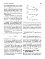
Voltage / (V) Fig. 4. The voltage dependence for the high frequency Raman modes of the PPP-based carbon anode in the lithium-ion battexy. Heat treatment at 700°C and curves were obtained by fits to a Lorentzian line shape.
capacity of LiC,, which is three times higher than in graphite (LE,). The voltage dependence of the high frequency Raman modes of the PPP-based carbon, heat treatment temperature 7OO0C,for electrochemical insertion of Li ions is shown in Fig. 4 [6]. Lithium is taken up at a preferred binding site in PPP-based carbons in the high voltage range (2.8-1.0 V), denoted by Zone I, while Li insertion and release in the PPP-based carbon are accompanied by a shift of the peak wave number to the lower voltage range (1-0.04 V), denoted by Zone 11. Raman results on PPP-based carbons suggest a charge transfer effect, following introduction of Li+ ions, even in such a disordered material. Other kinds of disordered carbon show no change in Raman peaks. Raman experiments in highly ordered graphite exhibit a distinct peak change based upon the stage phenomena. The intensity changes of the Raman bands observed in PPP-based carbon correlate with the electrical conductivity associated with the lithium insertion and release. Inaba et al. reported an in situ Raman study on MCMB heat-treated at around lOOO"C, and showed no change in 1580 cm-' Raman peak [7]. These different results indicate that these charge and discharge mechanisms taking place especially in low temperature carbons, with super high Li storage capacity, differ carbon to carbon, being dependent upon the detail of structure within these carbons. The above results are consistent with NMR studies of charge and discharge processes [5]. The lithium atoms in PPP-based carbon must occupy two different sites corresponding to bands A and B. Band C can be assigned to the by-product lithium carbonate. Band A has a Gaussian line shape with a chemical shift of 9.85 ppm and a half width of 11.6 ppm. Band B has a Lorentzian line shape with a chemical shift of
zyxwvu
46
zyxwvutsrqp zyx Chapter 3
Cavity
Crystalline
Intercalation charge
zyxwvuts discharge
Non-crystalline I
Doping (cluster-like)
Doping
Fig. 5. A schematic model for the insertion of lithium into the internal surfaces of nanopores formed by single, bi- and tri-layer graphene sheets in glassy carbon materials.
-0.62 ppm and a half width of 8.9 ppm. The nature of band A can be understood from the doping of a lithium ion by PPP-based carbons. This is accompanied by electron occupation in the lowest unoccupied molecular orbital (LUMO) and gives rise to radical formation. The Gaussian line shape of band A is consistent with the ionic mechanism, where 7Li nuclei are fixed and inhomogeneous broadening determines the line shape. Namely, site A can be considered as an ionic or GIC site. On the other hand, band B may result from the existence of molecular Li, in the PPP-based carbon. A small chemical shift for band B is consistent with a Li, molecule model. This model is also consistent with a Lorentzian line shape. As a result, this site is called a “covalent” site to be distinguished from the ionic site. Both lithium sites are schematically demonstrated in Fig. 3b. The large Li storage capacity as LiC, in the PPP-based carbon is largely due to the formation of small flat pre-graphitic clusters with small diameters of several nanometres. The insertion of lithium into the disordered carbons obtained from a petroleum pitch [8], epoxy Novolac resin [9], and polyfurfuryl alcohol [lo] has been extensively studied by measuring the voltage profiles for charging and discharging of Li ions, aiming for development of high capacity LIB. The voltage profiles of the glassy or disorder carbons show characteristic low voltage plateaux with relatively high charge4ischarge capacities. The storage sites of Li+ions with higher densities than in first-stage Li remain to be elucidated. Another model in which lithium is inserted and stored in the specific nanopores or surfaces of single layer graphene sheets [11,12] has been put forward, and is as shown schematically in Fig. 5. In disordered carbons, three types of Li storage mechanisms could be occurring depending upon the defective small carbon network structure. 4 Substitution of Heteroatoms
Many heteroatoms and molecules can be intercalated between the layers and or can reside at the edges of carbon “grains” and near to inter-cluster vacancies. However,
zyxwvutsrq zyxwvut zyx
Structural Design and Functions of Carbon Materials
47
i
179O
164O
zyxwv zyxwvut
Fig. 6. A schematic model for (a) undoped and (b) boron-substitutedgraphene sheet. Side view of the graphene at the left is the undoped sheet, and at the right is the boron-doped sheet that has an apex angle of 164".
such heteroatoms as boron (B) and nitrogen (N) can occupy carbon lattice sites substitutionally. In disordered carbons, nitrogen atoms have sp3bonding. Boron (B) atoms can substitute carbon atoms in the hexagonal basal planes of graphite to an extent of less than 2.5-3.0 at% [20]. The substituted boron atoms behave as an anti-oxidant, a graphitization catalyst and improve the charge-discharge capacity for Li-ion rechargeable batteries [13-18]. A schematic model for the boron-substituted graphene sheet based on the structure analysis obtained by STM of HOPG (highly oriented pyrolytic graphite) and calculated model is shown in Fig. 6, to be compared with that of pristine graphite sheet [19]. At a B-doped lattice site an electron is localized and an emphasized atomic image can be observed in the specific trigonal lattice of an STM image. Boron doping of graphitizable carbons can reach 2.5 at% [20] with electronic [19,21] as well as electrochemical changes [1,2]. Oxidation is a serious problem for carbon-carbon composites above 400"C, so limiting applications at high temperatures. It is established that substituted boron significantly improves the anti-oxidation behavior of a carbon-carbon composite [ 17,221.Jones and Thrower proposed three possible mechanisms to explain the role of boron as an anti-oxidant of graphite [17]. 1. Physical blockage of the surface active sites by a B,03 barrier layer formed on the surface during oxidation. 2. Chemical inhibition via electron transfer between carbon and substitutional boron atoms on the internal graphite lattice sites, which increases the activation energy for the C-0, reaction. 3. Boron doping results in the improvement of graphite crystallinity, and thus decreases in the total number of accessible active surface sites for oxygen.
48
zyxwvutsrqp
zyxwvu zyxw zyxwvuts zyxwvut Chapter 3
Both chemical features and electronic structures can be modified by B-doping. Boron doping has been reported for mesocarbon microbeads (MCMBs) [l] and carbon fibers [2], heat treatment temperature > 1800"C, by using metallic boron as a dopant, extents of substitution increasingwith increasing HTT. An effect of boron doping is to accelerate the graphitization of carbon materials for HTT of 1800-2500°C. Electrochemical lithium intercalation takes place at a higher potential in boron-doped graphite than in undoped carbons presumably because the substitutional boron acts as an electron acceptor in the carbons and changes the electronic structure especially of Highest Occupied Molecular Orbital (HOMO) [23]. The interlayer spacing distances (doo2)as a function of heat treatment temperature and charge-discharge profiles for doped and undoped MCMBs heat treated at 2000°C are compared in Figs. 7a and b [1,2].
zyxwv zyxwvutsrqp 3.50
-
4 N
$ 3.45 .-a C (9
D L
x
5
3.40
i
zyxw
3.35
00
Heat Treatment Temperature ("0
3
+-
I I
v_
-.-
5 m
I
I I
zyxwvut I
I I
5 1 L
HTT=20000C
I
$ 2 -1
(b
i
2'
i
I
-Pristine __.
Bdoped
-*a !-- - . - ,
o
o
50
180
150
200
250
300
Capacity (mAh/g)
Fig. 7. The interlayer spacing distances (dWJ as a function of heat treatment temperatures (a) and charge-discharge profiles for doped and undoped MCMBs heat treated at 2000°C (b). First and second cycles are indicated in (b).
zyxwvutsr zyxwvut
Structural Design and Functions of Carbon Materials
49
The carbon alloys obtained by doping with B, N and possibly with Si and P may modify the electronic, chemical and physical properties of graphene layers. Doping is one promising approach for alloying carbons, one which may widen applications of graphite and carbons. 5 Metal-doped Fullerenes
Fullerene-containing soot is synthesized using an arc discharge between graphite electrodes in approximately 200 torr of helium gas [24]. The soot contains about 13 wt% of C,,, and 2% C,,,. Doping is used to modify the electronic properties of the fullerene molecule and bulk crystals. Metal-doped fullerene, mainly endohedral doping, is where rare earth metals such as La, Y and Sc enter the interior of such cage molecules such as C,,,, C,, and C,, [25]. The substitution of a heteroatom such as boron for a carbon atom on the cage molecule has also been known [26]. Exohedrul doping, also called intercalation with doped species located between the cage molecules, is the most common method for doping fullerenes. Electrical conductivity of fullerenes increases considerablywith exohedral doping [27] as in GIC. Alkali metals such as Li, Na, K, Rb, and Cs have been used as exohedral dopants for C,, crystalline solids. Structural models for endohedral and exohedral doping into the fullerenes are shown in Fig. 8. The most interesting electric property of the metal-doped fullerenes is the high temperature superconductivity at about 40 K [28]. Fullerenes doped with Cs at 40 K [29], Rb at 29 K [30], and K at 18 K [31] show superconductivity. Metal-
Fig. 8. The model structures for (a) endohedral and (b exohedral doping into the fullerenes.
50
zyxwvutsrqp Chapter 3
zyxw zyx
doping causes the lattice constant of the host crystals, based on C60,to increase, the ChO,-ChO coupling to decrease, and the electronic bandwidth derived from the LUMO level to narrow, and thereby the corresponding electronic density of states to increase. The strong dependence of the superconducting transition temperature T, for various metal (M) compounds such as M,C,, and M,-xM,C,, on the lattice constant has been studied [31]. Enhancement in electrical conductivity and occurrence of superconductivity takes place by charge transfer phenomena after exohedral doping, very similar to the occurrence of superconductivityin GICs such as C,CsBi,,, (4.05 K). 6 Metal-doped Carbon Nanotubes
The electrical resistivity of carbon nanotubes and fullerenes can be significantly improved by metal doping. Metal-doped carbon nanotubes can also be synthesizedby a laser ablation method. Figure 9 shows the schematic model for a metal-doped carbon nanotube rope. Thess et al. report [32-341 that Ni/Co-doped carbon electrodes form bundles of single-walled carbon nanotube (SWCNT) through the laser ablation method, resembling a rope-like nanotube bundle. The electrical resistivity of the rope-like tube is 0 . 3 4 lo4 ~ Qcm at 300 K, showing metallic behavior in the temperature dependence. It has an electron transport property of a nano-scale quantum wire at low temperatures. Doping with K or Br, brings about important changes in the overall resistivity-temperature behavior. Figure 10 the variation of resistivity with temperature for doped bulk SWNT samples [34]. After doping, the resistivity exhibits a more metallic behavior as observed in GICs.
zyxw zyxwv zyxwvu zyxwv e
e
e
e
e
e
*.
e
e
e .
e
e
e
0
e
e
e
e
e
e
e
e
e
e
e
Fig. 9. A schematic model of the metal-doped carbon nanotube.
zyxwvutsr
Structural Design and Functions of Carbon Materials
zyxwvuts zyxwvut zyxwvut zyxwvu
- I E
(a)Pristine material
G. & 0.001 ..-> .2
(h)after doping with potassium at 473K
................................. _ . _ ...................... _.-.-.-.
_ . _ . - .(c)after - - heating in the cryostat vacuum to 580K overnight -..- ..-
i (fl
v1
51
(d)after 3h at 580K
0.0001
I
0
I
I
I
I
I
100
200
300
400
500
600
Tern peratu re[K]
Fig. 10. Resistivityversus temperature for a bulk SWCNT sample [3]:(a) Pristine material; (b) after doping with potassium at 473 K, (c) after heating in the cryostatvacuum to 580 Kovernight; (d) after 3 hat 580 K.
An ambitious approach, unique to SWNTs, would be to exploit the quantum phenomena observed at very low temperatures; doping-induced shifts in conductance peaks versus voltage should be directly related to the shift in Fermi energy of the tubes. Another type of alloying of carbon nanotubes is doping by nitrogen atoms. This will change the electronic structure of carbon nanotubes to metallic so suggesting further applications. N-doped carbon nanotubes are prepared by several methods. One method is the pyrolysis of a solid organic precursor such as melamine using ferrocene as a catalyst [35]. SEM images of N-doped (CN,) nanotubes obtained by the pyrolysis of solid organic precursor are shown in Fig. 11. The SEM images show well-aligned tubes. HRTEM images (Fig. 12a) show the bamboo-shaped morphology of CN, tubes. The above SEM and HRTEM images suggest the schematic model of CN, tubes as shown in Fig. 12b. Another method for preparing the CN, nanotube is by spray pyrolysis [36,37]. Carbon nanotubes can be fluorinated as reported for carbon fibers [38,39]. The electronic state is that of a semiconductor [38,39] being dependent on the type of C-F bonding (covalent or ionic) and the location of the fluorine atoms within the carbon. The lubrication properties of fluorinated nanotubes and of nanofibers are better than for other CF, systems. In Fig. 13, TEM images of fluorinated nanotubes at different resolutions are shown. These images show that the morphology of the fluorinecontaining fibers is amorphous and defective and that this is caused by the presence of fluorine atoms within the graphene layers. This is very similar to that observed in bulk (CF), [40]. The proposed model for ideal fluorination of a nanotube is shown in Fig. 14. In this model, the stoichiometry is CF,, but in the fluorinated nanotubes it is estimated, from the EELS spectra, that their fluorine content was ca. 1-8 at% [41]. It
52
zyxwvutsrqp zyx Chapter 3
Fig. 11. SEM images of CN, nanotubes at different resolutions.
Fig. 12. TEM image (a) and schematicmodel (b) of CN, tube. Bamboo shape morphology is clearly found in the image.
zyxwvutsrqpo zyxwvutsrq
Structural Design and Functions of Carbon Materials
53
Fig. 13. TEM images of fluorinated carbon nanotubes at different resolutions. Disordered layer structure can be found.
zyxwvutsrq
Fig. 14. Simulated model of CF, nanotube.
54
Chapter 3
zyxwvutsr
was also found that electron beam irradiation heating of fluorinated nanotubes, using UHVTEM, enhances the dissociation of fluorine atoms from the carbon network, resulting in the recovery of the layered structure of the undoped carbon nanotubes [41,42]. 7 Conclusions
Research into carbon alloys by designing the structure at the both atomic and molecular levels for carbon nanotubes, fullerenes, fibers, glassy carbons, single crystal graphites and related materials, is still at an early stage in science and in practical application. Reaction mechanisms for alloying need further elucidation. However, carbon alloys may have important and promising industrial applications such as in electrical and electronic devices, space technologies, batteries and bioengineering. Large-scale production of carbon alloys is also an important target for the carbon industries. The nano-scale structure of carbon materials and their alloys may be a key factor to control and to design the physical and chemical properties of innovative applications of carbons, graphites and related materials References
zyxwvuts
1. C. Kim, T. Fujino, K. Miyashita, T. Hayashi, M. Endo and M.S. Dresselhaus, J. Electro-
chem. SOC.,147: 1257,2000. 2. C. Kim, T. Fujino, K. Miyashita, T. Hayashi, M. Endo and M.S. Dresselhaus, J. Electrochem. SOC.,147: 1265,2000. 3. Sony's Catalog, Lithium ion rechargeable battery, ACG-4012-N-9707-P3-002, 1997. 4. M. Endo, C. Kim, T. Karaki, Y. Nishimura, M.J. Matthews, S.D.M. Brown and M.S. Dresselhaus. Carbon, 37: 561,1999. 5. K. Sato, M. Noguchi, A. Demachi, K. Oki and M. Endo. Science, 264 556,1994. 6. M. Endo, C. Kim, T. Karaki, T. Fujino, M.J. Matthews, S.D.M. Brown and M.S. Dresselhaus, Synthetic Metals, 98: 17, 1998. 7. M. Inaba, H. Yoshida and Z. Ogumi, J. Electrochem. SOC., 143: 2572,1996. 8. N. Sonobe, M. Ishikawa and T. Iwasaki, 3ShBattery Symposium in Japan, Extended Abstracts, 47, 1994. 9. Y. Liu, J.S. Xue, T. Zheng and J.R. Dahn, Carbon, 34: 193,1996. 10. A. Omaru, H. Azuma, M. Aoki, A. Kita and Y. Nishi, 182"dmeeting of the Electrochemical Society, Toronto, Canada, Extended Abstracts of Battery Division, 34,1992. 11. A. Mabuchi, K. Tokumitsu, H. Fujimoto and T. Kasuh. J. Electrochem. SOC.,1 4 2 1041, 1995. 12. J.R. Dahn, T. Zheng, Y. Liu and J.S. Xue. Science, 270 590, 1995. 13. B.M. Way, J.R. Dahn, T. Tiedje, K. Myrtle and M. Kasrai, Phys. Rev., B46: 1897,1992. 14. M. Endo, C. Kim, T. Karaki, T. Tamaki, Y. Nishimura, M.J. Matthews, S.D.M. Brown and M.S. Dresselhaus, Phys. Rev. B58: 8991, 1998. 15. J.R. Dahn, J.N. Reimers, A.K. Sleigh and T. Tiedje, Phys. Rev. B45: 3773,1992. 16. J.G. Naeini, B.M. Way, J.R. Dahn and J.C. Irwin, Phys. Rev. B54: 144,1996. 17. L.E. Jones and P.A. Thrower, Carbon, 29: 251,1991. 18. A. Agrawal, H. Yinnon, D.R. Uhlmann, R.T. Pepper, C.R. Desper, J. Mat. Sci., 21: 3455, 1986.
zyxwvu
zyxwvut zyxwvu zyxwvuts zyxwv
Structural Design and Functions of Carbon Materials
55
19. M. Endo, T. Hayashi, S.W. Hong, T. Enoki and M.S. Dresselhaus, J. Appl. Phys., 9 0 5670, 2001. 20. C.E. Lowell, J. Am. Ceram. SOC.,5 0 142,1967. 21. Y. Hishiyama, H. Irumano and Y. Kaburagi, Phys. Rev., B63: 245406,2001. 22. H. Murty, D. Beiderman and E. Herintz, Fuel, 56: 305, 1997. 23. N. Kurita, Carbon, 38: 65,2000. 24. H.W. Kroto, J.R.Heath, S.C. O’Brien, R.F. Curl and R.E. Smalley. Nature (Landon), 318: 162,1985. 25. D.S. Bethune, R.D. Johnson, J.R. Salem, M.S. de Vries and C.S. Yannoni, Nature (London), 366: 123,1993. 26. M.S. Dresselhaus, G. Dresselhaus and P.C. Eklund, J. Mat. Res., 8: 2054, 1993. 27. M.S. Dresselhaus and G. Dresselhaus, Adv. Phys., 30: 139, 1991. 28. C.M. Lieber and C.C. Chen, In: Solid State Physics. Academic Press, New York, 109,1994. 29. T.T.M. Palstra, 0. Zhou, Y. Iwasa, P. Sulewski, R. Fleming and B. Zegarski, Solid State Commun., 93: 327, 1995. 30. H.A. Hebard, Phys. Today, 45: 26, 1992, Nov. issue. 31. R.C. Haddon, A.F. Hebard, M.J. Rosseinsky, D.W. Murphy, S.J. Duclos and F.A. Thiel, Nature (London), 350: 320,1991. 32. A. Thess, R. Lee, P. Nikolaev, H. Dai, P. Petit, J. Robert, C. Xu, Y.H. Lee and S.G. Kim, Science, 273: 483, 1996. 33. J.E. Fischer, H. Dai, A. Thess, R. Lee, N.M. Hanjani, D.L. Dehaas and R.E. Smalley. Phys. Rev., B55: R4921, 1997. 34. R.S. Lee, H.J. Kim, J.E. Fischer, A. Thess and R.E. Smalley. Nature, 388: 255, 1997. 35. M. Terrones. H. Terrones, N. Grobert, W.K. Hsu, Y.Q. Zhu, J.P. Hare, H.W. Kroto, D.R.M. Walton, Ph. Kohler-Redlich, M. Ruhle, J.P. Zhang and A.K. Cheetham, Appl. Phys. Lett., 75: 3932, 1999. 36. M. Mayne, N. Grobert, M. Terrones, R. Kamalakaran, M. Riihle, H.W. Kroto and D.R.M. Walton, Chem. Phys. Lett., 338: 101,2001. 37. M. Terrones, R. Kamalakaran, T. Seeger and M. Riihle, Chem. Comm., 23: 2335,2000. 38. G. Seifert, T. Kohler and T. Frauenheim, Appl. Phys. Lett., 77: 1313,2000. 39. K.N. Kudin, H.F. Bettinger and G.E. Scuseria, Phys. Rev. B, 6304: 5413,2001. 40. M. Endo, M. Shikata, H. Touhara, K. Kadono, and N. Watanabe. Trans. Inst. Elect. Eng. Japan, 105-A: 329,1985 (in Japanese. 41. T. Hayashi, M. Terrones, C. Scheu, Y.A. Kim, M. Riihle, T. Nakajima and M. Endo, Nanoletters, 1: 491, 2002. 42. T. Hayashi, M. Terrones, Y.A. Kim, T. Nakajima and M. Endo, In: Nanotubes and Related Materials, MRS, A14, 18,2001.
zyxwvutsrq
57
zyxw
zyxwvu zyxwvut Chapter 4
Surface and Hidden Surface-controlled Carbon Alloys Katsumi Kaneko
zyxwvutsrqp
Department of Chemistiy, Faculty of Science, Chiba Universiq, Inage, Chiba 263-8522, Japan
Abstract: The importance is emphasized of a new concept called a “hidden surface” to describe porosity in carbons; the necessity for this concept of a hidden surface is supported by molecular potential theory. Approaches to methods of alloying porous carbons and the merits of alloyed porous carbons are described. The alloying of hidden surfaces can be brought about by (a) introduction of mixed valencies into carbons, (b) doping by foreign atoms or clusters, (c) development of specific porosities, and (d) the tailoring of hidden surfaces with clusters and ultrafine particles. Space-alloying in porosity is further discussed in terms of alloying-induced pore space, control of pore space functions by alloying, and nanogeometry-controlled growth of foreign compounds. Several examples of alloyed porous carbons possessing new functions and properties are described.
zyxwvut zyxwvu
Keywords: Micropore, Mesopore, Gas adsorption, Molecular potential calculation, Alloyed porous carbon.
1 Importance ofHidden Surfaces and Confined Spaces in Carbon Materials
Solid surfaces are never completely flat. Even the surface of a single crystalline solid has some roughness due to an irregular arrangement of the surface atoms. Solid surfaces have a roughness factor stemming from surface voids and pores. Although there is no rigorous distinction between surface voids and pores, the term “pore” is used for a void whose depth is larger than molecular sizes. Pores are classified into micropores (w < 2 nm), mesopores (2 nm c w < 50 nm), and macropores (w > 50 nm) according to IUPAC [l]. Here w is the pore width which is the slit width for slit-shaped pores or the diameter of cylindrical pores. Here this new concept of the hidden surface is used to stress differences from the concepts of established surface science. Pore-wall surfaces are called the hidden surfaces because they cannot be elucidated by ordinary surface-science investigative tools and because they have unique properties quite different from those of external surfaces. Solid materials have a cohesive structure which depends on the interaction between primary particles. The cohesive structure leads indispensably to void spaces which are not occupied by such composite particles as atoms, ions, and fine particles.
58
zyxwvutsrqpo zyx zyx Chapter 4
Consequently, such voids or pores intensively depend on the cohesive forces. For carbons, diamond has very strong covalent sp3-sp3bonding, while graphite has both conjugated (sp2-sp2)bonding in the graphene sheet and the weak inter-graphene sheet interactions which approximate to van der Waals interactions. The van der Waals interaction is associated with the interacting area. The inter-graphene sheet distance becomes larger with decreasing graphene sheet size. Thus, the pore structure depends on fundamental chemical bonding structures. Carbon materials are classified into crystalline and less-crystalline materials. Graphite and diamond are representatives of well-crystalline (single crystal) carbon materials. Graphite is composed of stacked graphene sheets with the interlayer spacing almost coinciding with the thickness of the graphene sheet, owing to the above-mentioned bonding state. Consequently, graphite has no effective space between the graphene sheets for acceptance of foreign atoms and molecules. However, appropriate conditions produce what are called “intercalation compounds”. Atoms such as potassium atoms can be inserted between the graphene sheets. Thus, even graphite can have latent spaces in its structure. For diamond, the interatomic covalent bond is too strong to accept foreign atoms to form a new compound. Activated carbon is a representative porous solid. The fundamental structure is noncrystalline, and is composed of imperfect nanographitic units (these are designated “nanographites”here) with inter-nanographite-linkagesof the sp3-sp3 bonding nature to form the pore walls. The chemical bonding state of activated carbon is a mixed state of graphite and diamond. There is an abundance of pore space between pore walls. The surface areas of the pore walls and the external surface of activated carbon sample can be determined by nitrogen adsorption at 77 K with the aid of the subtracting pore effect (SPE) method [2,3]; this method will be described later. The surface area of pore walls of activated carbon is > 1000m2g-’, whereas that of the external surface is less than -50 m’ g-I. Therefore, the pore spaces or intrapore surfaces are predominant compared with the external surface. Is there any difference between the hidden surfaces in the pore and external surfaces of carbon materials? Although the structures of actual activated carbons have a complexity inherent to the higher order structure of the precursor, the nano-level structure can be approximated by the basal plane of the graphite.
zyxwv zyxw zy
Differencesbetween Hidden and External Sur$aces based on Mokcular Potential 17seory
1.1
An explicit distinction for the hidden and external surfaces can be derived for the basal piane model using the following molecular potential theory. Two model cases will be shown: one is a graphite-slit pore model and the other is a single-wall nanotube open pore model. The graphite-slit pore model has been applied to activated carbon to explain the molecular adsorption properties. The latter model is applied to the single-wall carbon nanotube and single-wall carbon nanohorn.
zyxwvu zyx zyxw zyxwvuts zyxwvuts zyxwvutsrqpo zyxwvut zyxw
Surface and Hidden Surface-controlledCarbonAlloys
59
The interaction of a molecule with the graphitic slit pore of the micropore model of activated carbon is expressed by use of the Lennard-Jones (JJ)potential and Steele potential. The interaction between a molecule and a surface atom as a function @(r) of the distance r between them can be expressed by the LJ potential
where E, and 0, are the well depth and effective diameter for the molecule-graphitic carbon atom. These cross parameters are calculated according to the LorentzBerthelot rules, ,E = (qSE ~ ) " ~0, ; = (0%+ 0,)/2. Here, (qS, E,,) and (off, E ~ are ) the Lennard-Jones parameters for a surface atom and a molecule, respectively. The interaction potential @(z)for a molecule and a single graphite slab is given by the Steele 10-4-3 potential [4] @(z) = 2~p,
&sf
0,:A{(2/5)(0,,h?)'" - ( o , ~ / z-) ~0,: / [3A(0.61A + z)']}
(2)
where z is the vertical distance of the molecule above the surface, A is the separation between graphite layers (= 0.335 nm), pc is the number density of carbon atom in a graphite layer (= 114/nm3).As the micropores of activated carbon can be approximated by the slit spaces between the predominant basal planes of nanographitic units, the whole interaction potential @(z), of a molecule with the micropore of an inter-graphite surface distance H can be given by Eq. (3): @(z)p= @(z) + @(H - 2)
zyx
(3)
Consequently, we can evaluate the potential profile of the molecule adsorbed in the graphitic micropore. Here H is not the effective pore width w determined by the adsorption experiment. The difference betweenHand w is a function of o,,and otT[5].
H - w = 0.85 os,- 0,.
(4)
Figure 1 shows potential profiles of a nitrogen molecule with slit-shaped graphite pores of w = 0.5 and 1.2 nm using the one-center approximation. Here, the molecular position in Fig. 1 is expressed by a vertical distance z from the central plane between two pore-wall surfaces. The potential minimum for the flat surface is 1100 K, while the minima for w = 0.5 nm and 1.2 nm are 2250 K and 1200 K, respectively. The overlapping of the interaction potential from the opposite pore walls gives rise to a greater potential minimum for the slit pore. Also, the interaction potential profile for thew = O S nm indicates that there are no sites localized on the pore-wall surface. The bottom of the potential profile is almost flat near the central position (z = 0) of the pore. On the other hand, the potential profile of w = 1.2 nm has distinct double minima on the pore-wall surface. Although the potential depth of w = 1.2 nm is greater than that of the flat surface, the potential profile indicates the localization of
60
zy
zyx zyxwvutsrq zyxwvut
zyxwvutsrq zyxwvu zyxw zyxwvutsr Chapter 4
500
. c, 2
e
0
-500
2 -1500
0
3?.-1000 85
t
-2000
bdnm
1
0.5
zyx
nitrogen molecules on the pore walls. Accordingly,the potential profile of w = 0.5 nm is unique and it is indicative of the importance of the space concept inherent to the very narrow pores (ultra-micropores). The potential profile of the pore of w = 0.4 nm has the repulsive effect. The potential minimum of w = 0.4nm is higher than that of w = 0.5 nm. Thus, the flat surface and the pore surface must be distinguished from the molecular potential theory. Also the external surface must be divided into the basal plane and the edge surface of actual graphite crystals. The molecule-surface interaction on the edge surface is different from that on the basal plane even without the presence of surface functional groups. The ideal structure of edge surfaces of graphite crystals should be surface terminated with hydrogen atoms, being completely different from the basal plane of the graphite. Fujita et al. [6] classified two types of the carbon+zwbon structures at the edges of a graphene sheet as “armchair” and “zigzag” arrangements; the electronic structure sensitively depends on the structure. However, only differences between external and hidden basal planes will be discussed here. For single-wall carbon nanotubes, the following equation for the molecule-pore wall interaction is used for calculation of the interaction potential profile [7]. Interaction potentials are evaluated below for molecules inside and outside a pore wall made of n-graphene rolled sheets by replacing the sum over atoms in each cylindrical shell by an integral. One will be left with a sum over shells of radii a,, a,, a2, ..., with a, = a, + n X 0.34 [nm]. Thus
where pc is the two-dimensional density of carbon and r is the distance of the adsorbate molecule from the axis of the cylinder and z’ is the distance parallel to the pore axis. is the angle between a,, and r. Equation (5) can be written as
+
zyxwvutsrqponm zyxwvu zyx
Su@ace and Hidden Suflace-controlled Carbon Alloys
where
Z,
61
zyxwvuts zyxwvutsrqpo zyxwvutsr zyxwvutsr zyxwvut zyxwv -
=
2n
and$ Jd~jj (zj2+a,: + R * - 2a, R cos 9)”’
-m
(rn = 3 or 6)
(7)
(,
This integral can be expressed by a hypergeometric function using R and u,~. Here, R is the distance of the molecule from the center of the tube. Then the Steele smooth function for a molecule inside and outside the cylindrical pore can be analytically calculated. The detailed calculation procedures are reported elsewhere [8]. The interaction profiles of a nitrogen molecule with a nanotube of interatomic diameter H = 2 nm were calculated. The potential minimum inside the tube (1250 K) is deeper than that of the external position by more than 300 K. Thus, the difference of the molecule-pore wall interaction between the external and intrapore configurations is remarkable, compared with the slit-shaped pore of H = 2 nm (see Fig. 1). Hence, it is important to distinguish between the molecular states in the intrapore-space and on the external surface for molecular processes on the nano-order structures. This important feature on the inside and outside of pores can be extended to the macroscopic level. The Kelvin equation describes the dependence of the sign of the curvature of the surface of the condensate on the vapor pressure change of the condensate [9]. We need to introduce the concept of the hidden surface in order to stress the different nature of the pore-walls. In particular, the pores of nanometre ranges such as micropores and small mesopores require the concept of the hidden surface. Why do we use the term “hidden surface”? There are various kinds of advanced tools for surface science analyses using electrons such as X-ray photoelectron spectroscopy XPS, and scanning tunnelling microscopy STM. Unfortunately, these powerful surface science tools cannot be applied to elucidation of pore-wall structures and nano-order atomic or molecular systems in pores. Namely, the pore-wall structure and atomic or molecular system in pores are hidden from these surface science tools. Appropriate research methods to study hidden pore-wall structures and molecular systems confined in the pore need to be developed. 1.2
Confinementof Molecules in Carbon Micropore Spaces
The concept of a confined carbon space is important. A graphite slit-like pore of w I1 nm has a deep potential well, as described above. Kaneko et al. elucidated the special function of such pores for molecules and atoms with in situ X-ray diffraction, in situ small angle X-ray scattering, low temperature magnetic susceptibility measurement, high resolution adsorption measurement, heats of adsorption measurements, and computer simulation using activated carbon fiber (ACF). ACFs have uniform
62
zyxwvutsrqp zyx zyx Chapter 4
micropores. It is to be noted that pitch-based ACFs have fewer surface functional groups and ACFs of different w values are available. Typical examples of the confinement effect of molecules in the micropores of ACFs are given in the following. The adsorption energy of a helium atom for the first layer with a graphite surface is 108 K (the energy is expressed in Kelvin) and even that of the second layer is 19 K, both of which are much greater than 4.2 K. Therefore, an enhanced bilayer adsorption occurs even on the graphite surface and carbon black at 4.2 K. Steele proposed a theoretical mechanism of enhanced bilayer adsorption [lo]. This enhanced bilayer adsorption is particularly relevant for adsorption in carbon micropores at 4.2 K. This prediction is supported by the formation of a high density structure and marked uptake from an extremely low relative pressure PIP,, [11,12].A clear difference between the helium adsorption isotherm at 4.2 K and the nitrogen adsorption isotherm at 77 K of an ACF is observed; amounts of helium adsorption in the extremely low Pip,, range ( 800 m2 g-’ and large mesopore volumes > 0.55 ml g-’. 4.4 Tailoring Hidden and External Sugaces of Activated Carbon with Clusters or Ultrafine Particles
zyxwvu
There are many examples of hidden and/or external surfaces tailored with clusters or ultrafine particles. Although supercritical NO cannot be adsorbed predominantly on activated carbon near room temperature, tailoring the external or hidden surface of ACF with ultrafine iron oxide enhances remarkably the NO adsorptivity at 303 K; the saturated amount of adsorption at 101 kPa of NO is greater than 0.3 g g-’ [66,67]. Recent examples are described below. 4.4.1
Tailoring Hidden Surfaces of ACF with Nitrogen-containing Units
Yang and Kaneko modified the hidden surfaces of ACF with nitrogen-deposition by chemicalvapor deposition of pyridine at 1273 K for 1 h after heating ACF at 1273 Kin
74
zyx zyxwvutsrqp zyxwvutsrqp Chapter 4
Table 4
Micropore structures of N-alloyed and pristine ACFs SampIes
N-ACF ACF
Surface area (m2k)
(mk)
Micropore volume
Pore width (nm)
1280 2000
0.64 0.99
0.89 1.0
zyxwvuts
argon, a method originally developed by Kawabuchi et a1.[68]. The resultant ACF is described as N-ACF. The electronic states of doped nitrogen atoms were determined using XPS; depth profiles of nitrogen atoms were measured with argon etching. The composition of N-ACF was C,,,N. The content of doped nitrogen atoms in graphitic skeletons is < 1.8at%. The peak of the broad XPS spectrum of Nls was observed at 401 eV, and is assigned to quaternary nitrogen. The peak of Nls, for pyridinic nitrogen, was not observed at 398 eV. Therefore, the nitrogen atoms in “graphitic” units appear to be deposited on the hidden surfaces and have effective positive charges. The N-deposition procedure does not change the features of the nitrogen adsorption isotherm, suggesting that N-ACF is highly microporous. Table 4 summarizes the pore structures of these ACF samples. N-ACF has a surface area > 1000 m2 g-’ regardless of the CVD treatment at 1273Kwith pore widths becoming slightly smaller after N-alloying. Some pores should be blocked by the CVD deposition treatment on preparation of N-ACF. The nitrogen deposition enhances water and ethanol adsorptivities of ACF due to the introduction of the polar sites [69,70].
zyxwvutsrqp zyxwvu zyxwvut
4.4.2 Clustered Ti0,-embedded ACF from an Ionized Cluster Beam Method
Yamashita et al. [71] prepared an ACF impregnated with ultrafine TiO, particles using the ionized cluster beam method. The method deposits ultrafine TiO, clusters on the external surface of ACF without blocking the micropores. The composite effects arising from the micropores of ACF and the deposited TiO, induce excellent photocatalysis properties. This ACF is efficient for the photocatalytic degradation of 1,Zdichloroethane diluted in water. The concentrated 1,Zdichloroethane in micropores of ACF from the solution is effectively decomposed by the photocatalysis of TiO, on external surfaces.
4.5
Function of Pore Space Alloyed with Clusters and Fine Particles
Alloying the pore space is not clearly distinguishable from alloying the hidden surfaces, as mentioned in Section 2. From the many examples available, a few are described below.
zyxwvu zyxwvut zyxwvutsrq 75
Surface and Hidden Surface-controlledCarbon Alloys
4.5.1 Ambient TemperatureRreduction of NO to N2by Metal-tailored Carbon Micropores
Ru or Pt fine particles exhibit a high catalytic activity for the reduction of NO. Ultrafine Pt or Ru particles are dispersed in the carbon micropores of ACF by reduction with hydrogen at 623 K of a dried metal salt after immersion of the evacuated ACF in metal salt solution. XPS shows the formation of metal particles with small amounts of oxide. The tailored amounts of metal particles are in the order of 0.2-O.5% of carbon atoms, by XPS analysis.Accordingly,the microporosityof ACF does not markedly vary due to the tailoring. N,O from the concentrated NO dimer in the micropores is reduced to nitrogen very quickly even at 303 K [72]. The NO reduction finishes within 7 min. The first-order plot for the initial reaction gives a half-life of 3 min. No CO, is observed at 303 K, whereas small amounts of N,O (0.005 N20/N,) are formed. However, raising the reaction temperature to 423 K lowers the reaction rate and leads to production of CO, (CO,/N, = 0.08). Mass analysis indicated the formation of nitrogen corresponding to a yield of 0.77. Thus, NO is rapidly reduced to nitrogen over Ru- or Pt-ACF at 303 K. In this case, NO is removed by both reduction and adsorption; the total removal ratio of NO at 303 K is 0.96 which is greater than the nitrogen yield. As no oxygen is observed, the oxygen of the system must be chemisorbed on the ACF. This formation of nitrogen stems from the quasi-high pressure effect of the strong molecular potential field [73]. As molecules confined in micropores have a highly dense structure, as if they were compressed by high pressure, the confined molecular assembly exhibits a special reactivity. For NO, almost all NO molecules adsorbed in carbon micropores are dimerized even at 303 K because the adsorbed layer forms the low temperature liquid-like phase. This quasi-high pressure effect assists the catalysis of NO reduction by ultrafine Ru and Pt metals.
zyxwv zyxwvu zyxwvut zyxw zyxw
4.5.2 Morphology Controlled Nanocrystal Formation in Pore Space of Carbon Tubes
Kyotani et al. prepared carbon nanotubes using the template of anodic aluminum oxide film containing straight pores by chemical vapor deposition of propylene. Their method is illustrated in Fig. 4. They then introduced material by metal-organic chemical vapor deposition or by reduction of impregnated metal ions to the carbon-coated nanochannels of the anodic aluminium oxide film [74-761. As the inner diameter of the carbon tube is ca. 20 nm, low dimensional crystals were obtained. They prepared nickel oxide nanobelts, iron oxide nanoparticles, and platinum nanorods. The preparation method of nickel oxide nanobelts is as follows. The carbon-coated anodic aluminium oxide film was subject to metal-organic chemical vapor deposition of nickelocene at 548 K using a mixed gas flow of the nickelocene (0.6 kPa) with hydrogen diluted with nitrogen. The aluminium oxide was dissolvedwith 10 M NaOH solution at 423 K in an autoclave. TEM images showed the formation of a single nanobelt 500 nm in length, within the carbon tube. Selected area electron diffraction
76
z
zyxwvutsrq zyx Chapter 4
Anodic aluminum oxide film
-zyxwvut Carbon tube aggregate
HF washing
zyxwvutsrq
Fig. 4.Template carbonization method using anodic oxide film for preparation of carbon tube of inner diameter of 20 nm.
indicated the formation of a crystalline nickel oxide. Further, iron oxide nanoparticles and platinum nanorods, as nanodimensional crystals, were formed in the carbon tubes and characterized by TEM, selected area electron diffraction and X-ray diffraction. These nanodimensional crystal-tailored nanotubes are expected to show a new catalysis and special physical properties. 5 Design of New Porous Carbon with Carbon Alloying Technique
Possible applications for porous carbons with hidden and external surfaces modified with alloying are discussed above. However, the separation of molecular processes on hidden and external surfaces is not straightforward. It is considered that carbon nanotubes are ideal absorbents having regular cylindrical intrapores. Nevertheless, carbon nanotubes have an aggregate structure which promotes the existence of micropores and mesopores. Hence, it is not straightforward to distinguish between molecular processes on hidden and external surfaces. Inoue et al. attempted to distinguish between these two processes using multi-wall carbon nanotubes [77]. Recently, Murata et al. [8,78] succeeded in distinguishing between these processes using the single wall carbon nanohorn; they experimentally demonstrated a pure molecular process within the intrapores of a carbon nanohorn. Hopefully, this experimental result will stimulate further theoretical studies of molecular behavior in carbon nanotubes [79,80] and so give a new direction to the design of carbon nanomaterials.
Surface and Hidden Surface-controlled CarbonAlloys
zyxwvu 77
zyxwvu
Further developments of porous carbons could be based on the use of this alloying technique which can use the different concepts of related scientific fields. For example, the surface science of carbon materials has an intimate relationship with carbon pore space science. Iwasawa et al. [Sl] studied the surface structure and changes of transition metal carbides with low energy electron diffraction and scanning tunnelling microscopy. Their studies are helpful in the design of new alloying methods of hidden surfaces of porous carbon. Also Takasu et al. 1821 studied the interfacial behavior of transition metal oxides on graphite surfaces with an electrochemical technique. This study also suggested the tailoring of hidden surfaces making use of transition metal oxides. Currently, experimental techniques available to elucidate the nature of molecular assemblies or dispersed particles confined in carbon pores are not as powerful as those of surface science. Hence, the sciences of hidden surfaces and materials confined in carbon pore spaces require further significant improvements based on a variety of expertises [82-86].
zyxwvuts
Acknowledgement
Dr. H. Inokuchi suggested the importance of the hidden surface concept.
References
1. K.S.W. Sing, D.H. Everett, R.A.W. Haul, L. Moscou, R.A. Pierotti, J. Rouquerol and T.
Siemieniewska, Reporting physisorption data for gas/solid systems with special reference to the determination of surface area and porosity. Pure Appl. Chem., 5 7 605419,1985. 2. K. Kaneko and C. Ishii, Superhigh surface area determination of microporous solids. Colloids Surfaces, 67: 203-212,1992. 3. N. Setoyama, T. Suzuki and K. Kaneko, Simulation study on relationship between high resolution a,-plot and pore size distribution for activated carbon. Carbon, 36: 1459-1467, 1998. 4. W.A. Steele, The physical interaction of gases with crystalline solids. Surface Sci., 36: 317-352,1973. 5. K. Kaneko, R. Cracknell and D. Nicholson, Nitrogen adsorption in slit pores at ambient temperature. Langmuir, 10: 46064609, 1994. 6. K. Wakabayashi, M. Fujita, H. Ajiki and M. Sigrist, Electronic and magnetic properties of nanographite ribbons. Phys. Rev., B59: 827143282,1999. 7. K. Murtata, K. Kaneko, W.A. Steele, F. Kokai, K. Takahashi, D. Kasuya, K. Hirahara, M. Yudasaka and S. Iijima, Molecular potential structures of heat-treated single-wall carbon nanohorn assemblies. J. Phys. Chem., in press. 8. K. Murata, K. Kaneko, W.A.Steele, F. Kokai, K. Takahashi, D. Kasuya, M. Yudasaka and S. Iijima, External and internal nanospaces of single wall carbon nanohorn aggregates. Nanoletters, 1: 197-199,2001. 9. R. Defay, I. Prigogine, A. Bellemans and D.H. Everett. Surface Tension and Adsorption, Chap. 1.Longman, London, 1966. 10. W.A. Steele, Concerning a theory of multilayer adsorption with particular reference to adsorbed helium. J. Chem. Phys., 25: 8194323, 1956.
78
zyxwvutsrqp zy zyxwvuts zyxwvuts Chapter 4
11. N. Setoyama, K. Kaneko and F. Rodriguez-Reinoso, Ultramicropore characterization of microporous carbons at low temperature helium adsorption. J. Phys. Chem., 100: 10331-10336,1996. 12. N. Setoyama and K. Kaneko, Density of He adsorbed in micropores at 4.2 K. Adsorption, 1: pp.1-9, 1995. 13. R.E. Leckenby and E.J. Robbins, The observation of double molecules in gases. Proc. Roy. SOC.(London), A291 :389-412,1966. 14. M. Aoshima, T.Suzuki and K.Kaneko, Molecular association-mediated micropore filling of supercritical Xe in a graphite slit pore by grand canonical Monte Carlo simulation. Chem. Phys. Lett., 310: 1-7,1999. 15. C.E. Dinerman and G.E. Ewing, Infrared spectrum, structure, and heat of formation of gaseous (NO)?. J. Chem. SOC.,100: 6948-631,1978. 16. K. Kaneko, N. Fukuzaki and S. Ozeki, The concentrated NO dimer in micropores above room temperature. J. Chem. Phys., 87: 776-777, 1987. 17. K. Kaneko, Nanospace geometry sensitive molecular assembly. Supramolecular Sci., 5: 267-273,1998. 18. T. Ohkubo, T. Iiyama, K. Nishikawa, T. Suzuki and K. Kaneko, Pore width-dependent ordering of C?H,OH molecules confined in graphitic slit nanospaces. J. Phys. Chem., 103: 1859-1863,1999. 19. T. Ohkubo, T. Iiyama and K. Kaneko, Organized structures of methanol in carbon nanospaces at 303 K with in situ X-ray diffraction. Chem. Phys. Lett., 3 1 2 191-195,1999. 20. T. Ohkubo and K. Kaneko, Oriented structures of alcohol hidden in carbon micropores with ERDF analysis. Colloid Surfaces, 187-188: 177-185,2001. 21. K. Morishige, K. Kawamura and A. Kose, X-Ray diffraction study of the structure of a monolayer methanol adsorbed on graphite. J. Chem. Phys., 93: 5267-5270,1990. 22. T. Iiyama, K. Nishikawa, T. Otowa and K. Kaneko, An ordered water molecular assembly in a slit-shaped carbon nanospace. J. Phys. Chem., 99: 10075-10076,1995. 23. T. Iiyama, K. Nishikawa, T. Suzuki and K. Kaneko, Study of the structure of a water molecular assembly in a hydrophobic nanospace at low temperature with in situ X-ray diffraction. Chem. Phys. Lett., 274: 152-158,1997. 24. Z.M. Wang and K. Kaneko, Effect of pore width on micropore filling mechanism of SO2in carbon micropores. J. Phys. Chem., 1 0 2 2863-2868,1998. 25. T. Ohkubo, C.-M.Yang, E. Raymundo-Pinero, A. Linares-Solano and K. Kaneko, Hightemperature treatment effect of microporous carbon on ordered structure of confined SO?. Chem. Phys. Lett. 3 2 9 71-75,2000. 26. K. Kaneko, C. Ishii, M. Ruike and H. Kuwabara, Origin of superhigh surface area and microcrystalline graphitic structures of activated carbon. Carbon, 3 0 1075-1088,1992. 27. C. Ishii and K. Kaneko, Surface and magnetic properties of microporous carbon spheres. Prog. Organic Coatings, 31: 147-152,1997. 28. Y. Higaonna, C. Ishii and K. Kaneko, Magnetic properties and pore structures of KOHactivated mesocarbon microbeads. In preparation 29. K. Kaneko, C. Ishii, H. Kanoh, Y. Hanzawa, N. Setoyama, T. Suzuki, Characterization of Porous Carbons with High Resolution a,-analysis and low temperture magnetic susceptibility. Adv. Colloid Interface Sci., 295: 76-77, 1998. 30. T.T. Berek, W. Ackerman, D.W. Hua, R.T. Raine and D.M. Smith, Highly microporous boron nitride for gas adsorption. Langmuir, 7: 2844-2846,1991. 31. K. Kaneko, N. Kosugi and H. Kuroda, Characterization of iron oxide dispersed activated carbon fibers with water adsorption and Fe K-edge XANES and EXAFS. J. Chem. SOC. Faraday Trans., 85: 869481,1989. 32. A. Matsumoto and K. Kaneko, Micropore filling of supercritical NO on external surface
zyxwvu zyxwvutsrqp zyxw zyxwv
Surface and Hidden Surface-controlled Carbon Alloys
79
modified microporous carbons. J. Chem. SOC.Faraday Trans., 85: 3437-3450,1989. 33. H. Inokuchi, N. Wakayama, T. Kondow and Y. Mori, Activated adsorption of hydrogens on aromatic-alkali-metal charge transfer complexes. J. Chem. Phys., 46: 834-842, 1967. 34. H.Sakuno, A. Ogawa, N. Akuzawa and Y. Takahashi, Absorption of organic molecules on alkali metal-graphite intercalation compounds. Tanso, 238-243, 1990. 35. N. Akuzawa, M. Murakami, M. Nakano, Y. Soneda, R. Matusmoto and Y. Takahashi, Host effect on the properties of AM-GICs. Mol. Crys. Liq. Crys., 340: 59-64,2000. 36. K. Fujie, S. Minagawa, T. Suzuki and K. Kaneko, NO/H,O clathrate formation in sub-nano graphitic slit space. Chem. Phys. Lett., 236: 427-430, 1995. 37. J. Miyawaki, T. Kanda, T. Okui, M. Saito, T. Suzuki and K. Kaneko, Macroscopic evidence of enhanced formation of methane nanohydrates in hydrophobic nanospaces. J. Phys. Chem., 102: 2187-2192,1998. 38. J. Miyawaki and K. Kaneko, Hysteresis-associated pressure shift water adsorption in carbon micropores. Langmuir, 17: 664669,2001. 39. J. Imai, M. Soma, S. Ozeki, T. Suzuki and K. Kaneko, Reaction of dimerized NO, (x = 1or 2) with SO, in a restricted slit-shaped micropore space. J. Phys. Chem., 95: 9955-9960, 1991. 40. K. Kaneko, Specific intermolecular structures of gases confined in carbon nanospace. Carbon, 38, pp.287-303, 1999. 41. N. Setoyama, G. Li, K. Kaneko, F. Okino, R. Ishikawa, M. Kanda and H. Touhara, Nitrogen adsorption on fluorination activated carbon fiber. Adsorption, 2: 293-297,1996. 42. G. Li, K. Kaneko, S. Ozeki, F. Okino and H. Touhara, Water rejective nature of fluorinated microporous carbon fibers. Langmuir, 11: 716-717,1995. 43. K. Kaneko, T. Ohkubo, T. Iiyama, C. Yang and H. Touhara, Cluster mediated filling of water and alcohol on microporous carbon alloys, In: D.D. Do (Ed.), Adsorption Science and Technology, pp. 1-5. World Scientific, Singapore, 2000. 44. Y. Hanzawa and K. Kaneko, Lack of a predominant adsorption of water on carbon mesopores. Langmuir, 13: 5802-5805, 1997. 45. K. Kaneko, Y. Hanzawa, T. Iiyama, T. Kanda and T. Suzuki, Cluster-mediated water adsorption on carbon nanospaces. Adsorption, 5: 7-13,1999. 46. T. Kyotani, L. Tsai and A. Tomita, Preparation of ultrafine carbon tubes in nanochannels of anodic aluminum oxide film. Chem. Mater. 8: 2109-2113,1996. 47. Y. Hattori, Y. Watanabe, S. Kawasaki, F. Okino, B.K. Pradham, T. Kyotani, A. Tomita and H. Touhara, Carbon-alloying of the rear surfaces of nanotubes by direct fluorination. Carbon, 37: 1033-1038,1999. 48. H. Touhara, Potential use of carbon nanotube as anode materials for litihium batteries. Ouyou Butsuri (Applied Physics), 69,3-37,2000. 49. H. Touhara and F. Okino, Property control of carbon materials by fluorination. Carbon, 38: 241-267,1999. 50. R.W. Pekala. J. Matter. Sci., 24: 3221-3227, 1989. 51. Y. Hanzawa and K. Kaneko, Activated carbon aerogels. Langmuir, 25: 6167-6169,1996. 52. Y . Hanzawa, K. Kaneko, R.W. Pekala and M.S. Dresselhaus, The pore structure determination of carbon aerogels. Adsorption, 2: 187-196,1998. 53. E. Bekyarova and K. Kaneko, Microporous nature of Ce, Zr-doped carbon aerogels. Langmuir, 15: 7119-7121,1999. 54. E. Bekyarova and K. Kaneko, Structure and physical properties of tailor-made Ce, Zr-doped carbon aerogels. Adv. Mater., 12: 1625-1628,2000. 55. E. Bekyarova, K. Kaneko and E. Bekyarova, Adsorption of supercritical N, and 0, on pore-controlled carbon aerogels. J. Colloid Interface Sci., 238: 357-361,2001. 56. P. Fornasiero, R. Di Monte, G. Ranga Rao, J. Kaspar, S. Meriani, A. Trovarelly and M.
80
zyx zyxwvuts zyxwvu zyxwvuts zyxwv Chapter 4
Graziani, Rh-Loaded (20,-ZrO, solid solutions as highly efficient oxygen exchanges. J. Catal. 151: 168-177,1995. 57. G. Balducci, J. Kaspar, P. Fornasiero, M. Graiani and M. Saiful Islam, J. Gale, Computer simulation studies of bulk reduction and oxygen migration in Ce0,-ZrO, solid solution. J. Phys. Chem. B, 101: 1750-1753,1997. 58. M. Haneda, T. Mizushima and N. Kakuta, Behavior of oxygen species adsorbed of Al,03-supported cerium oxide catalysts for methane oxidation. J. Chem. SOC.Faraday Trans., 91: 44594465,1995. 59. M. Abe, K. Kawashima,K. Kozawa, H. Sakai and K. Kaneko, Amination of activated carbon and adsorption characteristicsof its aminated surface. Langmuir, 16: 5059-5063,2000. 60. M.Inagaki, K. Sakamoto and Y. Hishiyama, Carbonization and graphitizationof polyimide upilex. J. Mater. Res., 6 1108-1113,1991) 61. Y. Hishiyama, A. Yoshida and Y. Kaburagi, Graphite films prepared from carbonized polyimide films. Carbon, 30: 335337,1992. 62. H. Hatori, Y. Yamada and M. Shiraishi, Preparation of macroporous carbon films from polyimide by phase inversion method. Carbon, 30: 303-304,1992. 63. M. Sato, H. Isobe, H. Yamamoto, T. Iiyama and K. Kaneko, Oriented micrographitic carbon film of high surface area. Carbon, 33: 1347-1350,1995. 64. M. Nozaki, Y. Nishi, T. Iiyama and K. Kaneko, Alloyed-nitrogen induced pore-gate control of nanostructure oriented carbon film. Langmuir, to be submitted. 65. H. Tamon, H. Ishizaka, T. Yamamoto and T. Suzuki, Preparation of mesoporous carbon by freeze drying. Carbon, 37: 2049-2055,1999. 66. K. Kaneko, Effect of temperature on micropore filling of supercritical NO on Fe,O, dispersed activated carbon fibers. Colloid Surf., 37: 329-338,1989. 67. Z.M. Wang, T. Suzuki, N. Uekawa, K. Asakura and K. Kaneko, Mixed valence oxide dispersed induced micropore filling of supercritical NO. J. Phys. Chem., 9 6 10917-10922, 1992. 68. Y. Kawabuchi, C. Sotowa, M. Kishino, S. Kawano, D.D. Whitehurst and I. Mochida, Chemical vapor deposition of heterocyclic compounds over activated carbon fiber to control its porosity and surface function. Langmuir, 13,23144317,1997. 69. C.-M. Yang, M. El-Merraoui and K. Kaneko, Characterization of nitrogen-alloyed activated carbon fiber. Langmuir, 1 7 675-680,2001. 70. C.-M. Yang and K. Kaneko, Adsorption properties of nitrogen-alloyed activated carbon fibers. Carbon, 39: 1075-1082,2001. 71. H. Yamashita, M. Harada, A. Tanii, M. Honda, M. Takeuchi, Y. Ichihashi, M. Anpo, N. Iwamote,N. Itoh and T. Hirao, Preparation of efficient titanium oxide photocatalystsby an ionized cluster beam method and their photocatalyticreactivitiesfor the purification of water. CatalysisToday, 223: 1-7,2000. 72. Y. Nishi, T. Suzuki and K. Kaneko, Ambient temperature reduction of NO to N2 in Ru-tailored carbon subnanospace. J. Phys. Chem., 101: 1937-1938,1997. 73. K. Kaneko, Control of supercritical gases with solid nanospaces. Studies Surf. Sci. Catal., 120B: 635458,1998. 74. T. Kyotani, L.F. Tsai and A. Tomita, Formation of platinum nanorods and nanoparticles in uniform carbon nanotubes prepared by a template carbonization method. Chem. Commun., 701-702,1997. 75. B.K. Pradhan, T. Toba, T. Kyotani and A. Tomita, Inclusion of crystalline iron oxide nanoparticles in uniform carbon nanotubes prepared by a template carbonization method. Chem. Mater., 10 2510-2515,1998. 76. B.K. Pradhan, T. Kyotani and A. Tomita, Nickel nanowire of 4 nm diameter in the cavity of carbon nanotubes. Chem. Commun., 1317-1318,1999.
zyxwvuts zyxwvu zyxwvuts
Su#ace and Hidden Su#ace-controlled CarbonAlloys
81
77. K. Murata, K. Kaneko, F. Kobori, K. Takahashi, M. Yudasaka and S. Iijima, Pore structure of single-wallcarbon nanohorn aggregates. Chem. Phys. Lett., 331: 1420,2000. 78. S. Inoue, N. Ichikuni, T. Suzuki, T. Uematsu and K. Kaneko, Capillarycondensation of N2 on multi-wall carbon nanotube. J. Phys. Chem., 102 46894691,1998. 79. G. Stan, M.J. Bojan, S. Curtarolo, S.M. Gatica and M.W. Cole, Uptake of gases in bundles of carbon nanotubes. Phys. Rev. B, 62: 2173-2180,2000. 80. S.M. Gatica, M.W. Cole, G. Stan, J.M. Hartman and V.H. Crespi, Momentum distribution of helium and hydrogen in nanotubes. Phys. Rev. Biol., 6: 9989-9991,2000. 81. K. Fukui, L. Rong-Li, S. Otani and Y. lwasawa, Novel selective etching reaction of carbon atoms on molybdenum carbide by oxygen at room temperature visiualized by scanningtunneling microscopy. Chem. Phys. Lett., 325: 275-280,2000. 82. Y. Takasu, T. Ohnuma, W. Sugimoto and Y. Murakami, Pseudocapacitance of molybdenum oxide particles highly dispersed on glassy carbon surface. Electro Chemistry, 67: 1187-1188,1999. 83. K. Kaneko, Nanodimensional magnetic assembly of confined 02,In: J. Schwarz and C. Contescue (Eds.), Surfaces of Nanoparticles and Porous Materials, pp. 635-658, Marcel Dekker, New York, 1998. 84. T. Iiyama, T. Ohkubo and K. Kaneko, In situ X-ray diffraction studies on micropore filling, In: Kanellopoulos (Ed.), Recent Advances in Gas Separation by Microporous Membranes, pp. 35-66, Elsevier Science,Amsterdam, 2000. 85. N. Miyajima, T. Akatsu, T. Ikoma, O.Ito, B. Rand, Y. Tanabe and E. Yasuda, A role of charge-transfer complex with iodine in the modification of coal tar pitch. Carbon, 38: 1832-1838,2000. 86. K. Nishikawa, K. Fukuyama and T. Nishizawa, Structure change of glass-like carbon with heat teatment, studied by small angle X-ray scattering: I. Glass-like carbon prepared from phenolic resin. Jpn. J. Appl. Phys, 3 7 64866491,1998.
zyxw zyxwv zyxwvu zyxwv 83
Chapter 5
Control of Interface and Microstructure in Carbon Alloys Yasuhiro Tanabe and Ei-ichi Yasuda
Materials and Siructures Laboratory, Tokyo Institute of Technology, Midori, Yokohama 226-8503,Japan
zyx
Abstract: Graphite-related materials are actually alloys consisting of three types of carbon having the different sp’,sp’ andsp’bonding, and also of other elements. Graphite consists of sp’ bonding, is the stable phase of carbon at ambient temperature and pressure, and shows strong anisotropy dependent on its covalent and van der Waals bonding. This strong anisotropy gives wide variations to the properties of graphite-related materials. Alloying techniques can be applied to modify the structures, and, as a consequence, their properties. The following are introduced as examples of alloying: surface treatments of carbon fibers in order to modify the bonding and structures of interfaces with carbon matrices, as well as precursor modification to change microstructures of resultant carbon products. Although detailed mechanisms for modification of interfaces are still not clarified, oxygen-, amino- and amine-groups on carbon fibers do change the structures of interfaces, and the properties of their composites. Chemical modifications of precursors modify structures in resultant carbons. Keywords: Interface, Microstructure, Anisotropy, Surface treatment, Composite.
1 Introduction As mentioned in Chapter 1, carbon materials consisting of sp’, sp’ and sp’ bondings are carbine, graphite and diamond, respectively. The thermodynamicallystable phase at the ambient pressure and temperature is graphite. Depending on the hybrid orbital structure of sp’, graphite crystals show a strong anisotropy being covalent within the hexagonal basal planes (graphene layers) with van der Waals bonding in the direction normal to the planes. Elastic moduli, C , , and C33,of a graphite crystal are 1060 GPa and 36 GPa, respectively. Graphite-related materials are normally agglomerations of imperfect graphite crystalshystallites. Young’s modulus of the graphite crystal in off-axis can be expressed as a function of inclined stressing angle, e($ = COS^), to the c-axis as follows [1,2]:
zyxw
84
zyxwvutsrqp zyx zyx zyx zyxwvutsr zyxwvutsr Chapter 5
The compliance of the graphite crystal in off-axisis shown together with each term of (2S,, S,,), S,, and SI,in Fig. 1. The compliance between 0 and 25" is governed by S33, that above 25" by shear modulus of (2S,, + S,,), and only between 88"and 90" does S,, control the stiffness. The crystallites have to be aligned within ca. 22" to the fiber direction to give a high modulus of the fiber. Young's modulus of general polycrystalline graphite materials can be expressed as in the following equation:
+
The measured Young's moduli of selected carbon fibers are plotted in Fig. 2 [3] against the Young's moduli estimated from Eq. (2). The thermal expansion coefficients of a graphite crystal also show large anisotropy K-' at ambient in different directions: a, = -1.8 x 10" IC'and a, = 27 x temperature. Thermal expansion coefficients of polycrystalline graphites can also be estimated from the same equation as for the Young's modulus.
RI
&
+
8c
._ m Q
0
zyxwvut zyxwvutsr 0.08 0.07 0.06
zyxwvutsrq
0.05 0.04
0.03
0.02 0.01 0 -0.014 0
:
:
10
20
:
:
:
:
:
:
I
30 40 50 60 70 80 90
Angle I degree
Fig. 1.Young's modulus of graphite crystal in off-axis angle.
Fig. 2. Measured Young's modulus of some carbon fibers versus calculated modulus from Eq. (2) [3].
zyxwvut zyxwvu zyxwv
zyx zyxwv zyxwvutsr zyxwvuts
Control of Interface and Microstructure in Carbon Alloys
85
Highly Orientated Pyrolytic Graphite (HOPG) has the highest thermal conductivity of 5000 W/(m.K) along the layer planes at about 100 K [4]. Because the phonon conduction is predominant in graphite-related materials, thermal conductivity (K) is expressed as a product of density (p), heat capacity (c), phonon velocity (v) and the mean free path length (I> of the phonon as indicated in Eq. (3):
Considering the form of the equation, large thermal conductivities result from high phonon velocity and large mean free path length as exist in graphitizable and graphitic carbons. Thermosetting resin precursors generally produce hard carbons, i.e., nongraphitizable carbons. However, they give graphite-like structures when in the matrix form of composites with fibers [SI.The mechanism of the graphitization is attributed to "stress graphitization" [6]. However, the carbon derived from furan resin could be graphitized using only low stress values or even no stress above 100o"C, when it was compressed during carbonization to about 2 MPa 171. Transmission electron microscope ('IEM) observations reveal that orientations of carbon hexagonal layers in the carbon matrix existed on heat treatment temperatures (HTT) to 1100°C the graphitic structure appearing at 2400°C irrespective of the matrix precursor [8]. Interactions between fibers and matrix affect the graphitization and the orientation of the layers. Further, graphitization of the matrix in carbon fiber reinforced carbon matrices ((C/C) composites) could be changed by surface treatment of the carbons fibers 191. Microstructure modification by surface treatment of fibers has been observed in composites using a pitch precursor for the matrix [lo]. Surface treatment of carbon fibers changed the mechanical properties not only of CFRPs [ll]but also C/C composites [9]. It is reported that induced surface roughness of fibers affected bondindadhesion with matrix resins including precursors [12] and could change mechanical properties. In this chapter chemical ways to control microstructures and interfaces within C/C composites are reviewed. 2 Interface Control
Surface treatments and sizing treatments of carbon fibers affect matrix microstructures and the mechanical properties of C/C composites [9]. Non-surface-treated carbon fibers result, during carbonization, in a large size of matrix domains in the composites as observed using optical microscopy (matrix domains) with small bulk shrinkage of the composites normal to the fiber axis. Carbonized C/C composites prepared from fibers without surface treatment show pseudo-plastic fracture behavior with a high breaking strength, whilst carbonized CIC composites from surface-treated fibers were brittle and had low strength. However, after graphitization, composites with non-surface-treated fibers had lower strengths compared to those with surface-
86
zy zyx zyxwvutsrqp Chapter 5
Deflection
Fiber Type
zyxwv
Fig. 3. Stress-strain, load-deflection, diagrams of various C/C composites using carbon fibers with or without surface treatment and sizing treatment [9].
treated fibers. In composites with non-surface-treated fibers, the domains of graphite-like structure in the matrix were large, interfacial bonding was weak and the interface could not transfer forces between fibers and matrix effectively.The relevant stress-strain (load-deflection) diagram is as Fig. 3. Surface-treated carbon fibers caused large bulk shrinkages of the compositeswith many closed cracks being initiated in the matrix so making impregnation treatments difficult. On the other hand, the degree of graphitization was affected by the size and aspect ratio of fillers: larger size and aspect ratios increased the degree of graphitization [13]. To study the effects of surface treatment on interfacial bonding, i.e., the shear strength,PAN- and Pitch-based carbon fibers were heat treated at 2600" before ozone treatment; functional groups on as-received fibers were removed by this treatment [lo]. Furfuryl alcohol condensate was selected as the matrix precursor. All of the processing was carried out in 25 mm diameter graphite tubes containing 40 vol% of unidirectionally aligned fiber. After oxidation of the fiber in the tube at 110°Cfor 0 to 8 h, furfuryl alcohol condensate was impregnated, carbonized and graphitized. With increasing oxidation periods, the density increased from 1.4 to 1.55 g ~ m - and ~ , the open porosity decreased from 25 to 8%. The shear strength is shown in Fig. 4. This shows that for fibers without surface treatment a low shear strength of 7 MPa exists increasing to 20 MPa gradually with increasing extents of oxidation [14]. Interfacial shear strengths of CFRPwith epoxy resin correlated well with polarities of fiber surfaces as calculated using advancing contact angles rather than receding contact angles [151. This indicates that infiltration of resin affects interfacial strength as well as wettability. The polarity of fibers increased with increasing oxidation treatment. In the electrochemical treatment of carbon fibers in 0.1 N NaOH for 28 s,
zyxwvuts
Control of Interface and Microstructure in CarbonAlloys
zyxwvu zyx 87
zyxwvu zyxwvut zyxwvu zyxwvu
Duration of ozone treatment on CF / h
Fig. 4. Shear strength of furan resin derivedUD CIC composites with a surface treatment of the carbon fiber by ozone treatment [14].
increases in electric current density decreased in nanometre steps (step-like features) in fibers, and mild structural heterogeneity of fibers was obtained [16]. Surface functional groups introduced by the surface treatment were localized on edges of graphite-like crystallites. Introduction of amino-groups to surfaces of carbon fibers resulted in no degradation of the fibers but with increased interfacial bonding to an epoxy resin [171. The amino-groups increased the wettability of fibers to epoxy resins and reacted with the hardening agent of the resins. Amine-groups on the fiber surfaces also increased wettability between fibers and epoxy resins [18]. Treatment of fibers with 14.7 Am' resulted in no STM image due to a loss of electrical conductivity benveen the fibers and the tip of the STM equipment. It indicates that carbon surfaces were covered with non-conductive material. Direct fluorination of carbon fibers introduces stable hydrophilic groups to their surfaces at room temperature but decreased the degradation of fibers compared with anodic oxidation of fibers [19]. In anodic oxidation, numbers of micro/meso pores less than 5 nm in diameter decreased and those over 5 nm increased. Surfaces of commercially available carbon fibers contain larger amounts of nitrogen compared with surfaces of non-oxidized fibers. Bulk nitrogen diffuses to the fiber surfaces by the oxidation treatment. X P S analysis revealed that the fibers were bound to the matrix by covalent bonding [20]. Functional groups produced by surface treatment enhanced bonding because the sizing agent did not cover all of the surfaces of the fibers. The domain sizes of matrix microstructure became smaller when fine graphite particles were mixed into pitch and thermosetting resin matrix precursors [21,22]. Surface treatment of carbon fibers affected the microstructure following the addition of graphite powder, Le., non-surface-treated fibers resulted in much finer domains than surface-treated fibers. However, surface-treatment of fibers had no significant
88
zyxwvutsrq zyx zyx Chapter 5
zyxwvutsrqpon
Fig. 5. Microstructures in various graphitized C/C composites. (a) With surface-treated fiber without graphite powder, (b) with surface-treatedfiber with graphite powder, (c) non-surface-treatedfiberwithout graphite powder, and (d) non-surface-treatedfiber with graphite powder [9].
zyxwvu zyxwvut
effects on carbons derived from thermosetting resins [21], as shown in Fig. 5. Smallsized graphite powder mixed in sizing agent effectively decreased the size of the matrix domains, i.e., e 1 wt% addition effectively decreased the domain size in pitch-derived matrix carbon [23]. In C/C composites, oxidation is initiated at the interface between fibers and matrix [24]. Hence, strong bonding at the interface would be effective in enhancing oxidation resistance [25]. Surface treatments should therefore be made on carbon fibers. In C/C composites using surface-treated fibers, an interface region between fibers and matrix consisted of amorphous carbon which degraded the mechanical properties of the composites. In a CFRP with oxidized carbon fibers, an interface layer that differed from the matrix resin was detected by dynamic visco-elastic measurements [26]. This layer in CFRPs could become the amorphous region following carbonization of C/C composites. However, no detailed information on functional groups and sizing agents for the phase change are currently available. When matrix precursors were impregnated into composites after high temperature heat treatment of the composites, i.e., graphitization, weak interfaces between the original matrix and the impregnated matrix appeared. This is because amounts of covalent bonding decreased due to the development of the graphite-like structure by heat treatment to high temperatures [27]. Such phenomena were also observed in different combinations of two different matrices, especially after heat treatment to high temperatures [28].
zyxwvutsr zyxwvu 89
Control of Interface and Microstructure in Carbon Alloys
‘0
Fig. 6. Schematics of a layered structure of oxidized carbide at a low pressure of oxygen.
An interesting result concerning the interface of carbon fibers is that residual carbons in a Si-Ti-C-0 fiber system were deposited on the surfaces as a homogeneous carbodgraphite layer and this layer acted as a sliding layer in the composites [29].As a result, this layer changed the fracture behavior of the composites from brittle to pseudo-plastic. At low oxygen pressures, metal carbides were transformed into a layered structure consisting of graphite and metal oxides 1301. The schematics of this are illustrated in Fig. 6. In composites using Si-Ti-C-0-system fibers the same phenomena took place and a graphite layer was formed. Ion implantation techniques are used as surface treatments of carbon-related materials [31]. Metal ions were implanted in a thin layer of the surface and the layer became amorphous with a dose of 10l6to lo1’with 150 keV acceleration. Annealing treatment tended to enrich the ions on surfaces and the metal ions became stable. It was thought that amorphous carbon on the surfaces of the implanted carbons would improve their performance as electrodes. Therefore, they can be used as special electrodes [31].
zyxwvuts
3 Microstructure Control
As mentioned above, thermal conductivity of graphitic materials is strongly affected by their microstructure. A typical example is demonstrated in a C/C composite. Figure 7 shows thermal conductivity k of a UD-C/C composite for a thermosetting resin derived matrix. Thermal conductivity increased significantly with increasing HTT. This change coincides with increases in the crystallite size (110 diffraction band), which coincideswith the mean free path length of phonons calculated from Eq. (3) as seen in Fig. 8 [32]. Generally, precursors with a large molecular weight gave smaller domain sizes in carbon products than those having a small molecular weight 1221. The former precursors are of high viscosity at low temperatures, with the latter showing low char yields and melting during carbonization at atmospheric pressure. To change the microstructure of carbon products, ratios of small to large molecules were controlled 1221 and additions of small amounts of fine graphite/carbon particles were found to be
90
zyxwvutsrqp zyx zyxwvu zyxwvuts zyxwvuts Chapter 5
7
zyxwvuts ""t Longitudinal direction
E
9-&-
zyxw
1.o 0-1.
1000
2000
30C
I
Heat-treatment Temperature / "C
Fig. 7. Change of thermal conductivity of UD C/C composite as a function of heat treatment temperature 1321.
effective [33]. However, it is quite difficult to control the viscosity of carbonizing systems. Therefore, it is preferable to use low molecular weight precursors and then polymerize them at low temperatures in order to produce a high char yield. Dehydration and/or cross-linking by air-oxidation and treatments with chlorine at about 200°C were found to be effective in stabilizing precursors during carbonizations. Some disadvantages of these treatments are that air-oxidation took a long time at the relatively high temperature of above 250°C and chlorine treatment caused localized reactions resulting in heterogeneous structures. Recently, it was found that iodine treatment at ca. 100°C for coal-tar-pitch, softening point 103"C,resulted in a decreased benzene-soluble fraction, an increasing pyridine-insoluble fraction in the pyrolyzed pitch, an increasing char yield to 75 wt% and a decreasing domain size in the resultant carbon [34]. Detailed studies [35,36] revealed that iodine formed a charge transfer complex with pitch molecules, and that the complex increased char yield and also viscosity during carbonization. Condensed aromatic rings consisting of molecules having ca. 14 benzene rings were suitable for complex formation. A structural model for the complex was produced. Iodine affected dehydration and polymerization of the pitch during its pyrolysis, with
I o h t t i c e Spacing
U
oCrystaIIite Size
\
zyxwv zyxwv 4600
300
.-8 2.45 5 2.44 m
200 100
3 v)
V
2000 2400 2800 Heat-treatment Temperature / "C Fig. 8. Crystallite size of (110) plane in resin derived composite [32].
zyxwvutsrqp zyxw
Control of Inteflace and Microstructure in Carbon Alloys
91
zyxwvu zyxwvuts zyx
polymerizations taking place at a temperature 200°C lower than for the original pitch. This low temperature polymerization brought about the high char yield. An interesting phenomenon has been observed with a glassy carbon. On heat treatment, a surface layer, only 20 to 30 nm deep, is graphitized, the interior remaining more or less unchanged [37].Surfaces themselves have an important role in the determination of crystal structure. New types of carbon alloys with interesting properties have appeared as a result of the project “Carbon Alloys”. For example, activated carbons with controlled pore size(s) have been produced from a mixture of a standard resin with an ion-exchanged resin containing different cations, as well as hard carbons with nitrogen alloying, and semiconductor carbons [38]. The concept of Carbon Alloys can be extended to ceramic materials containing carbons [39]. For example, the synthesis of fine carbide powders through a sol-gel route employed a mixture of resin precursors and metal alkoxides. In this process, homogeneous structures with small quantities of doping additives in resultant carbons can be easily achieved. Ultra-fine particles of ceramics were also synthesized at low temperatures. For PAN fibers mixed with fine particles of Fe or Ni dispersed in the carbon, reaction rates were accelerated at 1250 K including the evolution of nitrogen gas resulting in low nitrogen contents and high carbon yields from the PAN fibers [40]. An interesting effect of alloying on the oxidation of resin-derived carbon was found. Additions of small amounts, < 1 wt% of Ta or Ti, into a resin precursor enhanced the oxidation resistance of the resultant carbons [41]: in a typical case, oxidation rates decreased by a factor of ten. Detailed mechanisms still await clarification. The number of the active sites in the carbon decreases and oxidation rates were lower than the desorption rates of CO and CO, [42]. For amorphous films of hydrogenated carbon nitride formed by a CVD process, the hardness of the films depended upon amounts of hydrogen that terminated the (b)
Fig. 9. Cluster models for amorphous hydrogenated carbon nitride. Each hexagon indicates a cluster consisting of carbon and nitrogen network. Hydrogen atoms terminate the networks. (a) Relatively small amount of hydrogen and the boundary is mechanicallystrong, and (b) relatively large amount of hydrogen and the boundary is weak [43].
92
zyx zyxwvutsrq zyxwvu zyxw zyxwv zy
zyxwv Chapter 5
.8.
-7
-
.6
-
.5
-
1000
Oneat ATi02
0
I
I
,
' 0
-
1500 2000 2500 3000 Heat-treatment Temperature 1 "C
Fig. 10. Density change of inorganic powder contained composites[4].
clusters. The model in Fig. 9 indicates hardness increases with hydrogen content [43]. The hexagons indicate clusters of amorphous hydrogenated carbon nitride with carbon and nitrogen networks being terminated by hydrogen. As the shear strength of the graphene layer planes of HOPG is as low as 3 MPa, then the high orientation of graphite crystals in the matrix has to be restrained. To make high shear strength materials, a fine microstructure is required, achieved when fine particles were added to the precursor. In general terms, density, grain size, and orientation of both the crystallite in the grains and the grains themselves mainly control the mechanical properties of ceramics. The density of the composites mixed with fine particles is plotted against €TIT,as in Fig. 10.The densities of the composite, except for Sic-doped composites, increased with increasing HTI'. However, the density of Sic-doped composites decreased dramatically above 2400°C. The phase diagram of the silicon-carbon system indicates the incongruent melting of S i c at about 255OoC, the vapor pressure of the molten silicon being well above this temperature [44]. The (002) diffraction profiles of a furan-resin derived C/C composite were asymmetric, the profile being composed of three peaks, from the fiber, from the glass-like matrix carbon and from the partially graphitized matrix. Peak separation was carried out by computer using a Pierson-VI1 type function. Composites with added B,C had the smallest interlayer d,, spacingsreaching the value of single crystal graphite of 0.336 nm at 2400°C. Doping with TiO, accelerated the graphitization at above 2600°Cof composites [44]. Shear strengths of selected composites, measured by the DNC method, are shown in Fig. 11.Although some additives affected strength, additions of tantalum, effective as an anti-oxidant, had little effect on shear strength even for composites with high HTT. Shear strengths of composites without additives have much higher values than reported values, until now. The shear strength of a UD C/C composite made using a short curing period was about 30 MPa (H'IT 1000°C) [14], this being about 40%lower than that of a fully cured composite [45]. In resin-derived matrix composites the curing process is quite important in determining the properties of composites,
zyxwvu zyx zyxwvutsrqpon zyxwv
Control of Integace and Microstnccture in CarbonAlloys 1
I
O*o.k 0.A
I
93
I
zyx zyx zyxwvu
0.h 0.42 do02 "m
0.54
Fig. 11. Shear strength as a function of interlayer spacing of graphite component in the matrix (Neat, Sic, B,C, TalO,, TiOJ [45].
4
Conclusion
Interactions or reactions between carbodgraphite and metals are observed directly by TEM at temperatures from 1200 to 1500°C [38]. However, the detailed mechanisms of alloying for surface treatments and microstructural arrangements, on both nano- and meso-scales, remain to be elucidated. Alloying of some elements to precursors is effective in modifying structures and properties of both precursors and resultant carbon products. Although the concept of carbon alloys is novel, in general terms, carbons are actually alloys consisting not only of carbon atoms but also other elements. However, many complicated phenomena observed with conventional carbons may be resolved based on the carbon alloys concept. References 1. B.T. Kelly, Physics of Graphite. Applied Science, London, pp. 62-266,1986. 2. L. Hull, Introduction to Composite Materials. Cambridge University Press, London, pp. 102-125,1981. 3. Y. Tanabe, E. Yasuda, K Yamaguchi, M. Inagaki and Y. Yamada, Tanso, 147: 66-73,1991. 4. Y. Touloukian, R.W. Powell, C.Y. Ho and P.G. Klemens, Thermal Conductivity. Thermophysical Properties of Matter series, Vol. 2. IFWlenum, New York, p. 41, 1970. 5. Y. Hishiyama, M. Inagaki, S. Kimura and S. Yamada, Carbon, 12: 249-258,1974. 6. M. Inagaki and R.A. Meyer, Chemistry and Physics of Carbon, Vol. 26. Marcel Dekker, New York, pp. 149-244,1999. 7. Y. Tanabe, E. Yasuda and S. Kimura, Ext. Abstracts the 18th Biennial Conference on Carbon, pp. 241-242, Worcester, USA, 1987. 8. G.S. Rellick and P.M. Adams, Carbon, 32 (1): 127-144,1994. 9. L.M. Manocha, E. Yasuda, Y. Tanabe and S. Kimura, Carbon, 2 6 333-337,1988. 10. E. Yasuda, Y.Suzuki, Y. Inoue, H. Izawa, 0.Ebato, S. Takano, K Kihara, A. Kondou, M. Ookawa, T. Hiraoka, M. Shimada, M. Kume, K. Niiya, Y. Aiba and K. Takeuchi, Tanso, 170: 247-254,1995 (in Japanese).
94
zyxwvutsrqp zyxwvutsrq zyxwvu zyxw Chapter 5
11. N. Tsujioka, Z.Maekawa, H. Hamada and M. Hojo, J. SOC.Mater. Science, Japan, 42 163-169,1997. 12. F.Nakao and H. Asai, Sen-I Gakkai-Shi, 49 (1): 18-25,1993(in Japanese). 13. Y. Tanabe, K.Hoshi, M. Ishibashi, T. Akatsu and E. Yasuda, Carbon, 39:294-296,2000. 14. E. Yasuda, J. Ariyoshi, T. Akatsu and Y. Tanabe, Composites ’95,Proc. Japan-US CCM-VII, Kyoto, pp. 159-166,1995. 15. F.Nakano, J. Adhesion SOC.Jpn., 28 (8): 315-323,1992(in Japanese). 16. J.-B. Donnet, S.-J. Park and W.-D. Wang, Polymer for Advanced Technologies,5: 395-399, 1994. 17. K. Kawashima, H.Takahashi, M. Shimazaki, T. Kohno, H. Sakai, K. Nishiyama and M. Abe, Tanso, 195:336-340,2000(in Japanese). 18.A.N. Netravali and Y.K. Kamath, Proceedings of the ASME Materials Division, 69 (1): 785-798,1995. 19. Y-B. Chong, H. Ohara and N. Watanabe, Sen-I Gakkai-Shi, 49 (3): 111-116,1993 (in Japanese). 20. M.J. Reis, A.M. Bothlho Do Rego, J.D. Lopes Da Silva and M.N. Soares, J. Mater. Sci., 30 118-126,1995. 21. E. Yasuda, Y. Tanabe, L.M. Manocha and S. Kimura, Carbon, 26 225-227,1988. 22. S.Kimura, E.Yasuda, K. Yasuda, Y. Tanabe, K. Kawamura and M. Inagaki, Tanso, (125): 62-68,1986. 23. E.Yasuda, Y. Tanabe and M. Takabatake, Tanso, (140):310-313,1989(in Japanese). 24. E. Yasuda, S.Kimura and Y.Shibusa, Trans. JSCM, 6:1623,1980. 25. P.A. Thrower and L.R. Radovic, AFRL-SR-BL-TR-98-0150,1998. 26. S. Ide and T. Takahashi, Polymer Preprints Japan, 37 (9): 2630-2632,1988(in Japanese). 27. E. Fitzer and L.M. Manocha, In: Carbon Reinforcements and CarbonlCarbon Composites. Springer-Verlag, Berlin, pp. 179-185,1998. 28. P.W.M. Peters, G. Luedenbach, R. Pleger and R. Weiss, J. European Ceramic SOC.,13: 561-569,1994. 29. K. Matsunaga, T. Ishikawa, S. Kajii, T. Hogami and Y. Kohtoku, J. Gram. SOC.Jpn., 103 (3): 288-292,1995. 30. S. Shimada, J. Ceramic. SOC.Jpn., 109:S3342,2001, 31. K. Takahashi, K.Yoshida and M. Iwaki, J. Chem. SOC.Jpn., 6:1055-1063,1985. 32. S.Kimura, E.Yasuda and Y. Tanabe, J. Ceramic SOC.Jpn., 93 (2): 89-95,1985(in Japanese). 33. S.Kimura, K.Yasuda, E. Yasuda and Y. Tanabe, Tanso, 128:30-37,1987(in Japanese). 34. E. Yasuda, H.Kajiura and Y. Tanabe, Tanso, 170 286-289,1995. 35. N. Miyajima, T. Akatsu, T. Ikoma, 0.Ito, B. Rand, Y. Tanabe and E. Yasuda, Carbon, 38: 1831-1838,2000. 36. N. Miyajima, T. Akatsu, 0. Ito, R. Sakurovs, S. Shimizu, M. Sakai, Y. Tanabe and E. Yasuda, Carbon, 39:647453,2001. 37. Y. Tanabe, J. Yamanaka, K. Hoshi, H. Migita and E. Yasuda, Carbon, 39 2343-2349,2001 38. Presented in the final report of Grant-in-Aid for ScientificResearch “Carbon Alloys”,2000 (in Japanese); some of the topics are presented in this book. 39. N. Narisawa, Y. Okabe, M. Iguchi, K. Okamura and Y. Kurachi, J. Sol-Gel Sei. Technol., 12 143-152,1998. 40. T. Watanabe, Y. Ohtsuka and Y. Nishiyama, Carbon, 32 329-334,1994. 41. E.Yasuda, S. Park, T. Akatsu and Y. Tanabe, J. Mater Sci. Lett., 13:378-380,1994. 42. Y. Tanabe, M.Utsunomiya, M. Ishibashi, T. Kyotani Y. Kaburagi and E. Yasuda, Carbon, 40:2295-2301,2002.
zyxwvutsr zyxw zyxwvutsrq
Control of Interface and Microstructure in Carbon Alloys
95
43. H. Saitoh, K. Yatsui, H. Ito and S. Ohshio, Final report of the Carbon Alloys Projects, pp. 239-244,2000. 44. E. Yasuda, Y. Hotta, T. Akatsu and Y. Tanabe, Proc. 2nd Int. Meeting of Pacific Rim Ceramic Societies (PacRim 2), 1996. 45. Y. Tanabe, Y. Hotta, T. Akatsu, S. Yamada and E. Yasuda, J. Mater. Sci., 16: 557-559, 1997.
Part 3 Typical Carbon Alloys and Processing
99
zyxwvu zyxwvutsrq zyxwvutsrq Chapter 6
Intercalation Compounds Noboru Akuzawa
Tokyo National College of Technology, 1220-2 &nu@,
Hachioi-shi, Tokyo 193-0997,Japan
Abstract: The science and technology of intercalation compounds developed in the project “Carbon Alloys” are summarized in this chapter. The subjects are: lithium insertion into carbon materials, new intercalation compounds prepared from unique host carbon materials, and host effects following the intercalation of halogen molecules and alkali metals. Several carbons are examined as possible anodes of lithium-ion batteries and the origins of large capacitiesof less graphitized carbons are debated. Theoretical approaches are proposed for the analysis and design of anode materials with high performances. Effects of heteroatoms introduced into carbons have been extensively investigated by experiment and by calculation. The electronic and magnetic properties of diamond-derived nanographite and its derivatives formed by the intercalation of potassium and halogen molecules are investigated. It was found that edge-inherited non-bonding Ic-electron states play an essential role in characterizing the electronic structure of nanographite. The intercalation of potassium into nanographite diminishes the contribution of the edge-states due to charge transfer from the potassium atom. Evidence of charge transfer from nanographite to iodine, absent in bulk-graphite, is identified. The charge transfer fraction, per carbon atomf,, follows the order of potassium > bromine > iodine. Intercalation of cesium into carbon materials with different heat-treatment temperawas carried out and the resultant carbon alloys were characterized by XRD, ESR, tures (HTT) Raman spectroscopy and molecular sorption techniques. Increases in carbon-carbon interlayer spacings due to intercalation by cesium were observed even for less-graphitized carbons of HTI < 1273K. The compound CsC, was able to sorb molecules such as hydrogen, although almost no shift of the Raman peak (G-band) was observed, suggesting weak interactions between the cesium and carbon layers.
zyxw zyxwvuts zyxwvu
Keywords: GIC, Intercalation, Lithium-ion battery, Host-guest reaction, Nanographite, Host effect.
1 Introduction
Intercalation modifies the structure of graphite and carbon materials. The resulting compounds consist of carbon and intercalate layers. They show unique properties such as high electrical conductivity, catalysis in chemical reactions, molecular sorption, etc. The possibilities for application of graphite intercalation compounds
100
zyxwvutsrqpo zyx zyxwvuts zyxw zyxwvu z Chapter 6
Table 1
Possible applications of graphite intercalation compounds 1.
Highly conductive materials
HNO,, AsF,, etc.
2.
Electrode materials in batteries primaly cell secondary batteries thermocell
F,CoCI,, etc. Li, H,SO,, Ni(OH),, etc. Br,, HN03
3.
Catalyst for organic synthesis
Li, K, Br,, etc.
4.
Materials for storage and isotope-separation of hydrogen
K, Rb, Cs
5.
Others exfoliationof graphite thermal energy storage electrochromic
__-
H,SO,, HNO,, K-THF, etc. MnC1,-NH, Li-DMSO - - - ~
are summarized in Table 1 [l].Ten years ago, the idea of using carbon materials as the anode of Li-ion batteries had not been realized. Practical applications were limited to carbon fluoride as cathodes of a primary cell and Li-insertion compounds for Li-ion secondary batteries, as well as exfoliated graphite for use as gaskets. In the Carbon Alloys project, various topics concerning intercalation compounds were investigated from fundamental levels to practical applications. This chapter describes the results with special reference to lithium-insertion into carbons, new intercalation compounds prepared from unique host carbons, host effects on intercalation of alkali metals and halogen molecules. The topics described in this chapter are summarized in Fig. 1.
2
Li-insertion into Carbon Materials
Carbon materials have been used as anode electrodes in commercial Li-ion secondary batteries for portable electronic devices. This battery system has several advantages over conventional aqueous batteries, including: (1) higher energy densities (up to 135 Wh kg-', 300 Wh 1-I); (2) higher cell voltages (up to 4.0 V); and (3) longer shelf-life (5-10 years) and cycle life (1000-3000 cycles) [2]. Several carbons ranging from non-graphitizable carbon to graphite were examined. Further improvements are required and the results of this Carbon Alloys project are summarized in Part 5. This section focuses on the Li-intercalation (and de-intercalation) properties of these carbon materials.
zyx
MCIz - GICs (M= Cu. Cof
101
host materials
acceptor compounds
I
zyxwvutsr
zyxwvutsrqponm zyxwvu zyx zyxwvu
Intercalation Compounds
graphite carbon fibers
]
c-axis resistivity
donor comDounds
I
I
-
I
Li GICs I Li-ion secondary battery
RC,, C.N, B,C,N,
I
carbon nanotube
resistivity
Cs GICs
artificial graphite
~
molecular sorption
(nano-sized graphite)
ACF nanographite anode for Li-ion secondary battery
K-doped nanographite new function
beohmite
(A10(0€$x(OR)J new function
I
I 1 nanocomposite with inorganic and organic layers
I
catalysis
Fig. 1. Schematicrepresentationof the intercalation compoundsinvestigated in the Carbon Alloysproject.
Milled mesophase pitch-based carbon fibers (mMPCFs) were developed to enhance cell performance in terms of specific capacity and coulombic efficiency, carbons having excellent anode performances [3]. The effects of boron doping in mMPCFs were also examined [4,5]. The electrochemical Li-insertion into boron doped mMPCFs, at a concentration of 2.7 at% (2.4 wt%), took place at a voltage about 40 mV higher than in the original mMPCFs. It is considered that substitutional boron acts as an electron acceptor in the graphite lattice. Carbon alloys containing boron and/or nitrogen were prepared by chemical vapor deposition (CVD) or arc-discharge techniques, and the electrochemical Li-insertion behavior was examined [6-111. C,N, BC, and BxCyNzsoots prepared by arc-discharge were composed of fine particles, diameters of 20-100 nm, the electrode potentials increasing linearly with increasing lithium-ion deintercalation, accompanied by a large irreversible capacity. On the other hand, carbon, BC, and B,C,Nz cathode deposits produced simultaneously with the above soots and consisting mainly of nanotubes with diameters of ca. 20 nm and nanoparticles with diameters < 100 nm, showed similar charge-discharge behavior to that of natural graphite, although the reversible capacity was less. It was also shown that for Cp-coated graphite, prepared by CVD, the electrode potential was low and gradually increased during the final stages of deintercalation of lithium. The reversible capacitywas -397 mAh g-I, larger than -365 mAh g-' for carbon-coated graphite and 334 mAh g-' for heat-treated CAN soot. Molecular orbital calculations on stable structures and on electronic and lithiumadsorption properties of several polyaromatic hydrocarbon sheets substituted by boron, nitrogen, etc. were performed to understand the large capacities of boron-
102
zyxwvutsrq zyx zyxwv Chapter 6
substituted disordered carbon materials [12-16]. A semi-empirical molecular orbital calculation on model structures (polyaromatichydrocarbonssuch as C2,Hl,,B2C2,HI2, N2(;,HI2, etc.) gave the following results. The substitution of boron into carbon networks creates an electron acceptor level in a energy region lower than that for pristine carbon. This lower level accepts electrons from the adsorbed lithium atoms more easily, resulting in the stabilization energy by lithium adsorption being larger than for pristine carbon. On the other hand, for the nitrogen-substituted carbon networks, the electron acceptor level is almost the same. The effects of B-N-B-unit substitution on the electronicproperties of C,,H,, were also examined. Substitution is preferred at the periphery of the C,,H,, sheet, so creating electron acceptor levels in a lower energy region than that for C,,H,,. This level accepts electrons from the adsorbed lithium atoms more easily, so that the lithium adsorption energy for B-N-Bsubstituted sheet is larger than that for the pristine sheet. The adsorbed lithium atoms are stabilized near boron atoms to be far from nitrogen atoms, indicating that nitrogen atoms may have a negative effect in the enhancement of lithium storage of B-N-B-substituted carbon sheets. The same calculations were applied to the system of a lithium-doped graphite cluster with dangling bonds (C,,, C9J and to hydrogen terminations at the edges of the cluster (CS4Hl8, C,,H,,) [17,18]. Negative ion states for lithium atoms were found in a graphite cluster heavily doped with lithium. These calculations identified a quasistable site near the terminated hydrogen atoms for a negative lithium ion, and this site was stabilized by Coulomb interactions between the lithium ions. The origin of the negative ions comes from the finite size effects of the graphite cluster. The negatively charged lithium sites are energeticallyunstable compared with the positively charged lithium sites. Thus, if there is only a single lithium atom on the cluster, then a positive lithium ion will always result. The total charge transfer from lithium ions to the cluster does not depend on the number of lithium atoms but rather on the relative geometries of the doped lithium atoms on the cluster. The ab initio molecular orbital method applied to lithium doped pyrene (C,,Hlo) and ovalene (C3,H,,) showed the following [19]: 1. There exist not only ionic but also covalent characters between Li and the carbon sheet; the covalent character originates from the overlap between lithium 2p and carbon A-atomic orbitals, and is prominent at the edge sites. 2. The energy barrier for a Li to travel from one site to another is very small at room temperature. 3. Substitution of a hydrogen atom by a hydroxyl group stabilizes a Li dopant, with substitution by methyl groups having little effect. Lithium insertion into fluorinated open-ended nanotubes embedded in an A203 template was investigated [20] showing a large irreversible capacity (> 1000 mAh g-') in the first charge-discharge cycle, similar to MWNT [21] and SWNT [22]. The efficiency, however, increases sharply after the second cycle. The reversible capacity of the second cycle is 501 mAh g-I, much larger than the theoretical value of 372 mAh g-' for the graphite intercalation compound, LiC,.
zyxwvut zyx
zyxwvutsr zyx zyxwvutsrqpon 103
Intercalation Compounds
zyx zyx zyxwvu zyxwvu
In situ calorimetry of mesocarbon microbeads (MCMBs) heat-treated to 973,1273 and 2073 K was carried out during electrochemical Li-insertions and Li-extractions [23]. The MCMBs with €€ITSof 973 and 1273 K showed hysteresis in the voltage profiles and exhibited large heat evolution over the plateau regions at -0 V in the Li-insertion process and at -1 V for Li-extraction. Otherwise, an MCMB, ITIT 2073 K, showed a small heat evolution attributable mainly to the entropy change. An observed large heat evolution for MCMBs with H'ITs of 973 and 1273K is due to the existence of two Li-sites in the carbon materials, i.e. intercalation sites and bonding sites [24]. 3 New Intercalation Compounds Prepared from Unique Host Carbon Materials
Diamond nanoparticles (grain size 4-6 nm) were converted to graphitic nano-sized clusters by heat-treatment to high temperatures. The onset temperature for the diamond-graphite transition is 1473 K with complete conversion to graphitic structures at around 1873 K [25,26]. Their intercalation compounds with potassium, bromine and iodine were prepared [27,28]. The staging phenomenon observed in bulk-graphite intercalation compounds was absent, as is to be expected for a finite size host-guest system. K-intercalated samples contained a small fraction of neutral potassium clusters. Raman scattering measurements indicated the absence of charge transfer from nanographite to iodine, also absent in bulk-graphite. The charge transfer fraction per carbon atomf, followed the order of potassium > bromine > iodine, which is similar to the trends observed in bulk-graphite intercalation compounds. Activated carbon fibers (ACFs), with a three-dimensional network of nano-sized graphites of size 2-3 nm, provided micropore space available for the accommodation of guest species through intercalation or inclusion processes. The effect of doping with fluorine or iodine on the electrical properties and magnetic properties was studied [29-321. For iodine molecules, charge transfer took place from nanographite to iodine giving a nanographite-iodine intercalation system in metallic ACFs (ACFs with HTT > 1273-1473 K), similar to the system of diamond-derived nanographite with iodine. For insulating ACFs (ACFs with €€IT< 1273-1473 K), the presence of accommodated iodine species modified the electron conduction process between the nanographite domains. The introduction of fluorine brought about the formation of sp3 defects having dangling bonds with localized magnetic moments accompanied with local lattice distortion. In nanographites, the electronic state of the dangling bonds associated with formed C-F bonds extended in the wide region, resulting in the shrinkage of a-electron wave functions. A semi-empirical quantum calculation supported those features [33,34]. By adding fluorine atoms singly to the CZ4H12 cluster, fluorine atoms first terminated at the edge sites of the cluster, and then the interior carbon atoms were doped by the fluorine atoms by breaking the n-bonds between a carbon atom and its neighboring carbon atom. Control of properties of carbons by fluorination was extensively investigated [35-391. Carbon nanotubes prepared by pyrolytic carbon deposition onto anodic
-
104
zyxwvutsrqp zyx Chapter 6
aluminum oxide films were fluorinated in the temperature range 323473 K [35-381. Fluorine atoms were covalently bonded to carbon atoms on the internal surfaces (hidden surfaces) of the tubes. The pristine and fluorinated nanotubes embedded in the template (aluminum oxide films) adsorbed nitrogen within the tubes. Amounts of nitrogen adsorbed by the fluorinated tubes were less than for the pristine tubes. Analysis of the isotherms suggested that fluorination of the internal surfaces of the nanotubes provided a low surface energy for the cylindrical mesopore systems of the tubes. Fluorination of C,N at temperatures 300-573 K yielded intercalation compounds consisting of mixtures of several stages or mixtures with the unreacted phase [39]. The reason why the substituted nitrogen of CJiJmade the intercalation of fluorine easier was considered as follows. Nitrogen substitution induces electron localization, and hence carbon atoms bound to nitrogen are positively charged. However, second or third neighboring carbon atoms are usually considered to be negatively charged. The increase in the electron density is favourable for fluorination. Monolayers of Yb, Cu or Ag were successfully intercalated beneath a monolayer of graphite adsorbed on a Ni (111) substrate [40]. Surface phonon dispersion was determined before and after intercalation. The phonon dispersion relationships of the monolayer of graphite on Ni(ll1) system and those of the Yb-intercalated were characterized by graphite-like phonon modes which were softened by strong interactions with the Ni substrate. For Cu- or Ag-intercalated material, the dispersion curves were very similar to those of bulk graphite. The stiffening observed after Cu or Ag intercalation was caused by a weaker interaction of the graphite monolayer with the Ni substrate.
zyxwvu zyxw zyxwv zyx
4 Host Effect on the Intercalation of Halogen Molecules and Alkali Metals
Electrical resistivities of a graphite (graphene) sheet and anisotropic materials derived from petroleum cokes, with HTT 1273 to -3073 K were determined during IBr doping at room temperature [41]. Considerable changes in resistivity were observed for graphite and carbon materials, H'IT > 2273 K. The relative resistances, RIR,, defined as the resistance of IBr-doped material divided by that of the pristine material, were as follows: 0.095 (graphite sheet), 0.116 (€€IT3073 K), 0.117 (HTT 2673 K), 0.167 (HTT 2473 K), 0.269 (HTT 2273 K), 0.862 (HTT 2023 K) and 0.838 (HTT 1273 K). IBr molecules did not intercalate with carbon materials of HTT < 2273 K, but reduced the resistivity somewhat by surface adsorption. However, they intercalated into carbon materials with IFITS > 2273 K and reduced the resistivity considerablythrough charge transfer between intercalated IBr molecules and carbon layers. This result is in good agreement with the observations of X-ray diffraction measurements and Raman spectroscopy of the carbon materials. Both the d(w (XRD parameter) and R-value (Raman parameter) changed significantly at -2273 K.The heat treatment of the IBr-doped carbon materials yielded stable residue compounds. The resistivity value of a residue compound was about half that of the corresponding host carbon.
Intercalation Compounds
zyxwvuts zyxw
zyxwv zyx zyxwv zyxwv 105
Intercalation of cesium into the carbons (the same as used in the IBr doping) was done to understand any host effects on the structure and properties of the resulting compounds [42]. CsC,, absorbed hydrogen at 77 K, irrespective of HTT of the host carbons, although a stepwise increase of the absorbed amount was observed at HTT 2273 K. The G-band of Raman spectra of CsC, prepared from carbon materials with HTT > 2023 K shifted from 1580 cm-’ (host carbon) to 1602 cm-’. On the other hand, almost no shift was observed for CsC, prepared from materials with HTT’s < 2023 K. However, expansion of C-C interlayer spacings of CsC,, was detected by X-ray diffraction. Those facts suggest that nanospaces were created by intercalation of cesium even for less graphitized material, HIT 1273 K, but that well-organized nanospaces were created from the more graphitized materials. A similar host effect was seen in the intercalation of potassium [43].
-
5 Physical Properties of MC1,-GICs and Alkyl Derivative of Boehmite with Layered Structure
The c-axis resistivities of Stages 1 and 2 MC1,-GICs (M = Cu, and Co) were determined under magnetic fields [44]. The c-axis resistivity of Stage 1 (CuC12-GIC) showed a typical angular dependent magnetoresistance oscillation, as commonly observed in several quasi two-dimensional electron systems with a periodically warped cylindrical Fermi surface. This indicated a band conduction along the c-axis. On the other hand, both band conduction and incoherent interlayer hopping took place for stage 2 CuC1,-GIC and Stage 1CoC12-GIC,with only incoherent interlayer hopping for Stage 2 CoC1,-GIC. The reaction of aluminum alkoxide in straight-chain primary alcohols at elevated temperatures yielded the alkyl derivativesof boehmite [AlO(OH),,(OR),], a class of intercalation compound where moiety of guests were covalently bonded to the host boehmite layers [45,46]. A composite alumina/carbon was prepared by calcination of the glycol derivatives of boehmite [A10(0H),(0(CH2),0H),]] (x + y = 1) in an inert atmosphere. The activity of a Co-Mo catalyst supported on this composite for desulphurization of thiophene was excellent.
zyx
6 Conclusion
Topics relating to intercalation compounds in the Carbon Alloys project are described. Li-insertion into a variety of carbon materials is of interest, mainly because of practical applications. Both experiments and calculations showed that there are two kinds of site for lithium atoms (Le., two kinds of electronic states of Li) in carbons composed of small “crystallites”. A diamond-derived carbon (nanographite) has a unique electronic state due to edge effects. The charge transfer from nanographite to iodine was confirmed, this not having been recognized in bulk-graphite. The effects of degree of graphitization of host carbons on the properties of the intercalation compounds were also studied.
zyxwvutsrqp zyx
zyxwvutsrqp zyx
106
Chapter 6
References
1. M. Znagaki, Application of graphite intercalation compounds. J. Mater. Res., 4: 1560-1568, 1989. 2. T. Zheng and J.R. Dahn, Applications of carbon in lithium-ion batteries, In: T.D. Burchell (Ed.), Carbon Materials for Advanced Technologies,p. 341. Pergamon, Amsterdam, 1999. 3. M. Endo, C. Kim, T. Karaki, T. Kasai, M.J. Matthews, S.D.M. Brown, M.S. Dresselhaus, T. Tamaki and Y. Nishimura, Structural characterization of milled mesophase pitch-based carbon fibers. Carbon, 3 6 1633-1641,1998. 4. M. Endo, C. Kim, Y. Nishimura, M.J. Matthews, S.D.M. Brown and M.S. Dresselhaus, Anode performance of a Li ion battery based on graphitized and B-doped milled mesophase pitch-based carbon fibers. Carbon, 37: 561-568,1999. 5. M. Endo, C. Kim, Y. Nishimura, T. Fujino and K. Miyashita, Recent development of carbon materials for Li ion batteries. Carbon, 38: 183-197,2OoO. 6. M. Koh and T. Nakajima, Synthesis of well crystallized boron-carbon filament by chemical vapor deposition using a nickel catalyst. Carbon, 36: 913-920,1998. 7. M. Koh and T. Nakajima, Electrochemical behavior of carbon alloy BC, and of BC,-coated graphite prepared by chemical vapor deposition. Electrochimica Acta, 4 4 1713-1722, 1999. 8. T. Nakajima and M. Koh, Synthesisof high crystalline carbon-nitrogen layered compounds by CVD using nickel and cobalt catalysts. Carbon, 35: 203-208,1997. 9. M. Koh, T. Nakajima and R. N. Singh, Synthesis and electrochemicalbehavior of carbon alloy CJV. Mol. Cryst. Liq. Cryst., 310 341-346,1998. 10. T. Nakajima, M. Koh and M. Takashima, Electrochemical behavior of carbon alloy C$ prepared by CVD using a nickel catalyst. Electrochimica Acta, 43: 883-891,1998. 11. T. Nakajima, M. Koh and T. Katsube, Structure, chemical bonding and electrochemicalbehavior of heteroatom-substituted carbons prepared by arc discharge and chemical vapor deposition. Solid State Sci., 2: 17-29,2000. 12. N. Kurita, Molecular orbital calculationson lithium-absorptionin boron or nitrogen substituted disordered carbon. Tanso, 1998: 266271,1998 (in Japanese). 13. N. Kurita, Molecular orbital calculations on electronic properties and lithium storage of substituted disordered carbons. Mol. Cryst. Liq. Cryst., 340: 413418,2000. 14. N. Kurita, M. Sasaki and Y. Goto, Electronic and Li-absorption properties of boron and nitrogen substituted graphite sheets. Hyomen, 38: 10-19,2000 (in Japanese). 15. N. Kurita, Molecular orbital calculationson lithium absorption in boron- or nitrogen- substituted disordered carbon. Carbon, 38: 65-75,2000. 16. N. Kurita and M. Sasaki, Molecular orbital calculations on stable structures, electronic and lithium-absorption properties of BNB-substituted graphite sheets. Tanso, 347-352,2000 (in Japanese). 17. M. Nakadaira, R. Saito, T. Kimura, G. Dresselhaus and M.S. Dresselhaus, Excess Li ions in a small graphite cluster. J. Mater. Res., 1 2 1367-1375,1997. 18. M. Yagi, R. Saito, T. Kimura, G. Dresselhaus and M.S. Dresselhaus, Electronic states in heavily Li-doped graphite nanoclusters. J. Mater. Res., 14: 3799-3804,1999. 19. H. Ago, M. Kato, K. Yahara, K. Yoshizawa, K. Tanaka and T. Yamabe,Ab initio study on interaction and stability of lithium-doped amorphous carbons. J. Electrochem. Soc., 146: 1262-1269,1999. 20. H. Touhara, Application of carbon nanotubes to lithium battery. Ouyobuturi, 6 9 33-37, 2000 (in Japanese). 21. E. Frackowiak, S. Gautier, H. Gaucher, S. Bonnamy and F. Beguin, Electrochemical stor-
zyx
Intercalation Compounds
zyxwvutsrq zyx 107
age of lithium multiwalled carbon nanotubes. Carbon, 37: 61-69, 1999. 22. B. Gao, A. Kleinhammes, X. P. Tang, C. Bower, L. Fleming, Y. Wu and 0.Zhou, Electrochemical intercalation of single-walledcarbon nanotubes with lithium. Chem. Phys. Lett., 307: 153-157,1999. 23. M. Inaba, M. Fujikawa, T. Abe and Z. Ogumi, Calorimetric study on the hysteresis in the charge-discharge profiles of mesocarbon microbeads heat-treated at low temperatures. J. Electrochem. SOC.,147: 4008-4012,2000. 24. T. Zhen, W.R. McKinnon and J.R. Dahn, Hysteresis during lithium insertion in hydrogen-containing carbons. J. Electrochem. SOC.,143: 2137-2145,1996. 25. O.E. Andersson, B.L.V. Prasad, H. Sato, T. Enoki, Y. Hishiyama, Y. Kaburagi and S. Bandow, Structure and electronic properties of graphite nanoparticles. Phys. Rev. B, 58: 16387-16395,1998. 26. B.L.V. Prasad, H. Sato, T. Enoki, Y. Hishiyama, Y. Kaburagi, A.M. Rao, P.C. Eklund, K. Oshida and M. Endo, Heat-treatment effect on the nanosized graphite rc-electron system during diamond to graphite conversion. Phys. Rev. B, 6 2 11209-1 1218,2000. 27. B.L.V. Prasad, H. Sato, T. Enoki, Y. Hishiyama and Y. Kaburagi, Nano-graphites and their potassium intercalated compounds: structural and electronic properties. Mol. Cryst. Liq. Cryst., 340: 793-798,2000. 28. B.L.V. Prasad, H. Sato, T. Enoki, Y. Hishiyama and Y. Kaburagi, A.M. Rao, G.U. Sumanasekera and P.C. Eklund, Intercalated nanographite: structure and electronic properties. Phys. Rev. B, 64 (24) 235407-235416,2001. 29. K. Takai, H. Sato, T. Enoki, N. Yoshida, F. Okino, H. Touhara and M. Endo, Fluorine-induced sp3-carbon sites in a nano-sized K electron system and their effects on the electrical properties. Mol. Cryst. Liq. Cryst., 340: 289-294,2000. 30. Y. Shibayama, H. Sato, T. Enoki, X.-X. Bi, M.S. Dresselhaus and M. Endo, Novel electronic properties of a nano-graphite disordered network and their iodine doping effects. J. Phys. SOC.Japan, 6 9 756767,2000. 31. Y. Shibayama, H. Sato, T. Enoki, M. Endo and N. Shindo, Magnetic properties of activated carbon fibers and their iodine-doping effect. Mol. Cryst. Liq. Cryst., 310 273-278,1998. 32. T. Enoki, K. Takai, Y. Shibayama, N. Kobayashi,A. Nakayama, H. Sato, N. Yoshida and H. Touhara, Host-guest systems in microporous carbons. Mat. Res. Symp. Proc., 548: 3-14, 1999. 33. R. Saito, M. Yagi, T. Kimura, G. Dresselhaus and M. S. Dresselhaus, Electronic structure of fluorine doped graphite nanoclusters. J. Phys. Chem. Solids, 6 0 715-721,1999. 34. R. Saito, M. Yagi, T. Kimura, G. Dresselhaus and M.S. Dresselhaus, Chemical reaction of intercalated atoms at the edge of nano-graphene cluster. Mol. Cryst. Liq. Cryst., 340 71-76,2000. 35. H. Touhara and F. Okino, Property control of carbon materials by fluorination. Carbon, 38: 241-267,2000. 36. Y. Hattori, Y. Watanabe, S. Kawasaki, F. Okino, B.K. Pradham, T. Kyotani, A. Tomita and H. Touhara, Carbon-alloying of the rear surfaces of nanotubes by direct fluorination. Carbon, 37: 1033-1038,2000. 37. Y. Hattori, K. Kobayashi, S. Kawasaki, F. Okino, K. Yanagiuchi, A. Tsuyoshi, M. Nakayama, K. Nakajima, K. Kimura and H. Touhara, Direct thermal fluorination of DLC surfaces. Carbon, 36: 1399-1401,1998. 38. Y. Hattori, K. Kaneko, F. Okino, H. Touhara, T. Kyotani and A. Tomita, Adsorptive properties of designed fluorinated carbon nanotubes in D.D. Do (Ed.), Adsorption Science and Technology: pp. 309-313, World Scientific, Singapore, 2000. 39. M. Koh, H. Yumoto, H. Higashi and T. Nakajima, Fluorine intercalation in carbon alloy C,N. J. Fluorine Chem., 97: 239-246, 1999.
zyxwvu
108
zyx zyxwvutsrq Chapter 6
40. D. Farias, KH. Rieder, A.M. Shikin, V.K. Adamchuk, T. Tanaka and C. Oshima, Modification of the surface phonon dispersion of a graphite monolayer absorbed on Ni(II1) caused by intercalation of Yb, Cu and Ag. Surface Sci., 454-456: 437441,2000. 41. N. Akuzawa, K. Kaneko, N. Miyata, Y. Soneda and Y. Takahashi, Doping of IBr into graphite and carbon materials with different graphitization degree. Tanso, 229-232,1999. 42. N. Akuzawa, T. Tajima, M. Watanabe, Y. Soneda and Y. Takahashi, Sorption of molecules by alkali metal-carbon alloys. Proc. Eurocarbon 2000, Vol. 2, pp. 827-828,2000. 43. N. Akuzawa, M. Murakami, M. Nakano, Y. Soneda, R. Matsumoto and Y. Takahashi, Host effect on the properties of AM-GICs. Mol. Cryst. Liq. Cryst., 340: 59-64,2000. 44. H. Sato, O.E. Andersson, T. Enoki, I.S. Suzuki and M. Suzuki, Dimensional crossover and angular dependent magnetoresistance of magnetic graphite intercalation compounds; MC1,GIC's (M=Cu and Co). J. Phys. SOC.Jpn., 69 (4): 1136-1144,2OOO. 45. M. Inoue and M. Kimura, Alkyl derivatives of boehmite having the second stage structure. Mol. Cryst. Liq. Cryst., 341: 431436,2000. 46. M. Inoue, M. Kimura and T. Inui, Alkoxyalumoxanes. Chem. Mater., 1 2 5541,2000.
109
zyxwvu zyxwvutsr Chapter 7
Porous Carbon Takashi Kyotani
zyxwvutsrqp
Institute of Multidisciplinary Researchfor Advanced Materials, Tohoku University, Katahira, Sendai 980-8577,Japan
Abstract: The porous material group of Scientific Research on Priority Areas “Carbon Alloys” project had two subjects to consider: (1)the development of methodologies for precise control of carbon pore structure, and (2) the development of novel utilization procedures for porous carbons. The concern of the first subject was the control of micro- and meso-porosity in carbons. For this purpose, several methods and techniques were proposed. Research into the second subject showed that advanced porous carbons of this project could be used as high performance membranes, adsorbents and catalyst supports. This chapter describes the results of individual research groups. Keywords: Micropore, Mesopore, Template carbonization, Carbon nanotube, Molecular sieving carbon, Adsorption, Catalyst support.
1 Introduction
Porous carbons or activated carbons have been used for thousands of years and are now versatile materials of major industrial significance. They are characterized by their highly developed surface areas and porosities, The development of micro- and meso-pores is of importance because only such pores allow carbons to adsorb large quantities of chemicals from gas and liquid phases. For pore development, these carbons usuaIly have been activated, that is gasification with an oxidizing gas or by reaction with a suitable chemical reagent. The complexity of carbon structure, however, prevents these conventional activation processes from preparing carbons with specific pore structures. Current environment problems and the recent development of industrial technology provide new applications for porous carbons and, at the same time, they require carbons to have desired pore structures. To meet such requirements, novel approaches to control pore structure were proposed under “Carbon Alloys”. Furthermore, it was demonstrated that such porous carbons have outstanding performances in several novel applications. Thus, in this chapter, special emphasis has been placed on two issues: (a) the development of methodologies for precise control of pore structures of carbons and (b) the development of novel utilization areas for porous carbons.
110
zyx zyxwvutsrq zyxwvu
zyxwvut Chapter 7
2 Control of Pore Structure
2.1
Control of Micropores
Porous carbons are characterized by their highly developed micro- and meso-pore structures, micropores being essential for adsorption of small molecules. Efforts to control structure in carbons are particularly relevant to the molecular sieving carbons (MSC). The porosity of MSC consists of uniform pores of nanometre size. Such unique pore structures of MSC separate molecules in terms of either size or shape. It is established that zeolites possess molecular sieve properties because of their regular three-dimensional porosity. In comparison with zeolites, MSC have other attractive features such as excellent shape selectivity for planar molecules, a high hydrophobicity, a high heat-resistance and a high corrosion-resistance.Thus, it is suggested that MSC can be used in applications unsuitable to zeolites. There are two main methods to prepare MSC; one is synthesis by pyrolysis of a suitable carbon precursor(s) and the other is the modification of existing porosity of a suitable porous carbon. For the first method, Miura et al. [l]proposed the use of an ion-exchange resin as the precursor. The ion-exchange resin was a spherical polystyrene-based resin with sulphonic acid groups as ion exchangeable sites. The resins were ion-exchanged with several types of cations and carbonized at 500 to 900°C. Figure 1shows accumulated pore volume distributions of the resultant carbons with metal additions at different loadings. The metal and its loading (mol kg-' of resin) are denoted by the symbol of the element followed by a number. Although the starting resin materials were very similar, the pore size distribution curves differed considerably. Carbons prepared from resins with di- or tri-valent cations (Ca, Fe, Ni, Cu and Zn) have larger micropore volumes than those with uni-valent cations (H, Na and K). The average pore diameter of the former resins varied 0.38 to 0.45 nm, depending on the multivalent cation. This suggests that micropore sizes are controllable by changing the cation. Miura et al. [l] put forward the following explanation for such behavior. The metal sulphides and/or the metals formed in the
-.-
zyx zy
0.20
Fe3-1:46
w
2
I
zyxwvu
g 0.15
L
C
._
0.10
m
0.00
P O.30
0.35 0.40 0.45 Pore Diameter [nm]
0.50
Fig. 1. Pore volume distribution curves of MSCs prepared from the ion-exchange resins with a different metal cation. (Reprinted from Ref. [l], Copyright (1999) with permission from Elsevier Science).
Porous Carbon
zyx
zyxwvutsrq zyx zyxwvuts zyxwv zyxwvutsrq Ill
3 zyxwvu 15 ,
P
zyxwvutsr
zyxwvutsrqpo 10
h
a
)L
s
0
3
5
1
2 3 5 10 poreradius rp [nm]
20 30
Fig. 2. Effect of resorcinohvater ratio on pore distributionsof the carbon aerogels. (Reprinted from Ref. (41, Copyright (2000) with permission from Elsevier Science).
decomposition of the sulphonate groups with di- or tri-vaIent metal cations behave as pillars standing between the carbon layers, the presence of such pillars preventing collapse of micropores during carbonizations. From a practical point of view, this method is very promising, because waste ion-exchange resins can be converted to MSC by this method. In fact, Miura et al. [ 11 carbonized waste resins containing calcium ions and confirmed the formation of uniform micropores as seen in Fig. 1 [2]. Chemical modification of micropore walls may give porous carbons some novel properties and functions. Kaneko et al. [3] introduced nitrogen into the pores of activated carbon fibres. They carried out pyridine CVD over pitch-based activated carbon fibres, HTT 700 to 1000°C. The CVD experiments at 700 and 1OOO"C gave activated carbon fibres alloyed with nitrogen and with their original micropore structures almost unchanged. XPS analyses showed that the nitrogen exists either as pyridinic or quaternary nitrogen. These alloyed carbons showed higher adsorption capacities for water and ethanol because of the presence of nitrogen atoms on the pore wall. 2.2
Control of Mesopores
For adsorbates of large molecular size, such adsorbates as polymers, dyes or vitamins and bacteria, as well as electric double-layer capacitors, mesopores are needed within adsorbents for such adsorbates. Thus, the design and control of mesoporosity is desirable both for improvements of performance of activated carbons and for the development of new applications. Details of these methods are reviewed by Kyotani [4].Some of the methods, especially those within the Carbon Alloys project, are introduced below. 2.2.1 Catalytic activation Catalytic activation of carbon is the most common method [5-lo]. Tamai et al. [ l l ] demonstrated that porous carbons, having extremely large mesoporosities, were
112
zyxwvutsrq zyx zyx Chapter 7
formed when organo-rare-earth metal complexes were used as an additive. Complexes such as Y(acac), (Y acetylacetonate) or Ln(C,H,), (Ln = Y, Sm, Tb,Yb, Lu) were mixed with petroleum pitch in THF. After removing the solvent, the mixture was pre-oxidized with air at 360°C and then activated in steam at 930°C. The pore structure of the resulting carbons was examined. Both BET and BJH analyses of nitrogen isotherms at -196°C gave total and mesopore surface areas, respectively, and the ratio of these two surface areas was used as a measure of mesopore ratio. It was found that, although the BET surface areas of these carbonswere rather low (200-400 m2 g-'), the mesopore ratio reached 7040% by additions of metal complexes. To obtain both mesoporosity and high surface area, Tamai et al. [12,13] applied a method similar to the preparation of mesoporous carbon fibres. When they used Y(acac) as an additive, they obtained mesoporous carbon fibres with a surface area as high as 1200m26'.The mechanism of such mesopore formation using metal complexes is not fully understood. Probably, migration of metal-containing nano-particles in the carbon matrix during the steam activation caused the development of mesopores, as Tomita et al. [5] and Yoshizawa et al. 1141have pointed out. 2.2.2 Carbonization of mesoporous aerogel
Mesoporous carbon can also be prepared by carbonizing mesoporous organic aerogels prepared by a sol-gel reaction. The preparation of organic aerogels and their carbonizations were originally carried out by Pekala et al. [15], who found that the resultant carbon aerogels had high porosities (> 80%) and high surface areas (400-900 m2 g-'). However, the details of their porous structures were not fully examined. Tamon et al. [16-181 analyzed the porous structure of carbon aerogels and attempted to control the pore size distributions. Briefly, the preparation procedure adopted by Tamon et al. [16-181 is as follows. A basic aqueous solution of resorcinol (R) and formaldehyde (F) was prepared and kept in a glass vial at 20°C. During this period, polycondensation of R and F, cluster formation and gelation took place. To preserve the network structure of the aquagel and to minimize the shrinkage during drying, the gel was subject to a supercritical drying using carbon dioxide. The resultant organic aerogel was then carbonized at 1OOO"C.Figure 2 shows pore distribution curves of the carbon aerogels prepared with different ratios of R to water [19]. All the carbon aerogels have a narrow pore distribution in the mesopore range without microporosity. The mesoporosity of the carbon aerogels could be ascribed to a network structure that was inherited from the original organic aerogel. It should be noted that the pore size and the pore volume can be controlled by changing the ratio of R to water. Tamon et al. [16-181 thoroughly investigated the effect of preceding sol-gel conditions (Le. amounts of R, F, water and catalyst) on final pore-size distributions. Also provided were empirical equations to estimate mesopore sizes and volumes from the amounts of R, F, water and catalyst in the sol-gel reaction. According to Tamon et al. [16-181, strict control of mesopore size is possible in the range of 2.5-9.2 nm. Recently, they examined the applicability of
Porous Carbon
zyxwvutsrq zyxw 113
freeze-dryinginstead of supercritical drying [20], because the former drying process is more cost-effective than the latter. They prepared organic cryogels from R and F using freeze-drying with t-butanol and indicated the formation of mesoporous carbons even from such organic cryogels. Kaneko et al. prepared Ce, Zr-doped carbon aerogels as the catalyst support for NO conversion [21]. A serious problem of the carbon support is carbon consumption by oxidation during the NO reduction. Ceria has a high capacity for oxygen storage and the reducibility of ceria is enhanced by mixing with ZrO,. Thus, a Ce-Zr-doped porous carbon could be a new catalyst for the NO conversion. Kaneko et al. [21] successfullydoped Ce and Zr in carbon aerogels using the sol-gel polycondensation of resorcinol with formaldehyde with additions of Ce and Zr nitrate salts, followed by carbonization at 1050°C and then activation with carbon dioxide. The doped carbon aerogels were found to be highly microporous and have a monolith form as with the non-doped mesoporous aerogel. Such characteristics of the resultant doped carbons are suitable as catalyst supports.
2.2.3
zyxwvuts
Carbonizationofpolymer blends
Another approach for the production of mesoporous carbons is to carbonize a polymer blend, of a physical or chemical mixture, of more than two different types of polymers. Because a polymer blend (including block and graft copolymers) possesses properties more attractive than each constituent polymer, such blends are now used as important industrial raw materials as well as a highly functional material. The interactions between each constituent polymer in a polymer blend determine its morphology, that is, whether it has a homogenous or phase-separated structure. The phase separation phenomena of a polymer blend provide a selection of microdomain structures with sizes at the nanometre to millimetre level, depending on blend ratio and the compatibility of constituent polymers. The concept of polymer blend carbonizations was interpreted by Ozaki et al. [22] as follows. Two types of polymers with different degrees of thermal stability were employed, One becomes a carbon matrix with its original shape unchanged following high temperature treatment while the other carbonizes to gaseous products. If such a polymer blend has a phase-separated structure, its carbonization would lead to the formation of a porous structure, because the thermally unstable polymer (pore-former polymer) would decompose to leave pores in the carbon matrix formed from the stable polymer (carbon-precursor polymer). Hatori et al. adapted this method to prepare mesoporous carbon films [23]. They used two different types of polymer; poly(dipheny1ene pyromellitimide) (PP) and poly(ethy1ene glycol) (PEG) as carbon precursor and pore former polymers, respectively. The polymer blend film prepared from PP and PEG was imidized at 200°C and then carbonized at 600°C. An analysis of the nitrogen adsorption isotherm of the resultant carbon showed that 55% of the total pore volume was made up of mesopores with pore entrances of about 4 nm.
zyxwvu
114
zyxwvutsrqp zyx zyxwvut Chapter 7
Ozaki et al. prepared mesoporous carbon fibres using the polymer blend carbonization method [22]. The carbon precursor and the pore-forming polymers were a Novolac-type phenol resin and poly(viny1 butyral), respectively. These two polymers were mixed in methanol, followed by removal of the solvent, the spinning of the polymer blend and finally carbonization at 900°C. The introduction of poly(viny1 butyral) resulted in the development of some mesopores. Ozaki et al. prepared Pt-loaded carbon fibres by using Pt-containing poly(viny1 butyral) as a pore forming polymer when most of platinum particles were present on the pore wall [24]. Not only mesoporous but also macroporous carbon can be synthesized by the polymer blend method, as Takeichi et al. demonstrated 1251.They prepared poly(urethane-imide) films in which polyimide (carbonizing polymer) and polyurethane (pyrolyzing polymer) were phase-separated. They thermally treated the films at 350-400°C, followed by carbonization at 900°C. The SEM studies of the carbon films showed the formation of macropores of 1.5 pm diameter. The microdomain size of phase separated structures in a polymer blend is controllable at nanometre to millimetre levels, the domains having the shape of spheres, rods or lamellae. Such flexibility in size and shape makes for an attractive carbonization method based on polymer blends. It is noteworthy that this method does not require an activation process. 2.3
Porous Carbon Prepared by Template Carbonization
A template carbonization method consists of the Carbonization of an organic compound in the nanospace of a template inorganic substance and the subsequent liberation of the resultant carbon from the template. Such spatial regulation of a carbonization field by the nanospace makes it possible to control the structure of a carbon at nanometre level by changing either the size or the shape of the nanospace. So far, several unique carbons have been synthesized using this method. For example, the groups of Kyotani and Tomita prepared ultra-thin graphite films from the carbonization of organic polymers in the two-dimensional openings between the lamellae of a layered clay such as montmorillonite and taeniolite [26-281. The template carbonization technique also allows the preparation of mesoporous carbon, when a porous inorganic material is used as a template. For example, Knox et al. [29,30] impregnated silica gel or porous glass with phenol resin and then carbonized the resin, followed by the removal of the template. The resultant carbon was mesoporous and it is commercially available as a column-packing material for liquid chromatography. Kamegawa et al. used a similar method to prepare mesoporous carbons and found that the porous carbons showed swelling phenomenon when kept in contact with organic vapors [31]. Han et al. used silica nanoparticles as a template for the production of mesoporous carbons [32-341. Kyotani et al. prepared mesoporous carbon from a nano-composite of polyfurfuryl alcohol/silica gel produced by a sol-gel process [35].At first, silica sol was prepared by stirring the mixture of TEOS, ethyl alcohol, de-ionized water and HC1 at 80°C for 3 h.
zyxwvu
Porous Carbon
zyxwvutsrq zyx 115
zyxwvu
Diameter (nm)
Fig. 3. Pore size distribution curves for mesoporous carbons prepared by the sol-gel method with different HCImEOS ratios and gelation temperatures.(Reprinted from Ref. (41, Copyright (2000) with permission from Elsevier Science).
After cooling the mixture in an ice bath, furfuryl alcohol (FA) was added to the silica sol. This mixture was poured into a dish and then heated for 2 h at temperatures up to 120°C. During this treatment, both the gelation of TEOS and the polymerization of FA took place. Then, the polyfurfuryl alcohol/silica gel film was further heat-treated at 800°C to carbonize the polymer in the film. The resultant carbonhilica composite film was washed with HF to extract carbon from the silica framework. Figure 3 shows pore size distribution curves for the carbons prepared at different sol-gel reaction conditions. All the curves have a peak in the range of 2-6 nm, and their peak position and broadness change with the sol-gel reaction conditions. At low HCl/TEOS ratios, the pore size is about 2 nm and its distribution looks sharp. The size and the broadness become larger with increasing HCl/TEOS ratios. The gelation temperature also influences resultant pore sizes quite considerably. As well as silica gel and porous glass, zeolites are attractive templates because of their regular porous structure. If the nano channels in zeolites are completely filled with carbon and the carbon is extracted from the zeolite framework, then formation of a porous carbon can be expected. Using this concept, Kyotani et al. used a USY zeolite as a template to prepare new types of porous carbons [36]. They used the CVD method to introduce carbon into the zeolite channels. The CVD used propylene at
116
zyxwvutsrqp zyx zyxwvutsrq Chapter 7
zyxw zyxwvut
Table 1
Effectof carbon deposition conditions on pore structure of the CVD-carbonsprepared in zeolite channels _____--
_ _ _ _ _ _ - ~
Deposition temperature (“C)
Propylene conc. (%inNZ)
Deposition period
BET surface area (m2 g-9
Micropore volume (cc g-’ )
Mesopore volume (cc ~ g-’)
800 800 800 800
2.5 2.5 2.5 2.5
1 3 6 12
1320 1720 1510 1430
0.54 0.69 0.55 0.57
0.42 0.71 0.64 0.52
800 800 800
1.3 1.3 1.3
12 15 18
2060 2200 1790
0.82 0.88 0.72
0.75 0.83 0.62
700 700
2.5 2.5
12 18
1660 2260
0.66 1.11
0.79 0.76
(h) ~
_
-
~
-
700 or 800°C and the carbon was liberated from the zeolite framework by acid washing. A SEM examination found that the appearance of the carbon was similar to that of the zeolite; not only did both consist of fine particles, but also the surface features of each particle were similar. This surprising similarity in micro-morphology meant that the presence of carbon in the zeolite channels occurred without serious carbon deposition on the external surface of the zeolite. Table 1contains BET surface area, micropore and mesopore volumes for the carbons prepared under different CVD conditions. The results indicate that all of the carbons are highly porous with some surface areas exceeding 2000 m2 8’.The porosity of these carbons originates from the porous structure of the USY zeolite. It is to be noted that the porosity consists not only of micropores but also of considerable amounts of mesopores. Mallouk et al. and Rodriguez-Mirasol et al. also reported the formation of similar types of porous carbon by using zeolites Y and (3 as templates [37,38]. Recently, two Korean research groups independently obtained mesoporous carbon with an ordered structure based on mesoporous silica (MCM-48) as the template [39,40]. Kyotani et al., by using a two-step method for the filling of carbon into the channels of zeolite Y template, prepared an ordered microporous carbon that retained the structural regularity of zeolite Y [41]. They carbonized polyfurfuryl alcohol in the nano-channels in zeolite Y and then deposited carbon from the propylene CVD on the zeolite. It was found that the resultant microporous carbon has a periodicity of 1.4 nm, the same as the spacing of (111) plane of zeolite Y. This method therefore has good possibilities because of the variety of zeolites which are available.
-
Porous Cadon
2.4
zyxwvutsrq zyxwvut 117
Synthesis of Carbon Nunotubes
Carbon nanotubes are nanometre-wide needle-like cylindrical tubes of concentric graphitic sheets, and have attracted much attention because of their unusual structure, their potential applications, and interest in their fundamental properties. Carbon nanotubes were discovered in 1991 by Iijima when he investigated material deposited on a cathode used for the arc-evaporation synthesis of fullerenes 1421. Later, Ebbesen et al. found the optimum arc-evaporation conditions for the production of carbon nanotubes in bulk quantities [43]. Apart from the arc-discharge method, several other methods have been proposed, e.g. catalytic pyrolysis of hydrocarbons [44-46] and condensation of a laser-vaporized carbonetalyst mixture [47l. In the Scientific Research on Priority Areas, “Carbon Alloys”, the following novel techniques for the production of carbon nanotubes have been proposed. Kyotani et al. prepared carbon nanotubes by the template method using pyrolytic carbon deposition on the inner walls of the nanochannels of an anodic aluminum oxide film [48,49]. They then liberated the carbon from the film by dissolving away the template. A striking feature of their method is the production of monodisperse carbon tubes with uniform length, diameter and wall thickness. Furthermore, with this template technique, perfect encapsulation of other materials into the cavities of carbon nanotubes could easily be achieved when encapsulation was carried out before the dissolution of the template material. Other materials do not stick onto the outer surface of the carbon nanotubes; this is because there is no other space for the materials to be loaded except within the nanochannels of the carbon-coated anodic oxide film. With this filling technique, Kyotani et al. [50-54] prepared Pt, Ag, Fe and Ni-filled carbon nanotubes in which the metal or metal oxide was present as nanorods, nanoparticles, nanocrystals or nanoribbons, as shown in Fig. 4. It was found that the morphology of such nano-metal compounds formed in the tubes is significantly influenced by the method of metal loading, i.e., whether it was done by chemical vapor deposition, wet impregnation or ion-exchangeloading. In other words, loading methods and conditions control the morphology of nano-metal compounds in the nanotubes. By using a polymer blend as a carbon precursor, Oya et al. successfully prepared carbon nanotubes with outer diameters of 200-300 nm [SIFigure . 5 is a schematic drawing of their preparation method. Initially, fine particles of polyethylene, coated with phenol resin layer, were dispersed in polyethylene matrix. The whole system was spun and phenol resin tubes with a core of poIyethylene were formed in the matrix. Then, after stabilization and carbonization, carbon nanotubes were formed, because the polyethylene cores and matrix decomposed whilst the phenol resin tubes remained as carbon materiaI. The preparation scheme of Fig. 5 shows that a rigorous control is possible of tube diameter and tube wall thicknessby changingthe size of the original polyethylene particles and the skin thickness of the phenol resin. Furthermore, this simple preparation method facilitates the production of carbon nanotubes on a large scale. The establishment of mass production methods is indispensable for future applications of carbon nanotubes.
zyxwvu zyxwv zyxwv
118
zyx zyxwvutsrq Chapter 7
zyxwvutsrq zyxwv zyx
Fig. 4. TEM images of nano metal compounds in carbon nanotubes prepared by the template method.
Phenol resin
\ Po!yethylene core Spinnin
+
zyx
> =
Phil. Mag. B., 63: 417-436, 1991. 13. C.E. Lowell, Solid solution of boron in graphite. Am. Ceram. SOC.,50: 142-144, 1967. 14. Y. Hishiyama, Y. Kaburagi, K. Kobayashi and M. Inagaki, Structure and properties of boronated graphite. Molec. Cryst. Liquid Cryst., 310 279-284, 1998. 15. Y. Hishiyama, Y. Kaburagi and K. Sugihara, Negative magnetoresistance and of magnetic susceptibility of boronated graphite. Molec. Cryst. Liquid Cryst., 340: 337-342,2000. 16. Y. Hishiyama, H. Irumano, Y. Kaburagi and Y. Soneda, Structure, Rarnan scattering and transport properties of boron-doped graphite. Phys. Rev. B, 63: 245406-1-24506-11,2001. 17. K. Sugihara, Y. Hishiyama and Y. Kaburagi, Electronic and transport properties of boronated graphite: 3D-weak localization effect. Molec. Cryst. Liquid Cryst., 340: 367-371, 2000.
zyxwvu
zyxwvutsrqp zyxwvutsrqp
414
Chapter 24
18. R.J. Nemanich and S.A. Solin, First and second-order Raman scattering from finite-size crystals of graphite. Phys. Rev. B, 20: 392401,1979. 19. R.P. Vidano, D.B. Fischbach, L.J. Wills andT.M. Loehr, Observationof Raman band shifting with excitation wavelength for carbons and graphites. Solid State Commun., 39: 341-344,1981. 20. D.E. Soule, The effect of boron on the electronic properties of graphite. In: Proceedingsof the Fifth Conferenceon Carbon,Vol. 1, pp. 13-21. Pergamon Press, Elmsford,N.Y., 1962.
Part 5 Function Developments and Application Potentials
417
Chapter 25
zyxw
zyxwv zyxwv zyxwvutsrq
Applications of Advanced Carbon Materials to the Lithium Ion Secondary Battery Morinobu Endo and Yoong Ahm Kim
Faculty of Engineering, Shimhu Univemiy, Nagano-shi 380-8553,Japan
Abstract: Lithium ion secondary batteries are currently the best portable energy storage device for the consumer electronics market. Recent developments of the lithium ion secondary batteries have been achieved by the use of selected carbon and graphite materials as anodes. The performance of lithium ion secondary batteries depends significantly on the microstructure of the anode materials made from carbon and graphite. Due to the contribution of the carbon materials used in the anode for last ten years, the capacity of the typical Li-ion battery has been improved almost double. However, active investigations continue to identify the key parameters of carbons that provide improved anode properties. Carbon and graphite materials have a wide variety of microstructure, texture, crystallinity and morphology, depending on their preparation processes and precursor materials, as well as various forms such as powders, fibers and spherules. This chapter describes correlations between microstructural parameters and electrochemical properties of conventional and novel types of carbon materials for Li-ion batteries. Anode carbons include graphitizable carbons such as milled mesophase pitch-based carbon fibers, non-graphitizable carbon such as polyparaphenylene-based carbon heat-treated at low temperatures. Market demand and the trends in lithium-ion secondary batteries are commented upon. Kevw0rd.c Carbon, Microstructure, Anode material, Lithium ion battery.
1
Introduction
This chapter discusses recent achievements of carbon anode materials and their structural design for better performances of Li-ion secondary batteries. Metallic lithium is very promising as an electrode material of batteries that can bring the lightest weight with high voltage and high energy density. Because lithium possesses the lowest electronegativityof the standard cell potential -3.045 V in existing metals, it is the anode material which gives electrons most easily to form positive ions [l]. However, the negative electrode of lithium metal has serious problems in secondary battery use, because its cyclic life is too short and there are safety factors caused by dendrite formation on surfaces of lithium metal electrode during charge/discharge
418
zyxwvutsrqp zyxw zyx
zyxwvut zyx zyxw Chapter 25
cycles [2-51. To solve these problems a locking-chairconcept was established in which intercalation phenomena were used for the anode reaction of lithium ion secondary batteries [6-lo].The intercalation of lithium into graphite through vapor transport was first synthesized as a graphite intercalation compound (GIC) with stage structure [111. Since then, extensive studies have investigated both staging and charge transfer phenomena of the Li-GICs, of composition Li& with 0 I x 5 1, andx = 3 under high pressure, in carbon materials having a wide range of structures [12-151. In the rechargeable Li-ion batteries, based on the rocking chair or shuttle cock concept, lithium ions shift back and forth easily between the intercalation hosts of cathode and anode. Thus, lithium ion secondary batteries consist of a carbonaceous anode and a lithium transition metal oxide such as LiCoO,, LiNiO, and LiMn,O, as the cathode, as shown in Fig. la. The anode on Cu foil and cathode on Al foil are formed into spiral or plate-folded shapes which give a US18650 cylindrical type (18
Insulator
---
Fig. 1. (a) Charging/discharging mechanism of Li-ion secondary battery; (b) cylindrical cell.
zyxwvutsrqpo zyxwvutsr zyxw zyxwv zyxwvutsrqp zyxwvutsrqponmlk zyxwv zyx
Lithium Zon Secondary Batteries
419
Carbon /graphite I
c
10oPrn
b
Carhon or graphite sheet
c.
250 pm
Density : 1.0 Weight :200
- 1.5glml - 250g/m2
Fig. 2. SEM photographs of the carbon anode sheets formed on both sides of a Cu foil lead.
mm diameter and 650 mm height, Fig. lb), and prismatic cells. Between these two electrodes, a porous polymer separator of polyolefin with about 25 pm thickness, made by polyethylene (PE) and polypropylene (PP) is placed (Fig. lb) [16,17]. Figure 2 shows SEM photographs of anodes in which carbon sheets are formed on both sides of a copper-foil lead. The electrolyte is an organic liquid such as PC, EC+DEC or a recently developed gel type polymer stable under high voltages. A lithium salt such as LiClO,, LiBF, and LiPF, is dissolved in the electrolyte. A theoretical lithium storage capacity of a graphite anode for a Li ion secondary battery could be 372 mAh g-' and corresponding to the 1st stage GIC LiC,. The charge/discharge total reactions and anode reaction based on Li+ intercalation and de-intercalation are as follows [MI: Charge
yc+xLi+xe-
400°C are 290 and 688.5 eV, respectively, indicating the binding energy for a completely covalent C-F bond. In these fluorinated compounds the graphene (graphite) layers change to arrays of transa) Cl,binding energies for C-F bonds Covalent (sp9 Layered materials (0-10-'"10'~~ S ern-9
II F-ACFs
(CF),
\
{
I
290
I
4
I {+,F,, I
/
I
Ira
4
intercalation mmpounds (0-105 S cm-')
L----
C,,F, C,F
CFIM CFo.e.3 CFo.n\ (2 093) (100C) I ( 2 93)
,o(%%C> I
[
zyxwvu zyxwvuts zyxwvutsr zyxwvut Semi-ionic (sp3.spz) Intermediate phase
--?
graphite Cd? C F ' ,
c5'f I
J
t i
I
285 Binding energy I eV
F-&NTs
b) F,, binding energies for C-F bonds
Fig. 1. XPS binding energies (eV) of fluorinated activated carbon fibers (F-ACFs), hidden surface fluorinated multi-wall carbon nanotubes (F-MWNTs) and other fluorinated carbon materials.
Propeq Control of Carbon Materials by Fluorination
zyxwvu zyx 487
linked cyclohexane-type chairs with sp3-hybridizedcarbons, and are, consequently, electrical insulators. Films of (CF),, and (C,F), from the fluorination of HOPG are transparent. On the other hand, graphite intercalated by fluorine at room temperature (graphite C,F) is ionic and metallic blue in color. In this graphite intercalation compound (GIC), graphene layers with sp*-hybridizedcarbons are maintained. The bonds between fluorine and carbon atoms are not covalent but are ionic. The Cls binding energy 284.5 eV is that of graphite with the Fls binding energy approximating to that of LiF. For activated carbon fibers (ACFs), the Cls binding energy increases from 288.5 to 289.7 eV as the fluorination temperature increases from room temperature to 200°C with the Cls binding energy being between that of (CF),, and C,F. The color of fluorinated ACFs (F-ACFs) changes from black, through brown and yellow, to white following fluorination in the temperature range 20-200°C. The F/C atomic ratio (CFo,71-CF,~2x) and color are strongly dependent on fluorination conditions. The characteristic variations in color of the F-ACFs can be explained in terms of the nature of the C-F bond, hence the electronic structure of F-ACFs. The carbon nanotubes, MWNTs, synthesized by the template carbonization technique, behave as ACFs in regard to fluorination.
zyx
3 The Chemistry of Carbon Nanotubes with Fluorine and Carbon Alloying by Fluorination 3.1
Side Wall Fluorination of Single-wall Carbon Nanotubes
Nanometer-sized carbon tubes (nanotubes), a novel form of carbon, have currently attracted considerable scientific interest and nanotechnological interest as applications to electronic devices and energy-related technologies. Progress in bulk syntheses and purification of SWNTs has been developed and high-quality materials in sufficient quantities are now available for research into the chemical reactivity of SWNTs and MWNts. Mickelson et al. [ll]were the first to report the fluorination of purified and end-closed SWNTs, the subject being of interest for carbon alloying of external surfaces of nanotubes and side-wall chemical functionality. The fluorination used purified SWNTs prepared by the dual-pulsed laser vaporization of Co/Ni doped graphite rods in the temperature range 150-600"Cfor 5 h using fluorine gas diluted with helium. Compositions of the products, fluorinated at 150,250,325, and 400°C were CF,,.,,4, CF,,,,,, CF,l,495, and CF,,,, respectively. The tube-like morphology of the fluorinated SWNTs was maintained to a limiting composition of GF. Further fluorination at 350°C led to the breaking of C-C bonds and, hence, the destruction of the tube. Fluorination changed the electronic properties of the SWNTs drastically. The pristine SWNTs were good conductors (10-15 $2for two-probe resistance across the length of the ca.10 mm x 3 mm x 30 km bucky paper samples). Tubes fluorinated at temperatures of 250°C and above were insulators (two-probe resistance > 20 Ma).
zyxwvuts zyxwv zyxwv
488
zyxwvutsrqpo zyx zyxwv Chapter 30
TEM studies showed that with fluorination at 325”C,the majority of the nanotubes maintained their tube-like morphology. These results indicate that the formation of carbon alloyed SWNTs by fluorination, and the information of chemical state in terms of the surface element distribution/concentrationand chemical structures of these materials are of interest in connection with possible applications. 3.2
Muhiwall Carbon Nanotubes
The fluorination of commerciallyavailable powdered cylinder core (MER Corp.)was reported by Nakajima et al. [6]. These sampleswere a mixture of MwNTs (964 nm in average length, 18.4 nm and 3.6 nm in average external and internal diameters, respectively) with small carbon particles (45 nm in average diameter). A TEM study selectively examined the fluorinated MWNTs. Fluorine intercalation at room temperature into MWNTs was confirmed to be “stage 1 C5,3F.Fluorine was intercalated into the outer part of the nanotube wall where the graphene layers are planar. The interlayer distance of 0.53 nm (by X-ray diffraction) was nearly equal to that of fluorine intercalated graphite with semi-ionic C-F bonds. In contrast, the inner part of the wall of MWNTs was not intercalated. When the MWNTs were fluorinated at 3O0-40O0C,fluorinated buckled layers were observed at the surfaces with distances of 0.65 nm, indicating the formation of graphite fluoride layers (2-3 nm in thickness) with covalent C-F bonds. TEM images showed that the tubular morphology was retained after fluorination at room temperature to 400°C and that the ends of the tubes were closed. These results suggest the carbon alloying of external surfaces of MWNTS. Fluorination is also reported for MwNTs (10 pm in length and 20-40 nm in outer diameter), free from amorphouscarbon particles, and prepared by thermal decomposition of acetylene over silica-supported cobalt catalysts [121. Reaction at room temperature using a mixture of F2, HF, and IF, with the MWNTs yielded intercalation-type compounds. For all samples, XRD diffractions indicated the persistence of the main reflection at interlayer spacings d (002) of 0.340 nm, with diffractions corresponding to two fluorine-intercalated phases with repeat distances of I, = 0.63 and 0.746 nrn. TEM images of graphitized MwNTs showed that the tubular morphology had been preserved but that the outer layers of the walls were perturbed.
zyxwvu zyxw zyxw
3.3
Temphte-synthesizedCarbon Nanotubes
Initial preparation methods for SWNTs or MWNTs include carbon-arc synthesis, laser vaporization of graphite and catalyticdecomposition of hydrocarbons. Synthesis of carbon nanotubes by a new template method has recently been developed [13-151. Kyotani et al. [13,14] developed the template carbonization technique based on anodic aluminum oxide (alumina) films of uniform and straight channels with nanometer-sized diameters. Aligned and monodispersed carbon nanotubes with open
zyxwvut zyxwvutsrq
Property Control of Carbon Materials by Fluorination
489
ends were successfullysynthesized using chemical vapor deposition (CVD) of carbon within the pores of the alumina films. Such open-ended nanotubes embedded in the alumina template can be filled with selected compounds, and chemical functionality of the internal surfaces (the hidden surfaces), after of the nanotubes are separated from the template, can be changed by chemical reaction. As fluorination effectively perturbs the x-electron systems of carbon then carbon-alloyingof the hidden surfaces of nanotubes in the Al,O, template via fluorination was attempted experimentally.
zyxwvu zyxwv
3.3.1 Hidden Surface Fluorination
Hattori et al. [7] prepared pristine MWNTs to be used for hidden surface fluorination experiments. Aluminum oxide films used as the template were ca. 75 pm in thickness with ca. 30 nm diameter straight channels. The reaction cell was initially evacuated to a pressure of 0.1 Pa (ca. 1x Torr) overnight at 200°C. Thermal decomposition of propylene in the alumina channels deposited carbon on the channel walls giving carbon-coated A1,0, composite (C-Al,O,) films [14]. Fluorination was by the direct reaction of elemental fluorine with the C-Al,O, films, at 0.1 MPa pressure of fluorine, 50-200°C for 5 days, The resulting compounds were washed with an excess of 46% HF solution at room temperature to dissolve the N,O, template. In this way, the nanotubes, the hidden surfaces of which were selectively fluorinated, were obtained in the form of an insoluble fraction (see Fig. 2) [9].
zyxwvu zyxwvutsr
Carboncoated Azo3 film
A
, .
A
A
m mm -m m
30 nm
A
Carbon nanotubes
A
A
Fig. 2. Schematic drawing of the fluorination process of carbon nanotubes (reproduced with permission from Ref. [7]).
490
zyxwvutsrqpo zyx Chapter 30
zyxwvu zyxwvutsrq
Fig. 3. SEM images of the template-synthesizedcarbon nanotubes fluorinated at 200°C (reproducedwith permission from Ref. [9]).
3.3.2 SEM and TEM Observations
Figure 3 shows SEM images of the template-synthesized carbon nanotubes fluorinated at 200°C and essentially are a free-standing nanoporous carbon membranes. In addition, Fig. 4 shows high-resolution TEM images of the carbon nanotubes and the nanotubes fluorinated at 200°C. These images show that the nanotubes have a wall thickness of -4 nm and a diameter of -30 nm, the outer diameters remaining unchanged after fluorination. Figures 4a-q show short and wavy 002 lattice fringes in the cross-section of the tube walls, the lines lying parallel to the tube axis. The lattice images of the fluorinated tubes (Figs. 4d-f) show that tubular morphology is preserved and that the hidden surfaces of the tubes may be slightly perturbed. The 002-lattice fringe image suggests slight modifications to the hidden surface of the tubes after fluorination, such as buckled fluorinated carbon layers. 3.3.3 Characterization by EDX, XPS, and Raman Spectroscopy The selectivefluorination of the hidden surfaces of nanotubes is supported further by EDX and EELS. Figure 5 shows the EDX spectrum for a single tube selected from
zyxw zyxwvutsrqp zyxwvut 491
Property Control of Carbon Materials by Fluorination
10 nm
zyxwvuts
2.5 nm
Pristine
-
Binary Image
zyxwvutsrq zyxwvuts
10 nm
2.5 nm Fluorinated at 200°C
Binary Image
Fig. 4.High-resolution TEM images of template-synthesized carbon nanotubes: (a), (b) and (c) pristine; (d), (e) and ( f ) fluorinated at 200°C.
fluorinated carbon nanotube membranes by STEM observations. The intense peak ascribed to fluorine atoms in the spectrum clearly indicates hidden surface fluorination because the external surfaces of the nanotubes are covered with the alumina template during fluorination. Fluorinated hidden surfaces were investigated by XPS. Figure 6 shows the Cls and Fls XPS spectra of the C-Al,O, films fluorinated in the temperature range 50 to 200°C. The peaks observed at 288.2-289.4 eV (Fig. 6A) are from sp3-hybridizedcarbon atoms with covalent C-F bonds, similar to those in the covalent graphite fluorides, (CF), and (C,F),. In contrast to the Cls spectra, the Fls spectra (Fig. 6B) have a symmetrical shape, Fls electron binding energy of 686.8688.5 eV being close to that of C-F covalent bond. With increasing temperature of fluorination, the Cls and Fls peaks assigned to fluorine functional groups shift to higher binding energies and increasing intensities. The surface compositions (F/C) determined by the peak area ratio of Cls and Fls XPS spectra are 0.52,0.86 and 1.42
492
zyx zyx zyxwvutsrqp zyxwvutsrqpo Chapter 30
1
c v)
s8
zyxwvutsrqp
.
zyxwvut zyxwvu zyxw
Energy I keV
Fig. 5. EDX spectrum of a single carbon nanotube fluorinated at 200°C.
I
295
290
285
Binding energy / eV
280
1
zyxwvut
700
I
I
695
a
I
690
1
1
685
1
1
1
680
Binding energy/eV
Fig. 6. Cls (A) and Fls (B) XPS spectra of the carbon coated Al,O, films: (a) pristine; fluorinated at (b) 50"C, (c) lOO"C, and (d) 200°C( reproduced with permission from Ref. [7]).
at fluorination temperatures of 50, 100 and 200"C, respectively. The material obtained at 200°C (Fig. 6A(d) shows a barely observable peak at 284.4 eV, with two intense peaks at 289.3 and 291.1 eV, indicating surface coverage by covalent C-F bonds. The peaks assigned to the fluorine groups almost disappeared after 30 s of etching by argon ions, the Cls peak becoming narrower and more symmetrical. These depth profiles indicate the fluorination is limited only to the outermost layers of surfaces. The Raman study also characterized the microstructures of the carbon material deposited on the C-A120,films [7]. In the Raman spectra of fluorinated samples, two bands was observed at -1340 and -1590 cm-' being features of Raman peaks of less-ordered carbon materials. The spectra confirm that internal carbon atoms, below the outermost layer, still retain their $-hybridization after fluorination.
Property Control of Carbon Materials by Fluorination
3.4
zyxwvut zyxw 493
Properties and Potential Applications
zyxw zyxwv zyx zyx
The chemistry of fluorination of SWNTs and MWNTs is just beginning to be understood. Consequently, research into the physical and chemical properties related to potential applications are at an early stage. However, these ensembles of aligned and mono-dispersed carbon nanotubes indicate promising prospects such as gas storage and energy-related applications. Such membranes as shown in Fig. 3 could be optimum materials for electrochemical cells or capacitors based on the electric doublelayer. The selective fluorination either of the hidden surfaces or of the outer surfaces of tubes could bring about other improved applications including selective gas permeation, lithium batteries and conductive materials. 3.4.1 Adsorption Properties
The adsorption and desorption isotherms of nitrogen at 77 K on pristine and fluorinated carbon nanotube membranes, where open-ended nanotubes are embedded in the A Z O ,templates, were measured using an automatic volumetric sorption analyzer [7] as shown in Fig. 7 .The amount of nitrogen adsorbed by the pristine sample at PlP,, = 0.96 decreased from 84.9 to 52.5 and 35.0 cm3g-' (gas volume at STP) by fluorination at temperatures of 50 and lOO"C, respectively. The adsorption and desorption branches for the pristine sample do not coincide, that is hysteresis is observed. The isotherm is nearly of type IV, indicating that both samples are mesoporous materials. The BET analysis of isotherms leads to surface areas and c-values. The fluorination temperature dependence of c-values, BET surface areas and amounts adsorbed
Fig. 7. Adsorption and desorption isotherms of nitrogen at 77 K for pristine and fluorinated carbon nanotubes membranes. Closed and opened symbols indicate adsorption and desorption, respectively (reproduced with permission from Ref. [9]).
494
zyx zyxwvutsrqpo Chapter 30
Table 1 Fluorination temperature dependence of c-value, BET surface area and amounts of adsorbed nitrogen of carbon nanotube membranes (reproduced with permission from Ref. [lo]) Fluorination temperature
c-value
Pristine 50°C 100°C
104 72 60
, ,s
(m%)
~P,P,=".,,
26.4 20.5 15.5
(cm3k)
84.9 52.5 35.0
zyxwvutsrq zyxwvut
at P/P,,= 0.96 are in Table 1 [lo]. The BET surface areas and the amounts of nitrogen adsorption by the fluorinated nanotubes are smaller than for the pristine sample. The c-values from the BET analysis of the fluorinated sample were 72 and 60 at fluorination temperatures of 323 K and 373 K, respectively. The c-values for the fluorinated sample are much smaller than that for the pristine sample (104). As the c-value is associated with the enthalpy of adsorption, the decrease in the c-value indicating the formation of surfaces of lower energy by fluorination. The a,-plots for pristine carbon nanotube membranes and fluorinated membranes at 100°C using reference data of nonporous carbon black are shown in Fig. 8. In the a,-plot for the pristine sample (Fig. sa), two deviations are observed at a low a,-region
zyxwvuts zyxwvuts 20
.
m 15
E
," 10 4
5
0
0.5
1
1.5
2
Fig. 8. The a, plots for carbon nanotube membranes: (a) pristine and (b) fluorinated at 100°C (reproduced with permission from Ref. [lo]).
zyxwvuts zyxwvut zyxwvuts zyxw
zyxwvu zyxw zyxw
Property Control of Carbon Materials by Fluorination
495
and at a,> 1.5. The former is due to differences in surface free energy between the reference and pristine membranes, the latter to capillary condensation in the hollow cores of the nanotubes. As for the a,-plot of the fluorinated sample shown in Fig. 8b, the deviation at a, > 1.5 was also observed, indicating the presence of capillary condensation. Although these a,-plots have a good linearity at 0.6 < a, < 1.5, a downward deviation from linearity is clearly observed below a,= 0.6. The downward deviation of the a,-plot for the fluorinated membranes indicates that the fluorinated membranes have a lower surface free energy than carbon black [16-181. The downward deviation for the sample fluorinated at 373 K is larger than that for the sample fluorinated at 323 K, indicating a decrease in the surface free energy with an increase in fluorine content of the internal surfaces of nanotubes embedded in the A20, templates. The surface area and the pore size distribution were determined using the BET equation and the BJH method, respectively. The surface area (26.2 m' g-I) of the pristine membranes decreased to 20.5 and 15.5 m2g-' by fluorination at temperatures of 50 and 1OO"C,respectively. The decreases in surface area and in amounts adsorbed following an increase in fluorination temperature suggest that the interaction between the hidden surfaces of fluorinated nanotubes and the nitrogen adsorbate becomes weaker. Therefore, the surface free energy of the fluorinated sample is smaller than that of pristine sample. The pore size distribution curve for pristine membranes had a peak at about 25 nm pore diameter which was consistent with the hollow core diameter of nanotubes embedded in A120, templates. The pore distribution curves for fluorinated films shifted to smaller diameters with increasing fluorination temperature. Because the TEM images show that nanotubes retain their structural properties in the bulk after fluorination, the change in adsorption behavior is due to a change in the surface properties of the fluorinated membranes. After fluorination, the contact angle of a water droplet on the carbon-coated A20,film increased from 86.6" to 104.2'. This result also supports the concept that the surface free energy of the hidden surfaces of nanotubes decreases on fluorination. 3.4.2 ElectrochemicalProperties A primary battery based on fluorine and lithium is theoretically an optimum redox system for a high energy density power source. Such a battery has been developed commercially using graphite fluoride (CF),,as a cathode material for a lithium battery with an electrolyte-organic solvent system. In this context, the use of fluorinated MWNTs as high-density energy conversion materials may make worthwhile an investigation into lithium cells with fluorinated MWNTs cathodes. Electrochemical properties of fluorinated MWNTs, as cathode materials for primary lithium celIs, have been studied using the Li/lM LiC10,-PCF-MWNTs cell (PC: propylene carbonate, F-MWNTs: fluorinated MWNTs) [19]. The F-MWNT cathode (fluorinated MWNTs at 480°C) showed a flat and stable discharge potential
496
zyxwvutsrqp zyx
zyxw zyxwvut zyxwv Chapter 30
at ca. 2.4 V with a capacity of 620 Ah kg-'. Discharge performances of conventional cathode materials are dependent on the structure of pristine carbons, fluorine contents and C-F bonding nature. Nevertheless, cathode behavior for F-MWNTs, similar to those of fluorinated activated carbon fibers 1201, indicate that further detailed investigations of structural properties and discharge reactions could bring improvements to F-MWNTs as cathode materials. Electrochemical energy storage has been investigated using template-synthesized tubule membranes [21] and MWNTs produced by catalytic decomposition of acetylene I221 using lithium test cells with non-aqueous electrolyte solutions. Early work by Martin et al. 1211indicated that highly ordered carbon nanotubule membrane are good candidates as anodes for lithium ion secondary batteries [21]. Electrochemical insertion of lithium into template-synthesized MWNTs, heattreated M W N T s to 1OOO"C (HT1000-MWNTs) has been studied. Hidden surfaces of F-MWNTs fluorinated at 50°C (F-MWNTs) were examined by cyclic voltammetry (CV) and discharge-charge experiments on Li/lM LiClO,-PCNE (WE (working electrode) = pristine MWNTs, HT1000-MWNTs, and F-MWNTs) cells [8,23-251. Effects of degree of crystallinity and hidden surface fluorination on the electrochemical properties were studied. In the test cells, lithium was inserted into MWNTs in the cathodic (discharge) process and extracted in the anodic (charge) process. The hidden surface fluorination resulted in significant changes in electrochemical properties of MWNTs. The OCV value of 2.98 V of pristine MWNTs increased to 3.64 V by fluorination. This value of OCV was comparable to that of (GF), prepared by fluorination of activated carbon fibers 1201. The first discharge sweep in the CV of F-MWNTs started with a reduction of C-F bond at 3.0 V. The current peak due to reduction of oxygenated groups and electrolyte observed in the CV of the pristine MWNTs almost disappeared. Replacement of oxygenated groups by fluorine species during fluorination probably occurred. The potential hysteresis of insertion/ extraction processes disappeared for both heat-treated and fluorinated samples compared to pristine MMNTs. Partial organization of the surface micro-texture and enhanced crystallinity of NwNTs by heat treatment could account for the disappearance of the potential hysteresis. For the F-MWNTs, fluorination removed the amorphous carbon on the hidden surface of pristine MWNTs [20] and created a relatively ordered micro-texture so accounting for the disappearance of the potential hysteresis. The galvanostaticdischarge-chargecurves of pristine MWNTs are shown in Fig. 9a for 2nd-5th cycles. The large irreversible capacity in the reduction processes was observed for the first cycle. This was attributed to a relatively high BET surface area of 28 mz g-' about 5-20 times that of powder graphite and the occurrence of oxygen functional groups on the MWNTs. The reversibility of the insertion and extraction process enhanced considerably the second and consecutivecycles. For example, it was observed from galvanostatic discharge-charge experiments that the first cycle coulomb efficiency of 22% increased to about 80% with a reversible capacity of 460 mAh g-' at the sixth cycle. The effect of fluorination of MWNTs on their discharge-
zyxwvutsrqp zyxwvu zyxwvuts zyxwvutsrqponm zyxwvut zyxwvutsrqponm zyxwvutsrq zyxwvu zyxwvutsr
Property Control of Carbon Materials by Fluorination
t,
2 cl 6
'
'
I
.
'
.
'c4 c 3
I
I
.
I
,
497
'
'
e333
'
V
.
2-
>
1 4
.3 *
s
c
1-
0
a
a m
.
;0 w
uo
.
,
0
Capacity / mAh S'
200
D3,'4,5 400
,
0
Capacity / mAhg-'
Fig. 9. Discharge-charge characteristics of lithium insertion into (a) carbon nanotube membranes and (b) hidden surface fluorinated carbon nanotube membranes at 50"C, under a current density of 50 mNg. D2-D6: 2nd-6th discharge curves; C2-C6: 2nd-6th charge curves (reproduced with permission from Ref. ~51).
zyxwvut
charge properties is shown in Fig. 9b. The irreversible capacity at the first cycle occurs for both pristine and fluorinated MWNTs. Also, there is a significant increase in columbic efficiency from ca. 23% to ca. 90% with a reversible capacity of ca. 400 mAh g-' after the second cycle. Surface lithiation and formation of SEI may explain irreversible charge losses in the first discharge sweep, reduced in the second and consecutivesweeps. The change in hidden surface properties by fluorination reduced the formation of SEI and resulted in an increase in coulomb efficiency of more than 10% after the second cycle. References 1. F. Okino and H. Touhara, Solid state supramolecular chemistry: two- and three dimensional inorganic networks. In: G. Alberti and T. Bein (Eds.), Comprehensive Supramolecular Chemistry, Vol. 7, Chapter 2. Pergamon, Oxford, 1996. 2. 0.Zhou, R.M. Fleming, D.W. Murphy, C.H. Chen, R.C. Haddon, A.P. Ramirez and S.H. Glarum, Defects in carbon nanotubes. Science, 263: 1744-1747,1994. 3. R.S. Lee, H.J. Kim, J.E. Fisher, A. Thess and R.E. Smalley, Conductivity enhancement in single-walledcarbon nanotubes bundles doped with K and Br. Nature, 388: 255-257,1997. 4. A.M. Rao, P.C. Eklund, S. Bandow, A. Thess and R.E. Smalley, Evidence for charge transfer in doped carbon nanotube bundles from Raman scattering. Nature, 388: 257-259,1997. 5. V.A. Nalimova, D.E. Sklovsky, G.N. Bondarenko, H. Alvergnat-Gaucher, S. Bonnamy and F. Beguin, Lithium interaction with carbon nanotubes. Synth. Met., 88: 89-93, 1997. 6. T. Nakajima, S. Kasamatsu and Y. Matsuno, Synthesis and characterization of fluorinated carbon nanotube. Eur. J. Solid State Inorg. Chem., 33: 831-840, 1996. 7. Y. Hattori, Y. Watanabe, S. Kawasaki, F. Okino, B.K. Pradham, T. Kyotani, A. Tomita and H. Touhara, Carbon-alloying of the rear surfaces of nanotubes by direct fluorination. Carbon, 37: 1033-1038,1999.
498
zyxwvutsrq zyxwvutsrq zyxwv Chapter 30
8. H. Touhara, Potential use of carbon nanotube as anode materials for lithium batteries. Ouyou Butsuri (Applied Physics), 6 9 33-37,2000. 9. H. Touhara and F. Okino, Property control of carbon materials by fluorination. Carbon, 38: 241-267,1999. 10. Y. Hattroi, K. Kaneko, F. Okino, H. Touhara, T. Kyotani, and A. Tomita, Adsorptive properties of designed fluorinated carbon nanotubes. Proceedings of the 2nd Pacific Basin Conference on Adsorption Science and Technology, Australia, pp. 14-18,2000. 11. E.T. Mickelson, C.B. Huffman, A.G. Rinzler, R.E. Smalley, R.H. Hauge and J.L. Margrave, Fluorination of single-wall carbon nanotubes. Chem. Phys. Lett., 296: 188-194,1998. 12. A. Hamwi, H. Alvergnat, S. Bonnamy and F. Beguin, Fluorination of carbon nanotubes. Carbon, 35: 723-728,1997. 13. T. Kyotani, L. Tsai and A. Tomita, Formation of ultra-fine carbon tubes by using an anodic aluminum oxide film as a template. Chem. Mater., 7:1427-1428,1995 14. T. Kyotani, L. Tsai and A. Tomita, Preparation of ultrafine carbon tubes in nanochannels of anodic aluminum oxide film. Chem. Mater., 8: 2109-2113,1996. 15. G. Che, B.B. Lakshmi, C.R. Martin, E.R. Fisher and R.S. Ruoff, Chemical vapor deposition based synthesis of carbon nanotubes and nanofibers using template method. Chem. Mater., 1 0 260-267,1998. 16. M. Jaroniec and K. Kaneko, Physicochemical foundations for characterization of adsorbents by using high-resolution comparative plots. Langmuir, 13: 65894596,1997. 17. N. Setoyama, G. Li, K, Kaneko, F. Okino, R. Ishikawa, M. Kanda and H. Touhara, Nitrogen adsorption on fluorinated activated carbon fiber. Adsorption, 2 293-297,1996. 18. K. Kaneko, N. Setoyama, G. Li, F. Okino, R. Ishikawa, M. Kanda and H. Touhara, Pore structure and adsorption properties of fluorinated activated carbon fiber. Tanso, pp. 71-76, 1999. 19. A. Hamwi, P. Gendraud, H. Gaucher, S. Bonnamy and F. Beguin, Electrochemicalproperties of carbon nanotube fluorides in a lithium cell system. Mol. Cryst. Liq. Cryst., 310 185-190,1998. 20. H. Touhara, K. Kadono, N. Watanabe and J.-J. Braconnier, Electrochemicalcharacteristics of fluorinated activated carbon fiber-lithium cells. J. Electrochem. SOC.,134: 1071-1075,1987. 21. G. Che, B.B. Lakshmi, E.R. Fisher and C.R. Martin, Carbon nanotubule membranes for electrochemical energy storage and production. Nature, 393: 346349,1998. 22. E. Frackowiak, S. Gautier, H. Gaucher, S. Bonnamy and F. Beguin, Electrochemicalstorage of lithium multiwalled carbon nanotubes. Carbon, 37: 6149,1999 23. H. Touhara, I. Mulopadhyay, F. Okino, S. Kawasaki, T. Kyotani, A. Tomita and W.K. Hsu, Electrochemical lithium insertion of heat treated and chemically modified multi-wall carbon nanotubes. Int. Symp. Nanonetwork Materials: Fullerenes, Nanotubes, and Related Systems, AIP Conference Proc., Vol. 590, pp. 249-252,2001. 24. I. Mukopadhyay, J. Inahara, N. Hoshino, F. Okino, S. Kawasaki, H. Touhara, T. Kyotani and A. Tomita, Electrochemical Li insertion in template synthesized highly aligned multiwall carbon nanotubes. Electrochem. SOC.Proc., Reactive Intermediates in Organic and Biological Electrochemistry,pp. 3740,2001. 25. H. Touhara, J. Inahara, T. Mizuno, Y. Yokoyama, S. Okano, K. Yanagiuchi, I. Mukopadhyay, S. Kawasaki, F. Okino and H. Shirai, Property control of new forms of carbon materials by fluorination. J. Fluorine Chem., 114: 181-188,2002.
zyxwvut zyxw
499
Chapter 31
zyxwv zyxwv
Preparation of Metal-loaded Porous Carbons and Their Use as a Highly Active Catalyst for Reduction of Nitric Oxide (NO) Kouichi Miura and Hiroyuki Nakagawa
zyxwvutsrqp zyxwvutsr zyxwvu
Department of Chemical Engineering, Kyoto Univemiq,Kyoto 606-8501,Japan
Abstract: A new method to produce porous carbons containing metal compounds in a highly dispersed state is outlined and has been applied to the formation of a catalyst for nitric oxide reduction. An ion-exchange resin with carboxylic acid groups as ion-exchange sites was exchanged with various cations and then carbonized to prepare metal-loaded porous carbons. Ni’+ type resins carbonized at 500°C had a high activity to convert nitric oxide (NO) to nitrogen in the absence of gaseous reducing agents at 300°C. However, their activity decreased when the nickel was oxidized to NiO. The deactivated catalyst was regenerated by reducing agents such as carbon monoxide without loss of catalytic activity so suggesting the use of this catalyst in practical applications. In the presence of carbon monoxide in the feed stream, the high activity of the catalyst was maintained for quite some time because in situ regeneration reactions occurred simultaneously without loss of carbon from the catalyst. The metal-loaded carbons were of high catalytic activity because of relatively large amounts of metal in a highly dispersed state. Keywords: Metal-loaded porous carbon, Catalytic decompositionof nitric oxide, Ion-exchange resin.
zyxwv zyxwvuts
1 Introduction
Porous carbons such as activated carbons have been used as catalysts and catalyst supports in many reactions [l].The catalytic reduction of nitric oxide with reducing reagents such as ammonia is an example. Nozaki et al. 121 found that copper oxide supported on an activated carbon showed a high activity for nitric oxide removal in the presence of ammonia even at 130°C. Kasaoka et al. [3] simultaneouslyremoved NOx and SOXusing V,WiO,/AC (activated carbon) with the catalyst at 130°C.Nishijima et al. [4] compared the activities of metal halides (supported differently) for nitric oxide reduction and found that the metal halides supported on a carbon were more active than those supported on silica or alumina at low temperatures [4].Singoredjo
500
zyxwvutsrqp zyxw zyxwv Chapter 31
et al. [5]found that copper salts supported on carbon exhibited high activity for the reduction of nitric oxide. Mehandjiev and Bekyarova [6] reported on the effectiveness of a carbon supporting cobalt oxide for the reduction of nitric oxide by carbon monoxide. Imai et al. [7] found that nitrogen was formed from nitric oxide over a-FeOOH-dispersed ACF without using gaseous reducing reagents such as ammonia or carbon monoxide. In these reaction systems it is important to support large amounts of a metal compound on carbon, in a highly dispersed state, for the carbon catalyst to be active. A new method is available to produce carbons which have different micropore volume distributions by carbonizing ion-exchange resins having different cations [SI. The cations are highly dispersed in the ion-exchange resin, and hence the cations become highly dispersed metals or metal oxides in the porous carbons. In this chapter, porous carbonized resins, prepared from a weak-acid ion-exchange resin, catalytically reduced nitric oxide to nitrogen at low temperatures.
2 Sample Preparation 2.1
Starting Materials
A metacrylic acid type resin was used as a starting material. Resin particles were spherical in shape the average diameter being -0.5 mm. The basic structure of the resin is shown in Fig. 1 and contains divinylbenzene as a cross-linking reagent. The total content of ion-exchangegroups, determined from amounts of sodium in the Na+ type resin, was 10.2 mol kg-' of dried H+ type resin. 2.2
Procedure
Figure 2 shows the procedure for pre.paring the metal-loaded porous carbons as developed by Nakagawa et al. [SI. The ion-exchange resin, as received, was treated with aqueous ammonium-complex ion solutions (basic) of sulfates (CuSO,, NiSO,, ZnSO,) to exchange with the different cations. Basic conditions were necessary for the ion-exchange because the ion-exchange resin was a weak acid. Amounts of cation
Fig. 1. Basic structure of the ion-exchange resin used.
zyx zyxwvutsr zyxwvuts zyxwv zyxwvu 501
Preparation of Metal-loaded Porous Carbons
15 GPa, are divided into three groups: metallic hard materials (e.g., TiN, Tic, CrN, WC), covalent hard materials (e.g., diamond, Sic, B,C, Si,N,) and ionic hard materials (e.g., A1,0,, ZrO,, TiO,). Carbon also has a role as a component in hard materials. Materials with a hardness >40 GPa, the so-called “super-hard (or ultra-hard) materials” include diamond (C) (hardness: 80-100 GPa, Knoop hardness: Hk 8000IOOOO), zincblende-type cubic boron nitride (c-BN) (40-60 GPa, Hk: 4000-6000) and carbon boride (B4C)(30-45 GPa, Hk:3000-4500) [2]. Recently, diamond-like carbon (DLC), carbon nitride, silicon carbon nitride, boron carbonitride, and ceramic nanocomposites are described as super-hard materials. Most of these super-hard materials are composed of the three elements carbon, boron and nitrogen, as shown in Fig. 1. This chapter describes preparation methods and properties of thin films of such super-hard carbon alloys as diamond-like carbon (DLC), carbon nitride (e.g. p-C,N,) and boron carbonitride (B,C&). These materials are carbon alloys and have the mixed bonding states of sp’, sp2 and sp3,and are bonded to the alloying elements hydrogen, nitrogen and boron.
546
zyxwvutsrqpo zyxwvu Chapter 34
B
1
zyzyxwvu xwvutsrqponzyxw mlkjihgfedcba 1
Diamond a, p, c-C3N, DLC
Fig. 1. Super-hard materials composed of the elements boron, carbon and nitrogen.
2 Diamond-like Carbon
Diamond-like carbon (DLC) is defined as an amorphous carbon (a-C) having structural, mechanical, electrical, optical, chemical, and acoustic properties similar to those of diamond. The name “diamond-like carbon” is currently used widely when at least one property is similar to diamond. Table 1 shows the comparison of typical properties between diamond-like carbon and diamond. Diamond-like carbon has properties similar to diamond (Table l), and potential applications in various industrial fields, as indicated in Fig. 2. Diamond-like carbon will be used more extensively in industry in the 21st century. Diamond has the cubic structure of zincblende with sp3 hybridization of tetrahedrally coordinated carbon atoms, whereas graphite has a hexagonal layered structure with sp2hybridization of trigonally coordinated carbon atoms. According to Table 1 Comparison of properties of diamond-like carbon films with diamond films -
Crystal structure Chemical bonding state Density (g cm”) Electrical resistivity (acm) Dielectric constant Optical transparency Optical band-gap (eV) Refractive index Hardness (GPa) Hydrogen content (at%) Morphology
Diamond-like Carbon Films
Diamond
amorphous mainly sp3 -3 1o7-1ol4 8-12 infrared 1-2 2.0-2.8 10-90 0-40 very smooth
cubic lattice SP3
3.52 10’3-1oI6 5.6 ultraviolet-infrared 5.5 2.41-2.44 90-100 0 rough
Super-hard Materials
zyxwvuts zyx 547
Electric and Electronic Products (hard disk. magnetic head, video deck, magnetic tape, integrated circuit, heat sink, speaker, flat-panel display) Cutting Tools (drill. milling, paper cutter, aluminum cutter) Molds (metal press, fused-glass press, ceramic-powder press)
Automobile Comoonents [piston ring, piston) Optical Components (lens. flame, shutter) Sanitary Wares (cock)
zyxwvut
Ornament and Decorative Products Medical Components (artificial bone. artificial lens)
High
Biocompatibility
Transmittance
Fig. 2. Properties of diamond-like carbon films and their potential applications.
the ratio of these two carbon bonding fractions, sp3/sp2,two types of amorphous carbon, diamond-like carbon and graphite-like carbon (GLC) exist. The diamondlike carbon has a higher fraction of sp3 bonding, and the graphite-like carbon has a higher fraction of sp2 bonding. Soft amorphous carbons are termed ‘polymer-like carbon’ (PLC). The boundaries between these three forms of carbon are not clearly defined. Figure 3 illustrates the regions of the three amorphous carbons (named) according to their sp3/sp2ratios and hydrogen contents. The preparation of diamond-like carbon was first reported by Aisenberg and Chabot in 1971[3]. They used an ion beam evaporation technique. Other researchers reported the preparation of diamond-like carbon using different experimentations
548
zyxwvutsrqpon zyx Chapter 34
zyxwvutsrq
zyxwvutsrqp 0
f 5-7 Graphite
50-70
IO0
Hydrogen Content (at. %)
Fig. 3. Region of various amorphous carbons and their notations according to their ratios spi/spz and hydrogen contents. a: amorphous; ta: tetrahedral.
Table 2 Methods of preparation of diamond-like carbon films Preparation method PVD (Physical Vapor Deposition) Electron beam deposition Ion beam deposition Laser ablation Ion plating Sputtering Plasma-based ion implantation CVD (Chemical Vapor Deposition) Plasma-enhanced CVD Hot-filament CVD
Type
Carbon source
-
graphite graphite graphite graphite, CxHy graphite
arc, electron-assisted dc, rf, electron cyclotron resonance -
zyxwvut zyxw
dc, rf, microwave, electron cyclotron resonance
M y , carbon compounds CxHy, carbon compounds
[4-6]. During the three years 1996-1998, more than 1000 papers were published describing the preparation of many types of diamond-like carbon. There are many methods to prepare diamond-like carbon films, as shown in Table 2. Plasma-enhanced chemical vapor deposition (CVD), arc ion plating and magnetron sputtering are used in industry. The schematic diagram of a parallel-plate rf (radio frequency) plasma-enhanced chemical vapor deposition system, widely used, is shown in Fig. 4.
Super-hard Materials
zyxwvutsrqp zyxwvu 549
RF Power Supply
zyxwvutsrq
Fig. 4. Parallel-plate rf plasma-enhanced chemical vapor deposition system.
The crystal structure of diamond-like carbon is amorphous according to X-ray and electron diffraction studies. The sp3 bonding locations and the sp' bonding locations constitute nano-size domains with structures too small to be determined by X-ray diffraction. These micro- and nano-structures are best investigated using Raman spectroscopy,electron loss energy spectroscopy (EELS) and Fourier-transform infrared spectroscopy (FTIR). Conventional Raman spectroscopy uses a visible light laser as an exciting source, the energy of this visible light laser corresponding to n + n* transitions. Due to a resonant Raman effect the strength of the sp2location is approximately 60 times that of the sp3locations. Therefore, it is relatively more difficult to determine the fraction of sp3location within an amorphous carbon made up of random networks of sp' and sp3 bonding. However it is possible to know the existence of sp3parts in the carbon samples. Typical Raman spectra using an Ar-laser of 514.5 nm excitation energy are shown in Fig. 5 [7]. The Raman spectrum of the powder after breaking of diamond-like carbon by the high internal stresses is also shown for comparison (Fig. 5b). Diamond composed of sp3 bonding shows a sharp peak at 1333 cm-' and graphite composed of sp2bonding shows a sharp peak at 1580 cm-'. However, the spectrum of diamond-like carbon is composed of two broad peaks (Fig. 5a). The height, area ratio, position, width at half-maximum and the tangent of base lines of these two peaks vary accordingto the preparation method and conditions, thickness and hydrogen content, and relate to the properties of the diamond-like carbon films. The peak at about 1500 cm-' is the G-peak (or G-band; G means graphite) and relates tosp'bonding. The Raman spectrum for the powder shown in Fig. 5b is similar
zyxwv zyxwvutsrq
550
zyxwvutsrq Chapter 34
Raman Shift (c$
zyxwv
Fig. 5. Visible Raman spectra taken at 514.5 nm for diamond-like carbon films and their potential applications: (a) carbon film, and (b) carbon powder after the breakage of a diamond-like carbon film.
to the spectrum of the graphite-like carbon films, the G-peak of the graphite-like carbon spectrum being similar to that of the powder. On the other hand, the G-peak of the diamond-like carbon film is broad and shifts to lower frequencies due to the sp3 bonding in the film so indicating the existence of sp3bonding. The peak at about 1350 cm-' is the D-peak (or D-band; D means disordered) and is attributed to bond-angle disorder in the graphite structure induced by linking with sp3carbon atoms, as well as to the lack of long distance order in graphite-like microdomains. It is difficult to evaluate sp3/sp2ratios from visible Raman spectroscopy. Recently, UV Raman spectroscopy, that uses ultra violet light as an exciting source, has become available. By using UV Raman spectroscopy the portion of sp3 bonding in carbons can be evaluated. The reason why the UV light is used is as follows: A photon >5 eV is necessary to excite 0 + o* transitions necessary to provide information on sp3bonding. A typical UV Raman spectrum of a diamond-like carbon film is shown in Fig. 6 [8]. There are two broad peaks at 1650 cm-' and 1150 cm-', the former being due to the sp2 bonding parts and the latter due to the sp3 bonding. By analyzing the peak height ratios and positions of these two peaks, information on sp3bonding is obtained directly. The hardness of a diamond-like carbon is dependent on sp3/sp2ratios and on hydrogen contents. 'Hardness' has not yet been defined precisely as a physical
zyxwvutsr zyxw zyxw zy
Super-hard Materials
zyxwvuts zyxw 551
zyxwvuts zyxwvu zyxwvutsrqp 500
1000 1500 Raman shifts ( cm' )
2000
Fig. 6 . UV Raman spectrum taken at 244 nm for a diamond-like carbon film [8].
parameter and values of hardness vary according to the testing method used. Hardness is a measure of resistance of a solid against deformation and, microscopically, indicates how interatomic distances vary with force when applied externally. Hardness has a close relationship to bulk modulus which is a physical parameter describing volume changes with compression of a solid. A material with a larger bulk modulus has a larger hardness. For measuring hardness, relative testing methods are used conventionally. Generally, hardness is defined in terms of the size of an indentation made by a diamond tip, that is Vickers hardness (H,) and Knoop hardness (HK),according to the shape of a diamond tip. H , is usually 10-15% smaller than HK.To measure the hardness of thin films correctly the indentation depth of a diamond tip has to be less than one tenth of the film thickness, in order to avoid the effect of substrates. Hence, the indentation load needs to be small and as a result the size of the trace becomes very small. The measurement of the size by conventional optical microscopy then becomes difficult. Therefore, a nano-indentation method has been developed to measure the indentation depth automatically under loads of less than 1 N for thin films of thickness of less than 1 pm [9]. Using the nano-indentation method, the hardness of diamond-like carbon films varies between -5 GPa and -90 GPa dependent on preparation methods and conditions. The hardness of diamond is 90-100 GPa and the hardness of graphite -4 GPa. The hardness of manufactured films vary from that of diamond to that of graphite. A negative biasing of substrates is important to have high hardness for ion plating, sputtering and plasma-enhanced chemical vapor deposition. Bombardment by Ar-ions effectively increases the fraction of sp3 bonding. Figure 7 shows one example of the dependence of hardness on negative bias voltage [lo]. When hydrocarbon compounds are used as starting materials, hydrogenated diamond-like carbon (a-C:H) is formed, depending on the starting hydrocarbon, and so providing many types of hydrogenated diamond-like carbons. The properties of hydrogenated diamond-like carbons, therefore, can be studied precisely. The relationship between hydrogen content and properties of hydrogenated diamond-like
zyxwv zyxwvuts
Chapter 34
552
zyx
zyx zyxwvutsr zyxwvut zyxwvutsrqp a
0
0
a a
-100 -200 -300 -400 -500
Bias Voltage (V)
Fig. 7. Dependence of the hardness of diamond-like carbon films as a function of the substrate biasvoltage. The films were prepared by shielded ion plating.
carbons is understood. The hydrogen content is determined by using elastic recoil detection analysis (ERDA), secondary ion mass spectroscopy (SIMS), glowdischarge spectroscopy(GDS), and Fourier transform infrared spectroscopy(FTIR). Hydrogen atoms in diamond-like carbon films terminate the sites of double bonding of carbon. Hence, increasing hydrogen contents decrease extents of double bonding and hardness. After considerable termination by hydrogen, the films become soil organic films. To form the harder diamond-like carbon, a lower hydrogen content is crucial.
3
Carbon Nitride
Carbon nitride is another new material that shows interesting properties. After the calculations by Liu and Cohen [ll], researchers are now trying to prepare P-C,N, which may be harder than diamond, the hardest material known so far. Structures of carbon nitride have been proposed and their properties have been studied theoretically. Figure 8 shows four, theoretically predicted, crystal structures for carbon nitride [11-14]. Such preparation techniques as sputtering, ion plating, plasma-enhanced chemical vapor deposition, ion implantation, laser ablation and dynamical mixing, as listed in Table 2, have been used to synthesize carbon nitride [15-201. Currently, almost all carbon nitride thin films which have been synthesized are amorphous and the N/C ratios in the films are usually < O S . The existence of microcystallites of P-C,N, was detected in an amorphous carbon matrix by transmission electron microscopy. Amorphous carbon nitride thin films have been prepared at relatively high rates by arc ion plating and their mechanical properties investigated [21-231. Amorphous carbon nitride (a-C:N) films have been prepared in a nitrogen plasma by the shielded arc ion plating (SAIP) method in which a shielding plate is inserted
Super-hard Materials
zyxwvuts zyxw 553
zyxwv zyxw zyxw
C Atom : NAtom:
0
Fig. 8. Four types of crystal structures of carbon nitride predicted theoretically: (a) hexagonal P-C,N, [ll]; (b) zincblende-like c-C,N, [12]; (c) rhombohedral C,N, [13];and (d) trigonal a-C,N, [14].
between a target and a substrate in order to reduce macroparticle deposition onto the substrate. Based on the nano-indentation method, the hardness and wear resistance of these films have been measured and compared with those of hydrogen-free amorphous carbon (a-C:Ar) thin films similarly prepared by SAIP using an argon plasma [22,23]. They were further compared with those of a-C:N:Ar thin films prepared by SAIP using a N,-Ar plasma. A sintered graphite target (Toyo Tanso IG510, ash 10 ppm), 64 mm in diameter and 32 mm in thickness, was mounted on a target holder and served as a cathode in an SAIP apparatus. An arc discharge was generated in N,, Ar or N,-Ar mixture gas, at a pressure of 1 Pa to synthesize the films and called a-C:N, a-C:Ar or a-C:N:Ar respectively. In the synthesis of a-C:N:Ar, 50% N, and 50% Ar mixture gas was used. A cathodic direct current at the target was maintained at 60 A. Si(100) wafers of 10 mm x 30 mm with 0.5 mm thickness were used as substrates. Films, 150 nm in thickness, were prepared on these substrates. A dc bias voltage of 0 to -500 V was applied during deposition. In general, macro-particles generated at arc-discharge points on the target frequently arrive at the substrate surface. Accordingly, such particles significantly degrade a deposited film. In order to reduce the number of macro-particles arriving on the substrate, a shielding plate of stainless steel was inserted between the target and the substrate.
zyxwvutsr
554
zyxwvutsrqp zyx
zyxw zyxwv zyxw Chapter 34
The chemical compositions of the a-C:N and a-C:NAr films and chemical bonding states of a-CN, a-CAr and a-C:N:Ar films were analyzed by X-ray photoelectron spectroscopy (XPS, Shimadzu-Kratos, AXIS) using Mg Ka radiation. The hardness of the films was measured by a nano-indenter (Hysitron, TriboScope) interfaced with an atomic force microscope (AFM, Park Scientific Instruments, Autoprobe-LS). A Berkovich-type diamond tip whose radius was < 100 nm was used for the measurement. A force-displacement curve of each sample was measured at a peak load of 250 pN. Loading, holding and unloading times for each indentation were 2.5,O and 2.5 s, respectively. An average hardness of each film was determined from five measurements. The wear resistance of the film was analyzed with the same indentation system. First, each film surface was scanned with the Berkovich-type diamond tip at a constant load force of 20pN. The tip-scanning was repeated 30 times in a 1pm-square region with 512 lines at a scanning rate of 2 p m s-'. After this tip-scanning, the wear profile of the film was acquired using the indenter in the AFM mode at a contact force < 5 pN using the same identical tip. Its wear depth was determined from this wear profile. Wear depth on a sub-nm scale could be detected. Wear depths of control substrates, that is, silicon and silica, were both 15 nm. The hardness of a-CAr, a-CN:Ar and a-C:N films depended on the substrate bias voltage, V,, used in their manufacture, particularly for the a-C:Ar films, becoming >30 GPa at V, in the range -50 to -150 V. By biasing the substrate negative, the bombardment of Ar' ions on the growing film surface increased the diamond-like (sp') components in the film and hardened the film. The hardness showed a maximum of more than 35 GPa at V , = -100 V, which corresponded to the maximum for diamond-like components. When the substrate was biased more negatively than -100 V, the hardness decreased to 7 GPa at a bias voltage less than -400 V. This value was smaller than the hardness of the silicon substrate of -10 GPa, but larger than that of graphite, -4 GPa. An excessive ion bombardment increased the surface temperature and enhanced the formation of graphite-like (sp') components in the film which became soft. The hardness of a-CN films prepared at V , = 0 V was 10 GPa and almost equal to that of silicon. When biased at V , = -100 to -500 V, the hardness slightly increased to 13-15 GPa. The maximum hardness of 15 GPa was obtained at V , = -250 and -300 V. The hardness of a-C:N films was much lower than the maximum hardness of a-C:Ar films and less dependent on V,.The main ionic species in the nitrogen plasma were N2+ions, which were found using the optical emission spectroscopy. The mass of the N2+ ion is less than that of the Ar+ ion. The bombardment effect of N2+ions was expected, therefore, to be less than the effects of Ar' ions. Hence, a bias voltage as high as -300 V was needed to increase the amounts of the diamond-like components in the film. As a result, the N2+ion bombardment did not reduce the amounts of diamond-like components significantly at V , values of e -300 V. This leads to the small decrease in hardness with V,.
-
zyxwvuts zyxwvuts zyxwvut
Super-hard Materials
zyxwvuts zyxw 555
0
zyxwvutsrq zyxwvuts zyxwvu -100 -200 -300 -400 -500
Bias Voltage (V)
Fig. 9. Wear depth of a-C:Ar(grey bar), a-CNAr (black bar) and a-C:N(white bar) films as a function of the substratebias voltage.
The hardness of a-C:N:Ar films was intermediate between those of a-C:Ar and a-C:N films at the same V,. The hardness showed a maximum of 20 GPa at V, = -100 V, which was the same bias voltage to give the maximum hardness for a-C:Ar films. The mixing effect of argon is effective in increasing the hardness at V,at e -300 V. The nitrogen to carbon atomic ratios (N/C) in a-C:N and a-C:N:Ar films were determined by XPS as a function of V,.For a-C:N films the N/C ratio was about 0.29 at V, = 0 V and increased slightly to 0.33 at V, = -100 V. The reason why the N/C ratio increased could be due to the N2+ ions being attracted to the negatively biased substrate during deposition. However, the N/C ratio decreased at V, e -100 V and approached 0.11 at V, = -500 V. At highly negative values of V,, N,' ions bombarded the film surface more heavily and nitrogen atoms were selectively sputtered from the growing film surface. This reduced the N/C ratio of the deposited film. The N/C ratio for a-C:N:Ar films showed a similar behavior of decreasing V,.Because the content of nitrogen in the N,-Ar plasma was less than the content in the N2 plasma, at the same total pressure, the N/C ratios of a-C:N:Ar films were less than the ratios of a-C:N films. Figure 9 shows wear depths of a-C:Ar, a-C:N:Ar and a-C:N films as a function of V,.The wear depth of a-C:Ar films decreases drastically from 8.5 nm at V, = 0 V to 0.7 nm at V, = -100 and -200 V. The wear depth of a-C:Ar films prepared at further negative V , (-300, -400 and -500 V) were, however, more than 50 nm. These a-C:Ar films were much less wear resistant than silicon (Si) and silica (SiO,). Both the wear resistance and the hardness of a-C:Ar films showed similar trends with V,. Harder a-C:Ar films showed higher wear resistance. The a-C:N film prepared at V, = 0 V was the least wear resistant of the deposited a-C:N films, with the lowest hardness of 10GPa. The biased a-C:N films became more wear resistant. In particular, the a-C:N films prepared at -300 V were markedly more wear resistant so that its wear depth was too shallow to be measured in spite of the fact
zyxwvutsrqp zy zyxwvutsrq zy Chapter 34
556
nm
2.0
’
0.0
‘0.0
’‘0.0
0.0
Fig. 10. AFM images of wear profiles of an a-C:N film (a) prepared at -300 V, and (b) of an a-CAr film prepared at -100 V.
zyxwvuts
that the much harder a-C:Ar films, Le., the films prepared at V, = -100 and -200 V, were obviously worn under the same wear test conditions. Figure 10 indicates AFM images of the wear profiles of an a-C:N film prepared at -300 V and an a-C:Ar film prepared at V, = -100 V, which has the best wear resistance of the a-C:Ar films prepared. A wear depth of 0.7 nm is detected for the a-C:Ar film, but no wear depth is detected for the a-C:N film. The Nls XPS spectra of a-C:N films showed dual peaks centered at around 398.0 and 400.5 eV. With applying more negative V,,the intensity of the first peak decreased and that of the second peak increased. Intensities of these two peaks were almost equal at around V, = -300 V where the most wear-resistant film was obtained. This second peak probably relates to C-N networks that may be the origin of the excellent wear resistance. The a-C:N:Ar films display an interesting wear-resistance behavior. Wear depths of the films prepared at V, = -100 and 200 V are < 3 nm, these films having good wear resistance. Particularly, the wear depths of the films prepared at V, = 0, -300, -400 and -500 V are almost zero. These films show excellent wear resistance similar to the a-C:N film prepared at V, = -300 V. In these films, C-N networks similar to those in a-C:N film prepared at V, = -300 V are formed. These wear resistant films are applicable as coatings on hard disks, magnetic heads, cutting tools, molds, etc.
4 Boron Carbonitride (BxCyNz)
Ternary compounds containing boron, carbon and nitrogen are potential candidates as super-hard materials. These compounds are essentially carbon alloys. Hexagonal (BN)xCy has the properties of a high temperature semiconductor, whereas cubic (BN)xCy is a super-hard material finding an applicable in cutting tools for steel. The cubic boron carbonitride also has a significant resistance to high-temperature oxidation. This makes cubic boron carbonitride an excellent material for high-speed cutting and polishing of steels because it is more thermally and chemically stable than diamond and harder than c-BN.
Super-hard Materials
zyxwvutsrq zyxwvu zyxwvu 557
Recently, cubic BC,N (c-BC,N) was synthesized from the graphite-like BC,N at pressures >18 GPa and temperatures >2200 K [24]. As graphite can be converted into diamond, similarly,graphite-like BC,N can be converted into c-BGN. The lattice parameter of c-BGN determined at ambient temperature and pressure is 0.3654 nm and is larger than the lattice parameters of diamond and c-BN. The nano-indentation hardness of c-BC,N is 75 GPa and harder than that of c-BN single crystal material (55 GPa). Cubic BC,N is a super-hard material. 5 Conclusion
Super-hard materials have important applications as tools for cutting, grinding and drilling, and as wear-resistant coatings. Super-hard materials, based on carbon alloys, possess a wide range of properties and industrial applications. The preparation and properties of such super-hard materials as diamond-like carbon, carbon nitride and boron carbonitride are presented. The development of new super-hard materials, some having new crystal structures by alloying carbon with other elements is a major research project. Theoretical approaches to predict the formulation of new superhard materials are available [25].
zyxwvuts
References 1. J. Musil, J. Vyskcil and S. Kadlec, In: M.H. Francombe and J.L. Vossen (Eds.), Physics of Thin Films, Vol. 17. Academic Press, pp. 79-144,1993. 2. R. Riedel (Ed.), Handbook of Ceramic Hard Materials, Vols. I and 11. Wiley-VCH, 2000. 3. S. Aisenberg and R. Chabot, J. Appl. Phys., 4 2 2953-2961,1971. 4. C. Weissmantel, In: KJ. Klabunde (Ed.), Thin Films from Free Atoms and Particles Academic Press, pp. 153-201,1985. 5. J. Robertson, Prog. Solid Chem., 21: 199-333, 1991. 6. D.R. McKenzie, Rep. Prog. Phys., 59: 1611-1664, 1996. 7. Y. Taki and 0. Takai, Thin Solid Films, 316: 45-50,1998. 8. K.W.R. Gilkes, H.S. Sands, D.N. Batchelder, J. Robertson and W.I. Milne, Appl. Phys. Lett., 70: 1980-1982,1997. 9. W.C. Oliver and G.M. Pharr, J. Mater. Res., 7 1564-1583,1992. 10. N. Tajima, H. Saze, H. Sugimura and 0.Takai, Vacuum, 5 9 567-573,2000. 11. A.Y. Liu and M.L. Cohen, Science, 245: 841-842,1989. 12. A.Y. Liu and R.M. Wentzcovitch, Phys. Rev. B, 5 0 10362-10365,1994. 13. J. Ortega and O.F. Sankey, Phys. Rev. B, 51: 2624-2627,1995. 14. Y. Guo and W.A. Goddard 111, Chem. Phys. Lett., 237: 72-76,1995. 15. K. Ogata, J.F.D. Chubaci and F. Fujimoto, J. Appl. Phys., 76: 3791-3796,1994. 16. Y. Taki, T. Kitagawa and 0.Takai, Thin Solid Films, 304: 183-190,1997. 17. N. Hellgen, M.P. Johansson, B. Hjorvarsson, E. Brotman, L. Hultman and J.-E. Sundgren, Phys. Rev. B, 59: 5162-5169,1999. 18. T.W. Scarf, R.D. Ott, D. Yang and J.A. Barnard, J. App. Phys., 85: 3142-3154,1999. 19. N. Hellgen, M.P. Johansson, B. Hjorvarsson, E. Brotman, M. Ostbom, B. Liedberg, L. Hultman and J.-E. Sundgren, J. Vac. Sci. Technol. A, 18: 2349-2357,2000.
zyxwvu zyxwv
558
zyxwvutsrqp zyxw Chapter 34
20. M.A. Monclus, A.K.M.S. Chowdhury, D.C. Cameron, R. Barklie and M. Collins, Surf. Coat. Technol., 131: 488-492,2000. 21. Y. Taki, N. Tajima and 0.Takai, Thin Solid Films, 334 165-172,1998. 22. N. Tajima, H. Saze, H. Sugimura and 0.Takai, Jpn. J. Appl. Phys., 38: L1131-L1133,1999. 23. 0. Takai, N. Tajima, H. Saze and H. Sugimura, Surf. Coat. Technol., 142-144: 719-723, 2001. 24. V.L. Solozhenko, D. Andrault, G. Fiquet, M. Mezouar and D.C. Ruble, Appl. Phys. Lett., 78: 1385-1387,2001. 25. J.E. Lowther, Phys. Stat. Sol. (B), 217 533-543,2000.
zyxw zyxw zyxwvutsr zyxwvu zyxwvuts zyxwvu 559
Contributing Authors
Noboru Akuzawa Tokyo National College of Technology, 1220-2Kunugda, Hachioi-shi, Tokyo 193-0997,Japan. E-mail: [email protected] Morinobu Endo Faculty of Engineering Shinshu University, 4-17-1 Wakasato,Nagano-shi 380-8553, Japan. E-mail: [email protected] Toshiaki Enoki Department of Chemistry, Tokyo Institute of Technology, Ookayama, Meguro-ku, Tokyo 152-8551, Japan. E-mail: [email protected] Ken-ichi Fukui Department of Chemistry, Graduate School of Science, The University of Tokyo, Hongo, Bunkyo-ku, Tokyo 113-0033, Japan Yohko Hanzawa Department of Chemistry, Faculty of Science, Chiba University, 1-33 Yayoi, Inage, Chiba 263-8522, Japan Koichi Hata Department of Electrical and Electronic Engineering, Mie University,1515 fimihama, Tsu 514-8507, Japan Takuya Hayashi Faculty of Engineering Shinshu University, 4-17-1 Wakasato,Nagano-shi 380-8553, Japan Hisako Hirai Institute of Geoscience, University of Tsukuba, Tsukuba, Ibaraki 305-8571, Japan. E-mail: [email protected] Yoshihiro Hishiyama Musashi Institute of Technology, 1-28-1 Tamazutsumi, Setagaya-ku, Tokyo 158-8557, Japan. E-mail: [email protected] Sung Wha Hong Faculty of Engineering, Shinshu University, 4-17-1 Wakasato,Nagano-shi 380-8553, Japan Tadaaki Ikoma Institute of Multidisciplinary Research for Advanced Materials, Tohoku University, Katahira, Aoba-ku, Sendai, 980-8577, Japan Michio Inagaki Aichi Institute of Technology, Yakusa, Toyota 470-0392, Japan. E-mail: ina @ac.aitech.ac.jp
560
zyx zyxwvutsrq zyxwvuts
zyxwvu Contributing authors
Osamu Ito Institute of Multidisciplinary Research for Advanced Materials, Tohoku University, Katahira, Aoba-ku, Sendai, 980-8577,Japan. E-mail: [email protected] Yasuhiro Iwasawa Department of Chemistry, Graduate School of Science, The Universityof Tokyo, Hongo, Bunkyo-ku, Tokyo 113-0033,Japan. E-mail: iwasawa@ chem.s.u-tokyo.ac.jp Masato Kakihana Materials and Structures Laboratoiy, Tokyo Institute of Technology, Nagatsuta 4259,Midori-ku, Yokohama 226-8503,Japan. E-mail: [email protected] Katsumi Kaneko Department of Chemistry, Faculty of Science, Chiba University, 1-33Yayoi, Inage, Chiba 263-8522,Japan. E-mail: [email protected] Yoong Ahm Kim Faculty of Engineering, Shinshu University, 4-17-1Wakasato, Nagano-shi 380-8553, Japan Katsuki Kusakabe Department of Applied Chemistry, Kyushu University,Fukuoka 812-8581,Japan. E-mail: [email protected] Takashi Kyotani Institute of MultidisciplinaryResearch for Advanced Materials, Tohoku Univenity, 2-1-1,Katahira, Sendai 980-8577,Japan. E-mail: [email protected] Rong-Li Lo Department of Chemistry, Graduate School of Science, m e University of Tokyo, Hongo, Bunkyo-ku, Tokyo 113-0033,Japan Kouichi Miura Depafiment of Chemical Engineering, Kyoto University, Kyoto 606-8501,Japan. E-mail: [email protected] Mikio Miyake School of Materials Science, Japan Advanced Institute of Science and Technology 1-1,Asahidai, Tatsunokuchi, Nomi, Ishikawa 923-1292,Japan. E-mail: miyakeejaist. ac.j p Shigeharu Morooka Department of Applied Chemistty, Kyushu University,Fukuoka 812-8581,Japan Hiroyuki Nakagawa Department of Chemical Engineering, Kyoto University, Kyoto 606-8501,Japan Tsuyoshi Nakajirna Department ofApplied Chemistry, Aichi Institute of Technology, Yakusa-cho, Toyota-shi, 470-0392,Japan. E-mail: nakajima @ac.aitech.ac.jp Keiko Nishikawa Division of Diversity Science, Graduate School of Science and Technology, Chiba University, Yayoi., Inage-ku, Chiba 263-8522,Japan. E-mail: [email protected]. ac.jp
zyxwvut zyxw
zyxwvut zyxw zyxwvutsr zyxwv
Contributingauthors
561
Takashi Nishizawa Chemical & Environmental Tech. Lab., Kobe Steel, Ltd., 5-5, Takatsukadai 1-chome, Nishi-ku, Kobe, Hyogo 651-2271, Japan. E-mail: t-nishizawa@ rd.kcrl.kobelco.co.jp Hiroaki Ohta Faculty of Engineering, Shinshu University, 4-17-1 Wakasato, Nagano-shi 380-8553, Japan Tatsuo Oku Ibaraki Study Center, The University of the Air, 2-1-1 Bunkyo, Mito, Ibaraki 310-0056. E-mail: oku @u-air.ac.j p Minoru Osada Materials and Structures Laboratoiy, Tokyo Institute of Technology, Nagatsuta 4259, Midori-ku, Yokohama 226-8503, Japan Asao Oya Faculty of Engineering Gunma University, Kiryu, Gunma 376-8515, Japan. E-mail: [email protected] Bhagavatula L.V. Prasad Department of Chemistiy, Tokyo Institute of Technology, Ookayama, Meguro-ku, Tokyo 152-8551,Japan Riichiro Saito Department of Electronic-Engineering, Universityof Electro-Communications, 1-5-1 Chofugaoka, Chofu, Tokyo 182-8585,Japan. E-mail: [email protected]. Address from March 2003: Department of Physics, Tohoku University,Sendai, 780-8578, and CREST, JST, Japan Yahachi Saito Department of Electrical and Electronic Engineering, Mie University, 1515 Kamihama, Tsu 514-8507,Japan. E-mail: [email protected] Hiroyasu Saka Department of Quantum Engineering, Nagoya University, Nagoya 464-8603, Japan. E-mail: [email protected] Mototsugu Sakai Department of Materials Science, Toyohashi University of Technology Ternpaku-cho, Toyohashi, 441-8580 Japan. E-mail: [email protected] Richard Sakurovs CSIRO, Division of Enegy Technology, P.O. Box 136, North Ryde 1670,Australia Hirohiko Sat0 Department of Chemistiy, Tokyo Institute of Technology, Ookayama, Meguro-ku, Tokyo 152-8551,Japan Yoshiyuki Shibayama Department of Chemistiy, Tokyo Institute of Technology, Ookayama, Meguro-ku, Tokyo, 152-8551,Japan Minom Shiraishi Tokai University, School of High-technologyfor Human Weyare, Dept. Material
zyxwvu
562
zyxwvutsrqp zyx zyxwvu zyxwvut
zyxwvutsr Contributingauthors
Science and Technology, Nishino 31 7, Numazu, Shizuoka 410-0395,Japan. E-mail: [email protected] Soshi Shiraishi Department of Chemistry, Faculty of Engineering, Gunma University, Tenjin-cho 1-51, K~IYU, Gunma 376-8515,Japan. E-mail: [email protected] Noboru Suzuki Utsunomiya University, Yoto 7-1-2, Utsunomiya 321-8585, Japan. E-mail: suzukin @cc.utsunomiya-u.ac.jp Kazuyuki Takai Department of Chemistry, Tokyo Institute of Technology, Ookayama, Meguro-kU, Tokyo 152-8551,Japan Osamu Takai Department of Materials Processing Engineering, Graduate School of Engineering, Nagoya University,Furo-cho, Chikusa-ku, Nagoya 464-8603,Japan. E-mail: [email protected] Yasuhiro Tanabe Materials and Structures Laboratory, Tokyo Institute of Technology, 4259 Nagatsuta, Midori, Yokohama 226-8503,Japan. E-mail: y. tanabe@ rlem.titech.ac.j p Hidekazu Touhara Department of Materials chemistry, Faculty of Textile Science and Technology Shinshu University, Ueda 386-8567,Japan. E-mail: [email protected] Shinji Tsuneyuki Department of Physics, Graduate School of Science, The Universityof Tokyo, Hongo 7-3-1,Bunkyo-ku, Tokyo 113-0033,Japan. E-mail: [email protected]. u-tokyo.ac.jp Sashiro Uemura Ise Electronics Cop., 728-23Tsumura, Ise 516-1103, Japan Hiromi Yamashita Department ofApplied Chemistry, Osaka Prefecture University, Gakuen-cho 1-1, Sakai, Osaka 599-8531,Japan. E-mail: [email protected] Ei-ichi Yasuda Materials and Structures Laboratory, Tokyo Institute of Technology, 4259 Nagatsuta, Midori, Yokohama 226-8503,Japan. E-mail: e.yasuda @ rlem.titech.ac.j p
zyxw 563
Subject Index
absorption, 319 absorption factors, 162 acetylene, 19 activated carbon fibers, 61,103,385,485 - fluorination of, 392 activated carbons, 58,448 adhesive material, 516 adsorbed species, 263 adsorption, 265,442 - and desorption of residual gas molecules, 464 adsorption isotherm, 324 - standard, 326 - Type I, 325 - Type 11,326 - Type 111,326 - TypeIV,326 - TypeV,328 adsorption processes, 118 adsorption properties, 485,493 algae, 517 aluminakarbon composite, 105 amorphous carbons, 239,285 amorphous diamond, 239 analysis procedures, 162 angular resolved X P S , 213 anisotropy ratios, 402 anodic aluminum oxide film, 117 apparent crystallite size, 167 arc ion plating, 548 atomic distances, 190 atomic-scalevisualization, 260 Auger process, 212 average crystallite layer plane transverse magnetoresistance, 402
zyxwvu Babinet theorem, 178 Bacillus carboniphilus, 516 band gap, 239 BCN heterodiamonds, 151 Berkovich indenter, 361 BET specific surface areas, 450 p-Sic, 236 binding energy, 212 bond-counting rule, 152 boron, 441 boron carbonitride, 545 boron doping, 46 boron-substituted carbon, 346 boron-substituted graphite, 342,344 Bragg condition, 226 Breit-Wigner-Fano lines, 34 bright field image, 225 Bright theory, 399 Brown-Ladner equation, 309 bulk modulus, 551
zyxwvutsr zyxw C K-edge XANES, 200 C/C composites, 85 C, fullerenes, 249 camera length, 228 capillary condensation, 320 carbon aerogels, 71,112,454 carbon alloys, 9 - definition, 9 carbon coils, 525 carbon family, 5 carbon fibers, 4 - improvement in compressive strength, 524 - high quality, 523
564
zyxwvutsrqpo zyx zyx zyxw
- thinner, 133 carbon matrices, 523 carbon matrix precursors, 530 carbon membranes, 119,469 carbon nanotubes, 15,26,41,103,117, 135,201,285,292,447,455,459,485 - electrical properties of, 527 carbon nitride, 545 I3C-NMR,273 carbon paste, 440 carbonization, 501 carbyne, 8 Car-Parrinello method, 150 catalysts, 200 - supports, 120 catalytic activation, 111 catalytic activity, 265 cathode-ray-tube-type light bulbs, 465, 466 charge transfer, 103 - rateof,337 charge/discharge cycling, 339 charge/discharge behavior, 101 chemical shift, 212 - anisotropy, 306 chemical states, 212 chemisorption, 319 classic carbons, 3 clean surfaces, 463 coal, 202,204,282 coal-tar pitch, 370 cohesion at the interface, 534 complementary system, 178 composite electrode, 347 confinement effect, 64 confocal micro Raman spectroscopy, 35 constant current method, 339 contact time, 312 continuous wave EPR, 276 coordination numbers, 190 corehhell particles, 135 corrected profile, 166 C P M S method, 312
Subject index
crack, 233 cross-polarization, 305 crystallite sizes, 162 - distribution of, 172 CsC,, 105 current densities, 462 current vs. voltage characteristics of various carbon nanotubes, 462 cyclic voltammetry, 335,336
zyx
dark field image, 225 Debye rings, 228 density functional theory, 145, 147 density of state, 239 dependence of the resistivity pTon temperature, 412 diamond, 7,545 - coating, 234 - converting to graphite, 386 diamond-like carbon, 294,545,546 - hardness, 550 - hydrogenated, 551 diatoms, 517,519 dielectric function, 239 differential charging, 213 diffraction patterns, 228 diffuse reflectance absorption, 269 dipolar dephasing, 274 direct dipole-dipole interaction, 306 disordered carbons, 419 distance distribution function, 182 Dollimore-Heal method, 449,503 domain sizes, 87 doping, 41,441 double resonance Raman, 33 doubleting of Ka radiation, 162 Dubinin-Radushevich equation, 450
edge states, 385 elastic modulus, 355 elastoplastic indentation, 351 electric double layer capacitor, 447 electrical properties of nanotubes, 527
zyxwvutsrqpon zyxw
Subject index
electrochemical double-layer capacitors, 442 electrochemical lithium insertion, 485, 496 electrochemical properties, 495 electrochemical reactions, 436 electrochemical sensors, 443 electrochemical techniques, 336 electron energy-loss spectroscopy, 36,239 electron nuclear double resonance, 278 electron spectroscopy for chemical analysis, 211 electron tunneling, 464 electronic density of states, 257 electronic structure, 145 elemental analysis, 230 emission patterns, 463 enhanced molecule-pore wall interaction, 65 enthalpy of alloy formation, 534 EPR, 276 exfoliated graphite, 100 exfoliation fracture toughness, 538 extended X-ray absorption fine structure, 189 field cooling effects, 390 field emission display, 465,467 field emission microscopy, 459 field emission properties, 527 first-principles molecular dynamics method, 150 fish, 518,520 fluorescence mode, 195 fluorescence rejection, 291 fluorinated nano-graphite, 392 fluorinated nanotubes and nanofibers, 51 fluorination, 103,392,485,487 - control of carbon properties by, 486 fluorine atom on nanographite, 28 fluorine-induced sp3bonds, 393
565
focused ion beam, 223 food chain, 518,519 fracture criterion, 541 full width at half maximum, 162,216 fullerenes, 15,26,34,41,201,285,295 - metal, 207 functional groups, 212 galvanostatic discharge-chargecurves, 496 galvanostatic method, 335,339 gas separations, 469 G-band, 33 generalized gradient approximation, 149 glass-forming materials, 375 glass-like carbon, 4 glassy carbon, 91,289,440 grand canonical Monte Carlo simulation, 62 graphite, 7,236 - boron-substituted, 342,344 - converting diamond to, 386 - CxN-coated, 348 - first coulombic efficiency of, 340 - highly ordered, 419 - theoretical capacity of, 340 graphite intercalation compounds, 28, 42,99,154,418 graphite-diamond transformation, 150 graphite-like carbon, 547 graphite-slit pore model, 58 graphitizable (soft) carbons, 43,421 Guinier Plot, 180 Guinier region, 179 gyration radius, 179,180
zyxwvutsr zyxw hard carbons, 16 Hartmann-Hahn condition, 305 Hartree-Fock self-consistent-field method, 147 heat treatment temperature, 34 heating experiments, in situ, 223 heat-treated carbons, 364
566
zyxwvutsrqp zyx zyx Subject index
hetero-atomic alloys, 10 hidden surface fluorination, 489 hidden surfaces, 10,57 high resolution a,-plot, 325 highly active catalyst, 499 highly ordered pyrolytic graphite, 288, 440 high-power decoupling, 308 high-pressure adsorption, 330 homo-atomic alloys, 10 host effect, 99, 104 host-guest reaction, 99 hybridization, 16 hydrocarbon compounds, 200 hydrogen content of diamond-like carbon, 550 hydrophobicity, 70 hyperfine sublevel correlation spectroscopy, 279 hysteresis loop, 326 indentation ductility index, 357 indirect dipole-dipole interaction, 306 infra-red spectra, 269 in-situ measurements, 194 instrumental broadening, 163 insulator-metal transition, 386 interatomic potential, 146 inter-band transition, 239 intercalation, 41,99 - compounds, 439 interface control, 85 interference fringes, 463 interlayer spacing d002,405 internal standard, 162 iodine treatment, 90 ion implantation, 34 ion-exchange resin, 110,499 ion-sieving effect, 452
Kelvin equation, 327 Kikuchi lines, 228 Knight shift, 313 Knoop indenter, 361 laboratory X A F S , 196 Larmor precession, 300 lattice constants, 167 lattice fringes, 227 layered metal carbon alloy, 257 lithium - cavity model, 46 - electrochemical insertion of, 496 - insertion in disordered carbon, 44 - LiC2 model, 44 - rechargeable batteries, 441 - reference electrode, 337 - storage model, 430 Lithium-ion battery, 99 Li-encapsulated diamond, 154 Li-GICs, 418 Li NMR, 427 local density approximation, 149 local structures, 189 longitudinal magnetoresistance, 399 Lorenz-polarization factors, 162 low energy electron diffraction, 258 lugworms, 519
zy
zyxwvut
JSPS method, 162,166 K3CM,295
macropores, 502 magnetoresistance, 395 mapping of electronic states, 265 marine organisms, fixation of, 515 MaxwellStefan diffusivity, 471 MCl,-GICs, 105 mesocarbon microbeads, 5 mesopores, 57,111,449 mesoporous activated carbon fibers, 449 mesoporous silica, 116 metal carbides, 258 metal-doped carbon nanotubes, 50 metal-doped fullerenes, 49 metallic lithium, 417
Subject index
zyxwvutsrq zyx zyxwv 567
metal-loaded porous carbons, 499 Meyer hardness, 352,355,358 microcapsules, 139 microelectrodes, 444 micro-organisms, 516 micropore filling, 325 micropores, 57,110,469,503 microstructure control, 89,530 microstructure effect in LIB, 426 microstructure observation, 542 microstructures of interfaces, 524 microtextures, classification of, 400 minerals, 202 mobile hydrogen, 282 molecular dynamics, 145 molecular orbital methods, 147 molecular sieving carbons, 110 molecular simulations, 326 molybdenum carbide, 257 mosaic spread, 405 multilayer adsorption, 320 multiwall carbon nanotubes, 141,459, 486,488
nitrogen substituted carbons, 342 nitrogen yields, 506 nitrogen-doped nanotube, 51 nitrogen-substituted carbon, 346 non-graphitizable (hard) carbons, 43, 421 novel composites, 523,527
occlusion, 319,320 old but new materials, 4 optimum fitting, 171 orbital susceptibility, 386 organic electrolytes, 443 oxidation resistance, 534 oxidation, effect of alloying on, 91 oxygen functional groups, 436 partial density of states, 30 Pauli susceptibility, 389 pentagonal pinch mode, 34 pentagonal rings, 21,463 permeability, 471 permeance, 471 permeation, 469,474 photoelectric effect, 211 photo-electron spectroscopy, 36 photoemission process, 211 phototransformation and photo-decomposition in C60,290 physical adsorption, 319 n: bond, 20 n-z* transition, 218 plasma-enhanced chemical vapor deposition, 548 polycrystalline graphite, 401 polymer blend carbonizations, 113 polymer blend technique, 129 polymer precursors, 41 polymer-like carbon, 547 pore development, 502 pore size distribution, 448 pore structures, 175 pore volume distributions, 503
zyxwvut zyxw zyxw
nano-composites, 545 nano-diamond, 386 nanofibers, 134,459 nanografibers, 459 nanographite, 16,99,385 - fluorinated, 392 nano-indentation method, 551 nanopores, 327 nanorods, 75 near-IR absorption, 270 negative magnetoresistance, 399 new carbons, 3 Ni2+type resins, 499 nickel catalyst, 346 nitric oxide - decomposition catalyst, 506 - durability of removal of, 507 - reducing reagents, 505 - reduction of, 499
568
zyx zyxwvuts Subject index
pores - latent,321 - open,321 Porod region, 179 porosity, 175 porous carbons, 109,129,499 porous glass, 114 powder pattern, 307 preferential etching, 213 preferential support, 131 primary lithium cells, 495 proton magnetic resonance thermal analysis, 281 pseudo-capacitances, 443 pulsed EPR, 278 pyramidal indentation, 351 pyrolytic carbons, 4 pyromellitic dianhydridelp-phenilenediamine, 403
sample exchange method, 301 sample thickness, 164 scanning probe microscope, 36 scanning tunneling microscopy, 47,257 scattering parameter, 176 scattering vector, 176 sea, 515 seaweed bed, 518 - artificial, 515 - formation, 519 self-relaxation,314 self-similarity, 180 semi-empirical molecular orbital calculations, 342 separation factor, 471 separation processes, 118 shear strengths, 92 shielded arc ion plating, 552 ts bonds, 15,20 Si,N,, 234 quadrupolar moment, 313 SiC/C multi-layered coating method, 534 quasi-high pressure effect, 68 silica gel, 114 simple two-band (STB)model, 396 radical content, 276 single file diffusion, 470 Raman characterization of diamond-like single-hole system, 399 carbon, 294 single-wall carbon nanotubes, 60,459, Raman imaging, 285 486,487 Raman spectra, 33,409 singly occupied molecular orbital levels, - doped fullerenes, 295 342 Raman spectroscopy, 285,549 slit-like pore, 470 real space, 177 small-angle X-ray scattering, 175 reciprocal space, 177 solid-echo signal, 282 reducing regeneration, 508 solid-state NMR, 273 reflection loss of electro-magnetic SP, 5 waves, 525 sp*,5,549 regeneration, in situ,499,512 sp3,5,549 resistivity of doped single-wallcarbon sp3 fraction, 239 nanotube, 50 space alloying, 57 resonance effects, 285 SPE surface area, 65 resonance Raman scattering of a spectrum imaging, 247 single-wallcarbon nanotube, 292 spin glass, 391 retardation time spectrum, 379 spin hybridization, 15 rigid hydrogen, 282 spin-lattice relaxation, 302
zyxwvut zyx
zyxwvut
Subject index
zyxwvutsrqp zyxw zyxwvutsr 569
spin magnetism, 33 spin relaxation, 276 spin-spin relaxation, 302 spinning, 130 spinning side bands, 307 Steele potential, 59 Stokes radius, 452 structure image, 227 supercritical gas, 329 super-hard materials, 545 surface areas, 58 surface carbon atoms, 257 surface enhanced Raman spectroscopy, 35 surface fractal, 180, 183 - dimension, 323 surface functionalities, 212 surface oxide, 436 surface structure, 258 surface-treated fibers, 85 Suzuki effect, 230 synchrotron radiation, 194
transmission mode, 195 pans-polyacetylene, 20 transverse magnetoresistance, 397 true hardness, 355 tunneling resistance, 257
ultraviolet photoelectron spectroscopy, 220
zyxwvut zyxw
temperature-programmed-reaction experiments, 510 template carbonization, 114 template-synthesized carbon nanotubes, 488 thermal conductivity, 531,532,537 - of graphitic materials, 89 time-temperature superposition, 374 topological defect in the electronic structure, 16 transmission electron microscopy, 223
valence band spectra, 220 vapor grown carbon fibers, 459 Vickers indenter, 360 viscoelastic indentation, 351
weak-beam technique, 226 weak localization effect, 400 wettability, 442 working electrode, 337 work-of-indentation, 358
X-ray absorption fine structure, 189 X-ray absorption near edge structure, 190 X-ray diffraction, 161 - in situ, 61 X-ray photoelectron diffraction, 213 X-ray photoelectron spectroscopy, 35, 211,260
Zeeman splitting, 300 zeolites, 115 zig-zag edge, 389 zooplankton, 518